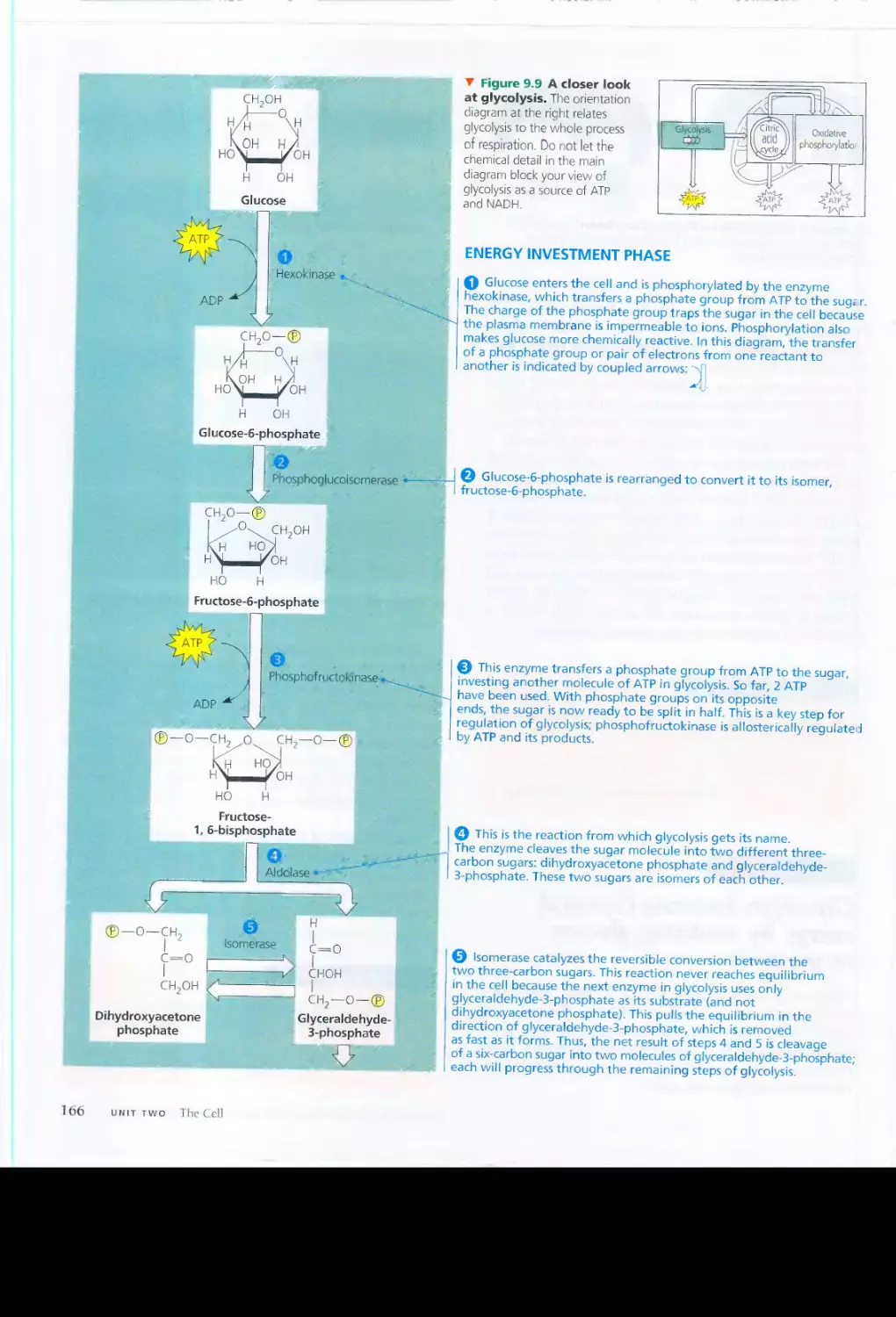
/


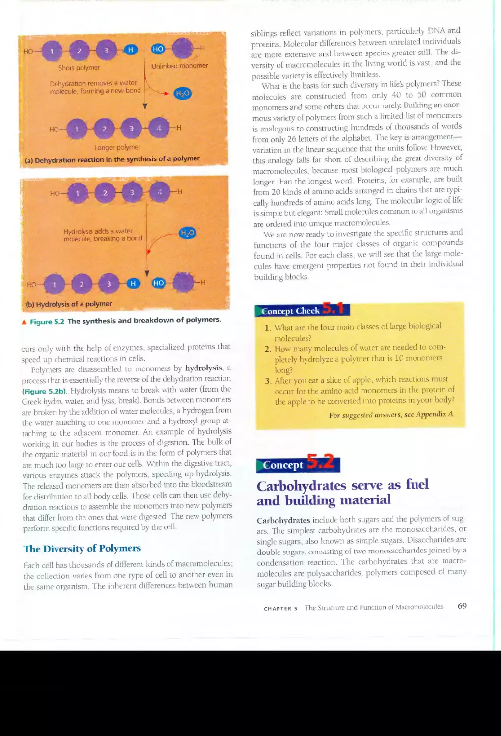


Текст
:.v.- ._
.
.."" .
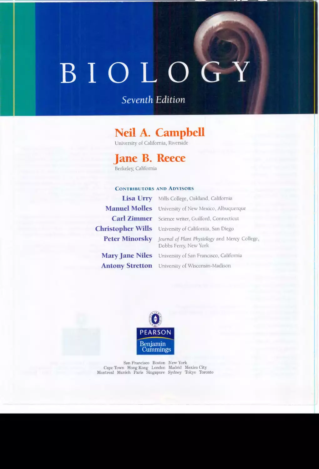
BIOLO
Seventh Edition
Neil A. Cantpbell
University of California, Riverside
Jane B. Reece
Berkeley, California
CONTRIBUTORS AND ADVISORS
Lisa Urry
Manuel Molles
Carl Zimmer
Christopher Wills
Peter Minorsky
Mary Jane Niles
Antony Stretton
Mills College, Oakland, California
University of New Mexico, Albuquerque
Science writer, Guilford, Connecticut
University of California, San Diego
Journal of Plant Physiology and Mercy College
Dobbs Ferry, New York
University of San Francisco, Calif 01 nia
University of Wisconsin-Madison
PEARSON
Benjamin
Cummings
San
'rancisco Boston New York
Cape Town Hong Kong London Madrid Mexico City
Montreal Munich Paris Singapore Sydney Tokyo Toronto
Editor-in-Chief: Beth Wilbur
Senior Supervising Editor: Deborah Gale
Supervising Editors: Pat Burner and Beth N. Winic1wff
Managing Editor: Erin Gregg
An Director: Russell Chun
Photo Editor: Travis Amos
\1arketing Managers: Josh Frost and J4f Hester
Developmental Editors: John Burner, Alice E. Fugate, Sarah C. G. Jensen,
Mati Lee, Suzanne OliviC/: Ruth Steyn, and Susan Weisberg
Developmental Artists: Hi/air Chism, Blaheley Kim, Kenneth Probst, Carla
Simmons, and Laura Southworth
Biology Media Producer: Christopher Delgado
Media Project Manager: Briel1n Buchanan
Project Editor: Amy C. Austin
Photo Coordinator: Donna Kalal
Permissions Editors: Sue Ewing and Marcy LunelLa
Publishing Assistants: Trinh Bui and Julia Khait
Illustrations: Precision Graphics, Russell Chun, Phil GU.7..y, and
SIeve McEntee
Text and Cover Designer: Marh Ong
Photo Researchers: Brian Donnelly, Donna Ka/ol, 1ra Kleinberg, Robin
Sampel; and Maureen Spuhler
Copy Editor: Janel. Greenblatt
Production Management, An Coordination, and Design Support:
lvlorgan E F/oyd, Robert R Hansen, Sherrill Redd, S. Brendan Short, and
Kirsten Sims al GTS Companies
Compositor and Prepress: GIS Companies
Manufacturing l-,-ianager: Pam Augspurger
Cover Printer: Phoenix Color
Printer: Von Hoffmann Press, 1ne.
On the cover: Photograph of bird's nest fern, Asplenium nidus: Linda
Broadfoot. Special thanks to Dennis High, Center for Photographic Art,
for his advice and assistance with cover research.
Credits continue following the appendices.
PIE ISBN 0-321-27045-2
Copyright (Q 2005 Pearson Education, Inc., puhlishing as Benjamin
Cummings, 1301 Sansome St., San Francisco, CA 94111. All rights re-
served. Manufactured in the United States of America. This publication
is protected by copyright and permission should he obtained from the
publisher prior to any prohibited reproduction, storage in a retrieval
system, or transmission in any form or by any means, electronic, me-
chanical, photocopying, recording, or likewise. To obtain permission(s)
to use material from this work, please submit a written request to Pear-
son Education, Inc., Permissions Department, 1900 E. lake Ave.,
Glenview, Il 60025. For infomlation regarding permissions, call
(847) 486-2635.
Many of the designations used by manufacturers and sellers to distin-
guish their products are claimed as trademarks. Vi/here those designa-
tions dppear in this book, and the publisher was aware of a trademark
claim, the designations have been printed in initial caps or all caps.
If you purchased this book within the United Stdtes or Canada you
should be aware that it has been wrongfully imported without the ap-
proval of the Publisher or the Author.
I 2 3 4 '5 6 7 8 9 ] 0-VHC-09 08 07 06 05
PEARSON
Benjamin
Cummings
San Franci<;co Boston New York
Cape Town Hong Kong London Madrid Mexico City
Montreal Munich Paris Singapore Sydney Tokyo Toronto
! bout the Authors
..... q- -?;."'" ..J' /'
--;.-'",
;-- ":-r... _" .-t- J'
".- .....
:
:.. - .". .....
. ./
/>
--",-.-.- :..:- .
"'- -- --.<:
> ;;:
l'>;
:
"."
.\
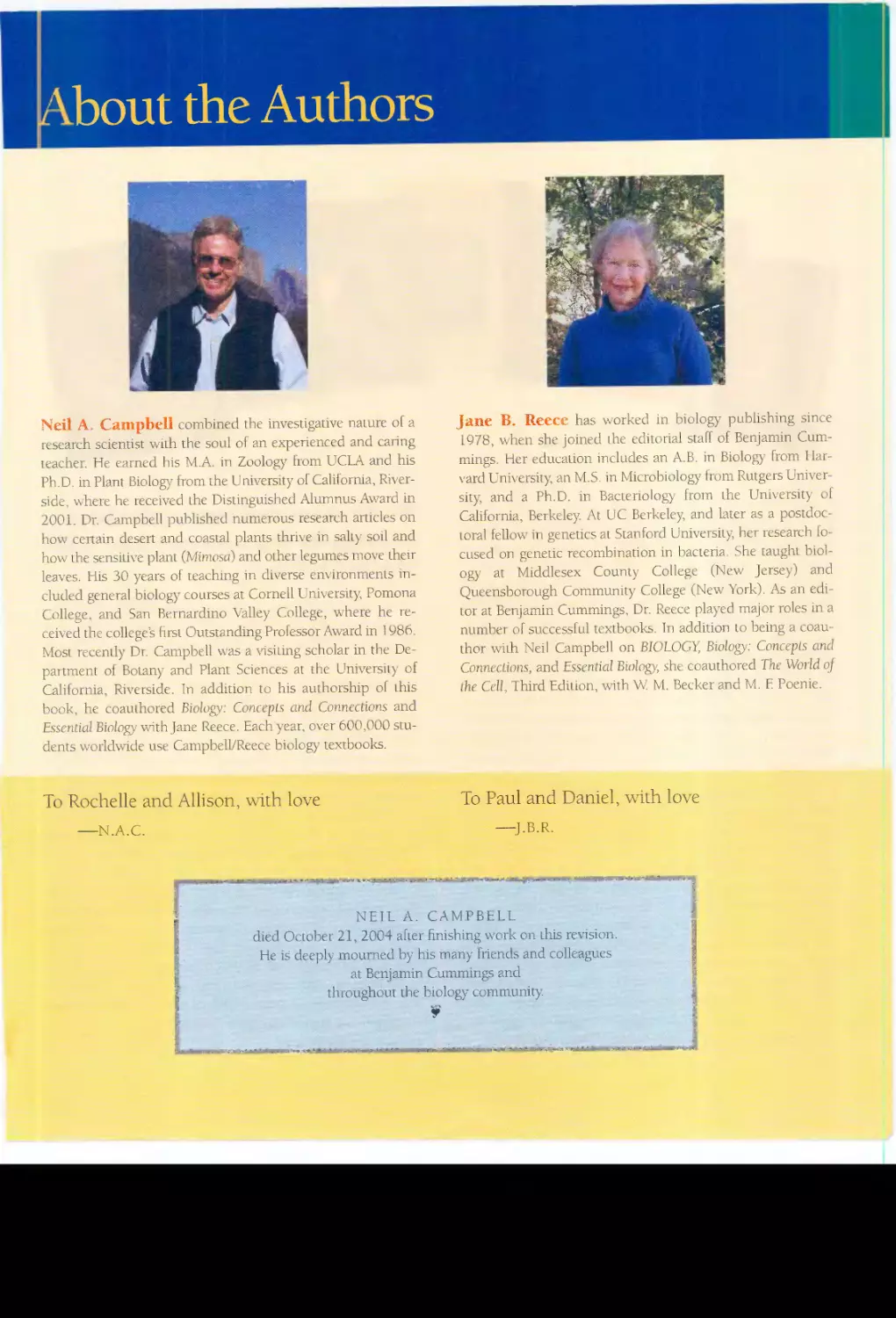
Neil A. Campbell combined the investigative nature of a
research scientist with the soul of an experienced and caring
teacher. He earned his M.A. in Zoology from UCLA and his
Ph.D. in Plant Biology from the University of California, River-
side, where he received the Distinguished Alumnus Award in
2.001. Dr. Campbell published numerous research articles on
hoy\' certain desert and coastal plants thrive in sally soil and
how the sensit1\T plant (JvIimosa) and other legumes move their
leaves. His 30 years of teaching in diverse environments in-
cluded general biology courses at Cornell University, Pomona
College, and San Bernardino Valley College, where he re-
ceived tbe college's first Outstanding Professor Award in 1986.
Most recently Dr. Campbell was a visiting scholar in the De-
partment of Botany and Plant Sciences at the University of
California, Riverside. In addition to his authorship of this
book, he coauthored Biology: Concepts and Connections and
Essential Biology wlthJane Reece. Each year, over 600,000 stu-
dents worldv-ride use Campbell/Reece biology textbooks.
To Rochelle and Allison, vvith love
-N.A.C.
... 't
\ '. {:
..
. '
,,
.,;
... . .,- J,
.<
::-
-j...
..
:.: ..
.-
? v
:-.-., "t
0:' k . ,
;;
i
.
:f:£>...;Z
,. ..\,--,'.<.i A '.,. .'. -. .-
;',' '<'
'''''''''' ..........
'-
. .
../
. '":...}
,.
:.
:."
, \
J.ane B. Reece has worked in biology publishing since
1978, when slH:' joined the editorial staff of Benjamin Cum-
mings. Her education includes an A.B. in Biology from Har-
vard University, an M.S. in Microbiology from Rutgers Univer-
sity, and a Ph.D. in Bacteriology frorn the University of
California, Berkeley. At UC Berkeley, and later as a postdoc-
toral fellow in genetics at Stanford University, her research fo-
cused on genetic recombination in bacteria. She taught biol-
ogy at Middlesex County College (New Jersey) and
Queensborough Community College (New York). As an edi-
tor at Benjamin Cummings, Dr. Reece played major roles in a
number of successful textbooks. In addition to being a coau-
thor \vi.th Neil Campbell on BIOLOGY, Biology: Concepts and
Connect.ions, and Essential Biology, she coauthored The World oj
the Cell, Third Edition, \vi.th Vv. M. Becker and M. F Poenie.
To Paul and Daniel, with love
-J.B.R.
". :",
.......
.
".A.'9:::r...."""..-: A ,. ,,«,,#.i-dl:,.=!I8f-
"""
:'::
+
".+
'i€.
:IQI;,
$-"'''''::.
.A-'''<iIt<''''
'''-
.''.-
'':'.V
''''':SIIIIIMI8F>o(.
._"'y-:-
.......
..;r.,,":
. <e_ ,._>
'I!I: </,<
7 + .jj:.,.
."Ij!";';;'.
NEIL A. CAMPBELL
died October 2.1, 2004 after finishing work on this revision
He is deeply mourned by his many friends and colleagues
at Benjamin Cummings and
throughout the biology community
J
I
'-
,
"
..
I
"
.
"
.' " .-
. . y. . _
....._=>_...._ '1::,. .
<!>,.
'\0- =. > "'C. .......e-....=:ae,. 'jI;,,
,-:<t
.
"" ""
v:Sl&i>
. 4>:r"'" -.. ........_.>
+ :&'Jo>.-.....; :.
y,('IIi.._
_ 1t
__oF_..1t.>_"IIi.. .
,_{; ,_4IIHhIiII;,:
:II'" :i>'lP'''' - ._.. :,.'" "'C",>
'IF_
".,
. . _.
:'"
.<III:
Preface
BIOLOG
. \31,9\1>",',\
BIOLO
.
1-tI.,d.. .£"dul
..
<
-
. .
;......-..-.
.;:-..-,....-
---. .. .I
>'.
(
,
:k
,:
': /
\3\01.-(
t
I
" !i ' ,
'I "
I! ,
'. "',A.?>-':
'.' I
<. '.
. .
..
,,-
"-. ._..
"
, .
.
.
--- ..
CAMPBE
. .. .
.«
r
CA.Ivfi>BEL
.. .'.
:
,..':,:.,,(
_.i
;L --'
:
.,..""!".....,,
:11>:111>
-
, ,',' CAMPBELL
.........>-- ,.."
,
y_<II;, "'IF b- <
C harles
arvvin
escr
be(
, ev
lution as a process of "de-
scent wIth modlficatlOn.' It IS a phrase that also fits the
continuing evolution of BIOLOGY. This Seventh Edition
is our most ambitious revision of the book since its origin-a
new textbook "species" with several evolutionary adaptations
shaped by the changing environment of biology courses and by
the astonishing progress of biological research. But these adap-
tive modifications are still true to the two complementary
teaching values at the core of every edition of BIOLOGY. First,
we are dedicated to crafting each chapter from a framework of
key concepts that will help students keep the details in place.
Second, we are committed to engaging students in scientific in-
quiry through a combination of diverse examples of biologists'
research and opportunities for students to practice inquiry
themselves.
These dual emphases on concept building and scientific in-
quiry emerged from our decades of classroom expelience. It is
obviously gratifying that our approach has had such broad ap-
peal to the thousands of instructors and millions of students
who have made BIOLOGY the most widely used college sci-
ence textbook. But with this privilege of sharing biology with
so many students comes the responsibility to continue im-
proving the book to serve the biology community even better.
As we planned this new edition, we visited dozens of campuses
to hear what students and their instructors had to say about
their biology courses and textbooks. VVhat we learned from
those conversations about new directions in biology courses
and the changing needs of students informed the many im-
provements you'll find in this Seventh Edition of BIOLOGY
IV
BIOLO
.' ..
.J'..
BIOta
.
.
. -1
".
BIOLOG)
" '
:'C'><-
.' '..
,
,
,(,
.
._><.
:""'»;:-::
. ,."
.-',
.
,,:
,,",
l!-f ra E f .
'R Hcii,
f 1 c' '
"C
Ai!,
£
oR.
.: .
$
<" ,.'
.. .. -
.
.:iI:<Iie >:"".""..
. . "
. .;;.
"
:;
J
"IF..'.
CI" r PI! ELL
f .
FI:C
. .
- '" >'"IIP' -<..
:
. ::i8II
:( "
. . '"
" '1»...
We have restructured each chapter
to bring its key concepts into even
sharper focus
The discovery explosion that makes modern biology so excit-
ing also threatens to suffocate students under an avalanche of
information. The past few editions of BIOLOGY set the details
in a context of key concepts, typically ten to twenty per chap-
ter. In this new edition, we have taken the next evolutionary
step of restructuring each chapter to help students focus on
fewer, even bigger ideas-typically just five or six key con-
cepts per chapter. A new Overview section at the beginning of
each chapter sets an even broader context for the key concepts
that follow. And at the end of each of the concept sections, a
Concept Check with two or three questions enables students
to assess whether they understand that concept before going
on to the next. Answers to the Concept Check questions are
located in Appendix A, as are the answers to the Self-Quizzes
from the Chapter Review at the end of each chapter.
In our ongoing interactions with students and instructors,
they have responded enthusiasti
ally to our new organization
and pedagogy Compared to other textbooks, including earlier
editions of our own, students have found the new chapter
structure and design of BIOLOGY, Seventh Edition, to be
more inviting, more accessible, and much more efficient to
use. But in achieving these goals, we have not compromised
the depth and scientific accuracy the biology community has
come to expect from us.
;
:'-. ,'
'
.
;
!:. "-
,."
, itt,.'
=.:..:.
.
..
.' .'- ' ;..
.. ./.':it-,/.,,
.....,........ .........
..
.;;.;". ;,;:'
... ..
."'$
,:,
- '.,,
:<":...':;#
:'. iiIP' ...... -to .
.
6-."
..... --
....
..
-
".
l...e} Conceptr, keep
the supporting
details in context.
The Overview sets
"he stage for the rest
of the chapter.
figure references
in color help
students move easily
between text and
figures.
-l
. .
... .....:.. ",,:...- '.
-.,
=< .
-
..
.
]iI
r.latl:m..
: ,.:.
1. \\ihy is the tra!lster of energy in an ecosystem re-
ferred to as ener!':y flow. nol energy cy;:hng?
2. How docs the second law of thcnnodynamks
explain v,'hy- an eCDsyst.ems energy supply musr he
(:omiI:!llaliy l'<'plenished?
3. lioware deninvores essemial to
llStamingecosystell1sJ
Fo!' sU
g'....'\ted (JnSWf-l.S, se.e i\ppcndix lL
.;: ,. : -:-:."
,
::..
..........
./;:
"--1
...:':
:
_.:...,.
.:.,
':S
:-
....::..
-"...
..., :::.......:
:-..::":.....t.
::-::..
";,.....
:
.
?- ......
"
-- ..-....>:
_.
.
.
Ecosystems
. Concepts
54.1 Ecosystem ecology emphasizes energy flow
and chemical cvclin o
. b
54.2 Physical and chemical factors limit primary
production in ecosystems
54.3 Fnergv transfer between trophic levels is
usually less than 20% efficient
54.4 Biological and geochemical processes 1I10ve
nutrients between organic and inorganic
parts of the ecosystem
54.5 The human population is dismpting
chemical cycles throughout the biosphere
-
Ecosystems, Energy, and Matter
A n eco
ys[em consists 01 all the organisms liYing in a
community as well as all the abiotic faClOrs with
which they interact. EW5ystems can range from a Im-
crocosm. such as the aquarium in Figure 54.1, to a large area
sud! as a lake or fOR'S!. As ",4th populations and commUlll-
tIes, the boundaries of ecosystems ,Ire usually not disnele.
Caies and far!TIs are examples of human-domiroted ecosys-
tems. Many ecologists regard the emire biosphere as a global
ecosystem, a composae of all the local ecosystems on Earth
Regardless of an ecosystem's size. it
dynatnic, involve IWO
prol,esses thaI cannot be fully described by population or
community pmcesses and phrnomena: energy 1I0w and
chemical cycling. Energy enlers most ecosystems in the fom1
-.f c.-.,Ii r. : h", ,-.1
:'
.-
..
....
:':
.
,
'"
.y"'.
---.;;.
\-.
.:.' f"
:"
,
,.
"-.
..
"S.
_J_"
" ,'''' ?-'-.
"..
'::i-
;
.
"
.
J."
,
it
.' .
.A
,:
:..,:. .c.
..
- .
..-
/"
".
''-<..,j:",
, t
- ., .
\,
:
t. :
\.;
:.-......... '
"to . '.
J ;.:
?-
l
,.. , '"'
"
...
--- . ,
.IM
-"
.
A Figure 54.1 An aquarium. an ecosystem bounded by
glass.
among abiotic and hiotK components d the ecosystem. Pho-
tosynthetic organisms assimilate these clements in 1I10rgamc
foml from the air, soil. and water and mcorporate them into
organic molecules, some 01 which are consumed by animals.
[he clements are returned in inorganic form to rhe atr, ;;oi1.
and water by the me.tabolism of plants and arumals and by
other orgamsms, such as bacleria and fungi. thar break doviT1
orgamc wastes and dead orgamsms.
130th energ.) and matler move through eco
ystem
via lhe
mmsfer of substances during phorosymhesis and feeding rela-
tionships. HowewT. because energy. unlike maner. l,annot be
recycled, an ecosystem must be powered by a conunuous in-
flux 01 energy from an external source-in most <:ases. the
sun. [nagy fl",vs through ecosystems. while matter cycles
Wlthinthem.
Resources crinciJI to human survIval and welfare, rangmg
from the food we Eat to th.. oxygen we breathe, are products
of ecosystem processes. In this chapter, we wIll explore the
dplamics 01 energy flow and chemical cydmg in ecosystems
and consider some of (he impacrs of human activities on these
proce'>Ses.
Each numbered
Concept Hea(l
announces the
beginning of a new
concept section.
.
.
{
Ecosystem ecology emphasizes
energy flow and chemical cycling
ecosystem ecologists view cco'y5letJ1S as tr.mslonl1er
of en-
ergy and processors of maner. By groupmg the species in a
. 00'.__' "f !ceding relalionships (see
ranSIO!lllation of ent'J'gy Ltl
enb of chemical element
Concept Chech. Questions at the end of
each concept section encourage students to
assess their mastery of the concept.
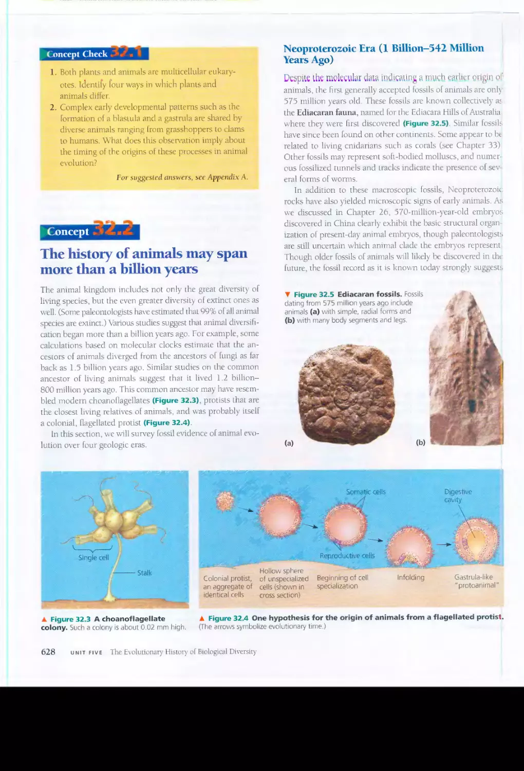
Physical and chemical factors
limit primary production in
ecosystems
n1e amount of light energy converted to chemical energy (or-
ganic compounds) by autotrophs during a given time period
is an ecosystems primary produCTion, This photnsymhetic
produCltS the starting poim for slUches of ecosystem metabo-
lism and energy lIow.
....:..1
"
,
...'
.
-- ;;" . ..
,t. "
-- ."\'. ,
.
... Figure 54.3 Fungi decomposing a dead tree.
."':.0.
: ..
Ecosystem Energy Budgets
Most primary producers use light energy to synthesIZe energy-
rich orgamc molecules. whtch can
ubsequenrly he broken
down to generate ATP (see Chapter 10). Consumers acquire
their organic fuels secondhand (or even third- or fourth hand)
through food webs such as that in figure 53.n. n1Cl-cfore,
the extenr of phorosymheuc production sets the spendmg
limit for the energy budget of the emire ecosystem.
consumers in an ecosystem. In a fores!. for example, birds
might feed on eanhworms that have been keding on leaflir-
ter and Its associated prokarymes and fungi. Bm even more.
importanr rhanthis channeling of resources from producers to
consumers is the role that detritivore.s play in making vital
chemical 'ailable to producers.
.e organic material in an
ts in in
Preface v
New "Exploring Figures" provide
efficient access to many complex
topics
Biology is a visual science. Thus we have always authored
BiOLOGY's graphics and narrative side by side to coordinate
their message. In the Seventh Edition, this text-an integration
reaches its next evolutionary level with a new feature callecl
"Exploring Figures." Each of these large figures is a learning
unit that brings together a set of related illustrations and the
text that describes them. The Exploring Figures enable stu-
In Exploring Figures, art, photos,
and text are fully integrated.
dents to access dozens of complex topics much more effi-
ciently, now that the textual and visual components have
merged.
The Exploring Figures represent core chapter content, not
to be confused with some textbooks' "boxes," which feature
content that is peripheral to the flow of a chapter. Modern bi-
ology is challenging enough without diverting students' auen-
tion from a chapter's conceptual storyline. Thus, each Explor-
ing Figure is referenced in the main text body where it fits into
the development of a concept, just as the text points students
to all the other supporting figures at the appropriate places in
the narrative.
Figure 6.31
. :.' :.Iorin..: Intercellular Junctions in Animal Tissues
Tight junction
Tight junctions prevent
fluid from moving
across a layer of cells
-....
,
..
..
\ /
.'
, -". -"":
".,&.
";: ';";'\
:f-
' }.,. ".
f f' t J y'
.
.
.
. " f'" ,,:.
'.. .'. '"
'1'
' J <'; ;
.' . '.' .. .... ,
) . . \. .' _
.:O' . 0'. . _ .
.. .:::
. . .
\ Tight junctions ' . .':
:
=t
'ate €;
i("
r.
..'
..; ..:
"
-'
:;.. :'[i%
::
.
""
.
" 1 '/"
'
'-"i'!'
; :. I
: ...1 f
f: 0.1
.'I
' .,_
Desmosome
.;: . .:-
.:-"...:...... -
. t--
--
:.- -...:: .-
':"){:
ii"- .,,:,'
.-I:
- -:
..
Gap
junctions
,<
.
.'\,.
_
A .
>
-
$:
J "
r .'
" . ' '
r"
. . ",
.
., ;:,' "
<.. .,,;'i-< ,
>
;
Extracellular
matrix
Space
between
cells
"., '.'""
.
'..." ,':
Plasma membranes
of adjacent cells
VI Preface
.
:
1;-
" >
"
t:
't
:
c:
....'i ,""to; ,
.....;;
.
.>
f J
;:,4:
:
J' ,,_
y
!
f' t
,;J>
',< ... ,
-
4
i
t;
:.
'
,;;<.,
,
...;:,f'>
1
::
t
.:n:" $
--:
f f;
i ._ .. '
!6.
-
...i!l 'IF «'" 1;.4!-.
'
..<;.<1'. ".
I-----!
05 11m
. .. .... .
.,"""\, ......
.
.
<:;: :......._'-
<8;..
..... ):.
;
i
>-.,.,,,, , ,......, . _. ..'> :J
· c ;.
>. ?Z,:.::?h :.IC:
-. .
:
'. ,-'::',
.
- . .
1---1
1 pm
Gap junction
J '- :>., ,;
': ":>"
'
'. ': .1i--'!!
':' '. . 4.f
:., .: \ ..
," j
...
(" - I jI:
1:l1}: '
f'> "- .
..
,;,
;.:.
;
y
.. 'f> ":__ )
L' :..\ :
ff':c'4"'f<:
:;
;*" .., ".:.
>; ;" :v_
.
... .;
I I
0.1 pm
TIGHT JUNCTIONS
AI tight junctions, the memhranes of
neighboring cells are very tightly pressed
again
t each other, bound logelher by
specific proteins (purple). Forming cominu-
ous seals around I he cells, tight junctions
prevem leakage of extracellular fluid across
a layer of epilhelial cells.
DESMOSOMES
Desmosomes (also called anchoring
junc/iot1s) function like rivets, fastening cells
together imo strong sheets. lmemlediate
filaments nude of sturdy keratin proteins
anchor desmosomes in the cytoplasm.
GAP JUNCTIONS
I
Gap junctions (also called communicating
junctions) provide cytoplasmic channels from
one cen to an adjacent cell. Gap junctions
consisl of special membrane proteins that
surround a pore through which ions. sugars.
amino aCIds, and other sman molecules may
pass. Gap junctions are necessmy for commu-
nication between cells in many types of lissue
,
including heart muscle and animal embryos.
Scientific inquiry is more
prominent than ever in BIOLOGY
and its supplements
One objective for many biology instructors is for students to
learn to think as scientists. Tn both the lecture hall and labo-
ratory, col1eagues are experimenting v,rith diverse approaches
Figure 1.29
Inquir- Does the presence of poisonous
coral snakes affect predation rates on their
mimics, king snakes?
XPERIMENT
David Pfennig and his colleagues made
artificial snakes to test a prediction of the mimicry hypothesis: that
king snakes benefit from mimicking the warning coloration of coral
snakes only In regions where poisonous coral snakes are present.
The Xs on the map below are field sites where the researchers
placed equal numbers of artificial king snakes (experimental group)
and brown artificial snakes (control group). The researchers recovered
the artificial snakes after four weeks and tabulated predation data
based on teeth and claw marks on the snakes (see Figure 1.28).
-. -
RESULTS
In field sites where coral snakes were present,
predators att
ckcd far fewer artificial king snakes than brown
artificial snakes. The warning coloration of the "king snakes"
afforded no such protection where coral snakes were absent. In
fact, at those field sites. the artificial king snakes were more likely to
be attacked than the brown artificial snakes, perhaps because the
bright pattern is particularly easy to spot against the background.
I: Key
[2J % of attacks on
artificial king snakes
u=J % of attacks on
brown artificial snakes
for involving students in scientific inquiry, in v/hich questions
about nature focus strategic investigation and analysis of data.
New textbook features and new inquiry-based supplements
make this edition of BIOLOGY more effective than ever as a
partner to instructors who emphasize the process of science.
Modeling Inquiry by Example
Scientif-ic inquiry has always been one of BIOLOGY's unifying
themes. Each edition has traced the history of many research
questions and scientific debates to help students appreciate
not just "what we know," but "how we know," and "what we
do not yet know." In BIOLOGY, Seventh Edition, we have
strengthened this theme by making examples of scientific in-
quiry much more prominent throughout the book.
The increased emphasis on inquiry begins in Chapter 1,
where we have thoroughly revised the int roduction to the many
ways that scientists explore biological questions. Chapter 1
also introduces a new feature called "Inquiry Figures," which
showcase outstanding examples of experiments and field
studies in a format that is consistent throughout the book.
Complementing the Inquiry Figures are the new "Research
Method Figures," which \valk students through the tech-
niques and tools of modern biology You can find a list of the
Inquiry and Research Method Figures on pages xx-xxi. These
new features, like the Exploring Figures, are integral to chap-
ter flovv' rather than being appended as boxed asides.
New Inquiry Figures and Research Method Figures
help students learn to think like scientists.
X Field site with
artificial snakes . .'. >'
;
In areas where coral snakes K .>><>.,. ',,:, '.' .
were absent, most attacks " 83:% '{Ii
were on artificial king snakes. "<
. . ",....-'"" .
)( "'- '
North X X
Carolina X
,
1 1 _X. "
South ':'
.
X ,. X
Carolina:".
r
':'
. >\
" ,
",
1\ -,.-
-
\:::::.
Xr 0
OO
/=
,
'
(J
In areas where coral
snakes were present,
most attacks were on
brown artificial snakes.
CONCLUSION
The field experiments support the mimicry
hypothesis by not falsifying the key prediction that imitation of
coral snakes is only effective where coral snakes are present. The
expenments also tested an alternative hypothesis that predators
generally avoid all snakes with brightly colored rings, whether or
not pOisonous snakes with that coloration live in the environment.
That alternative hypothesis was falsified by the data showing that
the ringed coloration failed to repel predators where coral snakes
were absent.
Figure 7.4
.
- -". -,rth -' "".'
. Freeze-Fracture
. PPLICATION
A cell membrane can be split into its two
layers, revealing the ultrastructure of the membrane's interior.
TECHNIQUE
A cell is frolen and fractured with a knife.
The fracture plane often follows the hydrophobic interior of a
membrane, splitting the phospholipid bilayer into two separated
layers. The membrane proteins go wholly with one of the layers.
f""-o:?'?'-"... Extracellular .' .
.. _. layer
'r
'
, '; ; ," L .' :::::.: ;'. '\ '
':..:. ."\ . .
. ,<" Proteins
.,
C .:'-
.. .' - .'
Plasma Cytoplasmic
membrane layer
v
a,...-.:
RESULTS
These SEMs show membrane protein
(the
"bumps") in the two layers, demonstrating that proteins are
embedded in the phospholipid bilayer.
::-:
..
Z111.
.
=")..
._
.;::
'
"......;.-,X
Extracellular layer
Cytoplasmic layer
Preface Vll
Learning Inquiry by Practice
Modeling scientific inquiry by example has only ephemeral
impact unless students have an opportunity to apply what
they have learned by asking their own biological questions
and conducting their own investigations. On a small scale,
BIOLOGY, Seventh Edition, encourages students to practice
thinking as scientists by responding to "Scientific Inquiry"
questions in the Chapter Review at the ends of chapters.
On a much bigger scale, new supplements build on the
textbook to provide diverse opportunities for students to
practice scientific inquiry One example is Biological InqLlilY: A
Worhbooh of Investigative Cases, by Margaret \Vaterman of
Southeast Missouri State University and Ethel Stanley of Beloit
College, which is available without cost to students whose in-
structors request it as a supplement to the textbook. This in-
novative ne\v workbook offers eight case studies, coordinated
with the eight units of chapters in BIOLOGY. In each case, a
realistic scenario sets up a series of inquiry-based activities.
The cases work well either as class-discussion projects or as
take-home assignments for students working alone, or better.
in small groups.
Another student-centered supplement is Practicing Biol-
ogy, by Jean Heitz, University ofvVisconsin, Madison, which
Scientific Inquiry
Wnen bacteria infect an animal, the number uf bacteria in the body
increases in an exponential fashion (graph A) After infection by a
\imlent animal virus \\'1th a lytic reproductive cyck, there is no
evidence o[ infection [or a while. Then, the number or viruses rises
suddenly and subsequently increases in a series of steps (grapn B).
Explain the difference in the growth curves.
t A t B
ro V1
Q:; (j)
V1
+-' 2
v
ro .;;:
..D
'j-- '+--
0 0
Q:; Q:;
..D ..D
E E
:::J :::J
Z z
Time Time
-i:.....
.... :...:.,:.
Biological Inquiry: A Workbook of Investigative Cases Explore .' <..,;:,
.:>,:
\Vest "/Vile virus in tile case "TIle Don01:S Dilemma."
.::.>.v'
Investigation \Vhat Causes Infections in AIDS Patients?
Investigation WIlY Do AIDS Rales Differ Across tlte U.s.?
Investigation What Are the Patterns of Antibiotic Resistance?
Inquiry Questions, Media Investigations, and the new
Biological Inquiry Workbook help students practice
scientific inquiry.
VIII Preface
IS also available without additional cost upon request of
instructors using BIOLOGY, Seventh Edition. This work-
book supports various learning styles with a variety of activ-
ities-including modeling, drawing, and concept -mapping-
that help students construct an understanding of biological
concepts.
Students \-vill find still more opportunities for active learn-
ing at wwwcampbel1biology.com and the CD-ROM that is in-
cluded with each book. And the excellent Student Study Guide
- ,
by
/lartha Taylor of Cornell University, continues to be a
proven learning tool for students.
The CampbeWReece Interviews:
A Continuing Tradition
Scientific inquiry is a socia] process catalyzed by communica-
tion among people who share a curiosity about nature. One of
the many joys of authoring BIOLOGY has been the privilege to
humanize science by imerviewing some of the \\Todd's most
influential biologists. Eight new interviews that introduce the
eight units of the textbook provide students with windows to
inquisitive minds that are driving progress in biology and con-
necting science to society. The interviewees for this edition are
listed on page }G'{iii.
; I
iologk
1 Inquiry
t. ..So f
'"
I
: I
I
I
.-- r
-I
":-1
I
.- t
<:.-1
Ethfi '
..::-
"'-"" \\ :-
BIOLOGY
The Donor's
Dilemma
US..!:::t!y RlI!.:,e!1 foolld 0:.'111
:?X.u
>(It to pJrlJoC.Ud(o,:!
-. (<)mpanY-W0I1:S
b<o:xi (J(Mt$,
11:"'(:<!:O:'t --=f$1
fur
he dec.Jd.;(!
o cona;:p
Iocd AttE'r tJUlnc o:Jt
c!<.mc< ehQfbl
ty fOfO
dod
Iflg tOO b!<1(xl
"1'f,:>'>!'>IIn? J:'
1!?11f)Pf3hJre, a!"d blood.....-lDrlf.:j
!.
. Rl.7J!.
"
t (jUNf! fur tll:!lllllo!r''''
Rw.Se1l !O'1err\,JDi.e<; (nc I-.tm 1.$101 tl('l\'(' ;'Pu
e>.'€r:;1o" -questIOnS v.'fth c Q
'1 (J
hlS-OlI'in_
'W°n;'1 f I trav.. N.-.....- Nil... \'In:"'''
We t Nde \' ru!- i
ul'KOI'I'ImlX'l, R l'1e Intervle\'lt!r
..j s.;.;10Ps.. ali [l'oc;atM blcod;',;, :P;t£od for "Ws:
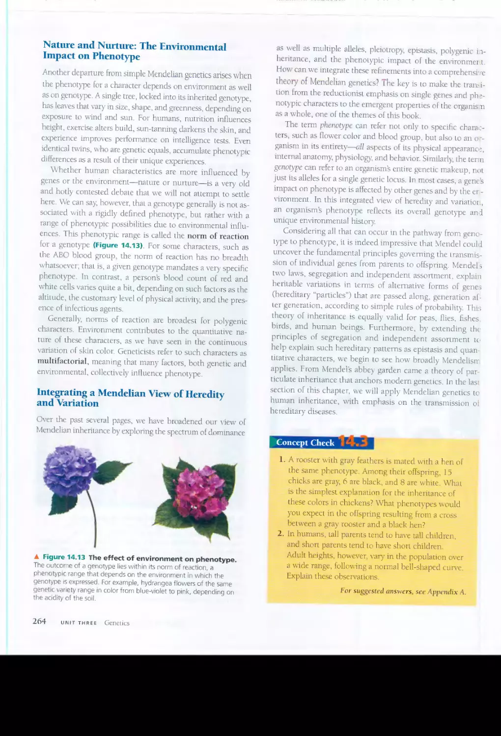
N>I<!"JI
lls..
\.
I ht'ft:' II) CJhfotrt
1l"5.
x
"r..m90\o ;",
" She ql:;
f'1 {wet hIS ::JapeM'Orl::
IN''!;.
I?(> Y.ou !,.};{J )'!)u I
..
( h".J ¥'I)' ff........:.!,. ()(
headaches It! the I,aS: y.,'Pt't., Is fS".
re.:l !.:!,a;':Jn that
yW mlilk )'lo'.Illlight n
C 11 / '.
"'No. b;1t j'o;,? hec'd .ha! ..Qr.-- -mes
cor1"J
II., c ,my
'9tClns.H :tU'
:>o:..<lllt
I';(.
"f J.rA y(>t
back: t(om i! Md
tl1r In EO!
Id.",(. Cc!orQ':1:3, over
the FtK.:Tth o
Juoj. ......."C1c.
I1
. T!;
u:" wai? JlI:.'\"6-
r=-D:::rt
rh.=t: ther!:" atl? '" 101 (",t C:;'i.f'.. ot thl? v,ru<;
!.I1..,£e. ,]110 fm 50(1 ((r,'
I'<'O wjlh IrllY..o{f!..o'1W I)tll:!
_
"
'.'€'U. i' VO".J "}=.
'."I.?!.t Ni1€' w:l<;, ........\vii1 fwd
cu! Wb 11::'S.
'.;:fI your .:II0c4 wlilldel;l.If)' the
prt-::('n(
0'1 gP.!
.Wcli. tt-l')' (
'l tell frUHI hl$
t-AtT cther .'''T ((In Iden1cfy the
w
repbetJ 'When W$1Nk\llj"LlsfiT
New y«
In 1999. ,el: 1nc.s.J{>\p1e:
now mutiltlC!r::5 ar=><;hO\Vlnq ( (I Tn
II
IJ
S.lO Iffenml :tJ'"as_ Wl::-.r
<;.jraL'-r5 ef thf''''
1..1:; -n diffE'r
l r
tOO try.-
SorlIl1Wf'5tN pvru\ongl....
RuS
wor.Wif!d
NQ.
shesaICl1N1thasmilf',
M{':'f(lri!;:"I"
n
-.. .
r&
,.... y« ':11=
;
:;;
:; ;'-f
':..
>
.} '> ",!-,.
...
i
{;
.,
'> t<. _
jf.'--. f. ..-
, ,-
,.t
y_= :iIt.<!.:y
.,
<,.:.
1!drnl1'IIt1todlht
\r'lfU').p.;<1'{If'
1
Ool:1yatir f
i!I
t.....[!"OposIW..e-.
rSo I 1
1a'Je
.gollt It I CDIL"'a
UNrT]C-'Sl:
(; v.le In>.: cJ 29
CASE ANALYSIS
1 Recognize \,otennalls5-ue:s-
lld ma"lor lopics III [bt: case:. Wh.'l D tIu:; L<'Is.... ..!lxJUI
UnOf"rhn(" 'Hm." .11 phrn<;e:.<; Thm ";OCT.. to be rm"1<1n:;n1 it""! UndH,,[;:mdlll,g 1111'S: ..: 1..<;(' .men 1,5t
3--+ bIOlug:.-I<..Lut.r..! to)Jh_
\J! ,
::.Ul"S IJL lh-=. -t;.;J:..e
1. \\'har 5p,
ifiC' qur-..:;ti(lu<;; de }""OU haw' 311,-'111 ,hi"'H
I(Ipl("
? fir )"CI,u<;df. or b,
lif'r "'E'I. jJ- ;-i
wup" nl<lJ.:o: a hst (.)1 wh::\t ".OU .dn:14d)' know
lb:'ll!( L
1I:' l.
'= IrI .he: VVhJ.1 Do I Kn(,l"l.....
'
cl..,h.1Inn LL.:;;, q\I
5'i1ens Y(:\I would lih (('I l(01m more 1b1J'lIT m l
e ..\....0;11 Do I Ne.:::d 10
K[LO'
r oi_,lwnu
What Do I Know')
What Do I Need to Know?
Balancing Inquiry with a Conceptual
Foundation
-\lthough this nevv edition of BIOLOGY showcases the process
of science more prominently than ever, there are two good
reasons to avoid overstating the power of inquiry-based con-
tent in any biology textbook.
First, those of us who advocate more inquiry in biology
courses mainly have student-centered inquiry in mind, not
textbook-centered inquiry As a mostly passive experience,
reading about inquiry in a textbook should be merely an entry-
way to a variety of active experiences that are promoted by
inquiry-based supplements, by investigative labs, and by activ-
ities that instructors create to support student-centered inquiry
Second, the most inlportant way a textbook can support
student inquiry is by providing context with clear, accurate
explanation of the key biological concepts. Just as biologists
generally study the scientific literature as background for their
OVln inquiry, students vvill be much more successful in their
personal inquiry if it emerges from a basic understanding of
the relevant biology. Thus, BIOLOGY, Seventh Edition, is not a
"reform textbook" of the genre that replaces a careful unfold-
ing of conceptual content with a stream of relatively uncon-
nected research examples, requiring beginning students to
put it all together for themselves. We believe that such an un-
balanced reaction to the call for inquiry-based refornl is likely
to leave most students frustrated and ill-equipped to practice
active inquiry in their labs, course projects, class discussions,
and Socratic lecture environments. In BIOLOGY, Seventh Edi-
tion, we have carefully integrated the inquiry-based content
into the development of each chapter's main ideas so that the
research examples reinforce rather than obscure the concep-
tual framework.
BIOLOGY supports a diversity of
courses and serves students
throughout their biology
education
Even by limiting our scope to a few key concepts per chapter,
BIOLOGY spans more biologicalterIitory than most introduc-
tory courses could or should attempt to cover. But given the
great diversity of course syllabi, we have opted for a survey
broad enough and deep enough to support each instructor's
special emphases. Students also seem to appreciate BIOLOGY's
breadth and depth; in this era when students sell many of
their textbooks back to the bookstore, more than 75% of stu-
dents who have used BIOLOGY have kept it after their intro-
ductory course. In fact, we are delighted to receive numerous
letters and emails from upper-division students and graduate
students, including medical students, expressing their appre-
Clallon for the long-term value of BIOLOGY as a general re-
source for their continuing education.
Just as we recognize that fe"w courses will cover all 55 chap-
ters of BIOLOGY, we also realize that there is no one "correct"
sequence of topics for a general biology course. Though a bi-
ology textbook's table of contents must be linear, biology itself
is more like a web of related concepts without a fixed starting
point or a prescribed path. Diverse courses can navigate this
network of concepts starting vrith molecules and cells, with
evolution and the diversity of organisms, or with the big-
picture ideas of ecology \Ne have built BIOLOGY to be versatile
enough to support vmious syllabi. The eight units of the book
are largely self-contained, and most of the chapters within
each unit can be assigned in a different sequence. For exam-
ple, instructors who integrate plant ancl animal physiology
can merge chapters from Unit Six (Plant Form and Function)
and Unit Seven (Animal Form and Function). Instructors who
begin their course with ecology and continue with this "top-
down" approach can assign Unit Eight (Ecology) right after
Chapter 1, which introduces the unifying themes that provide
students with a panoramic view of biology no nlatter what the
topic order of the course syllabus.
Evolution and BIOLOGY's other
themes connect the concepts and
integrate the whole book
The first chapter articulates 11 themes that provide touch-
stones for students throughout the book and clistinguish our
approach in BIOLOGY from an encyclopedic topical ap-
proach. In this Seventh Edition, \ve have added the theme of
"biological systems" to integrate a variety of research initia-
tives based on high-throughput data collection and readily
available computing power. But as in all previous editions, the
central theme is evolution, which unifies all of biology by ac-
counting for both the unity and diversity of life. The evolu-
tionary theme is woven into every chapter of BIOLOGY.
Evolution and the other whole-book themes work with the
chapter-level concepts to help students constnlct a coherent
view of life that will serve them long after they have forgotten
the details fossilized in any biology textbook.
Neil Campbell and]ane Reece
Preface IX
!
cknowledgments
O ne of the eminent scientists imerviewed in this ne\\T edition
pointed out that much of the fun of doing biology comes from
working with a diversity of talemed people. The same can be
said for making a biology textbook Fortunately for us, this Seventh
Edition of BIOLOGY is the product of the talents, dedication, and en-
thusiasm of a large and varied group of people. The authors wish to
express their deepest thanks to the numerous instructors, researchers,
students, publishing professionals, and artists who have contributed
to this edition.
As authors of both past and present editions of this text we are
mindful of the daunting challenge of keeping up to date in all areas of
our rapidly expanding subject. \Ne are particularly grateful to the
seven Contributors and Advisers listed on the title page, \vhose ex-
pertise has ensured that the book is current and enlivened \vith fresh
examples. Vie \vorked especially closely vvith deve10pmemal biologist
Lisa Urry, who had major responsibility for updating contem and im-
plememing our new format and features for Units 1-3 and Chapter
47. Her rigorous scholarship and attention to detail in the areas of bi-
ological chemistry, cell and molecular biology, genetics, and develop-
mental biology \liere a great boon. vVe thank her for her commitment
and enthusiasm, relentless hard work, punctuality, and good cheer
throughout the process. Equally helpful was ecologist Manuel1/1011es,
who brought his sciemific and teaching expertise to the revision of
Unit 8, enhancing the structure of the unit and its verbal and visual
presentation of ecology; he played a major role rev,lYiting the behav-
ioral ecology chapter, \vhich is essentially new. He also helped provide
a more ecological perspective to Chapters 40,42, and 44, in the unit on
animal form and function. Science wliter Carl Zimmer contributed
many improvements and new perspectives to Unit 5, the diversity unit.
Evolutionary biologist Christopher \Vills helped us tackle the challenge
of improving and updating Unit 4, the evolution unit, and Chapter 26.
Plant biologist Peter :tv1inorsky helped bring Unit 6 up to date. And
neurobiologist Antony Stretton advised us on the revision of Chapters
48 and 49. As in earlier editions, immunologist Mary Jane Niles organ-
ized and implemented the significant revision of Chapter 43.
Thanks also to the instructors who suggested revised or new Con-
cept Check and Chapter Review questions. These include (in alphabet-
ical order) Bruce Byers, Jean Heitz, \Villiam lloese, Torn Owens, Mark
Lvford, Randv Phillis (soecial thanks), Mitch Price. Fred Sack, Richard
" , t
Showman, and Elspeth \Valker. Its not easy to \\Tite good questions,
and we appreciate the time and effort these dedicated educators con-
tlibuted to enhancing the effectiveness of our books questions.
Further helping us improve BIOLOGY's scientific accuracy and ped-
agog}
about 240 biologists and teachers, cited on the list that follows
these Acknowledgements. provided detailed re\iews of one or more
chapters for this edition. Special thanks to LWI.Trence Brewer, Richard
Brusca, Anne Clark. Douglas Eernisse, ),1ark Kirk, Vv'aller Judd, :vIike
Levine, Diane !vlarshall,
ick Money, Tom O\'.Tens, Kevin Padian, Daniel
Papaj, Mitch Price, Bruce Reid, and Alistair Simpson for their guidance.
Thanks also to the numerous other professors and their students,
from all over the \vorlet \vho offered suggestions by \\/[iting directly to
the authors. In addition, vv'e appreciate the candid and specific feed-
back we rece1\Td from the students and faculty who participated in
group discussions held at Skyline College, Mills College, and Indiana
x
University. Last but not least, we thank our coauthors on our nonma-
jors texts, Eric Simon and Marty Taylor, for providing rigorous feed-
back on a number of chapters. Of course, we alone bear the responsi-
bility for any errors that remain in the text, but the dedication of our
contributors, advisers, reviewers, and correspondents makes us espe-
cially confident in the accuracy of this edition.
Many scientists have also helped shape this Seventh Edition by dis-
cussing their research fields with us, ans\veling specific questions in
their areas of expertise, and, often, sharing their ideas about biology
education. Neil Campbell thanks the many University of California,
Riverside, colleagues \yho have influenced this book, including Ring
Carde, Richard Cardullo, Mark Chappell, Darken DeMason, Norman
Ellstrand, Anthony Huang, Bradley Hyrnan, Tracy Kahn, Elizabeth
Lord, Carol Lovatt, Eugene Nothnagel, John Oross, rimothy Paine.
David Reznick, Rodolfo Ruibal, Clay Sassaman, William Thomson,
John Trumble, Rick Redack, Mike Adams, and the late John Moore
(whose "Science as a \Vay of Knowing" essays have had such an im-
portant influence on the evolution of BIOLOGY). Jane Reece thanks
members of the Mills College Biology and ChemistrylPhysics Depart-
ments, especially Elisabeth Wade, as well as Fred vVilt, John Gerhart
and Kris Niyogi from the University of California, Berkeley for their
assistance to conuibutor Lisa Urry.
Intervie\vs vv'ith prominent scientists have been a hallmark of
BIOLOGY since its inception, and conducting these interviews was
again Olle of the great plea
ures ofrevising the text. To open the eight
units of this Seventh Edition, we are proud to include interviews \\ith
Lydia ::vlakhubu, Peter Agre, Eric Lander. KeImeth Kaneshiro, Linda
Graham, Natasha Raikhel, ElichJarvis. and C
ne Likens.
The value of BIOLOGY as a learning tool is greatly enhanced by
the supplementary materials that have been created for instructors
and students. Vve recognize that the dedicated authors of these ma-
terials are essentially writing mini (and not so mini) books. \Ne much
appreciate the hard work and creativity of the following: Margaret
vVaterman and Ethel Stanley (authors of the new BiologicallnquilY:
A \Vod;:book of investigative Cases); Jean Heitz (Practicing Biology, 2 nd
edition); Joan Slurp (InsLructor's Guide); Janet Lanza (New Designsfor
Bio-Explorations); Cllris Romero (PowerPoint Lectures); Laura
Zanello (Spanish GIOSSClIY); and Judith Morgan and Eloise Brown
Carter (Investigating Biology Lab !\t1anual, 5 th Edition). Vv'e thank Bill
Barstow for heading up the test bank team, and we wish to acknm.vl-
edge the test bank comributors: Jean DeSaix, Michael Dini, Conrad
Firling, Peter Follette, Mark Hens, Janice Moore. Tom Owens, :Mar-
shall Sundberg, Robert Yost, and Ed Zalisko. Thanks also to Bill Wis-
chusen, who compiled our Active Learning Questions and wrote
discussion poims. Once again, \ve thank our long-time colleague
Manv Tavlor for her excellem and student-focused work on the
, ,
Student Stud)' Guide; she has now completed seven editions of this
popular student aid. In addition, \ve are grateful to the many other
people-biology instructors, editors, anists. production experts,
and narrators-who aTe listed in the credits for the impressive elec-
tronic media that accompany the book.
BIOLOGy: Sevemh Edition, results from an unusually strong syn-
ergy between a team of scientisrs and a team of publishing professinn-
also An all-new design, the comprehensive revision of the illustration
program as well as the text, the addition of major new pedagogical fea-
lures, and a rich package of supplements. both printed and electronic
( ombined with a tight schedule to create unprecedented challenges for
. he puhlishing team.
The members of the core book team at Benjamin Cummings
-)rought extraordinary talents and extraordinarily hard work to this
_1roject. Our leader, Editor-in-Chief Beth \Nilbur. is a fun colleague in
.he books creation and a respected advocate for biology education in
seneral and our book in particular in the academic community En-
thusiastic, creative, endlessly supportive of us and the other members
:)f the team, Beth is a wonderful person and a pleasure to vvork with.
Unflappable under pressure, she navigates difficult situations grace-
fully-a major asset in a project of this complexity
The incomparable Deborah Gale. Director of Development, man-
aged the entire project on a day-by-day basis. Deborah coproduced
the first and second editions of the book, along Vv'ith the developmen-
tal editing of the second edition, and we have been delighted with her
return. Amazingl)
Deborah is able to combine a towlly professional,
no-nonsense management style and a wilHngness to dig into the nitty-
gritty with a sense of humor tbat kept the rest of us happily slaving
away at her direction.
Supervising editors Pat BUlller and Beth \Vinickoff had the avve-
some responsibility of overseeing in detail the vvork of the connibu-
tors, developmental editors, and developmentaJ anists. Together, Pat
and Beth carefully read every single chapter and checked every illus-
tration. doing whatever was necessa1)' to make this edition the most
effective biology textbook available-and, vve think, exceeding the
high standards established in previous editions. \Ve are immensely
grateful to Pat, the multitalented and tireless Developmental -
/Ianager
for Biology who has been our colleague for many years, for her in-
credible dedication, sound editorial judgment, and extraordinary at-
tention to detail. The exceptionally talented Beth \Vinickoff, new to
this Seventh Fdition, was the originator of our new process of book
development and production. Beth brought fresh perspectives on
process, pedagogy, and editOlial approach-in addition to her superb
hands-on editing of six chapters. \Ve look forward to working again
\vith Pat and Beth on subsequent editions (after they recover from this
one, of course!).
The responsibilities of the developmental editors for this edition
were especially challenging. Almost all the chapters were heavily re-
vised, requiring intensive editOlial involvement from initial planning
through production. vVe were fortunate to have on our team some of
college publishing's top developmental editors. In alphabetical order.
they were John Burner (Units 5-7), Alice Fugate (Units 4 and 7), Sarah
Jen.sen (Units 2, 3, and 7), Matt Lee (Units 5 and 7), Suzanne Olivier
(Units 1 and 2), Ruth Steyn (Units 3 and 7), and Susan \Veisberg
(Units 7 and 8). In addition to their other tasks, John Burner, Matt Lee,
and Ruth Steyil brought their specific content expertise to bear on
their chapters' revisions.
The suppon of our bright, efficient, and good-natured Publishing
Assistants. Trinh Bui and Julia Khait, is much appreciated. \Vhat
would \ve all have done Viithout them?
Vie also want to thank someone who doesn't fit neatly into any of
our publishing categories: our colleague, former editor, and friend
Robin lleyden. Robin brought her imaginative energy and dedication
to biology education to the Sevelllh Edition in several ways. These in-
clude early planning for the development of the media for this edition
and the conception and developmental management of the new case
study \vorkbook by Margaret \Naterman and Ethel Stanley. Robin also
organized the first Benjamin Cummings Biology Leadership Confer-
ence, which brought us a fresh supply of creative teaching ideas from
outstanding biol0 6 7 educators.
Once again the book has benefited greatly from the work of Russell
Chun, our Senior Producer, Art and Media. Russell established a vi-
brant new art style for this edition that met the requirements of the
contelll and exceeded our expectations for pedagogical and aesthetic
excellence. Under his direction were the developmental artists, who
developed all the new figures and redesigned many of the older ones
to make them clearer and more appealing. These skilled and creative
illustrators were Hilair Chism (Units 1-3 and 7), Blakeley Kim (Unit
8), Kenneth Probst (Lnits 4 and 5), and Laura Southwonh (Lnits 3
and 7). Carla Simmons (Units 5 and 6) has contributed her artistic and
pedagogical talents to every edition of this textbook. Final rendering of
the hundreds of new and revised illustrations was carded out bv Rus-
j
sell, Phil Guzy, Steve McEntee, and the artists of Precision Graphics.
Meanwhile, Photo Editor Travis Amos led a team of photo researchers
in finding hundreds of new photos for this edition. The photo re-
searchers were Brian Donnelly, Donna KalaL Ira Kleinberg, Robin
Samper, and Maureen Spuhler_ The efficient Donna Kalal also coordi-
nated the orduing of photos from a multitude of sources. Vie are in-
debted to the entire art and photo team and to the book's talented text
and cover designer, Mark Ong, for the most beautiful and visually ef-
fective edition ever. In addition to creating the stunning design,Mark
was involved in laying out every chapter, and his artistic sensibility re-
inforced all of our goals for this revision.
The book production team had the cmcial responsibility of con-
verting the text manuscript and illustrations to pages ready for the
printer. Many thanks to Managing Editor Erin Gregg, \vho was re-
sponsible for overseeing the complex design and production process.
including the management of both in-house and freelance employees.
At GTS Companies (the compositor), we particularly want to thank
Rob Hansen, Brendan Short, Morgan floyd, and Sherrill Redd, who
provided expertise and solutions to complicated production chal-
lenges Vi,ith good humor, and designer Kirsten Sims, who helped us
improve the appearance and pedagogical utility of the Exploring Fig-
ures. Finany, we thank Manufacturing Manager Pam Augspurger,
without whose vvork you would not be holding a physical copy of the
book in your hands.
\Ve are pleased to thank the topnotch publishing professionals who
worked on the book's printed supplements. Amy Austin, Robin Hey-
den, Ginnie Simione Jutson, and Joan Keyes developed these supple-
ments, and Vi\ian McDougal and Jane Brundage were responsible for
their production.
In regard to the excellent package of elecuonic media that accom-
panies the book, we offer special thanks to Brienn Buchanan, who cre-
atively pulled together all the elements of the student CD-ROM and
website, and Christopher Delgado, who produced all of the instmctor
media resources, as well as the An Notebook.
Linda Davis, President of Benjamin Cummings Publishing, has
shared our commitment to excellence and provided strong support for
three editions now, and we are happy to thank her once again. \Ve also
want to thank the AddLson vVesleylBenjamin Cummings President,
Jim Behnke (who was the editor of the first edition of this book), for
his support of our new developmental process, and Editorial Director
Frank Ruggirello for his vigorous commitment to the book's success.
Both before and after publication. we are fortunate to have expe-
rienced Benjamin Cummings marketing professionals on our book
team. Senior Marketing Manager Josh Frost and Director of Market-
ing Stacy Treco provided consistent support and useful input
throughout the entire development of this edition. Thanks, also, to
Jeff Hester, who has recently joined the marketing team. Vie much
appreciate the work of the talented Lillian Carr and her marketing
communications team, who have created stunning brochures,
posters, and other materials tbat have helped get the vvord out about
Acknowledgments Xl
this new edition And thanks to Mansour Bethany for developing the
ebrochure and other assistance.
The Addison Wesley/Benjamin Cummings field staff, which repre-
sents BIOLOGY on campus, is OUT living link to the students and pro-
fessors who use the text. The held representatives ten us what you like
and don't like abom the book, and they prmide prompt seriice to bi-
ology departments. The field reps are good al1ies in science education,
Reviewers of the Seventh Edition
Thomas Adams, Michigan State University
Shylaja Akkaraju, Bronx Commul1ity College oj CUNY
Bonnie Amos, Angelo State University
Jeff Appling, Clemson University
J. David Archibald, San Diego Stale University
David Armstrong, University oJ Colorado at Boulder
Mary AsllIC}
University of Illinois at Chicago
Karl Aufderheide, Texas A&M University
Ellen Baker, Santa Monica College
Susan Barman, Michigan Slate University
Andrew Barton, University of Maine, Farmington
David Bass, University o[ Central Ohlahoma
Bonnie Baxter, Hobart [-.0 William Smith
Tim Beagley, Salt Lahe Community College
Margaret E. Beard, College of the Holy Cross
Chris Beck, Emm y University
Patricia Bedinger, Colorado Stale University
Tania Beliz, College oJ San1vlateo
Raben Blanchard, University of New Hampshire
Andrew Blaustein, Oregon State University
Allan Bornstein, Southeost 1vlissouri State University
Lisa Boucher, University oJ Nebrasha-Omaha
Roben Bowker, Glendale Community College (Arizona)
Barbara Bowman, Mills College
Sunny Boyd, University of Notre Dame
Lmvrence Brewer. Universi
)I qf Ke11lucky
Paul Broady, University of Canterbury
Carole Browne, Wake Forest University
David Bruck, San Jose State University
Rick Brusca, Arizol1a-Sonora Desert Museum
Howard Buhse, University of illinois at ChIcagO
Arthur Buikema, Virginia Tech
Al Burchsted, College oj Slaten Island
Alison Campbell, Universi
>' of Waikato
Frank Cante1mo, SlJohn's Universi
>,
John Capehean, Univel sily oJ Houston-Downtown
Robert Carroll, East Carolina Universi
y
David Champlin, University qf Southern Maine
Giovina Chinchar, Tougaloo College
Anne Clark, Binghamton University
Greg Clark, University of Texas, Austin
Randy Cohen, Cal
[omia State Ul1iversity, Northlidge
Jim Colbert, Iowa State University
Robert Colvin, Ohio University
Elizabeth Connor, University of MassachuseUs
Joanne Conover, Universily q[ Connecticut
Greg Crowther, University of Washington
Karen Curto, University qf Pittsburgh
Anne Cusic, University oj Alabama at Birmingham
Larry Davenport, San
ford University
Teresa DeGolier, Bethel College
Roger Del Moral, University q[ Washington
Veronique De1esalle, Get
ysburg College
Daniel Dervananian, University of Georgia
Jean DeSaix, University o[ North Carolina at Chapel Hill
Biao Ding, Ohio State University
XlI Acknowledgments
and we thank them for their professionalism in communicating the
features of our book
Finan)
we \vish to thank our families and friends for their encour-
agement and for enduring our continuing obsession vlith BIOLOGY.
Neil Campbell and Jane Reece
October 2004
Stanley Dodson, University qf Wisconsin-Madison
Mark Drapeau, University qf Cal
fornia, irvine
Gary DudlC}
Ul1iversity oj Georgia
Douglas Eernisse, California State University, Fuller/on
Brad Elder, University oj Ohlahoma
Norman Ellstrand, University oj Colifornia, Riverside
Dennis Emer)
Iowa State University
John Endler, University oj Cal
fornia, Santa Barbara
Gerald Esc11, Wake Forest University
Frederick B. Essig, University (iI South Florida
Marv Eubanks, Duhe University
J _
Paul Farnswonh, Uni.versity oJ Texas, at San Antonio
Kim Finer. Kent State University
Frank Fish, West Chester University
Steven Fisher, University of Cal
fomia, Santa Barbara
Lloyd Fitzpatrick, University qfNorth -lCxas
Bill Fixsen) Harvard University
James franzen, University of Pittsburgh
Frank frisch, Chapman University
Bernard FIve Universilv Of Texas at Arlinoton
' J
b
Chandler Fulton, Brandeis University
Michael Gaines, University of Miami
J. Whitfield Gibbons, University of Georgia
J. Phil Gibson, Agnes Scott College
Simon Gilroy, Pennsylvania State University
Alan Gishlick, National CenterJor Science Education
John Glendinning, Barl1ard College
Sandra Gollnick, State University of New York at Buffalo
Robert Goodman, University oj Wisconsi n-1v1adison
Phyllis GrilTard, University of HoustDn-Downtown
Joel Hage, Raq[ord University
Jody Hall, Brown University
Douglas Hallett, Northern Arizona University
Sam Hammer, Boston Ul1iversity
Laszlo lianzely Northern illinois University
Jefr Hardin, University q[Wisconsin-Madison
Carla I lass, Pennsylvania State University
Chris llaufler, University of Kansas
Chris [laynes, Shelton State Community College
Blair Hedges, Pennsylvania State
David Hein, Tulane Uni.ver
i
y
Michelle Hemicks, University qf California, Los Angeles
John D. Helmann, Cornell University
Scott Herrick, Missouri Western State College
David Hibbett, Clark University
vVilliam Hillenius, College of Charleston
Robert Hinrichsen, Indiana University of Pennsylvania
A. Scott Holaday, Texas Tech University
Karl Holte, Idaho Slate University
Nancy Hopkins, Tulane University
Sandra Horikami, Daywna Beach Commumty College
Sandra Hsu, Sh}/Iine College
Cheryl Ingram-Smith, Oemson University
Stephen Johnson, William Penn University
.Walter Judd, University q[ Florida
Thomas Kane, University of Cincinnati
-amos Kapros, University oJ Missowi
Jennifer Katcher, Pima Community College
Norm Kenkel, Universi
y oJ Manitoba
ark Kirk, University of Missouri-Columbia
Daniel Klionsky, University of Michigan
-'\led Knight, Linfield College
Javid Kohl, University of California, Santa Barbara
uavid KuriJaka, Ohio University
Elaine La, Brandcis University
William [Amoreaux, College oJ Staten Island
Dominic Lannutti, EI Paso Community College
John Lepri, University oJ North Carolina at Greensboro
Donald Levin, University oJ Texas, Austin
:-"1ike Levine, University of CaliJornia, Berkeley
Clark Lindgren, Grinnell College
Steven Lynch, Louisial1a stote University at Shreveport
Philip 1\1- :vlenedy, Haverford College
Richard Machemer Jr., SI. John Fisher CoHege
Elizabeth Machunis-Masuoka, University o..fVirginia
Linda Maier, University oJ Alabama in HU11Lsville
Jose Maldonado, El Paso Community College
Richard 1\.1alkin, Universi
y of Cal
fornia, Berheley
William Margolin, University cd Tcxas Medical School
Diane Marshall, University oJ New Mexico
Linda Martin-Morris, University of Washington
Lee McClenaghan, San Diego State University
Kerry McDonald, University o..f Missouri-Columbia
Neal McReynolds, Tcxas AC7'M Inlemational
Usa Meffert, Rice University
Michael t'v1eighan, University of California, Ber/?eley
Scott Meissner, Corl1ell University
John Merrill, lIAichigan State University
James Mickle, North Carolina State University
Alan Molumby, University of Illinois at Chicago
Nicholas Money, Miami University
Alex Motten, Duhe University
Rita Moyes, Texas AC7'M College Station
Greg Nishiyama, College of the Canyons
Jane Noble-Harvey, Delaware University
Richard Norman, University of Michigan-Dearborn
Steven Norris, California State, Channel islands
Steve Nowicki, Duke University
Linda Ogren. University of California, Santa Cruz
Jeanette Oliver, St. Louis Community College, Florissant Valley
Laura J Olsen, University of Michigan
Catherine Ortega, Fort Lewis College
Charissa Osborne, Butler University
Thomas Owens, Cornell University
Penny Padgett, University of North Carolina at Chapel Hill
Kevin Padian, University oJ Cal
fornia, Berkeley
Dianna Padilla, State University of New Yorh, Stony Brooh
Daniel Papaj, University of Arizol1a
Ronald Patterson. Michigan Stale University
Debra Pearce, Northern Kentuc
y University
Beverly Perry, Houston Community College
David Pfennig, University of North Carolina at Chapel HilI
Daniel Potter, University oJ California, Davis
Andy Pratt, University oJ Canterbury
Mitch Price, Pennsylvania Stale
Val Raghavan, Ohio State University
Talitha Rajah, Indiana University Southeast
Thomas Rand, Saint Mary's University
Annya Redman, Pennsylvania State
Bruce Reid, Kran University
Douglas Rll0ads, Universi
y o..f Arhansas
Carol Rivin, Oregon State University
Laurel Roberts, University of Pittsburgh
William Roosenburg, Ohio University
Neil Sabine, Indiana University East
Tyson Sacco, CornrlI University
Rowan Sage, University oJ Toronto
K. Sdlhasivan, University of Texas, Austin
Gary Saunders, University 0..1 New Brunswich
David Schimpf, University of Minnesota Duluth
Robert Schorr, Colorado State Univasity
David Schvlartz, Houston Community College
Christa Schv.'imzer, University of Maine, Orono
Shukdeb Sen, Bethune-Cookman College
Wendy Sera, Seton Hill University
Timothy Shannon, Francis Marion University
Joan Sharp, Simon Fraser University
Victoria C. Sharpe, Blinn Co/lege
Richard Shenvin, University of Pittsburgh
James Sbinkle, Iiinity University
Richard ShO\vman, Ul1iversity oJ South Carolil1a
Anne Simon, University of Mwyland
Alastair Simpson, Dalhousie University
Roger Sloboda, Dartmouth University
John Smarrelli, Le Moyne College
Kelly Smitb, University qf North FlOlida
Nancy Smith-Huerta, Miami Ohio University
Amanda Starnes, EmOlY Univasity
Margery Stinson, Southwestern College
James Stockand, University oj Texas Health Science Centn;
San Antonio
Antony Stretton, University of Wisconsin-Madison
Mark Sturtevant, University oJ Michigan-Flint
Judith Sumner, Assumption CoHege
Rong Sun Pu, Kean University
Marshall Sundberg, Emporia State Universi
y
Lucinda Svvatzell, Southeast Missouri Slate University
Janice Swenson, University of North Florida
David Tauck, Santa Oam University
John Taylor, University oJ Cal
fomia, Berkeley
Thomas Terry, University oJ Connecticut
Cyril Thong, Simon Fraser University
Robert Thornton, University of California, Davis
Stephen Timme, PitLsburg State University
Leslie fowill, Alizona State University
J8. mes Traniello, Boston University
Constantine Tsoukas, San Diego Slate University
Marsba Turell, Houston Community College
Catherine Ueken, Northern Arizona Universi
y
Gerald Van Dyke, North Carolina State University
Brandi Van Roo, Framingham State College
Moira Van Staaden, Bowling Green State
Neal Voelz, SI. Cloud Stale Univasity
Jyoti Wagle, Houston Community CoHege
Edward \Vagner, Universi
y of California, Irvine
D. Alexander Wait, Southwest Missouri State University
Beth Vv'ee, Tulane University
Matt White, Ohio University
Elizabeth Willott, University oJ Arizona
Bill Wischusen, Louisiana State University
Clarence Wolfe, Northern Virginia Community College
Linda Yasui, Northern Illinois University
Zai Ming Zhao, University of Texas, Austin
Reviewers of Previous Editions
Kenneth Able (State University of New York, Albany), Martin Adamson
(University of British Columbia), John Alcock (Arizona State
University), Richard Almon (State University of New York, Buffalo),
Katherine Anderson (University of California, Berkeley), Richard J An-
dren (Montgomery County Community College), Estry Ang (University
of Pittsburgh at Greensburg), J David Archibald (Yale University),
Acknowledgments Xill
Howard j. Arnott (University of Texas at Arlington), Robert Atherton
(University of\Vyoming), Leigh Auleb (San Francisco State University),
P. Stephen Baenziger (University of Nebraska), Katherine Baker
(MillersviUe University), William Barklow (Framingham State College),
Steven Barnhart (Santa Rosa Junior College), Ron Basmajian (lv1erced
College), Tom Beatty (University of British Columbia), Wayne Becker
(University ofVv'isconsin, Madison). Jane Beiswenger (University of
vVyoming), Anne Bekoff (University of Colorado, Boulder), l'/[arc Bekoff
(University of Colorado, Boulder). Tania Beliz (College of San Mateo),
Adrianne Bendich (Hollman-La Roche, Inc.), Barbara Bentley (
tate
Cniversity of Nnv .York, Stony Brook), Darwin Berg (University of Cali-
fornia, San Diego),
lerner Bergen (Nlichigan State University), Gerald
Bergstrom (University of Wisconsin, Milwaukee), Anna Vv: Berkovitz
(Purdue University), Dorothy Berner (Temple University), Annalisa
Berta (San Diego State University). Paulette Bicrzychudek (Pomona
College), Charles Biggers (Memphis State University), Andre\v R
Blaustein (Oregon State University), Judy Bluemer (1:vlonon College),
Robert Blystone (Trinity University). Robert Boley (University of Texas,
Arlington), Eric Bonde (University of Colorado, Boulder), Richard
Boohar (University of Nebraska, Omaha), Carey L Booth (Reed
College), James L. Botsford (New. \Ilexico State University), j. Michael
Bowes (Humboldt State University), Richard Bowker (Alma College),
Barry BO\VTllan (University of California, Santa Cruz). Deric Bownds
(University of Wisconsin, Madison), Robert Boyd (Auburn University),
Jerry Brand (University of Texas. Austin), Theodore A Bremner
(Howard University), James Brenneman (UnivETsity of Evansville),
Charles H. Brenner (Berkelev, California), Donald P. Briskin (Universitv
, J
of IllinoLc;, Urbana), Paul Broady (University of Canterbury), Danny
Brower (University of Arizona), Carole Browne (\i\ l ake Forest
University), Mark Browning (Purdue Umversity), Herbert Bruneau
(Oklahoma State University), Gary Brusca (Humboldt State
University), Alan H. Brush (University of Connecticut, Storrs), Nleg
Burke (University or
orth Dakota). Edwin Burling (De Anza College),
William Busa (Johns Hopkins University), John Bushnell (University of
Colorado), Linda Bmler (University of Texas, Austin), David Byres
(Florida Community College, Jacksonville), lain Campbell (Univer
ity
or Pittsburgh), Robert E. Cannon (Cniversity of North Carolina at
Greensboro), Deborah Canington (University of California, Davis),
Gregory Capelli (College of William and ),1ary), Richard Cardullo (Uni-
versity of California, Riverside). :\Iina Caris (Texas A & "tv! Vniversity),
Bruce Chase (University of l'\ebraska, Omaha), Doug Cheeseman (De
Anza College), Shepley Chen (University of Illinois, Chicago), Joseph P.
Chinnici (Virginia Commonwealth University), Henry Claman (Univer-
sitv of Colorado Health Science Center), Ross C. Clark (Eastern
,
Kentuckv Universitv), Lvnwood Clemens (Michigan State University),
vVilliam P. Coffman' (University of Pittsburgh), J. John Cohen (Univer-
sity of Colorado Health Science Center), David Cone (Saint Marys Uni-
versity), John Corliss (University of Maryland), James r. Costa (West-
ern Carolina University), Stuart j. CO\vard (University of Georgia),
Charles Creutz (University of Toledo), Bruce Criley (Illinois vVesleyan
University), Norma CrUey (Illinois Wesleyan University), Joe W Crim
(University of Georgia), Richard Cyr (Pennsylvania State University),
\i\
Marshall Darley (University of Georgia),
1arianne Dauwalder (Uni-
versity of Texas, Austin), Bonnie J. Davis (San Francisco State Univer-
sity), Jerry Davis (University of Wisconsin, La Crosse), Thomas Davis
(University of New I Iampshire), John Dearn (University of Canberra),
James Dekloe (University of California, Santa Cruz), 1. Delevoryas
(University of Texas, Austin), Diane C. DeNagel (]'\iorthwestern Univer-
sity), Jean DeSaix (University of :\Iorth Carolina), iv'hchael Dini (Texas
Tech University), Andrew Dobson (Princeton University), John Drees
(Temple University School of Nledicine), Charles Drevves (Iowa State
University), Marvin Druger (Syracuse University), Susan Dunford (Uni-
versity or Cincinnati), Betsey Dyer (Wheaton College), Robert Eaton
(Universitv of Colorado), Robert S. Ed g ar (Universitv of California,
, ,
Santa Cruz), Betty J Eidemiller (Lamar University), William n Eldred
(Boston Cniversity), \'fargaret 1. Erskine (Lansing Community
College), David Evans (Cniversity of Florida), Rol)ert C. Evans (Rutgers
University, Camden), Sharon Eversman (Montana State University),
Lincoln Fairchild (Ohio State University), Peter Fajer (Florida State
XIV Acknowledgments
University), Bruce Fall (University of Minnesota), Lynn Fancher (Col-
lege of DuPage), Larry Farrel] (Idaho Stale University), Jerry E Feldman
(University of California, Santa Cruz), Eugene Fenster (Longview Com-
munity College), Russell Fernald (Cniversity of Oregon), Milton
Fingerman (Tulane University), Barbara Finney (Regis College), David
Fisher (University of Hawaii, Manoa), William Fixsen (Harvard Univer-
sity), Abraham Flexer Ovlanuscript Consultant, Boulder, Colorado),
Kerr\' Foresman (Universit } ' of Montana), Norma Fovller (Universitv of
, J
Texas, Austin), Robert G. Fowler (San Jose State L"ni\Trsity), David Fox
(University or Tennessee, Knoxville), Carl FrankellPennsylvania State
Cniversity, Hazleton), Bill Freedman (Dalhousie University), Ouo
Friesen (Cniversity of Virginia), Virginia Fry (1\-1onterey Peninsula Col-
lege), Alice Fulton (University of Iowa), Sara Fultz (Stanford
University), Berdell Funke (North Dakota State University), Anne
Funkhouser (Universitv of the Pacific), Arthur Vv'. Galston (Yale Univer-
J
sity), Carl Gans (University or Michigan), John Gapter (University of
Northern Colorado), Reginald Garrett (University of Virginia), Patricia
Gensel (University of North Carolina), Chris George (California Poly-
technic State University, San Luis Obispo). Robert George (University
of Wyoming), Frank Gilliam (Marshall University), Simon Gilroy
(Pennsylvania State University), Todd Gleeson (University of
Colorado), Da\id Glenn-Lewin (Wichita StJ.te University), Vv'illiam
Glider (Universitv of Nebraska), Elizabeth A. Godrick (Boston Univer-
,
sity), Lynda Goff (University of Cahfornia, Santa Cruz), Elliott
Goldstein (Arizona State University), Paul Goldstein (University of
Texas, El Paso), Anne Good (Umversity of California, Berkeley}, Judith
Goodenough (Cniversity of Massachusetts, Amherst), V/ayne Goodey
(University of British Columbia), Ester Goudsmit (Oakland University)
Linda Graham (University of Wisconsin, Nladison), Robert Grammer
(Belmont L1niversity), Joseph Graves (Arizona State University), A. J. E
Griffiths (University of British Columbia), William Grimes (Cniversity
of Arizona),
lark Gromko (Bov.,rling Green State University). Serine
Gropper (Auburn University), Katherine L. Gross (Ohio State Univer-
sity), Gary Gussin (University of 10\\ra), Mark Guyer (National Human
Genome Research Institute), Ruth Levy Guyer(Bethesda, Maryland), R.
\Vayne Habermehl (Montgomery County Community College), 1\.1ac
Hadley (University of Arizona), Jack P. Hailman (University Gf \i\7iscon-
sin), Leah Haimo (University of California, RiversIde), Rebecca Halyard
lClayton State College), Penny Hanchey-Bauer (Colorado State Univer-
sity), Laszlo Hanzely (Northern lllinois Unh-ersity), Jeff Hardin
(University of\Visconsin. Madison), Richard Harrison (Cornell Univer-
sity), H. D. Heath (California State University, Hayvvard), George Hech-
tel (State University of New York, Stony Brook), Jean Heitz-Johnson
(Cniversity of \Visconsin, Madison). Colin Henderson (University of
Montana), Caroll Henry (Chicago State University), Frank Heppner
(University of Rhode Island), [ra Herskowitz (University of California,
San Francisco), Paul E. Hertz (Barnard College), R. James Hickey (1vli-
ami University), Ralph Hinegardner (University of California. Santa
Cruz), William Hines (Foothill College), Helmut Hirsch (State Univer-
sity of NC\v York, Albany), Tuanhua David Ho (\Vashington
University), Carl Hoagstrom (Ollio Northern University), James Hoff-
man (University of VemlOllt), James Holland (Indiana State University,
Bloommgton), Charles Holliday (Lafayette College), Laura Hoopes (Oc-
cidental College), Nancy Hopkins (Massachusetts Institute of Technol-
ogy), Kathy Hornberger (\i\lidener University), Pius F Horner (San
Bernardino Valley College), Margaret Houk (Ripon College), Ronald R.
Hoy (Cornell University). Donald Humphrey (Emory University School
of Medicine), Robert J. Huskey (University of Virginia), Steven Hutche-
son (University of Ivlaryland, College Park), Bradley Hyman (Cniversity
of California, Riverside), Mark Iked (San Bernardino Valley College),
Alice Jaddet (State University of New York, Albany), John Jackson
(Korth Hennepin Community College), John C. Jahoda (Bridgewater
State College), Dan Johnson (East Tennessee Stale University), Randall
Johnson (University of California, San Diego), \\layne Johnson (Ohio
State University), Kenneth C. Jones (California State University, North-
ridge), Russell Jones (University of California, Berkeley), AlanJournet
(Southea..st Missouri State University), Tllomas C Kane (University of
Cincinnati), E. L Karlstrom (University of Puget Sound), George
Khoury (National Cancer Institute), Robert Kitchin (University of
\Jyoming), Attila O. Klein (Brandeis University), Greg Kopf (University
d Pennsylvania School of Jvledicine), Thomas Koppenheffer (Trinity
1. niversity), Janis Kuby (San Francisco State University), J. A. Lackey
(-;tate University of New York, Oswego), Lynn Lamoreux (Texas A&: M
llniversity), Carmine A. Lanciani (University of Florida), Kenneth Lang
(Humboldt State University), Allan Larson (\V"ashingwn University),
Diane K Lavell (State University of New York, Conland. and Emory
UniYersity), Charles Leavell (Fullerton College), C S. Lee (University of
r exas ), Robert 1 eonard (University of California, Riverside), Joseph
Levine (Boston College), Bill Lewis (Shoreline Community College),
john Lewis (Lorna Linda University), Lorrame Lica (California State
1 Jniversity, Hayward), Harvey Liftin (Brovvard Community College),
--1arvey Lilly'\vhite (University of Florida, Gainesville), Sam Loker (Uni-
','ersllY of New Mexico), Jane Lubchenco (Oregon State University),
vlargaret A. Lynch (Tufts University), James MacMahon (Utah State
Jniversity), Charles 1\.1allery (University of Miami), Lynn Margulis
,Boston University), Eclith \1arsh (Angelo State University), Karl Mat-
LOX (Miami University of Ohio), Joyce Maxv-lell (California State
University, Northridge), Jeffrey D. May (Marshall University), Richard
YlcCracken (Purdue University), Jacqueline McLaughlin (Pennsylvania
State University, Lehigh Valley), Paul Melchior (North Hennepin Com-
munity College), Phillip i'v1eneely (Haverford College), John Merrill
(University of \Vashington). Brian Metscher (University of California,
Irvine), Ralpll Meyer (University of Cincinnati), Roger Milkman (Univer-
sity of Iowa), Helen Miller (Oklahoma State CniversilY), John 1\.Hller
(University of California, Berkeley). Kenneth R. Miller (Brm:v11. Univer-
sity), John E. Minnich (University of Wisconsin, IvIilwaukee), IvIichae1
Misamore (Louisiana State University), Kenneth Mitchell (Tulane Uni-
versity School of Medicine). Russell Monson (University of Colorado,
Boulder), frank Moore (Oregon State UnivTrsity), Randy Moore
(Wright State University), William Moore (Wayne State University),
Carl Moos (Veterans Administration Hospital, Albany, New York).
Michael MOle (Temple University), Deborah Mowshowitz (Columbia
University), Darre! L Murray (University of Illinois at Chicago), John
Mutchmor (Iowa Statr University), Elliot Mverowitz (California Institute
, J
of Technology), Gavin Naylor (Iowa State University), John Neess (Urli-
versity ofVvisconsin, Madison), Raymond Neubauer (University of
Texas, Austin), Todd Newbury (University of Cahrornia, Santa Cmz),
Harvey Nichols (University of Colorado, Boulder), Dd)orah Nickerson
(University of South Florida), Bette Nicotri (Uniwrsity of\Vashington),
Caroline Niederman (Tomball College). Maria Nieto (California State
Universit}
Hayvvard), Charles R. Noback (College of Physicians and
Surgeons. Columbia University), Mary C Nolan (Irvine Valley College),
Peter Nonacs (University of California, Los Angeles), David O. Norris
(University of Colorado, Boulder), Cynthia Norton (University of
Maine, Augusta), Steve Norton (East Carolina University), Bette H. Ny-
bakken (Harmell College), Brian O'Conner (University of
Massachusetts, Amherst), Gerard O'Donovan ({j"niversity of North
Texas), Eugene Odum (University of Georgia), Patricia O'l-Iem (Emory
University), Gary P. Olivetti (University of Vennont), John Olsen
(Rhodes College), Sharman O'Neill (University of California, Davis),
\Nan Ooi (Houston Community College), Gay Ostarello (Diablo Valley
College), Barry Pakvitz (University of Georgia), Peter Pappas (County
College of Morris), Bulah Parker (North Carolina State University),
Stamon Parmeter (Chemeketa Community College), Robert Pauerson
(San francisco State University), Crellin Pauling (San francisco State
University), Kay Pauling (Foothill Community College), Daniel Pavuk
(Bowling Green Stale University), Debra Pearce (Northern Kentucky
University), Patricia Pearson ('Western Kentucky University), Shelley
Penrod (North Harris College), Bob Pittman (Michigan State
University), James Platt (Uni\Trsity of Denver), Martin Poenie (Univer-
sity of Texas, Austin), Scott Porthig (University of Pennsylvania),
Jeffrey Pommerville (Texas A &:. tv! University), Warren Porter (Univer-
sity of \Nisconsin), Donald POltS (University of California, Santa Cruz),
David Pratt (University of California, Davis), Halina Presley (University
of Illinois, Chicago), Rebecca Pyles (East Tennessee State University),
Scott Quackenbush (Florida international University), Ralph Quatrano
(Oregon State Univer
ity), Deanna Raineri (University of 1llinois,
Champaign-Urbana), Charles Ralph (Colorado State University), Kurt
Redborg (Coe College), Brian Reeder (Morehead State University), C.
Gary Reiness (Lewis &: Clark College), Charles Remington (Yale
University), David Reznick (University of California, Riverside), Freel
Rhoades (Western Washington State University). David Reid
(Blackburn College), Christopher Riegle Orvine Valley College), Donna
Ritch (Pennsylvania State University), Thomas Rodella (Merced
College), Rodney Rogers (Drake University), \Vayne Rosing (Middle
Tennessee State University), Thomas Rost (University of California,
Davis), Stephen I. Rothstein (University of California, Santa Barbara),
John Ruben (Oregon State University), Alben Ruesink (Indiana Univer-
sity), Don Sakaguchi (Iowa State University), Walter Sakai (Santa Mon-
ica College), Mark F. Sanders (University of California, Davis), Ted Sar-
gent (University of Massachusetts, Amherst), Gary Saunders
(University of New Brunswick). Carl Schaefer (University of Connecti-
cut), Lisa Shimeld (Crafton Hills College), David Schimpf (University
of Minnesota, Duluth), William H. Schlesinger (Duke University), Erik
P. Scully (Towson State University), Edna Seaman (Northeastern Uni-
versity), Elaine Shea (Loyola College, Maryland), Stephen Sheckler
(Virginia Polytechnic institute and Slate University), James Shinkle
(Trinity University), Barbara Shipes (Hampton University), Peter
Shugarman (University of Southern California), Alice Shuttey (DeKalb
Community College), James Sidie (Ursinus College), Daniel Simberloff
(Florida State University), Susan Singer (Carleton College), John Smar-
relli (Loyola University), A.nc1re\v T. Srnith (Arizona State University),
John Smol (Queen's University), Andrew 1- Snope (Essex Community
College), Mitchell Sogin (\Voods Hole Marine Biological Laboratory),
Susan Sovonick-Dunford (University of Cincinnati), Frederick W
Spiegel (University of Arkansas), Karen Steudel (University of Wiscon-
sin), Barbara Stewart (Swarthmore College), Cecil Still (Rutgers Univer-
sity, New Bmnsv,idz), John Stolz (California institute of Technology),
Richard D. Storey (Colorado College), Stephen Strand (University of
California, Los Angeles), Eric Strauss (University of Massachusetts,
Boston), Russell Swllken (Augusta College), John Sullivan (Southern
Oregon State University), Gerald Summers (University of Missouri),
Marshall D. Sundberg (Emporia State Cniversity), Daryl Sweeney (Uni-
versity of Illinois, Champaign-Urbana), Samuel S. Sweet (University of
California, Santa Barbara), Lincoln Taiz (University of California, Santa
Cruz), Samuel Tarsitano (Southwest Texas State University), David
Tauck (Santa Clara University), James Taylor (University of New
Hampshire), :vlartha R. Taylor (Cornell University), Roger Thibault
(Bowling Green State University), William Thomas (Colby-Savvyer Col-
lege), Jolm Thornton (Oklahoma State University), Roben Thorntoll
(University of California, Davis), James Traniello (Boston University).
Robert Tuveson (University of lIlinois, Urbana), MallIa G. Tyrrell
(Stonehil1 College), Gordon Uno (University of Oklahoma), Lisa A.
Urry (Mills College), James Vv
Valentine (University of California.
Santa Barbara), Joseph Vanable (Purdue Umversity), Theodore Van
Bruggen (University of South Dakota), Kathryn Vanden Bosch (Texas A
&: M University), Frank Visco (Orange Coast College), Laurie Vitt (Uni-
versity of California, Los Angeles), Thomas J. Volk (University of \\/is-
consin, La Crosse), Susan D. Waaland (University of\iVashington),
William \Vade (Dartmouth Medical College), John Waggoner (Loyola
Marymount University), Dan \Valker (San Jose State University),
Robert L. \Vallace (Ripon College), Jeffrey 'Walters (North Carolina
State University), Margaret Waterman (University of Pittsburgh),
Charles 'Webber (Loyola University of Chicago), Peter \Vebster (Uniwr-
sity of Nlassachusells, Amherst), Terry Webster (University of Connecti-
cut, Storrs), Peter \Vejksnora (University of Wisconsin, Milwaukee),
Kentwood Wens (University of Connecticut), David J Weslenberg (Uni-
wrsity of Missouri, Rolla), Stephen \Vil1iams (Glendale Community
College), Christopher \Vills (University of California, San Diego), Freel
'vVilt (University of California, Berkeley), Robert T. Woodland (Univer-
sity of tvlassachusetts Medical School), Joseph \lIloodring (Louisiana
State University), Patrick Woolley (East Central College), Philip Yarn
(University of Michigan), Hideo Yonenaka (San [rancisco State Uni-
versity), Edward lalisko (Blackburn College), John Zimmerman
(Kansas State University), Uko Zylstra (Calvin College).
Acknowledoments xv
D
Supplements
Supplements for the Student
Campbell BIOLOGY Student CD-ROM and Website
( www.campbellbiology.com)
The CD-ROM and website that accompany each book include
230 interactive Activities, 8S Videos, and 55 Investigations. In
addition, new graphing exercises (Graph It!) help students
learn hoyv to build and interpret graphs.
The CD-ROM and website are fully integrated with the
text, reinforcing students' focus on the big ideas. The media
organization mirrors that of the textbook, with all the Activi-
ties, Videos, and Investigations for a given chapter correlated
to the key concepts in the book.
e A (.
I
<'«.f"JR:.,.c
"_ '"_ . . [) 53: Community Ecologv
.
A
---... m .A
.._
__.f...._>.
1ft
t5
Stop Refresh Home
AutoFili Print Mail
o
<t- --:;: '
"«'!<:»-''''::e".>
'::F.
.'''''::''''
.....:<.-
o.
W Concept 53.1 A community is an association of interacting or potentially interacting sp
,
VideO': Whale Eating a Seal .. .. 'W. . .::' "
Video: Clownfish and Anemone 02005 P.."oo Ed.oat"'o. 1M.. p',bI
h"'
.. Benia""" Cuonmmos
Video: Sea Horses
ActMty: Interspecific Interactions
Biology Labs On-Line: PopulationEcfllom1.ab
Fon.,.,1rd
. Home
FAQs
. Site Reqs.
Feedback .:
Tech Support
Credits
. t . '.
t. 53: Community Ecology
.
.
Ij
,
.. tio"i;
... .
Home» 53 ComrroUNly Ecology» Chapier Guide
. .]1' ;. -' /(
..
,.:- ,....... Ill.
Chapter 53: Community Ecology
Pre. T est
i
S'
<I'
,.
<..' ;"1
1'1-: : .
...,;. .':' .:
: J
;
,
... :. I
";. .
. .', /" .
.
;\
-
$-. .
;' C'".' tf
, ;f:Ht,
There are three separate quizzes per chapter: a Pre-Test, a
Chapter Quiz, and an Activities Quiz. A new electronic
Gradebook automatically records students' quiz scores. An-
other new feature of the Seventh Edition, Cumulative Test,
allows students to build a self-test with questions from more
than one chapter. Feedback is provided to students on all
quizzes and tests in the media, which have been upgraded in
level of difficulty
New Flashcards, Word Roots, and Kev Terms linked to the
.I
Glossary help students master terminology Students can also
access Art from the book with and without labels, the Glossary
vvith audio pronunciations, the Campbell BIOLOGY IntervIews
from previous editions, an E-Book, the Biology Tutor Center,
Web Links, News, and Further Readings,
,_
j) ('II
o Chjmp
Qonistic-V
.....-«,.
- - -
,. -.
Tutor Center
- - . <.-
" .CHlMP AGON'$TlCBEHAYJoIt
A;
f_r 1f
" t.
::::.
.o(
"".:*,,0,
- Thl.&ftjlrnp
nzeR\';a$;tilmedjn-' : -
l
,?ombe.,Afri,ca. )-!e Iso-enga.Qe!3 In' '"..
.agonfst!i:behav[or 1(1 ,he torm or;ar. .
aggrnsj\<e-tpteat.:dlsplay. 8"l)a'llor. < , ,
:S.uC.h as this. is ril.ua:ized anc IS '
'performed in order to gailt
he ammal
access10 gre.a!!1! r,(soun;es,s:uttras
toue, a maJe!- or higher ,stiuidmg:in ;f'I
>
-group.
.
ApprQpriate Chapters; 34: 51,'(file is-In.
Chapter
1)
.... :.
....
, J
.f.
.a;...
.<.t)
'
:IbI' .
'1>
".
..-'be
, ':
. .
j
" /- -,
';
\;
4. v:,
r
$-'
:
:'
V'
1
.:1;. _'"
'
.. .
I ':
'M.
.::l. ..>
<&
>.-
.".'
'\IF
'". i
HI<Jet"xl
I 85 new videos bring biology to life.
: - ; r I /
W Concept 53.2 Dominant and keystone species exert strong controls on community
structure
ActMty: Food Webs
Investigation: How Are Impacts on Community Oiversity Measured?
j5
0'*iif:.-"-
="'
t.t
lp.
rm£Jl!'!
!fS:' tof9
J11
1L
"'? ¥
=:.. ..;t_ '
J\o;..;.;:
::
..
".
"r.
ecla
}
10r2
: I ; _ ,.. :'
. I
...:.'
. '-,..r
'. .
.
p Concept 53.3 Disturbance influences species diversity and composition
Activity. Primarj' Succession
. . . . UWfe'Book .
.'
'."" . '.
.
..
i.
W Concept 53.4 Biogeographic factors affect community biodiversity
ActM1y: Exploring Island BiogeograJ>hy
Graph It: Species Area Effect and Island Biogeogl aptly
"
a '{
- ..
,
,,'
:{ , .
...;,. - ..
,.
,.
.
.
l*
0 .'.
" .
..
<.....':. .
.
.._----:.-- . ,,
I
CUtt (In .I1e lab Nctebook window and recor.tJ you
(e$l;1ts I"} 100 '"E>:periment
..
bfB> lISfng."" for f:O «
otg3nf
j!jallonor"rfor
!'9
n 1fIi118tcn_ TtreOckcJ(<>n
i'-1ew Experiment. and repeat
steps. ror a <fs1!efent I
combination ofhormot"S's.. wne:n YOli
ave completed
!'Ie "-fxper!m fll 1" La.ble. eilci<()Q U'fa Questions WIndOW
and answer Quesjon 1.
W Concept 53.5 Are communities loose associations or integrated units of interacting
species?
W Chapter Review
ActMties Quir
Chapter Quiz
I N'C"!:.""...:
t J
! V"rr'1J:TJ";,fJ",J1;-,..rJ;rJ:J".r.r':--"J;,r;:J'g.rJ',rJ!r
".r...'&J"'.rJ;j",rt,r.Jr.rrl
. Q uest'ons .
: .. ';;.. uo.:9'ot
/' .'. (,.
., .J ..;. . " if
J
.'_ ..; 1
:
:;
Z;r
;'
ai:
Q)rflf1: _" I ;1 it1heetrect1
..
c....,
W
. , -, .
e ee, ':la....:t'o
o .t nux:.r. a
Ci- 1 f .
t .
. J:. . - -. .;.
I -.. i.:. ] .. I
rt
:,r..f
o
ht\f:o
:
s >
1
-: v...... t
,... g;o-,,'
a
' !I:
oe. t:
' r:o
:r a -.. .-
.... . -'.. I ". %'.
I discrga:::'2ze6 l:!
S of t:
ss;ue
it'l;:;/":
:
,
..
. .J
-=
C
C '
:d_=l"S'
.
,:{
@2005 Pearson Education, Inc., publishing as Beniamin Cumminos
....
."
.
-- "
@Loc.al m.achme zon
. 55 Investigations help students
develop scientific skills such as posing
hypotheses, collecting data, and
analyzing results.
XVI
NEW! Biological Inquiry: A Workbook
of Investigative Cases (0-8053-7176-1)
Margaret Vlatennan, Southeast Missouri State University, and
Ethel Stanley, Beloit College
This new workbook offers eight investigative cases, one for each
unit of the textbook In order to understand the science in each
case, students will pose questions, analyze data, think critically,
{
xamine the relationship between evidence and conclusions,
.,:onstruct hypotheses, investigate options, graph data, interpret
cesults, and communicate scientific argunlents. Students will
3.ctively engage in the experimental nature of science as they
gain new insight into how we know what we know. For exam-
ple, in "The Donor's Dilemma" (the Unit 3 case) students ex-
plore the concepts of protein synthesis, viral genomes, and
transmission pathways while investigating the case of a blood
donor who may have been exposed to the \i\lest Nile virus. Web
links and other online resources referred to in the investigative
cases are provided on the Campbell BIOLOGY website.
Student Study Guide (0-8053-7155-9)
Martha R. TaylO1
Cornell University
This popular study guide offers an interactive approach to
learning, providing framework sections to orient students to the
overall picture, concept maps to complete or create [or most
chapters, chapter summaries, word roots, chapter tests, and a
variety of questions, including multiple choice, short-answer
essay
art labeling, and interpreting graphs.
Practicing Biology: A Student Workbook, Second
Edition (0-8053-8184-2)
Jean Heitz, University of Wisconsin, l-.1adison
This workbook's hands-on activities emphasize key ideas,
principles, and concepts that are basic to understanding biol-
ogy. Suitable for group work in lecture, discussion sections,
and/or lab, the workbook includes class-tested Process of Sci-
ence activities, concept map development, drawing exercises.
and modeling activities.
NEW! Test Preparation Guide to MCAT/GRE for
Campbell BIOLOGY, 7e (0-8053-7178-8)
Exclusively available with BIOLOGY, Seventh Edition, this
new guide includes sample questions and answers from the
Kaplan test preparation guides, correlated to specific pages in
this edition of BIOLOGY.
Art Notebook (0-8053-7183-4)
This resource contains all the an from the text without labels
,
with plenty of room for students to take notes.
BIOLOGY-Basic Concepts Study Card (0-8053-
7175-3)
Useful as a quick reference guide, this fold-out card summa-
rizes the basic concepts and content covered in BIOLOGY,
Seventh Edition.
Biology Tutor Center (www.aw.comltutorcenter)
This center provides one-to-one tutoring for college students
via phone, fax, and email during evening hours and on week-
ends. Qualified college instructors are available to answer
questions and provide instruction regarding self-quizzes and
other content found in BIOLOGY, Seventh Edition. Visit the
website for more information.
The Benjamin Cummings Special Topics Booklets
. Understanding the Human Genome Project (0-8053-
6774-8)
. Stem Cells and Cloning (0-8053-4864-6)
. Biological Terrorism (0-8053-4868-9)
. The Biology of Cancer (0-8053-4867-0)
The Chemistry of Life CD-ROM, Second Edition
(0-8053-3063-1)
Robert M. Thornton, University of California, Davis
This CD-ROM helps biology students grasp the essentials of
chemistry with animations, interactive exercises, and quizzes
with feedback
An Introduction to Chemistry for Biology Students,
Eighth Edition (0-8053-3970-1)
George 1. Sachheim, University of Illinois, Chicago
This printed workbook helps students master all the basic
facts, concepts, and terminology of chemistry that they need
for their life science course.
Biomath: Problem Solving for Biology Students
(0-805306524-9)
Robert \V Kech and Richard R. Patterson
A Short Guide to Writing about Biology, Fourth
Edition (0-321-07843-8)
Jan A. Pechenih, Ttifts University
Spanish Glossary (0-8053-7182-6)
Laura P Zanello
Supplements xvn
Supplements for the Instructor
Campbell Media Manager (set of 8 CD
ROMs)
(0-8053-7153-7)
The Campbell Media Manager combines all the instructor
and student media for Campbell/Reece BIOLOGY into one
chapter-by-chapter resource. It includes eight CD-ROMs, one
[or each unit in the text. Instructor media includes Power-
Point Lectures, Power Point TextEdit Art, PowerPoint Active
Lecture Questions, and the Image Library-l,600 photos, all
art from the book \vith and without labels, selected an layered
for step-by-step presentation, tables, 8S videos, and more
than 100 animations. Also included are Lecture Outlines in
\Nord format for each chapter. (These resources are also avail-
able in the Instructor Resources section at wwwcampbell-
biology. com.) The Campbell Media Manager also includes
printed thumbnail-sized images for easy viewing of all the re-
sources in the Image Library and a convenient fold-out Quick
Reference Guide.
scriptions of frequent student misconceptions for each chap-
ter and suggestions for how to address these concerns. The
Instructor's Guide is available in print and in \Nord format.
Lecture Outlines
Joan Sharp, Simon Fraser University
Chapter lecture outlines are available in Word format in the
Instructor Resources section at wWlv.campbeIlbiology.com.
Transparency Acetates (0-8053-7149-4)
Approximately 1,000 acetates include all full-color illustra-
tions and tables from the text, many of which incorporate pho-
tos. In addition, selected figures illustrating key concepts are
broken down into layers for step-by-step lecture presentation.
Printed Test Bank (0-8053-7154-0)
Computerized Test Bank (0-8053-7153-2)
Edited by William Barstow, University
f Georgia
Thoroughly revised and updated, the Seventh Edition test
bank also includes optional questions from the book's Self-
Quizzes and questions related to the media
to encourage students to use these re-
sources. Available in print, on a cross-plat-
form CD-ROM, and in the instructor
section of CourseCompass™, Blackboard,
and VlebCT.
H<lml!, . f't\Q$. : T«II'ReG5
IISupport tie(!Jts:rutiirS&:' ,AW ac.c;orn
. . . :-..:. , - ... . . '.:. ... '. ,. _ . ... .. . .. . .
.- . . :.. . . . _ .. .;:.,y.. . ::
-I: . ':. .. .., .. .
. .. .::S.. . ..' .. .
: . .' . .'
-<:"
A . " .
_- , '- , ' , -:;' A.«.....
<!II';. '" , . .
< -:...
.V.". "'_ _ 'I!'''\i'-...... .)''111''>
<:A -t-----"-:" ,- :/'1.-",-
A .;..;;..:.-
,
. .-.
, . <... y ......-:A ,:-:::-:.."
:t."
CAMPnEU. MEDIA "dANAG:I:P..
mOT OGY
2L
t
.
"
:'--:
'.>:: -.!.<E,?> m w'.
:&o..
;J'r..
'
A"":'<
- -', .
...... .' .
5elect Chf!pr
r<
I
V - .
.. .
'A:;M,""""tUIxa;y
;1$ to:iC!
'?
'!
; Me<,f,
pp
:t
:f
m<,
, J<i1,;i
lj$q"rces
..
....
Unit 1, The Chttmistry'of Ufe
I Instructor Media
I Student MedIa
.
III
.
ImaQP I ih.°.8ry
Th
cor'lple:e Irn':lJ
Pr
5enta: or. ubrar i Indu_
5
ov
r :6'}O
hot.JS, a I :h" "rt
cd :ables from tee
book
.In'
Id'iered .ert in Pow;-rPoint@ format
Artivities
tlJdents: "an expl
re over 230 I..te....
ctiye act.I'.,'me5:.
nclud.ng anlm
tli'ln
, virtuallabs i review ..'Xerdses,
.1r.d videcs. Ass.ign activities with an autQrr06tic.aIl
.
yr.aded ard e-rn.a.I.ablc Acllvlbes :.?UIZ.
._."' ,-- L
. :-",
:ir. -#_""
:
'
'4.J'
:::r:.'
i I
.
..
"':u
:. C
!.....- x-
---"
: # /
:'
; ; >';
; ',':r .
;[:: a:--
. <., --
"it-
:
#-;'/.:;
..
.
:
.
f.,'
; Ch'iIpw Gu'
j>!irf
'
4dy:TOC
;
.t.:nI'Hi,
c
.
I ..::;.., --.1
.- j(; { .
.. ...... ' t",.," "'. i
1.:;' .<
.
,1:-
..
. "
,
r:.::'::=
::
---
_. - «,...
i.-E-'BOok.
Q,Ilt!
: Test
)
, «- >
"'
!t
-
;C- se- 8o<i<.
Cam
fnter<>;",""
Pm,'1erPointlj:' I ibr..Jry
hi ot th
l!rt t.ebles.
nd ph:tos
re proy d
d as'
Fo.",,
ri'ol...tl].1 Stlces.. i='lus pr!lP
r
d PowerPo nt
Lectu....es Inle
rate :l:a a
, P'lOW5. tables, and I
cture
ou:hnes.
Investigations
Stude:nts can 5xpenmen
.. m""
lgate, aId 2nal¥z8
ov
r 55 case S::;Jdles t..,..,t devek p sdentlfic thlflkinf)
:)
;ll1s Ass:ign th
s:e to student::;: ','.llth an
aLto["""'
tirJIIlly-gr8ded and e-r-ai'
hlll:l L
b Report.
"
..'
;.j
"
P'
".1:':- "'a-.., -
- I. . I
::;....{':
-....
--
-""...
- " ,..'11. .-..."
._
. . '\j,;to"" = 'n
.I..4
:
_:::.:-
tt(Z:f.
"<.-=-
- -.
...._
Nct
:11.' ..
...:.:. '
.
4P""....t""'4
.
_
_ . tb::
.:!ht@..'1#.
----
&l(X'h
,
II;.+" <.:
:,_"
..
1> '!IS
. . :==M_II I"
,,::"_
-
...
.".
1'--: 1 .
; A & ! I =
::f:
" r I n
::::1-:£-"£.:::.=....._
t; H R
-- .
(t
g"-
...
..
,
.
Instructor's Guide to Text and Media (0-8053-7148-6)
Joan Sharp, Simon Fraser University
This reference tool includes a chapter-by-chapter listing of
all the text and media resources a\Tailable to instructors and
students organized by key concepts. It also includes objec-
tives, key terms, and word roots. New to this edition are de-
XVlll Supplements
PowerPoint Lectures
Chris C. Romero, Front Range Community
College, Lmimer Campus
PowerPoint Lectures are provided for each
chapter and include the art, photos, tables,
and an editable lecture outline. The Pow-
erPoint Lectures can be used as is or cus-
tomized for your course "vith your own
images and text and/or additional phOlOS,
videos, and animations from the Campbell
Media Manager.
PowerPoint TextEdit Art
All the art, photos, and tables are pro-
vided in PowerPoint on the Campbell
\tfedia
1anager. Selected figures are lay-
ered for step-by-slep presentalion. The
PowerPoint Te2<..tEdit Art can be used as is
or labels can be edited in PowerPoint
NEW! Biological Inquiry: A Workbook of
Investigative Cases, Instructor Version
(0-8053-7177-X)
The Instructor Version provides specific and detailed sugges-
tions on how to use eacn case study effectively, outlining links
to specific content in BIOLOGY, Seventh Edition, and other
5.upplements, providing direction on how LO facilitate problem-
based learning, and listing suggested answers and opportuni-
ties for eXlended investigations.
?racticing Biology, Instructor's Edition, Second
iEdition (0-8053-8184-2)
The instnlctors version is available online at:
/lttp://suppscentraLaw.com.
Course Management Systems
[he content from the Campbell BIOLOGY website is also
Jvailable in these popular course management systems:
ourseCompass, Blackboard, and vVebCT. For more informa-
tion, visit http://cms.aw.cOln.
Classroom Response Systems
1nvigorate lectures wIth Active Lecture Queslions. These
multiple-choice questions are adapted from various sources,
including the end-of-chapter questions, Student Study Guide,
Test Bank. and Biomath: Problem Solving for Biology by Robert
'0/ Keclz and Richard R Patterson. The questions are available
in PowerPoint formaL via Supplements Central QltLp:!/
supps_centra1.aw.com) or preloaded on H-ITT and PRS.
Sources and answers are located in the PowerPoint Notes
field. Selected 4uestions in eacb chapter include additional Dis-
cussion Points added as suggestions for the instructor.
Supplements for the Lab
Investigating Biology, Fifth Edition (0-8053-7196-6)
Judith Giles Morgan, Emory University, and M. Eloise Brown
Carter, Oxford College of Emmy University
Witb its distinctive investigative approach to learning, this
laboratory manual encourages students to practice science.
Students are invited to pose hypotheses, make predictions,
conduct open-ended experiments, collect data, and then ap-
ply the results to new problems.
Investigating Biology, Annotated Instructor's Edition,
Fifth Edition (0-8053-7180-X)
Teaching information, added to the original Student Edition
text, includes margin notes with hints on lab procedures, ad-
ditional art, and answers to in-text and end-of-chapter ques-
tions from the Student Edilion. Also featured is a detailed
Teaching Plan at the end of each lab with specifIc suggestions
for organizing labs, including estimated time allotments and
suggestions for encouraging independent thinking and col-
laborative discussion.
Preparation Guide for Investigating Biology, Fifth
Edition (0-8053-7181-8)
Guides lab coordinators in ordering materials as well as in
planning, setting up, and nmning labs.
NEW! Designs for Bio-Explorations (0-8053-7229-6)
Janet Lanza, Universi
y of Arhcmsas at Little Rock
Eight inquiry-based laboratory exercises offer students cre-
ative control over the projects they undertake. Students are
provided background information that enables them to design
and conduct their own experiments.
NEW! Designs for Bio-Explorations, Instructor's
Version (0-8053-7228-8)
The instructor's version is available online at:
http://suppscentraLaw.com.
Symbiosis Book Building Kit-Customized Lab
Manuals (0-201-72142-2)
Build a customized lab manual, choosing the labs you want,
importing artwork [rom our graphics library, and even adding
your own material, and get a made-to-order black and
white lab manual. Visit http://www.pearsoncustom.com/database/
symbiosis.html for more informalion.
Biology Labs On-Line (www.biologylabsonline.com)
Twelve on-line labs enable students to expand their scientific
horizons beyond the tradilional wet lab selling and perform
potenlially dangerous, lengthy, or expensive experiments in
an electronic environment. Each experimem can be repeated
as often as necessary, employing a differem set of variables
each time. The labs are available for purcbase individually or
in a 12-pack 'with the printed Student Lab Manual.
Student Lab Manual for Biology Labs On-Line
(0-8053-7017 -X)
Instructor's Lab Manual for Biology Labs On-Line
(0-8053-7018-8)
Supplements XIX
Featured Figures
Exploring Figures
1.3 Exploring levels of Biological Organization 4
1.14 Exploring lifes Three Domains 14
4.10 Exploring the Functional Groups of Organic
Compounds 64
5.20 Exploring levels of Protein Structure 82
6.9 Exploring Animal and Plant Cells 100
6.30 Exploring Intercellular Junctions in Animal Tissues
121
7.20 Exploring Endocytosis in Animal Cells 138
11.7 Exploring IVfembrane Receptors 206
12.6 Exploring the Mitotic Division of an Animal Cell 222
13.8 Exploring the Meiotic Division of an Animal Cell 244
21.2 Exploring Model Organisms for Genetic Studies of
Development 412
24.4 Exploring Reproductive Barriers 474
27.13 Exploring Major Groups of Bacteria 542
28.12 Exploring Structure and Function in the Ciliate
Paramecium caudatum 557
29.5 Exploring Derived Traits of land Plants 576
29.9 Exploring Bryophyte Diversity 582
29.14 Exploring Seedless Vascular Plant Diversity 587
30.4 Exploring Gymnosperm Diversity 594
30.12 Exploring Angiosperm Diversity 602
33.3 Exploring Invertebrate Diversity 639
33.37 Exploring Insect Diversity 662
34.36 Exploring Mammalian Diversity 698
35.9 Exploring Examples of Differentiated Plant Cells 718
37.13 Exploring Unusual Nutritional Adaptations in Plants
768
38.3 Exploring Floral Variations 773
40.5 Exploring Structure and Function in Animal Tissues
824
41.2 Exploring Four Main Feeding Mechanisms of
Animals 845
42.4 Exploring Vertebrate Circulatory Systems 870
44.18 Exploring Environmental Adaptations of the
Vertebrate Kidney 939
46.11 Exploring Human Oogenesis 974
46.12 Exploring Human Spermatogenesis 975
49.8 Exploring the Structure of the Human Ear 1051
50.10 Exploring Global Climate Patterns 1088
50.17 Exploring Aquatic Biomes 1094
50.20 Exploring Terrestrial Biomes 1100
54.17 Exploring Nutrient Cycles 1196
55.22 Exploring Restoration Ecology \Vorldwide 1226
xx
Inquiry Figures
1.29 Does the presence of poisonous coral snakes affect
predation rates on their mimics, king snakes? 23
4.2 Could organic compounds have been synthesized
abiotically on the early Earth? 59
7.6 Do membrane proteins move? I27
10.9 \Nhich wavelengths of light are most effective in
driving photosynthesis? 187
12.8 During anaphase, do kinetocbore micro tubules
shorten at their spindle pole ends or their
kinetochore ends? 225
12.13 Are there molecular signals in the cytoplasm that
regulate the cell cycle? 228
12.17 Does platelet-derived grovlth factor (PDGF)
stimulate the division of human Ebroblast cells in
culture? 231
14.3 When F 1 pea plants with purple flowers are allowed
to self-pollinate, what flower color appears in the
F 2 generation? 253
14.8 Do the alleles for seed color and seed shape sort into
gametes dependently (together) or independently?
257
15.4 In a cross between a wild-type female fruit fly and a
mutant white-eyed male, what color eyes will the
Fl and F 2 offspring have? 277
15.5 Are the genes for body color and wing size in fruit
flies located on the same chromosome or different
chromosomes? 279
16.2 Can the genetic trait of pathogenicity be transferred
between bacteria? 294
16.4 Is DNA or protein the genetic material of phage T2?
295
16.11 Does DNA replication follow the conservative,
semi conservative, or dispersive model? 300
17.2 Do individual genes specify different enzymes in
arginine biosynthesis? 311
18.15 Can a bacterial cell acquire genes from another
individual? 347
21.5 Can a differentiated plant cell develop into a whole
plant? 415
21.6 Can the nucleus from a differentiated animal cell
direct development of an organism? 416
21.21 vVhich cell layers in the floral meristem determine
the number of floral organs? 430
22.12 Can predation pressure select for size and age at
maturity in guppies? 447
23.11 Does geographic variation in yarrow plants have a
genetic component? 464
24.7 Can divergence of allopatric fn1it fly populations
lead to reproductive isolation? 477
24.10 Does sexual selection in cichlids result in
reproductive isolation? 480
26.2 Can organic molecules form in a reducing
atmosphere? 513
31.21 Does having mycorrhizae beneGt a plant? 620
36.19 What causes phloem sap to flow from source to
sink? 753
39.5 What pan of a coleoptile senses light, and how is
the signal transmitted? 792
39.6 Does asymmetric distribution of a growth-
promoting chemical cause a coleoptile to grow
toward the light? 793
39.7 What causes polar movement of auxin from shoot
tip to base? 795
39.13 How does ethylene concentration affect the triple
response in seedlings? 799
39.17 What wavelengths stimulate phototropic bending
toward light ? 803
39.18 How does the order of red and far-red illumination
affect seed germination? 803
39.22 How does interrupting the dark period "lith a lirief
exposure to light affect flowering? 807
39.23 Is phytochrome the pigment that measures the
interruption of dark periods in photoperiodic
response? 807
39.24 Is there a flowering hormone? 808
44.6 What role does fur play in water conservation by
camels? 926
47.4 What is the effect of sperm binding on on Ca 2 +
distribution in the egg? 990
47.21 Does fibronectin promote cell migration? 1002
47.22 Is cadherin required for development of the
blastula? 1003
47.24 How does distribution of the gray crescent at the
first cleavage affect the potency of the two daughter
cells? 1005
47.25 Can the dorsal lip of the blastopore induce cells in
another part of the amphibian embryo to change
their developmental fate? 1006
47.27 \Alhat role does the zone of polarizing activity (ZPA)
play in limb pattern formation in vertebrates?
1008
48.25 Are mammalian biological clocks influenced by
external cues? 1031
49.13 How do insects detect different tastes? 1055
49.37 \Alhat are the energy costs of locomotion? 1074
50.8 Does feeding by sea urchins and limpets affect
seaweed distribution? 1086
51.10 Are the different songs of closely reJated green
lacewing species uncler genetic control? 1112
51.12 How does dietary environment affect mate choice by
female Drosophila mojavensis? 1113
51.14 Does a digger wasp use landmarks to fmd her nest?
1115
52.7 How does caring for offspring affect parental
survival in kestrals? 1142
52.18 How stable is the Isle Royale moose population?
1150
53.2 Can a species' niche be influenced by interspecific
competition? 1 160
53.28 How does species richness relate to area? 1178
54.6 V/hich nutrient limits phytoplankton production
along the coast of Long Island? 1189
54.9 Ls phosphorus or nitrogen the limiting nutrient in a
Hudson Bay salt marsh? 1191
55.10 What caused the drastic decline of the Illinois
greater prairie chicken population? 1216
Research Method Figures
2.5
5.24
6.3
6.4
6.5
7.4
10.8
13.3
14.2
14.7
15.7
20.4
20.5
20.7
20.8
20.10
20.12
20.14
20.19
21.7
23.14
25.15
37.3
48.9
Radioactive Tracers 35
X-Ray Crystallography 86
Light Microscopy 96
Electron Microscopy 96
Cell Fractionation 97
Freeze Fracture 126
Determining an Absorption Spectrum 187
Preparing a Karyotype 240
Crossing Pea PlanLs 252
The Testcross 256
Constructing a Linkage Map 281
Cloning a human gene in a Bacterial Plasmid 387
Nucleic Acid Probe Hvbridization 389
,
The Polymerase Chain Reaction (PCR) 391
Gel Electrophoresis 393
Southern Blotting of DNA Fragments 395
Dideoxy Chain-Termination Method for Sequencing
DNA 397
DNA Microarray Assay of Gene Expression Levels
401
Using the Ti Plasmid to Produce Transgenic Plants
407
Reproductive Cloning of a Mammal by Nuclear
TransplanLation 417
Using a Virtual Population to Study the Effects of
Selection 467
Applying Parsimony to a Problem in Molecular
Systematics 502
,
Hydroponic Culture 757
Intracellular Recording 1016
featured Figures XXI
New to the Seventh Edition
The following list provicles just a few highlights of what's ne\-\!
in BIOLOGY, Seventh Edition.
CHAPTER 1 Exploring Life
Chapter 1 no\v includes a discussion of systems biology as one of
the book's themes,
The section on scientific inquiry is more robust and features a
new case study of research on mimicry in snake populations.
UNIT ONE The Chemistry of Life
At the suggestion of many instructors, the chapter on the basic
principles of energy and metabolism, fonnedy Chapter 6, has
been moved to Unit -IVlO. In this edition, we provide a basic in-
troduction to ATP in Chapter 4 and to enzymes in Chapter 5.
UNIT TWO The Cell
The chapter "An Introduction to IVletabo!ism" is now Chapter 8 in
Unit Two, where it directly precedes the chapters on cellular res-
piration and photosynthesis. In addition to an improved presen-
tation of the thermodynamic laws, Chapter 8 expands upon the
introduction to ATP dnd enzymes given in Unit One.
UNIT THREE Genetics
Chapter 19 has been updated throughout, including expanded
coverage of histone modifiCations, DNA methylation, and epige-
netic inheritance; a new discussion of regulation of gene expres-
sion by miRNAs and siRNAs; an updated discussion of the types
of DNA sequences in the human genome; and a new section on
genome evolution.
In Chapter 20, new material ranges from a discussion of the cur-
rent estimate of the number of human genes, to a more global
view of gene interactions within a given genome, to compansons
of genomes of different species, all part of the current thrust to
understand the biology of \vhole systems.
Chapter 21 provides an expanded section on the evolution of de-
velopment ("evo-devo"), including a new comparison of the
genes involved in animal and plant development.
UNIT FOUR Mechanisms of Evolution
Changes to this unit aim to combat misconceptions about evolu-
tionary processes, as wen as to eliminate hints of circular reason-
ing that are a target of anti-evolution arguments.
New examples highlight vibra11l research in evolutionary biolog)
including the continued impact of molecular systematics on phy-
logenetic studies and the use of virtual populations to model evo-
lutionary processes.
Chapter 2'5 has been revised to focus on the inquiry process in-
volved in exploring phylogeny. Expanded coverage of genome
evolution includes a nf\V discussion of neutral theory.
XXII
UNIT FIVE The Evolutionary History of
Biological Diversity
Chapter 26, now titled "The Tree of Life: An Introduction to Bio-
diversit}
" is newly focused on placing lifes diversity in the con-
text of Earth 5 history, with emphasis on the major branchings in
the tree of life.
Updates in Chapter 27 (no-w titled "Prokaryotes") reflect new data
regarcHng prokaryote classification and the growing evidence for
cooperative relationships among prokaryotes.
Chapters 28 (now titled ''Protists''), 29, 30, and 31 include more
information on the natural history, ecological roles, and human
impact of various groups of protists. plants, and fungi. Updates
include the implications of recent phylogenetic nndings on
classification, such as the recognition of a new fungal phylum
(Clomeromycota) .
Chapters 32, 33, and 34 present a cohesive view of animal diver-
sity, including an overview of hypotheses regarding animal phy-
logen)
an expanded survey of invertebrate phyla. added detail on
natural history
updating of vertebrate classification, and recent
findings relating to human origins_
UNIT SIX Plant Form and Function
New examples high light the role of biotechnology in agriculture,
such as the development of generically engineered "smart plants"
that signal phosphorus denciencies and the potential application
of "terminator technology" to the problem of transgene escape
from Glvl crops.
Nevv content in Chapter 39 focuses on the potential application
of systems biology to the study of plant hormone interactions.
UNIT SEVEN Animal Form and Function
New research examples highlight the physiology of diverse ani-
mals and relate physiological adaptations to the animaLs' ecologi-
cal context.
Content on thermoregulation has been moved from Chapter 44
(novv titled "Osmoregulation and Excretion") to Chapter 40 ("Ba-
sic Principles of Animal Form and Function"), where it serves as
an extended example of various animals' ability to maintain
homeostasis.
New sections on the vertebrate brain and on neurological disor-
ders highlight recent discoveries as wen as the tantalizing oppor-
tunities in these dynamic areas of research.
UNIT EIGHT Ecology
A new Chapter 51 ("Behavioral Ecology") brings the subject into
the 21 st centuf)
with expanded coverage of game the(1)
mate
choice, and animal cognition.
Kew examples throughout the unit highlight current research
and applications, including facilitation and community structure,
the FACTS-l study of CO 2 impact on forests, and novel ap-
proaches in restoration ecology.
nterviews
".'
'>
,
I ..
".. <;,
, :
"
<O:;
:-
::::, '<:j,. ,,'
.........
.F;;,N....... _.:"\
.,. ----:- :
. .
::
,.
:..",.
'.:' ::
'... .
....,
-"':'. :
. .....
...
,
I.
<)"'Z'
,
: ; f'
.
,
.
}
1J
.
. -.
-
,,
}',
\.
tf.
.
UNIT ONE
The Chemistry of Life 30
Lydia Makhubu
University of Swaziland (retired)
,":..t.- / .
.;::=:
. ''":' $ .. .
.' ........
_." \).......:
:.: .
.-
. ,
.u.' ",
. ".' "..
UNIT TWO
The Cell 92
." .}'.:-.
.011.... .. <---1
<, ",,
.' ',.
,,"
Peter Agre
Johns Hopkins University School of
Medicine
. -;.. '.:_."...; ) l .
:
:.
, -'
.. '
UNIT THREE
Genetics 236
.........,
"
. :-.
Eric Lander
.' .
:7 .'::...:
..- . .
Massachusetts lnstitute of Technology
" .
-
.--
;::.:::'
. ," :<
:'<:\:;
'.' ,-", ,, ) , ,
.. . .. ...........
...
.
: "
,:,::'r.. --;.
UNIT FOUR
Mechanisms of Evolution 436
'
.-..
;,'-6'c
#:' 4'- .
',' .....--
.' .'"
, ::#; . "s.'" < ..
:.;.?' <"
-
,.
:'::.: l
: . t
:":
i:?;:
:-- :'. > -
. ,. ':.-..",,:
;;.:
. t
-
..
:r-
.
Kenneth
Kaneshiro
University of Hawaii, Manoa
UNIT FIVE
The Evolutionary History of
Biological Diversity 510
Linda Graham
University of Wisconsin-Madison
UNIT SIX
Plant Form and Function 710
,""4
Natasha Raikhel
University or California, Riverside
" . UNIT SEVEN
Animal Form and Function 818
...... .
'.
Erich Jarvis
Duke University Medical Center
] UNIT EIGHT
Ecology 1078
Gene E. Likens
Institute of Ecosystem Studies
XXlli
Brief Contents
1 Exploring Life 2
UNIT ONE The Chemistry of Life
2 The Chemical Context of Life 32
3 Water and the Fitness of the Environment 47
4 Carbon and the Molecular Diversity of Life 58
5 The Structure and Function of Macromolecules
68
UNIT TWO The Cell
6 A Tour of the Cell 94
7 Membrane Structure and Function 124
8 An Introduction to Metabolism 141
9 Cellular Respiration: Harvesting Chemical Energy
160
10 Photosynthesis 181
11 Cell Communication 20 I
12 The Cell Cycle 218
UNIT THREE Genetics
13 Meiosis and Sexual Life Cycles 238
14 Mendel and the Gene Idea 251
15 The Chromosomal Basis of Inheritance 274
16 The Molecular Basis of Inheritance 293
17 From Gene to Protein 309
18 The Genetics of Viruses and Bacteria 334
19 Eukaryotic Genomes: Organization, Regulation,
and Evolution 359
20 DNA Technology and Genomics 384
21 The Genetic Basis of Development 411
UNIT FOUR Mechanisms of Evolution
22 Descent with Modification: A Darwinian View
of Life 438
23 The Evolution of Populations 454
24 The Origin of Species 472
25 Phylogeny and Systematics 491
UNIT FIVE The Evolutionary History of
Biological Diversity
26 The Tree of Life: An Introduction to Biological
Diversity 512
XXIV
27 Prokaryotes 534
28 Protists 549
29 Plant Diversity I: How Plants Colonized Land
573
30 Plant Diversity II: The Evolution of Seed Plants
591
31 Fungi 608
32 An Introduction to Animal Diversity 626
33 Invertebrates 638
34 Vertebrates 671
UNIT SIX Plant Form and Function
35 Plant Structure, Growth, and Development 712
36 Transport in Vascular Plants 738
31 Plant Nutrition 756
38 Angiosperm Reproduction and Biotechnology
771
39 Plant Responses to Internal and External Signals
788
UNIT SEVEN Animal Form and Function
40 Basic Principles of Animal Form and Function
820
41 Animal Nutrition 844
42 Circulation and Gas Exchange 867
43 The Immune System 898
44 Osmoregulation and Excretion 922
45 Hormones and the Endocrine System 943
46 Animal Reproduction 964
47 Animal Development 987
48 Nervous Systems lOll
49 Sensory and Motor Mechanisms 1045
UNIT EIGHT Ecology
50 An Introduction to Ecology and the Biosphere
1080
51 Behavioral Ecology 1106
52 Population Ecology 1136
53 Community Ecology 1159
54 Ecosystems 1184
55 Conservation Biology and Restoration Ecology
1209
etailed Contents
1 Exploring Life 2
OVERVIEW: Biology's Most Exciting Era, 2
CONCEPT 1.1 Biologists explore life from the microscopic to
the global scale 2
A Hierarchy of Biological Organization 3
A Closer Look at Ecosystems 6
A Closer Look at Cells 6
CONCEPT 1.2 Biological systems are much more than the
sum of their parts 9
The Emelgent Propenies of Systems 9
The Power and Limitations of Reductionism 9
Systems Biology 10
feedback Regulation in Biological Systems 11
CONCEPT 1.3 Biologists explore life across its great
diversity of species 12
Grouping Species: The Basic Idea 12
The Three Domains of Life 13
Unitv in the Diversitv of Life 14
, ,
CONCEPT 1.4 Evolution accounts for life's unity and
diversity 15
,-amral Selection 16
The Tree of Life 1 7
CONCEPT 1.5 Biologists use various forms of inquiry to
explore life 19
Discovery Science 19
Hypothesis-Based Science 20
A Case Study in Scientific Inquiry: Investigating Mimicry
in Snake Populdtions 21
Limitations of Science 24
Theories in Science 24
Model-Building in Science 24
The Culture of Science 2 '5
Science, Technology, and Society 25
CONCEPT 1.6 A set of themes connects the concepts of
biology 26
,
,
'I
I
1--'--- ;.
I
.,/-.....'
-
\ '!
..:. I \'-. '( J n1UH '
".' / J 1- t
.
Ju
I' / >',_.
./
-)
I
. x \
, \' '. ). \
- \,.
(. .
)'
'J
UNIT ONE
The Chemistry of Life
2
The Chemical Context of Life
32
OVERVIEW: Chemical Foundations of Biology 32
CONCEPT 2.1 Matter consists of chemical elements in pure
form and in combinations called compounds 32
Elements and Compounds 32
Essential Elements of Life 33
CONCEPT 2.2 An element's properties depend on the
structure of its atoms 34
Subatomic Particles 34
Atomic N umber and Atomic Mass 34
Isotopes 35
The Energy Levels of Electrons 36
Electron Configuration and Chemical Properties 37
Electron Orbitals 38
CONCEPT 2.3 The formation and function of molecules
depend on chemical bonding between atoms 39
Covalent Bonds 39
Ionic Bonds 41
V'leak Chemical Bonds 42
Molecular Shape and function 42
CONCEPT 2.4 Chemical reactions make and break chemical
bonds 44
3 Water and the Fitness of the
Environment 47
OVERVIEW: The Molecule That Supports All of Life 47
CONCEPT 3.1 The polarity of water molecules results in
hydrogen bonding 47
CONCEPT 3.2 Four emergent properties of water contribute
to Earth's fitness for life 48
Cohesion 48
Moderation of Temperatures 49
Insulation of Bodies of \Vater by Floating Ice 50
The Solvent of Life 51
CONCEPT 3.3 Dissociation of water molecules leads to
acidic and basic conditions that affect living
organisms 53
Effects of Changes in pH 53
The Threat of Acid Precipitation 55
4 Carbon and the Molecular Diversity of
Life 58
OVERVIEW: Carbon-The Backbone of Biological
Molecules 58
CONCEPT 4.1 Organic chemistry is the study of carbon
compounds 58
xxv
t--
CONCEPT 4.2 Carbon atoms can form diverse molecules by
bonding to four other atoms 59
The Formation of Bonds \-vith Carbon 59
Molecular Diversity Arising from Carbon Skeleton
Variation 61
CONCEPT 4.3 Functional groups are the parts of molecules
involved in chemical reactions 63
The Functional Groups Most Important in the Chemistry
of Life 63
ATP: An Important Source of Energy for Cellular
Processes 66
The Chemical Elements of Life: A Review 66
5 The Structure and Function of
Macromolecules 68
OVERVIEW: The Molecules of Life 68
CONCEPT 5.1 Most macromolecules are polymers, built
from monomers 68
The Synthesis and Breakdown of Polymers 68
Diversity of Polymers 69
CONCEPT 5.2 Carbohydrates serve as fuel and building
material 69
Sugars 70
Polysaccharides 71
CONCEPT 5.3 Lipids are a diverse group of hydrophobic
molecules 74
Fats 75
Phospholipids 76
Steroids 77
CONCEPT 5.4 Proteins have many structures, resulting in a
wide range of functions 77
Pol}1)Cptides 78
Protein Conformation and [-unction 81
CONCEPT 5.5 Nucleic acids store and transmit hereditary
information 85
The Roles of Nucleic Acids 86
The Structure of Nucleic Acids 87
The DNA Double Helix 88
DNA and Proteins as Tape Measures of
Evolution 89
The Theme of Emergent Properties in the Chemistry of
Life: A Review 89
UNIT TWO
The Cell 92
6 A Tour of the Cell 94
OVERVIEW: The importance of Cells 94
CONCEPT 6.1 To study cells, biologists use microscopes and
the tools of biochemistry 94
Microscopy 95
Isolating Organelles by Cell Fractionation 97
CONCEPT 6.2 Eukaryotic cells have internal membranes that
compartmentalize their functions 98
Comparing Prokaryotic and Eukaryotic Cells 98
A Panoramic ViC\v of the Eukarvotic Cell 99
,
xxvi Contents
"
- ,
<i :,
"
:i:!
.'.
)0
_. . '1 - '-- _
M '.'6-
..
\ ' fi.
..!...
., --.-..
.....,
. ..
>.;;.
- _ - (; . s .-.... - .}',.,.
,'"
' t
......
It ,'
' ":. ',' .:
'-<./
. 1: r-,..
. " .-. "'
l"
. ....
:)
.,).
-
. \ ,
"
. \
"
'"
"
.-/
"/'-,---
-..
--
.....,..,.
CONCEPT 6.3 The eukaryotic cell's genetic instructions are
housed in the nucleus and carried out by the
ribosomes 102
The Nucleus: Genetic Library of the Cell 102
Ribosomes: Protein Factories in the Cell 102
CONCEPT 6.4 The endomembrane system regulate,> protein
traffic and performs metabolic functions in the cell 104
The Endoplasmic Reticulum: Biosynthetic Factory 104
roe Golgi Apparatus: Shipping and Recei\7ing Center 105
Lysosomes: Digestive Compartments ] 07
Vacuoles: Diverse Maintenance Compartments l08
The Endomembrane System: A Review 108
CONCEPT 6.5 l\Htochondria and chloroplasts change energy
from one form to another ] 09
Mitochondria: Chemical Energy Conversion 109
Chloroplasts: Capture of Light FneTgy ]] 0
Peroxisomes: Oxidation 110
CONCEPT 6.6 The cytoskeleton is a network of fibers that
organizes structures and activities in the cell 112
Roles of the Cytoskeleton: Support, Motility, and
Regulation ] 12
Components of the Cytoskeleton 113
CONCEPT 6.7 Extracellular components and connections
between cells help coordinate cellular activities 118
Cell vValls of Plants 118
The Extracellular
/latrix (EC
if) of Animal Cells 119
Intercellular Junctions 120
THE CELL: A Living Unit Greater Than the Sum of Its
Parts 120
7 Melnbrane Structure and Function 124
OVERVIEW: Life at the Edge 124
CONCEPT 7.1 Cellular membranes are fluid mosaics of
lipids and proteins 124
Membrane Models: Scientific Inqul'Y 125
The r-luidity of Membranes 126
,
Membrane Proteins and Their Functions 127
The Role of Membrane Carbohydrates in Cell-Cell
Recognition 129
Synthesis and Sidedness of Membranes 129
CONCEPT 7.2 Membrane structure results in "elective
permeability 130
The Permeability of the Lipid Bilayer 110
Transport Proteins 130
CONCEPT 7.3 Passive transport is diffusion of a substance
across a membrane with no energy investment 130
Effects of Osmosis on vVater Balance 13 l
Facilitated Diffusion: Passive Transport Aided by
Proteins 133
CONCEPT 7.4 Active transport uses energy to move solutes
against their gradients 134
The Need for Energy in Active Transport 134
Maintenance of Membrane Potential by Ion Pumps 134
Cotransport: Coupled Transport by a Membrane
Protein 136
CONCEPT 7.5 Bulk transport across the plasma membrane
occurs by exocytosis and endocytosis 137
Exocytosis 13 7
Endocytosis l3 7
.3 An Introduction to Metabolism 141
OVERVIEW: The Energy of Life 141
CONCEPT 8.1 An organism's metabolism transforms matter
and energy, subject to the laws of thermodynamics 141
Organization of the Chemistry of Life into Metabolic
Pathvv'avs 141
,
Forms of Energy 142
The Lavvs of Energy Transformation 143
CONCEPT 8.2 The free-energy change of a reaction tells us
whether the reaction occurs spontaneously 145
Free- Energy Change _ .lG 145
Free Energy, Stability, and Equilibrium 145
Free Energy and Metabolism 146
CONCEPT 8.3 ATP powers cellular work by coupling
exergonic reactions to endergonic reactions 148
The Structure and Hydrolysis of ATP 148
How ATP Performs \Vork 149
The Regeneration of ATP 150
CONCEPT 8.4 Enzymes speed up metabolic reactions by
lowering energy barriers 150
The Activation Energy Darrier ISO
How Enzymes Lov\!er the E A Barrier 152
Substrate Specificity of Enzymes 152
Catalysis in the Enzyme's Active SitE' 152
Effects of Local Conditions on Enzyme Acti\ity 154
CONCEPT 8.5 Regulation of enzyme activity helps control
metabolism 156
Allosteric Regulation of Enzymes 156
Specific Localization of Enzymes vVithin the Cell 157
9 Cellular Respiration: Harvesting
Chemical Energy 160
OVERVIEW: Life is Work 160
CONCEPT 9.1 Catabolic pathways yield energy by oxidizing
organic fuels 161
Catabolic Pathways and Production of ATP 161
Redox Reactions: Oxidation and Reduction 161
The Stages of Cellular Respiration: A Preview 164
CONCEPT 9.2 Glycolysis harvests chemical energy by
oxidizing glucose to pyruvate 165
CONCEPT 9.3 The citric acid cycle completes
the energy-yielding oxidation of organic molecules 168
CONCEPT 9.4 During oxidative phosphorylation,
chemiosmosis couples electron transport to ATP
synthesis 170
[he Pathway of Electron Transport 170
Chemiosmosis: The Energy-Coupling
J'vlechanism 171
An Accounting of ATP Production by Cellular
Respiration 173
CONCEPT 9.5 Fermentation enables some cells to
produce A TP without the use of oxygen 174
Types of Fermentation 175
Fementation and Cellular Respiration
Compared 175
The Evolutionary Significance of Glycolysis 176
CONCEPT 9.6 Glycolysis and the citric acid cycle connect to
many other metabolic pathways 176
The Versatility of Catabolism 176
13iosV11lhesis (Anabolic Pathwavs) 177
, ,
Regulation of Cellular Respiration via Feedback
Mechanisms 177
10 Photosynthesis 181
OVERVIEW: The Process That Feeds the Biosphere 181
CONCEPT 10.1 Photosynthesis converts light energy to the
chemical energy of food 182
Chloroplasts: The Sites of Photosynthesis in Plants 182
Tracking Atoms Through Photosynthesis: Scientific
Inquiry 183
The ],vo Stages of Photosynthesis: A Preview 184
CONCEPT 10.2 The light reactions convert solar energy to
the chemical energy of ATP and NADPH 186
The Nature of Sunlight 186
Photosynthetic Pigments: The Light Receptors 1.86
Excitation of Chlorophyll by Light 188
A Photosystem: A Reaction Center Associated \vith Light-
Harvesting Comple..xes 189
oncyclic Electron Flow 190
Cyclic Electron Flow 191
A Comparison of Chemiosmosis in Chloroplasts
and Mitochondria 192
CONCEPT 10.3 The Calvin cycle uses ATP and NADPH to
convert CO 2 to sugar 193
CONCEPT 10.4 Alternative mechanisms of carbon fixation
have evolved in hot, arid climates 195
Photorespiration: An Evolutionary Relic? 195
C 4 Plants 196
Ct\M Plants 196
The Importance of Photosynthesis: A Review 197
Contents
XXVll
11 Cell Communication 201
OVERVIEW: The Cellular Internet 201
CONCEPT 11.1 External signals are converted into responses
within the cell 201
Evolution of Cell Signaling 201
Local and Long-Distance Signaling 202
The Three Stages of Cel1 Signaling: A Previnv 203
CONCEPT 11.2 Reception: A signal molecule binds
to a receptor protein, causing it to change shape 204
Intracellular Receptors 205
Receptors in the Plasma Membrane 205
CONCEPT 11.3 Transduction: Cascades of molecular
interactions relay signals from receptors to target
molecules in the cell 208
Signal Transduction Pathways 208
Protein Phosphorylation and Dephosphorylation 209
Small Molecules and Ions as Second Messengers 210
CONCEPT 11.4 Response: Cell signaling leads to regulation
of cytoplasmic activities or transcription 212
Cytoplasmic and Nuclear Responses 212
Fine-Tuning of the Response 213
12 The Cell Cycle 218
OVERVIEW: The Key Roles of Cell Division 218
CONCEPT 12.1 Cell division results in genetically identical
daughter cells 219
Cellular Organization of the Genetic Material 219
Distribution of Chromosomes During Cell Division 219
CONCEPT 12.2 The mitotic phase alternates with interphase
in the cell cycle 221
Phases of the Cell Cvcle 221
>
The Mitotic Spindle: A Closer Lool? 221
Cytokinesis: A Closer Lool? 224
Binarv Fission 226
>
The Evolution of Mitosis 227
CONCEPT 12.3 The cell cycle is regulated by a molecular
control system 228
Evidence for Cytoplasmic Signals 228
The Cell Cycle Control System 229
Loss of Cell Cycle Controls in Cancer Cells 232
UNIT THREE
Genetics 236
13 Meiosis and Sexual Life Cycles 238
OVERVIEW: Hereditary Similarity and Variation 238
CONCEPT 13.1 Offspring acquire genes from parents by
inheriting chromosomes 238
Inheritance of Genes 239
Comparison of Asexual and Sexual Reproduction 239
CONCEPT 13.2 Fertilization and meiosis alternate in sexual
life cycles 240
Sets of Chromosomes in Human Cells 240
Behavior of Chromosome Sets in the Human Life
Cycle 241
The Variety of Sexual Life Cycles 242
XXVlll
Contents
...,-:.
. .. :;-
j,
._<':
\'f-.t't- '
..: ..
. -:
.
...::::.; .
'. :.J . 4-1: ,.;.<-
r" <
.
, .'. '':;. . .' #i: . 'il«.
"'. ' ."
: ...."" 't !uj
#
",l,
:1
;}
;'"
. . ,. ....... .
..
..:q."'IL" ,..
.......4;
s;;f5:'
;,
.
.yt..
.
';"
.r<
"" --ot- ..."..{t"-i':,4'f;:" . ' , .
.' O;<
.
i . : .
;; .
.
. iF"
.:': "
";.r:;o
.. ...........' _.. '»
W:.;;.'
. .". .
.... ...
........
.
. ",
.'
:'.
;
.":.
.
-
i:
i
CONCEPT 13.3 Meiosis reduces the number of chromosome
sets from diploid to haploid 243
The Stages of Meiosis 243
A Comparison of Mitosis and Meiosis 247
CONCEPT 13.4 Genetic variation produced in sexual1ife
cycles contributes to evolution 247
Origins of Genetic Variation Among Offspring 247
Evolutionary Significance of Genetic Variation vVithin
Populations 248
14 Mendel and the Gene Idea 251
OVERVIEW: Drawing from the Deck of Genes 251
CONCEPT 14.1 Mendel used the scientific approach to
identify two laws of inheritance 251
Mendel's Experimental, Quantitative Approach 252
The Law of Segregation 253
The Law of Independent Assortment 256
CONCEPT 14.2 The laws of probability govern Mendelian
inheritance 258
The J'vlultiplication and Addition Rules Applied to
Monohybrid Crosses 258
Solving Complex Genetics Problems "vith the Rules of
Probabilitv 259
,
CONCEPT 14.3 Inheritance patterns are often more complex
than predicted by simple Mendelian genetics 260
Extending Mendelian Genetics for a Single Gene 260
Extending Mendelian Genetics for Two or More
Genes 262
Nature and Nurture: The Environmental Impact on
Phenotype 264
Integrating a Mendelian View of Heredity and
Variation 264
CONCEPT 14.4 l"lany human traits follow Mendelian
patterns of inheritance 265
Pedigree Analysis 265
Recessively Inherited Disorders 266
Dominantly Inherited Disorders 267
Ylultifactorial Disorders 268
Genetic Testing and Counseling 268
15 The Chromosomal Basis of
Inheritance 274
OVERVIEW: Locating Genes on Chromosomes 274
CONCEPT 15.1 Mendelian inheritance has its physical basis
in the behavior of chromosomes 274
Morgan's Experimental Evidence: Scient
fic Inquiry 276
CONCEPT 15.2 Linked genes tend to be inherited together
because they are located near each other on the same
chromosome 277
I-low Linkage Affects Inheritance: Scient
fic Inquiry 277
Genetic Recombination and Linkage 278
Linkage lv'lapping Csing Recombination Data: Sciellt
fic
Inquiry 279
CONCEPT 15.3 Sex-linked genes exhibit unique patterns of
inheritance 282
The Chromosomal Basis of Sex 282
Inheritance of Sex-Linked Genes 283
X Inactivation in Female Mammals 284
CONCEPT 15.4 Alterations of chromosome number or
structure cause some genetic disorders 285
Abnormal Chromosome Number 285
Alterations of Chromosome Structure 286
Human Disorders Due to Chromosomal Alterations 287
CONCEPT 15.5 Some inheritance patterns are exceptions to
the standard chromosome theory 288
Genomic Imprinting 288
Inheritance of Organelle Genes 289
16 The Molecular Basis of Inheritance 293
OVERVIEW: Life's Operating Instructions 293
CONCEPT 16.1 DNA is the genetic material 293
The Search for the Genetic Material: Scient
fic
1 nquiJ)i 293
Building a Structural Model of DNA: Sciel1t
fic
Inquiry 296
CONCEPT 16.2 Many proteins work together in DNA
replication and repair 299
The Basic Principle: Base Pairing to a Template
Strand 299
DNA Replication: A Closer LooI< 300
Proofreading and Repairing D\JA 305
Replicating the Ends of D"!\JA Molecules 306
17 From Gene to Protein 309
OVERVIEW: The Flow of Genetic Information 309
CONCEPT 17.1 Genes specify proteins via transcription and
translation 309
Evidence from the Study of Metabolic Defects 309
Basic Principles of Transcription and Translation 31 I
The Genetic Code 312
CONCEPT 17.2 Transcription is the DNA-directed synthesis
of RNA: a closer Iooh 315
Molecular Components of Transcription 315
Synthesis of an RNA Transcript 316
CONCEPT 17.3 Eukaryotic cells modify RNA after
transcription 317
Alteration of mRNA Ends 317
Split Genes and RNA Splicing 318
CONCEPT 17.4 Translation is the RNA-directed synthesis of
a polypeptide: a closer looh 320
Molecular Components of Translation 320
Building a Polypeptide 323
Completing and Targeting the Functional
Protein 324
CONCEPT 17.5 RNA plays multiple roles in the cell: a
review 327
CONCEPT 17.6 Comparing gene expression in prokaryotes
and eukaryotes reveals key differences 327
CONCEPT 17.7 Point mutations can affect protein structure
and function 328
fypes of Point Mutations 328
Mutagens 329
\A/hat is a Gene? Revisiting the Question 330
18 The Genetics of Viruses and
Bacteria 334
OVERVIEW: Microbial Model Systems 334
CONCEPT 18.1 A virus has a genome but can reproduce only
within a host cell 334
The Discovery of Viruses: Sc.ient
{lc InCJLiily 334
Structure of Viruses 335
General Features of Viral Reproductive Cycles 336
Reproductive Cycles of Phages 337
Reproductive Cycles of Animal Viruses 339
Evolution of Viruses 342
CONCEPT 18.2 Viruses, viroids, and prions are formidable
pathogens in animals and plants 343
Viral Diseases in Animals 343
Emerging Viruses 344
Viral Diseases in Plants 345
Viroids and Prions: The Simplest Infectious
Agents 345
CONCEPT 18.3 Rapid reproduction, mutation, and genetic
recombination contribute to the genetic diversity of
bacteria 346
The Bacterial Genome ancllts Replication 346
Mutation and Genetic Recombination as Sources of
Genetic Variation 346
J'vlechanisms of Gene Transfer and Genetic Recombination
in Bacteria 348
Transposition of Genetic Elements 3'51
CONCEPT 18.4 Individual bacteria respond to environmental
change by regulating their gene expression 352
Operons: The Basic Concept 353
Repressible and Inducible Operons: Two Types of
Negative Gene Regulation 3 '54
Positive Gene Regulation 356
19 Eukaryotic Genomes: Organization,
Regulation, and Evolution 359
OVERVIEW: How Eukaryotic Genomes Work and
Evolve 359
CONCEPT 19.1 Chromatin structure is based on successive
levels of DNA packing 359
Nucleosomes, or "Beads on a String" 360
Higher Levels of DNA Packing 360
Contents XXIX
CONCEPT 19.2 Gene expression can be regulated at any
stage, but the key step is transcription 362
Differemial Gene Expression 362
Regulation of Chromatin Structure 363
Regulation of Transcription Initiation 364
Mechanisms of Post- Transcriptional Regulation 368
CONCEPT 19.3 Cancer results from genetic changes that
affect cell cvcle control 370
J
Types of Genes Associated with Cancer 370
Imerference with Normal Cell-Signaling Pathways 371
The J'vlulListep Model of Cancer Development 373
Inherited Predisposition to Cancer 374
CONCEPT 19.4 Eukaryotic genomes can have many
noncoding DNA sequences in addition to genes 374
The Relationship Between Genomic Composition and
Organismal Complexity 374
Transposable Elements and Related Sequences 375
Other Repetitive DNA, Including Simple Sequence
DNA 376
Genes and Multigene Families 377
CONCEPT 19.5 Duplications, rearrangements, and mutations
of DNA contribute to genome evolution 378
Duplication of Chromosome Sets 378
Duplication and Divergence of DNA Segments 378
Rearrangements of Parts of Genes: Exon Duplication and
Exon Shuffling 380
How Transposab1e Elements Contribute to Genome
Evolution 380
20 DNA Technology and Genomics 384
OVERVIEW: Understanding and Manipulating
Genomes 384
CONCEPT 20.1 DNA cloning permits production of multiple
copies of a specific gene or other DNA segment 385
D'\A Cloning and Its Applications: A Preview 385
Using Restriction Enzymes to Make Recombinant
DNA 386
Cloning a Eukaryotic Gene in a Bacterial Plasmid 386
Storing Cloned Genes in DNA Libraries 388
Cloning and Expressing Eukaryotic Genes 390
Amplifying DNA in Vitro: The Polymerase Chain Reaction
(PCR) 391
CONCEPT 20.2 Restriction fragment analysis
detects DNA differences that affect restriction sites 392
Gel Electrophoresis and Southern Blotting 392
Restriction Fragment Length Differences as Genetic
J'vlarkers 394
CONCEPT 20.3 Entire genornes can be mapped at the DNA
level 394
Genetic (Linkage) Mapping: Relative Ordering of
J'vlarkers 396
Physical Mapping: Ordering DNA Fragments 396
DNA Sequencing 396
CONCEPT 20.4 Genome sequences provide clues to
important biological questions 398
Identifying Protein-Coding Genes in DNA Sequences 399
Determining Gene Function 400
Studying Expression of Interacting Groups of
Genes 400
Comparing Genomes of Different Species 400
Future Directions in Genomics 402
xxx Comems
CONCEPT 20.5 The practical applications of DNA
technology affect our lives in many ways 402
Medical Applications -+02
Pharmaceutical Products 4-04-
Forensic Evidence 404
Elwironmental Cleanup 405
Agricultural Applications 406
Safety and Ethical Questions Raised by DNA
Technology 407
21 The Genetic Basis of Development 411
OVERVIEW: From Single Cell to Multicellular
Organism 411
CONCEPT 21.1 Embryonic development involves
cell division, cell differentiation, and
morphogenesis 412
CONCEPT 21.2 Different cell types result from differential
gene expression in cells with the same DNA 415
Evidence [or Genomic Equivalence 415
Transcriptional Regulation of Gene Expression During
Development 418
Cytoplasmic Determinants and Cell-Cell Signals in Cell
Differentiation 420
CONCEPT 21.3 Pattern formation in animals and plants
results from similar genetic and cellular mechanisms 421
Drosophila Development: A Cascade of Gene
Activations 421
C. degans: The Role of Cell Signaling 425
plant Development: Cell Signaling and Transcriptional
Regulation 429
CONCEPT 21.4 Comparative studies help explain how the
evolution of development leads to morphological
diversity 43]
Widespread Conservation of Developmental Genes
Among Animals 43l
Comparison o[ Animal and Plant Development 433
UNIT FOUR
Mechanisms of Evolution
436
22 Descent with Modification: A Darwinian
View of Life 438
OVERVIEW: Darwin Introduces a Revolutionary Theory 438
CONCEPT 22.1 The Darwinian revolution challenged
traditional views of a young Earth inhabited by
unchanging species 438
Resistance to the Idea of Evolution 439
Theories of Gradualism 440
Lamarck's Theon l of Evolution 440
,
CONCEPT 22.2: In The Origin oj Species, Darwin proposed
that species change through natural selection 441
Danl,,'in's Research 441
The Origin of Species 443
CONCEPT 22.3 Darwin's theory explains a wide range of
observations 446
Natural Selection in Action 446
Homology, Biogeography, and the Fossil Recorcl 448
1 1.-
it
-.
.
..-."
..
.
...
.
. .....
.;-...
_:/
. .
, .......
\. "
'$;,&....
, -
-', ,'>',-
;:......:
ii; .'
\, .
f;.':;.
\
.\
-
-.
.:.....
"::
..
-
,
-.";'
-:.....
-"'"
. :.
..',
.-:.,
i.
\ :;>
.."
. ,
.,
. .'
....
'-, . .
",..:.
.
. ':
-
.
...
. .-S :
-:
. . . ...
....
.,
...,.
." .
.
:... .., .... ....'":
.
"
, i
":
""11k
.\
. <
" :--:....--.;:y:;.....
...::
-r
...
..,
.... :..
i .,X"
'.
,.'.. .::.' ""
. .." ,,'"
..
.
. ,
t -- .,. ._..... 1......._
" "" ." .'.' .. ':_ ':,..>';".
i; .
, , '-L.' ". '0 . ,'
'\" :_
-,:' ).
"-\r
,,;.
-'-:"''''' .....
..-
'
""
;
.
;'\, ....
...-... ..
......-
.
':- ,'-
"
.-.':
-
.. ......f .'.
\tVhat Is Theoretical about the Darwinian View of
Life? 45]
23 The Evolution of Populations 454
OVERVIEW: The Smallest Unit of Evolution 454
CONCEPT 23.1 Population genetics provides a foundation
for studying evolution 454
The )"1odern Synthesis 455
Gene Pools and Allele Frequencies 455
The Hardy-Vkinberg Theorem 456
CONCEPT 23.2 Mutation and sexual recombination produce
the variation that makes evolution possible 459
/ruration 459
Sexual Recombination 460
CONCEPT 23.3 t\atural selection, genetic drift, and gene
flow can alter a population's genetic composition 460
Natural Selection 460
Genetic Drift 460
Gene Flow 462
CONCEPT 23.4 Natural selection is the primary mechanism
of adaptive evolution 462
Genetic Variation 462
A Closer Look at Natural Selection 464
The Preservation of Genetic Variation 466
Sexual Selection 468
The Evolutionary Enigma of Sexual
Reproduction 469
Why Natural Selection Cannot Fashion Perfect
Organisms 469
24 The Origin of Species 472
OVERVIEW: That "Mystery of Mysteries" 472
CONCEPT 24.1 The biological species concept emphasizes
reproductive isolation 473
The Biological Species Concept 473
Other Deiinitions of Species 476
CONCEPT 24.2 Speciation can take place with or without
geographic separation 476
Allopatric ("Other Country") Speciation 477
Sympatric ("Same Coumry") Speciation 4 78
Allopatric and Sympatlic Speciation: A
Summarv 480
. j
Adaptive Radiation 480
Studyi.ng the Genetics of Speciation 481
-I ne Tempo of Speciation 481
CONCEPT 24.3 Macroevolutionary changes can accumulate
through many speciation events 482
Evolutionarv
ovelties 482
j
Evolution of the Genes That Control
Development 484
Evolution Is Not Goal Oriented 486
25 Phylogeny and Systematics 491
OVERVIEW: Investigating the Tree of Life 491
CONCEPT 25.1 Phylogenies are based on common ancestries
inferred from fossil, morphological, and molecular
evidence 492
[he Fossil Record 492
Morphological and Molecular Homologies 492
CONCEPT 25.2 Phylogenetic systematics connects
classification with evolutionary history 495
Binomial :\omenclature 496
Hierarcnical Classification 496
Linking Classification and Phylogeny 496
CONCEPT 25.3 Phylogenetic systematics informs the
construction of phylogenetic trees based on shared
characters 497
Cladistics 498
Phylogenetic Trees and Timing 499
Maximum Parsimony and Maximum
Likelihood 501
Phylogenetic Trees as Hypotheses 501
CONCEPT 25.4 Much of an organism's evolutionary history
is documented in its genome 504
Gene Duplications and Gene Families 505
Genome Evolution '505
CONCEPT 25.5 Molecular clocks help track evolutionary
time 506
Molecular Clocks 506
The Universal Tree of Life 507
UNIT FIVE
The Evolutionary History of
Biological Diversity 510
26 The Tree of Life: An Introduction to
Biological Diversity 512
OVERVIEW: Changing Life on a Changing Earth 512
CONCEPT 26.1 Conditions on early Earth made the origin of
Life possible 513
Synthesis of Organic Compounds on Early
Earth '513
Abiotic Syntnesis of Polymers 514
Protobionts '515
The "RNA World" and the Dawn of :\"atural Selection 515
CONCEPT 26.2 The fossil record chronicles life on
Earth 517
How Rocks and Fossils Are Dated '517
The Geologic Record 518
Mass Extinctions 518
Contents XXXI
1-
,v i
.......
,""
). , ...;-
....:
:
"
r _.
4i.--/
.
.
:,"'::7 '. _
.
"
-;
:. .: :..
.
.;..>
'.,:-
. :':".')
-...
;'>
\;
?l """
"--
.....,s.. ..
.."
-...
=..,
_:
- .....
:-
, '
:' ,'
,
<.-:, "
. .':'-. .
;..':;'
t:
-=-." ..
. v.....
.. ",:\'
...
.
.'. .-">.. .
"
..-' .........
"
--:'.:i....'i>:-
:;0:'
.....
.
.
..:'
. ........
::: .
.:
..
-
.
F, 't:-
.:,
'f<:. :,.:.'_
\.
'<;,
) y' ".
. "
{j><t< '
.',
'
-
?
.'--':
,
:. 0..- .."
"'..>:0....
...........".. .-
"
./ '
....-
.
".'
- -...". <.- -"... -
-.. ....
. -
> ...
:
. «i", ::,.. ,:,
:'
"
"", ..: '<:;'
>
-'.
'
<
:: .
....
.....' .'
...";.'
., .
. :--
.
.
. -;..... .
:-,;
, .
j
\t ::
;'<. ' ,
.u "'_..
.,:Jo.
--
.
......
::
..
:
...........
:.;-""
"- . ;
. .. ._OJ;
_:.
:;;-..: -:i ..:-:.:......
. .
\ ,.
.
. ..:-- '>" -.
.
.
-.
'.po'.
" ,
.: ."- ."'"'
. '.
.,! ...'
;
,." ,"
..
,
.'
CONCEPT 26.3 As prokaryotes evolved, they exploited and
changed young Earth 521
The First Pmkaryotes 521
Electron Transport Systems 52]
Photosynthesis and the Oxygen Revolution 522
CONCEPT 26.4 Eukaryotic cells arose from symbioses and
genetic exchanges between prokaryotes 523
The First Eukaryotes 523
Endosym biotic Origin of Mitochondria and
Plastids 523
Eukaryotic Cells as Genetic Chimeras 524
CONCEPT 26.5 Multicellularity evolved several times in
eukaryotes 525
The Earliest Multicellular Eukaryotes 525
The Colonial Connection 526
The "'Cambrian Explosion" 526
Colonization of Land by Plants, Fungi, and
Animals 527
Continental Drift 527
CONCEPT 26.6 New information has revised our
understanding of the tree of life 529
Previous Taxonomic Systems 529
Reconstructing the Tree of Life: A \Vork in
Progress 529
27 Prokaryotes 534
OVERVIEW: They're (Almost) Everywhere! 534
CONCEPT 27.1 Structural, functional, and genetic
adaptations contribute to prokaryote success 534
Cell-Surface Structures 534
. Motility 536
Internal and Genomic Organization 537
Reproduction and Adaptation 537
CONCEPT 27.2 A great diversity of nutritional and metabolic
adaptations have evolved in prokaryotes 538
Metabolic Relationships to Oxygen 538
itrogen Metabolism 538
Metabolic Cooperation 538
CONCEPT 27.3 Molecular systematics is illuminating
prokaryote phylogeny 540
Lessons [rom :v1olecular Systematics 540
Bacteria 541
Archae a 541
xxxii
Contents
CONCEPT 27.4 Prokaryotes play crucial roles in the
biosphere 544
Chemical Recycling 544
Symbiotic Relationships 545
CONCEPT 27.5 Prokaryotes have both harmful and beneficial
impacts on humans 545
Pathogenic Prokaryotes 545
Prokaryotes in Research and Technology 546
28 Protists 549
OVERVIEW: A World in a Drop of Water 549
CONCEPT 28.1 Protists are an extremely diverse assortment
of eukaryotes 549
Endosymbiosis in Eukaryotic Evolution 5')0
CONCEPT 28.2 Diplomonads and parabasalids have
modified mitochondria 552
Diplomonads 552
Parabasalids 553
CONCEPT 28.3 Euglellozoans have flagella with a unique
internal structure 553
Kinetoplastids 553
Euglenids 554
CONCEPT 28.4 Alveolates have sacs beneath the plasma
membrane 555
Dinoflagellates 555
Apicomplexans 555
Ciliates 556
CONCEPT 28.5 Stramenopiles have "hairy" and smooth
llagella 558
Oomycetes (Water ::-"101ds and Their Eelatives) 558
Diatoms 559
Golden Algae 560
BrO\'vn Algae 560
CONCEPT 28.6 Cercozoans and radiolarians have threadlike
pseudopodia 563
Foraminiferans (Forams) 563
Radiolarians 563
CONCEPT 28.7 Amoebozoans have lobe-shaped
pseudopodia 564
Gvmnamoehas ')04
j -
Entamoebas 564
Slime Molds 564
CONCEPT 28.8 Red algae and green algae are the closest
relatives of land plants 567
Red Algae 567
Green Algae 567
29 Plant Diversity I: How Plants Colonized
Land 573
OVERVIEW: The Greening of Earth 573
CONCEPT 29.1 Land plants evolved from green
algae 573
!\1orphological and Biochemical Evidence 573
Generic Evidence 574
Adaptations Enabling the
ilove to Land 574
CONCEPT 29.2 land plants possess a set of derived
terre'itrial adaptations 575
Defining the Plant Kingdom 575
Derived Traits of Plants 575
The Origin and Diversification of Plants 575
CONCEPT 29.3 The tife cvcles of mosses and other
,
bryophytes are dominated by the gametophyte stage 580
Bryophyte Gamerophytes '580
Bryophyte Sporophytes 580
The Ecological and Economic Importance of
[-'!losses 583
CONCEPT 29.4 Ferns and other seedless vascular plants
formed the first forests 584
Origins of Vascular Plants 584
Classification of Seedless Vascular Plants 586
The Significance of Seedless Vascular Plants 588
30 Plant Diversity II: The Evolution of Seed
Plants 591
OVERVIEW: Feeding the vVorld 591
CONCEPT 30.1 The reduced gametophytes of seed plants
are protected in ovules and pollen grains 591
Advantages of Reduced Gametophytes '591
Heterospory: The Rule Among Seed Plants 592
Ovules and Production of Eggs '592
Pollen and Production of Sperm 592
The Evolutionary Advantage of Seeds 593
CONCEPT 30.2 Gymnosperms hear "naked"' seeds, typically
on cones 593
Gymnosperm Evolution 596
A Closer Look at the Life Cvde l)f a Pine 596
I
CONCEPT 30.3 The reproductive adaptations
of angiosperms include flowers and fruits 598
Characteristics of Angiosperms '598
Angiosperm Evolution 60 I
Angiospeml Diversity 602
Evolutionary Links Between Angiosperms and
Animals 604
CONCEPT 30.4 Human welfare depends greatly on seed
plants 605
Products from Seed Planrs 605
Threats to Plant Diversitv 606
,
31 Fungi 608
OVERVIEW: Mighty Mushrooms 608
CONCEPT 31.1 fungi are heterotrophs that feed by
absorption 608
Nutrition and Fungal Lifestyles 608
Body Structure 609
CONCEPT 31.2 Fungi produce spores through sexual or
asexual life cycles 610
Sexual Reproduction 610
Asexual Reproduction 611
CONCEPT 31.3 Fungi descended from an aquatic, single-
celled, flagellated protist 612
The Oligin of Fungi 612
The Move to Land 612
CONCEPT 31.4 Fungi have radiated into a diverse set of
lineages 612
Chytrids 613
Zygomycetes 613
Glomeromvcetes 61 '5
,
Ascomvcetes 616
,
Basidiomvcetes 618
,
CONCEPT 31.5 Fungi have a powerful impact on ecosystems
and human welfare 620
Decomposers 620
Symbionts 620
,
Pathogens 622
PracticallJses of Fungi 623
32 An Introduction to Animal Diversity 626
OVERVIEW: \Vekome to Your Kingdom 626
CONCEPT 32.1 Animals are multicellular. heterotrophic
eukaryotes with tissues that develop from embryonic
layers 626
Nutritional Mode 626
Cell Structure and Specialization 626
Reproduction and Development 627
CONCEPT 32.2 The history of animals may span more than a
billion years 628
Neoproterozoic Era (l Billion-542 \tillion Years
Ago) 628
Paleozoic Era (542-2511\hllion Years Ago) 629
Mesozoic Era (251-65.5 Iv1ilhon Years Ago) 629
Cenozoic Era (6'5.5 \lillion Years Ago to the
Present) 629
CONCEPT 32.3 Animals can be characterized by "body
plans" 630
Svmmetrv 630
, I
Tissues 630
Bodv Cavities 631
, -
Protostome and Deuterosrorne Developrnenr 631
CONCEPT 32.4 Leading hypotheses agree on
major features of the animal phylogenetic
tree 633
Poims of Agreement 633
Disagreement over the Bilaterians 634
Future Directions in Animal Systematics 636
33 Invertebrates 638
OVERVIEW: Life \Vithout a Backbone 638
CONCEPT 33.1 Sponges are sessile and have a porous body
and choanocytes 642
CONCEPT 33.2 Cnidarians have radial symmetry,
a gastrovascular cavity, and cnidocytes 643
Hydrozoans 644
Scyphozodns 644
Cubozoans 645
Anthozoans 64'5
CONCEPT 33.3 Most animals have hilateral symmetry 646
Flatworms 646
Rotifers 648
Lophophorates: Ectoprocts, Phoronids. and
Brachiopods 649
Nemeneans 649
CONCEPT 33.4 Molluscs have a muscular foot, a visceral
mass, and a mantle 650
ChiLOl1s 651
Gastropods 651
Contents
xxxiii
."\:JIG":' -- ,> -'.
Bivalves 652
Cephalopods 652
CONCEPT 33.5 Annelids are segmented worms 653
Oligochaetes 6'53
Polychaetes 655
Leeches 655
CONCEPT 33.6 Nematodes are nonsegmented
pseudocoelomates covered by a tough cuticle 655
CONCEPT 33.7 Arthropods are segmented coelomates that
have an exoskeleton and jointed appendages 656
General Characteristics of Arthropods 656
CheliceJiforms 6"58
:vlyriapods 659
Insects 660
Crustaceans 664
CONCEPT 33.8 Echinoderms and chordates are
deuterostomes 665
Echinoderms 665
Chordales 667
34 Vertebrates 671
OVERVIEW: Half a Billion Years of Backbones 671
CONCEPT 34.1 Chordates have a notochord and a dorsal,
hollO\v nerve cord 671
Derived Characters of Chordates 673
Tnnicates 6 T3
LanceJetes 674
Earlv Chordate EvolUlion 674
,
CONCEPT 34.2 Craniates are chordates that have a head 675
Derived Characters of Craniates 676
The Origin of Craniates 676
llagfishes 676
CONCEPT 34.3 Vertebrates are craniates that have a
backbone 678
Derived Characters of Vertebrates 678
Lampreys 678
Fossils l)f Early Vertebrates 678
Origins of Bones and Teeth 679
CONCEPT 34.4 Gnathostomes are vertebrates that have
]a\vs 679
Derived Characters of Gnathostomes 679
Fossil (;nathostomes 680
ChondriClhyans (Sharks, Rays, and Their
Relatives) 680
Ray-Finned Fishes and Lobe-Fin.'> 682
CONCEPT 34.5 Tetrapods are gnathostomes that have limbs
and feet 684
Derived Characters of Tetrapods 684
The Origin of Tetrapods 684
Amphibians 685
CONCEPT 34.6 Amniotes are tetrapods that have a
terrestrially adapted egg 687
Derived Characters of Amniotes 688
Early Amniotes 688
Reptiles 688
Birds 691
CONCEPT 34.7 Mammals are amniotes that have hair and
produce milk 694
Derived Characters of 1-1ammals 694
XXXIV
Contents
"
Early Evolution of Mammals 694
Monotremes 695
Marsupials 69'5
Eutherians (Placental Mammals) 697
CONCEPT 34.8 Humans are bipedal hominids \vith a large
brain 701
Derived Characters of Humans 701
The Earliest Hominids 702
Australopiths 703
Bipedalism 704
Tool Use 704
Early Homo 704
Neanderthals 705
Homo sapiens 705
UNIT SIX
Plant Form and Function
710
35 Plant Structure, Growth, and
Development 712
OVERVIEW: No Two Plants Are Alike 712
CONCEPT 35.1 The plant body has a hierarchy of organs,
tissues, and cells 712
The Three Basic Plant Organs: Roots, Stems, and
Leaves 713
The Three Tissue Systems: Dermal , Vascular. and
, ,
Ground 71 7
Common Types of Plant Cells 717
CONCEPT 35.2 Meristems generate cells for ne\v organs 720
CONCEPT 35.3 Primary growth lengthens roots and
shoots 721
Primary Growth of Roots 721
Primary Grovvth of Shoots 723
CONCEPT 35.4 Secondary grO\vth adds girth to stems and
roots in \-voody plants 725
The Vascular Cambium and Secondary Vascular
,
Tissue 725
Cork Cambia and the Produclion of Periderm 728
CONCEPT 35.5 Growth, morphogenesis, and differentiation
produce the plant body 728
Ivlolecular Biology: Reyolutionizmg the Study of
Planrs 728
Growth: Cell Division and Cell Expansion 729
Morphogenesis and Pattern Formation 730
Gene Expression and Control of Cellular
Differentiation 732
Location and a Cell's Developmental Fate 732
Shifts in Development: Phase Changes 733
Genetic Control of flowering 734
36 Transport in Vascular Plants 738
OVERVIEW: Pathways for Survival 738
CONCEPT 36.1 Physical forces drive the transport of
materials in plants over a range of distances 738
Selective Permeability of Membranes: A Revinv 738
The Central Role of Proton Pumps 739
Effects of Differences in 'Nater Potential 740
f "
: -1
I
I.
I "
.""."
:
{,;.
I. "
J. =.;,oj""
.<
..-1.)4
.".
,:,::., ....
'. -
'- r.
"",'
...:,,;:..
.":.
....
....,....
.{.
".
,...'\ 'f.. '
-,
. -: 't' '.
-:":.
.."':f
?
.... " .
'. ':
.
'
-"
.-...'\. ,-'
;'." .'.".
. /A._
.
.
::t::.
:'..1 ...
..--
:
_
..' :f::':' .:\ . . .. . "', . : . '_ '
. ., .::
; .
.... ."
.'
. '",.
..,.
...;..:
."
Three
laior Compartments of Vacuolated Plam Cells 743
Functions of the Symplast and Apoplast in Transport 743
Bulk Flow in Long-Distance Transport 743
CONCEPT 36.2 Roots absorb water and minerals from the
soil 744
The Roles of Root Hairs, Mycorrhizae, and Cortical
Cells 744
The Endodermis: A Selective Sentrv 744
,
CONCEPT 36.3 Water and minerals ascend from roots to
shoots through the xylem 746
Factors Affecting the Ascent of Xylem Sap 746
Xylem Sap Ascent by Bulk Flow: A Review 748
CONCEPT 36.4 Stomata help regulate the rate of
transpiration 749
Effects of Transpiration on \Vilting and Leaf
Temperature 749
Stomata: Major Pathways for Water Loss 750
Xerophyte Adaptations That Reduce
Transpiration 751
CONCEPT 36.5 Organic nutrients are translocated through
the phloem 751
Movement from Sugar Sources to Sugar Sinks 752
Pressure Flmv: The Mechanism of Translocation in
Angiosperms 753
31 Plant Nutrition 756
OVERVIEW: A Nutritional Net\vork 756
CONCEPT 37.1 Plants require certain chemical elements to
complete their life cycle 756
Ivlacronutrients and Micronutrients 757
Symptoms of Ivlineral Deficiency 758
CONCEPT 37.2 Soil quality is a major determinant of plant
distribution and growth 759
Texture and Composition of Soils 759
Soil Conservation and Sustainable Agriculture 760
CONCEPT 37.3 Nitrogen is often the mineral that has the
greatest effect on plant grmvth 763
Soil Bacteria dnd Nitrogen Availability 763
Improving the Protein Yield of Crops 764
CONCEPT 37.4 Plant nutritional adaptations often involve
relationships with other organisms 764
The Role of Bacteria In Symbiotic :\"itrogen
Fixation 764
Mvcorrhizae and Plant
Lltrition 766
,
Fpiphytes, ParasItic Plants, and Carnivorous
Plants 767
38 Angiosperm Reproduction and
Biotechnology 771
OVERVIEW: To Seed or Not to Seed 771
CONCEPT 38.1 Pollination enables gametes to come together
within a flmver 771
Flower Structure 772
Gametophyte Development and Pollination 774
Mechanisms That Prevent Self-Fertilization 775
CONCEPT 38.2 After fertilization, ovules develop into seeds
and ovaries into fnlits 776
Double Fertilization 776
From Ovule to Seed 777
From Ovary to Fruit 778
Seed Germination 779
CONCEPT 38.3 Many flmvering plants clone themselves by
asexual reproduction 781
Iv1echanisms of Ase.?\.ual Reproduction 781
Vegetative Propagation dnd Agriculture 781
CONCEPT 38.4 Plant biotechnology is transforming
agriculture 783
Artificial Selection 783
Reducing World Hunger and Malnutrition 784
The Debate over Plant Biotechnology 784
39 Plant Responses to Internal and External
Signals 788
OVERVIEW: Stimuli and a Stationary Life 788
CONCEPT 39.1 Signal transduction pathways link signal
reception to response 788
Reception 789
Transduction 789
Response 790
CONCEPT 39.2 Plant hormones help coordinate growth,
development, and responses to stimuli 791
The Discovery of Plant Hormones 792
A Survev of Plant Hormones 793
,
Systems Biology and Hormone Interactions 801
CONCEPT 39.3 Responses to light are critical for plant
success 802
Blue-Light Photoreceprors 802
Phytochromes as PhoLOreceptors 802
Biological Clocks and Circadian Rhythms 805
The Effect of Light on the Biological Clock 806
Photoperiodism and Responses to Seasons 806
CONCEPT 39.4 Plants respond to a wide variety of stimuli
other than light 808
Gravity 809
Mechanical Stimuli 809
Environmental Stresses 810
CONCEPT 39.5 Plants defend themselves against herbivores
and pathogens 812
Defenses Against Herbivores 81 '3
Defenses Against Pathogens 813
Contents
xxxv
Bivalves
652
UNIT SEVEN
Animal Form and Function
818
40 Basic Principles of Animal ForlU and
Function 820
OVERVIEW: Diverse Forms, Common Challenges 820
CONCEPT 40.1 Physical laws and the environment constrain
animal size and shape 820
Phvsicallaws and Animal Form 821
,
Exchange with the Environment 821
CONCEPT 40.2 Animal form and function are correlated at
an levels of organization 823
Tissue Structure and Function 823
Organs and Organ Systems 827
CONCEPT 40.3 Animals use the chemical energy in food to
sustain form and function 828
Bioenergetics 828
Influences on
'v1etabolic Rates 829
Energy Budgets 830
CONCEPT 40.4 Many animals regulate their
internal environment within relatively narrow limits 831
Regulating and Conforming 832
Mechanisms of Homeostasis 832
CONCEPT 40.5 Thermoregulation contributes to
homeostasis and involves anatomy, physiology, and
behavior 833
Ectotherms and Endotherms 813
")'lodes of Heat Exchange 834
Balancing Heat loss and Gain 814
Feedback Mechanisms in Thermoregulation 819
Adjustment to Changing Temperatures 839
Torpor and Energy Conservation 840
41 Animal Nutrition 844
OVERVIEW: The Need to Feed 844
CONCEPT 41.1 Homeostatic mechanisms manage an animal's
energy budget 844
Glucose Regulation as an Example of Homeostasis 846
Caloric Imbalance 846
CONCEPT 41.2 An animal's diet must supply carbon
skeletons and essential nutrients 849
Essential Amino Acids 849
Essential Fatty Acids 850
Vitamins 850
l'v1inerals 85]
CONCEPT 41.3 The main stages of food processing are
ingestion, digestion, absorption, and elimination 853
Digestive Compartments 853
CONCEPT 41.4 Each organ of the mammalian digestive
system has specialized food-processing functions 855
The Oral Cavity, Pharynx, and Esophagus 856
The Stomach 857
The Small Intestine 858
The Large Intestine 861
CONCEPT 41.5 Evolutionary adaptations of vertebrate
digestive systems are often associated with diet 862
Some Dental Adaptations 862
XXXVI
Contents
_I::"
..l__ T:__ 1 .
, :5-"".
A _.<.
:--... :--
.;" .
':
- .:-
:
... .-:
.:.-:
")]
'.t"
: _
. '.
. .
." "....
'.
:
:
:.
"f
.
' ....:.
-r. ..... ..
...
... . .. :
,',
....w
.:,....:...-. t::'
,
t....
.
;'.:.
:: ...':i':':"
:
:-'
: -:
:"'''''... -9 ::
,,
6t-._
-........ ':;....
.t .'::":",.'
.,: ..
- ",.
.
., ,
.;, ,
'. t.
,.,
-:.......
'f't.-.J ..::
",. ..
.- ...-:.;".
".
...: '.$:", ." .\': ......
:: ,
.:-..
." '. .:...
Stomach and Intestinal Adaptations 863
Symbiotic Adaptations 863
42 Circulation and Gas Exchange 867
OVERVIEW: Trading with the Environment 867
CONCEPT 42.1 Circulatory systems reflect phylogeny 867
Invertebrate Circulation 868
Survey of Venebrate Circulation 869
CONCEPT 42.2 Double circulation in mammals depends on
the anatomy and pumping cycle of the heart 871
Ivlammalian Circulation: The Pathway 872
The Mammalian Hean: A Closer Look 872
Ivlajntaining the I leart's Rhythmic Beat 871
CONCEPT 42.3 Physical principles govern blood
circulation 874
Blood Vessel Structure and Function 874
Blood FlO\v Velocity 875
Blood Pressure 876
Capillary Function 877
fluid Return by the lymphatic System 878
CONCEPT 42.4 Blood is a connective tissue \vith cells
suspended in plasma 879
Blood Composition and Function 879
Cardiovascular Disease 882
CONCEPT 42.5 Gas exchange occurs across specialized
respiratory surfaces 884
Gills in Aquatic Animals 884
Tracheal Systems in Insects 886
Lungs 886
CONCEPT 42.6 Breathing ventilates the lungs 888
How an Amphibian Breathes 888
Ho-w a Bird Breathes 889
Control of Breathing in Humans 890
CONCEPT 42.7 Respiratory pigments bind and transport
gases 891
The Role of Partial Pressure Gradients 89]
Respiratory Pigments 892
Elite Animal Athletes 894
i
3 The Immune System 898
OVERVIEW: Reconnaissance, Recognition, and
Response 898
CONCEPT 43.1 Innate immunity provides broad defenses
against infection 898
External Defenses 899
Internal Cellular and Chemical Defenses 899
Invertebrate Immune I\1echanisms 902
CONCEPT 43.2 ln acquired immunity, lymphocytes provide
specific defenses against infection 903
Antigen Recognition by Ly-rnphocytes 903
Lymphocyte Development 905
CONCEPT 43.3 Humoral and cell-mediated
immunity defend against different types of
threats 908
1 Ielper T Cells: A Response to :'\early All
AnLigens 909
Cytotoxic T Cells: A Response to Infected Cells and
Cancer Cells 910
B Cells: A Response to Extracellular
Pathogens 910
Active and Passive Immunization 914
CONCEPT 43.4 The immune system's ability to distinguish
self from nonself limits tissue transplantation 915
Blood Groups and Transfusions 915
Tissue and Organ Transplants 916
CONCEPT 43.5 Exaggerated, self-directed, or diminished
immune responses can cause disease 916
Allergies 916
Autoimmune Diseases 917
Immunodeficiency Diseases 917
-
44 Osmoregulation and Excretion 922
OVERVIEW: A Balancing Act 922
CONCEPT 44.1 Osmoregulation balances the uptake and loss
of water and solutes 922
Osmosis 922
Osmotic Challenges 923
Transport Epithelia 926
CONCEPT 44.2 An animal's nitrogenous wastes reflect its
phylogeny and habitat 927
Forms of Nitrogenous \\iaste 927
The Influence of Evolution and Environment on
Nitrogenous \\Tastes 928
CONCEPT 44.3 Diverse excretory systems are variations on a
tubular theme 928
Excretory Processes 929
Survev of Excretorv Svstems 929
J , _
CONCEPT 44.4 Nephrons and associated blood vessels are
the functional units of the mammalian kidney 931
Stmcture and Function of the Nephron and Associated
Stmctures 931
From Blood Fillrate to Urine: A Closer Laoh 933
CONCEPT 44.5 The mammalian kidney's ability
to conserve "vater is a key terrestria]
adaptation 934
Solute Gradients and \Vater Conservation 93'5
Regulation of Kidney Function 936
CONCEPT 44.6 Diverse adaptations of the vertebrate kidney
have evolved in different environments 938
45 Hormones and the Endocrine
System 943
OVERVIEW: The Body's Long-Distance Regulators 943
CONCEPT 45.1 The endocrine system and the nervous
system act individually and together in regulating an
animal's physiology 943
Overlap Between Endocrine and Neural
Regulation 944
Control Pathways and Feedback Loops 944
CONCEPT 45.2 Hormones and other chemical signals bind to
target cell receptors in target cells, initiating pathways
that culminate in specific cell responses 945
Cell-Surface Receptors for \Vater-Soluble
Hormones 946
Intracellular Receptors for Lipid-Soluble
Hormones 947
Paracrine Signaling by Local Regulators 9-+ 7
CONCEPT 45.3 The hypothalamus and pituitary integrate
many functions of the vertebrate endocrine system 948
Relationship Bet\veen the Hypothalamus and Pituitary
Gland 950
Posterior Pituitary Hormones 951
Anterior Pituitary I Iormones 951
CONCEPT 45.4 Nonpituitary hormones help regulate
metabolism, homeostasis, development, and behavior 953
Thyroid Hormones 953
Parathyroid Hormone and Calcitonin: Control of Blood
Calcium 954
Insulin and Glucagon: Control of Blood Glucose 955
Adrenal Hormones: Response to Stress 956
Gonadal Sex Hormones 9'58
1.1elatonin and Biorhvthms 9'59
-
CONCEPT 45.5 Invertebrate regulatory systems also involve
endocrine and nervous system interactions 959
46 Animal Reproduction 964
OVERVIEW: Doubling Up for Sexual Reproduction 964
CONCEPT 46.1 Both asexual and sexual reproduction occur
in the animal kingdom 964
Mechanisms of Asexual Reproduction 964
Reproductive Cycles and Patterns 965
CONCEPT 46.2 fertilization depends on mechanisms that
help sperm meet eggs of the same species 967
Ensuring the Survival of Offspring 967
Gamete Production and Dehvery 968
CONCEPT 46.3 Reproductive organs produce and transport
gametes: focus on humans 969
Female Reproductive Anatomy 969
'v1ale Reproductive Anatomy 971
Human Sexual Response 972
CONCEPT 46.4 In humans and other mammals, a complex
interplay of hormones regulates gametogenesis 973
The Reproductive Cycles of Females 973
l-Iormona] Control of the Male Reproductive System 977
Contents
XXXVII
CONCEPT 46.5 In humans and other placental mammals, an
embryo grmvs into a newborn in the mother's uterus 978
Conception, Embryonic Development, and
Birth 978
The Mother's Immune Tolerance of the Embryo and
FeLUs 982
Comrdception and Abortion 982
Modern Reproductive Technology 983
47 Animal Development 987
OVERVIEW: A Body-Building Plan for Animals 987
CONCEPT 47.1 After fertilization, embryonic development
proceeds through cleavage, gastrulation, and
organogenesis 988
Fertilization 988
Cleavage 992
Gastrulation 994
Organogenesis 997
Developmental Adaptations of Amniotes 998
Ivlammalian Development 999
CONCEPT 47.2 Morphogenesis in animals involves specific
changes in cell shape, position, and adhesion 1001
The Cytoskeleton. CelllvloLility, and Convergent
Extension 1001
Roles of the Extracellular Matrix and Cell Adhesion
Molecules 1002
CONCEPT 47.3 The developmental fate of cells depends on
theiT history and on inductive signals 1003
Fate l.-1apping 1004
Establisning Cellular Asymmetries 1004
Cell-Fate Determination and Pattern Formation bv
>
Inductive Signals 1006
48 Nervous Systems 1011
OVERVIEW: Command and Control Center 1011
CONCEPT 48.1 Nervous systems consist of circuits of
neurons and supporting cells 1012
Organization of Nervous Systems 1012
Information Processing 1013
Neuron Structure 1013
Supporting Cells (Glia) lOl4
CONCEPT 48.2 Ion pumps and ion channels maintain the
resting potential of a neuron 1015
Tne Resting Potential 1016
Gated Ion Channels 1017
CONCEPT 48.3 Action potentials are the signals conducted
byaxons 1017
Production of Action Potentials 1018
Conduction of Action Potentials 1020
CONCEPT 48.4 Neurons communicate with other cells at
synapses 1021
Direct SY11aptic Transmission 1022
Indirect Synaptic Transmission 1023
Neurotransmitters 1024
XXXVIII
Contents
CONCEPT 48.5 The vertebrate nervous system is regionally
specialized ] 026
The Peripheral Nervous System 1026
Embryonic Development of the Brain 1028
The Brainstem 1029
Tne Cerebellum 1030
The Diencephalon l030
The Cerebrum 1031
CONCEPT 48.6 The cerebral cortex controls voluntary
movement and cognitive functions 1032
Information Processing in the Cerebral Cortex 1012
Lateralization of Cortical function 1033
Language and Speech 1034
Emotions ] 034
Memory and Learning 1035
Consciousness 1036
CONCEPT 48.7 CNS injuries and diseases arc the focus of
much research 1037
I\erve Cell Developmem 1037
\Jeural Stem Cells 1038
Diseases and Disorders of the Nervous Systems 1039
49 Sensory and
1otor Mechanisms 1045
OVERVIEW: Sensing and Acting 1045
CONCEPT 49.1 Sensory receptors transduce stimulus energy
and transmit signals to the central nervous system 1046
Functions Performed by Sensory Receptors 1046
lypes of Sensory Receptors 1046
CONCEPT 49.2 The mechanoreceptors involved with hearing
and equiliblium detect settling particles or moving
fluid 1050
Sensing Gravity and Sound in Invertebrates 1050
Hearing and Equilibrium in 1iammals 1050
Hearing and Equilibrium in Other Vertebrates 1053
CONCEPT 49.3 The senses of taste and smell arc closely
related in most animals 1054
Taste in Humans 105'5
Smell in Humans 1056
CONCEPT 49.4 Similar mechanisms underlie vision
throughout the animal kingdom 1057
Vision in Invertebrates 10'57
The Vertebrate Visual System 1058
CONCEPT 49.5 Animal skeletons function in support,
protection, and movement 1063
lypes of Skeletons ] 063
Physical Support on Land 1064
CONCEPT 49.6 Muscles move skeletal parts by
contracting 1066
Vertebrate Skeletal Muscle 1066
Other Types of Muscle 1072
CONCEPT 49.7 Locomotion requires energy to overcome
friction and gravity 1073
S\vimming 1073
Locomotion on land 1073
Flying 1074
Comparing Costs of Locomotion 1074
UNIT EIGHT
Ecology
50
'...:... ' y<
. )" "
_j$:. -r...-."_
:'=ot--
"*-
::
?c
.,..;1,
.
.."-3
,..;
.:
'. ;.
t.
1072
";"-...'
>'
, . .' ." +. ' ;. .: .
- . -..:- ..-;
An Introduction to Ecology and the
Biosphere 1080
OVERVIEW: The Scope of Ecology 1080
CONCEPT 50.1 Ecology is the study of interactions between
organisms and the environment 1080
Ecology and Evolulionary Biology 1081
Organism...s and Fmironment 1081
Subtlelds of Ecology 1082
Ecology and Environmental Issues 1083
CONCEPT 50.2 Interactions benveen organism., and the
environment limit the disuibution of species 1083
Dispersal and Distribution 1084
Behavior and Habitat Selection ] 085
Biotic Factors 1085
Abiotic Factors 1086
Climate 1087
CONCEPT 50.3 Abiotic and biotic factors
influence the structure and dynamics of
aquatic biomes 1092
CONCEPT 50.4 Climate largely determines
the distribution and structure of terrestrial
biomes 1098
Climate and Terrestrial Biomes 1098
General Features of Terrestrial Biomes 1098
2:.
. t
., ..
...... -.
:
.:
-:.. "i ,: .
. :' ,--.;' .-", ,"'.
,: ".,;......":,:..,. :....
.:.-
';;. '''
.
':1, i. ::: a",
f.. ','.
vSk,.+. - ..
, "'. H"
\: ..,'
'1 . .
""
:' -+ ->
_ f ..
:',,\?--'.:' ..'
", "
. '\'
'
i'
\ . \ . t ,-
r-:. t;' . ,
. .' -.r
-0
::,:-
.:+-::
. ....... . \
. "'''>
' .:' \1
,.!. t
"">.. . .
-'
....
.....-
::
'/ .... '.'
1.:. .
",' ........
':
......
..
"-
.
I.
"
Mating Behavior and
1ate Choice l12 '3
Applying Game Theory 1127
CONCEPT 51.6 The concept of inclusive fitness can account
for most altruistic social behavior 1128
Altruism 1128
Inclusive Fitness 1129
Social Learning 1 131
Fvolution and I [uman Culture 1132
51 Behavioral Ecology 1106
OVERVIEW: Studying Behavior 1106
CONCEPT 51.1 Behavioral ecologists distinguish between
proximate and ultimate causes of behavior 1106
\Nhat Is Behavior? I 107
Proximate and Ultimate Questions 1107
Etholot-,'y IlD7
CONCEPT 51.2 Many behaviors have a strong genetic
component 1109
Directed MovemenLS 11 J 0
Animal Signals and Communication Ill]
Genetic Influences on Mating and Parental
Behavior 1112
CONCEPT 51.3 Environment, interacting with an animal's
genetic makeup, influences the development of
behaviors 1 l13
Dietary Influence on \/!ate Choice Behavior 1113
Social Environment and Aggressive Behavior 1114
Learning 1114
CONCEPT 51.4 Behavioral traits can evolve by natural
selection 1118
Behavioral Variation in Natural Populations 1118
Experimental Evidence for Behayioral
Evolution 1120
CONCEPT 51.5 Natural selection favors behaviors that
increase survival and reproductive success ll21
Foraging Behavior 1122
S2 Population Ecology 1136
OVERVIEW: Earth's Fluctuating Populations 1] 36
CONCEPT 52.1 Dynamic biological processes influence
population density, dispersion, and demography 1136
Density and Dispersion 1137
Demography I 139
CONCEPT 52.2 Life history traits are products of natural
selection 1141
Life History Diversity 1141
"Trade-offs" and Life Histories 1142
CONCEPT 52.3 The exponential model describes population
growth in an idealized, unlimited environment 1143
Per Capita Rate of Increase 1143
Exponential Growth 1144
CONCEPT 52.4 The logistic growth model includes the
concept of carrying capacity 1145
The Logistic GrO\vth lvlodel 1145
The Logistic Model and Real Populations 1146
The Logistic Model and Life Histories 1147
CONCEPT 52.5 Populations are regulated by a complex
interaction of biotic and abiotic influences l148
Population Change and Population Density 1148
Density-Dependent Population Regulation 1148
Population Dyllamics 1150
Population Cycles 1151
CONCEPT 52.6 Human population growth has slowed after
centuries of exponential increase 1152
The Global Human Population 1152
Global Carrying Capacity 1155
Col1lents
XXXIX
53 Community Ecology 1159
OVERVIEW: What Is a Community? 1159
CONCEPT 53.1 A community's interactions include
competition. predation, herbivory, symbiosis, and
disease ] 159
Competition 1160
Predation 1161
Herbivory 1163
Parasitism 1163
Disease 1163
Mutualism 1164
Commensalism 1164
Interspecific Interactions and Adaptation l164
CONCEPT 53.2 Dominant and keystone species exert strong
controls on community structure 1165
Species Diversity 1165
Trophic Structure 1166
Species with a Large Impact 1168
Bottom-up and Top-Dovv'n Controls 1170
CONCEPT 53.3 Disturbance influences species diversity and
composition 1171
V/hat Is Disturbance? 1172
Human Disturbance 1173
Ecological Succession 1173
CONCEPT 53.4 Biogeographic factors affect community
biodiversity 1175
Equatorial-Polar Gradients 1176
Area Effects 1 ] 76
Island Equilibrium Model 1177
CONCEPT 53.5 Contrasting views of community structure
are the subject of continuing debate 1178
Imegrated and Individualistic Hypotheses 1178
Rivet and Redundancy l.{odels 1180
54 Ecosystems 1184
OVERVIEW: Ecosystems, Energy, and Matter 1184
CONCEPT 54.1 Ecosystem ecology emphasizes energy flow
and chemical cycling 1184
Ecosystems and Physical Laws 1185
, ,
Trophic Relationships 1185
Decomposition 118'5
CONCEPT 54.2 Physical and chemical factors limit primary
production in ecosystems 1186
Ecosystem Energy Budgets J 186
Primary Production in Marine and Freshwater
Ecosystems 1188
Primary Production in Terrestrial and \Vetland
Ecosystems 1190
CONCEPT 54.3 Energy transfer between trophic levels is
usually less than 20% efficient 1191
Production Efficiencv 1191
,
The Green \-Vorld Hypothesis 1193
CONCEPT 54.4 Biological and geochemical processes move
nutrients behveen organic and inorganic parts of the
ecosystem 1195
A General Model of Chemical Cycling 1195
Biogeochemical Cycles 1195
xl Contents
Decomposition and Nutrient Cycling Rates 1198
Vegetation and Nutrient Cycling: The Hubbard Brook
Experimental Forest 1198
CONCEPT 54.5 The human population is disrupting
chemical cycles throughout the biosphere 1200
Nutrient Enrichmem 1200
Acid Precipitation 1201
Toxins in the Environment 1202
Atmospheric Carbon Dioxide 1203
Depletion of Atmospheric Ozone 1205
55 Conservation Biology and Restoration
Ecology 1209
OVERVIEW: The Biodiversity Crisis 1209
CONCEPT 55.1 Human activities threaten Earth's
biodiversity 1209
The Three Levels of Biodiversity 1210
Biodiversitv and Human VvTelfare 1211
>
Four Major Threats to Biodiversity 1212
CONCEPT 55.2 Population conservation focuses on
population size, genetic diversity, and critical
habitat 121 5
Small-Population Approach 1215
Declining-Population Approacn 1218
\Veighing Conflicting Demands 1219
CONCEPT 55.3 Landscape and regional conservation aim to
sU'itain entire biotas 1220
Landscape Structure and Biodiversity ] 220
Establishing Protected Areas 1222
CONCEPT 55.4 Restoration ecology attempts to restore
degraded ecosystems to a more natural state 1224
Bioremediation 1225
Biological Augmentation 1225
Exploring Restoration 1225
CONCEPT 55.5 Sustainable development seeks to improve
the human condition \vhile conserving biodiversity 1228
Sustainable Biosphere lnitiative 1228
Case Study: Sustainable Development in Costa
Rica 1 228
Biophilia and the Future of the Biosphere l229
APPENDIX A Answers
APPENDIX B The Metric System
APPENDIX C A Comparison of the
Light Microscope and
the Electron Microscope
APPENDIX D Classification of Life
CREDITS
GLOSSARY
INDEX
'Iliolbgy
.-.. I
.:,"
'-:'
::
f
.::-........ -"'.:--_::.
Exploring Life
ey Concepts
1.1 Biologists explore life froIll the lnicroscopic to
the global scale
1.2 Biological systelTIS are much more than the
sum of their parts
1.3 Biologists explore life across its great diversity
of species
1.4 Evolution accounts for life's unity and
diversity
1.5 Biologists use various forms of inquiry to
explore life
1.6 A set of themes connects the concepts of
biology
. erview
Biology's Most Exciting Era
W elcome to biology, the scientific study of life. You
are becoming involved with biology during its
most exciting era. The largest and best-equipped
community of scientists in history is beginning to solve biolog-
ical puzzles that once seemed unsolvable. vVe are moving ever
closer [0 understanding how a single microscopic cell develops
into a complex plant or animal: h0'\7 plants convert solar energy
to the chemical energy o[ rood; how the human mind works;
how various forms of life network in biological communities
such as forests and coral reefs; and how the great diversity oflife
on Eanh evolved [rom the first micmbes. The more we learn
about life, the more fascinating it becomes, as progress on one
question leads to even more questions that \vill captivate curi-
ous minds [or decades to come. More than anything else, biol-
ogy is a quest, an ongoing inquiry about the nature of life
2
,:..
-" "-:.-
A Figure 1.1 Biology is the science that focuses on life.
Modern biology is as important as it is inspiring. Research
breakthmughs in genetics and cell biology are transforming
medicine and agriculture. Molecular biology is providing nnv.
tools {or fields as diverse as anthropology and criminal sci-
ence. Neuroscience and evolutionary biology are reshapin!!
psychology and sociology Nnv models in ecology are helping
societies evaluate environmental issues, such as the causes
and biological consequences of global "varming. These are just
a fevv examples of how biology weaves into the fabric of our
culture more than ever before. There has never been a better
tirne to explore life.
The phenomenon \ve call life defies a simple, (me-sentence
definition. Yet almost any child perceives that a clog or a bug
or a plant, such as the fern "fiddlehead" that graces the cover
of this book (Figure 1.1), is alive, while a rock is not. We rec-
ognize life by \vhat living things do. Figure 1.2 highlights
some of the properties and processes \ve associate \.vith life.
As we set off to explore life, it helps to have a panoramic
view of the vast field o[ study before us. This opening chapter
introduces the wide scope of biology: highlights the diversity
oflife, describes themes, such as evolution, that unify all of bi-
ology, and examines methods of inquiry that biologists use lo
explore life.
oncept
:":-.
:
-:.
}... II. -
Biologists explore life from the
microscopic to the global scale
The slUdy of life extends from the microscopic scale of the
molecules and cells thal make up organisms to the global scale
of the entire living planet. Vv'e can divide this enormous range
into differentlevds of biological organization.
4
,
0.. ....;. :- .
:-- ":.
-
, ':' . '
,
..
...s.:,$ .
;::::: . . ' . ..., .i::.: . _
\ " " -'.' --.
. ,v.. A .
: -
..
.;.;. .'
>.
:. . ...... . -. ,.
'::i ." . ,/¥
,<
:'
. .
. ' . ' ' . . .,,' -"t.. t
. '- -, '-"
"
. '.4...'t,
tv, .w..
". . .'
"., .. \ :
'}I I ';"-"',
'.-: .':.- .
....-
.
"
:. .
.u " .
. 1;,> t.. J; ,.' .,...,
' , .
':1"i'. );
: : . A.'- .: '..:' ,,<
'; ,", :""... .:'"t
: '
. ' . ' '
. " ,>
;:"
,' ',.
i
::-=
'5:... ,,'3.":
,
:-0.
.
': }
'-
"'.i-'-
/'
' ..
/
...\
}
"
.>.i..:, ,,',
" . '0/"
. \
1;
\
,
:
._.....). -,>.'
-
-:-,. :
-::
:::
'-
:'
.+
..", ..
':> .
'Y.'
. ..
..::....:.
..
';...
';;:
- .;.-.
.,>
x'
;
/{,r:
.
e
z.
:...;.
..,
.
\ x.
....:,.
<.
I'
:.
). ..
"
x
. . ': ">
r y
..::0....
- :
.
.... ..
::;:
:
l .. ':
'.:
6'
. _:\
':
t"
: -
::
; " "': " . --
"'.r. .-
'
.-
:
,
( -:. .":, .-.
....".
.:-:-
(a) Order. This close-up of a sunflower
illustrates the highly ordered structure that
characterizes life.
(b) Evolutionary adaptation. The appearance of this
pygmy seahorse camouflages the animal in its
environment. Such adaptations evolve over many
generations by the reproductive success of those
individuals with heritable traits that are best suited
to their environments.
(c) Response to the environment. This
Venus' fly trap closed its trap rapidly in
response to the environmental
stimulus of a dragonfly landing on
the open trap.
t.,'
"';,'..,
x .
..'{x.
,
,.' ),
:/j;
.'. . .
-.- .."-: ....
.-
, .
. .
:":0.
. ....:.
..
"." "::-:!::_.. .--.
........
'.--",-::..-
.
-- :"<-::':..
.-:-- >:-.
..
:..:.....
"
:.:,
:
::
..: ;- "ot'
_..
<x'& ...--.<..
.<....
"":..
?'
'''\ >:.- >\,
..,,' ;:,:. .' '-,'
"'.' '\.::7;
y_
'
':,;
" ( M""
, ;' .........
....' . .:
"
.: ,..'... '.
, . . -
,
<It.
'- ,...-
":
.....
. .
......
.
>-
;::
> .,:
.
,:'[
,
.
.
>':.
,.-t..
.-::.:.
..'
-.
-.:l..
""
". '.-
.
....
..,
....:
\;,
x
-:.
.'
.
. -: ..;-
.-
..
. ""('
,'::::. '
-::$:.. "
:"
.,
".
...& .' ,
'."I
'-:,'
" -. ',/
..,.;. ?\-' , ;,
'
."
.
.. ',.
..."'-7_
.'. ,0: .-
.:,....-
.._:- 'On
::;:
::,-.. --t-''I
...- '..
..-
,..-;:
(e) Energy processing. This hummingbird obtains fuel in
the form of nectar from flowers. The hummingbird will
use the chemical energy stored in its food to power
flight and other work.
\
,
to
i.:.
.... <
. ., ....
5. -,
(d) Regulation. The regulation of
blood flow through the blood
vessels of this jackrabbit's ears
helps maintain a constant body
temperature by adjusting heat
exchange with the surrounding air.
;>.;
.:
,
"
;-
-!"').. .
:..,. :.;
.... ..'tt....... .- --,-.,-,;::;.-
.. .:: . '- _ -'
_. .,_.,. .:.<#
'.
..-,; . -:,. ....
,
., .
1:-...
" ..
, .
.,
'»,
--:
(f) Growth and development.
Inherited information carried by
genes controls the pattern of
growth and development of
organisms, such as this Nile
crocodile,
.
.,
'-.\
'
f'ffi:
-
.;:
.:.)-*
(g) Reproduction. Organisms (living
things) reproduce their own kind.
Here an emperor penguin protects
its baby.
. ><> .
---
_.
,,...-
J
:.
0- .........'-- .
.""'3
. Figure 1.2 Some properties of life.
.-
A Hierarchy of Biological Organization
Imagine zooming in from space to take a closer and closer look
at Ii fe on Earth. Our destination is a forest in Ontario, Canada,
where we will eventually use microscopes and other instru-
ments to explore a maple leal' right dm.vn to the molecular
level. Figure 1.3 (on the next two pages) narrates this journey
into life, with the circled numbers leading you through the lev-
els of biological organization illustrated by the photographs.
CHAPTE R 1 Exploring life 3
Figure 1.3
z. :.:
.I..,:, >,., - Levels of Biological Organization
'''.
. .
..
.
'4 .
- .'
..
.... .
--
,
.t .
><
-'.
=. -.
::\
"-,
o The biosphere. As soon as we are near enough to Earth to make out its continents
and oceans, we begin to see signs of life - in the green mosaic of the planefs
..... . forests, for example. This is our first view of the biosphere, which consists of all the
\ environments on Earth that are inhabited by life. The biosphere includes most
. regions of land; most bodies of water, such as oceans, lakes, and rivers; and the
atmosphere to an altitude of several kilometers.
"
,
: . >':i
;:.., ,>
X
--......
.>ot:, . .
... ..:'
. .
. ' -....... ....-
,<'
, . -..
....
.,;. .
. 4'''
--' "
i
:<
";,:::
.., .. ...
-.... .' ..
OJ:' .:
,.:;
t
- -yi- ,.
'. .:
-
..
:
.-;.
"
,--'-
""::--"';.
\
,,,
It'
*-,
1<.....
- . -I.
;
--t _
Y': ,. of.
. . .
,
. .
.
.
<
t. .C'
. .;
:./
.:
.... .'.
-- .- " .t. ."
..'.'.J$
..
.. ...,
.
') ....
k '<
....
.-
C:'''':. "
" .
... , ,".
-A--",S
. - '1..._
,- .....
" .:--..
,.
. .<.'
.'-
€) Communities. The entire array of
organisms inhabiting a particular
ecosystem is called a biological
community. The community in our
forest ecosystem includes many
kinds of trees and other plants, a
diversity of animals, various
mushrooms and other fungi, and
enormous numbers of diverse
microorganisms, which are living
forms such as bacteria that are too
small to see without a microscope.
Each of these forms of life is called
a speCIes.
4 CHAPTER 1 Exploring Lift'
"
. l
8 Ecosystems. As we approach Earth s surface for an imaginary
landing in Ontario, we can begin to make out a forest with an
abundance of deciduous trees (trees that lose their leaves in the
autumn and grow new ones in the spring). Such a deciduous forest
is an example of an ecosystem. Grasslands, deserts, and the ocean s
coral reefs are other types of ecosystems. An ecosystem consists of
all the living things in a particular area, along with all the
nonliving components of the environment with which life
interacts, such as soil, water, atmospheric gases, and
light. All of Earth's ecosystems combined make up
the biosphere.
: ."-<'. ". .{.;.:..--
- .,",.:". .:
.........
:
.
:i .._
_.c'i
-'t':
.-
'
',.
",- .,
- ..
. /.' .'.
. .""
.
'
,
.
......
r .
.::,<r>
. ,
:"
." . t
'I::
...r H
"' .
: '
t '>;..
.....
..;'
(" .-- !It'. . ._
.";<. :-.'
"t- '. .
" .
:
;.
'. .
.
.
::. -
.
"
o' .-
> ;..
, .
\.. .";5--"'.""
f
"''':-'
. ..'" / ..
.-..... ,.....,..,...... ..'.
._-_.
.i. .
" .-.'.l ...
.... ."'....,...
,.
'
i . .
, - .
011::<
.. #' '. . .
. . ':: .-.
. -'-0;.'. '!"
fi'U':
'
'..
"'- e,........
'.
. ' .
f.
.
-y. . .
.
- ,;. -'-- . ,
.;;.
.. ....;",..
....,. "
'!'
,>- .
\. .-
"'.;. - ."-/S..(.<,
_
". '"
"'-". '..).-S:k
:-./. .:,.....
;. . '"
7;; .
7"
:>. . .
.>,. 0:'_
':;:'.'
..
.=
:::
,':
.. '.
.:. .
"
.0
."!",..
.".
: ..'
..... ;-.
"-.'
. 'r ' ,.'<:.
..' '.
:-:- . -!- .',
..
,.-..
. (
... .
..
. . .: ;..---.: :.;
-"--. .".
,
.-.
"
.. . ,
-';. - "'-
" .. 1;
'
, <"
./9....
j.
..-""'..... ..
"
...
. .-.....:.
.,
. .
'it;
"
o Populations.
A population
consists of all
the individuals
of a species
living within
the bounds of a
specified area.
For example, our Ontario forest includes a
population of sugar maple trees and a population
of American black bears. We can now refine our
definition of a community as the set of populations
that inhabit a particular area.
, ,
-..
Ii;
f' .
;".
c
,,"-
o
,
"
<' -
""
::-
""/0 '
'!
<
"-..
-.....0;
, .
-Y""." ,
.-.-
" " - _" I
. ,'" <;
.. - ..:1:::
.'
. .
.
:-
:-
. .
; ''"
. - .
'''
;
'
,i'. . '.
':- '
".'
-, - ,-"_ .
,""
. .
..'
'.. .
..;,-
.
...... '.
..' o.
o Organisms. Individual living things are
called organisms, Each of the maple
trees and other plants in the forest is
an organism, and so is each forest
animal such as a frog, squirrel, bear,
and insect. The soil teems with
microorganisms such as bacteria.
o Organelles. Chloroplasts are examples of
organelles, the various functional components
that make up cells. In this figure, a very powerful
tool called an electron microscope brings a single
chloroplast into sharp focus,
o Cells. The cell is life's fundamental unit of structure and
function. Some organisms, such as amoebas and most bacteria,
are single cells. Other organisms, including plants and animals,
are multicellular. Instead of a single cell performing all the
functions of life, a multicellular organism has a division of labor
among specialized cells. A human body consists of trillions of
microscopic cells of many different kinds including muscle cells
and nerve cells, which are organized into the various specialized
tissues. For example, muscle tissue
consists of bundles of muscle cells,
And note again the cells of the tissue
within a leaf's interior. Each of the
cells you see is only about 25 Jlm
(micrometers) across. It would take
more than 700 of these cells to reach
across a penny, As small as these cells
are, you can see that each contains
numerous green structures called
chloroplasts, which are responsible for
photosynthesis.
_
_I
.
'-
J"'I
...
""
-
'
.;
*"
.....- .
\
,
" ,....
,","-
\
" .
. .....
,,' 'P
-
"..,'
i:J
.
'-
- ....
,
. \
50 Jlm
':
.:
. ,
\).-
)
.....
....'
, ,
......
/. . ::
tf:.
;o-
,,;e 1
""'-
<,I:'
::,'.""
i . ;, '.c-'
. '"
J
.!fi< .
t '
\ :';
1i.
J
rr >
':'
" . fi'% .
1. - . -.
]
'
:
....
-> !
:
\
%
jI::'
";:;\. >
'-'>
;:'
Cell
....
.' .'
?>
-
'-
,:" '
;w:t,
'-::
:
.:. :
.,
""
. ?>-:
.. ,,','
4:. itJJ
:.
.:..[
--
.f . '..../"":..
.?<
.. .
;:
,< ;
'<
:ir
. '"" w ..... .'
:<, . 'f. .,.' '."
t
.- 1%' ..or-
.... :
.>,",-
:(
2J
<::
;-:,;
,,
,,'i;;
.,..%*. ",
. ",
J/..,
. ,,',."
.::
,-
'<.. "';,
" :
,';;":- '-
:
:;
.
,:.
-..: <,- --. ....... -
..
j----J
1 0 Jlm
& Tissues. Our next scale change to see a leaf's
tissues requires a microscope. The leaf on the
left has been cut on an angle. The honey-
combed tissue in the interior of the leaf
(upper half of photo) is the main location of
photosynthesis, the process that converts
light energy to the chemical energy of sugar
and other food. We are viewing the sliced
leaf from a perspective that also enables us to
see the jigsaw puzzle-like tissue called
epidermis, the "skin" on the surface of the
leaf (lower half of photo). The pores through
the epidermis allow the gas carbon dioxide, a
raw material for sugar production, to reach
the photosynthetic tissue in the interior of the
leaf. At this scale, we can also see that each
tissue has a cellular structure. In fact, each
kind of tissue is a group of similar cells.
(it Organs and organ systems. The structural
hierarchy of life continues to unfold as we
explore the architecture of the more complex
organisms. A maple leaf is an example of an
organ, a body part consisting of two or more
tissues (which we'll see upon our next scale
change). Stems and roots are the other major
organs of plants. Examples of human organs
are the brain, heart, and kidney. The organs
of humans and other complex animals are
organized into organ systems, each a team
of organs that cooperate in a specific
function. For example, the human digestive
system includes such organs as the tongue,
stomach, and intestines.
lJlm
>., ..
.-- -:'.
. - .
< \
Atoms
W Molecules.
Our last scale
change vaults
us into a chloroplast for a view of life
at the molecular level. A molecule is
a chemical structure consisting of
two or more small chemical units
called atoms, which are represented
as balls in this computer graphic of a
chlorophyll molecule. Chlorophyll is
the pigment molecule that makes a
maple leaf green. One of the most
important molecules on Earth,
chlorophyll absorbs sunlight during
the first step of photosynthesis.
Within each chloroplast, millions of
chlorophylls and other molecules are
organized into the equipment that
converts light energy to the chemical
energy of food.
CHAPTE R 1 Exploring Life 5
In Figure] .3, we traced the greenness that we first saw in
our extraterrestrial view of the biosphere all the way dovvn to
the molecular level of chlorophyll. As a follow-up to this
overview of life's structural hierarchy, we'll take a doser look
at just two biological levels near opposite ends of the size
scale: ecosystems and cells.
A Closer Look at Ecosystems
Life does not exist in a vacuum. Each organism interacts
continuously with its environment, which includes other
organisms as well as nonliving factors. The roots of a tree,
for example, absorb water and minerals [ronl the soil. The
leaves take in carbon dioxide from the air. Solar energy
absorbed by chlorophyll drives photosynthesis, which
converts water and carbon dioxide to sugar and oxygen.
The tree releases oxygen to the air, and its roots help form
soil by breaking up rocks. Both organism and environ-
ment are affected by the interactions bet\veen them. The
tree also interacts with other life, including soil microor-
ganisms associated with its roots and animals that eat its
leaves and fruit.
Ecosystem Dynamics
The dynamics of any ecosystem include two major processes.
One process is the cycling of nutrients. For example, minerals
acquired by plants \-vill eventually be returned to the soil by
microorganisms that decompose leaf litter, dead roots, and
other organic debris. The second major process in an ecosys-
tem is the flow of energy from sunlight to producers to
consumers. Producers are plants and other photosynthetic
organisms that convert light energy to chemical energy Con-
sumers are organisms, such as animals, that feed on producers
and other consumers.
Energy Conversion
Moving, growing, reproducing, and other activities of life re-
quire organisms to perform work. And work depends on a
source of energy. The cxcnange of energy between an organ-
ism and its surroundings often involves the transformation of
one form of energy to another. For example, when a leaf pro-
duces sugar, it converts solar energy to chemical energy in
sugar molecules. \Vhen an animal's muscle cells use sugar as
fuel to power movements, they convert chemical energy to
kinetic energy, the energy of motion. And in all these energy
conversions, some of the available energy is convened to
thermal energy, which vvorking organisms dissipate to their
surroundings as heat. In contrast to cnemical nutrients,
which recycle within an ecosy
tem, energy flows through an
ecosystem, usually entering as light and exiting as heat
(Figure 1.4).
6 CHAPTER 1 Exploring Life
Sunlight
f.-"
Ecosystem
.0;;
:
."
-:. "oJ.
." .' ,x>o-'
y .
'-::-:
i. -
Producers
(plants and other
photosynthetic
organisms)
,
, >
, -:>'
.' '.,. .
: .....
'-
.r _" ..--.
.."
..
. '
Jo '.,....,. ...._.:-:...
(. '
h
miJ:
...'
;";' ' >
.'
ergy. .
Heat
',' .
: .
<
- ' :
_-
'i.:".-...
_--,'\;:- "'
'::i."'::.
-.
/-,-
. .' .'.
-.- . ....
- -- .:>,.. -. ..: .
. ,
v '"
. ,. .
- . "
. ..i:...........-
'.
.:
: to.
..
,-:i.<..
'.,
"'. "., '
A ;><!
#
/. .'_
......
:) .,"_ \ . '. 1.. ., '"
. . '. ".J
.
ilA\
:'
C:;;
:;
-' .:o
5um
rs y ',
',
.
.
... " " (including animals) " t. ..:
:..
;:'
.:-,'. ,.:.:..-
.'.
:..-" ,.:::..-'" ,::::'......
..-..:'.....
'..
.
' .:
Heat
A Figure 1.4 Basic scheme for energy flow through an
ecosystem.
A Closer Look at Cells
Tn lifes structural hierarchy, the cell nas a special place as the
lowest level of organizal ion that can perform all activities re-
quired for life. For example, the ability of cells to divide to
form new cells is the basis for all reproduction and for tne
growth and repair of multicellular organisms (Figure 1.5).
Your every movement and thought are based on the activities
of muscle cells and nerve cells. Even a global process such as
the recycling of carbon, a chemical element essentia] to life, is
the cumulative product of cellular activities, including the
photosynthesis that occurs in the chloroplasts of leaf cells.
Understanding how cells work is a major research focus o[
modern biology
"
J
/
.. -:f;:-
-..
.
.
.
"-...... -./
I I
25fJm
... Figure 1.5 A lung cell from a newt divides into two
smaller cells that will grow and divide again.
Sperm cell
--
Nuclei
containing
DNA
Fertilized egg
with DNA from
both parents
_ 0"" Q
.. '> 0
()- o'
"'" "
D C v
, q
#> (\)
.fi
.. ..' " A
... _ a '"
"'_ "Y'
a
..
-.. -.. -..
-.. -.. -..
..-.........
/
...
Egg cell
.. Figure 1.6 Inherited DNA directs development of an organism.
The Cell's Heritable lnfonnation
Take another look at the dividing cell in Figure 1.5. 'vVithin
the cells you can see structures called chromosomes, which
are stained with a blue-glowing dye. The chromosomes are
partly made of a substance called deoxyribonucleic acid, or
DNA for short. DNA is the substance of genes, the units of in-
heritance that transmit information from parents to offspring.
Your blood group (A, B, AB, or 0), for example, is the result
of certain genes that you inherited from your parents.
Each chromosome has one very long DNA molecule, ,.vith
hundreds or thousands of genes arranged along its length.
The DNA of chromo5:>omes replicates as a cell prepares to di-
vide; thus, each of the tvvo cellular offspring inherits a com-
plete set of genes.
Each of us began life as a single cell stocked with DNA in-
herited from our parents. Replication of that DNA transmitted
those genes to our trillions of cells. In each cell, the genes
along the length of DNA molecules encode the information
for building tne cell's other molecules. In this vvay, DNA di-
rects the development and maintenance of the entire organism
(Figure 1.6).
The molecular structure of DNA accounts for its informa-
tion-rich nature. Eacn DNA molecule is made up of (vVO long
chains arranged into what is called a double helix. Each link
of a cnain is one of four kinds of chemical building blocks
called nucleotides (Figure 1.7). The way DNA encodes a cell's
information is analogous to the way we arrange the lellers
of the alphabet into precise sequences with specific meanings.
The word rat, for example, conjures up an image of a rodent;
the words tar and art, which contain the same letters, mean
very different things. libraries are filled with books con-
taining information encoded in varying sequences of only
26 letters. \Ve can think of nuc1eotides as the alpnabet of
"
Embyro's cells with
copies of inherited DNA
Nucleus
--!/DNA
t.;_ . .
Cell
...-.::-
..'..
>
:.0 ° .. .> c_.> :-.><" ,.:'. _. ,,!o_. )i
-{ '."
w
.."{:
"
\io.-.
.. -.
\ < ,
*
... .
...
.1:"
*
O
0 0 -
;y
.0
"
.. ..
...,..
*.
, ...-'
.
.. :. -f
r'
... )..". -.
,(
;
..A '
...
/<
(a) DNA double helix. This
model shows each atom
in a segment of DNA. Made
up of two long chains of
building blocks called
nucleotides, a DNA molecule
takes the three-dimensional
form of a double helix.
.':
.--; .';-: .
Offspring with traits
inherited from
both parents
A
Nucleotide
..
,..,
G.
,x
1\
,. p
(b) Single strand of DNA. These
geometric shapes and letters are
simple symbols for the nucleo-
tides in a small section of one
chain of a DNA molecule. Genetic
information is encoded in specific
sequences of the four types of
nucleotides (their names are
abbreviated here as A, T, C, and G)
... Figure 1.7 DNA: The genetic material.
CHAPTER 1 Exploring Life 7
inheritance. Specific sequential arrangements of these four
chemical letters encode the precise information in genes,
which are typically hundreds or thousands of nucleotides
long. One gene in a bacterial cell may be translated as "Build
a purple pigment." A particular human gene may mean "[vlake
the hormone insulin."
More generally, most genes program the cell's production of
large molecules called proteins. The sequence of nuckotides
along each gene codes for a specific protein \vith a unique
shape and function in the cell. One protein might be pan of
the contractile apparatus of muscle cells. Another protein
might be an antibody, part of the body's defense system
against viruses and other disease agents. Still another protein
might be an enzyme, a protein that catalyzes (speeds up) a
specific chemical reaction in a cell. Almost all cellular activi-
ties involve the action of one or more proteins. DNA provides
the heritable blueprints, but proteins are the tools that actu-
ally build and maintain the cell.
All forms of life employ essentially the same genetic code.
A particular sequence of nucleotides says the san1e thing to
one organism as it cloes to another. Differences between
organisms reflect differences between their nucleotide se-
quences. But because the genetic code is universal, it is possi-
ble to engineer cells to produce proteins normally found only
in some other organism. One of the first pharmaceutical prod-
ucts obtained using this technology "Vvas human insulin, pro-
duced by bacteria into \vhich a gene for this human protein
was inserted.
The entire "library" of genetic instructions that an organ-
ism inherits is called its genome. The chromosomes of each
human cell pack a genome that is about 3 billion nucleotides
long. If the one-leller symbols for this sequence of nu-
cleotides were written in letters the size of those you are now
reading, t"he genetic text \vould fill about 600 books the size
of this one. Within this genomic library of nucleotide se-
quences are genes coding for the production of more than
7'5,000 different kinds of proteins, each with a speciGc func-
tion.
Two Main Fonns of Cells
All cells share certain characteristics. For example, eyery cell
is enclosed by a membrane that regulates the passage of mate-
rials bet\veen the cell and its surroundings. And every cell
uses DNA as its genetic information.
\Ne can distinguish two main forms of cells: prokaryotic
cells and eukaryotic cells. The cells of two groups of microor-
ganisms called bacteria and archaea are prokaryotic. All other
forms of life, including plams and animals, are composed of
eukaryotic cells.
A eukaryotic cell is subdivided by internal Inembranes
into various membrane-enclosed organelles, including the
chloroplasts of Figure 1.3. In most eukaryotic cells, the largest
8 CHAPTER 1 Exploring Life
EUKARYOTIC CELL PROKARYOTIC CELL
\ DNA
1
..
(no nucleus)
:'''{;';'i,'\.
'\P:
';'
><i. Membrane ,,:::
'L
.
Membrane
"," 1
:/"2'''':.
,L
U' ..;.
;}..;/iL
4:
'.t'"
f$-" M'
..d
.
-
_;:» -..,.".. f "{,;}
Cytoplasm ,,' .r; .'."P ". '" -.", ' >, '<,' J'''
". . ,.. ,
J "', ..,
' Jj;" ,.,. '. '-'. t;"'-1...,.. ,'. """4» ,'''.' .
,:
j
;;;<,
}.;'
;;
$
,;
fl: J
,,,""':
.>'
JI1';. <I!"'!8?'
........)Ij:
i4 <i!'
"t
....- '.. :g!' _ .12'; <!:
i
..A...
-';A:
. .J1';;r
J
;
};
;< <.">:,
.f.;t "';>.' ..:;,+
/
,
, ''<''C '.'" .."".... .'
- .,"t
' -, ";f;:
;!.
::
'"f.
,'" ,,' , '2it
"
:
,
,
; 'L ,
!!'_ ._.. w. . ' '."
' ''": f ,
1:J!P:
:- t
:'j
'%>
:
'--"''''''<
>i" "'" ,,
t: . ',' ;....
, , .,', 'f!""" .. 'i\.. ji ;..'
* 1
A
_
Or g anelles : ., , _!; . , -1"t ' , " .t
..
'-
Nucleus (contains DNA)
I I
1fJm
. Figure 1.8 Contrasting eukaryotic and prol<aryotic cells
in size and complexity.
organelle is the nucleus, which contains the celLs DKA (as
chromosomal molecules). The other organelles are located in
the cytoplasm, the entire region bet \veen the nucleus and
outer membrane of the celL
Prokaryotic cells are much simpler and generally smaller
than eukaryotic cells (Figure 1.8), In a prokaryotic cell, the
D)JA is not separated from the rest of the cell by enclosure in
a membrane-bounded nucleus. Prokaryotic cells also lack the
other kinds of membrane-enclosed organelles that character-
ize eukaryotic cells.
The prokaryote-eukaryote difference is an example of the
biological diversity we \vill explore in Concept 1.3. But first,
let
take one more look at the hierarchy of biological order,
this time in the context of a research movement called systems
biology
oncept Check
;,
,;;. .'. ".
1. For each biological level in Figure 1.3 v"Tite a sen-
tence that includes the next "lower" level. Example:
"A community consists of populations of the various
species inhabiting a specific area.--
2. \Vhat are the relationships between these three
genetic terms: DNA, genes, and chromosomes?
3. Explain why
at the cellular level, plants have more
in common with animals than they do '\lith bacteria.
j
For
uggestcd answers, see Appendix A.
oncept
: .IJ"'£:
Biological systems are much more
than the sum of their parts
<'The whole is greater than the sum of its parts." That familiar
adage captures the important concept that a combination of
components can form a more complex organization called a
system. Examples of biological systems are cells, organisms,
and ecosystems. To understand how such systems work, it is
not enough to have a "pans list," even a cmnplete one. The
Tlllure of biology is in understanding the behavior of whole.
'ntegrated systems.
The Emergent Properties of Systems
Take another look at the In-els of life in Figure 1.3. \Vith each
step up\vard in this hierarchy of biological order, novel prop-
erties emerge that are not present at the level just below. These
emergent properties are due to the arrangement and interac-
tions of parts as complexity increases. For example, a test-tube
mixture of ch lorophyll and all the other molecules found in a
chloroplast cannot perform photosynthesis. The process of
photosynthesis emerges from the yery specific way in v,rhich
the chlorophyll and other molecules are arranged in an intact
chloroplast. To take another example, if a serious head injury
disrupts the intricate architecture of a human brain, the mind
may cease to function properly even though all of the brain
pans are sti1l present. Our thoughts and memories are emer-
gent properties of a complex network of nerve cells. At an even
higher level of biological organization-at the ecosystem
level-the recyclmg of nutrients such as carbon depends on a
nel\vork of cliverse organisms interacting with each other and
with the soil and air.
Figure 1.9
Modern biology as
an information
science. Automatic
DNA-sequencing
machines and
abundant computing
power accelerated the
Human Genome
Project. This facility in
Cambridge, United
Kingdom, was one of
many labs that
collaborated in the
international project.
:/: \
\r'
'.I'" ...
\
,,\ : ,
.... , .' .
/..-
J,4-:,
#:-""
""
-. .-:.
".. ."
./
..-:
.....:
-" ;.-;
',».......
:,.
.. ..
./
:- _ 47
...- -
Emergent properties are neither supernatural nor unique to
life. \Ve can see the imponance of arrangement in the distinc-
tion bet\:veen a box of bicycle parts and a working bicycle. And
while graphite and diamonds are both pure carbon, they have
very difTerent properties based on how their carbon atoms are
arranged. But compared to such nonli\'ing examples, the emer-
gent properties of life are particularly challenging to study be-
cause of the unrivaled complexity of biological systems.
The Power and Limitations of Reductionism
Because the properties of life emerge from complex organiza-
tion, scientists seeking to undersLand biological systems con-
front a dilemma. On the one hand, we cannot fully explain a
higher level of order by breaking it dO\Vll into its parts. A dis-
sected animal no longer functions; a cell reduced to its chem-
ical ingredients is no longer a cell. Disrupting a living systenl
interferes with the meaningful understanding of its processes.
On the other hand, something as complex as an organism or
a cell cannot be analyzed without taking it apart.
Reductionislll-reducing complex systems to simpler
com-ponents that are more manageable to study-is a power-
ful strategy in biology. For example, by studying the molecu-
lar structure of DNA that had been extracted from cells, James
\Vatson and Francis Crick inferred, in 1953, how this mole-
cule could senT as the chemical basis of inheritance.
Tn 2001, almost half a century after the famous v\fork of
\Vatson and Crick, an international team of scientists pub-
lished a "rough draft" of the sequence of 3 billion chemical
letters in a human genome (Figure 1.9). (Researchers have
also sequenced the genomes of many other species.) The
press and \vorld leaders acclaimed the Human Genome Pro-
ject as one of the greatest scientific achievements ever. But
unlike past cultural zeniths, such as the moon\valk of Apollo
astronauts, the sequencing of the human genome is more a
..
'-
-
"I:
'--
--
......
..-.:-.-.-
.
..'
, r
-'''".-
',,; t
'-
'.',
. -: -/
.'_6-,
, :¥
" t%
,..
/,
.;:-
---
./
" f?"
J\
. '
:--
,.
. -'-
C H A PH R 1 Fxploring Life 9
commencement than a climax. As the quest continues, scien-
tists are learning the functions of thousands of genes and
their protein products. And research is now moving on to
how the activities of these myriad molecules are coordinated
in the development and maintenance of cells and whole or-
ganisms. At the cutting edge of this research is the approach
called systems biology
Systems Biology
Biology is turning in an exciting new direction as many re-
searchers begin to complement reductionism with new
strategies for understanding the emergent properties of life-
how all the parts of biological systems such as cells are func-
tionally integrated. This changing perspective is analogous to
moving from ground level on a cenain street corner to an
aerial view above a city, where you can now see how\7ariables
such as time of day, construction projects, accidents, and
traffic signal malfunctions affect traffic dynamics throughout
the city
The ultimate goal of systems biology is to model the dy-
namic behavior of whole biological systems. Accurate mod-
els will enable biologists to predict how a change in one or
more variables will impact other components and the whole
system. How, for example, will a slight increase in a n1uscle
cell's calcium concentration affect the activities of the dozens
of proteins that regulate muscle contraction? How will a
drug that lowers blood pressure affect the function of organs
throughout the human body and possibly cause harmful
side effects? How will increasing the water supply to a crop
impact key processes in the plants, such as the use of certain
.. Figure 1.10 A systems
map of interactions
between proteins in a cell.
This diagram maps about 3,500
proteins (dots) and their network
of interactions (lines connecting
the proteins) in a fruit fly cell.
Systems biologists develop such
models from huge databases of
information about molecules
and their interactions in the cell.
A major goal of this systems
approach is to use such models
to predict how one change, such
as an increase in the activity of a
particular protein, can ripple
through the cell's molecular
circuitry to cause other changes.
One of the applications will be a
more accurate prediction of the
side effects of various drugs.
10 CHAPTER 1 Exploring life
Outer membrane
and cell surface '\..
"""\- ' -
CELL \.,0
10
;
- '-:
i:)..!:..:'" <>'" Cytoplasm, ./ 0 0 -"--: 0 / '\..
,r
:
'
l,
"t Y\Tlrr;
.:, ".
" '
_ o.
'
--<$.
-':Y'
: ...;/..... .. !'r ;
.
"qJ
t
'''',. Nucleus" --.
o 0 , _ 1)
....
.............: \." , \
.
/i.J\---v
-.. i'"
-.: "'.-
--n L,r-
., .{ ":!:.- '"
.r!.. ..,!,'!>.ott...," , - ,. .,' .' I 0 9-""""""- .
1, ..
-,...
..
'_.{.. ": I -f
: \Aifji',.' ;';.'<..;':Lj
-4'-. ':'."
\ : ::<..>
./6 0 ,- 0 cY
'@ 0 "
... <'.:,( . . -u.\,,_ "
I-''i1'' (.:.--r" ..;$-/;;:.' /" \j' .", .," .'
0 - 1 VC '
'o.:
/.
1\
i'f:1J-'
. '. .;;
---
'\ ,.....
,)
. . !
: -.'
%
_ . - 0 ' "
, "'- . /.
,,\
,-;)<G
... \. ... .. . .', I'
. .
"'-. ...... .,'.
"" v..,c;
:-.:'. ",' y.
;.,<.,
., .. ,. -0 1
.:'. .,..........\:-
\,
,:,\.,,<>. . ..,'f .J'f'-r-- .'. .;".' .
" ,,'. --o-
- . 0 0 a.._
..' c<'
".
:: "j\
'1j' '" . ';U::';" ...... .v l .' ..:....,. -
,'<-:," 0 0 _ 0 i 0--0 ---.
, .
l
l
-.
;
:' ?
;"'\" ,
' .';" :
y: """. « ,'---:.'" .' <-' . :'. ,,> .,..
'..'
. '..... .
-: ,-'.' ---...... -
, ,
\?Y -:>:
.
':.... >,,,'.:. '.' p . "'. ': '.;?
7:,: ,:.;, ./ "
'" _ ,.. . (J 0-0
<J
t;:1>..;) . ". "".:" Y" \, .." . -,c. " p-.., <
'.' ..q-... Q
..J- --; :'::''':d-W'
' >., '.'7_
. ."
,-
. "
',.'" r,"., .
:.. '_: '. .......... ,.,." '" _ _ ;(D 0 (D
"\ \:?,
_;j.x"
'
'--.' . . ,-;" .':" '
'.; .::.'.
':-::/ . "b" ...: ..:....' ".
- 0 -O
' .
or'
iI![ 0 Co °e "02 .' 'o';
' ,. ;,. 0 '0' ". 0 0"':
_ .', . .J .;r,... " -':'
. 1 ..'--, .Q' . . .,' "'. ..
'.' "". . q l ("\.""
. <
.-.n... ... - '-
, ..:"
.,- .
- _ -..... - fA.-.. .::-;- . . -. . _ . . ____- ..........--u
,-:.\ ,"/-,
' -;T. '.' .
.,7-.... >... :o.q I
. "'. ,r' 0 """"'0 _ 1
-' ",.-' ". it:. " - '
.'!::;\-!...=:";
'-::--.:'. '. .<. .
»'7 ", ... p
_ , 0
- r
.
.
r
I/,." z: -il
I
<" ,
.
\ .'<: <-.....,'. -'q,
. :,' ,.-":". .
-_
" . '\f.--<
' i."-' .,.
,.
" ...
'\:.,..(.
.". ',' 'Q.. '-,"",,,, -
i!
>
,:f:.., . . / 1,\, .;'r \ \
l,-('-
'
;:.-: k"' .J _' ,-"..: .A'/.' ..,0 ,-0 I 0 0 0
,
';<E--J..' 3-;:i.--;;
'
'
f! *;<'>J\I.. '=-:!,
.:, .\
,.. . ",' . . ..- d .' -
...o 0
>(
" ,e/. !t
: I-""L
\
. '''
i
.'... ''5'" 1'
."" 0 '" 0 0
'" .
...-.:.
. \ \ '" .'1'\ -J'-r.., ,
. 0
. I ' ,l."'...." '" \
't -:.'
..;".. .0" , ..
. . . 0
,,
). .,.... i.,.....
_
i..,. -,;.- 's \/.
> t
"f I ."'
. )j '-
1\--"t'
;'
:\
.x:
,.
',.':'
.,
f, ' , ' l ""
\ J b '<
/
. \\
;..
-.. . "" _
'.'" . ''1:;. . . , \ . .'l.
.:", " -
0
..... -...:.---..........
.. - ...:-
;-.
'<.... ....\..
.
,'
Z ...... e
-
....'.
-..;:- «.,
:...,'\1)-. "
soil minerals and the storage of proteins essential for human
nutrition? How \\/ill a gradual increase in atmospheric car-
bon dioxide alter ecosystems and the entire biosphere? The
aim of systems biology is to make progress answering such
big questions.
Systems biology is relevant to the study of life at all levels
Scientists investigating ecosystems pioneered the systems ap-
proach in the 1960s \-\lith elaborate models diagramming the
network of interactions between species and nonliving com-
ponents in salt marshes and other ecosystems. Even earlier
biologists studying the physiology (functioning) of humans
and other organisms were integrating data on how multiple
organs coordinate processes such as the regulation of sugar
concentration in the blood. Such models of ecosystems and
organisms have already been useful for predicting the re-
sponses of these systems to changing vmiables.
Systems biology is now taking hold in the study of life at
the cellular and molecular levels, driven panly by the deluge
of data [rom the sequencing of genomes and the grm,ving cat-
alog of knmvn protein functions. In 2003, for example, a large
research team published a network of protein interaction5
within the cell of a fruit fly, a popular research organism. The
model is based on an extensive database of thousands of
known proteins and their knmvn interactions with other pro-
teins. For example, protein A may bind to and alter the activ-
ities of proteins B, C, and D, which then go on to interact vvlth
still other proteins. Figure 1.10 maps these protein partner-
ships to their cellular locales.
The basics of the systems strategy are straightforward
enough. First, it is necessary to inventory as many parts as
possible, such as all the known genes and proteins in a cell
(;)
,,0
(reductionism). Then it is necessary to investigate how each
part behaves in relation to others in the working system-all
the protein-protein interactions, in the case of our fly cell
example. Finally, with the help of computers and innovative
software, it is possible to pool all the data from many re-
search teams into the kind of system network modeled in
Figure 1.10.
Though the basic idea of systems biology is simple, the
practice is not, as you would expect from the complexity of bi-
ological systems. It has taken three key research developments
to bring systems biology \vithin reach:
. High-throughput technology. Systems biology depends
on methods that can analyze biological materials very
rapidly and produce enormous volumes of data. Such
mega-data-collection methods are said to be "high-
throughput." The automatic DNA-sequencing macbines
that made the Human Genome Project possible are exam-
ples of high-throughput devices (see Figure 1.9).
. Bioinformatics. The huge databases that result from high-
throughput methods would be chaotic without the com-
puting power, software, and mathematical models to
process and integrate all this biological information. The
new field of bioinfonnatics is extracting useful biological
information from the enormous, ever-expanding data sets,
such as DNA sequences and lists of protein interactions.
The Tnternet is nurturing systems biology through dissem-
ination of the digital data that feed bioinformatics.
,. Interdisciplinary research teams. In 2003, Harvard },,1ed-
ical School formed a department of systems biology, its first
new department in two decades. Nearby MTT is busy or-
ganizing over 80 faculty members from many departments
into a new program for computational and systems biology:
These and other systems biology stan-ups are melting pots
of diverse specialists, including engineers, medical scien-
tists, chemists, physicists, mathematicians, computer sci-
entists, and, of course, biologists from a variety of Ilelds.
A number of prominent scientists are promoting systems
biology "vith missionary zeal, but so far, the excitement ex-
ceeds the achievements. However, as systems biology gathers
momentum, it is certain to have a growing impact on the
questions biologists ask and the research they design. After all,
scientists aspired to reach beyond reductionism to grasp how
whole biological systems work long before new technology
made modern systems biology possible. Tn fact, decades ago,
biologists had already identified some of the key mechanisms
that regulate the behavior of complex systems such as cells,
organisms, and ecosystems.
Feedback Regulation in Biological Systems
A kind of supply-and-dem.and economy applies to some of
tbe dynamics of biological systems. For example, when your
muscle cells require more energy during exercise, they
mcrease their consumption of tbe sugar molecules that pro-
vide fuel. In contrast, when you rest, a different set of chemi-
cal reactions converts surplus sugar to substances that store
the fueL ·
Like most of the cell's chemical processes, those that de-
compose or store sugar are accelerated, or catalyzed, by the
specialized proteins called enzymes. Each type of enzyme cat-
alyzes a specific chemical reaction. In many cases, tbese reac-
tions are linked into chemical pathways, each step with its
own enzyme. How does the cell coordinate its various chemi-
cal patbways? In our specific example of sugar management,
how does the cell match fuel supply to demand by regulating
its opposing patbways or sugar consumption and storage? The
key is the ability of many biological processes to self-regulate
by a mechanism called feedback.
Tn feedback regulation, the output, or product, of a process
regulates that very process. In life, the most common form of
regulation is negative feedback, in which accumulation of an
end product of a process slows that process (Figure 1.11). For
example, the cell's breakdown of sugar generates chemical
energy in the form of a substance called ATP An excess accu-
mulation of ATP "feeds back" and inhibits an enzyme near the
beginning of the pathway.
Though less common than negative feedback, there are also
many biological processes regulated by positive feedback, in
which an end product speeds up its production. The clotting of
your blood in response to injury is an example. When a blood
vessel is damaged, structures in the blood called platelets be-
gin to aggregate at the site. Positive feedback occurs as chem-
icals released by the platelets attract more platelets. The
platelet pile then initiates a complex process that seals the
/ A
.' A
Negative
feedback 0
.' 1..III.!
1 J
,I' '.!.
::;-- E:rzYr:n
1"::
r . 7-"""11111\1\'"
Enzyme 1
B
B
Enzyme 2
----.
c 1
Enzyme 3
--
1
,,
.
'
Q,
*
\.I.;I'''
'
",;.
:"J.......
'-
t
i
....'--..:.&-
<
t;i)i
...
....,:.::.
f
:X
.:
.
i
... Figure 1.11 Negative feedback. This three-step chemical
pathway converts substance A to substance o. A specific enzyme
catalyzes each chemical reaction. Accumulation of the final product
(0) inhibits the first enzyme in the sequence, thus slowing down
production of more O.
CHAPTER 1 Exploring Life II
9 Enzyffie 4
I;l
Positive
feedback 0
M
Y Enzyme 4
I;l
U ',\\\11111111111,
· t
--
zym
___:,':
,
'" -"""""1\1\'
Y
..:. -;fc.r
_..
Enzyme 6
<
""":S;<'''IiPc'9>'
j<:-<,' ..--.",
'.0= .., ' 2- z:r
'
; .,:. '
: :.
"
'tI
$.'
.,- ,.
Jt '
:Z
;
;, :i..
'\'
7
::
..
Enzyme 6
. .- .', f"'-"; it:" ":; dt-!l>i!"1
;:::i;_-'-i
t.
. ".{;;.
: _i::; t
,
'"t..
:Z.
j_ :*.:.. h
:_
v.:Z -?1:
'" --
-- j ---- --"" --"'h" ",,-
i Ti:T
I r
J7 5J:
f
. 1: ';
. .<
..i,o.."".
-_:.:i: .
... Figure 1.12 Positive feedback. In positive feedback. a product
stimulates an enzyme in the reaction sequence, increasing the rate of
production of the product. Positive feedback is less common than
negative feedback in living systems.
wound with a clot. Figure 1.12 shows a simple model of posi-
tive feedback.
Feedback is a regulatory motif common to life at all levels,
[rom the molecular level to the biosphere. Such regulation is
an example of the integration that makes living systems much
greater than the sum of their parts.
" -....
oncept Check "'_ f!;"
1. Apply the principle of emergent properties to ex-
plain the relationship of a sentence to the alphabet
of letters from which that sentence is constructed.
2. How does high-throughput technology complement
bioinformatics?
3. When you flush a toilet, \-vater begins to (ill the tank
and lift a float attached to a lever. When the water
level reaches a certain height, the lever shuts the
water valve and prevents the tank from overflowing.
What type of regulatory mechanism is at work in
this nonliving system?
For suggested answers, see Appendix A.
.
f
£oncept ,>-
Biologists explore life across
its great diversity of species
,..'
.......,.
We can tnink of biology's enormous scope as having two di-
mensions. The "vertical" dimension, which we examined in
this chapter's fIrst two concepts, is the size scale that reaches
all the way from molecules to the biosphere. But biology's
12 CHAPTER 1 Exploring Life
l -.:' ---
:
-
--
-
!
:!. .
.- -it-_ , _'. /foo - "\:
'\ ",.
.; ;.;;:
:'->_'\" . '
':s ": \,"7".. ;",,<, ,,;,.,, ...
.::::
-
- -,,,' '. ..,- , f '-\ . ,
iJ '1
(' :';
f
'
;
:.L
, '
' '
",i
:i ;" ' '- ," ". '-'
'; , " I" :,
'_"'
_
_:-
_ :
_ ;
_ ,- , '
- '-'» " ' ;'
, <'-J'{
:, ',::r. )
;.
::t:
.F:'-c._,"
_
"'
': _ .<- I
,. n___
'
-.. _ . '
-
_..-.. . .:.: _:-' I
o(A-
.' \ <...,
.
,'
;:..z; .:' .'
-_-
,. :..- ¥.:- :'
-'"", <---:..
' _ ",
i:" i
'-'{i . ..- ,- . .H'"'
-""- --- -: ,
-:)"'
e. \ '
\f
.:
;x'
,.."1{t\
M.
.
, ,,
:\
'« n. ....-:::- , rjf
't.
'-.,., '. '::' . ."j
.. "">;.. <...
:.:: -
-
""__ _n:
." ,,,., ,-'.' ------
,.':.--.
". '., ::,.-
.......:: - >._. ..,:-,,",,,' I
i .:
. ..',
:: ::' '
. , - ":ir/, "'''''_-'
"
--
:""" {-: ;"
.'. :-:::\"-- ";,
.-..- .
: .::.
,.,
.- .
..
------
. " . :-.
. ,
:.... ..
._
'r
}"
:
---.', ..:\
'i-:-'- - ,'.- ;.
._ . ;::,,
:.i.
:"_
'."..
;-
.
>. .'--'
';.- \..-
.
::
..
. .,
, ....
..::- .
-
..... "\ ". :
..,:'::;
-:;.
-- ::-:':,.-
.:
'.;..'
A Figure 1.13 Drawers of diversity. This is just a small sample
of the tens of thousands of species in the moth and butterfly collection
at the National Museum of Natural History in Washington, D.C.
scope also has a "horizontal" dimension stretching across the
great diversity of species, now and over life's long history:
Diversity is a hallmark of life. Biologists have so far identified
and named about 1.8 million species. This enormous diversity 01
life includes approximately 5,200 knovvn species of prokuyotes,
100,000 fungi, 290,000 plants, 52,000 vertebrates (animals with
backbones), and 1,000,000 insects (more than half of all known
forms of life). Researchers identify thousands of additional species
each year. Estimates of the total species count range from about
10 million to over 200 million. Whatever the actual number, the
vast variety of life makes biologY5 scope very wide (Figure 1.13).
Grouping Species: The Basic Idea
There seems to be a human tendency to group diverse items ac-
cording to similarities. For instance, perhaps you organize your
music collection according to artist. And then maybe you group
the various artists into broader categories, such as dance music.
pany music, exercise music, and study-time music. Tn the same
way
grouping species that are similar is natural for us. We may
speak of squirrels and butterflies, though we recognize that
many different species belong to each group. \Ve may even SOIL
groups into broader categories, such as rodents (which include
squirrels) and insects (which include butterflies). Taxonomy, the
branch of biolobT}' that names and classifies species, fmmalizes
this ordering of species into a series of groups of increasing
breadth (Figure 1.14). You \villiearn more about this taxonomic
scheme in Chapter 25_ For now, we v,rill focus on kingdoms and
domains, the broadest units of dassification.
O rder Class
,-. -
--'. . - - ''': :.: ;.",,
<-
" ..., '-'-
.,,
,.> '.: - -. "'
- '
i
.. '<.>'./ / /.
....,. ... ... :. _....
: /"
. ,,,.,_ :
'<:
'
_ ,,':'
.r _'
'.,"', ,,;:
,..,.;.:;'tf;
"':..
}:
.
:
:.-. -
.
-'..
" . -..-... - - >"
/. . .
?4J
./;.., ,:
!'.,:",":.::. ._
//,' J;iJ ,
."'
: .<. ?'4G -
.
,$. ,.:. " 'h. /./7:.''''' "'-
.>' . ':",,' - ';:-:.::
\(
,.t,
,?o
, <.C
.
/ - ;
';
".?
1;"
..
!-:.;
7 /' ;.
" '. ""t' '/'
'. -. ':. / - --' ;./
, -:. :..
>;. y
/:5'
.:>....:::;;... }
-
-.-.:'
"
;
/
..'
'-
,:"
,
,.
#
/ '...."7'.
.. ,"
,:
, yy
;:::; t
carnivora
, /.: -'"
/ "
/
'.;-
. /.
/'
Mammalia
Species
Genus
Family
Ursus
arnen-
can us
(American
biack bear)
,"'IA.--:'__
.....
..
.
"
...
, t':
.,
Phylum
Kingdom
Domain
. ..
..,. /"
. . -/
/
. " , "'- 1
' " -
j
..
...
"
,
:.,
"''''
.. ,'"'
.
:
' "\"
.':-. ...
-." .;,: :-£.. ,::- .-
. .
. ...::.:;
, ,
.;
.:;;: ...
'-,
.
"
..
, .
;"f;
, ',.
J
"
;.
.
"".}
"
'\
..
A ';......., .:-t.,.
o.
..., '-:?f
.I"\
'
; :.;.,'"
J
:-""
'$
?i:
,
.
Lf't!Z:-- .r"
."'i:t..:.o
.....-.",
. i!'
!\:
:
;
i
:::.
>
£.
i
:-: :::.
-
,,5:
';<
{
J . .
,
4
.
.
.ic l' .;.
\- ;. ,
--i
,
>t
. V
:':l
".
t;<
.
. ,.
...
'-
.........
._'. .t:.: .-
T__ :
< ::.,. .
..
, '. fj .;.:.
,
.;- .-
.
i: .."
-.
t.
..
.,.
...' ,
.
(.-
", '.{,
\:.
:
/
/';'.,:
_....-..._
,
. ,.... .
'5?"'-<
"" .-
,_;0;<........-:. .,....,.....,."...
.,,;,
{..
,..::>....
q--..,..t'-.
>;--'$--
",' >'
."....
':.
, .
:?' ../: .,)..:' :
"":.:.
....
)
..-" .
..
<, - ft ;"A' .
.;!
. -
." ". " '
;v:::' ., '- , ,-- r
':..' ,
'. .
/ . "'
.
,
'
,",--.': .
"-
.-
.
.
:.:.
!'#
-_. .
""""'_'r
"'---.
.'
:""¥':.-:..""
" ::X'
* :":'::1-
;v .-._
: .
.
.:.
.'.":..,. <.0-- -
-
. -; .-4,
. -.;:\
.:: ...?
"'
;'
...,,
,:.:
.
..
.
:..":
"'
Chordata'. ,.' ,,:'-":,.
g
i
... Figure 1.14 Classifying life. The taxonomic scheme classifies
species into groups that are then combined into even broader groups
Species that are very closely related, such as polar bears and brown
bears, are placed in the same genus, genera (plural) are grouped into
families, and so on. This example classifies the species Ursus
american us, the American black bear.
The Three Domains of Life
Until the last decade, most biologists adopted a taxonomic
scheme that divided the diversity of life into five kingdoms, in-
cluding the plant and animal kingdoms. But nevv methods,
such as comparing the DNA sequences of diverse species, have
led to an ongoing reevaluation of the number and boundaries of
kingdoms. Different researchers have proposed anywhere
from six kingdoms to dozens of kingdoms. But as debate con-
tinues at the kmgdom level, there is more of a consensus that
(. ...... .F- f/'
,..<-
.'
",..:-..
A
"".....
?
.' .
,
<
::,/
".....
..
. .
..
f
:.
.Ni .
.;.
...:-(!o<
; .'
,
-;j
}
.........
... .
.
';'- .
,-
""
'"
.-
. '.f- _..
.;
.: it -..-
...
.?
''''.,.
-'
;"
;
.
.'
-;-
?'*::..
'.. -
"''-:::-)
. ..$
.Q
.;,
...-
, "'"
..'
.). ,.
.'
-;:-;..{.': ::".
:l
;
.,........ /' .t
J.i>.;;"
-'o:'
....
:..
.q'
','
....
}. :
: ;.:, ;:
'
>
,. .(.
-:.....
.<:
£
'
,,'
.' f
"
..
':
.
9 A'.
"(".........:
the kingdoms of life can now be grouped into three even
higher levels of classification called domains. The three do-
mains are named Bacteria, Archaea, and Eukarya.
The fi rst tvvo clomains, domain Bacteria and domain
Archaea, bOlh consist of prokaryotes (organisms \vitb prokary-
otic cells). Most prokaryotes are unicellular and microscopic. In
the five-kingdom system, bacteria and archaea were combined
in a single kingdom, called kingdom Monera, because they
shared the prokaryotic form of cell structure. But evidence now
supports the view that bacteria and archaea represent two very
CHAPTER 1 Exploring Life 13
/ .' ...
Protists (muJtiple kingdoms) are
unicelll,llar eukaryotes --and their
" relative1y simple multicellular relatives. Pictured
:'. here IS an assortment of protIsts inhabiting
pond water. SdentJsts are currently debating
how to split the protists mtb several kingdoms .'
that better represent evolution and diversity.
Figure 1.15
. :<.; I.!t ri:\ Life's Three Domains
DOMAIN BACTERIA
":,', .. -
-'
..'... -:,'.'
,. : :.. ....., .... ::::.:..
.{'.. .&
;.,. ......
, ....
;...'
'.
i ""'.
. . 1'"-:'
x..:
....
L":::. ;"
. =- '. :-:-..:'. .
'. .' :-. :;,.... . . . .-.
.... :: -.: ..:. - .:: . .... , ... ...... ;
.-;:.' . ."
"
,-
. ': \. . :.:, ;.
<,
.,.
,. ....
.:..a;,...
.'; .-. .-.. ... .
....... ,
" .-..
."
, :. .:- '
... .. , ,,'
'
}
:"1;'.
. . ,:.. " ,
:.., . ,
Bacteria are the most diverse and 4 J..lm
widespread prokaryotes and are
now divided among multiple kingdoms.
Each of the rod-shaped structures in this
photo is a bacterial cell.
DOMAIN ARCHAEA
-:. .'. _ l' ;"/ _
..
.. :......... -. "'
,
DOMAIN EUKARYA
.;:....... ...
...'" . - _ . .s
' />.
;. '. -;'" ---......,. -
.'/. ' .:. ".:- ....
. .
:\ ;;-,\! :'j'?; -: . "'i-..
" ;I!!: --<;" . '., ,-"¥.-."tl -
:: 't ,' i ' -' ": - ) ' > = >.
"'. -"." .. ',';:" t:" ' . ,. r .:, ,:- '::
' . - . , :' : ' , , , L . --'. . " " .": . " "';:.. "". '.
.T ' . ,. ,'" .',... ."'?:' .-< .,.$- -:c .
-.". <.'" '",'-'". ,-,< <
-. .'
"
.'.:. .:
'.': .'
.""-., ,"
,,;
. " -:::.,
.011. '.. "'{ .
j;;.";-
- ...
. .
, ..
" ..-
. )""- ' ,
..-.c.-.
:
': .
.,.'
Y:.'
::. "
I 100 fJ.m 1 ' Kingdom Plantae consists of multicell lar
eukaryotes that carry out photosynthesis, the
converSion of light energy to food.
'"
-;-
: ..:. >-..
... , '.-':", .-
.-
=""'''''-- ,
..f!;' . --.:-'
'. '" ,
.. . , '"t- . -" ,:"""' :.'il;. " ' .. ..;0.
, 0( " ''<-''><''1''
... ;. ;"1: '- \ "';:- . .
. v ..'" i";;" ' "'-""". Z . ;V
.; , . .'
." ....., "=" '" ...
- t.. : '>,<!' '::i;: \: >- ." .'
" , ,..... ': .., : .
,- :/', ::,...: / ".'
.., ..-. " j;:..
_... . :-..c < : .. ): . .
A_,,. <...-<""'it t!<' .'. ". '= .....,..., :....... ....c..
. _), ;t....:-,..,.;.';'
. , . . .""-' ' : . 0. ......' "' . "' .
... .:.'} ...-...
'"
Kindom Fungi is defioedin -part by the .-;-
nutritional mode of its members, such as this
mushroom, which absorb nutrients after
, decbmposll1g organic material.
,
=!!-,,:-. -....... .
.
, ',-..'
- ,
-
. .
. .;-.
.;. ..: '-;=- ';. .: -
'. . .
.::::--;--.
-.. - ;:: "
'.0::
'& '
'. .-....
,
_.c -.. ""'"
.: '>.,., : = :. ./ . ,,> -
"
,.; : .
..' .....
.....:::...:
/
Many of the prokaryotes known I I
h " . E h 0.5 fJ.m
as arc aea Ive In art's extreme .
environments such as salty lakes and boiling
hot springs Domain Archaea includes
multiple kingdoms. The photo shows a
colony composed of many cells.
/"
./ hY,/X.r,/ /...r-m!YH'// g/ ./ />.m:. UE""./ '.a;;-Y/X"/.iW' .A' ::W-/7 #.?;-z?" 4J.::9J«".( $ ./ @' &
1?.z:
distinct branches of prokaryotic life, different in key w'ays that
you'll learn about in Chapter 27. There is also molecular evi-
dence that archaea are at least as closely related to eukaryotic
organisms as they are to bactelia.
All the eukaryotes (organisms \vith eukaryotic cells) are
now grouped into the various kingdoms of domain Eukarya
(Figure 1.15). In the era of the five-kingdom scheme, most of
the single-celled eukaryotes, including the microorganisms
known as protozoans, were placed in a single kingdom, the
kingdom Protista. Many biologists extended the boundalies of
the kingdom Protista to include some multicellular forms, such
as seaweeds, that are closely related to certain unicellular pro-
tists. The recent taxonomic trend has been to split the protists
into several kingdoms. In addition to these protistan kingdoms,
the don1ain Eukarya includes three kingdon1s of multicellular
eukaryotes: the kingdoms Plantae, fungi, and Animalia. These
three kingdoms are distinguished partly by their modes of nu-
trition. Plants produce their own sugars and other foods by
14 CHAPTER 1 Exploring Life
. . .... . ..
.
. ; .......
: - :.'(
; .....
:,:-
.... .;J-
..... ..,
..... .,'
,;>-
%.
'<
. ,
, .
._! .
I _ ......-:-..
.'. ,-;-.-..- ....
.x.
'.::.
...... ..- .. .
... .. -. . ...
'.,
.'
J. .. <
.
....-: ".
,,",'
"
Kindom Animalia consists of multicellular
eukaryotes that ingest other organisms.
'"
photosynthesis. Fungi are mostly decomposers that absorb nu-
trients by breaking down dead organi.sms and organic wastes,
such as leaf litter and animal feces. Animals obtain food by in-
gestion, which is the eating and digesting of other organisms. It
is, of course, the kingdom to \vhich \ve belong.
Unity in the Diversity of Life
As diverse as life is, there is also evidence of remarkable unity.
especially at the molecular and cellular levels. An example is
the universal genetic language of DNA, which is common to
organisms as different as bacteria and animals. And among eu-
karyotes, unity is evident in many features of cell struclUre
(Figure 1.16).
How can we account for life's dual nature of unitv and di-
,
versitI'? The process of evolution, introduced in the next con-
cept, illuminates both the similarities and differences among
Earth's life
1 5 lm
f--
. ,<
..- .".... :.
,'; , '&% ,
'. '
. '/" 'i .\ ,::" . ::.
tlM< :...
-".c, .,
'" ;
., . :.::.
I ': "'.
. _. "' s;,:-. -.;.
. " . '. -
<:':' ;: t:: ,>
):
'.1
Cilia of Paramecium.
The cilia of Paramecium
propel the cell through
pond water.
5IJm
I
. ./
J/
,": :o-.,.. .
. a.1lJ m
I
>
'!
.. ,
. . .
. . -'. .....",>-
':t. "):.,.. .:<'"
.
t f '
;
- , '. . ,, ..
' . .,;.' { ': .
Cross section of cilium. as viewed
with an electron microscope
Cilia of windpipe cells. The cells that line the
human windpipe are equipped with cilia that
help keep the lungs clean by moving a
film of debris-trapping mucus upward.
A Figure 1.16 An example of unity underlying the
diversity of life: the architecture of cilia in eukaryotes.
Cilia (singular, cilium) are extensions of cells that function in
locomotion. They occur in eukaryotes as diverse as the single-celled
Paramecium and humans. But even organisms so different share a
common architecture for their cilia, which have an elaborate system of
tubules that is revealed in cross-sectional views.
Concept Check ":,> .. :: .
1. Hovv is a. mailing address analogous to biologys
hierarchical taxonomic system?
2. \Vhat is the key di fference that distinguishes organ-
lsms of domain Eukarya from the other tV,TO
,
domains?
For suggested ans\vers, see Appendix A.
jiZ :' ,
\} .
'-." .....
'{....:., .'
:/
.- .
...
.. . ,::., ..... ..
. . <:.: ,;:/
.:':. of. :_ .t."t. ..
: .,>:' ...:.:\.t'
. ','" .,;..,.....
, :.1-:"
"\ 1-,_ ....,, . .:'\. ..- :i!' .....'. \..... . .. <' ...:'.'
, > . ...., > :< ' .' 'J <':'> .-: '- .:_ . . . ' :' " : . '; . : . ; : . , ..
':: :' ,,:' -w;, >< :'-fr,;::::, ' C .;l > . .... ' ::ic,'!" ".\II >'
. '. :.,;.. . .\.-.:.;". . ":i' f ''>''!j' . - . '.
';f :4 ,;, tr ; : .'" :. ' \: 1: l :!
. > '. ' :. .'" . ,,,.;t ,. . 4 '. ,"., <,;. .,..... i- ;..
: . :'; , : . ; . . . :< . . ;: . . . , :, . ;:..::. ' . ;, : , " , ' < " .< >; , '. , . \ , ; . ,> <:' . " " > . ''' . ." / .t< , '"
':.'r: 1 ' "" , ,< ' \!: ;' ' :,
')":: ;:: ,:,,'ij; :. < , :. "» :.':. ' .. ." :-:\i.
.' -J. . . . , :.1
. . : i . .: . : : .. ; . . . i . t. . ;;: ;.: . it' , ., . .
. i.,' ::¥'. ; ..". :':;" .2: ' :;f1 ...'r, ;.i. : . ./;r
. Figure 1.17 Digging into the past. Paleontologist Paul Sereno
gingerly excavates the leg bones of a dinosaur fossil in Niger, Africa.
.
<; ,
oncept
Evolution accounts for life's
unity and diversity
;,;.::
The history of life, as documented by fossils and other
evidence, is a saga. of a changing Earth billions or years old,
inhabited by an evolving cast of living forms (Figure 1.17).
This evolutionary view of life came into
sharp focus in November 1859,
when Charles Robert Darwin
published one of the most im-
portant and controversial
books ever \vritten. Entitled
On the Origin oj Spccies by
Natural Selection, Danvin's
book vvas an immediate best-
seller and soon made "Dar-
winism" almost synonymous
\;vith the concept of evolution
(Figure 1.18).
The Origin ( f Species articu-
lated two main points. First,
Darwin presented evidence
to support his vievv that con-
temporary species arose from
._.of
::-
..... .
...
.r>'''O..''''
- ,
f' -,: .A> f'
?",ji=- ."'r
. >
,.
. ... ;:'d <
);'
/I.
> ,: ,, ; -: .
.;4
..
..\.
',j' (
<E : L
..,
"
. *!;'t:i .
> '... \j¥" -
.",:::r.
+. :r'-: .:.} . CIF >e; j'
.. Figure 1.18 Charles
Darwin in 1859. the year he
published The Origin of
Species.
CHAPTER 1 Exploring Life- 15
." :
.: .
"
.;
. .".- - ..-:.
> . ....' ,
,..... . --
: :::-
,
<
;.. ""
, ' ': :'f
.l
i- I
."4
....... ".' t-:
; .
J .
,. . ..;.
.& Figure 1.19 Unity and diversity in the orchid family.
These three rain forest orchids are variations on a common floral theme
For example, each of these flowers has a liplike petal that helps attract
pollinating insects and provides a landing platform for the pollinators.
a succession of ancestors. (We vvill discuss the evidence for
evolution in detail in Chapter 22.) Darwin called this evolu-
tionary history of species "descent with modification." It was
an insightful phrase, as it captured the duality oflifes unity and
diversity-unity in the kinship among species that descended
from common ancestors; diversity in the modifications that
evolved as species branched from their common ancestors
(Figure 1.19). DarVv'in's second main point vvas to propose a
mechanism for descent with modification. He called this evo-
lutionary mechanism natural selection.
Natural Selection
Darwin synthesized his theory of natural selection from ob-
servations that by themselves were neither new nor profound.
Others had the pieces of the puzzle, but Danvin saw how they
fit together. He inferred natural selection by connecting two
readilv observable features of life:
J
16 CHAPTER 1 Exploring Life
OBSERVATIOK: Individual variation. Individuals in a
population of any species vary in many heritable traits.
OBSERVATION: Overproduction and competition. A
population of any species has the potential to produce
far more offspring than \vill survive to produce offspring
of their own. \\lith more individuals than the environ-
ment can support, competition is inevitable
INfER[[\iCE: Unequal reproductive success. from
the observable facts of heritable variation and over-
production of offspring, Danvin inferred that individ-
uals are unequal in their likelihood of surviving dncl
reproducing. Those individuals 'with heritable traits
best suited to the local environment will generally
produce a disproportionately large number of healthy,
fertile offspring.
TT'\"FERENCE: Evolutionary adaptation. This unequal
reproductive success can adapt a population to its envi-
ronment. Over the generations, heritable traits that en-
hance survival and reproductive success tend to increase
in frequency among a populations individuals. The
population evolves.
Dan.:vin called this mechanism of evolUlionary adaptation
"natural selection" because the natural en\Tlronment "selects"
for the propagation of certain traits. Figure 1.20 summarizes
Danvin's theory of natural selection. The example in Figure
1.21 illustrates the ability of natural selection to "edjt" a pop-
ulation's heritable variations Vle see the products of natural
selection in the exquisite adaptations or organisms to the spe-
cial circumstances of their vvav of life and their environment
J
(Figure 1.22),
Population
of or.ganisms
-7.
Hereditary
variations
Overproduction
and struggle for
existence
/
Differences in
reproductive success
-- !
Evolution of adaptations
in the population
-
.. Figure 1.20 Summary of natural selection.
.....
..
:;.}
.'
.-:..
. .<. ;:;
:' .. '
.
,(,
, '
?):
.... ... ":' ..
:
8 Population with varied inherited traits.
' :
; :
. .
,;
,) < .
.,., .)
>;?- :. .
J- ';
... ,, .-.;
>_ $.J
, . \
\.
. ,
\';. '
, .-;......
\)
..v.....
..-..'..
':. -> . £
. <
'. :.
',; .' +.
".:;;,,;'..- . . '"
.;..
f) Elimination of individuals with certain traits.
.- .'S.,;,.
-.' c.
,> $'
.....<'
( '\-.::
f
. -.;:..
<..'
. .:
:. ..'":.
..:.,""
. : ..; .
-;-=; J. :'
'..<,,;':
,,'
e Reproduction of survivors.
. }:
, "
: >,
""...-<
/. :
"';-:-
.... . ;.
o Increasing frequency of traits that enhance
survival and reproductive success.
.-}"
..."."'... -
.: ::;/,
.r'"
,.. i..
....,:'. .
A Figure 1.21 Natural selection. This imaginary beetle
population has colonized a locale where the soil has been blackened
by a recent brush fire. Initially, the population varies extensively in the
inherited coloration of the individuals. from very light gray to charcoal.
For hungry birds that prey on the beetles, it is easiest to spot the
beetles that are lightest in color.
.,;;: ;. ";;;. '_ "'-- ;./v.-:- :::::,).
.: ,,:- -... -.
. , :1' '. :. ....... .. -"'r ..\,'
3
" ' " ;; ;..
.....":,-.
.
, .....:.;;.:
,.- .:.
:......
..<
.,
.
. .,.
-' ....... ....
......., .,.......:, .... ':::,:::.-:. . .-
-
....-
:.
.' ..'"
-
-
.. Figure 1.22 Form fits function.
Bats, the only mammals capable of active
flight, have wings with webbing between
extended "fingers." In the Darwinian view
of life, such adaptations are refined by
natural selection.
:
",.:
:0-':-'
..... ::...-
'
The Tree of Life
Take another look at the skeletal architecture of the bats wings
in Figure 1.22, These forelimbs, though adapted for flight, ac-
tually have all the same bones,joims, nerves, and blood vessels
found in other limbs as diverse as the human arm, the horse's
foreleg, and the whale's flipper. Indeed, all mammalian forelimbs
are anatomical variations of a common arcnitecture, much as the
flowers in Figure 1.19 are variations on an underlying "orchid"
theme. Such examples of kinship connect life5 "unity in diver-
sity" to the Darwinian concept of "descent wlth modification." In
this view, the unity of marmnalian limb anatomy reflects inheri-
tance of that stnlCture from a common ancestor-the "proto-
t}l Je " mammal from which all other mammals descended, their
diverse forelimbs modified by natural selection operating over
millions of generations in different environmental contexts. Fos-
sils and other evidence corroborate anatomical unity in support-
ing this view of mammalian descent from a common ancestor.
Thus, Darwin proposed that natural selection, by its cumu-
lative effects over vast spans of time, could enable an ancestral
species to "split" into two or more descendant species. This
would occur, for example, if one population fragmented into
several subpopulations isolated in different environments. In
these various arenas of natural selection, one species could
gradually radiate into many species as the geographically iso-
lated populations adapted over many generations to different
sets of environmental factors.
The "family tree" of ] 4 finches in Figure 1.23, on the next
page, illustrates a famous example of adaptive radiation of new
species from a common ancestor. Darwin collected specimens
of these birds during his 1835 visit to the remote Galapagos Is-
lands, 900 kilometers (km) ofT the Pacific coast of South Amer-
ica. These relatively young, volcanic islands are home to many
species of plants and animals found nowhere else in the
world, though Galapagos organisms are clearly related to
species on the South American mainland. After volcanism
built the Galapagos several million years ago, Gnches probably
diversified on the various islands from an ancestral finch
C HAP TE R 1 Exploring life 17
h
.#"
/'/'/'/'
/
Large
ground finch
-r .
..."..",-:-..
""' ,-:,.
... "1;,,'
_;/'; 'II'.dII;, <'"'« ./
M)
Small
ground
frnch
, < < ( /,; ;It
.y "._ -:4 J
.,. '?" -; -' ,;;, ,@-,
Geosplza
ful[ginosa ,
Large cactus
, ground fin'5::h-
l ; /"
'"
.. .".( h"
....:..I' . ..-. -r
: --. . . " .-":
<:, ,:
...... .;;..
/
Geospiza
magnirostns
/ Sharp-b aked
ground finch
"
Medium
ground ,/
finch,
"
/ ..y. :...-
, Geosplza
conirostris "
/
/'
1>
. /. ?.
..at" .. r '":...... ..
. ::.. .
. /
." /1'-
/
_..: .'IF A -:. .-_. ",
GeospiZa -' /
diffie/lis Cactus . ,
round fmch
""_-..-..;.r
;,, :,,:
: »-.
. ; . .;.,.....:. -?
'"
/
,',/ ./ CactGsplza
f' "''i./ pal/ida "
angrove.J; '/'
fmcb ' '"
"
"
/,.: .
';
/.-'
'"
/
/'
..... .1'_....
.. .. . ,< ... ..
"
i!: .-x
'.,.)«
Geospiza
ortis
/
/
:.../;,;,-'/.
" ,
" 4'
'"
,,' ,.,Q:
. .
Geospiz(j ,
scan dens
,/
./
Cactospiza
hellobates
/. /./,'"
Seed eaters / /
/
"
/
,/
./
/
/4
Seed eater
Cactus flower
eaters
" "
.?'
/ /
.p
1-
/'
/'
./#
./
./
, / /' /'
/
d
>;
..
'"
1
La rg
tree finch
>t
f
,
-....
,,'
./
,.
Q" M
7 '.,
,/
Camarhynchus
pSittac.ufa
/' 'l-"/ /' $
Woodpecker / Medium,
, tree .finch ./
fi'nch
Green
warbler
finch
Gray .
warbler
fJnch
,/
,:!
!!
"
-': ,
.,. : /
;. ,;7 , '..' "'<"
-Y" . ',' .
' :. . """. ':
"? ' "', ',
/.- ,:'4 '
/ / Camarhynchu5
pa.uper Small
tree finch
Q
:if
.. j!
Certhidea
fusca
Certhide3
olivacea
'"
/'
/
<
/
/'
'" /'
'"
/
,i!! "
j:
,/
.. <r'..:,'
'If...-"'; .//
/
./
/
i
ii('
..'
, Camarhynchus
parvulu5 '
./ /
/ Vegetanan ,
finch /
./
,;//
'"
? /
./
#
Q .
>16.'.'
,/
,/
'"
/'
'"
/-C -:.....»J
/' ./ /
./ "d
< /. A.' ./ij
,.. ..<@' ,...# /'
",P / "#' p./ ./ P ./
? ./-Y/;';
./ /"
/ "
.; ./
/ /'
",:f
././
-7
/
'f
/
/
,/
/'/
'"
/
, ./
/
./
"#' 7' /' ./ /'/ .d!$ff#
4" /' ./ ,;y /
/
/
? ./
/
." /.I( -?7"."%/
Ground finches
/'/'
/
4
#
/
,;
./
/ ,/
'" ?
/
q
/
/'
/
/
/
.
. l>' :::-.'
$
/
, ."
y
6/
w/
/' PlfJtyspizCJ
/ crass/(ostrts
// /
J,
,
,/
Insect eaters
"' %'" .
0/ b
Bud eater
1..'
,.-
Tree finches
Warbler finches 7 ,/
.,
1'/
./ "'.
o/Y.-P'/ .dfjP"F //././ /ffP' /
j
4
P.JT/ // .//'./ . / /
V./ / / //",/,./ ./
.
'"
,/ ./
. 7 ".,
,
,
/ '"
Common ancestor from
South Amencan mainland
/
./
/
.. Figure 1.23 Descent with modification: adaptive radiation of finches on the
Galapagos Islands. Note the specialization of beaks, which are adapted to various food sources
on the different islands.
species that by chance reached the archipelago from the main-
land. Years after Darwin's collection of Galapagos finches, 1"('-
searchers began to sort out the relationships among the finch
species, first from anatomical and geographic data and more
recently with the help of DNA sequence comparisons.
Biologists' diagrams of evolutionary relationships generally
take treelike forms, and for good reason, Just as an inclividual
has a genealogy that can be diagrammed as a family tree, each
18 CHAPTER 1 Exploring ute
species is one t\vig of a branching tree of life extending back in
time through ancestral species more and more remote, Species
that are very similar, such as the Galapagos [mches, share a
common ancestor at a relatively recent branch point on the tree
of life. But through an ancestor that Jiyed much farther back i1.
time, fll1ches are relateclto sparrows, havvks, penguins, and all
other birds. And birds, mammals, and all other vertebrates
(animals \\lith backbones) share a common ancestor even more
ancient. Vv'e find evidence of still broader relationships in such
similarities as the matching machinery of all eukaryotic cilia
(see Figure 1.16). Trace life back far enough, and there are only
f..)ssils of the primeval prokaryores that inhabited Earth over
3.5 billion years ago. We can recognize their vestiges in our
0\..\111 cells-in the universal genetic code, for example. All of
life is connected through its long evolutionary history:
.
oncept Check ;: ;-II""' '"
1. Explain why "editing" is better than "creating" as a
metaphor for hovv natural selection acts on a popu-
lation's he ritable variation.
2. The three domains you learned about in Concept 1.3
can be represented in the tree of life as the three
main branches. On the eukarvotic branch, three of
j .
the subbranches are the kingdoms Plantae, Fungi,
and Animalia. Evidence supports the view that fungi
and animals are more closely related to each other
than either of these kingdoms is to plants. Draw a
simple branching pattern that symbolizes the rela-
tionship between these three eukaryotic kingdoms.
For suggested Clll.Swers, see Aprendix A.
-;5-.
oncept
Biologists use various forms
t()f inquiry to explore life
,.
:: .
The word science is derived from a Latin verb meaning "to
know" Science is a way of knowing. It developed out of our cu-
riosity about ourselves, other life-forms, the world, and the uni-
':erse. Striving to understand seems to be one of our basic urges.
At the heart of science is inquiry, a search for information
and explanation, often focusing on specinc questions. Inquiry
drove Darwin to seek answers in nature for hO\lv species adapt
to their environments. And inquiry is driving the analyses of
genomes that are helping us understand biological unity and
diversity at the molecular level. In fact, the inquisitive mind is
the engine that drives all progress in biology
There is no formula for successful scientific inquiry, no sin-
gle scientinc method with a rule book that researchers must
rigidly follow. As in all quests, science includes elen1ents of
challenge, adventure, and surprise, along with careful plan-
ning, reasoning, creativity, cooperation, competition, pa-
tience, and the persistence to overcome setbacks. Such diverse
dements of inquiry make science far less structured than most
:1eople realize. That said, it is possible to distill certain charac-
teristics that help to distinguish science from other ways of
describing and explaining nature.
Biology blends two main processes of scientific inquiry:
discovery science and hypothesis-based science. Discovery
science is mostly about c1esoibing nature. Hypothesis-based
science is mostly about explaining nature. Most scientific in-
quiries combine tbese two research approaches.
Discovery Science
Sometimes called descriptive science, discovery science
describes natural structures and processes as accurately as
possible through careful observation and analysis of data. For
example, discovery science gradually built our understanding
of cell structure, ancl it is discovery science that is expanding
our databases of genomes of diverse species
Types of Data
Observation is the use of the senses to gather information,
either directly or indirectly with the nelp of tools such as mi-
croscopes that extend our senses. Recorded observations are
called data. Put another way, data are items of information on
which scientifLc inquiry is based.
The tenn data implies numbers to many people. But some
data are qualitative, often in the form of recorded descriptions
rather tnan numerical measurements. For example, Jane
Goodall spent decades recording her observations of chim-
panzee behavior during field research in a Gambian jungle
(Figure 1.24). She also documented her observations witn
i/'
... ...
""'" I.J:H/
.....--. -. "'-.',
,. .. "h->U- _ R.....
.., wN -.-.d L' ,4
d :to ----.;(-"
rZ "' tk..:f"
1.....1. -r /' I{q.b, A..
, . .14, ;: fsoot..d""""'4
l..oiL,. , ,
'I-.!h. .I .
H. '.' __ . !.ku h..<
(0 j _l .......
"1-14..: ...... lJ....'
"- '< J<i ,""" 'OJ..._
.,.... .
1;-
,: -;
..: ).' \':'fr'
.
:--"""-...- ,. .". ."-.....
Ii -4i..
.oM 10.. _ y . -(
II-' J.-A _J .... .......,?
i)..:J 01-. J"Jr..,.jc,, "",
1"! - A-oI...,.", ILJ.
-; ....:_(. :J- Co
' ...t..;.,. l. r. l;...
-x.. I '" WI.. .
"I t... c1J-,,;:;;J. .
<. . nl.., ..""j
tr" "'" L';;:.' ""-<.
' \.
'1' ""'- "'
'-:-,4:.. :. '.'
..... +
'...' . .'
. s ... .
.;} . ..,
.. ' ',,,.'
:',,
. ..
'{< . '...
. ,v !t'"_
V::;; :;
...
-' ... ,.
'< :;'..'.
. ....A>
, .
. > .
: )'
:'. c. ..,',
- ;
i'"
.< '\,. .:..'".
, .... A _ . ,.' :.." '.,
....
,,'
...... OJ' ,
....:.....
. . . ..
...'. ,
'..... .
..: ......
. . '
,".:-.
>'.
... . ',. ..
,...;; _.-, A
. :. ,
: :. ..- _,...... _. . _ ',.p .....;.
: .. ......:: { .. .. '- . ...
* .'., ".'; .- .;';;;} : : . ... . . . . . . . . . : . . ' . . , . :. ; .: : . . .. > . . . , . . . : . .... : . . :: :_- ... : .. . !- ' . ' . .. < . . . . .. . . . . . ... - ... . . > . . .. - : . ,. ' . ' . .. . ' , . . . . '. ; < ..
'.;,.: : :,..e ' x .: '_'" ' ',
,". . "<.. ".{O@' " ' '" '.. ,,, . .>¥. ,-"''''-:'' . . , .
::0Jt:.:: -t ); \ __ . :>:....: ,;. :.:* it :. : 't.,' <,' _..... ,...:..,'
A Figure 1.24 Jane Goodall collecting qualitative data on
chimpanzee behavior. Goodall recorded her observations in field
notebooks, often with sketches of the animals' behavior.
CHAPTER 1 Exploring Life 19
photographs and movies. Along with these qualitative data,
Goodall also enriched the field of animal behavior with vol-
umes of quantitative data, which are generally recorded as
measurements. Skim through any of the scientific journals in
your college libral) and you'll see many examples of quanti-
tative data organized imo tables and graphs.
Induction in Discovery Science
Discovery science can lead to important conclusions based on
a type of logic called induction, or inductive reasoning.
Through induction, we derive generalizations based on a large
number of specific observations. "The sun always rises in the
east" is an example. And so is "All organisms are made of cells."
That generalization, part of the so-called cell theory, \vas based
on t\VO centuries of biologists discovering cel1s in the diverse
biological 5peci mens they observed with microscopes. The
careful observations and data analyses of discovery science,
along with the inductive generalizations they sometimes pro-
duce, are fundamental to our understanding of nature.
Hypothesis- Based Science
The observations and inductions of discovery science engage
inquisitive minds to seek natural causes and explanations [or
those observations. \Vhat caused the diversification of finches
on the Galapagos Islands? \Vhat causes the roots of a plant
seedling to grow dmvll\vard and the leaf-bearing shoot to
grm\l upward? \Vhat explains the generalization that the sun
always rises in the east? In science. such inquiry usually in-
volves the proposing and testing of hypothetical explanations,
or hypotheses.
The Role of Hypotheses in Inquiry
ln science, a hypothesis is a tentative answer to a \vell-framed
question-an explanation on trial. It is usually an educated
postulate, based on past experience and the available data of
discovery science. A scientific hypothesis makes predictions
that can be tested by recording additional observations or by
designing experiments.
We all use hypotheses in solving everyday problems. Let's
say, for example, that your flashlight fails during a camp-out.
That's an observation. The question is obvious: \Vhy doesn't
the flashlight \-vork? T\vo reasonable hypotheses based on past
experience are that (1) the batteries in the flashlight are dead
or (2) the bulb is burnt out. Each of these alternative hy-
potheses makes predictions you can test \\lith experiments.
For example, the dead-battery hypothesis predicts that replac-
ing the baLLeries will fix the problem. Figure 1.25 diagrams
this campground inquiry Of course, we rarely dissect our
thought processes this way when \ve are solving a problem us-
ing hypotheses, predictions, and experiments. But hypothesis-
based science clearly has its origins in the human tendency to
figurE' things out by tinkering.
20 CHAPTER 1 Exploring Life
.fY;
--....t '
_f.'" ..:. ::
:e:. "-...;S -- \..:..J<.--
",< 1 f'I' .-
: ..:-, . .
.. .
r "" "' .,#/!tZ(' ""
ObservatIons
f:.t .
.<C' ,;-
- -.
",.;j: .=::..
, . '..,,:..
_ t 1->..
... ..;. .- .
.. '.< -: - "{' ', .
.;o,.' .::.
-' :- Qm;stion
- -
"
HypothesIs # 1 :
Dead batteries
Hypothesis #2:
Burr)t-out bulb
::!
Prediction:
Replacing batteries
will fix problem
Prediction:
Replacing bulb i
will fix prolJlem
_.... ""J
.
..- t:
<....... -
.'::< "'>
"" .;;
"""':'-
. ...... :. "
.: "f ' ;'
+ ." .
"' '"
":- . .'
T est prediction
- - - - -- -.,
Test prediction
,.
. ., ;-. ....
fO' .
',' --t.
" .
." 0._
'I: "
,..,"- '.:. :.
. <' :."
... .......... .. :v
..; . \.
.
;: ;.;.. ....
. 4 : .....
.... . ,',-", .
'..:. :!->.
Test falsifies hypothesis
... --
T est does not falsify hypothesis ;
,-
A Figure 1.25 A campground example of hypothesis-
based inquiry.
Deduction: The "If. . . thru" Logic
of Hypothesis-Based Science
A type of logic called deduction is built into hypothesis-
based science. Deduction contrasts with induction, which,
remember, is reasoning from a set of specific observations to
reach a general conclusion. In deductive reasoning, the
logic flows in the opposite direction, from the general to the
specific. From general premises, we extrapolate to the
sDecific results we should expect if the premises are true. lf
all organisms are made of cells (premise 1), and humans are
organisms (prnnise 2), then humans are composed of cells
(deductive prediction about a specific case).
In hypothesis-based science, deduction usually takes the
form of predictions about what outcomes of experiments or
observations \\T should expect i[ a particular hypothesis
(premise) is correct. Vie then test the hypothesis by perfoml-
ing the experiment to see whether or not the results are as pre-
dicted. This deductive testing takes the form of " r. . . then"
11 jgic. Tn the case of the flashlight example: f the dead-bauery
hypothesis is correct, and you replace the batteries with new
CInes, then the flashlight should work.
A Closer Look at Hypotheses
in Scientific Inquity
The flashlight example illustrates two important qualities of
scientific hypotheses. First, a hypothesis must be testable;
there must be some way to check the validity of the idea.
Second, a hypothesis must be Ials fiable; there must be some
observation or experiment that could reveal if such an idea is
actually not true. The hypothesis that dead batteries are the
sole cause of the broken flashlight could be falsified by replac-
ing the old batteries with new ones. But try to devise a test to
falsify the hypothesis that invisible campground ghosts are
fooling with your flashlight. Does restoring flashlight function
by replacing the bulb falsify the ghost hypothesis? Not if the
ljlayful ghosts are continuing their mischief.
The flashlight inquiry illustrates another key point about
lypothesis-based science. The ideal is to frame two or more
alternative hypotheses and design experiments to falsify those
candidate explanations. In addition to the two explanations
_ested in Figure 1.25, one of the many additional hypotheses
is that both the batteries and the bulb are bad. What does this
hypothesis predict about the outcome of the experiments in
Figure l.25? What additional experiment would you design
to test this hypothesis of multiple malfunction?
Vie can mine the flashlight scenario for still one more im-
ponantlesson about hypothesis-based science. Although the
burnt-out bulb hypothesis stands up as the most likely expla-
nation, notice that the testing supports that hypothesis not by
proving that it is correct. but by not eliminating it through fal-
siftcation. Perhaps the bulb was simply loose and the ne"Vv
bulb was inserted correctly. We could allempt to falsify the
burnt-out bulb hypothesis by trying another experiment-
removing the bulb and carefully reinstalling it. But no amount
of experimental testing can prove a hypothesis beyond a
shadow of doubt, because it is impossible to exhaust the test-
ing of all alternative hypotheses. A hypothesis gains credibil-
ity by surviving various attempts to falsify it while testing
eliminates (falsifies) alternative hypotheses.
The Myth of the Scientific Method
The steps in the flashlight example of Figure 1.25 trace an ide-
alized process of inquiry called the scient fic method. We can rec-
ognize the elements of this process in most of the research arti-
cles published by scientists, but rarely in such structured form.
Very few scientific inquiries adhere rigidly to the sequence of
steps prescribed by the "textbook" scientiftc method. For exam-
ple, a scientist may start to design an experiment, but then
backtrack upon realizing that more observations are necessary:
In other cases, puzzling observations simply don't prompt well-
defined questions until other research projects place those ob-
servations in a new context. For example, Darwin collected
specimens of the Galapagos fmches, but it wasn't until years
later, as the idea o[ natural selection began to gel, that biologists
began asking key questions about the history of those birds.
Moreover, scientists sometimes redirect their research when
they realize they have been "barking up the wrong tree" by ask-
ing the wrong question. For example, in the early 20th century,
much research on schizophrenia and manic-depressive disor-
der (now cal1ed bipolar disorder) got sidetracked by focusing
too much on the question of how life experiences cause these
serious maladies. Research on the causes and potential treat-
ments became more productive when it "vas refocused on
questions of how certain chemical imbalances in the brain con-
tribute to mental illness. To be [air, we acknowledge that such
twists and turns in scientific inquiry become more evident
with the advantage of historical perspective.
There is still another reason that good science need not
conform exactly to anyone method of inquiry: Discovery sci-
ence has contributed much to our understanding of nature
without most of the steps of the so-called scientiflC method.
It is important for you to get some experience with the
power of the scientific method-by using it for some of the
laboratory inquiries in your biology course, for example. But
it is also important to avoid stereotyping science as lock-step
adherence to this method.
A Case Study in Scientific Inquiry:
Investigating Mimicry in Snake Populations
Now that we have highlighted the key features of discovery
science and hypothesis-based science, you should be able to
recognize these forms of inquiry in a case study of actual sci-
entific research.
The story begins with a set of observations and generaliza-
tions [rom discovery science. Many poisonous animals are
brightly colored, often with distinctive patterns that stand out
against the background. This is called warning coloration be-
cause it apparently signals "dangerous species" to potential
predators. But there are also mimics. These imposters look like
poisonous species, but are actually relatively harmless. An ex-
ample is the Oower fly, a nonstinging insect that mimics the ap-
pearance of a stinging honeybee (Figure 1.26 on the next page).
CHAPTER 1 Exploring Life 21
Flower fly
(nonstinging)
...._,.
---.....
:.:r.' . '. ..
.
.. '
.-';:;'"?"
:.r
.,-'
'- ..,
............
.......:
., .
:;. : '
,'. ...>" A
,.. .,.
. .<
.
!j,; ; ',J"
'"
'- "' .-'
Honeybee (stinging)
-" ";.-" '
.... Figure 1.26 A stinging honeybee and its nonstinging
mimic, a flower fly.
Wnat is the function of such mimicry? What advantage
does it confer on the mimics? In 1862, British scientist Henry
Bates proposed the reasonable hypothesis that mimics such as
flower flies benefit when predators confuse them with the
harmful species. In other words, the deception may be an evo-
lutionary adaptation that evolved by reducing the mimics risk
of being eaten. As intuitive as this hypothesis may be, it has
been relatively difficult to test, especially with field experi-
ments. But then, in 2001, biologists David and Karin Pfennig,
along with William Harcombe, an undergraduate at the Uni-
versity of North Carolina, designed a simple but elegant set of
field experiments to test Bates's mimicry hypothesis.
The team investigated a case of mimicry among snakes that
live in North and South Carolina. A poisonous snake called
the eastern coral snake has warning coloration: bold, alternat-
ing rings of red, yellow, and black. Predators rarely attack
these snakes. It is unlikely that predators learn this avoidance
behavior, as a first strike by a coral snake is usually deadly.
Natura] selection may have increased the frequency of preda-
tors that have inherited an instinctive recognition and avoid-
ance of the warning coloration of the coral snake
A nonpoisonous snake named the scarlet king snake mim-
ics the ringed coloration of the coral snake. Both king snakes
and coral snakes live in the Carolinas, but the king snakes'
geographic range extends farther north and west into regions
where no coral snakes are found (Figure 1.27).
22 CHAPTER 1 Exploring Life
The geographic distribution of the Carolina snakes mad2
it possible to test the key prediction of the mimicry hypothe-
sis. Mimicry should help protect king snakes from predator ,
but only in regions where coral snakes also live. The mimicry
hypothesis predicts that predators in non-coral snake areas
will attack king snakes more frequently than will predators
that live where coral snakes are present.
Field Experiments with Artificial SnaJtes
To test the mimicry hypothesis, Harcombe made hundreds of
artificial snakes out of wire covered with a c1aylike substance
called plasticine. He fashioned two versions of fake snakes: an
expenmental group with the red, black, and yellow ring pattern
of king snakes; and a control group of plain broVvTI artificial
snakes as a basis of comparison
Scarlet king snake
>_.,.' .1\
I
L
D
',' ,,: ..
Key
.',
-.... ;,' .
-."....'
".-... .
- ,.-
... :::.: y
Range of scarlet
king snake
Range of eastern
coral snake
.,--....
.",1::
..' -.-:--:"':::.:-...
'.,
, .. :. '.' ,."
;.it:
. . - .
......
' '.'
.-:., -?_::. - . "
., ....:.
: ':'"
, "
North
Carolina
,, .," "
, :. . ,'
Carolina ('-" !.."'- K "<",:'\ '> , ."
... ' . ."f'".' .
. " ." ,
'\$ . - ""';" . "', . "
, ,. .."'.
,,<:' . J '::: .
:;:$- , '-
. >Ok ...:'\ '. ;. ""-
. ......'
,
. ,
.. .
: -=:,.<-
... . . :-:.
.' .. !'> r . :-
: :....: .
- o ". .'. ." ..
.
..- t...-
. ., . - . "::.
.r': - _:;. ..
-, .
.: :
. '.
y/ ",;
',.
;;"-'
.......;
"'. ---
... -;- ':.-
'., .....-. ;'- --" .:; .;::-..,.
....:0("....:-. ::. ,. .........,.,
": ,
Eastern coral snake
.' ..
--. :-.
::"': ' '. ,',
,.
." ',--. .'
-:::.' '..
<
. ......,:.
< "" , .: ,.
. ',:.
Scarlet king snake
& Figure 1.27 Geographic ranges of Carolina coral snakes
and king snakes. The scarlet king snake (Lampropeltis triangulum)
mimics the warning coloration of the poisonous eastern coral
snake (Micrurus fulvius). Though these two snake species cohabit
many regions throughout North and South Carolina, the geographic
range of the king snake extends north and west of the range of the
coral snake
The researchers placed equal numbers of the two types of
artificial snakes in field sites throughout North and South
Carolina, including the region 'where coral snakes are absent
(see figure 1.27). After four weeks, the scientists retrieved the
fake snakes and recorded how many had been attacked by
looking for bite or claw marks. The most common predators
\vere foxes, coyotes, and raccoons, but black bears also at-
tacked some of the artificial snakes (Figure 1.28).
The data fit the key prediction of the mimicry hypothesis.
Compared to the bro\'v'11 artificial snakes, the ringed snakes
Vlere attacked by predators less frequently only in field sites
within the geographic range of the poisonous coral snakes.
Figure 1.29 summarizes the Geld experiments. This figure also
introduces an illustration format we \-vill use throughout the
book to feature other examples of biological inquiry.
Desigtling Con(Tolled Experiments
The snake mimicry experiment provides an example of how
scientists design experiments to test the effect of one variable
bv cancelin b G out the effects of anv unwanted variables, such as
, '
the number of predators in this case. The design is called a
controlled experiment, \vhere an experimental group (the
artificial king snakes, in this case) is compared "'lith a control
I
I
-- .-.
<'.:: .
").".
. I
1:;:'-..=
.. .
.:..: :
.: " -."
.">.. " -
..... '$.
"' '-.: : {. .: \ " '
......
'-
....;:-=-' .
:.....-......
1. .....-...:.
-..:-- . .. i.
." .
-
-.. :,.'"
", :<4,.
. . ". .:!- .
.... v
:.:
., ,."
(a) Artificial king snake
...'--
. .
I .: . .
'"
'.
\
'\
.....
(b) Brown artificial snake that has been attacked
'" Figure 1.28 Artificial snakes used in field experiments
to test the mimicry hypothesis. You can see where a bear
chomped on the brown artificial snake in (b).
Figure 1.29
""" ':. Q . ::"; Does the presence of poisonous
coral snakes affect predation rates on their
mimics. king snakes?
David Pfennig and his colleagues made
artificial snakes to test a prediction of the mimicry hypothesis: that
king snakes benefit from mimicking the warning coloration of coral
snakes only in regions where poisonous coral snakes are present.
The Xs on the map below are field sites where the researchers
placed equal numbers of artificial king snakes (experimental group)
and brown artificial snakes (control group). The researchers recovered
the artificial snakes after four weeks and tabulated predation data
based on teeth and claw marks on the snakes (see Figure 1.28).
- -
RESULTS
In field sites where coral snakes were present,
predators attacked far fewer artificial king snakes than brown
artificial snakes. The warning coloration of the liking snakes"
afforded no such protection where coral snakes were absent. In
fact, at those field sites, the artificial king snakes were more likely to
be attacked than the brown artificial snakes, perhaps because the
bright pattern is particularly easy to spot against the background.
l , . - K ' ' I
-.:. ::':o-
% of attacks on
artificial king snakes
% of attacks on
brown artificial snakes
Field site with
artificial snakes
x
In areas where coral snakes
were absent, most attacks
were on artificial king snakes.
)\ ,
North X X '.
caro :x, . &
South - ,
Carolina ,){ . ) :. X\ ':>.); . . ....».
. '" _' . y---l ., f c >;\
' 0 .)
In areas where coral
snakes were present,
most attacks were on
brown artificial snakes.
The field experiments support the mimicry
hypothesis by not falsifying the key prediction that imitation of
coral snakes is only effective where coral snakes are present. The
experiments also tested an alternative hypothesis that predators
generally avoid all snakes with brightly colored rings, whether or
not poisonous snakes with that coloration live in the environment
That alternative hypothesis was falsified by the data showing that
the ringed coloration failed to repel predators where coral snakes
were absent.
CHAPTER 1 Exploring life 23
group (the brown artificial snakes). Ideally. the experimental
and control groups differ only in the one factor the experi-
ment is designed to test-in our example, the effect of the
snakes' coloration on the behavior of predators.
\Vhat if the researchers had failed to control their experi-
ment? \Vithout the brmv11. mock snakes as a control group, the
number of auacks on the fake king snakes in different geo-
graphic regions would tell us nothing about the effect of snake
coloration 011 predator behavior at the different field sites. Per-
haps, for example, fewer predators attacked the artificial king
snakes in the eastern and southern field sites simply because
fewer predators live there. Or maybe warmer temperatures in
those regions make predators less hungry. The brown artificial
snakes enabled the scientists to rule out such variables as
predator density and temperature because those factors would
have had equal effects on the control group and experimental
group. Yet predators in the eastern and southern field sites at-
tacked more brown artificial snakes than "king snakes." The
clever experimental design left coloration as the only factor
that could account for the 10\v predation rate on the artificial
king snakes placed within the range of cora! snakes. It was not
the absolute number of attacks on the artificial king snakes
that counted, but the difference between that number and the
number of attacks on the brown snakes.
A common misconception is that the tem1 c011Lrolled experi-
ment means that scientists control the experimental environ-
ment to keep everything constant except the one variable being
- -
tested. But that impossible in field research and not realistic
even in highly regulated laboratory environments. Researchers
usually "control" un\vanted variables not by eliminating them
through environmental regulation, but by canceling their effects
by using comrol groups.
Limitations of Science
Scientific inquiry is a powerful way to knovv nature, but there
dre limitations to the kinds of questions it can answer. These
limits are set by science's requirements that hypotheses be
testable and falsifiable and that observations and experimental
results be repeatable.
Observations that can't be verified may be imeresting or
even entertaining, but they cannot count as evidence in scien-
tific inquiry'. The headlines of supermarket tabloids would
have you believe that humans are occasionally born \vith the
head of a dog and that some of your classmates are extrater-
resLrials. The unconfirmed eynvitness accounts and the
computer-rigged photos are amusing but unconvincing. In
science, evidence from observations and experiments is only
convincing if it stands up to the criterion of repeatability. The
scientists who investigated snake mimicry in the Carolinas
obtained similar data when they repeated their experimems
with different species of coral snakes and king snakes in Ari-
24 C HAP TE R 1 Exploring Life
zona. And you should be able to obtain similar results if you
\vere to repeat the snake experiments.
Ultimately, the limitations of science are imposed by its nat--
uralism-its seeking of natural causes for natural phenomena.
Science can neither support nor falsify hypotheses that angels,
ghosts, or spirits, both benevolent and evil, cause storms,
rainbmvs, illnesses, and cures. Such supernatural explana--
tions are simply outside the bounds of science.
Theories in Science
"It's J 'ust a theorvl" Our evervdav use of the term the01 Y often
, J J
implies an untested speculation. But the term themy has a very
different meaning in science. \\lhat is a scientific theory, anL
how is it different from a hypothesis or from mere speculation?
First, a scientific theory is much broader in scope than a hy-
pothesis. This is a hypothesis: "T"hmicking poisonous snakes is
an adaptation that protects nonpoisonous snakes from preda-
tors." But thi.<; is a theory: "Evolutionary adaptations evolve b)
natural selection." Darwin's theory of nalUral selection accounts
for an enormous diversity of adaptations, including mimicry.
Second, a theory is general enough to spin off many ne\Y
specific hypotheses that can be tested. For example, Peter anc'
Rosemary Grant, of Princeton University. \\i'ere motivated by
the theory of natural selection to test the specific hypothesis
that the beaks of the Galapagos finches evolve in response 10
changes in the types of available food.
And third, compared to anyone hypothesis, a theory is gen-
erally supported by a much more massive body of evidence.
Those theories that become vvidely adopted in science (such as
the theory of natural selection) explain a great diversity of ob-
servations and are supported by an accumulation of evidence.
In fact, scrutiny of general theories continues through testing
of the specific, falsifiable hypotheses they spa\\ln.
In spite of the body of evidence supporting a widely ac-
cepted theory, scientists must sometimes modify or even reject
theories when new research methods produce results that
don't fit. for example, the five-kingdom theory of biological
diversity began to erode when new methods for comparin
cells and molecules made it possible to test some of the hypo-
thetical relationships bet ween organisms that were based on
the theorv If there is "truth" in science, it is conditional, based
.J
on the preponderance of available evidence.
Model Building in Science
You may work with many models in your biology course
this year. Perhaps you'll model cell division by using pipe
cleaners or other objects as chromosomes. Or maybe you'll
practice using mathematical models to predict the growth
of a bacterial population. Sciemists often construct models
as less abstract representations of ideas such as theories or
JJ.' Figure 1.30 Modeling From From Figure 1.31
the pattern of blood flow body lungs Science as a
through the four chambers ...: ,. social process.
c f a human heart. In her New York
University
Right Left laboratory, plant
atrium atrium biologist Gloria
Coruzzi mentors
one of her students
in the methods of
Right Left molecular biology.
ventricle ventricle
1 1
To lungs To body
natural phenomena such as biological processes. ScientiflC
lnodels can take many forms, such as diagrams, graphs,
three-dimensional objects. computer programs, or mathe-
Inatical equations.
The choice of a model type depends on how it will be
used to help explain and communicate the object, idea. or
process it represents. Some models are meant to be as lifelike
as possible. Other models are more useful if they are symbolic
schematics. For example. the simple diagram in Figure 1.30
does a good job of modeling blood flow through the cham-
bers of a human heart without looking anything like a real
:1eart. A heart model designed to help train a physician to
;>erform heart surgery would look very differenL. \Vhatever
ts design, the lest of a model is hmv well it fits the available
data. how comfortably it accommodates new observations,
. j
.10W accurately it predicts the outcomes of new experiments,
and hmv effectively it clarifies and communicates the idea or
j
process it represents.
The Culture of Science
MOvies and cartoons sometimes portray scientists as loners
working in isolated labs. In reality, science is an intensely social
activity. Most scientists work in teams, which often include
both graduate and undergraduate students (Figure 1.31). And
to succeed in science, it helps to be a good communicator. Re-
search results have no impact until shared \'\lith a community
of peers through seminars, publications, and websites.
Both cooperation and competition charactnize the scien-
tific culture. Scientists working in the same research field of-
ten check one another's claims by attempting to confirm ob-
servations or repeat experiments. And when several scientists
converge on the same research question, there is all the ex-
citement of a race. Scientists enjoy the challenge of "getting
there first" with an important discoyery or key experiment.
The biology community is part of society at large, em-
bedded in the cultural milieu of the times. For example,
changing attitudes about career choices have increased the
f III f
.' t f'
":0-"> -
:-.,... ""i
-
'" ...:- ...
"
''''
.... :
-, -/. '\.
..- .-".. 1> .... . :.- --;:;, r ,i
'fJ >...J ""
.J'"V-." "",.
.. : -:
. "
""' :_ r
... .'.....::
;;\;- - " '\,,"-
_.t. .
, ..' <! . '- , -'
"
,
i::....
..........:.
1t . . . t,
.;':':'" ' - "-.-:
,r-- . .
J II'''.
1\ ,' \ \ \' (" '.,-..
. t.. ,\ \ ,
,. ,. :-
. _ -' J .
1;". : .'
. "'. : f _.:
.
-''i"::
"':.
,
- .--..:
' , . j " \ ' i
:" ,. , r
. / "' (. .. ",""- , '"J:",
.1 ,
- .... - --
"':-. ....._ -- . ". ':
proportion of ,vomen in biology, which has in turn affected
the emphasis in certain research fields. A few decades ago,
for instance, biologists who studied the mating behavior of
animals focused mostly on competition among males for ac-
cess to females. More recent research, however, emphasizes
the important role that females play in choosing mates. For
example, in many bird species, females prefer the bright
coloration that "advertises" a male's vigorous health, a be-
havior that enhances the female's probability of having
healthy offspring.
Some philosophers of science argue that scientists are so
influenced by cultural and political values that science is
no more objective than other ways of "knowing nature." At
the other extreme are people \-vho speak of scientific theo-
ries as though they were natural laws instead of human in-
terpretations of nature. The reality of science is probably
somewhere in between-rarely perfectly objective, but
cont1l1uously vetted through the expectation that observa-
tions and experiments be repeatable and hypotheses be
testable and falsifiable.
Science, Technology, and Society
The relationship of science to society becomes clearer when
\ve add technology to the picture. Though science and tech-
nology sometimes employ similar inquiry patterns, their
basic goals differ. The goal of science is to understand natu-
ral phenomena. In contrast, technology generally applies
scientific knowledge for some specific purpose. Biologists
and other scientists often speak of "discoveries." while engi-
neers and other technologists more often speak of "inven-
tions." And the beneficiaries of those inventions include
scientists, who put new technology to work in their re-
search; the impact of information technology on systems
biology is just one example. Thus, SClence and technology
are interdependent.
CHAPTER 1 Exploring life 25
,
-'"
;.
. - : '':....- .
"
"
;?\. ,':"
.:'-- .:.... - -.?o--::i .
. ,_ " ? : x-.), ::: ..:: '
,.
-.f.-'- ,
. t
':. .
- ,.
.:"
.:::-
"
:--=
. '-..... --
.:.:
...,....
......:"C'
. --.;-.> ,.....'" . -
. .
.
/.":'
"
-.
- ,, -.-;.. :..
-........ .
. . . ...,....-..
, 1 ' . _ -, -- "1:ff"'. N' -:.'" _-_0 \ "'.:>l':, \:k:.
__1/111.-. ......._../ -- -l -. -- ::--; ."- . '=:: \ -
.A. Figure 1.32 DNA technology and crime scene
investigation. Forensic technicians can use traces of DNA extracted
from a blood sample or other body tissue collected at a crime scene to
produce molecular fingerprints. The stained bands you see in this
photograph represent fragments of DNA, and the pattern of bands
varies from person to person.
The potent combination of science and technology has dra-
matic effects on society For example, discovery of the structure
of DNA by Vlatson and Crick 50 years ago and subsequent
achievements in DNA science led to the many technologies of
DNA engineering that are transforming a diversity of fields, in-
cluding medicine, agriculture, and forensics (Figure 1.32). Per-
haps Watson and Crick envisioned that their discovery would
someday produce important applications, but it is unlikely
thar they could have predicted exactly \vhat those applications
would be.
The directions that technology takes depend less on the cu-
riosity that drives basic science than it does on the currenL
needs and wants of people and on the social environment of
the times. Debates about technology center more on "should
V.le do it" than "can we do it." \Vith advances in technology
come difficult choices. For example, under what circum-
stances is it acceptable to use DNA technology to check if
people have genes for hereditary diseases? Should such tests
always be voluntary, or are there any circumstances when ge-
netic testing should be mandatory? Should insurance compa-
nies or employers have access to the information, as they do
for many other types of personal health data?
Such ethical issues have as much to do \v1th politics, eco-
nomics, and cultural values as with science and technology
But scientists and engineers have a responsibility to help edu-
cate politicians, bureaucrats, corporate leaders, and voters
about how science 'vvorks and about the potential benefits and
risks of specific technologies. The crucial science-technology-
society relationship is a theme that increases the significance
of any biology course.
26 CHAPTER 1 Exploring life
oncept Check . :-T
1. Contrast induction \'lith deduction
2. Critique this statement: "Scientists design controlled
experiments to study a single variable by preventing
all other factors from changing. "
3. In the snake mimicry experiments, \vhy did the
researchers place some of their artificial snakes
beyond the geographic range of coral snakes?
4. Contrast "theory" \vith "hypothesis."
For suggested answers, see ApIJendix A.
oncept .; B th
A set of themes connects
the concepts of biology
In some \-vays, biology is the most demanding of all sciences.
partly because living systems are so complex and partly be-
cause biology is an interdisciplinary science that requires
knowledge of chemistry, physics, and mathematics. Modem
biology is thE' decathlon of natural science. And of all the sci-
ences, biology is the most connected to the humanities and
social sciences. As biology students, you are definitely in the
right place at the right time!
No matter what brings you to biology, you will find the
study of life to be endlessly challenging and uplifting. But this
ever-expanding subject can also be a bit intimidating, even 10
professional biologists. How, then, can beginning students de-
velop a coherent view of life instead of hopelessly trying to
memorize the details of a subject that is now far too big to
memorize? One approach is to fit the many things you learn
into a set of themes that pervade all of biology-\vays of
thinking about life that will still apply decades from now,
when much of the specific information fossilized in any text-
book \vilJ be obsolete. Table 1.1 outlines a number of broad
themes you will recognize from this first chapter of Biology.
These unifying themes \vill reemerge throughout the book to
provide touchstones as you explore life and begin asking im-
portant questions of your 0\\'11.
...,..:-
. Deept Check
,.
-. "- . .
. III _-?'
1. \Vrite a sentence relating the theme of "scientific
inquiry" to the theme of "science, technology, and
society"
For suggested anS1-vers, see AIJpendix A.
able 1.1 Eleven Themes that Unify Biology
Theme
Description
Theme
The cell
Description
/: t :?; ; " ,
>. '\I;. .it .,...« -. ".
c @ ';f :
... £i ;' *
Heritable
information
.. "
Emergent proper-
ties of biological
systems .
oo '" .
tBdJ"''{I. ,
. 't:....."" TA>....-.)
. >.;;: :::.
...+ ;. t= ;:.:'
.. ;
. , :
',;; ..
.'
Regulation
"3 .,J
"" .4:::
'*' I ' 4"", '>.'.";
.::;::. ..;;..;..::
.
"
..:-:"
Interaction with
the environment
f
-, '.
,;.. i. '}
, "<' .v.
. . 'I
'(o'l-"
,,
--
Energy and life
......-.....-:-.-....,,::;
, .'
.. .... --
. >
..... ..... .-.
Cells are every organisms basic units of
structure and function The two main types
of cells are prokaryotic cells (in bacteria and
archaea) and eukaryotic cells (in protists,
plants, fungi, and animals).
The continuity of life depends on the
inheritance of biological information in the
form of DNA molecules. This genetic
information is encoded in the nucleotide
sequences or the DNA.
The living world has a nierarchical
organization, extending from molecules to
the biosphere. With each step upward in
level, system propenies emerge as a result of
interact ions among components at the lmver
levels.
Feedback mechanisms regulate biolot,>1cal
systems. In some cases, the regulation
maintains a relatively steady state for
internal factors such as body temperature.
Organisms are open systems that exchange
materials and energy \'lith their
surroundings. An organisms environment
includes other organisms as well as
nonliving factors.
All organisms must perform vi/ork "vhich
requires energy. Energy flows from sunlight
to producers to consumers.
Unity and diversity
.:- -,:.
.". .
(0:
,'.
,
Evolution
:;.-
., .
l
J
Biologists group the diversity of life into
three domains: Bacteria, Archaea, and
Eukarya. As diverse as life is, \ve can also
find unity, such as a universal genetic code.
The more closely related two species are, the
more characteristics they share.
. .....
EvolUlion, biologys core theme, explains
both the unity and diversity of life. The
DarVvinian theory of natural selection
accounts for adaptation of populations to
their environment through the differential
reproductive success of varying individuals.
..+: :
+ - .
Structure and
[unction
.-.-. .. ..
....:..-
..... ''''.:-", ..-
:"
Scientific inquiry
I:::: . .'_.
-. -.. , ' .. .,. .
J' .
,. -::,
t' ,.,i ,\.> :.,-.,: 'c' '
Science,
technology, and
society
, .... ...,,-.
"\ :' :
"' Il f .
Form and function are correlated at all1evels
of biological organization.
'-
'.
The process of science inc1uc1es observation-
based discovery and the testing of
explanations through hypothesis-based
inquiry. Scientific credibility depends on the
repeatability of observations and
experiments.
.-
Many technologies are goal-oriented
applications of science. The relationships of
science and technology to sodet yare nmv
more crucial to understand than ever before
C HAP TE R 1 Exploring Life 27
CHAPTER 1 Exploring Life 29
Chapter
Go to the Campbell Biology website (www.campbellbiology.com) or CD-
ROM to explore Activities, Investigations. and other interactive study aids.
SUMMARY OF KEY CONCEPTS
I
Concept ,. . '
Biologists explore life from the microscopic to the
global scale
A Hierarchy of Biological Organization Cpp. 3-6) The
hierarchy of life unfolds as follmvs: biosphere> ecosystem>
community> population> organism> organ system> organ>
tissue> cell> organelle> molecule> atom.
Activity The Levels of Life Card Game
A Closer Look at Ecosystems Cp. 6) vVhereas chemical
nutrients recycle within an ecosystem, energy flows through an
ecosvstem.
Acti ity Energy Flow and Chemical Cycling
A Closer Look at Cells Cpp. 6-8) The cell is the lowest level
of organization that can perform all activities required for life.
Cells contain DNA, the substance of genes, which program the
cell's production of proteins and transmit information from par-
ents to offspring. Eukaryotic cells contain membrane-enclosed
organelles, including a DNA-containing nucleus. Prokaryotic
cells lack such organelles.
Activity Heritable lnfonnation: DNA
Activity Comparing Proka1yotic and Eulwryotic Cells
Concept.... ill '. :.
Biological systems are much more than the sum of
their parts
The Emergent Properties of Systems Cp. 9) Due to increas-
ing complexity, new properties emerge vvith each step upward in
the hierarchy of biological order.
The Power and Limitations of Reductionism Cpp. 9-10)
Reductionism involves reducing comple..x systems to simpler
components that are more manageable to study
Systems Biology Cpp. 10-11) Systems biology seeks to
create models of the dynamic behavior of whole biological
systems. With such models, scientists will be able to predict hovv a
change in one part of the system WIll affect the rest of the system.
Feedback Regulation in Biological Systems Cpp. 11-12)
In negative feedback, accumulation of an end product slows the
process that produces that product. In positive feedback, the
end product speeds up its production.
Activity Regulation: Negative and Positive Feedback
Concept >....?-
Biologists explore life across its great diversity of species
Grouping Species: The Basic Idea Cpp. 12-13) Taxonomy
is the branch of biology that names and classifies species accord-
ing to a system of broader and broader groups.
The Three Domains of Life Cpp. 13-14) Domain Bacteria
and Domain Archaea consist of prokaryotes. Domain Eukarya,
the eukaryotes, includes the various protist kingdoms and the
kingdoms Plantae, Fungi, and Animalia.
Activity Classification Schemes
28 C HAP T E R 1 Exploring Life
'S Review
,":
Unity in the Diversity of Life Cpp. 14-15) As diverse as life
is, there is also evidence of remarkable unity.
.,
Concept ,; 111 $
Evolution accounts for life's unity and diversity
Natural Selection Cpp. 16-17) Darwin called the evolution-
ary history of species "descent with modification." He prop sed
natural selection as the mechanism for evolutionary adaptatIon
of populations to their environments. Natural selection is the
evolutionary process that occurs when a populations heritable
variations are exposed to environmental factors that favor the re-
productive success of some individuals over others.
Investigation How Do Environmental Changes Affect a
Population?
Activity Form Fits Function: Cells
The Tree of Life Cpp. 17-19) Each species is one twig of a
branching tree of life extending back in time through ancestral
species more and more remote. All of life is connected through
its long evolutionary history:
Concept .!;' $: -::
Biologists use various forms of inquiry to explore life
Discovery Science Cpp. 19-20) In discovery science, scien-
tists describe some aspect of the world and use inductive rea-
soning to draw general conclusions.
Graph it An Introduction to Graphing
Hypothesis-Based Science Cpp. 20-21) Based on observa-
tions, scientists propose hypotheses that lead to predictions and
then test the hypotheses by seeing if the predictions come tme.
Deductive reasoning is used in testing hypotheses: If a hypothe-
sis is correct, and we test it, then \ve can expect a particular out-
come. IIypotheses must be testable and falsifiable.
Investigation How Does Acid Precipitation Affect Trees?
A Case Study in Scientific Inquiry: Investigating Mim-
icry in Snake Populations Cpp. 21-24) Expelimems must be
designed to test the effect of one vmiable by testing control groups
and experimental groups that vary in only tbat one variable.
Limitations of Science Cp. 24) Science cannot address super-
natural phenomena because hypotheses must be testable and
falsifiable and observations and experimental results must be
repeatable.
Theories in Science Cp. 24) A scientific theory is broad in
scope, generates new hypotheses. and is supported by a large
body of evidence.
Model Building in Science Cpp. 24-25) odels of ideas,
stmctures, and processes help us understand scientific phenom-
ena and make predictions.
The Culture of Science Cp. 25) Science is a social activity
charactoized by both cooperation and competition.
Science, Technology, and Society Cpp. 25-26) Technology
applies scientific knowledge for some specific purpose.
Activity Science, Technology, and Society: DDT
Concept ..,,',,
A set of themes connects the concepts of biology
Underlying themes provide a framevv'ork for understanding
biology Cpp. 26-27).
TESTING YOUR KNOWLEDGE
Science, Technology, and Society
The fruits of v:.rild species of tomato are tiny compared to the giant
beefsteak tomatoes available today. This difference in fruit size is al-
most entirely due to lhe larger number of cells in lhe domesticated
fruits. Plant molecular biologists have recently discovered genes
that are responsible for controlling cell division in tomatoes. Why
would such a discovery be important to producers of other kinds of
fruits and vegetables? To the study of human development and dis-
ease? To our basic understanding of biology?
Evolution Connection
I
typical prokaryotic cell has about 3,000 genes in its D
A, while a
h_U11an cell has about 25,000 genes. About l,OOO of these genes are
present in both lypes of cells. Based on your understanding of evo-
lr ion , explain how such differenl organisms could have the same
s.lbset of genes.
I
I Scientific Inquiry
Eased on the results of the snake mimicry case study, suggest an-
either hypothesis researchers might investigate further.
Illvestigation How Do Environmental Changes Affect a
J opulation?
I'westigation How Does Acid Precipitation Affect Trees?
C H A PTE R 1 Exploring Life 29
The Chemistry
of Life
...
... ,,,
2'
"""':. :.._....:...
-:.:
-:-
,,-.
"
.,
:f.
::::;"
.
...,/ :;:."'
' .-; ...
)':-
.
....<
.
.
.....
/,;.-p ",...--;,
.jr"" 2
...
>-'
.
,;-..
:::'
"
x-.-
...
/
/:7-
/'
-/
.:....
.;#'?/.....;-
.
:
:...
;,
k
...
/ ;:.
,/
::-'" ..............
_..
AN INTERVIEW \f\1ITH
Lydia Makhubu
Until her recent retirement, Lydia Makhubu was
the Vice Chancellor (in American terms, Presi-
dent) of the University of Swaziland, where she
was also Professor of Chemistry. She received
her higher education in Lesotho and at the Uni-
versities of Alberta and Toronto, where she
earned a Ph.D. in Medicinal Chemistry. Building
on her study of chemistry, Dr. Makhubu has had
a distinguished career as a scientist in the area
of health and traditional medicine, as a leader
in higher education, and as a commentator on
science, technology, and development in Africa
and other developing regions. Among the
many instances of her international service, she
has been a consultant to several United Nations
agencies and to the American Association for
the Advancement of Science and has chaired
the Association of Commonwealth Universities.
Jane Reece met with her in Paris, where Dr.
Makhubu was attending a meeting of the Exec-
utive Board of UNESCO, the United Nations Ed-
ucational, Scientific and Cultural Organization.
Please tell us a little about
Swaziland and its people.
Swaziland is a small landlocked country; only
17,400 square kilometers in area (smaller than
New Jersey). It borders Mozambique on the
east and South Africa on the west and south.
The country ranges in altitude from high to low,
and it has a great diversity of organisms, espe-
cially plants. The capital, Mbabane, is in the
ecological zone called the highveld, where the
altitude is close to 1,800 meters alJO\T sea level.
The altitude drops to the middleveld, \vhich has
rich soils especially good for agriculture. 1\5 you
go towardlvlozambique anc1the sea. the land
gets lovv and flat-the lO\vve1c1. The climate and
the plant and animal species change as the alti-
tude changes.
30
Swaziland is unusual these days because it is
a kingdom, \vith a king who has executive au-
thority In the population of about 1 million,
there is only one ethnic group, the Swazis, so
we haven't had the conflicts that have afflicted
some other African nations. SwaZlland was a
BritisD colony; gaining independence in 1968.
These days, the economy isn't as good as it used
to be, in part because we've had a lot of drought
and we're heavily dependent on agriculture.
What influenced you to become a
medicinal chemist?
In the early days, my parents were teachers, but
then my father took up a career in the Ministry
of Health, becoming an orderly In a medical
clinic. VVe lived at the clinic, and T could see
him cDeck people-since doctors were scarce,
an orderly had a lot of responsibility 1 wanted
to be a medical doctor at that time. I ended up
studying chemistry at college in Lesotho. From
there, I went to Canada, 'vvhere 1 did a masterS
degree and doctorate. 1 liked chemistry because
it seemed to make sense: You mix this and that,
and a product appears. 1 became interested in
organic chemistry, and then, probably influ-
enced by the importance of medicine in my so-
ciet)
I chose medicinal chemistry. I wanted to
study the effects of drugs on the body.
What do you mean by "medicine
in my society"?
The traditional medicine of my people. At col-
lege in Lesotho, \-ve students used to argue
about traditional medicine: Some believed it
was absolute nonsense; atDers thought it
worked. T was interested in this question. So
when 1 came back from Canada, T immediately
J
sought out traditional healers, including some
of mv relatives, and I was shO\vn a few of the
,
medicinal preparations they used. 1 started
working in the laboratory to try to identify what
was In those medicines.
-
:''':.''';''.''< ,,
' .
." .:..... '..:::.';.-'.. ....-"
;,,' ...:..-.:-::,-
::.:..:
,.
-
""'':.--::
.
f'
\'
;..:
. .;:
:
.
.-,
I',:
j
.:.... ;.....:",
:"5-..
-'.
--
.,
-'-
',
: .,,:::
,: '.':
:
_:
.
..-.....'
":":"
.
-
..
In other countries of Southern Africa, tradi-
tional healers have organized themselves imo
associations and have even established some
clinics. But in Swaziland, the British banned
traditional medicine by the \Vitchcraft Act of
190 I-and this Im.v has not been repealed yet.
Even people V\lho had access to modern clinics
though, often continued to go to traditional
healers, as \-velL and this continues today
Tell us about the research on the
plant Phytolacca dodecandra and
its potential for preventing disease.
This plant, also called edod or soapben)
is a
cornman bush in Africa. One day in 1964, an
Ethiopian scientist, the late Aldilu Lemma of
Addis Ababa University; was walking near a
small stream, where he saw women washing
clothes. He noticed a large pile of dead snails, of
the tYl)e that transmits the disease scnistosomia-
sis. He asked the women, "vVhat are you using
for soap?"" and leamed that they were using the
berries of Phywlacca. He then took berries to the
lab, along vvith some living snails, and [ound
that extracts of the berries killed the snails.
What is schistosomiasis?
Also called bill1arzia or snail lever, this is a
debilitating disease that amicts more than 200 mil-
lion people worldwide. It is one of the greatest
scourges in the developing world. The disease is
caused by a parasitic flatworm (a fluke) that uses
an aquatic snail as a host dming pan of its life cy-
cle (see Figure 33.l n Huke larvae released by the
snails pierce the skin of people standing or S\vim-
ming in the "vater, infecting them. You can control
schistosomiasis by killing the parasite Vvith a drug
or by killing the snail with a synthetic mol-
luscicide-but both are too ex-pensive in Ahica.
Is this where Phytolacca comes in?
Yes, PI
yLOlacca berries are a better control
method for schistosomiasis in Africa than
synthetic chemicals of any sort because people
can easily grow the plant and harvest the benies.
Chemists have isolated the Phytolacca chemical
that is lethal to snails, although it is not yet
kno,"" n exactly how it acts. Researchers have dis-
covered that the chemical also kills some other
parasites that live in African rivers, as well as the
brvae of mosquitoes (which transmit malaria).
And there are no bad environmental effects be-
cause the chemical readily decomposes.
At the University of Swaziland, we obtained
seeds of Phytolacw from Ethiopia, grevv the
plants. and harvested the berries. The Et hiopi-
ans came to shO\v us how to do everything; it
was a tnle collaboration between Swazi and
Ethiopian scientists, with help from some Zim-
babwans and an American. VVbrking in the lab,
've discovered the concentration at which the
berry extract killed the snails. and then \-ve went
into the [te1d for further tests. Now we have se-
lected an area in Swaziland where schistosomia-
sis i5 very prevalent, and we're vvorking with
t he people there, teaching them how to grow
<md use Phytolacct.l. \Ye hope that, in another
year, the communities will be able to control
the disease themselves.
What goes on at your university's
institute of traditional medicine?
At this institute, officially the "S\vaziland Institute
of Research in Traditional
1edicine,
vledicinal
and Indigenous Food Plants," multidisciplinary
learns study all aspects of traditional medicine.
Traditional healers are essential team members
hecause t}ley know the healing plants and how
to use them. Vie have had several workshops
vlith traditional healers, trying to convince them
of the importance of sharing their knowledge
with us -because they are going to die, like an
of us, and the knowledge may soon be lost.
However, the healers---even my re1atives!-are
reluctant to help. They think, "You \-vith your
white coats are going to make loads of money
from my knowledge." Their belief system is an-
other obstacle. The healers believe that they are
given the power of healing by their ancestors,
and they are supposed to pass on this knowl-
edge only to their children. But mostly it is sus-
picion. You know, for a long time they vvere
called witches, and quite a few of the older ones
are still sore about that; thev ask me, "V/hen is
,
that \Vitchcraft Act of yours going to be re-
pealedr'-as if I had wTitten it! But slowly we
are managing to convince them. \Ve want to in-
volve them [or the long tenn, not only to show
us the plants and help us grow them but to
come into the lab to teach us hmv they prepare
the medicine, so that we can quantify every-
thing. But it's not easy
It is also importam, I think, to study the
spiritual beliefs of the healers because the whole
system is based on those beliefs. They say they
are shO\vn the plants in a dream by their ances-
tors' spirits, and they make diagnoses by thrmv-
ing bones and going into a trance, during
which the spirits speak to them.
What is the state of the
environment and biological
diversity in S\vaziland?
Not very good. I think the underlying problem
is overreliance on the natural environment. es-
"You can cpntrol schistosomiasis by kil1ing the parasite with a
. drug or byl killing the snail with a synthetic molluscicide-but
both are td. expensive in Africa. . . . Phytolaccabernes are a bet-
ter control method. . . because people can easily grow the plant. "
l'
I, t
\.
.k
, '
;. -
'-:
'..
.
-.
..
x.'.";;'.
,XI l
.J
'.
'\:
...,
x.. /- .;,. '
l
. i
I
I
,-:--
.
._.. :_'. ...;',,:o:.
-:
..
- :-j -- ,
.f.'.
.
::-.:
.....
'
. Jei.?.
-
...:'
...$".- .
..
:..:
...,.
---' .,....... - "". .',
..-
_:.' ':..,;
,,:.. ",:-" "t..
':.: ,....:
..
',' . t: ':".
.J'"
,
o ...f:'::
.. .
. ..-...... -......:...- :;:-.,
:.::,.
:. ;.
::.. -
0t".. .
I
1.'-.
I
.
...
. :
....
. ' . 1
:
"'\. J
'Ii . : - :.
"p .., .
"
J; 'K'" -.J .:' .-.
...
.. .... .....:':.-.,.. .
., -if"
,
n ;;,,:;..';;;;;"
.'
.-
..-
:
.' :;:
'
'.
pecially plant resources. in many rural areas,
people have chopped down trees for wood until
the land is completely bare; they do not know
how to replant. Their grazing animals, such as
cattle, often eat whatever plants remain. And
the healer
may overharvest medicinal plants
from the WIld. Many plants are disappearing_
Preservation of diversity goes along with
preservation of the environment. So, you find
that, in pans of Swaziland, certain animals have
disappeared because the plants they lived on
are no longer there. Even the climate 1S affected
For instance, in the forested highveld of Swazi-
land, there used to be lots of rain_ But as the
plants are rernoved, the rainfall lessens,
Another issue is damage that can result from
projects associated \-vith economic develop-
ment, such as mining and dam construction. It
is only recently that companies carrying out
these big projects are being required to take
care of the environment.
What are the challenges that
science education faces in Africa?
\Ne don't have enough resources to build proper
science facilities, and we don't have enough sci-
ence teachers. Another serious problem is the
underrepresemation o[ vv'omen in science; this is
particularly bad in Africa. \Vomen are left be-
hind. Science, especially physical science, is not
considered a field for \vomen. Many people
think that if women go laO far, they won't get a
husband. But the situation is starting to change.
?
You are the President of the Third
World Organization for Women
in Science (TWOWS). What does
this organization do?
'vVe provide fellmvships for postgraduate study,
enlisting support from organizational benefac-
tors. The fellowship recipients are usuany sent
to good universities in developing countries,
such as South Africa or Pakistan, where the
available money can go a long way 1 WO'vVS
also promotes collaboration among women
Ii-om developing coumries who are already
establisbed scientists.
But it's crucial to stan at the earliest level,
primary schooL Researchers have learned that
once girls get started in science, they do well.
But they need to be encouraged by their teach-
ers. If there is equipment available, it is used by
the boys; the girls' role may be simply recording
the results! So we are working hard to encour-
age the involvement of women scientists at all
levels of education, to S}lOW the teachers that
girls can be scientists.
,,"
31
-:._....\
.;:::..
".
,.:..,
:
.
t
'."'
;=r'
::f
-$:
::..
.
-....
.../::...
.:":
.""
- -:...., -.......-
'-"'-
-:;.
.:;':-"X' 3:
Z-
";:"
The Chemical
on text of Life
ey Concepts
2.1 Matter consists of chemical elements in pure
form and in combinations called compounds
2.2 An element's properties depend on the
structure of its atoms
2.3 The formation and function of molecules
depend on chemical bonding between atoms
2.4 Chemical reactions make and break chemical
bonds
. verview
Chemical Foundations of Biology
L ike other animals, beetles have evolved structures and
mechanisms that defend them from attack The soil-
dwelling bombardier beetle has a particularly effective
mechanism for dealing vvith the ants that plague it. Upon de-
tecting an ant on its bod)
this beetle ejects a spray of boiling
hot liquid from glands in its abdomen, aiming the spray di-
rectly at the ant. (In Figure 2.1, the beetle aims its spray at a
scientist's forceps.) The spray contains irritating chemicals
that are generated at the moment of ejection by the explosiv'e
reaction of two sets of chemicals stored separately in the
glands. The reaction produces heat and an audible pop.
Research on the bombardier beetle has involved chemistry,
physics, and engineering, as well as biology This is not sur-
prising, for unlike a college catalog of courses, nature is not
neatly packaged into the individual natural sciences. Biologists
specialize in the study of life, but organisms and the world they
live in are natural systems to which basic concepts of chemistry
and physics apply Biology is a multidisciplinary science.
This unit of chapters introduces key concepts of chemistry
that ViTill apply throughout our study of life. \Ne will make
32
.
A'
> >, 1:' > (:':. > ',:,
"', ". ..::.
':.,.:.
....... .
t'
.
""'",,-""i/
"
, , ----
.l
!
", J:
':::
, .,: . "'r., "
ij
.,.
,
,\ . .
,
.. ; .
i%
:
.-
:
,:.-;...,.
: -
-.,
.
:.-
...-
:-?;;-.
...._
'
-...-.:;."-... . ,
: : '
.:-:... ..:.... '
.
'. - .
:
v..;...... .-:
_.
..... .
'. ::...-.:_-..
:..-:;
.
. .:. ..-.
- .-<).. .
>. . . .' :"'
';
.'
:-
; j,
..., ....'. ...
...-.... . ..... "
.,.';
" .
', .
. " 'fi,,,
::<
' . ,:
';Z
\ ,1' -. .,'
. .
.-.it. -. .....
"' .
. J:
. . -
.
... ,
'. ,...... .
,
'. ."
,........ .... 'I
. :..f" (' -..
-;
:.. ;
.-.
.
"
. ..('f. .'
....
. ,. 1 \
.
-:.? ;..,.
..:.\. ..
,::..v! "',' ., - ..).:... . ·
'!f" " " .... . .'. ._
""J
.
; ..j
,.
f. -
;' :.. i
o'
,,
r
.....
.
, '.
"
\'
- ,
'-:>.,
.. ..
./' .:.;.
.A. Figure 2.1 The bombardier beetle uses chemistry to
defend itself.
many connections to the themes introduced in Chapter 1.
One of those themes is the organization of life into a hierarch)
of structural levels, vvith additional properties emerging at
each successive level. In this unit, we will see how the theme
of emergent properties applies to the lowest levels of biologi-
cal organization-to the ordering of atoms into molecules and
to the interactions of those molecules within cells. Somewhere
in the transition from molecules to cells, we will cross the
blurry boundary between nonlife and life. 'vVe begin by con-
sidering tbe chemical components that make up all matter. A5
Lydia Makhubu mentioned in the interview on pages 30 and
31, chemistry is an integral aspect of biology
.......,
"
'"
,
O t ,
nCep''''i.''''-'' .'>
Matter consists of chemical
elements in pure form and in
combinations called compounds
Elements and Compounds
Organisms are composed of Blatter, which is anything that
takes up space and has mass. * Matter exists in many diversE'
forms, each with its ovvn characteristics. Rocks, metals, oils,
gases, and humans are just a few examples of what seems an
endless assortment of matter.
'" Someumes \"'e substiLUte the term weight for mass. although the two are
not identical. Mass is the amount of matter in an object, whereas the
weight of an object is how strongly thaI mass is pulled by gravity. The
'weight of an astronaut walking on the moon is approximately 1/6 that on
Eanh, but his Or her mass is the same. 1l0\vever, as long as we are
earthbound, the weight of an object IS a measure of its mass; in evel yclay
language, therefore, we tend to use the tenns interchangeably.
.,-
i:'v
.,-
/
.,-
, '" .,-
... <: _,; 'S.. ... ,/._-;
\ ....': ' 't{'( '''{<>
, ', ..._;:\ 1;!J.. V'\i; fl'
". \ .' ... \.:':o\.
1 .' 'f ",,-'. , ,..
. . (f,.... '"
",,' ' ",
'<- .... :t-' "\
'.' " "'.
f ... /
....
. \ ",. :: , " : .A' l
1 _- . . - . , ,' . '. '.' . "'-::"'.'
'. : .....-" \:;. -- '\ ' ... . _:,:w,:;;/?" . )
!::" ,...
"
I,
-:
"
, ,
,
--1
a. :
", "'.
. 1
+
- -
-
.1
I
- "
.- -;:.;;;.... ",
"
l'
- : .... . -:..:
"
. :;:
0....
"
Sodium
Chlorine
Sodium chloride
A Figure 2.2 The emergent properties of a compound.
The metal sodium combines with the poisonous gas chlorine to form
the edible compound sodium chloride, or table salt.
}'!Iatter is made up of elements. An element is a substance
that cannot be broken dmvn to other substances by chemical
reactions. Today, chemists recognize 92 elements occurring in
nature; gold, copper, carbon, and oxygen are examples. Each
element has a symbol, usually the first letter or two of its name.
Some of the symbols are derived from Latin or German names;
for instance, the symbol for sodium is Na, from the Latin word
rwtdum.
A cOlnpound is a substance consisting of two or more dif-
erent elements combined in a fixed ratio. Table salt, for ex-
ample, is sodium chloride (NaCl), a compound composed of
Lhe elements sodium (Na) and chlorine (Cl) in a 1:1 ratio. Pure
sodium is a metal and pure chlorine is a poisonous gas, vVhen
chemically combined, hovvever, sodium and chlorine form an
edible compound. This is a simple example of organized matter
having emergent propenies: A compound has characteristics
different from those of its elements (Figure 2.2).
Essential Elements of Life
About 25 of the 92 natural elements are known to be essential
to life. Just four of these-carbon (C), oxygen (0), hydrogen
(H), and nitrogen (N)-make up 96% of living maller. Ph os-
" '
... Figure 2.3 The effects of essential-
element deficiencies. (a) This photo shows
the effect of nitrogen deficiency in corn. In this
controlled experiment, the plants on the left are
growing in soil that was fertilized with
compounds containing nitrogen, while the soil
on the right is deficient in nitrogen. (b) Goiter,
an enlarged thyroid gland, is the result of a
deficiency of the trace element iodine. The
goiter of this Malaysian woman can probably
be reversed by iodine supplements.
.. .,
--
.....
...
. ".\
'""
.,
., ,
, .
. :\-:
"
....
,
(a) Nitrogen deficiency
Table 2.1 Naturally Occurring Elements in
the Human Body
Atomic Percentage
Number of Human
Symbol Element (See p. 34) Body Weight
0 OXygen 8 65,0
C Carbon 6 18.5
H Hydrogen 1 9.'5
)J Nitrogen 7 3.3
Ca Calcium 20 1.5
P Phosphorus 15 1.0
K Potassium 19 0.4
S Sulfur 16 0.3
Na Sodium 11 0,2
Cl Chlorine 17 0.2
1 g Magnesi urn 12 0.1
Trace element'; (less than 0.01 %): boron (B). chromium (Cr), cobalr (Co), Lap-
per (Cu), fluorine (F), iodine (1), iron (Fe), manganese (tvln), molybdenum
(Mo), selenium (5e). silicon (SO. tin (5n), \'Jnadium (V), and zinc: (Zn).
phorus (P), sulfur (S), calcium (Ca), potassium (K), and a fnv
other elements account for most of the remaining 4% of an
organisms weight. Table 2.1 lists by percentage the elements
that make up the human body; the percentages for other or-
ganisms are similar. Figure 2.3a illustrates the effect of a defi-
ciency of nitrogen, an essential element, in a plant.
Trace elements are those required by an organism in only
minute quantjties. Some trace elements, such as iron (Fe), are
needed by all forms of life; others are required only by certain
species. For example, in vertebrates (animals with back-
bones), the element iodine (1) is an essential ingredient of a
hormone produced by the thyroid gland. A daily intake of
only 0.15 milligram (rug) of iodine is adequate for normal
(0"'::;" ...J _ _..
, , "' .
, .
'. ,
'........ . -.
--:
, ..... - ... --....
.. '"it.
.. - .:/ -......{ - ':,. ......
.' ;;,2. --:::- . '-;i:'S/r
. "" ':"f' "
f . : :i' . .1
, . ii ',.,\ , .
, .. . ' , \
u""l . ... /.
\ ' . ",',
\._ ....-
.." , , .,..,.
. '.::;:.'.. .:-
":-:.. .
"
, ,
..,
(b) Iodine deficiency
CHAPTER 2 The Chemical Context or Life 33
activity of the human thyroid. An iodine deficiency in the diet
causes the thyroid gland to gro\1,1 to abnormal size, a condition
called goiter (Figure 2.3b). \Vhere it is available, iodized salt
has reduced lhe incidence of goiler.
oncept Check "' :. . .
1. Explain \vhy table salt is a compound, \vhile the
oxygen we breathe is not.
1. 'vVhat four chemica] elements are most abundant in
the food you ate yesterday?
For suggested answers, see Appendix A.
:
oncept . /..,
.:;;:: .
.......-
.-: . .i :.-
An element's properties depend
on the structure of its atoms
Each element consists of a certain kind of atom that is different
from the atoms of any other element. An atom is the smallest
unit of matter that still retains the properties of an element.
Atoms are so small that it would take aboUl a million of them
to stretch across the period primed at the end of this sentence.
\Ve symbolize atoms with the same abbreviation used for the
element made up of those atoms; thus, C stands for both the
element carbon and a single carbon atom.
Subatomic Particles
Although the atom is the smallest unit having the properties of
its element, these tiny bits of matter are composed of even
smaller parts. called subatomic particles. Physicists have split
the atom into more than a hundred types of particles, but only
three kinds of panicles are stable enough to be of relevance
here: neutrons, protons, and electrons. Neutrons and pro-
tons are packed together tightly to form a dense core, or
atOlllic nucleus, at the center of the atom. The electrons, mov-
ing at nearly the speed of light, form a cloud around the nu-
cleus. Figure 2.4 shows two models of the structure of the
helium atom as an example.
Electrons and protons are electrically charged. Each elec-
tron has one unit of negative charge, and each proton has one
unit of positive charge. A neutron, as its name implies, is elec-
trically neutral. Protons give the nucleus a positive charge,
and it is the attraction between opposite charges that keeps
the rapidly mmring electrons in the vicinity of the nucleus.
The neutron and proton are almost identical in mass, each
about 1. 7 X 10- 24 gram (g). Grams and other conventional
units are not very useful for describing the mass of objects so
34 UNIT ONE The Chemistry of Life
Cloud of negative
charge (2 electrons)
1
Electrons
Nucleus
\
,"".
;; '\:t k '....;.. .... ..; ..<.
(a) This model represents the
electrons as a cloud of
negative charge, as if we had
taken many snapshots of the 2
electrons over time, with each
dot representing an electron's
position at one point in time.
(b) In this even more simplified
model, the electrons are
shown as two small blue
spheres on a circle around the
nucleus.
. Figure 2.4 Simplified models of a helium (He) atom. The
helium nucleus consists of 2 neutrons (brown) and 2 protons (pink). Two
electrons (blue) move rapidly around the nucleus. These models are not
to scale; they greatly overestimate the size of the nucleus in relation to
the electron cloud.
minuscule. Thus, for atoms and subatomic particles (and for
molecules as \vell), we use a unit of 111eaSUrement called the
dalton, in honor of John Dalton, the British scientist who
helped develop atomic theory around 1800. (The dalton is the
same as the atomic mass unit, or amu, a unit you may have
encountered elsewhere.) Neutrons and protons have masses
close to 1 dalton. Because the mass of an electron is only about
1 12 000 that of a neutron or proton, \\.'e can ignore electrons when
computing the total mass of an atom.
Atomic Number and Atomic Mass
Atoms of the various elements differ in their number of sub-
atomic particles. All atoms of a particular elemem have the
same number of protons in their nuclei. This number of pro-
tons, which is unique to that element, is called the atomic
number and is wriuen as a subscript to the left of the symbol
for the element. The abbreviation 2He, for example, tells us
that an atom of the element helium has 2 protons in its
nucleus. Unless other'A'ise indicated, an atom is neutral in
electrical charge, which means that its protons must be bal-
anced by an equal number of electrons. Therefore, the atomic
number tells us the number of protons and also the number of
electrons in an electrically neUlral atom
Vie can deduce the number of neutrons from a second quan-
tit} the mass nlullber, \-vhich is the sum of protons plus neu-
trons in the nucleus of an atom. The mass number is VvTillen
as a superscript to the left of an elenlent's symbol. For exam-
ple, we can use this shorthand to write an atonl of helium as
iHe. Because the atomic number indicates how many protons
there are we can determine the number of neutrons bv sub-
, J
tracting the atomic number from the mass number: A iHe
atom has 2 neutrons. An atom of sodium, -1Na, has 11 pro-
tons, 11 electrons, and 12 neutrons. The simplest atom is
hydrogen H, which has no neutrons; it consists of a lone pro-
ton \\rith a single electron moving around it.
Aln10st all of an atonl's mass is concentrated in its nucleus,
because. as mentioned earlier, the contribution of electrons to
mass is negligible. Because neutrons and protons each have a
mass very close to L dalton, the mass number is an approxi-
mation or the total mass or an atom, called its atomic mass.
So vve m.ight say that the atomic nlass or sodium CHNa) is 23
daltons, although more precisely it is 22.9898 daltons.
Isotopes
All atoms of a given element have the same number of protons,
but some atoms have more neutrons than other atoms of the
same element and therefore have greater mass. These different
a.tomic fOlms are called isotopes of the element. Tn nature, an
element occurs as a mixture of its isotopes. For example, con-
sider the three isotopes of the element carbon, which has the
atomic number 6. The most common isotope is carbon-12, l C,
which accounts [or about 99°!cJ of the carbon in nature. It has 6
neutrons. Most of the remaining 1 % of carbon consists of atoms
of the isotope lic, with 7 neutrons. A third, even rarer isotope,
l C, has 8 neutrons. Notice that all three isotopes of carbon have
6 protons-otherwise, they would not be carbon. Although
isotopes of an element have slightly different masses, they be-
have identically in chemical reactions. (The number usually
given as the atomic mass of an element, such as 22.9898 dal-
tons for sodium, is actually an average of the atomic masses of
all the element's naturally occuning isotopes.)
Both 12C and 13C are stable isotopes, meaning that their
nuclei do not have a tendency to lose panicles. The isotope 14C,
however, is unstable, or radioactive. A radioactive isotope is
one in which the nucleus decays spontaneously, giving off parti-
cles and energy \Vhen the decay leads to a change in the number
of protons, it transforms the atom to an atom of a different ele-
ment. For example, radioactive carbon decays to form nitrogen.
Radioactive isotopes have many useful applications in biol-
ogy. In Chapter 26, you will learn how researchers use mea-
surements of radioactivity in fossils to date those relics of past
life. Radioactive isotopes are also useful as tracers to follow
atoms through metabolism, the chemical processes of an or-
ganism. Cells use the radioactive atoms as they \vould nonra-
dioactive isotopes of the same element, but the radioactive
tracers can be readily detected. Figure 2.5 presents an example
of hO\v biologists use radioactive tracers to monitor biological
processes, in this case cells making copies o[ their DNA.
Figure 2.5
fte',: " :" ,:":":f ":": ,.,d Radioactive Tracers
Scientists use radioactive isotopes to label
certain chemical substances, creating tracers that can be used
to follow a metabolic process or locate the substance within an
organism. In this example, radioactive tracers are being used to
determine the effect of temperature on the rate at which cells
make copies of their DNA.
. . .
.
TECHNIQUE
Ingredients including
radioactive tracer
, right blue)
'\. Human cells
/!
Incubators
1 . <_ 2. 3,.
. .' . : ":: :: . ' : -..f
' d » ... ..-;., -:
4 <- 5 , 6 ,
. i. j pi '. 1
. ":;:: . . < . "'.
"} .:. -1 :) )... ,N "::.-; '. ... .$
7 <
8' ,
9 'C
,.
,. {,
..; 4' A:':. ...
. .
o Ingredients for
making DNA are
added to human cells. One
ingredient is labeled with 3H, a
radioactive isotope of hydrogen. Nine dishes of cells are
incubated at different temperatures, The cells make new
DNA, incorporating the radioactive tracer with 3 H .
- .,
. g .-
.-- =>i <>
> .,
.,,;
. .... +<'
')-..., '
f} The cells are placed in
test tubes, their DNA
is isolated, and
unused ingredients
are removed.
DNA (old and new)
456 7 8 9
,. _. v " ;"C. .'> :F:.
; " 1 ' I '
. -, . ': : 1
.. p...:-.., -1ft, 'Mr'. ,_ Or IJ' ..."" y., ; -.
d """'" > ;,'" _..
l-
i
f
t
.'
-------
't
(
I
/i' ;:)"
<"-
'\
\
-.... .
: '",
',\ -i
,.
, ;"""
J (:"
.,.:"\
a ":.......
.' .: -::-:--
--- I
' : I
"
- ... ",
EC-KNlA!"
-a-...-kWI1M
.' ,
...;... ---"
-.........
.....
8 A solution called scintillation fluid is added to the test tubes
and they are placed in a scintillation counter. As the 3H in the
newly made DNA decays, it emits radiation that excites
chemicals in the scintillation fluid, causing them to give off
light. Flashes of light are recorded by the scintillation counter.
RESULTS
The frequency of flashes, which is recorded as
counts per minute, is proportional to the amount of the radioactive
tracer present, indicating the amount of new DNA. In this
experiment,
when the counts
per minute are
plotted against
temperature,
it is clear that
temperature
affects the
rate of DNA
synthesis-the
most DNA was
made at 35°(.
/ /
Optimum
temperature
for DNA
synthesis
<lJ
.+-,
-' 30.
, Eo
,-0
" <lJ 0. 20
.0.......
" ¥J x
", c......... 10
. :J -
O
U J' :"/
/ / 0
10
20 / 30 40
Temperature (be)
50
/7/ -:?
.q
...-:?
CHAPTER 2 The Chemical Context of Life 35
. '." "" .
.,":-: .;-,
'<
-';'.
:-.: :...::=-':..- :-'
.... . ..: -' :....
.......
.':':- ."
".' '" .:
-.- $:' : ..: .
-:'-'- .:-
_.: , :.....-;:::
::.. . : .
,.:.:": ::
. ,",,:
. . -to--" '".
. . -. ='"
:::::. .---:::)- . - :-'-. .
'.
. ". ...:"
..
Cancerous
throat
tissue
. < . .... -....
""'- ..-.....
..,:.
. .
. .
;'t>'
...-.... J .. ..
- :... <. -, -':;""-:.:"" :
,....: ...:. -..-' ---
n
... Figure 2.6 A PET scan, a medical use for radioactive
isotopes. PET, an acronym for positron-emission tomography, detects
locations of intense chemical activity in the body. The patient is first
injected with a nutrient such as glucose labeled with a radioactive
isotope that emits subatomic particles. These particles collide with
electrons made available by chemical reactions in the body. A PET
scanner detects the energy released in these collisions and maps "hot
spots," the regions of an organ that are most chemically active at the
time. The color of the image varies with the amount of the isotope
present, with the bright yellow color here identifying a hot spot of
cancerous throat tissue.
Radioactive tracers are important diagnostic tools in medi-
cine. For example, certain kidney disorders can be diagnosed
by injecting smal1 doses of substances containing radioactive
isotopes into the blood and then measming the amount of
tracer excreted in the urine. Radioactive tracers are also used in
combination with sophisticated imaging instn1ments, such as
PET scanners, \\rhich can monitor chemical processes, such as
those involved in cancerous grovvth, as they actually occur in
the body (Figure 2.6).
Although radioactive isotopes are very useful in biological
research and medicine, radiation from decaying isotopes also
poses a hazard to life by damaging cellular molecules. The
severity of this damage depends on the type and amount of ra-
diation an organism absorbs. One of the most serious environ-
mental threats is radioactive fallout from nuclear accidents.
The doses of most isotopes used in medical diagnosis, how-
ever, are relatively safe.
The Energy Levels of Electrons
The simplified models of the atom in Figure 2.4 greatly exag-
gerate the size of the nucleus relative to the volume of the
whole atom. If an atom of helium were the size of Yankee Sta-
dium, the nucleus would be only the size of a pencil eraser in
36 UN IT 0 N E The Chemistrv of Life
,
the center of the field. Moreover, the electrons \vould be like
two tiny gnats buzzing around the stadium, in a space ap-
proximately a million times as large as the nucleus. Atoms are
mostly empty space.
\Vhen two atoms approach each other during a chemical
reaction, their nuclei do not come close enough to interact. Of
the three kinds of subatomic particles \ve have discussed, only
electrons are directly involved in the chemical reactions be-
tween atoms.
An atoms electrons vary in the amount of energy they pos-
sess. Energy is defined as the capacity to cause change, for in-
stance by doing work. Potential energy is the energy that
maller possesses because of its location or structure. For ex-
ample, because of its altitude, \..vater in a reservoir on a hill has
potential energy \Vhen the gates of the reservoir's dam are
opened and the water runs downhill, the energy can be used
to do work, sud! as turning generators. Because energy has
been expended, the water has less energy at the bottom of the
hill than it did in the reservoir. Matter has a natural tendency
.-
to move to the lmvest possible state of potential energy; in this
example, \vater runs dovvnhill. To restore the potential energy
of a reservoir, work must be done to elevate the water against
gravity:
The electrons of an atom also have potential energy be-
cause of ho\v they are arranged in relation to the nucleus. The
negatively charged electrons are attracted to the positively
charged nucleus. It takes work to move an electron farther away
from the nucleus, so the more distant the electrons are from the
nucleus, the greater their potential energy Unlike the continu-
ous flow of water dO\\Tlhill, changes in the potential energy of
electrons can occur only in steps of fixed amounts. An electron
having a certain discrete amount of energy is something like a
ball on a staircase (Figure 2.7a). The ball can have different
amounts of potential energy, depending on which step it is on,
but it cannot spend much time between the steps. An electron
cannot exist in between its fixed states of potential energy.
The different stales of potential energy that electrons have in
an atom are called energy levels. An electrons energy level is
correlated with its average distance from the nucleus; these aver-
age distances are represented symbolically by electron shells
(Figure 2.7b). The first shell is closest to the nucleus, and elec-
trons in this shell have the lowest potential energy. Electrons
in the second shell have more energy, electrons in the third
shell more energy still, and so on. An electron can change the
shell it occupies, but only by absorbing or losing an amount of
energy equal to the difference in potential energy between its
position in the old shell and that in the new shell. Vv'hen an
electron absorbs energ} it moves to a shell farther out from
the nucleus. For example, light energy can excite an electron
to a higher energy level. (Indeed, this is the first step taken
when plants harness the energy of sunlight for photosynthe-
sis, the process that produces food from carbon dioxide and
water) \tVhen an electron loses energy, it "falls back" to a
(a) A ball bouncing down a flight
of stairs provides an analogy
for energy levels of electrons,
because the ball can only rest
on each step, not between
steps.
"
/ ":c.: . <
....:
.,
. ," "1
,.. L - '"
.." -":1\
...... -<...."li
..' ,
-:-hird energy level (shell)
First energy level (shell)
Energy
lost
Second energy level (shell)
Atomic
nucleus
- ;:...,,,-,,,,-,,: ,.,
",. 1
(b) An electron can move from one level to another only if the energy
it gains or loses is exactly equal to the difference in energy between
the two levels. Arrows indicate some of the step-wise changes in
potential energy that are possible.
.. Figure 2.7 Energy levels of an atom's electrons. Electrons
exist only at fixed levels of potential energy, which are also called
electron shells.
Hydrogen
lH
First
shell
o
lithium
3 li
Beryllium
4 Be
Boron
58
Carbon
6 C
shell closer to the nucleus, and the lost energy is usually re-
leased to the environment in the form of heat. For example,
sunlight excites electrons in the paint of a dark car to higher
energy levels. vvhen the electrons fall back to their original
levels, the surface of the car heats up. This thermal energy can
be transferred to the air or to your hand if you touch the car.
Electron Configuration and Chemical
Properties
The chemical behavior of an atom is determined bv its elec-
.J
tron configuration-that is, the distribution of electrons in the
atom's electron shells. Beginning with hydrogen, the sinlplest
atom, we can imagine building the atoms of the other ele-
ments by adding 1 proton and 1 electron at a time (along with
an appropriate number of neutrons). Figure 2.8, an abbrevi-
ated version of \vhat is called the periodic table oJ the elements,
shows this distribution of electrons for the first 18 elements,
from hydrogen (lH) to argon (IsAr). The elements are
arranged in three rows, or periods, corresponding to the num-
ber of electron shells in their atoms. The left-to-right sequence
of elements in each rO\v corresponds to the sequential addi-
tion of electrons (and protons).
Hydrogen's 1 electron and helium's 2 electrons are located
in the first shell. Electrons, like all matter, tend to exist in the
Atomic mass
Nitrogen
7 N
2
He
4.00
Atomic number_____ Helium
-------2 He
Element SymbOI
o
Electron-shell
diagram
Oxygen
8°
Fluorine
gF
Neon
lONe
00000000
Second
shell
Sodium
11 Na
Magnesium
12 M g
Aluminum
13 A1
Silicon
14 Si
Third
shell
Phosphorus
lS P
A Figure 2.8 Electron-shell diagrams of
the first 1 elements in the periodic
table. In a standard periodic table, information
for each element is presented as shown for
helium in the inset. In the diagrams in this
modified table, electrons are shown as blue
dots and electron shells (representing energy
levels) as concentric rings We are using these
electron-shell diagrams as a convenient way to
picture the distribution of an atom's electrons
Sulfur
16 S
Chlorine
17 CI
Argon
lsAr
among its electron shells, but keep in mind that
these are simplified models. The elements are
arranged in rows, each representing the filling
of an electron shell. As electrons are added,
they occupy the lowest available shell
CHAPTER 2 The Chemical Context of Life 37
lo\\rest available state of potential energy, which they have in
the first shell. However, the first shell can hold no more than
2 electrons; thus, there are only two elements (hydrogen and
helium) in the first row of the table. An atom \vith more than
2 electrons must use higher shells because the first shell is full.
The next element, lithium, has 3 electrons. Two of these elec-
trons fill the first shell while the third electron occupies the
second shell. The second shell holds a maximum of 8 elec-
trons. Neon, at the end of the second row, has 8 electrons in
the second shell, giving it a total of 10 electrons.
The chemical behavior of an atom depends mostly on the
number of electrons in its outermost shell. \Ve call those outer
electrons valence electrons and the outermost electron shell
the valence shell. In the case of lithium, there is only 1 valence
electron, and the second sheD is the valence shell. Atoms with
the same number of electrons in their valence shells exhibit
similar chemical behavior. For example, fluorine (F) and chlo-
rine (Cl) both have 7 valence electrons, and both combine
with the element sodium to form compounds (see Figure 2.2).
An atom with a completed valence shell is unreactive; that is,
it will not interact reaclilv with other atoms it encounters. At
,
the far right of the periodic table are helium, neon, and argon,
the only three elements shown in Figure 2.8 that have ful
valence shells. These elements are said to be inert, meanin
chemically unreactive. All the other atoms in Figure 2 8 are
cnemically reactive because they nave incomplete valencE'
shells.
Electron Orbitals
Early in the 20th century, the electron shells of an atom were
visualized as concentric paths of electrons orbiting the nu-
cleus, somewhat like planets orbiting the sun. It is still con-
venient to use two-dimensional concentric-circle diagrams to
symbolize electron shells, as in Figure 2.8, if we bear in mind
that an electron shell represents the average distance of ar
electron from the nucleus. This is only a model, however, and
it does not give a real picture of an atom. In realit) we can
never know the exact path of an electron What we can do
instead is describe the space in \vhich an electron spends most
of its time. The three-dimensional space where an electron is
found 90% of the time is called an orbital.
Each electron shell consists of a specific number of orbitals
of distinctive shapes and orientations (Figure 2.9). You can
-('
/
"
Electron orbitals.
Each orbital holds
up to two electrons.
./-
/- '
-% --:;?
/- . -,,/
/--/ /
.Y.
"
15 orbital
25 orbital
,- "
/' '"
. /'
,,-
/ /"
,/ ... ...
/"; .
-/
.../"
Ii
Ij
/i
I
!I]
Three 2p orbitals
15, 25, and 2p orbitals
Electron-shell diagrams.
Each shell is shown with
its maximum number of
electrons, grouped in pairs.
o
o
g
(a) First shell
(maximum
2 electrons)
A Figure 2.9 Electron orbitals. The three-
dimensional shapes in the top half of this figure
represent electron orbitals-the volumes of
space where the electrons of an atom are most
likely to be found. Each orbital holds a
maximum of 2 electrons. The bottom half of
the figure shows the corresponding electron-
(b) Second shell
(maximum
8 electrons)
(c) Neon, with two filled shells
(10 electrons)
shell diagrams. (a) The first electron shell has
one spherical (5) orbital, designated ls. (b) The
second and all higher shells each have one
larger 5 orbital (designated 2s for the second
shell) plus three dumbbell-shaped orbitals called
p orbitals (2p for the second shell) The three
2p orbitals lie at right angles to one another
along imaginary X-, y-, and z-axes of the atom.
Each 2p orbital is outlined here in a different
color. (c) To symbolize the electron orbitals of
the element neon, which has a total of 10
electrons, we superimpose the 15 orbital of the
first shell and the 2s and three 2p orbitals of
the second shell
38 UNIT 0 N E The Chernistrv of Life
,
think of an orbital as a component of an electron shell. (Recall
that an electron shell corresponds to a particular energy level.)
The first electron shell has only one spherical s orbital (called
Is), but the second shell has four orbitals: one large spherical
orbital (called 2s) and three dumbbell-shaped p orbitals
(called 2p orbitals). Each 2p orbital is oriented at right angles
to the other two 2p orbitals (see Figure 2.9). (The third and
higher electron shells also have s a.nd p orbitals, as well as or-
hitals of more complex shapes.)
No more than 2 electrons can occupy a single orbital. The
first electron shell can therefore accommodate a maximunl of
2 electrons in its s orbital. The lone electron of a hydrogen
a.tom occupies the Is orbital, as do t"he 2 electrons of a helium
;J.tom. The four orbitals of the second electron shell can hold a
maximum of 8 electrons. Electrons m each of the four orbitals
:lave nearly the same energy, but they move in different vol-
umes of space.
The reactivity of atoms arises from the presence of un-
paired electrons in one or more orbitals of their valence shells.
Notice that the electron configurations in Figure 2.8 build up
\-vith the addition of 1 electron at a time. For sinlplicity, \ve
place 1 electron on each side of the outer shell until the shell
is half full, and then pair up electrons until the shell is full.
\Vhen atoms interact to complete their valence shells, it is the
unpaired electrons that are involved.
:..
".
oncept Check. (':. .:
1. A lithium atom has 3 protons and 4 neutrons. \Vl1at
is its atomic mass in daltons?
2. A nitrogen atom has 7 protons, and the most com-
mon isotope of nitrogen has 7 neutrons. A radioac-
tive isotope of nitrogen has 8 neutrons. \Vhat is the
atornic number and mass number of this radioactive
nitrogen? V-hite as a chemical symbol with a sub-
script and superscript.
3. Look at Figure 2.8, and determine the atomic num-
ber of magnesium. How many protons and electrons
does it have? How many electron shells? HO\\1 many
valence electrons are in the valence shell?
4. In an electron-shell diagram of phosphorus. in
which shell do electrons have the most potential en-
ergy? in which shell do electrons have the least po-
tential energy?
5. How many electrons does fluonne have? How many
electron shells? Name the orbltals that are occupied.
How many unpmred electrons does fluorine have?
For suggested answers, see Appendix A.
oncept ::,g.: >:.':)
The formation and function of
molecules depend on cherirlcal
bonding between atoms
ow that we have looked at the stnKture of atoms, we can
move up the hierarchy of organization and see how atoms
combine to form molecules and ionic compounds. AtonlS wlth
incomplete valence shells can interact with certain other atoms
in such a way that each partner completes its valence shell: The
atoms either share or transfer valence electrons. These interac-
tions usually result in atoms staying close together, held by
attractions called chemical bonds. The strongest kinds of
chemical bonds are covalent bonds and ionic bonds.
Covalent Bonds
A covalent bond is the sharing of a pair of valence electrons
by two atoms. For example, let's consider what happens
when [\\.'0 hydrogen atoms approach each other. Recall that
hydrogen has 1 valence electron in the first shell, but the
shell's capacity is 2 electrons. When the two hydrogen atoms
come close enough for their 1 s orbitals to overlap, they
share their electrons (Figure 2.10). Each hydrogen atom now
has 2 electrons associated \vith it in what amounts to a com-
pleted valence shell, sho\vn in an electron-shell diagram in
o In each hydrogen
atom, the single electron
is held in its orbital by
its attraction to the
proton in the nucleus.
Hydrogen atoms (2 H)
/.,,
.../
/
;.;,./:'- -'
.;,-/
91"
/-'
./
. ./
.. ,.h'
l
e When two hydrogen
atoms approach each
other, the electron of
each atom is also
attracted to the proton
in the other nucleus.
'" /'....
/'
. -' -
7
. .;.-
//
;///
/ '
/
--
l
8 The two electrons
become shared in a
covalent bond,
forming an H 2
molecule.
./ .. ..", /' .... .. 0:;;.- /? .
-7' .. .,
-1
.- ...
Hydrogen
molecule (H 2 )
A Figure 2.10 Formation of a covalent bond.
CHAPTER 2 The Chemical Conte.,'Ct of Life 39
Figure 2.11a. Two or more atoms held together by covalent
bonds constitute a nlOlecule. In this case, we have formed a
hydrogen molecule. \lYe can abbreviate the structure of this
molEculE as Ii -Ii, where the line represents a singl€ coval€nt
bond, or simply a single bond -that is, a pair of shared elec-
trons. This notation, which represents both atoms and bonding,
is called a structural formula. \Ve can abbre\'iate even further
by writing H2' a molecular formula indicating simply that
the molecule consists of two atoms of hydrogen.
\Vith 6 electrons in its second electron shell, oxygen needs
2 more electrons to complete its valence shell. T\vo oxygen
atoms form a molecule by sharing tl-VO pairs of valence elec-
trons (Figure 2.11b). The atoms are thus joined by what is
called a double covalent bond, or simply a double bond.
Each atom that can share valence electrons has a bonding
capacity corresponding to the number of covalent bonds the
atom can form. \Vhen the bonds form, they give the atom a full
complement of electrons in the yalence shell. The bonding ca-
pacity of OXygen, for example, is 2. This bonding capacity is
called the atoms valence and usually equals the number of
unpaired electrons in the atoms outermost (valence) shell. See
if you can determine the valences of hydrogen, ox'ygen, nitro-
gen, and carbon by studying the electron configurations in
Figure 2.8. By counting the unpaired electrons, you can see that
the valence of hydrogen is 1; oxygen, 2; nitrogen, 3; and car-
bon, 4. A more complicated case is phosphorus (P), another el-
ement important to life. Phosphorus can have a valence of 3, as
we would predict from its 3 unpaired electrons. In biologically
important molecules, however, we can consider it to have a
valence of 5, forming three single bonds and one double bond.
The molecules H 2 and O 2 are pure elements, not com-
pounds. (Recall that a compound is a combination of two or
more c1
fferent elements.) An example of a molecule that is a
compound is \vater, with the molecular formula H 2 0. It takes
two atoms of hydrogen to satisfy the valence of one oxygen
atom. Figure 2.11c shows the structure of a \vater molecule.
\Vater is so important to life that Chapter 3 is devoted entirely
to its structure and behavior.
Another molecule that is a compound is methane, the main
component of natural gas, witn the molecular formula CH 4
(Figure 2.11d). It takes four hydrogen atoms, each with a valence
of 1, to complement one atom of carbon, with its valence of 4.
\Ve \\illiook at many other compounds of carbon in Chapter 4.
The attraction of a particular kind of atom for the electrons
of a covalent bond is called i.ts e1ectronegativity. The more
electronegative an atom, the more strongly it pulls shared elec-
trons toward itself. In a covalent bond between t \\TO aLOms of
the same element, the outcome of the tug-of-war for common
electrons i.s a standoff: the two atoms are equally electronega-
tive. Such a bond, in \Alhich the electrons are shared equally, is
a nonpolar covalent bond. for example, the covalem bond of
H 2 is nonpolar, as is the double bond of O 2 , In other com-
pounds, however, \\/here one atom is bonded LO a more elec-
40 UN I TON E The Chemistry of Life
Name Electron- Structural Space-
(molecular shell formula filling
formula) diagram model
(a) Hydrogen (H 2 ).
Two hydrogen
atoms can f9rm a . . t7A tU'i H-H
single bond. ,<H-..&.
(b) Oxygen (02).
Two oxygen atoms
share two pai rs of
electrons to form
a double bond.
.
..
..........
-$-.'}........... .....
0=0
- ..
,"'\,
,:
" (c) Water (H 2 0).
Two hydrogen
atoms and one
oxygen atom are
joined by covalent
bonds to produce a
molecule of water.
'" @
"- - .... .
CW"
O-H
I
H
-
:....
(d) Methane (CH 4 ).
Four hydrogen
atoms can satisfy
the valence of
one carbon
atom, forming
methane.
H
I
H-(-H
I
H
4 Figure 2.11 Covalent bonding in four molecules. A single
covalent bond consists of a pair of shared electrons. The number of
electrons required to complete an atom s valence shell generally
determines how many bonds that atom will form. Three ways of
indicating bonds are shown; the space-filling model comes closest to
representing the actual shape of the molecule (see also Figure 2.16).
tronegative atom, the electrons of the bond are not shared
eq ually. This sort of bond is called a polar covalent bond.
Such bonds vary in their polarity, depending on the relative
electronegativ'ity of the tvvo atoms. For example, the individual
bonds of methane (CH 4 ) are slightly polar because carbon and
hydrogen differ slightly in electronegativity. In a more extreme
example, the bonds between the oxygen and h)ldrogen aLOms
of a water molecule are quite polar (Figure 2.12). Oxygen is
one of the most electronegative of the 92 elements, attracting
shared electrons much more strongly than hydrogen does. In a
covalent bond between oxygen and hydrogen. the electrons
spend more time near the oxygen nucleus than they do near the
hydrogen nucleus. Because electrons have a negative charge,
the unequal sharing of electrons in water causes the oxygen
atom to have a partial negative charge (indicated by the Greek
letter 0 before a minus sign, 0-, or "delta minus") and each
hydrogen atom a partial positive charge (0+, or "delta plus')
E ecause oxygen (0) is more electronegative than hydrogen (H),
snared electrons are pulled more toward oxygen.
8-
. ......-:..::
:.
\ ,-
\-
<>- H
8+
8+
H 2 0 -
This results in a
partial negative
charge on the
oxygen and a
partial positive
charge on
the hydrogens.
A Figure 2.12 Polar covalent bonds in a water molecule.
] onic Bonds
ln some cases, two atoms are so unequal in their attraction
for valence electrons that the more electronegative atom
snips an electron completely away from its partner. This is
what happens ,;v-hen an atom of sodium (llNa) encounters an
atom of chlorine (17Cl) (Figure 2.13). A sodium atom has a
total of 11 electrons, vvith its single valence electron in the
third electron shell. A chlorine atom has a total of 17 elec-
trons. ,
ith 7 electrons in its valence shell. \;Ynen these two
atoms meet. the lone valence electron of sodium is trans-
ferred to the chlorine atom, and both atoms end up with their
valence shells complete. (Because sodium no longer has an
electron in the third shell, the second shell is no\v the valence
shell.)
The electron transfer between the two atoms moves one
unit of negative charge from sodium to chlorine. Sodium, now
\vith 11 protons but only 10 electrons, has a net electrical
charge of 1+. A charged atom (or molecule) is called an ion.
\Vhen the charge is positive, the ion is specifically caned a
cation; the sodium atom has become a cation. Conversely, the
chlorine atom, ha,-ing gained an extra electron, now has 17
protons and 18 electrons, giving it a net electrical charge of
1- It has become a chloride ion-an anion, or negatively
Figure 2.13 Electron transfer and
ionic bonding. The attraction between
oppositely charged atoms, or ions, is an ionic
bond. An ionic bond can form between any
two oppositely charged ions, even if they have
not been formed by transfer of an electron
from one to the other.
charged ion. Because of their opposite cnarges, cations and
anions attract each other; this attraction is called an ionic
bond. The transfer of an electron is not the formation of a
bond: rather. it allows a bond to form because it results in two
ions. Any tvY'O ions of opposite charge can form an ionic bond.
The ions need not have acquired their charge by an electron
transfer with each other.
Compounds fonned by ionic bonds are called ionic com-
pounds, or salts. Vve knmv the ionic compound sodium chlo-
nde (NaCl) as table salt (Figure 2.14). Salts are often found in
nature as crystals of various sizes and shapes. each an aggregate
of vast numbers of cations and anions bonded by their electrical
attraction and arranged in a three-dimensional lattice A salt
crystal does not consist of molecules in the sense that a covalent
compound does, because a covalently bonded molecule has a
definite size and number of atoms. The formula for an ionic
compound, such as NaCl, indicates only the ratio of elements in
a crystal of the salt. "NaCl" is not a molecule.
Figure 2.14 A sodium
chloride crystal. The sodium
ions (Na+) and chloride ions (CI-)
are held together by ionic bonds.
The formula NaCI tells us that the
ratio of Na-T- to CI- is 1 :1.
..;;.'
...
i:
..
.-¥.>_:
j;" :::-. .
..
!-:
::>..'
- -Na+
CI-
. .
.,;. ..
""
.
..;,
o The lone valence electron of a sodium
atom is transferred to join the 7 valence
electrons of a chlorine atom.
f) Each resulting ion has a completed
valence shell. An ionic bond can form
between the oppositely charged ions.
Na
Sodium atom
+
"
,/
,/
@
./ ...
...
'__-f;:@
.... .,. .....r
.
/. -=
."- ¥ ./.'/ .,
..r9"'-'l
(I
Chlorine atom
Na+
Sodium ion
(a cation)
\
(1-
Chloride ion
(an anion)
J
v
Sodium chloride (NaCI)
CHAPTER 2 The Chemical Context or Life 41
Not all salts have equal numbers of cations and anions. For
example, the ionic compound magnesium chloride (MgC1 2 )
has two chloride ions for each magnesium ion. Magnesium
(12Mg) must lose 1 outer electrons if the atom is to have a
complete valence sheD, so it tends to become a cation with a
net charge of 2 + (Mg 2 +). One magnesium cation can there-
fore form ionic bonds \\71th two chloride anions.
The term ion also applies to entire molecules that are elec-
trically charged. In the salt ammonium chloride (NH 4 Cl), for
instance, the anion is a single chloride ion (CI-), but the
cation is ammonium (NH 4 +), a nitrogen atom with four cova-
lently bonded hydrogen atoms. The whole ammonium ion
has an electrical charge of 1 + because it is 1 electron short.
Environment affects the strength of ionic bonds. In a dry
salt crystal, the bonds are so strong that it takes a hammer and
chisel to break enough of them to crack the crystal in two.
Place the same salt crystal in \vater, however, and the salt dis-
solves as the attractions betv.,reen its ions decrease. In the next
chapter, you will learn how \vater dissolves salls.
Weak Chemical Bonds
In living organisms, most of the strongest chemical bonds are
covalent ones, which link atoms to form a cell's molecules.
But \veaker bonding within and between molecules is also in-
dispensable in the cell, where the properties of life emerge
from such interactions. The most imponant large biological
molecules are held in their functional form by \-veak bonds.
In addition, when tv/o molecules in the cell make contact,
they may adhere temporarily by weak bonds. The reversibil-
ity of weak bonding can be an advantage: Two molecules can
come together, respond to Olle another in some way, and then
separate.
Several types of weak chemical bonds are important in Ii\'-
ing organisms. One is the ionic bond, which we just dis-
cussed. Another type of v/eak bond, crucial to life, is knO\V11
as a hydrogen bond.
Hydrogen Bonds
Among the various kinds of 'weak chemical bonds, hydrogen
bonds are so important in the chemistry of life that they de-
serve special attention. A hydrogen bond forms when a
hydrogen atoul covalently bonded to one electronegative atom
is also attracted to another electronegative atom. In living cells,
the electronegative partners involved are usually oxygen or ni-
trogen atoms. Refer to Figure 2.15 to examine the simple case
of hydrogen bonding between water (H 2 0) and ammonia
(NH-J ln the next chapter, we'll see how hydrogen bonds
between \vater molecules allow some insects to walk on water.
Van der Waals Interactions
Even a molecule with nonpolar covalent bonds may have posi-
tively and negatively charged regions. Because electrons are in
42 UNIT ONE The Chemistry or [He
Water
(H 2 0)
,', "<
\
'.
Ammonia
(NH 3 )
A hydrogen
bond results
from the
attraction
between the
partial positive
charge on the
hydrogen atom
of water and
the partial
negative charge
on the nitrogen
atom of
ammonia.
... Figure 2.15 A hydrogen bond.
constant motion, they are not always symmetrically distributed
in the molecule; at any instant, they may accumulate by chance
in one pan of the molecule or another. The results are ever-
changing "hot spots" of positive and negative charge that enable
all atoms and molecules to stick to one another.
These van der \Vaals interactions are weak
and occur onlv w'hen atoms and molecules
.I
are very close together. In spite of their
weakness, van der \Vaals interactions were
recently shown to be responslble for the
ability of a gecko lizard (right) to walk up
a wall. Each gecko toe has hundreds of
thousands of tiny hairs, with multiple
projections at the hairs tip that increase
surface area. Apparentl}
the van der \Vaals
interactions between the hair tip molecules
and the molecules of the walts surface are so nu-
merous that in spite of their individual weakness,
together t"hey can support the gecko's body \veight.
Van der Vvaals interactions, hydrogen bonds, ionic
bonds, and other \veak bonds may form not only be-
tween molecules but also between different regions of a
single large molecule, such as a protein. Although these
bonds are individually \veak, their cumulative effect is to
reinforce the three-dimensional shape or a large molecule. You
will learn more about the very important biological roles of
\veak bonds in Chapter 5.
. '"
-..
""i
>
:,
Molecular Shape and Function
A molecule has a characteristic size and shape. The precise
shape of a molecule is usually very important to its function in
the living cell.
A molecule consisting of two atoms, such as H 2 or O 2 , is
always linear, but molecules \\/ith more than two atoms have
Z
5 orbital ..
\
Four hybrid orbitals
Three p orbitals
f'-\
-
x
P
/'
y
Tetrahedron
(a) Hybridization of orbitals. The single 5 and three p orbitals of a
valence shell involved in covalent bonding combine to form four
teardrop-shaped hybrid orbitals. These orbitals extend to the four
corners of an imaginary tetrahedron (outlined in pink).
Space-filling
model
Ball-and-stick
model
Hybrid-orbital model
(with ball-and-stick
model superimposed)
Unbonded
el ctroh ""- I'
pair
.
(
'H> +\.
" '"
',2 ' ,
'<"- ' ')
H l "-_/ .... H
'104.5° "
- ,
Water (H 2 0)
$::
.H
'\
'H
Methane (CH4)
(b) Molecular shape models. Three models representing molecular
shape are shown for two examples: water and methane. The
positions of the hybrid orbitals determine the shapes of the
molecules.
.. Figure 2.16 Molecular shapes due to hybrid orbitals.
more complicated shapes. These shapes are determined by the
positions of the atoms' orbitals. \Vhen an atom forms covalent
bonds, the orbitals in its valence shell rearrange. For atoms with
valence electrons in both sand p orbitals (review Figure 2.9),
the single s and three p orbitals hybridize to form [our new
hybrid orbitals shaped like idemical teardrops extending from
the region of the atomic nucleus (Figure 2.16a). If we connect
the larger ends of the teardrops with lines, \ve have the outline
of a geometric shape called a tetrahedron, similar to a pyramid.
For the 'water molecule (H 2 0), two of the hybrid orbitals in
the oxygen atom's valence shell are shared with hydrogen atoms
{Figure 2.16b). The result is a molecule shaped roughly like a v:
its t\I/O covalent bonds spread apart at an angle of 104.5°.
The methane molecule (CH 4 ) has the shape of a completed
tetrahedron because all [our hybrid orbitals of carbon are
shared vvnh hydrogen atoms (see Figure 2.16b). The nucleus
of the carbon is at the center \vith its four covalent bonds ra-
diating to hydrogen nuclei at the corners of the tetrahedron.
Larger molecules containing multiple carbon atoms, includ-
ing many of the molecules that make up living matter, have
more complex overall shapes. However, the tetrahedral shape
of a carbon atom bonded to four other atoms is often a re-
peating motif 'within such molecules.
Molecular shape is crucial in biology because it determines
how biological molecules recognize and respond to one another
with specificity. Only molecules with complementary shapes are
able to bind to each other by weak bonds. An example of this
specificity is provided by a mechanism of pain control. Natural
signal molecules called endorphins bind to specific molecules,
called receptors, on the surface of brain cells, producing eupho-
ria and relieving pain. It turns out that molecules with shapes
similar to endorphins have similar effects. Morphine, heroin, and
other opiate drugs, for example, mimic endorphins by bmding
to endorphin receptors in the brain (Figure 2.17). The role of
molecular shape in brain chemistry illustrates the relationship be-
tween structure and [unction, one of biologys unifying themes.
Natural
endorphin
II Carbon ' Nitrogen
D Hydrogen D Sulfur
_ Oxygen
Morphine
' ' 2'::< ". :',." >'
, ",. « ><
: '
:"\\
, "c ,,'
<.
.
,',.
..., .......
!',_ . _u" ,
, "
'" _ _ o':
.... ::g: -::.
. 1-..: .
-- ,' ,.. '
'!= '
-...-:
::",-..
"
:< .....:
\.
';'.' --X -'.
" n ); _ , T "-: _ ..'" : ,
" <,. - ,...... «
,f {- i -
\ . --": ,;
' . .; ,
:,:>:
c.".
:'9F 'IIr . :I!' __< (( .( .
..iF ...
"
, ''-
. -
,.....
, .
(a) Structures of endorphin and morphine. The boxed portion of
the endorphin molecule (left) binds to receptor molecules on target
cells in the brain. The boxed portion of the morphine molecule (right)
is a close match.
/ ' i:; ""
Natural 1 :&'< -
t:> . rr' m / . ':«
;pf ,. pp
<;,; , : "\;. <
::€<>"'- :.J&:,-- .". .
:-;.... : ...
-?
, :
J'":..
\.. -MorphIne,
//
A.4-#
:" :;"':"':"
/
i
.'......._': .
.:i
.
,. ;:.'
/'
.. .?,
. .'
'';'':.-...::.... Endorphin
receptors
..... - .:.
'J.t
..
Brain cell
(b) Binding to endorphin receptors. Endorphin receptors on the
surface of a brain cell can bind to both endorphin and morphine.
.. Figure 2.17 A molecular mimic. Morphine affects pain
perception and emotional state by mimicking the brain's natural
endorphins.
CHAPTER 2 The Chemical Context of Li[e 43
-. "$
Concept Check /.::
"
.......:.:;..
1. \Vhy does the folloWIng structure fail to make sense
chemically?
H-C=C-H
2. Explain what holds together the atoms in a crystal of
magnesium chloride (MgC1 2 ).
For suggested answers, see Appendix A.
-
::..' ....:
- ? ,,).:...,:.; :-
Concept
Chemical reactions make and
break chemical bonds
The making and breaking of chemical bonds, leading to
changes in the composition of maller, are called chemical
reactions. An example is the reaction between hydrogen and
oxygen to form water:
/' #,/ #'7/' /' '" /'/'//' #. AP.7 /'
" " /
"..Q"
0::0 ;P
. ,""..;be-
+ .- -:;
"-
Y<'"Q /
" . ,-
. . 1-
'" .. '... "-C q'"'i
'!f- /
/ '"
2TI + Q2 .. 2 H)O '"
2 /
..,.. ./ /
Reactants Reaction " Products
"
This reaction breaks the covalent bonds of H 2 and O 2 and
forms the new bonds of H 2 0. When we write a chemical
reaction, we use an arrow to indicate the conversion of the
starting materials, called the reactants, to the products. The
coefficients indicate the number of molecules involved; for
example, the coefficient 2 in front of the H 2 means that the
reaction starts with t\VO molecules of hydrogen. Notice that all
atoms of the reactants must be accounted for in the products.
Matter is conserved in a chemical reaction: Reactions cannot
create or destroy matter but can only rearrange it.
PhoLOsynthesis, which takes place within the cells of green
plant tissues, is a particularly important example of how chemi-
cal reactions rearrange matter. Humans and other animals
ultimately depend on photosynthesis for food and oxy-gen, and
this process is at the foundation of almost all ecosystems. The
following chemical shorthand summarizes the process of photo-
synthesis:
6 CO 2 + 6 H 2 0
) C 6 H 12 0 6 + 6 O 2
The raw materials of photosynthesis are carbon dioxide
(C0 2 ), which is taken from the air, and water (H 2 0), which is
44 U NIT 0 N E The Chemistry of Life
absorbed [rom the soil. \-Vithin the plant cells, sunlight
po\vers the conversion of these ingredients to a sugar called
glucose (C 6 H 12 0 6 ) and oxygen molecules (0 2 ), a by-produce
that the plant releases lnto the urroundin ; (J:igur 2.19).
Although photosynthesis is actually a sequence of many
chemical reactions, we still end up .with the same number
and kinds of atoms we had when we started. Matter has sim..
ply been rearranged, ,vith an input of energy provided by
sunlight.
Some chemical reactions go to completion; that is, all the
reactants are convened to products. But most reactions are
reversible, the products of the fOlVvard reaction becoming the re..
actants for the reverse reaction. For example. hydrogen and
nitrogen molecules can combine to fom1 ammonia, but ammo-
nia can also decompose to regenerate hydrogen and nitrogen:
3 H 2 + N 2 2 NH}
The opposite-headed arrows indicate that the reaction IS
reversible.
One of the factors affecting the rate of a reaction is the
concentration of reactants. The greater the concentration or
reactant molecules, the more frequently they collide with one
another and have an opportunity to react to form products
The same holds true for the products. As products accumulate,
collisions resulting in the reverse reaction become increasingly
frequent. Eventually, the forward and reverse reactions occur at
the same rate, and the relative concentrations of products and
reactants stop changing. The point at which the reactions off-
set one another exactly is called chemical equilibrium. This is
a dynamic equilibrium; reactions are still going on, but with nt'
net effect on the concentrations of reactants and products.
Equilibrium does not mean that the reactants and products are
.-..:.'.;:.
,>" . ',..
--:. > -... ;;-
. .' ::<:>:. " ::.
::-. .-:=:........... .... , "'.",
'"
. "
-.-' ....':'
:;;
.:y.
..:..'. "3' ::.':.
x
. .
. r=.
...'t. .... ,_
"
.'. ,.6_:"
:
'.' ",'
.1.'.'
".
<."Si>
-- :;. : .
,,-.
A Figure 2.18 Photosynthesis: a solar-powered
rearrangement of matter. Elodea, a freshwater plant, produces
sugar by rearranging the atoms of carbon dioxide and water in the
chemical process known as photosynthesis, which is powered by
sunlight. Much of the sugar is then converted to other food molecules.
Oxygen gas (02) is a by-product of photosynthesis; notice the bubbles
of oxygen escaping from the leaves in the photo.
equal in concentration, but only that their concentrations have
stabilized at a particular ratio. Tne reaction involving ammonia
reaches equilibrium when ammonia decomposes as rapidly as
it forms. In tnis case, there is far more ammonia than hydrogen
and nitrogen at equilibrium.
Vve wil] return to the subiect of chemical reactions after
more detailed study of the various types of molecules that
are important to life. In the next chapter, vve focus on water,
fle substance in \vhich all the chemical processes of living
orgamsms occur.
Chapter
Go to the Campbell Biology website (www.campbellbiology.com) or CD-
EOM to explore Activities, Investigations, and other interactive study aids.
_ !1*ARY OF KEY CONC_ !?
. I . I
J\1atter consists of chemical elements in pure form
and in combinations called compounds
I'. Elements and Compounds (pp. 32-33) Elements cannot be
broken down chemically to other substances. A compound con-
tains two or more elements in a fixed ratio.
11. Essential Elements of Life (pp. 33-34) Carbon, oxygen, hy-
drogen. and nitrogen make up apprOX1l11ately 96°,t) of living matter.
Investigation How Are Space Rocks Analyzed for Signs of L fe?
. I . I
An element's properties depend on the structure of
its atoms
Subatomic Particles (p. 34) An atom is the smallest unit of
an element. An atom has a nucleus made up of positively
charged protons and uncharged neutrons, as wen as a surround-
ing cloud of negatively charged electrons.
Activity Structure of the Atomic Nucleus
Atomic Number and Atomic Mass (pp. 34-35) The mUll-
ber of electrons in an electrically neutral atom equals the number
of protons.
Isotopes (pp. 35-36) Most elements have two or more isotopes,
different in neutron number and therefore mass. Some isotopes
are unstable and give off particles and energy as radioactivity.
Radioactive t.racers help biologists monitor biological processes.
The Energy Levels of Electrons (pp. 36-37) Tn an atom,
electrons occupy specific energy levels, each of which can be
represented by an electTOn shell of that atom.
Electron Configuration and Chemical Properties
(pp. 37-38) Electron configuration determines the chemical
behavior of an atom. Chemical behavior depends on the num-
ber of valence electrons-electrons in the outermost shell. An
atom \vith an incomplete valence shell is reactive.
Activity Electron Anangement
Electron Orbitals (pp. 38-39) Elect.rons move within or-
bitals, three-dimensional spaces with specific shapes located
within each successi\T shell.
Activity Build an Atom
;-: . :
. :: . . .
oncept Check
. ,
.:.:. $ .
--,. ......: '-
1. Refer to the reaction betvveen hydrogen and ox)' gen
to [Oilll water, shovm as a ball-and-stick model on
page 44. Draw the electron-shell diagram represent-
ing this react ion.
2. \Vnich occurs faster at equilibrium, the (ormation of
products from reactants, or reactants from products?
For suggested answers, ee AppendLx A.
,:... """i
Review
-:. ....
. I ,
The formation and function of molecules depend on
chemical bonding between atoms
Covalent Bonds (pp. 39-41) Chemical bonds form when
atoms interact. and complete their valence shells. A single
covalent bond is the sharing of a pair of valence electrons by
t\VO atoms; double bonds are the sharing of two pairs of
electrons. Molecules consist of two or more covalently bonded
atoms. Electrons of a polar covalent bond are pulled closer
to the more electronegative atom. A covalent bond is nonpolar
if both atoms are the same anclthndore equally
e 1ectronegatiw.
Activity Covalent Bonds
Activity Nonpolar and Polar Molecules
Ionic Bonds (pp. 41-42) Two atoms may differ so much in
electronegativity that Olle or more electrons are actually trans-
ferred from one atom to the other. The result is a negatively
charged ion (anion) and a positively charged ion (cation). The
attraction between two ions of opposite charge is called an
ionic bonel.
Activity Ionic Bonds
Weak Chemical Bonds (p. 42) A hydrogen bond is a weak
attraction between one electronegative atom and a hydrogen
atom that is covalently linked to another electronegative atom.
Van der vVaals interactions occur v./hen transiently positive and
negative regions of molecules auran each other. \7v'eak bonds re-
inforce the shapes of large molecules and help molecules adhere
to each other.
Activity Hydrogen Bonds
Molecular Shape and Function (pp. 42-44) A molecule's
shape is determined by the positions of its atoms' valence
orbitals. \Vhen covalent bonds form, the sand p orbitals in
the valence shell of an atom may combine to form four hybrid
orbitals that extend to the corners of an imaginary tetrahe-
dron; such orbitals are responsible for the shapes of H 2 0,
CH 4 , and many more complex biological molecules. Shape is
usually the basis for the recognition of one biological mole-
cule by another.
-:-
. I . I .;/ .
Chemical reactions make and break chemical bonds
Chemical reactions change reactants into products while con-
serving matter. Most chemical reactions are reversible. Chemical
equilibrium is reached vv'hen the forvvarcl ancl reverse reaction
rates are equal Cpp. 44-45).
C H A PH R 2 [he Chemical Context of Life 45
TESTING YOUR KNOWLEDGE
Evolution Connection
The text states lhat the percentages of naturally occurring elements
making up the human body (see Table 2.1) are similar to the per-
centages of these elements found in other organisms. I-low could
you account for this similarity among organisms?
1r -
Scientific Inquiry
Female silkworm moths (Bombyx mod)
attract males by emitting chemical signals
that spread through the air. A male hun-
dreds of meters away can detect these
molecules and fly toward their source. The
sensory organs responsible for this behav-
ior are the comblike antennae visible in
the photograph here Each filament of an
antenna is equipped with thousands of
receptor cells that detect the sex attractant. Based on what you learned
in this chapter, propose a hypothesis to accoum for the ability of the
male moth to detect a specific molecule in the presence of n1dny other
molecules in the air. What predictions does your hypothesis make?
Design an experimem to test one of these predictions.
)- ..,
:.'" . :; : ."--...,,
"'.: I .
':"", .' ::'
.\. -
:-: ..". ".:-" .. ...-
. .
"
- .
:-> -.. ., ..-: ........
--£'
.'. ;;"'-L .....'.,.." ' .l,j;
Investigation How Are Space Rocks Analyzed fOl" Signs of Life?
46 UNIT 0 N E The Chemistry of Life
Science, Technology, and Societ.y
VV11ile \-vailing at an airport, Neil Campbell once overheard this
claim: "lt5 paranoid and ignorant to worry about industry or agric'UI-
ture contaminating the environment \-\-1th their chemical wastes. Aftc
all, this stuff is just made of the same atoms that were already present
in our environment." How would you counter this argumem?
., ....
-:':'::
::-.
'::._..
:
.
::.
- .:
ater and the
..,..,
:- :..;:. ':..
. Fitness of the
Environment
Concepts
3.1 The polarity of water molecules results in
hydrogen bonding
3.2 Four emergent properties of water contribute
to Earth's fitness for life
3.3 Dissociation of water molecules leads to
acidic and basic conditions that affect living
organIsms
Overview
The Molecule That Supports
All of Life
A s astronomers study newly d
scovered planets orbiting
distant stars, they hope to fInu evidence of water on
these far-off celestial bodies, for water is the substance
that makes possible life as we know it here on Earth. All organ-
isms familiar to us are made mostlv of water and live in an en-
j
vironment dominated by water. \i\later is the biological mediunl
here on Earth, and possibly on other planets as well.
Life on Earth began in water and evolved there for 3 billion
years before spreading onto land. 1\ilodern life, evcn tcrrestrial
Cland-dvvrelling) life, remains tied to water. All living organisms
require water more than any other substance. Human beings,
for example, can survive for quite a few weeks \vithout food,
but only a week or so without water. Molecules of water partic-
ipate in many chemical reactions necessary to sustain life. \10st
cells are surrounded by water, and cells themselves are about
70-959{) Vv'ater. Three-quarters of Earth's surface is submerged
in \vater (Figure 3.1). Although most of this water is in liquid
form, water is also present on Earth as ice and vapor. \Nater is
the only common substance to exist in the natural environment
in all three physical states of matter: solid, liquid, and gas.
..
"'" '..
A
.
. .<
o ,....
, ,"
..':
. '
>}I.:......
. ,.
%.
" ,"
<, ',;
t 4:f
..
0, 'A
. ,.
. '.
.....J'.. ./'0"'-"
. .,'
--:..
1!: t
...
-- ..\ .
"i:' .
.
. 't';'
- .
:.,
-' \ '.
>
..
'
. , "\...
. '- '.
x'
....... '
'..,\ -
....
."'. -
, .
\
. . ...:..:;-' :!o.
"
..
'. ')
,' -
-
0y( ,-_. ./: :.._
\<.
- ...
>'
,,-
.
..-;
. ....
o\.
<:
":".
.
j
7'
/..-.
. - .
, .-
o' ;,
.'
.... Figure 3.1 A view of Earth from space, showing our
planet's abundance of water.
The abundance of \-vater is a major reason Earth is habit-
able. In a classic book called The Fitness oj Lhe Enviromnent,
ecologist Lawrence Henderson highlights the importance of
vvater to life. "VVhile acknO\vledging that life adapts to its envi-
ronment through natural selection, Henderson emphasizes
that for life to exist at all, the environment must first be a suit-
able abode. In this chapter, you will learn how the structure of
a water molecule allows it to form weak chemical bonds \vith
other molecules, including other water molecules. This ability
leads to unique properties that support and maintain living
systems on our planet. Your objective in this chapter is to de-
velop a conceptual understanding of how water contributes to
the fltness of Earth for life.
oncept
:- .: : : : .
"
......
....
,
;...:.;
.... ._
.. . j
The polarity of water molecules
results in hydrogen bonding
Vlater is so common that it is easy to overlook the fact that it
is an exceptional substance with many extraordinary qualities.
Following the theme of emergent properties, vve can trace
waters unique behavior to the structure and interactions of its
molecules.
Studied in isolation, the \vater molecule is deceptively sim-
ple. Its two hydrogen atoms are joined to the oxygen atom hy
single covalent bonds. Because oxygen 1S more electronegative
than hydrogen, the electrons of the polar bonds spend more
time closer to the O),)Tgen morn. In other words, the bonds that
hold together the atoms in a water molecule are polar covalent
bonds. The water molecule, shaped something like a wide \
is
a polar lllolecule, 111eaning that opposite ends of the molecule
have opposite charges: The oxygen region of the molecule has
47
// /
'4Y/
./
f7
#'
;.
,4-
-¥
,
#J
..#
/:@
:/ /:
/'
:.
,
&-,
,',
'
:..1.(
W . -0 _,. '.
,(j+ .n.
-
0-;-
/ ,##
0+
#$
2
"",
:....
. Figure 3.2 Hydrogen bonds between water molecules.
The charged regions of a polar water molecule are attracted to
oppositely charged parts of neighboring molecules. Each molecule can
hydrogen-bond to multiple partners, and these associations are
constantly changing. At any instant in liquid water at 37°C (human
body temperature), about 15% of the molecules are bonded to four
partners in short-lived clusters.
a partial negative charge (8-), and the hydrogens have a par-
tial positive charge (8+) (see Figure 2.12).
The anomalous properties of water arise from attractions
between these polar molecules. The attraction is electrical;
the slightly positive hydrogen of one molecule is attracted
to the slightly negative oxygen of a nearby molecule. The
tvvo molecules are thus held together by a hydrogen bond
(Figure 3.2). Although the arrangement of molecules in a
sample of liquid water is constantly changing, at any given
moment, many of the molecules are linked by multiple hy-
drogen bonds. The extraordinary qualities of water are
emergent properties resulting from the hydrogen bonding
that orders molecules into a higher level of structural
organization.
'onccpt Check ' .
1. \Vhat is electronegativity and how does it affect in-
teractions between water molecules?
2. \Vhy is it unlikely that two neighboring water mole-
cules would be arranged like this?
/H I-I"
o 0
"H H/
for suggested answers, see Appendix A.
48 UN I TON E The Chemistry of Life
;.
oncept,,:5
."<
:
Four emergent properties of
water contribute to Earth's
fitness for life
vVe will examine four of water's properties that contribute to
the suitabilitv of Earth as an environment for life. These are
j
waters cohesive behavior, its ability to moderate temperature,
its expansion upon freezing, and its versatility as a solvent.
Cohesion
\\later molecules stav close to each other as a result of hvdro..
j ,
gen bonding. \\lhen water is in its liquid form, its hydrogen
bonds are very fragile, about one-twentieth as strong as cova-
lent bonds. They form, break, and re-form with great fre-
quency. Each hydrogen bond lasts only a few trillionths of a
second, but the molecules are constantly forming new bonds
"\-vith a succession of partners. Thus, at any instant. a substan-
tial percentage of all the water molecules are bonded to their
neighbors, making water more structured than most other liq-
uids. Collectively, the hydrogen bonds hold the substance to-
gether, a phenomenon called cohesion.
Cohesion due to hydrogen bonding contributes to the trans-
port of water and dissolved nutrients against gravity in plant5
(Figure 3.3). Viater from the roots reaches the leaves through
'0 r$:,",,.:.,,, < ,
"{""',',:- .{-- . . }Jl;
':,'
::'
:
',;', ': Water-conducting cells
::
,: ,)".,'
:" ';- '. ",
<"
,;;:,
' :"',.:';'; .. 1
-'
::0:.. . ..>=
: ... -
.,.
" . .", ....:.
'" . , ..:' . ..- -..; ,..a,,!_
" .'
'_ , -.
. " ..... . '.-. ' ".:-
. ..
. ...
,..:
.. .: .. '\..
":!' .. "
" ;X."
_l
. .}...... -:
.. .
'1'- ',J- --,.. -,c".:>.. __t. 1It.
..
..
..
!..
.: "0:
""';;"?'" ":., .': :' :.\,.... . :..t
:;.W.'
h-;
.,
;}..-t:
::-t
.
"::[. .;
2
:;..,.E
:.
.:<
?;
f.
:
t
: :
i.; ...
... r_.-;- .. - _" _ '::
':.
'--:
-....... ..._...,......-:.
:'!...
......."'"......
'--.
..._ I
:
/ ;"-:-'"
./ /. Y .. ..
::\ . :_}.. ..........
_
.--:..
-_
_
.
1
..
.:-... .
..r
.. ..-.... _ 4" ::;.". .._ 'I. .\
..
....
:
!
.::
....,
..,,
-;fIj:
_'
>
.;-,:;.<r:.---- -- -;j
;"/.:.,-.,,
, .' .
!
: .
. .W _ f _ .:
.
., " ,,' "
' -'''<, :,;": - 'f .-- -
" \ ,'.\'JKi;-'
'
. .:'.:.-. '"
-'
. ;.'": . . t
",.: ,,' " ,:;}' "
,,},'!' ;' ,\ .'
"
:
j, ' t ' 't ,
, '
', ,!:1'
.
. .,
:
t.
-
\.
If,' ) l'':,. "
-'.I .
'- ,,"> ',:" :
<
,:2:,
,
"
::
:i,,:(
'
It'. .
, .
,
I ·
'" .;
:J
./,
::.
;..+ ...;
,! '
,/,
. ..
.' ...' '::4
-:: ....
:,
..
",,: ./:' ,
:,:,
*tj
'.;;,
-<!" ",' "",' ,.,:,<.---,;'i'
,', f.
J'
100 11m
.... Figure 3.3 Water transport in plants. Evaporation from
leaves pulls water upward from the roots through water-conducting
cells, in this case located in the trunk of a tree. Cohesion due to
hydrogen bonding helps hold together the column of water within the
cells. Adhesion of the water to cell walls helps resist the downward
pull of gravity. Because of these properties, the tallest trees can
transport water more than 100 meters (m) upward-approximately
one-quarter the height of the Empire State Building in New York City.
.-......., ,
. ,.. ..:(:":>
.. ....:.:
,
.i-
'>' ..
.
.
\
.
"'- r
o:.(,.
...{.
:.
;.,:..'
ft.::-_:
f: ::: -:: :
'.
'
.' . ;}
::..- .,.:--
\ >
.
+
- ....
'-
.
.'
':j..
:.1
.,<
'1
I
I
i
.
A Figure 3.4 Walking on water. The high surface tension of
water, resulting from the collective strength of its hydrogen bonds,
cllows the water strider to walk on the surface of a pond.
c: network of water-conducting cells. As water evaporates
from a leaf, hydrogen bonds cause water molecules leaving
t he veins to tug on molecules fanher dm:vn, and the upward
pull is transmitted through the water-conducting cells all the
\vay dmvn to the roots. Adhesion, the clinging of one sub-
stance to another, also plays a role. Adhesion of water to the
walls of the cells helps counter the downward pull of gravity.
Related to cohesion is surface tension, a measure of hoyv
difficult it is to stretch or break the surface of a liquid. Water
has a greater surface tension than most other liquids. At the
interface between water and air is an ordered arrangement
of water molecules, hydrogen-bonded to 'one another and to
the water below. This makes the ,;vater behave as though
coated '\-'lith an invisible film. You can observe the surface ten-
sion of water by slightly overfilling a drinking glass; the water
will stand above the rim. In a rnore biological example, some
animals can stand, walk, or run on water without breaking
Lhe surface (Figure 3.4).
\1oderation of Temperature
\Vater moderates air temperature by absorbing heat from air
that is warmer and releasing the stored heat to air that is
::::ooler. \Vater is effective as a heat bank because it can absorb
or release a relatively large amount of heat \vith only a slight
change in its own temperature. To understand this capability
of water, we must first look briefly at heat and temperature.
Heat and Temperature
Anything that moves has kinetic energy, the energy of n10-
tion. Atoms and molecules have kinetic energy because they
are always moving, although not necessarily in any particular
direction. The faster a molecule moves, the greater its kinetic
energy Heat is a measure of the total amount of kinetic energy
due to molecular motion in a body of matter. Temperature
measures the intensity of heat due to the average kinetic
energy of the molecules. When the average speed of the mol-
ecules increases, a thnIDometer records this as a rise in tem-
perature. Heat and temperature are related, but they are not
the same. A swimmer crossing the English Channel has a
higher temperature than the water, but the ocean contains far
more heat because of its volume.
Whenever two objects of different temperature are brought
together, heat passes from the warmer to the cooler object un-
tilthe two are the same temperature. Molecules in the cooler
object speed up at the expense of the kinetic energy of the
vvarmer object. An ice cube cools a drink not by adding cold-
ness to the liquid, but by absorbing heat from the liquid as the
ice itself melts.
Throughout this book, we will use the Celsius scale to in-
dicate temperature (Celsius degrees are abbreviated as DC). At
sea level, water freezes at QOC and boils at 100°C. The tem-
perature of the human body averages 37°C, and comfortable
room temperature is about 20-25°C.
One convenient unit of heat used in this book is the calorie
(cal). A calorie is the amount of heat it takes to raise the tem-
perature of 1 g of water by 1°C. Conversely, a calorie is also
the amount of heat that 1 g of water releases when it cools by
1°C. A kilocalorie (kcal), 1,000 cal, is the quantity of heat re-
quired to raise the temperature of 1 kilogram (kg) of water by
1°C. (The "calories" on food packages are actually kilocalo-
ries.) Another energy unit used in this book is the joule 0).
One Joule equals 0.239 cal; one calorie equals 4.184 j.
Water's High Specific Heat
The ability of water to stabilize temperature stems from its rel-
atively high specific heat. The specific heat of a substance is
defined as the amount of heat that must be absorbed or lost
for 1 g of that substance to change its temperature by 1°C. We
already knoViT waters specific heat because we have clefined a
calorie as the amount of heat that causes 1 g of water to
change its temperature by 1°C. Therefore, the specific heat of
\vater is 1 calorie per gram per degree Celsius, abbreviated as
1 cal/g/°C. Compared vvith most other substances, water has
an unusually high specific heat. For example, ethyl alcohol,
the type of alcohol in alcoholic beverages, has a specific heat
of 0.6 cal/g/oC-that is, only 0.6 cal is required to raise the
temperature of Ig of ethyl alcohol 1°C.
Because of tne high specific heat of water relative to other
materials, water will change its temperature less when it ab-
sorbs or loses a given amount of heat. The reason you can burn
your fingers by touching the metal handle of a pot on the stove
when the water in the pot is stillluke'\-'larm is that the specific
heat of water is ten times greater than that of iron. In other
words, it will take only 0.1 cal to raise the temperature of 1 g
C H A PT E R 3 \Vater and the Fitness of the Environmem 49
of iron 1°C. Specific heat can be thought of as a measure of
how well a substance resists changing its temperature when it
absorbs or releases heat. Water resists changing its tempera-
ture; when it does change its temperature, it absorbs or loses a
relatively large quantity of heat for each degree of change.
\Ve can trace water's high specific heat, like many of its other
propenies, to hydrogen bonding. Heat must be absorbed in or-
der to break hydrogen bonds, and heat is released when hydro-
gen bonds form. A calorie of heat causes a relatively small
change in the temperature of \vater because much of the heat is
used to disrupt hydrogen bonds before the water molecules can
begin moving faster. And when the temperature of water drops
slightly, many additional hydrogen bonds form, releasing a con-
siderable amount of energy in the form of heat.
Vvnat is the relevance of water's high specific heat to life on
Earth? A large body of water can absorb and store a huge
amount of heat from the sun in the daytime and during sum-
mer while warming up only a fevv degrees. And at night and
duTing winter, the gradually cooling water can warm the air.
This is the reason coastal areas generally have milder climates
than inland regions. The high specific heat of water also tends
to stabilize ocean temperatures, creating a favorable environ-
ment for marine life. Thus, because of its high specific heat,
the water that covers most of Earth keeps temperature fluctu-
ations on land and in water within limits that permit life. Also,
because organisms are made primarily of water, they are more
able to resist changes in their O\vn temperature than if they
were made of a liquid with a lower specific heat.
Evaporative Cooling
Molecules of any liquid stay close together because they are
attracted to one another. Molecules moving fast enough to
overcome these aLtractions can depart the liquid and enter the
air as gas. This transformation from a liquid to a gas is called
vaporization, or evaporation. Recall that the speed of molecular
movement yaries and that temperature is the average kinetic
energy of molecules. Even at low temperatures, the speediest
molecules can escape into the air. Some evaporation occurs at
any temperature; a glass of water at room temperature, for ex-
ample, will eventually evaporate. If a liquid is heated, the av-
erage kinetic energy of molecules increases and the liquid
evaporates more rapidly
Heat of vaporization is the quantity of heat a liquid must
absorb for 1 g of it to be converted from the liquid to the
gaseous state. For the same reason that water has a high spe-
cific heat, it also has a high heat of vaporization relative to
most other liquids. To evaporate 1 g of water at 25°C, about
580 cal of heat is needed-nearly double the amount needed
to vaporize a gram of alcohol or ammonia. Water's high heat of
vaporization is another emergent property caused by hydrogen
bonds, which must be broken before the molecules can make
their exodus from the liquid.
50 UNIT 0 N E The Chemistrv of Life
,
V\/ater's high heat of vaporization helps moderate Earths
climate. A considerable amount of solar heat absorbed by
tropical seas is consumed during the evaporation of surface
v./ater. Then, as moist tropical air circulates poleward, it re-
leases heat as it condenses to form rain.
As a liquid evaporates, the surface of the liquid that remains
behind cools down. This evaporative cooling occurs because
the "hOLlest" molecules, those with the greatest kinetic energ)
are the most likely to leave as gas. lL is as if the hundred fastest
runners at a college transferred to another school; the average
speed of the remaining students would decline.
Evaporative cooling of water contributes to the stability of
temperature in lakes and ponds and also provides a mecha-
nism that prevents terrestrial organisms from overheating. For
example, evaporation of water from the leaves of a plant helps
keep the tissues in the leaves from becoming too warm in the
sunlight. Evaporation of sweat from human skin dissipates
body heat and helps prevent overheating on a hot day or when
excess heat is generated by strenuous activity High humidity
on a hot day increases discomfort because the high concentra-
tion of water vapor in the air inhibits the evaporation of sweat
from the body
Insulation of Bodies of Water by Floating Ice
Water is one of the few substances that are less dense as a solid
than as a liquid. In other \'lords, ice floats in liquid water.
\Vhile other materials contract when they solidify, water ex-
pands. The cause of this exotic behavior is, once again, hy--
drogen bonding. At temperatures above 4°C, water behaves
like other liquids, expanding as it warms and contracting as it
cools. \Vater begins to freeze when its molecules are no longer
moving vigorously enough to break their hydrogen bonds. As
the temperature falls to O°C, the \vater becomes locked into"
crystalline laLlice, each ,vater molecule bonded to four part-
ners (Figure 3.5). The hydrogen bonds keep the molecules at
"arm.s length," far enough apart to make ice about 1 0% less
dense (10% fewer molecules for the same volume) than liquid
water at 4°C. \Vhen ice absorbs enough heat for its tempera-
ture to rise above O°C, hydrogen bonds between molecules are
disrupted. As the crystal collapses, the ice melts, and mole-
cules are free to slip closer together. Water reaches its greatest
density at 4°C and then begins to expand as the molecule5
move faster. Keep in mind, however, that even in liquid \\later.
many of the molecules are connected by hydrogen bonds,
though only transiently: The hydrogen bonds are constantly
breaking and re-forming.
The ability of ice to float because of the expansion of water
as it solidifies is an important factor in the ntness of the envi-
ronment. If ice sank, then eventually all ponds, lakes, and
even oceans would freeze solid, making life as we know it
impossible on Earth. During summer, only the upper fevv
inches of the ocean would thaw. Instead, when a deep body of
,z .-.,..
x,.,.-::-:
.::...- ...-:.
...;.
ft".. ""
,.i-'"
...: .., -
'0
. .
'.
.
..'
.
.
.
'.
..... --'
"
,
,.
,
...' -"'-
....... ...\...
. ...
" " .
> -
.
,.-»
.
:
"<l ,;
.. . ';
--
'"...,. + \
.
,
..-:,p. ..
.
.......
...
..;: -
....
.
. ..;; "
.
,<:-
".
:f .', "
. . ..
... ...
"
.
..;.
:-. -:.
?- , ..-.. "£....;;:.
.,.
;;;;.
-' ".
. .
.
. .$.
.
.
.
.
. .
.
,. :; j.: ...., .
>...
-
.
'-".. '.
.
t.\(
.
.
.
.
.
.- .
.
.
.
".
o ,
.
.
. .
, .
. '
. '
.
.
.
Ice
Hydrogen bonds are stable
, '
..}
"
.
..
>-'
...
-" "
. ....
.
.:
.-
. ..,-
,<'-
.... ..
'\.:-
.
,
:-....
--
.
"..,>
-.. &:
.
/
-- . .
> .
;,
.. .j-..
> ...+
Liquid water
Hydrogen bonds
constantly break and re-form
A Figure 3.5 Ice: crystalline structure
aIJ1d floating barrier. In ice, each molecule
is hydrogen-bonded to four neighbors in a
tl ree-dimensional crystal. Because the crystal is
spacious, ice has fewer molecules than an
equal volume of liquid water. In other words,
ice is less dense than liquid water. Floating ice
becomes a barrier that protects the liquid
water below from the colder air. The marine
organism shown here is called a euphausid
shrimp; it was photographed beneath the
antarctic ice.
Vi-ater cools, the floating ice insulates the liquid water below,
preventing it from freezing and allowing life to exist under the
fJOzen surface, as shown in the photo in Figure 3.5.
1 'he Solvent of Life
P sugar cube placed in a glass of water will dissolve. The glass
vrill then contain a uniform mixture of sugar and water; the
concentration of dissolved sugar will be the same everywhere
i'l the mixture. A liquid that is a completely homogeneous
nixture of two or more substances is called a solution. The
(issolving agent of a solution is the solvent, and the sub-
stance that is dissolved is the solute. In this case, water is the
solvent and sugar is the solute. An aqueous solution is one in
vhich vvater is the solvent.
The medieval alchemists tried to fmd a universal solvent,
one that would dissolve anything. They learned that nothing
\,rorks better than water. However, water is not a universal sol-
\ ent; if it were, it would dissolve any container in which it was
tored, including our cells. But water is a very versatile solvent,
c quality we can trace to the polarity of the water molecule.
Suppose, [or example, that a crystal of the ionic compound
.odium chloride eNaCl) is placed in water (Figure 3.6). At the
urface of the crystal, the sodium and chloride ions are ex-
posed to the solvent. These ions and the water molecules have
:: mutual affinity through electrical attraction. The oxygen re-
f. ions o[ the water molecules are negatively charged and cling
to sodium cations. The hydrogen regions of the water mole-
cules are positively charged and are attracted to chloride anions.
1 \s a result, water molecules surround the indivi.dual sodium
and chloride ions, separating and shielding them from one an-
other. The sphere of waler molecules around each dissolved ion
is called a hydration shell. \Vorl<.ing inward from the surface of
the salt crystal, water eventually dissolves all the ions. The re-
sult is a solution of two solutes, sodium cations and chloride
anions, hOTIlOgeneoLlsly TIlixed with water, the solvent. Other
ionic compounds also dissolve in water. Seawater, for instance,
contains a great variety of dissolved ions, as do living cells.
A compound does not need to be ionic to dissolve in water;
compounds made up of nonionic polar molecules, such as
/'
...., '-
.-
..,
"'>:
+
Negative
oxygen regions
of polar water
molecules are
attracted to sodium
cations (Na+).
.
+
-,;;,
....
/,-"
"
Positive
hydrogen regions
of water molecules
cling to chloride
anions ((1-).
" ,
{
,
-
'Y
'" Na+
+ +
CI ...,
+
+
,/ +
;'
.Ii>
...,
/!/
.'
,--.&:. '
CI-
-,.
'k, '.
'
'/
/
-'l/ "-'-
.
.
" -... --
'./
< .
.' '.Y
,/ -i-
___ ----
..&&7 "':--......... . .
:
.
$-4f' ;;:
'
a',ffp.7 r .a:'
;
.
/f:-
.
w: f.
':
.
;
.{<>
-
W ,,
'i
'.
.,
", .!
A. Figure 3.6 A crystal of table salt dissolving in water.
A sphere of water molecules, called a hydration shell, surrounds each
sol ute ion.
C HAP TE R 3 Water and the Fitness of the Environment 51
"
'",. /
'/ -' -' ",? -'
< __. "
0 "
:":
'.,.: ::..,':
;:;':.' ":'
:
;:'
',
'
:';
: <'. ..'
< : -'... > ",,-, '. <.c .' --' t
:".
.; . : .
''i, .
: ; . . . :.. .'. 'i;
.:.' . , . . . ,: . .:'.'
: -
'
,.Z::...... .:.-.: -
..""."';:-'A'''' .
.- ;
.
:::.'
>' :>'/"\
.
""" ""." ,,
.:.
..
':- .,...,... ....- "---
.,
. ' . '/ .. ;:
:: . ' ,
, ' .
' .
;=.
. ' . : . .
. .,,:,
......:./
/./.
,
?::
" .'y .. ;.. - .... - :: '" /
..:- >"
.....A
' .... ,,<.
",... '
"\
¥
y,;;
i'
f:,:rt::
/ >.:'h-,;- ,';A/'l-'/r/ / q
/'
.'
. "
0( ,.;.
- , .
. ',. .'.M :::-- ; ...'.j.
-j< .-.- ._
'J.. -..,
. ...:
.
. .' ""
' '
.«
..
" ; . ."
--- ....
,.
"..
,
-.'-....._.,:,.'
, ,'\ ".
." .. .-...
"'.
.'
....... :'" ::,>;=----::
'.
- - '.-.'.. ::.
.-- '>:. ...
:,::;..'./<.:...:,
, .$. .'
.'- ...:--:.,:
:"
. ..:" -
:.;. -- ".
- '..
"...... .
, -::.:,-
'
,.... '{.
," r.
:.... . .::
- ....'
;; . ," ,:' ,..;..., .
;':: ..
..' . .
:;'
. '-:::,\ :'-
'-
-- -
(a) Lysozyme molecule in a nonaqueous
environment
-;.
'
.0-
.;.
; ..
;':
\
-' ,:..-- \
.0.'::- 1 This oxygen is
.
attracted to a slight
positive charge on
the lysozyme
molecule.
,,/
'-": / .".
'.e::
This hydrogen is attracted to a slight
negative charge on the lysozyme molecult
.
. ..r -.
>'
/
.'. //
//:7
(b) Lysozyme molecule (purple) in an aqueous
environment such as tears or saliva
(c) Ionic and polar regions on the protein's surface
attract water molecules.
.... Figure 3.7 A water-soluble protein. This figure shows human lysozyme, a protein
found in tears and saliva that has antibacterial action.
sugars, are also water-soluble. Such compounds dissolve when
water molecules surround each of the solute molecules. Even
molecules as large as proteins can dissolve in water if they have
ionic and polar regions on their surface (Figure 3.7). Many dif-
ferent kinds of polar compounds are dissolved (along with
ions) in the water of such biological fluids as blood, the sap of
plants, and the liquid within all cells. \i\later is the solvent oflife.
Hydrophilic and Hydrophobic Substances
Whether ionic or polar, any substance that has an affinity for
water is said to be hydrophilic (from the Greek hydro, water,
and philios, loving). In some cases, substances can be hy-
drophilic without actually dissolving. For example, some
components in cells are such large molecules (or complexes of
multiple molecules) that they do not dissolve. Instead, they
remain suspended in the aqueous liquid of the cell. Such a
mixture is an example of a colloid, a stable suspension of fine
particles in a liquid. Another example of a hydrophilic sub-
stance that does not dissolve is colton, a plant product. Cot-
ton consists of giant molecules of cellulose, a compound with
numerous regions of partial positive and partial negative
charges associated with polar bonds. Water adheres to the cel-
lulose fibers. Thus, a cotton towel does a great job of drying
the body, yet does not dissolve in the washing machine. Cel-
lulose is also present in the walls of water-conducting cells in
a plant; you read earlier how the adhesion of water to these
hydrophilic "valls allows water transport to occur.
There are, of course, substances that do not have an affinity
for water. Substances that are nonionic and nonpolar actually
seem to repel water; these substances are said to be hydro-
phobic (from the Greek phobos, fearing). An example from the
kitchen is vegetable oil, which, as you know, does not mix
stably with water-based substances such as vinegar. The hy-
drophobic behavior of the oil molecules results from a preva-
52 UNIT 0 N E The Chemistry of life
lence of relatively nonpolar bonds, in this case bonds betwee'1
carbon and hydrogen, which share electrons almost equalh
Hydrophobic molecules related to oils are major ingredients
of cell membranes. (Imagine what would happen to a cell if its
membrane dissolved.)
Solute Concentration in Aqueous Solutions
Biological chemistry is '\vet" chemistry Most of the chemical rf-
actions in organisms involve solutes dissolved in water. To un-
derstand chemical reactions, we need to know how many atom3
and molecules are involved. Thus, it is important to learn hovv to
calculate the concentration of solutes in an aqueous solutio 1
(the number of solute molecules in a volume of solution).
When carrying out experiments, we use mass to calculat,
the number of molecules. We know the mass of each atom ill
a given molecule, so vve can calculate its Inolecular mass,
which is simply the sum of the masses of all the atoms in a
molecule. As an example, lets calculate the molecular Inass of
table sugar (sucrose), vvhich has the molecular formula
C12HnOll' In round numbers of daltons, the mass of a car-
bon atom is 12, the mass of a hydrogen atom is 1, and th
mass of an oxygen atom is 16. Thus, sucrose has a molecula-
mass of 342 daltons. Of course, vveighing out small number,
of molecules is not practical. For this reason, we usually
measure substances in units called moles. Just as a dozen al-
ways means 12 objects, a mole (mol) represents an exac
number of objects-6.02 X 10 23 , which is called Avogadro
:;
number. Because of the way in which Avogadro's number and
the unit daltol1 were originally defined, there are 6.02 x 10 2 ;
dalLons in 1 gram. This is significant because once we deter-
mine the molecular mass of a molecule such as sucrose, we
can use the same number (342), but with the unit gram, to
represent the mass of 6.02 X 10 23 molecules of sucrose, 0"
one mole of sucrose (this is sometimes called the molar mass)
To obtain one mole of sucrose in the lab, therefore, we weigh
out 342 g.
The practical advantage of measuring a quantity of chemi-
cds in moles is that a mole of one substance has exactly the
S2me number of molecules as a mole of any other substance.
,
If the molecular nlass of substance A is 342 daltons and that
o' substance B is 10 daltons, then 342 g of A will have the
sc.me number of molecules as 109 of B. A mole of ethyl alco-
h.Jl (C 2 H 6 0) also contains 6.02 X 10 23 molecules, but its
IT ass is only 46 g because the mass of a molecule of ethyl al-
cohol is less than that of a molecule of sucrose. Measuring in
moles makes it convenient for scientists \vorking in the labo-
ratory to combine substances in fixed ratios of molecules.
How would vve make a liter (L) of solution consisting of
1 mol of sucrose dissolved in vvater? We would measure out
342 g of sucrose and then gradually add water, while stirring,
until the sugar was completely dissolved. V./e would then add
e 10ugh water to bring the total volume of the solution up to
1 L At that point, we would have a I-molar (l M) solution of
S"Jcrose. Molarity-the number of moles of solute per liter of
s'.Jhnion-is the unit of concentration most often used by bi-
ologists for aqueous solutions.
Concept Check ,.>,..;:
/ .
1. Describe how properties of water contribute to the
upward movelnent of v"ater in a tree.
2. Explain the popular adage, "It's not the heat, it's the
humidity"
3. How can the freezing of \vater crack boulders?
4. How would you make a 0.5-molar (0.5 Iv1) solution
of sodium chloride (NaCl)? (The atomic mass of Na
is 23 dallOns and that of Cl is 35.5 daltons.)
For suggested answers, see Appendix A.
....".
:
-
oncept
...- '"
...' .,-".,
"
IDissociation of water molecules
]leads to acidic and basic condi-
1tions that affect living organisms
Occasionally a hydrogen atom participating in a hydrogen
bond betvveen two water molecules shifts from one molecule
to the other. vVhen this happens, the hydrogen atom leaves its
dectron behind, and what is actually transferred is a hydrogen
j on, a single proton v./ith a charge of 1 +. The water molecule
t hat lost a proton is nO\v a hydroxide ion (OH-), which has
a charge of 1-. The proton binds to the other water molecule,
,naking that molecule a hydronium ion (H 30 +). We can pie-
I ure the chemical reaction this way:
+
H....
H .-
H.,
'i'
. ..
. H ,.:
:.,-;:.........'
H-:
_..H:'
.
+
..
H.t: .-.
Hyuronium
ion (H 3 0+)
Hydroxide
ion (OH-)
Although this is what actually happens, we can think of the
process in a simplified way, as the dissociation (separation) of
a water molecule into a hydrogen ion and a hydroxide ion:
H 2 0
[-{+ + 01-1-
Hydrogen
ion
Hydroxide
ion
As the double arrows indicate, this is a reversible reaction that
will reach a state of dynamic equilibrium when water dissoci-
ates at the same rate that it is being re-formed from H+ and
OH-. At this equilibrium point, the concentration of water
molecules greatly exceeds the concentrations of H+ and OH-.
In fact, in pure water, only one water molecule in every 554
million is dissociated. The concentration of each ion in pure
water is 10- 7 M (at 25°C). This means that there is only one
ten-millionth of a mole of hydrogen ions per liter of pure
water and an equal number of hydroxide ions.
Although the dissociation of 'vvater is reversible and statisti-
cally rare, it is exceedingly important in the chemistry of life.
Hydrogen and hydroxide ions are very reactive. Changes in
their concentrations can drastical1y affect a cell's proteins and
other complex molecules. As we have seen, the concentrations
of H+ and OH - are equal in pure water, but adding certain
kinds of solutes, called acids and bases, disrupts this balance.
Biologists use something called the pH scale to describe how
acidic or basic (the opposite of acidic) a solution is. In the re-
mainder of this chapter, you will learn about acids, bases, and
pH and why changes in pH can adversely affect organisms.
Effects of Changes in pH
Before discussing the pH scale, let's see what acids and bases
are and how they interact with water.
J
Acids and Bases
\Vhat would cause an aqueous solution to have an imbalance
in its H+ and OH- concentrations? \\lhen the substances
called acids dissolve in water, they donate additional H+ to
the solution. An acid, according to the definition often used
by biologists, is a substance that increases the hydrogen ion
concentration of a solution. for example, when hydrochloric
acid (HCl) is added to water, hydrogen ions dissociate from
chloride ions:
I-ICl ) H+ + Cl-
This additional source of H + (dissociation of water is the
other source) resuhs in the solution having more H + than
OH-. Such a solution is known as an acidic solution.
C H A PTE R 3 'vVater and the Fitness of the Environment 53
A substance that reduces the hydrogen ion concentration
of a solution is called a base. Some bases reduce the H+ con-
centration directly by accepting hydrogen ions. Ammonia
(NH 3)' for instance, acts as' a base when the unshared electron
pair in nitrogen's valence shell attracts a hydrogen ion from
--L
the solution, resulting in an ammonium ion (NH 4 ' ):
NH 3 + H+ :;:::::: NH 4 +
Other bases reduce the H + concentration indirectly by disso-
ciating to form hydroxide ions, which then combine with hy-
drogen ions in the solution to form water. One base that acts
this way is sodium hydroxide (NaOH), 'which in water disso-
ciates into its ions:
NaOH
) Na+ + OH-
In either case, the base reduces the H+ concentration. Solu-
tions with a higher concentration of OH- than H+ are knovJn
as basic solutions. A solution in which the H+ and OH- con-
centrations are equal is said to be neutral.
Notice that single arrows were used in the reactions for HCl
and NaOH. These compounds dissociate completely when
mixed with water, and so hydrochloric acid is called a strong
acid and sodium hydroxide a strong base. In contrast, ammo-
nia is a relatively weak base. The double arrows in the reaction
for ammonia indicate that the binding and release of hydrogen
ions are reversible reactions, although at equilibrium there
will be a fixed ratio of NH/ to NHJ.
There are also weak acids, which reversiblv release and ac-
.J
cept back hydrogen ions. An example is carbonic acid, which
has essential functions in many organisms:
H 2 C0 3 ---"- HC0 3 - + H+
...-
Carbonic Bicarbonate Hydrogen
acid ion 1011
Here the equilibrium so favors tne reaction in the left direc-
tion that when carbonic acid is added to water, only 1 % of the
molecules are dissociated at any partkular time. Still, that is
enough to shift the balance of H+ and OH- from neutrality
The pH Scale
In any aqueous solution at 25°C, the product of the H+ and
OH- concentrations is constant at 10- 14 . This can be wrillen
[H+] [OH-] = 10- 14
In such an equation, brac.kets indicate molar concentration
for the substance enclosed within them. In a neutral solution at
room temperature (25°C), [H+) = 10- 7 and [OH-) = 10- 7 ,
so in t"his case, 10- 14 is the product of 10- 7 X 10- 7 . If
enough acid is added to a solution to increase [H+] to 10- 5
11., then [OH ] will dec..line by an equivalent amount to 10- 9
M (note that 10-) X 10- 9 = 10- 14 ). This constant relationship
expresses the behavior of acids and bases in an aqueous solu-
tion. An acid not only adds hydrogen ions to a solution, but
54 UNIT 0 N E The Chemlstry of life
also renloves hydroxide ions because of the tendency for H+
to combine with OH- to form water. A base has the opposite
effect, increasing OH- concentration but also reducing H+
concentration by the formation of \vater. If enough of a base is
added to raise the OH- concentration to 10- 4 M, it vvill cause
tne H+ concentration to drop to 10- 10 M. Whenever we
know the concentration of either H+ or OH- in an aqueous
solution, we can deduce the concentration of the other ion.
Because the H+ and OH- concentrations of solutions ca 1
vary by a factor of 100 trillion or more, scientists have devel-
oped a way to express this variation more conveniently than 1-1
moles per liter. The pH scale (Figure 3.8) conlpresses the range
of H+ and OH- concentrations by employing logarithms. Th
pH of a solution is defined as the negative logarithm (base lC)
of the hydrogen ion concentration:
pH = -log [H+]
For a neutral aqueous solution, [H+) is 10- 7 M, giving us
-log 10- 7 = -(-7) = 7
...
.- .
-
." -"':-. :;::
pH Scale
0
1 Battery acid
2 Digestive (stomach)
juice, lemon juice
3 Vinegar, beer, wine,
cola
4 Tomato juice
5 Black coffee
Rainwater
6 Urine
7 Pure water
Human blood
8
Seawater
9
10
Milk of magnesia
11
Household ammonia
12
Household bleach
13
Oven cleaner
14
::
, "{.
:!!
>::
'"
..
, "","".: ','"
&lf
..0) ..,...., /\
'C
r----.
ill,
.
'-
u
c
'"
'"
Neutral
[H+] = [OH-]
.
11\
,
tXlj:
..2:"-0
01'--'"
cv
- v:; --,
m+
/IJ)I
'::'-b.
,.... ,
y-
:':-.... .
,:
/¥.
A Figure 3.8 The pH scale and pH values of some aqueous
solutions
Notice that pH declines as H+ concentration increases. Notice.
teo, that although the pH scale is based on H + concentration,
it also implies OH- concentration. A solution of pH 10 has a
hvdrogen ion concentration of 10- 10 M and a hydroxide ion
concentration of 10- 4 M.
The pH of a neutral aqueous solution is 7, the nlidpoint of
tte scale. A pH value less than 7 denotes an acidic solution; the
lcwer the number, the more acidic the solution. The pH for
htsic solutions is above 7. Most biological fluids are V\rithin the
rc.nge pH 6-8. There are a few exceptions, however, including
the strongly acidic digestive juice of the human stomach,
Vv hich has a pH of about 2
Remember that each pH unit represents a tenfold difference in
E + and OH- concentrations. It is this mathematical feature that
makes the pH scale so compact. A solution of pH 3 is not twice
a ; acidic as a solution of pH 6, but a thousand times more acidic.
VJhen the pH of a solution changes slightl} the actual concen-
tlations ofH+ and OH- in the solution change substantially
13 uffers
The internal pH of most living cells is close to 7. Even a slight
ciange in pH can be harmful, because the chemical processes
of the cell are very sensitive to the concentrations of hydrogen
and hydroxide ions.
The presence of buffers in biological fluids allovvs for a
relatively constant pH despite the addition of acids or bases.
E uffers are substances that minimize changes in the concen-
r-'"ations of H+ and OH- in a solution. For example, buffers
rormally maintain the pH of human blood very close to 7.4,
v'hich is slightly basic. A person cannot survive for more than
a few minutes if the blood pH drops to 7 (neutral) or rises to
7.8. Under normal circumstances, the buffering capacity of
t le blood prevents such swings in pH.
A buffer works by accepting hydrogen ions from the solu-
l.on when they are in excess and donating hydrogen ions
LJ the solution when they have been depleted. Most buffer so-
IJtions contain a weak acid and its corresponding base, which
combine reversibly with hydrogen ions. There are several
buffers that contribute to pH stability in human blood and
nany other biological solutions. One of these is carbonic acid
(H 2 CO)), which, as already mentioned, dissociates to yield a
bicarbonate ion (HC0 3 -) and a hydrogen ion (H+):
Response
to a rise in pH
H 2 C0 3
H + donor
(acid)
!-lCG 3
+
H acceptor
(base)
+ H+
Response to
a drop in pH
Hydrogen
10n
-;he chemical equilibrium between carbonic acid and bicarbon-
c.te acts as a pH regulator, the reaction shifting left or right as
other processes in the solution add or remove hydrogen ions. If
the H+ concentration in blood begins to fall (that is, if pH lises),
the reaction proceeds to the right and more carbonic acid disso-
ciates, replenishing hydrogen ions. But 'vvhen H+ concentration
in blood begins to lise (when pH drops), the reaction proceeds
to the left, with HC0 3 - (the base) removing the hydrogen ions
from the solution to fonn H 2 C0 3 , Thus, the carbonic acid-
bicarbonate buffering system consists of an acid and a base in
equilibrium with each other. Ivlost other buffers are also acid-
base pairs.
The Threat of Acid Precipitation
Considering the dependence of all life on water, contamina-
tion of rivers, lakes, and seas is a dire environmental problem.
One of the most serious assaults on water quality is acid pre-
cipitation. Uncontaminated rain has a pH of about 5.6,
slightly acidic, owing to the formation of carbonic acid fronl
carbon dioxide and water. Acid precipitation refers to rain,
snow, or fog with a pH lower or more acidic than pH 5.6.
Acid precipitation is caused primarily by the presence in
the atmosphere of sulfur oxides and nitrogen oxides, gaseous
compounds that react with water in the air to form strong
acids, which fall to earth with rain or snow. A major source of
these oxides is the burning of fossil fuels (coal, oil, and gas)
in factories and automobiles. Electrical power plants that
burn coal produce more of these pollutants than any other
single source. \Vinds carry the pollutants away, and acid rain
may fall hundrecls of kilometers away from industrial centers.
In certain sites in Pennsylvania and New York, the pH of rain-
fall in December 2001 averaged 4.3, about 20 times more
acidic than normal rain. Acid precipitation falls on many
other regions, including eastern Canada, the Cascade Moun-
tains of the Paciflc Northwest, and certain parts of Europe and
Asia (Figure 3.9).
Acid precipitation can damage life in lakes and streams. In
addition, acid precipitation falling on land washes away cer-
tain mineral ions, such as calciunl and magnesium ions, that
.,'
I I l:' .
. , I
1: ' , ' If ;
,. .. "' j .I- . I \' ! ....:
.... . '( s,: I .r -{
":, ., ,.... .\.t"l.;- f < t t. " 1 "
- : . . .s . ' .... I' 1 i :
v'' '1 ;.' ..' r i: -, . '.,,'
Normal -.::.,.; . I : ., .' '..:. '. \ . " i : . ::t ':- ,," - '.
. ...,..... - ... . , . - '\. . ..'
rain '" _ - . --,.', ". "'" i --,
, .- . -- .' . ' - '.' . '" . , r-'
'. \ . '! .:' I" . >" '. " " I' --......."". 's
; ":* \ :/ ;;fi>''';: ',;iY" .
.=: . '..: . .' - }..:' ." l--' . f..::'
aCl;Q
... \,
"\1
: t
;!; \ : :j, ::: ..: ..--_..
y' -3.... Aqd-
. 4 rain
5
6
7
8
9
10
11
12
. ,3" K.. r.
. . .
,: :;. 'i: '., ,
.
-....;
..1"-
r-'
_.-' ,
.-,
:;:' "''';-;.':..,' ::; . ..,..,
A Figure 3.9 Acid precipitation and its effects on a forest.
Acid rain is thought to be responsible for killing trees in many forests,
including the fir forest shown here in the Czech Republic.
CHAPTER 3 Water and the fitness of the Environment 55
.-
ordinarily help buffer the soil solution and are essential nutri-
ents for plant growth. At the same time, other minerals, such
as aluminum, reach toxic concentrations when acidification
increases their solubility The effects of acid precipitation on
soil chemistry have taken a toll on some North American
forests and are contributing to the decline of European forests
(see Figure 3.9). Nevertheless, studies indicate that the major-
ity of North American forests are nor currently suffering sub-
stantially from acid precipitation.
If there is reason for optimism about the future quality of
water resources, it is that we have made progress in reducing
acid precipitation (see Chapter 54). Continued progress can
come only from the actions of people vvho are concerned
about environmental quality. This requires understanding the
crucial role that water plays in the environment's fitness felr
continued life on Earth.
"': " .
Concept Check .> . .:.<.'?
1. Compared to a basic solution at pH 9, the same vol-
ume of an acidic solution at pH 4 has times
more hydrogen ions (H+).
2. HCl is a strong acid that dissociates completely
in \vater: HCl -,'> H+ + Cl- \,\'hat is the pH of
0.01 M HC!?
For suggested answers, see Appendix A.
Cha pter :'il '. Review
!I
Go to the Campbell Biology website (www.campbellbiology.com) or co-
RaM to explore Activities, Investigations, and other interactive study aids.
SUMMARY OF KEY CONCEPTS
Concept ."Is :
The polarity of water molecules results in hydl"ogen
bonding
A hydrogen bond forms v/hen the oxygen of one water molecule
is electrically attracted to the hydrogen of a nearby molecule.
Hydrogen bonding between water molecules is the basis for
water's unusual properties (pp. 47-48).
Activity The Polarity of Water
'I . I - '.-
Four emergent pl"operties of water contribute to
Earth's fitness fOl" life
Cohesion (pp. 48-49) Hydrogen bonding keeps water mole-
cules close to each other, and this cohesion helps pull water up-
ward in the microscopic vessels of plants. Hydrogen bonding is
also responsible for water's surface tension.
Activity Cohesion of Water
Moderation of Temperature (pp. 49-50) Hydrogen
bonding gives \vater a high specific heat. I-kat is absorbed
when hydrogen bonds break and is released when hydrogen
bonds form, helping minimize temperature fluctuations to
\v1thin limits that permit life. Evaporative cooling is based on
water's high heat of vapOlization. \Vater molecules must have
a relatively high kinetic energy to break hydrogen bonds. The
evaporative loss of these energetic water molecules cools a
surface.
56 UNIT 0 N E The Chemistry of Life
Insulation of Bodies of Water by Floating Ice
(pp. 50-51) Ice is less dense than liquid water because its more
organized hydrogen bonding causes expansion into a crystal f01-
mation. The lower density causes ice to float. vv-hich allows life
to exist under the frozen surfaces of lakes and polar seas.
The Solvent of Life (pp. 51-53) VV3.ter is an unusually versatile
solvent because its polar molecules are attracted to charged and
polar substances. Ions or polar substances surrounded by \vater
molecules dissolve and are called solutes. Hydrophilic substances
have an affinity for \vater; hydrophobic substances do not. lv'lolar-
ity, the number of moles of solute per liter of solution, is used as a
measure of solute concentration in solutions. A mole is a certain
number of molecules of a ubstance. The mass of a mole of the
substance in grams is the same as the molecular mass in daltons.
'"
Concept .:'- _ .
Dissociation of watel" molecules leads to acidic and
basic conditions that affect living organisms
Effects of Changes in pH (pp. 53-55) \\later can dissociate
into H+ and OH -. The concentration of H+ is expressed as pH
where pH = -log [I-f']. Acids donate additional H+ in aqueou3
solutions; bases donate OH- or_accept H+. In a neutral solutior
at 25°C, [H+] = [OH-] = 10- 1 , and pH = 7. In an acidic solu-
tion, [H+] is greater than IOH-J, and the pH is less than 7. In a
basic solution, [H+] is less than [OH-J, and the pH is greater
than 7. I3uffers in biological fluids resist changes in pH. A buffe.
consists of an acid-base pair that combines reversibly with hy-
drogen ions.
Activity Dissociation of Water Molecules
Activity Acids, Bases, and pH
The Threat of Acid Precipitation (pp. 55-56) Acid precipita-
tion is rain, sno\v, or fog with a pH below 5.6. It often results from
a reaction in the air between water vapor and sulfur oxides and
nitrogen oxides produced by the combustion of fossil fuels.
Investigation How Does Acid Precipitation Affect Trees?
TESTING YOUR KNOWLEDGE
Evolution Connection
The surface of the planet Mars has many landscape features remi-
niscent of those formed liy flowing "vater on Eartn, including
what appear to be meandering channels and out\.vash areas. Re-
ent probes sent to Mars have revealed strong evidence that liquid
\Fater was once present on its surface. Ice exists at the Martian
roles today, and some scientists suspect a great deal more water
may be present beneath the Martian surface. Why has there been
SJ much interest in the presence of "vater on Mars? Does the pres-
ence of water make it more likely that life had evolved there?
.I
\ Vhat other physical factors might also be ilnportant?
,
Scientific Inc[uiry
"
I. Design a controlled experiment to test the hypothesis that acid
precipitation inhibits the growth of Elodea, a common fresh-
water plant.
2. In aglicultural areas, fanners pay close attention to the \'leather
forecast. Right before a predicted overnight freeze, farmers
spray \vater on crops to protect the plants. Use the properties
of \vater to explain how rhis works. Be sure to mention why
hydrogen bonds are responsible for this phenomenon.
Investigation How Does Acid Precipitation AJfeet Trees?
Q
Science, Technology, and Society
Agriculture, industry, and the growing populations of cities all com-
pete, through political influence, for water. If you were in charge of
water resources in an arid region, \V"hat would your priorities be for
allocating the limited water supply for various uses? How would
you try to build consensus among the different special-interest
groups?
CHAPTER 3 Water and the Fitness of the Environment 57
-I
.. -
. ,
.:.:.-. :
.;
r;' ; Z bon and the
,,', Molecular
iversity of Life
ey Concepts
4.1 Organic chemistry is the study of carbon
compounds
4.2 Carbon atoms can form diverse molecules by
bonding to four other atoms
4.3 Functional groups are the parts of molecules
involved in chemical reactions
. verview
Carbon-The Backbone of
Biological Molecules
A lLhough water is the universal medium for life on
Earth, living organisms, induding all the plants and
the snail you see in Figure 4.1, are made up of chem-
icals based mostly on the element carbon. Carbon enters the
biosphere through the action of plants, which use the sun's
energy to transform CO 2 in the atmosphere into the molecules
of life. These molecules are then passed along to animals that
feed on plants, such as the snail in the photo. Of all chemical
elements, carbon is unparalleled in its ability to fonn mole-
cules that are large, complex, and diverse, and this molecular
diversity has made possible the diversity of organisms that
have evolved on Earth. Proteins, DNA, carbohydrates, and
other molecules that distinguish living matter from inanimate
material are all composed of carbon atoms bonded to one an-
olher and to atoms of other dements. Hydrogen (H), oxygen
(0), nitrogen (N), sulfur (S), and phosphorus (P) are other
common ingredients of these compounds, but it is carbon (C)
that accounts for the large diversity of biological molecules.
Proteins and other very large molecules are the main focus
of Chapter 5. In this chapter, we investigate the properties
of smaller molecules, using them to illustrate a few concepts
58
, -
., >
9,
..
. .-
"."
...
--
.
:::-
-.. ..". - .,'.
",.- . '- .
-'<. % .. >. of'O.
"
.-
"
.......<:. :: .
.....
-:.::
-
"""-
"'--- ;,..'
. .
..
:.. ..
-. ...
A Figure 4.1 Life is based on carbon.
of molecular architecture that highlight carbon's importance
to life and the theme that emergent properties arise from the
organization of the matter of living organisms
.it. "\
oncep t; : ;.,
Organic chemistry is the study
of carbon compounds
Compounds containing carbon are said to be organic, and the
branch of chemistry that specializes in the study of carbon com-
pounds is called organic chemistry. Organic compounds range
from simple molecules, such as methane (CH 4 ), to colossal
ones, such as proteins, with thousands of atoms and molecular
masses in excess of 100,000 daltons. Most organic compounds
contain hydrogen atoms in addition to carbon atoms.
The overall percentages of the major elements of life-C.
H, 0, N, S, and P-are quite uniform from one organism to
another. Because of carbons versatility, however, this limited
assortment of atomic building blocks, taken in roughly the
same proportions, can be used to build an inexhaustible vari-
ety of organic molecules. Different species of organisms, and
different individuals within a species, are distinguished by
variations in their organic molecules.
Since the dawn of human history, people have used other
organisms as sources of valued substances-from foods to
medicines and fabrics. The science of organic chemistry orig-
inated in attempts to purify and improve the yield of such
products. By the early 19th century, chemists had learned to
make many simple compounds in the laboratory by combin-
ing elements under the right conditions. Artificial synthesis of
the complex molecules extracted from living matter seemed
impossible, however. At that time, the Swedish chemist Jans
J .kob Berzelius made the distinction between organic com-
p.)unds, those that seemingly could arise only within living
organisms, and inorganic compounds, those that were found
in the nonliving world. The new discipline of organic chemistry
v"as first built on a foundation of vitalism, the belief in a life
force outside the jurisdiction of physical and chemical laws.
Chemists began to chip away at the foundation of vitalism
v'hen they learned to synthesize organic compounds in their
laboratories. In 1828, Friedrich Wohler, a German chemist
v'ho had studied with Berzelius, attempted to make an "inor-
ganic" salt, ammonium cyanate, by lTIixing solutions of am-
monium ions (NH/) and cyanate ions (CNO-). \Vohler was
astonished to find that instead of the expected product, he
had made urea, an organic compound present in the urine of
animals. Wohler challenged the vitalists when he wrote, "1
r.lust tell you that I can prepare urea without requiring a
kidney or an animal, either man or dog." However, one of the
iJ.gredients used in the synthesis, the cyanate, had been ex-
tracted from animal blood, and the vitalists were not swayed
by Wohler's discovery. A few years later, however, Hermann
Kolbe, a student of Wohler's, made the organic compound
acetic acid from inorganic substances that could themselves
be prepared directly from pure elements.
The foundation of vitalisnl finally crumbled after several
more decades of laboratory synthesis of increasingly complex
organic compounds. In 1953, Stanley Miller, then a graduate
tudent at the University of Chicago, helped bring this abiotic
(nonliving) synthesis of organic compounds into the context
of evolution. Miller used a laboratory simulation of chemical
conditions on the primitive Earth to demonstrate that the
spontaneous synthesis of organic compounds could have
been an early stage in the origin of life (Figure 4.2).
The pioneers of organic chemistry helped shift the main-
stream of biological thought from vitalism to mechanism, the
view that all natural phenomena, including the processes of
life, are governed by physical and chemical laws. Organic
chemistry was redefined as the study of carbon compounds,
regardless of their origin. Ivlost naturally occurring organic
compounds are produced by organisms, and these molecules
represent a diversity and range of complexity unrivaled by
inorganic compounds. However, the same rules of chemistry
apply to inorganic and organic molecules alike. The founda-
tion of organic chemistry is not some intangible life force, but
the unique chemical versatility of the element carbon.
JCe ..,
oncept Check ;". .;.,
1. In Stanley Ivliller's experiment, what conclusion
could be drawTI from the presence of urea in the
products?
For suggested answers, see Appendix A.
Figure 4.2
I:"'*:,uiry Could organic compounds have been
synthesized abiotically on the early Earth?
EXPERIMENT In 1953, Stanley Miller simulated what were
thought to be environmental conditions on the lifeless, primordial
Earth. As shown in this recreation, Miller used electrical discharges
(simulated lightning) to trigger reactions in a primitive "atmosphere"
of H 2 0, Hz, NH 3 (ammonia), and CH 4 (methane)-some of the
gases released by volcanoes.
L
" l"
'.. "
..' "', k
:\
t.') '..'
I.
d
" ,
" .
j.. ;
\,
....
....... .
..
. -:"-;' 1 \ ' "
:' . ." .
., '
"' , -
"
{. ,
. \..
.. ..; ...
, ,
,."", -
,:':..: ..._- .--..
,
;. :/
.. ;" -
'>'.j
,
,
,
11 r
. .
\«
"" .e
'k
I'. '#. f"
- -
'. .
I'
: t : \. f
A variety of organic compounds that play key
roles in living cells were synthesized in Miller's apparatus.
.
Organic compounds may have been
synthesized abiotically on the early Earth, setting the stage for the
origin of life. (We will explore this hypothesis in more detail in
Chapter 26.)
.< ""
oncept. :, S""''':'
Carbon atoms can form diverse
molecules by bonding to four
other atoms
The key to the chemical characteristics of an atom, as you
learned in Chapter 2, is in its configuration of electrons. Elec-
tron configuration determines the kinds and number of bonds
an atom will form with other atoms.
The Formation of Bonds with Carbon
Carbon has a total of 6 electrons, with 2 in the first electron
shell and 4 in the second shell. Having 4 valence electrons in a
shell that holds 8, carbon would have to donate or accept
4 electrons to complete its valence shell and become an ion. In-
stead, a carbon atom usually completes its valence shell by shar-
ing its 4 electrons with other atoms in covalent bonds so that 8
electrons are present. Each carbon atom thus acts as an inter-
section point from which a molecule can branch off in up to
CHAPTER 4 Carbon and the Molecular Diversity of Life S9
-1-
./
Molecular
Formula
/'
// /
Structural ' h /'
Formula-P',
# /' '"
,/' /' AI"
/
"".
/
,, 1J}
H -q/, p; ,
' ,,'1
I '-/-' Wjf?'4
. :r:';o" '"
, %
H-(-H 4; % '
./ -,- I 4; d-1';/ 7'- :-
Wt' Iff .
H ' .,->' -.
<- J1 '7. ',-- ?, " ",
4.?$''d' . ,,',,-,', f
/' $9 /'l# 'jt//' "/AP"-:Y / - /, ¥ , h
/ p'¥/. '9' %'- ' / f'/.,-,
-", " ", hk '
..'fr/ ('/ # n.p Q . w/ / ._ .- ..
HFHP ' ." -
J-/ "1' 1 >'" /',,? /<,t '" , \
H H .
-(-(- <' ""
'L t'# ' ,
H ,- H " . I <
-' ...-.
./ /' ./ "',
/. , :/;?'
"
./ .."..... /.
Name and Comment
1/
(iI) Methane. When a (qrbon
atom has four single bonds to.
" other" atoms, the: molecule. is
'tetrahedrai. "
>
<'
"
. <;.H4 /' :/'A
/
/ /'
/ v / "
. " " "
" /'
h
/' h
"
(b) Ethane. A molecule may have ,
, more than one tetrahedraf -
group of single-bonded
atoms (Ethane consists of
two such groups.)
/'
C 2 H 6
"
,"
/'
"
(c) Ethene (ethylene). When
two carbon atoms are joined
by a double bond, all atoms
attached to those carbons
are in the same plane" the
molecule is flat.
/' /'
//
Balf-and-Stick
Model
/'/ ".p- //'
/' . /,,zV'
Space-Filling
Model y
/'
./ %
-(,
"
"
"
/.'::
'
'?
<t
"
-'1/",- -
\ '--
-.
--
'1<
:f;j
;1'
, "
/
//A'J'7/
#" ,
1-
-.-;:
-;
/.
... -"' .
¥
/
.y:.... .... ....,
/
/. -
-:; -
--"<:;;-
'F.
t.
::!
'"
/.t
>'i$.
/'/
/,
, ,
/ ,
/'
///-4
/'
?
"
/'
..... /",/ 'l.....
.- _.
C 2 H 4
H"
C=(
H/ "H
"
"
.'
"
/'
-d
/' J /}/ /'
/'./,/ ,/
of
,
/' ./ ./ -"
/'/'
/' " /
... Figure 4.3 The shapes of three simple organic molecules.
four directions. This tctravaIence is one facet of carbon's versa-
tility that makes large, complex molecules possible.
In Chapter 2, you also learned that when a carbon atom
forms single covalent bonds, the arrangement of its four hy-
brid orbitals causes the bonds to angle toward the corners
of an imaginary tetrahedron (see Figure 2.16b). The bond
angles in methane (CH 4 ) are 109.5° (Figure 4.3a), and they
are approximately the same in any group of atoms where
carbon has four single bonds. For example, ethane (C 2 H c -J
is shaped like two tetrahedrons overlapping at their apexes
(Figure 4.3b). In molecules with still more carbons, every
grouping of a carbon bonded to four other atoms has a tetra-
hedral shape. But when two carbon atoms are joined by a
double bond, all bonds around those carbons are in the
same plane. For example, ethene (C 2 H 4 ) is a flat molecule; its
atoms all lie in the same plane (Figure 4.3c). We find it con-
venient to write all structural formulas as though the molecules
represented were flat, but keep in mind that molecules are
three-dirnensional and that the shape of a molecule often
determines its function.
The electron configuration of carbon gives it covalent com-
patibility with many different elements. Figure 4.4 shows
electron-shell diagrams of the four major atomic components
of organic molecules. As you may recall from Chapter 2, these
models allow us to see the valences of carbon and its most fre-
quent partners-oxygen, hydrogen, and nitrogen. We can
think of these valences as the basis for the nl1es of covalent
bonding in organic chemistry-the building code that gov-
erns the architecture of organic molecules.
60 UN IT 0 N E The Chemistry of Life
"
..'./;
"
-":.,,.,,:
,,-
"
.,:.::- .- :
. - '>, .
./.'- -,"
"
r;-
"
,. ,
"
" ' ,
""
"
" "
/
.-
/
./ / /4 h
./ ¥"/
--: b
/
Hydrogen
(valence = 1)
Nitrogen
(valence = 3)
Carbon
(valence = L_)
Oxygen
(valence = 2)
-'-"
s: -- , ....;:...
,t( '
\ -*'; --
@
A Figure 4.4 Electron-shell diagrams showing valences fo.'
the major elements of organic molecules. Valence is the
number of covalent bonds an atom can form. It is generally equal to
the number of electrons required to complete the atom's outermost
(valence) electron shell (see Figure 2.8),
A couple of additional examples will show how the rules of
covalent bonding apply to carbon atoms vvith partners other
than hydrogen. In the carbon dioxide molecule (C0 2 ), a single:
carbon atom is joined to two atoms of oxygen by double cova-
lent bonds. The structural formula for CO 2 is shO'wn here:
o=c=o
Each line in a structural formula represents a pair of shared elec-
trons. Notice that the carbon atom in CO 2 is involved in two
double bonds, the equivalent of four single covalent bonds. The
arrangement completes the valence shells of all atoms in the
molecule. Because carbon dioxide is a very simple molecule and
lacks hydrogen, it is often considered inorganic, even though it
contains carbon. vVhether we call CO 2 organic or inorganic.
there is no question about its importance to the living world. As
previously mentioned, CO 2 is the source of carbon for all the
organic molecules found in organisms.
Another relatively simple molecule is urea, CO(NH 2 h.
This is the organic compound found in urine that \Vbhler
H H
J J
H-C ,-H
r
H H
Ethane
H H H
,1 -I 1-,
H -:4:: c -----'( --"- H
-"r; I hr'
H H H
Propane
(a) length. Carbon skeletons vary in length,
H
I, -
H -..d: .,- H
H 'I H
I .. t ;' l
H--+-C C"'-e H
"1 1 '<'1
H H H
2 -methyl propane
(commonly called isobutane)
(b) Branching- Skeletons may be unbranched or branched,
H H H H
1 , I. I I
H----"--'C-C -C-H
T I I
H H H H
Butane
H H H H
,I I n L< j"
H =,--(--=--(-H
-, r
H H
l-Butene
H H H H
J,l,Lt
H c----,--r=C-C-H
I I
H H
2-Butene
(c) Double bonds. The skeleton may have double bonds, which
can vary in location.
H
I
H C t/H O .//
JI-: I
,( .#c,
H-C tP H
I
H
Cyclohexane Benzene
(d) Rings. Some carbon skeletons are arranged in rings. In the
abbreviated structural formula for each compound (at the right),
each corner represents a carbon and its attached hydrogens.
H"C/ H
H C _ ':C vH 0
H--- i: ' l---H
H C ' - C ,.-- H
H --- "- , /'- --- H
/(,'-..
H H
i Figure 4.5 Variations in carbon skeletons. Hydrocarbons, organic molecules consisting
only of carbon and hydrogen, illustrate the diversity of the carbon skeletons of organic molecules.
,.,' ,-,
/ -h'/,', /.fr/0-
:;f' / 0 "//F
, /' ' j '$%
"........ . /.. ..,.
H /, C 'Y H.
',\:''\../-
:'N ,N' ,
'" 1:/ " I
' ' '1I fl
/
.;; Urea ,-
//;, -
q ,,, :;.0"
learned to svnthesize in the earl v 19th
j j
century. The structural formula for urea is
shown at the right.
Again, each atom has the required
number of covalent bonds. In this case,
one carbon atom is involved in both single
and double bonds.
Both urea and carbon dioxide are mol-
ecules with only one carbon atom. But as Figure 4.3 shows, a
carbon atom can also use one or more of its valence electrons
to form covalent bonds to other carbon atoms, making it pos-
sible to link the atoms into chains of seemingly infinite variety
'-1olecular Diversity Arising from Carbon
Skeleton Variation
Carbon chains form the skeletons of most organic molecules
(Figure 4.5). The skeletons vary in length and may be straight,
:Jranched, or arranged in closed rings. Some carbon skeletons
have double bonds, 'vvhich vary in number and location. Such
variation in carbon skeletons is one important source of the
molecular complexity and diversity that characterize living
maller. In addition, atoms 01 other elements can be bonded to
the skeletons at available sites.
Hydrocarbons
All the molecules shown in Figures 4.3 and 4.5 are hydro-
carbons, organic molecules consisting only of carbon and
hydrogen. Atoms of hydrogen are anached to the carbon
skeleton wherever electrons are available for covalent bonding.
Hydrocarbons are the major components of petroleUln, which
is called a fossil fuel because it consists of the partially decom-
posed remains of organisms that lived millions of years ago.
Although hydrocarbons are not prevalent in living organ-
isms, many of a cell's organic molecules have regions consisting
of only carbon and hydrogen. For example, the molecules
known as fats have long hydrocarbon tails attached to a non-
hydrocarbon component (Figure 4.6). Neither petroleum nor
fat dissolves in water; both are hydrophobic compounds
Fat droplets (stained red)
:,,: i:} ;'}:!}: :: J:t !Jlif.
" .!ie. : ' "-- '<.--,:' '" n - - ,-
- . : . :. (': : ; i' ( .' _:. ,.::
--___ '}'<j -<' '\)z' ':' ' ').
,-,,- ' : ' '-'- ::
' -,< ,' , ' f':<"' ,,
Grl :'r?
f:> ';" ,4..)
,7'; 'D > -\'; ,\)
. > ,» \ ;';) (. > l)
":. , ') < .::) (: J
""". '< <, j
c: >'-- 3
S' " , ; ,:: , j " ',.
. - E..; '
{" ': ,''',
:i!Il<,'.< --,...
. '
'i:.' ';
o
(a) A fat molecule
I 1
1 00 m
(b) Mammalian adipose cells
Ii. Figure 4.6 The role of hydrocarbons in fats. (a) A fat
molecule consists of a small, non-hydrocarbon component joined
to three hydrocarbon tails. The tails can be broken down to provide
energy, They also account for the hydrophobic behavior of fats.
(Black = carbon; gray = hydrogen; red = oxygen.) (b) Mammalian
adipose cells stockpile fat molecules as a fuel reserve. Each adipose
cell in this micrograph is almost filled by a large fat droplet, which
contains a huge number of fat molecules,
CHAPTER 4 Carbon and the Molecular Diversity of Life 61
because the great majority of their bonds are nonpolar
carbon-to-hydrogen linkages. Another characteristic of hy-
drocarbons is that they can undergo reactions that release a
relatively large amount of energy The gasoline that fuels a car
consists of hydrocarbons, and the hydrocarbon tails of fat
molecules serve as stored fuel for animal bodies.
Isomers
Variation in the architecture of organic molecules can be seen
in isomers, compounds that have the same numbers of atoms
of the same elements but different structures and hence differ-
ent properties. Compare, for example, the two pentanes in
Figure 4.7a. Both have the molecular fonnula CsH12' but they
differ in the covalent arrangement of their carbon skeletons.
The skeleton is straight in one form of pentane but branched in
the other. We will examine three types of isomers: structural
isomers, geometric isomers, and enantiomers.
Structural isomers differ in the covalent arrangements of
their atOlllS. The number of possible isomers increases
tremendously as carbon skeletons increase in size. There are
only three pentanes (lwo are shov/n in Figure 4.7a), but tnere
are 18 variations of CSH18 and 366,319 possible structural
isomers of C 2o H 42 . Structural isomers may also differ in the
location of double bonds.
Geometric isomers have the same covalent pannerships,
but they differ in their spatial arrangements. Geometric iso-
mers arise from the inflexibility of double bonds, which, un-
.I
like single bonds, will not allow the atoms they join to rotate
freely about the bond axis. If a double bond joins two carbon
atoms, and each C also has two different atoms (or groups of
atoms) attached to it, then two distinct geometric isomers are
possible. Consider the simple example in Figure 4.7b. Each of
the carbons has an H and an X attached to it, but one isomer
has a "cis" arrangement, with two Xs on the same side relative
to the double bond, and the other isomer has a "trans"
arrangement, with the Xs on opposite sides. The subtle difTer-
ence in shape between geometric isomers can dramatically af-
fect the biological activities of organic molecules. For example,
the biochemistry of vision involves a light-induced change of
rhodopsin, a chemical compound in the eye, from the cis iso-
mer to the trans isomer (see Chapter 49).
Enantiomers are molecules that are mirror images of each
other. In the ball-and-stick models shov/n in Figure 4.7c, the
middle carbon is called an asymmetlic carbon because it is at-
tached to four different atoms or groups of atoms. The four
groups can be arranged in space about the asymmetric carbon
in two different ways that are mirror images. They are, in a
way left -handed and right-handed versions of the molecule. A
cell can distinguish these isomers based on their different
shapes. Usually, one isomer is biologically active and the other
is inactive.
The concept of enantiOlllers is important in the pharma-
ceutical industry because the two enanliomers of a drug
62 UNIT 0 N E The Chemistry of Life
H H H H H
"I I I 0,/ J I
H-:[-(-(-C-(-H
r-// l"1 r' J"
H H H H H
H
I
H-'-(-H
I
H-(-H
H I H
, I
H-(-(-(-H
, I --I I ."
H H H
(a) Structural isomers differ in covalent partners, as shown in
this example of two isomers of pentane.
x X
" /
C=C
/ '\.
H H
H X
" /
C=C
/ '"
X H
cis isomer: The two Xs are on
the same side.
trans isomer: The two Xs are on
opposite sides
(b) Geometric isomers differ in arrangement about a double
bond. In these diagrams X represents an atom or group of
atoms attached to a double-bonded carbon.
C0 2 H
C0 2 H
-
H _
CH 3
NH 2 NH 2 '
H
CH 3
L isomer
o isomer
, ,-
.'
, '-
.. .-"
. .
- ": <\:.-
.;-
(c) Enantiomers differ in spatial arrangement around an
asymmetric carbon, resulting in molecules that are mirror
images, like left and right hands. The two isomers are
designated the Land 0 isomers from the Latin for left
and right (levo and dextro). Enantiomers cannot be
superimposed on each other.
Ii. Figure 4.7 Three types of isomers. Compounds with the
same molecular formula but different structures, isomers are a source
of diversity in organic molecules.
may not be equally effective. For example, L-dopa is effective
against Parkinson's disease, while its enantiomer, D-dopa, is
not (Figure 4.8). In some cases, one of the isomers may even
produce harmful effects. This was the case with thalidomide,
a drug prescribed for thousands of pregnant women in the
late 1950s and early 1960s. The drug was a mixture of two
enantiomers. One enantiomer reduced morning sickness, the
desired effect, but the other caused severe birth defects. (Un-
fortunately, even if the "good" thalidomide enantiomer is used
in purified form, some of it soon converts to the "bad" enan-
tiomer in the patients body) The differing effects of enamiomers
in the body demonstrate that organisms are sensitive to even
.
A ... ......
Y ", ,,, ",(-.;",- " .:
\- \ '.-
t.
( <'
':'./ ,," , :,
(.-_:,,,... f"
\. . ..:
'.....
-)
' 1 "
. . .1:
, _ .f. <
(. J
1'<.' ,\':' '_:)
,'-,,<' , . .-
::;.. . .;: , , )
,..., "
\.'
.j
-... .
/ "
.
<....
L-Dopa
(effective against
Parkinson's disease)
D-Dopa
(biologically
inactive)
A Figure 4.8 The pharmacological importance of
tmantiomers. L-Dopa is a drug used to treat Parkinson's disease, a
disorder of the central nervous system. The drug's enantiomer, the
mirror-image molecule designated o-dopa, has no effect on patients.
1 he most subtle variations in molecular architecture. Once
again, we see that molecules have emergent properties that de-
lJend on the specific arrangement of their atoms.
oncept Check A \ ,,:-:
1. Draw a structural formula for C 2 H 4 .
2. Look at Figure 4.5. and determine which pair(s) of
molecules is (are) isomers of each other, identifying
the type(s) of isomer.
3. Vv'hat is the chemical similarity between gasoline
and fat?
For suggested answers, see Appendix A.
. ..
oncept ':<..,,'. .'
Functional groups are the parts
of molecules involved in chemical
.
reactions
The distinctive properties of an organic molecule depend not
only on the arrangeme11l of its carbon skeleton, but also on
the molecular components attached to that skeleton. vVe will
now examine certain groups of atoms that are frequently
attached to the skeletons of organic molecules.
The Functional Groups Most Important in
the Chemistry of Life
The components of organic molecules that are most commonly
involved in chemical reactions are known as functional
groups. If we think of hydrocarbons as the simplest organic
molecules, we can vicvv functional groups as attachments that
..
')
.'\ . ,. .
:0.
- ,
.... .:.
"
',.
"
; - : .-
\ ,-
t . . -<.....
- . ,
Estradiol
"".
'"
:: !
J...' ','
-,' -
... .
-::-..-
.,
"..,:
ff: }-=:
11
I
i
- - , .
'.r','>':( -
.
<,
HO
\.-" , (
. . ".
'* . t' .:: -: . ,
:- . -:J<'
o. _
- '.':;: ';:"-;", .
' .
:..;.':i..;;v
<;,-
., ..:
l
'. «(I
. -. ,:'
.' :. '. ..:;-:
Female lion
"': .-'.-i.' , -;>..- ;:. ...:.,
-, . . : t-:"'
:' :p;,,:':f:: i .
-- .).. -- .
"
..-,.
<. .....
".
<
'_or:
..
1 :;""-!.-::'..-
; ; . .:=:
', .. .'
Testosterone
o
,..
.if
,. ..-:.:.::....:
-;:
,,'
.'\: -.::
Male lion
A Figure 4.9 A comparison of functional groups of female
(estradiol) and male (testosterone) sex hormones. The two
molecules differ only in the functional groups attached to a common
carbon skeleton of four fused rings, which is shown here in abbreviated
form. These subtle variations in molecular architecture influence the
development of the anatomical and physiological differences between
female and male vertebrates.
replace one or more of the hydrogens bonded to the carbon
skeleton of the hvdrocarbon. (However, some functional
.I
groups include atoms of the carbon skeleton, as we will see.)
Each functional group behaves consistently from one or-
ganic molecule to another, and the number and arrangement
of the groups help give each molecule its unique properties.
Consider the differences between testosterone and estradiol (a
type of estrogen). These compounds are male and female sex
hormones, respectively, in humans and other vertebrates
(Figure 4.9). Both are steroids, organic molecules with a com-
mon carbon skeleton in the form of four fused rings. These
sex hormones differ only in the functional groups attached to
the rings. The different actions of these two molecules on
many targets throughout the body help produce the contrast-
ing features of females and males. Thus, even our sexuality
has its biological basis in variations of molecular architecture.
The six functional groups most important in the chemistry
of life are the hydroxyl, carbonyl, carboxyl, amino, sulfhydryl,
and phosphate groups. These groups are hydrophilic and thus
increase the solubility of organic compounds in water. Before
reading further, take time to familiarize yourself with the
functional groups in Figure 4.10 on the next two pages.
C H A PTE R 4 Carbon and the lv'lolecular Diversity of Life 63
Figure 4.10
E..: ploring Some Important Functional Groups of Organic Compounds
FUNCTIONAL
GROUP
STRUCTURE
NAME OF
COMPOUNDS
EXAMPLE
FUNCTIONAL
PROPERTIES
64
HYDROXYL
-OH
(may be written HO-)
In a hydroxyl group (-OH), a
hydrogen atom is bonded to an
oxygen atom, which in turn is
bonded to the carbon skeleton of
the organic molecule, (Do not
confuse this functional group with
the hydroxide ion, OH-.)
Alcohols (their specific names
usually end in -00
H H
I I
H-(-(-OH
I I '"
H H
Ethanol, the alcohol present in
alcoholic beverages
. Is polar as a result of the
electronegative oxygen atom
drawing electrons to\\Tard itself.
. Attracts water molecules, helpmg
dissolve organic compounds such
as sugars (see Figure 5.3).
UN IT 0 N E The Chemistrv of Life
,
CARBONYL
\.
- . O _: <:..
.., :I!' .
c'
\'
. ....
. / J' ..o.".;@ ....... ..:.
The carbonyl group C:>CO)
consists of a carbon atom joined to
an oxygen atom by a double bond
Ketones if the carbon} I group is
,\rithin a carbon skeleton
Aldchydes if the carbonyl group is
at the end of tbe carbon skeleton
H 0
I .j..-
H-(-.t'
I --\ /H
H H/(\
H
Acetone, the simplest ketone
H H 0
I I If
H-(-(-C
I I .\
H H H
Propanal, an aldehyde
. A ketone and an aldehvde mdV
. J -, J
be structurall omers with
different properties, as is the case
for acetone and propandl
CARBOXYL
,'" .. . "' .'".. ,«.
: 10 0 .: .f<:
; 1/ '"
-( ;:
f \ .'
.' 01:-1
\iVhen an oxygen atom is double-
bonded to a carbon atom that is
also bonded to a hydroxyl group,
the entire assembly of atoms is
called a carboxyl group (-COOH)
Carboxylic acids, or organic acids
H
I : 0
II
H-(-(
I :\
OH:
H -. .-
Acetic acid, which gives vinegar
its sour taste
. Has acidic properties because it is
a source of hydrogen ions.
. The covalent bond bet ween
oxygen and hydrogen is so polar
that hydrogen ions (H+) tend to
dissociate reversibly; for example,
H 0, H 0....
I II I 1/
H-(-( . H-(-C -+H+
I '\ I \
H .- OH' H 0-
Acetic acid
Acetate ion
. In cells, found in the ionic fonn,
which is called a carboxylate group.
AMINO
......,:. .."
....
H
I e'
-N
\
H t
... ....... U> .:'".1---
The amino group (-NH 2 ) consists
of a nItrogen atom bonded to two
hydrogen atoms and to the carbon
skeleton.
Amines
o H H
I I
(-(-'-N
j I \
HO H H
Glycine
Because it also has a carboJ\.}l
group, glycine is both an amine and
a carboxylic acid; compounds \vith
both groups are called amino acids
.. Acts as a base; can pick up a
proton from the surrounding
solution:
jH
-N
\H
H
I
-+N-H
I
H
(non ionized) (ionized)
.. Ionized, \vith a charge of 1 + .
under cellular conditions.
SULFHYDRYL
. -
-';.'
SH
< .;»,..
i
(may be written HS -)
The sulfhydryl group consists of a
sulfur atom bonded to an atom of
hydrogen; resembles a hydroxyl
group in shdpe.
Thiols
H H
I I
H-(-(-$H
I I
H H
ElhanelhlOl
.. Two sulfhydryl groups can
interact to help stabilize protein
structure (see Figure 5.20).
PHOSPHATE
.0
II
.... ----O---'P-Q,-- .
I
0-
o' '" .rV."",
1n a phosphate group, a phosphorus
atom is bonded to four o"-]'gen
atoms; one oxygen is bonded to the
carbon skeleton; two oJ\.-v g ens carrv
, ,
negative charges; abbreviated CEJ.
The phosphate group (-OPOl-)
is an ionized form of a phosphoric
acid group (-OP0 3 H 2 ; note the
two hydrogens).
Organic phosphates
OH OH H . .' 0. 0 '
I I I. II'::
H-( -( -( -:-O-"-P O '
I I I I
H H H 0:-
Glycerol phosphate
lilt- Makes the molecule of which it
is a pan an anion (negatively
charged ion).
.... Can transfer energy between
organic molecules.
FUNCTIONAL
GROUP
'\
STRUCTURE
NAME OF
COMPOUNDS
EXAMPLE
FUNCTIONAL
PROPERTIES
CHAPTER 4 Carbon and the Molecular Diversity of Life 65
ATP: An Important Source of Energy for
Cellular Processes
The "Phosphate" column in Figure 4.10 shows a simple ex-
ample of an organic phosphate molecule. A more complicated
organic phosphate, adenosine triphosphate, or AT is worth
mentioning because it is the primary energy-transferring mol-
ecule in the cell. ATP consists of an organic molecule called
adenosine attached to a string of three phosphate groups:
000
II II II
-O-P-O-P-O-P-O -{ Adent)S'ine I
I I I "n'
0- 0- 0-
Where three phosphates are present in series, as in ATP, one
phosphate may split off as an inorganic phosphate ion. This
ion, HOP0 3 2 -, is often abbreviated CBi in this book. Losing
one phosphate, AlP becomes adenosine diphosphate, or ADP
The reaction releases energy that can be used by the cell, as
you will learn in more detai1 in Chapter 8.
Adenosme.1 CE)i + CE>--<E>-1 Adenosme] + Energy
ATP Inorganic ADP
phosphate
oncept Check :,01', ..<
1. \Vhat does the term "amino acid" signify about the
structure of such a molecule?
2. Vv'hat change usually occurs in ATP "\vhen it releases
energy?
For suggested answers, see Appendix A.
The Chemical Elements of Life: A Review
Living matter, as you have learned, consists mainly of carbon,
oxygen, hydrogen, and nitrogen, with smaller amounts of
sulfur and phosphorus. These elements share the character-
istic of forming strong covalent bonds, a quality that is essential
in the architecture of complex organic molecules. Of all
these elements, carbon is the virtuoso of the covalent bond.
The versatility of carbon makes possible the great diversity
of organic molecules, each with particular propenies that
emerge from the unique arrangement of its carbon skeleton
and the functional groups appended to that skeleton. At the
foundation of all biological diversity lies this variation at
the molecular level.
Chapter ,: ,.-, Review
Go to the Campbell Biology website (www.campbellbiology.com) or (D-
RaM to explore Activities, Investigations, and other interactive study aids
SUMMARY OF KEY CONCEPTS
Concept ,:'I' "
Organic chelnistry is the study of carbon compounds
Organic compounds were once thought to arise only within
living organisms, but this idea (vitalism) was disproved when
chemists were able to synthesize organic compounds in the
laboratory (pp. 58-59).
onccpt <,.,-;'
Carbon atoms can form diverse molecules by bonding
to four other atoms
.. The Formation of Bonds with Carbon (pp. 59-6]) A
covalent -bonding capacity of four contributes to carbons ability
to form diverse molecules_ Carbon can bond to a variety of atoms,
including 0, H, and N. Carbon atoms can also bond to other car-
bons, forming the carbon skeletons of organic compounds.
Molecular Diversity Arising from Carbon Skeleton
Variation (pp. 61-63) The carbon skeletons of organic mole-
cules vary in length and shape and have bonding sites for atoms
of other elements. Hydrocarbons consist only of carbon and hy-
drogen. Isomers are molecules with the same molecular formula
but different structures and properties_ Three types of isomers
are structural isomers, geometric isomers, and enantiomers.
Activity Diversity of Carbon-Based Molecules
66 UNIT ONE The Chemistry of Life
Activity Isomers
Investigation What Factors Determine the Effectiveness of
Drugs?
,
Conccpt- ". :'.
Functional groups are the parts of molecules involved
in chemical reactions
The Functional Groups Most Important in the Chem-
istry of Life (pp. 63-65) Functional groups are chemically
reactive groups of atoms within an organic molecule that give
the molecule distinctive chemical properties. The hydroxyl
group (-OH) is polar, thus helping compounds dissolve in
\\Tater. The carbonyl group (::>CO) can be either at the end of
a carbon skeleton (aldehyde) or within the skeleton (ketone).
The carboxyl group ( -COOH) is found in carboxylic acids.
The hydrogen of this group can dissociate, making such mole-
cules weak acids. The amino group ( - NH 2 ) can accept a
proton (H+), thereby acting as a base. The sulfhydryl group
( - SH) helps stabilize the structure of some proteins. The
phosphate group (-OP0 3 2-) has an important role in the
transfer of energy
Activity Functional Groups
.. ATP: An Important Source of Energy for Cellular
Processes (p. 66) When a phosphate group splits off [rom ATP
energy is released that can be used by the cell.
The Chemical Elements of Life: A Review (p. 66) Living
matter is made mostly of carbon, oxygen, hydrogen, and
nitrogen, with some sulfur and phosphorus. Biological diver-
sity has its molecular basis in carbon's ability to form a huge
number of molecules with particular shapes and chemical
properties.
I I
TESTING YOUR KNOWLEDGE
Evolution Connection .
Some scientists believe that life elsewhere in the universe might
be based on the element silicon, rather than on carbon, as on
Earth. What properties does silicon share with carbon that \\Tould
make silicon-based life more likely than, say, neon-based life or
aluminum-based life? (See Figure 2.8.)
Scientific Inquiry
In 1918, an epidemic of sleeping sickness caused an unusual
rigid paralysis in some survivors, similiar to symptoms of
c:dvanced Parkinson's disease. Years later, L-dopa, a chemical
used to treat Parkinson's disease (see Figure 4.8), was given to
50me of these patients, as dramatized in the movie Awakenings.
L-Dopa was remarkably effective at eliminating the paralysis, at
least temporarily. However, its enantiomer, o-dopa, was subse-
quently shown to have no effect at all, as is the case for Parkin-
son's disease. Suggest a hypothesis to explain why, for both
diseases, one enantiomer is effective and the other is not.
Investigation What Factors Determine the Effectiveness oJ Dmgs?
Scien e, Technology, and Society
Thalidomide achieved notoriety 50 years ago because of a wave of
birth defect.s among children born to women who took thalidomide
during pregnancy as a treatment for morning sickness. However, in
1998 the U.S. Food and Drug Administration (FDA) approved this
drug for the treatment of certain conditions associated with Hansen's
disease (leprosy). In clinical trials, thalidomide also shows promise
for use in treating patients suffering from AIDS, tuberculosis, and
some types of cancer. Do you think approval of this drug is appro-
priate? If so, under what conditions? What criteria do you think the
FDA should use in weighing a drug's benefits against its dangers?
CHAPTER 4 Carbon and the Molecular Diversity of life 67
::::..,' ..:,:..-'-:",,:..::::-.;:";::
: ':...:-:' _...,x.:__,....:.
...':-,.....
: - ......
....
a
.:...
",':Ti'he Structure
.......::: .:
d Function of
acromolecules
ey Concepts
5.1 Most macromolecules are polymers, built
from monomers
5.2 Carbohydrates serve as fuel and building
material
5.3 Lipids are a diverse group of hydrophobic
molecules
5.4 Proteins have many structures, resulting in
a wide range of functions
5.5 Nucleic acids store and transmit hereditary
information
Overview'
The Molecules of life
W e have seen how the concept of emergent proper-
ties applies to \vater and relatively simple organic
molecules. Each type of small molecule has
unique properties arising from the orderly arrangement of its
atoms. Another level in the hierarchy of biological organization
is reached when small organic molecules are joined inside
cells, forming larger molecules. The four main classes of large
biological molecules are carbohydrates, lipids, proteins, and
nucleic acids. Many of these cellular molecules are, on the mo-
lecular scale, huge. For example, a protein may consist of thou-
sands of covalentlv connected atoms that form a molecular
j
colossus with a mass of over 100,000 daltons. Biologists use
the term macromolecule for such giant molecules.
Considering the size and complexity of macromolecules, it
is remarkable that biochemists have determined the detailed
structures of so many of them (Figure 5.1). The architecture of
a macromolecule helps explain how that molecule works.
Life's large molecules are the main subject of this chapter. For
68
.: II
-:-'.' , I
,.' .. .
.', !,' A
,,? .' ;. - . ._
"' 1 . . ". ti >,.;... .!II
t* ..\' i' ....
I,
A:c... _ -.
,
.'" . ....
I "
I .
. 1-
. -. -I
..
-r
.
. .
>. .
:-_t
.:. .-;:- . : :...
. >-,... ......;]
;''' ''. . . '. \, 1-,
;.; :W"
Y' '.' "''t..
'., ' .-- . - " -'
. 1I...
st .
".,.
II
-;+ .
:. .,
....-
A Hi- ,
. -';''>.= .. ;. .
-: i ..,:: .'
'. -..::.....-:...-.
..
. . ...:;:- ... .
..' ........
. . .
...-'" .
-:,. .'
..... .'.
. .2:::".;-...':.:::::
.,.,,-......
.....::...--:" ;.
t"r..
""'"
.-
..,' "'
--1'"
. .-
I '--'
I
I
I
. /4:: :; . . . : . ., _
..,........ ' _.... ........ -
..,-
,
. >"
A Figure 5.1 Scientists working with computer models of
proteins.
these molecules, as at all levels in the biological hierarchy, forrr
and [unction are inseparable.
. . .
oncept '''''''0' II '.
Most macromolecules are
polymers, built from monomers
The large molecules in three of the four classes of lifes organic
compounds-carbohydrates, proteins, and nucleic acids-are
chain-like molecules called polymers (from the Greek polys.
man) and meris, pan). A polymer is a long molecule consist-
ing of many similar or identical building blocks linked by co-
valent bonds much as a train consists of a chain of cars. The
,
repeating units that serve as the building blocks of a polymer
are small molecules called monomers. Some of the molecules
that serve as monomers also have other functions of their own
The Synthesis and Breakdown of Polymers
The classes of polymeric macromolecules differ in the nature of
their monomers but the chemical mechanisms bv which cells
, . j
make and break polymers are basically the same in all cases
(Figure 5.2). Monomers are connected by a reaction in which
two molecules are covalently bonded to each other through loss
of a water molecule; this is called a condensation reaction,
specifically a dehydration reaction, because the molecule
lost is water (Figure 5.2a). When a bond forms between two
monomers, each monomer contributes part of the water mol-
ecule that is lost: One molecule provides a hydroxyl group
(-OH), vvhile the other provides a hydrogen (-H). In mak-
ing a polymer, this reaction is repeated as monomers are
added to the chain one by one. The cell must expend energy
to carry out these dehydration reactions, and the process oc-
: _""'" _"" ,, """',-' ,,<II!........
'. .'. :- .,.
! HO . . 1,. .> . :'," 2 ::\
. .':' ':. ,.
Short polymer""
'-
""'Or..'V "tot.." ...
:,:
'. , '9':"':' :' ';: ",
HO
:. H
'-' .:. ....:..: .-....
..-.....: :'.
Unlinked :monomer
Dehydration removes a water
molecule, forming a new bond
'.: " . '"
. .
"
- ::
.
'"
;:,
;:,
..:
.,
Longer polymer
.
L (a) Dehydration reaction in the synthesis of a polymer
, .... ...... ... ..... ... ....
HO
H
A -
... -.-
w '
:.
f
.
t
;%!
."
.
.
:s
,.
,.
"
-.. . _ .» JItI ... . . -- '
- ...... '-'-'-"...... ....
. ..
HO '.., l' :",.':.' i. __
H
;...:.. ....
:'..: .
Hydrolysis adds a water :.:.: . ': "',.'"
molecule, breaking a bond .,r.::... .,.
JI'
; ;"'", . - . - ....
".':0':
HO
.: 3.'
H
HO
.... . ': :..
: :{b) Hydrolysis of a polymer
'- , <!\.., , '%..
..... ":::>..,..... ' ..... -.-...:;''':,....._.:...."
... Figure 5.2 The synthesis and breakdown of polymers.
,
"
,..
"-
"
'-
"-
"-
"
.
w.
'-
.
=>-
H
"=
"-
"-
1i:
,
'"
".
curs only with the help of enzymes, specialized proteins that
speed up chemical reactions in cells.
Polymers are disassembled to monomers by hydrolysis, a
process that is essentially the reverse of the dehydration reaction
(Figure 5.2b). Hydrolysis means to break with water (from the
Greek hydro, water, and lysis, break). Bonds between monomers
are broken by the addition of water molecules, a hydrogen from
the water anaching to one monomer and a hydroxyl group at-
taching to the adjacent monomer. An example of hydrolysis
working in our bodies is the process of digestion. The bulk of
the organic material in our food is in the form of polymers that
are much too large to enter our cells. Within the digestive tract,
various enzymes attack the polymers, speeding up hydrolysis.
The released monomers arc then absorbed into the bloodstream
for distribution to all body cells. Those cells can then use dehy-
dration reactions to assemble the monomers into new polymers
that differ from the ones that were digested. The new polymers
perfom1 specific functions required by the celL
The Diversity of Polymers
Each cell has thousands of different kinds of macromolecules;
tne collection varies from onE" type of cell to another even in
the same organism. The inherent differences bet'vveen human
siblings reflect variations in polY111ers, particularly DNA and
proteins. Molecular differences between unrelated individuals
are more extensive and between species greater still. The di-
versity of macromolecules in the living world is vast, and the
possible variety is effectively limitless.
What is the basis for such diversity in life's polymers? These
molecules are constructed from only 40 to 50 common
monomers and some others that occur rarely Building an enor-
mous variety of polymers from such a limited list of monomers
is analogous to constructing hundreds of thousands of words
from only 26 letters of the alphabet. The key is arrangement-
variation in the linear sequence that the units follow. However,
this analogy falls far short of describing the great diversity of
n1acromolecules, because most biological polymers are much
longer than the longest word. Proteins, for example, are built
from 20 kinds of amino acids arranged in chains that are typi-
cally hundreds of amino acids long. The molecular logic of life
is simple but elegant: Small molecules common to all organisms
are ordered into unique macromolecules.
VIe are now ready to investigate the specific structures and
functions of the four major classes of organic compounds
found in cells. For each class, we will see that the large mole-
cules have emergent properties not found in their individual
building blocks.
oncept Check '>,
1. \Vhat are the {our main classes oflarge biological
molecules?
2. How many molecules of water are needed to com-
pletely hydrolyze a polymer that is 10 monomers
long?
3. After you eat a slice of apple, which reactions must
occur for the amino acid monomers in the protein of
the apple to be converted into proteins in your body?
For suggested answers, see Appendix A.
-
oncept .
Carbohydrates serve as fuel
and building material
Carbohydrates include both sugars and the polY111ers of sug-
ars. The simplest carbohydrates are the monosaccharides, or
single sugars, also known as simple sugars. Disaccharides are
double sugars, consisting of two monosaccharides joined by a
condensation reaction. The carbohvdrates that are macro-
.I
molecules are polysaccharides, polymers composed of many
sugar building blocks.
C HAP TE R 5 The StrucLUre and Function of Macromolecules 69
Sugars
Monosaccharides (from the Greek monos, single, and sacchar;
sugar) generally have molecular formulas that are some multiple
of the unit CH 2 0 (Figure 5.3). Glucose (C 6 H 12 0 6 ), the most
common monosaccharide, is of central importance in the chem-
istry of life. In the structure of glucose, we can see the trade-
marks of a sugar: The molecule has a carbonyl group ()C=O)
and multiple hydroxyl groups (-OH). Depending on the lo-
cation of the carbonyl group, a sugar is either an aldose (alde-
hyde sugar) or a ketose (ketone sugar). Glucose, for example, is
an aldose; fructose, a structural isomer of glucose, is a ketose.
(Most names for sugars end in -ose.) Another criterion for classi-
fying sugars is the size of the carbon skeleton, which ranges from
three to seven carbons long. Glucose, fructose, and other sugars
that have six carbons are called hexoses. Trioses (three-carbon
sugars) and pentoses (five-carbon sugars) are also common.
Still another source of diversity for simple sugars is in the
spatial arrangement of their parts around asymmetric carbons.
(Recall from Chapter 4 that an asymmetric carbon is a carbon
attached to four different kinds of partners.) Glucose and galac-
tose, for example, differ only in the placement of parts around
one asymmetric carbon (see the purple boxes in Figure 5.3).
What seems like a small difference is significant enough to give
the two sugars distinctive shapes and behaviors.
Triose sugars
(C 3 H 6 0 3 )
11\
CI)
11\
o
"'C
<2:
H "{);,,.
"-.: ..;,. '
", ;
H-(-OH
I
H-(-OH
I
H
Glyceraldehyde
-'/
9
Figure 5.3 The structure and
classification of some monosaccharides.
Sugars may be aldoses (aldehyde sugars, top
row) or ketoses (ketone sugars, bottom row),
depending on the location of the carbonyl group
(dark orange). Sugars are also classified
according to the length of their carbon
skeletons. A third point of variation is the spatial
arrangement around asymmetric carbons
(compare, for example, the purple portions of
glucose and galactose).
... 11\
CI)
11\
o
....
... CI)
...
/"
,..
...
'/
70 U NIT 0 N E The Chemistry of Life
Although it is convenient to draw glucose with a linear
carbon skeleton, this representation is not completely accu.-
rate. In aqueous solutions, glucose molecules, as well as most
other sugars, form rings (Figure 5.4).
Monosaccharides, particularly glucose, are major nutrients
for cells. In the process known as cel1ular respiration, cells
extract the energy stored in glucose molecules. Not only are
simple sugar molecules a major fuel for cellular work, but
their carbon skeletons serve as raw material for the synthesis
of other types of small organic molecules, such as amino acids
and fatty acids. Sugar molecules that are not immediately used
in these ways are generally incorporated as monomers into
disaccharides or polysaccharides.
A disaccharide consists of two monosaccharides joined by
a glycosidic linkage, a covalent bond formed between two
monosaccharides by a dehydration reaction. For example
maltose is a disaccharide formed by the linking of two mole-
cules of glucose (Figure 5.5a). Also known as malt sugar, malt-
ose is an ingredient used in brewing beer. The most prevalent
disaccharide is sucrose, which is table sugar. Its two monomers
are glucose and fructose (Figure 5.5b). Plants generally trans-
port carbohydrates from leaves to roots and other nonphoto-
synthetic organs in the form of sucrose. Lactose, the sugar
present in milk, is another disaccharide, in this case a glucose
molecule joined to a galactose molecule.
Pentose sugars
(C S H 10 0 S )
. "-
H .....'O ,
>C .
T
H-(-OH
I
H-(-OH
I
H-(-OH
I
H-(-OH
I
H
Ribose
q
?'
Ribu{ose
of'
.->
"'-',
".- /"
Hexose sugars
(C 6 H 12 0 6 )
H /
I .
H -C-'--OH
.I
"(;=0
. l
HO -C-H
'1
H -C-OH
. .(.i- j .v/ .
.
H .0
.......t.,!'.
.c. '
I'
H-C-OH
I
HO-(-H
. . -- I ,,- - >
H C-OHi
r . '" '"
H-(-OH
I
H-C-OH
I
H
Glucose
, ...
/"
... r LfY /T'/ ; .../ - /....- ./
H h< 4 7. %'"
. I ,-.. "? /" -c -//
ft, "'., -; -- '" ./ f''' '/. ,-'/'" . fj
'/ H --,-( -OH h: _;. H -C .OH
/-@: z'/ _.0;.' .' 1
'I *' 'o;. .... /
;.(: "';' 0. ... ;/ ':C: :""::o :
'r")...' *...., r... .
H-C-OH /.' 6 ...-H-C-OH,
0. _ I .. wY /-" t . '
H ->/ ... H-C':-'-OH '
. ..v->..,? / / '- ... J ... h.:
Dihydroxyacetone H C OH /
:';, -9ff? '" ./ : ;;p f .. ..
"-,/
/" H'
/
/0-.
--
...
" ./
H" q-'..
c
I
H-C-OH
I
HO -(-H
" I ;.
HO-(-':"'" '
'" '1
H-C-OH
I
H-(-OH
I
H
Galactose
...-
4
i'<
J:
->
,y
/"
/
1
i'
/"
1f
H -'-C-OH
..., I /....
" ...-
H-(-OH
." f: -J
, H
...
/"
Q
,-
./
/
#
/
Fructose
...
/
H ,;,0
\..1"
,C
H-3 -OH
:> I
HO ---'C-H
- 4 1
H-C-OH
51
H-C-OH
6 1 '
_H-C-OH
- -I
H
i (a) linear and ring forms. Chemical equilibrium between the--linear and ring:
structures greatly favors the formation of rings: To .form the glucose ring,
tarbon 1 bonds to the oxygen (jttac;:hed to car.,t>on5 _ -
_.;:_ _ _ _ ___ _ __ _ _ - ....""-, ......... _- ", ,....;;0' ....: ----&."- -"""""....-'"'Qt".
r
,'" --
--..... -
- -,
:< ...:. .......,..
..,.,:::........ ,
---"- -
'I&. _ "
&
e
B
:s
l
I
6C H 2 0H _ 6tH20 H
5 -6 J-i' _ _ s ()-:-:,
H \ / --fH .",_' H\ I / \_2 -
4( _ lC _ - . -- "' 4( F'// 1(;
/ \ H / "- b" " / H 2 1': :lr:- _ _,_
OH 3 C _ 2 ( OH 3(-.-( QH_:
I '"' -,
OH H OH
-,
I
H
4. Figure 5.4 Linear and ring forms of glucose.
; --.....
(a) Dehydration reaction in
- the synthesis of maltose.
The__bonding of two glucose
units-forms maltose. The
glycosidic link joins the
!L number 1 carbon of one
;: glucose to the number 4
! carbon'of the second glucose.
Joining the glucose monomers
in a different way would re-
sult in a different disaccharide
,.,
r
..
"'- ,.
, ".::-,,: , :.- :... ,::-:, ,:.::;:..."
.'-- - --;-"-.' .."
>1
: (b) Dehydration reaction in
. the synthesis of sucrose.
> Sucrose i a disaccharide
formed from glucose and
fructose. Notice that
, fructose, though a hexose
like glucose, forms,a five-
sided ring.
,....::.:::--'.............. .,'.,:,.:.. ....::'
.
.-&.. -- ..... - ."'-, -
H OH
Glucose
...,..;':;::'; .
.'V'::..: " . , ':": ,.':...
;.. -.;:., '. .... _-', -0,. ,;,.,.' ..... ..... .
"'......->- ",.'..'.,.'._'- "<0181!1&. _ --.. '-
.;:
OH H
'.'
...
'-
:>
1:1 OH
'"
'Iio
,.
(b) Abbreviated ring structure. Each corner
represents a carbon. The ring's thicker edge
indicates that you are looking at the ring
edge-on; the components attached to the: ring ,
lie a )V. _or. below the plane of the ri .9. 't
::--
\.
'>
- _"\. '-'
....o..,..
,'...... . "'}.'.. '",> ... .::','" ,.,.,s.. " :., ;''':. ,. ..
):
..
1-4
H glyco?idic
1 linkage
,
"
*
.;:.
:
o
..
H"'" OH
H OH
H
OH
Glucose
Maltose
.%
:
is
.
. . ""' ::' .O" "<;.');'>. ...-: "::. .;".,:.' . _
. '>' . ::.'>;", . "'; ......
- .......
..&.'-
'-
f"
..--
Glucose Fructose
.... ,.. : : ..... ...... *:"" ". .....;.....;:,,:.;..
H
OH
4. Figure 5.5 Examples of disaccharide synthesis.
polysaccharides
Polysaccharides are macromolecules, polymers with a few
hundred to a few thousand monosaccharides joined by glyco-
sidic linkages. Some polysaccharides serve as storage material,
hydrolyzed as needed to provide sugar for cells. Other poly-
saccharides serve as building material for structures that
protect the cell or the whole organism. The architecture and
function of a polysaccharide are detennined by its sugar mono-
mers and by the positions of its glycosidic linkages.
Storage Polysaccharides
Starch, a storage polysaccharide of plants, is a polymer consist-
ing entirely of glucose monomers. Most of these monomers are
,
1-2
H glycosidic
1 linkage
.
o
OH: H
H OH
Sucrose
--"t.':;...,-::-",' ""::, :=.:,:::
,....., ::.... ... :.:. ':'.::-:: .:.{':''';' ':..... .;:::.:::-:. .;.'
joined by ]-4 linkages (number 1 carbon to number 4 carbon),
like the glucose units in maltose (see Figure S.Sa). The angle of
these bonds makes the polymer helical. The simplest form of
starch, amylose, is unbranched. Amylopectin, a more complex
form of starch, is a branched polymer with 1-6 linkages at the
branch points.
Plants store starch as granules within cellular structures called
plastids, which include chloroplasts (Figure 5.6a). Synthesizing
starch enables the plant to stockpile surplus glucose. Because
glucose is a major cellular fuel, starch represents stored en-
ergy. The sugar can later be withdrawn from this carbohydrate
"bank" by hydrolysis, which breaks the bonds between the
glucose monomers. Most animals, including humans, also
have enzymes that can hydrolyze plant starch, making glucose
CHAPTER 5 The Structure and Function of Macromolecules 71
................
.... -- ---=---- ,.... - - """--..- :
""- .... ...._ -_____.... '!C'-_
Chloroplast
Starch
-
:!fr.:_
t"-
.y.
"
>"
t ..
,.
'..
":,
yf; ;'i
",.
, ;i t
. :? ;:! ':::; :>
" ..', .t$1. B
, t . :,fi:tJ
;' ,'.', <;..",ij
;.' : >
,. ;l!l£
,»;;1>';<..
, ,,).;r;;,-
-
'. ,
'"
,
. :'
; .$:
,
'1"'-:
:0"
K
. .
'<
1
< .:
.....)[.
,.;
,. CI" :II' "
'1'-;,!1j_ y,y A '<JP . .'O
:f.ft-. .,::-
.." r. ,,:.,
:;"'?-<,,\.,
"'>. .;8;
",A 'f{:
..£. -.> q"
4.!<r,'IC "'. -." '
'"
$i
F=:6 r -
Y<.: -s;-t
....""..
t;..
ittt
'0.; ;.<_ : ;;:::" - ;" j . f:\'t
.._ . .,.
.'
;;;
'...
:-4.
."- ,
.X".__« >
.'.:":1>;'
Y, '" .
_ i<-:#.:
: ,.. ,
"
"
::::
'"
'-
.'x .:.
.< . . yo fir?
.-t.ff
, X;
: .lf4 w;.
.,-'.diIi.,F'..
. y '
'f
1 tm
.' - -..)
.><,
."""' .
- ,.
,--.y...,;. .
t,':'
.
t:.
,
t :O
.
" '
-, ".
"
-"0
,,,,, ...;
:. ''7' -- 't" "\,
.. '-- .
...,
.... .
....f :--
,. Amylopectin
)./.'>
,-:::".;,.:
......:.: .'
Amylose
(a) Starch: a plant polysaccharide. Two forms ofstarch are
mylose .(unbranched) and amylopectin (br n hed), The light ovals
In the micrograph are granules of starch wlthtn a chloroplast of a
plant cell.
,: ..:' -"'- ' .. ..;:.- ;.;..... :-.... '-' ::-.- Y .; - .. ':-. _ ._:.. -., . "',:. ,.,.',, , .. ., . _.
I
!ij
!!i
Ii
!\
Ii
11
Ii
...
I
>!
...
...
...
f
Mitochondria
Glycogen granules
iil
'; ''i t'o : (.
t'? ... ';"
0.5 11m
:
( . ,\ .'
.;." .'> ' U. .
V .. -. ..Wi':::.,. , f . Y"),
. t.::-. t-f' ; '.. (
. -.. - t-"", t .. ::.,
. . -'
>t'. .:,' A, ",., h t..;
::'1 .'.-# /'
fL,.
, .. d'
:, ;"' . ..\ \('o:W
. . -'. ,. - 'w
... : ,.,.j:; < <:'. , "
. ."'-; :.;- t"i -to
..; ;;; - ';' '.. ., i#:
.: ¥' 'r'
.:
,... -"--:.
') v'.
',.., . 2f9--:
_:: .
. -
.'.
fJ ..'
/?
,
'\
."''''
\ ') "'"
,
" 'r,".
Q..
'.:.....:1 .
1"\
.. ':">J"'?o,.
Glycogen
(b) Glycogen; an animal polysaccharide. Glycogen is more branched
than amylopectin Animal cells stockpile gfycogen as den e clusters of
granu,les within liver and muscle (ells (The micrograph shows part- of
a liver cell; mitochondria are organelles that help break down sugars.)
.-:;./.
...
... Figure 5.6 Storage polysaccharides of plants and animals. These examples, starch
and glycogen, are composed entirely of glucose monomers, represented here by hexagons. Due to
their molecular structure, the polymer chains tend to form helices.
available as a nutrient for cells. Potato tubers and grains-the
fruits of wheat, corn, rice, and other grasses-are the major
sources of starch in the human diet.
Animals store a polysaccharide called glycogen, a polymer
of glucose that is like amylopectin but more extensively
branched (Figure 5.6b). Humans and other vertebrates store
glycogen mainly in liver and muscle cells. Hydrolysis of glyco-
gen in these cells releases glucose when the demand for sugar
increases, This stored fuel cannot sustain an animal for long,
however. In humans, for example, glycogen stores are de-
pleted in about a day unless they are replenished by con-
sumption of food.
Structural Polysaccharides
Organisms build strong materials from structural polysaccha-
rides. For example, the polysaccharide called cellulose is a
major component of the tough walls that enclose plant cells.
On a global scale, plants produce almost 10] 1 (l00 billion)
tons of cellulose per year; it is the most abundant organic
compound on Earth. Like starch, cellulose is a polymer of
72 U NIT 0 N E The Chemistrv of life
,
glucose, but the glycosidic linkages in these two polymers dif-
fer. The difference is based on the fact that tnere are actuallv
.J
t\'/o slightly different ring structures for glucose (Figure 5.7a).
\Alhen glucose forms a ring, the hydroxyl group attached to
the number 1 carbon is positioned either below or above the
plane of the ring. These two ring forms for glucose are called
alpha (ex) and beta (13), respectively In starch, all the glucose
monomers are in the ex configuration (Figure 5.7b), the
arrangement vve saw in Figures 5.4 and 5.5. In contrast, the
glucose monomers of cellulose are all in the 13 configuration,
making every other glucose monomer upside down with re-
spect to its neighbors (Figure 5.7c).
The differing glycosidic links in starch and cellulose give the
two molecules distinct three-dimensional shapes. vVhereas a
starch molecule is mostly helical, a cellulose molecule is
straight (and never branched), and its hydroxyl groups are
free to hydrogen-bond with the hydroxyls of other cellulose
molecules lying parallel to it. In plant cell \valls, parallel cellu-
lose molecules held togetner in this way are grouped into units
called microfibrils (Figure 5.8). These cable-like microfibrils
"'\
'--- --
_..... _ _,'"' '" _..&. __,..... _ '>.. - """""1
::::
.
..;,.;
H .#0
"Cl'
I
H -(-OH
I
HO -C-H
I
H -(-OH
I
H -(-OH
I
H -C-OH
I
H
p Glucose
, 4
-.....
-
-'" 4
..---
H
OH
H
OH
a Glucose
,
.;
{a} a and glucose ring structures. These two interconvertible
forms of glucose differ in the placement of the hydroxyl
group attached to the number 1 carbon.
" "i}I,.,",- -- - - - " '- -
11, Figure 5.7 Starch and cellulose structures.
-
-
Cell walls
n u ' ,,-<Il
'':;;' ., "". _'t.
:;":'>- ; '(,11
y 'i -- ' \ \ U ,J
.;0;> , - '''''r
, ,:' r Q _ ii;r ' -
k.....4' ".\.. ['l '
_ 7 .f;.
. \!,ff'! ,:,--
(
"'
- .
.
.. .
Plant cells
'-''''' ,'-'-
..-..... -_:s.. __
<><
i
"
;),
&
o
o
OH
OH
$
v.
, -
(b) Starch: 1-4 linkage of a glucose monomers.
..
.....---"' ','
'. ,,';;- :, .", -::' :.. ';" ,.... . ...::;::" .. "
,,- - ---
-- -
-. __.....-. .JOt..'-'- '- ,,
, --.. -':'__...--
,
OH
OH
o
OH
;:
,;:
HO
i r OH CH 2 0H OH CH 2 0H
11:, '-,,- ' --
I {c} Cellulose: 1-4 inkage f (3 glucose monomers. The angles of
the bonds that hnk the rings make every other glucose monomer
upside down with respect to its neighbors. Compare the positions
of the highlighted -OH groups in (b) starch and (c) cellulose.
.......... " --- -- -
o
Cellulose microfibrils
in a plant cell wall
About 80 cellulose
molecules associate
to form a microfibril, the
main architectural unit
of the plant cell wall.
Microfibril
.,,_'_ 1 . :.. ..'......_-.
..>'1>..- ,/':,( .-
. ..:.
:. )o.- :.;. : _-;:{ _ __
.s. .._.>J-o. "' -'
. ]?; .- ; ;...-s .
1;,4.' '.-i-:', " p.> . - .
;:.': : , L .': :..- .: :-;ri;/
1 .. .,; . /. ,-",, ."\
€ " '.- :,1 . _... ;(' <>"'- .,
"'-< ", .. ') - - .
./-- ,:.:::':.::. : :.,'- - "-. '\"/.." .
.-., .. .-. ___e.
.} -. ' :.'.!
,:. :.
"":;' 'Iii:
I
.,"'Ii
...
'.II.
.- :.'...
0.5 11m
. .
. -
. .
O-O Ov O o > o0-v
.._ ,H CHz : -.\H CHP:
/ oA Ii voA A'\_"
"-J "'--/'0. o/"o d/"O'
Parallel cellulose molecules are \_OH CH OH .--..._OH CH OH
held together by hydrogen ..... z :' -.... z ./
bonds between hydroxyl CHi:J'H ::'OH CHpH :"-OH
groups attached to carb d on k0'Vo OH- 0 o
atoms 3 an 6. o o/"O O/"O
'--PH CHzOH "'OH CHpH
Glucose
monomer
.... Figure 5.8 The arrangement of cellulose in plant cell walls.
A cellulose molecule
is an unbranched
glucose polymer.
CHAPTER 5 The Structure and Function of Macromolecules 73
. .
larly, a termite, which is unable to digest cellulose by itself, has
microbes living in its gut that can make a meal of wood. Some
fungi can also digest cellulose, thereby helping recycle chem-
ical elements within Earth's ecosystems,
Another important structural polysaccharide is chitin, the
carbohydrate used by arthropods (insects, spiders, crustaceans,
and related animals) to build their exoskeletons (Figure 5.10).
An exoskeleton is a hard case that surrounds tne soft parts of an
animal. Pure chitin is leathery, but it becomes hardened when
encrusted with calcium carbonate, a salt. Chitin is also found
in many fungi, which use this polysaccnaride rather than
cellulose as the building material for their cell walls. Chitin is
similar to cellulose, except that the glucose monomer of chitin
has a nitrogen-containing appendage (see Figure 5.1 Oa).
'>.
'"...."S""':.:. '
':.J
;....
..-::' ". """:-r
-< ." J' ..... ,,<,!<.: "
<:;
""':. ....
: ;-" .":'- .'
. J!. '. ::; ..-? ?":""_/
.x'
. -". {: ..:..... . ..-:::.. ':.' :.,"' ..
..& Figure 5.9 Cellulose-digesting bacteria are found in
grazing animals such as this cow.
....- -"':'"
oncept Check ..:.>. ,/.
are a strong building material for plants as well as for humans,
wno use wood, which is rich in cellulose, for lumber.
Enzymes that digest starch by hydrolyzing its a linkages are
unable to hydrolyze the linkages of cellulose because of the
distinctly different shapes of these two molecules. In fact, few
organisms possess enzymes that can digest cellulose. Humans
do not; the cellulose in our food passes through the digestive
tract and is eliminated with the feces. Along the way, the cellu-
lose abrades the wall of the digestive tract and stimulates the
lining to secrete mucus, which aids in the smooth passage of
food through the tract. Thus, although cellulose is not a nutri-
ent for humans, it is an imponant part of a healthful diet. Most
fresh fruits, vegetables. and whole grains are rich in cellulose.
On food packages, "insoluble fiber" refers mainly to cellulose.
Some microbes can digest cellulose, breaking it down to glu-
cose monomers. A cow harbors cellulose-digesting bacteria in
the rumen, the first compartment in its stomach (Figure 5.9).
The bacteria hydrolyze the cellulose of hay and grass and con-
vert the glucose to other nutrients that nourish the cow. Simi-
1. Write the formula for a monosaccharide that has
three carbons.
2. A dehydration reaction joins two glucose molecules
to form maltose. The formula for glucose is
C 6 H 12 0 6 . What is the formula for maltose?
3. Compare and contrast starch and cellulose.
For suggested answers, see Appendix A.
oncept
:$:-- ...........
-.:.....- -:
,..A......>.
Lipids are a diverse group
of hydrophobic molecules
Lipids are the one class of large biological molecules that does
not consist of polymers. The compounds called lipids are
grouped together because they share one important trait:
..."..... "::;...'" ,... ............ .... '."" ..........., ..,............ ............ ....... ....
\
....-
. ,." - ./{(/"
.
0'
,,-:.
}. -.,. -
- ; ;:: ? " :
.. - i _- V\
.;;. .
b. (.
,
,
..... ,
"
. ,......::
H NH
I
c=o
I
CH 3
I
...:_.c-:.'. ..,. >.. .._
-:,.'-"- . . - ;-
,
">
roo ... ,. -. _ ) . .
.':
"
':::
'.,, .,
- -,. .
:' '," "'-."::: ::: '. ,..:. .:
,..: :.:.,:" ..,:* ;-:.... "S.":..:--,:,::: "",, "' .
,
., .
.:'
"
;".01'.
(a) The structure of the chitin monomer.
(b) Chitin forms the exoskeleton of arthropods.
This cicada is molting, shedding its old
exoskeleton and emerging in adult form.
(c) Chitin is used to make a strong and flexible
surgical thread that decomposes after the
wound or incision heals.
..& Figure 5.10 Chitin, a structural polysaccharide.
74 UNIT 0 N E The Chemistry of Life
They have little or no affinity for water. The hydrophobic
behavior of lipids is based on their molecular structure. Al-
though they may have some polar bonds associated with oxy-
gen, lipids consist mostly of hydrocarbons. Smaller than true
(polymeric) macromolecules, lipids are a highly varied group
in both form and function. Lipids include waxes and certain
rigments, but we will focus on the most biologically impor-
tant types of lipids: fats, phospholipids, and steroids.
fats
Although fats are not polymers, they are large molecules, and
Dey are assembled from smaller molecules by dehydration
r actions. A fat is constructed from two kinds of smaller mole-
cules: glycerol and fatty acids (Figure 5.11 a). Glycerol is an
alcohol with three carbons, each bearing a hydroxyl group. A
fatty acid has a long carbon skeleton, usually 16 or 18 carbon
atoms in length. At one end of the fatty acid is a carboxyl group,
the functional group that gives these molecules the name fatty
ccid. Attached to the carboxyl group is a long hydrocarbon
chain. The nonpolar C - H bonds in the hydrocarbon chains of
fatty acids are the reason fats are hydrophobic. Fats separate
from water because the water molecules hydrogen-bond to one
- -
H H H H H H H H
1 o'\. H I H I H I H I H I H I H I H
H_C_ C t/c t/c t/C t/c . t/c t/c t/C t/H
I . I I I I I I I I
H H H H H H H H
H -c -OH
I e Fatty acid
H-C-OH (palmitic acid)
Glycerol
(a) Dehydration reaction in the synthesis of a fat
Ester linkage
r"\ .
H ,- - 01 H H H H H H H
II II,H I H I HI HI H I H I H I H
H-C T-.?= 't-t/C"-t /c"-t /C"-t/C"- /c"-t /C"-t /C"- /H
. I I I I I I I I I I I I I I I
H H H H H H H H H H H H H H H
, 01 H H H H H H H
I HIH I H I H I H I H I H I H I H
H-CTO-c l/c,,-l/c,,-l/c,,-l/c,,-l/c,,-l/c,,-l/c,,-I/H
-- T T T T 1 ) 1 T
H H H H H H H H
,- - 01 H H H H H H H
I HIH I H I H I HI H I H I H I H
H-CT-.?= 't-t/C"-t/c"-t/C"-t/c"-)/C"-t/C"-t/C"-t/H
I I I I I I I I I I I I I I I I
H H H H H H H H H H H H H 4 H H
(b) Fat molecule (triacylglycerol)
.A. Figure 5.11 The synthesis and structure of a fat, or
triacylglycerol. The molecular building blocks of a fat are one
molecule of glycerol and three molecules of fatty acids. (a) One water
molecule is removed for each fatty acid joined to the glycerol. (b) A fat
molecule with three identical fatty acid units. The carbons of the fatty
acids are arranged zig-zag to suggest the actual orientations of the
four single bonds extending from each carbon (see Figure 4.3a).
another and exclude the fats. A common example of this phe-
nomenon is the separation of vegetable oil (a liquid fat) from
the aqueous vinegar solution in a bottle of salad dressing.
In making a fat, three fatty acid molecules each join to glyc-
erol by an ester linkage, a bond between a hydroxyl group and
a carboAJl group. The resulting fat, also caned a triacylglycerol,
thus consists of three fatty acids linked to one glycerol mole-
cule. (Still another name for a fat is triglyceride, a word often
found in the list of ingredients on packaged foods.) The fatty
acids in a fat can be the same, as in Figure 5.11b, or they can
be of two or three different kinds.
Fatty acids vary in length and in the number and locations of
double bonds. The tenns saturated fats and unsaturated fats are
commonly used in the context of nutrition (Figure 5.12). These
tenns refer to the structure of the hydrocarbon chains of the
fatty acids. If there are no double bonds between carbon atoms
composing the chain, then as many hydrogen atoms as possible
are bonded to the carbon skeleton. Such a structure is described
as being saturated with hydrogen, so the resulting fatty acid is
called a saturated fatty acid (Figure 5.12a). An unsaturated
:s
-
-
.... -
'.--
..r,....
'!:_ OJ' -___r'
....... "0
..""'r
.. - '
'II> ;.
" ,;..c..........y...-
-7/
*
I 0;
Steari-c acid
6-'/ \i,
. . : ' :0'> ,", :'-:"L' '/T\
. <: :Ir ''\ ",.: <.. , ,'.<.> 'l
, ,\ I . . > ,.
." ;;, ' -:\. /
"
,r
<.... 0.
,: . ...
. <
:.:. -:
,-
-T'" ...... ¥':
\ tl
, """,.
.,,-, .:. ." ;. '.>:: ,;. -'.'. :i,
ft
?f"<
t . :..
;.../Ic
. - 4
/
,
Oleic acid
; .N( "
;<:-:-: -
,...,t < )'"
.,.
-:"
" . r' ; '-..
>u .' ";"'- I "-/ ' .:' " .> < 'J
- . . . . , "--=--. y. < - .
.} . , , ' .
, >-/ "" ,'< , .
"'. .". L . c> ; 'Q:
r /\:.,." _ _ =. - - _ '" . cis double bond
,, ,6' . -..
, ,.r -' - , causes bending /
(b) Unsaturated fat and fatty acid. At room temperature, the
, /, molecules of an unsaturated fat such as this oHve oil cannot pack
. "': together dosely enough to solidify because of the kinks In their
; fatty aCId tails .'
J' :,/
/:
A Figure 5.12 Examples of saturated and unsaturated fats
and fatty acids.
CHAPTER 5 The Structure and Function of Macromolecules 75
fatty acid has one or more double bonds, formed by the re-
moval of hydrogen atoms from the carbon skeleton. The fatty
acid will have a kink in its hydrocarbon chain vvherever a cis
double bond occurs (Figure 5.12b).
A fat made from saturated fatty acids is called a saturated
fat. Most animal fats are saturated: The hydrocarbon chains of
their fatty acids-the "tails" of the fat molecules-lack double
bonds, and the molecules can pack tightly, side by side. Satu-
rated animal fats-such as lard and butter-are solid at room
temperature. In contrast, the fats of plants and fishes are gen-
erally unsaturated, meaning that they are built of one or more
types of unsaturated fatty acids. Usually liquid at room tem-
perature, plant and fish fats are referred to as oils-olive oil
and cod liver oil are examples. The kinks where the cis double
bonds are located prevent the molecules from packing to-
gether closely enough to solidify at room temperature. The
phrase "hydrogenated vegetable oils" on food labels means
that unsaturated fats have been synthetically converted to sat-
urated fats by adding hydrogen. Peanut butter, margarine, and
many other products are hydrogenated to prevent lipids from
separating out in liquid (oil) form.
A diet rich in saturated fats is one of several factors that
may contribute to the cardiovascular disease known as athero-
sclerosis. In this condition, deposits called plaques develop
within the walls of blood vessels, causing inward bulges that
impede blood flow and reduce the resilience of the vessels.
Recent studies have sho\vn that the process of hydrogenating
vegetable oils produces not only saturated fats but also unsat-
. -, - ,_. -- -- -- - -- -t :::". _uu___nn_____n__________n_
CH 2 -'f'!<QH 3 )3 .' ' "..' , ,,>
I CholIne '. '. ".' '.
CH 2 II ".:' .O., .
6------------.-------------------__ __7:/"" " "
I ,
.s:::. 0 = - 0- I Phosphate
Co 0 ,0
e L______ .:___:..___.__n____n__uu__n_________ "----" . , ' ,'
-g, )H2 - .yH-CH 2 Glycerol . '. .. r ·
:E: 0 0
I I --------------
--------------
c=o: (=0
"'C
I'G
(I)
..c
v
if}
*'
11\
fa
...
.
.c
o
..c
c..
o
"'C
>.
::I:
------.. " i.
t i
r 'IE
i.::,.i ;. :-:-:<
;: .. . ..-.. .... A.....
"" .. -.. -' ., .
. . ;;.
.;II: '1':''' . oJ :!!"
.'Ii>. ...
"=,,.:.. ":IIh. . ,... V. :Ii'\.
;>
: ., :r :
... , . ....V:,. - i: .'. ,Y
;"..;
.: A :/,'. i." .
'\; JF
-., >'i'
, :II
:(,=
Fatty acids
. ,y: ;, '"
.... )- ,
.;,.:"1: ,. :It
/. .
,.'''''.
"
... ;;
. ? <;, <,
(> '. '..t:........
; . \b:: :
<
>,. ., . s
L L,. '.' :-:.
(:.....
.
." '
"17
.<":> ../',
; . :) .
.., ->. .
... >. =;;>.
'"
(a) Structural formula
(b) Space-filling model
7 6 UN I TON E The Chemistry of Life
urated fats with trans double bonds These trans fat molecules
may contribute more than saturated fats to atherosclerosis (see
Chapter 42) and other problems.
Fat has come to have such a negative connotation in our
culture that you might wonder \vhether fats serve any useful
purpose The major function of fats is energy storage. The hy-
drocarbon chains of fats are similar to gasoline molecules and
just as rich in energy. A gram of fat stores more than tw'ice as
much energy as a gram of a polysaccharide, such as starch. Be-
cause plants are relatively immobile, they can function with
bulky energy storage in the form of starch. (Vegetable oils are
generally obtained from seeds, where more compact storage is
an asset to the plant.) Animals, however, must carry their en-
ergy stores with them, so there is an advantage to having a
more compact reservoir of fuel-fat. Humans and other mam-
mals stock their long-term food reserves in adipose cells (see
Figure 4.6b), which swell and shrink as fat is deposited and
withdrawn from storage. In addition to storing energy, adipose
tissue also cushions such vital organs as the kidneys, and a
layer of fat beneath the skin insulates the body This subcuta-
neous layer is especially thick in whales, seals, and most other
marine mammals, protecting them from cold ocean water.
Phospholipids
A phospholipid, as shmvn in Figure 5.13, is similar to a fat, but
has only two fatty acids attached to glycerol rather than three.
The third hydroxyl group of glycerol is joined to a phosphate
.. Figure 5.13 The structure of a phospholipid.
A phospholipid has a hydrophilic (polar) head and two
hydrophobic (nonpolar) tails. Phospholipid diversity is
based on differences in the two fatty acids and in the
groups attached to the phosphate group of the head.
This particular phospholipid, called a phosphatidylcholine.
has an attached choline group. The kink in one of its
tails is due to a cis double bond. (a) The structural
formula follows a common chemical convention of
omitting the carbons and attached hydrogens of the
hydrocarbon tails. (b) In the space-filling model,
black = carbon, gray = hydrogen, red = oxygen,
yellow = phosphorus, and blue = nitrogen. (c) This
symbol for a phospholipid will appear throughout
the book.
;
it
Hydrophilic
head
<.. .
. \
,, , "1
'f \ ','
.t I . Hydrophobic
. I, tails
.
(c) Phospholipid symbol
?
\1\j A TER
Hydrophilic
head
.-' - . .. .:. ... ......:...,. ._....< .'"': .../.y..I'........ - ...:
""<b( "
-: ;.-1'.'>IIi <'--S... .!-___
;
H .drophobic
tat
WATER
/" .-: // r..... / J'..
,./ J' .F . "'/ ".
.. Figure 5.14 Bilayer structure formed by self-assembly of
phospholipids in an aqueous environment. The phospholipid
bilayer shown here is the main fabric of biological membranes. Note
that the hydrophilic heads of the phospholipids are in contact with
water in this structure, whereas the hydrophobic tails are in contact
v/ith each other and remote from water.
group, "\vhich has a negative electrical charge. Additional small
molecules, usually charged or polar, can be linked to the phos-
phate group to form a variety of phospholipids.
Phospholipids show ambivalent behavior toward water.
Their hydrocarbon tails are hydrophobic and are excluded
from water. However, the phosphate group and its attach-
ments form a hydrophilic head that has an affinity for water.
\Vhen phospholipids are added to water, they self-assemble
into double-layered aggregates-bilayers-that shield their
hydrophobic portions from water (Figure 5.14).
At the surface of a cell, phospholipids are arranged in a sim-
ilar bilayer. The hydrophilic heads of the molecules are on the
outside of the bilayer, in contact with the aqueous solutions in-
side and outside the cell. The hydrophobic tails point tovvard
t he interior of the bilayer, away from the water. The phospho-
lipid bilayer forms a boundary between the cell and its exter-
nal environment; in fact, phospholipids are major components
of all cell membranes. This behavior provides another example
of how form fits function at the molecular level.
Steroids
Steroids are lipids characterized by a carbon skeleton con-
sisting of four fused rings (Figure 5.15). Different steroids
vary in the functional groups attached to this ensemble of
rings. One steroid, cholesterol, is a common component of
;lnimal cell membranes and is also the precursor from
which other steroids are synthesized. Many hormones, in-
cluding vertebrate sex hormones, are steroids produced
from cholesterol (see Figure 4.9). Thus, cholesterol is a cru-
cial molecule in animals, although a high level of it in the
blood may contribute to atherosclerosis Both saturated fats
./
/' r /
./
./
CH 3 ./
CH 3
<'
./ --
.. Figure 5.15 Cholesterol, a steroid. Cholesterol is the
molecule from which other steroids, including the sex hormones, are
synthesized. Steroids vary in the functional groups attached to their
four interconnected rings (shown in gold).
and trans fats exert their negative impact on health by af-
fecting cholesterol levels.
oncept Check . '.} II
1. Compare the structure of a fat (triglyceride) with
that of a phospholipid.
2. How do saturated fats differ from unsaturated fats,
both in structure and in behavior?
3. Vv'hy are human sex hormones considered to be
lipids?
For suggested answers, see AppendIX A.
oncept
" ,.'. : .
......-..- -' -,
Proteins have many structures,
resulting in a wide range of
functions
The imponance of proteins is implied by their name, which
comes from the Greek word proteios, meaning "first place."
Proteins account for more than 50% of the dry mass of most
cells, and they are instrumental in almost everything organ-
isms do. Some proteins speed up chemical reactions, while
others play a role in structural support, storage, transport,
cellular communications, movement, and defense against for-
eign substances (Table 5.1, on the next page).
The most important type of protein may be enzymes. Enzy-
matic proteins regulate metabolism by acting as catalysts,
chemical agents that selectively speed up chemical reactions in
CHAPTER 5 The Structure and Function of Macromolecules 77
Table 5.1 An Overview of Protein Functions
Type of Protein
Function
Examples
Enzymatic proteins
Selective acceleration of
chemical reactions
Digestive enzymes catalyze the hydrolysis of the polymers in food.
Insects and spiders use silk fibers to make their cocoons and "\-vebs, respectively. Col-
lagen and elastin provide a fibrous framework in animal connective tissues. Keratin is
the protein of hair, horns, feathers, and other skin appendages.
Ovalbumin is the protein of egg white, used as an amino acid source for the devel-
oping embryo. Casein, the protein of milk, is the major source of amino acids for
baby mammals. Plants have storage proteins in their seeds.
Hemoglobin, the iron-containing protein of vertebrate blood, transports oxygen
from the lungs to other parts of the body. Other proteins transport molecules across
cell membranes.
Insulin, a homone secreted by the pancreas, helps regulate the concentration
of sugar in the blood of venebrates.
Receptors built into the membrane of a nerve cell detect chemical signals
released by other nerve cells.
Actin and myosin are responsible for the movement of muscles. Other proteins
are responsible for the undulations of the organelles called cilia and flagella.
Antibodies combat bacteria and viruses.
Structural proteins
Support
Storage proteins
Storage of amino acids
Transport proteins
Transport of other
substances
Hormonal proteins
Coordination of an
organism's activities
Response of cell to
chemical stimuli
Movement
Receptor proteins
Contractile and
motor proteins
Defensive proteins
Protection against disease
-
the cell without being consumed by the reaction (Figure 5.16).
Because an enzyme can perform its function over and over
again, these molecules can be thought of as workhorses that
keep cells running by carrying out the processes of life.
A human has tens of thousands of different proteins, each
with a specific structure and function; proteins, in fact, are the
most structurally sophisticated molecules known. Consistent
with their diverse functions, they vary extensively in struc-
ture, each type of protein having a unique three-dimensional
shape, or conformation.
Polypeptides
Diverse as proteins are, they are all polymers constructed from
the same set of 20 amino acids. Polymers of amino acids are
called polypeptides. A protein consists of one or more
polypeptides folded and coiled into specific conformations.
Amino Add Monomers
.cafbori
4;<.:oJ H R '..7 0 '
."\ ' '., ,':. -f
.. ,N c:--::- C .;:
"' H/ "'::r "OH .
.,. H)'.
/ -
Amino acids are organic molecules
possessing both carboxyl and amino
groups (see Chapter 4). The illustra-
tion at the right shows the general for-
mula for an amino acid. At the center ':'
Amino
of the amino acid is an asymmetric group
carbon atom called the alpha (a) car- ."
bon. Its four different partners are an amino group, a carboxyl
group, a hydrogen atom, and a variable group symbolized by R.
The R group, also called the side chain, differs with each amino
acid. Figure 5.17 shows the 20 amino acids that cells use to
:;..
CarbDx}fJ..:
.'" ,,: Qr? p, .' .
78 UNIT ONE The Chemistry of Life
o Active site is available for
a molecule of substrate, the
reactant on which the enzyme acts.
f) Substrate binds to
enzyme.
""",................,......_.....--...... .....X'Ito......,,- _ := : :"""' &..................... ........ . . "'-"'
c
..
:
"'. Ubstrate
':'" ",', (sucrose)
'. < " ' >!:'-- - ,.>.>--:
. ;: .... .. "-" -- - .:. -::' £y.............
!
.
! Gluco;: '':: ::{''': ' '-:'' .\.M .;,
>Enzyme
-- /7
I ' ' · " .(sucrase)
' Fructose
';:' mo >,-,,:
"
- . ./-
", " -r..
I <:. j ' . <''''
- .--
.' .,
-' :::
.
::::
.:".' ...
<-> ._A' . ".....' ..
.,'
. { .." \
..r e
._-
.. .
v
.'\..
:0;-
-: .--....:,,-.,
.........-.
, .:-.,:__.x.. _..
.".- ....'
#'.A.'.'\,q "'_."'-' ,::-. . -
- "-" .,;-' ,
\
,
L.""...... ............."......,,' . : "......,..................,..........."..........., " ....... ,
o Products are released.
8 Substrate is converted
to products.
... Figure 5.16 The catalytic cycle of an enzyme. The enzyme
sucrase accelerates hydrolysis of sucrose into glucose and fructose,
Acting as a catalyst, the sucrase protein is not consumed during the
cycle, but is available for further catalysis.
""
...... '"':h...- 4$. ..... . "'-..... I' >..'" ....:.. ->,.(
<;..-. .q. ... "'.. ' "'-s,........ ...... ..:':...:....... - .... - .-.:. ....,. ..........
:.. .
H 0
I
H 3 N+-C 1 -C",
0-
H
Glycine (Gly)
Nonpolar
CH 3
I
S
I
CH -
I z
CHz 0
I "V
H N+-C-(
3 '. I "0-
H
Methionine (Met)
:-;.
".
. >I:' ».. m-" .-/
Polar
OH
I
CHz 0
I.'
,H N+-C-C
:3 I "
0-
H
Serine (Ser)
.," . :4' .;. .r .-#:: '3: £.
:>'- '....
CH 3 CH 3
'\:/
CH
I , O
H N+-C-C
3 I "-
0-
H
CH 3 0
I:v
H N+-C-C
3 I "
H' 0-.,
Alanine (Ala)
Valine (Val)
CH 3 CH 3
'\:/
CH
I
CHZ 0
. I
H N+-C-C
3 I "
0-
H
t..
CH 3
I
1Hz ..
>: H 3 C-CH {o
.:.' I -<
H N+-C-C
3 I '"
H 0
leucine (leu)
Isoleucine (lie)
"'::" .-:....;:...... .:,v- . .,..::
CHz 0
I
H 3 N+- 1 -C"o_
H
Tryptophan (Trp)
-::s-:' .......- ".'::
.9
CHz 0
t #
H N+-C -C
3 I "
0-
H
Phenylalanine (Phe)
:::.'.:.::;:.;._....:.::_ -.: 0.,
"
..-
.
?
CHz
/"-: :
HzC CHz: 0
I .,...... /
H N+-(-C
Z ",'
I 0-
H ;.
NH
Proline (Pro)
. ::.:",:". .... . :,,::::_ :-:..::;:'::. . ': .::.-...:- -x.....:. _ . ....: . .::::.::: .' .:...:.... ":. :::
.. ...... .'.1' ......../ .fl.. .;:-,...- .....::::x..: ... '" .... ...... ":;Y.: .;/ ;,::.... ..:::-.' .-$:' ' ).,/
. :-:::-" -: : .... . ..> :--:=-...;:: ....:..v :-....:: .::.-., .:::.".:'... ,!,. . ::-:. '-:";;. ..:;:..' ".:,.'" y ..... .... '." ....0:........ .' .'-'
'. .;>. ..".,. . ". " . ':': ..... < :.: ........;>:... .;. :;.;>;.,,< .: . ',.:, '.::
..
OH
. : ' : 9 1
.I..... :- -,..,...
:. SH ,', ::::.-.....
:> I
C , H20'> " C 1 Hz , 0
" -,' ,'.
H 3 N+-C-C H N+-C-C
I "0- 3 I "-
H: -. H 0-
O /CH 3
CH 0
,I, #
H 3 N+-C -C
I ".
0-
H
Threonine (Thr)
Cysteine (Cys)
Tyrosine (Tyr)
""'" ..
5-.
'.. ' ::;:.' " .=*"'"';./'" ...:.../..::-;..,-:...... .;::...: ."-:.:-:;;"..,:" .::-::.-.;;./.:.:........::s: ...:., >V' 'v."/
..::: :...:O::'.: ....::...J'.. .::-.' .-;./ «. ...:....."'j...:- ..0 <::.'*. .:::. . '... ........
<'O-
Electrically
charged
-0 0
"C
I
CHz 0
,.J"
H N+-(-C
3 I "
0-
H
Aspartic acid (Asp)
... Figure 5.17 The 20 amino acids of
proteins. The amino acids are grouped here
according to the properties of their side chains
(R groups), highlighted in white. The amino
.-;:-"
Acidic
:::
:: ." ." .,.;:::::' : t.
: : N z O
C
I
CHz
. I
CHz : 0 .; CH z :; 0
I -/ ........1 ,.
H 3 N+-C , -C", H3N+-C , -C",
0- 0-
H H
::...:. ......y. .::- ..
$..
.1
(;.
NHz 0
"c/
I
.
.'
.:
Asparagine (Asn)
Glutamine (Gin)
.....; .:.,. ,:: .....,:.:.: ,:",=.- . : ::: - .:..........:..-...:.:.: .-.............. .::- :..... ".-::.::: ....:,:.. ::'.:.. ", ::.' - ':::.
.>x .. '..:'. ,.' :: ..... . ::., . .:.:. :." -: .' :.,: .....::.:-:...:: :: :. ::::.:":..::.::- .. ..:-:;:- " .:::., '.:. .
Basic
.". -'-. - "It >'5.
':-' )-.::'...::'"'' ::.... .
NH + :
.: I 3 .'.
TH, ;
"... CHz
I )
1Hz .
CHz := 0
- I -,
H N+-C-C
3 I '"
H 0
0- 0
"
C
I
CHz
I
CHz :'
. I -- "',#0
H N+-C-C
3 . I ,> "
0-
H
: :-
Gfutamic aCid (Glu)
lysine (Lys)
.:.:::. .......:. ' '.-:.:.
.;
::.'
NHz >
, I <
1=NH z + : ;:
: NH .'.
: I
CH z
i I
" CH
: I z
.. CH .:'
. I ?': .... 0
H 3 N+-C 1 -C"
0-
H
Arginine (Arg)
... ::'. ': ::.... .'. :..:..:..-.:-;:.....::::;::: ;
.
, :f..)i
:: C I Hz 0 ..
,.,..
._ H 3 N+- T -C'b_
< H
Histidine (His)
... _..... "*...:4<-..<- ;:::»...... "* .::::._ ,'-:'::"' ' . .;: ._...., ............ .. :....;,;....:.. ....;-:;,..:... .:.:.. .::... ._.:,. ....... ..' .: .. -:" . ........ .... - ........ .. .....:... .'. ..... .......... ..' .... ,. .....<$.- .-.. :... .x::: .;;:.:;>o:.,:. :..,:. . -:. .... -: .:.. :.. ... ./!f
.
acids are shown in their prevailing ionic forms
at pH 7.2, the pH within a cell. The three-letter
abbreviations for the amino acids are in
parentheses. All the amino acids used
in proteins are the same enantiomer, called the
L form as shown here (see Figure 4.7).
C HAP TE R 5 The Structure and Function of Macromolecules 79
build their thousands of proteins. Here the amino and car-
boxyl groups are all depicted in ionized form, the way they
usually exist at the pH in a cell. The R group may be as simple
as a hydrogen atom, as in the amino acid glycine (the one
amino acid lacking an asymmetric carbon, since two of its a
carbon's partners are hydrogen atoms), or it may be a carbon
skeleton with various functional groups attached, as in gluta-
mine. (Organisms do have other amino acids, some of which
are occasionally found in proteins. Because these are relatively
rare, they are not shown in Figure 5.17.)
The physical and chemical properties of the side chain de-
termine the unique characteristics of a particular amino acid.
In Figure 5.17, the amino acids are grouped according to the
propenies of their side chains One group consists of amino
acids with nonpolar side chains, which are hydrophobic. An-
other group consists of amino acids with polar side chains,
which are hydrophilic. Acidic amino acids are those with
side chains that are generally negative in charge owing to the
presence of a carboxyl group, which is usually dissociated
(ionized) at cellular pH. Basic amino acids have amino groups
in their side chains that are generally positive in charge.
(Notice that all amino acids have carboxyl groups and amino
groups; the terms acidic and bCL'iic in this context refer only to
groups on the side chains.) Because they are charged, acidic
and basic side chains are also hydrophilic.
Amino Acid Polymers
Now that we have examined amino acids, let's see how they
are linked to form polymers (Figure 5.18). When two amino
acids are positioned so that the carboxyl group of one is adja-
cent to the amino group of the other, an enzyme can cause
them to join by catalyzing a dehydration reaction, with the
removal of a water molecule. The resulting covalent bond is
called a peptide bond. Repeated over and over, this process
yields a polypeptide, a polymer of many amino acids linked
by peptide bonds. At one end of the polypeptide chain is a
free amino group; at the opposite end is a free carboxyl group.
Thus the chain has an amino end (N-terminus) and a car-
,
boxyl end (C-terminus). The repeating sequence of atoms
highlighted in purple in Figure 5.18b is called the polypeptide
backbone. Attached to this backbone are different kinds of
appendages, the side chains of the amino acids. Polypeptides
range in length from a few monomers to a thousand or more.
Each specific polypeptide has a unique linear sequence of
amino acids. The immense variety of polypeptides in nature
illustrates an important concept introduced earlier-that cells
can make many different polymers by linking a limited set of
monomers into diverse sequences.
Determining the Amino Acid Sequence of a Polypeptide
The pioneer in determining the amino acid sequence of
proteins was Frederick Sanger, who, with his colleagues at
80 U NIT 0 N E The Chemistry of Life
OH
Peptide 9
bond I
OH
I
CH2 CH2
SH
I
CH2
H
I
H-N-C-C
I II
H 0
H H
I I
N-C-C--o fD-N-C-C-OH
I II I II
H 0 H 0
(a)
.
OH
Side chains
i H Y Peptide i H
CH2 CH2 bond CH2
7 7 - 1
H-N-(-(-N-(-C-N-C-(-OH Backbone
I II I II I II
H 0 H 0 H 0
t t
Amino end Carboxyl end
(N-terminus) (C -terminus)
(b)
... Figure 5.18 Making a polypeptide chain. (a) Peptide bonds
formed by dehydration reactions link the carboxyl group of one amino
acid to the amino group of the next. (b) The peptide bonds are
formed one at a time, starting with the amino acid at the amino end
(N-terminus). The polypeptide has a repetitive backbone (purple) to
which the amino acid side chains are attached
Cambridge University in England, worked on the hormonE:
insulin in the late 1940s and early 1950s. His approach was to
use protein-digesting enzymes and other catalysts that break
polypeptides at specific places rather than completely hydrolyz-
ing the chains to amino acids. Treatment with one of these
agents cleaves a polypeptide into fragments (each consistin
of multiple amino acid subunits) that can be separated by a
technique called chromatography Hydrolysis with a different
agent breaks the polypeptide at different sites, yielding a sec-
ond group of fragments. Sanger used chemical methods to de-
termine the sequence of amino acids in these small fragments
Then he searched for overlapping regions among the pieces
obtained by hydrolyzing with the different agents. Consider,
for instance, two fragments with the following sequences:
Cys-Ser-Leu - T,.vr-Gln-Leu
Tvr-Gln-Leu-Glu-Asn
,
We can deduce from the overlapping regions that the intact
polypeptide contains in its primary structure the following
segment:
Cys-Ser-Leu- Tyr-Gln-Leu-Glu-Asn
Just as we could reconstruct this sentence from a collection
of fragments with overlapping sequences of letters, Sanger
and his co-workers vvere able, after years of effort, to recon-
struct the complete plimary structure of insulin. Since then,
most of the steps involved in sequencing a polypeptide have
been automated.
F1rotein Conformation and Function
Once we have learned the amino acid sequence of a polypep-
tide, what can it tell us about protein conformation and func-
tion? The term polypeptide is not quite synonymous with the
term protein. Even for a protein consisting of a single polypep-
tide, the relationship is somewhat analogous to that between a
long strand of yarn and a sweater of particular size and shape
that one can knit from the yarn. A functional protein is not just
a polypeptide chain, but one or more polypeptides precisely
twisted, folded, and coiled into a molecule of unique shape
(Figure 5.19). It is the amino acid sequence of a polypeptide
that determines what three-dimensional conformation the
protein 'vvill take.
When a cell synthesizes a polypeptide, the chain generally
folds spontaneously, assuming the functional conformation
for that protein. This folding is driven and reinforced by the
formation of a variety of bonds between pans of the chain,
vrhich in turn depends on the sequence of amino acids. Many
proteins are globular (roughly spherical), while others are
fibrous in shape. Even Vvithin these broad categories, count-
less variations are possible.
- ,
I .
Groove
<'
-I
...:
- .
(a) A ribbon model shows how the single polypeptide chain folds and
coils to form the functional protein. (The yellow lines represent one
type of chemical bond that stabilizes the protein's shape.)
A proteins specific conformation determines how it works.
In almost every case, the function of a protein depends on its
ability to recognize and bind to some other molecule. For
instance, an antibody (a protein) binds to a particular foreign
substance that has invaded the body, and an enzyme (another
type of protein) recognizes and binds to its substrate, the sub-
stance the enzyme works on. In Chapter 2, you learned that
natural signal molecules called endorphins bind to specific
receptor proteins on the surface of brain cells in humans, pro-
ducing euphoria and relieving pain. Morphine, heroin, and
other opiate drugs are able to mimic endorphins because they
all share a similar shape with endorphins and can thus fit into
and bind to endorphin receptors in the brain. This fit is very
specific, something like a lock and key (see Figure 2.17). Thus,
the [unction of a protein-for instance, the ability of a receptor
protein to identify and associate with a particular pain-relieving
signal molecule-is an emergent property resulting [rom exqui-
site molecular order.
Four Levels of Protein Structure
In the complex architecture of a protein, we can recognize
three superimposed levels of structure, known as primary,
secondary, and tertiary structure. A fourth level, quaternary
structure, arises when a protein consists of two or more
polypeptide chains. Figure 5.20, on the following two pages,
describes these four levels of protein structure. Be sure to
study this figure thoroughly before going on to the next
sectlon.
<
. :- . .,
_. ";'."J.\;..
;;:i
:..., ....- . -
.-, .... -"::'.- ..
>
>t- .
.. ":.. .". ::- ::.
-.... ,.A; - . _. . _
., ".;-:-..: ......: ...
- -.".
, . . ... ... ,:'
Groove
:.' )-
. . . :..:.... ". . , :-...:-
::. -;- .
." ":'- ",' .... .
I., . .:, :'. :-<-'.,' " :: . -, ;..-..--:>..
. . ...
,_. :-;-.
. .
."'r _
(b) A space-filling model shows more clearly the globular shape
seen in many proteins, as well as the specific conformation
unique to lysozyme.
... Figure 5.19 Conformation of a protein. the enzyme lysozyme. Present in our
sweat, tears, and saliva, lysozyme is an enzyme that helps prevent infection by binding to and
destroying specific molecules on the surface of many kinds of bacteria. The groove is the part
of the protein that recognizes and binds to the target molecules on bacterial walls.
C HAP TE R 5 The Structure and Function of Macromolecules 81
Figure 5.20
.' ' Levels of Protein Structure
pleated sheet
"
:.0': ..::;.0 ....,:-
P 0'" 0 °
n I -', II 1-'; II " U t>,!;': - _ _ _ _ .
N",cr ,'<" \ \'I/c- "'t{,<, \ \'I/ c - " t \'I"c-
c- {-" . ,. . yC[ \ ," C[ \ 1\ \'.- \- \ --:: ...: _ c.
.., ....\-\ .,...r\ ,r\ ".
9. "'. , '. K.
,. . . \\ . l ' .
o 0 0 0,
\\ _\'I '\. I, \-\ \\ _\,\,t',} r\ \\ _\'I ' \ \-\ \\_
I c ,''--t{ \ / c \ ',' i \ / c \. " \ (. \' -
C \-\ \ 'c r\ I 'C r\" e \-\, , ' c
\ -10 \ '\'.
.-. t\ .', R t\ - - - -
-----------
--------------
15
0' helix
+H N
3
Amino end
25
»-' :;,," ',R c:... 8 """,""':'J "r:_H
,<" \ ,-<', ./- , ' ....''\,
..' N-H....:-D= N-H '_ 9=C
' 1p:; : ' : '! .}-! =: , ' 9 r1!
. !< W£ R itt: ,hf-C -R .(-P,
: ';,,:,:,<:::Jl:o= ,: '. h rtH'O= >, _ _ __
'o c , . N - ,' . .l3: c N - ri _ - - - - - - - - - -
, '"'t ' ",(. ,.
!( :"H'-' f( "H "
PRIMARY STRUCTURE
SECONDARY STRUCTURE
The primary structure of a protein is its unique sequence of amino
acids. As an example, let's consider transthyretin, a globular protein
found in the blood that transports vitamin A and a particular thyroid
hormone throughout the body Each of the four identical polypep-
tide chains that, together, make up transthyretin is composed of 127
amino acids. Shown here is one of these chains unraveled for a closer
look at its primary structure. A specific one of the 20 amino acids,
indicated here by its three-letter abbreviation, occupies each of the
127 positions along the chain. The primary structure is like the order
12-
of letters in a very long word. Ifleft to chance, there would be 20 I
different ways of making a polypeptide cha\n 127 amino acids long.
However, the precise primary structure of a protein is determined
not by the random linking of amino acids, but by inherited genetic
information.
Glu /Ie
. Asp Thr
75 Lys
Most proteins have segments of their polypeptide chains repeatedly
coiled or folded in patterns that contribute to the proteins overall con-
fonnation. These coils and folds, collectively referred to as secondary
structure, are the result of hydrogen bonds between the repeating
constituents of the polypeptide backbone (not the amino acid side
chains). Both the oxygen and the nitrogen atoms of the backbone ar
electronegative, with partial negative charges (see FibTUre 2. l5). Tb
weakly positive hydrogen atom attached to the nitrogen atom has an
affinity for the o:x:ygen atom of a nearby peptide bond. Individuallv;
these hydrogen bonds are weak, but because they are repeated many
times over a relatively long region of the polypeptide chain, they can
support a panicular shape for that pan of the protein.
One such secondary structure is the a helix, a delicate coil held
together by hydrogen bonding between every fourth amino acid,
shown above for transthyretin. Although transthyretin has only one n
helix region (see tertiary structure), other globular proteins have mul-
tiple stretches of ex helix separated by nonhelical regions. Some fibrous
proteins, such as a-keratin, the structural protein of hair, have the a
helix formation over most of their length.
The other main type of secondary structure is the r3 pleated sheet.
As shown above, in this structure two or more regions of the polypep-
tide chain lying side by side are connected by hydrogen bonds between
pans of the two parallel polypeptide backbones. Pleated sheets make
up the core of many globular proteins, as is the case for transthyretin,
and dominate some fibrous proteins, including the silk protein of a
spiders web. The teamwork of so many hydrogen bonds makes each
spider silk fiber stronger than a steel strand of the same weight.
Ser
Leu Ala lys Trp Tyr
Gly 80
lie
Ser 90
85 Pro- Phe His G I .
, U HIs
Ala
Glu
Val
Ala Thr Phe Val
Asn 95 lie
Asp Thr Ala
A Tyr
r9
Set Arg 105
100 Gly Pro
Ala
110
Abdominal glands of the t '
spider secrete silk fibers -"."
that form the web.
The radiating strands, made ' . '.:'.
of dry silk fibers, maintain 1, I ..'.
the shape of the web. ---'--, _ "
I
I
.1
r q
IIII I U ' 'I
flJ ,I
J
'J I
i),
"
.1
'1
;1
.1
"-\\ I
,\ \
Leu
11 S Ser
T y r PrO'
r Ser
Thr Set yr
Thr
Ala 120
Val /0
Val Lys Glu C
Thr . n Pro .......0-
125 Carboxyl end
\ ,
\
The spiral strands (capture
strands) are elastic, stretching
in response to wind, rain, \"
\ ---...
and the touch of insects. '; >':. '
J..
" "
"'" '-,
......
Spider silk: a structural protein
containing pleated sheets
82 UN I TON E The Chemistry of Life
L",'\",-- _' _ __ _ ",,0 ; c.;; 'f : :: : ;; : L:,f"" '-:;;:- :."''i '"::'''",'' ='A''' T'''' ' '" ".
>.' . " '-,.---., '.. ,--r ' ,,« ,, ,-
i }' < : ', j :. ' '::':::: :: " ;,.,' :- < : " . , .
: £' (.; ." ),:,. ."0' ,', ._-,
,, ==?====,, :t i:1 ::' :,) "
: , _. . , .:,, -r:;:'. .. " , _- . ::.. , .'; '.. ..- .' ' , : . \ . . . :;' ,, ', ': 0;.:" '__="'" ,_
,,' \- " _ .. ____ . ,; {;:;L::: '..,,::;f '
,"-', . ,-/,: ., ': ': . ; <, ," : ,-i""':';:': '::-""'" "",/_ ","":,;;' ;;' , i,,&,,i
,,' .. .-:i'- -.-....-:- -::- - - .;.... a .. ",=.. .;;....._.. .. .--' -
i;::' '... * .. ... .'.-
i>'=' :., :>:'.. ,=-<>!>- _ ...:.,£"-,::..- '- $S"'" " "
:
,:
:
:
'
'
:
:
"
j
.. . ,,:.
..:.........
...... \ ': : ;-'': ::$;>..':-- ';:, , ,*'$...: ..:.-:x........."'.:- ;-: :.-. ..: :'':. :;...-:-: .-:-.",'-'-:'. :_..:':":-" . ..-.;:..;: '.":-; ,::,...... :::",,.. ... .:' : W 3*
TERTIARY STRUCTURE
Superimposed on the patterns of secondary structure is a protein's
tertiary structure, shown above for the transthyTetin polypeptide.
Rather than involving interactions between backbone constituents,
tertiary structure is the overall shape of a polypeptide resulting from
interactions between the side chains (R groups) of the various amino
a.:ids, One type of imeraction that contributes to tertiary stn1Cture
b-somewhat misleadingly-called a hydrophobic interaction. As
a polypeptide folds into its functional conformation, amino acids
with hydrophobic
(nonpolar) side
chains usually
end up in clus-
ters at the core
of the protein,
out of contact
with water. Thus,
what we call a hydropho-
bic interaction is actually
caused by the action of
water molecules, which
exclude nonpolar sub-
stances as they form
hydrogen bonds with each
other and with hydrophilic
parts of the protein. Once non-
polar amino acid side chains are
close together,. van der Waals interactions help hold them together.
1\1eanwhile, hydrogen bonds between polar side chains and ionic
bonds between positively and negatively charged side chains also
help stabilize tertiary structure, These are all weak interactions, but
their cumulative effect help give the protein a unique shape.
The conformation of a protein may be reinforced further by cova-
l nt bonds called disulfide bridges. Disulfide bridges form where
two cysteine monomers, amino acids \-vith sulfhydryl groups (-SH)
on their side chains, are brought close together by the folding of the
protein. The sulfur of one cysteine bonds to the sulfur of the second,
End the disulfide bridge (-5-5-) rivets parts of the protein together
(see yellow lines in Figure 5 . 19a) , All of these different kinds of
bonds can occur in one protein, as shown above in a small part of a
hypothetical protein.
...:.'
Hydrophobic
interactions and
van der Waals
interactions
,':'
',. .-:
f"
"
....::.......
?::
CH
/'-. '
H 3 C CH 3 ,.-'
H H 3 C CH 3
'\/
i" CH w':'
[Hydrogen I ' "'u.
HO-'C bond
I
CH 2
CH 2
I
Polypeptide
backbone
CH -S-S-CH
2 2
Disulfide bridge
o
, "
", CH 2 N.H,>T -O-C-CH 2
Ionic bond
<. :'.
;.. ..
Some proteins consist of two or more polypeptide chains aggregated
into one functional macromolecule. Quaternary structure is the over-
all protein structure that results from the aggregation of these polypep-
tide subunits. For example, shown above IS the
complete, globular transthyretin protein, made up of
its four polypeptides. Another example is collagen,
shov.rn on the right, which is a fibrous protein that
has helical subunits intertwined into a larger triple
helix, giving the long fibers great strength. This suits
collagen fibers to their function as the girders of con-
nective tissue in skin, bone, tendons, ligaments, and
other body parts (collagen accounts for 40% of
the protein in a human body). Hemoglobin, the
oxygen-binding protein of red blood cells shown
below, is another example of a globular protein
with quaternary structure. It consists of four poly-
peptide subunits, two of one kind (a chains) and
two of another kind (r3 chains). Both a and r3 sub-
units consist primarily of a-helical secondary struc-
ture. Each subunit has a nonpolypeptide
component, called heme, with an iron atom that
binds oxygen.
QUATERNARY STRUCTURE
[ ' , "
-f \." '
. \ t .
(';f :\
' ' J." , \.
'> ...: . ,,":
, , ,...
". h,,\_
-.."" .
""Iron
Heme
" '
Jo'''.;; ;. .." ,...
'>.:' ..'
a Chains
Hemoglobin
Polypeptide
chain
,:,o,.J .._.'
, .,,' t:.. ',.' ,'A
. \ ,.c':. , '
.."
.,
_ ,:f. .-
"
. '
""" :
. -- :-..-
. ;: . ....
::. :,<_..
- )
...' .-.4
:;,......
Collagen
C HAP TE R 5 The Structure and Function of Macromolecules 83
Sickle-Cell Disease: A Simple Change in
Primary Structure
Ev
n a glight change in primary gtructure can affect a protein's
conformation and ability to function. For instance, the substi-
tution of one amino acid (valine) for the normal one (glutamic
acid) at a particular position in the primary structure of he-
moglobin, the protein that carries oxygen in red blood cells,
can cause sickle-cell disease, an inherited blood disorder. Nor-
mal red blood cells are disk-shaped, but in sickle-cell disease,
the abnormal hemoglobin molecules tend to crystallize, de-
forming some of the cells into a sickle shape (Figure 5.21). The
life of someone with the disease is punctuated by 'sickle-cell
crises,' which occur when the angular cel1s clog tiny blood
vessels, impeding blood flow The toll taken on such patients
is a dramatic example of how a simple change in protein
structure can have devastating effects on protein function.
, /'
I ./ ./ ././
Normal hemoglobin
Glu
6
Primary
structure
1
2
3 4 5
(
-"
/
...t
(
\<
\, .' f3 subumt
\',,, J
\
#
': V
,
,
\
\
\
\ "
\\
...;.
"
'-
Q
_0, P
- \
,\
-
'-),
,
,
\
,
i
ex \
\
,
" ". \ ...
or:4. .
Secondary
and tertiary
structures
ex
.
"':";--
: ;:..
Quaternary
structure
Normal
hemoglobin
(top view)
V' ...
--."
..-......;.
i-
t
l
f3 1-
Function
Molecules do
not assoCiate
with one
another; each
carries oxygen
'
'
.'
"
.....
"-
"-
,
,
,
"-
"
-"
:'"
.,%
is
&/
,.
1 0 Jlm
Red blood
cell shape
Normal cells are
full of individual
hemoglobin
molecules, each
carrying oxygen_
o
./ "
What Determines Protein Confonnation?
You've learned that a unique shape endows each protein with
a specific function. But what are the key factors determjnjnH
protein conformation? You already know most of the answer:
A polypeptide chain of a given amino acid sequence can
spontaneously arrange itself into a three-dimensional shape
determined and maintained by the interactions responsible
for secondary and tertiary structure. This folding nonnally
occurs as the protein is being synthesized within the cell.
However, protein conformation also depends on the physical
and chemical conditions of the protein's environment. If the
pH, salt concentration, temperature, or other aspects of its
environment are altered, the protein may unravel and lose its
native conformation, a change called denaturation (Figure
5.22). Because it is misshapen, the denatured protein is bio-
logically inactive.
/./' ".../ /
././ ./ -
Sickle-cell hemoglobin
"
Primary
structure
w-,
2
6
/.-
./
./
Secondary
and tertiary
structures
3
5
"..
"
"
"
Quaternary
structure
",-.... ='
Exposed 3'''
>
"'.
hYdroph
,#,
° ,
-
I
region ,\\" - t
\ '
" , I
, -
\ ,<,
. I
\ "'\, . I
\ ,..
;-:
--... I
\ I
\ I
\ I
\ j
\ ....., n -
. ,-' \_
;;;,
ex ":::.., ., '
4
.
\,._.. \ n
. ...
p
.-=
\
.
'""
''''' ".0
.
'
J
'" "
, ex
r3..
1 ." ../J I
_.:oi!j " I
1«W 1
\ /
\ I
\ I
\
I
\.
./
y:
.
'.
>. /
f3 subunit
?
"..
,.p
"..
Function
::, Sickle-cell
hemoglobin
Molecules
interact with
one another to
crystallize into
a fiber; capacity
to carry oxygen is
greatly reduced
.;
"'" ,
lit
.- ....... ....iJt
--"
$'fat"
f.
i'= ""7'
:
:
"..
"..
/
"..
".. ./
Red blood
"..
".. cell shape
./
"..
"..
"..
........-..::....
,
'
j
".
.,..,.
'
..
............... ...
.
. &
......
: - ......
..:.
':..:-:
.;:
---
, <
10pm
Fibers of abnormal
hemoglobin deform
cell into sickle
shape.
'
..
:..
..
-.::..
;,
.;. -.
..;:
........-:. ......
./
/
-/// ,
.. Figure 5.21 A single amino acid substitution in a protein causes sickle-cell disease.
To show fiber formation clearly, the orientation of the hemoglobin molecule here is different from that in
Figure 5.20.
84 UN IT 0 N E The Chemistry of Life
(J
_. ..._i
OlypePtide
(
'-'
O /'"4.
..
"
I ,--",.,
'
. .
:
.\
1 .
.
-
\<J -.::
. ..... 1 ,.. .... ..,
::.
;
¥.
;.
Denaturation
,
:r -t"J rFP
.f;-'
'
A,.
v .
\. '
.J..
--
@ ' '---,.
,
,.... U
'(IJ;;.
d
""-'
Normal protein
\:
z
1 0 D
,
Denatured protein
Renaturation
.. Figure 5.22 Denaturation and renaturation of a protein.
High temperatures or various chemical treatments will denature a
protein, causing it to lose its conformation and hence its ability to
function. If the denatured protein remains dissolved, it can often
rE
nature when the chemical and physical aspects of its environment
a
e restored to normal.
1yfost proteins become denatured if they are transferred
from an aqueous environment to an organic solvent, such as
eLher or chloroform; the polypeptide chain refolds so that its
hydrophobic regions face outward toward the solvent. Other
denaturation agents include chemicals that disrupt the hydro-
!Zen bonds, ionic bonds, and disulfide bridges that maintain a
protein's shape. Denaturation can also result from excessive
l-:,eat, which agitates the polypeptide chain enough to over-
pmver the weak interactions that stabilize conformation. The
white of an egg becomes opaque during cooking because the
c
enatured proteins are insoluble and solidify. This also ex-
plains why extremely high fevers can be fatal: Proteins in the
blood become denatured by such high body temperatures.
V/hen a protein in a test -tube solution has been denatured
by heat or chemicals, it will often return to its functional
shape when the denaturing agent is removed. VIle can con-
clude that the information for building specific shape is in-
trinsic to the protein's primary structure. The sequence of
amino acids determines conformation-where an ex helix can
Figure 5.23
J\ chaperonin in
ilction. The
computer graphic
( eft) shows a large
chaperonin protein
complex with an
interior space that
provides a shelter
for the proper folding
of newly made
polypeptides. The
(omplex consists of
two proteins: One
protein is a hollow
cylinder; the other is
a cap that can fit on
E
ither end.
Cap
.;'
;
-
>.
,.:-
;.
..if. .
',';"'"
Hollow
cylinder
.. ....
.. -_ J';"
.
...... ...
form, where r3 pleated sheeLs can occur, where disulfide
bridges are located, where ionic bonds can form, and so on.
However, in the crowded environment inside a cell, COlTect
folding may be more of a problem than it is in a test tube.
The Protein-Folding Problem
Biochemists now know the amino acid sequences of more than
875,000 proteins and the three-dimensional shapes of about
7,000. One ,,/Ould think that by correlating the primary struc-
tures of many proteins vvith their conformations, it would be
relatively easy to discover the nlles of protein folding. Unfortu-
nately, the protein-folding problem is not that simple. Most pro-
teins probably go through several intermediate staLes on their
way to a stable conformation, and looking at the mature con-
formation does not reveal the stages of folding required to
achieve that form. However, biochemists have developed meth-
ods for tracking a protein through its intermediate stages of
folding. Researchers have also discovered chaperonins (also
called chaperone proteins), protein molecules that assist the
proper folding of other proteins (Figure 5.23). Chaperonins do
not actually specify the correct [mal structure of a polypeptide.
Instead, they work by keeping the new polypeptide segregated
from "bad influences" in the cytoplasmic environment while it
folds spontaneously. rhe well-studied chaperonin shoVlTT1 in
Figure 5.23, from the bacterium E. coli, is a giant multiprotein
complex shaped like a hollow cylinder. The cavity provides a
sheller for folding polypeptides of various types.
Even when scientists have an actual protein in hand, deter-
mining its exact three-dimensional structure is not simple, for
a single protein molecule is built of thousands of atoms. X-ray
crystallography is an important meLhod useclto determine a
protein's three-dimensional structure (Figure 5.24). Another
method that has recently been applied to this problem is nu-
clear magnetic resonance (NMR) spectroscopy, which does
not require protein crystallizaLion. These approaches have
contlibuted greatly to our understanding of proteill structure
and have also given us valuable hints abouL protein [unction.
Correctly
folde? ------ r
protein
@.
-
" :-
.., ..
: ..
-_. \.:::l
:::-..
.".
.
:
'. ::..;:... "". .-_. "
;
, 1j: . ,;.:. '
)W:., .::
,
.;-::
..'
.-\
1
,! -
_. -..
. '. ,- . '
"'"
.... -. ...,.-
'-
..'.. .
---:-::..::- .
Steps of Chaperonin
Action:
o An unfolded poly-
peptide enters the
cylinder from one end.
f) The cap attaches, causing the
cylinder to change shape in
such a way that it creates a
hydrophilic environment for
the folding of the polypeptide
C) The cap comes
off, and the properly
folded protein is
released.
Chaperonin
(fully assembled)
CHAPTER 5 Tne Structure and FunClion of Macromolecules 85
Figure 5.24
Research
.:"thod X-Ray Crystallography
APPLICATION
Scientists use X-ray crystallography to
determine the three-dimensional structure of macromolecules such
as nucleic acids and proteins. In this figure we will examine how
researchers at the University of California, Riverside, determined the
structure of the protein ribonuclease, an enzyme whose function
involves binding to a nucleic acid molecule.
Researchers aim an X-ray beam through
the crystallized protein. The atoms of the crystal diffract (deflect)
the X-rays into an orderly array. The diffracted X-rays expose
photogr.aphic film, producing a pattern of spots known as an X-ray
diffraction pattern. _ / X-ra y
. diffraction pattern
Photographic film -r:_:'{:
Diffracted X-rays -
#
.\
1%::t;,.
:'jj .
- ;:-:;"...
X-ray
source
X-ray
beam
i 7 \"
r
I
Crystal
RESULTS
Using data from X-ray diffraction patterns, as
well as the amino acid sequence determined by chemical methods,
scientists build a 3D computer model of the protein, such as this
model of the protein ribonuclease (purple) bound to a short strand
of nucleic acid (green).
Nucleic acid
,
Protein
I
. .,
'"
I
;.
.. . &
';/,:); :}r:\J,
;. ,:..
..;
. "'>. .1.;,
-,
.... ..
-.
-. .
(a) X-ray diffraction pattern
(b) 3D computer model
. . -.$-
Concept Check> ..iI
1. Why does a denatured protein no longer function
normally?
2. Differentiate between secondary and tertiary struc-
ture by describing the parts of the polypeptide chain
that participate in the bonds that hold together each
level of structure.
3. A genetic mutation can change a protein's primary
structure. How can this destroy the proteins function?
For suggested answers, see Appendix A.
86 UN I TON E The Chemistry of Life
""
:--...
. -"'
oncept '.,f .....;
Nucleic acids store and transmit
hereditary information
If the primary structure of polypeptides determines the con-
formation of a protein, what detennines primary structure?
The amino acid sequence of a polypeptide is programmed by
a unit of inheritance known as a gene. Genes consist of DNA,
which is a polymer belonging to the class of compounds
known as nucleic acids.
The Roles of Nucleic Acids
There are two types of nucleic acids: deoxyribonucleic acid
(DNA) and ribonucleic acid (RNA). These are the molecule.3
that enable living organisms to reproduce their complex
components from one generation to the next. Unique among
molecules, DNA provides directions for its oVvn replication.
DNA also directs RNA s}'11thesis and, through RNA, controls
protein synthesis (Figure 5.25).
-:t:a
:7 :.w:.....<.r;i._:_... __
oOo
_"Io
.......
:.. -
.c-
\.
\ ....
- \.
-" \
\
: \
. ..
."
(
«-.,. .. < '.' ..
.':::
2.... / DN
._.:t.::
:
r; -'... -..,
--
-.- -
.
.; -, J'
= .:..
::t:
.; . .:
-.'
.
0 Synthesis of 1 .. .,:,
m
_in. t.
:
u:le
s _ ,.: .:
..:
:..";::;. ):,:> ,..:.,
_,
'" ',..-:
_... mRNA
.
: . ..: :
. .. . ..;;... .:
::..
": ':
' "".
"I .
"'. 1.0
;
.,
>
. .... .- "'-
t.: .
.
::-' .. .
:., } ':-i.
:" (>
:
- :;-:
. .....
p<
/.
CYTOPLASM
..'
/mRNA
@ Movement of
mRNA into cytoplasm
via nuclear pore
Ribosome
...
.
\)
,
tf'
y '\,
,
,.'
.
:
8 Synthesis
of protein
-: - ...
...
. '-':.' ....-
G
Amino
acids
-*">
.i!fr ..
Q Q
Polypeptide
.... Figure 5.25 DNA
RNA
protein: a diagrammatic
overview of information flow in a cell. In a eukaryotic cell,
DNA in the nucleus programs protein production in the cytoplasm by
dictating the synthesis of messenger RNA (mRNA), which travels to the
cytoplasm and binds to ribosomes As a ribosome (greatly enlarged in
this drawing) moves along the mRNA, the genetic message is
translated into a polypeptide of specific amino acid sequence.
DNA is the genetic material that organisms inherit from
their parents. Each chromosome contains one long DNA mol-
ecule, usually consisting of from several hundred to more
than a thousand genes. When a cell reproduces itself by di-
v.ding, its DNA molecules are copied and passed along from
one generation of cells to the next. Encoded in the stnlCture of
DNA is the information that programs all the cell's activities.
The DNA, however, is not directly involved in running the op-
erations of the cell, any more than computer software by itself
ca.n print a bank statement or read the bar code on a box of
cereal. Just as a printer is needed to print out a statement and
a scanner is needed to read a bar code, proteins are required
to implement genetic programs. The molecular hardware of the
cdl-the tools for most biological functions-consists of pro-
teins. For example, the oxygen carrier in the blood is the
protein hemoglobin, not the DNA that specifies its structure.
How does RNA, the other type of nucleic acid, fit into the
flow of genetic infom1ation from DNA to proteins? Each gene
along the length of a DNA molecule directs the synthesis of
a type of RNA called messenger RNA (mRNA). The mRNA
molecule then interacts with the cell's protein-synthesizing
S'end
Nucleoside
A
\
Nitrogenous
base
0,9
o
II
-O-P-O
I
0-
Phosphate
group
(b) Nucleotide
OH
3' end
(a) Polynucleotide, or
nucleic acid
machinery to direct the production of a polypeptide. We can
summarize the flow of genetic information as DNA --,)- RNA
protein (see Figure 5.25). The actual sites of protein syn-
thesis are cellular structures called ribosomes. Tn a eukaryotic
cell, ribosomes are located in the cytoplasm, but DNA resides
in the nucleus. Messenger RNA conveys the genetic instruc-
tions for building proteins from the nucleus to the cytoplasm.
Prokaryotic cells lack nuclei, but they still use RNA to send a
message [rom the DNA to the ribosomes and other equipment
of the cell that translate the coded information into amino
acid sequences.
The Structure of Nucleic Acids
Nucleic acids are macromolecules that exist as polymers
called polynucleotides (Figure 5.26a). As indicated by the
name, each polynucleotide consists of monomers called nu-
cleotides. A nucleotide is itself composed of three parts: a ni-
trogenous base, a pentose (five-carbon sugar), and a phosphate
group (Figure 5.26b). The portion of this unit without the
phosphate group is called a nucleoside.
A Figure 5.26 The components of nucleic acids. (a) A polynucleotide has
a regular sugar-phosphate backbone with variable appendages, the four kinds of
nitrogenous bases. RNA usually exists in the form of a single polynucleotide, like
the one shown here. (b) A nucleotide monomer is made up of three components:
a nitrogenous base, a sugar, and a phosphate group, linked together as shown
here. Without the phosphate group, the resulting structure is called a nucleoside.
(c) The components of the nucleoside include a nitrogenous base (either a purine
or a pyrimidine) and a pentose sugar (either deoxyribose or ribose).
(c) Nucleoside components
C HAP TE R 5 The Structure and runCl ion of Macromolecules 87
::/.{
,,--.., <},
::
,.,'
.
-
- '...... .
'.'. ....
,("" ....""- ..,;-. -. /' . C" -=
e.. _, '- '.
Nucleotide Monomers
To build a nucleotide, lets first consider the two components of
the nucleoside: the nitrogenous base and the sugar (Figure
S.26c). There are two families of nitrogenous bases: pyrimidines
and purines. A pyrimidine has a six-membered ring of carbon
and nitrogen atoms. (The nitrogen atoms tend to take up H+
from solution, which explains the term nitrogenous base.) The
members of the pyrimidine family are cytosine (C), thymine
(T), and uracil (U). Purines are larger, with a six-membered
ring fused to a five-membered ring. The purines are adenine (A)
and guanine (G). The specific pyrimidines and purines differ in
the functional groups attached to the rings. Adenine, guanine,
and cytosine are found in both types of nucleic acid; thymine is
found only in DNA and uracil only in RNA.
The pentose connected to the nitrogenous base is ribose in
the nucleotides of RNA and deoxyribose in DNA (see Figure
5.26c). The only difference between these two sugars is that
deoxyribose lacks an o:\.]'gen atom on the second carbon in the
ring; hence its name. Because the atoms in both the nitroge-
nous base and the sugar are numbered, the sugar atoms have a
prime (') after the number to distinguish them. Thus, the sec-
ond carbon in the sugar ring is the 2' ("2 prime") carbon, and
the carbon that sticks up from the ring is called the 5' carbon.
So far, we have buill a nucleoside. To complete the con-
struction of a nucleotide, we attach a phosphate group to the
5' carbon of the sugar (see Figure S.26b). The molecule is now
a nucleoside monophosphate, better l.mown as a nucleotide.
Nucleotide Polymers
Now we can see how these nucleotides are linked together to
build a polynucleotide. Adjacent nucleotides are joined by cova-
lent bonds called phospho diester linkages between the -OH
group on the ]' carbon of one nucleotide and the phosphate
on the 5' carbon of the next. This bonding results in a
backbone with a repealing pattern of sugar-phosphate
units (see Figure 5.26a). The two free ends of the
polymer are distinctly differenl from each other.
One end has a phosphate attached to a 5' car-
bon, and the other end has a hydroxyl
group on a 3' carbon; we refer to these
as the 5' end and the 3' end,
respectively So we can say that the
DNA strand has a built -in direction-
ality along its sugar-phosphate back-
bone, from 5' to 3', somewhat like a
one-way street. All along this sugar-
phosphate backbone are appendages con-
sisting of the nitrogenous bases.
The sequence of bases along a DNA (or mRNA) polymer is
unique for each gene. Because genes are hundreds to thousands
of nucleotides long, the number of possible base sequences is
effectively limitless. A gene's meaning to the cell is encoded in
its speciGc sequence of the four DNA bases. For example, the
3' end "'"
.
. '.
.j
'X
5'end
88 UN IT 0 N E The Chemistry of tife
sequence AGGTAACTT means one thing, whereas the se-
quence CGCTTTAAC has a different meaning. (Real genes, of
course, are much longer.) The linear order of bases in a gene
specifies the amino acid sequence-the primary structure-of
a protein, which in turn specifies that protein's three-dimen-
sional conformation and function in the cell.
The DNA Double Helix
The RNA molecules of cells consist of a single polynucleotide
chain like the one shown in Figure 5.26. In contrast, cellula.r
DNA molecules have two polynucleotides that spiral around an
imaginmy axis, forming a double helix (Figure S.27). James
Watson and Francis Crick, working at Cambridge University,
5'end
3'end
;;
{:'''"... ...
(
:.
.
.
< . .' ...
-;ij:";.." -
/ Sugar-phosphate
backbone
; .. -
"T,.. . A
F
(; . ...
Base pair (joined by
hydrogen bonding)
-#,b. ... '\.
..
.( ,
, .:.. ..
/::
.. Old strands
.. ...
.
'?;;.
.S;: -'. . ", / Nucleotide
..' </, ;,r. ,. about to be
. ,<" ,.' .<; '. added to a
"0" . / new strand
£.:
".'
.<
..... ;.:..
..,
c?'. 'S:;
. .
'
"" .
,
'"
.'" '"',
..
:>;;:. "
...
nd
.... ..-
, c.
'
" .
..
- /-
:... . :"
".
.r
...
-<f.
..'....,
.c.
,. ('-
-\.
..- .'"
..
.
;:.......
-..v :
..- ;; A
v
'\.
4
-:;i.'
.
5'end
.. .-
; .
.
I..
" )
G' '
",
'
.y ,
\., .9'
New
strands
. .L"".
..-1
,-..
./
..
.
"
5'end
(, ...
.' .1 ...
/fy
':
A Figure 5.27 The DNA double
helix and its replication. The DNA
molecule is usually double-stranded, with the
sugar-phosphate backbone of the anti parallel
polynucleotide strands (symbolized here by blue
ribbons) on the outside of the helix. Holding the two
strands together are pairs of nitrogenous bases attached
to each other by hydrogen bonds. As illustrated here with
symbolic shapes for the bases, adenine (A) can pair only with
thymine (T), and guanine (G) can pair only with cytosine (C).
When a cell prepares to divide, the two strands of the double
helix separate, and each serves as a template for the precise
ordering of nucleotides into new complementary strands (orange)
Each DNA strand in this figure is the structural equivalent of the
polynucleotide diagrammed in Figure 5.26a.
3'end
first proposed the double helix as the three-dilnensional struc-
ture of DNA in 1953. The two sugar-phosphate backbones run
in opposite 5' -+ 3' directions from each other, an arrange-
ment referred to as antiparallel, somewhat like a divided high-
'yay. The sugar-phosphate backbones are on the outside of the
helix, and the nitrogenous bases are paired in the interior of
the helix. The two polynucleotides, or strands, as they are
called, are held together by hydrogen bonds between the
paired bases and by van der \Vaals interactions between the
stacked bases. Most DNA molecules are very long, with thou-
sands or even milhons of base pairs connecting the two
chains. One long DNA double helix includes many genes,
each one a particular segment of the molecule.
Only certain bases in the double helix are compatible vvith
each other. Adenine (A) always pairs with thymine (T), and
guanine (G) always pairs with cytosine (C). Lf we were to read
he sequence of bases along one strand as we traveled the length
of the double helix, we would know the sequence of bases
along the other strand. If a stretch of one strand has the base se-
quence S'-AGGTCCG-3', then the base-pairing rules tell us
that the sanle stretch of the other strand must have the se-
quence 3'-TCCAGGC-5' The tvvo strands of the double helix
are complemcl1twy, each the predictable counterpart of the
other. It is this feature of DNA that makes possible the precise
copying of genes that is responsible [or inheritance (see Figure
5.27). In preparation for cell di\'lsion, each of the two strands
of a DNA molecule serves as a template to order nucleotides
into a new complementary strand. The result is two identical
copies of the original double-stranded DNA Inolecule, which
are then distributed to the two daughter cells. Thus, the struc-
ture of DNA accounts for its function in transmitting genetic
infonnation whenever a cell reproduces.
DNA and Proteins as Tape Measures
of Evolution
\Ve are accustomed to thinking of shared traits, such as hair
and milk production in mammals, as evidence of shared ances-
tors. Because we now understand that DNA carries heritable in-
fonnation in the form of genes, we can see that genes and their
products (proteins) document the hereditary background of an
organism. The linear sequences of nuc1eotides in DNA mole-
cules are passed from parents to offspring; these sequences
determine the amino acid sequences of proteins. Siblings have
greater sitnilarity in their DNA and proteins than do unrelated
individuals of the same species. If the evolutionary view of li[e
is valid, we should be able to extend this concept o[ "molecular
genealogy" to relationships helween species: We should expect
two species that appear to be closely related based on fossil and
anatomical evidence to also share a greater proportion of their
DNA and protein sequences than do more distantly related
species. In fact, that is the case. For example, if we compare a
polypeptide chain o[ human hemoglobin v-,rith the correspon-
ding henl0globin polypeptide in five other vertebrates, we find
the following. In this chain of 146 amino acids, humans and
gorillas differ in just 1 amino acid, humans and gibbons differ
in 2 amino acids, and humans and rhesus monkeys differ in 8
amino acids. More distantly related species have chains that are
less similar. Humans and mice differ in 27 amino acids, and
humans and frogs differ in 67 amino acids. Molecular biology
has added a new tape measure to the toolkit biologists use to
assess evolutionary kinship.
oncept Check :
.;-- .' :
'.:
1. Go to Figure 5.26a and number all the carbons in
the sugars for the top three nucleotides; circle the
nitrogenous bases and star the phosphates.
2. In a DNA double helix, a region along one DNA
strand has this sequence of nitrogenous bases:
5'-TAGGCCT-3'. List the base sequence along the
other strand of the molecule, clearly indicating
the 5' and 3' ends of this strand.
For suggested answers, see Appendix A.
The Theme of Emergent Properties in
the Chemistry of Life: A Review
Recall thatli[e is organized along a hierarchy of structural lev-
els (see Figure 1.3). \Vith each increasing level of order, new
properties emerge in addition to those of the component
parts. In Chapters 2-5, we have dissected the chemistry oflife
using the strategy of the reductionisL But we have also begun
to develop a more integrated view of life as \ve have seen how
properties emerge with increasing order.
We have seen that the unusual behavior of vlater, so essen-
tial to life on Earth, results from interactions of the water mol-
ecules, themselves an ordered arrangement of hydrogen and
oxygen atoms. We reduced the great complexity and diversity
of organic compounds to the chemical characteristics of car-
bon, but we also saw that the unique properties of organic
compounds are related to the specific structural arrangements
of carbon skeletons and their appended functional groups. We
learned that small organic molecules are often assembled into
giant molecules, but we also discovered that a macromolecule
does not behave like a composite of its monomers but rather
takes on additional properties owing to the interactions be-
tween those monomers.
By completing our overview of the molecular basis of life with
an introduction to the important classes of macromolecules that
build living cells, we have built a bridge to Unit Two, where we
\vill study the celts structure and function. 'vVe \\rillmaintain our
balance between the need to reduce life to a conglomerate of
simpler processes and the ultimate satisfaction of vievving those
processes in their integrated context.
C H A PH R 5 The Structure and Function of l\.1acromolecules 89
Chapter ::
'.. Review
Go to the Campbell Biology website (www.campbellbiology.com) or CD-
ROM to explore Activities, Investigations, and other interactive study aids.
SUMMARY OF KEY CONCEPTS
Concept '., i
Most macromolecules are polymers, built from
monomers
The Synthesis and Breakdown of Polymers (pp. 68-69)
Carbohydrates, lipids, proteins, and nucleic acids are the four
major classes of organic compounds in cells. Many of these
compounds are very large molecules. Most macromolecules are
polymers, chains of identical or similar monomers. Monomers
form larger molecules by condensation reactions, in which water
molecules are released (dehydration). Polymers can disassemble
by the rf\-erse process, hydrolysis,
Activity Making and Breaking Polymers
The Diversity of Polymers (p. 69) Each class ot polymer is
formed from a specific set of monomers. Although organisms
share the same limited number of monomer types, each organ-
ism is unique because o[ the specific arrangement of monomers
into polymers. An immense variety o[ polymers can be built
from a small set of monomers.
Concept ,.,' it,: ':
Carbohydrates serve as fuel and building material
Sugars (pp. 70-71) Sugars, the smallest carbohydrates, serve
as fuel and carbon sources. Monosaccharides are the simplest
sugars. They are used for fuel, converted to other organic mole-
cules, or combined into polymers, Oisaccharides consist of two
monosaccharides connected by a glycosidic linkage.
Activity Models of Glucose
Polysaccharides (pp. 71-74) Polysaccharides, polymers of
sugars, have storage and structural roles. The monomers of
polysaccharides are connected by glycosidic linkages. Starch in
plants and glycogen in animals are both storage polymers of glu-
cose. Cellulose is an important structural polymer of glucose in
plant cell 'walls. Starch. glycogen, and cellulose differ in the po-
sitions and orientations of their glycosidic linkages.
Activity Carbohydrates
Lipids are a diverse group of hydrophobic molecules
Fats (pp. 75-76) Fats store large amounts of energy. Also
known as triacylglycerols, fat,s are constructed by the joining of
a glycerol molecule to three fatty acids by dehydration reactions
Saturated fatty acids have the maximum number of hydrogen
atoms. Unsaturated fatty acids (present in oils) have one or
more double bonds in their hydrocarbon chains.
Phospholipids (pp. 76-77) Phospholipids, which are maior
components of cell membranes, consist of two fattv acids and a
phosphate group linked to glycerol. Thus, the "he
d" of a phos-
pholipid is hydrophilic and the "tail" hydrophobic.
Steroids (p. 77) Steroids include cholesterol and certain
hormones. Steroids have a basic structure of four fused rings of
carbon atoms.
Activity Lipids
90 UNIT 0 N E The Chemistry of life
Qn
ept
: ,
(
Proteins have many structures, resulting in a wide
range of functions
Polypeptides (pp_ 78-81) A polypep! ide is a polymer of
amino acids connected in a specific sequence. A protein consists
of one or more polypeptide chains folded into a specific three-
dimensional conformation. Polypeptides are constnlcted from
20 different amino acids. each \vith a characteristic side chain (R
group). The carboxyl and amino groups of adjacent amino acids
link together in peptide bonds.
Protein Conformation and Function (pp. 81-86) The
primary structure of a protein is its unique sequence of amino
acids. Secondary stnlCture is the folding or coiling of the
polypeptide into repeating configurations, mainly the a helix
and the 13 pleated
heet, which result from hydrogen bonding
between parts of the polypeptide backbone. Tertiary structure is
the overall three-dimensional shape of a polypeptide and results
from interactions between amino acid R groups. Proteins made
of more than one polypeptide chain have a quaternary level of
structure. Protein shape is ultimately detemlined by its primary
structure, but the structure and function of a protein are sensi-
tive to physical and chemical conditions.
Activity Protein Functions
Activity Protein Structure
Biology Labs On-Line HemoglobinLab
',.
'I . I .. , ".>
Nucleic acids store and transmit hereditary
information
The Roles of Nucleic Acids (pp. 86-87) D
A stores infor-
mation for the synthesis of specific proteins. R:-JA (specifically
mRNA) carries this genetic information to the protein-
synthesizing machinery
The Structure of Nucleic Acids (pp. 87-88) Each nu-
cleotide monomer consists of a pentose covalently bonded to a
phosphate group and to one of four different nitrogenous bases
(A, G, C, and T or U). RNA has ribose as its pentose: D
A has
deoxyribose. R
A has U; DNA. T In a polynucleotide, nucleotides
are joined to form a sugar-phosphate backbone from which the
nitrogenous bases project. Each polynucleotide strand has polar-
ity, with a 5' end and a 3' end. The sequence of bases along a
gene specifies the amino acid sequence of a particular protein.
The DNA Double Helix (pp. 88-89) DNA is a helical, double-
stranded macromolecule \vith bases projecting into the interior of
the molecule from the two antiparalle1 polynucleotide strands. Be-
cause A always hydrogen-bonds to T, and C to G, the nucleotide
sequences of the two strands are complementary. One strand can
serve as a template for the formation of the other. This unique
feature of DNA provides a mechanism for the continuity of life.
DNA and Proteins as Tape Measures of Evolution
(p. 89) Molecular comparisons help biologists sort out the
evolutionary connections among species.
Activity Nucleic Acid Functions
Activity Nucleic Acid Stmcture
.
.
.
The Theme of Emergent Properties in the Chemistry of
Life: A Review (p. 89) Iligher levels of organization result in
the emergence of new properties. Organization is the key to the
chemistry of life.
TESTING YOUR KNOWLEDGE
(
Evolution Connection
Comparisons of the amino acid sequences of proteins or the nu-
cleotide sequences of genes can shed light on the evolutionary di-
wrgence of related organisms. Would you expect all the proteins or
genes of a given set of organisms living on Earth today to show the
Sdme degree of divergence? \\lhy OT why not?
Scientific Inquiry
During the Napoleonic Wars in the early 1800s, there was a sugar
shortage in Europe because supply ships were blockaded from har-
bors. To create artificial sweeteners, German scientists hydrolyzed
wheat starch. They did this by adding hydrochlOlic acid to heated
starch solutions, which caused some of the glycosidic linkages be-
rween the glucose monomers to break. The process broke only
about SOOk) of the glycosidic linkages, ho\vever, so the s\veetener
was less sweet than sugar. In addition, consumers complained of a
slight bitterness resultmg from by-products of the reaction. Sketch
a glycosidic linkage in starch using Figures'). Sa and S. 7b for refer-
ence. Shovl how the acid was able to break this bond. vVhy do you
think the aCid broke only 50% of the linkages 1Il the wheat starch?
Biological Inquiry: A Workbook of Investigative Cases Explore
macromolecules further with the case "Picture Perfect."
Biology Labs On-Line HemoglobinLab
Scienc
T echnology, and
oci
ty I
Some amateur and professional athletes take anabolic steroids to
help them "bulk up" or build strength. The health risks of this prac-
tice are extensively documel1led. Apart from health considerations,
hmv do you feel about the use of chemicals to enhance athletic per-
formance? Do you think an athlete who takes anabolic steroids is
cheating, or is the use of such chemicals just pan of the preparation
required to succeed in a competitive sport? Defend your ans\ver.
CHAPTER 5 The Structure and Function of Macromolecules 91
-/
..-.." ............... ;,..
"/"-:;/1'7
:t::::.
,,'
-
v
.:--
y;.:. :
\-..;:,
...
.......
"':'"
}
-;:-.
--"'
?(
.
'-
;'
/,//
y,
7 ./:'" -' .... '/'
;//-/;f"y A
.-.-'
..:>.;,-
:;.
..-
...:.
0'
,,
"'/'!
-
v:-
_,-f;;.
';':-
....
_.-
./ .../ .
:,...
.
.:.
,'.. :;.
':. \,*'":..... .
- "
\
.
:
./ ./"'4'
/
.:-:-
....
":.--.:...*;:,-
:
. .:,::-:
:.:
,,::.:-,"
.;.:-''':.
:-,
.
AN INTERVIEW WITH
Peter Agre
In 2003 Peter Agre received the Nobel Prize in
Chemistry for the discovery of aquaporins,
"water pore" proteins that allow water mol-
ecules to rapidly cross the cell membrane. (He
shared the prize with Roderick MacKinnon of
Rockefeller University, who works on the other
main aspect of cellular "plumbing"-the trans-
port of ions.) A medical doctor as well as a re-
searcher in basic science, Dr. Agre is a professor
in the Departments of Biological Chemistry and
Medicine at the Johns Hopkins University
School of Medicine.
How did you start in science?
As a little kid, really. I grew up in Nonhfield.
Minnesota, \vhere my clad was teaching chem-
istry at St. Olaf College. The kids in our family
had an idyllic life, playing in apple orchards and
roaming the campus. \I'Ve lived right across the
street from my dads JaboratoI)
a really nice lab
"vith cheerful students who did summer re-
search. I'd do little experiments with my fathers
help. \;Ve'd change the pH of some solution, and
the indicator dye would turn pink or blue-it
was amazing! As an adolescent I didn't 'want any-
thing to do vv'ith any of that. But the first merit
badge I ever earned as a Boy Scout was in chem-
istI)
Then I became more interested in medicine.
What was the appeal of medicine?
In part, the people. The local doctors were re-
ally interesting and nice people, and it seemed
like they did a lot of good things.
High school was a bit of a detour-l was
kind of a hell raiser then. My friends and L
started an underground newspaper (this was
1967, and 1 thought a revolution was about to
occur). T didn't do a lot of school work. 50 I
\vas invited to leave high schooL T went to night
school and then got into Augsberg Col1ege
92
The Cell
What turned you on to research?
At medical school, I spent time in a research lab
and that real1y fired me up. r got to \vork v"ith a
group of vibrant scientists from all over the
world. T learned you could approach complex
problems at a molecular level-reductionist
biology \lVe purified a variant of cholera toxin
produced by certain strains of the bacteriurn E
coli. Like the toxin of the true cholera bacterium
(Vihrio cholerae), this can cause a terrible and
sometimes fatal diarrhea.
If young people saw scientists the \vay 1 did
in that lab, they would all want to be scientists.
Too many people still stereotype scientists in a
negative way-something like Doc from Bach to
Ihe Fulure. I'm afraid we've lost good role mod-
els in science. And toO often we scientists are so
stressed out trying to gel grants funded that our
\vork doesn't look appe<:tling to students.
People need to realize how much science is
a social endeavor. Scientists have personalities
and interests and fears and limitations. \;Ve're
not cybertronic machines. And we collaborate.
Unless you're at the level of Leonardo da Vinci,
you get a lot of ideas from other people.
Tell us about combining medical
practice with basic research.
Until 1993, I was an attending physician in
Hematology here. I stopped in part because 1
was busy with starting a new graduate program
in cellular and molecular medicine. (It provides
Ph.D. training in basic science but vvith direct
medical relevance.) Combining medical practice
with basic research is a challenge because there
are only so many hours in the day I'm married
vlith four children, and, as much as I liked my
patients. my heart was more in the research. Ln
practicing medicine, you can help individual pa-
tients, but with research, you might make a dis-
covery that would help thousands of patients.
As a student, 1 doubted I had \vhat it took to
succeed in basic science, but I wanted to trv it.
,
II
'!i
"
:,..... ..
... '-'::,...
.1
, ..::.
."
. ..:-
..J;:/.!,
'''=-<.
,
-
,.
...,-.:.
"<-
{;"
- -'.1
I
.-
.:0; .::
.......
[ still have my medical license, which I use
for Boy Scout camp physicals, ringside posi-
tions as medical doctor to amateur \vrestlers,
and inner city kids-I'm their doc. Anywa)'
my
wifes not convinced that research is going to
carry me to retirement. She may be right. 1
think its better in life not to be overconfident.
Let's talk about the research that
led to the discovery of aquaporins
I'm a blood specialist (hematologist), and my
particular interest has been proteins found in
the plasma membrane of red blood cells. The
plasma membrane is a phospholipid bilayer
\vith inserted cholesterol molecules. "integral"
proteins that span the bilayer, and membrane-
associated proteins that bind to the membrane's
inner surface. With red cells, it's easy to study
plasma membrane in pure fon11, and there are
interesting disorders associated vv'ith defects in
the proteins.
V/hen r joined the faculty at Hopkins, 1 be-
gan to study the Rh blood antigens. Blood
group antigens arE' membrane glycoproteins of
red cells that exist in the population in two or
more forms. The differences can be in the at-
tached carbohydrate, as with the antigens of the
ABO blood groups (see C.hapter 43), or they
can be v\ithin the core protein. Rh is of medical
importance because of Rh incompatibility,
which occurs when Rh negative mothers have
Rh positive babies and become sensitized to the
Rh antigen. Unless the mother is treated, theres
a significanL risk that in subsequent pregnan-
cies, her immune system will attack the babys
red blood cells. 5urprisingl)
as of the late
1980s, no one had yet been able to isolate the
Rh antigen.
Membrane-sparming proteins like the Rh
protein are really messy to \vark with. But we
worked out a method to isolate and partially
purify the Rh protein. Our sample seemed to
consist of two proteins, but \ve were sure that
the smaller one \vas just a breakdO\\ n product
or the larger one. \Ve were completely 'wrong,
though: It \vas a \vater channel protein. w'hich
we later named aquap01in-l.
lIow did you figure that out?
1" tool< a lot of convincing to make myself
believe that this protein was even a new dis-
covery. Using antibodies we made to the pro-
cin. we sho\ved it to be one of the most
abundant proteins in red cell membranes
(200,000 copies per cell!) and even more
2bundanL in certain renal (kidnev) cells. I
nlked to 15 or so membrane biochemists:
\Vhat could it be? Although many people
5tudy red cells, no one had ever seen this pro-
kin bdore because it doesn't stain \vith the
usual protein stains 1L v.ms John Parker, a
hematologist and superb red-cell phYSlOlogist,
\vho finally said, "l/laybe this protein is in-
"olved in \vater transporl."
To test this hypothesis, we performed a sim-
ple experiment using frog egg ceUs: We injected
1 he cells with mR:\A that the cel1s translated
'nto aquaporin and found that vve could turn
. he cens from being almost watenight (vv'ithout
i1quaporin) into highly water-permeable (\vith
.lquaporin). The cells \vith aquaporin would
3well and explode in distilled water as a result
Jr osmosis: With aquaporins, the cell mem-
brane became pernleable to water but not to
things dissoked in it (solutes), so water mol-
ecules crossed the membrane from the side
\\lith less solute (distilled v,-ater) to the side with
more (the cell interior)
You know, as a scientist, you can benefit
from being a bit like Huckleberry Finn. You
should explore your fancy to some degree. If
something interesting shows up, it's good to go
for it.
Why do cells have water channel
proteins?
Not all cells do. Before our discovery, hO'vvever,
many physiologists thought that diffusion
through the phospholipid bilayer was enough
for getting water into and out of all cells. Oth-
ers said this couldn't be enough, especially for
cells whose water permeability needs to be
very high or regulated-for example, cells
forming the tubules of the kidneys, which con-
trolthe concentration of the urine. A number
of researchers had m.ade observations indicat-
ing that diffusion through the pllospholipid bi-
layer wasn't everything. But there was a lot of
skepticism umil our experiment with frog eggs
ended the controversy.
\Vater transport is very important in our
bodies. For exarnple, our kidneys must filter
and reabsorb many liters of water eveTY day. If
we don't reabsorb that water, we die of dehy-
dration. Aquaporins allow us to reabsorb
enough water, without reabsorbing ions.
How do aquaporins work?
The structure of an aquaporm molecule helps
explain how it functions. The protein looks like
an hourglass spanning the membrane. The two
halves are symmetrical 'l,vith opposite orienta-
tions. An hourglass works equally v-.Ten if it's
"Science. is a social endeavor. Scientists have personalities and
interests and fears and limitations. We'le not cybertronic
n1achines. And we collaborate. Unless you're at the level of
Leonardo da Vinci, you get a lot of ideas fl0m other people. "
-- .
f:;
"
. -."'::<
,:. ¥ :.: ,x
: -
:'
j..:
<
............;'
'--1, ,
rr. - ..".. -
1";:_:- -
...' . .
..... .
:. .
. .
»".
.' "'--¥."'.
:,
.. .;:..=,,
"...
--.-.:-."
.,..'
._-s;,
.'
.....
--.
-;
-.-;.:
.;
.: ?:
..
,::-s.:
':'::-
'-',.
.
':':.
":::::-. .!
-'!t:",,:
.-, ..:....,...,..,
<./:!: '.'"... :,:: :c."-'. i
--
. :
:_ i"" .
: :..... :"_- .'
":
. .
:
-
-'
--
---
...... ,:.
.
:'
;:.--.,:.:: .
.,
,., :':'. '..'P' . ., A''';': "
...
.
.yr,
..:
..
":
.,
-j...."r."J:.....: .
,-- '
':' 1 ' . 5':"
Y" .
", .:.
..;.:
. -;''''
, .
'
-
_.:..
,.'
"
:-- Ok.
right side up or upside dO\vn, and aquaporins
work equally well for the uptake or release of
V-later. The driving force is not some kind of a
pump action, but simply osrnosis. An aqua-
porin allows osmosis to occur extremely rap-
idly j\;'othing larger than \\'ater molecules can fit
through the channel. In addition, the passage of
ions is prevented-even sman ones like the hy-
dronium ions (H 3 0+) formed by the combin-
ing ofH I with H 2 0 (see p. 51). They are
repelled by positively charged amino acids at
the narrowest part of the channel.
What happens when mutations
disrupt aquaporin function?
\1ulations in aquaporin genes can cause health
problems. People whose kidney cells have
defective aquaporin-2 molecules need to drink
20 liters of water a day to prevem dehydration.
They can't concemrate their urine enough. lndi-
viduals \vho can't make aquaporin-] generally
do OK in modem life, but they get dehydrated
much more easily than other people. In addi-
tion, some patients make too much aquaporin,
causing them to retain too much fluid. Fluid re-
tention in pregnant \vomen is caused by the
synthesis of too much aquaporin-2.
Knowledge or aquapOlins may in the future
contribute to the solution of medical problems.
Specific aquaporins, such as those of the
malaria parasite, might be useful targets for
ne\v drugs.
What has the study of aquaporins
revealed about evolution?
Several hundred aquaporins are known so far.
Plants have the most; rice has around 50
whereas mammals have only about 10. Appar-
ently, aquapoTIns are even more important for
plants than animals. The presence of aquapor-
ins in almost all organisms and similarities
among the molecules suggest that aquaporin
arose very early in evolution.
What are your major goals now?
Of great importance to me right now is the suc-
cess of the next generation of scientists. As a
scientist, there are three things that count: origi-
nal discovene..,>, the respect of your peers, and
training future scientists-and the third is per-
haps the most important in the long run. Al-
most everything we scientists do eventually gets
revised. V'.Je usually pick up a story and carry It
for a while until someone else takes it over. So
the educating of young scientists is a big issue,
as is scientific literacv for C\ervone.
I
93
-
-:..
;-:
...:-
::...
. :.,,-t'
. ,..-;;:.. .:
:".,
---:-'
'''",,''''
...' "
>'
-,.
f
:.::
.:...
-.
::-
-:-..................:;,.::.:: .........'--
,," ",j;.//
.:-.':'::":- ;,--
A Tour of
the Cell
Concepts
6.1 To study cells, biologists use nlicroscopes
and the tools of biochenlistry
6.2 Eukaryotic cells have internal melnbranes
that conlpartInentalize their functions
6.3 The eukaryotic cell's genetic instructions are
housed in the nucleus and carried out by the
ribosomes
6.4 The endomembrane system regulates protein
traffic and perfornls lnetabolic functions in
the cell
6.5 Mitochondria and chloroplasts change
energy from one form to another
6.6 The cytoskeleton is a network of fibers that
organizes structures and activities in the cell
6.1 Extracellular conlponents and connections
between cells help coordinate cellular
activities
. erview
The Importance of Cells
T he cell is as fundamental to biology as the atom is to
chemistry: All organisms are made of cells. In the hier-
archy of biological organization, the cell is the simplest
collection of maller that can live. indeed, there are diverse forms
of life existing as single-celled organisms. More complex organ-
isms, including plams and animals, are multicellular; their bod-
ies are cooperatives of many kinds of specialized cells that could
not survive for long on their own. However, even w'hen they are
arranged into higher levels of organization, such as tissues and
organs, cells can be singled out as the organisms basic units of
structure and function. The cOl1lraction of muscle cells moves
your eyes as you read this sentence; when you decide to turn the
94
. < - '-;'" ..
. '. '.
. .-
. ,"-
'.,
{
"
':$.
. .
, .
: - "- .
,....,
. I
. I '-
<
- ",.-.
. .0>,.., '.,' ': .
...,
'" .' - .... .' -......"" '" '
1 0 J.lrm
I
A. Figure 6.1 A cell and its skeleton viewed by
fluorescence microscopy.
next page, nerve cells \vill transmit that decision from your braill
to the muscle cells of your hand. Everything an organism does
occurs fundamentally at the cellular level.
The cell is a microcosm that demonstrates most of the themes
introduced in Chapter 1. Life at the cellular level arises from
structural order, reinforcing the themes of emergent properties
and the correlation bet\veen structure and function. For exam-
ple, the movemel1l of an animal cell depends on an intricate
interplay of the structures that make up a cellular skeleton
(green and red in the micrograph in Figure 6.1). Another recur-
ring theme i.n biology is the interaction of organisms with thei.r
environment. Cells sense and respond to environmental nuctu-
ations. And keep in mind the one biological theme that unifies
all others: evolution. All cells are related bv their descent from
.J .
earlier cells. However, they have been modified in many differ-
ent ways during the long evolutionary history of life on Earth.
Although cells can differ substantially from each other, they
share certain common characteristics. In this chapter, we'll first
learn about the tools and experimental approaches that have
allowed us to understand subcellular details; then we'll tour
the cell and become acquainted with its components.
:
,.
-::
oncept . ". "... "
."":'0:-
..:.::.
To study cells, biologists use
microscopes and the tools of
biochemistry
It can be difficult to understand how a cell, usually too small
,
to be seen by the unaided eye, can be so complex. How can
cell biologists possibly investigate the inner workings of such
tiny entities? Before we actually tour the cell, it vvill be helpful
to learn how cells are studied.
1'.licroscopy
The advance of a scientific field often parallels the invention of
instruments that extend human senses to new limits. The dis-
covery and early study of cells progressed with the invention
of microscopes in 1590 and their improvement in the 17th
century. Microscopes of various types are still indispensable
tools for the study of cells.
The microscopes first used by Renaissance scientists. as well
as the microscopes you are likely to use in the laboratory, are all
light microscopes (lMs). Visible light is passed through the
specimen and then through glass lenses. The lenses refract
(bend) the light in such 8. vvay that the image of the specimen
is magnified as it is projected into the eye, onto photograph
film, or onto a video screen. (See the diagram of microscope
stntcture in Appendix C.)
Two important parameters in microscopy are magnification
and resolving power, or resolution. Magl1 fiwLion in microscopy
:5 the ratio of an object's image to its real size. Resolution is a
measure of the clarity of the image; it is the minimum distance
,wo points can be separated and still be distinguished as tvvo
separate pOilllS. For example. what appears to the unaided eye as
one star in the sky may be resolved as twin stars \vith a telescope.
Just as the resolving pov./er of the human eye is limited, the
resolution of telescopes and microscopes is limited. Micro-
scopes can be designed to magnify objects as much as desired,
but the light microscope cannot resolve detail finer than about
0.2 micrometer (pm), or 200 nanometers (nm), the size of a
small bacterium (Figure 6.2). This resolution is lim_ited by the
shortest wavelength of light used to illuminate the specimen.
Light microscopes can magnify effectively to about 1,000 times
the size of the actual specimen; at greater magnifications, the
image becomes increasingly blurry. Most of the improvements
in light microscopy since the beginning of the 20th century
have involved new methods for enhancing contrast, which clar-
ifies the details that can be resolved (Figure 6.3, next page). In
addition, scientists have developed methods for staining or la-
beling particular cell components so that they stand out visually.
AILhough cells were discovered by Robert Hooke in 1665,
the geography of the cell "vas largely uncharted until tne 1950s.
Most subcellular structures, or organelles, are too small to be
resolved by the light microscope. Cell biology advanced rap-
idly in the 1950s \v1th the introduction of the electron micro-
scope. Instead of using light, the electron lllicroscope (EM)
focuses a beam of electrons through the specimen or onto its
surface (see Appendix C). Resolution is inversely related to the
wavelength of the radiation a microscope uses for imaging,
and electron beams have wavelengths much shoner than the
vv'avelengtns of visible light. Moden1 electron microscopes can
theoretically achieve a resolution of about 0.002 nm, but the
practical limit for biological structures is generally only about
2 nm-still a hundredfold improvement over the ligl1t micro-
scope. Biologists use the term cell uItyc s(ructure to refer to a
cell's anatomy as revealed by an electron microscope.
10 m
'0
Human height
;.\ -
--::: - ". {
: .. !
- "..
1 m
'..21
Length" of some
nerve and
muscle celts
-Q--
S'
r-
!
ClI
ClI
"'0
ClI
"'0
It!
0.1 m
Chicken e.gg
:)
1 cm
Frog egg
'-.....: .
"', :' -.j
1 mm
100 /lm
ClI"
"Q.
1 0 /lm
Q- "
;; "O". '\ .
'" '-
- 11\ ".
Most plant and .( - -, 0"' 0
-\00.
'-\ -'J '"
animal cells "'......... : t. . .e
....
. .c
- I- Nucleus 0"
.,--\-- ::;
Most bacteria
I- I- Mitochondrion '"
>
-- " ClI-
n . ,
-' < 0
; " '" . ,
-:.; 111 ''';
, 0
" ., . ::'
, ,
Smallest bacteria r :- :l
) Viruses --
.. -;. ..
.-
.
- Ribosomes .
- L Proteins :
} .-
Lipids ,-
,
-} SmaH molecules r ,-
-At ms'O -.
1/lm
100 nm
10 nm
1 nm
0.1 nm
Measurements
1 centimeter (cm) = 10- 2 meter (m) = 0.4 inch
1 millimeter (mm) = 10- 3 m
1 micrometer (/lm) = 10- 3 mm = 10-6 m
1 nanometer (nm) = 10- 3 11m = 10- 9 m
A. Figure 6.2 The size range of cells. Most cells are between
1 and 100 !Jm in diameter (yellow region of chart) and are therefore
visible only under a microscope. Notice that the scale along the left side
is logarithmic to accommodate the range of sizes shown. Starting at
the top of the scale with 10m and going down, each reference
measurement marks a tenfold decrease in diameter or length. For a
complete table of the metric system, see Appendix B.
CHAPTER 6 ATouro[theCell 95
Figure 6.3
Res '... : .,: '. ''''..)1' '-,. . Light Microscopy
TECHNIQUE
(a) Brightfield (unstained
specimen). Passes light
directly through specimen.
Unless cell is naturally
pigmented or artificially
stained, image has little
contrast. [Parts (a)-(d)
show a human cheek
epithelial cell.]
(b) Brightfield (stained
specimen). Staining with
various dyes enhances
contrast, but most staining
procedures require that cells
be fixed (preserved).
(c) Phase-contrast. Enhances
contrast in unstained cells by
amplifying variations in density
within specimen; especially
useful for examining living,
unpigmented cells.
(d) Differential-interference-
contrast (Nomarski). Like
phase-contrast microscopy, it
uses optical modifications to
exaggerate differences in
density, making the image
appear almost 3D.
(e) Fluorescence. Shows the
locations of specific molecules
in the cell by tagging the
molecules with fluorescent
dyes or antibodies. These
fluorescent substances absorb
ultraviolet radiation and emit
visible light, as shown here in
a cell from an artery.
(f) Confocal. Uses lasers and special
optics for "optical sectioning" of
fluorescently-stained specimens.
Only a single plane of focus is
illuminated; out-of-focus
fluorescence above and below
the plane is subtracted by a
computer. A sharp image results,
as seen in stained nervous tissue
(top), where nerve cells are green,
support cells are red, and regions
of overlap are yellow. A standard
fluorescence micrograph (bottom)
of this relatively thick tissue is
blurry.
96 UNIT TWO rheCell
RESULTS
.: .., . . . ::-. ......... ... .'
"
::
.::,.
'"
'
:=':
:.: ." ..' . .....
:i'"J'- .
-. -. :,.-"
..,...:>.<:: ...... , ;
50 11m
Xd:... .'
.... (.-
- ... .'I..
" ,-," . .
/. .-"
',.
.*
,; :t< 't
. F) ' .' 'i- - .
'"
'::.'
"
. ,
,. ..: :-: .
., .(
..A . ..
..' 'I> .- . <.: -;£:r . : '
. . -
" 1-"'i- '. .,' . ' _ ..
..... - . .'
.......
.-- '{
:- ""
. ,
.....-......
....-.. .... .
\ ,',,: ri'
''' > \::>;..
.,,<"1",, ,
-- ' -...
"
"
,
....'C,
I
'" '. .': ''''',,:--
:--.... .
-
?:>:.-
-1"....-.......
'. ...::. ...
50 11m
r.. : '--:1 : ,}
. ,.,.... J t- 'i:' '--.'
::_' oq "".l.'- .
. . . _b""i....", ......,
:. -.- . i{,: .:':-: .:
'..;: -1' ' '-"i!t.< . -' -, .
....... .;o.: ."",,,. , ,
't$ ., ' .\.... . .
,-:-' { ;:j _ x.....
.f' > < : : 1-! __ ;
. ... '.,
":--'. .-
.,
<"':
. - :
'<
':.' ; ,:; -. , ..
'.
I I
50 lm
Figure 6.4
': , .-.,.,,: ,-:,.,.,...:- '/\.:t' --'i.: :, Electron Microscopy
.
(a) Scanning electron micro-
scopy (SEM). Micrographs
taken with a scanning
electron microscope show
a 3D image of the surface
of a specimen. This SEM
shows the surface of a cell
from a rabbit trachea
(windpipe) covered with
motile organelles called
cilia. Beating of the cilia
helps move inhaled debris
upward toward the throat.
(b) Transmission electron
microscopy (TEM). A
transmission electron
microscope profiles a thin
section of a specimen. Here
we see a section through a
tracheal cell, revealing its
ultrastructure. In preparing
the TEM, some cilia were cut
along their lengths, creating
longitudinal sections, while
other cilia were cut straight
across, creating cross
sections.
I
Cilia
111 m
I----'----J
t:i!1 jf g :B:1 ::
y"": """../I: .... . 11-> (
t . .f. ..
". "I< }. ./":.. :PC
;",..;: . :;='i
. ,
, .
.«.
;, i '"
: t . ::oo. :raY 4, : >.
; +_ >,, } r. '.' ,..
'1... '1' ',.
t: ':
. '" " : ,
;:, ! t + :
?
.
..
.........'111:
'111$-» .:
+ :.<II!!:
. ....
-. .
Longitudinal Cross section
section of of cilium
c 1 w
"' _ - c;; =, - ,; J.., r # .
-;r- .>!> f- ' ...,....
,;: ' =:- -"-; f :_ : -i
"'i- " z;, c/ , , ,- ."" "-'!>'
. , :...r - ""''l:? ,,£- ,
»!(" f ,; y;; ,,;.;.., '.r. . ".A
. I. .. ;- /? ,U > ] . .. Y:. :u;
'II l t.!J;.,>' ' ll. q e rs
i '" 2, ,f : 'I.n ";l: ,\ d :.1.rl ;; ;.:' ;/:
. J; 'Ii"" . 'if: /.', it" J} I;,'f\ 1i 1:-/ 1 '; :
:< J;; l ( :
There are two basic types of elecLron microscopes: the
scanning electron microscope (SEM) and the transmission
electron microscope (TElVO. The SEM is especially useful for
detailed study of the surface of a specimen (Figure 6.4a). The
electron beam scans the surface of tne sample, \vhich is usu-
ally coated with a thin film of gold. The beam excites electrons
on the samples surface, and these secondary electrons are de-
tected by a device that translates the pattern of electrons into
an electronic signal to a video screen. The result is an image of
the topography of the specimen. The SEM has great depth of
field, which results in an image that appears three-dimensionaL
Cell biologists use the TEM mainly to study the internal ul-
trastructure of cells (Figure 6.4b). The TEM aims an electron
beam through a very thin section of the specimen, similar to the
way a light microscope transmits lightthrougn a slide. The spec-
imen has been stained \vitn atoms of heavy metals, \vhich at-
tach to certain cellular structures, thus enhancing the electron
density of some parts of the cell more than otners. The electrons
passing through tne specimen are scattered more in the denser
regions, so fewer electrons are transmitted. The image is created
by the pattern of transmitted electrons. Instead of using glass
lenses, the TEM uses electromagnets as lenses to bend the paths
of the electrons, ultimately focusing tne image onto a screen
[or viewing or onto photographic film. Some microscopes are
equipped with a digital canlera to photograph the image on
the screen; others are equipped with a digital detector in place
of both screen and camera.
Electron mi.croscopes reveal many organelles that are im-
possible to resolve with the light microscope. But the light mi-
croscope offers advantages, especially for the study of living
cells. A disadvantage of electron microscopy is that the meth-
cds used to prepare the specimen kill the cells. Also, specimen
preparation can introduce artifacts, structural features seen in
micrographs that do not exist in the living cell (as is true [or
all microscopy techniques). From this point on in the book,
micrographs are identified by the type of microscopy: LM for
G. light micrograph, SEM for a scanning electron micrograph,
2nd TE1\1 for a transmission electron micrograph.
Microscopes are the most important tools of cytology, the
sLUdy of cell structure. But simply describing the diverse or-
ganelles \vithin the cell reveals little about their function. Mod-
ern cell biology deyeloped from an integration of cytology \vith
biochemistry. the study of the molecules and chemical processes
(.metabolism) of cells. A biochemical approach called cell
fractionation has been particularly important in cell biology:
Isolating Organelles by Cell Fractionation
The goal of cell fractionation is to take cells apart and sepa-
fate the major organelles from one another (Figure 6.5). The
instrument used to fractionate cells is the centrifuge, \vhich
can spin test tubes holding mixtures of disrupted cells at var-
ious speeds. The centrifugal force separates the cell compo-
nents by size and density. The most powerful machines, called
ultracentrifuges, can spin as fast as 130,000 revolutions per
minute (rpm) and apply forces on particles of more than
1 million t111leS the force of gravity (l ,000,000 g).
Cell fractionation enables the researcher to prepare specific
components of cells in bulk quantity to study their composition
and functions. By following this approach, biologists have been
able to assign various functions of the cell to the different or-
ganelles, a task that would be far more difficult \\>ith intact cells.
For example, one cellular fraction collected by centrifugation
has enz)'1.lles that [unction in the metabolic process known as
cellular respiration. The electron microscope reveals this fraction
to be very rich in the organelles called mitochondria. This evi-
dence helped cell biolot,rists determine that mitochondria are the
sites of cellular respiration. Cytology and biochemistry comple-
ment each other in correlating cellular structure and function.
....-,. .:'::
onccpt Cbeck .-...:. :i
1. \Vhich ty p e of microsco p e would vou use to stud\'
j j j
(a) the changes in shape of a living \vhite blood cell,
(b) the details o[ sur1a({: texture of a hair, and (c) the
detailed structure o[ an organelle?
for suggested answers, see Appendix A.
Figure 6.5
lei.::. '.h'::", :.', :... Cell Fradionation
First, cells are homogenized in a blender to
break them up. The resulting mixture (cell homogenate) is then
centrifuged at various speeds and durations to fractionate the cell
components, forming a series of pellets.
./
./
.,' , ,
'or;
.
..
:,
.'
Homogenization
, Tissue
ce.lls
/ -I
Homogenate
, .J .
../
1 000 9
(1000 times the
force- of gravity)
, _' 1 0 min Differential centrifugation
4/
Supernatant poured
into next tube
/
20,000 9
20 min
,./
-v-
"1 ,-
Pellet rich in
nuclei and
ceHular debris
1
Pellet rich in
mitochondria
(and chloro-
plasts if cells
are from a plant)
0-
I y
Pellet rich in
"mlcrosomes"
(pieces of plasma
membranes and
cells' internal
membranes)
". r
Pellet rich in
ribosomes
RESULTS
In the original experiments, the researchers
used microscopy to identify the organelles in each pellet,
establishing a baseline for further experiments. In the next series of
experiments, researchers used biochemical methods to determine
the metabolic functions associated with each type of organelle.
Researchers currently use cell fractionation to isolate particular
organelles in order to study further details of their function.
CHAPTER 6 A Tour of the Cell 97
-.....
'"
oncept t, ...,>< .
Eukaryotic cells have internal
membranes that comparbnentalize
their functions
The basic structural and functional unit of every organism is
one of tv/O types of cells-prokaryotic or eukaryotic. Only
organisms of the domains Bacteria and AlThaea consist of
prokaryotic cells. Protists. fungi, animals, and plants all con-
sist of eukaryotic cells. This chapter focuses on generalized
animal and plant cells, after first comparing them vvith pro-
karyotic cells.
Comparing Prokaryotic and Eukaryotic Cells
All cells have several basic features in common: They are all
bounded by a membrane, called a plasma rnembrane. Within the
membrane is a semifluid substance, cytosol, in \vhich organelles
are found. All cells contain chromosomes, carrying genes in the
form of DNA. And all cells have ribosomes, tiny organelles that
make proteins according to instnKtions from the genes.
A major difference between prokaryotic and eukaryotic
cells, indicated by their names, is that the chromosomes of a
eukaryotic cell are located in a membrane-enclosed organelle
called the nucleus. The \vord prohwyotic is from the Greek pro,
meaning "before," and lwryon, meaning "kernel," referring here
to the nucleus. In a prokaryotic cell (Figure 6.6), the DNA is
concentrated in a region called the nucleoid, but no membrane
separates this region from the rest of the cell. In contrast, the
eukaryotic cell (Greek CH, true, and h('lryon) has a tnle nu-
cleus, bounded by a membranous nuclear envelope (see
Figure 6.9, pp. 100-10n. The entire region between the nu-
cleus and the plasma membrane is called the cytoplasm, a
term also used for the interior of a prokaryotic cell. Within the
cytoplasm of a eukaryotic cell, suspended in cytosol, are a va-
riety of membrane-bounded organelles of specialized form and
function. These are absent in prokaryotic cells. Thus, the pres-
ence or absence of a true nucleus is just one example of the dis-
parity in structural complexity between the two types of cells.
Eukaryotic cells are generally quite a bit bigger than pro-
karyotic cells (see Figure 6.2). Size is a general aspect of cell
structure that relates to function. The logistics of carrying out
cellular metabolism sets limits on cell size. At the lower limit.
the smallest cells known are bacteria called mycoplasmas.
which have diameters between 0.1 and 1.0 pm. These are per-
haps the smallest packages with enough DNA to program me-
tabolism and enough enzyn.1.es and other cellular equipment
to carry out the activities necessary for a cell to sustain itself
and reproduce. Most bacteria are 1-] a pm in diameter, a di-
mension about ten times greater than that of mycoplasmas.
Eukaryotic cells are typically 10-100 pm in diameter.
Metabolic requirements also impose theoretical upper lim-
its on the size that is practical for a single cell. As an object of
a particular shape increases in size, its volume grows propor-
tionately more than its surface area. (Area is proportional to a
linear dimension squared, whereas volume is proportional to
the linear dimension cubed.) Thus, the smaller the object, the
greater its ratio of surface area to volume (Figure 6.7).
_ .______-1 Pili: attachment structures on
__ --- . the surface of some prokaryotes
Nucleoid: region where the
cell's DNA is located (not
enclosed by a membrane)
,,;.-- . .
" ,.,(. :<.>,. ';.., V- Ribosomes: organelles that
. (..<: ; '\. v.... synthesize proteins
::\ t. (, . ' ., ' .
. " \\ . ' '.>,>, ,. : . :.v- . Plasm membrane: membrane
,', \\:-.. l .... . ':, enclosmg the cytoplasm . '
' " <," -.t-4- >- 'U"...,;;
Bacterial . . ." c' J \ ..y I Cell wall: rigid structure outside . . ,.:.",: 't . --: .;,"'!:
h ,. ),..,1. t.." th I b ,'C1i'.. <1 ,\ '" .:,;;
C romosome : . \"' ......... < . ':,', - e p asma mem rane \ ;:: '., . :;, r p'.r::
'\: " 'i . "1-:j'-1I\<-:r,,; _'" .' ",
'< ..D'" . r ir e ter coating - .: } : ;:! , $
..I
(a) A typical f
rod-shaped bacterium t..
)
t
'\. '-, .
"
0 U
98 UNIT TWO The Cell \ I
'}
Flagella: locomotion
organelles of
some bacteria
0.5 11m
(b) A thin section through the
bacterium Bacillus coagulans
(TEM)
4. Figure 6.6 A prokaryotic cell. Lacking a true nucleus and the other
membrane-enclosed organelles of the eukaryotic cell, the prokaryotic cell
is much simpler in structure. Only bacteria and archaea are prokaryotes.
Surface area increases while
total volume remains constant
1liJ
1 .
Total surface area
(height x width x
number of sides x
number of boxes)
6
150
750
. -......."'....-
.Total volume
: (height x width x length
. x number of boxes)
----
"-" """'""...., I ""'''"'
125 I: 125
'"
!S
'Q "'" ""...11 _ -.............
. Surface-to-volume b'./ ? ", :: ./":;.» "'.
./. /' p... ;"
ratio / ... -
' 6 1.2" 6 /
: (surface area + volume) ,/ . " @ /
:r /<>"
p" / vW"/
.A. Figure 6.7 Geometric relationships between surface
.;uea and volume. In this diagram, cells are represented as boxes.
Using arbitrary units of length, we can calculate the cell's surface area
tin square units), volume (in cubic units), and ratio of surface area to
volume. The smaller the cell, the higher the surface-to-volume ratio.
4 high surface-to-volume ratio facilitates the exchange of materials
between a cell and its environment.
At the boundary of every cell, the plasma membrane func-
tions as a selective barrier that allO\vs sufficient passage of oxy-
gen, nutrients, and 'wastes to service the entire volume of the
cell (Figure 6.8). For each square micrometer of membrane,
only so much of a particular substance can cross per second.
Rates of chemical exchange \vith the extracellular environment
might be inadequate to maintain a cell with a very large cyto-
plasm. The need for a surface area sufficiently large to accom-
modate the volume helps explain the microscopic size of most
cells. Larger organisms do not generally have larger cells than
smaller organisms-simply more cells. A sufficiently high ratio
of surface area to volume is especially important in cells that
exchange a lot of material with their surroundings, such as
Outside of cell
intestinal cells. Such cells may have many long, thin projec-
tions from their surface called microvilli, w'hich increase sur-
face area without an appreciable increase in volume.
Prokaryotic cells will be described in detail in Chapters 18
and 27 (see Table 27.2 for a comparison of prokaryotes and
eukaryotes), and the possible evolutionary relationships be-
tween prokaryotic and eukaryotic cells will be discussed in
Chapter 28. Most of the discussion of cell structure that fol-
lows in this chapter applies to eukaryotic cells.
A Panoramic View of the Eukaryotic Cell
1n addition to the plasma membrane at its outer surface, a eu-
karyotic cell has extensive and elaborately arranged intenlal
membranes, which partition the cell into compartments-the
membranous organelles mentioned earlier. These membranes
also participate directly in the cell's metabolism, because many
enzymes are buill light into the membranes. furthermore, the
cell's compartments provide different local environments that
facilitate specific metabolic functions, so incompatible processes
can go on sim ultaneousl y inside the same cell.
Membranes of various kinds are fundamental to the organ-
ization of the cell. In general, biological membranes consist of
a double layer of phospholipids and other lipids. Embedded
in this lipid lJilayer or attached to its surfaces are diverse pro-
teins (see Figure 6.8). However, each type of membrane has a
unique composition oflipiclS and proteins suited to that mem-
brane's specific functions. for example, enzymes embedded
in the membranes of the organdIes called mitochondria func-
tion in cellular respiration.
Before continuing v,rith this chapter, examine the overviews of
eukaryotic cells in Figure 6.9 on the next two pages. These gen-
eralized cell diagrams introduce the various organelles and pro-
\ide a map of the cell for the detailed tour upon v,.rhich \ve will
now embark. Figure 6.9 also contrasts animal and plant cells. As
eukaryotic cells, they have much more In common than either
has with any prokaryotic cell. As you will see, however, there are
important differences between animal and plant cells.
. 0"- . ;; Ca . hydrdtesldecnarn
./ /./" 0
"./
.... Figure 6.8 The plasma membrane.
The plasma membrane and the membranes of
organelles consist of a double layer (bilayer) of
phospholipids with various proteins attached to
or embedded in it. The phospholipid tails in the
interior of a membrane are hydrophobic; the
interior portions of membrane proteins are also
hydrophobic. The phospholipid heads, exterior
proteins, exterior parts of proteins, and
carbohydrate side chains are hydrophilic and in
contact with the aqueous solution on either
side of the membrane. Carbohydrate side
chains are found only on the outer surface of
the plasma membrane. The specific functions
of a membrane depend on the kinds of
phospholipids and proteins present.
i! , :,!?" J;;
--p.. .' ''':' - .<. . .---.
t;..o ;,.A':<"""' >-- ' ':' '\<:4
=...G" ('0",." 7_ "'''''... 19?]o -: U ;I:: "oJ
'5'Zs-..t.?£ 'Ii '\o:...- ... .. -;
.... .__ ., . :;",....... ',' o,
.._n . .., , _<: .:
4:;;:: r < : ? ,P. . ...t
A"d-". '--' .I' -.. .;$<- ih ".. ;)
:.. .f"" s.. 1' .o! .r :.' _ ;"" '";.;.. ...... '"'!
Inside of cell i I
0.1 11m
(a) TEM of a plasma
membrane. The
plasma membrane,
here in a red blood
cell, appears as a
pair of dark bands
separated by a
light band.
Hydrophilic
region
Hydrophobic
region
;"# !f'-
", \
ti 1.- 1
1M ! 0'
\ '<J{ {
W .
Hydrophilic
region
Phospholipid
(b) Structure of the plasma membrane
CHAPTER 6 A rom of the Cell 99
Figure 6.9
F x;'.:f'..,'ri, " Animal and Plant Cells
ANIMAL CELL
I
I
This drawing of a generalized animal cell incorporates the most com-
mon structures of animal cells (no cell actually looks just like this).
As shown by this cutaway view, the cell has a variety of organelles
("little organs"), many of vvhich are bounded by membranes, The
most prominent organelle in an animal cell is usually the nucleus,
Most of the celts metabolic activities occur in the cytoplasm, th
emire region between the nucleus and the plasma membrane, Tb.
cytoplasm contains many organelles suspended in a semifluid
medium, the cytosol. Pen'ading much of the cytoplasm is a labyrinth
of membranes called the endoplasmic reticulum (ER).
.: , >.. '. "..__', 'J'f' "'F""
, . , . :: :,'
- " .
. .r " " '" ";,,,..,-,(
CYTOSKELETON: ;:;: ,',;; , .- ,X " '; :.1,. ':y'/. .
reinforces cell's shape, / "< :,.., '::,r ,<,' : ;"'N
functions in cell movement; . ,<, f ' >,. '.. . . .,";. ".f ,. , . ' ,\':&,,,,::
components are made of <' '. ,j . \ . "".: . .. "' Q;._ J, '.
r '," ',,: ""'_
. ........{...... .;j . ...
\ .: .1'.... _ . .... -s¥. ."'"
.. '- .' ;;0;)', .fi -- '" , ,.: .
Intermediate filaments : "..:,>t ./ '",'1 "'. '. "":,''':' - '" '" ,.;.:,.::'
\\1 It l' : s \. f' .. ". r ,. - '" ". /' .
Microtubules . ,,'i "i ' $' ",.. »< ? --, .', ," "
A' ,,?,. . --C........ ,,$> .. "', '" j \
. ....., . .,. _ ' >:',. :', ' .", . ..: . \ :'- ':;:::'1
.:.f} '>'/.'" '. '" h::" '. ' "'. ',., " .
... . -. :.{:. . r.'::-:,,,,.\... 'V \;. ; ::-.. ' .' : : ._ ,> ...: . __ . :.....: .-: '. .f
-' .'-. 1",'. 4i1." "Il,;. <;.,' , ' t,.. '. , ,
...: .... - . ..... (o;.... '1h W ..' ....... .; .. WL.l' ...._> t - " , .
Microvilli" -.>¥ . '.- ,- A '\:; . .,""<- \ ,-- .,, "f>.', ,"':"" ::, d:.r.' .:} - . :-- /.
. ...: .:.-:- .;7" .. . .... _ ... ".:>;.:.. ..-...: ..._ .... . :-". , .' '.;". ,:: "
projections that . /... .->. ' ,'. ... i:>'- .. " . .' ->'-.'::',:' .-
... ...;::,- - . .... .. .....,:-....x. ... ......
increase the cell's ",,,"'''..' '" ,",..,--- 'P""; ., ':'"",',",i" i;,;. .,'" " '.
.-. -.'" '.;yr ............. "S:. ...
surface area L '...../"' ,,-; 'r! ..' "-,'
... . ". !o..
....., ... \.
-.
..: ...
<
"
.>
Nuclear envelope: double
membrane enclosing the
nucleus; perforated by
pores, contiguous with ER
Flagellum: locomotion
organelle present in
some animal cells;
composed of membrane-
enclosed microtubules
ENDOPLASMIC RETICULUM (ER): network
of membranous sacs and tubes; active in
membrane synthesis and other synthetic
and metabolic processes; has rough
(ribosome-studded) and smooth regions
r
A
'\
Nucleolus: nonmembranous
organelle involved in production
of ribosomes; a nucleus has
one or more nucleoli
NUCLEUS
Centrosome: region
where the cell's
microtubules are
initiated; in an animal
cell, contains a pair of
centrioles (function
unknown)
Rough ER
Smooth ER
..--,
,>,
"'"""
Chromatin: material
consisting of DNA and
proteins; visible as
individual chromosomes
in a dividing cell
"
.-.. ..
... - : 4:_.
.,
.' . - .,
., '"
....
) Plasma membrane:
membrane
,, ,""': . enclosing the cell
)( "
",'
. - "'-:.
...
. ,
. , ,
'....;.:,..:<.t
';0' .
, -!,. 1'''...
.1/
..-/
...
...' . . ...
. ... .,.:; '\ :...:-i.
c_ .:
, .J. .
,.'
Ribosomes:
nonmembranous
organelles (small brown
dots) that make protein5;
free in cytoplasm or
bound to rough ER or
nuclear envelope
..is-:-=:':J
Golgi apparatus: organelle active
in synthesis, modification, sorting,
and secretion of cell products
,.:'
'.'
..-
Peroxisome: organelle
with various specialized
metabolic functions;
produces hydrogen
peroxide
"
, :",, t
, !W.
.... .-...-'\,..
Mitochondrion: organelle
where cellular respiration
occurs and most A TP is
generated
'- . --:........... Lysosome: digestive
organelle where
macromolecules are
hydrolyzed
In animal cells but not plant cells:
Lysosomes
Centrioles
Flagella (in some plant sperm)
100 UNIT TWO ThE' Cell
PLANT CELL
This drawi.ng of a generali.zed plant cell reveals the similarities and
differences between an animal cell and a plant cen. In addition to
most of the features seen in an animal cell, a plant cell has
membrane-enclosed organelles called plastids. The most i.mportant
type of plastid is the chloroplast. which carries out photosynthesis.
Many plant cells have a large central vacuole; some may have one or
more smaller vacuoles. Outside a plant cell's plasma membrane is a
thick cell wall, perforated by channels called plasmodesmata.
{ Nuclear envelope
t\ UCLEUS Nucleolus
Chromatin
, , ,:: ;:" r
' , f;A, .'
-'
<:;019; apparatus I- .t /'; ''., ',' / , . ,
l \- ,' . .- A
. ",' -:,-' -.< ,.:-0
'f.' ,,'
>;"'
'.' Rough
j i endoplasmic
< .- :. reticulum
._ ......1( .. _: .
. . ., ,"t>-
... ,r ,.-::. .- . . .:;;;.
. ,: ': d. - ' , ; p t asmic
-... " - -"' ,"";
. , reticulum
: '.. .' ,
: -:\ ' \ .: < . /". _\ :;;: ;
.,' .,.. " _ " '...,," c.
>},,'- :_' ;., <.<-o/; :-:", A .'
,. - , ' -- -$1 i:' '. --.X'" ,
" : i"
..
--,* '!!
1-
'''''1J' '.":;a 1;\
.!.. ,t" _ 1
18' l -'
} ,!\
. \. _».< _ .. d"" -".:,,;: Microfilaments
. "t ," - r: : ate
;'-" - j; "...,"..-. -._-.-,.'- ."
__''" ::- :_'' ....- - _' ;.<, .' :.:', Microtubules
., > "",., " ,,' . > _ . ,{!tJ;;E , .":,'e
-,,$. "..,,
':';"': ;' : <t'
. :.
>, ? l.
If you preview the rest of the chapter now, you'll see Figure
6.9 repeated in miniature as orientation diagrams. In each
case, a particular organelle is highlighted, color-coded to its
appearance in Figure 6.9, As we take a closer look at
individual organelles, the orientation diagrams will help you
place those structures in the context of the whole cell.
Centrosome: region where
the cell's microtubules are
j litiated; lacks centrioles
i 1 plant cells
:t J:
" '.'"
Ribosomes (small brown dots)
}.:-
Central vacuole: prominent organelle
in older plant cells; functions include storage
breakdown of waste products, hydrolysis of
macromolecules; enlargement of vacuole is a
major mechanism of plant growth
Tonoplast: membrane enclosing the central
vacuole
.JJr
. <"9
..-
....'
-
,
>
Peroxisome
CYTOSKELETON
..
..... ......3- < ::: -<:
,.
.
. --<
>,
. >.
..... .
.t;
Mitochondrion
. :
;;:. .
;.-
.
-.............
,
;.
"-
....,.;,
Wall of adjacent cell
t-
Chloroplast: photosynthetic
organelle; converts energy of
sunlight to chemical energy
stored in sugar molecules
Plasma membrane
Cell wall: outer layer that maintains
cell's shape and protects cell from
mechanical damage; made of cellulose,
other polysaccharides and protein
->
Plasmodesmata: channels
through cell walls that
connect the cytoplasms of
adjacent cells
In plant cells but not animal cells:
Chloroplasts
Central vacuole and tonoplast
Cell wall
Plasmodesmata
'.=:,"--:
CHAPTER 6 A Tour ofthe Cell 101
"
oncept Check ' .:' " --<
1. After carefully revievving Figure 6.9, briefly describe
the structure and function of each of the following
organelles: nucleus, mitochondrion, chloroplast,
central vacuole, endoplasmic reticulum, and Golgi
apparatus.
For suggested answers, see Appendix A.
': .:.
t '. ....
oncep "",} .. /'
The eukaryotic cell's genetic
instructions are housed in the
nucleus and carried out by
the ribosomes
On the first stop of our detailed tour of the cell, let's look at t "vo
organelles involved in the genetic control of the cell: the nu-
cleus, which houses most of the cell's DNA, and the ribosomes,
which use information from the DNA to make proteins
The Nucleus: Genetic Library of the Cell
The nucleus contains most of the genes in the eukaryotic cell
(some genes are located in mitochondria and chloroplasts). It
is generally the most conspicuous organelle in a eukaryotic
cell, averaging about 5 pm in diameter. The nuclear envelope
encloses the nucleus (Figure 6.10), separating its contents
from the cytoplasm.
The nuclear envelope is a double membrane. The two mem-
branes, each a lipid bilayer vvith associated proteins, are sepa-
rated by a space of 20-40 nm. The envelope is perforated by
pores that are about 100 nm in diameter. At the Up of each
pore, the inner and outer membranes of the nuclear envelope
are continuous. An intricate protein structure called a pore
complex lines each pore and regulates the emry and exit of cer-
tain large macromolecules and particles. Except at the pores,
the nuclear side of the envelope is lined by the nuclear lamina,
a netlike array of protein filaments that maintains the shape of
the nucleus by mechanically supporting the nuclear envelope.
There is also much evidence for a nuclear matrix, a framework
of fibers extending throughout the nuclear interior. (In
Chapter 19, 'vve will examine possible functions of the nuclear
lamina and nlatrix in organizing the genetic material.)
\Vithin the nucleus, the DNA is organized into discrete
units called chrOlllosomes, structures that carry the genetic in-
formation. Each chromosome is made up of a material called
chromatin, a complex of proteins and DNA. Stained chro-
matin usually appears through both light microscopes and
electron microscopes as a diffuse mass. As a cell prepares to
102 UNIT TWO The Cell
divide, however, the thin chromatin fibers coil up (condense).
beconling thick enough to be distinguished as the familiar sep.
arate structures we know as chromosomes. Each eukaryotic
species has a characteristic number of chronl0somes. A typica1
hunlan cell, for example, has 46 chromosonles in its nucleus.
the exceptions are the sex cells (eggs and sperm), vvhich havE
only 23 chromosomes in hunlans. A fruit fly cell has 8 chro-
nl0somes in most cells, with 4 in the sex cells.
A pronlinem structure within the nondividing nucleus is thE'
nucleolus (plural, nucleoli), which appears through the electron
microscope as a mass of densely stained granules and fibers ad-
joining part of the chromatin. Here a special type of RNA called
ribosomal RNA (rRNA) is synthesized from instructions in the
DNA. Also, proteins inlported from the cytoplasm are assembled
\vith rRL"-JA into large and small ribosonlal subunits in the nucle-
olus. These subunits then exit the nucleus through the nuclear
pores to the cytoplasm, where a large and a small subunit can as-
semble into a ribosome. Sometimes there are two or nl0re nu-
cleoli; the nunlber depends on the species and the stage in the
cell's reproductive cycle. Recent studies have suggested that the
nucleolus may perfOlm additional functions as well.
As \ve saw in Figure 5.25, the nucleus directs protein syn-
thesis by synthesizing messenger RNA (mRNA) accorcllng to
instructions provided by the DNA. The mRNA is then trans-
ported to the cytoplasm via the nuclear pores. Once an mRNA
molecule reaches the cytoplasm, ribosomes translate the
nlRNA's genetic message into the primary structure of a spe-
cific polypeptide. This process of transcribing and translating
genetic information is described in detail in Chapter 17.
Ribosomes: Protein Factories in the Cell
Ribosomes, panicles made of ribosomal RNA and protein, are
the organelles that carry out protein synthesis (Figure 6.11).
Cells that have high rates of protein synthesis have a particu-
larly large number of ribosomes. For example, a human pan-
creas cell has a few million ribosomes. Not surprisingly, cells
active in protein synthesis also have prominent nucleoli.
(Keep in mind that both nucleoli and ribosomes, unlike most
other organelles, are not enclosed in membrane.)
Ribosomes build proteins in two cytoplasmic locales (see
Figure 6.11). Free libosomes are suspended in the cytosol,
while bound ribosomes are attached to the outside of the enclo-
plasmic reticulum or nuclear envelope. l'v1ost of the proteins
made on free ribosomes function within the cytosol; examples
are enzymes that catalyze the first steps of sugar breakdown.
Bound ribosomes generally make proteins that are destined
either for insertion into membranes, for packaging within
certain organelles sllch as lysosomes (see Figure 6.9), or [or
export from the cell (secretion). Cells that specialize in protein
secretion-for instance, the cells of the pancreas that secrete
digestive enzymes-frequently have a high proportion of
bound ribosomes. Bound and free ribosomes are structurally
,I'
.:.
:z:: jJi
"
.; %
.*' -
.-.-:-.:-.- ... --
1 Jlm
I I
'"
Nucleolus
p
",..
/NUcieus
"\,"
"'-:::.' .
"
J- 9f-. _ \ -;. '" '; if.
. ));, ;< : .(r
Chromatin
:
, ;..t.
o ..s .-, ':. < .0-
"'-Ii
..-;
Nuclear envelope:
Inner membrane
Outer membrane
,;; .
'" £
.
,.
,-
;j{;' $":
,!,"<.;' . .
i . . _ . ' . ' .. ' . . . : .. . . ' . . : , . ' . r _ : , . .t : t . . , ' _ : . ' . _ 1 ._ ' .. i;, i
- _.:o :-, ,' -.: : I ' ' Z'
''C.;, .., . I . -.t" ....""'$
{:':;J;;{' ';i'o ,.<;,;:i4 l
"'{.:'"
.."Is:> .:
. ' ,
, .
Pore
complex
a
'"
.
;-
't.
--
-:.....
..
..
. .
Surface of nuclear envelope.
TEM of a specimen prepared by a
special technique known as
freeze-fracture.
0.25Jlm
I 1
. ,"--
...::& -'<tY_
._ '\.....
..1it_:5e-
. .':;.,
'-;, '\..f
Rough ER
Ribosome
_--"..:;:'" _. _: .::" ,h'.""
1 flm
'(:::
-. : ,..
"
".
. .;": .-'j.- 'r: ." - '.f!'. ' . '-";f ..;r:7Jlji.
. ...,.,- .7';' - ...".. < ;7.' ,"." "jdi>. H} ..o
y. . &I !r ." ....... - -._.- .;,,,,,.
tr. .' -i!i.""-' ..;,.' ;;-.; : .,.;.'X
':'.. t< ...... O' "f"'- : r. ii' '. .r./" . '. .
. ,4 q.j ,' > . .r. -. - i'!. . f'-.
. .. ..", _. . . -;j( " 9
,,_ _ _ .. y ¥ ." A ..._..,.-
r:. ''t\ ' · . ) '-;
.. ;.p ..ft" )< , ...,'. .
'. . .'" '-.,,;p... .. .i't . . ..;.4
. '. 4'-" f!">.. . I!r'l;r."",. ,.
.. .J:!;J.._ ...:?' .t4! # -:' . l'--. !$
.. ..:-- }..!t); '''..il! .,;..) ''" ., .; , .
L ""- ,.,!..... t ,"'". '."''lh.''!I'-,
- ..,. !If", ";"1'.. .cA '" ... .. ... : """...
p""" .,. - . ,:t' :"'#"'..-\.... - . .....
,........._" .;j..;:_ .j ,..& .,,, ".,j!--:-.i ...;
. ...<.... ..-,*,, '" : !I's- 'T'. . .'
iII1:' 'I5f:f . . . .tI '.__....... ;...... < '4
"1:. A.
'. -",_..:.=--. _.. /.
. ...c;,. .. :- A Y
i . :. ',', .'. '" -«-- ' - . . ,. :'.. ' . ' . . . . - . ... .. '
,,\>. ....' .",: ; -"
_.!t .. .. '\r_...'" ... ,.:' - .' : . ;. < ..'
IIIF . "". _'k. " -.i
. - .:;. --'" . .. . - ,.,
,., AS' ;*!».: . .
j:..,.... ...... ." : . 'm>
. ,. .:,. ' ,'; ; :J<'
Close-up of nuclear
envelope
..
Nuclear lamina (TEM). The netlike lamina
lines the inner surface of the nuclear envelope
Pore complexes (TEM). Each pore is ringed
by protein particles.
J Figure 6.10 The nucleus and its envelope. Within the nucleus are the chromosomes, which
appear as a mass of chromatin (DNA and associated proteins), and one or more nucleoli (singular,
nucleolus), which function in ribosome synthesis. The nuclear envelope, which consists of two
membranes separated by a narrow space, is perforated with pores and lined by the nuclear lamina.
Cytosol
Endoplasmic reticulum (ER)
Free ribosomes
Bound ribosomes
, .
#,.A' ,
d" ,
.
.,
"
Large
subunit
.... Figure 6.11 Ribosomes. This electron
micrograph of part of a pancreas cell shows
many ribosomes, both free (in the cytosol) and
bound (to the endoplasmic reticulum). The
simplified diagram of a ribosome shows its two
subunits.
:-t.
0.5 lm
TEM showing ER and ribosomes
:f;''':.'::' -"' ; ;: it
Diagram of a ribosome
CHAPTER 6 A Tour of the Cell 103
identical and can alternate betvleen the two roles; the cell ad-
justs the relative numbers of each as metabolic changes alter
the types of proteins that must be synthesized. You will learn
more about ribosome structure and function in Chapter 17.
oncept Check ,. fI.::'-
1. v.Jhat role do the ribosomes play in carrying out the
genetic instructions?
2. Describe the composition of chromatin and of
nucleoli and the function(s) of each.
For suggested answers, see Appendix A.
: ....
oncept -:tf. _A"":,
The endomembrane system
regulates protein traffic and
performs metabolic functions
in the cell
Many of the different membranes of the eukaryotic cell are
part of an endomembrane system, which carries out a vari-
ety of tasks in the cell. These tasks include synthesis of pro-
teins and their transport into membranes and organelles or
out of the cell, metabolism and movement of lipids, and detox-
ification of poisons. The membranes of this system are related
either through direct physical continuity or by the transfer of
membrane segments as tiny vesicles (sacs made of membrane).
Despite these relationships, the various membranes are not
identical in structure and function. Moreover, the thickness,
molecular composition, and types of chemical reactions car-
ried out by proteins in a given membrane are not fixed, but
111ay be modified several times during the membranes life.
The endomembrane system includes the nuclear envelope,
endoplasmic reticulum. Golgi apparatus, lysosomes, various
kinds of vacuoles, and the plasma membrane (not actuallv an
j
enclome111brane in physical location, but nevertheless related to
the endoplasmic reticulum and other internal membranes).
\Ve have already discussed the nuclear envelope and vvill now
focus on the endoplasmic reticulum and the other endomem-
branes to which the endoplasmic reticulum gives rise.
The Endoplasmic Reticulum:
Biosynthetic Factory
The endoplasmic reticulum (ER) is such an extensive net-
work of 111embranes that it accounts for more than half the
total membrane in many eukaryotic cells (The word endo-
104 UNIT TWO The Cell
plasmic means "\vithin the cytoplasm," and reticulum is Latin
for "little net.") The ER consists of a network of membranous
tubules and sacs called cisternae (from the Latin cisterna, a
reservoir for a liquid). The ER membrane separates the in-
ternal compartment of the ER, called the ER lumen (cavity)
or cisternal space, [rom the cytosol. And because the ER
membrane is continuous with the nuclear envelope, the
space between the two membranes of the envelope is con-
tinuous with the lumen of the ER (Figure 6.12),
There are two distinct, though connected, regions ofER that
differ in structure and functio11: smooth ER and rough ER
Smooth ER is so na111ed because its outer surface lacks ribo-
somes. Rough ER has ribosomes that stud the outer surface of
the membrane and thus appears rough through the electron
microscope. As already mentioned, ribosomes are also at-
tached to the cytoplasmic side of the nuclear envelopes outer
membrane, which is continuous with rough ER
Functions of SJnooth ER
The smooth ER of various cell types functions in diverse meta-
bolic processes. These processes include synthesis of lipids,
metabolis111 of carbohydrates, and detoxification o[ dnlgs and
pOIsons.
Enzymes of the smooth ER are important to the synthesis
of lipids, including oils, phospholipids, and steroids. Among
the steroids produced by the smooth ER in animal cells are the
sex hormones of vertebrates and the various steroid honnones
secreted by the adrenal glands. The cells that actually synthe-
size and secrete these hormones-in the testes and ovaries for
,
example-are rich in S11100th ER, a structural feature that fits
the function of these cells.
In the smooth ER, other enzymes help detoxify drugs and
poisons, especially in liver cells. Detoxification usuallv in-
j
volves adding hydroxyl groups to dnlgs, making them 1110re
soluble and easier to flush from the body. The sedative phe-
nobarbital and other barbiturates are examples of drugs me-
tabolized in this manner by smooth ER in liver cells. In fact,
barbiturates, alcohol, and many other drugs induce the pro-
liferation of smooth ER and its associated detoxification
enzymes, thus increasing the rate of detoxification. This, in
turn, increases tolerance to the dnlgs, meaning that higher
doses are required to achieve a particular effect, such as
sedation. Also, because some of the detoxification enzymes
have relatively broad action, the proliferation of smooth ER
in response to one dnlg can increase tolerance to other drugs
as well. Barbiturate abuse, for example, may decrease the
effectiveness of certain antibiotics and other useful drugs.
The s11100th ER also stores calcium ions. In muscle cells.
for example, a specialized smooth ER membrane pumps cal-
CiU111 ions from the cytosol into the ER lumen wnen a mus-
cle cell is stimulated by a nerve impulse, calcium ions rush
back across the ER membrane into the cytosol and trigger
contraction of the muscle cell. In other cell types, calcium ion
rdease from the smooth ER can trigger different responses.
Functi0115 of Rough ER
11any types of specialized cells secrete proteins produced by
ribosOlnes attached to rough ER. For example, certain cells in
the pancreas secrete the protein insulin, a hormone, into the
.
.
Smooth ER
ROU9
<.. _"f
Nuclear
envelope
.;
'.
/
..(..
.'"
",./0.
.=-.:.
-::. l": -
\ :.: ..
.. ,_:Yi ..:; - .
>"',:-- ';'{(, . ,.'"
-' :.> '. ,' , :c' ;.
. ; .
.....
",
,'«" ". __' , ';';;":':;; S'__ "'-'i:':
" l',.:< % : 1hf?; < ,,::: ,."'
ER lumen ------- < ">.:" . : "':..' ".,"'. "" ' . '. ':).. . .,:'
Cisternae ' ( -- " ""T;:nsitional ER
Transport vesicle
Smooth ER
Rough ER
200 nm
I 1
.. "'. . ""-:.:...... ' . --")if'" .' ..... -,..
. __ . ...' p .- . ..!-:' . ".' :.' ' ,. . _. , ;.,' ...;. . .=..' : . '. A. ,.,.
l r j.: :"t tfi ,; ::) ) ;:L I;;i: { tt
. C-'r. --.'. ,.. .-" $> . , ..' { ., .'" ..". "" ;' '.'.-- ,. ; <>
.;'r} jJ ,' , ". \ ,;::",.; " ;,>i ;.. : . : ;-::' X.}{ .:.'t
.;. . r \ J .'" .."1 < >""........""...... , .
. ": s>: :. ; >:* _,' .:» i' .. «: ,':' . .:',,1 ' '- ; :, "' ' . : '11':: "':- ': .' :.
., ...,."'..........".< ., . ,.. . > ,... --',., ".... .,..
..': ._ :: ;)' . ;.: :.,<i: . '. ;} -', ..- ": ".' : "'" . . ' :.: '
.. . ,_..,. . ""',-$,'. ._ f. .. ."",,' , . . .0', :if'"
':'.,t:" i"\.' . ...: ' ::; :'.: " . ."- ::';'. :;' '..?:r;,\ : r ; .
.. ... ,J<." ..' " .. - . ,". ..' .... " .
:: ... ',; '. \1' )../ ';'; ; ) . . ..' ';. } ii i.- " :;:-
....'...... """_}" .1: .. ...,.i.. $. ft. .. I \j.;''';_..,
:, ..:.. '-'.,.. ,... ',:. ',;. " .:r :' ':: :;>/& > :., ..' #i":t: :
""J-4 """ . '..." .A" ". o\r, ..,-4.',:" -'it"""" ". , :itt
. y _,' :.: ' < .. , ., >' '...., t .",....jIF - "'.. 1"". '11'.< y 'I. . 'I> H <11= <It. -,-, A
, 40' .. ". . ., '. .,..... .' . ,., . ".1 ...,.}..+-"", / . ! '_jt".:,.h . .. :oe
. Figure 6.12 Endoplasmic reticulum (ER). A membranous
system of interconnected tubules and flattened sacs called cisternae,
the ER is also continuous with the nuclear envelope. (The drawing is a
cutaway view.) The membrane of the ER encloses a continuous
compartment called the ER lumen (or cisternal space). Rough ER, which
IS studded on its outer surface with ribosomes, can be distinguished
from smooth ER in the electron micrograph (TEM). Transport vesicles
bud off from a region of the rough ER called transitional ER and travel
to the Golgi apparatus and other destinations.
bloodstream (see Figure 6.11). As a polypeptide chain grows
from a bound ribosome, it is threaded into the ER lumen
through a pore fom1ed by a protein complex in the ER mem-
brane. As the ne\v protein enters the ER lumen, it folds into its
native conformation. Most secretOl)' proteins are glycoproteins,
proteins that have carbohydrates covalently bonded to them.
The carbohydrate is attached to the protein in the ER by special-
ized molecules built into the ER membrane.
Once secretory proteins are formed, the ER membrane
keeps them separate from the proteins, produced by free ribo-
somes, that "vill remain in the cytosol. Secretory proteins de-
part from the ER wrapped in the membranes of vesicles that
bud like bubbles from a specialized region called transitional
ER (see Figure 6.12). Vesicles in transit from one part of the
cell to another are called transport vesicles; \ve will learn
their fate in the next section.
In addition to making secretory proteins, rough ER is a
membrane factory for the cell: it grows in place by adding
membrane proteins and phosphobpids to its own membrane.
As polypeptides destined to be membrane proteins grow from
the ribosomes, they are inserted into the ER membrane itself and
are anchored there by their hydrophobic portions. The rough ER
also makes its OVv'11 membrane phospholipids; enzymes built
into the ER membrane assemble phospholipids from precur-
sors in the cytosol. The ER membrane expands and is trans-
ferred in the fmID of transport vesicles to other components of
the endomembrane system.
The Golgi Apparatus: Shipping and
Receiving Center
After leaving the ER rnany transport vesicles travel to the Golgi
apparatus. \Ve can think of the Golgi as a center of manufac-
turing, warehousing, sorting, and shipping. Here, products of
the ER are modified and stored and then sent to other desti-
nations. Not surprisingly, the Golgi apparatus is especially ex-
tensive in cells specialized for secretion.
The Golgi apparatus consists of flattened membranous
sacs--cistemae-Iooking like a stack of pita bread (Figure 6.13,
on the next page). A cell may have many or even hundreds of
these stacks. The membrane of each cisterna in a stack sepa-
rates its internal space from the cytosoL Vesicles concentrated
in the vicinity of the Golgi apparatus are engaged in the
transfer of material between the parts of the Golgi and other
structures.
A Golgi stack has a distinct polarity, with the membranes of
cisternae on opposite sides of the stack differing in thickness
and molecular composition. The t\VO poles of a Golgi stack are
referred to as the cis face and the trans face; these act, respec-
ti\Te1y, as the receiving and shipping deparunents of the Golgi
apparatus. The cis face is usually located near the ER Trans-
port vesicles move material from the ER to the Golgi appara-
tus. A vesicle that buds from the ER can add its nlembrane
and the contents of its lumen to the cis face by fusing with a
CHAPTER 6 A Tour of the Cell 105
Cisternae
. .'. ' e Cisternal
i -., maturation:
'm_, Goigi cisternae
\ move in a cis-
to-trans
direction
o Vesicles form and
,;. leave Golgi, carrying
:'
\ specific proteins to
other locations or to
the plasma mem-
brane for secretion
. trans face
("shipping" side of
Golgi apparatus)
Goigi
apparatus
cis face
("receiving" side of
Golgi apparatus)
. < ;}
. ,
, .
- 1;, .
o Vesicles also
transport certain
proteins back to ER
\/ r-----,
J' ".. . ,
o Vesicles move
from ER to Goigi
: --"
f) Vesicles coalesce to
form new cis Goigi cisternae
,........ /::"
" .
'"
or-
/:.
,. "
.;:
."/.
..' .. '" .', ,""
/-; ;::'.,
, ;\
, L-:
r
'1".
\;.
.r,-. . ,
/'
.. .. /. . .J'.
:.. .
... ..."..-A
,;0..:.... .;' ..., ___
o Vesicles transport specific
proteins backward to newer
Goigi cisternae
... Figure 6.13 The Golgi apparatus. The
Golgi apparatus consists of stacks of flattened
sacs, or cisternae, which, unlike ER cisternae,
are not physically connected. (The drawing is
a cutaway view.) A Goigi stack receives and
dispatches transport vesicles and the products
0.1 urn
I . I
;:f r . + .,: nn _.>,1bic"' A . : .
-'- f'. .JJ'.
1 ;__ .4 : ;...-.;,.£:
.aE. i:"'I> "'I:. .(+'.Ji;,. ,.' > .
.. - ::
:e..o.,<: .'<. .
..
>
,;
..,
.. .... "!!I>.i>-..' ..Ii..,.. . '.." ""t... .,
!_.L..... :: 1 ""h :; , ::. t. ;; :' :.,,'"
.... ., ......., . "'""""J.t'--' 'l ','
.>w. -- .r:.:- .te ?::o;,;, :... ., , ._-- f-.i -i'i-:} :--> ..
'" t. . : < -'if,
...",... ... .,.,;.... ". ,., . .4' " "'. ,
r "it , t,.:; oe - _ .< -"i"> -:-..- .
;f-" 1Iiir ".:.,,\.-, '" ","'. .. I!!;O- . > . - . , ,
.JI 1r > A ,...:' , >?' i',t :,, ......4 .. . ,/> !.:»
_ ' 01 ".wc1 '4 ..... -. . . '.. ;. :"'f
t ;r.".-.., .... ; '.. 'I>' !---I, "'"J:
"" r- 4 <"- ..p :-4 .J .:_; ; "A_
J'"¥ -,4.k' 0i:: ''''''1... 3......
1: .r..'IP'.'s.... :"..,:;_ x;-'l>... »l,, '»..t t -!O::.' ... - _.....'"
_ ". _ ,t..;>..; ,:!i>....<T 3J'1i; -:: -'i '" ;:. -iri L -;;i .. .
;: ___ A '< "'T " : ; " 'IP' :_:;ct- r , A .. S < - ,y ' AA¥"
-..;, .
t
"r
.-
.;.
TEM of Golgi apparatus
they contain. A Golgi stack has a structural and
functional polarity, with a cis face that receives
vesicles containing ER products and a trans face
that dispatches vesicles The cisternal
maturation model suggests that the Golgi
cisternae themselves appear to "mature,"
moving from the cis to the trans face while
carrying some proteins along. In addition, some
vesicles recycle enzymes that had been carried
forward, in moving cisternae, "backward" to a
newer region where their functions are needed
(TEM).
Golgi membrane. The trans [ace gives rise to vesicles, which
pinch off and travel to other sites.
Products of the ER are usually modified during their tran-
sit from the cis region to the trans region of the Golgi. Proteins
and phospholipids of membranes may be altered. For exam-
ple, various Golgi enzymes modify the carbohydrate portions
of glycoproteins. Carbohydrates are first added to proteins in
the rough ER, often during the process of polypeptide synthe-
sis. The carbohydrate on the resulting glycoprotein is then
modified as it passes through the rest of the ER and the Golgi.
The Golgi removes some sugar monomers and substitutes
others, producing a large variety of carbohydrates.
In addition to its finishing work, the Golgi apparatus man-
ufactures certain macromolecules by itself. Many polysaccha-
rides secreted by cells are Golgi products, including pectins
and certain other non-cel1ulose polysaccharides made by
plant cells and incorporated along \vith cellulose into their
cd] walls. (Cellulose is made by enzyn1es located within the
plasma membrane, which directly deposit this polysaccharide
106 UNIT TWO The Cell
on the outside surface.) Golgi products that will be secreted
depart from the trans face of the Golgi inside transport vesicles
that eventually fuse with the plasma membrane.
The Golgi manufactures and refines its products in stages,
with different cisternae between the cis and trans regions con-
taining unique teams of enzymes. Until recemly, we viewed
the Golgi as a static structure, with products in various stages
of processing transferred from one cisterna to the next by vesi-
cles_ \Vhile this may occur, recent research has given rise to a
new model of the Golgi as a more dynamic structure. Accord-
ing to the model called the cisternal maturation model, the cis-
ternae of the Golgi actually progress forward from the cis to
the trans face of the Golgi, carrying and modifying their pro-
tein cargo as they move. Figure 6.13 shows the details of this
model.
Before a Golgi stack dispatches its products by budding
vesicles from the trans face, it sorts these products and targets
them for various parts of the ceIL Molecular identification tags,
such as phosphate groups that have been added to the Golgi
products, aid in sorting by acting like ZIP codes on mailing la-
bels. Finally, transport vesicles budded from the Golgi may
have external molecules on their menlbranes that recognize
"docking sites" on the surface of specific organelles or on the
1= lasma membrane, thus targeting thenl appropriately.
IJysosomes: Digestive Compartments
A lysosome is a membranous sac of hydrolytic enzymes that
an animal cell uses to digest all kinds of macromolecules. Lyso-
somal enzymes \vork best in the acidic environment found in
lysosomes. If a lysosome breaks open or leaks its contents. the
released enzynles are not very active, because the cytosol has a
neutral pH. However, excessive leakage from a large number of
lysosonles can destroy a celllJY autodigestion.
Hydrolytic enzymes and lysosomal membrane are made by
rough ER and then transferred to the Golgi apparatus for further
processing. At least some Iysosomes probably arise by budding
from the trans [ace of the Golgi apparatus (see Figure 6.13).
111 m
I I
Lysosome
Lysosome contains
active hydrolytic
enzymes
Food vacuole fuses
with lysosome
Hydrolytic
enzymes digest
food particles
\
.. ., ,> :>
"--...--".
Digestive
enzymes
.' . <" . ' . ; 'h
Lysosorne ..>..... ,,,,,' " . '".;.' ' '_'''' l
, . ".'
Plasma membrane '".., ,,;< '-<r". -- nO<' .
'\-. /.. "'.-' R
/ . '". . . :- ? ".'
" .,...., .r ,,: . ..
..'
,,,,,-,"':
....
Digestion
Food vacuole
::...........
....:. -.:ss. ',.
: ,........
(a) Phagocytosis: lysosome digesting food
Proteins of the inner surface of the lysosomal membrane and
the digestive enzymes themselves are thought to be spared
[rom destruction by having three-dimensional confonllations
that protect vulnerable bonds tronl enzymatic attack.
Lysosomes cany out intracellular digestion in a variety of cir-
cumstances. Amoebas and many other protists eat by engulfing
smaller organisms or other food particles, a process called
phagocytosis (from the Greek phagein, to eat, and hyLos, vessel,
referring here to the cell). The food vacuole formed in this way
then fuses with a lysosome, whose enzymes digest the food
(Figure 6.14a). Digestion products, including simple sugars,
amino acids, and other monomers, pass into the cytosol and be-
come nUtIlents for the celL Some human cells also carrv out
.I
phagocytosis. Among them are macrophages, a type of white
blood cell that helps defend the body by engulfing and destroy-
ing bacteria and other invaders (see Figure 6.32).
Lysosomes also use their hydrolytic enzymes to recycle the
cell's own organic material, a process called autophagy. During
autophagy, a damaged organelle or small amount of cytosol
Lysosome containing
two damaged organelles
111 m
- ;''''
/,;"'i;%,"
('( ;: ,; ":4 -.' ;
& .
Mitochondrion
fragment
Peroxisome
fragment
<i4'{
'''''''"".;'.c < . .
0:,',< f/:"""' y; "ii;
" .:¥ ';':'i';:;;,,,,
. - -,; ;;." . .
. t.", ifJ/: '-"'
, ; 4'"
:} .;. .2 . .
, ' "t
I ';:'iU- :,
;f\ .:
Lysosome fuses with
vesicle containing
damaged organelle
Hydrolytic enzymes
digest organelle
components
..
Lysosome
. .:&. \
, " ' -;ii ':):"
.. . .,
," ,.........
Vesicle containing
"'.... ..._.""-. damaged mitochondrion
"'-
...... "'i;.., ,_,..;;..,. ...........__, .... "., o;:; .........
-
-..
-,
.--, '
, .;.....-> . f'
"<: " "" b;i"'?J
-.. ::o. .
..:">"' "': .
." 1.,. ".
-----. ,.' : . ,'" '. ,.'
,..
'0-'.:-:-
. "
Digestion
"'--
,-,
,
(b) Autophagy: lysosome breaking down damaged organelle
.A. Figure 6.14 Lysosomes. Lysosomes
digest (hydrolyze) materials taken into the cell
and recycle intracellular materials. (a) Top In
this macrophage (a type of white blood cell)
from a rat, the Iysosomes are very dark because
of a specific stain that reacts with one of the
products of digestion within the lysosome
(TEM). Macrophages ingest bacteria and viruses
and destroy them using Iysosomes. Bottom
This diagram shows one lysosome fusing with a
food vacuole during the process of phagocytosis.
(b) Top In the cytoplasm of this rat liver cell, a
lysosome has engulfed two disabled organelles,
a mitochondrion and a peroxisome, in the
process of autophagy (TEM). Bottom This
diagram shows a lysosome fusing with a vesicle
containing a damaged mitochondrion.
CHAPTER 6 A Tour of lhe Cell 107
becomes surrounded by a membrane, and a lysosome fuses
with this vesicle (Figure 6.14b). The lysosomal enzynles dis-
mantle the enclosed material, and the organic monomers are
returned to the cytosol for reuse. \Vith the help of lysosomes,
the cell continually renews itself. A human liver cell, for ex-
ample, recycles half of its macromolecules each \veek.
The cells of people with inherited lysosomal storage dis-
eases lack a functioning hydrolytic enzynle nonnally present in
lysosomes. The lysosomes become engorged with indigestible
substrates, w'hich begin to interfere with other cellular acti\Tities.
In Tay-Sachs disease, for exanlple, a lipid-digesting enzynle is
missing or inactive, and the brain becomes impaired by an ac-
cumulation of lipids in the cells. Fortunately, lysosomal storage
diseases are rare in the general population.
Vacuoles: Diverse Maintenance
Compartments
A plant or fungal cell may nave one or several vacuoles. vVhile
vacuoles carry out hydrolysis and are thus similar to lyso-
somes. they carry out other functions as vvell. Food vacuoles,
formed by phagocytosis, have al ready been mentioned (see
Figure 6.14a). lvlany freshVv.ater protists have contractile vac-
uoles that pump excess water out of the cell, thereby nlain-
taining the appropriate concentration of salts and other nl01e-
cules (see Figure 7.14). Mature plant cells generally contain a
large central vacuole (Figure 6.15) enclosed by a nlembrane
called the tonoplast. The central vacuole develops by the
'. . Cytosol
". ." ,.' -o.-, _I. .
'!:;! . " , . F, '> 1 T '
, .v'-. 'J' :/.'1.'0"
.. .. i. . iJ > ,,,
:;-: C:',":" ">:"". '.;. T . I t . .. .....
*'" ;'';;:L :';.:.£' ".;,onop as - ...,
. J[ iJ 7r' ' :ntral .;'
'" <!,. ,. . ...;" ... .' .,{fij - '
it:;''-'''. :;,i 'i:/A : ,'t", vacuole t ' '"f;j
\t"l,; ,.. '" . ..,."..
: X-" y:, ' . -'.,< 0.(-.; :.(J$'J.:
Cell wall- G" ,,,,,,;,%\.. /: ?1:/f
0;. . "\ f ' . :,.,
. . . . 'f... 0£f "" 'f
Nucleus
Chloroplast
5fJm
A Figure 6.15 The plant cell vacuole. The central vacuole is
usually the largest compartment in a plant cell; the rest of the
cytoplasm is generally confined to a narrow zone between the
vacuolar membrane (tonoplast) and the plasma membrane (TEM).
108 UNIT TWO The Cell
coalescence of smaller vacuoles, lnemselves derived from the
endoplasnlic reticulum and Golgi apparatus. Tne vacuole is in
this way an integral part of a plant celrs endomembrane sys--
tern. Like all cellular mOllbranes, the tonoplast is selective in
transporting solutes; as a result, the solution inside the vac--
uole, called cell sap. differs in composition from the cytosoL
The plant cell's central vacuole is a versatile compartment.
It can hold reserves of inlportant organic compounds, such as
the proteins stockpiled in the vacuoles of storage cells in
seeds. It is also the plant cells main repository of inorganic
ions, such as potassium and chloride. 1\hny plant cells use
their vacuoles as disposal sites for metabolic by-products that
would endanger the cell if they accumulated in the cytosol
Some vacuoles contain pigments tnat color the cells, such as
the red and blue pignlents of petals that help attract pollinat-
ing insects to flowers. Vacuoles may also help protect the plant
against predators by containing compounds that are poison-
ous or unpalatable to aninlals. The vacuole has a major role in
the grovvth of plant cells, which en large as their vacuoles ab-
sorb water, enabling lne cell to become larger with a minimal
investment in new cytoplasm. And because of the large vac-
uole, the cytosol often occu p ies onlv a thin laver between the
j J j
plasma nlembrane and the tonoplast. so the ratio of mem-
brane surface to cytosolic volume is great, even for a large
plant cell.
The Endomembrane System: A Review
Figure 6.16 reviews the endomembrane system. shmving the
flow of membrane lipids and proteins through the various
organelles. As the membrane moves from the ER to the Golgi
and then elsewhere, its molecular composition and metabolic
functions are nl0dified, along with those of its contents. The
endomembrane system is a complex and dynamic player in
the cells compartmental organization.
\i\le'll continue our tour of the cell vv'ith sonle membranous
organelles that are not closely related to the enclomembrane
system, but play crucial roles in the energy transformations
carried out bv cells.
j
oncept Check !.:,..j{
1. Describe the structural and functional distinctions
bet\veen rougn and snl00th fR.
2. Imagine a protein that functions in the ER, but
requires modiI-ication in the Golgi apparatus before
it can achieve that function. Describe the protein's
path through the cell, starting with the mRNA mole-
cule that specifies the protein.
3. How do transport vesicles serve to integrate the
endomembrane system?
For .suggested answers, .see Appendix A.
.;,
, . ' :,
', Mi"!
....-.
......, :,"" <.-;....,. . . - .:::: . . . ...-.: - ..'."
...\ :_ _ ..../ ::_<0' '-. - - . .; __ _:_.- .' :',:
., ; : -:-t:- 0 ::- ; --;
" -/ .-, " '_. ..t. ::: . ... ";.'., i ;., :
;"...-t :- .J.::, ... x ,.- ..... ..../
.) "- '.. '.:;..-; ---.
.: .. ' ..... .
.. .__'-' _ "5--. :
. .; ..
/#
. 1.".-:-";:;
o Nuclear envelope is connected
to rough ER, which is also
continuous with smooth ER
; :<it). ,
>'
Smooth ER
f) Membranes and proteins
produced by the ER flow in the
form of transport vesicles to the
Golgi
o Golgi pinches off transport
Jesicles and other vesicles that give
rise to Iysosomes and vacuoles
.:...;.:::......J::...-:"':.,
..--;:'.--
--.<...
-----
Nucleus
'-'.:' -:- ::.::
.. /.. .
-. -, ':'-
j ROu9h ER
,
( "
," ...
r .
'.'.;'
{
Ii
' ....- :.
-;0-
. ;.--
. ....;,..
:::1-
,
,'/
,.....:.
s.:
(". '-, \- .,.. /'
o Lysosome available
for fusion with another
vesicle for digestion
/---
.£;
::',: -,.-
.,/ )
:-,: ' l f' .. .'
10"?,- .'" _ .
« <}i' .
=:..'
,,,- '.;., .,'
,- ... ..
. ....; ',I'
..........
Plasma
membrane
'. .' .'-
..... ......
,-
trans Golgi
., '
-""
-'.",. ...;
o Transport vesicle carries
proteins to plasma membrane
for secretion
\
o Plasma membrane expands
by fusion of vesicles; proteins
are secreted from cell
.... Figure 6.16 Review: relationships among organelles of the endomembrane
system. The red arrows show some of the migration pathways for membranes and the materials
they enclose.
:'>,. :f
oncept<: "; III : .Y
Mitochondria and chloroplasts
change energy from one form
to another
OrganiSl11S transform energy they acquire from their sur-
roul1dings. In eukaryotic cells, mitochondria and chloroplasts
are the organelles that conven energy to fonns that cells can
use [or \-vork Mitochondria (singular, mitochondrion) are the
sites of cellular respiration, the metabolic process that gener-
ates ATP by extracting energy from sugars, fats, and other fuels
with the help of oxygen. Chloroplasts, found only in plants
and algae, are the sites o[ photosynthesis. They conven solar
energy to chemical energy by absorbing sunlight and using it
to drive the synthesis of organic C0l11pounds such as sugars
[rom carbon dioxide and \I'later.
Although mitochondria and chloroplasts are enclosed by
membranes, they are not part of the enclomembrane system.
In contrast to organelles of the endomembrane system, each
of these organelles has at least two membranes separating the
innermost space from the cytosol. Their membrane proteins
are made not by the ER, but by free ribosomes in the cytosol
and by ribosomes contained within these organelles them-
selves. Not only do these organelles have ribosomes, but they
also contain a sl11all amount of DNA. It is this DNA that pro-
grams the synthesis of the proteins made on the organelle's
o\-vn ribosomes. (Proteins il11poned from the cytosol-con-
stituting most of the organelle's proteins-are programl11ecl
by nuclear DNA.) Mitochondria and chloroplasts are semi-
autonomous organelles that grov/ and reproduce within the
cell. In Chapters 9 and 10, we \vill focus on hmv mitochondria
and chloroplasts [unction. vYe \\-'111 consider the evolution of
these organelles in Chapter 28. Here we are concerned mainly
with the structure of these energy transformers.
In this section, we \vill also consider the peroxisome, an ox-
idative organelle that is not part of the endomembrane system,
Like mitochondria and chloroplasts, the peroxisome imports its
proteins primarily from the cytosol.
Mitochondria: Chemical Energy Conversion
Mitochondria are found in near1y all eukaryotic cells, includ-
ing those of p1ants, animals, fungi, and protists. S0l11e cells
have a single large l11itochondrion, but l110re often a cell has
CHAPTER 6 A Tour of the Cell 109
hundreds or even thousands of 111itochonclria; the number is
correlated with the cell's level of 111etabolic activity For exam-
ple, motile or contractile cells have proportionally more 111ito-
chondria per volume than less active cells. Mitochondria are
about 1-10 pm long. Time-lapse films of living cells reveal
mitochondria moving around, changing their shapes, and di-
viding in t\VO, unlike the static cylinders seen in electron mi-
crographs of dead cells.
The mitochondrion is enclosed by two 111e111branes, each a
phospholipid bilayer with a unique collection of embedded
proteins (Figure 6.17). The outer membrane is smooth, but
the inner membrane is convoluted, with infoldings called
cristae. The inner me111brane divides the 111itochondrion into
two internal compartments. The first is the inter111embrane
space, the narro\v region between the inner and outer mem-
branes. The second compartment, the mitochondrial matrix,
is enclosed by the inner membrane. The matrix contains
many different enzymes as well as the mitodlOndrial DNA
and ribosomes. Some of the metabolic steps of cellular respi-
ration are catalyzed by enzymes in the matrix. Other proteins
that function in respiration, including the enzyme that makes
ATp, are built into the inner membrane. As highly folded sur-
faces, the cristae give the inner mitochondrial membrane a
large surface area for these proteins, thus enhancing the pro-
ductivity of cel1ular respiration. This is another example of
structure fiuing function.
Chloroplasts: Capture of Light Energy
The chloroplast is a specialized member of a family of closely
related plant organelles called plastids. Amyloplasts are color-
less plastids that store starcn (a111ylose), particularly in roots
and tubers. Chromoplasts have pigments that give fruits and
flowers their orange and yellow hues. Chloroplasts contain the
green pigl11ent chlorophyll, along with enzymes and other
molecules that [unction in the photosynthetic production of
sugar. These lens-shaped organelles. measuring about 2 pm
by 5 1-1111, are found in leaves and other green organs of plant::;
and in algae (Figure 6.18).
Tne contents of a chloroplast are partitioned from the cy-
tosol by an envelope consisting of t\VO membranes separated
by a very narrow internlembrane space. lnside tne cnloroplast
is another membranous syste111 in the fOTI11 of flattened, inter-
connected sacs called thylakoids. In some regions, tnylakoids
are slacked like poker chips; each stack is called a granunl
(plural, grana). The fluid outside the thylakoids is the stroma,
whicn contains the chloroplast DNA and ribosomes as well as
many enzymes. The membranes of the chloroplast divide the
chloroplast space into three conlpartments: the intermembrane
space, the stroma, and the thylakoid space. In Chapter 10, you
\villiearn now this c0111partmental organization enables the
cnloroplast to convert light energy to che111ical energy during
photosynthesis.
As with mitochondria, the static and rigid appearance of
cnloroplasts in micrographs or schematic diagrams is not true
to their dynamic behavior in the living cell. Their shapes arE:'
changeable, and they grow and occasionally pinch in two, re..
producing themselves. Tney are mobile and move around thE-
cell with 111itochondria and other organelles along tracks of
the cytoskeleton, a struclUral net\vork we vv,ill consider later
in this chapter.
Peroxisomes: Oxidation
The peroxiS0111e is a specialized metabolic compartment
bounded by a single 111embrane (Figure 6.19). Peroxisomes
Mitochondrial "
DNA
.;:..,...-
110 UNIT TWO The Cell
.... Figure 6.17 The mitochondrion, site
of cellular respiration. The inner and
outer membranes of the mitochondrion are
evident in the drawing and micrograph
(TEM). The cristae are infoldings of the inner
membrane. The cutaway drawing shows
the two compartments bounded by the
membranes: the intermembrane space and
the mitochondrial matrix. Free ribosomes are
seen in the matrix, along with one to several
copies of the mitochondrial genome (DNA).
The DNA molecules are usually circular and
are attached to the inner mitochondrial
membrane.
Figure 6.18 The chloroplast, site of photosynthesis. A chloroplast is enclosed by two
membranes separated by a narrow intermembrane space that constitutes an outer compartment
The inner membrane encloses a second compartment containing the fluid called stroma. Free
ribosomes and copies of the chloroplast genome (DNA) are present in the stroma. The stroma
surrounds a third compartment, the thylakoid space, delineated by the thylakoid membrane
Interconnected thylakoid sacs (thylakoids) are stacked to form structures called grana (singular,
granum), which are further connected by thin tubules between individual thylakoids (TEM).
"
'-
rS IC /;( ( / ._-
I '. l \ l. j },-
J II .--- :: - --= 4;?0' (
1'1 1 c ", dfP.'\\
IV I: - .,.
I ( ::-' ,. - ... . Chloroplast
"" I l : .. -
- .
V / . Ribosomes
_ _ Stroma_______________ _;c;;", :
6 rop'ast - fi .:- '. =-- Inner and outer :>:': :;tt:Ii.::.. . "
\ ' membranes : - / . .}
t o / ,: /, ¥--Granum iJ Jl 7ff ;J(."< ; ' }
..
----
.- _.: - .
Thylakoid ,.-
j-
contain enzymes that transfer hydrogen from various sub-
strates to oxygen, producing hydrogen peroxide (H 2 0 2 ) as a
by-product, from \vnich the organelle derives its name. These
-eactions may have many different functions. Some peroxi-
somes use oxygen to break fatty acids down into smaller
molecules that can then be transported to mitochondria,
Chloroplast
Mitochondrion
111 m
£ Figure 6.19 Peroxisomes. Peroxisomes are roughly spherical and
often have a granular or crystalline core that is thought to be a dense
collection of enzyme molecules. This peroxisome is in a leaf cell. Notice
its proximity to two chloroplasts and a mitochondrion. These organelles
cooperate with peroxisomes in certain metabolic functions (TEM).
111 m
where they are used as fuel for cellular respiration. Peroxi-
somes in the liver detoxify alcohol and other harmful com-
pounds by transferring hydrogen from the poisons to oxygen.
The H 2 0 2 formed by peroxisome metabolism is itself toxic,
but the organelle contains an enzyme that converts the H 2 0 2
to \-vater. Enclosing in the same space both the enzymes that
produce hydrogen peroxide and those that dIspose of this
toxic cOlnpound is another exmnple of how the cell's com-
partmental structure is crucial to its functions.
Specialized peroxisomes called glyoxysomes are found in
the fat-storing tissues of plant seeds. These organelles con-
tain enzymes that initiate the conversion of fatty acids to
sugar, which the emerging seedling can use as a source of
energy and carbon until it is able to produce its own sugar
by photosynthesis.
Unlike lysosOlnes, peroxisomes do not bud from the en-
domembrane system. They grow larger by incorporating pro-
teins made Plimarily in the cytosol, lipids made in the ER, and
lipids synthesized within the peroxisOlne itself. Peroxisomes
may increase in number by splitting in two when they reach a
certain size.
oncept Check :: . :.:j
1. DesClibe at least two COTIlmOn characteristics of
chloroplasts and mitochondria
2. Explain the characteristics of mitochondria and
chloroplasts that place them in a separate category
[rom organelles in the enclomembrane system.
FOJ' suggested answers, see Appendix A.
CHAPTER 6 A Tour ofrhe Cell III
.:::.:
.
:.:.'
oncept ')t:' .:. :
The cytoskeleton is a network of
fibers that organizes structures
and activities in the cell
In the early days of electron microscopy, biologists thought
that the organelles of a eukaryotic cell f10ated freely in the cy-
tosol. But improvements in both light microscopy and elec-
tron microscopy have revealed the cytoskeleton, a network of
fibers extending throughout the cytoplasm (Figure 6.20). The
cytoskeleton, which plays a major role in organizing the struc-
tures and activities of the cell, is composed of three types of
molecular structures: microtubules, microfilaments, and inter-
mediate filaments (Table 6.1).
Roles of the Cytoskeleton: Support,
Motility, and Regulation
The most obvious function of the cytoskeleton is to give me-
chanical support to the cell and maintain its shape. This is
especially important for animal cells, which lack walls. The
remarkable strength and resilience of the cytoskeleton as a
whole is based on its architecture. Like a geodesic dome, the
cytoskeleton is stabilized by a balance between opposing
forces exerted by its elements. And just as the skeleton of an
animal helps fix the positions of other body parts, the cy-
toskeleton provides anchorage for many organelles and even
cytosolic enzyme molecules. The cytoskeleton is more dy-
namic than an animal skeleton, hovvever. It can be quickly
dismantled in one pan of the cell and reassembled in a new
location, changing the shape of the cell.
The cytoskeleton is also involved in several types of cell
motility (movement). The term cell motility encompasses both
Microtubule
:". . '!;1i\ """Y '''.
t' \\ "' ; . "
:t .);, \..,
I ( \ \,.,(t;'i ,
\ x .. , , ",
.. . { , t > \
,, . . .. ., . ' .
_.., '( .-...
I Microfilaments
0.25 11m
.. Figure 6.20 The cytoskeleton. In this TEM, prepared by a
method known as deep-etching, the thicker, hollow microtubules and
the thinner solid microfilaments are visible. A third component of the
cytoskeleton, intermediate filaments, is not evident here,
112 UNIT TWO The Cell
changes in cell location and more limited movements of pans
of the cell. Cell motility generally requires the interaction of
the cytoskeleton vvith proteins called motor proteins. Exam-
pIes of such cell motility abound. Cytoskeletal elements an<.l
motor proteins work together with plasma membrane mole-
cules to allow whole cells to move along fibers outside the
cell, }'.10tor proteins bring about the movements of cilia and
flagella by gripping microtubules \-vithin those organelles and
propelling them past each other, A similar mechanism involv-
ing microfilaments causes muscle cells to contract. Inside the
cell, vesicles often travel to their destinations along "mono-
rails" provided by the cytoskeleton. For example, this is hov/
vesicles containing neurotransmitter molecules migrate to
the tips ofaxons, the long extensions of nerve cells that reo.
lease these molecules as chemical signals to adjacent nerve
cells (Figure 6.21), The vesicles that bud off from the ER trave_
to the Golgi along tracks built of cytoskeletal elements, It is
the cytoskeleton that Inanipulates the plasma membrane to
form food vacuoles during phagocytosis. FinallJ the stream-
ing of cytoplasm that circulates materials \vithin many largE:'
plant cells is vet another kind of cellular movement brouaht
} b
about oy components of the cytoskeleton.
.av / ,/ /#
/'
/'
1P
/' ",/ /.vA'/ /'
Q ,
./
V.Q/9' V/
J".D"/////""/"// ............ ....
p--- " . .-> ..
> " Vesicle "',' '-,
-' -' ;': < ' , . Receptor for
.,:: >/\ __ motor protein
;;
;;
;;
?'
/'
./
/"
7 ./#
;;
t
"
"
-'
.,
MICrotubule
< of cytoskeleton
/ /
" Motor protein
/ (Arp powered) /
/#740'4' -'
/./ "
(a) Motor proteins that attach to receptors on organelles can "walk"
the organelles along microtubules or, in some cases, microfilaments.
Microtubule
Vesicles
0.25 11m
I I
" ", '':1t <"' '.:::;; . ....; .' .... " -, '. , ..
-.;i .. ' " ,,-- ". . '. < . --, ". '. < ' , ' > .,.;." '''4 .' . j - - ;
...... > . --" - : ..,.: f: .:. .. ..r- : , ...!.... --y. , , '., <> :. -/' tit .'
(;, ;;. :' " .:C __ ,,;.>. > ,,_ ..'. :". ,,:<:.. .. t ....: ;. :. ....
.. .,,' -- , '" .. q . .. . '" "". . -- ., 'f .
'f i .?o\, ! ' :;.f t ;1j : .'G.t{f. :J
. ' '!1' -- '.:: ,"..if.<#'"'' .' -- >oil- -;-,?4' .,, , < ..,. ,-;1\' _ -- ",jJj; . ? ,
l 7G;?(; <: {"'X :f:f$:i- ,":s]
(b) Vesicles containing neurotransmitters migrate to the tips of nerve
cell axons via the mechanism in (a). In this SEM of a squid giant
axon, two vesicles can be seen moving along a microtubule. (A
separate part of the experiment provided the evidence that they
were in fact moving.)
.. Figure 6.21 Motor proteins and the cytoskeleton.
I _ ble 6.1 The Structure and Function of the Cytoskeleton
Property
Microtubules
(Tubulin Polymers)
Microfi laments
(Actin Filaments)
Intermediate Filaments
Structure
Hollow tubes; wall consists of
13 columns of tubulin molecules
l\vo intenwined strands of actin,
each a polymer of actin subunits
Fibrous proteins supercoiled into
thicker cables
Diameter
25 nm Vv'ilh 15-nm lumen
8-12 nm
7nm
Protein subunits
111bulin, consisting of
a-tuhulin and -tubulin
Actin
One of several different proteins of the
keratin famil)' depending on cell type
Main functions
Maintenance of cell shape
(compression-resisting "girders")
Cell motility (as in cilia or flagella)
Chromosome movements in
cell division
Organelle movements
1 0 11m
I I
Micrographs of
fibroblasts, a
fayorite cell type for
cell hiology studies.
Each has been
experimentally
treated to
fluorescently tag the
structure of interest.
-
r'?""J'>'/ ./'?}"''Y./T 0/ ....,.............,.;....... /" /" ///,,;;;;y A
Column of tubulin dimers 9-
cb6 "':$.
, .... ..,
,, , "" i
-:.' . ....
, ".; ,,,, 25 om
..' " .: "' ... ';:t.,t +.. 1
. ......L"'... '. ... t/
'-; . '. .: 13 ""; b ti, ' dim r./ 4
"--0'$/& /'.#' .#' #'£"" # ./
.-
The most recent addition to the list of possible cytoskeletal
functions is the regulation of biochemical activities in the cell.
YfounLing eyidence suggests that the cytoskeleton can trans-
mit mechanical forces exened by extracellular molecules via
surface proteins of the cell to its interior-and even into the
nucleus. In one experiment, investigators used a micro-
manipulation device to pull on cenain plasma membrane pro-
teins attached to the cytoskeleton. A video microscope captured
almost instantaneous rearrangements of nucleoli and other
structures in the nucleus. In this way, the transmission of
tvlaintenance of cell shape
(tension-bearing elements)
Changes in cell shape
1\luscle contraction
CytopLasmic streaming
Cell motility (as in pseudopodia)
Cell diyision (cleavage furrow
formation)
+..x ..:.. ''* 0" .;
10 um
H
'"
,
. ."
. ,
wo.:", .........- ...
g 9"'// 7¥..,
:$.
l6
l : 'n :bU: !
$' '-"'" , ... "'-...",;:;: . " 1
<" t . . !>"'). ,, ,,' nm
. "'" ,,;, _......,-$J
/' .,. .'
- H .#%"', - '-
Maintenance of cell shape
(tension-bearing elements)
Anchorage of nucleus and certain
other organelles
Formation of nuclear lamina
5 urn
I I
. :..' '\-\. &, \...\ x("v.,. '\),
./ .#;
,p
\ Protem subuni:s (keratins)
Fibrous subunits . ' _.
i ",.."\. '" .!&.12 om
R'
'"
,;'
>
-:
, /'/' ././ /'
",
/'
naturally occurring mechanical signals by the cytoskeleton
may help regulate cell function
Components of the Cytoskeleton
Now let's look more closely at the three main types of fibers
that make up the cytoskeleton (see Table 6.1). Microtubules
are the thickest of the three types; microfilaments (also called
actin filaments) are the thinnest; and intermediate filaments
are fibers with diameters in a middle range.
CHAPTER 6 A Tour of the Cell 113
Microtubules
Microtubules are found in the cytoplasm of all eukaryotic
cells. They are hollow rods measuring about 25 nm in diame-
ter and from 200 DIll to 25 \lm in length. The wall of tne nol-
low tube 1S constructed from a globular protein called tubulin.
Eacn tubulin nl0lecule is a dimer consisting of two slightly
different polypeptide subunits, a-tubulin and r3-wbulin. A
microtubule grows in length by adding tubulin dimers to its
ends. J\.1icrotubules can be disassembled and their tubulin
used to build microtubules elsewhere in tne cell.
Microtubules shape and support the cell and also serve as
tracks along which organelles equipped with motor proteins
can move (see Figure 6.21). For example, microwbules guide
secretory vesicles from the Golgi apparatus to tne plasma
membrane. Microwbules are also responsible for the separa-
tion of chromosomes during cell division (see Chapter 12).
Centrosomes and Centrioles. In many cells, microtubules
grow out from a centrosome, a region often located near the
nucleus that is considered to be a "microtubule-organizing
center." These microtubules function as compression-resisting
girders of the cytoskeleton. \Vitnin tne centrosome of an ani-
mal cell are a pair of centrioles, each composed of nine sets of
triplet microtubules arranged in a ring (Figure 6.22). Before a
cell divides, the centrioles replicate. Although centrioles may
help organize microtubule assembly, tney are not essential for
this function in all eukaryotes; centrosomes of most plants
lack centrioles, but have well-organized microtubules.
Cilia and Flagella. Tn eukaryotes, a specialized arrangement
of microtubules is responsible for the beating of flagella (singu-
lar,flagellum) and cilia (singular, cilium), locomotor appendages
that protrude from some cells. Many unicellular eukaryotic or-
ganisms are propelled through \-vater by cilia or flagella, and the
spenll of animals, algae, and some plants have flagella. vVhen
cilia or flagella extend from cells that are held in place as part of
a tissue layer, they can move fluid over the surface of the tissue.
For example, the ciliated lining of the "windpipe sweeps mucus
containing trapped debris out of the lungs (see Figure 6.4). In a
woman's reproductive tract, the cilia lining the oviducts (fallop-
ian tubes) help move an egg toward the uterus.
Cilia usually occur in large numbers on the cell surface.
They are about 0.25 ]lm in diameter and about 2-20 ]lm in
length. Flagella are the smne diameter but longer than cilia,
measuring 10-200 ]lm in length. Also, flagella are usually lim-
ited to just one or a fevv per cell.
Flagella and cilia differ in their beating patterns (Figure 6.23).
A flagellum has an undulating motion that generates force in the
same direction as the flagellum's axis. In contrast, ci lia work
more like oars, with alternating power and recovel)' strokes gen-
erating force in a direction perpendicular to the cilium's axis.
Though different in length, number per cell, and beating
pattern, cilia and flagella share a common ultrastructure. A
114 UNIT TWO rhe Cell
(
Centrosome
A
\
f :f
:.-,;.."':'.:' ...'IIL.. tg
'":" t i rc..
p"'t';i" "I: '>':i\'
e:- .:'''''. . ' i, 1 i
.,'
3
; ;i
_ _ _\::",.;)0":,0)
"' ' '<. .__.. __ 'Wn.
:3't ....- fi.A &"'"_____
-! ....;. : ,..'-;. = ..-=
,------------
Centrioles
>,...:
;-,
. f
-'------- Microtubule
i "',
. ,
. . "-
-b-----
0.25 11m
. ''''''''''".
..v Y r"\ -.. ../;,.:HIt ....., A . . _,. >. _ - .. . "
''-<''- ' ,f""" ft ... -''';''' ?; ''' '<'' .. '.' . " 7J: " ... . ...,..... .".. .-''t "....
_ f JI!'4i''''IF ..... ;:_"oa. ':r ?:'. >. . . " .,:)e-.:?k:.,"Ce:ll' - $._..... :. "'>.- :vi.- ',.J" . " f 1" ¥t ' o::iIIF->6<
' "''''''i''' fll!"-'111:."" .... .. . .. . .."",.' :!IF ,,<. :S:".'IIi'....c;jI:....< .......
..... . . 4¥>O. - . , - .:" >if!: -r:r .. - 'i: < "':'... l<:.i -;i- :t"<:- <..}t-14 }<. _ "';:_ , ...'. ..,.. ""
t.,; <#,;;; .., 'K""'- !i".,;; P. :t. .,.,. if'. .,. . '_"''',h?:,. t?t.".<,.f:' "
r i : }\ 1 f ' 5f L IT . f :
IA'....'" . .."'< ...."...f, , ",; .'l' l .'1 ' ""'''''' '"",''1'';f'''<i\\ .
.."i . v.. ;o.,M. "'-:'" =-..'I.. ?t ;...;: 'i x -"":'"":!:..:'I... .--:,.'J't A ....: >c.......... . >- :"I ,," <'1: -j ,,'
':'Y<E. :#:¥4.J'..( 4> _O< . :-. A ft.,,{lt. . ... : .'.- )"" n ... #..{ "J(:;t:.'f..,o,:"="" m;,... :
::tt < f':.. "f ;':':iti;;*:. _V. ': .< .. .... . : ..; ,:,:.<. _ . ,. '.. :..:. y. --;¥ tt l. 1: ..
....'4J 1 ..... ":'"''>''".#'"'''/ ""'} """"'" 1>.....-. . ,,""', <to. ....l , ... .... "
<.:"fIIII" )'.! :>.if > :R: ..._'I!I< ;;:_ '!'I6..,I! -:....z . "'II '!'" :""" .< , . . 1(". .....:iII>< .>' #., . "'- <J::!..
.. ,<:, :8I"'.... -t.. - .t.. \ .:iIt« A:-"" '. -. ... :... A ' t"'V .....;,."'r'o/ll"'. ...1A_ w }-<E
; :. ::} '?f;f ji;{j;i: it l'; -:; . . ., ',*; ¥ ,-\1 ' ;:!f ::::
{', 'r.j'.. f ,; . ..{;,,':; ' . "'4;;.'r, -.,, "1.' ,., .',h<f..., _ .:J" 4'.vjf "-i .< . ."" r "
1 .+ o{:; ti ><. x ,. J>< _.:x\ , *" .:II' '_- i (;.)! ; t..1\ ' ; 1',-c. :.tt 'jf." '. ."- \'<:,f - . . : .: - ".,:.
. ". ', A ,:; *."'.-."'$'<'8; , l'v '?'} - "'" g" ""' '''''!f. , '...d,.."",' 1'0<... ... !'''*'' 'ir ., .....
t:: J.t ;;: . }J : ;f:I' <}f.;ti :: t ' ' ;1f':' -;f:: ; [ ; ;
Longitudinal section
of one centriole
Microtubules Cross section
of the other centriole
A Figure 6.22 Centrosome containing a pair of centrioles.
An animal cell has a pair of centrioles within its centrosome, the regior
near the nucleus where the cell's microtubules are initiated. The
centrioles, each about 250 nm (0.25 jJm) in diameter, are found at
right angles to each other, and each is made up of nine sets of three
microtubules. The blue portions of the drawing represent nontubulin
proteins that connect the microtubule triplets (TEM).
cilium or flagellum has a core of microlUbules sheathed in an
extension of the plasma membrane (Figure 6.24). Nine dou-
blets of microtubules, the members of each pair sharing part or
their \\lalls, are arranged in a ring. In the center of the ring arc
two single microtubules. This arrangement, referred to as the
"9 + 2"' pattern, is found in nearly all eukaryotic flagella anc'.
cilia. (Tne flagella of motile prokaryotes, discussed in Chapter
27, do not contain microtubulesJ Flexible "wagon wheels" of
cross-linking proteins, evenly spaced along the lengtn of tne
cilium or flagellum, connect the outer doublets to each other
(the wheel rim) and to the two central micrOlUbules (tne wneel
spokes). Each outer doublet also has pairs of side-arms spaced
along its length and reaching toward the neighboring doublet
these are motor proteins. The microtubule assembly of a cilium
or flagellum is anchored in the cell by a basal body, which is
structurally identical to a centriole. In [act, in many animals
(including humans), the basal body of the fertilizing sperm ,
flageJlum enters the egg and becomes a centriole.
Each motor protein extending from one microtubule doublet
to the next is a large protein called dynein, which is composed
(a) Motion of flagella. A flagellum
usually undulates, its snakelike
motion driving a cell in the same
direction as the axis of the
flagellum. Propulsion of a human
sperm cell is an example of flagellate
locomotion (LM).
(b) Motion of cilia. Cilia have a back-
and-forth motion that moves the cell
in a direction perpendicular to the
axis of the cilium. A dense nap of cilia,
beating at a rate of about 40 to
60 strokes a second, covers this
Colpidium, a freshwater protozoan
(SEM)
......
Qi i n-of- wl m :; I
.. --
- .
-,_.--
.' -. ,- '. - ;
. ;', '----'-', :""",.
'.. .'" ' -':;,@
,-. . .-_.' --' . . -
.....- .,...
................ "":' So'.,..... .....,')
.......--..._ ..."""'"-..-..'V'o. ....."'-.o"................ 1
Direction of organism's movement
'''
Direction of Direction of
active stroke recovery stroke
.; -._- -
.,..-::---......
-_..
-
',:
)&.
-- .'
._ .....
-.-... ..
_ .. .-"i -y
::; -.,
'...:...,:
.... Figure 6.23
A comparison
of the beating of
flagella and cilia.
..:
:
'-'::
;
- "'.
... . .:.
:-.::.- '.
.'.
. :
...,..' :
::.
,
: :: '.::: :: .':., -:-: ::.,
1 5 I
m
v. ...:...._.....:::" -- :..;;. -.:
t j ... . . -.-: . .-. . f _ _;_,k.. ,....-,
,- -1-- ' ,:. ..-
. -. -.
...-- .-..., --- ;' . '" - -
. .......
"
_/ ..J'
"'-'
.-t NY . .
,,/.I.:f / . .
_' .i,\ ;;' .. !
.'.J. .
. , -.. .
:.+-.. - ..-:
- - . ; -
- .. :
.;;
:::?- _: .
-' ;t "-
<: __: .".. ,.>:'. <-0
. . <:.
'< ......
>".
\ '.
.....
-
, ,.
. .
:""':': . .
.' ._..,' "': ::.'" -: ;1
f----j
1 5 lm
I 0.1 m I Outer microtubule____________ / Iasma
doublet _ embrane
""'. Dyneinarms /f:) _:. - _..., .
". ,-"
; : Central _,,'l ....--, ,.'
, < - \::::_. '"
j>. ... . : . '-.'-4:.::- ;,';,. microtubule !i ' .,: . . r:
''ii t > h' " }115/- , C' -'
'; . '- _ 1:; _ . . , : ' . - "\'!>.f' ;; ::>,:{ _ _____ . :; C ross-linking ':..:.: .-.-). :.
.i( "'" .:'_ ... '? . .... - "") ":"1 -"; _\
",,,.. . . . _'-'if!f -J -- . ::-:;.,. protein inside - - ,-...,-..- ....;. -c.
:$' ' Y":..." q " .o;.. w:
:'t".t, t.. '." t' ':". -<. outer doublets .. :
:." <t"-_..,,. ,. ,'__ _ _. _ c --
o,'i . ;t . -. Radial . :: T >
. - - i . t:. -. ' .. ..
. -- -"".::f!'"," " _",,!&-,,., spoke ,-' c_
s<-
,r"'''' ___ 'IF ... .a>
'lFs+"'>III< :iF.
- .r
....s.JIII!IiIiIr
"'" ...."
.'
-
Microtubules
Plasma
membrane
:' '..
(b) A cross section through the cilium shows the "9 + 2"
arrangement of microtubules (TEM). The outer microtubule
doublets and the two central microtubules are held together
by cross-linking proteins (purple in art), including the radial
spokes. The doublets also have attached motor proteins,
the dynein arms (red in art).
0.5 J..lm
(a) A longitudinal section of a cilium shows micro-
tubules running the length of the structure (TEM).
-"! '--.
"-
'¥ '.. .f . .
. .,:
..' :\-
i .
. -:.
0.1 !..1m
I I
< >;r ; .'" >:>;, TriPlet I1 R). ....-4' , 0,O
y;. '%;< :,.. . ff JYC'I': ) !v 0' "
' :.r"-" K';- ---r};" , n"'-.. vH 11
....''''. .;x. ""'&1 ;1 L.:.k '. ....) - '-::.;;,
'" r" "IiF f....7 - :\ L
}G*'Ji .<"f '>t" fr). ,,\'
-, ,- '-." - _ -, 'r1T'0 ' OT'0, . v -.
-1 ' .. ' : _ Y./ 0:
'!\ .. - ,, -
. - . ...'- -to'?!-: 1. ,. lUJ .('\)
:;: ( .,itt ..;:- 'to ' " '-'
, ;'"._,; 1. _ - . ",7)¥J!-, .»- ,'--,,- I
'''''14 '.- -}f-'. «"\) O .
-.t,p,..,.}t " "N. -
i«\o'ilA IcO>c-.)\. .
-r:f:; . "='l. O U1 QNQ
.' A\h A..--fo)
\j :. / {O
Cross section of basal body
(c) Basal body: The nine outer doublets of a cilium or
flagellum extend into the basal body, where each
doublet joins another microtubule to form a ring of
nine triplets. Each triplet is connected to the next
by nontubulin proteins (blue). The two central
microtubules terminate above the basal
body (TEM).
.& Figure 6.24 Ultrastructure of a eukaryotic flagellum or cilium.
CHAPTER 6 A Tour of the Cell 115
of several polypeptides, These dYl1ein arms are responsible for
the bending movements of cilia and flagella, A dynein arm
performs a complex cycle of movements caused by changes in
the conformation of the protein, with ATP providing the en-
ergy for these changes (Figure 6.25).
The mechanics of dynein "walking" are reminiscent of a
cat climbing a tree by attaching its claws, moving its legs, re-
leasing its front clavvs, and grabbing again farther up the
tree. Similarly, the dynein arms of one doublet auach to an
adjacent doublet and pull so that the doublets slide past
each other in opposite directions. The arms then release
from the other doublet and reattach a little farther along its
length. \Vithout any restraints on the movement of the mi-
crotubule doublets, one doublet would continue LO "walk"
along and slide past the surface of the other, elongating the
cilium or Dagellum rather than bending it (see Figure 6.25a).
For lateral movement of a cilium or flagellum, the dynein
'\valking" must have something to pull against, as when the
muscles in your leg pull against your bones to move your
knee. In cilia and flagella, the microtubule doublets seem LO
be held in place by the cross-linking proteins just inside the
outer doublets and by the radial spokes and other structural
elements. Thus, neighboring doublets cannot slide past each
other very far. Instead, the forces exerted by the dynein anns
cause the doublets to curve, bending the cilium or flagellum
(see Figure 6.25b and c).
Microfilaments (Actin Filaments)
J\.1icrolllaments are solid rods about 7 nm in diameter. They
are also called actin 1l1aments, because they are built from
molecules of actin, a globular protein. A microhlament is a
t\visted double chain of actin subunits (see Table 6.1). Besides
occurring as linear filaments, microulaments can form struc-
LUral networks, due LO the presence of proteins that bind along
the side of an actin filament and allow a ne\v filament to
extend as a branch. T'v'licrofilaments seem to be present in all
eukaryotic cells.
In contrast to the compression-resisting role of micro-
tubules, the structural role of microfilamenrs in the cytoskeleton
is to bear tension (pulling forces). The ability of microfila-
ments to form a three-dimensional network just inside the
plasma membrane helps support the cell's shape. This net-
work gives the cortex (outer cytoplasmic layer) of a cell the
semisoHd consistency of a gel, in contrast with the more fluid
(sol) state of the interior cytoplasm. in animal cells specialized
for transporting materials across the plasma membrane, such
as intestinal cells, bundles of micro filaments make up the core
of microvilli, the previously mentioned delicate projections
that increase the cell surface area (Figure 6.26).
Microfilaments are well known for their role in cell motil-
ity, particularly as part of the contractile apparatus of muscle
116 UNIT TWO The Cell
/ ."-' . '
, ...
t .,?-<1;. .-il
..
...
...
. .
.' "
/ ,
(b) Effect of cross-linking proteins. In a cihum odJagellum, two "
/ / adjacent doublets cannot slide far because they are physically 1
restrain€d by proteins, so they bend. (Only two of the nine
outer doublets in Figvre 6.24b are-shown here.)
./;'
./
;'
;'
,
W"
1".
j
"
"t
/-Y
/
/
W';'
. /"'Y ./ / ,.,.
/ / /
./ /
;'
'"
/ ##Y
J '
'#
:r
....").' ,..
"
./
MIcrotubule
/ doublets
,
; -' : _'t'
/. '... ''l -
I -;.. ;
.
-J "
q? ...
.( ....
, -
4 ...
..'
...
..
..
-:
4
;'
]
;';!
t
./
...
...
...
. ..
.;: .
:p. ...: :
-<
, ...
)
': -
"
"
...
....
A.
A' .
1l ..... , l
" ,a
?' . ,., ,..
Dynein arm
..
/ 1
(a) Dynein "walking." Powered by ATP, the dynein arms of one
microtubule doubJ t grip the adjacent doublet, push it up, /
release and then grip again. If the two microtubule doub1ets
were not attached, they wouW slide relative to each other.
// / / / /' /'///.7// / // //
..... .£-;/.,"'n; -. «-- a /',."<'" <"u .
ioVAJ99!:6 'Yh...1';. .
@"r
, ,
j# / M
/, / ;
l c'
- .. ' t
- .. .... .J.
", /
"J!
j:;
J!
e
f
Cross-linking
proteins. inside f.-
0uter doubte s
./ '
/
,,'-,
.
9.-: _::. .:..... ....
"
.
--
" o.
" =
] :.-:'
.....
- -.-, .. .;:.;; .; .,
... --
...
:.
:!
j:;
!!
4
.
...:
. Anchorage
'/< ... '¥ in cell
-;;" . ..;;; ,. /' .;-
/0,.;.. .....,><'-< .-y
.._. -
// ./ #' 4-' ' #' #"
..A> / :..;.:r- / Ao"
. ' II ';";' v,"
I .....-:--- . ":' -.......' .
...... . ..."
F:>.:- . A. ,.-.:.."-'-'-_.....
./ "? ':.\' .:: ......." -.-.....-. .
,.."..ro. I. . .
, " ..' '.
f''''-..............J. . .
fY ./_..+_ __ ;_
'''4//4-.//
//0' ..1'09 -
',OJ
o.
I .'
). . \'
. .; . . «. .1;/
_. i:. ."Y.o._........
>.
/
/
... ':.
y ..i! :>; t.
/
"
.O "
(c) Wavelike motion. Localized synchronized activation of merny
dvnein. arms probably causes a bend to begm at the base of the
/ Cilium or flagellum and move outward toward the tip Many
/ .' successlv:.e b nd$ such as the ones shown here to the left and :;
/ right, result tn a wav.elike motion. In this diagram, the two centra
:; mtcr6:tubules and the cross-hnkihg proteins are not shown:
;/' /'./;' / / /.;,h,u/ #
.A Figure 6.25 How dyne in "walking" moves flagella and cilia
:-: '
Plasma membrane
Microvillus
Microfilaments (actin
filaments)
Intermediate filaments
,
r.
. r ,'".: . ; .f'-:
.,..,-; .. "t.., :; , -¥J;t. .
'5,.,;' :.»/' :<:: .,' ",:.i,. '-
...._, .. ". .. ,'.', "'.....,:.. - < )
... "'''''''... .,.. ,; . .,. ........ < y' '. ,. < .e.. to:
- :; -," . ;', "f.1' " '-r' <.;, '.', ... ....dI
b';':';' . ,:.:": '- l '" '\ ..: .
'. '...': .<... "'. , ... ' .,. . . " , \
2,-2.' . . . .' , .
.-7;...., .' 4. .., ,-,
"'fI ': . :: < ", . .'<.-..;.' /- ..;: ' ','i -
J.,.... '. ...(t':" '. ._.Ii ,A t c .. """
:"'" ':.., .:1..... ;' :'j , ,' _c::' J. " '\
Ji .. t-i.\ ,'Att&.. .. ." " ' f; ....;
itII'.. -' . "l" >.1 0 '. ,-,' :
IL .. .w... _J.' \ i'A j". "" . ,;
I
0.25 lm
A Figure 6.26 A structural role of microfilaments. The surface
crea of this nutrient-absorbing intestinal cell is increased by its many
r1icrovilli (singular, microvillus), cellular extensions reinforced by bundles
of microfilaments. These actin filaments are anchored to a network of
intermediate filaments (TEM).
cells. Thousands of actin filaments are arranged parallel to
one another along the length of a muscle cell, interdigitated
with thicker filaments made of a protein called myosin
(Figure 6.27a). Myosin acts as a motor protein by means of
projections (arms) that "walk" along the actin filaments. Con-
traction of the nluscle cell results [rom the actin and myosin fil-
aments sliding past one another in this way, shortening the
cel1. ln other kinds of cells, actin filaments are associated with
myosin in miniature and less elaborate versions of the arrange-
ment in muscle cells. These actin-myosin aggregates are re-
ponsible for localized contractions of cells. For example, a
contracting belt of microfilaments forms a cleavage furrow that
pinches a dividing animal cell into t"vo daughter cells.
Localized contraction brought about by actin and nlyosin
also plays a role in amoeboid movement (Figure 6.27b), in
which a cell, such as an amoeba, for example, crawls along a
surface by extending and nowing into cellular extensions
cal1ed pseudopodia (from the Greek pscudes, false, and pod,
foot). Pseudopodia extend and contract through the reversible
;Jssembly of actin subunits into microfilaments and of micro-
filaments into networks that convert cytoplasm from sol to
. 1- - .... .o< -:: .c.- . ....
-r.
-I!
. . ..
: ::;,. -., : , . ; ' f " -
... , "',. 'Muscle cell
,
( .--: 0'-
. -;-:':'" -
"
Actin filament _.... ,"",.,',;; ",,", ,.:t-"'''''''''' '''' . .
.' . .:1.;' :.'., .- 'c..!,!, .
'1,:", t';" r'r .
......:= "i: '. -;..... ..;:.. ...,
. . '.' . '. .
..$:...., ... .... '-, ......
Myosin fi1ament -' ; . . '. ." "f . .: ". .
Myosin arm ': ". r *..i'''W .h''':' '' .
, -..t... .......... ., ....... :r:., .:-. ......
"
, ;, "".' -'"'''>>>- '"
' '. '4
:.. , - ... ..... ...'\. .....:S:.:O. ._. ""J
"'l.": . -> :1
....... ..................,"'C..-...."='...._
... ;-.....- ."
.' 't
'- ... "'\:.... ...-:: ....;.v. »>-.........
. .,:, ' '.' .:',.:t: ,. .
................ ::
. ,t . :. .... .:1
:to. ..;... '..;s".,;-. .....-
t" -,». 1
. i.' :- ....-.. ....$...........
"'-..."'-""".....-",' .
,
...
(a) Myosin motors in muscle cell contraction. The "walking'! of
myosin arms drives the parallel myosin and actin filaments past each
other so that the actin filaments approach each other in the middle
(red arrows). This shortens the muscle and the cell. Muscle
contraction involves .contraction of many muscle cells at the
same time.
"
-' /,./
/" ./
'/
Cortex (outer cytoplasm)' ...............
gel with actin network ,' ,.'...
'... . "'-:,-=
.# ..:::. - . ./'
Inner cytoplasm: sol . .' . . . I .
with actin subunrts .'... .' !' ..!- .'./,
, ' .-/ ..-----'
-' ' /..... --co. _ . . ..--.....
" Extending /.": . -:'
$ seUdopodium ,: ( . ;...-.-:- ... >- ..01( \. . _ y' ' .
x ,." . \
<.
o!
(b) Amoeboid movement. Interaction of actin filaments with myosin
near the cell's trailing end (at right) squeezes the interior fluid
forward (to the left) into the pseudopodium.
,
...
"
,
.- ".
Nonmoving
cytoplasm (gel)
......... , " .." :_) ':' < .:'. ":: .___ / Chloroplast
",., ,. , ... ' t " _. .
,........".. "'. ,." /' r .
'Streaming ,: f"'. ': 'c- . .'. : ..'..<>' .....--. :,
' cyt . O p lasm "I " .'" .«. " .". ..'., ., y ;:\.
; , ::.. ' i\} 1-,; t .,'
(501) 'J : Vacuole .,. .
Parallel actin' & . . , . .J".. / "
filaments ) y ,,::: . ; - .. . '.,-- Cell wall
"':/ .;< ., .' :/' ? ! ..'
.,,".. ..,.... .'. ... ,
-.- ....""..e:: .... . 'v """.....
.' .r .,; ... __... ,-. '--';.. ._.' - -' _.
:
1-
fI
...
Q
(c) Cytoplasmic streaming in plant cells. A layer of cytoplasm cycles
around the cell, moving over a carpet of parallel actin filaments
Myosin motors attached to organelles in the fluid cytosol may drive
the streaming by interacting with the actin.
"b7/
,/ . .&
,.
, ,
... Figure 6.27 Microfilaments and motility. In the three
examples shown in this figure, cell nuclei and most other organelles
have been omitted for clarity.
gel. According to a widely accepted model, filaments near the
cell's trailing end interact with myosin, causing contraction.
Like squeezing on a toothpaste tube, this contraction forces
the intnior fluid into the pseudopodium, where the actin net-
work has been weakened. The pseudopodium extends until
CHAPTER 6 A Tour of the CeU 117
the actin reassembles into a network. Amoebas are not the
only cells that move by crawling; so do many cells in the ani-
mal body, including some white blood cells.
In plant cells, both actin-myosin interactions and sol-gel
transformations brought about by actin may be involved in
cytoplasmic streaming, a circular flow of cytoplasm within
cells (Figure 6.27c). This movement, which is especially com-
mon in large plant cells, speeds the distribution of materials
within the cell.
lntennediate Filaments
Intermediate filaments are named for their diameter, which, at
8-12 nm, is larger than the diameter of microfilamems but
smaller than that of microtubules (see Table 6.1, p. 113). Spe-
cialized for bearing tension (like microfilaments), imermedi-
ate fllaments are a diverse class of cytoskeletal elements. Each
type is constructed from a differem molecular subunit belong-
ing to a family of proteins whose members include the keratins.
T'v'licrotubules and micro filaments, in contrast, are consistent
in diameter and composition in all eukaryotic cells.
Intermediate filaments are more permanent fixtures of
cells than are microfilamems and micrOlubules, which are
often disassembled and reassem bled in various parts of a
cell. Even after cells die, intermediate filament networks
often persist; for example, the outer layer of our skin con-
sists of dead skin cells full of keratin proteins. Chemical
treatments that remove mlcrofilaments and microtubules
from the cytoplasm of living cells leave a web of intermediate
filaments that retains its original shape. Such experiments
suggest that intermediate filaments are especially important.
in reinforcing the shape of a cell and fixing the position of
certain organelles. For example, the nucleus commonly sits
within a cage made of intermediate filaments, fixed in loca-
tion by branches of the filaments that extend into the cyto-
plasm. Other imermediate filaments make up the nuclear
lamina that lines the interior of the nuclear envelope (see
Figure 6.10). In cases where the shape of the entire cell is
correlated with function, intermediate filaments support
that shape. For instance, the long extensions (axons) of
nerve cells that transmit impulses are strengthened by one
class of intermediate filament. Thus, the various kinds of in-
termediate filaments may function as the framevvork of the
entire cytoskeleton.
;
oncept Check .-: - : ..,.
1. Describe how the properties of microtubules, micro-
filaments, and intermediate filaments allow them to
determine cell shape.
2. How do cilia and flagella bend?
For suggested answers, see Appendix A.
118 UNIT TWO TheCell
.
:...'. . . '
oncept :> '..,,;-
Exttacellular components and
connections between cells help
coordinate cellular activities
Having crisscrossed the interior of the cell to explore variOl1S
organelles, we complete our tour of the cell by returning to th
surface of this microscopic world, where there are additiomJ
structures with important functions. The plasma membrane is
usually regarded as the boundary of the living cell, but m05t
cells synthesize and secrete materials of one kind or another
that are external to the plasma membrane. Although they ar
outside the cell, the study of these extracellular structures is
central to cell biology because they are involved in so many
cellular functions.
Cell Walls of Plants
The cell wall is an extracellular structure of plant cells th9t
distinguishes them from animal cells. The wall protects the
plant ell, maintains its shape, and prevents excessive uptake
of water. On the level of the whole plant, the strong walls of
specialized cells hold the plant up against the force of gravity
Prokaryotes, fungi, and some protists also have cell walls, but
\-ve will postpone discussion of them until Unit Five
Plant cell walls are much thicker than the plasma mem-
brane, Bnging [rom 0.1 pm to several micrometers. The exacL
chemical composiLion of the \-vall varies from species to
species and even from one cell type to another in the same
plant, but the basic design of the wall is consistent. Micro-
fibrils made of the polysaccharide cellulose (see Figure 5.8)
are embedded in a matrix of other polysaccharides and pro..
Lein. This combination of materials, strong fibers in a "grouncl
substance" (matrix), is the same basic architectural design
found in steel-reinforced concrete and in flberglass.
A young plant cell first secretes a relatively thin and flexible
wall calJed the primary cell wall (Figure 6.28). Between pri--
mary walls of adjacent cells is the middle lamella, a thin layer
rich in sticky polysaccharides called pectins. The middle
lamella glues adjacent cells together (pectin is used as a thick-
ening agent in jams and jellies). V\Then the cell matures anL
stops growing, it strengthens its wall. Some plant cells do this
simply by secreting hardening substances into the primary
wall. Other cells add a secondary cell wall between th{
plasma membrane and the primary wall. The secondary wall.
often deposited in several laminated layers, has a strong and
durable matrix that affords the cell protection and support.
\Vood, for example, consists mainly of secondary walls. Plant
cell walls are commonly perforated by channels between
adjacent cells called plasmodesmata (see Figure 6.28), which
will be discussed shortly
_->"; """"'"1k .\ Central
' f :> vacuole Plasma
< '<_;. ,> " of cell membrane
.... "'i '\..
< " ..:: :'<,;;; . \ 4. SeCOndary
" '!-:.;' _ cell wall
........ !!If, >'_ .... . 'be
" - - ., ';;;.; ..".'"
, ":...):. ..: .0 ':., , . .
':>- "'-' "", ",>;' ., ' X' Pnmary
· '- , "}\ ; 'i;: " , cell wall
,.5 ... - "".
. .'... . -,.. \.? '! ,.' : '
Central !'\.
_ : Middle
vacuole \':>':;, - . * lamella
of cell '". . : ..,;. ".:k.
{ ., '>:- ,/i: .
I ; ) ! < . :.;. :
_W. ;,... ..a>c ,. ..
1-----1
l m
- ,
; / Central vacuole
' . '. '-::-. -=-b -... . - ,.). . Cytosol
!\ «:; Dv .,' / ;!y' '
,, " ," -' './ //'- -," /
. >:. ' .-7>- , i< " t @ .' Plasma membrane
,@..- t :"- I
,. s'--$F .-
; Q '<=. . ...y,' d ' ., - Plant cell walls
'" .. ., .': \
t ,. ,._-,.___.. ' '" . ---: ;""- ,'.'
?f ., . '. . .' .
': '. ,C/. ." .:.. ' ", .. .".&:;" "'<'_.".,.
. . .. - .0""" " '., ...'. . .,. .
_ ... - '.' ..o . . - .,' ... '.: '
. " ..'" . '
Plasmodesmata
Figure 6.28 Plant cell walls. The orientation drawing shows
several cells, each with a large vacuole, a nucleus, and several
chloroplasts and mitochondria. The transmission electron micrograph
(TEM) shows the cell walls where two cells come together. The
multilayered partition between plant cells consists of adjoining walls
individually secreted by the cells.
The Extracellular Matrix (ECM) of
Animal Cells
Although animal cells lack walls akin to those of plant cells,
they do have an elaborate extracellular matrix (ECrvO
(Figure 6.29). The main ingredients of the ECM are glycopro-
teins secreted by the cells. (Recall that glycoproteins are pro-
teins \vith covale.ntly bonded carbohydrate. usually short
chains of sugars.) The most abundant glycoprotein in the
ECM of most animal cells is collagen, \vhich forms strong
fibers outside the cel1s. In fact. collagen accounts for about
half of the total protein in the human body The collagen fibers
are embedded in a net work woven from proteoglycans,
which are glycoproteins of another class. A proteoglycan mol-
ecule consists of a small core protein with many carbohydrate
chains covalently attached. so that it may be up to 95% car-
bohydrate. Large proteoglycan complexes can fOTIll when
hundreds of proteoglycans become noncovalently attached to a
single long polysaccharide molecule, as shown in Figure 6.29,
Some cells are attached to the ECM by still other ECM glyco-
proteins, including fibronectin. Fibronectin and other ECM
proteins bind to cell surface receptor proteins called integrins
that are built into the plasma membrane. Integrins span the
membrane and bincl on their cytoplasmic side to associated
proteins attached to microfilaments of the cytoskeleton. The
name integrin is based on the word integrate: Integrins are in
a position to transmit changes between the ECt/l and the cyto-
skeleton and thus to integrate changes occurring outside and
inside the cell.
Collagen fibers
3re embedded
in a web of
proteoglycan
complexes,
/. Af$.// AW/7 /# /0' /..-'..-' ./ d?Y./ ...?-
/ ,/' EXTRACELLULAR FLUID. ./ gr/ Y.
# ' /// / /'
,; /.Y -'/" , Y9" W" , / _./
/ :/
: <:/ .' /
-/ / .
..-' . /..- /...-',. /¥
. Y,.. /'l/
-=::.".. % . .r -:' "Y.".:.: :,..; ' '. <
. ) t".... . '-"',,;1 .0 ""I';t,, ,....
""h :" ".' ;: ',. " ;/. ' '.
, . ,. ',. \ '" "., . '.'
\,'%..! . ./';.;'! It
:t! h . .J"--- I- /'
+ r - <'-' ,.. .
, ,. \,\... . ! I r " "
:/i, '.;:) , ,:,:, y,;. {'.
nK " $ t <Y" "'" .
.:' t.,. ., . 'if ' . {\ /
... / h
;.- .
........ .
. -.".' I
':. .? :t A :
.1 b "
.\ \ 2-<;'\"
i'<. ,,_. "q\
'- :;'Ji:.J':\) * .
'._ >.t:il.:31!--i--..,__.... _
:_ ;.'., ",::' ':"' .: , ..' :>. ,J'- -
:- . . Micro- \... CYTOPLASM
; filaments '\.
.. .. .
..-
.
,y. ..-'
:.,
..-
/
.:
/'..-
',' //
- /' . .....
/,- ,
Fibronectin
attaches the
ECM to
integrins
embedded in
the plasma
membrane.
"
/,..-' ' /
- ::-( ; .".#'/
"-----. ".,'
. -:;:v,,): y-" - .--... ....")
... «
// :;'r'
-to
; ", '
Plasma
membrane
. t- ;t.:< ; t
. ':. : '" .1
.
"
'. i} i ' d: i
> < "-. . "., <>'" I:
. :; :> : -
,---. '
--'- -
E'
/
.:..:.....
.:.-''''::"'....
A proteoglycan
complex consists
of hundreds of
proteoglycan
molecules attached
noncovalently to a
single long polysac-
charide molecule.
Polysaccharide
molecule
\
Carbo-
hydrates
/\
\/<--.
:
<-
Integrins are membrane
proteins that are bound
to the ECM on one side
and to associated
proteins attached to
microfilaments on the
other. This linkage can
transmit stimuli
between the cell's
external environment
and its interior and can
result in changes in cell
behavior.
Q'/.
.....
\-. -----
../
4:
L...r--J
Proteoglycan
molecule
..-
/
.. Figure 6.29 Extracellular matrix (ECM) of an animal cell. The molecular composition
:md structure of the ECM varies from one cell type to another. In this example, three different types
:::If glycoproteins are present: proteoglycans, collagen, and fibronectin.
CHAPTER6 A Tour of the Cell 119
Current research on fibronectin, other ECM molecules, and
imegrins is reyealing the influential role of the extracellular ma-
trix in the lives of cells. By communicating vvith a cell through
integnns, tne ECM can regulate a cell's behavior. For example,
some cells in a developing embryo migrate along specific path-
ways by matching the orientation of their micro filaments to the
"grain" of fibers in the extracellular matrix. Researchers are also
learning that the extracellular matrix around a cell can influ-
ence the activity of genes in the nucleus. Tnfonnation about the
ECM probably reaches the nucleus by a combination of me-
chanical and chemical signaling pathways. Mechanical signal-
ing involves fibronectin, integrins, and micro filaments of the
cytoskeleton. Changes in the cytoskeleton may in turn lri& er
chemical signaling pathways inside the cell, leading to changes
in the set of proteins being made by the cell and therefore
changes in the cell's function. In this way; the extracellular ma-
trix of a particular tissue may help coordinate the behavior of all
the cells "Vvithin that tissue. Direct connections between cells
also function in this coordination, as we discuss next.
Intercellular Junctions
The many cells of an animal or plant are organized into tissues,
organs, and organ systems. Neighboring cells often adhere, in-
teract, and communicate through special patches of direct
physical contact.
Plants: Plasmodesmata
It might seem that the nonliving cell walls of plants "Vvould iso-
late cells from one another. But in fact, as shoVi'll in Figure
6.30, plant cell walls are perforated ,vith channels called
plasmodesmata (singular, plasmodesma; from the Greek desmos,
to bind). Cytosol passes through the plasmodesmata and
connects the chemical environments of adjacent cells. These
connections unify most of the plam into one living continuunl.
The plasma membranes of adjacent cells line the channel of
each plasmodesma and thus are continuous. \Vater and small
solutes can pass freely from cell to cell, and recent experimems
have shown that in cenain circumstances, specific proteins and
RNA molecules can also do this. The macromolecules to be
Cell walls
. { t' :: .A" :: '- . ;.{:. .: l 1. :;. .r.5-_ j. + . , !f .( ::> ,
Interior ,tI...,. '.,... . :..: , -.'"" - "'-. ,'!-...4t..t ""_" - .?f:- )!.:. .' ;,ob> ">f - .. ." k . -j
of cell ; : f : ffI i
Interlor { ;;--'. . - '<" ,, ',""l.< . -,.., ..., ..." .... ),.
of cell \ t;.t:'r? )... :'.':. : :' -. ', ::;,:: ' f,"- ' ;:TI'::': ) ':- "",'i!
I !
0.5 jlm
Plasmodesmata Plasma membranes
. Figure 6.30 Plasmodesmata between plant cells. The
cytoplasm of one plant cell is continuous with the cytoplasm of its
neighbors via plasmodesmata, channels through the cell walls (TEM).
120 UNIT TWO The Cell
transponed to neighboring cells seem to reach the plasmode5-
mata by moving along fibers of the cytoskeleton.
Animals: IigJtt Junctions, Desmosomes,
and Gap Junctions
In animals, there are three main types or intercellular junc-
tions: tight junctions, desmosomes, and gap junctions (which ar2
most like the plasmodesmata of plants). AJl three types are C5-
pecially common in epithelial tissue, "Vvhich lines the intern;,J
surfaces of the body. Figure 6.31 uses epithelial cells of the in-
lestinal lining to illustrate these junctions: please study this
figure before moving on.
oncept C\ec'<" .;!If :,.'
1. In what ways are the cells of multicellular plants and
animals structurally different from single-celled
plants or animals?
2. What characteristics of the plant cell "Vvall and animal
cell extracellular matrix allow the cells to fuifilllheir
need to exchange matter and information with their
eXlernal environment?
For suggested answers, see Appendix A.
The Cell: A Living Unit Greater Than
the Sum of Its Parts
From our panoramic view of the cell's overall companmental
organization to our close-up inspection of each organelle's ar-
chitecture, this tour of the cell has provided many opportuni-
ties to correlate structure with function. (This would be a
good time to review cell structure by returning to Figure 6.9.
pp. 100 and 101.) But even as we dissect the cell, remember
that none of its organelles works alone. As an example of cel-
lular integration, consider the microscopic scene in Figure 6.32.
The large cell is a macrophage (see Figure 6.14a). It helps
defend the body against infections by ingesting bacteria (the
smaller cells) into phagocytic vesicles. The macrophage
crawls along a surface and reaches out to the bacteria with
thin pseudopodia (called filopodia). Actin filaments interac
with other elements of the cytoskeleton in these movements.
After the macrophage engulfs the bacteria, they are de-
stroyed by lysosomes. The elaborate endomembrane system
produces the lysosomes. The digestive enzymes of the lyso-.
somes and the proteins of the cytoskeleton are all made or
ribosomes. And the synthesis of these proteins is pro-
grammed by genetic messages dispatched from the DNA in
the nucleus. All these processes require energy; which mito-
chondria supply in the form of ATP Cellular functions arise
from cellular order: The cell is a living unit greater than tlK
sum of its parts.
Figure 6.31
:::"'x,.I'.":,,,,: ,, : Intercellular Junctions in Animal Tissues
Tight junction
Tight junctions prevent
fluid from moving
across a layer of cells
--
'"
.;
,
.. /
, '
, '
I .
\: ,,':" .' "1
"1: I : . I .: -f-
. . t
, , . ' i
. II
'!I
-
r
. )< x _ .ss. ...
... ::,.. j. \
-J. ; - r!
f - :. :::::: t '
; :. j' ',. . t,
...-..
>
:;:;
\.:>.
,
1
:?
:::.
:-:
.... TI9 tjU; ions .. J
Intermediate ",,-0 . : .' C ..,J :':I .: >.
_..........,.... ;O'C'...f............-
ents .' oesm:me -;;':$,"
y;. ?t '.' .'
. .>"".A, <It1.,' ' . '
'"'-n
t;EH'fCi
- k . : :..- , : .. ..;)
.....,,, ". iF.. <I; .. *' '
:: jA ' , ;' l.i <.:- .1;f>::
r <1' ..:.p t,;"':-j; ' < "i»'+
. ' Jl Jit1t..-" . ' ,,_'>ii'. ,
. ..... ..:= r'«>"IF'..;"IIi: .<I....v'
.t:; .:'li ::.: '.;,.;:",,:»;
<!.. . $""4..: :;,'I!: K"!'. )0,," ; "A}:»='
I I
0.5 m
<:.
'.
. .; ; £ L'
"
. ..
., :
\
'"
"
.........
Gap
'.' junctions
I A' '",.'::"
V ," '< -.
.," " : '>. ,-'
- ,..
f
...'
.. ..
; ,-..-,:: - - '-N.
" ,.,.,..:
.::;'::1
,>.
.'
:''f... .
:t ,
-;.
''\ '..
. ,.."" <:..... <
..
Extracellular
matrix
Gap junction
Soace
between
cells
Plasma membranes
of adjacent cells
Figure 6.32 The emergence of cellular functions from the
cooperation of many organelles. The ability of this macrophage
(brown) to recognize, apprehend, and destroy bacteria (yellow) is a
coordinated activity of the whole cell, Its cytoskeleton, Iysosomes. and
plasma membrane are among the components that function in
phagocytosis (colorized SEM).
,. .
',,' 'io<"
_ "".1:-J....'" .,,, .
...... ....: . .....- ...: ... I ....
"$.,....- _. &-:"... ;r. .....
'. .- ". '" ' .< t-i:-, .
: -: 9 '- '. \ ___""t :
, ',,", .' 'I: ....'£
.. ",- ." . . A .h "
-'.' ',_ . -j , , > -. ',J -..... -
. ": ;->A- ..,, .. -
, --y , .-u.1i' _
,: ,'. . , r . . .
r", . i 'Dt . .:.
""., .
1-----1
1 lm
,,_ > J
.: ' ,. ', '%
",
\i:' "1 J
';'!I ' l
:< m, :t
(- :::'¥:'j '
;--:':..,. ;./<, , " ",
! i ,__f < '. ':
<:: ..' ;
;. ;.. t };'!.( . :...
0,1 11m
!I
rOt
I
TIGHT JUNCTIONS
At tight junctions, the membranes o[
neighboring cells are very tightly pressed
against each other, bound together by
specific proteins (purple). Forming continu-
ous seals around the cells: tight junctions
prevent leakage of extracellular fluid across
a layer of epithelial cells.
:1
;I
DESMOSOMES
Desmosomes (also called anchoring
junctions) [unction like rivets, fastening cells
together into strong sheets. Intermediate
filaments made of sLUrdy keratin proteins
anchor desmosomes in the cytoplasm.
GAP JUNCTIONS
Gap junctions (also called communicating
junctions) provide cytoplasmic channels from
one cell to an adjacent celL Gap junctions
consist of special membrane proteins that
surround a pore through which ions: sugars,
amino acids. and other small molecules may
pass, Gap junctions are necessary for commu-
nicatlon between cells III many types of tissues,
including heart muscle and animal embryos
\
...
.':'-'
-
"'--.....
C H A PTE R 6 A Tour of the Cell 121
Chapter
Go to th Campbell 6iology web ite {www.<;ampbellbiQlo9y"om) or CO-
ROM to explore Activities, Investigations, and other interactive study aids.
SUMMARY OF KEY CONCEPTS
Concept iI'lII :
To study cells, biologists use microscopes and the
tools of biochemistry
.. Microscopy (pp. 95-97) Improvements in microscopy have
catalyzed progress in the swdy of cell structure.
Activity Metric System Review
Investigation What Is the Size and Scale of Our World?
.. Isolating Organelles by Cell Fractionation (p. 97) Cell
biologists can obtain pellets enriched in specific organelles by
centrifuging disrupted cells.
Concept -:. -
Eukaryotic cells have internallnembranes that
compartmentalize their functions
.. Comparing Prokaryotic and Eukaryotic Cells
(pp. 98-99) All cells are bounded by a plasma membrane Un-
like eukaryotic cells, prokaryotic cells lack nuclei and other
membrane-enclosed organelles.
Activity Prokaryotic Cell Structure and Function
Activity Comparing Prokaryotic and Eukmyotic Cells
.. A Panoramic View of the Eukaryotic Cell (pp.99-101)
Plant and animal cells have most of the same organelles.
Activity Build an Animal Cell and a Plant Cell
. I- '- ,',
The eukaryotic cell's genetic instructions are housed
in the nucleus and carried out by the ribosomes
.. The Nucleus: Genetic Library of the Cell (pp. 102-103)
The nucleus houses DNA and nucleoli, where ribosomal subunits
are made. Materials pass through pores in the nuclear envelope.
.. Ribosomes: Protein Factories in the Cell (pp. 102-104)
Free ribosomes in the cytosol and bound ribosomes on the out-
side of the ER and the nuclear envelope synthesize proteins.
Activity Role of the Nucleus and Ribosomes in Protein Synthesis
. I ..
The endomembrane system regulates protein traffic
and performs metabolic functions in the cell
.. The membranes of the endomembrane system are connected by
physical continuity or through transport vesicles (p. 104).
.. The Endoplasmic Reticulum: Biosynthetic Factory
(pp. ]04-l05) Smooth ER synthesizes lipids, metabolizes car-
bohydrates, stores calc1um, and detoxifies poisons. Rough ER
has bound ribosomes and produces proteins and membranes,
which are distributed by transport vesicles from the ER
.. The Golgi Apparatus: Shipping and Receiving Center
(pp. 105-107) Proteins are transported [rom the ER to the
Golgi, where they are modified, oned, and released in trans-
port vesicles.
122 UNIT TWO The Cell
.".:.. .... .
-,
t; ;
Review
lysosomes: Digestive Compartments (pp.107-105)
Lysosomes are sacs of hydrolytic enzymes. They break down
ingested substances and cell macromolecules for recycling.
.. Vacuoles: Diverse Maintenance Compartments (p. 108)
A plant cell's central vacuole [unctions in digestion, storage,
vvaste disposal, cen growth, and protection.
.. The Endomembrane System: A Review (pp.l08-109)
Activity The Endomembrane System
," ;:::,.
Concept !/!. >
Mitochondria and chloroplasts change energy from
one form to another
.. Mitochondria: Chemical Energy Conversion (pp.l09-LIO)
Mitochondria, the sites of cellular respiration, have an outer
membrane and an inner membrane that is folded into cristae.
.. Chloroplasts: Capture of Light Energy (pp.110-1l1)
Chloroplasts contain pigments that function in photosymhesis.
T\vo membranes surround the fluid stroma, which contains thy-
lakoicls stacked into grana.
Activity Build a Chloroplast and a Mitochondrion
.. Peroxisomes: Oxidation (pp. 1] 0-111) Peroxisomes
produce hydrogen peroxide (H 2 0 1 ) and convert it to water.
..
. I } i_ : -
The cytoskeleton is a network of fibers that organizes
structures and activities in the cell
.. Roles of the Cytoskeleton: Support, Motility, and Regu-
lation (pp. 112-113) The cytoskeleton functions in structural
support for the cell, motility, and signal transmission.
.. Components of the Cytoskeleton (pp. 113-] 18)
Microtubules shape the cell, guide movement of organelles, dnd
help separate the chromosome copies in dividing cells. Cilia
and flagella are motile appendages containing microtubules.
Microfilamems are thin rods built from actin; they function in
muscle contraction, amoeboid movement, cytoplasmic stream-
ing, and support for microvilli. Intermediate filaments support
cell shape and fix organelles in place.
Activity Cilia and Flagella
"
. I . I ,'it _ :
Extracellular cOlnponents and connections between
cells help coordinate cellular activities
... Cell Walls of Plants (pp. 118-119) Plam cell walls are made
of cellulose fibers embedded in other polysaccharides and protein.
.. The Extracellular Matrix (ECM) of Animal Cells
(pp. 119-120) Animal cells secrete glycoproteins that form the
ECM, \vhich functions in support, adhesion, movement, and
regulation.
.. Intercellular Junctions (pp. 120-121) Plants have plasmod-
esmata that pass through ac. oining cell walls. Animal cells have
light junctions, desmosomes, and gap junctions_
Activity Cell JUJlctions
.. The Cell: A Living Unit Greater Than the Sum of Its
Parts (pp. 120-121)
Activity Review: Animal Cell Structure and Function
Activity Review: Plant Cell Structure and Function
TESTING YOUR KNOWLEDGE
-
Evolution Connection
ALhough the similarities among cells reveal the evolutionary unity
of life, cells can differ dramatically in structure. Vv'hich aspects of
cell structure best reveal their evolutionary unity? What are some
namples of specialized cellular modifications?
Scientific Inq irY
ir lagine protein X, destined to go to the plasma membrane of a celL
Assume that the mRNA carrying the genetic message for protein X
has already been translated by ribosomes in a cell culrure. You col-
lect the cells, break them open, and then fractionate their contellls
bv differential centrifugation as sho\iVI1 in figure 6. '5. Tn the pellet of
Vv hich fraction would you expect to find protein X? Explain your
answer by describing the transit of protein X through the celL
investigation What Is the Size and Scale of Our World?
..". II< OJ _ _
Science, Tecbnology, and Society
Doctors at a California university removed a man's spleen. standard
treatment for a type of leukemia. The disease did not recur. Re-
searchers kept some of the spleen cells alive in a nutrient medium.
They found that some of the celJs produced a blood protein that
showed promise for treating cancer and AIDS. The researchers
patented the cells. The patient sued, claiming a share in profits
from any products derived from his cells. The California Supreme
Court ruled against the patient, stating that his suit "threatens to
destroy the economic incentive to conduct important medical re-
search." The US Supreme Court agreed. Do you think the patient
was treated fairly? What else would you like to know about this
case that might help you make up your mind?
CHAPTER 6 A Tour of the Cell 123
.... '.
.. "
,.'
:,.
Membrane
Structure and
Function
ey Concepts
7.1 Cellular membranes are fluid mosaics of lipids
and proteins
7.2 Membrane structure results in selective
permeability
7.3 Passive transport is diffusion of a substance
across a membrane with no energy investment
7.4 Active transport uses energy to move solutes
against their gradients
7.5 Bulk transport across the plasma membrane
occurs by exocytosis and endocytosis
. verview
Life at the Edge
T he plasma membrane is the edge of life, the boundary
that separates the living cell from its nonliving sur-
roundings. A remarkable film only about 8 nm
thick-it would take over 8,000 to equal the thickness of this
page-the plasma membrane controls traffic into and out of
the cell it surrounds. Like all biologica] membranes, the
plasma membrane exhibits selective permeability; that is, it
allows some substances to cross it more easily than others.
One of the earliest episodes in the evolution of life may have
been the formation of a membrane that enclosed a solution
different [rom the surrounding solution while still permitting
the uptake of nutrients and elimination of waste products.
This ability of the cell to discriminate in its chemical ex-
changes with its environment is fundamental to life, and it is
the plasma membrane and its component molecules that
make this selectivity possible.
In this chapter, you will learn how cellular membranes
control the passage o[ substances. The importance of selective
124
/
- ,
. .<
...;
-:. . .
./
. '.... .. . . . y.""!
,
.
'. .
.-, ,--:'
\
.' \:
.' .
. ,... . . . y A;"._ -.:[: ...:')o.1 ''X "... .. .
- ..=( ,<Q,'N. '= ;:.. .y.....h .. ...: ';1:- f""-t:. ....., . ""
, . k _;i;\t .:-- ; . r. :;-t :.. :;! ,. ,."...- {
,,'tl'W'i "'''' "'{'./. . $""" '<. ::- '. >.'
!\ . ':J..i,.,.i;t"f..:' .J....f*.: '.. ;'< ;.','><\ :... ',_. " .f<:
. 'u ' ; l t", J ' ",j'/;', "" , "',-(; '-" ,1; > ". " . ' "", , -. "
\. .\ h ' '' ; ." 'J"' ". j .. ....-1-. _ . .
: . \ , \ . ; a{ , r ;!tl. . 1 t<: .."f ......- " "'-:.'. .' . A- ,...<.'
;,1..;, f .J(. ., ;'1.11.' 17; :' .>/.'''. '" A:, . . ",-"" .
,J:;t ..:", ".., '...' oj> , .' I t{.rfP.. , ..... ,'> \...<. - 5' ''''.l,'
;, , ' ;!; ,WflJ O 1 1,
" 'J ... "' ,;; : ff J'" 11/1\.\ \', '.\,\
;o... ... , .. _ 1: (./: i; I..f. _
'.. ...:fft .... " .. ,":;.........::.<. .n' .<i;::}? _4A3-r..
..."o.:PO<A"<!II"., "'*.... y
. .
- ii
'" " ",,'>- '1.' .... _",' ;;"';.";
(iT '"
>i&
'- '
-'
/.!
..: -....- .
''''''''' .,
;} f .'
tf\<'. :
!flr2iJ;
} \o ! ;
:.x..../'.'A.
'hi. '::.at" <,
;;,
. .
.
... Figure 7.1 The plasma membrane.
permeability \vas highlighted when the 2003 Nobel Prize in
Chemistry \-vas awarded to Peter Agre (see the interview on
pp. 92-93) and Roderick MacKinnon two scientists who
worked out how water and specific ions are transported inll)
and out of the cell. We wilJ concentrate on the plasma mem-
brane, the outermost membrane of the cell, represented ill
Figure 7.1. However, the same general principles of mem-
brane traffic also apply to the many varieties of internal mem-
branes that partition the eukaryotic cell. To understand how
membranes work, we begin by examining their architecture.
"- ....
Concept < /.1
Cellular membranes are fluid
mosaics of lipids and proteins
Lipids and proteins are the staple ingredients of membranes, al.
though carbohydrates are also imponant The most abundant
lipids in most membranes are phospholipids. The ability of
phospholipids to form membranes is inherent in their molecu-
lar structure. A phospholipid is an aluphipathic luolecule
meaning it has both a hydrophilic region and a hydrophobic
region (see Figure 5.13). Other types of membrane lipids are
also amphipathic. Furthermore, most of the proteins of mem-
branes have both hydrophobic and hydrophilic regions.
How are phospholipids and proteins arranged in the mem-
branes of cells? You encountered the currently accepted
model for the arrangement of these molecules in Chapter 6
(see Figure 6.8). In this fluid mosaic model, the membrane i5
a fluid structure with a "mosaic" of various proteins embed-
ded in or attached to a doub]e ]ayer (bilayer) of phospho-
lipids. We']] discuss this model in detail, staning with the
story of how it was developed.
5cientists began building molecular models of the membrane
c ecades before membranes were first seen with the electron
nicroscope in the 1950s. Tn 1915, membranes isolated from
r d blood cells were chemically analyzed and found to be com-
I'0sed of lipids and proteins. Ten years later, two Dutch scien-
t;sts, E. Gorter and F. Grendel, reasoned that cell membranes
must actually be phospnolipid bilayers. Such a double layer of
I'1olccules could exist as a stable boundary between two aque-
()us compartments because the molecular arrangement shelters
the l1ydrophobic tails of the pnospholipids from water while
fxposing the hydrophilic heads to water (Figure 7.2).
With the conclusion that a phospholipid bilayer was the
main fabric of a membrane. the next question was where to
place the proteins. Although the heads of phospholipids are hy-
drophilic, the surface of a membrane consisting of a pure phos-
pholipid bilayer adheres less strongly to water than does the
surface of a biological membrane. Given these data, in 1935,
ugh Davson and James Danielli suggested that this difference
::ould be accounted for if the membrane were coated on both
:;ides with hydrophilic proteins. They proposed a sandwich
,nodel: a phospholipid bilayer between two layers of proteins.
When researchers first used electron microscopes to study
:::ells in the 1950s, the pictures seemed to support the Davson-
Danielli model. By the 1960s, the Davson-Danielli sandwich
had become widely accepted as the structure not only of the
plasma membrane, but of all the internal membranes of the cell.
By the end of that decade, however, many cell biologists recog-
nized two problems with the model. First, the generalization that
all membranes of the cell are identical was challenged. Whereas
the plasma membrane is 7-8 nm thick and has a three-layered
structure in electron micrographs, the inner membrane of tne mi-
tochondrion is only 6 nm thick and looks like a row of beads. Mi-
tochondrial membranes also have a substantial1y
greater percentage of proteins than do plasma
nlembranes, and there are differences in the
specific kinds of phospholipids and other lipids.
In snort membranes with different functions
,
differ in chemical composition and structure.
A second, nlore serious problem with the
sandwich model was the placement of the
proteins. Unlike proteins dissolved in
the cytosol, nlembrane proteins are
1'lembrane Models: Scientific Inquiry
WATER
'j. / ,<
4' - ... /,,.y
:: k
- "<
. .
o
--
. ,. .';,"
-' "j.:,', ,":f 'k#
:q./. /r'./,
-,,-;/
>$ : -Je--:"> '*-
./" . ... ./ .
A Figure 7.2 Phospholipid bilayer (cross section).
not very soluble in water. Membrane proteins have hydropho-
bic regions as well as hydrophilic regions (that is, they are am-
phipathic). If such proteins were layered on the surface of the
membrane, their hydrophobic parts would be in an aqueous
envi ronmenl.
In 1972, S. J Singer and G. Nicolson proposed that mem-
brane proteins are dispersed and individually inserted into the
phospholipid bilayer, witn only their hydrophilic regions pro-
truding far enough from the bilayer to be exposed to \vater
(Figure 7.3). This molecular arrangement would maximize
contact of hydrophilic regions of proteins and phospholipids
with water while providing their hydrophobic parts with a
nonaqueous environment. According to this model, the mem-
brane is a mosaic of protein molecules bobbing in a fluid
bilayer of phospholipids.
A method of preparing cells for electron microscopy called
freeze-fracture has demonstrated visually that proteins are in-
deed embedded in the phospholipid bilayer of the membrane.
Freeze-fracture splits a nlembrane along the middle of the phos-
pholipid bilayer. \tVhen the halves of tne fractured membrane are
viewed in the electron microscope, the interior of the bilayer
appears cobblestoned, witn protein particles interspersed in a
smooth matrix, as in the iluid mosaic model (Figure 7.4, next
page). Other kinds of evidence further support this arrangement.
Models are proposed by scientists as hypotheses, ways of
organizing and explaining existing information. Replacing
one model of membrane structure with another does not im-
ply that the original model was worthless. The acceptance or
rejection of a model depends on how well it fits observations
and explains experimental results. A good model also makes
predictions that shape future research. Models inspire experi-
ments and few models survive these tests without modifica-
,
tion. New findings may make a model obsolete even then, it
may not be totally scrapped, but revised to incorporate the
new observations. The fluid mosaic model is continually be-
ing rehned and may one day undergo further revision.
Now let's take a closer look at membrane structure, begin-
ning with the ability of lipids and proteins to drift laterally
within the membrane.
Phospholipid
bilayer
HydrophIlIC region
-' ofpf.O{em. ,""""',L" _ - <d'.,. _ r (
\. ."., :. . -:;._, v A. .:,::-
,"4 " : . .:! :; ' TI : =::.' ':'> 'i/ _-"_,
. .. _.\' - .. ). ': y..-t:h::-
,.¥\ r 1 y v ) ! f(:? " ;:r,),, :: - . -r? _ { , - _ \\ ._ :\\ _
'J L (fn .' , 1 _ \ ... _ _ ' \ ' _ I\ ) ' o' . . , , 0 \ \ .\' ii:.
'\' - , ' 1 1:(,!' -':'
' - l \ -} 1 d V <.0< f .
_oj., '._-' 1"1J ' ;l _ : ) 11 :\" t -'\' --
.A G ' -- i.! " --} ;- -c--
: .u ,y "".. ... : f.... (" ..
- . '0' if;). . ".': :. " 7\
':?- _ c. _J1,;;" :.:'
'--
Hydrop!r?bic region of . rotem
, .-
- \
J'
" ' \ 1',Ot
} !:..
,- -
A Figure 7.3 The fluid mosaic model for membranes.
CHAPTER 7 Membrane Structure and Function 125
Figure 7.4
Research Meth g'. Freeze-Fracture
i PPLIC ·
.
A cell membrane can be split into its two
layers, revealing the ultrastructure of the membrane's interior.
TeCHNIQUE
A cell is frozen and fractured with a knife.
The fracture plane often follows the hydrophobic interior of a
membrane, splitting the phospholipid bilayer into two separated
layers. The membrane proteins go wholly with one of the layers.
"
Extracellular ""'" .
::::: ..'"
, .... . . . _'<_ '"",, _.Iayer ,. . ", \:; ti >.
. ;;; df- - ''': ' ).t"- ;.: '::" "
....... <P>...,', _.....\ _.... :-
':';' \ . .<'., ' >',>\ Proteins
/ ,,--Q-- ; . , ::" , .'".
Plasma
membrane
/-
'/1
.;":".
T" ....-
.eo
" .: -f.> }r1/f:
/,'l'p'.,',c ,
Cytoplasmic
layer
RESULTS
These SEMs show membrane proteins (the
"bumps") in the two layers, demonstrating that proteins are
embedded in the phospholipid bilayer.
:.............. .
..... . ;."
. ?i
!::..) 10 ';; '-';'" :'-..'''''';''> (: "' C. ... ......,.,..... ..:.1.....,'
; : l #ltt . \ . :
: .. .....AI
. .' . ;) 1 . ;t ..< . ,.
. '-::{
Extracellular layer
Cytoplasmic layer
The Fluidity of Membranes
Membranes are not static sheets of molecules locked rigidly in
place. A membrane is held together primarily by hydrophobic
interactions, which are much weaker than covalent bonds (see
Figure 5.20). Most of the lipids and some of the proteins can
drift aboUl laterally-that is, in the plane of the membrane
(Figure 7.5a). It is quite rare. however, for a molecule to flip-flop
transversely across the membrane, switching rrom one phos-
pholipid layer to the other; to do so, the hydrophilic part of the
molecule must cross the hydrophobic core or the membrane.
The lateral movement of phospholipids within the mem-
brane is rapid. Acljacent phosphohpids switch positions about
10 7 times per second, which means that a phospholipid can
travel about 2 pm-the length of a typical bacterial cell-in 1
second. Proteins are much larger than lipids and move more
slowly, but some membrane proteins do, in fact, drift (Figure
7.6). And some membrane proteins seem to move in a highly
directed manner, perhaps driven along cytoskeletal fibers by mo-
tor proteins connected to the membrane proteins' cytoplasmic
regions. However, many other membrane proteins seem to be
held vinually immol;ile by their attachment to the cytoskeleton.
A membrane remains fluid as temperature decreases, until
finally the phospholipids seule into a closely packed arrange-
ment and the membrane solidifies, much as bacon grease
126 UNIT TWO The Cell
*'
A
'/
V
latera! movement
-1 fP' -times per second)
Flip-flop
.{.:.. once per" month
"
9
-
." ft / . / / .' / / // " ,. // ". /,/ // /'
" " ,
"//y
(a) Movement of phospholipids. Lipids move laterally in a
membrane, but flip-flopping across the membrane is quite rare.
/"
fluid
#
,/ /' - "'7
.If.... f.. .(1 " "": '''\./ <. . .") [_n.
t l ThJ1'''' J':..;, \.., "1.',..;1.....",,«
.\ .I\\\! {if ;W. } \\ . } \):\ J \H \ \ ll l
PJ1 !, !f' ' Jv
,r- ':/\. l 1f\I(ri 1
- . :_:.o. ;,-r... 4t 4 "" ; -:-'>. - ;
. rn f uraled hy mcarbon
taIls: Wttli kink$ , /.,/" ,/ ". ,
/
(b) Membrane fluidity. Unsaturated hydrocarbon tails of phospholipids
have kinks that keep the molecules from packing together,
enhancing membrane fluidity.
#;
/
Choleste 01
/". .#'
./ ./ /
".
/
//. '" h ,.
,/
(c) Cholesterol within the animal cell membrane. Cholesterol
reduces membrane fluidity at moderate temperatures by reducing
phospholipid movement, but at low temperatures it hinders
solidification by disrupting the regular packing of phospholipids.
A. Figure 7.5 The fluidity of membranes.
forms lard when it cools. The temperature at which a mem-
brane solidifies depends on the types or lipids it is made of.
The membrane remains fluid to a lower temperature if it is
rich in phospholipids with unsaturated hydrocarbon tails (see
Figures 5.12 and 5.13). Because of kinks in the tails where
double bonds are located, unsaturated hydrocarbons cannot
pack together as closely as saturated hydrocarbons, and this
makes the membrane more fluid (Figure 7.5b).
The steroid cholesterol, which is wedged between phos-
pholipid molecules in the plasma membranes of animal cells,
has different effects on membrane fluidity at differcnt temper-
atures (Figure 7.5c). At relatively warm temperatures-at
37°C, the body temperature of humans, [or example-
cholesterol makes the membrane less fluid by restraining the
movement of phospholipids, However, because cholesterol
Human cell
Hybrid cell
Mixed
proteins
after
1 hour
also hinders the close packing of phospholipids, itlm:x.rers the
temperalLlre required for the membrane to solidify. Thus, cho-
lesterol can be thought of as a "temperalLlre buffer" for the
membrane, resisting changes in membrane fluidity that can be
caused by changes in temperature.
Membranes must be fluid to work properly; they are usu-
ally about as f1uid as salad oil. When a membrane sol idifies, its
permeability changes, and enzymatic proteins in the mem-
brane may become inactive-for example, if their activity re-
quires them to be able to move laterally in the membrane. The
lipid composition of cell membranes can change as an adjust-
ment to changing temperature. For instance, in many plants that
tolerate extreme cold, such as \-vinter wheat, the percentage of
unsaturated phospholipids increases in autumn, an adapta-
tion that keeps the membranes from solidifying during winter.
igure 7.6
nqu.', Do membrane proteins move'?
XPERIMENT
Researchers labeled the plasma membrane
Jroteins of a mouse cell and a human cell with two different
narkers and fused the cells, Using a microscope, they observed
=he markers on the hybrid cell.
. ESULTS
Mouse cell
Membrane Proteins and Their Functions
.
.
Now we come to the rnosatc aspect of the fluid mosaic model. A
membrane is a collage of different proteins embedded in the
fluid matrix of the lipid bilayer (Figure 1.7). lvlore than '50 kinds
The mixing of the mouse and human
membrane proteins indicates that at least some membrane proteins
move sideways within the plane of the plasma membrane,
;-
./
///I? /
-
.#'
/,/ /'!-
./ '
'
,/
" .-',
,/'f/;/
' ;-
/. F befS'Q
;?;) £/ ,:exttateUular
mafx ECMl
.. '. // /' .",
,.' , ft','
, J: t \
. .-'/
"
"
/'/
./
A
/'
--... :,.
....... -' :': . .. -:;;.,. a .&:. . ." '.
./
, ..:u'
..".."":':.:.........
..,'- r.
I.
Glycoprotein
\
,;-0..'.
"1"
f!f
'....<;.
..........::: ." '. .....; ..;. .:
-r
"
/'./
) . ff-,. :- . } :'.. ' Carbol y d'ate >',
\ r ,', / , ,r;" '
"r' ')' /!' ; ',..// ,\':' , ,-
, f , / ,/ .
'" '. .,.>,. ...L. - /
----"i: > " ' <' '" .;':' .iY k
,/ \ \ '?: \ > 1Jl " \ '" 1 3: , ' t ;;tJ ,t '''' 'ji,' :--'- - , ' . , ', . > "',,.,....,.'" '.,"""
I t l ' . 'II> '!: r H;->" > ";:-N$-:: "'-1.. 1 ,, .'V ":: ,.
Ii ) > i ; . "1.." 1" ".' ' ,', -- , . j .' , '
' ,lyf , l ;. />' , . , 1i:''-I'i . )f1f . L '-# , .' , :H . f< . ' , ;r , . r. , ' , .-,J ':, ,t'" ',,:. Ji', , ' . , ,
\,\ ll1it)qr: 4?:lI) \: -\; "" '
r :'<'0 . ,' )ljA: ' ', ,; .-<, r{:, :: :>
<'. -- ")"", . ) ' I f' ..,.:.wj; ' 0 ,,
.' - "," ..... '" ' '" .,T"'. ' -, ,ro..A,
'-- ..... ; ; . <!!' ... i" (- . -. . ) ; J >-oc \w . '=l .} e
':;. . ,-Y,' ':;:'-t"., - / ' tf{: \ .' . '\"\ \l
. , , \ J ; i)1j{ii i ,It [5.(: l
." f ..:' .l;)} f ,'H }., . 1'itlC h \
" - \.":....'1: f ''1!f. ' Jj'}' ,\ b) ,} ' T
.', . . 'lL l 'kJ1i;\ ; L*, ' i
a< > . .,:' ,:: .* : ,>/\,,,,"":'d -iii' 't<!:''', .,.,. .;,
c "",,. . >
Peripheral ':;.> . ..",>i:" ; . ". " ,
protein > ...>-""-, ': ".
';N _ o-
/
j
l' ./ ",
,, -:, '
1Y < . -.-.
/, '.' ./''\-
..: -">.........:.
' t#
" :(, ..c;",'
A: <' . .
: .: +
j" :'i \ :.\ \ \\) ! \
< ., ' I . .
. ,.,\, \ 0
<-, ,,' '-'
't. ,: l_ \ " ,<' "', · "-
' - .
i>
>.:0.
'.
. \'
\= .-
.- .'-'
",'
, <.,Glycolipld
" \
,<} \'.r,
EXTRAC.fLLULAR
SIDE Of
MEMBRAN
'/
<
,. x....,4.....J 0:...:" .
"/ ..
_ "-e.
. ">,'. :'"
,
.s-
..
......':;:-
'-
: .;. '. . :, :;-:
, r" r"c' .if.: %';
'(i!);'! " ',1"
'.' I: . '. if) /
., '. ' . -:". < ,,' {
. . \ ,\ ". ' .
I }. ,
" .). '-I,) .f.
: -.;r:.:: tJ 1- : .,} J
.,!".Ii:. . . ,\.1
. , ; . ".
., '
:;....
,,/ :y
" -Z;ftrrvj7
. .,," t ..'r ,...
, I ' j ' 1 "
, 4.', ".'"t, " ,
#' "J /' '
. t"hs. 0 ;:'!A J
, .'.", ;;$ ,1-");. '
'" . ,," ';'u'.:,
Microfilaments
of cytoskeleton
"'.'
...
...-", :-,
.....;,":',.;:-
". :-.;.
,.
,,:-..::" . "
.,
;:>0 .s' .
Cholesterol
.....
><c
Integral
protein
.,t
..
CYTOPLASMIC SIDE
OF MEMBRANE
.. Figure 7.7 The detailed structure of an animal cell's plasma membrane, in
cross section.
CHAPTER 7 Membrane Structure and Function 127
of proteins have been found so far in the plasma membrane of
red blood cells, for example. Phospholipids form the main fabric
of the membrane, but proteins determine most of the mem-
brane's specific functions. Different types of cells contain dif-
ferent sets of membrane proteins, and the various membranes
within a cell each have a unique collection of proteins
Notice in Figure 7.7 that there are two major populations
of membrane proteins. Integral proteins penetrate the hy-
drophobic core of the lipid bilayer. Many are transmembrane
proteins, which completely span the membrane. The hydro-
phobic regions of an integral protein consist of one or more
stretches of nonpolar amino acids (see Figure S.l?), usually
coiled into a helices (Figure 7.8). The hydrophibc parts of the
molecule are exposed to the aqueous solutions on either side
of the membrane. Peripheral proteins are not embedded in
the lipid bilayer at all; they are appendages loosely bound to
the surface of the membrane, often to the exposed parts of in-
tegral proteins (see Figure 7.7).
On the cytoplasmic side of the plasma membrane, some
membrane proteins are held in place by attachment to the cyto-
skeleton. And on the exterior side, certain membrane proteins
are attached to fibers of the extracellular matrix (see Figure
6.29; integrins are one type of integral protein). These attach-
ments combine to give animal cells a stronger framevvork than
the plasma membrane itself could provide.
Figure 7.9 gives an overview of six major [unctions per-
formed by proteins of the plasma membrane. A single cell may
;' $P?"" //
N-te f1l1Pus $ ' \. . '" ; : ;,CEL UlAR /
'7" ;: ' '\ ,
,7'£: ':' :" ,,;" c"-1, .., '-. 1:, ,,' ,!, -, n.., ,:
'" ... l&.... " ,,-''--L.. > I.. i .l't>..
"' \' \ ':- ) '\'\ f\. ; ::",. ' .. :.. ./7 . . '<l ftjr- 1 l i7 .
. } , I j ( . I . .,' . r-. . . } II ,,'.b. t..t <.
':} '\;; : 'i,0'i\;t. ,', ,' :'l;/'<:I': !f;"
\1, \ '.l \ \\\ \\\ \ I... .>-" " ---:rL ,, ,\, I ..iq.ct(> .'J."
" ......".. \. ' . J;f.....;"' "p ..!J ...!f'U'f "'&'
y_ .. > , , > t5:.1j < ." ." - .... ...,Jl ..--' ..) : AA 4 '. .- A. ' i.t, ;
, .. k ," " I :" " ,.....;' , -.: '1""'" "
' ( ;::inU ::i < ' G-' Ls::; .i'
6. Figure 7.8 The structure of a transmembrane protein.
The protein shown here, bacteriorhodopsin (a bacterial transport
protein), has a distinct orientation in the membrane, with the
N-terminus outside the cell and the (-terminus inside. This ribbon
model highlights the (X-helical secondary structure of the hydrophobic
parts of the protein, which lie mostly within the hydrophobic core of
the membrane. The protein includes seven transmembrane helices
(outlined with cylinders for emphasis). The nonhelical hydrophilic
segments of the protein are in contact with the aqueous solutions
on the extracellular and cytoplasmic sides of the membrane.
128 UNIT TWO The Cell
(a) Transport. (left) A protein that spans
the membrane may provide a
hydrophilic channel across the
membrane that is selective for a
particular solute. (right) Other
transport proteins shuttle a substance
from one side to the other by changing
shape. Some of these proteins hydro-
lyze A TP as an energy source to actively
pump substances across the
membrane.
(b) Enzymatic activity. A protein built into
the membrane may be an enzyme with
its active site exposed to substances in
the adjacent solution. In some cases,
several enzymes in a membrane are
organized as a team that carries out
sequential steps of a metabolic pathway.
(c) Signal transduction. A membrane
protein may have a binding site with a
specific shape that fits the shape of a
chemical messenger, such as a
hormone. The external messenger
(signal) may cause a conformational
change in the protein (receptor) that
relays the message to the inside of the
cell.
(d) Cell-cell recognition. Some glyco-
proteins serve as identification tags that
are specifically recognized by other
cells.
(e) Intercellular joining. Membrane
proteins of adjacent cells may hook
together in various kinds of junctions,
such as gap junctions or tight junctions
(see Figure 6.31).
(f) Attachment to the cytoskeleton and
extracellular matrix (ECM).
Microfilaments or other elements of the
cytoskeleton may be bonded to
membrane proteins, a function that
helps maintain cell shape and stabilizes
the location of certain membrane
proteins. Proteins that adhere to the
E(M can coordinate extracellular and
intracellular changes (see Figure 6.29).
/
f/'
/
: "' ,., ,.X;;. ,.. , ',,'. !
I . .,' , IIi / <: .,.' j
d ,',' ", , , . ,,:. /I(9.!
\\ . .:. . . ;:t .' . :\
. ',< ,.' a'; f.. . .,>,;!
, -
1
:f ."1;". t<- .:i>
\
W1»,}
.... .
.:.i :,:,: ."., ..:
:..
". h, .
'.1 }M '1
.,' \ q&f.i
; }, \ . .
:/]"} .' .
}"" .} @
'/S' . . ft.-
. . f I
/ t r; 'W
.--:-: ;-- H A -?
.....,. . <' . ;;: .. . , ". .,. ">.."7
-V" 1". :.. -$ ;
l le,M r, f »J1m
"0'\\ '" .:.. ; . . , \\W,i " \ " , A\'
I. :'1/..II'L -'"
'! t !<:;:"'f ,:. (i';g ' ,i.:
':' './"
Receptor I
..
. . v _f:.' .?J;. -? . . i
;t( "I
f{n ,:; . mx p\ \
'; t : / fjg/ :. ,
:r! ,".:"?
' f :: " ;$ , '''''..
1IJf . j" . - w ) liL;J
-:: !'?,:;. .: J "",,:. /:so . . . fZ.J.il
t.,; ';;; ".; .' ," ;;.
:lmlt.:;\l. .. .f'}k lt i>\J'l
\}\J tv': . ;., 1 } (
-; i +..( J., 'I!' 'Iii<., . :111< i .-..
':..,tt <.. .:" ." :) h. ; .
!\ ) \ \)W"'. . iifi.Mit\);l
J3UJV...U1jJ..1 . ' fJ.iJ
\)}}'i. 'j
,. ' ,.1 ' ;...;J." , .
'y t - '; t ,J'\- J!:l.. «n;
"' '.
. ";/
Q'. Y'. ': . .. : '"
..-./ . /
, #. P--;, /' 'r/..'P'
:# -r / J? '/ ,
@'f' l' ,.' "..'.' /' .p h
,. :7....., --... . :t J.'>' ;,. f: . "': . ' .
./11>. ""<;, .: ...
i. ." ,Jl:: '.'» iil
..f,.( { , :;
zX: .¥1'...;(... .,!"!tri i;
,. .' - .:< <, .;: ,
.".. < '->- '- . ."'"
_...-c ....
.. Figure 7.9 Some functions of membrane proteins. In
many cases, a single protein performs some combination of these tasks.
111ve membrane proteins carrying out several of these func-
ti.Jns, and a single protein may have multiple functions. Thus,
the membrane is a functional mosaic as well as a structural one.
l'he Role of Membrane Carbohydrates
i 1 Cell-Cell Recognition
Cell-cell recognition, a cell's ability to distinguish one type of
neighboring cell from another, is crucial to the functioning
c f an organism. It is important, for example, in the sorting of
cdls into tissues and organs in an animal embryo. It is also the
1::asis for the rejection of foreign cells (including those of trans-
planted organs) by the immune system, an important line of
c dense in vertebrate animals (see Chapter 43). The way cells
r:cognize other cells is by binding to surface molecules, often
carbohydrates, on the plasma membrane (see Figure 7.9d).
Membrane carbohydrates are usually short, branched
(hains of fewer than 15 sugar units. Some of these carbohy-
drates are covalently bonded to lipids, forming molecules
called glycolipids. (Recall that glyco refers to the presence of
carbohydrate.) Most, however, are covalently bonded to pro-
teins, which are thereby glycoproteins (see Figure 7.7).
The carbohydrates on the external side of the plasma mem-
hrane vary from species to species, among individuals of the
,ame species, and even from one cell type to another in a sin-
gle individual. The diversity of the molecules and their loca-
I ion on the cell's surface enable membrane carbohydrates to
,unction as markers that distinguish one cell from another.
-::;or example, the four human blood types designated A, B,
\B, and 0 reflect variation in the carbohydrates on the surface
:Jf red blood cells.
Synthesis and Sidedness of Membranes
'v1embranes have distinct inside and outside faces. The two
lipid layers may differ in specific lipid composition, and
each protein has directional orientation in the membrane
(see Figure 7.8). vVhen a vesicle fuses with the plasma mem-
brane, the outside layer of the vesicle becomes continuous
with the cytoplasmic layer of the plasma membrane. There-
fore, molecules that start out on the inside face of the ER end
up on the outside face of the plasma membrane.
The process, shown in Figure 1.10, starts with 0 the syn-
thesis of membrane proteins and lipids in the endoplasmic
reticulum. Carbohydrates (green) are added to the proteins
(purple), making them glycoproteins. The carbohydrate por-
tions may then be modified. 8 Inside the Golgi apparatus, the
glycoproteins undergo further carbohydrate modification, and
lipids acquire carbohydrates, becoming glycolipids. @) Trans-
membrane proteins (purple dumbbells), membrane glyco-
lipids, and secretory proteins (purple spheres) are transported
in vesicles to the plasma membrane. e There the vesicles fuse
'with the membrane, releasing secretory proteins [rom the cell.
Vesicle fusion positions the carbohydrates of membrane
$!IIS> : ...... - .." <
ER " , '" '" 4-
O - . '
. ,< ." " """ "
. "....,,,. "" -
.' {k o:\;; ,": ":""..,,'" ,,'" "'<i,:
Transmembrane ' .-',. G} - , <c; " it''''",
,Q':',< ':.,
A-"'& . . "-'\.
:G , : "- ,1;- .,.
. . __ '>. : ":6 i<""" "" ...,
;$ !,....' , . ,
' .. \..,Q@' ,
Glycolipid . . ",.-'-'.:f '" <".> ,.,;g, ...,....'" --..
, , :,,' . \ ,'Q ,,,,,
Goigi f):" . ,:'--:: : / 9 :". ,', .-'. ' <
apparatus ,,>:r.-@ .W", , 4, - ", . .:.
.- .. .. .;.:.. ..' ... ... : ';:;':
".. ! > " ,> ''>- -"'" ' -- > . . "".' -"'--.;" "'..' ' "'\ .
: . . ::::..',..'!:-:/,': .:'> :' ;., ,'.-; ' .-' .""':
.._. ...... ""'.... , .........;.............0:' ....... ...........,. v _ . ,, _" :' .' '- . .
. ve \ Sic / le _ i: ; 4 ""< .. 'fJf' "';, ' ".. <'
,:. ::" :':,:>_::' ,-",,' > .= / ,',,'
"'''''% m.',"-"', .' .. . ,f' .,.- '''9
( : > . ) ':! = }J
..,' \ .. Plasma membrane:
-< " ".-;:'- : ' i'<:'D ' Cytoplasmic face
. --< :' ""' "':.: Extracellular face \
'/ .J.. i Iransmem?rane \ \
j Secreted -Z . glycoprotein '
protein @ -; ,- -[ . .
:t ,," G @""'\ ';', ,, <$.' ' .. <
. @ Q -( C'"" . . .).:.. "
-/" < 9 Membrane <.1 tohpid
./ /' " .
. Figure 1.10 Synthesis of membrane components and
their orientation on the resulting membrane. The plasma
membrane has distinct cytoplasmic and extracellular sides, or faces, with
the extracellular face arising from the inside face of ER, Golgi,
and vesicle membranes.
glycoproteins and glycolipids on the outside of the plasma
membrane. Thus, the asymmetrical distribution of proteins,
lipids, and their associated carbohydrates in the plasma mem-
brane is determined as the membrane is being built by the ER
and Golgi apparatus.
Concept Check ,/. "')
1. How would you expect the saturation levels of
membrane fatty acids to differ in plants adapted to
cold environments and planLs adaptec.lLO hot envi-
ronments?
2. The carbohydrates attached to some of the proteins
and lipids of the plasma membrane are added as the
membrane is made and refined in the ER and Golgi
apparatus; the new membrane then foms transport
vesicles that travel to the cell surface. On which side
of the vesicle membrane are the carbohydrates?
For suggested answers, see Appendix A.
CHAPTER 7 Membrane Structure and Function 129
.... -.
oncept
Membrane structure results
in selective permeability
The biological membrane is an exquisite example of a supra-
molecular structure-many molecules ordered into a higher
level of organization-with emergent properties beyond
those of the individual molecules. The remainder of this
chapter focuses on one of the most important of those prop-
erties: the ability to regulate transport across cellular bound-
aries, a function essential to the cell's existence. Vie will see
once again that form fits function: The fluid mosaic model
helps explam how membranes regulate the cell's molecular
traffk.
A steady traffic of small molecules and ions moves across
the plasma membrane in both directions. Consider the
chemical exchanges between a muscle cell and the extracel-
lular fluid that bathes it. Sugars, amino acids, and other
nutrients enter the cell, and metabolic waste products leave
it. The cell takes in oxygen for cellular respiration and expels
carbon dioxide. lL also regulates its concentrations of inor-
ganic ions, such as Na +, K+, Ca 2 +, and Cl-, by shuttling
them one way or the other across the plasma membrane. Al-
though traffic through the membrane is extensive, cell mem-
branes are selectively permeable, and substances do not
cross the barrier indiscriminately The cell is able to take up
many varieties of small molecules and ions and exclude oth-
ers. Moreover, substances that move through the membrane
do so at different rates.
The Permeability of the Lipid Bilayer
Hydrophobic (nonpolar) molecules, such as hydrocarbons,
carbon dioxide, and oxygen, can dissolve in the lipid bilayer
of the membrane and cross it v\lith ease, .without the aid of
membrane proteins. However, the hydrophobic core of the
membrane impedes the direct passage of ions and polar mol-
ecules, \vhich are hydrophilic, through the membrane. Polar
molecules sLlch as glucose and other sugars pass only slowly
through a lipid bilayer, and even \vater, an extremely small
polar molecule, does not cross very rapidly. A charged atom
or molecule and its surrounding shell of water (see Figure
3.6) find the hydrophobic layer of the membrane even more
difficult to penetrate. Fortunately, the lipid bilayer is only
part of the story of a membrane5 selective permeability. Pro-
teins built into the membrane play key roles in regulating
transport.
Transport Proteins
Cell membranes are permeable to specific ions and a variety of
polar molecules. These hydrophilic substances can avoid
130 UNIT TWO The Cell
contact \\lith the lipid bilayer by passing th rough transpOl t
proteins that span the membrane. Some transport protein .,
called channel proteIns, function by having a hydrophilic chan-
nel that certain molecules or atomic ions use as a tunn( 1
through the membrane (see Figure 7.9a, left). For example,
the passage of water molecules through the membrane in cer-
tain cells is greatly facilitated by channel proteins knuwn a;
aquaporins. (These were discovered in the laboratory of Peter
Agre; see pp. 92-93.) Other transport proteins, called carrier
proteins, hold onto their passengers and change shape in a \vay
that shuttles them across the membrane (see Figure 7.9a,
right). In both cases, the transport protein is specific for thL
substance it translocates (moves), allowing only a certain sub-
stance (or substances) to cross the membrane. For example,
glucose carried in blood and needed by red blood cells for cel-
lular activities enters these cells rapidly through specihl:
transport proteins in the plasma membrane. This "glucose
transporter" is so selective as a carrier protein that it even re..
jects fructose, a structural isomer of glucose.
Thus, the selective permeability of a membrane depends
on both the discriminating barrier of the lipid bilayer anc
the specific transport proteins built into the membrane. BUI
what determines the direction of traffic across a membrane
At a given time, will a particular substance enter or leave th{
cell? And what mechanisms actually drive molecules acros
membranes? \lVe will address these questions next as we ex-
plore two modes of membrane traffic: passive transport and
active transport.
.-":
oncept Check .:'. liP..
1. TViro molecules that can cross a lipid bilayer without
help from membrane proteins are O 2 and CO 2 ,
\\lhat properties allow this to occur?
2. \Vhy would water molecules need a transport pro-
tein (aquaporin) to move rapidly and in large quan-
tities across a men1brane?
For suggested a11Sl..vers, see Appendix A.
oncept
. ...:';,
.,,'
. 'Ii . ...< ,:
Passive transport is diffusion of a
substance across a membrane
with no energy investment
Molecules have a type of energy called thermal motion (heat).
One result of thermal motion is diffusion, the tendency for
molecules of any substance to spread out evenly into the avail-
able space. Each molecule moves randomly, yet diffusion of a
M lecules of d e/ Membrane (C OSs section)
, <! .4, '
>#' .
fopulation of molecules may be directional. A good way to vi-
sJalize this is to imagine a synthetic membrane separating
r ure water fronl a solution of a dye in water. Assume that this
1 nem brane has microscopic pores and is permeable to the dye
lnolecules (Figure 7.11a). Each dye molecule wanders ran-
domly, but there Vvill be a net movement of the dye molecules
across the membrane to the side that began as pure water. The
<lye lll01ecules will continue to spread across the membrane
until both solutions have equal concentraLions of the dye.
Once that point is reached, there will be a dynamic equilib-
'"ium, vvith as many dye molecules crossing the membrane
( ach second in one direction as in the other.
'vVe can now state a simple rule of diffusion: In the absence
,)f other forces, a substance \-vill diffuse from where it is more
..:oncentrated to where it is less concentrated. Put another way,
lny substance will diffuse down its concentration gradient.
No work lllust be done to make this happen; diffusion is a
spontaneous process. NOle t.hat each substance diffuses dov.rn
its own concentration gradient, unaffected by the concentra-
tion differences of other substances (Figure 7.11b).
Much of the traffic across cell membranes occurs by diffu-
sion. When a substance is nl0re concentrated on one side of a
mem brane than on the other, there is a tendency for the sub-
stance to diffuse across the melllbrane dOWTI its concentration
gradient (assuming that the TIlembrane is permeable to that
substance). One impOltant example is the uptake of oxygen
by a cell performing cellular respiration. Dissolved oxygen
(a) Diffusion of one solute. The membrane
has pores large enough for molecules of
dye to pass through. Random movement
of dye molecules will cause some to pass
through the pores; this will happen more
often on the side with more molecules.
The dye diffuses from where it is more
concentrated to where it is less
concentrated (called diffusing down a
concentration gradient). This leads to a
dynamic equilibrium: The solute molecules
continue to cross the membrane, but at
equal rates in both directions.
. '
. . \ . -- ,
'.? ,:,: ......
}/ ,.
WATER
, .-./
',9'/ > ,.:>.
"h . U
:.-- /1' -'"<.
... -.....:.:... ....:.'.
. -
'k
...f!' ....
,..
*.. "..
(b) Diffusion of two solutes. Solutions of
two different dyes are separated by a
membrane that is permeable to both.
Each dye diffuses down its own concen-
tration gradient. There will be a net
diffusion of the purple dye toward the
left, even though the total solute
concentration was initially greater on
the left side.
/ -"._,
'"'. . .., .q
o
.{(
.$
r
>
/,.,:'
/"
"
p ",-,'
.-"
A Figure 7.11 The diffusion of solutes
across a membrane. Each of the large
arrows under the diagrams shows the net
diffusion of the dye molecules of that color.
.
Net diffusion
diffuses into the cell across the plasma membrane. As long as
cellular respiration consmlles the O 2 as it enters, diffusion
into the cell will continue, because the concentration gradient
favors movement in that direction.
The diffusion of a substance across a biological membrane
is called passive transport because tne cell does not have to
expend energy to make it happen. The concentration gradient
itself represents potential energy (see Chapter 2, p. 36) and
drives diffusion. Remember, however, that membranes are se-
lectively pelmeable and therefore have different effects on the
rates of diffusion of various molecules. In the case of water,
aquaporins allow water to diffuse very rapidly across the
melllbranes of certain cells. The movement of water across the
plasma membrane has important consequences for cells.
Effects of Osmosis on Water Balance
To see how two solutions with different solute concentrations
interact, picture a U-shaped glass tube with a selectively per-
rneable membrane separating two sugar solutions (Figure
7.12). Pores in this synthetic membrane are too small for sugar
molecules to pass through but large enough for water mole-
cules. How does this affect the water concentration? It seems
logical that the solution with the higher concentration of
solute \vould have the lower concentration of water and that
water would diffuse into it from the other side for that reason.
Howf'ver, for a dilute solution like most biological fluids,
r .r..../.#"
..J
.A
"
/"
.......:.
'.
-<.... 0:'"..:-
'-.'
/"
"'"
.7 ,,/.,:;.
"<
f. ,. /
'"":4;
....:-.".
.<
...;?
'"
<: ' v.. .,:
V""
'"
",
....
... h-'..x.- ..... ..&?.// -
::; ,. q tlipr,'f,ij (' ,
J
..... -...... .
-,;.."
t7 v'
lf <
'\,..:"
"
c=::::>
o --------+-,.;
./
;:: // ./<2 ::--
A ,'(7'
'f' .
>/ ..
/"
./"
../
-:;-,'
/"
'I: .... .
,-" .
... --'" "," ./....
, /"
,
.tqt'tJUbfMm .
.
Net diffusion
Equilibrium
CHAPTER 7 Membrane Structure and Function 131
Lower
concentration
of solute (sugar)
Higher
concentration
of sugar
1<
Same concentration
of sugar
f -: .
-... -.",.
- - ..............
>'. '.' _..1
/ /'
,.__.1'
H 2 0
.
.
-:"/
.,
LJ'
Selectively
permeable mem-
brane: sugar mole-
cules cannot pass
through pores, but
water molecules can
.
.
Water molecules
cluster around
sugar molecules
? {:-'
/ "
More free water
molecules (higher
concentration)
z-..
"
Fewer free water
molecules (lower
concentration)
.>-
- /,/ :;
/
.eJ.1IueJ.1+
T
Water moves from an area of higher
free water concentration to an area
of lower free water concentration
A Figure 7.12 Osmosis. Two sugar solutions of different
concentrations are separated by a selectively permeable membrane,
which the solvent (water) can pass through but the solute (sugar)
cannot. Water molecules move randomly and may cross through the
pores in either direction, but overall, water diffuses from the solution
with less concentrated solute to that with more concentrated solute.
This transport of water, or osmosis, eventually equalizes the sugar
concentrations on both sides of the membrane
solutes do not affect the \-vater concentration significantly In-
stead, tight clustering of water molecules around the hy-
drophilic solute molecules makes some of the water unavailable
to cross the membrane, It is the difference in free water concen-
tration that is imporanl. But the effect is the same: \\later dif-
fuses across the melnbrane from the region o[ lower solute con-
centration to that of higher solute concentration until the solute
concentrations on both sides of the membrane are equaL The
diffusion of water across a selectively permeable membrane is
called osmosis. The movement of water across cell membranes
and the balance of water between the cell and its environment
are crucial to organisms, Let's now apply to living cells what we
have learned about osmosis in artificial systems.
Water Balance of Cells Without Walls
When considering the behavior of a cell in a solution, both
solute concentration and membrane permeability must be
considered. Both [actors are taken into account in the concept
of tonicity, the ability of a solution to cause a cell to gain or
lose water. The tonicity of a solution depends in part on its
132 UNIT TWO The Cell
concentration of solutes that cannot cross the membranc
(nonpenetrating soIUles), relative to that in the cell itselF. r
there are more nonpenetrating solutes in the surroundin!:,
solution, water will tend to leave the cell, and vice versa.
If a cell without a wall, such as an animal cell, is immersec
in an environment that is isotonic to the cell (iso mean
"same"), there will be no net movement of water across thf
plasma membrane. Water flows across the membrane, but a,
the same rate in both directions. In an isotonic environment
the volmne of an animal cell is stable (Figure 7.13a).
Now let's transfer the cell to a solution that is hypertonic to
the cell (hyper means "more," in this case more nonpenetrat-
ing solutes). The cell will lose water to its environnlent,
shrivel, and probably die. This is one reason why an increase
in the salinity (saltiness) of a lake can kill the animals there-
if the lake water becomes hypertonic to the animals' cells, the
cells might shrivel and die. However, taking up too much
water can be just as hazardous to an dnimal cell as losing
water. If we place the cell in a solution that is hypotonic to the
cell (hypo means "less"), water will enter the cell faster than it
leaves, dnd the cell v/ill swell and lyse (burst) like an overfilled
\vater balloon.
A cell without rigid walls can tolel"are neither excessive up-
take nor excessive loss of water. This problem of water balance
is automatically solved if such a cell lives in isotonic sur-
roundings. Seawater is isotonic to many marine invertebrates.
The cel1s of most terrestrial (land-dwelling) animals are
bathed in an extracellular fluid that is isotonic to the cells. An-
imals and other organisms without rigid cell walls living in
hypertonic or hypotonic environments must have special
adaptations [or osmoregulation, the control of water balance.
I::'or example, the protist Paramecium lives in pond water,
which is hypotonic to the cell. Paramecium has a plasma mem-
brane that is much less permeable to water than the membranes
of most other cells, but this only slows the uptake of water,
which continually enters the cell. Paramecium doesn't burst
because it is also equipped with a contractile vacuole, an or-
ganelle that functions as a bilge pump to force water out of the
cell as fast as it enters by osmosis (Figure 7.14). We will exam-
ine other evolutionary adaptations for osmoregulation in
Chapter 44.
Water Balance of Cells with Walls
The cells of plants, prokaryotes, fungi, and some protists have
walls. When such a cell is immersed in a hypotonic solution-
batnecl in rainwater, for example-the wall helps maintain the
cell's water balance. Consider a plant cell. Like an animal cell,
the plant cell swells as water enters by osmosis (Figure 7.13b).
llowever. the elastic wall will expand only so much before it
exerts a back pressure on the cell that opposes further water
uptake. At this point, the cell is turgid (very firm), which is
the healthy state [or most plant cells. Plants that are not
Figure 7.13 The water balance of
li'"ing cells. How living cells react to changes
in the solute concentration of their environment
depends on whether or not they have cell walls.
(<3) Animal cells such as this red blood cell do
not have cell walls. (b) Plant cells do. (Arrows
indicate net water movement since the cells
were first placed in these solutions.)
(a) Animal cell. An
animal cell fares best
in an isotonic environ-
ment unless it has
special adaptations to
offset the osmotic
uptake or loss of
water.
(b) Plant cell. Plant cells
are turgid (firm) and
generally healthiest in
a hypotonic environ-
ment, where the
uptake of water is
eventually balanced
by the elastic wall
pushing back on tlle
cell.
v:oody, such as most houseplants, depend for mechanical
S lppOrL on cells kept turgid by a surrounding hypotonic solu-
tion. If a plant's cells and their surroundings are isotonic, there
i:> no net tendencv for water to enter, and the cells become
.J
flaccid (limp).
However, a wall is of no advantage if the cell is immersed in
<: hypertonic environment. In this case, a plant cell, like an an-
50 11m
Filling acuole
.' .
,>" < " <;;F' '. .< '" '<- . .
· "' ;:r:: ;;,
'. :: ;'--''''-- -.... .-.
. ' ::'I ..-
{' ". -. Sf;"'''.
;.:. "'/. r -,':.i.,, .;:.'}: . .,
-..
.y-.
'! ':'.'
pS:
. ' _. :. :'
. J- :-
...
<"
-5' ;.... .,,-, _,...
(a) A contractile vacuole fills with fluid that enters from a system of
canals radiating throughout the cytoplasm.
Contracting vacuole
,','-. \.:
50 11m
....1",:-
". -_.:--"-
"'- . ...-,.
..;,:;t.. !/t.:::: ,..::..:.
'"10.:'"
"'.
'c,.. ;o,. ', f,
' .-."
>
, ,
.$'
. .-
; , - .'- 't.", --..
. .;'..;" . .
,
.. /.' :.'.;,.
(b) When full, the vacuole and canals contract, expelling fluid from
the cell.
.. Figure 7.14 The contractile vacuole of Paramecium: an
evolutionary adaptation for osmoregulation. The contractile
vacuole of this freshwater protist offsets osmosis by bailing water out
of the cell.
Hypotonic solution
Isotonic solution
Hypertonic solution
.J' ,.
;:"/
H 2 0 . H 2 0
H 2 0
H 2 0
-.:: ,i-
,::">
" .'
, , .
.::. "';":.'.
.......: -.
,'.' .....
. ,
...- ''':'-: .-.
, '.
.........
' :::-.
':' ."
; '\l'
.: .......:....
...: .'
. I
:
Normal
Shriveled
Lysed
H 2 0
I .'
( , ) .,
'-' .
...--
->-.:.--- .
H 2 0 H 2 0,.
H 2 0
r if1 i :'-
iJ > <
tj'/ .
fi:{;L i . . t-
, / "j-
'./
'""'"rOO . ' " ,
.' ," 1 '>
, .
.-
q
- ,".'
Turgid (normal)
Flaccid
Plasmolyzed
. ; "
imal cell, will lose water to its surroundings and shrink. As the
plant cell shrivels, its plasma membrane pulls away from the
walL This phenomenon, called plasmolysis, causes the plant
to wilt and can be lethal. The \valled cells of bacteria and fungi
also plasmolyze in hypertonic environments.
Facilitated Diffusion: Passive Transport
Aided by Proteins
Let's look more closely at hovv \vater and certain hydrophilic
solutes cross a membrane. As mentioned earlier, many polar
molecules and ions impeded by the lipid bilayer of the mem-
brane diffuse passively witn the help of transport proteins that
span the membrane. This phenomenon is called facilitated
diffusion. Cell biologists are still trying to learn exactly how
various transport proteins facilitate di ffusion. Most transport
proteins are very specific: They transport only particular sub-
stances but not others.
As described earlier, the two types of transport proteins
are channel proteins and carrier proteins. Channel proteins
simply provide corridors that allow a specific molecule or ion
to cross the membrane (Figure 7.15a). The hydrophilic pas-
sageways provided py these proteins allow water molecules
or sma\] ions to flow velY quickly from one side of the mem-
brane to the other. While water molecules are small enough
to cross through the phospholipid bilayer, the rate of water
movement by this route is relatively slow because of their po-
larity Aquaporins, the water channel proteins, facilitate the
massive amounts of diffusion that occur in plant cells and in
animal cells such as red blood cells (see Figure 7.13). An-
other group of channels are ion channels, many of which
function as gated channels; a stimulus causes them to open
or close. The stimulus may be electrical or chemical; if chem-
ical, the stimulus is a substance other than the one to be
transported. For example, stimulation of a nerve cell by
CHAPTER 7 Membrane Structure and Function 133
/
EXTRAC ELlULAR
"flUID
//
./ /' :.
b'
//
?'/:
?'
/'
. - ... ; -.
.....-"..: -
Channel protein
Solute
Ou
CYTOPLASM
(a) A channel protein (purple) has a channel through which
water molecules or a specific solute can pass.
//....
P
4/
/'
Y
.r> ":Iir t J' ;, -", l#"""
. .:iI':.:e 1"" ¥
' .m&l ' . "./
,i ._:
",: ',:- )}1'; -:
.. . ; . . f ; i
" ...
?
.4
/'
./
/"
P"-
t
-y
.\
.'-
Carrier protein
0°
(b) A carrier protein alternates between two conformations, moving a
solute across the membrane as the shape of the protein changes.
The protein can transport the solute in either direction, with the net
movement being down the concentration gradient of the solute.
... Figure 7.15 Two types of transport proteins that carry
out facilitated diffusion. In both cases, the protein transports the
solute down its concentration gradient.
certain neurotransmitter molecules opens gated channels that
allow sodium ions into the cell.
Carrier proteins seem to undergo a subtle change in shape
that somehow translocates the solute-binding site across the
membrane (Figure 7.15b). These changes in shape may be trig-
gered by the binding and release of the transported molecule.
In certain inherited diseases, specific transport systems are
either defective or missing altogether. An example is cystin-
uria, a human disease characterized by the absence of a pro-
tein that transports cysteine and some other amino acids
across the membranes of kidney cells. Kidney cells normally
reabsorb these amino acids from the urine and return them to
the blood, but an individual afflicted with cystinuria develops
painful stones from amino acids that accumulate and crystal-
lize in the kidneys.
oncept Check.
1. if a ParamecituTI were to swim from a hypotonic
environment to an isotonic one, would the activity
of its contractile vacuole increase or decrease? \Vhy?
For sugge ted answers, see Appendix A.
134 UNIT TWO The Cell
oncept
.. . -
.IL
.11 ""
Active transport uses energy
to move solutes against their
gradients
Despite the help of transport proteins, facilitated diffusion is stn
considered passive transport because the solute being trans-
ported is moving dovvn its concentration gradient. Facilitated
diffusion speeds the transport of a solute by providing an effi-
cient passage through the membrane, but it does not alter th::
direction of transport. Some transport proteins, however, can
move solutes against their concentration gradients, across the
plasma membrane from the side where they are less concen-
trated to the side where thev are more concentrated.
.I
The Need for Energy in Active Transport
To pump a molecule across a membrane against its grad ien _
requires work; the cell must expend energy Therefore, thi:;
type of membrane traffic is called active transport. The
transport proteins that move solutes against a concentration
gradient are all carrier proteins, rather than channel pro--
teins. This makes sense, because when channel proteins art:
open, they merely allmv molecules to flow down their con-
centration gradient, rather than picking them up and trans-.
porting them against their gradient.
Active transport enables a cell to maintain internal con
centrations of sma1l molecules that differ from concentration
in its environment. For example, compared to its surround-
ings, an animal cell has a much higher concentration 0-
potassium ions and a much lower concentration of sodiurr
ions. The plasma membrane helps maintain these steep gra
dients by pumping sodium out of the cell and potassium intc
the cell.
As in other types of cellular work, A TP supplies the energ)
for most active transport. One way ATP can power active trans-
port is by transferring its terminal phosphate group directly tc
the transport protein. This may induce the protein to changr
its conformation in a manner that translocates a solute bounc
to the protein across the membrane. One transport system
that works this way is the sodiunl-potassium pun1p, which
exchanges sodium (Na +) for potassium 0<-+) across the
plasma membrane of animal cells (Figure 7.16). Figure 7.17
reviews the distinction between passive transport and active
transport.
Maintenance of Membrane Potential
by Ion Pumps
All cells have voltages across their plasma membranes. Voltage
is electrical potential energy-a separation of opposite charges.
XTRAC£lLULAR
LUID
[Na ) high
[K ) low
. .,
f. $c /! i i. :--. Na : -. t .,f ?>-; j
,, "M;!J )if"- _' ' .,.;: "j \ Ii ;rm"'i
ft) A - . Na , , "
l , {!\ _\ \ \\' ) J( ::
. ,,' ,' '_ 11'.1 _ ' " .' " ',' .",
[Na+llo w
Na-l:,
:::YTOPLA5M , [K-I'] high
ti) Cytoplasmic Na+ binds to
tile sodium-potassium pump.
.{ ;'J,;.;', -' ,,- , Na:" " . 'i' i$i'
Iii \) \\\"\ .,- i""; .-:' ..I' mfiJl\\ \
;''' ...,<;>., I{ I,' I Na't.. ", tl
('\:U\\II{.MII\ "-' ;f'= F{l{\.\«,{{ {U
n< <, r.j," Na"'" ,Y-i '- !:to
..,' ,V<' < Ai
ADP
f) Na+ binding stimulates
phosphorylation by ATP.
;/
.'\,
".J.
1 ;:;.>:.. . _; ...
;; *'J« : I "
:s>_>,i:.y::O:< %'- -('
'-
, ; . .;..., > .
"'.11 \1i;\}lllU
.,:d\ (i 1«U
. ;;'?1 :
- . :
_...
:,r- ,,>:...
K!
o K+ is released and Na+
;;ites are receptive again; the
:::ycle repeats.
(v
8 phosphorylation causes
the protein to change its con-
formation, expelling Na+
to the outside.
;
....\.
¥
....:::
..
,;
j('
( >'. & '\0
. '*'. .d1",,,i;. I!'
,'91
. : <tt . ' . ;. . ' , ;, ' ?j ./\ \ .",
. \: ..tl
. ! tt :=
... -
.--
;;. .i;':) .' .
\1\1\\ \\ r",
ll;{; i t !(\ :: -,'
+; . T' ,
/ .
o .; l.t''$. -
) '\\ ' '
)\', \Pi F "
'fl;H I ' { " j :.- - ,,-: -
.1\(\ ,I, {II-'
- . - ,\ )t.r:.'
:.- ,t\)14-, >i
J(>._ ,.: \,' }ll)) M
." -- {. l/ \ TrlTI
.. __ 1 '.\ 'It\:
")... <. .t 1.0/-
o Loss of the phosphate
restores the protein's original
conformation.
Q
CV,
o Extracellular K+ binds to
the protein, triggering
release of the phosphate
group.
.. Figure 7.16 The sodium-potassium pump: a specific case
of active transport. This transport system pumps ions against steep
concentration gradients: Sodium ion concentration (represented as
[Na+]) is high outside the cell and low inside, while potassium ion
concentration ([K+]) is low outside the cell and high inside. The pump
oscillates between two conformational states in a pumping cycle
that translocates three sodium ions out of the cell for every two
potassium ions pumped into the cell. ATP powers the changes in
conformation by phosphorylating the transport protein (that is, by
transferring a phosphate group to the protein).
The cytoplasm of a cell is negative in charge compared to the
extracellular flUId because of an unequal distribution of anions
and cations on opposite sides of the membrane. The voltage
across a membrane, called a membrane potential, ranges fr01ll
Passive transport. Substances
diffuse spontaneously down their
concentration gradients, crossing
a membrane with no expenditure
of energy by the cell. The rate
of diffusion can be greatly
increased by transport proteins
in the membrane.
/'.
Active transport.
Some transport proteins
act as pumps, moving
substances across a
membrane against their
concentration gradients.
Energy for this work is
usually supplied by A TP.
{
,/" l (#
<
Q
'.
"
/
P,fP#
., .
} ,) W '\\'; 1',: rt
f : Il { ! . .'. -' J H ( >i { rP
II \ '1,\ \\ \ _. ' , ' , 1\ "J)
.,. . ! .. Jr>;,
$
,' ..;.1> -': J';'(:':?,\;. -: -.!
iN , l}} + ' , \ I \ . \ , \)j\\jl1\'\\}')h ,cc:
,If . /' , ,h'" , .. 't -' ."
pi 1 1 , ,:J}"'i,)' ',- ,
,\ , : '\\ : " \j\,}.1P\W'h\ : :-.: '
:t.:1'- .. * >. fA "iIIF.s.::i: -f'..'IE}1:::I", . { ;'-.
<et
\:' J-i'
I\,I! '\
" ,lj/ ' H 1
. -: ;l. r. -i.. . jK.
T
(;' , &
,(
a
Q
..
)
\
v
Diffusion.
Hydrophobic
molecules and
(at a slow rate)
very small un-
charged polar
molecules can
diffuse through
the lipid bilayer.
Facilitated diffusion.
Many hydrophilic
substances diffuse
through membranes
with the assistance of
transport proteins,
either channel or
carrier proteins.
+
.. Figure 7.17 Review: passive and active transport
compared.
about -50 to -200 millivolts (mV), (The minus sign indicates
that the inside of the cell is negative compared to the outside.)
The membrane potential acts like a battery, an energy
source that affects the traffic of all charged substances across
the membrane. Because the inside of the cell is negative com-
pared to the outside, the membrane potential favors the pas-
sive transport of cations into the cell and anions out of the
cell. Thus, two forces drive the diffusion of ions across a mem-
brane: a chemical force (the ion's concentration gradient) and
an electrical force (the effect of the membrane potential on the
ion's movement). This combination of forces acting on an ion
is called the electrochemical gradient. In the case of ions, we
must refine our concept of passive transport: An ion does not
simply diffuse dovv'11 its concentration gradient, but diffuses
dmvn its electrochemical gradient. For example, the concentra-
tion of sodium ions (Na+) inside a resting nerve cell is much
lower than outside it. \A/hen the cell is stimulated, gated chan-
nels that facilitate Na+ diffusion open, Soc.hum ions then "fall"
down their electrochemical gradient, driven by the concentra-
tion gradient of Na+ and by the attraction of cations to the
negative side of the membrane.
Some membrane proteins that actively transport ions con-
tribute to the membrane potential. An example is the sodium-
potassium pump. Notice in Figure 7.16 that the pump does not
CHAPTER 7 rv1embrane Structure and function 135
....' - ...
- r J; ;
.' -/ l'
'9P,j. ......-- __ , '
-h: S
t. , :.:..;;s-<t>--::::::--.,.
" - "'fr---=- ,
',.---,:-" .-:-:-"&
\
@
/ ' /' /' /'
EXTRACELLUL.AR
+ flU If) '.
9 . ':'
\GJ
<-@
+
.,' Proton PI.! . lp . <
- ::.;,;. .
CYTOPLASM
,
- > r:
>
-r 7
- :.: ' /
._ ,.
+ @'
...
+
+ (W,
",/ ../' /. /,,;/ -v
;/ ,y
@ "p
..Ht
--,. ¥
"" /'" '->
... Figure 7.18 An electrogenic pump. Proton pumps, the main
electrogenic pumps of plants, fungi, and bacteria, are membrane
proteins that store energy by generating voltage (charge separation)
across membranes. Using ATP for power, a proton pump translocates
positive charge in the form of hydrogen ions The voltage and H+
concentration gradient represent a dual energy source that can drive
other processes, such as the uptake of nutrients.
-L +
translocate Na' and K one for one, but actually pumps three
sodium ions out of the cell for every two potassium ions it
pumps into the cell. Witn each "crank" ofLne pump, there is a
net transfer of one positive charge from the cytoplasm to the
extracellular fluid, a process that stores energy in the fonn of
voltage. A transport protein that generates voltage across a
membrane is called an electrogenic pump. The sodium-
potassium pump seems to be the major electrogenic pump of
animal cells. The main electrogenic pump of plants, fungi,
and bacteria is a proton pump, \vhich actively transports hy-
drogen ions (protons) out of the cell. The pumping of H+
transfers positive charge from the cytoplasm to the extracellu-
lar solution (Figure 7.18). By generating voltage across mem-
branes, electrogenic pumps store energy that can be tapped
for cellular work, including a type of membrane traffic called
cotransport.
Cotransport: Coupled Transport
by a Membrane Protein
A single ATP-powered pump that transports a specific solute
can indirectly drive the active transport of several other
solutes in a mechanism called cotransport. A substance tnat
has been pumped across a membrane can do work as it
moves back across the membrane by diffusion, analogous to
water that has been pumped uphill and performs work as it
flows back down. Another specialized transport protein, a
cOlransporter separate from the pump, can couple the
"Clownhill" diffusion of this substance to the "uphill" trans-
port of a second substance against its own concentration gra-
dient. For example, a plant cell uses the gradient of hydrogen
ions generated by its proton pumps to drive the active transport
136 UNIT TWO The Cell
/'
\ - ->;'
< _ v:: ' ::_._-' '... +
} .- .........
@ , .]: j.;roto : \
. . ,
rt. ; .". ., ,..'.,' '
7
y
,;1Ii/..., /
.
/. ,
..
:
"',
"
M
/'
/.
r;i?
'6YJ
'- ': : . n ' ../' -:-;
' ",: ,..'
;', '. =t
'0--'": . ".' :'<
., ,/::',': ;)
.. -
. .." . '_ ,..:o._.{.:' - .
+
' ;-- , '" ,.
, !!-"",.
.,;" .
r:;)i'/'>; //
./ \ Dif uSlqr, r»
./ . .of H /' .
/.
@ ;
/
+
.:!
:,i;'A'.-
, SU((if.s' H.:-- ,
.-':, _co.transportel
. .
< .
5. l »1'+
t-.,, . ""- ;.
" ' .-.E . - t
,.... .,-
- fc. . r '-i +
(' "':,
r
Sucrose
/'
./
... Figure 7.19 Cotransport: active transport driven by
a concentration gradient. A special carrier protein such as this
sucrose-H+ cotransporter is able to use the diffusion of H+ down its
electrochemical gradient into the cell to drive the uptake of sucrose
The H+ gradient is maintained by an ATP-driven proton pump that
concentrates H+ outside the cell, thus storing potential energy that
can be used for active transport, in this case of sucrose. Thus, ATP
is indirectly providing the energy necessary for cotransport.
of amino acids, sugars, and several other nutrients into the celL
One specific transport protein couples the return of hydrogen
ions to the transport of sucrose into the cell (Figure 7.19).
The protein can translocate sucrose into the cell against a con-
centration gradient, but only if the sucrose molecule travels in
the company of a hydrogen ion. The hydrogen ion uses the
common transport protein as an avenue to diffuse down the
electrochemical gradient maintained by the proton pump.
Plants use the mechanism of sucrose-H+ cotransport to load
sucrose produced by photosynthesis into specialized cells in
the veins of leaves. The sugar can then be distributed by the
vascular tissue of the plant to nonphotosynthetic organs,
such as roots.
What we know about cotransport proteins, osmosis, and
water balance in animal cells has helped us find more effective
treatments for the dehydration resulting from diarrhea, a seri-
ous problem in developing countries where intestinal parasites
are prevalent. Patients are given a solution to drink contain-
ing a high concentration of glucose and salt. The solutes are
taken up by transport proteins on the intestinal cell surface
and passed through the cells into the blood. The resulting os-
motic pressure causes a flow of water from the intestine
through the intestinal cells into the blood, rehydrating the pa-
tient. Because of the specific proteins involved, both glucose
and the sodium ion from salt must be present. The same prin-
ciple underlies athletes' consumption of solute-rich solutions
after a demanding athletic event.
oncept Check ,"
J::
.
1. Vvnen nerve cells establish a voltage across their
membrane with a sodium-potassium pump, does
this pump use ATP or does it produce ATP? why?
2. Explain why the sodium-potassium pump in Figure
7.16 would not be considered a cotransporter.
For suggested answers, see Appendix A.
oncept '
... .
l ulk transport across the plasma
rn.embrane occurs by exocytosis
.nd endocytosis
Vlater and small solutes enter and leave the cell by passing
through the lipid bilayer of the plasma membrane or by being
pumped or carried across the membrane by transport proteins.
1- owever, large molecules, such as proteins and polysaccha-
r'des, as well as larger panicles, generally cross the membrane
by a different mechanism-one involving vesicles.
I JXOcytosis
p..s we described in Chapter 6, the cell secretes macromolecules
1::y the fusion of vesicles -with the plasma membrane; this is
called exocytosis. A transpon vesicle that has budded from
t le Golgi apparatus moves along microtubules of the cy-
uskeleton to the plasma membrane. When the vesicle mem-
brane and plasma membrane come into contact, the lipid
Iilolecules of the two bilayers rearrange themselves so that the
two membranes fuse. The contents of the vesicle then spill to
the outside of the cell, and the vesicle membrane becomes
part of the plasma membrane (see Figure 7.10).
Many secretory cells use exocytosis to export their prod-
ucts. For example, certain cells in the pancreas manufacture
the hormone insulin and secrete it into the blood by exocylO-
;is. Another example is the neuron, or nerve cell, which uses
exocytosis to release neurotransmitters that signal other neu-
rons or muscle cells. When plant cells are making \valls, exo-
cytosis delivers proteins and certain carbohydrates from Golgi
"esicles to the outside of the cell.
Endocytosis
n endocytosis, the cell takes in macromolecules and particu-
ate matter by forming new vesicles from the plasma mem-
Jrane. Although the proteins involved in the processes are
different, the events of endocytosis look like the reverse of ex-
ocytosis. A small area of the plasma membrane sinks inward
to form a pocket. As the pocket deepens, it pinches in, form-
ing a vesicle containing material that had been outside the
cell. There are three types of endocytosis: phagocytosis ("cel-
lular eating"), pinocytosis ("cellular drinking"), and receptor-
mediated endocytosis. Please study Figure 7.20 on page 138,
which describes these processes, before going on.
Human cells use receptor-mediated endocytosis to take in
cholesterol for use in the synthesis of nlembranes and as a pre-
cursor for the synthesis of other steroids. Cholesterol travels
in the blood in particles called low-density lipoproteins
(LDLs), complexes of lipids and proteins. These particles act
as ligands (a general term for any molecule that binds specif-
ically to a receptor site of another molecule) by binding to
LDL receptors on membranes and then entering the cells by
endocytosis. In humans with familial hypercholesterolemia,
an inherited disease characterized by a very high level of cho-
lesterol in the blood, the LDL receptor proteins are defective
or missing, and the LDL particles cannot enter cells. Instead,
cholesterol accumulates in the blood, where it contributes to
early atherosclerosis, the buildup of lipid deposits within the
walls of blood vessels, causing them to bulge inward and im-
pede blood flow.
Vesicles not only transport substances between the cell and
its surroundings but also provide a mechanism for rejuvenat-
ing or remodeling the plasma membrane. Endocytosis and ex-
ocytosis occur continually to some extent in most eukaryotic
cells, and yet the amount of plasma membrane in a nongrow-
ing cell remains fairly constant over the long run. Apparently,
the addition of membrane by one process offsets the loss of
membrane by the other.
Energy and cellular work have figured prominently in our
study of membranes. We have seen, for example, that active
transport is powered by ATP. In the next three chapters, you
will learn more about how cells acquire chemical energy to do
the work of life.
oncept Check
.__. . .;: . ....
, , S.""
1. As a cell grows, its plasma membrane expands. Does
this process involve endocytosis or exocytosis?
Explain.
2. To send a signal, a neuron may carry out exocytosis
of chemical signals that are recognized by a second
neuron. In some cases, the first neuron ends the
signal by taking up the signaling molecules by
endocytosis. \Vould you expect this to occur by
pinocytosis or by receptor-mediated endocytosis?
Explain your reasoning.
For suggested answers, see AppendIx A.
CHAPTER 7 Membrane Structure and Function 137
Figure 7.20
E.,- ',:. .-rin,.' Endocytosis in Animal Cells
PHAGOCYTOSIS
-.:::-..............'''' ........ ,.....' :
Pseudopodium
.: ... ...' >. -'"' . .
,<>....' .....
of" ',,,.$", i - "t.
.. ,... .." f .
, . .:
":'" :'FOOd"or\ ,
<-'\<..' ". :.>' " other particle 'J'. "''-:
In phagocytosis, a cell
engulfs a particle by wrap-
ping pseudopodia around
it and packaging it within
a membrane-enclosed sac
large enough to be clas i-
fied as a vacuole. The
particle is digested after
the vacuole fuses with a
lysosome containing
hydrolytic enzymes.
EXTRACELLULAR
FLUD
",.
."
/
7'/
.
CYTOPLASM
\
Food
vacuole
PINOCYTOSIS
In pinocytosis, the cell
"gulps" droplets of extra-
cellular fluid into tinv
.I
vesicles. It is not the fluid
itself that is needed by the
cell, but the molecules
dissolved in the droplet.
Because any and all in-
cluded solutes are taken
into the cell, pinocytosis
is nonspecific in the
substances it transports.
;- Plasma
membrane
--.;:,.... -..........""::-, -
__ Aiii '-
-"'- '
- '.-'. ""'<:- _:.: ;):,
.," . ...
"
.
<-
. .
. ,
,/ // '/./
..
""""_:-
"'- - ',-
. -
'" ,', iJ
" .V ',__,
"'.
'"
....
r
Vesicle
RECEPTOR-MEDIATED ENDOCYTOSIS
Receptor-mediated endocytosis enables the cell to acquire bulk
quantities of specific substances, even though those substances may
not be very concentrated in the extracellular fluid. Embedded in
the membrane are proteins with specific receptor sites exposed to
the extracellular fluid. The receptor proteins are usually already
clustered in regions of the membrane called coated pits, which are
lined on their cytoplasmic side by a fuzzy layer of coat proteins.
Extracellular substances (ligands) bind to these receptors. Vvhen
binding occurs, the coated pit forms a vesicle containing the
ligand molecules. Notice that there are relatively more bound
molecules (purple) inside the vesicle, but other molecules (green)
are also present. After this ingested material is liberated from the
vesicle, the receptors are recycled to the plasma membrane by the
same vesicle.
138 UNIT TWO The Cell
Receptor _
./
-9
/
r
/ ._F"
/
llg:and
/ """'oV "
./ ./
.""" , <:,€ 'i,
"'] v CQat (
:'0 ;proteio:l.
.: i ;:% ; : .;k
. 'fc "
.f ;..' >U:
l' J;!f 1
'. : >:-;t-'
'" . .:a:::.
iiJ" . Z"
< Y'IF :
i.. l.j .:}
K,. : >F"'#<
=... ?f'Y Plasma _. ..
-, ,.V,membraQg,",, t'
;>- ,
:- 1 _.; ".
.. :
r
1J-tm
-J
..
fiJlr
. <
Pseudopodium
of amoeba
..1'.. ..
' 1. <>
"i"
f "J"
<Jft. ,
d:.e.
/ "',. ;'.,:(i:\
Bacterium ?t-,,-" < ::.'; " ..'..,
, .., :. -(';> . :;" . .
Food vacuole ' , . t
.. . -j{ : . ....'>
...,.,»
An amoeba engulfing a bacterium via
phagocytosis (TEM).
0.5J-tm
! ;t 1 fJ \,.
...,. ._, ,- --_..
" -',' "' ':) "'",.
.; : : !:: ii{
".. ".,,- . -ik; . -,,,,..,, .
: j;t l j": .
'Y" t; A_»", .;. .1">;,'1,: .!, -<'
<,> '. - {' ''' '',0I!:f
'.,r_ _/3.""' 7.'':'''''''. '
.. ,. ' - '-::.: ' -----
"'j1.'f( .,
,. ',.. 'I:'.. s-:,.. .<;:
"4; . t. A :'A' /
'>' :f;..
Pinocytosis vesiclE:s
forming (arrows) In
a cell lining a small
blood vessel (TEM)
I
-
../'" Coat protein
:-.. \». 'ur -' Coated
. . '.....- .
...\,'0 _, . vesIcle
-. -
< p. ... >.' : "till,l
'< -' "dk ' _, , .'...
'* S>' -' it l J. ,( } \ 1. -- .
I I, -,'. t;'::.. :_
- .- -
Coated -,- ,<- .:
prt >
;
//
'"
"
"
.
"
"
-
,
;,.:....
.: ;:ii ;.' ":'2 ' : i-L i , ,;,
_ ;.-- '-w"t" ';)0;_-' >t: - - _ ... ":f1'i
r ;> ;i <JP
; ; f. . - ,, --
' . : -----. _ : _ -+". _ ."!'. -IX.' >Fj."<'
_'" :;, fA " ""-)f
a .; :,;;'i..;;.(.;.,,
.,z;*T:" jE>-..
A coated pit
and a coated
vesicle formed
during
receptor -
mediated
endocytosis
(TEMs).
:t ",;:}
+ .. :II'
-iiF"" 0;:'11;..
;; j: r +.
i[t ,i,i}" : ii,;E;
0.25 J-tm
Chapter ..
Go to the Campbell Biology website (www.campbellbiology.com) or CD-
ROM to explore Activities, Investigations. and other interactive study aids
SUMMARY OF KEY CONCEPTS
--
( ellular membranes are fluid mosaics of lipids and
proteins
.. Membrane Models: Scientific IJtquiry (pp. 125-126) The
Davson-Danielli sanchvich model of the membrane has been
replaced by the fluid mosaic model, in \vhich amphipathic
proteins are embedded in the phospholipid bilayer.
. The Fluidity of Membranes (pp. 126-127) Phospholipids
and, to a lesser extent, proteins move laterally w-ithin the mem-
brane. Cholesterol and unsaturated hydrocarbon tails in the
phosphohpids affect membrane fluidity.
. Membrane Proteins and Their Functions (pp. 127-129)
Integral proteins are embedded in the lipid bilayer; peripheral
proteins are attached to the surfaces. The functions of mem-
brane proteins include transport, enzymatic activity, signal
transduction, cell-cell recognition, intercellular joining, and
attachment to the cytoskeleton and extracellular matrix.
. The Role of Membrane Carbohydrates in Cell-Cell
Recognition (p. 129) Short chains of sugars are linked to
proteins and lipids on the exterior side of the plasma mem-
brane. \vhere they can interact with the surface molecules of
other cells.
I'. Synthesis and Sidedness of Membranes (p. 129)
Membrane proteins and lipids are synthesized in the ER and
modified in the ER and Golgi apparatus. The inside and outside
faces of the mernbrane differ in composition.
Activity Membrane Srructure
Concept .
l'v1embrane structure results in selective permeability
I A cell must exchange small molecules and ions \vith its sur-
roundings, a process controlled by the plasma membrane
(p. 130).
. The Permeability of the Lipid Bilayer (p. 130) Hydro-
phobic substances are soluble in lipid and pass through
membranes rapidly.
Transport Proteins (po 130) Polar molecules and ions gener-
ally require specific transport proteins to help them cross.
Activity Selective Permeability of Membranes
Concept .
Passive transport is diffusion of a substance across
a membrane with no energy investment
. Diffusion is the spontaneous movement of a substance doVlt'll its
concentration gradient (pp- 130-131).
Activity Diffusion
Effects of Osmosis on Water Balance (pp. 13]-133)
Water flows across a membrane from the side \-vhere solute is
less concentrated (hypotonic) to the side where solute is more
concentrated (hypertonic). If the concentrations are equal
(isotonic), no net osmosis occurs. Cell survival depends on
Review
balancing \vater uptake and loss. Cells lacking walls (as in ani-
mals and some protists) are isotonic with their environments or
have adaptations for osmoregulation. Plants, prokaryotes, fungi.
and some protists have elastic cell walls, so the cells donOt burst
in a hypotonic emironment.
Activity Osmosis and Water Balance in Cells
Investigation How Do Salt Concentrations Affect Cells?
Facilitated Diffusion: Passive Transport Aided by
Proteins (pp. 133-134) In facilitated diffusion. a transport
protein speeds the movement of water or a solute across a
membrane dovln its concentration gradient.
Activity Facilitated Diffusion
Concept
Active transport uses energy to move solutes against
their gradients
The Need for Energy in Active Transport (pp.134-135)
Specific membrane proteins use energy, usually in the form of
ATp, to do the wTH'k of active transport.
Activity Active Transport
Maintenance of Membrane Potential by Ion Pumps
(pp. 134-136) lons can have both a concentration (chemical)
gradient and an electrical gradient (voltage). These forces com-
bine in the electrochemical gradient, vv'hich determines the
net direction of ionic diffusion. Electrogenic pumps, such as
sodium-potassium pumps and proton pumps, are transport
proteins that contribute to electrochemical gradients.
Cotransport: Coupled Transport by a Membrane
Protein (po 136) One solute's "downhill" diffusion drives the
others "uphill" transport.
Concept .'" .,
Bulk transport across the plasma membrane occurs
by exocytosis and endocytosis
Exocytosis (p. 137) In exocytosis, transport vesicles migrate to
the plasma membrane, fuse Vvith it, and release their contents.
Endocytosis (pp. 137-138) In endocytosis, molecules enter
cells within vesicles pinched inward from the plasma mem-
brane, The three types of endocytosis are phagocytosis, pinocy-
tosis, and receptor-mediated endocytosis.
Activity Exocytosis and Endocytosis
TESTING YOUR KNOWLEDGE
r_" -'
- Evolution Connection'
Paramecium and other protists that live in hypotonic environments
have ceIl rnembrane adaptations that slow osmotic water uptake,
while those living in isotonic environments have more permeable
cell rnembranes. \Vhat water regulation adaptations would you ex-
pect to have evolved in protists living in hypertonic habitats such
as Great Salt Lake? Hovv' about those living in habitats where salt
concentration fluctuates?
CHAPTER 7 Membrane Structure and Function 139
Scientific Inquiry
An experiment is designed to study the mechanism of sucrose up-
take by plant cells. Cells are immersed in a sucrose solution, and
the pH of the solution is monitored with a pH meter. Samples of
the cells are taken at intervals, and the sucrose in the sampled cells
is measured. The measurements show that sucrose uptake by the
cells correlates with a rise in the pH of the surrounding solution.
The magnitude of the pH change is proportionalLO the starting
concentration of sucrose in the extracellular solution. A metabolic
poison known to block the ability of cells to regenerate ATP is
found to inhibit the pH changes in the extracellular solution. Pro-
pose a hypothesis accounting for these results. Suggest an addi-
tional experiment to test your hypothesis.
Investigation How Do Salt Concentrations Affect Cells?
140 UNIT TWO The Cell
Science, Technology, and Society
Extensive irrigation in arid regions causes salts to accumulate in
the soil. (The water contains low concentrations of salts, but
\vhen the water evaporates from the fields, the salts are left be-
hind to concentrate in the soil.) Based on \vhat you have learned
about water balance in plant cells, explain \vhy increasing soil
salinity (saltiness) has an adverse effect on agriculture. Suggest
some \vays to minimize this damage. 'What costs are attached to
your solutions?
::;....-..
..
: .' --
-}: .'.'
J ,,'
I ;
,
,
"-
r --
.....
"
" ,
, .' "
,
;..
!n Introduction
to Metabolism
ey Concepts
8.1 An organism's metabolism transforms matter
and energy, subject to the laws of
thermodynamics
8.2 The free-energy change of a reaction tells us
whether the reaction occurs spontaneously
8.3 ATP powers cellular work by coupling
exergonic reactions to endergonic reactions
8.4 Enzymes speed up metabolic reactions by
lowering energy barriers
8.5 Regulation of enzyme activity helps control
metabolism
Overview
l:he Energy of Life
' T he living cell is a chemical factory in miniature, where
thousands of reactions occur vvithin a microscopic
space. Sugars can be converted to amino acids that are
linked together into proteins vlhen needed, and proteins are
dismantled into amino acids that can be converted to sugars
v-hen food is digested. Small molecules are assembled into
rolymers, which may be hydrolyzed later as the needs of the
cell change. In multicellular organisms, many cells export
chemical products that are used in other parts of the organism.
1 he process known as cellular respiration drives the cellular
economy by extracting the energy stored in sugars and other
bels. Cells apply this energy to perform various types of work,
sJ.ch as the transport of solutes across the plasma membrane,
\/hich Vie discussed in Chapter 7. In a more exotic example,
cells of the fungus in Figure 8.1 convert the energy stored in
certain organic molecules to light, a process called biolumines-
cence. (The glow may attract insects that benefit the fungus by
dispersing its spores.) Bioluminescence and all other metabolic
A Figure 8.1 Bioluminescence by a fungus.
activities carried out by a cell are precisely coordinated and
controlled. In its complexit) its dficienC) its integration, and its
responsiveness to subtle changes, the cell is peerless as a chem-
lCal factory. The concepts of metabolism that you learn in this
chapter will help you understand ho\.v matter and energy flow
during liie's processes and hovv that ilmv is regulated.
: ";.. "
oncept :. ;
An organism's metabolism
transforms matter and energy,
subject to the laws of
thermodynamics
The totality of an organism's chemical reactions is called met-
abolism (from the Greek metabole, change), Metabolism is an
emergent property of life that arises from interactions between
molecules within the orderly environment of the cell.
Organization of the Chemistry of Life
into Metabolic Pathways
V'le can picture a cells metabolism as an elaborate road map of
the thousands of chemical reactions that occur in a cell,
arranged as intersecting metabolic pathways. A metabolic
pathway begins "vith a specific molecule, which is then altered
in a series of defined steps, resulting in a certain product. Each
step of the pathway is catalyzed by a specific enzyme:
0 ', . nzy' 1e 1
A .
. Reaction 1
Starting
molecule
EnZ} me' 2 0 >:. Enzyme 3 .,- *
. C . 0'
Reaction 2 \<-. - ,> Reaction 3 -'_: '-."'":'
B
Product
141
Analogous to the red, yellow, and green stoplights that
control the flow of traffic, mechanisms that regulate enzyn1es
balance metabolic supply and demand, averting deficits or
surpluses of important cellular molecules.
Metabolism as a whole manages the material and energy
resources of the cell. Some metabolic pathways release energy
by breaking down complex molecules to simpler com-
pounds. These degradative processes are called catabolic
pathways, or breakdowTI pathways. A major pathway of ca-
tabolism is cellular respiration, in vlhich the sugar glucose
and other organic fuels are broken down in the presence of
oxygen to carbon dioxide and water. (Pat}nvays can have
more than one starting molecule and/or product.) Energy
that \vas stored in the organic molecules becomes available to
do the work of the cell, such as ciliary beating or Inembrane
transport. Anabolic pathways, in contrast, consume energy
to build complicated molecules from simpler ones; they are
sometimes called biosynthetic pathways. An example of an-
abolism is the synthesis of a protein from amino acids. Cata-
bolic and anabolic pathways are the "downhill" and "uphill"
avenues of the metabolic map. Energy released from the
down hill reactions of catabolism can be stored and then used
to drive the uphill reactions of the anabolic pathways.
In this chapter, \ve will focus on the mechanisms common
to metabolic pathways. Because energy is fundamental to all
metabolic processes, a basic knm,vledge of energy is necessary
to understand how the living cell works. Although we will use
some nonliving examples to study energy, keep in mind that
the concepts demonstrated by these examples also apply to
bioenergetics, the study of how organisms manage their
energy resources.
Forms of Energy
Energy is the capacity to cause change. In everyday life, en-
ergy is importam because some forms of energy can be used to
do work-that is, to move matter against opposing forces,
such as gravity and friction. Put another way, energy is the
ability to rearrange a collection of matter. For example, you
expend energy to turn the pages of this book, and your cells
expend energy in transporting certain substances across mem-
branes. Energy exists in various forms, and the work of life
depends on the ability of cells to transform energy from one
type into another.
Energy can be associated \vith the relative motion of objects;
this energy is called kinetic energy. Moving objects can per-
form work by imparting motion to other matter: A pool player
uses the motion of the cue stick to push the cue ball, which in
turn moves the other balls; water gushing over a dam turns
turbines; and the contraction of leg muscles pushes bicycle
pedals. light is also a type of energy that can be harnessed to
perform vlork, such as povlering photosynthesis in green
plants. Heat, or thermal energy, is kinetic energy associated
\vith the random movement of atoms or molecules.
142 UNIT TWO The Cell
An object not presently moving may still possess energy.
Energy that is not kinetic is called potential energy; it is l11-
ergy that maller possesses because of its location or structure.
vVater behind a dam, for instance, stores energy because of its
altitude above sea level. Molecu1es store energy because of the
arrangement of their atoms. Chemical energy is a term us d
by biologists to refer to the potential energy available for re-
lease in a chemical reaction. Recall that catabolic pathways
release energy by breaking down complex molecules. Bio]o-
gists say that these complex molecules, such as glucose, re
high in chemical energy. During a catabolic reaction, atoms re
rearranged and energy is released, resulting in 10wer-ener5)1
breakdmvl1 products. This transformation also occurs, for ex-
ample, in the engine of a car when the hydrocarbons of gaso-
line react explosively with oxygen, releasing the energy tl at
pushes the pistons and producing exhaust. Although less ex-
plosive, a similar reaction of food molecules with oxygen po-
vides chemical energy in biological systems, producing carbon
dioxide and water as waste products. It is the structures aI td
biochemical pathways of cells that enable them to relea.3e
chemical energy from food molecules, powering life processls.
How' is energv converted from one form to another? Co 1-
sider the divers in Figure 8.2. The young man climbing the
On the platform, a diver
has more potential energy.
Diving converts potential
energy to kinetic ener JY.
",' ,;.'{o."'-
.
."-
.
.
3:::
>:s
.
.>:
.
"
....
'.:-"
' . ,.
. .
n' -_ _0__':'
- .
,-...- . -' ".- ''':.':
-':::- .....
''';:' .v-"Y.
-.,,!:-
.\
t
. - #.'.
.... .....
'",
.,.,.:..
: ;
---..... "' ... - - . "
. .__ I ''- I.:. ' i -.
-:,; i :.c , ';'
Climbing up converts kinetic
energy of muscle movement
to potential energy.
\
In the water, a diver h,lS
less potential energy.
A. Figure 8.2 Transformations between kinetic and
potential energy.
_' _ ,.i-.:-". ---:'. <..
1: "»" .
> --- t.:: """ '- ."" :..!" , ,
.. .-o;;.. ..... '!!to. . ,.-- -
. . ' -, :0,''::,, ""'''J ." . --;-,,-\.0- r. ' " - . 4 r; _ --;,:;.: ,.' >
,- -.:- . '. ;,: ; . - l: "'" "--..... :.. '"' " >..1 , !..:; . .
, . \"""*" ...,. <II", . --. . ,"".... ;,
.... \ ...-W.' . " ... -;:,, .. 1''' . -,."
';,,_;_ --> .... .. ': ,.":? '''..f ". , -,.-'- ' . '. '" _:- '-
st :pS to the diving platfonn is releasing chemical energy from
t"be food he ate for lunch and using some of tnat energy to per-
fcnn the work of climbing. The kinetic energy of muscle
movement is thus being transformed into potential energy
o'ving to his increasing height aboye the water. The young
rran diYing is converting his potential energy to kinetic en-
e] gy, w'hich is then transferred to the v"rater as he enters it. A
snaIl amount of energy is lost as heat due to friction.
Now let's go back one step and consider the original source
o - the organic food molecules that provided the necessary
chemical energy for the divers to climb the steps. This chemi-
cal energy was itself derived from lignt energy by plants dur-
ing photosynthesis. Organisms are energy transformers.
The Laws of Energy Transformation
The study of the energy transformanons that occur in a col-
lection of matter is called thermodynamics. Scientists use
the word system to denote the matter under study; they refer to
the rest of the universe-everything outside the system-as the
s trroundings. A closed system, such as that approximated by liq-
lid in a thermos bottle, is isolated from its surroundings. In an
open system, energy (and often matter) can be transferred be-
t ,;YTen the system and its surroundings. Organisms are open
systems. They absorb energy-for instance, light energy or
chemical energy in the form of organic molecules-and release
beat and metabolic waste products, such as carbon dioxide, to
the surroundings. Two laws of thermodynarnics govern energy
transformations in organisms and all other collections of matter.
The First Law of Thermodynamics
According to the first law of thermodynamics, the energy
(1f the universe is constant. Energy can be tran ferred and
.. '
,<M.... "
"',,,."
Ii.... '... ,.. . .
.' . ...;;;r :.. - "
j- 6,'# .< if.' '
... I..' .,
,,- .. ':; - '
-:... (( /. I ":.;.___ 4
-'..""# ii-.....' ;.I'...,.
..," ##/ - -'."<:'J. .
_ . ""llf"" ' -",
, ;f., . ..'\...t, -\- --"
.e t '#1 , ¥... ..-:. ') ....
:,_.., 't .;:r/",*: :. -,- ";..
.... \ ."u,''i-::.': ""». :I:"'I:-,"
.... .. ,. ,II., ,..L/ 1: 5:;;
". ',f... , ,; .
....:,\\ . ... ' ,.' ,
. . -. , . . . ' '.
. : . \:: - '
:k. ... . "".,....- . '.-.:" -
Chemical
en rgy_
. -
,.
>... -
'."""
tran formed, but it cannot be created or destroyed.. The first law is
also known as the principle of conservation of energy. The elec-
tric company does not make energy, but merely converts it lo
a form that is convenient to use. By converting sunlight to
cnemical energy, a green plant acts as an energy transformer,
not an energy producer.
The cheetah in Figure 8.3a will convert the chemical en-
ergy of the organic molecules in its food to kinetic and other
forms of energy as it canies out biological processes. \Vhat
happens to this energy after it has performed work? The sec-
ond law helps to answer this question.
The Second Law of Thermodynamics
If energy cannot be destroyed, why can't organisms simply
recycle their energy over and over again? It turns out that
during every energy transfer or transformation, some energy
becomes unusable energy, unavailable to do work. In most
energy transformations, more usable forms of energy are at
least partly converted to heat, whicn is the energy associated
with the random motion of atoms or molecules. Onlv a small
.J
fraction of the chemical energy from the food in Figure S.3a
is transformed into the motion of the cheetah shown in
Figure 8.3b; most is lost as heat, \'-vhich dissipates rapidly
through the surroundings.
In the process of carrying out chemical reactions thar per-
form various kinds of work, living cells unavoidably convert
organized forms of energy to heat. A system can put heat to
work only when there is a temperature difference that re-
sults in the heat flowing from a warmer location to a cooler
one. If temperature is uniform, as it is in a living cell, then
the only use for heat energy generated during a chemical re-
action is to warm a body of matter, such as the organism.
(This can make a room crowded \vith people uncomfortably
_of-' ---:"c'. .,;:::..- -
.@
+
.-.....:.
..H20_:':
<=
. .. .:
;:""'
:. ::;':-- .,: -,
Heat
<
f-::<-- .':::} ,.... - '.). -.....
, -- ..... .,.:..,-. ..,.:.;..."'": '.",-- .,. ,
(b) Second law of thermodynamics: Every energy transfer or transformation increases
the disorder (entropy) of the universe. For example, disorder is added to the cheetah's
surroundings in the form of heat and the small molecules that are the by-products
of metabolism.
(a) First law of thermodynamics: Energy can
be transferred or transformed but neither
created nor destroyed. For example, the
chemical (potential) energy in food will be
converted to the kinetic energy of the
cheetah's movement in (b).
A Figure 8.3 The two laws of thermodynamics.
CHAPTER 8 An Introduction to Metabolism 143
warm, as each person is carrying out a multitude of chemical
reactions!)
A logical consequence of the loss of usable energy during
energy transfer or transformation is that each such event
makes the universe more disordered. Scientists use a quantity
called entropy as a measure of disorder, or randomness. The
more randomly arranged a collection of matter is, the greater
its entropy We can now state the second law of thermody-
namics as follows: Every energy tran fer or transformation in-
creases the entropy of the universe. Although order can increase
locally, there is an unstoppable trend toward randomization of
the universe as a whole.
In many cases, increased entropy is evident in the physical
disintegration of a system's organized structure. For example,
you can observe increasing entropy in the gradual decay of an
unmaintained building. Much of the increasing entropy of the
universe is less apparent, however, because it appears as in-
creasing amounts of heat and less ordered forms of matter. As
the cheeLah in Figure 8.3b converts chemical energy to kinetic
energy; it is also increasing tne disorder of its surroundings by
producing heat and the small molecules that are the break-
down products of food.
The concept of entropy helps us understand why certain
processes occur It turns out tnat for a process to occur on its
own, without outside help (an input of energy), it must in-
crease the entropy of the universe. Lets first agree to use the
word spontaneous for a process that can occur without an input
of energy. Note that as we're using it here, the \A.lord spontaneous
does not imply that such a process would occur quickly Some
spontaneous processes may be virtually instantaneous, such as
an explosion, while others may be much slower, such as the
rusting of an old car over time. A process that cannot occur on
its own is said to be nonspontaneous; it will happen only if en-
ergy is added to the system. We know from experience that
certain events occur spontaneously and others do not. For
instance, we know that water flows downhill spontaneously,
but moves uphill only with an input of energy, for instance
when a machine pumps the water against gravity In fact, an-
other way to state the second law is: For a process to occur spon-
taneousb it must increase the entropy of the universe.
Biological Order and Disorder
Living systems increase the entropy of their surroundings, as
predicted by thermodynamic law It is true that cells create or-
dered structures from less organized starting materials. For
example, amino acids are ordered into the specific sequences
of polypeptide chains, At the organismal level, Figure 8.4
shovls the extremely symmetrical anatomy of a plants root,
formed by biological processes from simpler starting materi-
als. However, an organism also takes in organized forms of
matter and energy from the surroundings and replaces them
with less ordered forms. For example, an animal obtains starch,
144 UNIT TWO The Cell
50 11m
--1
.. -..."(:..:
. --, .
" ':;;:-,, '----
. ?:-.:... .
v .
, I
.:.:- t.
. - "2-
"
-:>
- ,
"-
-...;
A . ,.
. . -'
. b-..:...... -,
:> ..
{
, '$ , <'
....
.' ,
---
'...;". .
'
",
< -
.:- .... -;
.:,.-
. " -
-";'- $ .
:- =... '"
'1
:;:..-.
..:. -..-
: <: : -
....:,:
-"[
'f
N
-,
-,': -":. ::;
.;;:."
..
".. :. -(
-:'- -- -::"-
.::-. i.:-: .
i- : .
.
- ".
':: ':'. :")0
.;... - .
" ...: ..: .. ;,
",' *' -,: -
*., .;,.-
- - --. "
,
t. "'"
....
-:to
. . .... .: .
'- -' '---
-: . : :- . -,.- ': ::... -- -} -: : . - .
:.. :'.,: .....;
. .
_. ; _.
-,; ....
o
-,
',",,_ ".:1
, ,
, ,
... Figure 8.4 Order as a characteristic of life. Order is evident
in the detailed anatomy of this root tissue from a buttercup plant (LM
cross section). As open systems, organisms can increase their order as
long as the order of their surroundings decreases.
proteins, and other complex molecules from the food it eat:>.
As catabolic pathways break these molecules down, the ani-
mal releases carbon dioxide and water-small molecules th( t
store less chemical energy than the food did. The depletion ( f
chemical energy is accounted for by heat generated durin?;
metabolism. On a larger scale, energy flows into an ecosysten
in the form of light and leaves in the form of heat.
During the early history of life, complex organism:;
evolved from simpler ancestors. For example, we can trace
the ancestry of the plant kingdom to much simpler organism;
called green algae. However, this increase in organization
over time in no way violates the second law. The entropy of ,1
particular system, such as an organism, may actually de-
crease, so long as the total entropy of the universe-the sys-
tem plus its surroundings-increases. Thus, organisms arc
islands of low entropy in an increasingly random universe.
The evolution of biological order is perfectly consistent with
the laws of thermodynamics.
.' ,-
Concept Check :'j? ,;
1. How does the second law of thermodynamics
help explain the diffusion of a substance across a
membrane?
2. \Nhat is the relationship between energy and work?
3. Describe the forms of energy found in an apple as it
grows on a tree, then falls and is digested by some-
one who eats it.
For suggested answers, see Appendix A.
oncept . 1\11:'/ '
l'he free-energy change of a
r action tells us whether the
r action occurs spontaneously
T 1e laws of thermodynamics that we've just discussed apply
tc the universe as a vlhole. As biologists, we want to under-
sund the chemical reactions of life-for example, to know
which reactions occur spontaneously and which ones require
some input of ener b 1J from outside. But how can Vie know this
v"ithout assessing the energy and entropy changes in the en-
ti re universe for each separate reaction?
free-Energy Change, G
Recall that the universe is really equivalent to "the system"
plus "the surroundings." In 1878,]. Willard Gibbs, a profes-
sor aL Yale, defined a very useful function called the Gibbs free
e lergy of a system (without considering its surroundings),
s"mbolized bv the letter G. We'll refer to the Gibbs free energy
-
sllnply as free energy. Free energy measures the portion of a
svstems energy that can perform work \vhen temperature and
r: ressure are uniform throughout the system, as in a living cell.
Lets consider how we determine the free energy change that
occurs when a system changes-for example, during a chem-
ical reaction.
The change in free energy, G, can be calculated for any
specific chemical reaction with the following formula:
G = H - T <)
This formula uses only properties of the system (the reaction)
15elf: H syrnbolizes the change in the system's enthalpy (in
biological systems, equivalent to total energy); S is the
change in the system's entropy; and T is the absolute temper-
2ture in Kelvin (K) units (K = °C + 273; see Appendix B).
Once '\ve know the value of G for a process, we can use it
to predict whether the process \ivill be spontaneous (that is,
whether it will run vlithour an outside input of energy). A
century of experiments has ShO\V11 that only processes with a
negative G are spontaneous. For a process to occur sponta-
neously, therefore, the system must either give up enthalpy (H
must decrease), give up order (TS must increase), or both:
'Vhen the changes in Hand TS are tallied, G must have a
negative value ( G < 0). This means that every spontaneous
process decreases the system's free energy. Processes that have
a positive or zero G are never spontaneous.
This information is immensely interesting to biologists, for
it gives us the povler to predict which kinds of change can hap-
pen vvithout help. Such spontaneous changes can be harnessed
10 perfon11 work. This plinciple is very important in the study
of metabolism, where a major goal is to determine which reac-
tions can supply energy to do work in the living celL
Free Energy, Stability, and Equilibrium
As we saw in the previous section, when a process occurs
spontaneously m a system, we can be sure that G is negative.
Another way to think of .6.G is to realize that it represents the
difference between the free energy of the final state and the
free energy of the initial state:
!1G = G fina1 stalt' - Ginitial state
Thus, G can only be negative when the process involves a
loss of free energy during the change from initial state to final
state. Because it has less free energy, the system in its final state
is less lIkely to change and is therefore more stable than it
was previously
We can think of free energy as a measure of a system's in-
stability-its tendency to change to a more stable stale. Un-
stable systems (higher G) tend to change in such a way that
they become more stable (lower G). For example, a diver on
top of a platform is less stable than when floating in the \A.later,
a drop of concentrated dye is less stable than when the dye is
spread randomly through the liquid, and a sugar molecule is less
stable than the simpler molecules into which it can be broken
(Figure 8.5, on the next page). Unless something prevents it,
each of these systems will move toward greater stability: The
diver falls, the solution becomes uniformly colored, and the
sugar molecule is broken down.
Another term for a state of maximum stability is equilib-
rium, which you learned about in Chapter 2 in connection
with chemical reactions. There is an important relationship
between free energy and equilibrium, including chemical
equilibrium. Recall that most chemical reactions are re-
versible and proceed to a point at which the forward and
backward reactions occur at the same rate. The reaction is
then said to be at chemical equilibrium, and there is no fur-
ther net change in the relative concentration of products and
reactants.
As a reaction proceeds toward equilibrium, the free energy
of the mixture of reactants and products decreases. Free en-
ergy increases \A.lhen a reaction is somehow pushed away
from equilibrium, perhaps by removing some of the products
(and thus changing their concentration relative to that of the
reactants). For a system at equilibrium, G is at its lowest pos-
sible value in that system. We can think of the equilibrium
state as an energy valley. Any small change from the equilib-
rium position will have a positive .6.G and will not be sponta-
neous. For this reason, systems never spontaneously move
away from equilibrium. Because a system at equilibrium can-
not spontaneously change, it can do no work. A process is
spontaneous and can perform work only when it is moving
toward equilibrium.
.
CHAPTER 8 An Introduction to Metabolism 145
I
"
· More free energy (higher G)
· Less stable
· Greater work capacity
?
i ;.:
, ,-
'-T / '" ,y/ ,.
","'/
In a spontaneous change
· The free energy of the system
./ decreases (llG< 0) ,.
" · The system becomes more -;
stable
· The released free energ}/ an
be harnessed to do work
...... /../? '-ff"
*, £
. -
:.
I,
-!'I
:-
-
"
:}
\
" ,
· Less free energy (lower G)
· More stable
· Less work capacity
(a) Gravitational motion. Objects
move spontaneously from a
higher altitude to a lower one,
" ./
'-7 ' /./
",/0
{} .'
-, .
I '" -;. " ,
'''' '
-" ..-,.;. . .
JI..,'
!:;! .1.";,
/'
//
./
/ " ..Q
,'.'\ ..
> '
/
"
o
, '''!t
," ,+
IIl\
,
{)
/ ./
o
'.
.... -...
(b) Diffusion. Molecules in a drop
of dye diffuse until they are
randomly dispersed.
(c) Chemical reaction. In a cell, a
sugar molecule is broken down
into simpler molecules.
.. Figure 8.5 The relationship of free energy to stability. work capacity. and
spontaneous change. Unstable systems (top diagrams) are rich in free energy, or G. They
have a tendency to change spontaneously to a more stable state (bottom), and it is possible to
harness this U downhill U change to perform work.
Free Energy and Metabolism
We can now apply the free-energy concept more specifically
to the chemistry of life's processes.
Exergonic and Endergonic Reactions
in Metabolism
Based on their free-energy changes, cnemical reactions can be
classifled as either exergonic ("energy outward") or ender-
gonic ("energy inward"). An exergonic reaction proceeds
with a net release of free energy (Figure 8.6a). Because the
chemical mixture loses free energy (G decreases), G is nega-
tive for an exergonic reaction. Using G as a standard for
spontaneity, exergonic reactions are those that occur sponta-
neously (Remember, the word spontaneoLls does not imply
that a reaction vvill occur instantaneously, or even rapidly.)
The magnitude of G for an exergonic reaction represents the
maximum amount of work the reaction can perform. * The
greater the decrease in free enert,ry', the greater the amount of
work tnat can be done.
We can use the overall reaction for cell ular respiration as an
example:
C 6 H 12 0 6 + O 2 6 CO 2 + 6 H 2 0
b.G = -686 kcal/rnol (- 2,870 kJlrnol)
* [he word maximum qualifies this statement, because some of the free
energy is released as heat and canno! do work. Therefore, b.C represents a
theoretica] upper limit of availahle enog)'.
146 UNIT TWO The Cell
for each mole (180 g) of glucose broken dm,vn by respiratiotl
under what are called "standard conditions" (1 M of each re-
actant and product, 25°C, pH 7), 686 kcal (2,870 kj) of en-
ergy are made available for work. Because energy must be
conserved, the chemical products of respiration store 686 kcal
less free energy per mole than the reactants. The products are,
in a sense, the spent exhaust of a process that tapped tne fre\
energy stored in the sugar molecules.
An endergonic reaction is one that absorbs free energ\l
from its surroundings (Figure 8.6b). Because this kind of
reaction essentially stores free energy in molecules (G in-
creases), G is positive. Such reactions are nonspontaneous,
and the magnitude of G is the quantity of energy required
to drive the reaction. If a chemical process is exergonic
(downhill) in one direction, then the reverse process mUSi:
be endergonic (uphill). A reversible process cannot be
downhill in both directions. If G = - 686 kcal/mol for res--
piration, which converts sugar to carbon dioxide and \A,later,
then the reverse process-the conversion of carbon dioxide
and water to sugar-must be strongly endergonic, \A,lid
G = +686 kcallmol. Such a reaction would never happer..
by itself.
How, then, do plants make the sugar that all the livin ;
world consumes for energy? Tney get the required energy-.
686 kca] to make a mole of sugar-from the environment b)
capturing light and convening its energy to chemical energ)
Next, in a long series of exergonic steps, they gradually spenc
that chemical energy to assemble sugar molecules.
Reactants
------------------------rT-----
Amount of
energy
released
(6.G < 0)
Products D
l -----------
;t!
/' . 1
.
: ,-- _. ___..:--.' :.-:' ,.....-. /.::':i,,:: ; ::':- _,,
1 .
>
.
.s
-
E.}
a .
C .:
0' .:
Q.
QJ
...
u. >
Energy
.;:
Iiti
'"
:";:." '-'. :-\:.:=-'-
.:::::::':'::":.?' -'
.:-: , :. ,
Progress of the reaction
.
(a) Exergonic reaction: energy released
... Figure 8.6 Free energy changes ( G) in exergonic and
endergonic reactions.
Ec,uilibrium and Metabolism
Reactions in a closed system eventually reach equilibrium and
can then do no work, as illustrated by the closed hydroelectric
sy.;tem in Figure 8.7a. The chemical reactions of metabolism
are reversible, and they, too, would reach equilibrium if they
occurred in the isolation of a test tube. Because systems at
ecuilibrium are at a minimum of G and can do no work, a cell
that has reached metabolic equilibrium is dead! The fact that
m:::tabolism as a whole is never at equilibrium is one of the
defming features of life.
Like most systelTIS, a cell in our body is not in equilibrium.
The constant flow of materials ill and out of the cell keeps the
mt:tabolic pathways j rom ever reaching equiliblium, and the
ce U continues to do work throughout its life. This plinciple is il-
lustrated by the open (and more realistic) hydroelectric system
in Figure 8.7b. However, unlike this simple single-step system,
a catabolic pathway in a cell releases free energy in a series of
reactions. An example is cellular respiration, illustrated by
analogy in Figure 8.7c. Some of the reversible reactions of res-
p- ration are constantly .'pulled" in one direction-that is, they
are kept out of equilibrium. The key to mail1laining this lack of
ec luilibrium is that the product of one reaction does not accu-
II ulate, but instead becomes a reactant in the next step; finally,
v'vaste products are expelled from the cell. The overall sequence
o - reactiolls is kept going by the huge free-energy difference be-
tween glucose at the top of the energy "hill" and carbon diox-
ide and water at the "downhill" end. As long as our cells have a
steady supply of glucose or other fuels and o},.'Ygen and are able
tC) expel waste products to the surroundings, their metabolic
pJthways never reach equilibrium and can continue to do the
VI ork of life.
We see once again how important it is to thin k of organ-
i .ms as open systems. Sunlight provides a daily source of free
e lergy for an ecosystem's plants and other photosynthetic or-
g3.nisms. Anima1s and other nonphotosynthetic organisms in
an ecosystem must have. a source of free energy in the form of
t l.e organic products of phoLOsynthesjs. No\v that we have
Products
t
>,
())
'-
<J)
c
(])
lli Reactants
'-
LL.
Energy
--- - ---
..
Progress of the reaction
(b) Endergonic reaction: energy required
--"
I t.G < 0 I
---n-----
Amount of
energy
required
(6.G > 0)
U
-----------
..
I6.G = 0 I
,.-;- ,..:-'.:...
I' ,;.. - '.
: '.. ""
.' '.i!i;i..fO ...<.
; . ..
I--:' --:. ::' - . .- __---:;-_
..!... /""'r <,.
:1:: iJ : '.. ! I i:.
J \.. I
I r__.. ""'. '
:j -j' "II"
'I , I
1 - ' j
J
- ..t____ _
..
to'
i
!
., & ,
, ""'-"....
--.--:... .-:;- "\
. It
,".. ,; A
II
'-.----....
j
,
..... ,' "
(a) A closed hydroelectric system. Water flowing downhill turns a
turbine that drives a generator providing electricity to a light bulb,
but only until the system reaches equilibrium,
I6.G < 0 I
'1. , i ;;'; -,r' x, 1 G < 0 I
. ! !j l::$.-:-# :' ."';a; . ." ;..':, pq :..:: i _.
/' ____- ..-;eY :,' 1'. / /." . ----:--
j' ---':':1" ... " :, ;O I:." ! 2 :: ;= 13'-- : l -. ' j ;'
j 1--':"'" ".'. ,-. -'<;' -. - . .'
j , ' --:i ." ;, '. ".' .. . , i .
j :----:--..,'" ? . !,
1-__.1 l:' II
1 -
. " . .., , -,:::-- ; ----,
""""'"'
(b) An open hydroelectric
system. Flowing water
keeps driving the gen-
erator because intake and
outflow of water keep the
system from reaching
equilibrium.
..:'S\:'--
,' ' '
i
"
!
!
L_ _
':::.:. ').. "..... "
.,.
I6.G < 0 I
. :'- ' J I
:P" i
11:
,I
L
,, -- ....
......-......
, .
1-'
-J --.....,
i
- ,
I
I6.G < 0 I
(c) A multistep open hydroelectric system. Cellular respiration
is analogous to this system: Glucose is broken down in a series
of exergonic reactions that power the work of the cell. The
product of each reaction becomes the reactant for the next,
so no reaction reaches equilibrium.
.. Figure 8.7 Equilibrium and work in closed and open
systems.
CHAPTER 8 Anlmroductiontofl/letabolism 147
applied the free-energy concept to metabolism, we are ready
to see how a cell actually performs the work of life.
Concept Check . II
1. Cellular respiration uses glucose, which has a high
level o[ free energy, and releases CO 2 and water,
which nave low levels of free energy. Is respiration
spontaneous or not? Is it exergonic or endergonic?
\Vhat happens to the energy released from glucose?
2 A key process in metabolism is the transport of H
ions across a membrane to create a concentration
gradient. In some conditions, H+ ions flOVl back
across the membrane and come to equal concentra-
tions on each side. In 'vvhich conditions can the H+
ions perform work in this system?
For suggested answers, see Appendix A.
"
-' . ......
oncept ,:' .. .. :
ATP powers cellular work by
coupling exergonic reactions
to endergonic reactions
A cell does three main kinds of work:
Mechanical work, such as the beating of cilia (see Chapter 6),
the contraction of muscle cells, and the movement of chro-
mosomes during cellular reproduction
Transport work, the pumping of substances across mem-
branes against the direction of spontaneous movement (see
Chapter 7)
Chemical work, the pushing of endergonic reactions, which
would not occur spontaneously, such as the synthesis of
polymers from monomers (the focus of tnis chapter, and
Chapters 9 and 10)
A key feature in the way cells manage their energy resources to
do this work is energy coupling, the use 01 an exergonic
process to drive an endergonic one. A TP is responsible [or me-
diating most energy coupling in cells, and in most cases it acts
as the immediate source of energy that powers cellular 'work.
The Structure and Hydrolysis of ATP
ATP (adenosine triphosphate) vvas introduced in Chapter 4
when we discussed the phosphate group as a functional
group Here we will look more closely at the structure of this
molecule. ATP contains the sugar ribose, with the nitrogenous
base adenine and a chain of three phosphate groups bonded
to it (Figure 8.8).
148 UNIT TWO The Cell
000
II II II
-O-P-Q-P-O-P
I I I
0- 0- 0-
Phosphate groups
Adenine NH 2
I
,fN---... C/C . \J
HC II I
\ N ......... C :::H
""'-N
O-CH
2
Ribose
OH
.. Figure 8.8 The structure of adenosine triphosphate (AT ».
In the cell, most hydroxyl groups of phosphates are ionized (-0 ).
The bonds between the phosphate groups of ATP's tail on
be broken by hydrolysis. When the terminal phosphate bond
is broken, a molecule of inorganic phosphate (abbreviated C?\
throughout this book) leaves the ATP, which becomes adeno-
sine diphosphate, or ADP (Figure 8.9). The reaction is exer-
gonic and under standard conditions releases 7.3 kcal of
energy per mole of ATP hydrolyzed:
ATP + H20 ADP + @i
.6.G = -7.3 kcal/mol (- 30.5 kJlmol) (standard conditions)*
The free-energy change for many different reactions has be\ n
measured in the laboratory under standard conditions. If the ,G
of an endergonic reaction is less than the amount of energy re-
leased by ATP hydrolysis, then the two reactions can be couph:d
so that, overall, the coupled reactions are exergonic (Figure 8.1(1).
Because their hydrolysis releases energy, the phosphate bonds
of ATP are sometimes referred to as high-energy phosphate
Adenosine triphosphate (A TP)
C8 +
l
+
Energy
Inorganic phosphate
Adenosine diphosphate (ADP)
J. Figure 8.9 The hydrolysis of ATP. The hydrolysis of ATP yield .
inorganic phosphate (@,) and ADP.
'" In the cen, ,he conditions do not conform to standard conditions, oVving i 1
large part to reactant and product concentrations that differ from 1 M. For
example, when ATP hydrolysis occurs under cellular conditions the actual
LlG is about - 13 kcallmol, 78% greater than the enert,'Y released by Al P
hydrolysis under standard conditions.
bcnds, but the tenn is misleading. The phosphate bonds of ATP
are not unusually strong bonds, as "high-energy" may imply;
ra her, the molecule itself has high energy in relation to that of
the products (ADP and (£)). The release of energy during the
h) drolysis of ATP comes from the chemical change to a state of
10 Ner free energy, not from the phosphate bonds themselves.
ATP is useful to the cell because the energy it releases on
hydrolyzing a phosphate group is somewhat greater than the
er ergy most other molecules could deliver. But why does this
h drolysis release so much energy? If we reexamine the ATP
molecule in Figure 8.8, we can see that all three phosphate
glOupS are negatively charged. [hese like charges are crowded
tcgether, and their mutual repulsion contributes to the insta-
bility of this region of the ATP molecule. The triphosphate tail
or ATP is the chemical equivalent of a compressed spring.
Ii[ow ATP Performs Work
When ATP is hydrolyzed in a test LUbe,
dIe release of free energy merely heats
the surrounding water. In an organism,
this same generation of heat can some-
times be beneficial. For instance, the
process of shivering uses ATP hydrolysis
during muscle contraction to generate
h,::at and warm the body. In most cases in
the cell, however, the generation of heat
alone would be an inefficient (and po-
tentially dangerous) use of a valuable en-
eigy resource.
Instead, with the help of specific en-
zymes, the cell is able to couple the energy
c f ATP hydrolysis directly to endergonic
processes by transferring a phosphate
roup from ATP to some other molecule,
s".lch as the reactant. The recipient of the
phosphate group is then said to be
phosphorylated. The key to coupling
t xergonic and endergonic reactions is the
formation of this phosphorylated inter-
mediate, which is more reactive (less sta-
ble) than the original unphosphorylated
molecule.
The three types of cellular work-
mechanical, transport, and chemical-are
nearly always powered by the hydrolysis
of ATP (Figure 8.11). In each case, a phos-
phate group is transferred from ATP to
;ome other molecule, and this phospho-
-'-ylated molecule undergoes a change
1 hat performs 'vvork. An example is the
synthesis of the amino acid glutamine
__rom glutamic acid (another amino acid)
Endergonic reaction: G is positive, reaction
is not spontaneous
'\
....
+ 3
NHz
3>
i.
Glu.
Glutamine
G = +3.4 kcallmol
.
Glutamic Ammonia
acid
Exergonic reaction: G is negative, reaction
is spontaneous
$ + Hp . ADP + CV;
G = -7.3 kcal/mol
.__.... '-, '- ..... -2f.. -- _ -- -
Coupled reactions: Overall G is negative;
. together, reactions are spontaneous
,__ _" """ _o _- -" »>< ... _...-
f1G == -3.9 kcal/mol
'II.c,'<--:"'=- -"10
.. Figure 8.10 Energy coupling using ATP hydrolysis. In this
example, the exergonic process of ATP hydrolysis is used to drive an
endergonic process-the synthesis of the amino acid glutamine from
glutamic acid and ammonia.
---..
, . '" --'" <£).
' ;o, 1
r-
, -:ID
Motor protein Protein moved
(a) Mechanical work: A TP phosphorylates motor proteins
Membrane
protein
$
ADP
+
<£).
1
-
'0' ...../ ,.' ..:. ..t.., > .
-. --.... . \,11'- <_"
, c' r "
. .. .ftii};:
. .
""'
<£)I
Solute
Solute transported
(b) Transport work: ATP phosphorylates transport proteins
A N 2
Glu + H + <£)0
I
Glu
Reactants: Glutamic acid Product (glutamine)
and ammonia made
(c) Chemical work: ATP phosphorylates key reactants
.. Figure 8.11 How ATP drives cellular work. Phosphate group transfer is the mechanism
responsible for most types of cellular work. For example, (a) ATP drives mechanical work by phos-
phorylating motor proteins, such as the ones that move organelles along cytoskeletal "tracks" in
the cell. ATP also (b) drives active transport by phosphorylating certain membrane proteins. And
ATP (c) drives chemical work by phosphorylating key reactants, in this case glutamic acid that is
then converted to glutamine. The phosphorylated molecules lose the phosphate groups as work is
performed leaving ADP and inorganic phosphate (@i) as products. Cellular respiration replenishes
the ATP supply by powering the phosphorylation of ADP, as we will see in the next chapter.
C HAP TE R 8 An Introduction to Metabolism 149
and ammonia (see Figure 8.11c). First, ATP phosphorylates
glutamic acid (Glu), making it a less stable phosphorylated
intermediate. Second, ammonia displaces the phosphate group,
forming glutamine (Glu-NH 2 ). Because the overan process is
exergonic, it occurs spontaneously (see Figure 8.10).
The Regeneration of ATP
An organism at work uses ATP continuously, but ATP is a re-
newable resource that can be regenerated by the addition of
phosphate to ADP (Figure 8.12). The free energy required to
phosphorylate ADP comes from exergonic breakdown reac-
tions (catabolism) in the cell. This shuttling of inorganic phos-
phate and energy is called the ATP cycle, and it couples the
cell's energy-yielding (exergonic) processes to the energy-
consuming (endergonjc) ones. The ATP cycle moves at an as-
tonishing pace. For example, a working muscle cell recycles
its emire pool of ATP in less than a minute. That turnover rep-
resents 10 million molecules of ATP consumed and regener-
ated per second per cell. If ATP could not be regenerated by
the phosphorylation of ADp, humans would use up nearly
their body weight in ATP each day
Because both directions of a reversible process cannot go
downhill, the regeneration of ATP from ADP and (E:\ is neces-
sarilyendergonic:
ADP + @i ATP + H 2 0
L1G = + 7.3 kcaVmo] (+ 30. '5 kj/moJ) (standard conditions)
Because ATP formation from ADP and cP)i is not spomaneous,
free energy must be spent to make it occur. Catabolic (exer-
gonic) pathways, especially cellular respiration, provide the
energy for the endergonic process o[ making ATP Plants also
use light energy to produce ATP
Thus, the ATP cycle is a turnstile through which energy
passes during its transfer from catabolic to anabolic pathways.
In fact, the chemical potential energy temporarily stored in
ATP drives most cellular work.
A TP synthesis from
ADP + CE)i requires energy
/ &" -: ""
A TP hydrolysis to
ADP + CE)j yields energy
/
/' ,/
". ././
1 $' W a.A
;; 0/.-:..0
' . 0/
4P . , v/./
Energy for cellufar work.
(endergoulc, €n rgy-;{
consuming processes}
of
,/
/'
:f;'
Energy from cataboHsm
<.exergonic, energy- "
yielding processes)
,/
ADP +@j
"
./ / /"
/, /" ./". "
/.
//'
/'
.A Figure 8.12 The ATP cycle. Energy released by breakdown
reactions (catabolism) in the cell is used to phosphorylate ADp,
regenerating ATP. Energy stored in ATP drives most cellular work.
ISO UNIT TWO The Cell
'.< - --,
Concept Check it--.,.'
1. In most cases how does AlP transfer energy from
exngonic to endergonic processes in 1 he cell?
2. \Vhich of the following groups hds more free energy:
glutamic acid + ammonia + ATp, or glutamine +
ADP + <Bi? Explain your ansvver.
For suggested answers, see Appendix A.
oncept
: ;, -,.'.
Enzymes speed up metabolic
reactions by lowering energy
barriers
The laws of thermodynamics tell us what will and will nut
happen under given condiLions but say nothing about the rate
of these processes. A spontaneous chemical reaction OCCUlS
without any requiremem for oULside energy, but it may occn-
so slowly that it is imperceptible. For example, even thougJ.
the hydrolysis of sucrose (table sugar) to glucose and fructose
is exergonic, occurring spontaneously with a release of free
energy ( G = -7 kca1Jmol), a solution of sucrose dissolved i 1
sterile water will sit for years at room temperalLlre vvith no ar-
preciable hydrolysis. However, i[ we add a small amount of J.
catalyst, such as the enzyme sucrase, to the solution, then a 1
the sucrose may be hydrolyzed within seconds (Figure 8.13 .
How does an enzyme do this?
A catalyst is a chemical agent that speeds up a reaction
without being consumed by the reaction; an enzyme is a cat-
alytic protein. (Another class of biological catalysts, made of
RNA and called libozymes, is discussed in Chapters 17 and
26.) In the absence of regulation by enzymes, chemical traffit::
through the pathways of metabolism 'vvould become hope-
lessly congested because many chemical reactions would take
such a long time. In the next two sections, vve will see wha[
impedes a spontaneous reaction from occurring fasLer and
how an enzyme changes the situation.
The Activation Energy Barrier
Every chemical reaction between molecules involves botl
bond breaking and bond forming. For example, the hydroly..
sis of sucrose involves breaking the bond between glucose ane
fructose and one of the bonds of a water molecule, and ther
forming two new bonds, as shown in Figure 8.13. Changinf,
one molecule into another generally involves contorting the
starting molecule into a highly unstable state before the reac-
tion can proceed. This contortion can be compared to a metal
_:- "' - .-,....- ---....,"_:.
M. _- __
.... -
_"V'-_. -
'---. - ....
......'"')is: _ ';U a"-:Hi.SJ:= "
-<
.
. Sucra$e"
+
.
°
H
>
;;
;;
? .' '. , , ;- ".'-'...
OH
H
Sucrose
C 12 H 22 0 11
:_-. . ::::: ....... ... :,,:: ,.....,,:';' )., ""'''''''.'. -... ....... .- ,..........v.,;:................ .....................-.._, ..... .'
11
.
OH
Fructose
C 6 H 12 0 6
"
:.:.:.. ,::-.......:
+
mo'
"
"
-:-:
H OH
Glucose
CfjH1206'
, . ':: v' x' "=.,""""'::.:::'.' ;'::' ,,,,:-,,, .:,,,,,,"""'-. .......
. ......... . . .... .. ".,::;::':.
A Figure 8.13 Example of an enzyme-catalyzed reaction: hydrolysis of sucrose by sucrase.
key ring when you bend it and pry it open to add a new key.
Tr e key ring is highly unstable in its opened form but returns
to a stable state once the key is threaded all the way onto the
nng. To reach tne contorted state where bonds can change,
rClctant molecules must absorb energy from their surround-
in !,S. When the new bonds of the product molecules form,
erergy is released as heat, and the molecules return to stable
shapes with lower energy
The initial investment of energy for starting a reaction-the
erergy required to contort the reactant molecules so the
b( 'nds can change-is knoVv'1l as the rree energy of activa-
tion, or activation energy, abbreviated E A in this book. We
Con think of activation energy as the amount of energy needed
to push the reactants over an energy barrier, or hill, so that the
"cownhill" pan of the reaction can begin. Figure 8.14 graphs
the energy changes for a hypothetical exergonic reaction that
svaps pm"nons of two reactant molecules:
AB + CD AC + BD
T le energizing, or activation, of the reactants is represented
b:' the uphill portion of the graph, with the free-energy con-
tent of the reactant molecules increasing. At the summit, the
rt actants are in an unstable condition knmvn as the transition
stlte: They are activated, and the breaking and making of
bJnds can occur. The bond-forming phase of the reaction cor-
rcsponds to the down hill pan of the curve, which shows the
k,ss of free energy by the molecules.
Activation energy is often supplied in the form of heat
that the reactant molecules absorb from the surroundings.
The bonds of the reactants break only when the molecules
have absorbed enough energy to become unstable and are
therefore more reactive (in the transition state at the peak of
the curve in Figure 8.14). The absorption of thermal energy
increases the speed of the reactant molecules, so they collide
more often and more forcefully Also, thermal agitation of the
a_oms in the molecules makes the bonds more likely to
break. As the molecules seule into their new, more stable
bonding arrangements, energy is released to the surround-
ings. If the reaction is exergonic, E A will be repaid with div-
i(lends, as the formation or new bonds releases more energy
t- lan was invested in the breaking of old bonds.
The reactants AB and CD must absorb
enough energy from the surroundings
to reach the unstable transition state,
where bonds can break.
Bonds break and new
bonds form, releasing
energy to the
surroundings.
/.. -
L\G < °
//
/.
,1' e-e
'i e-e
())
ft. Reactants
E A
-,.
'c
.
.
."
.
././
'"
./ .-
Products
"
/'
progress of the reaction
, .-/
4'/ h"
.r
A Figure 8.14 Energy profile of an exergonic reaction.
The "molecules" are hypothetical, with A, 8, C, and D representing
portions of the molecules. Thermodynamically, this is an exergonic
reaction, with a negative L\G, and the reaction occurs spontaneously.
However, the activation energy (E A ) provides a barrier that determines
the rate of the reaction.
The reaction shown in Figure 8.14 is exergonic and occurs
spontaneously However, the activation energy provides a bar-
rier that determines the rate of the reaction. The reactants
must absorb enough energy to reach the LOp of the activation
energy barrier before the reaction can occur. For some reac-
tions, E A is modest enough that even at room temperature
there is sufficient thermal energy for many of the reactants to
reach the transition state in a short time. In most cases, how-
ever, E A is so high and the transition state is reached so rarely
that the reaction will hardly proceed at all. In these cases, the
reaction will occur at a noticeable rate only if the reactants are
heated. The spark plugs in an automobile engine energize the
gasoline-oxygen mixture so that the molecules reach the tran-
sition state and react; only then can there be the explosive
CHAPTER 8 An Introduction lO Metabolism 151
release of energy that pushes the pistons. Without a spark, a
mixture of gasoline hydrocarbons and oxygen will not react
because the E A barrier is too high.
How Enzymes Lower the E A Barrier
Proteins, DNA, and other complex molecules of the cell are
rich in free energy and have the potential to decompose spon-
taneously; that is, the laws of thermodynamics favor their
breakdown. These molecules persist only because at tempera-
tures typical for cells, few molecules can make it over the
hump of activation energy However, the barriers for selected
reactions must occasionally be surmounted for cells to carry
out the processes necessary for life. Heat speeds a reaction by
allowing reactants to attain the transition state more often, but
this solution would be inappropriate for biological systems.
First, high temperature denatures proteins and kills cells. Sec-
ond, heat would speed up all reactions, not just those that are
necessary Organisms therefore use an alternative: catalysis.
An enzyme catalyzes a reaction by lowering the E A barrier
(Figure 8.15), enabling the reactant molecules to absorb
enough energy to reach the transition state even at moderate
temperatures. An enzyme cannot change the .6.G for a reac-
tion; it cannot make an endergonic reaction exergonic. En-
zymes can only hasten reactions that would occur eventually
anyway, but this function makes it possible for the cell to have
a dynamic metabolism, routing chemical traffic smoothly
through the celL And because enzymes are very selective in
the reactions they catalyze, they determine which chemical
processes will be going on in the cell at any particular time.
Substrate Specificity of Enzymes
The reactant an enzvme acts on is referred to as the enzvmes
.J - .J
substrate. The enzyme binds to its substrate (or substrates,
when there are two or more reactants), forming an enzYIne-
;'
Course of
reaction
without
enzyme
E A
without
enzyme
! EA with
enzyme
is lower
- '0-" ij
// t
. >,
E1
(j)
C
QJ
W
(j)
"-
u..
Course of
reaction
with enzyme
llG is unaffected ::
by enzyme
Prod ucts
Progress of the reaction
<-
/
/
./
/' /'
A Figure 8.15 The effect of enzymes on reaction rate.
Without affecting the free-energy change ( G) for a reaction, an
enzyme speeds the reaction by reducing its activation energy (E A ).
152 UNIT TWO The Cell
substrate complex. While enzyme and substrate are joinE-d,
the catalytic action of the enzyme converts the substrate to t l.e
product (or products) of the reaction. The overall process em
be summarized as follows:
Enzyme-
substrate
complex
For example, the enzyme sucrase (most enzyme names end in
-ase) catalyzes the hydrolysis of the disaccharide sucrose into xs
two monosaccharides, glucose and fructose (see Figure 8.13):
Enzyme +
Substrate(s)
-"""
Enzyme +
Product(s)
-"""
Sucrase +
Sucrose +
H 2 0
Sucrase-
sucrose-H 2 0
complex
-"""
.,--
Sucrase +
Glucose +
Fructose
-"""
The reaction catalyzed by each enzyme is very specific; an e 1-
zyme can recognize its specific substrate even among closely
related compounds, such as isomers. For instance, sucrase 'vV ill
act only on sucrose and will not bind to other disaccharidt s,
such as maltose. What accounts for this molecular recognitioJ1?
Recall that enzymes are proteins, and proteins are macromole-
cules with unique three-dimensional conformations. The
specificity of an enzyme results from its shape, which is a co 1-
sequence of its amino acid sequence.
Only a restricted region of the enzyme molecule actually
binds to the substrate. This region, called the active site, is typ-
ically a pocket or groove on the surface of the protein (Figu-e
8.16a). Usuall) the active site is formed by only a few of the en-
zymes amino acids, with the rest of the protein molecule pm-
viding a framework that detennines the configuration of tJ te
active site. The specificity of an enzyme is attributed to a com-
patible fit between the shape of its active site and the shape of tIte
substrate. The active site, however, is not a rigid receptacle for
the substrate. As the substrate enters the active site, interactiolts
between its chemical groups and those on the amino acids of tIle
protein cause the enzyme to change its shape slightly so that tl e
active site fits even more snugly around the substrate (FiguE e
8.16b). This induced fit is like a clasping handshake. Induced
fit brings chemical groups of the active site into positions that
enhance their ability to catalyze the chemical reaction.
Catalysis in the Enzyme's Active Site
In an enzymatic reaction, the substrate binds to the active site
(Figure 8.17). In most cases, the substrate is held in the acti\ e
site by weak interactions, such as hydrogen bonds and ionic
bonds. Side chains (R groups) of a few of the amino acids that
make up the active site catalyze the conversion of substrate to
product, and the product departs from the active site. The er -
zyme is then free to take another substrate molecule into its ac-
tive site The entire cycle happens so fast that a single enzyme
molecule tY1Jically acts on about a thousand substrate moleculrs
per second. Some enzymes are much faster. Enzymes, like Olh( r
catalysts, emerge from the reaction in their original fon11. Then-
Figure 8.16 Induced fit between an
e uyme and its substrate. (a) In this
computer graphic model, the active site of this
ellzyme (hexokinase, shown in blue) forms a
g oove on its surface. Its substrate is glucose
(r d). (b) When the substrate enters the active
si te, it induces a change in the shape of the
p otein. This change allows more weak bonds
to form, causing the active site to embrace the
substrate and hold it in place.
Substrate
1.
Active site
Enzyme
Enzyme-substrate
complex
(a)
(b)
. Figure 8.17 The
,active site and
c:atalytic cycle of an
cmzyme. An enzyme can
convert one or more
reactant molecules to one
or more product molecules.
--he enzyme shown here
converts two substrate
molecules to two product
Ilolecules.
o Substrates enter active site; enzyme
changes shape so its active site
embraces the substrates (induced fit).
. 1:-.:::
fJ Substrates held in
active site by weak
interactions, such as
<":B hydrogen bonds and
ionic bonds.
-'
- : ::::--:' .::
-- ,
8 Active site (and R groups of
its amino acids) can lower E A
and speed up a reaction by
. acting as a template for
substrate orientation,
. stressing the substrates
and stabilizing the
transition state,
. providing a favorable
m icroenvi ronment,
. participating directly in the
catalytic reaction.
. : _..'.- ::
.:-- ..
."' - - .
Substrates
' ,..,'
Enzyme-substrate
complex
: ,a.-"'.,
-"-.:.
o Active
site is
available
for two new
substrate
molecules.
,.
.....
'.' >.;,t.: -
-: : -:....
00'
.-....
-:. .:: . -.;-:. -,:.--
'\
:::'$.-"":
Enzyme
.,.
?
-c; . ..- ,
. -- . _. . .
.;." -=:=_.,.: :..:'" -.......
. .... ....
: .: . . ; "' -.- -:
,:,..
'- . .
" Products are I
released. I
o Substrates are
converted into
products.
Products
fore, very small amounts of enzyme can have a huge m.etabolic
impact by functioning over and over agaill in catalytic cycles.
Most metabolic reactions are reversible. and an enzyme can
catalyze both the forward and the reverse reactions. \Vhich re-
action prevails depends mainly on the relative concentrations
of reactants and products. The enzyme always catalyzes the
reaction in the direction of equilibrium.
Enzymes use a variety of mechanisms that lower activation
energy and speed up a reaction (see Figure 8.17, step ).
First, in reactions involving two or more reactants, the active
site provides a template for the substrates to come together in
the proper orientation for a reaction to occur between them.
Second, as the active site of an enzyme clutches the bound
J
substrates, the enzvme mav stretch the substrate molecules
J J
CHAPTER 8 An introduction to Metabolism 153
toward their transltlon-state conformation, stressing and
bending critical chemical bonds that must be broken during
the reaction. Because E A is proportional to the difficulty of
breaking the bonds, distorting the substrate makes it ap-
proach the transition state and thus reduces the amount of
free energy that must be absorbed to achieve a transition state.
Third, the active site may also provide a microenvironment
that is more conducive to a particular type of reaction than the
solution itself would be without the enzyme. For example, if
the active site has amino acids with acidic side chains
(R groups), the active site may be a pocket of Imv pH in an
otherwise neutral cell. In such cases, an acidic amino acid may
facilitate H+ transfer to the substrate as a key step in catalyzing
the reaction.
A fourth mechanism of catalysis is the direct participation of
the active site in the chemical reaction Sometimes this process
even involves brief covalent bonding between the substrate and
a side chain of an amino acid of the enzyme. Subsequent steps
of the reaction restore the side chains to their original states, so
the active site is the same after the reaction as it was before.
The rate at which a particular amount of enzyme converts
substrate to product is partly a function of the initial concen-
tration of the substrate: The more substrate molecules are
available, the more frequently they access the active sites of
the enzyme molecules. Hmvever, there is a limit to how fast
the reaction can be pushed by adding more substrate to a
fixed concemration of enzyme. At some point, the concentra-
tion of substrate will be high enough that all enzyme mole-
cules have their active sites engaged. As soon as the product
exits an active site, another substrate molecule enters. At this
substrate concemration, the enzyme is said to be saturated,
and the rate of the reaction is cletermined by the speed at
which the active site can convert substrate to product. \Vhen
an enzyme population is saturated, the only \\fay to increase
the rate of product formation is to add more enzyme. Cells
sometimes do this by making more enzyme molecules.
Effects of Local Conditions
on Enzyme Activity
The activity of an enzyme-how efficiently the enzyme func-
tions-is affected by general environmental factors, such as
temperature and pH. It can also be affected by chemicals that
specifically iniluence that enzyme.
Effects of Temperature and pH
Recall from Chapter '5 that the three-dimensional structures of
proteins are sensitive 10 their environment. As a consequence,
each enzvme vv'orks better under some conditions than under
j
others, because these optimal conditions favor the most active
conformation for the enzvme molecule.
j
Temperature and pH are environmental factors important
in the activity of an enzyme. Up to a point, the rate of an
154 UNIT TWO The Cell
enzyruatic reaction increases with increasing temperatur.: ,
partly because substrates collide with active sites more fr,>-
quently when the molecules move rapidly Above that tempe -
ature, however, the speed of the enzymatlc reaction drol IS
sharply The thermal agitation of the enzyme molecule disrup-s
the hydrogen bonds, ionic bonds, and other weak interactions
that stabilize the active conformation, and the protein molecu e
evemually denatures. Each enzyme has an optimal temper::,-
ture at which its reaction rate is greatest. Without denaturing
the enzyme, this temperature allows the greatest number (If
molecular collisions and the fastest conversion of the reactam s
to product. molecules. Most human enzymes have optimd
temperatures of about 3S-40°C (close to human body tempel-
ature). Bacteria that live in hot springs contain enzymes wit 1
optimaltemperaLUres of 70°C or higher (Figure 8.18a).
Just as each enzyme has an optimal temperature, it also has
a pH at which it is most active. The optimal pH values for
most enzymes fall in the range of pH 6-8, but there are ex-
ceptions. For example, pepsin, a digestive enzyme in the stom-
ach, vvorks best at pH 2. Such an acidic environmem denature:;
most enzymes, but the active conformation of pepsin is adapted
to maintain its functional three-dimensional structure in the
acidic environment of the stomach. In contrast, trypsin, a di-
gestive enzyme residing in the alkaline environment of the
intestine, has an optimal pH of 8 and would be denatured ill
the stomach (Figure 8.18b).
f}
"h
" .
;7 ,
Optimal temperature for
typical human enzyme
Optimal temperature for
enzyme of thermophilic
(heat-tolerant)
bacteria
/ t .
" c
0.-
+-> .'
u-
_ct!
<lJ
'-
y,.-
o
(l)
4-'
ro
tt:::
,.
o 20,/ Q /40 60 80 100
/ T€mperature C"C) ..
(a) 0 timal temperature -for two enzymes
"/ /"" ./ / o//,f(/ ,
,
/'
t-
Optimal pH for pepsin
(stomach enzyme)
o / 1
,
4 3 -7 4 5
,r, "pH
H for two enzymes
/;#' #,/
Optimal pH ,
for trypsin
(intestinal
enzyme)
,
;}
,.
,.
/
./
6 7 8 9 10 1
..
" , /
c
o
;::;
v-
-{!,j
f!!
.......
o
2;
-ro
Ci:
/
,
(b) 0 timal
#" //
... Figure 8.18 Environmental factors affecting enzyme
activity. Each enzyme has an optimal (a) temperature and (b) pH
that favor the most active conformation of the protein molecule.
Cofactors
11Jny enzymes require nonprotein helpers for catalytic activ-
it). These adjuncts, called cofactors, may be bound tightly to
the enzyme as permanent residents, or they may bind loosely
ar d reversibly along with the substrate. The cofactors of some
er zymes are inorganic, such as the metal atoms zinc, iron, and
cc'pper in ionic form. If the cofactor is an organic molecule, it
is nl0re specifically called a coenzyme. Most vitamins are
cClenzymes or raw materials from which coenzymes are made.
CJfactors function in various ways, but in all cases where they
me used, they perform a crucial function in catalysis. You'll
encounter examples of cofactors later in the book.
En e Inhibitors
Certain chemicals selectively inhibit the action of specific en-
z:/mes, and we have learned a lot about enzyme function by
st udying the effects of tnese molecules. If the inhibitor at-
tc.ches to the enzyme by covalent bonds, inhibition is usually
11 reversible.
Many enzyme inhibitors, however, bind to the enzyme by
Vi eak bonds, in which case inhibition is reversible. Some re-
versible inhibitors resemble the normal substrate molecule
and compete for admission into the active site (Figure 8.19a
a'1.d b). These mimics, called competitive inhibitors, reduce
(le productivity of enzymes by blocking substrates from en-
taing active sites. This kind of inhibition can be overcome by
i lcreasing the concentration of substrate so tnat as active sites
become available, more substrate molecules than inhibitor
nolecules are around to gain entry to tne sites.
In contrast, noncompetitive inhibitors do not directly
compete with the substrate to bind to the enzyme at the active
ile (Figure 8.19c). Instead, they impede enzymatic reactions
"by binding to another part of the enzyme. This interaction
causes the enzyme molecule to change its shape, rendering
the active site less effective at catalyzing the conversion of sub-
.trate to product.
Toxins and poisons are often irreversible enzyme inhibitors.
l\n example is sarin, a nerve gas that caused the death of sev-
eral people and injury to many otners when it was released by
terrorists in the Tokyo subway in 1995. This small molecule
binds covalently to the R group on the amino acid serine,
'.vhich is found in the active site of acetylcholinesterase, an en-
::yme imponant in the nervous system. Other examples include
I.he pesticides DDT and parathion, innibitors of key enzymes in
_he nervous system. Finally, many antibiotics are innibitors of
5pecific enzymes in bacteria. For instance, penicillin blocks the
tetive site of an enzyme that many bacteria use to make their
:::ell walls.
Citing enzyme inhibitors that are metabolic poisons may
give the impression that enzyme inhibition is generally abnor-
mal and harmful. In fact, molecules naturally present in the
cell often regulate enzyme activity by acting as inhibitors.
A substrate can
bind normally to the
active site of an
enzyme.
Substrate
t Active site
-:.. .
.-
'.--
Enzyme
(a) Normal binding
A competitive I
inhibitor mimics the
substrate, competing
for the active site.
../h
..
CompetItive
. inhibitor
.- "
(b) Competitive inhibition
A noncompetitive
inhibitor binds to the
enzyme away from
the active site, altering
the conformation of
the enzyme so that its
active site no longer
functions.
, "
<: '"
... :: '.. . -..:..
3 .=-
""" ,"
Noncompetitive inhibitor
(c) Noncompetitive inhibition
Figure 8.19 Inhibition of enzyme activity.
Such regulation-selective inhibition-is essential to the con-
trol of cellular metabolism, as we discuss next.
oncept Check ";£ .,4 .
1. Many spontaneous reactions occur very slowly. \\1 hy
don't all spontaneous reactions occur instantly?
2. Describe \rvhy enzymes act only on very specific sub-
strates.
3. Malonate is a competitive inhibitor of the enzyme
succinate dehydrogenase. Describe how malonate
would prevent the enzyme from acting on its normal
substrate succinate
For suggested answers, see Appendix A.
CHAPTER 8 An InLroduction to Metabolism 155
-<
Concept c,.<c,""""
Regulation of enzyme activity
helps control metabolism
Chemical chaos would result if an of a cells metabolic pathways
were operating simultaneously. Intrinsic to the process of life
is a ceIJs ability to tightly regulate its metabolic pathways by
controlling when and where its various enzymes are active. It
does this either by switching on and off the genes that encode
specific enzymes (as we wiJl discuss in Unit Three) or, as we
discuss here, by regulating the activity of enzymes once they
are made.
Allosteric Regulation of Enzymes
In many cases, the molecules that naturally regulate enzyme
activity in a cell behave something like reversible noncompeti-
tive inhibitors (see Figure 8.19c): These regulatory molecules
change an enzymes shape and the functioning of its active site
by binding to a site elsewhere on the molecule, via noncova-
lent bonds. Allosteric regulation is the term used to describe
any case in vvhich a protein's function at one site is affected by
the binding of a regulatory molecule to a separate site. It may
result in either inhibition or stimulation of an enzymes activity.
Allosteric Activation and Inhibition
Most allosterically regulated enzymes are constructed from
two or more polY1Jeptide chains, or subunits (Figure 8.20).
Each subunit has its o\vn active site. The entire complex oscil-
lates between two conformational states, one catalytically ac-
tive and the other inactive (Figure 8.20a). In the simplest case
of allosteric regulation, an activating or inhibiting regulatory
molecule binds to a regulatory site (sometimes called an al-
losteric site), often located where subunits join. The binding
of an activator to a regulatory site stabilizes the conformation
that has functional active sites, \ lhereas the binding of an
inhibitor stabilizes the inactive form of the enzyme. Ihe sub-
units of an allosteric enzyme fit together in such a way that a
conformational change in one subunit is transmitted to all
others. Through this interaction of subunits, a single activator
or inhibitor molecule that binds to one regulatory site will af-
fect the active sites of all subunits.
Fluctuating concentrations of regulators can cause a sophis-
ticated pattern of response in the activity of cellular enzymes.
The products of AlP hydrolysis (ADP and CBi), for example,
playa major role in balancing the nO\ l of traffic between ana-
bolic and catabolic pathways by their effects on key enzymes.
For example, ATP binds to several catabolic enzymes allosteri-
cally, lowering their affinity for substrate and thus inhibiting
their activity. ADP, however, functions as an activator of the
same enzymes. This is logical because a major function of ca-
tabolism is to regenerate ATP If ATP production lags behind its
156 UNIT TWO The Cell
Allosteric activator
stabilizes active form.
Allosteric enyzme
with four subunits
\,"
:'"'\ ': "
Active site
(one of four)
'. /
i
" .
'/':"
/::.,'--,
Regulatory" "
site (one
of four)
r
<'
Activator
Stabilized active form
Active form
Oscillation
Allosteric inhibitor
stabilizes inactive form
':
"
.:;,. .
<.
.' :::......-
......:: - -. -...
.'
.' , : .". :.':
r .
.";.
" .
Non- --. ':
f . I . Inhibitor
unctlona InactIve form
active
site
Stabilized inactive
form
(a) Allosteric activators and inhibitors. In the cell activators and
inhibitors dissociate when at low concentrations. The enzyme can
then oscillate again.
Binding of one substrate molecule to
active site of one subunit locks all
subunits in active conformation.
Substrat
. ,......... .:. ;-: :_-s.-:
, ,
.-'-. .
".;- . ..:.0-
":,>: .;. :...:.:, :
:: :-:\---: . -r ;;.'-. <'.. ,.
\..;
.'
'.
..
- ......
Inactive form
Stabilized active form
(b) Cooperativity: another type of allosteric activation. Note
that the inactive form shown on the left oscillates back and forth
with the active form when the active form is not stabilized by
substrate.
.... Figure 8.20 Allosteric regulation of enzyme activity.
use , ADP accumulates and activates these kev enzvmes that
j j
speed up catabolism, producing more ATP If the supply of AlP
exceeds demand, then catabolism slows do\vn as ATP mole-
cules accumulate and bind these same enzymes, inhibiting
them. AlP, ADP, and other related molecules also affect key en-
zymes in anabolic pathways. In this way, allosteric enzymes
control the rates of key reactions in metabolic pathways.
In another kind of allosteric activation, a substrate molecule
binding to one active site may stimulate the catalytic powers
of a multi-subunit enzyme by affecting the other active sites
(F gure 8.20b). If an enzyme has two or more subunits, a sub-
sr--ate molecule causing induced fit in one subunit can trigger
tbe same favorable conformational change in all the other sub-
units of the enzyme. Called cooperativity, this mechanism
amplifies the response of enzymes to substrates: One substrate
molecule primes an enzyme to accept additional substrate
IT olecules more readily
Feedback InJtibition
"V'hen ATP allosterically inhibits an enzyme in an ATP-
g.::nerating pathway, the result is feedback inhibition, a com-
mon method of metabolic control. In feedback inhibition, a
ll_etabolic pathway is switched off by the inhibitory binding of
its end product to an enzyme that acts early in the pathway.
F gure 8.21 shows an example of this control mechanism op-
erating on an anabolic pathway Some cells use this pathway
cf fi ve steps to synthesize the amino acid isoleucine from thre-
cnine, another amino acid. As isoleucine accumulates, it
slows down its own synthesis by allosterically inhibiting the
enzyme for the very first step of the pathway Feedback inhi-
/" ,
"/<4;)
(" .':'.,/ ,'; Initial substrate"
.., " . _,' (t reom e) .
I :y" -? "w /
,',' 1f)
. , Threonine'
0( .
,_ ,,'_ ' , ;.,:: _: m actt :, Jt:
. . ".
-- h
,
1- :;-
Active site
avaIlable
'A
:: --:/
<
:' f "
. .: +. -S-»
,; V,
, ' / .;s,........
- ,.. -
....-- ;;--
':Enzyme '1
(threon',ne ,
..'
dearntOdse)
. Isoleucine
. use-dug py --
, tell
::-.;.;
:f
. ./."
:-
, -:-
nter edtpte A / Y ___
" ' 7'-
! Enzyme';"f#
Active site of ' 4 /'
enzyme 1 no ,- $;,,,,-
,.lQnger bInds ", Intermedl.ate8 ,/
th
reonine; ! .-' ,_ _ n/ p
pathway is Enzyme 3,
switched off -,- - -
$/ >
- /
, "JEff)
It -d' t C ,... h' "
n er r::. ' 4 '
tt
, /
h"itermedrate D
!r"
! ;n yme 5 !:
- -;'l "e/#
/' ?$ / ,,
" ?W ? "....
/.# :1"@" "
./ _ ,"{ : nd p; odu'(t'
,-:/_' y' (-isoleuCIne)
' ;f"
/'
Feedback
inhibition
,-.....
'.
-#
; !so!eucJfle
, binds to
./ -
allosteric
Site
*
<"
, "
*'
".f
//
/'
" 4
---
.. Figure 8.21 Feedback inhibition in isoleucine synthesis.
Mitochondria,
sites of cellular respiration
Y, ... . .'f + .
1 lm
A. Figure 8.22 Organelles and structural order in
metabolism. Organelles such as these mitochondria (TEM) contain
enzymes that carry out specific functions, in this case cellular
respiration.
bition thereby prevents the cell from wasting chemical resources
by synthesizing more isoleucine than is necessary
Specific Localization of Enzymes
Within the Cell
The cell is not just a bag of chemicals with thousands of dif-
ferent kinds of enzymes and substrates in a random mix.
Structures within the cell help bring order to metabolic path-
ways. In some cases, a team of enzymes for several steps of a
metabolic pathway is assembled into a multienzyme com-
plex. Tne arrangement controls and speeds up the sequence
of reactions, as the product from the first enzyme becomes
the substrate for an adjacent enzyme in the complex, and so
on, until the end product is released. Some enzymes and en-
zyme complexes have fixed locations \vithin the cell, and act
as struclural components of particular membranes. Others are
in solution within specific membrane-enclosed eukaryotic
organelles, each with its own internal chemical environment.
For example, in eukaryotic cells, the enzymes for cellular res-
piration reside in specific locations within mitochondria.
(Figure 8.22).
In this chapter we have learned that metabolism, the
intersecting set of chemical pathways characteristic of life, is a
choreographed interplay of thousands of different kinds of
cellular molecules. In the next chapter we explore cellular res-
piration, the major catabolic pathway that breaks down organic
molecules, releasing energy for the crucial processes of life.
oncept Check;;::,., ,;;;
1. How can an activator and an inhibitor have different
effects on an allosterically regulated enzyme?
For suggested answers, see Appendix A.
CHAPTER 8 An introduction to Metabolism 157
Chapter ..i . Review
Go to the Campbell Biology website (www.campbellbiology.com) or CO-
RaM to explore Activities, Investigations, and other interactive study aids
SUMMARY OF KEY CONCEPTS
Concept . ,: <
An organism's metabolism transforms matter and
energy, subject to the laws of thermodynamics
.. Organization of the Chemistry of Life into Metabolic
Pathways (pp. 141-142) Metabolism is the collection of
chemical reactions that occur in an organism. Aided by en-
zymes, it follows intersecting pathways, which may be catabolic
(breaking down molecules, releasing energy) or anabolic (build-
ing molecules, consuming energy).
.. Forms of Energy (pp. 142-143) Energy is the capacity to
cause change; some forms of energy do work by moving maller.
Kinetic energy is associated with motion. Potential energy is
stored in the location or structure of matter and includes chemi-
cal energy stored in molecular structure.
.. The Laws of Energy Transformation (pp. 143-144) The
first la\v, conservation of energy, states that energy cannot be cre-
ated or destroyed, only transferred or transfonned. The second
la\v states that spontaneous changes, those requiring no outside
input of energy, increase the entropy (disorder) of the universe.
Activity Energy Transformations
Concept :.-
The free-enel-gy change of a reaction tells us whether
the reaction occurs spontaneously
.. Free-Energy Change, G (p. 145) A living systell1 free energy
is energy that can do work under cellular conditions. The
change in free energy (.1G) during a biological process is related
directly to enthalpy change (.111) and to the change in entropy
(.15): .1G = .1H - T.1S.
Free Energy, Stability, and Equilibrium (pp.145-146)
Organisms live at the expense of free energy. During a sponta-
neous change, free energy decreases and the stability of a system
increases. At maximum stahility, the system is at equilibrium.
.. Free Energy and Metabolism (pp. 146-148) In an exer-
gonic (spontaneous) chemical reaction, the products have less
free energy than the reactants ( - .1G). Endergonic (nonspoma-
neous) reactions require an input of energy (+ .1G). The addi-
tion of starting materials and the removal of end products
prevent metabolism from reaching equilibrium.
-..-.
. I':!>
ATP powers cellular work by coupling exergonic
reactions to endergonic reactions
.. The Structure and Hydrolysis of ATP (pp. 148-149) ATP
is the cell's energy shuttle. Release of its terminal phosphate
group produces ADP, inorganic phosphate, and free energy
Activity The Structure of ATP
.. How ATP Performs Work (pp. 149-150) ATP drives ender-
gonic reactions by phosphorylation, the transfer of a phosphate
158 UNIT TWO The Cell
II
group to specific reactants, making them more reattive In this
way, cells can carry out work, such as movement and anabolism.
Activity Chemical Reactions and ATP
The Regeneration of ATP (p. 150) Catabolic pathways drl\'e
the regeneration of ATP from ADP and phosphate.
. I . ;
Enzymes speed up metabolic reactions by lowering
energy barriers
.. The Activation Energy Barrier (pp. 150-152) In a chemi-
cal reaction, the energy necessary to break the bonds of the reac-
tants is the activation energy, EA'
.. How Enzymes Lower the E A Barrier (p. 152) Enzymes,
.which are proteins, are biological catalysts. They speed up
reactions by lowering the activation energy barrier.
.. Substrate Specificity of Enzymes (p. 152) Each type of en-
zyme has a unique active site that combines specifically "vith its
substrate, the reactant molecule on which it acts. The enzyme
changes shape slightly when it binds the substrate (induced fit).
Activity How Enzymes Work
.. Catalysis in the Enzyme's Active Site (pp. 152-154) The
active site can lovv'er an Ell. barrier by orienting substrates cor-
rectly, straining their bonds, providing a favorable microenvi.ron
ment, and even covalently bonding \vith the substrate.
.. Effects of Local Conditions on Enzyme Activity
(pp. 154-155) Each enzyme has an optimal temperature and
pH. Inhibitors reduce enzyme function. A competitive inhibitor
binds to the active site, while a noncompetitive inhibitor hinds to
a different site on the enzyme.
Investigation How Is the Rate of Enzyme Catalysis Measured?
Bioiogy labs On-line EnzymeLab
-:-.- ..
. I "' .,;'
Regulation of enzyme activity helps control
metabolism
Allosteric Regulation of Enzymes (pp. 156-157) Many
enzymes are allostelically regulated: They change shape when
regulatory molecules, either activators or inhibitors. bind to spe-
cific regulatory sites, affecting enzymatic function. In feedhack
inhibition, the end product of a metabolic patlwlaY allosterically
inhibits the enzyme for an earlier step in the pathway
Specific Localization of Enzymes Within the Cell
(p. 157) Some enzymes are grouped into complexes, SOITle are
incorporated into membranes, and others are contained inside
organelles.
TESTING YOUR KNOWLEDGE
.. .
-. EvoIution.Coluiection
A recent revival of the ami-evolutionary "argument from design" holds
that biochemical pathways are too complex to have evolved, because
all intermediate steps in a given pathway ITlust be present to produce
the final product. Critique this argument. How could you use the ex-
isting diversity of metabolic pathways that produce the same or simi-
lar products to support your case?
Scientific Inquiry
A lesearcher has developed an assay to measure the activity of an
important enzyme present in liver cells being grovvn in culture.
Sh adds the enzyme"s substrate to a dish of cells, then measures
thl' appearance of reaction products. The results are graphecl as the
arliount of product on the )I-axis versus time on the x-axis. The re-
se< rcher notes four sections of the graph. For a short period of
tine, no products appeared (section A). Then (section B) the reac-
ticn rate was quite high (the slope of the line was steep). After
some time, the reaction slO\vecl dmvn considerably (section C), al-
thJugh products continued to appear (the line \vas not flat). Still
la1.er, the reaction resumed its original rapicl rate (section D). Dravv'
th::: graph, and propose a model to explain the molecular events
Ulldedying this interesting reaction profile.
Investigation How Is the Rate oJ Enzyme Catalysis Measured?
Biology labs On-line EnzymeLab
- Science, Technology, and Society I
The EPA is evaluallng the safety of the most commonly used
organophosphate insecticides (organic compounds containing
phosphate groups). Organophosphates typically interfere with
nerve transmission hy inhibiting the enzymes that degrade the
transmitter molecules diffusing from one neuron to another.
Noxious insects are not uniquely susceptible; numans and other
vertebrates can be affected as well. Thus, tne use of organophos-
phate pesticides creates some healtn risks. As a consumer, \vnat
level of risk aTe you \villing to accept in exchange for an abundant
and affordable food supply? \-\That other facts would you like to
know before you defend your opinion?
CHAPTER 8 An lntroduction to Metabolism 159
"''''$"'..,.. .:-::" -
_:):: ,
...... ::.
t
-: !
_$::.
-:- .
.
. ..:::. - : .
Cellular
Respiration
arvesting Chemical
Energy
. } i
-;::-::':::. . ,b
-..:.
.-:-
... "....
....:
:.:?:::.',,:..:
.. ..
""' .... ...
ey Concepts
9.1 Catabolic pathways yield energy by oxidizing
organic fuels
9.2 Glycolysis harvests chemical energy by
oxidizing glucose to pyruvate
9.3 The citric acid cycle cOlllpletes the energy-
yielding oxidation of organic molecules
9.4 During oxidative phosphorylation,
chelllioslllOsis couples electron transport to
ATP synthesis
9.5 Fermentation enables some cells to produce
ATP without the use of oxygen
9.6 Glycolysis and the citric acid cycle connect
to 111any other 111etabolic pathways
. erview
Life Is Work
L iving cells require transfusions of energy from outside
sources to perform their many tasks-for example, as-
sembling polymers, punlping substances across mem-
branes, moving, and reproducing. The giant panda in Figure
9.1 obtains energy for its cells by eating plams; some animals
feed on other organisms that eat plants. The energy stored in
the organic molecules of food ultimately comes from the sun.
Energy l10ws into an ecosystem as sunlight and leaves as heat
(Figure 9.2). in contrast, the chemical elements essential to
life are recycled. Photosymhesis generates oxygen and or-
ganic molecules used by the lllitochondria of eukaryotes
(including plants and algae) as fuel for cellular respiration.
Respiration breaks this fuel down, generating ATP The waste
products of respiration, carbon dioxide and water, are the
raw materials for photosynthesis. In this chapter, we con-
160
I
!'
: . .
'I:'.,
.- .:!-
,'.
. :;::
. !
_W...;
.
.""..,
.'
. .
'-
I
l
.. . \, :"":";:
::: ::.
,.:.'
.$-
.... - - :.:
'.
.....-.-
v..: __.
).-.
"-., .I'
../.V,\ '
. - i - ;
" '
....:.;:
<
; .
c-
"" - -!:
.. _ -.: {: '-i:' '.':-'_.
'- "'
.......".:.,...
.-..;,;- :.
.').;.-
-, '" _.--- , .-::=
...-.: ...r--.... __ . _
" .':Jc....
- . -', - .. .-
.-.:..... .,'
.... -:.:-Y.. ".:,,:-; ;';."
-., *' .::---
::. .-?"C".. ,
... Figure 9.1 This giant panda is consuming fuel to powe
the work of life.
sieler how cells harvest the chemical energy stored in organ c
molecules and use it to generate ATP, the molecule that
drives most cellular work After presenting some basils
about respiration, we will focus on the three key pathways of
respiration: glycolysis, the citric acid cycle, and oxidati\ e
phosphorylation.
,- -- - --- - - - - - - -- - - - - - - - - - - ---,
ECOSYSTEM 0
t? . y-
Photosynthesis
in chloroplasts .
CO + H 0 OrganIC + 0
2 2 molecules 2
cellUlar reSPiration r::/
in mitochondria
l -, ,.'- .
\iYJ . .
1ok.:;;._ . > .
."".. -.
'..
.
o
W
powers most cellular work
I------------------ l - -------------------
Heat
energy
. Figure 9.2 Energy flow and chemical recycling in
ecosystems. Energy flows into an ecosystem as sunlight and
ultimately leaves as heat, while the chemical elements essential to life
are recycled.
....:..: - ,..:....
I '..........
oncept ..:.. .
C,atabolic pathways yield energy
by oxidizing organic fuels
In this section, we consider several processes that are central
to cellular respiration and related pathways.
Catabolic Pathways and Production of ATP
o "ganic compounds store energy in their arrangement of
at Jms. \i\lith the help of enzymes, a cell systematically degrades
cc mplex organic molecules that are rich in potential energy ro
si-npler waste products that have less energy Some of the en-
ergy taken out of chemical storage can be used to do ,vork; the
rest is dissipated as heat. As you learned in Chapter 8, metabolic
pathways that release stored energy by breaking dmivTl complex
IT'olecules are called catabolic pathways. One catabolic process,
f{ rmentation, is a partial degradation of sugars that occurs
Vi ithout the use of oxygen. However, the most prevalent and
efficient catabolic pathway is cellular respiration, in which
o >cygen is consumed as a reactant along with the organic fuel.
In eukaryotic cells, mitochondria house nlost of the metabolic
e'=tuipment for cellular respiration.
Although very different in mechanism, respiration is in prin-
c pie similar to the combustion of gasoline in an automobile
engine after oxygen is mixed vv'ith the fuel (hydrocarbons).
Food provides the fuel for respiration, and the exhaust is car-
ton dioxide and water. The overall process can be sunlmarized
as follows:
Organic Carbon -
. + Oxyaen . . + Water + Energy
compounds b dloxlde
Although carbohydrates, fats, and proteins can all be
processed and consumed as fuel, it is helpful to learn the steps
(.[ cellular respiration by tracking the degradation of the sugar
glucose (C 6 H 12 0 6 ), the fuel that cells most often use:
C f ,l-112 0 6 + 6 O 2 6 CO 2 + 6 H 2 0 + Energy (ATP + heat)
This breakdow1.1 of glucose is exergonic, having a free-energy
change of -686 !zcal (- 2,870 kJ) per mole of glucose decom-
posed ( G = -686 kcalJmol). Recall that a negative G indi-
cates that the products of the chemical process store less energy
.han the reactants and that the reaction can happen sponta-
J.leously-in other words, without an input of energy
Catabolic path\vays do not directly move flagella, pump
.3olutes across membranes, polymerize monomers, or perform
Jther cellular \vork. Catabolism is linked to \vork by a chemi-
.::al drive shaft-ATP, which you learned about in Chapter 8.
To keep working, the cell must regenerate its supply of ATP
from ADP and CBi (see Figure 8.ll). To understand how cel-
lular respiration accomplishes tnis, let's examine the funda-
mental chemical processes known as oxidation and reduction.
Redox Reactions: Oxidation and Reduction
Why do the catabolic pathvvays that decompose glucose and
other organic fuels yield energy? The answer is based on the
transfer of electrons during the chemical reactions. The reloca-
tion of electrons releases energy stored in organic molecules,
and this energy ultimately is used to synthesize A TP
The Principle of Redox
In many chemical reactions, there is a transfer of one or more
electrons (e -) from one reactant to another. These electron
transfers are called oxidation-reduction reactions, or redox
reactions for short. ln a redox reaction, the loss of electrons
from one substance is called oxidation, and the addition of
electrons to another substance is kno\vn as reduction. (Note
that adding electrons is called reduction; negatively charged
electrons added ro an atom reduce the amount of positive
charge of that atom.) To take a simple, nonbiological example,
consider the reaction between the elements sodium (Na) and
chlorine (Cl) that forms table salt:
becomcs oxidized
I (loses electron) J,
Na + Cl Na+ + Cl-
L-becomes n: duced t
(gains electron)
We could generalize a redox reaction this way:
r--becomes oxidized .,!..
Xc + y X + YC
L-becomes reduced
ln the generalized reaction, substance X, the electron donor, is
called the reducing agent; it reduces Y which accepts the do-
nated electron. Substance Y, the electron acceptor, is the
oxidizing agent; it oxidizes X by removing its electron. Because
an electron transfer requires both a donor and an acceptor, ox-
idation and reduction always go together.
Not all redox reactions involve the complete transfer of elec-
trons from one substance to another; some change the degree of
electron sharing in covalent bonds. The reaction bet\veen
methane and o.:\.ygen, shO\\'11 in Figure 9.3 on the next page, is
an example. As explained in Chapter 2, the covalent electrons
in methane are shared nearly equally betv,reen the bonded
atoms because carbon and hydrogen have about the same affin-
ity for valence electrons; they are about equally electronegative.
But when methane reacts ,-,vith OAjgen, forming carbon dioxide,
electrons end up fanher away from the carbon atom and closer
to their new covalent partners, the oxygen atoms, which are
very electronegative. In effect, the carbon atom has partially
"lost" its shared electrons; thus, methane has been oxidized.
Now let's examine the fate of the reactant O 2 , The two atoms
of the oxygen molecule (0 2 ) share their electrons equally But
when oXygen reacts with the hydrogen from methane, forming
water, the electrons of the covalent bonds are drawn closer to
CHAPTER 9 Cellular Respiration: Harvesting Chemical Energy 161
-
Reactants
Products
r becomes oxidized
CH 4 + 2 O 2
l
..
CO 2 + Energy + 2 H 2 0
t
becomes reduced
H
+.
H-+C+H
+
H
..
..
o
o 0 ::
:: 0 H--tO H
C
Methane
(reducing
agent)
Oxygen
(oxidizing
agent)
Carbon dioxide
Water
... Figure 9.3 Methane combustion as an energy-yielding
redox reaction. The reaction releases energy to the surroundings
because the electrons lose potential energy when they end up closer to
electronegative atoms such as oxygen.
the oxygen (see Figure 9.3). In effect, each oxygen atom has
panially "gained" electrons, and so the oxygen molecule has
been reduced. Because oxygen is so electronegative, it is one of
the most potent of all oxidizing agents.
Energy must be added to pull an electron away from an
atom, just as energy is required to push a ball uphill. The
more electronegative the atom (the stronger its pun on elec-
trons), the more energy is required to take an electron away
from it. An electron loses potential energy when it shifts from
a less electronegative atom toward a more electronegative one,
just as a ball loses potential energy \vhen it rolls downhill. A
redox reaction that relocates electrons closer to oxygen, such
as the burning of methane, therefore releases chemical energy
that can be put to work.
Oxidation of Organic Fuel Molecules During
Cellular Respiration
The oxidation of methane bv oxvgen is the main combustion
j j u
reaction that occurs at the burner of a gas stove. The combus-
tion of gasoline in an automobile engine is also a redox reaction;
the energy released pushes the pistons. But the energy-
yielding redox process of greatest interest here is respiration:
the oxidation of glucose and other molecules in food. Exam-
ine again the summary equation for cellular respiration, but
this time think of it as a redox process:
r-- becomes oxidized
C6 H 12 0 6 + (-) Or-- 6 C02 + (-) H20 + Energy
L- becomes reduced----...1'
As in the combustion of methane or gasoline, the fuel (glu-
cose) is oxidized and ox.)'gen is reduced. The electrons lose
potential energy along the \vay, and energy is released.
In general, organic molecules that have an abundance of
hydrogen are excellent fuels because their bonds are a source
of "hilltop" electrons, whose energy may be released as these
162 UNIT TWO The Cell
electrons "fall" down an energy gradient when they are tral IS-
ferred to oxygen. The summary equation for respiration indi-
cates that hydrogen is transferred from glucose to oxygen. But
the important point, not visible in the summary equation, is
that the status of electrons changes as hydrogen is transferr, d
to oxygen, liberating energy (.6.G is negative). By oxidizing
glucose, respiration liberates stored energy from glucose and
makes it available for ATP synthesis.
The main energy foods, carbohydrates and fats, are reSE:r-
voirs of electrons associated with hydrogen. Only the barrier
of activation energy holds back the flood of electrons to a
lower energy state (see Figure 8.14). vVithout this barrier. a
food substance like glucose would combine almost instanu-
neously with O 2 , vVhen we supply the activation energy hy
igniting glucose, it burns in air, releasing 686 kcal (2,870 kJ)
of heat per mole of glucose (about 180 g). Body temperatu T
is not high enough to initiate burning, of course. Instead, if
you swallow some glucose, enzymes in your cells will lower
the barrier of activation energy, allowing the sugar to be oxi-
dized in a series of steps.
Stepwise Energy Harvest via NAD + and the
Electron Transport Chain
If energy is released from a fuel all at once, it cannot be har-
nessed efficiently for constructive work. For example, if a
gasoline tank explodes, it cannot drive a car very far. Cellu-
lar respiration does not oxidize glucose in a single explosive
step either. Rather, glucose and other organic fuels are brc-
ken down in a series of steps, each one catalyzed by an eli-
zyme. At key steps, electrons are stripped from the glucOSE.
As is often the case in oxidation reactions, each electron tra\ _
els with a proton-thus, as a hydrogen atom. The hydrogen
atoms are not transferred directly to oxygen, but instead arl
usually passed first to a coenzyme called NAD + (nicoti-
namide adenine dinucleotide, a derivative of the vitamin
niacin). As an electron acceptor, NAD+ functions as an oxi-
dizing agent during respiration.
Ho\v does NAD + trap electrons from glucose and other oro.
ganic molecules? Enzymes called dehydrogenases remove c:.
pair of hydrogen atoms (two electrons and two protons) frorr..
the substrate (a sugar, [or example), thereby oxidizing it. The
enzyme delivers the two electrons along with one proton to it
coenzyme, NAD+ (Figure 9.4). The other proton is released
as a hydrogen ion (H+) into the surrounding solution:
I Dehvdro g enase I
H-C-OH + NAD+ , . ) c=o + "\JADH + H+
I I
By receiving two negatively charged electrons but only one pos-
itively charged proton, NAD + has its charge neutralized when it
is reduced to NADH The name NADH shO\vs the hydrogen
that has been received in the reaction. NAD+ is the most versa-
... Figure 9.4 NAC+ as an electron shuttle. The full name for
NAD+, nicotinamide adenine dinucleotide, describes its structure; the
molecule consists of two nucleotides joined together at their phosphate
groups (shown in yellow). (Nicotinamide is a nitrogenous base, although
not one that is present in DNA or RNA.) The enzymatic transfer of two
electrons and one proton (H+) from an organic molecule in food to
NAD + reduces the NAD+ to NADH; the second proton (H +") is released.
Most of the electrons removed from food are transferred initially to
NAD+
NAD+
(;
:5
H 0
6- 11
C - NH
I 2.
:::-..
N+ Nicotinamide
(oxidized form)
- ,;'
+ 2[H]
(from food)
O-CH
. o t-o
o=p-o- HO OH
I NH,2
N A H
Hq N
HO OH
<>
/'
tlle electron acceptor in cellular respiration and functions in
.everal of [he redox steps during the breakdovvn of sugar.
Electrons lose very little of their potential energy when they
are transferred from food to NAO+, Each NAOH molecule
formed during respiration represents stored energy that can
be tapped to make ATP \vhen the electrons complete their
"fall" down an energy gradient from NADH to oxygen.
How do electrons that are extracted from food and stored by
\!ADH finally reach oxygen? It will help to compare the redox
.::hemistry of cellular respiration to a much simpler reaction:
Lhe reaction between hydrogen and oAJgen to form water
:Figure 9.5a). Mix H2. and 02., provide a spark for activation
2 e- + 2 H+
--......
2 e- + H+
\ NADH
,
H+
t
:oeI}YJq ()gen,qse
Reduction of NAD+
""
Oxidation of NADH
'"
o
H H II I
(j-C-NH 2 I
i N Nicotinamide
l J (reduced form)
+ H
..
energy, and the gases combine explosively. The explosion repre-
sents a release of energy as the electrons of hydrogen fall closer
to the electronegative oAJgen atoms. Cellular respiration also
brings hydrogen and oxygen together to form water, but there
are two important differences. First, in cellular respiration, the
hydrogen that reacts with OA'ygen is derived from organic mole-
cules rather than H 2 . Second, respiration uses an electron trans-
port chain to break the fall of electrons to oxygen into several
energy-releasing steps instead of one explosive reaction
(Figure 9.5b). The transport chain consists of a number of
molecules, mostly proteins, built into the inner membrane of
a mitochondrion. Electrons removed from food are shuttled
. :!&.-
_:Sf\.'
H 2 +
_ __ """....,'-"4. _
A
1..9 !!1 1..9
!!1
Explosive
>- >-
01 01
'- release of .....
(j) (j)
c , heat and light c
(j) (j)
(j) energy (j)
(j) (j)
'- '-
u... u...
:
::-.
';.::::
, >.....
O :..
/1 '
....-::...('......-:-
I
.. Figure 9.5 An introduction to
electron transport chains. (a) The
uncontrolled exergonic reaction of hydrogen
with oxygen to form water releases a large
amount of energy in the form of heat and
light: an explosion. (b) In cellular respiration,
the same reaction occurs in stages: An electron
transport chain breaks the "fall" of electrons in
this reaction into a series of smaller steps and
stores some of the released energy in a form
that can be used to make ATP. (The rest of the
energy is released dS heat.)
"
"
.. U'
.P'./.
; .,.,'. .
O ...
2 .
'. ;4' AY' - ,
'/ 2 I-
!:'!
./ '-,
(frbm food via'NADH)
ft
+
Controlled
release of
energy for
synthesis of
ATP
2 H+ + 2 e .
.
(0 .'
=:< \
.'
',' ' \
"'- .
':)
..,. . , -
\ , \ ,
. .
':) ...-
2 e-
2 H+
h.° 2
It,
/' .
HOd
, -<%
(a) Uncontrolled reaction
...... ..;.
, (b) Cellular respiration
!t>.
'"
.." ,-:..:,.
;.,......,.
CHAPTER 9 Cellular Respiration: Harvesting Chemical Energy 163
by NADH to the "top," higher-energy end of the chain. At the
"bottom," lower-energy end, oxygen captures these electrons
along with hydrogen nuclei (H+), forming water.
Electron transfer from NADH to oxygen is an exergonic reac-
tion with a free-energy change of -53 kcal/mol (- 2221 /mol).
instead of this energy being released and wasted in a single ex-
plosive step, electrons cascade down the chain from one carrier
molecule to the next, losing a small amount of energy vvith
each step until they finally reach oxygen, the terminal electron
acceptor, whicn has a very great affinity for electrons. Each
"downhill" carrier is more electronegative than its "uphill"
neighbor, with oxygen at the bottom of the chain. Thus, the
electrons removed from food by NAD + fall duwn an energy
gradient in the electron transport chain to a far more stable
location in the electronegative oxygen atom. Put another way,
oxygen pulls electrons down the chain in an energy-yielding
tumble analogous to gravity pulling objects downhill.
In summary, during cellular respiration, most electrons
travel the follovving "downhill" route: food NADH
electron transport chain oxygen. Later in this chapter, you
vvilllearn more about how the cell uses the energy released
from this exergonic electron fall to regenerate its supply
of ATP
Now that we have covered the basic redox mechanisms of
cellular respiration, let$ look at the entire process.
The Stages of Cellular Respiration: A Preview
Respiration is a cumulative function or three metabolic stages:
1. ly'01);515 tcoTOi-c:ode.cf'teal. thtouj5hoUl the chapter;' ;/
2 Th '. . -- ' d '. .' .' '.' .' ' 1 . 1 . .) "" ,,,,"."" $.,,,-,, ."....:""':.'<> .:'%.
. e CItnc an cy:-' color-:codcc sa mon>'.;" ..",'>."",,, . ,,", .. '0\
3. Qxidative phosphorylatIOn: electr )n transport and ",.,.' ,,:...J....,. {,:
,..L . . ( I .'. d d . 1 '
....lle ) 10515 com-co f' \'10 e( .,.' .:' ....-.. .. .....- .;.','., .'., , H<'" '
Electrons
carried
, via NADH
... Figure 9.6 An overview of cellular
respiration. During glycolysis, each glucose
molecule is broken down into two molecules of
the compound pyruvate. The pyruvate enters
the mitochondrion, where the citric acid cycle
oXidizes it to carbon dioxide. NADH and a
similar coenzyme called FADH 2 transfer
electrons derived from glucose to electron
transport chains, which are built into the inner
mitochondrial membrane. During oxidative
phosphorylation, electron transport chains
convert the chemical energy to a form used for
ATP synthesis in the process called
chemiosmosis.
Glycolysis
Glucose' Q» Pyruvat:'
Cytosol
$
Substrate-level
phosphorylation
164 UNIT TWO The Cell
Technically, cellular respiration is defined as including on y
the processes that require O 2 : the citric acid cycle and oxid:l-
rive phosphorylation. We include glycolysis, even though it
doesn't require O 2 , because most respiring cells deriving en-
ergy from glucose use this process to produce starting material
for the citric acid cycle.
As diagrammed in Figure 9.6, the first two stages of ceUulzx
respiration, glycolysis and the citric acid cycle, are the catabolic
pathways that decompose glucose and other organic fuels.
Glycolysis, which occurs in the cytosol, begins the degradatio'J.
process by breaking glucose into two molecules o[ a compoun:l
called pyruvate. The citric acid cycle, which takes place withit
the mitochondrial matrix, completes the breakdown of glucos
by oxidizing a derivative of pyruvate to carbon dioxide. Thu "
the carbon dioxide produced by respiration represents frag-
ments o[ oxidized organic molecules.
Some of the steps of glycolysis and the citric acid cycle ac
redox reactions in which dehydrogenase enzymes transfer elec-
trons from substrates to NAD+, forming NADH. In the third
stage of respiration, the electron transport chain accepts elec-
trons [rom the breakdown products of the first two stages
(most often via NADH) and passes these electrons [rom onc
molecule to another. At the end of the chain, the electrons are
combined with molecular oxygen and hydrogen ions (H+),
forming water (see Figure 9.Sb). The energy released at eact,
step of the chain is stored in a form the mitochondrion can use
to make ATP This mode of ATP synthesis is called oxidativE
phosphorylation because it is powered by the redox reaction
of the electron transport chain.
The inner membrane of the mitochondrion is the site of
electron transport and chemiosmosis, the processes that to-
gether constitute oxidative phosphorylation. Oxidative phos-
phorylation accounts for almost 90% of the ATP generated by
Electrons carried ---
via NADH and
FADH 2
.., phosphorylation: .:' ,
(.. :etectron transport. . .......... )
I ../,: cyde. and W V)
-$
----
Substrate-level
phosphorylation
Oxidative
phosphorylation
. -: ... . .
.-....'......,. . . .
. ..... .
. ..... .f?'i
::;-.-.' ,. -Eh t.: e.:
@
P
Substrate
- ':""-,
+$
product
. Figure 9.7 Substrate-level phosphorylation. Some ATP is
made by direct enzymatic transfer of a phosphate group from an
01 ganic substrate to ADP.
respiration. A smaller amount or ATP is formed directly in a
kw reactions of glycolysis and the citric acid cycle by a mech-
a:1isl11 called substrate-level phosphorylation (Figure 9.7).
1 his mode of ATP synthesis occurs when an enzyme transfers
a phosphate group from a substrate molecule to ADp, rather
(Ian adding an inorganic phosphate to ADP as in oxidative
t,hosphorylation. "Substrate molecule" here refers to an or-
,anic molecule generated during the catabolism of glucose.
For each molecule of glucose degraded to carbon dioxide and
\vater by respiration, the cell makes up to about 38 molecules of
f\Tp, each vvith 7.3 kcallmol of free energy. Respiration cashes in
the large denomination of energy banked in a single lTIolecule of
glucose (686 kcal/mol) for the small change of many molecules
()f ATp, which is more practical for the cell to spend on its work.
This preview has introduced hmv glycolysis, the citric acid
cycle, and oxidative phosphorylation fit inw the overall
process of cellular respiration. vVe are now ready to take a
doser look at each of these three stages of respiration.
.. .. , :::-
oncept Check ., :.. ,;
1. In the following redox reaction, which compound is
oxidized and which is reduced?
+ +
C 4 H 6 0 S + NAD C4H-j-Os + NADH + H
For suggested answers, see ApIJenclix A.
: ?
oncept
:.. :'. .>,.
Glycolysis harvests chemical
energy by oxidizing glucose
to pyruvate
The word ,glycolysis means "spliuing of sugar," and that is
exactly \vhat happens during this pathvvay. Glucose, a six-
carbon sugar, is split into two three-carbon sugars. These
smaller sugars are then oxidized and their remaining atoms
rearranged to form lvVO molecules of pyruvate. (Pyruvate is
the ionized form of pyruvic acid.)
Gry fy$15'
._..
Energy investment phase
Glucose
2 ADP + 2 (E)
used
Energy payoff phase
4 ADP + 4 @
$ formed
2 Pyruvate + 2 H 2 0
2 NAD+ + 4 e + 4 H+
\2 NADHj + 2 H+
"
Net
)10 2" Pyruvate + 2 H 2 0
)10 2 A TP
. 2 NADH + 2 H"f-
Glucose
4 A TP formed - 2 A TP used
.2 NAD+ + 4 e- + 4 H+
./ /' /' "
A Figure 9.8 The energy input and output of glycolysis.
As summarized in Figure 9.8 and described in detail in Fig-
ure 9.9, on the next two pages, the pathway of glycolysis consists
of ten steps, which can be divided into two phases. DUling the
energy investment phase, the cell actually spends ATP This in-
vestment is repaid with dividends during the energy payoff
phase, when ATP is produced by substrate-level phosphoryla-
tion and NAD+ is reduced to NADH by electrons released from
the oxidation of the food (glucose in this example). The net en-
ergy yield from glycolysis, per glucose molecule, is 2 ATP plus
2 NADH.
Notice in Figure 9.9 that all of the carbon originally present
in glucose is accounted for in the two molecules of pyruvate; no
CO 2 is released during glycolysis- Glycolysis occurs whether or
not O 2 is present. Hovvever, if O 2 is present, the chemical energy
stored in pyruvate and NADH can be extracted by the citric acid
cycle and oxiclative phosphorylation, respectively
. oncept Check ' : , :.
1. During the redox reaction in glycolysis, which
molecule acts as the oxidizing agent? The reducing
agent?
for suggested al1S\-Vers, see Appendix A.
C H A PT E R 9 Cellular Respiration: Harvesting Chemical Energy 165
./
J ,
""
/////'/'
CH 2 0H
o
"/ /'
't' Figure 9.9 A closer look
at glycolysis. The orientation
diagram at the right relates
glycolysis to the whole process
" of respiration. Do not let the
,.. chemical detail in the main
h diagram block your view of
!I glycolysis as a source of ATP
" and NADH.
Gly(olysis
Q»
./
H
OH
/'
,..
Glucose
/'H' ,;
/
v.,..A......-:""
5"ATPj>"
/ ...
$
1Vv<L--7
W
"
ADP
'n
fl., %
'HexoKinase' :>
->'
ENERGY INVESTMENT PHASE
? .. ....;.
,..
/'
/.
(» Glucose enters the cell and is phosphorylated by the enzyme
hexokinase, which transfers a phosphate group from A TP to the sugC'r.
The charge of the phosphate group traps the sugar in the cell because
the plasma membrane is impermeable to ions. Phosphorylation also
makes glucose more chemically reactive. In this diagram, the transfer
of a phosphate group or pair of electrons from one reactant to
another is indicated by coupled arrows: .:{l
h
H20
OH H
HO OH
H OH
Glucose-6-phosphate :-
D :f;' :IUOOisom rase '" . ,.':' I f) Glucose-6-phosphate is rearranged to convert it to its isomer,
- ',' ' " " .' fructose-6-phosphate.
r ,.._
CH 2 0-(E) / ;:
CH20H
H HO
H OH /
,
HO H
./
"
./
,..
./
./
,&
"
Fructose-6-phosphate
,..
$'
(
ADP
. :6sPhofru tokjnas :'.
;,.
"
./ 8 This enzyme transfers a phosphate group from ATP to the sugar,
investing another molecule of ATP in glycolysis. So far, 2 ATP
have been used. With phosphate groups on its opposite
ends, the sugar is now ready to be split in half. This is a key step for
regulation of glycolysis; phosphofructokinase is allosterically regulated
by A TP and its products.
.//../ // . .# / A
CEJ-O- :2-0-CEJ _
HO H
-f
,..
/.
1
/.
Fructose-
1,6-bisphosphate
/
./
,..
o This is the reaction from which glycolysis gets its name.
The enzyme cleaves the sugar molecule into two different three-
I carbon sugars: dihydroxyacetone phosphate and glyceraldehyde-
3-phosphate. These two sugars are isomers of each other.
, "
./"/ 1/,."
-:.. -" -, // %
- AJdo1ase' r ' r.;.>.
,j
" ,
/.
(E)-O-CH
I 2
?=O I
CH 2 0H <
Dihydroxyacetone
phosphate
,J)-
Isomerase
,.. H
I
c=o
> tHOH
I
CH 2 -O-(E)
"
Glyceraldehyde-
" 3-phosphate
"
"
"
rn< /'
4/ V
0 Isomerase catalyzes the reversible conversion between the
/ two three-carbon sugars. This reaction never reaches equilibrium
, in the cell because the next enzyme in glycolysis uses only
glyceraldehyde-3-phosphate as its substrate (and not
dihydroxyacetone phosphate). This pulls the equilibrium in the
direction of glyceraldehyde-3-phosphate, which is removed
as fast as it forms. Thus, the net result of steps 4 and 5 is cleavage
/ of a six-carbon sugar into two molecules of glyceraldehyde-3-phosphate;
$/ ./ each will progress through the remaining steps of glycolysis.
166 UNIT TWO The Cel1
,//
/.
;;-
¥
'$-..
'"
//
?'
yt
,;'.,/
Triose phosphate'" _
., dehydrogeha5€
2<E>i
" .....:
2 NAD+
/
2 1 NADH l
+ 2 H+
'> 2
CE)-O-C=O
I
CHOH
I
CH2 -o-CE) ,#
1. 3-Bisphosphoglycerate
;;:;
/'
::f
:::
/'
/'
2 ADP
."
/' /'
-"
e
'&'
»-
......, ",-
" PhoSPhoglycerol;m.s<>,
0-
I
C=O
I
CHOH
I
CH2 -o-CE)
3-Phosphoglycerate
D /'
, ,
,=,. ,/
Plios"phogiyceromutase '.:-i.'-"J '
_. .' -'>.: e Next. this enzyme relocates the remaining phosphate group. This
0- step prepares the substrate for the next reaction.
I
C=O
I
H-C-O-CE)
I
CH 2 0H
2 -Phosphog Iycerate
, n E ol.S€ "
2 H 2 0.../ U ,-
2 0-
I
C=O
I
C -o-CE) .-;
II
CH 2
Phosphoenolpyruvate ,<
2 ADP
. ...... <::-:.. « ?
2
/'
.-
/'
./'
2
>
"
e
'G>"
Pyruvate kinase
2
0-
I
C=O
I
C=O
I
CH3
pyruvate
/ ,
/'
.....:- ....
.
,
/ .'
/'
#
,/
ENERGY PAYOFF PHASE
o This enzyme catalyzes two sequential reactions while it holds
glyceraldehyde-3-phosphate in its active site. First. the sugar is
oxidized by the transfer of electrons and H+ to NAD+. forming NADH
(a redox reaction). This reaction is very exergonic. and the enzyme
uses the released energy to attach a phosphate group to the oxidized
substrate. making a product of very high potential energy. The
source of the phosphates is the pool of inorganic phosphate ions that
are always present in the cytosol. Notice that the coefficient 2
precedes all molecules in the energy payoff phase; these steps occur
after glucose is split into two three-carbon sugars (step 4).
"
""
/.
f) Glycolysis produces some ATP by substrate-level phosphorylation.
The phosphate group added in the previous step is transferred to ADP
in an exergonic reaction. For each glucose molecule that began
glycolysis. step 7 produces 2 ATP. since every product after the sugar-
splitting step (step 4) is doubled. Recall that 2 ATP were invested to get
sugar ready for splitting; this ATP debt has now been repaid. Glucose
has been converted to two molecules of 3-phosphoglycerate. which is
not a sugar. The carbonyl group that characterizes a sugar has been
oxidized to a carboxyl group (- COO-). the hallmark of an organic
acid. The sugar was oxidized in step 6. and now the energy made
available by that oxidation has been used to make ATP.
/
o This enzyme causes a double bond to form in the substrate by
extracting a water molecule. yielding phosphoenolpyruvate (PEP).
The electrons of the substrate are rearranged in such a way that the
remaining phosphate bond becomes very unstable. preparing the
substrate for the next reaction.
/'
G> The last reaction of glycolysis produces more A TP by transferring
the phosphate group from PEP to ADP, a second example of substrate-
level phosphorylation. Since this step occurs twice for each glucose
molecule. 2 ATP are produced. Overall. glycolysis has used 2 ATP in
the energy investment phase (steps 1 and 3) and produced 4 ATP in
the energy payoff phase (steps 7 and 10), for a net gain of 2 ATP.
Glycolysis has repaid the ATP investment with 100% interest.
Additional energy was stored by step 6 in NADH. which can be used
to make A TP by oxidative phosphorylation if oxygen is present.
Glucose has been broken down and oxidized to two molecules of
pyruvate. the end product of the glycolytic pathway. If oxygen is
present. the chemical energy in pyruvate can be extracted by the
citric acid cycle.
CHAPTER 9 Cellular Respiration: Harvesting Chemical Energy 167
oncept
-: -..
,'.,..:...
......
The citric acid cycle completes
the energy-yielding oxidation of
organic molecules
Glycolysis releases less than a quarter of the chemical energy
stored in glucose; most of the energy remains stockpiled in the
two molecules of pynlVate. If molecular oxygen is present, the
pyruvate enters the mitochondrion, where the enzymes of
the citric acid cycle complete the oxidation of the organic fuel.
Upon entering the mitochondrion via active transport, pyru-
vale is first converted to a compound called acetyl coenzyrne A,
or acetyl CoA (Figure 9.10). This step, the junction between
glycolysis and the citric acid cycle, is accomplished by a multi-
enZyine complex that catalyzes three reactions: 0 Pyruvate's
carboxyl group (-COO-), which is already fulJy oxidized and
thus has little chemical energy, is removed and given off as a
molecule of COl' (This is the first step in which CO 2 is released
during respiration.) 8 The remaining two-carbon fragment is
oxidized, forming a compound named acetate (the ionized form
of acetic acid). An el1Z}111e transfers the extracted electrons to
NAD +, storing energy in the form of NADH. 8 Finally, COell-
zywe A, a sulfur-containing compound derived from a B vita-
min, is attached to the acetate by an unstable bond that makes
the acetyl group (the attached acetate) very reactive. The prod-
uct of this chemical grooming, acelyl CoA, is now ready to feed
its acetyl group imo the citric acid cycle for further oxidation.
The citric acid cycle is also called the tricarboxylic acid cycle
or the Krebs cycle, the latter honoring Hans Krebs, the German-
Blitish scientist who was largely responsible for elucidating the
pathway in the 1930s. The cycle functions as a metabolic furnace
CYTOSOL
MITOCHONDRION
r?L NAD+ I NADH 1 + H+
I ' e/
c=o ..
pyr :te I (CO,) Coenzyme A
Transport protein
(S-CoA)
S
> r=O
CH 3
Acetyl CoA
"
.... Figure 9.10 Conversion of pyruvate to acetyl CoA. the
junction between glycolysis and the citric acid cycle.
Because pyruvate is a charged molecule, it must enter the
mitochondrion via active transport, with the help of a transport
protein. Next, a complex of several enzymes (the pyruvate
dehydrogenase complex) catalyzes the three numbered steps, which
are described in the text. The acetyl group of acetyl CoA will enter the
citric acid cycle. The CO 2 molecule will diffuse out of the cell.
168 UNIT TWO The Ce1l
that oxidizes organic fuel derived from pyruvate. Figure 9.11
summarizes the inputs and outputs as pyruvate is broken
down to 3 CO 2 molecules, including the molecule of CO 2 re-
leased during the conversion of pyruvate to acetyl CoA. Th.
cycle generates 1 ATP per turn by substrate-level phosphoryla-
tion, but most of the chemical energy is transferred to NAD+
and the related coenzyme FAD during the redox reactions. The
reduced coenzymes, NADH and FADH 2 , shuttle their cargo of
high-energy electrons to the electron transport chain.
Now let's look at the citric acid cycle in more detail. The cy..
cle has eight steps, each catalyzed by a specific enzyme. YOl
can see in Figure 9.12 that for each turn of the citric acid cy
cle, two carbons (red) enter in the relatively reduced form 0 .
an acetyl group (step 1), and tvlO different carbons (blue \
leave in the completely oxidized form of CO 2 (steps 3 and 4).
The acetyl group of acetyl CoA joins the cycle by combining
with the compound oxaloacetate, forming citrate (step 1).
Glycolysis
r::»>
Pyruvate
(from glycolysis,
2 molecules per glucose)
.i
'J
Acetyl CoA
CA
i",".;.'
IFADH2 I
A'h.
'\.1ix
2 (C0 2 )
FAD
W ADP + CEJ;
... Figure 9.11 An overview of the citric acid cycle.
To calculate the inputs and outputs on a per-glucose basis, multiply by
2, because each glucose molecule is split during glycolysis into two
pyruvate molecules
Glycolysis
Q»
. ( -COA )
: C=O
, I
; CH:;
G Acetyl CoA adds its two-carbon acetyl
group to oxaloacetate, producing citrate.
..-. ..... - ............
$
..
Acetyl CoA
) The substrate
is oxidized,
reducing NAD+ to
NADH and
regenerating
oxaloacetate.
H;lQ
o=c-coo-
I
CH 2
I .
coo
Oxaloacetate
coo
I
CH2
1
HO-C -coo-
I
CH,
I -
coo-
Citrate
I NADH I
N. D+
coo-
I
HO-CH
I Malate
CH,
I -
coo
o Addition of a
water
molecule
rearranges
bonds in the
substrate
Citric
acid
cycle
coo-
I
CH
II Fumarate
HC
I
coo-
GTwo
hydrogens are
transferred to
FAD, forming
FADH 2
and oxidizing
succinate.
GTP GDP
ADN
$
coo
I
CH 2
I .
CH2
I
coo-
Succinate
coo-
I
CH2
I
CH-
I '
c=o
<) .
p. (S'-:(oA)
I .
Succinyl
CoA
l FADH 2 I
FAD
...,.,--.... - -... -........ .'
f) Citrate is
converted to
its isomer,
isocitrate, by
removal of
one water
molecule
and addition
of another.
coo-
I
C H 2
I
HC -coo-
I
HO-CH
I
coo-
Isocitrate !11
e Citrate
loses a CO 2
molecule,
and
the resulting
compound is
oxidized,
reducing
NAD+ to
NADH.
(C0 2 )
coo
I
CH,
I -
CH 2
I
c=o
I
coo
I NAD H I
+ H+
a-Ketoglutarate
(C0 2 )
o Another CO 2
is lost, and the
resulting
compound is
oxidized,
reducing NAD+
to NADH.
The rema i n-
ing molecule is
then attached
to coenzyme A
by an unstable
bond.
+ H+
" CoA is displaced by a phosphate group,
which is transferred to GDP, forming GTP, and
then to ADP, forming ATP (substrate-level
phosphorylation).
.. Figure 9.12 A closer look at the citric
acid cycle. In the chemical structures, red type
traces the fate of the two carbon atoms that
enter the cycle via acetyl CoA (step 1), and blue
type indicates the two carbons that exit the
cycle as CO 2 in steps 3 and 4. (The red labeling
only goes through step 5, but you can continue
to trace the fate of those carbons.) Notice that
the carbon atoms that enter the cycle from
acetyl CoA do not leave the cycle in the same
turn. They remain in the cycle, occupying a
different location in the molecules on their next
turn after another acetyl group is added. As a
consequence, the oxaloacetate that is
regenerated at step 8 is composed of different
carbon atoms each time around. All the citric
acid cycle enzymes are located in the
mitochondrial matrix except for the enzyme
that catalyzes step 6, which resides in the inner
mitochondrial membrane. Carboxylic acids are
represented in their ionized forms, as -COO,
because the ionized forms prevail at the pH
within the mitochondrion. For example, citrate
is the ionized form of citric acid.
CHAPTER 9 Cellular Respiration: Harvesung Chemical Energy 169
(Citrate is the ionized form of citric acid, for which the citric
acid cycle is named.) The next seven steps decompose the cit-
rate back to oxaloacetate. It is this regeneration of oxaloacetate
that makes this process a cycle.
For each acetyl group tnat enters tne cycle, 3 NAD + are re-
duced to NADH (steps 3, 4, and 8). In step 6, electrons are
transferred not to NAD +, but to a different electron acceptor,
FAD (flavin adenine dinucleotide, derived from riboflavin, a B
\.itamin). Step 5 in the citric acid cycle forms a GTP molecule
directly by substrate-level phosphorylation, similar to the
ATP-generating steps of glycolysis. This GTP is then used to
synthesize an ATP, the only ATP generated directly by the cit-
ric acid cycle. Most of the ATP output of respiration results
from oxidative phosphorylation, when the NADH and FADH 2
produced by the citric acid cycle relay the electrons extracted
from food to the electron transport chain. In the process, they
supply the necessary energy for the phosphorylation of ADP
to ATP We will explore this process in the next section.
oncept Check . : . . .':
1. In which molecules is most of the energy from the
citric acid cycles redox reactions conselyed? How
will these molecules convert their energy to a form
that can be used to make ATP?
2. \Vhat cellular processes produce the carbon dioxide
that you exhale?
For suggested answers, see Appendix A.
. ,.}. '¥:'
oncept ,': III ,.
During oxidative phosphorylation,
chemiosmosis couples electron
transport to ATP synthesis
Our main objecrive in this chapter is to learn how cells harvest
the energy of food to make ATP But the metabolic compo-
nents of respiration we have dissected so far, glycolysis and
the citric acid cycle, produce only 4 ATP molecules per glu-
cose molecule, all by substrate-level phosphorylation: 2 net
ATP from glycolysis and 2 ATP from the citric acid cycle. At
this point, molecules of NADH (and FADH 2 ) account fOfl11ost
of the energy extracted from the food. These electron escorts
link glycolysis and the citric acid cycle to the machinery of ox-
idative phosphorylation, which uses energy released by the
electron transport chain to power ATP synthesis. In this sec-
tion, you will learn first how the electron transport chain
works, then how the inner membrane of the mitochondrion
couples electron Dow down the chain to ATP synthesis.
170 UNIT TWO The Cell
The Pathway of Electron Transport
The electron transport chain is a collection of molecules e 11-
bedded in the inner membrane of the mitochondrion. The
folding of the inner membrane to form cristae increases its
surface area, providing space for thousands of copies of the
chain in each mitochondrion. (Once again, we see that stn.c-
ture fits function.) Most components of the chain are proteins,
which exist in multiprotein complexes numbered I throu. h
IV Tightly bound to these proteins are prosthetic groups, non-
protein components essential for the catalytic functions of ClT-
tain enz)'rnes.
Figure 9.13 shmvs the sequence of electron carriers in tle
electron transport chain and the drop in free energy as electro:1S
travel down the chain. During electron transport along the
chain, electron carriers alternate betvveen reduced and oxidized
states as they accept and donate electrons. Each component of
the chain becomes reduced when it accepts electrons from its
"uphill" neighbor, which has a lower affinity for electrons (is le3s
electronegative). [t then returns to Its oxidized form as it passl s
electrons to its "downhill," more electronegative neighbor.
Now lets take a closer look at the electron transport chain
in Figure 9.13. Electrons removed from food by NAD+, durir g
glycolysis and the citric acid cycle, are transferred from NAD -I
to the First molecule of the electron transport chain. This mol-
ecule is a flavoprotein, so named because it has a prosthetic
group called Davin mononucleotide (FMN in complex 1). In
the next redox reaction, the flavoprotein returns to its ox-
dized form as it passes electrons to an iron-sulfur protei.,
(Fe-S in complex 1), one of a family of proteins with both iro 1
and sulfur tightly bound. The iron-sulfur protein then passes
the electrons to a compound called ubiquinone (Q in Figur.;:
9.13). This electron carrier is a small hydrophobic moleculE.,
the only member of the electron transport chain that is not J
protein. Ubiquinone is mobile within the membrane rather
than residing in a particular complex.
Most of the remaining electron carriers between ubiqui-
none and oxygen are proteins called cytochromes. Thei
prosthetic group, cal1ed a heme group, has an iron atom tha:
accepts and donates electrons. (It is similar to the heme group
in hemoglobin, the protein of red blood cells, except that the
iron in hemoglobin carries oxygen, not electrons.) The elec.,
tron transport chain has several types of cytochromes, each,
different protein with a slightly different electron-carryint..
heme group. The last cytochrome of the chain, cyt Q3, passe
its electrons to oxygen, which is very electronegative. Eacb
oxygen atom also picks up a pair of hydrogen ions from the
aqueous solution, fonning water.
Another source of electrons for the transport chain is
FADH 2 , the other reduced product of the citric acid cycle.
Notice in Figure 9.13 that FADH 2 adds its electrons to the
electron transport chain at complex II, at a lower energy level
than NADH does. Consequently, the electron transport chain
prl)vides about one-third less energy for ATP synthesis when
the electron donor is FADH 2 rather than NADH.
The electron transport chain makes no ATP directly Its
fu :lction is to ease the fall of electrons from food to oxygen,
br aking a large free-energy drop into a series of sn1aller steps
thJ.t release energy in manageable amounts. How does the mi-
to2hondrion couple this electron transport and energy re-
le,1se to AIP synthesis? The ans\ver is a mechanism called
chemiosmosis.
Glycolysis
r:»:>
J-f
JAF'
NADH I
50
I FADH 2 \
---
c'
f
I
40 ' .
(1,
v
:::::,..
N 30
o
o
+j
()
->
+ '
r;:J
U
.-
G
-:::, 20
IJ)
,-
<lJ
,=
,j)
:lJ
Q)
LL
10
o
2 H++ 112.
.
A Figure 9.13 Free-energy change during electron
transport. The overall energy drop (.6.G) for electrons traveling from
NADH to oxygen is 53 kcal/mol, but this "fall" is broken up into a series
of smaller steps by the electron transport chain. (An oxygen atom is
represented here as 1/2 O 2 to emphasize that the electron transport
chain reduces molecular oxygen, O 2 , not individual oxygen atoms. For
every 2 NADH molecules, 1 O 2 molecule is reduced to 2 H 2 0.)
Chemiosmosis: The Energy-Coupling
Mechanism
Populating the inner mnnbrane of the mitochondrion are
many copies of a protein complex called ATP synthase, the
enzyme that actually makes A TP from ADP and inorganic
phosphate (Figure 9.14). ATP synthase \vorks like an ion
pump running in reverse, Recall from Chapter 7 that ion
pumps use ATP as an energy source to transport ions against
their gradients. In the reverse of that process, AIP synthase
uses the energy of an existing ion gradient to power ATP syn-
thesis. The ion gradient that drives phosphorylation is a pro-
ton (hydrogen ion) gradient; that is, the power source for the
ATP synthase is a difference in the concentration ofH+ on op-
posite sides of the inner mitochondria] membrane. (Vie can
also think of this gradient as a difference in pH, since pH is a
measure of H+ concentration.) This process, in which energy
stored in the form of a hydrogen ion gradient across a mem-
hrane is used to drive cellular work such as the synthesis
of ATr, is caned chemiosmosis (from the Greek osmos,
push). We have previously used the word osmosis in dis-
cussing \valer transport, but here it refers to the flow of H +
across a membrane.
from studying the structure of ATP synthase, scientists have
learned how the flow of H+ through this large enzyme powers
ATP generalion. AIP synthase is a multisubunit complex with
" ..,--- -=..- "--
(NTERfYl:EMI!RANE SPACE
@".@
@
9, . +:
\U
;.
\w
/
)/
A rotor within the
membrane spins
clockwise when
H+ flows past
it down the H+
gradient.
:
<0:)
',/
.:.- ..... .
,/-(..
.. . ,,' w' A stator anchored
'.: '.,' 'j 'Y-J }:: in the membrane
'.' . ) " :':";1\"('\' ' I holds the knob
\ j. t ' \ h .
" I stationary.
. .
:,,>.j" )\') '} ii j ) A rod (or "stalk")
Y\1\:Y;' extending into
. ,. '., ,.,' ,,' the knob also
spins, activating
catalytic sites in
Q H -t the knob.
\!!Y
. .;';. ,.-. ,.", .. ",- " .
.. . f : k 1\:
l \t\ !l \: -,.\'IA\"\\\\ ,
:':' : ;" 1I I :. ",.::
-/1 : -h' , ilt J ;) \l\ }
. ; ( . t , l." I :".' , ........L
:""> I- .. . 'j A y . t ."
,> ;,"'8F\ ..a;j <,,,,,,,, .....iE .p:itc 1 . .)." 1-
.'_ I _
"--
1
I
I
I
,
.,
; .-l
'j;
,;. ..
Three catalytic
sites in the
stationary knob
join inorganic
phosphate to ADP
to make ATP.
ADP
\J'
+
(£)\
MITOCHONDRIAL MATRIX
A Figure 9.14 ATP synthase, a molecular mill. The ATP
synthase protein complex functions as a mill, powered by the flow of
hydrogen ions. This complex resides in mitochondrial and chloroplast
membranes of eukaryotes and in the plasma membranes of
prokaryotes. Each of the four parts of ATP synthase consists of a
number of polypeptide subunits.
CHAPTER 9 Cellular Respiration: Harvesting Chemical Energy 171
four main parts, each made up of multiple polypeptides (see
Figure 9.14): a rotor in the inner mitochondrial membrane; a
knob that protrudes into the mitochondrial matrix; an internal
rod extending from the rotor into the knob; and a stator, an-
chored next to the rotor, that holds the knob stationary: Hy-
drogen ions flow down a narrow space bet\veen the stator and
rotor, causing the rotor and its attached rod to rotate, much as
a rushing stream turns a waterwheel. The spinning rod causes
conformational changes in the stationary knob, activating
three catalytic sites in the subunits that make up the knob,
such that ADP and inorganic phosphate combine to make ATP
So how does the inner mitochondrial membrane generate
and maintain the H+ gradient that drivesATP synthesis in the
AlP synthase protein complex? Creating the H+ gradient is
the function of the electron transport chain, which is shuwn
Glycolysis
c»:>
>
"'i- ;.'
:.9 . t>
.... ...:-"
Intermembrane
space
ProteIn c"oniplex
"
of--electfOr) .'
carrier5: . '
',,"'j"
'< \ J : J
'. .» :: <,\'\ ','
. 'iV' .7,"" \ >" \"!'
>1' :'J> .' ,,\ \\' \ \.,tM: \, .
,\',\..\-'- "t)':(\\.Y\' '>fb\'\,,>
\: I. \.. ,', \ . ..
'!>:i p . »\ >''-\';" U .
.. >, .,-',:; ....1C. ,', .
',. : < , :';:";,"',v ," .
:"'.'''" . + H+
Inner
mitochondrial
membrane
(carrying electrons
from food)
\
S'
: - -:""
in its mitochondrial location in Figure 9.15. The chain is a.n
energy converter that uses the exergonic flO\v of electrons to
pump H+ across the membrane, from the mitochondrial ma-
trix into the intermembrane space. The H+ has a tendency to
move back across the membrane, diffusing down its gradient.
And the ATP synthases are the only sites on the membra Ie
that are freely permeable to H +, The ions pass through a
channel in ATP synthase, which uses the exergonic flow of 1-1 +
to drive the phosphorylation of ADP (see Figure 9.14). Thus,
the energy stored in an H+ gradient across a membrane cou-
ples the redox reactions of the electron transport chain to ATP
synthesis, an example of chemiosmosis.
At this point, you may be wondering how the electrcn
transport chain pumps hydrogen ions. Researchers ha\T
found that certain members of the electron transport cham
'- .:- ,'", ..".. Cyt c' '''',
:?l:;: ;-t ."
' .I'\i :1\)\})"': fm ' Wt\ j ,
; )i't).;, '>, ,I "' tt' "'. '.
. ID'r' i'l.uj" IV''''
'');;}', 'l'fl ..
;! ,:0:':fj1 " ih .'
NAD+
.- . "7/' ..' Inner
- ;LI' Q f)l . >,,;, r rial
;'...: '.,:/ "' :' :.' -..
.... ' """ , . ', " .-''-.... \." . -::.:
"'" -- ....""""'"'.: ..:'.,',,,:":..". , ' "":',." .,.'." " . ". ....... -" .. .-. '. . .
,t '.,,:'C "'Y""' ,A' .f'?' df:r iift'- F';"i;- I "" ..",,,,. ;;':, , - " -.. "
.: . , .
t ;:-} ;;;:'" ,
:. ,f:t. __ ?
.:i
Mitochondrial
matrix
\
A Figure 9.15 Chemiosmosis couples
the electron transport chain to ATP
synthesis. NADH and FADH 2 shuttle high-
energy electrons extracted from food during
glycolysis and the citric acid cycle to an electron
transport chain built into the inner
mitochondrial membrane. The yellow arrow
traces the transport of electrons, which finally
pass to oxygen at the "downhill" end of the
chain, forming water. As Figure 9.13 showed,
most of the electron carriers of the chain are
grouped into four complexes. Two mobile
172 UNIT TWO The CeIl
......... ....
t
Q
\gI
':'I
2 H+ + 1/2 02
v
Electron transport chain
Electron transport and pumping of protons (H+),
which create an H+ gradient across the membrane
+ '!)- - ;.... :r.'. .:.. .. ' .:<: ' .',
.<' < ,l>J "" ,,' t,: '-i..
I ( ' j }< j' J . J ; l / Pi ( ' Ii \ : .'; < 1 :'
}' t t J: ...' I. "
I :r -, jT" -! ' !-.:
.' ; \.jt! nj > ithl\,d" If I IIr, I
, ':., '/1/ I l."l"'i
. . ;: ,\;;., ',j"jr h ,,' :;,: ",,,,\-,1;
..". -, £a. '*
(
T ATP
synthase
h' . .:" i \ ;- ;.. - '< ; ,.
;: f »J j \:; j.;.h-.\}-}l\b'l
i'. \ I''i\> hltl ,?
\\\' \rr \ tI,; Pi'\ \t\},
f. p . >f-!'{ .11i it,.. ! ,111
;:: ; . :1 .,j . . ;1'.: . -: .
H 2 0
)-
a
ADP + CE>j
w
Ht
'..'IIIF'
} \
}
v
Chemiosmosis
A TP synthesis powered by the flow
of H+ back across the membrane
J
carriers, ubiquinone (Q) and cytochrome c (Cyt c),
move rapidly along the membrane, ferrying
electrons between the large complexes. As
complexes I, III, and IV accept and then donate
electrons, they pump hydrogen ions (protons)
from the mitochondrial matrix into the
intermembrane space. (Note that FADH 2
deposits its electrons via complex II and so
results in fewer protons being pumped into the
intermembrane space than NADH.) Chemical
energy originally harvested from food is
transformed into a proton-motive force, a
v
Oxidative phosphorylation
gradient of H+ across the membrane. The
hydrogen ions flow back down their gradient
through a channel in an ATP synthase, another
protein complex built into the membrane, The
ATP synthase harnesses the proton-motive force
to phosphorylate ADp, forming ATP. The use of
an H+ gradient (proton-motive force) to
transfer energy from redox reactions to cellular
work (ATP synthesis, in this case) is called
chemiosmosis. Together, electron transport and
chemiosmosis compose oxidative
phosphorylation.
accept and release protons (H+) along with electrons. At cer-
tai,l steps along the chain, electron transfers cause H+ to be
taken up and released into the surrounding solution. The
elE ctron carriers are spatially arranged in the membrane in
su:h a way that H+ is accepted fronl the mitochondrial matrix
and deposited in the intermembrane space (see Figure 9.15).
The H + gradient that results is referred to as a proton-motive
force, emphasizing the capacity of the gradient to perform
w1Jrk. The force drives H+ back across the membrane through
the specific H+ channels provided by ATP synthases.
In general terms, chemiosmosis is an energy-couplin:z; mecha-
nism that uses energy stored in the form of an H+ gradient across
a membrane to drive cellular 'work. In mitochondria, the energy
fcr gradient formation comes from exergonic redox reactions,
and ATP synthesis is the work performed. But chemiosmosis
also occurs elsewhere and in other variations. Chloroplasts
u:;e chemiosmosis to generate ATP during photosynthesis; in
these organelles, light (rather than chemical energy) drives
b.)th electron flow down an electron transport chain and the
resulting H+ gradient formation. Prokaryotes, which lack
both mitochondria and chloroplasts, generate H+ gradients
a:ross their plasma membranes. They then tap the proton-
motive force not only to make ATP but also to pump nutrients
and waste products across the membrane and to rotate their
Lagella. Because of its central importance to energy conver-
sions in prokaryotes and eukaryotes, chemiosmosis has
} elped unify the study of bioenergetics. Peter Mitchell was
CYTOSOL
Electron shuttles
span membrane
---
. Glycolysis 2
,. Glucose Y Pyruvate
, 2
Acetyl
-' CoA \ \
.' \
+ 2 A TP
by substrate-level
phosphorylation
awarded the Nobel Prize in 1978 for originally proposing the
chemiosmotic model.
An Accounting of ATP Production
by Cellular Respiration
Now that we have looked more closely at the key processes of
cellular respiration, let's return to its overall function: harvest-
ing the energy of food for ATP synthesis.
During respiration, most energy flows in this sequence:
glucose NADH electron transport chain -4 proton-
motive force ATP We can do some bookkeeping to calculate
the ATP profit when cellular respiration oxidizes a molecule of
glucose to six molecules of carbon dio}",ide. The three main de-
partments of this metabolic enterprise are glycolysis, the citric
acid cycle, and the electron transport chain, which drives oxida-
tive phosphorylation. Figure 9.16 gives a detailed accounting of
the ATP yield per glucose molecule oxidized. The tally adds the
4 ATP produced directly by substrate-level phosphorylation
during glycolysis and the citric acid cycle to the many more
molecules of ATP generated by oxidative phosphorylation.
Each NADH that transfers a pair of electrons from food to the
electron transport chain contributes enough to the proton-
motive force to generate a maximum of about 3 ATP
Why are the numbers in Figure 9.16 inexact? There are three
reasons we cannot state an exact number of AfP molecules gen-
erated by the breakdown of one molecule of glucose. First, phos-
MITOCHONDRION
"
\
(/ \
Oxidative
phosphorylation:
:> electro ansport . c. \) }
chemiosmosis. ,
.:..----'.----..-
/
J
. ----
+ 2 ATP
by substrate-level
phosphorvlation
+ about 32 or 34 ATP I
by oxidative phosphorylation, depending
on which shuttle transports electrons
from NADH in cytosol
J
Maximum per glucose:
.. Figure 9.16 ATP yield per molecule of glucose at each stage of cellular
respiration.
CHAPTER 9 Cellular Respiration: Harvesting Chemical Energy 173
phorylation and the redox reactions are not directly coupled to
each other, so the ratio of number of NADH molecules to
number of AlP molecules is not a whole number. We know
that 1 NADH results in 10 H+ being transported out across
the inner mitochondrial membrane, and we also know that
somewhere between 3 and 4 H+ must reenter the mitochon-
drial matrix via AlP synthase to generate 1 AlP Therefore, 1
NADH generates enough proton-motive force for synthesis of
2.5 to 3.3 AlP; generally, we round off and say that 1 NADH
can generate about 3 AlP Ihe citric acid cycle also supplies
electrons to the electron transport chain via FADH2' but since
it enters later in the chain, each molecule of this electron car-
rier is responsible for transport of only enough H+ for the
synthesis of 1.5 to 2 AlP.
Second, the AlP yield varies slightly depending on the type
of shuttle used to transport electrons from the cytosol into the
mitochondrion. Ihe mitochondrial inner membrane is imper-
meable to NADH, so NADH in the cytosol is segregated from
the machinery of oxidative phosphorylation. Ihe two elec-
trons of NADH captured in glycolysis must be conveyed into
the mitochondrion by one of several electron shuttle systems.
Depending on the type of shuttle in a particular cell type, the
electrons are passed either to NAD+ or to FAD. If the electrons
are passed to FAD, as in brain cells, only about 2 AlP can re-
sult from each cytosolic NAOH. If the electrons are passed to
mitochondrial NAD +, as in liver cells and hean cells, the yield
is about 3 AlP.
A third variable that reduces the yield of AlP is the use of
the proton-motive force generated by the redox reactions of
respiration to drive other kinds of work. For example, the
proton-motive force powers the mitochondrion's uptake of
pyruvate from the cytosol. So, if all the proton-motive force
generated by the electron transport chain were used to drive
AlP synthesis, one glucose molecule could generate a maxi-
mum of 34 AlP produced by oxidative phosphorylation plus
4 AlP (net) from substrate-level phosphorylation to give a
total yield of about 38 AlP (or only about 36 AlP if the less
efficient shuttle were functioning).
We can now make a rough estimate of the efficiency of res-
piration-that is, the percentage of chemical energy stored in
glucose that has been restocked in AlP. Recall that the com-
plete oxidation of a mole of glucose releases 686 kcal of en-
ergy (.1G = -686 kcallmol). Phosphorylation of AOP to form
ATP stores at least 7.3 kcal per mole of AlP Iherefore, the
efficiency of respiration is 7.3 kcal per mole of AlP times
38 moles of AlP per mole of glucose divided by 686 kcal per
mole of glucose, which equals 0.4. Thus, about 40% of the en-
ergy stored in glucose has been transferred to storage in A TP.
The rest of the stored energy is lost as heat. vVe use some of
this heat to maintain our relatively high body tempera.ture
(3rC), and we dissipate the rest through sweating and other
cooling mechanisms. Cellular respiration is remarkably effi-
cient in its energy conversion. By comparison, the most efficient
1 74 UNIT TWO The Cell
automobile converts only about 25% of the energy stored in
gasoline to energy that moves the car.
Concept Check '.">.' j
1. \Vhat effect would an absence of O 2 have on the
process shown in Fit,rure 9.157
2. In the absence of O 2 , as above, what do you think
would happen if you decreased the pH of the inter-
membrane space of the mitochondrion? Explain
your answer
For suggested answers, see AppendIx A.
-.. - .. ::: .
oncept '.:",,' .: .;.
Fermentation enables some cells
to produce ATP without the use
of oxygen
Because most of the ATP generated by cellular respiration s
the work of oxidative phosphorylation, our estimate of AlP
yield from respiration is contingent upon an adequate supply
of oxygen to the cell. \Vithout the electronegative oxygen 1)
pull electrons dovvn the transport chain, oxidative phosphc-
rylation ceases. However, fermentation provides a mechanisn
by which some cells can oxidize organic fuel and generate ATP
without the use of oxygen.
How can food be oxidized without oxygen? Remember, ox-
idation refers to the loss of electrons to any electron acceptor,
not just to oxygen. Glycolysis oxidizes glucose to two mole-
cules of pyruvate. The oxidizing agent of glycolysis is NAD-/,
not oxygen. Overall, glycolysis is exergonic, and some of the
energy made available is used to produce 2 ATP (net) by
substrate-level phosphorylation. If oxygen is present, then ad..
ditional ATP is made by oxidative phosphorylation wheT'-
NADH passes electrons removed from glucose to the electron
transport chain. But glycolysis generates 2 AlP whether oxy-
gen is present or not-that is, whether conditions are aerobi<
or anaerobic (from the Greek aEl; air, and bios, life; the pretiA
an- means "without").
Anaerobic catabolism of organic nutrients can occur b)
fermentation. Fermentation is an extension of glycolysis that
can generate ATP solely by substrate-level phosphoryla-
tion-as long as there is a sufficient supply of NAD + to accept
electrons during the oxidation step of glycolysis. Without
some mechanism to recycle NAD+ from NADH, glycolysis
would soon deplete the cell's pool ofNAO+ by reducing it all
to NADH and shut itself down fOT lack of an oxidizing agent.
Under aerobic conditions, NAD + is recycled productivelv
fwm NADH by the transfer of electrons to the electron trans-
pc rt cham. The anaerobic alternative is to transfer electrons
fwm NADH to pyruvate, tne end product of glycolysis.
Types of Fermentation
Fermentation consists of glycolysis plus reactions that regen-
erate NAD+ by transferring eleClrons from NADH to pyruvate
or derivatives of pyruvate. Tne NAD+ can then be reused to
o;-.;:idize sugar by glycolysis. which nets two molecules of ATP
b'.! substrate-level phosphorylation. There are many types of
fennentation, differing in the end products formed from pyru-
V:lte. Two common t}lJes are alcohol fermentation and lactic
al.:id fermentation.
In alcohol fermentation (Figure 9.17a), pyruvate is con-
verted to ethanol (ethyl alcohol) in two steps. The first step
releases carbon dioxide from the pyruvate, w-hich is con-
verted to the two-carbon compound acetaldehyde. In the sec-
cnd step, acetaldehyde is reduced by NADH to ethanol. This
r generates the supply of NAD + needed for the continuation
cf glycolysis. Many bacteria carry out alcohol fermentation
l nder anaerobic conditions. Yeast (a fungus) also carries out
c1cohol fermentation. For thousands of years, humans have
used yeast in brewing, \vinemaking, and baking. The CO 2
bubbles generated by baker's yeast allow bread to rise.
During lactic acid fermentation (Figure 9.17b), pyruvate
is reduced directly by NADH to form lactate as an end prod-
Hct, with no release of CO 2 , (Lactate is the ionized form of lac-
lic acid.) Lactic acid fermentation by certain fungi and bacte-
-ia is used in the dairy industry to make cheese and yogurt.
Other types of microbial fermentation that are commercially
'_mportant produce acetone and methanol (methyl alcohol).
Human muscle cells make ATP by lactic acid fermentation
when oxygen is scarce. This occurs during the early stages of
strenuous exercise, "vhen sugar catabolism for AlP produc-
tion outpaces the muscle's supply of oxygen from the blood.
Under these conditions, the cells switch from aerobic respira-
tion to fermentation. Ihe lactate that accumulates may cause
muscle fatigue and pain, but the lactate is gradually carried
away by tbe blood to the liver. Lactate is con\Trted back to
pyruvate by liver cells.
Fermentation and Cellular Respiration
Compared
Fermentation and cellular respiration are anaerobic and aero-
bic alternatives, respectively, for producing AlP by harvesting
the chemical energy of food. Both pathways use glycolysis to
oxidize glucose and other organic fuels to pyruvate, with a net
production of 2 AlP by substrate-level phosphorylation. And
in both fermentation and respiration, NAD + is the oxidizing
agent that accepts electrons from food during glycolysis. A key
difference is the contrasting mechanisms for oxidizing NADH
back to NAD+, which is required to sustain glycolysis. In fer-
--.... --- - _..-...._
G1ucose
2 ADP + 2ev.
J
,
2 (C02) '
H
I
(=0
I
(H 3
2 Acetaldehyde
: :
I 2 NAD+ \ 12 NADH
7
;
:.....
S:"
.., i:i
H
: I
"H-(-OH
.> . I
.... (H 3
' thap ll
(a) Alcohol ferf'!1entation
.,".
-s..:
.
0-
I
(=0
I
(=0
I
(H 3
2 Pyruvate
2 ADP + 2(E)j
Glucose
.'
0-
I
(=0
I
H-(-OH
I
(H 1
ri'< L !iiat J
1 2 NAD+ I 12 NADH
7
-:':'
..;;-;
(b) lactic acid fermentation
" :-,:,";,," . ... ,.
.'';':",,\::...--" ..,.::-..,,':
.....
- ....., .......,..;:-,....-... ,,"_.:-".
.. Figure 9.17 Fermentation. In the absence of oxygen, many cells
use fermentation to produce ATP by substrate-level phosphorylation.
Pyruvate, the end product of glycolysis, serves as an electron acceptor for
oxidizing NADH back to NAD+, which can then be reused in glycolysis.
Two of the common end products formed from fermentation are
(a) ethanol and (b) lactate, the ionized form of lactic acid.
mentation, the final electron acceptor is an organic molecule
such as pyruvate (lactic acid fermentation) or acetaldehyde
(alcohol fermentation). In respiration, by contrast, the final
acceptor for electrons from NADH is oxygen. This not only
regenerates the NAD + required for glycolysis but pays an ATP
bonus when the stepwise electron transport from NADH to
oxygen drives oxidative phosphorylation. An even bigger AlP
payoff comes from the oxidation of pyruvate in the citric acid
cycle, which is unique to respiration. Without oxygen, the en-
ergy still stored in pyruvate is unavailable to the celL Thus,
cellular respiration harvests much more energy from each
CHAPTER 9 Cellular Respiration: Harvesting Chemical Energy 175
sugar molecule than fermentation can. In fact, respiration
yields as much as 19 times more ATP per glucose molecule
than does fermentation-up to 38 ATP for respiration, com-
pared to 2 AlP produced by substrate-level phosphorylation
m fermentation.
Some organisms, including yeasts and many bacteria, can
make enough ATP to survive using either fermentation or
respiration. Such species are called facultative anaerobes.
On the cellular level, our muscle cells behave as facultative
anaerobes. In a facultative anaerobe, pyruvate is a fork in the
metabolic road that leads to two alternative catabolic routes
(Figure 9.18). Under aerobic conditions, pyruvate can be con-
verted to acetyl CoA, and oxidation continues in the citric
acid cycle. Under anaerobic conditions, pyruvate is diverted
from the citric acid cycle, serving instead as an electron
acceptor to recycle NAD + To make the same amount of ATP,
a facultative anaerobe would have to consume sugar at a
much faster rate when fermenting than when respiring.
The Evolutionary Significance of Glycolysis
The role of glycolysis in both fermentation and respiration
has an evolutionary basis. Ancient prokaryotes probably used
glycolysis to make ATP long before oxygen was present ill
Earths atmosphere. The oldest known fossils of bacteria date
back 3.5 billion years, but appreciable quantities of oxygen
probably did not begin to accumulate in the atmosphere un-
til about 2.7 billion years ago. Cyanobacteria produced this
Glucose
-
.
........-..:,
.:..:.; haf\ i
.,. .' :or .
lactate
."/ .... - :.: , . /-
&. Figure 9.18 Pyruvate as a key juncture in catabolism.
Glycolysis is common to fermentation and cellular respiration. The end
product of glycolysis, pyruvate, represents a fork in the catabolic
pathways of glucose oxidation. In a cell capable of both cellular
respiration and fermentation. pyruvate is committed to one of those
two pathways, usually depending on whether or not oxygen is present.
176 UNIT TWO The Cell
O 2 as a by-product of photosynthesis. Therefore, eady
prokaryotes may have generated ATP exclusively from glyTlll-
ysis, which does not require oxygen. In addition, glycolysis is
the most widespread metabolic pathway, which suggests tbat
it evolved very early in the history of life. The cytosolic 10cJ.-
tion of glycolysis also implies great antiquity; the pathway
does not require any of the membrane-bounded organelles .)f
the eukaryotic cell, which evolved approximately 1 billic n
years after the prokaryotic cell. Glycolysis is a metabolic hei:--
loom from early cells that continues to function in fermenta-
J
tion and as the first stage in the breakdovvll of organic mok-
cules by respiration.
Concept Check :. . ,::
1. Consider the NADI-I formed during glycolysis. What
is the final acceptor for its electrons during fennen-
tation? \Vhat is the I-inal acceptor for its electrons
during respiration?
2. A glucose-fed yeast cell is moved from an aerobic en-
vironment to an anaerobic one. For the cell to con-
tinue generating ATP at the same rate, hm.v would its
rate of glucose consumption need to change?
For suggested answers, see Appendix A
.:'-
'); :. ,-:
Concept " : ..:j;':
Glycolysis and the citric acid
cycle connect to many other
metabolic pathways
So far, we have treated tne oxidative breakdovin of glucose in
isolation from the cell$ overall metabolic economv In this sec-
J
tion, you \\illiearn that glycolysis and the citric acid cycle are
major intersections of various catabolic and anabolic (biosyn-
thetic) pathways.
The Versatility of Catabolism
Throughout this chapter, Vie have used glucose as the fuel for
cellular respiration. But free glucose molecules are not com-
mon in the diets of humans and other animals. We obtain
most of our calories in the form of fats, proteins, sucrose and
other disaccharides, and starch, a polysaccharide. All these or-
ganic molecules in food can be used by cellular respiration to
make ATP (Figure 9.19)
Glycolysis can accept a wide range of carbohydrates for ca-
tabolism. In the digestive tract, starch is hydrolyzed to glucose,
which can then be broken down in the cells by glycolysis and
the citric acid cycle. Similarly, glycogen, the polysaccharide
th1t humans and many other animals store in their liver and
m.lscle cells, can be hydrolyzed to glucose between meals as
fud for respiration. The digestion of disaccharides, including
sucrose, provides glucose and other monosaccharides as fuel
for respiration.
Proteins can also be used for fuel, but first they must be di-
grsted to their constituent amino acids. Many of the amino
ac ids, of course, are used by the organism to build new pro-
teins. Amino acids present in excess are converted by enzymes
tc intermediates of glycolysis and the citric acid cycle. Before
amino acids can feed into glycolysis or the citric acid cycle,
their amino groups must be removed, a process called deami-
nltion. The nitrogenous refuse is excreted from the animal in
tl'e form of ammonia, urea, or other waste products.
Catabolism can also harvest energy stored in fats obtained
e [her from food or from storage cells in the body. After fats
--
"""
I p'rr I
Amino
acids
Glycerol Fatty
acids
Fats
Sugars
Glycolysis
Glucose
D
Glycera{dehyde 3-(E)
D
Pyruvate
NH 3
- .. ...
:=;
:.
-;"",::
Oxidative .
phosphorylation :
.. Figure 9.19 The catabolism of various molecules from
food. Carbohydrates, fats, and proteins can all be used as fuel for
cellular respiration. Monomers of these molecules enter glycolysis or
the citric acid cycle at various points. Glycolysis and the citric acid cycle
are catabolic funnels through which electrons from all kinds of organic
molecules flow on their exergonic fall to oxygen.
are digested to glycerol and ratty acids, the glycerol is con-
vened to glyceraldehyde-3-phosphate, an intermediate of
glycolysis. 1v'lost of the energy of a fat is stored in the fatty
acids. A metabolic sequence called beta oxidation breaks the
fatty acids down to two-carbon fragments, which enter the
citric acid cycle as acetyl CoA. Fats make excellent fuel. A
gram of fat oxidized by respiration produces more than twice
as much ATP as a gram of carbohydrate. Unfortunately, thiS
also means that a person who is trying to lose weight must
work hard to use up fat stored in the body, because so many
calories are stockpiled in each gram of fat.
Biosynthesis (Anabolic Pathways)
I
Cells need substance as \\lell as energy. Not all the organic
molecules of food are destined to be oxidized as fuel to
make ATE In addition to calories, food must also provide
the carbon skeletons that cells require to make their own
molecules. Some organic monomers obtained from diges-
tion can be used directly. For example, as previously men-
tioned, amino acidslrom the hydrolysis of proteins in food
can be incorporated into the organism's own proteins. Of-
ten, however, the body needs specific molecules that are not
present as such in food. Compounds formed as intermedi-
ates of glycolysis and the citric acid cycle can be diverted
into anabolic patnways as precursors from which the cell
can synthesize the molecules it requires. For example, hu-
mans can make about half of the 20 amino acids in proteins
by modifying compounds siphoned avvay from the citric
acid cycJe. Also, glucose can be made from pyruvate, and
fallY acids can be synthesized [rom acetyl CoA. Of course,
these anabolic, or biosynthetic, pathways do not generate
ATP, but instead consume it.
In addition, glycolysis and the citric acid cycle function as
metabolic interchanges tnat enable our cells to convert some
kinds of molecules to others as we need them. For example,
an intermediate compound generated during glycolysis, dihy-
droxyacetone phosphate (see figure 9.9, step 5), can be con-
verted into one of the major precursors of fats. If we eat more
food than we need, we store fat even if our diet is fat-free.
tv'1etabohsm is remarkably versatile and adaptable.
I
I
I
Regulation of Cellular Respiration via
Feedback Mechanisms
Basic principles of supply and demand regulate tne metabolic
economy. The cell does not waste energy making more of a
particular substance than it needs. If there is a glut of a certain
amino acid, for example, the anabolic path"way that synthe-
sizes that amino acid from an internlediate of the citric acid
cycle is switched off. The most common mechanism for this
control is feedback inhibition: The end product of the ana-
bolic pathway inhibits the enzyme that catalyzes an early step
of the pathway (see Figure 8.20). This prevents the needless
CHAPTER 9 Cellular Respiration: Harvesting Chemical Energy 177
diversion of key metabolic intermediates from uses that are
more urgent.
The cell also controls its catabolism. If the cell is working hard
and its AlP concentration begins to drop, respiration speeds up.
\Vhen there is plenty of ATP to meet demand, respiration slovvs
cloWTI, sparing valuable organic molecules for other functions.
Again, control is based mainly on regulating the activity of en-
zymes at strategic points in the catabolic pathway. One impor-
tant switch is phosphofructokinase (Figure 9.20), the enzyme
Glucose
/;II '
AMP
I
Stimulates
0
S!
":Ji
0
Inhibits
-9 i
o
GlycolysIS
Fructose-6-phosphate
r'1 h ;> .<-
Phosphofructokinase '\
V
Fructose-l 6-bi'sphosphate
Inhibits
" ./ '
".
"
..
Pyruvate
w
Citrate
t: ' \ ' __{btri ".
'.. Si acid" .:\ .
'\. ,'.
, '. ,, clT ..' , :';: ;
I
,,,:'
Oxidative
" phosphorylation
.. Figure 9.20 The control of cellular respiration. Allosteric
enzymes at certain points in the respiratory pathway respond to
inhibitors and activators that help set the pace of glycolysis and the
citric acid cycle. Phosphofructokinase, the enzyme that catalyzes step 3
of glycolysis (see Figure 9.9), is one such enzyme. It is stimulated by
AMP (derived from ADP) but is inhibited by ATP and by citrate. This
feedback regulation adjusts the rate of respiration as the cell's catabolic
and anabolic demands change.
178 UNIT TWO The Cel1
that catalyzes step 3 of glycolysis (see Figure 9.9). That is the
earliest step that commits substrate irre\'ersibly to the gly-
colytic pathway. By controlling the rate of this step, the cell
can speed up or slow down the entire catabolic proce s;
phosphofructokinase can thus be considered the pacemak r
of respiration.
Phosphofructokinase is an allosteric enzyme with recep-
tor sites for specific inhibitors and activators. It is inhibited
by A TP and stimulated by AMP (adenosine monopho.,-
phate), which the cell derives from ADP As AlP accumu-
lates, inhibition of the enzyme slows do\vn glycolysis. Tl- e
enzyme becomes active again as cellular work converts AlP
to ADP (and AMP) faster than ATP is being regenerated.
Phosphofructokinase is also sensitive to citrate, the fip;t
product of the citric acid cycle. If citrate accumulates in m.-
tochondria, some of it passes into the cytosol and inhibils
phosphofructokinase. This mechanism helps synchronize
the rates of glycolysis and the citric acid cycle. As citrate a(-
cumulates, glycolysis slows down, and the supply of acetyl
groups to the citric acid cycle decreases. [f citrate consumr-
tion increases, either because of a demand for more ATP or
because anabolic pathways are draining off intermediates cf
the citric acid cycle, glycolysis accelerates and meets the de-
mand. Metabolic balance is augmented by the control cf
other enzvmes at other k e } T locations in g lvcolvsis and th
.J j .J
citric acid cycle. Cells are thrifty. expedient, and responsiv.?
in their metabolism.
Examine Figure 9.2 again to put cellular respiration into the
broader context of energy flow and chemical cycling in
ecosystems. The energy that keeps us alive is released, but no:
produced, by cellular respiration. We are tapping energy thac
was stored in food by photosynthesis. In the next chapter, you
will learn how photosynthesis captures light and converts it tll
chemical energy.
oncept Check
1. Compare the structure of a fat (see Figure 5.11) \vith
that of a carbohydrate (see Figure 5.3). \Vhat fea-
tures of their structures make fat a much better Iuel?
2. Under \vhat circumstances might your body synthe-
size fat molecules?
3. \Vhat will happen in a muscle cell that has used up
its supply of oxygen and AlP? (See Figure 9.20.)
For slIggested answers, see Appendix A.
Chapter:! : Review
Go to the Campbell Biology website (www.campbellbiology.com) or CD-
ROI.A to explore Activities, Investigations, and other interactive study aids.
SUMMARY OF KEY CONCEPTS
.. Life's processes require energy that enters the ecosystem in the
form of sunlight. Energy is used for work or dissipated as beat,
while the essential chemical elements are recycled by respirdtion
and photosynthesis Cp. 160).
Activity Build a Chemical Cycling System
f . I
I
......:::
Catabolic pathways yield energy by oxidizing
01 ganic fuels
.. Catabolic Pathways and Production of ATP Cp. 161) The
breakdown of glucose and other organic fuels is exergonic.
Starting 'With glucose or another organic molecule and using O 2 ,
cellular respiration yields H 2 0, CO 2 , and energy in the fOID1 of
ATP and heat. To keep working, a cell must regenerate ATP.
.. Redox Reactions: Oxidation and Reduction
(pp. 161-164) rhe cell taps the energy stored in food mole-
cules through redox reactions, in which one substance partially
or totally shifts electrons to another. The substance receiving
electrons is reduced; the substance losing electrons is oxidized.
During cellular respiration, glucose CC 6 H 12 0 6 ) is oxidized
to CO 2 , and O 2 is reduced to H 2 0. Electrons lose potential
energy during their transfer from organic compounds to oxygen.
Electrons from organic compounds are usually passed first to
NAD T , reducing it to NADH. NADH passes the electrons to an
electron transport chain, which conducts them to O 2 in energy-
releasing steps. The energy released is used to make ATP.
. The Stages of Cellular Respiration: A Preview
(pp. 164-165) Glycolysis and the citric acid cycle supply
electrons (via NADH or FADH 2 ) to the electron transport chain,
which drives oxidative phosphorylation. Oxidative phosphory-
lation generates ATP.
Activity Oveniew oJ Cellular Respiration
. .
Glycolysis harvests chemical energy by oxidizing
lucose to pyruvate
. Glycolysis breaks down glucose into two pyruvate molecules and
nets 2 ATP and 2 NADH per glucose molecule (pp. 165-167).
Activity Glycolysis
I
The citric acid cycle completes the energy-yielding
oxidation of organic molecules
I The import of pymvate into the mitochondrion and its conver-
sion to acetyl CoA links glycolysis to the citric acid cycle. The
two-carbon acetyl group of acetyl CoAjoins the four-carbon
oxaloacetate, forming the six-carbon citrate, which is degraded
back to oxaloacetate. The cycle releases 2 CO 2 , forms 1 ATp, and
passes electrons to NAO+ and FAD, yielding 3 NADH and 1
FADH 2 per turn Cpp. 168-170).
Activity The Citric Acid Cycle
. .
, ,i
'I ..
During oxidative phosphorylation, chemiosmosis
couples electron transport to ATP synthesis
.. NADH and FADH 2 donate electrons to the electron transport
chain, which powers ATP synthesis via oxidative phosphoryla-
tion (p. 170).
.. The Pathway of Electron Transport (pp. 170-171) In the
electron transport chain, electrons from NADH and FADH 2 lose
energy in several energy-releasing steps. At the end of the chain,
electrons are passed to O 2 , reducing it to H 2 0.
Chemiosmosis: The Energy-Coupling Mechanism
(pp. 171-173) At certain steps along the electron transport
chain, electron transfer causes protein complexes to move H T
from the mitochondrial matrix to the intermembrane space,
storing energy as a proton-motive force (H+ gradient). As H+
diffuses back imo the matrix through ATP synthase, its passage
drives the phosphorylation of ADP.
Activity Electron Transport
.. An Accounting of ATP Production by Cellular
Respiration (pp. 173-174) About 40% of the energy stored in
a glucose molecule is transferred to ATP during cellular respira-
tion, producing a maximum of about 38 ATP.
Biology Labs On-Line MitochondliaLab
Investigation How Is the Rate of Cellular Respiration
Measul"ed?
. I
x
Fermentation enables some cells to produce ATP
Without the use of oxygen
.. Types of Fermentation (p. 175) Glycolysis nets two ATP
by substrate-level phosphorylation whether oxygen is present
or not. Under anaerobic conditions, the electrons from NADH
are passed to pyruvate or a derivative of pyruvate, regenerating
the NAO T required to oxidize more glucose. [wo common
types of fermentation are alcohol fermentation and lactic acid
fermentation.
Activity Fennentation
.. Fermentation and Cellular Respiration Compared
(pp. 175-176) Both use glycolysis to oxidize glucose, but differ
in their final electron acceptor. Respiration yields more ATP.
The Evolutionary Significance of Glycolysis (p. 176)
Glycolysis occurs in nearly all organisms and probably evolved
in ancient prokaryotes before there was O 2 in the atmosphere.
Concept '::'. ., ,
Glycolysis and the citric acid cycle connect to many
other metabolic pathways
.. The Versatility of Catabolism (pp. 176-177) Catabolic
pathways funnel electrons from many kinds of organic mol-
ecules into cellular respiration.
.. Biosynthesis (Anabolic Pathways) (p. 177) The body can
use small molecules from food clirectly or use them to build
other substances through glycolysis or the citric acid cycle.
.. Regulation of Cellular Respiration via Feedback
Mechanisms (pp. 177-178) Cellular respiration is controlled
by allosteric enzymes at key points in glycolysis and the citric
acid cycle.
CHAPTER 9 Cellular Respiration: Harvesting Chemical Energy 179
TESTING YOUR KNOWLEDGE
Evolution Connection
ATP synthase enzymes are found in the prokaryotic plasma mem-
brane and in mitochondria and chloroplasts. What does this suggest
about the evolutionary relationship of these eukaryolic organelles to
prokaryotes? How might the amino acid sequences of the ATP syn-
thases from the different sources support or refute your hypothesis?
Scientific Inquiry
In the 1940s, some physicians prescribed low doses of a drug
called dinitrophenol (DNP) to help patients lose weight. This un-
safe method was abandoned after a few patients died. DNP uncou-
ples the chemiosmotic machinery by making the lipid bilayer of the
inner mitochondrial membrane leaky to H+. Explain how this
causes weight loss.
Biology labs On-line MitochondriaLab
Investigation How Is the Rate of Cellular Respiration Measured?
180 UNIT TWO The Cell
A
Science, Technology, and Society
Nearly an human societies use fermentation to produce alcoholic
drinks such as beer and wine. The practice dates back to the earh-
est days of agriculture. How do you suppose this use of fermenta
tion was first discovered? vVhy did wine prove to be a more useful
beverage, especially to a preindustrial culture, than the grape juic
from which it was made?
Biological Inquiry: A Workbook of Investigative Cases Explo,"e
fermentation further in the case "Bean Brew. "
... ,-.-'..-.. ..., .;:... ....
..... - .-.,'
,.;;
.:: ,,
; .....-s. :: ....'. _:f'
,. ?
.......,.
-'::-:.-.
.,.
'::..
;,':
.,'
,
': .
:...
-):
,'.
: .
-:::
:':
I
.f
:-;'-.....
'..'
.;,.
.-.;: .-:
-. ..........
....
.'::;.--
.... ::::::,. ., --
. .; '- -.: ::\:; :=-
Photosynthesis
" ey Concepts
1 iJ.1 Photosynthesis converts light energy to the
chemical energy of food
1 .2 The light reactions convert solar energy to
the chemical energy of ATP and NADPH
10..3 The Calvin cycle uses ATP and NADPH to
convert CO 2 to sugar
10.4 Alternative mechanisms of carbon fixation
have evolved in hot, arid climates
Overview
l""'he Process That Feeds
the Biosphere
ife on Earth is solar powered. The chloroplasts of
plants capture light energy that has traveled 150 mil-
lion kilometers from the sun and convert it to chemical
energy stored in sugar and other organic molecules. This con-
\ersion process is called photosynthesis. Let's begin by plac-
i 19 photosynthesis in its ecological context.
Photosynthesis nourishes almost the entire living vvorld
directly or indirectly An organism acquires the organic com-
pounds it uses for energy and carbon skeletons by one of two
major modes: autotrophic nutrition or heterotrophic nutri-
tion. Autotrophs are "self-feeders" (auto means "self," and
irophos means "feed"); they sustain themselves without eating
anything derived from other organisms. Autotrophs produce
1 heir organic molecules from CO 2 and other inorganic raw
materials obtained from the environment. They are the ulti-
mate sources of organic compounds for an nonautotrophic
.. .;
.. ;;- : {> 0"'.... 0/<. ;
. .. .. .. -' '.-.., , . .. .
. :; ; c.. ' . -XP" ) . r.{..:..,. :
...OJ:' .('1".... ,. ' _ . )..-.
"':
,
.
...1>,. ' .
.r_..
-. .: :i:...)..:.:
. ....
... Figure 10.1 Sunlight consists of a spectrum of colors.
visible here in a rainbow.
organisms, and for this reason, biologists refer to autotrophs
as the producers of the biosphere.
Almost all plants are autotrophs; the only nutrients they re-
quire are water and minerals from the soil and carbon dioxide
from the air. Specifically, plants are photoautotrophs, organ-
isms that use light as a source of energy to synthesize organic
substances (Figure 10.1). Photosynthesis also occurs in algae,
certain other protists, and some prokaryotes (Figure 10.2, on
the next page). In this chapter, our emphasis vvill be on plants;
variations in autotrophic nutrition that occur in prokaryotes
and algae will be discussed in Chapters 27 and 28.
Heterotrophs obtain their organic material by the second
major nlode of nutrition. Unable to make their own food, they
live on compounds produced by other organisms (hetcro
means "other"). Heterotrophs are the biosphere's consumers.
The most obvious form of this "other-feeding" occurs when an
animal eats plants or other animals. But heterotrophic nutri-
tion may be nlore subtle. Some heterotrophs consume the
remains of dead organisms by decomposing and feeding on
organic litter such as carcasses, feces, and fallen leaves; they
are known as decomposers. Most fungi and many types of
prokaryotes get their nourishment this way. Ahnost all hetero-
trophs, including humans, are completely dependent on
photoautotrophs for food-and also for oxygen, a by-product
of photosynthesis.
In this chapter, you will learn how photosynthesis
works. After a discussion of the general principles of photo-
synthesis, we will consider the two stages of photosynthe-
sis: the light reactions, in which solar energy is captured
and transformed into chemical energy; and the Calvin cycle,
in which the chemical energy is used to make organic mol-
ecules of food. Finally, we will consider photosynthesis
from an evolutionary perspective.
181
Figure 10.2 Photoautotrophs. These
organisms use light energy to drive the synthesis
of organic molecules from carbon dioxide and
(in most cases) water. They feed not only
themselves, but the entire living world. (a) On
land, plants are the predominant producers of
food. In aquatic environments, photosynthetic
organisms include (b) multicellular algae, such
as this kelp; (c) some unicellular protists, such
as Euglena; (d) the prokaryotes called
cyanobacteria; and (e) other photosynthetic
prokaryotes, such as these purple sulfur
bacteria, which produce sulfur (spherical
globules) (c, d, e: LMs).
II .r. J ,it'
.1)\ - 1;1 'I ',
\ IJ e.";.3,,
_...- _ )0 t.-.. , ' f$
.- / - '. ;'. . !
-z. :.. . h
. I I d / . .'
': '. r}r
, .: -, t ,I ..
I.: .
, _ r. ..:
' ,"'-'
: nf 1r'f .;. if:
.. .-' -':). .r'. .._ _ _ 0"... _ . _
. f
.;Z ,-
. . ....-", .... ,..
. . _.'.. ;i .::-:.¥" "... .:
.. . --1i:" AW-
---'- - ,;- .-
"1'\"
.' .
-.J'" or .
.'_ -: .' . f "
- ../" -;--- ..,."". ,....
, _.J. -
, 1-
.r.. . - .. -.t
- >
_.:i
d'; ,'.
."
(a) Plants
.'.....
-(.-'".'$....
.::
f!%: ¥> ; . ,.
/ ?p 71/#....
/ //.' : r
;#79-1/. ""',
/" ff'
. "l , ,;
. z'
:" / -"
. ...".;,.- ',.
> . :"-:':P;;; .-
:£ or"
-"" ..' «
.... :.0:..'.
'":'...-i.:: __.
./ ./
'/.t ..':'
,.
. .'. ..
. . '- ':. '. "_."'.. - "> ':.
.. . . ...-...
1-'
.: .' ".
..: '..",\:-
-...
\'. ........ ...
.. :
.:-.i--" )-:-:--....-..
'.
".
'., "
,
-... .... h_".
---.......-....:
t ,< : ,: .:.J
x
. v
..' <,
' ' l.f:.; ;. ,
'.--'- . :;" '4-
"' t : ,.\:
"' ",,,,--. ,. "'-'"
" ... ,, , ,"
" ' . ,.
"-,,,-, -. ."
. - '. ._--......
.:.!'I.:"
, " >': 4
. - ,
.;..... .
......-
(b) Multicellular algae
oncept
.. . : I " ' . :- "" :'
- .
Photosynthesis converts light
energy to the chemical energy
of food
You were introduced to the chloroplast in Chapter 6 This re-
markable organelle is responsible for feeding the vast majority
of organisms on our planer. Chloroplasts are present in a vari-
ety of photosynthesizing organisms (see Figure 10.2), but here
we vvill focus on plants.
Chloroplasts: The Sites of Photosynthesis
in Plants
All green parts of a plant, including green stems and un-
ripened fruit, have chloroplasts, but the leaves are the major
sites of photosynthesis in most plants (Figure 10.3) There are
182 UNIT TWO The Cell
;:0.;". ..-/"
.?f
-=>
" " ",,..
. t-= .
, .
,. '.'
A "' _ . ." .
.;., "
...
- ..:....
1'""", ,
.. :-""" - "' " . -. ., ... . ' : : , j. "' ::-.;--:' - ..
........? :... . or' "! "'.::. .
-...""...:....: ::- . , .
""
'- ' "s '
. - J'
- -,.. .-.
--
::.: ..
?"
]".A ..
"". -'
.;: ......,.
"........ .
,. :
"
-,
..,..., .
,
-.." ::z
Y>'
)I'
. ... -:.
, ';l '-'
..
.' ----- ,.
'-.
-r.
-
;...
, .,.;-0::- ..' _;':::;:' '.. '..
;>.
--{ ') "--":--.
, .
.:
.;::/f 1k ,:
.., 'b -(.1. "', "'. .
';> : <' ti; #J !{'" . t . ,:'.-., ."
it - ,." .'--," '-". .., , .
':itI1:J :: :;cf ff;:" ,
". :t.:};: ;;j:} F
... -. .: . J ..c..
. .J$:'-. .,.
"
. .::...
<
-'::'.-'.
(c) Unicellular protist
I j
10l1m
:: '.... . .
",, ' .
. ( ' ,(' - .
--c _ . ""
,
. ,,-., ..... -
". ........ .
_ : -<[',.J ,.' -......
. :... :-, ' , . ':',Y. ....'*".... ._
\t... 1 .,. '0/
I -j
1.5,.Hr.
....,
(e) Purple sulfur
bacteria
. '- : . .
"\.
, .' '):-
.
. '. - ".
.' - . .
,'
\:
. ')
'-. ':
(d) Cyanobacteria
I I
40 11m
about half a million chloroplasts per square millimeter of leaf
surface. TIle color of the leaf is from chlorophyll, the green
pigment located yvithin chloroplasts. It is the light energy ab-
sorbed by chlorophyll that drives the synthesis of organic
molecules in the chloroplast. Chloroplasts are found mainly ill
the cells of the TI1esophyll, the tissue in the interior of the leal.
Carbon dioxide enters the leaf, and oxygen exits, by way of
microscopic pores called stomata (singular, stoma; from the
Greek, meaning "mouth"). Vlater absorbed by the roots is de--
livered to the leaves in veins. Leaves also use veins to expor_
sugar to roots and other nonphotosynthetic parts of the plant
A typical mesophyll cell has about 30 to 40 chloroplasts
each organelle measuring about 2-4 pm by 4-7 pm. An enve-
lope of two membranes encloses the stroma, the dense fluic
\vithin the chloroplast. An elaborate system of interconnected
membranous sacs called thylakoids segrega.tes the strom"l
from another compartment, the interior of the thylakoids, OJ
thylakoic1 space. Tn some places, thylakoid sacs are stacked if I
columns called grana (singular, granum). Chlorophyll reside:.
... f"gure 10.3 focusing in on the
loccltion of photosynthesis in a plant.
Leaves are the major organs of photosynthesis
in plants. These pictures take you into a leaf,
ther into a cell, and finally into a chloroplast,
the Jrgdnelle where photosynthesis occurs
(mic die, LM; bottom, TEM).
,-:;:.
in Ihe thylakoid membranes. (Photosyn-
thetic prokaryotes lack chloroplasts, but
they do have photosynthetic membranes
ari ;ing from infolded regions of the
plasma membrane that function in a man-
ner similar to the thylakoid membranes of
chloroplasts; see Figure 27.7b.) Now that
we have looked at the sites of photos)m-
thE sis in plants, we are ready to look more
clcsely at the process of photosynthesis.
t
T)'acking Atoms Through Photosynthesis:
S( ientific Inquiry
Sc entists have tried for centuries to piece toget her the process
by which plants make food. Although some of the steps are still
net completely understood, the overall photosynthetic equa-
tic n has been known since the 1800s: In the presence of light,
the green parts of plants produce organic compounds and oxy-
gen from carbon dioxide and water. Using molecular formulas,
we can summmize photosynthesis v,lith this chemical equation:
(") CO 2 + 12 H 2 0 + Light energy C 6 H 12 0 6 + 6 O 2 + 6 H 2 0
The carbohydrate C 6 H 12 0 6 is glucose. * \Vater appears on
both sides of the equation because 12 rnolecules are con-
sumed and 6 molecules are newly formed during photosyn-
tr esis. 'vVe can simplify the equation by indicating only the net
consumption of water:
6 CO 2 + 6 H 2 0 + Light energy C 6 H 12 0 6 + 6 O 2
'v"riting the equation in this form, Vle can see that the overall
c"1emical change during phoLOsynthesis is the reverse of the
one that occurs during cellular respiration. Both of these
metabolic processes occur in plant cells. However, as you will
soon learn, plants do not make food by simply reversing the
s eps of respiration.
Now let's divide the photosynthetic equation by 6 to put it
in its simplest possible form:
CO 2 + IIzCJ [CH 2 0! + O 2
Here, the brackets indicate that CH 2 0 is not an actual sugar
but represents the general formula for a carbohydrate. In
The direct product of phOlOsynLhesis is actually a three-carbon sugar. Glu-
cose is used here only to simplify the relationship between photosynthesis
and respiration.
.W'
./'
.\-
"-
........
'\
,- ,
-?
....
,.
. <
<::,
,.
Leaf cross section
Vein
,'/
"fi' , " '. - .O? } .
.-[1 . '\t'
/1" .... ..) . ')
"\-. ,.
1<
",
Mesophyll
"'.. .f
... l} . .
.,
. _ . < 0' ..;.....-
..
"' .. .--""
"')....' 1 .
'i...{ -
" .
4
. .'
.. ..
..... .... y 'III:
; : .:.
-.",>
,
-,
-'- : .
or -.'...._ :, _
.:. ....,.,.
<'
-:-" .......-..:.- .
-f...
"
' \ ..
. ....
:P
"
}
"
Stomata
CO 2 °2
. 'I..
. >.,
'. .
Mesophyll cell
Chloroplast
Stroma Granum
.
'\ ,
I I
5 11m
:$
".
.
,
. ,: .
(
\ .
. r
:
. / .
. , .
'.>'- --- . \
Thylakoid
Thylakoid
space
.' ---------outer
membrane
Intermembrane
space
Inner
membrane
.(;;,.
1 11m
CHAPTER 10
Photosvnthesis
,
183
other words, we are imagining the synthesis of a sugar mol-
ecule one carbon at a time. Six repetitions would produce a
glucose molecule. let's now use this simplified formula to see
how researchers tracked the cnemical elements (C, H, and 0)
from the reactants of photosynthesis to the products.
The Splitting of Water
One of the first clues to the mechanism of photosynthesis
came from the discovery that the oxygen given off by plants
through their stomata is derived from water and not from car-
bon dioxide. The chloroplast splits water into hydrogen and
oxygen. Before this discovery, the prevailing hypothesis was
that photosynthes1s split carbon dioxide (C0 2 C + O 2 )
and then added water to the carbon (C + H 2 0 [CH)O]).
This hypothesis predicted that the O 2 released during photo-
synthesis came [rom CO 2 , This idea was challenged in the
1930s by C B. van Niel of Stanford University. Van Niel was
investigating photosynthesis in bacteria that make their car-
bohydrate from CO 2 but do not release O 2 , Van Niel concluded
that, at least in these bacteria, CO 2 is not split into carbon and
oxygen. One group of bacteria used hydrogen sulfide (H 2 S)
rather than water for photos)ll1thesis, forming yellow globules
of sulfur as a waste product (these globules are visible in
Figure 10.2e). Here is the chemical equation for phoLOs)lnthe-
sis in these sulfur bacteria:
CO 2 + 2 H2S [CH 2 0] + H 2 0 + 2 S
Van Niel reasoned that the bacteria split H 2 S and used the
hydrogen atoms to make sugar. He then generalized that idea,
proposmg that all photosynthetic organisms require a hydro-
gen source but that the source varies:
Sulfur bacteria: CO 2 + 2 H 2 S [CHlO] + H 2 0 + 2 S
Plants: CO 2 + 2 H 2 0 -+ [CH 2 0I + H 2 0 + O 2
General: CO 2 + 2 H 2 X [CIllO] + H 2 0 + 2 X
Thus, van Niel hypothesized that plants split water as a source
of electrons from hydrogen atoms, releasing oxygen as a by-
product.
Nearly 20 years later, scientists confirmed van Niels hy-
pothesis by using oxygen-IS esO), a heavy isotope, as a radio-
active tracer to follow the fate of oxygen atoms during
photosynthesis. The experiments shO\ved that the O 2 from
plants was labeled Vvith 18 0 only if water was the source of the
tracer (experiment I). If the 18 0 was introduced to the plant
in the form of CO 2 , the label did not turn up in the released
O 2 (experiment 2). In the following summary, red denotes
labeled atoms of oxygen esO):
Experiment 1: CO 2 + 2 H 2 0 [CH 2 0I + H 2 0 + O 2
Experiment 2: CO 2 + 2 HlO [CH 2 0J + H 2 0 + O 2
A significant result of the shuffling of atoms during photo-
synthesis is the extraction of hydrogen from water and its in-
corporation into sugar. The waste product of photosynthesis,
184 UNIT TWO The Cell
Reactants:
12 H2\
6 H 2 0 6 02
Products:
C 6 H,206
A Figure 10.4 Tracking atoms through photosynthesis.
O 2 , is released to the atmosphere. Figure 10.4 shows the fates
of all atoms in photosynthesis.
Photosynthesis as a Redox Process
lets briefly compare phOlosynthesis with cellular respirati(ln.
Both processes involve redox reactions. During cellular respi-
ration, energy is released from sugar when electrons asso.::i-
ated with hydrogen are transported by carriers to oxyg( n,
forming water as a by-product. The electrons lose potential
energy as they "fall" down the electron transport chain toward
electronegative oxygen, and the mitochondrion harnes es
thal energy to synthesize ATP (see Figure 9.15). Photos)lntre-
sis reverses the direction of electron flow. \;Vater is split, and
electrons are transferred along vlith hydrogen ions from t 1e
water to carbon dioxide, reducing it to sugar. Because the
electrons increase in potential energy as they move from water
to sugar, lhis process requires energy This energy boost is PD-
vided by light.
The Two Stages of Photosynthesis: A Preview
The equation for photosynthesis is a deceptively simple sum-
mary of a very complex process. Actually, photosynthesis is
not a single process, but two processes, each with mulLiple
steps. These two stages of photosynthesis are knov,m as tlLe
light reactions (the photo pan of photosynthesis) and tI'e
Calvin cycle (the synthesis pan) (Figure 10.5).
The light reactions are the steps of photosynthesis that
conven solar energy to chemical energy Light absorbed L7
chlorophyll drives a transfer of electrons and hydrogen from
water lO an acceptor called NADP+ (nicotinamide adenire
dinucleolide phosphate), which temporarily stores the ener-
gized electrons. Vllater is split in the process, and thus it is the
1ighl reactions of photosynthesis that give off O 2 as a b)-
product. The electron acceptor of the light reactions, NADP--,
is first cousin to NAD+, v\Thich functions as an electron carrier
in cellular respiration; the two molecules differ only by
the presence of an extra phosphate group in the NADP+ mo.l-
ecule. The light reactions use solar power to reduce NADP -
to NADPH by adding a pair of electrons along with a hydro-
gen nucleus, or H+. The light reactions also generate ATI
using chemiosmosis to power the addition of a phosphat,
:igure 10.5 An overview of
ph ,)tosynthesis: cooperation of the
lig '\t reactions and the Calvin cycle. In
thE chloroplast, the thylakoid membranes are
thE sites of the light reactions, whereas the
Ca vin cycle occurs in the stroma. The light
re2:tions use solar energy to make ATP and
N,l\ DPH, which function as chemical energy and
ree ucing power, respectively, in the Calvin cycle.
Th Calvin cycle incorporates CO 2 into organic
m(1Iecules, which are converted to sugar. (Recall
frcm Chapter 5 that most simple sugars have
fOI mulas that are some multiple of [CH 2 0].)
t\ smaller version of this diagram will
reappear in several subsequent figures as a
reminder of whether the events being
described occur in the light reactions or in
:he Calvin cycle.
Chloroplast
[,roup to ADP, a process called photophosphorylation.
J-hUS, light energy is initially converted to chemical energy in
the form of two compounds: NADPH, a source of energized
dectrons ("reducing pO\ver"), and ATp, the versatile energy
currency of cells. Notice that the light reactions produce no
sugar; that happens in the second stage of photosynthesis,
lhe Calvin cycle.
The Calvin cycle is named for Melvin Calvin, who. along
-vith his colleagues, began to elucidate its steps in the late
] 940s. The cycle begins by incorporating CO 2 [rom the air
into organic molecules already present in the chloroplast.
This initial incorporation of carbon into organic com-
pounds is known as carbon fixation. The Calvin cycle then
reduces the fixed carbon to carbohydrate by the addition of
electrons. The reducing po\ver is provided by "NADPH,
which acquired energized electrons in the light reactions.
To convert CO 2 to carbohydrate, the Calvin cycle also re-
quires chemical energy in the [onn of ATP, which is also
generated by the light reactions. Thus, it is the Calvin cycle
that makes sugar, but it can do so only with the help of the
NADPH and ATP produced by the light reactions. The
metabolic steps of the Calvin cycle are sometimes referred
to as the dark reactions, or light -independent reactions, be-
cause none of the steps requires light directly. Nevertheless,
the Calvin cycle in most plants occurs during daylight, for
only then can the light reactions provide the NADPH and
H 0.....
. 2
.
LlG HT
'{[ACTIONS
I NADPH \
NADP+ ,., --:,: ,.'
ADP,':
+ '13\ ,,-
\!:.II "
....,',,, ' :.-
,"
CALVIN
CYCLE
.
[CH 2 0]
(sugar)
ATP that the Calvin cycle requires. In essence, the chloro-
plast uses light energy to make sugar by coordinating the
two stages of photosynthesis.
As Figure 10.5 indicates, the thylakoids of the chloroplast
are the sites of the light reactions, while the Calvin cycle oc-
curs in the stroma. In the thylakoids, molecules of NADP+
and ADP pick up electrons and phosphate, respectively, and
then are released to the stroma, where they transfer their
high-energy cargo to the Calvin cycle. The two stages of pho-
tosynthesis are treated in this figure as metabolic modules
that take in ingredients and crank out products. Our next
step toward understanding photosynthesis is to look more
closely at how the two stages 'work, beginning with the light
reactions.
Concept Check
<. :;1' ,'
-
1. How do the reactant molecules of photosynthesis
reach the chloroplasts in leaves?
2. How did the use of an oxygen isotope help elucidate
the cnemistry of photosynthesis?
3. Describe how the two stages of photosynthesis are
dependent on each other.
F01 suggested answers, see AppendIx A.
CHAPTER 10 Photosynthesis 185
Concept
The light reactions convert solar
energy to the chemical energy of
ATP and NADPH
Chloroplasts are chemical factories powered by the sun. Their
thylakoids transform light energy into the chemical energy of
ATP and NADPH. To understand this conversion better we
,
need to know about some important properties of light.
The Nature of Sunlight
Light is a form of energy known as electromagnetic energy,
also called electromagnetic radiation. Electromagnetic energy
travels in rhythmic waves analogous to those created by drop-
ping a pebble into a ponet Electromagnetic waves, however,
are disturbances of electrical and magnetic fields rather than
disturbances of a material medium such as water.
The distance between the crests of electromagnetic waves is
called the wavelength. Wavelengths range from less than a
nanometer (for gamma rays) to more than a kilometer (for
radio \;<,laves). This entire range of radiation is known as the
electromagnetic spectrunl (Figure 10.6). The segment most
important to life is the narrow band from about 380 nm to
750 nm in wavelength. This radiation is knO\vn as visible light
because it is detected as various colors by the human eye.
The model of light as waves explains many of light's prop-
erties, but in certain respects light behaves as though it
consists of discrete panicles, called photons. Phorons are not
tangible objects, but they act like objects in that each of them
10- 5 nm 10- 3 nm 1 nm
I
1m
10 3 nm 10 6 nm (10 9 nm) 10 3 m
,
Gamma
X-rays UV
rays
Micro-
waves
Radio
waves
Infrared
Visible light
'u'-- .. ...: ' - -r -- .:::
"}:
_ ...::" _-_ ;.- . ........::._-:-:v:'": ..:_>.:....::...;.;.,
380 450 500
Shorter wavelength
Higher energy
550 600
650 700 750 nm
. Longer wavelength
. Lower energy
£. Figure 10.6 The electromagnetic spectrum. White light is a
:nixt re of all wavelengths of visible light. A prism can sort white light
I to Its component colors by bending light of different wavelengths at
different angles. (Droplets of water in the atmosphere can act as
prisms, forming a rainbow; see Figure 10.1.) Visible light drives
photosynthesis.
186 UNIT TW 0 The Cell
has a fixed quantity of energy The amount of energy is in-
versely related to the wavelength of the light; the shorter t 1e
wavelength, the greater the energy of each photon of trat
light. Thus, a photon of violet light packs nearly twice as
much energy as a photon of red light.
Although the sun radiates the full spectrum or electromag-
netic energy, the atmosphere acts like a selective windo lV,
allo\Ving visible light to pass through while screening out a
substantial fraction of other radiation. The pan of the spe::-
trum we can see-visible light-is also the radiation th:n
drives photosynthesis.
Photosynthetic Pigments:
The Light Receptors
When light meets matter, it may be reflected, transmitted, or
absorbed. Substances that absorb visible light are known <.$
pigments. Different pigments absorb light of different wavc::-
lengths, and the wavelengths that are absorbed disappear. If a
pigment is illuminated with white light, the color we see is
the color most reflected or transmitted by the pigment. (If a
pigment absorbs all wavelengths, it appears black.) We se;::
green when we look at a leaf because chlorophyll absorbs
violet-blue and red light while transmitting and reflectin.
green light (Figure 10.7), The ability of a pigment to absorb
various wavelengths of light can be measured with an instru-
ment called a spectrophotometer. This machine direct3
beams of light of different wavelengths through a solution of
the pigment and measures the fraction of the light transmitted
Light
Reflected
light
Chlo\laSI
...
'"-
. .
. . ,
I
t. ,
Absorbed
light
,-
--
Granum
-
-
'n
Transmitted
light
A Figure 10.7 Why leaves are green: interaction of light
with chloroplasts. The chlorophyll molecules of chloroplasts
absorb violet-blue and red light (the colors most effective in driving
photosynthesis) and reflect or transmit green light. This is why leaves
appear green.
at each wavelength (Figure 10.8). A graph plotting a pigment's
lig lt absorption versus wavelength is called an absorption
sp ectrUID.
The absorption spectra of chloroplast pigments provide clues
to the relative effectiveness of different wavelengths [or driving
pr_otosynthesis, since light can perform work in chloroplasts
or ly if it is absorbed. Figure 10.9a shows the absorption spectra
of three types of pigments in chloroplasts. If we look brst at the
arsorption spectrum of chlorophyll a, it suggests that violet-
blLle and red light work best for photosynthesis, since they are
igure 10.8
f... . earch. ., . tho..' Determining an Absorption
. pectrum
An absorption spectrum is a visual
representation of how well a particular pigment absorbs different
wavelengths of visible light. Absorption spectra of various chloroplast
pigments help scientists decipher each pigment's role in a plant.
1 PPLI .
. '
ECHNIQUE
A spectrophotometer measures the relative
amounts of light of different wavelengths absorbed and
transmitted by a pigment solution.
o White light is separated into colors (wavelengths) by a prism.
f) One by one, the different colors of light are passed through the
sample (chlorophyll in this example). Green light and blue light
are shown here.
@) The transmitted light strikes a photoelectric tube, which converts
the light energy to electricity.
e The electrical current is measured by a galvanometer. The meter
indicates the fraction of light transmitted through the sample,
from which we can determine the amount of light absorbed.
White
light
Refracting
prism
Chlorophyll Photoelectric
solution tube
Galvanometer
Q 7
Slit moves to
pass light
of selected
wavelength
'-:;. ./:0 . .;;;' __.' .__
'.. - ........,,\\\1'/// / :;:
. .:. 0 100
-,....."'..0' . "'"
'j.';-: -::.-
Green
light
The high tran mittance
(low absorption)
reading indicates that
chlorophyll absorbs
very little green light.
Q
----.-
,,-
Blue
light
The low transmittance
(high absorption)
reading indicates that
chlorophyll absorbs
most blue light.
RESULTS
See Figure 1 0.9a for absorption spectra of
three types of chloroplast pigments.
Figure 10.9
Inquiry Which wavelengths of light are
most effective in driving photosynthesis?
Three different experiments helped reveal
which wavelengths of light are photosynthetically important. The
results are shown below
RESULTS
>-'V1
..0 -t-'
+-,C
L(\)
O1E
=01
-'-
00..
ct:;
o co
-.;:; 0..
0..0
...... ......
o 0
V1_
..D £
«U
. ERIMENT
Chlorophyll a
Chlorophyll b
- Carotenoids
600
' .Qf} -
400.....
soo
Wavelength of light (nm)
(a) Absorption spectra. The three curves show the wavelengths of
light best absorbed by three types of chloroplast pigments.
QJ
V1 V1
.- co
V1 (\)
(\) -
£(\)
+-'......
C N
G';'o
0>-
-0..0
£ "'D
o..
- ::)
o V1
(\) co
+-'(\)
co E
0:: .........
(b) Action spectrum. This graph plots the rate of photosynthesis
versus wavelength. The resulting action spectrum resembles
the absorption spectrum for chlorophyll a but does not match
exactly (see part a). This is partly due to the absorption of light
by accessory pigments such as chlorophyll band carotenoids.
.\
..Ae( b.1(' rl <
. c
f'J. "','
- ':
/-1 .of , -
.. - 'G '
./ -
'l
:: . .to, _,.,:!ii "l :
\ ..rl"" 1 .:.I....' I '::UI-:" l ';'(i'I'. :;' ' "-
. ,;:(,,)'I.'-:-I ,,'; '. '.I":I:\ :.; l' ?[.'"; ': -:'..J
:- W ' . ( '" .,\. '. \ ..'.': .-.
,,,' ' \ ., . . ,." .,..
,- "........... '\........"'... \,.. ....:)., \...., ..._...
, I
-/ \: --
I
400
I
500
600
700
(c) Engelmann's experiment. In 1883, Theodor W. Engelmann
illuminated a filamentous alga with light that had been passed
through a prism, exposing different segments of the alga to dif-
ferent wavelengths. He used aerobic bacteria, which concentrate
near an oxygen source, to determine which segments of the alga
were releasing the most O 2 and thus photosynthesizing most.
Bacteria congregated in greatest numbers around the parts of
the alga illuminated with violet-blue or red light. Notice the close
match of the bacterial distribution to the action spectrum in part b.
Light in the violet-blue and red portions of
the spectrum are most effective in driving photosynthesis.
.
.
CHAPTER 10 Photosynthesis 187
absorbed, \vhile green is the least effective color. This is con-
firmed by an action spectrum for photosynthesis (Figure
10.9b), which profiles the relative effectiveness of differem wave-
lengths of radiation in driving the process. An action spectrum is
prepared by illuminating chloroplasts with light of different col-
ors and then plotting wavelength against some measure of pho-
tosynthetic rate, such as CO 2 consumption or O 2 release. The
action spectrum for photosynthesis \vas first demonstrated in
1883 in an elegant experiment performed by German botanist
Theodor vV Engelmann, who used bacteria to measure rates
of photosynthesis in filamentous algae (Figure 10.9c).
Notice by comparing Figures 10.9a and 10.9b that the ac-
tion spectrum for photosynthesis does not exactly match the
absorption spectrum of chlorophyll Q. The absorption spec-
trum of chlorophyll a alone underestimates the effectiveness
of certain \vavelengths in driving photosynthesis. This is
partly because accessmy pigments yvith different absorption
spectra are also photosynthetically important in chloroplasts
and broaden the spectrum of colors that can be used for pho-
tosynthesis. One of these accessory pigments is another form
of chlorophyll, chlorophyll b. Chlorophyll b is almost identi-
cal to chlorophyll a, but a slight structural difference bet\veen
them (Figure 10.10) is enough to give the t\VO pigments
slightly different absorption spectra (see Figure lO.9a). As a
result, they have different colors-chlorophyll a is blue-green,
whereas chlorophyll b is yello\v-green.
Other accessory pigments include carotenoids, hydrocar-
bons that are various shades of yellow and orange because
they absorb violet and blue-green light (see Figure 10. 9a).
Carotenoids may broaden the spectrum of colors that can
drive photosynthesis. However, a more important function of
at least some carotenoids seems to be photoprotection: These
compounds absorb and dissipate excessive light energy that
would otherwise damage chlorophyll or interact with oxygen,
forming reactive oxidative molecules that are dangerous to the
cell. Interestingly carotenoids similar to the photoprotective
ones in chloroplasts have a photo protective role in the human
eye. These and other related molecules are highlighted in
health food products as "phytochemicals" (from the Greek
phyton, plant) that have antioxidant powers. Plants can
synthesize all the antioxidants they require, whereas humans
and other animals must obtain some of them from their diets.
Excitation of Chlorophyll by Light
What exactly happens vlhen chlorophyll and other pigments
absorb light? The colors corresponding to the absorbed wave-
lengths disappear from the spectrum of the transmilled and
reflected light, but energy cannot disappear. \Vhen a molecule
absorbs a photon of light, one of the molecule's electrons is el-
evated to an orbital where it has more potential energy. When
the electron is in its normal orbitaL the pigment molecule is
said to be in its ground Slate. Absorption of a photon boosts
188 UNIT TWO The CeH
CH, in chlorophyll a
CHO in chlorophyll b
/
/
/'
CHi
R
CH H
1 I
. . , c"- . 9'(." /c,
HJC-.C\. r H /- CH 1-CH 3
C""""N N-(
;f , 65. ' "
J-J'""-C fv'g C-H
. \ , /
H C C='IJ N-C
- \ I I '\
( ( C-CH,
" ./ /"' / .-..-
. "c. ( "'c
"H I.
CH 2 H-L-(
I I
CH} (=0
I I
1=0 ?
o C H 3
I
CH 2
Porphyrin ring:
light -absorbing
"head" of molecule;
note magnesium
atom at center
Hydrocarbon tail:
interacts with hydrophobic
regions of proteins inside
thylakoid membranes of
chloroplasts; H atoms not
shown
.t.. Figure 10.10 Structure of chlorophyll molecules in
chloroplasts of plants. Chlorophyll a and chlorophyll b differ only
in one of the functional groups bonded to the porphyrin ring.
an electron to an orbital of higher energy, and the pigmen:
molecule is then said to be in an excited state. The only pho-.
tons absorbed are those whose ener 6 7 is exactly equal to the
energy difference between the ground state and an excited
state, and this energy difference varies from one kind of atom
or molecule to another. Thus, a particular compound absorb5
only photons corresponding to specific wavelengths, which is
vlhy each pigment has a unique absorption spectrum.
Once absorption of a photon raises an electron from the
ground state to an excited state, the electron cannot remain
there long. The excited state, like all high-energy states, is
unstable. Generally, when isolated pigment molecules ab-
sorb light, their excited electrons drop back down to the
ground-state orbital in a billionth of a second, re1easing their
excess energy as heat. This conversion of light energy to heat
is what makes the top of an automobIle so hot on a sunny
day. (\Vhite cars are coolest because their paint reflects all
wavelengths of visible light, although it may absorb ultra-
violet and other invisible radiation.) In isolation, some pig-
ments, including chlorophyll, emit light as well as heat after
absorbing photons. As excited electrons fall back to the
ground stale, photons are given off. This afterglow is called
fluorescence. If a solution of chlorophyll isolated from
chloroplasts is illuminated, it will fluoresce in the red-orange
part of the spectrum and also give off heat (Figure 10.11).
Figure 10.11 Excitation of isolated
chl,)rophyll by light. (a) Absorption of a
phc ton causes a transition of the chlorophyll
mo ecule from its ground state to its excited
sta1e. The photon boosts an electron to an
orbital where it has more potential energy. If
the illuminated molecule exists in isolation, its
excited electron immediately drops back down
to 1 he ground-state orbital, and its excess
enngy is given off as heat and fluorescence
(ligilt). (b) A chlorophyll solution excited with
ultl aviolet light fluoresces with a red-orange glow.
c
o
......
of-'
U
Q)
Q)
4-
o
>-
Q)
c
UJ
Ph
...
<> 0'" 1-
Ground
state
(a) Excitation of isolated chlorophyll molecule
(b) Fluorescence
A Photosystem: A Reaction Center Associated
ith Light-Harvesting Complexes
Chlorophyll molecules excited by the absorption of light en-
cgy produce very different results in an intact chloroplast
tllan they do in isolation (see Figure 10.11). In their native
e 1vironment of the thylakoid membrane, chlorophyll mol-
e.:ules are organized along \;<,lith other small organic molecules
and proteins into photosystems.
A photosystem is composed of a reaction center sur-
Dunded by a number of light -harvesting complexes (Figure
10.12). Each light-harvesting cOIllplex consists of pigment
nolecules (which may include chlorophyll a} chlorophyll b,
c:nd carotenoids) bound to particular proteins. The number
z..nd variety of pigment molecules enable a photosystem to har-
vest light over a larger surface and a larger portion of the spec-
trun1 than any single pigment molecule alone could. Together,
these light-harvesting complexes act as an antenna for the re-
,:.ction center. When a pigment molecule absorbs a photon, the
energy is transferred from pign1ent molecule to pigment mol-
I cule within a light -harvesting complex until it is funneled into
.he reaction center. The reaction center is a protein complex
:hat includes two special chlorophyll a molecules and a mol-
cule called the primary electron acceptor. These chlorophyll
a molecules are special because their molecular environ-
ment-their location and the other molecules with which they
are associated-enables them to use the energy from light to
boost one of their electrons to a higher energy level.
The solar-pmvered transfer of an electron from a special
chlorophyll a molecule to tbe primary electron acceptor is the
first step of the light reactions. As soon as the chlorophyll elec-
tron is excited to a higher energy level, the primary electron ac-
ceptor captures it; this is a redox reaction. tsolated chlorophyll
fluoresces because there is no electron acceptor, so electrons of
photoexcited chlorophyll drop right back to the ground state.
Light-harvesting Reaction . I
com lexes center / pnmar y e ectron
. . , . /. ,, c:ep r....
' \<:i,' ' .
.' q:;:: ;; C
. '!:
Thylakoid-{;. '
-- .,.
iI6Ii!!J1Ii1"
1..
-
.
r
.
,
" ....
"'_ """'- j". .;.:......4 . __"._.4.
.>
,--
Photon
Photosystem
A
f
,,' .
, ..
-. -- -
Q)
c
ca
......
.D
E
Q)
E
--0
o
ca
>-
L
f-
;1' c,' i ,, c,l''-
. "..J r '..' ,t .'..
< x \)'. .
I '\ 1. ,
'.:.t I )< 1 ( ;
\ J. i .), .. ';;,... {.J. ".,
'< t. " " .; "'. ,:..
.. .'
.'''''Jss-.. <...._y ,.. <1''..... .i-'l.'j .::E t..;E f". ,-... ".._....... ..:, :....;,..
. --; . ,. ,
Transfer Special
of energy chlorophyll a
molecules
". . V
STROMA
I.'
.,; "'" t¥ili':.. '9F:::P' -
" \' ) J'\
'{(ill
}m\ .
< , ) j
" ,) . ', l t r
. ; \ -. ; . »>-"i" .
"""; : , .. ,.'
Pigment
molecules
;,
THYLAKOID SPACE
(INTERIOR OF THYLAKOID)
A Figure 10.12 How a photosystem harvests light. When a
photon strikes a pigment molecule in a light-harvesting complex,
the energy is passed from molecule to molecule until it reaches the
reaction center. At the reaction center, an excited electron from one of
the two special chlorophyll a molecules is captured by the primary
electron acceptor.
In a chloroplast, this immediate plunge of high-energy elec-
trons back to the ground state is prevented. Thus, each photo-
system-a reaction center surrounded by light -harvesting
complexes-functions in the cbloroplast as a unit. lL converts
CHAPTER 10 Photosynthesis 189
f"lec--. 0
(1"0
- 'I] tl".
. ,': C1I)SPO
. pq 'l"t", "I1.
. .
". .', \ '-"II)
Cytochrome
complex
light energy to chemical energy, which will ultimately be used
for the synthesis of sugar.
The thylakoid membrane is populated by two types of pho-
tosystems that cooperate in the light reactions of photosynthe-
sis. They are called photosystem II (PS II) and photosystem I
(PS I). (They were named in order of their discover) but the
two function sequentially, with photosystem II functioning
first.) Each has a characteristic reaction center--a particular
kind of primary electron acceptor next to a pair of special
chlorophyll a molecules associated with specific proteins. The
reaction-center chlorophyll a of photosystem II is known as
P680 because this pigment is best at absorbing light having a
wavelength of 680 nm (in the red pan of the spectrum). The
chlorophyll a at the reaction center of photosystem I is called
P700 because it most effectively absorbs light of wavelength
700 nm (in the far red part of the spectrum). These two pig-
ments, P680 and P700, are actually identical chlorophyll a
molecules. However, their association vvith different proteins
in the thylakoid mem brane affects the electron distribution in
the chlorophyll molecules and accounts for the slight differ-
ences in light-absorbing properties. Now let's see how the two
photosystems work together in using light energy to generate
ATP and NADPH, the two main products of the light reactions.
.
,.': :::.
;.>. >"..,.:,:::..:. .; l
- .. :- .
" ".
i -;: :..... ...
. - '.ro.-. 0__
- . ,_-.,. .0. _ . ._ _....
- 0- ._ _ _
--. .- .
H 2 0
,(0
_.......-
I/)
c
o
'-
+--'
U
QJ
QJ
.......
o
>-
Q")
'-
QJ
C
UJ
2 H+
+
'h@ 3
.
. e-,':
Li9; < ,\.
' , .
;0
.:. ;
"
\
.::. -::. ::,::,).:... :..;..- -:
. , Q ,
. ,
.. ::-.. .... ....
, . .
.. .:'
w
,
:, ,c,, "". .._ \-:-"
.. <Q
r
Photosystem II
(PS II)
190 UNIT TWO TheCell
Noncyclic Electron Flow
Light drives the synthesis of NADPH and ATP by energizing the
two photosystems embedded in the thylakoid membranes of
chloroplasts. The key to this energy transfol111ation is a now of
electrons through the photosyslems and other molecular com[,o-
nents built into the thylakoid membrane. DUling the light reac-
tions of photosynthesis, there are two possible routes for electr,)n
flow: cyclic and noncyclic. Noncyclic electron flow, the IJI"e-
dominant route, is shown in Figure 10.13. The numbers in t 1e
text description correspond to the numbered steps in the figure.
o A photon of light strikes a pigment molecule in a liglt-
harvesting complex and is relayed to other pigme-u
molecules until it reaches one of the two P680 chloo-
phyll a molecules in the PS n reaction center. It excit:s
one of the P680 electrons to a higher energy state,
8 This electron is captured by the primary electron acceptor.
@) An enzyme splits a water molecule into two eleclrons,
two hydrogen ions, and an oxygen atom. The electrons
are supplied one by one to the P680 molecules, each re-
placing an electron lost to the primary electron acceptor.
(Missing an electron, P680 is the strongest biological o; -
idizing agent known; its electron hole must be filled.)
Y Figure 10.13 How noncyclic electron flow during the
light reactions generates ATP and NADPH. The gold arrows
trace the current of light-driven electrons from water to NADPH.
;' f7':; 'J \' ;
.,, <.:. 8., __ ...., :; ':'-' 0
. " fd)
....
..'
. :. ::
e
, cpC \,
.
NADP+
reductase
'.
." .:....:.
+ H+
-. " . "J?7.o0 -'
' "/ " "-i;
- . 0 ' '. : , ' ;' '. '. " :' , ' :"".../
..'-:;,......
'- "; O '':'' . ' : ''' . ' '''''' , ' ': . ,'., .," :" ",
.... .y.
...:;"...
.. -.....-
,v "1
"""":':"'0
- .... ' . ,.._.,
C ::-,' -
!f!l Light
o
;\" ",--.
-Y'".
,;." ,
: },
S:i OJ:
"'-- .
Photosystem I
(PS I)
{: -:;- \
! r - ,'< I l
I I. , { I
i 1 A' - ! '( "
I . I !
e l-J -
: ..- J
ee
\. fI __
Photosystem II
The oxygen atom immediately combines with another
oxygen aton1, fOnTIing O 2 ,
() Each photoexcited electron passes from the primary
electron acceptor of PS II to PS I via an electron trans-
port chain (similar to the electron transport chain that
functions in cellular respiration). The electron transport
chain between PS 11 and PS I is made up of the electron
carrier plastoquinone (Pq), a cytochrome complex, and
a protein called plastocyanin (Pc).
The exergonic "fall" of electrons to a lower energy level
provides energy for the synthesis of ATP
'» Meanwhile, light energy was transferred via a hght-
harvesting complex to the PS 1 reaction center, exciting
an electron of one of the two P700 chlorophyll a mol-
ecules located there. The photoexc1ted electron was
then captured by PS I's primary electron acceptor, cre-
ating an electron "hole" in the P700. The hole is fIlled
bv an electron that reaches the bottom of tbe electron
j
transport chain from PS 1I.
o Photoexcited electrons are passed from PS I's primary
electron acceptor down a second electron transport
chain through the protein (erredoxin (Fd).
(;) The enzyme NADP+ reductase transfers electrons [rom
Fd to NADP+. Two electrons are required (or its reduc-
tion to NADPH.
As complicated as the scheme shown in Figure 10.] 3 is, do
not lose track of its functions: The light reactions use solar
power to generate ATP and NADPH, which provide chemical
energy and reducing power, respectively, to the sugar-lllaking
Factions of the Calvin cycle. The energy changes of electrons
as they flow through the light reactions are shown by analogy
ill. Figure 10.14.
Cyclic Electron Flow
Under certain conditions, photoexcited electrons take an
alternative path called cyclic electron flow, which uses photo-
ystem I but not photosystem n. You can see in Figure 10.15
Figure 10.15 cyclic electron flow.
Photoexcited electrons from PS I are
occasionally shunted back from ferredoxin
(Fd) to chlorophyll via the cytochrome
complex and plastocyanin (Pc). This electron
shunt supplements the supply of ATP (via
chemiosmosis) but produces no NADPH. The
II shadow" of noncyclic electron flow is
included in the diagram for comparison with
the cyclic route. The two ferredoxin molecules
shown in this diagram are actually one and the
same-the final electron carrier in the electron
transport chain of PS I.
$
\
;.c-
..-,::.,,
,e \
-..
,
, ,
I
I ,
., c
B
o
..c
Q -
I
e-
J
,.
- _.. . ,:;;:: ..""J,
... _ . ;:..'\;, >J 7 . ...
.. "':--.0...
:, PtKjfp5Yst.eF6-:I
t1h6tO$ygt ,m 1\'
.. Figure 10.14 A mechanical analogy for the light
reactions.
that cyclic flow is a short circuit: The electrons cycle back
from ferredoxin (Fd) to the cytochrome complex and from
there continue on to a 1'700 chlorophyll in the PS I reaction
center. There is no production of NADPH and no release of
oxygen. Cyclic flow does, however, generate ATP
What is the function of cyclic electron flow? Noncyclic
electron flow produces ATP and NADPH in roughly equal
quantities, but the Calvin cycle consumes more ATP than
NADPH. Cyclic electron flow makes up the difference, since
it produces ATP but no NADPH. The concentration of
NADPH in the chloroplast may help regulate which pathway,
cyclic versus noncyclic, electrons take through the light reac-
tions. H the chloroplast runs low on ATP [or the Calvin cycle,
NADPH will begin to accumulate as the Calvin cycle slows
down. The rise in NADPH may stimulate a temporary shift
from noncyclic to cyclic electron flow until ATP supply
catches up with dernand.
Whether ATP svnthesis is driven bv nonc y clic or cvclic
, j j
electron flow, the actual mechanism is the same. This is a good
Cytochrome
complex
-- -- -----..
,.
, Primary I
9 c Pto l
I V'
I ,
I I fNADP-'-
_ reductase
.
I
-
{ NADP
_ ..- NADr+
L
,
-...
I
L_ )
Photosystem 1
CHAPTER 10 Photosynthesis 191
time to review chemiosmosis, the process that uses mem-
branes to couple redox reactions to ATP production.
A Comparison of Chemiosmosis in
Chloroplasts and Mitochondria
Chloroplasts and mitochondria generate ATP by the same
basic mechanism: chemiosmosis. An electron transport
chain assembled in a membrane pumps protons across the
membrane as electrons are passed through a series of carriers
that are progressively more electronegative. In this way, elec-
tron transport chains transform redox energy to a proton-
motive force, potential energy stored in the form of an H +
gradient across a membrane. Built into the same membrane
is an ATP synthase complex that couples the diffusion or hy-
drogen ions dovvll their gradient to the phosphorylation of
ADP Some of the electron carriers, including the iron-
containing proteins called cytochromes, are very similar in
chloroplasts and mitochondria. The ATP synthase com-
plexes of the two organelles are also very much alike. But
there are noteworthy differences between oxidative phos-
phorylation in mitochondria and photophosphorylation in
chloroplasts. In mitochondria, the high-energy electrons
dropped down the transport chain are extracted from or-
ganic molecules (which are thus oxidized). Chloroplasts do
not need molecules from food to make ATP; their photosys-
tems capture light energy and use it to driye electrons to the
top of the transport chain. In other words, mitochondria
transfer chemical energy from food molecules to ATP (and
NADH), whereas chloroplasts transform light energy into
chemical energy in ATP (and NADPH).
The spatial organization of chemiosmosis also differs in
chloroplasts and mitochondria (Figure 10.16). The inner
membrane of the mitochondrion pumps protons from the
mitochondrial matrix out to the intermembrane space,
which then serves as a reservoir of hydrogen ions that pow-
ers the ATP synthase. The thylakoid membrane of the
chloroplast pumps protons from the stroma into the thy-
lakoid space (interior of the thylakoicl), which functions as
the H+ reservoir. The thylakoid n1embrane makes ATP as the
hydrogen ions diffuse down their concentration gradient
from the thylakoid space back to the stroma through ATP
synthase complexes, whose catalytic knobs are on the
stroma side of the membrane. Thus, ATP forms in the
stroma, where it is used to help drive sugar synthesis during
the Calvin cycle.
The proton (H+) gradient, or pH gradient, across the thy-
lakoid membrane is substantial. When chloroplasts are illu-
minated, the pH in the thylakoid space drops to about 5 (the
H+ concentration increases), and the pH in the stroma in-
creases to about 8 (the H+ concentration decreases). This
gradient of three pH units corresponds to a thousandfold dif-
ference in H+ concemration. II in the laboratory the lights are
192 UNIT TWO The Cell
Key
:J
. Higher [H+]
Lower [H+]
Mitochondrion
Chloroplast
("":. . t)""S''''''C> ,:; .;:7 ,--
,"'- "" . ,: .;:.: ' .-:
. i: .. ; . - .:' ; :. .; ';
.;;; . -'. .::t J..' - ,
. :(5:=\') <,1 ,><,;.
MITOC HONDR/ON
STRUCTURE
CHLOROPLAS"
STRUCTURE
'11', --, ajff.usion' Th I k . d
Intermembrane . . ;:;> " '. y a 01
space ,.._' e "e .' "" 'if\,.. . space
Membrane 1 ;»X!;" ; . ' p r t ; \:;.f m " i . ' , l , :"tKll !i
Iii ;. '.- _ '. 1,.. > . , :n !.".,
to..'....__ . . h alP . _ -:F : . ;(a-:: ii\
-: :' ;-- .:. j'- \;
ATP '') J
?Yilff\ase; - -.
. : $ stroma
ADP- + CE!j y. A TP
H+ <
Matrix
.' 1 t;' "':!IIi
.A. Figure 10.16 Comparison of chemiosmosis in
mitochondria and chloroplasts. In both kinds of organelles.
electron transport chains pump protons (H+) across a membrane from a
region of low H + concentration (light gray in this diagram) to one of
high H f- concentration (dark gray). The protons then diffuse back acro s
the membrane through ATP synthase. driving the synthesis of ATP.
turned off, the pH gradient is abolished, but it can quickly be
restored by turning the lights back on. Such experiments
provide strong eYidence in support of the chemiosmotic
model.
Based on studies in severa] laboratories, Figure 10.1-
shows a current model for the organization of the light
reaction "machinery" within the thylakoid membrane. Each
of the molecules and molecular complexes in the figure is
present in numerous copies in each thylakoid. Notice that
NADPH, like ATP, is produced on the side of the membrane
facing the stroma, where the Calvin cycle reactions take
place.
Let's summarize the light reactions. Noncyclic electron flow
pushes electrons from w'ater, where they are at a low staLe of
potential energy, to NADPH, where they are stored at a high
state of potential energy. The light-driven electron current also
generales ATP Thus, the equipment of the thylakoid mem-
brane converts light energy to chemical energy stored in
NADPH and ATP (Oxygen is a by-product.) Lets now see how
the Calvin cycle uses the products of the light reactions to syn-
thesize sugar from CO 2 ,
( '. >]
'-=----
S--ROMA
(Low H+ concentration)
o
[CHpJ (sugar)
Photos y stem II Cytochrome Photosystem I
complex
. " ."". ,U9ht'E;i, ,) ziW 1 U9ht ,I '"' "' '..' ,:,tur::.e 0
, : . . , ' . ' ", : ' . ..... ".. .' ,.} , '.,' :: ',: '::" ';'>,. {; ;(':;';.<'_ ;}:; ' \; \M: ;'7 ,Fd ": ,j NADP+ + 2W
,', .,., . -'", , ',' .' ! .... ,. ",,:.,.'.' X ,..{} . ,. .. . NADPH + H+
' HY KOI:SP :E ' : :: i:\ e' ". "V'k;V ' ,' 1 · 3' ',: .' ,, :: :
:k-"'\:..
(I-iigh H+ concentration) +2 H+ 2 H+ .. -....
... ...<
. . . . I . . . . . . a I . . . . . . I . . . . · · · · · .. . ,",-,: "-'1' .
o "t' A.'.,. ", ,".< : .}i.' ./;;
;U" '".", ...., ., ,.' "'H,;,::;",\:,,:,:,),,'<i"': ;':');Y':"':"'.;... . y . , ., . . .. . " , . . , .. i< , " .,.-.:<:..,:.'> :..>,;;.; >..''''A ...";
"; > . _ ..i:;: :::.;.,,/:;; ,,:.:i'. ,./i...
, ,: ,., .," . ', " ' , " ,: . -'.. . ,, ........, ' , ' _ \ _ . . " :\.." ''.. \\ ;,. .,:(.'. :{'-Y'."': '.." ;:"::",:.. '.:.' \.' "::" . :: . ' ... " . .,
:...._ . .... ... >1',;:F ., ' , ' . ' _ . ' , '..,"" "."' _ .' . :... . _ . , . . . . . . . . '. _ v. _ .. .
.... '-.,_. ...: .:.....-!-- . : :$"'..: ,'. ...
. . e ATP ' -r .....:" .-
synthase . .
.( =:
>-
To
Cah7in..
Yge
-'
> .
. ./' . ,;>.... ". J .
STROMA
(Low H+ concentration)
j;. Figure 10.17 The light reactions and
(hemiosmosis: the organization of the
1hylakoid membrane. This diagram shows a
(urrent model for the organization of the
1hylakoid membrane. The gold arrows track the
noncyclic electron flow outlined in Figure 10.13.
,.\s electrons pass from carrier to carrier in redox
reactions, hydrogen ions removed from the
stroma are deposited in the thylakoid space,
storing energy as a proton-motive force
(H+ gradient). At least three steps in the light
reactions contribute to the proton gradient:
o Water is split by photosystem 1\ on the side of
the membrane facing the thylakoid space; @ as
plastoquinone (Pq), a mobile carrier, transfers
electrons to the cytochrome complex, protons are
translocated across the membrane into the
thylakoid space; and @) a hydrogen ion is
removed from the stroma when it is taken up by
NADp r . Notice how, as in Figure 10.16, hydrogen
ions are being pumped from the stroma into the
thylakoid space. The diffusion of H+ from the
thylakoid space back to the stroma (along the H+
concentration gradient) powers the ATP synthase.
These light-driven reactions store chemical energy
in NADPH and ATp, which shuttle the energy to
the sugar-producing Calvin cycle.
:"...::: : ."
...
" '" :: .'0 .....
Concept Check
. ..... .
: -:: '. . .,'....
,oncept ;: '. ,;-: . ' .-.,.
1. Vv 7 hat color or light is leasl effective in driving photo-
synthesis? Explain.
2. Compared to a solution or isolated chlorophyll, why
do intact chloroplasts release less heat and fluores-
cence w-hen illuminated?
3. Tn the light reactions, \vhat is the electron donor?
\-\/here do the electrons end up?
For suggested answers, see Appendix A.
The Calvin cycle uses ATP and
NADPH to convert CO 2 to sugar
The Calvin cycle is similar to the citric acid cycle in that a
starting material is regenerated after molecules enter and leave
the cycle. However, while the citric acid cycle is catabolic, ox-
idizing glucose and releasing energy, the Calvin cycle is ana-
bolic, building sugar frOln smaller molecules and consuming
C HAP TE R 1 0 Photosynthesis 193
,
energy Carbon enters the Calvin cycle in the form of CO 2 and
leaves in the form of sugar. The cycle spends ATP as an energy
source and consumes NADPH as reducing power for adding
high -energy electrons to make the sugar.
The carbohydrate produced directly from the Calvin cy-
cle is actually not glucose, but a three-carbon sugar named
glyceraldehyde-3-phosphate (G3P). For the net synthesis
of one molecule of this sugar, the cycle must take place
three times, fixing three molecules of CO 2 , (Recall that car-
bon fixation refers to the initial incorporation of CO 2 into
organic material.) As we trace the steps of the cycle, keep in
mind that we are following three molecules of CO 2 through
the reactions. Figure 10.18 divides the Calvin cycle into
three phases:
Phase 1: Carbon fixation. The Calvin cycle incorporc tes
each CO 2 molecule, one at a time, by attaching it to a fi ve-
carbon sugar named ribulose bisphosphate (abbrevial ed
RuBP). The enzyme that catalyzes this first step is ReBP
carboxylase. or rubisco. (It is the most abundant protein in
chloroplasts and probably the most abundant protein on
Earth.) The product of the reaction is a six-carbon intemle-
diate so unstable that it immediately splits in half, forming
two molecules of 3-phosphoglycerate (for each CO 2 ),
Phase 2: Reduction. Each molecule of 3-phosphoglycerate
receives an additional phosphate group from ATP, becom-
ing 1,3-bisphosphoglycerate. Next, a pair of electrons do-
nated from NADPH reduces 1,3-bisphosphoglycerate to
G3P Specifically, the electrons from NADPH reduce tle
<!>.
:. '<
Input
3 .. ( (Entering one
CO at a time)
2
[CH}O} (sugar)
Phase 1: Carbon fixation
3 ADP
3W
3 (E)---q, ' ;,..;':f fL-.(£)
Ribulose bisphosphate
(RuBP)
! _: '.:
po: :
>
3 P
Short-lived "-... '1
intermediate LJ
6
3-Phosphoglycerate '1'
6 ATP
"," " 6ADP
'"
">
"'
CALVIN
CYCLE
"h s_e 3;;
.RegeneratlQJT of
th£( (h cep Qt';
_ (R"u . IJPJ ...
- - '..':
6
1,3-Bisphosphoglycerate
\t..
\.0 oJ'
5
G3P '<t ".' Y'// :'::;;::"" ..< . ,.,
..... '.;;: ;..- .-' . . -2!"'" r .u
. . : :;" ;.
...... / J' d. ....._ o('.¥.1'.
6 1 NADPH I
6 NADP+
6@j
6
G Iyceraldehyde- 3-phosph ate
r-- (G3P)
- - . ..
Phase 2.;
R 9uc i n
Figure 10.18 The Calvin cycle. This diagram tracks carbon atoms
(gray balls) through the cycle. The three phases of the cycle correspond to
the phases discussed in the text. For every three molecules of CO 2 that
enter the cycle, the net output is one molecule of glyceraldehyde-3-
phosphate (G3P), a three-carbon sugar. The light reactions sustain the
Calvin cycle by regenerating ATP and NADPH.
""1; ;,
1 " . \t ..!:'\.EJ
G3P Glucose and
(a sucar} other organic
Output compounds
194 UNIT TWO The Cell
(arboyxl group of 3-phosphoglycerate to the aldehyde
group of G3P, which stores more potential energy. G3P is a
.ugar-the same three-carbon sugar formed in glycolysis
by the splining of glucose. Notice in Figure 10.18 that for
every three molecules of CO 2 , there are six molecules of
,:::;3P. But only one molecule of this three-carbon sugar can
.)e counted as a net gain of carbohydrate. The cycle began
with 15 carbons' 'worth of carbohydrate in the fonn of three
molecules of the five-carbon sugar Ru13P Nov,T there are 18
carbons' worth of carbohydrate in the form of six mole-
cules of G3P. One molecule exits the cyde to be used by the
plant cell, but the other five molecules must be recycled to
regenerate the three molecules of RuBP.
Phase 3: Regeneration of the CO 2 acceptor (RuBP). In a
complex series of reactions. the carbon skeletons of five 11101-
ecules of G3P are rearranged by the last steps of the Calvin
cycle into three molecules of RuBP To accomplish this, the
cyde spends tbree more molecules of ATP The RuBP is now
prepared to receive CO 2 again, and the cycle continues.
For the net syntbesis of one G3P molecule, the Calvin cycle
consumes a total of nine molecules of ATP and six molecules of
I\ADPH. The light reactions regenerate the ATP and NADPH.
The G3P spun o[f from the Calvin cycle beCOlnes the starting ma-
tE rial for metabolic pathways that synthesize other organic com-
pounds, induding glucose and other carbohydrates. Neither the
light reactions nor the Calvin cycle alone can make sugar from
(0 2 , Photosynthesis is an emergent property of the intact
chloroplast, which integrates the two stages of photosynthesis.
Concept Check .... ;t:.. .-
1. To syntheSlze one glucose molecule, the Calvin cycle
uses molecules of CO 2 , molecules of
ATP, and molecules of NADPH.
2. Explain why the high nmnber of ATP and NADPH
molecules used during the Calvin cycle is consistent
vvitb the high value of glucose as an energy source.
3 Explain why a poison that inhibits an enzyme of the
Calvin cycle will also inhibit the light reactions.
For suggested answers, see AppendIx A.
Concept . ".' -: .,. :
Alternative mechanisms of carbon
fixation have evolved in hot, arid
climates
Ever since plants first moved onto land about 475 million
years ago, they have been adapting to the problems of terres-
trial life, particularly the problem of dehydration. In Chapters
29 and 36, V,Te will consider anatomical adaptations that help
plants conserve 'vvater. Here we are concerned with metabolic
adaptations. The solutions often involve trade-offs. An impor-
tant example is the con1promise between photosynthesis and
the prevention of excessive water loss from the plant. The CO I
required for photosynthesis enters a leaf via stomata, the pores
through the leaf surface (see Figure 10.3). However, stomata
are also the main avenues of transpiration, the evaporative
loss of water from leaves. On a hot, dry day, most plants close
their stomata, a response that conserves water. This response
also reduces photosynthetic yield by limiting access to CO 2 -
\iVith stomata even partially dosed, CO 2 concentrations begin
to decrease in the air spaces within the teaf, and the concen-
tration of O 2 released from the light reactions begins to in-
crease. These conditions within the leaf favor a seemingly
\vasteful process called photorespiration.
Photorespiration: An Evolutionary Relic?
In most plants, initial fixation of carbon occurs via rubisco,
the Calvin cycle enzyme that adds CO 2 to ribulose bisphos-
phate. Such plants are called C 3 plants because the first or-
ganic product of carbon fixation is a three-carbon com-pound,
3-phosphoglycerate (see Figure 10.18). Rice, wheat, and soy-
beans are C 3 plants that are important in agriculture. vVhen
their stOlnata panially close on hot, dry days, C 3 plants pro-
duce less sugar because the declining level of CO 2 in the leaf
starves tbe Calvin cycle. In addition, rubisco can bind O 2 in
place of CO 2 , As CO 2 becomes scarce within the air spaces of
the leaf, rubisco adds O 2 to the Calvin cycle instead of CO 2 ,
The product splits, and a two-carbon compound leaves the
chloroplast. PeroxisOlnes and mitochondria rearrange and
split this compound, releasing CO 2 , The process is called
photorespiration because it occurs in the light (photo) and
consmnes O 2 while producing CO 2 (respiration). Hmvever,
unlike normal cellular respiration, photorespiration generates
no ATP; in fact, photorespiration consumes ATP And unlike
photosynthesis, photorespiration produces no sugar. In fact,
photorespiration decreases pl1otosynthetic output by sipbon-
ing organic Inaterial from the Calvin cycle.
How can we explain the existence of a metabolic process
that seelns to be counterproductive for the plant? According
to one bypothesis, photorespiration is evolutionary baggage-
a Inetabolic relic from a n1uch earlier time, when the atmos-
phere had less O 2 and more CO 2 than it does today. in the an-
cient atmosphere that prevailed when rubisco first evolved,
the inability of the enzyme's active site to exclude O 2 would
have made little difference. The hypothesis speculates that
modern rubisco retains some of its chance affinity for O 2 ,
which is now so concentrated in the atmosphere that a certain
amount of photorespiration is inevitable.
It is not kno'vVl1 whether photorespiration is beneficial to
plants in any \vay. It is knO\/iTll that in many types of plants-
CHAPTER 10 Photosynthesis 195
including crop plants-photorespiration drains away as much
as 50% of the carbon fixed by the Calvin cycle. As het-
erotrophs that depend on carbon fixation in chloroplasts for
our food, we naturally view photorespiration as wasteful. In-
deed, if photorespiralion could be reduced in certain plant
species without othenvise affecting photosynthetic productiv-
ity, crop yields and food supplies might increase.
In certain plant species, alternate modes of carbon uxat ion
have evolved that minimize photo respiration and optimize
the Calvin cycle-even in hot, arid climates. The two most
importam of these photosynthetic adaptations are C 4 photo-
synthesis and CAM.
C 4 Plants
The C 4 plants are so named because they preface the Calvin
cycle with an alternate mode of carbon fixation that forms a
four-carbon compound as its first product Several thousand
species in at least 19 plant families use the C 4 pathway.
Among the C 4 plants important to agriculture are sugarcane
and corn, members of the grass family.
A unique leaf anatomy is correlated "vith the mechanism of
C 4 photosynthesis (Figure 10.19; compare with Figure 10.3).
In C 4 plants, there are two distinct types of photosynthetic
cells: bundle-sheath cells and mesophyll cells. Bundle-sheath
cells are arranged into lightly packed sheaths around the
veins of the leaf. Between the bundle sheath and the leaf sur-
face are the more loosely arranged mesophyll cells. The
Calvin cycle is confined to the chloroplasts of the bundle
sheath. However, the cycle is preceded by incorporation of
CO 2 into organic compounds in the mesophyll. The first step,
bl;:.: ':: ..$.%,..._
{ Mesophyll cell . 1 . <. -' i. . ...
Photosynthetic Jf': h;; h\ I::
cells of C 4 plant Bundle- ..':{(1 ',, ' " .1.'-' /', -
leaf sheath _ .:" .:> ' , """".." ')
cell .. '" ,/';;-;; .,,:---. ".
. % ..' '.' . - ' \,,'
/ // {. f«
Vein -{ " . , .,
(vascular tissue) ... .{ ' l\A u'
"'j " \
C 4 leaf anatomy . lt:" ;:>
........\.,, "' A ..:
....
>'
'"
, .-
",""'".
....
Stoma
.. Figure 10.19 C 4 leaf anatomy and the C 4 pathway. The
structure and biochemical functions of the leaves of C 4 plants are an
evolutionary adaptation to hot, dry climates. This adaptation maintains a
CO 2 concentration in the bundle sheath that favors photosynthesis over
photorespiration.
196 UNIT TWO The Cell
carried out by the enzyme PEP carboxylase, is the addition or
CO 2 to phosphoenolpyruvate (PEP), forming the four-carbon
product oxaloacetate. PEP carboxylase has a much higiler
affinity for CO 2 than rubisco and no affinity for O 2 . Therefcre,
PEP carbox)llase can fix carbon efficiently when rubbco
cannot-that is, when it is hot and dry and stomata are par-
tially closed, causing CO 2 concentration in the leaf to fall and
O 2 concentration to rise. After the C 4 plant fixes carbon from
CO 2 , the mesophyll cells export their four-carbon produ,_ ts
(malate in the example shown in Figure 10.19) to bundle-
sheath cells through plasmodesmata (see Figure 6.30). vVitrin
the bundle-sheath cells, the four-carbon compounds relecse
CO 2 , which is reassimilated into organic material by rubis::o
and the Calvin cycle. Pyruvate is also regenerated for convC[-
sion to PEP in mesophyll cells.
In effect, the mesophyll cells of a C 4 plant pump CO 2 into
the bundle sheath, keeping the CO 2 concentration in the
bundle-sheath cells nigh enough for rubisco to bind carbc'n
dioxide rather than oxygen. The cyclic series of reactions
involving PEP carboxylase and the regeneration of PEP can be
thought of as a COrconcentrating pump that is powered l:y
ATP In this wa) C 4 photosynthesis minimizes photorespiraticn
and enhances sugar production. This adaptation is especia11y
advantageous in hot regions with intense sunlight, where
stomata partially close during the day, and it is in such em ll -
ronments that C 4 plants evolved and thrive today
CAM Plants
A second photosynthetic adaptation to arid conditions ha3
evolved in succulent (water-storing) plants (including jade
! " Mesophyll
cell / .... :,' ,. .
. . > EP (arboxyla
a I
cO 2
L
. .
! .
t. . I
, \. Oxaloacetate (4 C) PEP (3 C) I
\ alat JfJj
-'7 ;-> :....::__ . ":> _'___ -" ...: ,.. . ,
;.....,:.
" '<'''' ' Pyruvate (3 C)
CO 2 ' "
/'
The C 4 pathway
o In mesophyll cells
the enzyme PEP
carboxylase adds
carbon dioxide to PEP
o A four-carbon
compound
conveys the atoms
of the CO 2 into a
bundle-sheath cell
via plasmodesmata.
Bundle
-c sheath
cell
\:
\,
\ ,
\
. CALVIN
CYCLE
1
' 8 In bundle-sheath
I cells, CO 2 is
I released and
/ enters the Calvin
cycle.
,
'\
"
"
Sugar
,4 ,./.- . ,....':-/
,, _..:.:. >.{0
.- . :/ Vascular";: "
tissue ;'
... -:0" ? . .: "o--:"..'
pla:lts), many cacti, pineapples, and representatives of seY-
erat other plant families. These plants open their stOll1ata
dll -ing the night and close them during the day, just the re-
verse of how other plants behave. Closing stomata during the
da' helps desert plants conserve \-vater, but it also prevents
CO 2 froll1 entering the leaves. During the night, when their
stcmata are open, these plants take up CO 2 and incorporate
it into a variety of organic acids. This mode of carbon fixation
is :alled crassulacean acid metabolism, or CAM, after the
plant famity Crassulaceae, the succulents invvhich the
pr.)cess was first discovered. The mesophyll cells of CAM
plants store the organic acids they make during the night in
th ir vacuoles until morning, when the stomata close. During
the day, when the light reactions can supply ATP and NADPH
for the Calvin cycle, CO 2 is released from the organic acids
made the night before to become incorporated into sugar in
tr e chloroplasts-
Notice in Figure 10.20 that the CAM pathway is Sill1ilar to
the C 4 pathway in that carbon dioxide is first incorporated
into organic intermediates before it enters the Calvin cycle.
The difference is that in C 4 plants, the initial steps of carbon
fixation are separated structurally from the Calvin cycle,
whereas in CAM plants, the two steps occur at separate times
but within the same cell. (Keep in 1nind that CAM, C 4 , and C 3
plants all eventuaHy use the CalYin cycle to make sugar from
carbon dioxide.)
oncept Check .,, . ,. ./ :
1. Explain 'why photorespiration lowers photosynthetic
output for plants.
2. How would you expect the relative abundance of C)
versus C 4 and CAM species to change in a geographic
region whose climate becomes much hoLter and drier?
For suggested answers, see Appendix A.
The Importance of Photosynthesis: A Review
In this chapter, we have followed photosynthesis from pho-
tons to food. The light reactions capture solar energy and use
-;..... ". . :; _.. . .:..:-. .. - . ..... .
: '. ;.... ..--::. /' .;. /
""'"
.,.::.;....
';:-:..<'). ..... ;..... '. "........ ...
. .,.'-< 1!-" .,. '4!\ . - .
. h . > >. ::- t,. . i.
, .\ . 'f
. " , . . .,.'\;..
<'. 4.i.\:
"
. :: . . ;..:.f.'- . : . ... : ' . - > < .- :>:: " "'.> .' ,.
. -,; .....:.... "
: - ;' \ ,,'"'' . .
, . -
, '.t'. ...... .:.
. >.. ",'
,. \ '. . ..
:" .
, , .,
1 . ,_'1 "
. >,' .' ,
,
,
'Io - f
t..
. '\
, ,<. ., '): .
, .. 1
. .. S:. ::0'
, .:. ""; ,...
. "!!-.'.
.......
.." I .>
. .;
Sugarcane
Mesophyll
cell
Bundle-
sheath
cell
.... Figure 10.20 C 4 and CAM
photosynthesis compared. Both
adaptations are characterized by 0 preliminary
incorporation of CO 2 into organic acids,
followed by @ transfer of CO 2 to the Calvin
cycle. The C 4 and CAM pathways are two
evolutionary solutions to the problem of
maintaining photosynthesis with stomata
partially or completely closed on hot, dry days.
. .. ,
'"
;.,... ,;
t.
t. :
. . ..
. .
. "
. .
.....::. . :. . \." .... <....
. . ..- .-" ... :.. . "
., . ... .
,: .
.'
'r-
. '.
.
-, .
. . ..
-
. H' "-
. t '-:- .. ..
, , . ,.... . .-..,. > .
\..,
::c,;
.. .
...:- .
. ...;
r
Pineapple
C 4
CAM
.C0 2
,."C0 2
.' ..>, '.
Organic acid -.,
L
/ .
CO 2
Night
. CO 2 incorporated
into four-carbon
organic acids
(carbon fixation)
Day
". f) Organic acids
release CO 2 to
Calvin cycle
Sugar
(a) Spatial separation of steps.
In C 4 plants, carbon fixation and
the Calvin cycle occur in different
types of cells.
(b) Temporal separation of steps.
In CAM plants, carbon fixation
and the Calvin cycle occur in the
same cells at different times.
C H A PTE R 10 Photosynthesis 197
Photosystem n
Electron transporfc"'ain
PhQ(osystem l
'- I NADPH I
.... Figure 10.21 A review of
photosynthesis. This diagram outlines the
main reactants and products of the light
reactions and the Calvin cycle as they occur in
the chloroplasts of plant cells The entire
ordered operation depends on the structural
integrity of the chloroplast and its membranes.
Enzymes in the chloroplast and cytosol convert
glyceraldehyde-3-phosphate (G3P), the direct
product of the Calvin cycle, into many other
organic compounds.
Light
Chloroplast
light reactions
Calvin cycle
, H 2 0
"" ',.
--, .....
\
'/
NADP+ '-
ADP '"'
+(E:\ ,
RuBP
'"
,: . .
/
3-Phosphoglycerate
G3P
fa
Amino acids
Fatty a cids
I Sucrose (e p )'
Light reactions:
· Are carried out by molecules in the
thylakoid membranes
· Convert light energy to the chemical
energy of A TP and NADPH
· Split H 2 0 and release 02 to the
atmosphere
it to make ATP and transfer electrons from water to NADP+
The Calvin cycle uses the ATP and NADPH to produce sugar
from carbon dioxide. The energy that enters the chloroplasts
as sunlight becomes stored as chemical energy in organic
compounds. See Figure 10.21 for a review of the entire
process.
What are the fates of photosynthetic products? The sugar
made in the chloroplasts supplies the entire plant with
chemical energy and carbon skeletons for the synthesis of aU
the major organic molecules of plant cells. About 50% of the
organic material made by photosynthesis is consumed as
fuel for cellular respiration in the mitochondria of the plant
cells. Sometimes there is a loss of photosynthetic products to
photo respiration.
Technically, green cells are the only autotrophic parts of the
plant. The rest of the plant depends on organic molecules ex-
ported from leaves via veins. In most plants, carbohydrate is
transported out of the leaves in the form of sucrose, a disac-
charide. After arriving at nonphotosynthetic cells, the sucrose
provides raw material for cellular respiration and a multitude
of anabolic pathways that synthesize proteins, lipids, and other
products. A considerable amount of sugar in the form of glu-
cose is linked together to make the polysaccharide cellulose,
especially in plant cells that are still growing and maturing.
198 UNIT TWO The Cell
Calvin cycle reactions:
· Take place in the stroma
· Use ATP and NADPH to convert
CO 2 to the sugar G3P
· Return ADP, inorganic phos-
phate, and NADP+ to the light
reactions
Cellulose, the main ingredient of cell walls, is the most abun--
dant organic molecule in the plant-and probably on the sur
face of the planet.
Most plants manage to make more organic material each
day than they need to use as respiratory fuel and precursors
for biosynthesis. They stockpi1e the extra sugar by synthesiz-
ing starch, storing some in the chloroplasts themselves and
some in storage cells of roots, tubers, seeds, and fruits. In ac-
counting for the consumption of the food molecules pro-
duced by photosynthesis, let's not forget that most plants lose
leaves, roots, stems, fruits, and sometimes their entire bodies
to heterotrophs, including humans.
On a global scale, photosynthesis is the process that is
responsible for the presence of oxygen in our atmosphere.
Furthermore, in terms of food production, the collective pro-
ductivity of the minute chloroplasts is prodigious; it is
estimated that photosynthesis makes about 160 billion metric
tons of carbohydrate per year (a metric ton is 1,000 kg, about
1. J tons). That's organic matter equivalent to a stack of about
60 trillion copies of this textbook-l 7 stacks of books reach-
ing from Earth to the sun! No other chemical process on the
planet can match the output 01 photosynthesls. And no
process is more important than photosynthesis to the welfare
of life on Earth.
Chapter ,:' < "..
Go 10 the Campbell Biology website (www.campbellbiology.com) or CD-
ROI,jJ to explore Activities. Investigations, and other Interactive study aids.
SUMMARY OF KEY CONCEPTS
Plants and otner autonophs are the producers o[ the biosphere.
Pnotoautotrophs use the energy o[ sunlight to make organic
molecules [rom CO 2 and H 2 0. Heterotrophs consume organic
molecules from other organisms for energy and carbon (p. 181).
0', ,to -_.
P Ilotosynthesis converts light energy to the chemical
energy of food
... Chloroplasts: The Sites of Photosynthesis in Plants
(pp. 182-183) In autotrophic eukaryotes, pnotosynthesis oc-
curs in chloroplasts. organelles containing thylakoids. Stacks of
thylakoids form grana.
Activity The Sites of Photosynthesis
Tracking Atoms Through Photosynthesis: Scientific In-
quiry (pp. 183-184) Photosynthesis is summarized as
6 CO 2 + 12 H 2 0 + Light energy C 6 H 12 0 6 + 6 O 2 + 61-'hO
Chloroplasts split water into hydrogen and oxygen, incorporating
the electrons of hydrogen into sugar molecules. PhotosY1l1hesis is
a redox process: H 2 0 is oxidized, CO 2 is reduced.
Ii» The Two Stages of Photosynthesis: A Preview
(pp. 184-185) The light reactions in the grana split water, re-
leasing O 2 , producing ATP, and forming NADPH. The Calvin cy-
cle in the stroma fomls sugar from CO 2 , using ATP for energy
and NADPH for reducing power.
Activity Overview of Photosynthesis
Concept : . ,. ,,-
The light reactions convert solar energy to the
chemical energy of ATP and NADPH
.. The Nature of Sunlight (p. 186) Light is a form of electro-
magnetic energy The colors we see as visible light include tnose
wavelengtns that drive photosynthesis.
Photosynthetic Pigments: The Light Receptors
(pp. 186-188) A pigment absorbs visible light of specific wave-
lengths. Chlorophyll a is the main photosynthetic pigment in
plants. Other accessory pigments absorb different wavelengths
oflight and pass the energy on to chlorophyll a.
Activity Light Energy and Pigments
Investigation How Does Papcr Chromatography Scparate
Plant Pigments?
Excitation of Chlorophyll by Light (p. 188) A pigment
goes from a ground state to an excited state when a photon
boosts one of its electrons to a nigher-energy orbital. This ex-
cited state is unstable. Electrons from isolated pigments tend to
fall back to the ground state, giving off heat and/or light.
... A Photosystem: A Reaction Center Associated with
Light-Harvesting Complexes (pp. 189-190) A photosys-
tem is composed of a reaction center surrounded by light-
harvesting complexes that funnel the energy o[ photons to the
reaction center. \\1hen a reaction-center chlorophyll a molecule
absorbs energy, one of its electrons gets bumped up to the
primary electron acceptor. Photosystem I contains P700
... ".:i '
.$,.-:. .::-;;«.:.
Review
chlorophyll a molecules at the reaction center; photosystem n
contains P680 molecules.
Noncyclic Electron Flow (pp. 190-191) Noncyclic electron
flow produces NADPH, ATp, and oxygen.
Cyclic Electron Flow (pp. 19]-192) Cyc1ic electron flO\v em-
ploys only photosystem 1, producing ATP but no NADPH or °2'
A Comparison of Chemiosmosis in Chloroplasts and
Mitochondlia (pp. 192-193) In both organelles, the redox
reactions o[ electron transport chains generate an H + gradient
across a membrane. ATP synthase uses this proton-motive force
to make ATP
Activity The Light Reactions
.... .." '
Concept ' : ..
The Calvin cycle uses ATP and NADPH to convert
CO 2 to sugar
The Calvin cycle occurs in the stroma and consists o[ carbon
fixation, reduction, and regeneration of the CO 2 acceptor. Using
electrons [rom NADPH and energy [rom ATp, the cycle synthe-
sizes a three-carbon sugar (G3P). Most of the G3P is reused in
the cycle, but some exits the cycle and is converted to glucose
and other organic molecules (pp. 193-195).
Activity The Calvin Cycle
Investigation How Is the Rate of Photosynthesis Measured?
Biology Labs On-Line LeafLab
C -". i ,. :
oncept . .'c.", '",;-
Alternative mechanisms of carbon fixation have
evolved in hot, arid climates
Photorespiration: An Evolutionary Relic? (pp. 195-196)
On dry, hot days, plants close their stomata, conserving water.
Oxygen from the light reactions builds up. In photorespira-
tion, O 2 substitutes for CO 2 in the active site of rubisco. This
process consumes organic fuel and releases CO 2 \vithout pro-
ducing ATP or sugar.
C 4 Plants (p. 196) C 4 plants minimize the cost of photo-
respiration by incorporating CO 2 imo [our-carbon compounds in
mesophyll cells. These compounds are exported to bundle-sheath
cells, \vhere they release carbon dioxide for use in the Cahrin cycle.
CAM Plants (pp. 196-197) CAM plants open their stomata at
night, incorporating CO 2 into organic acids, which are stored in
mesophyll cells. During the day the stomata close, and the CO 2
is released [rom the organic acids for use in the Calvin cycle.
Activity Photosynthesis in Dry Climates
... The Importance of Photosynthesis: A Review
(pp. 197-198) Organic compounds produced by photosynthe-
sis provide the energy and building material for ecosystems.
TESTING YOUR KNOWLEDGE
. Evolution Connection
Photorespiration can substantially decrea e soybeans' phoros)'l1-
thetic output by about 50%. Would you expect this figure to be
higher or lower in wild relatives of soybeans? \\1hy?
CHAPTER 10 Photosynthesis 199
Scientific Inquiry
The diagram below represents an experiment with isolated chloro-
plasts. The chloroplasts were first made acidic by soaking them in
a solution at pH 4. After the thylakoid space reached pH 4, the
chloroplasts were transfened to a basic solution at pH 8. The
chloroplasts then made ATP in the dark. Explain this result.
pH 7 . pH 4
pH 4
PH8
'<it
Investigation How Does Paper Chromatography Separate Plant
Pigments?
Investigation How Is the Rate of Photosynthesis Measured?
Biology Labs On-Line LeafLab
200 UNIT TWO The Cell
Science, Technology, and Society
CO 2 in the atmosphere traps heat and warms the air, just as clear
glass does in a greenhouse. Scientific evidence indicates that the
cO 2 added to the air by the burning of wood and fossil fuels is
contributing to a rise in global temperature. Tropical rain forests a-e
estimated to be responsible for more than 20% of global photosyr-
thesis. It seems reasonable to expect that the rain forests would re-
duce global \varming by consuming large amounts of CO 2 , but
many experts no\v think that rain forests make little or no net con-
tribution to reduction of global warming. 'vVhy might this be? (Hint:
What happens to the food produced by a rain forest tree when it i
eaten bv animals or the tree dies?)
.I
__.. :::.'i
.-..:..:... '
:-:.....-
<,
";-,:...-.
.",=:.;o '.'
'"
:
.,?::-:-';'-.-" '.
Cell
ommunication
ey Concepts
1 "I 1 External signals are converted into responses
within the cell
11.2 Reception: A signal molecule binds to a
receptor protein, causing it to change shape
11.3 Transduction: Cascades of molecular
interactions relay signals from receptors to
target molecules in the cell
11.4 Response: Cell signaling leads to regulation
of cytoplasmic activities or transcription
Overview
lfhe Cellular Internet
A hiker slips nd falls down a steep ravine, injuring her
leg in the tall. Tragedy is averted when she is able to
... pull out a cell phone and call for help. Cell phones, the
Internet, e-mail, instant messaging-no one would deny the
imponance of communication in our lives. The role of commu-
nication in life at the cellular level is equally criticaL Cell-to-cell
communication is absolutely essential for multicellular organ-
isms such as humans and oak trees. The trillions of cells in a
multicellular orgamsm must communicate witD each otller to
coordinate their activities in a way that enables the organism to
develop from a fertilized egg, then survive and reproduce in
-urn. Communication bet \veen cells is also important for many
unicellular organisms. Networks of communication bel\veen
..::ells can be even more complicated than the \Vorld \Vide Web.
In studying how cells signal to each other and how they in-
terpret the signals the)' receive, biologists have discovered
some universal nlechanisms of cellular regulation, additional
evidence for the evolutionary relatedness of all life. The same
small set of cell-signaling mechanisms shows up again and
. .
..
..f!' '-
. ,
,'
- - ,-
( '. -
..0........ . . ....
. .-'
. - . .: ...F_ ... .'
'.'
0'" :-.
.. o . _ -.::.:.
.....
. o.. ': .. ';_-"0
' .; .:. ",f,.:
:.s
.'-.' .
0- : . :_'.'
: ,0' If'
'''::_ _\""-':o o..J;,
0-, .. .o,,-:,.... .0 , .
,.,/'0. -::; o' '"41-' .
- . .:..... 0 :-:;._-" . o'
: ? :;- : f>-:': " '." :. '
.
*.
.
"; . . ..:.?
-......
:. .
';:0"0
o _ 9: ':: no. - :":
x.- .0-;"- 00 -_' 0::'- '.'" ......
00.-.:- ...::.... .
.;' '. . -'"ti.." .
...
o :- :::. :::: .
) :::f:: ,:\:... ..<.:.......i <... '.< . ',. :., "',,' f' '
, ..,./:"" ""':::'".'. "" ..c" ,:
>;'" . .' ..:. <:;. . ..
':"' \ ::"--1',' >:',:' ""., ','.'"
. . ' i
.. 0:-.
"
. Figure 11.1 Viagra (multicolored) bound to an enzyme
(purple) involved in a signaling pathway.
again in many lines of biological research-from embryonic
development to hormone action to cancer. In one example, a
common cell-to-cell signaling pathway leads to dilation of
blood vessels. Once the signal subsides, the response is shut
dmvn by the enzyme shmvn in purple in Figure 11.1. Also
shown is a multicolored molecule that blocks the action of this
enzyme and keeps blood vessels dilated. Enzyme-inhibiting
compounds like this one are often prescribed for treatment of
nledical conditions. The action of the multicolored compound,
known as Viagra, will be discussed later in the chapter. The sig-
nals received by cells, whether originating from other cells or
from changes in the physical environment, take vari.ous forms,
including light and touch. However, cells most often commu-
nicate with each other by chemical signals. In this chapter, we
focus on the main mechanisms by which cells receive, process,
and respond to chemical signals sent from other cells.
.,..-.)...
oncept, ::f.. ;,::
External signals are converted
into responses within the cell
What does a "talking" cell say to a "listening" cell, and hmv
does the latter cell respond to the message? Let's approach
these questions by fIrst looking at communication among
microorganisms, for modern microbes are a window on the
role of cell signaling in the evolution of life on Earth.
Evolution of Cell Signaling
One topic of cell "conversation" is sex-at least for the yeast
Saccharomyces cerevisiae, which people have used for millennia
to Inake bread, wine, and beer. Researchers have learned that
201
cells of this yeast identify their mates by chemical signaling.
There are two sexes, or mating types, called a and a (Figure
11.2). Cells of mating type a secrete a chemical signal called
a factor, which can bind to specific receptor proteins on
nearby a cells. At the same time, a cells secrete a factor,
which binds to receptors on a cells. Without actually enter-
ing the cells, the two mating factors cause the cells to grow
toward each other and bring about other cellular changes.
The result is the fusion, or mating, of two cells of opposite
type. The new a/a cell contains all the genes of both original
cells, a combination of genetic resources that provides ad-
vantages to the cells descendants, which arise by subsequent
cell divisions.
How is the mating signal at the yeast cell surface changed,
or transduced, into a form that brings about the cellular re-
sponse of mating? The process by which a signal on a cells
surface is convened into a specific cellular response is a series
of steps called a signal transduction pathway. Many such
pathways have been extensively studied in both yeast and an-
imal cells. Amazingly; the molecular details of signal transduc-
tion in yeast and mammals are strikingly similar, even though
//,p-'
/ a factor
Rec: \ ., ,..
'-. '. /:- V ,/.: -'
.,. 'I> -/
'''.<;
"--, <¥>
........... .,.
. Exchange of
, , "mating
factors. Each
cell type
secretes a .
mating factor '.
'<;.
that binds to '.;
receptors on .:
the other cell
type.
/
a
""'q
, -{V
lj
'.,. -v
.<7
"
a factor
/
Yeast cell,
mating type a
Ye,ast cell,
/ mating type u
,
/
./
., _.
./
-. /.- _..
Mating. Binding
of the factors to
receptors
induces challges
in the cells that
l-ead to their
fusion
-?: '" '" ./
/,
.
<;
/'
,
..
, -
/
/
/
New alex cell.
The nucleus of
the fused cell
includes all the
genes from the
a and ex cells.
-7
.v
/
/
}
, J /<x;
"
J;_
/
/
/
// /'.
/
/,///./ /;7//
... Figure 11.2 Communication between mating yeast
cells. Saccharomyces cerevisiae cells use chemical signaling to identify
cells of opposite mating type and initiate the mating process_ The two
mating types and their corresponding chemical signals, or mating
factors, are called a and (X.
202 UNIT TWO The Cell
the last common ancestor of these two groups of organis TIS
lived over a billion years ago. These similarities-and oth rs
more recently uncovered between signaling systems in bacteria
and plants-suggest that early versions of the cell-signaling
mechanisms used today evolved well before the first multicel-
lular creatures appeared on Earth. Scientists think that signaling
mechanisms evolved first in ancient pmkaryotes and single-
celled eukaryotes and were then adopted for new uses by th ir
multicellular descendants_
Local and Long-Distance Signaling
Like yeast cells, cells in a multicellular organism usually COIn-
municate via chemical messengers targeted [or cells that mlY
or may not be immediately adjacent. Cells may communicate
by direct contact, as we saw in Chapters 6 and 7_ Both anim, Is
and plants have cell junctions that, where present, directly
connect the cytoplasms of adjacent cells (Figure 11.3a). Tn
these cases, signaling substances dissolved in the cytosol G.n
pass freely between adjacent cells. Moreover, animal cells may
communicate via direct contact between membrane-bourd
cell surface molecules (Figure 11.3b). This son of signalin,?;,
called cell-cell recognition, is important in such processes ,tS
embryonic development and the immune response.
In many other cases, messenger molecules are secreted ry
the signaling cell. Some of these travel only short distance-5;
such local regulators influence cells in the vicinity One cla .s
of local regulators in animals, growth factors, are compounc s
./ /
R/$ -0:// ...
Plasma -rDernbranes
#' '/-,/ /
/
/'
./
//....
""
'. . ....... -: '
'./
./
-.-
\
'.
lit I
11('
I
.&
<> 11( {.-' . 0
'--.......::....."'
-
Gap JunctJons
between animal cens
P1asmode$mata
between plant cells
(a) Cell .unctions Bo h animals and plants have cell Junctions that
"al ow molecules to pa<s J:.eadily be Ween adjacent c lfs wrthout
CfOSSltlO lasma membranes
/: // r H /' 1/ 'i? .....-
/ /
//
./ / / ./ - ,
.. / //"
." -,/ /
/#
/
11/
9./,;::.....
/
/
.... /
'l
,,;
/
"
':i
/
(b) Cell-cell recognitIon. TWoc 1I5 in an ammal may commurncate
. by In eraction. between molecules protruding from their surfaces.
/ / . .....-
. Figure 11.3 Communication by direct contact between
cells.
Local signaling
.A
Long-distance signaling
//
\
./
Endocrine cell CJP < Blood
4. ': - l \ '< - \ vessel
..0& GJ .r.»" . ICI..
, o".. fo""- , .s>_ ,-: -
. . - : .e;.:: : .
\ O "... , "
,-- - . <! ' ..-
_a \ I'
:,'%"fe"l; -
,,'/ (; t one tra:els
.. ,," in bloodstream
I to target cells
./>' -< :::
>.r -_ _-
r--
all
/
:
Target eel
/
J -- - ./ Hectrical sign-al
along nerve cell
__ _ ,mggers rele,;3se of
, o$urottansrnitte
/'
. '..
< ,/;' / / ;'
Target cen
15 stlmulated./ /,/'
-:--. .....: :...... :..,;;;.. : . "'.;; .::'.;; :....... :.' -;}:.
./
./
./
<..:
t..
./
//
, --'?:
..Qo:":.-.
r ../
_././ .. 00) -n,
r" ,4t ./ __
_' -;ff?ffD9 ./ ,i ;:;:-
'e l - - - < ' __o} , :,
- ' ; } ';-'
/ / // .
aJ .' ulil'tor
(11 _ ,u$eSlhrough
-:",' rac€Ih.Har tlui,d
?
"'".. /' -".. /
:;:--
-?
")'.;"
." ,.,..
.'
::;:
:-
,.'
/f.",- '" -'l5--/
". /"
_ .,?-;;?
,, Neurotransmitter
diffuses across
, s' nap!£e-
--
;':.0
'\ -
l'
/'
'"
} /-'., ",
;. l' /'
- ..; J J: get
-:-
:: ; i1< ..
\ ;i. ' · '::.
(a) Paracrine signaling. A secreting cell acts on
nearby target cells by discharging molecules
of a local regulator (a growth factor, for
example) into the extracellular fluid.
(b) Synaptic signaling. A nerve cell releases
neurotransmitter molecules into a
synapse, stimulating the target cell.
).S.. . /' /" . : ;'}.. :,."
(c) Hormonal signaling. Specialized endocrine
cells secrete hormones into body fluids, often
the blood. Hormones may reach virtually all
body cells.
... Figure 11.4 Local and long-distance cell communication in animals. In both local
and long-distance signaling, only specific target cells recognize and respond to a given chemical
sic nal.
tr.at stimulate nearby target cells to grow and multiply.
Numerous cells can simultaneously receive and respond to
the molecules of growth factor produced by a single cell in
their vicinity This type of local signaling in animals is called
p<1racrine signaling (Figure 11.4a).
Another, more specialized type of local signaling called
S:l11aptic signaling occurs in the animal nervouS system. An
elecllical signal along a nerve cell triggers the secretion of a
chemical signal in the form of neurotransnlitter molecules.
These diffuse across the synapse, the narrow space between
tlle nerve cell and its target cell (often another nerve cell). The
neurotransmitter stimulates the target cell (Figure 11.4b).
Local signaling in plants is not as well understood. Because
of their cell walls, plants must use mechanisms somewhat dif-
ferent from those operating locally in aninlals.
Both animals and plants use chemicals caned hormones
for long-distance signaling. In hormonal signaling in animals,
;'1.1so known as endocrine signaling, specialized cells release
_10rmone molecules into vessels of the circulatory system, by
which they travel to target cells in other parts of the body
(Figure 11.4c). Plant hormones (often cal1ed growth regulators)
sometimes travel in vessels but more often reach their targets
by moving through cells (see Chapter 39) or by diffusion
through the air as a gas. Hormones vary wi.dely in molecular
size and type, as do local regulators. For instance, the plant
hormone ethylene, a gas that promotes fruit ripening and
helps regulate growth, is a hydrocarbon of only six atoms
(C 2 I--L1-) that can pass through cell walls. In contrast, the mam-
malian hormone insulin, which regulates sugar levels in the
blood, is a protein with thousands of atoms.
The transmission of a signal through the nervous system
can also be considered an example of long-distance signaling.
An electrical signal travels the length of a nerve cell and is
then converted back to a chemical signal that crosses the
synapse to another nerve cell. Here it is converted back into
an electrical signal. In this way, a nerve signal can travel along
a series of nerve cells. Since some nerve cells are quite long,
the nerve signal can quickly travel great distances-from your
brain to your big toe, for example. This type of long-distance
signaling vvill be covered in detail in Chapter 48.
What happens when a cell encounters a signal? The signal
must be recognized by a speci[lc receptor molecule, and the
inforn1ation it carries must be changed into another fonn-
transduced-inside the cell before the cell can respond. The
remainder of the chapter discusses this process, primarily as it
occurs in animal cells.
The Three Stages of Cell Signaling: A Preview
Our current understanding of how chemical messengers act
via signal transduction pathways had its origins in the pioneer-
ing work of Earl VV Sutherland, whose research led to a Nobel
Prize in 1971. Sutherland and his colleagues at Vanderbilt Uni-
versity were investigating how the aninlal hormone epineph-
rine stimulates the breakdown of the storage polysaccharide
glycogen within liver cells and skeletal muscle cells. GI)'cogen
CHAPTER 11 CeU Communication 203
Figure 11.5 Overview of cell
signaling. From the perspective of the cell
receiving the message, cell signaling can be
divided into three stages: signal reception,
signal transduction, and cellular response.
When reception Occurs at the plasma
membrane, as shown here, the transduction
stage is usually a pathway of several steps,
with each molecule in the pathway bringing
about a change in the next molecule. The last
molecule in the pathway triggers the cell's
response. The three stages are explained in
the text.
EXTRACELLULAR
flUID
- e Reception
Receptor >- -T- 00 , .
. < ' o.
:".....
'- -
/ SIgnal
molecule
.,,: :
,/ ..............: ....4. .
"
breakdown releases the sugar glucose-I-phosphate, which the
cell converts to glucose-6-phosphate. The cell (a liver cell, for
example) can then use this compound, an early intermediate in
glycolysis, for energy production. Alternativel) the compound
can be stripped of phosphate and released from the liver cell
into the blood as glucose, 'vvhich can fuel cells throughout the
body. Thus, one effect of epinephrine, which is secreted from
the adrenal gland during times of physical or mental stress, is
the mobHization of fuel reserves.
Sutherland's research team discovered that epinephrine
stimulates glycogen breakdown by somehow activating a
cytosolic enzyme, glycogen phosphorylase. However, when
epinephrine was added to a test -tube mixture containing the
enzyme and its substrate, glycogen, no breakdown occurred.
Epinephrine could activate glycogen phosphorylase only when
the hormone was added to a solution containing intact cells.
This result told Sutherland two things. First, epinephrine does
not interact directly v..'ith the enzyme responsible for glycogen
breakdown; an intermediate step or series of steps must be oc-
curring inside the celL Second, the plasma membrane is some-
how involved in transmitting the epinephrine signaL
Sutherlands early work suggested that the process going on
at the receiving end of a cellular conversalion can bE' dissected
into three stages: reception, transduction, and response
(Figure 11.5):
o Reception. Reception is the target cells detection of a
signal molecule coming from outside the celL A chemi-
cal signal is "detected" when it binds to a receptor pro-
tein located at the cell's surface or inside the cell.
8 Transduction. The binding of the signal molecule
changes the receptor protein in some way, initiating the
process of transduction. The transduction stage converts
the signal to a form that can bring about a specific cellu-
lar response. In Sutherland's system, the binding of epi-
nephrine to a receptor protein in a liver cell's plasma
membrane leads to activation of glycogen phosphory-
lase. Transduction sometimes occurs in a single step but
more often requires a sequence of changes in a series of
204 UNIT TWO The Cell
"
CYTOPLASM
Plasma membrane
I 8 Transduction'
I 0 Response I
-..
Activation
-.. of cellular
response
-..
-..
Relay molecules in a signal transduction pathway
. ' ...',
different molecules-a signal lransduction pathway The
molecules in the pathway are often caned relay molecules.
@) Response. In the third stage of cell signaling, the trans-
duced signal finally triggers a specific cellular response.
The response may be almost any imaginable cellular a.:::-
tivity-such as catalysis by an enzyme (for exampl ,
glycogen phosphorylase), rearrangement of the cy_
toskeleton, or activation of specific genes in the nucleu=,.
The cell-signaling process helps ensure that crucial
activities like these occur in the right cells, at the right
Lime, and in proper coordination with the other cells of
the organism. VvTe'll now explore the mechanisms of cell
signaling in more detail.
......::::
,
oncept Check >'. o ..
1. Explain how nerve cells provide examples of both
local and long-distance signaling.
2. 'Alhen epinephrine is mixed with glycogen phos-
phorylase and glycogen in a test tube, is glucose-l-
phosphate generated? \Vhy or why not?
For suggested answers, sce AppenclLx A
. ... '- .".
_ oncept -.< -:0 III '>.'
Reception: A signal molecule
binds to a receptor protein,
causing it to change shape
\Vhen we speak to someone, others nearby may hear our mes-
sage, sometimes with unfortunate consequences. However
errors of this kind rarely occur among cells. The signals emit-
ted by an a yeast cell are "heard" only by its prospective mates,
ex cells. Similarly, although epinephrine encounters many
types of cells as it circulates in the blood, only certam target
cell:; detect and react to the hormone. A receptor protein on
or in the target cel1 allows the cell to "hear"' the signal and re-
spcnd to it. The signal molecule is conlplementary in shape
to . specific site on the receptor and attaches there, like a key
in cl lock or a substrate in the catalytic site of an enzyme. The
sig 1al nlolecule behaves as a ligand, the term for a molecule
that specifically binds to another molecule, often a larger
onl . Ligand binding generally causes a receptor protein to
unjergo a change in confornlation-that is, to change
shape. For many receptors, this shape change directly acti-
vates tne receptor, enabling it to interact with other cellular
molecules. For other kinds of receptors, the immediate effect
of ligand binding is to cause the aggregation of two or more
receptor molecules, which leads to further molecular events
in ide the cell.
lvlost signal receptors are plasma membrane proteins. Their
li ;ands are water-soluble and generally too large to pass freely
through the plasma me1nbrane. Other signa] receptors, hmv-
e\-er, are located inside the cell. We discuss these next, before
returning to membrane receptors.
lutracellular Receptors
Intracellular receptor proteins are found in eitner the cyto-
plasm or nucleus of target cells. To reach such a receptor, a
c'1emical messenger passes through the target celrs plasma
TIlembrane. A number of important signaling molecules can
do this because they are either hydrophobic enough or small
e.:1ough to cross the phospholipid interior of the menlbrane.
ucn hydrophobic chemical messengers include the steroid
hormones and thyroid honnones of animals. Another chemi-
ca] signal with an intracellular receptor is nitric oxide (NO), a
gas; its very small molecules readily pass between the mem-
hrane phospholipids.
The behavior of testosterone is representative of steroid
hormones. Secreted by cells of the testis, the hormone travels
through the blood and enters cells all over the body. In the
cytoplasm of target cells, the only cells that contain receptor
,nolecules for testosterone, the hormone binds to tne recep-
tor protein, activating it (Figure 11.6). V\lith the hormone at-
tached, the active form of the receptor protein then enters the
nucleus and turns on speciflC genes that control male sex
characteristics.
How does the activated hormone-receptor complex turn
on genes? Recall that the genes in a celrs DNA function by be-
ing transcribed and processed into messenger RNA (mRNA),
which leaves the nucleus and is translated into a specific pro-
tein by ribosomes in the cytoplasm (see Figure 5.25). Special
proteins called transcription factors control which genes are
turned on-that is, -which genes are transcribed into mRNA-
in a particular cell at a particular time. The testosterone re-
ceptor, when activated, acts as a transcription factor that
turns on specific genes.
Hormone ------",-' .")
t€stosterore)
EXTRACELLULAR
flUID
o The steroid
hormone testosterone
passes through the
plasma membrane.
\/
R "-:"
eceptor " - _J'>-
protein
-::'
r
Plasma
membrane \ f) Testosterone binds
to a receptor protein
in the cytoplasm,
Hormone- activating it.
receptor
complex
.-'
... "
'9-
-:.,:}.?<..-
""'""
,\.- ?
.,L"=. :0..,.'. .
I C) The hormone-
receptor complex
enters the nucleus
and binds to specific
genes.
c:,. . .:...
. ..- -" .:::- .... .--:- -::..:. :::.. ; --:;_...:-.- .:.,
: . -- .. ... .. -;. _.:: .--.:::.. ;'::.
.: -- - --: .- "'S : .' >
. . .:":\L Y ' - ......$;,i:. . . -
._-h C'_ -- _ -- ;. "..:'- _ -- ..., -' - --_- ----''DNA
mRNA\' . ... .
:r .;; ;.: --:;:.-:: .--:: -'::'_'-::,-.;':::::;-.-:.--- _;e;
...: -
I 0 The bound protein
j stimulates the
1 transcription of
the gene into mRNA.
'::;:
New protein
/
0 The mRNA is
translated into a
: specific protein.
NUCLEUS'
:-;.
..,.
-:' ;. :
CYTOPLASM
$. Figure 11.6 Steroid hormone interacting with an
intracellular receptor.
By acting as a transcription factor, the testosterone receptor
itself carries out the complete transduction of the signal. Most
other intracellular receptors function in the same -way, al-
though many of them are already in the nucleus before the
signal molecule reaches them (an example is the thyroid hor-
mone receptor). Interestingl)- many of these intracellular re-
ceptor proteins are structurally similar, suggesting an evolu-
tionary kinship. 'vVe will look more closely at hormones with
intracellular receptors in Chapter 45.
Receptors in the Plasma Membrane
Most water-soluble signal molecules bind to specific sites on
receptor proteins embedded in the cell's plasma membrane.
Such a receptor transmits information from the extracellular
environment to the inside of the cell by changing shape or ag-
gregaling \vhen a specific ligand binds to it. We can see how
membrane receptors work by looking at three major types:
G-protein-linked receptors, receptor tyrosine kinases, and ion
channel receptors. These receptors are discussed and illus-
trated in Figure 11.7 on the next three pages; please study this
figure before going on.
C H A PT E R 11 Cell Communication 205
figure 11.7
.:: ..<-.Iori"':.f. Membrane Receptors
..
G-PROTEIN-LiNKED RECEPTORS
/ /
'-- . .;- ; . .J #
.() / .
h "-
Enzyme @
..J:i>i
. > "
Signal-bmding site
.:
:'00'. - .;_ ::,
/,.., }.' ;" " ::- -.. " ' !'. ":; "tt ;'
{i: X y:': ': -_te,,!:
A G-protein-linked receptor
is a plasma membrane recep-
tor thar works with the help of
a protein called a G protein.
Many different signal mole-
cules use G-prOlein-linked
receptors, including yeast
mating factors, epinephrine
and many other hormones,
and neurotransmitters. These
receptors vary in their bind-
ing sites for recognizing signal
molecules and for recogniz-
ing different G proteins inside
the celL Nevertheless, G-protein-linked receptor proteins are all re-
markably similar in stmclUre. They each have seven ex helices span-
ning the membrane, as shown above,
". .'.'.->'-'"
w .." "" f.... ..'
J-' .;"&<- .. ,.
...* -.
Segment that
interacts with
G proteins
. .'.'..
,!.- -. --:.-' ;'.'!;,
.- . '1t }; >k:.
y
G-protein-Jinked receptor
/./ ,.
/" ,,/ .
/ /- '/
/
G-p ot€lrHmked
receptor.
Plasma
membrane
"1- .
<'
-' . '\..
)
'" /
,#;F / -¥
'\
. ;..1'
"
GDP
'---
CYTOPLASM
G protei n
(inactive)
o Loosely attached to the cytoplasmic side of the membrane, the G
protein functions as a molecular switch that is either on or off,
depending on which of two guanine nucleotides is attached, GDP
or GTP-hence the term G protein. (GTp, or guanosine
triphosphate is similar to ATP.) When GDP is bound to the G
protein, as shown above, the G protein is inactive. The receptor and
G protein work together with another protein, usually an enzyme.
i!f""
/
Activated
en me
#' f
. .' I
;'
.
-'
..0
/ .
-----
/,
//#
/
/
)
Cellular response
e The activated G protein dissociates from the receptor and diffuses
along the membrane. then binds to an enzyme and alters its
activity. When the enzyme is activated, it can trigger the next step
in a pathway leading to a cellular response,
206 UNIT TWO The Cell
I
A large family of eukaryotic receptor proteins has this seconcLu)'
structure, where the single polypeptide, represented here as a rib-
bon, has seven transmembrane ex helices, represented as cylind,. rs
and depicted in a row for clarity: Specific loops benveen the helices
form binding sites for signal and G-protein molecules.
G-protein-linked receptor systems are extremely widespread a 1d
diverse in their functions, including roles in embryonic development
dnd sensory reception. In humans, for example, both vision and
smell depend on such proteins. Similarities in structure amo'lg
G proteins and C-protein-Jinked receptors of modern organisr:1s
suggest that G proteins and associated receptors evolved very earb:.
G-protein systems are involved in many human diseases, inclu i-
ing bacterial infections, The bacteria that cause cholera, pertus is
(whooping cough), and botulism, among others, make their victims
ill hy producing toxins that interfere with G-protein function. Phar-
macologists now realize that up to 60% of all medicines used today
exert dleir effects by influencing G-protein pathways.
.. "
/
/ff
Activa ect /. .
, *,,
eceptp ""., , 1-
h -- ':r :::" .\
..;;. - -- '.- :,;. or : :"--..
/
/ff FA
IhactJve
-'!P'
enzyme-
#'
/7
. -'/ '"
Signa molecule
////4>/
/
///,
'/
e When the appropriate signal molecule binds to the extracellular
side of the receptor, the receptor is activated and changes shape
Its cytoplasmic side then binds an inactive G protein, causing a GTP
to displace the GDP. This activates the G protein.
f' .f" / /,/
/ ... /.
. /"
"./ <:
*1
-' ,
h-/
.4W
,..y-
-: .-,-
>
..
Q.
-.
/' /. //
@.
I
.<.-: ;.: .
e The changes in the enzyme and G protein are only temporary,
because the G protein also functions as a GTPase enzyme and soon
hydrolyzes its bound GTP to GDP. Now inactive again, the G protein
leaves the enzyme, which returns to its original state. The G protein
is now available for reuse, The GTPase function of the G protein
allows the pathway to shut down rapidly when the signal molecule
is no longer present.
Contil1ued 011 next page
RECEPTOR TYROSINE KINASES
A cceptoT tyrosine kinase can trigger more than one sIgnal trans-
due tiun pathway at once, helping the cell regulate and coordinate
ma'lY aspects of cell growth and cell reproduction. This receptor is one
of 1 major class of plasma membrane receptors characterized by
ha, ing enzymatic activity A hinase is an enzyme that catalyzes the
transfer of phosphate groups. The part of the receptor protein extend-
in into the cvtoplasm functions as an enzyme, called tyrosine kinase,
th2t catalyzes the transfer of a phosphate group from ATP to the amino
I ::e SI 1-brng51
. c
/
./
..--".--j
Tyrosines I
1}.t-..
,;. y.
...;Jyr
. ! .
.---"
CYTOPLASM
Receptor tyrosine
kinase proteins
(inactive monomers)
t) Many receptor tyrosine kinases have the structure depicted
schematically here. Before the signal molecule binds, the receptors
exist as individual polypeptides. Notice that each has an
extracellular signal-binding site, an ex helix spanning the
membrane, and an intracellular tail containing multiple tyrosines.
/ /
/' .. /"-'/
.
"'" - ,-
I
"
:;..
./ ...:
. . .".-.-
.'
.- :.;
. '.<'
¥t' ,TjI' r- 'j,_ (j''I\{ : :;:]i,"l'J
lY( .'.- ;...tY! CE .'[t- .:. '::::':"1Y r (E)
JY<',' T1' 6 6ADP CD,"<:'
Activated tyrosine- Fully activated receptor
kinase regions tyrosine-kinase
(un phosphorylated (phosphorylated
dimer) dimer)
e Dimerization activates the tyrosine-kinase region of each
polypeptide; each tyrosine kinase adds a phosphate from an ATP
molecule to a tyrosine on the tail of the other polypeptide.
acid tyrosine on a substrate protein. Thus, receptor tyrosine kinases
are membrane receptors that attach phosphates to tyrosines.
One receptor tyrosine kinase complex may acnvate ten or more
different transduction pathways and cellular responses. The ability of
a single ligand-binding event to mgger so many pathways is a key dif-
ference between receptor tyrosine kinases and G-protein-linkecl re-
ceptors. Abnormal receptor tyrosine kinases that dimerize even in the
absence of signal molecules may contribute to some kinds of cancer.
.//
::/ "/ '"
Sl / '
motec Ie .:i::-."
/ .;- ","e' '.".
/
),AlP/
"'-
.'
,. .."
..
..
0)-. "
: ')'
.. . i'j
< vr .__:. 3}!
, t :. ;;' .!¥(
, !yt.:,; Tyr
Tyr'::..
"'=.y-C"
:. i(-:",:
.,. ".y,r
'.,;. Ti"
Dimer
f) The binding of a signal molecule (such as a growth factor) causes
two receptor polypeptides to associate closely with each other,
forming a dimer (dimerization).
.. /' '" J
/y/
Activated relay
oroteHls
" A - :
_:-. .0.;
. '
ce. I;; ".' ,.Jj'P . / ..
CE1Y(::.- .:,:..-)y.(1J ' .
r6 ' "
\.E '.I ',' .' Tyt P .' ,
""-.>?-- '. ...
- . ,
Cellular
response 2
. "
'>.'
: .:: .
.....
Cellular
response 1
..
-' ,,"
'-
Inactive
.:"
relay proteins
o Now that the receptor protein is fully activated, it is recognized by
specific relay proteins inside the cell. Each such protein binds to a
specific phosphorylated tyrosine, undergoing a resulting structural
change that activates the bound protein. Each activated protein
triggers a transduction pathway, leading to a cellular response.
Continued on next page
CHAPTER 11 Cell Communication 207
Figure 11.7 (continued)
t X> forin - -: Membrane Receptors
ION CHANNEL RECEPTORS
A ligand-gated ion channe1 is a type of membrane receptor, a region
of which can act as a "gate" when the receptor changes shape. vVhen
a signal molecule binds as a ligand to the receptor protein, the gaLe
opens or closes, allowing or blocking the flow of specific ions, such
as Na + or Ca 2 +, through a channel in the receptor. Like the other re-
ceptors we have discussed, these proteins bind the ligand at a spe-
cific site on their extracellular side.
8 Here we show a
ligand-gated ion
channel receptor
that remains closed
until a ligand binds
to it.
./ /;' f./ /'
Sfgnal- ('''' ,/ Gate
molecule ' dosed
(ilgand) ___./ _._ ;-:--_.__
. ::y . _.._ I.:;.).'
% '.
./ /' :%
././
7' fons
./ -// / -
h ;'
Ligand-gated/ '
ion channel receptor
Plasma /
membrane
e When the ligand
binds to the
receptor and the
gate opens, specific
ions can flow
through the channel
and rapidly change
the concentration of
that particular ion
inside the cell. This
change may directly
affect the activity of
the cell in some way.
;'
-
/.
'/
G te open _ ;'
- <t ....... ."
/-r
--""&
..r ....J' :::.:.i'
;' -
:':./
......... ..'
: .......
Q 0
() °O
o Co) .....
o
Q O
Cellular
response
e When the ligand
dissociates from this
receptor, the gate
closes and ions no
longer enter the
cell.
Gate dosed
... ....
;'
/>' /'
./
'\/
-,
?' -:,.
-:.""<..;...
ligand-gated ion channels are v'ery important in the nervous system.
For example, the neurotransmitter molecules released at a synapse
between two nerve cells (see Figure 11.4b) bind as ligands to ion
channels on the receiving cell, causing the channels to open. Ions
flow in and trigger an electrical signal that propagates down the
length of the receiving cell. Some gated ion channels are controlled
by electrical signals instead of ligands; these voltage-gated ion chan-
nels are also crucjal to the functioning of the nervous system, as we
will discuss in Chapter 48.
208 UNIT TWO The Cell
....;::::- ::::
oncept Check __ \..: 1
1. Nerve grovvth factor (NGF) is a water-soluble signal
molecule. \iVould you expect the receptor for KGF
to be intracellular or in the plasma membrane?
For suggested answers, see Appendix A.
oncept
- . .,:-. -
--.' : ...-",/
./
Transduction: Cascades of
molecular interactions relay
signals from receptors to target
molecules in the cell
\Vhen signal receptors are plasma membrane proteins, like
most of those we nave discussed, the transduction stage of cell
signaling is usually a multistep patnway. One benefit of suc 1.
pathways is the possibility of greatly amplifying a signal. If
some of the molecules in a pathway transmit the signal t J
multiple molecules of the next component in the series, th.
result can be a large number of activated molecules at the end
of the path\vay. In other words, a small number of extracellu-
lar signal molecules can produce a large cellular response.
Moreover, multistep pathways provide more opportunities for
coordination and regulation than simpler systems do, as we"ll
discuss later.
Signal Transduction Pathways
The binding of a specif--ic signal molecule to a receptor in th(
plasma membrane tri&g;ers the first step in the chain of molec-
ular interactions-the signal transduction pathviay-tna
leads to a particular response within the cell. Like falling
dominoes, the signal-activated receptor activates another pro-
tein, which activates anOlner molecule, and so on, until the
protein that produces the final cellular response is activated.
Tne molecules that relay a signal from receptor to response.
which we call relay molecules in this book, are mostly pro-
teins. The interaction of proteins is a major theme of cell sig-
naling. Indeed. protein interaction is a unifying theme of all
regulation at the cellular level.
Keep in mind that the original signal molecule is not phys-
ically passed along a signaling path\ lay; in most cases, it
never even enters the cell. When we say that the signal is re-
layed along a pathway, we mean that certain information is
passed on. At each step, the signal is transduced into a differ-
ent form, commonly a conformational change in a protein.
Very often, the conformational change is brought about by
phosphorylation.
Protein Phosphorylation
an,d Dephosphorylation
Previous chapters introduced the concept of activating a pro-
tein by adding one or more phosphate groups to it (see Figure
8.11). In Figure 11.7, we haye already seen how phosphory-
lation is involved in the activation of receptor tyrosine ki-
na es. In fact, tne phosphorylation and dephosphorylation of
pwteins is a widespread cellular mechanism for regulating
protein activity. The general name for an enzyme that transfers
phosphate groups from ATP to a protein is protein kinase.
Re.::all that receptor tyrosine kinases phosphorylate other re-
ce )tor tyrosine kinase monomers. t./lost cytoplasmic protein
kinases, however, act on proteins different from themselves.
Atlother distinction is that most cytoplasTIlic protein kinases
prlOsphorylate either the amino acid serine or threonine,
,/
rather than tyrosine. Such serine/threonine kinases are widely
involved in signaling pathways in animals, plants, and fungi.
Many of the relay molecules in signal transduction pathways
are protein kinases, and they often act on other protein kinases
in the path\vay. Figure 11.8 depicts a hypothetical pathway
containing three different protein kinases, which create a
"phosphorylation cascade." The sequence shown is similar to
many kno\\oTI pathways, including those triggered in yeast by
mating factors and in animal cells by many grmvth factors. The
signal is transmitted by a cascade of protein phosphorylations,
each bringing with it a conformational change. Each shape
change results from the interaction of the newly added phos-
phate groups with charged or polar amino acids (see Figure
5.17). The addition of phosphate groups often changes a pro-
tein from an inactive form to an active fonn (although in other
cases phosphorylation decreases the activity of the protein).
/"
/'
/'
:..,
/%:;
:..:
Activated relay
molecule
o A relay molecule
activates protein kinase 1.
Inactive
, protein kinase- '
1 J
Active
protem
kinase ./
/.'
1p-
.4
..
ADP
..
....
. pr..
-------
(V.
I
" Enzymes called protein
phosphatases (PP)
catalyze the removal of
the phosphate groups
from the proteins,
making them inactive
and available for reuse.
8 Active protein kinase 1
transfers a phosphate from A TP
to an inactive molecule of
protein kinase 2, thus activating
this second kinase.
o
;o-:s.
0,.,
:tz
Q)
.
o
\S'c
Q)O!
<9
Active'
prote-in '
k " ./
rl1ase,
/2.
'P'
o Active protein kinase 2
then catalyzes the phos-
phorylation (and activation) of
protein kinase 3
ADP
'P'
o Finally, active protein
kinase 3 phosphorylates a
protein (pink) that brings
about the ceWs response to
the signal.
..
,.. .
P p - - - - - - - -
(V. .' "".-- ,.
1
ActIve
protein
kinase
3/
/
n..___"",
Inactive "
t . !
pro eln .
.,
""' . ,n ".'W'-- . "'. ';.:0>
..
Cellular
response
ADP
. -= ft
Active .
protein,J
. -f;l
..
,-
.¥ .pp.
(V.
1
---
-----
.to. Figure 11.8 A phosphorylation cascade. In a phosphorylation cascade, a series of different
molecules in a pathway are phosphorylated in turn, each molecule adding a phosphate group to the
next one in line. The active and inactive forms of each protein are represented by different shapes to
remind you that activation is usually associated with a change in molecular conformation.
CHAPTER 11 Cell Communication 209
The importance of protein kinases can hardly be over-
stated. About 2% of our own genes are thought to code for
protein kinases. A single cell may have hundreds of different
kinds, each specific for a different substrate protein. Together,
they probably regulate a large proportion of the thousands of
proteins in a celL Among these are most of the proteins that,
in turn, regulate cell reproduction. Abnormal activity of such
a kinase can cause abnormal cell growth and contribute to the
development of cancer.
Equally important in the phosphorylation cascade are the
protein phosphatases, enzymes that can rapidly remove phos-
phate groups from proteins, a process called dephosphorylation.
By dephosphorylaLing and thus inactivating protein kinases,
phosphatases provide the mechanism for turning off the sig-
nal transduction pathway when the initial signal is no longer
present. Phosphatases also make the protein kinases available
for reuse, enabling the cell to respond again to an extracellular
signaL At any given moment, the activity of a protein regulated
by phosphorylation depends on the balance in the cell between
active kinase molecules and active phosphatase molecules. The
phosphorylation/dephosphorylation system acts as a molecu-
lar switch in the cell, turning activities on or off as required.
Small Molecules and Ions
as Second Messengers
Not all components of signal transduction pathways are pro-
teins. Many signaling pathways also involve small, nonprotein,
water-soluble molecules or ions caned second messengers.
(The extracellular signal molecule that binds to the membrane
receptor is a pathway's "first messenger.") Because second
messengers are both small and water-soluble, they can readily
spread throughout the cell by diffusion. For example, as we'll
see shortly, it is a second messenger caned cyclic AMP that
carries the signal initiated by epinephrine from the plasma
membrane of a liver or muscle cell into the cell's interior, where
it brings about glycogen breakdown. Second messengers partici-
pate in pathways initiated by both G-protein-linked receptors
/
¥
/
// / /'
/'
/ /8/
/'
/
N,yN)
N!-N , .>,
Adenylyl cyclase "
"
,
/
000
II II II
-O-P-O-P-O-P-O-'-CH ,
I I I 2 /
0- 0-. 0- / 0 /'
and receptor tyrosine kinases. The two most w1dely used sec-
ond messengers are cyclic AMP and calcium ions, Ca 2 +. A
large variety of relay proteins are sensitive to the cytosolic con-
centration of one or the other of these second messengers.
Cyclic AMP
Once Earl Sutherland had established that epinephrine some-
how causes glycogen breakdown without passing through tile
plasma membrane, the search began for the second messenger
(he coined the term) that transmits the signal from the plasma
membrane to the metabolic machinery in the cytoplasm.
Sutherland found that the binding of epinephrine to the
plasma membrane of a liver cell elevates the cyrosolic concen-
tration of a compound called cyclic adenosine monophosphate,
abbreviated cyclic AMP or cAMP (Figure 11.9). An enzyme
embedded in the plasma membrane, adenylyl cyclase, COll-
verts ATP to cAMP in response to an extracellular signal-m
this case, epinephrine. But the epinephrine doesn't stimula e
the adenylyl cyclase directly When epinephrine outside tl e
cell binds to a specific receptor protein, the protein activates
adenylyl cyclase, which in turn can catalyze the synthesis of
many molecules of cAMP In this way, the normal cellular COJ [_
centration of cAMP can be boosted twentyfold in a matter of
seconds. The cAMP hroadcasts the signal to the cytoplasm. Tt
does not persist for long in the absence of the hormone, bl-
cause another enzyme, caned phosphodiesterase, converts the
cAI\/IP to AMP Another surge of epinephrine is needed to boo ,t
the cytosolic concentration of cAMP again.
Subsequent research has revealed that epinephrine is only
one of many hormones and other signal molecules that trig-
ger the formation of cAMP It has also brought to light th
other components of cAMP pathways, including G protein ,
G-protein-linked receptors, and protein kinases (Figur!
11.10). The immediate effect of cAMP is usually the acti-
vation of a serine/threonine kinase called protein kinase A. The
activated kinase then phospnorylates various other proteins,
depending on the cell type. (The complete pathway fo--
,p'/
./ ./#$#/
:i!
1
(x>'
N /",
-Phosphodr€sterase
NH}
l):>
/' /
/ 0
II
HO-P-0-CH 2 oJ'
/ 6- /' 0
/ .,.. ,
/
OH OH'
,/
..; ATP
,v / / l'
) . /CH 2 'l ( /
, -' ,- '-' 0 / ... -;;.-
/' /
7'l ? o /0
Pyrophosphat€ P .H 2 O
/ "-
, <:V-<V i , 0- 0
/ OH
/' "
/ Cyclic AMP j(
/" / /' /ff/'ff ffi$F/ /' .z, /
/
"
OH OH t
&
AMP
/
////.¥
... Figure 11.9 Cyclic AMP. The second messenger cyclic AMP (cAMP) is made from ATP by
adenylyl cyclase, an enzyme embedded in the plasma membrane. Cyclic AMP is inactivated by
phosphodiesterase, an enzyme that converts it to AMP.
210 UNIT TWO The Cell
First messenger ./.. ,/':
$Ignat molecule ./ 'l
; r _ such 9-$ eplneph me / Adenylyl ./
-1 _ G P ro ein .@ _ _ _ cyclase .0
"fi ->-..- /' - I - /#' - -- / :;
x:-
v,
./ . !
1
,
---,; --..
'
. Figure 11.10 cA pas -- . __,_ Second
secon esse,:,ger In a G -fATv1 _ messenger
, roteln-slgnahng pathway. -,-'
1 he first messenger activates a " '
Ci-protein-linked receptor, which . Protein
clctivates a specific G protein. In k na, A
1 urn, the G protein activates /
ddenylyl cyclase, which catalyzes JI
the conversion of ATP to cAMP. JI
'--he cAMP then activates another
)rotein, usually protein kinase A.
-:. . 0;:': ..
G-protein-linked
receptor
Cellular responses
epinephrine's stimulation of glycogen breakdovvn is shown
la.er, in Figure 11.13.)
Further regulation of cell metabolism is provided by other
G-protein systems that inhibit adenylyl cyclase. In these sys-
tems, a different signal molecule activates a different receptor,
which activates an inhibitory G protein.
Now that we know about the role of cAMP in G-protein-
signaling pathways, we can explain in molecular detail how
certain microbes cause disease. Consider cholera, a disease
that is frequently epidemic in places where tne water supply is
c,mtaminated with numan feces. People acquire the cholera
bacterium, Vibrio cholerae, by drinking contaminated water.
1 he bacteria colonize the lining of the small intestine and pro-
c'uce a LOxin. Tne cholera toxin is an enzyme that chemically
nodi.fles a G protein involved in regulating salt and water se-
cretion. Because the modified G protein is unable to hydrolyze
( TP to GDP, it remains stuck in its active form, continuously
5timulating adenylyl cyclase to make cA1\,1P The resulting high
(oncentration of cAMP causes the intestinal cells LO secrete
large amounts of water and salts inLO the intestines. An in-
lected person quickly develops profuse diarrhea and if left un-
I reated can soon die from the loss of water and salts.
Our understanding of signaling pathways involving cyclic
,\1v'lP or related messengers has allowed us to develop treat-
lnents for certain conditions in humans. One such pathway
uses cyclic GMP, or cGMP, as a signaling molecule; its effects
nclude relaxation of smooth muscle cells in artery walls. A
:ompound that inhibits the hydrolysis of cGMP to GMP, thus
prolonging the signal, \-vas originally prescribed for chest
pains because it increased blood flow to the heart muscle. Un-
der the trade name Viagra (see Figure iLl), this compound is
nov,.' widely used as a treatment for erectile dysfunction. Via-
gra causes dilation of blood vessels, which allows increased
blood flow to the penis, optimizing physiological conditions
for penile erections.
Calcium Ions and Inositol Trisphosphate ( IP 3)
Many signal molecules in animals, including neurotransmit-
ters, growth factors, and some hormones, induce responses in
their target cells via signal transduction pathways that increase
the cVLOsolic concentration of calcium ions (Ca 2 +). Calcium is
j
even more widely used than cAMP as a second messenger.
Increasing the cytosolic concentration of Ca 2 + causes many
responses in animal cells, including muscle cell contraction,
secretion of certain substances, and cell division. In plant
cells, a wide range of hormonal and environmental stimuli can
cause brief increases in cytosolic Ca 2 + concentration, trigger-
ing various signaling pathw'ays, such as the pathway for
greening in response to light (see figure 39.4). Cells use Ca 2 +
as a second messenger in both G-protein and receptor tyro-
sine kinase pathways.
Although cells always contain some Ca 2 +, this ion can
function as a second messenger because its concentration in
t he cytosol is normally much lower than the concentration
outside tne cell (Figure 11.11). In fact, the level of Ca 2 + in
/"
././ />
LAR
EXTRACELL
FlU1D
" /'
Plasma
memb ane
./
"\
x
...
/ -
-- /,' -/ //
;7 v1 rP-
/ /'--{'
,.:' - . --:% ,- :f-
1: ?- # -
:,< -' 7-\ ;:-
":.. ......
./
,./
/'
Ca 2 +
pump
..i,.
./
/:! -?,-i .--"
./ ":
?? .--,-
Mitochondrion '"
-/
'd-'-:-
/ -.
/:-
. """' ......,
'.,
-
:>
0;
""
':<
0;
,,:;-
i
-
--y
-/
"
,
Nucleus
.!
V'/..."./-
-«--
.
/'
/
./
-P
./
CYTOSOL
. ""'''''''''''' ;""".......q-....' .. > /'
--),/dPA2Y/ .Ji--./--
. d"/;;:'/ _-
./
! :- l-
,'\ 2
Ca +
pump
/-.-- Endoplasmic
reticulum (ER)
<1'
-f
-/
/ ; --:; -
.,. #,-
<
./ -'
//'" ;/'//'/ //
/
L! .-J /'
//
-:;..
--" Ca 2 +
pump
-/
/' /'././
./-
High [Ca 2 +]
Low [Ca 2 +] ,-
-' -'
A Figure 11.11 The maintenance of calcium ion
concentrations in an animal cell. The Ca 2 + concentration in the
cytosol is usually much lower (light blue) than in the extracellular fluid
and ER (darker blue). Protein pumps in the plasma membrane and the
ER membrane, driven by ATP. move Ca 2 + from the cytosol into the
extracellular fluid and into the lumen of the ER. Mitochondrial pumps,
driven by chemiosmosis (see Chapter 9), move Ca 2 + into mitochondria
when the calcium level in the cytosol rises significantly.
CHAPTER 11 Cell Communication 211
the blood and extracellular fluid of an
animal often exceeds that in the cytosol
bv more than 10,000 times. Calcium
J
ions are actively transported out of the
cell and are actively imported from the
cytosol into the endoplasmic reticulum
(and, under some conditions, into mi-
tochondria and chloroplasts) by vari-
ous protein pumps (see Figure 11.11).
As a result, the calcium concentration
in the ER is usually much higher than
that in the cvtosol. Because the cvtoso-
J J
lic calcium level is low, a small change
in absolute numbers of ions represents
a relatively large percentage change in
calcium concentration.
In response to a signal relayed by a sig-
nal transduction pathv,ray, the cytosolic
calcium level may rise, usually by a mech-
anism that releases Ca 2 + from the cells ER. The pathways lead-
ing to calcium release involve still other second messengers,
inositol trisphosphate (I P 3) and diacylglycerol (DAG). These
two messengers are produced by cleavage of a certain kind of
phospholipid in the plasma membrane. Figure 11.12 shows
how this OCcurs and how IP3 stimulates the release of calcium
from the ER. Because TP3 acts before calcium in these pathways,
calcium could be considered a "third messenger." However, sci-
entists use the term second messenger for all small, nonprotein
components of signal transduction pathways.
Figure 11.12 Calcium and IP3 in
signaling pathways. Calcium ions (Ca 2 +)
and inositol trisphosphate (lP 3 ) function as
second messengers in many signal
transduction pathways. In this figure, the
process is initiated by the binding of a signal
molecule to a G-protein-linked receptor. A
receptor tyrosine kinase could also initiate this
pathway by activating phospholipase C"
oncept Check .
. ,<
IPrgated
calcium channel
. " > /",.w , ; ,Q, . 1(
Endopla..;:mlC "'.::.: ,..."/." .<. .:".', Q
re CuiQ,;'i(ERj'> :t ), : > ; -0° 0 : 00
..f :":''''.
Ca 2 +
(second
messenger)
o A signal molecule binds
to a receptor, leading to
activation of phospholipase C.
/' \
./
./ EXTRA-
CEllULAR
FLUID
/ . /
Signall)lolecllie
f1 st rhes$.enger
/
t " 1
/
/
.., ./,
....;:.-
;..". .,'
..........;
G-protein-linked
receptor
'-
CYTOSOL
e IP 3 quickly diffuses through
the cytosol and binds to an 1Pr
gated calcium channel in the ER
membrane, causing it to open.
f) Phospholipase C cleaves a
plasma membrane phospholipid
called PIP 2 into DAG and IP 3 .
o DAG functions as
a second messenge r
in other pathways.
'\
/ /.
/
.
/.
G protem
1-
- .
i-/ / A D*G
fo/
P1P 2 "X_
Phospholipase C
1P3
{second messenger I
-- 20
Various / Cellular
:. proteins . responses
acti V
o Calcium ions flow out of
the ER (down their con-
centration gradient), raising
the Ca 2 + level in the cytosol.
o The calcium ions
activate the next
protein in one or mor.:>
signaling pathways
oncept
.' .6<.
-' {, II"" r
Response: Cell signaling leads
to regulation of cytoplasmic
. . . . .
actiVities or transcnption
Vie now take a closer look at the cells subsequent response to
an extracellular signal-what some researchers call the "out-
put response." \.Vhat is the nature of the final step in a signal-
ing pathway?
1. \Vhat is a protein kinase, and what is its role in a
signal transduction pathway?
2. \.Vhen a signal transduction pathv,ray involves a
phosphorylation cascade, how does the cells
response get turned 0[[7
3. Upon activation of phospholipase C by Jigand binding
to a receptor, what effect does the IP3-gated calcium
channel have on Ca H concentration in the cvtosol?
.I
Cytoplasmic and Nuclear Responses
Ultimately, a signal transduction pathv,ray leads to the regula-
tion of one or more cellular activities. The response may OCCUl
in the cytoplasm or may involve action in the nucleus.
In the cytoplasm, a signal may cause, for example, the open-
ing or closing of an ion channel in the plasma membrane or a
change in cell metabolism. As \;<,re have discussed already, the
response of liver cells to signaling by the hormone epinephrine
helps regulate cellular energy metabolism. The [mal step in
the signaling pathway activates the enzyme that catalyzes the
For suggested answers, see Appendix A.
212 UNIT TWO [he Cell
breakdown of glycogen. Figure 11.13 shows the complete
pad-way leading to the release of glucose-l-phosphate from
glycogen. Note that at each step the response is arnplifted, as
we 'vill discuss later.
i'-lany other signaling pathways ultiInately regulate not the
acti-rity or enzymes but the synthesis of enzymes or other pro-
teins, usually by turning specific genes on or off in the nu-
clells. Like an activated steroid receptor (see Figure 11.6), the
finel activated molecule in a signaling pathway may function
as ,\ transcription factor. Figure 11.14 shO\.\,s an example in
wh ch a signaling pathvvay activates a transcription factor that
tur 1S a gene on: The response to the growth factor signal is the
s)TI1thesis or InRNA, which \\'ill be translated in the cytoplasm
into a specific protein. In other cases, the transcription factor
. "e i n I ;"" N"-'" ".'.=«" , ·
. Bif1ding of epinephrine to G-protein-:hnked reteptorJl.mo"reoJle)..
: ,,::.: .:".:,..,-'. . . . t . ':'..' '. '. ..:.:::.,,:"'. . ,. .::.:,::..:, .' ..:... '.' '.', '.,,-,:;::::;0". " '. <' :;, """ .'
'/
"
Transduction . r-'\
.. ..Inactive G protein- 't
Active.G protein (10 2 molecu\es)
Inactive adenylyl eye) .
Active adenylyl -cyclase (10 4 )
An ..
/ Cyclic ANJP (10 4 ) ,
Inactive protein kJnaC)
Active protBJn KtnaSf' A {1 04) .
Inac ve phosphorylase Q .
Active phosphorylase kinase (1 O$y
Inactive glycogen phOSPhOryC") .
Active gl cogeh phosphorylase (1 Db)
,
.r
. .
....h
1/
,/
GIYC
Glucose-1-phosphate
(10 8 molecules)
[ Response I
Figure 11.13 Cytoplasmic response to a signal: the
stimulation of glycogen breakdown by epinephrine. In this
s gnaling system, the hormone epinephrine acts through a G-protein-
linked receptor to activate a succession of relay molecules, including
cAMP and two protein kinases (see also Figure 11.10). The final protein
tJ be activated is the enzyme glycogen phosphorylase, which releases
llucose-1-phosphate units from glycogen. This pathway amplifies the
hormonal signal, because one receptor protein can activate about 100
molecules of G protein, and each enzyme in the pathway can act on
r'lany molecules of its substrate, the next molecule in the cascade. The
number of activated molecules given for each step is approximate.
might regulate a gene by wrning it off. Often a transcription
factor regulates several different genes.
All the different kinds of signal receptors and relay mol-
ecules introduced in this chapter participate in various
gene-regulating pathways, as well as in pathways leading to
other kinds of responses. The molecular rnessengers that
produce gene regulation responses include growth factors
and certain plant and animalilormones. Malfunclioning of
growth factor pathways like the one in Figure \1.14 can
contribute to the development of cancer, as we will see in
Chapter 19.
Fine-Tuning of the Response
\Vhy are there often so many steps between a signaling event
at tne cell surface and tne cell's response? As menlloned
earlier, signaling pathways vvith a multiplicity of steps have
, ,
,,: s..-- Growth factor
-. .
Reception : .
. \".
- .-....
_ Receptor
.
..
"" ,.'"
- ...... . .;
.'
Phosphorylation
cascade
I Transduction \
CYTOPLASM
",'"
p- ''-..
:;::
'naGti e / "" ""'&-" :\
transcription Active':.
f.actor traJlS.c n pt1on",,---- ...
>';' factor _
ff h- ..< .. ,"
,, . ..:.:::.----
<' . " : ,,,:"... (:". " ,..". jj :. '.", .' ":'. ,- > "' . {;",
.<: DNA' 0 ."x \"'\:. . .,::. '\'<\-, ./'4":f5-,:"",,', '., " {- " ,.:.\:v.: ",..:", .....
':. . ",<-,<, ':;'.' ,V,!'. ',:, <.' . :.,./':::. .'::' .' . :..., J"'..
l v J
Gene
Response
, ..'
.:\"
. .: .
'..." -'- .-
- .... -.,
NUClEUS
mRNA
:,.:;"
..::: .... ;..... '". ...v. . ....;. -:.. : :-.......... ...:-;:.::"':;;:'... :.:......:.., "::-:. .--:.;;.,.'-
. .". .....,. . . ....', ..'....: . ,
,;..:-.......;'-' ,.....
. :.::...:,:,. .
. .....;;0..'....
.. Figure 11.14 Nuclear responses to a signal: the
activation of a specific gene by a growth factor. This
diagram is a simplified representation of a typical signaling pathway
that leads to the regulation of gene activity in the cell nucleus. The
initial signal molecule, a local regulator called a growth factor, triggers
a phosphorylation cascade. (The ATP molecules that serve as sources of
phosphate are not shown.) Once phosphorylated, the last kinase in the
sequence enters the nucleus and there activates a gene-regulating
protein, a transcription factor. This protein stimulates a specific gene so
that an mRNA is synthesized, which then directs the synthesis of a
particular protein in the cytoplasm.
CHAPTER 11 Cell Communication 213
two important benefits: They amplify the signal (and thus the
response), and they contribute to the specificity of response.
Signal Amplification
Elaborate enzyme cascades amplify the cells response to a sig-
nal. At each catalytic step in the cascade, the number of acti-
vated products is much greater than in the preceding step. For
example, in the epinephrine-triggered pathway in Figure
11.13, each adenylyl cyclase molecule catalyzes the formation
of many cAMP molecules, each molecule of protein kinase A
phosphorylates many molecules of the next kinase in the path-
way, and so on. The amplification effect stems from the fact
that these proteins persist in the active form long enough to
process numerous molecules of substrate before they become
inactive again. As a result of the signal's amplification, a small
number of epinephrine molecules binding to receptors on the
surface of a liver cell or muscle cell can lead to the release of
hundreds of millions of glucose molecules from glycogen.
The Specificity of Cell Signaling
Consider two different cells in your body-a liver cell and a
heart muscle cell, [or example. Both are in contact with your
bloodstream and are therefore constantly exposed to many dif-
ferent hormone molecules, as we}] as to local regulators
secreted by nearby cells. Yet the liver cell responds to some sig-
nals but ignores others, and the same is true for the heart cell.
And some kinds of signals trigger responses in both cells-but
different responses. For instance, epinephrine stimulates lhe
Irver cell to break down glycogen, but the main response of the
heart cell to epinephrine is contraction, leading to a more rapid
heartbeat. How do we account for this difference?
The explanation for the specificity exhibited in cellular re-
sponses to signals is the same as the basic explanation for vir-
tually all differences bet ween cells: Different kinds of cells have
different collections C?f proteins (Figure 11.15). The response of a
particular cell to a signal depends on its particular collection
of signal receptor proteins, relay proteins, and proteins
needed to carry out the response. A liver cell, for example, is
poised to respond appropriately to eprnephrine by having the
proteins listed in Figure 11.13 as well as those needed to man-
ufacture glycogen.
Thus, two cells that respond differently to the same signal
differ in one or more of the proteins that handle and respond
to the signal. Notice in Figure 11.15 that different pathways
may have some molecules in common. For example, cells A,
B, and C all use the same receptor protein [or the orange sig-
nal molecule; differences in other proteins account for their
differing responses. In cell D, a different receptor protein is
used for the same signal molecule, leading to yet another re-
sponse. In cell B, a pathway that is triggered by a single kind
of signal diverges to produce two responses; such branched
pathways often involve receptor tyrosine kinases (\vhich can
214 UNIT TWO The Cell
activate multiple relay proteins) or second messengers (w lich
can regulate numerous proteins). fn cell C, two pathways rig-
gered by separate signals converge to modulate a single re-
sponse. Branching of pathways and "cross-talk" (interact on)
between pathways are important in regulating and coordhat-
ing a cell's responses to information coming in from cliffe Tnt
sources in the body Moreover, the use of some of the same
proteins in more than one pathway allo\vs the cell to econo-
mize on the number of different proteins it must make.
Signa1
moJecue
./ q/ // / /
-,
,.,::
,-
f.-
/
/
/' _A!/:-- .
'-
'1'-
./
9
r
/ --
Receptor
..-::' '.'
.,- -0-
/"9'
) Relay
molecules
/,
-;r./
,- .. ./... -.r
,-
Response 2 Response 3
. .
Response 1
Cell A. Pathway leads
to a single response
Cell B. Pathway branches,
leading to two responses
"" /
"'-
/ / /o'...
/ / .
/
/
-,
,. "
,'.
/
r
/.
/
.... t
./, ...' .
/
" -
:\ ./:
/ff
/ /Y-
/ ...
===:.:-
,< / ; ,-
/ A "
/' i_ tlon
...... ... or inhibition
J
Response 4
Response 5
Cell C. Cross-talk occurs
between two pathways
Cell D. Different receptor
leads to a different response
... Figure 11.15 The specificity of cell signaling. The
particular proteins a cell possesses determine what signal molecules it
responds to and the nature of the response. The four cells in these
diagrams respond to the same signal molecule (orange) in different
ways because each has a different set of proteins (purple and teal
shapes). Note, however, that the same kinds of molecules can
participate in more than one pathway.
SigJ aling Efficiency: Scaffolding ProteiltS
and Signaling Complexes
The signaling pathways in Figure 11.15 (as well as some of the
other pathway depictions in this chapter) are greatly simpli-
fied The diagrams show only a fev,.' relay molecules and, for
clarity's sake, display these molecules spread out in the cy-
tosol. If this were true in the cell, signaling pathways would
operate very inefficiently because most relay molecules are
proLeins, and proteins are too large to diffuse quickly through
the viscous cytosol. How does a particular protein kinase, for
instance, find its substrate?
ecent research suggests that the efficiency of signal trans-
duction may in many cases be increased by the presence of
sea ffolding proteins, large relay proteins to which several
otter relay proteins are simultaneously attached. For example,
one scaffolding protein isolated from mouse brain cells holds
three protein kinases and carries these kinases with it when it
bir ds to an appropriately activated membrane receptor; it thus
facilitates a specific phosphorylation cascade (Figure 11.16). In
fact, researchers are fmding scaffolding proteins in brain cells
that permanently hold together networks of signaling-pathway
proteins at synapses. This hardwiring enhances the speed and
accuracy of signal transfer between cells.
When signaling pathways were Grst discovered, they were
th )Ught to be linear, independent pathways. Our understand-
in. of the processes of cellular communication has benefited
fwm the realization that things are not that simple. In fact, as
se:n in Figure 11.15, some proteins may participate in more
than one pathway, either in different cell types or in the same
cell at different tirnes or under different conditions. This view
underscores the importance of permanent or transient protein
complexes in the functioning of a cell.
The importance of the relay proteins that serve as points
of branching or intersection in signaling pathways is high-
li,shted by the problems arising when these proteins are de-
fe'ctive or missing. For instance, in an inherited disorder
called \tViskott-Aldrich syndrome (WAS), the absence of a
single relay protein leads to such diverse effects as abnormal
bleeding, eczema, and a predisposition to infections and
le-ukemia. These symptoms are thought to arise primarily
from the absence of the protein in cells of the immune sys-
t('m. By studying normal cells, scientists found that the
"vAS protein is located just beneath the cell surface. The
rrotein interacts both with microfllaments of the cytoskele-
t.)n and with several different components of signaling
'pathways that relay information from the cell surface, in-
cluding pathways regulating imlnune cell proliferation. This
rnultifunctional relay protein is thus both a branch point
2.nd an important intersection point in a complex signal
transduction network that controls immune cell behavior.
'Vhen the \VAS protein is absent, the cytoskeleton is not
properly organized and signaling pathways are disrupted,
leading to the WAS symptoms.
Signal...............
molecule
Plasma
membrane
--I,
:1
..
Receptor
,
r
I.¥-
.......... Th r ee
, different
protein
kinases
'"
Scaffolding '
protein
A Figure 11.16 A scaffolding .protein. Th caff lding protein
shown here (pink) simultaneously binds to a specific actlva ed .
membrane receptor and three different protein kinases. This physical
arrangement facilitates signal transduction by these molecules.
Termination of the Signal
To keep Figure 1l.15 simple, we have not indicated the inac-
tivation mechanisms that are an essential aspect of cell signal-
ing. For a cell of a multicellular organism to remain alert and
capable of responding to incoming signals, each molecular
change in its signaling pathways must last only a short time.
As we saw in the cholera example, if a signaling pathway com-
ponent becomes locked into one state, whether active or inac-
tive, dire consequences for the organism can result.
Thus, a key to a cell's continuing receptiveness to regulation
is the reversibility of the changes that signals produce. The
binding of signal molecules to receptors is reversible, with the
result that the lower the concentration of signal molecules,
the fewer \vill be bound at any given moment. When signal
molecules leave the receptor, the receptor reverts to its inac-
tive form. Then, by a variety of means, the relay molecules re-
turn to their inactive forms: The GTPase activity intrinsic to a
G protein hydrolyzes its bound GTP; the enzyme phosphodi-
esterase converts cAMP to AMP; protein phosphatases inacti-
vate phosphorylated kinases and other proteins; and so forth.
As a result, the cell is soon ready to respond to a fresh signal.
This chapter has introduced you to many of the general
rnechanisms of cell cOilmunication, such as ligand binding,
conformational changes, cascades of interactions. and protein
phosphorylation. As you continue through the text, you \vill
encounter numerous examples of cell signaling.
'-, . ,: )...
011cept Check X '" .i.
1. How can a target cell's response to a hormone be
amplified more than a milhonfold?
2. Explain how two cells with different scaffolding pro-
teins could behave differently in response to the
same signaling molecule.
For suggested answers, see AppendIX A.
CHAPTER 11 Cell Communication 215
Chapter < ::- ,--'->'
: ::
: ..
Go to the Campbell Biology website (www.campbellbiology.com) or co-
RaM to explore Activities Investigations, and other interactive study aids
SUMMARY OF KEY CONCEPTS
Concept --- .
External signals are converted into responses within
the cell
.. Evolution of Cell Signaling (pp_ 201-202) Signaling in mi-
crobes has much in common \vith processes in multicellular or-
ganisms, suggesting an early origin.
.. Local and Long-Distance Signaling (pp. 202-203) In lo-
cal signaling, animal cells may communicate by direct contact or
by secreting local regulators, such as growth factors or neuro-
transmitters. For signaling over long distances, both animals and
plants use hormones; animals also signal along nerve cells.
Investigation How Do Cells Comnnmicate with Each Other?
.. The Three Stages of Cell Signaling: A Preview
(pp. 203-204) Earl Sutherland discovered 110\"i r.he homlOne
epinephrine acts on cells. The signal molecule epinephrine
binds to receptors on a cell's surface (reception), leading to a
series of changes in the receptor and other molecules inside the
cen (transduction) and finally to the activation of an enzyme
that breaks down glycogen (response).
Activity Ovenriew of Cell Signaling
Concept it ,. -
Reception: A signal molecule binds to a receptor
protein, causing it to change shape
.. The binding between signal molecule (ligand) and receptor is
highly specific. A conformational change in a receptor is often
the initial transduction of the signal (pp. 204-205).
.. Intracellular Receptors (p. 205) Intracellular receptors are
cytoplasmic or nuclear proteins. Signal molecules that are small
or hydrophobic and can readily cross the plasma membrane use
these receptors.
.. Receptors in the Plasma Membrane (pp. 205-208) A
G-protein-hnked receptor is a membrane receptor that works
with the help of a cytoplasmic G protein. Ligand binding acti-
vates the receptor, which then activates a specific G protein,
which activates yet another protein, thus propagating the sig-
nal along a signal transduction pathway.
Receptor tyrosine kinases react to the binding of signal mole-
cules by forming dimers and then adding phosphate groups to
tvrosines on the cvro p lasmic side of the other subunit of the
.I .I
receptor. Relay proteins in the cell can then be activated by
binding to different phosphorylated tyrosines, allmving this
receptor to trigger several pathways at once.
Specific signal molecules cause ligand-gated ion channels in a
membrane to open or close, regulating the now of specific ions.
Activity Reception
Concept.' .'
Transduction: Cascades of molecular interactions relay
signals from receptors to target molecules in the cell
.. Signal Transduction Pathways (p. 208) At each step in a
path\vay the signal is transduced imo a different form, com-
monlya conformational change in a protein.
216 UNIT TWO The Cell
Review
Protein Phosphorylation and Dephosphorylation
(pp. 209-210) Many signal transduction pathways include
phosphorylation cascades, in which a series of protein kinascs
each add a phosphate group to the next one in line, activating it.
Phosphatase enzymes soon remove the phosphates.
Small Molecules and Ions as Second Messengers
(pp. 21q-212) Second messengers, such as cyclic AMP (cAj\lP)
and Ca2-r, diffuse readily through the cytosol and r.hus help
broadcast s gnals quickly: Many G proteins activate )a1 enyl yl
cyclase, whIch makes cAMP from ATP. Cells use Ca- as a sec-
ond messenger in both G-pror.ein and tyrosine kinase pathways.
The tyrosine kinase pathways can also involve two other seccnd
messengers, DAG and IP 3- IP J can nigger a subsequent increase
in Ca 2+ levels.
Activity Signal Transduction Pathways
Concept ." ,.),;
Response: Cell signaling leads to regulation of
cytoplasmic activities or transcription
.. Cytoplasmic and Nuclear Responses (pp. 212-213) Ir
the cytoplasm, signaling pathw'ays regulate, for example, en-
zyme activity and cytoskeleton rearrangement. Other pathways
regulate genes by activating transcription factors. proteins tha_
turn specific genes on or off.
Activity Cellular Responses
Activity Build a Signaling Pathway
.. Fine-Tuning of the Response (pp. 213-215) Each catalylic
protein in a signaling path\vay amplifies the signal by activatir g
multiple copies of the next component of the path\vay; for long
pathways, tile total amplification may be a millionfold or more.
The particular combination of proteins in a cell gives the cell
great specificity in both the signals it detects and the response-;
it carries out Scaffolding proteins can increase signal transduc-
tion efficiency. Path\'lay hranching and cross-talk further help
the cell coordinate incoming signals. Signal response is termi-
nated quickly by the reversal of ligand binding.
TESTING YOUR KNOWLEDGE I
Evolution Connection
You learned in this chapler that cell-to-ceU signaling is thought
to have arisen early in the history of life, because the same
mechanisms of signaling are found in distantly related organ-
isms. But why hasn't some "better" mechanism arisen? Is it too
difficult to evolve wholly new signaling mechanisms, or are ex-
isting mechanisms simply adequate and therefore maintained?
Put another vlay, do superior signaling mechanisms need to
evolve if existing mechanisms are adequate and effective? VVhy
or why not?
.I
Scientific Inquiry
Epi 1ephrine initiates J. signal transduction pathvlay that involves
pro.luction of cyclic AMP (cAMP) and leads to the breakdown of
glycogen to glucose, a major energy source for ceBs. But glycogen
bre1kdovm is actually only part of a "fight-or-flight response" that
epi 1ephrine brings about; the overall effect on the body includes
inc.-eased heart rate and alertness, as well as a burst of energy.
Giyen that caffeine blocks the activity of cAMP phosphodiesterase,
pre-pose a mechanism by which caffeine ingestion leads to height-
ened alertness and sleeplessness.
Investigation How Do Cells Cormnunicate willI Each Other?
Science, Technololo, ,. and Society
The aging process is thought to be initiated at the cellular level.
Among the changes that can occur after a certain number of cell di-
visions is the loss of a cell's ability to respond to grovvth factors and
other chemical signals. Much research intO aging is aimed at under-
standing such losses, \vith the ultimate goal of significantly extend-
ing the human life span. Not everyone, however, agrees that this is
a desirable goaL If life expectancy were greatly increased, what
might be the social and ecological consequences? How might we
cope with them?
CHAPTER 11 Cell Communication 217
... .-"'. ..:. .
..->: ':.
.:.
.-.....
::. . --:-.:.::..:-...... .' -_.'-'':;' .:. -: '- - -,
. --: :: f
.;:.'
:
,-;. :
.
:?::.
...
.,:, .: .-
.'.:X'";. -"'""!:. . .
.. ..
.:." ......:.-'."-;;0.. ..::--_......_:_.......=
he Cell Cycle
ey Concepts
12.1 Cell division results in genetically identical
daughter cells
12.2 The mitotic phase alternates with interphase
in the cell cycle
12.3 The cell cycle is regulated by a molecular
control system
Overview
The Key Roles of Cell Division
T he ability of organisms to reproduce their own kind is
the on characteristic that best distinguishes living
things from nonliving matter. This unique capacity to
procreate, like all biological functions, has a cellular basis,
Rudolf Virchow, a German physician, put it this way in 1855:
"Where a cell exists, there must have been a preexisting cell,
just as the animal arises only from an animal and the plant
100 11m
I I
GO
. ¥ . . - ..
:).-- -
,,-
.' - .
.... . . .... w
...... . .....,;.. .:S'i: .
'- .:. .
y.... -.
. ,-
.
.J
.:- ..
, .... ..... -0-' _ _.
>:: .
; .
> .
[ .
F.
"
- - . .
...-
).c ."
(a) Reproduction. An amoeba, a single-celled
eukaryote, is dividing into two cells. Each
new cell will be an individual organism (LM).
-..
'"
...--.,.-
!;;: ," :'
u :--'-!,; :i-
. \< -
.' --
';..
"'.' <C. _
...... --..
A. Figure 12.1 Chromosomes in a dividing cell.
only from a plant." He summarized this concept with the
Latin axiom "Omnis cellula e cellula," meaning "Every cell frorCl
a cell." The continuity of life is based on the reproduction (f
cells, or cell division. The series of fluorescence micrographs
in Figure 12.1 follows an animal cells chromosomes, from
lower left to lower right, as one cell divides into two.
Cell division plays several imponant roles in the life of an
organism. When a unicellular organism, such as an amoeba,
divicles and forms duplicate offspring, the division of one cell
reproduces an entire organism (Figure 12.2a). Cell division
on a larger scale can produce progeny from some multicellu-
1ar organisms (such as plants that grow from cuttings). Cell diu
vision also enables sexually reproducing organisms to develop
from a single cell-the fertilized egg, or zygote (Figure 12.2b)
And after an organism is fully grown, cell division continue:
to function in renewal and repair, replacing cells that die frorr
normal wear and tear or accidents. For example, dividing cells
in your bone marrow continuously make new blood cel15'
(Figure 12.2c).
200 11m
I
20 11m
I I
... #/
./
.,..
:1: ' - :y..... ':r;
, .
,0'(' ...'
::i
....
. -;:,=.
..-
'
.
..
..1;:-
.
. ...
...
(b) Growth and development. This micrograph
shows a sand dollar embryo shortly after the
fertilized egg divided, forming two cells (LM)
(c) Tissue renewal. These dividing bone
marrow cells (arrow) will give rise to new
blood cells (LM).
A. Figure 12.2 The functions of cell division.
218
The cell division process is an integral part of the cell cycle,
the. life of a cell from the time it is first formed from a dividing
paft.::nt cell until its ov,ln division into two cells. Passing iden-
tical genetic material to cellular offspring is a crucial function
of cell division. In this chapter, you will learn how cell divi-
sion distributes identical genetic material to daughter cells. *
Aft, r studying the cellular mechanics of cell division, you will
lea-on about the molecular control system that regulates
prc gress through the cell cycle and what happens v,rhen the
control system malfunctions. Because cell cycle regulation, or
a bck thereof, plays a major role in cancer development, this
aspect of cell biology is an active area of research.
.
oncept
(.ell division results in genetically
i( entical daughter cells
., ..
. . .....:;;...
Tr e reproduction of an ensemble as complex as a cell cannot
oc::ur by a mere pinching in half; a cell is not like a soap bubble
tlllt simply enlarges and splits in two. Cell division involves the
distribution of identical genetic material-DNA-to two
de ughter cells_ What is most remarkable about cell division is
the fidelity with which the DNA is passed along from one gen-
eration of cells to the next. A dividing cell duplicates its DNA,
al ocates the two copies to opposite ends of the cell, and only
t1' en splits into daughter cells.
Cellular Organization of the Genetic Material
A cell's endovvment of DNA, its genetic information, is called
it:; genome. Although a prokaryotic genome is often a single
long DNA molecule, eukaryotic genomes usually consist of a
nUlnber of DNA molecules. The overall length of DNA in a eu-
karyotic cell is enormous. A typical human cell, for example,
has about 2 m of DNA-a length about 250,000 times greater
than tne cell's diameter. Yet before the cell can divide, all of
l1is DNA must be copied and then the two copies separated
S.J that each daughter cell ends up witn a complete genome.
The replication and distribution of so much DNA is man-
ageable because the DNA molecules are packaged into
(hromosomes, so named because they take up certain dyes
used in microscopy (from the Greek chroma, color, and soma,
body) (Figure 12.3). Every eukaryotic species has a charac-
teristic number of chromosomes in each cell nucleus. For ex-
ample, the nuclei of human somatic cells (all hody cells except
tne reproductive cells) each contain 46 chromosomes made
up of two sets of 23, one set innerited from each parent.
;;. Although the terms daughter cells and sister chromatids (a tenn you will
encounter later in the chapter) are traditional and \\ill be used throughout
this book, the structures they refer LO have no gemler.
"
,..... '.
.<
., .
50 J.1m
... Figure 12.3 Eukaryotic chromosomes. Chromosomes (stained
orange) are visible within the nucleus of the kangaroo rat epithelial cell in
the center of this micrograph. The cell is preparing to divide (LM).
Reproductive cells, or gametes-sperm cells and egg cells-
have naIf as many chromosomes as somatic cells, or one set of
23 chromosomes in humans.
Eukaryotic chromosomes are made of chromatin, a com-
plex of DNA and associated protein molecules. Each single
chromosome contains one very long, linear DNA molecule that
carries several hundred to a few thousand genes, the units
that specify an organism's inherited traits. The associated pro-
teins maintain the structure of the chromosome and help con-
trol the activity of the genes.
Distribution of Chromosomes During
Cell Division
Vv"'hen a cell is not dividing, and even as it duplicates its DNA
in preparation for cell division, each chromosome is in the
form of a long, thin chromatin flber. After DNA duplication,
however, the chromosomes condense: Each chromatin fiber
becomes densely coiled and folded, making the chromosomes
much shorter and so thick tnat we can see them with a light
mlcroscope.
Each duplicatcd chromosome has two sister chromatids.
The two chromatids, each containing an identical DNA mol-
ecule, are initially attached by adhesive proteins all along their
lengths. In its condensed form, the duplicated chromosome
CHAPTER 12 [he Cell Cycle 219
Figure 12.4 Chromosome
duplication and distribution during
cell division. A eukaryotic cell preparing to
divide duplicates each of its chromosomes The
micrograph shows a duplicated human
chromosome (SEM). The copies of each
chromosome are then distributed to two
daughter cells during cell division.
(Chromosomes normally exist in the highly
condensed state shown here only during the
process of cell division; the chromosomes in
the top and bottom cells are shown in
condensed form for illustration purposes only.)
A eukaryotic cell has
multiple chromo-
somes, one of which is
represented here. .'
Before duplication, "-
each chromosome has a
single DNA molecule.
Once duplicated, a
chromosome consists of "
two sister chromatids
connected at the
centromere. Each ....
chromatid contains a
copy of the DNA
molecule.
Mechanical processes
separate the sister
chromatids into two
chromosomes and
distribute them to
two daughter cells.
has a narrow '\vaist" at a specialized region caned the
centromere where the two chromatids are most closelv at-
J
tached (Figure 12.4). Later in the cell division process, the two
sister chromatids of each duplicated chromosome separate
and move inLO two new nuclei, one at each end of the cell.
Once the sister chromatids separate, they are considered indi-
vidual chromosomes. Thus, each new nucleus receives a
group of chromosomes identical to the original group in the
parent cell. Mitosis, the division of the nucleus, is usually fol-
lowed immediately by cytokinesis, the division of the cyto-
plasm. vVhere there was one cell, there are now two, each the
genetic equivalent o[ the parent cell.
What happens to chromosome number as we follow the
human life cycle through the generations? You inherited 46
chromosomes, one set of 23 from each parent. They v,rere
combined in the nucleus of a single cell when a sperm cell
from your father united with an egg cell from your mother,
forming a fertilized egg, or zygote. Mitosis and cytokinesis
produced the 200 trillion somatic cells that now make up
your body. and the same processes continue to generate new
cel1s to replace dead and damaged ones. In contrast, you
produce gametes-eggs or sperm cells-by a variation of
cell division called meiosis, which yields nonidentical
daughter cells that have only one set of chromosomes, thus
half as many chromosomes as the parent cell. 1vleiosis occurs
only in your gonads (ovaries or testes). In each generation of
220 UNIT TWO The Cell
'<:I; S>. " =-- ''-s.,
'- L:' ...- ...;-:-
051l m
, .
r
. ." . ,.'
..; ;; f:..
'. .' .i / < . r:' X .
; .. , ." :.' -
- "' ' .....
'.1"" ':':" '. :
. ,.?.......:..
i . . :.-
. -R_ . .: . ,;'
- . _.. ..
, .. v' . ,
.>_: ,::;, <IO '
. f' <L.; . '.,
,., .
I
Chromosome
duplication
(including DNA
synthesis)
Centromere
,....... i.e.
". .
.. 14 '.
''1' t-;.' " ..
. ."1" 1.
"
...
)
lit
"' " .
.
lit
",
Sister
chromatids'
7 "..
"
"
(
...
-1io
. .....
"'''*."'''-' ':'. '''-;', ,, '- -
Centromeres
. . ..... _=- ""."'., -:''''u:' --a. -----
humans, meiosis reduces the chromosome number from 46
(two sets of chromosomes) to 23 (one set). Fertilization fuses
two gametes together and returns the chromosome num ber
to 46, and mitosis conserves that number in every somatic
cell nucleus of the new individual. Tn Chapter 13, we vvill
examine the role of meiosis in reproduction and inheritance
in more detail. In the remainder of this chapter, we focus on
mitosis and the rest of the cell cycle.
' ;$
oncept Check .i","" .
L Starting with a fertilized egg (zygote), a series of five
cell divisions would produce an early embryo with
how manv cells?
J
2. HO\;<,r many chromatids are in a duplicated
chromosome?
3. A chicken has 78 chromosomes in its somatic cells,
how many chromosomes did the chicken inherit
[rom each parent? Ho\\ m ny chromosomes are in
each of the chicken$ gametes? How many chromo-
somes will be m each somatic cell of the chickens
offspring? How many chromosomes are in a "set"?
For suggested answers, see Appendix A.
"
. :
oncept "".-. '. II >
lhe mitotic phase alternates
vlith interphase in the cell cycle
IQ 1882, a German anatomist named \Valther Flemming de-
veloped dyes that allowed him to observe, for the first time,
tl:.e behavior of chromosomes during mitosis and cytokinesis.
(Il fact, Flemming coined the terms mitosis and chromatin.)
D Jring the period between one cell division and the next, it
allpeared to Flemming that the cell was simply growing larger.
B.lt we now know that many critical events occur during this
st 1ge in the life of a cell.
Fhases of the Cell Cycle
Iv itosis is just one part of the cell cycle (Figure 12.5). In fact, the
n itotic (M) phase, which includes both mitosis and cytokine-
si 3, is usually the shortest part of the cell cycle. Mitotic cel1 divi-
sion alternates \vith a much longer stage called interphase,
v\ hich often accounts [or about 90% of the cycle. It is during in-
H rphase that the cell grows and copies its chromosomes in
p--eparation for cell division. Interphase can be divided into
subphases: the G 1 phase ("first gap"), the S phase ("synthe-
s',s"), and the G 2 phase ("second gap"). During all three sub-
phases, the cell grows by producing proteins and cytoplasmic
organelles such as mitochondria and endoplasmic reticulum.
rl.owever, chromosomes are duplicated only during the S phase
(we discuss synthesis of DNA in Chapter 16). Thus, a cell grows
(G l ), continues to grow as it copies its chromosomes (S), grows
rlore as it completes preparations for cell division (G 2 ), and di-
\ ides (M). The daughter cells may then repeat the cycle.
INTERPHASE '.'
/- (
:- G 1
.,
, /,
."
S
(DNA syn hest5
-'
-'
G2.
/- ...
->
, "
Figure 12.5 The cell cycle. In a dividing cell, the mitotic (M)
phase alternates with interphase, a growth period. The first part of
t.lterphase, called G 1 , is followed by the S phase, when the
hromosomes replicate; the last part of interphase is called G 2 . In the
"J! phase, mitosis divides the nucleus and distributes its chromosomes
o the daughter nuclei, and cytokinesis divides the cytoplasm,
producing two daughter cells.
A typical human cell might undergo one division in 24
hours. Of this time, the M phase would occupy less than 1 hour,
while the S phase might occupy about 10-12 hours, or about
half the cycle. The rest of the time would be apportioned be-
tween the G 1 and G 2 phases. The G 2 phase usually takes 4-6
hours; in our example, G 1 would occupy about 5-6 hours. G l
is the most variable in length in different types of cells.
Time-lapse films of living, dividing cells reveal the dynamics
of mitosis as a continuum of changes. For purposes of descrip-
tion, however, mitosis is conventionally broken down into five
stages: prophase, prometaphase, metaphase, anaphase, and
telophase. Overlapping "vith the latter stages of mitosis, cy-
tokinesis completes the mitotic phase. Figure 12.6, on the next
two pages, describes these stages in an animal cell. Be sure to
study this figure thoroughly before progressing to the next two
sections, which examine mitosis and cytokinesis more closely
The Mitotic Spindle: A Closer Look
lvlany of the events of mitosis depend on the mitotic spindle,
which begins to form in the cytoplasm during prophase. This
structure consists of fibers made of microtubules and associ-
ated proteins. While the mitotic spindle assembles, the other
micro tubules of the cytoskeleton partially disassemble, prob-
ably providing the material used to construct the spindle. The
spindle microtubules elongate by incorporating more sub-
units of the protein rubulin (see Table 6.1).
The assembly of spindle microtubules starts at the centro-
some, a nonmembranous organelle that functions throughout
the cell cycle to organize the cell's microtubules (it is also
called the rnicroLubule-organizing center). In animal cells, a pair
of centrioles is located at the center of the centrosome, but the
centrioles are not essentjal [or cell division. In fact, the cen-
trosomes of most plants lack centrioles, and if the centrioles of
an animal cell are destroyed with a laser microbeam, a spindle
nevertheless fonns during mitosis.
During interphase, the single centrosome replicates, foml-
ing two centrosomes, which remain together near the nucleus
(see Figure] 2.6). The two centrosomes move apart from each
other during prophase and prometaphase of mitosis, as spin-
cUe microtubules grow out from them. By the end of prometa-
phase, the two centrosomes, one at each pole of the spindle,
are at opposite ends of the cell. An aster, a radial array of
short microtubules, extends from each centrosome. The spin-
dle includes the centrosomes, the spindle microtubules, and
the asters.
Each of the two sister chromatids of a chromosome has a
kinetochore, a structure of proteins associated with specific
sections of chromosomal DNA at the centromere. The chro-
mosome's two kinetochores face in opposite directions. During
prometaphase, some of the spindle microtubules attach to the
kinetochores; these are called kinetochore microtubules. (The
number of microwbules attached to a kinetochore varies
CHAPTER 12 The Cell Cycle 221
Figure 12.6
Expl)" in-: The Mitotic Division of an Animal Cell
.,.. w
v-
G z .oF INTERPHASE
Centrosomes
(with centriole pairs)
Chromatin
(duplicated)
l:i
//
I
-{
,
'\,>
Nucleolus
Nuclear
envelope
Plasma
membrane
G 2 of Interphase
. A nuclear envelope bounds the nucleus.
. 1'he nucleus contains one or more
nucleoli (singular, nucleolus).
. Two centrosomes have formed by
replication of a single centrosome.
. In animal cells, each centrosome
features two centrioles.
. Chromosomes, duplicated during
5 phase, cannot be seen individually
because t hey have not yet condensed.
-17""
./ - / ,. / ?7/-
"
?3
The light micrographs show dividing
lung cells from a newt, which has 22
chromosomes in its somatk cells
(chromosomes appear blue, microtubules
green, intermediate filaments red). For
simplicity, the drawings show only four
chromosomes.
"
oS' %,r #'
222 UNIT TWO The Cell
"00
y. ::
- :.. .". .-.
- .-,;" ,,. :t'<
-... ....
;'-
. ,
PROPHASE
Early mitotic
spindle
+
.....p.;,.,
.<
Chromosome, consisting
of two sister chromatids
"
Prophase
. The chromatin fibers become more
tightly coiled, condensing into discrete
chromosomes observable vvlth a light
mIcroscope.
. The nucleoli di.c.;appearo
. Each duplicated chromosome appears
as two identical sister chromatids
joined together.
. The mitotic spindle begins to form. It is
composed of the centrosomes and the
microtubules that extend from them.
The radial arrays of shoner microtubules
that extend from the centrosomes are
called asters ("stars").
. The centrosomes move away from each
other, apparently propelled by the
lengthening microtubules between them.
'i". ,
PROMETAPHASE
+
Fragments
of nuclear
envelope
+
Kinetochore
microtubule
Prometaphase
. The nuclear envelope fragments.
. The micro tubules of the spindle can
now invade the nuclear drea and imer-
act with the chromosomes, which have
become even more condensed.
. tv1icrotubules extend from each centro-
some toward the middle of the cell.
. Each of the two chromatids of a
chromosome now has a kinetochore. a
specialized protein structure located at
the centromere.
. Some of the microlubules attach to the
kinetochores, becoming "kinetochore
microtubu1es." These kinetochore
micro tubules jerk the chromosomes
back and forth.
. Nonkinetochore microtubules interact
with those from the opposite pole of
the spindle.
METAPHASE
Metaphase
plate
:; ,< & r:r . " ., ;---_ ,.'.
". ._ .... ....
' - -
...,.....:;,. ..'
-" .'
../'>: .", ,
\ ,If#\ ,\\\
, #jl\ i \\ \ \ \
: '!I I \ l \:: , ,' \
\ .1 {\ \"
.' , - w.' W" I
». '=c(J
,:". fj
-=""-
\
\
Spindle
Centrosome at
one spindle pole
Metaphase
Metaphase is the longest stage of mito-
sis, lasting about 20 minutes.
.. The cemrosomes are now at opposite
ends of the celL
The chromosomes convene on the
metaphase plate, an imaginary plane
that is equidistant between the spindle's
nvo poles. The chromosomes' centfQ-
meres lie on the metaphase plate,
.. For each chromosome, the
kinetochores of the sister chromat.ids
are attached to kinetochore
microtubules coming from opposite
poles.
The entire apparatus of microtubules i.s
called the spindle because of its shape,
/-r
'"
.....$',.
. .==
ANAPHASE
..
. »::. . Wfjt<,. ,'''''' '" ..
/' .,' . .ro. " ' ".., ,
/ . " '
/ ; : f. :",,<
:.;:t' f '(W,.:t '\ ' \ \ \" . \ "'\
" t,. . 1/.'" j. I I It., \ \\ *-:\ '
fi. "/[. t.r- ,\\. \.' "'l . ." "
:di" 1 r 'if I' 1, \ .. ' :. ,
. I I " .1\." w \ .
, ',I \\ . , .. \; .
,':' {I . \ . \ .
\ " , It , I I I
'» '\'::'r' II,"', L 11 )
' ., , ,\. . .It "' J L 'l}: l
.__ . 1 " 1" I /
\ ,. .\\ \\ -\ \ Ar' I.. l J I
· t".'1' ,
- c-!2 ==
<., !J " /
-<-..:.-=--=.:.:: -
Daughter
chromosomes
Anaphase
Anaphase is the shonest stage of mito-
sis, last.ing only a few minutes.
Anaphase begins when the two sister
chromatids of each pair suddenly part.
Each chromatid thus becomes a full-
fledged chromosome.
The two liberated chromosomes begin
moving toward opposite ends of the
cell, as their kinetochore microtubules
shorten. Because these microtubules are
attached at the centromere region, the
chromosomes move centromere first (at
about 1 llm/min).
The cell elongates as the nonkinetochore
microtubules lengthen.
By the end of anaphase, the two ends
of the cell have equivalent-and com-
plete-collections of chromosomes.
J
-J;'
.., : ':.: :". -
,
-$ ..:.
...
:::
I
TELOPHASE AND CYTOKINESIS
Cleavage
furrow
\ /,
':;.
Nucleolus
rming
l\ re f .. ..
. .\ .'.'
-
. --
-" ,..-
:,." *=.....
';':'. "',,"<i.'
i ..' !J t: 'i<, .r::<'
"'. - -
\
.. '
<
.;,;
"'::-
0: \n, fJ 'Y' .
. . ..p
.;;\ ,,,,,
ff'
Nuclear
envelope
forming
.:.
Telophase
Two daughter nuclei begin to form in
the cell.
Nuclear envelopes arise from the frag-
ments of the parent cell's nuclear enve-
lope and other ponions of the
endomembrane system.
,
The chromosomes become less
condensed.
Mitosis, the division of one nucleus into
two genetically identical nuclei, is now
complete.
Cytokinesis
.. The division of the cytoplasm is usually
well underway by late telophase, so t.he
two daughter cells appear shortly after
the end of mitosis.
In animal cells, cytokinesis involves the
formation ot a cleavage furrow, which
pmches the cell in two.
CHAPTER 12
The Cell Cycle
,
223
among species, from one microtubule in yeast cells to 40 or so
in some mammalian cells.) When one of a chromosomes kine-
tochores is "captured" by microtubules, the chromosome be-
gins to move toward the pole from which those micro tubules
extend. However, this movement is checked as soon as micro-
tubules from the opposite pole attach to the other kinetochore.
SIster
chromatids
Overlapping
nonkinetochore
microtubules
Kmetochore
microtubules
.' .
-- .
'bo,'>
,.. " .
."''''' '\:=.
Microtubules
I I
0.5 Jlm
Chromosomes
". ' + "" '.. """'. ,<-' ..-'Jo '.:y ..,: .." "' ';.!. . . ,.; ';q. "... .....' _ _... '_'" "'''''; '''''
-"Ietlt-. .... "'''' '"; ..... .- ,> -t; '> ,l.,..", ... <"}o.) ... t....t :s:-.., ", . .;,.I' -.- +,. -' .... ""'A
.-", , ' "'"' . ,- - '. ."\.....-. -". >. .' . . . '''.<' '",t J: i? '"
!jO>- "i. " '> ' :,,':' ;- .:-",. '.":._ -' ;. -, :).:L' 7 'oI Y ": - _ « . .;,'-11;';
-- ... ";, '" <F -' ... !.\: "" . 4 A" l' if....k;<.... ' ' ;t'- ft. . .y,,>
"'.::, ftJ-. ". -:-} ;-:: :; r . "' ""...:> q\ .... .r '« ," > . -'>;:< '<.
}- 'be f '"f' "\. - <-...j.:.iff."'-<"\; "'r>;. . .y,,?";;;t.' ><.. ...-<IIt "<4IP"',":: -s:.- - lr-
, -()!- \ < :.:;t'j .. ':? " .: '.0 \ ::... f :;;; ::2 . .. ;_ 4 , . . ? ' , _.C,_ _. .i:: . . . i _ . ': . - . . . .
,--, '",-' t::' "-'-.::.:: . _;
. ,.,,; :.-' . . -::::-;';\"''x
: ., _ <;' _ ' _ . _ .. : " ._ i ,i;,. ...f 'i :i; '
. ',. . , . : . }, t; ;\ :, _ }
r '\.' ...... "!= . .. Y"' ',..l' <i: ',.' .' -Jf
)( > t .j;; )" ;; . _,' . ,-'_'; ";,' :.< '-_
","",' "<..;\., 1. " . t' !k,t(;:" , >.""f<#':{ -, . . >,
; , .",. A ;,, A "r ' '_.&::! .:...,....,yL ..: "- <", i ... - >'>-
.':. ,.. 0( .. G <>'\..J.' .< ..""..... ' . .J. , -if:'''''' "' ""
,I-", 'l i ;:,.:.{ -,,:;y ;' ,! .::-;. ',(, ". :',. . . ', _ '" , .-:.;" ;.:
. ... "'... -,, ,y, _ 'h iti. > ';:.1- ',... ......
- --4.:- '" -1:;"'... ''to . . t':r, '\.:;- "-'" 4.. r...... + '''-t:.,/<,
€. ,.Z ;".e: 2>'t" -.< ::: \ '-'f I. (... .;...+ A:""':t jtr: Z"
7 -.\. ".:,j..{",... > .'" :'>..diP'.,. . ""... _ .... <-'-.0;
>:-<\E,,, t -4 ..£ A< '-.f' < "7> ",<"J.' i:RP,:., -<i """ k .:f_<\-"
" '>", ;..A 'I.o:. "-l;'4 5 ' :-;" _ ! .(' : *'.' ..:::""" ':..l'''' -''- '
,<- .::: :r '..' ., ,{ \ L.. .-iAi; t{5 J:: S, ' ' 1>
. ' ;,-t .'.;' '" h""o> :£"" ":'':X-",;!'--Za,,', 'i'-_' _ ._", . ,i' .
..... ' '. />. ", "' '-, "'IE' ".J.. 'iff' ..... .(-t.l' . :-"'><c........ - . YAfrr- A ft., , -."'I!r""..:>
.- 6... " "';:. ' "" rPi!'-'" '..... <>','" ....{.J '. '.,. .:\" _:\, >"
." ..;- ';';; ',-..-L --,<J', ' '7- ' " .... :; :' :'::':i1i " i<r;- '-ot '",:Y:: ;:::;J.1 ...
- '" '. . .,.... . , .:.-y., . , ,'".' . "-- " ,'?>, ''''''- ?'''- .-,< ',,,,, ,.t:< '.' 't>-
Jt- ... I)(Jo. . c-9"".... "'>-'l'.( 1'." "'" 'J::::t; ....-..",,-,:o J Y ".it '" ....$"... -<>$. > :)
> Jfi , .,. t..:' . ....... '.J.*" , 5 _..- ?,. },'t "<W';: ": ft:<I'!-.' " "9 :
,..' .', . ,. -:- '""" . " - 'J .' I #!-,,>,,-O! ,"",),,, : G.' to...., , ",' '""""""," 11- -',
+ J::-, . .... 1"" t....:- ...;;< :..J:....r_y ....,l " '''>-'{ ' N-Q. l'>' ,;{p.<!'''''' ;;:::',$:1"-.::;"'t ''IJ.j:c: __ ' _
Centrosome
I J
1 !m
.A Figure 12.7 The mitotic spindle at metaphase. The
kinetochores of a chromosome's two sister chromatids face in opposite
directions. Here, each kinetochore is actually attached to a cluster of
kinetochore microtubules extending from the nearest centrosome.
Nonkinetochore microtubules overlap at the metaphase plate (TEMs).
224 UNIT TWO TheCell
What happens next is like a tug-of-war that ends in a draw.
The chromosome moves first in one direction, then the othn,
back and forth, finally settling midway between the two ends
of the cell. At metaphase, the centromeres of an the duplicaLt'd
chromosomes are on a plane midway betvvTen the spindle's
two poles. This imaginary plane is called the nletaphase pIa ,e
of the cell (Figure 12.7). Meanwhile, microtubules that do not
attach to kinetochores have been grO\ving, and by metapha ;e
they overlap and interact with other nonkinetochore micw-
tubules from the opposite pole of the spindle. (These are
sometimes called "polar" microtubules.) By metaphase, tl- e
microtubules of the asters have also grown and are in contact
with the plasma membrane. The spindle is now complete.
lets now see how the structure of the completed spindle
correlates with its function during anaphase. Anaphase corr-
mences suddenly when proteins holding together the sister
chromatids of each chromosome are inactivated. Once the
chromatids become separate, full-fledged chromosomes, thel
move toward opposite ends of the cell. How do the kineto-
chore micro tubules function in this poleward movement cf
chromosomes? One possibility is that the chromosomes aF
"reeled in" by micro tubules that are shortening at the spindk
poles. However. experimental evidence supports the hypoth-
esis that the primary mechanism of movement involves moto
proteins on the kinetochores that "walk" a chromosome along
the attached micro tubules toward the nearest pole. Mean.-
while, the micro tubules shorten by depolymerizing at their
kinetochore ends (Figure 12.8). (To review how motor pro..
teins move an object along a microtubule, see Figure 6.21.)
\Vhat is the function of the non kinetochore microtubules
In a dividing animal ceU, these microtubules are responsiblE-
for elongating the whole cell during anaphase. Nonkineto-
chore micro tubules from opposite poles overlap each otheI
extensively during metaphase (see figure 12.7). During ana-
phase, the region of overlap is reduced as motor proteins
attached to the microtubules walk them awav from one another,
J
using energy from ATE As the microtubules push apart from
each other, their spindle poles are pushed apart, elongating the
cell. At the same time, the microtubules lengthen somewhat by
the addition of tubulin subunits to their overlapping ends. As
a result, the microtubules continue to overlap.
At the end of anaphase, duplicate groups of chromosomes
have arrived at opposite ends of the elongated parent cell. Nu-
clei re-form during telophase. Cytokinesis generally begins
during these later stages of mitosis, and the spindle eventua]]y
disassem b I es.
Cytokinesis: A Closer Look
In animal cells, cytokinesis occurs by a process knowll as
cleavage. The first sign of cleavage is the appearance of a
cleavage furrow, a sha]\ovv groove in the cell surface near the
old metaphase plate (Figure 12.9a). On the cytoplasmic side of
. gure 12.8
,. During anaphase, do kinetochore
icrotubules shorten at their spindle pole
: nds or their kinetochore ends'?
:. . ERIMENT
. t The microtubules of a cell in early anaphase were labeled with a
fluorescent dye that glows in the microscope (yellow).
:Ldle [ ..
': pol . '7 { . .:
.,
.'.
".. .. .
. '. " j2-
. -V.
... )
r
;,;.. .
. .
i) A laser was used to mark the kinetochore microtubules by
eliminating the fluorescence in a region between one spindle
pole and the chromosomes. As anaphase proceeded,
researchers monitored the changes in the lengths of the
microtubules on either side of the mark.
Mark
\ , t ==--:.
\ .
( =-
-
.... ;--
' JL
)
i-
RESULTS
As the chromosomes moved toward the
poles, the microtubule segments on the kinetochore side of the
laser mark shortened, while those on the spindle pole side stayed
the same length.
(
!
r\'
-.'
-
This experiment demonstrated that during
anaphase, kinetochore microtubules shorten at their kinetochore
ends not at their spindle pole ends. This is just one of the
experiments supporting the hypothesis that during anaphase, a
chromosome tracks along a microtubule as the microtubule
depolymerizes at its kinetochore end, releasing tubulin subunits.
"'. .
. ," - .".:-':....
...- :
I
Microtubule
Chromosome .':'
m :t:: 1k ' "
protein
'f-:
.:<:«
:. Kinetochore
oo
o 0 0 Tubuli
o subunits
Chromosome "' :.
'<-" :<')
<': ..: ', ,. .-.....
-f
""' .
-;:-:
/" -;.,
/ .
':/ ..
.. ._..\i
,1
,
,
, ,
'!'::
'-
,,<:
.
.. .
. ,
' >' ; :-
: .f.
-w' ..;.:IS:' :
t : .
{i,
',if
_' n
. ,.
".
".
<
1"
. .....
r:P .
Cleavage furrow
"\:"" t' d '
.
,
' .
",'
'.-4 '
.,,
J.
,
.-"
$
'"
,
\.
'>-
.f"
./
j
Contractile ring. of
microfilaments
(a) Cleavage of an animal cell (SEM)
. ?
./
17
/'
\. . - + + 1" '" ... -- '¥
').. :: . J,'
. . ..; ? ;.. - . - - ;- A _
. .
"
;....
-<
./
. ;
. ,
t;
..
:!if'- --
t -:: .
, 1
j
. £ .-
..,;. ....
. '" '\ :."
.;..
"'...-- .;: ).-.
.. +:
. :"" :, -
.': .p7/
'F
"
/' .
:-:- .
---: >
..:'£-;.:.<
t::;
'.......
,
,_<::
f .
,-
...1-:-
I 1
100 m
..
"" ./
\'
,> t'
/\
'v
Daughter cells
;
'\,;:
\
New cell wall
.'. _ j . ,_<.>0,
" 1:;' V '. '.y-.-
. ' : .': ,,-- .. .-
.... ::
<,
,'.
""" V
.,
Daughter cells
(b) Cell plate formation in a plant cell (rEM)
h?f;'/'
Ai
A Figure 12.9 Cytokinesis in animal and plant cells.
CHAPTER 12 The Cell Cycle 225
. -"l.' I ''J>. . '.., .,..,'.
: \ ', ',$"" "'{-1':'. ,...
".' .".,..... 1.'
'.. ...,. .< ..it;, r '.
- - =:.:. t:{1' -' .... .
.; ' . -1/(': . . ' . ::'. .
",- :"- .I. . -.. t ' .. -
,l ":,}. ,": '.'#"
. - -: ......+ :.c.:.... . f :. '" ,
".<> J v: :>-.I.,..
t. ' , . ':t :'
.. .:'- . . -r.
" < ,\:... : . rr. . \
,.l. h " ..11" 1 '>10 ',:0,
..... '\.';' ., t . ".
;'.' -- ,," . , r..., "t": . , ,.
. ,1",,;
"... " . ...
- .... . )o oi,: ....
,. ,. ". ""..--
Nucleus
Nucleolus
f: .,,
'Y' '"
Chromatin
condensing
Chromosomes
'"
.'
. 10, .;.
. ,. '.:. '.'
:..,. \
" " , :'l.. ,.';
.
,
:": ....
.""
.
" "
!;.
,
. "I "
J.. .
¥ .
to
. .
.;; . ....':
-.,.
.......
" l
'. » .
:., .-
...
F'
.
'. ...
-. )." .. y:",,-
.. .
-':'0:
->:
,.
..,.., ,:
. j
.. . A
. ""'
...- --';0
of«
.("'ttto. -."
4".. . oj. .
8 Prophase. The
chromatin is condensing.
The nucleolus is
beginning to disappear.
Although not yet visible
in the micrograph, the
mitotic spindle is starting
to form.
f) Prometaphase. We
now see discrete
chromosomes; each
consists of two identical
sister chromatids. Later
in prometaphase, the
nuclear envelope will
fragment.
e Metaphase. The spindle
is complete, and the
chromosomes, attached
to microtubules at their
kinetochores, are all at
the metaphase plate.
A Figure 12.10 Mitosis in a plant cell. These light micrographs show mitosis in cells
of an onion root.
the furrow is a contractile ring of actin micro filaments associ-
ated with molecules of the protein myosin. (Actin and myosin
are the same proteins that are responsible for muscle contrac-
tion as well as many other kinds of cell movement.) The actin
micro filaments interact with the myosin molecules, causing the
ring to contract. The contraction of the dividing cells ring of mi-
crohlaments is like the pulling of drawstrings. The cleavage fur-
row deepens until the parent cell is pinched in two, producing
two completely separated cells, each with its ovm nucleus and
share of cytosol and organelles.
Cytokinesis in plant cells, which have cell walls, is markedly
different. There is no cleavage furrow Instead, during
telophase, vesicles derived from the Golgi apparatus move
along micro tubules to the middle of the cell, where they coa-
tesce, producing a cell plate (Figure 12.9b). Cell wall materi-
als carried in the vesicles collect in the cell plate as it grows.
The cell plate enlarges until its surrounding membrane fuses
with the plasma membrane along the perimeter of the cell.
Two daughter cells result, each with its own plasma mem-
brane. Meanwhile, a new cell wall arising from the contents of
the cell plate has formed between the daughter cells.
Figure 12.10 is a series of micrographs of a dividing plant
cell. Examining this figure will help you review mitosis and
cytokinesis.
Binary Fission
Prokaryotes (bacteria) reproduce by a type of cell division called
binary fission, meaning literally "division in half. n Most bacter-
ial genes are canied on a single bacterial chromosome that consists
226 UNIT TWO The Cell
- _.: - =-... . .;. '- ":-
"3,< . . #'
B . " .
- :' :'.- - :"{!,.
""'-':" .... ,.
. >:.. :F ,
. . < , . > : ! ,-...j
f' ..'. <.;, .. 'f .) ..:t .-
i....,. .:.,--, ( " " t ' .>. . "
'i.. o....."\'->f::. t "- . ,
,' i\ t:.;:, '< .
.....:... t:. >;,: .".-;
"'
J.... .
: 'i" -
.'...-.,.,
::'\.....
.
Cell plate
10 ,m
1---'--1
o Anaphase. The
chromatids of each
chromosome have
separated, and the
daughter chromosomes
are moving to the ends
of the cell as their
kinetochore micro-
tubules shorten.
J{'.....
-!t I
.. .
') ? 1
- - I .
... .- -
-". . ':\ .
. .
.;.. .....
\.
..
t.:-> r
'. . ..)
.... c...... :
. "''-.:i
..' ,- ...
'.....,.- ... ': :-:(
.
.-....;
.'
<. ...
.. i; ....: t..."
..
. '.
'. .
.. ,. . ,>...
.".. iII..r :-t>."
. .} , .' . .... . I .
I""
. «t". ',.( ',j .
7"'.
t
" Telophase. Daughter
nuclei are forming.
Meanwhile, cytokinesis
has started: The cell
plate, which will divide
the cytoplasm in two, i .
growing toward the
perimeter of the parent
cell.
of a circular DNA molecule and associated proteins. Although
bacteria are smaller and simpler than eukaryotic cells, the prob-
lem of replicating their genomes in an orderly fashion and
distributing the copies equally to two daughter cells is still for-
midable. The chromosome of the bacterium Escherichia coli, for
example, when it is fully stretched out, is about 500 times longer
than the length of the cell. Clearly, such a long chromosomE
must be highly coiled and folded within the cell-and it is.
In E. coli, the process of cell division begins when the DNA
of the bacterial chromosome begins to replicate at a specific
place on the chromosome called the origin of replication
producing two origins. As the chromosome continues to repli-
cate, one origin moves rapidly toward the opposite end of the
cell (Figure 12.11). While the chromosome is replicating, the
cell elongates. When replication is complete and the bacterium
has reached about twice its initial size, its plasma membrane
grows inward, dividing the parent E. coli cell into two daughter
cells. Each cell inherits a complete genome.
Using the techniques of modern DNA technology to tag the
origins of replication with molecules that glow green in fluo-
rescence microscopy (see Figure 6.3), researchers have directly
observed the movement of bacterial chromosomes. Tnis move-
ment is reminiscent of the poleward movements of the cen-
tromere regions of eukmyotic chromosomes dUling anaphase
of mitosis, but bacteria don't have visible mitotic spindles or
even microtubules. In most bactelial species studied, the two
origins of replication end up at opposite ends of the cell or in
some other very specific location, possibly anchored there by
one or more proteins. How bacterial chromosomes move and
how their specific location is established and maimained are
_'. ""', , /' Cell wall
Origin of .-",,' '- '='"..,., ;' '.
replication-----j'- '- - -..::
": " _ ..-< -< - "',': '.... - Plasma
"..... :...-=' _ -l.:_ ......
_ _;:-. _,,,- . .,..._. _\__ _ - membrane
E. coli cell
o Chromosome
replication begins.
Soon thereafter,
one copy of the origin
moves rapidly toward
the other end of the cell.
Bacterial
chromosome
Two copies
of origin
'
.
;J;;. ". u,
-=-.
'<" <;
":"
........ :
-.- - - - ---
6.. Replication continues.
One copy of the origin
is now at each end of
the cell
Origin ...... ,_.... ......... Origin
\ "'->-"- : ' I
. .. ,, ...- -_:- . -::= "
. ')...,::. ....... x "'"\ -J" ''f.o.'
\. '< ) f: " <O. 'fi .'4:- - )
- - '-,.:'*....::'-.;.:.; ---<--
..t Replication finishes. . . ,
The plasma membrane ,-<-., ' : ' .... ,>. --
grows inward, and 1; <:"",::,.,. . -.; . .., :" >);..- -.....,; """"-;
new cell wall is \. $ ,,- ,". .': :., . ..; . '".} J.
dep S ' ted "' """"':;:. ._:. ..... ". .t- .a,., , .
o I . . ,_ ,,'x . .- - --.- on .. "
.... .....-..:! ,........... .....,'-- '-......"'-
I
\
"
:' ->c , '-' .'. , '"
-:. ." :.o .' /
..."'-.,. --- -----."
'9 Two daughter
cells result.
"'x
" -- ,-
" '.' ."'; .... '\.0,,' ';
'..... . '- i
-:;:.=: ....
A Figure 12.11 Bacterial cell division (binary fission). The
example shown here is the bacterium E. coli. The single, circular
chromosome replicates, and the two copies move apart by an
unknown mechanism, so that the two origins of replication (green)
end up at opposite ends of the cell. Meanwhile, the cell elongates.
When chromosomal replication is complete, the plasma membrane
grows inward, dividing the cell in two as a new cell wall is deposited
between the daughter cells.
becoming clearer but are still not fully understood. Several
proteins have been identified that play important roles.
The Evolution of Mitosis
How did mitosis evolve? Given that prokaryotes preceded eu-
karyotes on Earth by billions o[ years, vve might hypothesize
that nlitosis had its Oligins in simpler bacterial mechanisms of
cell reproduction. In [act, some of the proteins involved in
bacterial binary fission are related to eukaryotic proteins,
strengthening the case for the evolution of mitosis from bacte-
rial cell division. Intriguingly, recent work has shown that two
of the proteins involved in binary fission are related to eu-
karyotic tubulin and actin proteins.
As eukaryotes evolved, along with their larger genomes and
nuclear envelopes, the ancestral process of binary fission
somehow gave rise to mitosis. Figure 12.12 traces a
" """"'-,
-- -- _. ,' ",- -'
'f/ -v' -' < -. -.- -- :_..C"
:. ..;:." v - .... ..
;' ," . .; ..._ <t !
'- . . _ ..... '\.. .. .......4 ,I:.
' , . ,>'
,* "s.;;: .::.. -. .... .' ...-=-"..
Bacterial
chromosome
(a) Prokaryotes. During binary fission, the origins of the daughter'
.'
chromosomes move to opposite end of.-the cell. Thf' mechanism
is not f.ully understood, but proteins- may anchor the daughter
chromosomes to specific sites on the plasma membrane
"
' """" ___ . Chromosomes
./ '
, ,'" "-
i: ri ; =fr: Microtubules
\.;::,, _: '-:, f[ - '. . , ' ," _ . " " .7 4 =
_ ' ? _{F - ---
"> "": . . .... ' Intactnuclear
,,' .. envelope
(b) Dinoflagellates. In uniceHular protists called dInoflagellates, the
nuclear envelope remains intact during cell division, and the
chromosomes attach to the nuclear envelope. Microtubules pass
through the nudeus insIde cytoplasmic tunnels, remforCing the
spatia] orientation of the nucleus, which then divides in a fission
process fem niscent of bactenal division.
/
-- - .. -
- :",,,-<"' ="'''- ' "' , - <.
' t. "
,
..' . -' -
Kmetochore
microtubules
. '
Intact nuclear
envelope
, '"
\\. ,
\
-----".......,.-- ---------=,
,
(c) Diatoms. hi another group of unicellular protists the diatoms,
the nuclear €l1 ve1o pe'also remains Intact during cel! division. But
In th€se organisms, the mICrotubules fbrm a spindle within the
budeus. Microtubules separate the chromosomes and the
nU,cleu$ splits into two daughter fjucl . , '"
-': ff
,
;'
,<
, ..
- - ---
, .' c:::. --=c.... "'- Kinet o chore
..,....../
';-, .' <::.. .:::> ' A. mlcrotubutes
/ L;i/ ,--.-<:'--5---"" \'
.,,-\' .' <. . --'-- --=:::::''''-..-- -',"
f tp .:::::::'.._ --. .::...... Q Centr()some
, .0 ,--;:--- C' -:> ____.0 ,i
. , . .:---- .....
, "\\J ;Z .:/- ... ,'
: . '.. ............. -:---- _____---- --- . j i
- '- c:
/ . . Fragments of
,. .>"' _ _ _ _ . nuclear envelope
(d) Most eukaryotes. In most other eukaryotes, including plants and
animals, the spindle forms outside the nucleus, and the nlJclear
,envelope breaks down during mitosIs. Microtubules separate the
chromosomes". and the nuclear enve\ope then re-forms.
A Figure 12.12 A hypothetical sequence for the evolution
of mitosis. In modern organisms, researchers have observed
mechanisms of cell division that appear to be intermediate between the
binary fission of bacteria (a) and mitosis as it occurs in most eukaryotes
(d). Except for (a), these schematic diagrams do not show cell walls.
CHAPTER 12 The Cell Cycle 227
hypothesis for the stepwise evolution of mitosis. Possible in-
termediate stages are represented by two unusual types of nu-
clear division found in certain modern unicellular protists.
These two examples of nuclear division are thought to be
cases vvhere ancestral mechanisms have remained relatively
unchanged over evolutionary time. In both types, the nuclear
envelope remains intact. In dinoflagellates, replicated chro-
mosomes are attached to the nuclear envelope and separate
as the nucleus elongates prior to cell division. In diatoms, a
spindle within the nucleus separates the chromosomes. In
most eukaryotic cells, the nuclear envelope breaks down and
a spindle separales the chromosomes.
oncept Check
"
:.....
1. During which stages of a cell cycle would a chromo-
some consist of two identical chromatids?
2. How many chromosomes are shown in the Figure
12.7 diagram? How many chromatids are shov.rn?
3. Compare cytokinesis in animal cells and plant cells.
4. \Vhat is a function of nonkinetochore microtubules?
5. Identify three similarities between bacterial chromo-
somes and eukaryotic chromosomes, considering
both structure and behavior during cell division.
Fur suggested answers, see Appendix A.
.: :,
oncept
:
The cell cycle is regulated by
a molecular control system
The timing and rate of cell division in different parts of a plant
or animal are crucial to normal growth, development, and
maintenance. The frequency of cell division vmies with the
type of cell. For example, human skin cells divide frequently
throughout life, whereas liver cells maintain the ability to di-
vide but keep it in reserve until an appropriate need arises-
say, to repair a wound. Some of the most specialized cells, such
as mature, fully formed nerve cells and muscle cells, do not di-
vide at all in a mature human. These cell cycle differences re-
sult from regulation at the molecular level. The mechanisms of
this regulation are of intense interest, not only for understand-
ing the life cycles of normal cells but also for understanding
how cancer cells manage to escape the usual controls.
Evidence for Cytoplasmic Signals
\Vhat drives the cell cycle? One reasonable hypothesis might
be that each event in the cycle triggers the next. According to
this hypothesis, for example, the replication of chromosomes
228 UNIT TWO The CeH
in the S phase might cause cell growth during the G 2 pha ;e,
which might in turn directly trigger the onset of mitos-s.
However, this apparently logical hypothesis is not in fact
correct.
In the early ] 970s, a variety of expenments suggested ,tn
alternative hypothesis: that the cell cycle is driven by specillc
molecular signals present in the cyloplasm. Some of the first
strong evidence for this hypothesis came from experiments
with mammalian cells grown in culture. In these expeli-
ments, two cells in different phases of the cell cycle we-e
fused La form a single cell with tvvo nuclei. If one of the origi-
nal cens was in the S phase and the other was in G l , the (;}
nucleus immediately entered the S phase, as though stimu-
lated by chemicals present in the cytoplasm of the first cell.
Similarly, if a cell undergoing mitosis (J:vl phase) vvas fuse::l
with another cell in any stage of its cell cycle, even G 1 , the sec-
ond nucleus immediately entered mitosis, w'ith condensc-
tion of the cnromatin and formation of a mitotic spindle
(Figure 12.13).
Figure 12.13
'"--:.:fl1. Are there molecular signals in the
cytoplasm that regulate the cell cycle?
XPERIMENT
In each experiment, cultured mammalian cells
at two different phases of the cell cycle were induced to fuse.
Expenment 1
Experiment 2
. .;..
0\ .
\(J
@
P)
9
5
G
M
G,
RESULTS
',. -"
/
,'/
O : .'"
..
r:;....
(, . ::,
c, B ..
" -0"
-. ......
s ./:, (/
s
M
M
When a cell in the
S phase was fused with
a cell in G 1 , the G 1 cell
immediately entered the
5 phase-DNA was
synthesized.
When a cell in the
M phase was fused with
a cell in G 1 , the G 1 cell
immediately began
mitosis-a spindle
formed and chromatin
condensed, even though
the chromosome had
not been duplicated.
CONCLUSION
The results of fusing cells at two different
phases of the cell cycle suggest that molecules present in the
cytoplasm of cells in the S or M phase control the progression of
phases
Tll e Cell Cycle Control System
The experiment shown in Figure 12.13 and other experiments
demonstrated that the sequential events of the cell cycle are di-
rected by a distinct cell cycle control system, a cyclically op-
erating set of molecules in the cell that both uiggers and coor-
dinates key events in the cell cycle. The cell cycle control system
ha3 been compared to the control device of an automatic v.,rash-
inf, machine (Figure 12.14). Like the washer's tirning device,
the cell cycle control system proceeds on its own, driven by a
b"L ilt-in clock However, just as a \vasher's cycle is subject to
be th intell1al control (such as the sensor that detects when the
tub is filled with water) and external adjustment (such as acti-
vation of the start mechanism), the cell cycle is regulated at cer-
tam checkpoints by both internal and external controls.
A checkpoi.nt in the cen cycle is a critical control point
where stop and go-ahead signals can regulate the cycle. (The
signals are transmitted within the cell by the kinds of signal
transduction pathways discussed in Chapter 11.) Animal cells
g,::nerally have built-in stop signals that halt the cell cycle at
c".leckpoints until overridden by go-ahead signals. Many sig-
nals registered at checkpoints COl1le from cellular surveillance
mechanisms inside the cell: the signals report whether crucial
cellular processes up to that point have been cOlnpleted cor-
Ectly and thus whether or not the cell cycle should proceed.
Checkpoints also register signals from outside the cell, as \ve
will discuss later. Three major checkpoints are found in the
C 1 , G 2 , and l-/1 phases (see Figure 12.14).
For many cells, the G L checkpoint-dubbed the "resniction
point" in l1lammalian cells-seems to be the most imp0l1ant. If
a cell receives a go-ahead signal at the G) checkpoint, it will usu-
;l11y complete the S, G 2 , and M phases and divide. Alternatively,
.
Control
( G, / M \ > stem
/M G 2
---- \
;/- \
M checkpoint \
G z checkpoint
.
.. Figure 12.14 Mechanical analogy for the cell cycle
control system. In this diagram of the cell cycle, the flat "stepping
stones" around the perimeter represent sequential events. Like the
control device of an automatic washer, the cell cycle control system
proceeds on its own, driven by a built-in clock. However, the system is
subject to regulation at various checkpoints, of which three are
shown (red).
. "'It.,
,
..
<: ",
<
G 1 checkpoint
(a) If a cell receives a go-ahead
signal at the G, checkpoint,
the cell continues on in the
cell cycle.
(b) If a cell does not receive a
go-ahead signal at the G,
checkpoint, the cell exits the
cell cycle and goes into Go, a
nondividing state.
Ai. Figure 12.15 The G 1 checkpoint.
if it does not receive a go-ahead signal at that point, it ,,\Till eXJt
the cycle, switching into a nondividing state called the Go phase
(Figure 12.15). Most cells of the hUl1lan body are actually in the
Go phase. As mentioned earlier, fully formed, mature nerve cells
and muscle cells never divide. Other cells, such as liver cells, can
be "called back" from the Go phase to the cell cycle by certain ex-
ternal cues, such as groVvw factors released during injury.
To understand how cell cycle checkpoints work, we fLrst
need to see what kinds of molecules make up the cel1 cycle
control system (the molecular basis for the cell cycle clock)
and how a cell progresses forward through the cycle. Then we
will consider the internal and external checkpoint signals that
can make the clock pause or continue.
The Cell Cycle Clock: Cyclins
and Cyclin-Dependent Kinases
Rhythmic fluctuations in the abundance and activity of cell
cycle control molecules pace the sequential events of the cell
cycle. These regulatory molecules are proteins of two main
types: kinases and cyclins. Protein kinases are enzymes that
activate or inactivate other proteins by phosphorylating thell1
(see Chapter 11). Particular protein kinases give the go-ahead
signals at the G 1 and G 2 checkpoints.
The kinases that drive the cell cycle are actually present at
a constant concentration in the growing cell, but much of
the time they are in an inactive form. To be active, such a ki-
nase must be attached to a cyclin, a protein that gets its
name from its cyclically fluctuating concentration in the celL
Because of this requirement, these lzinases are called cyclin-
dependent kinases, or Cdks. The activ1ty of a Cdk rises and
falls with changes in the concentration of its cyclin partner.
CHAPTER 12 The CeU Cvc1e 229
,
Figure 12.16a shows the fluctuating activity of the cyclin-
Cdk complex that was discovered first, called MPF. Note
that the peaks of t\ / IPF activity correspond to the peaks of cy-
din concentration. The eyelin level rises during the 5 and G 2
phases, then falls abruptly during mitosis (M).
The initials MPF stand for "maturation-promoting factor,"
hut we can think of MPF as "M-phase-promoting factor" be-
cause it triggers the cells passage past the G 2 checkpoint into
M phase (Figure 12.16b). When cyclins that accumulate dunng
c
o
.+:;
ra
"---
+-'
C
Q)
U
C
o
u
Q)
>
.+:;
ra
Q)
a:.
XiliEJ
M G 1 S -G 2
... ....- .. -'-
MPF activity
Cye/in
M )!:::: J s . 6 2 , M )!:J
\L
Time
(a) Fluctuation of MPF activity and cyclin concentration during
the cell cycle
" During G" conditions in the cell favor degradation
of eye/in, and the Cdk component of MPF is recycled.
G 2 associate with Cdk molecules, the resulting MPF compL x
initiates mitosis, phosphorylating a variety of proteins. MPF
acts both directly as a kinase and indirectly by activating other
kinases. For example, MPF causes phosphOlylation of vario-Is
proteins of the nuclear lamina (see Figure 6.10), which prJ-
motes fragmentation of the nuclear envelope during
prometaphase of mitosis. There is also evidence that MPF co 1-
tributes to molecular events required for chromosome co 1-
densation and spindle formation during prophase.
During anaphase, MPF helps switch itself off by initiating a
process that leads to the destruction of its own cyclin. TI te
noncyclin pan of MPF, the Cdk, persists in the cell in inactive
form until it associates with new cyclin molecules synthesized
during the Sand G 2 phases of the next round of the cycle.
What about the G 1 checkpoint? Recent research sugges_s
the involvement of at least three Cdk proteins and several dL-
ferent cyclins at this checkpoint. The iluctuating activities of
different cyclin-Cdk complexes seem to control an the stages
of the cell cycle.
o Synthesis of
cye/in begins in late
S phase and
continues through
G 2 . Because cyclin is
protected from
, degradation during
this stage, it
accumulates.
Q
0.-
S-
QJ
("\
("\
c
3
S.
QJ
!::!-
O'
::::i
fJ1I
G 2 Cdk
c:ec _ ._;
Cyclin
f) Accumulated
/ cyclin molecules
-" combine with
recye/ed Cdk mol-
-"
ecules, producing
enough molecules of
MPF to pass the G 2
checkpoint and
initiate the events of
A mitosis.
Degraded
cyclin
"0'1- F
. ):::-,
Cye/in is
degraded
". ".
e During anaphase, the cye/in C) MPF promotes mitosis
component of MPF is degraded, by phosphorylating various
terminating the M phase. The proteins. MPF's activity
cell enters the G 1 phase. peaks during metaphase.
(b) Molecular mechanisms that help regulate the cell cycle
.. Figure 12.16 Molecular control of the cell cycle at the G 2 checkpoint. The steps of
the cell cye/e are timed by rhythmic fluctuations in the activity of eye/in-dependent kinases (Cdks).
Here we focus on a cyclin-Cdk complex called MPF, which acts at the G 2 checkpoint as a go-ahead
signal, triggering the events of mitosis
230 UNIT TWO The Cell
Stop and Go Signs: Internal and
External Signals at the Clrecllpoints
Research scientists are only in the early
stages of working out the signaling path-
ways that link cyclin-dependent kinases to
other molecules and events inside and
outside the cell. For example, they kno"v
that in general, active Cdks function by
phosphorylating substrate proteins thaL
affect particular steps in the cell cyde. Ir
many cases, though, scientists don't yet
know what the vanous Cdks actually do.
However, they have identified some step
of the signaling pathways that convey
information to the cell cycle machinery
An example of an internal signal occurs
at the M phase checkpoint. Anaphase, the
separation of sister chromatids, does not
begin until aJl the chromosomes are prop-
erly attached to the spindle at the meta-
phase plate. Researchers have learned that
kinetochores not yet attached to spindle
microtubules send a molecular signal
that causes the sister chromatids to re-
main together. delaying anaphase. Only
when the kinetochores of all the chromo-
somes are attached to the spindle will the
sister chromatids separate (owing to in-
activation of the proteins holding them
together). This mechanism ensures that
daughter cells do not end up with miss-
ing or extra chromosomes.
3y growing animal cells in culture, researchers have been
ablz to identify many external factors, both chemical and
ph1sical, that can influence cell division. For example, cells
fail to divide if an essential nutrient is left out of the culture
mEdium. (This is analogous to trying to run an automatic
washing machine without the water supply hooked up.) And
even if all other conditions are favorable, most types of ma111-
m,llian cells divide in culture only if the growth medium in-
cludes specific growth factors. As mentioned in Chapter 11, a
gr .Jwth factor is a protein released by certain cells that stimu-
lates other cells to divide. While called a growth factor for his-
torical reasons, a protein that prom.otes mitosis is sometimes
more narrowly called a mitogen.
One such growth factor is platelet-derived growth factor
(PDGF), which is made by blood cells caned platelets. The ex-
periment illustrated in Figure 12.17 demonstrates that PDGF
is required for the division of fibroblasts in culture. Fibro-
basts, a type of connective tissue cell, have PDGF receptors
Oll their plasma membranes. The binding of PDGF 11101ecules
to these receptors (which are receptor tyrosine kinases; see
Chapter 11) triggers a signal transduction pathway that al-
lows the cells to pass the G} checkpoint and divide. PDGF
sLimulates fibroblast division not only in the artificial condi-
t ons of cell culture, but in an animal's body as well. When an
illjury occurs, platelets release PDGF in the vicinity. The
resulting proliferation of fibroblasts helps heal the wound.
Researchers have discovered at least 50 different growth fac-
tors that can trigger cells to divide. Different cell types respond
specifically to a certain growth factor or combination of
: rowth factors.
The effect of an external physical factor on cell division is
dearly seen in density-dependent inhibition, a phenome-
non in which crowded cells stop dividing (Figure 12.18a, on
the next page). As first observed many years ago, cultured
cells normally divide until they form a single layer of cells on
the inner surface of the culture container, at which point the
cells stop dividing. If some cells are removed, those border-
ing the open space begin dividing again and continue until
the vacancy is filled. It was originally thought that a cell's
physical contact with neighboring cells signaled it to stop di-
viding. However, while physical contact may have some in-
fluence, it turns out that the amount of required growth fac-
tors and nutrients available to each cell has a more important
effect: Apparently, when a cell population reaches a certain
density, the availability of nutrients beco111es insufficient to
allow continued cell growth and division.
Most animal cells also exhibit anchorage dependence (see
Figure 12.18a). To divide, they must be attached to a substra-
rum, such as the inside of a culture jar or the extracellular ma-
trix of a tissue. Experiments suggest that anchorage is signaled
to the cell cycle control system via pathways involving plasma
membrane proteins and elements of the cytoskeleton linked
to them.
Figure 12.17
. ''': ,.. . .' Does platelet-derived growth factor
(PDGF) stimulate the division of human
f"broblast cells in culture?
EXPERI NT
o A sample of connective tissue
was cut up into small pieces.
f) Enzymes were used to digest
the extracellular matrix, resulting
in a suspension of free fibroblast
cells.
o Cells were transferred to
sterile culture vessels
containing a basic growth
medium consisting of
glucose, amino acids,
salts, and antibiotics
(as a precaution against
bacterial growth). PDGF
was added to half the
vessels. The culture
vessels were
incubated at 3rc.
RESULTS
(a) In a basic growth medium
without PDGF (the control),
cells failed to divide.
(b) In a basic growth medium
plus PDGF, cells proliferated.
The SEM shows cultured
fibroblasts.
.,. '\. ;('
", Scalpels . t'
. .. /;"
/ .. ..-- .
... .'
, ,
#" "
'. " ..v/
Petri./ '-
plate
!to ,
...... .
.' .
t'
Without PDGF
",'
.-
oN-
.r
...
With PDGF
...'"
. "
./
.<;
./
Without PDGF
//
.
I 0fI0 .xr "'" " -.. ... .._ [--:o.. ""
CIO _....'10)0....'1
With PDGF
" '
':;;:.. :\,:' :"': iif' > " ;: ·
_"'" '" _ 'i- .' ". .
'. <'R0.:;i ; }: :t.... .' ':\ : j;;:
.,. .
..
, .... r't!:. !o::.,., .
, :::.:< . :- .....:-: ,) /" ;-
<.; ? ,,' . <
I -1
10 m
This experiment confirmed that PDGF
stimulates the division of human fibroblast cells in culture.
CONCLUSION
CHAPTER 12 The Cell Cycle 231
Density-dependent inhibition and anchorage dependence
appear to function in the bodys tissues as well as in cell cul-
ture, checking the growth of cells at some optimal density and
location. Cancer cells, which we discuss next, exhibit neither
density-dependent inhibition nor anchorage dependence
(Figure 12.18b).
./
//
,b fXr7 ,/
,/ " ;'// / ..
''//
/
././
,
"
/
'l'-
. - . --.....
:Y" '7 .-r; --.-
Cells anchor to dish surface and
divide (anchorage dependence) « ./
'9 'l'
I
,/
_., ::: ..>:
.-. .. ." ..:-:.
4",, 1' - r(r i .. l! '1
/,-. ,. -;.:,. ../'" .
When cells have formed a
. complete single layer, they stop
, dividing (densIty-dependent
inhIbition).
,
.t
j
-0P
1
.! ":-': :s '.
If sdme cells are scraped away
" the remaining cens divide to fill
the gap and then'stop (deosity- ,-
dependent inhibition). ;-
...-:
: ', ;);: r:.' : .iWi;-
.y
o
., } J'::i [' ';" ?::. "
!"'It '!Ii' ' ;,f "t _ ..
,. . ' ,. o/
.,,,,,"-,-- ", .
. r ;,.!('" ..,.:
{q
-- /
/;:... ,
_._- ::...,
- .......-......_.;;:,
¥f! , ' /f.:. «5 "'? fh- .. _/// .:.. .- /
.. . ;;-
'. ,.... ;..-s . . , ...i..,;;'... "',,
' _ l?-..1 '.::f: :/E ¥ >' __-. .
.- . .. /.;- -:-. ....-/ .
, j", J
25 11m
"
(a) Normal mammalian cells. The 'availability of nutrients, growth. /
factors and a substratum for attachment limits ceIl densit y to a
single layer. ,< ...
/ /. / q / ..,/
- ...
/
/ ./
ff
".
Cancer cells do not exhibft ;t
anchorage dependence or /
denshy-dependenfinhibition. >
}:;. . :"i ; ';Iif:
.': ....., .. .r4 - . __.__
. " \,.' t.,
- .: '
.:.: {
::v,l;, ;:
,'" " ;o;.<,.,p-.: tz -..'"; ,.
'. ,. (: . _. _ :. :,:c:. : '! :: ';'.
/ . ..z. .--/ .ro.
.-" / ...
I
25 f.lm
'-" ,. q -: ./
(b) Cancer cells. Cancer cells usually continue to divide well beyond
a single layer, forming a clump of overta _ ing celts;
;" ... .#'
./ -/
.. Figure 12.18 Density-dependent inhibition and
anchorage dependence of cell division. Individual cells are
shown disproportionately large in the drawings.
232 UNIT TWO The Cel]
Loss of Cell Cycle Controls in Cancer Cells
/
Cancer cells do not respond normally to the bodys control
mechanisms. They divide excessively and invade other tis-
sues. If unchecked, they can kill the organism.
By studying cells growing in culture, researchers ha'Te
learned that cancer cells do not heed the normal signals that
regulate the cell cycle. For example, as Figure 12.18b sho s,
cancer cells do not exhibit density-dependent inhibition whu1
growing in culture; they do not stop dividing when growth
factors are depleted. A logical hypothesis to explain this be-
havior is that cancer cells do not need growth factors in their
culture medium in order to grow and divide. They may male
a required growth factor themselves, or they may have an
abnormality in the signaling pathway that conveys the growth
factor's signal to the cell cycle control system even in the al--
sence of that factor. Another possibility is an abnormal cell C)-
de control system. In fact, as you will learn in Chapter I SI,
these are a]J conditions that may lead to cancer.
There are other important differences between normal cells
and cancer cells that reflect derangements of the cell cycle. If
and when they stop dividing, cancer cells do so at random
points in the cycle, rather than at the normal checkpoints.
Moreover, in culture, cancer cells can go on dividing indefi-
nitely if they are given a continual supply of nutrients; thev
are said to be "immortal." A striking example is a cell line tha.
has been reproducing in culture since 1951. Cells of this lint:
are called HeLa cells because their original source was a tumor
removed from a woman named Henrietta Lacks. By contrast
nearly all normal mammalian cells growing in culture divide
only about 20 to 50 times before they stop dividing, age, and
die. (\tVe'll see a possible reason for this phenomenon when
we discuss chromosome replication in Chapter 16.)
The abnormal behavior of cancer cells can be catastrophic
when it occurs in the body. The problem begins when a single
cell in a tissue undergoes transformation, the process that
converts a normal cell to a cancer cell. The bodys immune
system normally recognizes a transformed cell as an insurgent
and destroys iL However, if the cell evades destruction, it may
proliferate and form a tumor, a mass of abnormal cells within
otherwise normal tissue. If the abnormal cells remain at the
original site, the lump is called a benign tumor. Most benign
tumors do not cause serious problems and can be completely
removed by surgery In contrast, a malignant tumor becomes
invasive enough to impair the functions of one or more organs
(Figure 12.19)_ An individual with a malignant tumor is said
to have cancer.
The cells of malignant tumors are abnormal in many ways
besides their excessive proliferation. They may have unusual
numbers of chromosomes (whether this is a cause or an effect
of transfon11ation is a current topic of debate). Their metabo-
lism may be disabled, and they may cease to function in any
constructive way Also, owing to abnormal changes on the
/
"
"
;"
"
./
;"
"
Tumor
,
..
&..0 '
.- > -...-1-
... .....
:.
"
"
'!i
,.,.
. .....
l
-4:;
:.
'.\
'\"
"\..
",
Glandular
tissue
o A tumor grows from a single
cancer cell.
f) Cancer cells invade neigh-
boring tissue.
-
"
.._t
..
.fi,
.
<S;.'¥
...;......
"
. (
.......;,:
"" P-"':
$:
./1"">' .. . .
-;
'.,
;,,
Lymph
vessel
\', :.
t
f
.- -", ",-'-
..- '-""- ::,.
{
t
(
e
':. e
<> ""
\ "....
'.. ' .:.. -.-
........... '. .....
:.
'
_., 'i.
8"
I
Blood
vessel
" ",
Metastatic '/
tumor
o A small percentage of cancer
cells may survive and establish
a new tumor in another part
of the body.
.-'1:
"canc rc lI/
8 Cancer cells spread through
lymph and blood vessels to
other parts of the body.
A. Figure 12.19 The growth and metastasis of a malignant breast tumor. The cells
of malignant (cancerous) tumors grow in an uncontrolled way and can spread to neighboring
tis ,ues and, via lymph and blood vessels, to other parts of the body. The spread of cancer cells
bEyond their original site is called metastasis.
u'lls' surfaces, they lose or destroy their attachments to neigh-
boxing cells and tbe extracellular matrix and can spread into
n arby tissues. Cancer cells may also secrete signal molecules
that cause blood vessels to grow toward the tumor. A few tu-
mor cells may separate from the original tumor, enter blood
v ssels and lymph vessels, and travel to other parts of the
l-ody. There, they may proliferate and form a new tumor. This
spread of cancer cells to locations distant from their original
site is called metastasis (see Figure 12.19).
A tumor that appears to be localized may be treated with
high-energy radiation, which damages DNA in cancer cells
much more than it does in normal cells, apparently because
cancer cells have lost the ability to repair such dmnage. To
treat known or suspected metastatic tumors, chemotherapy is
llsed, in whicb drugs that are toxic to actively dividing cells
are administered through the circulatory system. As you
,"night expect, chemotherapeutic drugs interfere with speciflC
:;teps in the cell cycle. For example, the drug Taxol freezes the
mitotic spindle by preventing microtubule depolymerization,
which stopS actively dividing cells from proceeding past
n1etaphase. The side effects of chemotherapy are due to the
drugs' effects on normal cells. For example, nausea results
from chemotberapy's effects on intestinal cells, hair loss from
effects on hair follicle cells, and susceptibility to infection
from effects on iTnmune system cells.
Researchers are beginning to understand how a nonnal cell
is transformed into a cancer cell. You -will learn more about
tbe molecular biology of cancer in Chapter 19. Tbough tbe
causes of cancer are diverse, cellular transformation always
involves the alteration of genes that somehow influence the
cell cycle control system. Our knowledge of how changes in
the genome lead to tbe various abnOlmalities of cancer cells
remains rudimentary, however.
Perhaps the reason vve have so 11lany unanswered questions
about cancer cells is that there is still so 11luch to learn about
how normal cells function. The cell, life's basic unit of struc-
ture and function, holds enougb secrets to engage researchers
well into the future.
Concept Check
1. A researcher treats cells with a chemical that pre-
vents DNA synthesis. This treatment traps the cells
in which part of the cell cycle?
2. In Figure 12.13, why do the nuclei resulting from
experiment 2 contain different amounts of DNA?
3. Vihat is the go-ahead signal for a cell to pass the G 2
phase checkpoint and enter 11litosis? (See Figure
12.16.)
4 What would happen if you performed the experi-
ment in Figure 12.17 with cancer cells?
5. What phase of the cell cycle are most of your body
cells in?
6. Compare and contrast a benign tumor and a
malignant rumor.
for suggested answers, see Appendix A.
CHAPTER 12 The Cell Cycle 233
Chapter '.1
Go to the Campbell Biology website (www.campbellbiology.com) or CD-
ROM to explore Activities, Investigations, and other interactive study aids.
SUMMARY OF KEY CONCEPTS
Unicellular organisms reproduce by cell division. Multicellular
organisms depend on it for development from a fertilized egg,
growtn, and repair (pp. 218-219).
Activity Roles of Cell Division
oncept
Cell division results in genetically identical daughter
cells
Cells duplicate their genetic material before they divide, ensur-
ing that each daughter cell receives an exact copy of the genetic
material, DNA (p. 219).
Cellular Organization of the Genetic Material (p.219)
DNA is partitioned among chromosomes. Eukaryotic cnromo-
somes consist of chromatin, a complex of DNA and protein that
condenses during mitosis. In animals, gametes have one set of
chromosomes and somatic cells have two sets.
Distribution of Chromosomes During Cell Division
(pp. 219-220) In preparation for cell division, chromosomes
replicate, each one then consisting of two identical sister chro-
matids. The chromatids separate during cell division, becoming
the chromosomes of the new daughter cells. Eukaryotic cell
division consists of mitosis (division of the nucleus) and
cytokinesis (division of the cytoplasm).
oncept '.:: "',.,':,
The mitotic phase alternates with interphase in the
cell cycle
Phases of the Cell Cycle (pp. 221-223) Between divisions,
cells are in interphase: the G 1 , S, and G 2 phases. The cell grows
throughout interphase, but DNA is replicated only during the
synthesis (S) phase. Mitosis and cytokinesis make up the
mitotic (M) phase of the cell cycle. Mitosis is a continuous
process, often described as occurring in five stages: prophase,
prometaphase, metaphase, anaphase, and telophase.
Activity The Cell Cycle
The Mitotic Spindle: A Closer Look (pp. 221-225) The
mitotic spindle is an apparatus of micro tubules that controls
chromosome movement during mitosis. The spindle arises from
the centrosomes and includes spindle microtubules and asters.
Some spindle micro tubules attach to the kinetochores of chrom-
somes and move the chromosomes to the metaphase plate.
In anaphase, sister chromatids separate and move along the
kinetochore microrubules toward opposite ends of the cell.
Meanwhile, nonkinetochore microtubules from opposite poles
overlap and push against each other, elongating the cell. In
telophase, genetically identical daughter nuclei fonn at opposite
ends of the cell.
Cytokinesis: A Closer Look (pp. 224-226) Mitosis is usu-
ally followed by cytokinesis. Animal cells cany out cytokinesis
by cleavage, and plant cells form a cell plate.
Activity Mitosis and Cytokinesis Animation
Activity Mitosis and Cytokinesis Video
Investigation How Mudl Time Do Cells Spend in Each Phase
of Mitosis?
234 UNIT TWO The Cel]
-:- '-- :
"
Review
-,
,.
.....:.- -.:-.
Binary Fission (pp. 226-227) During binary fission, the cac-
terial chromosome replicates and the two daughter chromo-
somes actively move apart. The specific proteins involved in t'1is
movement are a subject of current research.
The Evolution of Mitosis (pp. 227-228) Since prokaryot::s
preceded eukaryotes by billions of years, it is likely that mitosis
evolved from bacterial cell division. Certain protists exhibit
types of cell division that seem intermediate between bacteria:
binary fission and the process of mitosis carried out by most
eukaryotic cells.
- -: -;
oncept . ,< . .
The cell cycle is regulated by a molecular control
system
Evidence for Cytoplasmic Signals (p.228)
Molecules present in the cytoplasm regulate progress through
the cell cycle.
The Cell Cycle Control System (pp. 229-232) Cyclic
changes in regulatory proteins work as a cell cycle clock. The
clock has specific checkpoints where the cell cycle stops until d
go-ahead signal is received. The key molecules are cyclins and
cyclin-dependent kinases (Cdks). Cell culture has enabled
researchers to study the molecular details of cell division. Both
internal signals and external signals control the cell cycle check-
points via signal transduction pathways. Most cells exhibit
density-dependent inhibition of cell division as well as anchor-
age dependence.
Loss of Cell Cycle Controls in Cancer Cells
(pp. 232-233) Cancer cells elude n011l1al regulation and divkk
out of control, fOffi1ing tumors. Malignant tumors invade sur-
rounding tissues and can metastasize, exporting cancer cells to
other parts of the body, where they may form secondary tumor .
Activity Causes of Cancer
TESTING YOUR KNOWLEDGE
Evolution Connection
Tne result of mitosis is that the daughter cells end up with the
same number of chromosomes as the parent cell had. Another wa}
to maintain the number of chromosomes would be to carry out
cell division first and then duplicate the chromosomes in each
daughter cell. What \vould be the problems with this alternative?
Or do you think it would be an equally good way of organizing
the cell cycle?
Scientific Inquiry
Microtubules are polar structures in that one end (called the + end)
polymerizes and depolyn1erizes at a much higher rate than the
other end (the - end). The experiment shmvTl in Figure 12.8
clearly identifies these two ends.
a. From the results, identify the + end and explain your reasoning.
b. If the opposite end were the + end, what would tne results
be? Make a sketch.
c Redesign the model in the conclusion of Figure 12.8 to
reflect your new version of the results.
tnvpstigation How Mu.ch Time Do Cells Spend in Each Phase of
MitJsis?
. Science, Technology, and Society
Hundreds of millions of dollars are spent each year in the search for
effE ctive treatments for cancer; far less money is spent preventing
carcer. Why do you think this is true? What kinds of lifestyle changes
could we make to help prevent cancer? What kinds of prevention
programs could be initiated or strengthened to encourage these
changes? What factors might impede such changes and programs?
CHAPTER 12 The Cen Cycle 235
-.
-..":'
:::-':-
:.:-
.......... -,":
- .._-.....
r
...,.>.:::-::;.';.-----
-..-:: .,-.
\':
:
--
:......
;
"
-'-,
---
.;,,:;
. --
-<
=-...
-..;,':.-
//
:....:: -
.'
$?' -. .:)::
..
..
.-,
t-'
"
,"-.'
. ., .
".:: ,', ,..... .-'- '.:. -"
:-..;.
.
.: :...-
...... .
-"""..,
::"5-
..;:",-" .-
AN INTERVIEW WITH
Eric Lander
Genomics, the study of all the genes in an or-
ganism and how they function together, is a
new field that is bringing about a revolution in
biology. Having played a major role in the Hu-
man Genome Project, Eric Lander is a leader of
this revolution. Dr. Lander is the founding di-
rector of the Broad Institute of MIT and Har-
vard, which uses genomics to develop new
tools and approaches to understanding and
treating disease. The institute includes the for-
mer Whitehead Center for Genome Research,
which played a leading role in the sequencing
of the human genome.
A graduate of Princeton, Dr. Lander earned
a doctorate in mathematics at Oxford Univer-
sity, where he was a Rhodes Scholar. He then
taught managerial economics at Harvard
Business School until turning full time to biol-
ogy in 1990. Among his many honors have
been a MacArthur Foundation Fellowship and
election to the U.S. National Academy of
Sciences. Dr. Lander is a professor at both MIT
and Harvard, and he has taught MIT's core
introductory biology course for a decade.
How did you get into genetics?
In high school, I took biology, bur I loved
math, and I was a math major in college. I
went on to get my Ph.D. in mathematics but
decided I didn't want to be a pure mathemati-
cian. One day, my brother suggested I might
be interested in the coding theOl)' of the brain
and sent me some papers on mathematical
neurobiology I realized that. to understand
them, I had to learn something about cellular
neurobiology This required me to study cell
biology Next came molecular biology
and
finally, I really had to know genetics. So one
thing led to another-and here I am still
learning genetics!
236
Genetics
What was the main purpose of
the Human Genome Project?
The ultimate purpose of the Human Genome
Project was to read out and make freely avail-
able the complete DNA sequence of the human
being. This inforn1ation is fundamental to our
biology. It contains the parts list in tenn<; of
\vhich a11 our biological structures and
processes must eventually be described.
You can study the detailed propClties of indi-
vidual genes, as biologists did before the Human
Genome Project and still do, or you can study
how all the components of the system interact.
Important discoveries are made at both levels,
but there are some things you only see when you
step back. Imagine looking at a pointillist paint-
ing. Up dose, the dots are interesting, but when
you step back you can see patterns that weren't
evident at first. Until the Human Genome Pro-
ject, it hadn't been possible to step back and get
the big picture of the human genome.
Analysis of the genome is unveiling a com-
prehensive picture of how the genes are turned
on and off in different tissues and at different
times, so we can see how genes collaborate in
modules or circuits. It's an ensemble picture
that we've just never had before.
What were the main challenges of
the Human Genome Project?
The biggest challenge was the necessity of a
1O,000-fold improvement in our methods for
mapping and sequencing DNA-the project
\:vas that much more ambitious than anything
ever done before! In any realistic sense, the sci-
entific community was crazy to propose it. But
realism is much overrated. Once we recognized
how important the sequence would be to thou-
sands of scientists. we began to ask why, exactly
we couldn't do it. Then we took on the barriers
one at a time. \Ve set intem1ediate goals, both
to obtain some infomlation that would be im-
mediately useful and to show ourselves that we
:
.
v ..
(.
I.;
.t
rt.
t
It .c' t
" .>\
,,'
) "',
"
-:,<
.
'. 1 ".
.;... .-
. ;
,.
t,
(11.,)
.V'
....:>
-
....:.;
. '.-
..... .
,. .., --.
..:;;.. :-.
....:
.
.
-
t h
. "
""": -;.;... \_
I.; ;
1:'1' . ..to
'f //;.' ;( ,
'F . i\: .
;1 .
: !,
dl?J{,<
!
!i
71
:
:'
;!
/}p.". .J". I j '..' '. :./ ..n ,"":'
':.. '. . , .
;....
were on the right track. And one after another,
we were able to reach our goals: first, genetic
maps we could use to trace the inheritance of
diseases, then physical maps of tbe chromoso-
mal DNA, and, finally, the nudeotide-by-
nucleotide sequence of the whole genome.
It was a great experience. Everybody in-
volved in the Human Genome Project knew we
were working on something that would still bl
fundamental to science a hundred years from
now We felt it would be important to our chil.
dren because the medicine of 50 years from
now will be based on this work. And it was tht
work of no one individual, no one center, no
one coumry: Overall, the project involved sev-
eral thousand people at 20 centers all over the
world-in the United States, the United King-
dom, France, Germany,]apan, and China. It
was science at its best, an international collabo-
ration of people working together for some-
thing bigger than themselves.
To achieve our goals, we constantly bad to
invent new methodologies, and we had to fig-
ure out how to automate as much as possible.
Then we had to figure out how to analyze the
data, My own background in mathematics
actually turned out to be usefuL
The genome sequences are just
long chains of A's, C's, T's, and
G's. How do you know which
sections are genes?
Knowing the three billion letters of the human
genome is still a far cry from understanding
what they say Its not easy to idemify the genes
within a very long sequence. This is particularly
so in humans and other multicellular eukary-
otes, which generally bave huge amollnts of
noncoding DKA and gene-coding sequences
split up into small segments (exons) intemtpted
by stretches of noncoding DNA (imrons). In
searching for human genes, we're looking for
small signals in a sea of noise.
Computer programs can do a passable job of
pick ng out gene sequences. but only passable.
The sequences you can look tor include ATG,
which starts the coding sequence of all genes,
but \TG is so common in the genome that it's
notnuch of a signal. You can also look for
places where that start signal is lollowed by a
fairl" long stretch of nucleotides before one of
the _hree-nucleotide stop signals appears, but
that can easily occur by cbance. So, using a
COII1pmer to look for those 1dnds of signals
alone does a pretty crummy job of identifying
gen.
s.
Happily, we have other uicks up our sleeves.
On
vvay to identHy genes is to compare the
gerome to copies of messenger R1'\A molecules
fro'll cells (see Figure 5.25). Another povverful
approach is to compare the human sequence to
the se of related species. Because many genes
are crucial to an organism's survival, they tend
to ,)e conserved (not change very much) during
evolution. So if we look at the portions of the
human genorne that are very similar in other
mcnnmals, they tend to cOI1lain genes. The task
of finding all the genes is a fascinating puzzle
thlt involves computer scientists, evolutionary
bi '1 10gists, molecular biologists, and others.
How many human genes
are there?
My best guess today is that there are 20,000-
25.000 protein-coding genes. One of the most
supllsing findings of the Human Genome
P:-oject was a gene count much lower than
Ixople had expected based on the total size of
the human genome: not so many years ago,
textbooks gave 100,000 as the li1<ely number
of human gene-so \Vhen we got the rough draft
of the sequence, we thought there might be as
many as 40,000 genes, but we soon learned
that many of these were actually pseudogenes,
which are defective, nonfunctional copies of
true genes. The gene count has been falling
and falling. Of course, genes for functional
RNA molecules, such as rRNA, that are not
translated into protein are also present in the
genome, but they don't add up to a lot of terri-
torv P robably onl y ' a few thousand oenes-
l'
b
although we're not certain.
How close are we to
understanding the human
genome?
fhe extent of our ignorance became clear from
comparing the human genome with the mouse
genome. When we lined up the twO, we found
thal about 5% of the human genome shmved
strong similarity to that of the mouse, indicating
stron g evolutionary conservation since the last
< j
common ancestor of mouse and human. Thus,
the sequences in this DNA must be functionally
very important. But only about a third of the 5%
could be accounted for by known genes and reg-
ulatory sequences, leaving a lot more that evolu-
tion "cares about" than we can explain today.
That's what 1 love about genornics. \Ve're
learning that there are vast tracts of biology we
have missed. It's as if we suddenly could look at
tIle whole Earth and see that, golly, there are
several continents we hadn't known about. Ge-
nomics is revealing huge territories for the next
generation of young scientists to explore.
"The Human Genome Project. . . was science at its best, an
international collab01
ation of people working togetheJ fOl
something bigger than themselves."
.',
. .:'
, -
,.
. -.
"
"
.. .:-
."*"
-«>..'<-....'.:..
1
.¥
"
>-
- --
, ." .'.
n
. .
.
"., ?-......
...
.' :..
-k
,-
,
':):'"
. --,'
."
--:
!.
s
.,: ;i:-"
.... <£0"
....,-:...: :..,,-t..:
, ..-:
. ..::--=
,.........
---" ...=....
".--..---
_.
..-
-,-:"...-::-.
........:s.........; -:;;_
"
-;--..::;." .
- . ".......:: .
.._'
.-
..
..
- ,..- ...
- ,;,
. '<'
)}{\\"
_no' .
.' .. ..
. ..:. .-..
....
,.
.. -;
'f -" -. t ........:.:
->
. .
.".
-
;, ;".-
........:..-
. ",>::-."
.: ,":-.-"
'.." j<:.
r-, ..-....
How is ge.nomics affecting the
study of evolution?
Evolutionary bi010gy is being transformed by
the availability of genomic data. Now we can
sit dovvn with the complete sequences of a
number of organisms and. begin to reconstmct
their evolutionary relationships and histories
in exquisite detail. \Ve can see the forces at
work in different lineages. for instance, we
can see that in humans the most actively
.I
evolving genes are those involved in reproduc-
tion and the immune system. Scientists have
long thought that genomes carried such infor-
mation about evolution, but we're fmally get-
ting LO see it laid out clearly. Evolution is, so to
speak, an experimentalist tbat has been run-
ning experiments for three and a half billion
years, since the origin of life on Earth. And,
wonderfully, the genomes of today's organisms
retain the lab notes of these experiments, so
that we can go back and reconstruct the events
that LOok place.
. ..;.
Let's talk about the application of
genomics to medicine. What goes
on at the Broad Institute?
The key challenge ahead is to understand the
molecular basis of common human diseases
such as diabetes, lung cancer, psychiatric dis-
eases, and many others. Understanding molec-
ular mechanisms is key to creating therapies.
The sequence of the human genome has given
us a great foundation for systematic under-
standing of disease-but it's just a start. Vie
now need a full understanding of all the func-
tional elements encoded in the human genome
sequence and how they regulate genes; of an
the common genetic variants in the human
population and how they correlate vvith 1151< of
disease; of the gene and protein expression pat-
terns that reflect the activation of cellular path-
ways; and of all the genetic mutations that
underlie cancers. 1n parallel, we need to de-
velop ways to use this comprehensive picture to
identify the right targets for drug development
and other therapies.
The Broad Institute's mission is to help drive
this next stage of biomedicine. The institute
includes many kinds of scientists-biologists,
chemists, physicians, mathematicians, and
others-from acrosS MlT and Harvard, includ-
ing the Harvard bospitals. Together, we're
developing new ways to generate biological
infOlmation and new ways to apply it in med-
ical studies. \Ve also feel strongly about making
all the data publicly available, so that anyone
can use it to ask new questions.
- .
237
.::-' ....
:
:. .;,":: ....:.
-.'
..' '-'
..;:
.$'':-
';::'-...
f\ . .......-,
.... '::..t
"-:.- '."- .'.-
,,:.
- .
:j:"
:
r:
-,
..:.:". . , :....$:;
.
>::"_--_00_; ...:"::
' ..._
_
K
:::-:( .
'-.;:,:-":,-
, .' -- -.,.
,-....:-
--'-"
, ".
·
J 51 - 5 and
-:-
:, -
"
--",
,-
"'Sexual Life
Cycles
Key Concepts
13..1 Offspring acquire genes from parents by
inheriting chromosomes
13..2 Fertilization and meiosis alternate in sexual
life cycles
13.3 Meiosis reduces the number of chromosome
sets from diploid to haploid
13.4 Genetic variation produced in sexual life
cycles contributes to evolution
Overview
Hereditary Similarity
and Variation
L iving organisms are distinguished by their ability to re-
produce their own kind. Only oak trees produce oaks,
and only elephants can make more elephants. Further-
more, offspring resemble their parents more than they do less
dosely related individuals of the same species. The transmis-
sion of traits from one generation to the next is called inheri-
tance, or heredity (from the Latin heres, heir). Along \vith
inherited similarity, there is also variation: Offspring differ
somewhat in appearance from parents and siblings. Farmers
have exploited these observations for thousands of years,
breeding plants and animals for desired traits. Just as ancient
is a curiosity about genetic similarities and differences among
people, including family members (Figure 13.1). The mecha-
nisms of heredity and variation, however, eluded biologists
until the development of genetics in the 20th century.
Genetics is the scientific study of heredity and hereditary
vmiation. In this unit, you will learn about genetics at the levels
238
."
,
'. -. .-
. 7'0 ""-:o-.
- , .
,... '....
:-:- . .
--:.. ....
..
,.
. .
..-....
<.
. .;.,;;,-..'-'.": ,.,-
-. .
-'_..-
'"
, .
.2 ..
::>-..
-"
f '-:0.
-:,.
-.::.
-:
:-
..--:...
'. .
'"
\.... .
..j..
.:'-:'..'
.r
.......'-,. .
..; ,...........
"
!.:!
: ,
...
;:
'-1'".
"-
:?
"
.--
,.
I
I',
'!...."
A. Figure 13.1 Francis Ford Coppola and his family.
of organism, cell, and molecule. On the practical side, you \-\ill
learn how modern genetics is revolutionizing medicine and
agriculture, and you will be asked to consider some social and
ethical questions raised by our ability to manipulate DNA, the
genetic mate1ial. At the end of the unit, you willieam about the
contributions geneticists have made to solving the mystery ;)f
how multicellular animals and plants arise from a single cell,
the fertilized egg. In fact, genetic methods and discovelies are
catalyzing progress in all areas of biology-from cell biology .0
physiology
evolutionary biology, ecology, and even behavior.
'vVe begin our study of genetics in this chapter by examin-
ing how chromosomes pass from parents to offspring in sexual y
reproducing organisms. The processes of meiosis (a special
type of cell division) and fertilization (the fusion of sperm
and egg) maintain a species' chromosome count during tre
sexual life cvcle. We will describe the cellular mechanics of
/
meiosis and how this process differs from mitosis. Finall y,
we will consider how both meiosis and fertilization cor-
tribute to genetic variation, such as the variation obvious in
the Coppola family (see Figure 13.1).
oncept
'.
..
' U'}:
. .... ',
: II .\,
Offspring acquire genes from
parents by inheriting
chromosomes
Family fIiends may tell you that you have your mothers freckle.;
or your father's eyes. However, parents do not, in any literal
sense, give their children freckles, eyes, hair, or any other traits
What, then, is actually inherited?
Inheritance of Genes
Par::nts endow their offspring with coded information in the
fonn of hereditary units called genes. The tens of thousands
of genes we inherit from our mothers and fathers constitute
om genome. This genetic link to our parents accounts for
family resemblances such as shared eye color or freckles. Our
ger es program the speciflc traits that emerge as we develop
fro .n fertilized eggs into adults.
Genes are segments of DNA. You learned in Chapters 1 and
5 that DNA is a polymer of four different kinds of monomers
called nucleotides. Inherited information is passed on in the
form of each gene's specific sequence of nucleotides, much as
printed information is communicated in the form of meaning--
ful sequences of letters. Language is symbolic. The brain trans-
lat s words and sentences into mental images and ideas; for
eX:1mple, the object you imagine when you read "apple" looks
ncthing like the word itself. Analogously, cells translate genetic
"sfntenCes" into freckles and other features that bear no resem-
blance to genes. Most genes program cells to synthesize specific
enzymes and other proteins whose cumulative action produces
ar organism's inherited traits. The programming of these traits
in the fonn of DNA is one of the unifying themes of biology
The transmission of heredilarv traits has its molecular basis in
,
the precise replication of DNA, which produces copies of genes
that can be passed along from parents to offspring. In animals
and plants, reproductive cells called gametes are the vehicles
trat transmit genes from one generation to the next. DUling fer-
tf ization, male and female gametes (sperm and eggs) unite,
tt ereby passing on the genes of both parents to their offspring.
Except for tiny amounts of DNA in mitochondria and
chloroplasts, the DNA of a eukaryotic cell is subdivided into
chromosomes within the nucleus. Every living species has a
c'laracteristic number of chromosomes. For example, humans
have 46 chromosomes in almost all of their cells. Each chro-
TIlOsome consists of a single long DNA molecule elaborately
c:Jiled in association with vari.ous proteins. One chromosome
includes several hundred to a few thousand genes, each of
v.hich is a specific sequence of nucleotides within the DNA
nolecule. A gene's speci[lC location along the length of a chro-
nosome is called the gene's locus (plural, loci). Our genetic
endowment consists of the genes carried on the chromosomes
\Je inherited from our parents.
Comparison of Asexual and Sexual
Jleproduction
Only organisms that reproduce asexually produce offspring
that are exact copies of themselves. In asexual reproduction,
a single individual is the sole parent and passes copies of all its
genes to its offspring. For example, single-celled eukaryotic
organisms can reproduce asexually by mitotic cell division, in
'Nhich DNA is copied and allocated equally to two daughter
::.:;
-" -,.. .....p"... ;.-.
.... Figure 13.2 The asexual
reproduction of a hydra.
This relatively simple multicellu-
lar animal reproduces by bud-
ding. The bud, a localized mass
of mitotically dividing cells, de-
velops into a small hydra, which
detaches from the parent (LM).
"-
"-
"'
Parent
Bud
.,' ,
0.5 mm
cells. The genomes of the offspring are virtually exact copies of
the parent's genome. Some multicellular organisms are also
capable of reproducing asexually. A hydra, which is related to
the jellies (jellyfishes), can reproduce by buckling (Figure
13.2). Because the cells of a bud are derived by mitosis in the
parent, the "chip off the old block" is usually genetically iden-
tical to its parent. An individual that reproduces asexually
gives rise to a clone, a group of genetically identical individu-
als. Genetic differences occasionally arise in asexually repro-
ducing organisms as a result of changes in the DNA called
mutations, which we will discuss in Chapter 17.
In sexual reproduction, two parents give rise to offspring
that have unique combinations of genes inherited from the two
parents. In contrast to a clone, offspring of sexual reproduction
vary genetically frOTll their siblings and both parents: They are
variations on a common theme of family resemblance, not ex-
act replicas. Genetic variation like that shovvn in Figure 13.1 is
an important consequence of sexual reproduction. What
mechanisms generate this genetic variation? The key is the be-
havior of chromosomes during the sexual lIfe cycle.
,:;,:. ., '=
oncept Check .,
. '.
1. How are the traits of parents (such as hair color)
transmitted to their offspring?
2. In the absence oflnutation, asexually reproducing
organisms produce offspring that are genetically
identical to each other and to their parents. Explain.
3. In organisms that reproduce sexually, hm,v similar
are the offspring to their parents? Explain.
For suggested answers, see Appendix A.
CHAPTER 13 Meiosis and Sexual Life Cycles 239
Concept
3
, ,
, """.. .
,: :"j. < "':"."
Fertilization and meiosis
alternate in sexual life cycles
A life cycle is the generation-to-generation sequence of stages
in the reproductive history of an organism, from conception
to production of its own offspring. In this section, we use hu-
mans as an example to track the behavior of chromosomes
through sexual life cycles. vVe begin by considering the chro-
mosome count in human somatic cells and gametes; 'vve v,rill
then explore how the behavior of chromosomes relates to the
human life cycle and other types of sexual life cycles.
Sets of Chromosomes in Human Cells
In humans, each somatic cell-any ce]] other than a gamete-
has 46 chromosomes. During mitosis, the chromosomes be-
come condensed enough to be visible in a light microscope.
Because chromosomes differ in size, in the positions of their
cemromeres, and in the pattern of colored bands produced by
certain stains, they can be distinguished from one another by
microscopic examination when sufficiently condensed.
Careful examination of a micrograph of the 46 human chro-
mosomes from a single cell in mitosis reveals that there are two
chromosomes of each type. This becomes clear ';<'7hen images of
the chromosomes are arranged in pairs, starting with the
longest chromosomes. The resulting ordered display is called a
karyotype (Figure 13.3). The two chromosomes composing a
pair have the same length, centromere position, and staining
pattern: These are called homologous chromosomes, or ho-
mologues. Both chromosomes of each pair carry genes con-
trolling the same inherited characters. For example, if a gene
for eye color is situated at a particular locus on a certain chro-
mosome, then the homologue of that chromosome wiH also
have a gene specifying eye color at the equivalent locus.
The two distinct chromosomes referred to as X and Yare an
Important exception to the general pattern of homologous chro-
mosomes in human somatic cells. Human females have a ho-
mologous pair of X chromosomes (XX), but males have one X
and one Y chromosome (XY). Only sma]] parts of the X and Yare
homologous. Most of the genes carried on the X chromosome do
not have counterparts on the tiny Y, and the Y chromosome has
genes lacking on the X. Because they detennine an individuals
sex, the X and Y chromosomes are called sex chromosomes.
The other chromosomes are called autosomes.
The occurrence of homologous pairs of chromosomes in
each human somatic cell is a consequence of our seA'l1al origins.
vVe inherit one chromosome of each pair from each parent. So
the 46 chromosomes in our somatic cells are actually two sets of
23 chromosomes-a maternal set (from our mother) and a pa-
ternal set (from our father). The number of chromosomes in a
single set is represented by n. Any cell Wlth two chromosome
240 UNIT T H R EE Genetics
Figure 13.3
t, .C',...., ,,' .>.;,: :::', .' :'.' Preparing a Karyotype
APPLICATION
A karyotype is a display of condensed
chromosomes arranged in pairs. Karyotyping can be used to screel
for abnormal numbers of chromosomes or defective chromosomes
associated with certain congenital disorders, such as Down
syndrome.
TECHNIQUE
Karyotypes are prepared from isolated
somatic cells, which are treated with a drug to stimulate mitosis
and then grown in culture for several days. A slide of cells
arrested in metaphase is stained and then viewed with a
microscope equipped with a digital camera. A digital photograpr
of the chromosomes is entered into a computer, and the chromo.
somes are electronically rearranged into pairs according to size
and shape.
"
.'to
'.,' > .'.
'RI "> .
1!- '"
'7:. t . -
\
, 7-'
-
.
..
-:...
:II> ",
J
"
,
. --.-:...-_--
. :"' ,:- -
:,.' ':
"
, '
. ,
'7-
. ....:-.:.
'-'''>'
.:: / ;:(:' ":""
. .. I
- .
' ...
. ......
;.
-
..:.::....;:>.:.. . - «:..,,:-.. . .,.. "0:
RESULTS
This karyotype shows the chromosomes from
a normal human male. The patterns of stained bands help identify
specific chromosomes and parts of chromosomes. Although difficult
to discern in the karyotype, each metaphase chromosome consists
of two, closely attached sister chromatids (see diagram).
Pair of homologous 5/lm
chromosomes
1 I
1\ i\ . " 1
! ", ..
\
, ,
1 2 3 4 II
centrome ", ", j I 'I
\ I,
Sister
II 7 8 9 10
chromatids i ", . f
t .= I II ,.
I'
11 12 13 '4 1
I .. ' II I
10 I' TT 18
X
, ." :1', £1
19 : 20 21 22
[
1
- I
Key
In=6{:
Maternal set of
chromosomes (n = 3)
Paternal set of
chromosomes (n = 3)
Two sister chromatids
of 0 le replicated
chromosome
Centromere
Pair of homologous
chromosomes
(one from each set)
... I igure 13.4 Describing chromosomes. A cell with a diploid
number of 6 (2n = 6) is depicted here in G 2 of interphase, following
chr<Jmosome replication. (The chromosomes have been artificially
con :iensed.) Each of the six duplicated chromosomes consists of two
sistH chromatids joined at the centromere. Each homologous pair is
con posed of one chromosome from the maternal set (red) and one
fron the paternal set (blue). Each set is made up of three chromosomes.
NOI sister chromatids are any two chromatids in a pair of homologous
chromosomes that are not sister chromatids.
set.; is called a diploid cell and has a diploid number of chro-
me som.es, abbreviated 2n. For humans, the diploid number is
46 (2n = 46), the number of chromosomes in our somatic cells.
In 3. cell in vvhich DNA synthesis has occurred, all the chromo-
somes are duplicated and thus each consists of two identical sis-
ler chromatids. Figure 13.4 helps clarify the various teTIllS that
Wf. use in describing duplicated chromosomes in a diploid cell.
St ldy this figure so that you understand the differences be-
tY\ en homologous chromosomes, sister chromatids, nonsister
cb romatids, and chromosome sets.
Unlike somatic cells, gametes (sperm and e&\!, cells) contain a
Si1 Lgle chromoSOlne set. Such cells are called haploid cells, and
ea::h has a haploid number of chromosomes (n). For humans,
the haploid number is 23 (n = 23), the number of chromosomes
fo Jnd in a gamete. The set of 23 consists of the 22 autosomes
pbs a single sex chromosome. An unfertilized egg cell (also
called an ovum) contains an X chromosOlne, but a sperm cell
may contain an X or a Y chromosome.
Note that each sexually reproducing species has a charac-
tEristic haploid number and diploid number. These may be
h' gher than, lmver than, or the same as the values for humans.
1\ow let's extend the concepts of haploid and diploid to un-
d' rstand chromosome behavior during the human life cycle.
I ehavior of Chromosome Sets in the Human
life Cycle
The human life cycle begins when a haploid sperm cell from
Lle father fuses \vith a haploid ovmn from the mother. This
vnion of gametes, culminating in fusion of their nuclei, is
called fertilization. The resulting fertilized egg, or zygote, is
diploid because it contains tvvo haploid sets of chromosomes
bearing genes representing the maternal and paternal family
lines. As a human develops from a zygote to a sexually mature
adult, mitosis generates all the somatic cells of the body. Both
chromosome sets in the zygote and all the genes they carry are
passed vvith precision to our somatic cells.
The only cells of the human body not produced by mitosis
are the gametes, which develop in the gonads-ovaries in fe-
males and testes in males (Figure 13.5). Imagine what would
happen if human gametes were made by mitosis: They would
be diploid like the somatic cells. At the next round of fertiliza-
tion, when two gametes fused, the normal chromosome num-
ber of 46 would double to 92, and each subsequent generation
would double the number of chromosomes yet again. This hy-
pothetical situation of constantly increasing chromosome num-
ber in sexually reproducing organisms is avoided through the
r A -
\
. ' .... J
. ..
Key
- .
Haploid (n)
Diploid (2n)
Haploid gametes (n = 23)
'/
.
Ovum (n)
....
//
/
y
./
.5 Sperm
cell (n)
'"
""
,;'
r FERTILlZA ION 'I
[. IOS;. ]
--"'-:I" --.
"... :.
.
...
I \: Ovary' : 1 : ,: I Testis
, "*'
Diploid
zygote
(2n = 46)
t .
-:;....
."j: ...-..... ,
,,' . ;
i :."; .
... - ."' .
..; [1 . ;:' ''',,:' .
, '''''' '.. ,<,
,",,' Mitosis and
.;,- development
"$',.
>. j
.ill: jq....
" P:.)
-.;,,,> i'
..
..,
,t
.;. "
,
i II:
,.
,
"
Multicellular diploid
adults (2n = 46)
.. Figure 13.5 The human life cycle. In each generation, the dou-
bling of the number of chromosome sets that results from fertilization is
offset by the halving of the number of sets that results from meiosis. For
humans, the number of chromosomes in a haploid cell is 23, consisting
of one set (n = 23); the number of chromosomes in the diploid zygote
and all somatic cells arising from it is 46, consisting of two sets (2n = 46).
This figure introduces a color code that will be used for other life
cycles later in this book. The teal-colored arrows highlight haploid
stages of a life cycle, and the beige-colored arrows highlight
diploid stages.
CHAPTER 13 Meiosis and Sexual Life Cycles 241
process of meiosis. This type of cell division reduces the num-
ber of sets of chromosomes fronl two to one in the gametes,
compensating for the doubling that occurs at fertilization. In
animals, meiosis occurs only in the ovaries or testes. As a result,
each human sperm and ovum is haploid (n = 23). Fertilization
restores the diploid condition by combining two haploid sets of
chromosonles, and the human life cycle is repeated, generation
after generation (see Figure 13.5). You will learn more about the
production of spelll and ova in Chapter 46.
In general, the steps of the human life cycle are typical of
many animals. Indeed, the processes of fertilization and meio-
sis are the unique trademarks of sexual reproduction. Fertil-
ization and meiosis alternate in sexual life cycles, offsetting
each other's effects on the chromosome number and thus per-
petuating a species' chromosome count.
The Variety of Sexual Life Cycles
Although the alternation of meiosis and fertilization is com-
mon to all organisms that reproduce sexual1y, the timing of
these t\VO events in the life cycle varies, depending on the
species. These variations can be grouped into three main types
of life cycles. In the type of life cycle that occurs in humans
and most other animals, gametes are the only haploid cells.
Meiosis occurs during the production of gametes, which un-
dergo no further cell division prior to fertilization. The diploid
zygote divides by mitosis, producing a multicellular organism
that is diploid (Figure 13.6a).
t:
Key
'I
_ Haploid
Diploid
Plants and some species of algae exhibit a second typt of
life cycle called alternation of generations. This type of life
cycle includes both diploid and haploid multicellular staLes.
The rnulticellular diploid stage is called the sporoph) te.
Meiosis in the sporophyte produces haploid cells called
spores. Unlike a gamete, a spore gives rise to a rnulticelh lar
individual without fusing with another cell. A spore divdes
mitotically to generate a multicellular haploid stage called 'he
gametophyte. The haploid garnetophyte makes ganletes by
mitosis. Fertilization among the haploid gametes results i 1 a
diploid zygote, which develops into the next sporophyte g,'n-
eration. Therefore, in this type of life cycle, the sporophyte
generation produces a gametophyte as its offspring, and I,he
gametophyte generation produces the next sporophyte gener-
ation (Figure 13.6b).
A third type of life cycle occurs in most fungi and some p '0-
tists, including some algae. After ganletes fuse and forn a
diploid zygote, meiosis occurs without a diploid offspring (le-
veloping. Meiosis produces not gametes but haploid cells t] tat
then divide by mitosis and give rise to a haploid nlulticellu ar
adult organism. Subsequently, the haploid organism carr es
out mitosis, producing the cells that develop into garnet s.
The only diploid stage in these species is the single-celled; y-
gote (Figure 13.6c). (Note that either haploid or diploid c( Us
can divide by mitosis, depending on the lype of life cycle.
Only diploid cells, however, can undergo meiosis.)
Though the three types of sexual life cycles differ in t le
timing of meiosis and fertilization, they share a fundamen al
on.
Gametes
n
/-;
Haploid multicellular
organism (gametophyte)
Haploid multicellular
organism
Mitosis /47 n
n'"
, --=
'"
-'q
n n.
, S
t<.-"y (. pores
/
r ' EI SIS J
U;R IL IZ TION 1
"
[M;' IS :
.
"
2n
Zygote 2n
:':. ....::,.
2n
>. '.' -,......'
..'. .,'
Diploid
multicellular
organIsm
.... .
Diploid
multicellular
organIsm
(sporophyte)
""""'-" -,,,,."Io........
....- :_."
Mitosis
(a) Animals
(b) Plants and some algae
i!>.
: "''> Mitosis
A" .,
- '" n
Mitosis
n
Mitosis
./ "':
/,4.
'<0/
-I'
.......
n
n
'"
.... .v
/
Gametes?> /
'"
A
n:
n
"
Gametes
r
" '
"" RTiLI, i' N :1
'"
/
, '"
... 4 .
I . " .&. . ..
."
,
{ .(
I: FERTILIZATION
"
MEIOSIS :]
......
" ,
--.... .
_. _..... f 2n
'"
Zygote
2n
Mitosis
Zygote
(c) Most fungi and some protists
A. Figure 13.6 Three types of sexual life cycles. The common feature of all three cycles is
the alternation of meiosis and fertilization, key events that contribute to genetic variation among
offspring. The cycles differ in the timing of these two key events.
242 UNIT THREE Genetics
result: Each cycle of chromosome halving and doubling con-
tribL' tes to genetic variation among offspring. A closer look at
meie sis will reveal the sources of this variation.
"
Deept Cheek " "'0'>'_
I How does the karyotype of a human female difler
from that of a human male?
2. How does the alternation of meiosis and fertilization in
the life cycles of sexually reproducing organisms main-
tain the normal chromosome count [or each species?
3. Dog spenn contain 39 chromosomes. \Vhat are the
haploid number and diploid number [or dogs?
£.. \Vhat process (meiosis or milOsis) is more directly
involved in the production of gametes in anin1als? In
plants and most fungi?
For suggested answers, see Appendix A.
;......
oncept
1\1 eiosis reduces the number of
clffomosome sets from diploid
t(1 haploid
,
..:.... .
Mc'ny of the steps of meiosis closely resemble corresponding
ste;>s in mitosis. Meiosis, like milOsis, is preceded by the repli-
cat ion of chromosomes. However, this single replication is fol-
10\'Jed by two consecutive cel1 divisions, called meiosis I and
rot :iosis II. These divisions result in [our daughter cells
(rc1ther than the two daughter cells of mitosis), each 'with only
hal'f as many chromosomes as the parent cell.
T le Stages of Meiosis
Tl.e overview of meiosis in Figure 13.7 shows how both mem-
bErs of a single homologous pair of chromosomes in a diploid
ce II are replicated and the copies then sorted into four haploid
dcughter cells. Recall that sister chromatids are two copies of
one chromosome, attached at the centromere; together they
mlke up one duplicated chromosome (see Figure 13.4). In
c( ntrast, the t\VO chromosomes of a homologous pair are in-
dividual chromosomes that were inherited from different par-
ellts; they are not usually connected to each other. Homologues
appear alike in the microscope, but they may have different
v 'rsions of genes at corresponding loci (for example, a gene
fe r freckles on one chromosome and a gene for the absence of
fr,:,,:ckles at the same locus on the homologue).
Figure 13.8, on the next twO pages, describes in detail the
slages of the tvvo divisions of meiosis for an animal cell whose
diploid number is 6. Meiosis halves the total number of chro-
:,, !9 lf . \. ,. ," " ,",
, ..:.:.;,.
- -".: , ...' -. . .
Homologous pair
of chromosomes
in diploid parent cell
/\
I
/"
"' U - Chromosomes
replicate
Homologous pair of replicated chromosomes
A
..
Sister
chromatids
i J
t' Diploid cell with
replicated
chromosomes
/....
<N'--
1
Meiosis I
,
.'
- .......... ,
:':
1
'\
'*
X
I : '.
."
.
,
.
'»
,
.".
<
, Homologous
. . chromosomes
separate
" if.
. ,.
/
_/-:.. ".'
.<-
. :.
"
Haploid cells with
replicated chromosomes
. < . <.....-:::.......: ..-.::.,., .... .-......... '...
;..,:
... ;':..' '.'::";:;".:-'/":..
:.., .... :-.'. ...... -
Meiosis II
,?'-
8 Sister chromatids
separate
I
1/
L -- .
'\
,1 );
:/
-:.
Haploid cells with unreplicated chromosomes
A. Figure 13.7 Overview of meiosis: how meiosis reduces
chromosome number. After the chromosomes replicate in
interphase, the diploid cell divides twice, yielding four haploid daugh-
ter cells. This overview tracks just one pair of homologous
chromosomes, which for the sake of simplicity are drawn in the
condensed state throughout (they would not normally be condensed
during interphase). The red chromosome was inherited from the
female parent, the blue chromosome from the male parent.
mosomes in a very specific way, reducing tne number of sets
from tvvo to one, vvith each daughter cell receiving one set of
chromosomes. Study Figure 13.8 thoroughly before gomg on
to the next section.
CHAPTER 13 Meiosis and Sexual Ufe Cycles 243
, j
Figure 13.8
< x; ,I,,;.' i,:: , The Meiotic Division of an Animal Cell
. I
INTERPHASE
y... .:i: <
« .:: :::. r . - " .< < '. ."h :: ,. "-: -
,v"' 'I:' -=,,,,,
.,< ." '. "_,0.
I
, ..>, '>._, . - + ,;-.,. ,.
MEIOSIS I: Separates homologous chromosomes
PROPHASE:I'
.,:,-:'.' ,..' . ...
,'- :.....;:..:..;;.=.:_..-:.'.
IVIETAPHAse I.
Centrosomes
\ ,'t + \ \;' J ;' I + -
., . ":\),':' ., '".., ...o" . ': \ :
", \...., ..'./ ",'", , ./ ,
" Nuclear ,____ /
/' envelope
Chromatin Tetrad Microtubule
attached to
kinetochore
Chromosomes duplicate
Interphase
Chromosomes replicate
during S phase but remain
uncondensed.
Each repJicated chromosome
consists of two genetically
identical sister chromatids
connected at the centromere
.. The centrosome replicates,
fanning two centrosornes.
Prophase I
This phase typically
occupies more than 90% of
the time required for meiosis.
.. Chromosomes begin to
condense.
.. Homologous chromosomes
loosely pmr along their
lengths, precisely aligned
gene by gene.
244 UNIT TH R E E Generics
Centromere
(with kinetochore)
ANAPHASE I
. ,.:....:
. ':-. :';,-.,'
Sister chromati! Is
remain attachei I
Homologous chromosomes
(red and blue) pair and
exchange segments; 2n ::: 6
in this example
.. In crossing over, the DNA
molecules in nonsister chro-
matids break at correspon-
ding places and then rejoin
to the others D0;A.
In synapsis, a protein struc-
ture called the synaptonemal
complex forms bet\veen
homologues, holding them
tightly together along their
lengths.
.. The synaptonemal complex
disassembles in late prophase,
and each chromosome pair
becomes visible in the
microscope as a tetrad, a
group of four chromatids.
Each tetrad has one or more
chiasmata, criss-crossed
regions where crossing over
has occurred; these hold the
Homologous
chromosomes
separate
Tetrads line up
homologues together until
anaphase L
The movement of
centrosomes, formation of
spindle miClowbules,
breakdovvn of the nuclear
envelope, and dispersal of
nucleoli occur as in mitosis.
In late prophase I (not shown
here), the kinetochores of
each homologue attach to
micro tubules from one pole
or the other. The homologous
pairs then move to\vard the
metaphase plate.
Metaphase I
The pairs of homologous
chromosomes. in the form of
tetrads, are now arranged on
the metaphase plate with one
Pairs of homologous
chromosomes split up
chromosome of each pair
facing each pole.
.. Both chromatids of a hOlno.
logue are attached to kineto-
chore microtubules from ore
pole; those of the other
homologue are attached to
microtubules from the
opposite pole.
Anaphase I
.. The chromosomes move
toward the poles, guided by
the spindle apparatus.
Sister chromdtids remain
attached at the centromere
and move as a single unit
toward the same pole.
.. Homologous chromosomes,
each composed of two sister
chromatids, move tovvard
opposite poles.
T = OPHASE I AND
,YTOKINESIS
"
MEIOSIS II: Separates sister chromatids
I
PROPHASE II
METAPHASE II
ANAPHASE II
TELOPHASE II AND
CYTOKINESIS
.
ft ij a + ...c....
Ic- \ ..
. &'
-- ..-,,,
-. -==.., {J
-,.
..
{' y.." '; ::
. \Jj _---
,
'0
Cleavage
furrow
:-:.. ..
- '-
Two haploid cells
form; chromosomes
are still double
Te lophase I and Cytokinesis
At the beginning of
telophase 1, each half of the
cell has a complete haploid
set of chromosomes, but
each chromosome is still
composed of two sister
chromatids.
.. Cytokinesis (division of the
cytoplasm) usually occurs
simultaneouslY'\'vith
telophase I, forming two
haploid daughter cells.
.. In animal cells, a cleavage
furrow fonns. (In plant cells,
a cell plate forms.)
.. In some but not all species,
the chromosomes decondense
and the nuclear envelope
and nucleoli re-form.
....;:,..-..... .:
"-<:-
---
.;..=:.-::: : 'X..
:0 e
..---'
Sister chromatids
separate
Haploid daughter cells
forming
During another round of cell division, the sister chromatids finally separate;
four haploid daughter cells result, containing single chromosomes
.. No chromosome replication
occurs between the end of
meiosis I and the beginning
of meiosis II, as the chromo-
somes are already replicated.
Prophase II
A spindle apparatus forms.
.. In late prophase II (not
shown here), chromosomes,
each still composed of two
chromatids, move toward
the metaphase II plate.
Metaphase II
.. The chromosomes are posi-
tioned on the metaphase
plate as in mitosis.
.. Because of crossing over in
meiosis I, the twO sister
chromatids of each chromo-
some are not genetically
identical.
.. The kinetochores of sister
chromatids are attached to
microtubules extending
from opposite poles,
Anaphase II
.. The centromeres of each
chromosome finally
separate, and the sister
chromatids come apart.
.. The sister chromatids of each
chromosome now move as
two individual chromosomes
toward opposite poles.
Telophase II and Cytokinesis
.. Nuclei form. the chromo-
somes begin decondensing,
and cytokinesis occurs.
.. The meiotic division of one
parent cell produces four
daughter cells, each with a
haploid set of (unreplicated)
chromosomes.
.. Each of the four daughter
cells is genetically distinct
from the other daughter cells
and from the parent cell.
CHAPTER 13 Meiosis and Sexual Life Cycles 245
Chiasma (site of
crossing over)
. . :- A ,. Prophase ]
Chromosome' l!!f!!l
replication >Tetrad formed by
--- -. . .- synapsis of homologous
chromosomes
Homologues
separate
during
anaphase 1; \" 'iJi!I>
sister ' ) " .
chromatids _ // '" "'>___ ./..'
remain together Daughter -,
cells of
meiosis I
- - - ----
MITOSIS
/..;.
Parent cell
(before chromosome replication)
. -' >
......
/:--- - ,
.,
:/t?'
'..,
,'
2n= 6
. .;
\
Prophase
,
t.....
f
Chromosome
replication
Duplicated chromosome
(two sister chromatids)
,:
Metaphase
. ..;-=
. \
- f
Tetrads
positioned at the
- metaphase plate
Chromosomes
positioned at the
metaphase plate
/
Sister chromatids
separate during
anaphase
Anaphase
Telophase
- '"
-
I' \
,.'
,"'d \
\
_.
.........
)
f
gG;;i
",
'<.:.'""'- /'
,-
. ... .. ... .
......... -.-"
--=---
2n
Daughter cells
of mitosis
2n
---
MEIOSIS
MEIOSIS I
,;;..
. -' -,..........,--
'
Metaphase I
"
Anaphase I
Telophase I
Haploid
n=3
MEIOSIS II
,
- i ':
{ . -i.-
caa;g /. '\ fii3IIiiiiIa CM;;;ia
.....,. _...... n :'\.::..----- n '-- n -:;.."r n
Daughter cells of meiosis II
Sister chromatids separate during anaphase II
SUMMARY
Property
Mitosis
:'Meiosi$:.<
'.. .. ,. " ;:.- .::: ::'
DNA
replication
Number of
divisions
Occurs during interphase before mitosis begins
I
Occurs during interphase before meiosis I begins
One, including prophase, metaphase,
anaphase, and telophase
TWQ, each including prophase, metaphase, anaphase and
telophase
Synapsis of
homologous
chromosomes
Does not occur
Occurs during prophase I, forming tetrads (groups of four
chromatids); is associated with crossing over between non-
sister chromatids
Number of
daughter cells
and genetic
composition
Role in the
animal body
Two, each diploid (2n) and genetically
identical to the parent cell
Four, each haploid (n), containing half as many chromosomes
as the parent cell; genetically different from the parent
cell and from each other
Enables multicellular adult to arise from
zygote; produces cells for growth and
tissue repair
Produces gametes; reduces number of chromosomes by half
and introduces genetic variability among the gametes
.. Figure 13.9 A comparison of mitosis and meiosis.
246 UNIT T H R EE Genetics
A Comparison of Mitosis and Meiosis
N(Yvv let's SUmlTIanZe the key differences between meiosis and
mitosis. Meiosis reduces the number of chromosome sets
fwm two (diploid) to one (haploid), whereas mitosis con-
scves the number of chromosome sets. Therefore, mitosis
pnduces daughter cells genetically identical to their parent
cell and to each other, whereas meiosis produces cells that dif-
fer genetically from their parent cell and from each other.
Figure 13.9 compares mitosis and meiosis. Three events are
unique to meiosis, and all three occur during meiosis 1:
1. Synapsis and crossing over. During prophase I, dupli-
cated homologous chromosomes line up and become
physically connected along their lengths by a zipper-
like protein structure, the synaptonemal complex; this
process is called synapsis. Genetic rearrangement be-
tvveen nonsister chromatids, known as crossing over,
also occurs during prophase 1. Following disassembly
of the synaptonemal complex in late prophase. the four
chromatids of a homologous pair are visible in the light
microscope as a tetrad. Each tetrad normally contains
at least one X-shaped region called a chiasma (pluraL
chiasmata), the physical manifestation of crossing over.
Synapsis and crossing over normally do not occur
during mitosis.
2. Tetrads on the metaphase plate. At metaphase I of
meiosis, paired homologous chromosomes (tetrads) are
positioned on the metaphase plate, rather than individ-
ual replicated chromosomes, as in mitosis.
3. Separation of homologues. At anaphase I of meiosis,
the duplicated chromosomes of each homologous pair
n10ve tovvard opposite poles, but the sister chromatids
of each duplicated ChrOlTIOSOme remain attached. In
mitosis, sister chromatids separate.
Meiosis 1 is called the reducLional division because it halves
the number of chromosome sets pcr cell-a reduction from
t\vo sets (the diploid state) to one set (the haploid state). The
sister chromatids then separate during the second meiotic di-
v'sion, meiosis II, producing haploid daughter cells. The
n-.echanism for separating sister chromatids is virtually identi-
cal in meiosis II and mitosis.
,Concept Check, ,:.
1. Using the concept of chromosome sets, explain
briefly how mitosis conserves chromosome number
whereas meiosis reduces the number of chromo-
somes by half.
2 How are the chromosomes in a cell at metaphase of
mitosis similar to and different [rom the chromo-
somes in a cell at metaphase of meiosis IT?
For suggesfed answers, see Appendix A.
" .
x. ,..':::-',
: . ......,....... .. . -.-:..- ' ...
oncept
Genetic variation produced
in sexual life cycles contributes
to evolution
How do we account for the genetic variation illustrated in
Figure 13. L? As you will learn in later chapters, mutations are
the original source of genetic diversity These changes in an
organism's DNA create different versions of genes. Once these
differences arise, reshuffling of the versions during sexual re-
production produces the variation that results in each mem-
ber of a species having its ovvn unique combination of traits.
Origins of Genetic Variation Among Offspring
In species that reproduce sexually, the behavior of chromo-
somes during n1eiosis and fertilization is responsible for most
of the variation that arises each generation. Let's examine three
mechanisms that contribute to the genetic variation arising
from sexual reproduction: independent assortment of chro-
mosomes, crossing over, and random fertilization.
Independent Assortment of Chromosomes
One aspect of sexual reproduction that generates genetic
variation is the random orientation of homologous pairs of
chromosomes at metaphase of meiosis 1. At metaphase I, the
homologous pairs, each consisting of one maternal and one
paternal chromosome, are situated on the metaphase plate.
(Note that the tetms maternal and paternal refer, respectively,
to the mother and father of the individual whose cells are un-
dergoing meiosis.) Each pair may orient with either its maternal
or paternal homologue closer to a given pole-its orientation
is as random as the flip of a coin. Thus, there is a 50% chance
that a particular daughter cell of meio is I will get the maternal
chromosome of a certain homologous pair and a 50% chance
that it will receive the paternal chromosome.
Because each homologous pair of chromosomes is posi-
tioned independemly of thE" other pairs at me.taphase 1, the first
meiotic division results in each pair sorting its maternal and pa-
ternal homologues into daughter cells independently of every
other pair. This is called independent assortment. Each daughter
cell represents one outcome of all possible combinations of ma-
ternal and paternal chromosomes. As shown in Figure 13.10
on the next page, the number of combinations possible for
daughter cells formed by meiosis of a diploid cell with two
homologous pairs of chromosomes (2n = 4) is four. Note that
only two of the four combinations of daughter cells shown in the
figure would result from meiosis of a single diploid cell, because
a single parent cell would have one or the other possible chro-
mosomal arrangement at metaphase I, but not both. However,
the population of daughter cells resulting from meiosis of a large
CHAPTER 13 Meiosis and Sexual Life Cycles 247
number of diploid cells contains all
four types in approximately equal
numbers. In the case of 11 = 3, eight
combinations of chromosomes are
possible for daughter cells. More gen-
erally, the number of possible com-
binations when chromosomes sort
independently during meiosis is 2 n ,
where n is the haploid number of the
orgamsm.
In the case of humans, the haploid
number (n) in the formula is 23. Thus,
the number of possible combinations
of maternal and paternal chromo-
somes in the resulting gametes is 2 23 ,
or about 8 million. Each gamete that
you produce in your lifetime contains
one of roughly 8 million possible
combinations of chromosomes inher-
ited from your mother and father.
L
. Key
- ..... .. '
.
Maternal set of
chromosomes
Paternal set of
chromosomes
Possibility 1
Po!;!;ibility 2
/II
''-HOO/
I \
71 /
" 00 / , --I/
I \ I \
.
\
v
Combination 1
Metaphase II
/1
" 001 /
I \
,c I /1",
'-v "'--1/
I \ I \
Two equally probable
arrangements of
chromosomes at
metaphase 1
Daughter
cells
Ci:iaGia
L:::2f"
J\
\
v
J \
v
v
Combination 2
Combination 3
Combination 4
A Figure 13.10 The independent assortment of homologous chromosomes in meiosL.
Crossing Over
As a consequence of the independent assonment of chromo-
somes during meiosis, each of us produces a collection of
gametes differing greatly in their combinations of the chromo-
somes we inherited from our tvvo parents. Figure 13.10 suggests
that each individual chromosome in a gamete is exclusively ma-
terna] or paternal in origin. In fact, this is not the case, because
crossing over produces recombinant chrOlllosomes, individ-
ual chromosomes that carry genes (DNA) derived f om two dif-
ferent parents (Figure 13.11).
Crossing over begins very early in prophase I, as homolo-
gous chromosomes pair loosely along their lengths. Each gene
on one homologue is aligned precisely with the corresponding
gene on the other homologue. In a single crossover event, the
DNA molecules of two nonsister chromatids-one maternal
and one paternal chromatid of a homologous pair-are bro-
ken at the same place and then rejoined to each other's DNA.
That is, the segment of each sister chromatid from the break
point to the end is joinedlo the rest of the other chromatid. In
effect, two homologous segments trade places, or cross over,
producing chromosomes with new combinations of maternal
and paternal genes (see Figure 13.11).
In humans, an average of one to three crossover events occur
per chromosome pair, depending on the size of the chromosomes
and the position of their centro meres. Recent research indicates
that, in some species, crossing over may be essential for synapsis
and the proper assonmenL of chromosomes in meiosis 1. How-
ever, the exact relationship between crossing over and synap-
sis is not yet fully understood and seems to vary among species.
At metaphase n, chromosomes that contain one or more
recombinant chromatids can be oriented in two alternative,
nonequivalent ways with respect to other chromosomes,
248 UNIT THREE Genelics
because their sister chromatids are no longer identical twins.
The independent assortment of these nonidentical sister chro-
matids during meiosis n increases even more the number of
genetic types of daughter cells that can result from meiosis.
You \villiearn more about crossing over in Chapter 15. Tt e
important point for now is that crossing over, by combinirg
DNA inherited from two parents into a single chromosome, is
an important source of genetic variation in sexual life cycles
Random Fertilization
The random nature of fertilization adds to the genetic varicl-
tion arising from meiosis. In humans, for instance, each ma e
and female gamete represents one of approximately 8 million
possible chromosome combinations due to independent as-
sortment during meiosis. The fusion of a single male game! e
with a single female gamete during fenilization vvill produce a
zygote with any of about 64 trillion (8 million X 8 million)
diploid combinations. (If you calculate 2 23 X 2 23 exactly, you
will finclthat the total is actually over 70 trillion.) Adding in
the variation brought about by crossing over, the number of
possibilities is truly astronomical. No wonder brothers an:l
sisters can be so different. You really are unique.
Evolutionary Significance of Genetic
Variation Within Populations
Now that you've learned how new combinations of genes ari :e
among offspring in a sexually reproducing population, let's see
how the genetic variation in a population relates to evolutioll
Darwin recognized that a population evolves through trle
differential reproductive success of its variant members. 011
)rophase I
,)f meiosis
a Nonsister
. chromatids
Tetrad--l
' Chiasma,
site of
crossmg
,,'
over
I
.f
..:.- '..
Metaphase I
.'
1\
Metaphase II *. * j
/\ _/ \
Daughter /' \ "'"
I (
Recombinant
chromosomes
.. Figure 13.11 The results of crossing over during meiosis.
Chapter
C:::> to the Campbell Biology website (www.campbellbiology.com) or CD-
R:':>M to explore Activities, Investigations, and other interactive study aids
SUMMARY OF KEY CONCEPTS
Incept : a
Offspring acquire genes from parents by inheriting
chromosomes
. Inheritance of Genes (p. 239) Each gene in an organisms
DNA has a specific locus on a ceTtain chromosome. We inherit
one set of chromosomes from our mother and one set from our
fat her.
. Comparison of Asexual and Sexual Reproduction
(p. 239) In asexual reproduction, one parent produces geneti-
cally identical offspring by mitosis. Sexual reproduction com-
bines sets of genes from two different parents, forming
genetically diverse offspring.
Activity Asexual and Sexual Life C ydes
average, thosE:' individuals best suited to the local environment
leave the most ofFspring, thus transmitting their genes. This nat-
ural selection results in the accumulation of those genetic varia-
tions favored by the environment. As the environment changes,
the population may survive if, in each generation, at least some
of its members can cope effectively with lhe new conditions.
Different genetic variations may work better than those that pre-
viously prevailed. In this chapter, we have seen how sexual
reproduction contributes to the genetic variation present in a
population, which ultimately results from mutations.
Although Darwin realized that heritable variation is what
makes evolution possible, he could not explain why offspring
resemble-but are not identical to-their parents. Ironically,
Gregor Mendel, a contemporary of Darvv'in, published a theory
of inheritance that helps explain genetic variation, but his dis-
coveries had no impact on biologists until 1900, more than 15
years after Darwin (1809-1882) and Mendel (1822-1884) had
j
died. In the next chapter, you will learn how Mendel discovered
the basic rules governing the inheritance of specific traits.
',' ,; j'OO'
oncept Check O .. .' _0 .'
1. Fruit flies have a diploid number of 8, and honey-
bees have a diploid number of 32. Assuming no
crossing over, is the genetic variation among off-
spring from the same two parents likely to be greater
in fruit flies or honeybees? Explain.
2 Under \.vhat circumstances would crossing over dur-
ing meiosis not contribute to genetic variation
among daughter cells?
For suggested answers, see Appendix A.
: .,....:-
,.'C o ' Review
_._..-f ':o.
Concept. '.
Fertilization and meiosis alternate in sexual life cycles
Sets of Chromosomes in Human Cells (pp. 240-241)
l\:ormal human somatic cells have 46 chromosomes made up of
two sets-one set of 23 chromosomes derived from each parent.
In diploid cells (2n = 46), each of the 22 maternal autosomes
has a homologous paternal chromosome. The 23rd pair, the sex
chromosomes, determines whether the person is female (XX) or
male (XY).
Behavior of Chromosome Sets in the Human Life Cycle
(pp. 241-242) At socual malUrity; ovaries and testes (the gonads)
produce haploid gametes by meiosis, each gamete containing a
smgle set of 23 chromosomes. During fertilization, an ovum and
sperm unite, forming a diploid (2n) single-celled zygote, \VhlCh
develops into a multicellular organism by mitosis.
The Variety of Sexual Life Cycles (pp. 242-243) Sexual
life cycles differ in the timing of meiosis in relation to fertiliza-
tion. Multicellular organisms may be diploid or haplOid or may
alternate between haploid and diploid generations.
CHAPTER 13 Meiosis and Sexual Life Cycles 249
Concept ' 'II'
Meiosis reduces the number of chromosome sets from
diploid to haploid
The Stages of Meiosis (pp. 243-245) The two cell divisions
of meiosis produce four haploid daughter cells. The number of
chromosome sets is reduced from diploid to haploid during
meiosis I, the reductional division.
A Comparison of Mitosis and Meiosis (pp.246-247)
:vleiosis is distinguished from mitosis by three events of meiosis
I: synapsis, which is associated with crossing over; positioning
of paired homologous chromosomes (tetrads) on the metaphase
plate; and movement of the two chromosomes of each homolo-
gous pair (not the sister chromatids) to opposite poles during
anaphase L Meiosis II separates the sister chromatids.
Activity Meiosis Animation
Concept .. . .
Genetic variation produced in sexual life cycles
contributes to evolution
Origins of Genetic Variation Among Offspring
Cpp. 247-249) The events of sexual reproduction that con-
tribute to genetic variation in a population are independent
assortment of chromosomes during meiosis, crossing over during
meiosis I, and random fertilization of egg cells by sperm.
Activity Origins of Genetic Variation
Investigation How Can the Frequency of Crossing Over Be
Estimated?
Evolutionary Significance of Genetic Variation Within
Populations Cpp. 248-249) Genetic variation is the raw
matelial for evolution by natural selection. Mutations are the
original source of this variation; the production of new combi-
nations of variant genes in sexual reproduction generates
additional genetic diversity:
250 UNIT THREE Genetics
TESTING YOUR KNOWLEDGE
Evolution Connection
Many species can reproduce either asexually or sexually: Speculate
about the evolutionary significance of the switch from asexual to
sexual reproduction that occurs in some organisms when the envi-
ronment becomes unfavorable.
, -
Scientific Inquiry
You prepare a karyotype of an animal you are studying and discover
that its somatic cells each have three homologous sets of chromo-
somes. a condition called triploidy vVhat might have happened?
Investigation How Can the Frequency of Crossing Over Be
Estimated?
Science, Technology, and Society
Starting \vith shon pieces of needles from straight, fast-growing
pine trees, we can grmv thousands of genetically identical trees that
are supelior producers of lumber \Vhat are the short-term and
long-term benefits and drawbacks ot this approach?
::.:
,
.,'
". . .;:-
.,--
:
}
.'
. ..... :.-
,
"
. .;.. : -::- ::. ...",:: .
. '.:-'
:.:.'
.,.. ..;::. . .
'''''-
'.:"':'.
.. " .. -
Mendel and
the Gene Idea
ey Concepts
114.1 Mendel used the scientific approach to
identify two laws of inheritance
14.2 The laws of probability govern Mendelian
inheritance
4.3 Inheritance patterns are often more
complex than predicted by simple
Mendelian genetics
14.4 Many human traits follow Mendelian
patterns of inheritance
I · erview
Drawing from the Deck of Genes
E yes of bro"\vn, blue, green, or gray; hair of black,
brovm, blond, or red-these are just a few examples o[
heritable variations that we may observe among indi-
viduals in a population. What genetic principles accoum for
the transnlission of such traits from parents to offspring?
One possible explanation of heredity is a "blending" hy-
pothesis, the idea that genetic material contributed by the two
parents mixes in a manner analogous to the vvay blue and yel-
low paints blend to make green. This hypothesis predicts that
over many generations, a freely mating population vvill give
rise to a uniform population of individuals. However, our
everyday observations and the results of breeding experi-
ments with animals and plants contradict such a prediction.
The blending hypothesis also fails to explain other phenom-
ena of inheritance, such as traits reappealing after skipping a
generation.
An alternative to the blending model is a "particulate" hy-
pothesis of inheritance: the gene idea. According to this
. :
"\: ", t ': ';. f . :,
.< . ,,'
''\ .-.., ,... .......... ..:'
. - -:-
.-.; :-;.::; .' _' J .:.'..- ';"... - <-...
.., ". y. :<.; :;_:'.
"., - .
"x::.r-!:. 'i '
. ". '.:>. '. 4:"
. "', '.: .t< '< -< .-".-> ': , ':' ;;" , :' . , t .:-
,' , '
-.. - '<\, ":-:.""'.- ":"..-
...
i
;
....,..
. ........;:-
'.i
,
,'.
. ,. .... t:'-/."
. '}::: ;., '
:- :::.: . ;'-: '.-..
... ""-
..........; i
,," -:,
J:;>:;,;5
.. . :"" ; : : ..
!. . '\...... .- -. .
.: ..": ,.
. ":":.:' <': ". ......-. , ,... ..;
'.
...... ..;
. ,.:%:..
-'<
: .-...
i., .
.',' . ,».t :
.. ....:.\.
;: ..J::: ,. :.
.::=
., ..;;........ -..'"":. ..
.-:: -
.' .
, '
.,
,,:;:
..- ,
'. _ s.
-"
A Figure 14.1 Gregor Mendel and his garden peas.
model, parents pass on discrete heritable units-genes-that
retain their separate idemities in offspring. An organism's col-
lection of genes is more like a deck of cards or a bucket of
marbles than a pail of paint. Like cards and nlarbles, genes can
be sorted and passed along, generation after generation, in
undiluted form.
Modern genetics had its genesis in an abbey garden,
where a monk named Gregor Mendel documented a partic-
ulate mechanism of inheritance. The painting in Figure 14.1
depicts T\1endel working with his experimental organism,
garden peas. Mendel developed his theory of inheritance
several decades before the behavior of chromosomes \-vas ob-
served in the microscope and their signiflcance understood,
So in this chapter, we digress [rom the study of chromo-
somes to recount how Mendel arrived at his theory 'vVe will
also explore how to predict the inheritance of certain char-
acteristics and consider inheritance patterns more complex
than those Mendel observed in garden peas. Finally, we will
see how the Mendelian model applies to the inheritance of
human variations, including hereditary disorders such as
sickle-cell disease.
'.: .-i: -.
oncept / :L. ,:.. {'
Mendel used the scientific
approach to identify two laws
of inheritance
Mendel discovered the basic principles of heredity by breed-
ing garden peas in carefully planned experiments. As we re-
trace his work, the key elements of the scientific process that
were introduced in Chapter 1 \-\rill be evident.
251
Mendel's Experimental, Quantitative
Approach
Mendel grew up on his parents' small farm in a region of Aus-
tria that is now part of the Czech Republic. At school in this
agricultural area, Mendel and the other children received agri-
cultural training along with basic education. later, Mendel
overcame financial hardship and illness to excel in high school
and at the Olmutz Philosophical InsLitute.
In 1843, at the age of 21, Mendel entered an Augustinian
monastery After failing an examination to become a teacher,
he went to the University of Vienna, where he studied from
1851 to 1853. These were very important years [or Mendel's
development as a scientist. Two professors were especially
influential. One was the physicist Christian Doppler, who en-
couraged his students to learn science through experimenta-
tion and trained Mendel to use mathematics to help explain
natural phenomena. The second was a botanist named Franz
Unger, who aroused Mendels interest in the causes of varia-
tion in plants. These influences came together in Mendel's
subsequent experiments with garden peas.
After attending the university, Mendel was assigned to teach
at a school where several other teachers shared his enthusiasm
for scientific research. What's more, many university professors
and researchers lived at the monastery with Mendel. Most im-
portant, the monks had a long-standing interest in the breeding
of plants. Around 1857, Mendel began breeding garden peas in
the abbey garden in order to study inheritance. In itself, this
does not seem extraordinary. What was extraordinary was
Mendels fresh approach to very old questions about heredity
Mendel rnost likely chose to work with peas because they
are available in many varieties. For example, one variety has
purple flowers, while another variety has white flowers. A
character is a heritable feature, such as flower color, that
varies among individuals. Each variant for a character, such as
purple or white color for flowers, is caned a trait. (Some ge-
neticists use the terms character and trait synonymously, but
in this book we distinguish between them.)
Another advantage in using peas was that Mendel could
strictly control which plants mated with which. The repro-
ductive organs of a pea plant are in its flowers, and each pea
Dower has both pollen-producing organs (stamens) and an
egg-bearing organ (carpel). In nature, pea plants usually self-
fertilize: Pollen grains released from the stamens land on the
carpel of the same Dower, and sperm from the pollen fertilize
eggs in the carpel. To achieve cross-pollination (fertilization
between different plants), Mendel removed the immature
stamens of a plant before they produced pollen and then
dusted pol1en from another plant onto the altered flowers
(Figure 14.2). Each resulting zygote then developed mto a
plant embryo encased in a seed (pea). Whether ensuring self-
pollination or executing artificial cross-pollination, Mendel
could always be sure of the parentage of new seeds.
252 UNIT THREE Genetics
Mendel chose to track only those characters that varied in
an "either-or" manner rather than a "more-or-less" manner.
For example, his plants had either purple flmvers or \vhite
flowers; there was nothing intermediate between these tWl)
varieties. Had Mendel focused instead on characters that
varied in a continuum among individuals-seed weight, fo:-
example-he would not have discovered the particulate na-
ture of inheritance (you'll learn why later).
Mendel also made sure that he started his experiment;
with varieties that were true-breedin g . When true-breedmp
<>
plants self-pollinate, all their offspring are of the same variet)
For example, a plant with purple flowers is true-breeding i[
Figure 14.2
. -:..; . -.:... th. ,.:: Crossing Pea Plants
APPLICATION
By crossing (mating) two true-breeding
varieties of an organism scientists can study patterns of
inheritance. In this example, Mendel crossed pea plants that
varied in flower color.
TECHNIQUE
- ;.;i:'
o Removed stamens
from purple flower
f) Transferred sperm-
bearing pollen from
stamens of white
flower to egg-
bearing carpel of
purple flower
.: 1 '. "-
. -. . ..
,:i>. '
db
. .....
.,.,.." ..
.t-,
Parental
generation
(P)
. '. Carpel
: . .<:. .
'\ : ;;' .
;- 1-r
Stamens
,....
8 Pollinated carpel
matured into pod
. "f.
"-+ -.
i:... _ "I. 'IF
o Planted seeds
from pod
RESULTS
When pollen from a white flower fertilizes
eggs of a purple flower, the first-generation hybrids all have purple
flowers. The result is the same for the reciprocal cross, the transfer
of pollen from purple flowers to white flowers.
",. .# ¥ . ,/ ",.
--" o Examined
offspring:
, all purple
First flowers
generation ..
offspring
(F 1 >
;;."S -."..:-:.:.....
: .. w. .,.
" "'::"-:i -:- .:' .... --. .-., : . ..: _ : -
':-.:: .
th: seeds produced by self-pollination all give rise to plants
th'lt also have purple flowers.
In a typical breeding experiment, Mendel cross-pollinated
tVl 0 contrasting, true-breeding pea varieties-for example,
purple-flowered plants and white-flowered plants (see
Figure 14.2). This mating, or crossing, of two true-breeding
v2rieties is called hybridization. The true-breeding parents
are referred to as the P generation (parental generation),
and their hybrid offspring are the F 1 generation (first filial
gE'neration, the word filial from the Latin word for "son"). Al-
lewing these Fl hybrids to self-pollinate produces an
F?. generation (second filial generation). Mendel usually fol-
le wed traits for at least the P, F 1, and F 2 generations. Had
Iv endel stopped his experiments with the F 1 generation, the
b tsic patterns of inheritance would have eluded him. It was
mainly Mendel's quantitative analysis of F 2 plants that re-
v..::aled the two fundamental principles of heredity that are
now known as the law of segregation and the law of inde-
pendent assortment.
The Law of Segregation
T' the blending model of inheritance were correct, the F 1
hybrids from a cross between purple-flowered and white-
flowered pea plants would have pale purple flowers, interme-
date between the two varieties of the P generation. Notice in
Figure 14.2 that the experiment produced a very different re-
sult: All the Fl offspring had flowers just as purple as the purple-
r owered parents. vVhat happened to the white-flowered
plants' genetic contribution to the hybrids? 1£ it were lost, then
the F 1 plants could produce only purple-flowered offspring in
the F 2 generation. But when Mendel allowed the F 1 plants to
self-pollinate and planted their seeds, the white-flower trait
reappeared in the F 2 generation.
Mendel used very large sample sizes and kept accurate
lecords of his results: 705 of the F 2 plants had purple flowers,
and 224 had white flowers. These data fit a ratio of approxi-
mately three purple to one white (Figure 14.3). Mendel
reasoned that the heritable factor for white flowers did not dis-
appear in the F 1 plants, but only the purple-flower factor was
affecting flower color in these hybrids. In Mendel's terminol-
ogy, purple flower color is a dominant trait and white flower
color is a recessive trait. The reappearance of white-flowered
)lants in the F 2 generation was evidence that the heritable fac-
..or causing that recessive trait had not been diluted by coexist-
'ng with the purple-flower factor in the F 1 hybrids.
Mendel observed the same pattern of inheritance in six
.Jther characters, each represented by two different traits
.Table 14.1, on the next page). For example, the parental
pea seeds either had a smooth, round shape or were wrin-
kled. When Mendel crossed his two true-breeding varieties,
all the Fl hybrids produced round seeds; this is the domi-
nant trait. In the F 2 generation, 75% of the seeds were
round and 25% were wrinkled-a 3: 1 ratio, as in Figure
14.3. Now let's see hmv Ivlendel deduced the law of segre-
gation from his experimental results. In our discussion, we
will use modern terms instead of some of the terms used by
Mendel (for example, we'll use '"gene" instead of Mendel's
"heritable factor").
Mendel's Model
Mendel developed a hypothesis, or model, to explain the 3: 1
inheritance pattern that he consistently observed among the
F 2 offspring in his pea experiments. We describe four related
concepts making up this model, the fourth of which is the law
of segregation.
First, alternative versions oj genes account for variations in in-
herited characters. The gene for flower color in pea plants, for
example, exists in two versions, one for purple flowers and
the other for wnite. These alternative versions of a gene are
Figure 14..3
r ;'.:i:\< When F, pea plants with purple
flowers are allowed to self-pollinate, what
flower color appears in the F 2 generation?
EXPERIMENT True-breeding purple-flowered pea plants and
white-flowered pea plants were crossed (symbolized by x). The
resulting F 1 hybrids were allowed to self-pollinate or were cross-
pollinated with other F 1 hybrids. Flower color was then observed in
the F 2 generation.
P Generation
(true-breeding
parents)
/,,
. )
X -.j . --------
.....;, .
<
'f
<S"
Purple 1
flowers
White
flowers
.'
F 1 Generation
(hybrids)
"
'.'<p.;i',
,/ ./
, ",.
",.""
",. /'
All plants had
purple flowers
I
y
,..
. "'--".
........., ..
, '.' F 2 Generation
. . .
rft
,
" .
"
..,
'.
'" . 'i"f.$=:-
$
. .....
..
. .'
.r-
/
RESULTS
Both purple-flowered plants and white-
flowered plants appeared in the F 2 generation. In Mendel's
experiment, 705 plants had purple flowers, and 224 had white
flowers, a ratio of approximately 3 purple: 1 white.
CHAPTER 14 Mendel and the Gene ldea 253
receSSIve.
The fourth and final pan of Mendel:;
model, now known as the law of segrr-
gation, states that the two alleles for a
heritahle character separate (segregate)
du ring gamete formation and end up in diJ-
ferent gametes. Thus, an egg or a sperm
gets only one of the two al1eles that ar\
present in the somatic cells of the organ-
ism. In terms of chromosomes, this seg-
regation corresponds to the distributioll
of homologous chromosomes to differ-
ent gametes in meiosis (see Figure
13.7). Note that if an organism has iden..
tical alleles for a particular character--
that is, the organism is true-breeding for
that character-then that allele is pres..
ent in all gametes. But if different allele .
are present, as in the F 1 hybrids, ther,
50% of the gametes receive the domi-
nant allele and 50% receive the recessive
allele.
Does Mendel's segregation model ac-
count for the 3:] ratio he observed if!
the F 2 generation of his numerous crosses? For the flower-
color character, the model predicts that the two different al-
leles present in an F 1 individual will segregate into gamete5
such that half the gametes will have the purple-flower allele
and half will have the white-flower allele. During self-polli-
nation, gametes of each class unite randomly. An egg with a
purple-flower allele has an equal chance of being fenilized
by a sperm vvith a purple-flower allele or one with a white-
flovver allele. Since the same is true for an egg with a white-
flower allele, there are a total of four equally likely combi-
nations of sperm and egg. Figure 14.5 illustrates these
combinations using a Punnett square, a handy diagrammatic
Table 14.1 The Results of Mendel's F 1 Crosses for Seven
Characters in Pea Plants
Dominant Recessive F 2 Generation
Character Trait x Trait Dominant:Recessive Ratio
Flower color Purr le x White 705:224 3.15:1
/,'':z.
} .:t
h;,fr
FlO\ver Axial X Terminal 651:207 3.14:1
position
Seed color
Yellow
Green
x
6022:200 l
3.01:1
Seed shape
Round
Wrinkled
x
5474:1850
2.96:1
.41'!"'.....
Pod shape
Inflated
Constricted
x
882:299
2.95:1
Pod color
Green
Yellow
x
428:152
2.82:1
Stem length
Tall
Dwarf
x
787:277
2.84:1
\.: .....
I
now called alleles (Figure 14.4). Today, we can relate this con-
cept to chromosomes and DNA. As noted in Chapter 13, each
gene resides at a specific locus on a specific chromosome. The
DNA at that locus, however, can vary somewhat in its se-
quence of nucleotides and hence in its information content.
The purple-flower allele and the white-flower allele are two
DNA variations possible at the flower-color locus on one of a
pea plant's chromosomes.
Second, for each character; an organism inherits two alleles,
one from each parent. Remarkably, Mendel made this deduc-
tion without knowing about the role of chromosomes. Re-
call from Chapter 13 that each somatic cell in a diploid
254 UNIT THREE GencLics
organism has two sets of chromosomes,
one set inherited from each paretl .
Thus, a genetic locus is actually repre-
sented twice in a diploid cell. The two
alleles at a particular locus may be ider-
tical, as in the true-breeding plants ot
Mendel's P generation. Or the allelts
may differ, as in the F 1 hybrids (see
Figure 14.4).
Third, f the two alleles at a locus d [fe';
then one, the dominant allele, detennint s
the organism's appearance; the other, the
recessive allele, has no noticeable effeL t
on the organism's appearance. Thu ,
Mendel's Fl plants had purple fiowers
because the allele for that trait is domi-
nant and the allele for white flowers is
de\Tice [or predicting the allele composition o[ offspring
frJm a cross between individuals of known genetic makeup.
Notice that Vv'e use a capital letter to symbolize a donlinant
allele and a lowercase letter [or a recessive allele. In our ex-
mnple, P is the purple-flower allele, and ]J is the white-
fbwer allele.
Allele for purple flowers
I
"I.
, '
Locus for flower-color gene
Homologous
pair of
chromosomes
Allele for white flowers
A. Figure 14.4 Alleles, alternative versions of a gene. A
somatic cell has two copies of each chromosome (forming a
romologous pair) and thus two alleles of each gene, which may be
identical or different. This figure depicts an F, pea hybrid with an allele
for purple flowers, inherited from one parent, and an allele for white
flowers, inherited from the other parent.
Figure 14.5 Mendel's law of
segregation. This diagram shows the
genetic makeup of the generations in Figure
14.3. It illustrates Mendel's model for
inheritance of the alleles of a single gene.
Each plant has two alleles for the gene
controlling flower color, one allele inherited
from each parent. To construct a Pun nett
square, list all the possible female gametes
along one side of the square and all the
possible male gametes along an adjacent side
The boxes represent the offspring resulting
from all the possible unions of male and
female gametes.
\Vhat will be the physical appearance of these F 2 off-
spring? One-fourth of the plants have inherited two purple-
flower alleles; clearly, these plants will have purple flowers.
One-half o[ the F 2 offspring have inherited one purple-flo\ver
allele and one white-flower allele; these plants \vill also have
purple f1owers, the dominant trait. Finally, one-fourth of the
F 2 plants have inherited two white-flower alleles and will, in
fact, express the recessive trait. Thus, Mendel's model ac-
counts for the 3: 1 ratio of traits that he obsen-ed in the F 2
generation.
Useful Genetic Vocabulary
An organism having a pair of identical alleles for a character is
said to be honlozygous for the gene controlling that charac-
ter. A pea plant that is true-breeding for purple Dowers CPP) is
an example. Pea plants w'ith white flowers are also homozy-
gous, but for the recessive allele Cpp). If we cross dominant
homozygotes with recessi\Te homozygotes, as in the parental
CP generation) cross of Figure 14.5, every offspring will have
two different alleles-Pp in the case of the F 1 hybrids of our
Dower-color experiment. An organism that has two different
alleles for a gene is said to be heterozygous for that gene. Unlike
Each true-breeding plant of the \.
P Generation , '.
parental generation has identical x . -.....-
alleles, PP or pp. .- .
'r
Gametes (circles) each contain only Appearance: Purple flowers White flowers
one allele for the flower -color gene Genetic makeup: PP pp
In this case, every gamete produced e
by one parent has the same allele. Gametes: P
Union of the parental gametes
produces F 1 hybrids having a Pp
combination. Because the purple-
flower allele is dominant, all
these hybrids have purple flowers
F 1 Generation
Appearance:
Genetic makeup:
Purple flowers
Pp
'/,
f
When the hybrid plants produce
gametes, the two alleles segregate,
half the gametes receiving the P
allele and the other half the p allele.
",. ThIs bo>.;, a Punnett square'; shows
. all possible combinations of alleles
In offspring that result from an
F-, x F; (Pp x Pp) cross Each square
represen.ts,an equa'Uy probable product
of 'feltWzation, For example the bottom
left box shows the genetic combinatIon
resulting from .a. p egg fertilized by
." >-
p \£J sp rm. - /.' '
. ." ..
Gametes.
",."" ,
-'" F sperm
,. ,
.. p;
, , "
F 2 Generation
(f)
, .,.
F eggs ,;
PP
Pp
\
"p
Pp
pp
Ran-dom combination of the gametes
results In the 3:1 fi?tio that Mendel
observed In the F 2 generation
3
1 "
.. :
",. ",. .
",. , ",.
C H A PTE R 14 1v'1endel and the Gene ldea 255
Phenotype Genotype
-...... -.. ,.. .....
Purple ,.:.- -- ,- pp
(homozygouS)
.'
..
.>
:1 . .:-.,
.'
. , Pp
3 Purple >- ..
. (heterozygous)
2
.'
-
.;.... Pp
. Purple
...... (heterozygous)
s .'
..' ..... ...- .... , .' .'.
;r pp
White ..' (homozygous)
.."
.,.
Ratio 3: 1 Ratio 1:2:1
.A. Figure 14.6 Phenotype versus genotype. Grouping F 2
offspring from a cross for flower color according to phenotype results in
the typical 3: 1 phenotypic ratio. In terms of genotype, however, there
are actually two categories of purple-flowered plants, PP (homozygous)
and Pp (heterozygous), giving a 1 :2: 1 genotypic ratio.
homozygotes, heterozygotes are not true-breeding because
they produce gametes "vith different alleles-for example, P
and p in the F] hybrids of Figure 14.5. As a result, those Fl
hybrids produce both purpJe-flowered and white-flowered
offspring when they self-polJinate.
Because of the different effects of dominant and recessive
a1Jeles, an organism's traits do not always reveal its genetic
composition. Therefore, we distinguish between an organisms
traits, called its phenotype, and its genetic makeup, its geno-
type. In the case of flower color in pea plants, PP and Pp
plants have the same phenotype (purple) but different geno-
types. Figure 14.6 reviews these terms. Note that phenotype
refers to physiological traits as well as traits relating directly to
appearance. For example, there is a pea variety that lacks the
normal trait of being able to self-pollinate. This physiological
variaLion is a phenotype.
The Testcross
Suppose we have a pea plant that has purple flowers. Vle can-
not tell from its flower color if this plant is homozygous or
heterozygous because the genotypes PP and Pp result in the
same phenotype. But if vve cross this pea plant Virith one hav-
ing white flowers, the appearance 01 the offspring will reveal
the genotype of the purple-flowered parent (Figure 14.7). Be-
cause white flowers is a recessive trait, the white-flowered
parent must be homozygous (pp). If all the offspring of the
cross have purple flowers, then the other parent must be
homozygous for the dominant alJele, because a PP X pp
256 UNIT THREE Genetics
Figure 14.7
,,:'es .-;..- - ,:<,"ethod The Testcross
· I I LlCATION
An organism that exhibits a dominant trait,
such as purple flowers in pea plants, can be either homozygous for
the dominant allele or heterozygous. To determine the organism's
genotype, geneticists can perform a testcross.
- -
TeCHNIQUE
In a testcross, the individual with the
unknown genotype is crossed with a homozygous individual
expressing the recessive trait (white flowers in this example). By
observing the phenotypes of the offspring resulting from this cross
we can deduce the genotype of the purple-flowered parent.
x
p" \
(' .\
""'I
. .....
Dominant phenotype,
unknown genotype:
PP or Pp?
. .
Recessive phenotype
known genotype:
pp
RESULTS
If pp, If Pp,
then all offspring then '/2 offspnng purple
purple: and '/2 offspring white:
:(0 (0 \0 0..
,p .' 0
\.:. Pp Pp Pp Pp
:\ :;j), r\
! P ;. '
"-
Pp Pp pp pp
cross produces all Pp offspring. But if both the purple and
the white phenotypes appear among the offspring, then the
purple-flowered parent must be heterozygous. The offspring
of a Pp X pp cross wil] have a 1: 1 phenotypic ratio. This breed-
ing of a recessive homozygote vvith an organism of dominant
phenotype but unknown genotype is called a testcross. It was
devised by Mendel and continues to be an important tool of
geneticists.
The Law of Independent Assortment
Mendel derived the law of segregation by performing breed-
ing experiments in which he followed only a single character,
such as flo\ver color. AI] the F 1 progeny produced in his
crosses of true-breeding parents were monohybrids, meaning
that they were heterozygous for one character. Vie refer to a
cross between such heterozygotes as a monohybrid cross
Mendel identified his second law of inheritance by follow-
ing two characters at the same time. For instance, two of the
seven characters Mendel studied were seed color and seed
shape. Seeds may be either yellow or green. They also may be
either round (smooth) or vvrinkled. From single-character
crosses, Mendel knew that tbe allele for yellow seeds is domi-
n.lnt (Y) and that tbe allele for green seeds is recessive (y). For
the seed-shape character, the allele for round is dominant (R),
a'ld the allele for wrinkled is recessive (1').
Imagine crossing two true-breeding pea varieties differing
it: both of these characters-a parental cross between a plant
with yellow-round seeds (YYRR) and a plant with green-
\'Tinkled seeds (yyrr). The F 1 plants \vill be dihybrids,
heterozygous for both characters (YyRr). But are these t\VO
ch.aracters, seed color and seed shape, transmitted from parents
loJ offspring as a package? Put another
v Tay, will the Yand R alleles always stay
t )gether, generation after generation? Or
are seed color and seed shape inherited
hdependently of each other? Figure 14.8
i lustrates how a dihybrid cross, a cross be-
tvveen F 1 dihybrids, can determine which
of these two hypotheses is correct.
The F 1 plants, of genotype YyRr, ex-
hibit both dominant phenotypes, yellmv
,eeds 'vvith round shapes, no matter which
hY1Jothesis is correct. The key step in the
experiment is to see what happens when
1:; 1 plants self-pollinate and produce F 2
offspring. If the hybrids must transmit
1 heir alleles in the same combinations in
which they were inherited from the P gen-
eration, then there vvill only be t\VO classes
,)f gametes: YR and yr This hypothesis
predicts that the phenotypic ratio of the
f 2 generation will be 3: 1, just as in a
monohybrid cross (see Figure 14.8).
The alternative hypothesis is that the
two pairs of alleles segregate independ-
ently of each other. In other words, genes
are packaged into gametes in all possible
allelic combinations, as long as each ga-
mete has one allele for each gene. In our
example, four classes of gametes would be
produced by an F 1 plant in equal quanti-
ties: YR, Yr, yR, and yr: If sperm of the four
classes are mixed with eggs of the four
classes, there will be 16 (4 X 4 ) equally
probable ways in which the alleles can
combine in the F 2 generation, as shown in
the Funnett square on the right in Figure
14.8. These combinations make up four
phenotypic categories with a ratio of
9:3:3: 1 (nine yellow-round to three green-
round to three yellow-wrinkled to one
green-wrinkled). When Mendel did the
experiment and "scored" (classified) the F 2 offspring, his results
were close to the predicted 9:3:3: 1 phenotypic ratio, support-
ing the hypothesis that each character-seed color or seed
shape-is inherited independently of the other character.
Mendel tested his seven pea characters in various dihybrid
combinations and always observed a 9:3:3: 1 phenotypic ratio
in the F 2 generation. Notice in Figure 14.8, however, that, if
you consider the t\VO characters separately, there is a 3: 1 phe-
notypic ratio for each: three yellow to one green; three round
to one wrinkled. /\5 far as a single character is concerned, the
alleles segregate as if this were a monohybrid cross. The results
of Mendel's dihybrid experiments are the basis for what we
Figure 14.8
Inqu.': Do the alleles for seed color and seed shape sort
into gametes dependently (together) or independently'?
Two true-breeding pea plants-one with yellow-round seeds and the
other with green-wrinkled seeds-were crossed, producing dihybrid F 1 plants. Self-pollination
of the F 1 dihybrids, which are heterozygous for both characters, produced the F 2 generation.
The two hypotheses predict different phenotypic ratios. Note that yellow color (Y) and round
shape (R) are dominant.
XPERIMENT
P Generation
"') yyrr
,.
YYRR
,.
Gametes
yr
F 1 Generation
YyRr
HYPotheSiS of
independent
assortment
1/4@)
Sperm .'
1/4@ V @ 1/ 4 (2) ....
H yp othes 1 iS of
dependent
assortment
-'
F 2 Generation
(predicted
, offspring)
... - c J;; m .:'
- - .- .;: ..)p,-,, '
-- ....<s' -'< " CO " !»-',
;. , "o;.'. . "' "&.
h YR _ Vz yr '{
.- . -
Egg?' ,/ :.': ,"
.tt r;;r. .
:.::\
/4@.
Eggs
V4@
"
1 YYRR "
,
YYRr YyRR YyRr
.
\
YYRr YYrr YyRr Yyrr
,
,
, ,
YyRR YyRr yyRR yyRr
-
YyRr Yyrr yyRr yyrr
YyRr
h
.
1; £
,.v
. '-: '. '-, '
"<i .
YR ,rwr, ""
y r Y.Y' 'r'
&.*"
v : ;....
' ;;:.o.."",:---,,
,1/4 @)
,, .
' -.;i
V4(0-
Phenotypic ratio 3.: 1
,
"
1/16} "
9/;)6
3/16 ,-
311&
./"'"'
.
Phenotypic ratio 9:3:3.1
_ .'i'
RESULTS
--.....
Phenotypic ratio approximately 9 : 3 : 3 : 1
315
108
101
32
The results support the hypothesis of independent assortment. The alleles
for seed color and seed shape sort into gametes independently of each other.
CHAPTER 14 Mendel and the Gene Idea 257
now call the law of independent assortment, which states
that each pair of alleles segregates independently of other pairs oJ
alleles during gamete formation.
Strictly speaking, this law applies only to genes (allele
pairs) located on different chromosomes-that is, on chro-
mosomes that are not homologous. Genes located near each
other on the same chromosome tend to be inherited together
and have more complex inheritance patterns than predicted
by the law of independent assortment. We will describe such
inheritance patterns in Chapter 1 5. All the pea characters
studied by Mendel were controlled by genes on different chro-
mosomes (or behaved as though they were); this fortuitous sit-
uation greatly simplified interpretation of his multi-character
pea crosses. All the examples we consider in the rest of this
chapter involve genes located on different chromosomes.
oncept Check.
1. Brielly describe how the experimental results shown
in Figure 14.3 support Mendel's "particulate"
hypothesis of inheritance.
2. Pea plants heterozygous for Dower position and
stem length (AaTt) are allowed to self-pollinate, and
400 of the resulting seeds are planted. How many
offspring would be predicted to be chvarf \-vith ter-
minal flowers? (See Table 14.1.)
For suggested answers, see Appendix A.
oncept ;" :A,.
.."':
The laws of probability govern
Mendelian inheritance
Mendel's laws of segregation and independent assortment
reflect the same rules of probability that apply to tossing
coins, rolling dice, and drawing cards from a deck. The
probability scale ranges from 0 to 1. An event that is certain
to occur has a probability of 1, while an event that is certain
not to occur has a probability of o. With a coin that has
heads on both si.des, the probability of tossing heads is ],
and the probability of tossing tails is O. \\7ith a normal coin,
the chance of tossing heads is 1(2, and the chance of tossing
tails is V2. The probability of drmving the ace of spades from
a 52-card deck is 1/ 52 . The probabilities of all possible out-
comes for an event must add up to 1. With a deck of cards,
the chance of picking a card other than the ace of spades
is 51(52.
Tossing a coin i1!ustrates an important lesson about proba-
bility For every toss, the probability of heads is V2 The out-
come of any particular toss is unaffected by what has happened
258 UNIT THREE Genetics
on previous trials. We refer to phenomena such as coin tosse3
as independent events. Each toss of a coin, whether done se-
quentially with one coin or simultaneously with many, is in-
dependent of every other toss. And like two separate coin
tosses, the alleles of one gene segregate into gametes inde-
pendently of another gene's alleles (the law of independent
assortment). Two basic rules of probability can help us predict
the outcome of the fusion of such gametes in simple monoh}-
brid crosses and more complicated crosses.
The Multiplication and Addition Rules
Applied to Monohybrid Crosses
How do we determine the probability that two or more inde-
pendent events will occur together in some specifIc combina-
tion? For example, what is the chance that tvvo coins tossed
simultaneously will both land heads up? The multiplicatio1l
rule states that to determine this probabilit) "ve multiply the
probability of one event (one coin coming up heads) by tht.
probalJility of the other event (the other coin coming up
heads). By the multiplication rule, then, the probability tha..
both coins will land heads up is 112 X 112 = 1/ 4 .
\tVe can apply the same reasoning to an F 1 monohybri(
cross (Figure 14.9). \;\lith seed shape in pea plants as the heri-
table character, the genotype of F 1 plants is RJ: Segregation ir
a heterozygous plant is like flipping a coin: Each egg pro-
duced has a V2 chance of carrying the dominant allele (R) and
a 1/2 chance of carrying the recessive allele (r). The same odds
apply to each sperm cell produced. For a particular F 2 plant
to have wrinkled seeds, the recessive trait, both the egg and
the sperm that come together must carry the r allele. The
probability that two r alleles will be present in gametes at
fertilization is V2 (the probability that the e&Q, will have an r) X
1(2 (the probability that the sperm will have an r). Thus, the
multiplication rule tells us that the probability of an F 2 plant
with wrinkled seeds (rr) is 1/4 (see the Punnett square in
Figure 14.9). Likewise, the probability of an F 2 plam carrying
both dominant alleles for seed shape (RR) is 1(4.
To figure out the probability that an F 2 plant from a
monohybrid cross will be heterozygous rather than homozy-
gous, we need to invoke a second rule. Notice in Figure 14.9
that the dominant allele can come from the egg and the
recessive allele from the sperm, or vice versa. That is, F 1
gametes can combine to produce Rr offspring in two inde-
pendent and mutually exclusive ways: For any particular
heterozygous F 2 plant, the dominant allele can come from
the egg or the sperm, but not from both. According to the
addition rule, the probability that anyone of t\VO or more
mutually exclusive events will occur is calculated by adding
together their individual probabilities. As we have just
seen, the multiplication rule gives us the individual proba-
bilities to add together. The probability for one possible way
of obtaining an F 2 heterozygote-the dominant allele from
/,r
/;?
/./
/,
x
Rr
Segregation of
alleles Into sperm '
, --,- ;.
1-7". - \-
:'-
,
Rr
Segregation of
{ alleles into eggs
;
j _\
I j',
P _' -I -\
/ /ilrD
.,(, I
\, /" -}/ ",
k U
...-; /
y-
7
Sperm
;
,, r{;''-f'' '
j ,,'" -- '.', ..
. J ?, ., (,--
- '- -:.: -
. .: '" .
, .."'
1 h_,e ;8:'> . .-
'- : -
, /'
-Y
."
,
, :: >;;; t qfi' "':' .:-:c...
, R-; -:Q "),
. -J :t <
" _ J:: ...: ;_.
,
,
1/4 1/4
....:10.,. -
,r > . --;f' ""-
" .'i . *,
:$_ Ri - ,
-i-:;,; ,-' --
";':'-
\.-.,r '" ...""'.;,
,"
1/4 1/4
"
--'=
: --
/
f
'¥
.p
Eggs
-1.;>. i:. R
/
/
,:;
'
1/; ( ,t; :,
.'" ""'....
«-
/:
/,
F Afl"'
i. Figure 14.9 Segregation of alleles and fertilization as
chance events. When a heterozygote (Rr) forms gametes, segregation
d alleles is like the toss of a coin. We can determine the probability for
c-ny genotype among the offspring of two heterozygotes by multiplying
t::>gether the individual probabilities of an egg and sperm having a
particular allele (R or r in this example).
the egg and the recessive allele [rom the sperm-is 1/ 4 . The
probability [or the other possible way-the recessive allele
irom the egg and the dominant allele from the sperm-is
also 1/4 (see Figure 14.9). Using the rule of addition, then,
ve can calculate the probability of an F 2 heterozygote as
J/ 4 + 1(4 = 1(2.
,jolving Complex Genetics Problems with
the Rules of Probability
We can also apply the rules of probability to predict the out-
.:ome of crosses involving multiple characters. Recall that each
:'lllelic pair segregates independently during gamete formation
the law of independent assortment). Thus, a dihybrid or
other multi-character cross is equivalent to two or more inde-
pendent monohybrid crosses occurring simultaneously. By
applying what we have learned about monohybrid crosses, we
can determine the probability of specific genotypes occurring
in the F 2 generation without having to construct unwieldy
Punnett squares.
Consider the dihybrid cross between YyRr heterozygotes
shovvn in Figure 14.8. We will focus first on the seed-color
character For a monohybrid cross of Yy plants, the probalJil-
ities of the offspring genotypes are 1/4 for yy, 1(2 for Yy, and 1/4
for yy. The same probabilities apply to the offspring geno-
types for seed shape: 1(4 RR, 1(2 Rl; and 1(4 rr. Knowing these
probabilities, we can simply use the multiplication rule to de-
tenl1ine the probability of each of the genotypes in the F 2
generation. For example, the probability o[ an F 2 plant hav-
ing the YYRR genotype is 1(4 X 1/4 = 1/]6. This corresponds to
the upper left box in the PunneLL square on the right in
Figure 14.8. To give another example, the probability of an F 2
plant with the YyRR genotype is 1(2 (Yy) X ]/4 (RR) = Vs. If
you look closely at the Pun nett square on the right in Figure
14.8, you will see that 2 of the 16 boxes (1(8) correspond to
the YyRR genotype.
Now let's see how we can combine the multiplication and
addition rules to solve even more complex problems in
J\.1endelian genetics. For instance, imagine a cross of two pea
varieties in which we track the inheritance of three characters.
Suppose we cross a trihybrid vrith purple flowers and yellow,
round seeds (heterozygous for all three genes) with a plant
with purple flowers and green, wrinkled seeds (heterozygous
for flo\.ver color but homozygous receSSIve for the other two
characters). Using Mendelian symbols, our cross is PpYyRr X
Ppyyrr. Vhat fraction of offspring from tnis cross \vould be
predicted to exhibit the recessive phenotypes for alleast two of
the three characters?
To ansvver this question. we can start by listing all geno-
types that fulfill this condition: ppyyRr; ppYyn: Ppyyn
PPyyn and ppyyn: (Because the condition is at least two re-
cessive traits, the last genotype, which produces all three reces-
sive phenotypes, counts.) Next, we calculate the probability
for each of these genotypes resulting from our PpYyRr X
Ppyyrr cross by multiplying together the individual proba-
bilities for the allele pairs, just as we did in our dihybrid
example. Note that in a cross involving heterozygous and
homozygous allele pairs (for example, Yy X yy), the proba-
bility of heterozygous offspring is 1(2 and the probability of
homozygous offspring is 112. Finally, we use the addition rule
to add together the probabilities for all the clifferent geno-
types that fulfill the condition of at least two recessive traits,
as shown below.
-- -
ppyyRr
ppYyrr
Ppyyrr
P P J 1V1T
'
ppyyrr
"""'Mo .. .... _ _ s::: "S'
1,11 (probability of pp) X V2'(YY) X 1/2 CRr) = YII>
1/'1 X 1/2 X 112 = J/ 16
MX x =
1/ 1 X 1/2 X 1/ 2 = VI6
.I/-l X Y2 X 1/ 2 = 1/16
'-"0.. ......,,
I
,
Chance of at least two recessive traits
'"
= 1'I1I\-or%
"'
\\lith practice, you'll be able to solve genetics problems faster
by using the rules of probability than by filling in Punnett
squares.
We cannot predict with certainty the exact numbers of
progeny of different genotypes resulting from a genetic cross.
But the rules of probability give us the chance of various out-
comes. Usually, the larger the sample size, the closer the results
'A'lll conform to our predictions. The reason Mendel counted
CHAPTER 14 MendelandtheGeneldea 259
so many offspling from his crosses is that he understood this
statistical feature of inheritance and had a keen sense of the
rules of chance.
oncept Check
-':, <:1.
- -. ,'.
1. For any gene with a dominant allele C and
recessive allele c, what proponions of the offspring
from a CC X Cc cross are expected to be homo-
zygous dominant. homozygous recessive, and
heterozygous?
2. An organism with the genotype BbDD is mated to
one with the genotype BBDd. Assuming independent
assortment of these two genes, write the genotypes
of all possible offspring from this cross and calculate
the chance of each genotype occurring using the
rules of probability.
3. \Vhat is the probability that an offspring from
the cross in question 2 will exhibit either of the
two recessive traits coded by the band d alleles?
Explain.
For suggested answers, see AppendLX A.
,0,:-,-
on ept
Inheritance patterns are often
more complex than predicted by
simple Mendelian genetics
In the 20th century, geneticists extended Mendelian princi-
ples not only to diverse organisms, but also to patterns of in-
heritance more complex than Mendel actually descrihed. It
was brilliant (ancllucky) that Mendel chose pea plant charac-
ters that turned out to have a relatively simple genetic basis:
Each character he studied is determined by one gene, for
which there are only two alleles, one completely dominant to
the other. * But these conditions are not met by all heritable
characters, even in garden peas. The relationship bet ween
genotype and phenotype is rarely so simple. This does nO( di-
minish the utility of Mendelian genetics (also called
Mendelism), however, because the basic principles of segre-
gation and independent assortment apply even to more com-
plex pallerns of inheritance. In this section, we will extend
Mendelian genetics to hereditary patterns that were not re-
ported by Mendel.
* There is one exception: Geneticists have found tl1at Mendels flovver-position
charaCier is actually determined by two genes.
260 UNIT THREE Genetics
Extending Mendelian Genetics
for a Single Gene
The inheritance of characters determined by a single gene de-
viates ITom simple Mendelian pauerns when alleles are nut
completely dominant or recessive, \vhen a particular gene h,s
more than two alleles, or -when a single gene produces mult -
pIe phenotypes. vVe \vill describe examples of each of the e
situations in this section.
The Spectrum of Dominance
Alleles can show different degrees of dominance and rece .-
siveness in relation to each other. vVe refer to this range as tne
spectrum of dominance. One extreme on this spectrum is see 1
in the Fl offspring of Mendel's classic pea crosses. These Fl
plants always looked like one of the two parental varieties be-
cause of the complete dominance of one allele over anothe '.
In this situation, the phenotypes of the heterozygote and the
dominant homozygote are indistinguishable.
At the other extreme is the codominance of both allele ;
that is, the two alleles both affect the phenotype in separate,
distinguishable ways. For example, the human MN hlood
group is determined by codominant alleles for two specifi.::
molecules located on the surface of red blood cells, the M and
N molecules. A single gene locus, at vvhich two allelic varia-
tions are possible, detennines the phenotype of this blood
group. Individuals homozygous for the 1\1 allele (MM) have
red blood cells with only M molecules; indiyiduals homozy-
gous for the N allele (NN) have red blood cells \vith only N
molecules. But both M and N molecules are present on the reel
blood cells of individuals heterozygous for the M and N allele:;
(MN). Note that the MN phenotype is not intermediate be..
tween the M and N phenotypes. Rather, both the M and 1\.
phenotypes are exhibited by heterozygotes, since both mole.
cules are present.
The alleles for some characters fall in the middle of tht
spectrum of dominance. In this case, the F 1 hylmds have c
phenotype somewhere in between the phenotypes of the twc
parental varieties. This phenomenon, called the incomplet(
dominance of either allele, is seen when red snapdragons arc
crossed with white snapdragons: All the F 1 hybrids have pink
flowers (Figure 14.10). This third phenotype results from
Dowers of the heterozygotes having less red pigment than the
red homozygotes (unlike the situation in Mendels pea plants.
where the Pp heterozygotes make enough pigment for the
flm,vers to be a purple color indistinguishable from those of PP
plants).
At first glance, incomplete dominance of either allele
seems to provide evidence for the blending hypothesis of in-
heritance, \vhich would predict that the red or white trait
could never be retrieved from the pink hybrids. In fact, inter-
breeding Fl hybrids produces F 2 offspring with a pheno-
typic ratio of one red to two pink to one white (Because
P Generation
r"'.
:::/
"'< :\.-4"-
...
r' i,. jl"-
"
...... ""'. - ',..
x . -- '-
White
ewe w
. ...h;
Red '
cRe R
. ,
p''/ /./
F 1 Generation
:""- t
:,(".
. .-
,. ,'.
""fi, : ::;'
Pink
eRC'/1!
1-
.-
./ /H'#.
'/'
Gametes
1\
v,
@ ''//' e ,,: ./" Sperm
1.; C R 'f V; eW'
.-.... .r .._ ..... '-"../'
/
,
. -, r
cRe R cReW /'
/'
.
F 2 Generation
Eggs '/
" --
1';@J
{;
/'
$; ,
/' ....
"/2,.f[j } .
cReW cwe W
"
/'
;::
/
...... ..- .....1"... ".I"
/ ,/
./
l\. Figure 14.10 Incomplete dominance in snapdragon
c:olor. When red snapdragons are crossed with white ones, the F 1
hybrids have pink flowers. Segregation of alleles into gametes of the
F', plants results in an F 2 generation with a 1 :2: 1 ratio for both
Jenotype and phenotype. Superscripts indicate alleles for flower
color: e R for red and C W for white.
"'1eterozygotes have a separate phenotype, the genotypic and
)henotypic ratios for the F 2 generation are the same, 1:2: 1.)
[he segregation of the red-flower and white-flower alleles in
:he gametes produced by the pink-flowered plants confirms
_hat the alleles for flower color are heritable factors that
maintain their identity in the hybrids; that is, inheritance is
particulate.
The Relation Between Dominance and Phenotype. Vve've
now seen that the relative effects of two alleles range from
complete dominance or one allele, through incomplete domi-
nance of either allele, to codominance of both alleles. It is im-
portant to understand that an allele is not termed dominant
because it somehow subdues a recessive allele. Recall that al-
leles are simply variations in a gene's nucleotide sequence.
vVhen a dominant allele coexists with a recessive allele in a
heterozygote, they do not actually interact at all. Tt is in the
pathway from genotype to phenotype that dominance and re-
cessiveness come into play.
To illustrate the re1ation between dominance and pheno-
type, we can use one of Mendel's characters-round versus
wrinkled pea seed shape. The dominant allele (round) codes
for the syntl1esis of an enzyme that helps convert sugar to
starch in the seed. The recessive allele (wrinkled) codes for a
defective form of this enzyTme. Thus, in a recessive homozy-
gote, sugar accumulates in the seed because it is not converted
to starch. As the seed develops, the high sugar concentration
causes the osmotic uptake of water, and the seed swells. Then
when the mature seed dries, it develops wrinkles. In contrast,
if a dominant allele is present, sugar is converted to starch, the
seeds do not take up excess water, and so the seeds do not
ViTfinkle when they dry. One dominant allele results in enough
of the enzyme to convert sugar to starch, and thus dominant
homozygotes and heterozygotes have the same phenotype:
round seeds.
A closer look at the relation between dominance and
phenotype reveals an intriguing fact: For any character, the
o"hserved dominance/recessiveness relationship of alleles de-
pends on the level at which we examine phenotype. Tay-
Sachs disease, an inherited disorder in humans, provides an
example. The brain cells of a "haby with Tay-Sachs disease are
unable to metabolize certain lipids because a crucial enzyme
does not v.mrk properly As these lipids accumulate in brain
cells, an infant begins to sufTer seizures, blindness, and de-
generation of motor and mental performance. An affected
child dies within a few years.
Only children who inherit two copies of the Tay-Sachs al-
lele (homozygotes) nave the disease. Thus, at the organismal
level, the Tay-Sachs allele qualifles as recessive. However, the
activity level of the lipid-metabolizing enzyme in heterozy-
gotes is intermediate bel\veen that in individuals homozygous
for the normal allele and that in individuals with Tav-Sachs
,/
disease. The intermediate phenotype observed at the bio-
chemical level is characteristic of incomplete dominance of
either allele. Fortunately, the heterozygote condition does not
lead to disease symptoms, apparently because half the nor-
mal enzyme activity is sufficient to prevent lipid accumula-
tion in the brain. Extending our analysis to yet another level,
we find that heterozygous individuals produce equal num-
bers of normal and dysfunctional enzyme molecules. Thus, at
the mo1.cntlar level, the norma1 allele and the Tay-Sachs allele
are codominanl. As you can see, wnether alleles appear to be
completely dominant, incompletely dominant, or codomi-
nant relative to each other depends on which phenotypic
trait is considered.
Frequency of Dominant Alleles. Although you might as-
sume that the dominant allele for a particular character
would be more common in a population than the recessive
allele for that cnaracter, this is not necessarily the case. For
example, about one baby out of 400 in the United States
is born with extra fingers or toes, a condition known as
C HAP TE R 14 Mendel and the Gene ldea 261
polydactyly. The allele for the unusual trait of polydactyly is
dominant to the allele for the more common trait of five
digits per appendage. In other words, 399 out of every 400
people are recessive homozygotes for this character; the
recessive allele is far more prevalent than the dominant a]]ele
in the population. In Chapter 23, you will learn how the rel-
ative frequencies of alleles in a population are affected by
natural selection.
Multiple Alleles
Only two alleles exist for tIle pea characters that Ivlendel stud-
ied, but most genes actually exist in populmions in more than
two allelic forms. The ABO blood group in humans, for in-
stance, is determined by multiple aneles of a single gene.
TherE are four possible phenotypes for this character: A per-
son's blood group may be either A, B, AB, or O. These letters
refer to two carbohvdrates-A and B-that mav be found on
'J J
the surface of red blood cells. A person's blood cells may have
carbohydrate A (type A blood), carbohydrate B (type B), both
(type AB), or neither (type 0), as shown schematically in
Table 14.2.
The four blood groups result from various comhinations
of three different alleles for the enzyme (I) that allaches the
A or B carbohydrate to red blood cells. The enzyme en-
coded by the JA allele adds the A carbohydrate, whereas the
enzyme encoded by JE adds the B carbohydrate (the super-
scripts indicate the carbohydrate). The enzyme encoded by
the i allele adds neither A nor B. Because each person carries
two alleles, six genotypes are possible, resulting in four
phenotypes (see Table 14.2). Both the 1 1 \ and the in alleles
are dominant to the i allele. Thus, ]AJA and JAi individuals
have type A blood, and ]B]B and IBi individuals have type B
blood. Recessive homozygotes, ii, have type 0 blood, be-
cause their red blood cells have neither the A nor the B car-
bohvdrate. The III and JR alleles are codominant; both are
J
expressed in the phenotype of ]A]n heterozygotes, who have
type AB blood.
Matching compatible blood groups is critical for safe blood
transfusions. For example, if a type A person receives blood
from a type B or type AB donor, the recipient's immune system
recognizes the "foreign" B substance on the donated blood
cells and allacks them. This response causes the donated
blood cells to clump together, potentially killing the recipient
(see Chapter 43).
Pleiotropy
So far, we have treated Mendelian inheritance as though eadl
gene affects one phenotypic character. Most genes, however,
have multiple phenotypic effects, a property called pleiotropy
(from the Greek pleion, more). For example, pleiotropic alleles
are responsible for the multiple symptoms associated with
certain hereditary diseases in humans, such as cystic fibrosis
262 UNIT THREE Genetics
Table 14.2 Determination of ABO Blood Grou.
by Multiple Alleles
Phenotype
(Blood Group)
Genotype
Red Blood Cells
\
[ALA or [Ai A
1HIB or [Hi B
\
[AlB
AB
'1
\
ii
o
and sickle-cell disease, discussed later in this chapter. Consid-
ering the intricate molecular and cellular interactions respon-
sible for an organisms development and physiology, it is not
surprising that a single gene can affect a number of character-
istics in an organism.
Extending Mendelian Genetics for Two
or More Genes
Dominance relationships, multiple alleles, and pleiotropy all
have to do with the effects of the alleles of a single gene. Vic
now consider two situations in which two or more genes are
involved in determining a particular phenotype.
Epistasis
In epistasis (from the Greek for "stopping"), a gene at onf
locus alters the phenotypic expression of a gene at a second
locus. An example vvill help clarify this concept. In mice and
many other mammals, black coat color is dominant to brown
Let designate Band b as the two alleles for this character. F01
a mouse to have brown fur, its genotype must be bb. But there
is more to the story. A second gene determines whether or not
pigment wiJ] be deposited in the hair. The dominant allele.
symbolized by C (for color), results in the deposition of either
black or brown pigment, depending on the genotype at the
first locus. But if the mouse is homozygous recessive for the
second locus (cc), then the coat is white (albino), regardless of
the genotype at the black/brown locus. The gene for pigment
deposition is said to be epistatic to the gene that codes for
black or brov-m pigment.
\N'hat happens if we mate black mice that are heterozygous
fu'- both genes (BbCc)? Although the two genes affect the same
phenotypic character (coat color), they follow the law of inde-
pE ndent assortment. Thus, our breeding experiment repre-
sents an F 1 dihybrid cross, like those that produced a 9:3:3: 1
ratio in Mendel's experiments. Vie can use a Funnell square to
represent the genotypes of the F 2 offspring (Figure 14.11). As
a result of epistasis, the phenotypic ratio among the F 2 off-
spring is 9 black to 3 brown to 4 (3 + 1) white. Other types of
epistatic interactions produce different ratios, but all are lTIodi-
n:d versions of 9:3:3:1.
Polygenic Inheritance
'Mendel studied characters that could be classifIed on an
ether-or basis, such as purple versus v/hite flmver color. But
for many characters, such as human skin color and height, an
either-or classification is impossible because the characters
vary in the population along a continuum (in gradations).
These are called quantitative characters. Quantitative varia-
t on usually indicates polygenic inheritance, an additive ef-
f. ct of two or more genes on a single phenotypic character
(the converse of pleiotropy, where a single gene affects several
phenotypic characters).
/ ?
, ./
// ':;?"';:./'......
. .. {'i::, -', '.
.-.. .- .:':..':l&f.;C": X
: ) -
> .... '111 '\
.. 11'- ."
.4r.t_ -., .r.'I!';:i8t:'i! .....
.7
,
/#y4Y
.-,/"-;,/
J/4
.,?
,.-
Sperm
'/4
1/4 V4
. // Eg s
'1 4 @
.
,.
"D.
..
..
<M __-
-- ... '. ;'
* .1.. ,.
",'ti( lJIII
p..
>; : ';'''I ;;
'Y4@
P-
,1/4@
....,
. .
. .
- .. -"_.. .
-. -s:..........-_
J/ :
'"
.,
" .
D(J
, ; 5. )
>
;.;"...;:
. "'!>;: :::..:
.. .
- '- :It
1j4 (fly
'
>:'1 :
;;. ,
.9 16
i.: .- ji>.:
_..;:'IiF.....: r
3h6
4A6
/./
, ,
.-
&. Figure 14.11 An example of epistasis. This Punnett square
illustrates the genotypes and phenotypes predicted for offspring of
matings between two black mice of genotype BbCc. The C/c gene,
which is epistatic to the Bib gene, controls whether or not pigment of
any color will be deposited in the hair.
There is evidence, for instance, that skin pigmentation in
humans is controlled by at least three separately inherited
genes (probably more, but we wil1 simplify). Let's consider
three genes, with a dark-skin allele for each gene (A, B, or C)
contributing one "unit" of darkness to the phenotype and be-
ing incompletely dominant to the other allele (a, b, or c). An
MBBCC person would be very dark, while an aabbcc individ-
ual would be very light. An AaBbCc person would have skin of
an intermediate shade. Because the alleles have a cumulative
effect, the genotypes AaBbCc and AABbcc would make the
same genetic contribution (three units) to skin darkness.
Figure 14.12 shows how this polygenic inheritance could result
in a bell-shaped curve, called a normal distribution, for skin
darkness among the progeny of hypothetical matings between
individuals heterozygous for all three genes. (You are probably
familiar \vitn the concept of a normal distribution for class
curves of test scores.) Environmental factors, such as exposure
to the sun, also affect the skin-color pbenotype and help make
the graph a smooth curve rather than a stair-like histogram.
,
/
-:po
\ . . ..\ ./ -'. \... \
000 X .- 000
AaBbCc AaBbCc
y
..-
looo!- " \ . .0011.. 0 I - 1...1 r... \ t.eJ"'1 .. --,'
000 000 . 000 000 .00 ..0" ";' , .;
_ . . . -": ..;_";' 1'. .:-,-<-;:
aabbcc / Aabbct ' AaBbc( AaBbC-c AABbCc MBBCc AABBCC
a
./
-;/
?,d
.-
...- :r
.c/ /-9' /
./
"." J . /'" ..,., ,,".. .r-
./
./
y
.;
20h
./
» '5/154
c ./
-Q)
01
.2 ?
Q.
.-
...... '
o
c-
o
-p
v-
ro
'-
.. ..L!...
'
;o".
- ;
<.
'j\;
/ .-
,
I ;:<
h -
&:.
.n'
,; - 1
.-
./y
/,
J ., >
, .,.__..:,..-1:- r;:",. .
, Y64
t/ .- /'
<-
./
,.-
&. Figure 14.12 A simplified model for polygenic
inheritance of skin color. According to this model, three
separately inherited genes affect the darkness of skin. The
heterozygous individuals (AaBbCc), represented by the two rectangles
at the top of this figure, each carry three dark-skin alleles (black circles)
and three light-skin alleles (open circles), The variations in genotype
and skin color that can occur among offspring from a large number of
hypothetical matings between these heterozygotes are shown above
the graph. The y-axis represents the fraction of progeny with each skin
color. The resulting histogram is smoothed into a bell-shaped curve by
environmental factors that affect skin color.
CHAPTER 14 Mendel and the Gene ldea 263
Nature and Nurture: The Environmental
Impact on Phenotype
Another departure from simple Mendelian genetics arises when
the phenotype for a character depends on environment as well
as on genotype. A single tree, locked into its inherited genotype,
has leaves that vary in size, shape, and greenness, depending on
exposure to wind and sun. For humans, nutrition influences
height, exercise alters build, sun-tanning darkens the skin, and
experience improves performance on intelligence tests. Even
identical twins, who are genetic equals, accumulate phenotypic
differences as a result of their unique e q)eriences.
Whether human characteristics are more influenced by
genes or the environment-nature or nurture-is a very old
and hotly contested debate that we \viU not attempt to settle
here. \tVe can say, hmvever, that a genotype generally is not as-
sociated vvith a rigidly defined phenotype, but rather with a
range of phenotypic possibilities due to environmental influ-
ences. This phenotypic range is called the norm of reaction
for a genotype (Figure 14.13). For some characters, such as
the ABO blood group, the norm of reaction has no breadth
whatsoever; that is, a given genotype mandates a very specific
phenotype. In contrast, a person blood count of red and
white cells varies quite a bit, depending on such factors as the
altitude, the customary level of physical activity and the pres-
ence of infectious agents.
Generally, norms of reaction are broadest for polygenic
characters. Environment contributes to the quantitative na-
ture of these characters, as we have seen in the continuous
variation of skin color. Geneticists refer to such characters as
multifactorial, meaning that many factors, both genetic and
environmental, collectively influence phenotype.
Integrating a Mendelian View of Heredity
and Variation
Over the past several pages, we have broadened our view of
Mendelian inheritance by exploring the spectrum of dominance
..... .: ..,
. ?"..,
. .
.v .... .
.. -.::... '. .... :.......:.: ,,' - - -
,; }j ':
" t - ...... .. . .:'.
.. ".-J ., ... :._ ,"
.. Figure 14.13 The effect of environment on phenotype.
The outcome of a genotype lies within its norm of reaction, a
phenotypic range that depends on the environment in which the
genotype is expressed. For example, hydrangea flo.wers of the .same
genetic variety range in color from blue-violet to pink, depending on
the acidity of the soil.
264 UNIT THREE Genetics
as w-ell as multiple alleles, pleiotropy, epistasis, polygenic LJ.-
heritance, and the phenotypic impact of the environmer 1-
How can we integrate these refinements into a comprehensi'Je
theory of Mendelian genetics? The key is to make the tran i-
tion from the reductionist emphasis on single genes and ph -
notypic characters to the emergent properties of the organis 11
as a whole, one of the themes of this book.
The term phenotype can refer not only to specific charac-
ters, such as flower color and blood group, but also to an 0--
ganism in its entirety-all aspects of its physical appearano ,
i-nternal anatom) physiology, and behavior. Similarly, the term
genotype can refer to an organisms entire genetic makeup, not
just its alleles for a single genetic locus. In most cases, a gene's
impact on phenotype is affected by other genes and by the er-
vironmenr. In this integrated vievv of heredity and variatioL,
an organism's phenotype reflects its overall genotype and
unique environmental history.
Considering all that can occur in the pathway from geno-
type to phenotype, it is indeed impressive that Mendel could
uncover the fundamental principles governing the transmis-
sion of individual genes from parents to offspring. Menders
two laws, segregation and independent assortment, explain
neritable variations in terms of alternative forms of gene:;
(hereditary "particles") that are passed along, generation af-
ter generation, according to simple rules of probability. Thi ;
theory of inheritance is equally valid for peas, flies, fishes.
birds, and human beings. Furthermore, by extending the
principles of segregation and independent assortment te,
help explain such hereditary patterns as epistasis and quan-.
titative characters, we begin to see how broadly Mendelisrr
applies. From Mendel's abbey garden came a theory of par-
ticulate inheritance that anchors modern genetics. In the last
section of this chapter, we will apply Mendelian genetics to
human inheritance, with emphasis on the transmission of
hereditan' diseases.
J
Concept Check '., .A, ..
I. A rooster vlith gray feathers is mated \vith a hen of
the same phenotype. Among their offspring, 15
chicks are gray, 6 are black, and 8 are white. \Vhat
is the simplest explanation for the inheritance of
these colors in chickens? What phenotypes would
you expect in the offspring resulting from a cross
hetween a orav rooster and a black hen?
- b ,
2. In humans, tall parents tend to have tall children,
and short parents tend to have shon children
Adult heights, however, vary in the population over
a wide range, following a normal bell-shaped curve.
Explain these observations.
For suggested answers, see Appendix A.
oncept
. J
A"
I\tany human traits follow
1'1endelian patterns of
iJrtheritance
\iFhereas peas are convenient subjects for genetic research,
humans are not. The human generation span is about 20
y<o:ars. and human parents produce relatively fe\v offspring
compared to peas and most other species. Furthermore,
breeding experiments like the ones 1vlendel performed are un-
a'.:ceptable \\lith humans. In spite of these difficulties, the
sfudy of human genetics continues to advance. spurred on by
the desire to understand our own inheriLance. New tech-
niques in molecular biology have led to many breakthrough
discoveries, as we will see in Chapter 20, but basic 1vlendelism
endures as the foundation of human genetics.
Jtcdigrcc Analysis
l Jnable to manipulate the mating patterns of people, geneti-
c ists must analyze the results of matings that have already oc-
curred. They do so by collecting infonnation about a family's
history for a particular trait and assembling this information
into a family tree describing the interrelationships of parents
and children across the generations-the family pedigree.
Figure 14.14a shows a three-generation pedigree that traces
the occurrence of a pointed contour of the hairline on the
forehead. This trait, called a widow's peak, is due to a domi-
nant allele, \V Because the widow's-peak allele is dominant, all
indi\ iduals who lack a \ividow's peak must be homozygous re-
cessive (ww). The two grandparents with widow's peaks must
have the \Vw genotype, since some of their offspring are ho-
mozygous recessive. The offspring in the second generatIOn
\vho do have widows peaks must also be heterozygous, be-
cause they are the products of \Vw X ww matings. The third
generation in this pedigree consists of two sisters. The one
who has a widow's peak could be either homozygous (WW) or
heterozygous (\Vw), given what 'vve know about the genotypes
of her parents (both vVw).
Figure 14.14b is a pedigree of the same family. but this time
we focus on a recessive trait, attached earlobes. \Ve'll use I for
the recessive allele and F for the dominant allele, which results
in free earlobes. As you work your way through the pedigree,
notice once again that you can apply what you ha\T learned
about Mendelian inheritance to fill in the genot vpes for most
individuals.
Ww
Ww
First generation
(grandparents)
Ff
Ff
ff
Ff
ww
ww
Second generation
(parents plus aunts
and uncles)
Ww ww ww Ww Ww ww
>"-'"
WW
or
Ww
,;",:. - ..:
ww
<... r
"",: cr
Widow's peak
No widow's peak
(a) Dominant trait (widow's peak). This pedigree traces the trait
called widow's peak through three generations of a family. Notice in
the third generation that the second-born daughter lacks a widow's
peak, although both ot her parents had the trait. Such a pattern of
inheritance supports the hypothesis that the trait is due to a dominant
allele. If the trait were due to a recessive allele, and both parents had
the recessive phenotype, then all of their offspring would also have the
recessive phenotype.
ff Ff
Ff
ff
Thild
generation
(two sisters)
" .'..... -
.
.,.' ....
,: .,. ......,..
";' .>. i : ':
FF
or
Ff
- '
-: . '.:-",j '5--,
::..-
"I' '..
,
,.
- . ;"
,"
Attached earlobe
Free earlobe
(b) Recessive trait (attached earlobe). This is the same family, but in
this case we are tracing the inheritance of a recessive trait, attached
earlobes. Notice that the first-born daughter in the third generation
has attached earlobes, although both of her parents lack that trait
(they have free earlobes). Such a pattern is easily explained if the
attached-lobe phenotype is due to a recessive allele. If it were due
to a dominant allele, then at least one parent would also have had
the trait.
.. Figure 14.14 Pedigree analysis. In these family trees, squares represent males and circles
represent females. A horizontal line connecting a male and female (D-O) indicates a mating, with
offspring listed below in their order of birth from left to right. Shaded squares and circles represent
individuals who exhibit the trait being traced.
CHAPTER 14 Mendel and the Gene ldea 265
An important application of a pedigree is to help us pre-
dict the future. Suppose that the couple represented in the
second generation of Figure 14.14 decide to have one more
child. 'vVhat is the probability that the child will have a
widow's peak? This is equivalent to a Mendelian F 1 mono-
hybrid cross (Ww X Ww), and thus the probability that a
child will inherit a dominant allele and have a widow's peak
is 3/ 4 e/4 WW + V2 Ww). What is the probability that the
child will have attached earlobes? Again, we can treat this as
a monohybrid cross (Ff X Ff), but this time we want to know
the chance that the offspring wiII be homozygous recessive
(ff). That probability is 1/ 4 . Finally, what is the chance that the
child will have a widow's peak and attached earlobes? Assum-
ing that the genes for these two characters are on different
chromosomes, the two pairs of alleles will assort independ-
ently in this dihybrid cross (WwFf X WwFf). Thus, we can
use the multiplication rule: 3/ 4 (chance of widows peak) X 1(4
(chance of attached earlobes) = 3(]6 (chance of widows peak
and attached earlobes).
Pedigrees are a more serious matter when the alleles in ques-
tion cause disabling or deadly hereditary diseases instead of
lllnocuous human variations such as hairline or earlobe config-
uration. However, for disorders in herited as simple Mendelian
traits, the same techniques of pedigree analysis apply
Recessively Inherited Disorders
Thousands of genetic disorders are known to be inherited as
simple recessive traits. These disorders range in severity from
relatively mild, such as albinism (lack of pigmentation, which
results in susceptibility to skin cancers and vision problems),
to life-threatening, such as cystic fibrosis.
How can we account for the recessive behavior of the alleles
causing these disorders? Recall that genes code for proteins of
specific function. An allele that causes a genetic disorder
codes either for a malfunctional protein or for no protein at
all. In the case of disorders classified as recessive, heterozy-
gotes are normal in phenotype because one copy of the nor-
mal allele produces a sufficient amount of the specific pro-
tein. Thus, a recessively inherited disorder shows up only in
the homozygous individuals \'/ho inherit one recessive allele
from each parent. We can symbolize the genotype of such in-
dividuals as aa and the genotypes of individuals with the
normal phenotype as either AA or Aa. Although phenotypi-
cally normal vvith regard to the disorder, heterozygotes (Aa)
may transmit the recessive allele to lheir offspring and thus
are called carriers.
Most people who have recessive disorders are born to par-
ents who are carriers of the disorder but themselves have a
normal phenotype. A mating between two carriers corre-
sponds to a Mendelian F J monohybrid cross (Aa X Aa); the
genotypic ratio for the offspling is lAA: 2 Aa: 1 aa. Thus, each
child has a 1(4 chance of inheriting a double dose of the reces-
266 UNIT THREE Genetics
sive allele and being affected by the disorder. From the geno-
typic ratio, we also can see that out of three offspring with tr'e
normal phenotype (one AA plus two Aa), two are predicted to
be heterozygous carriers, a 2/3 chance. Recessive homozygotE: s
could also result from Aa X Qa and aa X aa matings, but if tbe
disorder is lethal before reproductive age or results in sterility;
no aa individuals will reproduce. Even if recessive homoz)-
gotes are able to reproduce, such individuals will still aCCOULt
for a much smaller percentage of the population than het-
erozygous carriers (for reasons we will examine in Chapter 23"1.
In general, a genetic disorder is not evenly distribute.]
among all groups of humans. For example, the incidence (.f
Tay-Sachs disease, which we described earlier in this chapte--,
is disproportionately high among Ashkenazic jews, jewis 1
people whose ancestors lived in central Europe. In that popu-
lation, Tay-Sachs disease occurs in one out of 3,600 birth ,
about 100 times greater than the incidence among non-jeVY:;
or Mediterranean (Sephardic) jews. Such an uneven distribu-
tion results from the different genetic histories of the world:;
peoples during less technological times, when populations
were more geographically (and hence genetically) isolated.
We will now examine two other recessively inherited diseases,
vvhich are also more prevalent in some groups than others.
Cystic Fibrosis
The most common lethal genetic disease in the United States
is cystic fibrosis, which strikes one out of every 2,500 people
of European descent but is much rarer in Olher groups
Among people of European descent, one out of 2S (4%) is ",
carrier of the cystic fibrosis allele. The normal allele for this
gene codes for a membrane protein that functions in chloridE:
ion transport between certain cells and the extracellular fluid
These chloride transport channels are defective or absent ir
the plasma membranes of children who inherit two recessiV(
alleles for cystic fibrosis. The result is an abnormally high con-
centration of extracellular chloride, which causes the mucus
that coats certain cells to become thicker and stickier than
normal. The mucus builds up in the pancreas, lungs, digestive
tract, and other organs, leading to multiple (pleiotropic) ef-
fects, including poor absorption of nutrients from the intes-
tines, chronic bronchitis, foul stools, and recurrent bacterial
infections. Recent research indicates that the extracellular
chloride also contributes to infection by disabling a natural
antibiotic made by some body cells. \Vhen immune cells
come to the rescue, their remains add to the mucus, creating
a vicious cycle.
If untreated, most children with cystic fibrosis die before
their fIftb birthday Gentle pounding on the chest to clear mu-
cus from clogged airways. daily doses of antibiotics to prevent
infection, and other preventive treatments can prolong life. In
the United States, more than half of the people with cystic fi-
brosis now survive into their lale 20s or even JOs and bevond.
.J
Sickle-Cell Disease
The mo t common inherited disorder among people of
African descent is sickle-cell disease, which affects one out of
400 African-Americans. Sickle-cell disease is caused by the
substitution of a single amino acid in the hemoglobin protein
of red blood cells. \;Vhen the Oi\.'Ygen content of an affected in-
dividual's blood is low (at high altiLUdes or under physical
st 'e5S, for instance), the sickle-cell hemoglolJin molecules ag-
gregate into long rods that deform the red cells into a sickle
Sf ape (see Figure 5.21). Sickled cells may clump and clog
Sf lall blood vessels, often leading to other symptoms through-
Olll the body, including physical \veakness, pain, organ dam-
age, and even paralysis. The multiple effects of a double dose
of the sickle-cell allele are another example of pleiotropy Reg-
u ar blood transfusions can ward off brain damage in children
\;yith sickle-cell disease, and new drugs can help prevent or
treat other problems, but there is no cure.
Although two sickle-cell alleles are necessary for an indi-
v'dual to manifest full-blown sickle-cell disease, the presence
or one sickle-cell allele can affect the phenot}l)e. Thus, at the
organismallevel, the normal allele is incompletely dominant
to the sickle-cell allele. Heterozygotes, said to have sickle-cell
trait, are usually healthy, but the)' may suffer some sickle-cell
symptoms during prolonged periods of reduced blood oxy-
gen. At the molecular level, the two alleles are codominant;
80th normal and abnormal (sickle-cell) hemoglobins aTe
made in heterozygotes.
About one out of ten African-Americans has sickle-cell trait,
an unusually high frequency ofheterozygotes for an allele with
s vere detrimental effects in homozygotes. One explanation for
t 1is is that a single copy of the sickle-cell allele reduces the fre-
c.uency and severity of malaria attacks, especially among
) oung children. The malaria parasite spends part of its life cy-
cle in red blood cells (see Figure 28.11), and the presence of
Even heterozygous amounts of sickle-cel1 hemoglobin results
in lO\\ler parasite densities and hence reduced malaria symp-
toms. Thus, in tropical Africa where infection vvith the malaria
parasite is common, the sickle-cell allele is both boon and
bane. The relatively high frequency of African-Americans with
ickle-cell trait is a vestige of their African TootS.
<--
l ating of Close Relatives
"Nhen a disease-causing recessive allele is rare, it is relatively
llnhkely that two carriers of the same harmful allele will meet
and mate. However, if the man and VlDman are close relatives
dor example, siblings or ftrst cousins), the probability of pass-
Ing on recessive traits increases greatly These are called consan-
,.juineous ("same blood") maLings, and they are indicated in
Jedigrees by double lines. Because people with recent common
.:mcestors are more likely to carry the same recessive alleles than
ire unrelated people, it is more likely that a mating of close Tel-
,Hives will produce offspring homozygous for recessive traits-
including harmful ones. Such effects can be observed in many
types of domesticated and zoo animals that have become inbred.
There is debate among geneticists about the extent to
which human consanguinity increases the risk of inherited
diseases. 11any deleterious alleles have such severe effects that
a homozygous embryo spontaneously aborts long before
birth. Still most societies and cultures have laws or taboos
,
forbidding marriages between close relatives. These rules may
have evolved out of empirical observation that in most popu-
lations stillbirths and birth defects are more common when
,
parents are closely related. Social and economic factors have
also influenced the development of customs and laws against
. .
consangumeous marnages.
Dominantly Inherited Disorders
Although many hamlful al-
leles are recessive, a number
of human disorders are due
to dominant alleles. One ex-
ample is achondroplmia, a
form of dwarfism with a
prevalence of one among
every 25,000 people. Hetero-
zygous individuals have the
dvvarf phenotype (Figure
14.15). Therefore, all people
who are not achondroplastic
dwarfs-99.99% of the pop-
ulation-are homozygous
for the recessive allele. Like
the presence of extra fingers
or toes mentioned earlier,
achondroplasia is a trait for
which the recessive allele is
much more prevalent than
the corresponding dominant
allele.
Dominant al1eles that cause a lethal disease are much less
common than recessive alleles that do so. All such lethal alle-
les arise by mutations (changes to the DNA) in a sperm or egg;
presumably, such mutations occur equally often whether the
mutant allele is dominant or recessive. However, if a lethal
dominant allele causes the death of offspring before they ma-
ture and can reproduce, the allele ",vill not be passed on to fu-
LUre generations. In contrast, a lethal recessive allele can be
perpeLUated from generation to generation by heterozygous
carriers who have normal phenotypes. These carriers can re-
produce and pass on the recessive allele. Only homozygous
recessive offspring will have the lethal disease.
A lethal dominant allele can escape elimination if it causes
death only at a relatively advanced age. By the time the
symptoms become evident, the individual may have already
.- '-:" -.;;::"
. 'i' : .
- .(. .
::. j
0 \ .
.....
." i)
rJJ j
" I 'J
.
....v;.... ...... .
-:
'. "'
\ '..
r
:- "1;:i-'_
.A.. Figure 14.15
Achondroplasia. The late David
Rappaport. an actor, had
achondroplasia, a form of dwarfism
that is caused by a dominant allele.
CHAPTER 14 Mendel and the Gene ldea 267
transmiued the lethal allele to his or her children. For exam-
ple, Huntington's disease, a degenerative disease of the
nervous system, is caused by a lethal dominant allele that
has no obvious phenotypic effect until tne individual is
about 35 to 45 years old. Once tne deterioration of the nerv-
ous system begins, it is irreversible and inevitably falal. Any
child born to a parent who has the allele for Huntington's
disease has a 50% chance of inheriting the allele and the dis-
order. (The mating can be symbolized as Aa X Cia, \vith A be-
ing the dominant allele that causes Huntington's disease.) In
the United SLaleS, tnis devastating disease afnicts about one
in 10,000 people.
Until relatively recemly, tne onset of symptoms was the
only way to know if a person had inherited the Huntington's
allele, This is no longer the case. By analyzing DNA samples
from a large family with a high incidence of the disorder,
geneticists tracked the Huntington's allele to a locus near the
tip of chromosome 4 (Figure 14.16). This information led to
development of a test that can detect the presence of the
Humington's allele in an individual's genome. (The methods
that make such tests possible are discussed in Chapter 20.)
For those with a family history of lluntington's disease, the
ayailability or this test poses an agonizing dilemma: Under
\vhat circumstances is it beneficial for a presently healthy per-
son to find out whether he or sne has inherited a fatal and not
yet curable disease? Some individuals may wam to be tested
for the disease before planning a family
.>
t!
.
.
g
/
, I'" ,.. . Iff!:
..(
u
.. II
I
t.
:s
.. ql
-
"
-
.-
"
.
I ':'
I .. (1.$
t /.. .
. '*
"
. . .
. .
a .
... ,. ..
J. .
i'\
j
. .
!
_..../"
..".
.' ,.
....
_:
-:;
-
,:
.... Figure 14.16 large families as excellent case studies of
human enet cs. Here, Nancy Wexler, of Columbia University and
the Hereditary Disease Foundation, studies a huge pedigree that traces
Huntington's dis ase throug several generations of one large family in
Vene uela. Classical Mendelian analysis of this family, coupled with the
techniques of molecular biology, enabled scientists to develop a test
for the presence of the dominant allele that causes Huntington's
disease---:a test that can be used before symptoms appear. Dr. Wexler's
mother died of Huntington's disease, and thus there is a 50% chance
that Dr. Wexler inherited the dominant allele that causes the disease
To date she has shown no symptoms.
268 UNIT THREE Genetics
Multifactorial Disorders
The hereditary diseases we have discussed so far are som<::-
times described as simple Mendelian disorders because thE. y
result from abnormality of one or both alleles at a single gc-
netic locus. Many more people are susceptible to diseases that
have a multifactorial basis-a genetic component plus a signif-
icant environmental influence. Heart disease, diabetes, cancer,
alcoholism, certain mental illnesses such as schizophrenia and
manic-depressive disorder, and many other diseases are multi-
Factorial. In many cases, the hereditary component is poly-
genic. For example, many genes affect cardiovascular health,
making some of us more prone than others to heart attads
and strokes. But our lifestyle imervenes tremendously between
genotype and phenotype for cardiovascular health and otht r
multifactorial characters. Exercise, a healthful cliet, abstinenc.e
from smoking, and an ability to handle stressful situations all
reduce our risk of heart disease and some types of cancer.
At present, so little is understood about the genetic conu- -
butions to most multifactorial diseases that the best public
health strategy is to educate people about the importance (If
environmental factors and to promote healthful behavior.
Genetic Testing and Counseling
A preventive approach lO simple Mendelian disorders is po -
sible when the risk of a particular genetic disorder can be a -
sessed before a child is conceived or during tbe early stages c f
the pregnancy. Many hospitals have genetic counselors wn.J
can provide information to prospective parents concerned
about a family history for a specific disease.
Counseling Based on Mendelian Genetics
and Probability Rules
Consider the case of a hypothetical couple, Joh nand Caro1.
Both had a brother who died from the same recessively inher-
ited lethal disease. Before conceiving their first child,john and
Carol seek genetic counseling to determine the risk of having
a child with lhe disease. From the information about thei::-
brothers, we knmv that both parents of John and both parent:;
of Carol must have been carriers of the recessive allele. Thus,
John and Carol are both products of Aa X Aa crosses, where d
symbolizes the allele that causes this particular disease. \Vc
also kno\v thalJol1n and Carol are not homozygous recessive
(aa), because they do not have the disease. Therefore, thei--
genotypes are either AA or Aa.
Given a genotypic ratio of lAA:2Aa: laa for offspring 0:
anAa X Aa cross, John and Carol each have a 2/1 chance ofbe--
ing carriers (Aa). According to the rule of multiplication, thE.
overall probability of their firstborn having tbe disorder is 2/
(tne chance thatJohn is a carrier) multiplied by 2;3 (the chanCE.
that Carol is a carrier) multiplied by 1/4 (the chance of twc
carriers having a child with the disease), which equals 1(9.
Suppose that Carol and John decide to have a child-after all,
th re is an 8/9 chance that their baby will not have the disor-
der. H, despite these odds, their child is born \Alith the disease,
th n we would know that boLh]ohn and Carol are, in fact, car-
riCTs (Aa genotype). If both John and Carol are carriers, there
is a 1/4 chance thal any subsequent child this couple has will
have the disease.
vVhen we use Mendds laws to predict possible outcomes
of matings, it is important to remember that each child repre-
sents an independent event in the sense that its genotype is
unaffected by the genotypes of older siblings. Suppose that
J(-hn and Carol have three more children, and all three have
tlee hypothetical herediwry disease. There is only one chance
ir 64 (1f4 X 1(4 X VI) that such an outcome will occur. Despite
this run of misfortune, the chance that still another child of
this couple will have the disease remains 1(4.
l sts for IdentiJYing Carriers
Because most children with recessive disorders are horn to
parents with normal phenotypes, the key to assessing more
axurately the genetic risk for a particular disease is determin-
ing whether the prospective parents are heterozygous carriers
cf the recessi\T allele. For an increasing number of heritable
dsorders, tests are available that can distinguish individuals
c;f normal phenotype who are dominant homozygotes from
t lose who are beterozygotes. There are now tests that can
i .:lentify carriers of the alleles for Tay-Sachs disease, sickle-cell
clisease, and the most common form of cvstic fibrosis.
j
These tests for identifying carriers enable people \vith fam-
ily histories of genetic disorders to make informed decisions
2.bout having children. But these new methods for genetic
screening pose potential problems. If confidentiality is
breached, will carriers be stigmatized? \Vill they be denied
health or life insurance, even though they tbemselves are
healthy? will m.isinformed employers equate "carrier" with
disease? And \vill sufficient genetic counseling be available to
help a large number of individuals understand their test re-
;ults? New biotechnology offers possibilities for reducing hu-
111an suffering, but not before key ethical issues are resolved.
The dilemmas posed by human genetics reinforce one of this
-l)ook's themes: the immense social implications of biology
Fetal Testing
Suppose a couple learns that they are both Tay-Sachs carriers,
but they decide to have a child anY\,\fay Tests performed in
.::onjunction \vith a teclmique known as amniocentesis can
determine, IJeginning at the 14th to 16th week of pregnanc)
whether the developing fetus has Tay-Sachs disease (Figure
14.17a, on the next page). To perform this procedure, a physi-
cian inserts a needle into the uterus and extracts about
10 milliliters of amniotic fluid, the liquid that bathes the fetus.
Some genetic disorders can be detected from the presence of
certain chemicals in the amniotic fluid itself. Tests for other
disorders, including Tay-Sachs disease, are perfonned on cells
grown in the laboratory, descendants of the fetal cells
sloughed off into the amniotic fluid. These cultured cells can
also be used for karyotyping to identify certain chromosomal
defects (see Figure 13.3).
In an alternative technique called chorionic villus sam-
pling (CVS), a physician inserts a narrow tube througl1 the
cervix into the uterus and suctions out a tiny sample of tissue
from the placenta, the organ that transmits nutrients and fetal
wastes between the fetus and the mother (Figure 14.17b). The
cells of the chorionic villi of the placenta, tne portion sam-
pled, are derived from the fetus and have the same genotype
as the nnv individual. These cells are proliferating rapidly
enough to allow karyotyping to be carried out immediately
This rapid analysis is an advantage over amniocentesis, in
which the cells must be cultured for several weeks before
karyotyping. Another advantage of CVS is that it can be per-
formed as early as the eighth to tenth week of pregnancy
However, CVS is not suitable for tests requiring amniotic
fluid, and it is less widely available than amniocemesis. Re-
cently, medical scientists have developed methods for isolat-
ing fetal cells that have escaped into the mother's blood.
Although very few in number, these cells can be cultured and
then tested.
Imaging techniques allow a physician to examine a fetus
directly for major anatomical abnormalities. Tn the ultrasound
technique, sound waves are used to produce an image of the
feLUs by a simple noninvasive procedure. In fetoscopy, a
needle-thm LUbe con wining a viewing scope and fiber optics
(to transmit light) is inserted into the uterus.
Ultrasound has no known risk to either mother or fetus, but
amniocentesis and fetoscopy cause complications, such as ma-
ternal bleeding or even fetal death, in about 1 % of cases. For
this reason, these techniques generally are used only when the
chance of a genetic disorder or other type of birth defect is rel-
atively great. If the fetal tests reveal a serious disorder, the par-
ents face the difficult choice of terminating the pregnancy or
preparing to care for a child with a genetic disorder.
Newborn Screening
Some genetic disorders can be detected at birth by simple tests
that are now routinely performed in most hospitals in the
United States. One common screening program is for
phenylketonuria (PKU), a recessively inherited disorder that
occurs in about one out of every 10,000 to 15,000 births in
the United States. Children with this disease cannot properly
break down the amino acid phenylalanine. This compound
and its by-product, phenyl pyruvate, can accumulate to toxic
levels in the blood, causing mental retardation. However, if
the deficiency is detected in the newborn, a special diet low in
phenylalanine can usually promote nOl1lial development and
CHAPTER 14 Mendel and the Gene ldea 269
(a) Amniocentesis
(b) Chorionic villus sampling (CVS)
f'
A sample of amniotic
fluid can be taken
starting at the 14th to
16th week of
pregnancy.
..:'<-'
.:-< -. "'.,, c A sample of chorionic villus
, ., . tissue can be taken as early as
- . \"' the 8th to 1 Oth week of
.
i"'f;' ; :..:>' ,,' $ . pre g nancy.
' .--.,-..0>:'
-- .... , ",
' - <- -
"" ,' i-- Fetus
.
Biochemical tests can be
performed immediately on
the amniotic fluid or later
on the cultured cells.
Fluid-
Fetal",
cells "'"
Fetal "'-
celis ""
Suction tube
inserted through
cervix
Centrifugation
Placenta Chorionic villi
Uterus
Biochemical
tests
"
.' "
J
;
q
. .
'./
. .'
j .
-:.
-i -"/
. '
.
Fetal cells must be cultured
for several weeks to obtain
sufficient numbers for
karyotyping.
Several
weeks
Several
hours
Karyotyping and biochemical
tests can be performed on the
fetal cells immediately,
providing results within a day
or SQ,
nn II II
HIli I
Karyotyping
& Figure 14.17 Testing a fetus for genetic disorders. Biochemical tests may detect
substances associated with particular disorders. Karyotyping shows whether the chromosomes of
the fetus are normal in number and appearance.
prevent retardation. Unfonunately, very fnv other genetic dis-
orders are treatable at the present time.
Screening of newborns and fetuses for serious inherited
diseases, tests for identifying carriers, and genetic counsel-
ing-all these tools of modern medicine rely on the
Mendelian model of inheritance. \i\1e owe the "gene idea"-
the concept of particulate heritable factors transmilted
according to simple rules of chance-to the elegant quanti-
tative experiments of Gregor Mendel. The importance of his
discoveries was overlooked bv most biolo g ists until earlv in
.J .J
the 20th century, several decades after his findings were re-
poned. In the next chapter, you will learn how Mendel's laws
have lheir physical basis in the behavior of chromosomes
during sexual life cycles and how the synthesis of Mendelism
and a chromosome theory of inheritance catalyzed progress
in genetics.
oncept Check
- , ..
' . ;--: '';': . . :-.-.- ...-
1. Beth and Tom each have a sibling with cystic fibro-
sis, but neither Beth nor Tom nor any of their par-
ents have the disease. Calculate the probability that
if this couple has a child, the child will have cystic
fibrosis. \Vhat would be the probability if a lest re-
vealed that Tom is a carrier but Beth is not?
2. Joan was born \vith six toes on each foot, a domi-
nant trait called polydactyly -1\vo of her five siblings
and her mother, but not her father, also have extra
digits. \iVhat is Joans genotype for the number-of-
digits character? Explain your answer Use D and d
to sym bolize the dlleles for this character.
For suggested answers, see AppcndLx A.
270 UNIT T H R E E Genetics
Chapter
Go to the Campbell Biology website (www.campbellbiology.com) or CD-
RO \11 to explore Activities, Investigations, and other interactive study aids.
SUMMARY OF KEY CONCEPTS
-... -,,:
«, . . J
M ndel used the scientific approach to identify two
la ws of inheritance
. Mendel's Experimental, Quantitative Approach
(pp. 252-253) Gregor Mendel formulated a particulate theory of
inheritance based on experiments Vvith garden peas, carried out
in the 1860s. He showed that parents pass on to their offspring
discrete genes that retain their identity through the generations.
. The Law of Segregation (pp. 253-256) This law states that
the t\VO alleles of a gene separate (segregate) during gamete for-
mation, so that a sperm or an egg carries only one allele of each
pair. Mendel proposed this law to explain the 3: 1 ratio of F 2
phenotypes he observed v"hen monohybrids self-pollinated. Ac-
cording to :vIendels model, genes have alternative forms (alle-
les), and each organism inherits one allele for each gene from
each parent. If the two alleles of a gene are different, expression
of one (the dominant allele) masks the phenotypic effect of the
other (the recessive allele). Homozygous individuals have identi-
cal alleles of a given gene and are true-breeding. Heterozygous
individuals have two different alleles of a given gene.
Activity Monohybrid Cross
The Law of Independent Assortment (pp. 256-258) This
law states that each pair of alleles segregates into gametes inde-
pendently of other pairs. Ivlendel proposed this law based on
dihybrid crosses between plants heterozygous for two genes.
Alleles of each gene segregate into gametes independently of
alleles of other genes. The offspring of a dihybrid cross (the F 2
generation) have four phenotypes in a 9:3:3: 1 ratio.
Activity Dihybrid Cross
, OIlCt"pt .. ,":'
The laws of probability govern Mendelian inheritance
The Multiplication and Addition Rules Applied to
Monohybrid Crosses (pp. 258-259) The multiplication rule
states that the probability of a compound event is equal to the
product of the individual probabilities of the independent single
events. The addition rule states that the probability of an event
that can occur in two or more independent, mutually exclusive
ways is the sum of the individual probabilities.
Activity Gregor's Garden
.,.. Solving Complex Genetics Problems with the Rules of
Probability (pp. 259-260) A dihybIid or other multi-character
cross is equivalent to two or more independent monohyblid
crosses occurring simultaneously. In calculating the chances for
the various offspring genotypes from such crosses, each charac-
ter first is considered separately and then the individual proba-
bilities are multiplied together.
I Conccpt : . "",,:
Inheritance patterns are often nlore complex than
predicted by simple Mendelian genetics
I Extending Mendelian Genetics for a Single Gene
(pp. 260-262) For a gene with complete dominance of one
allele, the heterozygous phenotype is the same as that for the
'\ . ..t.
".. . : :-"
",:Ji -"
Review
homozygous dominant phenotype. For a gene with codomi-
nance of both alleles, both phenotypes are expressed in hetero-
zgotes. For a gene with incomplete dominance of either allele,
the heterozygous phenotype is intermediate between the two
homozygous phenotypes. Many genes exist in multiple (more
than two) alleles in a population. Pleiotropy is the ability of a
single gene to affect multiple phenotypic characters.
Activity Incomplete Dominance
. Extending Mendelian Genetics for Two or More Genes
(pp. 262-263) In epistasis, one gene affects the expression of
another gene. In polygenic inheritance, a single phenotypic
character is affected by two or more genes. Characters influ-
enced by multiple genes are often quantitative, meaning that
they vary continuously
. Nature and Nurture: The Environmental Impact on
Phenotype (p. 264) The expression of a genotype can be af-
fected by environmental influences. The phenotypic range of a
particular genotype is called its norm of reaction. Polygenic
characters that are also influenced bv the envi.ronment are called
,
multifactorial characters.
. Integrating a Mendelian View of Heredity and Variation
(p. 264) An organism's overall phenotype, including its physical
appearance, intel nal anatomy, physiology, and behavior, reflects
its overall genotype and unique environmental history. Even in
more complex inheritance patterns, Mendel's fundamental laws
of segregation and independent assortment still apply.
-:---" . ,.
COnCcl)l .'.:",.
Many human traits follow Mendelian patterns of
inheritance
. Pedigree Analysis (pp. 265-266) Family pedigrees can be
used to deduce the possible genotypes of individuals and make
predictions about future offspring. Predictions are usually statis-
tical probabilities rather than certainties.
. Recessively Inherited Disorders (pp. 266-267) Tay-
Sachs disease, cystic fibrosis, sickle-cell disease, and many
other genetic disorders are inherited as simple recessive traits.
Most affected individuals (with the homozygous recessive
genotype) are children of phenotypically normal, heterozygous
carriers.
Dominantly Inherited Disorders (pp. 267-268) Lethal
dominant alleles are eliminated from the population if affected
people die before reproducing. Nonlethal dominant alleles and
lethal ones that strike relatively late in life, such as the allele that
causes Huntington's disease, are inherited in a Mendelian way.
. Multifactorial Disorders (p. 268) Many human diseases,
such as most forms of cancer and heart disease, have both ge-
netic and environmental components. These do not follovv sim-
ple Mendelian inheritance patterns.
. Genetic Testing and Counseling (pp. 268-270) Using
family histories, genetic counselors help couples determine the
odds that their children will have genetic disorders. For a grow-
ing number of diseases, tests that identify carriers define the
odds more accurately. Once a child is conceived, amniocentesis
and chorionic villus samplmg can help determine whether a
suspected genetic disorder is present. Further genetic tests can
be performed after a child is born.
Investigation How Do You Diagnose a Genetic Disorder?
CHAPTER 14 Mendel and the Gene ldea 271
TESTING YOUR KNOWLEDGE
Genetics Probleuls
1. In some plants, a true-breeding, red-flowered strain gives all
pink flowers when crossed with a white-flowered strain: RR
(red) X rr (white) --+ Rr (pink). If flower position (axial or ter-
minal) is inherited as it is in peas (see Table 14.1), what \vill
be the ratios of genotypes and phenotypes of the F 1 generation
resulting from the following cross: axial-red (true-breeding) X
tenninal-whue? VvThat will be the ratios in the F 2 generation?
2. Flower position, stem length, and seed shape were three char-
acters that Mendel studied. Each is controlled by an independ-
ently assorting gene and has dominant and recessive expression
as follows:
Character
Flower position
Stenl length
Seed shape
Dominant
Axial (A)
Tall (T)
Round (R)
Recessive
Terminal (a)
O\,varf (t)
Vv'rinkled (I')
If a plant that is heterozygous for all three characters is allowed
to self-fertilize, what proportion of the offspring would you ex-
pect to be as follows? (Note: Use the rules of probability instead
of a huge Punnett square.)
a. homozygous for the three dominant traits
b. homozygous for the three recessive traits
c. heterozygous for all three characters
d. homozygous for axial and tall. heterozygous for seed shape
3. A black guinea pig crossed with an albino guinea pig produces
12 black offspring. \Vhen the albino is crossed with a second
black one, 7 blacks and 5 albinos are obtained. What is the
best explanation for this genetic situation? Write genotypes for
the parents, gametes, and offspring.
4. In sesame plants, the one-pod condition (P) is dominant to the
three-pod condition (p), and normal leaf (L) is dominant to
wrinkled leaf (l). Pod type and leaf type are inherited
independently. Determine the genotypes for the two parents for
all possible matings producing the follmving offspring:
a. 318 one-pod, normal leaf : 98 one-pod, wrinkled leaf
b. 323 three-pod, normal leaf : 106 three-pod, wrinkled leaf
c. 401 one-pod, normal leaf
d. 150 one-pod, normal leaf : 147 one-pod, wrinkled leaf: 51
three-pod, nonnalleaf: 48 three-pod, wrinkled leaf
e. 223 one-pod, normal leaf . 72 one-pod, vninkled leaf: 76
three-pod, normal leaf : 27 three-pod, wrinkled leaf
5. A man with type A blood marries a woman with type B blood.
Their child has type 0 blood. \Vhat are the genotypes of these
individuals? \A/hat other genotypes, and in what frequencies,
would you expect in offspring from this marriage?
6. Phenylketonuria (PKU) is an inherited disease caused by a re-
cessive allele. If a woman and her husband, who are both carri-
ers. have three children, what is the probability of each of the
following?
272 UNIT THREE Genetics
a. All three children are of normal phenotype.
b. One or more of the three children have the disease
c. All three children have the disease.
d. At least one child is phenotypically normal.
(Note: Remember that the probabilities of all possible outcomes
always add up to 1.)
7. The genotype of F j individuals in a tetrahybrid cross is
AaBbCcDd. Assuming independent assortment of these four
genes, what are the probabilities that F 2 offspring will have the
following genotypes?
a. aabbccdd
b. AaBbCcDd
c. AABBCCDD
d. AaBBccDd
e. AaBBCCdd
8. vVhat is the probability that each of the following pairs of par-
ents will produce the indicated offspring? (Assume independ-
ent assortment of all gene pairs.)
a. AABBCC X aabbcc --+ AaBbCc
b. AABbCc X AaBbCc - AAhhCC
c. AaBbCc X AaBhCc -)- AaRbCc
d. aaBbCC X AABbcc --+ AaBbCc
9. Karen and Steve each have a sibling with sickle-cell disease.
Neither Karen nor Steve nor any of their parents have the dis-
ease, and none of them have been tested to reveal sickle-cell
trait. Based on this incomplete infoffi1ation, calculate the prob-
ability that if this couple has a child, the child will have sickle-
cell disease.
10. In 1981, a strav black cat with unusual
j
rounded. curled-back ears was adopted
by a family in California. Hundreds of '0 .0-
descendants of the cat have since been
born, and cat fanciers hope to develop
the curl cat into a show breed. Suppose
you owned the first curl cat and wanted to develop a true-
breeding variety. How v,,'ould you determine whether the
curl allele is dominant or recessive? How would you obtain
true-breeding curl cats? How could you be sure they are
true-breeding?
11. Imagine that a newly discovered, recessively inherited disease
is expressed only in individuals \vith type 0 blood, although
the disease and blood group are independently inherited. A
normal man with type A blood and a normal woman with
type B blood have already had one child with the disease. The
woman is now pregnam for a second time. What is the proba-
bility that the second child will also have the disease? Assume
that both parents are heterozygous for the gene that causes
the disease.
12. In tigers, a recessive allele causes an absence of fur
pigmentation (a white tiger) and a cross-eyed condition. If two
phenotypically normal tigers that are heterozygous at this locus
are mated, what percentage of their offspring will be cross-
eyed? What percentage Vvil1 be white?
13. In corn plants, a dominant allele I inhibits kernel color,
while the recessive allele i permits color when homozygous.
At a different locus, the dominant allele P causes purple
kernel color, while the homozygous recessive genotype pp
causes red kernels. If plants heterozygous at both loci are
I crossed, what \vill be the phenotypic ratio of the offspring?
I
l-i}.. rhe pedigree below traces the inheritance of alkaptonuria, a
I biochemical disorder. Affected individuals, indicated here by
I the colored circles and squares, are unable to break down a
I substance called alkapton, which colors the urine and stains
body tissues. Does alkaptonuria appear to be caused by a dom-
inant allele or by a recessive allele? Fill in the genotypes of the
I individuals whose genotypes can be deduced. \\That genotypes
are possible for each of the other individuals?
Daniel
1 ). A man has six fingers on each hand and six toes on each foot.
I His wife and their daughter have the normal number of digits.
Extra digits is a dominant trait. What fraction of this couple's
children would be expected to have extra digits?
I
Ii Imagine that you are a genetic counselor, and a couple
I planning to stan a family come to you for information. Charles
I was married once before, and he and his first wife had a child
with cvstic fibrosis. The brother of his current wife Elaine died
,
of cystic fibrosis. VJhat is the probability that Charles and
Elaine \Vill have a baby with cystic fibrosis? (Neither Charles
nor Elaine has cystic fibrosis.)
I
1 ( . In mice, blac1-c color (B) is dominant to wnite ( ). At a different
locus, a dominant allele (A) produces a band of yellow just be-
low the lip of each hair in mice with black fur. This gives a
I frosted appearance known as agouti. Expression of the
recessive allele (a) results in a solid coat color If mice that are
heterozygous at both loci are crossed. what is the expected
phenotypic ratio of their offspring?
for Genetics Problems answers, see Appendix A.
Go to the websiLe or CD-ROM for more quiz questions.
Evolution Connection
Over the past half century, there has been a trend in the United
States and other developed countries for people to marry and start
families later in life than did their parents and grandparents. Specu-
late on the effects this trend may have on the incidence (frequency)
of late-actmg dominant lethal alleles in the population.
Scientific Inquiry
You are handed a mystery pea plant with long stems and axial flow-
ers and asked to determine its genotype as quickly as possible. You
know the allele for tall stems (T) is dominant to that for dwarf
stems (t) and that the allele for axial flowers (A) is dominant to that
for terminal flowers (a).
a. What are all the possible genotypes for your mystery plant?
b. Describe the one cross you would do, out in your garden, to
determine the exact genotype of your mystery plant.
e. \Vhile waiting for the results of your cross, you predict the
results for each possible genotype listed in part a. How do
you do this?
d. Make your predictions using the following format: If the
genotype of my mystery plant is , the plants resulting
from my cross will be
e. If Ih of your offspring plants have tall stems v.lith axial flow-
ers and 1f2 have tall stems with tenninal flO\vers, what must
be the genotype of your mystery plant?
f. Explain \vhy the activities you performed in parts c and d
were not "doing a cross."
Investigation How Do You Diagnose a Genetic Disorder?
-
Science, Technology, and Socie
Imagine that one of your parents had Huntington's disease. \Vhat is
the probability that you, too, will someday manifest the disease?
There is no cure for Huntingtons Vlould you wam to be tested for
the Huntington's allele? \Vhy or why not?
C HAP T E R 1 4 Mendel and the Gene idea 273
-.:-:. . .-:.. ..:- '.... .... - ... '---: ...-:
\
':.:.::. . :.. : ::.:-:. '..-:-
The
:,, . :;: ,
+',; osomal
..... .i
Basis of
Inheritance
:;;,.
:. ,-. ,:-:". .-.;;,
......;:..-
.- ....
. ""-
....-
"
. ",:-:.
:... "
:.... .
..
"5-::
"
',:--. ..
.....
. ......-..."::......-'
_...,.......:. ...,:::...
ey Concepts
15.1 Mendelian inheritance has its physical basis
in the behavior of chromosomes
15.2 Linked genes tend to be inherited together
because they are located near each other on
the same chromosome
15.3 Sex-linked genes exhibit unique patterns of
inheritance
15.4 Alterations of chromosome number or
structure cause some genetic disorders
15.5 Some inheritance patterns are exceptions to
the standard chromosome theory
. erview
Locating Genes on Chromosomes
T oday, we can show that genes-Gregor Mendels "hered-
itary factors"-are located on chromosomes. We can see
the location of a particular gene by tagging isolated chro-
mosomes with a fluorescent dye that highlights that gene. For
example, the yellow dots in Figure 15.1 mark the locus of a spe-
cific gene on a homologous pair of human chromosomes. (Be-
cause the chromosomes in this light micrograph have already
replicated, we see two dots per chromosome) one on each sister
chromatid.) A century or so ago, however, the relation of genes
and chromosomes was not immediately obvious. Many biolo-
gists remained skeptical about Mendels laws of segregation and
independent assortment until evidence accumulated that these
principles of heredity had a physical basis in the behavior of
chromosomes. In this chapter, vvhich integrates and extends
what you learned in the past two chapters, we describe the chro-
mosomal basis for the transmission of genes from parents to off-
spring, along with some important exceptions.
274
. . .. .
- -
..... .. . .
.:.. : '.". .
.... ....- :",.
...
> ,
.,.
.. ' "'.:;:-:..:.- .
...... ..
A Figure 15.1 Chromosomes tagged to reveal a specific
gene (yellow).
oncept
. .-...... -
. . ..,} II .
Mendelian inheritance has its
physical basis in the behavior
of chromosomes
Using improved techniques of microscopy, cytologists worked
out the process of mitosis in 1875 and meiosis in the 18903.
Then, around 1900, cytology and genetics converged as bio 1-
ogists began to see parallels between the behavior of chromo-
somes and the behavior of Mendel's "factors" during sexual Ii -e
cycles: Chromosomes and genes are both present in pairs in
diploid cells; homologous chromosomes separate and allelt-s
segregate during the process of meiosis; and fertilization re-
stores the paired condition for both chromosomes and gene:;.
Around 1902, Walter S. Sutton, Theodor Boveri, and others in-
dependently noted these parallels, and the chromosome theory
of inheritance began to take form. According to this theorf,
Mendelian genes have specific loci (positions) on chroml'-
somes, and it is the chromosomes that undergo segregation
and independent assortment.
Figure 15.2 shows that the behavior of homologous chw-
mosomes during meiosis can account for the segregation of
the alleles at each genetic locus to different gametes. The fik
ure also shows that the behavior of nonhomologous chrom(l-
somes can account for the independent assortment of the alle-
les for two or more genes located on different chromosome:;.
By carefully studying this figure, which traces the same dihy-
brid pea cross you learned about in Figure 14.8, you can see
how the behavior of chromosomes during meiosis in the f 1
generation and subsequent random fertilization gives rise to
the F 2 phenotypic ratio observed by Mendel.
· W OF SEGREGATION
he two alleles for each gene separate
. uring gamete formation. As an
> xample, follow the fate of the long
hromosomes (carrying Rand r).
. ead the numbered explanations
.elow.
_ Yellow-round
0 .--.- ,,: seeds (YYRR)
; \
Jt R;R ,J x
:x ,-/
"'.........-..c...
P Generation
Starting with two true-breeding pea
lants, we follow two genes through
t 1e F 1 and F 2 generations. The two
Ienes specify seed color (allele Y for
y,=lIow and allele y for green) and seed
'lape (allele R for round and allele r
for wrinkled). These two genes are on
different chromosomes. (Peas have
even chromosome pairs, but only
1wo pairs are illustrated here.)
Meiosis
All F 1 plants produce
yellow-round seeds (YyRr)
1=1 Generation
. . >. ,
.' R \ \.
, f _
yt
\" r.!
> ,
" Y /
- .
, -- --' -
R ,\y' -<
J
-- Y
Two equally
probable
arrangements
of chromosomes
at metaphase I
(1 \
\ \.mWy
LAW OF INDEPENDENT ASSORTMENT
Alleles of genes on nonhomologous
chromosomes assort independently during
gamete formation. As an example, follow
both the long and short chromosomes
along both paths. Read the numbered
explanations below.
Anaphase I
o Alleles at both loci segregate
in anaphase I, yielding four
types of daughter cells
depending on the chromosome
arrangement at metaphase I
Compare the arrangement of
the Rand r alleles relative to the
Y and y alleles in anaphase I.
Gametes
v
v
\
v
v
@
'"
./
0
.,/ ./ /'"
Fertilization among the F 1 plants
'"
3. -:' 3
@
0
'/....
F 2 Generation
f
:; e Fertilization
recombines the
{. Rand r alleles
at random.
"....>
/-q
fertilization results
in the 9:3:3:1
phenotypic ratio in
the F 2 generation.
., "./
9
1
, /
<,
'"
,/"
/.... ".
Figure 15.2 The chromosomal basis of Mendel's laws. Here we correlate the results
of one of Mendel's dihybrid crosses (see Figure 14.8) with the behavior of chromosomes during
meiosis (see Figure 13.8). The arrangement of chromosomes at metaphase I of meiosis and their
movement during anaphase I account for the segregation and independent assortment of the
alleles for seed color and shape. Each cell that undergoes meiosis in an F 1 plant produces two kinds
(f gametes. Overall, however, F 1 plants produce equal numbers of all four kinds of gametes
tecause the alternative chromosome arrangements at metaphase I are equally likely.
CHAPTER 15 The Chromosomal Basis ofInheritance 275
Morgan's Experimental Evidence:
Sdentific Inquiry
The first solid evidence associating a specific gene with a
specific chromosome came from the \vork of Thomas Hunt
Morgan, an experimental embryologist at Columbia University
early in the 20th century. Although Morgan was initially skepti-
cal about both Mendelism and the chromosome theory, his
early experimems prmided convincing evidence that chromo-
somes are indeec1the location of Mendels heritable factors.
Morgan's Choice of Experimental Organism
Many times in the history of biology, important discoveries
have come to those insightful enough or lucky enough to
choose an experimental organisnl suitable for the research
problem being tackled. Mendel chose the garden pea because
a number of distinct varieties were available. For his work,
Morgan selected a species of fruit fly, Drosophila melanogaster,
a common, generally innocuous insect that feeds on the fungi
grovving on fruit. Fruit flies are prolific breeders; a single mat-
ing will produce hundreds of offspring, and a new generation
can be bred every two weeks. These characteristics make the
fruit fly a convenient organism for genetic studies. Morgan's
laboratmy soon became known as "the fly room."
Another advantage of the fruit fly is that it has only four
pairs of chromosomes, which are easily distinguishable with a
light microscope. There are three pairs of autosonles and one
pair of sex chromosomes. Female fruit Dies have a homolo-
gous pair of X chromosomes, and males have one X chromo-
some and one Y chromosome.
While Mendel could readily obtain different pea varieties,
there were no convenient suppliers of fruit fly varieties for
Morgan to employ Indeed, he was probably the first person to
want different varieties of this common insect. After a year of
breeding flies and looking for variant individuals, Morgan was
rewarded with the discovery of a single male fly with white
eyes instead of the usual red. The normal phenotype for a
character (the phenotype most common in natural popula-
tions), such as red eyes in Drosophila, is called the wild type
(Figure 15.3). Traits that are alternatives to the wild type, such
as white eyes in Drosophila, are called mutant phenotypes be-
cause they are due to alleles assumed to have originated as
changes, or mutations, in the wild-type allele.
Morgan and his students invented a notation for symboliz-
ing alleles in Drosophila that is still widely used for fruit Dies.
For a given character in flies, the gene takes its symbol from
the first mutant (non-wild type) discovered. Thus, the allele
for white eyes in Drosophila is symbolized by w. A superscript
+ identifies the allele for the wild-type trait-w + for the allele
for red eyes, for example. Over the years, different gene nota-
tion systems have been developed for different organisms. For
example, human genes are usually written in all capitals, such
as HD for the allele for Huntingtons disease.
276 UNIT THREE Genetics
/;\
. .< :: "*-
\
. <
, . . . ' L"
-;:;::!- . - "'-."
.-: ....;,--
,
.'
,.
,"
.""
. '3
.. "" ,,,
--:-.t-- >: . <
-I> ".......
:: :.::--;::.-: .:: ..
:,,: ...=:. ..
.t . #
"\ -.J:'
"... .:... :
. J'
:},
.' ...
.:.
... r'"
..... '.: . : ...&
.:::-. . '":
-.
-:- : ,""
' . .:-::: ,-.. . .: :-:' ". . . :.
c _".
> 'f-"::: ::-..."::.;; .'.,. ,
. I
... Figure 15.3 Morgan's first mutant. Wild-type Drosophila flies:
have red eyes (left). Among his flies, Morgan discovered a mutant male
with white eyes (right). This variation made it possible for Morgan to
trace a gene for eye color to a specific chromosome (LMs).
Correlating Behavior of a Gene5 Alleles
with Behavior of a Chromosome Pair
Morgan mated his white-eyed male fly with a red-eyed femal, .
All the Fl offspring had red eyes, suggesting that the wild-tyr e
allele is dominant. When Morgan bred the F 1 flies to each
other, he observecl the classical 3: I phenotypic ratio among
the F 2 offspring. However, there was a surprising addition,tl
result: The white-eye trait shovved up only in males. All the 1- 2
females had red eyes, while half the males had red eyes an::l
half had \\7hite eyes. Therefore, Morgan concluded that somt.-
how a fly's eye color was linked to its sex. (If the eye-cole r
gene vvere unrelated to gender, one would have expected half
of the white-eved flies to be male and half female.)
.I
A female fly has two X chromosomes (XX), while a male fly
has an X and a Y (XY). The correlation between the trait of white
eye color and the male sex of the affected F 2 flies suggested tJ
Morgan that the gene affected in his white-eyed mutant was Ie-
cated exclusively on the X chromosome, \'\lith no correspondin s
allele present on the Y chromosome. His reasoning can be fo]-
lowed m Figure 15.4. For a male, a single copy of the mutart
allele would confer white eyes; since a male has only one X chrc-
mosome, there can be no wild-type allele (w -r) present to offsn
the recessive allele. On the other hand, a female could hay?
white eves only if both her X chromosomes carried the recessiv
.I .I
mutant allele (w). This was impossible for the F 2 females in Mor-
gans experimem because all the Fl fathers had red eyes.
Morgans finding of the correlation bet ween a particular
trait and an individuals sex provided support for the chromo-
some theory of inheritance: namely, that a specific gene is car-
ried on a specific chromosome (in this case, the eye-color gene
on the X chromosome). Tn addition, Morgans work indicated
that genes located on a sex chromosome exhibit unique in-
heritance patterns, which we will discuss later in this chapter.
Recognizing the importance of Morgan's early vvork, many
bright students were attracted to his fly room.
I .
F ure 15.4
I' . . "iry In a cross between a wild-type
.> male fruit fly and a mutant white-eyed
I ale, what color eyes will the F 1 and F 2
. pring have'?
: PERIMENT Morgan mated a wild-type (red-eyed) female
V\ ith a mutant white-eyed male The F 1 offspring all had red eyes.
p
Generation
'\t:
'\ ). .
'-: :':l
....... :<
, y
..... ,: . ,.
.) i,.:.. <tj
9
l
x ,\ cf
; '\-..
. .: y
, .
F 1
Generation
.10rgan then bred an F 1 red-eyed female to an F 1 red-eyed male to
roduce the F 2 generation.
ESULTS
The F 2 generation showed a typical
Ulendelian 3: 1 ratio of red eyes to white eyes. However, no females
displayed the white-eye trait; they all had red eyes. Half the males
had white eyes, and half had red eyes.
F 2 :- ...... , / ,
..!".,. I. ,
/' Generation .-:>. ..' .,...... ...- .
/
.. 9 -9 d' cJ'
Since all F 1 offspring had red eyes, the mutant
white-eye trait (w) must be recessive to the wild-type red-eye trait
(w+). Since the recessive trait-white eyes-was expressed only in
males in the F 2 generation, Morgan hypothesized that the eye-color
qene is located on the X chromosome and that there is no cor res-
Jonding locus on the Y chromosome, as diagrammed here.
.
.
w+ W
P X -- X
Generation '..-J,;.' x .. »
X y
w+ 9 y
Ova GJOC
(e 9 (3
F 1 " w- ""- w-
Generation .' '-"
9 w cf
Ova c; Y Cx:
( e9 tJ
w- "' w+ .'
. w- / -) .. .i '. ."
F 2 . . 9 -d
...' cJ'
<' w+
Generation
() " w '" 4- W
:t,J:. .
9 cJ'
w-
oncept Check. . : '".\.. l
1. \Vhlch one of Mendel's laV\-'s relates to the inheritance
of alleles for a single character? Which lmv relates to
the inheritance of alleles for lWO characters in a dihy-
blid cross? \\That is the physical basis of these laws?
2. If the eye-color locus in Drosophila \vere located on
an autosome, \vhat \vould be the sex and phenotype
of all the F 2 offspring produced by the crosses in
Figure l5.4?
For suggested answers, see AppendLx A.
oncept
, :. , . = $/-"
:." ..:.:.:....:.
Linked genes tend to be inherited
together because they are located
near each other on the same
chromosome
The number of genes in a cell is far greater than the number
of chromosomes; in fact, each chromosome has hundreds or
thousands of genes. Genes located on the same chromosome
that tend to be inherited together in genetic crosses are said
to be linked genes. \;Vhen geneticists follow linked genes in
breeding experiments, the results deviate from those ex-
pected from Mendel's law of independent assorlment.
How Linkage Affects Inheritance:
Scientific Inquiry
To see how linkage between genes affects the inheritance of
two different characters, let's examine another of Morgan's
Drosophila experiments. In this case, the characters are body
color and wing size, each \"\rith two different pnenotypes. \;Vild-
type fiies have gray bodies and normal-sized wings. In addi-
tion to these flies, Morgan had doubly mutant fiies with black
bodies and vestigial wings (much smaller than normal wings).
The alleles for these traits are represented by the follovv'ing sym-
bols: b + = gray, b = black; vg + = nonnal wings, vg = vestigial
wings. The mutant alleles are recessive to the wild-type alleles,
and neither gene is on a sex chromosome.
In studying these two genes, Morgan carried out the
crosses shown in Figure 15.5 (p. 279). He first mated true-
breeding wild-type flies (b + b + vg + vg +) with black, vestigial-
winged ones (b b vg vg) to produce heterozygous F 1 dihybrids
(b + b vg + vg), all of \\'hich were wild-type in appearance. He
then crossed female dihybrids with true-breeding males of the
double-mutant phenmype (b b vg vg). In this second cross,
which corresponds to a Mendelian testcross, we know the
genotype of the female parent (b + b vg + vg), and we also know
which allele combinations are "parental," meaning derived
C HAP T E R 1 5 The Chromosomal Basis of InheJitance 277
from the parents in the P generation: h + with vg + and h vvith
vg. 'vVe don't know, however, whether the two genes are lo-
cated on the same or different chromosomes. In the testcross,
all the sperm will donate recessive alleles (b and vg); so the
phenot}lJes of the offspring wlll depend on the ova's alleles.
Therefore, from the phenotypes of the offspring, we can de-
termine whether or not the parental allele combinations, b +
with vg + and b vvith vg, stayed together during formation of
the F I female's ova.
\Vhen Morgan "scored" (classified according to phenotype)
2,300 offspring from the testcross matings, he observed a
much higher proportion of parental phenotypes than would be
expected if the two genes assorted independently (see Figure
15.5). Based on these results, Morgan reasoned that body color
and wing size are usually inherited together in specific combi-
nations (the parental combinations) because the genes for
these characters are on the same chromosome:
Parents
in testcross
x
Most
offspring
or
However, if the genes for body color and wing size were al-
vl/ays inherited together in those parental combinations, no
nonparental phenotypes would have been observed anl0ng
the offspring of Morgan's testcross. In fact, both of the non-
parental phenotypes \vere produced in Morgan's experiments
(see Figure 15.5), suggesting that the body-color and wing-
size genes are only partially linked genetically To understand
this result, we need to further explore genetic recmnbination,
the production of offspring with combinations of traits differ-
ing from those found in either parent.
Genetic Recombination and Linkage
In Chapter 13, you learned that meiosis and random fertiliza-
tion generate genetic variation among offspring of sexually re-
producing organisms. Here vve will examine the chromosomal
basis of recombination in relation to the genetic findings of
Mendel and Morgan.
Recombination of Unlinked Genes:
Independent Assortment of Chromosomes
Mendel learned from crosses in which he followed two char-
acters that some offspring have combinations of traits that do
278 UNIT THREE Genetics
not nlatch either parent in the parental (P) generation. For ex-
ample, we can represent the cross between a pea plant wi:h
yellow-round seeds that is heterozygous for both seed co}.x
and seed shape O)Rr) and a plam with green-wrinkled seeds
(homozygous for both recessive alleles, yyrr) by the follovving
Punnell square:
Gametes from yellow-round
heterozygous parent (YyRr)
A
{ \
@0C0G2\
;;;..\
I
YyRr yyrr Yyrr yyRr
Gametes from green- { r"\
wrinkled homozygous \tV
recessive parent (yyrr)
\
J
)\ v J
Recombinant
offspring
v
Parental-
type
offspring
Notice in this Funnell square that one-half of the offsprirg
are expected to inherit a phenotype that matches one of the
parental phenotypes. These offspring are caned parent;.l
types. But two nonparental phenotypes are also found amoLg
the offspring. Because these offspring have new combinations
of seed shape and color, they are called recombinant types,
or recombinants for short. \;Vhen 50% of all offspring are n -
combinants, as in this example, geneticists say that there is a
50% frequency of recombination. The predicted phenotyp c
ratios among the of Is pring are similar to what Mendel actually
found in YyRr X yyrr crosses.
A 50% frequency of recombination is observed for any tv\ 0
genes that are located on different chromosomes. The physical
basis of recombination between unlinked genes is the rando] n
orientation of homologous chromosomes at metaphase I of
meiosis, vvhich leads to the independent assortment of allelts
(see Figures 15.2 and 13.10).
Recombination of Linked Genes:
Crossing Over
Now let's return to Morgan's fly room to see hm\' we can e)--
plain the results of the Drosophila testcross illustrated i1
Fit,JUre 15.5. Reca11 that most of the offspring from the testcro s
for body color and wing size had parental phenotypes, sug-
gesting that the two genes were on the same chromosome, bL t
a small number of offspring were recombinants. Althoug 1
there was linkage, it appeared to be incomplete.
Faced with these results, Morgan proposed that som
process must occasionally break the physical connection be-
tween genes on the same chronlosome. Subsequent experiment3
demonstrated that this process, now called crossing ovel,
accounts for the recombination of linked genes. In crossin:
over, which occurs w"hile replicated homologous chromo-
somes are paired during prophase of meiosis 1, one maternal
. gure 15.5
: . ui Are the genes for body color and wing size in fruit flies
. cated on the same chromosome or different chromosomes?
:. . RIMENT Morgan first mated true-breeding wild-type flies with black, vestigial-
\lJinged flies to produce heterozygous F 1 dihybrids, all of which are wild-type in appearance. He
t len mated wild-type F, dihybrid females with black, vestigial-winged males, producing 2,300
F 2 offspring, which he classified according to phenotype.
P Generation
(homozygous)
't:..,
. ;:;.
Wild type
(gray body,
normal wings)
i,.).' :. .
,,,:;.
..I, '
b+ b+ vg+ vg+
F 1 dihybrid
(wild type)
(gray body,
normal wings)
b+ b vg+ vg
, ,(
\ :
,.
...},
t
9 1 \
.> ;
. ESULTS
8
, .
, ,
Sperm
965
Wild type
(gray-normal)
\i:
--- .'> ..."",
'::t;
'I
'1
'"'
b+ b vg+ vg
<'
\
The discovery of linked genes and recom-
bination due to crossing over led one of
Morgan's students, Alfred H. Sturtevant,
to a method for constructing a genetic
map, an ordered list of the genetic loci
along a particular chromosonle.
Sturtevant hypothesized that recombi-
nation frequencies calculated from experi-
ments like the one in Figures 15.5 and
15.6 depend on the distances between
genes on a chromosome. He assumed that
crossing over is a random event, and thus
the chance of crossing over is approxi-
mately equal at all points along a chromo-
some. Based on these assumptions, Sturte-
vant predicted that the farther apart two
genes are, the higher the prohability that a
crossO'ver will occur between them and there-
fore the higher the recombination frequency.
His reasoning "vas simple: The greater the
distance between two genes, the more
points there are bet\veen them where
crossing over can occur. Using recombina-
tion data from various fruit fly crosses,
Sturtevant proceeded to assign relative positions to genes
on the same chromosomes-that is, to map genes.
A genetic map based on recombination frequencies is specif-
ically called a linkage map. Figure 15.7 (p. 281) shows Stur-
tevant's linkage map of three genes: the body-color (b) and
wing-size (vg) genes depicted in Figure 15.6 and a third gene,
called cinnabar (cn) Cinnabar is one of many Drosophila genes
x
Double mutant
. . (black body,
-' , vestigial wings)
,
" b b vg vg
-,'
Double mutant
(black body
vestigIal wings)
d bbvgvg
TESTCROSS
x
y
8-
8
944
Black-
vestigial
8'
206
Gray-
vestigial
185
Black-
normal
Ova
......
.....<:. : :.. ,: '" 7
-....;..,..
.
,
"-1 . ,
. 'f
b b vg vg
b+ b vg vg
bbvg+vg
J
. v
parentaJ- type
offspring
)\
. v
Recombinant (nonp.arentaHype)
offspnng
-'
If these two genes were on different chromosomes, the alleles from the F 1
dihybrid would sort into gametes independently, and we would expect to see equal numbers of
the four types of offspring. If these two genes were on the same chromosome, we would expect
each allele combination, b+ vg+ and b vg, to stay together as gametes formed. In this case,
only offspring with parental phenotypes would be produced. Since most offspring had a
parental phenotype, Morgan concluded that the genes for body color and wing size are located
on the same chromosome. However, the production of a small number of offspring with
nonparental phenotypes indicated that some mechanism occasionally breaks the linkage
between genes on the same chromosome.
.
c.nd one paternal chromatid break at corresponding points
<:.nd then are rejoined to each other (see Figure 13.11). In ef-
fect, the end portions of two nonsister chromatids trade places
each time a crossover occurs.
The reconlbinam chromosomes resulting from crossing over
may bring alleles together in new combinations, and the subse-
(luent events of meiosis distribute the recombinant chromo-
somes to gametes. Figure 15.6 Cp. 280)
shows how crossing over in a dihybrid fe-
male fly resulted in recombinant ova and
ultimately recombinant offspring in Mor-
gan's testcross. Most of the ova had a chro-
mosome \'lith either the b + vg + or b vg
parental genotype for body color and v.ring
size, but some ova had a recombinant chro-
mosome (b + vg or b vg +). Fertilization of
these various classes of ova by homozygous
recessive sperm (b vg) produced an off-
spring population in which 17% exhibited
a non parental, recombinant phenotype
(see Figure 15.6). As we discuss next, the
percentage of recombinant offspring, the
recombination frequency, is related to the dis-
tance between linked genes.
Linkage Mapping Using
Recombination Data:
Scientific Inquiry
CHAPTER 15 The Chromosomal Basis of Inheritance 279
affecting eye color. Cinnabar eyes, a mutant phenotype, are a
brighter red than the \vild-type color. The recombination fre-
quency bet\veen en and b is 9%; that between en and vg, 9.5%;
and that between band vg, 17%. In other words, crossovers be-
tween cn and b and between en and vg are about half as frequent
as crossovers bet\veen band vg. Only a map that locates en
about midway between band vg is consistent with these data,
as you can prove to yourself by drawing alternative maps.
c=::::::r=:D
b vg
Replication of 1
chromosomes
b+ vg+
b+ vg+
b vg
M . . C . bvg
elosls I: rosslng 1
over between band vg
loci produces new allele
combinations. b-'- vg+
vg
vg+
Meiosis II: Separation 1 b vg
of chromatids produces
recombinant gametes Recombinant
with the new allele chromosomes
combinations
Testcross
parents
b+ vg+
Gray body,
normal wings
(F J dihybrid)
--. :: &-, . ...
Gametes
oV G G
b+vg+ 0
Sturtevant expressed the distances between genes in map
units, defining one map unit as equivalent to a 1 % recombin<.-
tion frequency. Today, map units often are called centimorgaJ S
in honor of Morgan
In practice, the interpretation of recombination data is mOTe
complicated than this example suggests. For example, some
genes on a chromosome are so far from each other that a
crossover between them is virtually certain. The observel
\f£
f'. '\
. d1
b vg Black body,
vestigial wings
x ,'" . b vg (double mutant)
" Replication of
d" chromosomes
'b vg
b vg
b vg
b vg
-
): r
9
Meiosis I and II:
No new allele
combinations are
produced.
8 8 8 8 Ova
Testcross 965 944 206 185
offspring Wild type Black- G ray- Black-
Sperm (gray-normal) vestigial vestigial normal Recombination 391 recombinants
b+ vg+ b frequency = x 100 = 17 %
vg b+ vg b vg+ 2,300 total offspring
- . . - "IW c=::::::r=:D
b vg b vg b vg b vg
\
v
Parental-type offspring
... Figure 15.6 Chromosomal basis for
recombination of linked genes. In these
diagrams re-creating the testcross in Figure
15.5, we track chromosomes as well as genes.
The maternal chromosomes are color-coded to
j\
v
Recombinant offspring
)
distinguish one homologue from the other.
Because crossing over between the band vg loci
occurs in some, but not all, ovum-producing
cells, more ova with parental-type chromosomes
than with recombinant ones are produced in the
mating females. Fertilization of the ova by
sperm of genotype b vg gives rise to some
recombinant offspring. The recombination
frequency is the percentage of recombinant
flies in the total pool of offspring
280 UNIT THREE Genetics
., gure 15.7
:.....earch Method Constructing a Linkage Map IT
A linkage map shows the relative locations of
Ienes along a chromosome.
PPLICATI ·
ECHNIQUE
A linkage map is based on the assumption
1hat the probability of a crossover between two genetic loci is
proportional to the distance separating the loci. The recombination
1 requencies used to construct a linkage map for a particular
chromosome are obtained from experimental crosses, such as the
cross depicted in Figure 15.6. The distances between genes are
I xpressed as map units (centimorgans), with one map unit
quivalent to a 1 % recombination frequency. Genes are arranged
)n the chromosome in the order that best fits the data.
. ESULTS
In this example, the observed recombination
frequencies between three Drosophila gene pairs (b-cn 9%, cn-vg
9.5%, and b-vg 17%) best fit a linear order in which en is
positioned about halfway between the other two genes:
Recombination
frequencies
..-9% .....9. 5%- .
11(
17%
..
. .,
1
.:
.a: ':':'_ :. ., .',_.... :::.:.. :-.. ::;:.."...s..:-.. f...., -. ..0-:K:::" . :=' :......, "
b
en
vg
Chromosome
The b-vg recombination frequency is slightly less than the sum of
the b-cn and cn-vg frequencies because double crossovers are
fairly likely to occur between band vg in matings tracking these
two genes. A second crossover would "cancel out" the first and
thus reduce the observed b-vg recombination frequency.
frequency of recombination in crosses involving two such
genes can have a maximum value of 50%, a result indistin-
uishable from that for genes on different chromosomes. In
t his case, the physical connection bet ween genes on the same
chromosome is not reflected in the results of genetic crosses.
Despite being on the same chromosome and thus being
physically linked, the genes are genetically unlinked; alleles of such
\2:enes assort inde p endentlv as if thev were on different chro-
L... J J
',TIosomes. In fact, the genes for two of the pea characters that
Vlendel studied-seed color and ilower color-are now
-<nown to be on the same chromosome, but the distance
.Jetween them is so great that lin kage is not observed in ge-
1etic crosses. Genes located far apart on a chromosome are
mapped by adding the recombination frequencies from
::::rosses involving each of the distant genes and a number of
genes lying between them.
Using recombination data, Sturtevant and his colleagues
were able to map numerous Drosophila genes in linear arrays.
They found that the genes clustered into four groups of
HI
Mutant phenotypes
Short Black Cinnabar Vestigial
aristae body eyes wings
\ '\.,,\
" \..
.
... .. : -
].. . - '" <
0 48.5 57.5 67.0 }'C "
Brown
eyes
.
104.5
....... .:- ...
- .
: : -- .....». '" _ . -. 'IP' .-:- .
.' ..;.... t-
.- .h..-<......
(-:
-.:- .;, ::........ ...... ... _... >"' . ;:.".....:
:;. ..t{ ":, ' '.'::: ....;;;:";:::: ';: .: :.:, :' >:.::;::: . : -:.... .<:'
-.a> _,;,.'''';:__'.. :as :'-; -0-' ...;0:-
" .".. .
....' .':.
'. ";> - :. '. .:.", .".:::.:,::.,":. ...:;.:....
- '. '.-: ., .
"\, \ ,
\. < ,..'
",, , '
'..... -<:- -:;.. --
J'"
Normal
wings
Red
eyes
"-
\
\. .' ..;- '--'"''1.
'\,f!It." .....
......... ,"
. .,...' '::-
Long aristae
(appendages
on head)
I
Gray
body
Red
eyes
Wild-type phenotypes
.. Figure 15.8 A partial genetic (linkage) map of a
Drosophila chromosome. This simplified map shows just a few
of the genes that have been mapped on Drosophila chromosome II.
The number at each gene locus indicates the number of map units
between that locus and the locus for aristae length (left), Notice that
more than one gene can affec.t a given phenotypic characteristic,
such as eye color. Also, note that in contrast to the homologous
autosomes (II-IV), the X and Y sex chromosomes (I) have distinct
shapes.
linked genes. Because microscopists had found four pairs of
chromosomes in Drosophila cells, this clustering of genes was
additional evidence that genes are located on chromosomes.
Each chromosome has a linear array of specific gene loci
(Figure 15.8).
Because a linkage map is based on recombination
frequencies, it gives only an approximate picture of a chromo-
some. The frequency of crossing over is not actually uniform
over the length of a chromosome, as Sturtevant assumed, and
therefore map units do not correspond to actual physical dis-
tances (in nanometers, for instance). A linkage map does por-
tray the order of genes along a chromosome, but it does not
accurately portray the precise locations of those genes. Other
methods enable geneticists to construct cytogenetic maps of
chromosomes, which locate genes "vith respect to chromoso-
mal features, such as stained bands, that can be seen in the
microscope. The ultimate maps, which we will discuss in
Chapter 20, show the physical distances between gene loci in
DNA nucleotides. Comparing a linkage map with such a
physical map or with a cytogenetic map of the same chromo-
some, we find that the linear order of genes is identical in all
the maps, but the spacing between genes is not.
CHAPTER 15 The Chromosomal Basis ofInheritance 281
oncept Check
.. _...
":' ,', c, .>.. .
1. Vv hen two genes are located on the same chromo-
some, what is the physical basis for the production
of recombmam offspring in a testcross between a
dihybrid parent and a double-mutant parent?
2. For each type of offspring in Figure 15.5, explain
the relationship between its phenotype and the
alleles contributed by the female parent.
3. Genes A, B, and C dre located on the same chromo-
some. Testcrosses show that the recombination fre-
quency between A and B is 28% and between A and
C is 12% Can you detennine the linear order of
these genes?
For suggested answers, see Appendix A.
oncept
........ ::.-
"'......,, II --,.,,:'
Sex-linked genes exhibit unique
patterns of inheritance
As you learned earlier, Morgans discovery of a trait (white eyes)
that correlated with the sex of flies was a key episode in the de-
velopment of the chromosome theory of inheritance. In this
section, we consider the role of sex chromosomes in inheritance
in more detail. We begin by reviewing the chromosomal basis of
sex determination in humans and some other animals.
The Chromosomal Basis of Sex
Whether we are male or female is one of OUT more obvious
phenotypic characters. Although the anatomical and physio-
logical differences between women and men are numerous,
the chromosomal basis for determining sex is rather simple.
Tn humans and other mammals, there are two varieties of sex
chromosomes, designated X and Y. A person \vho inherits
two X chromosomes, one from each parent, usually develops
as a female A male develops from a zygote containing one
X chromosome and one Y chromosome (Figure 15.9a). The Y
chromosome is much smaller than the X chromosome (see the
micrograph to the left), and
onlv relativelv short se g -
, ,
ments at either end of the Y
chromosome are homolo-
gous with corresponding
regions of the X. These ho-
mologous regions allow the X
and Y chromosomes in males
to pair and behave like homo-
logous chromosomes during
meiosis in the testes.
"
X .. . ; -;\. .
.,'., . ii:.
.'i -'!:- :: ... ..
..,..' .' I '. '. . -A
?,.. -{ 1'>:< :: '., . .:'
.... ."
"i '\': ' ' .
:'!:\,.'..... '
.y
.(,,' j" .
. ,:.J'-;; .:.., f : ,
. , > :i,"
282 UNIT T H R EE Genetics
f
/'
#' #'
/
9
/'
';
:Jlf\'
9 t . r : , :\'
,t(/ " ,I 'if
#< l ,:.,,, ":' f .< : L
' : t ' i); ,
/;.J f"l
, ,J; l 'f
"...Y, 'r
"h-. ;,._ .' -
.( /..-
4;'
Parents
/
-:
'f?
l'
@ 2 .
x ,,'
. '
/
@ Ac @ "
44+ .... 44+
'W XX , Zygo es \' XY
, " . (offspnng) d'
- ,.
/,
"I
/
(a) The x-v system. In mammals the sex of an offspring depends
on whether the sperm ceIl contains an X chromosome or a Y
// ft'/ /' -# /' /'/ /,/,/'" /' .
/,ff
@
cJ
#'/'
/'
/'
/'
"
'"'I" M' o \',
,"
'-
..
po
.
(b) The x-o system. In grasshoppers, roaches, and some other
insects, -there IS pnly one type of sex chromosome the X
Eemales-are XX; niafe:s have only one'sex chromosome (XO).
Sex-of the offspring is: determined by whether the sperm c.etl
,contains an X -chromosome or no sex chromosome.
/fff-¥' "
"
/'
-'
:Y .#'" /'//'
"
,
?
4-
."
; , II.
. f I
. i (M ;',
. .-;.
.
\;V'
I
/'
"
d'
/'
,
/ /'
(c) The l-W ystem. 1n birds, some fishes and some lIisects, the ;
sex chromosome present in the ovum (not the sperm) deter-
m(nes the sex of offspring. The sex chromosomes are
destgnated Z and W. Females are ZW arid males (JFe ZZ.
-'
/
d'
r
/. ' .;:., { .:-.':......;
" ,.' . ..t t,\i' /,
" i.._ .-'
<9
'"
"
,,"/9
-'f'
-'
,.
"./ .J
-'
.4
"
;f.
(d) The haplo-diploid system. There are no sex chromosomes In
most speCies of bees and ants Females develop from fertIlized
ova and are thus diploid Males d velop from unfertHized eggs
and are haploid; they have no fathers. .
."
.. Figure 15.9 Some chromosomal systems of sex
determination. Numerals indicate the number of autosomes. In
Drosophila, males are XY, but sex depends on the ratio between
the number of X chromosomes and the number of autosome sets,
not simply on the presence of a Y chromosome.
In both testes and ovaries, the two sex chromosomes segre-
gate dUTing meiosis, and each gaInete receives one. Each
ovum contains one X chromosollle. In contrast, sperm fall
into two categories: Half the sperm cells a male produces con-
tain an X chromosome, and half contain a Y chromosome. We
can trace the sex of each offspring to th moment of concep-
tion: If a sperm cell bearing an X chromosome happens to
fertilize an ovum, the zygote is XX, a female; if a sperm cell
ccntaining a Y chromosome fertilizes an ovum, the zygote is
XI, a male (see Figure IS. 9a). Thus sex determination is a
matter of chance-a fifty-fifty chance. Besides the mammalian
X- Y svstem , three other chromosomal svstems for determin-
, '
ing sex are shown in Figure 15.9, in parts b-d.
In humans, the anatomical signs of sex begin to emerge
when the embrvo is about two months old. Before then, the
,
n.diments of the gonads are generic-they can develop into
either ovaries or testes, depending on hormonal conditions
within the embryo. \tVhich of tnese two possibilities occurs de-
p::nds on whether or not a Y chromosome is present. In 1990,
a British research team identified a gene on the Y chromosome
n quired for the development of testes. They named the gene
S>"(Y, for sex-determining region ofY. In the absence of SRY, the
g'Jnads develop into ovaries_ The researchers emphasized that
the presence (or absence) of SRY is just a trigger. The biochem-
ical, physiological, and anatomical features that distinguish
males and females are complex, and many genes are involved
in their development. SRY codes for a protein that regulates
other genes. Researchers have subsequently identified a num-
ber of additional genes on the Y chromosome that are required
for normal testis functioning. In the absence of these genes, an
)<'Y individual is male but does not produce normal sperm.
Inheritance of Sex-Linked Genes
I -1 addition to tneir role in determining sex, the sex chromo-
somes, especially X chromosomes, bave genes for many
;:>
@
./
/'
characters unrelated to sex. A gene located on either sex chro-
mosome is called a sex-linked gene, although in humans
the term has nistorically referred specifically to a gene on tne
X chromosome. (Note the distinction between the terms sex-
1.inlzed gene, referring to a gene on a sex chromosome, and
linked genes, referring to genes on the same chromosome that
tend to be inherited together.) Sex-linked genes in humans
follovv the same pattern of inheritance that Morgan observed
for the eye-color locus in Drosophila (see Figure ] 5.4). Fathers
pass sex-hnked alleles to all of their daughters but to none of
their sons. In contrast, mothers can pass sex-linked alleles to
both sons and daughters (Figure 15.10).
If a sex-linked trait is due to a recessive allele, a female will
express the phenotype only if she is a homozygote. Because
males have only one locus, the terms homozygous and
heterozygous lack meaning for c.lescribing their sex-linked genes
(the term hemizygous is used in such cases). Any male receiving
the recessive allele from his mother will express the trail. For
this reason, far more males than females have sex-linked reces-
sive disorders. However, even though the chance of a female
inheriting a double dose of the mutant allele is much less than
the probability of a male inneriting a single dose, there are fe-
males with sex-linked disorders. For insLance, color blindness
is a mild disorder inherited as a sex-linked trait. A color-blind
daughter may be born to a color-blind father whose mate is a
carrier (see figure IS.lOc). However, because the sex-linked al-
lele for color blindness is relatively rare, the probability that
such a man and woman will mate is low.
A number of human sex-linked disorders are much more se-
rious than color blindness. An example is Duchellue muscular
dystrophy, whicb affects about one out of every 3,500 males
R
./
.r
kXA x f' Xay ! 0
,y
Sperm
9 kxa[ ;: !xAy 1 0
y -'
sp rm .-
. .;:
I XAX" I x f'Xayj 0
y
Sperm
/
'"
/
t
a ,,'
ov - (0
(0
<0
xAx a XAy
@
Ova
xAx a XAy
""
/
J'
--'
/'
/'
@
, (a) A-father with the disorder will transmit /"
the mutant allele to all dqughters but to
no sons When the mother is a dominant '
homozygote. the daughters will have the 4
normal phenotype but will be carriers of
,.- the mutation
< '..
"'
,
XAX A xAy
....x:.. '..-:.
XaX A <t::_
xAx a XAy
::-':;:-.-
oXaX p __ -: Xay
-,- ..-..-
'"
Ova @
@
(c) If a carrier mates with a male who has
the disorder, there is a 50% chance that'
each child born to them will have the
dj-sorder, regardless of sex. Daughters
who do not have the disorder will be
earners, whereas males without the
disorder will be completely free of the
recessive allele.
(b) If a carrier mates with a male of norm.;!1
,_ phenotype, there is a 50% chance, that
ea_ch daughter will be a carrier like her
mother. nd a 50% chance that each
son will have the disorder
/"
'"
/"
./
./
, ./
/"'
... Figure 15.10 The transmission of sex-linked recessive traits. In this diagram, the
,uperscript A represents a dominant allele carried on the X chromosome, and the superscript a
:epresents a recessive allele. Imagine that this recessive allele is a mutation that causes a sex-linked
jisorder, such as color blindness. White boxes indicate unaffected individuals, light-colored boxes
ndicate carriers, and dark-colored boxes indicate individuals with the sex-linked disorder.
CHAPTER 15 The Chromosomal Basis of Tnherilance 283
born in the United States. The disease is characterized by a pro-
gressive \veakening of the muscles and loss of coordination. Af-
fected individuals rarely live past their early 20s. Researchers
have traced the disorder to the absence of a key muscle protein
called dystrophin and nave mapped the gene for this protein to
a specific locus on the X chromosome.
Hemophilia is a sex-linked recessive disorder defined by
the absence of one or more of the proteins required for blood
clotting. When a person with hemophilia is injured, bleeding
is prolonged because a firm clot is slow to form. Small cuts in
the skin are usually not a problem, but bleeding in the muscles
or joints can be painful and can lead to serious damage. Today,
people with hemophilia are treated as needed vvith intravenous
injections of the missing protein.
X Inactivation in Female Mammals
Although female mammals, including humans, inherit t'vvo
X chromosomes, one X chromosome in each cell becomes al-
most completely inactivated during embryonic develop-
ment. As a result, the cells of females and males have tne
same effective dose (one copy) of genes with loci on the X chro-
mosome. The inactive X in each cell of a female condenses
into a compact object called a Barr body, \vhich lies along
the inside of the nuclear envelope. Most of the genes of the
X chromosome that forms the Barr body are not expressed.
In the ovaries, Barr-body chromosomes are reactivated in
the cells that give rise to ova, so every female gamete has an
active X.
British geneticist Mary Lyon demonstrated that selection
of which X chromosome will form the Barr bodv occurs ran-
J
domly and independently in each embryonic cell present at
the time of X inactivation. As a consequence, females consist
of a mosaic of two types of cells: those with the active X de-
rived from the father and those with the active X derived
from the mother. After an X chromosome is inactivated in a
particular cell, all mitotic descendants of that cell have the
same inactive X. Thus, if a female is heterozygous for a sex-
linked trait, about half her cells will express one allele, while
the others \vill express the alternate allele. Figure 15:'11
shows how this mosaicism results in the mottled coloratic.n
of a tortoiseshell cat. In humans, mosaicism can be observE d
in a recessive X-linked mutation that prevents the develop-
ment of sweat glands. A woman \vho is heterozygous for this
trait has patches of normal skin and patches of skin lackir g
sweat glands.
Inactivation of an X chromosome involves modification of
the DNA, such as attachment of methyl groups (-CH 3 ) to
one of the nitrogenous bases of DNA nucleotides. (The regu-
latory role of DNA methylation is discussed further in Chap-
ter 19.) Researchers also have discovered a gene called XIST
(for X-inactive specific transcript) that is active only on tre
Barr-body chromosome. Multiple copies of the RNA molecu e
produced from this gene apparently attach to the X chrom(l-
some on which they are made, eventually almost covering i_.
Interaction of this RNA with the chromosome seems to init-
ate X mactivation. Our understanding of X inactivation is still
rudimentarv, hmvever.
J
oncept Check .' II
1. A white-eyed female Drosophila is mated vvith a red-
eyed (wild-type) male, the reciprocal cross of that
shown in Figure 15.4. \Vhat phenotypes and geno-
types do you predict for the offspring?
2. Neither Tim nor Rhoda has Duchenne muscular
dystrophy, but their firstborn son does have it. \Nhat
is the probability that a second child of this couple
will have the disease?
For suggested answers, see AppendIx A.
Figure 15.11 X inactivation and the
tortoiseshell cat. The tortoiseshell gene is
on the X chromosome, and the tortoiseshell
phenotype requires the presence of two
different alleles, one for orange fur and one
for black fur. Normally, only females can have
both alleles, because only they have two
X chromosomes. If a female is heterozygous
for the tortoiseshell gene, she is tortoiseshell.
Orange patches are formed by populations of
cells in which the X chromosome with the
orange allele is active; black patches have
cells in which the X chromosome with the
black allele is active. ("Calico" cats also have
white areas, which are determined by yet
another gene.)
Two cell populations
in adult cat: .. ,.
t }
Active X ,I,
....
@ Orange _ " ......... .."
rn fur .' ...'
" .
.. ..
Cell division Inactive X
and X ".. ..
chromosome Inactive X
@ Black .'..
inactivation
. . fur
Active X
Early embryo:
X chromosomes
Allele for
orange fur
Allele for
black fur
284 UNIT THREE Genetics
.'
.' '6€
-\\"
"" " -"7 ' \ - - Nondisjunction
" " Meiosis II
(,:'0 -( )' , ,{ ). ,.
\\$11 ,
'""" 1/ /'
\ / "-
I If', f ;I / a
""
n+1
oncept
.:;. . ::.
_,:' "II"""';"
Alterations of chromosome
number or structure cause
SC)me genetic disorders
Sex-linked traits are not the only notable deviation from the
inlleritance patterns observed by Mendel, and the gene mu-
taions that generate new alleles are not the only kind of
cr anges to the genome that can affect phenotype. Physical
ar d chemical disturbances, as well as errors during meiosis,
can damage chromosomes in major ways or alter their
number in a celL Large-scale chromosomal alterations often
le:3.d to spontaneous abortion (miscarriage) of a fetus, and
individuals born with these types of genetic defects com-
monly exhibit various developmental disorders. In plants,
sllch genetic defects may be tolerated to a greater extent
tr an in animals.
A bnormal Chromosome Number
Icleally, the meiotic spindle distributes chromosomes to daugh-
tEr cells without error. But there is an occasional mishap, called
a nondisjunction, in which the members of a pair of hom.olo-
gJl1S chromosomes do not move apart properly during meiosis
lor sister chromatids fail to separate during meiosis 11. In these
Clses, one gamete receives tVlO of the same type of chromosome
a ld another gamete receives no copy (Figure 15.12). The other
cl1romosomes are usua11y distributed normally. If either of the
aberrant gametes unites with a normal one at fertilization, the
./.'>(ob
/" ' J
" III '
'\:. '-""'//
..........
n+1
!. Figure 15.12 Meiotic nondisjunction.
"::Jametes with an abnormal chromosome
lumber can arise by nondisjunction in either
"lleiosis I or meiosis II.
offspring will have an abnormal number of a particular chro-
mosome, a condition known as aneuploidy.
1£ a chromosome is present in triplicate in the fertilized egg
(so that the cell has a total of 2n + 1 chromosomes), the aneu-
ploid cell is said to be trisomic for that chromosome. If a chro-
mosome is missing (so that the cell has 2n - 1 chromosomes),
the aneuploid cell is monosomic for that chromosome. Mito-
sis will subsequently transmit the anomaly to all embryonic
cells. If the organism survives, it usually has a set of symptoms
caused by the abnormal dose of the genes associated with the
extra or missing chromoson1e. Nondisjunction can also occur
during mitosis. If such an error takes place early in embryonic
development, then the aneuploid condition is passed along by
mitosis to a large number of cells and is likely to have a sub-
stantial effect on the organism.
Some organisms have more than two complete chromo-
some sets. The general term for this chromosomal alteration
is polyploidy, with the specific terms triploidy On) and
tetraploidy (4n) indicating three or four chromosomal sets,
respectively. One way a triploid cell may be produced is by the
fertilization of an abnormal diploid egg produced by nondis-
junction of all its chromosomes. An exmnple of an accident
that would result in tetraploidy is the failure of a 211 zygote
to divide after replicating its chromosomes. Subsequent nor-
mal mitotic divisions would then produce a 4n embryo.
Polyploidy is fairly common in the plant kingdom. As we
will see in Chapter 24, the spontaneous origin of polyploid in-
dividuals plays an important role in the evolution of plants. In
the animal kingdom, polyploid species are much less com-
mon, althougnthey are known to occur among the fisnes and
amphibians. Researchers in Chile were the first to identify a
,. ,
/--
Meiosis I
1 Be '1l"
'K J:
I \
( l",
jj
\ .>
Nondis j unction 7L / ,,- " \ -'
Gametes
..:;---=--
< )"
'"
'>.-
/
1\
"'. /--'-,.
8 81
\
........,-,.=....-
aU 8
$
n + 1 n-1
n
n
"">:: -
/
'0;;.-:-"--'
.:.:::.--........
-
n-1
n-1
Number of chromosomes
(a) Nondisjunction of homologous
chromosomes in meiosis I
(b) Nondisjunction of sister
chromatids in meiosis II
C H A PT E R 15 The Chromosomal Basis of inheritance 285
. :.' ;.'. .' ' :-.
-_..;;-"
'._ ..
. w'.""':
.. ':\. .
- . -.'
:;;-.......'... ,.:;"::. .,
-;..0,. -'--'A: ...:
.... :.
-. ':................:..:-,.
." ............ .'
... Figure 15.13 A tetraploid mammal. The somatic cells of this
burrowing rodent, ljimpanoctomys barrerae, have about twice as many
chromosomes as those of closely related species. Interestingly, its sperm's
head is unusually large, presumably a necessity for holding all that
genetic material. Scientists think that this tetraploid species may have
arisen when an ancestor doubled its chromosome number, presumably
by errors in mitosis or meiosis within the animal's reproductive organs.
polyploid mammal, a rodent whose cells are tetraploid (Figure
15.13). Additional research has found that a closely related
"
species also appears to be tetraploid. In general, polyploids are
more nearly normal in appearance than aneuploids. One extra
(or missing) chromosome dpparemly disrupts genetic balance
more than dot's an entire extra set of chromosomes.
Alterations of Chromosome Structure
Breakage of a chromosome can lead to four types of changes in
chromosome structure, depicted in Figure 15.14. A deletioll
occurs when a chromosomal fragment lacking a centromere is
:--:'
lost. The affected chromosome is then missing certain gen s.
In some cases, if mei.osis is in progress, such a "deleted" frdg-
ment may become attached as an extra segment to a sis .er
chromatid, producing a duplication. Alternatively, a detached
fragment could attach to a nonsister chromatid of a homo 0-
gous chromosome. In that case, though, the "duplicated" Sl g-
ments might not be identical because the homologues cmld
carry different alleles of certain genes. A chromosomal frz g-
ment may also reattach to the original chromosome but in t'1.e
reverse Olientation, producing an inversioll. A fourth possi-
ble result of chromosomal breakage is for the fragment to join
a nonhomologous chromosome, a rearrangement called a
translocation.
Deletions and duplications are especially likely to occur dl r-
ing meiosis. In crossing over, nonsister chromatids sometimes
break and rejoin at "incorrect" places, so that one partner giv s
up more genes than it receives. The products of such a nonreci-
procal crossover are one chromosome "'lith a deletion and one
chromosome with a duplication.
A diploid embryo that is homozygous for a large deletic'l1
(or has a single X chromosome with a large deletion, in a
mdle) is usually missing a number of essential genes, a conch-
tion that is ordinarily lethal. Duplications and translocatiolls
also tend to have harmful effects. In reciprocal translocations,
in which segments are exchanged between nonhomologous
chromosomes, and in inversions, the balance of genes is not
abnormal-all genes are present in their normal doses. Neve--
, - ._ - '%. --"-.:--..
--- -
..'
,'" . ,'.
(a) A deletion removes a chromosomal segment.
r:.: '
t t
AB CDE FGH
'.
"-
'>
,
CIJ
",,'"...," ,'. .:...." .' ....'........ ' .. ::....:;, .
De1etion
ABC E
F G H
.
'>
.....
,. .............. :-.........-=-........... -.;.... ... .....- . ,.............. ... -,,;;...""''':."': . "';;:--...::::s:_. ,, ."',:':'::"',,"'::' ...¥.'.'. . <....
(b) A duplication repeats a segment
ABC 0 E
............. ..... ::.. ............."' Ii&..
F G H
Duplication. (
ABCB CDE FGH
[:.::-J..::.!-'...=
-.-
.
t t
"
.;: ,.,....,
.,,,... 'Co' ..... ,., ' .. ..,. . ..... . ...: ..:,'" ,.; .,., ,, ::, 3.':::'-' .
. '. ""'\,,:."''''' '- ..... iU::L _L SH ":;:SSU
..... -..;:-........; ,...:- .,........'......;:;, '....: . -:...-.:::...........- .-.. .."'.:-'.".,. .;:..-:..:;-.... ....;:::::- ..,.;,..."':....:;.. '.
...
11 flm ______ __ ::.-... ...... ...._-.a..
--
--
Invel sion
A 0 C B E
i
AB C 0 E F G H
( .,:::-,:;l2f:'. .'
t t
. .......' ....-..::;. ", .......,-,.... ::- . ......., ,,,,:- .......... ......>.:.... :>,....., -:&.'...:.--:::, ' :.:-.:':. : ...,::':
, (c;) An inversion reverses a segment within
a chromosome
,
"
..' -:, . .... .:-.: ...... ..... ..........:.. ;:::- . :.;.::.:. . .,:' '
F G H
.
'. .&.........."" ,'-........ ..... "Qo,., ......"'I1Ii.'o....... -
-- -----...-
(d) A translocation moves a segment from
one chromosome to another,
nonhomologous one. In a reciprocal
translocation, the most common type,
nonhomologous chromosomes exchange
fragments. Nonreciprocal translocations
also occur, in which a chromosome
transfers a fragment without receiving a
fragment In return
A BC DE FGH
Q ':"" CC
. ... * -
.. ..., .
t
M N 0 C 0 E F G H
U.;......
Reciprocal
translocation
::': ':.::-:
:........ .
M N 0 P Q R
o
A B P Q R
= .
-'- ."'::c'
"....... :'
t
......... :'':;:---...,
' .'......'-:" -..,"'"
.... "'>:..... ....... ....: ::... ........
". - - -... .......--.;: ,. .....
.::. ...;:'. ...:. ......".;.......
A Figure 15.14 Alterations of chromosome structure. Vertical arrows indicate breakage
points. Dark purple highlights the chromosomal parts affected by the rearrangements.
286 UNIT TH R E E Genetics
the less, translocations and inversions can alter phenotype be-
cause a gene's expression can be influenced by its location
arrong neighboring genes.
H tIman Disorders Due to Chromosomal
AJ terations
ALerations of chromosome number and structure are asso-
cie.ted with a number of serious human disorders. Nondis-
jluction in meiosis results in aneuploid gametes. If an
anc:uploid gamete combines with a normal haploid gamete
dl-.ring fertilization, the result is an aneuploid zygote. Al-
though the frequency of aneuploid zygotes may be quite
hish in humans, most of these chromosomal alterations are
so disastrous to de\'elopment that the embryos are sponta-
m'ously aborted long before birth. However, some types of
ar euploidy appear LO upset the genetic balance less than
OLhers, with the result that individuals with cenain
aneuploid conditions can survive to birth and beyond.
These individuals have a set of symptoms-a syndrome-
characteristic of the type of aneuploidy Genetic disorders
G,used by aneuploidy can be diagnosed before birth by fetal
testing (see Figure 14.17).
D own Syndrome (Trisomy 21)
". .... .:: " .
One aneuploid condition,
Down syndrome, affects ap-
proximately one out of every
700 children born in the
United States (Figure 15.15).
Down syndrome is usually
the result of an extra chro-
"
;:;'
.,.,..... ...:.
....... ..
.. .:......
»
I....
\
}
. "}.
5"
t
2
..
a
.
.
1
.
..
10
." ,,'
11 11 13- 14 18
" , ,
... {-
18 ." .-
X
-- .t , . :
'" to t1 22
,," Figure 15.15 Down syndrome. The child exhibits the facial
teatures characteristic of Down syndrome. The karyotype shows
,xisomy 21, the most common cause of this disorder.
mosome 21, so that each body cell has a total of 47 chromo-
somes. Because the cells are trisomic for chromosome 21,
Down syndrome is often called trisomy 21. Down syndrome
includes characteristic facial features, short stature, heart de-
fects, susceptibility to respiratory infection, and mental retar-
dation. Furthermore) individuals with Down syndrome are
prone to developing leukemia and Alzheimer's disease. Al-
though people with Down syndrome, on average) have a life
span shorter than normal, some live to middle age or beyond.
Most are sexually underdeveloped and sterile.
The frequency of Down syndrome increases with the age of
the mother. While the disorder occurs in just 0.04% of children
born to women under age 30, the risk climbs to 1.25% for
mothers in their early 30s and is even higher for older mothers.
Because of this relatively high risk, pregnant women over 35 are
candidates for fetal testing to check [or trisomy 21 in the em-
bryo. The con-elation of Down syndrome with maternal age has
not yet been explained. Most cases result from nondisjunction
during n1eiosis 1, and some research points to an age-dependent
abnormality in a meiosis checkpoint that norrnally delays
anaphase until all the kinetochores are attached to the spindle
(like the M phase checkpoinL of the mitotic cell cycle; see Chap-
ter 12). Trisomies of some other chromosomes also increase in
incidence with maternal age) although infants 'with these auto-
somal trisomies rarely survive for long.
Aneuploidy of Sex Chromosomes
Nondisjunction of sex chromosomes produces a variety of
aneuploid conditions. Most of these conditions appear to up-
set genetic balance less than aneuploid conditions involving
autosomes. This may be because the Y chromosome carries
relatively few genes and because extra copies of the X chro-
mosome become inactivated as Barr bodies in somatic cells.
An exLra X chromosome in a male, producing XXY, occurs
approximately once in every 2,000 live births. People with
this disorder) called Klil1f:felter syndrome) have male sex
organs, but the testes are abnormally small and the man is
sterile. Even though the extra X is inactivated, some breast en-
largement and other female body characteristics are common.
The affected individual is usual1y of normal intelligence.
Males with an extra Y chromosome (XYY) do not exhibit anv
,
well-defined syndrome, but they Lend to be somewhat taller
than average.
Fen1ales with trisomy X (XXX), which occurs once in ap-
proximately 1,000 live births) are healthy and cannot be dis-
tinguished from '( females except by karyotype. Monosomy
X, caned Turner syndrome, occurs about once in every 5,000
births and is the only knovlT1 viable monosomy in humans.
Althou g h these XO individuals are I JhenOlV p icallv female thev
.J J "j
are sterile because Lheir sex organs do not mature. When pro-
vided with estrogen replacement therapy, girls with Turner
syndrome do develop secondary sex characteristics. Most
have norma] intelligence.
CHAPTER 15 The Chromosomal Basis of Inheritance 287
Figure 15.16 Translocation
associated with chronic
myelogenous leukemia (CML).
The cancerous cells in nearly all CML
patients contain an abnormally short
chromosome 22, the so-called Philadelphia
chromosome, and an abnormally
long chromosome 9. These altered
chromosomes result from the translocation
shown here.
-,,_.r )
: ...... ...
Normal chromosom::;
. . .
_...-:. . .....:.
...: : . -" . -'.::,.. . ....;.
Normal chromosome 22
Disorders Caused by Structurally
Altered Chromosomes
Many deletions in human chromosomes, even in a heterozy-
gous state, cause severe problems. One such syndrome, known
as cri du chal ("cry of the cat"), results from a specific deletion
in chromosome S. A child born with this deletion is mentallv
.I
retarded, has a small head \vith unusual facial features, and has
a cry that sounds like the mewing of a distressed cat. Such in-
dividuals usually die in infancy or early childhood.
Another type of chromosomal structural alteration associ-
ated with human disorders is translocation, the attachment of
a fragment from one chromosome to another, nonhomolo-
gous chromosome. Chromosomal translocations have been
implicated in certain cancers, including chronic myelogenous
leukemia (CML). Leukemia is a cancer affecting the cells that
give rise to white blood cells, and in the cancerous cells of
CMl patients, a reciprocal translocation has occurred. Tn
these cells, the exchange of a large portion of chromosome 22
wnh a small fragment from a tip of chromosome 9 produces a
much shortened, easily recognized chromosome 22, called
the Philadelphia chromosome (Figure 15.16). We will discuss
how such an exchange might cause cancer in Chapter 19.
oncept Check . :':.::; . : :;,.
1. More common than completely polyploid animals
are mosaic polyploids, animals that are diploid ex-
cept for patches of polyploid cells. How might a
mosaic tetraploid-an animal wit"h some cells con-
taining four sets of chromosomes-arise?
2. About 5% of individuals with Down syndrome have
a chromosomal translocation in which one copy of
chromosome 21 is attached to chromosome 14. How
could this translocation in a parent's gonad lead to
Down syndrome in a child?
3. Explain how a male cat could have the tortoiseshell
phenotype.
For suggested answers, see AppendIx A.
288 UNIT THREE Genetics
( v
C=:"-' .'. /
.." A....
- "" . '
Reciprocal
translocation
Translocated chromosome 9
.
..)l Philadelphia
:: . ..J chromosome
Translocated chromosome 22
Concept
.........;
.... :--
i. .,/' . .....>.
SOlne inheritance patterns are
exceptions to the standard
chromosome theory
In the previous section, you learned about abnormal devi.l-
tions from the usual patterns of chromosomal inheritance. "V-le
conclude this chapter by describing two normal exceptions 10
Mendelian genetics, one involving genes located in the nucleus
and the other involving genes located outside of the nucleus
Genomic Imprinting
Throughout our discussions of Mendelian genetics and tr e
chromosomal basis of inheritance, we have assumed that a
specific allele will have the same effect regardless of whether it
was inherited from the mother or the father. This is probably
a safe assumption most of the time. For example, when
Mendel crossed purple-flowered pea plants with whitE:--
flowered pea plants, he observed the same results regardle s
of whether the purple-flowered parent supplied the ova or the
pollen. In recent years, however, geneticists have identified
two to three dozen traits in mammals that depend on whic'l
parent passed along the alleles for those traits. Such variatio 1
in phenotype depending on whether an allele is inherited
from the male or female parent is called genomic imprilltinf.
(Note that the issue here is not sex linkage; most imprinted
genes are on autosomes.)
Genomic imprinting occurs during the formation of ga-
metes and results in the silencing of one allele of certain gene .
Because these genes are imprinted differently in sperm and
ova, a zygote expresses only one allele of an imprinted gene,
either the allele inherited from the female parent or the allele
inherited from the male parent. The imprints are transmitted
to all the body cells during development, so the same allele of
a given gene-either the maternally inherited allele or the
paternally inherited allele-is expressed in all cells of tha
organism. In each generation, the old imprints are "erased"
in gamete-producing cells, and the chromosomes of the
de'Tloping gametes are newly imprinted according to the sex
of .he individuaL In a given species, the imprinted genes are
always imprinted in the same way. For instance, a gene im-
printed for maternal allele expression is always imprinted for
maernal allele expression, generation after generation.
Consider, for example, the gene for insulin-like growth fac-
tor 2 CIgJ2), one of the first imprinted genes to be identified.
Alt hough this growth factor is required for normal prenatal
growth, only the paternal allele is expressed {Figure 15.17a}.
Evidence that the Igf2 gene is imprinted initially came from
crnsses between wild-type mice and dwarf mice homozygous
£0 a recessive mutation in the IgJ2 gene. The phenotypes of
heterozygous offspring (one normal allele and one mutant)
di Tered, depending on whether the mutant allele came from
th mother or the father (Figure 15.17b).
Paternal
chromosome
Normal/gf2 allele
(expressed)
\;;,: a;, :.::;; ,:,,;,:::a
V-' ' ' :.;;>
,." . '" '. . ,,-. "
': ;'$ .r
Maternal
chromosome
L: , "'" :.,,,: :
Normal/gf2 allele
(not expressed)
Wild-type mouse
(normal size)
(a) A wild-type mouse is homozygous for the normal/gf2 allele.
-
Normal/gf2 allele
(expressed)
Paternal
: ;'.. .. <-;:.
h. .. .......::
.
Maternal ., ",.,J ; ; ?: :,: . , . : . '"
I
Mutant Igf2 allele
(not expressed)
Normal size mouse
Mutant Igf2 allele
(expressed)
Paternal
--" :-?::;:'
Maternal c;, :, :} .. r:.::: :: . :,:, :::" . i'A
I
Normallgf2 allele
(not expressed)
Dwarf mouse
(IJ) When a normal/gf2 allele is inherited from the father, heterozygous
mice grow to normal size. But when a mutant allele is inherited
from the father, heterozygous mice have the dwarf phenotype.
,," Figure 15.17 Genomic imprinting of the mouse Igf2
Iene. (a) In mice, the paternal/gf2 allele is expressed and the
naternal allele is not (b) Matings between wild-type mice and those
omozygous for the recessive mutant Igf2 allele produce heterozygous
cffspring that can be either normal size or dwarf, depending on which
r arent passes on the mutant allele.
Vvhat exactly is a genomic imprint? In many cases, it
seems to consist of methyl (-CH 3) groups that are added to
cytosine nuc1eotides of one of the alleles. Such methylation
mav directlv silence the allele , an effect consistent with evi-
./ ./
dence that heavily methylated genes are usually inactive (see
Chapter 19). However, for a few genes, methylation has been
shm.vn to activate expression of the allele. This is the case for
the Igf2 gene: Methylation of a certain DNA sequence on the
paternal chromosome leads to expression of the paternal
Igf2 allele.
Genomic imprinting is thought to affect only a small frac-
tion of the genes in mammalian genomes, but most of the
knO\\ln imprinted genes are critical for embryonic develop-
ment. In experiments with mice, for example, embryos engi-
neeredto inherit both copies of certain chromosomes from
the same parent inevitably die before birth, whether that par-
ent is male or female. Normal development apparently re-
quires that embryonic cells have exactly one active copy-not
zero, not two-of certain genes. The association of aberrant
imprinting with abnormal development and certain cancers is
stimulating numerous studies on how different genes are im-
printed.
Inheritance of Organelle Genes
Although our focus in this chapter has been on the chromo-
somal basis of inheritance, we end with an important amend-
ment: Not all of a eukaryotic cell's genes are located on nuclear
chromosomes, or even in the nucleus. Some genes are located
in organelles in the cytoplasm; these genes are sometimes
called extranuclear genes. Mitochondria, as well as chloroplasts
and other plant plasticls, contain small circular DNA mol-
ecules that carry genes coding for proteins and RNA. These
organelles reproduce themselves and transmit their genes to
daughter organelles. Because organelle genes are not distrib-
uted to offspring according to the same rules that direct the
distribution of nuclear chromosomes clurin b o meiosis they do
, ,
not display Mendelian inheritance.
The first hint that extranuclear genes exist came from stud-
ies by Karl Correns on the inheritance of yellow or white
patches on the leaves of an otherwise green plant. In 1909, he
observed that the coloration of the offspring was determined
only by the maternal parent (the source of seeds that germinate
to give rise to the offspring) and not by the paternal parent (the
pollen source). Subsequent research showed that such col-
oration patterns, or variegation, are due to mutations in plastid
genes that control pigmentation. In most plants, a zygote re-
ceives all its plastids from the cytoplasm of the egg and none
from pollen, which contributes little more than a haploid set of
chromosomes. As the zygote develops, plastids containing
wild-type or mutant pigment genes are distributed randomly
to daughter cells. The pattern of leaf coloration exhibited by a
CHAPTER 15 The Chromosomal Basis of lnhernance 289
Figure 15.18 Variegated
leaves from Croton
dioicus. Variegated (striped or
spotted) leaves result from
mutations in pigment genes
located in plastids, which
generally are inherited from the
maternal parent
"".,..:..
"<'\
. ....
.... . '
...... :-. -
.......
......
"-.,
. -'\ ,.
tt
plant depends on the ratio of wild-type to mutant plastids in
its various tissues (Figure 15.18),
Similar maternal inheritance IS also the rule for mitochon-
drial genes in most animals and plants, because almost all the
mitochondria passed on to a zygote come from the cytoplasm
of the egg, Ihe products of most mitochondrial genes help
make up the protein complexes of the electron transport
chain and AlP synthase (see Chapter 9). Defects in one or
more of these proteins, therefore, reduce the amount of AlP
the cell can make and have been shown to cause a number of
rare human disorders. Because the parts of the body most
susceptible to energy deprivation are the nervous system and
the muscles, most mitochondrial diseases primarily affect
these systems, For example, a person with the disease called
mitochondrial myopathy suffers weakness, intolerance of exer-
cise, and muscle deterioration.
In addition to the rare diseases clearly caused by defects
in mitochondrial DNA, mitochondrial mutations inherited
Chapter
Go to the Campbell Biology website (www.campbellbiology.com) or CO-
RaM to explore Activities, Investigations, and other interactive study aids.
SUMMARY OF KEY CONCEPTS
Concept .% ." _
Mendelian inheritance has its physical basis in the
behavior of chromosonles
In the early 19005, several researchers proposed that genes are
located on chromosomes and that the behavior of chromosomes
during meiosis accounts for Mendel's lav/s of segregation and in-
dependent assortment Cpp. 274-275).
Morgan's Experimental Evidence: Sdentific lnquily
(pp. 276-277) Morgans discovery that transmission of the
X chromosome in Drosophila correlates 'vvith inheritance of
the eye-color trait was the first solid evidence indicating that
a specific gene is associated with a specific chromosome.
290 UNIT THREE Genetic<;
"',
from a person's mother may contribute to at least some ca ,es
of diabetes and hean disease, as well as to other disordns
that commonly debilitate the elderly, such as Alzheimer's
disease. In the course of a lifetime, new mutations gradua'ly
accumulate in our mitochondrial DNA and some 1 e-
,
searchers think that these mutations playa role in the nor-
mal aging process.
\Vherever genes are located in the cell-in the nucleus .Jr
in cytoplasmic organelles-their inheritance depends on
the precise replication of DNA, the genetic material. In the
next chapter, you will learn how this molecular reproduc-
tion occurs.
oncept Check
-..:. --'
: ... '.......!
- . ...: ..,
1. Gene dosage, the number of active copies of a gene.
is important to proper development. Identify and
describe two processes that help establish the proper
dosage of certain genes.
2. Reciprocal crosses between tvvo primrose varieties,
A and B, produced the following results: A female X
B male offspring with all green (nonvariegated)
leaves. B female X A male offspring with spotted
(variegated) leaves. Explain these results.
3. Mitochondrial genes are critical to the energy metab-
olism of cells, but mitochondrial disorders caused
by mutations in these genes are generally not lethal.
\Vhy not?
For suggested answers, see Appendix A.
.;-:-.::" ..:-0-..:-....
-:-: :. '.
. ..:...... :.
Review
.",
Concept ,.' . .,-:
Linked genes tend to be inherited together because they
are located near each other on the saIne chromosome
How Linkage Affects Inheritance: Scientific InquilY
(pp. 277-278) Each chromosome has hundreds or thousand
of genes. Genes on the same chromosome whosE' alleles are so
close together that they do not assort independently are said
to be linked. The alleles of unlinked genes are either on sepa-
rate chromosomes or so far apart on the same chromosome
that they assort independently.
Genetic Recombination and Linkage (pp. 278-280)
Recombinant offspring exhibit ne\v combinations of traits in-
herited from tvvo parents. Because of the independent assort-
ment of chromosomes dnd random fertilization, unlinked
genes exhibit a 50°'{) frequency of recombination. E\Tn \\lith
crossing over between nonsister chromatids during the first
meiotic division, linked genes exhibit recombination frequen-
cies less than 50%.
Linkage Mapping Using Recombination Data: Scientific
Inquiry (pp. 279-281) Geneticists can deduce the order of
genes on a chromosome and the relative distances between them
from recombination frequencies observed in genetic crosses. In
general, the farther apart genes are on a chromosome, the more
likely they are to be separated during crossing over.
Activity Linked Genes and Crossing Ova
I. 'I . I
St'x-linked genes exhibit unique patterns
of inheritance
The Chromosomal Basis of Sex (pp. 282-283) An
organism's sex is an inherited phenotypic character usually
determmed by the presence or absence of certain chromosomes.
Humans and other mammals have an X-Y system in which sex
normally is detennined by the presence or absence of a Y chro-
mosome. Different systems of sex determination are found in
birds. fishes. and insects.
Inheritance of Sex-Linked Genes (pp. 283-284) The sex
chromosomes carry certain genes for traits that are unrelated to
maleness or femaleness. For instance, recessive alleles causing
color blindness, hemophilia, and Duchenne muscular dystrophy
are carried on the X chromosome. Fathers transmit such sex-
linked alleles to all daughters but to no sons. Any male who in-
herits a single sex-linked recessive allele from his mother vV'ill
express the trait.
Activity Sex-Linked Genes
Investigation Whal Can Fmit Flies Reveal About
Inhaitance?
Biology labs On-line Fly Lab
Biology labs On-Line PedigreeLab
X Inactivation in Female Mammals (p. 284) In mam-
malian females, one of the two X chromosomes in each cell is
randomly inactivated during early embryonic developm.ent. 1 f a
female is heterozygous for a particular gene located on the
X chromosome, she will be mosaic for that character, v\Tlth about
half her cells expressing the maternal allele and about half ex-
pressing the paternal allele.
-
. '.' .10
oncept
iJterations of chromosome number or structure cause
orne genetic disorders
. Abnormal Chromosome Number (pp. 285-286)
Aneuploidy can arise when a nonnal gamete unites with one con-
taining tvv'O copies or no copies of a particular chromosome as a
result of nondisjunction during meiosis. The cells of the resulting
zygote have eitller one extra copy of that chromosome (trisomy)
or are missing a copy (monosomy). Polyploidy in v\!hich there are
more than two complete sets of chrom.osomes, can result from
complete nondisjunction during gamete formation.
Activity Polyploid Plants
I Alterations of Chromosome Structure (pp.286-287)
Chromosome breakage can result in various rearrangements.
A lost fragment leaves one chromosome with a deletion; the
deleted fragment may reattach to the same chromosome in a dif-
ferent orientation, producing an inversion. Or the fragment may
attach to a homologous chromosome, producing a duplication,
or to a nonhomologous chromosome, producing a translocation
Human Disorders Due to Chromosomal Alterations
(pp. 287-288) Changes in the number of chromosomes per cell
or in the structure of individual chromosomes can affect pheno-
type. Such alterations cause Down syndrome (usually due to
trisomy of chromosome 21), cenain cancers associated \:vith
chromosomal translocations, and various other human disorders.
. . " ),.,
Concept .'.. ..'
Some inheritance patterns are exceptions to the
standard chromosome theory
Genomic Imprinting (pp. 288-289) In mammals, the
phenotypic effects of certain genes depend on which allele is
inherited from the mother and which is inherited from the father.
Imprints are formed during gamete production, with the result
that one allele (either maternal or paternal) is not expressed in
offspring. Most imprimed genes now knmvn playa role in
embryonic development.
Inheritance of Organelle Genes (pp. 289-290) The inheri-
tance of traits controlled by the genes present in mitochondria
and chloroplasts depends solely on the maternal parent because
the zygote's cytoplasm comes from tbe egg. Some diseases affect-
ing the nervous and muscular systems are caused by defects in
mitochondrial genes that prevent cells from making enough ATP.
TESTING YOUR KNOWLEDGE
-
Genetics Problems
1. A man with hemophilia (a recessive, sex-linked condition) has
a daughter of normal phenotype. She marries a man who is
normal for the trait. \\lhat is the probability that a daughter of
this mating wHl be a hemophiliac? That a son ""ill be a hemo-
philiac? If the couple has four sons, ",rhat is the probability that
all four will be born with hemophilia?
2. Pseudoh)1Jertrophic muscular dystrophy is an inherited disor-
der that causes gradual deterioration of the muscles. It is seen
almost exclusively in boys born to apparently normal parents
and usually results in death in the early teens. Is this disorder
caused by a dominam or a recessive allele? Is its inheritance
sex-linked or autosomal? How do you know? Explain why this
disorder is almost never seen in girls.
3. Red-green color blindness is caused by a sex-linked recessive
allele. A color-blind man marries a woman v\ith normal vision
whose father was color-blind. \\lhat is the probability that they
Vvill have a color-blind daughter? What is the probability that
their first son vvill be color-blind? (Note: The two questions are
worded a bit differently)
4. A wild-type fruit fly (heterozygous for gray body color and nor-
mal wings) is mated with a black fly \vith vestigial wings. The
offspring have the following phenotypic distribution: wild type,
778: black-vestigial, 785; black-normal, 158; gray-vestigial,
162. W'hat is the recombination frequency between these genes
for body color and vying size?
5. In another cross, a wild-type fruit fly (heterozygous for gray
body color and red eyes) is mated Vvith a black fruit fly with
purple eyes. The offspring are as follows: wild type. 721; black-
purple, 751: gray-purple, 49: black-red, 45. What is the
recombination frcquency between these genes for body color
and eye color? Using information from problem 4, what fruit
Hies (genotypes and phenotypes) would you mate to determine
the sequence of the body-color, \\ing-size, and eye-color genes
on the chromosome?
CHAPTER 15 The Chromosomal Basis of Inheritance 291
6. W'hat pattern of inheritance would lead a geneticist to suspect
that an inherited disorder of cell metabolism is due to a defec-
tive mitochondrial gene?
7. Vv'omen born with an extra X chromosome (XXX) are healthy
.I
and phenotypically indistinguishable from normal XX women.
\Vhat is a likely explanation for this finding? How could you
test this explanation?
8. Determine the sequence of genes along a chromosome based
on the following recombination frequencies: A-B, 8 map units;
A-C, 28 map units; A-D, 25 map units; B-C, 20 map units;
B-D, 33 map units.
9. Assume that genes A and B are linked and are 50 map units
apart. An animal heterozygous at both loci is crossed vvith one
that is homozygous recessive at both loci. VvThat percentage of
the offspring will show phenot}1Jes resulting from crossovers?
If you did not knovv that genes A and B were linked, how
would you interpret the results of this cross?
10. A space probe discovers a planet inhabited by creatures ",rho
reproduce with the same hereditary patterns seen in humans.
Three phenotypic characters are height (T = tall, t = dwarD,
head appendages (A = antennae, a = no antennae), and nose
morphology (S = upturned snout, s = dO"\J\.TIturned snout).
Since the creatures are not "intelligent," Earth scientists are able
to do some controlled breeding experiments. using various het-
erozygotes in testcrosses. For tall heterozygotes with antennae,
the offspring are: tall-antennae, 46; dvvarf-antennae, 7; dwarf-
no antennae, 42; tall-no antennae,S. For heterozygotes with
antennae and an upturned snout, the offspring are: antennae-
upturned snout, 47; antennae-dov-vnturned snout, 2: no
antennae-dov,rnturned snout, 48; no antennae-upturned snout,
3. Calculate the recombination frequencies for both experiments.
II. Using the information from problem 10, a further testcross is
done using a heterozygote for height and nose morphology: The
offspring are: tall-upturned snout, 40; dwarf-upturned snout. 9;
dwarf-dovvTIturned snout, 42; tall-downturned snout, 9. Calcu-
late the recombination frequency from these data: then use your
answer from problem 10 to determine the correct sequence of
the three linked genes.
12. The ABO blood t)TJe locus has been mapped on chromosome
9. A father who has blood type AD and a mother \vho has
blood type 0 have a child \Alith trisomy 9 and blood type A.
Using this information, can you tell in which parent the
nondisjunction occurred? Explain your answer.
13. Two genes of a flO\\rer, one controlling blue (B) versus white (b)
petals and the other controlling round (R) versus oval (1')
stamens, are linked and are 10 map units apart. You cross a
homozygous blue-oval plant with a homozygous vvhite-round
plant. The resulting F 1 progeny are crossed with homozygous
white-oval plants, and 1. ,000 progeny are obtained. How many
plants of each of the four phenotypes do you expect?
292 UNIT TH R E E Genetics
14. You design Drosophila crosses to provide recombination data
for gene a, which is located on the same chromosome shown
in Figure 15.8. Gene a has recombination frequencies of 14%
with the vestigial-\ving locus and 26% with the brown eye
locus. \Vhere is a located on the chromosome?
For Genetics Problems answers, see Appendix A.
Go to the website or CD-ROM for more quiz questions.
Evolution Connection
You have seen that crossing over, or recombination, is thought to
be evolutionarily advantageous because this process continually
shuffles genetic alleles into novel combinations. Some organisms
however, have apparently lost the recombination mechanism, w"hile
in others, certain chromosomes do not recombine. \Vhat factors d )
you think may favor reduced levels of recombination?
Scientific Inquil"}
Consider Figure 15.5, in which the Fl dihybrid females resulted
fi-om a cross bet\veen parental (P) flies \\rith genotypes b + b + vg +
vg + and b b vg vg. Now, imagme you make F 1 females by crossing
two different P generation flies: b + 17+ vg vg X b b vg + vg +.
a. \V11at \vill be the genot}1Je of your F] females? Is this the sank
as that for the Fl females in Figure 15.5?
b. Draw the chromosomes for the Fl females, indicating the posi-
tion of each allele. Are these the same as for the F 1 females in
Figure 15.5?
c. Knowing that the distance between these two genes is 17 map
units, predict the phenotypic ratios you \\-ill get from a cross.
\Vill they be the same as in Figure 15.5?
d. Draw the chromosomes of the l F 1, and F 2 generations (as is
done in Figure 15.6 for the cross in Figure 15.5), shO\\ring
how this arrangement of alleles in the P generation leads, via
F 1 gametes, to the phenotypic ratios seen in the F 2 flies.
Investigation What Can Fruit Flies Reveal About Inl1critance?
Biology labs On line FlyLab
Biology Labs On-line PedigreeLab
Science, Technology, and Societ
About one in every 1,500 boys and one in evel; 2,500 girls are born
with a fragile X chromosome, the tip of which hangs on to the rest
of the chromosome by a thin thread of DNA. This abnormality
causes mental retardation. Opinions differ about whether children
\virh learning disorders should be tested by karyotyping for the
presence of a fragile X chromosome. Some argue that it's alVv'ays bet-
ter to know the cause of the problem so that education specialized
for that disorder can be presClibed. Others counter that attaching a
specific biological cause to a learning disability stigmatizes a child
and limits his or her opportunities. \Vhat is your evaluation of these
arguments?
.:0:-- ".- ......-.
..::.:,::-.,::.:. .:-. _..:" ..>
-""
$
':..
.. .....
.._:- _: x.-:- :. ...
. "
,, ...
",.
" lecular
"
- x.:" ::-- ......-.. I
Basis of
Inheritance
,
\
t.... .;:
"....):_.
Key Concepts
16.1 DNA is the genetic material
16.2 Many proteins work together in DNA
replication and repair
Overview
]jfe's Operating Instructions
_ I n April 1953, James \Vatson and Francis Crick shook the
scientific world with an elegant double-helical model for
the structure of deoxyribonucleic acid, or DNA. Figure
.. 6.1 shows \Vatson and Crick admiring their DNA model,
which they built from tin and wire. Over the past 50 years,
their model has evolved from a novel proposition to an icon of
modern biology. DNA, the substance of inheritance, is the
most celebrated molecule of our time. Mendels heritable fac-
tors and Morgan's genes on chromosomes are, in fact, com-
posed of DNA. Chemically spealzing, your genetic endowment
is the DNA contained in the 46 chromosomes you inherited
.rom your parents.
Of all nature's molecules, nucleic acids are unique in their
;:rbility to direct their own replication from monomers. In-
deed, the resemblance of offspring to their parents has its ba-
"is in the precise replication of DNA and its transm.ission
From one generation to the next. Hereditary information is
ncoded in the chemical language of DNA and reproduced in
all the cells of your hody. It is this DNA program that directs
the development of your biochemical, anatomical, physiolog-
ical, and, to some extent, behavioral traits. In this chapter,
you will learn hovv biologists deduced that DNA is the
genetic material, how Vlatson and Crick discovered its struc-
ture, and how cells replicate and repair their DNA-the
molecular basis of inheritance.
, .
- -
' "' f ' " ;: ' 'j- :. - I L
> ,",:p; ,
'4, :','/ 7'
, l
J" ". .... -.,
_ y\ . 'if t
. ....... ... .'
J . ., ....... ' I
...... . .'. iIi!"
. . .' '.' I
:'
::' . .::
!. . .,' :¥'.
.
;4 f'
I. .';..
.. . . ."
,
. '\
, .'
-
-
\
l
(
t .'
;.':
. '
"
;;;1-.
,.
l
, <' -
," :. .,' I
.,.. .
- 1
,
,
/
\
'j
J.' .,
..
"
,
I: .'
,
'\
':'.\
.: : .
A Figure 16.1 Watson and Crick with their DNA model.
"Concept 'i II""; ,
DNA is the genetic material
Today, even schoolchildren have heard of DNA, and scientists
routinely manipulate DNA in the laboratory and use it to
change the heritable characteristics of cells. Early in the 20th
centun however, the identification of the molecules of inher-
,
itance loomed as a major challenge to biologists.
The Search (or the Genetic Material:
Scientific Inquiry
Once T. H. Morgan's group showed that genes are located on
chromosomes (described in Chapter 15), the twO chemical
components of chromosomes-DNA and protein-became
the candidates for the genetic material. Until the 1940s, the
case for proteins seemed stronger, especially since bio-
chemists had identified them as a class of macromolecules
with great heterogeneity and specificity of function, essential
requirements for the hereditary material. Moreover, little
was known about nucleic acids, whose physical and chemi-
cal properties seemed far too uniform to account for the
multitude of specific inherited traits exhibited by every or-
ganism. This view gradually changed as experiments with
microorganisms yielded unexpected results, As with the
work of Mendel and Morgan, a key factor in determining
the identity of the genetic material was the choice of appro-
priate experimental organisms. The role of DNA in heredity
was first worked out by studying bacteria and the viruses
that infect them, \vhich are far simpler than pea plants, fruit
ilies, or humans. In this section, we will trace the search [or
the genetic material in some detail as a case study in scien-
tific inquiry.
293
Evidence That DNA
Can Transfonn Bacteria
Figure 16.2
nq i. ' Can the genetic trait of pathogenicity be transferred
between bacteria?
XPERI E
Bacteria of the "5" (smooth) strain of Streptococcus pneumoniae are
pathogenic because they have a capsule that protects them from an animal's defense system.
Bacteria of the "R" (rough) strain lack a capsule and are nonpathogenic. Frederick Griffith
injected mice with the two strains as shown below:
Living 5 cells
(control)
RESULTS
Mouse dies
-----...
....: . I 'f"v.:
',,",-:-d J'-' -,
Livi ng R cells
(control)
Heat-killed
5 cells (control)
Mixture of heat-killed 5 cell ;
and living R cells
We can trace the discovery of the genetic
role of DNA back to 1928. Frederick Grif-
fith, a British medical officer, was studying
Streptococcus pneumoniae, a bacterium that
causes pneumonia in mammals. Griffith
had two strains (varieties) of the bacterium,
a pathogenic (disease-causing) one and a
nonpathogenic (harmless) strain. He was
surprised to find that when he killed the
pathogenic bacteria with heat and then
mixed the cell remains with living bacteria
of the nonpathogenic strain, some of the
living cells became pathogenic (Figure
16.2). Furthermore, this new trait of path-
ogenicity was inherited by all the descen-
dants of the transformed bacteria. Clearl\
.J
some chemical component of the dead
pathogenic cells caused this heritable
change, although the identity of the sub-
stance was not knmvn. Griffith called the
phenomenon transformation, now de-
fined as a change in genotype and pheno-
type due to the assimilation of external
DNA by a cell. (This use of the \vord trans-
formation should not be confused \\lith the
conversion of a normal animal cell to a
cancerous one, discussed in Chapter 12.)
Griffith:S work set the stage for a
14-year search for the identity of the trans-
forming substance by American bacteriol-
ogist Oswald Avery. Avery purified various types of molecules
from the' heat-killed pathogenic bacteria, then tried to trans-
form live nonpathogenic bacteria vvith each type. Only DNA
worked. Finally, in 1944, Avery and his colleagues Maclyn
McCarty and Colin Macleod announced that the transforming
agent was DNA. Their discovery \vas greeted "With interest but
considerable skepticism, in part because of the lingering belief
that proteins were better candidates for the genetic material.
lvloreover, many biologists \.vere not convinced that the genes
of bacteria would be similar in composition and function to
those of more complex organisms. BUlthe major reason for the
continued doubt \vas that so little was knowTI about DNA.
Mouse healthy
Mouse healthy
Mouse dies
. >-" -----...; ..
., .' . i\
'" "" .
. :=..;:::z;=
'<:' 0.,..
- .. to.
,',,1
.. ,.'".
. -.' I ..'0
(:. .
.
--;. -=--,.-'.
, ,
. . - , -------"-""
Livi ng 5 cells
are found in
blood sample.
Griffith concluded that the living R bacteria had been transformed into
pathogenic S bacteria by an unknown, heritable substance from the dead 5 cells.
Evidence That Viral DNA Call Program Cells
Additional evidence for DNA as the genetic material came
from studies of a vinlS that infects bacteria. Viruses are much
simpler than cells. A vinls is little more than DNA (or some-
times RNA) enclosed by a protective coat, which is often sim-
ply protein. To reproduce, a virus must infect a cell and take
over the cell's metabolic machinery
294 UNIT TH R E E Genetics
Viruses that infect bacteria are widely used as tools by
researchers in molecular genetics. These viruses are callec"
bacteriophages (meaning "bacteria-eaters"), or just phage
(Figure 16.3). In 1952, Alfred Hershey and Martha Chase per
formed experiments showing that DNA is the genetic material
of a phage known as T2. This is one of many phages that infect
Escherichia coli (E. coli), a bacterium that normally lives in the
intestines of mammals. At that time, biologists already knev\
that T2, like many other \'1ruses, was composed almost entirel)
of DNA and protein. They also knew thm the T2 phage could
quickly turn an E. coli cell into a T2-producing factory that re-
leased many copies when the cell ruptured. Somehow, T2
could reprogram its host cell to produce viruses. But which vi-
ral component-protein or DNA-was responsible?
Hershey and Chase answered this question by devising an
experiment showing that only one of the t\VO components 01
T2 actually enters the E. coli cell during infection (Figure 16.4).
In preparation for their experiment, they used different ra-
dioactive isotopes to tag phage DNA and protein. First, they
grew T2 with E. coli in the presence of radioactive sulfur.
" .... ....- -'
<
. i "'. ;.;'.' r .' -.:
:acten;1 . .: _..
! ell' :". . . " . ; .
.i:!4' .. .
....... . .. '-.'
- .... - .... .
." ," .,., ,."<. ,
. .. '.: ' ' .;,..-
< ,.
E
c
o
o
Because protein, but not DNA, contains sulfur, the radioactive
atoms were incorporated only into the protein of the phage.
Next, in a similar way, the DNA of a separate batch of T2 was
labekd with atoms of radioactive phosphorus; because nearly
all the phage's phosphorus is in its DNA, this procedure left
the phage protein unlabeled. Tn the experimem, the protein-
labeled and DNA-labeled batches of T2 were each allowed to
infect separate samples of nonradioactive E. coli cells. Shortly
after the onset of infection, the cultures were whirled in a
blender to shake loose any parts of the phages that remained
outside the bacterial cells. The mixtures were then spun in a
centrifuge, forcing the bacterial cells to fonn a pellet at the
bottom of the centrifuge tubes, but allowing free phages and
parts of phages, which are lighter, to remain suspended in the
liquid, or supernatant. The scientists then measured the radio-
activity in the pellet and in the supernatant.
I.
Phage
head
.',
......... :. :-::=y
Tail
;.
Tail fiber
,-: .. .
,
... .-
,;", .
DNA
....... . :
"
'-
. -
'-, 1". ... .. . ..i:;: ..
. ' . ,.. : ,,:.:,. .:. ..-. .. .
. . - ;..
.... ...- ... . - - - .,' .
; '. ., "
Figure 16.3 Viruses infecting a bacterial cell. T2 and
rt'lated phages attach to the host cell and inject their genetic material
(colorized TEM).
Figure 16.4
I ui.' Is DNA or protein the genetic material of phage T21
EXPERIMENT
In their famous 1952 experiment, Alfred Hershey and Martha Chase used radioactive sulfur and phosphorus to trace the
fates of the protein and DNA, respectively, of T2 phages that infected bacterial cells.
J
}..... ,.. "
'l
... '?"<
.- DNA
, / ,-,' // /" -'":.,/ /"
;/ , ; Agitated m a blender. to
separate phages out$lde
the bact-eria from the
ba(tenaJ cells , /
; Empty . . "
protein shell ' , .
y -' . /-w " t'
, , , '" .... r "-
J .: /,-' 1 ....
y. , .. , ;1: .:" ; .
.' , t" .-,'
Phq . ge
...... .: ;;:1; gt; DNA
,-,.' < .., :-.
/.7' ..... -. .
. : _ c :H'" ff
"P' d , 9 -
,
//
./ /AfT,.
,.Mixed radioactively
labeled phages with '
bacteria. The phcges '
infected the bacterial celts.
'l
;Centnfugerl the mixture
so that bacteria formed
a pellet at the bottom of
the test tube
, . Measured the
radioactivIty in '
the pellet and
the liquId.
"
/
Pha' e { ' - /" Radioactiv3
g . . . ,J prpteln
<!O>". "'\. ' ,
Bacterial cell _: " ..: ,"
Radioactivity ,
(phage protein')
in liquid
Batch 1; Phages were
grown with radmactlve
sulfur (3S- ), which was
incorporated into phage
protein (pink),
/F
4
..rJ\ ct: '"
.'-:-: ""''''''''
. , ..,
.... ",-- .:
... -..........-
, ", ':., "->
»
"I a/ ,
;I t
-i;:,'
ntrffuge
.,;-fl
'«'-... ..- -"
,<,
",/.../"f, /
ta.;. - ,"'''' <l'
; ,;".' ,,; ....-¥-?ffim
/ #"%-
;W
':t,n "" m--
,. %-' }P
:-.."
7.,#, ,u
\ "
Pellet (bactenal
cells and contents)
, i '. '. Ritdloactiv
" , , DNA ,dJ,
'\,;, .,.....:;".... /
e "'\. ", "
--
,. Q -.:--..--..
:f ,4 / .... ,'\,, '............
, /" -; i.: J ..rJ\ r.,n ::-"
. """t' '!!. . ,r/" '1. 1:"'
/ :/ A. ..iII?
>< A'#, / . ''.. . ,
-' / \ -' / ":/:; '/ .,;- -. ". .,.'
,/':;' %" ."t
/" 7 . ';; ,"". d-
; ":/ ': ; :. .;. ;" .
'» /> , .
, '. :; :
/
Batch 2' Phages were ."
grown with radioaCtive
phosphorus (32p), which
was tncorporatecJ Into
phage DNA (blue)
.;f
,
/
. ,....
<'
p
»
..rJ\ c:c -/)
. 1:"' 1
- .....
.,
-./-
Centrifuge '
< '. .,
".:. ,'( peilet
Radioactivity
(phage DNA)
in pellet
;7
/ 'l /
' . '.: ' "M.,'"
.? ./ ./. 11
/
r/ /" ".-r
r
/'
/
,
7 ,
;p"
"',;-.....
'/"
Phage proteins remained outside the bacterial cells during infection, while phage DNA entered the cells. When cultured,
bacterial cells with radioactive phage DNA released new phages with some radioactive phosphorus,
.
.
Hershey and Chase concluded that DNA, not protein, functions as the T2 phage's genetic material.
CHAPTER 16 ThE' Iv10lecular Basis of Inheritance 295
Hershey and Chase found that when the bacteria had been
infected with the 12 phage containing radioactively labeled
proteins, ll10St of the radioactivity was found in the super-
natant, which contained phage particles (but not bacteria). This
result suggested that the protein of the phage did not enter the
host cells. But when the bactelia had been infected with the 12
phage containing radioactively labeled DNA, most of the ra-
dioactivity was found in the pellet, which contained the host
bacteria. Ihis result suggested that the phage DNA entered the
host cells. Moreover, when these bacteria were returned to a
culture medium, the infection ran its course, and the E coli re-
leased phages that contained some radioactive phosphorus.
Hershey and Chase concluded that the DNA of the virus is
injected into the host cell during infection, leaving the protein
outside. The injected DNA provides genetic information that
makes the cells produce new viral DNA and proteins, which
assemble into new viruses. Thus, the Hershey-Chase experi-
ment provided powerful evidence that nucleic acids, rather
than proteins, are the hereditary material, at least for viruses.
Additional Evidence That DNA Is the Genetic Material
Further evidence that DNA is the genetic material came from
the laboratory of biochemist Erwin Chargaff. It was already
knovm that DNA is a polymer of nucleotides, each consisting of
three components: a nitrogenous (nitrogen-containing) base, a
pentose sugar called deoxyribose, and a phosphate group
(Figure 16.5). Ihe base can be adenine (A), thymine (I), gua-
nine (G), or cytosine (C). Chargaff analyzed the base composi-
tion of DNA from a number of different organisms. In 1947, he
reported that DNA composition varies from one species to an-
other. For example, 30.3% of human DNA nucleotides have the
base A, whereas DNA from the bacterium E coli has only 26.0%
A. This evidence of molecular diversity among species, which
had been presumed absent from DNA, made DNA a more cred-
ible candidate for the genetic material.
Chargaff also found a peculiar regularity in the ratios of nu-
cleotide bases within a single species. In the DNA of each
species he studied, the number of adenines approximately
equaled the number of thymines, and the number of guanines
approximately equaled the number of cytosines. In human
DNA, for example, the four bases are present in these percent-
ages: A = 30.3% and T = 30.3%; G = 19.5% and C = 19.9%.
Ihe equivalences for any given species between the number of
A and T bases and the number of G and C bases became
known as ChargafFs rules. The basis for these rules remained
unexplained until the discovery of the double helix.
Additional circumstantial evidence was consistent with
DNA being the genetic material in eukaryotes. Prior to mito-
sis, a eukaryotic cell exactly doubles its DNA content, and
during mitosis, this DNA is distributed equally to the two
daughter cells. Also, in a given species, a diploid set of chro-
mosomes has twice as much DNA as the haploid set.
296 UNIT TH R E E Genetics
Building a Structural Model of DNA:
Sdentific Inquiry
Once most biologists were convinced that DNA was the genct'c
material, the challenge \vas to determine how the structure of
DNA could account for its role in inheritance. Bv tne earlv
j j
1950s, the arrangement of covalent bonds in a nucleic aci::l
polymer was well established (see Figure 16.5), and researchels
focused on discovering the three-dimensional structure of DNA.
Among the scientists working on the problem were Linus Paul-
ing, in California, and Maurice vVilkins and Rosalind FrankliL,
in London. First to come up with the correct answer, howevc',
\vere two scientists who were relatively unknOVv1.1 at the time--
the American James \\latson and the Englishman Francis Crick.
Sugar-phosphate
backbone
Nitrogenous
bases
S'end
0-
I 5
0=P-O-CH 2
I
0-
CH 3
HIYO
NyN'H
o
Thymine (T)
o
I
0=P-O-CH 2
I
0-
H
"=\
\
H
\
N
/ 'H
\-J
---
H
Adenine (A)
o
I
0=P-O-CH 2
I .
0-
H H
H0 'H
NyN
o
Cytosine (C)
o
I 5
0=P-O-CH 2
I
0-
H
rN o
1\\ /.
. .
.t' - H DNA nucleotidE
1\_
4
Phosphate H 3'
OH H
Sugar (deoxyribose)
3' end
N_ H
H/
Guanine (G)
£. Figure 16.5 The structure of a DNA strand. Each nucleotide
(monomer) consists of a nitrogenous base (T, A C, or G), the sugar
deoxyribose (blue), and a phosphate group (yellow). The phosphate of
one nucleotide is attached to the sugar of the next, resulting in a
"backbone" of alternating phosphates and sugars from which the
bases project. The polynucleotide strand has directionality, from the 5'
end (with the phosphate group) to the 3' end (with the -OH group).
5' and 3' refer to the numbers assigned to the carbons in the sugar ring.
The brief but celebrated partnership that solved the puzzle
of DNA structure began soon after \tVatson journeyed to Cam-
bridge University, where Crick was studying protein structure
,vith a technique called X-ray crystal10graphy (see Figure 5.24).
While visiting the laboratory of Maurice Vlilkins at King's Col-
lege in London, Watson saw an X-ray diffraction image of DNA
.".-
. ,aIF"6"'- ""!;, ""..... ....
'!'.(. -<1= .I"
;:> ". "'"
.'
" ..
,: ?>;1i'''', _:- _ ..
,r >.. _: " ' :" ": " " ' _" " 'T. . , : , J -
: . }.,, .:: >'?---- : .l -<_ '. '
, .
f. < . " . h' <=:. - .
... .,,' . ...
v. - .
. ''(' ,,''!f
...! f,..- '::t< .k
/< u ; . 4'
>. " ..gi;. .. < . <to . _' '" r .'"-1:.
;, ::&I" .o; <' y :t" I
, ,... ' , ' . . .. ... ' . - . '1 . '=t . ::"- t
--. ..-.- ;f . 4 . ";r:.... ...
; \:, '" ,:':""" J,.
"-
. ""'V
.r-
.""'f .
. .-
::-.-
.,.
..
'" .v.. .",
, ,..
, . .'.... _ A
(a) Rosalind Franklin
(b) Franklin's X-ray diffraction
photograph of DNA
J.. Figure 16.6 Rosalind Franklin and her X-ray diffraction
photo of DNA. Franklin, an X-ray crystallographer, made the photo
that Watson and Crick used in deducing the double-helical structure
01 DNA. Franklin died of cancer in 1958, when she was only 38. Her
colleague Maurice Wilkins received the Nobel Prize in 1962 along with
Watson and Crick.
, ; ,
I"
I
I
I
I
I
1 4
I
I
I
I
I
I
G --) . . ) C , --
I
A T
.. A
I
1 nm - .
I
o
\\ ......0
P
'0"'" \
o
\
H 2 C
5'end
o
......OH
P
'0"'" \
o
\
H 2 C
produced by \ViJ kins' colleague Rosalind Franklin (Figure
16.6a). lInages produced by X-ray crystallography are not ac-
tually pictures of molecules. The spots and smudges in Figure
16.6b ,vere produced by X-rays that were diffracted (deflected)
as they passed through aligned fibers of purified DNA. Crystal-
lographers use mathematical equations to translate such pat-
terns into information about the three-dimensional shapes of
molecules, and vVatson \-vas familiar with the types of patterns
that helical molecules produce. Just a glance at Franklin's X-ray
diffraction photo of DNA not only told him that DNA was
helical in shape, but also enabled him to deduce the vvidth of
the helix and the spacing of the nitrogenous bases along it. The
width of the helix suggested that it was made up of two
strands, contrary to a three-stranded model that Linus Pauling
had recently proposed. The presence of two strands accounts
for the now-familiar term double helix (Figure 16.7).
vVatson and Crick began building models of a double helix
that would conform to the X-rav measurements and what was
.J
then known about the chemistry of DNA. Having read an an-
nual report summarizing Franklin's work, they knew she had
concluded that the sugar-phosphate backbones were on the
outside of the double helix. This arrangement was appealing
because it put the relatively hydrophobic nitrogenous bases in
the molecule's interior and thus away from the surrounding
aqueous solution. \\latson constructed a model with the
nitrogenous bases facing the interior of the double helix (see
!
...
'"v . ... "'. '
Hydrogen bond
.I..
1>.... T A
=--,..
.. ........ ", G"": ';. . . . C
. : , ..........." ""-
. .....'" . . .11
.}.' G '-
I"'.'.:-.:..
I .
4'
I -'
3.4 nm
' I A, :',
C... G.
o
\\ ......0
P
-0"'" \
o
\
H 2 C
Ii T .... A
T A
A T
'A ..7
o
......0
p
-0"'" \
o
\
H 2 C
OH
3'end
A
G". f-
_ 0 _ .34 nm
...
3'end
OH
:
.' ,
,
CH 2
\
o
\ .....0'
P
0...... \\
o
...;.
C ::: ; ,. ::
CH 2
\
°
\ .....0'
P
0......
o
I
CH 2
\
o
\ .....0-
P
0......
o
"
-- -' :-
A T
.> " '
, ,
CH 2
\
o
\ .....0-
P
HO"'"
o
5'end
:t: _: ......
(b) Partial chemical structure
(c) Space-filling model
(a) Key features of DNA structure
.. Figure 16.7 The double helix. (a) The
"ribbons" in this diagram represent the sugar-
phosphate backbones of the two DNA strands.
The helix is "right-handed," curving up to the
right. The two strands are held together by
hydrogen bonds (dotted lines) between the
nitrogenous bases, which are paired in the interior
of the double helix. (b) For clarity, the two DNA
strands are shown untwisted in this partial
chemical structure. Notice that the strands are
anti parallel, meaning that they are oriented in
opposite directions. (c) The tight stacking of the
base pairs is clear in this computer model. Van der
Waals attractions between the stacked pairs play
a major role in holding the molecule together.
C H A PTE R 16 The Molecular Basis of Inheritance 297
Figure ] 6.7). You can imagine this arrangement as a rope
ladder with rigid rungs. The side ropes are the equivalent of
the sugar-phosphate backbones, and the rungs represent
pairs of nitrogenous bases. Now imagine the ladder twisted
into a spiraL Franklin's X-ray data indicated that the helix
makes one full turn every 3.4 nm along its length. With the
bases stacked just 0.34 nm apart, there are ten layers of base
pairs, or rungs of the ladder, in each turn of the helix.
The nitrogenous bases of the double helix are paired in spe-
cific combinations: adenine (A) with thymine (T), and gua-
nine (G) 'with cytosine (C). It was mainly by trial and error
that Watson and Crick arrived at this key feature of DNA. At
first, Watson imagined that the bases paired like ",vith like-
for example, A with A and C with C. But this model did not fit
the X-ray data, which suggested that the double helix had a
uniform diameter. Why is this requirement inconsistent with
like-with-like pairing of bases? Adenine and guanine are
purines, nitrogenous bases with two organic rings. In con-
trast, cytosine and thymine belong to the family of nitroge-
nous bases known as pyrimidines, which have a single ring.
Thus, purines (A and G) are about twice as wide as pyrim-
idines (C and T). A purine-purine pair is too wide and a
pyrimidine-pyrimidine pair too narrow to account for the
2-nm diameter of the double helix. Always pairing a purine
",vith a pyrimidine, however, results in a uniform diameter:
W I. W I.
,J ,.
. .-"
I" I
00
kDd
Purine + purine: too wide
Pyrimidine + pyrimidine: too narrow
Purine + pyrimidine: width
consistent with X-ray data
\i\1atson and Crick reasoned that there must be additional
specificity of pairing dictated by the structure of the bases.
Each base has chemical side groups that can form hydrogen
bonds with its appropriate partner: Adenine can form two hy-
drogen bonds with thymine and only thymine; guanine forms
three hydrogen bonds with cytosine and only cytosine. In
shorthand, A pairs with T, and G pairs with C (Figure 16.8).
The Watson-Crick model explained the basis for Chargaffs
rules. \i\1herever one strand of a DNA molecule has an A, the
partner strand has a T And a G in one strand is always paired
with a C in the complementary strand. Therefore, in the DNA
of any organism, the amount of adenine equals the amount of
thymine, and the amount of guanine equals the amount of cy-
tosine. Although the base-pairing rules dictate the combina-
tions of nitrogenous bases that form the "rungs" of the double
helix, they do not restrict the sequence of nucleotides along
each DNA strand. The linear sequence of the four bases can be
298 UNIT THREE Genetics
N
. ....
"
\
H
\
N-H........O
I
CH 3
N
/
Sugar
\........ H -N
N- }-\
o Sugar
Adenine (A) Thymine (T)
\'
/ - ..'
H
/
-{O........H-N Ij
<.
". ,
r('" ':.,.;j
/
Sugar
\j-H........N ,
i>. \ }-\
N - H. . . .. .. . 0 Sugar
/
H
Guanine (G)
Cytosine (C)
.. Figure 16.8 Base pairing in DNA. The pairs of nitrogenous
bases in a DNA double helix are held together by hydrogen bonds, as
shown here.
varied in countless ways, and each gene has a unique order, (Ir
base sequence.
In April 1953, Watson and Cnck surprised the scientific
world with a succinct, one-page paper in the British journal
Nature. ' The paper reported their molecular model for DNA:
the double helix, which has since become the symbol of me-
lecular biology. The beauty of the model was that the structme
of DNA suggested the basic mechanism of its replication.
. . "... :::::,
oncept Check '... i.
1. How did bacterial transformation occur in Griffiths
famous experiment (Figure 16.2)?
2. In Hershey and Chases experiment \vith the bacterial
virus T2, what result would you expect if protein
were the genetic material?
3. A fly has the following percentages of nucleotides in
its DNA: 27.3 % A, 27.6% T, 22.59[) G, and 22.5°A) C.
Hov/ do these numbers demonstrate Chargaff's
rules?
4. How did \i\1mson and Crick's model explain the basis
for Chargaffs rules?
For suggested answers, see AIJpendix A.
*]. D. ,"Vatson and f H. C. Crick, "Molecular StnlClure of Nucleic Acids: A
51 ructure for Deoxynucleic Acids" Nature 171 (1953): 738.
oncept "': ' 11 :.::.
lVlany proteins work together in
D'NA replication and repair
Tbe relationship between structure and [unction is manifest in
the double helix. The idea that there is specific pairing of ni-
trc'genous bases in DNA was the flash o[ inspiration that led
W-1tson and Crick to the correct double helix. At the same
tine, they saw the functional significance of the base-pairing
rules. They ended their classic paper with this wry statement:
"It has not escaped our notice that the specific pairing we have
pc.stulated immediately suggests a possible copying mecha-
ni:;m for the genetic material." In this section, you will learn
about the basic principle o[ DNA replication, as well as some
im pm-tant details of the process.
Tne Basic Principle: Base Pairing to a
T mplate Strand
Ir a second paper, Watson and Crick stated their hypothesis
for how DNA replicates:
t\ow our model for deoxyribonucleic acid is, in effect, a pair of
templat.es, each of which is complementary to the other. We imag-
ine that prior to duplication the hydrogen bonds are broken, and
the two chains unv,ind and separate. Each chain then acts as a
template for the formation onto itself of a new companion chain,
so that eventually we shall have two pairs of chains, where we only
had one before. Moreover, the sequence of the pairs of bases \vill
have been duplicated exactly. *
*1: H. C. Crick and j. D. Watson, "The Complementary trucLUre of Deoxyri-
honucleic Acid." Proc. Roy. Soc. (A) 223 (1954): 80.
Figure 16.9 illustrates \Vatson and. Crick's basic idea. To
make it easier to follow, we show only a short section o[ double
helix in untwisted form. Notice that if you cover one o[ the two
DNA strands of Figure 16.9a, you can still determine its linear
sequence of bases by referring to the uncovered strand and ap-
plying the base-pairing rules. The two strands are complemen-
tary; each stores the information necessary to reconstruct the
other. VVhen a cell copies a DNA molecule, each strand serves
as a template for ordering nucleotides into a new, complemen-
tary strand. As nucleotides line up along the template strand
according to the base-pairing rules, they are linked to form the
new strands. Where there was one double-stranded DNA mol-
ecule at the beginning of the process, there are soon two, each
an exact replica of the "parent" molecule. The copying mecha-
nism is analogous to using a photographic negative to make a
positive image, which can in turn be used to make another
negative, and so on.
This model of DNA replication remained untested for sev-
eral years following publication of the DNA structure. The req-
uisite experiments were simple in concept but difficult to per-
form. Watson and Crick's model predicts that when a double
helix replicates, each of the two daughter molecules will have
one old strand, derived from the parent molecule, and one
newly made strand. This semiconservative model can be dis-
tinguished from a conservative model of replication, in which
the parent molecule somehow re-forms after the process (that
is, it is conserved). In yet a third model, called the dispersive
model, all four strands of DNA following replication have a
mixture of old and new DNA (Figure 16.10, on the next page).
Although mechanisms for conservative or dispersive DNA
replication are not easy to devise, these models remained pos-
sibilities until they could be ruled out. Finally, in the late
1950s, Matthew Meselson and Franklin Stahl devised experi-
ments that tested the three hypotheses. Their experiments
A T A T A :j, T A J T
:. . ...-........
C G C G c: G C G
T T A I cJ . j
A T A T A
A T A T A T A T
, /
G C G C G C G C
P
(a) The parent molecule has
two complementary strands
of DNA. Each base is paired
by hydrogen bonding with
its specific partner, A with T
and G with C.
(b)The first step in replication is
separation of the two DNA
strands.
(c) Each parental strand now
serves as a template that
determines the order of
nucleotides along a new,
complementary strand.
(d)The nucleotides are connected
to form the sugar-phosphate
backbones of the new strands
Each "daughter" DNA mol-
ecule consists of one parental
strand and one new strand.
A Figure 16.9 A model for DNA
I eplication: the basic concept. In this
simplification, a short segment of DNA has
been untwisted into a structure that
resembles a ladder. The rails of the ladder are
the sugar-phosphate backbones of the two
DNA strands; the rungs are the pairs of
nitrogenous bases. Simple shapes symbolize
the four kinds of bases. Dark blue represents
DNA strands present in the parent molecule;
light blue represents free nucleotides and
newly synthesized DNA.
CHAPTER 16 The Molecular Basis of Inheritance 299
Parent cell
(a) Conservative
model. The two
parental strands
reassociate after
acting as
templates for "X\. /X'\
new strands, , \..x/ \1
thus restoring
the parental
double helix.
(b) SemiconselVa-
tive model.
The two strands
of the parental
molecule
separate, and "X'\. /X'\
each functions ' \..x/ \1
as a template
for synthesis of
a new, comple-
mentary strand
(c) Dispersive
model. Each
strand of both
daughter mol-
ecules contains
a mixture of
old and newly
synthesized
DNA
First
replication
Second
replication
,<......
\ '"
,.
.6'
\.
"X'\. /X'\ < . ..\ ..,,'. ' .. ..,
, \..x/ \U' . .
..;;
'W (.\VQ ,
..... \
'v <v
< "
'\\1\ i\
,
\
, < 1\ . .J
. '.""
'\....,;
... Figure 16.10 Three alternative models of DNA
replication. The short segments of double helix here symbolize the
DNA within a cell. Beginning with a parent cell, we follow the DNA for
two generations of cells-two rounds of DNA replication. Newly made
DNA is light blue.
supported the semiconservative model of DNA replication, as
predicted by \i\1atson and Crick (Figure 16.11).
The basic principle of DNA replication is elegantly simple.
However, the actual process involves some complicated bio-
chemical gymnastics, as we will now see.
DNA Replication: A Closer Look
The bacterium E. coli has a single chromosome of about 4.6
million nucleotide pairs. In a favorable environment, an E. coli
cell can copy all this DNA and divide to form tvvo genetically
identical daughter cells in less than an hour. Each of your
cells has 46 DNA molecules in its nucleus, one long double
helical molecule per chromosome. In all, that represents about
300 UNIT THREE Genetics
Figure 16.11
:>. .'.:.: Does DNA replication follow
the' conservative, semiconservative, or
dispersive model?
EXPERIMENT
Matthew Meselson and Franklin Stahl
cultured E. coli bacteria for several generations on a medium
containing nucleotide precursors labeled with a heavy isotope of
nitrogen, 15N. The bacteria incorporated the heavy nitrogen into
their DNA. The scientists then transferred the bacteria to a medium
with only 14N, the lighter, more common isotope of nitrogen. Any
new DNA that the bacteria synthesized would be lighter than the
parental DNA made in the 15N medium. Meselson and Stahl could
distinguish DNA of different densities by centrifuging DNA
extracted from the bacteria.
o Bacteria
cultured in
medium
containing
15N
J ''''
. .
/
fl '"
r . 8 Bacteria
J ;; \ transferred to
\ medium
'/ containing
14N
.
RESUL
,/
/'
, -
/
,..
". -'-*....
o DNA sample
centrifuged
after 20 min
(after first
replication)
.;,
o DNA sample: . Less
centrifuge? '. " 1 densE
after 40 mln ' ",I'
(after second . .
replication) :-. M
. orE
densE-
'I>'
The bands in these two centrifuge tubes represent the results
of centrifuging two DNA samples from the flask in step 2, one
sample taken after 20 minutes and one after 40 minutes.
CONCLUSION
Meselson and Stahl concluded that DNA
replication follows the semiconservative model by comparing their
result to the results predicted by each of the three models in Figure
16.10. The first replication in the 14N medium produced a band of
hybrid e S N_ 14 N) DNA. This result eliminated the conservative
model. A second replication produced both light and hybrid DNA, a
result that eliminated the dispersive model and supported the
semiconservative model.
First replication
Second replication
Conservative
model
.,- .,)
{
l YJ\)
s
'" ,;
70Ll\)
:
,
. Y) :It"
Semiconservative
model
- { X'\/X\
:4 .' )(/ \...
l ... l<YA'J
' r
....l
'!{
.....
-;.....-
Dispersive
model
p.J
1\Y"
l\YP
l\YJ\.)
. ,</; { l\V70
-..
.
..:I
- ,,'
6 billion base pairs, or over a thousand times more DNA than is
found in a bacterial cell. If \,ve were to print the one-letter sym-
bols for these bases (A, G, C, and T) the size o[ the letters you are
now reading, the 6 billion bases of information in a diploid hu-
n1an cell would fill about 1,200 books as thick as this text. Yet
it lakes a cell just a few hours to copy all this DNA. This replica-
tic n of an enormous amount of genetic information is achieved
with very few errors-only about one per 10 billion nucleotides.
The copying of DNA is remarkable in its speed and accuracy
More than a dozen enzymes and other proteins participate in
D'\JA replication. Much more is known about how this "replica-
tion machine" works in bacteria than in eukaryotes, and 'vve \vill
dt"Scribe the basic steps of the process for E. coli, except where
othervvise noted. \Vhat scientists have learned about eukaryotic
D \!A replication suggests, however, that most of the process is
fundamentally similar for prokaryotes and eukaryotes.
Getting Started: Origins of Replication
TI1e replication of a DNA molecule begins at special sites called
o igins of replication. The bacterial chromosome, which is cir-
cular, has a single origin, a stretch of DNA having a specific
st'quence of nucleotides. Proteins that initiate DNA replication
rfcognize this sequence and attach to the DNA, separating the
t\v'O strands and opening up a replication "bubble." Replication
0'- DNA then proceeds in both directions umil the entire mole-
ClJ.le is copied (see Figure 18.14). In contrast to a bacterial chro-
mosome, a eukaryotic chromosome may have hundreds or even
thousands of replication origins. Multiple replication bubbles
f()rm and eventually fuse, thus speeding up the copying of the
v' ry long DNA molecules (Figure 16.12). As in bacteria, eu-
t) Replication begins at specific sites
where the two parental strands
separate and form replication
bubbles.
kmyotic DNA replication proceeds in both directions from each
origin. At each end of a replication bubble is a replication fork,
a Y-shaped region where the new strands of DNA are elongating.
Elongating a New DNA Strand
Elongation of new DNA at a replication fork is catalyzed by en-
zymes called DNA polymerases. As individual nucleotides
align with complementary nucleotides along a template strand
of DNA, DNA polymerase adds them, one by one, to the grow-
ing end of the new DNA strand. The rate of elongation is about
500 nucleotides per second in bacteria and 50 per second in hu-
man cells. In E. coli, two different DNA polymerases are involved
in replication: DNA polymerase HI and DNA polymerase 1. The
situation in eukaryotes is more cOTIlplicated, with at least 11
different DNA polymerases discovered so far; however, the
general principles are the same.
Each nucleotide that is added to a growing DNA strand is
actually a nucleoside triphosphate, which is a nucleoside (a
sugar and a base) with three phosphate groups. You have al-
ready encountered such a molecule-ATP (adenosine triphos-
phate; see Figure 8.8). The only difference between the ATP of
energy metabolism and the nucleoside triphosphate that sup-
plies adenine to DNA is the sugar component, which is de-
oxyribose in the building block of DNA, but ribose in ATP
Like ATP, the triphosphate monOTIlerS used [or DNA synthesis
are chemically reactive, partly because their triphosphate tails
have an unstable cluster of negative charge. As each monomer
joins the growing end of a DNA strand, it loses two phosphate
groups as a molecule of pyrophosphate (p)-(p)j. Subsequent
hydrolysis of the pyrophosphate to two molecules of inorganic
Parental (template) strand
Daughter (new) strand
0.25 J.lm
1
. . ::<._ w:... : :1'< '» *-7 :<.:;..."'.1;""'" . " .. ...., i
..t :.¥ ' . "' , .., A"'. '.I,. -
.. ,"'" ,"""" . . / ," ... . , p' . . /.. .,." .', .". ,
d. *' ...t t; + ?". 'A' :';,.. ...:.. ; .> ff,,;\ {t>e.' .-
:.i., "'" "",."" .. :.. . t ;-..,. .. ,, , '... '
.11' y li1' . #.." :0 t!., .t<:F» -:f:Ib," ..:RW..!,," ,
? ' _ :,_" .:. \ :. :.. . :: :" .. -ij _:f. . : 4 :'=
..* .', ' - " . .' :: '" .
,...,.. f. f-W '.idt: ....,.
f; .. .",. .' v:" .. ",It . "',6,-"
j '. ;;.;':1...."'.,..' ': "t:"'.... '.,,J'. -... '.
:':. ,, .-"\.:t,.', . ' i...;.: - . :,
" 'Mt"'I1b- . . ;ill. /I{"" .. . .
'; :$.:';; . , ' r"; . ", ;.; :'" .',
'l"J'''''' ...;.or."':,.....,. . " .',......"
jt.<=:-"2!" w} _:.?-,.I" ,"._' _.j" <. :: ,.- ,.'t".*, "!,. .4
,.,, . :jRII:.' .,. -. . /1!' WI" f .W.h.:st: 'irjf:9:''<
... . .,....',.:J .It " " -"''fI' . .. J'''.
.. ' ' ':' "', "'..:: .' .". . . ;"I " . £ "ill" '
,. .. i, --.....;.. 11-11'.,.' ... ' ,."., " ...
<> 4('" '. "'"..... .' .'" ...011" *':.... .. if .'
'. '." .,' , # i,..,,_ . . """,,".. '.
.' ',' ,. ';':1>.""'" .';,..'.
, It ... .. >"" 1/bf.', t' "" .# :(It
1--".,. , . : .. (Jj. 4 .. .W.fS""' ,* :SEt "'It
I'" ". ,''' ..'' . '" ,":. )'. .' .,. ,JI' .
.._> ",.'I& ; #1.:111> ..( ,,".fl.. : -,""-r........, i
.., . '.:' ">. ":. ..... # r.., . ""', ".
. ..y .*, 1.:' 4r , I.'_,. .,i
. ..[,1:--. .' .. , ... .- ...:' . I!i
'" ., " ,., ', . .... ;
.. _', t '. ,,,. i . .
. ' t"" .. \"
tF:' ,: __' ' . ''<I....''." - ':,.
Replication fork
4:) The bubbles expand laterally, as
DNA replication proceeds in both
directions.
1
a.IIS:03'-
,I.: "'III'X _ .G.
QJto WHO!!, .ne
J.':H":_
Eventually, the replication
bubbles fuse, and synthesis of
the daughter strands is complete.
-
!
Two daughter DNA molecules
'!JC "
(a) In eukaryotes, DNA replication begins at many sites along the giant
DNA molecule of each chromosome.
(b) In this micrograph, three replication
bubbles are visible along the DNA of
a cultured Chinese hamster cell (TEM).
A. Figure 16.12 Origins of replication in eukaryotes. The red arrows indicate the movement of
.he replication forks and thus the overall directions of DNA replication within each bubble.
CHAPTER 16 TheMo1ecularBasisoflnheritance 301
.. Figure 16.13 Incorporation of a
nucleotide into a DNA strand. DNA
polymerase catalyzes the addition of a
nucleoside triphosphate to the 3' end of a
growing DNA strand
New strand
5'end
Template strand
3'end
Sugar
. '
5' end
A
"" --
'I
3'erd
..."..,,/ -:: -:
"4-:- ;-:....-.
phosphate CBi is the exergonic reaction
that drives the polymerization reaction
(Figure 16.13).
Base
Phosphate -L?-
!;--
'/
-¥-.." .....-
.> ./.
Antiparallel Elongation
Iv
OH
:,. .......:-
_.".J
As we have noted throughout this chapter,
the two ends of a DNA strand are different
(see Figure 16.5). In addition, the two
strands of DNA in a double helix are an-
tiparallel, meaning that they are oriented in
opposite directions to each other (see
Figure 16.13). Clearly, the two new strands
fonned during DNA replication must also
be antiparallel to their template strands.
How does the antiparallel structure of the double helix affect
replication? DNA polymerases add nucleotides only to the free
3' end of a growing DNA strand, never to the 5' end (see Figure
16.13). Thus, a new DNA strand can elongate only
in the 5' ---,') 3' direction \Vith this in mind, let's
examine a replication fork (Figure 16.14). Along
one template strand, DNA polymerase III (ab-
breviated DNA pol lIT) can synthesize a com-
plementary strand continuously by elongating
the new DNA in the mandatory 5'---,')3' direction. DNA pol
III sinlply nestles in the replication fork on that template
strand and continuously adds nucleotides to the comple-
mentary strand as the fork progresses. The DNA strand
made by this mechanism is called the leading strand.
To elongate the other new strand of DNA in the mandatory
5'---,')3' direction, DNA pol III must work along the
other template strand in the direction away from the
replication fork. The DNA strand synthesized in this
direction is called the lagging strand. * In contrast
to the leading strand, which elongates continuously,
the lagging strand is synthesized as a series of segments. Once
a replication bubble opens far enough, a DNA pol III molecule
attaches to the lagging strand's template and moves away from
the replication fork, synthesizing a short segment of DNA. As
the bubble grows, another segment of the lagging strand can
be made in a similar way These segments of the lagging strand
are called Okazaki fragments, after the Japanese scientist who
3' end
. ";'.:: ..... -"
..._ :-.: ':-...' ' ..._,.:;........... ' '-'"
DNA poJY.(i1.eras ;
. .-...:- .. -' . . -'
"\
(E)--(E)j
Pyrophosphate
\
2(p).
1
fr
OH
3' end
:'
-::
r
'"
, '.
\
0\-\
)
. ;:.-;
v
Nucleoside
triphosphate
5' end
5' en::!
o DNA pol III elongates
DNA strands only in the
5' 3' direction.
Parental DNA
/
\..
f) One new
strand, the
leading strand,
can elongate
continuously
5' 3' as the
replication fork
progresses.
I f'
.<:"'., -:\ ,:'
'If'.:. .,: - .
': ..' ;
....
3'
\..=\. 5'
5'
"III",
"
\.
..\.
3'
<.
-. ..:....,
,- .
5'
@) The other
new strand, the
lagging strand,
must grow in all
overall 3' 5'
direction by
addition of
short segments,
Okazaki
fragments, that
grow 5' 3'
(numbered here
in the order
they were
made).
............ ::
, ,....
Okazaki
fragments
P ID' ''''-
(.: .\.'- '-.
.,'" . ...... -
.,... ...._ . ,.-. - .
.'
3'
DNA pol III
- .-- "
,....>;:
!
Template
strand
/
I, .'
, -\
\'
:.-o I : :- i
,.. , ...'
Leading strand
,/ .1gging strand
. : ,.: ..- 2
, ",'
,
o DNA ligase
joins Okazaki
fragments by
form i ng a bond
between their
free ends. This
results in a
continuous
strand.
Template
strand
DNA ligase
..
Overall direction of replication
... Figure 16.14 Synthesis of leading and lagging strands
during DNA replication. DNA polymerase'" (DNA pol III) is closely
associated with a protein that encircles the newly synthesized double
helix like a doughnut. Note that Okazaki fragments are actually much
longer than the ones shown here. In this figure, we depict only five
bases per fragment for simplicity.
"Synthesis of the leading strand and synthesis of the lagging trand occur con-
currently and at the same rate_ The lagging Sl rand is so named because its
synthesis is slightly delayed relative to s)'nrhesis of the leading strand; each
new fragment cannot he started until enough template has been exposed at
the replication Fork.
302 UNIT THREE Genetics
di: covered them. The fragments are about 1,000 to 2,000 nu-
ckotides long in E. coli and 100 to 200 nucleotides long in
e-ckaryotes. Another enzyme, DNA ligase, eventually joins
(ligates) the sugar-phosphate backbones of the Okazaki frag-
m2nts, forming a single new DNA strand.
PIiming DNA Synthesis
D \JA polymerases cannot initiate the synthesis of a polynu-
d otide; they can only add nucleOlides to the 3' end of an al-
ready existing chain that is base-paired with the template strand
(s e Figure 16.13). The initial nucleotide chain is a short one
(C.ned a primer. Primers may consist of either DNA or RNA (the
01 her class of nucleic acid), and in initiating the replication of
cdlular DNA, the primer is a short stretch ofRNA with an avail-
able 3' end. An enzyme called primase can start an RNA chain
ffJm scratch. Primase joins RNA nucleotides together one at a
tLne. making a primer complementary to the template strand at
the location where initiation of the nevv DNA strand will occur.
(Primers are generally 5 to 10 nucleotides long.) DNA polIIl
then adds a DNA nucleotide to the 3' end of the RNA primer
a 1d continues adding DNA nucleotides to the grmvmg DNA
st rand according to the base-pairing rules.
Only one plimer is required for DNA poIlU to begin synthe-
s- zing the leading strand. For symhesis of the lagging strand,
however, each Okazaki fragment must be primed separately
(I:igure 16.15). Another DNA polymerase, DNA polymerase I
(1JNA pol I), replaces the RNA nucleotides of the primers \vith
Dr\A versions, adding them one by one onto the 3' end of the
adjacent Okazaki fragment (fragment 2 in Figure 16.15). But
DNA pol I cannot join the final nucleotide of this replacement
DNA segment to the first DNA nucleotide of the Okazaki frag-
r lent vvhose primer \-vas just replaced (fragment 1 in Figure
] 6.15). DNA ligase accomplishes this task, joining the sugar-
phosphate backbones of all the Okazaki fragments into a contin-
uous DNA strand.
Other Proteins That Assist DNA Replication
',ou have learned about three kinds of proteins that function
in DNA synthesis: DNA polymerases, ligase, and primase.
Other kinds of proteins also participate, including helicase,
topoisomerase, and single-strand binding proteins. Helicase
is dn enzyme that untwists the double helix at the replication
forks, separating the two parental strands and making them
<lvailable as template strands. This unt\visting causes tighter
twisting and strain aheacl of the replication fork, and topo-
isomerase helps relieve this strain. After helicase separates
the two parental strands, molecules of single-strand binding
Jrotein then bind to the unpaired DNA strands, stabilizing
1 hem until they serve as templates for the synthesis of new
complementary strands.
Table 16.1 and Figure 16.16, on the next page, summarize
')NA replication. Study them carefully before proceeding.
3'
o Primase joins RNA
nucleotides into a primer.
,c '<.:
- . .:.
Template
strand
5'
I.-I'
. ..
!
3'
RNA pri
5'
.11"
o After reaching the h !
next RNA primer (not .
shown), DNA pollIJ "" ..
falls off. ,<;
5'
3'.-Jr
f) DNA pollIJ adds
DNA nucleotides to
the primer, forming
an Okazaki fragment.
/
..
'.,. . .3'
5'
,-:- 1. "t,'.
. . ". ,-; .. .. . . . .. ..
" ... ..
. Okazaki" .. ..
. fragment l
5'
->;
3'
5'
. I I'
e After the second fragment is
primed, DNA pol III adds DNA
nucleotides until it reaches the !
first primer and falls off.
3' 5 , .'
, .
' ""
. .. ..
:'..-':'::":
'._'--:._ .::
.11"
.-: : .
":':1:.
!
5'
3' 0
" DNA pol I replaces
the RNA with DNA
adding to the 3' end
of fragment 2.
- \ -
. \
. I I I'"
. ...................... .
o DNA ligase forms a !
bond between the newest
DNA and the adjacent DNA
of fragment 1.
5'
3' .,,"2"
..
,
3
5'
,
3
5'
1
.\
o The lagging
strand in this region
is now complete.
I
3
5
..
Overall direction of replication
... Figure 16.15 Synthesis of the lagging strand.
C H A PT E R 16 The Molecular Basis of Inheritance 303
Table 16.1 Bacterial DNA replication proteins and their functions
Protein
Function for Leading and Lagging Strands
Helicase
Single-strand binding
protein
Topoisomerase
Unwinds parental double helix at replication forks
Binds to and stabilizes single-stranded DNA until it can be used as a template
-
Corrects "overwinding" ahead of replication forks by breaking, swiveling, and rejoining DKA strands
Primase
Function for Leading Strand
Synthesizes a single RNA primer at the 5' end of the
leading strand
Continuously synthesizes the leading strand,
ddding on to the primer
Removes primer from the 5' end of leading strand
and replaces it with DNA, adding on to the adjacem
3' end
Joins tbe 3' end of the DNA that replaces the primer
to the rest of the leading strand
Function for Lagging Strand
Synthesizes an RNA primer at the 5' end of each
Okazaki fragment
Elongates each Okazaki fragment. adding on to
its primer
Removes the primer from the ')' end of each
fragment and replaces it with DI'\A, adding on to
the 3' end of the adjacent fragment
Joins the Okazaki fragments
DNA pol III
DNA pol I
DNA Ligase
, -
..
Overall direction of replication
o Helicase unwinds the
parental double helix.
Leading
strand
Origin of replication
Lagging
strand
.
.,I''\. ...
f) Molecules of single-
strand binding protein 0 The leading strand is
stabilize the unwound synthesized continuously in the
template strands. 5' 3' direction by DNA pol III.
, ::, ' ]
. .. .... ...;:::,
-- ,..- :.
lagging
strand
OVERVIEW
Leading
strand
.." .-.
:.. -;.
......... Leading
strand
...
:t
'
""
"" 1: \ Replication fork
., Primase
DNA ligase
"
5'
3' I
Parental DNA
.. -'\.
. \
, '"
o Primase begins synthesis
of the RNA primer for the
fifth Okazaki fragment.
#
DNA pol III
;
: 4 --.--'
- - .. -.
--iN: .}
DNA poll
Laggin g
strand .
a --///.t \ t
/ , , ,--
..." .
-- r----.-
ri' _ J),-.. '1'
, -- " ." "1;. -<
.... ,.-. ,. . -.. .:,'
5
Primer
o DNA poll removes the primer from the 5' end
of the second fragment, replacing it with DNA
nucleotides that it adds one by one to the 3' end
of the third fragment. The replacement of the
last RNA nucleotide with DNA leaves the sugar-
phosphate backbone with a free 3' end.
o DNA pol In is completing synthesis of
the fourth fragment. When it reaches the
RNA primer on the third fragment, it will
dissociate, move to the replication fork,
and add DNA nucleotides to the 3' end of
the fifth fragment primer.
". .......
o DNA ligase bonds
the 3' end of the
second fragment to
the 5' end of the first
fragment.
... Figure 16.16 A summary of bacterial
DNA replication. The detailed diagram
shows one replication fork, but as indicated
in the overview diagram, replication usually
occurs simultaneously at two forks, one at
either end of a replication bubble. Notice in
the overview diagram that a leading strand is
initiated by an RNA primer (red), as is each
Okazaki fragment in a lagging strand.
Viewing each daughter strand in its entirety
in the overview, you can see that half of it is
made continuously as a leading strand while
the other half (on the other side of the origin)
is synthesized in fragments as a lagging
strand.
304 U NIT T H R E E Genetics
The DNA Replication Machine
m a Stationary Complex
It is traditional-and convenient-to represent DNA poly-
merase molecules as locomotives moving along a DNA "rail-
road track," but such a model is inaccurate in tv\!O important
VdYS. First, the various proteins that participate in DNA repli-
cction actually form a single large complex, a DNA replication
«machine." Many protein-protein interactions facilitate the ef-
ficiency of this machine; for example, helicase works much
more rapidly when it is in contact with primase. Second, the
D:\!A repbcation machine is probably stationary during
tre replication process. In eukaryotic cells, multiple copies of
tr e machine, perhaps grouped into "factories," may anchor to
tl e nuclear matrix, a framework of fibers extending through
the interior of the nucleus. Recent studies support a model in
¥lhich D)JA polymerase molecules "reel in" the parental DNA
and extrude newly made daughter DNA molecules. Addi-
ti::mal evidence suggests that the lagging strand is looped
through the complex, so that when a DNA. polymerase com-
pletes synthesis of an Okazaki fragment and dissociates, it
d.Jesn'L have far to travel to reach the pnmer for the next frag-
ment, near the replication fork. This looping of the lagging
strand enables more Okazaki fragments to be synthesized in
If ss time.
I roofreading and Repairing DNA
Vie cannot allribute the accuracy of DNA replication solely
to the specificity of base pairing. Although errors in the com-
pleted DNA molecule amount to only one in 10 billion nu-
cleotides, initial pairing errors between incoming nucleotides
and those in the template strand are 100,000 times nlore
common-an error rate of one in 100,000 base pairs, During
DNA replication, DNA polymerases proofread each nu-
cleotide against its template as soon as it is added to the grow-
ing strand. Upon finding an incorrectly paired nucleotide, the
J:'olymerase removes the nucleotide and then resumes synthe-
sis. (This action is similar to fixing a typing error by using the
"jelete" key and then entering the correct letter.)
Mismatched nucleotides sometimes evade proofreading by
a DNA polymerase or arise after DNA synthesis is completed-
by damage to an existing nucleotide base, for instance. In
I11ismatch repair, cells use special enzymes to fix incorrectly
paired nucleondes. Researchers spotlighted the importance of
such enzymes when they found that a hereditary defect in one
of them is associated with a form of colon cancer. Apparently,
this defect allows cancer-causing errors to accumulate in the
DNA at a faster rate than normal.
Maintenance of the genetic information encoded in DNA re-
quires frequent repair of various kinds of damage to existing
DNA. DNA molecules are constantly subjected to potentially
hannful chemical and physical agents, as we'll discuss in Chap-
ter 17. Reactive chemicals (in the environment and occurring
naturally in cells), radioactive emissions, X-rays, and ultraviolet
light can change nucleotides in ways that can affect encoded
genetic information, usually adversely. In addition, DNA bases
often undergo spontaneous chemical changes under normal
cellular conditions. Fortunately, changes in DNA are usually
corrected before thev become self-perpetuating mutations.
.I
Each cell continuously monitors and repairs its genetic mate-
ria1. Because repair of damaged DNA is so important to the sur-
vival of an organism, it is no surprise that many different DNA
repair enzymes have evolved Almost 100 are known in E. coli,
and about 130 have been identified so far in humans.
Most mechanisms for repairing DNA damage take advan-
tage of the base-paired structure of DNA. UsuallJ a segment
of the strand containing the dam.age is cut out (excised) by a
DNA-cutting enzyme-a nuclease-and the resulting gap is
filled in with nucleotides properly paired with the nucleotides
in the undamaged strand. The enzymes involved in filling the
gap are a DNA polymerase and ligase. DNA repair of this type
is called nucleotide excision repair (Figure 16.17).
o A thymine dimer
distorts the DNA molecule.
.
I I
I
1
f) A nuclease enzyme cuts
the damaged DNA strand
at two points and the
damaged section is
removed.
Nuclease
'"
' 1 :---- f4t1 : lit.. :":" 1 ' ' 1 --;;
':. :" ,.. :.:' ".':'.'; .'" , .
. .' .
. ", . ,
, . .
. , . .'
.. - -
. . . -
.' '
, . :::- :: . '-.:.. -" . .-.... , ,",.
./ ./
1 @) Repair synthesis by
DNA a DNA polymerase
POlymerase . fills in the missing
------.:.. nucleotides.
I '1/ I
';., ,
. ," . '
.....
1
DNA
liga H e 0 DNA ligase seals the
free end of the new DNA
, "': ,."' to the old DNA, making the
" ::;,. .-. strand complete.
II r ".
. Figure 16.17 Nucleotide excision repair of DNA
damage. A team of enzymes detects and repairs damaged DNA. This
figure shows DNA containing a thymine dimer, a type of damage often
caused by ultraviolet radiation. A nuclease enzyme cuts out the damaged
region of DNA, and a DNA polymerase (in bacteria, DNA poll) replaces
it with a normal DNA segment. Ligase completes the process by
closing the remaining break in the sugar-phosphate backbone.
CHAPTE R 16 The Molecular Basis of Inheritance 305
One function of the DNA repair enzymes in our skin cells
is to repair genetic damage caused by the ultraviolet rays of
sunlight. One type of damage, the type shown in Figure
16.17, is t"he covalent Hnking of thymine bases that are adja-
cent on a DNA strand. Such thymine dimers causE' the DNA to
buckle and interfere \\lith DNA replication. The importance of
repairing this kind of damage is underscored by the disorder
xeroderma pigmentosum, \vhich in most cases is caused by an
inherited defect in a nucleotide excision repair enzyme. Indi-
viduals with this disorder are hypersensitive to sunlight; mu-
tations in their skin cells caused by ultraviolet light are left
uncorrected and cause skin cancer.
Replicating the Ends of DNA Molecules
In spite of the major role played by DNA polymerases in DNA
replication and repair, it turns out that there is a small portion
of the cell's DNA that DNA polymerases cannot replicate or
repair. For linear DNA, such as the DNA of eukaryotic chro-
mosomes, the fact that a DNA polymerase can only add nu-
cleotides to the 3' end of a preexisting polynucleotide leads to
a problem. The usual replication machinery provides no way
to complete the 5' ends of daughter DNA strands. Even If an
Okazaki fragment can be started with an RNA primer bound
to the very end of the template strand, once that primer is re-
moved, it cannot be replaced with DNA, because there is no
3' end onto which DNA polymerase can add DNA nucleotides
(Figure 16.18). As a result, repeated rounds of replication pro-
duce shorter and shorter DNA molecules.
Prokaryotes do not have this problem because thei r DNA is
circular (v,nth no ends), but what about eukaryot.es? Eukary-
otic chromosomal DNA molecules have nucleotide sequences
called telomeres at their ends (Figure 16.19). Telomeres do not
contain genes; instead, the DNA typically consists of multiple
repetitions of one short nucleotide sequence. The repeated unit
in human telomeres, for example, is the six-nucleotide se-
quence TTAGGG. The number of repetitions in a telomere
varies from about 100 to 1,000, Telomeric DNA protects the
organism's genes from being eroded through successive rounds
of DNA replication. In addition, telomeric DNA and specific
proteins associated with it somehow prevent the staggered ends
of the daughter molecule from activating the celts systems for
monitoring DNA damage. (The end of a DNA molecule that is
"seen" as a double-strand break may otherwise trigger signal
transduction pathways leading to cell cycle arrest or cell death.)
Telomeres do not prevent the shortening of DNA molecules
due to successive rounds of replication; they just postpone
the erosion of genes near the ends of DNA molecules. As
5ho\v11 in Figure 16.18, telomeres become shorter during every
round of replication. As we \vould expect, telomeric DNA does
tend to be shorter in diyiding somatic cells of older individuals
and in cultured cells that have divided many times. It has been
proposed that shortening of telomeres is somehow connected
306 U NIT T H R E E Genetics
End of parental
DNA strands
-
....-
' Leading strand
Lagging strand
Last fragment
Previous fragment
A
\
A
{
\ {
RNA primer
Lagging strand
5'
.-.. ... .'1-.'.'
3'
,
" ,
. . . .'.'
Primer removed but
cannot be replaced
with DNA because
no 3' end available
for DNA polymerase
Removal of primers and
replacement with DNA
where a 3' end is available
5'
."'.:. -'
.- ./.. /.?
I' .< 1'1 I 1'1,' .
3'
I Second round
+ of replication
5' .
.'.
New leading strand 3" .," h'
New lagging strand 5'
3'
. ......
./
.,'
".'. .'.
I Further rounds
+ of replica tion
Shorter and shorter
daughter molecules
... Figure 16.18 Shortening of the ends of linear DNA
molecules. Here we follow the end of one strand of a DNA molecule
through two rounds of replication. After the first round, the new
lagging strand is shorter than its template. After a second round, bot
the leading and lagging strands have become shorter than the original
parental DNA. Although not shown here, the other ends of these DN/,
molecules also become shorter
. .
. .
.
..,.
" .. .
-
.
..
. .
'. ,
. .. >;
.
.
..
..
.. a
of/! .jI
I 1 j
Mm
... Figure 16.19 Telomeres. Eukaryotes have repetitive
noncoding sequences called telomeres at the ends of their DNA,
marked in these mouse chromosomes by a bright orange stain (LM).
to the aging process of certain tissues and even to aging of the or-
gaiism as a whole.
But what about the cells whose genomes persist unchanged
from an organism to its offspring over many generations? If
th, chromosomes of germ cells (which give rise to gametes)
became shorter in every cell cycle, essential genes would
eventually be missing from the gametes they produce. Fortu-
n2tel) this does not occur: An enzyme called telomerase cat-
al\'zes the lengthening of telomeres in eukaryotic germ cells,
thus restoring their original length and compensating for the
shortening that occurs during DNA replication. The lengthen-
ing process is made possible by the presence, in telomerase, of
a short molecule of RNA that serves as a template for nevv
telomere segments. Telomerase is not active in most somatic
cells, but its activity in germ cells results in telomeres of TIlaX-
imum length in the zygote.
Normal shortening of telomeres may protect organisms
from cancer by limiting the number of divisions that somatic
cElls can undergo. Cells from large tumors often have unusu-
ally short telomeres, as one would expect for cells that have
undergone many cell diyisions. Further shortening would
p,oesumably lead to self-destruction of the cancer. lntriguingl}
researchers have found telomerase activitv in cancerous so-
j
n atlc cells, suggesting that its ability to stabilize telomere
length may allow these cancer cells to persist. Many cancer
Chapter
GJ to the Campbell Biology website (www.campbellbiology.com) or CD-
ROM to explore Activities, Investigations, and other interactive study aids.
SUMMARY OF KEY CONCEPTS
, oncept' '.-. '
DNA is the genetic material
. The Search for the Genetic Material: Scientific Inquiry
(pp. 293-296) Experiments \vith bacteria and with phages pro-
vided the first strong evidence that the genetic material is DNA.
Activity The Hershey-Chase Experiment
. Building a Structural Model of DNA: Scientific Inquiry
(pp. 296-298) 'Watson and Crick deduced that DNA is a dou-
ble helix. Tvv'o antiparallel sugar-phosphate chains \vind around
the outside of the molecule; the nitrogenous bases project into
the interior, where they hydrogen-bond in specific pairs, A with
T and G with C.
Activity DNA and R1"\JA Structure
Activity DNA Double Helix
_:: lit
I oncept ,,'.' · -,'.
l\tany proteins work together in DNA replication
a nd repair
.. The Basic Principle: Base Pairing to a Template Strand
(pp. 299-300) DNA replication is semi conservative: The par-
ent molecule umvinds, and each strand then serves as a template
cells do seem capable of unlimited cell division, as do immortal
strains of cultured cells (see Chapter 12). If telomerase is
indeed an important factor in many cancers, it may provide a
useful target for both cancer diagnosis and chemotherapy.
In this chapter, you have learned how DNA replication pro-
vides the copies of genes that parents pass to offspring. How-
ever, it is not enough that genes be copied and transmitted; they
must also be expressed. In the next chapter, we will examine
how the cell translates genetic infomlation encoded in DNA.
oncept Check
.,- :\
, , ,
, .-,!;'. ,>'::
1. \Nhat role does complementary base pairing play in
the replication of DNA?
2. Identify two major functions of DNA polllI in DNA
replication.
3. Why is DNA pol I necessary to complete synthesis
of a leading strand? Point out in the overview box in
Figure 16.16 vvhere DNA poll would function on
the lOp leading strand.
4. How are telomeres important for presef\ing eukaryotic
genes?
For suggested answers, see Al)pendh A.
.._. .;::' ,. , R
:, j , eview
[or the synthesis of a new strand according to base-pairing
rules.
Activity DNA Replication: An Overview
Investigation What Is the Correct Model for DNA
Replication?
DNA Replication: A Closer Look (pp. 300-305) DNA
replication begins at origins of replication. Y-shapecl replication
forks form at opposite ends of a replicdtion bubble, where the
two DNA strands separate. DNA synthesis starts at the 3' end of
an R A primer, a short polynucleotide complementary to the
template strand. DNA polymerases catalyze the synthesis of new
DNA strands, working in the 5' 3' direction. The leading
strand is synthesized continuously, and the lagging strand is
synthesized in shon segmems, called Okazaki fragments. The
fragments are joined together by DNA ligase.
Activity DNA Replication: A Closer Look
Activity DNA ReplicatiO"t1 Review
Proofreading and Repairing DNA (pp. 305-306) DNA
polymerases proofread ne\vly made DNA, replacing any
incorrect nuc\eotides. In mismatch repair of DNA, repair
enzymes correct errors in base pairing. In nucleotide excision
repair, enzymes cut out and replace damaged stretches of DNA
Replicating the Ends of DNA Molecules (pp.306-307)
The ends of eukaryotic chromosomal DNA get shorter with each
round of replication. The presence of telomeres, repetitive
sequences at the ends of linear DNA molecules, postpones the
erosion of genes. Telomerase catalyzes the lengthening of
te10meres in germ cel1s.
CHAPTER 16 fheMolecularBasisofInheritance 307
TESTING YOUR KNOWLEDGE
Evolution Connection
Many bacteria may be able to respond to environmental stress by
increasing the rate at which mutations occur during cell division.
How might this be accomplished, and what might be an evolution-
ary advantage of this ability?
_Scient fic In'qulry
Demonstrate your understanding of the Meselson-Stahl experiment
by answering the follovving questions.
a. DesClibe in your OVv'I1 words exactly what each of the centrifu-
gation bands pictured in Figure 16.11 represents.
308 UNIT THREE Genetics
b. Imagine that the experiment is done as follows: Bacteria a[l
first grown for several generations in a medium containing
the lighter isotope of nitrogen, 14 N , then switched into a
medium containing lSN. The rest of the experiment is ide:r.-
tical. Redraw Figure 16.11 to reflect this experiment, pre-
dicting what band positions you would expect after one
generation and after l\VO generations if each of the three
models shmvl1 in Figure 16.10 were true.
Investigation \Vhat Is the Correct MocIel for DNA Replication?
: ---..." "'...... r_...... r
_ S;cc e, Technology and Society
Cooperation and competition are both common in science. \Alhat
roles did these t\VO social behaviors play in Watson and Cricks db-
covery of the double helix? How might competition between scien-
tists accelerate progress? How might it slow progress?
,...":"'":;,.-'
,
'- :....: .................- -;.:.:...."":. ,.;: . ..;,,- ... . ....... .'.
,
,',
:-.,.. . :: :'- .,.-...- ,..... -:: ..
"
:" ','.
-....
:-
-... .'';;'
-...:-'-:'
-._ "
.:- :.
...;::. :. :-..:.
From Gene to
Protein
.....:: ..--
Concepts
1 .1 Genes specify proteins via transcription and
translation
11.2 Transcription is the DNA-directed synthesis
of RNA: a closer look
1 .3 Eukaryotic cells modify RNA after
transcription
1 7.4 Translation is the RNA-directed synthesis of
a polypeptide: a closer look
17.5 RNA plays multiple roles in the cell: a review
17.6 Comparing gene expression in prokaryotes
and eukaryotes reveals key differences
17.7 Point mutations can affect protein structure
and function
I. erview
l""he Flow of Genetic Information
"' T he information content of DNA, the genetic material, is
in the form of specific sequences of nucleotides along
the DNA strands. But how does this information deter-
lTLine an organism's traits? Put another way, what does a gene
actually say? And how is its message translated by cells into a
specific trait, such as brown hair or type A blood?
Consider, once again, Mendel's peas. One of the characters
1vlendd studied "vas stem length (see Table 14.1). I'v1endel did
not know the physiological basis for the difference bet\veen
the tall and dwarf varieties of pea plants, but plant scientists
have since worked out the explanation: Dwarf peas lack
growth hormones called gibberellins, \vhich stimulate the
normal elongation of stems. A dwarf plant treated with gib-
berellins from an external source grows to normal height.
, 1-
" .
"
-
. 4', J"....
. .\.
. ;.....
, )
. f
' ', ,;': - ':.:t' .
,...' I :
'-
""')0
,
t
.r:
, ' ...
,-,j ",. 1f . _
, .
'.
... 4...
,-..... .
<. .' , ....
t "(,'IIi"":.' .
-. .
'.' . <;-; '!J
. '1!" '
-
\:.:.
J $.
.: >
...,
..
"
i
. ".:\ J.,. .
..r- ....._
"
f'"'
'..... . ><'.. --. .
. "
.. '
..\. -
\ "
l
"
\, :
.. Figure 17.1 A ribosome, part of the protein synthesis
machinery.
vVhy do dwarf peas fail to make their 0\VI1 gibberellins? They
are missing a key protein, an enzyme required for gibberellin
synthesis. And they are missing that protein because they do
not have a properly functioning gene for that protein.
This example illustrates the main point of this chapter: The
DNA inherited by an organism leads to specific traits by dic-
tating the synthesis of proteins, 1n other words, proteins are
the links between genotype and phenotype. The process by
which DNA directs protein synthesis, gene expression, includes
two stages, called transcription anutranslatioll. In Figure 17.1,
you can see a computer model of a ribosome, which is part of
the cellular machinery for translation-polypeptide synthesis.
This chapter describes the flow of information from gene to
protein in detail. By the end, you will understand how genetic
mutations, such as the one causing the dwarf trait in pea
plants, affect organisms through their proteins.
oncept
,;:
}.:? '. ;'
j .. - II :::
Genes specify proteins via
transcription and translation
Before going into the details of how genes direct protein syn-
thesis, let's step back and examine how the fundamental rela-
tionship between genes and proteins was discovered.
Evidence from the Study of Metabolic Defects
In ] 909, British physician Archibald Garrod was the first to
suggest that genes dictate phenotypes through enzymes tnat
catalyze specific chemical reactions in the cell. Garrod postu-
lated that the symptoms of an inherited disease reflect a per-
son's inabiliL y to make a panicular enzyme. He referred to
309
such diseases as "inborn errors of metabolism." Garrod gave as
one example the hereditary condition caned alkaptonuria, in
which the urine is black because it contains the chemical
alkapton, which darkens upon exposure to air. Garrod rea-
soned that most people have an enzyme that breaks down
alkapton, whereas people with alkaptonuria have inherited an
inability to make the enzyme that metabolizes alkapton.
Garrod's idea was ahead of its time, but research conducted
several decades later supported his hypothesis that a gene dic-
tates the production of a specific enzyme. Biochemists accu-
mulated much evidence that cells synthesize and degrade
most organic molecules via metabolic pathways, in which
each chemical reaction in a sequence is catalyzed by a specific
enzyme. Such metabolic pathways lead, for instance, to the
synthesis of the pigments that give fruit flies (Drosophila) their
eye color (see Figure l5.3). In the I930s, George Beadle and
Boris Ephrussi speculated that in Drosophila, each of the vari-
ous mutations affecting eye color blocks pigment synthesis at
a specific step by preventing production of the enzyme that
catalyzes that step. However, neither the chemical reactions
nor the enzymes that catalyze them were kno\-v11 at the time.
Nutritional Mutants in Neurospora:
Scientific Inquiry
A breakthrougn in demonstrating the relationship between
genes and enzymes came a few years later, when Beadle and
Edward Tatum began working witn a bread mold, Neurospora
crassa. They bombarded Neurospora with X-rays and then
looked among the survivors for mutants that differed in
their nutritional needs from the wild-type mold. Wild-type
Neurospora has modest food requirements. It can survive in
the laboratory on agar (a moist support medium) mixed only
with inorganic salts, glucose, and the vitamin biotin. From
this mininwl medium, the mold uses its metabolic patnways to
produce all the other molecules it needs. Beadle and Tatum
identif-ied mutants thar could not survive on minimal
medium, apparently because they were unable to synthesize
certain essential molecules from the minimal ingredients.
However, most sucn nutritional mutants can survive on a
complete growth mediLLn1, minimal medium supplemented with
all 20 amino acids and a few other nutrients.
To characterize the metabolic defect in each nutritional
mutant, Beadle and Tatum took samples from the mutant
growing on complete medium and distributed them to a num-
ber of different vials. Each vial contained minimal medium
plus a single additional nutrient. The particular supplement
that allo\ved growth indicated the metabolic defect. For ex-
ample, if the only supplemented vial tnat supported growtn of
the mutant was the one fortified with the amino acid arginine,
the researchers could conclude that the mutant was defective
in the biochemical pathway that wild-type cells use to synthe-
. . .
s]ze argmme.
310 U NIT T H R E E Genetics
Beadle and Tatum went on to pin down each mutant's de-
fect more specifically Their \vork with arginine-requir ng
mutants was especially instructive. Using genetic crosses,
they determined that their mutants fell into three classes,
each mutated in a different gene. The researchers t]-.en
showed that they could distinguish among the classes of nm-
tants nutritionally by additional tests of their growth re-
quirements (Figure 17.2). In the synthetic pathway leading
to arginine, they suspected, a precursor nutrient is converted
to ornithine, which is converted to citrulline, which is ((In-
verted to arginine. When they tested their arginine mutants
for growth on ornithine and citrulline, they found that Cine
class could grow on either compound (or arginine), the s\ c-
ond class only on citrulline (or arginine), and the third In
neither-it absolutely required arginine. The three classes of
mutants, the researchers reasoned, must be blocked at dif-
ferent steps in the patnway that synthesizes arginine, w1th
each mutant class lacking the enzyme that catalyzes the
blocked step.
Because each mutant was defective in a single gene, Beadle
and Tatum's results provided strong support for the cne
gene-one enzyme hypothesis, as they dubbed it, which stal es
that the function of a gene is to dictate the production o' a
specific enzyme. The researchers also showed how a comhi-
nation of genetics and biochemistry could be used to work
out the steps in a metabolic pathway Further support for tl.e
one gene-one enzyme hypothesis came with experiments t[ at
identified the specific enzymes lacking in the mutants.
The Products of Gene Expression:
A Developing Story
As researchers learned more about proteins, they made min:Jr
revisions to the one gene-one enzyme hypothesis. First of all,
not all proteins are enzymes. Keratin, the structural protem of
animal hair, and the hormone insulin are two examples of
nonenzyme proteins. Because proteins that are not enzym s
are nevertheless gene products, molecular biologists began =0
think in terms of one gene-one protein. However, many pr,)-
teins are constructed from two or more different polypeptide
chains, and each polypeptide is specified by its own gene. For
example, hemoglobin, the oxygen-transporting protein of ver-
tebrate red blood cells, is buill from two kinds of polypep-
tides, and thus two genes code for this protein (see Figu e
5.20). Beadle and Tatum's idea has therefore been restated ,lS
the one gene-one polypeptide hypothesis. Even this statl -
ment is not entirely accurate, though. As you will learn later in
this chapter, some genes code for RNA molecules that hm'e
important functions in cells even thougn they are never tran.;-
lared into protein. But for now, we will focus on genes that
code for polypeptides. (Note that it is common to refer to pro-
teins, rather than polypeptides, as the gene products, a prac-
tice you \vill encounter in this book.)
I
' ure 17.2
:itc-i' '. Do individual genes specify
. fferent enzymes in arginine biosynthesis7
:I .
Working with the mold Neurospora crassa,
G orge Beadle and Edward Tatum had isolated mutants requiring
arginine in their growth medium and had shown genetically that
trese mutants fell into three classes, each defective in a different
gc ne. From other considerations, they suspected that the metabolic
p,lthway of arginine biosynthesis included the precursors ornithine
and citrulline. Their most famous experiment, shown here, tested
both their one gene-one enzyme hypothesis and their postulated
arginine pathway_ In this experiment, they grew their three classes
0- mutants under the four different conditions shown in the Results
sE'ction below.
SULTS
The wild-type strain required only the minimal
medium for growth. The three classes of mutants had different
g 'owth requirements.
Class I Class II Class III
Wild type Mutants Mutants Mutants
,,#
Ulinimal . 0 0' ", 0
r1edium
(\11M) ,.
( :ontrol)
,. -:/
G ' , 0 0: ,. 0 ",
t.llM + h ,
. / '7
Ornithine .,/
,
IJIM + n 0 n- O;
Citrulline .,/
.,/
,. ;?
I.JIM + 0 -0 0: .7
/\rginine O.
(control) . ,
.,/
.
.
From the growth patterns of the mutants,
Eeadle and Tatum deduced that each mutant was unable to carry out
cne step in the pathway for synthesizing arginine, presumably
tecause it lacked the necessary enzyme. Because each of their
rlutants was mutated in a single gene, they concluded that each
nutated gene must normally dictate the production of one enzyme.
1 heir results supported the one gene-one enzyme hypothesis and also
confirmed the arginine pathway. (Notice that a mutant can grow
c,nly if supplied with a compound made after the defective step.)
Class I Class II Class III
Mutants Mutants Mutants
(mutation (mutation (mutation
Wild type in gene A) in gene B) in gene C)
"
,.
Precursor Prep,jrsor Precursor Precursor
Gene A .....-Enzyme AX .r/
A A A
,
Ornithine Ornithine Ornithine Ornithine
,. BX
Gene B --.f-r)zyme "
B " B ... ,. iL hi
" V'
/' ,
,. Citrulline Citrulline Citrullme Citrulline
Gene C -...Enzyml:' ,. eX
c c c
rglome , rglr e Argmine .- Arginine
Basic Principles of Transcription
and Translation
Genes provide the instructions for making specific proteins.
But a gene does nOI build a protein directly. The bridge be-
tween DNA and protein synthesis is the nucleic acid RNA. You
learned in Chapter 5 that RNA is chemically similar to DNA,
except that it contains ribose instead of deoxyribose as its
sugar and has the nitrogenous base uracil rather than thymine
(see Figure 5.26). Thus, each nucleotide along a DNA strand
has A, G, C, or T as its base, and each nucleotide along an
RNA strand has A, G, C, or U as its base. An RNA molecule
usually consists of a single strand.
it is customary to describe the now of infonnation from gene
to protein in linguistic terms because both nucleic acids and
proteins are polymers with specific sequences of monomers
that convey infonnation, much as specific sequences of letters
communicate information in a language like English. In DNA
or RNA, the monomers are the four types of nucleotides, which
differ in their nitrogenous bases. Genes are typically hundreds
or thousands of nucleotides long, each gene having a specific
sequence of bases. Each polypeptide of a protein also has
monomers arranged in a particular linear order (the protein's
primary structure), but its monomers are the 20 amino acids.
Thus, nucleic acids and proteins contain infonnation wriuen in
two different chemical languages. Getting from DNA to protein
requires two major stages, transcription and translation.
Transcription is the synthesis of RNA under the direction of
DNA. Both nucleic acids use the same language, and the infor-
mation is simply transcribed, or copied, from one molecule to
the other. Just as a DNA strand provides a template for the syn-
thesis of a new complementary strand during DNA replication,
it provides a template for assembling a sequence of RNA nu-
cleotides. The resulting RNA molecule is a faithful transcript of
the gene's protein-building instructions. In discussing protein-
coding genes, this type of RNA molecule is called messenger
RNA (mRNA), because it carries a genetic message from the
DNA to the protein-synthesizing machinery of the celL (Tran-
scription is the general term for the synthesis of any kind of
RNA on a DNA template. Later in this chapter, you will learn
about other types of RNA produced by transcription.)
Translation is the actual synthesis of a polypeptide, which
occurs under the direction of mRNA. During this stage, there
is a change in language: The cell must translate the base se-
quence o[ an mRNA molecule into the amino acid sequence of
a polypeptide. The sites of translation are ribosomes, com-
plex panicles that facilitate the orderly linking of amino acids
into polypeptide chains.
You might wonder why proteins couldn't simply be trans-
lated directly from DNA. There are evolutionary reasons for
using an RNA intermediaLe. First, it provides protection for the
DNA and its genetic information. As an analogy, when an ar-
chitect designs a house, the Oliginal specifications (analogous
C H A PH R 17 from Gene to Protein 311
to DNA) are not what the construction workers use at the site.
Instead they use copies of the originals (analogous to mRNA),
keeping the originals pristine and undamaged. Second, using
an RNA intermediate allows more copies of a protein to be
made simultaneously, since many RNA transcripts can be
made from one gene. Also, each RNA transcript can be trans-
lated repeatedly.
Although the basic mechanics of transcription and transla-
tion are similar for prokaryotes and eukaryotes, there is an im-
portant difference in the now of genetic information within the
cells. Because bacteria lack nuclei, their DNA is not segregated
from ribosomes and the other protein-synthesizing equipment
(Figure 17 .3a). As you will see later, this allows translation of an
mRNA to begin while its transcription is still in progress (see
Figure 17.22). In a eukaryotic cell, by contrast, the nuclear en-
velope separates transcription from translation in space and
time (Figure 17.3b). Transcription occurs in the nucleus, and
mRNA is transported to the cytoplasm, where translation oc-
curs. But before they can leave the nucleus, eukaryotic RNA
transcripts are modified in various ways to produce the [mal,
functional mRNA. The transcription of a protein-coding eu-
karyotic gene results in pre-mRi"JA, and RNA processing yields
the finished mRNA. The initial RNA transcript from any gene,
including those coding for RNA that is not translated into pro-
tein, is more generally called a primary transcript.
Lets summarize: Genes program protein synthesis via ge-
netic messages in the form of messenger RNA. Put another
way, cells are governed by a molecular chain of command:
DNA RNA protein. In the next section, we discuss how
the instructions for assembling amino acids into a specific or-
der are encoded in nucleic acids.
The Genetic Code
When biologists began to suspect that the instructions for
protein synthesis were encoded in DNA, they recognized a
problem: There are only four nucleotide bases to specify 20
amino acids. Thus, the genetic code cannot be a language like
Chinese, where each written symbol corresponds to a single
word. How many bases, then, correspond to an amino acid?
Codons: Triplets of Bases
Ii each nucleotide base were translated into an amino acid,
only 4 of the 20 amino acids could be specified. Would a lan-
guage of two-letter code words suffice? The base sequence
AG, for example, could specify one amino acid, and GT could
specify another. Since there are four bases, this would give us
16 (that is, 4 2 ) possible arrangements-still not enough to
code for all 20 amino acids.
Triplets of nucleotide bases are the smallest units of uni-
form length that can code for all the amino acids. If each
arrangement of three consecutive bases specifies an amino
acid, there can be 64 (that is, 43) possible code words-more
312 U NIT T H R E E Genetics
-
-----
/'
/ /. g:::Y7 #_
---
% :. <:. r;;;:""<e...,
-' --: J' /" '(,.
! DNA
I mRNA
t Ribosome
'i
i
Ii
Tf!AN cjU'PTION J
'.
;1
"
\ i
.1
I TRANSLATION
" ""-
Polypeptide
:"':"
--,--"""
....
(a) Prokaryotic cell. In a cell lacking a nucleus, mRNA
produced by transcription is immediately translated
without additional processing.
...
a : ;::;;: .#. -
I ..;- -. r <> rr
i ..- //--- "/ <;. '/ "\
I t' [TRA1iI cI!IPTIpI\lI ! DNA
! \}" I RNA PROCESSING I ! Pre-mRNA
.':"
" mRNA_ I
\. I TRANSLATION I
/Ribosome
:.- "'-
-""""""Polypeptide
:..:
"-
,
. .;::s. .......... -.:-:. .,, . , ...,. . ".::',' , ,', . ".' .... ....,'._ ..."\.. . ,"';-. ........ ,,_'
(b) Eukaryotic cell. The nucleus provides a separate
compartment for transcription. The original RNA
transcript, called pre-mRNA, is processed in various
ways before leaving the nucleus as mRNA.
... Figure 17.3 Overview: the roles of transcription and
translation in the flow of genetic information. In a cell,
inherited information flows from DNA to RNA to protein. The two main
stages of information flow are transcription and translation. A miniaturE'
version of part (a) or (b) accompanies several figures later in the chapter
as an orientation diagram to help you see where a particular figure fits
into the overall scheme.
than enough to specify all the amino acids. Experiments ha'le
verified that the flow of information from gene to protein is
based on a triplet code: The genetic instructions for a
polypeptide chain are written in the DNA as a series Jf
nonoverlapping, three-nucleotide words. For example, the
ba.;;e triplet AGT at a particular position along a DNA strand
re5.ults in the placement of the amino acid serine at the corre-
sIDnding position of the polypeptide to be produced.
During transcription, the gene detennines the sequence of
ba:;es along the length of an mgNA molecule (Figure 17.4). For
each gene, only one of the two DNA strands is transcribed. This
strand is called the template strand because it provides the
tenlplate for ordering the sequence of nucleotides in an RNA
trc.nsoipt. A giv n DNA strand can be the template strand for
some genes along a DNA molecule, \vhile for other genes in
ot ler reglOns, the complementary strand may function as the
template. :-Jote, however, that for a given gene, the same strand
is used as the template every time it is transcribed.
An mRNA molecule is complementary rather than identical
to its DNA template because RNA bases are assembled on the
te nplate according to base-pairing rules. The pairs are similar
to those that form during DNA replication, except that U, tne
Rl--JA substitute for T, pairs "\vith A and tne mRNA nucleOlides
cc,ntain ribose instead of deoxYlibose. Like a new strand of
D \lA, the RNA molecule is synthesized in an antiparallel direc-
tion to the template strand of DNA. (To review what is meant by
"amiparallel" and the 5' and 3' ends of a nucleic acid chain, see
Figure 16.7.) For example. tne base triplet ACC along the DNA
(y,rritten as 3'-ACC-S') provides a template for 5'-UGG-3' in
JNA/ <::4
llolecule J
1: . G ene 1. f'\;:;
',7
, «., y'
Gene 2
f .f
\.
"
t
Gene 3 ( .- -,'
, '. ' r -,At'!
l:.cr .- -_/ -, "
, .
, : -: a_to.
)NA strand
,:template)
. ' 5'
3'
GA.
'.. - ..-
. .'
Ace
A A
-.,....,..'. . .:,:,0. '::- - ..
TRANSCRIPTfoN'
. -- ,
U G G U U U G G cue A
mRNA 5'
I
3'
Codon
_.J"0'"'> "
"
TRANSLATION
Drotein
---I
o
Amino acid
Figure 17.4 The triplet code. For each gene, one DNA strand
functions as a template for transcription. The base-pairing rules for
CNA synthesis also guide transcription, but uracil (U) takes the place
of thymine (T) in RNA. During translation, the mRNA is read as a
s' quence of base triplets, called codons. Each codon specifies an
amino acid to be added to the growing polypeptide chain. The mRNA
is read in the 5' 3' direction.
the mRNA molecule. The mRNA base triplets arc called
codons. and they are customarily written in the 5' 3' direc-
tion. In our example, UGG is the codon for the amino acid tryp-
tophan (abbreviated Trp). The term codon is also sometimes
used for the DNA base triplets along the nontemplale strand.
These co dons are complementary to the template strand and
thus identical in sequence to the mRNA except that tney have T
instead of U. (For this reason, the nOlltemplaLe DNA strand is
sometimes called the "coding strand.")
During translation, the sequence of codons along an mRNA
molecule is decoded, or translated, into a sequence of amino
acids making up a polypeptide chain. The codons are read by
the translation machinery in the 5' 3' direction along the
mRNA. Each codon specifies which one of the 20 amino acids
\vill be incorporared at the corresponding position along a
polypeptide. Because codons are base triplets, the number of
nuc\eotides making up a genetic message must be three times
the number of amino acids making up the protein product.
For example, it takes 300 nucleotides along an mRNA strand
to code for a polypeptide that is 100 amino acids long.
Cracking the Code
Molecular biologists cracked the code of life in the early 1 <;160s,
when a series of elegant experiments disclosed the amino acid
translations of each of the RNA codons. The first codon was de-
ciphered in 1961 by Marshall Nirenberg, of the National Insti-
tutes of Health, and his colleagues. Nirenberg synthesized an
artificial mRNA by linking identical R..I\JA nucleotides contain-
ing uracil as their base. Ko maller where this message started or
stopped, it could contain only one codon in repetition: UUU.
irenberg added this "poly-U" to a test-tube mixture containing
amino acids, ribosomes, and the other components required for
protein synthesis. His artificial system translated the poly-U into
a pol}'lJeptide containing a single amino acid, phenylalanine
(Phe), strung togetner as a long polY1Jhenylaianine chain. Thus,
Nirenberg determined that tne mRNA codon UUU specifies the
amino acid phenylalanine. Soon, the amino acids specified by
the codons AM, GGG, and CCC were also determined.
Although more elaborate techniques were required to de-
code mixed triplets such as AUA and CGA, all 64 codons were
deciphered by the mid-1960s. As Figure 17.5 on t.he next page
shows, 61 of the 64 triplets code [or amino acids. The three
co dons that do not designate amino acids are "stop" signals, or
termination codons, marking the end of translation. Notice that
the codon AUG has a dual function: It codes for the amino acid
methionine (Met) and also functions as a "start" signal, or initi-
ation codon. Genetic messages begin with the mRNA codon
AUG, which signals the protein-synthesizing macninery to be-
gin translating the mRNA at that location. (Because AUG also
stands for methionine, polypeptide chains begin \\lith methio-
nine when they are synthesized. HO\vever, an enzyme may
subsequently remove this starter amino acid from the chain.)
CHAPTER 17 From Gene to Protein 313
u
UUU J
Phe
uuc
UUA ]
Leu
UUG
Second mRNA base
C : A
UCU UAU ]
T y r
UCC UAC
Ser
G
UGU ]
UGC _ CY5 C
u
UCA
UCG
OAA _--5t0 6 ---- UGii,:<s-tOn 1 '
. ,'- .
6(L"gQt? - UGG Trp ..
-
"'C
C
CUU
CUC
CUA
CUG
CCU
CCC
CCA
CCG
CAU ]
His
CAC
CM ]
Gin
CAG
Leu
Pro
,
Ln
-
OJ
1/1
..c--
oCt
z
a::
E A
.....
1/1
...
AUU l
AUC lie
AUA
Thr
AAU ]
Asn
AAC
AM ]
Lys
MG
AUG Metor
start
ACU
ACC
ACA
ACG
GUU
GUC
GUA
GUG
GCU
GCC
GCA
GCG
GAU ]
GAC Asp
GAA ]
Glu
GAG
Val
Ala
CGU
CGC
CGA
CGG
-
"'C
s:::
OJ
Arg
,
, M
-
OJ
.. 1/1
--..c
U oCt
z
a::
C E
"'C
, ...
AGU ]
Ser
AGC
AGA ]
Arg
AGG
..s:::
....
..
GGU
GGC
Gl y
GGA -
GGG
A Figure 17.5 The dictionary of the genetic code. The three
bases of an mRNA codon are designated here as the first second and
third bases, reading in the 5' -- 3' direction along the mRNA. (pr ctice
using this dictionary by finding the codons in Figure 17.4.) The codon
AUG not only stands for the amino acid methionine (Met) but also
functions as a "start" signal for ribosomes to begin translating the
mRNA at that point. Three of the 64 codons function as "stop" signals,
marking the end of a genetic message.
Notice in Figure 17.5 that there is redundancy in the genetic
code, but no ambiguity. For example, although codons CAA
and GAG both specify glutamic acid (redundancy), neither of
them ever specifies any other amino acid (no ambiguity). The
redundancy in the code is not altogether random. In many
cases, codons that are synonyms for a particular amino acid
differ only in the third base of the triplet. vVe \vill consider a
possible benefit for this redundancy later in the chapter.
Our ability to extract the intended message from a written
language depends on reading the symbols in the correct
groupings-that is, in the correct reading frame. Consider
this statement: "The red dog ale the cal." Group the letters in-
correctly by starting at the wrong point, and the result wi11
probably be gibberish: for example, "her edd oga tet hec al."
The reading frame is also important in the molecular lan-
guage of cells. The short stretch of polypeptide shown in
Figure 17.4, for instance. will only be made correctly lf the
mRNA nucleotides are read from IdtLO right (5' 3') in the
groups of three shown in the figure: UGG UUU GGC UCA.
Although a genetic message is written with no spaces be-
tween the codons, the cell's protein-synthesizing machinery
reads the message as a series of nonoverlapping three-letter
words. The message is nol read as a series of overlapping
314 UNIT TH REE Genetics
Figure 17.6 A tobacco
plant expressing a firefly
gene. Because diverse forms of
life share a common genetic
code, it IS possible to program
one species to produce proteins
characteristic of another species
by transplanting DNA. In this
experiment. researchers were
able to incorporate a gene from a
firefly into the DNA of a tobacco
plant. The firefly gene codes for
an enzyme that catalyzes a
chemical reaction that releases
light energy.
,,!
.:'
''I
"
- \ -
"
.;: 'i:;.- :; _..I
words -UGGUU U, and so on-which \vould convey a verr
different message.
EvolunOJI of tile Genetic Code
The genetic code is nearly universal, shared by organisms frOll
the simplest bacteria to the most complex animals. The RN-\
codon CCG, for instance, is translated dS the amino acid pn-
line in all organisms whose genetic code has been examined. l1.
laboratory experiments, genes can be transcribed and tran ",-
lated after being transplanted from one species to anoth{ r
(Figure 17.6). Bacteria can be programmed by the insertion of
human genes to synthesize certain human proteins for medic.l1
use. Such applications have produced many exciting develoI -
ments in biotechnology (see Chapter 20).
Exceptions to the universality of the genetic code indue. e
translation systems where a few co dons differ from the stm -
dard ones_ Slight variations in the genetic code exist in certain
unicellular eukaryotes and in the organelle genes of sorre
species. Some prokaryotes can translate stop codons into or e
of two amino acids not found in most organisms. Despi e
these exceptions, the evolutionary significance of the cod("s
near universality is clear. A language shared by all living thin!,s
must have been operating very early in the history of life--
early enough to be present in the common ancestors of all
modern organisms. A shared genetic vocabulary is a remind! T
of the kinship that bonds all life on Earth. I
oncept Check ,',
...
-- . .:."
1. Draw the nontemplate strand of DNA for the tem-
plate shown in Figure 17.4. Compare and contrast
its base sequence \vith the mRNA molecule.
2. \Vhat protein product \vould you expect from a
poly-G mRNA that is 30 nucleoticles long?
For suggested answers, see Appendix A.
-::-
t" ':"':. -1 :
oncept
lranscription is the DNA-directed
synthesis of RNA: a closer look
N JW that we have considered the linguistic logic and evolution-
ary significance of the genetic code, we are ready to reexamine
tr mscription, the first stage o[ gene expression, in more detail.
l\- [olecular Components of Transcription
Wessenger RNA, the carrier of information from DNA to the
c( II's protein-synthesizing machinery, is transcribed from the
Promoter
.J
5' '
3' =:"!l, -:::::::::- '
\
, 'I Start point
RNA polymerase
Transcription unit
A
1<,-= 3'
-¥.&-"--" 5'
DNA
t: 0 Initiation. After RNA polymerase
binds to the promoter, the DNA
strands unwind, and the polymerase
initiates RNA synthesis at the start
point on the template strand.
. ,
, .
c
< --r- .,.
5 ' « ",'>.,
I ./...........1 ' "
3' y ;' _ . '\ " ," ,"
>. -
« '
- RNA
Unwound tran-
[>NA script
3'
5'
I
Template strand
of DNA
8 Elongation. The polymerase moves
downstream, unwinding the DNA and
elongating the RNA transcript 5' 3'.
In the wake of transcription, the DNA
strands re-form a double helix.
5'
3'
Rewound
DNA\
> J. .._"'.
.."" ,d>-:", . -:'
. -'';"; '" ,,-
"
3'
5'
, 3 .,
-" '''IL''''''-'>
-"' ,. . <-,-:;_:'.,'
r.
5
RNA/
transcript
.., e Termination. Eventually, the RNA
transcript is released, and the
polymerase detaches from the DNA
5'
3'
, 3'
5'
5'
...
3' ,"........
( \
, \
,
Completed RNA transcript
template strand of a gene. An enzyme called an RNA poly-
merase pries the two strands of DNA apart and hooks to-
gether the RNA nucleotides as they base-pair along the DNA
template (Figure 17.7). Like the DNA polymerases that func-
tion in DNA replication, RNA polymerases can only assemble
a polynucleotide in its 5' ---'» 3' direction. Unlike DNA poly-
merases, however, RNA polymerases are able to start a chain
from scratch; they don't need a primer.
Specific sequences of nucleotides along the DNA mark
where transcription of a gene begins and ends. The DNA
sequence where RNA polymerase attaches and initiates tran-
scription is known as the promoter; in prokaryotes, the
sequence that signals the end of transcription is called the
terminator. (The termination mechanism is different in
eukaryotes, which we'll describe later.) Molecular biologists re-
fer to the direction of transcription as "downstream" and the
other direction as "upstream." These terms are also used to
describe the positions of nucleotide sequences within the DNA
or RNA. Thus, the promoter sequence in DNA is said to be
upstream from the terminator. The stretch of DNA that is tran-
scribed into an RNA molecule is called a transcription unit.
Bacteria have a single type of RNA polymerase that synthesizes
not only mRNA but also other types of RNA that function in
Elongation
Non-template
strand of DNA
RNA
polymerase
'.-:--.-
T C
c: A
.
;"
3'
3' end'
"'
- *- . ",.& "=''':'::........' ..;:: :
,, :A .
/'
.. G
->-
-), '.''}. "\ :SA
. .' .' { , \' :"' ...'
, , .,, A'V
'.- ...... '". ':.;;, -
........ '"z.. ". ./
>'
'< .
'., .':;;.At":":
5'
/ . , >' ','
". ...:- "i> :U-
; :.
5' \ Direction of transcription
("downstream")
Newly made
RNA
.... Figure 17.7 The stages of transcription: initiation,
elongation, and termination. This general depiction of
transcription applies to both prokaryotes and eukaryotes, but the
details of termination vary for prokaryotes and eukaryotes, as
described in the text. Also, in a prokaryote, the RNA transcript is
immediately usable as mRNA, whereas in a eukaryote, it must first
undergo processing to become mRNA.
CHAPTER 17 From Gene to Protein 315
protein synthesis. In contrast, eukaryotes have three types ofR1\JA
polymerase in their nuclei, numbered I, II, and III. The one used
for mRNA synthesis is RNA polymerase II. The other two RNA
polymerases transcribe RNA molecules that are not translated into
protein. In the discussion of transcription that follows, we start
with the features of mRNA synthesis common to both prokary-
otes and eukaryotes and then describe some key differences.
Synthesis of an RNA Transcript
The three stages of transcription, as shown in Figure 17.7 and
described next, are initiation, elongation, and termination of
the RNA chain. Study Figure 17.7 to familiarize yourself with
the stages and the terms used to describe them.
RNA Polymerase Binding and Initiation of Transcription
The promoter of a gene includes within it the transcription
start point (the nucleotide where RNA synthesis actually be-
gins) and typically extends several dozen nucleotide pairs
"upstream" from the start point. In addition to serving as a
binding site for RNA polymerase and determining where tran-
scription starts, the promoter determines which of the two
strands of the DNA helix is used as the template.
Certain sections of a promoter are especially important for
binding RNA polymerase. In prokaryotes, the RNA polymerase
itself specifically recognizes and binds to the promoter. In eu-
karyotes, a collection of proteins called transcription factors
mediate the binding of RNA polymerase and the initiation of
transcription. Only after certain transcription factors are at-
tached to the promoter does RNA polymerase II bind to it. The
completed assembly of transcription factors and RNA poly-
merase II bound to the promoter is called a transcription
initiation complex. Figure 17.8 shows the role of transcrip-
tion factors and a crucial promoter DNA sequence called a
TATA box in forming the initiation complex in eukaryotes.
The interaction between eukaryotic RNA polymerase II and
transcription [actors is an example of the importance of protein-
protein interactions in controlling eukaryotic transcription (as we
will discuss further in Chapter] 9). Once the polymerase is firmly
attached to the promoter DNA, the two DNA strands unwind
there, and the enzyme starts transcribing the template strand.
Elongation of the RNA Strand
As RNA polymerase moves along the DNA, it continues to un-
twist the double helix, exposing about 10 to 20 DNA bases at
a time for pairing with RNA nucleotides (see Figure 17.7). The
enzyme adds nucleotides to the 3' end of the growing RNA
molecule as it continues along the double helix. In the wake of
this advancing wave of RNA synthesis, the new RNA molecule
peels away from its DNA template and the DNA double helix
re-forms. Transcription progresses at a rate of about 60 nu-
cleotides per second in eukaryotes.
316 U NIT T H R E E Genetics
A single gene can be transcribed simultaneously by several
I
molecules of RNA polymerase following each other like trud.s
I
in a convoy A growing strand of RNA trails off [rom eac1.
polymerase, with the length of each new strand reflecting ho.lv
far along the template the enzyme has traveled from the sta t
point (see Figure 17.22). The congregation of many pol .-
I
merase molecules simultaneously transcribing a single gere
increases the amount of mRNA transcribed from it, which
helps the cell make the encoded protein in large amounts.
'/ -oJ,
( J
-- ( Itfjjj;, 'OJt. flJ J t DNA
- I f{NA -ROCF
- , J)
- mRNA 1 /
I TRAI SLATION :::;.e
PolypeptIde
o Eukaryotic promoters
commonly include a TAT A
box, a nucleotide sequence
containing TAT A, about 25
nucleotides upstream from
the transcriptional start
point. (By convention,
nucleotide sequences are
given as they occur on the
non-template strand.)
Promoter
A
5'
3' .- ./
v
(
. : ; J\i
TAT A box
t
Start point
: 3'
" 5'
Template
DNA strand
f) Several transcription
factors, one recognizing
the TAT A box must bind
to the DNA before RNA
polymerase 11 can do so.
Transcription
factors
, .
.. ; =" '
5' . ./ .__",, /./ ._:r ....r .-'
3 - --.-,_.- ,.- - . t
.' -- ,: ;"':
.- 3'
< 5'
<:
e Additional transcription
factors (purple) bind to
the DNA along with RNA
polymerase II, forming the
transcription initiation
complex. The DNA double
helix then unwinds, and
RNA synthesis begins at
the start point on the
template strand.
RNA polymerase II
.
Transcription factor:
:,.0... ....
-'" -
. 5' . _ -;- .
":
\\r: .
- . - .' m\lA transcript
- 3'
5'
3'
- <
4y,"-.....
Transcription initiation complex
r
. Figure 17.8 The initiation of transcription at a eukary01ic
promoter. In eukaryotic cells, proteins called transcription factors
mediate the initiation of transcription by RNA polymerase II.
...
} rnnination of Transaiption
The mechanism of termination differs between prokaryotes
c;,nd eukaryotes. In prokaryotes, transcription proceeds
t 1rough a terminator sequence in the DNA. The transcribed
t nninator (an RNA sequence) functions as the termination
signal, causing the polymerase to detach from the DNA and
r lease the transcript, which is available for immediate use as
r lRNA. In eukaryotes, however, the pre-mRNA is cleaved
f -om the growing RNA chain while RNA polymerase II con-
t'.nues to transcribe the DNA. Specifically, the polymerase
t:anscribes a sequence on the DNA called the polyadenylation
signal sequence, which codes for a polyadenylation signal
(t\A.UAAA) in the pre-mRNA. Then, at a point about 10 to 35
r ucleotides downstream from the AAUAAA signal, proteins
associated with the growing RNA transcript cut it free from
tCle polymerase, releasing the pre-mRNA. The polymerase
continues transcribing for hundreds of nucleotides past the
ire where the pre-mRNA was released. Transcription is termi-
rated when the polymerase eventually falls off the DNA (by a
r lechanism that is not fully understood). Once the pre-mRNA
1 as been made, it is modified during RNA processing, the
tJpic of our next section.
..'
Concept Check'.'. ,'. , ,.
1. Compare and contrast the functioning of DNA
polymerase and RNA polymerase
2. Is the promoter at the upstream or downstream end
of a transcription unit?
3. In a prokaryote, how does RNA polymerase "know"
where to start transcribing a gene? In a eukaryote?
4. How is the primary transcript produced by a
prokaryotic cell different from that produced by a
eukaryotic cell?
For suggested answers, see Appendix A.
A modified guanine nucleotide
added to the 5' end
5' Cap
5' UTR
.q.v -&,:...... -:. -0>' .0;:.-. ....,.. ".' _. -:.:-0':"..:
.. -'. "
oncept .: ' )' ..,> '
Eukaryotic cells modify RNA
after transcription
Enzymes in the eukaryotic nucleus modify pre-mRNA in
specific ways before the genetic messages are dispatched to
the cytoplasm. During this RNA processing, both ends of the
primary transcript are usually altered. Also, in most cases, cer-
tain interior sections of the molecule are cut out and the
remaining pans spliced together. These modifications help
form an mRNA molecule that is ready to be translated.
Alteration of mRNA Ends
Each end of a pre-mRNA molecule is modified in a particular
way (Figure 17.9). The 5' end, the end transcribed first, is
capped off with a modified form of a guanine (G) nucleotide
after transcription of the first 20 to 40 nucleotides, forming a
5' cap. The 3' end of the pre-mRNA molecule is also modi-
fied before the mRNA exits the nucleus. Recall that the pre-
mRNA is released soon after the polyadenylation signal,
AAUAAA, is transcribed. At the 3' end, an enzyme adds 50 to
250 adenine (A) nucleotides, fOTTlling a poly-A tail. The 5'
cap and poly-A tail share several important functions. First,
they seem to facilitate the export of the mature mRNA from
the nucleus. Second, they help protect the mRNA from
degradation by hydrolytic enzymes. And third, once the
mRNA reaches the cytoplasm, both structures help ribo-
somes attach to the 5' end of the mRNA. Figure 17.9 shows
a diagram of a eukaryotic mRNA molecule with cap and tail.
The figure also shows the untranslated regions (UTRs) at the
5' and 3' ends of the mRNA (referred to as the 5' VTR and 3'
UTR). The UTRs are parts of the mRNA that will not be trans-
lated into protein, but they have other functions, such as
ribosome binding.
50 to 250 adenine nucleotides
added to the 3' end
<='> -
Polyadenylation signal
v
3 UTR
v
Poly-A tail
-: "-.; .....;.: . . .'.
translated into protein, nor are the regions
called the 5' untranslated region (5' UTR) and
3' untranslated region (3' UTR).
I. Figure 17.9 RNA processing: addition
c ,f the 5' cap and poly-A tail. Enzymes
r lodify the two ends of a eukaryotic pre-mRNA
r 1Olecule. The modified ends may promote
tie export of mRNA from the nucleus and
help protect the mRNA from degradation.
When the mRNA reaches the cytoplasm, the
modified ends, in conjunction with certain
cytoplasmic proteins, facilitate ribosome
attachment. The 5' cap and poly-A tail are not
CHAPTER 17 From Gene to Protein 317
Split Genes and RNA Splicing
The most remarkable stage of RNA processing in the eukary-
otic nucleus is the removal of a large portion of the RNA mol-
ecule that is initially synthesized-a cut-and-paste job called
RNA splicing (Figure 17.10). The average length of a tran-
scription unit along a eukaryotic DNA molecule is about
8,000 nucleotides, so the primary RNA transcript is also that
long. But it takes only about 1 ,200 nucleotides to code for an
average-sized protein of 400 amino acids. (Remember, each
amino acid is encoded by a triplet of nucleotides.) This means
that most eukaryotic genes and their RNA transcripts have
long noncoding stretches of nucleotides, regions that are not
translated. Even more surprising is that most of these non-
coding sequences are interspersed between coding segments
of the gene and thus between coding segments of the
pre-mRNA. In other words, the sequence of DNA nucleotides
that codes for a eukaryotic polypeptide is usually not contin-
uous; it is split into segments. The noncoding segments of
nucleic acid that lie between coding regions are called inter-
vening sequences, or introns for short. The other regions are
called exons, because they are eventually expressed, usually
by being translated into amino acid sequences. (Exceptions
include the UTRs of the exons at the ends of the RNA, which
make up part of the mRNA but are not translated into pro-
tein. Because of these exceptions, you may find it helpful to
think of exons as sequences of RNA that exit the nucleus.)
The terms intron and exon are used for both RNA sequences
and the DNA sequences that encode them.
In making a primary transcript from a gene, RNA poly-
merase II transcnbes both introns and exons from the DNA, but
the mRNA molecule that enters the cytoplasm is an abridged
version. The introns are cut out from the molecule and the
exons joined together, forming an mRNA molecule with a con-
tinuous coding sequence. This is the process of RNA splicing.
How is pre-mRNA splicing carried out? Researchers have
learned that the signal for RNA splicing is a short nucleotide
--/
r/ J
(f ( -j NSCRIPTION I !DNA
I RNA PROCESSiNG J !pre-mRNA
I
poIypePtlde
sequence at each end of an imron. Particles called small nuclea,
ribonucleoproteins, abbreviated snRNPs (pronounced "snurps")j
recognize these splice sites. As the name implies, snRNPs ar 1
located in the cell nucleus and are composed of RNA and pro I
tein molecules. The RNA in a snRNP particle is called a small
nuclear RNA (snRNA); each molecule is about 150 nucleotide 1
long. Several different snRNPs join with additional proteins t(J
. I
form an even larger assembly called a spliceosome, which i
I
almost as big as a ribosome. The spliceosome interacts witl
certain sites along an imron, releasing the intron and joinin
together the two exons that flanked the intron (Figure 17.11)1
There is strong evidence that snRNAs playa major role iIt
these catalytic processes, as well as in spliceosome assembl:
and splice site recognition.
Ribozymes
The idea of a catalytic role for snRNA arose from the discover
of ribozymes, RNA molecules that function as enzymes. hi
some organisms, RNA splicing can occur without proteins o '
additional RNA molecules: The intron RNA functions as 1
ribozyme and catalyzes its own excision! For example, in th-
protozoan Tetrahymena, self-splicing occurs in the productiOJ 1
of ribosomal RNA (rRNA), a component of the organism;
ribosomes. The pre-rRNA actually removes its own introns. l
The fact that RNA is single-stranded plays an importan
role in allowing certain RNA molecules to [unction as ribo-
zymes. A region of an RNA molecule may base-pair with t
complementary region elsewhere in the same molecule, thu;
imparting specific structure to the RNA molecule as a whole.
Also, some of the bases contain functional groups that mai T
participate in catalysis. Just as the specific shape of an enzy-
matic protein and the functional groups on its amino acid sid
chains allow the protein to function as a catalyst, the structUl-'
I
of some RNA molecules allows them to function as catalyst ,
too. The discovery of ribozymes rendered obsolete the belit f
that all biological catalysts were proteins.
Pre-mRNA
5' Exon Intron Exon
1 5' Cap _t .. ...",. _ :..
-" - "'-';"'.. ..., ,.
1 30 31 104
Intron Exon 3'
>.:..,,,.",_ Poly-A tail I
105 146
mRNA
Coding
segment
A
Iintrons cut out and
exons spliced to gether
,
Poly-A tail
A Figure 17.10 RNA processing: RNA
splicing. The RNA molecule shown here
codes for -globin, one of the polypeptides of
hemoglobin. The numbers under the RNA refer
to codons; -globin is 146 amino acids long. The
-globin gene and its pre-mRNA transcript have
three exons, corresponding to sequences that will
leave the nucleus as mRNA. (The 5' UTR and 3'
UTR are parts of exons because they are includec
in the mRNA; however, they do not code for I
protein.) During RNA processing, the introns are
cut out and the exons spliced together.
318 UNIT T H R EE Genetics
TClmination of Transcription
The mechanism of termination differs between prokaryotes
awl eukaryotes. In prokaryotes, transcription proceeds
thr"mgh a terminator sequence in the DNA. The transcribed
terminator (an RNA sequence) functions as the termination
sig'1al, causing the polymerase to detach from the DNA and
rel ase the transcript, which is available for immediate use as
mRNA. in eukaryotes, however, the pre-mRNA is cleaved
frcm the growing RNA chain while RNA polymerase II con-
lir ues to transcribe the DNA. Specifically, the polymerase
tr;:;llscribes a sequence on the DNA called the polyadenylation
si .nal sequence, which codes for a polyadenylation signal
(AAUAAA) in the pre-mRNA. Then, at a point about 10 to 35
nLcleotides downstream from the AAUAAA signal, proteins
as.;ociated with the growing RNA transcript cut it free from
the polymerase, releasing the pre-mRNA. The polymerase
cc ntinues transcribing for hundreds of nucleoticles past the
site where the pre-mRNA was released. Transcription is termi-
mlted when the polymerase eventually falls off the DNA (by a
mechanism that is not fully understood). Once the pre-mRNA
h,l.5 been made, it is modified during RNA processing, the
tc pic of our next section.
oncept Check '1.:.;" . .:..'
1. Compare and contrast the functioning of DNA
polymerase and RNA polymerase.
2. Is the promoter at the upstream or downstream end
of a transcription unit?
3. in a prokaryote, how does RNA polymerase "know"
where to start transcribing a gene? In a eukaryote?
4. How is the primary transcript produced by a
prokaryotic cell different from that produced by a
eukaryotic cell?
For suggested answers, see Apl1endix A.
-=
( 1
(f( ili:}SCR'PT'ON I !DNA
f1
I RNI\, PROCESSiNG 1 !pre.mRNA U
I /
I TRANSLATIOI I /Ribosome
polypePtide
A modified guanine nucleotide
added to the 5' end
.:.: . . .
5' Cap
5 UTR
. <1;
. 1
oncept
. "
:. .... II.". ,-- :;"
Eukaryotic cells modify RNA
after transcription
Enzymes in the eukaryotic nucleus modify pre-mRNA in
specific ways before the genetic messages are dispatched to
the cytoplasm. During this RNA processing, both ends of the
primaty transcript are usually altered. Also, in most cases, cer-
tain interior sections of the molecule are cut out and the
remaining parts spliced together. These modifications help
form an mRNA molecule that is ready to be translated.
Alteration of mRNA Ends
Each end of a pre-mRNA molecule is modified in a particular
way (Figure 17.9). The 5' end, the end transcribed first, is
capped off with a modifled form of a guanine (G) nucleotide
after transcription of the first 20 to 40 nucleotides, forming a
5' cap. The 3' end of the pre-mRNA molecule is also modi-
fied before the mRNA exits the nucleus. Recall that the pre-
mRNA is released soon after the polyadenylation signal,
AAUAAA, is transcribed. At the 3' end, an enzyme adds 50 to
250 adenine (A) nucleotides, forming a poly-A tail. The 5'
cap and poly-A tail share several important funcnons. First,
they seem to facilitate the export of the mature mRNA from
the nucleus. Second, they help protect the mRNA from
degradation by hydrolytic enzymes. And third, once the
mRNA reaches the cytoplasm, both structures help ribo-
somes attach to the 5' end of the mRNA. Figure 17.9 shows
a diagram of a eukaryotic mRNA molecule with cap and tail.
The figure also shows the un translated regions (UTRs) at the
5' and 3' ends of the mRNA (referred to as the 5' UIR and 3'
UIR). The UTRs are parts of the mRNA that will not be trans-
lated into protein, but they have other functions, such as
ribosome binding.
50 to 250 adenine nucleotides
added to the 3' end
Polyadenylation signal
'-
v
3 UTR
v
Poly-A tail
..... .
translated into protein, nor are the regions
called the 5' untranslated region (5' UTR) and
3' untranslated region (3' UTR).
, Figure 17.9 RNA processing: addition
of the 5' cap and poly-A tail. Enzymes
modify the two ends of a eukaryotic pre-mRNA
molecule. The modified ends may promote
:he export of mRNA from the nucleus and
help protect the mRNA from degradation.
When the mRNA reaches the cytoplasm, the
modified ends, in conjunction with certain
cytoplasmic proteins, facilitate ribosome
attachment The 5' cap and poly-A tail are not
CHAPTER 17 From Gene to Protein 317
Split Genes and RNA Splicing
The most remarkable stage of RNA processing in the eukary-
otic nucleus is the removal of a large portion of the RNA mol-
ecule that is initially synthesized-a cut-and-paste job called
RNA splicing (Figure 17.10). The average length of a tran-
scription unit along a eukaryotic DNA molecule is about
8,000 nucleotides, so the primary RNA transcript is also that
long. But it takes only about 1,200 nucleotides to code for an
average-sized protein of 400 amino acids. (Remember, each
amino acid is encoded by a triplet of nucleotides.) This means
that most eukaryotic genes and their RNA transcripts have
long noncoding stretches of nucleotides, regions thar are not
translated. Even more surprising is that most of these non-
coding sequences are interspersed between coding segments
of the gene and thus bet ween coding segments of the
pre-lnRNA. In other words, the sequence of DNA nucleotides
that codes for a eukaryotic polypeptide is usually not contin-
uous; it is split into segments. The noncoding segments of
nucleic acid that lie between coding regions are called inter-
vening sequences, or introns for short. The other regions are
called exons, because they are eventually expressed, usually
by being translated into amino acid sequences. (Exceptions
include the UTRs of the exons at the ends of the RNA, which
make up pan of the mRNA but are not translated into pro-
tein. Because of these exceptions, you may find it helpful to
think of exons as sequences of RNA that exit the nucleus.)
The terms intron and exon are used for both RNA sequences
and the D NA sequences that encode them.
In making a primary transcript from a gene, RNA poly-
merase II transcribes both introns and exons from the DNA, but
the mRNA molecule that enters the cytoplasm is an abridged
version. The introns are cut out from the molecule and the
exons joined together, forming an mRNA molecule with a con-
tinuous coding sequence. This is the process of RNA splicing.
How is pre-mRNA splicing carried om? Researchers have
learned that the signal for RNA splicing is a short nucleotide
// -
\
-1 II
\\ I f{NA PRQCES5INC, I !pre-mRNA U
' 7
I ,_ "oo .
PolypePtide
sequence at each end of an intron. Panicles called small nLLckar
ribunucleoproteins, abbreviated snRNPs (pronounced "snurp "),
recognize these splice sites. As the name implies, snRNPs tre
located in the cell nucleus and are composed of RNA and pro-
tein molecules. The RNA in a snRNP particle is called a small
nuclear RJ__I\JA (snRNA); each molecule is about 150 nucleotic es
long. Several different snRNPs join with additional proteins to
form an even larger assembly called a spliceosome, which is
almost as big as a ribosome. The spIiceosome interacts with
certain sites along an intron, releasing the intron and joini 19
together the two exons that flanked the inn.on (Figure 17.1 I).
There is strong evidence that snRNAs play a major role in
these catalytic processes, as well as in spliceosome assemlly
and splice site recognition.
Ribozymes
The idea of a catalytic role for snRNA arose from the discovery
of ribozymes, RNA molecules that function as enzymes. [n
some organisms, RNA splicing can occur without proteins ,)r
additional RNA molecules: The imron RNA functions as a
ribozyme and catalyzes its mvn excision! For example, in t _le
protozoan Tetrahyrnena, self-splicing occurs in the producti{ ,n
of ribosomal RNA (rRNA), a component of the organism's
ribosomes. The pre-rRNA actually removes its own introns.
The fact rhat RNA is single-stranded plays an important
role in allowing certain RNA molecules to function as rih_)-
zymes. A region of an RNA molecule may base-pair with a
complementary region elsewhere in the same molecule, thus
imparting specific stnKture to the RNA molecule as a whol .
Also, some of the bases contain functional groups that m, y
participate in catalysis. Just as the specific shape of an enzv-
matic protein and the functional groups on its amino acid sic e
chains allow the protein to function as a catalyst, the structure
of some RNA molecules allows rhem to function as catalyst;,
too. The discovery of ribozymes rendered obsolete the belief
that all biological catalysts were proteins.
Pre-mRNA
5 Exon Intron Exon
1 5 'Cap_'- '\'..:.- '.
1 30 31 104
mRNA
.. Figure 17.10 RNA processing: RNA
splicing. The RNA molecule shown here
codes for f)-globin, one of the polypeptides of
hemoglobin. The numbers under the RNA refer
Intron
Exon 3
-.: _ Poly-A tail I
105 146
Coding
segment
A
'Introns cut out and
+ exons spliced to gether
\
Poly-A tail
"<
to codons; f)-globin is 146 amino acids long. The
f)-globin gene and its pre-mRNA transcript have
three exons, corresponding to sequences that will
leave the nucleus as mRNA. (The 5' UTR and 3'
UTR are parts of exons because they are includec
in the mRNA; however, they do not code for
protein.) During RNA processing, the introns are
cut out and the exons spliced together.
318 UNIT THREE Genetics
RNA transcript (pre-mRNA)
5'c
-:. . ;",";0..":-' . ;.:::...,... . ... ::::.....:. . '.....:.........;;... .:,-" ......
:, ,..
Exon 1
Intron
Exon 2
Protein -,' R _ . .
o snRNA '
.' :.. .,&
...-. .:.......
'< Other proteins "" i
\ ( .". «"'''
snRNPs
Spliceosome
,*
8
.
5'
o;;,'
Spliceosome
components
J
Cut-out
intron
mRNA
€
5' 1
". .
Exon 1
Exon 2
. Figure 17.11 The roles of snRNPs and spliceosomes in
p -e-mRNA splicing. The diagram shows only a portion of the pre-
rr RNA transcript additional introns and exons lie downstream from
the ones pictured here. 0 Small nuclear ribonucleoproteins (snRNPs)
a ld other proteins form a molecular complex called a spliceosome on
a pre-mRNA containing exons and introns. @ Within the spliceosome,
sllRNA base-pairs with nucleotides at specific sites along the intron.
@) The RNA transcript is cut, releasing the intron and at the same time
splicing the exons together. The spliceosome then comes apali,
rpleasing spliced mRNA, which now contains only exons.
] ne Functional and Evolutionary Importance of Introns
\Vhat are the biological [unctions of introns and RNA splic-
i 19? One idea is that introns play regulatory roles in the cell;
Cl least some introns contain sequences that control gene ac-
t,yitl' in some 'vvay. And the splicing process itself is necessary
for the passage of mRNA from the nucleus to the cytoplasm.
One established benefit of the presence of exons and in-
t rons in genes is to enable a single gene to encode more than
one kind of polypeptide. A number of genes are known to
give rise to two or more different polypeptides, depending on
which segments are treated as exons during RNA processing;
this is called alternative RNA splicing (see Figure 19.8). For
l:xample, sex differences in fruit flies are largely due to differ-
ences in how males and females splice the RNA transcribed
from certain genes. Early results from the Human Genome
Project (discussed in Chapter 20) suggest that alternative RNA
splicing nlay be one reason humans can get along vvith a
-
-v.- -
Gene
.A
DNA {
\
Exon 1 Intron Exon 2 IntlOo: ..'.
TransCrIption
RNA processing
Translation
\
Domain 3
..
Domain 2
Domain 1
polypeptide
"
,.........':-:. ,-.:.... . ",..... .. .x.........' ...."''''
.,... ....":
... Figure 17.12 Correspondence between exons and
protein domains. In a large number of genes, different exons
encode separate domains of the protein product.
relatively slnall number of genes-not quite tV\lice as many as
a fruit ny Because of alternative splicing, the number o[ dif-
ferent protein products an organism can produce is much
greater than its number of genes.
Proteins often have a modular architecture consisting o[
discrete structural and functional regions caned domains_
One domain of an enzymatic protein, for instance, might in-
clude the active site, while another might allach the protein to
a cellular membrane. In many cases, different exons code for
the different domains of a protein (Figure 17.12). The pres-
ence of introns in a gene may facilitate the evolution of new
and potentially useful proteins as a result of a process known
as eXOl1 shuffling. 1ntrons increase the probability of potentially
beneficial crossing over between the exons of alleles-simply
by providing more terrain for crossovers without interrupting
coding sequences. We can also imagine the occasional mixing
and matching of exons between completely different (nonal-
lelic) genes. Exon shuffling of either sort could lead to new
proteins virith novel combinations of functions. While most of
the shuffling 'vvould result in nonbenehcial changes, occasion-
ally a beneficial variant might arise.
oncept Check i. .:']Ii '.;.:.
1. How does alteration of the 5' and 3' ends of pre-
mRNA affect the mRNA that exits the nucleus?
2. Describe the role of snRNPs in RNA splicing.
3. How can alternative RNA splicing generate a greater
number of polypeptide products than there are genes?
For suggested answers, see Appendix A.
CHAPTER 17 From Gene to Protein 319
oncept
. . . :- .:.
..... .,
,: .- >
Translation is the RNA-directed
synthesis of a polypeptide:
a closer look
We will now examine in greater detail hmv genetic informa-
tion flovvs from mRNA to protein-the process of translation.
As we did for transcription, we'll concentrate on the basic
steps of translation that occur in both prokaryotes and eu-
karyotes while pointing out key differences.
Molecular Components of Translation
In the process of translation, a cell interprets a genetic mes-
sage and builds a polypeptide accordingly The message is a
series of codons along an mR0JA molecule, and the inter-
preter is called transfer RNA (tRNA). Tne function of LRNA
is to transfer amino acids from the cytoplasmic pool of
amino acids to a ribosome. A cell keeps its cytoplasm
stocked vvith all 20 amino acids, either by synthesizing
them from other compounds or by taking them up from the
surrounding solution. The ribosome adds each amino acid
brought to it by tRNA to the grmving end of a polypeptide
chain (Figure 17.13).
Molecules of tRNA are not all identical. The key to trans-
lating a genetic message into a specific amino acid sequence is
that each type of tRNA molecule translates a particular mRNA
codon into a particular amino acid. As a tRNA molecule ar-
rives at a ribosome it bears a specific amino acid at one end.
At the other end of the tRNA is a nucleotide triplet called an
anticodon, which base-pairs vvith a complementary codon on
mRNA. For example, consider the mRNA codon UUU, vvhich
is translated as the amino acid phenylalanine. The tRNA that
base-pairs witn this codon by hydrogen bonding has AAA as
its anticodon and carries phenylalanine at its other end (see
the middle tRNA in Figure 1 7.13). As an mRNA molecule is
moved through a ribosome, phenylalanine vvill be added to
the polypeptide chain whenever the codon UUU is presented
for translation. Codon by codon, the genetic message is trans-
lated as tRNAs deposit amino acids in the order prescribed,
and the ribosome joins the amino acids into a chain. The
tRNA molecule is a translator because it can read a nucleic
acid word (the mRNA codon) and interpret it as a protein
vvord (the amino acid).
Translation is simple in principle but complex in its bio-
chemistry and mechanics, especially in the eukaryotic cell.
In dissecting translation, vve'll concentrate on the slightly
less complicated version of the process that occurs in
prokaryotes. Let's first look at some of the major players in
this cellular drama, then see how they act together to make
a polypeptide.
320 UNIT THREE Genetics
fRANSCRIPTlr'N I 1 DNA
! mRNA
,(RibosomE
TRANSlATION I
Polypept de
o
(iP
.
ca
.
o
.
OJ
Amino
tR: d:ith
amino acid
attached
"
fa) t;t
Polypeptide
\C. '
...4 "
(j
...-.
::....
-..-.. '.'
Ribosome
/
:. ._ rt:.. ;-
-. r ;;
....
'J..
'- ....- ......::.
,:;...>...' .- .;. :..?-.
(,'
) ...
'- ,
.,.
tRNA
.}"'«
.,
'., .. ,,:. <
- : ..{ -...
Anticodon
. {C-
. '" . "" :: ..q,. "
u.GGu.' _;. c: .
<.'-' " ..' . ,,',i- \o..:y-l Ly..J - _
;/ ... ......" ;::::: <".'., I), : d' '<>*
mRNA
A. Figure 17.13 Translation: the basic concept. As a moleculE
of mRNA is moved through a ribosome, codons are translated into
amino acids, one by one. The interpreters are tRNA molecules, each
type with a specific anticodon at one end and a corresponding amino
acid at the other end. A tRNA adds its amino acid cargo to a growing
polypeptide chain when the anticodon bonds to a complementary
codon on the mRNA. The figures that follow show some of the detail
of translation in the prokaryotic cell.
The Structuf'e and Function of Transfer RNA
Like mRNA and other types of cellular RNA, transfer RN \.
molecules are transcribed from DNA templates. In a eukary-
otic cell, tRNA, like mRNA, is made in the nucleus and must
travel from the nucleus to the cytoplasm, where translatio:l
occurs. In both prokaryotic and eukaryotic cells, each tRN1-\'
molecule is used repeatedly, picking up its designated amin'J
acid in the cytosol, depositing this cargo at the ribosome, ant l
then leaving the ribosome to pick up another amino acid.
As illustrated in Figure 17.14, a tRNA molecule consists of
a single RNA strand that is only about 80 nucleotides long
(compared to hundreds of nucleotides for most mRNA mol..
ecules). Because of the presence of complementary stretchb
of bases that can hydrogen-bond to each other, this single
strand can fold back upon itself, forming a molecule with,
three-dimensional structure. Flattened into one plane to re-
veal this base pairing, a tRNA molecule looks like a cloverlea'
(Figure 17.14a). The tRNA actually tvvists and folds into ,
3'
Amino acid
attachment site
(a) Two-dimensional structure. The four base-paired regions
and three loops are characteristic of all tRNAs, as is the base
sequence of the amino acid attachment site at the 3' end. The
anticodon triplet is unique to each tRNA type. (The asterisks
mark bases that have been chemically modified, a characteristic
of tRNA.)
Amino acid
attachment site
Hydrogen
bonds
I
t j
3' 5'
Anticodon
> }Anticodon
(b) Three-dimensional structure
(c) Symbol used
in this book
.t Figure 17.14 The structure of transfer RNA (tRNA).
f,nticodons are conventionally written 3' --- 5' to align properly with
odons written 5' --- 3' (see Figure 17.13). For base pairing, RNA
s rands must be anti parallel, like DNA. For example, anticodon
;'-AAG-5' pairs with mRNA codon 5'-UUC-3'.
compact three-dimensional stnlctl1re that is roughly L-shaped
{Figure 17.14b). The loop protruding from one end of the L
includes the anticodon, the special base triplet that binds to a
specific mRNA codon. From the other end of the L-shaped
1 RNA molecule protrudes its 3' end, \vhich is the attachment
site for an amino acid. Thus, the stnlCLUre of a tRNA molecule
Fits its function.
Amino acid
,,:-" Aminoacyl-tRNA
/ synthetase (enzyme)
',",
o Active site binds the
amino acid and ATP.
Adenosine I
#..1
f __,_ ":fx
)
\
f) A TP loses two @ groups
and joins amino acid as AMP.
""'"--
/p
Pyrophosphate (E)--(E)I
(f). I
I(f)
Phosphates i
,
I
1
.1
l...:
1 .
:
"'--
tRNA
8 Appropriate I
tRNA covalently
bonds to amino
acid displacing /
AMP. /
CE>-I Adenosine I
AMP
.
>'t'}.
"'.
. .
,< \
"
'\
J
v
tit Activated amino acid L
is released by the enzyme. 1--""""'"
i!-"
:
\.
Aminoacyl tRNA '
(an " activated
amino acid")
" .
A Figure 17.15 An aminoacyl-tRNA synthetase joins a
specific amino acid to a tRNA. Linkage of the tRNA and amino acid
is an endergonic process that occurs at the expense of ATP. The ATP loses
two phosphate groups, becoming AMP (adenosine monophosphate).
The accurate translation of a genetic message requires two
recognition steps. First, t"here DlUSt be a correct match be-
t\veen a tRNA and an amino acid. A tRNA that binds to an
mRNA codon specifying a particular amino acid must carry
only that amino acid to the ribosome. Each amino acid is
joined LO the correct tRNA by a specific enzyme called an
aminoacyl-tRNA synthetase (Figure 17.15). The active site
of each type of aminoacyl-tRNA synthetase fits only a spe-
cific combination of amino acid and tRNA. There are 20 dif-
ferent synthetases, one for each amino acid; each synthetase
is able to bind all the different tRNAs that code [or its spe-
cific amino acid. The synthetase catalyzes the covalent at-
LachmenL of the amino acid to its tRNA in a process driven
by the hydrolysis of ATP The resulting aminoacyl tRNA, also
called an activated amino acid, is released from the enzyme
CHAPTER 17 From Gene La Protein 321
and delivers its amino acid to a growing polypeptide chain
on a ribosome.
The second recognition step involves a correct march be-
tween the tRNA anticodon and an mR A codon. If one tRNA
variety existed for each of the mRNA codons thar specifies an
amino acid, there would be 61 tRNAs (see Figure 17.5). In
fact, there are only about 45, signifying that some tRNAs must
be able to bind to more than one codon. Such versatility is
possible because the rules for base pairing between the third
base of a codon and the corresponding base of a tRNA anti-
codon are not as strict as those for DNA and mRNA codons.
For example, the base U at the 5' end of a tRNA anticodon can
pair with either A or G in the third position (at the 3' end) of
an mRNA codon. This relaxation of the base-pairing rules is
called wobble. V.,lobble explains why the synonymous codons
for a given amino acid can differ in their third base, but usu-
ally not in their other bases.
Ribosomes
Ribosomes facilitate the specific coupling of tRNA anticodons
with mRNA co dons during protein synthesis. A ribosome,
which is large enough to be seen \vith an electron microscope,
is made up of two subunits, called the large and small sub-
units (Figure 17.16). The ribosomal subunits are constructed
of proteins and RNA molecules named ribosomal RNA, or
rRNA. In eukarYOles, the subunits are made in the nucleolus.
Ribosomal RNA genes on the chromosomal DNA are tran-
scribed, and the RNA is processed and assembled with pro-
teins imported from the cytoplasm. The resulting ribosomal
subunits are then exported via nuclear pores to the cytoplasm.
In both prokaryotes and eukaryotes, large and small subunits
join to form a functional ribosome only when they attach to
an mRNA molecule_ About two-thirds of the mass of a ribo-
some is rRNA. Because most cells contain thousands of
ribosomes, rRNA is the most abundant type of RNA.
Although the ribosomes of prokaryotes and eukaryotes are
very similar in structure and function, those of eukaryotes are
slightly larger and differ somewhat from prokaryotic ribosomes
in their molecular composition. The differences are medically
significant. Certain antibiotic drugs can inactivate prokaryotic
ribosomes \Nithout inhibiting the alJility of eukaryOlic ribo-
somes to make proteins. These drugs, including tetracycline
and streptomycin, are used to combat bacterial infections.
The structure of a ribosome reflects its function of bringing
mRNA together with amino acid-bearing tRNAs. In addition
to a binding site for mRNA, each nbosome has three binding
sites for tRNA (see Figure 17.16). The P site (peptidyl-tRNA
site) holds the tRNA carf)ing the growing polypeptide chain,
while the A site Caminoacyl-tRNA site) holds the tRNA carry-
ing the next amino acid to be added to the chain. Discharged
tRNAs leave the ribosome from the E site (exit site). The ribo-
some holds the tRNA and mRNA in close proximity and
322 UNIT THREE Genetics
'-
I A \JSC IPTION I ! DNA
o
! mRNA
r/Ribosome
TRAtNl;f10N .I <:>.£
<peptide
Exit tunnel
Growing I
POlype ide .t
'\;.. '=:/,- -".-
. . : .> ;r : ;: . .
<I'
tRNA
molecules
.;". .;'/
-:';,
Large
subunit
.".
} Small
subunit
, .
:' I
." 'l
5' -
... 3'
mRNA
(a) Computer model of functioning ribosome. This is a model of
a bacterial ribosome, showing its overall shape. The eukaryotic
ribosome is roughly similar. A ribosomal subunit is an aggregate of
ribosomal RNA molecules and proteins.
P site (pePtidYI-tRNA
binding site)
. ,', ,,/ A site (-0:minoacyl-
. ;:t-. tRNA binding site)
& :iesite) . .:;.:::::/,. :.,. ;..-..,. ::.::;' " ,"
.,', .... :...x
... " ; t : -%
-;..
Large
subunit
mRNA
binding site
:;0,' .==
. '>. v::> ..
"., >
"" ,
Small
subunit
(b) Schematic model showing binding sites. A ribosome has an
mRNA binding site and three tRNA binding sites, known as the
A, P, and E sites. This schematic ribosome will appear in later
diagrams.
Amino end "",\ /Growing polypeptide
'.'
.'
... ','.
Next amino acid
to be added to
polypeptide chain
'<;,
.
mRNA .
\ -.
5' " '
" ..i?' -
..!'..;.... .
.,:;
, .
'. '"
..
tRNA
". I I
. .
"""-
C6d'4rt;:.:" ;
3'
(c) Schematic model with mRNA and tRNA. A tRNA fits into a
binding site when its anticodon base-pairs with an mRNA
codon. The P site holds the tRNA attached to the growing
polypeptide. The A site holds the tRNA carrying the next amino
acid to be added to the polypeptide chain. Discharged tRNA
leaves from the E site.
..A Figure 17.16 The anatomy of a functioning ribosome
po itions the new amino acid for addition to the carboxyl end
of t:J.e growing polypeptide. lL then catalyzes the formation of
tne peptide bond. As the polypeptide lJecomes longer, it
pa ses through an exit tunnel in the ribosome's large subunit.
WI.en the polypeptide is COITlplete, it is released to the cytosol
thr.Jugh the exit tunnel.
Four decades of genetic and biochemical research on ribo-
sor,1.e structure have culminated in the detailed structure of
thE bacterial ribosome, which appears as a ribbon model in
Fi :ure ] 7.] . Recent research strongly supports the hypothesis
that rRNA, not protein, is primarily responsible for botn
structure and function of the ribosome. The proteins, which
an largely on the exterior, support the conformational
dunges or the rRNA molecules as they carry out catalysis dur-
in> translation. RNA is the main constituent of the interface
be ween the t\VO subunits and of the A and P sites, and it is the
catalyst or peptide bond formation. Thus, a ribosome can be
re:sarded as one colossal ribozyme!
B Llilding a Polypeptide
'J\Te can divide translation, the synthesis of a polypeptide
chain, into three stages (analogous to those of transcription):
in itiation, elongation, and termination. All three stages re-
qltire protein "factors" that aid m RNA, tRNA T and ribosoITles
ir the translation process. r or certain aspects of chain initia-
ti m and elongation, energy i.s also required. It is provided by
the hydrolysis of GTP (guanosine triphosphate), a molecule
closely related to ATP
IUbosome Association and Initiation
o - Translation
1 he initiation stage of translation brings
t()gether mRNA, a tRNA bearing the first
amino acid of the polypeptide, and the
two subunits of a ribosome (Figure
17.17). First, a small ribosomal subunit
binds to borh mRNA and a specific ini-
tIator tRNA, which carries the amino
2cid methionine. The small subunit then
Il1oves, or scans, downstream along the
mRNA until it reaches the start codon,
l\UG, which signals (he start of transla-
tion; this is important because it estab-
I ishes the codon reading frame for the
mRNA. The initiator tRNA, already asso-
ciated \vith the complex, then hydrogen-
bonds with the start codon.
The union of mRNA, initiator tRNA,
l.nd a small ribosomal subunit is followed
by the attachment of a large ribosomal
:;ubunit, completing a translation initiation
1''JI' .
mRNA I -
5'.... ':0"': u, . 3'
AC .. : .:7:'. ;>.
---Small
ribosomal
subunit
o A small ribosomal subunit binds to a
molecule of mRNA. In a prokaryotic cell,
the mRNA binding site on this subunit
recognizes a specific nucleotide sequence
on the mRNA just upstream of the start
codon. An initiator tRNA, with the
anticodon UAC, base-pairs with the start
codon, AUG. This tRNA carries the amino
acid methionine (Met)
InitIator tRNA
mRNA binding site
complex. Proteins called initiation factors are required to bring
all these conlponents together. The cell also spends energy in
the form of a GTP molecule to form the initiation complex. At
the completion of the initiation process, the initiator tRNA sits
in the P site of the ribosome, and the vacant A site is ready for
the next aminoacyl tRNA. Note that a polypeptide is always
synthesized in one direction, from the initial methionine at
the amino end, also called the N-terminus, toward the final
amino acid at the carboxyl end, also called the C-terminus
(see Figure 5.18).
Elongation of the Polypeptide Chain
In the elongation stage of translation, amino acids are added
one IJY one LO the preceding amino acid. Eacn addition in-
volves the participation of several proteins called elongation
factors and occurs in a three-step cycle described in Figure
17.18 on the next page. Energy expenditure occurs in the first
and third steps. Codon recognition requires hydrolysis of two
molecules of GTP, which increases the accuracy and efficiency
of this step. One more GTP is hydrolyzed to provide energy
for the translocation step.
The mRNA is moved through the ribosome in one direc-
tion only, 5' end first; this is equivalent to the riboso111e mov-
ing 5' --7> 3' on the mRNA. The important point is that the
ribosome and the mRNA move relative to each other, unidi-
rectlOnally, codon by codon. The elongation cycle takes less
than a tenth of a second in prokaryotes and is repeated
as each amino acid is added to the chain until the polypep-
tide is completed.
. f
/Large
/ ribosomal
subunit
P sIte
,.. '0
:-)\.
r'J
, .
,'.' "'
, . .
.,.. -"'"' . '\
.: _ l.
--<\.
GDP
g
" jI.
. . .... ......:;. ..,.";'V - .""-....... 3'
; .
r.)I':iIIt..
:1:
."A
..<
5'. .
i .:.:
Translation initiation complex
8 The arrival of a large ribosomal subunit
completes the initiation complex.
Proteins called initiation factors (not
shown) are required to bring all the
translation components together. GTP
provides the energy for the assembly
The initiator tRNA is in the P site; the A
site is available to the tRNA bearing the
next amino acid.
J. Figure 17.17 The initiation of translation.
CHAPTER 17 from Gene to Protein 323
I mAN",CRIPTION I ! DNA
! mRNA
s rne
I TRANSLATION I ....._
Polypept,de
Amino end . ..,. ..:.. '.
.. "". "-
of polypeptide ."'j ...;' ::<::,
--::: \:
mRNA
Ribosome ready forA /
next aminoacyl tRNj
.. ...: - , ;: . ; . ::. ;'';-..-'" ':--_''' ..:--......: t :. .:». ..
5'
. site si'te " ' .
,: : .l-.
? ...,.
. . '::2
L.
"<y :
'\.
...- .. r
\...
-,..",
. . .. .
, .
,
'.
.'
C,...::'
:'\.......
'.: ,"" .,-
.
I ,-:,
. </",.,
-' ,
c
,-.
: i : . =-:. .
.' .' ....
8 Translocation. The
ribosome translocates the
tRNA in the A site to the
P site. The empty tRNA in
the P site is moved to the
E site, where it is released.
The mRNA moves along
with its bound tRNAs,
bringing the next codon
to be translated into the
A site.
,' :. .... >: .
:!.: .:: ,"- ' -':;; :: , '.'.
- '- ::: .
) ... .: .,, : ':.
.- .",-..-:"
..' ;
. 'f
.,1
.....:. "'.
:. :
'" .. e.>':
."..-
......,..
,-:.:..:
, , :. ,T. : :.
. '-..:
" .. . .;,.. :;c.... ,....;.. .:>.:..
.::':;-. .. .... - .. '.
;' : -:"» .$......:<-
o Codon recognition. The anticodon
of an incoming aminoacyl tRNA base-
pairs with the complementary mRNA
codon in the A site. Hydrolysis of GTP
increases the accuracy and efficiency
of this step
,
3'
- .. - -<'"
.....,., : r :'''., '
. .;. ... . .;:.
. ;: : ... ..
.;;;. :.:;,.......... ;:
.
;..... ..
'.'. ," ), .
i..;'- .
", '. t L ,{'.
2
....."
"'. - . ' .r_'
, _.' . - . ...-.
. .: . ,'
..
;- ' ,.
. ,
; : r? ,. '
....: \ ,....
..
,. " ,'.
"V'.
. . .). '
, .?'
. "" I I '. ._ /.
'.'..,w; ' \
.!t' , ... . . .
. ,-.',.
" . ,A," '....
" ., ...
. ..¥::
" >'
$
l. I r
- ! .......
... Figure 17,.18 The elongation ycle of translation. Not shown in this diagram are the proteins
called elongation factors. The hydrolysIS of GTP plays an important role in the elongation process.
Termination of Translation
The final stage of translation is termination (Figure 17.19). Elon-
gation continues until a stop codon in the mRNA reaches the A
site of the ribosome. The base triplets UAG, UAA, and UGA
do not code for amino acids but instead act as signals to stop
translation. A protein called a release factor IJinds directly to the
stop codon in the A site. The release factor causes the addition
of a \vater molecule instead of an amino acid to the polypeptide
chain. This reaction hydrolyzes the completed polypeptide from
the tRNA in the P site, releasing the polypeptide through the
exit tunnel ofLhe ribosome large subunit (see Figure 17.16a),
The remainder of the translation assembly then comes apart.
Polyribosomes
A single ribosome can make an average-sized polypeptide in
less than a minute. Typically, however, a single mRNA is used to
324 UNIT THREE Genetics
f) Peptide bond formation.
An rRNA molecule of the
large subunit catalyzes the
formation of a peptide
bond between the new
amino acid in the A site and
the carboxyl end of the
growing polypeptide in
the P site. This step attaches
the polypeptide to the tRNA
in the A site.
make many copies of a polypeptide simultaneously because
several ribosomes can translate the message from one mRNA at
the same time. Once a ribosome moves past the start codon, 1
second ribosome can attach to the mRNA; thus, a number of ri-
bosomes may trail along one mRNA. Such strings of ribosomes,
called polyribosomes (or polysomes), can be seen \vith an
electron microscope (Figure 17.20). Polyribosomes are founc
in both prokaryotic and eukaryotic cells. They enable a cell t< I
make many copies of a pol}'lJeptide very quickly.
Completing and Targeting
the Functional Protein
The process of translation is often not sufficient to make (J
functional protein. In this section, you vv'illiearn about modifi-
cations that polypeptide chains undergo after the translatior
process as well as some of the mechanisms used to target com-
pleted proteins to specihc sites in the cell.
+'.. "-
. .'", - ......
' . . - "
< , ';:. - ;;;r;;iy "_ ' ;>, ' ".<
....' ( .: \/ ",<
.'.:,: "i'(-:"'> "
;._':'
What determines whether a ribosome will be free in the cy-
tosol or bound to rough ER at any particular time? Polypeptide
synthesis always begins in the cytosol, \vhen a free ribosome
starts to translate an mRNA molecule. There the process contin-
ues to completion-unless the growing polypeptide itself cues
the ribosome to attach to the ER. The polypeptides of proteins
destined for the endomembrane system or for secretion are
marked by a signal peptide, which targets the protein to the
ER (Figure 17.21). The signal peptide, a sequence of about 20
amino acids at or near the leading (amino) end of the polypep-
tide, is recognized as it emerges from the ribosome by a protein-
RNA complex called a signal-recognition particle (SRP).
This panicle [unctions as an adapter that brings the ribosome
to a receptor protein built into the ER membrane. This receptor
is pan of a multiprotein translocation complex. Polypeptide
synthesis continues there, and the growing polypeptide
snakes across the membrane into the ER lumen via a protein
pore. The signal peptide is usually reuloved by an enzyme. The
rest of the completed polypeptide, if it is to be a secretory pro-
tein, is released into solution \vithin the ER lumen (as in Figure
17.21). Alternatively if the polypeptide is to be a membrane
protein, it remains partially embedded in the ER membrane.
o Polypeptide
synthesis begins
on a free
ribosome in
the cytosol.
f) An SRP binds to
the signal peptide,
halting synthesis
momentarily.
8 The SRP binds to a
receptor protein in the ER
membrane. This receptor
is part of a protein complex
(a translocation complex)
that has a membrane pore
and a signal-cleaving enzyme.
Ribosome
/
;i: ..-
Y"'t ->
,'. <:";t""
'Ii ,:....
. ,
"
mRNA
- Signal
peptide
f
Signal-
recognition
particle
(SRP)
-if A ,!,:.<
.
. .' 10
SRP , ->:.
recep or (
protein ..,
, ;.:
./..... t "'"
". .... ..... .
TranslocatIon
.comple,x
.
/
CYTOSOL
:,
'.ER lUMEN
.--
..... " . .' .,': ..:...... :.
... Figure 17.21 The signal mechanism
for targeting proteins to the ER. A
polypeptide destined for the endomembrane
system or for secretion from the cell begins
Other kinds of signal peptides are used to target polyp, p-
tides to mitochondria, chloroplasts, the interior of the nucle J.S,
and other organelles that are not parr of the endomembnllle
system. The critical difference in these cases is that translation
is completed in the cytosol before the polypeptide is imported
into the organelle. The mechanisms of translocation also vary,
but in all cases studied to date, the "zip codes" that address
proteins for secretion or to cellular locations are signal prp-
tides of some sort. Prokaryotes also employ signal sequen<. es
to target proteins for secretion.
oncept Check 5 ..7 .: .
1. Which two processes ensure that the correct amino
acid is added to a growing polypeptide chain?
2. Describe hovv' the formation of polyribosomes can
benefit the cell
3. Describe how a polypeptide to be secreted is trans-
ported to the endomembrane system.
For suggested answers, see Appendix A.
tit The SRP leaves, and
the polypeptide resumes
growing, meanwhile
translocating across the
membrane. (The signal
peptide stays attached
to the membrane.)
" The signal-
cleaving enzyme
cuts off the
signal peptide.
o The rest of
the completed
polypeptide leaves
the ribosome anc
folds into its fina'
conformation.
.
< :".:--?'
...
."
;. '..r";.'.
"I' , ... ... .....
. :,...".,
, '':<. 1
'< --1;;;.
...: _.:Hh.......
SJgnal :' , . ".:.
pept de <. ",'
ff;rnov.ed
/
- :;. /-
Protein
.,'.*'
.'../. . ,... .':'.. .. 4. . /"..../.. '.
. /.:/.' ; .
./
/.
.............
,', .........
,.
., .1:.... w;'
;). 9./.
"
.(
. .
.,..
". .-::.....
.... .r...... ,/ .. 7.' ...{
?;...
.-; _. .'" V'/ .
with a signal peptide, a series of amino acids
that targets it for the ER. This figure shows
the synthesis of a secretory protein and its
simultaneous import into the ER. In the ER
and then in the Golgi, the protein is further
processed. Finally, a transport vesicle conveys it
to the plasma membrane for release from the
cell (see Figure 7.9),
326 UNIT THREE Genetics
',:,, -
.
oncept
....
.,'
R1 A plays multiple roles in the
c(ll: a review
As we have seen, the cellular machinery of protein synthesis
(ard ER targeting) is dominated by RNA of various kinds. In
ad,Jition to mRNA, these include tRNA, rRNA, and, in eu-
l<a yates, snR:-.JA and SRP RNA (Table 17.1). A type of RNA
cal led small nucleolar & A (snoRJ"\JA) aids in processing pre-
rR \lA transcripts in the nucleolus, a process necessary for ri-
bosome f01111ation. The diverse functions of these small RNA
molecules range from structural to in formational to catalytic.
Recent research has also revealed the presence of sl1lall, single-
ar d double-stranded RNA molecules that play unexpectedly
in portant roles in regulating which genes get expressed.
These types of RNA are called srnaIl intelIering RNA (siRNA)
and microRNA (miRNA) (see Chapter 19).
The ability of RNA to perform so many different functions is
h'sed on three properties. First, RNA can hydrogen-bond to
other nucleic acid molecules (DNA or RNA). Second, it can as-
Sl me a specific three-dimensional shape by forming hydrogen
able 11.1 Types of RNA in a Eukaryotic Cell
Type of RNA
Functions
Messenger RNA (mRNA)
Carries information specifying
amino acid sequences of proteins
from DNA to ribosomes.
Serves as adapter molecule in
protein synthesis; translates
mRNA codon into amino acids.
Plays catalytic (ribozyme) roles
and structural roles in ribosomes.
ServE's as a precursor to mRl\A,
rRKA, or tRNA, before being
processed by splicing or cleavage.
Some inn-on RNA acts as a ribo-
zyme, catalyzing its o\-vn splicing.
Plays structural and catalvLic
, '
roles in spliceosomes, the
complexes of protein and R)JA
that splice pre-mRNA
Is a component or the signal-
recognition panicle (SRP), the
protein-RNA complex that recog-
nizes the signal peptides of
pol}1JE'ptides targeted to the ER.
Aids in processing or pre-rRNA
transcripts for ribosome subunit
rormatilH1 in the nucleolus.
Are imolved in regulation of
gene expression.
Transfer RNA (tR0JA)
Ribosomal RNA (rRt"'\Ji\)
Primary transcript
Small nuclear RNA
( nRNA)
SRP R;-.JA
Small nucleolar RNi\
(snoRNA)
Small interfering RNA
(siRNA) and microRNA
(miRNA)
bonds between bases in different pans of its own polynu-
cleotide chain (as seen in tRNA; see Figure 17.14). Third, it has
functional groups that allow it to act as a catalyst (ribozyn1e).
These three properties l1lake RNA quite multifunctional.
DNA may be the genetic n1aterial of all living cells, but
RNA is much more versatile. You \villlearn in Chapter 18 that
lnany viruses use RNA rather than DNA as their genetic mate-
rial. In the past few years, scientists have begun to appreciate
the diverse functIons carried out by RNA molecules. in fact,
the journal Science bestowed its 2002 "Breakthrough of the
Year" award on the discovery of the small regulatory RNA
molecules siRNA and miRNA.
oncept Check
.
. ..:. ...
I. Describe three properties of RNA that allovv it to
perform diverse roles in the cell.
For suggested answers, see AppendIx A.
- --. ;. .:.
. oncept
. o. '
.::: .;;. <- .': ::;:
Comparing gene expression in
prokaryotes and eukaryotes
reveals key differences
Although prokaryotes and eukaryotes carry out transcription
and translation in very similar ways, we have noted certain
differences in cellular machinerv and in details of the
,
processes. Prokaryotic and eulzaryotic RNA polymerases are
different, and those of eukaryotes depend on a complex set of
transcription factors. Transcription is terminated differently in
the tvvo kinds of cells. Also, prokaryotic and eulzaryotic ribo-
somes are slightly different. The most important differences,
however, arise from the eukaryotic cell's compartmental or-
ganization. Like a one-room workshop, a prokaryotic cell en-
sures a streamlined operation. In the absence of a nucleus, it
can sinlultaneously transcribe and translate the same gene
(Figure 17.22, on the next page), and the newly made protein
can quickly diffuse to its site of function. In contrast, the eu-
lzaryotic cell's nuclear envelope segregates transcription from
translation and provides a compartment ror extensive RNA
processing. This processing stage provides additional steps
"'.Those regulation can help coordinate the eukaryotic cell's
elaborate activities (see Chapter 19). Finally eukaryotic cells
have complicated mechanisms for targeting proteins to the ap-
propriate cellular compartment (organelle).
Where did eukaryotes and prokaryotes get the genes that en-
code the huge diversity of proteins they synthesize? For the past
few billion years, the ultimate source of new genes has been the
mutation or preexisting genes, the topic of the next section.
CHAPTER 17 hom Gene to Protein 327
. ":;::: :e. 'D NA ' ' "'" > - .<''I!..- ":.--.: ...f":'IH:...": -:8> ' <' d:>"...& ,i... . ;!..;; e . '_£"'0/.
,.- .." . po ymerase " . .. l! >' .... . "--' >1- '" ....."'."
;;: ;':\:," 'Z" j' ,' '. ' c '>" '" :' ?t i.! L ..." . ".. ;. i\ <,. ,
;,_;1: r "", ;,"h.':.. \ ''''':.'., ->. :.4..4.',,: :':', '-; '. \>,. ';' '.:.:
. > ., \.. \. r: . « '" t. ,'<10',-« ,! ; ';',.<.
,: -" . >'_ :;,' : :, dn;; t::::i:'" "'d.:"
i!" <; :h { '" : ,, , j
:..t:.i ' A . \' _ . J . .:f>"! . ::'> , ' .. :- . , -..,. - v-
.., .,:c, R'-Er' ...p.r .i :- ": ..<t ::.> i ::!- .. r- <- · .i1 . : : }f
RNA
POlymerase\ .
Direction of
transcription
0.25 11m
..
DNA
Polyribosome {
Polypeptide
(amino end)
. J
. ....
,,"
.......
mRNA (5' end)
. Figure 17.22 Coupled transcription and translation in
bacteria. In prokaryotic cells, the translation of mRNA can begin as
soon as the leading (5') end of the mRNA molecule peels away from
the DNA template. The micrograph (TEM) shows a strand of E. coli DNA
being transcribed by RNA polymerase molecules. Attached to each RNA
polymerase molecule is d growing strand of rnRNA, which is already
being translated by ribosomes. The newly synthesized polypeptides are
not visible in the micrograph but are shown in the diagram.
oncept Check
......;..
1. In Figure 17.22. number the RNA polymerases in
order of their initiation of transcription Then num-
ber each mR),JA's ribosomes in order of their initia-
tion of translation.
2. \Nould the arrangement shoVv'Tl in figure 17.22 be
found in a eukaryotic cell? Explain.
For suggested answers, see Appendix A.
1
oncept
Point mutations can affect protein
structure and function
..... '-
.' ..:--
Mutations are changes in the generic material of a cell (or
virus). In Figure 15.14, we considered large-scale mutations,
chromosomal rearrangements that affect long segmems of
DNA. NO"'l we can examine point mutations, chemical
changes in just one base pair of a gene.
If a point mutation occurs in a gamete or in a cell that gives
rise to gametes, it may be transmitted to offspring and to a suc-
cession of future generations. If the mutation has an adverse ef-
328 UNIT THREE Genetics
feCl on the phenotype of an organism, the mutant condition is
referred to as a genetic disorder, or hereditary disease. For ex-
ample, vve can trace the genetic basis of sickle-cell disease to a
mutation of a single base pair in the gene that codes for one of
the polypeptides of hemoglobin. The change of a single nu-
cleotide in the Dl\:A's template strand leads to the production
of an abnormal protein (Figure 17.23, and see Figure 5.21). In
individuals who are homozygous for the mutant allele, the
sickling of red blood cells caused by the altered hemoglobin
produces the multiple symptoms associated \Nith sickle-cell
disease (see Chapter 14). Let's see hmv different types of point
mutations translate into altered proteins.
Types of Point Mutations
Point mutations within a gene can be divided into two general
categories: base-pair substitutions and base-pair insertions or
deletions. VV'hile reading about how these mutations affect pro-
teins, refer to Figures 17.24 and 17.25, on the next tvvo pages.
Substitutions
A base-pair substitution is the replacement of one nucleotide
and its partner with another pair of nucleotides. Some substi-
tutions are called silent mulations because, owing to the redun-
dancy of the genetic code, they have no effect on the encoded
protein. Tn other words, a change in a base pair may transform
one codon into another that is translated into the same amino
acid. For example, if 3' -CCG-5' on the template strand mu-
tated to 3'-CCA-5', the mRNA codon that used to be GGC
would become GGU, and a glycine 'vvould still be inserted at
the proper location in the protein (see figure 17.5). Other sub-
stitutions may change an amino acid but have liule effect on
the protein. The new amino acid may have properties similar
to those of the amino acid it replaces. or it may be in a region
of the protein where the exact sequence of amino acids is not
essential to the proteins function.
Hmvever, the base-pair substitutions of greatest interest are
those that cause a readily detectable change in a protein. The
alteration of a single amino acid in a crucial area of a protein-
in the aClive site of an enzyme, for example-vvill significantly
alter protein activity Occasionally, such a mutation leads to an
improved protein or one \vith novel capabilities, but much
more often such mutations are detrimental. leading to a use-
less or less active protein that impairs cellular function.
Substitution mutations are usual1y missense mutations;
that is, the altered codon still codes for an amino acid and
thus makes sense, although not necessarily the right sense. But
a point mutation can also change a codon for an amino acid
into a stop codon. This is called a nonsense mutation, and it
causes translation to be terminated prematurely; the resulting
polypeptide will be shorter than the polypeptide encoded by
the normal gene (see Figure 17.24). Nearly all nonsense mu-
tations leaclLO nonfunctional proteins.
Figure 17.23 The molecular basis of
si :kle-cell disease: a point mutation.
T e allele that causes sickle-cell disease differs
frc'm the wild-type (normal) allele by a single
D!.JA base pair.
Wild-type hemoglobin DNA Mutant hemoglobin DNA
3' 5' 3'
5
In the DNA, the
mutant template
strand has an A
where the wild-type
template has a T.
The mutant mRNA
has a U instead of an
3' A in one codon.
5'
3' 5'
Nor mal hemo globin
H Glu H
F
9
Sickle- cell hemoglobin
.' '.., J
" . . ..' I
The mutant (sickle-
cell) hemoglobin has
a valine (Val) instead
of a glutamic acid
(Glu).
Insertions and Deletions
Gly
'f..
"',.., 3'
t p ;
Carboxyl end
Insertions and deletions are additions or losses of nu-
cleotide pairs in a gene. These mutations bave a disastrous ef-
fect on the resulting protein more often than substitutions
do. Because m RNA is read as a series of nucleotide triplets
during translation, the insertion or deletion of nucleotides
may alter the reading frame (triplet grouping) of the genetic
message. Such a mutation, called a franleshift mutation, will
occur whenever the number of nucleotides inserted or
deleted is not a multiple of three (see Figure 17.25, on the
next page). All the nucleotides that are downstream of the
deletion or insertion will be improperly grouped into codons,
and the result will be extensive missense probably ending
sooner or later in nonsense and premature termination. Un-
less the [rameshift is very near the end of the gene, it \!vill pro-
duce a protein that is almost certain to be nonfunctional.
V lild type
A
m RNA 5'
P atein ) Met H Lys H Phe
Amino end
-:,,= .. . "--
H
Ease-pair substitution
No effect on amino acid sequence
U instead of C
Gly I
Mutagens
Met H Lys H Phe
l'v'lutations can arise in a number of vvays. Errors during DNA
replication, repair, or recombination can lead to base-pair sub-
stitutions, insertions, or deletions, as well as to mutations af-
fecting longer stretches of DNA. l'v'lutations resulting from such
errors are called spontaneous mutations. 1L is difficult to calculate
the rate at vvhich such mutations occur. Rough estimates have
been made of the rate of mutation during DNA replication for
both E. coli and eukaryotes, and the numbers are similar:
About 1 nucleotide in every 10 LO is altered and passed on to
the next generation of cells.
A number of physical and chemical agents, called
mutagens. interact with DNA in ways that cause mutations.
in the 1920s, Hermann Muller discovered that X-rays caused
genetic changes in fruit flies. \Vith X-rays, he was able to make
Drosophila mutants that he could use in his genetic studies.
But he also recognized an alarming implication of his discov-
ery: X-rays and other forms of high-energy radiation pose haz-
ards to the genetic materia] of people as well as laboratory
Missense
A instead of G
- .
- " . ..
... . '.0.
I Met H Lys H Phe H ,:.,: . .. ::::.:;
Nonsense
U instead of A
A tJ G tA G tJ u U ,G0,G D 7.>.. F:
I Met I
.. Figure 17.24 Base-pair substitution. Mutations are changes
In DNA, but they are represented here as they are reflected in mRNA
.-md its protein product. Base-pair substitutions may lead to silent,
nissense, or nonsense mutations
CHAPTER 17 From Gene to Protein 329
Wild type
J
mRNA 5' j,
protein ) Met H
Amino end
.
"'-1 "£;.1 :}
U :A A'
Lys H Phe H Gly t p )
Carboxyl end
Base-pair insertion or deletion
Frameshift causing immediate nonsense
Extra U
I Met I
Frameshift causing
extensive missense td Missing
o .. D Q I@ . , .,..
'2''' .,J
I Met H Lys }-< - - £ ...
Insertion or deletion of 3 nucleotides:
no frameshift but extra or missing amino aod
rn Missing
..;: -....' ,,-'-'::::':-" . :.. _............-..":'... .-...."::.
Met H Phe H Gly I
.. Figure 17.25 Base-pair insertion or deletion. Strictly
speaking, the example at the bottom is not a point mutation because
it involves insertion or deletion of more than one nucleotide.
organisms. Mutagenic radiation, a physical mutagen, includes
ultraviolet (UV) light, which can cause disruptive thymine
dimers in DNA (see Figure 16.17).
Chemical mutagens fall into severa] categories. Base
analogs are chemicals that are similar to normal DNA bases
but that pair incorrectly during DNA replication. Some other
chemical mutagens interfere \Vlth correct DNA replication by
inserting themselves into the DNA and distorting the double
helix. SLin other mutagens cause chemical changes in bases
that change their pairing propenies.
Researchers have developed various methods to test the
mutagenic activity of different chemicals. A major application
of these tests is the preliminary screening of chemicals to iden-
tify those that may cause cancer. This approach makes sense
because most carcinogens (cancer-causing chemicals) are mu-
tagenic and, conversely, most mutagens are carcinogenic.
330 UNIT THREE Genetics
Concept Check
.'
$" .. ., .
.,
,. I
I. \\That happens when one nucleotide pair is lost from
the middle of the coding sequence of a gene?
2. The template strand of a gene contains the sequence
3' - TACTTGTCCGAT ATC- 5'. Dravv the double strand
of DNA and the resulting mRNA, labeling all '5' and
]' ends Determine the amino acid sequence. Then
show the same ann a mutation changes the template
DNA sequence to 3'-TACTfGTCCAr\TATC-S'. What
is the effect on the amino acid sequence?
For suggested answers, see Appendix A.
What is a gene? revisiting the question
Our definition of a gene has evolved over the past few chapteL;,
as it has through the history of genetics. We began with the
Mendelian concept of a gene as a discrete unit of inhelitancc
that affects a phenotypic character (Chapter 14). We saw thet
Morgan and his coHeagues assi b '1led such genes to specific lot i
on chromosomes (Chapter 15). We went on to view a gene as J.
region of specific nucleotide sequence along the length of ;1
DNA molecule (Chapter 16). Finally, in this chapter, vve have
considered a functional definition of a gene as a DNA sequence
coding for a specific polypeptide chain. All these definitions ar\
useful, depending on the context in which genes are bein.,;
studied. (Figure 17.26 summarizes the path from gene to
polypeptide in a eukaryotic cell.)
Even the one gene-one polypeptide model must be refinec'
and applied selectively Most eukaryotic genes contain non
coding segments (introns), so large portions of these genes
have no corresponding segments in polypeptides. Molecula-
biologists also often include promoters and certain other reg-
ulatory regions of DNA within the boundaries of a gene. These
DNA sequences are not transcribed, but they can be consid.
ered part of the functional gene because they must be present
for transcription to occur. Our molecular definition of a gene
must also be broad enough to include the DNA that is tran-
scribed into rRNA, tRNA, and other RNAs that are not trans-
lated. These genes have no polypeptide products. Thus, we
arrive at the following definition: A gene is a region (?f DNA
whose final product. is either a polypeptide or an RNA molecule.
For most genes, however, it is still useful to retain the one
gene-one polypeptide idea. In this chapter, you have learned
in molecular terms how a typical gene is expressed-by tran-
scription into RNA and then translation into a polypeptide that
forms a protein of specific structure and function. Proteins, in
turn, bring about an organism's observable phenotype.
Genes are regulated. We will explore the regulation of gene
expression in eukaryotes in Chapters 19 and 21. In the next
chapter, we begin our discussion of gene regulation by focus-
ing on the simpler molecular biology of bacteria and viruses.
- w"'-:.
',,-
-'
"
.. . .
'< NA .' :' .....,....
_' r. . > ,:.. {'.
'. "t; -- - -:'.. ,<, '. . ' , . . .' .
. ".... 1<4.-- \ ...' . ..'
. . " 3 ' . .
. '-, -.. -
,. .f:'" . ''i;, ''<'" ,.
\. ; , . :,'; , ...i:'\ ;.:' <
.::,.'.
-;..,
"
- : :
,: NSCRIPTION,.
4:) RNA is transcribed
f om a DNA template.
.' ...-:.
"
y »".,.:...._.-
r:- '" -_: .
-:.. t">..
, _\,f
",;,,: q,&': '
A........
-.. ::.. - ...:
"":. ..-.-
t
I
5'
RNA -t-\ . . . ,
transcript
»' : ":.
, . .
. ",RNA <'.
,d' ", polymerase
'y
RNA PROCESSING
\
Exon
I
,..':: '.
. ,, .:.., """
'.' -'- ......., ".: - .
<
) In eukaryotes, the
{NA transcript (pre-
" lIRNA) is spliced and
nodified to produce
nRNA, which moves
from the nucleus to the
.::ytoplasm.
RNA transcript
. .' ". (pre-mRNAj'
-:' _. :: . .:- : :....
. 'Intron'
. -.;
.:.:..
.::,.:;.. .,
..... a.... V' .'
..... .--, &
.,- . :
.-.::.:.: .'
..'
Aminoacyl-tRNA
synthetase
( '
'., .
Amino \
acid '\ .
tRNA > J
I ( =./
'\
. \'.,
'
_:' .....
, .
'"-,. ,? --. \ : :': :::: . ..,,':;': .
:::'", ..., :.:. ...
NUCLEUS
,. -.
. :- . .
FORMATION OF
( YTOPLASM -- INITIATION COMPLEX
. After leaving the
nucleus, mRNA attaches
to the ribosome.
-'.
...-c: .
NO ACID ACTIVATION
i:'ot :._' .-:'- -'.'-
'- "
.
'tE
e Each amino acid
attaches to its proper tRNA
with the help of a specific
enzyme and ATP.
. ;:A
. ,.",
mRN j
Growing
polypeptide
£
..,.-
Activated
amino acid
3
(')
q,&'\
..r...,.
-:' /".
, ;.-
Ribosomal
subunits
.'.
,
.p
..
'"
)-.
.:... : .. ......
-.........-
. i..
?- *.
,'"
s,'.:'-::::<:".' '- ,
.....,.. .:>'"(
Co?
....--
5'
:; '\
". '
,
.,
"
,, """'-
.,
'.
.'
.' "'.
.I S :__. .:-'
>....
.-. ; 'V.
,;: - ;:".:.
:",. ""
.:.:
:". .:-:...: - p:"..
. ':-.... '
RANSLA TION :'
..,
< ,
"Co . >..,
-p. .",
: . ;.:\ .
{, ..,._.:..:"
..
" A succession of tRNAs
add their amino acids to
Anticodon the polypeptide chain
as the mRNA is moved
through the ribosome
one codon at a time.
(When completed, the
polypeptide is released
from the ribosome.)
, A"A;A':
/
U.GG u u UA U G .
;, .'"(- :: :-. :';:?': ; ,:,' o: : :.; : :r?" :::; ..
Ribosome
Ii. Figure 17.26 A summary of
transcription and translation in a
!ukaryotic cell. This diagram shows the path
-:rom one gene to one polypeptide. Keep in
,1Iind that each gene in the DNA can be
transcribed repeatedly into many RNA
110lecules, and that each mRNA can be
translated repeatedly to yield many polypeptide
molecules. (Also, remember that the final
products of some genes are not polypeptides
but RNA molecules, including tRNA and rRNA.)
In general, the steps of transcription and
translation are similar in prokaryotic and
eukaryotic cells. The major difference is the
occurrence of RNA processing in the eukaryotic
nucleus. Other significant differences are found
in the initiation stages of both transcription and
translation and in the termination of
transcription.
CHAPTER 17 FromGenetoProtein 331
Chapter
Go to the Campbell Biology website (www.campbellbiology.com) or co-
RaM to explore Activities, Investigations, and other interactive study aids.
SUMMARY OF KEY CONCEPTS
Concept ./.-.:
Genes specify proteins via transcription and
translation
Evidence from the Study of Metabolic Defects
(pp- 309-311) DNA controls metabolism by directing cells to
make specific enzymes and other proteins. Beadle and Tatums
experiments with mutant strains of Neurospora supported the
one gene-one enzyme hypothesis. Genes code for polypeptide
chains or for R:"JA molecules.
Investigation How Is a Metabolic Pathway Analyzed?
Basic Principles of Transcription and Translation
(pp. 311-312) Transcription is the nucleotide-to-nucleotide
transfer of information from DNA to RNA, while translation is
the informational transfer from nucleotide sequence in RNA to
amino acid sequence in a polypeptide.
Activity Overvinv oj Protein Synthesis
The Genetic Code (pp. 312-314) Genetic information is
encoded as a sequence of nonoverlapping base triplets, or
codons. A codon in messenger RNA (mRN1\) either is translated
into an amino acid (61 codons) or serves as a translational stop
signal (3 codons). Codons must be read in the correct reading
frame for the specified polypeptide to be produced.
. .
oncept /::
Transcription is the DNA-directed synthesis of RNA:
a closer look
Molecular Components of Transcription (pp.315-316)
RNA synthesis is catalyzed by RNA polymerase. It follows the
same base-pairing rules as DNA replication, except that in RNA,
uracil substitutes for thymine.
Activity Transcription
Synthesis of an RNA Transcript (pp. 316-317) The three
stages of transcription are initiation, elongation, and termina-
tion. Promoters signal the initiation of RNA synthesis. Tran-
scription factors help eukaryotic RNA polymerase recognize
promoter sequences. The mechanisms of termination are differ-
ent in prokaryotes and eukaryotes.
Concept .; ::-. ":' .
Eukaryotic cells modify RNA after transcription
Alteration of mRNA Ends (p. 317) Eukaryotic mRNA mol-
ecules are processed before leaving the nucleus by modification
of their ends and by Ri\A splicing. The 5' end receives a modi-
fied nucleotide cap, and the 3' end a poly-A tail.
Activity RNA Processing
Split Genes and RNA Splicing Cpp. 318-319) Most
eukaryotic genes have introns interspersed among the coding
regions, the exons. In RNA splicing, introns are removed and
exons joined. RNA splicing is carried out by spliceosomes, but
332 UNIT THREE Genetics
.:.;;. : . -. ._' - ..:....-.
" ' .;
> :1:
.. .
Review
in some cases, RNA alone catalyzes splicing. Catalytic RNA mol-
ecules are cal1ed ribozymes. The presence of introns allows fm
alternative RNA splicing.
;:.." -... !>:
Concept . . .f "".:' '!--
Translation is the RNA-directed synthesis of a
polypeptide: a closer look
Molecular Components of Translation (pp. 320-323) A
cel1 translates an mRNA message into protein with the help of
transfer RNA (tRNA). After binding specinc amino acids, tRN1\
molecules line up by means of their anticodons at compJemen
tary codons on mRNA. Ribosomes help facilitate this coupling
\vith binding sites [or mRNA and tRNA.
Building a Polypeptide (pp. 323-325) Ribosomes coordi-
nate the three stages of translation: initiation, elongation, and tfr-
mination. The fonnation of peptide bonds between amino aci&
is catalyzed by rRNA. 1\ number of ribosomes can translate a Si1[-
gle mRNA molecule simultaneousl) forming a polyribosome.
.l\ctivity Translation
Biology Labs On-line TranslationLab
Completing and Targeting the Functional Protein
(pp. 324-326) After translation, proteins nlay be modined in
ways that affect their three-dimensional shape. Free libosomes
in the cytosol initiate the syTIthesis of all proteins, but proteins
destined for the endomembrane system or for secretion must b.
transported into the ER Such proteins have signal pep tides to
which a signal-recognition particle (SRP) binds, enabling the
translating ribosome to bind to the ER.
':.::..
Concept ." '::..
RNA plays multiple roles in the cell: a review
RNA can hydrogen-bond to other nucleic acid molecules (DNA
or RNA)_ It can assume a specific three-dimensional shape.
And it has functional groups that allO\v it to act as a catalyst, a
ribozyme (p_ 327).
....
Concept. .:. . (\.
Comparing gene expression in prokaryotes and
eukaryotes reveals key differences
Because prokaryotic cells lack a nuclear envelope, translation can
begin while transcription is still in progress. In a eukaryotic cell,
the nuclear envelope separates transcription from translation, anc
extensive RNA processing occurs in the nucleus (pp. 327-328).
Concept ." .- :c,-
Point mutations can affect protein structure and
function
Types of Point Mutations (pp. 328-330) A point mutation
is a change in one DNA base pair, which may lead to production
of a nonfunctional protein or no protein at all. Base-pair substi-
tutions can cause missense or nonsense mutations. Base-pair
insertions or deletions may produce frameshift mutations.
Mutagens (pp- 329-330) Spontaneous mutations can occur
during DNA replication, recombination, or repair. Chemical and
physical mutagens can also alter genes.
TESTING YOUR KNOWLEDGE
. Evolution Connection
The genetic code (see Figure 17.5) is rich with evolutionary impli-
catLms. For instance, notice tbat the 20 amino acids are not ran-
donly scattered; most amino acids are coded for by a similar set of
codDns. "Vihar evolutionary explanations can be given for this pat-
tern? (Hint: There is one explanation relating to historical ancestry
anc some less obvious ones of a "form-fits-function" type.)
Scientific InquirY
A biologist inserts a gene from a human liver cell into the chro-
me some of a bacterium. The bacterium then transcribes and
translates this gene. The protein produced is useless and is found
to contain many more amino acids than does the protein made
by the eukaryotic celL Explain why.
Investigation How Is a Metabolic Pathway Analyzed?
Bi< logy labs On-line TranslationLab
Science, Technology, and Society
Our civilization generates many potentially mutagenic chemicals
(pesticides, for example) and modifies the environment in ways
that increase exposure to other mutagens, notably UV radiation.
V.Jhat role should government play in identifying mutagens and
regulating their release to the environment?
CHAPTER 17 FromGenetoProtfin 333
.' ..:.... .",-
/,;..;;""' - .c. .'_ C .;., ":_>' :' .. . ..: . .
$..... , '"':...... '?:: -..,. .
-... -":':'. -:.
.... _ "" : .._ . . ._ . .....i.$ ....
.- :." .. .."
, ....::.:.......: ............:.. -"-:: .,
-...
i , ' , i en - cs of
\.., .>"i;:, '
......... ...
... ::-... '" :-....,:.)----.
VIruses and
Bacteria
-
..'
.:;,::....
,.
. -.::. ,
. .
; m
' .
.....-..;,.. ;-;-. - .... .
ey Concepts
18.1 A virus has a genome but can reproduce only
within a host cell
18.2 Viruses, viroids, and prions are formidable
pathogens in animals and plants
18.3 Rapid reproduction, mutation, and genetic
recombination contribute to the genetic
diversity of bacteria
18.4 Individual bacteria respond to environmental
change by regulating their gene expression
erview
Microbial Model Systems
T he photo in Figure 18.1 shows a remarkable event: the
attack of a bacterial cell bv numerous structures that
resemble miniature lollipo s. These structures, a type
of virus called T4 bacteriophage, are seen infecting the bac-
terium Escherichia coli in this colorized SEM. By injecting its
DNA mto the cell, the virus sets in motion a genetic takeover
of the bacterium. Molecular biology was born in the laborato-
ries of microbiologists studying viruses and bacteria. Microbes
such as E. coli and its viruses are called model systems because
of their frequent use by researchers in studies that reveal
broad biological principles. Experiments with viruses and
bacteria provided most of the evidence that genes are made of
DNA, and they were critical in working out the molecular
mechanisms of the fundamental processes of DNA replication,
transcription, and translation.
Beyond their value as model systems, viruses and bacteria
have unique genetic mechanisms that are interesting in their
own right. These specialized mechanisms have important
applications for understanding how viruses and bacteria cause
334
.j.-
." ..
... ......
......- ...::-.... '-.;.::0
. -<
- -. . ..
-'" ..
.-'
"
..' '. - J'
-. -:...-
. . 0:.
,
'.,
. ....; .' -:.
''%:: .
<-
-' . - .. .':.'.
: '" ; .:. '.;'.' >.,' - .
0.5 fJ.
A Figure 18.1 14 bacteriophage infecting an E. coli cell.
disease. In addition, techniques enabling scientists to mani )_
ulate genes and transfer them from one organism to anoth r
have emerged from the study of microbes. These techniqu s
are having an important impact on both basic research at d
biotechnology (see Chapter 20).
In this chapter, we explore the genetics of viruses ard
bacteria. Recall that bacteria are prokaryotes, with cells
much smaller and more simply organized than those of eu-
karyotes, such as plants and animals. Viruses are smaller and
simpler still (Figure 18.2). Lacking the structures and met.t-
bolic machinery found in cells. most viruses are little mOl'e
than genes packaged in protein coats. 'vVe will begin with tr e
structure of these simplest of all genetic systems and the r
role as disease-causing agents, or pathogens. Then \ve wi II
discuss the genetics of bacteria and regulation of their gene
expressIOn.
. '--.-:,:.'
. . . onceptd . : .; :
A virus has a genome but can
reproduce only within a host cell
Scientists were able to detect viruses indirectly long before
they \-vere actually able to see them. The story of ho\v viruses
were discovered begins near the end of the 19th century.
The Discovery of Viruses: Sdentific Inquiry
Tobacco mosaic disease stunts the growth of tobacco plants
and gives their leaves a mouled, or mosaic, coloration (FigurE
18.3). in 1883, Adolf Mayer, a German scientist, discoverec
that he could transmit the disease from plant to plant by rubbinf
sap extracted from diseased leaves onto healthy plants. AfteT
.A'
///
,4%/
'/0,
",
VI US -,' '
',
(
/'
/...,?
, ,
"
?
// /'
/7
,
'
/
acte'rju
,;"7 , /
, nimal
':Jl' -celt \
' ' '.
..
;
"
.....,.
' ,
'...-..::
,
<$
:.......
Animal cell nucleus-
( 25J.lm
I- I
,
...
." '"
:.;,;.: .",'/ ..;,.:..'
:. ..:O-..........'S:":.;:O"'.>;..'.. ..:-:.....,_.. ......:-.;.
... Figure 18.2 Comparing the size of a virus. a bacterium.
arid an animal cell. Only a portion of a typical animal cell is shown.
Its diameter is about ten times greater than the length of E. coli_
an unsuccessful search for an infectious microbe in the sap,
M aver concluded that the disease vvas caused bv unusuall v
J J J
small bacteria that could not be seen v,ith the microscope.
T lis hypothesis was tested a decade later by Dimitri
hanowsky, a Russian who passed sap from infected tobacco
leaves through a filter designed to remove bacteria. After fil-
tEring, the sap still produced mosaic disease.
Tvanowsky clung to the hypothesis that bacteria caused to-
b:1Cco mosaic disease. Perhaps, he reasoned, the bacteria were
so small that they passed through the filter or made a ftlterable
toxin that caused the disease. This lauer possibility was ruled
OJt when the Dutch botanist Maninus Bciierinck discovered
t1'lat the infectious agent in the filtered sap could reproduce.
He rubbed plants with filtered sap, and after these plants de-
v. loped mosaic disease, he used their sap to infect more
plants, continuing this process through a series of infections.
The pathogen must have been reproducing, [or its ability to
cause disease vvas undiluted after several transfers from plant
tl,) plant.
In fact, the pathogen reproduced only within the host it
i'1fectecL Unlike bacteria, the mysterious agent of mosaic dis-
ease could not be cultivated on nutrient media in test tubes
or petri dishes. Beijerinck imagined a reproducing particle
lnuch smaller and simpler than bacteria. His suspicions were
confirm_ed in 1935 when the American scientist \Venddl
Stanley crystallized trw infectious particle, now known as
"',
:
..> ....... c. x...
". ' :!'J
... .1".- ... .;,.
. .'! .." t f.... a
... .. ..... , !;.... -
''''71: .', '. '
':' Jtivi/; . , '.;.;r .'
. '; , . ' .
".; .< ,- '-0+' '
: ..' '. !' .. ..
. 't. ... ,
,.' iIt... "
... ,.--: ",.If:
, #' ' . ;
,""' V< '
'" ..:fJf'
' '.'
,. : t. r
.
.. Figure 18.3 Infection by tobacco mosaic virus (TMV).
A healthy, uninfected tobacco leaf (left) compared with a leaf
experimentally infected with TMV (right).
tobacco mosaic virus (TMV). Subsequently, TMV and many
other viruses vvere actually seen with the help of the electron
lnlCroscope.
Structure of Viruses
The tiniest viruses are only 20 nm in diameter-smaller than
a ribosome. Millions could easily fit on a pinhead. Even the
largest viruses are barely visible in the light microscope. Stanley's
discovery that some viruses could be crystallized vvas exciting
and puzzling news. Not even the simplest of cells can aggregate
into regular crystals. But if viruses are not cells, then what are
they? They are infectious particles consisting of nucleic acid
enclosed in a protein coat and, in some cases, a 111embranous
envelope. Let's examine the structure of viruses more closely
and then how they reproduce.
Viral GenoJnes
We usually think of genes as being made of double-stranded
DNA-the conventional double helix-but many viruses defy
'J '
this convention. Their genomes may consist of double-
stranded DNA, single-stranded DNA, double-stranded RNA,
or single-stranded RNA, depending on the kind of virus. A
virus is called a DNA virus or an RNA virus, according to the
kind of nucleic acid that makes up its genome. In either case,
the genome is usually organized as a single linear or circular
molecule of nucleic acid. The smallest viruses have only four
genes, \:vhile the largest have several hundred.
Capsids and Envelopes
The protein shell enclosing tDe viral genome is called a capsid.
Depending on the type of virus, the capsid may be rod-shaped,
polyhedraL or more complex in shape (hke T4). Caps ids are
built from a large number of protein subunits called
capsorneres, but the number of different hinds of proteins is usu-
ally small. Tobacco mosaic virus has a rigid, rod-shaped capsid
CHAPTER 18 The Genetics of Viruses and Bacteria 335
Capsomere
of QP\ .',
RNA
I
Capsomere
A- \
... :1 < ': . :....
. 1;...
-.;. -. ;.. ::;.- -=::
-.-.i-...._.-t::.---t..:-- . :-_-:;
. ,k ,-:.. .w_.
. >- $::>
.. .. "-,...
DNA
. .... .:-
_..
); .' >f-;:.> :t : :: : ...
:;.'
:---_. .:-
"-""- W
,:
.'
'\ '< ; :,
Glycoprotein
.' ,
18 x 250 mm
<-
70-90 nm (diameter)
t ; ;' !!
...,.... of'''' ,v- .'::;;.J" _ t ./1- ....
<-:1''<..-: c-rc... . ,. ". . ..J '--i;:;'+:'/
... '-:.....,....,. {' ,, . J-
.f.....:..; :.. .:..... ...,.-J;.,.....t/ ..... .
;;.4. . " / .... "" . ;;._ 4-
,r a. y .. "'J..
!i fj ::';'.;H .5) ' . . , . J
. "'".f6.'¥ '!: , ;;. + rP '..
-;t E " . ' . .. . " . " . . . ... . "' _ _
1 : 'i ", . _ _ _
: .. " 1'.. ..; .: -;:i:t) ,, .. .
. 'i ' ,
-
-. ". .,
!
.«. . .
. -d .. . .... 'P:<:.
..;.
.-
. -
......
- ....
. ...........
..
. "", -.
- '-
> J
h '
.>" J;-:'
,::.r., /-.
? # ..
, .; ::;'" ...,
, .
-.-.- :?: .;;..; .- "..
..' -...'. .. . "' , ' . : ...: .. . '.
' . : -<-;-t. -. ". . . ..
...: ......
. "'x ?E
I j
20 nm
(a) Tobacco mosaic virus has a
helical capsid with the overall
shape of a rigid rod.
50 nm
(b) Adenoviruses have a poly-
hedral capsid with a glycopro-
tein spike at each vertex.
Membranous
envelope
:\ 't'.:r. 'v i l. ,
i"\ .: ':' "'"
'.: ,.... ,..' '''':, ,0
;f:,;:rE:f:J':
....."'". ,,;"."- :' <"" ..< .' :{.. <:
;;<::;.: ', ,.,.' ... . ".:::,; :'. .
/ . ' :" :.; ::...,.,, 1: 'a.
.. .' I r.
Glycoprotein ·
I
Head-----+:-:, .
'--}
----DNA
Tail t>- .
sh eath-----,..
Capsid
RNA
, .
Tail--. .....
fiber
, .
-.
.-..N.... ..
Ir; \
80-200 nm (diameter)
80 x 225 nm
- ::;::':-........-
-':...:'" .
.
;
',:! .
",'
..,
.,... <:> :.,-
- h"
.-.. < .
:..... :..::- ....
....
.f'''.'_'.
oc . '=i »:' ' \ rPt ""':' i- '"
. :.(- ,,- . ,.
- -. ,.j,'
:,: .l,''' '' ? :< >c
.:. "'SZ ,' "" >'
" ' -' '''1 ,, , .. <: x
, : 1 , ."", :
: ",:; "? !:;?!> < -,
. . ..
. ;;\
. .:. ::. , ; .:-
. ';"-> e:
, "
. 'i.
?:,;". "
0.,.....<;. i....
. ;' .: .>. ':, ....
--,.
......-
, '.
;.. > 7..j
, <i
:: : . -.' , , : .,
.- ,.'"
.. .;:,:
':"
, .':,.\
.
.... ..........
=-.:-:..
".
.. .. .' .., ,...-.........:0- ',...:.-..:..:<'$:.:... <.:'
f-----j
50 nrn
(d) Bacteriophage T4, like other
"T-even" phages, has a
complex capsid consisting of d
polyhedral head and a tail
apparatus,
50 nm
(c) Influenza viruses have an
outer envelope studded with
glycoprotein spikes. The
genome consists of eight
different RNA molecules, each
wrapped in a helical capsid.
... Figure 18.4 Viral structure. Viruses are made up of nucleic acid (DNA or RNA) enclosed in a
protein coat (the capsid) and sometimes further wrapped in a membranous envelope. The individual
protein subunits making up the capsid are called capsomeres. Although diverse in size and shape,
viruses have common structural features, most of which appear in the four examples shown here.
(All the micrographs are colorized TEMs.)
made from over a thousand molecules of a single type of pro-
tein arranged in a helix (Figure 18.4a). Adenoviruses, vvhich
infect the respiratory tracts of animals, have 252 identical
protein molecules arranged in a polyhedral capsid with 20 tri-
angular facets-an icosahedron (Figure 18.4b).
Some viruses have accessory structures that help them infect
their hosts. For instance, a membranous envelope surrounds
the capsids of influenza viruses and many other viruses found
in animals (Figure 18.4c). These viral envelopes, which are
derived from the membrane of the host cell, contain host cell
phospholipids and membrane proteins. They also contain
proteins and glycoproteins of viral origin (glycoproteins are
proteins \vith carbohydrate covalently attached). Some viruses
carry a few viral enzyme molecules within their capsids.
The most complex capsicls are found among viruses that in-
fect bacteria, called bacteriophages, or simply phages. The
first phages studied included seven that infect E. coli. These
336 UNIT TH REE Genetics
seven phages vvere named type 1 (Tl), type 2 (T2), and S,)
forth. in the order of their discovery. The three T-even phages
(T2, T4, and T6) turned out to be very similar in structuIl.
Their capsids have elongated icosahedral heads enclosing theJr
DNA. Attached to the head is a protein tail piece \vith fibeJ 5
that the phages use to attach to a bacterium (Figure 18.4d).
General Features of Viral
Reproductive Cycles
Viruses are obligate intraceJlular parasites: They can repro-
duce only within a host cell. An isolated virus is unable to re-
produce or do anything else except infect an appropriate hos
cell. Viruses lack metabolic enzymes, ribosomes, and othc'
equipment for making proteins. Thus, isolated viruses arc
merely packaged sets of genes in transit from one host cell t(l
another.
Fach type of virus can infect only a limited range of host
cell:;" called its host range. This host specificity results from
the eyolution of recognition systems by the virus. Viruses
identify their host cells by a "Iock-and-key" fit between pro-
teins on the outside of the virus and specific receptor TIlolecules
on ' he surface of cells. (Presumably, the receptors first evolved
because they carried out functions of beneftt to the organism.)
SOTle viruses have broad host ranges. West Nile virus, for ex-
am )le, can infect mosquitoes, birds, and humans, and equine
encephalitis virus can infect mosquitoes, birds, horses. and
humans. Other viruses have host ranges so narrow that they
infect only a single species. 1'vleasles virus and poliovirus, for
ins.ance, can infect only humans. Funnermore, infection by
\Tlrases of multicellular eukaryotes is usually limited to partic-
ular tissues. Human cold viruses infect only the cells lining
the upper respiratory tract, and the AIDS virus binds to specific
receptors on certain types of white blood cells.
A.. viral infection begins vvhen the genome of a virus makes
its vvay into a host cell (Figure 18.5). The mechanism by
wbich this nucleic acid enters the cell varies, depending on
the type of virus and the type of host cell. For example, the
T-even phages use their elaborate tail apparatus to inject DNA
into a bacterium (see Figure 18.4d). Once inside, the viral
ge lOme can commandeer its host, reprogramming the cell to
co oy the viral nucleic acid and manufacture viral proteins.
The host provides the nucleoticles for making viral nucleic
aCIds, as well as enzymes, ribosomes, tR As, amino acids,
ATp, and other components needed for making the viral pro-
te ns dictated by viral genes. Most DNA viruses use the DNA
polymerases of the host cell to synthesize new genomes along
the templates provided by the viral DNA. In contrast, to repli-
cate their genomes, RNA viruses use special virus-encoded
p(.lymerases that can use RNA as a template. (Uninfected
n:lls generally make no enzymes for carrying out this latter
process.)
After the viral nucleic acid molecules and capsomeres are
p oduced, their assembly into nevv viruses is often a spoma-
n( ous process of self-assembly. In fact, the RNA and cap-
se-meres of TMV can be separated in the laboratory and then
rEassembled to form complete viruses simply by mixing the
c< >mponents together under the right conditions. The simplest
type of viral reproductive cycle ends with the exit of hundreds
O' thousands of viruses from the infected host cell, a process
that often damages or destroys the cell. Such cellular damage
a-ld death, as well as the body's responses to this destruction,
c 1Use some of the symptoms associated with viral infections.
The viral progeny that exit a cell have the potential to infect
aJditional cells, spreading the viral infection.
There are many variations on the simplified viral reproduc-
t've cycle we have traced in this overview. 'vVe v"ill now take a
closer look at some of these variations in bacterial viruses
C)hages) and animal viruses; later in the chapter, we will con-
s cler plant viruses.
o Virus enters cell
and is uncoated,
releasing viral DNA
and capsid proteins.
VIRUS
DNA
e Meanwhile, host
enzymes transcribe the
viral genome into viral
mRNA, which other host
enzymes use to make
more viral proteins.
. ...
Capsid-
f) Host enzymes
replicate the viral
genome.
HOST CELL
<i>
,.
Viral DNA
/
o "
O 0
.Q 0°
a8 0 0 o o o
mRNA
t
'-..
"
. 0 0 " 0
I Viral DNA Capsid 000 Q () 0
1 /VVV\/\./:I. proteins °0°0 00 oo
1 0 0 00 0 0
\ I : oo o
OOQ
Q :{}QQ
(,) (,) 00 0 0
O,V 0
r
"
-..J". >:",
.
o Viral genomes
and capsid proteins
self-assemble into
new virus particles,
which exit the cell.
. t:.;' I
'W"
Ai. Figure 18.5 A simplified viral reproductive cycle. A virus
is an obligate intracellular parasite that use the equipment and small
precursors of its host cell to reproduce. In this simplest of viral cycles,
the parasite is a DNA virus with a capsid consisting of a single type of
protein.
Reproductive Cycles of Phages
Phages are the best understood of all vIruses, although some
of them are also among the most complex. Research on
phages led to the discovery that some double-stranded DNA
viruses can reproduce by tvvo alternative mechanisms: the
lytic cycle and the lysogenic cycle.
The Lytic Cycle
A phage reproductive cycle that culminates in death of the host
cell is known as a lytic cycle. The term refers to the last stage
of infection, during which the bacterium lyses (breaks open)
and releases the phages that vvere produced within the cell.
Each of these phages can then infect a healthy cell, and a few
successive lytic cycles can destroy an entire bacterial population
C H A PH R 18 The Genetics of Viruses and Bacteria 337
in just a few hours. A phage that reproduces only by a lytic cy-
cle is a virulent phage. Figure 18.6 illustrates the major steps
in the lytic cycle of T4, a typical virulent phage. The figure
and legend describe the process, \lvhich you should study be-
fore proceeding.
After reading about the lytic cycle, you may wonder why
phages haven't exterminated all bacteria. In fact, phage treat-
ments have been used medically in some countries to help
control bacterial in fections. But bacteria are not defenseless.
First, natural selection favors bacterial mutants with receptor
sites that are no longer recognized by a particular type of
phage. Second, \vhen phage DNA successfully enters a bac-
terium, the DNA often is recognized as foreign and cut up by
cellular enzymes called restriction endonucleases, or simply
restriction enzymes. The bacterial cell's ov/n DNA is chemi-
cally modified in a \-val' that prevents attack by restriction
enzymes. But just as natural selection favors bacteria with
effective restriction enzymes, natural selection favors phage
mutants that are resistant to these enzymes. Thus, the parasite-
host relationship is in constant evolutionary flux.
There is yet a third important reason bacteria have been
spared from extinction as a result of phage activity Instead of
... Figure 18.6 The lytic cycle of phage
T4, a virulent phage. Phage T4 has about
100 genes, which are transcribed and
translated using the host cell's machinery. One
of the first phage genes translated after the
viral DNA enters the host cell codes for an
enzyme that degrades the host cell's DNA
(step 2); the phage DNA is protected from
breakdown because it contains a modified form
of cytosine that is not recognized by the
enzyme. The entire lytic cycle, from the phage's
first contact with the cell surface to cell lysis,
takes only 20-30 minutes at 3rc.
Phage assembly
,
I
':
.\t
t
:n.
..z
'-
p.. ,\
.,
t >
.ce #
">0. ...o-_,,-
't 'J
!
t
Head
Tails
"""""'"
.J:P
, =-"'-=>
""" ==<=
''",
. '
Tail fibers
338 UNIT TH R E E Genetics
" Release. The phage
directs production of an
enzyme that damages the
bacterial cell wall, allowing
fluid to enter. The cell
swells and finally bursts,
releasing 100 to 200
phage particles.
lysing their host cells, many phages coexist \vith them in what
is called the lysogenic cycle,
The Lysogenic Cycle
In contrast to the lytic cycle, which kills the host cell, _he
lysogenic cycle replicates the phage genome \-vithout destroy-
ing the host. Phages capable of using both modes of repro-
ducing within a bacterium are ca11ed temperate phages A
temperate phage called lambda, written with the Greek leuer
A, is vvidely used in biological research. Phage A resembles --4,
but its tail has only one short tail fiber.
Infection of an E. coli cell by phage A begins when the
phage binds to the surface of the cell and injects its DNA
(Figure 18.7). Within the host, the A DNA molecule form;:; a
circle. \\lhat happens next depends on the reproductive
mode: lytic cycle or lysogenic cycle. During a lytic cycle, the
viral genes immediately turn the host cell into a A-producing
factory, and the cell soon lyses and releases its viral products.
During a lysogenic cycle, however, the A DNA molecule is
incorporated by genetic recombination (crossing over) into a
specific site on the host cell's chromosome. \Vhen il1tegrat d
into the bacterial chromosome in this way, the viral DNA is
o Attachment. The T4 phage uses
its tail fibers to bind to specific
receptor sites on the outer surface
of an E. coli cell,
,..
f) Entry of phage DNA
and degradation of host
DNA. The sheath of the
tail contracts, injecting the
phage DNA into the cell
and leaving an empty
,',' :. .,+ . - capsid outside. The cell's
-( ' DNAiSh droIYZed_
/. ''':;' -" r-..
/ J \. ;: ,',. '<--,
I",
. :;:- ....-
.... .... \"
'-.:-/--/
2. .
-"(. ,
" .,
;.'" /'
, .j'
...
"".
'"
. ,......... ...
;j' -,
'-.. .'# '. --i1r- -:-..
'. ".. _.- . :-...;;p,'
- i; . :. ,.:- -.;. , . ' ,..
.:1.v :p.' ..?.s: .'"£-"'- 0;..- ,
. <: . \ : ",,-,,"{:?,, , «.i1".p .
... ..... .. ..{(..;.:,
-' :" - /'- " - ;; ,,
.. . . .: . ;j.
-. \ .;.;.
/
" .......""....-:... .
"
:-? .......
-- : ;P\;"'l: tJ ..
.( ,_ '{,.;:. '.' .......!i
. ....-. .. . '"
...: <.; . '-
. '''n' .,: : '-..
", ': '-< - - !- <. "-;',,"
-, /
. - "'- '-
e Assembly. Three separate sets of proteins
self-assemble to form phage heads, tails,
and tail fibers The phage genome is
packaged inside the capsid as the head
forms.
I) Synthesis of viral genomes and
proteins. The phage DNA directs
production of phage proteins and
copies of the phage genome by host
enzymes, using components within
the cell.
. .' 67:Je ?:n£ NA. g; : e
! -- , (c;;d'
Phage / ,jj ......;s>.. ,,- ....,,
Bacterial
chromosome Occasionally, a prophage
, t! ' exits the bacterial chromosome.
. Rn2.. . v.: . - .. . \ ___ initiatingalyticcyde.
O U\ l
,V , I Lytic cycle I \....--........if I Lysogenic cycle I
- "- " Certain factors
The cell lyses, releasing phages. determine whether
"' Lytic cycle or Lysogenic cycle
is induced is entered
. , -;;
([ J
New phage DNA and
proteins are synthesized and
assembled into phages.
Many cell divisions
produce a large
population of
bacteria infected
with the prophage.
I
."C". ,
,$>: """ ",0.
ff&:;; £2; "
. .,
":' -- "' " .
The bacterium reproduces normally,
copying the prophage and
transmitting It to daughter cells.
prophage
/
..
prophage may be carried in the host cell's
chromosome for many generations. Phage A
has one main tail fiber, which is short.
A Figure 18.7 The lytic and lysogenic
cydes of phage A. a temperate phage.
Af _er entering the bacterial cell and
cir':ularizing, the A DNA can immediately
ini iate the production of a large number of
progeny phages (lytic cycle) or integrate into
the bacterial chromosome (lysogenic cycle). In
most cases, phage A follows the lytic pathway,
which is similar to that detailed in Figure 18.6.
However, once a lysogenic cycle begins, the
kTlO\-Vll as a prophage. One prophage gene codes for a protein
that prevents transcription of 1nost of the other prophage
genes. Thus, the phage genome is mostly silent \vithin the
bacterium. Every time the E. coli cell prepares to divide, it
replicates the phage DNA along with its own and passes the
copies on to daughter cells. A single infected cell can quickly
gi-ve rise to a large population of bacteria carrying the virus in
p-ophage form. This mechanism enables viruses to propagate
wlthout killing the host cells on which they depend.
The term lysogenic implies that prophages are capable of
giving rise to active phages that lyse their host cells. This oc-
Cllrs when the 'A genome exits the bacterial chromosome and
iTlitiates a lytic cycle. What triggers the switchover from the
lysogenic to the lytic mode is usually an environmental signal,
such as radiation or the presence of certain chemicals.
In addition to the gene for the transcription-preventing
protein, a few other prophage genes may also be expressed
during lysogenic cycles. Expression of these genes may alter
the hosts phenotype, a phenomenon that can have important
medical significance. For example, the bacteria that cause the
l-uman diseases diphtheria, botulism, and scarlet feyer would
t e harmless to humans if it were not for certain prophage
g nes that cause the host bacteria to make toxins.
Reproductive Cycles of Animal Viruses
Everyone has suffered from viral infections, vvhether cold
sores, influenza, or the common cold. Like all viruses, those
that cause illness in humans and other animals can reproduce
only inside host cells. Many variations on the basic scheme of
viral infection and reproduction are represented anlong the
animal viruses. One key variable is the nature of the viral
genome: Is it composed of DNA or RNA? Is it double-stranded
or single-stranded? The nature of the genome is the basis for
the common classification of vinlses shov,rn in Table 18.1 on
the next page. Single-stranded RNA viruses are further classi-
fied into three classes (IV-VI) according to how the RNA
genome functions in a host cell.
Another important characteristic of a virus is the presence
or absence of a menlbranous envelope derived from host cell
membrane. Rather than consider all the mechanisms of viral
infection and reproduction, we vvill focus on the roles of viral
envelopes and on the functioning of RNA as the genetic mate-
rial of many animal viruses. Whereas few bacteriophages have
an envelope or RNA genome, nearly all the animal viruses
with RNA genomes have an envelope, as do some with DNA
genomes (see Table 18.D.
C HAP T E R 1 8 The Gene.tics of Vimses and Bacteria 339
Table 18.1 Classes of Animal Viruses
Classl
Family
Envelope
Examplesl
Disease
I. Double-stranded DNA (dsDNA)
Adenovrrus No
(see Figure 18.4b)
Papovavims No
Respiratory diseases; animal
tumors
Papillomavims (warts, cer-
vical cancer); polyomavims
(animal tumors)
Herpes simplex I and IT
(cold sores, genital sores);
varicella zoster (shingles,
chicken pox); Epstein-Barr
virus (mononucleosis,
Burkitts lymphoma)
Smallpox VIruS; cowpox
vi rus
Herpesvirus Yes
POXV1ms Yes
II. Single-stranded DNA (ssDNA)
Parvovirus
No
B19 parvovirus (mild rash)
III. Double-stranded RNA (dsRNA)
Reovims
No
Rotavims (diarrhea);
Colorado tick fever virus
IV. Single-stranded RNA (ssRNA); serves as mRNA
Picornavirus
No
Rhinovims (common cold);
poliovims, hepatitis A vims,
and other enteric (intestinal)
VIruses
COIOnavirus Yes
(see Figure 18.11b)
Flavivirus Yes
Severe acute respiratory
syndrome (SARS)
Yellow fever virus; West
Nile virus; hepatitis C
virus
Togavirus Yes
Rubella virus; equine
encephalitis viruses
V. ssRNA; template for mRNA synthesis
Filovirus Yes Eoola vims (hemorrhagic
fever)
OrthomyxoVlms Yes Influenza virus
(see Figure 18.4c)
Pa ramyxovirus Yes Measles VIruS; mumps vims
Rhabdovirus Yes Rabies virus
VI. ssRNA; template for DNA synthesis
Retrovims Yes
(see Figure 18.9)
HIV, human immuno-
deficiency virus (AIDS);
RNA tumor viruses
(leukemia)
340 UNIT THREE Genetics
Viral Envelopes
An animal virus equipped with an outer membrane, or viral
envelope, uses it to enter the host cell. Protruding from the
outer surface of this envelope are viral glycoproteins that bind
to specific receptor molecules on the surface of a host cell.
Figure 18.8 outlines the events in the reproductive cycle of an
enveloped vin1s with an RNA genome. You can see that viral
glycoproteins for new envelopes are made by cellular enZYI :1es
in the endoplasmic reticulum (ER) of the host cell. Th2se
glycoproteins, embedded in ER membrane, are transporte<' to
the cell surface. Tn a process much like exocytosis, new virus
capsids wrap themselves in membrane as they bud from he
cell. In other words, the viral envelope is derived from :he
host cell's plasma membrane, although some of the molecl1les
of this membrane are specified by viral genes. The enveloped
viruses are now free to infect other cells. This reproductive C) cle
does not necessarily kill the host ce11, in contrast to the 1) tic
cycles of phages.
Some viruses have envelopes that are not derived from
plasma membrane. The envelopes of herpesviruses, for ex-
ample, are derived from the nuclear menlbrane of the hest.
These viruses have a double-stranded DNA genome and .e-
produce vvithin the host cell nucleus, using a combinati,:)n
of viral and cellular enzymes to replicate and transcr;be
their DNA. [n some cases, copies of the herpesvirus DNA T-
main behind as mini chromosomes in the nuclei of certt in
nerve cells. There thev remain latent until some sort of
.J
physical or emotional stress triggers a new round of active
virus production. The infection of other cells by these new
viruses causes the blisters characteristic of herpes, such as
cold sores or genital sores. Once someone acquires a herp( s-
virus infection, Dare-ups may recur throughout the pt r-
son's life.
RNA as Viral Genetic Material
Although some phages and most plant viruses are R1\A
viruses, the broadest variety of RNA genomes is found among
the viruses that infect animals. Among the three types of singl -
stranded RNA genomes found in animal viruses, the genorl1e
of class TV viruses can directly serve as mRNA and thus can he
translated into viral protein immediately after infectio-1..
Figure 18.8 shows a virus of class V, in which the RNA gi -
nome serves as a template for mRNA synthesis. The R1\:A
genome is transcribed into complementary RNA strands;,
which function both as mRNA and as templates for the SYll-
thesis of additional copies of genome RNA. Like all viruses
that require RNA RNA synthesis to make mRNA, this OLe
uses a viral enzyme that is packaged with the genome insice
the capsid.
The RNA animal viruses with the most complicated repfCI-
ductive cycles are the retroviruses (class VI). These viruses ale
0 Glycoproteins on the viral envelope
a.. or 9' bind to specific receptor molecules (not
I shown) on the host cell, promoting
(j- _ __ _. -c;;- _ viral entry into the cel\.
RNA .-- :
/_.' QOQ
Envelope (With" .,.,
glycoproteins) .
f) The capsid and viral genome
enter the cell. Digestion of the
capsid by cellular enzymes releases
___________' - _ the viral genome.
HOST CELL
/VV'V\I'V'\ Viral genome (RNA)
TemPlate
mRNA _ A"'\/V"
_ " "V'-- j/ ,
ER .-'/_ , JI '". /Capsi?
()()o( proteins
Glyco.- 10° 000
proteins 0 {) 0 0
o (t "'s....
Capsid
--
.....,..
-$
:::-
/-
)
-,
<
.'
f
" Complementary RNA
str _mds also function as mRNA,
which is translated into both
capsid proteins (in the cytosol)
and glycoproteins for the viral
envelope (in the ER).
!f
5>
.
,'
o Vesicles transport
envelope glycoproteins to
the plasma membrane.
8 The viral genome (red)
functions as a template for
synthesis of complementary
RNA strands (pink) by a viral
enzyme.
_-,l A;, ..............l\.l"'
. 0 New copies of viral
genome (RNA) genome RNA are made
,/ using complementary RNA
, strands as templates.
.... . -
.::.
.j>'<'
---g . J. ,, "f ?
o- 'U : (1.. '. .
Q<> ;.. Ji ,.
Q-
t:. - - -:«- : --. t -'-- :
"..... ._ -g _ . 1J
I!i-
'" -. " 'i1i.- "iii
q o OOQ
o A capsid assembles
around each viral
genome molecule.
o Each new virus buds
from the cell, its enve-
lope studded with viral
glycoproteins embedded
in membrane derived
from the ER.
envelopes for progeny viruses occurs by the
mechanism depicted in this figure.
A Figure 18.8 The reproductive cycle of
al enveloped RNA virus. Shown here is a
vii us with a single-stranded RNA genome that
fu lCtions as a template for synthesis of mRNA.
Some enveloped viruses enter the host cell by
fusion of the envelope with the cell's plasma
membrane; others enter by endocytosis. For all
enveloped RNA viruses, the formation of new
e( Luipped with an enzyme called reverse transcriptase, which
tr:mscribes an RNA template into DNA, providing an RNA
CINA information flow, the opposite of the usual direction.
This unusual phenomenon gave rise to the name retroviruses
(retro means "backward"). Of particular medical importance is
I- IV (human immunodeficiency virus), the retrovirus that
c mses AIDS (acquired immunodeficiency syndrome). HIV
a 1d other retroviruses are enveloped viruses that contain two
i< lentical molecules of single-stranded RNA and two molecules
of reverse transcriptase (Figure 18.9).
After HTV enters a host cell, its reverse transcTiptase mol-
ecules are released into the cytoplasm and catalyze synthesis
rf viral DNA. The newly lllade viral DNA then enters the cell's
ruckus and integrates into the DNA of a chromosome. The
tltegrated viral DNA, called a provirus, never leaves the host's
E,enome, remaining a permanent resident of the cell. (Unlike a
rrophage, a provirus never leaves.) The host's RNA polymerase
uanscribes the proviral DNA into RNA molecules, which can
GIYCoprotein .
/ . Viral envelope
. . Capsid
- ,
-"
f';:'
.- ,__, _- __J:'
::;;.
'y
';'- -
...:.
; :"--'-:
,.... ..:."-'
:/
'"
- .:."
: .:0..
.\(.
RNA
(two identical
strands)
/-;
........
'"
..-.( :::
.q. "0 ' .;.
Reverse
transcriptase
........."
.::.:.
A Figure 18.9 The structure of HIV. the retrovirus that
causes AIDS. The envelope glycoproteins enable the virus to bind to
specific receptors on certain white blood cells.
CHAPTER 18 The Genetics of Viruses and Bacteria 341
HIV
Membrane of
white blood cell
I
: ....t.... \
(... ,' t.. "' I I ......
"-. ' ..--: .'!4' ". ' I'
,.I. \ " .
0;' . . .:-- .'"'\ .f". 'l
W" .: ..
) . . 11 ,..,: . t .
'!\' -;!" ..' ':.;11[.....
. ',& '$J ..A";
,, ,Jf .;- :.-::'.
'., 'M.:'-= Si" :, .'.
?
_ _ i);
.- .
,p,r. '..
. .".t. I I#';.!( -. : -:..:::i,:
,,,,, '. .;, . r. .:' '. .
,: . "'¥"- ....: ' , 1.:.
....:.... _.... .. r , ,_ . ....)
" ,. lr.. ;' "':! l ' -': / { "':
"$i . .,.-."..'#'
". J'!. .. "";<-i.
::4t-::.
I I
0.25 JIm
HIV entering a cell
--
-. .- .......,,_..........
-\..&..... ,.. ..I. .»6.;:O:....:f..... :,r
-"4.....oC... 1,."";- -° 0 If. :"_::'
- ,..: ,,,,,. .":.." . ...- .;. .. ;....; .... :;.
,,? . .? ",- .,.'JI1. ,, .. ;t;-"':-:-:"
",): ... .. .. -f . f1:";l,;"l.
.:',l"-' t... , ... "" :: .........:'.. .. :,. .ro"
'::- .. ". ...!.....-,.,. . ('-tt - . ..;- -' _
...
. ..
.......
. , ...
. : :Jj."
".
. '"iI',
:j : : . 11 . >' :",
.. ....,,}t..-' .. > ..' '-::A'8t. .
-...,:: , ;-I1=;rM ' .., -= ..:...:--;; .,;.;....
::;. ol" ..,. .'-J: "l4" . -V- "_-..# #>"'; . #
/' ; ' i.{: ; <41.;1
' f:'?: ). "::' ... .-':::
, ;'. : ::.. . . :':.' , r.o;,;ra.,.-: .
'": :r ;
. '
<_:,, ' .. i[
- .-
New HIV leaving a cell
8 Reverse transcriptase
catalyzes the synthesis of a
second DNA strand
complementary to the first
o The viral proteins
include capsid proteins
and reverse transcrip-
tase (made in the cyto-
sol) and envelope glyco-
proteins (made in the ER).
G The virus fuses with the
cell's plasma membrane. ,. jT".?Q
The capsid proteins are --. r
removed, releasing the . 'J)
viral proteins and RNA. t ,,;< ':\.
-------- t : ,.. 1
(J.... G'or;i'
. \, g O (it oOg _
4i> g 0 "-
(it 0 0 0 ° HOST CELL
° (I 0 0 (it o
00 0 G 0 G oReverse
transcriptase
8 Reverse transcriptase
catalyzes the synthesis of a
DNA strand complementary
to the viral RNA
,"'<1"-
,
..,"
Viral RNA
..:
RNA-DNA
hybrid
/\/V\I'V\/\
/VVVV\/'\
.:
DNA
Chromosomal .I
DNA JI"
,
RNA genome /
for the
next vir 1 I\/VV\/V\ IV\/\/V'V\
generation mRNA
A
00
Go ggO (it
°0 0 0 (it
o (it 0 0 0 °
o gO Q
00
"iJ..
'\
--,.::. -.'
.-' ......
""-:.:'
I
4- :, 't' J4 )
'-- ..Q
0-' -Q 1Qi,
0- -Q q
tL '. .;' l.; 0- ' ..... .'
· - " tt... f;I" q ....
'", ",' , , -(J .... ,It },
_ .',<. '(I 11'--" . -u
...' i;'¥J :, .: / ..., .,.i' . ;" """T'ob
QoC. 0- .. .' ;-Q 00<»0.....
t- . --..-0
/ "'.. . --Q
q ., >;,'"
_ 00.
CD New viruses bud
off from the host cell.
V'
fa Capsids are
assembled around
viral genomes and
reverse transcriptase
molecules.
o Vesicles transport the
glycoproteins from the ER to
the cell's plasma membrane.
.& Figure 18.10 The reproductive cycle of HI\!, a retrovirus. The photos on the left
(artificially colored TEMs) reveal HIV entering and leaving a human white blood cell. Note in step 4
that DNA synthesized from the viral RNA genome is integrated into the host cell chromosomal
DNA, a characteristic unique to retroviruses.
Evolution of Viruses
[unction both as mRNA for the synthesis of viral proteins
and as genomes for new virus panicles re leased from the
celL Figure 18.10 traces the HTV reproductive cycle, which is
typical of a retrovirus. In Chapter 43, we will describe how
HIV causes the deterioration of the immune system that oc-
curs in AIDS.
342 UNIT THREE Genetics
o The double-
stranded DNt
is incorporated
as a provi rus
into the cell's
DNA.
o Proviral
genes are
transcribed in_o
RNA molecules,
which serve a.
genomes for
the next viral
generation ar'd
as mRNAs for
translation into
viral proteins.
Viruses do not really fit our definition of living organisms. An
isolated virus is biologically inert, unable to replicate its gem s
or regenerate its own supply of ATP. Yet it has a genetic prograrJ.
written in the universal language o[ life. Do we think of viruses
as natures most complex molecules or as the simplest fonns d
life? Either way, we must bend our usual definitions. Althoug 1
vinses are obligate intracellular parasites that cann'tt reproduce
ind pendently, their use of the genetic code makes it hard to
der,y their evolutionary connection to the living world.
How did viruses originate? Because they depend on cells
for their own propagation, viruses most likely an; not the de-
sceldants of precellular forms of \ife, but evolved after the first
cells appeared, possibly multiple times. Most molecular biol-
ogists favor the hypothesis that viruses originate.d from frag-
ments of cellular nucleic adds that could move from one cell
to ;-mother. Consistent \vith this idea is the observation that a
viI.1l genome usually has more in common with the genome of
its host than with the genomes of viruses infecting other hosts.
Incleed, some viral genes are essentially identical to genes of
the host. On the other hand, recent sequencing of many viral
gel101nes has found that the genetic sequences of some viruses
arE' quite similar to those of seemingly distantly related viruses
(sl.ch as an animal virus and a plant virus that share similar
sequences). This genetic similarity may reflect the persistence
of groups of viral genes that 'vvere evolutionarily successful
during the early evolution of viruses and the eukaryotic cells
I
serving as their hosts. The origin of viruses is still a topic of
mIlCh debate. I
Perhaps the earliest viruses were naked bits of nucleic acid
th l.t made it from one cellLO another via injured l cell surfaces.
The evolution of genes coding for capsid protems may have
facilitated the infection of undamaged cells. Candidates for
th original sources of viral genomes include plasmids and
transposons, genetic elenlents that we vvill discuss in more detail
laler in the chapter. Plasnlids are small, circular DNA molecules,
found in bacteria and also in yeasts, 'vvhich are unicellular eu-
karyotes. Plasmids exist apart fronl the cell's Igenome, can
replicate independently of the genome, and ar> occasionally
tr.U1sferred between cells. Transposons are DNA segments that
cc.n move from one location to another within arell's genome.
T lUS, plasmids, transposons, and viruses all share an impor-
tant feature: They are mobile genetic elements. I
The ongoing evolutionary relationship between viruses and
the genomes of their host cells is an associati<;m that Inakes
y ruses vel)' useful model systenls in molecular biology Knowl-
edge about viruses also has many practical applications, since
v ruses have a tremendous impact on all livmg organisms
through their ability to cause disease. I
1.
Concept Check
...,:.." .
1. Compare the effect on the host cell of a lytic (virulent)
phage and a lysogenic (temperate) phage.
2. How do some viruses reproduce without possessing
or ever synthesizing DNA?
3. vVhy is HIV called a retrovirus?
For suggested answers, see Appendix A
oncept t .., ::.,.,::i .
Viruses, viroids, and prions are
formidable pathogens in animals
and plants
Diseases caused by viral infections afflict humans, agricultural
crops, ancllivestock worldwide. Other smaller, less complex
entities known as viroids and prions also cause disease in
plants and animals.
Viral Diseases in Animals
The link between a viral infection and the symptoms it pro-
duces is often obscure. Viruses may damage or kill cells by
causing the release of hydrolytic enzymes from lysosomes.
Some viruses cause infected cells to produce toxins that lead
to disease symptoms, and some have molecular components
that are toxic, such as envelope proteins. How much damage
a virus causes depends partly on the ability of the infected tissue
to regenerate by cell division. People usually recover completely
from colds because the epithelium of the respiratory tract,
which the viruses infect, can efficiently repair itself. ln contrast,
damage inflicted by poliovirus to mature nerve cells is perma-
nent, because these cells do not divide and usually cannot be
replaced. Many of the temporary synlpLOms associated with
viral infections, such as fever and aches, actually result from
the body's own efforts at defending itself against infection.
The immune system is a complex and critical part of the
body's natural defenses (see Chapter 43). The immune system
is also the basis for the major medical tool for preventing viral
infections-vaccines. Vaccines are harmless variants or deriv-
atives of pathogenic microbes that stimulate the immune sys-
tem to mount defenses against the actual pathogen. Vaccination
has eradicated smallpox, at one time a devastating scourge in
many parts of the world. The viruses that cause smallpox,
polio, anclmeasles infect only hUlnans. This very narrow host
range was critical to the successful effort of the \..vorld Health
Organization to eradicate smallpox; similar worldwide vacci-
nation caInpaigns currently are under way to eradicate the
other two viruses as well. Effective vaccines are also available
against rubella, mumps, hepatitis B, and a number of other
viral diseases.
Although vaccines can prevent certain viral illnesses, med-
icaltechnology can do little, at present, to cure most viral infec-
tions once they occur. The antibiotics that help us recover from
bacterial infections are powerless against viruses. Antibiotics
kill bacteria by inhibiting enzyme-catalyzed processes specific
to the pathogens, but viruses have few or no enzymes of their
own. However, a few drugs effectively combat certain viruses.
Most antiviral drugs resemble nucleosides and as a result inter-
fere with viral nucleic acid synthesis. One such drug is acy-
C HAP TE R 1 8 The Genetics of Vi ruses and Bacteria 343
clovir, which impedes herpesvirus reproduction by inhibiting
the viral polymerase that synthesizes viral DNA. Similarly,
azidothymidine (AZT) curbs HIV reproduction by interfering
with the synthesis of DNA by reverse transcriptase. In the past
ten years, much effort has gone into developing drugs against
HIV Currently, multi drug treatments, sometimes called
"cocktails," have been found to be most effective. Such a regi-
nlen commonly includes a combination of two nucleoside
mimics and a protease inhibitor, which interferes wlth an en-
zyme required for assembly of virus partic1es.
Emerging Viruses
Viruses that appear suddenly or that suddenly come to the at-
tention of medical scientists are often referred to as emerging
viruses. HI'/, the AIDS virus, is a classic example: This virus
appeared in San Francisco in the early 1980s, seemingly out
of nowhere. The deadly Ebola virus, recognized initially in
1976 in central Africa, is one of several emerging viruses that
cause hemonhagicfevC1; an often fatal syndrome characterized
by fever, vomiting, massive bleeding, and circulatory system
collapse. A number of other dangerous new viruses cause
encephalitis, inflammation of the brain. One example is the
V!est Nile virus, vvhich appeared for the first time in North
Anlerica in 1999 and has spread to all 48 contiguous states in
the U.S.
An even more recent viral disease to emerge is severe acute
respiratory syndrome (SARS), which first appeared in southern
China in November 2002 (Figure 18.11a). DUling a global
outbreak from November 2002 to July 2003, about 8,000 peo-
ple were known to be infected, of whom more than 700 sub-
sequently died. Researchers quickly identified the agent
causing SARS as a coronavirus, a virus with a single-stranded
RNA genome (class IV) that was not previously known to
cause disease in humans (Figure 18.11b).
From where and how do such viruses burst on the human
scene, giving rise to previously rare or unknO\vl1 diseases?
Three processes contribute to the emergence of viral diseases.
First, the mutation of existing viruses is a major source of
these new diseases. RNA viruses tend to have an unusually
high rate of mutation because errors in replicating their RNA
genomes are not corrected by proofreading. Some mutations
enable existing viruses to evolve into new genetic varieties
(strains) that can cause disease in individuals vvho had devel-
oped imnlUnity to the ancestral virus. Flu epidemics, for
instance, are caused by ne\V strains of influenza virus geneti-
cally different enough from earlier strains that people have
little immunity to them.
Another source of new viral diseases is the spread of existing
viruses from one host species to another. Scientists estimate
that about three-quarters of new human diseases originate in
other animals. For example, hantavirus is common in rodents,
especially deer mice. The population of deer mice in the somh-
344 UNIT THREE Genetics
. ..
, -. . . ".
," . ..
.;..-.
" .
:"'> <:.. ' t : : . --. : ;: :": _.
:::.
... .". ,,- ,. . .. " :
"
.)
':':":-. . - .:. -.:-. :- ... .....':. :- ...::' .->.
:-.. . .'- . -.::- . .. : . ...::
,,' - "-, .' "--, et. "
. j Y; :
.;:,
.;;
. '::'::.::-: . .
.. ;,:
&...
. .
.
'-
....... :..:::..-'.::..-.:;:.:=' :::...'"::% :..:-.......'2'
I
I
(a) Young ballet students in Hong
Kong wear face masks to
protect themselves from the
virus causing SARS.
I
. :.' <"
.....
'. .
,>
.-.1'
........
'.
1-----/
30 nm
(b) The SARS-causing agent is a
coronavirus like this one
(colorized TEM), so named
for the "corona" of glyco-
protein spikes protruding
from the envelope.
A Figure 18.11 SARS (severe acute respiratory syndrom ),
a recently emerging viral disease.
I
western United States exploded in 1993 after unusually wet
weather increased the rodents' food supply Many people who
inhaled dust containing traces of urine and feces from infec ed
mice becanle infected with hantavirus, and dozens died. The
I .
source of the SARS-causing vi.rus was still undetermined as of
spring 2004, lalthough candidates include the exotic animJ.ls
found in food markets in China. And early 2004 brought --e-
ports of the first cases of people in southeast Asia infected with
a flu virus previously seen only in birds. If this virus evolves so
that it can spread easily from person to person, the potent ial
for a major human outbreak is significant. Indeed, evidence is
strong that the flu pandemic of 1918-1919, which ktlkd
about 40 million people, originated in birds.
Finally, the dissemination of a viral disease from a small,
isolated population can lead to widespread epidemics. For i 1-
stance, AIDS: went unnamed and virtually unnoticed f.Jr
decades before it began to spread around the world. In this
case, technological and social factors, including affordable
international travel, blood transfusions, sexual promiscui,y;
and the abuse of intravenous drugs, allowed a previously rare
human disease to become a global scourge.
Thus, emerging viruses are generally not new; rather, they
are existing viruses that mutate, spread to new host species, or
disseminate more widely in the current host species. Changl:s
in host behavior or environmental changes can increase tl- e
viral traffic responsible for emerging diseases. For example,
new roads through remote areas can allow viruses to spreaJ.
between previously isolated human populations. Another
problem is the destruction of forests to expand cropland, ai
environmental disturbance that brings humans into contal t
with other animals that may host viruses capable of infect in 5
humans.
Vi] 'al Diseases in Plants
MOlT than 2,000 types of viral diseases of plants are knovvll,
anCL toaether thev account for an estimated loss of S 15 billion
b '
annually worldwide due to agricultural and horticultural crop
de5lruc ion. Com1non symptoms of viral infection include
ble:lChed or bro\\'11 spots on leaves and fruits, stunted growth,
anc' damaged flowers or roots, all tending to diminish the
yield and quality of crops (Figure 18.12).
Plant viruses have the same basic structure and mode of
replication as animal viruses. lV'lost plant viruses discovered
tnus far, including tobacco mosaic virus (TMV), have an RNA
genome. Many ha\'e a rod-shaped capsid, like T}ilV (see
Fi ure 18.4a); others have a polyhedral capsid.
Plant viral diseases spread by two major routes. In the first
route, called hOlizontal transmission, a plant is infected [rom an
eX 1 _emal source of the virus. Because the invading virus must get
pa:;t the plant's outer protective layer of cells (the epidermis), the
pLmt becomes more susceptible to viral infections
if -t has been damaged by wind, injury, or in-
sedS. Insects pose a double threat, because
th v can also act as carriers of vinlses, trans-
,
m tting disease from plant to plant. Farm-
er.; and gardeners may transmit plant
vi "uses inadvertently on pruning shears
ard other tools. The other route of viral
inlection is vertical transmission, in which
a plant inherits a viral infection from a
parent. Vertical transmission can occur in
:":
;. .
"
<-
asexual propagation ([or example, by taking cuttings) or in sex-
ual reproduction via infected seeds.
Once a \rims enters a plant cell and begins reproducing, viral
components can spread throughout the plant by passing through
plasmodesmata, the cytoplasmic connections that penetrate the
walls between adjacent plant cells (see Figure 6.28). Proteins
encoded by viral genes are capable of alteling the diameter of
plasmodesmata to allow passage of viral proteins or genomes.
Scientists have not yet devised cures for most viral plant diseases.
So their efforts are focused largely on reducing the incidence and
transmission of such diseases and on breeding varieties of crop
plants that are relatively resistant to certain viruses.
Viroids and Prions: The Simplest Infectious
Agents
As small and si1nple as viruses are, they dwarf another class of
pathogens: viroids. These are circular RNA molecules, only
several hundred nucleotides long, that infect plants. One
viroid disease has killed over 10 million coconut palms
in the Philippines. Viroids do not encode proteins but
can replicate in host plant cells, apparently using
cellular enzymes. These small R"'\IA molecules seem
to cause errors in the regulatory systems that con-
trol plant growth, and the symptoms typically
associated with viroid diseases are abnonnal de-
velopment and stunted growth.
An important lesson from \riroids is that a single
molecule can be an infectious agent that spreads a
disease. But viroids are nucleic acid, whose ability to
be replicated is well knOV\'11. Even more surprising is
the evidence for infectious proLeins, called prions,
which appear to cause a number of degenerative brain
diseases in various ani1nal species. These diseases in-
clude scrapie in sheep; mad cow disease, which has
plagued the European beef industry in recent years; and
Creutzfeldt-Jakob disease in humans, which has caused the
death of some 125 British people in the past decade. Prions are
most likely transmitted in food, as in the consumption by peo-
ple of prion-Iaden beef from callIe with mad cow disease. Two
charactelistics of prions are especially alamling. First, prions are
very slovv-acting agents; the incubation period until symptoms
appear is around ten years. Second, prions are virtually in-
destructible; they are not destroyed or deactivated by
heating to norrnal cooking temperatures. To date, there
is no knoVvTI cure [or plion diseases, and the only
hope for developing effective treatments lies in un-
derstanding the mechanism of infection.
How can a protein, which cannot replicate itself,
be a transmissible pathogen? According to the lead-
ing hypothesis. a prion is a mis[olded form of a pro-
tein normally present in brain cells. \Vhen the prion gets
into a cell containing the normal form of the protein, the prion
)..
......-..
:
.
".. ..Y. .....
, ,:\_.;, , -
\
:.. .............. .....,
,J' ' '*'
:, -"' -:
{J : .,
" .,. ,,,} ,
f ;" -\-, 'l
,; ..' ' \J
, --, -, '""-. ' -,,
r ' ' \ ---., ::
I': ;tit"- ",' - , .
,.' -, '<__- u -
': --, ",. <: }, -,.. \,,-
, -
.. ; . . :.'\,.
,'f '" .'-
i r ;: , .: :>,': t ' ': -1
. ,'- ;- ': '>{::' , -, ' --., ,
-,: -" -, - . . ---
\. ,- - -- k K. -, .0-
-" .' -\-:i_;" :\;. ,', :;; :>': -)_ . .
ft. _ .. . ....-- :
-: ' .> " ::
..-:.
,-
"'.) ..
..;:
. ....
I'. '...
. ..,:=,; ,;-;
: :'''.,,-.:.,- : .': ....'
----., - '-.
........
.'
-. ,
. . ........'...
.- ). ...
'- .
. . .'.-....... . ..
-- . ---- -." -- ':
';:. (...,:: ......
..- 0" .";...; " f:::"
. :.'" ., ,
. ..
'.<-
-, :..
y . >:
it. Figure 18.12 Viral infection
( f plants. Infection with particular
\i ruses causes "breaking" or
sneaking of tulip flower color (top),
i:Tegular brown patches on tomatoes
(eft center), and black blotching on
squash (bottom).
. .- .. ",' .
, ,
,
"
, '<
-'i, -. ):;__ ;
- .jt!:;--
:-.:.
' ":..
- , ' f.t"
. 'If J:. . ',_..
. \ n. . ..-
-" ,:--
........ .
'¥:'
..,,'},
:,:.....
.
....
-\
-!
"t...Y
''1
O;i!
.....
"( .
C H A PT E R 18 rhe Genetics of Viruses and Bacteria 345
\y/ u \-,/ CJ
P c::=> X . c::=> Many prions '<
;;;:i V \ :n 6 0 c:";>""
r\ Dr-i \..----1 .---. t .... ,J
converts the normal protein to the prion verSIOn (Figure
18.13). In this \vay, prions may repeatedly tngger chain reac-
tions that increase their numbers. This model, first proposed in
the earlv 1980s. is now wideIv acce p ted.
.J .J
, Ch . k : ': '. "..,'
oncept ec ." . .,>::'-
1. Descnbe two ways a preexisting VIrus can become
an emergmg Vlrus.
2. Compare horizontal versus vertical transmission of
viruses in plants
3. \Vhy does the long incubation period of prions in-
crease their danger as a cause of human disease?
For suggested answers, see Appendix A.
,.'
oncept
Rapid reproduction, mutation,
and genetic recombination
contribute to the genetic
diversity of bacteria
, ::!:;::,
By studying the mechanisms by which viruses are replicated,
researchers also learn about the mechanisms that regulate
DNA replication and gene expresslOn in cells. Bacteria are
equally valuable as microbial models in genetics research, but
for different reasons. As prokaryotic cells, bacteria allow re-
searchers to investigate molecular genetics in the simplest true
organisms. Information about numerous prokaryotic species
has accumulated recently with the advent of large-scale
genome sequencing. However, except \vhere otherwise noted,
we will focus on what has been learned from the well-studied
intestinal bacterium Escherichia coli, sometimes called "the
laboratory rat of molecular biology."
The Bacterial Genome and Its Replication
The main component of the genome in most bacteria is one
double-stranded, circular DNA molecule that is associated
with a small amount of protein. Although we will refer to this
346 UNIT THREE Genetics
:. .._,;'
.... Figure 18.13 Model for how prior-s
propagate. Prions are misfolded versions of
normal brain proteins. When a prion contac:s a
normal "twin," it may induce the normal
protein to assume the abnormal shape. The
resulting chain reaction may continue until
prions accumulate in aggregates to dangerous
levels, causing cellular malfunction and
eventual degeneration of the brain
: :.
...- ..:.--
I
stn1Cture as the bacterial chr01/1mome, it is very different fr\)m
eukaryotic chromosomes, which have linear DNA molecdes
associated with a large amount of protein. in E. coli, the ch -0-
mosomal DNA consists of about 4.6 million nucleotide pairs,
representing about 4,400 genes. This is lOO times more DnA
than is found in a t\mical virus, but on Iv about one-thousanc th
I .J t' .J
as nlUch DNA as in an average human cell. Still, this is a lot of
DNA to be packaged in such a small container.
I
Stretched c;mt, the DNA of an E. coli cell would meaSl re
about a millilTleter in length, 500 times longer than the cd!.
I
\Nithin a bacterium, however, certain proteins cause the ch] 0-
mosome to tightly coil and "supercoil," densely packing it so
that it fills only part of the cell. This dense region of Dt A,
called the nucleoid, is not bounded by membrane like t le
nucleus of a eukaryotic cell. In addition to the chromosmre,
many bacteria also have plasmids, much smaller circles of
DNA. Each plasmid has only a small number of genes, from
just a few to several dozen. You will learn more about plas-
mids later in this section.
Bacterial ceps divide by binary fission, which is prececll d
by replicatiori of the bacterial chromosome (see Figu:e
I
12.11), From a single origin of replication, DNA synthesis
progresses in both directions around the circular chronlO-
some (Figure 18.14). Bacteria can proliferate .very rapidly in a
favorable environment, whether in a natural habitat or in a
I
laboratory culture. For example, E. coli growing under optim,ll
conditions can l divide every 20 minutes. A laboratory culture
started with a single cell on an agar plate containing nutrien.5
can produce a mass, or colony, of 10 7 to lOB bacteria within
I
12 hours. Reproductive rates in the organisms natural habita_,
the large intestine (colon) of mammals, can be much slowe::-.
One doubling in the human colon takes about 12 hours, fer
instance, but this is enough to replace the 2 X 10¹⁰ bacteria
lost each dav in feces.
.J
I
Mutation and Genetic Recombination
as Sources of Genetic Variation
I
Binary fission is an asexual process-the production of off-
spring from a single pare.nt. Thus, most 01 the bacteria in a
colony are genetically identical to the parent cell. Mutatior,
however, can cause some of the offspring to differ slightly in
genetic makeup. The probability of a spontaneous mutation
/
.. Figure 18.14 Replication of a bacterial chromosome.
FDm one origin, DNA replication progresses in both directions
arJund the circular chromosome until the entire chromosome has
been reproduced. Enzymes that cut, twirl (red arrow), and reseal the
double helix prevent the DNA from tangling. Keep in mind that
al though the overall direction of DNA replication is outward from the
Of igin in both directions, one new strand at each replication fork is
rrade discontinuously in the direction back toward tt-e origin (see
Fi)ure 16.16). I
o.::curring in a given E. coli gene averages about 1 X 10- 7 per
cll division, only one in 10 million. But among the 2 X 10¹⁰
n w E. coli cells that arise each day in a single hmnan colon,
there Vvill be approximately (2 X 10¹⁰)(1 X lO-7) = 2,000
bacteria that have a mutation in that gene. The total number
c f mutations when all 4,300 E. coli genes are considered is
a )out 4,300 X 2,000 = 9 million per day per human host.
The imponant point is that new mutations, though indiviclu-
ally rare, can significantly increase genetic di\ ersit y when re-
productive rates are very high because of short generation
spans. This diversity, in turn, affects the evoluTion of bacterial
T'opulations: Bacterial populations composed of individuals
t at are genetically well equipped for the local environment
\'iill reproduce more prolifically than populations with less fit
individuals. I
In contrast, new mutations make a relatively small contri-
bution to genetic variation in a population of slowly reproducing
organisms, such as hUlllans. Most of the heritable variation we
observe in a human population is due not to the creation of
novel alleles by new mutations, but to the recombination of
existing alleles during sexual reproduction (see Chapter 15).
Even in bacteria, where new mutations are a major source of in-
dividual variati.on, additional diversity arises frolll genetic re-
combination, defined as the cOlllbining of DNA from two
sources. In ll10st of the cases we will discuss here, the two
sources of DNA are the genomes of two separate bacterial
cells, with genomic DNA from one cell ending up in the
genome of the other cell.
Figure 18.15 shows one type of experiment providing evi-
dence that genetic recombination occurs in bacteria. This
experiment made use of two mutant E. coli strains, each un-
able to synthesize a required alllino acid, either tryptophan
Figure 18.15
nqui':>: Can a bacterial cell acquire genes
from another bacterial cell?
Researchers had two mutant strains, one that
could make arginine but not tryptophan (arg+ trp ) and one that
could make tryptophan but not arginine (arg- trp+). Each mutant
strain and a mixture of both strains were grown in a liquid medium
containing all the required amino acids. Samples from each liquid
culture were spread on plates containing a solution of glucose and
inorganic salts (minimal medium), solidified with agar.
Mixture
EXPERIM
i ._
I
...,
.
,
.+
::
Mutant
strain ...;
". .,.
arg I trp- ;: :'r
1[t
.::/ : t
. : arg- trp+
..:
.:i:
=:.:
.........
. .
.... .
, ..-
:.
'. .
:: : I
. : ::j
..... .
.-.....
4.. '\I'jI
. :. ;
RESULTS
Only the samples from the mixed culture,
contained cells that gave rise to colonies on minimal medium,
which lacks amino acids.
Mixture
----- .... ....
Mutant I- .. , Mutant
strain '" strain
arg+ trp- arg- trp+
No Colonies No
colonies grew colonies
(control) (control)
CONC"USION
Because only cells that can make both
arginine and tryptophan (arg+ trp+ cells) can grow into colonies
on minimal medium, the lack of colonies on the two control plates
showed that no further mutations had occurred restoring this ability
to cells of the mutant strains. Thus, each cell from the mixture that
formed a colony on the minimal medium must have acquired
one or more genes from a cell of the other strain by genetic
recombination.
C HAP TE R 1 8 The Genetics of Viruses and Bacteria 347
or arginine. As a result, the mutant strains were unable to
grow on a minimal medium containing only glucose (as a
source of organic carbon) and salts. \i\,1hen bacteria [rom the
two strains were incubated together, hmvever, cells emerged
that could grow on minimal medium, indicating that they
made both tryptophan and arginine. The number of such
cells far exceeded what could be accounted for by mutation.
Most of the cells that could svnthesize both amino acids
.J
must have acquired one or more genes from the other strain,
presumably by genetic recombination.
Mechanisms of Gene Transfer and Genetic
Recombination in Bacteria
Bacteria differ from eukaryotes in the mechanisms used to
bring DNA from two individuals together in one cell. In eu-
karyotes, the sexual processes of meiosis and fertilization
combine DNA from two individuals in a single zygote (see
Chapter 13). But meiosis and fertilization do not occur in
prokaryotes. Instead, three other processes-transformation,
transduction, and conjugation-bring together bacterial DNA
from different individuals.
Transfonnation
In the context of bacterial genetics, the process of transformation
is the alteration of a bacterial cell's genotype and phenotype by
the uptake of naked, foreign DNA from the surrounding envi-
ronment. For example, bacteria of a harmless strain of
Streptococcus pneumoniae can be transfoill1ed to pneumonia-
causing cells by the uptake of DNA from a medium containing
dead, broken-open cells of the pathogenic strain (see Figure
16.2). This transformation occurs when a live nonpathogenic
cell takes up a piece of DNA carrying the allele for patho-
genicit) which codes for a cell coat that protects the bac-
terium from a hosts immune system. The foreign allele is then
incorporated into the chromosome of the nonpathogenic cell,
replacing the allele for the "coatless" condition by genetic re-
combination-an exchange of DNA segments by crossing
over. The cell is now a recombinant: Its chromosome contains
DNA derived from two different cells.
For many years after transformation was discovered in lab-
oratory cultures, most biologists believed the process to be
too rare and haphazard to play an important role in natural
bacterial populations. But researchers have since learned that
many bacteria possess cell-surface proteins that recognize and
transport DNA from closely related species into the cell,
which can then incorporate the foreign DNA into the
genome. E. coli and some other bacteria appear to lack this
transformation mechanism. However, placing E. coli in a cul-
ture medium containing a relatively high concentration of
calcium ions will artificially stimulate the cells to take up
small pieces of DNA. In biotechnology, this technique is ap-
plied to introduce foreign genes into the E. coli genome-
348 U NIT T H R E E Genetics
genes coding for valuable proteins, such as human insulin, nd
growth hormone.
Transduction
In the process known as transduction, phages (the vim3es
that infect bacteria) carry bacterial genes from one host celJ to
another as a result of aberrations in the phage reproductlve
cycle.
Figure 18.16 depicts the events in generalized t ransducti.m,
a process by vvhich bacterial genes are randomly transfen ed
from one bacterial cell to another. Recall that near the end cf a
phage's lytic cycle, viral nucleic acid molecules are packaged
within capsids, and the completed phages are released when
o Phage infects a bacterial cell
that has alleles A+ and B+.
8 Host DNA (brown) is fragmented,
and phage DNA and proteins are
made. This is the donor cell.
8 A bacterial DNA fragment (in
this case a fragment with the
A+ allele) may be packaged
in a phage capsid.
o Phage with the A+ allele
from the donor cell infects
a recipient A-f) cell, and
crossing over (recombination)
between donor DNA
(brown) and recipient DNA
(green) occurs at two places
(dotted lines).
" The genotype of the resulting
recombinant cell (A+f) differs
from the genotypes of both the
donor (A+B+) and the recipient
(A-f).
,.
' e :: ,
p A+ B+ "
:. ( I I ) :
.
. .....#
J
....=
...
----.............
'"o')oo ""'-...:" '''''="'..
. A+ B+-' '
c:- I -+---... .
.--'- .-
..../-- .
I Donor
f cell
:
. .... ...........
,-.' 't!j' . .A) f
... . .< , .:::: -0,. . .
. . >0;
. . jJ .-........A +
Crossing J.
ov er A''' '::: + ' ''; "" ,
. '... A"':>,
. ,
:-I-L....t- "-
'. C- A- B .../I
- -' -........,.'"
. 1 Recipient
t cell
,
"Io. '"
.;: . "'\
I J!
C A+ B n '
-- - ?': ;:::/
Recombinant cell
.A. Figure 18.16 Generalized transduction. Phages occasionall 1
carry random pieces of the host chromosome containing bacterial
genes from one cell (the donor) to another (the recipient). The
transferred DNA may recombine with the genome of the recipient,
bringing about a recombinant cell.
the host cell lyses. Occasionally, a small piece of the host cell's
degraded DNA is accidentally packaged within a phage capsId
in {'lace of the phage genome. Such a virus is defective because
it l1cks its own genetic material. Ho\vever, after its release
fro',n the lysed host, the phage can anach to another bat-
teriurn (the recipient) and inject the piece of bacterial DNA
ace uired from the first cell (the donor). Some of this DNA can
,
subsequently replace the homologous region of the recipient
cell's chromosome, if a crossover takes place at each end of the
piE ceo In this case, the recipient cell's chromosome becomes a
combination of DNA derived from two cells; genetic recombi-
na.:ion has occurred.
Temperate phages, those able to integrate tbeir genome into
th! bacterial chromosome as a prophage (see Figure 18.7), can
ca'-ry out specialized transducLion. In this process, a prophage
picks up just a few adjacent bacterial genes as it exits the chro-
ml)some and transfers them to a new host cell. This process
ca.1 result in efficient transfer, but only of genes adjacent to the
prophage site.
C Jnjugation and Plas,nids
Sometimes referred to as bacterial "sex," conjugation is the
di rect transfer of genetic material between two bacterial cells
ttat are temporarily joined. The DNA transfer is one-way:
One cell donates DNA, and its "mate" receives the DNA. The
donor, sometimes called the "male," uses appendages called
SE x pili (singular, sex pilus) to attacb to the recipient, sometimes
called the "female" (Figure 18.17). After contacting a recipient
cell, a sex pilus retracts, pulling the two cells togetber, much
li.(e a grappling hook. A temporary cytoplasmic mating bridge
d-'en forms between the t\VO cells, providing an avenue for
DNA transfer.
"
;... ,.
,$,
. "
:'."
. ' .;;.
Sex pilus
I
1f.-1-m
.. Figure 18.17 Bacterial conjugation. The E. coli donor cell
lIeft) extends sex pili, one of which is attached to a recipient cell. The
.wo cells will be drawn close together, allowing a cytoplasmic mating
)ridge to form between them. Through this bridge, the donor will
transfer DNA to the recipient (colorized TEM).
In most cases, the ability to form sex pili and donate DNA
during conjugation results from the presence of a special
piece of DNA called an F factor (F for fertility). An F [actor
can exist eitber as a segment of DNA witbin the bacterial
chromosome or as a plasmid. A plasmid is a small, circular,
self-replicating DNA molecule separate from the bacterial
chromosome. Certain plasmids, such as F plasmids, can un-
dergo reversible integration into the cell's chromosome. A ge-
netic element that can replicate either as part of the bacterial
chromosome or independently of it is called an episome. In
addition to some plasmids, temperate yiruses, such as phage A,
qualify as episomes.
A plasmid has only a small number of genes, and these
genes are not required for the survi\'al and reproduction of the
bacterium under normal conditions. However, the genes of
plasmids can confer advantages on bacteria living in stressful
environments. For example, the F plasmid facilitates genetic
recombination, 'which may be advantageous in a changing en-
vironment that no longer favors existing strains in a bacterial
population.
The F Plasmid and Conjugation. The F factor and its plas-
mid form, the F plaslnid, consist of about 25 genes, most
required for the production of sex pili. Cells containing the
F plasmid, designated F+ cells, function as DNA donors dur-
ing conjugation. The F plasmid replicates in synchrony with
the chromosomal DNA, and division of an F+ cell usually
j
gives rise to t \\70 offspring that are both F+. Cells lacking the
F factor m either form, designated F-, function as DNA re-
...L
cipients during conjugation. The F' condition is transferable
in the sense tbat an F+ cell converts an F- cell to F+ when
the two cells conjugate, as shown in Figure 18.18a, on the
next page. The original cell remains F+ because the process
of transfer involves a special type of DNA replication: One
parental strand of F factor DNA is transferred across the mat-
ing bridge, and each parental strand acts as a template for
synthesis of the second strand in its respective cell. In a mat-
ing of F+ and F- cells, only F plasmid DNA is transferred.
Chromosomal genes can be transferred during conjugation
when the donor cell's F factor is integrated into the chromo-
some (Figure 18.18b. top). A cell with the F factor built into its
chromosome is called an HJr cell (for High frequency of
recombination). Like an F+ cell, an Hfr cell functions as a donor
during conjugation: DNA replication is initiated at a specific
point on the integrated F factor DNA; from that point, a single
strand of the F factor DNA moves into the F- partner, drag-
ging along adjacent chromosomal DNA (Figure 18.18b.
center). Random movelnents of the bacteria almost always
disntpt conjugation long before an entire strand of the Hfr
chromosome can be passed to tbe F- cell. The single strand in
each cell serves as a template lor synthesis of a second strand.
Thus, the Hfr cell's DNA remains the same, wbile the F- cell
acquires new DNA, some of it chromosomal. Temporarily, the
recipient cell is a partial diploid, containing its own complete
CHAPTER 18 The Genetics o[ViIUses and Bacteria 349
F PlaS r .c5 ia, chromosome ___.
F+cell 6 0 : '\ a ;
,' - :.....' _....."....., :.:c ... _ '.._ ._ , "_ ""-..' """'-
.... ---..."" ,....., -- . ' ..''' ..
Mating _ -- :. ,__ i"""
bridge . '''<, ... t
O . O -
F- cell : . . ;
h .... _ - Bacterial -...,. _'<»."""--' "&.. ,,'
chromosome
o A cell carrying an F plasmid
(an F+ cell) can form a
mating bridge with an F- cell
and transfer its F plasmid
f) A single strand of the F plasmid
breaks at a specific point (tip of blue
arrowhead) and begins to move
into the recipient cell As transfer
continues, the donor plasmid
rotates (red arrow).
:.
i O ' D --' --o--"'
. 0
'. - ' .
-
. _ : f, -
.Fa
o DNA replication occurs in
both donor and recipient
cells, using the single paren-
tal strands of the F plasmid
as templates to synthesize
complementary strands
(light blue).
(a) Conjugation and transfer of an F plasmid from an F+ donor to an r recipient
- - " - = "'t
F+ cell "o- d .
'- '-.... '- - ""-
o The circular F plasmid in an F+ cell
can be integrated into the circular
chromosome by a single crossover
event (dotted line).
Hfr cell
...,,- '.....
,-' ....... 1 . - ': -""""'''''',-
e + c
..... ) -
- - ...... .- - ..-.
. .
o Since an Hfr cell has all the F factor
genes, it can form a mating bridge
with an F- cell and transfer DNA
»
F- cell
,P - -S '
F factor .-
f) The resulting cell is called an Hfr cell
(for High frequency of recombination).
:.
-, ,- -
t" . :._.._-. -,{-....,
[ 0+ C+ B+ A + _ _ '-
. -.: '
:..
,:-. .'-,
---?- .. B+ ........-:
: ,J 'N
\ ._' A _.'
. "",,--
4:} The location and orientation
of the F factor (blue) in the
donor chromosome (brown)
determine the sequence of
gene transfer during
conjugation. In this example,
the transfer sequence for four
genes is A-B-C-D.
F+ cell
.',0
,-, ...,.
OJ
,
-....... .-........
-
F+ cell
., .......- ,
- -- ., ,- -"""""
<sp<"-'- - -'''o_",, .
;0 0 ::
'-
. ... "'- )j s. " ..;:)1:
o The plasmid in the
recipient cell circularizes
Transfer and replication
result in a compete F
plasmid in each cell. Thus.
both cells are now F+.
Hfr cell
)I
"- ,,
. 'G - B?:) ':;
",-.--
' "---
-. - -.: ""'.....
.- -..- -:-:-
"' \d .. +B+ Ef
. __ A .-Q
... ...... - " .... - ..
o The mating bridge
usually breaks well
before the entire
chromosome and the
rest of the F factor are
transferred.
:.
Temporary -,<:: ' ''' '' - '""'<; - » Recombinant F-
partial 'jT rL :(f ) . ' a-Ii E: })) bacterium
diploid ', \4;J'''. A. '\' 41 " _' A _ }
' . . ....... . . "'oi: . _,
o Two crossovers can result in the e The piece of DNA ending up outside the
exchange of similar (homologous) bacterial chromosome will eventually be
genes between the transferred degraded by the cell's enzymes. The
chromosome fragment (brown) recipient cell now contains a new com-
and the recipient cell's chromo- bination of genes but no F factor; it is a
some (green). recombinant F- cell
(b) Conjugation and transfer of part of the bacterial chromosome from an Hfr donor
to an r recipient. resulting in recombination
.:.
... Figure 18.18 Conjugation and recombination in E. coli. The DNA replication that
accompanies transfer of an F plasmid or part of an Hfr bacterial chromosome is called rolling circle
replication. This is sometimes referred to as the "toilet paper" model because of the way the single
strand rolls off the donor cell DNA and moves into the recipient cell.
350 UNIT THREE Genetics
F- :hromosome plus transferred chromosomal DNA from the
Hfr donor. If part of the newly acquired DNA aligns Vvith the
hor 1010gous region of the F- chromosome, segments of DNA
can be exchanged (Figure 18.18b. bottom). Reproduction of
thi , cell gives rise to a population of recombinant bacteria
Witll genes derived from tvVO different cells. This process of
conjugation and recombination accounts for the results of the
exreriment shown in Figure 18.15, in which one of the bacte-
rial strains was Hfr and the other F- .
R ")lasmids and Antibiotic Resistance. In the 1950s, ]apa-
ne e physicians began to notice that sonle hospital patients suf-
fer.ng from bacterial dysentery, v.rhich produces severe diarrhea,
diet not respond to antibiotics that had generally been effective
in the past. Apparently, resistance to these antibiotics had
evnlvecl in certain strains of Shigella, the pathogen. Eventually,
re .earchers began to identify the specific genes that confer an-
tiHotic resistance in Shigella and other pathogenic bacteria.
Sometimes, mutation in a chronlosomal gene of the pathogen
ca'l cause resistance. For exanlple, a mutation in one gene may
reduce the pathogen's ability to transport a particular antibiotic
in_o the cell. lvlutation in a diffuent gene may alter the intracel-
lular target protein for an antibiotic molecule, reducing its in-
hibitory effect. Some bacteria have resistance genes coding for
enzymes that specifically destroy certain antibiotics, such as
teLracvcline or ampicillin. Genes conferring this type of resis-
tance are generally carried by plasmids knO\vn as R plasmids
(E for resistance).
Exposure of a bacterial population to a specific antibiotic,
Vvhether in a laboratory culture or within a host organism, will
k II antibiotic-sensiti\T bacteria but not those that happen to
Inve R plasmids with genes that counter the antibiotic. The
theory of natural selection predicts that under these circunl-
SI ances, the fraction of the bacterial population carrying genes
for antibiotic resistance will increase, and that is exactly \vhat
happens. The medical consequences are also predictable: Re-
sistant strains of pathogens are becoming more common,
naking the treatment of certain bacterial infections more dif-
f cull. The problem is compounded by the fact that many
R plasmids, like F plasmids, have genes that encode sex pili
2nd enable plasmid transfer from one bacterial cell to another
1,1' conjugation. Making the problem still worse, some R plas-
mids carry as many as ten genes for resistance to that many
.ntibiotics. How do so many antibiotic resistance genes be-
come part of a single plasmid? The answer involves another
t}1Je of mobile genetic element, which WT investigate nexl.
Transposition of Genetic Elements
n the previous section, you learned how DNA from one bacter-
al cell can be transferred to another cell and recombined into the
:=?;enome of the recipient. The DNA of a single cell can also
undergo recombination owing to movement o[ so-called trans-
oosable genetic elements, or simply transposable elements,
vvithin the cells genome. Unlike a plasmid or prophage, trans-
posable elements never exist independently but are always pan
o[ chromosomal or plasmid DNA. During the movement of these
elements, called transposition, the transposable element moves
from one site in a cells DNA to another site-a target site-by a
ty1)e of recombination process. In a bacterial cell, a transposable
elelnent may move within the chromosonle, from a plasmid to
the chromosome (or vice versa), or from one plasmid to another.
Transposable elements are sometimes called ''jumping
genes," but the phrase is misleading because they never com-
pletely detach from the cell's DNA. (The original and new
DNA sites are brought together by DNA folding.) Some trans-
posable elements move from one DNA location to another by
a "cut-and-paste" mechanism. Others move by a "copy-and-
paste" mechanism, in which the transposable element repli-
cates at its original site, and a copy inserts elsev\lhere. In other
words, the transposable element is added at a new site with-
out being lost from the old site.
Although transposable elenlents vary in their selectivity for
target sites, most can move to many alternative locations in the
DNA. This ability to scaller certain genes throughout the genome
makes transposition fDndamenLally different from other mecha-
nisnlS of genetic shuffling. During bactoial transfonnation, gen-
eralized transduction, and conjugation (and during meiosis in
eukaryotes as well), recombination occurs bet\veen homologous
regions of DNA, regions of identical or very similar base se-
quence that can undergo base pairing. In contrast, the insertion
of a transposable element in a nevI" site does not depend on com-
plementary base sequences. A transposable element can move
genes to a site where genes of that sort have never before existed.
Insertion Sequences
The simplest transposable elements, called insertion sequences,
exist only in bactoia. An insertion sequence contains a single
gene, which codes for transposase, an enzyme that catalyzes
movement of the insertion sequence from one site to another
within the genome. The transposase gene is bracketed by a pair
of noncoding DNA sequences about 20 to 40 nucleotides long.
These sequences are called inverted repeals because the base se-
quence at one end o[ the insertion sequence is repeated upside
dovv11 and backward (inverted) at the other end (Figure 18.19a,
on the next page). Transposase recognizes these inverted re-
peats as the boundaries of the insertion sequence. During
transposition, molecules of the enzyme bind to the inverted re-
peats and to a target site elsewhere in the genome and catalyze
the necessary DNA cutting and resealing.
An insertion sequence can cause mutations if it transposes
into the coding sequence of a gene or into a DNA region that
regulates gene expression. This mechanism of mutation is
intrinsic to the cell, m contrast to mutagenesis by extrinsic
factors such as environmental radiation and chemicals. Inser-
tion sequences account [or about 1.5% of the E coli genome.
CHAPTER 18 The Genetics of Viruses and Bacteria 351
- -- "'""" --
(
Insertion sequence
A.
\
5'
3 '
3 '.
!i.
5/
:s
ATCCGGT...
TAGGCCA...
Inverted
repeat
... ACCGGA T
...TGGCCTA
Transposase gene Inverted
repeat
,
.
(a) Insertion sequences, the simplest transposable elements ih
; bacteria, contain a single gene that encodes transposase, which
" catalyzes movement within the genome. The inverted repeats are
backward, upside-down versiDns of each other; only a portion is
'shown. The inverted repeat sequence varies from one type of
insertion sequence to another.
"'"
,<",,-"-, "&
,
'"
-
Transposon
(
A
\
Insertion
sequence
Antibiotic
resistance gene
I
Insertion
sequence
S1
3' .
5'ss
5'
3
v/
Inverted repeats
j
Transposase gene
, (b) Transposons contain one or more genes in addition to the
' transposase gene. In the transposon shown here, a gene for
resistance to an antibiotic is located between twin insertion
sequences. The gene for antibiotic resistance is carried along as
part of the transposon when the transposon is inserted at a hew
site in the genome.
". ' '..'
......... .,-,-....:
... Figure 18.19 Transposable genetic elements in bacteria.
These diagrams are not to scale; most transposons are considerably
longer than insertion sequences.
Hovvever, mutation of a given gene by transposition occurs
only rarely-aboUl once in every 10 million generations. This
is about the same as the spontaneous mutation rate due to
other factors.
Transposons
Transposable elements longer and more complex than inser-
tion sequences, called transposons, also move about in the
bacterial genome. In addition to the DNA required for trans-
position, transposons include extra genes that go along for
the ride, such as genes [or antibiotic resistance. In SOlne bac-
terial transposons, the extra genes are sandwiched between
t\I;'O insertion sequences (Figure 18.19b). It is as though two
insertion sequences happened to land relatively close to-
gether in the genome and now travel together, along with all
the DNA betvv'een them, as a single transposable element.
Other bacterial transposons do not contain insertion se-
quences; these have different inverted repeats at their ends.
In contrast to insertion sequences, which are not known
to benefit bacteria in any speciflc way, transposons may help
bacteria adapt to new environments. vVe mentioned earlier
that a single R plasmid can carry several genes for resistance
352 UNIT THREE Genetics
'I!
to different antibiotics. This is explained by transposons,
which can add a gene for antibiotic resistance to a plasmid
already carrying genes for resistance to other antibiotics. 1 he
transmission of this composite plasmid to other bactel ial
cells by cell division or conjugation can then spread resist-
ance to a variety of antibiotics throughout a bacterial por u -
btion. In an antibiotic-rich environment, natural selecti]n
favors bacteria that have built up R plasmids with multil )le
antibiotic resistance genes through a series of transpositions.
Transposons are not unique to bacteria and are imporwnt
components of eukaryotic genomes as well. You will learn
about transposable elelnents in eukaryotes in Chapter 19.
Concept Cbeck .
, "
'.'81:,"" I'.
1. Distinguish bet\veen the three mechanisms of trans-
ferring DNA from one bactelial cell to another.
2. \Vhat are the similarities and differences between
lysogenic phage DNA and a plasmid?
3. Explain why the process of conjugation can lead to
genetic recOlnbination of chromosomal DNA in an
Hfr X F- mating, but not in an F+ X F- mating.
For suggested answers, see Appendlx A.
oncept "1 .:. a ': '
Individual bacteria respond to
environmental change by
regulating their gene expression
Mutations and various types of gene transfer generate the gf-
netic variation that makes natural selection possible. And natl:-
ral selection, acting over many generations, can increase th
proportion of individuals in a bacterial population that aL':
adapted to some new environmental condition. But how can a_l
individual bacterium, locked into the genome it has inhelited,
cope with environmental fluctuation?
Consider, for instance, an individual E. coli cell living in th;
erratic environment of a human colon, dependent for its
nutrients on the whimsical eating habits of i.ts host. If the envi-
ronment is lacking in the amino acid tryptophan, which the
bacterium needs to survive, the cell responds by activating ""
metabolic pathway that makes tryptophan from another com-
pound. Later, if the human host eats a tryptophan-rich meal
the bacterial cell stops producing tryptophan, thus saving itsel'
frOln squandeling its resources to produce a substance that i5
available from the surrounding solution in prefabricated form.
This is just one example of how bacteria tune their metabo-
lism to changing en\'ironrnents.
Metabolic control occurs on two levels (Figure 18.20).
First, cells can adjust the activity of enzymes already present.
This is a fairly fast response, which relies on the sensitivity of
ma lY enzymes LO chemical cues that increase or decrease
their catalytic activity (see Chapter 8). For example, the ac-
tivi:y of the flrst enzyrne in the tryptophan synthesis pathway
is inhibited by the pathway's end product. Thu.s, if trypLO-
ph,m accumulates in a cell, it shuts dov,Tfl the synthesis of
me re tryptophan by inhibiting enzyme actiYity Such feedback
inkbitiol1, typical o[ anabolic (biosynthetic) pathways, allows
a cell to adapt to short -term fluctuations in the supply of a
substance it needs.
Second, cells can adjust the amounl being made o[ certain
en.:)mleS; that is, they can regulate the expression of the genes
en..:oding the enzymes. If, in our exampk, the environment
COlltinues to provide all the tryptophan the cell needs, the cell
st(\pS making the enzymes that work in the tryptophan path-
way This control of enzYlne production occurs at the level o[
tnmscription, the synthesis of messenger RNA coding for
th:::se enzyn1es. More generally, many genes o[ the bacterial
genome are s\vitched on or off by changes in the metabolic
st;'..lUS of the cell. The basic mechanism [or this control of gene
o pression in bacteria, described as the operon model, was
discovered in 1961 by Fran<;.:ois Jacolj and Jacques Monocl at
tloe Pasteur Institute in Paris. Let's see what an operon is and
,a) Regulation of enzyme
activity
(b) Regulation of enzyme
production
Feedback
inhibition
\
\
Precursor
CJ
. Enzyme 1 < G e.ll
c=J V
Enzyme 2 < G ?t 21
CJ
Enzyme 3 < G 31
CJ \,-
Enzyme 4 < G 41
CJ \.
Enzyme 5 <Ge I
GJ .,\
Tryptophan ,::
o
o
.:. . --:-;:.. -_..-....
.J\. Figure 18.20 Regulation of a metabolic pathway. In the
pathway for tryptophan synthesis, an abundance of tryptophan can
both (a) inhibit the activity of the first enzyme in the pathway
Ifeedback inhibition), a rapid response, and (b) repress expression of
he genes for all the enzymes needed for the pathway, a longer-term
esponse. The 0 symbol stands for inhibition.
how it 'works, using the control of tryptophan synthesis as our
first example.
Operons: The Basic Concept
E. coli synthes1zes tryptophan from a precursor 1TIokcuie in a
series of steps, each reaction catalyzed by a specific enzyme
(see Figure 18.20). The five genes coding for these enzy....mes
are clustered together on the bacterial chromosome. A single
promoter serves all five genes, whico constitute a transcrip-
tion unit. (Recall from Chapter 17 that a promoter is a site
where RNA pol)mleraSe can bind to DNA and begin transcrip-
tion.) Thus, transcription gives rise to one long mRNA mol-
ecule that codes for all five enzymes in the tryptophan
pathway. The cell can translate this mRNA into five separate
polypeptides because the mRNA is punctuated with start and
stop codons that signal where the coding sequence for each
polypeptide begins and ends.
A key advantage of grouping genes of related function
into one transcription unit is that a single on-off "switch" can
control the whole cluster o[ functionally related genes.
When an E. coli cell must make tryptophan for itself because
the nutrient medium lacks tois anlino acid, all the enzymes
for the metabolic P athv-lav are svntoesized at one time. The
, ,
switch is a segment. of DNA called an operator. Both its lo-
cation and name suit its function: Positioned within the pro-
nloter or between the promoter and the enzyme-cod ing
genes, t.he operalor controls the access of RNA polymerase to
the genes. All t.ogether, the operator, the promoter, and the
genes they control-the entire stretch o[ DNA required for
enzyme production for the trypt.ophan patoway-constiLUte
an operon. I !ere \ve are dissecting the lrp operon (trp for
tryptophan), one o[ many operons in the E. coli genome
(Figure 18.21, on t.he next page).
I[ the operator is the switch [or controlling transcription,
how does this switch vwrlz? By itself, the Lrp operon is turned
on; that is, RNA polymerase can bind to the promoler and
transcribe the genes o[ the operon. The operon can be
sw'itched o[f by a protein called the trp repressor. The repres-
sor binds to the operator and blocks attachment of RNA poly-
merase to the promoter, preveming transcription 01 the genes.
A repressor protein is specific; that is, it recognizes and binds
only to the operator of a particular operon. The repressor that
switches 0[[ the trp operon by binding to the trp operator has
no effect. on ot.her operons in the E. coli genome.
[he trp repressor is toe product of a regulatory gene
called trpR, which is located some dist.ance away [rom the
operon it controls and oas its own promoter. Regulatory
genes are expressed continuously, although at a low rate,
and a few trp repressor molecules are always present in E.
coli cells. \,vhy, then, is the trp operon not switched o[f per-
manently? First, the binding of repressors to operators is re-
versible. An operator vacillates between two states: one
CHAPTER 18 The Genetics of Viruses and Bacteria 353
trp operon
A
Promoter
Promoter
"\
{
DNA
. trpR
f
RNA/
polymerase
-
A
(
"\
Genes of operon
-
Regulatory
gene
3'
5
trpE
Operator
Start codon Stop codon
mRNA 5' \5). -:0;, _C', .... y --. ->.1 -
+
t; E ---.::1 I:;; -:.-D ..' J b-' -c _.-1 r
\
mRNA
A"<o-, r
-/ '.,.<>. / ,
trpD
trpC
trpB
trpA
.'
/1 \
B _.1 t:- A : d
}
v
Protein Inactive Polypeptides that make up
repressor enzymes for tryptophan synthesis
(a) Tryptophan absent, repressor inactive. operon on. RNA polymerase attaches to the DNA at the promoter and transcribes the operon s genes
DNA f!:"
'4
No RNA made
mRNA
)
Active
repressor
Protein
.
Tryptophan
(corepressor)
(b) Tryptophan present. repressor active, operon otf. As tryptophan
accumulates, it inhibits its own production by activating the I epressor
protein.
without the repressor bound and one wilh the repressor
bound The relative duration of each state depends on the
number of active repressor molecules around. Second, the
trp repressor, like most. regulatory proteins, is an allosteric
protein, with two alternative shapes, active and inactive (see
Figure 8.20). The trp repressor is synthesized in an inactive
form with little affinity for the £1'17 operator. Only if trypto-
phan binds to the trp repressor at an allosteric site does the
repressor protein change to the active form t.hat can attach
to the operator, turning the operon off.
Tryptophan functions in this system as a corepressor, a
small molecule t.hat cooperates \"\/ith a repressor protein to
switch an operon off. As tryptophan accumulates, more
tryptophan molecules associate with trp repressor mole-
cules, which can then bind to the £1'17 operator and shut
down production of the tryptophan pathv,/ay enzymes. If
the cell's tryptophan level drops, transcription of the
operon's genes resumes. This is one example of how gene
expression responds rapidly to changes in the cell's internal
and external environment.
354 UNIT THREE Genetics
.. Figure 18.21 The trp operon: regulated synthesis of
repressible enzymes. Tryptophan is an amino acid produced IIY
an anabolic pathway catalyzed by repressible enzymes. (a) Five
genes encoding the polypeptides that make up the enzymes of ths
pathway are grouped, along with a promoter and an operator, int::)
the trp operon. The trp operator is located within the trp promoter.
(b) Accumulation of tryptophan, the end product of the pathway,
represses transcription of the trp operon, thus blocking synthesis
of all the enzymes in the pathway. The mechanism in E. coli is
shown here.
Repressible and Inducible Operons:
Two Types of Negative Gene Regulation
The trp operon is said to be a repressible operon because its
transcription is usually on but can be inhibited (repressed)
when a specific small molecule (tryptophan) binds allosteri-
cally to a regulat.ory protein. In contrast, an inducible operon is
usualIy off but can be stimulated (induced) when a speciu:::
small molecule interact.s with a regulatory prot.ein. The classi:::
example of an inducible operon is t.he lac operon (lac for lac-
tose), the subject of Jacob and Monods pioneering research.
The disaccharide lact.ose (milk sugar) is available to E. coli if
the human host drinks milk Lactose metabolism begins Vvitl I
hydrolysis of the disaccharide into its component monosaccha-
rides, glucose and galactose, a reaction catalyzed by t.he enzyme
f3-galacLOsidase. Only a few molecules of this enzyme are pres-
ent. in an E. coli cell growing in the absence of lactose. But if lac..
tose is added to the bacteriums environment, the number or
f3-galactosidase molecules in the cell increases a thousandfolCt
within about 15 minutes.
--he gene for f3-galactosidase is pan of the lac operon, which
inc1 udes two other genes coding for enzymes that function in
lactDse metabolism. The entire transcription unit is under the
cornnland of a single operator and pronloter. The regulatory
ger e, lad, located outside the operon, codes for an allosteric
rep ressor protein that can switch off the lac operon by binding
to the operator. So far, this sounds just like regulation of the trp
operon, but there is one important difference. Recall that the
trp repressor is inactive by itself and requires tryptophan as a
corepressor in order to bind to the operator. The lac repressor,
in contrast, is active all by itself, binding to tne operator and
switching the lac operon off. In this case, a specific small mol-
eC1 tIe, called an inducer, inactivates the repressor.
For the lac operon, the inducer is allolactose, an isomer oflac-
to ,e fonned in snlall amounts from lactose that enters the cell. In
the absence oflactose (and hence allolactose), the lac repressor is
in its active configuration, and the genes of the lac operon are
sil nced (Figure 18.22a). If lactose is added to the cell's sur-
Regulatory Promoter
g e. .A
Operator \
I
DNA > -'- )' lacZ
3' No
RNA
made
mRNA 51 RNA/--- «
polymerase
Protein ... Active
repressor
(a) Lactose absent, repressor active, operon off. The lac repressor is
innately active, and in the absence of lactose it switches off the operon
by binding to the operator.
{
, .' I 2ru-J
'- ,. , .
/:..--
RNA
polymerase
roundings, allolactose binds to the lac repressor and alters its
confOImation, nullifying the repressor's ability to attach to the
operator. \Vithout bound repressor, the lac operon is transcribed
into nlRNA for the lactose-utilizing enzymes (Figure 18.22b).
In the context of gene regulation, the enzymes of the lac-
tose pathway are referred to as inducible enzymes, because their
synthesis is induced by a chemical signal (allolacLOse, in this
case). Analogousl) the enzymes for tryptophan synthesis are
said to be repressible. Repressible enzymes generally function in
anabolic pathways, which synthesize essential end products
frOln raw materials (precursors). By suspending production of
an end product when it is already present in sufficient quan-
tity, the cell can allocate its organic precursors and energy for
other uses. In contrast, inducible enzymes usually [unction in
catabolic pathways, which break do"Vvn a nutrient to simpler
molecules. By producing the appropriate enz)'1nes only when
the nutrient is available, the cell aVOIds wasting energy and
precursors making proteins that are not needed.
Figure 18.22 The lac operon: regulated synthesis of
inducible enzymes. E. coli uses three enzymes to take up and
metabolize lactose. The genes for these three enzymes are clustered
in the lac operon. One gene, lacZ, codes for B-galactosidase, which
hydrolyzes lactose to glucose and galactose. The second gene, lacY,
codes for a permease, the membrane protein that transports
lactose into the cell The third gene, laeA, codes for an enzyme
called transacetylase, whose function in lactose metabolism is still
unclear. The gene for the lac repressor, lael, happens to be adjacent
to the lac operon, an unusual situation. The function of the darker
green region at the upstream (left) end of the promoter is revealed
in Figure 18.23.
lac operon
A
laeZ
lacY
DNA " lad
3'
51
Allolactose
(inducer)
mRNA
mRNA 5' ..".
.- -:-- :.: ... . :...:--"
1
1
1
Protein
Permease
J .. Transacetylase
iJ.-GalactOstda><f \
\:
Inactive
repressor
(b) Lactose present, repressor inactive, operon on. Allolactose, an isomer
of lactose, derepresses the operon by inactivating the repressor. In this way,
the enzymes for lactose utilization are induced.
CHAPTER 18 The Genetics ofVimses and Bacteria 355
Regulation of both the up and lac operons involves the
negative control of genes, because the operons are switched off
by the active form of the repressor protein. It may be easier to
see this for the trp operon, but it is also true for the lac operon.
Allolactose induces enzyme synthesis not by acting directly on
the genome, but by freeing the lac operon from the negative
effect of the repressor. Gene regulation is said to be positive
only when a regulatory protein interacts directly with the
genome to switch transcription on. let's look at an example of
positive control of genes, again involving the lac operon.
Positive Gene Regulation
For the lactose-utilizing enzymes to be synthesized in appre-
ciable quantit) it is not sufficient for lactose to be present in
the bacterial cell. The other requirement is that the simple
sugar glucose be in short supply Given a choice of substrates
for glycolysis and other catabolic pathways, E. coli preferen-
tially uses glucose. The enzymes for glucose breakdm'\lTI (gly-
colysis; see Figure 9.9) are continually present.
How does the E. coli cell sense the glucose concentration, and
how is this information relayed to the genome? Again, the mech-
anism depends on the interaction of an allosteric regulatory pro-
tein with a small organic molecule, in this case cyclic AMP
(cAMP), which accumulates when glucose is scarce (see Figure
11. 9 for the structure of cAMP). The regulatory protein, called
catabolite activator protein (CAP), is an activator of transcription.
When cAMP binds to CAp' the regulatory protein (CAP) as-
sumes its active shape and can bind to a specific site at the up-
stream end of the lac promoter (Figure 18.23a). The attachment
of CAP to the promoter directly stimulates gene expression.
Thus, this mechanism qualifies as positive regulation.
If the amount of glucose in the cell increases, the cAMP
concentration falls, and without it, CAP detaches from the
operon. Because CAP is inactive, transcription of the lac
operon proceeds at only a low level, even in the presence of
lactose (Figure 18.23b). Thus, the lac operon is under dual
control: negative control by the lac repressor and positive con-
trol by CAP The state of the lac repressor (with or without
bound allolactose) determines whether or not transcription of
the lac operon's genes occurs at all; the state of CAP (\Nith or
without bound cAMP) controls the rate of transcription if the
operon is repressor-free. It is as though the operon has both an
on-off switch and a volume control.
In addition to the lac operon, CAP helps regulate several
other operons that encode enzymes used in catabolic path-
ways. When glucose is plentiful and CAP is inactive, the syn-
thesis of enzymes that catabolize compounds other than
glucose generally slows down. The cell's ability to catabolize
other compounds, such as lactose, enables a cell deprived of
glucose to survive. The specific compounds present at the
moment determine which operons are switched on. These
multiple contingency mechanisms suit an organism that can-
not control what its host eats. Bacteria are remarkable in their
356 UNIT THREE Genetics
(
Promoter
A
"\
v- .
,
"-
< ope::r
polymerase
Active can bind
CAP and transcribe
DNA lacI
CAP-binding site t
cAMP <>\ <">
. w
./'
/
'.
Inactive lac
repressor
Inactive
CAP
(a) lactose present, glucose scarce (cAMP level high):
abundant lac mRNA synthesized. If glucose is scarce, the
high level of cAMP activates CAP, and the lac operon produces
large amounts of mRNA for the lactose pathway.
Promoter
(
A
/
CAP-binding site
"\
lacZ
, Operator
. RNA
L . ,., ) polymerase
'... can't bind
DNA
,
<-
.. -
lad
Inactive
CAP
Inactive lac
repressor
'/
/
(b) lactose present, glucose present (cAMP level low): little
lac mRNA synthesized. When glucose is present, cAMP is
scarce, and CAP is unable to stimulate transcription.
A Figure 18.23 Positive control of the lac operon by
catabolite activator protein (CAP). RNA polymerase has high
affinity for the lac promoter only when catabolite activator protein
(CAP) is bound to a DNA site at the upstream end of the promoter.
CAP attaches to its DNA site only when associated with cyclic AMP
(cAMP), whose concentration in the cell rises when the glucose
concentration falls. Thus, when glucose is present, even if lactose also
is available, the cell preferentially catabolizes glucose and does not
make the lactose-utilizing enzymes.
ability to adapt-over the long term by evolutionary changes
in their genetic makeup and over the short term by the con-
trol of gene expression in individual cells. Of course, the var-
ious control mechanisms are also evolutionary products that
exist because they have been favored by natural selection.
.,-.; ';'.; J"
oncept Check , ,., . <-
1. A certain mutation in E coli changes the lac operator
so that the active repressor cannot bind. How would
this affect the cell's production of R>-galactosidase?
2. How does binding of the trp corepressor and the lac
inducer to their respective repressor proteins alter
repressor function and transcription in each case?
For suggested answers, see Appendix A.
Chapter
Go tJ the Campbell Biology website (www.campbellbiology.com) or CD-
ROt\1 to explore Activities, Investigations, and other interactive study aids.
SUMMARY OF KEY CONCEPTS
:- ...:
. I ..'
A "ims has a genome but can reproduce only within a
ho t cell
The Discovery of Viruses: Scientific Inquiry
(pp. 334-335) Researchers discovered viruses in the late 1800s
by studying a plant disease, tobacco mosaic disease.
Structure of Viruses (pp. 335-336) A virus is a small nu-
cleic acid genome enclosed in a protein capsid and sometimes a
membranous envelope containing viral proteins that help
viruses enter cells. The genome may be single- or double-
stranded DNA or RNA.
General Features of Viral Reproductive Cycles
(pp. 336-337) Viruses use enzymes, ribosomes, and small
molecules of host cells to synthesize progeny viruses. Each type
of virus has a characteristic host range.
Activity Si11111lified Viral Reproductive Cycle
Reproductive Cycles of Phages (pp. 337-339) In the lytic
cycle, entry of the viral genome into a bacterium programs de-
struction of host O-:\A, production of new phages, and digestion
of the host's cell wall, releasing the progeny phages. In the lyso-
genic cycle, the genome of a temperate phage inserts into the
bacterial chromosome as a prophage, which is passed on to host
daughter cells until it is induced to leave the chromosome and
initiate a lytic cycle.
Activity Phage Lytic Cycle
Activity Phage Lysogenic and Lytic Cycles
.. Reproductive Cycles of Animal Viruses (pp.339-342)
Many animal viruses have an envelope. Retroviruses (such as
HIV) use the enzyme reverse transcriptase to copy their RNA
genome mto D A, which can be integrated into the host
genome as a provirus.
Activity Retrovirus (HIV) Reproductive Cycle
J" Evolution of Viruses (pp. 342-343) Since viruses can re-
produce only \vithin cells, they probably evolved after the first
cells appeared, perhaps as packaged fragments of cellular nu-
cleic acid.
I Concept .:¥
Viruses, viroids, and prions are formidable pathogens
In animals and plants
II»- Viral Diseases in Animals (pp. 343-344) Symptoms may
be caused by direct viral harm to cells or by the body's immune
response. Vaccines stimulate the immune system to defend the
host against specific viruses.
Emerging Viruses (p. 344) Outbreaks of "new" viral diseases
in humans are usually caused by existing viruses that expand
their host territory.
Investigation What Causes Infections in AIDS Paticnts?
Investigation Why Do AIDS Rates Differ Across the U.S.?
Viral Diseases in Plants (p. 345) Viruses enter plant cells
through damaged cell walls (horizontal transmission) or are
inherited from a parent (vertical transmission),
. .:.;"i-
,.
.."'.' R .
;,>.';> eVleW
.- . . ."
Viroids and Prions: The Simplest Infectious Agents
(pp. 345-346) Viroids are naked RNA molecules that infect
plants and disrupt their growth. Prions are slow-acting, virtually
indestmctible infectious proteins that cause brain diseases in
mammals.
Concept 'f . ::. "
Rapid reproduction, mutation, and genetic
recombination contribute to the genetic diversity of
bacteria
The Bacterial Genome and Its Replication
(p. 346) The bacterial chromosome is usually a circular DNA
molecule with few associated proteins. Plasmids are smaller
circular DNA molecules that can replicate independently of the
chromosome.
Mutation and Genetic Recombination as Sources of
Genetic Variation (pp. 346-348) Because bacteria can pro-
liferate rapidly, new mutations can quickly increase a popula-
tion's genetic variation. Further diversity can arise by
recombination of the DNA from two different bacterial cells.
Mechanisms of Gene Transfer and Genetic Recombina-
tion in Bacteria (pp. 348-351) New bacterial strains can
arise by the transfer of DNA from one cell to another cell. In
transformation, naked DNA enters the cell from the surround-
ings. In transduction, bacterial DNA is carried from one cell to
another by phages. In conjugation, an F+ donor cell, which
contains the F plasmid, transfers plasmid DNA to an F- recipi-
ent cell. The F factor of an Hfr cell, which is integrated into the
bacterial chromosome, brings some chromosomal DNA along
with it when it is transferred to an F- cell. R plasmids confer
resistance to various antibiotics.
Investigation What Are the Patterns of Antibiotic Resistance?
Transposition of Genetic Elements (pp. 351-352) DNA
segments that can insert at multiple. siTes in a cell's DNA contribute
to genetic shuffling in bacteria. Insertion sequences, the simplest
bacterial transposable elements, consist of inverted repeats of DNA
flanking a gene for transposase. Bacterial transposons have addi-
tional genes, such as those for antibiotic resistance.
Concept : ). . , :
Individual bacteria respond to environmental change
by regulating their gene expression
Operons: The Basic Concept (pp. 353-354) Cells control
metabolism by regulating enzyme activity or the expression of
genes coding for enzymes. In bacteria, genes are often clustered
into operons, with one promoter serving several adjacent genes.
An operator site on the DNA switches the operon on or off.
Repressible and Inducible Operons: Two Types of
Negative Gene Regulation (pp. 354-356) In a repressible
operon, binding of a specific repressor protein to the operator
shuts off transcription. The repressor is active when bound to a
corepressor, usually the end product of an anabolic pathway. In
an inducible operon, binding of an inducer to an innately active
repressor inactivates the repressor and turns on transcription.
Inducible enzymes usually function in catabolic pathways.
Positive Gene Regulation (p. 356) Some. operons are also
subject to positive control via a stimulatory activator protein,
such as catabolite activator protein (CAP), which promotes
transcription when bound to a site within the promoter.
Activity The lac Operon in E. coli
CHAPTER 18 The Genetics of Viruses and Bacteria 357
TESTING YOUR KNOWLEDGE
Evolution Connection
The success of some viruses lies in their ability to evolve within the
host. Such a virus evades the host's defenses by rapidly mutating
and producing many altered progeny viruses before the body can
mount an attack. Thus, the viruses present late in infection differ
from those that initial1v infected the body Discuss this as an exam-
j j
ple of evolution in microcosm. \Vhich viral lineages tend to survive?
Scientific Inquiry
When bacteria infect an animal, the number of bacteria in the body
increases in an exponential fashion (graph A). After infection by a
virulent animal virus vvith a lytic reproductive cycle, there is no
evidence of infection for a while. Then, the number of viruses rises
suddenly and subsequently increases in a series of steps (graph B).
Explain the difference in the growth curves.
t A t B
f'\) V)
.;:: a.J
a.J V)
+-' ::J
U l-
f'\) S
.D
'+- '+-
0 0
I-
ilJ
a.J .D
.D E
E
::J ::J
Z z
Time ,. Time ,.
Biological Inquiry: A Workbook of Investigative Cases
Explore West Nile virus ;n the case "TIle Donor's Dilemma."
Investigation What Causes Infections in AIDS Patients?
Investigation Why Do AIDS Rates Differ Across the U.S.?
Investigation What Are the Pattems of Antibiotic Resistance?
Science, Technology, and Society
Explain how the excessi\T or inappropriate use of antibiotics poses
a health hazard for a human population.
358 UNIT THREE Genetics
...... .:-
_...;::...J-........ ::.::. . }"::-.- :1.:-.-:..._: '
:", - ..
. ::-.-'';''';: :......:
ryotie
--enomes
.:-;:.:.'
.- ':--., .... .... :-:..-:.-: &. .,.
rganization,
· egulation, and
Evolution
-
:; .
.... "', "
,: : .
,:::-; .:. .
"i-s"
.,.:..:-
". -."
'..' ."
I ey Concepts
19.1 Chromatin structure is based on successive
levels of DNA packing
19.2 Gene expression can be regulated at any
stage, but the key step is transcription
19.3 Cancer results from genetic changes that
affect cell cycle control
19.4 Eukaryotic genomes can have many
noncoding DNA sequences in addition to
genes
9.5 Duplications, rearrangements, and mutations
of DNA contribute to genome evolution
Overview
li:low Eukaryotic Genomes
'Work and Evolve
E Ukaryotic cells face the same cnallenges as prokaryotic
cells in expressing tneir genes. However, the typical eu-
karyotic genon1e is much larger, and, in multicellular
( ukaryotes, cell specialization is crucial. These two features
present a formidable information-processing task for the eu-
karyotic cell.
The human genome, for instance, has an estimated 25,000
genes-more than five times tnat of a typical prokaryote. It
a.lso includes an enormous amount of DNA tnat does not
code for RNA or protein. Managing so much DNA requires
that the genome be elaborately organized. In all organisms,
DNA associates with proteins that condense it. In eukaryotes,
the DNA-protein complex, called chromatin, is ordered into
higher structural levels than the DNA-protein conlplex in
prokaryotes. The fluorescence micrograph in Figure 19.1
. :v.::
:,;-' -. :'...\,
: ,. '. .......
---." '.
" \>
'. ': - >
.....,
",:-'::: :
.,. .
. , }
......
"
x.. . ',.
......
. :-;-
,',,, . i- ..:,t :{: ., ', . ,'.- <'\ ::'-'. ."
_«. ., .'$"!> - ' -1&' ,-<C,.'< ' " , . ,
'>' <" " "' ' '.-","". ".' '. ,. :", .-'./ ,, ',,
.............
-t
.: , . :.....,,:.
"' :'
.- .
..:
-
;." '.
...... ,-.,: '
.:.
...
.';.
. ....
'. ,'\,<
::'\:
:
'0,"'-.-'.
..'
: ,....
... Figure 19.1 DNA in a eukaryotic chromosome from a
developing salamander egg.
gives a sense of the complex organization of the chromatin in
a eukaryotic cnromosome. Part of the chromatin is packed
into the main axis (white) of the chromosome, while tnose
pans that are being actively transcribed are spread out in
loops (red).
Both prokaryotes and eukaryotes must alter their paLLerns
of gene expression in response to changes in environmenLal
conditions. Multicellular eukaryoles, in addition, must de-
velop and maintain multiple cell types. Eacn of these cell
types contains tne same genome but expresses a different sub-
set of genes, a significant challenge in gene regulation.
In this cnapter, you \\'ill first learn about the sLrUCLUre 01
cbromatin and now changes in cbromatin structure affect gene
expression. Then we w111 discuss other mechanisms for regu-
lating expression of eukaryotic genes. As in prokaryotes, eu-
lzaryotic gene expression is most often regulated at the stage of
transcription. Next, we will describe how disruptions in gene
regulation can lead to cancer. In the remainder of the chapter,
we "vill consider the various types of nucleotide sequences in
eukaryotic genomes and explain how they arose and changed
during genome evolution. Being aware of the rorces that have
shaped-and continue to shape-genomes \vill help you un-
derstand how biological diversity has evolved.
oncept 'l :: '. ":: .
Chromatin structure is based on
successive levels of DNA packing
Eukaryotic DNA is precisely combined with a large anl0unt of
protein, and the resulting chromatin undergoes striking
changes in the course of the cell cycle (see Figure 12.6). In
interphase cells stained for light microscopy, the chromatin
359
usually appears as a diffuse mass within the nucleus, suggest-
ing that the chromatin is highly extended. As a cell prepares
for mitosis, its chromatin coils and folds up (condenses),
eventually [arming a characteristic number of short, thick
chromosomes that are distinguishable from each other \vith
the light microscope.
Eukaryotic chromosomes contain an enormous amount
of DNA relative to their condensed length. Each chromo-
some contains a single linear DNA clouble helix that, in
humans, averages about 1.5 x 10 8 nucleotide pairs. If com-
pletely stretched out, such a DNA molecule would be about
4 cm long, thousands of times longer than the diameter of a
cell nucleus. All this DNA-as well as the DNA of the other
45 human chromosomes-fits into the nucleus through the
elaborate, multilevel system of DNA packing outlined in
Figure 19.2.
Nucleosomes, or "Beads on a String"
Proteins called his tones are responsible for the first level of
DNA packing in chromatin. The mass of histone in chromatin
is approximately equal to the mass of DNA. Histones have a
high proportion of positively charged amino acids (lysine and
arginine), and they bind tightly to the negatively charged DNA.
(Recall that the phosphate groups of DNA give it a negative
charge all along its length; see Figure 16.7.) Histones are very
similar from one eukaryote to another, and similar proteins ar
found even in prokaryotes. The apparent conservation of his-
tone genes during evolution probably reflects t he pivotal role
of histones in organizing DNA within cells.
Tn electron micrographs, unfolded chromatin has the appear-
ance ofbeacls on a string, as shown in Figure 19.2a. In this con-
figuration. a chromatin fiber is 10 nm in diameter (the 10-nm
fiber). Each "bead" is a nucleosome, the basic unit of DNA
packing; the "string" between the beads is called linker DNA. A
nucleosome consists of DNA wound around a protein core
composed of two molecules each of four types of histone:
H2A, H2B, H3, and H4. The amino end (N-terminus) of each
histone protein (the histone tail) extends outward from the
nucleosome. A molecule of a fifth histone, called HI, attaches
to the DNA near the nucleosome when a 10-nm chromatin
fiber undergoes the next level of packing.
The association of DNA and histones in nucleosomes
seems to remain essentially intact throughout the cell cycle.
The histones leave the DNA only transiently during DNA
replication. and, with very few exceptions, they stay with the
DNA during transcripuon. How can DNA be transcribed
vvhen it is wrapped around histones in a nucleosome? Re-
searchers have learned that changes in the shapes and posi-
tions of nucleosomes can allow Rl'\J/\-synthesizing polymerases
to move along the DNA. later in this chapter, we 11 discuss
some recent discoveries about the roles of histone tails and
nucleosomes in the regulation of gene expression.
360 UNIT THREE Genetics
Higher Levels of DNA Packing
The next level of packing is due to interactions between the hs-
tone tails of one nucleosome and the linker DNA a-ld
nucleosomes to either side. \Vith the aid of histone HI, these in-
teractions cause the extended 10-nm fiber to coil or fold for n-
,
ing a chromatin fiber roughly 30 nm in thickness, the 30-1 m
.fiber (Figure 19.2b). The 30-nm fiber, in turn, forms loops ca]kd
looped domains attached to a chromosome scaffold made of non-
histone proteins, thus making up a 300-nm fiber (Figure 19.2 :).
Tn a mitotic chromosome, the looped domains themselves cl)il
and fold, further compacting all the chromatin to produce the
characteristic metaphase chromosome shO\vn in the microgra] Ih
at the bottom of Figure 19.2d. Particular genes always end l.p
located at the same places in metaphase chromosomes, inc i-
cating that the packing steps are highly specific and precise
Though interphase chromatin is generally much less CO l-
densed than the chromatin of mitotic chromosomes, it sho\'Ts
several of the same levels of higher-order packing. Much of tIle
chromatin comprising a chromosome is present as a 10-nm fibET,
but some is compacted into a 30-nm fiber, which in some regiOlls
is funher folded into looped domains. Although an interpha:: e
chromosome lacks an obvious scaffold, Its looped domains seD n
to be attached to the nuclear lamiru, on the inside of the nucle;. r
envelope, and perhaps also to fibers of the nuclear matrix. These
attachments may help organize regions of active transcription.
The chromatin of each chromosome occupies a specific restricted
area \vithin the interphase nucleus. and the chromatin fibers d
different chromosomes do not become entangled.
Even during interphase, the centromeres and telomeres cf
chromosomes, as \vell as other chromosomal regions in som
cells, exist in the highly condensed state represented in figuF:':
19.2d. This type of interphase chromatin, visible as irregular
clumps with a light microscope. is called heterochromatin, tt)
distinguish it from the less compacted euchromatin ("true
chromatin"). Because of its compaction, heterochromatin
DNA is largely inaccessible to transcription enzymes and thu:;
generally is not transcribed. In contrast, the looser packing OL
euchromatin makes its DNA accessible to enzymes and avail
able for transcription. In the next section, we will consider
hmv changes in chromatin and other mechanisms allow a cell
to regulate vvhich of its genes are expressed.
- , ,-' "it' .,.:'<
oncept Check : :.. ' '.'
1. Describe the structure of a nucleosome. the basic
unit of DNA packing in eukaryotic cells.
2. \Ay'hat chemical properties of histones and DNA
enable these molecules to bind tightly together?
3. Tn general, how does dense packing of DNA in
chromosomes prevent gene expression?
For suggested answers, see AppendIx A.
,;' "OS>\ ;> 1 2nm
"
;p ,..
- ,;'
<'<- "
DNA double helix
);.\:"': . _'''" l1f"' -_;.-':'" ;.c.,\. -..;; ... ': .:: \ ,.,":" : i:. , 60.i :{.; '. f: : ft'"
f>.", ""... " ..'" '-f: ..r,. ....!r. f }I -, ... t,' ,....,.- ."II "t(
.... ;: ;',.'. ... ).. ,,: .:-:: . ;'': ':/-3 .: : ... .,"; ;!' -:'1'.;" .' ""
t. f'!..... ... . ""':';'j. ..;r;'f "; \:O .....'*"'.:. .\.:,.'... ....;.. /.
.... J":1J ....." .. ....... 4. . $.. ':". .;:." .... ..", G"'JI,". ?-'. '.
. t ."" ':;t.l'fl ,..1]:.. . "t , .-.. JI4 " ...._ "< .""'.,,' .
.. ,.._t o " ...... . "-:'.'.,.. ....... ... .. ","_.,. .,l. .. ,,'it.
. ... A" ....;''':...._ ..l':..4,. .\. ..,.v ....., 1If."'''.. '.'' "'¥O.. i: W ",IL"' $ .\......'.)
_:' .. I't;. "J " ",t. -'.'"t t'14h; "Itl; : .: '"Jj'l<..9;, t .4 J '.."'.t .#.t.
;.;:.;t._...."!!' t .... ,.If,:" .:!. ;.*"..if....I> ."-;", '.<i ":.:'. '1' ;:..t....;.....'
' '.:'S ) . -:.l . ;... i'.';.. ::'r. .:: ;:.. i.:".::.l. \:. .
i !f{ [ 11 :1 i\t\1 i
. ";414f ..-......., . -;; :. .,_."1:'t';" ' . .......,.'9 .:.#:&....i<..,.,.1It.
. .: < . ;:..:-;'.; ;.:; . .. " , . . :,. '::;; .::;. "!:.:::. ': ..:;_....::> ./i'....:':;j/;
.t ':""' ..'A.!..........<-..>....-;: ..4...... ,. 4..'S'< ' . ......._.-441:._'t.t!J.,.: ..:,....."tth..\
His- ,
tones
.":" " \
". .->" /I
\f'. <:."
Linker DNA ". .
("string")
Histone
tails
fJ
Histone H1
}onm
.-.- :
.-.... -
J
'--'
} Nucleosome
("bead")
(a) Nucleosomes (10-nm fiber). DNA and histone molecules form "beads on a string," the
extended chromatin fiber seen during interphase. A nucleosome has eight histone molecules
with the amino end (tail) of each projecting outward. A different type of histone, H 1, can
bind to DNA next to a nucleosome, where it helps to further compact the 1 O-nm fiber.
30 nm
",,
.. ... 4'"
'f' . q
;1. iI\;"
,.;.1IiI><:Sfj.
'"
1 ':.., ."....
,;;; >, , C ' 3> 'X:;, : T?f8;t;:,
-.: -'. . -
..
..,
..
.'
"':: _#c
:;r r
.
.... .
!ii .:;; >7.."I
",. I
"'-
_L! s.
.-.::::.
;,.
- ..-':
. -:;. - -
'--v--l
Nucleosome
(b) 30-nm fiber. The string of nucleosomes coils to form a chromatin fiber that is 30 nm in
diameter (tails not shown). This form is also seen during interphase.
. .-
",
Loops
..
1300 nm
+" < 4:.
..:.. >_:\h:;: , ; ); ' ,.
, ,J ......('.: m" '.'.0 . .."i : '1t:..
J ,f v.ly _;.lKti . --:' \ \. . .. . !"'>. -.J'.,c
. .,t k ..r-tt. .>.'.'- . f . ".... .. ", < . ) i?::
/'1-'"
..
Scaffold
..
<,"'., .......
(c) looped domains (300-nm fiber). During prophase, further folding of the 30-nm fiber into
looped domains forms a 300-nm fiber. The loops are attached to a scaffold of nonhistone proteins.
1700 nm
...
;' >i . ..::\;!:i .;j.:?i' '
. /\
!>-
. ' ." !f
.(: \ r$. :....: I
.( A ....
., 'C. < '.:
. . do...... , ... .
.; 'y '< .i
,,,,*»' ....:, . ".r1f"*f. _ » <;:::.
. v "'i':,.;r.;J" '$
.. .,
:S>"'-:, "'!'!
:". . ",.:t \.:<:t
j - . ;. ""' t ,y
t ,? /.::
,;>;';" \y . ,Ji*f:.;:, ,+ .:>/; . ""'::\...
- . -
'. ." : ;' ' 'l ,, :. ,,-:. :
" .' . ," ... . .(\. ,. L ... ",
i . <!., :?..p' A#. -- t' : -" y ,
"',,; . r'1' ; \:; +;:;:;,::.' ,-r ....::., .,. ......, ; . ,: 1 t-("
:-:::::-.:.:...:..,. -:: -- .
....,.:....
...:..:.. ..
:.. : :-'-.':
'... .
1.400 nm
..'-'
«=:.jiP''IP'.<It
.
:;:
....... -.
.'. ...:.....
. ,
..' -.:: :;:-. :."':
t,;.. ."::.. ' '\ > 'C. ,;... ..:;e: -,,;;i :.."
lit' ,t-
.. - !<
(d) Metaphase chromosome. The chromatin folds further, resulting in the maximally
compacted chromosome seen at metaphase. Each metaphase chromosome consists
of two chromatids.
... Figure 19.2 Levels of chromatin packing. This series of diagrams and transmission electron
micrographs depicts a current model for the progressive stages of DNA coiling and folding.
CHAPTER 19 Eukaryotic Gcnomes: Organization, Regulation, and Evolution 361
. .. -
oncept
'-..'- . .-....-
Gene expression can be regulated
at any stage, but the key step is
transcription
All organisms must regulate which genes are expressed at any
given time. Both unicellular organisms and the cells of multi-
cellular organisms must continually turn genes on and off in
response to signals from their external and internal environ-
ments. The cells of a multicellular organism must also regulate
their gene expression on a more long-term basis. During de-
velopment of a multicellular organism, its cells undergo a
process of specialization in form and function called cell
differentiation, resulting in several or many differentiated
cell types. The mature human bod)' for instance, is composed
of about 200 different cell types. Examples are muscle cells
ancl nerve cells.
Differential Gene Expression
A typical human cell probably expresses about 20% of its
genes at any given time. Highly differentiated cells, such as
muscle cells, express an even smaller fraction of their genes.
Although almost all the cells in an organism contain an identi-
cal genome, * the subset of genes expressed in the cells of each
type is unique, a]]owing these cells to carry out their specific
function. The differences between cell types, therefore, are due
not to different genes being present, but to differential gene
expression, the expression of different genes by cells with the
same genome.
The genomes of eukaryotes may contain tens of thousands
of genes, but for quite a few species, only a small amount of
the DNA-about 1.5% in humans-codes for protein. Of the
remaining DNA, a very small fraction consists of genes for
RNA products such as ribosomal RNA and transfer RNA. Most
of the rest of the DNA seems to be noncoding, although re-
cently researchers have learned that a significant amount of it
may be transcribed into RNAs of unknown function. In any
case, the transcription proteins of a cell must locate the light
genes at the right time, a task on a par with finding a needle in
a haystack. vVhen gene expression goes awry, serious imbal-
ances and diseases, including cancer, can arise.
Figure 19.3 summarizes the entire process of gene expres-
sion in a eukaryOlic cell, highlighting key stages i.n the ex-
pression of a protein-coding gene. Each stage depicted in
Figure 19.3 is a potential control point at which gene expres-
sion can be turned on or ofT, accelerated, or slowed dO\Vll.
*CeUs of the immune syslf"m are <m exception. During their differentiation
rearrangement of the immunoglobulin genes results in a change in the
genome, which \vill be discussed in ChapIn 43
362 UNIT THREE Genetics
_.. _ Si9 n al \
>'-4
Chromatin
NUCLEU$
. .
"f?.
"
\.
,
' L rorr-ahD modi ki3t1on:j
- DNA unpacking nwoh/lrfg
:5 rustOJl€ acet Iatiof) and ':
,DNA demethyfatl?n '
DNA
'T
\ }
v
Ge'l€
Gene available
for transcription
..
I
I I.... :r anscnption
RNA Exon
, -.; "" '/ i' Primary transcript
I Intron
I . RNA pro essing
i
.: 1
i
I
'"
Tail
./
Cap _....r mRNA in nucleus
Transport to cytoplasm
"
'"
-
CYTOPLASM
:. mRNA In cytoplasm
Degradatidn-'
:. .J?:ftnRNA.. --.
1"'::
Translation
.-. J
'"
Polypeptide
: . .,- . C Ie-avage
'. themical'modification
: Trdnsport to cel1ular
desti(1ation
Active protein
! k. De9raifation of pmt.'n .1
Qg .
fi)Ci) Q Q Degraded protein
G 0 G g"
(i r9 Q
}
... Figure 19.3 Stages in gene expression that can be
regulated in eukaryotic cells. In this diagram, the colored boxes
indicate the processes most often regulated. The nuclear envelope
separating transcription from translation in eukaryotic cells offers an
opportunity for post-transcriptional control in the form of RNA
processing that is absent in prokaryotes. In addition, eukaryotes have a
greater variety of control mechanisms operating before transcription
and after translation. The expression of any given gene, however, does
not necessarily involve every stage shown; for example, not every
polypeptide is cleaved. As in prokaryotes, transcription Initiation is the
most important control point.
Only 40 years ago, an understanding of the mechanisms
tbat control gene expression in eukarYOles seemed almost
borelessly out of reach. Since then, new research methods, in-
cluding advances in DNA technology (see Chapter 20), have
empowered molecular biologists to uncover 111any of the
details of eukaryotic gene regulation. In all organisms, the ex-
pression of specific genes is most commonly regulated at tran-
scrption, often in response to signals coming frOln outside the
cer. For that reason, the term gene expression is often equated
with transcription for both prokaryotes and eukaryotes. How-
eVfr, the greater complexity of eukaryotic cell structure and
fur,ction provides opportunities for regulating gene expres-
sion at additional stages. In the following three sections, we'll
examine some of the important control points of eukaryotic
gene expression more closely.
Regulation of Chromatin Structure
A mentioned earlier, the structural organization of chro-
m ttin not only packs a cell's DNA into a compact form that
fits inside the nucleus but also is important in helping regu-
la'.e gene expression. Genes within heterochromatin, which is
highly condensed, are usually not expressed. The repressive
er;ect of heterocbromatin has been seen in experiments in
\vhich a transcriptionally active gene was inserted into a re-
gion of heterocbromatin in yeast cells; the inserted gene was
n,) longer expressed. In addition, the location of a gene's pro-
IT oter relative to nucleosomes and to the sites wbere the DNA
attaches to the chromosome scaffold or nuclear lamina can
a:so affect whether it is transcribed. A flurry of recent research
indicates that certain chelnicallnodifications to the histones
a ld DNA of chromatin influence both chromatin structure
and gene expression. Here we examine the effects of these
filodifications, which are catalyzed by specific enzymes.
Histone Modifications
There is mounting evidence that chemical modifications to
Listones playa direct role in the regulation of gene transcrip-
tion. The N-terminus of each histone molecule in a nucleo-
some protrudes outward from the nucleosome (Figure 19.4a).
These histone tails are accessible to various modifying en-
::ymes, which catalyze the addition or removal of specific
chemical groups.
In histone acetylation, acetyl groups (-COCH 3 ) are at-
' ached to positively charged lysines in histone tails; deacetyla-
tion is the relnoval of acetyl groups. Vlhen the histone tails of
a nucleosome are acetylated, their positive charges are neutral-
ized and they no longer bind to neighboring nucleosomes
(Figure 19.4b). Recall that such binding promotes the folding of
chrOlnatin into a more compact structure; when this binding
does not occur, chromatin has a looser structure. As a result,
transcription proteins have easier access to genes in an
acety lated region. Researchers have shown that some en-
zYlnes that acetylate or deacetylate hisLOnes are closely asso-
ciated with or even components of the transcription factors
that bind to promoters (see Figure 17.8). In other words, his-
tone acetylation enzymes lnay prOlnote the initiation of tran-
scription not only by modifying chrOlnatin structure, but also
by binding to, and thus "recruiting," components of the tran-
scription machinery.
Several other chemical groups can be reversibly attached to
amino acids in histone tails. For example, the addition of
methyl groups (-CH 3 ) to histone tails (methylation) can lead
1',1. ttr;",..oo(> es.
t
Tr aflscnpnon
t
RNA processing
\ !
A
I Ir .anslatlon
I
,
\
,-1
1
\
. i W
.'
'--J
:
Histone
tails
, ,
'x
DNA L\
double helix '" . " d
_. y " " AmlnoaCi s
, "4;, ..AD ' available
, for chemical
modification.
(a) Histone tails protrude outward from a nucleosome. In this
end view of a nucleosome, each type of histone is shown in a
different color. The amino acids in the N-terminus tails are
accessible for chemical modification.
\,. .! \.. a) i
" ' .'., ',' . J'
, .
.' ' .. -' c;
.r l
'."--.j
. .
a
.. 1l9
", '> .
)I
..
....
""
,.
, '
t
Acetylated histones
.,
.' \"
.
Unacetylated histones
(b) Acetylation of histone tails promotes loose chromatin
structure that permits transcription. A region of chromatin in
which nucleosomes are unacetylated forms a compact structure
(left) in which the DNA is not transcribed. When nucleosomes are
highly acetylated (right), the chromatin becomes less compact,
and the DNA is accessible for transcription.
.. Figure 19.4 A simple model of histone tails and the
effect of histone acetylation. In addition to acetylation, histones
can undergo several other types of modifications that also help
determine the chromatin configuration in a region.
CHAPTER 19 Eukaryotic Genomes: Organization, Regulation, and Evolution 363
to condensation of the chromatin. The recent discovery that
this and many other modifications to histone tails can affect
chromatin structure and gene expression has led to the histone
code hypothesis. According to this model, specific combina-
tions of modifications, rather than the overall level of histone
acetylation, help determine the chromatin configuration,
which in turn influences transcription.
DNA Methylation
Distinct from methylation of histone tails is the addition of
methyl groups to certain bases in DNA after DNA is synthesized.
In fact, the DNA of most plants and animals has methylated
bases, usually cytosine. Inactive DNA, such as that of inacti-
vated mammalian X chromosomes (see Figure 15.11), is gener-
ally highly methylated compared with DNA that is actively
transcribed, although there are exceptions.
Comparison of the same genes in different tissues shows
that the genes are usually more heavily methylated in cells in
which they are not expressed. Removal of the extra methyl
groups can turn on certain of these genes. Moreover, re-
searchers have discovered that certain proteins that bind to
methylated DNA recruit histone deacetylation enzymes. Thus,
a dual mechanism, involving both DNA methylation and his-
tone deacetylation, can repress transcription.
At least in some species, DNA methylation seems to be
essential for the long-term inactivation of certain genes that
occurs during normal cell differentiation in the embryo. For
instance, experiments have shown that deficient DNA methyl-
ation due to lack of a methylating enzyme leads to abnormal
embryonic development in organisms as different as mice and
Arabidopsis (a plant). Once methylated, genes usually stay that
way through successive cell divisions. At DNA sites where one
strand is already methylated, methylation enzymes correctly
methylate the daughter strand after each round of DNA repli-
cation. Methylation patterns are thus passed on, and cells
forming specialized tissues keep a chemical record of what oc-
curred during embryonic development. A methylation pattern
maintained in this way also accounts for genomic imprinting
in mammals, where methylation permanently regulates ex-
pression of either the maternal or paternal allele of certain
genes at the start of development (see Chapter 15).
Epigenetic Inheritance
The chromatin modifications that we have just discussed do not
involve a change in the DNA sequence, and yet they may be
passed along to future generations of cells. Inheritance of traits
transmitted by mechanisms not directly involving the nu-
cleotide sequence is called epigenetic inheritance. Researchers
are amassing more and more evidence for the importance of
epigenetic information in regulation of gene expression. Clearl)
enzymes that modify chromatin structure appear to be integral
parts of the cel1s machinery for regulating transcription.
364 UNIT THREE Genetics
Regulation of Transcription Initiation
Chromatin-modifying enzymes provide initial control of ge:1e
expression by making a region of DNA either more or less able
to bind the transcription machinery Once a gene is optima ly
modified for expression, the initiation of transcription is t 1e
most important and universally used stage at which gene ex-
pression is regulated. Before looking at how cells control thcir
transcription. let's review the structure of a typical eukaryotic
gene and its transcript.
Organization of a Typical Eukaryotic Gene
A eukaryotic gene and the DNA elements (segments) that con-
trol it are typically organized as shown in Figure 19.5, whit h
extends what you learned about eukaryotic genes in Chapter 17.
Recall that a cluster of proteins called a transoiption initiatit 'n
complex assembles on the promoter sequence at the "up-
stream" end of the gene. One of these proteins, RNA pobr-
merase II, then proceeds to transcribe the gene, synthesizing a
primary RNA transcript (pre-mRNA). RNA processing i1 _
cludes enzymatic addition of a 5' cap and a poly-A tail, as well
as splicing out of introns, to yield a mature mRNA. Associate::l
with most eukaryot ic genes are multiple control element ;,
segments of noncoding DNA that help regulate transcriptio:1
by binding certain proteins. These control elements and tre
proteins they bind are critical to the precise regulation of gene
expression seen in different cell types.
The Roles of Transcription Factors
To initiate transcription, eukaryotic RNA polymerase require;
the assistance of proteins called transcription factors (see
Figure 17.8). Because the transcription factors mentioned ill
Chapter 17 are essential for the transcription of all protein-
coding genes, they are sometimes called general transcription
factors. Only a few general transcription factors independu
ently bind a DNA sequence, such as the TATA box within the
promoter; the others primarily bind proteins, including ead
other and RNA polymerase II. Protein-protein interactions an
crucial to the initiation of eukaryotic transcription. Onl)
when the complete initiation complex has assembled can the
polymerase begin to move along the DNA template strand
producing a complementary strand of RNA.
The interaction of general transcription factors and RNA
polymerase II with a promoter usually leads to only a low rate
of initiation and production of few RNA transcripts. In eu-
karyotes, high levels of transcription of particular genes at the
appropriate time and place depend on the interaction of con-
trol elements with other proteins that can be thought of as
specific transcription factors.
Enhancers and Specific Transcription Factors. As you can
see in Figure 19.5, some control elements, named proximal
Enhancer
(distal control elements)
Proximal
control elements
#.
/
/
/
Promoter
DNA
Upstream
I Chrornatlll changes I
Primary RNA
transcript 5'
(pre-mRNA)
t
[ 'tfar.s(yiultO,t°t _ ..... . J
Exon
Intron
:. :':
Exon
Intron
#-
Poly-A signal
sequence
Exon
Termination
A;
Downstream
Transcription
Exon Intron
Cleaved 3' end
of primary
transcript
RNA processing:
Cap and tail added;
introns excised and
exons spliced together
Intran RNA <
(
mRNA G P P P I
Lv----l
5' Cap 5'UTR
(untranslated
region)
l'
I FNA ,p. t9r!, jml. -A
I
l
I Translation
Coding segment
/'.
\
1-.' AAA...AAA 3
3' UTR poIy-A
(untranslated tail
region)
Start
codon
Stop
codon
... Figure 19.5 A eukaryotic gene and
it!, transcript. Each eukaryotic gene has a
promoter, a DNA sequence where RNA
pc ymerase binds and starts transcription,
ppxeeding "downstream." A number of
control elements (gold) are involved in
re ulating the initiation of transcription; these
ar 2 DNA sequences located near (proximal to)
or far from (distal to) the promoter. Distal
control elements can be grouped together as
enhancers. A polyadenylation (poly-A) signal in
the last exon of the gene is transcribed into an
RNA sequence that determines where the
transcript is cleaved and the poly-A tail added.
Transcription may continue for hundreds of
nucleotides beyond the poly-A signal before
terminating. RNA processing of the primary
transcript into a functional mRNA involves
three steps: addition of the 5' cap, addition of
the poly-A tail, and splicing. In the cell, the 5'
cap is added soon after transcription is
initiated; splicing and poly-A tail addition may
also occur while transcription is still under way
(see Figure 17.9).
wntraI elements, are located close to the promoter. (Although
some biologists consider proxImal control elements part of
the promoter, we do not.) The more distant distal control eIe-
rr ents, groups of which are called enhancers, may be thousands
of nuc1eotides upstream or downstream of a gene or even
v ithin an intron. A given gene may have multiple enhancers,
each active at a different time or in a different cell type or
location in the organism.
The interactions between enhancers and specific transcrip-
tion factors called activators or repressors are particularly
important in controlling gene expression. An activator is a
protein that binds to an enhancer and stimulates transcription
of a gene. Figure 19.6, on the next page, shows a current model
lor how binding of activators to enhancers located far from the
promoter can influence transcription. Protein-mediated bend-
ing of the DNA is thought to bring the bound activators in con-
: act with a group of so-called mediator proteins, which in turn
:nteract with proteins at the promoter. These multiple protein-
protein interactions help assemble and position the initiation
,:omplex on the promoter.
Hundreds of transcription activators have been discovered in
eukaryotes. Researchers have identified two common structural
elements in a large number of activator proteins: a DNA-binding
domain-a part of the proteins three-dimensional structure that
binds to DNA-and one or more activation domains. Activation
domains bind other regulatory proteins or components of the
transcription machinery, allowing a sequence of protein-protein
interactions that result in transcription of a given gene.
Some specific transcription factors function as repressors
to inhibit expression of a particular gene. Eukaryotic repressors
can cause inhibition of gene expression in several different
ways. Certain repressors block the binding of activators either
to their control elements or to components of the transcription
machinery Other repressors bind directly to their own control
elements in an enhancer and act to turn off transcription even
in the presence of activators.
1n addition to affecting assembly of the transcription ma-
chinery directly, some activators and repressors act indirectly
by influencing chrOlnatin structure. Recall that a gene present
in a region of chromatin with high levels of histone acetylation
is able to bind the transcription machinery, whereas a gene in
a region of chromatin with low levels of histone acetylation is
not (see Figure 19.4). Studies in yeast and mamlnals show that
some activators recruit proteins that acetylate histones near
the promoters of specific genes, thus promoting transcription.
In contrast, other repressors recruit proteins that deacetylate
histones, leading to reduced transcription, a phenom_enon
referred to as silencing. Indeed, recruitment of chromatin-
modifying proteins seems to be the most common mechanism
of repression in eukaryotes.
CHAPTER 19 Eukaryotic Genomes: Organization, Regulation, and Evolution 365
Distal control
element .
DNA
./
o Activator proteins bind
to distal control elements
grouped as an enhancer in
the DNA. This enhancer has
three binding sites.
/ Activators
-( .'.
8 A DNA-bending protein
brings the bound activators
closer to the promoter.
Other transcription factors,
mediator proteins, and RNA :
polymerase are nearby. .
I Chroma1',n cI angl:> I
t
b Tr 1'!5C T<pt. ;q(» . '.' 1
t
o The activators bind to .
certain general transcription "
factors and mediator.
proteins, helping them form
an active transcription
initiation complex on the
promoter. :.
RNA pro(essl g
A
I Ira"1slat on
t
Promoter
Gene
!
L.v-J
TAT A
box
General
transcriptIon
factors
-; .--.:::
Group of
mediator proteins
_, ..i
.
. RNA
---
4'.::.
.- ......- .....'
.;.
,-:}
.',' ,"'>_/RNA
--;./ polymerase II
':/-:i :
.
RNA synthesis
Transcription
initiation complex
A Figure 19.6 A model for the action of enhancers and transcription activators.
Bending of the DNA by a protein enables enhancers to influence a promoter hundreds or even
thousands of nucleotides away. Specific transcription factors called activators bind to the enhancer
DNA sequences and then to a group of mediator proteins, which in turn bind to general transcription
factors assembling the transcription initiation complex_ These protein-protein interactions facilitate the
correct positioning of the complex on the promoter and the initiation of RNA synthesis_ Only one
enhancer is shown in this figure, but a gene may have several that act at different times or in different
cell types.
Combinatorial Control of Gene Activation. In eukaryotes,
the precise control of transcription depends largely on the
binding of activators to DNA control elements_ Considering
the very large number of genes that must be regulated in a
typical animal or plant cell, the number of completely dif-
ferent nucleotide sequences found in control elements is
surprisingly small. A dozen or so short nucleotide se-
quences appear again and again in the control elements for
different genes. On average, each enhancer is composed of
about ten control elements, each of which can bind only
one or two specific transcription factors. The particular
comhination of control elements in an enhancer associated
366 UNIT TH R E E Genetics
with a gene turns OUt to be more important than the pres..
ence of a single unique control element in regulating tran--
scription of the gene.
Even with only a dozen control element sequences avail-
able, a large number of combinations are possible. A particulaJ
combination of control elements wiI] be able to activate tran-
scription only when the appropriate activator proteins are
present, such as at a precise time during development or in a
particular cell type. The example in Figure 19.7 illustrates
how the use of different combinations of control elements to
activate transcription allows exquisite regulation of transcrip-
tion with a small set of control elements.
Enhancer
Promoter
<-
Control
elements
/
Albumin
gene
/
Crystallin
gene
Liver cell nucleus
Lens cell nucleus
>
u
N
c
it
Available
activators
Available
activators
.0
((
. " . :>:,
!:
5- '--..
"
17/ /1;-7; ,. .
I .
Albumin gene
not expressed
.
.
Albumin gene
expressed
,..
,
'- _.,>' ;;;:----
-'.-
'-'--... -
/
/
., ,
.... ,
Crystallin gene
not expressed
.
Crystallin gene
expressed
(')) Liver cell. The albumin gene
is expressed, and the crystallin
gene is not
(b) Lens cell. The crystallin gene
is expressed, and the albumin
gene is not.
J... Figure 19.7 Cell type-specific transcription. Both liver cells
imd lens cells have the genes for making the proteins albumin and
crystallin, but only liver cells make albumin (a blood protein) and only
lens cells make crystallin (the main component of the lens of the eye).
- he specific transcription factors (activators and repressors) made in a
particular type of cell determine which genes are expressed. in this
example, the genes for albumin and crystallin are shown at the top,
ach with an enhancer made up of three different control elements.
u..lthough the enhancers for the two genes share one control element,
?ach enhancer has a unique combination of elements. All the activators
required for high-level expression of the albumin gene are present only
in liver cells (a), whereas the activators needed for expression of the
crystallin gene dre present only in lens cells (b). For simplicity, we
consider only the role of activators here, although the presence or
absence of repressors may also influence transcription in certain cell
types.
CoordiJtately Controlled Genes
How does the eukaryotic cell deal with genes of related func-
tion toat need to be turned on or off at the same time? In
Chapter 18, you learned that in prokaryotes, such coordi-
nately controlled genes are often clustered into an operon,
\vhich is regulated by a single promoter and transcribed into a
single mRNA molecule. Thus, the genes are expressed to-
gether, and the encoded proteins are produced concurrently.
\Vith rare exceptions, operons that vvork in this .way have not
been found in eukaryotic cells.
Recent studies of the genomes of several eukaryotic species
have round that some co-expressed genes are clustered near
one another on the same chromosome. Exanlples include cer-
tain genes in the testis of the fruit fly and musde-related genes
in a small vvorm called a nematode. Unlike genes in prokary-
otic operons, however, each eukaryotic gene in these clusters
has its own promoter and is indivi.dually transcribed. The co-
ordinate regulation of clustered genes in eukaryotic cells is
thought to involve changes in the chromatin structure that
make the entire group of genes either available or unavailable
for transcription.
More commonly, co-expressed eukaryotic genes, such as
genes coding for the enzymes of a metabolic pathway, are
found scattered over different chromosomes. In tnese cases,
coordinate gene expression seems to depend on the associa-
tion of a specific control element or combination of elements
witn every gene of a dispersed group. Copies of the activators
that recognize these control elements bind to them, promot-
ing sinlultaneous transcription of the genes, no matter \vhere
they are in the genome.
Coordinate control of dispersed genes in a eukaryotic cell
often occurs in response to external chemical signals. A steroid
hormone, for example, enters a cell and binds to a specific
intracellular receptor protein, forming a hormone-receptor
complex that serves as a transCliption activator (see Figure
11.6). Every gene whose transcription is stimulated by a
particular steroid hormone, regardless of its chromosomallo-
cation, has a control element recognized by that hormone-
receptor complex.
11any signal molecules. such as nonsteroid hormones and
growth factors, bind to receptors on a cell's surface and never
actually enter the cel1. This kind of signal can control gene ex-
pression indirectly by triggering signal transduction pathways
that lead to activation of particular transcription activators or
repressors (see Figure 11.14). The principle of coordinate reg-
ulation is the same as in toe case of steroid hormones: Genes
with the same control elements are activated by the same
chemical signals. Systems for coordinating gene regulation
probably arose early in evolutionary history and evolved by
the duplication and distribution of control elements within
the genome.
CHAPTER 19 Eukaryotic Genomes: .organization. Regulation, and Evolution 367
Mechanisms of Post-Transcriptional
Regulation
Transcription alone does not constitute gene expression. The
expression of a protein-coding gene is ultimately measured
by the amount of functional protein a cell makes, and much
happens between the synthesis of the RNA transcript and the
activity of the protein in the cell. An increasing number of
examples are being found of regulatory mechanisms that op-
erate at various stages after transcription (see Figure 19.3).
These mechanisms allow a cell to fine-tune gene expression
rapidly in response to environmental changes without altering
its transcription patterns. Here we discuss how cells can regu-
late gene expression once a gene has been transcribed.
RNA Processing
RNA processing in the nucleus and the export of mature RhA to
the cytoplasm provide several opportunities for regulating gene
oqJression that are not available in prokaryotes. One example of
regulation at the RNA-processing level is alternative RNA
splicing, in which different mRNA molecules are produced from
the same primary transClipt, depending on which RNA segments
are treated as exons and which as introns (Figure 19.8). Regula-
tory proteins specific to a cell type control intron-exon choices
by binding to regulatol)T sequences within the primary transcript.
I ::::hrornatlfJ chrnges I
t
I Tra:lscnpt1on
t
t
DNA
Primary
RNA
transcript
.
-
RNA splicing I or \
mRNA
I 1-.
A. Figure 19.8 Alternative RNA splicing. The primary
transcripts of some genes can be spliced in more than one way,
generating different mRNA molecules. Notice in this example that one
mRNA molecule has ended up with the green exon and the other with
the purple exon. With alternative splicing, an organism can produce
more than one type of polypeptide from a single gene.
368 UNIT THREE Genetics
mRNA Degradation
The life span of mRNA molecules in the cytoplasm is an im-
portant factor in determining the pattern of protein SY11th:sis
in a cell. Prokaryotic m RKA molecules typically are degraded
by enzymes within a few minutes of their synthesis. This slort
life span of prokaryotic mRNAs is one reason prokaryotes can
vary their patterns of protein synthesis so quickly in respo'1se
to environmental changes. In contrast, mRNAs in multicellu-
lar eukaryotes typically survive for hours, days, or even
weeks. For instance, the mRNAs for the hemoglobin polypc p-
tides (a-globin and f3-globin) in developing red blood cdls
are unusually stable, and these long-lived mRNAs are tra-1S-
lated repeatedly in these cells.
Research on yeasts suggests that a common pathway of
mRNA breakdown begins \vith the enzymatic shortening of
the poly-A tail (see Figure 19.5). This helps trigger the acti::m
of enzymes that remove the 5' cap (the two ends of the mRNA
may be briefly held together by the proteins involved). Le-
moval of the cap, a critical step, is also regulated by particu ar
nucleotide sequences in the mRNA. Once the cap is remov( d,
nuclease enzymes rapidly chew up the mRNA.
Nucleotide sequences that affect the length of time an mRNA
remains intact are often found in the untranslated region (UT )
at the 3' end of the molecule (see Figure 19.5). In one expci-
ment, researchers transferred such a sequence from the short-
lived mRNA for a growth factor to the 3' end of a normally
stable globin mR)JA The globin mRNA was quickly degrade,i.
During the past few years, another mechanism that bloC1 s
expression of specific mRNA molecules has come to light. R>
searchers have found small single-stranded RNA molecules,
called microRNAs (miRNAs), that can bind to complemen-
tary sequences in mRNA molecules. The miRNAs are formed
from longer RNA precursors that [old back on themselves,
forming a long, double-stranded hairpin structure held tc-
gether by hydrogen bonds (Figure 19.9). An enzyme, fittingly
called Dicer, then cuts the double-stranded RNA molecul;:
into short fragments. One of the t\VO strands is degraded, and
the other strand (miRNA) associates v,lith a large protein com-
plex and acts as a homing device, directing the complex to any
mRNA molecules that have the complementary sequence. De-
pending on vanous factors, the miR:-JA-protein complex then
either degrades the target mRNA or blocks its translation.
Inhibition of gene expression by RNA molecules was firs
observed by biologists who noticed that injecting double..
stranded RNA molecules into a cell somehovv turned off a gent
with the same sequence. They called this experimental phe-
nomenon RNA interference (or RNAi). It was later shown tc
be due to small interfering RNAs (siRNAs), RNAs of similm
size and function as miRNAs. In fact, subsequent researc}
showed that the cellular machinery that generates siRNAs i .
the very same as that responsible [or producing miRNAs natu-
rally in the cell. The mechanisms by which these small RNAs
function appear to be similar as welL
o -'-he micro-
RN (miRNA)
precursor folds
bad on itself,
held together
by hydrogen
bon ds.
f) An enzyme
called Dicer moves
along the double-
stranded RN.A.,
cutting it into
shorter segments.
8 One strand of
each short double-
stranded RNA is
degraded; the other
strand (miRNA) then
associates with a
complex of proteins.
o The bound
miRNA can base-pair
with any target
mRNA that contains
the complementary
sequence.
o The miRNA-protein
complex prevents gene
expression either by
degrading the target
mRNA or by blocking
its translation.
t
< ?.f ; . I 'ra>>.;\;itioc,
,
:
Protei n .......
complex
.:-........ - -.. .
......;:....:0
"
.-<>
_ "Io.':
Dicer
" -..
".
l;?i
/"s"'2
.;:3 miRNA
-
" ...
.
,
:. -
-
--
I
Hydrogen
bond
I C hromatlll changes I
I
,
Trarsu:otlon
,
RNA p<<x."ss'nq
........ ,--.-
i' .-
, --
Degradation of mRNA
OR
Target mRNA
.:.....
I
: : : :
=-- ",:<::i.. r
......y
Blockage of translation
A Figure 19.9 Regulation of gene expression by microRNAs (miRNAs). RNA transcripts
from miRNA-encoding genes are processed into miRNAs, which prevent expression of complementary
mF:NAs (siRNAs are believed to be generated and to function in a similar way).
Because the cellular RNAi pathway can lead to the destruc-
tic'n of RNAs w1th sequences complementary to those found
m double-suanded RNA molecules, it is commonly believed
to have originated as a natural defense against infection by
Rl-\!A viruses. However, the fact that the R:-JAi pathway can
affect expression of cellular genes also supports alternative
IY'odels. In any case, it is clear that RNAi plays an important
wle in regulating gene expression in the cell.
Initiation of Translation
T 'anslation presents another opportunity for regulating gene
expression; such regulation occurs most comm.only at the ini-
tiation stage (see Figure ] 7.17). The initiation of translation of
selected mRNAs can be blocked by regulatory proteins that
b ,nd to speciflc sequences or structures within the untrans-
bted region at the 5' end (5' UTR) of the mRNA, preventlng
t:1e attachment of ribosomes. A different mechanism for block-
hg translation is seen in a variety of mRNAs present in the egg
cells or many organisms: These stored mRNAs lack poly-A tails
d sufficient size LO allow translation initiation. At the appro-
priate time dur1ng embryonic developlnent, a cytoplasmic en-
Lyme adds Inore A residues, allowing translation to begin.
Alternatively, translation of an the mRNAs in a cell may be
legulated simultaneously. Tn a eukaryotic cell, such "global"
control usually involves the activation or inactivation of one
or more of the protein factors required to initiate translation.
This mechanism also plays a role in starting translation of
"nRNAs that are stored in egg cells. Just after fertilization,
translation is triggered by the sudden activation of translation
initiation factors. The response is a burst of synthesis of the
proteins encoded by the stored mR As. SOTI1e plants and
algae store mRNAs during periods of darkness; light then trig-
gers the reactivation of the translational apparatus.
Protein Processing and Degradation
The final opportunities for controlling gene expression occur
after translation. Often, eukaryotic polypeptides must be
processed to yield functional protein molecules. For instance,
cleavage of the initial insulin polypeptide (pro-insulin) forms
the active hormone. In addition, many proteins undergo
chemical modifications that make them functional. Regula-
LOry proteins are commonly activated or inactivateu by the re-
versible addition of phosphate groups, and proteins destined
for the surface of animal cells acquire sugars. Cell-surface pro-
teins and many others must also be transported to target
destinations in the cell in order to function. Regulation might
occur at any of the steps involved in modifying or transporting
a protein.
Finally, the length of time each protein functions in the cell
is strictly regulated by means of selective degradation. Many
proteins, such as the cyclins involved in regulating the cell
cycle, must be relatively short-lived if the cell is to function ap-
propriately (see Figure 12.16). To mark a particular protein for
destruction, the cell commonly attaches molecules of a small
protein called ubiquitin to the protein. Giant protein com-
plexes called proteasOlnes then recognize the ubiquitin-tagged
CHAPTER 19 Eukaryotic Genomes: Organization, Regulation, and Evolution 369
8 Enzymatic components of the
proteasome cut the protein into
small peptides, which can be
further degraded by other
enzymes in the cytosol.
I Chromatin changes I
o Multiple ubiquitin mol-
ecules are attached to a protein
by enzymes in the cytosol
f} The ubiquitin-tagged protein
is recognized by a proteasome,
which unfolds the protein and
sequesters it within a central cavity.
t
TranScription
t
RNA vocess'ng
J
I Traf1sJdllon
Ubiquitin
t
"'.
t'. .
U
Protein to
be degraded
... Figure 19.10 Degradation of a
protein by a proteasome. A proteasome,
an enormous protein complex with a shape
suggesting a trash can, chops up unneeded
proteins in the cell. In most cases, the proteins
Proteasome
and ubiquitin
to be recycled
Proteasome
- -.. .;.:- .."
," - '"' :' ,. . v - r
o," - " ',.c
i {l ... 0" .'
> . '"
'{ ""
' $" y
. '.> 1\ " < >""
-L iJ , ,
J """\.
, '-
. i},
) r <r)
:c J-r
, gift
Protein
fragments
(peptides)
"-.
,
Protein entering a
proteasome
attacked by a proteasome have been tagged
with short chains of ubiquitin, a small protein.
Steps 1 and 3 require ATP. Eukaryotic
proteasomes are as massive as ribosomal
subunits and are distributed throughout the
cell. Their barrel-like shape somewhat
resembles that of chaperone proteins, which
protect protein structure rather than destroy i
(see Figure 5.23).
protein molecules and degrade them (Figure 19.10) The
importance o[ proteasomes is underscored by the finding that
mutations making cell cycle proteins impervious to proteasome
degradation can lead to cancer.
,. ,..4f.
oncept Check ., :.:". ......
1. In general, \V.hat is the effect of histone acetylation
and DNA methylation on gene expression?
2. Compare the roles of general and specific transcrip-
tion factors in regulating gene expression.
3. If you compared the nucleotide sequences of the
distal control elements in the enhancers of three
coordmately regulated genes, what would you
expect to find? vVhy?
4. Once mRNA encoding a particular protein reaches
the cytoplasm, what are four mechanisms that can
regulate the amount o[ the active protein in the cell?
For suggesled answers, see Appendix A.
.::....
, oncept
- ..:. ". ..-"
Cancer results from genetic changes
that affect cell cycle control
In Chapter 12, we considered cancer as a set of diseases in
which cells escape from the control mechanisms normally lim-
iting their growth. No"Vv that we have discussed the molecular
370 UNIT THREE Genetics
basis of gene expression and its regulation, we are ready to
look at cancer more closely The gene regulation systems trat
go vvrong during cancer turn out to be the very same systens
that play important roles in embryonic development, the in-
mune response, and many other biological processes. Thus,
research into the molecular basis o[ cancer has both benefikd
from and informed many other fields of biology:
Types of Genes Associated with Cancer
The genes that normally regulate cell grovlth and divisicn
during the cell cycle include genes [or growth factors, their r :-
ceptors, and the intracellular molecules of signaling pathway 3.
(To review the cell cycle, see Chapter 12.) Mutations that alter
any of these genes in somatic cel1s can lead to cancer. The
agent o[ such change can be random spontaneous mutatioJl.
However, it is likely that many cancer-causing mutations n _
suIt from en\ironmental influences, such as chemical cat"cine-
gens, X-rays, and certain viruses.
An early breakthrough in understanding cancer came i 1
1911, when Peyton Rous discovered a virus that causes cancer
in chickens. Since then, scientists have recognized a number
of tumor viruses that cause cancer in various animals, includ-
ing humans (see Table 18.1). The Epstein-Barr virus, a herpes-
virus that causes infectious mononucleosis, has been linked tl)
several types of cancer, notably Burkitt's lymphoma. Papil-
loma viruses (of the papovavirus group) are associated with
cancer of the cervix. Among the retroviruses, one called
HTLV-l causes a type of adult leukemia. All tumor viruses
transform cells into cancer cells through the integration of \iral
nucleic acid into host cell DNA.
One ogenes and Proto-Oncogenes
Res 'arch on tumor viruses led to the discovery of cancer-causing
genes called oncogenes (from the Greek onco, tumor) in certain
retnwiruses. Subsequently, close counterparts of these oncogenes
were found in the genomes of humans and other animals. The
nornal cellular genes, called proto-oncogenes, code for proteins
that stimulate normal cell growth and division.
How might a proLO-oncogene-a gene that has an essential
function in normal cells-become an oncogene, a cancer-
ca"l sing gene? In general, an oncogene arises from a genetic
ch2.nge that leads to an increase either in the amount of the
pmto-oncogene"s protein product or in the intrinsic activity of
each protein Inolecule. The genetic changes that convert
proto-oncogenes to oncogenes [all inLO three main categories:
nwvement of DNA within the genome, amplification of a
pwto-oncogene, and point mutations in a control element or
in [he proto-oncogene itself (Figure 19.11).
Cancer cells are frequently found to contain chromosomes
that have broken and rejoined incorrectly, translocating frag-
ments from one chromosome to another (see Figure 15.14). If
a translocated proLO-oncogene ends up near an especially ac-
tJ've promoter (or other control element), its transcription Inay
increase, making it an oncogene. Movement of transposable
elements also may place a more active pr01noter near a proLO-
or cogene, increasing its expression. (Eukaryotic transposable
elelnents are described later in the chapter.) The second main
tYJe of genetic change, amplification, increases the number of
ccpies of the proto-oncogene in the cell. The third possibility
is a point mutation either 0) in the promoter or an enhancer
that controls a proto-oncogene, causing an increase in its ex-
pression, or (2) in the coding sequence, changing the gene's
product to a protein that is more active or more resistant to
degradation than the normal protein. All these mechanisms
GlTI lead to abnonnal stimulation of the cell cycle and put the
cell on the path to malignancy
'Y Figure 19.11 Genetic changes that can turn
pi oto-oncogenes into oncogenes.
DNA
-4..
1ranslocation or transposition:
Iene moved to new locus,
under new controls
Gene amplification:
multiple copies of the gene
"
t Jew I
promoter
"'!/
Normal growth-stimulating
protein in excess
Normal growth-stimulating
protein in excess
Tumor-Suppressor Genes
In addition to genes whose products normally promote cell divi-
sion, cells contain genes whose normal products inhibit cell divi-
sion. Such genes are called tumor-suppressor genes because
the proteins they encode help prevent uncontrolled cell growth.
A.ny mutation that decreases the nonnal activity of a tumor-
suppressor protein may contribute to the onset of cancer, in
effect stin1ulating growth through the absence of suppression.
The protein products of tumor-suppressor genes have
various functions. Some tumor-suppressor proteins nor-
mally repair damaged DNA, a function that prevents the cell
from accumulating cancer-causing mutations. Other tumor-
suppressor proteins control the adhesion of cells to each
other or to the extracellular matrix; proper cell anchorage is
crucial in normal tissues-and often absent in cancers. Still
other tumor-suppressor proteins are components of cell-
signaling pathways that inhibit the cell cycle.
Interference with Normal Cell-Signaling
Pathways
The proteins encoded by many proto-oncogenes and tumor-
suppressor genes are components of cell-signaling pathways.
Let's take a closer look at how such proteins function in nor-
mal cells and what goes wrong with their function in cancer
cells. We wIll focus on the products of two key genes, the ras
proto-oncogene and the p53 tumor-suppressor gene. Muta-
tions in ras occur in about 30% of human cancers; mutations
in p53 in more than 50%.
The Ras protein, encoded by the ras gene, is a G protein
that relays a signal [rom a growth factor receptor on the plasma
membrane to a cascade of protein kinases. The cellular re-
sponse at the end of the pathway is the synthesis of a protein
that stimulates the cell cycle (Figure 19.12a, on the next page).
Normally, such a pathway will not operate unless triggered by
'-'-
Point mutation
within a control
element
ri
\
}
"
Oncogene
'\
v
Oncogene
Normal growth-stimulating
protein in excess
Hyperactive or
degradation-
resistant protein
CHAPTER 19 Eukaryotic Genomes: Organization, Regulation, and Fvolmion 371
8 Growth
factor
MUTATION
..-. --.....<>
\
.
'::.-.
.,:=....
e G protein
-
" Ras
$
Hyperactive
Ras protein
(product of
oncogene)
issues signals
on its own
.......
-'"
(a) Cell cycle-stimulating pathway.
This pathway is triggered by 8 a growth
factor that binds to f) its receptor in the
plasma membrane. The signal is relayed to 8
a G protein called Ras. Like all G proteins, Ras
is active when GTP is bound to it. Ras passes
the signal to 0 a series of protein kinases
The last kinase activates 0 a transcription
activator that turns on one or more genes
for proteins that stimulate the cell cycle. If a
mutation makes Ras or any other pathway
component abnormally active, excessive cell
division and cancer may result.
..{:.... _.' ---. ;.-: .- "
.,
$
.., ';;/ ""'CE-' \-r::; " T:
",/:" :.:.'.:. , <El:'.-: ;:::::::<>v
f}Receptor CEO; "': h
cascade)
,.
f NUCLEUS
o Transcription/'.. '",,",
factor (activator)
I
DNA .
-. : '.
. r , '\-,.
<X ": 4
"
,
...
o
<
Protein that
stimulates
the cell cycle
Gene expression
.
---""-- i H!IW,n', 'J:
J
(b) Cell cycle-inhibiting pathway. In this
pathway, 8 DNA damage is an intracellular
signal that is passed via f) protein kinases
and leads to activation of 8 p53. Activated
p53 promotes transcription of the gene for a
protein that inhibits the cell cycle. The
resulting suppression of cell division ensures
that the damaged DNA is not replicated.
Mutations causing deficiencies in any
pathway component can contribute to the
development of cancer.
"
-- ,;$ii !I!liI! >I!'; .__-
f) Protein kinases
(
A
\
MUTATION
....
,
J / "
light -"': "
8 DNA damage
in genome
*
Defective or
missing
transcriptior
factor, such -3S"
p53, cannot ...
activate ;..,
transcription
J
,
DNA .JY
.......
/ "
O ;e
of p53 t
#''-
. --,', fflYi..
'- ', .'L/ -"-,
+
""
'"-
Protein that
inhibits
the cell cycle
*
>"""""'.-:-,""-"""'.'-............."
(c) Effects of mutations. Increased cell division
possibly leading to cancer, can result if the
cell cycle is overstimulated, as in (a), or not
inhibited when it normally would be, as in (b).
..::::.,..::. ",-'.':,',".
.EfFECTS OF' MUTATIONS
...... ".;"..;,. :' .;.::' "
';;;
,""
Protein
overexpressed
,.,..
. .
-'. .': --....'- -
.....
>.> .:,.<$.''''
, .......... ' ...'
. .;; ,, -.-
Increased cell
division
Protein absent
I
Celfcycle
overstimulated
Cell cycle not
inhibited
"",*, ,\,.. .. . ..... ........... -
.......... 0:-....,..;:...,...... ... .»....... ............'"$.. :-.. .. '. ::::-......:: .:- . . ,. ....... ,. .V ..'-'...'.... ........... .........., -......:....::-..-.:.
... Figure 19.12 Signaling pathways that regulate cell division. Both stimulatory and
inhibitory pathways reguJate the cell cycle, commonly by influencing transcription Cancer can result
from aberrations in such pathways in combination with other factors.
372 UNIT THREE Genetics
the appropriate growth [actor. But certain mutations in the ras
genf can lead to production of a hyperactive Ras protein that
triggers the kinase cascade, resulting in increased cell division
even in the absence of growth factor. In fact, nyperactive ver-
sior s or excess amounts o[ any of the pathway's components
can have the same outcome: excessive cell division.
f.igure 19.12b shows a pathway in which a signal1eacls to
the sYl1thesis of a protein that suppresses the cell cycle. In this
casc, the signal is damage to the cell's DNA, perhaps as the
resnlL of exposure to ultraviolet light. Operation of this signal-
ing pathway blocks the cell cycle until the damage has been
repaired. Otherwise, the damage might conuibute to tumor
for nation by causing mutations or chromosomal abnonnali-
tie:.. Thus, the genes for the components of the pathway act as
tur.lor-suppressor genes. The tumor-suppressor protein en-
cod.ed by the wild-type p53 gene is a specific transcri.ption
factor tnat promotes the synthesis of cell cycle-inhibiting
pwteins. That is why a mutation knocking out the p53 gene,
lik a mutation that leads to a hyperactive Ras protein, can
lead to excessive cell growth and can((: r (Figure 19.12c).
The p53 gene, named for the 53,000-dalwn molecular
weight of its protein product, has been called the "guardian
angel of the genome." Once activated, for example by DNA
damage, the p53 protein functions as an activator for several
genes. Often it activates a gene called p21, whose product
he Its the cell cycle by binding to cyclin-dependent kinases, al-
lowing time for the cell to repair the DNA; the p53 protein can
aLo turn on genes directly involved in DNA repair. When
D \lA damage is irreparable, p53 activates "suicide" genes,
whose protein products cause cell death by a process called
aroptosis (see Figure 21.18). Thus, in at least tnree 'Nays, p53
v'events a cell from passing on mutations due to DNA dam-
age. If mutations do accumulate and the cell survives through
IT any divisions-as is more likely if the p53 tumor-suppressor
gr'ne is defective or missing-cancer may ensue.
The Multistep Model of Cancer Development
More than one somatic mutation is generally needed to pro-
duce all the changes charactetistic of a full-fledged cancer cell.
This may help explain why the incidence of cancer increases
greatly vvith age. If cancer results from an accumulation of
mutations and if mutations occur throughout life, then the
longer we live, the more hkely we are to develop cancer.
The model of a multistep path to cancer is well supported
by studies of one of the best understood types of human can-
cer, colorectal cancer. About 135,000 new cases of colorectal
cancer are diagnosed each year in the United States, and the
disease causes 60,000 deaths per year. Like most cancers,
colorectal cancer develops gradually (Figure 19.13). The first
sign is often a polyp, a small, benign growth in the colon lin-
ing. The cells of the polyp look nonnal, although they divide
unusually frequently The tumor grows and may eventually
become malignant, invading other tissues. The developlnent
of a malignant tumor is paralleled by a gradual accumulation
of mutations that convert proto-oncogenes to oncogenes and
knock out tumor-suppressor genes. A ras oncogene and a mu-
tated p53 tumor-suppressor gene are often involved.
About a half dozen changes must occur at the DNA level
for a cell to become fully cancerouS. These usually include the
appearance of at least one active oncogene and the mutation
or loss of several tumor-suppressor genes. Furthermore, since
mutant tumor-suppressor alleles are usually recessive, in most
cases mutations must knock out both alleles in a cell's genome
to block tumor suppression. (:t:v'1ost oncogenes, on the other
hand, behave as dominant alleles.) Finally, in many malignant
tumors, the gene for telomerase is activated. This enzyme pre-
vents the shortening of chromosome ends during DNA repli-
cation (see Figure 16.19). Production of telomerase in cancer
cells relnoves a natural limit on the number of times the cells
can divide.
Y Figure 19.13 A multistep model for the development of colorectal cancer. Affecting
the colon and/or rectum, this type of cancer is one of the best understood. Changes in a tumor parallel a
series of genetic changes, including mutations affecting several tumor-suppressor genes (such as p53) and
the ras proto-oncogene. Mutations of tumor-suppressor genes often entail loss (deletion) of the gene.
APC stands for II adenomatous polyposis coli, II and DCC stands for "deleted in colorectal cancer." Other
mutation sequences can also lead to cancer.
",., ", :-
t,./'< R.::' : "" ; ;;'''\ -:' G Loss of
P"'- =-+.._......... tumor-su pp ressor
-
, . gene DCC
Small benign
growth (polyp)
!} ., / Colon
.;,, J!-
..:. ..:
-;.
=-... ......
:...:
...:: :..
..:.-:
./'.
.,- D,; .. . 0 Loss of tumor-
...... ,:..
l :> Colon wall suppressor gene
,_ ":"__ . I ».,. APC (or other)
--t., '","
'
:.
"':-.-
...........
----...;:.,.
Normal colon
epithelial cells
f) Activation of
ras oncogene
o Loss of
tumor -suppressor
gene p53
:-
o Additional
mutations
,-
7--' p"
! ;' -, ,-,--.:.' '}:'
; :T, ;:.
"":.... .
Larger benign
growth (adenoma)
Malignant tumor
(carcinoma)
CHAPTER 19 Fukaryotic Genomes: Organization, Regulation, and Evolution 373
Viruses seem to playa role in about 15% of human cancer
cases worldwide. Viruses contribute to cancer development
by integrating their genetic material into the DNA of infected
cells. By this process, a retrovirus may donate an oncogene to
the cell. Alternatively, integrated viral DNA may disrupt a
tumor-suppressor gene or convert a proto-oncogene to an
oncogene. Finally, some viruses produce proteins that inacti-
vate p53 and other tumor-suppressor proteins, thus making
the cell more prone to becoming cancerous.
Inherited Predisposition to Cancer
The fact that multiple genetic changes are required to produce
a cancer cell helps explain the observation that certain cancers
run in some families. An individual inheriting an oncogene or
a mutant allele of a Lumor-suppressor gene is one step closer
to accumulating the necessary mutations for cancer to de-
velop than is an individual without any such mutations.
Geneticists are devoting much efforL to identifying inher-
ited cancer alleles so that predisposition to certain cancers can
be detected early in life. About 15% of colorectal cancers, for
example, involve inherited mutations. Many of these affect the
tumor-suppressor gene called adenomatous polyposis coli, or APC
(see Figure 19.13). This gene has multiple functions in Lhe
cell, including reglliation of cell migration and adhesion. Even
in patIents vvith no family history of the disease, the APC gene
is mutated in 60% of colorectal cancers. In these individuals,
new mutations must occur in both APC alleles before the
gene's function is lost. Since only 15% of colorectal cancers are
associated vvith known inherited mutations, researchers con-
tinue in their efforts to identify "markers" that could predict
the risk of developing this type of cancer.
There is evidence of a strong inherited predisposition in
5-10% of patients with breast cancer. This is the second most
.common type of cancer in the United States, striking over
180,000 women (and some men) annually and killing 40,000
each year. Mutations in the BRCAl or BRCA2 gene are found in
at least half of inherited breast cancers (BRCA stands for BReast
CAncer). A woman who inherits one mutant BRCAl allele has
a 60% probability of developing breast cancer before the age
of 50, compared with only a 2% probability for an individual
homozygous for the nonnal allele. Both BRCAl and BRCA2 are
considered to be tumor-suppressor genes because their wild-
type alleles proLect against breast cancer and Lheir mutant
alleles are recessive. Researchers have not yet determined what
the normal products of these genes actually do in the cell. How-
ever, recent evidence suggests that the BRCA2 protein is directly
involved in repairing breaks that occur in both strands of DNA.
The study of these and other genes associated with inher-
ited cancer may lead to new methods for early diagnosis and
treatment of all cancers. Studying these genes also increases
our understanding of the normal processes of regulation of
the genome.
374 UNIT T H R E E Genetics
.. j:'
oncept Check ) > .
1. Compare the usual functions of proteins encoded
by proto-oncogenes -with those encoded by Lumor-
suppressor genes.
2. Explain how the types of mutations that lead to
cancer are different for a proto-oncogene and a
tumor-suppressor gene.
3. Under what circumstances do we consider cancer to
have a hereditary component?
For suggested answers, see Appendix A.
, .oncept
' . . :..
: .-_ ;. f.
Eukaryotic genomes can have
many noncoding DNA sequences
in addition to genes
We have spent most of this chapter, and indeed this unit, fo-
cusing on genes that code for proteins. Yet the coding regio is
of these genes and the genes for RNA products such as rRf\A
and tRNA make up only a tiny portion of the genomes of mest
multicellular eukaryotes. The bulk of most eukaryotic
genomes consists of noncoding DNA sequences, often d2-
scribed in the past as 'junk DNA." However, much evidence is
accumulating that noncoding DNA plays important roles n
the cell, an idea supported by its persistence in diver:;e
genomes over many hundreds of generations. In this sectio 1,
we examine how genes and noncoding DNA sequences a-e
organized within eUkaIYOlic genomes, using the human
genome as our prime example. Genome organization tells liS
much about how genomes have evolved and continue to
evolve, the subject of the final section of the chapter.
The Relationship Between Genomic
Composition and Organismal Complexity
Several trends are evident when we compare the genomes (If
prokaryotes and those of eukaryOles, including more complex
groups such as mammals. Although there are exceptions, \\-E
find a general progression from smaller to larger genomes, bl t
with fewer genes in a given length of DNA. For example, hu-
mans have 500 to 1,500 times as many base pairs in ther
genome as most prokaryotes, but on average only 5 to 1 j
times as many genes-thus many fewer genes in any given
length of DNA.
In prokaryotic genomes, most of the DNA codes for pro-
tein, tRNA. or rRNA; the small amount of noncoding DNH
consists mainly of regulatory sequences, such as promoters.
The coding sequence of nucleotides along a prokaryotic gene
proceeds from start to Cmish without interruption by noncod-
ing :;equences (introns). In eukaryotic genomes, by contrast,
m05 of the DNA does not encode protei.n or RNA, and it in-
duces more complex regulatory sequences. In fact, humans
hav! 10,000 times as much noncoding DNA as prokaryotes.
Sonle of the noncoding DNA in multicellular eukaryotes is
pre ,ent as introns within genes. lndeed, Introns account for
mo :t of the difference in average length between human genes
(27 000 base pairs) and prokaryotic genes (1,000 base pairs).
t-Jow that the complete sequence of the human genome is
ava lable, we know what makes up most of the 98.5% that
dOEs not code for proteins, rRNAs, or tRNAs (Figure 19.14).
Gene-related regulatory sequences and introns account for
24('b of the human genome. The remaining sequences, located
bet'Neen functional genes, include some unique noncoding
D1\A, such as gene fragments and mutated genes that are non-
furctional. Most intergenic DNA, ho'wever, is repetitive
DNA, sequences that are present in multiple copies in the
ger1ome. Somewhat surprisingly, about three-fourths of this
Exons (regions of genes coding
for protein, rRNA, or tRNA) (1.5%)
Repetitive
DNA that
includes
transposable
elements
and related
sequences
(44%)
Introns and
regulatory
sequences
(24%)
//"71
,,/ I I /:
,,/ I I /
,,//" / I \ /' /' /'
"""" l \ ./ U.mque /// _
/ Repetitive .... non .odiP9
DNA DNA-C1sooJ/
unrelated to r
transposable
J elements
(about 15%)
.
\
Alu elements
(10%)
Simple sequence
DNA (3%)
Large-segment
duplications (5-6%)
j Figure 19.14 Types of DNA sequences in the human
9 enome. The coding sequences in genes (dark purple) make up only
alJout 1.5% of the human genome, while introns and regulatory
s quences assoCiated with genes (light purple) make up about a
cuarter. The vast majority of the human genome does not code for
r uman proteins or RNAs, and much of it is repetitive DNA (dark and
lilJht green). Because repetitive DNA is the most difficult to sequence
and analyze, classification of some portions is tentative, and the
percentages given here may shift slightly as genome analysis continues.
repetitive DNA (44% of the entire human genome) is nlade up
of transposable elements and sequences related to them.
Transposable Elements and Related
Sequences
AU organisms seem to have stretches of DNA that can move
from one location to another within the genome. In Chapter
18, we described transposable elements in prokaryotes, which
Inay have been an evolutionary source of viruses. However,
the first evidence [or wandering DNA segments did not come
from experiments with prokaryotes, but from American
geneticist Barbara McClintock's breeding experiments with
Indian corn (maize) in the 1940s and 1950s (Figure 19.15).
tv1cClintock identified changes in the color of corn kernels
that 1l1ade sense only if she postulated the existence of genetic
elements capable of moving [rom other locations in the
genome into the genes [or kernel color. 1v1cClintock's discov-
ery received little attention until transposable elements were
discovered in bacteria 1l1any years later and microbial geneti-
cists learned more about the molecular basis of transposition.
Movement of Transposons and RetTotransposons
Eukaryotic transposable elements are of two types: trans-
posons. which mo\,e vvithin a genome by means of a DNA in-
termediate, and retrotransposons, which move by means of
an RNA intermediate, a transcript of the retrotransposon
DNA. Transposons can move by a "cut-and-paste" mecha-
nism, which removes the element from the original site, or by
. . . ...-:-..
.......
. , '
, " '
" ....,;. .
.. 1 , .
S.e-.
'.. ,
_ . ":_ l'
\
I \
\
I ,.
1
,
'
.
.. .,.." . -
t .
. .
.
-
::,.' :
, '.;. "/.- ;, "",
,: - :-....-
.....,
. ,
...... ..
...
....
.., .:.
'\
.'"
"
, .
:!
.,.;. .:. ....
, ;.1,' '-- "
," ... '91"" J.,
:".;..: '
,. .f '"
:-,:. -.
>,
'. .
.. Figure 19.15 The effect of transposable elements on
corn kernel color. Barbara McClintock first proposed the idea of
mobile genetic elements after observing variegations in corn kernel
color. Although her idea was met with skepticism when she proposed
it in the 19405, it was later validated. She received the Nobel Prize in
1983, at the age of 81, for her pioneering research.
CHAPTER 19 Eukaryotic Genomes: Organization, Regulation, ancl Evolution 375
a "copy-ancl-paste" mechanism, which
leaves a copy behincl (Figure 19.16a).
Retrotransposons always lea\T a copy at
the original site during transposition, since
they are initially transcribed into an RNA
intermediate (Figure 19.16b). To insert at
another site, the RNA intermediate is
first convened back to DNA by reverse
transcriptase, an enzyme encoded in the
retrotransposon itself. Thus, reverse tran-
scriptase can be present in cel]s not
infected \vith retroviruses. (In fact, retro-
viruses may have evolved from retrotrans-
posons.) A cellular enzyme catalyzes
insertion of the reverse-transcribed DNA at
a new siLe. }'/Iost transposable elements in
eukaryotic genomes are retrotransposons.
,':, :"' "
-' .'
DNA of genome
T ransposon
New copy of
transposon
.' tI '
' . II, ,.' .
/
.,
- /0
. '" ". . --.
Transposon !
is copied
Insertion
Mobile transposon
(a) Transposon movement ("copy-and-paste" mechanism)
DNA of genome
.0' .. ..... _/ .. _../
Sequences Related to Transposable
Elements
Reverse
transcriptase
Multiple copies of transposable elements
and sequences related to them are scat-
tered throughout eukaryotic genomes. A
single unit is usually hundreds to thou-
sands of base pairs long, and the dis-
persed "copies" are similar but usually
not identical to each other. Some of these
are transposable elements that move us-
ing enzymes encoded either by them-
selves or by other transposable elements,
and some are related sequences that have
lost the ability to move altogether. Trans-
posable elements and related sequences
make up 25-50% of most mammalian
genomes and even higher percentages in amphibians and
higher plants (see Figure 19,14).
In humans and other primates, a large portion of transpos-
able element-related DNA consists of a family of similar se-
quences called Alu elements. These sequences alone account
for approximately 10% of the human genome. Alu. elements
are about 300 nucleotides long, much shorter than most func-
tional transposable elements, and they do not code for any
protein. However, many Alu elements are transcribed into
RNA molecules: their cellular function, if any, is unknown.
Although many transposable elements encode proteins,
these proteins do not carry out normal cellular functions.
Thus, these elements often are described as "noncocling"
DNA, along with other repetitive sequences
-.-// ..- .
Retrotransposon
New copy of
retrotransposon
,I. ,"" , '.
/
.' i/ ..
--. . :,. . -:.. " . ..... '-'.
Insertion
'-----':.:-:-' ..>/
-
,'<;:' -
..< -. '"
. ....,...
(b) Retrotransposon movement
A. Figure 19.16 Movement of eukaryotic transposable elements. (a) Movement of
transposons by either the cut-and-paste mechanism or the copy-and-paste mechanism (shown here)
involves a double-stranded DNA intermediate that is inserted into the genome. (b) Movement of
retrotransposons begins with formation of a single-stranded RNA intermediate, The remaining steps
are essentially identical to part of the retrovirus reproductive cycle (see Figure 18.10). In movement
of transposons by the copy-and-paste mechanism and in movement of retrotransposons, the DNA
sequence remains in the original site as well as appearing in a new site.
Other Repetitive DNA, Including Simple
Sequence DNA
Repetitive DNA that is not related to transposable elements
probably arose by mistakes that occurred during DNA
376 UNIT T H R E E Genetics
replication or recombination. It accounts for about 15% of
the human genome (see Figure 19.14). About a third of this
(S°,.{) of the human genome) consists of large-segment dupli-
cations, in which a long stretch of DNA, between 10,000
and 300,000 nucleotide pairs, seems to have been copied
from one chromosomal location to another, on the same or
a different chromosome.
In contrast to single duplications of long sequences, simple
sequence DNA contains many copies of tandemly repeated
short sequences, as in the foIlovving example (showing one
DNA strand only):
GTTACGTTACGTTACGTTACGTTACGTTAC . .
In this case, the repeated unit consists of five nucleotides
(GTTAC). Repeated units often contain fewer than 15 nu-
cleotides, but they may include as many as 500 nucleotides,
The number of repeated units at a particular site in the
genome varies as well. For example, there could be as many as
several hundred thousand repetitions of the GTTAC unit at
one site. Altogether, simple sequence DNA makes up 3% of
the .1uman genome.
The nucleotide composition of simple sequence DNA is
often different enough from the rest of the cell's DNA to have
an intrinsically different density. If genomic DNA is cut into
pieces and centrifuged at high speed, segments of different
dersity migrate to different positions in the centrifuge tube.
Repetitive DNA isolated in this \vay was originally called
satdlite DNA because it appeared as a "satellite" band in the
centrifuge tube, separate from the rest of the DNA. Now the
term is often used interchangeably with simple sequence
Dl\A.
Vluch of a genome's simple sequence DNA is located at
chlOmosomal telomeres and centromeres, suggesting that this
Dl'u\ plays a structural role for chromosomes. The DNA at
centromeres is essential for the separation of chromatids in
cell division (see Chapter 12). Centromeric DNA, along Vvith
simple sequence DNA located elsewhere, also may help
organize the chromatin within the interphase nucleus. The
sinple sequence DNA located at telomeres, at the tips of
chromosomes, prevents genes frOTll being lost as the DNA
shJrtens 'with each round of replication (see Chapter 16).
Te omeric DNA also binds proteins that protect the ends of a
Chr01TIOSome from degradation and from joining to other
chromosomes.
Genes and Multigene Families
Vve finish our discussion of the various types of DNA sequences
in eukaryotic genomes \-vith a closer look at genes. Recall that
sequences coding for proteins and structural RNAs compose a
illere 1.5% of the human genome (see Figure 19.14). If we in-
cLlde introns and regulatory sequences associated with genes,
tre total amount of gene-related DNA-coding and noncod-
ir g-constitutes about 25% of the human genome.
As in prokaryotes, most eukaryotic genes are present as
u lique sequences, \vith only one copy per haploid set of chro-
mosomes. But in the human genome, such solitary genes
make up only about half of the total coding Dt<JA. The rest oc-
c Jrs in multigene families, collections of identical or very
similar genes.
Some mulLigene families consist of identical DNA se-
quences, usually clustered tandem]y. \Nitn the notable ex-
ce.ption of the genes for histone proteins, mulLigene families
cf identical genes code for RNA products. An example is the
LI.mily of identical sequences encoding fhe three largest ri-
1:: osomal RNA (rRNA) molecules (Figure 19.17a). These
rRNA molecules are encoded in a single transcription unit
that is repeated tandemly hundreds to thousands of times in
CIne or several clusters in the genome of a multicellular eu-
karyote. The many copies of this rRNA transcription unit
Lelp cells to quickly make the millions of ribosomes needed
for active protein synthesis. The primary transcript is
cleaved to yield the three rRNA molecules. These are then
. '. DNA'
"!o'" / ' RNA . .
., ". . .' . . transcnpts
h";' . :;; .::-:, "...;...., ,.. . !\
.'.it:-:'.';I. (.r('" ;J 'i-:"'\ f.':
. . ! rf;i{; : ;' ._. ,.;.d.":' ::"; ; ', I;;'"
. .t"; { --;,\:, -"' J:,:"" ...># 1' -1:.''''' . -. "....... <{<"..
. .,i:::,\. - . . 7t:;, t1t$:;...:. .
Non-transcribed' \ . :; ''-':'' ' }
spacer Transcription unit
t7 .'
. "." .
n......." . .1.
" _..J ;,f ).::?;-'
DNA
185 5.85 285
rRNA I
. .v';:.
.. 5.85
;./
285 "y;"A
..
.., <;,.
.- ,- . . ":.
"
185
'):. > .. :ii>".
.....A. "
(a) Part of the ribosomal RNA gene family. Three of the
hundreds of copies of rRNA transcription units in a salamander
genome are shown at the top (TEM), Each "feather"
corresponds to a single transcription unit being transcribed by
about 1 00 molecules of RNA polymerase (the dark dots along
the DNA), moving left to right. The growing RNA transcripts
extend out from the DNA. In the diagram below the TEM, one
transcription unit is shown. It includes the genes for three types
of rRNA (blue), adjacent to regions that are transcribed but later
removed (yellow). A single transcript is made and then processed
to yield one molecule of each of the three rRNAs, which make
up part of a ribosome. A fourth rRNA (55 rRNA) is also found in
the ribosome, but the gene encoding it is not part of this
transcription unit.
Heme ______ A \. / . -Globin
\r "
Hemoglobin G (1 ). ?' " '
.... ... . ..f/. .' - ". .: "
,:' '.'_: .-,'.
a-Globin , _, . ,. . .:/
'-. '.' -- . -
," _:Q>- P in gem fam].! I
Chromosome 16
B-Globin gene family
Chromosome 11
.---::
G"{ Ay \jf
o
l
\jf \jf (J., 'l'IJ' I . (Y,:z a l \jfo
- !
E
I
! ;etJs.. ,
Embryo and adult Embryo Fetus
(b) The human a-globin and -globin gene families.
Hemoglobin is composed of two a-globin and two -globin
polypeptide subunits. The genes (dark blue) encoding
a- and -globins are found in two families, organized as shown
here. The noncoding DNA separating the functional genes within
each family cluster includes pseudogenes (green), nonfunctional
versions of the functional genes Genes and pseudogenes are
named with Greek letters
I
I
,.
Adult
... Figure 19.17 Gene families.
C H A PH R 19 Eukaryotic Genomes: Organization, Regulation, and Evolution 377
combined with proteins and one other kind of rRNA
(5S rRNA) to form ribosomal subunits.
The classic examples of multigene families of nonidentical
genes are two related families of genes that encode globins, a
group of proteins that include the a and J3 polypeptide sub-
units of hemoglobin. One family, located on chromosome 16
in humans, encodes various forms of a-globin; the other, on
chromosome 11, encodes forms of J3-g10bin (Figure 19.17b).
The different forms of each globin subunit are expressed at
different times in development, allowing hemoglobin to func-
tion effectively in the changing environment of the developing
animal. In humans, for example, the embryonic and fetal
forms of hemoglobin have a higher affinity for oxygen than
the adult forms, ensuring the efficient transfer of oxygen from
mother to developing fetus. Also found in the globin gene
family clusters are several pseudogenes, nonfunctional nu-
cleotide sequences quite similar to the functional genes.
The arrangement of the genes in gene families has provided
insight into the evolution of genomes. We will consider some
of the processes that have shaped the genomes or different
species over evolutionary time in the next section.
Concept Check t .. .' ,. : .'
1. Discuss the characteristics that make mammalian
genomes larger than prokaryotic genomes.
2. 1- low do introns, transposable elements, and simple
sequence DNA differ in their distribution in the
genome?
3. Discuss the differences in the organization of the
rRNA gene family and the globin gene families. How
do these gene families benefit the organism?
For suggested answers, see AppendIx A.
':=".'l
oncept ."
Duplications, rearrangements,
and mutations of DNA contribute
to genome evolution
The basis of change at the genomic level is mutation, which
underlies much of genome evolution. It seems likely that the
earliest forms or life had a minimal number of genes-those
necessary for survival and reproduction. If this was indeed the
case, one aspect of evolution must have been an increase in
the size of the genome, with the extra genetic material provid-
ing the raw material for gene diversification. In this section,
we will first describe how extra copies of all or pan or a
genome can a.rise and then consider subsequent processes that
378 UNIT THREE Genetics
can lead to the evolution of proteins (or RNA plOducts) v\ith
related or entirely new functions.
Duplication of Chromosome Sets
An accident in meiosis can result in one or more extra set of
chromosomes, a condition known as polyploidy In a pcly-
ploid organism. one complete set of genes can provide essen-
tial functions for the organism. The genes in the one or more
extra sets can diverge by accumulating mutations; these varia-
tions may persist if the organism carrying them survives and
reproduces. In this way, genes with novel functions can
evolve. As long as one copy of a crucial gene is expressed, the
divergence of another copy can lead to its encoded protein
acting in a novel way, thereby changing the organisms pher 0-
type. The accumulation of mutations in many (or even a fe N)
genes may lead to the branching off of a nevv species, as hap-
pens often in plants (see Chapter 24). Although polypkid
animals exist, they are rare.
Duplication and Divergence of DNA Segmen ts
Errors during meiosis can also lead to the duplication of indi-
vidual genes. Unequal crossing over during prophase I of
meiosis, for instance, can result in one chromosome with a
deletion and another with a duplication of a particular region.
As illustrated in Figure 19.18, transposable elements in the
Transposable Gene
element
r rd< ::,;,'I:, .
:..1:
, J
?
Crossover
, "",
Incorrect pairing
of two homologues
during meiosis
J
.
(9'
,,..0--.. --- .-.
v "'v
." :'.'
'." ,". -:,.
and
... Figure 19.18 Gene duplication due to unequal crossing
over. One mechanism by which a gene (or other DNA segment) can
be duplicated is recombination during meiosis between copies of a
transposable element flanking the gene. Such recombination between
misaligned nonsister chromatids of homologous chromosomes produces
one chromatid with two copies of the gene and one chromatid with
no copy.
genome can provide sites where nonsister
chromatids can cross over, even when
their homologous gene sequences are not
correctly aligned.
Also, slippage can occur during DNA
rep ication, such that the template shifts
wk 1 respect to the new complementary
strcnd, and one region of the template
stn.,nd is either not copied or copied
twi.::e. As a result, a region of DNA is
del ted or duplicated. It is easy to imag-
ine how such errors could occur in
regions of repeats, such as the simple
see uence DNA described previously. The
vmiability in numbers of repeated units of
simple sequence DNA at the same site is
probably due to errors like these. Evi-
de lce that molecular events such as
uD!2qual crossing over and slippage lead
to duplication of genes is found in the
eXistence of multi gene families.
., ..
. -7
" """>,,"
-
,
"'-
-- -- -
(J)
:E
'p
I:i :>.
....
ro
C
o
-+:;
::}
o
>
w
Duplication of
ancestral gene
Mutation in
both copies
Ancestral- globin gene
Transposition to
different chromosomes
I
?
Further duplications
and mutations
;i:
,.
:
:':'
j
, . ... ..... . ......... , .,
-. ..... .....,' . ....,.. .........
:;:;:
.,
'0
'V 'Vag'V u1 <X_z a l 'Va
a-Globin gene family
,y', on chromosome 16
. -... "'*:: ::-..........,...... ':., : ""&
E
G y Ay 'V15 () f3
f3-Globin gene family
on chromosome 11 "
. . ._,,:;::... ;. - -,: :.--"'<: ...
.- .;:. .,,' ..
.....;"""....'-"-,
.. Figure 19.19 Evolution of the human a-globin and (3-globin gene families.
Shown here is a model for evolution of the modern a-globin and l3-globin gene families from a
single ancestral globin gene.
E, olution of Genes with Related Functions: The Human
Gobin Genes
D-.lplication events can lead to the evolution of genes with re-
la ed functions, such as those of the a-globin and l3-globin
gene families (see Figure 19.17b). A comparison of gene se-
qllences within a multigene family can suggest the order in
which the genes arose. This approach to re-creating the evolu-
tkmary history of the various globin genes indicates that they
aG evolved from one connnon ancestral globin gene, which
'Was duplicated and diverged into a-globin and l3-globin an-
o'stral genes about 450-500 million years ago (Figure 19.19).
Each of these genes was later duplicated several times, and the
c.)pies then diverged from each other in sequence, yielding
the current family members. In fact, the common ancestral
globin gene also gave rise to the oxygen-binding muscle pro-
t!::in lnyoglobin and to the plant protein leghemoglobin. The
latter two proteins function as monomers,
and their genes are included in a "globin
superfamily."
After the duplication events, the differ-
(: nces between the genes in the globin
families undoubtedly arose from muta-
tlons that accumulated in the gene copies
over many generatlOns. The current
model is that the necessary function pro-
'ided by an a-globin protein, for example,
was fulfilled by one gene, while other
l.:opies of the a-globin gene accumulated
'andom mutations. Some mutations may
'lave had an adverse affect on the organ-
ism and some may have had no effect, but some mutations
may have altered the function of the protein product in a way
that was advantageous to the organism at a particular life stage
without substantially changing its oxygen-carrying function.
Presumably, natural selection acted on these altered genes to
maintain them in the population, leading to production of al-
ternative forms of a-globin protein.
The similarity in the amino acid sequences of the various
a-globin and l3-globin proteins supports this model of gene
duplication and mutation (Table 19.1). The amino acid se-
quences of the l3-globins, for instance, are much more similar
to each other than to the a-globin sequences. The existence of
several pseudogenes among the functional globin genes pro-
vides additional evidence for this model (see Figure 19.17b).
That is, random mutations in these "genes" over evolutionary
time have destroyed their function.
Table 19.1 Percentage of Similarity in Amino Acid Sequence
Between Human Globin Proteins
a-GlObins-t
{3-Globins \
a-Globins (1-Globins
a S 13 y E
100 58 42 39 37
58 100 34 38 37
42 34 100 73 7'5
39 38 73 100 80
37 37 75 80 100
C HAP TE R 19 Eukaryotic Genomes: Organization, Regulation, and Evolution 379
Evolution of Genes with Novel Functions
In the evolution of the globin gene families, gene duplication
and subsequent divergence produced family members whose
protein products performed related functions. Alternatively,
one copy of a duplicated gene can undergo alterations
that lead to a completely new function for the protein prod-
uct. The genes for lysozyme and a-lactalbumin are good
examples.
Lysozyme is an enzyme that helps prevent infection by
hydrolyzing the cell walls of bacteria; a-lactalbumin is a
nonenzymatic protein that plays a role in milk production in
mammals. The two proteins are quite similar in their amino
acid sequences and three-dimensional structures. Both genes
are found in mammals, whereas only the lysozyme gene is
present in birds. These findings suggest that at some time af-
ter the lineages leading to mammals and birds had separated,
the lysozyme gene underwent a duplication event in the
mammalian lineage but not in the avian lineage. Subsequently,
one copy of the duplicated lysozyme gene evolved into a gene
encoding a-lactalbumin, a protein with a completely dHferent
function.
Rearrangements of Parts of Genes: Exon
Duplication and Exon Shuffling
Rearrangement of existing DNA sequences has also con-
tributed to genome evolution. The presence of introns in most
eukaryotic genes may have promoted the evolution of new
and potentially useful proteins by facilitating the duplication
or repositioning of exons in the genome Recall from Chapter
1 7 that an exon often codes for a domain, a distinct structural
or functional region of a protein.
We've already seen that unequal crossing over during
meiosis can lead to duplication of a gene on one chromosome
and its loss from the homologous chromosome (see Figure
.19.18). Bya similar process, a particular exon within a gene
could be duplicated on one chromosome and deleted from
the homologous chromosome. The gene with the duplicated
exon would code for a protein containing a second copy of
the encoded domain. This change in the proteins structure
could augment its function by increasing its stability, enhanc-
ing its ability to bind a particular ligand, or altering some
other property Quite a few protein-coding genes have multi-
ple copies of related exons, which presumably arose by
duplication and then diverged. The gene encoding the extra-
cellular matrix protein collagen is a good example. Collagen is
a structural protein with a highly repetitive amino acid
sequence, which is reflected in the repetitive pattern of exons
in the collagen gene.
Alternatively; we can imagine the occasional mixing and
matching of different exons either within a gene or bel ween
two nonallelic genes owing to errors in meiotic recombina-
tion. This process, termed exon sl1L [{lil1g, coulcllead to new
380 UNIT THREE Genetics
proteins with novel combinations of functions. As an eXc1m-
pIe, lets consider the gene for tissue plasminogen activator
(TPA). The TPA protein is an extracellular protein invoh'ed
in limiting blood clotting. It has four domains of three tyr es ,
each encoded by an exon; one exon is present in two copies.
Because each type of exon is also found in other protei'1s,
the gene for TPA is believed to have arisen by several in-
stances of exon shuffling and duplication (Figure 19.2D).
The TPA protein slows the clotting reaction and therefore
limits the damage that can result from heart attacks a d
some types of stroke, as long as it is administered immedi-
ately to victims.
How Transposable Elements Contribute to
Genome Evolution
The persistence of transposable elements as a large fraction of
some eukaryotic genomes is consistent with the idea that tney
can play an important role in shaping a genome over evolu-
tionary time. These elements can contribute to the evolution
of the genome in several ways. They can promote recombiea-
tion, disrupt cellular genes or control elements, and carry
entire genes or individual exons to new locations.
The presence of homologous transposable element S -
quences scattered throughout the genome allows recombinl-
tion to take place between different chromosomes. Most such
alterations are probably detrimental, causing chromosomal
translocations and other changes in the genome that may he
lethal to the organism. But over the course of evolutionary
time, an occasional recombination like this may be advanta-
geous to the organism.
'Io-il"'i».......' -.... - -- "- "- _ __
-1
'Iii
"
"/
..---..."",
- ...
...
..
...
...
...
...
..
... - ... - '.. Exon
,".. shuffling
, \
, \
Exon
dupticatiqn
'... I
,
--=
.ft '
.-:'"
Epidermal growth
factor gene with multiple
" EGF exons (green)
!i
!Ii
!Ii
":'- :,'.- ' ",
'"
Fibronectin gene with multiple
"finger" exons (orange)
".,------.......
;' "
:: . ""'
Plasminogen gene with a
"kringle" exon (blue)
------
..
.,
..
..
....
Exon
shuffling
....
C?rtions of am::estral genes
'Io.. ......__.__ ..... "IiO:
. TPA gene as it exi ts today
.' ,
At. Figure 19.20 Evolution of a new gene by exon shuffling
Exon shuffling could have moved exons from ancestral forms of the
genes for epidermal growth factor, fibronectin, and plasminogen (left)
into the evolving gene for tissue plasminogen activator, TPA (right). The
order in which these events might have occurred is unknown.
Duplication of the "kring/e" exon from plasminogen after its movemen
could account for the two copies of this exon in the TPA gene. Each
type of exon encodes a particular domain in the TPA protein.
The movement 01 transposable elements around the
gerlome can have several direct consequences. For instance, if
a t-ansposable element "jumps" into the middle of a coding
secuence of a protein-coding gene, it prevents the normal
functioning of the interrupted gene. If a transposable element
ins rts within a regulatory sequence, the transposition may
lead to increased or decreased production of one or more pro-
teils. Transposition caused both types of effects on the genes
coling for pigment-synthesizing enzymes in 1v1cClintock's
corn kernels. Again, while such changes may usually be harm-
ful, in the long run some may prove beneficial.
During transposition, a transposable element Inay carry
along a gene or group of genes to a new position in the
getl0me. This mechanism probably accounts [or the location
of the a-globin and r3-g10bin gene families on different human
dTomoS01nes, as \vell as the dispersion of the genes of certain
other gene families. By a similar tag-along process, an exon from
OT _e gene may be inserted into another gene in a mechanism
si_TIilar to that of exon shuffling during recombination. For ex-
ample, an exon may be inserted by transposition into the intron
of a protein-coding gene. 1£ the inserted exon is retained in the
R\JA transcript during RNA splicing, the protein that is syn-
thesized will have an additional domain, vvhich may confer a
n w function on the protein.
Recent research reveals yet another way that transposable
e-'elnents can lead to new coding sequences. This work shows
that an AIL! element may hop into introns in a way that creates
a 'weak alternative splice site in the RNA transcript. During
rrocessing of the transcript, the regular splice sites are used
more often, so tbat the original protein is made. On occasion,
however, splicing occurs at the new weak site, with the result
that some of the Alu element ends up in the mRNA, coding for
a new portion of the protein. 1n this way, alternative genetic
combinations can be "tried out" while the function of the orig-
inal gene product is retained.
Clearly, these processes produce either no effect or harmful
effects in most individual cases. However, over long periods of
time, the generation of genetic diversity prO\'ides more raw
material for natural selection to work on during evolution. Re-
cent adyances in DNA technology have allowed researchers to
sequence and compare the genomes of many different species,
increasing our understanding of how genomes evolve. You
will learn more about these topics in the next chapter.
-. :. ,..", ,
" - oncept Check-<. :',} .':>
1. Describe three examples of errors in cellular
processes that lead to DNA duplications.
2. \Vhat processes are thought to have led to the
evolution of the globin gene families?
3. Look at the portions of the flbronectin and EGF
genes shown in Figure 19.20 (left). How might they
have arisen?
4. \Alhat are three ways transposable elements are
thought to contribute to the evolution of the genome?
For suggested answers, see AppendIx A.
Chapter; :} 1' Review
('0 to the Campbell Biology website (www.campbellbiology.com) or CD-
F.OM to explore Activities, Investigations, and other interactive study aids.
I SUMMARY OF KEY CONCEPTS
Concept ". :. . .
Chromatin strUcture is based on successive levels of
DNA packing
NucleosOlnes, or "Beads on a String" (pp. 360-361)
Eukaryotic chromatin is composed mostly of DNA and histone
proteins that bind to each other and to the DNA to form nucleo-
somes, the most basic units of DNA packing. Histone tails
extend outward from each bead-like nucleosome core.
Higher Levels of DNA Packing (pp. 360-361) Additional
folding leads ultimately to highly compacted heterochromatin,
the form of chromatin in a metaphase chromosome. In inter-
phase cells, most chromatin is in the highly extended form
called euchromatin.
Activity DNA Packing
Concept "":..: u
Gene expression can be regulated at any stage, but the
key step is transcription
.. Differential Gene Expression (pp. 362-363) Each cell of a
multicellular eukaryote expresses only a fraction of its genes. In
each type of differentiated cell, a unique subset of genes is ex-
pressed. Key stages at which gene expression may be regulated
include changes in chromatin structure, initiation of transcrip-
tion, RNA processing, mRNA degradation. translation, and
protein processing and degradation.
Activity Overview: Control of Gene Expression
.. Regulation of Chromatin Structure (pp. 363-364) Genes
in highly compacted chromatin are generally not transcribed.
Chemical modification of histone tails can affect the configura-
tion of chromatin and thus gene expression. Histone acetylation
seems to loosen chromatin structure and thereby enhance
transcription. l) A methylation is associated with reduced
transcri ption.
.. Regulation of Transcription Initiation (pp. 364-367)
r.Aultiple DNA control elements distant from the promoter (in
one or more enhancers) bind specific transcription factors
C HAP TE R 19 Eukaryotic Genomes: Organization, Regulation, and Evolution 381
(activators or repressors) that regulate transcription initiation for
specific genes within the genome. Bending of DNA enables acti-
vators bound to enhancers to contact proteins at the promoter.
Unlike the genes of a prokaryotic operon, coordinately con-
trolled eukaryotic genes each have a promoter and control ele-
ments. The same regulatory sequences are common to all the
genes of a group, enabling recognition by the same specific tran-
scription factors.
Activity Control of Transcription
Investigation How Do You Design a Gene Expression System?
.. Mechanisms of Post-Transcriptional Regulation
(pp. 368-370) Regulation at the RNA-processing level is exem-
plified by alternative RNA splicing. Also, each mRNA has a
characteristic life span, determined in part by seq uences in the
leader and trailer regions. RNA interference by single-stranded
micro-RNAs can lead to degradation of an mRNA or block its
translation. The initiation of translation can be controlled via
regulation of initiation factors. After translation, various types
of protein processing (such as cleavage and the addition of
chemical groups) are subject to control, as is the degradation
of proteins by protedsomes.
Activity Post- Transcriptional Control Mechanisms
Activity Review: Control of Gene Expression
Concept ,:. . ... .;0
Cancer results from genetic changes that affect cell
cycle control
.. Types of Genes Associated with Cancer (pp. 370-371)
The products of proto-oncogenes and tumor-suppressor genes
control cell division. A DNA change that makes a proto-oncogene
excessively active converts it to an oncogene, which may promote
excessive cell division and cancer. A tumor-suppressor gene
encodes a protein that inhibits abnormal cell division. A mutation
in such a gene that reduces the activity of its protein product may
also lead to excessive cell division and possibly to cancer.
.. Interference with Normal Cell-Signaling Pathways
(pp. 371-373) Many proto-oncogenes and tumor-suppressor
genes encode components of grovvth-stimulating and growth-
inhibiting signaling pathways, respectively A hyperactive version
of a protein in a stimulatory pathway, such as Ras (a G protein),
functions as an oncogene protein. A defective version of a pro-
tein in an inhibitory path\vay, such as p53 (a transcription acti-
vator), fails to function as a tumor suppressor.
.. The Multistep Model of Cancer Development
(pp. 373-374) Normal cells are converted to cancer cells by the
accumulation of multiple mutations affecting proto-oncogenes
and tumor-suppressor genes. Certain viruses promote cancer by
integration of viral DNA into a cell's genome.
.. Inherited Predisposition to Cancer (p. 374) Individuals
who inherit a mutant oncogene or tumor-suppressor allele have
an increased risk of developing certain types of cancer.
Activity Causes oI Cancer
oncept:. : ::: . .='t..
Eukaryotic genomes can have many noncoding DNA
sequences in addition to genes
.. The Relationship Between Genomic Composition and
Organismal Complexity (pp. 374-375) Compared with
prokaryotic genomes, the genomes of eukaryotes generally are
larger, have longer genes, and contain a much greater amount
of noncoding DNA both associated with genes (introns, regula-
tory sequences) and between genes (much of it repetitive
sequences).
382 UNIT THREE Genetics
.. Transposable Elements and Related Sequences
(pp. 375-376) The most abundant type of repetitive DNA in
higher eukaryotes consists of transposable elements and related
sequences. Two types of transposable elements occur in eukarr-
otes: transposons, which move via a DNA intermediate, and
retrotransposons, which are the most prevalent and move via < n
RNA intermediate. Each element may be hundreds or thousands
of base pairs long, and similar but usually not identical copies
are dispersed throughout the genome.
.. Other Repetitive DNA, Including Simple Sequence DN-\
(pp. 376-377) Shon noncoding sequences that are tandemly
repeated thousands of times (simple sequence DNA) are espe-
cially prominent in centro meres and telomeres, where they
probably play structural roles in the chromosome.
.. Genes and Multigene Families (pp. 377-378) Most
eukaryotic genes are present in one copy per haploid set of
chromosomes. However, the transcription unit encoding the
three largest rRNAs is tandemly repeated hundreds to thou-
sands of times at one or several chromosomal sites, enabling
the cell to make the rRNA for millions of ribosomes quickly.
The multiple, slightly different genes in the two globin gene
families encode polypeptides used at different development,11
stages of an animal.
Concept - <.,,. :.-<
Duplications, rearrangements, and mutations of DN
contribute to genome evolution
.. Duplication of Chromosome Sets (p. 378) Accidents in
cell division can lead to extra copies of all or part of a genome,
which may then diverge if one set accumulates sequence
changes.
.. Duplication and Divergence of DNA Segments
(pp. 378-380) The genes encoding the various globin protein3
evolved from one common ancestral globin gene, which dupli--
cated and diverged into a-globin and r3-globin ancestral genes.
Subsequent duplications of these genes and random mutation::,
gave rise to the present globin genes, all of which code for
oxygen-binding proteins. The copies of some duplicated gene
have diverged so much during evolutionary time that the fune-.
tions of their encoded proteins are now substantially different.
.. Rearrangements of Parts of Genes: Exon Duplication
and Exon Shuffiing (p. 380) Rearrangement of exons within
and between genes during evolution has led to genes containing
multiple copies of similar exons andJor several different exons
derived from other genes.
.. How Transposable Elements Contribute to Genome
Evolution (pp. 380-381) MO\Tment of transposable elements
or recombination bervveen copies of the same element occasion-
ally generates new sequence combinations that are beneficial to
the organism. Such mechanisms can alLer the functions of genes
or their patterns of expression and regulation.
TESTING YOUR KNOWLEDGE
Evolution Connection
One of the revelations of the human genome sequence was the pres-
ence of relict prokaryotic sequences-genes of prokarYOles incorpo-
rated into our genome but now defunct molecular fossils. What may
have occurred to maroon prokaryotic genes in our genome?
Scientific Inquiry
Prc state cells usually require testosterone and other androgens ro
sU1yive. But some prostate cancer cells thrive despite treatments
thet eliminate androgens. One hypothesis is that estrogen, often
considered a female hormone, may be activating genes normally
controlled by an androgen in these cancer cells. Describe one or
mNe experiments ro test this hypothesis. (See Figure 11.6 ro
re' iew the action of these steroid hormones.)
In' estigation How Do You Design a Gene Expression System?
Science, Technology, and Society
Tr lce amounts of dioxin were present in Agent Orange, a defoliant
srrayed on vegetation during the Vietnam \Var. Animal tests sug-
ge st that dioxin can cause birth defects, cancer, liver and thymus
d,.mage, and immune system suppression, sometimes leading to
death. But the animal tests are equivocal; a hamster is not dffected
b;,i a dose that can kill a guinea pig. Dioxin acts somewhat like a
scroid hormone, entering a cell and binding to a receptor protein
d- at then attaches to the celb DNA Hmv might this mechanism
hdp explain the variety of dioxin's effects on different body systems
and in different animals? llow might you determine ,vhether a type
0- illness is related to dioxin exposure? How might you determine
v\ hether a particular individual became ill as a result of exposure to
dioxin? \Vhich would be more difficult to demonstrate? VVl1Y?
CHAPTER 19 Eukaryotic Cenomes: Organization, Regulation, and Evolution 383
.s: .... . -:_- - ::-:.:,-_.
-......... .
,::.
........
-' .
. -.
: ....:
-:. ....-..:
... .:
. .'
. .
.
.'
,
, ,
.,
";
..:..
"
.i'
"
.:
"
.
"'-.-.
"
D
.-:-.
.; . -:::
{ ._ .. .....:: - :: -{ :. ...- :;.-.::.-- '.:-
'1\ Technology
and Genomics
-. ... ,...... .....
. y Concepts
20.1 DNA cloning permits production of multiple
copies of a specific gene or other DNA
segment
20.2 Restriction fragment analysis detects DNA
differences that affect restriction sites
20.3 Entire genomes can be mapped at the DNA
level
20.4 Genome sequences provide clues to
important biological questions
20.5 The practical applications of DNA
technology affect our lives in many ways
. erview
Understanding and
Manipulating Genomes
O ne of the great achievements of modern science has
been the sequencing of the human genome, which
was largely completed by 2003. The sequencing of
the first complete genome, that of a bacterium, had been car-
ried out a mere eight years previously During the intervening
years, researchers accelerated the pace of DNA sequencing,
while working on other genomes, aided by the development
of faster and faster sequencing machines. These sequencing
accomplishments have all depended on advances in DNA
technolog} starting with the invention of methods for making
reconlbinant DNA. This is DNA in which nucleotide se-
quences from two different sources-often different species-
are combined in vitro into the same DNA molecule.
The methods for making recombinant DNA are central to
genetic engineering, the direct manipulation of genes for
practical purposes. Applications of genetic engineering include
384
.. { .....: . .. ee .. ...... .1It ...
.. .. ;: ... .. .. .. .-.e . .
.,... . . .. .. e_ . . .. .. .
' o. .. ... .. ....' _ . ' '. ... 0..
ODII>. ,... '.. ........ .. . .... ... ..
.. '..0 ...... ..... ... ,.... .....e .
" · .. .... .. _e:. Ii:..:....:. I :-e:..... .
. .. . ....' . .. ... .. ... . .
· ... ...... .... . .0..... ... ...
...... . *. .... ...... ...
> .. .. · .... ... ._.. .G.
... . ... ....... .... . ..
. a. ....' ..... .. ._... ...
... .0... ... . . '.. .s. .
. .. . ... 0.... .
.. ..... . .... ... ...., ..
_. .1.... ._ e..... ... . . . .
. .0 ... ...... . . . ..... . , Q"
... · . . · oil. .... ... c. ....... .
... · 0 ... .. 0.. ... s... .....
· .... · .'. . G... . ..... ...
.-.. . i ... ..... .. ........ .... ..
. ..... . '.. e ..:. .. ....... .
........ '....... ... . .. . .s .. .... .
o . ......0... . $0 ..... .. ....0 ..... .. ..
· ......... ... ..... . ... . . ..
.. .... .. ... .. ..... . ... QC ...
:-: o: . ::. :$ :...::.: : ....::9::.e-: ..:
.. .. ..... .0 a... ... .. ..... . .... .
.... . ".' ........ :D... I,... Q.. ... ..
.04;. . ... .4. . ... . '&. ........ .. .
. .0.., ..i. ..... '..... ... . e.... .
_4 ......: '" .. Ii . . .... ........ ou.ct.. ..
' ...JIt)... . ..e...... ., . ... ..... ... ..
.. . ... ... c,'I" .. e.. __ ,......... _iCJI
. a o. e .... .... .. .... .....
0" e. .. .'.. .. 8....... .. .. .. ....
... .. \..- .. .-.CI' . . It....' . I... . .. G. . '. ...
. . .a :. ..... .. .. . .... .... . .. ... . ._
.. e' ..... u .. .... ... .... .... .....
.... 0 ...... '... . ... . .... .
,.o . .. .... . ...... . .c.... .' .. .... ..
. .' ... .. · . It . ...... ...... ... . .... .. . o. . .
.. (> ....e . <'-..e- .ct.... _. ".. ....
. e.. . . . ....... ........ ..... .
..0 .. ..... '\ : . .. . .._. ."'... . .. ..... .
.. .... ... .... .8 . ..... . . .. ... ..
I
.. Figure 20.1 DNA microarray that reveals expression
levels of 2,400 human genes (enlarged photo).
the manufacture of hundreds of protein products, such s
hormones and blood-clotting factors. Using DNA technolog--;,
scientists can make recombinant DNA and then introduce t
into cultured cells that replicate the DNA and express its
genes, yielding a desired protein.
DI\A technology has launched a revolution in the area cf
biotechnology, the manipulation of organisms or their com-
ponents to make useful products. Practices that go back cen-
turies are forms of biotechnology: for example, the use of
microbes to make wine and cheese and the selective breeding
of livestock, which exploits naturally occurring mutations and
genetic recombination. Modern biotechnology based on the
manipulation of DNA in vitro differs from earlier practices b;T
enabling scientists to modify specific genes and move them
between organisms as distinct as bacteria, plants, and animals
DNA technology is now applied in areas ranging from agri..
culture to crimina] law. More important, its use allows re-
searchers in virtually all fields of biology to tackle age-ole
questions in a more comprehensive way. For instance, thE
level of expression of thousands of different genes can now bE
measured at the same time, as shown in the DNA microarra)
in Figure 20.1. In the photograph, the color of each spot rep.
resents the relative expression of one of 2,400 human genes in
a particular tissue. With this technique, researchers can com-
pare gene expression in particular tissues or under different
conditions. The knowledge gained from such global expres-
sion studies was largely inaccessible only a few decades ago.
In this chapter, we first describe the main techniques for
manipulating DNA and then discuss how genomes are ana-
lyzed and compared at the DNA level. In the last section, we
survey the practical applications of DNA technology, conclud-
ing the chapter by considering some of the social and ethical
issues that arise as DNA technology becomes more pervasive
in our lives.
-.-._
. "
oncept ..".
DiNA cloning permits production
01 multiple copies of a specific
g(! ne or other DNA segment
The molecular biologist studying a particular gene faces a
crallenge. Naturally occurring DNA molecules are very
long, and a single molecule usually carries many genes.
Moreover, genes may occupy only a slTIall pro-
pc)rtion of the chrOlTIosomal DNA, the rest
being noncoding nucleotide sequences. A
si 191e human gene, for example, might
c()nstitute only 1/100.000 of a chromoso-
rr::al DNA molecule. As a further compli-
cation, the distinctions between a gene
and the surrounding DNA are subtle,
(tJnsisting only of differences in nu-
c eotide sequence. To work directly with
s'xcific genes, scientists have developed
methods for preparing well-defined,
gene-sized pieces of DNA in multiple
identical copies, a process called gene
cloning.
])NA Cloning and Its
A.pplications: A Preview
Most methods for cloning pieces of DNA
in the laboratory share certain general
features. One common approach uses
hacteria (most often, Escherichia coli) and
their plasmids. Recall from Chapter 18
1 hat bacterial plasmids are relatively
.:;mall, circular DNA molecules that repli-
::ate separate from a bacterial chromo-
:;QIne. For cloning genes or other pieces
.)f DNA in the laboratory, a plasnlid is
first isolated from a bacterial cell, and
then the foreign DNA is inserted into it
(Figure 20.2). The resulting plasmid is
no\:v a recOlnbinant DNA molecule, com-
bining DNA from two sources. The
plasmid is returned to a bacterial cell.
producing a recombinant bacterium, which
reproduces to form a clone of identical
cells. Because the dividing bacteria repli-
cate the recombinant plasmid and pass it
on to their descendants, the foreign gene
is "cloned" at the same time; that is, the
clone of cells contains multiple copies of
the gene.
Bacterium
Cloned genes are useful for two basic purposes: to make
many copies of a particular gene and to produce a protein
product. Researchers can isolate copies of a cloned gene from
bacteria for use in basic research or to endow an organism
with a new metabolic capability, such as pest resistance. For
example, a resistance gene present in one crop species might
be cloned and transferred into plants of another species.
Alternatively, a protein with medical uses, such as hunlan
growth hormone, can be harvested in large quantities fronl
bacterial cultures carrying the cloned gene for the protein.
Cell containing gene
of interest
'-
....:, -..... "'\;,
( 8 I : serted into
Plasmid
)
\
! 0 Host cell grown in culture
to form a clone of cells
..<.'<>; O
, o "0, of?- <6 t :s: cloned'
Gene of -:o """" ' ........ - protein expressed
interest . OG o. b y g ene of interest
Copies of gene \ -r J " ' . o 0"" °00 0 Protein harvested
ti ,. 0 Basic research and ,Q
'.i t 'll' various applications JOG"
j. ,'':\' : GoGo'
$ I . I
(;;'J 0 GG .\
It......, GoG "
' 00°
.f ':' V
Bacterial
chromosome
Basic
research
on gene
., "'(
. t '\ .....- ,
Gene for pest
resistance inserted
into plants
..
Recombinant .
DNA (plasmid) !
6 Plasmid put into
bacterial cell
DNA of
chromosome
Recombinant
bacterium
:: W ;-' - -:---- O - '
s t
.
i
,>=. .. - ,....<;. .-....-= :",/.
Basic
research
on protein
-
h '-- j'
, .:: :' ": : :..:... '. < ,:'
;p ;-" /$//. "". ". ./
. y' / ;:.---:." ". .<
.' '"
.,
.....-.... :
.(?
: '-
t "......
Gene used to alter
bacteria for cleaning
up toxic waste
Protein dissolves
blood clots in heart
attack therapy
Human growth hor-
mone treats stunted
growth
.. Figure 20.2 Overview of gene cloning with a bacterial plasmid, showing
various uses of cloned genes. In this simplified diagram of gene cloning in the laboratory, we
start with a plasmid isolated from a bacterial cell and a gene of interest from another organism.
Only one copy of the plasmid and one copy of the gene of interest are shown at the top of the
figure, but the starting materials would include many copies of each.
CHAPTER 20 DNA Technology and GenomiC5 385
Most protein-coding genes exist in only one copy per
genome-something on the order of one part per million of
DNA-so the ability to done such rare DNA fragments is
extremely valuable. In the remainder of this chapter, you \\1111
learn more about the techniques outlined in Figure 20.2 and
related methods.
Using Restriction Enzymes to Make
Recombinant DNA
Gene cloning and genetic engineering \ vere made possible by
the discovery of enzymes that cut DNA molecules at a limited
number of specific locations. These enzyules, called restric-
tion endonucleases, or restriction enzymes, were discovered
in the late 1960s by researcners studying bacteria. In nature,
these enzymes protect the bacterial cell against intruding DNA
from other organisms, such as phages or other species of bac-
teria. They work by cutting up the foreign DNA, a process
called restriction.
Hundreds of different restriction enzymes have been iden-
tified and isolated. Each restriction enzyme is very specific,
recognizing a particular short DNA sequence, or restriction
site, and cutting both DNA strands at specific points within
this restriction site. The DNA of a bacterial cell is protected
from the cell's own restriction enzymes by tne addition of
methyl groups (-CH]) to adenines or cyLOsines within tne
sequences recognized by the enzymes.
The top of Figure 20.3 ill ustrates a restriction site recog-
nized by a particular restriction enzyme from E. coli. As
shown in this example, most restriction sites are symmetrical:
That is, the sequence of nudeotides is the same on both
strands when read in the 5' 3' direction. Most restriction
enzymes recognize sequences con taining four to eight
nucleotides. Because any sequence this short usual1y occurs
(by chance) many times in a long DNA molecule, a restriction
enzyme \ 'il1 make many cuts in a DNA molecule, yielding a
set of restriction fragments. All copies of a particular DNA
molecule always YIeld tne same set of restriction fragments
when exposed to the same restriction enzyme. Tn other
words, a restriction enzyme cuts a DNA molecule in a repro-
ducible way (Later you will learn hm,v the different fragments
can be separated.)
The most useful restriction enzymes cleave the sugar-
phospnate backbones in both DNA strands in a staggered
way, as indicated in Figure 20.3. The resulting double-
stranded restriction fragments have at least one single-
stranded end, called a sticky end. These short extensions can
form hydrogen-bonded base pairs with complementary sticky
ends on any otner DNA molecules cut with the same enzyme.
The associations formed in this way are only temporary, but
the associations between fragments can be made permanent
by the enzyme DNA ligase. Tnis enzyme catalyzes the forma-
tion of covalent bonds that close up the sugar-phosphate
386 UNIT THREE Genetics
5'
DNA
3'
Restriction site
,
;-.. '-GA AT TC '..,;.
./ - (T TAAG- -
3'
.,< 5'
A.
I
o Restriction enzyme cuts
the sugar-phosphate
backbones at each arrow.
/ ".
, ,/.G
'(11 A
AATTC . "-
YG " . '
Sticky end
/'
8 DNA fragment from another
source is added. Base pairing
of sticky ends produces
various combinations.
fKKrr=--
,<=---
v
Fragment from different
DNA molecule cut by the
same restriction enzyme
"/ r/'; G IAATTC IAATT C' ,,'j
/' .,' "< . 'C T T A A I i , , ' '" ::C T T AA G '
One possible combination
8 DNA ligase
seals the strands.
J
-:r :u .
;;i A .
.(.
...... .-
Recombinant DNA molecule
A. Figure 20.3 Using a restriction enzyme and DNA ligase
to make recombinant DNA. The restriction enzyme in this exampk'
(called EcoRI) recognizes a specific six-base-pair sequence, the restriction
site, and makes staggered cuts in the sugar-phosphate backbones
within this sequence, producing fragments with sticky ends. Any
fragments with complementary sticky ends can base-pair; if they come
from different DNA molecules, recombinant DNA is the product.
backbones. As you can see at the bottom of Figure 20.3, thE'
ligase-catalyzed joining of DNA from two different source
produces a stable recombinant DNA molecule.
Cloning a Eukaryotic Gene in a Bacterial
Plasmid
Now that you've learned about restriction enzymes and DNA
ligase, we can take a closer look at how genes are cloned in
plasmids. The original plasmid is called a cloning vector,
defined as a DNA molecule that can carry foreign DNA into a
cell and replicate there, Bacterial plasmids are widely used as
cloning vectors for several reasons. They can be easily isolated
from bacteria, manipulated to form recombinant plasillids by
insertion of foreign DNA in vitro, and then reintroduced into
bacterial cells. 1\,1 ore over, bacterial cells reproduce rapidly and
in the process multiply any forelgn DNA they carry.
Pn during Clones of Cells
Fig ...re 20.4 details one method for cloning
a plrticular gene from humans or other eu-
km yotic species using a bactenal plasmid
as .,he cloning vector. The step numbers in
th( text that follows correspond to those in
thE" figure.
o 'vVe begin by isolating the bacterial
plasmid from E. coli cells and DNA
containing the gene of interest from
human cells grown in laboratory
culture. The plasmid has been engi-
neered to carry two genes that 'will
later prove useful: ampR, which makes
E. coli cells resistant to the antibiotic
ampicillin, and lacZ, which encodes
(3-galacLOsidase. This enzyme hydro-
lyzes the sugar lactose, as well as a
synthetic molecular mimic called
X-gal. Within the lacZ gene is a single
copy of the restriction site recognized
by the restriction enzyme used in the
next step.
EJ Both the plasmid and the human DNA
are digested with the same restriction
enzyme, one that produces sticky
ends. The enzyme cuts the plasmid
DNA at its single restriction site within
the lacZ gene, but cuts the human
DNA at multiple sites, generating
many thousands of fragments. One of
the human DNA fragments carries the
gene of interest.
f$) Next we mix the human DNA frag-
ments with the cut plasmids, allowing
base pairing between their comple-
mentary sticky ends. 'vVe then add
DNA ligase, which permanently joins
each base-paired plasmid and human
DNA fragment. Some of the resulting
rec01nbinant plasmids contain human
DNA fragments like the three shown
in Figure 20.4. This step may also gen-
erate other products, such as a plasmid
containing several human DNA frag-
ments, a combination of two plasmids,
or a rejoined, nonrecombinant version
of the original plasmid.
o The DNA prepared in step 3 is mixed
with bacteria that have a mutation in
their own lacZ gene, making them
unable to hydrolyze lactose. Under
Figure 20.4
.; ,:,:.;;. ". . ..hq'....: , :"< r:" Cloning a Human Gene in a Bacterial Plasmid
Cloning is used to prepare many copies of a gene of interest for use in
sequencing the gene, in producing its encoded protein, in gene therapy, or in basic research.
, . PLICATION
TECHNIQUE In this example, a human gene is inserted into a plasmid from E. coli. The
plasmid contains the ampR gene, which makes E. coli cells resistant to the antibiotic ampicillin.
It also contains the lacZ gene, which encodes f)-galactosidase. This enzyme hydrolyzes a
molecular mimic of lactose (X-gal) to form a blue product. Only three plasmids and three
human DNA fragments are shown, but millions of copies of the plasmid and a mixture of
millions of different human DNA fragments would be present in the samples.
o Isolate plasmid DNA Bacterial cell lacZ gene
from bacterial cells ,, ......... -- Iactose
eir m ., .;.__..,, , a , ,\ . breakdown)
containing the gene , . '(. .
of interest. . ,.', estnctlon
,,-.. '. . ' site
- ".. ." .
ampR gene /"
(ampicillin
resi tance)
Human
cell
'-
)
I
( ":'\
\ \ .' .:; ::
\ :;
,)
Bacterial )
plasmid Gene of , .
interest
f) Cut both DNA samples with the same a \
restriction enzyme, one that makes a S e t n ic d k s Y } ' Human DNA
single cut within the lacZ gene and many f
cuts within the human DNA. 0 0 = ragments
o Mix the cut plasm ids and DNA fragments.
Some join by base pairing; add DNA
ligase to seal them together. The products
are recombinant plasmids (shown here)
and many nonrecombinant plasmids.
000
Recombinant DNA plasm ids
e Introduce the DNA into bacterial cells
that have a mutation in their own lacZ
gene. Under suitable conditions, some
cells will take up a recombinant plasmid
or other DNA molecule by
transformation.
I
,'- a t
'. ,.:.'
, " ,
Recombinant€{?
bacteria
0 '" .---'"
. ..""" 0 ",
. '
'.' .'
o Plate the bacteria on agar containing
ampicillin and X-gal. Incubate until
colonies grow.
- --
;RESULTS
. - ........
f 0.."\
y -
Colony carrying non- ,; Colony carrying
recombinant plasmid recombinant plasmid
with intact lacZ gene with disrupted
lacZ gene
Only a cell that
took up a plasmid, which has the ampR
gene, will reproduce and form a colony.
Colonies with nonrecombinant
plasm ids will be blue, because they can
hydrolyze X-gal. Colonies with
recombinant plasm ids, in which lacZ is
disrupted, will be white, because they
cannot hydrolyze X-gal. By screening
the white colonies with a nucleic acid
probe (see Figure 20.5), researchers can
identify clones of bacterial cells carrying
the gene of interest.
,..
0: O
Bacterial
ts' < ' clone
,,--
.r? 0' d'
CHAPTER 20 DNA Technology and GenomiC5 387
suitable experimental conditions, the cells take up for-
eign DNA by transformation (see p. 348). Some cells
acquire a recombinant plasmid carrying the gene of
interest. Many other cells, however, take up a recombinant
plasmid carrying a different gene, a nonrecombinant
plasmid, or a human DNA fragment. These different pos-
sibilities wiU be sorted out later.
o In the actual cloning step, the bacteria are plated out on
solid nutrient medium (agar) containing ampicillin and
X-gal, the molecular mimic of lactose. Use of this medium
allows us to identify clones of cells transformed \vith a re-
combinant plasmid.
How do we recognize the cell clones carrying recombi-
nant plasmids? First, only cells with a plasmid will repro-
duce, for only they have the ampR gene conferring resistance
to the ampicillin in the medium. Each reproducing bac-
terium forms a clone by repeated cell divisions, generating a
large group of cells that have an descended from the parent
cel1. Once the clone contains about 10 5 cells, it is visible as a
mass, or colony, on the agar. As cells reproduce, any foreign
genes carried by recombinant plasmids also are copied, or
cloned.
Second, the color of the colonies allows us to distinguish
colonies of bacteria with recombinant plasTI1ids from those with
nonrecombinant plasmids. Colonies containing nonrecombi-
nant plasmids v\lith the intact lacZ gene will be blue because
they produce functional f3-galactosidase, vvhich hydrolyzes the
X-gal in the medium, forming a blue product. In contrast, no
functional f3-galactosidase is produced in colonies containing
recombinant plasmids with foreign DNA inserted into the lacZ
gene; thus, these colonies 'hill be white.
The procedure to this point will have cloned many dif-
ferent human DNA fragments, not just the one that interests
us. The final and most difficult part of cloning a particular
gene is identifying a colony containing that gene among the
many thousands of colonies carrying other pieces of human
DNA.
Identihring Clones Carrying a Gene of Interest
To screen all the colonies with recombinant plasmids (the
white colonies in the above method) for a clone of cells con-
taining a gene of interest, we can look either for the gene itself
or for its protein product. In the first approach, which we de-
scribe here, the DNA of the gene is detected by its ability to
base-pair with a complementary sequence on another nucleic
acid molecule, a process called nucleic acid hybridization.
The complementary molecule, a short, single-stranded
nucleic acid that can be either RNA or DNA, is called a
nucleic acid probe. If we know at least part of the nucleotide
sequence of the gene of interest (perhaps from knowing the
protein it encodes or its sequence in the genome of a closely
related species), we can synthesize a probe complementary to
388 UNIT THREE Genetics
it. For example, if pan of the sequence on one strand of the de-
sired gene is
5/ ' . . GGct AACTTAG /;,' : 3'
then we would synthesize this probe:
3' f,CCGATTGAA TCQ S'
Each probe molecule, \vhich will hydrogen-bond specifica ly
to a complementary strand in the desired gene, is labeled with
a radioactive isotope or a fluorescent tag so we can track it.
For example, \ve could transfer a few cells from each white
colony in Figure 20.4 (step 5) to a spot on a new agar plate
and allow each to grow into a new colony. Figure 20.5 shO\ /s
how a number of such bacterial clones can be simultaneously
screened for the presence of DNA complementary to a DNA
probe. An essential step in this method is the denaturation ,)f
the cells' DNA-that is, the separation of its two strands. [\s
\vith protein denaturation, DNA denaturation is routinely ac-
complished with chemicals or heat
Once we've identified the location of a colony carrying tre
desired gene, we can grovv some ce]]s from that colony in
liquid culture in a large tank and then easily isolate larFe
amounts of the gene. Also, we can use the cloned gene itself ,s
a probe to identify similar or identical genes in DNA fror!
other sources, such as DNA from other species.
Storing Cloned Genes in DNA Libraries
The cloning procedure in Figure 20.4, \vhich starts with 1
mixture of fragments from the entire genome of an organism,
is called a "shotgun" approach; no single gene is targeted fo
cloning. Thousands of different recombinant plasmids arc
produced in step 3, and a clone of each ends up as a (white)
colony in step s. The complete set of plasmid clones, each car..
rying copies of a particular segment from the initial genome.
is referred to as a genomic library (Figure 20.6a). Scientists
often obtain such libraries (or even particular cloned genes I
from another researcher or a commercial source (sometime
referred to as "cloning by phone"!).
Certain bacteriophages are also common cloning vectors
for making genomic libraries. Fragments of foreign DNA can
be spliced into a phage genome, as into a plasmid, by using a
restriction enzyme and DNA ligase. An advantage of using
phages as vectors is that a phage can carry a larger DNA insert
than a bacterial plasmid. The recombinant phage DNA is
packaged into capsids in vitro and introduced into a bacterial
cell through the normal infection process. Inside the cell, the
phage DNA replicates and produces new phage particles, each
carrying the foreign DNA. A genomic library made using
phage is stored as a collection of phage clones (Figure 20.6b).
Since restriction enzymes do not recognize gene boundaries,
ure 20.5
",-" ,*:..' ,'" .
' -$" Nucleic Acid Probe Hybridization
Hybridization with a complementary nucleic acid probe detects a specific
DNA within a mixture of DNA molecules. In this example, a collection of bacterial clones
(colonies) are screened to identify those carrying a plasmid with a gene of interest.
"liCATION
Cells from each colony known to contain recombinant plasmids (white
colonies in Figure 20A) are transferred to separate locations on a new agar plate and
allowed to grow into visible colonies. This collection of bacterial colonies is the master plate.
CHNIQUE
Master plate
x
x
II _ -
I
.1
>C
Solution
containing
probe
--------- \
.,.- -...
.,. ......}.
.,.'" Filter lifted and ......... x
flipped over *1!
.J
Filter-
"
Radioactive
single-stranded
DNA
Probe........... 0'"
DNA "'>;.
Gene of
interest
....> ..
Y.' ,
:11:-;
I
Single-stranded
DNA from cell
Film
---
.,. ... ...
;;
L-- ;
- x-
,:..-')< -
/-
.
,
,
\.'....:<
x
11 ",J'
"' '
,
., I
..
,
\
I
,
r
,
x
Hybridization
on filter
-: ,:
f) The filter is treated to break open the cells and
denature their DNA; the resulting single-stranded
DNA molecules are treated so that they stick to
the filter. Radioactive probe molecules
complementary to part of the gene of interest are
incubated with the filter. The single-stranded
probe base-pairs with any complementary DNA
on the filter; excess DNA is rinsed off. (Colonies
with radioactive probe-DNA hybrids are shown
here in orange but would not be visible yet.)
f) The filter is laid under
photographic film,
allowing any
radioactive areas to
expose the film
(autoradiography).
Black spots on the
film correspond to the
locations on the filter
of DNA that has
hybridized to the
probe.
.i) A special filter paper is
pressed against the
master plate,
transferring cells to
the bottom side of the
filter. Marks (Xs) are
placed on the filter
paper and agar to
establish the position
of each individual
colony in reference to
the marks.
Colonies of cells containing the gene of interest have been identified by
nucleic acid hybridization. Cells from colonies tagged with the probe can be grown in large
tanks of liquid growth medium. Large amounts of the DNA containing the gene of interest
can be isolated from these cultures. By using probes with different nucleotide sequences, the
collection of bacterial clones can be screened for different genes.
ESULTS
Figure 20.6 Genomic libraries. A
t]enomic library is a collection of many bacterial
Jr phage clones, each containing copies of a
particular DNA segment from a foreign genome.
In a complete genomic library, the foreign DNA
segments cover the entire genome of an
organism. (a) Shown are three of the thousands
of "books" in a plasmid library. Each "book" is
a clone of bacterial cells, each containing copies
of a particular foreign genome fragment (pink,
yellow, black segments) in its recombinant
plasmid. (b) The same three foreign genome
segments are shown in three "books" of a
phage library.
Colonies
containing
gene of
interest
Mast: P/:!
\.
\
>C ><
.",
x.---
_x "
.-.\
,
,
x ,
,
-
--------
,
e After the developed
film is flipped over,
the reference marks
on the film and
master plate are
aligned, so that
colonies carrying the
gene of interest can
be located.
Foreign genome
cut up with
. restriction
/ or"nz yme . Jf
Bacterial
clones
Recombinant
plasmids
(a) Plasmid library
.."
Recombinant
phage DNA
, B:.
Phage
clones
(b) Phage library
CHAPTER 20 DNA Technology and Genomics 389
some genes in either type of genomic library \Vill be cut and
divided up among two or more clones.
Researchers can make another kind of DNA library starting
with mRNA extracted from cells. The enzyme reverse transcrip-
tase (obtained from retroviruses) is used in vitro to make single-
stranded DNA transcripts of the mRNA molecules. FoUo\Ving
enzymatic degradation of the mRNA, a second DNA strand,
complememary to the first, is synthesized by DNA polyn1erase.
This double-stranded DNA, called complementary DNA
(cDNA), is then modified by addition of restriction enzyme
recognition sequences at each end. Finally, the cDNA is in-
serted into vector DNA in a manner similar to the insertion of
genomic DNA fragments. The isolated mRNA was a mixture
of all the mRNA molecules in the cells used, transcribed from
many different genes. Therefore, the cDNAs that are cloned
make up a library containing a collection of genes. However, a
cDNA library represents only pan of the genome-only the
subset of genes that were transcribed into mRNA in the origi-
nal cells.
Genomic and cDNA libraries each have advantages, depend-
ing on what is being studied. If you want to clone a gene but are
unsure in what cell type it is expressed or are unable to obtain
that cell type, a genomic library is almost cenain to contain the
gene. Also, if you are interested in the regulatory sequences or
introns associated \Vith a gene, a genomic library is required
because these sequences are absent from the fully processed
mRNAs used in making a cDNA library: For this very reason, if
you are only interested in the coding sequence of a gene, you
can obtain a stripped-down version of the gene from a cDNA li-
brary: A cDNA library is also useful for studying genes respon-
sible for the specialized functions of a particular cell type, such
as brain or liver cells. Finally, changes in patterns of gene ex-
pression during development can be traced by making cDNA
from cells of the same type at different times in the life of an
orgamsm.
Cloning and Expressing Eukaryotic Genes
As an alternative to screening a DNA library for a particular nu-
cleotide sequence, the clones can sometimes be screened for a
desired gene based on detection of its encoded protein. For ex-
ample, if the protein is an enzyme, its activity can be measured;
alternatively, the protein can be detected using amibodies that
bind specincally to it. And once a particular gene has been cloned
in host cells, its protein product can be produced in larger
amOUnLs for research purposes or valuable practical applications.
Bacterial Expression Systems
Gelling a cloned eukaryotic gene to function in bacterial host
cells can be difficult because certain aspects of gene expression
are different in eukaryOles and prokaryotes. To overcome differ-
ences in promoters and other DNA control sequences, scientists
usual1y employ an expression vector, a cloning vector that con-
390 UNIT THREE Genetics
tains a highly active prokaryotic promoter just upstream of a
restriction site where the eukaryotic gene can be inserted in the
correct reading frame. The bacterial host cell \Vill recognize the
promoter and proceed to express the foreign gene now linked to
that promoter. Such expression vectors allow the synthesis of
many eukaryotic proteins in bacterial cells.
Another problem \Vith expressing cloned eukaryotic genes in
bacteria is the presence of noncoding regions (intIOns) in must
eukaryotic genes. Introns can make a eukaryotic gene very IOi1g
and un\Vieldy and they prevent correct expression of the gene by
bacterial cells, which do not have RNA-splicing machinery This
problem can be surmounted by using a cDNA fom1 of the ger e,
whicl1 includes only the exons. Bacteria can express a eukaryolic
cDNA gene if the vector contains a bactenal promoter and allY
other control elements necessary for the genes transcription aEd
translation.
Eukaryotic Cloning .and Expression Systems
Molecular biologists can avoid eukaryotic-prokaryotic incom-
patibility by using eukaryotic cells such as yeasts, rather than
bacteria, as hosts for cloning anclJor expressing eukaryot c
genes of interest. Yeast cells, single-celled fungi, offer two ad-
vantages: They are as easy to grow as bacteria, and they ha\ e
plasmids, a rarity among eukaryotes. Scientists have even cor.-
structed recombinant plasmids that combine yeast and bacte-
rial DNA and can replicate in either type of celL Another useftJ
tool for cloning eu karyotic genes are yeast artificial chrome-,-
somes (YACs), which combine the essentials of a eukaryoti:
chromosome-an origin for DNA replication, a centromerE.,
and two telomeres-\Vith foreign DNA. These chromosome-
like vectors bel1ave normally in mitosis, cloning the foreign
DNA as the yeast cell divides. Because a YAC can carry a much
longer DNA segment than can a plasmid vector, a cloned frag-
ment is more likely to contain an entire gene rather than Just ,\
portion of it.
Another reason to use eukaryotic host cells for expressing a
cloned eukaryotic gene is that many eukaryotic proteins will
not function unless they are modified after translation, for ex-
ample by the addition of carbohydrate or lipid groups. Bacte-
rial cells cannot carry out these modifications, and if the gent
product requiring such processing is from a mammal, even
yeast cells Wlll not be able to modify the protein correctly. The
use of host cells from an animal cell culture may therefore be
necessary.
Scientists have developed a variety of methods for introduc-
ing recombinant DNA into eukaryotic cells. In electroporation,
a brief electrical pulse applied to a solution containing cells cre-
ates temporary holes in their plasma membranes, through which
DNA can enter. (This technique is now commonly used for bac-
teria as well.) Alternatively, scientists can inject DNA directly
into single eukaryotic cells using microscopically thm needles.
To get DNA into plant cells, the soil bacterium Agrobaclerium
can be used, as you will learn later. Lf the
intnduced DNA is incorporated into a
cell:) genome by genetic recombination,
then it may be expressed by the cell.
Figure 20.7
It ,. ' ch "
' dr, , ' ." .;.
'<¥' '
The polymerase Chain Reaction (PCR)
With PCR, any specific segment-the target sequence-within a DNA
sample can be copied many times (amplified) completely in vitro.
.
A111plifying DNA in Vitro: The
PoLymerase Chain Reaction
(PCR)
The starting materials for PCR are double-stranded DNA containing the
target nucleotide sequence to be copied, a heat-resistant DNA polymerase, all four nucleotides,
and two short. single-stranded DNA molecules that serve as primers. One primer is complementary
to one strand at one end of the target sequence; the second is complementary to the other strand
at the other end of the sequence.
Dr- A cloning in cells remains the best
method for preparing large quantities of a
particular gene or other DNA sequence.
Hcwever, when the source of DNA is
sC8nty or impure, the polymerase chain
reHction, or PCR, is quicker and more se-
lective. Tn this technique, any specific target
segment witnin one or many DNA mol-
eC'Jlcs can be quickly anlplified (copied
m;:ny times) in a test tube. With automa-
tien, PCR can make billions of copies of a
target segment of DNA in a few hours, sig-
ni.lcantly faster than the days it would take
to obtain the same nunlber of copies by
screening a DNA library for a clone with
the desired gene and letting it replicate
within host cells.
In the PCR procedure (Figure 20.7), a
tr ree-step cycle brings about a chain reac-
tLm that produces an exponentially
g"owing population of identical DNA nlol-
ecules. During each cycle, the reaction
Inixture is heated to denature (separate)
the DNA strands and then cooled to allow
a lnealing (hydrogen bonding) of short,
s ngle-stranded DNA primers complemen-
tary to sequences on opposite strands at
each end of the target sequence; finally, a
l eat-stable DNA polymerase extends the
primers in the 5' 3' direction. 1f a stan-
dard DNA polymerase were used, the pro-
tein would be denatured along with the
DNA during the first heating step and
would have to be replaced after each cycle.
The key to automating PCR was the dis-
covery of an unusual heat-stable DNA
IJolymerase, first isolated from prokaryotes
living in hot springs, that could withstand
.he heat at the start of each cycle.
Just as impressive as the speed of PCR
is its specificity. Only minute amounts of
DNA need be present in the starting
material, and this DNA can be in a par-
[iaily degraded state. The key to this high
Cycle 1
yields
2
molecules
Cycle 2
yields
4
molecules
Cycle 3
yields 8
molecules;
2 molecules
(in white boxes)
match target
sequence
i RESULTS
5" ,3'
Target
sequence
Genomic DNA
3' . ",5'
" .
/"
o Denaturation:
Heat briefly
to separate DNA
strands
5 , ,
.'
3'
5'
3'
8 Annealing:
Cool to allow
primers to form
hydrogen bonds
with ends of
target sequence
t
f) Extension:
DNA polymerase
adds nucleotides to
the 3' end of each
pnmer
' .
t
(1
i"
-<:i
<:7 / \ I \ t\
t
During each PCR cycle, the target DNA sequence is doubled. By the end
of the third cycle, one-fourth of the molecules correspond exactly to the target sequence, with
both strands of the correct length (see white boxes above). After 20 or so cycles, the target
sequence molecules outnumber all others by a billionfold or more.
CHAPTER 20 DKA Technology and Genomics 391
specificity is the primers, which hydrogen-bond only to se-
quences at opposite ends of the target segment. By the end of
the third cycle, one-fourth of the molecules are identical to the
target segment, with both strands the appropriate length.
With each successive cycle, the number of target segment
molecules of the correct length doubles, soon greatly out-
numbering all other DNA molecules in the reaction.
Despite its speed and specificity, PCR amplification cannot
substitute for gene cloning in cells when large amounts of a
gene are desired. Occasional errors during PCR replication
impose limits on the number of good copies that can be made
by this method. Increasingly, however, PCR is used to make
enough of a specific DNA fragment to clone it merely by in-
serting it into a vector.
Devised in 1985, PCR has had a major impact on biological
research and biotechnology PCR has been used to amplify DNA
from a wide variety of sources: fragments of ancient DNA from
a 40,000-year-old frozen woolly mammoth; DNA from finger-
prints or from tiny amounts of blood, tissue, or semen found at
crime scenes; DNA from single embryonic cells [or rapid prena-
tal diagnosis of genetic disorders; and DNA of viral genes from
cells infected with viruses that are difficult to detect, such as
HIV We'll return to applications of PCR later in the chapter.
oncept Check ., .:. '1/. ..
1. If the medium used for plating cells in step 5 of
Figure 20.4 did not contain ampicillin, cells contain-
ing no plasmid would be able to grow into colonies.
What color would those colonies be and why?
2. Imagine you vvant to study human r3-g10bin, a pro-
tein present in red blood cells. To obtain sufficient
amounts of the protein, you decide to clone the
r3-g10bin gene. \Vould you construct a genomic
library or a cDNA library? \A/hat material 'would you
use as a source of DNA or RNA?
3. What are two potential difficulties in using plasmid
vectors and bacterial host cells for production of large
quantities of human proteins from cloned genes?
For suggested answers, see Appendix A.
oncept .....:: " \.:,,:.
Restriction fragment analysis
detects DNA differences that
affect restriction sites
\Vith techniques available for making homogeneous prepara-
tions of large numbers of identical DNA segments, we can be-
gin to tackle interesting questions about specific genes and
392 UNIT THREE Genetics
their functions. Does a panicular gene differ from person to
person, and are there certain alleles associated with a heredi-
tary disorder? \i\lhere in the body and when is the gene
expressed? Where is the gene located within the genome? Is
expression of the gene related to expression of other gencs?
\Ve can also ask how the gene differs from species to spec es
and begin to unravel its evolutionary history
To answer such questions, we need to knmv the complete
nucleotide sequence of the gene and its counterparts in vari-
ous individuals and species, as well as its expression pattern.
These topics are covered in the next t\-vo sections. In this sec-
tion, we discuss a more indirect approach, called restnctiJn
fragment analysis, that detects certain differences in the nu-
cleotide sequences of DNA molecules. This type of analy ;is
can rapidly provide useful comparative information aboJt
DNA sequences.
Gel Electrophoresis and Southern Blotting
Many approaches for studying DNA molecules make use )f
gel electrophoresis. This technique uses a gel as a molecul=tr
sieve to separate nucleic acids or proteins on the basis of siz ,
electrical charge, and other physical properties (Figure 20.f).
Because nucleic acid molecules carry negative charges cn
their phosphate groups, they all travelLOward the positiye
electrode in an electric field. As they move, the thicket of
polymer fibers impedes longer molecules more than it does
shorter ones, separating them by length. Thus, gel elec-
trophoresis separates a mixture of linear DNA molecult-s
into bands, each consisting of DNA molecules of the same
length.
In restriction fragment analysis, the DNA fragments pre.-
duced by restriction enzyme digestion of a DNA molecule al e
sorted by gel electrophoresis. \Vhen the mixture of restrictio'"l.
fragments from a particular D A molecule undergoes elec-
trophoresis, it yields a band pauern characteristic o[ the start-
ing molecule and the restriction enzyme used. In fact, the
relatively small DNA molecules of viruses and plasmids can b
identified simply by their restriction fragment patterns. (Larger
DNA molecules, such as those of eukaryotic chromosome ,
yield so many fragments that they appear as a smear, rather
than as distinct bands.) Because DNA can be recovered un-
damaged frOlTI gels, the procedure also provides a way to pre-
pare pure samples of individual fragments.
Restriction fragment analysis is also useful for comparing
two different DNA molecules-for example, two alleles of a
gene. A restriction enzyme recognizes a specific sequence 01
nucleotides, and a change in even one base pair will prevent i_
from cutting at a particular site. Thus, if the nucleotide differ..
ences between the alleles occur within a restriction enzyuH
recognition sequence, digestion with tnat enzyme will produo-
a different mixture of fragments from each allele. And eacD
mixture will give its 0\'/11 band pattern in gel electrophoresis.
For example, sickle-cell disease is caused by a mutation in a
ure 20.8
: -'. - rch Method Gel Electrophoresis
Gel electrophoresis is used for separating
nucleic acids or proteins that differ in size, electrical charge, or
other physical properties. DNA molecules are separated by gel
el.:>ctrophoresis in restriction fragment analysis of both cloned
genes (see Figure 20.9) and genomic DNA (see Figure 20.10).
. . PLICATION
CHNIQUE
Gel electrophoresis separates macromolecules
on the basis of their rate of movement through a gel in an electric field.
H:JW far a DNA molecule travels while the current is on is inversely pro-
pxtional to its length. A mixture of DNA molecules, usually fragments
p-oduced by restriction enzyme digestion, is separated into "bands";
e:Kh band contains thousands of molecules of the same length.
/ Mixture
I I . , I of DNA
I' ! .
Cathode ' .' mol cules
. of dlffer-
.1 .. : L. . t -:- ent sizes
; . ¥: ¥ . >,. . II
4 Each sample, a
mixture of DNA
molecules, is placed
in a separate well
near one end
of a thin slab of
gel. The gel is
supported by glass
plates, bathed in an
aqueous solution,
and has electrodes
attached to each
end.
"
Power
source
..
V Gel
....--7Glass
;/ plates
.
______ li--.---_
Anode
8 When the current is
turned on, the nega-
tively charged DNA
molecules move
toward the positive
electrode with shorter
molecules moving
faster than longer ones.
Bands are shown here
in blue, but on an actual
gel, DNA bands are not
visible until a DNA-
binding dye is added.
The shortest molecules,
having traveled farthest,
end up in bands at
the bottom of the gel.
--:..$.==-' t.::== - , =----. ._ --=- --
o " Ii..... c -' -, ::. .. "'; Longer
- -' = . t molecules
-
,
-..
,
...
-3
.
. '.....
.
.
f'
.... :-
'1
"
<
!!i:
-
1;
-,'
Shorter
molecules
o
_ . _ __"'"' ---.:.- f
-.- . -
RESULTS
After the cur-
rent is turned off, a DNA-binding dye
is added. This dye fluoresces pink in
ultraviolet light, revealing the sepa-
I rated bands to which it binds. In this
actual gel, the pink bands correspond
to DNA fragments of different
lengths separated by electrophoresis.
If all the samples were initially cut
with the same restriction enzyme,
then the different band patterns
indicate that they came from
different sources.
".;. .:
. ..... : . ',:
single nucleotide that is located within a resuiction sequence
in the (3-globin gene (see Figure 17.23 and p. 267). As shown
in Figure 20.9, restriction fragment analysis by electrophore-
sis can distinguish the nomal and sickle-cell alleles of the
(3-globin gene.
The starting materials Ln Figure 20.9 are samples of the cloned
and purified (3-g10bin alleles. Suppose, however, we wanted to
compare genomic DNA samples from three individuals: a person
homozygous for the nOlmal (3-g10bin allele; a person with sickle-
cell disease, homozygous for the mutant allele; and a heterozy-
gous carrier. Electrophoresis of genomic DNA digested 'with a re-
striction enzyme yields too many bands to distinguish them
Normal -globin allele
A
I :: \ II
I L, 1: I:- k ;, ,'.' > ,. L, -.-J II L, 1
1- 175 bP-j-201 bP---1 arge frag e n
Odel Odel Odel Odel
Sickle-cell mutant -globin allele
I A
I
/ J
376 bP arge frag en
II
1 4
OdeI
Odel
Odel
{a} DdeI restriction sites in normal and sickle-cell alleles of
-globin gene. Shown here are the cloned alleles, separated
from the vector DNA but including some DNA next to the coding
sequence. The normal allele contains two sites within the coding
sequence recognized by the OdeI restriction enzyme. The sickle-
cell allele lacks one of these sites.
Normal Sickle-cell
allele allele
t --}I 1.:
..
Large
fragment '"
,,:
f
,. 376 bp
201 bp ,,-
1 75 bp
,. ./ /./
(b) Electrophoresis of restriction fragments from normal and
sickle-cell alleles. Samples of each purified allele were cut with
the OdeI enzyme and then subjected to gel electrophoresis,
resulting in three bands for the normal allele and two bands for
the sickle-cell allele. (The tiny fragments on the ends of both
initial DNA molecules are identical and are not seen here.)
A Figure 20.9 Using restriction fragment analysis to
distinguish the normal and sickle-cell alleles of the
(3-globin gene. (a) The sickle-cell mutation destroys one of the Odel
restriction sites within the (3-globin gene. (b) As a result, digestion
with the Odel enzyme generates different fragments from the normal
and sickle-cell alleles.
CHAPTER 20 DNA lechnology and Genomics 393
individually. But with a method called Southern blotting,
which combines gel electrophoresis and nucleic acid hy-
bridization, we can detect just those bands that include parts
of the -globin gene. The principle is the same as in nucleic
acid hybridization for screening bacterial clones (see Figure
20.5). In this case, the probe is a radioactive single-stranded
DNA molecule that is complementary to the r3-globin gene.
Figure 20.10 outlines the entire procedure and demonstrates
how it can be used to compare DNA samples from the three
individuals mentioned above. Southern blotting reveals not
only whether a particular sequence is present in a sample of
DNA but also the size of the restriction fragments that contain
the sequence. One of its many applications, as in our r3-g10bin
example, is to identity heterozygote caniers of mutant alleles
associated with genetic diseases.
Restriction Fragment Length Differences
as Genetic Markers
Restriction fragment analysis proved invaluable when biolo-
gists turned their attention to noncoding DNA, which com-
prises most of the DNA of animal and plant genomes (see
Figure 19.14). When researchers subjected cloned segments
of noncoding DNA from different individuals to procedures
like that shown in Figure 20.8, they were excited to discover
many differences in band patterns. Like different alleles of a
gene, noncoding DNA sequences on homologous chromo-
somes may exhibit small nucleotide differences.
Differences in the restriction sites on homologous chromo-
somes that result in different restriction fragment pallerns are
called restriction fragment length polymorphisms (RFlPs,
pronounced "Rif-lips"). RFLPs are scattered abundantly
throughout genomes, including the human genome. This type
of sequence difference in noncoding DNA is conceptually the
same as a difference in coding sequence. Analogous to the sin-
gle base-pair difference that identifies the sickle-cell allele, a
RFLP can serve as a genetic marker for a particular location
(locus) in the genome. A given RFLP may occur in numerous
variants in a population. (The word polymorphisms comes
from the Greek for "many forms.")
RFLPs are detected and analyzed by Southern blotting,
with the probe complementary to the sequence under consid-
eration. The example shov-1ll in Figure 20.10 could as easily
represent the detection of a RFLP in noncoding DNA as one in
the coding sequences of two alleles. Because of the sensitivity
of DNA hybridization, the entire genome can be used as the
DNA starting material. (Samples of human DNA are typically
obtained from white blood cells.)
Because RFLP markers are inherited in a Mendelian fash-
ion, they can serve as genetic markers for making linkage
maps. The geneticist uses the same reasoning illustrated in
Figure 15.6: The frequency with which two RFLP markers-
or a RFLP marker and a certain allele for a gene-are inherited
394 UNIT THREE Genetics
together is a measure of the closeness of the two loci OIL a
chromosome. The discovery of RFLPs greatly increased the
number of markers available for mapping the human genome.
No longer were geneticists limited to genetic valiations tltat
lead to obvious phenotypic differences (such as genetic d tS-
eases) or even to differences in protein products.
..:'- ...... ...._.;:
oncept Check . . .....-... ..,,' '.
1. Suppose you carry out electrophoresis on a sample of
genomic DNA isolated from an individual and treated
with a restriction enzyme. After staining the gel with
a DNA-binding dye, what would you see? Explain
2. Explain why restriction fragment length polymor-
phisms (RFLPs) can serve as genetic markers even
though they produce no visible phenotypic differences.
For suggested answers, see Appendh.. A.
oncept
- .; .: .
::.:..,.. -.;:. ::- " .3:- '
Entire genomes can be mapped
at the DNA level
As early as 1980, molecular biologist David Botstein and COl-
leagues proposed that the DNA variations reflected in RFLJ s
could serve as the basis for an extremely detailed map of the
entire human genome. Since then, researchers have used SUC"l
markers m conjunction with the tools and techniques of DNA
technology to develop more and more detailed maps of th
genomes of a number of species.
The most ambitious mapping project 10 date has been se-
quencing of the human genome, officially begun as th.
Human Genome Project in 1990. This effort was largeb
completed in 2003 when the nucleotide sequence of the vast
majority of DNA in each human chromosome (the 22 auto-
somes and the pair of sex chromosomes) \vas obtained. Orga-
nized by an international, publicly funded consortium of
researchers at universities and research institutes, the project
proceeded through three stages that provided progressively
more detailed views of the human genome: genetic (or link-.
age) mapping, physical mapping, and DNA sequencing. (Thl
interview with Eric Lander on pp. 236-237 gives a personal
view of the project.)
In addition to mapping human DNA, researchers with the
Human Genome Project are also working on the genome
of other species important in biological research. They have
completed sequences for E. coli and numerous other prokary-
otes, Saccharomyces cerevisiae (yeast), Caenorhabditis elegans
(nematode), Drosophila melanogaster (fruit fly), Mus musculus
(mouse), and quite a few others. These genomes are of great
! ure 20.10
,. o. . ..-. rd1 : '. '." ..h .: Southern Blotting of DNA Fragments
. TION Researchers can detect specific nucleotide sequences within a DNA
sample with this method. In particular, Southern blotting is useful for comparing the
re .triction fragments produced from different samples of genomic DNA.
, CHNIQUE
In this example, we compare genomic DNA samples from three
individuals: a homozygote for the normall3-globin allele (I), a homozygote for the mutant
sic kle-cell allele (II), and a heterozygote (III).
DNA + restriction enzyme
/'.
Restriction
fragments
I II III
. ......
- == ==
-
---
--..
!:!!! === ===
- -
- -
-
-
'-
-.
- -
(
r
..... - ....
/ -:"'
,-
- 'J
- ,
-0 '
' f. ':
5 !,
,
'r
t :
4f
!.
.
f. '
5 '
. :
1 Normal
I -globin
(1l1ele
II Sickle-cell 111 Heterozygote
allele
f) Preparation of restriction fragments.
Each DNA sample is mixed with the same
restriction enzyme, in this case OdeI.
Digestion of each sample yields a mixture
of thousands of restriction fragments.
f) Gel electrophoresis. The restriction
fragments in each sample are separated by
electrophoresis, forming a characteristic
pattern of bands. (In reality, there would be
many more bands than shown here, and
they would be invisible unless stained.)
. "
Probe hydrogen-
bonds to fragments
containing normal
or mutant B-globin
......_ :".,;> ....,.,- ..,,/ ". ..",;>
Radioactively .. .,
labeled probe I II III
for B-globin
gene is added . - .
to solution in -
. .- .'!:
plastic bag .,
a
..:s-
f:.';' Fragment from
sickle-cell
B-globin allele
'Fragment from
normal B-globin
allele
9-% ,,, -
Paper blot
' _ . .L:' >
fit Hybridization with radioactive probe. The paper blot is exposed to a solution
containing a radioactively labeled probe. In this example, the probe is single-stranded
DNA complementary to the B-globin gene. Probe molecules attach by base-pairing to
any restriction fragments containing a part of the B-globin gene. (The bands would not
be visible yet.)
RESULTS
Because the band patterns for the three samples are clearly different,
this method can be used to identify heterozygous carriers of the sickle-cell allele (111), as well as
those with the disease, who have two mutant alleles (II), and unaffected individuals, who have
two normal alleles (I). The band patterns for samples I and II resemble those observed for the
purified normal and mutant alleles, respectively, seen in Figure 20.9b. The band pattern for
the sample from the heterozygote (III) is a combination of the patterns for the two
homozygotes (I and II).
Nitrocellulose
paper (blot)
Heavy
weight
/
B/OLOc..'y
--
((ff;J)
: e\ \ . <_< I/?/, _
Sponge ,,'\ < '.<.' < :.' ," ,.',
Alkaliri:J t: -: :- 2 : . _ . . ::, . ': s
." /
solution
e Blotting. With the gel arranged as shown
above, capillary action pulls the alkaline
solution upward through the gel, transfer-
ring the DNA to a sheet of nitrocellulose
paper (the blot) and denaturing it in the
process. The single strands of DNA stuck to
the paper blot are positioned in bands
corresponding to those on the gel.
I
II III
- Film over
paper blot
e Autoradiography. A sheet of
photographic film is laid over the paper
blot. The radioactivity in the bound probe
exposes the film to form an image
corresponding to those bands containing
DNA that base-pairs with the probe.
CHAPTER 20 DNA Technology and Genornics 395
interest in their own right and are also providing important
insights of general biological significance, as we'll discuss
later. In addition, the early mapping efforts on these genomes
were useful for developing the strategies, methods, and new
technologies necessary for deciphering the human genome,
which is much larger.
Genetic (Linkage) Mapping: Relative
Ordering of Markers
Even before the Human Genome Project began, earlier re-
search had painted a rough picture of the organization of the
genomes of many organisms. For Instance, the karyotype of
a species reveals the number of chromosomes and their
overall banding pattern (see Figure 13.3). And some genes
had already been located on a particular region of a whole
chromosome by fluorescence in situ hybridization (FISH), a
method in which fluorescently labeled probes are allowed to
hybridize to an immobilized array of whole chromosomes
(see Figure 15.1). Cytogenetic maps based on this type of in-
formation provided the starting point for more detailed
mappmg.
With cytogenetic maps of the chromosomes in hand, the
initial stage in mapping a large genome is to construct a
linkage map of several thousand genetic markers spaced
throughout each of the chromosomes (Figure 20.11, stage 0).
The order of the markers and the relative distances bet\veen
them on such a map are based on recombination frequencies
(see Chapter 15). The markers can be genes or any other iden-
tifiable sequences in the DNA, such as RFLPs or the simple
sequence DNA discussed in Chapter 19. Relying primarily on
simple sequence DNA, which is abundant in the human
genome and has various "alleles" differing in length, re-
searchers compiled a human genetic map with some 5,000
markers. Such a map enabled them to locate other markers,
including genes, by testing for genetic linkage to the kno\vTI
markers. It was also valuable as a framework for organizing
more detailed maps of particular regions.
Physical Mapping: Ordering DNA Fragments
In a physical map, the distances bet\veen markers are ex-
pressed in some physical measure, usually the number of base
pairs along the DNA. For whole-genome mapping, a physical
map is made by cutting the DNA of each chromosome into a
number of restriction fragments and then determining the
original order of the fragments in the chromosomal DNA. The
key is to make fragments that overlap and then use probes or
automated nucleotide sequencing of the ends to find the over-
laps (Figure 20.11, stage 8). In this iA l ay, more and more frag-
ments can be assigned to a sequential order that corresponds
to their order in a chromosome.
Supplies of the DNA fragments used for physical mapping
are prepared by cJoning. In working with large genomes,
396 UNIT THREE Genetics
Cytogenetic map
Chromosome banding
pattern and location of
specific genes by
fluorescence in situ
hybridization (FISH)
o Genetic (linkage)
mapping
Ordering of genetic
markers such as RFLPs
simple sequence
DNA, and other
polymorphisms (about
200 per chromosome)
f) Physical mapping
Ordering of large over-
lapping fragments
cloned in Y AC and SAC
vectors, followed by
ordering of smaller
fragments cloned in
phage and plasmid
vectors
e DNA sequencing
Determination of
nucleotide sequence of
each small fragment and
assembly of the partial
sequences into the com-
plete genome sequence
Chromosolle
{S ;: ; « " ' : d \>!< ?
Genes located
by FISH
\
'. ./
-;,.%- '-'--..
-:: .-..
.".,, """"""
Over apping ->'''-- ;..='''''
fragments
'(..-!-
A Figure 20.11 Three-stage approach to mapping an
entire genome. Starting with a cytogenetic map of each
chromosome, researchers with the Human Genome Project proceede j
through three stages of mapping to reach the ultimate goal, the neatly
complete nucleotide sequence of every chromosome.
researchers carry out several rounds of DNA cutting, cloning,
and physical mapping. The first cloning vector is often a yea t
artificial chromosome (YAC), which can carry inserted frak
ments a million base pairs long, or a bacterial artificid
chromosome (BAC), an artificial version of a bacterial chrc-
mosome that can carry inserts of 100,000 to 500,000 base
pairs. After such long fragments are ordered, each fragment is
cut into smaller pieces, which are cloned in plasm ids or
phages, ordered in turn, and finally sequenced.
DNA Sequencing
The ultimate goal in mapping a genome is to determine th
complete nucleotide sequence of each chromosome (Figur'
20.11, stage 8). If a pure preparation of many copies of a DNH
fragment up to about 800 base pairs in length is available, the
sequence of the fragment can be determined by a sequencing
machine. The usual sequencing technique, described in Figur
20.12, was developed by British scientist Frederick Sanger; it i:;
Fi . ure 20.12
_ :- . rch Met' . '- Dideoxy Chain-Termination Method for Sequencing DNA
The sequence of nucleotides in any cloned DNA fragment up to about 800
ba e pairs in length can be determined rapidly with specialized machines that carry out sequencing
reactions and separate the labeled reaction products by length.
eN
. . .
This method synthesizes a nested set of DNA strands complementary to the
orj']inal DNA fragment. Each strand starts with the same primer and ends with a
dic'eoxyribonucleotlde (ddNTP), a modified nucleotide. Incorporation of a ddNTP terminates a
growing DNA strand because it lacks a 3' -OH group, the site for attachment of the next
nu.::leotide (see Figure 16.13). In the set of strands synthesized, each nucleotide position along the
original sequence is represented by strands ending at that point with the complementary ddNTP.
Be:::ause each type of ddNTP is tagged with a distinct fluorescent label, the identity of the ending
nucleotides of the new strands, and ultimately the entire original sequence, can be determined.
HNIQUE
:
D ideoxyri bon ucleotides
(fluorescently tagged)
Deoxyribonucleotides
t) The fragment of DNA to be sequenced is
denatured into single strands and incubated
in a test tube with the necessary ingredients
for DNA synthesis: a primer designed to
base pair with the known 3' end of the
template strand, DNA polymerase, the four
deoxyribonucleotides, and the four
dideoxyribonucleotides, each tagged with
a specific fluorescent molecule.
Primer
T 3'
G -
T
T 5'
DNA
(template strand)
5'
l1li
mil
IiiiI
EiiI
dATP-
ddCTP
ddTTP
C
T
G
A
C
T
T
C
G
A
C
A
A
DNA
polymerase
- "
r. ' i
:i:!
,", .
(;TF
C=4
, -
'r-'
H
OH
I'
.;:...-
l ,, 3'
"', 5' C
-
A
C
T
T
C
G
i
Labeled strands
ddG _ 3'
A
C
T
G
A
A
G
C
T-
G
T
T
.) Synthesis of each new strand
starts at the 3' end of the primer
and continues until a dideoxy-
ribonucleotide is inserted, at
random, instead of the normal
equivalent deoxyribonucleotide,
This prevents further elongation
of the strand. Eventually, a set of
labeled strands of various lengths
is generated, with the color of the
tag representing the last nucleotide
in the sequence.
DNA (template
strand)
ddA
C
T
G
A
A
G
C
T-"
G
T
T
ddC-
T
G
A
A
G
C
T-
G
T
T
ddT-
11
)
ddG1
dd g 1 dd )
T T '," T
G G G
T T -/ T
T- T T
ddG
C
T-'"
G
T
T
ddi =5
G
T
T
3'
I
I
I
I
I
,
Direction
of movement
of strands
j,'<
- J
.. 4
"1W
8 The labeled strands in the mixture are separated
by passage through a polyacrylamide gel in a
capillary tube, with shorter strands moving
through faster. A fluorescence detector senses
the color of each fluorescent tag as the strands
come through. Strands differing by as little as
one nucleotide in length can be distinguished.
/
!:- ..
---1,
1
-,.c:
-<
"b- :S>'
.
,7
"
r;;._ ,
f.
Detector
Laser
. ESULTS
}.. -.
-' :
The color of the
fluorescent tag on each strand indicates the
identity of the nucleotide at its end, The results
can be printed out as a spectrogram, and the
sequence, which is complementary to the
template strand, can then be read from bottom
to top. (Notice that the sequence here begins
after the primer.)
G'--
A - -' " .;.=
C
T
G
A : - -
A -:::-=,-=-,::::::=-",::::::-,=:-'::;:::.::.-",
G -
C 6= :--:-=- =--= -=- :
.-.P = .'.......:.. l'
.
' ""
- "-.?
-
,<::-
CHAPTER 20 DNA Technology and Genomics 397
often called the dideoxyribonucleotide (or dideoxy, for short)
chain-termination method. Even with automation, the sequenc-
ing of all 2.9 billion base pairs in a haploid set of human
chromosomes presented a formidable challenge. In fact, as dis-
cussed in the interview on pp. 236-237, a major thrust of the
Human Genome Project was the development of technology for
faster sequencing, along with more sophisticated computer
software for analyzing and assembling the partial sequences.
In practice, the three stages shown in Figure 20.11 overlap
in a way that our simplified version does not portray, but they
accurately represent the overarching strategy employed in the
Human Genome Project. In 1992, emboldened by advances
in sequencing and computer technology, molecular biologist
J. Craig Venter devised an alternative approach to the se-
quencing of whole genomes. His idea was essentially to skip
the genetic mapping and physical mapping stages and start
directly with the sequencing of random DNA fragments. Pow-
erful computer programs would then assemble the resulting
very large number of overlapping short sequences into a sin-
gle continuous sequence (Figure 20.13).
Despite the skepticism of many scientists, the value of Ven-
ters approach became clear in 1995 when he and colleagues
reported the first complete genome sequence of an organism,
the bacterium Hemophilus injluenzae. In May 1998, he set up
a company, Celera Genomics, and promised a completed
o Cut the DNA from
many copies of an
entire chromosome
into overlapping frag-
ments short enough
for sequencing.
, .,.c. . . "' , "" o ' : "' ':"" , ''' v : :: .'. :': :' ,:')
-:-. x.. x- ;--.. ;.. - :- } --' -: .-:- ..": {::'-(..;;oO. ."-" ._.-:;, _._ ._;:.
!
8 Clone the fragments
in plasmid or phage
vectors (see Figures
20A and 20.6).
- I - ':::- ",.. "-
- ..,.. '" -/ / \ ........... ---- /
......\ - '"........, I //_
f) Sequence each
fragment (see Figure
20.12).
\
\
\
\
\
\
\ \
, ,
ICGCCATCAGTI lAC GAT ACTGGTj
IAGTCCGCTATACGA I :
I
I
I
I
I
I
I I
I I
r ATCGCCATCAGTCCGCTATACGATACTGGTCM,. I
\
\
\
\
\
\.
o Order the
sequences into one
overall sequence
with computer
software.
A Figure 20.13 Whole-genome shotgun approach to
sequencing. In this approach, developed by Celera Genomics,
random DNA fragments are sequenced and then ordered relative to
each other. Compare this approach with the hierarchical, three-stage
approach shown in Figure 20.11.
398 UNIT THREE Genetics
human sequence in three years. His whole-genome shotgun
approach was further validated in March 2000 with comnle-
tion of the Drosophila me1anogaster genome sequence. As
promised, in February 2001, Celera announced the sequenc-
ing of over 90% of the human genome simultaneously with a
similar announcement by the public Human Genome Projl ct.
Representatives of the public consortium point out that
Celera made much use of the consortium's maps and sequence
data, which are immediately made freely available to all
researchers, unlike Celeras data. They further assert that l1e
infrastructure established by their approach greatly facilita ed
Celera's efforts. Venter, on the other hand, argues for the effi-
ciency and economy of Celeras methods, and indeed, .he
public consortium has made some use of them. Clearly, b )th
approaches are valuable and have contributed to the rapid
completion of genome sequencing for quite a few species.
Sequencing of the human genome is now virtually CO TI-
plete, although some gaps remain to be mapped. Because of the
presence of repetitive DNA and for other poorly understood
reasons, certain parts of the chromosomes of multicellular or-
ganisms resist detailed mapping by the usuallTIethods.
On one level, genome sequences of humans and other or-
ganisms are simply dry lists of nucleotide bases-millions of
A's, T's, C's, and G's in mind-numbing succession. But on an-
other level, analyses of these sequences for various species and
comparisons between species are leading to exciting discovn-
ies, which we discuss next.
oncept Check <' t .}
1. \Vhat is the major difference between a genetic
(linkage) map and a physical map of a chromosome?
2. In general, how does the approach to genome map-
ping used in the Human Genome Project differ from
the shotgun approach?
For suggested answers, see Appendix A.
: . ,i"' ... . :
oncept.L ;,F ,:<.,,,{
Genome sequences provide clues
to important biological questions
Now that the sequences of entire genomes are available, scier _
tists can study whole sets of genes and their interactions, a'1
approach called genomics. Genomics is yielding new insighlS
into fundamental questions aboUl genome organization regu-
lation of gene expression, growth and development, and evc-
lution. Using the methods of DNA technology geneticists ca 1
study genes directly, without having to infer genotype fron
phenotype, as in classical genetics. But the newer approach
poses the opposite problem, determining the phenotype from
the genotype. Starting with a long DNA sequence, how does
one recognize genes and determine their function?
Identifying Protein-Coding Genes in
Dl'A Sequences
DN1\ sequences are collected in computer data banks avail-
able via the Lnternet to researchers all over the world. To identify
as-yet unknown protein-coding genes, scientists use software
to scan these stored sequences for transcriptional and transla-
tional start and stop signals, for RNA-splicing sites, and for
oth( r telltale signs of protein-coding genes. The software also
looks for certain short coding sequences similar to those pres-
ent in known genes. Thousands of such sequences, called
expressed sequence tags, or EST\), are cataloged in computer
dat lbases. Tnis type of analysis identifies sequences tnat may
be "new" protein-coding genes, so-called putative genes or
ger e candidates.
\ltnough genome size generally increases from prokary-
ote:; to eukaryotes, it does not always correlate witn biological
complexity among eukaryotes. For instance, the genome of
Fritillaria assyriaca, a [lowering plant, contains 120 X 10 9
ba ;e pairs, about 40 times the size of the human genome.
Moreover, the number of genes an organism has is often lower
thc.n expected from the size of its genome. In particular, the
cu Tent estimated number of numan genes-about 25,000 or
less-is lnuch lower than the 50,000 to ] 00,000 expected
ani only about one and a half times the number found in the
fn-Lt ny and the nematode worm (Table 20.1). This initially
seemed surprising, given the larger diversity of cell types in
humans and other vertebrates and their generally greater
biological complexity. Relative to the other organisms studied
50 far, genes account for a much smaller fraction of tne human
genome. Much of the enormouS amount of noncoding DNA
in the human genome is repetitive DNA, but unusual1y long
introns also contribute significantly.
So what n1akes humans and other vertebrates apparently
more complex than flies or worms? For one thing, gene expres-
sion is regulated in more subtle and complicated ways in verte-
brates than in other organisms. Some of the large amount of
noncoding DNA in vertebrates may function in these regulatory
mechanisms. Also, vertebrate genomes tend to "get more bang
for the buck" from their coding sequences because of alterna-
tive splicing of RNA transcripts. Recall that this process gener-
ates more than one functional protein from a single gene (see
Figure 19.8). For instance, nearly all human genes contain mul-
tiple exons, and an estimated 75% of these multi-exon genes
are alternatively spliced. Tf we assume that each alternatively
spliced human gene on average specifies three different
polypeptides, then the total number of different human
polypeptides would be about 75,000. Additional polypeptide
diversity could result from variations in post-translational
cleavage or addition of carbohydrate groups in different cell
types or at different developmental stages. Another likely con-
tribution to the biological complexity of vertebrates is the much
larger number of possible interactions between gene products
that result from greater polypeptide diversity. Later we will ex-
amine experimental methods [or uncovering these interactions.
The identities of about half of the human genes were
known before the Human Genome Project began. But what
about the others, the new genes revealed by analysis of DNA
sequences? Clues about their identities
can come from comparing the sequences
of new gene candidates with those of
known genes from various organisms. In
some cases, a newly identified gene se-
quence will match, at least partially, the
sequence of a gene whose function is well
kno\vn. For example, part of a new gene
may match a known gene tnat encodes a
protein kinase, suggesting that the new
gene does, too. In other cases, however,
the new gene sequence will be similar to a
previously encountered sequence whose
function is still unknown. In still other
cases, the sequence may be entirely un-
like anything ever seen before. In the or-
ganisms that have been sequenced so far,
many of the gene candidate sequences are
entirely ne\-v to science. For example,
about a third of the genes of E. coli, the
best studied of research orgamsms, are
. ble 20.1 Genome Sizes and Estimated Numbers of Genes*
Organism
Haploid Genome
Size (Mb)
Number
of Genes
Genes
per Mb
Hemophilus iI fluenzac (bacterium)
Eschelichia coli (bacterium)
Saccharomyces cerevisiae (yeast)
Caenorhabditis elegans (nematode)
Arabidopsis thaliana (plant)
Drosophila rnclano< aster (fruit fly)
Oryza sativa (rice)
Danio rerio (zebratlsh)
Mus musculus (house mouse)
Homo sapiens (human)
FritHlaria assyriaca (plant)
1.8
4.6
12
97
118
180
430
1,700
2,600
2,900
120,000
1,700
4,400
5,800
19,000
25,500
13,700
60,000
22 ,000
25,000
25,000
ND
940
950
480
200
21')
76
140
13
11
10
ND
- ,
"'Strictly delmed "genome" refers to the haploid genome of an organism. Some values given here are
likely to be revised as genome analysis continues. 1\lb = million base pairs. ND = not determined.
new to us.
CHAPTER 20 D'\JA Technology and Genomics 399
Determining Gene Function
So how do scientists determine the function of a new gene
identified by genome sequencing and comparative analysis?
Perhaps the most common approach is to disable the gene and
then observe the consequences in the cell or organism. In one
application of this approach, called in vitro mutagenesis,
specific mutations are introduced into the sequence of a
cloned gene, after which the mutated gene is returned to a
cell. If the introduced mutations alter or destroy the function
of the gene product, the phenotype of the mutant cell may
help reveal the function of the missing normal protein. Re-
searchers can even put such a mutated gene into cells from the
early embryo of a multicellular organism (such as a mouse) to
study the role of the gene in the development and functioning
of the whole organism.
A simpler and faster method for silencing expression of
selected genes exploits the phenomenon of RNA interference
(RNAi), described in Chapter 19. This experimental approach
uses synthetic double-stranded RNA molecules matching the
sequence of a particular gene to trigger breakdown or to block
translation of the gene messenger RNA. To date, the RNAi
technique has had some limited success in mammalian cells,
including human cells in culture. But in other organisms,
such as the nematode and the fruit fly, RNAi is already prov-
ing valuable for analyzing the functions of genes on a large
scale. In one study, RNAi was used to prevent expression of
86% of the genes in early nematode embryos, one gene at a
time. Analysis of the phenotypes of the worms that developed
from these embryos allowed the researchers to group most of
the genes into a small number of functional groups. This type
of genome-vvide analysis of gene function is sure to become
more common as research focuses on the importance of inter-
actions between genes in the system as a whole-the basis of
systems biology (see Chapter 1).
Studying Expression of Interacting
Groups of Genes
A major goal of genomics is to learn hO\v genes act together to
produce and maintain a functioning organism. As mentioned
earlier, part of the explanation for how humans get along vvith
so few genes probably lies in the complexity of net\vorks of
interactions among genes and their products. Once the
sequences of entire genomes of several organisms neared com-
pletion, some researchers began using these sequences to
investigate \vhich genes are transcribed ill different situations,
such as in different tissues or at different stages of development.
They also consider 'whether groups of genes are expressed in a
coordinated manner, with the aim of identifying global patterns
or networks of expression. The results of such studies will begin
to reveal hO\v genes act together as a functional network in an
orgamsm.
400 UNIT THREE Genetics
The basic strategy in global expression studies is to is( late
the mRNAs made in particular cells, use these molecules as
templates for making the corresponding cDNAs by revl rse
transcription, and then compare this set of cDNAs with 201-
lections of genomic DNA fragments. DNA technology m - kes
such studies possible; with automation, they are easily 11er-
formed on a large scale. Scientists can now measure the ex-
pression of thousands of genes at one time.
Currently, the main approach for genome-wide expres ion
studies uses DNA microarray assays. A DNA microarray c:m-
sists of tiny amounts of a large number of single-stranded DNA
fragments representing different genes fixed to a glass slide n a
tightly spaced array (grid). (The array is also called a DNA (hip
by analogy to a computer chip.) Ideally, these fragments repre-
sent all the genes of an organism, as is already possible for
organisms \vhose genomes have been completely sequenced.
Figure 20.14 outlines how the DNA fragments on a microarray
are tested for hybridization with samples of cDNA moleClles
prepared from the mRNAs in particular cells of interest c. nd
labeled vvith fluorescent dyes.
For example, in one study, researchers perfom1ed microacay
assays of more than 90% of the genes of C. elegans during every
stage of its life cycle. The results showed that expression of
nearly 60% of the genes changed dramatically during develop-
ment, and many were expressed in a sex-specific pattern. Sl ch
studies illustrate the value of DNA microarrays to reveal general
profiles of gene expression over the lifetime of an organism,
In addhion to uncovering gene interactions and providing
clues to gene function, DNA microarray assays may contribute
to a better understanding of certain diseases and suggest new
diagnostic techniques or therapies. For example, compari::1g
patterns of gene expression in breast cancer tumors and non-
cancerous breast tissue has already resulted in more inform d
and effective treatment protocols. Ultimately, information frcm
DNA micro array assays should provide us a grander view-of
how ensembles of genes interact to form a living organism.
Comparing Genomes of Different Species
The genomes of about 150 species had been completely or 91-
most completely sequenced by the spring of 2004, 'with many
more in progress. Of these, the vast majority are genomes Jf
prokaryotes, including about 20 archaean genomes. Among
the 20 or so eukaryotic species in the group are vertebrates,
invertebrates, and plants. The first eukaryotic genome to be
completed was that of the yeast Saccharomyces cerevisiae, a
single-celled organism; the nematode Caenorhabditis elegans, a
simple worm, was the first multicellular organism who e
genome was sequenced. The plant Arabidopsis thaliana, an-
other important research organism, has also been completed.
Other species whose genomes have been or are currently bt,-
ing sequenced include the honey bee, the dog, the rat, the
chicken, and the frog.
Fi. re 20.14
'?' '; . '__.' '. .,' - ,": .". ,<t DNA Microarray Assay of
G 'ne Expression Levels
With this method, researchers can test
thoJsands of genes simultaneously to determine which ones are
expressed in a particular tissue, under different environmental
corditions in various disease states, or at different developmental
stac)es. They can also look for coordinated gene expression.
. ..
.
=
HNIQUE
o Isolate mRNA.
f.) Make cDNA by reverse
transcription, using fluores-
cently labeled nucleotides.
€ Apply the cDNA mixture to
a microarray, a microscope
slide on which copies of
single-stranded DNA frag-
ments from the organism's
genes are fixed, a different
gene in each spot. The
cDNA hybridizes with any
complementary DNA on
the microarray.
C) Rinse off excess cDNA; scan
microarray for fluorescence.
Each fluorescent spot
(yellow) represents a gene
expressed in the tissue
sample.
ESULTS
t::?
. -; :: ! ; :-
!
Tissue sample
./".).
.
_.../'
mRNA molecules
!
. . ,,:
Labeled cDNA molecules
(single strands)
!
___) t
") ....-... /"","r. ."..-:"\ C) "
c.. 'c .}
DNA I
mlCroarray t
/ .._"-..c:,,, Y "-J"_J.'
" ""'----'"' i
- ----;J '.
- ,
o .
::to
Size of an actual
DNA microarray
with all the genes
of yeast (6,400
spots)
'i .'
:1,
The intensity of fluorescence at each spot is a
neasure of the expression of the gene represented by that spot in
the tissue sample. Commonly, two different samples are tested
together by labeling the cDNAs prepared from each sample with a
differently colored fluorescence label. The resulting color at a spot
reveals the relative levels of expression of a particular gene in the
1wo samples, which may be from different tissues or the same
11ssue under different conditions.
Comparisons of genome sequences from different species
allow us to determine evolutionary relationships between
those species. The more similar in sequence a gene is in two
species, the more closely related those species are in their evo-
lutionary history Likewise., comparing multiple genes be-
tween species can shed light on higher groupings of species,
which reflect their evolutionary history. lndeed, comparisons
of the complete genome sequences of bacteria, archaea, and
eukarya strongly support the theory that these are the three
fundamental domains of life.
In addition to their value in evolutionary biology, compara-
tive genome studies confilm the relevance of research on sim-
pler organisms to our understanding of biology in general and
human biology in particular. The similarities between genes of
disparate organisms can be surprising, to the point that one re-
searcher now views fruit flies as "little people "vith wings." The
yeast genome also is proving quite useful in helping us to
understand the human genome. For example, the large amount
of noncoding DNA in the human genome initially hindered the
search for regulatory control elements. But comparisons of non-
coding sequences in the human genome with those in the much
smaller yeast genome revealed regions with highly conserved
sequences; these turned out to be important regulatory
sequences in both organisms. In another example, several yeast
protein-coding genes are so similar to certain human disease
genes that researchers have figured out the functions of the dis-
ease genes by studying their normal yeast counterparts.
Comparing the genomes of two closely related species can
also be quite useful, because their genomes are likely to be or-
ganized similarly. Once the sequence and organization of one
genome is known, it can serve as a scaffold for organizing the
DNA sequences from a closely related species as they are de-
termined, greatly accelerating mapping of the second genome.
For instance, the mouse genome, which is similar in size to the
human genome, was mapped at a rapid pace, \vith the human
genome sequence serving as a guide. This approach is particu-
larly helpful when one of two related species has a much
shorter genome than the other. An example is the tsetse fly,
Glossina palpalis, which transmits the parasite causing African
sleeping sickness. The tsetse fly genome contains 7 X 10 9 base
pairs (more than twice the size of the human genome), but the
genom.e of a closely related fly is only one-tenth as large. Re-
searchers are sequencing the smaller genome first. Then they
will set their sights on the much larger tsetse fly genome,
focusing on the coding sequences the two spedes are ex-
pected to share.
The small number of gene differences between closely related
species also makes it is easier to correlate phenotypic differences
between the species with particular genetic differences. For
example. one gene that is clearly different in hUD1ans and chim-
panzees appears to function in speech, a characteristic that obvi-
ously distinguishes the two species. And the genetic similarity
between mice and humans, which share 80% of their genes, can
CHAPTER 20 DNA Technology and Genomics 401
be exploited in studying certain human genetic diseases. If re-
searchers know or can hypothesize the organ or tissue in which
a defective gene causes a particular disease, they can look for
genes that are expressed in these locations in experiments \vith
mice. This approach has revealed several human genes of inter-
est, including one that may be involved in Down syndrome.
Other research efforts are under way to extend genomic
studies to many more microbial species and to neglected
species from diverse branches of the tree of life. These studies
will advance our understanding of all aspects of biology, in-
cluding health, ecology, and evolution.
Future Directions in Genomics
The success in sequencing genomes and studying entire sets
of genes is encouraging scientists to attempt similar systematic
study of the full protein sets (proteomes) encoded by genomes,
an approach called proteomics. For reasons already men-
tioned, the number of proteins in humans and our close rela-
tives undoubtedly exceeds the number of genes. Because
proteins, not genes, actually carry out the activities of the cell,
we must study when and where they are produced in an or-
ganism, and also how they interact, if we are to understand
the functioning of cells and organisms. Assembling and ana-
lyzing proteomes pose many experimental challenges, but on-
going technical advances are providing the necessary tools to
meet those challenges.
Genomics and proteomics are enabling biologists to ap-
proach the study of life from an increasingly global perspec-
tive. Biologists are now in d position to compile catalogs of
genes and proteins-a listing of all the "pans" that contribute
to the operation of cells, tissues, and organisms. \\/ith such
catalogs in hand, researchers are shifting their attention from
the individual pans to their functional integration in hiologi-
cal systems. A nrst step in this systems biolo b 7 approach is
defining gene circuits and protein interaction networks (see
Figure 1.10). With the use of computer science and mathe-
matics to process and integrate vast amounts of biological
data, researchers can detect and quantify the many combina-
tions of interactions.
Another exciting prospect is our increasing understanding
of the spectnnn of genetic variation in humans. Because the
history of the human species is so short, the amount of DNA
variation among humans is small compared to that of many
other species. Most of our diversity seems to be in the form of
single nucleotide polymorphisms (SNPs, pronounced
"snips"), which are single base-pair variations in the genome,
usually detected by sequencing. In the human genome, SNPs
occur on average about once in 1,000 base pairs. In other
words, if you could compare your personal DNA sequence
with that of a person of the same gender-either siuing next
to you or on the other side of the world-you would find
them to be 99.9% identical.
402 UNIT T H R EE Genetics
Scientists are already well on their way to identifying the
locations of the several million SNP sites in the human
genome These will be useful genetic markers for studying hu-
man evolution, the differences between human po -mlati Jns,
and the migratory routes of human populations througllout
histOry. SNPs and other polymorphisms in noncoding (and
coding) DNA will also be valuable markers for identifying dis-
ease genes and genes that affect our health in more subtle
ways. This is likely to change the practice of medicine lat<: r in
the 21st century: However, applications of DNA research and
technology are already affecting our lives in many ways, a we
discuss in the final section of the chapter.
oncept Check . :'t"
1. Current estimates are that the human genome con-
tains about 25,000 genes, but there is evidence for
many more different human polypeptides. \-Vhat
processes might explain this discrepancy?
2. \Vhat is the major value of DNA microarray analysis
for studying gene expression?
3. \Vhy is the genetic variation among people so much
less than it is among individuals of many other
. 7
species.
For suggested answers, see Appendix A
oncept,.'/'.> ""'>. ...,'
The practical applications of DNJ:\
technology affect our lives in
many ways
DNA technology is in the news almost every day. Most orkn,
the topic of the story is a new and promising application in
medicine, but thi.s is just one of numerous fIelds benefiting
from DNA technology and genetic engineering.
Medical Applications
One obvious benefit of DNA technology is the identificati.)n
of human genes whose mutcuion plays a role in genetic dis-
eases. These discoveries may lead to ways of diagnosing, treat-
ing, and even preventing such conditions. DNA technology is
also contributing to our understanding of "nongenetic" d s-
eases, from arthritis to AIDS, since a persons genes influence
susceptibility to these diseases, Furthermore, diseases of .:1.11
sorts involve changes in gene expression within the affected
cells and often withi.n the patients immune system. By using
DNA microarray assays or other techni.ques to compare gene
expression in healthy and diseased tissue.s, researchers hope
to fi ld many of the genes that are turned on or off in particu-
lar ciseases. Tnese genes and their products are potential targets
for prevention or therapy.
Diagnosis of Diseases
A new chapter in the diagnosis of infectious diseases has
bee.l opened by DNA technology, in particular the use of
PCR and labeled nucleic acid probes to track do\vn certain
pathogens. For example, because the sequence of the HIV
gen tic material (RNA) is known, PCR can be used to am-
plif '1 , and thus detect, HIV RNA in blood or tissue samples.
RN -\ cannot be directly amplified by PCR, but the RNA
genome is first converted to double-stranded cDNA with
reverse transcriptase (RT). PCR is then performed on the
cDNA, using a probe specific for one of the HIV genes. Tnis
technique, called RT-PCR, is often the best way to detect an
otherwise elusive infection.
Medical scientists can now diagnose hundreds of human
gepetic disorders by using PCR and primers corresponding to
clo led disease genes, then sequencing the amplified product
to ook for the disease-causing mutation. Among the genes
for human diseases that have been cloned are those for sickle-
cel disease, hemophilia, cystic fibrosis, Huntington's disease,
and Duchenne muscular dystrophy. Affected individuals with
such diseases often can be identified before the onset of
symptoms, even before binh. It is also possible to identify
symptomless carriers of potentially harmful recessive alleles
(see Figure 20.10).
Even when a disease gene has not yet been cloned, the
presence of an abnormal allele can be diagnosed with reason-
ah e accuracy if a closely linked RFLP marker has been found
(Fi Jure 20.15). Alleles for Huntington's disease and a number
of other genetic diseases were first detected in this indirect
W2 y. If the marker and the gene itself are close enough, cross-
ing over between the marker and the gene is very unlikely to
Df\A
RFLP marker
'
Restriction /
sites \
Disease-causing
allele
Normal allele
.. Figure 20.15 RFLPs as markers for disease-causing
al eles. This diagram depicts homologous segments of DNA from a
fa nily in which some members have a genetic disease. In this family,
di ferent versions of a RFLP marker are found in unaffected family
m. mbers and in those who exhibit the disease. If a family member has
in lerited the version of the RFLP marker with two restriction sites near
the gene (rather than one), there is a high probability that the
in ::Iividual has also inherited the disease-causing allele.
occur during gamete formation. Therefore, the marker and
gene will almost always be inherited together, even though the
RFLP marker is not part of the gene. The same principle ap-
plies to all kinds of markers, including SNPs.
Human Gene Therapy
Gene therapy-the alteration of an afflicted individual's
genes-holds great potential for treating disorders traceable to
a single defective gene. In theor) a normal allele of the defec-
tive gene could be inserted into the somatic cells of the tissue
affected by the disorder.
For gene therapy of somatic cells to be permanent, the cells
that receive the normal allele must be ones that multiply
throughout the patient's life. Bone marrow cells, which in-
clude the stem cells that give rise to all the cells of the blood
and immune system, are prime candidates. Figure 20.16 out-
lines one possible procedure for gene therapy of an individual
8 Insert RNA version of normal allele
into retrovirus.
Viral RNA
Retrovirus
capsid
I
I
I
I
I
I
I
I
'--
--
\ I
\O ..
. Viral DNA carrying the normal
allele inserts into chromosome.
8 Let retrovirus infect bone marrow cells
that have been removed from the
patient and cultured.
Bone -
marrow
cell from
patient
" , . ::
.,
''\. y ,
j ,\,. .:
:)
.,.,... ...
.. \ " :i:Hc " ". A
..... ;,....CJ #' '
': ]() / - \ ,' ;
;; ." ! ,H s: . Bone " " "'1
. ' ".. /'; '," ;l. :. t[\ r: ;%;:"" " ;
1
. .
: -:::---
, :' '0'""' .",
.
o Inject engineered
cells into patient.
A Figure 20.16 Gene therapy using a retroviral vector. A
retrovirus that has been rendered harmless is used as a vector in this
procedure. which exploits the ability of a retrovirus to insert a DNA
transcript of its RNA genome into the chromosomal DNA of its host cell
(see Figure 18.10). If the foreign gene carried by the retroviral vector is
expressed, the cell and its descendants will possess the gene product,
and the patient may be cured. Cells that reproduce throughout life,
such as bone marrow cells, are ideal candidates for gene therapy.
CHAPTER 20 D'\JA Technology and Genomics 403
whose bone marrow cells do not produce a vital enzyme
because of a single defective gene. One type of severe com-
bined immunodeficiency (SCID) is caused by just this kind of
defect. If the treatment is successful, the patients bone mar-
row cells will begin producing the missing protein, and the
patient will be cured.
The procedure shown in Figure 20.16 was used in the first
gene therapy trial for SCID, which began in 1990. But the
clinical results from this and succeeding studies during the
1990s did not convincingly demonstrate the effectiveness of
the treatment. In another trial that started in 2000, ten young
children with SCID were treated by the same procedure. Nine
of these patients showed significant, definitive improvement
after two years, the first indisputable success of gene ther-
apy. However, two of the patients subsequently developed
leukemia, a type of blood cell cancer. Researchers discov-
ered that in both cases, the retroviral vector used to carry the
normal allele into bone marrow cells had inserted near a gene
involved in the proliferation and development of blood cells,
causing leukemia. This and other trials of retrovirus-based
gene therapy were temporarily suspended in several coun-
tries. By learning more about the behavior of retroviruses,
researchers may be able to control the insertion of retroviral
vectors to a location that might avoid such problems.
Several other technical questions are also posed by gene
therapy. For example, how can the activity of the transferred
gene be controlled so that cells make appropriate amounts of
the gene product at the right time and in the right place? How
can we be sure that the insertion of the therapeutic gene does
not harm some other necessary cell function? As more is
learned about DNA control elements and gene interactions,
researchers may be able to ans\ver such questions.
In addition to technical challenges, gene therapy raises dif-
ficult ethical questions. Some critics suggest that tampering
with human genes in any way will inevitably lead to the prac-
tice of eugenics, a deliberate effort to control the genetic
makeup of human populations. Other observers see no fun-
damental difference between the transplantation of genes into
somatic cells and the transplantation of organs.
Treatment of human germ-line cells in the hope of correct-
ing a defect in future generations raises further ethical issues.
Such genetic engineering is routinely done in laboratory mice,
and the technical problems relating to similar genetic engi-
neering in humans will eventually be solved. Under what cir-
cumstances, if any, should we alter the genomes of human
germ lines or embryos? In a way, this could be thought of as
interfering with evolution. From a biological perspective, the
elimination of unwanted alleles from the gene pool could
backfire. Genetic variation is a necessmy ingredient for the
survival of a species as environmental conditions change with
time. Genes that are damaging under some conditions may be
advantageous under other conditions (one example is the
sickle-cell allele, discussed in Chapter 14). Are we willing to
404 U NIT T H R E E Genetics
risk making genetic changes that could be detrimental to the
survival of our species in the future? We may have to face this
question soon.
Pharmaceutical Products
You learned earlier in the chapter about DNA cloning and ex-
pression systems for producing large quantities of proteins
that are present naturally in only minute amounts. The host
cells used in such expression systems can even be engine( red
to secrete a protein as it is made, thereby simplifying the task
of purifying it by traditional biochemical methods.
Among the first pharmaceutical products "manufactured" in
this way were human insulin and human growth hOn11oJne
(HGH). Some 2 million people with diabetes in the United
States depend on insulin treatment to control their disease. Hu-
man growth hormone has been a boon to children born with a
form of dwarfism caused by inadequate amounts of HGH. An-
other important pharmaceutical product produced by gen,:::tic
engineering is tissue plasminogen activator (TPA). If adminis-
tered shortly after a hean attack, this protein helps dissc lye
blood clots and reduces the risk of subsequent heart attacks.
The most recent developments in pharmaceutical proch cts
involve truly novel ways to fight certain diseases that do lOt
respond to traditional drug treatments. One approach is he
use of genetically engineered proteins that either block or
mimic surface receptors on cell membranes. One such exper-
imental drug mimics a receptor protein that HIV binds to
\vhile entering white blood cells. The HIV binds to the drug
molecules instead and fails to enter the blood cells.
DNA technology also can be used to produce vaCClres,
which stimulate the immune system to defend against spec fic
pathogens (see Chapter 43). Traditional vaccines are of t'NO
types: inactivated (killed) microbes and viable but weakened
(attenuated) microbes that generally do not cause illness.
Most pathogens have one or more specific proteins on th ir
surface that trigger an immune response against it. This t) pe
of protein, produced by recombinant DNA techniques, can be
used as a vaccine against the pathogen. Alternatively, genetic
engineering methods can be used to modify the genome of the
pathogen to attenuate it.
Forensic Evidence
In violent crimes, body fluids or small pieces of tissue may .x
left at the scene or on the clothes or other possessions of tOle
victim or assailant. If enough blood, semen, or tissue is avail-
able, forensic laboratories can determine the blood type or
tissue type by using antibodies to detect specific cell-surfa::::e
proteins. However, such tests require fairly fresh samples in
relatively large amounts. Also, because many people have the
same blood or tissue type, this approach can only exclude a
suspect; it cannot provide strong evidence of guilt.
DNA testing on the other hand, can identify the guilty in-
dividual with a high degree of certainty, because the DNA se-
quence of every person is unique (except for identical twins).
RFLP analysis by Southern blotting is a powerful method for
detecting similarities and differences in DNA samples and re-
quires only tiny amounts of blood or other tissue (about 1,000
cells). In a murder case, for example, this method can be used
to compare DNA samples from the suspect, the victim, and a
small amount of blood found at the crime scene. The forensic
scientist usually tests for about five RFLP markers; in other
wOI-ds, only a few selected portions of the DNA are tested.
Hmvever, even such a small set of markers from an individual
car provide a DNA fingerprint, or specific pattern of bands,
that is of forensic use, because the probability that t\VO people
(V/10 are not identical twins) would have the exact same set of
RFLP markers is very small. The autoradiograph in Figure
20 17 resembles the type of evidence presented to juries in
murder trials.
DNA fmgerprinting can also be used to establish paternity
A comparison of the DNA of a mother, her child, and the pur-
ported [ather can conclusively settle a question o[ paternity.
Sometimes paternity is of historical interest: DNA fingerprint-
ing has provided strong evidence that Thomas Jefferson or
one o[ his close male relatives fathered at least one of the chil-
den o[ his slave Sally Hemings.
Toda) instead of RFLPs, variations in the lengths of certain
IT-Kated base sequences in simple sequence DNA within the
genome are increasingly used as markers for DNA fingerprint-
in -So These repetitive DNA sequences are highly variable from
pe rson to person, providing even more markers than RFLPs.
For example, one individual may have the repeat unit ACA re-
peated 65 times at one genome locus, 118 times at a second
lo.:::us, and so on, whereas another individual is likely to have
different numbers of repeats at these loci. Such polymorphic
ge netic loci are sometimes called simple tandem repeats (STRs).
The greater the number of markers examined in a DNA sanl-
pIe, the more likely it is that the DNA fingerprint is unique to
one individual. PCR is often used to amplify particular STRs
or other markers before electrophoresis. PCR is especially
valuable when the DNA is in poor condition or available only
in minute quantities. A tissue sample as small as 20 cells can
be sufficient for PCR amplification.
Just how reliable is DNA fingerprinting? In most forensic
cases, the probability of two people having identical DNA
fingerprints is between one chance in 100,000 and one in a
billion. The exact figure depends on the number of markers
compared and on the frequency of those markers in the gen-
cal population. In [ormation on how common various markers
are in different ethnic groups is critical because these marker
frequencies may vary considerably among ethnic groups and
between a particular ethnic group and the population as a
v'hole. V'/ith the increasing availability of frequency data,
fl)rensic scientists can make extremely accurate statistical
Blood from
Defendant s defendant's Victim's
blood (D) clothes blood (V)
r A
41.19 8U!!
D jeans shirt V
-
-
- -
-
-
-
.- -
-- -
.... Figure 20.17 DNA fingerprints from a murder case.
This autoradiograph shows that DNA in blood from the defendant's
clothes matches the DNA fingerprint of the victim but differs from the
DNA fingerprint of the defendant. This is evidence that the blood on
the defendant's clothes came from the victim, not the defendant. The
three DNA samples were subjected to Southern blotting using
radioactive probes (see Figure 20.10). The DNA bands resulting from
electrophoresis were exposed to probes for several different RFLP
markers in succession, with the previous probe washed off before the
next one was applied.
calculations. Thus, despite problems that can still arise from
insufficient data, human error, or flawed evidence, DNA fin-
gerprints are now accepted as compelling evidence by legal
experts and scientists alike. In fact, DNA analysis on stored
forensic samples has provided the evidence needed to solve
many "cold cases" in recent years.
Environmental Cleanup
Increasingly, the remarkable ability of certain microorganisms
to transform chemicals is being exploited for environmental
cleanup. Scientists are no\v engineering these metabolic ca-
pabilities into other nlicroorganisms, which are then used to
help treat some environmental problems. For example,
many bacteria can extract heavy metals, such as copper,
lead, and nickel, from their environments and incorporate
the metals into compounds such as copper sulfate or lead
sulfate, which are readily re.coverable. Genetically engi-
neered microbes may become important in both mining
minerals (especially as ore re.serves are depleted) and clean-
ing up highly toxic mining wastes. Biotechnologists are also
trying to engineer microbes that can degrade chlorinated
hydrocarbons and other harmful compounds. These mi-
crobes could be used in wastewater treatlnent plants or by
manufacturers before the compounds are ever released into
the environment.
CHAPTER 20 DNA Technology and Genomics 405
A related research area is the identification and engineering
of microbes capable of detoxifying specific toxic wastes found
in spills and waste dumps. For example, bacterial strains have
been developed that can degrade some of the chemica1s re-
leased during oil spills. By moving the genes responsible for
these transformations into different organisms, bioengineers
may be able to develop strains that can survive the harsh con-
ditions of environmental disasters and help detoxify the
wastes.
Agricultural Applications
Scientists are working to learn more about the genomes of
agriculturally important plants and animals, and for a number
of years they have been using DNA technology in an effort to
improve agricultural productivity.
Animal Husbandry and "Phann" Animals
DNA technology is now routinely used to make vaccines and
growth hormones for treating farm animals. On a still largely
experimental basis, scientists can also introduce a gene from
one animal into the genome of another animal, which is then
called a transgenic animal. To do this, they first remove egg
cells from a female and fertilize them in vitro. Meanwhile, they
have cloned the desired gene from another organism. They
then inject the cloned DNA directly into the nuclei of the fer-
tilized eggs. Some of the cells integrate the foreign DNA, the
transgene, into their genomes and are able to express the foreign
gene. The engineered embryos are then surgically implanted in
a surrogate mother. If an embryo develops successfully, the re-
sult is a transgenic animal, containing a gene from a third
"parem" that may even be of another species.
The goals of creating a transgenic animal are often the same
as the goals of traditional breeding-for instance, to make
a sheep with beLLer quality wool, a pig with leaner meat, or a
Co\V that will mature in a shorter time. Scientists might, for ex-
ample, identify and clone a gene that causes the development
of larger muscles (muscles make up most of the meat we eat)
in one variety of caule and transfer it to other cattle or even to
sheep.
Transgenic animals also have been engineered to be phar-
maceutical "factories"-producers of a large amount of an
otherwise rare biological substance for medical use. For ex-
ample, a transgene for a desired human protein, such as a
hormone or blood-clotting factor, can be inserted into the
genome of a farm mammal in such a way that the transgenes
product is secreted in the animal's milk (Figure 20.18). The
protein can then be purified from the milk, usually more
easily than from a cell culture. Recently, researchers have en-
gineered transgenic chickens that express large amounts of
the transgene's product in eggs. Their success suggests that
transgenic chickens may emerge as relatively inexpensive
pharmaceutical factories in the near future.
406 UNIT THREE Generics
.." .; -:-. ....., .
-:.; '.
.?
.<
,
"
"'j
.,-
v.
. ...
.. >;;:; 1:: ':
,. ,,» '.
L. J. . fi
'4 . -
"'.
. -i ..
_ r. .
)-:.
.. (.:
...- -
.....,. \,v "
:.. < .... ...-\ : :-. : ---;:... '"
. .
;.. I
y.
.
, - ...
-- " "
" :. I':'
:. I
-,
"\'
".
,.
"'1:' "
", !.",.
. . «."
. ,, ",
,.... ,
.'
i .. '
. .
... Figure 20.18 "Pharm" animals. These transgenic sheep car y
a gene for a human blood protein, which they secrete in their milk.
This protein inhibits an enzyme that contributes to lung damage in
patients with cystic fibrosis and some other chronic respiratory
diseases. Easily purified from the sheep's milk, the protein is currentl 1
under evaluation as a treatment for cystic fibrosis.
Human proteins produced by farm animals may differ in
some ways from the corresponding natural human proteins.
Thus, these proteins must be tested very carefully to enSLre
that they will not cause allergic reactions or other adverse
effects in patients receiving them. Also, the health and
welfare of farm animals carrying genes from humans and
other foreign species are important issues; problems such as
low fertility or increased susceptibility to clisease are not
uncommon.
Genetic Engineering in Plants
Agricultural scientists have already endowed a number of
crop plants with genes for desirable traits, such as delay( d
ripening and resistance to spoilage and disease. In one stri-<.-
ing way, plants are easier to genetically engineer than mcst
animals. For many plant species, a single tissue cell grovvn in
culture can give rise to an adult plant (see Figure 21.5).
Thus, genetic manipulations can be performed on a single
cell and the ceUthen used to generate an organism with new
traits.
The most commonly used vector for introducing new gellt?s
into plant cells is a plasmid, called the Ti plasmid, from the
soil bacterium Agrobacterium tumejaciens. This plasmid ink-
grates a segment of its DNA, known as T DNA, into the chro-
mosomal DNA of its host plant cells. For vector purposes,
researchers vvork with a version of the plasmid that does not
cause disease, as the wild-type version does. Figure 20. 9
outlines one method for using the Ti plasmid to produce tran:;-
genic plants. Scientists can introduce recombinant Ti plasmiLs
into plant cells by electroporation. Alternatively, the recombi-
nant plasmid can be put back into Agrobacteriwn; susceptible
plants or plant cells growing in culture are then infected with
bactelia that contain the recombinant plasmid.
F ure20.19
c":. '" search Method Using the Ti Plasmid to
. ' oduce Transgenic Plants
. . .
CATION
Genes conferring useful traits, such as pest
re:;istance, herbicide resistance, delayed ripening, and increased
nutritional value, can be transferred from one plant variety or
5 ecies to another using the Ti plasmid as a vector.
CHNIQUE
The Ti plasmid is isolated from
the bacterium Agrobacterium
tumefaciens. The segment of
the plasmid that integrates into
the genome of host cells is
called T DNA.
E) Isolated plasm ids and foreign
DNA containing a gene of
interest are incubated with a
restriction enzyme that cuts in
the middle of T DNA. After
base pairing occurs between
the sticky ends of the
plasm ids and foreign DNA
fragments, DNA ligase is
added. Some of the resulting
stable recombinant plasm ids
contain the gene of interest.
.) Recombinant plasm ids can be
introduced into cultured plant
cells by electroporation. Or
plasm ids can be returned to
Agrobacterium, which is then
applied as a liquid suspension
to the leaves of susceptible
plants, infecting them. Once a
plasmid is taken into a plant
cell, its T DNA integrates into
the cell's chromosomal DNA.
-
. ESULTS
Agrobactenum tumefaciens
..
"'.'- 'I;, -"'- "'"
Ti
plasmid
Site where
restriction
enzyme cuts
Recombinant
Ti plasmid
TDNA
!
"c.-- ..--
" ,
; ." '
Ujo-':' . _
W,;.
fransformed cells carrying the
transgene of interest can
egenerate complete plants
that exhibit the new trait .
conferred by the transgene.
Plant with
new trait
Genetic engineering is rapidly replacing traditional plant-
creeding programs, especially for useful traits, such as herbi-
cide or pest resistance, determined by one or a few genes. For
example, crops engineered with a bacterial gene making the
r: lants resistant to herbicides can grow while weeds are de-
sLroyed. Similarly, genetically engineered crops that can resist
destructive microbes and insects have reduced the need for
chemical insecticides.
Genetic engineering also has great potential for improving
the nutritional value of crop plants. For instance, scientists
have developed transgenic rice plants that produce yellow rice
grains containing beta-carotene, which our body uses to make
vitamin A (see Figure 38.16). This "golden" rice could help
prevent vitamin A deficiency in the half of tllf' world's popula-
tion that depends on rice as a staple food. Currently, large
numbers of young children in Southeast Asia suffer from vita-
min A deficiency, which leads to vision impairment and in-
creases susceptibility to disease.
In a novel twist, the pharmaceutical industry is beginning
LO develop "pharm" plants, analogous to "pharm" animals.
Although natural plants have long been sources of drugs, re-
searchers are now creating plants that make human proteins
for medical use and viral proteins for use as vaccines. Several
such products are being tested in clinical trials, including vac-
cines for hepatitis B and an antibody produced in transgenic
tobacco plants that interferes with the bacteria that cause
tooth decay. Large amounts of these prot.eins might be pro-
dnced more economically by plants than by cultured cells.
Safety and Ethical Questions Raised by
DNA Technology
Early concerns about potential dangers associated with
recornbinant DNi\ technology focused on the possibility that
hazardous new pathogens might be created. What might hap-
pen, for instance, if cancer cell genes were transferred into
bacteria or viruses? To guard against such rogue microbes,
scientists developed a set of guidelines that \vere adopted as
formal government. regulations in the United States and some
other countries. One safety measure is a set of strict laboratory
procedures designed to protect researchers from infection by
engineered microbes and to prevent the microbes [rom acci-
dentally leaving the laboratory. In addition, strains of micro-
organisms to be used in recombinant DNA experiments are
genetically crippled to ensure that they cannot survive outside
the laboratory. Finally, certain obviously dangerous experi-
ments have been banned.
Today, most public conce.rn about possible hazards centers
not on recombinant microbes but on genetically modified
(GM) organisms used as food. In common language, a GM
organism is one that has acquired by artificial means one or
more genes from the same or another species. Salmon, [or ex-
ample, have been genetically modified by addition of a more
active salmon growth hormone gene. However, the majority
of the GM organisms that contribute to our food supply are
not animals, but crop plants.
Some countries have been wary o[ the GM revolution, with
the safely of GM foods and possible environmental conse-
quences of growing GM plants being the major concerns. In
CHAPTER 20 DNA Technology and Genomics 407
1999, for instance, the European Union suspended the intro-
duction of new GM crops pending new legislation Early in
2000, negotiators from 130 countries (including the United
States) agreed on a Biosafety Protocol that requires exporters
to identify GM organisms present in bulk food shipments and
allows importing countries to decide whethe.r the products
pose environmental or health risks.
Advocates of a cautious approach toward GM crops fear
that transgenic plants might pass their new genes to close rel-
atives in nearby \ lild areas. We know tllat lavvn and crop
grasses, for example, commonly exchange genes with wild
relatives via pollen transfer. If crop plants carrying genes for
resistance to herbicides, diseases, or insect pests pollinated
wild ones, the offspring might become "super weeds" that are
very difficult to control. Another possible hazard, suggested
by one laboratory-based study, is that a transgene encoding a
pesticide-type protein might cause plants to produce pollen
toxic to butterflies. However, scientists with the Agricultural
Research Service concluded from a two-year study that but-
terflies \vere unlikely to be exposed to toxic levels of pollen.
As for the risks to human health from GM foods, some peo-
ple fear that the protein products of transgenes might lead to
a1lergic reactions. Although there is some evidence that this
could happen, advocates claim that these proteins could be
tested for their ability to cause allergic reactions.
Today, governments and regulatory agencies throughout
the world are grappling with how to facilitate the use of
hiotechnology in agriculture, industry, and medicine while
ensuring that new products and procedures are safe. In
the United States, such applications ofbioteclmology are eval-
Chapter
Go to the Campbell Biology website (www.campbellbiology.com) or co-
RaM to explore Activities, Investigations, and other interactive study aids
SUMMARY OF KEY CONCEPTS
Incept ---: .- -'. .-
DNA cloning permits production of multiple copies of
a specific gene or other DNA segment
DNA Cloning and Its Applications: A Preview
(pp. 385-386) DNA cloning and other techniques, collectively
termed D A technology, can be used to manipulate and analyze
DNA and to produce llseful new products and organisms.
Activity Applications of DNA Technology
tJsing Restriction Enzymes to Make Recombinant DNA
(p. 386) Bacterial restriction enzymes cut O;\A molecules
within short, specific nucleotide sequences to yield a set of
double-stranded DNA fragments with single-stranded sticky
ends. The sticky ends on fragments from one DNA source can
base-pair with complementary sticky ends on fragments from
408 UNIT THREE Genetics
uated for potential risks by various regulatory agencies, in-
cluding the Food and Drug Administration, Environme ltal
Protection Agency, National Institutes of Health, and Dep;lrt-
mem of Agriculture. These agencies are under increasing pres-
sure from some consumer groups. Meanwhile., these same.
agencies and the public must consider the ethical implications
of biotechnology
Completion of the mapping of the human genome, for in-
stance, raises significant e.t hical questions. \Vho should hJve
the right to examine someone else's genes? How should t1at
information be used? Should a person's genome be a factOJ in
suitability for a job or eligibility for insurance? Ethical consid-
erations, as well as concerns about potential environmertal
and health hazards, \\-ill likely slow some applications of
biotechnology There is always a danger that too much regula-
tion will stifle basic research and its potential benefits. How-
ever, the power of DNA technology and genetic engineering-
our ability to profoundly and rapidly alter species that he ve
been evolving for millennia-demands that we proceed vllth
humility and caution.
,
oncept Check
'-- -;-
, ','!II ,,'
1. \tVhat is the advantage of using stem cells for gene
therapy?
2. List at least thre.e different propenies that have been
acquired by crop plants via genetic engineering.
For suggested answers, see Appendix A.
.. -.-:-
' ,.
. ." ----.. -.....
.. -._-- ..;. ....: ;-.
Review
other DNA molecules; sealing of the base-paired fragments \ lit 1
DNA ligase produces recombinant DNA molecules.
Activity Re_ ttiction Enzymes
Cloning a Eukaryotic Gene in a Bacterial Plasmid
(pp. 386-388) A recombinam plasmid is ma.de by inserting re--
striction fragmems from DNA containing a gene of interest intc
a plasmid vector that has been cut open by the same enzyme.
Gene cloning results when the recombinant plasmid is intro-
duced into a host bacterial cell and the foreign genes are repli-
cated along with the bacterial chromosome as the host cell
reproduces. A clone of cells carrying the gene of interest can be
identified with a radioactively labeled nucleic acid probe that
has a sequence complementary to the gene.
Activity Cloning a Gene in Bacteria
Investigation How Can Antibiotic-Resistant Plasmids
Transform E. coli?
Storing Cloned Genes in DNA Libraries (pp.388-390)
A genomic library is the collection of recombinant vector clones
produced by cloning DNA fragments derived from an entire
genome. A cDNA (complementary DNA) library is made by
cloning DNA made in vitro by reverse transcription of all the
mRNA produced by a particular kind of cell,
.. (loning and Expressing Eukaryotic Genes (pp.390-391)
Several technical difficulties hinder the expression of cloned
eukaryotic genes in bacterial host cells. The use of cultured
eukaryotic cens as host cells and yeast artificial chromosomes
(YACs) as vectors helps avoid these problems.
.. Amplifying DNA in Vitro: The Polymerase Chain Reac-
tion (PCR) (pp. 391-392) PCR can produce many copies of
c. specific target segment of DNA, using primers that bracket the
c1esired sequence and a heat-resistam DNA polymerase.
I . I
Re!'triction h'agment analysis detects DNA differences
th t affect restriction sites
.. Gel Electrophoresis and Southern Blotting
(pp. 392-394) DNA restriction fragments of differenL lengths
.:an be separated by gel electrophoresis. Specific fragments can
:Je identified by Southern blotting, using labeled probes that hy-
I)ridize to the DNA immobilized on a "blot" of the gel.
ctivity Gel Electrophoresis of DNA
.. Restriction Fragment Length Differences as Genetic
Markers (p. 394) Restriction fragment length polyrnorphisms
(RFLPs) are differences in DNA sequence on homologous chro-
mosomes that result in restriction fragments of different lengths,
,:vhich can be detected by Southern blotting. [he thousands of
RFLPs present throughout eukaryotic DNA ran serve as genetic
markers.
Activity Analyzing DNA Fragments Using Gel Electrophoresis
Investigation How Can Gel Electrophoresis Be Used to
Analyze DNA?
C . I . I .,
Entire genomes can be mapped at the DNA level
.. Genetic (Linkage) Mapping: Relative Ordering of
Markers (p. 396) The order of genes and other inherited
markers in the genome and the relative distances between Them
can be determined from recombination frequencies.
.. Physical Mapping: Ordering DNA Fragments (p. 396)
A physical map is constn.lcted by cutting a DNA molecule into
many short fragments and arranging them in order by identif}'-
ing overlaps. A physical map gives the actual distance in base
pairs between markers.
.. DNA Sequencing (pp. 396-398) Relatively short DNA
fragments can be sequenced by the dideoxy chain-tem1ination
method, \vhich can be performed in automated sequencing
machines.
Activity The Human Genome Project: Genes on Human
Chromosome 17
. ... .:'
. I
Genome sequences provide clues to important
b ological questions
Identifying Protein-Coding Genes in DNA Sequences
(p. 399) Computer analysis of genome sequences helps re-
searchers identify sequences that are likely to encode proteins.
Current estimates are that the human genome contains about
25,000 genes, but the number of human proteins is much
larger. Compalison of the sequences of "new" genes with those
of known genes in other species may help identify new genes.
.> Determining Gene Function (p. 400) For a gene of
unknown function, experimental inactivation of the gene and
observation of the resulting phenotypic effects can provide clues
to its function.
.. Studying Expression of Interacting Groups of Genes
(p. 400) D"JA microarray assays allow researchers to compare
patterns of gene expression in different tissues, at different
times. or under different conditions.
Comparing GenOlnes of Different Species (pp. 400-402)
Comparative studies of genomes from related and v,ridely diver-
gent species are providing valuable information in many fields
of biology.
.. Future Directions in Genomics (p. 402) Genomics is the
systematic study of entire genomes; proteomics is the systematic
study of all the proteins encoded by a genome. Single nucleotide
polymorphisms (SNPs) proyide useful markers for studying
human genetic variation.
. neept ".ct.. .,
_-'-_ -,. a-.-' .,
The practical applications of DNA technology affect
our lives in many ways
.. Medical Applications (pp. 402-404) DNA technology is
increasingly being used in the diagnosis of genetic and other
diseases and offers potemial for better treatment of certain
genetic disorders or even permanent cures.
.. Pharmaceutical Products (p. 404) Large-scale production
of human hormones and other proteins \vith therapeutic uses.
including safer vaccines, are possible "lith D:-':A technology
.. Forensic Evidence (pp. 404-405) Dl\"A "fingerprints" ob-
tained by analysis of tissue or body fluids found at crime scenes
can provide definitive evidence that a suspect is guilty or not.
Such fingerprints are also useful in parenthood disputes.
Activity DNA Fingerprinting
.. Environmental Cleanup (pp. 405-406) Genetic engineer-
ing can be used to modify the metabolism of microorganisms
so that they can be used to extract minerals from the environ-
ment or degrade various types of potentially toxic waste
materials.
.. Agricultural Applications (pp. 406-407) The aim of devel-
oping transgenic plants and animals is to improve agricultural
productivity and food quality
.. Safety and Ethical Questions Raised by DNA Technology
(pp. 407-408) The potential benefits of genetic engineering
must be carefully weighed against the potential hazards of creat-
ing products or developing procedures that are ham1ful to
humans or the environment.
Activity Malzing Decisions About DNA Technology:
Golden Rice
TESTING YOUR KNOWLEDGE
Evolution Connection
11 DNA-based Technologies become widely used, how might they
change the way evolution proceeds, as compared with the natural
evolutionary mechanisms of the past 4 billion years?
Scientific Inquiry
You hope to study a gene that codes for a neurotransmitter protein
in human brain celk You know the amino acid sequence of the
protem. Explain how you might (a) identify the genes expressed in
C H APT E R 20 DN1\ Technology and Genomics 409
a specific type of brain ceU, (b) identify the gene for the neurotrans-
mitter, (c) produce multiple copies of the gene for study, and
Cd) produce a quantity of the neurotransmitter [or evaluation as a
potential medication.
Investigation How Can Antibiotic-Resistant Plasmicls Transform
E.coli?
Investigation How Can Gel Electroplwresis Be Used to Analyze
DNA?
410 UN IT TH RE E Genetics
Science, Technology, and Society
Is there danger of discrimination based on testing for "ham1[ul" genes?
\iVhat policies can you suggest that would prevem such abuses?
.' .
- ,-:;."
-' .
-;:. ."......::
?,.' ::: :/ .....- x,
f
.;;,:.
:..
Genetic
Basis of
Development
....T
-.
,... ..... -...;:.....
t- ' ,,>'
(r: ._ '
:"':$..,::":;.,'....:- ,..,'
Concepts
211 1 Embryonic development involves cell
division, cell differentiation, and
morphogenesis
2 .2 Different cell types result from differential
gene expression in cells with the same DNA
21.3 Pattern formation in animals and plants
results from similar genetic and cellular
mechanisms
2"1.4 Comparative studies help explain how
the evolution of development leads to
morphological diversity
. verview
I'rom Single Cell to
l''Iulticellular Organism
' I his chapter applies much of what you've learned
about molecules, cells, and genes to one of biology's
most important questions-how a complex multi-
cellular organism develops from a single cell. The applica-
tion of genetic analysis and DNA technology to the study of
development has revolutionized the field. In much the
s :rme way that researchers have used mutations to deduce
-rathways o[ cellular metabolism, they now use mutations to
cissect developmental pathways. in one striking example,
5\viss researchers demonstrated in 1995 that a particu]ar
gene [unctions as a master switch that triggers development
cf the eye in Drosophila. The scanning electron micrograph
i 1 Figure 21.1 shO\\rs part of the head o[ an abnormal fly
that has a small extra eye on each antenna. In this fly, the
,
,;.
;.,:a.
"
....
:': : ; . . : :. ,' \ " fjj. {< ';'-'" , :
, , '1;"" .-. . ,"", , . '\ "
:: ";, :.;:f1:; : : ,i :i £ "" , '4,< z "',
,'\-'_ ',', :.'._' .,.. .. ,."..,"' ",''' .'' '' f.",. ,,- ' .
:' ,,;,:: ,Z!;;' , ; ,'.' '" .,/>, ", ,
;>j ';i , ;.. ' " ' '. ',: "
'"/,. y:, " "! <, ;....' ...... -', ,
.1.. ;.{ .,.. -I. ".<. _ '15. - 'IF..,,-
-; t<i'iV'" ' , - '" 't", ' . -,,, ", ': ,
, ' , ..,1- ". .... \ ..t.. f..:.... ... , "
. . .' ... '1 ' I > ,. ,
,, ,:;; '- ::f \ 0 . : ,<,: , ,
. .'.>.. :., . ',
> ._ ,., "........'- -;.c:
,,;,
<"..
.......o!o :'\,'
. " ' " ?:1!: j,;:: , =: /
, 'f' > :;:l\ . ;>- :? . { . :;, )
. , "P,.) " .' .
,-- * . {'>, :'-' ' . ,
,
,.,
! \ <
:.j]
, A
" ...
), .: ,
. . \: \: i";
.
...
,:;:i tJ '
: :,;-';' '::'
'"
fl.
:f.
",:,, t- r'
;;,i;.:c:' ,"' + \ ,
j. Figure 21.1 Mutant Drosophila with an extra small eye
on its antenna.
master gene triggering eye development was expressed in
an abnormal body location, causing extra eyes, A similar
gene activates eye development in mice and other mam-
mals, In fact, developmental biologists are discovering
remarkable sinlilarities in the mechanisms that form diverse
orgamsms,
The scientific study of development got under way about
130 years ago, around the same time as genetics. But for
decades, the two disciplines proceeded along mostly separate
patns. Developmental biologists focused on embryology, the
study of the stages o[ development leading from a fertHized
egg to a fully formed organism. They studied animals that lay
their eggs in water, including marine invertebrates and fresh-
water vertebrate amphibians, such as frogs. By studying these
and other animals, as well as plants, biologists worked out a
description of animal deve]opment (see Chapter 47) and plant
development (see Chapter 35) at the macroscopic and micro-
scopic levels.
in recent years, scientists have applied the concepts and
tools of molecular genetics to the study of developmental bi-
ology vvith remarkably fruitful results. In this chapter, we
introduce some of the basic mechanisms that control devel-
opment in animals and plants, focusing on what has been
learned from molecular and genetic studies, A[ter introduc-
ing the basic cellular processes underlying development, we
consider how cells become different [rom each other and the
factors that establish the spatial pattern of these different
types o[ cells in the embryo, Next, \ve examine in more detail
the m.olecular basis of several specific developmental phe-
nomena as examples o[ some general principles of develop-
ment. Finally, we discuss what researchers can learn about
evolution [rom comparing developm,ental processes in differ-
ent species.
411
Figure 21.2
Explorin . , Model Organisms for Genetic Studies of Development
DROSOPHILA MELANOGASTER
(FRUIT FLY)
. ... .:: ::. '': ;.: .(... .-
,.-: .
> ,
-. . ..-/: -_:
.... . . -
..-...... '.
, '.
). _..'<C'
,.
...:.- -
'. .
-' -
.-.-.. :; , .... - .,
< .
..
.:-,-.. '
.,,;- -,
" .:,
.:.., ':- >; ,.
',-
:.
,,-",,"'
.. ,0.. '""
Researchers can draw on a wealth of information about the fruit fly
Drosophila melanogaster (often referred to simply as Drosophila), one
of the most important model organisms in developmental genetics.
First chosen as a model organism by the pioneering geneticist 1. H.
MOTgan in the early 20th century, Drosophila has been studied inten-
sively by generations of geneticists since then. Small and easily grovvn
in the laboratory, Drosophila has a generation time of only two \veeks.
It produces many offspring and the embryos develop outside the
mother$ body-both assets for developmental studies. Sequencing of
the Drosophila genome was completed in 2000; it has 180 X 10 6 base
pairs (180 million bases, Ylb) and contains about 13,700 genes. Al-
though early development of fruit flies is at least superficially quite
different from that of many other animals, research on Drosophila de-
velopment has yielded deep insights inLo basic principles of animal
development.
When the primary research goal is to understand broad bi-
ological principles, the organism chosen for study is called a
model organism. Researchers select model organisms that
lend themselves to the study of a particular question, are rep-
resentative of a larger group, and are easy to grow in the lab.
For research aimed at uncovering the connections between
genes and development, biologists have turned to organisms
which have relatively short generation times and small
genomes and about which much is already knov\iTI. Organisms
vvith these attributes are particularly convenient for genetic
analysis. Among the favorite model organisms in develop-
mental genetics are the fruit fly Drosophila melanogastel; the
nematode Caenorhabditis elegans, the mouse Mus rnusculus, the
zebrafish Danio rerio, and the plant Arabidopsis thaliana. Before
proceeding, acquaint yourself \-'lith these model organisms in
Figure 21.2. Developmental principles we have learned from
studying these organisms will be presented throughout this
chapter.
412 UNIT THREE Gene Li cs
CAENORHABDITIS ELEGANS
(NEMATODE)
The soil-dwelling nematode Caenorhabditis elegal1s (or C. elegan:;) is
easily grovvn in the laboratory in petri dishes. It is about 1 mm ldng,
has a simple, transparent body with only a few types of cells, and
grows from zygote to mature adult in only three and a half days. The
nematode genome is 97 Mb long and contains an estimated 19)00
genes. Most individuals are hermaphrodites, producing both eggs md
sperm. Hermaphrodites are convenient for genetic studies becaust re-
cessive mutations are easy to detect. If a wonn vvith the wild-type I he-
notype self-fertilizes and one-fourth of its offspring have a mUI ant
phenotype (homozygous for a recessive allele), then the parent iT_ust
be heterozygous for the recessive mutant allele. Even if homozyg.,)tes
Vvith the recessive mutation do not reproduce, the mutation can be
maimained in the heterozygotes. Another advantage of C. elegars is
that every adult hermaphrodite has exactly 959 somatic cells, whch
arise from the zygote in virtually the same way for every individual.
Using a microscope to follow all the cell divisions stm ting immedial ely
after a zygote forms, biologists have been able to reconstruct the en jre
ancestry of every cell in the adult body.
':\
,:;::,<- ..,--.....
. "- .>............ '., '''>
:::.. -" .. ,,- '.:,..,- \.. .. ;;.,-
.: -......'\,,;"0;,: ;'<;0':''--'''' ". - - '\ ' ':\
. . :' . --::' -, " . . . \
\J
\ . ',"
...... -
."X '
.,;7'-.
i/
:--
:. ;, ! ...-.;;; .
-I
o 25 mm
.,-'
Conceptl>":-' ,:::.
Embryonic development involves
cell division, cell differentiation,
and morphogenesis
In the embryonic development of most organisms, a sing], -
celled zygote (fertilized egg) gives rise to cells of many differc1t
types, each type with a different stn1ClUre and correspondillg
function. For example, an animal will have muscle cells th::lt
enable it to move and nerve cells that transmit signals to t1 Le
muscle cells; a plant will have mesophyll cells that carry 011t
photosynthesis and guard cells around stomata (pores) th.u
regulate the passage of gases into and out of leaves. Within a
multicellular organism, cells of different types are organized
into tissues, tissues into organs, organs into organ systems, and
organ systems into the whole organism. Thus, the process of
e'nbryonic development must give rise not only to cells of dif-
ferent types but to higher-level structures arranged in a partic-
ular way in three dimensions.
The photos in Figure 21.3 illustrate the dramatic transfor-
nation of a zygote into an organism. This transfonnation results
f 'om three interrelated processes: cell division, cell clifferenti-
ation, and morphogenesis. Through a succession of mitotic
cell divisions, the zygote gives rise to a large number of cells.
Cell division alone, however, would produce only a great ball
C'f identical cells, nothing like an animal or plant. During em-
bryonic development, cells not only increase in number, but
dso undergo cell differentiation, the process by which cells
become specialized in structure and function. Moreover, the
(lifferent kinds of cclls are not randomly distributed but are
organized into tissues and organs. The physical processes that
give an organism its shape constitute morphogenesis, mean-
'ng "creation of form."
MUS MUSCULUS
(MOUSE)
.'.,
... ::: .-
Among vertebrates, two in panicular lend
tht'mselves to the genetic analysis of devel-
opment, the mouse and the zebrafish. The
mouse Mus musculus has a long history as a
mammalian model, and much is known
aboUl its biology. The mouse genome is
about 2,600 Mb long with about 25,000
genes, roughly the same as the human ge-
ne-me. Researchers are now adept at manip-
uhting mouse genes to make transgenic
mIce and mice in which particular genes are
"knocked out" by mUldtion. IIowever, mice
h .ve a generation time of about nine weeks,
and their embryos develop in the mother's
Ule:rus, hidden from view, both disadvan-
tages for developmental studies.
DAN/O RER/O
(ZEBRAFISH)
ARAB/DOPS/S THALl ANA
(COMMON WALL CRESS)
Many of the disadvantages of the mouse as a
vertebrate model are absent in the zebrafish
Danio f010. These smal1 fish (2-4 cm long)
are easy to breed in the laborator} and the
transparent embryos develop outside the
mother's body. Although the generation
time is relatively long (two to four months),
early development proceeds quickly: By 24
hours after fertilization, most of the tissues
and rudiments of the organs have formed,
and by two days, a tiny fish hatches out of
the egg case. The zebrafish genome (esti-
mated to be 1,700 Mb long) is still being
mapped and sequenced, but researchers
have already identified many genes involved
in this animal's development.
, -.
.",
For studying the molecular genetics of plant
development, researchers often use a small
flowering plant in the mustard family called
Arabidopsis thaliana (or simply Arabidopsis).
One of these plants can grow in a test tube
and produce thousands of progeny after
eight to ten weeks; as in iJ.endel's pea plants,
each flower makes both eggs and sperm. For
research on gene function scientists can
make transgenic Arabidopsis plants (see fig-
ure 20.19). Compared wIth some other
plant species, Arabidopsis has a relatively
small genome, about 118 Mb, 'which con-
t-ains an estimated 25,500 genes.
""
. 0.. .. ./
,.::-7 ;' ,.. t
.'/- -.
....;.....; ;- <
-.< ::..
-,,1 _ '.: '-, ' ..
--..- -
--. _.---....-
.."=: .
.......:: ..-...
..-. .
/ . ......., .. .:-....... ........ // . ........"""
,":
0(.,
;- . ......
(a) Fertilized eggs of a frog
(b) Tadpole hatching from egg
... Figure 21.3 From fertilized egg to animal: what a
difference a week makes. It takes just one week for cell division,
differentiation, and morphogenesis to transform each of the fertilized
frog eggs shown in (a) into a hatching tadpole like the one in (b). A
protective jelly coat surrounds the eggs and tadpole.
CHAPTER 21 The Genetic Basis or Development 413
- - -- - - - -
The processes of cell division, differentiation, and morpho-
genesis overlap 'in time (Figure 21.4). Morphogenetic events
layout the basic body plan very early in embryonic develop-
ment, establishing, for example, which end of an animal
embryo will be the head or which end of a plant embryo will
become the roots. These early events detennine the body axes
of the organism, such as the anterior-posterior (head-to-tail)
axis and the dorsal-ventral (back-to-belly) axis. L1ter morpho-
genetic events establish relative locations of structures within
smaller regions of the embryo, such as the appendages on a
fly's body, the fins on a fish, or the digits on a vertebrate
limb-and then \vithin regions still smaller.
Cell division and differentiation play important roles in
morphogenesis in all organisms, as does the appropriately
timed programmed death of certain cells. However, the over-
all schemes of morphogenesis in animals and plants exhibit
significant differences. in addition to many shared develop-
mental mechanisms, the development of animals and plants
differs in two major ways:
(a) Animal development. Most
animals go through some
variation of the blastula and
gastrula stages. The blastula is a
sphere of cells surrounding a
fluid-filled cavity. The gastrula
forms when a region of the
blastula folds inward, creating a
tube-a rudimentary gut. Once
the animal is mature,
differentiation occurs in only a
limited way-for the replacement
of damaged or lost cells.
_oL ___
j r , ""
"-
---.. f
.
Zygote
(fertilized egg)
Eight cells
In animals, but not in plants, movements of cells and tis ;ues
are necessary to transform the early embryo into the char-
acteristic three-dimensional form of the organism.
In plants, but not in animals. morphogenesis and growt 1 in
overall size are not limited to embryonic and juvenile peri-
ods but occur throughout the life of the plant.
The structures responsible for a plant$ continual growth and
formation of ne\v organs are apical meristems, perpetullly
embryonic regions in the tips of shoots and roots. In animals,
ongoing development in adults is normally restricted to the
generation of cells that must be continually replenished
throughout the animal's lifetime. Examples are blood cells,
skin cells, and the cells lining the intestines.
During differentiation and morphogenesis, embryonic cdls
behave and function in different ways from each other, even
though all of them have ansen from the same cell-the zy-
gote. In the next section, you wil1 learn about the princi Jal
way in which this occurs.
Gut
..
Cell
l ' ,;;movemen,t:: ,;
. J.. ,.. .-.J.. .. ""r' '.
\' ; # :'- ;;1;E<;t.
. - '---Joo-
"",,, .. ".:.
:.,
"". .
- it ,,- -:.., ._
Blastula
(cross section)
Gastrula
(cross section)
Adult animal
(sea star)
. £ "" N . ."
II< /? . / . "
;' /.. . . /' :>:.?-:.: /.",' /. /
'.# . ' bg- -:$1$...,
"..."" .
" / ,,'. / ():bter.ri.aQt / It. .f f<;', :.: " tiQff
(b) Plant development. In plants Seed
with seeds, a complete embryo leaves
develops within the seed. Shoot
Morphogenesis, which involves apical
cell division and cell wall rneristem
expansion rather than cell or
tissue movement, occurs :. :. :. :.
throughout the plant's lifetime. ----- Root .
Apical meristems (purple)
continuously arise and develop apical
into the various plant organs as meristem " ",
the plant grows to an Zygote Two cells
indeterminate size.
(fertilized egg) Embryo Plant
inside seed
A Figure 21.4 Some key stages of development in animals and plants. Cell division,
morphogenesis, and cell differentiation occur in both animal development and plant development.
Molecular events leading to cell differentiation begin as early as the two-cell stage, but observable
differences among cells are not evident until much later.
414 UNIT T H R E E Genetics
'Deept Cheek
- ::.:
II. .
] . As you learned in Chapter 12, mitosis gives rise to
two daughter cells that are genetically identical to
the parent cell. Yet you, the product of many mitotic
divisions, are not just a ball of identical cells. \Vhy?
2. What are the fundamental differences between plants
and animals in their mechanisITls of development?
For suggested Clnswers, see Appendix A.
'X:
oncept '. .'
Different cell types result from
dJIerential gene expression in
c. lls with the same DNA
In earlier chapters, we have stated that differences between
ceols in a multicellular organism come alITlost entirely from
differences in gene expression, not from differences in the cells'
genomes. (There are a few exceptions, such as antibody-
pr )ducing cells; see Figure 43.11.) Furthermore, we have
m::ntioned that these differences arise during development as
resulaLOry mechanisms turn specific genes on and off. Let's
now look at some of the evidence for this assertion.
Etvidence for Genomic Equivalence
The results of many experiments support the conclusion that
nearly all the cells of an organiSITl have genomic equivalence-
tr at is, they all have the same genes. vVhat happens to these
gfnes as a cell begins to differentiate? We can shed some light
on this question by asking whether genes are irreversibly in-
activated during differentiation. For example. does an epider-
n al cell in your 6nger contain a functional gene specifying eye
color, or has the eye-color gene been destroyed or perma-
n:ntly inactivated there?
IiJtipotency in Plants
One experimental approach for testing genomic equivalence
i5 to see whether a differentiated cell can generate a whole
crganism. Such experiments were performed during the
1950s by f C. Steward and his students at Cornell University,
v.rorking with carrot plants (Figure 21.5). They found that
c illerentiated cells taken from tile root (the carrot) and placed
i 1 culture medium could grow into normal adult plants, each
feneticaUy identicalLO the "parent" plant. These results show
that differentiation does not necessarily involve irreversible
changes in the DNA. In plants, at least, mature cells can
dedifferentiate and then give rise to all the speciaHzed cell
types of the mature organism. Any cell \'lith this potential is
;aid to be totipotent.
Figure 21.5
. ::> ,'i"'/ Can a differentiated plant cell
develop into a whole plant?
EXPERIMENT
"p
f '\;'<f
':: .
ok t\
>,11
''\ (
{'
Transverse
section of
carrot root
/
:t
2-mg
fragments
. - '
, ..' '.'
,.(
? >
"
I .
. .
/
Fragments cul-
tured in nutrient
medium; stir-
flng causes
single cells to
shear off into
liquid.
Single cells
free in
suspension
begin to
divide.
Embryonic
plant develops
from a cultured
single cell.
Plantlet is cul-
tured on agar
medium. Later
it is planted
in soil.
RESULTS
A single
somatic (nonreproductive)
carrot cell developed into a
mature carrot plant. The new
plant was a genetic duplicate
(clone) of the parent plant.
Adult plant
At least some differentiated (somatic) cells in
plants are totipotent, able to reverse their differentiation and then
give rise to all the cell types in a mature plant.
Using one or more somatic cells from a multicellular or-
ganism to make another genetically identical individual is
called cloning, and each 11e\'il individual nude in thIS vvay can
be called a clone (from the Greek hion, tvvig). Plant cloning is
now used extensively in agriculture. lndeed, if you have ever
grown a new plant from a cutting, you bave practiced cloning.
Nuclear Transplantation in Animals
Differentiated cells from animals generally do not divide in
culture, much less develop into the multiple cell types of a
new organism. Therefore, animal researchers hacl to use a dif-
ferent approach to the question of whether differentiated
C H A PH R 21 The Genetic Basis of Development 415
animal cells can be totipotent. Their approach was to remove
the nucleus of an unfertilized egg cell or zygote and replace it
with the nucleus of a differentiated cell, a method called
nuclear transplantation. If the nucleus from the differentiated
donor cell retains its full genetic capability, then it should be
able to direct development of the recipient egg into all the
proper tissues and organs of an organism.
Such experiments were conducted on frogs by Robert
Briggs and Thomas King in the 1950s and extended by John
Gurdon in the ] 980s. These researchers transplanted a nu-
cleus from an embryonic or tadpole cell into an enucleated
egg of the same species. The transplanted nucleus was often
able to support normal development of the egg into a tadpole
(Figure 21.6). However, the "potency" of transplanted nuclei
in directing normal development \vas inversely related to the
age of the donor: the older the donor nucleus, the lower the
percentage of normally developing tadpoles.
From these results, we can conclude that something in the
nucleus does change as animal cells differentiate. In frogs and
most other animals, nuclear potency tends to be restricted
more and more as embryonic development and cell differenti-
ation progress. Research has shown that although the base
sequence of the DNA usually cloes not change, the chromatin
structure is altered in specific ways, usually involving chemi-
cal modifications of histones or DNA methylation (see Chap-
ter 19). However, these chromatin changes are sometimes
reversible. and biologists agree that the nuclei of most difler-
entiated animal cells have all the genes required for making
the entire organism. In other \\lords, the various cell t}1Jes in
the bodv of an animal differ in structure and function not
j
because they contain different genes, but because they express
different sets of genes from a common genome.
Reproductive Cloning of Mammals. Evidence that all cells
in an organism have the same DNA also comes from experi-
ments with mammals. Researchers have long been able to
clone mammals using nuclei or cells from a variety of early
embryos. But it was not knO\vn whether a nucleus from a funy
differentiated cell could be "reprogrammed" to be totipotent.
However, in 1997, Scottish researchers captured newspaper
headlines when they announced the birth of Dolly, a lamb
cloned from an adult sheep by nuclear transplantation from a
differentiated cell (Figure 21.7). These researchers achieved
the necessary dedifferentiation of donor nuclei by culturing
mammary cells in nutrient-poor medium. The researchers
then fused these cells with sheep egg cells \o,Those nuclei had
been removed. The resulting diploid cells di\ided to form
early embryos, which were implanted into surrogate mothers.
One of several hundred implanted embryos successfully com-
pleted normal development, and Dol1y was born.
Later analvses shm.ved that Dollv5 chromosomal DNA
j j
was indeed identical to that of the nucleus donor. (Her
mitochondrial DNA came from the egg cell donor, as expected.)
416 UNIT THREE Genetics
Figure 21.6
Inquiry Can the nucleus from a
differentiated animal cell direct
development of an organism?
EXPERIMEN
Researchers enucleated frog egg cells by
exposing them to ultraviolet light, which destroyed the nucleus. NUCl?i
from cells of embryos up to the tadpole stage were transplanted into
the enucleated egg cells.
Frog ryo o: _e cell
, ; I *' UV l
t 8; I ¥
I
Frog tadpole
..-:. :..r
' . , ..>.
, . .. .
:...... =".. ......;-
Less differ- ,
entiated cell (j
I
I
/-
I \
1
*
t
!'011fJ Fully differ-
) entiated
'f- I (intestinal) cell
,
I
,
\
\
\
I)
/ Enucleate
egg cell
Donor
nucleus
trans-
planted
, Donor
i nucleus
I
/ trans-
planted
""<;:." .-/
t
t
?- "'
{ , .
,r:-) a j
. ,
r .
r;"' \"l
,1 J-'
...6
t
>,
-.i\.....- -.ai:: :......."'::-v"'"'.
'f;;""". ; '.' _ .. ;11&;;_', .,'"
.-; ,... '.,... _.
.... . ,
-
Most develop
into tadpoles
<2 % develop
into tadpoles
RESULTS
Most of the recipient eggs developed into
tadpoles when the transplanted nuclei came from cells of an early
embryo, which are relatively undifferentiated cells. But with nuclei
from the fully differentiated intestinal cells of a tadpole, fewer than
2 % of the eggs developed into normal tadpoles, and most of the
embryos died at a much earlier developmental stage.
CONCLUSION
The nucleus from a differentiated frog cell can
direct development of a tadpole. However, its ability to do so
decreases as the donor cell becomes more differentiated,
presumably because of changes in the nucleus.
In 2003, at age 6, Dolly suffered complications from a lur g
disease usually seen in much older sheep and was euthanized.
Dolly's premature death, as wen as her arthritic condition, led
to speculation that her cells were "older" than those of a no_--
mal sheep, possibly reflecting incompletE' reprogramming of
the original transplanted nucleus.
Since 1997, cloning has also been demonstrated in nl-
merous other mammals, including mice, cats, cows, horses,
and pigs. Tn most cases, the goal has been to produce new
individuals; this is known as reproductive cloning. \Ale ha\ e
already learned much of interest from such experiments. For
example, cloned animals of the same species do not always
look or behave identically. In a herd of cows cloned from th
same cell line, certain cows are dominant and others ar
more submissive. Another example is the first cloned
Fi. ure 21.7
,+, earch Method Reproductive Cloning of
a ammal by Nuclear Transplantation
This method is used to produce cloned
animals whose nuclear genes are identical to the donor animal
supplying the nucleus.
. ' . ICATION
HNIQUE
Shown here is the procedure used to produce
Dc lIy, the first reported case of a mammal cloned using the nucleus
of a differentiated cell.
- ..."..Yi.._
..... . ,.'
.- "-.
'..
,
Mammary
cell donor
-.." .- '"
Egg cell
donor
..
,
,-_.
't
..... -J
;. . ' . . . ' . , : . ' . >"': -
.. .....i ' .. "I. ..."",>. j...
- .....:
.. i:
... ",. - . -:
t...1,. I..
8
....--
Egg cell
from ovary \
.<
Nucleus
removed
Cultured
mammary cells
are semistarved,
arresting the cell
cycle and causing
dedifferentiation
e cel used J
o
r-
Nucleus from
mammary cell
o Grown in culture
y,e..
_' Early embryo
o Implanted in uterus I
of a third sheep
. ---o;
""
""i
,-;
.;.
h' ; ,.;::.'; :. Surrogate
. , . .. mother
J.
- I:.
" Embryonic
development
'C' .
,,:
. .. .-.. ,
..
Lamb ("Dolly")
genetically identical to
mammary cell donor
RESULTS
[ The cloned animal is identical in appearance
and genetic makeup to the donor animal supplying the nucleus,
but differs from the egg cell donor and surrogate mother.
cat, named Copy Cat (Figure
21.8). She has a calico coat, like
her single female parent, but
the color and pattern are differ-
ent due to random X chromo-
some inactivation, which is a
normal occurrence during
embryonic development (see
figure 15.10). Clearly, environ-
nlental influences and random
phenomena can playa signifi-
cant role during development.
The successful clonin g of A Figure 21.8 Copy Cat.
the first cloned cat.
various mammals has height-
ened speculation about the cloning of humans. In early 2004,
South Korean researchers reported success in the first step of
reproductive cloning of humans. In thIS \vork, nuclei from dif-
ferentiated human cells were transplanted into unfertilized
eggs from \vhich the nuclei had been removed. These eggs
were stilnulated LO divide, and some reached the blastocyst
stage, an early enlbryonic stage similar to the blastula stage in
Figure 21.4. Although the embryos were not al10wed to de-
velop beyond the blasLOcyst stage, the work of these researchers
brings us one step closer to the possibihty of human repro-
ductive cloning, which raises unprecedented ethical issues.
However, problems associated \\rith the cloning process have
bought us a little more time for thought.
-:..:........
.". :
. ->-
". ',.
"
,,- ...
,
""-
.... '._-.
.' ". : ::
..' (JO w," ,
. . "':'. ,,",.>,
Problems Associated with Animal Cloning. ln mo t nu-
clear transplantation studies thus far, only a small percentage
of cloned embryos develop normally to birth. And like Dolly,
many cloned animals exhibit various defects. Cloned mice, for
instance, are prone to obesity, pneumonia, liver failure, and
premature death. Scientists believe that even cloned animals
that appear normal are likely to have subtle defects.
In recent years, we have begun to learn possible reasons un-
derlying the low efficiency of cloning and the high incidence of
abnormalities. Tn the nuclei of fully differentiated cells, a small
subset of genes is turned on and expression or the rest is re-
pressed. This regulation often is the result of epigenetic changes
in chromatin, such as acetylation of histones or methylation of
DNA (see Figure 19.4). Many of these changes must be re-
versed in the nucleus from a donor animal in order for genes to
be expressed or repressed appropriately for early stages of de-
velopment. Researchers have found that the DNA in enlbry-
onic cells from cloned embryos, like that of differentiated cells,
often has more methyl groups than does the DNA in equivalent
cells from uncloned enlbryos of the same species. This finding
suggests that the reprogramming of donor nuclei is not always
complete. Because DNA methylation helps regulate gene ex-
pression, misplaced methyl groups in the DNA of donor nuclei
may interfere with the pattern of gene expression necessary for
normal embryonic development.
C H A PH R 21 The Genetic Basis of Development 417
The Stem Cells of Animals
Further support for the idea that cens become different byex-
pressing different sets of genes from the same genome comes
from the study of a group of cells called stem cells. A stem cell
is a relatively un specialized cell that can both reproduce ltself
indefinitely and, under appropriate conditions, differentiate
into specialized cells of one or more types. Thus, stem cells are
able both to replenish their own population and to generate
cells that travel dO\\TIl various differentiation pathways.
Many early animal embryos contain totipotent stem cells,
which can give rise to differentiated cells of any type. Stem
cells can be isolated from early embryos at the blastula stage
or its human equivalent, the blastocyst stage (Figure 21.9). In
culture, these emhlyonic stem cells reproduce indefinitely: and
depending on culture conditions, they can differentiate into
various specialized cells, including even eggs and sperm.
The adult body also has a variety of stem cells, which serve
to replace nonreproducing specialized cells as needed. In con-
trast to totipotent embryonic stem cells, adult stem cells are
said to be pluripotent, able to give rise to multiple bw not all
cell types. For example, stem cells in the bone marrow give
rise to all the different kinds of blood cells (see Figure 21.9),
and those in the intestinal wall regenerate the various cells
forming the lining of the intestine. To the surprise of many, \ve
have recently discovered that the adult brain contains stem
J
cells that continue to produce certain kinds or nerve cel1s
there. Although adult animals have only tiny numbers of stem
cells, scientists are learning to identify and isolate these cells
from various tissues and, in some cases, to grow them in cul-
ture. Taking this research further, scientists have found that
with the right culture conditions (for instance, the addition o[
specific growth factors), cultured stem cells from adult ani-
mals can differentiate into multiple types of specialized cells.
Research with embryonic or adult stem cells is providing
valuable information about differentiation and has enormous
potential for medical applications. The ultimate aim is to sup-
ply cells for the repair of damaged or diseased organs: tor
example, insulin-producing pancreatic cells for people with
diabetes or certain kinds of hram cells for people with Parkin-
son's disease or Huntington's disease. Currently, embryonic
stem cells are more promising than adult stem cells [or such
applications, but because the cells are derived from human
embryos, their use raises ethical and political issues.
Embryonic stem cells are currently obtained from embryos
donated by patients undergoing infertility treatment or from
long-term cell cultures originally established with cells isolated
from donated embryos. With the recent cloning of human em-
bryos to the blastocyst stage, scientists might be able to use
such clones as the source of embryonic stem cells in the future.
When the major aim of cloning is to produce embryonic stem
cells to treat disease, the process is calIedlherapeutic cloning. Al-
though most people believe that reproductive clomng of
4 I 8 UNIT T H R EE Genetics
Embryonic stem cells
Early human embryo
at blastocyst stage
(mammalian equiva-
lent of blastula)
Adult stem cell.:
From bone marro' v
in this example
" -.
.::.'
..
f ,- ..:t.H4:t : t
'...."i:t':4"} I:
\ «.,: II - .....4../'
\:.. ..,
I .::-_
.
".< I! .' .".
!/),
-' -:.- -,
luriPotent ) '
cells .
, .-
i
Cultured
stem cells
l
1\
Different
culture
conditions
1#
\"
!
!
!
Different
types of
differentiated
cells
Liver cells
Nerve cells
Blood cells
f:)
-.....
.. .
..;.:.
... Figure 21.9 Working with stem cells. Animal stem cells,
which can be isolated from early embryos or adult tissues and grown
in culture, are self-perpetuating, relatively undifferentiated cells.
Embryonic stem cells are easier to grow than adult stem cells and can
theoretically give rise to all types of cells. The range of cell types that
can arise from adult stem cells is not yet fully understood.
humans is unethical, opinions vary about the morality of ther-
apeutic cloning. Some believe that it is wrong to create en"-
bryos that will be destroyed, whereas others, in the words (If
the researcher who created Dolly, believe that "cloning prom-
ises such great benefits that it would be immoral not to do it. "
The evidence we've discussed shows that nearly all the cells
in a plant or animal contain the same set of genes. Next w '
look at the main processes that give rise to different cell
types-that is, the molecular basis of cell differentiation.
Transcriptional Regulation of Gene
Expression During Development
As the tissues and organs of an embryo take shape, the cells be-
come visibly different in structure and function. These observn
able changes are actually the outcome of a cells developmental
Master control gene myoD
I
hist'Jry extending back to the first mitotic divisions of the
zygnte. However, the earliest changes that set a cell on a patn
to pecialization are subtle ones, showing up only at tne
molecular level. Before biologists knew much about the mo-
len lar changes occurring in emlxyos, they coined the term
det rmination to refer to the events that lead to the observ-
abk differentiation of a cell. At the end of this process, an em-
bryonic cell is irreversibly committed to its [mal fatc, and it is
said to be determined. 1f a committed cell is experimentally
plal.:ed in another location in the embryo, it will still differen-
tiae into the cell type that is its normal [ate.
Today we understand determination in tenl1S of molecular
chcnges. The outcome of determination-observable cell dif-
ferentiation-is marked by the expression of genes for tissue-
spe.::ific proteins. These proteins are found only in a specific cell
t,}1=:e and give the cell its characteristic structure and function.
The first evidence of differentiation is the appearance of
mtNAs for these proteins. Eventually, differentiation is ob-
servable with a microscope as changes in cellular structure. In
most cases, the pattern of gene expression in a differentiated
cell is controlled at the level of transcription.
Nucleus
_______I - -:7'
/ :r:: ""
L ,,'
. :s:"".
Embryonic
precursor cell
DNA
Differentiated cells are specialists at making tissue-specific
proteins. For exmnple, as the result of transcriptional regula-
tion, liver cells specialize in making albun1in, and lens cells
specialize in making crystallins (see Figure 19.7). lndeed, lens
cells devote 80% of their capacity for protein synthesis to mak-
ing crystallin proteins, which enable the lens to transmit and
focus light. Skeletal muscle cells are another instructive exam-
ple. The "cells" o[ skeletal muscle are long fibers containing
many nuclei within a single plasma membrane. They contain
high concentrations or muscle-specific versions of the contrac-
tile proteins myosin and actin, as well as n1embrane receptor
proteins that detect signals from nerve cells.
Muscle cells develop from embryonic precursor cells that
have the potential to develop into a number of alternative cell
types, including cartilage cells or fat cells, but particular condi-
tions commit them to becoming lnuscle cells. Although the
cOll1mitted cells appear unchanged under the microscope,
determination has occurred, and they are now myoblasts. Even-
tually, 111yoblasts start to churn out large amounts of muscle-
specific proteins and [use to form mature, elongated, multi-
nucleate skeletal muscle cells (Figure 21.10, left).
Other muscle-specific genes
,,:< ,.
1-
OFF
OFF
K:.."'>' -,}
k
. 1
, .
mRNA
/CJ
!
o Determination. Signals from
other cells lead to activation of a
master regulatory gene called
myoD, and the cell makes MyoD
protein, a transcription factor. The
cell. now called a myoblast, is
irreversibly committed to becoming
a ske!etal muscle cell.
. - - /
., .-/
/ "' / " ../
,( -- = ..,.,.-.'
Myoblast
(determined)
,?
/Y , ?'"/;P
Differentiation. MyoD protein stimulates
the myoD gene further, and activates /
/
genes encoding other muscle-specific
transcription factors,- which In turn
activate genes for muscle. proteins. MyoD
also turns on genes that block the cell
cyde,. thus stopping cell division. The .'
nondividing myoblasts fuse to become
mature multinucleate muscle cells, also
caJled muscle fibers.
'"
. .'
. '- :',, '.-', -:;.;. <'... ; ">.. .;;> :(:--. . . '- <.--.
'. . ." ... ...... ,.x .. . :.."" ....... ..... -", ........< ::---.: .-. . ,,' .
... ....,'
. .
. ' --.ss - "'_". . .-..- ;,:..
:......... .
, :-), :
"."
<
<
..
- .... . , .-
:. ": :'';:
. ,,_.
?
Muscle cell
fun differentIated)
,.
,
-'
.
mRNA
t
CJ
OFF
MyoD protein
(transcription
factor)
,./,"'Y'''''';
'-,
.>
} ."' I
mRNA
W
Myosin, other
muscle proteins,
and cell-cycle
blocking proteins
"
mRNA
/'
MyoD
Another.
transcription
factor
"
/.
A Figure 21.10 Determination and differentiation of muscle cells. This figure depicts
c simplified version of how skeletal muscle cells arise from embryonic cells. These precursor cells
r2semble fibroblasts (see the photo in Figure 12.15).
C HAP TE R 21 The Genetic Basis of Development 419
Researchers have worked out what happens at the molecu-
lar level during muscle cell determination by growing myo-
blasts in culture and applying some of the techniques you
learned about in Chapter 20. They first produced a cDNA
library containing all the genes that are expressed in cultured
11lyoblasts. The researchers then inserted each of the cloned
genes into a separate embryonic precursor cell and looked for
differentiation into myoblasts and musc1e cells. In this way,
they identified several so-called "master regulatory genes"
whose protein products commit the cel1s to becoming skeletal
muscle. Thus, in the case of muscle cells, the molecular basis
of determination is the expression of one or more of these
master regulatory genes.
To understand more about how commitment occurs in
muscle cell differentiation, let's focus on the master regulatory
gene called myoD (see Figure 21.10, right). This gene encodes
MyoD protein, a transcription factor that binds to specific
control elements in the enhancers of various target genes and
stimulates their expression (see Figure 19.6). Some target
genes for MyoD encode still other muscle-specific transcrip-
tion factors. MyoD also stimulates expression of the rnyoD
gene itself, thus perpetuating its effect in maintaining the cells
differentiated state. Presumably, all the genes activated by
MyoD have enhancers recognized by MyoD and are thus co-
ordinately controlled. Finally, the secondary transcription fac-
tors activate the genes for proteins, such as myosin and actin,
that confer the unique properties of skeletal muscle cells.
The MyoD protein is powerful. Researchers have been able
to use it to change some kinds of fully differentiated nonmus-
cle cells, such as fat cells and liver cells, into muscle cells.
Why cloesn't it work on all kinds of cells? One likely explana-
tion is that activation of the muscle-specific genes is not solely
dependent on MyoD but requires a particular combination of
regulatory proteins, some of which are lacking in cells that do
not respond to MyoD. The determination and differentiation
of other kinds of tissues may play out in a similar fashion.
Cytoplasmic Determinants and Cell-Cell
Signals in Cell Differentiation
Explaining the role of myoD in muscle cell differentiation is a
long way from explaining the development of an organism.
The myoD story immediately raises the question of what trig-
gers the expression of that gene and then raises a series of sim-
ilar questions leading back to the zygote. What generates the
first differences that arise among the cells in an early embryo?
And what controls morphogenesis and the differentiation of
all the different cell types as development proceeds? As we
saw in the case of muscle cells, this question comes down to
which genes are transcribed in the cells of a developing or-
ganism. Two sources of information, used to varying extents
in different species, "tell" a cell which genes to express at any
given time during embryonic development.
420 UNIT THREE Genetics
One important source of information early in development
is the egg cells cytoplasm, which contains both RNA and pro-
tein molecules encoded by the mothers DNA. The cytoplasm
of an unfertilized egg cell is not homogeneous. Messen,?;er
RNA, proteins, other substances, and organelles are distribu ed
unevenly in the unfertilized egg; and this heterogeneity has a
profound impact on the development of the future embryo in
many species. Maternal substances in the egg that influence the
course of early development are called cytoplasmic detenni-
nants. After fertilization, early mitotic divisions distribute the
zygotes cytoplasm into separate cells. The nuclei of many of
these cells are thus exposed to different cytoplasmic determi-
nants, depending on whicn portions of the zygotic cytopla ,m
a cell received (Figure 21.11a). The set of cytoplasmic deter-
minants a panicular cell receives helps determine its develop-
mental fate by regulating expression of the cell's genes duri 19
the course of cell differentiation.
The other important source of developmental information,
which becomes increasingly important as the number of eII1-
bryonic cells increases, is the environment around a particular
cell. Most important are the signals impinging on an
embryonic cell from other embryonic cells in the vicinity Tn
animals, these include contact with cell-surface molecules (In
neighboring cells and the binding of growth factors secrett d
by neighboring cells. In plants, the cell-cell junctions kno\\ n
as plasmodesmata can allow signal molecules to pass from 01 Le
cell to another. The molecules conveying these signals a"e
proteins expressed by the embryos own genes. The signll
molecules cause changes in nearby target cells, a process
called induction (Figure 21.11b). In general, the signal mol-
ecules send a cell down a specific developmental path by
causing a change in its gene expression that results in obseno-
able cellular changes. Thus, interactions between embryomc
cells eventually induce differentiation of the many specialize::l
cell types making up a new organism.
You'll learn more about cytoplasmic determinants and in-
duction in the next section, where we take a closer look c t
some important genetic and cellular mechanisms of develop-
ment in three model organisms: Drosuphila, C. elegans, anJ
Arabidopsis.
oncept Check .. .: " .: r
1. Why can't a single embryonic stem cell develop into
an embryo?
2. If you clone a carrol, will all the progeny plants
("clones") look identical? Why or why not?
3. The signal molecules released by an embryonic cell
can induce changes in a neighbOling cell without
entering the cell. How?
For suggested answers, see Appendix A.
Unfertilized egg cell
' /
, r «. . Q Q",.,' " . Molecules of
Spe m " 'Q 0 Q .q {I a no t her nl1"o-
,. . OQA Q "'8o ' ....Jj'
.-.- " '/ d " QQ A ... ,.QA plasmic deter-
, 4 Q ' minant '
Molecules of a'-'f. Q. '..' '
a cytoplasrmc y #'./ ,Nucleus
,. determinant. .. _ " ;f ....
FertiJization '.'
d?
-< '
. /' fI /&d:ff
Q Q!I I/. 4 / ,a # .g
.Q Q Q 'f' '0 /" :9.' , j""
jQOIA Q "'" A ' ./" 4' ./ /F/i.
Zygote o@ . ' " n. A Q. ; '#%-p /
(fertlfized egg) \Q Q Q 'A -? /' ./
0
'>.@o A4. A ./
/3>..-..7" Y '
Mitbticcel! division
Q o "r . '. vi '
@ A .",
QQQ, C' OJ ,! AAA" /'/.
OA" QI " ill. &
Q Q , .. lili)../, '" "/h
...... - ...-</
\QOQQI . A ? 0'
.' embryo Q Q , !Irt._'" ",,'f.{'" d'.
, , ".- "". ".J$-;'
(.c ) Cytoplasmic det r inants in the egg- The unfertilized egg
ceU has mol cuJes 10 Its cytoplasm, encoded..by the mother's
./ gene i that influence development, Many of these cytop)asmiC
determinants, like the two shown here, are uneventy distributea' "
In the egg After fertilization and mitotic division the cell nuclei
p
{' of the embryo are exposed to different sets.'of cytoplasmIC
y de!ermirants and, as a re:sUl.t, express ,dl erent genes.
,/./ '-.-"<
. /0 \Q"'-.. "'"
. f. -- -"--_-"I:
o ,-'-f- \ Q
o Ii, './
\ /'" Earl y emb ry o
I -
,) \ Q ! 0 : ' (32 cells)
\ +; ".J-. . . .
, _. --...- /
/:;-ia\"Q ( g' 0 '
'>'>'" / '
./ ""
./
"p --
.
-Iff{'
{'?
--
/
,
./
./
,
,/
,/
'"
/'
, '/'
./
.;/ .--;;
/;
,/
?
"r;
& .,
J
'/ ".-
./
,/
Two-celled
'"-
./ ;$
,;.?
,/
'l' /-
I /jfi./
.
Signal'/
transdLjction
t pathway. $-
/' £/
Signal J/,
. receptor
... /
v/;;.
Signal ?"?,/
molecule ./,/
(indvce:r). ./
"
NUCLEUS
_0.".
/".
./
'\
=
It-'
.
@
a
o
@
/'
?tP.:
j
./ ./
\b) Induction by nearby cells. The cells at the bottom of the early ,
embryo depicted here are releasing chemicals that signal neQrby
cells to change their gene expression
/'
;,f"
./
Q
?
,/
y
/fi
/./
./
Figure 21.11 Sources of developmental information for
t'1e early embryo.
: .. ,.
.".....gf ".. ::,
',>-'" ,; . \11 ",.}'
Concept
Pattern formation in animals and
plants results from similar genetic
and cellular mechanisms
Before morphogenesis can shape an animal or plant, the or-
ganism's body plan-its overall three-dimensional arrange-
ment-must be established. Cytoplasmic determinants and
inductive signals contribute to this process, but what roles do
they play? We'll explore this question in the context of pattern
formation, the development of a spatia\ organization in which
the tissues and organs of an organism are all in their character-
istic places. In the life of a plant, pattern formation occurs con-
tinually in the apicalmeristems (see Figure 21.4b). In animals,
pattern formation is mostly limited to embryos and juveniles,
except in those species where lost parts can be regenerated.
Pattern formation in animals begins in the early embryo,
when the maior axes of an animal are established. Before con-
struction begins on a new building, the location of the front,
back, and sides are determined. In the same way, before spe-
cialized tissues or organs of an animal appear, the relative po-
sitions of the animal's head and tail, right and left sides, and
back and front are set up, thus establishing the three major
body axes. In plants, t he root-shoot axis is determined at a
similarly early stage of development. The molecular cues that
control pattern formation, collectively called positional
inforl11.ation, are provided by cytoplasmic determinants and
inductive signals (see Figure 21.11). These cues tell a cell its
location relative to the body axes and to neighboring cells and
determine how the cell and its progeny v\Till respond to future
molecular signals.
Drosophila Development: A Cascade
of Gene Activations
Pattern formation has been most extensively studied in
Drosophila me1anogastc1: where genetic approaches have had
spectacular success. These studies have established that genes
control development and have led to an understanding of
the key roles that specific molecules play in defining position
and directing differentiation. Combining anatomical, genetic,
and biochemical approaches to the study of Drosophila devel-
opment, researchers have discovered developmental princi-
ples common to many other species, including humans.
The Life Cycle of Drosophila
Fruit flies and other arthropods have a modular construction,
an ordered series of segments. These segments make up the
body's three major parts: the head, the thorax (midbody, from
which the wings and legs extend), and the abdomen. Like other
CHAPTER 21 [he Genetic Basis of Development 421
bilaterally symmetrical animals, Drosophzla has an anlerior-
posterior (head-to-tail) axis and a dorsal-ventral (back-to-
belly) axis. In Drosophila, cytoplasmic determinants that are
localized in the unfertilized egg provide positional informa-
tion for the placement of these two axes even before fertiliza-
tion. After fertilization, positional information operating on a
finer and finer scale establishes a specific number of correctly
oriented segments and finally triggers the formation of each
segment's characteristic structures.
The Drosophila egg cell develops in the females ovaf} sur-
rounded by ovarian cells called nurse cells and follicle cells
(Figure 21.12, top). These supply the egg cell 'vvith nutrients,
mRNAs, and other substances needed for development and
make the egg shell. After fertilization and laying of the egg, the
following events ensue (see key numbers in Figure 21.12):
o The first ten mitotic divisions have two notable features.
First, these early divisions, which occur very quickly, con-
sist of Sand M phases only, with no growth, so the amount
of cytoplasm does not change. Second, cytokinesis does
not occur. As a result, the early Drosophila embryo is one
big muhinucJeate cell (in contrast to vertebrate em-
hryos; see Figure 21.4).
8 At the tenth nuclear division, the nuclei begin to migrate
to the periphery of the embryo, forming an early blastula-
like stage called the blastodeTTIl.
8 At division 13, plasma membranes finally partition the
6,000 or so nuclei into separate cells of the late blasto-
derm. Althougn not yet apparent under the microscope,
the basic body plan-including body axes and segment
boundaries-has already been determined by this time.
A centrally placed yolk nourishes the embryo, and the
eK shell continues [0 protect it.
e Subsequent events in the embryo create clearly visible
segments, which at first look very mucn alike.
o Then, some cells move to new positions, organs form,
and a worm like larva (juvenile form) hatches out of the
shell. Drosophila goes through three larval stages, during
which the larva eats, gro.ws, and molts (sheds its tough
outer layer).
(3 The third larval stage becomes enclosed in a case, form-
mg a pupa.
t) Metamorphosis, the change from larva to adult fly, occurs
insicle the pupa, and the fJy emerges.
... Figure 21.12 Key developmental events in the life cycle
of Drosophila. The yellow egg cell (top) is surrounded by other cells
which form a structure called the follicle within one of the mother's
ovaries. The nurse cells shrink and eventually disappear; the egg cell
grows and matures, eventually filling the egg shell that is secreted by
the follicle cells. The egg is fertilized within the mother and then laid.
See text for description of steps 1-7, which generate the segmented
adult fly, each segment bearing a characteristic appendage.
422 UNIT THREE GeneLics
Egg cell
developing within
ovarian follicle
Follicle cell
Lv Nucleus
. :r ;.
Nurse h::...
cell
Egg
.y' cell
...i..,__.t_
I Fertilization
+ Laying of egg
Fertilized egg
Nucleus .
Embryo
Multinucleate
singJe cell
'"'"'"
-Egg
shell
8
. e _ ........
.... ..
-.. ....0
...... ....:;
.. ........
fJ
'"7'" .-\. . ... y
Early blastoderm .'::' .\
. . .
. .: .--.'" \..e _.'
. a . . .:.,. . "'
; y ;-.::'
Plasma
membrane
formation
---
Q Yolk
. -..:.--- y:;
\ ... .. .... .> _ e o,.. . "'-
,.,... """......., - .-.. 0
Late blastoderm -\ . \. .
/'.:.;;; O\'
Cells of '! i "o;' :j->0...
embryo
Segmented
embryo
larval stages (3)
Pupa
,....
Adult fly
BODY
AXES
o
Body
. segments
, ?-,,,- "':1160-'. '"
¥ "'> · ........" 1 .
..., . .. . \ -
., . ) :'< f t'l >\ ->
'-i-': . ,. :, ., . f----f
. >, - ....... ,'S.:t' 0.1 mm
c" ._ ,
t'
Hatching
,.<' .. ' "' ' . .
. . ... " ' . '" I .". t . -
"..-.}. .:. . 'i .' .,.' '-
, . . ' '. ,.t,..t ::-
, ';J L_ . :"$' ." '" ,.'" ''':.
>. 0
>
) : 3 '; H""
Metamorphosis f)
Head Thorax Abdomen
l >' > J
I' ..i.... ...<'.J-:II' l' '- Z' ".,
.... . >P' j "
-;:.. A.<I" ...-.. { -....:r "" J---t
-1: , '.. . '" :. '. . . ..."'.& 0 5
. <> . t!"'- mm
" > < 'i _ -
'j t
.-
Dorsal '
Anterior + Posterior
Ventral
In the adult ny, each seglllent is anatomically distinct, with
chm acteristic appendages (see Figure 2l.12, bottom). For
example, the first thoracic segment bears a pair of legs; the
second thoracic segment has a pair of legs plus a pair of wings;
and the third thoracic segment bcars a pair of legs plus a pair
of r"tlancing organs caned halteres.
Du ing the first half of the 20th century, classical embryolo-
gisl s made detailed anatomical observations of onbryonic
de, elopment in a nmnber of species and performed experi-
me'1ts in which they manipulated embryonic tissues.
Alt lough this research laid the groundwork for understand-
ing the mechanisH1s of development, it did not reveal the spe-
cific molecules that guide development or determine how
patterns are established. Then, in the 1940s, a visionary
Awerican biologist, Ed\vard B. Lewis, showed that a genetic
aVJfoach-the study of mutants-could be used to investi-
gate Drosophila development.
Lewis studied bizarre mutant flies "'lith developmental
defects that led to extra wings or legs in the wrong places
(Fi ure 21.13). He located the mutations on the ny's genetic
map, thus connecting the developmental abnormalities to spe-
cifc genes. This research supplied the first concrete evidence
th.It genes somehow direct the developrnental processes stud-
ie< 1 by embryologists. The genes Lewis discovered control pal-
te'n formation ill the late embryo: we'll return to them shortly
Insight into pattern formation during early development
di.l not come for another 30 vears or so, when two researchcrs
.J
in Germany, Christiane Nusslein-Volhard and Eric vVieschaus,
set out to identify all the genes that affect segment formation
in Drosophila. The project was daunting
fcr three reasons. The (irst was the sheer
n1lmber of Drosophila genes, now known
tc total about 13,700. The genes affecting
segmentalion might be just a fnv needles
ir a haystack or might be so numerous
and varied that the scientists would be
unable to make sense of them. Second,
ll'utations affectmg a process as funda-
n-ental as segmentation vvould surely be "-
embryonic lethals, Inutations \vith phe-
nJtypes causing death at the embryonic
or larval stage. Because organisms with
cnbryonic lethal mutations never repro-
c. uce, they cannot be bred for study.
1 hird, cytoplasmic determinants in the
egg were knOVv'11 to playa role in axis for-
Ilation, and thus the researchers knew
they would have to study the mother's
genes as well as those of the embryo.
GeJ letk Analysis of Early DevelopJttcnt:
Sdr?Jltific Inquiry
.,
- ;,," -"'<"" . . >. ,; .: J :!::: ,"-"''-.,
",<' >"\.. ., . Ail! /' " .
:: ; '::": 'f: -" . ,
,,1t. .. .' ,$; ,,'. '""-
, "': '- 3 L i' 'W
,/ ;.,>;."!J' .... - :>:1""'"
".. . '! J' ;!. ,{ 'J;:"
},. .? " -< ""f '%,.,.
C fj 1t:.,;;;;9 ';:; "
:: . ! -
. : . - 4'-,...........
;t, \,. v
Eye
. Ante"na '.
To get around the problem of embryonic lethality, Nusslein-
Volhard and \Vieschaus looked for recessive mutations, which
can be propagated in heterozygous flies. Their basic strategy
was to expose flies to a mutagenic chemical to create mutations
in the flies' gametes and then look among the flies' descendants
for dead embryos (or larvae) "vith abnormal segmentation. By
doing the appropliate crosses, they would be able to identify
living heterozygotes carrying an embryonic lethal mutant allele
and a nom1al allele of the sanle gene. The researchers hoped
that the abnormalities visible in the dead h011l0zygous embryos
would su est how the affected genes normally functioned.
NLlsslein-Volhard and Wieschaus eventually identified
about ] ,200 genes essential for embryonic development. Of
these, about 120 were essential for paLLern formation leading
to normal segmentation. Over several years, the researchers
were able to group these segmentation genes by general func-
tion, to map them, and to clone many of them. The result was
a detailed molecular understanding of the early sleps in pat-
lern formation in Drosophila.
\lilhen the results of Nusslein-Volhard and Wieschaus were
combined \vilh Lewis's earlier work. a coherent picture of
Drosophila development emerged. 1n recognition of their dis-
coveries, the three researchers were a\varded a Nobel Prize in
1995. Before we discuss how the segmentation genes func-
lion, \ve need to back up and look at the cytoplasmic deter-
minants deposited in the egg by the mother, for these control
the expression of the segmentation genes.
Axis EstablisJlJtlent
As previously mentioned, cytoplasmic determinants in the egg
are the substances that initially establish the axes of the
Drosophila body. These substances are encoded by genes of the
< --
.
:r .....
;.'"
-" } . . .
J : ' . ; ''' .;: . ' i;jf: .
."'f? e,," -- .. ... :.,,,.
- -' l"':L %.ii'-
',,;" -: ,;.ff.( --. '< - ...<
h'>.__ ""'"" .?,......:
', : -::1 - :T: i . ; ;;; '
. ,\\>. "J: ,'>c ,,' 'I F
. h ':-c : -i.;-;. __,- 1(,/
,. ' ".,.' -
')\, .';.., '. -
l ., .... ,..
{:.,: \ : .
.. ., .
.' .,' .
'" , ; }./ ':':' -
;,i J t "; "
Wild type
Mutant
A Figure 21.13 Abnormal pattern formation in Drosophila. Mutations in certain
genes, called homeotic mutations, cause a misplacement of structures in an animal. These
micrographs contrast the head of a wild-type fly, bearing a pair of small antennae, with that of
a homeotic mutant, bearing a pair of legs in place of antennae (SEMs).
CHAPTER 21 The Genetic Basis of Deve10pmern 423
mother, fittingly called maternal effect genes. A maternal effect
gene is a gene that, when mutant in the mother, results in a
mutant phenotype in the offspring, regardless of their own
genotype. In fruit fly development, the mRNA or protein
products of maternal effect genes are placed in the egg while it
is still in the mothers ovary. When the mother has a mutation
in such a gene, she makes a defective gene product (or none at
all), and her eggs are defective; when these eggs are fertilized,
they fail to develop properly.
Because they control the orientation (polarity) of the egg
and consequently of the fly, maternal effect genes are also
called egg-polarity genes. One group of these genes sets up
the anterior-posterior axis of the embryo, while a second
group establishes the dorsal-ventral axis. Like mutations in
segmentation genes, mutations in maternal effect genes are
generally embryonic lethals.
To see how materna] effect genes determine the body axes
of the offspring, we focus on one such gene called bicoid, a
term nleaning "two-tailed." An embryo whose mother has a
mutant bicoid gene lacks the front half of its body and has pos-
terior structures at both ends (Figure 21.14a), This phenotype
suggested to researchers that the product of the mother's bicnid
gene is essential for setting up the anterior end of the fly and
might be concentrated at the future anterior end. Thls hy-
pothesis is a specific example of the gradient hypothesis first
proposed by embryologists a century ago. According to this
idea, gradients of substances called morphogens establish an
embryo's axes and other features of its form.
DNA technology and other modern biochemical methods
enabled researchers to test whether the bicoicl product is in bct
a morphogen that determines the anterior end of the fly. The
researchers cloned the bicnid gene and used a nucleic acid
probe derived from the gene to reveal the location of bicoid
mRNA in the eggs produced by wild-type female flies. As
predicted by the hypothesis, the bicoid m RNA is highly con-
centrated at the extreme anterior end of the mature egg cell
(Figure 21.14b). After the egg is fertilized, the mRNA is trans-
lated into protein. The Bicoid protein then diffuses from the
anterior end toward the posterior, resulting in a gradient of
protein within the early embryo, with the highest concentra-
tion at the anterior end. These results are consistent ,,\lith the
hypothesis that Bicoid protein is responsible for specifying the
flys anterior end. To test the hypothesis more specifically, sci-
entists injected pure bicoid mRNA into various regions of early
embryos. The protein that resulted from its translation caused
anterior structures to form at the injection sites.
The bicoid research is important for several reasons. First, it
led to the identification of a specific protein required for some
of the earliest steps in pattern formation. Second, it increased
our understanding of the mother's critical role in the initial
phases of embryonic development. (As one developmental bi-
ologist has put it, "Mom tells Junior which way is up.") Finally,
the principle that a gradient of molecules can determine
424 UNIT THREE Genetics
Head
.
'"
t " ,...-.
............Tail
... ., ..
.7 . ,.. . -- '"?'- .
.
' .1 JI 1 t: l "
T1 T2 T-: 4 - J .4 .... J. A7 A8
A 1 A2 A3 A4 AS A6
Wild-type larva
Tail_________
/Tail
"
- ' : . ......... .-
't"-,; ..
t:..,
, .
,<
,
. '.
.'- -
A8
A8
A7
A6
A7
Mutant larva (bicoid)
(a) Drosophila larvae with wild-type and bicoid mutant
phenotypes. A mutation in the mother's bicoid gene leads to
tail structures at both ends (bottom larva). The numbers refer
to the thoracic and abdominal segments that are present
o Developing
egg cell
Nurse cells \:-= : . .::----"Whry-. " Egg cell
" 0 X ''
"'_. c . '.
t. .' 0 ,-. . "
o -1
e.T ....- - «.
o . "".
'Z" ZL:.: 1. b COid mRNA
F .,/,..;;." /
8 Bicoid mRNA
in mature
unfertilized egg
/ /
(' l' """N ,";;.:/... -'
Fertilization I I I
Translation of bicoid mRNA f 100 !J.m
t) Bicoid protein in
earfy embryo
...::::. "
-..
- . -"
"-
..... .
.....
Anterior end
(b) Gradients of bicoid mRNA and Bicoid protein in normal
egg and early embryo. o Transcribed from the maternal
bicoid gene in nurse cells, bicoid mRNA reaches the egg cell via
cytoplasmic bridges. It becomes anchored to the cytoskeleton
at the anterior end of the egg as the egg grows and the nurse
cells disappear. f} Labeled bicoid DNA (dark blue) was used as a
Rrobe to locate bicoid mRNA at the anterior end of the egg.
f) After fertilization, the mRNA is translated. In this early
embryo, the gradient of color (brown) reveals a gradient of
Bicoid protein. Bicoid protein is only one of several morphogens
involved in axis specification.
.6. Figure 21.14 The effect of the bicoid gene, a maternal
effect (egg-polarity) gene in Drosophila.
pobrity and position has proved to be a key developmental
con...:ept for a number of species, just as early embryologists
had thought. In Drosophila, gradients of specific proteins de-
termine the posterior end as well as the anterior and also are
res] )onsible for establishing the dorsal-ventral axis.
Se!.mentation Pattern
Th! Bicoid protein and other proteins encoded by egg-polarity
geres regulate the expression of some of the embryo's O\vn
genes. Gradients of these proteins bring about regional differ-
ences in the ex-pression of segmentation genes, the genes of
the embryo vvhose products direct formation of segments after
thE embryos major body axes are defined.
In a cascade of gene activations, sequential activation of
three sets of segmentation genes provides the positional in for-
m;:.tion for increasingly fine details or the animal"s modular
body plan. The three sets are called the gap genes, the pair-rule
genes, and the segment polarity genes.
The products of many segmentation genes, like those of
egg-polarity genes, are transcription factors that directly
ac.ivate the next set of genes in the hierarchical scheme of
pattern formation. Other segmentation genes operate more
in Jirectly, supporting the functioning of the transcription fac-
tol'S in various \\Tays. For example, some are components of
cell-signaling pathways, including signal molecules used in
cell-cell communication and the membrane receptors that
recognize them (see Chapter 11). Cell-signaling molecules are
CT itically important once plasma membranes have divided the
embryo into separate cellular compartments.
vVorking together, the products of egg-polarity genes like
bicoid regulate the regional expression of gap genes, which con-
tr.)l the localized expression of pair-rule genes. which in turn
a< tivate specific segment polarity genes in different parts o[ each
segment. The boundaries and axes of the segments are now set.
In the hierarchy of gene activations responsible for pattern for-
nation, the next genes to be expressed determine the specific
a 1atomy of each segment along the length of the embryo.
I Jentity of Body Parts
T 1 a normal ny, structures such as antennae, legs, and wings
c1evelop on the appropriate segments. The anatomical identity
cf the seglnents is set by master regulatory genes called
bomeotic genes. These are the genes discovered by Edward
I ewis. Once the segmentation genes have staked out the fly's
segments, homeotic genes specify the types of appendages
c.nd other structures that each segment will form. Mutations
1--1 homeotic genes can cause an entire structure characteristic
pf a panicular segment of the animal to arise Tn the wrong
:egment, as Lewis observed in flies (see Figure 21.13).
Like many of the egg-polarity and segmentation genes, the
] lOmeotic genes encode specific transcription [actors. These
-egulatory proteins are gene activators or repressors, contro]-
ling expression of genes responsible for particular anatomical
structures. For example, a homeotic protein made in the cells
of a panicular head segment specifies antenna development.
In contrast, a homeotic protein active in a certain thoracic
segment selectively activates genes that bring about leg
development. A mutant version of the gene encoding the
thoracic hOl11eotic protein causes the protein to be expressed
also in the head segment. There it overrides the normal
antennal gene-activating protein, labeling the segment as
"thoracic" instead of "head" and causing legs to develop in
place of antennae.
Scientists are now busy identifying the genes activated by
the homeotic proteins-the genes specifying the proteins that
actually build the fly structures. The following flowchart sum-
marizes the cascade of gene activity in the Drosophila embryo:
"I
--_...'..:..-............. ....
Hierarchy of Gene Activity in Early Drosophila Development
Maternal effect genes (egg-polarity genes)
Gap genes
Pair-rule genes
Segment polarity genes
Homeotic genes of the embryo
Other genes of the embryo
Segmentation genes
of the embryo
Although this simplified summary suggests a strictly sequen-
tial series of gene actions, the reality is more complicated. For
lllstance, the genes in each set not only activate the next set of
genes but also maintain their O\\l11 expression in most cases.
Amazingly, many of the molecules and mechanisms
revealed by research on fly pattern formation have turned
out to have close counterparts throughout the animal king-
dom. The homeotic genes and their products exhibit these
similaJities in a most striking way. vVe will return to this
point later in the chapter \vhen we consider the evolution of
development.
c. elegans: The Role of Cell Signaling
The development of a multicellular organism requires close
comlnunication between cells. Indeed, even before fertilization
in Drosophila, molecules made in neighboring nurse cells cause
localization of bicoicl mRNA at one end of the egg, thus helping
to establish the anterior end of the future embryo. Once the
embryo is tfi11y multicellular, \:vith membranes enclosing each
individual nucleus and accompanying cytoplasm, inductive
C HAP TE R 21 The Genetic Basis of Development 425
signaling among the embryo's own cells becomes increasingly
important. As we've seen, the ultimate basis for the differences
between cells is transcriptional regulation-the turning on and
off of specific genes. It is induction, signaling from one group
of cells to an adjacent group, that brings about differentiation.
In some cases, cell signaling also leads to the programmed
death of specific cells, a phenomenon that is also crucial to
nonnal embryonic development.
The nematode C. elegans has proved to be a very useful
model organism for investigating the roles of cell signaling,
induction, and programmed cell death in development (see
Figure 21.2). Researchers know the entire ancestry of every
cell in the body of an adult C. elegans, the organisms complete
cell lineage. This information can be represented in a celllin-
eage diagram, somewhat like a pedigree, that shows the fates
of all the cells in the developing embryo (Figure 21.15).
Because the lineage of each C. elegans cell is so reproducible,
scientists at first thought that it must be determined from the
start, suggesting that cytoplasmic determinants are the most
important means of establishing cell fate in nematodes.
However, although cytoplasmic determinants do playa key
role very early in C. elegans development, a combination of
genetic, biochemical, and embryological approaches have
revealed important contributions of inductive events as "vell.
P"/' ,
'/
" , 0
.--...
VI
>.....
:3
o ,/
...c
c Nervous
o
.+:; .' system,
ro outer /.
.!::'!
t:: ;: skin, mus-.
. . culature
"-
ill
4:'
'/U
(l)
E
,/
'/
1D -'
.. Figure 21.15 Cell lineage in C. elegans.
The Caenorhabditis elegans embryo is transparent,
making it possible for researchers to trace the
lineage of every cell, from the zygote to the adult
worm (LM). The diagram shows a detailed lineage
only for the intestine (gold), which is derived
exclusively from one of the first four cells formed
from the zygote. The intestinal cell lineage does not
happen to include any programmed cell death, an
important aspect of the lineages for some other
parts of the animal. The large white cells are eggs,
which will be fertilized internally and released
through the vulva.
oJo-
Induction
As early as the four-ceH stage in C. elegans, cell signaling he Ips
direct daughter cells down the appropriate pathways. For
instance, as shown in Figure 21.16a, a signal from cell 4 acts on
cell 3 so that one of the daughter cells of cell 3 eventually gi ,res
rise to the intestine. The signal is a cell-surface protein, m .de
by cell 4, that can be recognized and bound by a cell-surface re-
ceptor protein on cell 3. This interaction triggers events ins de
cell 3 that result in one end of the cell (the posterior er d)
becoming different from the other. When cell 3 divides, l he
posterior daughter cell \viII go on to make the intestine, when:as
the anterior daughter cell has a different fate. If cell 4 is expni-
mentally removed early in the four-cell stage, no intestine
fonns, but if an isolated cell 3 and cell 4 are recombined, the in-
testine develops as normal. These results helped researchers
recognize the role of induction in early nematode development.
Induction is also critical later in nematode clevelopnlent as
the embryo passes through three larval stages prior to bec01n-
ing an adult. The vulva, the tiny opening through which a
worm lays its eggs, arises from six cells that are present on the
ventral surface at the second larval stage (Figure 21.1Gb). A
single cell of the embryonic gonad, the anchor cell, initiate.': a
cascade of inductive signals that establishes the fates of the six
.# ./ '#/ #''''//
Zygote ,
,
-' First teif d!visiorr
# AP //#' .
./
/
Musculature, /
gonads
::i'
.
"
Musculature
, '
1i
.,
.
fj!
"
,/
1 ,/
.. .. .
HQtchi n
' In:esiine < < n
.,.
"
,/
j
"-
: .? "L;':#;/q :::::'<" r, " ."f'; ':' " '.:".. '. :,i/:' ;f.{ : -
"'."....,.. v ,. - ., ,.,;; ,.;;, , ' ., . ;..; ..... . '.', .
.- , q;." *." Z"- . _?_'< . v: fi}' . ,. ,..' _ :i.""> ...... _...- } >?"" ......;;
.v . ..t_,,_ ">",..,p. 1--- :a: '1i7 <". ; ""'_".t.... h
/". 9"'. . ., : -:;:!> ;. . ----o- "'-'" "<., -r ,..""',:- ,,; ..-
ANTERIOR
, .
/?K
426 UNIT THREE Genetics
"
POSTERIOR
--- I
,/
,/
.. L2 rnm
/
,/
./
/ /.y -
"'/
/' ---..-:_ -- "'«
--0,) .......'
./
. .
2
Q
-- 4
0 3 [] '/. .
'<iiii8 _.- .'
----.-' - ,
Signal
._',' Receptor proteIn
/.
A ., 1
. !ltenor r
Posterior
EMBRYO
d
/'
4
3
....
'"
!>
Posten or
daughter
cell of 3
. Atrtet'ior
daughter
celt. of 3
.
-:.<'-
."
Will go on to
< form adult
intestine
./
Will go on to
form muscle
and gonads
(.t) Induction of the intestinal precursor celt at the four-cell
stage. A signal protein on the s-urface of cell 4 induces events
1!1 cell 3 that determine the fate of the posterior daughter cell
of cell 3. The fates of the cells arising from the anteribr
: .djjughter celt are determined bylatef events
,//
?
m
'"
,/
/'
./'
enTIiS ",:,
LARVA
I
Signal
Gonad Anchor cell protein
...
!o... "
"
3"')/
Goo'$ GO
....
f .
\
J
v
Vulval precursor cells
Inner vu lva Outer vulva
" -;h
1
ADULT
I
.>
I
Epidermis
(b) Induction of vulval cell types during larval development.
The vulva arises from six precursor cells on the embryo's ventral
surface. The anchor cell in the gonad secretes many copies of a
particular signal protein, providing a strong inductive signal to
the closest precursor cell (dark blue), causing it to form the inner
part of the vulva. The two adjacent cells (medium blue) receive a
weaker signal and are induced to form the outer vulva. The
three remaining precursor cells (light blue) are too far away to
receive the signal; they give rise to epidermal cells. Additional
signaling among the precursor cells, not depicted in this figure,
also plays a role in vulval development.
... Figure 21.16 Cell signaling and induction during development of the
nc: matode. In both examples, a protein on the surface of or secreted from one cell signals one
or more nearby target cells, inducing differentiation of the target c.ells.
v,llval precursor cells. If an experimenter destroys the anchor
cell with a laser beam, the vulva fails to form, and tne precur-
sor cells simply become part of the worm's epidermis.
The signaling mechanisms in both of these examples are
s: milar to those discussed in Chapter 11. Secreted growth fac-
tors or cell-surface proteins bind to a receptor on the recipient
ell, initiating intracellular signal transduction pathways.
1 ransCliptional regulation and differential gene expression in
fle induced cell are the usual results.
These two examples of induction during nematode devel-
cpment illustrate a number of important concepts that apply
elsewhere in the development of C e1egans and many other
2nimals:
. In the developing embryo, sequential inductions drive the
formation of organs.
The effect of an inducer can depend on its concentration Gust
as we saw with cytoplasmic determinants in Drosophila).
I'. Inducers produce their effects via signal transduction path-
ways similar to those operating in adult cells.
The induced cell's response is often tne activation (or inac-
tivation) of genes-transcriptional regulation-which in
turn establishes the pattern of gene activity characteristic of
a particular kind of differentiated cell.
Programmed Cell Death (Apoptosis)
Lineage analysis of C elegans nas underscored another outcome
of cell signaling that is crucial in animal development: pro-
grammed cell death, or apoptosis. The timely suicide of cells oc-
curs exactly 131 times in the course of normal development in
C e1egans, at precisely the same points in the cell lineage of
each worm. in worms and other species, apoptosis is trigg;ered by
signals that activate a cascade of "suicide" proteins in the cells
destined to die. During apoptosis, a cell shrinks and becomes
lobed (called "blebbing''), the nucleus condenses, and the DNA is
fragmented (Figure 21.17, p. 428). Neighboring cells quickly en-
gulf and digest the membrane-bound remains, leaving no trace.
Genetic screening of C e1egans has revealed two key apopto-
sis genes, ced-3 and ced-4 (ced stands for "cell death"), which
C HAP TE R 21 The Genetic Ijasis of Development 427
- tl
..
---.; ."
.. .:-..
'
- -:;.-
.'
w
-.' {.. ./ .
;,. .
...---
,
. --;Q
: --
..
.>.
....: .'
,_ .:0
<'
I
f=---j
2Mm
... Figure 21.17 Apoptosis of human white blood cells.
A normal white blood cell (left) is compared with a white blood cell
undergoing apoptosis (right). The apoptotic cell is shrinking and
forming lobes ("blebs"), which eventually are shed as membrane-
bound cell fragments.
encode proteins essential for apoptosis. The proteins are called
Cecl-3 and Ced-4, respectively These and most other proteins
involved in apoptosis are continually present in ce1ls, but in in-
active form; thus, protein activity is regulated in this case, not
transcription or translation. In C. dcgans, a protein in the outer
mitochondrial membrane, called Ced-9 (the product of gene
ced-9), serves as a master regulator of apoptosis, acting as a brake
in the absence of a signal promoting apoptosis (Figure 21.18).
When a death signal is received by the cell, the apoptosis path-
way activates proteases and nucleases, emymes that cut up the
proteins and DNA of the cell. The main pmteases of apoptosis
are called caspases; in the nematode, the chief caspase is Ced- 3.
In humans and other mammals, several different pathways,
involving about lS different caspases, can lead to apoptosis.
The pathway that is used depends on the type of cell and on
the particular signal that triggers apoptosis. One important
pathway involves mitochondrial proteins. Apoptosis pathway
proteins or other signals somehow cause the mitochondrial
outer membrane to leak, releasing proteins that promote
apoptosis. Surprisingly, these include cytochrome c, which
functions in mitochondrial electron transport in healthy cells
(see Figure 9.1 S), but acts as a cell death factor when released
from mitochondria. The mitochondrial apoptosis of mammals
uses proteins homologous La the worm proteins Ced-3, Ced-4,
and Ced-9. Mammalian cells make life-or-death "decisions"
by somehow integrating the signals they receive, both "death"
signals and "life" signals such as growth factors.
A built-in cell suicide mechanism is essential to development
in all animals. The similarities between apoptosis genes in ne-
matodes and mammals, as we}] as the observation that apopto-
sis occurs in multicellular fungi and single-cell yeasts, indicate
that the basic mechanism evolved early in animal evolution. In
vertebrates, apoptosis is essential for normal development of
the nervous system, for normal operation of the irnmune
system, and for normal nlOrphogenesis of hands and feet in
428 UNIT THREE Genetics
humans and paws in other mammals (Figure 21.19). A 10'i..,rer
level of apoptosis in developing limbs accounts [or the webied
feet of ducks and other water birds in contrast to chickens aid
other land birds with nonwebbed feet. In the case of humans
,
the failure of appropriate apoptosis can result in webbed Ilng. rs
and toes. Also, researchers are investigating the possibility that
certain degenerative diseases of the nervous system result frc-ill
the inappropriate activation of apoptosis genes and that SOlie
Ced-9
protein (active}
inh'blts Ced-4
actIvity
/
/
,
./
...
/'
./
....
Mitochondrion
I
./
....
./
/
.//
./
....
('
:-.
......;..
vlP
"'\-
.........,.. .
- . -<...
'" '. /.
Death
sgnal
receptor
Ced-4 Ced-3
\ J
v
Inactive proteins
?-
./'
....
(a) No death signal. As long as Ced-9, located in the oute
mitochondrial membrane, is active apoptosls IS inhibited an
the cell remains alive
C ed-9
(inactive)
Cell
fO.ms
biebs
.....
/'
.
..../'/
Death
srgtlal
.'
/
/
, ... I
f
--. '-.. -
.,.,...
./
J .
::> .'.'."
Active Active
Ced-4 Ced-3
+ ..,JIf
,
Activation
cascade
....
Other
proteases
;
,
Nucleases
/.
......
,., -"..
/
(b) Death signal. When a cell receives a death signal Ced-9 I
inactivated relieving Its mhlbltlon 0 Ced-3 and Ced 4 Active
Ced- 3 triggers a cascade 0 reactions leading to activation 0
nudeases and proteases The action of lhese -enzymes causes the
changes seen In apoptotlc cells and eventual ceO death
/' / '/./ /" N. /" "'{ //./,.' -w'/...... /' /" /" /9'
A Figure 21.18 Molecular basis of apoptosis in C. elegans.
Three proteins, Ced-3, Ced-4, and Ced-g, are critical to apoptosis and
its regulation in the nematode. Apoptosis is more complicated in
mammals but involves proteins similar to those in the nematode.
Interdigital tissue
I
:>
:>
I
1 mm
at the margin of each interdigital region (left),
peaks as the tissue in these regions is reduced
(middle), and is no longer visible when the
interdigital tissue has been eliminated.
.. '-igure 21.19 Effect of apoptosis
during paw development in the
mo use. In mice, humans, and other mammals,
as Viell as land birds, the embryonic region that
dev::lops into feet or hands initially has a solid,
platelike structure. Apoptosis eliminates the
cells in the interdigital regions, thus forming the
digits. The embryonic mouse paws shown here
are stained so that cells undergoing apoptosis
appear bright green. Apoptosis of cells begins
{ L1
t _ '_-_" _
-
- .
" .- .-.'
- '
cancers result from a failure of cell suicide. Cells that have suf-
feF:d irreparable damage, including DNA damage that could
lea i to cancer, nonnally generate internal signals that trigger
apt >ptosis.
Studies on the roles of induction and apoptosis during de-
velopment of C. eIegans were begun less than 30 years ago by
SyJney Brenner, John E. Sulston, and H. Robert Horvitz. The
importance of their studies was highlighted in 2003, when the
N()bel Prize for Medicine was awarded to these researchers for
sifnificantly advancing our understanding of how genes regu-
late organ growth (such as the nematode vulva) and the
pr,xess of programmed cell death.
P ant Development: Cell Signaling
all1d Transcriptional Regulation
The genetic analysis of plant development, using model
Olganisms such as Arabtdopsis (see Figure 21.2), has lagged
be hind that of animal models simply because there are fewer
re3earchers working on plants. For example, in 2000, when
tr e Arabidopsts DNA sequence was completed, fewer than 5%
0-- its genes had been defined by mutational analysis, whereas
o'/er 25% of the genes in both Drosophila
and C. eIegans had been identified in that
v. ay. We are just beginning to understand
tLe molecular basis of plant development
ilL detail. Thanks to DNA technology and
Cell
cues from animal research, plant research layers
i , now progressing rapidly
1\ Iechanisms of Plant Development
mechanisms regulating development are cell-signaling (induc-
tion) and transCliptional regulation.
The embryonic development of most plants occurs inside the
seed and thus is relatively inaccessible to study (a mature seed
already contains a fully formed embryo). However, other impor-
tant aspects of plant development are observable throughout a
plant's life in its meristems, particularly the apical meristems at
the tips of shoots. It is there that cell division, morphogenesis,
and differentiation give rise to ne\,-v organs, such as leaves or the
petals of flowers. v\Te'll discuss two aspects of pattern fonnation
in floral meristems, the apical meristems that produce flowers.
Pattern Fonnation in Flowers
Environmental signals, such as day length and temperature,
trigger signal transduction pathways that convert ordinary
shoot meristems to floral meristems, causing a plant to flower.
Researchers have combined a genetic approach with tissue
transplantation to study induction in the development of
tomato flowers. As shown in Figure 21.20, a floral meristem is
a bump consisting ofthree layers of cells (Ll-L3). All three lay-
ers participate in the formation of a flower, a reproductive
\-..
...:-:......
--
1--1 general, cell lineage is much less impor-
t: mt for pattern formation in plants than
i.1 animals. As mentioned previously, many
r lant cells are totipotent, and their fates
depend more on positional information
than on cell lineage. Therefore, the major
Floral meristem
"- .. / .- .. , .:::
Anatomy of a flower
Tomato flower
.. Figure 21.20 Flower development. A flower develops from three cell layers (L 1-L3) in a
floral meristem. A specific pattern of cell division, differentiation, and enlargement produces a
flower. The four types of organs (carpels, stamens, petals, and sepals) that make up a flower are
arranged in concentric circles (whorls). Each species has a characteristic number of organs in each
whorl. The tomato has six sepals, six petals, six stamens, and four carpels.
CHAPTER 21 The Genetic Basis of Deve10pment 429
structure with four types of organs: carpels (containing egg
cells), stamens (containing sperm-bearing pollen), petals, and
sepals Cleaflike structures outside the petals). In a mature
plant, the four types of organs are arranged radially, rather
than linearly like the body structures in Drosophila.
Tomato plants homozygous for a mutant allele called
fa.sciated (j) produce flowers with an abnormally large num-
ber of organs. To study what controls the number of organs,
researchers performed the grafting experiment outlined in
Figure 21.21. They grafted stems from fasciated plants onto
those of wild-type plants (FE; homozygous for normal allele)
and then grew new plants from the shoots that sprouted near
the graft sites. Many of the new plants were chimeras, or-
ganisms with a mixture of genetically different cells. Some
chimeras produced floral meristems in which the three cell
layers did not all come from the same "parent." The researchers
identified the P arental sources of the meristem lavers bv
.J .J
monitoring other genetic markers, such as an unrelated mu-
tation causing yellow leaves. The results showed that whether
the number of floral organs was normal or abnormally high
depended on whether the L3 layer arose from wild-type or
mutant cells. Thus, the l3 cell layer induces the overlying L2
and II layers to form a particular number of organs. The
mechanism of cell-cell signaling leading to this induction is
not yet known, but is currently under study.
In contrast to genes controlling organ number in flowers
are genes controlling organ identity. An organ identity gene
detern1ines the type of structure that will grow from a
meristem-for instance, whether a particular outgrowth from
a floral meristem becomes a petal or a stamen. Most of what
we know about such organ identity genes comes from research
on flower development in Arahidopsis.
Organ identity genes are analogous to homeotic genes in
animals and are often referred to as plant homeOlic genes. Just
as a mutation in a fruit fly homeotic gene can cause legs to
grow in place of antennae, a mutation in an organ identity
gene can cause carpels to grow in place of sepals. By collecting
and studying mutants with abnormal [Jowers, researchers
have been able to identify and clone a number of floral organ
identity genes. In plants with a "homeotic" mutation, specific
organs are missing or repeated (Figure 21.22). Some of these
mutant phenotypes are reminiscent of those caused by bicoid
or other pattern formation mutations in Drosophila. like the
homeotic genes of animals, the organ identity genes of plants
encode transcription factors that regulate specific target genes
by binding to their enhancers in the DNA. In Chapter 35, you
will learn about a current model of how these genes control
organ development.
Clearly, the developmental mechanisms used by plants are
similar to those used by the two animal species we discussed
earlier. In the next section, we will see what can be learned
from comparing developmental strategies and molecular
mechanisms across all multicellular organisms.
430 UNIT THREE Genetics
Figure 21.21
t1:> 7, :. W'1ic" cell layers i, t"e floral mer.
sten determine the number of floral organ - .
E PEIU 1E'\1
Tomato plants with the fasciated (ff) mutatio.
develop extra floral organs
"o7o
:i< a
o 0
o
. -2
Sepal
Petal
Carpel
Stamen
Witd-type,
1')[ lal
Fasciated (ff),
extra organs
Researchers grafted stems from mutant plants onto wild-type
plants. They then planted the shoots that emerged near the graft
site, many of which were chimeras.
.;. .,
, ',;
. -". " ......< . )
" )
'. ..
,j
'" f \,.:
.
. ..
V
,)
...,
>..
. '! :" t
j I
Graft/
." '<
,( ) .,
j"
i
.
,'
Jt! " ,.
.""'" .....",....
.
For each chimera, researchers recorded the flower
phenotype: wild-type or fasciated. Analysis using
other genetic markers identified the parental
source for each of the three cell layers of the
floral meristem (L 1-L3) in the chimeras.
J
Chimeras
RESULTS
The flowers of the chimeric plants had the
fasciated phenotype only when the L3 layer came from the
fasciated parent.
! - K e -
Wild-type (FF) Floral
Fasciated (ff) meristem
Plant Flower Phenotype Floral Meristem
Wild-type 0 Wild-type
parent 000
Fasciated (ff) Fasciated
parent
Chimera 1 Fasciated
Chimera 2 Fasciated
Chimera 3 0> Wild- e
o 0
0
Cells in the L3 layer induce the L 1 and L2
layers to form flowers with a particular number of organs. (The
nature of the inductive signal from L3 is not entirely understood)
; . .
.
.' f
Wild type
Mutant
A r-igure 21.22 Mutations in floral organ identity genes.
Wil::J-type Arabidopsis has four sepals, four petals, six stamens, and
two carpels. If an organ identity gene called apetala2 is mutated, the
ide ltities of the organs in the four whorls are carpels, stamens,
stamens, and carpels (there are no petals or sepals).
oncept Check ", ': '
"::;:
It.
1. \-Vhy are fruit l1y maternal effect genes also called
egg-polarity genes?
2. If a researcher removes the anchor cell from a
C. elegans embryo, the vulva does not form, even
though all Lhe cells that would have made the vulva
are present. Explain why.
3. Explam why cutLing and rooting a shoot from a
plant, then planting it successfully, provides evi-
dence that plant cells are totipotent.
For suggested answers, see Appendix A.
oncept . .
JI"
",. ':
(:omparative studies help explain
ILOW the evolution of development
leads to morphological diversity
Eiologists in the field of evolutionary developmental biology,
cr "evo-devo" as it is often called, compare developmental
processes of different rnulticel1ular organisms. Their aim is to
l,nderstand how developmental processes have evolved and
how changes in these processes can modify existing organis-
mal features or lead to new ones. \tVith the advent of molecular
techniques and the recent flood of genomic information, we
clre beginning to realize that the genomes or related species
'I,viLh strikingly different forms may have only minor differ-
ences in gene sequence or regulation. Discovering the mol-
ecular basis underlying these differences, in wrn, helps us
.mderstand how the myriad of diverse forms that cohabit this
planet have arisen, thus informing the study of evolution.
Widespread Conservation of Developmental
Genes Among Animals
Molecular analysis of the home otic genes in Drosophila has
shown that they all include a 1 SO-nucleotide sequence called
a homeobox, which specifies a 60-amino-acid homeodomain
in the protein. An identical or very similar nucleotide sequence
has been discovered in the homeotic genes of many inverte-
brates and vertebrates. In fact, the vertebrate genes homolo-
gous to the homeotic genes of fruit flies have even kept their
chromosomal arrangement (Figure 21.23). (Homeotic genes in
Adult
fruit fly
<./ :-"
.
, ,
,', c
J":' . " - ':': : : ::,.;,.:,.,, ,
.._4_. : -<........ ,
.< t
Fruit fly embryo
(10 hours)
.- ------...-....
_ lP'"18111"=.t <l1li>. 'Ii>4Yi'«.
( y - ...
""" . , ',', 11 J
, - ". . 1 1
< .- . :. -+ ,. :
',_.)
Fly
chromosome
t
£:3£I1 jI
-==-
..,-
.
-=-
.
Mouse
chromosomes
.
;j.i
--I
.
*
l .
Mouse embryo
(12 days)
.r
"
,
"\
*
.:- . .
.... ....-- y ... --
e .0.,'
"
"
..' ,
.-r.1II;
.' ,. 'I: :It..';'. ..... ,..:.i..'"
_._
"' ............
n...._
'""
Adult mouse
":t-:
+:'5t
:
..
,. . '"
:.>..
,;.,,'..-.
"
,' < .::-.
>,:
,
A Figure 21.23 Conservation of homeotic genes in a fruit
fly and a mouse. Homeotic genes that control the form of anterior
and posterior structures of the body occur in the same linear sequence
on chromosomes in Drosophila and mice. Each colored band on the
chromosomes shown here represents a homeotlc gene. In fruit flies, all
homeotic genes are found on one chromosome. The mouse and other
mammals have the same or similar sets of genes on four chromosomes.
The color code indicates the parts of the embryos in which these genes
are expressed and the adult body regions that result. All of these genes
are essentially identical in flies and mice, except for those represented
by black bands, which are less similar in the two animals.
CHAPTER 21 The Genetic Basis of OevelopmenL 431
, J?
,, :s. J :'''':M.'' , "
: . .:: ( i , : i V\ii '
"' , , '\ '\:
animals are often cal1ed Hox genes.) Funhem10re, related se-
quences have been found in regulatory genes of much more
distantly related eukaryotes, including plants and yeasts, and
even in prokaryotes. From these similarities, we can deduce
that the homeobox DNA sequence evolved very early in the
history of life and was sufficiently valuable to organisms to
have been conserved in animals and plants vinually un-
changed for hundreds of millions of years.
Not all homeobox-containing genes are homeotic genes;
that is, some do not directly control the identity of body
parts. However, most of these genes, in animals at least, are
associated with development, suggesting their ancient and
fundamental importance in that process. In Drosophila, for
example, homeoboxes are present not only in the home otic
genes but also in the egg-polarity gene bicoid, in several of the
segmentation genes, and in the master regulatory gene for eye
development.
Researchers have found that the homeobox-encoded
homeodomain is the part of a protein that binds to DNA when
the protein functions as a transcriptional regulator. However,
the shape of the homeodomain allows it to bind to any DNA
segment; by itself it cannot select a specific sequence. Rather,
more variable domains in a homeodomain-containing protein
determine which genes the protein regulates. Interaction of
these latter domains with still other transcription factors helps
a homeodomain-containing protein recognize specific en-
hancers in the DNA. Proteins \vith homeodomains probably
regulate development by coordinating the transcription of
batteries of developmental genes, switching them on or off. In
embryos of Drosophila and other animal species, different
combinations of homeobox genes are active in different parts
... Figure 21.24 Effect of differences
in Hox gene expression during devel-
opment in crustaceans and insects.
Changes in the expression patterns of four Hox
genes have occurred over evolutionary time.
These changes account in part for the different
body plans of the brine shrimp Artemia, a
crustacean (top), and the grasshopper, an
insect. Shown here are regions of the adult
body color-coded for expression of the Hox
genes that determine formation of particular
body parts during embryonic development.
of the embryo. This selective expression of regulatory gen s,
var)Tlng over time and space, is central to pattern formatiOl.
Developmental biologists have found that in addition to
homeotic genes, many other genes involved in developmE. TIt
are highly conserved from species to species. These inclu.:le
numerous genes encoding components of signaling pathwa,rs.
The extraordinary similarity among panicular developmen _al
genes in different animal species raises the question, How c.In
the same genes be involved in the development of animdls
whose forms are so very different from each other?
Current studies are suggesting likely answers to this qUfS-
tion. In some cases, small changes in regulatory sequences of
particular genes can lead to major changes in body form. F::)y
example, the differing patterns of expression of the Hox gen s
along the body axis in insects and crustaceans can explain the
different number of leg-bearing segments among these se s -
mented animals (Figure 21.24). In other cases, similar gen s
direct different developmental processes in different organ-
isms, resulting in different body shapes. Several Hox genes, [,Jr
instance, are expressed in the embryonic and larval stages of
the sea urchin, nonsegmented animals that have a body plan
quite different from insects and mice. Sea urchin adults make
the pincushion-shaped shells you may have seen on tr e
beach. They are among the organisms long used in classic.ll
embryological studies (see Chapter 47).
Sequencing of the Arabidopsis genome has revealed th.lt
plants do have some homeobox-containing genes. However.
these apparently do nOl function as master regulatory
switches as do the nomeobox-containing homeotic genes in
animals. Other genes appear to carry out basic processes (If
paLLerTI formation in plants.
Thorax
A
Genital
segments
\ {
\
Abdomen
A
- .
""-
"'--
.,,- '''. ';:.: If
., .............'1:. '\iI!_,
--"' ,...
'- "
>-"'M:1IiE.... t
l> ,. -
, ,+....
{
Thorax
A
If
Abdomen
A
\
'\-
' J>!' i :">. '-,:"'-"-.' - - .=---,...., ...:...
j .::}, ',
432 UNIT THREE Genetics
Comparison of Animal and Plant
Development
The last common ancestor of plants and animals was probably
a si-lgle-celled microbe living hundreds of millions of years ago,
so 1 he processes of development must have evolved indepen-
der tly in the two lineages of organisms. Plants evolved with rigid
cell walls that make the movement of cells and tissue layers vir-
tua ly impossible, ruling out the morphogenetic movements of
cel1s and tissues that are important in animals. Instead, mOI-
ph( )genesis in plants relies more heavily on differing planes of
cd division and on selective cell enlargement. (You vlilliearn
abc-ut these processes in Chapter 35.) But despite the differ-
ences bet\veen plants and animals, there are some basic similar-
ities in the actual mechanisms of development-legacies of
the ir shared cellular origins.
In both plants and animals, development relies on a cas-
cade of transcriptional regulators turning on or turning off
ge:les in a finely tuned series-for example, setting up the
heJd-to-tail axis in Drosophila and establishing the organ
idt'ntities in a radial pattern in the Arabidopsis flower. But
th genes that direct these processes differ considerably in
pl;-:tllts and animals. vVhile quite a few of the master regula-
to. y switches in Drosophila are homeobox-containing Hox
geies, those in Arabidopsis belong to a completely different
fmnily of genes, called the Mads-box genes. And although
Chapter ,
Go to the Campbell Biology website (www.campbellbiology.com) or CD-
ROM to explore Activities, Investigations, and other interactive study aids.
SUMMARY OF KEY CONCEPTS
I oncept, ·
Embryonic development involves cell division, cell
d'fferentiation, and morphogenesis
In addition to mitosis, embryoniC' cells undergo differenriation.
becoming specialized in structure and function. Morphogenesis
encompasses the processes tb<lt give shape to the organism and
its various parts. Several model organisms are commonly used
to study different aspects of the genetic basis of development
(pp.412-4l5).
oncept.. ..
II ifferent cell types result from differential gene
e; pression in cells with the same DNA
Evidence for Genonlic Equivalence (pp. 415-418) Cells
differ in structure and function not because they contain differ-
ent genes but because they express different portions of a com-
mon genome; they have genomic equivalence. Differentiated
cells [rom mature plants are often totipotent, capable of generat-
ing a complete new plant. The nucleus from a differentiated ani-
mal cell can sometimes give rise to a new animal if transplanted
homeobox-containing genes can be found in plants and
Mads-box genes in animals, in neither case do they perform
the same major roles in development that they do in the
other group.
In this final chapter of the genetics unit, you have learned
how genetic studies can reveal much about the molecular and
cellular mechanisms underlying development. The unity of
life is reflected in the similarity of biological mechanisms used
to establish body pattern, although the genes directing devel-
opment may differ among organisms. The similarities reflect
the common ancestrv of life on Earth. But the differences are
J
also crucial, for they have created the huge diversity of organ-
isms that have evolved. In the remainder of the book, we
expand our perspective beyond the level of molecules, cells,
and genes to explore this diversity on the organismallevel.
. '
. -.-:"
- . oncept Check ..-:: .
1. The DNA sequences called homeoboxes, which help
homeotic genes in animals direct development, are
common to flies and mice. Given this similarity, ex-
, -
plain wny these animals are so different.
For suggested answers, see Appendix A.
-.,....:: ...
-< Review
..s:
.:_- . :.-..
to an enucleated egg cell. Pluripotent stem cells from animal
embryos or adult tissues can reproduce and differentiate in vitlO
as well as in vivo, offering the potential for medical use.
Transcriptional Regulation of Gene Expression During
Development (pp. 418-420) Differentiation is heralded by
the appearance of tissue-specific proteins. These proteins enable
differentiated cells to carry out their specialized roles.
Cytoplasmic Determinants and Cell-Cell Signals in Cell
Differentiation (p. 420) Cytoplasmic determinants in the cyto-
plasm of the unfenilized egg regulate the expression of genes in
the zygote that affect the developmental fate of embryonic cells. in
the process caned induction, signal molecules from emblyonic
cells cause transcriptional changes in nearby target cells.
Activity Signal Transduction Pathways
-.,; -."' ......
Concept..:. '..:- _
Pattern formation in animals and plants results from
similar genetic and cellular nlechanisms
Pattern formation, the development of a spatial organization of tis-
sues and organs, occurs continually in plants, but is mostly limited
to embryos and juveniles in animals. Positional information, the
molecular cues that control pattern formation, tell a cell its loca-
tion relative to the bodys axes and to other cells (p. 42 L).
DrosopJtila Development: A Cascade of Gene Activations
(pp. 421-425) After fertilization, positional information on an
increasingly fine scale specifies the segments in Drosophila and
CHAPTER 21 The Genetic Basis of Deve10pment 433
final1y triggers the formation of each segments characteristic
structures. Gradients of morpho gens encoded by maternal effect
genes, such as bicoid, produce regional differences in the sequen-
tial expression of three sets of segmentation genes, the products
of which direct the actual formation of segments. Finally, master
regulatory genes, called homeoLic genes, specH}' the tn)e of ap-
pendages and other structures that form on each segment. Tran-
scription factors encoded by the homeotic genes are regulatory
proteins that control the expression of genes responsible for
specific anatomical structures.
Activity Role ofbicoid Gene ill Drosophila Development
Investigation How Do bicoid Mutations Alter Development?
C. elegat1S: The Role of Cell Signaling (pp.425-429)
The complete lineage of each cell in C. elegans is knov,ll1. Ce 11
signaling and induction are critical in determining worm cell
fates, including apoptosis (programmed cell death). An inducing
signal produced by one cell in the embryo can initiate a chain of
inductions that results in the formation of a particular organ,
such as the intestine or vulva. In apoptosis, precisely timed sig-
nals trigger the activation of a cascade of "suicide" proteins in
the cells destined to die.
Plant Development: Cell Signaling and Transcriptional
Regulation (pp. 429-431) Induction by cen-cell signaling
helps determine the numbers of floral organs that develop from
a floral meristem. Organ identity genes determine the type of
structure (stamen, carpal. sepal, or petal) that grows from each
whorl of aDoral meristem. The organ identity genes apparently
act as master regulatory genes, each controlling the activ"1ty of
other genes that more directly bring about an organs structure
and function.
Concept,. ".
Comparative studies help explain how the evolution
of development leads to morphological diversity
Widespread Conservation of Developmental Genes
Among Animals (pp. 431-432) Homeotic genes and some
other genes associated \vith animal development contain a
homeobox region, whose sequence is identical or similar in
diverse species. Related sequences are present in the genes of
yeasts, plants, and even prokaryOles. Other developmental
genes also are highly conserved among animal species. In many
cases, genes \vith conserved sequences play different roles in
the development of different species. In plants, for instance,
homeobox-comaining genes do not function in pattern forma-
tion as they do in many animals.
Comparison of Animal and Plant Development (p.433)
During embryonic development in both plants and animals, a
cascade of transcription regulators turns genes on or off in a
carefully regulated sequence. But the genes that direct analogous
developmental processes differ considerably in sequence in
plants and animals, as a result of their remote ancestry
TESTING YOUR KNOWLEDGE
Evolution Connection
Genes important in the embryonic development of animals, such as
homeobox-containing genes, have been relatively well conserved
during evolution; that is, they are more similar among different
species than are many other genes. VVhy is this?
434 UNIT THREE Genetics
,
Scientific In uiry
Stem cells in an adult organism can divide to form two daughter Slem
cells, thus maintaining a population of relatively undifferentiated c21h:;.
Alternatively, a given mitotic division may yield one daughter cell that
remains a stem cell and a second daughter cell that initiates a differen-
tiation pathway Propose one or more hypotheses to explain how tllis
can happen. (Note: There is no easy answer to this question, but it is
worth considering. For a hint, look at Figure 2] .16a.)
Investigation How Do bicoid Mutations Alter DevelolJJJ1ent?
Science, Technology, and Socie T
Government funding of embryonic stem cell research has been a
contentious political issue. Why has this debate been so heated?
Summarize the arguments for and against embryonic stem cell re-
search, and explain your O\vn position on the issue.
Mechanisms
of Evolution
;p
.
:.... :..: ..:.:
-
.
--.
,'
.......
--.'0
..:"::::::-
:s'-
,i
'
-.
'"
.'
''''''..... .- .-.-...,;:;,..
......
-,
F /p
-';..
;;,..
"
;.;......
.:-
.
::.
,.
-'
,::
."
1
;'-'...-
:---.
.: -.:: -.,:.....
..;.-
....'......;:.,..
{/ /'
"J' ij..
:'X:
". .....-..;\
::...s
'':-':-;:'..;::-:..",
-:,,
.-:_:,
"'i
//;..1
J:.;:
,.-,--.,c
-,." --
AN INTERVIEW WITH
Kenneth
Kaneshiro
The Hawaiian Islands are one of Earth's great-
est natural laboratories for understanding
mechanisms of evolution. Dr. Kenneth
Kaneshiro has contributed much to that under-
standing through his research on the diverse
species of Hawaiian Drosophila flies. Professor
Kaneshiro is the director of the Hawaiian Evo-
lutionary Biology Program at the University of
Hawaii, Manoa, where he is also the director of
the Center for Conservation Research and
Training. I first met Dr. Kaneshiro in 2003,
when I was a visiting scholar at lolani School in
Honolulu, where Kaneshiro began his educa-
tion. It was a joy returning to Hawaii a year
later to conduct this interview. I love this job!
How did the Hawaiian
Islands form?
Geologically, the islands are very young, \VIth the
oldest island, Kauai, being only about 5 to 6 mil-
lion years old. The islands popped up in chrono-
logical fashion as the Pacific Plate moved north-
west over a volcanic hotspot on the seafloor. The
youngest island, the Big Island of Hawaii, is
over the hotspot navv, and there is a new island,
Loihi, beginning to fonn as an undersea moun-
tain to the southeast of the Big Island.
And what makes the Hawaiian
Islands such a compelling place
to study evolution?
First, they're the most isolated landmass in the
'world, sitting out in the middle of the Pacific
nedrly 2,000 miles away from continenLS in any
direction. So for any species of organism that
arrived here-by blO\ving across the ocean on
the wind, for example-the founding popula-
tion would have then been isolared from gene
flow from other populations of that species.
436
And some of the organisms that made it here
radiated profusely into new species by coloniz-
ing other islands. Since the islands formed in
single file, from Kauai to the Big Island, you
aL')o have a chronological sequence in the origin
of species as founders went from older islands
to newer ones. In the case of Drosophila, the evi-
dence points to a single founder (a fertilized fe-
male) that arrived several million years ago and
"vhose progeny eventually radiated into the
more than 500 described species of Hawaiian
Drosophila flies. Thats about a quarter of all
known DrosophHa species in the world.
Obviously, that's far more
Drosophila species than there are
islands. Did environmental
variation on each island contribute
to that radiation of species?
Yes, differences in elevation, rainfall, and other
factors make each island very environmentally
diverse. And each island also has what are
called kipuhos, "islands" of vegetation sur-
rounded by lava. So, there are islands within is-
lands, with the lava fomling barriers between
the kipukas. For example, we have studied two
kipukas on the Big island that used to be con-
nected before a lava flow separated them Just
100 years ago. And vv'e're already detecting sig-
nificant genetic differences between Drosophila
populations that live in these two kipukas.
They're still the same species, but they've
started to diverge. Speciation is still a very dy-
namic process on the IIa\vaiian lslallds.
How did your interest
in biology develop?
It probably started when I was a child growing
up here in Hawaii. \\;'hen my dad took us fishing
around Oahu, it was very scientific, though it
didn't seem that way to me at the time.
1y dad
cOILsidered the tides and \vind, and he matched
the colors of his fishing lures to the kinds of bait-
fish that might be out there. When I fish today
T
'>
j/it;.'
'j
o
"
-<
?
- :---:-..
. -
-.
":.
- .
",-
0:. -....
.
,
-
. 0.:
-
, ...t,
\-.
'. .--
......
....... .:--
.......:). ':.
,.
.".
.
.....
.-.:..
,.....
!:
..
.......
--
-I
r
,
........
: .
.: -.....
,-
'..x--=-
.
....
,"". y
:
.
J
".
" .
_ _' <<t.:.. ,
\
-:.\
J
r
..-
.
, )
,
\
still apply those lessons from my dad. 1 think it
was that son of scientific approach to fishing hat
first got me interested in biology
When did that interest turn to
evolutionary biology?
\Nhen I was a freshman here at the Universitv .)f
J
Hawaii, I wanted to go into marine biology Bu
because of family pressures, I enrolled in the p 'e-
med program instead. To help pay my way, I t( -ok
a job with The Hawaiian Drosophila Project, start-
ing as a dishvlasher in the lab and learning hO\-r
to prepare the nutrient media for breeding tl-;.
flies. \Vithin a few months, I was involved in ac-
tual research, dissecting genitalia and looking at
other morphological characters that give us clu::s
about the evolutionary history of the Hawaiian
Drosophila species. I also had the opportunity t)
go into thefie1d. So ecology alS<.., became part of
my undergraduate education, not so much fr011
the classroom, bur by being involved in the
Dmsophila research. Every summer, 10 or 12 top
scientists with di fferent research specialties wotlld
visit to work on valious Drosophila projects
Looking over the shoulders of these eminent sc -
entisrs when I was an undergraduate really gC1t
me hooked. After I graduated, I switched to ento-
mology for my graduate work so I could contir LIe
studying the Hawaiian Dmsophila.
And that work included your
research on mating behavior in
Drosophila. I think many studen,:s
will be surprised to learn that
these flies court their mates.
The courtship between males and females is
very elaborate. And the mating behavior also ill-
cludes competition between males. A male will
defend a territory to which females are attracted
for mating. In one Drosophila species, for exam-
ple, the males have very wide heads, and two
males will butt [heir heads together-like
rams-and then push each other back and forth
as they joust for territory. In another species, the
con petition is more like sumo wrestling; the
maL's get up "tippy-toe" on their hind legs,
grarpling with their middle and forelegs. and
lod heads. But being a good fighter doesn't
mGn that you're also a good lover. For a male
fly, )eing able to fight off other males and de-
fend a tenitory gives him the most opportunity
to e'lClJunter females, but he still has to be able
to perfonn the very complex behavior that satis-
fies the courtship requirements of a female.
Such as?
In ( IIle species, the male hoi..sts his abdomen up
and over his head, in a scorpion-hke pose. This
clis )lays to the female a row of specialized bris-
tle that are on the undersIde of the abdomen.
Each bristle is flattened like a fan. Then the male
vihates his abdomen, and the bristles waft a
so -attractant vapor called a pheromone se-
creed from an abdominal gland. At the same
time, the male spreads his wings and rocks back
and forth, emitting a sound. vVhile dancing and
sir ging, the male e.xtends his mouthparts from a
ve- y white face. In response, the female actually
ki ses the male. Mating only occurs if the male
ca'l perform this elaborate display.
A .:cording to a model now known
a the "Kaneshiro Hypothesis,"
dlanges in such mating behavior
pJ ayed a key role in the origin of
Hawaiian Drosophila species,
e , pecially in the early stages of
speciation. What's the basic idea?
Shifts in mating behavior can occur in a small
pepulation after a founding event. Say you have
a population of flies on Kauai, the oldest island.
Then Oahu pops up, and a fertilized female
happens to make it there. She may found a new
population on Oahu by producing a few hun-
dred offspring. The males \vill vary in their abil-
ity to perform the species' original courtship
rituals. But in such a small population, females
who are 'very choosy will haye less opportunity
to reproduce than less choosy females, who vvill
encounter more mates they are \villing to ac-
cept. So selection favors fresh combinations of
genes that combine adaptations to the new en-
vironment along with less rigid mating beha\ ior
than in the "parent" species back on KauaL
That would explain \vhy mating behavior is
typically the most complex in the oldest
Drosophila species. I think such shifts in mating
behavior have been vel)' important in the evo-
lution of the Hawaiian Drosophila, and probably
in many other groups of organi..sms as well.
At the same time that biologists
are studying the evolution of
such a diversity of species on the
Hawaiian islands, the islands
have been designated a
biodiversity hotspot, meaning
that many species are
endangered. What are the biggest
threats to biodiversity in Hawaii?
There is the destruction of habitat, but the im-
pact of invasive species-nonnative species that
are accidentally or purposefully brought to the
islands-is probably the biggest threat. And
ants may be the number one problem. Hmvaii
doesn't have any native ants; they're all alien.
n the case of Drosophila, the evidence points to a single
ounder (a fertilized female) that arrived several1nillion years
, go and w hose plogeny eventually radiated into the 1nore than
.00 described species of Hawaiian Drosophila flies.
. . ,
-.;;. -
'I:
It!t1'
. ..,j
,+
"" ;
<> , . . .
'.
: , .\ ,', " ".
. :'-'\' \ ..;
x: t ,. '::.
:1:. .
..........
1 :'
:;...
<1:::'
"
"""
iIr,..
\ . .
...
.
. ,,:.- .. .:" :...;
--.:. ..
... -.;.. -..".,.
--
....
.
"'"
'It.
. t'\.
..
....
... ,-'1
__ J
.- --:;-:
$' --""
\
j' ..
.
;0- -
"'-- < :'0'-' .
I,."
" . f, gti!!' lin\j Illi '.'" , . :',
'; , 1t' .'" ".. , , '"
t' -. -- ....:.-
, ,'''' , ", . .,. v -
: ;"..::...,.. ,. "- \ .' ;"f
. '\ ... :: :;
1 - ......
. ,. : ,. ..... .
-
1 \' .
)'. --- :
+-..
. \\...
,-
:s-. ... - . .-;......,
. '" - *....
:. \',
...
,
i :
..... . < .
"-
- .
:":::. -
--- ..:_-.
--...-
'.' . .
. ' . -
-.-., ....
.' .
. ..'
- ...... .-
,
, .
., -d'-' .' - .:: . :..........,]'
[heir foraging in native forest ecosystems has
severely impacted the native arthropod fauna.
Invasive rdts threaten the native birds by getting
into nests and eating the eggs and young. vVild
pigs are also a serious problem. They de-
scended from hybridization between pigs the
native Hawaiians brought to the islands and the
pigs brought by Europeans. The wild pigs root
in the forests, vlhich creates puddles in which
mosquitoes breed. The mosquitoes carry
pathogens that cause malaria in birds. Also, in-
vasive plants are crmvding out many of the na-
tive plant species. Unfortunately, in spite of its
small size, Hawaii is the extinction capital of the
United States, if not the world.
One of your many hats is your
role as director of the Center for
Conservation Research and
Training here at the University of
Hawaii. What kind of work does
the Center conduct?
Our research interest in conservation biology
mainly takes an ecosystem and ecoregional ap-
proach: \Vhat happens on the mountaintops af-
fects ecosystems all the way dovln to the coral
reefs and beyond. I think we have to under-
stand such relationships between ecosystems in
order to protect the islands' biodiversity The
Center also has a major commitment to educa-
tion. \Ve have a National Science Foundation
grant that supports outreach programs \vhere
our graduate students vv'ork with schoolchild-
ren and mentor their teachers. Vie get these
K-12 students out into the field participating in
research: They are collecting valid scientific
data, discovering new species, and helping us
understand how to eradicate some of the alien
species. The sheer numbers of these young sci-
entists enable us to make certain kinds of meas-
urements that would be impossible otnerwise.
For example, researchers would usually moni-
tor contaminams in a stream by collecting water
samples at a few points along the stream. But
our graduate students worked with 320 seventh
graders to sample the \vater all the vvay from a
mountain waterfall to \vhere the stream drains
into the ocean. To me, this kind of environmen-
tal education, beginning at a young age, vv1.11
eventually increase public awareness and make
us all more effective in protecting our water re-
sources and our native ecosystems.
Aloha and mahcdo, Dr. Kaneshiro!
437
't
-.""
:...
-- -.f-"' ' - "'-' :.. .
.......-
:-:
....:
. ...<..
"C<:< nt with
.,:.::; :-: ::-
... ..---$ ::p-"".:-'-
/ l ;< !r::<: .cation
" . , :';.','" .:,. .. :' ';7. ""''' '1 a rwinian
View of Life
<.;.
>
.:;.-
=-_..:
8":<.-.
-..-..-: :-.......
:'-:':. -. . -'::::';::::::'-:.. : :;,.
ey Concepts
22.1 The Darwinian revolution challenged
traditional views of a young Earth inhabited
by unchanging species
22.2 In The Origin of Species, Darwin proposed
that species change through natural
selection
22.3 Darwin's theory explains a wide range of
observations
. . verview
Darwin Introduces a
Revolutionary Theory
A new era ofl}iology began on November 24,1859, the
clay Charles Darwin published 011 the Origin oISpecies
hy Means oj Natural Selection Darwin's book drew a
cohesive picture of life by connecting the dots among what
had once seemed a bewildering array of unrelated observa-
tions. The Origin of Species focused biologists' attention on the
great diversity of organisms-their origins and relationships,
their similarities and differences, their geographic distribu-
tion, and their adaptations to surrounding environments
(Figure 22.1).
Darwin made two major points in The Origin ( f Species.
First, he presented evidence that the many species of organ-
iSlllS presently inhabiting Earth are descendants of ancestral
species that were different from the modern species. Second,
he proposed a mechanism for this evolutionary process,
which he termed natural selection. The basic idea of natural
selection is that a population can change over generations if
individuals that possess certain heritable traits leave more off-
spring than other individuals. The result of natural selection is
438
.-46-:-,... .<.0(_:
. );,. -" -
. ..>' .'. ' h.
: ... ", ,,;
, . . . :. "'N.
.'.t:; : ., '.
:. .;
." ...... k
.f"
--......
( - .. _: . to..., ...,'-: _
:....:> :» ',.
.. -.:' " ..' . ....<- . '.'
<,..; : : f"..\ '\.... . ,;,,"' ." ",,' .'
....... J'-:.:
'" :.", :
,...<i
---. :.:
. ..<1..... -".,
..
,,::,,'<:=--.:' .
......,,:.:
...
:" . "'. ,'. :::J' . .;:j. ...: ,'. ./'..- "... ..... _ .......
';; <C:; ' :_} ;;; :"" ,......: . .
".>1-;'-:: j;"< 't. f ?<--:Jhi. J/t"";'&.:;; . - '" "f - -:-:,",'j, '. . '. . <'.,. v\... '\.
.; : !ttl ;, : 1 : i
':'; .:
A figure 22.1 A marine iguana. well-suited to its rocky
habitat in the Galapagos Islands.
evolutionary adaptation, an accumulation of inherited ch,tr-
acteristics that enhance organisms' ability to survive and
reproduce in specific environments. In modern terms, we em
define evolution as a change over time in the genetic comr o -
sition of a population. Eventually, a population mayaccumu-
late enough change that it constitutes a new species-a m, w
life-form, Thus we can also use the tenn evolution on a grarld
scale to medn the gradual appearance of an of biological di-
versity, from the earliest microbes to the enormous variety of
organisms alive today
Evolution is such a fundamental concept that its study illu-
minates biology at every level from molecules to ecosysten s,
and it continues to transfon11 medicine, agriculture, biotec:1-
nology, and conservation biolot-,7 You have already encountered
evolution as the main thematic thread woven throughout tr is
book. In this chapter, you will learn about the historical de-
velopment of the Darwinian view of life.
--..;-
,,- ., "'- .....
.: - :.: .... '-x
Concept
The Darwinian revolution
challenged traditional views
of a young Earth inhabited
by unchanging species
The impact of an intellectual revolution such as Darwinism
depends on timing as well as logic. To understand why Darnil.S
ideas were revolutionary, we need to examine his views jn
the context of other Western ideas about Earth and its lEe
(Figure 22.2),
Linnaeus (classification)
Hutton (gradual geologic change)
Lamarck (species can change)
Malthus (population limits)
Cuvier (fossils, extinction)
Lyell (modern geology)
Darwin (evolution, natural selection)
Mendel (inheritance)
Wallace (evolution, natural selection)
, U.S. Civil War
--
1 795 Hutton ,praposes: his ih€ory .of'.graduali'srn.;
17981 fv19Jthus.pubfishes S1E Say an the. PrinGjp1e..of.f>& Uf?(jon. ,,'
18 09 1 La arck' pubiishes.his theory of evolution.
1830 ItyeU ptlhljsh s Pr(APPJes.Qf" Geology
1831-1836 t a,rwjrT.trav!?l£ arqund ' he worlg on-.HMS: Beagle.
1837 roa'Y'dn pegins h1snotebooksPA :the origin ,0T spedes;
18 44 1 DaryviD writes:his'essa toll'the origin of species.
1858 t:WaIJatf;:' sends.his; heOry tQ Darvwt
18591JhB Origin ;6fSpe ies is pLlbliShed.
1865 1 €ndel'pubjishes in edta ce''Papers. .' .
American Revolution .;....,
--
.. ,. :. French Revolution
... Figure 22.2 The historical context of Darwin's life and ideas. The dark blue bars
above the timeline represent the lives of some individuals whose ideas have contributed to our
me dern understanding of evolution.
Rt sistance to the Idea of Evolution
The 0, igin of Species not only challenged prevailing scientific
vi( ws but also shook the deepest roots of vVestern culture.
Darwin's vie\v of life contrasted sflarply with traditional beliefs
of an Earth only a few thousand years old, populated by forms
of life that had been created at the beginning and remained
unchanged ever since. Darwin's book challenged a worldview
tl1at had been prevalent for centuries.
TJ Ie Scale of Nature and Classification
oj Species
A though several Greek philosophers suggested that life might
hnTe evolved gradually, one philosopher ,vho greatly inilu-
eT1ced early vVestern science, Aristotle (384-322 R.C.), viewed
species as fixed (unchanging). Through his observations of
n;,ture, Aristotle recognized certain "affinities" among living
tf ings, leading him to conclude that life-forms could be
aJ ranged on a ladder, or scale, of increasing complexity, later
called the scala natf.,LraC ("scale of nature"). Each form of life,
p rfeCl and permanent, had its allotted rung on this ladder.
These ideas coincided vvith the Old Testament account of
ceation, whicn holds that species \vere individually designed
bv God and therefore perfect. In the 1700s, many scientists
interpreted tne superb adaptations of organisms to their envi-
ronments as evidence that the Creator had designed each
s Jecies for a particular purpose.
One such scientist was Carolus Linnaeus (1707-1778), a
Swedish physician and botanist who sought to classify life's
diversity "for the greater glory of God." Linnaeus was a
founder of taxonomy, the branch of biology concerned with
naming and classifying organisms. He developed the two-
part, or binomial, system of naming organisms according to
genus and species that is still used today. Tn contrast to
the linear hierarchy of the scala naturae, Linnaeus adopted
a nested classification system, grouping similar species
into increasingly general categories. For example, similar
species are grouped in the same genus, similar genera (plu-
ral of genus) are grouped in the same family, and so on (see
Figure 1.14).
To Linnaeus, the observation that some species resemble
each other did not imply evolutionary kinship, but rather the
pattern of their creation. Hovvever, a century later his taxo-
nomic system would play a role in Danvin's arguments for
evolution.
Fossils, Cuvier, and Catastrophism
The study of fossils also helped to lay the groundwork for
Darvvin's ideas. Fossils are remains or traces of organisms
from the past. Most fossils are found in sediInentary rocks
formed from the sand and mud that settle to the bottom of
seas, lakes, and marshes. Nev./ layers of sediment cover older
ones and compress them into superimposed layers of rock
called strata (singular, stratum). Later, erosion may carve
CHAPTER 22 Descent with Modification: A Darwinian View of life 439
.. Figure 22.3 Fossils from strata of
sedimentary rock. The Colorado River
carved the Grand Canyon through more
than 2,000 m of rock, exposing
sedimentary layers that are like
huge pages from the book of life.
Each stratum entombs fossils that
represent some of the organisms
from that period of Earth's
history. The seed fern leaf fossil
(top) is from the shallower Hermit '<>
Shale layer (265 million years "'A
old), and the trilobite fossil (bottom)
is from the deeper Bright Angel Shale
layer (530 million years old).
. .:>-::
. <':;-..
"
"
...;
: -:
,<
:;$'
"
". - t.:4 . .-:
:-.'
<..-
<
_:.-k....
": ;5:
... .'::;;.......
' .A
... ..... '.-'. . '-....-.
'j;'" ',.
. .; <'..:"
" ' ;.
:, "'f.::'> --' . .:':_:.
... .. .< . "
' '.. '
.....:>.
:'
through upper (younger) strata and reveal older strata that had
been buried. Fossils in each layer provide a glimpse of some of
the organisms that populated Earth at the time that layer formed
(Figure 22.3),
Paleontology, the study of fossils, was largely developed
by French scientist Georges Cuvier 0769-1832). In examin-
ing rock layers in the region around Paris, Cuvier noted that
the deeper Colder) the strata, the more dissimilar the fossils are
from current life. I-fe also observed that from one stratum to
the next, some nnv species appear while others disappear. He
inferred that extinctions must have been a common occur-
rence in the history of life. Yet Cuvier staunchly opposed the
idea of gradual evolutionary cnange. Instead. he advocated
catastrophism, speculating that each boundary between
strata represents a catastrophe, such as a flood or drought,
that destroyed many of the species living at that time. He pro-
posed that these periodic catastrophes were usually confined
to local geographic regions, \vhich were repopulated by
species immigrating from other areas.
Theories of Gradualism
In contrast to catastrophism, the work of other scientists pro-
moted the concept of gradualism-the idea that profound
change can take place through the cumulanve effect of slow
but contmuous processes. In 1795, Scouish geologist James
Hutton (] 726-1797) proposed that Eanhs geologic features
could be explained by gradual mechanisms currently operat-
ing in the world, He suggested that valleys were formed by
rivers wearing through rocks and that sedimentary rocks con-
taining marine fossils were formed from panicles that had
eroded from the land and been carried by rivers to the sea.
440 UNIT F 0 U R Mechanisms of Evolution
.......... ... ..... -0...... ...- -.::
"".
.....-
»
, ;,..
.
...-
-!!>..... <......-:.:.-.: - "-
- .....-
.;....::..
-.. '_.... :' /;?'":"' i":::.:- -.. .., , "-
'1:': : " :: ;" " .
'<f" . ': ;
':. ' .\ . , . \t.
., ,.'
.' '::: f::<; ,;' ",' '.
,;t1,; £.
"
, '
--, }.y,
: " ,,:
:.
.' <1 ' ;s 1 , · ::' i »1
' .,.., "--i?S.. '\"'-- . .." ..... . , ._ '...
- '" - = -.;." '.]"'\::;....". .q,.- -.c.:-: _...... ,
. : ':" . :;... , ,:: ; "' :.., ,:. < : y i: ,," ',:
" ' '.,, ... ' . .,' "'. . .,. .".,.. "
" ' ':\><{ :;. i . ,,:.' 't" .< ' ",
>.', . . :.'<", "'. . . i;':::f\ L:'
i' > _ ", , ' , ' , ' ; ; :" : , J<
.' ,. ",.." I . ,"\.,. ' . ,,"',
" ' :':.:, . " i ' . .. ,:' > ':' .
......,L ., ':: :'.. ,:/.\.:.-t..;'::\. ', !
'<': \ :. : , v .>:<; ' "; :'
: '. 1t;.. ' ' , ...
'T . ';,-t.... ',<' ">';";;>- so .
\.':..:'" : 1! : ':-- ,, -:.\.-..... :
-"':. _...... ..t . .. \ . ....
. . ... .. }.. . \": . ..:: .: .-,.' , ..
.<.
..
<
- ".:.. ._ _..
-: .;;
...'.:- -:..,.
..' .- ,
-/< "
;'
. ..:,
:.:: j..<
,.,
The leading geologist of Darwin's time, Charles Lyell
0797-1875), incorporated Hutton's thinking into a mure
comprehensive theory known as uniformitarianism. L> ell
proposed that the same geologic processes are operating today
as in the past, and at the same rate.
Hutton and Lyell's ideas exerted a strong influence 'In
Darwin's thinking. Darwin agreed that if geologic char;ge
results from slow, continuous actions rather than sudd n
events, then Earth must be much older than the 6,000 years
that theologians estimated. He later reasoned that perhaps
similarly slow and subtle processes could act on living organ-
isms over a long period of time, producing substantial change.
Danvin was not the first to apply the principle of graduali .m
to biological evolution, however.
Lamarck's Theory of Evolution
Dunng the 18th century, several naturalists (including Darwi,l's
grandfather, Erasmus Darwin) suggested that life evolves as
environments change. But only one of Charles Darwin's prece-
cessors developed a comprehensive model for how life evolw.s:
French biologisLjean-Baptiste de Lamarck (1744-1829). Ale s,
Lamarck is primarily remembered today not for his visionary
recognition that evolutionary change explains the fossil recoxl
and organisms' adaptations to their environments, but for t 1e
inconect mechanism he proposed to explain how evoluti{,n
occurs.
Lamarck published his theory in 1809, the year Daf\\- n
was born. By comparing current species with fossil fonrs,
Lamarck had found what appeared to be several lines of d -
scent, each a chronological series of older to younger fossils leaJ-
ing to a living species. He explained this with two principles thou
: F,- .
":- ,< ,{
"
'""
. 'o ....--:
0$::-
-0.. ,.' ,
- . --;;_...::.. 'i " : _ .
"'. -....
.._ ""_!> .-'f .:-..;
>-.
.
.>
...- .-
'.. .'
. .0 ., -;.,.0-....
n,
- .?J-.;;;>"'.
'.';
...::
.' ,--
. >
'.
.._:""..."'-.... -
00' .. ..... .......'
...
> .
. --;;;. ..:. - ....-
-:- ... ._ .
.. .
, . ,
.
. .
:r .
'. '11.-
: "......::.:' .
, .::..,.....
'It I . <:
.., . -t::-
. f .
...... ---
- -.
. .--.....-....:.
-. ' O ,
..
... Figure 22.4 Acquired traits cannot be inherited. This bonsai
tre.? was "trained" to grow as a dwarf by pruning and shaping. However,
seeds from this tree would produce offspring of normal size.
w,: re commonly accepted at that time. The first was use and
di use, tne idea that pans of tne body that are used extensively
bfcome larger and stronger, while those that are not used de-
te: iorate. As an example, he cited a giraffe stretching its neck
to reacn leaves on high branches. The second principle,
inheritance oJ acquired characteristics, stated that an organism
cCtuld pass these modifications to its offspring. Lamarck rea-
sened tnat the long. muscular neck of the living giraffe had
e' 'olved over many generations as giraffes stretched tneir
n cks ever higher. Lamarck also thought that evolution hap-
p ns because organisms have an innate drive to become more
o,nllplex. Darwin rejected this idea in favor of natural selec-
tion, but he too thought that variation was introduced into the
evolutionary process through inheritance of acquired charac-
t( ristics. However, our modern understanding of genetics re-
fntes this principle; there is no evidence that acquired
cl-}aracteristics can be inherited (Figure 22.4).
Lamarck was vilifLed in his own time, especially by Cuvier,
v:ho denied that species ever evolve. ln retrospect, however,
1 amarck deserves credit for his insightful observations of na-
ture and recognition of gradual evolutionary change as the
best explanation for those observations.
oncept.Check'. . II ..'
1. \Nhich of the individuals discussed in this section
\'iewed species as fixed, and vvhicn viewed species
as being able to change over time?
2. \Vhat was Lamarck's theory of evolution? Explain its
significance.
For suggested answers, see Appendix A.
- . > . ,
<:..<., 11"'<
. :Concept
In The Origin of Species,
Darwin proposed that species
change through natural selection
As the 19th century dawned, it was generally belIeved that
species nad remained unchanged since their creation. A few
clouds of doubt about the permanence of species were begin-
ning to gather, but no one could have forecast the thundering
storm Just over tne horizon.
Darwin's Research
Charles Darwin 0809-1882) was born in Shrewsbury in
vvestern England. Even as a boy, he nad a consuming interest
in nature. vVhen he vvas not reading nature books, he was
fishing, hunting, and collecting insects. Darwin's father, an
eminent physician, could see no future for his 16-year-old son
as a naturalist and sent him to the University of Edinburgh
to study medicine. But Cnarles found medical school boring
and surgery before the days of anesthesia horrifying. He left
Edinburgh without a degree and enrolled at Cambridge
University with the intention of becoming a clergyman. At
that tinle in England, n1any scholars of science belonged to
the clergy.
At Cambridge, Darwin became the protege of the Reverend
John Henslow, a professor of botany. Soon after Darwin re-
ceived his B.A degree, Henslovv recommended the young grad-
uate to Captain Robert FitzRoy, who was preparing the survey
ship HMS Beagle for a voyage around the world. FitzRoyac-
cepted Darwin on board because of his education and because
of his age and social class. which were similar to FitzRoy's.
The Voyage of the Beagle
In 1831, the 22-year-old Darwin left England aboard the
Beagle. The primary mission of the voyage was to chart poorly
lzno\iVl1 stretches of the South American coastline. \Nhile the
ship's crew surveyed the coast, Darwin spent most of his time
on shore, observing and collecting thousands of South Amer-
ican plants and animals. He observed the various adaptations
of plants and anilllals that inhabited such diverse environ-
ments as the Brazilian jungles, the expansive grasslands of the
Argentine pampas, the desolate lands of Tierra del Fuego near
Antarctica, and the towering heights of the Andes Mountains.
Darwin noted that the plants and animals in temperate re-
gions of South N11erica more closely resembled species living in
the South American tropics than species in temperate regions of
Europe. Furthermore, the fossils he found, though clearly dif-
ferent from living species, were distinctly South American in
their resemblance to the living organisms of that continent.
C H A PH R 22 Descent with Modification: A Darwinian Vie\\7 of Life 441
""""'-
England --.....
NORTH
AMERICA
PACIFIC
OCEAN
ATLANTIC
OCEAN
Galapagos
Islands
SOUTH
AMERICA
Darwin in 1840,
after his return
-
Cape of
Good Hope
...
...,...
",.
-..If" .
-i
..: , .
'"
I ;?L- Cape Horn
Tierra del Fuego
",,' -
.... ......
.' '"
. , ,
,.
.
. Figure 22.5 The voyage of HMS Beagle.
Geologic observ'ations also impressed Danvin during the
voyage. Despite bouts of seasickness, he read Lyell's Principles
oj Geology while aboard the Beagle. He experienced geologic
change firsthand when a violent earthquake rocked the coast
of Chile, and he observed afterward that the coastline had
risen by several feet. Finding fossils of ocean organisms high
in the Andes Mountains, he inferred that the rocks containing
the fossils must have been raised there by a long series of sim-
ilar eanhquakes. These observations reinforced what he had
learned from Lyell-the physical evidence did not support
the traditional view of a static Earth only a fnv thousand
years old.
Darwin's interest in the geographic distribution o[ species
was further kindled by the Beagle's stop at the Galapagos, a
group 01 geologically young volcanic islands located near the
equator about 900 km (540 miles) west of South America
(Figure 22.5). Danvin was fascinated by the unusual organisms
he found there. Among the birds he collected on the Galapagos
were several kinds of finches that, although quite similar,
seemed to be different species. Some were unique to individual
islands, while others \vere distributed on two or more adjacent
islands. But Darwin did not fully grasp the significance of these
observations until after his return to England in 1836. He and
others found that although the animals on the Galapagos re-
semble species living on the South American mamland, most
of the Galapagos animals live nowhere else in the world. He
hypothesized that the Galapagos had been colonized by organ-
442 UNIT FOUR Mechanisms of Evolution
EUROPE
A. '.
.,.: '. .
J ,\ _ < . /.::":: " ':
::; ;':! ;'.: t : ::.(.... .
')'.f,,_.,. '__
. ,
AFRICA
HMS Beagle in port
AUSTRALIA
Tasmania
New
Zealano
isms that had strayed from South America and had then
diversified on the various islands.
Darwins Focus on AdaptatioJ1
As Darwin reassessed all that he had observed during the VOl -
age, he began to perceive adaptation to the environment and
the origin of new species as closely related processes. Could =1
new species arise from an ancestral forIll by the gradual accu-
mulation of adaptations to a different environment? Fron
studies made years after Darwins voyage, biologists have cOI -
eluded that this is indeed what happened to the Galapag03
fmches. Their beaks and behaviors are adapted to the specili...:
foods available on their home islands (Figure 22.6), DarwiIl
realized that an explanation [or such adaptations was essentic,l
to understanding evolution.
By the early 1840s, Danvin had \vorked out the major fea-
tures of his theory of natural selection as the mechanism of
evolution. However, he had not yet pubhshed his ideas. He
was in poor health, and he rarely left his home near London.
Despite his reclusiveness, Darwin was not isolated. Already
famous as a naturalist because of the letters and specimens
he had sent to England during the voyage of the Beagle, Oar..
win corresponded extensively with scientists around tht
\\lorld and ,vas often visited by Lyell, Henslow, and others.
In 1844, Darwin wrote a long essay on the origin of specie .
and natural selection. However, he was reluctant to introduct
Figure 22.6 Beak variation in
Gal'ipagos finches. The Galapagos Islands
are home to more than a dozen species of
dos. 'Iy related finches, some found only on a
single island. The most striking differences
amc1ng them are their beaks. which are
ada )ted for specific diets.
.... ";::'.l'
.:::
.:.'. ....:"""..
. .:;..: : ,....
". '.' . .' ',......
. .'..,' ". "
1
::?
-.'. .
_. -v . .
. . ' :;;-""',: ",' --:.' . :
. .... .' ...::,,"\
;",.
-.
.", -. ....
"'
_'z ".
":'--";
r,:'"
. .. .:..-
.".-';' ......... .........
(a) Cactus eater. The long,
sharp beak of the cactus
ground finch (Geospiza
scandens) helps it tear
and eat cactus flowers
and pulp.
hi: theory publicly, apparently because he anticipated the
uL=roar it would cause. Darwin asked his wife to publish his
es:;;ay if he were to die before finishing a mo re thorough dis-
se_tation. Even as he procrastinated, he continued to compile
evidence in support of his theory Lyell, not yet himself con-
vi-lCed of evolution, nevenheless urged Darwin to publish on
th;:: subject before someone else came to the same conclusions
ard published first.
In June 1858, Lyell's prediction came true. Darwin received
a manuscript from Alfred Russel 'vVallace (1823-1913), a
yc\ung British naturalist working in the East Indies who had
developed a theory of natural selection similar to Darwin's.
V\ allace asked Danvin to evaluate his paper and forward it to
L ell if it merited publication. Danvin complied, writing to
L ell: "Your words have come true with a vengeance. . . . I
n::ver saw a more striking coincidence. . . so all my original-
itv, whatever it may amount to, win be smashed." Lyell and a
colleague then presented \Vallace's paper. along with extracts
from Darwin's unpublished 1844 essa} to the Linnean Society
of London on July 1, 1858. Darwin quickly finished The Ori-
g:n of Species and published it the next year. Although vVallace
had written up his ideas for publication first, he \vas a great
almirer of Darwin and agreed that Darwin had developed the
theory of natural selection so extensively that he should be
knO\vn as its main architect.
Within a decade, Darwin's book and its proponents had
convinced most biologists that biological diversity was the
T,roduct of evolution. Darwin succeeded where previous
evolutionists had failed, mainly because he presented his
reasoning with immaculate logic and an avalanche of sup-
:;- t. :
t
i; -.
..- ....... ......
":". .
.-:-- ':- -- -;(':
.- /
; . -"-.
...........
"
.....
.'--
.. ";.....
..... ...
"
..---::::
........... "
...... ..:c.. -'.:,. ..
........
. - ',.
....... .
::->
(c) Seed eater. The large ground
finch (Geospiza magnirostris)
has a large beak adapted for
cracking seeds that fall from
plants to the ground.
.. -:-,
-,
"'e.:
Y.
...,:;.-:
"...::.,...... '..- ....
..... '
'.'
.-.
(b) Insect eater. The green warbler
finch (Certhidea olivacea) uses its
narrow, pointed beak to grasp insects,
porting evidence. He soon followed up his first book with
other pioneering work, in particular an exploration o[ the
type of natural selection knO\vn as sexual selection (see
Chapter 23).
The Origin of Species
In publishing his theory, Darvvin developed two main ideas:
that evolution explains life's unity and diversity and that natu-
ral selection is a cause of adaptive evolution.
Descent with Modification
In the first edition of The Origin of Species, Darwin did not use
the \vord evolution until the very end. Instead, he referred to
descent with modification, a phrase that summanzed his
view of life. Darwin perceived unity in life, with all organisms
related through descent from an ancestor that lived in the
remote past. As the descendants of that ancestral organism
spilled into various habitats over millions of years, they accu-
mulated diverse modifications, or adaptations, that fit them to
specific 'vvays of life.
In the Danvinian view, the history oflife is like a tree, with
multiple branchings from a common trunk to the tips of the
youngest tVvigs that represent the diversity of living organ-
isms. Each fork of the tree represents an ancestor of all the
lines of evolution that subsequently branched from that
point. Closely related species, such as the Asian elephant
and African elephants, are very similar because they shared
the same line of descent until a relatively recent divergence
C H A PH R 22 Descent with Modification: A Oarvvinian View of Life 443
/
\-'- ';
,."""
_ -' " !Ii,
:/"
.. J: <:..t
. : " (,1
"I.- .!:...
.. -
..t;
.,, "
.'it. .u.
: ; :: ,
.. . 4-
_.
;; :.; ;'
.
.,. . >... "
* «?
, - ;l
Loxodonta-' >,(
cyclotis <"
(Afrr( )
Sirenla
Hyracoidea (Manatees
& { . (Hyraxes) and relatives)
10 OO 1'
r 5: 1
"1H"7:0-,
:. /".-» .
".....:
..,.- . . /-;-
( ' .-
.........lI"'io .
....,. ,...
EJephas Loxodonta
maxim us afric-ana
,, "'-J {Asia) (Afnca)
"
V)
::}
-:S
::)
E
J:
/' '
"/
..-..
::)
E
E'
<t;
. -
, c:-
0
",'" \:),
<" . o.
: _ 0)
/"" -. .
.'\,;-- .... - './1'
"'. ". .-.
::<:...... .-
./
T.
E
:::J
c:
<I.!
-C:
.....
- 0-
.c:
(jj
a-
./
0
01
ru
l/')
'-
ru
Q;
>.
4- '. 24
o.
l/')
c
Q
2
34
c:
.0
""t
C
Q;
..a
.p
"
Q.".
/
/.
".
/
./
./
".
".
.". / .... /
".
t:'
y- , c
.'?< .... .
(jj
"S
's.:: '
(l)
o
E
""""""- .:::>
.<....1'.. .
-C:
./ ".... ./ ./'
".
/
;>
/
--
/
./
. '
-
./
./
.;
". H /
'- /
."
.. -/ /. / /" /./
/ /'
./
. igure 22..7 De cent with modification. This evolutionary tree of the elephants is based
mainly on fossils-their anatomy, order of appearance in strata, and geographic distribution. Note that
most branches of descent ended in extinction. Despite their very different appearances, the manatees
and hyraxes are the elephants' closest living relatives. (Timeline not to scale.)
from their common ancestor (Figure 22.7). Most branches
of evolution, even some major ones, are dead ends; about
99% of all species that have ever lived are now extinct. Thus
,
there are no living animals that fill the gap between the ele-
phants and their nearest relatives today, the manatees and
hyraxes, though some fossils have been found.
linnaeus had realized that some organisms resemble each
other more closely than others, but he had not linked these re-
semblances to evolution. Nonetheless, because he had recog-
nized that the great diversity of organisms could be organized
into "groups subordinate to groups" (Darwin's phrase). his
taxonomic scheme largely fit with Darwin's theory To Darwin,
the Linnaean hierarchy reflected the branching history of the
tree of life, \\lith organisms at the various taxonomic levels
related through descent from common ancestors
OBSERVATION #1: For any species, population sizes
would increase exponentially if all individuals that are
born reproduced successfully (Figure 22.8).
OBSERVATION #2: Nonetheless, populations tend to
remain stable in size, except for seasonal fluctuations.
OBSERVATION #3: Resources are limited.
INFERENCE #1: Production of more individuals
than the environment can support leads to a struggle for
existence among individuals of a population, with only
a fraction of their offspring surviving each generation.
OBSERVATION #4: Members of a population vary
extensively in their characteristics; no two individuals
are exactly alike (Figure 22.9).
OBSERVATION #5: Much of this variation is heritable.
INFEREN CE #2 : Survival depends in part on inherited
traits. Individuals whose inherited traits give them a high
probability of surviving and reproducing in a given en-
vironment have higher fitness and are likely to leave
more offspring than less fit individuals.
INFERENCE #3 : This unequal ability of individuals
to survive and reproduce will lead to a gradual change in
a population, with favorable characteristics accumulat-
109 over generations.
Natural Selection and Adaptation
How does natural selection \vork, and hmv does it explain adap-
tation? Evolutionary biologist Ernst \-layr has dissected the logic
of Darwin's theory of natural selection into three inferences
based on five obseI\'ations: *
'" Adapred :TOm E. Mayr, The Growth oj Biological Though.t: Diversity, Evolution
and lnhentance (Cambndge, MA: Harvard University Press, 1982).
444 UN IT F 0 U R !vlechanisms of Evolution
jIJ> Figure 22.8
OVE rproduction
of offspring.
Just one branch
of a maple tree
bea s dozens of
winged seeds. If all
the tree's off-
spring survived, we
wO'Jld quickly be
ove'whelmed by
ma )Ie forests,
\
.... :-.
) ..
:- ....:t:... ,
'. . \', ,
,.:
, '
"f ..... -
,
." 't '
,. ......;.-
...",:'
.Ii. \
-
"
<
:t ::- .
:
."o , '. ' . . . . : . .,.. "
,- . -'.....
""
< '
,
.JP>;.>'
'"' "
.
.:,v.;. ;
-.
'..
...... .?-
-...:"
...:"..
_\.'
.
"i. ;
.' "'-1: o'
o'
... -'i:.< :.;
-
..' :;,.'
;. -- : \.' '
>.. ;...;:. -.... -
' >
'> .
- -:;:.,.'
\ ': , -
. -,. ..
.",'
.x
..........
..: ::,{' ..; . "." "
.. -....."" '\ ..,-:,
- \ .
., .:.. '_. ... ........
.. /".' :;:- ..- .}-'
..' ::" .),'. . ,," {-- ,
.;.
.../ ,.,.
6. Figure 22.9 Variation in a population. To the extent that the
variation in color and dot patterns among the members of this population
of ladybird beetles is heritable, it can be acted on by natural selection.
Danvin perceived an important connec-
tion between natural selection, \vhich re-
S".llts from vvhat he called the struggle for
e'.;:istence, and the capacity of organisms to
"'Jverreproduce." Apparently, this insight
came after he read a 1 798 essay on pop-
tlation growth by Thomas 1vlalthus,
}.1althus contended that much of human
suffering-disease, famine, homelessness,
end war-was the inescapable conse-
c uence of the human population's potential
to increase faster than food and other
Iesources. The capacity to overreproduce
,eems to be characteristic of all species
(see Figure 22.8), Of the many eggs laid,
;'otmg born, and seeds spread, only a tiny
i raction complete their development and
ieave offspring of their own. The rest are
eaten, starved, diseased. unmated, or un-
Ible to reproduce for some other reason.
In each generation, environmental fac-
tors fllter heritable variations, favoring
some over others. Organisms 'with traits
favored by the environment tend to pro-
duce more offspring than do organisms
wid-lOut those traits. This differential re-
1 - . ."C/" , . ,,/.' .
. ... - ...: ?:-- . .
- - -..,
- . .
.... ." ";:
. . : - . -,\. ;
" ' "'"''
>. ,::. f:.. :.,
. .$, '" :i" .
.' . ":.::: :" :..:r:
Cabbage
, \ . <p > ;,:j
. .
,.,:::-'.. .
., f;;J1, .
.. ....j(:f. :.) l: .-:" " t
1E' ; - .
Cauliflower
,'/ '
..... .A,,:, .'
''''",.&,..
.'
'" .
Broccoli
productive success results in favored traits being disproportion-
ately represented in the next generation. lncreases in the fre-
quencies of favored traits in a population, which are ahvays
taking place regardless of whether the environment is chang-
ing, are an important source of evolutionary modification.
Artificial Selection. Darwin derived another piece of his the-
ory from the many familiar examples of selective breeding of
domesticated plants ancl animals. Humans have modified
other species over many generations by selecting and breed-
ing individuals that possess desired traits-a process caned
artificial selection. As a result of artificial selection, crop
plants and animals bred as livestock or pets often bear little
resemblance to their \vild ancestors (Figure 22.10).
If artificial selection can achieve so much change in a rela-
tively short period of time, Darwin reasoned, then \vhat he
termed "natural selection" should be capable of considerable
modification of species over hundreds or thousands of genera-
tions. Even if the advantages of some heritable traits over others
are slight, the advantageous variations yvill gradually accumulate
in the population, and less favorable variations will diminish.
Summary of Natural Selection. Let's once more state the
main ideas of natural selection:
Natural selection is the differential success in reproduction
(the unequal ability of individuals to survive and repro-
duce) that results from the interaction between individuals
that vary in heritable traits and their environment.
Terminal
bud
Lateral
buds
"
,. .
.:. . .':. :.. :".. ......
. '. ., ',.
..)
-:: :'
Brussels sprouts
-;' .
..."\ ;-' 'fl
Flower
clusters
Leaves
>v .:.:-.:
'.
. " I.
.(
Kale
,.
. It;
j F::}: ;/ ..'
' N {' .'
\" "..:;> \ . - ;.' .
: :; :--:
" .-. -..> ': -'
. . ::;:.. -...
:- ...: \ . __: \.... .'f "
;:- .
[c.
,i>. .
. .(
Flowers
and
stems
Stem
. ,
...f- .:;;.">
'.:'\.
Wild mustard
Kohlrabi
A Figure 22.10 Artificial selection. These vegetables have all been selected from one
species of wild mustard. By selecting variations in different parts of the plant, breeders have
obtained these divergent results.
C H A PTE R 22 Descent vvith Modification: A Darwinian Vie\v of life 445
(a) A flower mantid
in Malaysia
-., '--.....
f".
'. :< .,.
}
,,' .-- .
.....:..: -.-;,.
.' ': '':: " I
....- :
.
,,
:\
''\
... "_ . 3
...... 0(.
.'
',.
.. '-
_ ':. ..,;I;- .
"- \.-
(b) A stick mantid
in Africa
. .
+ . -.:' .1. -'.
'.
.:
......... <"-.
. . ..
(, )- '.. .-
, ?
-.'">0, -.-
.
" .
t
: -."., .. --...
,... -
--
A-:: .'_::. .....:;....... _ _'",:_ . '='. _.
A. Figure 22.11 Camouflage as an example of evolutionary
adaptation. Related species of the insects called mantids have diverse
shapes and colors that evolved in different environments.
Over time, natural selection can increase the adaptation of
organisms to their eIwironment (Figure 22.11).
If an environment changes over time, or if individuals of a
particular species move to a new environment, natural se-
lection may result in adaptation to these new conditions,
sometimes giving rise to new species in the process.
Before continuing, we need to emphasize three subtle but
important points about evolution by natural selection. The first
is that although natural selection occurs through interactions
between individual organisms aneltheir environment, individ-
uals do not evolve. A population is the smallest unit that can
evolve. (For now, we 'hill define a population as a group of
interbreeding individuals belonging to a particular species and
sharing a common geographic area.) Evolution can be mea-
sured only as changes in relative proportions of heritable vari-
ations in a population over a succession of generations.
Another key point is that natural selection can amplify or di-
minish only heritable traits-that is, traits that are passed from
organisms to their offspring. Though an organism may become
modified through its 0\\-11 interactions with the environment
eluring its lifetime, and these acquired characteristics may even
adapt the organism to its environment, there is no evidence that
such acquired characteristics can be inherited by offspring. vVe
must distinguish between adaptations that an organism acquires
elUling its lifetime and inhelited adaptations that accumulate in a
population over many generations as a result of nalUral selection.
446 UNIT F 0 U R Mechanisms or EvolUlion
Also, remember that environmental factors vary from place
to place and from time to time. A trait that is favorable in cne
situation may be useless-or even detrimental-in diffennt
circumstances. Natural selection is always operating, but
which traits are favored depends on the environment.
Thus, Darwin envisioned life as evolving through this grad-
ual accumulation of small changes. He postulated that natu -al
selection, operating in varying contexts over vast spans of
time as revealed by the emerging science of geology, could c c-
count for the entire diversity of life,
'. .
oncept Check .:::, ,./ ....<.:
1. Describe how the following concepts relate to
Darwins theory of evolution by natural selection:
overreproduction of populations, limited resources,
and heritable variation.
2. Explain v.rhy an individual organism cannot be said
to evolve.
For suggested answers, see Appendix A.
...... -- '::-
Concept ,,::,,. <;',: . "
Darwin's theory explains a wide
range of observations
Scientific inquiry seeks natural causes for natural phenom-
ena (see Chapter 1). The power of evolution as a unifyin s
theory is its versatility as a natural explanation for a divel-
sity of data from biology's many subfields. And like all gen-
eral theories in science, Darwin's theory of evolutio 1
continues to be tested by how effectively it can account for
additional observations and experimental outcomes, Per-
haps the most direct validations of Darwin's theory are pro-
vided by the many cases where scientists can actuallv
observe natural selection at work.
Natural Selection in Action
lets look at tvvo examples of natural selection as a mechanisn
of evolution in populations.
Differential Predation and Guppy Populations
Guppies (Poccilia rcticulata) are small freshwater fish that you
may know as aquarium pets. For many years, John Endler
of the University of California, Santa Barbara, and David
Reznick, of the University of California, Riverside, have stud-
ied wild guppy populations living in pools in the Aripo River
system on the Caribbean island Trinidad.
1 he researchers observed signif-Lcant
difLrences between populations in the
aye 'age age and size at which the guppies
rea( hed sexual maturity These variations
were correlated to tne type of predator
mo:;t active on that population. In some
pods, tne main predator is the small kil-
lifi ,h, which preys predominantly on ju-
verHe guppies. In other pools, the major
predator is the pike-cichlid, a larger fish
thm primarily eats sexually mature incli-
,riduals. Guppies in populations preyed
on by pike-cichlids begin reproducing at
a younger age and are sma1ler at maturity,
on average, than guppies that are preyed
on [))T lzillifisl1,
But altl10ugh correlation with type of
predator was suggestive, it does not nec-
es 'arily indicate cause and effect. To test
wI etl1er these differences are due to natu-
ral selection, Reznick and Endler intro-
dl.:ced guppies from "pike-cichlid pools"
to new pools that contained killifish but
nc. guppies (Figure 22.12). Over the next
11 years, tl1e researchers compared tl1e
ag and size at maturity of the trans-
pLmted guppies with the original popula-
til,n. After periods of 30 to 60 genera-
tions, the transplanted guppies were an
a\ erage of 14% heavier at maturity than
the nontransplanted individuals. Their av-
enge age at maturity had also risen. This
rf:mlt supports the hypothesis that natu-
reI selection caused the difference in the
,," F-.
P Jpulations. The conclusion is that be- /
G ,use pike-cichlids prey mainly on repro-
ductively mature adults, the chance that a
g IPPy will survive to reproduce several
times is relatively low. The guppies with
greatest reproductive success in these
ponds are those that mature at a young age
and small size, enabling them to produce
at least one brood before growing to a size preferred by the
il{e-cichlids. But in ponds with killifish the guppies that sur-
vive early predation can grmv slowly and still produce many
broods of young. This research by Reznick and Endler is one of
Tlany documented examples of evolution in natural settings
CNer relatively short time periods.
Figure 22.12
k,*,u 'J" Can predation pressure select for size and age
at maturity in guppies'?
_1te Evolution of Drug-Resistant HIV
EXPERIMEN Reznick and Endler transplanted guppies from pike-cichlid pools to killifish
pools and measured the average age and size of guppies at maturity over an 11-year period
(30 to 60 generations).
"'"
<- // ' Experimental
, . ...#*!-:, ;./ . t I nt f
. . ' /. # /;--?'.v,. ransp a a
predator: Killifish; preys %' .::: guppies .,
mainly on small guppies . , / .; ,"0
'/:. ....
'""- J! l}/ # .: .; {".\ '," .'
{ ::. _,_ ': - \ .: > - f<:
\- : , ;..., ,.,:;.
Guppies:
Larger at
sexual maturity
than those in
"pike-cichlid pools"
== "." '---
_:. ' '". ._:' 'II> ...'
Pools with killifish,
but no guppies prior
to transplant
:< : ;-.;;/ :
%.-...' -
::;::. ....-:.
. ,f; .
:;-. + -;
--. ::--.;..... .,..-..-.- ''''-:.
'-;-; t '7/
ft '" { ./
"," 7 .4 ,
#/ . "%:<-/#'
. -:.(".:
Predator: Pike-cichlid; preys mainly on large guppies
RESULTS
Guppies: Smaller at sexual maturity than
--' those in "killifish pools"
After 11 years, the average size and age at maturity of guppies in the
transplanted populations increased compared to those of guppies in control populations.
'/
, 6'
./ /Af#?r. ¥
-cr, 200 185.6 100
1.;' 160 161 5 g;.:s:. 80
::J >- ::J' >-.
0>1: 120 0"> 1:.' 60
..... '::J _ 6 1 ;z
, 0 ,80 67 5 I . 'O.g 40
..VlE _<1J
1B+-- 40 //.9"'- 200
; - .'
Ma $:,:
9t
.?
CONCLUSION
./
. Control population:
Guppies from pools with
pike-Clchlids as predators
. Experimental population:
Guppies transplanted to
pools with killifish as
predators
85.7 92.3
582
48,5
fen1ale<> ./
.,' Males.
F€male-s
"Jj
./'"
"
/
-J/if
-' '"
Reznick and Endler concluded that the change in predator resulted in
different variations in the population (larger size and faster maturation) being favored. Over a
relatively short time, this altered selection pressure resulted in an observable evolutionary
change in the experimental population.
1\.11 unsettling example of ongoing natural selection that di-
rectly affects our own lives is the e\'olution of dnlg-resistant
pathogens. This is a particular pro[)lenl in bacteria and viruses
with rapid rates of reproduction, because c1 variation that
makes individuals resistant to a particular drug can increase in
frequency very quickly in the population.
Consider the example of H1V (human immunodeficiency
virus), which causes AIDS (see Chapter 18 and Chapter 43).
Researchers have developed numerous drugs to combat this
pathogen, but using these medications selects for viruses re-
sistant to the drugs. A fnv drug-resistant viruses may be pres-
ent by chance at the beginning of treatment. Those that
sunrive the early doses pass on the genes that enable them to
C HAP TE R 22 Descent with Modification: LA. Darwinian View of Life 44 7
"
100
u
rn
o
75
c
lD
+-'
V1
v;
Q)
"-
Patient No.2
> 50
j:
'<-
o
+-'
c:
,/ 25
(J)
c..
- Patient No 3
0 ,/ ,/
,/
a 2 4 6 8 10 ; 12,/
./ Weeks
,/ ,/ /./;-/ /' ff _ -# ?/?/4
A Figure 22.13 Evolution of drug resistance in HIV. Rare
resistant viruses multiply quickly when each of these patients is treated
with the anti-HIV drug 3TC. Within just a few weeks, 3TC-resistant
organisms comprise 100% of the virus population in each case.
resist the drug to their progeny, rapidly increasing the fre-
quency of resistant viruses.
Figure 22.13 illustrates the evolution of HIV resistance to
the drug 3TC. Scientists designed 3TC to interfere with re-
verse transcriptase, the enzyme HIV uses to copy its RNA
genome into the DNA of the human host cell (see Figure
18.10). Because the 3TC molecule is similar in shape to the C
(cytosine) nucleotide of DNA, the HIV's reverse transcriptase
picks up a 3TC molecule instead of C and inserts it into a
grmving DNA chain. This error terminates further elongation
of the DNA and thus blocks reproduction of the HIV
The 3TC-resistant varieties of HIV carry slightly different
versions of reverse transcriptase that are able to discriminate
between the dn1g and the normal C nucleotide. The viruses
that carry these genes have no advantage in the absence of
3TC; in fact, they replicate more slowly than those that carry
the more common form. But once 3TC is added to their envi-
ronment it becomes a powerful selecting force, favoring re-
production of resistant individuals.
These examples highlight two key points aboUl natural se-
lection. First, natural selection is more a process of editing
than a creative mechanism. A drug does not create resistant
pathogens; it selects for resistant individuals that w-ere already
present in the population. Second, natural selection depends
on time and place. It favors those characteristics in a geneti-
cally variable population that increase fitness in the current,
local environment. vVhat is adaptive in one situation may be
useless or even harmful in another. In the guppy example, in-
dividuals that mature at an early age and small size are at an
advantage in a pool with pike-cichlids, but at a disadvantage
in a pool \v1th killifish.
448 UNIT F 0 U R Mechanisms of Evolution
Homology, Biogeography,
and the Fossil Record
The two examples you just read about show that natural e-
lection can bring aboUl change rapiclly enough to be obsen ed
directly In addition, Darwin's theory provides a cohes ve
explanation for observations made by researchers in the
fields of anatomy, embryology, molecular biology, biogeog a-
phy, and paleontology.
Homology
Darwin's concept of descent with modification can exph in
why certain characteristics in related species have an und,T-
lying similarity even though they may have very different
functions. Such similarity resulting from common ancestry is
known as homology.
Anatomical Homologies. The view of evolution as a remo::l-
ding process accounts for the findings of comparati ,'e
anatomy, the comparison of body structures between species.
The forelimbs of all mammals, including humans, cal s,
whales, and bats, show the same arrangement of bones from
the shoulder to the tips of the digits, even though these a)-
pendages can have very different functions-hfting, walkiIlg,
swimming, and flying (Figure 22.14). Such striking anatorri-
cal resemblances would not exist if these structures had arisen
anew in each species. The arms, forelegs, flippers, and wini s
of different mammals are homologous structures that repF -
sent variations on a structural theme that "vas present in their
common ancestor.
Comparative embryology, the comparison of early stages .)f
animal development, reveals additional anatomical homolu-
gies not visible in adult organisms. For example, at some point
in their development, all vertebrate embryos have tails poste-
rior to the anus, as well as structures called pharyngeal (throa)
pouches (Figure 22.15). These embryonic stn1Ctures develcp
into homologous structures Vlith very different functions, suc
as gills in fishes and parts of the ears and throat in humans.
Some of the most intriguing homologous structures aI e
vestigial organs, structures of marginaL if any, importance t)
the organism. Vestigial organs are remnants of structures thn
served important functions in the organism's ancestors. For
instance, the skeletons of some snakes retain vestiges of th
pelvis and leg bones of walking ancestors. Because limbs are 1
hindrance to a snakes way of life, natural selection has favoreJ
snake ancestors with successively smaller limbs. We woul,j
not expect to see these vestigial structures if snakes had an ori-
gin separate from other vertebrate animals.
Because evolution can only modify existing structures and
functions, it often produces less than perfect results. For ex-
ample, the human knee joint and spine derive from ancestral
structures that supported four-legged mammals. Few people
ancestor.
Anatomical resemblances among
species are generally reflected in their
molecules-in their genes (DNA) and
gene products (protems). Figure 22.16,
on the next page, corn pares the amino
acid sequence of human hemoglobin,
the protein that transports oxygen in blood, with the he-
moglobin of other vertebrates. The data show the same
pattern of evolutionary relationships that researchers find
when they compare other proteins or assess relationships
based on nomnolecular methods such as comparative
anatomy. The Darwinian view of life predicts that differ-
ent kinds of homologies in a group of organisms will all
tend to show the same branching pattern through evolu-
tionary history.
. ...,j
. "' """
<.; :i> . :
"
, : --.ii
,
..IJ
. '.
.... ..'.:
, "; <t
. <;'. ' .
",",, ,
.' 1 : . . ....
.> .--,;:
.,.
. .. .
".' '.
".
J ., -. .:..c ... .: :.:"
,
,-
: ( /:.': . \ . ;;""
,;.)-::: (:' "::,
...: .. ..... ....
... .-' ......, i.<-:.-
:..r
,
_0.-
....:..
.........'"':. ...
,. '" '1,"
.. \,
" . \,
>0" i >:
.' '-"
:' '\ -
1
-1
. ,. -
l ; j
., ,
. ii '
.', J:.:;"
;fi::i"...
:
b
+:/!:.
. - J.
. , 4
1
,
.. t:
":,"$H
",
j;' --
.?{,,::F ';';' <t
.. .- . ...:
. .
. ',".
- ,-' : .. .
, <:()I .
1\.""
. t.. ....
\.'$
i
. \ h- '<
"
'1
. 1'.., 6'.
:. r ! . :
. . ..
"
......:;": ..'
. I?
i)
'v-.
Human
Cat
Whale
Bat
.. Figure 22.14 Mammalian forelimbs: homologous structures. Even though they have
be.:ome adapted for different functions the forelimbs of all mammals are constructed from the
same basic skeletal elements: one large bone (gray), attached to two smaller bones (dark and light
orange), attached to several small bones (yellow), attached to approximately five digits, or
pralanges (brown).
'.
.. :
.' .
. , /-
\,;
. -....-
"Io?S'" :,-:,'-::.'. - _. ....
.:- ..,
-. .....-..- ..
--:. ....
. ..r-
. :-:..
'.c... - . .-
Pharyngeal
pouches
:.:. .-.:
" .:
"" .
A' ""'> "
' ;
.' ". .,.. if .
i'J
-. 1'::= -,
<. ,..., '.":
.,.
'Y' '(Jro ... .,_.
;f ' ,
post-anal---------
tail
Chick embryo
Human embryo
Figure 22.15 Anatomical similarities in vertebrate embryos. At some stage in their
eT1bryonic development, all vertebrates have a tail located posterior to the anus, as well as
j:haryngeal (throat) pouches. Descent from a common ancestor can explain such similarities.
E:ach old age without experiencing knee or back problems. If
these structures had originally evolved to support our
bipedal posture, we would expect them to be less subject to
injury. The anatomical remodeling that enabled our ancestors
to stand upright was apparently constramed by our evolu-
tionary h1story.
\1olecular Homologies. Biologists also observe similarities
,ln10ng organisms at the molecular level. All forms of life use
the same genetic machinery of DNA and
RNA, and the genetic code is essentially
universal (see Chapter 17). Because the
genetic code is shared by all organisms,
it is likely that all species descended
from a common ancestor. Molecular ho-
mologies also go beyond a shared code.
Organisms as dissimilar as humans and
bacteria sbare many genes that have
been inherited from a distant common
ancestor. Like tbe forelimbs of humans
and whales, these genes have often ac-
quired different functions.
Homologies and the Tree of Life. The
Darwinian concept of an evolutionary
tree of life can explain the homologtes
that researchers have observed. Some
homologies, such as the genetic code,
are shared by all species because they
date to the deep ancestral past. Homolo-
gies that evolved more recently are
shared only within smaller branches of
tbe tree. For example, all tetrapods (from
the Greek tetra, four, and pod, foot), the
vertebrate branch consisting of amphib-
ians, reptiles (including birds), and
mammals, possess the same basic five-
digit limb structure illustrated in Figure
22.14. Thus, hornologies result in a
nested pattern, with all life sharing the
deepest layer and each smaller group
adding hOTI1010gies to those they share
with larger groups. This nested pattern is
exactly what we would expect if life
evolved and diversified from. a common
C H A PTE R 22 Descent with Modification: A Darwinian View of Life 449
Species
Percent of Amino Acids That Are
Identical to the Amino Acids in a
Human Hemoglobin Polypeptide
. Human
100%
. -
Rhesus monkey i
- _ . f'
95%
Mouse
.....,-
',-
"':"-',
..- ..
,
69%
87%
: Chicken
-{
.\.
: Frog
..
L
54 0 0
'""
Lamprey
. ',." .' - . - ,.
" ;- --------....
14%
A Figure 22.16 Comparison of a protein found in diverse
vertebrates.
Biogeography
Darwins observations of the geographic distribution of
species-or biogeography-fonned an important part of his
theory of evolution. Closely related species tend to be found in
the same geographic region, whereas the same ecological niches
in distant regions are occupied by very different (though some-
times similar-looking) species. Consider Australia, which is
home to a group of mammals-the marsupials-that are dis-
tinct from another group of mammals-the eutherians-that
live elsewhere on Earth. (Eutherians are mammals that complete
their embryonic development in the uterus, whereas marsupials
are born as embryos and complete their development in an ex-
ternal pouch.) Some Australian marsupials have eutherian look-
alikes with similar adaptations living on other continents. For
instance, a forest-dwelling Australian marsupial called the sugar
glider is superficially very similar to "flying" squirrels, gliding eu-
thenaILs that live in North American forests (Figure 22.17). But
the sugar glider has many other characteristics that define it as
distinctly marsupial, much more closely related to kangaroos
and other Australian marsupials than to flying squirrels or other
eutherians. Once again Darvvins theory can explain these obser-
vations. Although these two mammals have adapted to similar
environments in similar vvays, they evolved independently from
different ancestors. The sugar glider is a marsupial not because
that is a requirement for its gliding lifestyle, but simply because
450 UNIT FOUR Mechanisms of Evolution
.. .....s..
::.r
,0;
" -;. ' -.: ":f- -''' :; :- - -
.... - ..::. .....
'.1:- ";' :,.
":. t . ... /,. A>.
NORTH . '. '
AMERICA "4.'1.
<. \ - -""'.
-A-" :' ._. -Q:
: r-- .-
, ' ''') '
":.'
Sugar> .
glider
d :
..
,,:_ ;: ,:- -:<:; '. .
:... . '; .} ,
.,.
.
:;: ';' : -'. 7' >
./'
".\ t
O'
k<' ,
'Sf
AUSTRALIA
, .
-
.
,...",.
¥
Z'
(;. tJ.'" .:-:
. _£I .
... ........,
..,
,.,,- /
./
Flying
squirn I
A Figure 22.17 Different geographic regions, different
mammalian "brands." The sugar glider is an example of the
diverse marsupial life that evolved in isolation on the island continen
of Australia. While sugar gliders superficially resemble the eutherian
flying squirrels of North America, the ability to glide through the air
evolved independently in these two distantly related groups of
mammals.
its ancestors were marsupials. The unique fauna of Austra ia
diversified after that island continent became isolated from the
landmasses on which eutherians diversified. The resemblan.:e
betvveen sugar gliders and nying squiuels is not homologOl s;
rather, it is an example of what biologists call convergent evolu-
tion (we'll take a closer look at convergence in Chapter 25).
Its no wonder that Danvin's experiences in the Galapagt)s
\-vere so pivotal to his thinking, since islands are showcases of
the influence of geography on evolution. They generally haye
many species of plants and animals that are endemic, which
means they are found nowhere else in the world. And yet, dS
Darwin observed when he reassessed his collections from tr'e
voyage, most island species are closely related to species from
the nearest mainland or neighboring island. This explairs
why tv\TO islands with similar environments in different parts
of the vvorld are populated not by closely related species bl t
rather by species dose to those of the nearest mainland, where
the envIronment is often quite different. Island chains like the
Galapagos, or archipelagos, are especially interesting in the r
biogeography. If a species that disperses from a mainland to an
island succeeds in its new environment, it may give rise t()
several new species as populations spread to other islands in
the archipelago. The finches of the Galapagos archipelago are
an example of this process (see Figure 1.23), as are the hun-
dreds of Hawaiian Drosophila species studied by Kennetl I
Kaneshiro (see interview on pages 436-437).
Th. Fossil Record
Th. succession of forms observed in the fossil record is con-
sis 1 ent with other inferences about the major branches of de-
scent in the tree or life. For instance, comparative data from
bic'chemistry, molecular biology, and cell biology suggest that
prnkaryotes are the ancestors of all life and predict that
prokaryores should precede an eukaryotic life in the fossil
record. lndeed, tbe oldest known fossils are prokaryotes (see
Chapter 26).
The Darwinian view of life predicts that evolutionary tran-
sit;ons should leave signs in the fossil record. Paleontologists
have discovered fossils of many such transitional forms that
lir k ancient organisms to modern species. For example, re-
selrchers have found fossil evidence that birds descended from
one branch of dinosaurs, and they have also found fossilized
w'1.ales linking these aquatic mammals to tbeir terrestrial
arlcestors (Figure 22.18).
Darwin's theory endures in biology because it explains so
many different kinds of observations: anatomical and molec-
u ar homologies that match pallerns in space (biogeogra-
PlY) and time (the fossil record). Natural selection can also
c,:plain how similar adaptations can evolve independently
among distantly related species, such as sugar gliders and
flving squirrels.
.??'
. . 4.
.... -,.
'->
ic>
.;:>' .: -';':i>
li3'
:">-,,.-
.:;.""
.,
<. . .?
;-
.,
"
,t
.-
-'
.J'
. <
- ;.' ;?: 0'
...
/ .
.'. .' ./'. ...
:-
. .' ,--
". _\J
'.'
."
i. Figure 22.18 A transitional fossil linking past and
>resent. The hypothesis that whales evolved from terrestrial (Iand-
::Jwelling) ancestors predicts a four-limbed beginning for whales.
Paleontologists digging in Egypt and Pakistan have identified extinct
whales that had hind limbs. Shown here are the fossilized leg bones of
Basilosaurus, one of those ancient whales. Other fossil whales have
been found with larger or smaller limbs, and all are related to still older
animals that spent only part of their time in the water.
What Is Theoretical about the Darwinian
View of Life?
Some people dismiss the Darwinian view of hfe as "lust a the-
ory." However, as we have seen, Darwin's explanation makes
sense of massive amounts of data, and the effects of natural se-
lection can be observed in nature.
What, then, is theoretical about evolution? The term
theory has a very different meaning in science than in every-
day use. The colloquial use of the word theOlY comes close to
what scientists mean by a hypothesis. In science, a theory is
more comprehensive than a hypothesis. A theory, such as
Newton's theory of gravitation or Darwin's theory of evolu-
tion by natural selection, accounts for many observations
and data and attempts to explain and integrate a great vari-
ety of phenomena. Such a unifying theory does not become
widely accepted unless its predictions stand up to thorough
and continual testing by experiment and additional observa-
tion (see Chapter 1). As the next three chapters demonstrate,
this has certainly been the case \vith the theory of evolution
by natural selection.
The skepticism of scientists as they continue to test theories
prevents these ideas from becoming dogma. For exmnple,
many evolutionary biologists now question whether natural
selection is the only evolutionary mechanism responsible for
the evolutionary history inferred from the fossil and molecu-
lar data. As we'll explore further in this unit, other factors have
also played an hnportant role, particularly in the evolution of
genes and proteins. The study of evolution is livelier than ever
as scientists find more ways to test the predictions of Darwin's
theory.
By attributing the diversity of life to natural processes,
Darwin gave biology a sound, scientific basis. Nevertheless,
the diverse products of evolution remain elegant and inspir-
ing. As Darwin said in the closing paragraph of The Origin ( r
Species, "There is grandeur in this view of life. . . ."
" ..."
::::::
Concept Check ..:" t.
1. Explain why the [ollowmg statement is jnaccurate:
"Anti-HIV drugs have created drug resistance in
the vints. "
2. HO\\T does Darwin's theory account [or both the
similar lnammahan forelimbs vvith different functions
shown in Figure 22.14 and the similar lifestyle
of the two distantly related mammals shown in
Figure 22.17?
3. Explain how the fossil record can be used to test
predictions of evolutionary theory
f'or suggested a11S\VerS, see Appendix A.
CHAPTER 22 Descent with Modification: A Darwinian \fiC\\' orufe 451
Chapter _: ?_: : Review
Go to the Campbell Biology website (www.campbellbiology.com) or CO-
RaM to explore Activities, Investigations, and other interactive study aids_
SUMMARY OF KEY CONCEPTS
Concept ,- IS
The Darwinian revolution challenged traditional views
of a young Earth inhabited by unchanging species
. Resistance to the Idea of Evolution (pp. 439-440)
Darwin's theory that life's diversity has arisen from fewer ances-
tral species through natural selection was a radical departure
from the prevailing views of \Vestern culture.
· Theories of Gradualism (p. 440) Geologists Hulton and
Lyell perceived that changes in Earths surface can result from
slow, continuous actions still operating at the present time.
· Lamarck's Theory of Evolution (pp. 440-441) Lamarck
hypothesized that species evolve, but the mechanisms he pro-
posed are unsupported by evidence.
ncept ,-- .
In The Origin of Species, Darwin proposed that species
change through natural selection
· Darwin's Research (pp. 441-443) Darwins experiences dur-
ing the voyage of HMS Beagle provided much of the background
for his idea that new species O1iginate from ancestral forms
through the gradual accumulation of adaptations. After return-
ing to England, he refined his theory and finally published it in
1859 after learning that \ValJace had the same idea.
Activity Darwin and the Galapagos lslands
Activity The Voyage of the Beagle: Darwin's Trip Around
the Wodel
Biology labs On-line EvolutionLab
· Tlte Origin of Species (pp. 443-446) In his book describing
his theory of descent \vith modification, Dan.vin recognized that
there are heritable variations in populations and that some of
those variations are better suited than others to a particular
environment Because organisms tend to produce many more
offspring than the environmem can support, there is a struggle
for existence, and those that are better suited to the environ-
ment tend to have greater success in surviving and reproducing.
Thus, these better-suited individuals lem'e more offspring than
otber individuals. Over time, this process of natural selection
can result in adaptation of organisms to their environment.
:.-
oncept _-- _ --': .
Darwin's theory explains a wide range of observations
· Natural Selection in Action (pp. 446-448) Researchers
have observed natural selection leading to adaptive evolution in
wild guppy populations. In humans, the use of drugs selects for
pathogens that through chance mutations are resistant to the
drugs' effects. The ability of bacteria and viruses to evolve rap-
idly poses a challenge to our society.
Investigation How Do Envimnmental Changes Affect a
Population?
Investigation What Arc the Patterns of Antibiotic Resistance?
452 UNIT FOUR :v1echanismsol Evolution
Homology, Biogeography, and the Fossil Record
(pp. 448-451) Evolutionary theory explains many kinds of
observations. including structural and molecular similarities,
geographic distribution of organisms, and the fossil record.
Activity Reconstructing FOJ'elimbs
· What Is Theoretical about the Darwinian View of Li e?
(p. 451) Dan'vin's theory of evolution by natural selection int- -
grates diverse areas of biological study and stimulates many new
research questions.
TESTING YOUR KNOWLEDGE
Evolution Connection
Explain why anatomical and molecular homologies generally fit {"
similar nested pattern.
Scientific Inquiry
Darwin's argument for the idea that evolution has occurred is
largely inductive. while his argument for the mechanism of natur II
selection is essentially deductive. Summarize in your O\vn words
the inductive and deductive components of Darwin's theory. (You
can review induction and deduction in Chapter 1.)
Investigation How Do Ellviromnental Changes Affect a
Population?
Investigation What A,'e the Patterns of Antibiotic Resistance?
Biology labs On-line EvolutionLab
,
Science, Technology and Society -
Is the concept of natural selection relevant in an economic or polili-
cal context? Tn other words, if a particular corporation or nation
achieves success or dominance, does this mean that it is fitter thar
its competitors and that dominance is justified? Why or why not?
;:... - :. .- .::-,-
.:..--
{:: ....;;..... .-....
J
..
.:.
.;---.'_. . _....... ::;:J: -- _,. -;;._
:.;: . :_... ....-:
,
..... - -.
. --: . :. :;:-:. :.- --
. .:
-:-:....
'-' ..:.-..... ....-=-.-
.-........-
. .-.;._..
<Thel volution
of Populations
ey Concepts
23.1 Population genetics provides a foundation
for studying evolution
23.2 Mutation and sexual recoInbination produce
the variation that makes evolution possible
23.3 Natural selection, genetic drift, and gene
flow can alter a population's genetic
composition
23.4 Natural selection is the primary Inechanism
of adaptive evolution
. verview
The Smallest Unit of Evolution
O ne common misconception about evolution is that
individual organisms evolve, in the Darwinian sense,
during their lifetimes. lL is true that natural selection
acts on individuals; each organism5 combination of traits af-
fects its survival and reproductive success compared to other
individuals. But the evolutionary impact of natural selection is
only apparent in the changes in a population of organisms over
time. Consider, for example, the collection of shells from a
population of Cuban tree snails (Polymita picta) shown in
Figure 23.1. Their different patterns and colors are largely the
result of genetic differences among the individuals. Suppose
that predators are less likely to feed on snails with a particu-
lar coloration, perhaps because these snails are better camou-
flaged against their surroundings. The proportion of snails of
that color wil1 tend to increase from one generation to the
next. Thus, the population, not its individual members,
evolves; some traits become more common vvithin the popu-
lation, while other traits become less common.
454
,',. , : (J" . ,.". . " . <. . " W V", "",
. \ -) }. l») \ .':
' ','=) : · > 'i C <. "3
;--;-: - - ':.- " 0; -,
. /-- ",'p '.' . , _ '--:. ', 't "' .: -- '<' .". «_
-'- -.-.. .:. - -
...-. ....
. ,; >< '- ) '" " , ,', ,'" , " 1> ; "
f-:' .-)."'\ . -; , '.
._ , :.: i . 'i . " . ' .: :' , " . ,(
.-.- .....- -.-::.;-........ ... ..:-....: - "', .'.'¥ - .....:.. -. . ... "- + ..
'<'.': :; ,.' :
:.:....._;;:- .:. .-::;::s,:.-.,-.,:-.,.;:...
' 0» :' '\' . -- . ( . . '.-, : '.. ,: ' ' '::':',:.,
) :' ." -. .'....... .-.... -. .-....
. . . - .'. .:; .
. .. . """ . ' .
'. . - '. .
... ""..
- .
. ,.
' '.
-=,, . '
:':..... '. 0:.:..' .
. :'
.J ""t -. ..
.
;,...
..-
:'" . - /. '-'.,- ...... (:\ .'-.."..' \
'). ...' ,
\'") . ' :.
"' -;i: .
"' ,' ,
. --}'i_ ,
, r::;-..
- ....- ....
... y
, .
i!.""\.. .. .
(('\ \
-' . :::::- . ':.
A Figure 23.1 Variation in a natural population.
.,,-:..;:
..........
:..... ,....
,,'.: II So
Concept ':c ",.
Population genetics provides
a foundation for
studying evolution
Today we can define evolutionary change on its smallfst
scale, or microevolutioll, as change in the genetic makeup of
a population from generation to generation (Figure 23.2), But
Danvin did not define evoluLion in this way In natural selec-
tion, Danvin hacl founcl a mechanism for change in speci s
........
- - ., -- h.-,
:i :;f;. !f _ " ;- - : ji; ; .
-, --:', :"_: ."..:.:.-. , ' ..:'" . " :' r'--:'::"d:< :f';':< -' ff. 3 :", . "':_ .'?; q' f,.
'.. X""'.."P1t:.. . ,. -, . .... -. "-t -#J..fI:T;.....,i;:r# i:i. r+->-:fff .
", ' . ? ," ", ,"( : , ;i ; 4 ' " ,:',: :'I
, , iH .I".. ,g /J1f :,_ " -.' . -'
:,;:'" '. ' ' : . ' if. ; /.</ ::,1 -: '::.; .'.
... '.JJ!.. ..,i;!,........ S . . ""..... .
- t. r .'. ::t ,. '.....-c-:
A Figure 23.2 Individuals are selected; populations eVOlVE].
The bent grass (Agrostis tenuis) in the foreground is growing on the
tailings of an abandoned mine. These plants tolerate concentrations 0 -
heavy metals that are toxic to other plants of the same species in the
pasture beyond the fence. Many seeds from the pasture drift onto the
tailings, but only those with genes that enable them to tolerate
metallic soil survive and reproduce,
OVE r time. However, he did not have a satisfactory explanation
for how the heritable variations required for natural selection
ap')ear in populations or how organisms transmit these varia-
tions to their offspring. Ideas about inheritance from Darvvin's
I time could not explain hov; inherited variations are main-
tai led in populations. For instance, the vl/idely accepted
bh nding hypotbesis proposed that the traits of parents are
bl( nded in the offspring. But Darvvin and others realized that
over time, blending would eliminate the differences between
in iividuals. Although breeding experiments and observations
of nature refuted this prediction, Darwin lacked an alternate
nndel of inheritance that could support bis bypothesis.
Just a few years after Dan.vin published On the Origin c?f
Sr-ecies, Gregor Mendel proposed just such a model: the par-
tie ulate hypothesis of inheritance, which stated that parents
P,5S on discrete heritable units (genes) that retain their iden-
til ies in offspring. But Danvin never sa\v Mendel's paper, and
its implications were not understood by the few scientists \vho
read it at the time. Mendel's contribution to evolutionary the-
o y was not appreciated until half a century later.
1 he Modern Synthesis
Ironically, when Mendel's 'worl<: was reconsidered in the early
20th century, many geneticists thought that his laws of inher-
iunce vvere at odds 'vvi.th Darwin's theory. Darwin considered
the raw material for natural selection to be "quantitative"
ciaracters-those characteristics in a population that vary
a ong a continuum, such as fur length in mamnlals or tbe
running speed of anilnals fleeing from predators. But Mendel
and otber early geneticists vvorked only with discrete "either-
(Ir" traits, such as purple or white flowers in pea plants, It 'Vvas
therefore not obvious that there \vas a genetic basis to the
more subtle variations that were central to Darwin's theory.
'Vithin a few decades, however, geneticists determined that
quantitative characters are influenced by multiple genetic loci
and that the alleles at each of these loci fol1ow Mendelian pat-
terns of inheritance (see Chapter 14). These discoveries
] lelped reconcile Darwin's and iv1endel's ideas and led to the
onnal founding of population genetics, the study of how
)opulations change genetical1y over time.
By the mid-20th century, population genetics also gave rise
_0 what is called the modern synthesis, a comprehensive the-
.Jry of evolution that integrated ideas from lTIany other fields.
[he first architects of the modern synthesis included statisti-
cian R. A. Fisher (] 890-1962), \-vho dernonstrated the rules
:JY which Mendelian characters are inherited, and biologist
J. B. S. Haldane (1892-1964), who studied the rules of natural
3election. Later contributors included geneticists Theodosius
Dobzhansky (1900-1975) and Sewall Wright (1889-1988),
biogeographer Ernst Mayr (1904-), paleontologist George
Gaylord Sinlpson (1902-1984), and botanist G. Ledyard
Stebbins (1906-2000).
Of course, scientific paradigms rarely endure without mod-
iClCation. The modern synthesis is stil1 expanding to integrate,
for example, the discovery of genetic changes in populations
caused by mechanislTIS other than natural selection. Although
the modern synthesis itself continues to evolve, its focus on
populations has shaped much of our current thinking about
evolutionary processes.
Gene Pools and Allele Frequencies
Before continuing our discussion of population genetics, we
need to define vvhat we mean by a population. A population
IS a localized group of individuals that are capable of inter-
breeding and producing fertile offspring. Populations of the
same species may be isolated from one another, thus ex-
changing genetic material only rarely. Such isolation is com-
mon for populations conCmed to different, \videly separated
islands or lakes. But not all populations are isolated, nor must
they have sharp boundaries (Figure 23.3). Still, individuals
near the population center are more likely to breed with
members of their own population than other populations and
thus on average are more closely related to one another than
to members of other populations.
The total aggregate of genes in a population at anyone
time is called the population's gene pool. It consists of all
-7"'"_ "
... ;:. ....
L MAP 6
(.:.:s 'AREA
75 - ;S
<t ".
('1 -'...... -\.
- -
,-
Beaufort Sea
"
-:_ f:'-
.r,":
-
I '-
:,,--
, ,;
:-. i I
, :
< ,-.
- .:-
0.°.
., -
.- ,
, - -
... Figure 23.3 One species, two populations. These two
caribou populations in the Yukon are not totally isolated-they
sometimes share the same area. Nonetheless, members of either
population are more likely to breed with members of their own
population than with members of the other population.
CHAPTER 23 The Evolution or Populations 455
alleles at all gene loci in all individuals of the population. If
only one allele exists at a particular locus in a population,
that allele is said to be frxed in the gene pool, and all indi-
viduals are homozygous for that allele. (Recall that homozy-
gous individuals have two identical alleles for a given locus,
while heterozygous individuals have two different alleles at
that locus.) But if there are two or more alleles for a particu-
lar locus in a population, individuals may be either homozy-
gous or heterozygous.
Each allele has a frequency (proportion) in the popula-
tion. For example, imagine a population of 500 wildflower
plants with two alleles, C R and C W , at a locus that codes for
flower pigment. Plants homozygous for the C R allele (CRC R )
produce red pigment and have red flowers; plants homozy-
gous for the C W allele (CwC\,v) produce no red pigment and
have white flowers; and heterozygotes (CRC W ) produce
some red pigment and have pink flowers. In our population
there are 320 plants with red flowers, 160 with pink flow-
ers, and 20 with white flowers. These alleles show incom-
plete dominance (see Chapter 14). Because these {lrE:'
diploid organisms, there are a total of 1,000 copies of genes
for flower color in the population of 500 individuals. The
C R allele accounts for 800 of these genes (320 X 2 = 640
for CRC R plants, plus 160 X 1 = 160 for CnC w plants).
When there are two alleles at a particular locus, the con-
vention is to use p to represent the frequency of one allele
and q to represent the frequency of the other allele. Thus, p,
the frequency of the C R allele in the gene pool of this popu-
lation, is 800/1,000 = 0.8 = 80%. And because there are
only two allelic fom1s of this gene, then the frequency of the
C W allele, represented by q, must be 0.2, or 20%. At loci
that have more than two alleles, the sum of all allele fre-
quencies must still equal 1 (100%).
We can easily measure genetic variability at this flower-color
locus because each genotype has a distinct phenotype. Al-
though many loci in gene pools have more than one allele, this
variability is usually less easy to quantify than in our flovver ex-
ample, because one allele may be completely dominant or the
alleles may not have obvious effects on phenotypes.
The Hardy-Weinberg Theorem
In a moment we will explore hO\\/ genes and allele frequencies
can change over time, representing evolutionary change. But
first, to proVlde a benchmark against vvhich to measure such
changes, we'll investigate the properties of gene pools that are
not evolving. Such gene pools are described by the Hardy-
Weinberg theorem, named for the two scientists who inde-
pendently derived the principle in 1908. The theorem states
that the frequencies of alleles and genotypes in a populations
gene pool remain constant from generation to generation,
provided that only Mendelian segregation and recombination
of alleles are at work (Figure 23.4).
456 UNIT FOUR Mechanisms of Evolution
Generation
1
.
.'-\ -
(7<'
X
cRe R
genotype
eWc W
genotype
Plants mate
+
.--.:..
. ...,
, .." -".,
"":".... . .
Generation
2
<..
All cReW
(all pink flowers)
/'x
o
50% C R 50% e W
gametes gametes
come together
at random
,,,-"",,,, .....
/'
- "" """"" '"' >" ",- " t
. :.,.,
.,.....__!\.i!o>....
:"......'
'. .' t-
. :\.
e
t-?\.
. '):» . >" ry »> "'
",,"
?
if
/
,
::: ." ".
/'
Generation
3
"
v \,:
25% eRcR. ./ 50% cReW
25% eWe w
&
/'
\
J
"
:-:;;-
v
;'7
/'x
o
'"
./
50% e R 50% CW
gametes gametes
come together
at random
# ./ I/'.#
/; ///f'- 'f
A %u '. . // . :,,- . <' (jJ ' 2:_ .,
/ - ._ _: ' . "\.. . .{, / )o. .. .' : /
./ v.' / ;' ."\....- _ ., '- :_._._
G.eneration ": :..?\ z.. / -\ ; "-\
/ 4 '.,"4 /..- ./ . .x, .-;;
,zt,
'" 25% eRc R 500.0 cReW
"' -; # /
/f # l' \
,o'4 <
'"
"
/'
/
"'A
/,o' /jf/'
/,o'
/
z
d
?'"
*
/
/
25% cHeW",
/
v
/
./ "4P
/':ry
Z,
Alleles segregate and <
subsequent genE!ratlons also have
three types of ffowers In the same proportions
./
/' /.4$'
. ;';' ;' ;',,/
./
/
. Figure 23.4 Mendelian inheritance preserves genetic
variation from one generation to the next.
Preservation of Allele Frequencies
The Hardy-\Veinberg theorem describes hovil Mendelian in-
heritance preserves genetic variation from one generation to
the next in populations that are not evolving. But it does more
than this. We \vill see that the theorem lays the groundwork
for understanding long-term evolutionary changes that Darn'in,
lacking knowledge of genetics, could not have envisioned. The
presnvation of genetic variation provides the opportunity for
natu ral selection to act over many generations.
Let's apply the Hardy-vVeinberg theorem to our hypotheti-
cal v ildflower population. Recall that 80% (0.8) of the flower-
colo' alleles in the gene pool are C R and 20% (0.2) are Cwo
Because each gamete produced by the flowers is haploid, it car-
ries only one allele for flower color. The chance that a gamete
will carry a C R allele is 0.8, and the chance that it will carry a
C'N 111ele is 0.2. The allele frequencies in all the gametes pro-
duced by the population il1 be the same as in the original
population. Provided that galnetes are contributed to the next
generation at random, allele frequencies will be unchanged.
Hal 4y- Weinberg Equilibrium
SUI=pose that the indi\'iduals in a population not only donate
gametes to the next generation at random but also mate at
ranjom-that is, all male-felnale matings are equally likely.
No. only viill this population have the same allele frequen-
cie from one generation to the next, but its genotype rre-
qUfncies can be predicted from the allele frequencies. Such
populations are in a state of Hardy-vVeillberg equilibrium.
It turns out that our wildflower population is in Hardy-
vVeinberg equilibrium (Figure 23.5). Using the rule of Inulti-
plil:ation (see Chapter 14), \ve can calculate the frequencies of
thE three possible genotypes assuming random unions of
spt rm and ova. The probability that two e R alleles will come
tofether is 0.8 X 0.8 = P X P = p2 = 0.64. Thus, about 64%
of the plants in the next generation \vill have the genotype
eReR. The frequency of e W C\V indi\'iduals will be about 0.2 X
0.2. = q2 = 0.04, or 4%. cReW heterozygotes can arise in two
different ways. If the sperm provides the C R allele and the
ovum provides the C W aHele, the resulting heterozygotes will
be 0.8 X 0.2 = 16% of the totaL If the ovum provides the e R
alL::le and the sperm the e W allele, the heterozygous offspring
wi!] make up another 0.8 X 0.2 = 16%. v\le can summarize
th2se unions of gametes in an algebraic equation:
7 7
p- + lpq + q-
(p + q)
ABele
frequencies of
male gametes
(p + q)
x
Allele
frequencies of
LCmale gametes
Genotype frequencies
in the next generation
A; with allele frequencies, all of these genotype frequencies
sum to 1. Thus, the equation for Hardy-vVeinberg equilibrium
states that at a locus with two alleles, the three genotypes will
appear in the following proportions:
7 7
p- + lpq + q- = 1
If a population were in Hardy-vVeinberg equilibrium and its
n' embers continued to mate randomly generation after gener-
a ion, allele and genotype frequencies would remain constant.
1 he system operates some\vhatlike a deck of cards: No matter
[1OVV many times the deck is reshuffled to deal out new hands,
t Ie deck itself remains the same. Aces do not grow more nu-
nerous than jacks. And the repeated shuffling of a population's
Gametes for each generation are drawn at random from
the gene pool of the previous generation:
80% C R (p - 0 8)
20% C W (q = 0.2)
-
r? , ,
,
Sperm
C R C W
(80%) (20%)
,
," p2 pq
t
@
-- ;
...-..:.; " :s -$'"
Q:: #. .. 0 -'-'. .
LJo ; cA ; c7\
V1 ..'
en ..': '
en .'
w
.' 64% 16%
,
CRC " CRCW
.' :- --
16%
#. . . .-:
.'''::<-
qp ,- ....-.
LJO . w., " CRCW
!:::'!-
.#
4%
cWc w
q2
If the gametes come together at random, the genotype
frequencies of this generation are in Hardy-Weinberg equilibrium:
64% CRC R 32% CRCW, and 4% CWC W
\ J
t
Gametes of this generation.
64% C R from + 16% C R from - 80% C R = 0.8 = P
C R C R homozygotes C R C W heterozygotes -
4% C W from + 16% C W from - 20% C W = 0.2 = q
cW C W nomozygotes C R C W heterozygotes -
With random mating, these gametes will result in the same
mix of genotypes in the next generation:
y
./
64 % C 8 CR", 32 % cReW, and 4% C W C W plants
r ,
.& Figure 23.5 The Hardy-Weinberg theorem. In our
wildflower population, the gene pool remains constant from one
generation to the next. Mendelian processes alone do not alter
frequencies of alleles or genotypes.
gene pool over the generations cannot, in itself, change the
frequency of one allele relative to another.
Note that a population does not need to be in Hardy-
Weinberg equilibrium for its allele frequencies to remain con-
stant. Many species, such as the peas that Mendel used in his
experiments, do not mate randomly. Since pea pollen normally
matures before the flower opens, it fertilizes the same flower that
contains it. On fact, Nlenclel could cross plants only by artificial
fertilization.) Because of self-fertilization, pea populations are far
from Hardy- vVeinberg equilibrium. All the homozygous plants
CHAPTER 23 The Evolution of Populations 457
produce only homozygotes, while about half the offsprIng of the
heterozygous plants are homozygous. It does not take many
generations before almost all the plants in the population are
homozygous. But even though such species do not mate ran-
domly, they still produce their gametes at random from their
gene pools. Without other influences, their allele frequencies
will not change over time. Just as in populations tnat do mate at
random, genetic variation-the raw material for evolutionary
change-is preserved even in the self-fertilizing peas.
Conditions for Hardy-Weinberg Equilibrium
The Hardy-Weinberg theorem describes a hypothetical popu-
lation that is not evolving. But in real populations, allele and
genotype frequencies do change over time. This is because the
following five conditions for non-evolving populations are
rarely met for long in nature:
1. Extremely large population size. The smaller the popu-
lation, the greater the role played by chance fluctua-
tions in allele frequencies from one generation to the
next, known as genetic drift.
2. No gene ]low. Gene flow, the transfer of alleles be-
tween populations, can alter allele frequencies.
3. No mutations. By introducing or removing genes from
chromosomes or by changing one allele into another,
mutations modify the gene pool.
4. Random mating. If individuals preferemially choose
mates with certain genotypes, including close rela-
tives (inbreeding), random mixing of gametes does
not occur.
5. No natural selection. Differential survival and repro-
ductive success of individuals carrying different
genotypes will alter allele frequencies.
Departure from these conditions usually results in evolution.
While natural populations rarely if ever are in true Hardy-
Weinberg equilibrium, in many populations the rate of evolu-
tionary change is so slow that these populations appear to be
close to equilibrium. This enables us to obtain approximate
estimates of allele and genotype frequencies, as the following
example shows.
Population Genetics and Human Health
The Hardy-Weinberg equation can be used to estimate the
percentage of the population carrying the allele for an inher-
ited disease. For example, about one out of every 10,000 ba-
bies in the United States is born with phenylketonuria (PKU),
a metabolic disorder that results from homozygosity for a re-
cessive allele. Left untreated, PKU results in mental retarda-
tion and other problems. (Newborns are now tested for PKU,
458 UNIT FOUR Mechanisms of Evolution
and their symptoms can be lessened with a phenylalanine-free
diet.)
To use the Hardy-Weinberg equation, we must assume that
people do not choose their mates because of whether 01 not
they carry this gene and do not generally mate with close rel-
atives (inbreeding). We must also neglect any effects of gene
flow from other populations into the United States, introduc-
tion of new PKU mutations, and differential survival and
reproductive success among PKU genotypes. These assu "11p-
tions are reasonable, since inbreeding in the United States is
not common, populations outside the United States have I 'KU
allele frequencies similar to those seen in the United StCles,
the mutation rate for the PKU gene is low; and selection oc-
curs only against the rare homozygotes.
If all these assumptions hold, then the frequency of indi-
viduals in the population born with PKU will correspond tl) q2
in the Hardy-Weinberg equation (q2 = frequency of the ho-
mozygotes for this allele). Because the allele is recessive, we
must estimate the number ofheterozygotes rather than cmm-
ing them directly as we did with the pink flowers. Since we
know there is one PKU occurrence per 10,000 births (q2
0.0001), the frequency of t he rece ssive allele for PKU is
q = V 0.0001 = 0.01
and the frequency of the dominant allele is
p = 1 - q = 1 - o. a 1 = 0.99
The frequency of carriers, heterozygous people who do not
have PKU but may pass the PKU allele to offspring, is
2pq = 2 x 0.99 x O.OJ = 0.0198 (approximately 2% of the
U.S. population)
Remember, the assumption of Hardy-Weinberg equilibril.m
results in only an approximation; the real number of carriers
may be somewhat different. Nevertheless, we can conclu::le
that many harmful recessive alleles at this and other loci re
concealed in the human population because they are carrkd
by healthy heterozygotes.
Concept Check ..':
,..... . .-: i
"':"11 : !
- ":.
1. \,i\lhat did Mendel's findings about genetics add to
Darwins theory of evolution by natural selection?
2. Suppose a population of organisms with 500 gene
loci is fixed at half of these loci, and has two alleles
at each of the other loci. Hovv many alleles are found
in its gene pool? Explain.
3. Which term in the Hardy-\Veinberg equation
(p2 + 2pq + q2 = 1) corresponds to the frequency
of individuals ,,\lith PKU alleles?
For suggested al1sweJ s, see Appendix A.
::-
.,
,ncept .' --, :,. .
M .1tation and sexual
relC ombination produce
thl variation that makes
evolution possible
As :'ou have learned, natural selection operates on the herita-
ble Jifferences, often slight ones, anlong individuals in a pop-
ulal ion. Two processes, mutation and sexual recom bination,
produce the variation in gene pools that contributes to these
indsidual differences.
Mlltation
Nnv genes and new alleles onglnate only by mutations
(Fi ,ure 23.6), which are changes in the nucleotide sequence of
Dl':A. A mutation is like a shot in the dark-it is not possible
to predict how it will alter DNA and what its effects will be.
MC 1 st mutations occur in somatic cells and are lost when the
inclividual dies. Only nlutations in cell lines that produce ga-
mEtes can be passed to offspring, and only a small fraction of
thf'se spread through populations.
p( int Mutations
A r.:hange of as liule as one base in a gene-a "point mutation"-
can have a significant impact on phenotype, as in sickle-cell
di:;ease (see Figure 5.21). But most point mutations are
probably harmless. One reason is that much of the DNA in
edzaryotic gen01nes does not code for protein products. And
bE cause the genetic code is redundant, even point mutations
in genes that code [or protein may have little effect because
tDey do not alter the protein's mnino acid composition (see
,. . .:\"
- -'
.. :.,
,""
.. ,...-:-
.-- "'-'. "
. "
..-,
- -;::-,,,
...:'
.
,,;:..) ,;:7"::'-;: f'.--Jf:'..:; .:,.'
. _..:.: ...:_ . :9'
---'"
. . . ;".- '. :
., . .... -:
- .
;.. .-..,....... . :.
. . .
t .' . "..,_....,
....... ,. - .
4'- c;
......... .'.:..:_.4.
.: <'- - - "'" i.'>"'.)- \,--<. ",:;
...,.:'::;:' :. .-""
. .....: "'-. - - -,:."
" :..:. ;,;,.--...
"
';f' .
.=s....
-
5>-- ..:; .
o.:; _" -\i<
>-
.
-y___;-.-'i'>
- .-....A .... .....:.
.\:---,
.:.- ,::. :. , ....
.:.
'"
,.
,- .. ,
.::-;'
:: ..'
..'
-...;.
-",;. ,,
-.;>;".,
:
...
. , .
,.>:':..:.
': :: : - .
: .-:-. .-::..
:.-
,.
, n
. ;.. '" t.,' .:_< ;. .",
...;,;;::
. ;.
-., .- .. -.
.. . . -
,.-,' " .:
:"/
.
. ,
-
.::- ;. -:'? :.:
..,s.:' .,.. '....;s. .': ';
, Figure 23.6 Mutations are the source of all heritable
, ariation. The diverse coat colors of these wild mustangs are the
products of past mutations over many generations.
Figure 17.24). Hmvever, some noncocJing regions of DNA do
regulate the expression of genes. Changes in these regulatory
regions of DNA can have profound effects.
Organisms rd1ect thousands of generations of past selection,
and a single ll1utatiollal change is about as likely to improve the
genome as blindly firing a gunshot through the hood of a car is
likely to ilnprove engine performance. On rare occasions, how-
ever, a ll1utant allele lnay actually make its bearer better suited to
the environment, enhancing reproductive success. This is more
likely when the environment is changing and mutations that
were once selected against become favorable. For example, as
you read in Chapler 22, certain mutations that make HIV resis-
tant to particular drugs also slow its reproductive rate. Only
after the drugs were introduced were these mutant alleles fa-
vored, and natural selection increased their frequency as a result.
Mutations that Alter Gene Number or Sequence
..,
{>
Chromosomal mutations that delete, disrupt, or rearrange
many loci at once are almost certain to be harmful. However,
when such lnutalions leave genes intact, their effects on organ-
isms may be neutral. In rare cases, chromosomal rearrange-
nlents may even be beneficial. For example, the translocation
of pan of one chromosome to a different chromosome could
link genes that logether have some heightened positive effect.
Gene duplication is an imponant source of variation. Du-
plications of chromosome segnlents, like other chromosomal
mutations, are nearly ahvays harmful. But smaller pieces of
DNA are often introduced into a genome through the activity
of transposable elements (see Chapter 19). If such a dupli-
cated segnlent does not have severe effects, it can persist over
generations, providing an expanded genome vvith new loci
that may take on neVi functions by funher mutations and sub-
sequent selection. New genes may also arise when the coding
ponions ot genes (exons) are shutTled within the genome.
Such beneficial increases in gene number appear to have
played a major role in evolution. for example, the remote an-
cestors of mammals carried a single gene for detecting odors that
has been duplicated through a variety of mutatlonal mecha-
nisms. As a result, 1110dern humans have close to 1,000 olfactory
receptor genes, and mice have 1,300. About 60% of human
olfactory receptor genes have been inactivated by subsequent
11lutations, whereas mice have lost only 20% of lheirs-a re-
markable demonstration that a versatile sense of smell is more
inlponant to nlice tnan it is to us!
...
Ii
Mutation Rates
Mutation rates tend to be low in aninlals and plants, averaging
about one mutation in every 100,000 genes per generation. But
in mlcroorganisms and viruses vvith shorr generation spans, mu-
tations can rapidly generate genetic variation- for instance, the
HIV V1rus has a generation span of about two days and an RNA
genome, which has a much higher mutation rate than a typical
CHAPTER 23 The Evolution of Populations 459
DNA genome. For this reason, single-drug treatments will
probably never be effective against HIV-mutant forms of the
virus that are resistant to a particular d rug are likely to appear
in relatively short order. Even double-drug treatments rarely
v/ork for long, because viruses with double mutations con-
ferring resistance to both drugs arise daily This explains why
the most effective AIDS treatments at this time are drug
"cocktails" combining several medications. It is less 1ikely
that multiple mutations against all the drugs will occur in a
short time period.
Sexual Recombination
In sexually reproducing populations, sexual recombination is
far more important than mutation on a generation-to-
generation time scale in producing the variations that make
adaptation possible. Nearly all phenotypic variations based on
genetic differences result from recombinational shuffling of
the existing alleles in the gene pool. (Of course, this allelic
variation originated from past mutations.) A population con-
tains myriad possible mating combinations, and fertilization
brings together the gametes of individuals that are likely to
have different genetic backgrounds. Sexual reproduction re-
arranges alleles into fresh combinations every generation.
Bacteria and many viruses can also undergo recom bina-
tion, but they do so less regularly than animals and plants and
often in ways that allow them to cross species barriers (see
Chapter 18). Many of the genes of the Escherichia coli bac-
terium 0157:H7, a frequent culprit in food poisoning cases,
are actually "mosaics" of genes from different types of bacteria.
The ability of pathogens to evolve rapidly through such ex-
tensive recombination, combined with their high mutation
rates, makes them especially dangerous adversaries.
" -.:
oncept Check /,'::'., .<:.. ,
1. Of all the mutations that occur, why do only a small
fraction become widespread in a gene pool?
2. How does sexual recombination produce variation?
For suggested answers, see Appendix A.
} 1 :..
oncept .,<, .,> : it' ....'
Natural selection, genetic
dnft, and gene flow can alter
a populations genetic composition
Note again on page 458 the five conditions required for a pop-
ulation to be in Hardy-Weinberg equilibrium. A deviation
460 UNIT FOUR Mechanisms of Evolution
from any of these conditions is a potential cause of evolution.
However, although new mutations can modify allele freql.en-
des, the change [rom one generation to the next is likely to be
small. Recombination reshuffles alleles, but does not change
their frequencies. Nonrandom mating can affect the relative fre-
quencies of homozygous and heterozygous genotypes but 1 su-
ally has no effect on allele frequencies. The three major fac ors
that alter allele frequencies and bring about most evolutior ary
change are natural selection, genetic drift, and gene flow.
Natural Selection
As you read in Chapter 22, DarVlrin:S concept of natural selec-
tion is based on differential success in reproduction: lndividuals
in a population exhibit variations in their heritable traits, ,.nd
those with variations that are beller suited to their emironm nt
tend to produce more offspring than those with variations t t1at
are less well suited.
We now know that selection results in alleles being passed
to the next generation in proportions different from their n.la-
tive frequencies in the present generation. For example, in our
imaginary wildflower population, white flowers (C w c. w)
might be more visible to plant -eating insects, resulting in
more white flovvers being eaten. And perhaps red flowers
(CnC R ) are more altracth/e to pollinators, increasing the (,P_
ponunity for red flowers to produce offspring. These diff' r-
ences in sUlTival and reproductive success would disturb
Hardy-Weinberg equiliblium: The frequency of the C W all de
would decline in the gene pool, and the frequency of the C R
allele would increase. Later in the chapter, we'll examine the
process of natural selection more closely
Genetic Drift
Flip a coin 1,000 times, and a result of 700 heads and 3l)0
tails would make you suspicious about that coin. But flip a
coin 10 times, and an outcome of 7 heads and 3 tails would
not be surprising. The smaller the sample, the greater the
chance of deviation from the predicted result-in this case, an
equal number of heads and tails
Similar deviations from the expected result-which occur
because real populations are finite in size rather than infinite--
explain how allele frequencies can fluctuate unpredictab,y
from one generation to the next. Such fluctuations are callE d
genetic drift. Consider the example in Figure 23.7. Note that in
this example, one of the alleles has been lost by an indiscrim-
inant event; that is, it was a matter of chance that the C"v a lle e
rather than the C R allele was lost. Over time, drift tends to
reduce genetic variation through such losses of alleles fr01il
the gene pool.
Two situations that can increase the likelihood that genetic
drift will have a large impact on a population are referred to ,'S
the bottleneck effect and the founder effect.
CiC R : CRC R CWC W CRC R
:
.
cReW Only 5 of cReW
% 10 plants
leave
offspring
CWC W C"lC R . CRC R CWC W
»: ';'.
,:' ,
cReW cReW
.
' '
RCR cReW CWC W CRC R
',-"- '-.. -" 'o.?'-'
,- _-\. -'. -.....
CRC R cReW cReW cReW
CRC R
"
Only 2 of
10 plants
leave
offspring
.
CRC R
CRC R
CRCR
CRC R
r?\
CRC R
CRC R
C7Z
CRC R
t.
CRCR CRC R
Generation 1
P (frequency of C R )= 0.7
q (frequency of C W )= 0.3
Generation 2
P = 0.5
q=0.5
Generation 3
P = 1.0
q= 0.0
... rigure 23.7 Genetic drift. This small wildflower population
has a stable size of ten plants, Only five plants (those in white boxes)
of Ieneration 1 produce fertile offspring. By chance, only two plants of
ger.eration 2 manage to leave fertile offspring. The C W allele first
increases in generation 2, then falls to zero in generation 3.
TJl e Bottleneck Effect
A (lisaster caused by a sudden change in the environment may
drastically reduce the size of a population. 1n effect, the few
su'vivors have passed through a restrictive "bottleneck:' and
their gene pool may no longer be reflective of the original
pc pulation's gene pool; this is called the bottleneck effect
(Figure 23.8a). By chance, certain alleles may be overrepre-
se lted mnong the survivors. others may be underrepresented,
ard some may be eliminated altogether. Genetic drift may
ccminue to substantially change the gene pool for many gen-
entions until the population is large enough that chance fluc-
lL1J.tions have less effect.
One reason that understanding the bottleneck effect is im-
p( )rtant is that human actions can sometimes create severe
bottlenecks for other species. For example, in the 1890s,
h'Jnters reduced the population of northern elephant seals in
C1lifornia to about 20 individuals. Since then, this mannnal
h lS become a protected species, and the population has re-
b,mnded to over 30,000 (Figure 23.8b). However, in examin-
ing 24 gene loci in a representative sample of the seals,
researchers found no variation: For each of the 24 genes, there
v\ as only one allele. In contrast, populations of the southern
e ephant seal. a closely related species that was not bottle-
n cked, show abundant genetic variation.
g
I -
[:1
-
)
Original . Bottlenecking . Surviving
population event population
(a) Shaking just a few marbles through the narrow neck of a bottle is
analogous to a drastic reduction in the size of a population after
some environmental disaster. By chance, blue marbles are over-
represented in the new population and gold marbles are absent.
'. ""', ,.
-- '. '" ,.,. :. . . '.
" . ," -00: ;,, ;i ; , .." ,,:' :
-:?>
.. ......
. . i ;..,._ ....._-.:"
'.. '" -
'" ..:::.
-.... ..:
.;,;.;.- . .':; '.:, - .
"',, ,:: --...,;'
'- .:. ('. .: .'.
. :--' ':' ..r'." . ,; ..'
'..- ..}..-.-........
.-.,'-;;,
.......
;:-<-
.'.':
....... ...;: ...y'
.' #0. '.
.::::( .--.'--..
._-. :'\o
. """"''-''''Y
. - . -. ....:: .
.. ."".;:O . '. .
.,;. -- .., '-
;;......'-
:._.-..'4'
, "-
:$.
.;, ..;:o'"
""'..-
>_:"$< "
.. ...
... -<$ ....' ,
"
;;> ' ,,; ' . :, :."
.,' .. ,. '
- :.. . :? < ,
.....,::. .
.. ' -
........-. .....:;:""'...")J:!'_.:-...-
. .:.. - '
., ..........
. .'. . - .
..., . : ;
-.'. .'
:.' :-:' ., '-:f:. ' .....:--: -{.............:. ...
'-... .7:? " - y :::.. ': ....._.:
- - . 'x «-" :-.' .
:..;.$:_:.
-"",
... ",." ....-
-:;,-;... .....
(b) Similarly, bottlenecking a population of organisms tends to reduce
genetic variation, as in these northern elephant seals in California
that were once hunted nearly to extinction.
.. Figure 23.8 The bottleneck effect.
CHAPTER 23 The Evolution of Populations 461
The Founder Effect
When a few individuals become isolated from a larger popu-
lation, this smaller group may establish a new population
whose gene pool is not reflective of the source population; this
is called the founder effect. For example, the founder effect
can occur when a few members of a population colonize a
new location. These founders pass through an "isolation bot-
tleneck" and represent a distinct gene pool with different allele
frequencies from those of the parent population.
The founder effect probably accounts for the relatively high
frequency of certain inherited disorders among isolated hu-
man populations. For example, in 1814, 15 colonists founded
a British settlement on Tristan da Cunha, a group of small is-
lands in the Atlantic Ocean midwav between Africa and South
j
America. Apparently, one of the colonists carried a recessive
allele for retinitis pigmentosa, a progressive form of blindness
that afflicts homozygous individuals. Of the 240 descendants
living on the island in the late 1960s, four had the disease.
The frequency of this allele remains ten times higher on Tris-
tan da Cunha than in the populations from which the
founders came.
Gene Flow
A population may gain or lose alleles by gene flow, genetic
additions to and/or subtractions from a population resulting
from the movement of fertile individuals or gametes. Suppose,
for example, that near our original hypothetical \vildflower
population there is a newly established wildflower population
consisting primarily of white-flowered individ uals (C'1 1 C w ). In-
sects carrying pollen from these plams may fly to and pollinate
plams in our original population. The introduced CWalleles
will modify our original population's al1ele frequencies in the
next generation.
Gene flow tends to reduce differences bet\veen populations.
If it is extensive enough, gene flow can amalgamate neighbor-
ing populations into a single population with a common gene
pool. For example, humans today move much more freely
about the world than in the past, and gene flow has become an
important agent of evolutionary change in human populations
that \vere previously quite isolated
oncept Check
.'
..'
. ...'
1. In v.That sense is natural selection more "predictable"
than genetic drift?
2. Distinguish genetic drift and gene flow in terms of
(a) how they occur and (b) their implications for
future genetic variation in a population.
For suggested answers, see Appendix A.
462 UNIT FOUR Mechanisms of Evolution
oncept
:-:-
.
:":' - ... .,
.-:.-- ., ,-' ..-:"
____S' :.
, ....
Natural selection is the prima l
mechanism of adaptive evolutiun
Of all the factors that can change a gene pool, only natural
selection is likely to adapt a population to its envlronrY'ent.
Natural selection accumulates and maintains favorable geno-
types in a population. As yOU have read, the process of selection
depends on the existence of genetic variation.
Genetic Variation
You probably have no trouble recognizing your friends ',n a
crowd. Each person has a unique genome, reflected in indi-
vidual phenotypic vaJiations such as appearance, voice, ::tnd
temperament. Individual variation occurs in populations of all
species. Although most people are very conscious of hunan
diversit) we are generally less sensitive to individualit) in
other organisms But variations are always present, ane as
DarVlrin realized, variations that are heritable are the raw ma-
terial for natural selection. In addition to the differences that
we can see or hear, populations have extensive genetic va:-ia-
Lion that can only be observed at the molecular level. For ex-
ample, you cannot identify a person s blood group (A, B, \B,
or 0) from his or her appearance.
Not all phenotypic variation is heritable (Figure 23.9). Pl.e-
notype is the cumulative product of an inherited genotype end
a multitude of environmental influences. For example, body-
builders alter their phenotypes dramatically but do not pass
their huge muscles on to the next generation. It is importan1 to
remember that only the genetic component of variation l an
have evolutionary consequences as a result of natural selecti\m.
t
: t
....
. .
.
, ) .
.
,
,\/
/
/
,'-t
.j
,,:
;, .\.
;.; :_ : . ,
., -.
, '.
., .:,
'ff' ,
.-.. .., '.'
. .
:-- ..
. - ,
.
-9"
"
-fl'
'"'' .
"
(a) Map butterflies that
emerge in spring:
orange and brown
(b) Map butterflies that
emerge in late summer:
black and white
... Figure 23.9 Nonheritable variation within a populatio 1.
These European map butterflies are seasonal forms of the same speces
(Araschnia levana). Owing to seasonal differences in hormones,
(a) individuals that emerge in the spring are orange and brown, whil<:
(b) individuals that emerge in the late summer are black and white.
These two forms are genetically identical at the loci for coloration.
Therefore, if these two forms differ in reproductive success, that in
itself would not lead to any change in the ability of the butterflies to
develop in these two different ways.
Vafl'ation Within a Population
Bott discrete and quantitative characteTs contribute to variation
with n a population. Discrete characters, such as the red, pink,
and white colors of our hypothetical wildflower population,
can be classified on an either-or basis (each plant has flowers
that are all either red or pink or \!vhite). Discrete characters of-
ten are determined by a single gene locus with different alleles
that produce distinct phenotypes. However, as discussed in
Chc.pter 22, most heritable variation consists of quantitative
cha, acters that vary along a continuum within a population.
Hel itable quantitative variation resuhs [rom the influence of
twc or more genes on a single phenotypic character.
Po1ymorphism. \Nhen individuals differ in a discrete charac-
ter, the different forms are called mOl'phs. A population is said
to ,Usplay phenotypic polymorphislll for a character if two
or nore distinct morphs are each represented in high enough
fre'luencies to be readily noticeable. (Obviously, the definition
of readily noticeable" is somewhat subjective, but a popula-
tio'1 is not considered polymorphic if it consists primarily of a
sin; le morph and other morphs are extremely rare.)
In contrast, height variation in the human population does
no. show phenotypic polymorphism because it does not con-
sis. of distinct and separate morphs-heights vary along a
co Itinuum. Nonetheless, polymorphisms playa role in such
ch l.racters at the genetic level. The heritable component of
height is the result of such genetic polYlllorphisms for alleles
at ..he several loci that influence height.
M, asuring Genetic Variation. Population geneticists measure
the number of polymorphisms in a population by determining
th amount of heterozygosity at both the level of whole genes
(g ne variability) and the molecular level of DNA (nucleotide
voriability). To see how tbis works, consider a population of
frqit flies (Drosophila). The genome of a fruit fly has about
1J,000 loci. The average heterozygosity of Drosophila is mea-
sued as the average percent of these loci that are heterozygous.
On average, a fllJit fly is heterozygous (has two different alleles)
at about 14% of its loci. You can therefore say that the fly pop-
u"ation has an average heterozygosity o[ 14%, meaning that a
t} pical fllJit fly is heterozygous at about 1,800 of its 13,000
g( ne loci and homozygous at all the rest.
Nucleotide variability is measured by comparing the nu-
cleotide sequences of DNA samples from two individuals and
tl en averaging the data from many such comparisons. The fruit
fly genome has about 180 million nucleotides, and the se-
q'Jences of any two flies differ on average by approximately 1 %.
Why does average heterozygosity tend to be greater than
nucleotide variability? This occurs because a gene can consist
of thousands of bases of DNA. A difference at only one of
t lese bases is sufficient to make two alleles of that gene differ-
ent and count toward average heterozygosity.
Based on measurements of nucleotide variability, humans
have relatively little genetic variation compared to most
species. Two humans differ by only about 0.1 % of their bases,
a tenth of the nucleotide variability found \\rilhin Drosophila
populations. Clearly, we humans aTe all far more genetically
alike than we are different. Still, this 0.1 % nucleotide variabil-
ity encompasses the entire heritable component of the many
different ways that people look, sound, and act, along \-vith
their biochemical differences, such as ABO blood group, that
are not outwardly visible.
Variation Between Populations
l'vlost species exhibit geographic variation, differences be-
tween the gene pools ol separate populations or population
subgroups. Figure 23.10 illustrates an example of geographic
variation observed in isolated populations o[ house mice (Mus
musculus) that were madvenently introduced to the Atlantic
island of Madeira by Portuguese settlers in the 15th century.
,"
. !
,-
"tr. .-:
", 11:. ... .. t;.J ,
..t': " :tti f
. ..- .. "'tt
1 24 314 5.18 6 7 15
......- .. ,. .
'ifo; ". t'
fj ' -...
... ... f! ... 1 ');
8.11 9.12 10.16 13.17 19 XX
" '.
..
"
,.
.
.
:v
"
".
"
...
.. "t,
. .
.,
'.,,1 -
J!r.. ' . "
.. _. "9
" ,
....;.!I'. . o.
(!j;;: ." ,; lk ... i ...
r. , '.b (; ; ..
..", ... J vJ. (oj
., r"" .... *" ! . .-
2.19 3.8 4.16 5.14 6.7
... S/
IO! )i
t( "..... :!t h.. ":. i?
\ "
9.10 11.12 13.17 1518 XX
A Figure 23.10 Geographic variation in chromosomal
mutations. Separated by mountains, several populations of house
mice on the island of Madeira have evolved in isolation from one another.
Researchers have observed differences in the karyotypes (chromosome
sets) of these isolated populations. In some of the populations, the
original chromosomes have become fused. For example, "2.4" indicates
fusion of chromosome 2 and chromosome 4. However, the patterns of
fused chromosomes differ from one mouse population to another.
Mice in the areas indicated by the gold dots have the set of fused
chromosomes in the gold box; mice in the locales with the red dots have
the different pattern of fusions in the red box. Because these mutations
leave genes intact, their effects on the mice appear to be neutral.
CHAPTER 23 The Evolution o( Populations 463
Because environmental factors are likely to differ from one
place to another, natural selection can contribute to geographic
variation. For example, one population of our hypothetical
wildflower species might have a higher frequency of C W alleles
than olher populations because local pollinators prefer white
flowers Genetic drift can also produce allele frequency differ-
ences between populations through the cumulative effect of ran-
dom fluctuations in frequencies rather than natural selection.
Some examples of geographic variation occur as a cline, a
graded change in a trait along a geographic axis. Tn some
cases, a cline may represent a graded region of overlap where
individuals of neighboring populations are interbreeding. Tn
other cases, a gradation in some environmental variable may
produce a cline. For example, the average body size of many
North American species of birds and mammals increases grad-
ually with increasing latitude. Presumably, the reduced ratio
of surface area to volume that accompanies larger size is an
Figure 23.11
'nquiry Does geographic variation in
yarrow plants have a genetic component?
EXPERIM
Researchers observed that the average size of
yarrow plants (Achillea) growing on the slopes of the Sierra Nevada
mountains gradually decreases with increasing elevation. To eliminate
the effect of environmental differences at different elevations,
researchers collected seeds from various altitudes and planted them
in a common garden. They then measured the heights of the
resulting plants.
RESULTS
The average plant sizes in the common
garden were inversely correlated with the altitudes at which the
seeds were collected, although the height differences were less
than in the plants' natural environments.
........
E
.s
+-'
.r::
OJ
Q.I
.r::
c
ra
Q.I
2
100
Heights of yarrow plants grown in common garden
#" v
/
jP'/
/ /q-'
j /"":1 /'
50 . _ ,
.!"-"'/
.# ","10..
/
/.' -'
"':E.
-if
-it!:' ;Z
'5
'0>'
w
"'"' " ,
-t;;-'
. ,
. .
P
/0<:1'
""t?
V"
0
..
........ 3,000
E
Q.I 2,000
'D
::::I 1,000
+-'
:t=
<J: 0
/
.,/
/"
Sierra Nevada
Range
Great Basin
Plateau
Seed collection sites
I CONCLUSION
The lesser but still measurable clinal variation
in yarrow plants grown at a common elevation demonstrates the
role of genetic as well as environmental differences.
464 UN IT F 0 U R Mechanisms of Evolution
adaptation that helps animals conserve body heat in cold
enVHonments. Experimental studies of many clines have C'Jn-
firmed that both genetic variation and environment playa role
in the geographic differences of phenotype (Figure 23.11),
A Closer Look at Natural Selection
From the range of available variations in a population, nalllral
selection increases the frequencies of certain genotypes, fitting
organisms to their environment over generations. This section
takes a closer look at natural selection as a mechanism of evo-
lutionary adaptation.
Evolutionary Fitness
The phrases "struggle for existence" and "survival of the fittest"
are commonly used to describe natural selection, yet these x-
pressions are misleading if taken to mean direct competil ive
contests among individuals. There are animal species in \vh ch
individuals, usually the males, lock horns or otherwise do cc'm-
bat to detemline mating privilege. BUl reproductive succes is
generally subtler and depends on many factors besides oUlri, ht
baules for mates. For example, a barnacle may produce m,)re
eggs than its neighbors because it is more efficient at collecting
food from the water. A moth may have more offspring tban
other moths in the same population because its body col,)rs
more effectively conceal it from predators, Wildf10wers tray
differ in reproductive success because some attract more po li-
nators oViring to slight variations in color, shape, or fragrance.
These are all examples of adaptive advantage, and they are all
components of what we call fitness: the connibution an inch-
vidual makes to the gene pool of the next generation, relar-ve
to the contributions of other individuals.
In a more quantitative approach to natural selection, popu-
lation geneticists define relative fitness as the contribution of
a genotype to the next generation compared to the contribu-
tions of alternative genotypes for the same locus. Consider O.1r
wildflower population once again. Let's assume that, on aVE r-
age, individuals with reel flowers produce fewer offspring than
those with white or pink flowers, which produce equal nUr-1-
bers of offspring. The relative fitness of the most reprodu:-
tively successful variants is set at 1 as a basis for compariso,:l,
so the relative fitness of a white-flowered or pink-flowen d
plant is 1. If plants with red flowers average only 80% as many
offspring, their relative fitness is 0.8.
Although population geneticists often refer to the relative tt-
ness of a genotype, it is important to remember that natural se-
lection acts on phenotypes, not genotypes. The entity that is
subjected to natural selection is the whole organism. Thus, tLe
relative fitness of a particular allele depends on the entire g >
netic and environmental context in which it is expressed. F( Ir
example, alleles that enhance the growth of a trees trunk and
limbs may be useless or even detrimental in the absence of
alleles at other loci that enhance the growth of roots necessary
to SL pport the tree. On the other hand, alleles or other pieces of
DNP that contribute nothing to an organisms success (or that
may even be slightly maladaptive) may be perpetuated in indi-
vidua.ls whose overall fitness is high. In this way; thousands of
piec. s of old retrovinlses and transposable elements have accu-
mubted and continue to accumulate in our genomes. These
functionless bits of DNA are carried along from one generation
to tbe next because they do no harm.
Survival alone does not guarantee reproductive success.
Relctive fitness is zero for a sterile plant or animal, even if it is
robllst and outlives other members of the population. But sur-
vival is necessary for reproduction, and longevity increases
fitnt::ss if it means that long-lived individuals leave more de-
scer.dants than those that die sooner. On the other hand, an
ind vidual that matures quickly and becomes fertile at an early
age even if it only lives for a short time, may have more off-
spr;ng than an individual that lives longer but matures late.
Thus, many factors affect both survival and fertility and con-
tribute to evolutionarv fitness.
j
y I'igure 23.12 Modes of selection.
The:;e cases describe three ways in which a
hypothetical deer mouse population with
her table variation in fur coloration from light to
dar ': might evolve. The graphs show how the
frequencies of individuals with different fur
COiNS change over time. The large white
arrows symbolize natural selection working
agcinst certain phenotypes.
t
ro
:J
-0
.2:
-0
c
'+-
o
>.
u
c
Q)
:J
0-
Q)
'-
u...
;., ...
"';;t ;. . .0. 'I":'i! 't: ......-
' -.' "; ';J-';
.
Directional, Disruptive, and Stabilizing Selection
Natural selection can alter the frequency distribution of heri-
table traits in three ways, depending on which phenotypes in
a population are favored. These three modes of selection are
called directional selection, disruptive selection, and stabiliz-
ing selection.
Directional selection (Figure 23.12a) is most common
when a population's environment changes or when members
of a population migrate to a new habitat with different envi-
ronmental conditions than their former one. Directional selec-
tion shifts the frequency curve for some phenotypic character
in one direction or the other by favoring individuals that de-
viate from the average. For instance, fossil evidence indicates
that the average size of black bears in Europe increased with
each glacial period, only to decrease again during the warmer
interglacial periods. Larger bears, with a smaller surface-to-
volume ratio, are better at conserving body heat and surviving
periods of extreme cold.
,/
Original population
"
.
'-1i:' .
-.
.-. ..
".... :..
----...
-
Original
population
Evolved
population
Phenotypes (fur color)
/
/
"
/
4-
;)
"
'"
'It
.y
...
/'
. ..." "': '.. !'
iS . .
..
,.'IF
. .
...:- ...
.
./
.;'
",
/'
to
.
/'
-?
..
-#"
..
">-
..
.v
" ".
h_'"
.. .
_'" 'l<
'J-.
(a I Directional selection shifts the overall
makeup of the population by favoring
variants at one extreme of the
distribution. In this case, darker mice are
favored because they live among dark
rocks, and a darker fur color conceals
them from predators.
(b) Disruptive selection favors variants
at both ends of the distribution. These
mice have colonized a patchy habitat
made up of light and dark rocks, with
the result that mice of an intermediate
color are at a disadvantage.
(c) Stabilizing selection removes
extreme variants from the population
and preserves intermediate types. If
the environment consists of rocks of
an intermediate color, both light and
dark mice will be selected against.
CHAPTER 23 The Evolution of Populations 465
Disruptive selection (Figure 23.12b) occurs when eml1.-
ronmental conditions favor individuals on both extremes of a
phenotvpic range over intermediate phenotypes. For exam-
ple, a population of black-bellied seedcracker finches in
Cameroon displays two distinctly different beak sizes. Small-
billed individuals feed mainly on soft seeds, whereas large-
billed birds specialize in cracking hard seeds. It appears that
birds with intermediate-sized hills are relatively inefficient at
cracking both types of seeds, and thus have lower relative
fitness. As you will read in the next chapter, disruptive selec-
tion can be important in the early stages of speciation.
Stabilizing selection (Figure 23.12c) acts against extreme
phenotypes and favors intermediate variants_ This mode of
selection reduces variation and maintai ns the status quo lor a
particular phenotypic character. For example, the birth
weights of most human babies lie in the range of 3-4 kg;
babies who are much smaller or larger sutler higher rates of
mortality.
Regardless of the mode of selection, however, the basic
mechanism remains the same. Selection favors certain herita-
ble traits through differential reproductive success.
The Preservation of Genetic Variation
What prevents natural selection from reducing genetic varia-
tion by culling all unfavorable genotypes? The tendency for
directional and stabilizing selection to reduce variation is
countered by mechanisms that preserve or restore it.
Diploidy
Because lllost eukaryotes are diploid, a considerable amoulll
of genetic variation is hidden from selection in the form of re-
cessive alleles. Recessive alleles that are less favorable than
their dominant counterparts, or even harmful in the current
environment, can persist because they are propagated in het-
erozygous individuals. This latem variation is exposed to nat-
ural selection only when both parents carry the same recessive
allele and combine two copies in one zygote. This happens
only rarely if the frequency of the recessive allele is very low.
The rarer the recessive allele, the greater the degree of protec-
tion from natural selection. Heterozygote protection main-
tains a huge pool of alleles that might not be favored under
present conditions but some of which could bring new bene-
fits \vhen the environment changes.
Balancing Selection
Selection itself may preserve vanatIOn at some gene loci.
Balancing selection occurs when natural selection maintains
stable frequencies of two or more phenotypic forms in a pop-
ulation, a state called balanced PolYluorphislu. This type of
selection includes heterozygote advantage and frequency-
dependent selection.
466 UN IT F 0 U R Mechanisms of Evolution
Heterozygote Advantage. If individuals who are heteruzy-
gems at a particular gene locus have greater fitness than the
homozygotes, natural selection will tend to maintain twe or
more alleles at that locus. An example of this heterozy ote
advantage occurs at the locus in humans that codes for one
of the peptide subunits in hemoglobin, the oxygen-carr) ing
protein of red blood cells. In homozygous individuals, a reces-
sive allele at that locus causes sickle-cell disease (see FigL.res
5.2] and 17.23). However, heterozygotes are protected against
the severest effects of malaria (although they are not resistant to
malarial infection). This protection is an important advant 1ge
in tropical regions where malaria is a major killer. The envi-
ronment in these areas favors heterozygotes over homozyg_ms
dominant individuals, who are more susceptible to mala -la,
and also over homozygous recessive individuals, who deveDp
sickle-cell disease. The frequency of the sickle-cell allele in
Africa is generally highest in areas where the malaria parasit, is
most common (Figure 23.13). In some tribes, it accounts for
20% of the hemoglobin alleles In the gene pool, a very h gh
frequency for such a harmful allele. But even at this high fre-
quency (q = 0_2), only 4% of the population suffers fom
sickle-cell disease (q2 = 0.04). There are far more heterozy-
gotes who are resistant to malaria (2pq = 2 X 0.8 X 0.2 =
0.32). Although the fitness advantage to the heterozygote is
much smaller than the disadvantage to the homozygotes, thae
are so many more heterozygotes than homozygotes that the .lg-
gregate benefit of the allele in the population balances its
aggregate harm.
.,:/ . "-"'I'-:.::-.:x<>-:.-.-. ..:.
// /. '. :::::::'.:;.:.'.' .- :.
. .X..:.:-::::::::::: / ....:::::::::::... :-:.::
.{........ ....:,; .' t"'i\? JJI' i:;; i;
,,;*£?t;!lc{ ":0; i
.,' ....-..- :.:.:' -..- -...,......--- -.......... ,/ .... / 'i
#' # ' f lij. <;;;y ' J;
" ,¥ ;¥ 11 ; , FreqOe ("'. of the ' : I
.' .,.,. ,.,' .:.::-. ,:.-::'t({ \.. . 5tCkle-cell allele>
::: ;:::::':' : -'- ,",'. < :::f;:;:;; /' ,(..,,:' " CJ '-' '. '. 0 2 5 ,{ .... // ....
;W/\:???ti\(}/ :/:.... " --, : .'!O " ....
,,'W......................... '1'" r----l . . .. " .
" /.:.:.- - ._-....... . '.:.:-:. . '. L-J 2. 5 5 -00 II'-Y /.
., . 1; ......../..:. '::.: i\' "u 5.0=7 5; I
D!5tr t , u:tIO .Qf ij / if hi ....
malana caused'.b y // I.." .J 5-'10.01'0..
. . k '.
Pasmbd'umJ8! ?a ldm., lO.O-12: .% ?q....
(3 protozQarrJ.... @'_ 12 " 5 0
. A-/ 14.' > .' 0
. r-:';';.l ....@. ...../ , h ». d.
........... .... p}/.: Y. Q' / //....'l **,1' ._
A Figure 23.13 Mapping malaria and the sickle-cell allelE.
The sickle-cell allele is most common in Africa, but it is not the only
balanced polymorphism that confers protection against malaria.
Different balanced polymorph isms are found in populations in other
parts of the world, such as around the Mediterranean Sea and in
Southeast Asia, where malaria is widespread.
Fig re 23.14
_"" "', ," ,', ", Meth"<i!' Using a Virtual population to Study the Effects of Selection
Using virtual organisms that can" reproduce"
and pass on characteristics to their offspring, biologists can model
the ffects of selection over many generations in a compressed period
AP LlCA I'
HNIQUE
Studies involving virtual populations are most
mecningful when based on a natural system. For example, Alan Bond
and Alan Kamil of the University of Nebraska created a "virtual
eco ::>gy" based on the well-studied predator-prey relationship
between the North American blue jay and the woodland moth. First,
the 'esearchers converted gray-scale photographs of moths into
digital moths. The wing patterns were determined by a complex
con puterized genome based on real moth genetics, including
pol 'genic inheritance, crossing-over events, and mutation. These
mO.hs can be "mated" and the genotypes and phenotypes of the
offspring determined by various mathematical models.
Bond and Kamil trained captive blue jays to "hunt" digital moths
disr'layed against patterned backgrounds, as the birds would hunt
moths hidden on tree trunks in the wild. To model the effects of blue
jay predation on the moth population, the researchers presented each
moth to a jay and measured the detection time (or lack of detection).
ThE:' researchers used a statistical model to calculate each moth's
likE ihood of breeding, producing the next generation of potential
pre /. They repeated this process for 100 generations each for the
..ULTS
. Using a virtual prey population enabled the
res2archers to model the long-term evolutionary impact of selection
Parental population sample
-....
. L .
,...... .....
"'"
"
. -
..- t
:'- ,"I
:' d" !. . ", ;
. - - _....... .
"',..;tl( -; ".,.. . t..... ,
,. '.<0'
E>.perimental group sample
..,
. ...... .
A
- '
, .'
\ ':" ".':' .... ". ,0(
,".,.t ..... .
" ... r.o " ...
. .,_4. . _" _ - ; \ .
"" , " "-.>< '. : ," .::.'.' :
. .-.'
,
..
.c;...
\ .... '.. : ..
,) <: .... "
," ' . . " ,
-" . i- . .
.. ' i.
......
ft
-. ".
-,
Plain background
":- " \ . '- ": '.',"",
Patterned background
Fn:quency-Dependent Selection. In frequency-dependent
selection, the fitness of anyone morph declines if it becomes
too common in the population. Figure 23.14 illustrates an ex-
periment that simulates frequency-dependent selection. In
the vvild, blue jays locate and eat Inoths that sit motionless on
tree trunks during the day. Captive jays can be trained to find
viI lual prey on a computer screen. These jays were presented
'vvith a series of screens that showed a speckled background,
of time. This method also enables researchers to isolate the effect of
the study variable by controlling for other influences on the
population's evolution-a nearly impossible goal in nature.
experimental group (exposed to frequency-dependent selection by
jays) and two control groups. In one control (no selection by jays), the
researchers randomly recombined the genotypes in each generation.
In the other control (frequency-independent selection), jays selected
moths, but a computer program eliminated the "search image" effect.
.....;. '.'
:
On pecking a moth
image the blue jay
receives a food reward.
If the bird does not
detect a moth on
either screen, it pecks
the green circle to
continue to a new set
of images (a new
feeding opportunity).
.. :""E ;!4
.r' o\-::' .."':-'
....
.,Ii'r
;,..:'. .....'\;,
I
"" L.":" " , ." J: "
X.-...-'"J "
/-""';.;j- "
(
" }.-<
..:.' .,....-=.:... .
by real predators. In this study, the experimental moth population
became more difficult to detect against the patterned backgrounds
compared to the nonselected control (as did the frequency-
independent control). But as shown in the graph below, in three
trials, the experimental group (colored lines) showed much greater
phenotypic variation than the frequency-independent control group
(black line). This result supports Bond and Kamil's hypothesis that
frequency-dependent selection by visual predators promotes
polymorphism in a prey population.
06
0.5
u
"a. c::
>,0
....... -
o+-'
. C
Q)'-
CL
04
03
Frequency-
independent control
0.2
o
20/
40
60
80
100
Generation number
, "
/ ,
sometimes including an image of a moth. When a jay pecked
at a moth image, it was rewarded with a small piece of food.
The jays soon learned a "search image"-a quick way to recog-.
nize the most common type of moth. As their skill grew, the
computer program adjusted the frequencies of the virtual prey,
making the common type rare and formerly rare types com-
mon, just as natural selection would do. The researchers also
programmed "mutations" that introduced new moth patterns.
CHAPTER 23 The Evolution of Populations 467
Whenever a type of moth became common, the jays rapidly
learned a new search image and targeted it. Frequency-
dependent selection preserved polymorphism in this popula-
tion: Rare moths of any pattern were at an advantage and
common moths of any pattern were at a disadvantage because
the jays learned to find the common moths more easily than the
rare ones. Similar frequency-dependent selection has been ob-
served in a number of predator-prey interactions in the wild.
Neutral Variation
Some of the genetic variation in populations probably has little
or no impact on reproductive success, and thus natural selection
does not affect these alleles. For example, most of the DNA base
differences between humans that are found in untranslated parts
of the genome appear to confer no selective advantage and there-
fore are considered neutral variation. And in pseudogenes,
genes that have become inactivated by mutations, genetic
"noise" is free to accumulate in all parts of the gene. The rela-
tive frequencies of neutral variations are not affected by natural
selection; over time, some neutral alleles increase in frequency
and others decrease through genetic drift. Even mutational
changes that alter proteins can be neutra1. For example, data from
Drosophila suggest that rougWy half of the amino-acid-changing
mutations that arise and subsequently become fixed have no
selective effect-that is, they do not affect protein function.
Of course, it is possible that genetic differences that appear
to be neutral may actually influence survival and reproductive
success in ways that are difficult to measure. Furthermore, a
variant allele may be neutral in one environment but not in an-
other. The debate over the extent of neutral variation continues
(as you 'will read further in Chapter 25), but one thing is clear:
Even if only a fraction of the extensive variation in a gene pool
significantly affects fttness, that is still an enormous reservoir of
raw material for natural selection and adaptive evolution.
Sexual Selection
Charles Darwin was the ftrst to explore the implications of
sexual selection, which is natural selection for mating success.
This type of selection can result in sexual dimorphism, marked
differences between the sexes in secondary sexual characteristics,
which are not directly associated \vith reproduction (Figure
23.15). These distinctions include differences in size, color, and
ornamentation. In vertebrates, males are usually the shmvier sex.
It is important to distinguish between intrasexual and
intersexual selection. Intrasexual selection, meaning selec-
tion '\vithin the same sex," is a direct competition among in-
dividuals of one sex for mates of the opposite sex. Intrasexual
selection is usually most obvious in males. For example, in
many species a single male patrols a group of females and pre-
vents other males from mating with them. The patrolling male
may defend his status by defeating smaller, weaker, or less
fierce males in combat; more often, he is the psychological
468 UN I T F 0 U R Mechanisms of Evolution
-:.,.".., .
. I':'" ,
:: -._ -= 1:.
.{
" .
s... .....' ..1 ',:
",
'.
. \. .J,_:
'"
.-
-',
-;'- '
,<. ..
., '." }':'{:,:-"
-: "';;;'. ... ") _.. !.A-
. . " .. '_ . ' :. . . ' , . . l . '.. . "!
".:.0.,' 1-,.... . .-.- --". . 7:
.
. ,
.
p
,
,
,«:-
.....
-,..., i2"
. ,.
. ,
."
/.... :;.:
".. -'"
".' .' :% .
. ---::".J .
-". : ..
-.... ,.
>
. ... . .....'
...:.. ...- .". . -. ,
.... - ......: . f "', ..-
.\\ ;-.
_ .1\ .'
, ,
. .,
(I
.". _...... "J."i
JC
,:*
i,' .' .
'- -
,.
..
..;,.<"
.,y.
. .<
.,. --
..
-.. -' t,.
. Figure 23.15 Sexual dimorphism and sexual selectiou.
Peacocks and peahens show extreme sexual dimorphism. There is
intrasexual selection between competing males followed by interse<ual
selection when the females choose among the showiest males.
victor in ritualized displays that discourage would-be com-
petitors but do not risk injury that would reduce his own fitness
(see Chapter 51). But evidence is growing that intraso,ual
selection can take place between females as wel1. In ring-ta led
lemurs, for example, females dominate males and also eSlab-
lish hierarchies of dominance among themselves,
In intersexual selection, also called mate choice, indh id-
uals of one sex (usually females) are choosy in selecting tl Leir
mates from the other sex. In many cases, the female's ch(,ice
depends on the showiness of the male's appearance or bet.av-
ior (see Figure 23.15). However because females have fewer
chances to mate than males, a female only gains an advant Ige
over other females if she chooses a mate that enables hel to
produce more fit offspring.
What intrigued Danvin about mate choice is that somE of
the male showiness does not seem to be adaptive in any ot 1er
way and may in fact pose some risk. For example, bri.; ht
plumage may make male birds more visible to predators. But
if such secondary sexual characteristics help a male gai] I a
mate, and this benefit outweighs the risk from predation, t1- en
both the bright plumage and the female preference for it \'ril1
be reinforced for the most Darwinian of reasons-because
they enhance reproductive success. Every time a femlle
chooses a mate based on a certain appearance or behavior, : he
perpetuates the alleles that influencecl her to make that
choice, allowing a male with an especially showy phenot) pe
to pass on his alleles to offspring.
How do female preferences for certain male characterisr lCS
evolve? Could there be fitness benefits to showy traits? Several
researchers are testing the hypothesis that these sexual adv\ r-
tisements reflect overall health. For example, male birds w th
serious parasitic infections may have dull, disheveled pluma., e.
They don't usually win many females. If the female choose:; a
showy male, she may be choosing a healthy one as well, and the
benefit will be a greater chance of healthy offspring.
Tht Evolutionary Enigma of
SexualReproductlon
Sexual reproduction
Asexual reproduction
Female-Q
«/ O
1\ 1\
000
1\ 1\ 1\
o :, / 0 Q 0
Ever as biologists study the mechanisms of
sexual selection, they continue. to wonder
about the evolution of sex itself As a mech-
anis' n of rapid population expansion, it is
far in[elior to asexual reproduction. Con-
side, for example, a population of insects
in \;1 hich half the females reproduce only
seXL ally and naIf reproduce only asex'Ually
Even if botn types of female.s produced the
sam. number of offspring each generation,
the .lsexual condition would increase in fre-
que-lcy because all of the females' offspring
would be daughters that are. able to give.
birt 1 to more reproductiye daughters in
turn. In contrast, half of the sexual females'
offs xing would be males, which would be
reql tired for reproduction but would themselves be unable to give
birtl1. Figure 23.16 diagrams this "reproductive handicap" of sex.
Nonetheless, sex is maintained in the vast majority of eukary-
otic species, even in those that can also reproduce asexually It
must enhance reproductive success somehow, for othenvise nat-
ural selection would act in favor of alleles that promote asexual
reproduction. But what advantage does sex provide?
One explanation is that the processes of me.iotic recombina-
tiOT land fertilization generate the genetic variation on whicn nat-
ural selection acts. The assumption is that natural selection
sustains sex, in spite of its reproductive drawbacks, because ge-
netic variation enables future adaptation to an ever-changing
en\ironment. But this assumption is difficult to defend. Natural
sekction always acts in the here and now, favoring the success of
thc.se individuals that reproduce best in the current environment.
So instead, consider how the genetic variation that results
from sex might bene.fit sexually reproducing individuals over
thE' short te.nn-on a generation-to-generation time. scale. One
hypothesis emphasizes the importance of genetic variation in
re istance. to disease. Many bacterial and viral pathogens recog-
ni:<T and infect a host by attaching to re.ceptor mole.cules on the
host's cells. There. should be. an advantage. in producing o£f-
sp 'ing that vary in their resistance to different diseases. For ex-
ample, one offspring may have cellular markers that make it re-
sistant to virus A, while another may be resistant to virus B. This
hypothesis predicts that gene loci that code for tne receptors to
which pathogens attach should have many alleles. And that
se,: ms to be the case. In humans, [or example, there are hun-
dr ds of alleles for each of two gene loci coding for the proteins
th H give cell surfaces their mole.cular finge.rprints (you'll read
m.Jre about these cellular markers in Chapter 43). Of course,
because most pathogens evolve rapidly in their ability to key on
5]:ecific n05l receptors, resistance of a particular genotype to a
given disease is not pennanent. But sex prO\ldes a mechanism
for "changing the locks" and varying them among offspring.
Generation 1
Female
Generation 2
, \
9
I \ Male
o
Generation 3
1\
o
1 \
o
/ Generation 4
. Figure 23.16 The "reproductive handicap" of sex. These diagrams contrast the
reproductive output of females (blue circles) over four generations for asexual versus sexual
reproduction. The scenarios assume two surviving offspring per female. The asexual population
rapidly outgrows the sexual one.
Such an example of coevolution, in which host and paraslte
must evolve quickly to keep up with each other, is sometimes
described as a "Red Queen race," after the Red Queen in Lewis
Carroll's Through the Looking Glass, who advised Alice to run as
fast as she could just to stay in the same. place.
Why Natural Selection Cannot Fashion
Perfect Organisms
As we noted in Chapter 22, nature abounds with examples of
organisms that seem to be le.ss than ideally "engineered" for
their lifestyles, for several reasons.
1. Evolution is limited by historical consLraints. Each species
has a legacy of descent with modification [rom a long line of
ancestral forms. Evolution does not scrap ancestral anatomy
and build each new complex structure from scratch; it co-opts
existing structures and adapts them to ne.w situations. For ex-
ample, we could imagine how some. bird species might bene-
fit by having wings for flight as well as fOLlr legs for speedy,
maneuverable running, instead of just two legs. But birds de-
scended [rom reptiles that had only two pairs of limbs, and
co-opting the forelimbs for flight left only the two hind limbs
for moving around on the ground.
2. Adaptations are often compromises. Each organism must
do many different things. A seal spends part of its time on
rocks; it could probably walk better if it had legs instead of
flippers, but it 'would not s\vim nearly as well. We humans
owe much of our versatility and athle.ticism to our prehensile.
hands and flexible limbs, which also make us prone to sprains,
torn ligaments, and dislocations. Structural reinforcement has
been compromised for agility
3. Chance and natural selection interact. Chance events affect
the subsequent evolutionary history of populations. For in-
stance, when a storm blows insects or birds hundreds of miles
CHAPTER 23 The Evolution of Populations 469
over an ocean to an island, the wind does not necessanly
transport the species, or the members of a species, that are
best suited to the new environment. Thus, not all alleles pres-
ent in the founding populations gene pool are better suited to
the new environment than alleles "left behind."
4. Selection can only edit existing variations. Natural selection
favors only the fittest phenotypes among those currently in
the population, which may not be the ideal traits. Nevv alleles
do not arise on demand.
With these constraints, evolution cannot craft perfect or-
ganisms. Natural selection operates on a "better than" basis
We can see evidence for evolution in the many imperfections
of the organisms it produces.
Chapter
Go to the Campbell Biology website (www.campbellbiology.com) or co-
RaM to explore Activities, Investigations, and other interactive study aids
SUMMARY OF KEY CONCEPTS
.... . . >::
Concept .: :..
Population genetics provides a foundation for
studying evolution
The Modern Synthesis (p. 455) The modem sYllthesis
integrates Mendelian genetics with the Darwinian theory of
evolution by natural selection and focuses on populations as the
basic unit of evolution.
Gene Pools and Allele Frequencies (pp. 455-456) A
population, a localized group of organisms that all belong to the
same species, is united by its gene pool, the aggregate of all
alleles in the population.
The Hardy-Weinberg Theorem (pp. 456-458) The Hardy-
Weinberg theorem states that the frequencies of alleles and
genotypes in a population vvill remain constant if Mendelian
segregation and random mating are the only processes that affect
the gene pooL If rand q represent the relative frequencies of the
only two possible alleles at a particular locus, then p2 + 2pq +
c? = 1, where p2 and q2 are the frequencies of the homozygous
genotypes and 2pq is the frequency of the heterozygous genotype.
Although many populations approximate Hardy- Vvbnberg equilib-
rium, the equilibrium in its strictest sense applies only if the popu-
lation is large, mating is random, mutation is negligible, there is no
gene flow from other populations, and all individuals have equal
reproductive success.
Investigation How Can Frequency of Alleles Be Calculated?
oncept .. .
Mutation and sexual recombination produce the
variation that makes evolution possible
Mutation (pp. 459-460) New genes and new alleles originate
only by mutation. Most mutations have no effect or are harmful,
but a few increase adaptation.
Sexual Recombination (p. 460) Genetic recombination
between sexually reproducing organisms produces most of the
variation in traits that makes adaptation possible.
Activity Genetic Variation from Sexual Recombination
470 UNIT FOUR Mechanisms of Evolution
.
.concept Check..<
:.1..;,.
... .
1. Does nucleotide variability in a population always
correspond to phenotypic polymorphism? vVhyor
why not?
2. \\/hat is the relative fitness of a sterile mule? Explain.
3. Huw does sexual selecuon lead to sexual dimorphism?
4. Explain what is meant by the "reproductive handi-
" f
cap o. sex.
For suggested answers, see Appendix A
' "',,:::. .:-...-
, ;:-.... -
{.;. :- _s: '
: : ;'-.: ..:,,: . .
Review
.::, "'.
oncept /. '.;.
Natural selection, genetic drift, and gene flow can
alter a population's genetic composition
Natural Selection (p. 460) Differential success in reproduc-
tion results in certain dlleles being passed to the next generation
in greater proportions than others.
Genetic Drift (pp. 460-462) Chance fluctuations in allele fre-
quencies from generation to generation tend to reduce genetic
variation in populations.
Gene Flow (p. 462) Genetic exchange between populations
tends to reduce differences between populations over time.
Biology labs On-line Populatiol1GeneticsLab
Activity Causes of Evolutionary Change
,
Concept <' _,......I. .
Natural selection is the primary mechanism of
adaptive evolution
Genetic Variation (pp. 462-464) Genetic variation include;
variation among individuals 'within a population in discrete arid
quantitative characters, as well as geographic variation betweell
populations.
A Closer Look at Natural Selection (pp. 464-466) One c r-
ganism has a greater relative fitness than another if it leaves more
descendants. Selection favors certain genotypes in a population :JY
acting on the phenotypes of individual organisms. Natural selection
can favor relatively rare individuals at one end of the phenotyp c
range (directional selection), can favor individuals at both extrem:,s
of the range rather than intennediate phenotypes (disruptive selec-
tion), or can act against extreme phenotypes (stabilizing selection),
The Preservation of Genetic Variation (pp. 466-468)
Diploidy maintains a reservoir of concealed recessive variation
in heterozygotes. Balanced polymorphism may maintain varia-
tion at some gene loci as a result of heterozygote advantage or
frequency-clependent selection.
Sexual Selection (p. 468) Sexual selection leads to the evoh -
tion of secondary sex characteristics, which can give individua s
an advantage in mating.
The Evolutionary Enigma of Sexual Reproduction (p. 469)
Enhanced disease resistance based on genetic variation is one po -
sible explanation for the persistence of sexual reproduction despLe
its lesser reproductive output compared to asexual reproduction.
"hy Natural Selection Cannot Fashion Perfect
o "ganisms (pp. 469-470) Structures result from modified an-
ce:;t ral anatomy; adaptations are often compromises; the gene
lxd can be affected by genetic drift; and natural selection can
act only on available variation.
Biology labs On-line EvolutionLab
TESTING YOUR KNOWLEDGE
in
Evolution Connection
How is the process of evolution revealed by the imperfections of
livin,s organisms?
Scientific Inquiry
In d'e population of wildflowers we used to test the Hardy-
\VeiLberg theorem, the frequency of Cl is 0.8 and the frequency of
C W is 0.2. In a different population of these flowers, the frequencies
of gt notypes do not conform to Hardy-Weinberg equilibrium: 60%
of tbe plants are CRC R and 40% are C W Cwo Assuming that all
Harcy-\Veinberg conditions are met, show that genotypes will
reaCl equilibrium in the next generation. Suppose instead that the
plaflts mate only v,Iith themselves (self-fertilize), What will the al-
le!e :md genotype frequencies be in the next generation?
Investigation How Can frequency of Alleles Be Calculated?
Biology labs On-Line PopulationGeneticsLab
Biology labs On-line EvolulionLab
Science, Technology, and Society
To ,\rhat extent are humans living in a technological society exempt
fron natural sekction? Justify your ansvv'er.
CHAPTER 23 The Evolution of Populations 471
::-: ---
.......... ,-....
-::..
'::
.::;
. -..... --
.. :;,...... ..
..:.,:::' ?"
..-:.. $': .
:'X
. ...... .;.
-,'
,',
....
..:::..... ........
......
;-.< ........ ..-
:="-'..:..' .=:-- -. '-' ;
"
. ....::-:-' ..
;0....-
-' : -......::....-..;: :...: ::': '.
-'.. ..-. - -........-.....
.
:s{':: __ ... ::.'
The Origin
of Species
ey Concepts
24.1 The biological species concept emphasizes
reproductive isolation
24.2 Speciation can take place with or without
geographic separation
24.3 Macroevolutionary changes can accumulate
through many speciation events
.
emew
That "Mystery of Mysteries"
D anNin came to the Galapagos Islands eager to explore
landforms newly emerged from the sea. He noted
that these volcanic islands, despite their geologic
youth, were filled with plants and animals known nowhere
else in the world, and later realized that these species, like the
islands, were new (Figure 24.1). He wrote in his diary: "Both
in space and time, we seem to be brought somewhat near to
that great fact-that mystery of mysteries-the first appear-
ance of new beings on this Earth."
The origin of new species, or speciation, is at the focal
point of evolutionary theory, because the appearance of new
species is the source of biological diversity It is not enough to
explain how adaptations evolve in a population (the topic
covered in Chapter 23; such changes, confined to a single
gene pool, are described as microevolution). Evolutionary
theory must also explain how new species originate and de-
velop through the subdivision and subsequent divergence of
gene pools. The fossil record reveals the cumulative effects of
speciation over vast tracts of time. The term macroevolution
refers to such evolutionary change above the species level-
for example, the appearance of feathers during the evolution
of birds from one group of dinosaurs, and other such "evolu-
tionary novelties" that can be used to define higher taxa.
472
......J ..... .
. ;.J; .
"
'<
i""Io u .
../. ... ,'.,
. ,.:
.....: "
-' -'-_ _-.... . .\!\\r...- .
- "'....-. -v....: ,.\. -
... . '. -<':"", , ;t/..",' .
>r'" / /.. '"
. .... . I
.... .' -'
jo.;'
.'
;. . . -;.--
- ;':<. :- .&::.
.. $.'.
- ?
-.... -.- ..0(
_. '., -tt."", , '--/ ..
- "' _ '1, "' '
; i: ¥:f .\ .! ..
. -:.;
' . ,,-
--,< -
-....10'.
....
A. Figure 24.1 The flightless cormorant (Nannopterum
harrisi), one of many new species that have originated on
the isolated Galapagos Islands.
VVE' can distinguish two basic patterns of evolutionary
change: anagenesis and cladogenesis (Figure 24.2). Anagen sis
(from the Greek ana, new, and genos, race), also called phyl tic
evolution, is the accumulation of changes that gradually trc ns-
form a given species into a species with different characteris-
tics. Cladogenesis (from the Greek klados, branch), also called
branching evolution, is the splitting of a gene pool into two or
more separate pools, which each give rise to one or more I ew
species. Only cladogenesis can promote biological diversit) by
increasing the number of species
.
. ' .
-' "
Q .
. ..:- '..
\\
o
o
.
(a) Anagenesis
(b) Cladogenesis
.A. Figure 24.2 Two patterns of evolutionary change.
(a) Anagenesis is the accumulation of heritable changes, altering thl
characteristics of a species. (b) Cladogenesis is branching evolution, n
which a new species arises from a population that buds from a pareJlt
species. (Note that the "parent" species may change as well.)
Cladogenesis is the basis for biological diversity.
In this chapter, we WIll explore the mechanisms by which
spedes arise. We will also examine the possible origins of sonle
no\ j features that define hIgher taxonomic groups. But first,
let's consider what we really mean when we refer to a "species."
oncept_ :.:: :::,4:, !\11 . .
TIle biological species concept
elnphasizes reproductive isolation
Sp.:cies is a Latin word meaning "kind" or "appearance." vVe
dis inguish between various "kinds" of plants or animals-
between dogs and cats, for instance-from di fferences in their
appearance. But are organisms truly divided into the discrete
un ts we call species, or is this classification an arbitrary at-
tenpt to impose order on the natural world? To answer this
qu stion. biologists have compared not only the morphology
(b(\dy form) of different groups of organisms but also less ob-
vic us differences in physiology, biochemistry, and DNA se-
qu.::nces. The results generally confirm that morphologically
di .tinct species are indeed discrete groups, with many differ-
en:es in addition to morphological ones.
T] le Biological Species Concept
Tr e primary definition of species used most frequently in this
textbook was proposed in 1942 by biologist Ernst 1vlayr. This
biJlogical species concept defines a species as a population
or group of populations whose menlbers have the potential to
in :erbreed in nature and produce viable, fertile offspring, but
ac unable to produce viable, fertile offspring with members of
other populations (Figure 24.3). In Olher words, the members
of a biological species are united by being reproductively
ccmpatible, at least potentially. All humans, for example, be-
long to the same biological species. A businesswoman in :'v1an-
helttan may be unlikely to meet a dairy farmer in Mongolia,
hIt If the two should happen to get together, they could have
vi.lble babies that develop into fertile adults. In contrast, hu-
mans and chimpanzees remain distinct biological species even
where they share territor} because many factors keep them
fDm interbreeding and producing fertile offspring.
R.7productive Isolation
B. cause biological species are distinguished based on repro-
d.lctive incompatibility, the concept hinges on reproductive
i olation-the existence of biological factors (barriers) that
impede members of two species from. producing viable, fertile
h fbricls. Although a single barrier may not block all genetic
e'{change between species, a combination of several barriers
c. m effectively isolate a species' gene poo1.
Clearly, a fly cannot mate with a frog or a fern, but the re-
productive barriers between more closely related species are
. ; ;:X;,::
". .
, .... "ii=-/ -' .",
""i?- ', "
..<
': -;-t
. ,,: .Y
'"
. - . . ,
...<_! --
......-
--...- -_......:;;;:,
: .._-
........
l>: .,.
.=-&,
:
....r. i -; . .
j
(a) Similarity between different species. The eastern meadowlark
(Sturnella magna, left) and the western meadowlark (Sturnella
neglecta, right) have similar body shapes and colorations.
Nevertheless, they are distinct biological species because their
songs and other behaviors are different enough to prevent
interbreeding should they meet in the wild.
.' ,..;
_ ..'.... .:',': \,,' . ::,'I ',-
.,
.......
. < 4'> ,:.':_ _ . ...:....
"
'=", ,.. .
'.
;;'::. :>.;,:.. .,t.
'. :. : ... . .f
;,: . :;.; ,, .,
:.,
'ii,'
yo
"
..,
-jo. ...
, ...s.:.:,;.- ':..... ..:.'.-
.,
: ":.,:,.;... .\. ,:
0.'5-..
... -:-'--;::
t
..:
\ ; ..
' ')
".
:.-
: !:: .
.. .
.
., .'; ...-
,
...
". ;;.f"'
:--.."", ;."
'-:i
:; .'
,.-.."\J-." . _ _...:: "
\. :,
.,.
'"
W<. .' :< :'.'
. - -:.\. ...*.:::;....:..
,
x:1.
. "
'.:- .."
.,, .:: :::: ..:.
.'-.,......."...-
:_-.' -
(b) Diversity within a species. As diverse as we may be in appearance,
all humans belong to a single biological species (Homo sapiens),
defined by our capacity to interbreed.
.. Figure 24.3 The biological species concept is based
on the potential to interbreed rather than on physical
similarity.
not so obvious. These barriers can be classified according to
whether they contribute to reproductive isolation before or
after fertilization. Prezygotic barriers ("before the zygote")
impede mating between species or hinder the fertilization of
ova if members of different species attempt to mate. If a
sperm cell from one species does overcome prezygotic barri-
ers and fertilizes an ovum from another species, postzygotic
barriers ("after the zygote") often prevent the hybrid zygote
from developing into a viable, fertile adult. Figure 24.4, on
the next two pages, describes various prezygotic and postzy-
gotic barriers.
CHAPTER 24 The Origin of Species 473
Figure 24.4
Expl'..ring Reproductive Barriers
:: .1.
j!= :c.< :I!' .:iIi,y : _:h:liP''IIII.- : :iE...ae u-- .-. ... >:'jiE"\..."9;,<. .....<;"""y .... .',"':;a:./it_A':'..;E,;:; <. -f''' ' ..": 1 5-":' -. ">1'.-:: "Ih..>:",!< -,.<
y' > _. - , ,- ,
., :;;..:.,....,-
Prezygotic barriers impede mating or hinder fertilization if mating does occur
Habitat
isolation
Individuals
of
different
speCies
()
s
.:-.., .
"'..>.,
:.-:. ,
:, .".::;,.........:'. >.,'
HABITA,tJSQLATION . . ..
. ..
Two species tnat occupy differ-
ent habitats v.ithin the same
area may encounter each other
,
rarely. if at alL even though they
are not isolated by obvious
physical barriers such as moun-
tain ranges.
Example: Two species of garter
snakes in the genus Thamnophis
occur in the same geographic
areas, but one lives mainl} in
water (a) vdIile the other is pri-
marily terrestrial (b).
(b)
:".. y ' ,. , ...
.. . .
. .
--. -.
. .
. . .
. .
- --" .
< <-'0, [. ; .
......... - -:
",,:'>
.---
'/..,
,.'
(a)
"'-
. .. '-
-..,..
... --'
.
Temporal
isolation
s
'-
.
."" . .......
Behavioral
isolation
s
jj: 1 D1! ::\nt : :Jn'm!I11! ' '"-,":',,,,", ', ".;; :' : ,><,
:...:-:::'
. ..
.tEMPORAlISOD\TiqN
',:., :: :::-:""'" .:,,:.-": -' ., :""_:::-.' .:. :.:"" - -.'.
:' ."...." ',,::-;:-':' :.,.:::;-. ..:" .;.':. : : -> ..: :"
Species that breed during differ-
ent times of the day, different
,
sea ons. or different years can-
not mix their gametes.
Example: In North America,
the geographic ranges of the
eastern spotted skunk (Spilogale
putorius) (c) and the \vestern
spotted skunk (Spilogale gracilis)
Cd) overlap, but S. putohus
mates in late wimer and
S.gracilis mates in late summer.
"
: -
(d)
. ,. L -."
t ..,;.-j...
(c)
, (
. '-t.
'. ,
-'. .
........
. ,
-. .'..Ii: -,
. .
,.
, ..'
. ..
,", ..-.
, ,. :
'. . ,. . . .
, ,
. . t.: .
"".'. .'
. '.' ..' ./ .
. ...:
474 UNIT F 0 U R IV!echanisms of E" 0 lution
fiillAVIORAL ISOLATION.
, :.....::::......,:.. '..' .,' .." . .. .
Courtship rituals that attract
mates and orher beha\ iors
unique to a species are effective
reproductive barriers, even
between closely related species
Example: Blue-footed boobies,
inhabitants of the Galapagos,
mate only after a cOurtship dis-
play unique to their species.
Pan of the "script" calls for the
male to nigh-step, a behavior
that calls tne female's attention
to his bright blue feet (e)
(e)
.:.......:.
, - ,
'. -. :-.;'
. . {'¥lif.
.. :. '.':
...,.'
, / ""
<,: ,
Mechanical
isolation
-
s
Mating
attempt
.".:
.;,
: .
. . '''.'....' ,,' . . .
...,:.;,,,...::-.'.
,';.,
,.. ,. .
MECHANICAlISOLATIO'. p.
Morphological differences car
prevent successful mating.
Example: Even in closely
related species of plants. the
flowers often have distinct
appeardnces that attract
different pollinators. These
two species of monkey flower
(Minwlus) differ greatly in the
shapes and colors of their
blossoms (f, g\ Thus, cross-
pollination between the plant
does not occur.
..s
,. .
"
,.
(9)
-
.....,,::: --_.-- ---.,::
-.,-, -"
:(" ': !:- . <1 '. - . .
-;.. . . .
. .:..J! .. 'j.:
..-'
6.
.-:,;' ' ', '
: &.
"
... ".. .,: <. ' .;.- ;. 1- .
. .
, ..'1 :\ <
'i__
. <.
" ,":.'
(f)
l
' ..
: . .
.:
'. ..,-
<.
-;:..
<F , . : . <',;I' .< .'I!;,A .." '. . .. . '::jp.-d> :' V <II' . '-'\1::..'"
"
<{ fr >' :"'-'..
... :
, '
Postzygotic barriers prevent a hybrid zygote from
developing into a viable, fertile adult
,
i;
:=:.'
Gametic Reduced Reduced
isolation hybrid hybrid
- * . viability fertility
S Ferrilization S S
(1)
I . .
i'j. " ;- . \ :Q ' .," .J
, .' .<, ,.,. -;. -
f it ",,- ., . - ... -, t -..;.,-.--.--.-
s; ' ':'T'.. ,"', .': \,,") ,'¥ < 'Z:-=,. ._
' '. ._''' 'I -'«;:;:' ,. .
i", ,>1 "'. _.,': ' ,.' :-.::--=-...... '. .
I ' <,<, ',.- 1" , '-., .
I j / f :' /;r ' ', '
.,;\ ,,' ,. ' .
. . ?'-:. . ' ....,,-,...::x,"... ,:.::.::'<", .'..
. ... .......,
. GAMETIC ISOLATION
'. .. .- . '.'
: ;.
.. :-::
Sf'erm of one species may not
bE able to fenihze the eggs of
ar other species. Ylany mecha-
nisms can produce this
isolation. For i.nstance, sperm
may not be able to survive in
tr . reproductive tract of females
oi the other species, or
biochemical mechanisms may
p"event the sperm from
p' netraring the membrane sur-
wunding the other species' eggs.
Ex:ample: Gametic isolation
s('pm'ates certain closely related
species of aquatic animals such
a: sea urchins (h). The sea
UThins release their sperm and
e : gs into the surrounding
VI- ater, where thev fuse and form
J
z ligates. Gametes of different
s )eci.es, like the red and purple
1: rchins shovvn here, are unable
t,) fuse.
. #'
. ;F'.' . .. .. .
.: . ('< ' IJ . '-- :'" '\ ,-,
(J'II. :t:'
:;\;; "r : , . >
'-, "._ < . 'fld"} , ,l; :j
'.
REDUCED HYBRID
VIABILITY
The genes of different parent
::opecies lllay interact and impair
the hybrid's development.
Example: Some salamandet
subspecies of the genus Ensatina
live in the same regions and
habitats, where they may occa-
sionally hyblidize. Bm most of
the hyblids do not complete
development, and those that do
are frail CO.
(i)
.... .,._.' ';t- - r..... ... :::) ).-:.'
_ . ' >" ,,,, : :,., .:.. , '\!i.-:".i kA. '- ""
....>;;.." . if . -"'.
, :: ' ; ';:
-, .
:> ! ">;-:; '",-:',""i:; . "...,. '-
....-. / ..,.'-:
;;:;."
. '.r ....
,, ' .
, .
'. . , ......,:-
."...:' .. . ,, . ..... ... :::.:'" . . .::""
....-.,
...-...:.i....- , . .,. .....-.
G>
. .
..« :
::;...;;... .-..'
;", .
.( . --..
, (
REDUCED HYBRID
FERTILITY
Even if hybrids are vigorous,
they may be sterile. If chromo-
somes of the t \NO parent species
ditler in number or structure,
meiosis in the hybrids may fail
to produce normal gametes.
Since the infertile hybrids can-
not produce offspling when
they mate with either parental
species, genes cannot flow
freely between the species.
Example: The hybrid o1fspri.ng
of a donkey Q) and a horse (k),
a mule (1), is robust but sterile.
(k)
.,
..
, -!
.' -._.- _...--- -
" .
(I)
'.
. }; '.: .. y
"J>
...... ; - ..y
.- .. -.:-
........
ii> , , . ,..1.
.... ,, ";..- : . ::
\
.'f
L':' '.......
. .- ,,'
" "._-'\.'.-:
... '».-- .
..
'.
4,.--
..
. .-
,.'" . -.
. " ,x; ,,e;- . .,
C HAP T E R 24 The Origin of Species 475
Hybrid
breakdown
s
Viable,
fertile
offspring
HYBRID BREAKDOWN
Some first-generation hybrids
are viable and fertile, but when
they mate v\lith one another or
"vith either parent species, off-
spring of the next generation
are feeble or sterile.
Example: Strains of cultivated
rice have accumulated different
mutant recessive alleles at two
loci in the course of their diver-
gence from a common ancestor.
Hybrids between them are vig-
orous and fertile (m, left and
right), but plants in the next
generation that carry too many
of these recessive alleles are
small and sterile (m, center).
Although these rice strains are
not vet considered different
.J
species, they have already
begun to be separated by
postzygotic barriers.
(m)
\t
.,.!-,; ! :,',
r' . "T!jj ! ' ;,
,\\\ l lj
' i . Wl' ' \\ \rl
\!! \\ 1\1 I
'III I
Limitations of the Biological Species Concept
While the biological species concepts emphasis on reproduc-
tive isolation has greatly influenced evolutionary theory, the
number of species to which this concept can be usefu11y applied
is limited. For example, there is no way to evaluate the repro-
ductive isolation of fossils or asexual organisms such as
prokaryotes. (Many prokaryoles do transfer genes by conjuga-
tion and other processes-see Chapter IS-but this transfer is
different from sexual recombination. Furlhennore, genes are of-
ten transferred bet ween distantly related prokaryotes.) It is also
difficult to apply the biological species concept to the many sex-
ual organisms about which little is known regarding their abil-
ity to mate with different kinds of organisms For such reasons,
alternative species concepts are useful in certain situations.
Other Definitions of Species
While the biological species concept emphasizes the separate-
ness of species from one anolher due to reproductive barriers,
several other definitions emphasize the unIty within a species.
For example, the morphological species concept character-
izes a species by its body shape, size, and other structural fea-
tures. The morphological species concept has advantages: It
can be applied to asexual and sexual organisms, and it can be
useful even without infomlation on the extent of gene flow. In
practice, this is how scientists distinguish most species. One
disadvantage, however, is that this definition relies on subjec-
tive criteria; researcners may disagree on which structural fea-
tures distinguish a species.
The paleontological species concept focuses on morpho-
logically discrete species known only from the fossil record.
We are forced to distinguish many species in this way because
there is little or no information about their mating capability
The ecological species concept views a species in terms of
its ecological niche, its role in a biological community (see
Chapter 53). For example, two species of Galapagos finches
may be similar in appearance but distinguishable based on
what they eat. Unlike the biological species concept, this def-
inition can accommodate asexual as well as sexual species.
The phylogenetic species concept defines a species as a
set of organisms with a unique genetic history-that is, as one
branch on the tree of life. Biologists trace the phylogenetic his-
LOry of a species by comparing its physical characteristics or
its molecular sequences with those of other organisms. Such
analysis can distinguish groups of individuals that are suffi-
ciently different to be considered separate species. (Of course,
the difficulty is in determining the degree of difference re-
quired to indicate separate species.) Phylogenetic information
sometimes reveals the existence of "sibling species": species
that appear so similar tnat they cannot be distinguished on
morphological grounds. Scientists can then apply the biologi-
cal species concept to determine if the phylogenetic distinc-
tion is confirmed by reproductive incompatibility.
476 UNIT F 0 U R Mechanisms of Evolution
The usefulness of each of these definitions depends on 1 he
situation and the questions we are asklng. The biologi:::al
species concept, with its focus on reproductive barriers is
particularly valuable for studying ho\v species originate.
oncept Check ,< : . ';. . ,
L TVlo bird species in a forest are not known to inter-
breed. One species feeds and mates in the treetops
and the other on the ground. But in captivity, the
two species can interbreed and produce viable,
fertile offspring. \Nhat type of reproductive barrier
most likely keeps these species separate? Explain.
2. a. \Vhich species concept can be used for both
asexual and sexual species? b. Which can only be
applied to sexual species? c. vVhich would be most
useful for identifying species in the field?
For suggested answers, see ApIJcndix A.
..... -
'Concept>,/:, :t:; . ::;:.
Speciation can take place with
or without geographic separatiOJl
Speciation can occur in two main ways, depending on ho \V
gene flow between the populations is interrupted (Figure 24. ).
-- ,. -.... "..'
..... .;
.;:" -"::'./_.. .. -f.-,.......,....,..'!:
/" .
. 'i. - .... .'"
.-" ....
J¥ar
"'A
.,,' . .
.' ..- .... ." "f-,-
' ',r ,'.
, . :".
.- ..7
."./ -
k.j!
". l
.-' ". ,x-
...
...'-:f! .;'
.,.
- ....
,
..-. - .-
',.
.. . -.:
, .'
.s-
. :
'. - ',/
p
.<>
.-
"
/
r.'
....,.
: ... -.. .,.
.:. -"':-:
:-='-.--'" :.
I
.....- -.......................-x-.:.....
:--" .,.... ,. .....:. .'. ".' .'.
(a) Allopatric speciation. A pop-
ulation forms a new species
while geographically isolated
from its parent population.
(b) Sympatric speciation. A
small population becomes a
new species without geo-
graphic separation.
.it. Figure 24.5 Two main modes of speciation.
All opatric ("Other Country") Speciation
In llopatric speciation (from the Creek aHos} other, and
patra, homeland), gene flow is interrupted when a population
is c ivided into geographically isolated sub populations. For
example, the water level in a lake may subside, resulting in
smaller lakes that are home to separated populations (see Fig-
ure 24.5). A river may change course and split a population of
ani nals that cannot cross it. Allopatric speciation can also oc-
cur without geologic remodeling, such as when individuals
cobnize a remote area, and their descendants become
gec graphically isolated from the parent population. An exam-
ple is the speciation that occurred on the Galapagos Islands
following colonization by mainland organisms.
How formidable must a geographic barrier be to keep al-
lor- atric populations apan? The ans\ver depends on the ability
of the organisms to move about. Birds, mountain lions, and
co"otes can cross hills, rivers, and canyons. Nor do such bar-
riers hinder the windblown pollen of pine trees or the seeds of
m:::.ny flowering plants. In contrast, small rodents may find a
de p canyon or a wide river a formidable barrier (Figure 24.6).
Once geographic separation has occurred, the separated gene
pools diverge through any or all of the mechanisms described in
Chapter 23: different mutations arise, sexual selection takes a
different course in the respective populations, other selective
pr .::ssures act differently on the separated organisms, and genetic
drift alters allele frequencies. Because small, isolated popula-
ticns are more likely than large populations to undergo a signifi-
cant change in their gene pool in a relatively short time due to
se ectioll and drift, they are. also more likely to experience allo-
pc tric speciation. In less than 2 million years, the few animals
ar d plants from the South American mainland that colonized
the Galapagos Islands gave rise to all the new species now found
thtTe. But for each small, isolated population that becomes a new
sf-"'ecies, many more perish in their new environment.
" ""'-. i. - -
.-.....-
.;.;.." .. .."
r. .... . :..:
_.9
"'-
, '.
:..- .?: ' .. "r" ." _. .
. .;
... . , c , ;:.
itJeut . u . ":'rU5 . > .<,
.... ., -""." ..--:' '.::-;
. --:.:",)7.;.
.:' .' ." . ...
.,:.
;: :' ..
,f.;, _-.- :_' "- :.. ---.:. A".
:::.: . . ,- ,, "-' :,.,.,:...,
" ". ' harris!
.....,.,
, 6 '
:...:i::::. . .
:_ -.-:
. ::.
. ':::"
.- . ---..
:.:.;
: I'
,I..:
,.
: , :
" ""> I". , .
'&. Figure 24.6 Allopatric speciation of antelope squirrels
(J n opposite rims of the Grand Canyon. Harris's antelope
5'luirrel (Ammospermophilus harrisi) inhabits the canyon's south rim
(12ft). Just a few miles away on the north rim (right) lives the closely
related white-tailed antelope squirrel (Ammospermophilus leucurus). In
ontrast, birds and other organisms that can disperse easily across the
cmyon have not diverged into different species on opposite rims.
To conn.m1 a case of allopatric speciation, it is necessary to
determine whether the allopatric populations have changed
enough that they no longer have the. potential to interbreed and
produce fertile offspring. In some cases, researchers evaluate
whether speciation has occurred by bringing together members
of separated populations in a laboratory setting (Figure 24.7).
Figure 24.7
:.:' - :tti" Can divergence of allopatric fruit fly
popul'ations lead to reproductive isolation?
. . IMENT
Diane Dodd, of Yale University, divided a fruit-
fly population, raising some populations on a starch medium and
others on a maltose medium. After many generations, natural
selection resulted in divergent evolution: populations raised on
starch digested starch more efficiently, while those raised on maltose
digested maltose more efficiently. Dodd then put flies from the
same or different populations in mating cages and measured
mating frequencies.
r1.
., .
....
. "..
< ....
"" tiaIP:PUlat
of fruit flies
(Drosphila
pseudoobscura)
.<
oC"'.....
f.. '"o!,Jo
.. t.
."
t....
. ...
.of (#
F\.
,........
I' "
.' .
.<
".
. ..
. "'"
. .
Some flies
raised on
maltose medium
Some flies
raised on
starch medium
Mating experiments
after several generations
RESULTS
When flies from "starch populations" were
mixed with flies from "maltose populations," the flies tended to mate
with like partners. In the control group. flies taken from different
populations that were adapted to the same medium were about as
likely to mate with each other as with flies from their own populations.
Female
Same Different
population populations
Female
Starch Maltose
t
...c
u
'--
ru
........
Q) (n
en
(lJ
V)
0
........
ru
2
c
o
(lJ '';:;
E ru
ru :::J
V1 0..
Q) 2i
en V)
+-' C
C 0
-.;:;
(lJ ru
5.
00
0..
12 15
22 9
18 15
8 20
'-
Mating frequencies
in control group
Mating frequencies
in experimental group
CONCLUSION
The strong preference of "starch flies" and
"maltose flies" to mate with like-adapted flies, even if they were
from different populations, indicates that a reproductive barrier is
forming between the divergent populations of flies. The barrier is
not absolute (some mating between starch flies and maltose flies
did occur) but appears to be under way after several generations of
divergence resulting from the separation of these allopatric
populations into different environments.
CHAPTER24 The Origin of Species 477
Biologists can also assess allopatric speciation in the wild. For
example, females or the Galapagos ground finch Geospiza dif-
.ficilis respond to the song of males from the same island, but
ignore the songs of males of the same species from other islands
(allopatric populations) that they encounter. This finding in-
dicates that different behavioral (prezygotic) barriers havE' de-
veloped in these allopatric G. dUJlcilis populations, which may
eventually become separate species.
vVe need to emphasize that geographic isolation, though
obviously preventing interbreeding between allopatric pop-
ulations, is not in itself a biological isolating mechanism. Iso-
lating mechanisms-which are intrinsic to the organisms
themselves-prevent interbreeding eve.n in the absence of geo-
graphic isolation.
Next, let's turn to mechanisms that can produce a new spe-
cies without geographic isolation from the parent population.
Sympatric ("Same Country") Speciation
In sympatric speciation (from the Greek syn, together), speci-
ation takes place in geographically overlapping populations.
How can reproductive barriers between sympatric populations
evolve when the members remain in contact with each other?
Mechanisms of sympatric speciation include chromosomal
changes and nonrandom mating that reduces gene now
Polyploidy
Some. plant species have their origins in accidents during cell
division that result in extra sets of chromosomes, a mutational
change that results in the condition called polyploidy. An
autopolyploid (from the Greek autos, selD is an individual
that has more than two chromosome sets, an derived from a
single species. For example, a failure of ce.ll division can dou-
ble a cell's chromosome number from the diploid number
(2n) to a tetraploid number (411) (Figure 24.8). This mutation
prevents a tetraploid from successfully interbreeding with
diploid plants of the original population-the triploid (3n)
Failure of cell division
in a cell of a growing
diploid plant after
chromosome duplication
gives rise to a tetraploid
branch or other tissue.
Gametes produced
by flowers on this
tetraploid branch
are diploid.
.
,
. .......... -
4n = 12
» :
.
2n
:.
2n = 6
... Figure 24.8 Sympatric speciation by autopolyploidy in plants.
478 UN IT F 0 U R Mechanisms of Evolution
offspring of such unions are sterile because their unpaired
chromosomes result in abnormal meiosis. However, the tet ra-
plaid plants can still produce fertile tetraploid offspring by
self-pollinating or mating with other tetrap1oids. Thus, in i Jst
one generation, autopolyploidy can generate reproduct ve
isolation without any geographic separation.
A much more common form of polyploidy can occur \V1Kn
two different species interbreed and produce a hybrid. Tnt.: r-
speci fic hybrids are often sterile because the set of chron 0-
somes from one species cannot pair during meiosis with the
set of chromosomes from the other species. However, though
infertile, the hybrid may be able to propagate itself asexually
(as many plants can do). In subsequent generations, various
mechanisms can change a sterile hybrid into a fertile po y-
ploid lmown as an allopolyploid (Figure 24.9 illustrates one
such mechanism). The allopolyploids are fertile \vith ea::h
other but cannot interbree.cl vvith either parental species--
thus they represent a new biological species.
The origin of new polyploid plant spe.cies is common
enough and rapid enough that scientists have document, d
several such speciations. For example., two nev\,l species 01
goatsbe.ard plants (genus li"agopogon) originated in the Pad lC
"\Jorthwest in the mid-1900s. AlLhOUgh the goatsbe.ard gen lS
is native to Europe, humans had introduced three species to
the Americas in the early 1900s. These species, T dubil S,
T pralcnsis> and T porrifolius, are now common weeds in
abandoned parking lots and other urban wastelands. In the
1950s, botanists identified two new Tragopogon species in r -
gions of Idaho and \Nashington where. all three Europe,.n
species are also found. One new species, I misce11us, is a
tetraploid hybrid of T dubius and T pratensis; the other new
species, T mirus, is also an allopolyploid, but its ancestors a-:e.
T duhius and T porr fo1ius. \Vhile the T rnirus populatic n
grmvs mainly by reproduction of its own members, aclc 1 i-
tional episodes of hybridization between the ancestral speci,. s
are continuing to add to the T mirus populatlOn-just one e_' -
ample of an ongoing speciation process that can be obse.rved.
Many important agricultural crops--
such as oats, cotton, potatoes, tobacco.
and wheat-are polyploids. The \vhe.:lt
used for bread, Triticum aestivum, s
an allohexaploid (six sets of chn.-
mosomes, two sets from each of three
different species). The first of the pol; -
ploidy events that eventually led u
modem wheat probably occurred abOlt
8,000 years ago in the Middle East as a
spontaneous hybrid of an e.arly culti-
vated wheat and a wild grass. Toda'_
plant ge.neticists create ne\v polyploids
in the laboratory by using chemical::;
that induce meiotic and mitotic error .
By harnessing the evolutionary proces .,
Offspring with
tetraploid karyo-
types may be viable
and fertile-a new
biological species.
»
! tff$
_.
4.
4n
Hybrid with ,.' -""'" Viable fertile hybrid
I -', 7 chromosomes (allopolyploid)
2nton3\\' ' r
,/
ffJJ
Unreduced gamete
with 4 chromosomes
Species A
2n=4
chromosome
number not
reduced from
((' "
"
Normal gamete
n=3
---- -.-"
Species B
2n=6
Unreduced gamete
with 7 chromosomes
! ;
2n = 10
Normal gamete
n=3
.to. Figure 24.9 One mechanism for allopolyploid speciation in plants. A hybrid of
tWJ different species is usually sterile because its chromosomes are not homologous and cannot
pa r during meiosis. However, such a hybrid may be able to reproduce asexually. This diagram
traces one mechanism that can produce fertile hybrids (allopolyploids) as new species. The new
species has a diploid chromosome number equal to the sum of the diploid chromosome numbers
of the two parent species.
researchers can produce new hybrid species with desired qual-
ities, such as a hybrid combining the high yield of 'vvheat \vith
the hardiness of rye.
H:witat DiffereJttiation and Sexual Selection
p< Ilyploid speciation also occurs in animals, although it is less
cC'mmon than in plants. Other mechanisms can also lead to
S) mpanic speciation in both animals and plants. For example,
reproductive isolation can occur when genetic factors enable a
subpopulation to exploit a resource not used by the parent
pJpulation, Such is the case with the North American apple
maggot fly, Rhagoletis ponwnella. The fly's original habitat was
nltive hawthorn trees, but about 200 years ago, some popula-
tions colonized apple trees introduced by European senlers.
Apples mature more quickly than hawthorn fruit, and so
the apple-feeding flies have been selected for ra.pid devel-
0l:1menl. These apple-feeding populations now shO\v temporal
i:,olation from the hawthorn-feeding R. pomonel1a. Although
the two groups are still classified a.s subspecies rather than
s.:parate species, speciation appears to be 'vvell under way.
One of Earth's hot spots of animal speciation is Lake Vic-
t'Jria in eastern Africa. This vast, shallow lake has filled and
([ied up repeatedly in response to climate changes The cur-
r nt lake, which is only 12,000 years old, is home to more
than 500 species of cichlid fishes. The species are so geneti-
c ally similar that it is very likely that many have arisen since
the lake last filled. Tile subdivision of the original fish popu-
htion into groups adapted to exploiting different food
ources was one factor contributing to this rapid speciation. But
l searchers from the University of Leiden in the Netherlands
have shown that an additional factor may have been nom-an-
,
dom mating (sexual selection). in which females select males
based on their appearance.
These researchers studied two closely related sympallic
species of cichlids that differ mainly in coloration: P L11dami1ia
pundamilia has a blue-tinged back, and Pundamilia nyercrci has a
red-tinged back. It is a reasonable hypothesis that a preference for
mates of like coloration functions as a behavioral barrier to inter-
breeding. ln an aqumium with natural light, females of each
species rnated only with males of their o\V11 species. But in an
aqumium. illuminated with a monochromatic orange lamp,
\vhich made the two cichlid species appear identicaL females of
each species mated indisCliminately with males of both species
(Figure 24.10, on the next page). The hybrids from the P punda-
milia x P nyererei matings were viable and fertile.
From the results of these expniments, we can infer that mate
choice based on coloration is the main reproductive barner that
normally keeps the gene pools of these two species separate. And
vve can also infer from their ability to interbreed in the laboratory
that, like the apple maggot flies, these species have only begun to
diverge. 1t seems likely that the ancestral population \-vas poly-
morphic for color and that divergence began with the appearance
of two ecological niches that divided the fish into subpopula-
tions. Genetic drift resulted in chance differences in their genetic
makeup, with the result that females in one subpopulation
favored red in their mates, while females in the other subpop-
ulation preferred blue_ Sexual selection then reinforced the
color difference as females mated preferentially with males
having genes for the least mistakable coloration (see Chapter 23
to review sexual selection). Pollution is now clouding the waters
of Lake Victoria, so perhaps the cichlids' divergence will be
CHAPTER 24 The Origin or Species 479
Figure 24.10
In iI'Wry Does sexual selection in cichlids
result in reproductive isolation?
EXPERIMENT
Researchers from the University of Leiden
placed males and females of Pundamilia pundamilia and P nyererei
together in two aquarium tanks, one with natural light and one with
a monochromatic orange lamp. Under normal light, the two species
are noticeably different in coloration; under monochromatic orange
light, the two species appear identical in color. The researchers then
observed the mating choices of the fish in each tank.
Normal light
Monochromatic
orange light
" ,
.-.-.p'
P. pundamilia
te lr
:« .
'" ....::-. -
I"'" J, :'. ».<'.
(( ,
.-' .... -".:= ..",..... .
......
,-....
.-......
.,. '
-.-.... - .-:-
-. ._.-:.....
.-.=:. .
. "'\
P. nyererei
.. .... -
". > .'. . :1< ..- >-
:-. : , .
«
.;. .. .
RESULTS
Under normal light, females of each species
mated only with males of their own species, But under orange
light, females of each species mated indiscriminately with males of
both species. The resulting hybrids were viable and fertile.
CONCLUSION
The researchers concluded that mate choice
by females based on coloration is the main reproductive barrier that
normally keeps the gene pools of these two species separate. Since
the species can still interbreed when this prezygotic behavioral
barrier is breached in the laboratory, the genetic divergence
between the species is likely to be small. This suggests that
speciation in nature has occurred relatively recently.
reversed. As it becomes more difficult for female cichlids to
visually distinguish between the males, the gene pools of
P. pundamilia and P nyererei may again blend together.
Allopatric and Sympatric Speciation:
A Summary
Before continuing our discussion, let's recap the two main
modes by which new species form. In allopatric speciation, a
new species fom1s while geographically isolated from its par-
em population. As the isolated population evolves by natural
selection and genetic drift, reproductive isolation from the an-
cestral species may arise as a by-product of the genetic
change. Such reproductive barriers prevent interbreeding
with the parem population, even if the populations come back
into contact.
SYTnpatric speciation, in contrast, requires the emergence
of a reproductive barrier that isolates a subset of a popula-
tion without geographic separation from the parent pop-
ulation. A common mechanism, especially in plants, is
480 UNIT FOUR Mechanisms of Evolution
allopolyploidy: hybridization between closely related species
coupled with errors during cell division that lead to fenile
polyploid individuals. Sympatric speciation can also res..llt
\vhen a subset of a population becomes reproductively i ;o-
lated because of a switch to a habitat, food source, or or] ter
resource not used by the parent population. In animals, spe-
ciation can also result from sexual selection in a polymorpl.lic
population.
Adaptive Radiation
The evolution of many diversely adapted species from a co:n-
mon ancestor upon introduction to various new environmen-
tal opportunities and challenges is caned adaptive radiation.
Adaptive radiation typically occurs when a few organisIns
make their way to new, often distant areas (Figure 24.11) or
when environmental changes cause numerous extinctiors,
opening up ecological niches for the survivors. For examp e,
fossil evidence indicates that mammals undenvent a drama'ic
adaptive radiation after the extinction of the dinosaurs 65 m 1-
lion years ago.
The Hawaiian archipelago is one of the world's great show-
cases of adaptive radiation, as Kenneth Kaneshiro explained _n
the interview on pages 436-437. Located about 3,500 kl1
from the nearest continent, the volcanic islands are progres-
sively younger as one follows the chain toward the southea t;
the youngest island, Havvaii, is less than a million years 0 d
and stiJI has active volcanoes. Each island was born "naked"
and was gradually populated by stray organisms that rode the
ocean currents and winds either from far-distant land areas or
from older islands of the archipelago itself. The physical di-
versity of each island-including immense ranges of altitudes
and rainfall-provides many opportunities for evolutionary
divergence by natural selection (Figure 24.12). MultipJe
)'. .
>
."-::..
'..O\..
..:. - .-.
......,
"
.1'
-
" \ '. '"
.:
:"..
. .
y : . " 1:
.;--
. , "
. -
.,
\'"
. .'
.; :' , .'
,_.. ,.... .-
&. Figure 24.11 Long-distance dispersal. Seeds of the plant
Pisonia cling like Velcro to this migratory black noddy tern off the
Australian coast. This mechanism enables the seeds to be transported
over long distances, (In fact, studying how such seeds bind tightly but
reversibly to feathers and fur inspired the inventors of Velcro.)
.,. . ;
' ;.. :-;r
"-:'." .,.
,' ".' II.
, ,- 'c','....
- ?,: :.'.".
\'\ ". - ;:.. .':<.;. ' f1-
\ . :'F
"'1'.'. ",.,- ·
,.
;; _ 9":;'/¥ "" .' '.j
_ . 1 -' J'.:-:r. . , : .,. t - --
- , 'b . : lf ;: "- "'/ii1.
if 'W ,,:m!ll!Pr"" f=' . " // .,., ." \ ' .
/K t 1 r 'J ', / MOlOK t ;:;S . . !:. . _ ! . - j !' \,' " ,
mlr?[1., : ('{:ff: . u'.. ,." . "", ,. ..- .' )..... , ..:6.. '.i...
7;/2'2 O _ 'AH r ANA( .'::;: 'i'!--",,, ,>'. > Argyroxiphium sandwicense
. _'/"i :, %"
".. :-( :. #r! fi ' .
r/ " ,.' .,..}. g' ,: ,f./
"""-
..
". .....:
.' -
::::
,
;;,f;;, -
"
>::>",,' '+-,.
.- "....
-:.
"0-'
;,
'0-'
or.
"
. HA)NA'>'
7
.,"#r '0 4
- ''''" .-'
_ "dlron "
y # . /. ; "
, <W/>' . _ ;£ w y ,
/ ' ..", f
,.F;#' .;;,.. _"", /,,,- ,__ . ",<" ''..' -
..-:y --. " ,..iI> ""¥' " .<, "-v. ,'. , .... -- ',' <:;- .-'. :.' > .'
, ' 1 { 'j1;f
." -..' ; . .. ... ' '
: ;'t; !;5%fitF,
. .... " "'-.
Dubautia taxa
.' ,.' ,.'.. >ii-', ... .- ,;;:.. " '<:---'".' '-'
.; . \; ?- '. - .- +:' ""!!(.
i" ,' :;> t. .",.... t:'.. '-A",'>-f'", " '
ii ;1. ' ::, :Q: ; , . : :' ". :' .'-,f" .. ':.s: ,
" fi, ,;"." ,;; ''';0[-'''''''' x "-".
.: ':i <, .-<.<: :: .;".-; J:..: :"' .,' -, j'j: ,.1
. ..; .' '!;o>. '. h-:;it, '<;.' -< -,' . - ""' ""', '
. .,--- ,""" ",<::: ,-#, . ,." ......."'..., , '
. )0 .. - .;S.".J'F., . - .... - "-'o'r_1..: "....:.
"'; ...{. -.".... t- rI ,./Jh:' v j"\
.. _ - ,.< ;f. :. ':i '" . .... _-,:;:'F...!. .t ;. < r..' =..... v. .,, (.. +'
" ',j >", ;'y,-,,"",';iy-+lf'--h' ,J:.... <'" ,, "
. "-r R-r....:.. .':... . _""."¥_. ... ' . i ....0
\ -. . :ty"'-:"'-' .-&)'Y \ "'l'-'1t'._ '-... .... / . . :-' ,
': i' ;i{ fl ; :rLi;'t :; : : , ,;< :.
': ... t;<h :>-,,:i:;; ;I""::i':;-':';;" Y - !:::-' ',,",,: 'l;' : :
.', ,, };;. t; i: :"'4¥?:,'-,;' :,?
Dubautia waiatealae
Dubautia scabra
" '. $, "> \","""" .' .,:" '. "
'; .":. "\-;: ,: = . ::".:. ''$ .:., ::.J -.:' .:=..:....:.. ...,... .
;,' ,:o-__',""'-....<4-<O-y.", .
< 15; t
Dubautia linearis
.. Figure 24.12 Adaptive radiation. Molecular analysis indicates that these remarkably varied
H 'Jwaiian plants, known collectively as the "silversword alliance," are all descended from an ancestral
tcrweed that arrived on the islands about 5 million years ago from North America, Members of the
si'versword alliance have since spread into many different habitats on the islands, evolving into
d stinct forms through allopatric and sympatric speciation.
ilLvasions and allopatric and sympatric speciation events have
ignited an explosion of adaptive radiation-most of the thou-
s.lnds of species that inhabit the islands are found nowhere
else on Earth.
:tudying the Genetics of Speciation
Rapid strides in genetics are enabling researchers to pinpoint
oenes tbat playa key role in particular cases of speciation. In
(,ne such study, Douglas Schemske and his colleagues at
Hichigan State University examined two species of monkey
flower (Mirnulus; see Figure 24.4f and g), Mimulus lewisii and
l1in-wlus cardi.nalis are pollinated by bees and humming-
birds, respectively, keeping their gene pools separate
through prezygotic isolation, But the species so far show no
postzygotic isolation; they can be mated readily in the green-
house, The resulting hybrids produce offspring with flowers
in a wide variety of colors and shapes, By observing which
pollinators visit which flowers and then investigating the
genetic differences among the plants, the researchers con-
cluded that two gene loci are largely responsible for pollina-
tor choice. One locus influences flmver color and the other
affects the alnount of nectar that the flowers make. Thus, by
determining the attractiveness of the flowers to different pol-
linatOl-s. allelic diversity at these loci has ultimately resulted
in speciation.
The Tempo of Speciation
The fossil record includes many episodes in which nevv species
appear suddenly in a geologic stratum, persist essentially un-
changed through several strata, and then apparently disappear,
Paleontologists Niles Eldredge of the American Museum of
Natural History and Stephen Jay Gould of Harvard University
CHAPTER 24 The Origin of Species 481
.../ Af
'
/,....."
':\/
.' :s..: -" . .
"01
'. :
'.,,, ""
:!. .
t ,
... '.. { ,..
.. <
.' ,
-
./
/
/
/
',----
.: -: ,
" "
\
/
O'p' /
/
'-. .,
"
.\ ;-
::...\ j '.
'.,. ", co'
. .
": ,o :;,"
-.... .(.-:.
?
tv
,;/
,. \
.'" .
Time
:....". . ,
(a) Gradualism model. Species
descended from a common
ancestor gradually diverge
more and more in their
morphology as they acquire
unique adaptations.
(b) Punctuated equilibrium
model. A new species
changes most as it buds
from a parent species and
then changes little for the
rest of its existence.
.. Figure 24.13 Two models for the tempo of speciation.
coined the term punctuated equilibrimll to describe these
periods of apparent stasis punctuated by sudden change.
Some scientists suggest that these patterns require an expla-
nation outside the Darwinian model of gradual descent \ lith
modification. However, this is not necessarily the case. For one
thing, such punctuations may have been less abnlpt than they
appear from the fossil record. Suppose that a species survived
for 5 million years, but most of its morphological alterations
occurred during the first 50,000 years of its existence iust
1 % of its total lifetime. Because time periods this short often
cannot be distinguished in fossil strata, the species would seem
to have appeared suddenly and then lingered with little or no
change before becoming extinct. Even though the emergence
of this species actually took tens of thousands of years, this pe-
riod of change left no fossil record (Figure 24.13).
In fact, Dan\i.n himself noted this pattern in the fossil record.
He anticipated the concept of punctuated equilibrium when he
wrote, "Although each species must have passed through nu-
merous transitional stages, it is probable that the periods during
which each underwent modification, though many and long as
measured by years, have been short in comparison with the pe-
riods dUTing vvhich each remained in an unchanged condition."
Stasis, too, can be explained. All species continue to adapt
after they come into existence, but often in ways that cannot be
482 UNIT F 0 U R Mechanisms of Evolution
detected from fossils, such as small modincations in their hio-
. -
chemistry. By necessity, paleontologists base hypotheses abt )ut
descent almost entirely on external anatomy and ske1eto 1S.
During periods of apparent equi1ibl um, changes in behavior,
internal anatomy, and physiology may go undetected If the
environment changes, however, the stasis will be broken by
punctuations that leave visible traces in the fossil record.
oncept Check ,. '. . . ,.'>.
1. Explain why allopanic speciation would be less
likely to occur on an island close to a mainland than
on a more isolated island of the same size
2. Normal \vatermelon plants are diploid (211 = 22),
but breeders have produced tetraploid (411 = 44)
waternlelons. If tetraploid plants are hybridized with
their diploid relatives, they produce triploid
(311 = 33) seeds. These offspring can produce triploid
seedless watermelons and can be funher propagated
by cuttings. Are the diploid and tetraploid \vater-
melon plants different species? Explain.
3. In the fossil record, transitional fossils linking newer
species to older ones are relatively rare. Suggest an
explanation for this observation.
For suggested ans\-vCl'S, see Appendix A.
"-:-"
:.: -
Concept
Macroevolutionary changes
can accumulate through many
speciation events
. ,
Speciation can result frorn differences as seemingly small as
the color on a cichlids back. However, as species diverge aDd
speciate again and again, these differences can accumula. e
and become more pronounced. Thus, speciation constitutt s
the beginning of macroevolutionary change. MacroevolutiOl -
ary transformations, like the microevolutionary changes thL.t
take place within a single gene pool, accumulate through the
processes that we examined in Chapter 23-natural se\ectiOl ,
mutation, genetic drift, and gene flow. It is the cumulati\c
change during thousands of small speciation episodes that ac-
counts for sweeping evolutionary changes We \-vill no\-v 100 (
at some of the ways in which these major transformations
have taken place.
Evolutionary Novelties
The Darwinian concept of descent with modincation can h
extended to account for even major morphological transfor-
maLions- In most cases, complex structures have evolved in
increments from much simpler versions that perfolllled the
sane basic function.
For example consider the human eye, an intricate organ
cOllstructed from numerous parts that work together in forrn-
inf an image and transmitting it to the brain. How could
the human eye have evolved in gradual increments? If the eye
needs all its components to function, how could a partial
ey( have been of any use to our ancestors?
The flaw in this argument, as Darwin himself noted, lies in
th( assumption that only complicated eyes are useful. Tn fact,
many animals depend on eyes that are far less complex than
our own (Figure 24.14). The simplest eyes that we know of
an patches of light-sensitive photoreceptor cells. These sim-
pk eyes appear to have had a single evolutionary origin and
an now found in a variety of animals. such as lImpets, small
mlJlluscs (members of the phylum Mollusca). These eyes have
ne lenses or other equipment for focusing images, but they do
enable tne animal to distinguish light from dark. Limpets
cling more tightly to their rock when a shadow falls on
th::m-a behavioral adaptation that reduces the risk of being
eaLen. Since limpets have had a long evolutionary history, we
ca conclude that tneir "simple" eyes are quite adequate to
SL pport their survival and reproduction.
In the animal kingdom, complex eyes of various types
e\ olved independently from such basic structures many
tilnes. Some molluscs, including the squids and octopuses,
h,\.ve eyes as complex as those of humans and other verte-
brates (see Figure 24.14). Although complex mollusc eyes
e\ olved independently of complex vertebrate eyes, botb
e\ olved from an ancestral simple cluster of photoreceptor
cells. This divergence took place through a series of incre-
IT ental modifications that benefited the eyes' owners at
e' ery stage. Evidence for this evolutionary divergence comes
from phylogenetic analysis of the genes that act as "master
n-gulators" of eye development and are shared by all animals
\\ ith eyes.
Throughout their evolutionary history, eyes retained their
b:tsic function of vision. But evolutionary novelty can also arise
\\ hen structures that originally played one role gradually ac-
quire a different one. Structures that evolve in one context but
h. come co-opted for another function are sometimes called
e.,:aptations to distinguish them from the adaptive origin of the
o-::-iginal structure.
Note that the concept of exaptation does not imply that a
S.ructure somehow evolves in anticipation of future use.
.Natural selection cannot predict the future; it can only im-
1= rove a structure in the context of its current utility. For
example, the lightweight, honeycombed bones of birds are
}-'omologous to the bones of birds' earthbound ancestors.
However, honevcombed bones could not have evolved in
.J
t 1e ancestors as an aclaptation for future flight-they must
tave had another benefit. FossIls of some close relatives of
Pigmented cells
(photoreceptors)
. . Pithelium
. . - /
..
- .......
/Pigmented
cells
........
...
. -'
-- Nerve fibers
""-Nerve fibers
(a) Patch of pigmented cells. The
limpet Patella has a simple
patch of photoreceptors.
(b) Eyecup. The slit shell
mollusc Pleurotomaria
has an eyecup.
! FIUid-fi led avity
r-< / "1 EpithelIum
VA
>: Ai) i ; : a)
Cellular
fluid
(lens)
Cornea
/'
,.""'-- _.
. . "
..\
1 .
<-'
''x. ...c--' '>,.
(c) Pinhole camera-type eye.
The Nautilus eye functions
like a pinhole camera
(an early type of camera
lacking a lens).
- Optic nerve
(d) Eye with primitive lens. The
marine snail Murex has a primitive
lens consisting of a mass of
crystal-like cells. The cornea is a
transparent region of epithelium
(outer skin) that protects the eye
and helps focus light.
,. ___ Cornea
>.
........ "0.
_ .... \;. ..... Lens
\. \
, \.
. ,:""-".""-
Retina
Optic nerve -
(e) Complex camera-type eye. The squid Loligo has a complex
eye whose features (cornea, lens, and retina), though similar to
those of vertebrate eyes, evolved independently.
.. Figure 24.14 A range of eye complexity among molluscs.
birds have been found in China. These small animals were
unable to fly but had feet that enabled them to climb and
perch in trees. Lightweight bones would have aided this
lifestyle. It is possible that winglike forelimbs and feathers,
which would have increased the surface area of these small
animals' forelimbs, were co-opted for night after function-
ing in some other capacity, such as courtship, thermo-
regulation, or camouflage (functions that feathers still serve
today). The ftrst flights might nave been only short glides.
Indeed, the first feathers to leave traces in the fossil record
vvere short and downy; feathers adapted for flight appeared
later. Over tinle, natural selection screening additional mu-
tations remodeled feathers and vvings in ways that better fit
their new function.
The concept of exaptation offers one explanation for how
novel features can arise gradually through a series of inter-
mediate stages, each of which has some function in the or-
ganism's current contexl. In the words of Harvard University
zoologist Karelliem, "Evolution is like modifying a machine
while it's running."
CHAPTER 24 The Origin of Species 483
Evolution of the Genes That Control
Development
As you read in Chapter 21, "evo-devo"-the interface between
evolutionary biology and developmental biology-is illumi-
nating how slight genetic divergences can be magnifled into
major morphological differences between species Genes that
prograrn development control the rate, timing, and spatial
pallern of changes in an organism's rorm as it develops from a
zygote in to an adult.
--
_ .'! .r':'"
:#",
i l
/--,
. / :\-:":.. . .:..
7"\. .. .;. ....
UTA
. ..
,- -:
. i
... ...{. . -;. ;.
,,,'
-- ;:
,> ::; ,/
_,:!J_- ?
: - '.!,," "- 1.' .
'.' :. ' f/
' 4-' i ; ,/
l' -:
,/
-- , c,,' . ;__, i
:/
.
. .. . .: ... , _. : .._ "1
r\.,. .
;,f
,---:';;:
:;.-"\},
. ':.,
1\" '..:
.;1 .
-
t ;
, '
.' -- I .
"
.?-"
$
f" ,;.-
r (_"V,
.. - \ '.. ..,.J
L 4
i.J t,}
,.:..<,.,-...,.},';(. ...
/
. ! ',:1 " ,/
..... /-
-. _:-.--: i.:J8"'-' -- _-
/ ,-
_ '1", .
.. :. ...
.<'... ;. -.
. I . .
t - ; "')
..... .. ...,;.-
Newborn 2 5 15 Adult -;-
Age (years)
(a) Differential growth rates in a- human. The arms and le.gs
lengthen more during growth than the hea-d and trunk, as can
be seen in this conceptuahzatiofl of an individual at diff rent .
ages- aU rescaled to the same height.
;'
/'
//
/'/' /'
, /'1'-
,/
r---
/'
""- -
\
/'
.....-.:.:. '''.. ......:
:-, -, -' - "--' - > ,- - : \
:. / "'""""',: ',,: ::,' ' , ,
,.:. ._". ,
,- -
/'
t -,
. f - -, -- , 1,_
[ - L -; - --I( ' ''''- c_ " -
-\:::........ '\. -
.-"-:f ..:- - ". - -'--
-'
Chimpanzee {e us
:?..
/'
'-
Chlmpanzee ;d a
j/
p
h-
/'
BEE , ----
- ;,:' .....>" --
.' '.
',,' ,.; "
\. -.",#t --'- - "
,.. ......
1- ," " -
'"
/
..
,
./
/
;'
\:
<-
. ., "I ;:;: " ;'
,-:_- 1 --
Human fetus
-"\.
Human adult
/'
:'
(b) Comparison of chimpanzee and human skull growth. The 9-
fetal skulls of humans and chimpanzees are- similar In shape.
Allometric growth transforms the rounded skull and vertical face /
of a newborn chimpanzee into the elongated skull and sloping
face characteristic -of adult apes. The same allometric pattern
of growth occurs in humaf1s, but with aJess dccelerated
elongation of the jaw relative to the rest of the skull.
/
./
.& Figure 24.15 Allometric growth. Different growth rates for
different parts of the body determine body proportions.
484 UNIT F 0 U R Mechanisms of Evolution
Changes in Rate and Timing
Many striking evolutionary transformations are the result of
heterochrony (from the Greek heLero, different, and chronos,
lime) an evolutionary change in the rate or timing of develup-
mental events. For example, an organism's shape depends in
part on the relative growth rates of different body pans dunng
development. This proportioning that helps give a body its
specific rorm is called allometric growth (from the Greek
allos, other, and metron, measure). Figure 24.15a tracks how
allometric growth alters human body proportions during
development. Changing these relative rates of growth e\ en
slightly changes the adult form substantially. For example, ( if-
ferent allometric patterns contribute to the contrasting shapes
of human and chimpanzee skulls (Figure 24.15b).
Heterochrony has also played a role in the evolution of
salamander feet (Figure 24.16), Most salamanders live on the
ground, but some species live in trees. The feet of the trce-
dwelling species are adapted for climbing vertically ratl--er
/
.. Y;;
..,
-1
,I
.. .... . - .
- - f: t.. -{' -
,">ji .4 :' i<::r;,}i:' ,:',
-. ....
- ....
....':_-
- ,
,.
.". ....,.. \
'"
t',: f '.
:
1
" ..
. (, ": w:
"
-:;?;' ' J'--': -:,: :--':.-; ,:,':: -:. ':':
>{
." _ J;;'
t
-I
'\-
--'
" \ ", :.- .... -:
:- -'- , - ', -- ' :;- '.. < ."
'v.4.... . ..1-. . '...
-...
.' .
.--
. ::',"
. '-.'
, . .
> '
- -:;- ;- :-'-::--
. . : .;; - / . ,, .
..... ....:;. i-.
-,
1 .>,, I{IfJ
Or.
".: .:... .
.:/ .
-'{
. ....'.. Ij.
. ...
I. - ....:-.
"'5
'" \.
., . ....., .
f '
(fJ if
'Iv' <)J
...,
(a) Ground-dwelling salamander. A longer time
period for foot growth results in longer digits and less webbing.
!- ..(,
? .
-,
,-. (:
!"
<
- ;';'..... .
-: .... -:. - .........
. .. . .. ... .:.. '.;.) ,:; .- '"
-' -' S" " I' .,' : -::. , h<," >'- :: ' ,
" ,
" --
...,_:-.
,
i" .......
....
. '. .
.' . -'-:.'''; .': }: _.> .:''': i.
'<
. .. . '"';
..."; .
, ,
. ."-:::
--
i>,"
-
.'" . ':.'...-
" '
"', ." .
(b) Tree-dwelling salamander. Foot growth ends
sooner. This evolutionary timing change accounts for the shorter
digits and more extensive webbing, which help the salamander climb
vertically on tree branches.
.& Figure 24.16 Heterochrony and the evolution of
salamander feet in closely related species.
thc.n walking on the ground; for example, their shorter digits
ani more extensive webbing provide better traction. The basis
fm this adaptation was probably selection for alleles of genes
fhA control the timing of foot development. According to this
hypothesis, the foot of the ancestral salamanuer g.rew until the
prl)ducts of certain regulatory genes switched off grm,vth, re-
sUlting in a foot of a certain size. A mutation in one or more of
thl::se regulatory genes could have switched ofT foot growth
sO.Jner, resulting in the stunted feet of tree-dwelling salaman-
ders. In this way, a relatively small amount of genetic change
can be amplified into substantial morphological change.
Heterochronv can also alter the timing of reproductive devel-
j
opment relative to the development of somatic (nonreproduc-
llye) organs. If the rate of reproductive development accelerates.
ccmpared to somatic development, the sexually mature stage of
a .:;pecies may retain body features that were juvenile structures
in an ancestral species-a condition called paedomorphosis
(f-om the Greek paedos, of a child, and morphosis, formation).
For example, 1110St salamander species have a larval stage that
undergoes metamorphosis in becoming an adult. But some
srecies grow to adult size and become sexually mature while re-
talning gills and other larval features (Figure 24.17). Such an
e\ olutionary alteration of developmental timing can produce
animals that appear very different from their ancestors, even
though the overall genetic change may be small. lndeed, recent
e' 'idence indicates that a genetic change at a single locus 'vvas
probably sufficient to bring about paedonlorphosis in the axolotl
salamander, although other genes may have contributeu as well.
In summary, heterochrony affects the evolution of mor-
p:J.ology by altering the rates at which various body parts
.S:2.
. ...........
,
- .:-
. .
. _. -: .
Figure 24.17 Paedomorphosis. Some sp cies retain as .
i'dults features that were juvenile in ancestors. This salamander IS
an axolotl, which grows to full size, becomes sexually mature, and
I eproduces while retaining certain larval (tadpole) characteristics,
including gills.
develop or by changing the timing of onset or completion of a
particular part's development.
Changes in Spatial Pattern
Substantial evolutionary changes can also result from alter-
ations in genes that control the placement and spatial organi-
zation of body parts. For example, as described in Chapter 21,
homeotic genes determine such basic features as where a pair
of wings and a pair of legs will develop on a bird or how a
plant's flower parts are arranged.
The products of one class of homeotic genes called Hox
genes provide positional information in an animal embryo.
This information prompts cells to develop into structures ap-
propriate for a particular location. Changes in Hox genes and
in the genes that regulate them in turn can have a profound
impact on morphology Consider, for example, the evolution
of tetrapods (the terrestrial vertebrates including amphibians,
birds and other reptiles, and mammals) from fishes, which are
aquatic vertebrates. During this process, four of the fish fins
evolved into limbs Among the adaptations of the tetrapod
limb are digits (fingers and toes in humans) that extend skele-
tal support to the tip of the 11mb.
In fish embryos, a Hox gene is expressed in a band of cells
running along one edge of the fin bud (Figure 24.18). In
\.
Z. =%. :.. ... .
-"",;...
...:"::" ,:": . . ......., "'t.
,.
;.
'f
'<- '
-, "
"
"
Chicken leg bud
,,'
Zebrafish fin bud
J.. Figure 24.18 Hox genes and the evolution of etrapo.d
limbs. The red zones indicate regions where a Hox gene Involved In
skeleton development is expressed.
CHAPTER 24 The Origin of Species 485
Hypothetical vertebrate
ancestor (invertebrate)
with a single Hox cluster
. . . ... !:oo.l 8 Most invertebrates have one cluster of homeotic
genes (the Hox complex), shown here as colored bands
C::... on chromosome. Hox genes direct development of
, . ., . ','" major body parts.
First Hox t
duplication
< ..
.
.. }
. ,"
0 In an early vertebrate, the duplicate set of genes
took on entirely new roles, such as directing the
development of a backbone.
.2 S
.
Hypothetical early
vertebrates Qawless)
with two Hox clusters
Second Hox
duplication
.. ..
. ..
. .'
.
.
.. ..,
.
.
. ..
.. -;:.
.
.- } : .
Vertebrates (with jaws)
with four Hox clusters
A Figure 24.19 Hox mutations and the origin of vertebrates.
tetrapod embryos, this same Hox gene is also expressed at the
tip of the limb bud, the embryonic structure that develops
into a forelimb or hind limb. The product of this Hox gene
provides positional information about how far digits and
other bones should extend [rom the limb.
The evolution of vertebrates from invertebrate animals was
an even larger evolutionary change, and it, too, was associated
with alterations in Hox genes and the genes that regulate
them (Figure 24.19). In producing such morphological nov-
elties, changes in developmental dynarnics have undoubtedly
played important roles in macroevolution.
Evolution Is Not Goal Oriented
The fossil record often shows apparent trends in evolution. For
instance, some evolutionary lineages exhibit a trend toward
larger or smaller body size. An example is the evolution of the
living horse (genus Equus), a descendant of the 40-million-
year-old Hyracotherium (Figure 24.20). About the size of a
large dog, Hyracothenum had four toes on its front feet, three
toes on its hind feet, and teeth adapted for browsing on
bushes and trees. In comparison, living horses are larger, have
only one toe on each foot, and possess teeth modified [or graz-
ing on grasses. Do these changes represent real trends, and if
so, how do we account for them?
Extracting a single evolutionary progression from the fossil
record can be misleading; it is like describing a bush as grow-
486 UNIT F 0 U R Mechanisms of Evolution
1 f} A mutation (duplication) of the single Hox complex
occurred about 520 million years ago and may have
provided genetic material associated with the origin of
the first vertebrates.
(» A second duplication of the Hox complex, yielding
the four clusters found in most present-day verte-
brates, occurred later, about 425 million years ago.
This duplication, probably the result of a polyploidy
event, allowed the development of even greater
structural complexity, such as jaws and limbs.
" The vertebrate Hox complex contains duplicates
of many of the same genes as the single inverte-
brate cluster, in virtually the same linear order on
chromosomes, and they direct the sequential
development of the same body regions. Thus,
scientists infer that the four clusters of the
vertebrate Hox complex are homologous to the
single cluster in invertebrates.
ing toward a single point by tracing only the branches tl'at
lead to that twig. For instance, by selecting certain spec;es
from the available fossils, it is possible to arrange a successi( m
o[ animals intermediate between HyracoLherium and living
horses that shows trends toward increased size, reduced nun-
ber of toes, and modification of teeth for grazing (yellow line
in Figure 24.20). However, if we include all fossil hors, s
known today, this apparent trend vanishes. The genus Equ,JS
did not evolve in a straight line; it is the only surviving twig I)f
an evolutionary tree that is so branched that it is more like a
bush. Equus actually descended through a series of speciaticn
episodes that included several adaptive radiations, not all .)f
which led to large, one-toed, grazing horses. For instance, no-
tice in Figure 24.20 that only those lineages derived fro n
Parahippus include grazers; other lineages derived froJD
MiohippHs, all of which are now extinct, remained multi-toed
browsers for 35 million years.
Branching evolution can result in an evolutionary tren::l
even if some new species counter the trend. For example, ore
mode] of 10ng-tem1 trends proposed by Steven Stanley (.f
Johns Hopkins University considers species to be analogoLs
to individuals: Speciation is their birth, extinction is the';r
death, and new species that diverge from them are their of-
spring. In this model, Stanley suggests that just as individuc I
organisms undergo natural selection, species undergo specie 5
selection. The species that endure the longest and generat
the most new offspring species determine the direction of major
Equus
: ;r '? Hippidion and other genera
_;'/./7 #' / !
Nannippus;, /"
. < " . . . N ";,/ , PlIO iPPUS "'.....- " , 1 \i J }<f' '"
Hipparion eo ipparion ' . I, .,
/" " "p>
,
Recent
(1 . 1 500 Y a )
). . ,
..." .
....v.
;,...:. . &...
", ,.., ...",: "," '
PI i$tocene
'11 ;8 mya)
.'
<-
..:
f;--.J'
\ t?, '
"
'" .....""";, '\......'
. !.
,Jjiocene
5.3 myo:)
/'
/'
'"
"
/.
'
Sinohippus
Megahippus
A- .....
._n-> ...... 1"..pQ ""'"
1_/
, -:("-'Y
<'/
/'
/'
.\
, .
.;. .'
...; .
Vliocen'e
(23.mya)
J
Anchitherium
Hypohlppus
-.... ,
.....(
I )Iigocene
" 33.9 mya;
Miohippus
Mesohippus
paleotherium
Propalaeotherium
,Eocene
:55.,8 mya)
Orohippus
Pachynolophus
Hyracotherium .-> .
..
Callippus
'"
Archaeohippus
'"
h
....,.
.,
x.."....
Merychippus
.
: ;::;"'..
-
.............::.;.--.
"';::,,:::"
..'
\
\
.. <
?
..,:<-
)..
) ) .'
Parahippus
..::....";, :::4t.:
1
\
Epihippus
_....._.;!t.... ..
, < .f
.r !).
: j j
>ff
e , .:": . ]
'.;:
Gr zE'rs
Browsers
)
.t:'
.
:'I,'
."'><t....
: .. '.
Figure 24.20 The branched evolution of horses. Using a yellow highlighter to trace
tt'e sequence of fossil horses that are intermediate in form between the living horse (Equus) and its
E )(ene ancestor Hyracotherium creates the illusion of a progressive trend toward larger size,
reduced number of toes, and teeth modified for grazing. In fact, the living horse is the only
sllrviving twig of an evolutionary bush with many divergent trends.
evolutionary trends. The species selection model suggests that
"Jifferential speciation success" plays a role in macroevolution
similar to the role of differential reproductive success in
r1icroevolution.
To the extent tbat speciation rates and species longevity re-
flect success, the analogy to natural selection is even stronger.
L is possible that qualities unrelated to the overall success of
(\rganisms in specifiC environments may become irnportant
when entire species are selected. For example, if a species is
able to disperse easily to new locations, it may give rise to a
large number of "daughter species,"
\;Vhether or not the species selection model is valid, there are
other possible sources of trends observed in the fossil record.
For example, sexual selection in the Irish elk of northern Eu-
rope drove an immense increase in the size of their antlers-
though this did not prevent the elk from becoming extinct be-
cause of changi.ng vegetati.on and hunting by humans. And
'vvhen horse ancestors invaded the grasslands that spread
CHAPTER 24 The Origin of Species 487
during the Age of Mammals, there was strong selection for
grazers that could escape predators by running faster. This
trend would not have occurred without open grasslands.
Whatever its cause, the appearance of an evolutionary
trend does not imply that there is some intrinsic drive toward
a particular phenotype. Evolution is the result of the interac-
tions between organisms and their current environments. If
environmental conclitions change, an apparent evolutionary
trend mav cease or even reverse itself.
j
In the next chapter, we will continue our study of speciation
and the further divergence of organisms, Wlth a more detailed
look at the fossil record and the effects of significant environ-
mental change.
Chapter
Go to the Campbell Biology website (www.campbellbiology.com) or CD-
ROM to explore Activities, Investigations, and other interactive study aids.
SUMMARY OF KEY CONCEPTS
Concept .
The biological species concept emphasizes
reproductive isolation
The Biological Species Concept (pp. 473-476) A biologi-
cal species is a group of populations whose individuals have
the potential to interbreed and produce fertile offspring with
each otner but not with members of other species. Tne biologi-
cal species concept emphasizes reproductive isolation through
prezygotic and postzygotic barriers that can result in separat-
ing the gene pools of differem populations.
Other Definitions of Species (p. 476) Althougn panicularly
helpful in thinking about speciation processes, the biological
species concept nas some major limitations. For instance, it
cannot be applied to organisms that are known only as fossils or
to organisms that reproduce only asexually. Thus, scientists
maintain alternative species concepts, such as the morphological
species concept, that are useful in various contexts.
Activity Overview oj Macroevolution
Concept I..
Speciation can take place with or without
geographic separation
Allopatric ("Other Country") Speciation (pp. 477-478)
Allopatric speciation may occur when two populations of one
species become geographically separated from each other. One
or both populations may undergo evolutionary change during
the period of separation. Should they come into contact once
more, they may be separated by tne prezygotic and postzygotic
isolating mechanisms that have accumulated.
Sympatric ("Same Country") Speciation (pp. 478-480)
A new species can originate wnile remaining in a geographically
overlapping area with the parent species. In particular, many
plant species have evolved sympatrically through polyploidy
(multiplications of the chromosome number). Autopolyploids
are species derived tnis way from one ancestral species. Allopoly-
488 UN IT Fa U R Mechanisms of Evolution
.A .
. oncept Check,." ..'
1. How can the Danvinian concept of descent with
modification explain the evolution of such complex
structures as the vertebrate eye or heart?
2. Explain why the concept of exaptation does not
mean that a structure evolves in anticipation of some
future environmental change.
3 How can heterochrony cause the evolution of differ-
ent bod\: forms?
J
For suggested answers, see Appendix A.
-............ .
.". E : t
Review
ploids are species "\'lith multiple sets of chromosomes derived
from different species. Sympatric speciation can also result from
the appearancE' of new ecological niches and from nonrandom
mating in polymorphic populations.
Allopatric and Sympatric Speciation: A Summary
(p. 480) In al10patric speciation, a new species forms while ge.J-
grapnically isolated from its parent population. In sympatric
speciation, a reproductive balTier isolates a subset of a popula-
tion vvithom geographic separation.
Adaptive Radiation (pp. 480-48]) Adaptive radiation can
occur \vhen a population encounters a multiplicity of nevv or
newly available ecological nicnes. This may happen during
colonization of a new environment. sucn as newly formed
volcanic islands, or after an environmental change that has
resulted in mass extinctions of other species in an area.
Studying the Genetics of Speciation (p. 481) rhe explo-
sion of genomics is enabling researchers to identify specific
genes involved in some cases of speciation.
Investigation Ho\\' Do New Species Alise by Genetic IsolatiOi t?
The Tempo of Speciation (pp. 48]-482) Eldredge and
Goulds punctuated equilibrium model draws on fossil evidenc
shc)\ving that species change most as they arise from an ancestnl
species, after which they undergo relatively little cnange for thE
rest of their existence. Tnis model contrasts \-vith a model of
gradual change throughoUl a species' existence.
.--
. .i'
oncept .': .'
Macroevolutionary changes can accumulate through
many speciation events
Evolutionary Novelties (pp. 482-483) Most novel biologic.l
structures evolve in many stages from previously existing struc
tures. Some complex structures, sucn as the eye, have nad simi-
lar functions during an stages of their evolution. The most
important functions of others, such as feathers, have changed.
Evolution of the Genes That Control Development
(pp. 484-486) :Many large evolutionary cnanges may have been
associated with mutations in genes that regulate development.
Such changes can affect the timing of developmental events
(heterochrony) or the spatial organization of body paIls. Some
of these changes result from mutational changes in nomeotic
genes and in the genes that regulate them.
Activity Allometric Growth
Evolution Is Not Goal Oriented (pp. 486-488) Long-term
evolutionary trends may arise because of adaptation to a chang-
ing environment. In addition, according to the species selectlon
model, trends may result when species with certain characteris-
tics endure longer and speciate more often than those with other
characteristics.
TESTING YOUR KNOWLEDGE
Evolution Connection
In the margin of one of his notebooks, Darwin scrawled a note to re-
mind himself nrver LO apply the terms higher or IDVv'er to species. It
W<:'3, and still is, \Try common for people to think of some species
or species groups as more or less evolved than others. This probably
stems from a notion of evolutionary '.progress." Is there such a thing
as t'yolutionary progress? \Vhy or why not? Defend your position as
if .ou were debating someone holding the opposite vievv
,
Scientific Inquiry
Cultivated American cotton plants have a LOtal of 52 chromo-
somes (2n = 52). In each cell, 13 pairs of chromosomes (26 chro-
mosomes) are smaller than the other 13 pairs. Old \Vorld cotton
plants have a total of 26 chromosomes (2n = 26), all large. Wild
American cotton plants have 26 chromosomes, all small. Propose
a hypothesis to explam how cultivated American cotton may have
originated. How could you test your hypothesis?
Investigation How Do New Species Arise by Genetic Isolation?
Scienc , Technology, and Society
- \Vhat is the biological basis for assigning all human populations to
a single species? Can you think of a scenario in \\7hich a second
human species could arise in the future hy cladogenesis?
CHAPTER 24 The Origin of Species 489
". ,, . . .-'"
,,
.,'
;;;.
.' '. . . -. . ,
"
..::
:
::f-.
.',,,
..
.. . ",-y;
.. . -.,.::.....
>?
, '
.'hy'ogeny and
Systematics
Concepts
2S.1 Phylogenies are based on common ancestries
inferred from fossil, morphological, and
molecular evidence
25.2 Phylogenetic systematics connects
classification with evolutionary history
2!:i.3 Phylogenetic systematics informs the
construction of phylogenetic trees based on
shared characters
25.4 Much of an organism's evolutionary history
is documented in its genome
25.5 Molecular clocks help track evolutionary time
. verview
lnvestigating the Tree of Life
--t volutionary biology is about both process and history In
--I this unit, we have already examined the processes of
evolution-natural selection and other mechanisms that
cl1ange the genetic composition of populations (see ChapLer 23)
and that can lead to the origin of new species (see Chapter 24).
B-J.t evolutionary biologists also seek to reconstruct the long-tenn
rE sults of Lhese processes-the entire history of life on Earth.
This chapter describes how biologists trace phylogeny
(f-:-om the Greek phylon, tribe. and genesis, origin), the evolu-
tiJnary history of a species or group of species. In construct-
ing phylogenies, biologists draw on the fossil record, which
provides information about ancient organisms (Figure 25.1).
T rley also utilize systematics, an analytical approach to un-
derstanding the diversity and relationships of organisms, both
present-day and extinct. Systematists have traditionally stud-
ied morphological and biochemical resemblances among or-
ganisms as a basis for inferring evolutionary relationships. In
. . . ; ,.t ' > <:.-: , ''- . lJ:>; . +
., r ..< ;-. ',. . ;>
-' . $' : .,. . ' . . . . . '.--: --"f
.' . .:.: ...; >.., :< , ";. ;'.;"' ". . . .. ;o : £ :. ' .
$iIi. ....,>. \. ....,
'\i\ , '\ " '. '" '" ..,"' ';fk . "\,'
\: . ., .: :.:'L" : . ,,, ..... . . .
-:'. . _. ;;> : . : .. '.: ;. .' : j. '::"'"
., )0.... .' -i>'-:" .. -. .. 3 "::,--
... . ., . '(:. _!-" f'C<.'.. ,...,_ .
. i< ' A .:: :. '::'<:: <' "\:' '\ . ' :1.. ,. '.. ...;...., .:; . ," ;.
... :@': & " , " . .... ,) '':J: -'' "Yo', "'-
'\ ,. ''- :,:f.\.' ''-' :;; ,.:: :. .y: :.,-' " < '''n.' . ,
'. " \>. "' 'it '. '. , \'.- .... "'. .. ,.
. ';- \' '''0-., ", 1i.
"
'. f
I
I'
.-
.
\
.l..
(:
"
.'
"
"
. :-.... . :-..:" , .. ,,' -... . "-.;,,.;
.
A Figure 25.1 A dragonfly fossil from Brazil. more than
100 million years old.
recent decades, systematists have gained a powerful new tool:
molecular systematics, which uses comparisons of DNA,
RNA, and other molecules to infer evolutionary relationships
between individual genes and even between entire genomes
(Figure 25.2). This information explosion is enabling evolu-
tionary biologists to construct a universal tree of all life, which
will continue to be refined as the database of DNA and RNA
sequences grows.
";;-
' _:
,
.
. > f ' . .'
... ... .. .'.' -,, . .
.'. ... :...
: -. "-. ..... .:
. .. . _:. ..
. ,
'.
*"
"
I '-
..
....'"
. :--
A Figure 25.2 An unexpected family tree. What are the
evolutionary relationships among a human, a mushroom, and a tulip?
Molecular systematics has revealed that-despite appearances-
animals, including humans, and fungi, such as mushrooms, are more
closely related to each other than either are to plants.
491
l
\
;.h
,
)
J
, '
.,
.;:::-.... .... "'.....
oncept'>",<. \
Phylogenies are based on common
ancestries inferred from fossil,
morphological, and molecular
evidence
In order to infer phylogenies, we must gather as much infor-
mation as we can ahout the morphologies, development, and
biochemistry of living organisms. But it is also essential to
study fossils, the preserved remnants or impressions left by
organisms that lived in the past. Fossils can help estahlish
relationships between living organisms because they reveal
ancestral characteristics that may have been lost over time in
certain lineages.
The Fossil Record
Sedimentary rocks are the richest source of fossils. Sand and
silt eroded from the land are carried by rivers to seas and
swamps, where the minerals settle to the bottom along with
the remains of organisms. Over millions of years, deposits pile
up and compress the older sediments below into layers caned
strata (Figure 25.3). The fossiJ record is based on the se-
quence in which fossils have accumulated in such strata.
Though sedimentary fossils are the most common, paleon-
tologists also study other types of fossils (Figure 25.4). But
fossils inform phylogeny only if we can determine their ages,
clarifying the order in which various characteristics appeared
and disappeared. In Chapter 26, we will discuss methods for
dating fossils and for assigning the divisions of the geologic
record. For now, keep in mind that the fossil record is a sub-
..;: ".
", ,,: ,
... '.. \ .
..
o Rivers carry sediment to the
ocean. Sedimentary rock layers
containing fossils form on the
ocean floor.
stantial, but incomplete, chronicle of evolutionary change A
large number of Earth's species probably did not die in the
right place at the right time to be captured in fossils; of those
fossils that were formed, many were probably destroyed by
later geologic processes; and only a fraction of existing fos ils
have been discovered_ Rather than giving a true indication of
the diversity of past life, the fossil record is biased in favor of
species that existed for a long time, were abundant and \vide-
spread, and had hard shells, skeletons, or other hard parts that
facilitated their fossilization. Even with its limitations, hO\>\1-
ever, the fossil record is a remarkably detailed account of b 0-
logical change over the vast scale of geologic time.
Morphological and Molecular Homologies
In addition to fossil organisms, phylogenetic history can be
infeITed from certain morphological and molecular similc;.ri-
ties among living organisms. Recall that similarities due 10
shared ancestry are called homologies. For example, the sirlli-
larity in the number and arrangement of bones in the fOI-e-
limbs of mammals is due to their descent from a comm,)n
ancestor with the same bone structure; this is an example of a
morphological "homology (see Figure 22.14). In the same W'l.y,
genes or other DNA sequences are "homologous if the natl re
of their similarity suggests that they are descended from the
sequences carried by a common ancestor.
In general, organisms that share very similar morphologies
or similar DNA sequences are likely to be more closely relat d
than organisms wit"h yastly different structures or sequenc'; s.
In some cases, however, the morphological divergence ]-'e-
tween related species can be great and their genetic divergeece
small (or vice versa). for example, consider the Hawaii.:m
silversword plants discussed in Chapter 24. These speces
vary dramatically in appearance throughout the islands: Sone
. . ,, :------: ",,, f) Over time, new strata are
" ' - f:_ deposited, containing fossils
- --- , __ _ . _ - achtlmepenod_
. - '" \'" ,7 _ _ _ ><- p : : ': r :Ji : : er I ,;
, " ". ''';:; >: : ' ,;. _. . ' . exposed. Erosion reveals strata and fossils
" 7.",.,j. . "'. <", " """., ' /
Younger stratum . .. ""< ;'" ........: .-'" " "' 4""""" .,0. . ..>... . " "\
. h ; ,,;;.., ..t5':-:-;:; ' ' 1 .... _._-'""''''......,...><,,>,.......
Wit more recent . \:J ,_,:'<_';-y0 >. .!_' _ . . . .
fossils :..: < . "'1 ,_ < ' '._ ; ..,.>.".<." . .
: Y:: Sils ' 'f ;;;.. -, --- .. . : ::; T:
,( . . .-: :: '
... Figure 25.3 Formation of sedimentary strata containing fossils.
492 UNIT F 0 U R tv'lechanisms of Evolution
' Lit":
': ... ::-. ..J. .. . > ::....
', *:': > ,.",: . .' ';; ;"
;-.;:t: .:.,
..: :.
" -
} >Z: .::.... ,.' ..; ::. . .,.
,,;,,' ". ;;,, :. ....> " ., "<"
.'" -............ . ... .. :. : ::--'"" . :....J,
,, ,:,':..-:., . .-";...:...
, ,: ':',: , '{;;t 3
(b) Petrified tree in Arizona, about 190 million
years old
:'-:j' -;.--" ::. -.- J.. .,' .
'" .... ..'" . ,' -; :
. " . ...' ' -, ....:.
, ,' . : . :, . . ,: . : . . i . ': . ::L . . ; , ; , ' ''' : ' . : _ :1 , ; . , : : - . <: ... ;; . .. '
;; :;. i '7 . .; -- , -....- - -
.:t.-._, - . ,"os '...
:.,'
,.
..t:
'.
j. .-. ".
.... ".
,
().. . ...
- i: ,:, ; ..
. w. ,
: \ i? .
:: -:
; ,.,
. ": -"'3.."'-
.' .
.
". t. . :l =:':;$.". . ..
. ,. fi/ '
u, ; t . { . t:r- }. .:.
\!.. '," ...j"A I
'} }J :;:
". . ....., (: ..\J
. . -'0' . :' J
. .: {t . fJ;;"
\ . :" '.' " "'f": .oi',- ,;,
.: . '..:. '.I"ff
> ,." . .j
. < '. . . ,,:.;: :." .,><.. '.;Jt
=. < <
; .'i '::
"<.. - . .
.'.: f
. ,
...,.
.' .....-.:
-.t' 1
: "
."10..... «
), f '-'
.f . ; .lr ;:::
&.'." ".
;.' \.\..,
. -.: It:;? '::
- .':.. .
i'
"P
)
.
. 9;' >' ..
.
, :'::". . ,, :: : .:....
. '"'-1 /; ;,;;--?:
. ;;-.";.."
(a) Dinosaur bones being excavated
from sandstone
_ ::. i":':' ,-: ..:;,: "\.
,. .-:.:.: . "\,,:.. ...., . .
./
.,.
,
. .,.
t : _ .'-: y .;;< :t,
"';"""{ . .. .
, .,: .{ 1" "" 1"': ;;-','>
--//, ,.
..- .'. . '.... .
" ""':
.-::......,
(f) Insects
preserved
whole in
amber
.
. .:.....'
. .. '.:' . . , ,
.:
O::"\=.r
( ) Tu ks of a 23,000-year-old mammoth, frozen
whole in Siberian ice
,,'
.... ..... ...'" . .:. /. . 1?...:;: .. ...,.
';","::.: : ., "" . : ,.. '2 . .' ': .. . .../ 1 0 . . : - - - -., - \.
. <.' _ ' _ ; ;1
r 1( ::. .if::' {
"";;I . .-" ;." \." '-:" - - -
F',.: .. .,<'. " .. ",,-. '.'.( . ' . <' .; . . " . : .. . . '; '- . , :)' . .; . :" . l . ;
, % J T ;\ .{' - ( ;:\ .,,' ;.'
-.,i: ' '».-<>.. "... 1;." ," .;.,.' ,, <.. >--'t.,"'" "C. .' .....
, '. :l: ,:.:.. ><> ' "\ -<>'- ':: ::i '..: \;; J <:' ,
.::.
".-;'
-" s.1--
'.
.: ..;:::......
.:....
).
. , '
'}":
'. ..
... "'.
-'J.
. 1.:: "';
, .
(c) Leaf fossil, about 40 million years old
'- . ., \ .. /-,,' ', w.;;". :.,,-..... ",
. -' ." . . t' ,". _ (f' 1 \ "0_'...... .' "f;, . .... . .......
< .:....
JO ..-_.
; . . "t..
,.
. ,
.'
. e;.< . " - '...l .
".... :." . ', :'" -:.n .... ,_.
> .
r .,<'t .
t.,:'. -.it
..........
-; ....
r:::«'. .
.... . .... ",'/>' -
(d) Casts of ammonites,
about 375 million
years old
.., -1.- .. __.-v
..' , :"L. .:,.:
: l : ,,3i
'h , ::, » ; ' ":.' . <, ',.'.,.' " ">
'-' -, A '': .
. x....... .......:.;-:....&a.... .:n; " -"V . :.,.-. . _ ... % ;.;
:": :.,.-: : , .,.,,:.:. .<: :J "''£ -<" -
"
.. -::-:'
.
-:, . ;: . ..:'"'.: -
<,",.:.. . F> :s'.:',>
. '''''''''' . -
'.!._"Y'
;:
b. j ,,0 "<,,: .>:.:' "'..., <.,
. '..Y
s
..-,r,.,- "
(e) Boy standing in n 150-million-year-old
dinosaur track in Colorado
.. Figure 25.4 A gallery of fossil types.
(el) An organism's mineral-containing hard
p'1rts, such as bones, shells, or teeth, are most
likely to be preserved as fossils. (b) Some fossils
form when minerals seep into and replace
o ganic matter. (c) Some sedimentary fossils
retain organic material from which scientists
can extract molecules for analysis. (d) Buried
organisms that decay may leave impressions
that are filled by water containing dissolved
minerals. The casts that form when the
minerals harden are replicas of the organisms.
(e) Trace fossils are footprints, burrows, and
other remnants of an ancient organism's
activities. (f) Entire organisms are sometimes
found preserved in amber (hardened resin from
a tree). (g) Rarely, ice or an acid bog preserves
the body of a very large organism.
alT tall, twiggy trees, and others are dense, ground-hugging
s lrubs (see Figure 24.12). But in spite of these striking phe-
rotypic differences, the silverswords' genes are very similar.
Eased on these small molecular divergences, scientists esti-
rlale that the silversword group began to diverge 5 million
) ears ago, which is also about the time when the oldest of the
current islands formed. \Ve can infer that the morphological
( iversity of the sHverswords is controlled by relatively few
fenetic differences.
Sorting Homology frOJu Analogy
A potential red herring in constructing a phylogeny is similar-
Ity due to convergent evolution-calleel analogy-rather than
to shared ancestry (homology). As you read in Chapter 22,
convergent evolution occurs vvhen similar environmental pres-
sures and natural selection produce similar (analogous) adap-
tations in organisms from different evolutionary lineages (see
Figure 22.17). For example, Australian and North American
CHAPTER 25 Phylogeny and Systematics 493
burrowing moles are very similar in appearance (Figure 25.5).
However, their reproductive systems are very dissimilar: Aus-
tralian moles are marsupials (their young complete their em-
bryonic development in a pouch outside the mother's body),
whereas North American moles are eutherians (their young
complete their embryonic development in a uterus "\lithin the
mother$ body). Indeed, genetic comparisons and the fossil
record provide evidence that the moles' common ancestor
lived] 20 million years ago, about the time the marsupial and
eutherian mammals diverged. This ancestor and most of its
descendants were not mole-like, but similar characteristics
evolved independently in these two mole lineages as they
adapted to similar lifestyles.
Distinguishing between homology and analogy is critical in
reconstructing phylogenies. For example, both bats and birds
have adaptations that enable them to fly. This superficial re-
semblance might imply that bats are more closely related to
birds than they are to cats, which cannot fly. But a closer ex-
amination reveals that the complex structure of the bats flight
apparatus is far more similar to the forelimbs of cats and other
mammals than to a bird$ v,ring. Fossil evidence also documents
that bat forelimbs and bird wings arose independently from
walking forelimbs of different ancestors. Thus, we can state
that the bat's forelimb is homologous to those of other mammals,
but analogous in function to a bird's "\ling. Analogous structures
that have evolved independently, such as bat forelimbs and
bird wings, are also sometimes called homoplasies (from the
Greek for "to mold in the same way").
Besides searching for corroborating similarities or fossil ev-
idence, another clue to distinguishing between homology and
analogy is to consider the complexity of the characters being
". -=--»v--::'
.::.....-:.}...
,
:::
,-
- ...-:;......,.. -
-,.;c, ,;,( ,- .
..
.: -:: .
: "'\.
.. ," -,
'. .: "': . .... . ,
. .
"
\4/
.:
./
::.>'"':._....":":! .:..... ....:--- ...-..-.-
::::. ....
. -; . :' .. . ..-
'1 '"
.. "';,......... ...
"
. "i
.,;.. . ,.
OI: .
... Figure 25 5 Convergent evolution of analogous
burrowing characteristics. An elongated body, enlarged front
paws, small eyes, and a pad of thickened skin that protects a tapered
nose all evolved independently in the marsupial Australian mole (top)
and eutherian North American mole (bottom).
494 UNIT FO U R J\-1echanisms of Evolution
compared. The more points of resemblance that two complt x
structures have, the less likely it is that they evolved indepen-
dently. For instance, the skulls of a human and a chimpanzee
do not consist of a single bone but rather of many bones fus( d
together. The compositions of the two skulls match almost per-
fectly, bone for bone. It is highly improbable that such complt x
structures, matching in so many details, have separate origins.
More likely, the genes involved in the development of bOth
skulls were inherited from a common ancestor. The sante
argument applies to comparing genes themselves, which a-e
sequences of thousands of nucleOlides. Each nucleotide po j-
tion along a stretch of DNA or RNA represents an inherit( d
character in the form of one of the four DNA bases: A (adenin(),
G (guanine), C (cytosine), or T (thymine). Thus, comparable
regions of DNA that are I ,000 nucleotides long provide 1,0('0
points of similarity or difference between two species. Systen-
atists compare long stretches of DNA and even entire genomes
to assess relationships between species. If genes in two organ-
isms share many portions of their nucleotide sequences, it is
highly likely that the genes are homologous.
Evaluating Molecular Homologies
Molecular comparisons of nucleic acids often pose technic3-1
challenges. The Erst step is to align comparable nucleic acid
sequences from the two species being studied. If the speci. s
are very closely related, the sequences likely differ at only one
or a few sites. In contrast, comparable nucleic acid sequences
in distantly related species usually have different bases a.t
many sites and may even have different lengths. This is h>
cause, over longer periods of time, insertions and deletio] LS
accumulate, altering the lengths of the gene sequences (see
Chapter 23). Suppose, for example, that certain noncodir g
DNA sequences in a particular gene in two species are very
similar, but a deletion mutation has eliminated the first base of
the sequence in one of the species. The effect is that the n>
maining sequence shifts back one notch. A comparison of tl e
two sequences that does not take this deletion into account
would overlook what in fact is a very good match. To aVOId
this problem, systematists use computer programs to analy:: e
comparable DNA segments of differing lengths and realign
them appropriately (Figure 25.6).
Such molecular comparisons reveal that a large number )f
base substitutions and other differences have accumulated be-
tween the comparable genes of the Australian and Nort h
American moles, indicating that their lineages have divergfd
greatly since their common ancestor; thus, we say that the liv-
ing species are not closely related. In contrast, the high degn. e
of gene-sequence similarity among the silverswords support s
the hypothesis that they are all very closely related, in spite of
their considerable morphological differences.
The fact that molecules have diverged between species dOl's
not in itself ten us how long ago their common ancestor lived.
I Ancestral homologous
DNA segments are
identical as species 1
and species 2 begin to
diverge from their
common ancestor.
1 CCATCAGAGTCC<
. ",-.J"'- " ..
/..-' ,/
'."
,7
2
G
h . / Deletion
1 C CAT C A@5AG Tee.
_ -;.' - / ..I'/.
2CCAT A . ("'
( G -r:) Insertion
€"t Deletion and insertion
mutations shift what
had been matching
sequences in the two
speCies.
f. Homologous regions
(yellow) do not all
align because of these
mutations.
-;.' '/7
1 C C A T"C A A G T C C
;-;r //..o} ..-;.
.b"// /'T
2 "C CAT G T . C A GAG T C C"
C) Homologous regions
realign after a com-
puter program adds
gaps in sequence 1.
CA AGTCC,"
1 CCAT
-:.;- ......../.- ,,/
... /.... _" ._r.l''/ .-;;.:} , ....... ///" ... .. .< .".1. 0 """';"
2 CCATG
CA A.GTCC
.. !Figure 25.6 Aligning segments of DNA. Systematists use
computer software to find and realign similar sequences along DNA
se ments from two species. (In this example, no bases have changed
and the comparable sequences are still identical once the length is
adjusted.)
Sometimes, as in the case of the moles, the fossil record pro-
vic:es data about when their common ancestor probably lived.
B-n in the case of the silverswords, few fossils have been
f01 md. For such species, researchers may be able to compare
thdr molecular divergence with that found in other plantlin-
ea.;es that have more complete fossil records. These values can
seve as a sort of molecular yardstick to measure the approxi-
m. lte time span of various degrees of divergence. (This is how
re ,earchers calculated that the silverswords' common ancestor
lived approximately 5 mHlion years ago, as we discussed ear-
lier.) Just as with morphological characters, it is necessary to
di tinguish homology [rom analogy to determine the useful-
nESS of molecular similarities for evolutionary studies. Two se-
quences that resemble each other at many points along their
length are most likely homologous (see Figure 25.6). But in
OT' anisms that do not appear to be closely related, the bases
th..=tt their othervvise very different sequences happen to share
may simply be coincidental matches, or molecular homo-
plasies (Figure 25.7). Scientists have developed mathematical
tools that can distinguish "distant" homologies from such
ccincidental matches in extremely divergent sequences. For
oample, such molecular analysis has provided evidence that,
dfspite our lack of morphological similarity, we humans do
inleed share a distant common ancestor with bacteria.
ACGGATAGTCCACTAGGCACTA
C c ACAGGTCTT1GACTAG
... Figure 25.7 A molecular homoplasy. These two DNA
sequences from organisms that are not closely related coincidentally
share 25% of their bases. Many tools have been developed to
determine whether DNA sequences that share higher proportions of
bases do so because they are homologous.
Scientists have so far sequenced more than 20 billion bases'
worth of nucleic acid data from thousands of species. This
enormous collection of data has fed a boom in the study of
phylogeny, clarifying many evolutionary relationships, such as
those between the Australian and North American moles and
those between the various silverswords. In the rest of this
chapter and the next unit, you will see many examples of the
tremendous impact of molecular systematics.
Concept Check
:" .
1. Suggest whether each of the follo\ving pairs of
structures more likely represents analogy or homol-
ogy, and explain your reasoning: (a) a porcupine's
quills and a cactus's spines; (b) a cat's paw and a
human's hand; (c) an owl's wing and a hornet's
wmg.
2. \Vhich of the follovving are more likely to be closely
related: two species with similar appearances but
very divergent gene sequences, or two species v,ith
very diflerent appearances but nearly identical
genes? Explain.
For sLLggested answers, see Appendix A.
Concept :":"':" II
Phylogenetic systematics connects
classification with evolutionary
history
The discipline of systematics dates to the 18th century In
1748, Swedish botanist and anatomist Carolus Linnaeus pub-
hshed Systcma naturae ("System of Nature"), his taxonomic
classification of all plants and animals known at the time.
Taxonomy is an ordered division of organisms into categories
based on a set of characteristics used [0 assess similarities and
differences. Although Linneaus' classification was not based
on evolutionary relationsDips but simply on resemblances,
CHAPTER 25 Phylogeny and Systematics 495
many features of his system remain useful in phylogenetic sys-
tematics. Two of these are binomial designations for species
and hierarchical classification.
Binomial Nomenclature
Common names for organisms-such as monkey, finch, and
lilac-convey meaning in casual usage, but they can also
cause confusion. Each of these names, for example, refers to
more than one species. Moreover, some common names do
not accurately reflect the type of organism. Consider these
three "fishes": jellyfish (a cnidarian), crayfish (a small lobster-
like crustacean), and silverfish (an insect). And of course, dif-
ferent languages have different words for various organisms.
To avoid ambiguity when communicating about their re-
search, biologists refer to organisms by Latin scientific names.
The two-pan format of the scientific name, called a binOluial,
was instituted by Linnaeus. The first part of a binomial is the
genus (plural, genera) to \,vhich the species belongs The sec-
ond part, the specific epithet, refers to one species \vithin the
genus. An example of a binomial is Panthera pm'dus, the scien-
tific name for the large cat commonly calleclthe leopard. Notice
that the first letter of the genus is capitalized and the entire bi-
nomial is italicized. (Scientific names are also .'latinized"; you
can name an insect you discover after a friend, but you must
add an appropriate Latin ending.) Many of the more than
11,000 binomials assigned by Linnaeus are still used today,
including the optimistic name he designated for our own
species-Homo sapiens, meaning '\vise man."
Hierarchical Classification
In addition to naming species, Unnaeus also grouped them
into a hierarchy of increasingly broad categories. The first
grouping is built into the binomial; species that appear to be
closely related are grouped into the same genus. For example,
the leopard (Panthera pardus) belongs to a genus that also in-
cludes the African lion (Panthera leo), the tiger (Panthera
tigris), and the jaguar (PanLhera on[(/). Beyond genera, system-
atists employ progressively comprehensive categories of clas-
sification (Figure 25.8). They place related genera in the same
family, group families into orders, orders into classes, classes
into phyla (singular, phylu.m), phyla into kingdoms, and,
more recently, kingdoms into domains. The named taxo-
nomic unit at any level is called a taxon (plural, taxa). For ex-
ample, Panthera is a taxon at the genus level, and Mammalia is
a taxon at the class level that includes all the many orders of
mammals. Note that taxa broader than the genus level are not
italicized, though they are capitalized. The resulting biological
classification of a particular organism is somewhat like a
postal address identifying a person in a panicular apartment,
in a building with many apartments, on a street with many
apartment buildings, in a city with many streets, in a state
with many cities, and so on.
496 UN IT F 0 U R Mechanisms of Evolution
D
Pan tf"> 2ra
pardus
Panthera
D 0
Genus
CJ
.. t:J D CT Felidae
Family
.-N'.,_ ..: .
. ClOD 0 D ,. 'Carnivora
Order
..... ..,.. ...:,.. ,
i ;/ 'b ?: 8,; ,.:.ti::J; Mammalia
/,f'. '::::' "'<'" .CI ss
COD - DO
o 0 Chordata
Phylum
D D
D
Animalia
o
Kingdom
;'" "0
Ii
r-,.
"--'
o
Dpmai"
Eukarya
,.
,,-
A Figure 25.8 Hierarchical classification. Species are placed
into groups belonging to more comprehensive groups.
Classifying species seems to come naturally to humans--it
is a way to structure our view of the world. We lump togetller
several species of tree to which we give the common name of
oaks and distinguish them from other species of trees vve (all
chestnuLs. Taxonomists have decideclthat oaks and chestnllts
are different enough that they should belong to separate gen-
era. Yet oaks and chestnuts are deemed similar enough to be
grouped into the same family, the Fagaceae. This decision was
ultimately arbitrary, because higher classification levels are
generally defined by various morphological characters ch05en
by taxonomists rather than by some quantitative measureml nt
applicable to all organisms. For this reason, the larger ca _e-
gories are often not comparable between lineages; that is, an
order of snails does not necessarily exhibit the same degree of
morphological or genetic diversity as an order of mammals.
Linking Classification and Phylogeny
We saw earlier how systematists explore phylogeny by exa- D-
ining various characteristics in living and fossil organisn IS.
They use branching diagrams called phylogenetic trees to
depict their hypotheses about evolutionary relationships. T 1.e
<4 '!'
: "- '"
r.t'''' < 3 ';..f> .
A.... .
'\ ., ;: '1ff""
.;I
-, '-, , ?
<: ';.-'! '"
. ","
" %<-<l "
" 'j.> " ,f"0
.l"tr ,\
.:1
::....e!
.-
. .
. .
)". ""
U\
Q)
V
Q)
Q.
VI
Lutra lutra
(European
otter)
Canis Canis
fa milia ris lupus
(domestic dog) (wolf)
Panthera Mephitis
pardus mephitis
(leopard) (striped skunk)
U\
:s
C P.anthera
Q)
C)
Mephitis Lutra
Canis
E Felidae
fa
LL.
Mustelidae
Canidae
Q)
"'C
o
Carnivora :
A =igure 25.9 The connection between classification and
ph 1,logeny. Hierarchical classification is reflected in the progressively
fim,r branching of phylogenetic trees. This tree traces possible
eve utionary relationships between some of the taxa within the order
Car nivora, itself a branch of the class Mammalia.
bnlnching of such trees reflects the hierarchical classification
of groups nested within more inclusive groups (Figure 25.9).
A phylogenetic tree is often constructed from a series of
dic hotomies, or two-VIray branch points; each branch point
represents the divergence of tvvo species from a common an-
cestor. For example, we could represent a branch point within
thE' cat family this way:
.- ,
Q 0/'
J.. ,Q3I'''AI. . .
.... l.... ? ..... ;.i
""'?;: :; .
_.7 \. :J
{fi , 1
,A,H
',' 'J.!
,
Domestic cat
Leopard
Common ancestor
As in Figure 25.9, we can also diagram dichotomous branching
of taxa that are more inclusive than species such as families
and orders. Each of the "deeper" branch points represents
progressively greater amounts of divergence:
!f:
-.....-:.;-
Wolf
<
/":
pl'
,
Leopard Domestic cat
.'
Common ancestor
Do not confuse the sequence of branching in a tree with the
vintages (actual ages) of the particular species. The tree in
Figure 25.9 does not indicate that the wolf evolved more re-
cently than the European otter, but only that their common
ancestor preceded the last common ancestor of the wolf and
the domestic dog.
Methods for tracing phylogeny began with Danvin, who,
unlike Linnaeus, realized the evolutionary implications of hi-
erarchical classification. Dar\\,in introduced phylogenetic sys-
tematics in The Origin oj Species, writing: "Our classifications
will come to be, as far as they can be so made, genealogies."
oncept Check .
1. \A/hich levels of the classification in Figure 25 8 do
humans share with the leopard?
2. \Vhat does the phylogenetic tree in Figure 25.9
indicate about the evolutionary relationships of the
leopard, striped skunk, and wolf?
For suggested answers, see Appendix A.
"'" ;&
. .
Concept
Phylogenetic systematics informs
the construction of phylogenetic
trees based on shared characters
:.
Patterns of shared characteristics can be depicted in a cliagram
called a cladogram (see Figure 25.11b on p. 499). A cladogram
by ilself does not imply evolutionary history. But if the shared
characteristics are due to common ancestry (that is, if they are
homologous), then the cladogram forms the basis of a phylo-
genetic tree. Within the tree, a clade (from the Greek hlados,
branch) is defined as a group of species that includes an an-
cestral species and all ilS descendants. The analysis of how
species may be grouped into clades is called cladistics.
CHAPTER 25 Phylogeny and Systematics 497
Grouping 1
Grouping 2
Grouping 3
D E G H J K
LcJ LFJ L,J
L tJ AJ
I
D E G H J K
LcJ LFJ l,J
L iJ AJ
I
D E G H J K
LcJ LJ LJ
LBJ J "
l A
I
(a) Monophyletic. In this tree, grouping 1,
consisting of the seven species B-H, is a
monophyletic group, or clade. A mono-
phyletic group is made up of an
ancestral species (species B in this case)
and all of its descendant species. Only
monophyletic groups qualify as
legitimate taxa derived from cladistics.
(b) Para phyletic. Grouping 2 does not
meet the cladistic criterion: It is
paraphyletic, which means that it
consists of an ancestor (A in this case)
and some, but not all, of that ancestor s
descendants. (Grouping 2 includes the
descendants I, J, and K, but excludes
B-H, which also descended from A.)
(c) Polyphyletic. Grouping 3 also fails the
cladistic test. It is polyphyletic, which
means that it lacks the common ancest< Ir
(A) of the species in the group. Further-
more, a valid taxon that includes the
extant species G, H, J, and K would
necessarily also contain D and E, which
are also descended from A.
.. Figure 25.10 Monophyletic, paraphyletic, and polyphyletic groupings.
Cladistics
Clades, like taxonomic ranks, can be nested within larger clades.
For example, the cat family represents a clade within a larger
clade that also includes the dog family. But not all groupings of
organisms qualify as clades. A valid clade is monophyletic
(meaning "single tribe''), signifying that it consists of the ances-
tor species and all its descendants (Figure 25.10a). \Vhen we
lack information about some members of a clade, the result is
a paraphyletic grouping that consists of an ancestral species
and some, but not all, of the descendants (Figure 25.10b). Or
the result may be a polyphyletic grouping of several species
that lack a common ancestor (Figure 25.10c). Such situations
call for funher reconstruction to uncover species that tie to-
gether these groupings into monophyletic clades.
Shared Primitive and Shared Derived Characters
After systematists have separated homologous from analogous
similarities, they must sort through the homologies to distin-
guish bet ween shared primitive and shared derived charac-
ters. "Character" here refers to any feature that a panicular
taxon possesses. The characters that are relevant to phy-
logeny, of course, are the homologous ones. For example, all
mammals share tne homologous character of a backbone.
However, the presence of a backbone does not distinguish
mammals from other vertebrates because nonmammalian
vertebrates such as nshes and reptiles also have backbones.
The backbone is a homologous structure that predates the
branching of the mammalian clade from the other vertebrates;
it is a shared primitive character, a character that is shared
beyond the taxon \ve are trying to denne. In contrast, hair, a
character shared by all mammals but not found in nonmam-
malian vertebrates, is a shared derived character, an evolu-
tionary novelty unique to a particular clade-in this case, the
mammalian clade.
498 UN IT F 0 U R Mechanisms of Evolution
Note that the backbone can also qualify as a shared derl"ed
character, but only at a deeper branch point that distinguislles
all \Trtebrates from other animals. Among vertebrates, he
backbone is considered a shared primitive character becaus it
e\Tolved in the ancestor common to all venebrates.
Outgroups
Systematists use outgroup comparison to differentiate between
shared derived characters and shared primitive characters. To
demonstrate this method, let's arrange five venebrates---a
leopard, turtle, salamander, tuna, and lamprey (a jawl :'ss
aquatic vertebrate)-into a cladogram. As a basis of comp ri-
son, we need to designate an outgroup, which is a species or
group of species that is closely related to the ingroup, the V:lr-
ious species we are studying; the outgroup is knovvn to be kss
closely related than any of the ingroup members are to e,ich
other based on other evidence (such as paleontology, emb -y-
onic developmem, and gene sequences). A good choice of an
outgroup for our example is a lancelet, a small animal that
lives in mudflats and (like vertebrates) is a member of the
phylum Chordata, but does not have a backbone. vVe can be-
gin building our cladogram by comparing the ingroup \Iv'th
the outgroup.
Outgroup comparison is based on the assumption that [_0-
mologies present in both the out group and ingroup must be
primitive characters that predate the divergence of both grou ps
from a common ancestor. In our study case, an example of
such a character is a structure called a notochord, a flexihle
rod running the lengLh of the animal. Lancelels have notocho] ds
throughout their lives, but in vertebrates the notochord is
present only in embryos and is replaced later in developmtnt
by the backbone. The species making up the ingroup display
a mixture of shared primitive and shared derived characte s.
The oUlgroup comparison enables us to focus on just these
characters that were derived at the various branch points of
TAXA
0.
......,::::J
Q) 0
Q)"-
uQ)
c::;
roo
...J '. '
"-
Q)
-0
C
ro
E
ro
ro
Vi
Q)
-:;:::;
"-
::::J
I-
-0
"-
ro
0-
o
Q)
...J
>-.
Q)
"-
0-
f
ro
...J
ro
c
::::J
I-
Hair
0 0 0 0 0 1
0 0 0 0 1 1
0 0 0 1 1 1
0 0 1 1 1 1
-- : --,' '--.- .: .:: : n:! :j
.. .,
0 1:-" .l:: l.'. -' T
--.... ,
.,. '. .. --:... . . .... . ... ".. -.,
.. -.::. .... ._' .......;::.- .,......
Vi
0:::
w
I-
U
<t:
0:::
<t:
I
U
Amniotic (shelled) egg
Four walking legs
Hinged jaws
Vertebral column
(backbone)
(a', Character table. A 0 indicates that a character is absent; a 1
indicates that a character is present.
- ,'" .
..A",
,
;ri=:r..'€"
i ,, G. :'" .
""' r-g':;!rf'q:':JJ. _
J. z. .f
: 'm, . <
'. ; \ . )
'f..
.,... , 't Turtle
'''$'".
G-
. Salamander
Leopard
Hair
-.,jl
;.;/
7
.-:.....;.'1:
/-- Tuna
'
.:
Lamprey
Four walking legs
Amniotic egg
l ·
Hinged jaws
L mcelet (outgroup)
'Yef.tebrall91(HrtQ'_
(t ) Cladogram. Analyzing the distribution of these derived
characters can provide insight into vertebrate phylogeny.
.. igure 25.11 Constructing a cladogram.
ve.tebrate evolution. Figure 25.11a tabulates examples of
these characters. Note thm aU the vertebrates in the ingroup
ha.:e backbones: this is a shared primitive character that was
prcsent in the ancestral vertebrate, though not in the out-
gwup. Now note that hinged jaws are a character absent ill
Ian preys but presem in other members of the ingroup; this
ch uacter helps us identify an early branch poi.nt in the verte-
brate clade. Figure 25.11b illustrates how the data in our table
of [10mologies can be translated into a cladogram.
Note thm the c1adogram i.n Figure 25.11b is not a phylo-
gCletic tree. We may suspect that it tells all evolutionary story,
bm to convert it to a phylogenetic tree we would need more
inbrmation-for example, from fossils, .which can indicate
wben, and in vvhicb groups, the characters first appeared.
" (...
. 'O
,
, o
oS
Q\:
t;"
< !\
-...... ---f . ':0<:::-- ,'.{
.S:- .
<.:..'
I (:' ....
\?" .-
r--, .
I ,, -t....;;.-£'
. o f:::- (0
(:''0 i:-'
<0" 0-0 0-0
I
/\
-/
.$ Q;--
f:::-c
'v'O
I
I
..... Figure 25.12 Phylogram. This phylogram was constructed by
comparing homologous hedgehog genes, employing the Drosophila
gene as an outgroup. The hedgehog gene is important in development
The varying lengths of the branches indicate that the gene has evolved
at slightly different rates in the different lineages.
Phylogenetic Trees and Timing
Any chronology represented by the branching pattern of a phy-
logenetic tree is relative (earlier versus later) rather than absolute
(so many millions of years ago). But some kinds of tree diagrams
can be used to present more specific infonnation about timing:
Phylograms present infonllation about the sequence of events
relative to one another, and ulLrametric trees present information
about the actual time that given events occurred.
PhylograJns
In a phylogranl, the length or a branch refiects the number of
changes that have taken place in a particular DNA sequence in
that lineage (Figure 25.12). Note that in the phylogram in
CHAPTER 2.5 Phylogeny and Systematics 499
Figure 25.12, the tOlallength of the vertical lines from the base
of the tree to the mouse is less than that of the line leading to
the outgroup species, the fruit fly Drosophila. This implies that
more genetic changes have occurred in the Drosophila lineage
than in the bird and mammal lineages since they diverged.
Later in this chapter you will read about how scientists can es-
timate the time that likely must have elapsed for a particular
number of changes in a DNA or RNA sequence to occur.
V1traJnetnc Trees
Even though the branches in a phylogram may have different
lengths, all the different lineages that descend from a common
ancestor have survived for the same number of vears. To take an
.J
extreme example, humans and bacteria had a common ancestor
.... Figure 25.13 Ultrametric tree. This
ultrametric tree was constructed from the same
molecular data as the phylogram in Figure
25.12. These data were then fit to known
branching events in the fossil record. In this
ultrametric tree, all the branches now have the
same total length from the base of the tree
through the various vertical segments leading
to the labels at the top. This reflects the
evidence that all of these lineages have diverged
from the common ancestor at the base for
equal amounts of time. Note also that as you
move up the tree, each branch point sprouts
two lineages of equal length, representing the
equal times of divergence from that common
ancestor. Because the root of the tree is located
at a time before a substantial fossil record
begins, its date is less certain
500 UNIT F 0 U R Mechanisms of Evolution
u
o
N
o
C
QJ
U
Ln
Ln
u
o
N
o
VI
QJ
2
Ln
N
'/
;.-
'"
if
"'
u
0
N
0
QJ
cti
(L
./
'"
'N
, ,/. <::t
Ln
u
o
N
o
"-
QJ
+--'
o
......
0..
o
QJ
Z
'+-
o 0
V)Q)
c cti
o V)
.- ......
= cu
2
that lived over 3 billion years ago. Evidence indicates that this 3.n-
cestor was a single-celled prokaryote, and thus it must have b :en
more like a modern bacterium than like a human. Even thm.gh
bacteria have apparently changed little in their stn1Cture since t iat
common ancestor, there have nonetheless been 3 billion year ; of
evolution in the bacterial lineage, just as there have been 3 bill on
years of evolution in the eukaryote lineage that includes hums.ns.
These equal amounts of chronological time can be represen ed
in an ultrametric tree. In an ultrametric tree, the branch ng
pattern is the same as in a phylogram, but all the branches that can
be traced from the common ancestor to the present are of eq Ja]
length (Figure 25.13). \N'hile ultrametric trees do not conLlin
the information about different evolutionary rates that can be
found in phylograms, they can dravv on data from the fossil rx-
ord to place certain branch points in the context of geologic tir:1e.
,
. .!>
..
.
. .
-",,," .
. ".
: ,(:.-
..,:;'
..
.J/ '-
. r
. o 0(:"
<0" 0
'- >,-,,,-",,,,
".
. ;,'" - 1>
1:' if
SO
<:::J..o
B
¥
...
t ':}..r&
cr&
'0<:::'-
v
- ':0<:::-
.
"\. --
r&
0-0
_S'-
(-<..:,-,
M .ximum Parsimony
an:l Maximum Likelihood
As J.vailable data about DNA sequences increase and it be-
COlnes possible to link more and more species, the difficulty of
building the phylogenetic tree that best describes evolutionary
his10ry also grows. What if you are analyzing data for
50 species? There are 3 X 10 76 diflerent ways to arrange
50 .3pecies into a tree! And which tree in this huge forest re-
flects the true phylogeny? Systematists can never be sure of
finding the single best tree in such a large data set, but they
car narrow the possibilities by applying the principles of max-
imum parsimony and maximum likelihood.
'\ccording to the principle of maximum parsimony, we
shc1uld first investigate the simplest explanation that is consis-
ten with the facts. (The parsimony principle is also called
"O,-=cam's Razor" after William of Occam, a 14th-century
En,slish philosopher who advocated this minimalist problem-
sol',ring approach of "shaving away" unnecessary complications.)
In the case of trees based on morphological characters, the
me 5t parsimonious tree is the one that requires the fewest
evdutionary events to have occurred in the fonn of shared
derived characters. For phylograms based on DNA sequences,
the most parsimonious tree requires the fewest base changes.
rhe pril1cipk of maxhnunl likelihood slates that, given
cenain rules about how DNA changes over time, a tree can be
fot nd that reflects the most likely sequence of evolutionary
events. Maximum likelihood methods incorporate as much
inbrmation as possible. As a simple example of more likely
an!11ess likely trees, let us return to the phylogenetic relation-
ships between a human, a mushroom, and a tulip. Figure 25.14
shows two possible, equally parsimonious trees for this trio.
1n Lree 1, the human is more closely related to the mushroom,
wi ereas in tree 2, the human is more closely related to the
tulip. Tree 1 is more likely if we assume that DNA changes
ha ,Te occurred at equal rates along all the branches of the tree
frcm the common ancestor. Tree 2 is also possible, but it re-
qu res assuming that the rates of evolution slowed greatly in
tht mushroom clade and sped up greatly in the tulip clade.
Tr us, assuming that equal rates are more common than un-
equal rates, tree 1 is more likely We will soon see that many
ge les do evolve at approximately equal rates in different line-
ag2s. But note that if we find new evidence of unequal rates,
tree 2 might be more likely! The likelihood of a tree depends
on the assumptions on which it is based.
Many computer programs have been developed to search
fo' trees that are parsimonious and likely. These include the
fo"lowing approaches:
1. "Distance" methods minimize the total of all the per-
centage differences among all the sequences.
2. More complex "character-state" methods minimize the
total number of base changes or search for the most
likely pattern of base changes among all the sequences.
Human
Mushroom
Tulip
Human
0 30% 40%
I
m 0 40%
0
Mushroo
Tulip
(a) Percentage differences between sequences
.
"
,
.
.
....,....
1
'fl..
t....,
25%
.:\
..: -
...
15% .'
.>
',.1
15%
15%.' 20%
"
...
'-'
10%
.,
./'.
;.
.' -'
5%
5% .
Tree 1: More likely
(b) Comparison of possible trees
Tree 2: Less likely
.. Figure 25.14 Trees with different likelihoods. Based on
percentage differences between genes carried by a human, a
mushroom, and a tulip (a), we can construct two possible phylograms
with the same total branch length (b). The sum of the percentages
from a point of divergence in a tree equals the percentage differences
as listed in (a). For example, in tree 1, the human-tulip divergence is
15% + 5% + 20% = 40%. In tree 2, this divergence also equals 40%
(15% + 25%). Assuming that the genes have evolved at the same rate
in the different branches, tree 1 is more likely than tree 2.
Although researchers can never be certain precisely which
tree truly reflects phylogeny, if they have collected a large
amount of accurate data, the various methods usually YIeld
similar trees. As an example of one method, Figure 25.15, on
the next two pages, walks you through the process of identi-
fying the most parsimonious molecular tree for a four-species
problem.
Phylogenetic Trees as Hypotheses
This is a good point at which to reiterate that any phylogenetic
tree represents a hypothesis about how [he various organisms
in the tree are related to one another. The best hypothesis is
the one that best fits all the available data. A phylogenetic hy-
pothesis may be modified when new evidence compels sys-
tematists to revise their trees. indeed, many older phylogenetic
hypotheses have been changed or rejected since the introduc-
tion of molecular methods [or comparing species and tracing
phylogenies.
CHAPTER 25 Phylogeny and Systematics 501
Figure 25.15
-".e..: . .rch . .. thod "Applying Parsimony to a Problem in Molecular Systematics
In considering possible phylogenies for a group
of species, systematists compare molecular data for the species. The
most efficient way to study the various phylogenetic hypotheses is to
begin by first considering the most parsimonious-that is, which
hypothesis requires the fewest total evolutionary events (molecular
changes) to have occurred.
TECHNIQUE
Follow the numbered steps as we apply the
principle of parsimony to a hypothetical phylogenetic
problem involving four closely related bird species.
o First, draw the possible phylogenies for the species
(only 3 of the 15 possible trees relating these.,?;. .'
four species are shown here).
"
--.
'.&J' -
I spelCies I
;.' -Q> -'.
. if"
q
4"
\
." ,. -.
\ :..
. .\ ',,',
..<.;:-..,.
.-- ,
t. ,v<
, ' ::->!!.
\'
...
Species
IV
OJ []] [ill] lliJ OJ. OTIJ [[] OJ [[J [!ill
<>
..-.. ,
.
.'"
'A:< ,
....
.;
:
':I
Species
H
Species
III
v
Three possible phylogenetic hypotheses
8 Tabulate the molecular data for the species (in this simplified Sites in DNA sequence
example, the data represent a DNA sequence consisting of just 1 2 3 4 5 6 7
seven nucleotide bases). OJ
A G G G G G T
[ill G G G A G G G
Species
[ill] G A G G A A T
G G A G A A G
@) Now focus on site 1 in the DNA sequence. A single base-change
event, marked by the crossbar in the branch leading to species I,
is sufficient to account for the site 1 data.
/G
Base-change
event
502 UN IT F 0 U R l'v1echanisms of Evolution
[J]OIJ
A G
illTIlliJ
G G -Bases at
site 1 for
each species
G
G
}
. Continuing the comparison of bases at [I] [ill [ill] IIV I
sites 2, 3, and 4 reveals that each of
these possible trees requires a total of
four base-change events (marked again
by crossbars). Thus, the first four sites
in this DNA sequence do not help us
identify the most parsimonious tree.
4: t After analyzing sites 5 and 6, we find
that the first tree requires fewer
evolutionary events than the other two
trees (two base changes versus four).
Note that in these diagrams, we
assume that the common ancestor had
GG at sites 5 and 6. But even if we
started with an AA ancestor, the first
tree still would require only two changes,
while four changes would be required
to make the other hypotheses work.
Keep in mind that parsimony only
considers the total number of events.
not the particular nature of the events
(how likely the particular base changes
are to occur).
OJ
IIVI
OJ [ill ITill OJ [ill] [ill
GG GG AA AA GG AA GG
GG AA GG GG
.........,
Two base
GG changes GG
ITJ[m[ill[ill]
[JJlliJ[[][illJ
GG AA GG AA
GG GG
GG
C At site 7, the three trees also differ in the [I] [ill [ill] lliJ OJ [ill] [ill OJ [ill [ill]
number of evolutionary events required to T G T G T T G G T G G T
explain the DNA data. .L .L
T T T G T T
ESULTS
To identify the most
r arsimonious tree, we total all the base-
change events noted in steps 3-6 (don't
iDrget to include the changes for site 1, on
1he facing page). We conclude that the
1,rst tree is the most parsimonious of these
1hree possible phylogenies. (But now we
must complete our search by investigating
1 he 12 other possible trees.)
T
[I][ill@I]
:I: :i:
8 events
T
[JJ[ill][ill[m
J
9 events
T
OJ [illITill
:E :I: .
10 events
CHAPTER 25 Phylogeny and Systematics 503
Often, in the absence of conflicting information, the most
parsimonious tree is also the most likely. But sometimes there
is compelling evidence that the best hypothesis is not the most
parsimonious (Figure 25.16). Nature does not always take the
simplest course. Perhaps the particular morphological or mo-
lecular character vve are using to sort taxa actually did evolve
multiple times. For example, both birds and mammals have
hearts "with four chambers, whereas lizards, snakes, turtles,
and crocodiles have hearts with three chambers (see Chapter
42). The parsimonious assumption would be that the four-
chambered heart evolved once and was present in an ancestor
common to birds and mammals but not to lizards, snakes,
turtles, and crocodiles. However, abundant evidence indicates
that birds are more closely related to lizards, snakes, turtles,
and crocodiles than they are to mammals. Thus, the four-
chambered heart appears to have evolved independently in
., . w .-.<i
",i-f{ .:. '-'::'
"'k . I-. .l'
. .
\..
..,-
"
,
. .&.:
J:
_& - --: .. ::" -.: <if
&..w..:'"
Lizard
Bird
Mammal
.,' . '" '. -::.- ."
Four -chambered
heart
(a) Mammal-bird clade
- f --;}...
_' ""_'. (;I
\
,: <:;,.".
"-
;... .'A .*'- .
r."., .
"'-'. -.:::.'
c:. "'.,J. <-"\>
"'.....-
"..-""It.,"-
. , . .""" .AI".
:!Yo'9»__.. < "':'. ..
Lizard
Bird
Mammal
Four-chambered
heart
Four-chambered
heart
.':;. ,, : ,"' ,: ,. :',;/0.
//
(b) lizard-bird clade
.& Figure 25.16 Parsimony and the analogy-versus-
homology pitfall. If we interpret the four-chambered hearts of birds
and mammals as homologous instead of analogous and use no other
information, the tree in (a) appears to be the more parsimonious tree. In
fact, abundant evidence supports the hypothesis that birds and lizards
are more closely related than birds and mammals are and that four-
chambered hearts evolved more than once, supporting the tree in (b).
504 UN IT F 0 U R Mechanisms of Evolution
birds and mammals. Indeed, studies have shovvn that the fo lr-
chambered hearts of birds and mammals develop differenJy,
vvhich supports the hypothesis that they evolved indepl n-
dently. The apparently parsimonious tree in Figure 23.16a is
not consistent .with these facts, while the tree in Figure 25. I6b
is consistent with the additional facts.
In this example, the problem is not so much .with the princi-
ple of parsimony as it is .with the analogy-homology issue. The
four-chambered hearts of birds and mammals turn out to be
analogous, not homologous. Matching changes of bases in DNA
sequences in two species can also occur independently, but the
more bases th& are involved, the less likely it is that the mate h-
ing changes are coincidental. Applying parsimony in molecular
systematics is more reliable for a data set of many long DNA,e-
quences than for a smaller data set. Similarly; occasionally mis-
judging an analogous similarity in morphology as a shared
derived (homologous) character is less likely to distort a ph)- 1 0 -
genetic tree if each clade in the tree is defined by several deri\ ed
characters. The strongest phylogenetic hypotheses are the se
supported by multiple lines of molecular and morphological fV-
idence as vvell as bv fossil evidence.
.J
oncept Check " .
1. To distinguish a particular clade of mammals v.-ithin
the larger clade that corresponds to class Mammalia,
would hair be a useful character? \\7hy or why not?
2. \iVhy might the most parsimonious tree not necessar-
ily be the most accurate in representing evolutionary
relationships among a particular group of species?
For suggested answers, see Appendix A.
...-,.:. ,-: ,<",,-
".
. .
.Concept , ..,. ..
Much of an organism's
evolutionary history is
documented in its genome
You have seen throughout this chapter that molecuhr
systematics-comparing nucleic acids or other molecules to in-
fer relatedness-is a valuable tool for tracing organisms' evolu-
tionary history The molecular approach helps us to understaLd
phylogenetic relationships that cannot be measured by nOIl-
molecular methods such as comparative anatomy For examp 2,
molecular systematics helps us uncover evolutionary relation-
ships between groups that have little common ground for mor-
phological comparison, such as mammals and bacteria. It is
possible to reconstruct phylogenies among groups of presellt-
day bacteria and other microorganisms for which we have r 0
fossil record at all. And molecular svstematics enables scientists
.J
to compare genetic divergence \-vitnin a species. Molecular
bio ogy has helped to extend systematics to evolutionary rela-
tionships far above and below the species level, ranging from the
ma or branches of tne tree of life to its linesttwigs. Still, its find-
ing:; are often inconclusive, as in cases where taxa diverged at
nearly the same time in the distant past. The differences may be
apparent, but not the order of their appearance.
The ability of molecular trees to encompass both short and
lor g periods of time is based on the fact that different genes
evolve at different rates, even in the same evolutionary lineage.
For example, the DNA that codes for ribosomal RNA (rRNA)
ch,mges relatively slowly, so comparisons of DNA sequences in
these genes are useful for investigating relationships between
ta} a that diverged hundreds of millions of years ago. Studies of
rR \IA sequences, for example, indicate that fungi are more
clc1sely related to animals than to green plants (see Figure 25.2).
In contrast, the DNA in mitochondria (mtDNA) evolves rela-
tively rapidly and can be used to explore recent evolutionary
ev.. nts. One research team has traced the relationships among
N,ltive American groups through their mtDNA sequences. The
ml )lecular findings corroborate other evidence that the Pima of
ATLzona, the T\/laya of Mexico, and the Yanomami of Venezuela
an. closely related, probably descending from the first of three
vwves of immigrants that crossed the Bering Land Bridge from
A5ia to the Americas about 13,000 years ago.
Gene Duplications and Gene Families
G'ne duplication is one of the most important types of muta-
ti(\n in evolution because it increases the number of genes in
the genome, providing opportunities for funher evolutionary
d- anges. The molecular phylogenies of gene duplications and
th influence of these duplications on genome evolution can
nt IW be followed in detail. These phylogenies must account for
re ,Jeared duplications that have resulted in gene families, which
are groups of related genes within an organism's genome (see
Fi gure 19.17). like homologous genes in different species,
t}lese duplicated genes have a common ancestor. \Ve distin-
guish these types of homologous genes by different names: or-
t!- ologous genes and paralogous genes.
The tenn orthologous genes (from the Greek orthos,
sLaight) refers to homologous genes that are passed in a straight
li.,_le from one generation to the next but have ended up in
d fferent gene pools because of speciation (Figure 25.17a). The
f3 hemoglobin genes in humans and in mice are onhologous.
Paralogous genes (from the Greek para, at the side of) re-
Sillt from gene duplication, so they are found in more than
o 1e copy in the same gen01ne (Figure 25.17b). Tn Chapter 23
y)U encountered an example: the olfactory receptor genes,
which have undergone many gene duplications in venebrate
alimals. llumans and mice each hcrve nuge families of more
than 1,000 of these paralogous genes.
It is possible to describe most of tne genes that make up
g:nomes as representing one of these two types of homology
Ancestral gene
I I
t
Speciation
(a)
Orthologous genes
Ancestral gene
I I
t
Gene duplication
I
II I 1,1
Paralogous genes
I
II
(b)
.. Figure 25.17 Two types of homologous genes. The colored
bands mark regions of the genes where differences in base sequences
have accumulated. Orthologous genes can only diverge after
speciation, whereas paralogous genes can diverge within the same
evolutionary lineage.
Note that onhologous genes can only diverge after speciation
has taken place, with the result that the genes are found in
separate gene pools. For example, humans and mice each
have one functioning f3 hemoglobin gene. These genes serve
similar functions, but their sequences have diverged since the
time that humans and mice had a common ancestor. Para-
logous genes, on the other hanel, can diverge even while they
are in the same gene pool, because they are present in more
than one copy in the genome. The paralogous genes compris-
ing the olfactory receptor gene family in humans have di-
verged from each other during our long evolutionary history
They now specify proteins that confer sensitivity to a wide va-
riety of odors, ranging (rom various foods to sex pheromones.
Genome Evolution
Now that we can compare the entire genomes of different
organisms, including our own, two remarkable facts have
. -
emerged. First, orthologous genes are widespread and can ex-
tend over huge evolutionary distances. Ninety-nine percent of
the genes of humans and mice are detectably orthologous, and
50% of our genes are orthologous with those of yeast. This re-
markable commonality demonstrates that all living organisms
share many biochemical and developmental pathways.
And second, the number of genes seems not to have in-
creased through duplication at the same rate as phenotypic
complexity Humans have only about five times as many genes
as yeast, a simple single-celled eukaryote, even though-unlike
yeast cells-we have a large, complex brain and a body that
CHAPTER 25 Phylogeny and Systematics 505
contains more than 200 different types of tissues. Evidence is
emerging that many human genes are more versatile than
those of yeast and arE' able to carry out a wide variety of tasks
in various body tissues. Before us lies a huge and exciting sci-
entific challenge: unraveling the mecnanisms that enable this
genomic versatil it y.
, ::" 'i
Concept Check :.< .'., .' .-.:.
1. Explain now comparisons between tne proteins of
1 wo species can }ield data about their evolutionary
relationsnip.
2. Conlrast onhologous gcnes 'vvitn paralogous genes.
For suggested answers, see Appendix A.
oncept ", :.' : '. .,:'<
Molecular clocks help track
evolutionary time
As we stated at the beginning of this chapter. one of the goals
of evolutionary biology is to understand the relationships be-
tween all living organisms, including those for which there are
no fossils. When we extend molecular phylogenies beyond
the fossil record, however, we must rely on an important as-
sumption about now change occurs at the molecular level.
Molecular Clocks
Recall our statement earlier that researchers nave estimated
that the common ancestor of Hawaiian silverswords lived
about 5 million years ago. How did they make this estimate?
It relies on the concept of a molecu1ar clock, a yardstick for
measuring the absolute time of evolutionary change based on
the observation that some genes and other regions of genomes
appear to evolve at constant rates. The assumption underlying
the molecular clock is that tne number of nucleotide substitu-
tions in onhologous genes is proponionalto the time that nas
elapsed since the species branched from their common ances-
tor. In the case of paralogous genes, the number of substitu-
tions is proportional to the time since the genes became
duplicated.
\Ve can calibrate tne molecular clock of a gene that has a
reliable average rate of evolution by graphing the number of
nucleotide differences against the times of a series of evolu-
tionary branch points that are known from the fossil record.
Tne graph line representing the evolution rate of this molecu-
lar clock can then be used to estimate the date of evolutionary
episodes that cannot be discerned from the fossil record, such
as the origin of the silverswords.
506 UNIT F 0 U R Mechanisms or Evolution
Of course, no genes mark time \'lith complete accuracy. In
fact, some portions of the genome appear to have evolved in
irregular fits and starts that are not at all clocklike. And e\'en
those genes that seem to have reliable molecular clocks are lC-
curate only in the statistical sense of showing a fairly smo(lth
averoge rate of change; over time, there may still be char .ce
deviations above and below that average rate. Finally e\'en
among genes that are clocklike, the rate of the clock may vZ.ry
greatly from one gene to another; some genes evolve a millbn
ti mes faster than others.
Neutral Theory
The regularity of change that enables us to use some geT es
as molecular clocks raise" the possibility that many of the
changes in these sequences result from genetic drift, and the
changes are mostly neutral-neither adaptive nor detrimental.
In tne 1960s,Jack King and ThomasJukes, at the University of
California, Berkeley, and Matoo Kimura, of the Japanese }\ a-
tional Institute of Genetics, published papers supporting this
neutral theory-that is, that mucn evolutionary change in
genes and proteins has no effect on ntness and therefore is rot
influenced by Danvinian selection. Kimura pointed out tr at
many new mutations are harmful and are removed quickly.
But if most of tne rest are neutral and have little or no effect,)n
fitness, then the rate of molecular change should indeed Je
regular like a clock. Differences in the rate of the clock in dif-
ferent genes are a function of how important each gene is_ If
the exact sequence of amino acids a gene specifies is essent al
to survival, most of the mutational changes will be harmiul
and only a few will be neutral. As a result, such genes chanse
only sIO\v'ly. But if the exact sequence of amino acids is less
critical, fewer of the nn'l mutations will be harmful and more
will be neutral. Such genes change more quicklv.
Difficulties widt Molecular Clocks
In fact, the molecular clock does not run as smoothly as neu-
tral theory predicts. Many irregularities are likely to be the r>
sult of natural selection in which some DNA changes a-e
favored over otners. Consequently, some scientists questicn
tne utility of molecular clocks for timing evolution. Their
skepticism is part of a broader debate about the extent to
\vhich neutral genetic variation can account for DNA dive--
sity. Indeed, new evidence suggests that almost half of d.e
amino acid differences in proteins of two Drosophila species,
D. simulans and D. yahuba, are not neutral but have resulted
from directional natural selection. l\:onetheless, it is likely
that over extremely long periods of time, fluctuations in evo-
lutionary rate due to selective pressure will average out, 50
that even genes with irregular clocks can serve as approximae
markers of elapsed time.
Another question mises when researchers attempt to extenj
molecular clocks beyond the time span documented by the
fos ;il record. Although some fossils date back as far as 3 billion
years, these are very rare. An abundant fossil record extends
nac k only about 550 million years, but molecular clocks have
beE:n used to date evolutionary divergences that occurred a bil-
li01l or more years ago. These estimates assume that the clocks
ha, e been constant for all that time. Thus, such estimates are
likdy to have a high degree of uncertaint}:
Ap plying a Molecular Clock: The Origin of HIV
Re.:ently, researchers at Los Alamos National Laboratory in
New Mexico used a molecular clock to date the origin of HIV
inkction in humans. Phylogenetic analysis shows that
HI\!, the virus that causes AIDS, is descended from viruses
that infect chimpanzees and other primates. (The viruses do
not cause any AIDS-like diseases in nonhuman hosts.) When
die I HIV jump to humans? There is no simple answer, because
the: virus has spread to humans more than once. The multiple
or gins of HIV are reflected in the variety of strains (genetic
types) of the virus. HIV's genetic material is made of RNA, and
li1 e other RNA viruses, it evolves quickly:
The most widespread strain in humans is HIV-1 M. To pin-
pe,int the earliest HIV-1 M infection, the researchers compared
sa npies of the virus from various times during the epidemic,
in.=luding one partial viral sequence from 1959. The samples
showed that the virus has evolved in a remarkably clocklike
fa..;hion since 1959. By extrapolating from their molecular
cl JCk, the researchers concluded that the ftrst HIV-l M inva-
sion into humans took place during the 1930s.
T he Universal Tree of Life
Ir the 1960s, researchers deciphering the genetic code
fcund that it is universal in all forms of life. They inferred
tl- at all present-day organisms must therefore have a com-
rr on ancestor. Today, researchers are applying systematics
tc, link all organisms in a tree of life (Figure 25.18).
Investigators use two criteria to identify regions of DNA
IT olecules that can demonstrate the branching pattern of this
tree: The regions must be able to be sequenced, and they
must have evolved so slowly that homologies between even
distantly related organisms can still be detected. The rRNA
g nes, which code for the RNA pans of ribosomes, fit these
req uirements. Because rRNA genes are fundamental to the
V\ orkings of the cell, their molecular clock runs so slowly that
tlley can serve as the basis for a universal tree of life. We will
e'{amine this tree in greater detail in later chapters, but for the
n lOment let us make two points:
1. The tree oflife consists of three great domains: Bacte-
ria, Archaea, and Eukarya. The domain Bacteria includes
most of the currently known prokaryotes, including the
bacteria closely related to chloroplasts and mitochondria
(we'll examine the bacterial origins of these organelles in
Bacteria
Eukarya
Archaea
o Symbiosis of
chloroplast
ancestor with
ancestor of
green plants
o
.
o
Q)
l1J
VI
.......
l1J
Q.J
>-
c 2
o
8 Symbiosis of
mitochondrial
ancestor with
ancestor
of eukaryotes
f) Possible fusion
of bacterium
and archaean,
yielding ancestor
of eukaryotic cells
o Last common
ancestor of all
living things
4
- -o .
co
2_
3
o
Origin of life
4
A Figure 25.18 The universal tree of life. All living organisms
are divided into three domains: Bacteria, Archaea, and Eukarya. The
details of this tree will change as further information emerges, but its
general outlines appear firm.
Chapter 26). The second domain, Archaea, consists of a
diverse group of prokaryotic organisms that inhabit a
wide varietv of environments. Some archaea can use hv-
, '
drogen as an energy source, and some were the chief
source of the deposits of natural gas that are found
throughout Earth's crust. Chapter 27 ",rill explore bacteria
and archae a in more detail. The third domain, Eukarya,
consists of all the organisms that have cells containing
true nuclei. This domain includes many groups of single-
celled organisms as well as multicellular plants, fungi,
and animals, and is the topic of Chapters 28-34.
2. The early history of these domains is not yet dear.
Comparisons of complete genomes from the three do-
mains show that, especially during the early history of
life, there have oeen substantial interchanges of genes
between organisms in the different domains. These took
place through horizontal gene transfer, in which genes
are transferred from one genome to another through
mechanisms such as transposable elements, and per-
haps through fusions of different organisms. The ftrst
eukaryote may have arisen through such a fusion be-
tween an ancestral bacterium and an ancestral archaean.
Because phylogenetic trees are based on the assumption
that genes are passed venically from one generation to
the next, the occurrence of such horizontal events
means that universal trees built from different genes of-
ten give inconsistent results, particularly near the root of
the tree. As a result, details of the tree are continually
being revised. However, the division into three great
CHAPTER 25 Phylogeny and Systematics 507
domains has remained secure since systematists first
glimpsed the overall shape of the universal tree.
In the next unit of chapters, we will explore the history of
biological diversity and its current variety Our study will be
illuminated by the concepts of evolution and systematics you
have learned in th is unit.
Chapter ,£
Go to the Campbell Biology website (www.campbellbiology.com) or CD-
ROM to explore Activities, Investigations, and other interactive study aids.
SUMMARY OF KEY CONCEPTS
oncept.; ,'.
Phylogenies are based on common ancestries inferred
from fossil, morphological, and molecular evidence
.. The Fossil Record (pp. 492-493) The fossil record is based
on fossil organisms preserved in geologic strata of different ages
and reveals ancestral characteristics that may have been lost.
Activity A Scrolling Geologic Record
.. Morphological and Molecular Homologies
(pp. 492-495) Organisms that share very similar morphologies or
DNA sequences are likely to be more closely related than organ-
isms vvith very different structures and genetic sequences. But
homology (similarity due to shared ancestry) must be sorted
from analogy (similarity due to convergent evolution).
Concept"'...
Phylogenetic systematics connects classification with
evolutionary history
.. Binomial Nomenclature (p. 496) Linnaeuss system gives
organisms two-pan names: a genus plus a specific epithel.
.. Hierarchical Classification (p. 496) Linnaeus imroduced
a system for grouping species in increasingly broad categories.
.. Linking Classification and Phylogeny (pp.496-497)
Systematists depict evolutionary relationships as branching phylo-
genetic trees, which may be based on various kinds of evidence.
Activity Classification Schemes
Concept ::" ,
Phylogenetic systematics informs the construction of
phylogenetic trees based on shared characters
.. Cladistics (pp. 498-499) A clade is a monophyletic grouping
of species that includes an ancestral species and all its descendants.
In cladistic analysis, clades are defined by their evolutionary
novelties, or shared derived characters. These are identified by
comparing ingroup species with an omgroup species that does
not have the shared derived character.
.. Phylogenetic Trees and Timing (pp. 499-500) In phylo-
grams, the length of a branch reflects the number of evolution-
ary changes in that lineage. Ultrametric trees place evolutionary
branch points in the context of geologic time.
Investigation How Is Phylogeny Detennined by Comparing
Proteins?
508 UN IT F 0 U R Mechanisms of Evolution
oncept Check .: z : ,,,';-:. : >
1. vVhat is a molecular clock? \Nhat assumption
underlies the use of a molecular clock?
2. Explain how numerous base changes could occur 111
DNA, yet have no effect on an organism's fitness.
For suggested answers, see Appendix A.
- ....: ._-..:
Review
<.
, .-.-
. Maximum Parsimony and Maximum Likelihood
(pp. 501-503) Among phylogenetic hypotheses, the most par-
simonious tree is the one that requires the fewest evolutionary
changes, and the most likely tree is the one based on the most
likely pattern of changes.
.. Phylogenetic Trees as Hypotheses (pp. 501-504) The
best phylogenetic hypotheses are those that are consiste11l with
the most data: morphological, molecular, and fossiL
oncept . r. .: , ,.\
Much of an organism's evolutionary history is
documented in its genome
.. Gene Duplications and Gene Families (p. 505)
Orthologous genes, found in a single copy in the genome, can
diverge only after speciation has taken place. Paralogous genes
arise through duplication \vithin a genome and can diverge
within a clade, often adding new functions.
.. Genome Evolution (pp. 505-506) Orthologous genes are
often shared by distantly related species. The relatively smal]
variation in total gene number in organisms of varying com-
plexity indicates that genes in complex organisms are extremely
versatile and that each gene can perfonll many functions.
.
Concept,;:' '." .,'.
Molecular clocks help track evolutionary time
.. Molecular Clocks (pp. 506-507) The base sequences of
some regions of DNA change at a rate consistent enough to
allow dating of episodes in past evolution. These molecular
clocks may result from the fixation of neutral mutations. but
even when selection plays a role. many genes have tended to
change in a clocklike fashion over long periods of time. Re-
searchers have measured some molecular clocks and shmvTl
that they are remarkably constant. Other genes, however,
change in a less predictable fashion.
.. The Universal Tree of Life (p. 507) The tree of life has
three great clades (domains): Bacteria, Archaea. and Eukarya.
TESTING YOUR KNOWLEDGE
Evolution Connection
Darwin suggested looking at a species' close relatives to learn what
its ancestors may have been like. How does his suggestion antici-
pate the use of outgroups in cladistic analysis?
Bio ogical Inquiry: A Workbook of Investigative Cases Explore
tlIe use of outgroups Clnd otlIer methods of nlOdem cladistic
ant lysis in "Tree Thinking."
Scientific Inquiry
SO] oe nucleotide changes cause amino acid substitutions in the en-
coe oed protein (nonsynonymous changes), and others do not (syn-
on 'lnous changes). In a comparison of rodent and human genes,
roc ents were found to accumulate synonymous changes 2.0 times
fas _er than humans, and nonsynonymous substitutions 1.3 times
fas er. What factors could explain this difference? How do such data
co nplicate the use of molecular clocks to date evolutionary events?
In estigation How Is Phylogeny Determined by Comparing
Pr ,teins?
'\; , .;:c,-:
Science, Technology,.'and Society
The ability to compare genomes has opened up new avenues in
medical research. Because humans and mice share so many orthol-
ogous genes, it is possible to infer gene function in humans by
"knocking out" the corresponding orthologous genes in mice. What
medical applications might such research lead to? What might be
the consequences of these discoveries to society?
CHAPTER 25 Phylogeny and Systematics 509
he Evolutionary
iory of Biological
Diversity
.. ".:..;:
{/
-::-("
; ..
:-;.:
-,,-,""-...:-::
,.:-/ -./,/ /
./
,'-
...:::::: ". .....-... .- -.. .
-,-
:;'/
:".-
"
........-...:
-=-- .-:.;.
<
/
-,
-- '
. /.8
_...
;
....- '.
."
.....:-
:$:/.../ .
:-
-:. -;:.;:" .... ,:. .........
AN INTERVIEW WITH
Linda Graham
Professor Linda Graham is a time traveler. By
peering through microscopes at fossils of the
earliest plants and their closest living relatives,
Dr. Graham looks back a half-billion years to in-
vestigate a major breakthrough in the history of
life: the origin of land plants from their aquatic
algal ancestor. Those early plants were the first
macroscopic creatures on land. In addition to
her research on the origin and early diversifica-
tion of land plants, Graham and her students
study how plants, especially mosses, continue to
have an enormous impact on the biosphere to-
day. Dr. Graham, who is a professor of botany
and environmental studies at the University of
Wisconsin-Madison, is also a gifted teacher, and
the interview that follows offers some sage ad-
vice for why the study of biological diversity is
an important part of your education.
What is the ancestor of
land plants?
Plants originated from a particular group of
green algae known as charophyceans. The
evidence is very strong that these ancestors had
already acquired some degree of complexity
in terms of being able to branch. They also had
some reproductive complexities that were prob-
ably inherited by the earliest land plants as well.
How long ago did this origin of
land plants from algae occur?
The origin of land plants is still somewhat con-
troversial in terms of timeline. The consensus
among the paleobotanical community is that
there were plants on the terrestrial surface at
about 475 million years ago. This conclusion is
based on evidence from fossil spores and other
types of fossilized plam material. However,
some of my colleagues have found fossil spores
that they believe to have originated from land
510
plants that lived as far back as 500 million years
ago, in the mid-Cambrian period. I predict that
in the future we will extend the origin of land
plante; back in time to at least the mid-Cambrian,
perhaps even further back.
Which living plants do you think
are most similar to the earliest
land plants?
The molecular evidence indicates that the
bryophytes-liverwons, homworts, and
mosses--are the oldest branches of the plant
kingdom. And yet no one had found complete
fossils of plants similar to bryophytes that were
older than fossils of early vascular plants-
plants with veins that transport water and
nutrients-which the molecular data indicate
diverged after bryophytes. That just didn't make
sense. The standard explanation for the absence
of early bryophyte fossils was that they just
didn't preserve wel1. However, I knew that
bryophytes produced spores and suspected that
they produced other resistant materials as welL
So, we wanted LO test the idea that bryophytes
have materials that could fossilize.
How did you do that?
We used two techniques to try to mimic the
effects of degradation that would occur when a
plant dies and falls into a water body and
becomes partially degraded by microbial ac-
tion. First, we treated living bryophyte mate-
rial with an extreme technique known as
acetolysis that combines high heat with strong
acids. This is the same technique that the pale-
obotanists use to extract spores from rocks.
We hypothesized that any plant material that
would survive such an extreme treatment was
fossilizable and should appear in a fossil
record. Some of our colleagues argued that we
might actually be generating resistant materials
by such extreme treatment. So we added rot-
ting techniques to our repertoire. We would
--,
-:->v-
..-::-.::.
1-,'"
.-
-
.'
'
..*:.::-.
v
(
..
..
......
t
-,.;..
..
;......,..........
.;
f
.:;
"
-,
. ;;
'X.
.
.... ....
leave our living bryophyte material in moist
soil for months, and then retrieve the materi,d
and see what was left. Amazingl}
the same
types of resistant bryophyte materials that
stood up to acetolysis also survived the more
gentle process of rotting. And amazingly, tho
.e
bits and pieces that survived rotting and ace-
tolysis looked like some scrappy fossils of very
ancient origin that people simply didn't recof-
nize as being plant materiaL
And some of those fossil fragment s
are older than vascular plants?
Yes. Those scrappy fossils consisting of spores,
tubelike structures, and bits of cellular sheets
were much older than the vascular plant fossil:-.
So we think that our work helps cement the
idea that bryophyte-like plants were indeed
present prior to the origin of vascular plants, ac.
is supported by the molecular data.
How did the spread of these early
land plants change the biosphere?
First, they helped produce early soils. Even tht.
earliest plants had some organic materials that
weren't easily degraded by microbes. So these
materials built up as an organic layer in the
soiL Second, by this photosynthetic conversiOl1
of carbon dioxide to resistant organic materiah,
early plants began to lower the amount of CO 2
111 the atmosphere. That staned a trend that
culminated in the lowest historic CO 2 level as l
result of the early woody plants of the coal
swamps during the Carboniferous period. In
addition, by producing organic acids, early
plants probably released phosphate from the
soil, and the runoff of phosphate would have
stimulated growth of photosynthetic microor-
ganisms in marine and fresh water ecosystems.
Finally, early land plants established terrestrial
ecosystems that eventually had sufficient or-
ganic productivity to support early land ani-
mals through food chains.
In addition to your interest in the
or igin of land plants, you also
st Idy the ecology of peat bogs.
\\t hat are peat bogs, and why are
they important?
pe;tt bogs are wetlands in vvhich the dominant
pl nt is Srhagnum, or "peat moss." This is a par-
tiolarly important moss because it is an ecologi-
cal engineer. The sph(/ TJTum of peal bogs absorbs
I11,lssive amounts of CO 2 from the atmosphere
ani stores it in organic materials that are not eas-
ily broken dovm by microorganisms-much like
the early bryophytes we just talked aboul. Peat
begs are very extensive across the Northern
HI misphere-far more than most people realize
because the bogs dre located in northern regions
tlut are not heavily populated. Very large areas of
Nxth America, Europe, and Asia are covered by
vast peat lands that store an enorn1OUS amOUnL
of carbon. By helping to regulate annospberic
COb which is a greenhouse gas, peat bogs func-
ti( n as a global tbermostal. The moss helps stahi-
It e tbe c1imate. If the temperature lises just a
lit le bit, then that \vill facilitate moss growth,
which will pull more CO 2 out of the atmosphere
ard help cool the planel. If it gets loo cooL the
rr.oss won't grovv as much, and there is a net
rf lease of CO 2 clue to microbial degradation,
\\. jch helps to vvaml the climate. So, yVe should
b.: thankful for the vast peat lands that perform
this thennostat function for our planet.
P nd how are we treating this
i, nportant peat bog ecosystem?
E:01ogists who study peat bogs are concerned
a )out the disruption of peat bogs [or mining or
for agricultural uses such as cranberry produc-
tion. By reducing the area of peal bogs, which
play such an important role in regulating cli-
mate we ma ) i he acceleratin a crlobal \vannin<y
, - . bb b.
How did you get started in
science, Dr. Graham, and how
did that interest turn to plants?
I had some wonderful schoolteachers when I
was in elementary school, middle school, and
high school-teachers who stimulated my in-
terest ih science. In particular, 1 had a female
chemistrv teacher \\'ho \vas a wonderful role
,
model. She \vas intelligent and confident, and I
got the idea from being in her class that I could
he a scientist too. 1 have alwavs been attracted
,
to microscopes and being able to see the won-
ders of intricate structures. Plants have very
interesting internal organization, and their rela-
tives, the algae, are also very beautiful under a
microscope. So, my speciftc interest in plants
mainly came from my fascination \vith micro-
scopic structure.
And how did that fascination
become focused on the origin
of land plants?
That was a pi\otal event that occurred while
stUdying for a botany final exam \vhen I was
an unuergraduate at Washington University in
St. Louis. One of the topics on the exam was lhe
life cycles of plants, which have an alternation
of multicellular haploid and diploid generations.
The puzzle of how this complex life cycle
originated stimulated my curiosity about the
evolution of plants from their algal ancestors.
By helping to regulate atmospheric CO 2 , which is a greenhouse
: as, peat bogs function as a global thermostat. The moss helps
stabilize the climate.
.{
.;;,:.:
'-..{
. .t-
"
:;;....-:.
;.
.,
/
-:=
. t l ",'
I".
"
. :.
j; .
";'
v:.
h
" - ...;,
:1>-'.'
,,-;<.
."
.\.
'. .
. - ........_;;
--. ....
.'." - ;;;;-
'4' _'
..:.
.,....
........-. "
..;. .
. ,..
". ,
.',
.,::.'
Why do you think it's important
for first-year bIology students to
learn about the diversity of life,
including plants, even if they
plan to specialize in cellular or
molecular biology or plan to go to
medical school?
Ont" reason we include biological diversity in
our curriculum here at the University of'vVis-
consin is that we recognize that this may be the
only poim in the education of biology students
that they will be exposed to the variety of
organisms. And we recognize that biological
diversity is important to all citizens because of
its impact on human health. As students learn
about prokaryoles, protists, plants, fungi, and
animals, including invertebrates, they begin to
see how these diverse organisms perform
essential roles in ecosvstems. And ultimateIv,
, '
our own health depends on the health of these
ecosvstems, which sustain humans wilh such
,
services as clean water and clean air.
What other main points do you
emphasize in your first-year
courses?
1 think that one of the most interesting aspects
of biology is the linkage that occurs bel ween
the different hierarchical approaches. For exarn-
pie, understanding molecular processes and
structures tells us a lot aboUl processes at the
organismallevel and also at the ecological level.
I think that what 1 can contribute to beginning
biologists is an appreciation for integrative
thinking and thinking across hierarchical1evels.
So in my classes, I try to point out that biologi-
cal knowledge is not compartmented, but
rather each topic is linked very tightly to other
areas of biological inquiry I also encourage
first-year students to think aboullarge issues
and big questions-even if those big questions
can't be answered by the application 01 a single
experiment or a single set of observations.
There are many questions in my personal re-
search area, the origin of plants and extending
back into the origin of life, that seem to some
people to be unans\verable because they oc-
curred so long ago that we can't perfonn direct
observations. But 1 would encourage studems
to expect that we can ans\ver such questions by
deduction and integrative thinking. Thinking
large and not focusing too much on the uewils
of particular systems will prove useful in under-
standing all of biology
511
:' .:;...........
,..::,
I ,-.. -:":,:::::.. :-:-.:.:- ; -' ......: ';: ':'::-f
-=-:::
':.-: ,': '-".., -::.-:..:.......:'
"
. ...:
..... ""x
"
. f:
".
..::......
...:":;: ':..::.::-::: :-..
... ..' :...,
-.....
. ..
"
--.,:;
. .
./
. ..:-...
.....-
. .. ..-? _: . - ..,' -- .-:.-- ..
.... ;:-.:
.
:....
-"
::..
:......::
. -
'< ' i >:,,",; ,ie of Life
An Introduction to
Biological Diversity
ey Concepts
26.1 Conditions on early Earth made the origin
of life possible
26.2 The fossil record chronicles life on Earth
26.3 As prokaryotes evolved, they exploited and
changed young Earth
26.4 Eukaryotic cells arose from symbioses
and genetic exchanges between
prokaryotes
26.5 Multicellularity evolved several times in
eukaryotes
26.6 New information has revised our
understanding of the tree of life
Changing life on a Changing
Earth
L ife is a continuum extending from the earliest organ-
isms to the great variety of species that exist today In
this unit, we \vil] survey the diversity of life and trace
the evolution of this diversity
One of this book's themes is the interaction between or-
ganisms and their environments (see Chapter 1). Through-
out this unit, we will see examples o[ the connection between
biological history and geologic history. Geologic events that
alter environments change the course of biological evolution.
When a large lake subsides and forms several small lakes, for
instance, some populations of organisms in the lakes become
isolated and may evolve into different species (see Chapter
24). Conversel) living things change the planet they inhabit.
For example the evolution of photosynthetic organisms that
511
. -' . ...... '" :
"",in':'". . \. .
" .-:t<: /.:: > '
oJ" o . ..
..". :. ;' '-
......
. ,
. ... ....
:
;.£
........ "...
<:
. ....-:
'. ' .
'X
... ..::
_06'"',...-.
........
.' .,....
r <.'- o&;:
.
.:v". '_
\.
>
l:"
.;,..
0.- .
.. . ..: -:: -.:,y.;:- .:. .....: :" ;- ..-.: ' .. " -"0 o' ..
-'" ' ,'. ,-' - '. :. : ';" :"; 7<""i-'-""":'- ,::: -,_
< _ : . : ; ..- .'.-:. .-:: . <._ ... .: 'v:_v u._><_ :> #/',0 Uq"
:-- . "-'<. :-, ,. - - . " -, .: ,- -'.. -- --,"'" '. :::-<>-). :
... '-'.," -- ;, ,-' .}:.;,;,:..",, , " _. .'N ''-' . _ . " "'$ ,,
-.' - . ' . - " , ",'" ,,!
'. - -.", -=.o_. . - ,.- .. ..' . ' .: ""
,. . .> -'\ ""-..' ..-....... '
: '" ,.'
.01;, .
:. .
>4<'. -.-' ,..',. i. .
." \'
.,:.
. . .. - .
o '.' .;A",,! '
.
< ' .
; .
-;.'I
.,,-:
, ...,' - . ......
, f.
A Figure 26.1 An artist's conception of Earth 3 billion
years ago.
released oxygen into the air had a dramatic impact on Eart!i's
atmosphere. (The Erst of these organisms included prokar) -
otes similar to those in the dense mats that resemble stepping
stones in Figure 26.1.) Another example of life changing
Earth occurred when plants moved onto land, as Linda Gre;-
ham explained in t he interview on pages 510-511. M uc ''1
more recentl) the emergence of Horno sapiens has change j
the land, water, and air on a scale and at a rate unpreCE-
dented [or a single species. The histories of Earth and its liL
are inseparable.
The chapters in this unit also emphasize key junctures ill
evolution that have punctuated the history of biological diver-
sity. Geologic history and biol gical history have been
episodic, marked by what were ii;l essence revolutions that
opened many new ways of life. ,
Historical study of any sort is an inexact discipline that de-
pends on the preservation, reliabilit)T, and interpretation of past
records, The fossil record of past life is generally less and les
complete the farther into the past we delve. However, each or-
ganism alive today carries traces of its evolutionary history i] t
its molecules, metabolism, and anatomy As we saw in Uni.
Four, such traces are clues to the past that augment the fossi 1
record. StilL the evolutionary episodes of greatest antiquity
are generally the most obscure.
'vVe will begin this chapter by discussing the origin 0"
life. That discussion is the most speculative in the entire
unit, for no fossil evidence of that seminal episode exists
'vVe will then turn to the fossil record and the connectior+
between biological events and the physical history of Earth
Next, we will present an overview. of major milestones if
the 3.8-billion-year story of life on Earth. Finally, we wil
consider how biologists now understand the tree of life, ;;
prelude to the survey of biological diversity presented in
Chapters 27-34.
_ ...»"5.'
oncept ::.y' -;. 8
Conditions on early Earth made
t Ie origin of life possible
Scientific evidence is accumulating that chemical and physical
pftJcesses on early Earth, aided by the emerging force of selec-
tic'n, produced very simple cells through a sequence of four
mJ.in stages: (1) the abiotic (nonliving) synthesis of small or-
genic molecules, such as amino acids and. nucleotides; (2) the
jo'ning of these small molecules (monomers) into polymers,
including proteins and nucleic acids; (3) the packaging of
tl ese molecules into "protobionts," droplets v,rith membranes
tl al maintained an internal chemistry different from that of
tt eir surroundings; and (4) the origin of self-replicating mol-
ecules that eventually made inheritance possible. This sce-
n..lrio has many uncertainties, but it does lead to predictions
that can be tested in the laboratory. In this section, "\ve will ex-
amine some of the evidence for each of these four stages.
Synthesis of Organic Compounds
{l n Early Earth
E3.rLh and the other planets of the solar system formed about
4.6 billion years ago, condensing from a vast cloud of dust and
mcks that surrounded the young sun. It is unlikely that life
(.)uld have originated or survived on Earth for the first few
l undred million years because the planet was still being bom-
carded by huge chunks of rock and ice left over from the for-
rilation of the solar system. The collisions generated enough
r eat to vaporize all the available waLer and prevent seas from
f.)[ming, a phase that likely ended about 3.9 billion years ago.
The oldest kno\vn rocks on Earth's surface, located at a site
called Issua in Greenland, are 3.8 billion years old. Although
c erLain chemical data from these rocks suggest that life may
} .ave existed then, this evidence is open to different interpreta-
tions, and no one has yet found fossils that old.
As the bomnardment of early Earth slowed, conditions on
the planet were extremely different from those of today The
first atmosphere was probably thick with water vapor, along
'\lith various compounds released by volcanic eruptions, in-
l:1uding nitrogen and. its oxides, carbon dioxide, methane, am-
nonia, hydrogen, and hydrogen sulfide. As Earth cooled, the
,vater vapor condensed into oceans, and much of the hydro-
en quickly escaped into space.
In the 1920s, Russian chemist A. L. Oparin and British sci-
ntist J- B. S. Haldane independently postulated that Earths
arly atmosphere had been a reducing (electron-adding) envi-
ronment, in which organic compounds could have formed
I-rom simple molecules. The energy for this organic synthesis
could have come from lightning and intense UV radiation. Hal-
dane suggested that the early oceans were a solution of organic
molecules, a "primitive soup" from which life arose. In 1953.
Stanley Miller and. Harold Urey, of the University of Chicago,
tested the Oparin-Haldane hypothesis by creating laboratory
conditions comparable to those that scientists at the time
thought existed on early Earth. Their apparatus yielded a vari-
ety of amino acids found in organisrns today, along vvith other
organic compounds (Figure 26.2; also see Figure 4.2). Many
laboratories have since repeated the experiment using different
Figure 26.2
'Inquiry Can organic molecules form in a
reducing atmosphere?
Miller and Urey set up a closed system in their
laboratory to simulate conditions thought to have existed on early
Earth. A warmed flask of water simulated the primeval sea. The
strongly reducing "atmosphere" in the system consisted of H 2 ,
methane (CH 4 ), ammonia (NH 3 ), and water vapor. Sparks were
discharged in the synthetic atmosphere to mimic lightning. A
condenser cooled the atmosphere, raining water and any dissolved
compounds into the miniature sea.
CH 4 '.
Water V?por --
Electrode
,
,1'1
i !
t
J
. ....:
. '"
' -.
-"
"'" ...., .
Cooled water
containing
organic
molecules
Sample for
chemical analysis
RESULTS
As material circulated through the apparatus,
Miller and Urey periodically collected samples for analysis. They
identified a variety of organic molecules, including amino acids such
as alanine and glutamic acid that are common in the proteins of
organisms. They also found many other amino acids and complex,
oily hydrocarbons.
CONCLUSION
Organic molecules, a first step in the origin of
life, can form in a strongly reducing atmosphere.
CHAPTER 26 The Tree of Life: An Introduction to Biological Diversity 513
recipes for the atmosphere. Organic compounds were also pro-
duced in some of these modified models.
However, it is unclear whether young Eanh's atmosphere
contained enough methane and ammonia to be reducing.
Grov..ing evidence suggests that the early atmosphere was made
up primarily of nitrogen and carbon dioxide and was neither re-
ducing nor oxidizing (electron-removing). Miller-Urey-type ex-
periments using such atmospheres have not produced organic
molecules. Sti]}, it is likely that small "pockets" of the early
aUllosphere-perhaps near volcanic openings-were reducing.
Instead of forming in the atmosphere, the first organic
compounds on Earth may have been synthesized near sub-
merged volcanoes and deep-sea vents-weak points in Earth's
crust where hot water and minerals gush into the ocean
(Figure 26.3). These regions are also rich in inorganic sulfur
and iron compounds, which are important in ATP synthesis
by present -day organisms.
Extraterrestrial Sources of Organic Compounds
Some of the organic compounds from which the first life on
Earth arose may have come from space. Among the meteor-
ites that land on Earth are carbonaceous chondrites, rocks
that are 1-2% carbon compounds by mass. Fragments of a
%'
/
;; ";". - . o
.: ,.
. '
-i '"
.....
"
v'../
.,:.......
'..
.....,-
'.- .
:,.
....
<. .
',..
..
.l.'
. .:.;;;-;' . .
'<,. ".
. -..,:., ,
;. 'i, - f
: ..-. .....
.. .
-:,..:'
.A Figure 26.3 A window to early life? An instrument
mounted to a robotic arm on the research submarine Alvin samples
the water around a hydrothermal vent in the Sea of Cortes. More
than 1.5 km below the surface, the vent releases hydrogen sulfide
and iron sulfide, which react and produce pyrite (fool's gold) and
hydrogen gas. Prokaryotes that live near the vent use the hydrogen as
an energy source. Such environments are among the most extreme in
which life exists today, and some researchers favor the hypothesis
that life may have begun in similar regions of early Earth.
514 UNIT FIVE The Evolutionary History of Biological Diversity
4.5-billion-year-old chondrite collected in southern Austral.a
in 1969 contain more than 80 amino acids, some in larLe
amounts. Remarkably, the proportions of these amino acids
are similar to those produced in the Mil1er-Urey experiment.
The chondrite amino acids cannot be contaminants froln
Earth because they consist of an equal mix of D and L isome--s
(see Chapter 4); organisms make and use only the L isomers,
with a few rare exceptions. Amino acids that reached ear y
Earth aboard chondrites could have added to the primiti\ e
soup, but recent calculations suggest that this comributic,n
was likely to have been small.
Looking Outside Earth for Clues
About the Origin of Life
The possibility that life is not restricted to Earth is becoming
more accessible to scientific testing. Mars is a good place to
test hypotheses about the chemistry on Earth before life e :-
isted (prebiotic chemistry). The surface of Mars is now a cold,
dry, and apparently lifeless desert, but evidence is growi:cg
that bil1ions of years ago it was relatively warm for a brief pc-
riod, vvith liquid water and a carbon dioxide-rich atmosphen,:.
During that period, prebiotic chemistry similar to that on early
Earth may have occurred on Mars. Did life evolve there ani
then die out, or was prebiotic chemistry terminated by dror-
ping temperatures and a thinning atmosphere before any lifE-
forms developed? Robot explorers are col1ecting data that may
answer these questions in the next decade.
1easurements by the spacecraft Galileo indicate that liquid
water lies beneath the ice-covered surface of Europa, one cf
jupiters moons, raising the possibility that Europas hidden ocean
may support prokaryotic life. Outside our solar system, mOE
than a hundred planets orbiting other stars have been found. All
these planets are at least as big as jupiter, but space-based obser-
vatories planned for coming decades should have more sensitiv(
telescopes and be able to detect smaller planets and moons cf
larger planets. If any of them has a strong spectroscopic signal
of free oxygen in its atmosphere, that would provide evidenc.:
for the presence of oxygen-producing photosynthetic organ-
isms. At present, however, it is unclear \vhat atmospherk
properties might indicate the presence of other forms of life.
Abiotic Synthesis of Polymers
I
Of course, living cells are more than a collection of amine
acids. Every cel1 has a vast assortment of macromolecules, in-
cluding proteins and the nucleic acids that are essential for self
replication, and it is di/licult to imagine the emergence of life ir
an environment that did not contain similar macromolecules.
Researchers have produced amino acid polymers by dripping.
solutions of amino acids onto hot sand, da} or rock. The poly-
mers formed spontaneousI) without the help of enzymes or ri-
bosomes. But un like proteins, these polymers are a complex
mix of linked and cross-linked amino acids, and each polyme1
20 11m
I
Glucose-phosphate
,.
.. . __": ' ., ;_- .- ..': , :'-3:
.., ,;r-
..
"'
.
y ,<iI>'::.*<-
. . -2 <..
-<".... ..... +-
. .,
.,;!' ',,-
'':'GI ucose-phosphate
Phospho.ryl .5e
- .'
.<r._' .,;.,
..,. -./i.
..:- >:.
't...
"
. .
<'
.1 . :
, ""
"'.
>1',. ,
..... ;.,,:
.;t ': :!,, ' ::
. .<.c x
,
--:c; .<' .....-
Y",.: ... . ?
..,.j
.
: <;..
..,
;. "
:, \ : . ,: ' ." ':; : .
.! ' ,f: <\'"
'.;., ,. ""
:. ..!
phosphate
,(
:: : , . >
.'I"J >"-
'/
.,
-../"
'<9>
'v..
""g.< ..:::-
. \ ...... . ...
:11;,..;
Starch
Amylase .
" .
Malt-ose
?'
(a I Simple reproduction. This lipo-
some is "giving birth" to smaller
liposomes (LM).
Maltose
(b) Simple metabolism. If enzymes-in this case,
phosphorylase and amylase-are included in the
solution from which the droplets self-assemble,
some liposomes can carry out simple metabolic
reactions and export the products.
K:;... ,;>'!
- '1 ;. "".: . .A.. .
... Figure 26.4 Laboratory versions of protobionts.
is different. Nevertheless, such macromolecules may have
acted as weak catalysts for a variety of reactions on early Earth.
I'rotobionts
life is defined partly by two properties: accurate replication and
n Letabolism. Neither property can exist without the other. DNA
molecules carry genetic information, including the instructions
r.eeded to replicate themselves accurately. But the replication of
DNA requires an elaborate enzymatic ll1achinery, along with a
copious supply of nucleotide building blocks that ll1ust be pro-
\ ided by the cells metabolism (see Chapter] 6). While Miller-
l Jrey-type experiments have yielded some of the nitrogenous
Lases of DNA and RNA, they have not produced anything like
T\ucleotides. Therefore, the building blocks of nucleic acids
probably were not part of the early organic soup. Self-replicating
m.olecules and a metabolism-like source of the building blocks
must have appeared together. How did that happen?
The necessary conditions may have been met by protobionts,
(..ggregates of abiotically produced molecules surrounded by a
'11embrane or membrane-like structure. Protobionts exhibit
.;ome of the properties associated with life, including simple
'eproduction and metabolism, as well as the maintenance of
m internal chemical environment different from that of their
:;urroundings.
Laboratory experiments demonstrate that protobionts could
have fODned spontaneously from abiotically produced organic
compounds. For example, small membrane-bounded droplets
Lalled liposomes can foml when lipids or other organic mol-
ecules are added to water (Figure 26.4). The hydrophobic 11101-
ecules in the mixture organize into a bilayer at the surface of the
droplet, much like the lipid bilayer of a plasma membrane. Be-
cause the liposome bilayer is selectively permeable, liposomes
undergo osmotic swelling or shrinking
when placed in solutions of different solute
concentrations. Some liposomes store en-
ergy in the form of a membrane potential,
a voltage across the surface. Such lipo-
somes can discharge the voltage in nerve
cell-like fashion; such excitability is char-
acteristic of all life (which is not to say that
liposomes are alive, but only that they
display some of the properties of life.) If
similar droplets forming in ponds on early
Earth incorporated random polymers of
linked amino acids into their membranes,
and if some of those polymers made the
membranes permeable to certain organic
molecules, then those droplets could have
selectively taken up organic molecules
from their environment.
The "RNA World" and
the Dawn of Natural Selection
The first genetic l11aterial was probably RNA, not DNA.
[homas Cech, of the University of Colorado, and Sidney
Altman, of Yale University, found that RNA, which plays a
central role in protein synthesis, can also carry out a number
of enzyme-like catalytic functions. Cech called these RNA
catalysts ribozymes. Some ribozymes can make complemen-
tary copies of short pieces of RNA, provided that they are sup-
plied with nucleotide buHding blocks (Figure 26.5), Others can
;:..." .:".,:-,... "..:s:. .'. .- "':-. . ',' ""'....- -- --_. ..... ::-.-. ,..".......
mee
Nucleotides e I
9
... ,- Ribozyme
::: :'" ", (RNA molecule)
Template
I
... m
6 ...
8"'
B ...
Complementary RNA copy
5'
-."'......-........ . .
5' .-. ..--' .......-..._...._ ,. - ,"''':-':
A. Figure 26.5 A ribozyme capable of replicating RNA.
This RNA molecule can make a complementary copy of another
piece of RNA (a template) containing up to 14 nucleotides.
CHAPTER 26 The Tree of Life: An Introduction to Biological Diversity 515
remove segments of themselves (self-splicing introns; see
Chapter 17), or can act on different molecules, such as trans-
fer RNA, excising pieces of these molecules and making them
fully functional. Ribozyme-catalyzed reactions are relatively
slow, but proteins normally associated with ribozymes can
increase the reaction rate more than a thousandfold.
Natural selection on the molecular level has been observed
operating on RNA populations in the laboratory Unlike
double-stranded DNA, which takes the form of a uniform
helix, single-stranded RNA molecules assume a variety of spe-
cifIc three-dimensional shapes mandated by their nucleotide
sequences. The molecule thus has both a genotype (its nucleo-
tide sequence) and a phenotype (its conformation, which
interacts with surrounding molecules in specific ways). In a
particular environment, RNA molecules with certain base
sequences are more stable and replicate faster and with fewer
errors than other sequences. Beginning with a diversity of
RNA molecules that must compete for monomers to replicate,
the sequence best suited to the temperature, salt concentration,
and other features of the surrounding solution and having the
greatest autocatalytic activity will prevail. Its descendants will
not be a single RNA species but will be a family of closely
related sequences because of copying errors. Selection screens
mutations in the original sequence, and occasionally a copy-
ing error results in a molecule that folds into a shape that is
even more stable or more adept at self-replication than the
ancestral sequence. Similar selection events may have oc-
curred on early Earth. Thus, the molecular biology of today
may have been preceded by an "RNA world," in which small
RNA molecules that carried genetic information were able to
replicate and to store information about the protobionts that
carried them.
Physicist Freeman Dyson, of Princeton Universit} has sug-
gested that the first RNA molecules may have been short,
virus-like sequences and that these sequences were aided in
their replication by random amino acid polymers that had
rudimentary catalytic capabilities. Perhaps this early replica-
tion took place inside protobionts that had accumulated large
numbers of organic monomers. If some of these RNA mol-
ecules could also bind specific amino acids to bases along part
of their length, this binding could have held a few amino acids
together long enough for them to be linked in peptides.
(Indeed, this is one function of rRNA in modern ribosomes,
the cellular sites of protein synthesis.) Some of these peptides
might have functioned as enzymes, helping the RNA mol-
ecules replicate. Others might have become embedded in the
protobiont membrane, allowing it to use high-energy inor-
ganic molecules such as hydrogen sulfide to carry out organic
reactions, including the production of RNA building blocks.
A protobiont with self-replicating, catalytic RNA would dif-
fer from its many neighbors that did not carry RNA or that
carried RNA without such capabilities. If that protobiont
could grow, split, and pass its RNA molecules to its daughters,
516 UNIT F I V E The Evolutionary History of Biological Diversity
the daughters would have some of the properties of their pn-
ent. Although the first such protobionts must have carried
only limited amounts of genetic information, specifying onl) a
few properties, they had inheritance and could be acted on by
natural selection. The most successful of these protobionts
would have increased in number, because they could explc'it
their resources effectively and pass their abilities on to sub5 -
quent generations. The emergence of such protobionts m lY
seem unlikely, but remember that there could have been tril-
lions of protobionts in bodies of water on early Earth. EVt n
those with only a limited capacity for inheritance would have
had a huge advantage over the rest.
Once RNA sequences that carried genetic information aJ)-
peared in protobionts, many further changes were possibh .
For exampJe, RNA could have provided the template on
which DNA nucleotides were assembled. Double-stranded
DNA is a much more stable repository for genetic informaticn
than the more fragile single-stranded RNA, and it can re
replicated more accurately Accurate replication was a neces-
sity as genomes grew larger through gene duplication ani
other processes, and as more properties of the protobionts b(-
came coded in genetic information. After DNA appeared, pc-
haps RNA molecules began to take on their modern roles as
intermediates in the translation of genetic programs, and th
"RNA world" gave way to a "DNA world." The stage was nO\.v
set for an explosion of life forms, driven by natural selection,
that has continued to the present day.
,
oncept Check.,.:. :: it
1. What hypothesis did Miller and Urey test in their
experiment?
2. Why was the appearance of protobionts surrounded
by membranes likely a key step in the origin of life?
3. What is a ribozyme?
For suggested answers, see Appendix A.
- .
oncept ., t" ,< ., ;;., -
The fossil record chronicles
life on Earth
Questions about the earliest stages in the origin of life on
Earth may never be fully answered because, as far as we know,
there is no record of these ancient events. Many later events,
however, are well documented in the fossil record Careful
study of fossils opens a window into the lives of organisms
that existed long ago and provides information about the evo-
lution of life over billions of years.
Hov{ Rocks and Fossils Are Dated
Recalt from Chapter 25 that most fossils are found in sedi-
ment ary rocks. The trapping of dead organisms in sediments
freez s fossils in time. Thus, the fossils in each stratum of sed-
imer wry rock are a local sample of the organisrns that existed
when the sediment that formed the stratum was deposited.
Because younger sediments are superimposed on older ones,
this book of sedimentary pages tells the relative ages of fossils
(see Figure 25.3).
The strata at one location can often be correlated with
stral a at another location by the presence of similar fossils,
known as index fossils. The best index fossils for correlating
stra a that are far apart are the shells of marine organisms that
were widespread (Figure 26.6). Index fossils reveal that at any
par,jcular location there are tikely to be gaps in the sequence.
Th ,t location may have been above sea level during certain
periods, and thus no sedimentation occurred; or some of the
sedimentary layers that were deposited when the location was
submerged may have been eroded away.
The relative sequence of fossils in rock strata tells us the or-
der in which the fossils were laid down, but it does not tell us
thE"ir ages. Examining the relative positions of fossils in strata is
lik peeling layers of wallpaper from a very old house that has
hal many occupants. You can determine the sequence in which
tho' layers were applied but not the year each layer was added.
Geologists have developed several methods for obtaining
atsolute dates for fossils. (Note that "absolute" dating does not
mean errorless dating, but only that age is given in years rather
than relative terms such as before and after.) One of the most com-
IJl on techniques is Tadiometric dating, which is based on the
decay of radioactive isotopes. Each radioactive isotope has a fixed
n.te of decay. An isotope's half-life, the number of years it takes
fc'r 50% of the original sanlple to decay, is unaffected by temper-
a ure, pressure, and other environmental variables (Figure 26.7).
Fossils contain isotopes of elements that accumulated in the
c rganisms when they were alive. For example, the carbon in a
1 ving organism includes the most common carbon isotope,
carbon-12, as wen as a radioactive isoLOpe, carbon-14. When
the organism dies, it stopS accU111ulating carbon, and the
carbon-14 that it contained at the time of death slowly decays
and becomes another element, nitrogen-14. Thus, by measur-
. ng the ratio of carbon-14 to total carbon or to nitrogen-14 in a
'ossil, we can determine the fossil's age. With a half-life of 5,730
years, carbon-14 is useful for dating fossils up to about 75,000
years old. Fossils older than that contain too little carbon-14 to
be detected with currect techniques. (After 13 half-lives-
74,490 years-a fossil contains only 1/2 13 , or 0.012%, of the
carbon-14 that was present when the fossil fonned.) Radioac-
tive isotopes with longer half-lives are used to date older fossils.
Paleontologists can often determine the age of fossils sand-
wiched between layers of volcanic rocks by measuring the
amount of the radioactive isotope potassium-40 in those layers.
; .: : 'f ; ".7 '" , \ .-:: :'
.. . .
-P'-"
'.
.
." .
. : "". ." . . ,:.:- . ..; *
'.. .;- .. ...- . ..t-
;.
' ,-. .
-;.oe- ..
.,- -. -
,.... - ": - ... :..- '; : ....,...
"":.'
. '. -:. ":. -. . >; r.:.-
.. -. .- : :'!'..... .:-'. -.
, ...:.
.: '.'-':.... t: ;:_.A.
; .
' ;'. ,....:
. - .';'- ..
,
, '
. . ' .-..
.. .'
.. .'
.........
,F '. ". :4;;:.<t>
. ."
;)?: : :t :,-'- :
.,... :.:- .c:'. .." .. ... '.. .
. ,:-:-."; ,, .
, .-
..- .':'
.:-...::..;.,.....-........ . '>."- 1'0 -
'. .". -: 1*.=' -:_ >f.. .' . . i:. .:<'-' i:" .
,.,.. . . '.. <- ,'" .. ; :- , .
'" _ .' '._' ,.' _ . r",._
." .,: > .
. : .."3.- ='i
: . , ,
.....,
...'-"": ...
-:..." ",;1
-. '-:.-
.' :' .
- -; .--: "" - }' »':- - " -
'-
""""'"
-"-" : : .'
.....
. ...:!-':!.
.. ....
"
... ..:! '
> -
- ;,.
,- . ;:. ""
.. Figure 26.6 Index fossils. Shelled animals called brachiopods
were extremely abundant in the ancient seas. Their fossils are useful
indicators of the relative ages of rock strata in different locations.
r::
I
-
,-
Q.J
0... Q.J .
.80...
0.8
,':1 0
-F.
c....
Q.J(!)
.... +-'
ro..c
0..0\
...- ::J
Oro
0-0
-.0:;.0
ro.....
c:c::
Accumulating
"daughter'"
isotope
V16
4
1
2 3
Time (half-lives)
.. Figure 26.7 Radiometric dating. A radioactive "parent"
isotope decays to a "daughter" isotope at a constant rate. The rate of
decay is expressed by the half-life, the time required for 50% of the
remaining parent isotope to decay (in this diagram, each division of the
clock face represents a half-life). Each type of radioactive isotope has a
characteristic half-life. For example, carbon-14 decays relatively quickly
and has a half-life of 5,730 years; uranium-238 decays slowly and has
a half-life of 4.5 billion years.
Fotassium-40 decays to the chemically unreactive gas argonAO,
which is trapped in the rock. When the rock is heated during
a volcanic eruption, the argon is driven off but the potassium
remains. This resets the clock for potassium-40 decay to zero.
The current ratio of potassium-40 to argon-40 in a layer of
volcanic rock gives an estimate of when that layer was formed.
Thus, if fossils are found in a sedimentary layer between two
volcanic layers that are 530 and 520 million years old, we can
infer that the fossils came from organisms that lived about 525
million years ago.
CHAPTER 26 The Tree ofUfe: An Introduction to Biological Diversity 517
The magnetism of rocks can also provide dating informa-
tion. During the formation of volcanic or sedimentary rock,
iron particles in the rock align themselves with Earth mag-
netic field. When the rock hardens, their orientation is frozen
in time. By measuring the magnetism of rocks with a device
called a magnetometer, geologists have determined that
Earth north and south magnetic poles have reversed repeat-
edly in the past. Because these magnetic reversals affect the
entire planet, they have left their record on rocks throughout
the world_ Therefore, patterns of magnetic reversals in one
location can be matched with corresponding patterns else-
where, al]owing rocks to be dated when other methods are
not available.
Geologists have established a geologic record of Earth his-
tory, which is divided into three eons (Table 26.1, on the facing
page). The first two eons-the Archaean and the Proterozoic-
lasted approximately 4 billion years. Collectively, these two
eons are often referred to as the Precambrian. The Phanero-
zoic eon, roughly the last half billion years, encompasses most
of the time that multicellular eukaryotic life has existed on
Earth, and it is divided in turn into three eras: the Paleozoic,
Mesozoic, and Cenozoic. Each era repre-
sents a distinct age in the history of Earth
f!T
and its life. For example, the Mesozoic /
era is sometimes called the "age of rep-
tiles" because of its abundance of reptil-
ian fossils, including those of dinosaurs.
The boundaries between the eras corre-
spond to times of mass extinctions seen
clearly in the fossil record, when many
forms of life disappeared and were re-
placed by forms that evolved [rom the
The Geologic Record
SurvIvors.
Lesser extinctions also mark the
boundaries of many of the periods into
wnich each era nas been divided. Periods
are further subdivided into intervals
called epochs. Table 26.1 lists the epochs
of the current era, the Cenozoic.
Mass Extinctions
A species may become extinct for many
reasons. lLs habitat may have been de-
stroyed, or its environment may have
changed in a direction unfavorable to the
species. If ocean temperatures fall by even
a few degrees, many species that are oth-
erwise well adapted will perish. Even if
physical factors in the environment remain
"-Extloctidh ritte.
u:%:?' 7" h
600
100
- 0500-"
/
's
, 80
-f'"
-- 60
Q.I
+J
..#2
,< J?- .:.' h_
.+J..
- 40
'.p
X
L.1:J
20
1 .
0
/
C
0
OJ
u
0 <
N
0
"-
OJ
+-'
0
"-
a...
.,:;
stable, biological factors may change; the environment in which
a species lives includes the otner organisms that live there, and
evolutionary change in one species is likely to impact others.
The fossil record chronicles a number of occasions w 1en
global environmental changes were so rapid and disrup jve
that a majority of species were swept away (Figure 26.8). Such
mass extinctions are known primarily from the decimat ion
of hard-bodied animals that lived in shallow seas, the org.:J.ll-
isms for which the fossil record is most complete. 1wo mass
extinctions-the Permian and the Cretaceous-have received
the most attention. The Permian mass extinction, which de-
fines the boundary between the Paleozoic and Mesozoic eras,
claimed about 96% of marine animal species. Terrestrial iEe
was also affected. For example, 8 out of 27 orders of inse..:ts
were wiped out. This mass extinction occurred in less tnan
5 million years, possibly much less-an instant in the context
of geologic time. The Cretaceous mass extinction of 65 milli )n
years ago, which marks the boundary between the Mesozoic
and Cenozoic eras, doomed more than half of all mari 1.e
species and exterminated many families of terrestrial plm 1s
and animals, including most of the dinosaurs.
The Permian mass extinction occurred at the time of encr-
mous volcanic eruptions in what is now Siberia, constituting the
most extreme episode of volcanism during the past half billi( ,n
/' "/' -;jf!'/ 1r / /
,!Ww / Millrbns of years ago
" 400.: 300 <' 200
/
,/ /
{/
100
'/}
o':. 0 / q
"
2,500.
'Permian mass
:exti1j-on
+
/ /
Number ,of
"taxoriomk
. . iuni1ies
/
2 000 ":#
z
c
3
0-<
1 500,
b
-'!-,- .
,...,.,
!U
" --Ji 3
1 QOO' ro '
VI
, .
, ,
, Cretaceous
mass extinction
1
h
'"
, 500
..# . @iw; , 4; ;,.; t/) . )/ / ' "-Ji/.
2; if<;Jrt: ,;1/ , d...... w-' '''J'J'
:t :"Nij- i 1a' f?f oJ' '/
? 0 "';;-
-r-/.. h-'-'
r- "::;';-, ''# :':'- (lJ ", " Q"
%./ -'''V C V/ '+- ,,'!',y "
"ro .0 *' ro ,..- / C
.;:: C -'f C It;'
...o ,:; >. ru C 0 / __
?:! c "../ 0 .;:: /" 0 0 .0 <:w E,
? C ;'!,,""O :J > .....__ Q .....:
(tj @. '- (!; 4. ro. /. Q.I
"h!-J %-9 0 _ q,v 0 /. U //a.. /
y $7-;;' /AfW&;;" fP/ @-/,.. " W.' u 'P:
'-:. ?,'/ " ,9 "Pa ,eo7ofc J0 "
/..%. .- .'./u __
a
VI
::J OJ
0 C (1)
u u QJ {IJ C
VI U 0) Q.I
V1 VI ru 0 -0\
VI ru ....... OJ 0
ro "- OJ (1)
-;:: ::J "- ro
I- ---.. U a... Z
Ceno- #
Mesozoic /
ZOIC
A Figure 26.8 Diversity of life and periods of mass extinction. The fossil record of
terrestrial and marine organisms reveals a general increase in the diversity of organisms over time
(red line and right vertical axis). Mass extinctions, represented by peaks in the extinction rate (blue
line and left vertical axis) interrupted the buildup of diversity. The extinction rate is the estimated
percentage of extant taxonomic families that died out in each period of geologic time.
518 UNIT FIVE The Evolutionary History of Biological Diversity
. . Ie 26.1 The Geologie Record
Rek,tive
Dumtion
of E:ons
PI lan-
er. .zoic
,-
'"
/ b
F'roter-
-ozoic
Archaean
Era
--------
,
,
'\ \
-\ \
\ \
\ \
\ \
\ \ \
, \ \
I \ \
\ \ \
\ \ \
\ \ \
\ \ \
,\ \
\ \ \
\ \ \
,\ \
,\ ,
\ \ \
, \ ,
\ \ \
, \ \
I \
I \
I \
, \
, \
, \
I \
I \
I \
\ \
, \
I \
, \
, \
\ \
, \
, \
I '9"
\
,
\
I
\
I
\
,
I
,
\
,
,
,
\
\
,
\
,
I
1
\
\
\
\
\
\
I
\
,
\
Cenozoic
MesozoiC
\
\
\
\
\
\
\
\
\
\
\
\
\
\
\
\
\
\
\
\
\
\
\
\
\
\
\
\
\
\
\
\
\
\
\
Paleozoic
--------
Period
eogene
Paleogene
Cretaceous
Jurassic
Triassic
/
Permi m
"
././ .d?'
Epoch
Holocene
Pleistocene
Pliocene
Miocene
Oligocene
Eocene
Paleocene
-/ ./ // .:7,.y
Age
(Millions of
Years Ago)
0.01
1.8
5.3
23
33.9
55.8
65.5
145.5
199.0
25-1
././ .,/
/;#;,/;/"./ "./ F./
299 L'
,.,
Carboniferous
,-
./ P.P' , ..-a./ ./
/,;" .// '/'" ./
Dc\- onian
SH urian
Ordovician
Cambrian
359-.2
", ' #
". '/" " ./
410
/" 443.";-
/¥
48 .3
542
600
2,200
2,500
2,700
3,500
3,800
Approx. 4,600
Some Important
Events in the
History of Life
D
"'-',' :.
_"..,.,.-;: ""S
Historical time
Ice ages; human appear
"
"- ,
-"
Origin of genns Homo
Continued radiation of mammals and
angiosperms; apel1ke ancestors of humans appear
. &
< ; : J!
Origins of many primate groupS,
including apes
>J
Angiosperm dominance increases; continued
radiation of most modern mammalian orders
Major radiation of mammals, birds,
and pollinating insects
»./f :..: ;#
> ;:f -Y=."
Flowering plants (angiosperms) appear; many groups of,,'\.,
organisms, including dinosaurs, become extinct at end of
penod (Cretaceous e},.'tinctions)
-:.., -
, ",I'
,. .i'"-1
.......-...--...., .
Gymnosperms continue as dominant
plants; dinosaurs abundant and diverse
..xi.
I V'
.'f-
Cone-bearing plants (gymnosperms)
dominate Landscape; radi.:\tion of dinosaurs;
<_,m:igip of !nammal-like reptiles
, Radiatio of reptiles; origin of mo t r
present-day orders of InseclS;'-extiriction {Jf
many marine and terrestrial organisms
at end of J )criod "
. f' ./ ./
Exte;$l e fbrest5 of vascular
plants; first seed plants ongin
o 'cptlIcs; am hibi-\Ds dominan t
.6-
'r Jr
Diversifli:.atJon ofbQu¥ <- '- "< .
fi$hes; first-tetrapods ... . ,
pd jnsects fiW ' " _:" "....,'
,,' /" XpfF /' . /
?/
""" <.r;"
_ <:::..v
/
'"iJ
..-
.
.r
Diversification of early vascular plants
It m-
,- (
,,
"
"'
.' i[
.#" '
./
,.y
Marine-algae abundant; colonization of
. and by plants_ and arthropods
Sudden irKrea e in diversity of many
animal phyla (Cambrian explosion)
'W<J.W,
\ I
Diverse algae and soft-bodied
invertebrate animals
3'> <5
, I . Ci -'"" ?-"
. '""-s
Oldest fossils of eukaryotic cells (5)
Concentration of atmospheric oxygen begins to increase
Oldest fossils of cells (prokaryotes)
Oldest known rocks on Earth's surface
Origin of Earth
CHAPTER 26 rhe TreE' (lure: An Introduction to Biological Diversity 519
years. Besides spC\ving lava and ash into the atmosphere, the
ellJptions may have produced enough carbon dioxide to wann
the global climate. Reduced temperature differences between
the equator and the poles would have slovved the mixing of
ocean water, \vhich in turn would have reduced the amount of
oxygen avaiJable to marine organisms. An oxygen deficit in the
oceans may have played a large role in the Permian extinction.
One clue to a possible cause of the Cretaceous mass extinc-
tion is a thin layer of clay enriched in iridium that separates
sediments from the Mesozoic and Cenozoic eras. Iridium is an
element very rare on Earth but common in many of the mete-
orites and other extraterrestrial objects that occasionally fan to
Earth. \Valter and Luis Alvarez and their colleagues at the Uni-
versity of California proposed that this clay is fallout from a
huge cloud of debris that billo\ved into the atmosphere \vhen
an asteroid or a large comet collided \-vith Earth. This cloud
"\vould have blocked sunlight and severely disturbed the
global climate for several months.
V\T},ere did the asteroid or comet hit? Research has focused
on the Chicxulub crater, a 6S-million-year-old scar beneath
sediments off the Yucatan coast of Mexico (Figure 26.9). About
180 km in diameter, the crater is the right size to have been
caused by an object \\7ith a diameter of 10 km.
Critical evaluation of various hypotheses for these and other
mass extinctions cominues. At the time of the Cretaceous mass
extinction, a spike in volcanic activity took place in vi/hat is
now India. Vias this spike triggered by the Chicxulub impact?
7
"'.,
/
e.
/
:;<
\.
"
.
//
NORTH
AMERiCA...... .
....:- Chicxulub
Yucatan -- ;' crat r
Peninsula oJ-
-:- ,; iv.-.. __ ;_0:
. :. .....-:
:.,...
,. ..1.
-.,
. .
.' /..'
. "." ..:".. : :
.,: ./. ::.:> ; ":: 1'.
'.." /..
.., . : ' "..-
. '
.''- -.
..;r --A ,Y. li=
,.
"
- '"
.:1:' .
Some scientists hypothesize that the Siberian volcanism that
coincided with the Permian mass extinction also may lave
had an eXLraterrestrial trigger. Recem evidence suggests that
there may have been a huge meteorite impact off the coa:;t of
Australia at that time. However, this interpretation is cor tro-
versial, in pan because no iridium-rich layer has been deteted
in Permian rocks. Alternatively, a near miss or a glancing l low
by an asteroid or other body may have been enough to at cel-
erate volcanic activitv.
.I
rvluch remains to be learned about the causes of mass ex-
Linctions, but it is clear that they provided life with unpar-
al1eled opportunities for adaptive radiations into newly vaGled
ecological niches. In the rest of this chapter, we will consider
some of the major events in the history of life. Figure 2£.10
uses the analogy of a clock to place these events in the Context
of the geological record. This clock will reappear at vari ms
points in the chapter as a quick reminder of when the events
we are discussing took place.
Concept Check .:' ' , .
1. Your measurements indicate that a fossilized skull
you unearthed has a carbon-14/carbon-12 ratio
about %6 that of the skulls of present-day animals.
\Vhat is the approximate age of the fossilized skull?
2. Based on Table 26.1, how long did prokaryotes
inhabit Earth before eukaryotes evolved?
For suggested ans1vers, see Appendix A.
. i,
fl .
,
- v...... " 'X -.
. < '. - '!jti : ' "
; i 7 ," .;;c ,;:;j,}! :' '
.... . ,: :.. : ":....: " '
........ . .
.. Figure 26.9 Trauma for Earth and its Cretaceous life. The 65-million-year-old
Chicxulub impact crater is located in the Caribbean Sea near the Yucatan Peninsula of Mexico. The
horseshoe shape of the crater and the pattern of debris in sedimentary rocks indicate that an
asteroid or comet struck at a low angle from the southeast. This artist's interpretation represents
the impact and its immediate effect-a cloud of hot vapor and debris that could have killed most
of the plants and animals in North America within hours.
520 UNIT FIVE rhe Evolutionary Histor)' of Biological Diversity
eSO-
1.. 0 \'-
I
Ceno-
ZOIC
HUmanS
Animals- -
0'<-
'l. .p"
Land Plants
Origin of solar
system and
Earth
Mu ticellular /
eukaryotes
Prokaryotes
Single-celled
eukaryotes
\
Atmospheric
oxygen
A Figure 26.10 Clock analogy for some key events in
E, rth's history. The clock ticks down from the origin of Earth 4.6
billion years ago to the present.
Concept .:". 't!"-..
l S prokaryotes evolved,
they exploited and changed
'Il oun g Earth
The oldest kno"Vvn fossils, dating
from 3.5 billion years ago, are fos-
sils of strOlnatolites, "Vvhich are
rockhke structures composed of
many layers of bacteria and sedi-
ment (Figure 26.11). Present-day
stromatolites are found in a revv'
\Alarm, shallo'w, salty bays. If bacte-
rial communities so complex existed
3.5 billion years ago, it is a reasonable hypothesis that life orig-
inated much earlier, pernaps as early as 3.9 billion years ago,
when Earth began to cool to a temperature at w'hich liquid \va-
ter could exist. It is clear that P rokarvotk life was alreadv
.J J
floUlishing \vnen Earth \vas still relatively young. Fairly early
; ","" :-.....: "..-1 -. .,-:: ::: ":- .. : -( ..' :. :: . ..'
. .........:; . .
":. -' - -..
.. "':i"i : N .; ._
...".. .'. -.. :-:.'.?';..:: .:
:».:: .... -"
"'r _ .:
.-} -' ....., : \ ' '. .
5,. ____;
-
.
._t'
-(-'-1.::
\ ,z...'-
:3 ' f£ _ ' f _ _ ;; .
." . _ .: .} 2: : :- :' ". _. - "
_.;._... . . . ",:o.. _ _ _____..... ...'" ....._-::,... ::- . - _ _ _ _,_ .D . .
.r"'" "':f)o.._ '.:.;-. '- .- -. ofIC- -"'" .. . -
' ' . :; . . - _ , ''>."--,, _ '"'Sit. _ _..
':-;._:, _-:-.: ..;. .-._ .., .. .. :..
... _ :.....-- "",,_.::.-::-, "i ... '\ . ' ...=-:-_
_. ,...:)? t : :.:. ... .C' .Oc .... ,
../ -. . - 1$ ., . ...... ........... , ,. .... "-';, ':
" .\(';:- '; "_ ..--.; , " ,' :: !ii&; :'<..' .,, - .: :/ ... . .. ! .
._. ." . . k. -! . ,. , ..,,_"f. . - ;1>. \,
'. '_'. _ .: 't f ' :- ...' -;..::if:...4 ¥. -#\ . "'": ',..'" ;,.
".,_.><' ..,...,. . '. - ".
r :; : 1l:' .; '"
..- . .'::" ... . -=:...--.. . . - ' - ' ' .". -. .... ....
....... ...........-...;...... -"
,." ';>"
: .:'. >.
(a) Lynn Margulis (top right), of the University of Massachussetts, and
Kenneth Nealson, of the University of Southern California, are
shown collecting bacterial mats in a Baja California lagoon. The
mats are produced by colonies of bacteria that live in environments
inhospitable to most other life. A section through a mat (inset)
shows layers of sediment that adhere to the sticky bacteria as
the bacteria migrate upward.
. ...._..
; __:..:;0. . ....
. /i"Y,"
-""'-
....
."
. .
. '.
. .
. l -." '. ?:.... '.' : -=.'\:: _. . - . : >.
. _ '.r... . ..
f" .,.:"
. (;.
_:. ....f
-=!:' '')...':--. ,:;. :. :,. - ..' :..........,
'- "'-
. ...<.:'.:.
"
... ...-:;, : .',._ . - ; :.
, :. ..
.:.
-t
.... ,.. .
"( :,. 'j: 5.{ '.: "
-:: -. . . .:
. .,.? ...... .,.. . .... .
. . ....-. , /' ,
..; - ::-,.: - : :' -. , .
. : ..- F_" .'_
. .; : . . , " . . ':': . ' . . < . :' . . '. c " '. ".....) .
..,.' . '-:: ": ' . .-,
., . ... . .' . ," <"'.' . '." ,
", ;'? '" " !; !! ;;;'... _.
< 5' ;;: . .t.
,
" ;- r..
.' - .....;;.: :,".
. t'
...v
,.A--..
;- ; \....
.
C£I."TM.C['TFt£
. "C "=::.
. ":-.-
. . -'.. ..";,.:,.-. :;: ; . - :':: .:.:.:....... ..'
. . - ., . .:..,, "..
...: .. ..... .-, ...... '.... -' q ;.-
- ..;:.... .
. "" '. .,> ' . .-.'
(b) Some bacterial mats form rocklike structures called stromatolites,
such as these in Shark Bay, Western Australia. The Shark Bay
stromatolites began forming about 3,000 years ago. The inset
shows a section through a fossilized stromatolite that is about
3.5 billion years old.
A Figure 26.11 Bacterial mats and stromatolites.
CHAPTER 26 The free of life: An Introduction to Biological Diversity 521
in prokaryotic history; two main evolutionary branches, the
bacteria and the archaea, diverged. As you will read in Chap-
ter 27, many species in these two lineages continue to thrive
in a wide variety of enVlronments today
The First Prokaryotes
The early protobionts that had both self-replicating and
metabolic capabilities must have used molecules for their
growth and replication that were already present in the
primitive soup. However, even the earliest successful proto-
bionts probably had [0 make at least some of the molecules
they required. Eventuall} these protobionts vvere replaced
by organisms that could produce all their needed com-
pounds from molecules in their environment. These proto-
bionts diversified into a rich variety of muotrophs, some of
which could use light energy. The diversification of auto-
trophs likely encouraged the emergence of heterotrophs,
which could live on products that the autotrophs excreted
or on the autotrophs themselves.
The autotrophs and heterotrophs were the first prokary-
otes, and thev were Earths sole inhabitants from at least 3.5
j
to about 2 billion years ago. As we \'I/ill see, these organisms
transformed the biosphere of our planet.
Electron Transport Systems
The chemiosmotic mechanism of ATP svnthesis, in which a
j
complex set of membrane-bound proteins pass electrons to
reducible electron acceptors \vith the generation of ATP from
ADP (see Chapter 9), is common to all three domains of life-
Bacteria, Archaea, and Eukarya. There is strong evidence that
this electron transport mechanism actually originated in
organisms that lived before the last common ancestor of all
present-day life. The earliest of these electron transport sys-
tems likely evolved before there was any free oxygen in the
environment and before the appearance of photosynthesis;
the organisms that used it \vould have required a plentiful
supply of energy-rich compounds such as molecular hydro-
gen, methane, and hydrogen sulfide. A great challenge facing
scientists studying the origin of life is to determine the steps
by which this electron transport mechansim originated, and
how important early versions of it might have been in the
emergence of the first cells.
\Ve'll explore the metabolically diverse prokaryotes that
use different types of electron transport in detail in Chapter
27. For now, the important point is that considerable meta-
bolic diversity among prokaryotes living in various environ-
ments had already evolved more than 3 billion years ago.
Most subsequent evolution has been more structural than
metabolic.
522 UNIT F I V E The Evolutionary History of Biological Diversity
-- - - - -
Photosynthesis and the Oxygen Revolutiol1
Photosynthesis probably evolved very
early in prokaryotic history, but in
metabolic versions that did not split
water and liberate oxygen. In Chap-
ter 27, we'll see examples of uch
nonoxygenic photosynthesis among
living prokaryotes. The only li\Ting
photosynthetic prokaryotes hat
generate O 2 are the cyanobacteria.
Most atmospheric O 2 is of biological origin, from the water-
splitting step of photosynthesis. \Nhen this oxygenic photo-
synthesis first evolved, the free O 2 it produced probably
dissolyed in the surrounding water until the seas and lalzes
became saturated with O 2 . Additional O 2 then reacted \Aith
dissolved iron and precipitated as iron oxide. These marine
sediments were the source of banded iron formations, red hy-
ers of rock containing iron oxide that are a valuable source of
iron ore today (Figure 26.12). Once al1 the dissolved iron had
precipitated, additional O 2 finally began to "gas out" of he
seas and lakes to accumulate in the atmosphere. This change
left its mark in the rusting of iron-rich terrestrial rocks. a
process that began about 2.7 billion years ago. This chronol-
ogy implies that cyanobacteria may have originated as early as
3.5 billion years ago, when the microbial mats that left fcs-
silized stromatolites began forming.
The amount of atmospheric O 2 increased gradually fro 11
about 2.7 to 2.2 billion years ago, but then shot up reb-
tively rapidly to more than 10% of its present level. This "OX'T-
gen revolution" had an enormous impact on life. In its fn-e
molecular and ionized forms and in compounds such "s
hydrogen peroxide, oxygen attacks chemical bonds and can
inhibit enzymes and damage cells. Its increasing atmospheric
"'-', ,c".'"",', .:..' . _ .
" . -f . ."
- . " ,
d ,
-', .-
.- "':t. ....
. -:. C". ....
.;. , -.". "..(.
", :. ',':',"-':, . h . .-
.;:...
:. ...:....
-::" ...
.....
....-?.
,. . : >
w"
.. J;
t
',::%" :-' . .
. . '-.
"
. ....
. ,:? '
t- -.,..
"..."..
.... "':
'<
-",
""'. '" ':"_
.......
'. ,.......
....:.-.....
...... .:.(,
. "':; -..
.;--:-
': ".--- ... -
;,
;,
;":;'-t: _: :.: "';
"..
....::;.:...'7'"
" .."
'- '
... - .
........."
- - I
, ,_«1',-.
-;--:l. i. ,
. ":-;-'..." .
:.:.-...,;:......, . ( -...
. . : . ... ? .', ,.,(' ," :.,,'. '. - .:
.. Figure 26.12 Banded iron formations: evidence of
oxygenic photosynthesis. These bands of iron oxide at Jasper
Knob in Michigan are about 2 billion years old.
concentration likely doomed many prokaryotic groupS. Some
species survived in habitats that remained anaerobic, where
WT find the1r descendants stHlliving today as obligate anaer-
obes (see Chapter 27). Among other survivors, a diversity of
adap.ations to the changing atmosphere evolved, including
ceUu ar respiration, which uses oxygen in the process of har-
vesti 19 the energy stored in organic molecules.
The early, gradual rise Ln atmospheric O 2 levels was associ-
ated with photosynthesis by ancient cyanobacteria. But what
caus d the accelerated rise Ln O 2 a few hundred million years
later? One hypothesis is that it followed the evolution of
eukaryotk cells containing chloroplasts, as we will discuss Ln
the 'text section.
"neept Cheek . .'. ·
. What do fossihzed stromatolites suggest about the
evolut10n of prokaryotes?
L The first appearance of free OAJ'gen in the anTIosphere
must have triggered a mass1ve wave of ext LnctLons
among the prokaryotes of the t1me. Why?
For suggested answers. see Appendix A.
oncept
. . -
E,ukaryotic cells arose from
symbioses and genetic exchanges
l)etween prokaryotes
Eukaryotic cells differ in many respects from the generally
s naIler cells of bacteria and archaea (see Chapter 6). Even the
s nallest single-celled eukaryote is far more complex in struc-
t ..Ire than any prokaryote. Among the most fundamental ques-
tons 1n b1010gy is how these complex eukaryotic cells evolved
from much simpler prokaryot1c cells.
The First Eukaryotes
The oldest fossils that most re-
searchers agree are eukaryotic are
about 2.1 billion years old. Other
fossils, of corkscrew-shaped organ-
isms that resembled slmple, single-
celled algae. are slightly older
(2.2 billion years), but their eu-
karyotLc nature 1S less certain.
However, some researchers postu-
late a much earlier eukaryotic origin based on traces of mol-
ecules similar to cholesterol found in rocks dating back
2.7 billion years. Such molecules are made only by eukary-
otic cells that can respire aerobically. If confirmed, these rmd-
ings could mean that eukaryotes evolved when the oxygen
revolution was beginning to transform Earth'S environments
dramalical1y.
Endosymbiotic Origin of Mitochondria
and Plastids
prokaryotes lack many internal structures, such as the nu-
clear envelope, endoplasmic reticulum, and Golgi apparatus,
that are characteristic of eukaryotic cells. prokaryotes also
have no cytoskeleton, so they are generally unable to change
the shape of their cells. In contrast, eukaryotic cells have a cy-
toskeleton and can change shape, enabling them to surround
and engulf other cells. Indeed, the first eukaryotes may have
been predators of other cells. A cytoskeleton also enables a
eukaryotic cell to sh1£t its internal structures from one part of
the cell to another, and it facilitates the regular movement of
chromosomes in mitosis and meiosis (see Chapters 12 and
13). Mitosis made it possible to reproduce the large genomes
of eukaryotes, and the closely related mechanism of meios1s
became an essential part of sexual recombination of genes in
eukaryotes.
How d1d the more complex organ1zation of the eukaryotic
cell evolve from the simpler prokaryotic condition? A
process called endosymbiosis probably led to mitochondria
and plastids (the general term for chloroplasts and related or-
ganelles, both photosynthetic and nonphotosynthetic). The
theory of endosymbios1S proposes that mitochondria and
plastids were formerly small prokaryotes hYing within larger
cells. The tenn endosymbionL refers to such a cell that lives
wLthin another cell, which is called the host cell. The pro-
posed ancestors of mitochondria were aerobic heterotrophic
prokaryotes that became endosymbionts; the proposed an-
cestors of plastids were photosynthetic prokaryotes that be-
came endosymblonts.
The prokaryotic ancestors of mitochondria and plastids
probably ga1ned entry to the host cell as undigested prey or in-
ternal parasites. Such a mechan1sm suggests an earlier evolu-
tion of an endomembrane system and a cytoskeleton that
made 1t possible for the larger cell to engulf the smaller
prokaryotes and package them within vesicles. By whatever
means the relationships began, it is not hard to imag1ne the
symb10sis eventually becoming mutually benefic1al. A het-
erotrophic host could use nutrients released from photosyn-
thetic endosymbionts. And in a world that was becomLng in-
creasLngly aerobic, a cell that vvas Ltself an anaerobe would
have benefited from aerobic endosymbionts that turned the
oxygen to advantage. In the process of becoming more inter-
dependent, the host and endosymbionts would have become a
single organism, its parts inseparable. All eukaryotes, whether
heterotrophic or autotrophic, have mitochondria or genetic
C H A PH R Z 6 [he Tree of Life: An Introduction to BioLogical Diversity 523
Cytoplasm
/DNA
Ancestral
prokaryote
Infolding of
plasma membrane
Engulfing
of aerobic
heterotroph ic
prokaryote
. /
"'
-. ,
Mitochondrion I
r .
'-'",
JVr
. '\\
...
Ancestral "-. ,' -'><Y -
heterotrophic
eukaryote
Endoplasmic
reticulum
Nuclear envelope
/'
/. .'
.
"
Cell with nucleus
and endomembrane
system
)
Engulfing of
photosynthetic
prokaryote in
some cells
.... Figure 26.13 A model of the origin of eukaryotes
through serial endosymbiosis.
Ancestral
photosynthetic
eukaryote
524 UNIT FIVE The Evolutionary History of Biological Diversity
remnants of these organelles. However, not all eukar} otes
have plastids. Thus, the hypothesis of serial en do symbi os is
(a sequence of endosymbiotic events) supposes that n ito-
chondna evolved before plastids (Figure 26.13).
The evidence supporting an endosymbiotic origin of r las-
tids and mitochondria is overwhelming. The inner membranes
of both organelles have enzymes and transport systems hat
are homologous to those found in the plasma membrane:; of
living prokaryotes. Mitochondria and plastids replicate/:" y a
splitting process reminiscent of binary fission in certain
prokaryotes. Each organelle contains a single, circular DNA
molecule that, like the chromosomes of bacteria, is not associ-
ated with histones or other proteins. These organelles contain
the transfer RNAs, ribosomes, and other molecules needec 1 to
transClibe and translate their DNA into proteins. In terms of
size, nucleotide sequence, and sensitivity to certain antibi-
otics, the ribosomes of mitochondria and plastids are more
similar to prokaryotic ribosomes than they are to the cy':o-
plasmic ribosomes of eukaryotic cells.
Which prokaryotic lineages gave rise to mitochondria a.1d
plastids? To answer this question, systematists have focus d
on the nucleotide sequence of the RNA in one of the ribo-
somal subunits. The gene for this small-subunit ribosorr al
RNA (SSU-rRNA) is presem in all organisms, making it a good
choice for studying the deepest branches of the tree oflife (s e
Chapter 25). Comparisons of SSU-rRNA from mitochondr'a,
plastids, and various living prokaryotes indicate that a grot p
of bacteria called the alpha proteobacteria are the closest rela-
tives of mitochondria, and that cyanobacteria are the close;t
relatives of plastids.
Over time, some of the genes originally present in mitu-
chondria and plastids were transferred to the nucleus, a proce s
that may have been accomplished by transposable elemems
(see Chapter 18). As a result, some mitochondrial and plasti,l
proteins are encoded by the organelles' own DNA, whereas otlr-
ers are encoded by genes in the nucleus. Still other proteins an
combinations of polypeptides encoded by genes in both loca-
tions; an example is the mitochondrial ATP synthase, the pro-
tein complex that generates ATP during cellular respiration (see
J
Chapter 9). The transfer of genes to the nucleus has advancec
further in some eukarYOles than in others: Mitochondria in tht
freshwater protist Reclinomonas americana have 97 genes, in-
cluding several genes that in other eukmyotes have been trans-
ferred to the nucleus; vertebrate mitochondria have only
34 genes. It is fair to say that eukaryotic cells now contain
only one genome, which is mostly nuclear but complemented
by DNA that has remained in mitochondria and plastids.
Eukaryotic Cells as Genetic Chimeras
In Greek mythology; the chimera was a monster that was part
goat, pan lion, and part serpent. The eukaryotic cell is a chimera
of prokaryotic parts, its mitochondria derived from one type
of bacteria, its plastids from another, and Us nuclear genome
from pans of these endosymbionts' genomes and from at least
one other cell, the cell that hosted the endosymbionts. Ivlito-
chondria and plastids have provided clues to t.heir origins
because they still carry small DNA molecules that have genes
orthologous to prokaryotic genes (see Figure 25.17). Clues to
other aspects of the origin of eukaryotic cells are far more
numerous and confusing. For example, some researchers have
proposed that the nucleus evolved from an endosymbiont
belonging to domain Archaea. Genes \vith close relatives in
both bactelia and archaea have been found in eukaryotic
nuclei, but the picture is cloudy because different nuclei have
different combinations of such genes.
The genome of eukaryotic cens may be the product of
genetic annealing, in \vhich horizontal gene transfers oc-
curred between many different bacterial and archaean lineages.
Carl vVoese, of the University of Illinois, has suggested that
most of these transfers took place during the early evolution of
life, but new evidence indicates that horizontal gene transfers
have happened repeatedly up to the present time. Ford Doolit-
tle, of Dalhousie Universit) has used this evidence of repeated
transfers to suggest a variant of genetic annealing: the "you are
what you eat" hypothesis, in \vhich evolving eukaryotes con-
sumed various bacteria and archaea and occasionally incorpo-
rated some or the en 'lllfed prokaryotes' genes into their nuclei.
The origin of other eukaryotic structures is the subject of
active research. The Golgi apparatus and the endoplasmic
reticulum may have originated from infoldings of the plasma
membrane. Proteins that appear to be homologous to the
cytoskeletal proteins actin and tubulin have been found in bac-
teria, where they are involved in the "pinching off' of bactelial
50 Jlm
, .
. ,I. \
."_ '. . ... 't ..\........ "t . \ \ "' ,
, ";,,' 'I): ""-;; .. t. .
, :. -...
. .(,,\:: ,.,.- .
\ .... t.. . .. !": ': ;j.: '" . ; .."s-:-: '"':....;:P..v.. . .., I ..
..... . -, ., \, ..jf%. '. . ......, 1 ,,"':r,. :7 '''' ''''' -!. .., .
;. . .-" ..<eof :... i" J .d'"..;..-:r .;. J", {f;f.. ,.I'..fu::t.t4"'. . . , A .",.... ;."
vA. : 1f.. ' 't:it. { '. ":: ':-.¥}::;." >- ". <. .
..,,-,- :,...;:\ . .... , -i'< ": "' '{,.," " :..7...... .
" .oIw. .....:..t...., .....- .. f.:....: :'itIr 'f:.. '""!"-""' ' . " .v.. .J
.,. .,";.. ...... f8 l','l'\'t' ':; - . >" . , ,. :.../ . .
-' ;-: .: t t . 4.: _ :.: .;./. .
. -;P'..;.. ..., . ... ":'C= y,..... 't. .'It... ... a.
." ¥. .........t..-; . '.t. .,.:. i ' ..'
. $-.\ ..' -... . .... .-... y.. .
. .,+' .- " .............0.-":""..
':.0.. . _"# . . "'#' .ji. ...-. ....... .
:0":- . it . ->-:'. ,
'.1\, . " -- , . . J .
. . . , ,.., :. .... ;.:': I .. .' 1
. ""' t.... ,."..
.. '" '\
:". .t i;#...- . . ,
.. .... ',. 1';, '.
. .. ?;, .' " ,
.... ,. ... ......- ."" .
... '.. .. ,- . .. "," "
-. ....-...., -:r- . -. - -.
. " , . ". -t- <,' <".-. .
ull .
.. ..:..
1':.. #' .......
. .:: '.".
. .. .
. - .
. ..R -.'9':'" ...
.' ../
.. ".:.'
. Figure 26.14 A complex symbiosis. The remarkable protist
Mixotricha paradoxa, which lives in the gut of termites, has three types
of bacteria attached to its surface that provide it with motility (SEM). A
fourth type of bacterium lives inside Mixotricha and digests wood
fragments eaten by the termite.
...
cells during cell division. Further investigation of these bacte-
rial proteins and their activities may provide information about
the origin of the eukaryotic cytoskeleton. Some investigators
have speculated that eukaryotic flagella and cilia evolved from
symbiotic bactelia, an idea supported by the observation that
bacteria have entered into more recent s)7J.11biotic relationships
with some protozoans (Figure 26.14). However, the 9 + 2
microtubule apparatus of eukaryotic flagella and cilia (see
Chapter 6) has not been found in any prokaryote.
An orchestra can playa greater variety of musical composi-
tions than a violin soloist can. Put simply, increased complexity
makes more variations possible. The origin of the eukaryotic
cell catalyzed the evolution of much more structural diversity
than was possible for the simpler prokaI)'otic cells. This built
on the first great adaptive radiation, the metabolic diversifica-
tion of the prokaryotes. A third VilaVe of diversification followed
the origin of multicellular bodies in several eukaryotic lineages.
Concept Check , .: :'.t' "cA;.
1. vVhat evidence supports the hypothesis that mito-
chondria preceded plastids in the evolution of
eukaryotic cells?
2. How is a eukaryotic cell like a chimera?
for suggested answers, see ApIJendix A.
Concept. : ;! . , *< .. :)\
Multicellularity evolved several
times in eukaryotes
After the first eukaryotes appeared, a great range of unicellu-
lar fom1s evolved, giving rise to the diversity of single-celled
eukaI)'otes that continue to flourish today But multicellular
forms also evolved. Their descendants include a variety of algae,
plants, fungi, and animals.
The Earliest Multicellular Eukaryotes
Molecular clocks date the common
ancestor of multicellular eukary-
otes to 1.5 billion years ago. How-
ever, the oldest known fossils of
multicellular eukaI)'otes are of rela-
tively small algae that lived about
1.2 billion years ago. Larger organ-
isms do not appear in the fossil
record until several hundred mil-
lion years later, in the late Proterozoic. Chinese paleontolo-
gists recently described a particularly rich site that contains
CHAPTER 26 The Tree of Life: An Introduction to Biological Diversity 525
..,;.. ..>
. ::: 'J.:.
:', / ,:.
, . . ."';"".1
.,> ."'-$<1:
",'"
.'
...,.:.,..4
)k
, "
-'..
.r
:.
. .;'"
;: .A. . :< ,c
.r.)< 1: '';.:..
<."'
. "
,
..
:. ."':'
' ':11'.,;;. ..,
<'r
i.
dBF "t .
. r2,::;. :; . ..;:. 'I.
. " Jit:;>
'. ; , .;.,; .'VI
a:,c ,t '"
'J <i'
..;
:. ".:
.
., .... !>
, : : <1
:$ .,c,. .,
: "r; {/
i- '.
:
I. ,
i :Y.;i:.
-:E
..; i
. . '
."""
"
.. ,
".":
.#.
F :i, :<y>, .</.
j;;,:t'>.
-,
.. >,
""""'y".
(a) Two-cell stage
I j
150 J.lm (b) later stage
f I
200 J.lm
· Figure 26.15 Fossils of Proterozoic animal embryos (SEM).
570-million-year-old fossils of a diversity of algae and animals,
including beautifully preserved structures that are probably
animal embryos (Figure 26.15).
Why were multicellular eukaryotes relatively limited in size,
diversity, and distribution until the late Proterozoic? Geologic
evidence indicates that a severe ice age occurred 750 to
570 milhon years ago. During this period, glaciers covered the
planets landmasses from pole to pole, and the seas were iced
over. According to the snowball Earth hypothesis, most life
vvould have been confined to areas near deep-sea vents and hot
springs or to those sparse regions of the ocean where enough
ice had melted for sunJight to penetrate the surface waters. The
fossil record of the first major diversification of multicellular eu-
karyotes corresponds to the time when snowball Earth thawed.
The Colonial Connection
The first multicellular organisms were colonies, collections of
autonomously replicating cells (Figure 26.16). Some cells in
the colonies became specialized for different functions. The
first such specializations had already appeared in the prokary-
otic world. For example, certain cells of the filamentous
cyanobacterium Nostoc differentiate into nitrogen-fixing cells
called heterocysts, which lose their ability to replicate. The
undifferentiated cells are photosynthetic and capable of repli-
cating, with the result that the filaments grow longer and
eventually break into smaller filaments. Nostoc has more than
7,000 genes, twice as many as its close relatives that are single-
ceJIed, such as Synechocystis, and many of the additional genes
are involved in the regulation of developmental pathways that
can lead to cellular differentiation.
The evolution of colonies and of cellular specialization was
carried much further in eukaryotes. A multicellular eukary-
ote, such as an animal, generaJIy develops from a single celJ-
the fertilized egg, or zygote, in the case of sexual reproduction
(see Figure 13.5). Cell division and cell differentiation help
transform the single cell into a multiceIlular organism with
many types of specialized ceIls. Increasing cell specialization
made it possible for multicellular organisms to divide particular
life functions, such as obtaining nutrients and sensing the
526 UNIT FIVE The Evolutionary History of Biological Diversity
"
... $
.f .
. ,C', .
. .....
-.'
. . -:-' -.'"
.,;....... "-
- --'.-..'
;;.:
-:: ' < ..... -
- ,."
, (
: t 1.
:'<;
:
<,
\. ' ,.
".' , .\
<' . " . <
....,: -:
---""¥--.
r.
. . ,\'. ""
..,;;;.
f..:'
"""
.
. ": . A ". . (.
/" .......
;<.....-- .
:.0'- . 0".'
" ,.:
.- - :r .
'.
." ......;;-..}..
;... -
;---.
. .,
c
.1.,O ' .' .'
, .JPI}
_. ..-:.. . .:' ;.;-'. "_.:.' :"-: :,..rJ'
.. Figure 26.16 A colonial eukaryote. Pediastrum is a
photosynthetic eukaryote that forms flat colonies (lM).
environment, among specific groups of cells. This divisioll of
function eventually led to the evolution of tissues, organs, :.nd
organ systems in many eukaryotes.
Just as the evolution of unicellular eukaryotes increased The
structural complexity of cells, the evolution of multicellular
eukaryotes broke through another threshold in structural,)r-
ganization and provided the stock for new waves of diversfi-
cation. Multicellularity evolved several times among ea -ly
eukaryotes, giving rise to several lineages of algae as well as to
plants, fungi, and animals.
The "Cambrian Explosion"
Most of the maJor phyla of animflls
appear suddenly in the fossil record
that was laid down during the fir3t
20 million years of the Cambrian
period, a phenomenon referred to s
the "Cambrian explosion." Fossils uf
two animal phyla-Cnidaria (sea
anemones and their relatives) an,!
Porifera (sponges)-are somewhat
older, dating from the late Proterozoic (Figure 26.17). Howevel,
molecular clocks suggest that many animal phyla originated
and began to diverge much earlier, between 1 billion anc1
700 million years ago. In the memorable phrase of Cambridgf
University paleontologist Simon Conway Morris, "the Cam-
brian explosion had a long fuse." At the beginning of the
Cambrian, for reasons not yet understood, these already di-
vergent phyla expanded in diversity relatively suddenly and
simultaneously The "explosion" in diversity included many
large animals with hard sheIls and exoskeletons that fossilized
easily We wiIl evaluate possible explanations for this diversifi-
cation in Chapter 32.
LIS ,{l SldA Q IKJ!20I0m 01 UO 1Jnp0l1Ul uV :;:!Jq}O ddll dlU 9l HHdVH)
Sl UJ 1 1 1nu :J1UB lO 11JLll U BlqO 'Ulnl U
' unJ 1001 JSJlIl 'Of JJ1dEl[) JJs) nos Jll1
lU01] 5IEIJLl lU pUB lJ1EA\ JO UOqdlOSqB JlI1
U1 p E 1ElJ.! unJ 4111\:\ p:.:nEpOS5B JIB slllqd
lS0LU jO SlOOl Jlfl ',{Bp01 UJA:J "1 Lll1J ]0
i\UBcllUO:J Jlp u pUEL PJZ!UOlO:J sluqd
"lW J41 OllJ)BiY\ ]0 550 1 J41 S1\:\01 5
Imp S;)AEJl1 Jl-P uo XF..t\'\ JO U )BO:J Jomd
-DIE"'\ B JAE4 'JE IB UJJ1 lUOIJ PJAIOAJ
lj:J l{M 'sluqd 'JldlUBXJ 10::1 'puq LlO JJnp
-OldJl 01 Jl(l!ssod 11 JpBlU lBl{l pUE UO lBlp
-Klpp 1UJAJId pJdpl{ IBlp SUO IBKIBpE lJ.1 A'\.
P })BPOSSB SEM S1UJlUUOl ;\UJ :J!lEnl:m IF.l1
-SJJUB ]0 1no am1UJA iClBUOpnlOAJ IBnpEl
S l{l 'ElJ J OZOJIBd lCpBJ Jl{1 U lnp 'O B
SIBJi\. UO mill OO 1 no qE l!l un P UB I JZ UOLo:J
IOU P1P SlBill1UB pUE ' unJ 'sluqd JO ULlOj
Jlj1 U JP[ :J d0:1S0l:JElU 'lJAJ!\'\0H "O B SlEJ,\
uoqnq B JJAO 11JA'\. SJ:JBjlnS IBP1SJllJ1 dump
pJ1EO:J 5JIOAIBZ{Old :JqJlJ.1UAsolOlld DlJ.IO
pUB BUJPBqOUEA:J Imp J:JUJP"!-L\J HS50] S J.LJl{_1 "JJ11 JO tC10lS 1l
Jlp U1 SJU01SJU LU lE10A d Jll1 JO JUO SBA'\. PUB[JO uonB UOI0:1 Jlll
"(6 L '9Z am6!:J
aas) sauoz uoqJnpqns aJe asaLjI).o /iuew :sluaAa J!uopaIIUalo!A to sauoz aleJ!pU! SlOp paJ aLjl
'luau!luOJ aLjI j.o ISaJ aLjI pue eJ!Jj.\i Ise3 uaaM:j.aq 6u!wJoj. S! (aJaLj UMOLjS IOU) /iJepunoq aWld Mau
e 'ardwexa JOj. :/\llueISUOJ a6uelp saleld aLjl "Sateld letSnA Ao[ew s.lItJ e 3 SI. "9Z aAn6!:I "
j
.
'. all?ld
I . W?!jexj.sn\f
.,
''' ..
J
..,, '" aWld
. au!dd!l!Ljd
,#
..
<>
,.fh
81eld ue! Se1n 3
.... )
JL[l u pnpU1 'SJSSJ:Jold :J O[OJ IUB110dlU! AUEJ;\!. 'J IBWBlp
J1B S1:JJJP JA lB[nilln:J l Jl{l 1nq '1\ll\\O[S "{lldBl OJ JZ\UBllBJl
51UJlUJAOlU J1BId 'lJL[lO llJBJ ISBd Jpn S SJIEl d Olt,l Jld41\'\
.lJplOq B JO 1.md S llnBd 5BJlp U V UES snOillBJU 5,B U10J W)
pUE 'lBJA IJd illJ llnoqe JO JIBl B lB l1BdB U!l] lP AI1UJ5Jld
J1E JdOln:I pUB B:JllJWV l{lJON 'JldwBXJ 10::1 '(SI."9Z ;:un6!::I)
ldlpO ll:1BJ pJill tCJlJ.1 JlJllA'\. SJ1B{d lJqlO lSU1B23B qsnd 10
mOJ] AB1\'\B uncI SJTCId JSJl{l '5JSB:J .'\UBlU ul " JIIUBW 23U1AFJPU11
'loll Jql uo lBOU 1Bl{1 1S11l:J 10 SJ1Bld IBJJ23 uo JJBjlns S,lJUEld
Jno SSOl:JB IHlp AJl{l pJXY IOU JJB SlUJU lUO:J S,l{llE:J lBl{l PBj
J4 1 01 SlJpl lU1J1 S l{l '11PP IB1UJUl1U0:1 S Jm jO uO 1nq111s p
[B lEds JL[l qV1\:\ SJlEp.uO:J 1BL[1 lOl:JBj :J l{dB.t OJ 10fElU 34 1
'JIE:JS IBqol B LlO "J]H .IO A'\.d 1\ AIEUOnn[OAJ UB PlE/\\Ol J:JEllBM
pUE U!AUBO pJ p11U 1519 1BlJ.l Al{dBl230J 0 q SEI\\ 1 'PJJPUI
' Jm ll U1 SE IP,'\'\. SB J:JBcIs U UO!5UJlU p SEq uO lnlO,'\J Wl[l J10U
'l[llE:J uo JJH .IO UO 1l110Ad J41 JO M.d AIJi\O s q1 JSOp Jl\'\ sV
JWG IeJU;JtqJu0:J
"O B pUOJ2S JUO A[UO pJ1BJddB sLLElUnq 'lnol{ UB
1UJSJldJl 01 pJ[B:JSJ1 J.tJ1\'\ l-<lOlS ll S,l{llB:I JO }POP Jl{l JI "S1BJA
Uonnw L 01 9 lsq Jl[1 Wl{l M i1 uO (SJdB) 5p OU illOl{ JdL[lO
lUOI} pJ lJA p J BJUn uEwnl1 Jl{l- JUJ :JS Jlf uo SI BA 11 1B J1El
JIE Jl'A mq 'suEm114 Jp11pLl SpOdBIPI 'SJlERpUJA :Jl1BnbE
Sl1O lBA Jl{l illOl} lUdlfl qs n23u ls p 1Elp Sqillq 1110} Jlp 01 J:1UJ
-ldjdl E 'SPOdElld1 pJllE:J J1B SJ1B1qJl1J,'\ 1B!1 SJ1JJl '(SIEWLlLElU
pUB SpJ q U1pnpw 'SJHldJl SUE1q lldLllE AUd lP) SJIElq
-JUJA pUB (SlJp ds pUB 5l:1JSU ,{pE}l1:J!)lBd) SPOcI01l{UB JlB
SIBlU UB PUBI JSIJA p pUB pEJ1dSJp1 M lsom Jll1 'SIUJlUU01!L\UJ
lBplSJllJ1 u pJ1U2SJIdJI J.tB sdnOl231ElU!LlB .(UEW L[ no111IV
'puq 01UO J]H JO pBJ.tds i\PB3 Jq1 01 }[:JEq cI l{SUOpBpl S!L[l
UnBp 'SlOOl pJZmSSOJ ISJP[O Jl{l }O JlUOS U 1UJp Ad J1B un.I
pUB slUBld JO suoqq:JOSSB J lON lLL\S lpnS 'Sl uB ld J41 lU01.)
It
/'-
t
aleld
J!j.ped
a eld sOJO)
aWld
I?Jn::l ap uenf
SI m!uv pue ' una
'sJueld Aq pue'1 JO UO!JeZ!uol0:J
'Ja!pea LjJnw paleu!6!Jo aAeLj /\ew el/iLjd
asaLjI WLjI s sa66ns aJUap!Aa JI?InJalOW 'JaAaMOH 'I?I/\Ljd I!?w!ue leJaAaS
j.o pJOJaJ I!ssoj. aLj u! aJul?Jeaddl? ISa!pea aljl MOLjS wl?J6e!p S!LjI U!
SJl?q aLjl "slew!ue:l-0 Uo!te!peA Ue!Aqwe:> all L""9Z aAn6!::I "
/ /
-' /
-1
.a
rIP
",
,"
",
/
h
'" uoe ;:;
:>tOZOJa:J,OJd
, a:J.el
4 /
-'
/
-::
:/.
/
/
./ , /.'
R::j
/
&
h
,/
=: ( noua.d
o :r ."-'/
- / uejjqw'=?)
g" 13..1-<3
"-,< ::>!ozotJlE'd'
"
.....-- I\l Je 3
t/)
Q., fP'./
lQ /-.-
o '¥'/Y '
'"
?;
,/
/
4
.#f
/
/0"
/
/
/
',tvS
'W
/" /f?
/' ' /
17 /
v
/ /
ft'
/
'/h.
So
a o
1
v; Q
ro// m
/./ ......,' ni'
./ OJ n :y"" n
» n :y//3'
:::J 0 0
:::J / 0 0.. 0./
'ro ;-0 OJ -'
..#R fii '3
,,,,/'/v; /v; '"
,/
/
VI
-0
'0
:::J
1..0
, CD
VI
0.
OJ
......,
iSj'
",
VI
ODS
'A
/"
'1' xwutJrldy £}(}S '5 {(1M-SUV p<,}sJt>'Sns ,Wi
i. uO SOIdxJ UB11qWB:J J1p 10J "JsnJ '6 uo I"
d r r
B JO lOq BPlU Jlll Aq 1UBJUI S11Bqi\'\ i.A101S1C[ I BlU 1 UB
JO plOJJ1 nSSOJ Jql JO llBd .{pBJ Jql JO UOp d ll JSJ p
poo'6 B .,U01S0IdXJ UBllqUlB:J" S l\Bi\:\. 1Bq.M. UI 'Z
i.SUIS1UB'610 IB[n1[;:D11InlU pUB PJlpJ
-JI'6u s 10J IJ}HP lioqJUnJ JO UO S A1P JC[1 SJOp f\i\OH . I
D. '., .» 1J ;)q=> Jd;) UO
__..__ 'f.. .
"pJ"91S1JA p SUB 1JqmJ Jll1 pUB 'PUqXd JUnDJq slB dns
-lBlU lSOlU 'SlUdU!lUOJ 1dIpO UO lJUl1XJ JWBJJq JDll1 PJAH 1Blll
SUB11dlpnJ l\PBd fY\JJ J1ll pUB 'pdg SDA p slB1dns1BlU 'BHB 1 1S n y
liI "SIB1dns1BlU JO }].lB }BJ1'6 B J>!H ,JBOUB" B!IB11Snv 1JS Sl UJU 11
-UOJ UDlpnOS J1l1 JO dmlTJ1q lUJnbdsqns dIll "pJwof llns JDM
SlUJW1UOJ 3111 dl!L[fY\ BJ11JIB1UV pUB BJ!1dWY IpnOS B1 A BHBllsny
pJllJBJI pUB BJ ldlUY l{110N f\'\OU S1 1ELl1\.'\. U 1S1"9 pJlqOAJ
!\IqBqOJcI s[B dnS1BJ/'J "( " Z Jln H dJS) Sl UJ W1UOJ JJL[lO
UO (SIBlUWBlU IB1UJJBld) SUB1ldLpnd Aq pJny: JSOll1 01 sno'601BuB
":>!OZOAaue4d
a4J 6U!Anp J:J.!AP leJuaU!Juo:> :J.O AlOJS!4 a41 OZ"9Z iun6!:1 "
"eae6ued
uauquOJJadns
aql. U! pau!of aJaM
sassewpuel S,4! Je 3
o lie 'J!ozoaled
aLj o pua a4 1\7'
"sassewpuel
(eueMp u 09)
UJa41nos pue
(e!Semel) uJa4:Wou
O!U! !Ids eae6ued
J!ozosal/\J-p!w aLj1 fiS
"S1uauqUOJ fiep-!uasaJd
ay! O!U! paWJedas
eueMpu09 pue
e!Semel'J!oZosalAj
1341 o pua aY1 fiS
"U!Jp o! anuquOJ
s1uauquOJ a41 "sa6ueJ
u!e1unOw Jofew
S.41Je3 o sa6unofi pue
salle! 1341 'sefielew!H
aY1 6u!wJo '06e sJeafi
UO!II!W 0 L 1 s nf e! sem 3
41!M pap!IIOJ e!pUI
"tI
QJ
f1)
o
N
2.
n
LSl
s:
tI)
U\
o
N
2.
n
o
V1
o
........
'<
ro
Q)
.....
so
\.0
o
S S9
f"'I
tI)
o
N
2.
n
o
i<"1IS.lJAJalBJ '3010!£!JO.{101S1HA1BUO!lnl0l\'::IdlJ.l :J/\I:llINn SZ
B!lB11SnV W SJIOl IBJ1'6010JJ ny: slBlUlUBlU IB1dns1BJ!\J 'pp01\.\
Jl{1 JO 1SJ1 Jl{1 JO JSOlp lP \'\. Al d1E l.{s os lSE11UOJ E10U -pUE EUnE}
UEHB11snv Al{.M. SB llJns 'SUlS UE'610 JO uonnq!11s1P 1UJllnJ Jll1
1noqB llJnlU SU EldXJ OSIB lJPP [B1UdUl1U0:J "SB1J J OZOSJW ApBJ
pUE J OZOJIBd J1Bl J111 '6U11np 1Jllp'a01 PdU10f J1J" 'UBJJO}O ill>!
000'( l\q pdlB1Bdds l\\.OU 'PPOf \ J1p }O S)lEd 01\\.1 JSJ1ll "nZB1g
pUB (BJ IJY 1SJ1\\) EUE49 U SJU1dJJ IJ1 B .M.IISJ1J UB!UUdd JO
SJpJds JlUES JIp JO SUSSOJ pdlJAOJS1P JABl{ Sl S 1'60IolUOJI Bd 'JId
-lUBXJ 10::I 'sJlzznd J1l{dB120d'301q AUBUI 01 uopnIos Jll1 SJp!A
-Old SUOllB1BdJS pUB S'6U1'61JLU IB1UJU11UOJ JO UlJllBd S1lll
"SlUJU11UOJ lJC[10 uo JSOl{l WOlJ Pd'6lJA
-1P 1BL[1 SIBill UB pUB slUqd JO SJ'6 BJU n l{l A'\. 'BUdlB AlBUOll
-nlOI\J J1EIBcIds B JlUBJJq llJBJ '11EcIB pJ1JllP SlUJU!1liOJ Jq1 SV
"BlJ .J10Z0SJl!\J. JL[l '6U11np 'O'6B S1BJ1\ UO mUI 081 1110qE UJ11 1.l\\.
SB.M. lJllP IB1UJU lUOJ JO .{ 10 1 S 111 dL[l U lJKIBllJ puoJJS 341.
"S1S1D Jll1 pJA AJnS 1Bll1 sdnOIi'I J!LUOUO
-XB110J sJll un)loddo 1\\JU i'lU!p!l\old pUE SUOl1JunXJ '3u snBJ
Aq K11SlJi\1P IBJ!20[0 q pddBllSJ1 lBl{l )JBdlU1 IB1UJlUUOI!I\UJ
SnOpUJlUJ1l B pBL[ EJB'6 UB d JO UOllBlUI0} dL[1 A.BpOl B1 S V IBl1
-UJJ JO lBll1 UBll11UdlLlU0l1AUJ JlJi\JS JI0lU UJAd UB AIqBqOld
'LIP pUB PlOJ SBM lUJuquOJ 1SBA Jl.:p JO 10llJ1U! Jll1. 'lB1 qEl{
)El{1 JO lunOlUB JlqBlJp1 SUOJ B pJl{Oll Sd p BJB'6 UB d JO UOllBlU10J
Jll1 pUB 'SJJ1Ei \ l\\'0HBIIS PJ11qBllW SJpJds JU!1BUI lS0W 'l\\.OU
SB 'JlU111Bll11Y 'SBdS lE1SBOJ 1\\OHBllS pJU1BIp pUB PAd[ EJS pJlJ
-MOll{J!l{.M. '.lJcbJp dUlBJJq sU1SBq UBJJO "(OZ"9Z aAn6!:I) .,PUB[
HB., 2U!UEJLU 'C C UCd )JlUBU UJJq SBq 1Blll 1UJUnUOJ1JcIns B
01U1 Dl{l J '601 SJSSECUP UB I pJ1B1BdJS Alsno!l\JJd Jll1 HB ll{i'lnOlq
SlUJlUJi\OW J1Bld 'ED J10Z0JIBd J1p JO pUJ J1ll1BJU 'O'6E S1Bdl\
UOHl!lU O Z 1 no qy "JJl1 UO SJJUJnUU1 '6UOllS l\HBpJdSJ pBq
1JllP IB1UJuqUOJ JO B'6BS 2U! nU 11 U OJ Jl{1 li Sld1dB1p 01\.'\1
"(61.:9Z aAn6!:I) SJApSlUJll1 SJ1qd Jlll U! Slu!od )tBJM }B 10
SJllBpunoq J1B}d 1B 1nJJO 'SPUB1S! pUB SU1B1unOlU JO i'llqpunq
"sa6ueJ u!e1UnOW 6u!wJo
'dn Sal!d le!Ja1eW JOOI ueaJO pue 1e:j.uauquOJ 'ap IIOJ sa1eld !uaJawp
fiq pa!JJeJ s1uauqUOJ ua4M 'sa>jenbLj)Jea sasneJ sauoz uoqJnpqns
1e UO!SUa1 6u!1elnwnJJe o asealaJ uappns "4JuaJ e 6u!wJo 'auo
asuap ssal a4! MOlaq SaA!p a!eld asuap aJOw 1341 'Ja410 4Jea pJeMO!
a/\ow sa eld aJa4M 'saUOZ uoqJnpqns W "6u peaJds Jooueas 6u!sneJ
'Sa!eld 410q 01 filleJ!J!awwfis 1snD sppe pue sa! !P!IOS >jJOJ aYl "de6
aLj u! dn sllaM >jJOJ ua110w pue 'a1eJedas sa eld 1341'(8 L "9Z aJn6!::I U!
SMOJJe 6u!soddo 'paJ!ed fiq pa eJ!pu!) sa6pp J!ueaJo se 4Jns 'sa!Jepunoq
a1 e l d awos W .sa!Aepunoq aJeld Je sJua/\3 6... "9Z aAn6!:I T
c.
..-- .
Iii. "" ..,
z., . > .,
a6P!J J!ueaJO
LjJuaJl
SpuelS! J!ueJlo/\
pue saoueJlo/\
6Z Al1 S1JA 1G P:?J1 OlOm 01 U0l1:mP011UI uv :JJllJO JJ1l Jl{l 9Z H31.dVH:J
mop uPl dl}l Sd)'{Bm md1SAS U1BmOp-ddll}1 dl}11Blfl dlON 'SU BW
-op dW1Bdds U1 BdBl}J1B pUB BPdPBq UPBld A]11Sn! SdJUdldJPP
dSdl}l . L Z ldldBl.[) u pdll} rq 1L{ dq ll!h\ l}J l}h\ 'SJllS ldPB1Bl}J
lBJ1 0101Si(L{d pUB 'IBJ LUdl}JO q 'IB1D1JnllS !b AUBW W BdBlplB
W01J ld]]1P B ldPBg 'PAdl UlOp U dl}l UBl}lldl} l} P Ad l J1 lUOU
-OXBl B 'SlUop u >flddns AIIB lUdSSd dlB BA1B>fn3 pUB 'BdBlfJ
-1"V 'B1ldPBg SUWUIOP dlll -Ul 1SAS U!EUlOP-d lq1 B ldopB
01 SlS1 0101q Pdl dABl} BlBP dSdl}l 1Bl}1 Z ldldBl}'J U1 A\BS d!y'\
'BWP lqnJdloUl UO pdSBq SlUBl OpqJ ]0 UO lJru1SUOJ dql U
-pnpw 'AmOUOXBl 01 S1SAIBUB J11S pBp Pd lddB dl\Bl} Sl S 1WUl d l
-SL\S 'SdpBJdP dd1l}11SBd dl}l U11nQ 'sd1L{SU011BPl JPdUd OI.Al}d
SlJdUdl lBl}l pUB lnpsn S lBl}l dWdl}JS B W dJH JO ,{1 SldA1P dl}l
ldpl0 01 Sldwdl1B-Snnl1SUOJ UBuml} d1B SWd1SAS J UlOUOXBl
ss .I80.Id U! 1f.I0M. V
: J!"" JO .II q1. 1I!1.:)n.:Q.suO:) 1I
'SlBd_'\ OZ UBl}l ;uom 10J A olo q U pdEBAdld
Uld1S,\S WOP U -di\g dl}l 'SlUdWdU pl 4 Jns l}lYy\ 'Sl S 11 01 d lB}
-nlldJ un U Bl1dJ 01 sd l}sUOllBldJ l dl}l JO dSnBJdq 'SPdd/I"..BdS SB
lpns 'SUlS UB'alO lqnlpJ llnUl dUlOS dpnpu 01 pdpuBdxd dld1\\
BlS 101d JO SdpBpunoq d41 lnq 'lq n lp J 1 Un dlB SlS lOJd lS 0 W
'slBUlWB 10 ' unJ 'sluqd JO SUO l U pp dl}llg 10U p p 1Bl}1 Sd1°
-,\lB nd llB ]0 pdlS SUOJ B1S 1OJd 'Wd1SAS UloP U )'{-dAg S,ld Bl
-l l}A\ U PdUgdP APBdP SB IOU SB1\\ BlSpOld wop'i3W)} dl}l
"wa:}s s wop6u!>l-aA :J. s...a>le:n. 4M I.Z"9Z a..n6 ::1 y
eJauolI\I
(
. C<".
- -'
f0.e o..ld
,<>"\0
S _--
" /" /" f0.ei\ \3
/ ,<>"\0
S
e1- S r,"OJd
....
....
"\ ''', "{-'<-.,..,<S.,,..' "
-:: : .., : ;;,.-... ,:;:' :: - ...,/-.
--",. '-.....-"'""\
-,..' ',' . ,'j
_ .... .. .;.j- ;5
e lew!uV"
!Bun:l
ae1- ue ld
; ;- --,
:: ....
....
," ,
,",
....:
.;f}";2";o': S'
- ./
i:
. -: -:: ('; I
- - " tt
, :5:"' . .
..;..""" ..,..':
" '
/ k
. " j::- .,
..., . ,
1 "
..
..! \ ( ' )
. .....,.
'> '
:'d.,."
<."' "
. \ : . . } l
c- -' \:: '., J
" ,
: 4 1 /
"".-'
'';
-'.
'A l ABJ .A poq
pdZqBpdds B u l}V1\\ 11 lSd 1P pUB POOl (lBd) lSd'aU SlBW
- UB lS0W 'UOqSd'i3 P lq pdJnpOld sdlnJdlow J UB'alO llBUIS
dL{l 'i3U ql0SqB pUB SdWl\ZUd dA lSd p ;3UqdDdS 'dJlnOS pooJ
l dl}l dpBAU lBl}l sldsodmoJdp dlB 'aunJ lS0W -Sl}d01 Old
-l d l} d.lB SlBUI uB pUB un::I 's sdl}luKs010l}d Aq pooJ
J dl}l 'l\uPIBUI 'Sl.{dOl1 0 1 nB dlB sluqd 'U011 11nu JO UOpdlpJ
dl}l uo "<pJBd-BHBUl1UV pUB ' 'i3un:l 'dBluqd-sdloAlmFld
lqnlpJ l[nUl JO smop'i3u )[ ddJL{l Pdl}S1 n 'auq s W ld)IBl l}lY\
-BldUOW 'UlOp U >f Ul '\O l dl}l
u Uldl}l upqd Kq Sdl0i(JB nd llB UIOl] lJBdB SdlOAJB Old
dl}l ldS pUB 'J lOl1B nd pUB JL10lC1B 01d 'SlpJ JO sddlC1
1UdJdJJ P '\llB UdUlBpunJ 01\\1 dl}l pdZ U OJdl mdlS,\S SJd Bl
-1 l}l\'\ - (I. Z"9Z a..n6 ::I) BHBUI UV pUB ' 'aun:l 'dB1UBld 'B1 S 110ld
'BldUOW :sUloP'i3U1)1 dAY JO U lS SUOJ mdlsAs B 10J ApAqJdJP
pdn JB ld)lBn l},,\\ -H 1JdqO'8. S,A1 SldA1Un Ip UJ0 'J Udqi\\ '6961
Hlun sls 'l{0101q JO Al lOfBUI dl}l l}1 M. Jqndod dUlBJdq 10U
P1P SUlOp W Oi\\ 1 UBl}l dl0W ljlY\' SdWdl}JS J UIOUOXBl
'swoP'i3W)llpoq
U1 dn Pd1\\Ol}S pUB S!S 0Io0Z pUB SlS1UBlOl.j 41 0 q ,{q pdUI Bp
dld1\\ Jqdl}lulCsOlOl}d dlB pUB dAOUl lBl}l DUJl'S113. SB l}Jns
SdqOD W slBUI1uB PdllBJ dldl\\-SUBOZ010ld-pooj lSd U1 pUB
dAOUl 1Bl}1 SUlS1UBZbo lBlnlpJ un 'UldlSi\S UIOp;3U -01\\l dl{l
uI 'slUqd l}11.A\ AllBJnpnl1S UOUIUlOJ u dP1H dABl} pUB J1 1d l}1
-uAS01Ol}d 10U dlB unJ l}'i3nol}l UdAd 'l no qB dAOUl 01 dlqBun
d1B 'sl u qd lS0UI d q ' 'aunJ lS0m dSnBJdq '\P1Bd 'SlUB{d SB
Pdg SSB{J dldM '001 ' un:l 'SlUBld pdldp SUOJ OSIB dldl\'\ sl s qd
- OJO ll}J L{l A\ SUIS UB'i310 lqnlpJ un JpOA1B n:I ' U0 11 BJ ypsnf
SB BPd1JBq ]0 SllB1\\ IpJ p ?:p dL{l 'i3 U 11P 'WOP W>J. 1UBld dl} U
Bpd1JBq pdJB{d sls mouOXBl 'pdlS SJdd WdlS,-\S UIOp;3U -OMl
dl}l 'ppo!\.\ IB qOJJ Ul dSJdA p dLp JO ,\ldAOJS p dl}1l{11!\\ Udi\
-SUlOp'au IBUI1UB pUB lUB{d
dl}l UddA'\1dq dJH JO SlU10J UMOU>J. lIB pdp A p sndBuuq .AUlOUO
-XB1 IBlUl0J u UO 11pB11 UOI B pBl} OSIB dLUdl}JS lUOp;3U1 -OMl
dl}l 'KlU01Olp p lBUIWB-1UBld B 01U L\ll BdU 19 10U op lBl{1 SUlS
-UB 3JO dSOlj dJqOU ,{pJBl pUB WIBdl IB1J1Sdlld1 'J1doJSODBW
B ddS d1\\ dsnBJdq-sIBUI UB pUB sluqd-dJH .to SlUOp'i3U )'{
0'-\'\1 AIUO dlB dldl}l lBl}l UOPOU dl}l Lp N\ dn J\'\dl?: sn JO KUBW
Sm !SAS :)!mOUOXEI sn0!A .1d
'Sd4JUBJq dSdl}l JO SUO S1;\ P ldU"!j dl}l dS Adl lCHB
-nuquOJ 01 Sls 'a'0ION slnds L{J1BdSdl wo'i3uo pUB 'dJH]O ddl1
dl}l JO Sd4JUK1q lsddddp dll 'i3U plB dl Sll} SU! i\\dU pdp A01d
d,'\Bq B1BP lBlllJdl0W 'SdpBJdP lUdJdl lil 'SWJOJ dSldA p S,dJH
JO sd l}SUO!lBpJ £muo mloAd dl}l 'i3u1pnl1SUOJdl 01 l}JBOld
-dB lBJPA1BUB UB s SJ lBWd1SAS ' Z ldldBL{'J u pdSSnJS p dM sV
J!I)O .ll 1p )0 811!pU lS.l pUn
.lnO P S!A .l s q UOp 1U.I0JU! M. N
. . . c.'"'' l.d;J:)uO
" -
#
'l
,{1 SlJ/I!Q 11 J!cgolom JO i\101S!H ,\11 U0!1nlo.\] Jljl 31\ 1:1 .L I N II O£
"sa4JueJq a4+ JO do+ a4+ SSODe pa+S!1 SJa+de4J a4+ U! peaJ !I!M nOA se 'aJa4 UMo4S
sd!4SuoqelaJ a4+ JO awos +noqe awqap AlaA!1 S! aJa41 "awq AJeuoqn\OAa JaAO aJ!1 JO uoqeJ!J!SJaA!p
a4+ saZ!Jewwtl ddJ ! 1 .,(\I J3AIP 1@ 16oI0Ill JO M31A \U3JJ"' 3UO ZZ!9l un61:1 -.
&
:}.saJu(? lesJaJ\!Un
/1Y
,
o//er// / /"
-:."
"
,
- eAJe)jn3 U!eWOa
- f'
"
./
./
/
"
./
-f/
"
./
./
/'
4 ;:;;g
/'
-; ./
/' 7'
/
{
.7
/
t
:/
&'
fi
p.
"/
'f
;#
;:;
/
i
i
/'
/.
.
./
t1
/.
/'
"
;r
/'
w! ./
/'
n
D IV --...
:::J 0
< "D
n --r
./9L
)
/"
./
$
./
?
"
:AJ
ro
0-
OJ ,
to
OJ
ro
/'
#.
--:,
/."
»
P' ;/
:/
#.
:It
./ ;p
./ f?
/'
./
n./
ro
,,--...
n
o
N
o
OJ
::J
VI
.--...
OJ
0-
o
OJ /
....
OJ
::J
v;........ ./
/
./
/'
J;
./ /
./
./
:& /,
/'
./
/
/
,
/
"
./
1- /
VI
/
3
o
""Q-
ro /'
l/)
" J>
OJ <
h'--+ 4j'/'ro/'A
"
43 " (j)
o V> W
0-
VI
9:
'Q)
o &OJ
/'3 ./
_VI /' Q)
./ ..0 -'l" .--+
o _
g-
" ::J
OJ n
./ #
.% M "D
0- --:,/ :a
o
A ,j_VI
e.:
,,/,, A
ij
"
'0
p
" ./ /'
sr ll::ms 'Sd l unlUWO:J lr q01:J lU pd1dAO:JS p AIlY\dU dl01dxd dft.\
SV "dpnpw PlnolfS ll rwop lprd SlUOp'i3U Aurw 1\-\0"4 lnoqr
pUB p nq )] dlr dJH JO su rUlOp ddllp dll1 MOl{ lDoqr snSUdSUO:J
B 1B d;\ 1.m UB:J dNI. d10pq PdPddU S If:J1rdSdl dlOW 4:Jnw
.rHBw uv .10 ' 'i3und 'dBluqd 01 P;:UldJsurll dq Plnolls SlS l
-Old PdHdJ-dl U s dUTOS ldllldllM ldAO SdnUnUOJ dlrqdQ "ddl1 dlll
.1 0 llJUB1q :Jpoi(.m nd dl{l JO dsrq d411BJU 101SdJur UOUlLUOJ B
d;\rllllJ lfft.\ 'SLUopZfu >! pdlBU'i3 Sdp AIMdU d10lU 10 J1\U mu! lUdlfl
JO lS0lU 1Hds ft.\OU SLus ur';ilo dSdl{l Apms Ol{ft.\ sls 'i301om .rls l
-Old UlOp'i3U >!. dlll u pddnOl APdLU10J sd10A:.m nd dSldA P dlll
JO AUd OlAl{d dll1 lno 'i3u 110s dlr 041\\ SlS WlUdlSAS Kq pJIUnOlU
U dq 5) WdlS,\S lUop'i3U }[-dA-g dlp 01 d Udnrl{:J 10frUlldl{10UV
"(ZZ'9Z a.m6!:1) Blrp 1Qn:JdlOlU .10 S!S'\lBUB :J!1dUdi101Al{d UO
pdsrq swop'i3u >!. dldmnUl 01u su!rurop :JnoA:.m Old 0ft.\1 dl{1
JO Lj::md JP!;\ P I\\OU sls 'i30lo qOlJ!LU Aurur 'Dr] Ul "SU!BLLlOP IUd
-ldJJ!P 01\\1 U SldqlUdlU dAB4 PlnON\ 1 dsnB:Jdq d1dlosqo rnuow
I£!; Al1 S1 <JA10 l BJ 1'i\ 0 10m 01 UowmpOllul uV :<J]rIJO <Jd1.l <Jq.l 9l H3.1d'dHJ
/;
A
#y
(f.:
#
/
/" ./
/'/"
,/ /
./
#' /"
.# ./
/
\ :\
I ' ._-'/ /'
0-
--
, ,I 3-
p. '(:' T
5:}.W?ld
/-: 7" /
rK . 7 ..
. ,
"i? /
',:4/ ./
/ ,,;
%'Y" --:
k ,/
y
..,/
/- . /'
,/ ,7 :t> ;:/
3 / r
A
t3 f/H
!
4
/
/'
/'
,/
"
/'
/'
/
3
:::I
o
""
-0
/ ro
, 3
9",""7
v-
OE Ja:t,delf)
} \
/"
VI
ro
ro
0-
ro
V>
. V>
<
OJ
V>
n
c
OJ
/ A
-P
./
./
,
'OJ
./
*,<
,0
?g.
/"'<
r+ .
/ro
;:
3
o
""
V>
ro
/' y>
/'
----.
--+,
ro
:::I
<
ro
,,/
y> f
'
6 //
* :3 ,
, ,
?fY Q/'
/ g// .r:
/ / //
V'
6l Ja:a.delf)
// .// /'
-0 /'
OJ
/:::1
r+
""
'v x!pu;:,ddv ;:);:)s 'S..{;:)MSUV P;:)1 S ;:)Zgns 10;[
"UOXB1 P11E,,\ E p l p SUOJ ld UOI OU S1 EldUOW
UIOp'dU1)]' 3Lp ALIA'\ U1Eldx 'ZZ"9Z 3ln;h-:l UO p SEg "Z
LEA1E>fn:r u nuop 3(11 U A\OU SUIS UE'dlO 3pnpU LU31
-51\5 LUop2u )].- i\g SJ )].Bll qM U1 SUIOp'dW>l tP1llfx\ "1
. .\ ' J }} q:J 1d uo:J
:": ...... .."
"3JU3qS IElmEu E SE A'd0l01Q A1Bll011 n l oA3
S31EPH1 i\ lEll1 S3S3ll10clA-q lqEls31 JO AuqnDs ;3u nu11uoJ 3q1
s II "BIEP 3jqEl EAE lS3Q 3Lj11!J lElll Sds3lllOclA:-l{ J1E s'du1 dnOl ;3
J WOUOXB1 pUB Sddll JPJU3;30lAqd IBL[l PU!LU U ddJ)]. 'v[ - L l
SlJl dE ll'J U1 dJH 10 Al SlJi\ p Jq1 k:JAlns nOA sv ''dU ppOUI l
J UlOUOXBlldlll1nJ 01 PBdl WM. IBll1 sdnOl'd MdU JJi\oJS!P lqnop
OU n dM 'Sd l!linUlWOJ SOLII U! SUIS UB'dlO dl{11 0 dlOUl dlm
-lnJ 01 M.OL[ U1Bd! JM SB pUB 'punOl;3ldpun dddp JAH lBll1 dSOLIl
n:.D m1 r P:UJp1SUOJ Jq 011liO q010ld F. lOJ 1UaW
-d!nbJ :JP'dUd'.3 pUB 'JHOqB1'dUl 'lBll11JnJ1S LUnUl!U UI JlJ:1 'dqpJSJG
-
UO!l ;JUUO=> UOrJl1IOA3:
390a'1M.ON)I "HI10A 9NIIS31,
S3Ul<JIPS UOpVJY!SSV[J A:J.!I\!:J.)V'
SUlOp'.3U )]. Am lU 01U SlS!IUOUOXB] Aq lHds liJJq SBlJ:
U!BUlOP IpB:! "UlJ]Si\S UIOP'3uPI-JA9 d4] PJJBIdJl SB4 (BA.IB n.:l
pUB 'BJBlJ:JIV 'BpJ1JBg:) UlJ]S1\S Wlmrop-JJIlJ:l V (Zn;-6Z£ Odd)
SS;) O.ld U! ))..I0M Y :;)J!l. JO ;);).II ;)1J.1 Un:Jn.l1suo:Jall ....
"BHBm UV pUB '!'3un:l 'JB1UBId
'BlSpOld "BIJUOW :SUloP'3uPl 'dAY pBlJ: IJ]q pJSOdOld SB,
1B4] LU nS!(S V 'SlBW!UB pUB SlUB!d :SUlOp'.3U )]. OM1 pBlJ: SIUJ1SAS
UO!1BJ!j SSBp KpB:! (6Z£ "d) swa1sAS :J!mouox I sn0!Aa.ld ....
;JJH JO ;J;J.... ;Jql
jO U!PUE1S.1;JpUn .1no p;Js!A;J.1 SEl( UOHEW.10jU! M.;JN
,-(, · "'.!:. .x 1da;>uo:J
'SJFznd J 4dBl'30J'3oN AUBlU U BldxJ
BJB'3uBd 1UJUpUO:JIJdns dIll JO dn>jl Jlq luJnb'dsqns pm UO!l
-BUllO] JlJ:.l TlO!lDIOAJ pJ1 JJ JJB ApUBJ!j!U'.3!S SBl{ ]JPP Il lUJUpUOJ
SJSSBUlpUq '3U!'3UBllBJl,\g: (8Z£-LZ£ odd) 1JPO 1 1uau!1u0:J ....
<JIn Io AA01SH-I ;;nu 1\:j.!I\!UV
'JUlp S!lll mOlJ J1Bp pUl ABpOl UOlUUlOJ JIB !'3unJ pUl
S]OOllUBId U'dJ/I:'\lJq sd lJ:SUOpBpl J!101CjUL\S 'O'3B SlBJA uOHnUl
OO lDoqB pUBl PJZ uoIOJ SIBUl!UB pUB '!'3unJ 'S]uqd (L Z£ "d)
SI W!uy pu '! un.::l 's1u ld Aq pU l. JO u0!1 Z!UOI0:J ....
'O'3B SlBJA UOnnUl GOL pUB liomN 1 UJJM1Jq 'lJ1pB'd
lJ:Jlllli pJ]BW'31l0 BI,\lJ:d JSJlJ:1 JO l\UBUl ml{l SM0lJ:S JJUdp1Ad ,!B!n
-JJIOUl mq 'poll'dd UBllqWBJ JlJ:1 UlOlJ J1Bp BIi\lJ:d IBlli uB ]SOlli
JO sn S50 J lS'dP{O J4.l (9Z£ "d) "UO!SOldxa u pqw :J" aq1. ....
'sIpJ pJZHBpJds '3mu1BlUOJ SJ1UOIOJ 'dlJM SlliS UB'3lO
lqll{pJ 11nlli l S1 '9 JlJ:.l (9Z£ "d) UO!pauu0:J 1 !UOI0:J a1J.I ....
'O'3B SlBJA uonnq s'l 01 Sd1 01U B}!llJ IBl n lpJ
-Qlnw JO 10]SJJUB UOlUUlOJ JIp J1Bp S>jJOp IBlnJJTOlli mq 'SlBd'\
uonnq Z"1 )].JBq ;:nBp SJlO...uB)[nJ IBlllTFD11Inlli JO snSsoJ lSJP[O
'dll.l (9Z£-£Z£ Odd) sa10AIE)).na .IEIDlp:JHl n W 1Sa!p a aqI ....
S;Jl 0 A.I E 1J. n ;J U! s;,W!1 IE.1;JA;JS P;JAIOA;J A}pEInIP n]nw
C' . . ,.;: 1da;> uo :J
'SUJJ J!10hm-p1d JO Sdlnnill]S XJldLUOJ Jlp 01 pJ1nqlllUOJ 'dABl{
ABlli SldJSUBl] JUJ'3 TBlUOZllOl{ pUB SJso qUl,\SOPUd lBuoq PPV
(£Z£-tz£ odd) SE.I;)W!q:J :J!1aua9 SE sIP:J :J!10AIE1J:Da ....
'sIp:J IJ'21Bl Jp1SU! '\UBJ!lONUIAS pJi"n pUB ,\q pdlSd'3u JId1\.\. 1BlJ:]
sJ10Lm>jOld IUOl] PJ1\Io1\.d '\P H sp!l S B[d pUB Bppu0lJ:JOHJAJ 'sUJJ
IJl{lO JO SlOlBpdld SB pJA!OAJ JABl{ ABLU SJ10iUB>j1l:r (tz£-£Z£ odd)
sP!1 sE ld pUE EppU01J.:J01!W JO U! !.IO :J!1 0 !qwAsop u a ....
"SlBJ,\ uonnq l' Z JBq dlBp SlldJ J11 01 \1B)].lld
JO SnSSO] lSJPIO Jl{.l (£Z£ "d) Sa10A.1 )).na 1 S .I!.::I a1J.I ....
s;,10A.1:E1J.0.1d U;J;JM.l;Jq S;J UEq X;)
n;)U;) pUE S;JSO!qwAS WO.I] ;JSO.IE s[P :)!1 0 A.IE}}n:;}
"t" "' , :. 1da;) u0 :J
".' '-
AHS1JA10 [l J! OI01H )0 Ar01S!H A.mU0111l101\. Jl{l 31\1:1 .lINn z£
"UJ'2,\XO '3U1Sll uopBl!dSJ1
IB1 n n Cl ;,) SB lpnS suownd-ep-e UWllCl;,) lOJ pCl1;,)ClpS Osr e 11 mq 'am
101 J'2UJHBl{J B pJsod O'2B SlBJ,{ uOHl1q L" Z moqB dlJl{dsounB
JlJ:1 lq UJ'2AxO JO uOnBF1llillJJB dl{.l T lJ1JBqOUBA:J U1 O'3B SlBJ.\:
uOH1!C[ '£: moqB PJAlOAJ AlqBqOld S!SJl{llL\S010l{d .J!U'd'2KxQ
'UJ'3A:xo JJllpOld 10U P1P S1SJl{llL\S010l{d 10 SJd'\11S J HIBJ Jl{.l
(£Z£-ZZ£ odd) uonDloAall ua AxO aql pUE S!saqluAS0101J.d ....
'IIdJ dlJ:l JO 1no +H JO 1l0clSUB,1l Jl{l 01 SppB J1 UB '21O JO
U01]BP1XO 'dlp '2ul1dnoJ Aq d.lV pJABS JJ\Bl{ ABm SU1Bl{J 1l0dSUBll
UOllJdp lS1'!} dlJ:.l (ZZ£ "d) swa1sAS 1.1odsu .I.l uO.l1:Jala ....
'O'3B SlBJA uonnq Z 111 0 qB 01 s"£: lUOlJ S1UB1!C[ BI I U 1
JlOS s,qUB:! JIJM SJ10A1B)].Old (ZZ£ "d) sa1 0 A.1ID[o.ld 1 S .I!.::I aqI ....
qJ.lE:;} unoA
p;J UEq pUE p;Jt!o]dx;) A;)ql 'P;JAIOA;J S;Jl 0 A.I E 1J. 0 .1d sv
:". · )-:: 1da;> uo :J
'SldlliOJ 10 SJlpOd]JU.l LUOlJ SlJBdUl! 10 A11A1PB J1UB:JlO1\ ,\q pJSnBJ
U Jq JABq 1\Bm SUO!]JU11Xd 'dSdl{.l "l1011B1pBl dApdBpB JO S'dpos!dJ
PUBl'3 Aq pJi\.\.onO} SU011Jll!lXJ SSBW IBIJ,\JS ,\q pJ]Bl11Jund UdJq SBlJ:
AIO]S14 A.lBUOl1llrOA:! (Iz£-OZ£ odd '81£ "d) suorl:Ju!1xa SSEW ....
p.WJ;:)}l J! 010;:)9 UmODs V I\:J.!I\!PV
'sJpJds I1ssoJ JO U01HsoclUlOJ JIp ll! 5J'2UBl{J 10fBm )].lBUl
l{J!l{, JO .{UBlli 'spopJd pUB 'SBld 'SUO'd J!'30IOJ'3 OlU1 pJp1 i \1P
S! ArO]S1l{ S,l{llB:! (61£-81£ Odd) p.lo:Jall :J 010a9 aq1. ....
'SP0lJ:1JUlIJIpO pUB '3u Wp J!llJUlO!PBl .<q Pdli!llilJ1Jp Jq UBJ sns
-SO} JO SJ:gl J1nIosqB J4.l "SnssoJ JO SJ'.3B JA1Wpl Jl{l Il J1\.Jl mBI1S AIBl
-UJUl1p'dS (81£-L 1£ Odd) pa1EO a.lV S]!ss0.::l pUE S)).:J0ll MOH ....
q1.1E3. UO ;JJ![ S;JP!UO.1q p.IO ;J.1 HSSO] ;Jq1-
. ...'. 1 da ;> uo :J
- -
"llOpJJpS {Bll11BU l{'21l0ll{11dqUlnu U! pJSBJDU! JABl{ pP10M pUB
SJJlnOSJl '3u Sll lB JA1PdJP JIOlli UJJq JABl{ PIlla1\.\. Vt\. J!1 1 \lB1BJ
''3u1mJndJI-JPS l{1!M. sl li0 1qO]Old .{pB:! 'U011lDndJI-HJS }O uuoJ
J1\.!l1LUpd B U! SJppoJpnu '3u1u'311B lOJ pUB S1SJl{lU,\S dPl1ddM{Ocl
U1 SppB OU lliB '3U!u'3HB lOJ SJlqdLUJ1 SB pdAIdS 1B41 VN JO SJJJ1 cl
}lOllS UJJq J1\.Bl{ i<l UlIBpJ1BUl JpdliJ'3 lS1Y Jl{.l (91£-£1£ Odd)
uO!1:Japs IE.l n 1 E N JO UMEO aq1 pu "PpoM VN1I" aqI ....
'SUJJ JO SJ!llJdOld dl{l JO JlliOS JABl{ WIp S1JldOlp
pJpUlloq-P1dn 's1Uo q01Olcl 01U! iUO]BloqBl Jl{l U1 JNUlJSSl
,\IsnoJuB1Uocls liBJ spUnOcllliOJ :J!UB'21Q (£1£ "d) S1UO!qolo.ld ....
">jJOl 10 'Al p 'pUl S 10l{ 01 pJppB UJl{1\.\. dZPdUl.Kl0cl
SppB OU1LUV (£I£-t 1£ odd) s.lawAlod JO S!Saq1 uA S :J!10!qy ....
l.tl1AJJ:J. AJ..f.V:J. uo U ;:)9 ;:)Jq P Q MOH uO!:J. e 6!:J. sa I\UI
'SJ]j.lOJ1JlU U puno} UJJq oSll JABl{ SppB
OU1UIV 'SlOS1nJJld .J1Ul '31OU1lliOlJ sJInJdIouI J1UB'.310 pJJllpOld
JMl{ 'dIdl{dsOUllB '3upnpJl B '3upB{nlli!S S1UJLUpJdxJ ,\IOmlOqBl
'O'2B SlBJ,\ uonnq g't 1110qB pJUllO] l{llB:! (t 1£-£1£ odd)
1J.1.1Ea Ap a uo spuDodwo:J :J!UE .I0 JO S!Saq1 uA S ....
;)]q!ssod
;J]![ jO u po ;Jql ;JpEW ql.1E:;} ApE;J uo sU0!1!pu0:J
.. to'.f 1da;> uo :J
Sldtl:JNO:J AtI)1 dO A"HVWWI1S
"sp!e 1\pnlS aAqJeJalU! JaljlO pue 'suoqe6qsaAul 'saq!A!p\f aJOldxa 0). If\JOH
-OJ JO (WOJ"1\6oI0!qUaqdwE)"MMM) a).!sqaM 1\50IO!8 lIaqdweJ aljl 0). 09
MdlAd([ .::: ::.' ldldeqJ
££ A11 S1d 1\!G lBJ! OlO1H 01 uOp:mpOIIUI UV :dJqJO XUl d1..]l 9Z 31d'o'H::>
" .snm;)ftuoq.W:J
;)Ifl SUHP AU f1" ;)SV:J ;)Ifl If1!M ..l;)nVI ;)Ifl 'J 10 ldX3" " lLO U V :J!#Sl;)AJP
pUV uOfPUnX;) ssvw fo S;)PA:J AUVW lJ13nOl1p ;)UOS SVlJ Ifl..lV3
uo ;)In sase) i3J\r),e6n.saJ\ul :1- 0 >jooq>(JOM V : J!nbUI 6oI0!a
lSJ1\ps.mo U1pnpU1 'sJpJds W1\11\ms Jq1 10 J sJJuJnbJsuOJ dIp JO
JUlOS Jq 11..] 1UlWqM lSU011JU11XJ sno1J\Jld UlOlJ 1JJJ!P 11 SJOp 1\'l.0H
l uoqJuqXJ SSBUl 1UJSJ1d Jq1 1noqB pJUlJJUO:) Jq J, PInoqs ' SU0 11
-:)uqXJ SSBUl Sn01JUlnU pJmpuJ Sl q JJq Wq1 UllJp1SUOJ 'P O ll Jd
SnOJ:)B1J1J dIp JO pUJ Jq11B JSOlp IB1\11 AlqBqOld ll!M SU011JWIXd JO
J1\B, uqlllSJ1 Jl{l 'dWUl1P S,I.J:1.1B:J UllJ11B pUB Sl SdlO J U1 B1 IBJ1d0l1
U OllSJp Aq AUBpJdSJ '1UJUlUOl1AUJ IBqofa dq11J1IB 01 Jnu1lUoJ
JM SV "lBJ1\. 1dd SJpJds MJJ B AIuO J BlJ1\B 01 1q nOl{1 s1 uoq
-:)UqXd JO JIBI IB1nwu dq1 'lSB11UO:) ul "lBJ'\ AIJAJ SJpJds JO SpJ1p
-unq JO U011JU11XJ J1..]l dsnBJ SJ11!1\qJl uBLUnl{ 1B41 JIBUlqSJ S1 1ddx :I
AJ !:)OS puu OIOuq:) .l ' :)u ! S
ilJl1V3" AJ1V:J uo U S;)H ;)ft] p a MOH UOr e6!:),siMUI
lSU1 !l0 dld11lnUl JO lU1 PO dI U!S B JO
dJUJp11\J Aq pJS1B1 Jq p{nOM suoqsJnb JyqUdPS lBllA'\ lU1 po dI li1S
B uoddns ppl0M d:)UJp11\d JO spuPI1Bl{M 'p J 1pm s Jq p\no:) JJ11 MJU
l{Jns JO sJmq!.nlB IBJ!LUJIp pUB IB:)!Sl\.qd J41 11 "SU1 110 lU d pUJcbp U 1
pBl{ JjH IB1l1SJllJIB11XJ pUB IB1l1SJllJ11JqPq, dq ppl0, uonsdnb
sn01Aqo UB 'Uld1SAS 1B{OS Jq1 U! Jldq, JSp pJldAOJS!P JldM dJ11 JI
AI!ubUI ;)YIJ.U !;)S
'1U;)lUU01!AU;) J UOll;)dAll E ul 'U l;)ldEll:) ;);)S) lU;)WUOl i\U;)
J uoIocL(l{ E U ups1nq lUOlJ ll;)J ;)1{1 Slu;)i\;ud pUE 'uopJ;)}Old
IEJ SAlld S;)F!AOld ';)dEllS U O SU EIU ElU Ip qM 'UEM IpJ l ;)ll} S
SU O J lOAIE).{Old HE APE;)U JO S;)ln}E;)J }uEuodlU }SOlU dIp JO ;)UO
s nJ:)n JS :> J nS-II ::>
'(Z'LZ a.m6!:1) SIE1 ds pUE 'OHPEq) SpOl '(pJOJ)
S31;)l{ds ;)lE llJ l{1\\ JO UOlUlUOJ }SOIU 331llI ;)ll} 's3dEl{S JO AId
-pEI\ E ;)AEl{ Sll;)J J lOA1E}{Old (";)A;) p;)p EUn 3l{} O} 3Iq s"!A }snf
-l;)l;)lUE P u rurl Of;L 1nOqE S! tp!qM 'SJSUJJq!WVU V1"!-1VXmWO!L[1
;)lOA1E){Old IUE!'i3 ;)lll s UO ld;)JX3 ;)lqElOu ;)1l0) 'sIpJ
J 101UE).{n;) AUElU JO l;)l;)lUE!P lUn 001-01 ;)lll UEl{1 l;)llEWS
l{JnlU 'lUrl f; - 1 JO ;) UEl dIp U Sl;)};)lUE!P 31\Ell AHEJ!dA 1 SIpJ
J 10A1E).{Old 'Sd UOIOJ U ApU;)UElUnd 10 AllU3 SUE11 d1E 31
- E S3p3ds 3UIOS lf nOl{lIE 'lqnIPJ un ;)lE S;)10A1E).{Old }SOW
S5a:J:Jns :J!JoAte1J0..1d OJ
Jnq!JJuo:J suo!JeJdepe :Jp u 11
pue 'Ieuop:JuUj 'Ie..1nJ:J11..Q.S
\I
.'." }da:Juo
<.
_....-
<,
'SWS UE 10 ;)A SEA1;)d ;)S3ll1 JO
Sl;)MOd JHOqEl;)lU 3l{1 SS;)WEl{ 01 ll UW Jq S iG30IOUl{J310 q
1\\0l{ pUE 'IEA!AlnS A13A 1no 10J S;)10A1E).{Old W3' U3q uo pU3d;)p
SUElUnll MOll IDE;)I [1!M nOA 'AnEU!d 'SUElUnl{ W SS;)Um SnO 13S
;)SnEJ 1Ell} SJp3dS J loAm)'{Old 10 A1!-lOU!W ;)lll 1noqE pE31 oSIE
n n0A. 'sIEJ lU;)llJ JO U pAJ;)l ;)l.p SE tpns 'lU10j1;)d A;)ll} S;)J
-l\l;)S lEJP30IoJ;) lEPU;)SS;) ;)111 JO JlUOS SE 1131\\ SE 'S;)lOA1E){Old JO
SUO 1E}dEPE ;)JqmpElUJl JIp }noqE PE;)l lnM nOA '131dEllJ S l{}
UI 'sJ }S n1JE1E4J IEJ!WJl{JO!q pUE '[EJ OIO SAlld 'IE1TI1JnllS
AUElU U nJFP lfJ lJ.1\\ 'EJEllJ1V pUE E l;)}JE9. 'SUWWOp Oi\'\}
01W P;)g SSEp J1E S;)10A1E).{Old '9Z nldEl{:) U! pEdl nOA SV
'Jas a6 epeJ\aN e :J.0 Ja:J,eM :J,0"l a"l:J, U! sa:J,oAle>toJd ..6U!J\01
-:J,ea"l.. :J. 0 Sa!UOI0:) MOlla pue a6ueJO I. 'LZ aJn6!:I ..
-,
..>.
_". ,i' 1' ' " \ ; tj ", 0 , ,- - ">:> " '_: .
, '.'
4::i :.,' < .< '
}.. .. '. .- , . ; .. ., : : . '" "
. .-:.' <..... .' . .;.;t
.g.....,.::
.:. :..- '"
""'J
-:;4::....:..
'"':,,'.:-.. ::":
..:-<: ('
/" ./
., <
\\ f ':
< \\.'"
H. _ '.* . . ".
\ .:.'> \. :N ,_
"-
':
"
. .......... , . ..
..........
.'
-!
'f
.... /
/
"
/-/". ...._....
,z; ,. ' ,
<, ,.//
. .- .
/'
-"Y..
/'
- . ...-.:;"'-
t££
'sndAIqd E pUE UEUInlf E UElJ.IIUJ1JJJ!P J.tOW AHEJq;)U;) J1E
HO) DP-PllJ JSj Sd JddS IEPJlJEq JIp JO SU BllS OMl lBlp SIEJA
-31 VN(lIElUOsoq-p u-pEdIUOJ ';)IdlUEXJ lOd -Al Sl;)A P J 1dU;)
urqS UO}SE UE JAEll SlUS!UE hO JSJll11El{1 2U!ldAOJS p 31E S}S
-oIo q 'SJ}OAIE)'{Old JO SJIA}S;)HI pJpEA 31{1 U l1dpUn A1OlS q
ArEUO!lnIOAd ;)l{1 u pru}SUOJ31 UI ';)JEJ1nS S,ql1E3. MOpq S;)PW
Z S){J01 U! p31JAOJS p UJ3q U3A;) ;)AEl{ A3l{..1 '(1.' LZ aJn6!:I)
SlUS UE 10 1JllIO lS0lU 10J 10l{ 00110 'PlOJ OO} 'AllES 001 'J PPE
001 SJJqd u pnpu ';)1;)q.M..LUJA3 }SOmIE JI\Pl{l S310A1E){01d
'S}Ud1.UU01 AU;) ;)S1dA p } qElfu 01 S;)}OA1E).{Old SnO lEA JIqE
-U;) }El{} SUO }E}dEpE JO l{}IEJM E S SSJJJns l Jll} 10J UOSEJl JUO
LA101S lJ. l dql }noq nOll{} ;)l;)l{dso q Jql J}EU lUOp O} SlUS UE
-10 AUq JSJq} pJJqEUJ SEll1EllM 'pdAH 131\;) 3AEq Ol{M JldOJd
JO l;)qIunu Jl{} UElJ.} l;)}E;)l S nos JHUJJ JO InJpuEq JI U S E
u S31OA1E).{01d JO l;)qmnu ;)lJ.l 'S31 0 A1E).{n3 HE JO IEl{1 SJUI I UJ}
lSEJIIE S! (SSElUON) SSELU lEJ 20IO!q Ji\!lJJIl 0J l dl{l W
'SlJqwnu U 10J dn J).{ElU UEq} Jl0IU AJl{} JZ1S U
)'{JEI A;)q} lEqM }nq 'J dOJSOD IU J1E S;)10AIE).{Old ISO
i ..1 lIMLU A (Js01lI(V) ..1,A 1I1
A\.;J!A1;JA .
SUEUIUq UO s:PEdUI
IEPy u q pUE IUjUIIEq q1 0 q AEq S 10AIE)}01d S.lZ
I qdso q
q1 U S IOIIEPll.I:J AEld S 10AIE)}OId v-lZ
Au oIAqd :JnOAIE)}OId
unEU lliUln S S:J 1EUI 1SAS IEIU:J IOW E- LZ
S 10AIElIOId U P AIOA AEq SUOnE1dEpE
:JHOqE1 UI pUE IEUO!1P1 U U jO A1 SI A p 1E I V Z-lZ
sS :J:Jus :JnOAIElIOId 01 1uqp1uo:J SUO 1EldEpE
:Jn u pUE 'IEuon:JuUj 'I EIU 1:Jll.I1S L -lZ
S:Jd;J:Juo:J A;J}I
Sdl0AIE}{Old
.-';_.
..
.<":.
' ___...:':<:':::'. ._.:0.
.-....
..j"
_: .
._' '
::
:;:.:: ;-
,:
.'
.->::.
'$.,..;;.:
{.
.. :::=- ..: .. ';:
"
.' . .. . .'.' .
££ S;;J101\W>10ld Ll HHdVH:>
O(lAJ1) asuodsaJ 6u!u!e s aLj sau!wJa ap lIeM 1113) aLj !o ampnJ s aLjl "aAp paJ e Lj !M pau!e s uaLj pue
'\oLjOJle U! pasup 'au!po! pue aAp aIO!A e Lj !M pau!e s aJe epapes "6u!u!e:J,s we.l9 [" Ll o:un6!:1 'Y
paJ JO >\u!d sJeadde 1113) alj pue 'wseldo A) aLj WOJ! pasu!J
AI!sea S! aAp aIO!A aLjl "aueJqwaw Ja no ue pue aueJqwaw
ewseld aLj uaaM aq JaAel e U! pa e)ol S! ! pue 'ue)AI6op! dad
ssal aAeLj epapeq aAqe6au-weJ!) "aJ\!:t.e6au-weJ9 (q)
wrl OZ
-:.,., .
"-:_ :<,
12!Japeq
aAq126au
-weJ!)
\.
"
.
......... -::'., . ..
" : .--.--
,;
-
..
.. "
u!alO J d
,«
'
} aueJqwaw ewseld
JaAe l }
ue)Al60pq da d
}- I112M 11 13 )
: aueJqwaw
Ja no
app eLj))eSAIOdod!l
,
''.. .
.?o.
:,
, .
AUBUl A0l1S p s'anlp "Lpns "B 1 lJBq A 1 sod-WB1 U ApBln
-J111Bd 'llBM IpJ [BU01Punj B JO UOPBW10J l[1 U11U3A31d snl[l
' W LIH-SSOlJ UBJAl Op 1d3d l[1 JO uonN qw 11 ql W01J S APdP
'uqnJ1U d wpnpu1 'SJ110 q lUB U B)13J JO SS UdA1PdJP l[l
°S illp dl[l JO A11u S p dUl1 3UBlqUl W ld1no
"Lp SnBJ q sJpONPUB 01 s p3ds /I.p sod-wB1 UBl[l lUBl S 1 S
-d1 lOW AIUOWUIOJ 1B B l 1JBq A11B U-UIB1 ' 10UI1 l[11n:I
"SdSU PP S,ApOq l[1 lSU1B'aB ql PBq S3ql 1J lOld sdpl[ 3UB1q
-W UI 131no 3ql pUB 'J1XOI U lJO 1B B11dPBq l\11B U-WB1
to SllBM dl[l UO s p!-.mqJJBsl(lododH l[l °s p ds dAmSod-LUB1
llBl[l WU31B 1l[1 lOUI '\nB1 U lB sdp ds A!-1B U-WB1
'Bp PBq '?\U SnBJ- SB S p 10 'J1U 'a0l:pBd ?\UOUlV o UP1P W
u11 001 U011BJgnu P1 NBnlBA ApBlnJ 11Bd B s1 U U BlS UlB1:)
"(qE"Ll a.ln6!:I) (SP1dH 01 p puoq S lB.lp
-,\qOqlBJ) S ppBl[JJBSAIododn SU!-BIUOJ 1Bq1 dUBlqW W 1 1110
UB l[11A\ 'X ldUlOJ dlOLU AllBln1JnJ1 S 1B pUB UBJ1\I OP11d d SS 1
ABl[ B11 1JBq AnE bu-IUE.l9 "(e["Ll aJn6!:I) uBJA(aopn cbd
JO lunOWB J?\1BI IpA11BPl B 1111.t\\ SnBM 1Jldw s JABq B1 1dPB q
An1Sod-um.l9 "UO 11SodwOJ llBA'\ IpJ 111 S JU 1Jjj1P UO pdSBq
sdn01 O/'i\} 01U1 SJpJds [BPdDBq AUBW .Aj1SSBP UBJ SlS11U pS
'UIB1:) UBpS):J"L[) SllBH UBP1S'\Lld l[S UBa t\1n1U J-qlud 1 U1U
Jl[l Aq p dopA p 'U1E1S UIE.l9 dq1 P311BJ mb ullJ l B U1S[l
°UBJAI Op11d d JBllnq SW 101d pUB S ppBlpJBS
-A[od JO AldPBA B WBlUOJ sllBM IpJ llB Bl[JlV "dJBpnS sl!- W01J
pU lX3 IBl[l sd[nJ IOw 1 l[10 SlOl[JUB pUB Ll1n1.l3PBq l lU 3l[1
S Sopu JpqBJ 1BlnJ [OlU S 4l "s p ld d,\[od ll 0 l[S Aq P >1uq
-SSOlJ Sl UIt\lod 1B l1S-p .npOUI JO }j10Mld U B 'UEJAI'30Pl1d d
U1 B I UOJ Sl1 B /'i\ 1pJ 1B11 1::mq lS0Ul 'lSKllUOJ UI "U111l[J 10 dS01
-nl1 J }O pBUI AllBnsn J1B SllBA\ IpJ Jqot\lB}jn3 ' 1JldBl[:) 111
pB 1 nOA sv "S lo,{lB)ln JO SOlp lU01J 1l011Jl111SUOJ pUB U011
- SOdIDOJ lB[llJdTOID U1ldJJ1P s 10t\lm[Old JO SllBM ll J ql
"aAp paJ pappe aLj s)jsew Lj)!LjM
'aAp alo!A alj aAOWaJ ou saop asu!J IOLjwle aLjl 'wseldo AJ
alj U! aAp :j.aIO!A alj:j. sdeJ:j. :j.eLj+ ue)Al60pqdad !O :j.unowe a6J121
e Lj:j.!M lIeM 1113) e aAeLj epapeq aA!+!sod-weJ!) "aJ\!:t.!sod- weJ 9 (e)
;.
e!Japeq
aAq!sod
-weJ!)
/
l
,;
...
,,- ,,).t. :.,
-
-.-
,
"-
. .
u!a:j.OJd
\
(.
:] } aueJqwaw ewseld
JaAe l } Il eM 11 13)
ue)/i160pq da d
.
'\
.\" * .
.'1-'
.,
\,j'
"
"l[SU pUB ).llod SB "Lpns 'SPOOl U Bll J Al S ld 01 pdSn
dq LIBJ l[BS Al[M SU1Bldx qJ qM 'Sd10ATB21Old JO U011JnpOld u l[1
51 q1l[U SSOll lB/Y\ Jldi\. S "Sn J p nB:'\\ 1 ql0 2ln '( ZAloUlsqd)
UBI\:\ J ql ID01J ABA'\B 2lupqs pUB .l lBM SOI S 10A.TB}jOld lS0UI
"(slAJ3S pazpolW) padeLjs-MaDS)jJw
aJe Lj)!ljM '(aJa4 UMOLjS) sa:j.aLj)oJ!ds pue 'sl!W 6uOI O:j. sadeLjs
a)j!I-ewww WOJ} a6ueJ Lj)!4M 'ell J ds apnpu! saWAJe>\oJd leJ!ds (J)
"(!lIpeqo:j.daJ:j.s) su!eLj) U! pa6ueJJe aJe SpOJ aLj:j. sWJO! awos U! :j.nq
'AJe:j.!los Allensn aJe AaLjl "saWAJe>\oJd padeLjs-poJ aJe (snllpeq ' Je ln6u!s)
!lIp e 8 (q) "(p)WOIALjde:j.s) sadeJ6 }O saLj)unq 6u!lqwasaJ sJa snp U! pue
'(aJaLj UMOLjS '!))wo:j.daJ:j.s) slla) Auew !O su!eLj) U! '(P)o)Old!p) sJ!ed U!
'AI6u!s In))O AaLjl 'sa:j.oAJe>\oJd \e)paLjds aJe (Sn:DW 'Jeln6u!s) P)O) (e)
"sa:J,o Je>lo.ld JO sadel,fs UOWWo:> :J,sow al,fl lOll a.ln6!:1 ..
leJ!ds (J)
Ollpeq)
p ade 4 s -po}j (q)
wrl Z
(pJOJ) leJ!Ja4dS (e)
wriL
wli S
...
...;11:
_.-::- ..:;-
.;.::=:: ,
-": ........
':. -
<..
....;;.
;.
....
.0
',.
'<,
'. .::
: . " ( :, 1...
-.:..........
.
..'-" .
. ,: . 'Z :.: , .
:: ..' . ,.', . ' .: :" _.
I'
,
. -
'..{:
.::.:..--:,....
.-:'
....-...
. . ' -: ,. ..... ._"", ---s.'.
.,
..-,.
.......?"-
. <
. :-_.":..
. 5....:,.:.-"
::
..
. ' '
:
.,
....":..
.{rME 10 plrA\01 lUdWJAOlU 'S1XR} 1 q lfXJ s noi\lE)TOld ,-\UEW
'ldl\<'J-\\0t{ ' Ud1.UUOl l\Ud sno<'JU<'J}3Old1;1t{ E UI "\1lUOpUn ;lAOlU Anu
S;ll0A1E 01d P;l1EIPcgEU 'lUJlUU01!-All;l lU10.11un ApA!-W[;l1 E UI
'(qpcgEU J!}OlUE}]:n;l 1\\;111\;1101 f;Z"9 pur tr9 S;llncg!.:I ;I;IS) ;lUE1q
-W;lW rwsqd ;l1:(] .10 UO!SU;llX;I UE All p;lldAOJ 10U ;l1E PUE qp
-Ell J1l0A"w nd JO lPP"Y'\ ;11:(1 lf1Ud1-dUO ;l1E qpcgEU J1101UB 01d
. (9"lZ -=un6 :O UO!-Sl ndOld .10 lUS1UElP;lW pUE ;l1DlJn11S lf10q U!
s;:nOA1E nd JO ;lSOlp lUOlJ D.1.11P s :no,{tB Old JO EIPcgEU ;l1:(l 'lI dJ
;llf] JO SpU;I ll.10q 10 ;lUO 1E Pd1E11UdJUOJ 10 ;lJE]1ns IFD ;:UPUd
dtP DAO P;lldl1EJS Jq ,{BW l[J!lfA\ 'EIP EU d1E UOlUWOJ ]SOlU d1:(]
';lAOlU 01 Sd]OA1E 01d d[qEUd ]Elf] Sd1TIDnJ1S SnO!lR\ ;ll[1 JO
'PUOJdS Dd 1:(lcgUd[ ,{poq l!;llf] S;l lU l1 Of; 01 dn-J;lsjwrl
Of; cgU PddJXd sp;I;lds 113 dAOW UEJ s;lqdds ;llUOS 'lUdW;lAOlU
[rUOl1Jdl p JO JIqEdEJ d1E S;I}O,\lEY[old TIE JO .1IElf lnoqv
AJ!rrJO:W
-t . ;A) :. ; : i - ; \ .
1 . .; .J... , .,.}'4':f t-".>,,,
. '"." . . , y". ,' 'f',,-"'!t</" .,, - ;:(;'.
i: '! Ic: i!i
, . .,.;. ....15; i.;"
:; {Yf; ?;
sn:p?Jedde leses
wu as
wnlla6el::J
'(11\]31 pazpOIW)
SaNIiJe>\oJd Ja4N o JO sa)epns o 4)ene o saNliJe>\oJd awos
alqeua sa6epuadde snOJawnu asa41 "ae "qw ::1 S"lZ a..n6!::1 V
"
wu aaz
I I
,..jj,! {'. ."
: '., 11ft!>'.: 'f .:., . ., ' aeuqwl::J
,A:> . ,J { A:/ ' { ,,'ji;.f:{r.j / 4M I t 'lr:; , . . .
. '<.'" . 'W' '.=" .,.. i"¥.. .i/Ii...1} J' , "
:'-« :-;., \« ..: .. 'M . " ;t1 t.' W .
... ::.,;,.t . , "D {' . . . . '.:,i /1! :.. . ;.: . Y . ' . : . . :.__ . _ .
..,.': ;, ..,
J' : -
;.:. .. . .
;;... :.:
\;ff .> . -_ <
- z..
. :;.; 'j'.i . ..., ;.:".:
" ci' '."f ' ., I . -" ,?,," < ; . : ' ' : " '
. . ' k' . 'f :" 'I - : :. .
-<>- . 'It I.. ;. '",\' H.":!. J
.(-.,. ? ,.31' -I "' "'r. (.,f7"f u J ";,: ..
r . .. .,.".. ;, ' '. nf ?);{:;f;.::
k....... . h? "';,,,,' .. . . .
/ '. ... Y,;;'!t-.-'
'"
.-
. ?:.- -. ,
,{11S1;:\A1G P::')1 010 9 1 0 K 10 1 S 1H ,\lBU011nrOA3. Jl{l :lAI:! INn 9£
'(L 1'81 d.m2 :I ddS) l1 D JJlf10 UE 0] VNG Sl:::JJSUEl] ll dJ dUO
1{J 1{A\. U sS:::JJOld E 'uo 1E2nrUOJ ullnp s r:lOl.lB Old un '1U d
X:::JS PdllEJ 'md pdznrq;ldS ']SOlf S11 .10 SJUr1qw;llu SnOJnlU dlf1
0} JpS1! Udlsq 01 dE11qlUY s;lsn 'Edlf1l0UOcg SdSnEJ Wl[] Wn 1d]JEq
dlf] 'JV20tf.LLOllO'2 VjJJSSjJN '(S"lZ a..n6 :1) m d unp 1d110lfS pUB
snOldwnu ;l10lU ,{UEnsn d1r ;lrpqluH '(mpd 'lEIncgu!s) Hd pUE
(V? lqw I 'lqncgu!-S) mpqur9 PdllEJ s;lcgrpuJddE dY[1PlElf .10 SUEdlU
Aq l;11:(lOUE dUO 0110 d1E1]sqns 1IJlf1 01 J lS S;I]o,{wY[Old JlUOS
'lUd]Si\S dunLUUl lS0lf l dlfl Aq S}PEllB W01J SJ1 01 \.W}{01d J1 Udcg
-DI{1Ed PP lfS OSl E UE:) sd[nsdE:J :AUOlOJ r W SIEnp1i\!pu! IJlflO
0110 dlE-usqns 11J1:(] 01 Jld1:(pE 01 Sdl0 A 1 E }]:01d sdIqEud JInsdEJ
dLJ.l . (f7"lZ aJn6 ::I) U1 d ]Old 10 dP!lBl[JJESAIOd JO Diq A">l:) lS E
' InsdE;) E Aq pJldAOJ S SJIO,{lEYf.Old Aurw JO HE.i\\ lpJ dlfl
'UEJA[cgOpqd;ld U!E]UOJ ]OU op 1:(J lfM 'S1[d:) UEW
-nlf cgUqJJJJB ApS1;lApE 1nOl[1!A\ B!-Jd}Jrq J UJcgOlf]Ed JO Sd1Jdds
lIeMlla)
('e!Japeq aJ\qe6au-weJ6 }O )qspapeJe4)
SampnJlS Jella6el} SM04s weJ6e!p S!41) 'U!a10Jd
Jelnqol6 e 'U!lla6el} }O su!(4) O pasodwm
1uawel! e 01 pa4)ene S! >\ooLj a41 '>\004 pal\JnJ
e sum 4)!4M sn}eJedde jeseq a4 sJaMod lIa)
a4 O U! >\)eq SUOlOJd o uo!snH!p a41 pue 'lia)
a41 }O 1no suo oJd JOdSUeJl sdwnd uaJ\pP-d1\f
'(11\]31) aueJqwaw ewseld pue lIeM lIa) a41
U! pappaqwa s6u!J }O wa1sAs e 'snleJedde leseq
a4 S! wnlla6el )qoIiJe>\oJd a4 o JOIOW a41
"wnna6elJ :>!:i.o ..e>lo"d 9"lZ a..n6!::1 ..
'(11\]31 paz!Jolw) lIa) I!S U0 1
e 'a6ew! S!4 u!-peJ IiJo eJ!dsaJ uewn4 a41 aU!lle41 slla) o 4)ene
01 aloliJe>\oJd )!ua604 ed a41 salqeua wn!Japeq sn:DOJoJdaJJS S!41
6u!punoJms alnsde) ap!J(4))eSAIOd a41 "alnsde:> f7"lZ a..n6 ::1
I""' '" . :, -' h '- """>;.;;; ,: :-/ ;., f;
tfJ : {
.,' ->/ '. . ."::.,:.-'::." -
.' " '. ./ ...... - '" ....
#' .::.:-
. , ., .; '.+:-.
.", .'
t::',,=, .
",, ' '4
. '
"t .
',.. .
..:!Ot-: .r ;. _' ...
", .f
...... <'
j! "
-,
_...
alnsde)
j
1&
\I.
....
/::;;
..,;. ,.
,
,-".'t
). . *
....
.
':";
-........:
.'. .
i<: .
"".."
. .
' "'.-- ....
/.oJ{"
..... ;:--.... ,
-: :::::: ,
4-
'/
. ." .."... .. .
....
wu aaz
I I
.. .
L£ s;:nOAIB>IOld Ll 1:I3J.dVH)
UE UJl{!\:\ s;,uodsopua PJllEJ sll )J 1UE1S SJ1 W10J UEJ 'Jl dLUE
-Xd 10J 'qlJlJEq U E1ld=> "SSdJJnS l JlJ:l 01 SJ1l1qplUOJ OSl E SUO l
-!PUOJ qS1ElJ. pUE1SlJ.1 '1\ 01 Sd1 0A1E )fOld JlUOS JO K1mqE Jl{l
'Uo 1JnpOlddl J 10.Zm)]:Old MOlS 1E1.:p SlEJ!UlJlJ.J J 10N!1UE )Jnp
-Old lP lJ.M JO AUEW 'SUlS!UEil'Joo1J lU DL{lO UlOlJ UO!HlddUlOJ
JJEJ oSlE dlDlEU U! Sdl0l\.IE)]:Old 'SlUS!UE'i31o 1dlJ.I0 Kq pdums
-UOJ d1E 10 'Sd1SE!\:\ JnOqEPlU l{1"!M- SdApSlUdl{l UOS!Od 'AIddns
1Ud J1nU l dlJ.l lSnElJ.Xd AllEDlUdAd SlldJ dlJ:l SE 'p d l! LU n S! UO!l
-JnpOlddl J lOl\.IE)]:Old 'dS1nOJ JO 'AlqEdl UI iSi\Ep ddllJ.1 '\IUO U!
l{llE:I 'i3U!"4'i3pA'\lnO AUOIOJ E 01 JSp dA!'i3' PlnOJ d10l\..IE)]:Old dI'i3'u s
E 'dlE-t S q1 1E Pd)]:JdlJ.JUn pJnU!1UOJ UO 1JnpOldd1 JI 'SUO!1!PUOJ
IELU Klo 1JpUn Sd1nU lU Ol AIUO W UOQEldUd'i3 l\:\dU E dJnpOld
UEJ Sd!JdClS dlUOS 'sln04 (-1 lUdAd JP!-L\ P UEJ Sd10 1UE )]:Old lS0lU
JHqM "UO os PUE '91 's '-to> JlUOJdq UJl{llJ.J l{M 'SlI JJ l SJlUOJdq
llJJ J!101UE)]:Old dl'i3ws E '(11"ll J1ll'i3 ..1 ddS) UO!ss-g ,uEWq Aq 'au
-P!A!Q 'lUdlUUOl AUd JlqmoAE] E U! AI)]:J nb JJllpOld;:u olIEqU d l
-od l JlJ.l .10 dSllEJJq }lEd u IllJsSJJJllS '.{Il{'i3 lJ. JIE SJI0 1U E)]:Old
uon ".}d pv pU UOn:)npo.Id lI
'SJAIJSlnO cgu lUml{ 1l10lJ.l!--M EPdlJEq
lH">l 01 SJqO!q lUE JSJq1 Jsn UEJ dM 'lIllSJ1 E SY "SJ10 A1E )]:nd
W 10U 1l1q SJ1011E 01d u S!SJlJ.IUAS U J1(Ud )]:JOIq pUE SJUlOS
-Oq!l 01 pli q 'JWP,\JE11d1 pUE Up..\WOllJ.IA1J SE lJ.JnS 'SJ!lO!qn
-UE UW1ldJ 1ElJ.l l.{'JnOUJ )Edl'i3 J1E SJJUJD]J1P JSJl{l 'lUJ1UOJ
VN.CI pUE U!J10ld l!JlJ.l U! ldJJ P PUE SJlUOSOqp JqoAIE nJ UElJ.l
JJllElUS A114'i3 ls J1E SJlUOSOqp J!10AIE)]:OJd 'JldlUEXJ 10::! 'SdJUJ
-lJ1HP JlUOS J1E Jldl{l l{2nOl{11E 'SJloi\JDlnJ pUE SJIOAJE){Old
U! 1EPlU!S -"lIElUJlUEpUnj J1E uoQElsUE11 pUE 'liO!ld 1JSUE1l
'UO 1EJHdJl YNQ 'L 1 pUE 91 Sldl dE 4=> U! IDU EldxJ sY
'(S1"Sl Jln'i3 :l dJS) J1E'i3'nfuoJ Sd10,..(JE Old UJl{M
SlJU1.IEd UJJMldq pJ1nJsuE11 AUPEJ1 Jq UEJ AUElU pUE 'JWOS
-OlUOllJ.J U!ELU dlJ.1 JO APUdPUJdJpU! JIEJndJl SP lUSEld 'IEA!AlnS
JO JJUElJ.J S,JIOA1E){Old E JSEJJJU! KllUEJ Fu'i3 s UEJ p lUsqd
E JO JJUJSJ1d Jl{l 'UO 1JJJU UE 1EJll 01 pJSn J1E SJ 10!q lUE UJlfN\.
SE lJ:JnS 'SJJUElSWl1Jlp U!E11JJ U! 1l1g 'JlUOSOlUOllJ:J Jl.{l Aq
pJpOJUJ dIE SUO!1JUnj lE lUdSSJ llE JJU S 'sp lUsqd S1 1ll041!1\\
JA!AlnS UEJ IpJ JqO.A:1E 01d JlJ.1 'SlUJlUU01 i\.Ud lS0lU uT 'SUO l
-JunJ JJUd'3U!1UOJ" l.{Jns ldl.{10 JAElJ.10 'SlUJ!11nU pJ1J1UllOJ
-UJ -,{plEJ 10 lUsnoqEldlU J41 IJJ1!P 'SJ 10!q!lUE 01 JJUE1S SJl
Jp!-L\Old SJUJ'i3 p lUsqd Jl{l "SJUJcg 1'1\JJ E KIUO JO 'i3'U lS!SUOJ lS0W
'SP!lUSUld PJlIEJ VNQ JO S U!-1ldlIEWS l{JnlU JAElJ. OSIE KElU II JJ
J!10Am 01d IEJ!dA1 E 'JlUOSOlUOllJ.J dl'i3u s Sl! 01 UOp ppE ul
"sqdEl'i301J!W UOl1JJIJ W lUsqdOlAJ 2U!pUnOllnS JlJ.l UElJ:1
1J1lJ:'i3n SlEJddE lEl{llUsqdOlKJ dlJ.l JO 11Ed E 'u01'3 u P!Oapnu E
U! PJIEJOI S! JlUOSOWOllJ.J JqOA1E Old dlJ.1 'SnJpnU JL[l U!lJ.l!M
PdU!ElUOJ J1E lJ.Jtl{A\ 'SdlUOSOlU01l{J J 10A1E)]:nd J){!lUn '(soLl
aom6!:O JlUOSOUl01qJ J!10Am)]:Old JlJ:l PdllEJ Kn EnSn S! l E pJ1EUl
J!lJUJcg ]0 'i3U!l S!l{l "J! 41!M pJlEpOSSE SU!J10ld i'1\JJ KpA!)qdl
SEl{ lEl{l YN 0 JO 'i3u J E JO SlS!SUOJ JlUOUJ'i3' dlJ.l JO lS0lU 'SJ10
-A1E)]:Old JO l<'l!lO BlU dlJ:l liT YNO l{JnUl SE lJ:lpuEsn0lJ.l-JUO
1 no qE Kl uO J'aEldj\E UO SElJ. pUE JWOUd'i3 J!101<"m nd E lUOl]
lUJ1JJJ1P ADA AllE1DlJll1lS S! J10A1E Old E JO JlUOUJ'i3 d4-L
'(l!\J31 paz!JoIOJ) \fNO
}O 6U!J al6u!s e }O s:j.Jed aJe IlaJ JIm "3 paJll dnJ S!Lj 6u!punoJJIlS sdoOI
pal6ue 'U!Lj aljl °awosowoon.p :>!:J.o Je>loJd 'd So Ll aJn6!:1 'f
' . -- ; " #' .\.. . ' '>.'
.," j .' '; ',: ". ;, . , . . ,"
..."........ :\..... ,.., . ..,. ".
:: " . j"'.
, .' }& :t y . . ¥ . . ".. .,
..... 1 &:, '!;! 1;1 :l9- 'tl :.......)
. '<"I ,f . ;;---:.:. ....- b A. ' - . - .
'.. I .. . _¥ . .'4i:. ¥ I {\ . . . ' .,'.
_;, '''4' I.'"'& , -. "'1-,'_II, " .
. b" J _:.:. ' \f . J' St . ' \.. - ."
,:I» ,, dfl:ffJ /" ;/,,\.. . '. -- J)Y . r- . W ( '\'4
. "" . . v . '. ,I!r'.' 7 .
, :., ,'. . . '. .'. A . - ','1:. '-
" ' .,'" .,.".." '}.:.: ' ,'.:-\;' ' ;/ "", .;'"
. '). ...,:. .. . '\.. . ...
. .' ;/."1?1t :,':;.; ': .: ::, :" .' : ' :. ....
._ .,,{..:.. _ ...:. :\ ':' ',r: ...'.""'''' ;.:
'(1!\J3D s seldoJOILjJ U! asoLj a>j!1 LjJnw 'saueJqwaw P!o)je\i\lj aAeLj
e!Japeqouel\J palleJ sa+aI\Je>joJd J! aLj uAso+aLjd (q) '(1!\J31) sa oI\Je)joJd
J!qoJae awos U! uo! eJ!dsaJ JelnllaJ U! uoqJun} 'e!JpuOl.pO:PW }O
ae s!JJ aLjl }O uaJs!u!waJ 'aueJqwaw ewseld aLj }O s6u!PIO}UI (e)
°sa:J.o Je>loJd JO saueJqwaw paz!lepads L" Ll aJn6!:1 V
<3:i.OAJe>jOJd )!:i.<34:i.UASO:i.°4d (q)
<3:i.OAJe>joJd )!qOJ<3V (e)
,.
"
. : < .. :....
y*",";. 'j
,...
"
; ,\J. .
o --g. ' .,t-.
o
" ',,\:,
,,,
\
',<
. f
.<=. .< i
'j
'"(.
l
":
- -'.t
aueJqw<3w ,;'"
.' A1o:i.eJ!ds<3H ,
'"
.>
.'
I
..
...
1':' .
;,
",.
..
.'
",,'
f.
"
i
. ;,
.,
,;
'4 ..
t
l--rl
wrl t
'.,
.
'..
. "
I I
wli l"O
"
"JUEIQlUdW EUTSEld Jl.{l JO s'i3U!PI 0 JU! Kn E
-nsn J1E SJUElqUlJUT JSJ4l '(CLl aom6!:I) SUO lJunj JHoqE1JUT
UUOFdd lElJ.1 SJUEJCjlUJW pdZHEpJds JAEy' op SllJJ J 10,UE)]:OJd
;:)lUOS 'DAJN\.0H "(9'9 J1n2!:::l JdS) SllJJ J!1 0AJE )]:nJ U punoJ
uopEzHnuJlU11EdwoJ XJldUloJ dlJ:l :>jJEI SlIJJ J!10;\lE Old 'UO!)
-EZ!UE'i310 J!lUOUJ l!JlJ.l pUE dlnnru1S IEU1J1U! 1 JlJ.1 L{loq U!
'SJ10AJE){nd JO JSOL[l UEl.{11JldlU S J1E Sdl0 1UD IOld JO SlpJ Jl.{1.
UOn'EZ!U'E bO :)!moU 9 pu I'EU.I;}".}UI
'Sd!UOI0J ]0 UOnEW10J Jl.{l 'i3unqEUJ 'SJpJds 1!JlJ.l .10 SldqlUJUl
ldlJ:I0 p1E1\\01 S!XEIOlUJlJ.J Ji\P1S0d l!q!lJ. XJ SllJJ n OJ ':q ;\lE1qos
1ElJ.l pJ1EllSUOUlJp S lEcJ u J .mJ 1111qsul Jlf1 pUE '\l SldA Ufl UOl
-dJUPd lE SlSpUJpS '(OOl Ul '(S xE1OlUdqJ Ji\ 1E'i3'JU) JJUE1sqns
J X01 E LUO..If ADM]) 10 (S!XEI0UTJl.{J Ji\!)!sod) Udcgl\XO 10 SlUJ .n
-nu p.Wf\i\.01 JAOlU AElU AdlJ:.I 'lUJllEd lUJUTJAOUl J!Jl.{1 'Ju 'i3UEl.p
.AQ SIEJ!lUJLjJ 01 puodSJJ S XE10UDl.p 1!q 4XJ lEl{l SJ10 1UE )]:Old
'JIGUlEX'J 10..1 "(J'i3UELIE 01 'S!XDl JJ10 J41lUO-lJ) snl mu qs E WOlJ
"saloill Old U!B11aJ 01 anb1Un S1 U011111nU JO apOUl
5 \l1 "(+'(d.:1J sUO SnOlldJ 10 'C'HN) 131UOUlUl13 '(5 Z 1:--1)
dp"YIns ua'aOlpiq SB ll::ms 'SdJUBlsqns J1UB'alOU1 aZ!P1 XO
Adlll ldUa 10J 1q'aq 'i3U!Sn JO P13dlSW 'lJAdl\'\OH "dJlnOS
UOqlBJ 13 SB cO=> '{IUO paau OSIB SlIdo.Il0lnuoUIaq:J "(
"aB'a1B pUB S1UB1d dIB SB
'SlldOll01nBOI0l{d JIB SJlOAIB Old JO Sdn0l2 IJl{10 {unu
pUB 13p'dPBqOUBA=> "ZO=> UlOl] spunodmoJ J!UB'i310 JO
S!Sat{luAS al[1 J1\11p 01 11 Jsn pUB l<'i3ldUJ ll{'i3n JIDldBJ
IBl[1 SlliS1UB'i310 Jl1al[IUASOI0l{d ;:3113 SqdO.Il 0 1 n U 0 1 0 l{d "I
. "oLl alqe U1 pJZ11BUlLUnS pUB Jldll
pJqpJSJp 'UO!lplnU JO S;:3pOlU 10fBUl InOJ U1 Sl[nS;:31 SJJlnOS
UOqlK> pUB saJ.mos i\'i31aUJ 10J Sa!lmq1SSod ;:3S;:3111 'i3 u 1 u N
-lliO=> "spunodUloJ J1UB;310 ldlll0 J BlU Ol-JSOJn{i? SB llJnS
-IUJplnU J1UB'alO ;:3UO 1SBJl 1B Jl!nb;:31 StfdOl10J21Jtf 'ISBllUOJ
UI "stfdo.1wrnv P;:3TIBJ alB ;:3JlnOS UOqlBJ 13 SB cO=> pUnOdUlOJ
J1 UB 'alOU1 ;:3111 .{[UO pJaU IBl{1 SUlS!UB'i3 1 0 "StfdO_11 0W2 tfJ P;:3ITBJ
;:3113 SI13J!UBtp UlOIJ .A:21JUJ U1BlqO lBl{1 JSOll1 pUB 'sl{dO_11 0 }(JLf.d
pJUBJ ;:3113 1l{'an UlOlJ i'alJUJ U B1qo 1Blll SUlS1UB hO
"Sal01{m Old 01 anb1Un SJpOUl IBU011p1nU ;:3lliOS 1111N\. 'a U0 1 B
'SJI0,\lB Old 'i3UOUlB pJIU;:3SJIC!;:U S1 S;:310AIB na U1 pJAlasqo
U011!:.ImU JO JMl .0;:31\3. :SJlOAlB na UB 'i3UOlliB UBlll SJI0AJB Old
'i3UOUlB 1JIBJl;3 S1 Al Sld1\1P IBU011 11nN "SIpJ dn JY[BUl 1 B lll
s;:3InJa[OlU J1uB'i310 Jl{1 'aU1pnnq U1 pJsn UOqlBJ pUB .A:2ldU;:3 U!B1
-qo A;:3111l \01l-uoq11mu Aq paZ!10'i3;:31BJ ;:3q UBJ SUlS1UB'i310 nv
g10AI 1Jo.Id II! P;)AIOA
A 1{ suopE1d p !l0q } m pn
I n0!1p!nn )0 Aa!s.I A!P J V
>. '-./ .:::,,: 1da:)uo
",.
"V KJP U4dd V 44S 'SJ: MSUV P;:)lS;:)a'1'?ns J:0:I
"SIU;:3UIU0l11\Ua 'i3U0U13llJ 01 IdBpB 01
S;:310AIB 01d SA,\0IIB uoqJnpOJddl P1dBJ MOl{ U B{dXd "£
"SJI0A.m)}n;:3 pUB SalO\lB)}OJd
JO UO 1t Z UB'alO J1 UlOU ;:3'i3 pUB 1BI n 11;:3J Jlfl 1SB11UO=> "(
SlUS1UB'i310 JalllO 10J l{SJBl{ 001 SIU;:3lli
-UOJ1i\UJ U! ai\"!Juns 01 SalO {JBY[Old aNBUJ I13l{1 SUO!1
-BldBpB JO S;:3IdUlBX;:3 01 \1 1S13;:311B u1BIdxa pUB A}!1UJPl "I
}]:;) q::> 1d ;)uo::>
" ld ldBlfJ
S1lfl U! IJlEI P J10 ldXJ J!dOl 13 'B 1J1JBq J1 U ;:3'i3 0 111Bd JO U01ln1
-01\;:3 UlJal-'i3uoI alII W JJJOJ JOfBUl 13 S1 ldJSUBJl JU;:3cg IBIUOZPOH
UO!ssg AIBU1q JO spunOJ 1uanbasqns 'aU1Jnp U01Daps IBIDlBU
01 IJJfqns ;:3JB A;:3111 ';:3UlOU;:3'i3 S,JI0AlBY[Old 13 01U1 P;:31BJodI0JW
K S1dA Q IBJY30l0!8 JO ,\lOlS H l\lBUO Jnl0,{\3. dLJl 3 A 1:1 INn 8£
alB SJuacg pallJJSUBl1 alII aJuO "saU;:3'i3 JO sdnOJcg acglBI U;:3i\J
10 S;:3U'd'i3 A\'dj 13 2U U BlUOJ p Ulsqd 13 JO J'aum.pXJ Jlll l1 wnd
UBJ uopB'i3n[UOJ ';:3IdlUBX;:3 10 "S;:3101{lBy[OJd U1 UO!lnI01\J P!dBl
SJ1Bl!pJ13J OS1B ( Z JaldBll=> JJS) ldJSUBl1 ;:3uacg I B I UOZ POH
" U0 1 1n [0i\;:3 al\qdBpB .10 aTdUlBXJ S11111U;:3UUnOp 01 SIS!1
-UJ!JS Jql pJIqBUJ HO] "'3. JO UO!IJnpOJdJI P1 dB1 dlll "SJ!UOIOJ
IBU cgpO aql 01 pJ1BdUlOJ SJUd'a 6f; dUlBS Jl{1 10.1 lIO!SSJldXd U1
sa;3UBl{J PUBlBd PJMOllS SJ1 UO TOJ 01\\11 B ll1 p a 1l 0 dJI Adlll '£:OOZ
UI "lUaUlUOl! -'\UJ 1!;:3l{1 01 UO!IBldBPB ,{IBUO!l n I01\J ,SJ!UOIOJ
al{1 'i3W'\IIJpUn SJ UBllJ J!1JUa;3 dl{1 'i3up o ldXJ S! WBJl S,P1 SUJ l
"suoq pUOJ [BlUJlUUOl!I\.UJ JlliBS Jlll lJ pUn Sd!UOIoJ 8861 Jlfl
UElfl1J1SBj %09 MOl'i3 UBJ 1\BP01 SJ uoloJ Jlp IBql pJTBaAd1 J1\ B ll
suosp13dwOJ l{JnS "SU011BJJUJ'i3 IJ1B{ JO JSOlll l{1!M SJ11 S 1 1J n B
- IB llJ 1 alfl JIBdLUOJ 01 tUJlll /'1\13111 IJlBl pUB SJ1UOIOJ J1l1}0 SJTd
-lUBS JZJJIJ A{lBln'aJl S1dlfJIBaSal dlll "8861 JJU1S suopBlaua'a
OOO'OZ lIBlf1 alOlU ll'i3n01l{1 HO] ug JO SJ1UO[OJ pJU!BI U 1 BUl d1\ B ll
lUBJ1 S!l{ pUB 1)}SUJl plBlp -a 'aldwBxd 10J 'Al1 s1a i\!Ufl JIBlS
UBcg1lfJW-J IV "AI0lBlOqq dlll U! UO!ln[Oi\J Apms Ol{A'i. SlS IUJpS
10J SUlS1UB2JO PP0lu 1UB1l 0dlU 1 JIB SJI0 A1B )}Old 'UOSBJI S ll1
l0:l 'uollBIndod 13 W UOUlUlOJ JI0tU JlUOJdq A[1J!h\S UBJ ssaUlY
IJlBJl lJJUOJ 1Bl{1 suopB1nLU 'uopJnpoldJl P!cTB1 ,sJl0 1 0B>1
-Old JO JSnBJJg: "UOPJdPS IBIDlBU Aq uoqnIoAa l{ nOllfl luaUl
-lIOl \UJ I1Jlll U1 SJcguBllJ 011\IY[J!nb 1dBPB lIBJ SJlOAIB)IOld
"u'a UJq JIOlU aUlOJdq SBll1uaUlUOl \UJ I1Jll1 IBlfl
SJnJ JA aJa1 ,{alfl UJllM lUsnoqB1JlU dumsa1 pUB JIB1pAllal 01
dIqB 'SJ!lTIlUJJ 10J J{qq,\ Inq 1UBUllOP U1BtUJ1 UBJ SJI0dsopUJ
'SIUJlliU0l1i\ua J111S0l{ SSJ1 UI "dlnSSJld If'i3!ll lapun =>0 T Z1
IB UlBJlS l[1!.M. luJUld!nba qq 11Jlll IBdlllsnm Sls00Io qOlJ!Ul
'SJ10dsopUJ Iny[ 0l "1alB1\\ U!poq W JA!i\lnS UBJ AJlll 1Blll
JIqBlnp OS dIB SJl0dsopua lS01/\1. "PU1llJq Jl0dsopua Jl{1 U1
-ABJI 'SJIBl'i3alU!S1P UJlll IpJ 1 B W'i3po al{1 JO ISJl Jl{l 'II B ll 13 01
SdLUOJ I! Jp1SU1 UlS!}OqBldlU pUB 'Jl0dsopUJ dl{1 UlOl] pJi\OlUJl
S! IdIBM "Jl0dsopua dlll ;3U1Ul10J 'TIBM llcgn01 13 11111\'\ 1! sPunOl
-Ins pUB JlUOSOUlOllp SI! JO AdOJ 13 saJnpOld IpJ IBU1 110 Jlll
"(6°ll aom6!:O lUdUluOl Aua Jlll U! 'i3uPPBI S11UJ!1InU TB!IUJSSa
"sJea/\ JO! nos alj1 U! a"!".ms 1! sdlalj 1E?W a,,!palOJd '>P!lj1 s,aJodsopua
U\j "(lI\J31) saJOdsopua saJnpoJd 'XE?Jlj1UE? aseas!p /\Ipeap alj1 sasneJ 1elj1
wn!Japeq alj1 'Spf:>JI.(/.Uf:> snWJf:>8 °aJodsopua uV 6°LZ aom6!::1
0Tf :f f;
«:-> _,t!-: A
w1i £'0
I I
. -- *
tl-)?
#.':F. ..
. A '
..
"
.....::
<" .;r
".!,
<
.
:?;t l ,..
<: '.
. ' :.f::
'
. 4. " -:<, , ::.'-,
.:f.:" ,
... :&:..,.; " ...t. '?' ',., ..... rif ;jJ
:\)i t,;& ;: }>::_ l,; &,¥f - f 1
< . "t... ..., " y", '. ." ,....,"" :L...n
\ , :;'> ' l""' ""', ;. :'A}::-' : ''<'-'" ' ". A" ".
{:'J, ",, r- ' aJOdSO P U3 . . . i . . ':
' .'I>.;... .> '"" ... - y
6£!; S310kUDjOld Ll MHdVHJ
'saJe!Jns Lp,OO uo SWJO! ieLl WI!!O!q e 'anbeld lewap
S! IAJ3S paz!JolOJ S!Lli U! ssew MOllaA aLII "WI!:J.0!q" 1.1: LZ aJn6!::1 "
iJ
,/. , ' ' " , .A . - '. . .. . . . . . .- .
- , ." -'" "" "-
':",-- ,.:;.. " "F"-> ,:" '>.-4'. ',,::, ,:.:' :' , "_, " '
!' . \ - , ,-
.:.:-- ",.:
'.' .
'. .
, , '
..
" '
_..... :.. .
"
-"........
.. ." ....
,0 '
'"
.....
- ".-
: .';:
"
..
.....
'-, -.
.--..\,
. .-... - '...
. --
. ,
- -': .'"
."->
. .. - _, V
,
'(,"-="""<\ ,/:
....,. :: .
I " ".: ,, : \
,--
,
- . .....:
" '...
"
.,'
. .' ,
'sa)jel JaieMLlsaJ! Auew U! puno! S! euaeqeUtf '(lI\Jl) s!SaLl UAsO OLld
no A-ueJ sllaJ laLlio aLIi al!LlM 'Ua601i!U X!! S sAJoJaiaLj se UMOU)j
sllaJ 'euaeqeutf wn!JapeqoueAJ snoiuawel!! aLji ul "a:a.o Je>loJd
le!uol0:> e U! uo!:a.eJadoo:> :>!Ioqe:a.aw 01." LZ aJn6!::1 Y
.' ,
..'
+ . -.
, .
..:-,.,
'".:t.>'- ... .
..-" . '\'
, ";:JSAJ 'II · a:J,ali:, ':,
, n <.
; ,.
, '
, ' '
,- .-.
>,
..
"
.' :
- -..'
.:. . .... :.....
. "-., ":'v
-'F _ . .. :' ...,. .
, ')
,',
SH )" ,',,:. . .-
',' t a :. AS }:04" ,
- .."
'. ,
'"
'A' ;.
-=i ri'Ol f
... ,,;,-,.,.
"4' .
.. .....
:...
:'
>',
1'"
. -
<: .
.'\. :..
'(a.a."LZ aJn6!::I) SllilYOlq SB UMOU Sd UOIOJ WlBOJ-dJBJ1ns
U 51nJJO UO 1B1ddOOJ JnoqB1dUI 'SdPdds JpOAImlOld dlUOS UI
'SdlB1Pi\qoq1BJ 10J d UBlp
-Xd U SlI dJ uPOqlj dU 01 U3 011 U PdXg 110dsUBl1 01 s1s,b
-OldPl{ 1\:\OIIB SUO ndUUOJ 1BlnlldJ131UI 'SlI 3J JQ d 1l1lli\SOlO11d
u loqll pu l\.q pdJnpOld (0 jO £rIUd S1Jp1Sd1 lBlll IIBA'\ IpJ
PdUd)},J l.p B Aq pdpUnOllnS d1B slsIbOldldH 'U011BXg Ud OlI
- U AIUO Ino AllBJ S1S,{JOJ<JPlf. PdlI BJ SIpJ
pdZqBpdds Mdj B 3Ulj1\:\ 'S S3l.puJ:SOlOlld
A,(IUO InO l\llBJ lUdWBIU B U1 SlpJ l S0 W
'(Oa."LZ aJn6!::I) Sd UOIOJ sn01udUIBIY SUI10]
vu;mqvuy 'Sl1dJ pd1B{OS SB U1An .10 pB31S
-UI 'UOnBX J U3 0l11U W PdAIOAW SdUIl\.ZUd
dq1 SdlBA PBU llJ1l{A\ '(0 sdJnpOld S1 Sd 1l 1
-Ul\SOlOlld 1Blll S UOSB31 dl{l 'dW 1 dlUBS
dq11B SdssdJOld q10q 1no £UBJ 10UUBJ lpJ
dl U S B 1nq 'U011Bxg Ud Oll U 10J pUB S S
-d1l1UASOlOqd 10J S1I1d101d WpOJU3 SdUd
SBll Du;mqvuy Wnpd1JBqoUB,\:) d1l1 'dJUBlS
-U 101 iUOIOJ B JO Sl1dJ PdZHBPdds UddMl
-dq dJB{d Sd B1 UOqBlddoOJ S1q1 'SdSBJ
dWOS LII 'SlldJ \Bnp AH)u1 SB dsn IOU PlnoJ
Adq1 SdJ1nOS31 IB1UdlUU01 1\.ud dsn 01 Wdql
SMOllB SdlOl{lB>I Old lIddA'\1dq UO lB1ddoo:J
UO!1tu;}do0:J :)noq 1;}W
'SUIdlSASOJd JO SdpAJ LId 011 U dlll U l\Bld sd10hB Old
lB1l1 SdlOl \B11UdSSd dlll SdSSnJS p -vs ld1dmrJ 1\:\0l 01 SIBldWW
dlUOS pUB 'ld1BA\ '(N 'cO:J 'llJ.'6n LIUO d11nb31 Adql 'SWS UB 10 UB
JO IU3pmnS-JpS IS0UI dql dlB B d1JBqOUBAJ u xg-Ud 011 U 'UOp
-!lJnU l dl{l JO Slilldl UI 'SdlnJdlOW J lm lO ldl{10 pUB SpqB OU lUB
01U U3 0111U "Pd}{g" S141 d1BlOclIoJu UdlllLIBJ SlpJ dlll "UOnexy
Ua O.l11U PdlIBJ sSdJold B '(£HN) B1UOLUWB 01 ((N.) U3<a0111 U
JpdqdsOlU1B 11dAUOJ 'B!-ldDBqOUBAJ dUIOS u pnp1I1 'Sdl 0AIB }[Old
WBlldJ '31cIUIBxd 10:1 'SW10J JO l{PpBA dPV'\ B U1 Ud Oll U dZIl
-OqBl dUI UB:) SdlOAIB>IOld 'dSn UD Adql SpUnOdUIOJ snOUd 0111U
dql U p31 Wq dlB S3I0iUB}[nd dHqi\\ 'sws l.m'iho llB U1 SppB J1 d p
-nu pUB SppB OU WB JO U011JnpOld dql10J lBqu3ssd S1 Ud Oll N
msnoq 1;}W U;}80.11!N
'SU BllJ 1l0cISUB.1l U0l1Jdp JO
PUd ..IpqUA\OP" dql1B SUOllJdP IddJJB '(_zrOS) SUOl dlBJlns 10
(_rON) SUO d1B111U SB qJns 'cO UBlJ.11d1l10 SdJUB1sqns l{J llh\
U 'UOne.lldsa.l :JlqO.laeuetZq A ldUd IBJ wdqJ PBlIXd Sldlll0
U011B1UdllUdJ l{q ,{pA snpXd dAH SdqOldBUB d1B nqo dlUOS '(0
i\q PdUOS10d d1B saqo.laem a1e Hqo 'lUdWU01 AUd JNOldBlIB
UB U1 UO lBIUdUl1dJ ,\q iY\0l OSIB UBJ Jnq 1UdSd1d S 11 P cO
dsn saqo.laeue aAl1e1ln:Jed '1 InOlJ.1!--iY\ M01 lOUUBJ pUB Uon
-B11dsd11BlnlIdJ 10J (0 dsn saqo.lae a1e Hqo '(6 1d1dBq:J d3S)
Ud '\XO 01 lJddS31 l{11l';\ Sd lBA OSlB WSHOqB13LU J110 A1 B)},Old
u;}8AxO 01 sdrqsuo!1 I;}lI :Jnoq 1;}W
'S1UBld JP1SB1Bd dUIOS Ud."..d pUB 'SlBlU1UB ' unj
'S1S 10ld SB 11d1\'\ SB Sd10j lB}[01d UOWB l{PP 1\:\ punoJ
S1 dpOUI IBuo lP1nu s lll 'uoq.WJ pUB ,{ ldUd l{lOq 10]
sdlnJdlow J UB 10 dwnSUOJ lsnlU sqdO.l1 0 .l a 1aqolliaq:J "t
'U011p1nu .10 dpOUI s1111 dsn Sd10 A1B >I Old
d U 1 1BW JO 13qlUnu V 'LU10J J lIB'i310 U UOq1BJ 1pql
u Blqo lsnw lnq A ldUd 10J l11 H dsn sqdO.l1 0 .l a Pq 0101 -ld "£
StUEld dlUOS
SlElU!UE ilunJ sts t01d pUE (um PJ1JSOD
'JldwEX3 10J) S3WA1E)[Old KUEI/\!
(Sl1X21fOlOIL{) '1JPDqoP OL iJI
'3ldlUEXJ 10]) Sd10,-\lE}{Old UWt13:)
('>l1q o lo.fl l1 S
'3ldlUExd 10J) SdtOAIE}{Olcl upn13:)
(dE:ZlE)
StS Wld lqm1dJ StUEld (EpdnEqouEAJ
'JldWBX3 lOJ) SJwkm}{Old J Pl{lUAS01Ol-ld
spUnodlUOJ spunodlUoJ
J1 lm :Z 1 0 J UEil10 l{dOlW1J1Jl{OlUdl[)
spUnOclUlO:l
J uEillO ll{ il n l{don o 13tJl{01Gl{d
4 doJ :J,OJa:J,aH
SIBJ lUdl-rJ
LO:) J1UBillOU 1 l{doU01nEow3l{:)
(:0:)
ll{iln
l{dOl101nEOWl{d
4 doJ :J,0:J,n'f'
sws!ue6JQ :1-0 SadAl
UO!:J,!J:J,nN
:1-0 apOIl\l
a)Jnos
uoqJe)
a)Jnos
A6Ja U 3
sapoll\lleUOr'!J:J,nN JOrew I, -lZ alq e l
.(t1S1dA!Q pn1i10loH1]0 .\J01S!H i(muopnloA3 dl[.l 3/\ 1:1 11 N nOt!;
'Ja de4J S14 UI passmslp sdnoJ6 JIWAJe>joJd JOfE'w a4 uaaM aq sd!4SuoQE'laJ a4 S 46!146!4 E'WP JE'lmalow
UQ pasE'q aaJ Jqaua6olA4d S!41 .sa:a.oAJe oJd o l\ua6oll\lId pa! !ldw!s V ZI:LZ aJn6!:I T
JOlSaJUE' E'SJ13I\!Un
/'
/ -'7'/./ /'
/'
//
/'
/' ./
/'
/' /'
,/
/'
/'
/
/'
./ 1
/' .-: ./ /'
/' /'
/' /' ...- /'
/'
/' ,
/'
/'
/'
/'
/'
/'
/'
/' /'
,/
.-;
/'
Z n m A O-C'I n
!l! ..... C 0 QJ ..... '<
ro ..... -. n QJ QJ
:::J :::J '< !l! $3 :::J
0 !l! QJ .., 0
QJ ..... n ..... I
..... n ::r -'""0 0-
..... n ::J QJ 0 !l!
n ::J !l!
::r ru ro VI n
!l! QJ ro 0 .--+ .--+
ro ro 0 <' ro
0 .--+ ::!.
0 .--+ ro ro
.--+ ro QJ
.--+ ro l/J
ro l/J VI
VI
ee(4),A
u!eWOa
,'. ..- ..
m
c,
7Y'
QJ
.....
'<
o
.--+
ro
l/J
eJtte>ln3
u!eWOa
l\UBm::m dlB Bqd1::mq SB PdY SSBP d::>UO S;:J10AtB 01d AUBW lBlp
p;:Jpnpuo::> SdMBdlI0::> S1"4 pUB dS;:JO/,.A pB:J 'Sd lJ:SUO 1Bpl AtBUO l
-n[OA;:J 10j1d lBUl B SB (VN"tP-flSS) VNt! [BWOSOq l l unqns
-[[BWS U Sfl 'SOL61 JIp U S;:JU;:J Jqo.£m Old JO s;:J::>uJnb;:Js ;:J41
upBdwo::> UB Jq SlS1 oI01qOD1U.1 ' l n1dm-I:J U1 pJSsn::>s1P SV
S:)n m;}1SAS .I ln:);}IOW mO.1) SUOSS;}'1
'Sl[nSJl JqBwmp pJ::>llpOld SBq 'lJI\JMOq
'AUJ O[Aqd ::>qol\.m Old JO UOqB lSJI\U1 Jql 01 S::>qBlUJ1SJ\S lBIn
-::>J[OW U [ddV ([01S l[ lBJp B IBJI\Jl 10U SJOp S::>11SllJnB1B1p
JSJlp U1-.mdwo::> 1\UJ'i30[AL[d ::>qO.\lB>1.Old 01 SJWO::> 11 UJq1\'\. 1l1g
'pooIq SJ UJ l1 BcI B WOl] pJlnl[n::> BpJnBq ::> UJ20q1Bd JO U011B::>g
-QUJp1 P1dm ;:J1p SB 1pns 'SlXJ1UO::> U1BllJ::> U1 J[qBnIB1\ lI11S J1B
BqJl"!-lJ JSJL[l ' WU1B1S WB1:) 01 JSUOdSJl pUB 'JpOW IBU011111
-nu 'l\.l1HlOW 'JdmiS SB 1pns BpJ1 lJ ::> d.L<1oUJqd uo ,{WOUOXB1
::>11 0A1B :>[Old pJSBq SlS 1BWJ1S/<S \lmUJ::> qlOl J1B[ Jt.p H1Ufl
Aua8olAl{d
!JOAIE1J:0.Id 8U!JEU!mnll!
S! S:J!JEmaJSAS .IEln:Jalow
.c>i . c":f. 1da:)uo
'-'
,/
/' /' ./
.a
IJ) n m 0 C'I aJ }>
""0 ::r ""0 ro !l! ro ""0
VI .--+
..... QJ .--+ 3 !l! ::r
0 3 0 QJ 3 !l!
n :::J
::J '< !l!
ro 0.. \ v
r-+
ro QJ E'!JapE'qoa oJd
VI VI
e!,Aepeg U!eWOa
'V xw u3dd V 33S 'S.l3MSUV p31S322ns AD]
LDU2vqVUV wnqJnBqouBi\J
Jlp 10J UJ Oll U pUB UOq1B::> .10 SJ::>lllOS Jq1 J1B 1Bq/\,\ "Z
'u B[dx t 1 \0[dw;:J 1 SJOp U01!P1l1 U JO JpOW 1Bll/y\
Q q
'SJAB::> SSJ[ll(.!:JH U S;:JAH pUB 1U;:J!11nu ::>!UB.o10 UB SB JU!U
-OrL[lJlU ppB 0ll!llm Jqll\.IUO sJ1 nbJ1 Wl11 n nBq V "I
1J. ;)1t::) Jd;) uo
:.-. - ...,
,(ts lJ dBq:J J;:JS) 1JJJP JsnOl(UJJ1 Jl(1 0110mqplUO::> JOrBm B
'JUBlI1Jill JO UO!mq OO£: P;:J BW11SJ UB Jllmsuo::> BJEq::>lB JSJlJ:1
'lBJA lJ:::>B :SUO!1B::> nWBl IBqO[ SBq d qSlJUUBd S1l[l 'BJE1p1B
;:Jq1 Aq U011dmnsuo::> JUBYPLU J1B1H1::>B] 1Bq1 spunodwo::> ;:J::>np
-Old BllJDBq Jq1 'U1lll Ul 'UJ OlpA1l pUB spunodlUo::> ::>!UB lO
SB 1pns 'SDnpOld J1SBM. :;'BJB1plB Jql JSll 01 lBJddB Bl1J1::>Bq Jql
'lOOU UEJ::>O JlJ:1 UO SJlB J1 B pddB4s-11Bq U lS XJO::> BJBq::>lB
u1wnsuO::>-JUBl(1JUI pUB B ;:J1::>Bq U1wnSUO::>-J1BJ1ns 'J1dUIBXJ
10::1 'J1BlJdoo::> OS[B SJpJds 1UJ1JjJ!P 01 U1 uopq SJ10A.IB:>[Old
'pJ[[Jdx;:J Jq 01 SJlSBA\ pUB 1011JIW Jl(1 U
SlI J ::> l[::>BJ1 01 Sl U J!11l1U !'i\OIIB UIIg01q Jq W SpUUBq:J 'nq1 0 uB
JUO 01 pUB ;:J1Bl1sqns Jql 01 SlIJ::> Jql d1Jl[pB 1Bql SWJ10ld J::>np
-Old oS1B slIJ::> Jql "' \Ol 01 ,{UO[O::> Jq1 u1snB::> 'SIp::> Aq1BJU
11 11D J1 lBl(1 sJ1n::>J1om UnBU S ;:J1JDJS Au010::> B U1 sIP:J
1 t£ SJ10AIB)[Old L Z 3l d V H)
B 'U SdOpOlFop n::mq 01 10[0::J SI SdMO Imp lUn::JS Pdl-dldlnd
B lUIOJ SdHlldop:;ll dWdllXd U B1D::J ]0 Sd U010:J 'ldlBMBdS
UBlllldPlBS SdlUq IBDAdS S IBll11UdWUOl Ud UB dl nbdl SDll10
JUil M 'Al unBS dlBIdl01 '\PIdW SJqdds dlUOS "BJS PEdO dill
pUB J Bl I1BS lBdlSJ d1l1 SB ll::Jns 'S1UdlUUOl AUd JW1 BS .{111<g ll
ll1 dAH (11BS 'olDl{ >iddlSJ d1l1 lUOlj) s;?Uqdop q ;?lU;?.l1 X 3
"COl IdldBll:J ddS) dnb ull::Jdl (tJ.:Jd) UO }JBJI u mp dSBldLLL{l od
dlll10J dSBlJUL\lod YNO JO J::Jlnos B SB .\'8010ull::Jdlo q u pdsn
s 's11s0,!--mJ S11JJOJOJAd 'dHlldoWldlll dUldllXd Idl{10UY ':)0£11
SB 1l';3!l{ SB SJln)Blddwdl 1B dA lns UB::J 'd';3P tJ. ::J lUBllY-PU"J.
d1l1 UO SIUdA [BUlldlfl01P.\1l BJs-dddP punOlB punOj JUlf dolU
- ldl l1 dUldllXd UB 'g.WlU11J Sl1qqOJAd ":Jo06 01 dn Sdln)BDdlUJl1B
s';3upds ::J UB::JI0A IPP-lllJlns U1 Jl\!1 snq o lofll1S snud2 JlflU1 BdBlf::J
-IB 'JldlUBXJ 10d . (1" L Z dln2H ddS) SlUJWUOl AUd lOll AIJA U
dAPlll (lOlf 'SOlUJ2l{1 dJlSJ dllllUOlJ) S;?UqdOlU.l;?q1 ;?lU;?.ll X 3
"SUd20UBllPlU pUB 'SdHlfdolBll dlUdl1Xd 'SdHlldow
- ld lll JUlJIIXd dpnpu SJnlfdolUJIIX "(lJ1'\. 0 l 'SOHljd ddlSJ Jl{l
lUOlJ) Suon pUO::J JUlJI1Xd JO ,,51JA01,, 2U UBJlU 'S;?UqdOlU;?.ll X ;?
SB UMOU JIB SWS UB'810 If::Jns "d1dlf1 JA nS UB::J SUlS1UB';310
1dlll0 MdJ 1Blll JWJIIXd OS S1UJlUUOl AUd ll1 dAH 1Blll Sdqdds 31B
B3Blf::JIY U BUlOp U1 p31j SSBp dldM JElll Sd101\IB 01d lS1Y Jlf.L
Jo001 <
s un!B.1JdlU;:J11B
ON s;:JlJ3ds JlUOS oN M01';3 01 "\lmqV
JlUOSOW0.11P
1lidsqV lU3S3.1d 1UJSd1d .1B[n::uD
VNQ lm M
p 3 1BpOSSB
!U;;IS3.1d lUdSd1d lU3SqV SJU01S H
[OJW311dlUBlOLlF)
pm UP1\WOldJ11S
pJl q llUl p n q l{u p31 q 4u sJ !O qpuB
!OU l{lA'\0.19 IOU 1111\'\0.19 4 1 /\\ 01 9 31p 01 ;:Jsllods3"ll
S3U3'6 dlUOS (S311J'6 JO SllBd
lU 3S31 d U lU;;IS31d 31B(! 'i3WpOJUOU) SU01!UI
S!SJl{lU,{S
3U UO l{nlU U 3101d 10J pPB
;:JlI!UO 1I13W dU UO l{!3 '\I -lAWIO:! OU lUB 101BmUI
Spll }'[ [Bl;:JA3S spu }'[ lBlJAJS PUPl ;:JUQ 3SBDU11\[od VN"ll
SliOq1BJ01p A 4
SUOq1lQOlp A 4 p3l{:JUlnq sUOq1BJOlpAl[
p3l[:JUBlq u n 3lUOS P;;Il{JUB1q u n sp dH JUB.1q W3 W
n BM lid;)
1u3SqV !UdSqV lU3S3.1d U UlQAl'6op!!d;;ld
s31131m';310
1U;;IS;;Ild 1U;;IsqV 1 UdSqV P3S0P1l3-;;IUBlqlU3W
lU JS31 d !U3SqV 1U3SqV JdOpAU3 IB3pnN
eAJe>Jn3 eaelpJ'V e!Japea
NIVWOO :>1.1SIH3.DVHVH:>
aJ!l JO SU!eWOa
aaJ'4.1 a"t:J. JO uos!Jedwo:> V Z- LZ alqe,!
'2uOl os 10J 11lBd AtBUO ln10Ad JIBlBddS B P dM0 l1 0 J
SBll 1B lll UOXB1 B 10J }Jddxd PlnoM dM SB 'S::J 1SpdPBIBll::J dnb un
,\UBlU JABll OSIB BdBll::JlB 'ldAdMOH "(loLl alqeJ.) sdloLm lld
l.{1 M SJ BlJ Dll10 pUB BPdDBq llJ /\'\. SJ Bll U BJld::J dlBllS BdBll::J.IY
e;}eIJ:>.1V
"qnDBq JO
sdn0l2 10fBUl IBldAdS IB 0011dSOp B 10J sd2Bd 2U 1\\.oHOJ dlll
Uo E"oll un6!:1 dU UIBX 'SdpOlU lBUO lp1nU lUJldlPP .\UBUl
2uWq111XJ Sdpdds U BIUOJ .A.BlU Bpdl::JBq JO dno12 ::J lUOUO
-XBl nBlUS B UdAd pUB 'BpJDBq '8UOWB PdlUdSdlddl S UIsnoqBJ
-dlU pUB UO l!1Jnu JO dPOW 10fBLU AD/\3. 'dSddll::J SS S d BUI
01 pdsn SdPdds [BqYdUdq dlll 01 SdSBdS P 1dlll0 pUB IBOllll ddllS
dsnB::J JB"lfl sdqdds ::J1Ud20ll1Bd dlp UlOlJ 'JO dlBMB dlB dl dodd
lS0Ul 1B1l1 Sdl0A1B:>[Old JO AlpofBlU ISBA dllJ dpnpu BpJDBg
ep;}1:>eg
"B dB ll::JIB pUB Blld1JBq 'Sd2BJUH U BlU 01\\1 01U
Sdl0AIB 01cI JO d::Jud21dA p APBd AIdA dllJ :SJqBlU ns.{s IBln::Jdl
-OlU uo pdSBq d::JUdld]U Ad B IT"Ll dln21 U u B2B dlON
"Sdqdds 1JllJO lUOlJ
pduocIUl1 SJUd2 JO S::J BSOUI AHBnDB dlB SdJOl\.IB OlcI AUBUl
JO S;:JlUOUd'ii dlll ]0 suopl0d lUB::Jy u2 s 'Jl nSd1 B sy 'ABpOl OS
op OJ dnWlUO::J Adlll pUB 'Sdpdds pdJBpl ApUBJS p lUOl] SJUd2
pdl nb::JB dABll Sdl0A1B:>[Old 'SIBdA JO SUO mlU JO spdlpunq
1dAO 'Sd10,\IB:>[Old jO UO 1DlO;\J dlfl U11J]SUBl1 dUd2 IBlUOZPOll
]0 d::JUB::J .nu2 s lUdlBddB dlll S1 UOSSd1 JUBll0cIlU 1dlllOUY
"ll::JIBJSdl JO SIBJA AUBUl dl nbdl IH/\'\. '\J! SlJA 1P
S lfl JO )POJS HnJ 2U BJ AlfM ddS UB::J nol.. 'SdJBlU lSd dWOS 01
2U plO::J::JB 'Sdpdds ::J JO,\lB Old 000'01 U B1UO::J Pl no ::J nos JO
1nJpuBll dl2u s B 'pJZ11JDBIBllJ AnnJ Uddq dABll sdpdds ::J lO
-AIB Old 00£;' t l luO dUllLAA CSUIop2u A'\.JU HqUd lUdSdlddl
sdll::JUBlq U B1ld::J lBl.{J JSJ2211S snlf::JIBdsdl ;:JlUOS) JjH JO ddll
dll1 OJ Sdll::JUBlq A\dU 10fBlU SppB ,,2u PdcIsOld ::J pud2" S llJ
IBdL\ L\ldA 'JUdUIU01 AU;:J dllJ lUOIJ ,\p::Jdl P IB n1BlU ::J ldU
-d2 Jldums 01 snlf::JIBdSdl /\''\.011 B A'\OU S0861 dlll U 0PBIOI 0 :)
JO A1 SldA1Un dllJ JO d::JBd UBUIION Aq P UddUO d SP O ll 1J W
"Sdqdds ::JqOAIB>f0ld HB JO L\J 10U lU HBlUS B-l\.10JBIOq B l
dllJ U pdlnJln::J Jq PIllO::J 1BqJ SdPdds dSOlf1 '\IUO dJB21}SdA
-U Plno::J AdlfJ 'SdJO.\lB Old JO SdUd'8 dllJ d::JUdnbdS OJ uB2dq
Sldll::J1BdSdl UdllLAA 'dSUdlUUl S Sdl0,\1B>IOld JO 1\ l SldA p ::J ldU
-d dlll IBlll S dUO "pd'iiIdmd APBJ11B dABll SUOSSdl JUB1.IOdUI
OA'\.llnq 'ssdl20ld U >J.10M B m1S S L\Ud2oll\.qd ::J JOl\.IB Old
'S::J JBUIdlS.{S 1B[n::JdlOlU UO pdSBq
Sdl0,.\.m OlcI JO BXB1 10fBlU dll1 JO dUIOS JO AU;:J201L\lld dA lBJUdJ
B SMOllS l"oll a..m6!:1 'sd2BdUH [BDAdS 1noq2nOllll PdldUB::JS
dlB 'B ld1JBq d/\11B dU-UIB12 SB q::Jns 'sdn0l2 Idlll0 '1dAdA\OH
'::Jqdl.\lldouoUI PBJ U1 dlB 'Bpdl::JBqOUB.\::J SB ll::Jns 'sdn0l2 J lUOU
-OXBl IBUO l pBll Md] B IBlp pdpnpUO::J dABl.{ PUB-SdUIOUJ'8
dl JUd 'SdSB::J JlUOS U -BJBp ::JllJUd2 JO SlunOWB Id21Bl
PdZA1BUB d::JU S dABll SJS 'ii0lo q01J W 'BdBl.{::JIY-UMO l dlll
jO U BUlOp B U 2uopq pUB Sdl0Am nd OJ pdlB{Jl Kpsop JI0m
I
(lAJ31 paz!JoIOJ) !Jo/Ad JCipeqm!/CiH
,.
)
.' .'
'....,
,
'<\\.
., '.
(1AJ31 pazpOIO))
wn!Japeq Ja6Jel e 6uppene
SnJOljdOfJCipeq 0flq!IIOI/CiP8
I
--....
t:-,_
. 'i><f. . · ,
II
(1AJ1) Sa1SeM In}lnS aJe salnq o l6
IIE'WS aLj1 ,'wn!JewoJlj:J
iI .i :\ ;:"t: " ' \
.. .. .
t,.:, :' ;: < '\ti'::t . ::{f:}/
,,., h'.;f: : ,J . -..i. . /..'
. .. - Ja;.-' :' ..,tp....:.!.". '.;;, ..¥ ,
. .. ":jjf;.'-: -t ". . 11:'" , . , ..r--it
...: ..:. r Jt ,< .."' ';: ; : '.
(1AJ31 pazpolOJ) seuowosoJJ!N
it
J#' . { ' <' '
;r:'. ,..". "",,-<> - : 1t'" ..
_ 1 ' . . .' .
, "';" o!f'
_0\":. :Ij
-..M. . __
: ,j ,.., -_..!,. _. 2- --..... ._'
";:. ..... .
> });.
- '-
.'......
. - . .
.:;,}
<
ox
J>
(1AJ31) awn6al E' :1-0 IIa) 10 OJ
E' ap!su! (SMOJJE') wn!qoZ!ljH
n
'!:.. ; '';»I\t,:_ r'''' ..
31d II\IVX3
't . . ; h . :ib.. -;;- >:-- :,-y
Kl SldA Q re::J olo g: JO ,\10!S H ,\lBUOpnloA3. <}l{l 3/\ 1:1 11 N n Z t
'Sl Dln lpBUl01S SJSnBJ lplqlY\ \IOIAd
.I;m v qOJJPH pUB 'UOTlBUlUlBUUl IBUT1SJ1U pUB 'i3wuos od pOO{q SJSnBJ qJ L.J:h\ l<ml?qoIAdUlvJ JpnpUT
BpJ1JBqOJ10ld uonsd:::r 'SIBUlWB lJl{10 10 SUBUlnL.J: 01 JIUJ'i30qlBd JlB dnOl'i3qns S l.p UT SJpJdS lS0W
'dIH3D'dS0310Hd N011Sd3 :dnOH9SnS
(1AJ3S) WnpapE'qox/\W e 'SnJEJOD
SCi:JAWOJPUOlj:J }O Sa!poq 6u!1! nJ :I
'PUOJJS IJd suoqn{OAJ1 0011B 'i3u uWds l{q
Ad1d Jlp 01U uoq pUB (.lljjW){ 009 'i3WUUnl uBUlnq B 01 dlqB1BdUlOJ)
JJSflUrl 001 01 dn lB AJld 11Jql J'i3.lBlP k ql 'Bp J 1 JB q lJL.J:10 }[JBllB
lBlp BpJDBqOJ10ld B1Pp JlB sopq \OI[dPg 'SlUJUlUOlI AUJ JlqB10ABJ
UT SJTUOlOJ A'\JU pun OJ pUB JA PB JUlOJJq s uods Jql 'sJlods
lUB1STSJ1 SdSBJp1 lBql l{POq 2U111H.lJ B OlU1 JlB2J1'i3UOJ slI JJ Jql
'JJlBJS ST poo.I 10 }nO SJpp nOS Jl{l UJqM 'SJTuoloJ dW10qBp UUO]
q J lqN\ 'B .lJDBqoxA:Ul 'i3upJlJJS-JUlHS alp SJpnpuT dnO.l qns sTql
I
.: :
'.
.,
$< .ji ii': '
. .i' - f '*. - ,
.". -
0,'. '.!-o --,.
'!:: .:", .
'dIH3D'dS0310Hd 'd1130 :dnOH9SnS
" ,. i(
'JIUJ'i30qlBd 10U s1 AUBUl10U 'SIBUlUlBUl1Jql0 pUB sUBUlnq JO SJuqSJ}UT
Jql .IO lUJP1s;:}1 UOUlUlOJ B 'nO] WIP!-lJtpS;[ 'B1JI0lP SJSnBJ JV.lJ10tp O,!-I%'\ pUB ''i3U1uos od POO] JO SJSBJ
JUlOS lO} JIq1suodSJl 51 VIIJUOWIVS 'JSBJSIP ,SJ1TBUUOl Jl SJSnBJ vl1Ju011?Jl 'dIdUlBXJ 1O.I SUJ'i3oqlBd
;:}lB BUJDBqOJIOld BUlUlB'i3 J qdOllOlJ}dq dUlOS 'J1SBi\:\ B SB In.Ilns 'i3upnpOld 'S"lH 'i3UTZTPTXO Kq '{'i3lJUJ
U Blqo qJlqlY\. 'wnpVUlD.H[J SB qJns BT1JDBq In.Ilns JpnpUT SlJqUlJUl J }dq!w';solOqd s,dn0l2qns S1ql
'dIH3D'dS0310Hd 'dWW'd9 :dnOH9SnS
'DnpOld J1SBN\ B
SB L ION) dlPl u 'i3upnpOld 'CrHN) Uln uoUlUlB 'i3UTZIPlxO 1{q 'i3U pl{JJ1 UJ20.l11 U U1 JIOl lUB1l0dUl
UB AB{d IBql BTlJ1JBq nos JO snuJ B 'SVUOUlDSO.lHN SJpnpu dnO.l'i3qns JS1JAIP AUBUOp .llnU s ql
,
'dIH3D'dS0310Hd 'd13S :dnOH9SnS
'S SOlqUu\SOPUJ q'i3nOlq! BpJ1JBqOJIOld BqdIB J q01JB UlOlJ PJAIOAJ BppUOlp 0 1TUl Wl{l JZlSdql0cL(q
S!STJUJPS '9l lJldBq:) U pJUTqdXJ sy '(61"Ol d.ln'i3H JJS) SJUBld dOD .IO SJWOUJ'i3 dql 01 u 1 YNQ
u'i3 uoJ AI1BJ 01 BpJDBq JSJql Jsn S.lJJU12UJ JT1JUJ'i3 :SlUBld u S10Ulm JJnpOld WTI'!-lJjJVqO.lgy snuJ2
Jlll U1 SJpJdS 'SU d!Old J}IBUl 01 Jsn UBJ lUBld lS0q Jq1 spunodUlOJ 01 "IN J!-lJqdSOUl1B llJAUOJ BpJPBq
Jql J1JqM '( ,(pUlBJ UBJqjBdd Jq1 JO SlUBld) SJUlMJl JO 51001 dlll u qllA\ SJl n pOU U JAH SJpJds WTI qOZlW
'JIdUlBXJ 10::I 'SlS0q J 10L{lB nJ q!1L\\ pJIBpOSSB 1{PSOP d1B dnO.l'i3qns Slql U1 SJpJds J4! JO ,{UBW
'dIH3D'dS0310Hd 'dHd1'd :dnOH9SnS
'BllJPBqOJ101d }O sdnOl'i3qns JAg JZ1u'i30Jdll{l1UJl.lnJ
SlSTlBUlJ1SAS lBlnJJI0W 'JlqOlJB JIB SlJlpO :JTqOldBUB J.lB Bp;:}}JBqo nO.ld JUlOS 'SqdOllO.lJlJ4 pUB
'SL.J:dOl!OmBOLUJqJ 'sqdOl1OlnB()}oqd s;:}pnpuT BpJPBq JAHB'i3JU-UlB.l'i3 JO JpBp JS1JAIP pUB J lBl S 4l
'dIH3D'dS0310Hd
t"
NOlldIH)S3a/dnOH
e!Ja:a,:)eg JO sdnoJ JOreW
.... '- ,
',:. , '. :i > '"If'. . ,:., ,:'
",. .,
E&.. Ll aln6!:1
i1
£t s noLm21Old LZ MHd'o'HJ
(lI\Jl) E'!JapeqoUE'AJ snO UaWE'I!}
'e!JOJeIl.DSQ :1-0 saqads OM 1
..,' ." ,.....:.
-:" .-
,.,;'
"
<>!
(lI\J3S pazpOIOJ)
lIaJ SE'lqOJq!} UE'WnLj E' 6upal\oJ
SE'WSE'ldOJAW :1-0 SpaJpUnH
iI
'f.
-e
.
-> ... . _ ' .,a
....' . '
..>. < :'.': ' ''''': ',< ."
. - . ' . ..
l h
(lI\J3S paZ!JOIOJ) SJ!W!q! UE'
AUE'W :1-0 aJJnos aLj 'SC:DAWOJdaJJS
]1
.. -.:- ; ) .:"..;,.... ............:. --.,..:-
..'....-.- -.::::--:""..
. : ........> --:
'."':!" ,.:-'
-.;:
. : d . . '<: . .'
.:
.
., ....
:.: -..." '_V" _
.-::.. -. ,
. ..;
/:-: .":: ...
:;.;;,. .
:-.....-
. --,
:.
,.
,
(lI\J31 paZ!JOIOJ)
a atpoJ!dS E' 'eJldsoJda7
]1
r"-i. "' r ",, "
,
"P»!" ""
. . ;..' " o,
. . .;....',,, . .
.... ....;:1, . L
.- . ./.J j
.>... ,'. i:-? 1-'
--.--...., ..f t -:;{:{.-
, y}-
.. .; --
(lI\J31 pazpolOJ) lIaJ IE'W!UE'
UE' ap!SU! (SMOJJE') eIPAWe/l{)
I
: ;; 'i \:2; :;,
. ., ,:a.,} """"',r' l;;. ......."
r f 4; !J
31d II\I'VX3
'Corn: un'5H 33S) s3lnJ310lU JWB<glO Idl{10 pUB SUP101d OlU p3lBIOd
-lOJU 3q UBJ 1mp spunodlUOJ 01 IN Jp rl{dSOllnB SllJAUOJ 1B'l{1 SSJJOld 3lp UO 1Bxg U;J20l1 U lOJ P;)Z
-IBqdds sH )J 3AB'l{ S;JwoIOJ sn01u3lUBrg 3UlOS 'SUl31SISOJ;) 3U lBlU pUB 1;)1BA'\'l{S31J lOJ pooJ JO lUnOUlB
snOlU10U3 UB 2UTP ,AOld '131BI'/\ S 313'l{1 131\.313'l{1\:\ luBpunqB ;)lB Bp31JBqOUBAJ IB uol0::> pUB AlBInOS
l.pog: '(9( 131dBlD 33S :lUnpJIJBqouB1b J 1O qUl1\SOPU3 UB UlOIJ P31\I 0 1\.3 Ap)]:H sl s qdOlOI'l{J 1JBJ ul)
'S1 S3 'l{l W \S010'l{d '5u IB13u3 -u3'5AxO '3)]:111uBld 'l{1 ";\ S310kLB)]:Old AluO 3l{1 3lB S'l{dOllOm B0 10'l{d 3S3l{1
"IH3D"80N"A)
sm wm{ W "B1uOUln3ud '5W21I BM "
S3SnBJ lB'l{l S3p3ds B '5u1pnpu 'SU3l30'l{IBd 31B S13'l{10 mq 'Bp3DBQ nos '5U'!,AH-;J31J 31B SBlUSqdOJ1\Ul
l\UBW '3ldlUBX3 10J 'SdU3'5 L 1 Aluo SBl{ WnHDUUJ'Z VWSDl dOJi J;V- s 'dtliou3'B UBUlS l\lqmj'lBUl3I 31\B'l{
SBUlSBldoJl\W "3lUOSOqp B SB 3'51q SB S3Ull1 ;JAg moqB Iluo 'lUrl 1'0 SB llBtliS SB S1313Ulr-TP 'l{1,!---i\\ 'SH3J
UlY\ou)]: UB JO 1S3 U 1 3'l{1 oSIB ;)lB A3'l{1 'SHBM lpJ )]:JBI 01 uMomI Bp 3 1 J r-q AIUO J'l{l 31B SBlUSBldOJl\W
"BP3DBq 3AT11sod-UlBl'5 OSIB 3lB smJoJo1dJ11S pUB smJoJoliljdms JO S3p3ds snopBl\. 'lUSnTI1oq S;)SnBJ
'l{JT'l{1\:\ 'wnUnn1oq wnTpT 1 l s OD pUB 'XB1'l{lUB S3SnBJ l-{J 'l{1\:\ '(6L[ 31n'5T.:I 33S) SIJVlljl U V snllPDf[ SB 'l{::>ns
'S3Pdds 1\lB1110S AUBlU 3pnpuT q131JBq 3AWsod-UlBl'5 'S313Jl\UlOUl1 JB IquolOJ 3'l{1 01 uOTHPPB uI
'UPj\UlOld;J1lS '5u pnpuT
'SJl1oTqnUr- IUBUl JO JJ1nOS B SB SJTUBdwOJ IBJ1m3JBlUIB'l{d ,{q pJ1TI1lnJ 3lB SJJiLUOld a1 1S snu3'B
3'l{1 u1 S3q3ds 2UnI31\:\p-nOs 'nos 'l{Jp JO lOpe "A'l{l1B3" 3'l{11OJ 3}q suodS;)1 1\l1 lBd 31r- SUO 1dlJ3S 1 3'l{1
:n OS u,! 1311BlU J UB lO ;Jl{1 3S0dlUOJ3p dp'l{ IB'l{1 S3p3ds <gU'!--<,\H-331J 31B S313JAlUOU DB 1S0Ul '13A31\:\0H
iSOld31 pur- s SOlnJ13qm 3SnBJ S313JAUlOU1!JB JO S3p3ds OMI 'slI 3J JO SU B'l{J p3'l{JUBlq l3W U W1 UOJ
S3WOIOJ Ul10J '(U3}IB1S,!Ul 3JUO ;)131\:\ Bp31JBq 3Sd'l{l ljJ 'l{1\:\ lOJ 'snl3unJ 'SJ2flw }}331 3'l{1 UlOlJ) S313J,\W
-OU IJB 3'l{1 'dnOl'5qns 3UO U S3p3dS i1 S13i\ P UT Bp31JBq0310ld d'l{l lBA l Bp3DBq 3An1Sod-LUB1
'dIH3D"S 3J\IlIS0d-V\l"H9
'dSB3S p 3UlAl S3SnBJ !lJflop'Zmq DHJ1.10g pUB 'sml-{cL(s S3snr-J wnPHlDd VWJuodJ.11
:S31 SBlBd J u320'l{1Bd snopolOu 31B S13'l{1O 1nq '5u A11-331J 3lB S31d'l{JOl ds AUBW 'slU3Ulqg 3}}'!1
-Ulnlp'5Bll 'IBUl31U1 ' UnBIOl JO SUB3Ul Aq 1U3UlUOl'!--L\u3 113'l{1 'l{2nOllp IB1 ds Sljd0l10l313'l{ IBJTP'l{ JS3ljI
S313H)OHldS
"S31B15 p31 Un 3'l{1 u 3SBdS P p311 UlSUB11 AUBnx3s UOlUUlOJ 1S0Ul J'l{l 'S11pl-{1dln I BJJOJ
-ou02uou S3SnBJ OSIB pUB ppOM 3'l{1 U SS3upunq JO 3SnBJ UOUlUlOJ 1S0lU d'l{l S 'snvwOt{JD.11 VW.{ WD 1'I{)
'S3p3ds 3UO 'UBJl\I'Bopnd3d }},JBI A3'l{11B'l{1 uT IBnsnun 31B SB P/(UlBI'l{J JO SUB1\:\ ;JAl1 B '53U-lUB1'.6 3'l{1 CIIV
SB J1SBq SB S3JlnOS3110J S1S0'l{ 1Td'lfl UO '5Wpu3d3P SlI3J IBlU1UB W'l{ITM t\IUO ;J1\.11\.ms UBJ S31 SBlBd JS3'l{1
S'dIOAV\lVlH)
NOlldIH)S3a/dnOH
'SU!dl O1 d dl.. BUl 01 dsn UBJ sws ue'i31o DII10 I Bl p UllOJ B 01W
Ud O"I:}1U xg OSlB Sd Jdds dlUOS pUB '(0 JPdLldsOUllB dJnpOld
B ldDBqOUBAJ "SU BlP POOJ l{'i3nollp dn pJSSBd Udl{l dlB l{::J L[M
'spunodwo:J :J ue'i310 d2-fBW 01 cO J dsn 'dldUlBxd 10J 'SdlO
-Art.'">l0ld J L[d01101nv 'SWS!UB'i31O JdlpO ,,(q cln UJ)pn dq UB:J 1BL[1
SW10J 01U SpUnOdUlO:J :J UB'l\10W 1JdAUO:J OSI B Sd10LW}[01d
CSdP,\:J 1B:J Uldlp JO uo ssn:Js p pdpBPp
B 10J t 1dKiB"L[) ddS) 'SlUdUldP ldL[1 0 pUB 'Ud'i30l1 U 'UOqlB:J
JO sdHddns 'i3uPp01un ,,(qdldL[l 'snnpOld d1SBh\ pUB 'UO lBP
-'i3di\. PBdP 'sdsdlO:J UM.Op U 2-fBd1q 's.lasodwoJap SB uo nunJ
SdlOA1B)jOld :J l{d01101d1dL[OWdlp 'dldwBxd 10:1 'ssdJOld S L[l U
dl Ol lofBW B ABld SdlOArBl..cud pUB 'lUdWU01 AUd Jlp1 0 S1UdU
-odwOJ 'i3U AnUOU pUB 'i3U!Aq dL[1 UddA\1dq SlUdUIdP IB:J Wdlp
JO WPA:Jd1 IBnU lUOJ dL[l UO pUdddP SUld1S"SOJ 'U1npl In,'\'\.
SUlOIB dSOL[l dDl{M S IBl{1 'ldlB110 1dUOOS 'JdlB1\.\ pUB 'l B 'pOS
dljl U spunodmo:J :J UB'i31oU 10 llBd dW 1 dUO IB dD1\.\ s'i3Wl{1
W/\ 1 HB U sdln:Jd10w :J!UB'i310 ;?l{1 dn d)IBlU IBl{1 SLUOlB dl{.l
U!I:)A:) 1I1E:)!m 1[)
'Ul p dq Plno1\.\ 'i3u A A.mS ?nI ldl{10 AUB 10 1 SDJdsOld dl{1
'lBdddBS p 01 d1dM Adlp'p IBl{l JldL[dso q 31.{1 01 lUB1l0dw OS
d1B Sdl01\lBl..Old 1119. S3PdcIs 1dl{10 lS0m 10J UO 0'i3 P1 ll0A \ qllB3:
uo d)n 'lx\01l0WOl1dUBld dtP W01] lBdddBS p 01 dldN. sUBum4 JI
.1 1fdso!q l{1 1I!
I0..1 IE!:>nI:> AE]d S 10A..tE}(O.ld
. . ,.. :-
.,:t.;,
.:.," Id :JUO
"\I x pu£)ddV ;:J;:JS 'S..l;:JM.SUV P;:Jl s ;:J1.}Zns ,1O::l
LS1UdmU01 AUd dmd11X;? u 3Aq 01
B3Bl{J1B JO SdPdds 3WOS :1lqBUd SJnS ldnB1BL[:J lBl{L\'\ "r
L UOWWO:J U dABLJ dSBdS P dwkl pUB SH!l{dl\S op IBl{,\\ .Z
iUd OI/\l{d
') lO/\lB)IOJd ]0 'i3U pUBlS1dpun 1no pdSB:1nU
APBdl'i3 SBl{ S:) IBUldlS;\S 1Bln:JdloLU MOl{ u qdx3. . [
". ::. :/'1f:,);Jq::> .d;J:,)uo
. dWO:J 01 SlBdA u iam:;l{J 13411nJ 0'i31dpun [l!M Z l' L l Jln'i3I::I U
dd11 dl{1 1B41 '\Pl.. l SWdJS 1! 's3nupuo:J 'i3u nddsOld sV ':J"9P B d
dl{l U lUdA IBWDl{101P,\q B WOlJ dUO pUB 'BPdq S U! S U!lCIs
1Ol{ lUOlJ dUO 's'i3upds lOq S,dU01SMOlldA WOlJ JUO :PdlBl0s
dldA\ SdPdds dl0JBI.{:J1BOUBU UlO1] sJ::Ju3nb3s VNO 1J410 d31l{1
'p;mmu SBA\ dPBP srq1DIJB lBdA B u l{l M 'OlBMP 'SOt/UH 1.. 3d1 9
dl{l UlOJ)) BI0JBl{J1BOUBN PJlIB:J A\OU 'JpBIJ UB3BQ:J1B l{1 1nO J
B 01 S'i3UOldq dl0,\lBl..01d S!41 IBl{l SJlBJIPU! dWOUJ'i3 3l{1 JO
S S'\IBUV 'SlWd dSBq OOO'OOs '\I UO 'i3U U B1UOJ 'ms uB'i310 lCUB JO
UA\oml lSd1!BlUS dl{l JO dUO S UB3BL{J1B AWl ;)l{1 JO :1WOUd'i3 dl{l
1\nSldA Q lBJ OIO 9.Jo A101S l-I A1BUOnnlol\3 Jl[l 31\1:1 liNn tv£"
'3)OJBlp1BUJD 1d lBll{:JnlU B 01 pdL[:JBllB 1d1JlliB p WlUrl t.O
AlUO sn rJ UBdB1{J1B p<UJAOJS p pUBP::JI JO 1SBOJ dlp JJO SIUdA
I BlUld l{lOlpAl{ 'Buqoldxd Sldl{J1BdSd1 'ZOOl Ul 'l{11B UO dJ ll0
UOnn10AJ APBd dql 01 Sdnp ldJJO ,{BlU BI0dBL{JlB10)l 'BdBl{:J1V
U BlliOp dlp U! d'i3BdUH lSdPlO dl{l 'CUBlU i3unoA 'uwml >I 3d1 :)
dl{l lliOlJ) BI0dBl{J1B10)T 'dPBP MdU B U BdBl{J1B dSdl{l pdJqd
l{dl{l "BI0dBq:J1BUdl:) 10 BI0dBl-{:J1BA1n3. ldL[l d 01 'i3 uo ldq 01
1BdddB IOU op 1Bl-{1 BdBL[:J1B pdldl\.O:JS p 1.. 1B d 1 Buo n B N dU01SMOl
-[3 A UI Ullds 10L[ B 2uqduIBs SDl{J1BdSdl '9661 UT 'Aud'i3 0 1AL{d
UBdBL{:J1B JO JlmJ d dl-{l JlBpdn 01 dnu lUO:J s'i3u PU J h\dN
'UBd:JO uxlo dl{l JO Sld1BA\ d:JBJ
-lllS dL{l 01 SlUdlU pdS dl..Bl 01 snOS W1BJ WOlJ 2u 'i3UBl slBl qBl{
W lS Xd SdPdcIs dSdlll 'SdPL{dOWdJ1Xd IOU dlB IBl{l BdBl{JJB
.1 0 SdPdds AUBW dpnpu OSIB BI0dBl{J1BUdlJ pUB B103BL[:J1BA
-ln3 l{lOq lBl{1 pd1BdAdl SBl{ i?U!1JddS01d :Jn dUd 9 'CS2U lds
I BLU1d l{10lPl\Q U SB ,:'i3u lds" SUBdUl UJ.n) BlOdBl{J1BUd1:) 'dPBI:J
PUO:JdS B 01 'i3uoldq SdPdds :JH!l{dOWldl{1 1S0lli l{'i3noql 'SdHl.{d
-OLU1dl{1 dlUdllXd "dLUOS Sdpnpu OSIB BlOdBl{JJB,{ln "CSd1 0ArB )1
-OJ:d JSdlp JO d'i3UB1 lBl!qBl{ dl{l 01 d:JUdnpl B 'pBOlq 'SA_InJ
>I dd1 C) d4 1 W01)) B10dBlpJBA1n Pdl1BJ dPBP B JO SldqUIdlU
d1B SUd'i30UBlPdW pUB S3Hl{dOlBL{ dlUd11X;:) UMOU)1 nv
"SdPHPBJ 1UdLUIBd1l J BMdS W SlJSod
-LUO:JdP 1UBllOdlU! OSIB dlB SUJ20UBl{P!,\J 'SIBlli UB JSdL[l JO
UO l lll1U dljl W dl011B IUdSSd UB 'i3u AB1d 'Sd10i\ qldl{ Dl{10 pUB
'Sdl lU1dl 'dplB:J .10 Sln dl{l U l{10-\ lUdlUU01 AUJ :J q01dBUB dQl
l qBqu SUd'i30UBl{1dUl ]0 SdPdds lJQ10 'BdBl{:J.m dSdL[l/\q pJ:Jnp
-Old dUBl{PW dL[l SI S1UdLUUOl AUd l{:JllS U punoJ "SB'i3 LfS1BW"
dill 'cO dl{l 11B pdlUnSUO:J dABL[ SlliS UB'i3100D W Dl{10 dlJLfM
Sdl.{SlBW pUB SdLUBA\S W dA!l SdPdds dlUOS 'co Aq pduos od dlB
SUd OUBl{PlU 'SdqOlJBUB JO 1Sdn lS dL[l 'i3UOlUV 'lJnpOld d1SBli:\
B SB dUBI.{Plli 'i3U SBJpl 'cH dZ P XO 01 cO:) dsn "'\dL{l :K'8ldUd
U B1qo Adl{1 /..BM dnb un dl{l 10J PdWBU dlB sUd om qldW
'(17I."lZ a..m6!:I) BWld1 d1B1Cpl ldA dl{l
U! SlUdLU'i3 d IBnsIA dljl 01 1BnlUIS Atd1\. lUdw'i3Id :J PljlU,{S010l{d
'Ja I?Ml?aS SI? /\:}II?S
SI? saw!:). X!S o:} al\!J S! e4 Ja:}I?M u!e:}uO) spuod ata 'uoqJnpoJd :}Ies
Il?qJawwO) JOJ pasn 'O)squl?J::I ues Jeau spuod asa4:). U! al\!J4:} eae4JJE'
,,6u!1\01- :}IE'S" InJJolo) "Sal!4dole4 awaJ:a,x:J 171. "lZ aJn6!:I ..
..,., " ..
:: ' . :o;
-
.:<.,.,...,"'@., ,
::!t;' ."" '*'- ...... .' .. r.. . :.. . > '
-. : i .; . ---. '" (,.
. . ""': ,'>'.".... . >;,
; :'__ __ _ _-:; t- ,' ;,; < :;_ f'--
. ...-:,6' ", ,.' 4?,'-iiX>
_ J; t - , ?
;:" ; : 0:> i-- - - '
J '.. -
,:.":k ,t:" . ? . : . . ,. . . ... . .. : . :. : , . ' . . :. ' :: ., :. ., , : . : : : . " _" " " " :' : : : : " . : : ' . . " : : :: : : : , " " ' : '. :.: ; : ' .: : . ' , r . ::
. .... - '-- j;:>- .:\,:: -:; -'
t S )10AlqOld Ll MHd'o'H:J
" 6a l JaMol s,uosJad e !O LldE'J60WLld aLl UI UMoLls SE' 'a !q s,>pq aLl
!O a !s aLj e dOla/lap /\E'W LlSE'J padE'LlS-6u!J ' a6Je l '\I o(1f\J3S paZ!JOIOJ)
!Japopf5Jnq e!/aJJo8 a aLl)oJ!ds aLl 6uq !wsueJ /\q aseas!p aLj
pE'aJds sapox{ snua6 a4 U! S)P!l "aseas!p aW.(l 9.."LZ aJn6!:I "
wris
I
.:"
., - .. ,;:
;,
'
'<
....;..;..-., ""'....' . :.: , .
.
"
',' ":",""
:.::>f,wo .
':-.- \:'
'..::
.....
"
.:
"
"
"0
Pd!l1lD Wll!nl::req B Aq pdSllB:J "(9.."lZ aJn6!:I) dSBdS!P Jwkl S!
dSBdS!P pJ!J1BJ-lSdU pBJ1USJP!M lS0W JL{l 'SdlB15 PJ1!Ufl Jip ul
"SdloArB>lmu ldl{lO l\q pdSl1BJ SdSBdS!P lBdllllB!P SIlO!lBA wmJ
d!P uOHpw Z lJl{1 0UB dl!l{M 'S1s0111JnqnJ Wl11-J J PVqOJA-f/Y, SIll
-lPBq Jip ,{q pdsnBJ S! llJ!l{M 'S!SOlIlJ1Jqrn JSBdS!P 23Ulll dlp JO
d!P 1BJ1\ B JlcIodC1 UOnnlU £: pUB Z UdJli:\1dg "SJSBJS!P UBWIll{ HB
JO JIBll moqB dSIlBJ Sdl0,UB Old 'PIOl lIV 'UO!1B1l1dJl d/\!l B 23dU
l!dlP dAlJSJP SJ1!SB1Bd UBU1l1l{ dlB IBlp SJpdcIs J!lO,\lB'>lOld dIll
S;}1-0AI 1I0.1d !u;}801{1- d
i\llSIlPU! pUB dlTIlLl1JpgB U!
SlOOl lBnUJSSd SB U!/l.1JS Ud1\J 'SUBWIl4 lP!M SUO!lJB 1d 1 U ! JA!l!
-sod dABl{ Sdl0A1B>jOld ]dl[lO AUBW "SJ!J dds Jqol\lB'>l0ld JO UO!l
-JBlJ nBWS B l\IUO 1UdSJ1cIdl sUdgOl{lBd dSdlll 'suBwnl{ W SSJUlU
dsmn 1Blll JSOl{l Jq 01 PUJ1 Sdlo£m OlcI UA'\OU}j-1S d q Jl{l d1!4ly\
su mn1{ uo s}:>eduq
I !:>yauaq pu IUJUU 1{
l[},Oq aAe1{ s)}OAIe){o.ld
:-"-'. - ':',,<>.- -Jd :>uo
) , J!' '/ "
"V xJPu<Jclclv <J<JS 's l<JMSUV P<J1 s <Ji3Zns .w:I
lUS![BnmlU jO dlUWBXd UB S! UOD1 ULOD Wf IJ Plf7 B
pUB SUBWllll UdJA\PLj d!l{SUO!1Bpl dl{l MOl{ U!BluxJ. °Z
"U!Bldx::J. °dJn Sl!
pUB l{11B::J. UO PBcluq JA!1JJllOJ l!Jl{l U! Sl UB !g d1B
Adlll ',\U!l dq ,{BW SJI01\lB'>l0lcI IBllp '\!PU! q 1l0l{11V 0-':
, t' / ' }{;>aq::> lda;>uo
'.'
°LlS!! aLl WOJ! s ua!J nu al\!a)aJ E'!JapE'q
aLII "sa ew lequa od lE'u6!s o PUE' /\aJd pE'JUE' o 46!1 aLl sasn LIS!!
aLjI "E'papeq ua)sau!wnlo!q 6u!JOqJeLl UE'6JO UE' S! (smeJqad{ed
uOJeLjda/qoJoLjd) 4S! Lj6!ILlSE'lt aLl to a/\a aLl MOlaq IE'I\O 6U! MO I6
aLjI .. "s:a,1.I6!lpeal.l" le!Japeq :wS!len:a,nw 5.. "ll aJn6!:I ..
:.'.:,- .,.
-,
I
- ,
\. .'. ? :I 'r!i
l ;.&, i f . .\
I, . ,C f;" :<_/ "h . ...' \
.. " t ':- .. . < . -/1. . .). , ....1f' ' '"
I ..>:.R . .; .;... . .. .. . . ".' ;!.:........,.ii- > \ .".:\. .
. 1 _,.., ',', " - '\. "" 00:,1)<',,'0;-.'-'" ..,
\ ,",:.::> ; :.'.i{,: " ! -1:V;\,;;'-,\. j <-;-:"::: "' :.; . .
..l. '<. - " .....'... ? .. .. '. .<'
'__ ; ',,: :-" " \ . - :: ' - . : " i --
I \- .
'\<1> ./'
I"
'.
"lS011 uBwnll Sl! SB l[dM SB uonfwDvJO v1JlfJ "H
Sl PUdq SdU!lSdlU dIll jO Ino BpJPBq 'au lddlUOJ nqlO 'aU!
-ddd)l "Jlq!lddJsns IOU Sl tIOD!LU01J70!1JPI.{J -g llJ!l{A\ 01 spuIlocI
-lUOJ IB!qoD!lUquB d::mpOld 01 SlpJ uBlUnll J::mpw SlBU !S
lJ4 1 0 "pooJ qlOSqB 01 A1BSSdJJU SpSSJA poolq IBW1SdlU
JO lOMPU dIll PI nq IB41 SdUd UBWl1l{ dlBA!PB WnpdlJBq dl{l
WOlJ SLBu'ii!5 'SUBWIllj Aq PdpddU SlUJ!11 11U 1dl110 pUB 'SU!W
-Bl .A 'S;?'lBlpAlloqlBJ 'aU!Z!SJ4lIIAS U! PdAIOAU! SdUdg JO ABnB
dglBl B SdpnpU! dWOUdg dIll "UOD11UDD70 m2lf7 SJpW 1J 1J 1J g 'SlS!
-lBllll1W 111'ii JSdlllJO dUO JO dWOUd'ii JldLdlUoJ l sx g Jl{l Pdllsq
-qIld S!IlOl '15 u1 1\l!SldA!Ufl U01 U!llsBlY\ 1'8 SlS!lUdPS '£:OOZ
ul °ll..h\OP )lBdlq lOUUBJ SJU lSdlU! UA'\O 1110 IBl{l POOl gUqSd !P
'slsHBnl 11 U! dlB Sdqdds JSdl{l JO ,(UBW 'SdU!11 Udl SB qJIllU
SB 1\q Apoq dill U! SLIdJ umulllllL B 1dqwnU1l10 slI dJ 11J41 Bpd1
-JBq JO SJqdds 000'1 01 OO pd1BWnSJ UB 01 JWOll J1B sduqSdl
-U! UBWIlL{ 'dldlUBXd 10:1 °Sd10,\lB}[Old J!lSnBnl11W UO spudddP
-pdpnpU! JPSlllOl\-SdlOA1B'>lnd AUBlU JO U!dq-lldM dIll
°lsoq dIll JO dSUddxd dIll 1B
SlYdUdLj 'dl1SE.lEd B PJllBJ 'lUS!UBglO JUO 'UIS111SE1Ed Ul C£:s
1d1dBll:J U1 PdSSl1JS!P nl{llllJ SB 'dllllBU U! J]B1 S! lUSHBSUJlU
-UI0:J) '1\BM lUBJY!U S 1\UB U! ldlllO Jql gu!d[dlllOU gU1 lU1B lj
ldll1!JU dEllA\ slHdudq UIS UBglO dUO 'UISHESUdUlUlO:J Ul
, (5... LZ aJn6!:I) 1 PUdq SlUS UBglO :1qO!qUl1\S l:pOq 'U1sH En l nUl
ul "WS11!SBlBd 10 'WSHBSUdWUIOJ 'wsqBllmU! ldlll!d SB PdZ!l
-ogdlBJ dlB sU1qSuoqBp1 J!lO!qWl\S °lU01qUIAS dIll SB UMOU}j
S ldHBlUS dl{l pUB lS0l{ dIll SB UMOU>1 S! 1J lBI dlp 'ldl{10
dlp UBll1 ldg.lB} ljJlllU S! SWS!UB'iilO J lO!qUL\S dIll jO dUO
Jl "("ldllldg O l 'ii U 1 A Q" 'iiU!UBdUI p1011-'\ }[ dJ1 0 B WOlJ) S1 S0 1qUlAS
PdllBJ S! IJBIUOJ lJdl!P U! dlB IBll1 Sd!Jdds 1 Ud1d lPP
.1 0 SUIS uB'iilO UddA'\1dLj cl!llSUO!lBP] IBJ!goIOJd uV °Sd10l\lB>jlld
lj1!M Sd!qSUO!lB\Jl dWmqU! 1\}lBFlU S lUJOJ SJ1 0L {lB}[01cI
dWOS '(UOqBlddoOJ J![OqB1dW) Sd10A1B>101clldlll0 41!M SUOqB
-pOSSB IBqYdudq dl\.Bl{ Sdl0'\lB>fOlcl jO SJpdds U BlldJ SB lSIl[
sd!1{sUO!1- I;}lI :)!1- 0 !q wA S
'Plo}al\!} ssaJoJd UMOp)jeaJq lejn eU aLl 6u!paads 'saseJ awos U!-I!0
I.j+ }O UMop">IeaJg al.j+ ate!+!U! tel.j+ e!Japeq al\!WU }O Lj MOJ6 a l?lnwqs
sJaZ!lqJa} a4i 'l?>jsl?l'v' U! LlJl?aq pa>jeos-I!o UI? uo sJaz!I! Ja} SAI?Jds
Ja>jJOM'v' "mds I!O ue :J.o Uo!:a.e!pawaJo!s ll. "lZ a..m6!:1 .
. . .... .... ..-
.....-.- .:
- -y-- :.
. .c'..;:
.- ....:::"
j f
- ...:... « <" . ,. . ,1-'" . .. ;s::. "
.... .J......,.: .' ":",,,<"..., . . " "'-., "'_ >'
. t _ ,''-- . --'; . . > .... '' . '': . " . . ;
:; ;:1 ;; .0 ;;... . : ,- f. "': /; ., · .
=,..... --... ...._- . ., . . . _-: . . . . . ; -
.,:- : "'..". .'..- --':';'-'.: -
. .... . ._.... .I'" _ .. , '\, ........:..
. ,Y '. \..:' : 'J .'; '.. ,>::: ;:?- ; :>':';' ".<:: > ,.' ,. : \
.:-......-
-_:._ 1'-.
,
- ..'.!:.>-
.: ." .
t .-.:.,
:$
.,
::
. . ."",.
" .' -. --;:
.:-:-.
.
. ;::r;?t<.
::-' -:,:."..-
. - ... ,
.
dql u A101JEJ dUO 'd10 mOll Pl0'ii lJEllXd UK> lE1p s;:nOA1E Old
Dl{lO 'iiU SSdU1EH 'JEdA qJEd SdP"!:Hns nddoJ lUOlJ nddoJ
JO 'ii UOH1N Of: ldAO 'iiU!PEllXd W lS!SSE E!ldPEg 'SdlO lUOlJ
SIEldlU nAOJdl dpl{ Sdl0 A1E )[Old 'A11snpu 'iiU!U lU dl{l Ul
'(l.."lZ aJn6!:I) smds EO dn 'iiU UEdP pUE dlSE1\\ dAqJE
-O!PEl U1\\OP 'iiU!)[Edlq dpnpu SUO lEJqddE liOqE pdlUdlO!q
1dlPO 'UOQ Ez m n l s l EJ !lUdl{J D1JE ldZmUdJ 10 lrgpuE[ SE pdsn
;:Jq UEJ lElp lE!D1ELU 01 1! 'iiU!l1dAUOJ ';:J'a'EMdS U l;:JllELU J UE'i3'10
d1p dsodlUOJdP EdElplE pUE EPdlJEq J qODEUB 'dldwBXd 10:1
'ldlE1\\ 10 'l E 'nos LUOlJ SlUBW110d ;:JAOLUdl 01 SLUS uB'ii10 JO dsn
dIp 'UOpE!paUla.lO!q U SlUd'iiB IEdqupd d1p dlB SdWA1B Old
'COl ldKIEI[) ddS) SdldLUExd OA\l dlB SlUBld J Ud'iiSUBl1 'iiuqnp
-Old U! sU.JPVj:;mLn1 wnJ.lJPvqoJ V JO PUE'iiU!UOP dUd'ii U! nO] ';[
JO dsn dlp A'a'010Ul{Jdl0!q U! SUO!lEJnddB 1\\dU JO UO!SOIdxd
UB 01 Pdl SBl{ SdlOA1B)j,Old JO 'iiU pUB1SDpun 1dlBdl'ii 1no 'SlBd;\
lUdJd1 Ul 'l1n'a'OA pUB dSdd1p 01 )[ULU 1nAUOJ 01 B ldl::mq pdsn
'aUO{ dABl{ SUBUTnl{ 'dldLUBXd 10.':1 'SdlO,\lE)j,Old .10 SdnmqBdBJ
JHOqEPLU dqllUOlj SlgdU;:Jq AUEW dEdl dA\ 'dl0U dA vsod B uo
A80loU1{:);}I pu 1{:).I ;}S;}'H U! S;}"'OAJ: }}O.1d
'SJpO!qpUB pUE S;:Juq::mA MdU 'a'U!dOPAdP JO ddolJ: d1p U! ')[JEUE
T OOlldqOl::>O dl{l U! pdsn Uddq pBl{lEl{l Sp DJI 1W 1 ? -g JO UW l l S
;:Jl{1 JO JLUOUd'a d1dldLUOJ dlp pdl{snqnd PUEIA1EW U! lJ:::>lEJSd"B
J! WOUd 9 10.1 dlm lsul dl{l w SlS lUdpS '£OOl AEW ul 'sdqdCIs
JqOA1B>!Old J!Ud'a0lJ:1Bd UO l{J1BdSdl dsud1U1 PdlElnUI!lS SEll
lE dl l{l dlll. 'd qd SdSnEJ lJ:J lJ:li:\ 'S 1S2d 1J U SJ.JA pUB ULl1U!111Wq 'J
dpnpu SUodEdM SB Sd1BP!PUBJ dq PlnOJ lElp Sdl0A1B)]:Old
lJl{10 'Pd!P dAY pUB 'XB1l{1UB JO SdSEJ pddopAdP dldodd Udd1
-lj'a 3 'dl EUd S '5'0 dl{l pUB B!PdUI SA\dU JO SldqUIdLU 01 PdHBW
sddopAUd U punOj dldlY\ 'XEll{1UB SdsnEJ 1El{1 UIn!ldlJEq d1.{l
'SP1.?Jtpuv STIllpvg JO sdlodsopud 'TOOL l;:JqOPO ul 'LUSpOlldlo q
JO SUOdBdM SB JBdllj1 IE!lUJ10d B dsod Sdl0AIB)[Old J ud'a0l1lEd
Al S13A Q IBJ 'a0l0m. JO i\101s H i\lBuo }nloA 3lJ.l :J "1:1 II N n 9t
'SlUdp1nu PE11X;:J pUE HEM lEW1S;:J1U ;:JlJ:1 01
J1()S1 -q re11B Q1 LH:LSIO dlqBU\11 B 1{1 <;;;U X010Xd lO} dPOJ SdUd
dUIOS 'dldUIExd lOd 'lS0l{ Sl JO UO!SBAU! S,LUnpdDBq J Ud
-0lJ:1Bd dl{l l{11.M. PdWPOSSB dlB SdUd'a pdl 10 dLU! dl{l JO AUBW
'(91'81 dln'aH ddS) s;:J'aBqdopdDBq JO' UOn JB J1p l{2nOll{1
AP)El lS0LU 'ldJSUBlJ dUd'a lE1UOZP0l{ l{'iinoll.p LH:L 10
d q
JO dLUOUd'a ;:Jl{1 01U! pd1BlO 10JU Uddq dAEl{ lsnLU SdUd.o
L8£'1 dSdlJ:l 'l1-)I W llBdlJ1UnoJ ou dAEl{ LH:L 10 U SdUd'a
91 t' dll1 JO lno L8£'1 lBl{l pdldAO::>S!P l\dl{l 'l1-)I P d l1 BJ
1]OJ '3. JO U El1S sS;:JluUEl{ B JO dUIOUd Jl{l lJ:11.M. l pdmdUIO::>
pUB LH:L 10 JO dLUOUd'a J1p pdJudnbds SlS!lUdPS JO WBd1
IBUO!1BU1;:J1U! UB '1 OOl ul "Jddq P d lBU!LUB1UOJ LUOlJ U;:J1JO 'lEdl\
ldd uO p;:JJu LH:L 10 JO SdSEJ OOO'sL dlB J1Jl{1 duoIB SJ1E1S
Pd1!UO dl{l U Wdll{11BqO{'a B S! 1! ABpOl 'l861 U! SJS!'a'O{O qOD
- uIJo UO!]UdUB dl{l 01 dLUE::> 151Y 'LH:L 10 PdlIEJ 'SUW11S 5no
-ld UEp lS0UI dl{l JO dUO 'pd'aldLUd dAElJ: BdlJ:llB!P Apoolq dsnE::>
lBl{l SU!E11S J Ud'aOl{lBd lnq 'SdU!lSd1U UBlunlJ: dl{l U11UO!qw,{s
SS;:JlLU1Bl{ E A{PEU!P10 S 'dJUE1SU! 10J 'HOJ ';[ 'SUd'iiOl{lBd IB1BJ
OlU! Sd10A1B)j,01d SSdlLU1El{'{llBLUIOU 'aU Ulm 'dJUJIru '" l{l!M
pdlEPOSSB SdUd;5 pBdlds OSIB UB::> ldJSUE11 dUd'a {B1UOZPOH
'ldJSUE1 1 dUd'ii IB1UOZ!
-lOll Aq SdPdds nl{10 01 pEdlds UBJ S;:JUd'a J5dl{1 pUE 'UO!PdPS
IBln1BU JO ll nsdl E SB SUO!lBlndod J lO,{lE>{Old 1n0lJ:'anOll{1
App nb A{dmnUl 01 dJUE1S!Sdl 'aUPldJUO::> SdUd'a Sd{qBU;:J SdlO
-i\lB)j,Old JO uo pnpOlcbl P!dBl dljl 'l;:J!{lE;:J pEdl nOl\ sV 'sdl0
-AIB)j,Old JO SU BI1S AUEUI u 'aU!A{OAd l\{1UdllnJ s SJ lON!lUE
01 dJUBlS!Sdl 'ldAdMOH 'dSBdS P JO dJUdPPU! dlJ:1 pdJnpdl pUE
SdAn ;\UElU 1Bdl'ii B pdABS dAElJ: SJ lO!q lUV 'S;:Jl0A1E)[01d J U;:J'a
-Ol{lBd JO 1Bdll{1 dl{l pdJnpdl A{1Bdl'a dABll ppOM PddOldA;:JP
dl{l U UOqE1!UES U! S1UdLUdAOldLU 'AlmudJ l{161 dlJ:1 dJU!S
''iimuoswd
pooJ dSnBJ :{11Inod U! punoJ Apudnbd1J d1E l{::>!l{M JO dLUOS
'SdPdds VllJUOULlDS ldl{10 IEldAdS pUE 'ldAdJ P!0lJ:dA1 SdSnEJ !-J.d!\1
Dl1JUOULIDS 'slBLU uB Al{11Bdl{ u 1UdSd1d Al1BUUOU 10U dlE l{J!l{1\\
'vllJUOUlIDS snUd dlll U! Sd Jdds lIB ApEdU dpnpU! BPdPBq
'auqnpOld-u!XOlOPUd JO S;:J{ dWBX 3 '1.IM.OP )]:B;:J1q Sl1 BM l1d::> l!dl{l
pUB d P EPdPBq dl{l Udl{A\ AIUO pdSBdpl dlE SU XOlOpUd 'SU!XOl
-OXd 01 lSE11UOJ ul "BpdlJBq dAqB'i5dU-LUBl'ii .10 dUBlqLUdLU ldmo
dlJ:l JO sluduodUIO::> dppBl{JJES1\{ododn J1B SU!X010PU3:
'SpOOJ PdUUB::> A{lddOldUI! SlUdLUldJ 1! SB ULnU1]moq
Uln!pJ.l1soD LUnpdlJEq dAmSod-LUBl'a Jl{l Aq pJ1dDdS U!X010Xd
UE 'U "'{01 UInUnmoq Aq pdSnBJ S UlsnnlOq JSEdS!P {BlEJ dl{l 'dId
-UlBXd 10:I 'lUdSd1d lOU alE UIdl{l dlnPBjnuELU 1ElJ:1 Sdl0A1B)[01d
dl{l J U;:JAd ;:JSB;:J p dJnpOld UB::> swx010x3 'S!SOLUSO Aq SA\OlloJ
ld1EA\ pUE 'In;5 dl{l 01U1 SUO ;:JP lOIl{J dSBdpl 01 Sl1dJ IEuqSdlU
sdWlnUI lS U 010Xd dl{l 'JDJJI0!-J.] O!-1q i\. lUn!lJPEqOdl01d dl{l
Aq pdSEdpl U 010Xd UB Aq pdsnEJ S! 'dSEdS P lEd41lB P snOld'a
-UBp E 'Eldl0l{J 'SdlOAm)[Old Aq pdldDdS SUP10ld dm SU!X01
-ox3: 'SU!X010PUd 10 SU XOlOXd SB PdY SSBP JlB l{J!l{1\\ 'suos od
'a'upnpOld Aq SSdUIH dSnEJ AIIEnsn Sd10AIB)j,Old J!Ud'aOl{lEd
'PdlBd11un p SldplO
-S!P snOlUdU pUB 'dSB;:JS p UBdl{ 'S l!ll{lm 'iiU!Wl!{!qdp JJnpOld
liEJ ;:JSBdSW dUIAl 'd::>!LU ppg pUB DdP UO dAq WlJ:1 S)PP ,{q
L t!; S nO,.(Te2l01cI LZ 3ld 'If H)
'UlSq SBIBd 10 'UlS IBSUdWUlO'J
'UlS1{BmnUl :Sd qSUO lBpl 'J lONUL\S U SUlS UB'alO IdqlO lP A\
dA1l Sdl0Am>iOld l\UBJf\j (£t£ "d) sdrq5UO!} P'H ;)!}O!qwAS ....
"SUldlSL.{SO'Jd U1 SlUdUldP JO 'aU pk)dl
d1:{l 01 dmqplUO'J SdJOA1B Old 'aU xg-Ud'a01l U pUB J l[dOllOlnB
JO Sd11 A lJB J lJlplli(S Jlp pUB Sd10A.1B}{Old 'J qd0l101d1dq
L.{q uO l sodUlO'JJQ (tt£ "d) 8U!pb H 1 ;)!lU l[) ....
u qdso!q q1 U! S;)I0.I Iepn.I Aeld S 10A.-te}}O.ld
..' ..,' ,:' :Jd ;)uo
'SUd'aOUBq1dUl pUB 'sdnqdojl q dUlJl1Xd
'SJnqdOUl1dql JWd11XJ JpnpU 1.{dq1 :SlUJUlUOl"!-l\Ud dUld11Xd
W dA1{ BdBqJlB dUlOS 'Sd10,.{lB}Ind q1 1\\ Sl1B111dql0 pUB Bpd1
-JBq ql!A\ SHBl1 U1Blld'J dlBqS BdBlplV (tt£-Ir£ odd) a lp.IV ....
'Bp J 1J B q dll.WSOd-UlBl'a d111 pUB BFd1JBqOJ10ld Jl[l
dlB SdnOl'a ls;> hBl OMl dql 'Bpd1JBq JO sdnOl'a lOfBW dq1 'aUOUlB
pd1Jl1B'JS dlB sddJ:1lBuoHpmu dSldA!Q (£+£-1 t£ odd) e! p g ....
S310A1ln10,ld fo uopv::r!fisS1J[) ii !l\n)v
'SdPBIJ
lX\dU lOrBUll\.H1UdP 01 SlS WUldlS;\S 'a u 1 1 \\OIlB 'Sdl0AlB Old JO
UO lBJY!SSBP 'J!}dUd'a0ll{qd B 01 'aWPBdI S1 S'JTlBUl d lS 1 \s lBjn'Jdlow
CI t£-Ot£ odd) S;)n lU 15AS ln;) Iow lUO ] SUOS5;;q ....
Au 8oIAqd !loA.-te}}o.Id
8U!letqlUnn! S! s !1elU lSA.S .Ieln:J low
:;" .' ."..d ;)uo
l.s;nOA..IV 01d
UJ UOJn1}J1N fo S3p O W ;no JJV 1VllM uorl-e6! sa"UI
'SdPdds 1UJldJFP dpnp u 1
l{BUl l['J q"'\ 'SUlpjO!q PdIlB'J Sd!uolOJ 'auqBOJ-;>'JBjlnS UIlOJ UB'J
Sdl 0ArB )lOld druOS 'SDnpOld JnoqB1dUl J'aUBqJXJ SIldJ 'au xg
-UJ'aOlVU pUB SHd'J 'JHdl[luASOlO1{d 'Du;mqvuv Ulnp d 1JBqOUBA'J
Jl.:p Ul 'SJlOA.rC}{Old Idql0 JO SJ!1!.1\11JB 'JHoqBldW dq1 uo pUddJp
SdJOAlB}{Old AUBJ"J (Ot£-6££ odd) uOH adoo:J ;)Hoq PW ....
'UO!lBXY
UJ'aOll!U PdHBJ sSdJOld B W B uOruUlB 01 Ud'aOll U JpJl[dSOUllB
1!JAUOJ UB'J druOS 'spunodUlOJ snoUd'aOlVU JO APpBA dPY1\. B
JZnoqBlJW UBJ SJloKlB}{Old (6££ od) lUsHoq l;)W ua8on!N ....
Md1Ad
°V xJPu3ddV 33S 'SJ.3MSH1J p31s3 i3J1S 10,;{
'ABP011\PA111S0d nOA pJ1JJJ
-JB JABl{ SJlOAIB">l0ld lBl{l Sl{BM OA'\11SBJl1B i\J!l UJ PI o£
LlBJll{l WSpOllJ10 q
IBqUJ10d B lllJl{l J}{Blll SJ101\1B">l01d JO SJ1D1BJJ 1Bl.{l\.-\ °Z
SU!XOwpUJ pUB SU!XOlOXJ lSBl1 U0 :J °1
. -......"::
> 1f ;)q:J 1d uo
'l!Un S!Lj1 JO IJpU BlllJl Jl{l JO ::J!dOl JL[l
'sws!uB'ibo ::JqoAIB>tnJ JO UO!11110AJ Jl{l pJPIBnq 1Bl{1 SJqp1\OU
IBll11Jn11S JL[l JO J::JUB1BJddB Jl{l 01 10pd PJAIOAJ A1m1BsnA
"ZO 1noql1M 10 qlY.'\ dA!.AmS UB'J Sdq()lJBUB dA 1BllnJBJ
pUB 'ZO l{q PdUOS10d JIB SdqOldBUB J1B'anqo 'LO dl nbJ1 Sdq01JB
J1B'a1NO (6£!; °d) ua8AxO 01 sdrqsuo!1 P'H ;)Hoq l W ....
'(6£ -8£ 'dd) Sdl01\lB}{Old 'auoum punoJ d1B-i\qd
-0l10lJ1JqOUldlp pUB 'Al[d0110ldldl[010l[d (4dOll0}nBOUlJ4'J
'Aqd01l01nBol0l1d-uOWllnu JO SJpOlU InoJ I1 B JO SdjdUlBX:::I ....
S loA.-te}}o.ld II! paAloA Aeq suone1depe
noqe1 m pue IeuOnP:1nu JO A1!S.l A!P 1e V
.,.: , .d;);)uo:)
<
'SlUdlUU0l11\UJ '5W'aUBlp u SJ10A1B}]Old JO uoqnloAd
dl[l J1B1HPBj IJJSUB1l Ju;>i? lB1uozp0l[ pUB U011JnpOldd1 p dB"8:
'SdpmUd'J 10J Suompuo'J qS1Bq W JlqB"[.L\ WBUld1 UBJ qJ!qA\
'SJ10dsopUJ UllOJ KUBl 'Uo ssg .{mu q Kq ,,(PP1nb d'JnpOldd1
s nOKIB}{Old (8££-L££ odd) UO!1 1d PV pu uo!pnpo daH ....
'SP1LUSB]d pJUBJ
VNG JO S'aU lldnBlUS dABq OSjB SJpdds JWOS "dUB1qUlJUl B
Aq pdpunollns 10U S11Bl.p VJ\.G JO 'aull B S JUlOUd'a 'J!10A1B Old
j BJ 1 dA1 dql 'U0l1BZHB1UdUlllBdUlOJ xdldlUO'J }]JBl L.{I1 Bnsn slI dJ
'J 10AIB}]Old CL £!; "d) UOn z!u 8.10 ;)! lUoua 9 pu I u.laluI ....
'n nlU l1 s U1BlDJ UlOlJ KBMB 10 plBMOl dl\.oW UB'J SJ10 Ar B}{Old
AUBUl 'SlUdUlUOl"!l\Ud snmu;>'aOlJldq Ul 'BHJ'aBll 'J 10A1B}]nJ
mmJ lUJldJJ1P AllBuon'JunJ pUB AI1BlmJilllS ;>lB IP11I"'\ 'Blp'aBU Aq
SJApSUlJl[ll d dOld BpJ1JBq dF10lU lSOW (L££-9£!; odd) A1HHOW ....
UOPJUJ1d pup 311"11Jn.115 lP:J J 10A..llJ OJ.d ;; !I\!P'a'
'dlB11sqns B 0110 ldqloUB dUO 01 d1Jl{pB
SIld'J JIp dp4 l[JlqA\ 'lIB1\.'\ lIJ'J Jlp Jp1smo m d pUB 'JBpqlUg 'djnS
-dB'J B dABq SJpJds AUBW 'slIB1\\ 1pql JO dlnnn.1}S ;>l[1 U1IdJJ1P
BP;>l JB q dA11B'adU-UlBl'a pUB JA 11S0cl-LUBl "lIBi\'\. HdJ B dABl[ SJ10
-AIB}{Old LIB APB;>1\: (9££-t££ odd) sa.lnpn lS ;) pnS-lla:J ....
ss;) ns :J!loA.-te}}o.ld 01 lnqP1uo
suone1depe :>!1 u 8 pue 'leUO!l unJ 'Ie.lnpn.l1.S
, /::',Jd ;)uo:>
Sld3::JNO:J .A3:)! d:O AlIVWWn S
'sp!e Apn s al\!peJa u! JaLjW pue 'SUo!w6qsal\ul 'sa!l!I\! J\i i3Joldxa o V'JOtJ
-0) JO (WO)0;;60I0!qllaqdweJoMMM) a !sqaM A6010!9 lIaqdwe) aLj 0) 09
...: -.:- -
'i ' IdldEqJ
JHoqB1Jlll S!l{l IIV' 'lllsn O Q B 1JUl pUB UO l!llDU JO SllllOJ JSIJA
-!P l!Jl{llllOlJ SJA!1dp l{p'i 31B l SJ10.{1B}{:Old JO SSJUlnpsn Jl.{l
'spnJ nSSOJ uo J::JUJpUJdJp JJnpJl 01 UJ'i3OlPl\l{ JO SlunOlllB
J'i31Bl 'i3uqnpOld SB l{::Jns 'S}{:SBl JypJds 1ll10pJd UB::J 1Bl.{1 SJ10
-ArB}{:Old "tt3!SJp" 01 SJdol{ld1UJi\. 'l{J1B DS W01J SJpJds MJU
,{pl!lUJ 'i3upnpOld 'PJJP U!-SJ10.{lB}{:Old 10J "SJlllOSOUl01L[::J
::J!Pl{l lL A.S" pgnq 01 'i3uqcIlllJllB J1B SJ BJIIOJ S!l{ pUB Jl{l Bl {1
pJ::JunOUUB SBL[ 0l{M. 'C1::JJf01d JUlOUJ-9 UBlllnH Jl{l JO SlJPBJl
Jl{l JO JUO) IJ1UJi\. 'i3!B1:J 1ll01J JlllOJ SBl.{ sJloLm}lOld 'i3W,{J!pom
10J SBJP! IB::J!PBl lS0m JL[l JO JUO 'Cae 1J1dBL[:J JJS) spnpOld
IJL[1 0 pUB 'SJUOLLllOl{ 'S::J!)O!q!lUB 'SU!lllB1!A J::JnpOld 01 SJlO.{m2t
-Old l\.J!POlll MOU UB::J SUBllll1l{ ' U!1JJU!'i3UJ ::J lJUd'i3 l.{'i3nOll{l
'J'i3uBl{::JXJ U2!JI0J S,BUBl{9 JO JIBl.{ 1110QB-l\.Bp B J1B11UJ::J
-uO::J Pl0'i3 JO 'i3}{: UO!Inlll I SJSSJJOld BUBl{C) JO UO!1B u UBJ!1JV
.s nokIB>fOld ::J u3 oljWd "{q UO lBU UlB1UOJ pooJ
IB 1U310d l[1 i\:\ p;m possB AP>fH dlB lBq1 SdSB::J dq1 }l[ nll lI pUB }lOd
-dl B l[::Jns d}\DOl -uOq'e1 UBS lood .1OJ S10DddsW Aq pdlP Uddq dABl[
lBl[l smmnmSdl JO 1Sn B qsnqnd i(PBIn dl slddBdsA'\.dU IB::J0I AUBW
AJ;j! OS pue - ' {g0IOmp l. ' ;U PS
{.s;noA.JVlIO.ld U! u0!J!-lJflN 10 S3POJ"l ;HI1 ;uy lmlA\. uone6n-S H\UI
lSd10i\.re>r:Old
JO "{'gOIo q AIBUO WIOAd pUB I\.I01s q IBln1BU dll1 U punoJ dq UBJ
llJIBdSdl S l[l 01U S}q su 1BllM ll::JdfOld B q:ms JO SlYdUdq IBnu;nod
pUB S>fSP dll1 dlB IBq,'\\. .SdPdds JnO,{lB}IOld i\:\dU Apl md UpddU 2
-Ud JO hmq ssod dq1 unB qSdAU dlB S}SqUdPS dUlOS 1Bq1 pBdl no.\.
A.I!llbUI :>gpU;)!:)5
A1!Sld1\ O P J OIOm JO ,U01S H ..{muopnTOA3. dl{l 3 A 1:1 11 N n 8t£"
LSUd20qlBd 1ums Sdl-2111p ]0 uO lnloAd dq1 u 1l n S dl STl{l
lq Ul A\OH l1nO pJd !I:\ lOU JlB AJq1 JT }UdPBd B DJJWd1 i(pp nb
Sd10,{1B>fOld UBJ ,{l[M .1UdSdld Ups JIB BpJ1::JBq JUqi\\ }UdUl}Bdl}
Jnu mo::Js p O} AP>f!! JIB S1Ud 1Bd pUB 'UOTPJJU dq1 }IBq 011d UOI
q::JnLU S3>f.BI } wq '$>f.dJi\:\ 1\'\.3J B UT SUlOldw..{s Hl 3!\3TPl UBJ s'i5mQ
SldlclOlp dUlOql B ,{q pBdIcls 3SBdSTP 2unl B '(g:l) S SOTlD13qm]0
::JTUldP d3 UB cgu snB::J 31B Bp3DBq 1UB}S Sdl-::JT}O qqUB 'dJUmSUT lOd
's::J }0 q 1UB PIBPUB}S 01 mB1S S31 dlB lBl{l Bp3DBq Aq pJSnB::J SJSBJ
-STP ]0 dJUJ lnS3.1 B WOqB p3U13JUOJ 3lB Jp !I.'\.ppO!l:\ SIBPU]O q}IBdH
UOH:>;JUU0:J u0!1nloA
3:903:1 \\ON)I Hf10A 9NLIS3:l
.snnpOlcl1dqlO pUB 'S::JnOTq 1UB 'SUTUlB}TA ]0 S S3ll1u,\S Jql
pUB ' U UTUl 'uoqB p3Ul310 q UT STOOl 10fBUl JlB Sd}OA.J:B>f.Old
-A: 2o louqJ31 VNQ u Sd::JUBApB 1UB11OclUl 01 PJT 3ABl[ SUJp
-vj nun l V pUB n OJ '3. SB q:ms Sd}OA.J:B>fOlcl u AloAUT S1U3WPddx3.
(L t!;-9t!; 'dd) OIOUlp .L pUE 1J::).lE 5 H U! 5;J10h.IE}(O.ld .....
.SUTB11S SS3TUl1Bl[ O} 3JUJTm A ql!.A'\. p31BpOSSB SJUJ pB31ds UB::J 13]
-SUBl} dU3 IBIUOZ]:lOH TUSPOlld101q JO SUOdBd!l:\ IBPUd10d dlB pUB
SU XOlOpUd 10 SU X010XJ UTSBdpl i\q dSBdS P JSnB::J AnB::J dA1 SJ}O
-AlB}IOld ::J ud20qlBd (9t!;-!;t!; 'dd) 5 10A..IE1f0.ld :)!U;J 01pEd .....
SUEwUq no s.:)l dm!
IEpg n;}q ptm tnJm.lEIJ. tpoq aAEq .0A.1E1J:0.ld
- ."0-- :ad;);)uo.
6t£
'UlS UB210 lB}nlIdJ llnUl B W SlIdJ PdzHBqdds dl{l JO lIB Aq PdUllOJ
-ldd SUO 1JUnJ J SBq dl{l lpJ d[2WS B JO SdpBpunoq dlp Wl[l1A'\.
lnO AllBJ lSnUllBl{l sws uB2l0 JO S l{11Jddxd p[nol{s dM 'SlI dJ ll B
JO dlB10qBp lS0Ul dtP-XdldlUOJ '{1 2 WPddJXd dlB SlSpOld AUBUl
'PAdl lBlnlpJ dl{l lB lnq 'Sdl0A1m'lnd lSdldlU s dl{l pdldp SUOJ
L{lqB1 :nlSnr dlB SlS lOld lBlnlpJ Un 'Sdqdds lBlnlpJ llnlU pUB lB U
-Ol OJ dWOS dlB dldl{l q2nOl[11B 'lBlnUdJ un dlB SlS 10ld lS 0 W
'SlUS UB2l0 JO
dn0l2 ldl{10 l\UB UBl{l Al Sld1\ p IBUO 1JUnJ pUB IB1l11 Jnl lS dlOW
l q l{Xd SlS 10ld 'lJB] UI 'suo lddJxd lnOl{l \'\. pdlq dq UBJ SlS l
-Old JO SJ lSpd1JB1Bl{J IBldUd2 1\:\.dJ lBql ws ldlns 1.US l 'B1S 1
-Old PdHBJ dJUO dn0l2 dl[l JO dll11BU Jl1 d l A l{dBlBd dl[l UdAEJ
s;)10A.1 "In )0 1U UQ.IOSS
S A!p AI m .:q U .I SJS!J0wtd
1d :>uo
'1:
'l[11 Bd l{ UBlUnl{ pUB 'AI1SnpU 'SlUdlSASOJd uo lJBdUl SnOW10Ud
l dl[l SB Ip1\:\. SB SUO lBldBpB IBJ Wdl{JO q pUB [B1l11 J nl1S l dl{l
l n OqB U1Bdi InM nO A 'SlSl)Old JO Sdn0l2 1UBJY W3 S lS0lU dl{l
JO dUlOS lJ.111\:\ pdlU BnbJB dUlOJdq In-!\:\. nOA 'ld1dBlP S l[l lq
' 2unJ 10U
'SlBUl UB 'SlUBld ldl[l dU dlB 1Bl{1 SdlOl\lB nd Olldpl 011,B1\:\. lUd
-UdAUOJ B SB AlUO lnq 'JSHWd DUdl dq1 dsn InlS sls 201o q lS0W
'SlS 'a0lo q dWOS Aq 1l['a 1 U.f\'\O J dl[l U SLUop2u SB PdZ U'aO
-Jd1 A'\.OU dlB SlS lOld JO Sd'aBdUn SnOpBi\ pUB 'pdUOpUBqB Uddq
SBl[ B)SnOld UlOp'aUP[ dl{l '1lnsd1 B sv 'SlS 101d ldl{10 01 dlB Ad1.{1
UBl[l SlBlU UB 10 ' 'aunJ 'SlUBld 01 pdlBpl ApSOp d10lU d1B SlS l
-Old dUlOS :(01'sZ: d1n2H ddS) J ldlAqdB1Bd DBJ W S B1Sl)Old
lBl{1 1Bd p dlUOJdq SBl{ 11 "dlqUlnlJ 01 UlOp'aU dl[l pdSnBJ dAB11
SJ lBlUd1SAS J 10i\lBl'{nd W SdJUBApB 'ldAd.N\OH 'B1S 10ld 'UlOp'aU
dl'aU S B W SlS lOld HB PdY SSBP SlS lUOUOXBl 'lSBd dl{l UI
"(Wl) Ja:a.eM puod :f.0 dOJp
e U! sa:a.oAJe>ana le!UOI0:J pue Jelnlla:J!Un .. "SZ aJn6!::1 "
'. ""
-. .. '.
I '.' : :,,_ '.' < . .
.
I
wTf OS
L.
\'; "
.' :-. i<' .-
,. ,
. .
j .. .. I,.
': ! . .... <I;. .... h,s'".'
'J!' '.':". ": ,'r,<:. .
"'";. .
.
, ". .
"
:. .- ".:.
'o
;"'0._:. :'
. ,.
"
. '
, ,,,"'" .",",.
... .".....- -.. . -
,.
.,(. .
"
'. .
... . ;:. . '- , .'
."... '..:
'.. -.0.
'":-. x."
",> .
.
..
.' """'" ..' '. ,
,'. .' ' "" .. '" ;;:-.' ,. .. -.:'. ;.: '. .' .'.
/,,' - ..;, . -. . ....;(, ::;;..,,'" '.... -_ c:::. .' .F ",,: . .:.. '.."-.. :':.' .... . . .
i ' . . . . C, & ",.: -. _. ,:.... - .
. ":. > . , . . ' \, : . -. . ;. . ' , .. . ' ' . :,... . . ... .... .
.,a. . . "it" .- ' ,',. ,,-: '.i . ' - ;'
:. ""!---; . ..-,.. .' ... . .
. ... ""'..;-" .'. .. ...,<:;. . ;...
. .,' ;, ,. '- ::--.., . '..;.,:).," ::' . .,. -.;... . '. ':,' '.' . ,.;.,,"
.. . ,/' . "". , \..'!.:
. . ",. > ': .: '-., -': -' " : . : ,,.. , . ' .; ,.' ". . n.. . .,.... : .
...; ---.,. - .. '.<':;. ...- . ".- . .. -- ..;;: ..-:
. "..... .--:-.-
'. .f.:.'
,.-.. .. ;::-'....
.. -:.. :
" ,
" '
}>":-,..
: ,."
',::. ...
::;..::....._ -:" :.: 1:.....: ,
.....
. ..t "..__.
«'S l{l UBl{l dL{d AW ldlU SBl[ ll{'a S lUBSBdld dlOW ON" 'dl011\:\. dl{
'SlUS UB'alO dSdl{l JO i\ldAOJS p s 11 UmBJd(l "lUdl{l uo SdAd P BI
lS19 2-{d0l[ud1\:\.nddl UBA U01UV ls doJsOlJ lU l{Jl n a dl{l dJU S
1dAd 'SlBdl\ 00£ UBl{l d10lU 10J SlS11UdpS 'aWn'aPIW Uddq dABl{
Adl[l 'S1snO.ld SB U.N\OU AnBUl10JU SdlOl\lB nd lBlnUdJw n
A{1S0lU JO SUlOP'aUPI dS1dA p 1\UBUl dl{l 01 'auopq Sdll11BdlJ InJ
- lnBdq dSdL{l 'Sdqol'a Udd1'a 'aUqqlUl11 dl'{q OOl Sldl[10 Alld1\:\.dr
d1l11B U1lU dNlUdSd1 dlUOS 'Sd'aBPUdddB d)[1{-q o lq JO SUBdlU Aq
guolB dddlJ Sld1110 dHl{M 'BUd'aBU 'au dd l{1\:\. l{111\:\. SdApSlUdl{l
pdOld SUlS UB'a10 Auq dSdl{l JO dWOS '(""SZ aJn6!::I)
1dlB/\'\. puod JO dOlp B W SlUSWB'a10 JO d1 D 'aBUdUl 'aU l{S
-U01SB UB IBdAd1 UBJ ddoJSOlJ W ll{'an 1dA'\.od-A'\.01 B UdA
.I 1 M.)O dO.I(I II! PI.IOM. V
A\ IA.I
.
SlUBld PUBI jO S;'Al:}Bl;'.I
lS;'SOl:) ;,q1 .IB ;'B lB U;';'.I pUB B lB P;'1I S.SZ
BlPodopn;,sd p;,dBqs- qol ;'ABq SUBOZOq owV l-SZ
BlPodopn;,sd
llHPB;'.Iq1 ;'ABq SUBl.IBIOlpB.I pUB SUBOZO:).I;':J g-SZ
BU BU
l{100WS pUB "A.IlBq" ABq S;'U dOU ;'WB.I 1 S S"SZ
;'UB.IqW;'W
BWSBld ;,q1 q1B;,u;,q S:)BS ;'ABq S lBIo;'AIV 17 v SZ
;'.In1:)lli1S IBU.I;'lUl
nblun B q1!M BU;' BU ;'ABq SUBozou;'l n3: E-SZ
Bl.IPUoq:)OllW P;'YlpoW
ABq SPHBSBqB.IBd pUB SPBUOWOldlO Z-SZ
S;'10A.IBlln jO
1U;'W1.IOSSB ;'S.I;'AlP Al;'W .I1X;' UB ;'.IB SlSl:}O.Id L SZ
s1d :>uo:> A
QSPOld
,.. : ...
....::..
, . " - . . ...
...:........;::....: >-: . _...... . .....
...:....'-.:: ....-;.' :.....
:::
:
*'" ;=_ '..J
j"
.-",,:..'.
.0
.:;::;.
:..x:
$
,,-,
:...-........
"-.. .
., '
-...... :. x:-:;:: - ..,
:;
"A !SJaA!p s! oJd:J.o aldwes news v Z"SZ a.m6!:1 ....
(11\11 :i,asu!) e61e uaaJ6 Ja:i,eM4saJ sno:i,uawel!:l. e 'e.lADO.l!dS (p)
" ,s.-.
"""'f .'
.,.
,........
.
"c.
- .,. ;:
..,-
, >
wri oos
" i 1
e61e paJ aU!Jew JelnllaJ!:i,lnw e 'eau!n6ues e!.Iassa/aa (J)
IT!''':-;.. . ,. :Y':' '. >;; ,:. -' ':'""7,,' .,-. '. < "';- ., ",' . ''',..:
:) )i : . ': . ':. ' :.':,, ' '-' {' T ' : ,..:< .., ..:- ., ..
'- . ." ....." .'
.<.. "!; . ,- ,
r :,:> } .;i":'" } ,. "'\.. ',.." .: ,t .
!t.<: ':" :' '. : .... :!.; <t .
"'..,. ",," '""",, '''''''.-1.,,-,-.
'<" .."", ' 'f! =.. it .
,,4"' f !:.'i ,, '.'
:: . ::V: .'
< . - :- /
..-; ... .
" "
:: ..; :-. .-.
0,>
......iv...-" '- .,:'< . . '.
. ;. .,. .'
' :' >.? - ; . .-:!.
K ' ' ""''-'
:: \. 't ' j
'\. 4" \ ,"",':
, "w" ...." ..
. _ __ -5.' >:..: .::-.,.
tt
...... ....
,.
it"
.- ::......
.:+.!;;; }"
. I ' . £,.,;;t" ., . .
':: JZ,; ;
.: -
..... "';..' "
'v', '; .
.' . ':..,.,
. - ...... .':....
:j$'
(11\11) a:i,ella6eu.ou!p w) V
aU!Jew JelnllaJ!un e 'sod!.I;S wn!;se.laJ (q)
...!". "
r
..- ,.
5.
- .j
.)
:II' io. u .
.0;- _- .... "'_,_ :._ _ ,:"' '-
:V'
.\'..--
..;:. <.......'"
'>, ""',,:-,,!:' . . : <" . > .'.
wri 00 L
I I
. . ::"";_. ........ : "_'0.
?
: .
I I
wri 00 L
;t
,..
.{ .", :'1';'
'..
: - ..'>
. :_" J
..
.:-
,,(;.:.
,. :;;.
" .
....
,> .
. ' :'
,.
.,. ".
' -- .
(11\11) ueozo:i,oJd JelnllaJ!un e
.Io;suaJ-S a:i,e!IP Ja:i,eM4saJj. a41 (e)
Al!Sl<\A O lBJ!'aolom: JO .f:lolS!H K.IBUO lnlo1\::I <\IIl. 31\ 1::1 .LI N n O££
dB2}l pdl 01 dSP dAB2 ,(UBmUdAd d2BdUH 2upBdq-P!lSBld S!,Lp
'f'8", a.m6!;1 U p w.qsnll l POill '1.1 Ul "Sp11SBld 01U P A\OA()
Udl\.llBlp-lUnpdl::mqouBl\:J :J!PlpUJ\SOlOljd B-1UO!qlUl\SOPUd
IBUO!11PpB UB pdl!nb:JB Sd)O'\lB>{nd :J!,qdOllOldldl{ JO dcaBdUH
dUO (101S1l[ :JqoL{lB>{nd U1 ldlBl lBl[l dlqmsod S1S! 20 1 0 !8
"lSBd dl[l W lUdl[l pBl[ Adl[l lBl[l Su2!s MOl[S 10 B!lPUOl[:J011 W
dABl[ ldl{l!d lBJ OS PdWms Sdl0 l \.IB>{nd HB lBLP nBJ dIp l{q pdllOd
-dns S! B!lPUOl[:J01!lU JO u po APBd dl[l 'B!ldnBqodlOld Bl[d
-IB 2u91n2ud L{q B!lPUOl{:J01!lU pdl!nb:JB L{lqBqOld Sdl0 1 \lB>{nd
lSd!PBd dl[l '9l ldldBl[:) U! pdssn:JS1P dI-A SB 'dldlUBxd l0.:l
Ip:J l S 0l{ dl{l W SdlPUB210 ,{plBlUPln pUB S1 u o!qm\SOpUd dlUB:J
-dq lP!l[A'\. 'SHd:J ldl{10 PdJl ncaud SlUS!UBcalO lBlnUd:J!un U!Bll d :J
l{:J!l[A\ U sSd:JOld B 'S!S01qlU..{SOPUd U! SU!ca!lO Sl! SBl{ l\l!Sldl\!P
lS!101d JO l{:JnlU lBl{1 d:JUdP!Ad dlqBldP!SUO:J l\'\OU S1 dldl[l (l\BpO)
lS!Xd lBl{l SlS110ld JO Al!SldA!P snOUllOUd dl{l 0) dSP dAB2 lBl[M
uOJ1,nIoA3: :.JJ1,OA.1u1fn3: tq S!SO!qmASOpU3:
'ld)(lBl[:J
S1l{1 U! sdn0l2 lS110ld IBldAdS JO SdpA:J dJ!1 dl{1 dl B 2!lS dA U! H!M.
dfY.\ .SdMl dSdl[l JO AUB 19 dl!nb 10U op lBl[l SUO 1BpBA dUlOS
l[11I-A cauoIB 'slsQOld caUOlUB PdlUdSdlddl dlB (9 £'1 dln2!.:l ddS)
SdPL{:J dJn IBnxds JO SddL{l :J SBq ddll{1 UV iUlB2uJ\S pUB S1S01dlU
JO Sdssd:Jold IBnxds dl[l AOldUld lSBdl1B 10 AHBl1XdS d:JnpOld
-dl OSIB UB:J SldL110 IBnxdsB AldA1snpXd dlB SlS!)Old dlUOS "SlS11
-Old 2UOlUB Pd!lBA All{ca!l[ dlB sdp;b d]H pUB uop:JnpOldd(l
'SWS UBcalO ldl{10 U! SlUONlUAS SB dAH SlS1)Old
L{umu 'SdPdds 2U!AH-ddlj dSdl{l 01 UOmppB UI "Sqd. pooJ ldlBM.
-l[SdlJ pUB dU lBUllS0W]0 uoqBpunOJ dl[llU10J (BpdnBqolIBA:J
pUB dB2lB J UOlllUBld) uonlUBldO)l{l[d 'd:JB,pns SJd1Ki\'\ dl{l
lBdU APA!SSBd 1JPP lBl{l SlUS!UBcalO JO Sdp unlUlUO:J dl{l '(2U1
-ldPUB/'/\ 'S01'll UD ld >{ dd1 9 dl[1 Ul01J) U01llUBld JO SlUdmnSUO:J
lUB110dlU1 Osp dlB SlS!lOld 'ms pUB pUBS dl{1l[2nOll[1 dddlJ 10
SdlBl1sqns ldl[lO pUB S}pOJ 01 SdApSlUdIp l[:JB11B lBl[l sldHd. P
-UlO1l0q dlB SlS!10ld AUBW 'SdllBI pUB 'spuod 'SUBd:JO UI "ldl1H
JBdl pUB nOS dlUBp SB l[:Jns SlB1!qBl{ IB!11SdlldllS10Ul 2u!pnpu!
'ldlBi\'\. SI dldl{l dJdl{A'\.AUB lS0lUIB puno] dlB L{dl{1 pUB ':J!lBnbB dlB
SlS!)Old lS0W '(Z"SZ am6!:I) dSldA!P dlB '001 'SlB1!qBl{ lS!101d
.sdnOlca :J ldlAl[douolU 01 Jdpll0U op Adl[l 'till0lo:Jd 1S!101d
cau!ssn:JS!p U! lnpsn dlB UD02'010Ad pUB V lD SUUdl dIp l{2nOl[11B
lBl{l 'ldAd.i\'\Ol[ 'PU!lU U! lBd8 'dWBU IBldud2 ldl[lO OU dABl{ l[:J1l{I-A
'SlS!101d (dllH-sn2unJ) dApdlOSqB pUB SUBOZ0101d 10 'SlSp
-Old (d)lH-IBlU1UB) dA!lSd2u! (D'31D 'lBI U!S) dB21B 10 'SlSpOld
(dlln-1UBld) :J!l d l[l UJ \SOlOl[d :Sdp02dlB:J ddll[l 01U! SlS110ld dP i\.!P
UB:J dM. '1XdlUO:J IK)!caol0:Jd UB Ipns UI .SdmunlUlUO:J [B:J!20101Q
U! SlS!lOld JO sdlol dl[l PUB1S1dpun 01 sn sdpl[ SdpOlU IBUOp!11nu
dSdl[l caU!l{S1 n2u !lS!O "SdcaBdUH 1S!101d AUBlU U1 L{PUdpUdddPU!
UdS lB HB dABl[ 1\l[dOll0 X !lU pUB 'AljdOl101d1dl{ 'J\l[dOll01nB
-010Ljd 'UO 1!11nu :J!l{dOllOldldl{ pUV S1Sdl{lUASOlOljd dU!qlUO:J
'sqdOI:JO>"'1 U1 PdHB:J 'Sldl[1 0 InlS .SdpqlBd POOl ld2,ml 2u!
-l sd2 W 10 Sdln:Jdl0lU :J!UB210 2lqqlOSqB 'Sl{dOllOldldLf dlB dlUOS
.slsqdOlOll[:J 2U U!B)U0:J 'Sl[dOll0lnB010l{d dlB SlS110ld dUlOS
'Sdl0 J \lBllnd UB .1 0 dSldJ\1P L{nBUO!1 ..!.)nu lS0lU dl[l dlB SlS!lOld
I S1SJ101d 8Z HHd'v'H)
'V xlpu<JddV ;J3S 'SDI SUV p3JS31J1'}ns .!O;[
"dUBlqW;)W q:JBd l\PlUJPI dpJ J lO.l(m}{nd 'd41
'dp SlnO p nu Jl{1 LUOlJ llin lJnBq Jll1 JO lliSBldO]L\:)
J4 1 JHUBdJS SdUBlqlliJlli }\UBlli A\Ol{ 's so q
-m".\sopUJ AtBlll ld upnp WnpJ]JBqOUBk) dA ]B dU
-lliBl B SlSJ U! H D JpOA1B}{nd B lJ1JB l\PH pJlliWI 'Z
"S15 101d UOlliB ".\l!SlJ.l\ P IBuon
-JUnJ pUB IBlnpnllS JO SJIdWBXd lnOJ ] SBd I1 B J11:J .1
. .. ' "f';'::' 'lI aq:J ..da uo:J
. .-:... ...':'*. ...::::.....
"PdlBJddBSIP SBL{ L{drolliOJpnU JL{] 'JldlliBXJ 10J 'SJ2BJUq
'dSdL{1 JO 150W Ul :AP1dIdwoJ lSO{ UJJq dABL{ 10 pJJnpJl JlliOJJq
JABL{ B IB pdnn UJ ;)L{1 JO sUBd AUBlli ']sBd 1UB1S p J10W Jq1 UI
p'dl.InJJO S!SO!qWASOPUd llmpuOJdS L[J!L[1\\ U! Sd BJUH uornv
"dlonJBA pooJ S,:JIOl\lB}{nd J L{dOll
-OldPL{ dql mOlJ p'di\ lJp S! ;)UB.lqWdW 1S0lliDlno dL{l pUB
dUBlqlliJW BWSBld S,BZlIB pdJIn2ud dlp lU01J PdA!l:Jp SI JUBlq
sa:j.IiLjdo!u LpeJeJOILj)
Sp! Ua l 6n 3
1(
S!SolqwAsopue
A epuo:>es
7 } ' -- r
>:.,,' .. - "-
. .,' .' p!:j.seld
LJ,..."
.-"r
; . , .:
.&'
-'\
\ \
c:
sal!douaweJ:j.S
-""
-
I a):
,
.' " (
i
<>"
'-_/
. ' :,.
...., e
' 0 ' . J "-':',"1 ;".
- ...---
c:
S!S.O!qwJ\sopuc
" lUepuo:>as
.----
( "
.,.II .
----
suexaldwOJ!dV
»
<
I'D
o
QJ
.-+
(!)
V1
------
.,
v
sa:j.ell a6e l!Ou!O
..Cr-!
PljSeld
-llidlli P1Iql Jq.l TUnpd1JBqouBAJ 1UJpUB JL{l ]0 S'dUBlqlUdlU
ld1no pUB ldUW dL{l SB Pd1BU!'a!10 SJUBlqlliJW lJUU! OMI Jl{.l
"SJUBlqlliJlli .,mof Aq pdpunOllns J1B Sp lSBld l 'dl.p 'd10A1B:>I
-nJ ldql0UB p'dJIn2ud lBl{l dlOi\lB}{nd B lliOl] PJ.l\lOAd sd1AQdo!u
-qJBlB10IqJ IBL{1 S!5JL{10dAq Jql L{lV\'\ lUJ1S!SU0:J "lldlOlumpnu
B pJHBJ 'UMO S]! JO snJpnu IBI2I1Sdi\ 'L\u1} B SU!B1UOJ pUB
SP ]SBld S1! q111\\ S SJL{1Ui\SOlOL{d 1no SJ!llBJ lH1S B IB PdJIffi5UJ
JL{l JSnBJJq Jllip i\lBUOpnIOAJ U Al1UJJ:Jl Ap.l\!}BPl p:JllnJJo
l\P){H SSJJOld s14l 'B lB UJJ12 B pJJln2uJ J10A1B}{nJ J!L{d0l1 0lJ
-lJL{ B U'dL{M PJAIOAJ SJ1Aqdo!uL{JB1B10IL{J SB U.MOU}{ JBZlIB
'JldlliBXJ 10 'SJi\PSWJL{l s1uo qWASOpUJ JlliBJJq pUB 'dlOA1B:>I
-nJ J1lId0l10lJIJL{ B JO JIonJBA POOl dq1 U pJlSJZlU! J1JM Adql
:S1so1qUIASOpUJ A.n puo:>JS 1UJl',UJpun JB2IB UJJ12 pUB JB
-IB P U 'UO!lnlOAd JI1 0l \lB){nd 'gupnp SUO!SBJJO IBlJAJS UQ
'S1UO!qlliAsOpUJ IB lJ]JBqoud..J JApB'adU-WB1 JL{l JO SJUBlq
-WJW lJ]nO pUB ldUU! dl{l 01 pUOdSJllOJ L{J qM 'SJUBlqWJUl
OM] ,{q pdpunOllns dlB JB'gIB UJd12 pUB dB IB pJl U spnsBld
'UOp ppB Ul 'BPd1JBqOUBAJ JO VNO Jq1 SJ{qlUJSJl A:psop JB
-IB UJdl'g pUB 'dB'aIB p'dl W 5:JUJ'a pnsBl d JO VNO Jq11BL{] U0!1 B .l\
-ldsqO Jq1 ;<q pJlloddns JIB S'dsJq10df.l{ JSdl.{l 'dB IB UdJl pUB
.":\\,."..
....
ii "
.i "V
\
If .: : \.
;-fY.
(! <".
ae61e uaaJ
/ ,.. -.
,
-1.
1 . . . j
. ,'j
.; r.\J ) 4
,
( . I
....F .\
rI e; j ' .
. :r.i' .
....
I , ) )
/'L
a:j.OAJe>jna
J!LjdoJ oJa:j.aH
.
$'\
II( . )
IS!SO!QWASOPUc -' J V) l
I AJeUj!Jd . ,)
'\ wnlJapeqoue
\
I
"
-' )
, ,
\-
'-
,----
e
ae61e pa
"P\;
.}.:'
1(
I
I )
'-::: ---\,
"(S!so!qwlisopua IiJepuOJas) sa:j.OIiJe>jna
JaLj o liq pa!ln6ua lil:j.uanbasqns aJaM LjJ!LjM JO awos 'ae61e uaaJ6
pue ae61e paJ O U! pa!!!SJafl!p a:j.OIiJe>jna leJ sa)ue :j.eLjl '(S!so!qwlisopua
IiJewpd) a oIiJe>jna J!LjdoJ:j.OJa:j.aLj leJ saJue ue liq pa!ln6ua
seM eLj wn!JapeqoueliJ aflqe6au-weJ6 e WOJ! pafllofla spqseld lie
:j.eLj :j.sa66ns sa:j.oIiJe>jna 6u!Jeaq-pqseld !O sa!pn s "S!SO!qw sopua
epuo:>as q pa:>npoJd SP!:t.seld :J.o :t.!SJaA!a E"SZ aJn6!::1 A
"SlSAJ dl{l
5m 11 2u }[upp :uopq D1EM dl{l 2umog "d1 1 1 2U1dlUBJ B U1 ill
pUB Bdl{llE1P 3D AdS d5nBJ UBJ Dl\P 10 lUB;:}llS dU115pd l\fi3u
-lUd35 B mOlJ Id1BM PdlBU1lUBJUOJ q=ms 'i?uPIU1 1 G "d B15 lSl\J
1UBLUIOP B U 31 SBIBd ;:}L[1 'i3U1U BlUOJ 5dJ3J l{11i\'\ Pd1BWlUBlUOJ
Id1B.i\'\ 2u upp Aq DW11?19 dn )p d Udl]O l50W dld03d "SlBLU
-WBlU JO ;ml1S31U1 ;:}l{1 Sl qBl{U11BL{1 Jl1SBlBd B '(es"sz i:un6!:O
SHDUnS31UJ V1P.1DJ9 51 PBUOlUOld1P B JO dldwBxd SnolUBJ u 1 uV
"(g" L Z dln'i31 d;:}S)
dJB]1nS IpJ 3l{1 01 pdl{ JB l1 B uqp'i3 B U U1dl 01 d IBlnqofa 3l{1 JO
pdSOdlUOJ SlUdLUBIY 3lB qJ1l{1'l\ 'BlP'i3BU JnOl\.lB OJd lUOlJ lUdldJ
-J1P 311 nb 3IB A3l{1. "(-vZ"g 31n'a1::l 33S) 3UBlqLU3lU BLU5Bld S.lpJ
dl{l Aq p313AOJ sdlnqn}01J lU ]0 s3lpunq JO U1lS1SUOJ 'WSBl d
-OlAJ 3L[1 ]0 SU015U31X3 dlB Blp'i3BU J110A1B 113 l B l{111BJ3(l BlI d2
-BD 31dl1ll1W pUB 1dpnu PdZ1S-IBnb3 OMI dABq spEllOmOId!o
sp uOmOld!o
t\11 S1 <\lI.1Q lBJ1 OlOmJO '\J01S!H l\JBUonnloll.::I <\4.l 31\1:1 .LINn Z
'SlU311iUOl AU3
J1qonr.ur. U1 puno] 31r. SPHr.Sr.qr.lr.d pU p uoliWtd1P
lSOW "lOSOl;\J dl{l U1 uoqJnpold dl.V U1 P3AlOAU1 S3UlAZ
-Ud JOJ 510nBJOJ 3JnpOld pUB llr.lUS AI3A dlB B1 1 PUOl{J011 lU
dl{l 'Sdq3ds 3lUOS UI "3pAJ pqB J llp dl[110] PdPddU AllBW
-IOU 3IB lBLP Sdl.Ui\ZU3 10 'SU1Bl{J 110dsUBll UOllJ3p 'VNG
3i\Bl{ IOU op B lpUOl{J011LU 113l{1 pUB 'sp115Qd >J:JBl 53PBP
OM1 3S3l{1 Ul S15 101d "(5PHB5BqBIBd ;:)l{1) BlB5BqBlBd pUB (SPB
- UOlUO ld1P ;:)l{1) BP1pBUOlUOld G l{11M In01 51l{1 ufa';:)q 3M
"(17"SZ aJn6!::I) Sl S 11 0 Jd JO SJpBp U BlU 3l{1
]0 \Bld,'\;:}S 1B Ap50p ;:)lOLU }[001 l[1M 3M 'uo lnl0,'\J J1}OAIB2I n3
U1 SUl;:)nBd pr.Olq 3L{1 JO 3lU05 P;:)U1 WBX ;:) ;:)ABl{ 3M lBl{l .i\'\°N
B UOl{:>Ol!UI p Y!pom AB1{
SVnBSBqE.IBd pUB spBuomOId!((
,," "" ;i.:" l.d;) uo
.......... ......
:-:>- :- :-0.._
"MOnaA papOJ-JOIOJ
aJE' Fj.sqOJd wop6u!)j aLj+ U! papnpu! aq 0+ pasn +E'Lj+ sapE'1) ":j.E'LjMawos pa6uE'LjJ a/\E'Lj sapE'punoq
JlaLj+ 4 6n0 4+1E' 'UO!+E'J!:I-!SSE'p :1-0 wa+slis wop6u!)j-a/\!:I- aLj+ WOJ:I- pa/\!/\Jns a/\E'Lj aE'+UE'ld PUE' 'E'!IE'W!UV
'! 6un ::l swop6U!)j aLjl "aaJ+ aLj+:l-0 do+ aLj+ +E' pawE'U aJE' LP!LjM 'sapE'p Ja6JE'1 o+U! padnoJ6 aJE'
saLjJUE'Jq aLj+ uo palaqE'1 sa+0IiJE')jn3 "5a:a.0 Je>lna :J.O ua601 lId aA!:a.e:a.ua:a. 'd 17"SZ aJn6!::1 T
.Lr
/-1- ./ '"
a:j.oA..IEDjna je..q:sa)UV
'"
.#/
;' /
:P' ?
//
-'/
'"
"
-"
" /
"""D
QJ n
n V1
n (!) :3 -:::::r-
0 0
:::J C 0.. ....,
n 0 QJ C"i Tl QJ
-:::::r- QJ QJ 0 ru
n :::J ...., '<
QJ -:::::r- V1 V1 m :3 :::0 ...., ,..,
...., 0 Q;
0 :::J :::J "QJ :3 -:::::r-
0 :::0 :3 :3 .-+ 0.. :J
""0 ...., (!) (!) QJ QJ QJ
-:::::r- 0 0.. .-+ \.0 ." (!) (!) :3 :3 0 ...., 0
'< :""0 QJ (!) ""0
"""D ,.., N :3 :3 0 0 QJ ---h
-:::::r- QJ Tl (!) -:::::r-
QJ (!) '< \.0 0 QJ C 0 0 (!) (!) " ...., '<
:J QJ .-+ QJ .-+ :J CT " CT QJ QJ .-+
.-+ :::J (!) QJ :J (!) \.0 0... 0.. QJ QJ :J :J (!)
V1 V1 V1 (!) V1 V1 V1 V1 V1 V1 V> V1 V1
(ae:i.Ueld!p!J!I\) :;J::J (e:i. uo >jo4:i. s !do) eozoqaowv :;J::J n
::r OJ CD
n 0 c.. ...
3! c.. » -n t"I
OJ ::r 0 :] c 0 0
"'0 :] N
:] 0 3 OJ 0
.-+ ... ::r '-0 . OJ
OJ 0 '< OJ OJ
tD "'0 .-+
::r OJ Qj"
'<
.-+
OJ
, /"
./ "./
.4'
"
#'
"
//
./
"/'
/
OJ C"i
...., 0 0
0 0..
:?:: 0
(!) 0 :3
:::J :J
QJ QJ QJ '<
.-+ ,..,
\.0 \.0 0 (!)
:3 .-+
QJ QJ (D
(!) (!) V1 V1
el!douaweJ:i.S
/" "
;'
"
"
.//
;'
"
-P'
"
» 0 7.
""0 0
n" :J :J """D
0 0 (!) QJ ""0
---h
:3 m .-+ ...., 0
QJ C 0 QJ
n ""0 \.0 \.0 ""0 0- :3
(!) (!) OJ QJ 0
(!) V1
QJ X QJ :J V1 QJ :J
.-+ QJ , .-+ " QJ
(!) :J (!) 0.. 0.. 0.. 0...
V1 V1 V1 V1 V1 V1 V1
e:i.eloaJ\IV m ""C S?
C OJ
'-0 ... "2-
OJ
CD C'" 0
:] OJ 3
0 \II 0
N OJ :]
0 OJ OJ
OJ c..
c..
OJ
'<
.... .
,
:"..'
::-'"
-'t-
'. ..
:$'
<
"' ,.: ::". ..... :,,-_."
:;" ....:0:-. .
.:j;
.. 'j
.,..
'. ,-: :. ....:: '. ,
....,...,. I:
"
"', ...
:-:>-"-
>.
.........:.-,.-.-
...... .
.
_.. :" :'..i:,.:;,
1
(a) Giardia intestinalis, a diplomonad (colorized SEM)
--\
') lm
Flagel1a
I
I--
5 11m
(b) Trichomonas vaginalis, a parabasalid (colorized SEM)
Ur dulating membrane
A Figure 28.5 Diplomonads and parabasalids.
I'arabasalids
I arabasalids include the protists caned trichomonads. The
Tll0st well-known species is Trichomonas vaginalis, a common
i l.habitant of tIle vagina of human females (Figure 28.5b).
T vaginaHs travels along the mucus-coated lining of the repro-
(luctive and urinary tracts of its host by moving its flagella and
by undulating part of its plasma membrane. If the normal
acidity of the vagina is disturbed, T vaginahs can outcompete
'xneficial microbes and infect the vaginal lining. Such infec-
Lions, which can be sexually transmitted, can also occur in tDe
urethra of males, though often vvithout symptoms. Genetic
studies of T vaginahs suggest tbat the species became patho-
genic when some of these parabasalids acquired a particular
gene through horizontal gene transfer from bacteria that also
dwell in the vagina. The gene allows T vaginaUs to feed on
epithelial cells, resulting in infection.
.... -. .. .-.
oncept Check :{ ." ".:'", .
1 Why do some biologists describe the mitochon-
dria of diplomonads and parabasalids as "higbly
reduced"?
2. How is the structure of Tlichomonas vaginalis well
suited to its parasitic lifest)' Ie inside its bost's repro-
ducti\,e and urinary tracts?
For suggesied answers, see Appendix A.
-s: " .... t
.. ;. .;;.\
oncept , " :,,:,,,,<>"!iII' .... ..
Eugleno zoans have flagella with
a unique internal strUcture
Euglenozo a (tIle euglenozoans) is a diverse clade tbat includes
predatory heterotropbs, photosynthetic autotrophs, and patho-
genic parasites. The main feature that distinguishes protists in
tbis clade is the presence of a spiral or crystalline rod of un-
known function inside their flagella (Figure 28.6). Most eu-
g1enozoans also have disk-shaped mitochondrial cristae. The
twO best-studied groups of euglenozoans are the kinetoplas-
tids and tbe euglenids.
Kinetoplastids
Kinetoplastids have a single, large mitocbondrion that contains
an organized mass of DNA called a kinetoplast. These protists
include free-li\ring consumers of prokaryotes in freshwater, ma-
rine, and moist terrestrial ecosystems, as well as species that
parasitize animals, plants, and otber protists. For example,
f j. 7Flagella
__r t .:: t. .
a.2ll m fl}. ' 1;.: ' :.
I .. "" - "f!jf'e; "
,:'< , ; ;
". .'. $t:. - " ..
'; "} f:1/ >':ir:
, ,"" .
. ... ':J." .
./)
..-..".
/ /'
/
-,
\ Crystalline rod
" : . Ring of microtubules
... Figure 28.6 Euglenozoan flagellum. Most euglenozoans
have a crystalline rod inside one of their flagella (TEM). The rod lies
alongside the 9 + 2 ring of microtubules found in all eukaryotic
flagella (compare with Figure 6,24).
CHAPTER 28 Protists 553
kinetoplastids in the genus I1ypanosoma cause sleepmg sickness
in humans, a disease spread by the African tsetse fly that is in-
variably fatal if left untreated (Figure 28.7). Trypanosomes also
cause Chagas' disease, which is transmitted by bloodsucking in-
sects and can lead to congestive hean failure
Trypanosomes evade immune detection with an effective
"bait-and-switch" defense. The surface of a trypanosome is
coated with millions of copies of a single protein. However,
before the hosts imtnune system can recognize the prctein
and mount an attack, new generations of the parasite sv. itch
to another surface protein with a slightly different molecular
structure. Frequent changes in the structure of the surrace
protein prevent the host from developing immunity As much
as a third of Trypanosoma's genome is dedicated to the pro-
duction of these surface proteins.
, :,...
.: .c.
:.: ..:\'
Euglenids
,.., - -. --... ,.
. t; . ." .. ..:
-.-.:.-.:::;"J:: :' '. '- " 'f-:-
.,-5..
Euglenids have a pocket at one end of the cell from which dne
or two flagella emerge (Figure 28.8). Paramylon, a glucose p()ly-
mer that functions as a storage molecule, is also characteristic of
euglenids. Many species of the euglenid Euglena are au-
totrophic, but when sunlight is unavailable, they can becolle
heterotrophic, absorbing organic nutrients from their environ-
ment. Many other euglenids can engulf prey by phagocyto ;is.
"
. :......-:
.,":':
' ::- ... '.
, ....
..'
<>
-./
C: _ -. ':
-':> ' ., ,;
oncept Check -,,< .t :. If'-:,
", .. 9J.l'm::-:' :.: ! .:
1. I low is Trypanosomas ability to produce an array of
cell-surface proteins advantageous to its survival?
2. Is Euglena an alga? Explain your answer
For suggested answers, see Appendix A.
... Figure 28.7 Trypanosoma, the kinetoplastid that causes
sleeping sickness. The squiggles among these red blood cells are
the trypanosomes (colorized SEM).
..:'
Long flagellum
. .. '--';> #;f"
'"-
." :
#
Eyespot: pigmented
organelle that functions
as a light shield, allowing
light from only a certain
direction to strike the
light detector
(
..
-.
<
4.
.. '4
.......- ,...
- Q
: "'\
,- , "
.{
/'
;-
light detector: swelling near the
base of the long flagellum; detect
light that is not blocked by the
eyes pot; as a result, Euglena move.;
toward light of appropriate
intensity, an important adaptation
that enhances photosynthesis
.. A--:
. H'"I'
5 tm
shortflagellUm '
------<
#:'
, ,
Nucleus '
-------- '.-; . . - Contractile vacuole
.1<- _' /
" --
Euglena (LM)
Plasma membrane______
.,:... ,;...
Pellicle: protein bands beneath
the plasma membrane that
provide strength and flexibility
(Euglena lacks a cell wall)
----------ChloroPlast
/'
Paramylon granule
.... Figure 28.8 Euglena, a
euglenid commonly found in
pond water.
554 UN I T F I V E The Evolutionary History of Biological Diversity
" cept
I
< . . " . ;...'"
:
.J.:; .' '"
Al"veolates have sacs beneath
th(' plasma membrane
Another clade of protists vvhose identity is emerging from mol-
ecular systematics, Alveolata (the alveolates), is characterized
by Tlembrane-bounded sacs (alveoli) just under the plasma
menlbrane (Figure 28.9). The function of the alveoli is un-
kno Jffi; researchers hypothesize that they may help stabilize
the ::ell surface or regulate the cell's water and ion content.
Alveolata includes three groups: a group of flagellates (di-
noflagellates), a group of parasites (apicomplexans), and a
gro Ip of protists that move by means of cilia (ciliates).
Dinoflagellates
Dinoflagellates are abundant components of both marine and
freshwater phytoplankton. There are also heterotrophic dinofla-
gellates. Of the several thousand known dinoflagellate species,
most are unicellular, but some are colonial. Each has a charac-
teristic shape that in many species is reinforced by internal
plates of cellulose. Two flagella located in perpendicular grooves
in this "armor" make dinoflagellates (from the Greek dinos,
vvl tirling) spin as they move through the water (Figure 28.10).
Dinoflagellate blooms-episodes of explosive population
growth-can cause a phenomenon called "red tide" in coastal
wolters. The blooms appear brownish red or pinkish orange
because of the presence of carotenoids, the most common pig-
IT ents in dinoflagellate plastids. Toxins produced by certain
dmoflagellates can cause massive kills of invertebrates and
fishes. Humans who consume molluscs that have accumu-
lated the toxins are affected as well, sometimes fatally.
Some dinoflagellates are spectacularly bioluminescent: An
ATP-driven chemical reaction creates an eerie glow at night
\\rhen vvaves, boats, or swimmers agitate seawater with dense
populations of the dinoflagellates. One possible function of
0.2 11m
I { I . i"::a9 )J :Ivooli
; "iI: "'t"", : > A ,,,,,,>.,. ' ..::F:- <,:
'., I\.. . ,-
t;V/
-.
{,
.'-"..- . .",,; ', ">",:...;,..,...,.
.... Figure 28.9
Alveoli. These
sacs under the
plasma membrane
are a defining
characteristic of
alveolates (TEM).
1" .
. : '<' .,' -.:. ;. .
...._:" .: -,
..- /',.;
- "'''''';;-. ...,
_ Flagella
. -:... ..,.::.....,:, .0:;
- .. ,
,,: .'
A
......... ,..r
__.
' --=-
.""".,
.'-.. '. ..: "'
, .
.. '_ : i: ,;: .
"
\; . ::.:::.
-: ..
- ,
.",,;:.........-
....- ;';';,
. . "
-/
",,",--' ..... « -.. ,
I!
'.
.. Figure 28.10 pfiesteria shumwayae, a dinoflagellate.
Beating of the spiral flagellum, which lies in a groove that encircles the
cell, makes this alveolate spin (colonzed SEM).
this bioluminescence is that when the water is disturbed by
organisms that feed on dinoflagellates, the light attracts fishes
that eat those predators.
Some dinoflagellates are mutualistic symbionts of coral
polyps, animals that build coral reefs. The dinoflagellates'
photosynthetic output is the main food for reef communitjes.
Apicomplexans
All apicomplexans are parasites of anilnals, and some cause
serious human diseases. The parasites spread through their
host as tiny infectious cells called sporozoites. Apicomplexans
are so named because one end (the apex) of the sporozoite cell
contains a complex of organelles specialized for penetrating
host cells and tissues. Apicomplexans also have a nonphoto-
synthetic plastid, called the apicoplast. Altbough apicomplex-
ans are not photosynthetic, their apicoplast has vital functions,
such as the synthesis of fatty acids.
Most apicomplexans have intricate life cycles with both
sexual and asexual stages. Those life cycles often require two
or more different host species for completion. For example,
Plasmodium, the parasite that causes malaria, lives in both
mosquitoes and humans (Figure 28.11, on the next page).
The incidence of malaria was greatly diminished in the 1960s
by the use of insecticides that reduced populations of Anopheles
lTIosquitoes, which spread the disease, and by drugs that lzilled
Plasmodium in humans. But the emergence of resistant varieties
of both Anopheles and Plasmodium has led to a resurgence of
malaria. About 300 million people in the tropics are now in-
fected and U p to 2 million die each vear from the disease.
, j
The searcb for malarial vaccines has been hampered by the
fact that Plasmodillm spends most of its time inside human cells,
hidden from the host's immune system. And, like trypanosomes,
Plasmodium continually changes its surface proteins. The ur-
gent need for new treatments for malaria inspired an ambitious
effort to sequence Plasmodium's genome. By 2003, researchers
CHAPTER 28 Protists 555
- - - -- --
o An infected Anopheles
mosquito bites a person,
injecting PlasmodIUm
sporozoites in its saliva.
f) The sporozoites enter the person's
liver cells. After several days, the sporozoltes
undergo multiple divisions and become
merozoites, which use their apical complex
to penetrate red blood cells (see TEM belc,w).
:"ihsJde, ij'u#o "
f) An oocyst develops
from the zygote in the wall
of the mosquito's gut. The
oocyst releases thousands
of sporozoites, which
migrate to the mosquito's
salivary gland.
SPorozoites
(n) , ,. "
,/I.,', /', '- / .""-
f', ::>-
" t.. t', .,..' "
.
' ., '''1<,;,
\' " 'i"'\\
\ -, .,' \
-./ OOCyst
f MEIO
\
- Zygote
\ (2n)
L F RT; 'IZA ION
d'
I Key
/ Haploid (n)
.. Diploid (2n)
\, :
\
"If
Merozoite
"\; /-;;- \,
'/
i:'t&; , .. . . ,
A' :t:;':'", .',
i 1 :"'J: ._,; jV:; :: '
!l" - eo-- ,;,;,. "', _ .. .' _ ' \ "
;; } ?
. ', \. ,<" ";-<' <'10' I
'_c j: :,x
Inside human"
Liver
"'",
"!>..........-..;-.
,;.'.
,
Liver cell
Oil
:.-< .
'-,
,
"A _
. "
"
'.,
Merozoite
(n)
' :'
Red blood
cell
1----1
0.5 .m
,y
Red blood
cells
8 The merozoites divide
asexually inside the red
blood cells. At intervals of
48 or 72 hours (depending
on the species), large
numbers of merozoites
break out of the blood
cells, causing periodic chills
and fever. Some of the
merozoites infect new red
blood cells.
"':..............:'='t...:
<
,",
o
w
. ,
, -
,,'
-;".
-"
.,;; . ;-...
..oil
.'
..
.' : .
, :. '-..:. .
Gametocytes
(n)
10 Some merozoites
I form gametocytes.
a...
d'
Gametes
.j
;
o Gametes form from gametocytes.
Fertilization occurs in the mosquito's
digestive tract, and a zygote forms.
The zygote is the only diploid stage
in the life cycle.
" Another Anopheles mosquito
bites the infected person and picks
up Plasmodium gametocytes along
with blood.
.. Figure 28.11 The two-host life cycle of Plasmodium, the apicomplexan that
causes malaria. (Colors are not true to life.)
had tracked the expression of most of the parasites genes at nu-
merous points in its life cycle. This research could help scientists
identity potential new targets for vaccines.
Ciliates
Ciliates are a large, varied group of protists named [or their
use of cilia to move and feed. The cilia may completely cover
the cell surface or mav be clustered in a few rows or tufts. In
./
certain species, such as Stentor (see Figure 28.2a), rows of
tightly packed cilia function collectively in locomotion. Other
ciliates scurry about on leglike structures constructed from
556 UNIT FIVE The Evolmionary History of Biological Diversity
many cilia bonded together. A submembrane system of micro-
tubules coordinates ciliary movements.
A distinctive feature of ciliates is the presence of two types
of nuclei: large macronuclei and tiny micronuclei. A cell may
have one or more nucleus of each type. Each macronucleus
typically contains dozens of copies of the ciliate's genome. The
genes are not organized in chromosomes but instead are pack-
aged in smaller units, each bearing many duplicates o[ just a
few genes. Macronuclear genes control the everyday [unctions
of the cell, such as feeding, \vaste removal, and maintaining
water balance. The upper portion of Figure 28.12 explores
these functions in the ciliate Paramecium.
figure 28.12
Structure and Function in the Ciliate Paramecium caudatum
. Paramecium feeds mainly on bacteria.
contractl\e Rows of cilia along a funnel-shaped oral
. (w '" . vacuole groove move food into the cell mouth.
.;- ,,'f.I(.(;0 where the food IS engulfed Into food
"' . ' .- 1 vacuoles by phagocytosis.
;..\' ¥:
.. . "',' .,
. " "':" ,'./j./ ! I .
_ ." ., -- Oral g roove
/,)- ..,.. : '<I /.-
10-' " .' .< y</
% .. " "I.. · Cell mouth
Thousands of cilia cover. the . . "/.;-A'" .. _ ,., .' .....,'" . \ Food vacuol s c?mbine with Iysosomes.
surface of ParameCium. /'}. "'; " . ' .' As the food IS digested. the vacuoles
" ? ., " -.. '" c follow a looping path through the cell.
Micronucleus / . N '\
Macronucleus '0?l'ljl''''_ f ' ... . . . . c .. . . . . The undigested contents of food
." 'F,,' ><> _ vacuoles are released when the
IrT'lf J1I'l-fh"" f vacuoles fuse with a specialized
: :-. . ,rrrnr .J1I
. , region of the plasma membrane
that functions as an anal pore.
o Two cells of compatible
mating strains align side
by side and partially fuse.
'.
Paramecium, like other freshwater
protists, constantly takes in water
by osmosis from the hypotonic
e wironment. Bladderlike contractile
vacuoles accumulate excess water from
rddial canals and periodically expel it
through the plasma membrane.
50 m
r--=---1
!
. 's .g. . ;; -x. i":£;"'r: : "i< ... .
. __. ....:. : : > --: : -...._ "..': _ JC
...t,..... '5,-c..- :::=.:?ji£'j...-'- - .". ... -,...... .- '!i -
'1 ; }i;r<. :"':
. .. .:;.. -. .;- .. -.
Compatible
mates
f\
.
: i
r
\\.
.
.
.#
T/
-
->
\-
/.'
,/ :.s
.....
'.'
f
.
FEEDING, WASTE REMOVAL, AND WATER BALANCE
CONJUGATION AND REPRODUCTION
8 Meiosis of micronuclei
produces four haploid
mICronuclei in each cell.
8 Three micronuclei in each cell
disintegrate. The remaining micro-
nucleus in each cell divides by mitosis.
/ Ma cronucleus
\ MEIOSIS]
:/
." ,i
e The cells swap
one micronucleus.
,.. .:...
-J :. .;. /)"
":.i:;.' i.
., .
..
1
,. -
"
I ).
1.
" ,.{
I
,
. ...
o
.
(it
1
e 8
.
-
I
Haploid
micronucleus
./
--r-
.
Diploid
micronucleus
:o..'OII\o. SSS
Diploid
! micronucleus
....
<
-
,
./
if Z
..('?
. ;
\\ MICRONUCLEAR \
] ! FUSION I
. .
;#" fh
/' {?/ '" . "V
" .//'/. a ,,:sr, .. "
......... .. ..t?:. .-;;
. @ . y-<-''..''' ''<-
\.-- - ."'\
.-:
.
.
.
o The cells
separate.
[
Key ]
o Micronuclei
fuse, formmg
a diploid
micronucleus.
Conjugation
Reproduction
f) Three rounds of
mitosis without
cytokinesis produce
eight micronuclei.
o Two rounds of
cytokinesis partition
one macronucleus and
one micronucleus into
each of four daughter
cells.
9 The original macro-
nucleus disintegrates.
Four micronuclei
become macronuclei,
while the other four
remain micronuclei.
CHAPTER 28 Protists 557
----- - - -----
Ciliates generally reproduce asexually by binary fission,
during which the macronucleus elongates and splits, rather
than undergoing mitotic division. Genetic variation results
from conjugation, a sexual process in which two individuals
exchange haploid micronuclei. Notice in the lower portion of
Figure 28.12 that reproduction and conjugation are separate
processes in ciliates. In 2003, a team of scientists at the Fred
Hutchinson Cancer Research Center in Seattle discovered that
conjugation also provides an opportunity for ciliates to elimi-
nate transposons and other types of "selfish" DNA that can
replicate within a genome. During conjugation, foreign ge-
netic elements are excised when micronuclei develop into
macronuclei. Up to 15% of a ciliate genome may be removed
in this way every time it undergoes conjugation.
. .-..
'. ....' .;:
oncept Check ,<" , :. ....,. .
1. \i\t'hat morphological feature supports molecular
data that suggest combining dinoflagellates, apicom-
plexans, and ciliates in a single clade?
2. Why is a "red tide" a cause for concern to people
who eat locally caught seafood?
3. \Nhy is it incorrect to refer to conjugation in ciliates
as a form of reproduction?
For suggested answers, see Appendix A.
::-:.-. :.
-=,::'. :.....
oncept ..-:;'.. ;: c
Stramenopiles have "hairy"
and smooth flagella
The clade Stramenopila (the stramenopiles) includes several
groups of heterotrophs as wen as certain groups of algae. The
name of the clade (from the Latin stramen, straw, and pilos, hair)
refers to a flagellum with numerous fine, hairlike projections, a
characteristic of these protists. In most stramenopiles, this
"hairy" flagellum is paired with a "smooth" (nonhairy) flagellum
(Figure 28.13). In some stramenopile groups, the only flagel-
lated cells are motile reproductive cells.
Oomycetes (Water Molds
and Their Relatives)
Oomycetes include water molds, white rusts, and downy
mildews. Early morphological studies suggested that these
organisms were fungi (in fact, oomyceLe means "egg fungus").
For example, many oomycetes have multinucleate filaments
(hyphae) that resemble fungal filaments. However, there are
many differences between oomycetes and fungi. Oomycetes
typically have cell walls made of cellulose, whereas the walls
558 UNIT FIVE The Evolutionary History of Biological Diversity
- - ------
..
'. J'
..
''' Hairy
I flagellum
Smooth
flagellum
..
- -.. ...
5 J.lm
... Figure 28.13 Stramenopile flagella. Most stramenopiles,
such as Synura petersenii, have two flagella: one covered with fine,
stiff hairs and one smooth.
of fungi consist mainly of another polysaccharide, chitin.
The diploid condition, which is reduced in fungi, predomi-
nates in most oomycete life cycles. Oomycetes also have flag-
ellated cells, whereas almost all fungi lack flagella. Molecular
systematics has confirmed that oomycetes are not closdy
related to fungi. Their superficial similarity is a case of ccn-
vergent evolution (see Chapter 25). In both oomycetes and
fungi, the high surface-to-volume ratio of filamentous struc-
tures enhances the uptake of nutrients from the organisms'
environments.
Although oomycetes descended from plastid-bearing ances-
tors, they no longer have plastids and no longer carry 01t
photosynthesis. Instead, they acquire nutrients mainly as decom-
posers or parasites. Most water molds are decomposers thlt
grow as cottony masses on dead algae and animals, mainly in
fresh water (Figure 28.14). White rusts and dmvny mildews
generally live on land as parasites of plants. They are disperse::!
primarily by windblown spores, although they also form fla ,-
ellated zoospores at some point during their life cycles.
The ecological impact of oomycetes can be significant. For
example, the oomycete Phytophthora irifestans causes potato
late blight, which turns the stalk and stem of potato plant;
to black slime. late blight contributed to the devastating
Irish famine of the 19th century, in which a million peoplt:
died and at least that many were forced to leave Ireland. The
disease remains a major problem toda) causing typical cror
losses of 15% in North America and 70% in some parts of
Russia where pesticides are unavailable. To understand thi5
pathogen better, molecular biologists have isolated DNA from
a specimen of P. infestans preserved from the Irish potato
blight of the 1840s. Genetic studies show that in recent
decades, the oomycete has acquired genes that make it more
aggressive and more resistant to pesticides. Scientists are look-
ing within the genomes of both Phytophthora and potatoes for
new weapons against the disease. In 2003, for example, a
team of researchers produced domestic potatoes that were re-
sistant to late blight by transferring genes from a blight-resistant
strain of wild potato.
G Encysted zoospores
land on a substrate and
germinate, growing into
a tufted body of hyphae.
tube
..
__' ... .. . .'.0 ::.. ._..
Cyst.::" ...">
f) Several days later,
the hyphae begin to
form sexual structures.
e Meiosis produces
eggs within oogonia
(singular, oogonium).
e On separate branches of the
same or different individuals, meiosis
produces several haploid sperm nuclei
contained within antheridial hyphae.
....
;".
,.-;.":':':->;-:': ;:" : </ ..
.< ;, - -
1 Oogonium
Egg nucleus
. (n) . .
_'. /" Anthendlal
. hypha with
sperm nuclei
(n)
CD Each zoospor-
ang' um produces
about 30
biflagellated
zoospores
asexually.
t..
..1
Y
:.",
ASEXUAL
REPRODUCTION
' ; :-'-.
...,....:
J [M I ;I 1
.. I ......... .
.
. :" )
'
\
"
'.. .
. .,
.-:--",-
, <
._ . ';-__ !Ii.
Zoospore
(2n) "'4 )
_.
.". ..' .
"... . *. ,...--:
..... ..:. .' .
,::",A.. . ..:";' . ..
. . '''' .
. / " ' ,; .
. ....
Zoosporangium
(2n)
@.
.: <
..;. .(.
/:.;' .
*
I FE Z T10N \
«3 The ends
of hyphae
form tubular
zoosporangia.
. . :::. .
Zygote
germination
SEXUAL
REPRODUalON
Zygotes
(oospores)
(2n)
, '
d ....r : ..
. ,-'<-
----
4
.
'>
"':
.. ,.-r.:.-__ >
&..
...
\
.".
l
Key
]
>-.
..
,"
"'....,.
"
$
..
Haploid (n)
III Diploid (2n)
....."
....!
.....',
I. .» "_- '..
. .
.. "
,.
"
,
. ",:.-. ....
" Antheridial hyphae grow like
hooks around the oogonium and
deposit their nuclei through
fertilization tubes that lead to the
eggs. Following fertilization, the
zygotes (oospores) may develop
resistant walls but are also
protected within the wall of the
oogonium.
-..'!
.. . .: . ",.<.. ...;.' ..
-..:"!...
f) The zygotes germinate
and form hyphae, and the
cycle is completed.
..'. .
.'J' >
..J''''' >
;...>
<: '
-.. .:..,.:
"...
t
:-.:'
.J
.':}
o A dormant period
follows, during which the
oogonium wall usually
disintegrates.
,
.'"
,
.-:.:-::....-- '.
,': ...'-
:-......-
: . ;,(.".
,.'
J :
;
A. Figure 28.14 The life cycle of a water mold. Water molds help decompose dead insects,
fishes, and other animals in fresh water. (Note the hyphal mass on the goldfish in the inset.)
Diatoms
Diatoms (also called bacillariophytes) are unicellular algae
that have a unique glass-like wall made of hydrated silica em-
bedded in an organic matrix. The wall consists of two parts
that overlap like a shoe box and its lid (Figure 28.15). These
walls provide effective protection from the crushing jaws of
predators: In 2003, German researchers discovered that live
diatoms can withstand pressures of up to 1.4 million kglm 2 ,
equal to the pressure under each leg of a table supporting an
elephant! lvluch of the strength of diatoms comes from the del-
icate lacework of holes and grooves in their walls-if the walls
were smooth, it would take 60% less force to crush them.
Most of the year, diatoms reproduce asexually by mitosis,
with each daughter cell receiving half of the parental cell wall
and generating a new half that fits inside it. Some species form
.:-=-'
.'
. .
.'Jo. ?".,
....' ...::.":
..... .....
:: -, -
'.-$
""
..x....
":.- .
i .
.
. .t.
. :s -
f .,'
,' . \""
. .
;"ii.
..
':"".
't.,
l
, .
. ...;s,: . .
.' ..... '-.
.. Figure 28.15 A freshwater diatom (colorized SEM).
CHAPTER 28 Protists 559
. o ' c".
_.:.- .
.... :. ..... . .... . -,; .-
, , " ,;;% , '-
:"...
..
, '-,\. :
. '. -::....:
'" ,:' '"
-:" ..... .
-<
..... '"
,:;,:,, ;<... ,
.",..:.- ..;. : . .-..
." 7 ;'.,' _.. .: !- .. .
"<
.. t
-. ..,
--
-",.-:. .)
,"
......
Il!>C,
".= :-.
.: .".:<
.;, .;.- .
...,,-
. ...,..:.-
--J
..;:
"
-:-
o. ........
-"X.)o .
-t-
.. Figure 28.16 Diatom diversity (lM).
50 11m
cysts as resistant stages. Sexual reproduction is not common
in diatoms, \Vhen it occurs, it involves the formation of eggs
and sperm; sperm cells may be amoeboid or flagellated, de-
pending on the species.
With an estimated 100,000 living species, diatoms are a
highly diverse group of protists (Figure 28.16). They are a ma-
jor component of phytoplankton both in the ocean and in
lakes: One bucket of water scooped from the surface of the sea
may contain millions of these microscopic algae. Like golden
algae and brown algae, diatoms store their food reserves in the
form of a glucose polymer called laminarin. Some diatoms
also store food as oil.
Massive accumulations of fossilized diatom walls are major
constituents of the sediments known as diatomaceous earth,
which is mined for its quality as a filtering medium and for
many other uses. Diatoms also have an interesting application
in the field of nanotechnology-the fashioning of microscopic
devices. In building their shells, diatoms perform an intricate,
three-dimensional self-assembly of microscopic components.
Nano-engineers are studying this process as a model for the
manufacture of miniature motors, lasers, and medicine-delivery
systems. They are even exploring the idea of modifying diatom
DNA to produce specific structures for humans.
Golden Algae
Golden algae, or chrysophytes (from the Greek chrysos,
golden), are named for their color, which results from their
yellow and brown carotenoids. The cells of golden algae are
typically biDagellated, with both flagena attached near one
end of the cell. Many golden algae are components of fresh-
water and marine plankton. \Vhile all golden algae are
photosynthetic, some species are mixotrophic, and can also
absorb dissolved organic compounds or ingest food particles
and prokaryotes by phagocytosis. Most species are unicel-
lular, but some, such as the freshwater genus Dinobryon, are
colonial (Figure 28.17). If cell density exceeds a certain level,
many species fon11 resistant cysts that can sun1ve for decades.
560 UNIT FIVE The Evolutionary History of Biological Diversity
':: J{ .-- ; ::,"'t
"
fr"
"
\
.\ ,
o .:t.
.
,
.:.,
'
".
"
o' /.
. .",_:. . -,:'-- ...
:?
//
.'
,
"
.' ,
i
,; .-'"
:-..
.. Figure 28.17 Dinobryon, a colonial golden alga found i'1
fresh water (lM).
Brown Algae
The largest and most complex algae are brown algae, or
phaeophytes (from the Greek phaios, dusky; brovvn). All arc
multicellular, and most are marine. Brown algae are especially
common along temperate coasts, where the water is cool. The}
owe their characteristic brown or olive color to the carotenoid
in their plastids, which are homologous to the plastids ol
golden algae and diatoms. I
Brown algae include many of the species commonly called
seaweeds. (Some large, multicellular species of red and green
algae are also referred to as seaweeds. \ le wil1 examine them
later in this chapter.) Seaweeds have the most complex multi-
cellular anatomy of all algae. Some even have specialized
tissues and organs that resemble those in plants. But various
evidence indicates that the similarities evolved independently
in the algal and plant lineages and are thus analogous, not
homologous.
The term thallus (plural, thaW; from the Greek thallos,
sprout) refers to a seaweed body that is plant-like. Unlike the
body of a plant, however, a thallus lacks true roots, stems, and
leaves. A typical seaweed thallus consists of a rootlike holdfast,
which anchors the alga, and a stemlike stipe, which suppOrts
leailike blades (Figure 28.18). The blades provide most of the
surface for photosynthesis. Some brown algae are equipped
with floats, which keep the blades near the water surface. Be-
yond the intertidal zone in deeper waters live giant seaweeds
, I
..;
. .,. + ' .',
... }. . .;\
- I, . - "";......,.. '"," ,:';;,". ...\ C.' ' ':<'"
.'.. .,,' 1t. ..' . \:'. "
. \
. ,." ;.; 1<.> Blade
-
I
,"
. .: 'Z ' '. .
.-:_.- ...
", .'
;:, :::' <. ::' .,
,.. , . . r t > . ,. ., .y'> "' .. .' -'. IL Stl ' pe
-.]' . .. ,....., .,-
:, ,. ".,.j tl.1 1' ."" (:1
"'i: ; . \' )... . .' . ;:> i: {
: ;. '"",: ' "';; .:i :::' }L: " : ;: ::. : :i Holdfast
A -igure 28.18 Seaweeds: adapted to life at the ocean's
mc rgins. The sea palm (postelsia) lives on rocks along the coast of
thE northwestern United States and western Canada. The thallus of
thi; brown alga is well adapted to maintaining a firm foothold despite
thE crashing surf.
."
. /'., .'
, ",': 5:"
!
/.
.,'
. ".."
....
..'"
.,.
. "
"
..
I,
I
.'
I
I,
.' -
"
!.
, . .
.,.:
"
,,-
" ':
.'
.-': ft,
A. Figure 28.19 A kelp forest. The great kelp beds of temperate
coastal waters provide habitat and food for a variety of organisms,
including many fish species caught by humans, Macrocystis, a kelp
common along the Pacific coast of the United States, can grow more
than 60 m in a single season. the fastest linear growth recorded in
any organism.
; «
(a) The seaweed is
grown on nets
in shallow
coastal waters.
'';
. ,,-.'
.4'"
. '
:.t.; -..'(: '
. .4t.
.0:' ,.1" .
: ,If' .
,
....
'-
. "::0." 't" "._'
.
"-
,
--.;.
, ..
.; ' ' ' .' :' :' '. '
,::."
":' . ,
'f" \:
. :: : )(:: ".;;: f' ". :,"
.. ... ....-.. .,.
. ". ,,t. - .:' ... ... ii - ,...r '... .,. ,
.. ..._". ..' -" ,., .:
._ . :., . :;.., :..." . .. '"
. .,...- ,'. ;, " '¥,
..... ' , .:. ,w.
i- -< .. / ,...,. -:.-
, ' \ .- "
\ . .. ___ .>,' :'f;.
.. . >
.... ._"' 't
, .-J
. ,
: .
(b) A worker spreads
the harvested sea-
weed on bamboo
screens to dry.
.1 .-
\
.--- .
......
\
\
\\
,...
1\' .
.. -",:Y ;..-
ol'I :"
. ":,' ':",
,",;i "f ,: .
-- .: !: .
v1'< . .-
(c) Paper-thin, glossy sheets
of nori make a mineral-
rich wrap for rice,
seafood, and vegetables
in sushi.
.
. ,
».... - ' ::...... -: :.-
... Figure 28.20 Edible seaweed. Nori is a traditional Japanese
food made from the seaweed Porphyra (a red alga, which we discuss
in more detail later in the chapter).
known as kelps (Figure 28.19). The stipes Df tnese brown al-
gae may be as long as 60 m.
Seaweeds that inhabit the intertidal zone must cope Wlth
water churned by waves and wind, along with t\vi.ce-daily low
tides that expose the algae to the drying atmosphere and intense
rays of the sun. Unique adaptations enable these seaweeds to
survive. For example, their cell walls are composed of cellulose
and gel-forming polysaccharides that help cushion the thalli
from waves and reduce drying when the algae are exposed.
Human Uses of Seaweeds
Seaweeds-brovvn algae as well as large, multicellular red and
green algae, which we discuss later in the chapter-are impor-
tant comrlloditics for humans. Many seaweeds are harvested
for food. For example, the brown alga Laminaria (Japanese
"kombu") is used in soups, and the red alga Porphyra (Japan-
ese "nori") is eaten as crispy sneets or used to wrap sushi
(Figure 28.20). The gel-forming substances in the cell walls of
algae (algin in brown algae, agar and carrageenan in red algae)
are alsO' used to thicken many processed foods, such as pud-
ding, ice cremn, and salad dressing.
Alternation of Generations
A variety of life cycles nave evolved among the multicellular
algae. The most complex life cycles include an alternation
of generations, the alternation of multicellular haploId and
diploid forms. Although haplDid and diploid conditions
CHAPTER 28 Protists 561
i 0 The sporophytes of this seaweed
are usually found in water just below
i 1\ the line of the lowest tides, attached
'" to rocks by branching holdfasts.
" 4. '
r ,
'>' ": , -
.
4
'
\
I
...
, \ .
..... :
H"
:
';
,':,: '" ";'J
'. .
..j ::.
.
"..; ,
" ,
'.
, "
1"
,.. ""' . ,..:... . .
,
'",
'1: >
, ....JI I
. <' ...
.,.,
,,-" 'v
('"
I ...
i
,
... \ ";.......:
...... -
.J... - .....
,-:.. .... ... ,.
!>-
Sporophyte
(2n)
S;
<-
o The zygotes
grow into new
sporophytes,
starting life
attached to
the remains of
the female
gametophyte.
} Developing
sporophyte
/' Zygote
,/ (2n)
Mature female
gametophyte
(n)
FERTILIZATION j
II ... :........ .... - ' 1"
E99\
,fI (#
7
Sperm
,7
\;
// "
Key 1
Haploid (n)
Diploid (2n)
8 In early spring, at the end of
the main growing season cells on
the surface of the blade develop
into sporangia.
r: I ? S S J
"'r
o Sporangia produce
ZOospores by meiosis.
Zoospores -\
' ./
»
.v
t-
o The zoospores are all
structurally alike, but
about half of them develop
into male gametophytes
and half into female
gametophytes. The
gametophytes look
nothing like the sporo-
phytes, being short,
branched filaments that
grow on the surface of
subtidal rocks.
/ '
./
Female
fF
/ ,
or(
//'...
Gametophytes
(n)
/
.
. "
Male
" Male gametophytes release
sperm, and female gametophytes
produce eggs, which remain
attached to the female gameto-
phyte. Eggs secrete a chemical
signal that attracts sperm of the
same species, thereby increasing
the probability of fertilization in
the ocean.
.
,J... ."K
/
o Sperm fertilize
the eggs.
... Figure 28.21 The life cycle of Laminaria: an example of alternation of generations.
alternate in all sexualli[e cycles-human gametes, for exam-
ple, are haploid-the term alternation of generations applies
only to life cycles in 'which both haploid and diploid stages are
multicellular. As you \\-ill read in Chapter 29, alternation of
generations also evolved in the life cycles of all plants.
The complex life cycle of the brovV11 alga Laminwia is an ex-
ample of alternation of generations (Figure 28.21). The diploid
individual is called the sporophyte because it produces repro-
ductive cells called zoospores. The zoospores develop into
haploid male and female gametophytes, which produce ga-
metes. The union of t\VO gametes (fertilization, or syngamy) re-
sults in a diploid zygote, which gives rise to a new sporophyte.
In Laminaria, the two generations are heteromorphic,
meaning that the sporophytes and gametophytes are struc-
turaHy different. Other algal life cycles have an alternation of
562 !J NIT F I V E The Evolutionary History or Biological Diversity
isomorphic generations, in which the sporophytes and game-
tophytes look similar to each other, although they differ in
chromosome number.
.;,. .- .......
.--
oncept Check, .,'=:._ ::' !If' _ '
1. \Vhat unique cellular feature is common to all
stramenopiles?
2. Compare the nutrition of oomycetes with that of
golden algae.
3. How is the structure of a brown alga such as Lami-
nana well suited to its intertidal zone habitat?
For suggested answers, see Appendix A
,
cept '><? " \{'f :
Cel cozoans and radiolarians
ha,re threadlike pseudopodia
A newly recognized clade, Cercozoa (the cercozo ans ), contains
a divusity of species that are among the organisms referred to as
arnOt: bas. Amoebas were formerly defined as protists that move
and f:ed by means of pseudopodia, extensions that may bulge
from virtually anYV\There on the cell surface, When an amoeba
moves, it extends a pseudopodium and anchors the tip, and
then more cytoplasm streams into the pseudopodium. However,
basel on molecular systematics, it is now clear that amoebas do
not constitute a monophyletic group but are dispersed across
many distantly related eukaryotic taxa. Those that belong to the
clade CerCOzoa are distinguished morphologically from most
othe r amoebas by their threadlike pseudopodia. Cercozoans in-
clue e chlorarachniophytes (mentioned earlier in this chapter in
the jiscussion of secondary endosymbiosis) and foraminiferans.
Pro ists in another clade, Radiolaria (the radiolarians), also have
threadlike pseudopodia and are closely related to cercozoans.
Fo raminiferans (Forams)
Fo' 'aminiferans (from the Latin foramen, little hole, and ferre,
to Jear) , or forams, are named for their porous shel1s, called
tests (Figure 28.22). Foram tests are generally multichambered
anll consist of organic material hardened vvi.th calcium carbon-
at(: . The pseudopodia that extend through the pores function in
sv\ imming, test fODl1ation, and feeding. Many forams also
20 m
'"
E } '>
4! ,'
:""
, ,
./
."---
,.0:' .
/'
, , r:
-
.... .. .
, -.f,
Jf',. "."
-. .. ..' .. .'
;"'.
...;..
4. Figure 28.22 Globigerina, a foram with a snail-like test.
Threadlike pseudopodia extend through pores in the test (LM). The
inset SEM shows a calcareous foram test.
,. .
. .
:.....
'..,
.
.;
.' ......
.. . --: --
%';.>
: -: - .:
""- :< <:'? ',2; :,";' :c:'j :'.' <;
,:"'
.. r " _..
.....-. ...
JiOr. _ .
.r-";:--;- =. . "::>0"...
,.
$"
- ......... . --""
'\., <
\.
..._ .,
"
,"
v
'-
."." -
;:,.." " "
. -". ;- . :"
.,
:'.....:
' dt .: "::,'::,,:',
.. '::", " ,:" f .'
:"'200p.m '
... Figure 28.23 A radiolarian. Numerous threadlike axopodia
radiate from the central body of this radiolarian, which is found in the
Red Sea (LM).
derive nourishment trom the photosynthesis of symbiotic algae
that live v...ithin the tests.
Forams are found in both the ocean and fresh waler. Most
species live in the sand or attach themselves to rocks or algae,
but some are abundant in plankton. The largest forams, though
single-celled, grow to a diameter of several centimeters.
Ninety percent of all identified species of forams are known
from fossils. Along with the calcareous remains of other pro-
tists, the fossilized tests of forams are components of marine
sediments, including sedimentary rocks that are now land for-
mations. Foram fossils are excellent markers for correlating
the ages of sedimentary rocks in different parts of the world.
Radiolarians
Radiolarians are mostly marine protists whose tests are fused
into one delicate piece, which is generally made of silica. The
pseudopodia of radiolarians, known as axopodia, radiate from
the central body and are reinforced by bundles of microtubules
(Figure 28.23). The microtubules are covered by a thin layer of
cytoplasm, which surrounds through phagocytosis smaller
microorganisms that become attached to the axopodia. Cyto-
plasmic streaming then carries the engulfed prey into the main
part of the cell. After radiolarians die, their tests settle to the
seafloor, where they have accumulated as an ooze that is hun-
dreds of meters thick in some locations.
,,:{.> ,,'
oncept Check ,.':. ,,; , · : $
1. \\/hy do forams have such a v..rell-preserved fossil
record?
2 Compare feeding in forams and radiolarians.
For suggested answers, see Appendix A.
CHAPTER 28 Protists 563
,;" f
oncept .lv '-.,
;.
;,.
'Ox.'::
Amoebozoans have
lobe-shaped pseudopodia
Many species of amoebas that have lobe-shaped, rather than
threadlike, pseudopoclia belong to the clade Amoebozoa (the
amoebozoans). Amoebozoans include gymnamoebas, enta-
moebas, and slime molds.
Gymnamoebas
Gymnamoebas constitute a large and varied group of amoebo-
zoans. These unicellular protists are ubiquitous in soil as well
as freshwater and marine environments. Most are heterotrophs
that actively seek and consume bacteria and other protists
(Figure 28.24). Some gymnamoebas also feed on detntus (non-
living organic matter).
....... ....
,'" ... .' .,.
'.
'. ,
...
.. ":.;;..
.-:... . .
_1 . .. :.:;" .-i"_ /
." ".
, .
......-c .,.
v.::.',
.J
.1 ....
' 40J..l.m '
PseudopodIa
. ...,-.
.--"..
.. ..
,.. ". ,,;. ...... ....
. .. .-.'," t) . -. .\
....... ...,.
. :--. J;.
..;
.... . .
'" '":.....
: ,. .:
. J
.' ,
. ,:) ......
:-:<"'.:i' ' ,
. ..
...... ....
. .
, . <-
""-.. '
.....'. -...
'..
. ....(..:.. 1f'
.Jy,.':.
. .:AI''O.
". --
..,.
."
.' . / .'
.' . ..:
. .,(
.., ..... A
<.,,"
. ,
-:
'. .," ".;. i
.- .:,. . ..
, .. ..... y-
.' ,
".
£. Figure 28.24 A gymnamoeba feeding. In this series of
images selected from a video, a gymnamoeba (Amoeba sp.) uses its
lobe-shaped pseudopodia to engulf its prey, a ciliate.
564 UNIT F I V E The Evolutionary History of Biological Diversity
Entamoebas
Whereas most amoebozoans are free-living, those that belong
to the genus Entamoeba are parasites that infect all clasE es of
vertebrates as well as some invenebrates. Humans host at least
six species of Entamoeba, but only one, E. histolytica, is known
to be pathogenic. E. histo ytiCCl causes amebic dysentery and is
spread via contaminated drinking water, food, or eating t!ten-
sils. Responsible for up to 100,000 deaths worldwide (-very
year, the disease is the third-leading cause of death due to par-
asites, after malaria and schistosomiasis (see Chapter 33)
Slime Molds
Slime molds, or mycetozoans (from the Latin meaning "fungus
animals"), were once thought to be fungi because, like ftmgi,
they produce fruiting bodies that aid in spore dispersal. How-
ever, the resemblance between mycetozoans and fungi app ars
to be another example of evolutionary convergence. Molec lIar
systematics places slime molds in the clade Amoebozoa ,md
suggests that they descended from unicellular, gymnamoe ba-
like ancestors. Slime molds have diverged into two main
branches, plasmodial slime molds and cellular slime mo;ds,
which are distinguished in part by their distinctive life cycles.
Plasmodial Slime Molds
Many species of plasmodial slime molds are brightly pig-
mented, usually yellow or orange (Figure 28.25). At one stage in
their life cycle, they form a mass called a plasmodium, which
may grow to a diameter of many centimeters (Figure 28.25).
(Don't confuse a slime molds plasmodium \\-ilh the apicom-
plexan parasite Plasmodium.) Despite its size, the plasmodium is
not multicellular; it is a single mass of cytoplasm that is undi-
vided by membranes and that contains many diploid nuclei. Tl is
"supercell" is the product of mitotic nuclear divisions that are
not followed by cytokinesis, the division of cytoplasm. In most
species, the mitotic divisions are synchronous, meaning thlt
4cm
-j
.'; ' ", . . ' .o :- . . . ., . ..ACi: " ' , :. . ' . >" . - . !" . ';' " - , - . . . . : ' . _ . - . >. : . , . "._ . ' : . . ' . ... , _ >- _ _..;.J .
. . "': ' 7."F:, .'. -:;. _.' . _ . ..
....<... F: : ,'.. . .', : "O; .'i t : Jr ' "
,.,... '* . SF, ; '.b".,," ' ",.' . _ ... "'_
'''--.... 4 - .' - .}-::'(;y' 'j.: .f:: ':c .- - i; : :o:-- _: - ; ...:
' I .. I!.' "'".: -;: .; , '1>": "' -.. .... ....:. . --}: -": )1 '":' -:.... ...
t : ' . . . _ -fii.-i.: ;"'..: :.....' ..;;--'""-. _ :-1." . _"", : 1.:
, "- . , .,. ....... ..... . .. " ,. . ','.
'"" " .' ?Alii!,; "'" -.', :,1, ; , ' .s. . :rc:': ' .. , .,. I
. - i' ' ,}\!;:4' ,-:. .;-... . f..'t1';:... .;:.\. .:.. ....,wIo ... . .:i:- ' ;: I
1.1...... ", :. . 1it ...... .;:, .."r-.p;,:e c ., .'.11' l'
-. .., ."Z" i! :. "' " :t... .. { ! .....:;.: .;.. :}. -- :'.. ..':t'''''' I.. I
. ' ,.!o 'if" 1f.... : . --=. ....".'W;". ,,"'.. .'''.0..-=:,.', .
'" . 'b; ;;" .-, ,, , {" ":--"... .,"'Ii ;;/\-:"'_:'.;ja . "
" . A.. '--'" .' _.'" -.' ;;. J1. ;'" ':''' '+ ' :--': ''''',. ". ".
..'\ ,,",..>l$.. -" "-.. .:....... -.?!t:":.i< ;>' """" I _ '"
;' .>:;. "" . !j.:;.' -:.- .... . ":/ ';".J? 1: ""'..{':lk'. I
, " . ''', . " ' . ""' : f..r ' 'J ' f "','&'#'fi;- .. ' --.
-. ......?-- I - w . : : -r! \ ...: : "f"'t
" '" . . :"... i '-""", , & " ::--jjIIi.. ',,-_
}. ., aft ;. . ... ../ ._ . _. .<."
-w. -i,1';: """!'......<' f' . iF' ..
..... '.. .- .,,- ....,..-.... , .
... Figure 28.25 Physarum polycephalum, a plasmodial
slime mold.
G The feeding stage
is a multinucleate
plasmodium that lives
on organic refuse.
f) The plasmodium
takes a weblike form.
8 The plasmodium erects
stalked fruiting bodies (sporangia)
when conditions become harsh.
.-
, 'w
' "'
Mature "i", . "
plasmodium ,." ,. ,'. '"
(preparing to fruit) ,..... "'-',
Young # '
sporangium /
o Repeated mitotic
divisions of the
zy Jote's nucleus,
without cytoplasmic
division, form a
feeding plasmodium
and complete
the life cycle.
-
...-%$
...
"
, "o-.
Feeding
plasmodium
.-
Zygote
(2n)
.,
[ SY G MY 1
-::>.....
- ..
..
, . I
. f
Amoeboid cells
(n)
j
'"
Germinating
spore
.
,.
.........
..
Flagellated cells
(n)
.' -t ','..
,.,
¥11. ,..
. -:<,..,. ..
-. ,. . '" ... +
o The cells unite
in rJairs (flagellated
wi '.h flagellated
and amoeboid with
amoeboid), forming
diploid zygotes.
(3 These cells are
either amoeboid or
flagellated; the two
forms readily convert
from one to the other.
o The resistant spores disperse
through the air to new locations
and germinate, becoming active
haploid cells when conditions
are favorable.
A. Figure 28.26 The life cycle of a plasmodial slime mold.
thousands of nuclei go through each phase of mitosis at the same
tlme. Because of this characteristic, plasmodial slime molds have
'been used to study the molecular details of the cell cycle.
Within the fine channels of the plasmodium, cytoplasm
,treams first one way, then the other, in pulsing flows that are
lKautiful to \vatch through a microscope. This cytoplasl11.ic
streaming apparently helps distribute nutrients and oxygen.
[he plasmodium extends pseudopodia through moist soil, leaf
mulch, or rotting logs, engulfmg food particles by phagocytosis
as it grows. If the habitat begins to dry up or there is no food
left, the plasmodium stops growing and differentiates into a
stage of the life cycle that produces fruiting bodies, which func-
tion in sexual reproduction. In most plasmodial slime molds.
the diploid condition is the predominant part of the life cycle.
,
: '
!> ;k>< h '
<'" ,. ..'''' '
'. ,':.
. .",,;J3i
, "
.,;.-'"
"
< '
. ....
.:'
. ...
-
. .,.
,..
"
"
. --
,
:
..,
r
-:::\
.... I. 'I.. "., "I ......" .
. ',': ..
1 I
mm
. .
'.'
Mature
sporangium
:{ ..."'
. .: ' 0'" ",.,.. "(.",
.' ' ' '.'
..;',:;:'
Spores
(n)
[ M IO IS]
[
Key]
.'
i
Haploid (n)
Diploid (2n)
/
Stalk---,,'
. ,.
.:..!. >.
/'
,"
o Within the bulbous
tips of the sporangia,
meiosis produces haploid
spores.
Cellular Slime Molds
Cellular slime molds pose a semantic question about what it
means to be an individual organism. The feeding stage of the
life cycle consists of solitary cells that function individually,
but when food is depleted the cells form an aggregate that
functions as a unit (Figure 28.27, on the next page). Although
this mass of cells superficially resembles a plasmodial slime
mold, the cells remain separated by their membranes.
Cellular slime molds also differ [rom plasmodial slime
molds in being haploid organisms (only the zygote is diploid)
and in having fruiting bodies that function in asexual rather
than sexual reproduction. Furthermore, cellular slime molds
have no flagellated stages.
CHAPTER 28 Protists 565
o In the feeding
stage of the life
cycle, solitary haploid
amoebas engulf bacteria.
Y-" " .""-:-. SEXUAL
/ REPRODUCTION
Solitary amoebas .;/'.
(feeding stage) , .
4
Aggregated
amoebas
-< :; ;, ::\ '"'" .----
_"" .,,-::i> ./'" " . "',. w " " -"" :'" jC;)
..... /VI'- "',..... '..; ... .
'- -
-
(3 Spores
are released.
o In a favorable
environment, amoebas
emerge from the spore
coats and begin fee ding.
. , " -
.- .J9'" '.
A . .:". ;;,
-. ...... '!", .. \....... .
.- " "
"/ ,
Emerging
amoeba
8 Other
cells crawl
up the stalk
and develop
into spores.
Spores
(n)
600 Jlm
I J
!"
J
Fruiting
bodies
ASEXUAL
REPRODUCTION
-':'>.....'...
-.,':
0/
"
Migrating
aggregate
-</
{'9
... :'>."-
" .... ,, : ;:"?...="
(3 The aggregate migrates for a
while and then stops. Some of the
cells dry up after forming a stalk that
supports an asexual fruiting body.
..:;-
" "f SYNGAMY l
f) During sexual repro-
duction, two haploid
amoebas fuse and
form a zyg ote.
I
."'-
8 The zygote
becomes a giant
cell (not shown)
by consuming
haploid amoebas.
After developin9 a
resistant wall, the
giant cell under90es
meiosis followeo by
several mitotic
divisions.
"
.<
$
!"
.
'-'
Zygote .
(2n) '
:--',
MEIOSIS
Amoebas
/
/
/
''l
/
/
"%i
o The resistant
wall ruptures,
releasing new
haploid amoebas.
. " '
.").
/
o When food is depleted,
hundreds of amoebas
congregate in response to a
chemical attractant and form
a sluglike aggregate (photo
below left). Aggregate
formation is the beginning
of asexual reproduction.
"
- ./:;.
". "
--; .
I
Key
]
.A Figure 28.27 The life cycle of Dictyostelium, a cellular slime mold.
I I
200 Jlm
% Haploid (n I
, Diploid (2,-)
Dictyostelium discoideum, a common cellular slime mold on
forest floors, has become a model organism for addressing the
evolution of mulLicellulanty One line of research has focused on
the fnliting body stage. During this stage, the cells that fon11 the
stalk die as they dry out, while the spore cells at the top survive
and have the potentiallO reproduce. Scientists have found that
mutations to a single gene can turn individual Dictyostelium cells
into "cheaters" that never become part of the stalk Since these
mutants gain a strong reproductive advantage over noncheaters,
it is puzzJing that all Dictyostelium cells are not cheaters.
In 2003, scientists at Rice University and the University of
Turin, Ital) discovered vvhy most of the cells do not cheal.
Cheating mutants lack a protein on their cell surface, and
noncheating cells can recognize this difference. Noncheaters
preferentially aggregate with other noncheaters, thus depriving
566 UNIT F I V E The Evolutionary History of Biological Diversity
cheaters of the opportunity to exploit them. Such a recognition
system may have been important in the evolution of multice1-
lular eukaryotes such as animals and plams.
"', -" :, " .
oncept Check. ,.<: ",. f. . ' ;
1. Contrast the pseudopodia of amoebozoans and
forams.
2. In what sense is "fungus animal" a fitting description
of a slime mold? In what sense is it not a fitting
description?
3. Does cooperation between cells exist in
amoebozoans? Explain.
For suggested answers, see Appendix A.
, I cept
-..-.... .
;. .'
Red aIgae and green algae arc the
clo5'est relatives of land plants
As we described in Chapter 26, molecular systematics and
studies of cell structure support this phylogenetic scenario:
More than a billion years ago, a heterotrophic protist acquired
a cya-l0bacterial endosYlnbiont, and the photosynthetic de-
scendants of this ancient protist evolved into red algae and
green algae (see Figure 28.3). At least 475 million years ago,
the Ii l.eage that produced green algae gave rise to land plants.
We v ill examine land plants in Chapters 29 and 30; here we
will 'ook at the diversity of their closest algal relatives, red al-
gae and green algae.
Re(], Algae
Man\! of the 6,000 known species of red algae (rhodophytes,
frorr the Greek rhodos, red) are reddish, owing to an accessory
pigment called phycoerythlin, which masks the green of chloro-
phy'1. However, species adapted to shallower water have less
phy:oerythrin. As a result, red algae species may be almost black
in d ep "vater, bright red at more moderate depths, and greenish
in vl ry shallow "vater. Some species lack pigmentation altogether
and function heterotrophically as parasites on other red algae.
Red algae are the most abundant large algae in the warm
coastal waters of tropical oceans. Their accessory pigments al-
'ow them to absorb blue and green light, which penetrate rela-
tively far into the water. A species of red alga has recently been
&< , .,. , ,."". -- . :.:.-,
---r. ,<- <' ';i\ 'i": :-.
, '-1 ""
' : IJ f ' -i '. . ,:; .
"" j_"' .--:'1 . ", ::;t... "
: i:f ' f :-.,', , . . ;'.:.':.
.,...,.. .",,, , ., ".
,>,; ' i ."'-''' . '" (>
t:1\. , .......0;:'1" ,_ -.j. ' c"':, "
*, - .. ' . .., ,\".-"
} :", "'",''-: \ <;): . ...tS '. -
.':. .:. '. "J::;;':..l.t. ., - . . ',-
_ .:f :-... ,, - , " .ra; -.:.:.",- - .>.. "
....-.,.. -. 1 . .: ..i',' .
\.<:' - "" $ ,." "
, ._: . ri. '; "" .'
'1 ,:r" "",,:,. .- ' ' ' .
, 'tl ' ' . " , \. ;" "
"',' ' . '0 . r ':..'.-,'''' /
t:,' ...'..r- .-.. -, ".. oI, '--. " ':-". ' -!-
...- .. ' '$..::} .: ' . ",> , '.::
I:;' ,,-. ....., .. ....
, " ,<- :?-:''''ft
:
. .' t i ::. ,.;: ; , ' . - .
.' ; !i:':::: : . 1
- : -. t.., --:-:?" " .. -''' \..-;
. r. H " . . .. . }' i.&.'
. ,.-, ":' -,' " ; ...
r' - ' ./"
:' ": -' -" ,,, .
(a) Bonnemaisonia hamifera. This red alga
has a filamentous form.
.. Figure 28.28 Red algae.
}: :?f: ';:);.i ; ';
.. - ""- "" -, ,,:':::'-
. - . , , : . : -:, < ' ::: . ' . '».... '>" <','.' '-."<., ..-..;"
,_' .'" " .,:/ ;it .
:- ..,. i - .: ....... -'" .....-. . ,-......$:: .:
- .; . , : :.- - . . .-..... . .::" '{
.... <... ...::.- . :"-. .
,.
'1"
.:}.
- .
. :- :_:: ;.. .'
. '
, '. ' : , ,.' , . " .'
- . - .
4: ' ' - .
;<, ? .::';.-" ;,' ." " '"
r:.. y ' .. .-: ;": ":-,..:f _ .'
..... <" +. .?';
. ""....." : . .' . ::'. \.
discovered living near the Bahamas at a depth of more than
260 m. There are a1so some freshwater and terrestrial species.
1'vlost red algae are multicellular, and the largest are included
in the informal designation "seaweeds" (Figure 28.28), al-
though no red algae are as big as the giant brown kelps. The
thalli of many red algae are filamentous, often branched and
intenvo ven in lacy patlerns. The base of the thallus is usually
differentiated as a simple holdrast.
Red algae have especially diverse life cycles, and alternation
of generations is comlnon. But unlike other algae, they have
no flagellated stages in their life cycle and depend on water
currents to bring gametes together for fertilization.
Green Algae
Green algae are named for their grass-green chloroplasts. In
their ultrastructure and pigment composition, these chloro-
plasts are much like those of organisms vIe traditionally call
plants. Molecular systematics and cellular morphology leave
little doubt that green algae and land plants are closely related.
In fact, some systematists noW advocate the inclusion of green
algae in an expanded "plant" kingdom, Viridiplantae (from
the Latin viridis, green).
Green a1gae are divided into two main groups, chloro-
phytes (from the Greek chloros, green) and charophyceans (see
Figure 28.4). i\.tore than 7,000 species of chlorophytes have
been identified, Most live in fresh water, but there are also
many marine species. The simplest chlorophytes are biflagel-
lated unicellular organisms such as Chlamydomonas, which
resemble the gametes and zoospores of more complex chloro-
phytes. Various species of unicellular chlorophytes exist as
(b) Dulse (Pa/maria pa/mata). This edible
species has a "leafy" form.
,.
:' - . ':; f :', -,,'- -,
.l ':-'
(c) A coralline alga. The cell walls of
coralline algae are hardened by calcium
carbonate. Some coralline algae are
members of the biological communities
around coral reefs.
;. '- ...-
i-;.;::
:' .10":--.
.. ,.
:.
..... i;'..
'- '
}.::.
; . . ." . ,
- "J<.
. ':: ::l
; ,<: ".
')" ," .:-.,;:-
. .". . J..
-?-.
,. "-"
-. - -;
"
.
,; . :
...:. .
.;' . ,,'
.:.:" ':" ..': .-.- {.
'
. -$-.. -
- i:f' ...-.. .-.. .
'L:- .'
-./ ...
.'
.-.....'.,
'. .'1;' ",
" .
.,.
. "%..,
".<., " '':,,;;'.' -. "."..-: ::'
j'-"" : ,
) '.;«;:: :. .'
t'
.,
.]
.0;.. .- -. '.:. .....t.., --,;
-- ... ..
,-..- ....
'.' .. :.
. ' Ii
_:. ..
" .: . . '.
, .,y "="" '.- ::':',1\."" '-'- : .
.: . "-
... <.. ";"i:... ! ::.- ;: {t . .;\'. . ... .. .' .. -,... ,
i !; f1i :'
f<;'f ":'fi ' ' (', " -' . ,c.: ' 7-
:...- ...
..::; . { '> ',"y .: , -', - :,
. .... . . .
' , ; : :: '3: .,," ..f : '> \
.... -....
>.
..; ,l.. _ ..-
.....
''>
"
'.:
,,'.'"'
} .:.
....- '-
" '
CHAPTER 28 Protists 567
. .';- ':t-<- . . . . .. . . ... : . , f/ . : : .. .':: ':.- .; _ , _". ""..., ......
. .,.... ..,
" . : ' " .> ' : . : . : , ,. , ., ' ' , ": , . -"_:-- - / . .
- -- - r, ."',' >: (';:.' . :. , .. ..
!f;. ;: .. ...... .......
- i. ,". ,.,: . . "".=-.. '
' _ , _ ; . . ,i , ' , , '-' . . . : , " , >c c :,:. ' _ ' : . , ' : ' . : , . - _ '- , ' . . .. ' . , . ,: : ,", . : . . _ ' ' ' , _ ;"!' i;:;. . ,.. '.<: :' £ ' " "
:. - ;"" -' . ,c ;4' , ;, f ; :; f / >:
_ ..-d. '.
., . -- -"
.... ." - . '-., . :-:---
. -': .
plankton or inhabit damp soil; some live symbiotically within
other eukaryotes, contributing pan of their photosynthetic
output to the food supply of their hosts. Chlorophytes are
among the algae thallive symbiotically with fungi in the asso-
ciations called lichens (see Figure 31.24). Some chlorophytes
have even adapted to one of the last places on Earth you might
expect to find them: snow For example, Chlamydomonas ni-
valis can form dense algal blooms on high-altitude glaciers
and snowfields, where its reddish pigments produce an effect
knO\vn as "watermelon snow" (Figure 28.29). These chI oro-
phytes carry out photosynthesis despite subfreezing tempera-
tures and intense visible and ultraviolet radiation. They are
protected by radiation-blocking compounds in their cyto-
plasm and by the snow itself, which acts as a shield.
larger size and greater complexity evolved in chlorophytes
by three different mechanisms: 0) the formation of colonies of
individual cells, as seen in Volvox (Figure 28.30a) and in
filamentous forms that contribute to the stringy masses known
as pond scum; (2) the repeated division of nuclei with no
50 um
I . j
20 11m
I I
/'
.""
9' /
/
»1-" /
_.-:;-'_/..';- .:
---).
. ,
. ... .
.<
. < .
. ..... .. 4'
... 't -!Io .. . ...
. .
.
, .. :.
, ": . .. ::
- ...
..... .
.
..
....
," '.': ,.;.' <,." :
.. 'C' :
.
,
. -
.
,,,..'''.... ""
... .
..
...... .
','
:
.
.--: .....
'.
'.
.
:".. ". .
, .
"
..'
'.
-.
.
.
.
". >
...
'..
,', . -:::........};, -...
..
.' ..
..:. in . '. .. '
/P, ...:. .,:'>' ';: .'. -' ; :> " .,;. ".
.,.. ,.,', ', , ..<:'.;{t
'. .*".. ..:" ': >' A::"'. ,'
. ::- \ >,...: ...... ,:'" .'. ;; -.::-','
:
:; <.
.',
"1.,+ "'"., >,r ".
. "
.-
. '..
.... <. ... . '.. ". :;:
,.c,' k "':,;
,'- ..'
' ;:r;: " ;; r; :, "
1 .,
(b)
..-.'
Caulerpa, an inter-
tidal chlorophyte.
The branched fila-
ments lack cross-walls
and thus are multi-
nucleate. In effect,
the thallus is one
huge "supercell."
-<-
-...: ':'-
'. .
.-:;.,;
,..'"""
.. Figure 28.29
Watermelon
snow. Carotenoids
I n some snow-
dwelling chloro-
phytes, such as
Chlamydomonas
nivalis, turn the
snow red.
.;; ,
Ji; .
-:
..-
'''1 ', .
, .. . '
,. <:: j:X ;:-'"'
. - :""" ... .. . , : . ,: "" .t; . . . . <; C. ,
. . :.... 4 > _ :; ':'.,. ':-0:: ",' ...
-/ --:"..
'. ';.>;:
-'-
+....
" . '; :\
-'-
.:/'....-
... -:.....,..
..: / ,". .
'-
-
"
r;
"'--;';..-
<"
:.....
\.
!-.- - .
.....
-.
I .:.
..-.. :;:.....: -= .
t
- .
. ... -
..,. Figure 28.30 Colonial and multicellular chlorophytes.
(a) Volvox, a colonial freshwater chlorophyte. The colony is a hollow
ball whose wall is composed of hundreds or thousands of biflagellated
cells (see inset LM) embedded in a gelatinous matrix. The cells are
usually connected by strands of cytoplasm; if isolated, these cells cannot
reproduce. The large colonies seen here will eventually release the small
"daughter" colonies within them (LM).
. c; '': ' '''-.!.,;?,' '.I". .;< --1(,!I. ;,P '-7."T..t. . '. .." *_
'?":i'-: ' 1? i"....1f.- .', .tE"J:,-!';.,J "'..,..;}" ,,-,--y- '7-}. . r.
? {?. '; : « ?
;r'i..;r ij .J/'i>.,f;'.fJi".>.... .: t,..:" l l;". ,......., , or '.'
] {- "-J' "N .\......' ""I.' '-'. :,;1,;': -4, ,
: . 'r:: ._:.; f :,!f '-'" .. .. '.: ,:'!'ff:£ ';;/ ';:
'. ". 'N ..{'.. -:\L.<t! .' '" " .__.,>. ,:;
'c': _. ., ;: .. ":. ';' ".)l?}':' '"; 0..' . ' '< > > y, ." ."<>!;,. «'
'" ;.: :: r"> 't fd''.ol -. SJ '' _<;:: r. _ "'-J . .;,!f" ..;.. ..:......
...:," . ., . .'d ':":#f!;i"!J?f; ":-<::":':l.-"}.-!t ;. iJ.-1 _ , ( : ".::-.<.
,:' .'. .,?>i'j<,.-\Ii';" :"""".f<', ,: h_"'i!" . '*,"". _ .rr;,;l'
- - ,'.. '!}l 'J!':' ff!),?! f :; f 'j :," , > ":: '__. 2 t:: )1
,. , i: ;,':')v__;:;,.., .../.-;,:..- ".. ... . " . i:' _,', . .. .
. .....,. !r .,. ':,..._..-:;::..:. ..p..r :.:t..,'u;! .:>..... , ':._ .' Y' .'_...{-:Ii:2!./". _ :
A"r-'1.:;< <'.",< - .. ,""i '.' :-t" ......... J
. ,,' <7\;, <, . :,,:: .,' o/ilV :,:
. -... - : , . . y : ;:"" !4
. 'r..::::,,_ -_ .>6. ';. ._'. . _"
,I. ",,'
._<.0..- 1
... '. -. ,. . ,
'. ,II ";::'£<": ; ' ; " 'i: ' l ';:';;';': ; ;
. ..'ft., "'. -. 1< '.t . "' ;1.;:S"
'.: 4v }""' J; f?t.rJ.:"{:-:{':" .:5Y:-:: :... f_. .;,::. 7 . , . ';':j: -'('
".:!...}.-,<;" ? fA' -...,;.?",,' "-,' f. '... ,.;.,. " . ,." ,.
: '..,:;. ::i{',.. : ;.. .y: \ ., , . '; -P': .' I. _,#,"; .'"
i.;.....i- I>.:-. ":l(.'t'.t -t"', ":. . - ':.i; ..;..: . ., <.. ... .......<t9
">"!;;..!f, +;,,.'o''1 Y', . t :;....:. '." ..' ", . .;;:; ,:.- .s ,.-., . ".r,
t'-' "'" -i/II!'" - " . \, , ...:jf r IF-
... ...,.. . .:1( t L:$J.J! : . , ".h ...'f(' yJ .. £" .. .
," ?f;':4 ' . 'L;" r . . 'r : r...: ; '" _ .. ("... '-: '". "" _ " ..
K'1.. 'JI:..;/? t > :.:r: it., ,:" Z" '.' .. . .. ....... _ .,.
::i! r . ; .. / "{1 ; ' f ;: ): > . :'" . _' : .:r
F:' ; "'- ' .J-:::,_. "I:' . .... . 0 -1,-' ' ';fr. .{.. ,_, . ..'9:.... . ,.
. .Jt<,;;;""-< ,. . Jl"';,!,<..f.'r :). . :-.:::..',;,'1-< _', ... , ... ..:-.... ..:1"<-.
(c) VIva, or sea lettuce. This edible seaweed has a multicellular
thallus differentiated into leaflike blades and a rootlike holdfast
that anchors the alga against turbulent waves and tides.
56S UNIT FIVE The Evolutionary History of Biological Diversity
Flagell3
.-<. 1 .
'-:';:1'
H
. ; ",,,,,;.. . ., : F .
.of. ;' " , ''yft._ ,
... ,_. , ""t . ,.., c. "k
:<: :<'" ' )'
.iI . <:< , 'J - ''' .
iGf:1f") ' \ .
"1"" :'... '"" "'';b.'' . , " "" .. J;','
j}! t t ij
¥ . .{>. .' ' -i" . "'fl' '" "".>....j. " .
Regions , i ;$i
of sin g le . ; :: ';, t."... -,;,:.,:.,'; })
: \.... !;fl' ," )"; ,. . .._ _:'.- if:/: <
ch IOnJplast"-\ . .'.: - :; . ; {.,-.. ,< -,'" . ...
Cellw2\1
Nucleus
f) These daughter cells develop flagella
and cell walls and then emerge as
swimming zoospores from the wall of
the parent cell that had enclosed them.
Ttl e zoospores grow into mature haploid
:ells, completing the asexual life cycle.
[
Key J
Haploid (n)
Diploid (2n)
,-/, -'
o In Chlamydomonas,
mature cells are haploid and
contain a single cup-shaped
chloroplast (see TEM at left).
/
'f%-
d/""""-: ./
- ::: : .;
/
,, {::--. Mature cell
(n)
.0. .... ..._"":..
/" 'l
, ,
/' ?". ". //
/"
0. . . - .(..r-
t .?;: '.
,
.
'$;:. <: Zoospores
",.-.,.. ,. '}
... . .
: ...,: -,.;;" <".
".. .....
ASEXUAL
REPRODUCTION
f) In response to a
shortage of nutrients, drying
of the pond, or some other
stress, cells develop into gametes.
C) Gametes of opposite
mating types (designated
+ and -) pair off and
cling together. Fusion of
the gametes (syngamy)
forms a diploid zygote.
<
" .
. . --=-,.."
::.- . ":":.
""
...
-0;
.4
./" .
/ .#'
'&
..
'./
. ,." . ,...,.
4' .... .'".... t,_.I-.
/
.."./ ./
/" /'
[:,..'.. .""'. ..../,_..//
--;: - .-
.......
o When a mature cell repro-
duces asexually, it resorbs its
flagella and then undergoes two
rounds of mitosis, forming four
cells (more in some species).
"
+
.'" ..... ".--:..-r
.......-
+
........ "
./ .
""
L s
A
SEXUAL
REPRODUCTION
.<f}..
} ,
Zygote
(2n)
'..
1, ,-
.+ .
./ ;-.,:.
:....:
- '. <
,,
+ /" ..';' .
- . ""
""' :: ' -.'
-=: -'.{>
-:<! -:
,
}>
MEIOSIS
e The zygote secretes a
durable coat that
protects the cell against
harsh conditions.
" After a dormant period, meiosis
produces four haploid individuals (two
of each mating type) that emerge from
the coat and develop into mature cells.
Figure 28.31 The life cycle of Chlamydomonas, a unicellular chlorophyte.
cytoplasmic division, as seen in the multinucleate filaments of
Caulerpa (Figure 28.30b); and (3) the formation of tnte multi-
(ellular forms by cell division and cell differentiation, as in
VIva (Figure 28.30c). Some multicellular marine chloropbytes
are large and complex enough to qualify as seaweeds.
Most chlorophytes have complex life cycles, with botb sexual
;1nd asexual reproductive stages, Nearly all reproduce sexually
I)y means of biflagellated gametes that have cup-shaped
chloroplasts (Figure 28.31). The exceptions are tbe conjugat-
ing algae, sucb as Spirogyra (see Figure 28.2d), which produce
amoeboid gametes. Alternation of generations evolved in the
life cycles of some green algae, including Ulva, in which alter-
nate generations are isomorphic.
The other main group of green algae, the charophyceans,
are the most closely related to land plants. For that reason, we
will discuss them along with plants in Chapter 29.
. ..
Concept Check ....,,.'.; .
1. identify two ways in 'which red algae are different
from brown algae,
2 vVhy is it accurate to say that Ulva has tfue m.ulticel-
lularity but Caulerpa does not?
For suggested answers, see AppendIX A.
CHAPTER 28 Protists 569
Chapter
Go to the Campbell Biology website (www.campbellbiology.com) or CD-
ROM to explore Activities, Investigations, and other interactive study aids.
SUMMARY OF KEY CONCEPTS
Concept, ,,' ". .
Protists are an extremely diverse assortment of
eukaryotes
Protists are more diverse than all other eukarymes and are no
longer classified in a single kingdom, Most protists are unicellular;
some are colonial or multicenular. Protists include photoauto-
trophs, heterotmphs, and mL'(otrophs. Most species are aquatic,
but others are found in moist terrestrial habitats. Some species are
exclusively asexual; others reproduce sexually. Table 28.1 reviews
the groups of protists surveyed in this chapter (pp. 549-550).
Endosymbiosis in Eukaryotic Evolution (pp. 550-551)
Some biologists hY1Jothesize that mitochondria and plastids are
descendants of alpha proteobacteria dnd cyanobacteria, respec-
tively, that were engulfed by eukaryotic cens and became endo-
symbionts. The plastid-bearing lineage eventually evolved into
red algae and green algae. Other protist groups evolved from
secondary endosymbiotic events in which red algae or green al-
gae were thernselves engulfed.
Activity Tentative Phylogeny of Euhwyotes
Concept '!, . u:
Diplomonads and parabasalids have modified
mitochondria
Diplomonads and parabasalic1s are adapted to anaerobic envi-
ronments. They lack plastids, and their mitochondria do not
contain DNA, an electron transport chain, or citric-acid cycle
enzymes (p. 552).
Diplomonads (p- 552) Diplomonads have multiple flagella
and two nuclei.
Parabasalids (p. 553) Parabasalids include trichomonads,
which move by means of flagella and an undulating part of the
plasma membrane.
Concept. : <
Euglenozoans have flagella with a unique intemal
structure
Euglenozoans have a spirdl or crystanine rod inside their fla-
gella, and most have disk-shaped mitochondrial cristae. They
include autotrophs, predatory heterotrophs, and parasites
(p. 553).
Kinetoplastids (pp. 553-554) Kinetoplastids have a kineto-
plast, an organized mass of DNA within a large mitochon-
drion. Parasitic kinetoplastids cause sleeping sickness and
Chagas'disease.
Euglenids (p. 554) Euglenids have one or two IlageUa that
emerge from a pocket at one end of the cell. These protisls store
the glucose polymer paramylon. Some species of Euglena can
switch from autotrophy to heterotrophy when sunlight is
unavailable.
570 UNIT FIVE The Evolutionary History of Biological Diversity
h:, 'i-'
-<;:( ::j
Review
Concept.. .: '.i.
Alveolates have sacs beneath the plasma membraDi
Alveoli, membrane-bounded sacs beneath the plasma
membrane, distinguish alveolates from other protists (p. 555).
Dinoflagellates (p. 555) Dinoflagellates are a diverse group of
aquatic photoautotrophs and heterotrophs. Their characterisl ic
spinning movements are produced by two flagella that lie in
perpendicular grooves in their cen surface. Rapid growth of
some dinoflagenate populations causes red tides.
Apicomplexans (pp- 555-556) Apicomplexdns are parasiks
that have an apical complex of organelles specialized for inva l_
ing host cells. They also have a nonphmosynthetic plastid, th
dpicoplast. The apicomplexan Plasmodium causes malaria.
Ciliates (pp. 556-558) Ciliates use cilia to move and feed. n ey
have large macronuclei and small micronuclei. The micronuclei
function during conjugation, a sexual process that produces
genetic variation. Conjugation is separate from reproduction,
which generally Occurs by binary fission.
Concept ' , .". ":,:
Stramenopiles have "hairy" and smooth flagella
Oomycetes (Water Molds and Their Relatives)
(pp. 558-559) Most oomycetes are decomposers or parasites
and have filaments (hyphae) that facilitate nutrient uptake. Th
oomycete Phytophthora infestans causes potato late blight.
Diatoms (pp. 559-560) Diatoms are surrounded by a two-
part glass-like wall and are a major component of phytoplank-
ton. Accumulations of fossilized diatom \valls comprise much of
the sediments known as diatomaceous earth.
Golden Algae (p. 560) Golden algae typically have two fla-
gella attached near one end of the cell. Many species are plank.
tonic. Their color results from the carotenoids they contain.
Brown Algae (pp. 560-562) Bro\vn algae are multicellular,
mostly marine protists. They indude some of the most complex
algae commonly known as seaweeds, many of which are com-
mercially important to humans. Like some red algae and green
algae and aU plants, some bmwn algae have a life cycle that fea-
tures alternation of generations: A multicellular diploid form al-
ternates with a multicellular haploid form.
Concept . ' . ':, _ "'9:
Cercozoans and radiolarians have threadlike
pseudopodia
Foraminiferans (Forams) (p. 563) Forams are marine and
freshwater amoebas with porous, generally multichambered
shells (tests) made of organic material and calcium carbonate.
Pseudopodia extend through the pores. [oram tests in marine
sediments form an extensive fossil record.
Radiolarians (p. 563) Radiolarians have fused test.s usually
made of silica. They ingest microorganisms by phagocytosis us-
ing their pseudopodia, which radiate from their central body.
mi' 28.1 A Sample of Protist Diversity
Mc1jor Clade Key Characteristics
Examples from Chapter
Giardia
Diplomonadida (diplomonads)
pa--abasala (parabasalids)
Euglenozoa (euglenozoans)
Ki letoplaSlida (kinelOplastids)
h,.g1enophyta (euglenids)
A veolata (alveolates)
D noflagellata (dinotlagellates)
A Jicomplexa (apicomplexans)
c l11ophora (ciliates)
Two equal-sized nuclei; modified mitochondria
Undulating membrane; modified mitochondria
Spiral or crystamne rod inside flagella
Kinetoplast (DNA in mitochondrion)
raramylon as storage molecule
Alveoli beneath plasma membrane
Armor of cellulose plates
Apical complex of organelles
Cilia used in movement and feeding;
macTO- and micronuclei
Tnchomonas
Trypanosoma
Eugle11CL
Ceratium, PJi.esteria
Plasmodium
Paramecium, Stentor
Stramenopila (stramenopiles)
Oomycota (oomycetes)
I,acillariophyta (dialOms)
Chrysophyta (golden algae)
Phaeophyta (brown algae)
Hairy and smooth flagella
Hyphae that absorb nutrients
Glassy, two-part wall
Flagella attached near one end of cell
All multicellular, some with alternation
of generations
Water molds, white rUSlS, downy mildews
Dinobryon
Laminaria, Macrocystis, PosteIsia
Amoebas with threadlike pseudopodia
Porous shell
Pseudopodia radiating from central body
Amoehas ,,\lith lobe-shaped pseudopodia
Soil-dwelling. freshwater, or marine
Parasites
Multinucleate plasmodium; fruiting bodies
that function in sexual reproduction
Multicellular aggregate that forms asexual
fruiting bodies
Phycoerythrin (accessory pigment);
no flagellated stages
Plant-type chloroplasls
Cercoz oa (cercozo ans ) and
Radiolaria (radiolari.ans)
Foraminfera (forams)
Rad.iolaria radiolarians)
Amoebozoa (amoeboz oans )
Gymnamoeba (gymnamoebas)
Entamoeba (entamoebas)
Myxogaslrida (plasmodial slime molds)
Dictyostelida (cellular slime molds)
Rhodophyta (red algae)
Chlorophyta (one group of green algae)
Globign ina
Amoeba
Entamoeba
Physarum
Viciyostdiu m
Bonnemaisonia, OeIesseria, Palmana
Caulerpa, Chlamydomonas, spirogyra,
Ulva, Volvox
. '
Concept ....
Amoebozoans have lobe-shaped pseudopodia
Ii Gymnamoebas (p. 564) Gynmamoebas are common unice1-
lular amoebozoans in soil as well as freshwater and marine envi-
ronments. Most are heterotrophs.
Entamoebas (p. 564) Entamoebas are parasites of vertebrates
and some. invertebrates. Entamoeba hiswlytica causes amebic
dysentery in humans.
.... Slime Molds (pp. 564-566) Plasmodial slime molds aggregate
into a plasmodium, a multinucleate mass o[ cytoplasm undivided
by membranes. The plasmodium extends pseudopodia through
decomposing material, engulfing food by phagocytosis. Cellular
slime molds form multicellular aggregates in which the cells
remain separated by their membranes. The cellular slime mold
Dictyostehum discoidcum has become an experimental model for
studying the evolution of multicellularity.
Concept' . ;..: 'it':":'
Red algae and green algae are the closest relatives
of land plants
.... Red Algae (p_ 567) Red algae have colors ranging from green
to black, owing to varying amounts of the accessory pigment
phycoerythrin. Most red algae are multiceHular; the largest are
seaweeds. They are the most abundant large algae in coastal wa-
ters of the tropics.
.... Green Algae (pp. 567-569) Green algae (chlorophytes and
charophyceans) are closely related to land plants. Most chloro-
phytes live i.n fresh water, although many are marine others live
in damp soil, in snow, or as symbionts in lichens. Chlorophytes
include unicellular, colonial, and multicellular forms. Most have
complex life cycles.
Investigation \\That Kinds of Protists Do Various Habitats
Support?
CHAPTER 28 Protists 571
TESTING YOUR KNOWLEDGE
Evolution Connection
Explain why systematists consider the kingdom Protista to be an
obsolete taxon.
. Scientific Inquiry
Applying the "If. . . then" logic of science (see Chapter 1), what are
a few of the predictions that arise from the hypothesis that plants
evolved from green algae? Put another way, how could you test this
hypothesis?
Investigation What Kinds of Protists Do V(wious Habitats Support?
572 UN IT F I V E The Evolutionary History of Biological Diversity
- ----
Science, Technology, and Society
The ability of the pathogen Plasmodium to evade the human im-
mune system is one reason developing d malaria vaccine is so d ffi-
cult. Another reason is that less money is spent on malaria research
than on research into diseases that affect far fewer people, such J.S
cystic fibrosis. What are the possible reasons for this imbalance n
research effort?
, "':"
...; ;.- -.. .'. -:-: '-.'
:'-,.;::'..
.:.
. .
- ,.._ :
"
. : '
,
.-:: :.
.-:-
".: <.:
1 .'; -
}
:::-
: :::::::"o
. -\:;,..: .
... :
..:.
j
, ,
-:..:-.........
'", Iversity I
How plants
Colonized Land
'.' ...:.:.":"- ...: ........,
, .., $ ""
iI
':ey Concepts
29.1
29.2
Land plants evolved from green algae
Land plants possess a set of derived
terrestnal adaptations
The life cycles of mosses and other
bryophytes are dominated by the
gametopbyte stage
Ferns and other seedless vascular plants
formed the first forests
29.3
2 C;j.4
" verview
l'he Greening of Earth
ooking at a lush landscape, such as the forest scene in
Figure 29.1, it is difficult to imagine the land without
I\..-...J any plants or other organisms. Yet for ITIore than the
fi. 'st 3 billion years of Earth's history, the terrestrial surface was
lifeless. Geochemical evidence suggests that thin coatings of
cfanobacteria existed on land about 1.2 billion years ago. But
it was only about 500 million years ago that plants, fungi, and
a'1.imals joined them ashore-
In this chapter, we will focus on land plants and how they
Evolved from aquatic green algae. Although some plant
pecies, such as sea grasses, returned to aquatic habitats
during their evolution, most plants live in terrestrial envi-
(onments. Therefore, we will refer to all plants as land
"Jlants, even those that are now aquatic, to distinguish them
from algae, which are photosynthetic protists. Since coloniz-
ing land, plants have diversifIed into roughly 290,000 living
species inhabiting all hut the harshest environments, such as
some mountaintops and some desert and polar regions. The
presence of plants has enabled other life-forms-including
humans-to survive on land. Plant roots have created habi-
tats for other organisms by stabilizing landscapes. More
.
$" - ." -
t .
. .-+
) .'. ./' : . y
, . ,,'" ""tr.
. :.'
..::. . ..-
.-'
"\ "
.---. .
; -:'"Y""'''
.- +;.. =' ... .:\.,...
. ... 'I" -+/it'-
.-
...;. .
./': ;.r
/:4-
..;"
.: "2--:.
?
:> /"
-:-. . .' ... - .
-'-..,- . ." ...-
_' , If
; . . f, A .1
,. I" .
" -'
. .J'"
, "
;, .:-' \.-
"
!.
\
. -
. - . f ( ' .." - ,.
"'6-"".. ,;.. If' ".
... . E ! .: '-
.; fZ' .
...-:; J. '?' .
., -
'.t: '2 ' I'" . "
.,
: ;..
.--:.. '
.... 'l'I .."
.-, .. .....,.. .
"
-=« ...
.. -..-:- '
--- ,If ."
:-
-
_. ........
<-
.-- .-.....
'"", {\ ...
..:::-(, -:;. - -
"
. "?o- .
... ,
.- "'- ':. -
. ) .
-" ...
i
,:"".
- ./.
. '-Q
. ....
....... .
A Figure 29.1 Tree ferns and a moss-covered log.
importantly, plants are the source of oxygen and the ultimate
provider of food for land animals.
In this chapter, we will trace the first 100 million years of
plant evolution, including the emergence of seedless plants
such as mosses and ferns. In Chapter 30, we will examine
the later evolution of seed plants.
oncept /./: ; '. t
Land plants
evolved from
green algae
As you read in Chapter 28, re-
searchers have identiheu. green al-
gae called charophyceans as the
closest relatives of land plants.
Here we will exarnine the evi-
dence of this relationship and
consider what it suggests about
how the algal ancestors of land
plants n1ight have been well
suited to make the move to land.
"" ..<n <J1
+-,. f::- .E
+-' c
(Z, {t} ....... ""'-
.ro '0.. Q) Q)
0.. 0:. 0-
<l'I V>
..... 0 ..Q
".ro ' ;:J (Z OJ
=i .,u- t c
u _...(1'} >, <t
v-.
ro ro cI..9
:>- >
p tn.
0 VI ."
r.- '(11 .,
'P
l/") (l)
(II QJ
'>. V1
-r-
Q.,
O.
>.
......
co
Morphological and Biochemical Evidence
Many key characteristics of land plants also appear in a vari-
ety of protists, primarily algae. For example, plants are multi-
cellular, eukaryotic, pnotosynthetic autotrophs, as are brovvn,
red, and certain green algae (see Chapter 28). Plants have cell
walls made of cellulose, and so do green algae, dinoflagellates,
and brown algae. And chloroplasts with chlorophylls a and b
are present in green algae, euglenids, and a few dinoflagel-
lates, as well as in planls.
573
-- - -- - - - - - ---- ---- ---
... Figure 29.2 Rosette
cellulose-synthesizing
complexes. These
distinctively rose-shaped
arrays of proteins are found
only in land plants and
charophycean algae,
suggesting their close
kinship (SEM).
,
.,- -6-.
:"..- .
. .:-'.
...:: --. :.L-
"'{:
J j
30nm
There are four key traits, however, that land plants share
only with the charophyceans, strongly suggesting a close rela-
tionship between the two groups:
Rose-shaped complexes for cellulose synthesis. The
cells of both lanel plants and charophyceans have rosette
cellulose-synthesizing complexes. These are rose-shaped
arrays of proteins in the plasma membrane that synthesize
the cellulose microfibrils of the ceU walls (Figure 29.2). In
contrast, linear arrays of proteins synthesize cellulose in
noncharophycean algae. Also, the cell walls of plants and
charophyceans contain a higher percentage of cellulose
than the cell walls of noncharophycean algae. These differ-
ences indicate that the cellulose walls in plants and charo-
phyceans evolved independently of those in other algae.
Peroxisome enzymes. The peroxisomes (see Figure 6 19)
of both land plants and charophyceans contain enzymes
that help minimize the loss of organic products as a result
of photo respiration (see Chapter 10). The peroxisomes of
other algae lack these enzymes.
Structure of fJagcJlated sperm. In species of land plants
that have flagellated sperm, the structure of the sperm
closely resembles that of charophycean sperm.
.. Formation of a phragmoplast. Certain details of cell divi-
sion Occur only in land plants and certain charophyceans,
including the genera Chm'a and Coleochaete. For example,
the synthesis of new cross-waUs (ceU plates) during cell di-
vision involves the formation of a phragmoplast, an align-
ment of cytoskeletal elements and Golgi-derived vesicles
across the midline of the dividing cell (see Figure 12.10).
Genetic Evidence
Over the past decade, researchers involved in an international
initiative called "Deep Green" have conducted a large-scale
study of major transitions in plant evolution, analyzing genes
from a wide range of plam and algal species. Comparisons of
both nuclear and chloroplast genes agree with the morphologi-
cal and biochemical data in pointing to charophyceans-
panicularly Cham and Culeuchaete-as the closest living rda-
tives of land plants (Figure 29.3). Note that this does not mean
that these living algae are the ancestors of plants; however, they
do offer a glimpse of what those ancestors might have been like.
574 UNIT FIVE The Evolutionary IIistory of Biological Diversity
I.
'/t
\{
; -- "
: . r.:"
"y
}
0'. f -
'-'
;!.( "
,\' >
,..
S . :--
...., ':
./ \ . t _;
,(
)'-
-:;:..
, "
\: :
' .<, . .:, " , .i"
- .<> .,..
(a) Chara,
a pond
organism
..-. . ,-
, '. ,';<- -:,.': :: >. '; ,,:'; . -.;;;.:'
rnrr1
. - ..-
, , '
-- . -..
"
. ,
. ./ ':" "" ".- ": _.......' ,-.
,1: J -- .(/ -. .
"1>.., - '.
.,
..-.-. -t.-
"."'.
, '. ,.... > , "" '-', ....-::,..> '.
. , ' \.
I 40 Jlm
(b) Coleochaete orbicularis, a disk-
shaped charophycean (lM)
... Figure 29.3 Examples of charophyceans, the closest
algal relatives of land plants.
Adaptations Enabling the Move to Land
Many species of charophycean algae inhabit shallow watt rs
around the edges of ponds and lakes, where they are subjrcl
to occasional drying. In such environments, natural selection
favors individual algae that can survive periods when they are
not submerged in water. In charophyceans, a layer of a
durable polymer caned sporopollenin prevents exposed Z'I-
gotes from drying out. An ancestral form of this chemin,]
adaptation may have also been the precursor to the tough
sporopollenin walls that encase plant spores.
It is likely that the accumulation of such traits by at lea.';t
one population of charophycean ancestors enabled their dt _
scendams-the first land plants-to live pomanently abo\
the waterline. These evolutionary novelties opened an 0..-
panse of terrestrial habitat, a new frontier that offered enor-
mous benefits. The bright sunlight was unfiltered by water
and plankton; the atmosphere had an abundance of CO 2 ; the
soil was rich in mineral nutrients; and initially there were rel.
atively few herbivores and pathogens_ Benefiting from thest'
..
environmental opportunities became possible as adaptatiow
evolved in plants that allowed them to survive and reproducf
on land.
--" .' .
oncept Check J/' n.: .'
1. Describe the evidence linking plants to a charo-
phycean ancestry.
For suggested answers, see Appendix A.
. 'I cept .' ·
Land plants posscss a set of
delived terrestrial adaptations
Man { of the adaptations that emerged after land plants di-
verged from thei r charophycean relatives facilitated survival
and reproduction on dry land. vVe will explore the most im-
poru.nt derived traits of plants. We will then examine some
fossil evidence of the divergence of land plants from charo-
phy::eans and survey main groups within the plant kingdom.
Defining the Plant Kingdom
vVbere exactly is tbe line dividing land plants from algae? Sys-
terratists are currently debating tbe boundaries of the plant
kingdom (Figure 29.4). The traditional scheme equates the
kingdom Plantae with embryophytes (plants with embryos;
phytc is from tbe Greek word for plant). Some plant biologists
nO\v propose that the boundalies of tbe plant kingdom should
be expanded to include the green algae most closely related to
plants-tbe charophyceans and a few related groups-and
named kingdom StrepLOphyta. Others suggest an even broader
definition of plants that also includes chlorophytes (the non-
cbarophycean green algae) in a kingdom Viridiplantae (see
Chapter 28). Since tbe debate is ongoing, this text retains the
er Ibryophyte definition of tbe plant kingdom and uses king--
dom Plantae as the formal name for the taxon.
r erived Traits of Plants
Five key traits appear in nearly all land plants but are absent in
the charophyceans: apical meristems; alternation of generations;
v'alled spores produced in sporangia; multicellular gametangia;
and multicellular, dependent en1bryos. Figure 29.5, on the next
twO pages, depicts these traits. We can infer that these traits
were absent in the ancestor common to land plants and charo-
phyceans but instead evolved independently as derived traits of
land plants. Some of these traits are not unique to plants, having
r volved separately in other lineages, and some traits have been
lost in certain lineages of plants. However, these key traits set
the first land plants apart from their closest algal relatives.
Additional derived traits that relate to terrestrial life have
evolved in many plant species. Pennanently eArposed LO the air,
land plants run a far greater lisk of desiccation (drying out) than
their algal ancestors. The epidermis in many species has a cover-
ing, known as a cuticle, that consists of polymers called poly-
esters and waxes. The cuticle acts as \lvaterproohng, "helping pre-
vent excessive water loss from the above-ground plant organs,
while also providing some protection from microbial attack.
Many land plants produce molecules called sccondwy com-
pOLinds, so nmned because they are products or secondary
metabolic pathways-side branches off the prilnary metabolic
Viridiplantae
p.
(
Streptophyta
(
Plantae
r
J'.
\:;.t-..:;
-
"
: ..
Red algae
Chlorophytes
Charophyceans Embryophytes
Ancestral alga
A Figure 29.4 Three clades that are candidates for
designation as the plant kingdom. This textbook adopts the
embryophyte definition of plants and uses the name Plantae for
the kingdom.
pathways that produce the lipids, carbohydrates, amino acids,
and other compounds common to aU organisms. Secondary
compounds include alkaloids, terpenes, tannins, and pheno-
lics such as flavonoids. Various alkaloids, terpenes, and tan-
nins have a bitter taste, strong odor, or toxic effect that helps
defend against berbivores and parasites. Flavonoids absorb
harmful UV radiation and may act as signals in symbiotic rela-
tionships with beneficial soil microbes. Some phenolics deter
attack by pathogenic microbes. As you survey the Inajor
groups of land plants, note how secondary compounds aid
their survival. Humans also benefit from secondary com-
pounds; just one example is use of the alkaloid quinine's
antin1icrobial prope.rties to fight malaria.
The Origin and Diversification of plants
Paleobotanists seeking the evolutionary Oligin of plants have
long debated what constitutes the oldest fossil evidence of
land plants. In the 1970s, researchers found fossil spores dat-
ing to the Ordovician period, up to 475 million years old.
Although the fossil spores resemble those of living plants,
they also have some striking differences. For example, spores
of living plants are typically dispersed as single grains, but
the fossil spores are [used together in groups of twO or four.
This difference raises the possibility that the fossil spores
were not produced by plants, but by some extinct algal rela-
tive. Furthermore, the oldest known fragments of plant tissue
are 50 million years younger than the puzzling spores.
CHAPTER 29 Plant Diversity 1: How Plants Coloni.zed Land 575
of
Figure 29.5
Exploring Derived Traits of Land Plants
APICAL MERISTEMS
In terrestrial hahitats, a photosynthetic or-
ganism finds essential resources in two very
different places. Light and CO 2 are mainly
available above-ground; water and mineral
nutrients are found mainly in the soiL
Though plants cannot move from place to
place, their roOlS and shoots can elongate,
increasing exposure to environmental re-
sources. This growth in length is susrained
throughout the plants ]jfe by the activity of
apical meristems, localized regions of cell
division at the tips of shoots and roots. Cells
produced by apical meristems differentiate
into various tissues, including a surface epi-
dermis that protects the body and several
types of internal tissues. Shoot apical meri-
stems also generate leaves in most plants.
Thus, the complex bodies of plants show
structural specialization for subterranean
and aerial organs-roots and leaf-bearing
shoots, respectively, in most plants.
\.
,",,-:'\.
\. (
'j'"i
Apical
meristem
of shoot
-(.,.
..
"'J".
I'
.
Developing',
.leaves "
Apical meristems of plant shoots
and roots. The light micrograpllS
are longitudinal sections at the t ps
of a shoot and root
\, """"
\ .
-".
-....',
'):.'"
'\ ':
...;(
! ,,' , '
\ . ' ;;' . ." Jt. ' . -::
..(il ......:)..-l.( 't'"'7. ;.' _
" ....' It' f .4h{... t-"& ), ,,\. - -
. '", 'j."¥"F ::'f Ie .
.\- . .<' W:' ' f:k t1";f :t'i1\
.. >;; '. .F;' l' ... ., - :1
. . ::-. -c:-+:,, 't ! ' :MJf
, ....-<,. .....: "- > .."'t{.z'.
._ ....... . u...: ...t u4a..) ( ,.
t } : ",4-.: ti;,.B h fl....,t
:M;<->-f"- ' 1:::. . L4 a ., -__»l'J<f,.3
-or . ... £'£-l /' J1" . " ..Jr "6)
<r.<;,.,-""", < -< 'l- 1 / ."..;<'\\#.... .
I ( l Y;f:j'" 'f]' t',r:-,; :rk :/!.(f
- .f- . >rJ...?... :x ,."" "!'!_ <.f..," .;,..
'.. .. f., }._:.. ... I ....f ....... .f 'J
. '4 j.,..'" -;:'i"i.';" 'r{', " < .Jr;-' "" "iI'
"J ",,-"'t . .. _ f " ( o:u;r I.. ., '*
. .'!;." .. r;O'f*" : =t.__ 't .. -'""{9.. . IO{ };).. l"- ""
-", 4<'(,""!;>-_ '<-. . ,,- ,::t ; l .t"'rJ".!"""'" "
. : .. .4. t..,. . ....... 1 .,,: ; J." "). (. <"
. '- <--:-. i;. /' J?
L' '_' "'L'.'- -'- .' ; :f .
' ri.--""" /''''' >1'1-- ' . q
. ". ,.... ' ',- ) .:
.' '"" ....-. > ,,",-- "':-....-.'
r - ::... ";;:-._ --i .: > . ':,1-
"'"'" -, -- .- _ "" :r, -'I<. ;.c,.
Shoot
11 'Z.
,\ ;:'... r.: 11:-- 1
- \: :>. '<'. -.: ; :. f j'
tt ., \\ ... .. r J. _, "s
1 ,,1".
" . . \ t f >\1 · . .. , - ;
\ 1 \ ' l\ .r, . .. . .
\ h_ . . ,
"'\ . . f:;. 5 , .. .?
1\. .... ,"
:\\. """ .' P-'.
{;- ..:. '-- <
'1 _ t "';" :";"J- _
. ';41:... ..:. ""1/.,/ I'
'i '
t , 1 '.
j
""- .
<1:i t
,;.
'"' .
.""': _. -.
- v: :i.
Apical meristem
of root
I I
1 00 Ilm
Root
--I
100 ,um
ALTERNATION OF GENERATIONS
The life cycles of all land plants alternate between two different mul-
ticellular bodies, with each form producing the other. This type of re-
productive cycle, called alternation of generations, also evolved in
various groups of algae but does not occur in the charophyceans, the
algae most closely related lo land plants. We can infer that alternation
of generations is a derived characteristic of land plams-it was nOl
present in the ancestor common to land plants and charophyceans.
Haploid multicellular
organism (gametophyte)
Mitosis '-
#
.. n
n / \
Spores n
l . IOSIS J
Mitosis
7.
. ,
{ n
.-<.
.1-0/ Gametes
>
n
'11/
/ .
I FERTILI ION I
{ 2n
2n Zygote
Mitosis
Diploid multicellular
organism (sporophyte)
Alternation of generations: a generalized scheme
576 UNIT FIVE The Evolutionary History of BIOlogical Diversity
Take care not to confuse the alternation of generations in plan-5
Vvith the haploid and diploid stages in the life cycles of all sexually
reproducing organisms (see Figure 13.6). In humans, for example,
meiosis in the gonads (ovaries and testes) produces haploid gametes
that unite, forming diploid zygotes that divide and become multicel-
lular. The haploid stage is represented only by single-celled gamete:::.
In contraSl, alternation of generations is distinguished by the fact
that there are both multicellular haploid and multicellular diploid
stages in the life cycle.
The two multicellular body forms that alternate in the life cycle .,
of land plants are the gamelophyte and sporophyte generations. Th£
cells of the gametophyte are haploid, meaning they have a single set
of chromosomes. The gametophyte is named for its production b}
mitosis of haploid gametes--eggs and sperm-that fuse during fer-
tilizalion, forming diploid zygotes. Mitotic division of the zygote
produces the multicellular sporophyte, the spore-producing gener-
ation. Thus, the cells of the sporophyte are diploid, having two sets
of chromosomes-one from each gamete. Meiosis in a mature sporo-
phyte produces haploid spores, reproductive cells thal can develop
into a new organism without fusing with another cell. In contrast,
gametes cannot develop directly into a multicellular organism but
instead must fuse and form a zygote. Mitotic division of a plant spore
produces a new multicellular gamelophyte. And so the alternation of
generations continues, with sporophytes producing spores that de-
velop into gametophytes, and gametophytes producing gametes that
unite, forming the zygotes that develop into sporophytes.
WALLED SPORES PRODUCED IN SPORANGIA
Plant spores are haploid reproductive cells that have the potential to
grow into multicellular, haploid gameLOphytes by mitosis. The poly-
mer 5.poropollenin makes the walls of plant spores very LOugh and
resistmt to harsh environments. This chemical adaptation makes it
possible for spores to be dispersed through dry air without harm.
"[lle sporophyte has multicellular organs called sporangia (singu-
lar, sporangium) that produce plant spores. Within a sporangium,
diplc\id cells called sporocytes, also known as spore mother cells,
und£'Tgo meiosis and generate the haploid spores, The outer tissues
of tr e sporangium protect the developing spores until they are re-
leased into the air.
Hulti.cellular sporangia that produce spores with sporopollenin-
enri,.::hed \\.'alls are key terrestrial adaptations ofland plant.s- Although
cha 'ophyceans produce spores, these algae lack multicellular sporan-
gia, and their flagellated, water-dispersed spores lack sporopollenin.
.....: .
" 'tA . .....'
'f .:l ! .
.. k .... .....1-.\ "
Spores f itr...... . : : ! ..
Sporangium r.: ::! ':' ( . ".
_" i' . - . :..... "
. '. . 9-<0.. $<-k.,' r . -
.-">' . -?'-' . _.- :[ I"
. . ". . :>,;
'N'>lj i&; . <t'
, ., ci" "..,:.... . -t:':'.: .
. ,. '<>'
. A..'
::...-._ ,
.,.':.- '. .I'
.""=:
" "
........- .
.
....,
l -(
." "
-. , -
.
;. .:> 'I( 1&
.co:! .; .,V: i' , >'
t:::. . ' < ;';;': ';"'" " . ..
''''£'',i ''' . ' < : )
. ...; :: . . ..0:; -
J' "j,..
, ."
Longitudinal section of
Sphagnum sporangium (LM)
< ,;t
sporophyte
Gametophyte
,".. -:,.. \'.- .- : ;.":
t . . - . ";f\
"\ : : _: ,,;::; c:,: _ -
Jo.:........'
..: :.-
sporophyte and sporangium of Sphagnum (a moss)
MULTICELLULAR GAMETANGIA
Female gametophyte
Archegonium
With egg
..' .
\.:
... .
.
'....'" :.
" . J\r'
,
'!!s
., '.
" .
. ".. 1 : " ;"'"
'., '.'
I ""," ;,.. .'
':;. '-'. .' \ ..,''\::: "'.."
.:.. . ,(" - ,
Antheridium
with sperm
/
I , .; .
'. ., -.. .--;. :-'
s'"
..,
/r .,. ", ' 'v -," '"f.
."{.,.' " "'. \ . 1 J
. . .
!. ,
,.' ,.":r.
t
-. '; . .,>" ""," Male
. gametophyte
'''f
-
I :
-
'"
.; ..;.
, -"'"
.,
j ".<.
-.;.' ."
I\.rchego nia and antheridia of Marchantia (a liverwort)
Another feature distinguishing early land plants from their algal an-
cestors was the production of gametes within multicellular organs
called gametangia. The female gametangia are called archegonia
(singular. Q1chego nium ), each of which is a vase-shaped organ t.hat
produces a single egg retained within the base of the organ. Male ga-
metangia, called antheridia (singular, antheJidium), produce and re-
lease sperm into the environment. In many major groups of living
plants, t.he sperm have flagella and swim to the eggs through water
droplets or wat.er films. Each egg is fertilized within an archegomum,
where the zygote develops into an embryo, As you will see in Chap-
ter 30, the gametophytes of seed plants are so reduced in size that
the archegonia and antheridia have been lost in some lineages.
21l m
l].¥--¥'
. \..
' .. >
." " ....., ";?",:., ::
. . ': y. iif.. ., \i
. -' . : ::; (' <
,..,. - . ,.
Embryo/" .: , ..- : ."
Maternal tissue /" :z- : " _, )-
, . 'L.".t1
"s: ", ", .j\":?
.....{ . < . ,;
\; '.' ," 1 <'
., . <.. . { .lQ '
,. ' .' ,.J..,
_ '( ..... ";;Jt
MULTICELLULAR, DEPENDENT EMBRYOS
¥
II
1v1ulticellular pldnt embryos develop trom zygotes that are retained
' ithin tissues of the female parenl. The parental tissues pro\,ide the
lleveloping embryo with nutrients, such as sugars and amino acids.
The embryo has specialized placental transfer cells. sometimes
present in the adjacent maternal tissue as well, which enhance the
Lrans[er of nutrIents from parent to embryo through elaborate in-
growths of the wall surface (plasma membrane and cell wall). This
interface is analogous to the nutrient-transferring embryo-mother
interface of eutherian (placental) mammals. The multicellular, de-
pendent embryo of land planLs is such a significant derived trait that
land plants are also known as embTyophytes.
. ..,,;:, A.' : .... _ :i;"
",.
"
"-
....
:\
!-------1
10 Ilm
'.
""=':-'
:\;'
). ..
Placental transfer cell
Embryo and placental transfer cell of Marchantia
CHAPTER 29 Plant Diversity 1: How plants Colonized Land 577
--------
:..
(a) Fossilized spores.
....."'.... Unlike the spores of
most living plants,
.. > . " . which are single
" grains, these spores
.
"" ,,:,0::. found in Oman are
" ,
"- in groups of four
.' (left; one hidden)
.. "
-. .'
and two (right).
-. -. -"::'
..-....-""" .: ."
'.. -.
"
..:.. -:-
> .
,; :. f{t, ;",;;\
i)) . ..- . ,--"..... "<. '
1 ' < 'r; : : .>' " :., .' . . ""-.; - -<' : \.:,.':.. ._'
o',',! . '. . -., f:".: \: :.'.
.,.... .....-. :-'.' '. - :-:-
_ ...'f,.> ',', .: _,'.,.
.., " ... 1. '
.... .. -C:-',:
". -..}. - ....>0._. .
- .,:.
: 4 , ._.
(b) Fossilized
sporophyte tissue.
The spores were
embedded in tissue
that appears to be
from plants.
... Figure 29.6 Ancient plant spores and tissue.
... "
In 2003, scientists from Britain and Oman shed some light
on this mystery when they extracted spores from 47S-million-
year-old rocks from the Middle Eastern country of Oman
(Figure 29.6a). Unlike previously discovered spores of this
age, these were embedded in plant cutide material that is sim-
ilar to spore-bearing tissue in living plants (Figure 29.6b). Af-
ter finding other small fragments of tissue that clearly be-
longed to plants, the scientists conduded that the Oman
spores represent fossil plants rather than algae.
A 200 J "molecular dock" study (see Chapter 25) of plants
suggested that the common ancestor of living plants existed
700 million years ago. If this is true, then the fossil record is
missing the first 225 million years of plant evolution. In 2003,
however, Michael Sanderson of the University of California re-
ported an estimate based on molecular data of 490 to 425 mil-
lion years, roughly the age of the spores discovered in Oman.
Whatever the precise age of the first land plants, those an-
cestral species gave rise to the vast diversity of living plants.
Table 29.1 summarizes the 10 phyla in the taxonomic scheme
used in this chapter and in Chapter 30. As you read the fol-
lowing overview of land plants, look at Table 29.1 in con-
junction with Figure 29.7, which reflects a view of plant phy-
logeny that is based on plant morphology, biochemistry, and
genetics.
land plants can be informally grouped based on the pres-
ence or absence of an extensive system of vascular tissue, cells
joined into lUbes that transport water and nutrients through-
out the plant body Most plants have a complex vascular tissue
system and are therefore called vascular plants. Plants that do
not have an extensive transport system-liverworts, horn-
worts, and mosses-are described as "nonvascular" plants,
even though some mosses do have simple vascular tissue.
Nonvascular plants are often informally called bryophytes
(from the Greek bryon, moss, and phylon, plant).
578 UNIT FIVE The Evolutionary History of Riological Diversity
------
-- -
Table 29.1 Ten Phyla of Extant Plants
ApproximatE
Common Number of
Name Extant Specie s
Bryophytes
(nonvascular plants)
Pbylum Hepatopbyta Liverworts 9,000
Phylum Amhocerophyta Hornwons 10d
Phylum BIJophyta Mosses 15,00U
Vascular Plants
Seedless Vascular Plants
Phylum Lycophyta Lycophytes 1200
(club mosses,
spike mosses,
and quilIworts)
Phylum Pterophyta PterophYles 12,000
(ferns, horsetails
and whisk ferns)
Seed Plants
Gymnosperms
Phylum Ginkgophyta Ginkgo I
Phylum Cycadophyta C vcads 130
,
Phylum Gnetophyta GnetophYles 75
(Gnelwn, Ephedra
and Welwilschia)
Phylum Coniferophytd Conifers 600
Angiosperms
Phylum Amhophyta Flowering plants 250,000
-
Although the term lnyophyte is still commonly used to refer
to all nonvascular plants, debate continues over the relatiofl-
ships of liverworts, homworts, and mosses to each other and
to vascular plants. Whereas some molecular studies have con-
cluded that bryophytes are not monophyletic, a recent analy-
sis of amino acid sequences in chloroplasts asserts tha:
bryophytes form a clade. The broken lines in Figure 29.7 re
flect the currem uncertainty regarding bryophyte phylogen}
Whether or not bryophytes are monophyletic, they share
some derived traits with vascular plants, such as multicellular
embryos and apical meristems, while lacking many innova-
tions of vascular plants, such as roots and true leaves.
Vascular plants form a clade, consisting of about 93% of all
plant species. Vascular plants can be categorized further into
three smaller clades. Two of these clades are the lycophytes
(club mOSses and their relatives) and the pterophytes (ferns
and their relatives). Each of these clades lacks seeds, which is
why coHectively the two clades are often informally called
seedless vascular plants. However, notice in Figure 29.7 that
seedless vascular plants are not monophyletic. The third clade
land plants
--"
Vascular plants
.A
\
\
r
Seed plants
Bryophytes
(nonvascular plants)
.A
{
Seedless vascular plants
.--/'
.\ .
{
\
(
\
.".
,
....
..
'
.. .."
',,,<,
....... '-
£
0..2:
0=
U ::J
3'0-
vi"
QJ
VI
.VI
o
E
QJ
.x.
'.0,
V"t
vi"
QJ
V"1
V1
o
E
..0
::J
\J
""-'
2
ro
QJ
U
>--
s::
0-
o
'-
l
u
V1
+--'
....
o
S
'-
(l)
>
..::i
VI
t
o
S
.-
-c-
o
I
V1
QJ
J./)
U"I
o
2:
/'
.,.
,/
...
Ancestral
green alga
of vascular plants consists of seed plants, the vast majority of
lIving plant species. A seed is an embryo packaged with a sup-
ply of nutrients inside a protective coat. Seed plants can be dl-
'ided into two groups, gymnosperms and angiosperms, based
on the absence or presence of enclosed chambers in which
,;eeds mature. Gymnospenns (from the Greek gymnos, naked,
md sperm, seed) are grouped together as "naked seed" plants
because their seeds are not enclosed in chambers. Surviving
gymnosperm. species, which consist primarily o[ conifers,
probably form a clade. Angiosperms (from the Greek angion,
container) are a huge clade consisting o[ all flowering plants.
Angiosperm seeds develop inside chambers called ovaries,
which originate vvithin flowers and mature into fruits.
Note that the phylogeny depicted in Figure 29.7 focuses
only on the relationships between extant plant lineages-
.-
,
/'
./'
\
(
. f " .
. '.
l ' " .
.,
.:-.:>: .. ::
. . " .
:
" '
......<
¥
.;' .
V1
E
'-
QJ
0..
U"I
o
c
E
>.
1...9
V"1
E
'-
QJ
0-
VI
.9
01
C
«
"li)
.pC
>--Qj
L........
Q..
2.
..c
-:- 0... S
v5
";:0
-+-'
.<lJ
VI
>-
0"
..c:
vi"
c
>-
QJ
Origin of seed plants
(about 360 mya)
--
,/
Origin of vascular plants
(about 420 mya)
./
...
Origin of land plants
(about 475 mya)
_ Figure'29;.7 Highlights of plant
evolution. This diagram reflects a
hypothesis about the general relationships
between plant groupS. The broken lines
indicate that the phylogeny of bryophytes
IS uncertain. Table 29.1 provides formal
names of the plant phyla we will examine
in this chapter and the next.
those that have surviving members in addition to extinct
members. Paleobotanists have also discovered fossils belong-
ing to extinct plant lineages. Many of these fossils reveal the
intermediate steps leading to the distinctive plant groups
found on Earth today.
,.! .". . .
oncept Check ./,,,.. ....
1. 1dentify three derived traits that distinguish plants from
charophyceans and facilitate life on land. Explain.
2. 1dentify each structure as either haploid or diploid:
(a) sporophyte; (b) spore; (c) gametophyte;
(d) zygote; (e) sperm; (D egg.
For sugge!lted answers, see Appendix A.
CHAPTER 29 Plant Diversity 1: How Plants Colonized Land 579
': \ ":;:.
oncept.,
"_.. .. .;::o
The life cycles of mosses and other
bryophytes are dominated by the
gametophyte stage
Bryophytes are represented to-
day by three phyla of small
herbaceous (nonwoody) plants:
liverworts (phylum Hepato-
phyta), hornworts (phylum
Anthocerophyta), and mosses
(phylum Bryophyta). Livenvorts
and hornworts are named for
their shapes, plus the suffix wort
(from the Anglo-Saxon, herb).
Mosses are the most familiar
bryophytes, although some plants
commonly called "mosses" are
not really mosses at an. These include Irish moss (a red sea-
weed), reindeer moss (a lichen), club mosses (seedless vascu-
lar plants), and Spanish mosses (lichens in some regions and
Dowering plants in others).
Note that the terms B1YOphytQ and blyophyte are not syn-
onymous. Bryophyta is the fOffilal taxonomic name for the
phylum that consists solely of mosses. The term bryophyte is
used informally to refer to all nonvascular plants-liverworts,
hornworts, and mosses. As noted previously, it has not been
established whether these three groups form a clade. System-
atists also continue to debate the sequence in which the three
phyla of bryophytes evolved.
Bryophytes acquired many unique adaptations after their
evolutionary split from the ancestors they share with living
vascular plants. Nevertheless, living bryophytes likely reflect
some traits of the earliest plants. The oldest knoVvTI fossils of
plant fragments, for example, include tissues that look very
much like the interior of liverworts. Researchers are eager to
discover more parts of these ancient plants to see if this re-
semblance is reflected more broadly.
"",./}
c:. .
(i}
.W.
U
,;>-
J::.
Q.
o
"-
OJ
.c
U
............. t.n.
V> .......
C C
<!J 'cu.
Q. P-
s...::.
. ..m_
..::S
a v
V) .V\
<!J fU
> >
c
o G.i
5 :0
V) .w
QJ .<1)
>., VI
..c
Q.
o
>.
'-
(Q
V>
.E
-.,-.
-Q;
c-
..V)
,g
.£
'>'
(.D.
.vi
E
,,-.
G.i
V>
eg..
0)
.'c
4:.
14..
/ ..
Bryophyte Gametophytes
Unlike vascular plants, in all three bryophyte phyla the game-
tophytes are larger and longer-living than sporophytes, as
shown in the moss life cycle in Figure 29.8. Sporophytes are
typically present only part of the time.
If bryophyte spores are dispersed to a favorable habitat,
such as moist soil or tree bark, they may germinate and grow
into gametophytes. Germinating moss spores, for example,
characteristically produce a mass of green, branched, one-cell-
thick filaments known as a protonema (plural, protonemata;
from the Greek proto, first, and nema, threads). A protonema
580 UNIT F I V E The Evolutionary History of Biological Diversity
has a large surface area that enhances absorption of water and
minerals. In favorable conditions, a protonema produces one
or more "buds," each with an apical meristem that generales a
gamete-producing structure known as a gametophore ('ga-
mete bearer"). Together, a protonema and a gametopLore
make up the body of a moss gametophyte.
Bryophyte gametophytes generally form ground-hug[:ing
carpets and are at most only a few cells thick. Such thin body
parts could not support a taU plant. A second constraint on
the height of bryophytes is the absence, in most species of
vascular tissue, which is required for long-distance transr on
of water and nutrients. The thin structure of bryophyte org;:ms
makes it possible to distribute materials without speciali;:ed
vascular tissue. Some mosses, however, including the geJ tUs
Polytrichwn, have conducting tissues in the center of their
"stems," and a few of these mosses can grow as tall as 2 m as a
result. Plant biologists are trying to determine whether th, se
conducting tissues are homologous with vascular plant tisSl Les
or are the result of convergent evolution.
The gametophytes are anchored by delicate rhizoids,
which are long, tubular single cells (in liverworts and hmn-
\vorts) or filaments of cells (in mosses). Unlike roots, whi:::h
are characteristic of vascular plants, rhizoids are not com-
posed of tissues, lack specialized conducting cells, and do not
playa primary role in water and mineral absorption.
Marure gametophytes produce gametes in gametangia cov-
ered by protective tissue. A gametophyte may have multiple
gametangia. Eggs are produced singly in vase-shaped archego-
nia, whereas antheridia each produce many sperm. Son_e
bryophyte gametophytes are bisexual, but in mosses tLe
archegonia and antheridia are typically carried on separate
female and male gametophytes. Flagellated sperm swim
through a film of water toward eggs, entering the archegonia
in response to chemical attractants. Eggs are not released bl t
instead remain within the bases of archegonia. After fenilizCl-
tion, embryos are retained within the archegonia. Layers (f
placental transfer cells help transport nutrients to the embryo,>
as they develop into sporophytes.
Bryophyte Sporophytes
Although bryophyte sporophytes are usually green and pho..
tosynthetic when young, they cannot live independently The)
remain attached to their parental gametophytes, from which
they absorb sugars, amino acids, minerals, and water.
Bryophytes have the smallest and simplest sporophytes of
all extant plant groups, consistent with the hypothesis that
larger and more complex sporophytes evo]ved only later in
vascular plants. A typical sporophyte consists of a foot, a seta,
and a sporangium. Embedded in the archegonium, the foot
absorbs nutrients from the gametophyte. The seta (plural,
setae), or stalk, conducts these materials to the sporangium,
also called a capsule, which uses them to produce spores by
meiosis. One capsule can generate up to SO million spores.
o The sporophyte grows a
long stalk, or seta, that emerges
from the ar chegonium.
Calyptra" -
;
,< : ', :.b \;:",
.
... . ,
Young
sporophyte
o Attached by its foot, the
sporophyte remains nutritionally
dependent on the gametophyte.
.... Figure 29.8 The life cycle of a
polyt .chum moSS.
Male
gametophyte
..... _......c. ..::-.:..., '_.
e Spores develop into
threadlike protonemat a.
,
"Bud"
" .-'
-/
/
";.
8 The haploid "'
protonemata - "-
produce "buds"
that grow into
gametophytes.
protonemata
"Bud"
-::
Gametophore .
Spores
..
o 00
. .0 ..,
D" 00 '0
PNistome : ;..:.: 0 .
::.:<
...:J: 4-
.:/
o Meiosis occurs and haploid
spores develop in the sporangium
of the sporophyte. When the
sporangium lid pops off, the
peristome "teeth" regulate
gradual release of the spores.
Sporangium
,.,. ..... /
[ MEIOSIS]
Mature :". '1
sporophytes .... - I
. -
; ;os .
;; - J
. '-: ( , ",'
t. ._.-' ;;\ '. -'
.;': , .' ' f : ..o>
-t. ,(0.
'\{. l "', 't , ' . ...: .
'':f.. y-
0'.- :.- ..
J )..'w . ... Ii' jJ,l,
l' f!" .' i iljif',. \
, 1 .\,.It.I'
. . ' (!" fi i'
. '. ,;j!t'il'
Capsule with
peristome (LM)
lIS
( Seta
[';capsUle
\ , (sporangium)
.-: /Foot
..£
: - . "' -"
: - .. "'*, "._.".
..
' ..,. . ,.
.-:..... "..
, I '
, . . emale
. ',)1. gametophytes
J
In most mosses, the seta becomes elongated, enhancing spore
dispersal by elevating the capsule. An immature capsule has a
protective cap of gametophyte tissue called the calyptra, which
is shed when tbe capsule is mature. In most moss species, the
upper part of the capsule features a ring of toothlike stn1CtureS
known as the peristome (see Figure 29.8). Tbe pelistome is spe-
cialized for gradual spore discharge, taking advantage of peri-
odic wind gusts that can carry spores long distances.
Hornwort and mosS sporophytes are larger and more com-
plex than those of liverworts. Both hornwort and moss sporo-
]
__ ,..........-Raindrop
{
Key
A/'
t&}-
'. ....If"
" $
"
Sperm
...
"",lit
s
Haploid (n)
Diploid (2n)
.t
.r; .
-;,
; .
}.
.:':-. ;i
, -
. ,. /
" /
e A sperm swims
through a film of
moisture to an
archegonium and
fertilizes the egg.
Antheridia
8 Most mosses have separate
male and female gametophytes,
with antheridia and archegonia,
respectively.
.N
--:
..
',..
,
,
_____Egg
..Y-,
.,,",
"---1
Female Archegonia
gametophyte
r .
(' " ,/
<"
Rhizoid
FERTILIZATION
(within" archegoniu m)
:.
Zygote
'5-...:. ..
.... :
,....' ",'," ,,', ArChegOniUm
:: : '
.-:..
" The diploid zygote
develops into a
sporophyte embryo within
the archegonium.
., \..'s
phytes also bave specialized pores called stomata (singular,
stoma), which are also found in all vascular plants. These
pores support photosynthesis by allowing tbe excbange of
CO 2 and O 2 between the outside air and the sporopbyte inte-
rior (see Figure 10.3). Stomata are tbe main avenues by whicb
water evaporates from tbe sporophyte. In hot, dry conditions,
the stomata can close, minimizing water loss.
The fact that stomata are present in mosses and hornworts
but absent in liverworts suggests three possible hypotheses for
their evolution. If liverworts are the deepest-branching lineage
CHAPTER 29 Plant Diversity 1: How Plants Colonized Land 581
Figure 29.9
Exploring Bryophyte Diversity
LIVERWORTS (PHYLUM HEPATOPHYTA)
The common name and sciemific name (from the Latin hepaticus, liver)
refer to the liver-shaped gametophytes of Marchantia, a genus common
in the Northern Hemisphere. In medieval times, their shape was
thought to be a sIgn that the plams could help treat liver diseases. Some
Gametophore of
female gametophyte
,,-
.. 4
<
>' i
-.. \
'-. .
.<. ....
' >
'..
. I.t(. r )
, 'vi :'
" <\..
<0. >
;.r...
"'-".
.
..--=-..-..
"
....
Q
liverworts, such as Marchantia, are described as "thalloid" becatlse of
the flattened shape of their gametophytes. (RecaU from Chaptl r 28
that the body of a multicellular alga is called a thaUus,) Marchmtia
gametangia are elevated on gamelophores that look like mini;: ture
trees (see also Figure 29.5), You would need a magnifying gla 's to
see the sporophytes, which have a shan seta (stalk) with a ronnd
sporangium. Some liverworts are called "leafy" because their game-
tophytes have stern like structures with many leaflike appendages.
Typically found in tropical and subtropical regions, leal}; liverw)rts
are much more common than thaUoid species.
Foot
Seta
PlagiocNla
deltoidE'a,
a "leafy"
liverwor t
}' .
Marchantia polymorpha,
a "thalloid" liverwort
1
Sporangium
"
-"
f l
'I
Lf)
Marchantia sporophyte eLM)
1i
HORNWORTS (PHYLUM ANTHOCEROPHYTA)
I
The common name and scientific name (from the Greek hems, horn)
refer to the shape of the sporophyte, which also resembles a small
grass blade. A typical sporophyte can grow up to about 5 cm high. A
sporangium extends along its length and splits open to release ma-
ture spores, starting at the tip of the horn. Gametophytes, which are
usually I to 2 cm in diameter, grow mostly horizomaUy and often
have multiple sporophytes attached
An Anthoceros
hornwort species
::":'.::.;.
." '. ...
.:,
.. . l
Sporophyte
.>'
-,.
".
.:. .
. ?
.....:.:. '.
. :. -:.-
: ::
:- ...
.,
. ..:-: ...
"<,. It
.:,.-. .
'.., ';":_"0'
Gametophyte
"}.. .. .0"'_">
. . -
"
....: ."
.. ;:..
,..
. ...JL
,"< -.
..
582 UNIT FIVE The Evolutionary History of Biological Diversity
MOSSES (PHYLUM BRYOPHYTA)
II
In Contrast \Vith liverworts and hornworts, moss gametophytes typi-
caUy grow more verticaUy than horizontally Heights range from les:;
than a millimeter to more than 50 cm but are less than 15 cm in mas:
species. The gametophytes are the structures that primarily comprise
a carpet of moss. Their "leaves" are usuaHy only one cell thick, bur
more complex "leaves" with ridges coated with cuticle can be found
on the common hairy-cap moss and its dose relatives. Moss sporo-
phytes are typically elongated and visible to the naked eye. with sizes
ranging up to about 20 cm. Though green and photosynthetic when
young, they turn tan or brownish red when ready to release spores.
-'
../..-.1'
Polytrichum commune,
hairy-cap moss
- ;:
""f:.....
. --
,- "
-:- ....
. ::' \..
Sporophyte
....... .
-' "f
,Q.. c
-,"-
>-
- ,""'-"
.. -
...... .
.....
. .-,
- ...
.... .. 4
., ,;..
4;
..,.
- , ..
...
....
.' -......dItooo....... .
.. /;
: ......... .a ". --...Ve.
. ; .....
-..-
' 'S
.<,
,-
...ii. ""'.
, .........,
. .... . -:: Gametophyte
"C.."' '., ,
-- . '"\.": 'P.
.:..
., ).
- ...,'
£8 pUBl PdZ!UOloJ sluBld !\:\OH :1 ,\11 S1d 1\10 lUBld 6 Z H Hd V H J
"a:Jue:J!!!u6!s
le:J!6oI0ae,p.le pue 'le:J!6ol0:Ja ':J!WOuo:Ja iI !M
a '<ildo'<.Iq e :SSOW ead .10 'wnu6ef./ds 01. "6Z a.ln6!:1 T
'sJeCJA !o spuesnoLj1. JO! SCJ!poq lew!ue JaLj1.o JO uewm.j aAJasaJd
ueJ wnu6el.jds Aq paJnpoJd suoq!puOJ JOod-ua5Axo 'J!ppe CJLjl
")"8 OOL-SOt> WOJl 6u!lep Awwnw 60q e ,,'uelf\l puello.L. (p)
....
, . ,: ': {.: , ;::t " 'i
...-.._.1. ......... ;,:-:-.....;;:" ..:a :. ..< _ ' ._>t-
......, "1 _.:;. .:_ -;.... .
. ...'.
.,;-. ;",
..;: ." . ...... -.
.
.;$
.": -_. ,
."'Iti
?,
, ..
. ..,.
:.;;." :;,", -' ....
:- .-;" ;"
. .._::..- .. .,:. .. -:;. ..': . :: "..; .
.: . .:.:".....
.... .............:.....,.' y
,"- ::-..- ...
. . . .,. .
.. .",...."
..'::, :""". --..:
....t .: . .. .: ::: < :
. ... .,:_-.... , . , , .. . ....... . '...:.-{
"a......',. ": '" ", \::, :'
",. ""..- ",,:
, .' ',', \. ' .- -' '.',:}
-: .:_; . :,-.
.. . .' ..;' ......:--
'., ."
- .
..;:.... ,
'A1.!lenb A5uods s1.! ssow CJLj1. sCJA!5
slIaJ 5U!J01.s-Ja:j.l2M peap pue slIaJ
JqaLj1.uAso1.oLjd 5U!A!1 !O uoqeu!q
-WOJ CJLjl "(1f\I1) "leaJ" wnu6elfds (J)
'saV\LjdoJods CJLj1.
'5u!JdsHO J!aLj1. pue sa1.ALjd
-o1.awe5 "A!eal" aLj1. a1.0N
"wnu6elfds lO dnasoJ) (q)
" ... - '
", ., .,,, - -:.. '" '1{..,
,,> .- ,ci'.-', . l . -
" .......it. .",,/- -J} :-.... \;-_ -": 4/ or..._,
, :....... r' ;., :!f:.,.--' 1''*-':
... - . \- ;. -:: ; ', '\. ',:.
, . - " " l . r y " , '.2-' -<. .'." ',." , ,,'
\ ,\, , .... I F-)," -
< ".", . ... .'....... · " ,i. "
.. "Ir ." 7"_ <;" "'- ..-' .. ::"'-;" y'{ ..,.... . .
,' ' " ": , "1--- ;->
./... .' \ . (I \" . .
¥ , . \ \ . - -' -:.
.. . -
:-J.
' wli 00 I I
sllaJ 5u!Jo1.s
- J a1. 12M peao
--...*".
.: .' >.<. ' ..', :<':
" .: .;
.,.
: ..,: :-. » "..
. ' ,0<, ,'\
" ,'. .-' . _ :.,: . r', : '';'
. ,:" . ..... ..::... ...... ,, l. .
.: >,;:_ ' \. ,';:": ,,--S''> ':.
.-' .."',
'*''''' ...
..'-'.
sllaJ
JqCJLj1.UAS
-o1.oLjd
5u!A!1
CJ1.ALjdoJOds !O dq
1.12 wn!5ueJods
CJ1.ALjdo1. CJwe 9
60q lead e WOJl palsaJ\Je4 6u!aq lead (e)
'
........... ;, ....'.
.. ,..........
- .... "":".:
, ,>5;
.;. ..f':'
--
:-.;:.
- ."".... .
. .-:... fi-... ... '. -
..:. :'''''' ".....-- -...
""
'.
,I) -'.
_.
:.:- ...... ,....
" ,
.:.",;;.
..... ..
;,...-" ,>
',"::>J"" -
'; . A':"'
,,': . , ,,"'.;' .'
.... ....-.: -. ".. .- "
<.
. ... '. ." . .,.:-
""- - .. ".:- ..-:.
" .
" i-:.,: ", .,«'-:' ,:'.:" ,;',,>.
.._.... '._..J" ...-""
.". """'''='
. :: "'- ..
. ;",
-A.'" .'
.. i.;'" :._ :g::f: S-
.....
,, j. ':., 'j ""' : -.';
' ';'''
ye..,....,. ...:. "- ..... .;....._. ..
'"
.:;-..... -:.
'V xw u3dd V ;J3S 'S..l31\i UV p3Js3 13J1s ..lD:]
'SdlAlldOl\lCj
u uopJunJ Slg dlnlJnJ1S 1\'\.olj JO SJldlUBXJ ddllj1 JA :J 'Z
LS1uqd JJl{lO W01j lJJJ P Sd11\lJ. d01 \lq op J\'\.0H .1
: ': · .;: '-", ;> .)]:;} q:J 1d ;}uo:J
..' ",-" . h
'S1 JJ JP IBJ OlOJJ
IBq pu3q Sl JJnpJl ABlU ul11Ll2mjds ]0 W1SJA1Bl{JJAO lUJ..Ltn::::>
'lUJlUd!l{s u!Jnp SlOO1 1uqd 2u }j,JBd 10J pJsn S pUB JJUO!1!P
-UOJ nos B SB SJi\..LdS OSlB l 'lJIBM U! 1l{2!JA'\. s;,SSOW dql SJlUn Oc
JlUOS qlOSqB UBJ IBl{l SHJJ PBdP J'i31Bl SBlJ. SSOlli lBJd JSnBJJg
"BPBUB::::> pUB pUBplI u AlqB10U 'ABp01 pl1J 10J pJ1SJAJBl{ tHIS s
l! pUB 'B!SY pUB Jd01n U! JJlnos JdnJ B UJJq UOI SBlj lBJd
.(t J31dBl{::::> JJS) SUO lB1IUdJUOJ (0::::> JllJlJ.dsmUIB IBqol
JZmqB1S dpl{ Sl OAldSJl UOq1BJ JSJql 'WJd U pdl01S J1B uoq
-lBJ J UB2JO ]0 SUOI uOHUq OOt pdlBLUpSJ UB 'Jp MppO.M
'SlBdA JO spUBsn0lJ.110J sdScboJ pJ!:-J!LUWnUI JAlJSJld UBJ s oq
lBJd '11nS3J B sy :t\BJdP l q qlf! OSlB s oq 1Bdd U SPAJI 1UJ nnU
pUB S31nIBlddwdl A'\.0l i\l1t\1}JB IBllJPBq JJnpJl ABW 1BlJ.l spunod
-WOJ Sd1JlJJS I 'UOmppB UI '''<-HPBJJ l\BJdP 10U SdOp lUTILl2VlfdS
'SHBA'\. lIJJ Sl! U pJPPJqWd spunodwoJ JHOUJl{d lUBlS!Sdl dql
JO JSnBJJg 's20q lBdd PdllBJ d1B SSOW S l{l &-1 pJ1BU!WOP SUO 'i3Jl
og '(01. "6Z a.ln6!:I) w d SB UMOU2f IBllJ1BlU J UB2JO Pdl\BJJp
AnB l1Bd JO Sl!sodJp JA SUdlXd U UI10J 'pBJldSJp !\'\ AHBqJd
-SJ S1 ,:SSOUI lBJd" 10 'tww2m'lds 'snuJ SSOlU pUBl1JM JUO
'sdpmQBl pUB Sdpn1!11B q lJ. 1B 10 Sl1JSJp U! 1UJSdJd UO l
-B pB1 ]0 SPi\.Jl 'i3u 2BwBp qlOSCjB SHBA'\. UJJ SSOW U spunodwoJ
JHOUJl{d 'lJAOJJOW 'UOqBJJ SJp JO JJ1'i3Jp JlUBS Jlll JA!-AIns
UBJ S'1UBld lBJnJSBA 1\'\.Jj 'JlqBUB1\B S JJTIlS!OlU UJl{1\'\. dlBJpAlj
-dl UdYI 'lJlBA'\. ApOq 1 JlJ.1 ]0 lSOlU .10 5S01 dlJ.I 3A!-Alns UBJ L\JlJ.1
JSnBJdq SlB1 qBlJ. AIP 10 PI OJ '\1di\. U! lS!XJ SdSSOlU AUBW .(O 1Jl
-dBq::::> JJS) Sl1JSJp pUB 'BJpUTIl 'SdOlU BIunow SB SlUdUIU011t\UJ
JUIdl]XJ lJ.Jns l qBl{U UJAJ SJqJds SSOLU dLUOS 'SJBW UB AU 110J
SlB1 qBq lU10J AJq1 JJJl{A'\ 'SPUBJ1dM pUB SlSdlOJ lS OUI U JS1JA p
pUB umUWOJ A}lBll1J!l.md 3JB SWBld JSdlj.l 'PFOA'\. JlJ.1 pun01B
SJSsmu pJ1nq!J1S!P SBlj S3Jods Ilj d1\'\.llJ. H JO IBS1Jds p PU .M
S SSOW JO :)ue".1odWI
;)!WOUO;)3: pue Ie;) OI0;)3:
'BIAlJ.d J1AqdoAlq JJllJ.l dlJ.l U! SJ1'\lJ.dOJods pUB Sdl A ljcl 0 1 JUI
-B .10 SJldWBXJ SJ1-e.!lSnm 'J'i3Bd UPB] dljl UO '6"6Z a.ln6!:1
'9£ J ndBLD U JdS m,l\\. nOA SB 'slUBld 1BlnJsBA
JO SSdJ::Jns Jl{1 U dl01 IBqnlJ B ABld BlBlUOIS JSnBJJq uoqnJoAJ
1UBId 'i3U!PUB1SJJpun 011UB1_lOdUI S uO lsJnb S!lJ.l 'S1UBJd JBJnJ
-SBA pUB SdSSOlU]O ApUJpUJdJpu BlBlU01s pJl!nb::JB S'110MlliOl{
SdBl{J3d JO 'J BJUH 1l0.M1JAH Jlp u ls01 UJJq UJljl pUB JJUO
PJAlOl\J Jl\Bq ABUI B1BWOIS UdlJ.1 'J'i3BJUH WqJUBJq-lSJclJJp
JlJ.1 J1B S110MlUOl{ .11 'Sluqd JBJnJSBA pUB 'SJSSOlU 'Sl101\'\. W0 lJ.
JO 101SJJUB JlJ.1 U JJUO PJAIOAd BlBLU01S UJl{l 's1UBJd P UB I JO
'nlIB1M01i1 01 W nSAS 100llS dll1 MOlI B oSlB S10 0 t{ 'nos dl{l
wO J s1u3 ,nnu 1)UE 1.31EM q10sqB 01 Wdl{l dlqBud pUB Sl UB l d lBt
-nJSBL\. 101lJUB 1Blll SUB'a10 dlB S100"H °SlUBld .W(nJSB/\ HB 1S0W
-IB U1 PdL\.lO/\d S1001 'SdlA.lldoAiq U1 UddS SP!OZ1ll 1 dl{l JO PB d 1 SU l
'pun01 -l\'\opq Sl!JdUdq SdP11\Old OSlB dnSSn lqnJSBl\ Pd"!j1 UZ3 !1
SJooH Jo llO!Jn'OI\t{
'pun01 dll1 di\OqB ll'a q SlUd llnu (BldU1 W pUB ld1
-Bh\ 110clsUB11 Pln():) Adll1 pUB ''aU!dOO1P P UB 1 S ll1!-M- 01 ll noud
'iJU011S dLUBJdq SUIdlS 11dq1 'Sd1AlldoAlq UBlI1 s1ll'iJ dll1d1Bd1'a 01
MOl'a 01 S1UBld 1qnJSBA pdll!wnd dnSSn lBlnJSBl\ PdY1 u 'a!1
'(6's£: am'aH ddS) S1JnpOld J1UB'alO nl{1 0 pUB 'SppB OWWB 'SlB
-i1ns d1nqp1s1P 1Bll1 sdqn1 01U pdi1uBl,m SlIdJ i1u1pnpUOJ-1Bi1ns
U1f\1} Sdpnpu1 llidolqd PdlI BJ JnS S 11 dq1 .U H n1uAlod Jqou
-dl{d dL[1 Aq pdUJlpi1uaus dlB SlI BM IpJ l dll1 'S11Bq1 PJ u'iH d1B
SlUBld 1BlnJSB/\ U1 SIlJJ i1u11JnPUOJ-1d1BI\'\ dL(l 'Sdd!d ld}BM J1 cloJS
-OD1 w jO Wd1S1\S B dP1L\.01d pUB U1BWd1 SII BM 11 J l{1 AIUO-SIpJ
PBdP AHBnPB d1B SP1dlpB11 "Sd1A.l{clodllJB 1 1 SB 01 paUdp1 SdlU11
-dLUOS dlB SlUBld lBlnJSB/\ 'SP1dlP-e.U }[Jq SlUB[d lBIn:JSBL\.UOU dsnBJ
-dg '(6"s£: dln' h:l JdS) S1001 WOl] dn SIBldWW pUB 1d1BM Al1BJ }Bll1
SlI dJ pJclBl{S-dqm 'Sp dlpBl1 Sdpnpw Sluqd lqnJSBl\. HB ]0 LUdIAX
Jlll 'SIB1JU W pUB 1d}BLi:\ d1.[110 lS0W spnpuoJ UldlAX ' Wd0 14 d
pUB Wd(AX :dnSS 1 lBInJSBL\. JO SddA.1 OM1 dL\.Bq SlUBld lBInJSBI\..
IU30Jlfd pUV IU3J A X u ,AodmVAL
'SlUBld PddS ]0 UOW1I0.t\d dq1 i1W
-lnp pdJnpd1 J10W Udi\d dWBJdq Sd1AlldoPlUB'a lBl{l ddS lU M no..\.
'Of ld1dB1[) Ul °S1uBld 1BlnJsBAuoU 1dl{10 pUB SdSSOW JO dpAJ
d]1} pd1BU1WOP-d1A.lld01dLUBi11BJ cL{1 B s}UdsJldal l{J l(M '8"6Z
dl.n H lP!-!\:\ dpAJ ?Hl Srl{l amdwoJ 'Md1-i\JllOJ 'udl{l 'dldumxd
UB SB Uldj B Sdsn l(J1lfl\'\ 'Za. "6Z aJn6!:I u1 SlUB(d lB(nJsBA sSdl
-PddS JO d{JAJ dJ1} 1UBU1lUOP-d1A.qdOlOds dl{l Apms UBJ nOA 'lmp
op 01 dJUBlfJ B d/\Bl{ nOA Inun -dJBJ1ns UOS dlf1 1\:\O(dq lsnf 10 UO
'i'p-t1JJ - ,
,l "'J
\\4 l,
" .....
'-
O(S UE'ld JE'IIlJSE'/\uou)
sa A4dOAJq
U! 6u!)PE'1 aJE' nq
S UE'ld JE'IIlJSE'/\ 6U!/\!1
aZ!JapE'JE'l-P S !E'J
asa41 "E'!6uE'Jods
lE'u!wJa PUE'
6u!4::>UE'Jq (padE'ljs
-A) snowo 04)!P
s !q!ljXa 06E' SJE'aA
UO!II!W OZ17 noqE' o
6uqE'p SIISSO! WOJ!
uo!pnJ sumaJ S!41
"s ueld Jeln:JseJ\
uJapow JO
aJ\! elaJ uapue
ue -'.Io!ew
UOlAlfdoe/6tf
.. .. "6Z aJn6!:I ....
I
,!
Al!SldA!Q lBJ! Ol0!9: JO ,<101S!H !\lBUOpI"QO.l\3. dL[l 3/\ 1:1 11 N n t8
1\:\01'a lBql sdln1Jn11S AU!1 dlB l{J1L[M 'sd1ALjdoPWB'a U1JJ pu 01
A.UnplBJ punOl dl{llplBdS pUB SJdU)( pUB SPUBL[ lno"\ UO UMOp
1;:)2 01 dL\.Bq PlnoM no},. °SdlA-qd010ds dl{l dlB sl u Bld AjBdl1BmlUBJ
dl{l 'dIdLUBxd 10.1 'SUld] UI 'SUOqBldUdi110 UO!lBUld11B dLj1 W lUBld
xd[dlUoJ dlOlU pUB ld'aJq dL[l S U011BldUJ (P!0[d1P) dlAqcl010ds
Jlf1 'ld/\JI\'\0ll 'S1UBld 1BpDSBL\. 1UBlXd UOWV °dZ1S U IBnbd lnoqB
d1dM. 1Bl{1 SJ1..\.qd010ds pUB Sd1Al{d01dWB Aq pdZp d 1 JBlB "lp Sd[J
-..\.J d111 pBl{ SlUBlcl lBl nJSB 1\ JO SlOlSdJUB dlJ.1 1BL(11Sd ns SnSSOd
salAlfdoAOdS lUVU!UlOQ IfJf-M- sapA::J 3Jrl
'SHAlldOlOds PdHBJ Sd1\Bdl i1Ul1Bdq-J10ds 'i:1 u 1pnp u 1 'Sd/\ Bd l
pUB S1001 JO dJudsdld dl{l pUB 'lUdO[qd pUB lUdIAX PdllBJ
Sdnssp lBlnJsB1\ U ]lOdSUB11 'SdlAlldOJods 1UBU1WOP lJ.11M
SJpA.J dJH :SlUBId lBlnJSB1\ i1U1f\H dZPd}:JElBqJ lBl{1 Sl1B,U U1 BW
d41 SdCHlJSdP UO lJdS S!l{l "ldlBl PdJ\l0L\.d WLj1 SUOpB1dBpB Ad}[
ldl{lO Pd)PBI Adlll 1nq 'sl u qd lB(nJSB/\ u n ]0 S11 Bl l PJ1\1 1d P
JWOS PdABlds!p ApBJ.1[B SlUBlcl lBlnJsBL\. JO SlOlSdJUB dlf1
"dL\.lAIns lL[i11W SDlf10 'Ud1Bd d1dM. B1i1uBl
-ods dWOS J1 UdL\.d 10J 'A101\1qld"l{ Jl1dsdP [B1\lAI ns PJSBdlJU1 pUB
SJ10ds JO U011Jnp01d 1d]BJli1 pd1B1HPBJ 1 UdW dop1\dP A1BU011
-nlo/\d Stq1 T!i1UB10ds d(d111nlU l{11M Sd POq XdlcllUOJ d10W
dlq1 ssod dpBW i1U1lfJUBlq 11dLj1 'WJ as InoqB UBlll 1dHB1 ou
l\\dl'a SlUBId dSJq1 L['anoq11V "(.... "6Z aJn6!:I) L[lM01 10J sdlAqcl
-O]dWB uo 1UdPUdCbp 10U dldl\'\ 1Blf1 SdlAL(clOlOds pdlpUElq
pBl{ SdPdds dSd"l{l 'Sd1'.\.lldoAiq d}[1} U n 'SlBdA Uonn w Oct 1l 10 qB
2lJBq dlBP SlUBld 1B(nJSBL\. S;ABp 0 1 JO SldUUnldlOj dL[l JO sgssod
SJU Id .I In:)s A JO SJ! .II pU S !.IO
°SlBl1qBl{ dWBp ApAPBpl 01 Pd1!WH
SB.'\'\ "l{UB3. UO d]11 1UBld lS0LU 's1Uqcl PJdS JO dJUdi11dWd dq1
dlOpq 1Bq1 Ap>m s 11 snll1 'SlUdWU011i\Ud dWBp u1 UOlUlUOJ
lS0W dlB sluBId 1B(nJSB1\ SSd(PddS 'au1i\H 'Sd1Aqd01d WB i1 dn2ElJ
J!dl{l SB IpM. SB 'wDds i1www1-MS dSd1l1 01 dna 'Sd1 A LjdoAlq u1
SB 'S'ai1d lpBdl 01 D1BM JO LUIY B lf nOlq1 W1MS lsmu pUB p d 1 B I
-p'i:1BU dlB SlUBld JBlnJsBA SSdlpddS
IdL[10 lIB pUB SUDJ JO wldds dL[l :;
'Pd1\IOl\d ldA 10U pBL( Sl UB Id PddS JO
sdn01 1S0m lnq i\j1Sldi\ P 01 UB'i3dC[
s1uqd lBlnJsBL\. Ud"l{M. 'popdd sno
-ldJ1uoqlB'J d"l{l i1upnp U011n(0L\.d
lUBld 01u1 sllf'i:11su1 dp 01d S1UBld
lBInJsBL\. SSdlpddS U!.i\n 1\Bp01
sdclBJsPUBI 1S0W d1BU1WOP Sl UB ld
1BlnJSB1\ 'uoqn(01\d 1uqd jO SlBdA
uonnw 001 lS 1 Y dq1 i1u11np UOl1Bl
-d'i:1dL\. 1udIB/\d1cl dlf1 dldM sluBld d">lH
-dlAl.{doAxllo sdlAlfcloAiq SBJDqM.
r,
o:i
»
::J
\.Q
0'-
Vf
-0
ro
"'"""
3
VI
cr
-'<-
3
:::j
0'
Vl-
TI
ro
.......
a
-0
::r
Ln ';::i..
::g rn
0... s-
a; 0/
VI :::!
< < n
ill .nJ ::J
VI }!i o"(\)
n c, '6
w '"0
..., .:::r
'P '<
iu n.
':J re
£ a.
"'0
ill
::J
.-+
VI
S1. w10J }S.I9 tp p 1U.I0J S1U'eId
.IBIIDSBA ssaIP S w1 tpO pUB SU.I .i
:;y: ,:<:. 0" ld :Juo
!;S!; pwq P Z!UOl0:J s1uBId l\,\0H :] A11S1 L\1Q 1UB]d 6l I:IHdVHJ
"lUdllidOpAdP JBdl JO I01lUO:J :J ldUd'3 upoldxd dlB SlS lUd
-qs 'Sd/\BdI]O W lO dL[1 pmnSldpun 1dlldq 0l "(q£1. "6Z aJn6!:O
ldLjP Ol SdLj:JUBlq Pdwof 1BLjl Wqqd/'A pddopAdP pUB PdUdI
-lBU dlliB:Jdq L[:J 1l/'A 'UId1S B UO ldL[ld'301 dSOP U!-L(I SdL[:JUBlq
JO S3 ldS B UI01] PdAIOAd d1\Bll ABW 'lSBllUO:J Aq 'SHAL[ dB2d W
"(e£1. "6Z aJn6!:1) ;:mssn lBln:JSB1\ JO SpU B1 I S dI2u s Aq p d lloddns
dld1\.'\ Slll/'A01 lnO dSdlll "SWdIS JO sql/'A01 mO llBLUS SB p3lBU
- 10 SllAL[cloD W 'UO H110Ad }Bdl JO ppOW dUO 01 U plO:J:JV
"pO ldd UB UOAdO dlp JO PUd dlp lBdU 'O B
SlBdA UO mUI OL£ lTIOqB Hlun d ldUId lOU op SllALjclB'3dUI 1nq
'02B SlBdA UO mUI 01 t p10:Jdl nssoJ dLfl W lEddclB I S1 Y SllAqd
-OD W "SU 3A 10 lOA'\ldU B Aq Pdi\.ldS BdlB d:JB]1ns ldlBdl l dL[1
JO Ilnsdl B SB SHAqcloD lli UElll Al 1\ pnp01d :J ldll1Ui\SOlOlld
1d1Bd1 uoddns sllAl{dB dw 'snAL[doD W UBLfl n?hq AllB:J
- dAl d1B Adq1 dsnB:Jdq pdlliBU os "Wd1SAS lBIn:JsBA pdlpUBlq
L{Il{'3 ll B I.:p SdAB3[ 'sUAqdB dlU d1\B1{ sluqd lBIn:JsBA ld1{10
HB ls0UllV "U dA dl'3u s B 1{1 /'A SdABdl pddBl{S-dW ds AllBnsn
'HBWS 'sIIAqdo.l:J!Ul d1\BL[ (SlUB[cllqnJsEA U1dpOlli JO d'3 Bdu n
lSdPlo dlp) Sd1AlldoJAl HV "SllALjdB'3dlli 10 sllA1{doD W ldL[IP
"e!u06a4JJe a4:t. U! s66a o:t.
e!p!Ja4:t.ue a4:t. WOJ} W!MS 0:t.
ella6el} asn wlads UJa:l G
"a:t.A4do:t.awe6 a4:t. ':t.uaJed S:t.! }O
wn!u06a4JJe ue WOJ} :t.no SMOJ6
:t.ueld 6unOA a4:t. pue 'a:}.A4doJods
Mau e o:}.u! sdola/\ap a:}.06Az 'V "
;'J
aIA4doIawl?9,- " . . .
"
'/
-----
.... y. "':
. ..:, .
.-..",,=.-., ..';::::' ." ...
, ..:.... ....:-..
. ;!t:
y: '.
'>
11.' .
. \"\06,\Z
\
aII\l.jdoJods
MaN
.....-.;;;.. ..
NOI.l'UZIlIUJ3::1
/#/»'/'11'". 1
'If
"
#to
.o;.
?"
663
wn!u06a4JJ\i
\.
z..
it1'
wJads . (:
'-A
- i} /
"
,.,.
oJ
4"
/
,
,
;'
...
/
wn!pp a 41U\i
..; 4f"/
_. " z
:' / /$
,.';. '" 1
J .
aI1\4 do I aw I?6
6unOA
/
< //
#'
. .;" :..:
.. :..;.,.
';.. .........
/.. "'J'...
. "'/,
Sa:t.A4do:}.awe6 uaaMlaq
UO!lez!I!1Ja}-SSOJJ al0WoJd
sws!ue4Jaw }O Alape/\ e
'a1A4dolawe6 awes a41 WOJ}
wJads pue 66a ue SM04s
uO!leJls n ll! S!41 4 6n0 411'V G
"a1A4dolawe6 Jq a 4:}.UAS01 0 4 d
'/Jews e 01U! sdolal\ap
aJods UJa} a4.l 0
SB Pd!:HsSBp dq UBJ sdABdl i\.l xdIdllioJ pUB dZ S JO SUIldl Ul "S S
-d1{lu..ASOlO1{d 10] A'31dUd 1BIOS dlOlU '3U!-lmdBJ Aqa!dlp 'SIUBId
lqnJSB1\ JO BalE dJB]1ns dlp dSBdDU lBLfI SUE ho d1B saABdl
Sai\V31. fo uopnJoi\tI
iSldAo..nUOJ S lll dAIOSdl dldLf ABW SdPdds 1UBld 1BlnJ
-SBA 1UdDJJ P U! lUdUIdopAJp 100110.nuoJ lB1{l S<JUd'3 U Apl1lS
"dUOU pBq 1U1S sluqd PddS pUB SUDJ ]0 SlOlSd:JUB dLfI Ud1{/'A
'o'3B Sledh UOnulU Oat S1001 dIdlU s pdhBIds p ApBdlIE 'did
-WBXd 10J 'SPSSO] dlA1{do:JAIlSdPlo Jl{l "UO 1TIIOAd IUd'31dAUO:J
lB SlU Lj dJUdP Ad nssoJ dll1 'sdqpBnw s AUBW dlBLjS SlUBId lB[
-n:JSBA JO Sd'3EJUq dSJ1{l JO SldqUIJW '3u NI JO S1001 dtP 1{'3nol{l
-IV °Sd'3BJUH WdldJJ P U ApUdpUdddPU 10 SlUB[cllBInJSBA lIB
)0 10ISdJUB UOUIWOJ dlp U dJUO AIUO Pd!\lOAd SlOO1 lJl{ld1{/'A
1Bd[J IOU S! II "SlUBld 1BlnJSBA 1UdPUB U SWdlS JO suoqlod
UBduBlldlqns 'lSOlliDMol dLp UI01J PdAIOAd dABl{ ABlU 51001
1Bll1 slsd'a'i3ns S 4l "SnssoJ U pdAlJsd1cl SlUBId 1B[n:JSBA APEd
JO sdnss 1 Wd1S dlqlUdSdl ApSO[J smqd U An JO SJnssq 100(1
"UJaj. e j.o ap :) aj.!1 a,.u ZI. "6Z aJn6!:I ...
'. """ .
, .< ;';0 :]:Plj
\\" ;,7\
\. ,
.' :." .'",
"e!6ueJods }O Ja:t.snp
e S! snJOS 4Je3
"POS palleJ slods aJe
sal\eal al\!pnpoJdaJ
s,alA4doJods a41 }O
ap!sJapun a4:t. uo 0
oQ>. ,,- &........:.. ,
'/
'
't . ....
,. ".,
:-;
../ :-
..
, .
snJOS
.. , ..
a l\l.jdoJOds
aJn II:? V\j
o
...
ec'
.
",
.',
wn!6ul:?JodS
I,J
'..
."
.,' ' ' .....
wn!6ul:?JodS .,,
'''::
....
.;:...............
aJods
./
j L 1 13W 1
..., :>
t ,,,>:" ,. ,"
". .'i: .,
/./ e--/
"-
/# '/. .
:f"' q,y
//j
; ::.
:- ..
" ; . .",
.
(Uz.> P!Old!O
(u) P!OldI:?H
/".1'
"alA4dolawe6 lenxas!q
e 01 as!J sal\!6 le41 aJods}o adAl
al6u!s e aJnpoJd sapads UJa} ls0V\l
"saJods asealaJ e!6ueJods 0
Aa}l
"S01Aqd01\lQ 0113 lp qffi 'SC)SSOlU an.n
10U 3lE A31l1 'S3SS0UI ;npds pur S3SS0UI qnp p311rJ Al UOUIUIOJ
31r 3WOS "llcgnoll.L "S3Q3ds oat 1 1l10qr Kq ArpOl pJ1U3S31C!dl
'p31\!A1nS SJ1Al{dOJA] nrUIS 3ql "pop3d Sn01JJ uoqlr'J J"l11 JO
PU3 3l{11r lJpp pur lJ]OOJ 3UmJJq Jlrm p 3lp U3l{M. IJUQXJ
JUIrJ3q A3lp lDq 'sdwrMs ls om 'UIlrM U Slr3A JO suo wm
10J p3A 1l{1 s nAlluOJAlllm cg 3lll om at url{1 310UI JO slq 3q
pur m l um.p J10UI JO SlJldUIr p ql M Sd3ll ApOOM lur cg
JO 3U!l13qlOur pur slurlu sn03Jrq lJ ll11 rms JO p3S0dUlOJ dUO
-S3uq AlrUOnnlO1\3 01\'\1 3131\'\ 31d1l1 'p01-ldC1 sno13puoqlr'J
Jl{1 Ag "lsrd lU3U UId 310m lBJ r JO SlJ pl ;:ur-s1urld lr[lDSrA
JO dnOJ:cg 3;\!1 UI ld lS0UI 3lp-S31Kl{dOJA[ JO Sdq3ds UldpOJ;\I
SJAOMIUnl) pUV
'sassow a1UdS 'sassow qn[) :V1Alfdo:JA7 mnlA1fd
'sluqd lB}nJSrA SSdlp33S JO sdnOJ:cg
Irldu3cg d1l1 S3q DS3p 'dcgrd cguqrJ 3lp uo 't71:6Z aJn6!:I
"3prp r w"lmM. rI"\l{d
dlrmdds 3dl1l1 sr W3l11 01 l3p.r 5.13l{10 "lJldrllJ S!ljl u op
J1\'\ sr 'r1AljdOld1d UInlAqd 3l{1 sr ldq13cg01 UI3ljl AJ ssrp MOU
SlSnrm3lSAS AurUI 'A]cgWplOJJV "3prp r UI10J sdnol'a ddllp
IIr lrlp dJU3p 1\J cguqu 1\UOJ 3P ;\01d suOSprdUIOJ .rrI nJ3 l
-OUllU3J31 'lJ1\dMOH "(SdlAlldopsd sr UM.OU)] 13l113'i301 'snU3cg
pdlrpl r pur SUld] )]:S lIM) r1Al{cl0pSd mnlAlld pur '(S31Alld
-oudqds pJllrJ os[r 'sprldS10l{) rl1{l{cloudllclS UInlAqd '(SU13J)
rlAl{dolJld UInlAl{cl :rlAlId 31B1rd3s p3ldp SUOJ U33q ?5uOl
31\rl{ SUlJJ ">ls l{1\\ pur 'Snr13s10q 'SU13J '3JUrlrdddr U Al1r31?5
lJJJ P A3l11 JSnrJdg "Sd!\ lrpJ 1!3l{1 pur SUlJJ )]:SlllM pur
'SnrldS10l{ 'SUlJJ dpnpu (rl 1 \qdo ld ld umlAqd) S31Alldo131d
"S110MWnb pur 'S3SS0UI d)]: ds 'S3SS0W qnp dpnpw (rlAlldoJ
-A1 Uln[Al{d) Sdl,\l{dOJAl "S31Al{do131cl pur sd1AqdOJAl :S3prp
OMI LU10J slurld 1qnJSr1\ SS31p3dS cgW''\H '13Hlr3 p310U SV
S!ueId .1eIn:>seA SS IP S JO uo!:)e:>9!SSe[)
,
- :- '1. -01! _
"SW,Ij.S pal.pueJq j.0 uo!snj. aLjll\q pa/\Io/\d
a/\eLjl\ew swalsAs JelnJse/\ palpuelq aAelj 4J!4M 'SIlA4de6ell\l (q)
i I
,
--'
.
II(
1I
A1 SlJi\!a PD 'a010!9 JO AI01SlH A1EU0I1n[0i\:::J Jql 3/\ 1:1 INn 98£"
JL-\qd O l;:)lUB9 nkqdOJodso.D!Ul U!
LUl;:H.iS Jp'-J"J: JlOdSOlJ!W um!2muodsOD!JI\I
;}.li\l{d01JlUE IT Al l dOJodSE2JUl UI .
s 1 ods 4
;}TEUI;}::! J.1. 1'.o;:}W lUn! UE10dsE bJI\J:
uOrpnpoJd i3JOdS snoJodsoJi3:).i3H
llilddS ;;rIJ\qdoldlliB .1 d d
:> HAl{ 010 S UI
TEnx;;>sIq J.IOds +0 ;}dA Q d .
, . , - lunloUElO S
s S3: £ AHE:)!di\L ;}12u!S "
uOrpnpoJd i3Jods snoJodsoWOH
:SUOn pUOJ OM1 3q1
s31rUmOJ Lurlcgr p 'i3U A,\0!l0J dql "snOlocls0l3Pll 3lr s1urlcllq
-nJsr;\ SS3[P33S 1\'\dJ r pur sluqd p33S IIV "S31Alldol3Ulrcg dIrm
01U dOpA3P qJ!q1\'\ 's;uodso.I:JlUl dJnpOlcl SH1{lldOlOdsOD1UI
u r 'dur.WdSODni\J. "S31Al{clo13UIr'd 3IrUI3J 01U clO{dA3p llJ llM.
'S;UOdSE'a;JUI JJnpOlcl snAlldoloclsrcg3m u q'durlodsrcg3W
'sawds JO SPU )[ OM1 S3JnpOld pur SHAqdOlods JO SdCIAl
OMl srll s3pdCIs snOJOdSOJ;J1;Jq r 'lSrl1UOJ uI "SU13J lsom
u sr 'dlAqdo13Lur lrnxds q r OlU sdoJd1\dP AnrJ dh 1r1l1
3l0cls JO ddAl 3UO upnpOlcl nAlldOlOds 10 ddAl dUO 'i3 U "!A r q
'sno.lOdsoUloq am S3P3ds lurId 1rlnJsrA sS3Ip3ds lSOJ;\I
"S13MOlJ JO surd pur mqolls mlddsoumA lUlOJ
S1l1\qclOlOds MOl{ 33S lHM nOA '0£ ldKlrl{:J UI '(dUOJ 'sopqo.n s
">l33l9 3l{1 mOl]) H!QOJ1S p3l[r) AUrJWqJ3l 'S3UOJ W10J
Sll,{l{dOlOds JO sdnOlcg 'smlddsouw.,{cg 1S0m U pur SdlAl{cloJ
-AI Aurw UT tll"6l 3l H 3dS) snlqdOlods 34110 S3p sldpun
dl{l uo Allrnsn '(snws 'lrIncgu s) pos sr UA\.OU}[ r cgurlods ]0
sld1snp JJnpOJ:cl sllAlldOJous U13J '3{dmrxd 10:1 "31mJnJ1S u
Al1r3J'd iUr1\ SllAllclOlOds 'r urlods lBdq lBll1 s3Ar3I P3 EPOLU
'sIIAqdoJods JO 3JU3ifJdW3 3lp srM dUOlS3nm ;\"lBuOpn{OA3 3UO
SUOPV!-lVA a..lods pUV sllAlfdo..lodS
"sal\eal j.o UO!:a.nlol\a a1n JOj. sasalnod H £1. "6Z aJn6!:I ...
::,......... ,"- '- ....... - "'S
'anssq JelnJse/\
to SpUEJ1S pa4JuI:?Jqun 'aI6u!.s fi:q pa1Joddns SLj:j.MOJ61nO Wi3lS news
se p.ateu!6po i3/\I:?Lj Aew 'sa1A4dOJI\I !O a5041 51:? LjJns 'SIl1i:4dOJJ!1f\I (e)
=-",::
- "
.."\ J:
.. . . /J 1(
,- ., 'l
._:.01
.-
"
.:
"'
"
..:
anssq 1elmsl:?/\
,.
...-.........., -."-,:.,,,,,, ....,... ..:-,..... :.';;:.-. ........"'. .,.... ...:-...... ...:,' .:- ;'. ...;.;-;. .....,;:..:..,
-,:: "",''''-....,;,: ."" ",. ,,,;,'''''''''''':'.' ;-...... :':.........;;,:"'" .... ...................., ,.. ''::.......-..::.. ''''.,
- ,',.' ,.- "...... ....... ......-..................... .....,......... ......... .......
L8 pmq Pdz uoloJ s1 u qd l\,\0H:l A1 SldA!([ 1u13ld 6l HHdVH:>
dWndm s,1Uqd 13 u Sdl0ds UOHTP1 13 m q1
dl0W dJnpOld sdpJds JlUOS 'u 'i3po ,:qdl{l LUOl]
IBJ pJplBJ dq llBJ SJ10ds JUlOqlN 'SldPW
IB13AdS SJlods 11ndBlBJ 1Bql S3J!AdP nm-'i3upds
q1!A\ qi\UB10ds Pd}[fB1S dABl{ s n.{qdOlods
'sJpdds lSOW uI "lpSl! SJlpB1Jp J1Aqdowds
'JunoA dqlld1JB SJW pUB SpAPl{S dl 1 \qdoPWB'i3
Jql 'sno1OdsolUuq JIB SdpJds HB 1SOlUIV
'(:>I0oq1XJl 5!l{1 JO ldAOJ
Jql dJS) spn]un 'PBJqdlPPY Jql 'dp pJpoJ
Sl SB S1\\Ol'J PU01j V 'SlJllBdl OlU! PdP!.l\1P UJ1
-JO 'SPUOlJ PdlI BJ 5dABdl d'Jlq 01 JSll Jl\1 2 1 B l{1
SlUJ1S IB1uozpoq Ji\Bq AUBJ1d1\1 sn-\l..{dOlodS
'(q£T6Z d1n'JH JdS) sn/qdB'i3dLU JABl{ SUDJ
's1Uqd lqnJSBA SSdlpddS DqlO 1S0lU 3:>IH u n
5 . 3:1
-: .<
. .
... "'.....,:;:,
... ..
',- ,< t':
. .......
UJa Apel
'eu!waJ-x!I!J
Wn!.JAlj:nt
. ..
</J: . .
... "
ssow qnp e 'wnAlj:JeJ,S!.JJ, wn.JJ,se!seljd!Q
;.,
. .
" .
. .;:.-
...,. , , .. - "so,.
:.'. .,; , ;""' ., - . ' ,.".. .< -::.
, ,,'.. . ..' '"
. .-.':'. -;., , : " - ::",-=-'" -:
. \ ."\.." '" y.
o .:: <'1 . _.' , ._ : :
. :. < :.,
'\. -- ...
,'; . , .'
"
O'; .-.
"' ..,'''.
--
..\
'. '"
.:..,
." ".
,,-
. . '"
,.;
J ..\....
,
. (SIlA4dOJods
. lO SJo:).snp)
mqOJ+s
I.
I
I
'I os pJ'J'i3Op d l B I\\
U1 .M01Z1 Ud1JO lp1ql \ 'S1001 Jl{l 01 UJ21\XO 1\11BJ
SIBUBJ lW J'i31trj 'l1BZ110 J1PqllL-\SOlOqd U!BW
dql 51 Wd1S Jq1 mq '1U!of I..{JBd WOlJ dZ11dLUJ
SdlpUBlq 10 SdABd[ nBLUS JO s um "S1U1 0 f
dABlj SUIdlS 11Jql dsnBJJq (.,S1li B Id IJdlU OL,)
Sd1Al.{dOlq11B pJUBJ OSIB dlB S11BlJS10H
'SJ1,\qd01JLUB'i3 IBnXJS1q 10 JIBLU ,(up
01 dSP dA"93 lBlp s;:uods 'i3U1SBJpl SdUOJ q 1 11\\
'snOlodsoLUoq d1B S11BldS.1OH 'SLUdlS Ji\1lB1
-J'JdA pUB ('i3Up13dq-JUOJ) 3[!lDJ JlBlBdJS di\Bq
SJpdds dLUOS 'sUBd pUB slOd qnDs 01 "SJqsm
'JU11nOJS" SB lnpsn AUBJ!.101S1q UIdl{l dpBUI
1Bq1 J1nlX31 L-\UllZ1 13 JABlI qJ!q.M 'SLUJ1S dlp
JO dJUBlBJddB Al{Sruq dq1 01 SIJpl dWBU Jql
51IV135HOH
.. .
i .
wa s alqJCJ!
uo snl!qOJ S -. , . . -.;,.",
. . ,
wa s CJflql? a6al\
I
...
,.... .
"
-..;
,
l!e:J, aSJ o4
Pla!
'f3suaJUe
wnJ,aS! nb 3
., ,-
.""::- '. 'A ".'
.-t J. .
: 'c/' '
. . .<
v . ';::f ,:. '- ',' ,. '
... r . -
f. .",\., J
, i.
<VJ.AHdOH3!d wnlAHd) 53J.AHdOH3!d
"
! .;.
-< .,
If.
.....
, "
lot..... ..
. > '"
. ).: --:
- '$-
.....
" .
.>
. ".,.J
< =,
'. ,
...
I.
, .
. "'Iio". '..
...-., .. ........
',--
-
"? :
,'
'" \. .' .' .
! ...." ...{'
, \.',t ' "',
. » 'X-' . . ',
f\
' .,
'
. <_.
"'
..,..,
, ."
. ,. -t
.
, ¥ ." ... ..
,<.. .... .
--." .:;:..
ssow a>J!ds e
'epode ellau!6e/f3S
J,./oMlI!nb e
'!!uun6
Sf3J,aos/
(VJ.AHdO)Al wnlAHd) 53J.AHdO)Al
,(JISJaAIO :J,ueld Jeln:>seA ssalpaas ), .I.J-:- :'. ' ::{
171. "6Z aJn6!::I
''Juol Id)d lU 11UJJ
13 WOqB AIUO 3113 pUB punOl'JDpun -' \0l'J Wq1
sdlL-\l{d01JUIB'a IBnXJSN 01 dS11 Z1U i\!'3 SJlods
lj11M 'snOlodsow04 JIB BDUJ'i3 lJ.lO£l 'JJlW
-m3ddB d}j.1PU1i\ 13 lU3q1 U1A12 'SlUd1S I1 Jl -{1 U1
l.p,M.0l'J1no ;:'J)UUBdl 'HBUIS JABq wq S1001 >pq
OSlB 'J-gPBd lj!TI0S Jq1 U1 AIUO puno] pUB SUl3]
}[S1qM 01 pJlqd1 ApSOp 's!--td1dISJWl snUJ'J <Jlp
JO Sdp3dS -q'i3uBlods pJsnJ dd1ql ]0 S!S SUOJ
lUJ1S 13 UO qOU>[ MOlpA lpB::I 'Sdl\Bdl p;OnpJl
1VdA SB P;,:)AloAd dABlJ. ABLU pUB JnSS11 IBlnJSBA
}[Jq 1Bl{1 Sl{l.MOl'JWO d}1H-dIBJS J1\.Bq SlU d 1S
'S1001 OU 1nq SLUd1S '3u qJUBlq L-\lsnoUIolOqJ
-1P dABlj (wn1oHsd) SUldJ }ls1q1\\ JO SdlI\qdOlods
dlp 'S11SS0J mqd 1qnJSB1\ dA111UIl1d nlq
53AI!Y13H a ,5 .3:1 >151
J
UJa
>JS!4M e
'wnpnu
wnJ,0l!sd
1\
f \ 1 '.< "
,. ."'. ,,'
. , I
'1l{'Jn JO Sdl{SBU 10 J>[OlUS 31BdD 01 lUJl{l p d1 1 U
-'J1 JJUO SIJqdBl'JOlOqd pUB SUBp '3BlU 1Bq1 11 0
U ljJp os am pUB spnop U! pdSBJp.l dlB Sd10dS
'snOlodsOlJldq HB d1B S1.1OMmnb pUB SJSSOW
J:>I1ds SBdldlIN\ 'snolOdsoLUoq HB 3113 SdSSOLU
qnD 'SBd1B AqS1BLU U! SdAH 1Bql snud'i3 dI'JW 5
13 JlB 'dd13l{S JBJI Ipql .1OJ p3LUBU 'Sl lOM m n O
iHBmOzpOl{ Mm'J UdlJO pUB DHBUI5 L-\tpmsn
JIB SdSSOlU J>[ dS 'CmqOllS) S3um pddBq5-qnp
01U1 pd1J1Snp J1B SHAqdOlOds 'S3SS0UI qnp Ul
'S1001 'JlqqJUBlq AIsnolUOlOl{J!P dJnpOld 1 13 41
SlUdlS 'Ju!2'3nq-pun01'a SB Ip.M SB 'SJABdl HBLUS
AUBlU l{111\:\ SlU;,:)1S ll{'i3pdn dABl{ SJ1l\-qd010dS
' 2unJ J110NUIAS L-\q pJlllUnU 'punOl'31dpun
dAn 51dq10 "J 1dqmAsolOqd JlB pUB punO.l'3
-dAOqB dAH Sd1Aqdoldum'3 "-\Un Jl{l 'SdPdds
;,:)UlOS UI 'SlOOU 1SJ.lO] JlBlddlUd1 uo 1\:\01'3
SdPdds IJq10 'Sd1!SB1Bd 10U 3.m wq dlBl1S
-qns 13 SB smqd IdlpO dsn lBq1 slUqd 'sdlAqd
-1dd SB Sddl1IBJ1d0l1 uo MOl'J SJ1AqdmAI AU'fJiIJ:
\i x!puJdclV JJS 'S..fJM U:V pJ1SJt3t311S .to.'!
LS3A lB{dllpl{1 pUB SU13] lSOUl pUB S31Al{d
-O::)/\llSOUl U331\'\PQ 3:JU31dJJ P 10!BW 3l{1 S1 1B ljj\,\ "Z
LS31Al{cloAlq pUB sluB[d 1Bln:JSB1\
SS31p33S U33MPq S3:JU313JJ1 P A3 1\'\dJ B d.m 1£l{M "1
.:><./ . :' .' 1{:J3q::> Jd :Juo
: . "./ .:.:::-:
'uopBldBPB IB .llS31131 JO dUl3ll11no 'i5u nu11uo:J 'sluB{d p33S
JO UOpB:J nS1J1\ p pUB U lO 3l.11 S3:JB11131dBlp lX3U 3l{l . pOp
-dd Sn0l3puoqlB::::> 3lll ]0 PU3 ::3l[1 1£ dn Alp 01 UB'i53q sdwBMS
3l{1 131JB d:JU3U illOld 01 dSOl j\3l.11 '3W 1 IBl{l IB l llB WUlOP IOU
313M S]UBld pd3S l.\'i5nOl{l 'SlUBld pd3S 31\ 1 Ul ld 313M SdUIBMS
Sn0l3J1uoq.lB::::> U S1UBlcl SS31p3dS 3Lp l{W'<\ UOIB cgU1MOl-9
.(V 131dBl[) 33S) 313l{dsOWIB 3l{1 01 UOqlB:J cgU1Ulnpl AC[
cgU UllBM [Bqolcg 01 SdlnqPlUO:J MOU 'cgWIOO:J IBqoTcg B 01 p d l n qpl
-UO:J ]Bl{1 SlUBTd UlOlJ P3lUlOJ 'lBO:J lBql ::> UOl s 11 'lB3A B SUOl uOH
-I!C[ 9 ulnq W1S 3p A'\p{-lOM 3Td03d pUB 'UOnnlOA3(l TBplsnp u l ::34 1
011Bqru::> SBM IBo::::> 'p3Ull0J D1\d 31\ SU31X3 1S0UI 3l{1 3113 S11 Sod3 p
[130::> SnODJ!Uoq1tQ 'DBJ UI "[130::> 01 1B3d dl[l P3113/\UO:J dlnSS31d
pUB ]B3l{ 'SlB3A]0 SUOHUW 131\0 pUB 'dol uo p3Ud SlU3UI!pJS 3UP
-q/\J. "B3S 3l{1 Aq p3131\0:J DIB1 'lB3d JO S13ABl :>j:J lp 01 p3Ulnl [B!J:31
-BUI ::>TUB 10 S!ljl :AB:J3p AP131dUIo:J 10U p p SlUBld PBdP 'SdUlBMS
Sn013FuoC[1B::::> JO S13lBM. lUBUcgB1S 3l{1 UI 'l BO :J 3UIB:J3q Al1 Bl11
-U3A3 S1S3l0]lSl"!j 3l{1 P3lUlOJ ]Bl{l SlUB1d lB}n:JSBA SS31pd3S 3lTl
'U01]BUII0J npB{ pB31dS )p U1
p311nS311Bljl 'i5WIOO:J IBqol'i5 cgU SnB:J 'SnOn] uoqlB:) dl{l 'i5U!lnp
3A J JO 101::>BJ 13 SB l{:JnUI SB Aq p3ddOlp S131\ 3 1 '(0::::> 1Bl{1 dl BW pS3
SlS1]U3PS 'd13l{dsmU1B 341W01J '(0::::> JO IB1\OUl31 Jl{1 U!SB31:)U
A1ISIJA!Q IBJ! OlO!a JO ,{wlS!H ,\lBUO!1l110i\3. Jlf.l 3 J\ I:J 11 N n 88
AllB:JpBUIBlp 'S S341W\S0104cl JO 31B1 1 3l{1 pJIB13{d:JJB S]UBld
S '{l 'SJABJl pUB '51 001 ' nSS llBln:)SBA JO uO lnlOAJ J1{l1{l M.
.(Sl. "6Z a"m6!:O SlS310] lSl"!j 3l{1 cgU!UUOJ 'SnOlJj!UOC[lB:) 3l{1 U
-lnp Sl 3l{ ]B3Jcg 01 M31 'SJA lB{dl1Bln:JSBA SS31pddS 1 3l{1 l{l M
cgUOIB 'SU1Jj pUB 'SnBl;:)S10l{ 'S31Aqclo::>'{1 cgU r\H JO S101S3JUB 3l{l
S}U ld In s A SS IP S
)0 u g! !S ql
'uo lnTo;\J U lnp 1S01 ;:)1;:)M. S3ABdT pUB S1001 3nl1 S,101S;:)J
-UB 11;:)l{11£ql SlSJcgcgnS S1S;:)l{10Ml{ S l{l 'SU13J 0] p31B{Jl A{dSOp
dlB sJJ;::ndJS2w.r pUB UL11WnSd 1Bl{l S;:)1B:J!PU ';:)lnnnllS Ulnds
pUB sd:Ju;:)nbdS VNO ]0 SdSAIBUB cgu pnpU! 'dJU;:)p Ad 4:JnUI
'l;:)AdM.0H "(-b1"6Z pUB 11"6Z Sdln H 33S) Sl UB l d IB{n:JSBi\
2U 1\n JO S31\qB{dl 1UJpUB JO SUSS OJ 01 3:JUB1C[W;:)S;:)1 cgUPI}(lS
I!;:)ql JO 3SnB::>;:)q "SnSSOJ cgWAH" pdTIB:J 31B 'SdABdT pUB S1001
dnl1 cgUPPBJ SlUBld IBln:JSB/\ AIUO dl.}1 'SUn] )[S l{M 'S31AQd
-!cb IBJ dOll JO AIU!BLU cgUpS SUO:J dpBP B UUOJ 'SJJ.21 d l S2UL l
'snu;:) p31B{dl A{dSOp B pUB (SUDJ lj"S l{M) Ul1110nSd
'SUlB;:)11S UOIB 10 S3:JB[d AllS1BUI U! punoJ 'ULnpsJnb 'snu
-3 pdlnC[plS p t\lJp1M ';:)l U!S B SB ;:)A 1\lnS s;:)p;:)ds 1 InOC[B
1\[ UO ABpOl 'IJ1\dM.0H 'UI 1 SB TIBl SB 2U!-M-Olcg ;:)UIOS 'po!JJd
snoDJ uoqlB::::> ;:)1.11 cgupnp dS1d1\ P AI3A Jl;:)lY\. SUBPS10H
"SlBl qBl{ ppB 01 pdlclBpB Ud!\3 3IB sdp;:)ds ;:)UIOS pUB 'S]S;:)10J ;:)1B
-DclUl;:)l W 3i\Pl{1 Sill;:)J iUBm 'SJ!clOl] ;:)l{1 W 3S.!d1\ P lS0Ul l{cgnOl{l
'sdq;:)ds OOO'Z [ UBl{} dl0UI uqdqUInu 'SlUB[d lBTn:JSBA SSJlpJJS
PB;:)lds;:)p"!-M lS0W ;:)l{] lBJ Aq dlB SU1;:)J ABpOl 'SlS d l0J dWBMS sno
-1dJ UOqlB::::> lBJ1 ;:)l.p W SUB13S104 pUB s;:)lAl{do:Jt\[ ddl1 ;:)P scgUOIB
M.dlcg pUB SW JO UB UOA;:)O l dl{l W01] AJd1\ SU;:)lXJ pdlB pBl Sill;:):!
S31\pVPH pUV S1l.LJ.1llsr'lA\. pUV
'SnVJ3s.L0H 'SUD.1 :VJA1fd0.l31d mn'A1fd
'pafl!J4 oSle 'sa!uu06eJp ue!6 6u!pnpu! 'SleW!U\;f 'snOJa:l-!uoqJe) a4 :1-0 "s saJO:I-
leOJ" a4 U! :j.Uepunqe oSle aJaM 'aJa4 UM04S OU 'sUJa:l- aaJ1 "l!e asJ04 e S! sa4JueJq /i.Ja4 ea:l-
4 IM Hal a4 uo aaJj. a41 'sa /i.4dOJ/i.1 aJe s)junn 46!eJ s 4 !M saaJj. a6Jel a4 :1- 0 SOV\J "a:Juap!Aa
. I!SS0:J uo paseq saJ0:J snoJa:J!uoqJe:> e :Jo uo!:a.da:Juo:J S.1- S !1- J V S I. "6Z aJn6!:I 'f"
.., ,
....
"- : i!?: -
'- J:.\1...'\.. ,\ . "."..
\ .-
....... -:;'_::.-:..". . " :-!J -.iJ '-K
. - '.:. _ ...f: .; ::: _. -.... - )-;-.? . .-. .
.c ."..; .' ,.<.,. :C . .'J..;..'?} 1
.V"-). .;, -., - . ............... . -I'." - 1
. (<i"'";- "'\'s . i! ....I'..:_::'" !. ; ;VI\ ,t".
..I ( ;.: - .. . - ""'.,- -= :..-....7..-. ;
-
;:;._=--. "G
$
' .-- :
: -i'
,-,
i.i;"",,? ;: < '" . '1
': -€:7 - I.....-".:... : ..' .....
.-
-::-
. .
"'", . '", t!! . .:.:.'.
, . .,,> , .'
. .,.
. -. :f -'" r.
'.
.
-:.
.... t. .
.,#
4".
,. .. ..
.
... - ,"
,J.:.. _
.
. ... ". .:.. ': .',
".:.."- ... _.... . .:-- _\.
-:,,-.
"
.1
. f'
\ ) \ .----.': >'
, ,
.
!1t ' ,
.", 0.1.,.
' -
- i..:
: r. I
,..
.
:).",
, -.
' >!t ,:i'
...-,.:;: ., ::=:;.-
. -. ' .."
." .:-.;,." .". -
'"
.1 . .' . I. -
- .-.: ...
__,J.;; +::: ' ;. :.?c::: J. .
"
,'" --
'-. ...
- '-,
,
,>
$-
-. . ,,:
.:" -........;.....
.--- -
.-.,.. .....
. -...:
7-;;:
. . -' . . . -......
".
,'? ! :". . J'
'
6S!; pU'el P3z1uol0:J S1uB]d A\OH :1 Al!S13A!O lW"ld 6l H 31 d 'If H)
"SU13J JO S3ApBp1 3S0p Alrem:::n" 31B SU13J }[STl.{M pUB S]1Bl3S10H
"110 S SB UMOU)]: B1 UB10ds JO S13lsnp 3:mpOld pUB snolOdsowol{
31B S3p3ds Ul3J lSOW "SlUBld IBlnJSB/\ SS3lp33S JS13A1P 1S0lU 3l{l
31B SUl3d "3JBpns 3l{1 UOIB MOl IBl{1 SlU31s IBlUOZ]..IOl{ pm" sUAqd
-01J1 UI AUBUI UpB3q SW31S 1l.{ pdn 3ABl{ s;:)l..\l.{dOlOds 31Al{dcn.\1
"SlUBld sn03JBq13q HBWS 31B s31,{qdOJAl UDpO "S1 SJlO j l S1 !:) 3l.{1
p3lBU1UIOp 1Bql SlUBld SnOJJBq13l{ pUB ApOOM p3pnpw S3lAl{d
-OJ,{llU3PUV "(S3APB(31 pUB SU13J }[S!l{.N\ pUB 'SpB13S10q 'SUDJ)
B:lL\lI d0l31 d UInlAl{d 3ql pUB (SllOMWnb pUB 'S3SS0UI J)Uds 'SdSSOW
qnp) Bl,{qdOJAl UInlAl.{d Jql 3pnpll! SlUBld IB{nJSBA SSJlP;:)JS
(88£-98£ odd) SlUEld ..1Eln;)SEA ss;)IP;J;JS JO UO!JE;)9!SSE[J
"s3lAl{do13lUB JlBUI3J pUB 3lBlU 01 JSTJ 3A1 lBill S310ds
JO SJMl OMI U1ABlJ 'snolOcIsOlJ13q 31B S3pJcIs lUBjd IBFnSBA
SS3Ip33S 3WOS pUB slUB]cI p33S UV "31AqcI01JlUB IBnXJS1q B 01 U 1
SdOpA3P AUBnsn l.{JTl{M '31OcIs JO 3UAI JUO upnpOld 'snolOds
-OUIoq 31B S3p3ds lUBld lB(nJSBi\ SS;:)lp33S lS0V\l "B1 UBlOds l{l!-M
S3AB31 p3!:)TPOW 31B Sl1Al{dOlOcIS 'suo1JV'!J.r},\ ;:uodS znw sl1 At [do.'wdS
il!ATlJnpOld JJ13qluASOlOqd 310W qlT-HI.
'13 1B] ,{l1Bnsn 31B Sl1,{qdB 3W "lU31S,{S 1BLnJSBA pJl{JUBlCl ,{lq Tq
B qlT-HI. S3i\B31 'Sl1,{l{dB 3lU 3i\Bl{ S1UBId 1B]nJSBi\ ldl{lO ll B lS0W
-IV "s31 A qdOJAI JO lBJTcIh 31B pUB lSlY PJi\l OA3 'U 3i\ Jl U S B l{11M
S3AB31 'Sl1,{qdoDTW "Sl1,{qdB 3lU pUB SlI,{qdOlJTW :S3Ml OM1 01U
p3Z].1O 3lBJ 31B S3AB3l 'U01ll1lOi\3 JO SWI;:)l Ul "SJi\rr;q fo tlOp1110i\'g
"S3 B3UH }U;:)13WP U1 ,{I1U3pU3d3pU P3i\lOA3 51001
13l{13l{M IB3pun 5111 "SW31S UB3uB1l31qns WOlJ P3i\lOi\3 Jl\Bl{
ABUI S100l! "S}UJplnU pUB 13lBM U1qlOsqB UT 3lOl lUB1l0dlU UB
AB]cI S1001 '53lAl{cIo,{lq JO sp OZ1q1 3ql 3>[H u n "S100(1 Jo tlopn]ot\'g
"SlU3p1nu J1UB 1O 13Ql0 pUB SlB ns S1JnpuOJ
'3nssTl Wl\H B 'UI301l{d "S31,{l{do,.uq UBl{1ldllBll'1\0l 01 sl u B]d
1qnJ5BA 1S0W s3lqBu3 W3l,(,x u UTU H Jl.{l "SPPl{JB1] P J ll BJ
SlIJJ pB3P s3pnpw SluBld IB]nJSB/\ lP" JO W3l,{X "S(B13WW pUB
ldlBM Jl{l JO 1S0lU SlJnpUOJ WJIAX "lU;:)0ll{d pUB W31,{X :s3nssT1
1B]nJSBl\ OM1 3l\Bq smB{cI IBLnJSBi\. "tUJO/t[d ptlV WJ/,\X tl111 0 dsuv.q
i3PA:J 3fn UJ.3:I v fo s3JJV1S l UJ ..l J ffta JlJJ 3..lV l V lJM uor e6rl.safl.UI
"'PA:J dJn U.t:J:I A:J.!A!PV
"3JBpnS 3q1 Mopq 10 UO MOl lBl{l smB{cI AUP JIB s;:n,{qd
-01JUIB 3l{.L "lUBld UDJ AJB311BWWBJ 3q1 JO 31dUIBX3 ;:)q] W SB
'UOTlB13U3 13 lBl 3q1 31B smB]cI 1qnJSBA sS31p33s JO S31..\qdOlOds
's3lAqdo,{lq ql!A'\ lSB11UOJ UI "sJ1f.. d(1LOdS 1tlVU1 W OG t[T!At SJpA J fn
"SJi\ B3 l pUB 'S1001 'w301l{d pUB WJ{l\X SB l{Jns 's1uBLcI IBlnJSBl\ JO
S11B11 p3Ap3p 13qlO PJ>[JBl ll1q SJ1,{qdOlods U1l{JUB1q '1UJpU3d
-3pu1 pBq sluBld AU11 3S3Lp lBq1 MOl{S pUB 021'. SlB3,{ UOHl!W m:t
1noqB >[JBq 3lBP SlUBld lBlnJsBA ,{Bp01 JO S13UUill310J 3ql JO snsSOd
(98£-t8£ odd) sJuEld .lEln;)SEA JO sJ!E.ll pUE SU P:O ....
S}S;}.10J }SJg
;}1[} P;}UUOJ s}ueld .1eln seA SS;}IP;};}S .1;}tpO pue SlU;},!
.K..:;....., Jd;:);)uo:>
"3pk> UOq1BJ ;:)q1 U! 3101 lUBllodWT UB u B[d 'spuB{ ]BJd pUB
s oq lB3cl SB pUBl JO S3SUBclx3 lB31 S131\OJ wnu2?mjds (£8£ "d)
S;JSSOW JO ;J;)uEJ.IodWJ ;)!WOUO;)3: pUE IE;) OI0;)3:
JpA:J ;Jfn ssoW 1\ !ArpV
"BlBlUOlS JABq S31AqdOlods ssow pm 11OMUIOH "lUn! UBlOcIs
pUB '(>[lB1S) B13S '100J 1'. JO lS1suo:J AllBJTdh ..\3ql 'SJ1/\qdOlOcIs
lUB]cI 1B]nJSBA UBq113ldUl1S pUB 1311BlUS "1U3lUqspnou 10J S31 Ai ld
-01dWB P10ldBq 3l{1 UO 1U3pUJd3p 31B pUB B1ilO 3lplB JO Ino
MOl s3IAqdOlOdS (£8£-08£ odd) s;J}Al(do.lodS ;J}Al(doAIH ....
"BTUO 3l{JIB W s'iJ h
3ql Ol ]dAB1l 0113lBM JO wry B Jl!nb31 BTPU3qlUB ,{q pJJnpOld
UIldds pdrelld BU Jql "3JBjlns Jill 01 S31,{Qdo13lliB lOqJUB
Md1Ad(l
, : ":,-,, . ,-:..... ::
; ! . ,:{,....
SPTOZ l{tI. "SSOlU 10 IBm 1'. SB qJns '31q1s1A 1S0lli l\llBJTMl 31B pUB
UOTlB1dU3 lUBU1WOP 3ql J1B S31,{qcIol;:)WB9 "IBJ 1l3A JIOW JIB
S3SS0m JO 3S0ql SB313l{M \l1B]UOZpOq 310W MOl S31"\qdo1dWB
1l0MU10q pUB 1l0Ml3Aq (OR£ od) S;J}Al(dOl;J WE 9 ;J}Al(dOAIH ....
"(08 "d) uo mloAJ 1 3l{1 JO 3Ju3nb3s
3q1 13AO S3nW]UOJ 3lBq3Q "3PBP B WlOJ ]OU ..\BUI-S3SS0W
pUB 'SllOMUJ0l{ 'SllOM13An-S31Al{cIOArq 10 BlAllcl JJlql 3ql
;}8e}s ;}:JA1[do};}we8 ;}1[} Aq p;}}eu!wop ;}.1B
s;}:JA1J.dobq .1;}1[}O pue S;}SSOW JO s;}pb ;}J!{ ;}1[1.
", &!: .:.." -.d;:);)uo:>
AU;JJJolAlfd Juvld fo S11iJJHlfZ H A:J.!A!PV
"Bl..\l{d U31 01 U 1
SlUBld 3PTA1P SlSPBlli31S,{S IS oW "CSluB]d Up3MOU) Slill3ds01
-UB pUB slill;:)dsouut.A :slUB]d p33S JO sdnOl OMI 3ql pUB
SUIJl pUB s31..\qdOJ,{l SB l{Jns 'sluBld IB]nJSBl\ sS3lp33s (slUqd
lBlnJ5BAuou) S31,{qdo,.uq wpnpu 'sdnOl lO!BW lBldAJS
01 U 1 p3 13A1P s1UB]d ',{]1u3nbJsqns "O B SlB3A UOTlPUI c;,L t I SB3 l
IB pUB] UO 313M slUB]d lBl{l S31 BJ 1P U 1 JJU3P!A3 nSSOd (6L£-8L£
'£L£ odd) sJuEld JO UO!}E;)9!S.l;JA!O pUE U P:O ;Jl(l
SlUVld fo suo JVJdvpV IV!J.1 SJ . UJ .L A:I,!A!PV
"SJD3cIs
l UB ld ,{UBW W P3AlOAJ 'spunodmOJ i\.mpUOJJS pUB 3P11 nJ 3l{1 SB
qJns 'SlTB11 p;:)AP3P lBUomppV "SOAIqUl3 l U3 PU3Cbp 'lBlnU 3J 11l nUI
pUB 'B1 UBl;:)UIB IB]nlpJT1lnUI 'Bj'BUBIOds U1 SJIOcIs p311BM 'suoq
-BIJU3 JO UOTlBlU31lB 'SWJ1S!--13Ul IBJ1dB Jpnpw 'S3APB{Jl IB lB lS3
-sop 113l{] 'SUB3J,{qclOlBqJ UlOlJ s1uB{d pUB{ JO 3PB(J Jl{1 q S 1n'5UTlSTP
IBlJl SlTBll p3AP30 (LL£-£L£ odd) SJuEld JO SJ!E.ll p;JA!.l;JO ....
"JBIUB{d UlOP U!>[ JO uomu!:)Jp 31,{qdo,.uqUI3 3q1 SUTBld l >[ooql XJ l
s ql 'pJAIOS;:)l S1 3lBqJp J113U;:) 0Il;:qd S1l{1 ]1lUO "3B IB UJJ1 UB
10 JUIOS 3pnpu1 01 pJpUBdXJ 3q PInoqs WOP'a U 1)]: l u B]cl 3ql1 B ql
}jWql S151 010N 3lUOS (£L£ od) WOP UDl JUEld ;Jl(J U!U9;JO
sUO!Je}dBpe
Iep}S;}.1.I;}} P;}AP;}P JO };}S e sS;}ssod S}UBld pUB'}
,. 11.. -.d;:);)uo
"PUB]
Alp JO uOPBz uoIOJ 3l{1 01 ,{BI\.\ dl.{l WUJdo 'SUOnTPUOJ IBPlS31131
W 3A!Ams 01 sluB]d JO SlO]SJJUB IB IB Jq1 P 3 1qBU3 Jl\Bl{ ..\BLU
Sl B1l JS3ql "S3}jB] pUB spuod JO SJ p3 Jq1 UO(B W,{lp (BUOTS
-BJJO pUBlsq1TM Ol SUB3J,{qdOlBl{J A'l.0llB WU3HodOlOds SB l{Jns
S11Bll (tL£ Od) pUE'] OJ ;JAOW ;Jl(J UHqEU3: suo!JEJdEpV ....
"s]uB]d pUB] JO S;:)ATlB
-pl W1\n lS3SOp Jql 31B SUB3J,{qcIoIBl{J lBq] ]5J nS S3U3 lSBl d
-OlOlq:J pUB IBJpnU U S3npBnUl s (tL£ od) ;J;)U;JPIA:.:I ;)!};JU;J9
"UO!S1A1P llJJ upnp slsB]d
-OW B.Illd JO liOqBUIIOJ pUB '3111lJI1llS uU3cIs PJlBIIJ BU W S3p
- lBnW1S 'S3UlAZU3 JUIOS!-XOIJd 'S3X3ldwOJ 'i3U1Z S3l{1UAS-3S0lnn3J
;:)113SOl Jpnpu SUBJJAqdOlBl{J pUB SlUBld /\q p31Bl{S S11 Bl l 3l{.L
(tL£-£L£ odd) ;J;)U;JP!A3: IE;)!W;Jl(;)O!H pUE IE;)! OI0l(d.lOW ....
;}e8Ie U;};}.!8 WO.1J P;}AIOA;} S}ueld pue'}
· Jd;:);)uo:>
S.ld3:JNOJ A.3:)I .10 A VWWOSI
sp!e Apms al\!peJa u! JaLjl0 pue 'suoqe6qsal\ul 'Sa!l!I\!P\1 aJOldxa 01 V\JO
-0) JO (wOJoA6olo!qllaqdw2JoMMM) al!sqaM A60IO!8 lIaqdwe) aLjl o 09
l ld l[)
h1S.IdA!O [BJ! OlOlg: .10 K101S1H A1BUOnnloA::I 3lJ:l 3/\ 1:1 .L INn 06
(1\11Um [B.ImlnJll B 11 l{111wn s nnq1ll1'e IBl{M ( .ImlnJ l B li1
Inpsn lUB]d B 1BW p[nOM lBl{1 }{JB{ A l{1 Op S H1q ll1B IBl{M (Al{M
:AnBlmlnJp B 1llBllOcIw! ;:)lB M J AI3A 'SSdpl{113J\ N -s sn IBJ! OI
-OJJ pUB J!UIOUOJ lUB1l0cIuq ABLJ. W3l{1 JO IB.Id1\ S pUB 'UOUIUIOJ
- dlB S1UB]cI lB[nJSBA SS;:)lpd3S pUB (SlUB]cI .IBlnJSBAuou) s;:)1 1 \l{cIoA1H
A1 pOS p(n OIOU1{:J .L L;):JU !:JS
l.3PA:J 3fn lU3.:l V fo S3JJV151U3..f3ffta 3111 3.!\tT lUll1\.\. UO!1. e6 n sa /\UI
"l1O!lB1PBl JO JlnOS ;:)LJ.l WO.IJ JJUBlS1p S,UIS1UB2.IO dl{l l{1!1\'\
S3SB;:)D p SUO!IBH1Ul JO AJudnb lJ l{1 lBl{1 S!S lpOCL\q 3l{1 IS;:)1 01
lUJm!.IdcIx liB u2!s p 'SWS!UB 1O IBIUJUl1l3cIx;:) mOA SB SlliBlcI SSOUI
pallocI U!SO "IU;:)ppJB 1B;:)pnu B .Id1JB 1\[1104S S1S31 U!IJnpUOJ .IB
nOA IB41 JU BUlI "scInO.I ldl{lO m01J SlUBld U! UBl{] S 11\l{cIoAlq U!
ldUOOS p Al SqO dq UB:) UOpqpB1]0 SIJ;:)1P J!13U;:) al{1 Al{M U!BlcIx::r
"SU0!lB1nm U\snBJ l\q SUIS1UB 10 S BlUBp UO!lB!PB -;:) BUIBp
dl{1 U!1011UOUI .IO] SUlS\UB 10 SB 3Iqm11BA '\llBP3cIs3 3q 01 SJSSOUI
punoJ Sldl{J1B;:)SJ.I 'UOPB!PB.I 341]0 S1JJJP [BJ1 01o!q 3l{1 U!SS3SSB
ul "SJHUI JO sp :upunlJ: 10 1 mOllBJ 3ApJBO\PB1 p31 l1BJS 'JU!Bl}{O
'[1\qOUld4:J U lllBlcI ldMOcI lB3pnu B 1B lUdppJB UB '9861 U1cIV UI
AwtbUI :JYIJ.u !:JS
"dPBP IBl{l 01 Jnb1lln JpSp;:)1.)BlBl{J pJApJp JUO
lSB;:)11B q1\i\\ LLLB1 OpBp ;:)ljl JO ljJUBlq llJB;:) pqrq ("SJ!lS!PBP MJ11\31
01 sZ: 1J1cIBlD JJS) "cIn0.I21nO 341 SB B lB UB;:)JAllcIO.IBl{J B dSn
"m.IdcIsoum/\ B pUB 'UlJJ B 'SSOUl B s;:)pnpU! IBl{l UlBl OpB]J B MB.IO
UOn;:) uuo=> uopnloA3:
39031:MON)I "HOOA 9NI1.S31.
"IBOJ JWBJJq l\IlBmUJl\J S1S;:)10J IS1!} 3lfl JO S1UBliUIJl
U!l{BJdP Jlfl .popJd snOl;:)J!Uoq.IB:) Jl{l JO pUJ d41 P;:)ZP;:)]JB1BqJ
lBl{1 unOO:) IBqol 10fBUl JI.J1 3JnpOlcI P;:) cI I3l{ 3ABl{ l\BUI l{lM012
.I 4.l .S1 S31 0J 1 Sd \[1B;:) 341 Pd] BU 1 UIO P SlUBlcI lB]nJSBA sSdlpddS
(SS "d) sJuEld .lEltDSEA SS;)IP;);)S JO ;):>uE:>Y!u8!S ;Jill .....
'"
.::.:' .. _.':.:':::.:-.. I
. " .
i
I.
,_.';,;-
-s;,
.
..: .t;
..,.
'....
.......,
.
.
......,
. ,.
"
-::
, .
..
::.-:-.. .'
..... : ......'
.=:-.
- .;,.
,
P"' zh£ '., ,/,,;; ersity II
The Evolution of
Seed Plants
Key Concepts
30.1 The reduced gametophytes of seed plants
are protected in ovules and pollen grains
3'0.2 Gymnosperms bear "naked" seeds,
typically on cones
:: 0.3 The reproductive adaptations of
angiosperms include flowers and fruits
2 0.4 Human welfare depends greatly on seed
plants
. verview
lfeeding the World
. ontinuing the saga of how plants transformed Eanh,
:l . this chapter follows the emergence and diversification
of seed plants. Fossils and comparative studies of liv-
ilg plants offer clues about the origin of seed plants some 360
Hlillion years ago. Seeds changed the course of plant evolu-
tion, enabling their bearers to become the dominant produc-
ers in most of the terrestrial ecosystems and to make up the
'. ast majority of plant biodiversity
Seed plants have also had an enormous impact on human
;ociety Starting about 13,000 years ago, humans began to do-
nesticate wheat, maize (commonly ca1!ed corn in the United
.';tates), bananas, and other wild seed plants. This practice
I.:merged separately in various regions of the world, including
the Near East, East Asia, Africa, and the Americas. One piece
.)f evidence, the well-preserved squash seed in Figure 30.1,
,vas found in a cave in Mexico and dates from between 8,000
md 10,000 years ago. This seed differs from wild squash
;eeds, suggesting that squash was being cultivated by that
Lime. The domestication of seed plants, particularly angio-
';perms, produced the most important cultural change in
i;;= . --;'-'J
." '.
< .
.. .: :'
., .....
..... '
, '
:- .
i..
,.
.: .
- >.
. . -....
<.
. .
ot...:
\ "
\,;{;.;:
:',
... ::
...."
A Figure 30.1 An ancient squash seed.
human history, transformmg most human societies from rov-
ing bands of hunter-gatherers to permanent settlements an-
chored by agriculture.
In thls chapter, we will first examine the general character-
istics of seed plants. Then we will look at the distinguishing
features and evolution of gymnosperms and angiosperms.
.:.: ':-.: .-.- :.
-, "J' ,-
oncept c,::: '?:::'. /
The reduced gametophytes of seed
plants are protected in ovules and
pollen grains
We begin with an overview of key terrestrial adaptations that seed
plants added to those already present in bryophytes and seed-
less vascular plants (see Chapter 29). In addition to seeds, the
following are common to all seed plants: reduced gametophytes,
heterospory, ovules, and pollen. Seedless plants lack these
adaptations, except for a few species that exhibit heterospory.
Advantages of Reduced Gametophytes
Mosses and other bryophytes have life cycles dominated by
gameLOphytes, whereas ferns and other seedless vascular
plants have sporophyte-dominated life cycles. The evolution-
ary trend of gametophyte reduction continued further in the
vascular plant lineage that led to seed plants. While the game-
tophytes of seedless vascular plants are visible to the naked
eye, the gametophytes of seed plants are mostly microscopic.
This miniaturization allowed for an important evolutionary
innovation in seed plants: Their tiny gametophytes can develop
from spores retained \vithin the sporangia of the parental
sporophyte. This arrangement protects the delicate female
591
(egg-containing) gametophytes from environmental stresses.
The moist reproductive tissues of the sporophyte shield the
gametophytes rrom drought conditions and from UV radiation.
This relationship also enables the dependent gametophytes to
obtain nutrients from the sporophyte. In contrast, the free-
living gametophytes of seedless plants must fend for themselves.
Figure 30.2 contrasts the sporophyte-gametophyte relation-
ships in bryophytes, seedless vascular plants, and seed plants.
1
(\-- Sporophyte
\ (2n)
....,
"
.
W- , "-
.;
Gametophyte
(n)
...._- ...:....:.--
(a) Sporophyte dependent on
gametophyte (mosses and
other bryophytes). In the
gametophyte-dominant life
cycle of mosses and other
bryophytes, the dependent
sporophyte is nourished by
the gametophyte as it grows
out of the archegonium.
Microscopic female
gametophytes (n) in f '
ovulate cones . _...
(dependent) . .
..t:'" :...
. ;. ..
.= .....+
' ,'
.
1
M - I. I
ICroSCOplC ma e
gametophytes (n)
in pollen cones :
(dependent) ;,.
Sporophyte (2n)
(independent)
."
>
\.
Sporophyte
(2n)
"
,,;-
"-,.. ..:"<-;..'
.
. Gametophyte
.. (n)
"
...:..,.. .
'- _ __ .,r"""
--......; -
'f,i' <
(b) Large sporophyte and
small. independent .game-
tophyte (ferns and other
seedless vascular plants).
The sporophyte is the
dominant generation in all
vascular plants. The gameto-
phytes of most ferns, though
small. are photosynthetic and
free-living (not dependent on
the sporophyte for nutrition).
Sporophyte (2n),
the flowering plant
(independent)
Microscopic male
gametophytes (n)
inside these parts
of flowers
(dependent)
i J ".."
..}. .. 1 .':
Microscopic female
gametophytes (n)
inside these parts
of flowers
( dependent)
.. - - --. ---.. .-< -.. .--
(c) Reduced gametophyte dependent on sporophyte (seed plants:, ;
gymnosperms and angiosperms). Garnetophytes of seed plants are
surrounded by sporophyte tissue from which the gametophyte derives
its nutrition_ Unlike bryophytes and most seedless vascular plants. seed
plants typically have microscopic gametophytes.
A Figure 30.2 Gametophyte/sporophyte relationships.
592 UNIT FIVE The Evolutionary History of Biological Diversity
Heterospory: The Rule Among Seed Plants
You read in Chapter 29 that nearly all seedless plants al e
homosporous-they produce one kind of spore, which usua[ y
gives rise to a bisexual gametophyte, The closest relatives < If
seed plants are all homosporous, suggesting that seed plan.s
also had homosporous ancestors. At some point, seed plants cr
their ancestors became heterosporous: Megasporangia i 1
megasporophylls produce megaspores that give rise to fema e
gametophytes, and microsporangia in microsporophylls pre,-
duce microspores that give rise to male gametophytes. Eac 1
megasporangium has a single functional megaspore, wherec5
each microsporangium contains vast numbers of microspore _
As we noted previously, the miniaturization of seed plar t
gametophytes contributed to the great success of this clade. No t
we will look at the development of the female gametophyt
within an ovule and the development of the male gamete-
phyte in a pollen grain. Then we will follow the transform;:-
tion of the ovule into a seed after fertilization.
Ovules and Production of Eggs
Although a few species of seedless plants are heterosporous, seeJ
plants are unique in retaining the megaspore within the parer't
sporophyte (see Figure 30.2c). Layers of sporophyte tissue calle.J
integuments envelop and protect the megasporangium. Gym-
nosperm megaspores are surrounded by one integumenl_,
whereas those in angiosperms usually have two imegumem ,
The whole structure-megasporangium, megaspore, and thei r
integument(s)-is called an ovule (Figure 30.3a). Inside each
ovule (from the Latin ovulum, little egg), a female ganlelophyt\?
develops from a megaspore and produces one or more egg cell .
Pollen and Production of Sperm
Microspores develop into pollen grains, which contain tht
male gametophytes of seed plants. Protected by a tough coa _
containing the polymer sporopol1enin, pollen grains can bt:
carried away from their parent plant by wind or by hitchhik-
ing on the body of an animal that visits the plant to feed. Thl:
transfer of pollen to the part of a seed plant containing th(.
ovules is called pollination. If a pollen grain germinates (be--
gins groVvring), it gives rise to a pollen tube that discharges tW('
sperm into the female gametophyte within the ovule, a ,
shown in Figure 30.3b.
Recall that in bryophytes and seedless vascular plants suer
as ferns, free-li'\ring gameLOphytes release flagellated spern
that must swim through a film of water to reach egg cells. Th<
distance for this sperm transport rarely exceeds a few cen
timeters. In seed plants, by contrast, the female gametophyt<
never leaves the sporophyte ovule, and the male gameto-
phytes in pollen grains are durable travelers that can be car-
I
ried long distances by the wind or by pollinators, dependin f
on the species_ Living gymnosperms provide evidence of thi
evolutionary transition. The sperm of some gymnospern-
; \ .,/"Integument I Female
,:. .,/" :' '\ gametophyte (n)
. . ""'''. spore wall i-'X /E99 nucleus (n)
/) '. :' . /i
. . ' "" .. -./.: M le.gametc:ph te.:' ( i:,'
Mt gasporanglum . _. (within ge mlnatlng-----:>' >--"""Discharged
(2/1) pollen grain) (n) /' sperm nucleus (n)
Megaspore (n) Micropyle -----pollen grain (n)
:,'" - ; ."'.
v .,' Ii" .
_ : " 4(. ',,"
. <t......... :>f.., .'
"."'. + .::';, ...
.: ,".'
;;.:.- J; :.'>
(a) Unfertilized ovule. In this sectional
view through the ovule of a pine (a
gymnosperm), a fleshy megasporangium
is surrounded by a protective layer of
tissue called an integument. (Angiosperms
have two integuments.)
(b) Fertilized ovule. A megaspore develops into a
multicellular female gametophyte. The micropyle,
the only opening through the integument, allows
entry of a pollen grain. The pollen grain contains a
male gametophyte, which develops a pollen tube
that discharges sperm.
A Figure 30.3 From ovule to seed.
srecies retain the ancient flagellated condition, but flagella
helve been lost in the sperm of most gymnosperms and all an-
gi.)sperms. The sperm of seed plants do not require water or
motility because the pollen is carried to the pollen tube,
which then conveys the sperm to the ovule.
I he Evolutionary Advantage of Seeds
v.. e have been discussing characteristics of seed plants, but
wClat exactly is a seed? if a sperm fenilizes an egg of a seed
plant, the zygote grows into a sporophyte embryo. As shown
ir Figure 30.3c, the whole ovule develops into a seed, which
cc,nsists of the embryo, along with a food supply, packaged
Vvithin a protective coat derived from the imegument(s).
The evolution of seeds enabled plants bearing them to bet-
tEr resist harsh environments and to disperse offspring more
Vv idely. Until the advent of seeds, the spore was the only pro-
tective stage in any plant life cycle. For example, moss spores
may survive even if the local environment becomes too cold,
t< 0 hot, or too dry for the mosses themselves to live. Their
ti lY size enables the moss spores to be dispersed in a dormant
Slate to a new area, where they can germinate and give rise to
n w moss gametophytes if and when the environment is fa-
v\.)fable enough for them to break dormancy. Spores were the
n .ain way that mosses and other seedless plants spread over
E3.rth for the first 100 million years of plant life on land.
in contrast to a spore, which is single-celled, a seed is a
multicellular structure that is much more resistant and com-
pLex. Its protective coat is derived from the integument(s) of
the ovule. After being released from the parent plant, a seed
n Lay remain dormant for days, months, or even years. Under
L.vorable conditions, it can then germinate, with the sporo-
1= hyte embryo emerging as a seedling. Some seeds drop close
t ) their parent sporophyte plant; others are carried far by the
v,ind or animals.
oncept Cbeck'\ ;,,: !II . :
Y 5eed coat
M;.\ (derived from
j :- , integument)
: .-. .
.... _ '"10.
Food supply
I (female
gametophyte
tissue) (n)
, 'v
, '
." "
Embryo (2n)
(new sporophyte)
(c) Gymnosperm seed. Fertilization
initiates the transformation of the
ovule into a seed, which consists of a
sporophyte embryo, a food supply, and
a protective seed coat derived from the
integument.
1. Contrast sperm delivery in seedless plants with
sperm delivery in seed plants.
2. \Vhat additional features of seed planb. not present
in seedless plants, contributed to their enormous
success on land?
For suggested answers, see Appendix A.
. oncept ,'j: t .; ..,..::::.
Gymnosperms bear "naked"
seeds, typically on cones
Recall from Chapter 29 that gym-
nosperms are plants that have
"naked" seeds that are not en-
closed in ovaries. Their seeds are
exposed on modified leaves that
usually form cones (strobili). In
contrast, angiosperm seeds are
enclosed in fruits, which are ma-
ture ovaries. Among the gym-
nosperms are many well-known
varieties of conifers, or cone-
bearing trees, including pine, fir,
and redwood. Of the ten plant
phyla in the taxonomic scheme adopted by this textbook (see
Table 29.1), four are gymnosperms: Cycadophyta, Ginkgo-
phyta, Gnetophyta, and ConiferoPhyta. The relationships of
these four phyla to each other are uncertain. Figure 30.4, on the
next two pages, surveys the diversity of extant gymnospeI1lls.
V'. ......--,. ..Ji
-..C P 1:
. m
.{Ii i6 -
>; '0. 0.
..c \- iu
a, m.
Q :J' a
.V V)
..!::. VI 1'0
U >
c
g w
-0
VI <I.J
<I.J <I.J
>, IA
r
0.
o
:>..
.....
ca
\I'}
E
CJJ
0.
\I'}
o
c
V)
E
k;
'a..
V)
.Q
g'
.<:(
E
>,
1..9
'"
CHAPTER 30 Plant Diversity II: The Evolution of Seed Plants 593
Figure 30.4
:).'.j rin.. Gymnosperm Diversity
I
PHYLUM CYCADOPHYTA
Cycads are the next largest group of gymnosperms after the conifers.
They have large cones and palmlIke leaves (true palm species are
angiosperms). Only about 130 species survive today, but cycads
thrived during the Mesozoic era, known as the "Age of Cycads" as
well as the "Age of Dinosaurs."
. ,, ':;
..:., '-
'..". .. :2
. UJ....f
. .
\." ,':' ,: "
t'" A,:,&.
' '1'.,
.........
:,.". ;: .$ /.. .. ;-
- .2 .
- . >. ...... .:........
-:- . ..... :
, '-",,", """ i(" -
--=- .'-
- ;;: ; ;..:=.- --<\
.,..:... . .
-'
."
.;.: $'-
'\' : : " - ' :
,: . ,.: , ,
," "
,- ,-:' .,;-
""''';.: : $ -:. . '
.. , ',,' ..y. '\
.:., :- "; )- .., :\"'
,.
.
. -- ;-;-- ,'< '><:
. "'I
-'
, """- .;. ." ":
... :..:. _:-0_'" _ ......... '''",..<:. .. '\;:
,-- ...."".....'. ... -... - -: ....
,,' ..'- ' ,.,.._.' . ,,""' '
. ,'-, '':-:', '..' - ':: '
,.".
..". - :.. . ,..., . ....- ..'
.. ., .: -.... -. - ..., . ..:-:.:: " ..
, .." "<" " '" ""-. *; . '. ',:;: :'"
.. . . .....,.-.:... ,
> .. ,," ':.:0"
" .
.' Y' "
.
.,,". .......
1''''
;;,. ..
.;.,..\
.,r
\ .....:..
. I
,. .
.
'!,O_'.. ..
Cycas revo/uta
PHYLUM GINKGOPHYTA
Ginkgo biloba is the only extant species of this phylum. Also kno.vn
as the maidenhair tree, it has deciduous fanlike leaves that turn g"ld
in autumn. It is a popular ornamental tree in cities because it tolera,es
air pollution welL landscapers usually plant only pollen-produci ng
trees because the fleshy seeds smell rancid as they decay.
! :il. : { ") ' ,< .:, . I ,."
;:; :" ,.> , £,,-, .., --, , "' .., '., ,
l : t . 'l : ::; .. __. :i .,
<,'X --.' ,',','.' .O','1f;.,..., " /» .' , ... ::11.-
I ;# < ;U ...
. : --=---.... . .. . .. " .'
....-. t .:. . ....:> ""'1 '..;. - ;.....:... ....
: ,,;.- A..... ..'? .. ...-
... . . '"
>.
:j.
'--
.
: ."
'. ... -
. ;.$.
",,'..
f'
.. ..'!= ...
r- ..."'t'....
. . <
, ... ..«tI.: -..
PHYLUM GNETOPHYTA
Plants in the phylum Gnetophyta, called gnetophytes, consist of three
genera: GnetLlllI, Ephedra, and Welwitschia. Some species are tropical,
whereas others live in deserts Although very different in appearance,
the genera are grouped together based on molecular data,
Welwitschia.
This genus
consists of one
speCies,
We/witschia
mirabilis, a plant
that lives only in
the deserts of
southwestern
Africa. Its strap-
like leaves are
among the largest
leaves known.
I
,
' . :t
\
."-;
."y.:{.
4 :_ ;c-"--
,
..:" ......
Ovulate cones
,,
..-.V' .. .
"$. .- . ... '. ..... - .
. j;. " .' -::. ,i
.... :....(of:I,.. S-;. 'Pi' ;:
'I- . .
.i.
,<
',y
- ","'- .
..""-:" .-::;
"
, -....
. .
" I ". ' "
.::I..'n',
,; . .
< .."
t. .
;.,
., ,
:
.'" .:
,
.p .. ,.
--
594 UN IT F I V E [he Evolutionary 1 hstory of Biological Diversity
,.
Gnetum. This genus
includes about 35
species of tropical
trees, shrubs, and
vines, mainly native
to Africa and Asia.
Their leaves look
similar to those of
flowering plants, and
their seeds look
somewhat like fruits.
<.
"
.';. ......". .
. .... :1'.;:' ,v;
A'\( _ '
.<' .'.
Ephedra. This genus includes about 40 species that inhabit
arid regions worldwide. These desert shrubs, commonly called
"Mormon tea " produce the compound ephedrine, which is used
medicinally as a
decongestant.
. .... o. .
,.
<:';_ "t<l1f_.' .:"\
.....! .:; .... .r..
,... -.
" > (".':J.rf
,--,, ' ; '<':,';;'/\;' "
- .;.<.;
c :' ,. :Jf> -.
.' , :-....( .i!' .....
-.h " V.
. ..
...
'
. '
. -:'::', '"'
...... ,",,-
-: :
6' __ _
...
PHYLUM CONIFEROPHYTA
Pbylum Coni[erophyta is by far the largest of the gymnosperm phyla,
ccnsisting of about 600 species of conifers (from the Latin conus,
cc ne, and fen-e, to carry). Many are large trees, such as cypresses and
re lwoods. A few conifer species dominate vast forested regions of
th Northern Hemisphere, where the growing season is relatively
slon because of latitude or altitude.
Most conifers are evergreens; they retain their leaves throughout
the year. Even during winter, a limited amount of phoLOsylllhesis oc-
curs on sunny days. \Vhen spring comes, conifers already have fully
developed leaves that can take advantage of the sunnier, warmer
days. Some conifers, such as the dawn redwood, tamarack, and
larch, are deciduous trees that lose leaves each autumn.
' l:.. :.
("
,
.. t:.:
-
..., '
'.. .,
. :... .:. . ';:.'.
4 >',. :.
:: - .::
Douglas fir. "Doug Common juniper. ....
fir" (pseudotsuga The "berries" of the ,
menziesii) provides common juniper ,
.'
more timber than (Juniperus communis) '.) )0, + to., . "'-: <.
any other North are actually ovule- .' x.- --......
",A..:t- -....... . ,
American tree producing cones 'f "
species, Some uses consisting of fleshy .,
include house sporophylls.
framing, plywood, '" -. '
.. . ....
pulpwood for paper, ) ...
.....-'o._.;. _
railroad ties, and : ......
.. ,
boxes and crates, ...-
.T.: ';'.......,
) j'- .., '--
P .lcific yew. The
b 1rk of Pacific yew
(laxa brevifolia) is a
s< Jurce of taxol, a
(I >mpound used to
tr?at women with
o.tanan cancer.
T 1e leaves of a
E Jropean yew
species produce a
s '"nilar compound,
v hich can be
harvested without
d ?stroying the
r:: ants. Pharmaceutical companies are continuing to develop techniques
f')1 synthesizing drugs with taxoHike properties.
--,"'" ..;
:-:t:"
'/H
'
"
Wollemia pine. Survivors of a conifer group
'.l.-' . :_ ":1 once known only from fossils, living Wollemia
P;. ': ' ..;". pines (Wollemia nobilis) were discovered in 1994
i?' !: '. ; i .ii '. ig; l :%
. ", , 'j" ,.Q.'_.. ',' - .> '. 40 known individuals in
, ' ,.". ,"", .,
"2:.c. ;:; ; ' ":"'\*' '*h tS;h P:'
' " ',.' ' ::' i.- :.' '\ :, ' ,.; { " ,7i i ; t}: th
"' ' , < ," : '5 ;,'" "';;, <; actual fossils.
/[ . -& ... . :":-: .t'
... ...... . - ..
-
- '.. _.,
- ..... .
? .'...
. " "
.......- .... I .', .
"". . 5_ .... ..- .4
,t.::\,, , >', :,:, . .0:;,:;; ... ,\ ,. '
: ;;:;;-. ."" :) ' t\\ ", ,\.
"<,.;,,'):-J-.\ . :;. :, .-:: ,."\ ",. '. ;"'=-:" t --
" :\1:,V, '¥o' ' ". ,.. ... , ' ',< > . - f'
, <= ", "J' , r ':. ',>
,,' > ;,::,¥ ,-".i.' ;
.
l
'f"
. . 'f
.' b4' '
., '. ' f,
-"- O;" ' 'I,'
.. J t.
.
. ..t.....,..
'..1-' .
.
'J 1
....
.' ,..
.::"' . " .f
6 . \.
..." .. , .;. ,. .' - >.'j:,,>:,fi .. :'
',1' "',,:< ." -¥ " "
! ' 3 r :, ' ,; ,;'.< .
. \J" ..I,' .. ' ., . ,>---:'t.". '1>; L.... ".
. ..- :. '. -....
,', ,-.-: .:' ' --;. ,.... (... iF! ;:"* ..
' h " . : ,'
, .. "
'I, i-
Bristlecone pine.
This species (Pinus
longaeva), which is
found in the White
Mountains of
California, includes
some of the oldest
living organisms,
reaching ages of
more than 4,600
years. One tree (not
shown here) is
called Methuselah
because it may be
the world's oldest
living tree. In order
to protect the tree,
scientists keep its
location a secret.
Sequoia. This giant
sequoia (Sequoiadendron
giganteum) in California's
Sequoia National Park
weighs about 2,500 metric
tons, equivalent to about
24 blue whales (the largest
animals), or 40,000 people.
The giant sequoia is one of
the largest living organisms
and also among the most
ancient, with some
individuals estimated to be
between 1,800 and 2,700
years old. Their cousins, the
coast redwoods (Sequoia
sempervirens), grow to
heights of more than
110 meters (taller than
the Statue of Liberty) and
are found only in a nar-
row coastal strip of nor-
thern California and
southern Oregon.
--
, ",;;"',&-,
-.. ''4,;;;:..-
, -.. '*' :t., '.,
....
.
..... . .. \"
.. .'
-':-":. .... :-:;. . - ;...
, >
. ..-....
., -.,
. .,' f. }"-
. -.",
'. t"
-
:;, . .....
. ..;:- :
.... ..... .
,', ', 'i
-'. . 4; .- 'w ":. -. ..;..:.. l
.. .
'. ' .... -:' ,
. .
,
" ,
','
..--....': .
'p.
,.
.' ... .
'. ., ...
,',
.-l-4'--.
.. ...,."
"..--
. .t .. ,.
;.
'
.,'
.::.: <:". .: '. ........
,",,'0<
..
: -
'... ..,.-
- ...
t. .
;'" ".... :.
...... ..'
..
, : '
.:i
..... "
.. ..,11.:"
',.
. .;,
..;-
,'';:;
".....
....
...
"
.' .: '4
. ."
, .' '.., -' 'i .,
. :5t',- ..._: +':> ,
-. '. ," ': . t.'
.....
: ':! -" -
. ,""qI'.r
:.. ,,-
_> 51.,
...........
-. . ... .
,....
.. .
.......
. ".
C H A PTE R 30 Plant Diversity II: The Evolution of Seed Plants 595
Gymnosperm Evolution
Fossil evidence reveals that by the late Devonian period, some
plants had begun to acquire some adaptations that character-
ize seed plants. For example, Archaeopleris was a hetero-
sporous tree that produced wood (Figure 30.5). It did not,
however, bear seeds. Such transitional species of seedless
vascular plants are sometimes called progymnosperms.
The first seed-bearing plants to appear in the fossil record
were gymnosperms dating from around 360 million years ago,
more than 200 million years before the first angiosperm fos-
sils. These early gymnosperm species became extinct, along
with several later gymnosperm lineages. Although the rela-
tionships between extinct and surviving lineages of seed
plants remain uncertain, morphological and molecular evi-
dence places surviving lineages of seed plants into two clades:
the gymnosperms and the angiosperms (see Figure 29.7).
Early gymnosperms lived in Carboniferous ecosystems still
dominated by lycophytes, horsetails, ferns, and other seedless
vascular plants. As the Carboniferous period gave way to the
Permian, markedly warmer and drier climatic conditions
favored the spread of gymnosperms. The flora and fauna
changed dramatically, as many groups of organisms disap-
peared and others became prominent (see Chapter 26).
Though most pronounced in the seas, the changeover also
affected terrestrial life. For example, in the animal kingdom,
amphibians decreased in diversity and were replaced by rep-
tiles, which were especially well adapted to the arid condi-
tions. Similarly, the lycophytes, horsetails, and ferns that
dominated the Carboniferous swamps were largely replaced
by gymnosperms, which were more suited to the drier climate.
In such gymnosperms as pines and firs, among the adaptations
to arid conditions are their needle-shaped leaves, which have
thick cuticles and relatively small surface areas.
' ' '.
,>,
." .
... Figure 30.5 A
progymnosperm.
Archaeopteris, which
lived 360 million years
ago, produced wood
and was heterosporous,
but did not produce
seeds. Growing up to
20 m tall, it had fernlike
leaves.
/
-.;: ,-
, ,",
. . ; ,
,-"' .
..... . ""-'"
..,.- ,
.....
,... ."J",.,
'., '0\).. .
'.
. -< . , .
" i.;
"
, "
"
< \.., ., r
.......", , """
"- ,---.
.-..... ... ....
.f,
,"
"
-{o:
"
".
:<.ai
3:
-,
,' .
.;. .a>-
it
<,
. .
1\,.'
. ,--; '$"
., .. '.':-
596 UN IT F I V E The Evolutionary History of Biological Diversity
Geologists consider the end of the Permian period, abOllt
251 million years ago, to be the boundary between the Palec-
zoic ("old life" ) and Mesozoic ("new life") eras. Life changed
profoundly as gymnosperms dominated terrestrial ecosysterrs
throughout the Mesozoic, serving as the food supply for giar t
herbivorous dinosaurs. The Mesozoic era ended with mass
extinctions of the dinosaurs and many other groups, and th
planet gradually cooled. Although angiosperms now domI-
nate most terrestrial ecosystems, many gymnosperms remai 1
an important part of Earth's flora.
A Closer Look at the Life Cycle of a Pine
As you read earlier in the chapter, the evolution of seed plant s
included three key reproductive adaptations: the increasin,
dominance of the sporophyte generation; the advent of th::
seed as a resistant, dispersible stage in the life cycle; and th
evolution of pollen as an airborne agent that brings gametes
together. To reinforce your understanding of these adapt:::-
tions, study Figure 30.6, which shows the life cycle of a pill<: ,
one of the most familiar gymnosperms.
The pine tree is the sporophyte; its sporangia are located 011
scalelike structures packed densely in cones. Like all see.i
plants, conifers are heterosporous. In conifers, the two types
of spores are produced by separate cones: small pollen cone:;
and large ovulate cones, in most pine species, each tree has
both types of cones. In pollen cones, microsporocytes (mi-
crospore mother cells) undergo meiosis and produce haploid
microspores. Each microspore develops into a pollen graill
containing a male gametophyte. In pines and other conifer ,
the yellow pollen is released in large amounts and carried by
the ",rind, dusting everything in its path. Meanwhile, in ovu-
late cones, megasporocytes (megaspore mother cells) underg,)
meiosis and produce haploid megaspores. Surviving mega-
spores develop into female gametophytes, which are retained
v.ithin the sporangia. From the time young pollen and ovulate
cones appear on the tree, it takes nearly three years for the
male and female gametophytes to be produced and brough t
together, and for mature seeds to form frOln the fertilized
ovules. The scales of each ovulate cone then separate, and th,
seeds travel on the wind, A seed that lands in a habitable plac
germinates, its embryo emerging as a pine seedling.
oncept Check ':: . , ,;:. ;'"
1. Based on figure 30.4, explain why the various types
of gymnosperms can be described as being similar
yet distinctive.
2. Explain how the pine life cycle (see Figure 30.6)
reflects basic characteristics of seed plants.
For suggested answers, see Appendix A.
Y Figure 30.6 The life cycle of a pine.
Mature
sporophyte _
(2n)
L
f) An ovulate cone scale has two
ovules each containing a mega-
sporangium. Only one ovule is shown.
/... -=-
/' .--:<'
'."':' . ' ' .' .-' ': ' "
.'!'."' . .,. ., .. .. "
r< -- ---.'. '. .':' t ,.>,
>_ := -::: ',':- .. ,'>::" _", . /Ovule
: /";i' ,.':' ,/ - " " - :> ;: ;,' " ''' ,'' :."
'... .- 1:.>' . ........, ..... ,'. ,' ' 'Ii:.. . .,
\.,'.:: ".:,:: .>, \:ii,: : .<.. ' , .;: :<,-"'" \" e A pollen grain
..' · <\ " .' .. ' ; ate ,:. . .' ,. . " Mega / sporocyte (2n) e i: 0 h
>( Integument and germinates,
\ forming a pollen
tf nal , ., ,. ; tube that slowly
Micropyle / ; digests
ovulate cone ,> ____. .' . .:' through the
. /.' < {:" . . .. megasporangium.
, /.., Megasporangium
. '. <7'
/,.",' Germinating
" P II pollen grain
.0 en
. - _... grains (n)
_3""--' > ,<'" ., ,
(containing male
gametophytes)
Key
o In most
conifer species,
each tree has
both ovulate
and pollen
cones.
J
/
Haploid (n)
Diploid (2n)
, ..Y
, "
..;
. ,'{" "
.. .
. '" >
, " ...
. It
Pollen
cone
" ,
-: - .. ..
Microsporocytes
(2n)
FC
.
<
};
t
<;,- -'
"\..._-......
C;""-<
MEIOSIS
'-
-:. ../- <
--7 ,. -/.."...
" While the
pollen tube
develops, the
megasporocyte
(megaspore
mother cell)
undergoes meiosis,
producing four
haploid cells, One
survives as a
megaspore.
....."
...' 0;:
"
,
".....
MEIOSIS
h.
Longitudinal
section of
pollen cone
........ /. - ...... ._.: '=, -:.",.
..... ."" ...., 1i$>
" >:..\, 'Sporophyll
Microsporangium
Surviving
megaspore (n).\
A ...:;1'. .
,. "' .
-"
Germinating
pollen grain
" ,
.'. . .
\ "
'\
\
l Seedling
8 A pollen cone contains many
microsporangia held in sporophylls.
Each microsporangium contains
microsporocytes (microspore mother
cells). These undergo meiosis, giving
rise to haploid microspores that
develop into pollen grains.
./
.:
.... . "'. )
''''-' '1
If--'.,_ .. .-.,.
1 ,.y'.
:.::'" .
/J....
'!"-:>
Archegonium
Integument
Egg (n) / '
Female \ . .
gametophyte ' ;,'"
';..:\ .
Germinating . ..:,." ..
pollen grain (n) ,. , .
0 The female gametophyte
develops within the megaspore
and contains two or three
Discharged .('
sperm nucleus (n)./ archegonia, each with an egg.
; :\ \;. '" ....
". or
" i:
;..'1;........ _
. . ;
Seeds on surface
of ovulate scale
i..
.
Food reserves Seed coat
(gametophyte (derived from
tissue) (n) parent
. / s: roPh e) (2n)
:
. :!
\l" J.
! .'l
. .'
---
',-
_. :..,+ '
'- ....-....;:.
. .
¥"'" . ',, .,., <i: -..,'_'!' ::.
; . : "" ''';
Embryo
(new sporophyte) . 0
(2n) I..
>. .::- <IIi"'. ...
"
I
Egg nucleus (n)
f) By the time the eggs are mature,
two sperm cells have developed in the
pollen tube, which extends to the
female gametophyte. Fertilization occurs
when sperm and egg nuclei unite.
4!3 Fertilization usually occurs more than a year after
pollination. All eggs may be fertilized, but usually
only one zygote develops into an embryo. The ovule
becomes a seed, consisting of an embryo, food
5 Jpply, and seed coat.
CHAPTER 30 Plant Diversity II: The Evolution of Seed Plants 597
. ... ....
,.-
oncept
The reproductive adaptations of
angiosperms include flowers
and fruits
(/') .....-... V)
"c In +-'
ro "!;:' c
8 Oro -
:>, Q. "a..
s::. >- >-
0.. .,2- ro
o ::J "5
>- u
IV VI
..c:. fU :> ro
U" >
c VI
o (/)
CD
v '5
I/) (I).
CD !J)
>. If)
.£
-0..
"0
C:-
oo
Commonly known as flowerj ng
plants, angiosperms are seed
plants that produce the reproduc-
tive structures called flowers and
fruits. The name angiosperm (from
the Greek angion, container)
refers to seeds contained in fruits,
the mature ovaries. Today angio-
sperms are the most diverse and
widespread of all plants, with
more than 250,000 species (about
90% of all plant species).
In
-E
>-
(1)
0..
_V1
o
'C
E
:>,
l'J
V!
E
>-
(])
0..
V!
o
01
c
«
Characteristics of Angiosperms
All angiosperms are classified in a single phylum, Anthophyta
(from the Greek anthos, flower). Before considering the evolution
of angiosperms, we wiI1 examine their key adaptations-flowers
and fruits-and the roles they play in the angiosperm life cycle.
Flowers
The flower is an angiosperm structure specialized for sexual
reprod lCtion. In many angiospem1 species, insects or other
animals transfer pollen from one flower to the female sex
organs on another flower, which makes pollination more
directed than the wind-dependent pollination of most gym-
nosperms. However, some angiosperms are wind-pollinated,
particularly those species that occur in dense populations,
such as grasses and tree species in temperate forests.
A flower is a specialized shoot that can have up to four rings
of modified leaves called floral organs: sepals, petals, stamens,
and carpels (Figure 30.7). Starting at the base of the flower are
the sepals, which are usually green and enclose the flower be-
fore it opens (think of a rosebud). Above the sepals are the
petals, which are brightly colored in most flowers and aid in at-
tracting pollinators. Flowers that are wind-pollinated, however,
generally lack brightly colored pans. The sepals and petals are
sterile floral organs, meaning that they are not directly involved
in reproduction. Within the whorl of petals are the fertile sporo-
phylls, floral organs that produce spores. The two whorls of
sporophylls are the stamens and carpels. Stamens, the micro-
sporophylls, produce micros pores that give rise to pollen grains
containing male gametophytes. A stamen consists of a stalk
called the filament and a tenninal sac, the anther, where pollen
is produced. Carpels are the megasporophylls, which make
598 UNIT FIVE The Evoluti.onary History of Biological Diversity
.. .:;,..
/ Sti 9 ma
Style
" ,/. a
'1)
"
.
. "
" ",
"--.- In
,.-. w
/ Ji'
petal-"'-;' . >
......
.......
:(
..... .....-..
.,.
..... ;;......':.
"'-::.
Sepal
\-
Ovule
Receptacle
... Figure 30.7 The structure of an idealized flower.
megaspores and their products, female gametophytes. Many aJl-
,6r10Spem1s have flowers with multiple carpels. At the tip of tLe
carpel is a sticky stigma that receives pollen. A style leads to tl e
ovary at the base of the carpel, which contains one or more
ovules. If fertilized, an ovule develops into a seed The whorls )f
noral organs-sepals, petals, stamens, and carpels-are ac-
tached to a part of the stem called the receptacle.
Some angiosperms, such as garden peas, have flowers
with a single carpel. Others, such as magnolias, have several
separate carpels_ Still other species, such as lilies, have tv. 0
or more fused carpels, usually forming an ovary with mulL--
pIe ovule-colllaining chambers. A single carpel or a group (If
fused carpels is sometimes called a pistil.
Fmits
A fruit typically consists 01 a mature ovary; although it can ir -
clude other flower parts as well. As seeds develop from ovult-s
after fertilization, the wall of the ovary thickens. A pea pod s
an example of a fnlit, with seeds (mature ovules, the peas) er -
cased in the ripened ovary (the pod). Fruits protect dOm1al t
seeds and aid in their dispersal.
The fruit begins to develop after pollination triggers hOl-
monal changes that cause the ovary to grow The wall of tne
ovary becomes the pericarp, the thickened wall of the frui..
As the ovary grows, the other flower parts wither away i::l
many plants_ If a flower is not pollinated, the fruit usually
does not develop, and the entire flower withers and falls awa'v.
Mature fruits can be either fleshy or dry (Figure 30.8 .
Oranges, strawberries, and grapes are exmnples of flesh y
fruits, in which one or more pericarp layers become soft dU1-
ing ripening. Dry fruits include beans, nuts, and grains. Th
dry; vvind-dispersed fruits of grasses, harvested while on th
(a Tomato, a fleshy fruit with
soft outer and inner layers
of pericarp
(b) Ruby grapefruit, a fleshy fruit
with a hard outer layer and
soft inner layer of pericarp
.........
.
,;;* ' "
"
.
<
....
,
"
"
........"
:) .:::... :: ':':.... ...,.... :
,".;;:
(c Nectarine, a fleshy
fruit with a soft outer
layer and hard inner
layer (pit) of pericarp
"
--: ... - .
..0,( . .
.: 'S ' :
y
,(,.>: .:
.
.t't,..
.'. )"
. \f
.", "...
... '\
" .
...., ,
; ",,"
_, ..:za
. .z . ;.".
. .
oJ' :": ........:........ ..
...... ..
. .
. .'
.......
...
..:. ..4
, . ...
. \
,
..r .._.
"':...... :..$:":- '
- .
(e') Milkweed, a dry fruit that
splits open at maturity
(e) Walnut, a dry fruit that
remains closed at maturity
.. Figure 30.8 Some variations in fruit structure.
plant, are major staple foods for humans. The cereal grains of
v'heat, rice, maize, and other grasses are easily mistaken for
s.xds, but each is actually a fruit vvith a dry pericarp that ad-
heres to the seed coat of the seed within. The ripening of a dry
flllit involves the aging and drying out of the fruit tissues.
Fruits can also be categorized according to whether they
cevelop from a single ovary, from multiple ovaries, or even
from more than one Hower. In Chapter 38, we \Vill examine
tile transition from flower to fruit in more detail.
Various fruit adaptations help disperse seeds (Figure 30.9).
lhe seeds of some flowering plants, such as dandelions and
nwples, are contained within fruits that function like kites or
t,ropellers, adaptations that enhance dispersal by wind. Some,
slJch as coconuts, are adapted to dispersal by water. And many
angiosperms rely on animals to carry seeds. Some or these
'plants have fruits modified as burrs that cling to animal fur (or
t le clothes of humans). Other angiosperms produce edible
f 'uits, which are usually nutritious, sweet tasting, and vividly
colored, adverrising their ripeness. When an animal eats the
fmit, it digests the fruit's fleshy part, but the tough seeds usu-
cIly pass unharmed through the animal's digestive tract. Ani-
mals may deposit the seeds, along with a supply or fertilizer,
miles from where the fruit was eaten.
J1teAngiosperm Life Cycle
P. typical angiosperm life cycle is shown in Figure 30.10, on the
r:.ext page. The flovver of the sporophyte produces microspores
(a) Wings enable maple fruits
to be easily carried by the wind.
"
. ;};
'. ; . .
;\..; ,.
....?
." .-
'. -:-,..
,
'-'
...... ,
;:..-.
t
.
.
..
"
......... . '
1 ::.- . . .
'. ,
. . --
" '. . "':;.:-
.. -
-j-
,. : -' . .... ..
.:!
"
. .
:;.. .:....:....._
""XJ;.... .
.,,: '\.. ,
(b) Seeds within berries and
other edible fruits are often
dispersed in animal feces.
, , '
:.,..".-.... ,.;,.. , ,.
"'......... ':.... ..
1 I
;:.
.
". :..:.....
. '/ .:;.
,
"
... Q-.
: \ "
. ,. ..
"t-
(c) The barbs of cockleburs
facilitate seed dispersal
by allowing the fruits to
"hitchhike" on animals.
:' . ..
. -:::";..-
-:: -. .
... Figure 30.9 Fruit adaptations that enhance seed dispersal.
that form male gametophytes and megaspores that form female
gameLOphytes. The male gametophytes are in the pollen grains,
which develop within microsporangia in the anthers. Each
pollen grain has two haploid cells: a generative cell that divides,
fomling two sperm, and a tube cell that produces a pollen tube.
Each ovule, which develops in an ovary, contains a female
gamelOphyte, also known as an embryo sac. The embryo sac
consists of only a few cells, one of which is the egg. Chapter 38
will discuss gametophyte development in more detail.
After its release from the anther, the pollen is carried to
the sticky stigma at the tip of a carpel. Although some flowers
self-pollinate, most have mechanisms that ensure cross-
pollination, which in angiosperms is the transfer of pollen
from an anther of a flower on one plant to the stigma of a
[lower on another plant 01 the same species. Cross-pollination
enhances genetic variability. In some cases, stamens and
carpels of a single Dower may mature at different times, or
they may be so arranged that self-pollination is unlikely.
The pollen grain germinates after it adheres to the stigma of
a carpeL Nowa mature male gametophyte, it extends a pollen
tube that grows down vvithin the style of the carpel. After
reaching the ovary, the pollen lUbe penetrates through the
micropyle, a pore in the integuments of the ovule, and dis-
charges two spem1 cells into the female gametophyte (embryo
sac). One sperm fertilizes the egg, forming a diploid zygote. The
other sperm fuses with the two nuclei in the large central cell of
the female gametophyte. This type of double fertilization is
unique to angiosperms.
CHAPTER 30 Plant Diversity 11: The Evolution of Seed Plants 599
o Anthers contain microsporangia.
Each microsporangium contains micro-
sporocytes (microspore mother cells) that
divide by meiosis, producing microspores.
1
Key
Haploid (n)
18 Diploid (2n)
f) Microspores form
pollen grains (containinq
male gametophytes). The
generative cell will diviae
to form two sperm. The
tube cell will produce the
pollen tube.
, '"
..-'.... ....Microsporangium
1 "" i Anter ,.; , . '- -' '\t- ;, rosporocytes (2n )
... : - '::. .... _. - """ ' "< ,..,," ", , l
. '. ...; --...."' . . : i: -' :, "' EIOSIS
_ -Q I ..>'" : " '. .,." __.. , .' /....
.." _. - :< :: - , "'z":'" ' ::. .... -... . ... .....,.. ;;' .. /.- '.I'
-.0-'" -." -.J' ... ".
If\- '.,' . >;:.: " -. .J) .';):..' "
' . .... . _.... .. ..... .....,:-..;
.... .'. .....:S;( :.. ...................... , ... ..
C;; ? I "
A. .of
o In the megasporangium.... Stigma :
of each ovule, the Pollen .
t
megasporocyte divides b Y ,_ ____ , . Megasparangium tube P ' . "
meiosis and produces four _____
megaspores. The surviving (n) sperm ,
megaspore in each ovule . ' Surviving j . ") 0
forms a female gametophyte " megaspore I ! I
(embryo sac). (n) I '
; I jlPalien \
/ I r/ tube
r i-Style , \
!jl J ",)
\
.',\
\.-, -\.
9 AntiPodai cells "
Female gametophyte P I I . ---- , ' .
( ) oar nucel ./ .
embryo sac S ' d .:
ynergl s---=------ '
Egg (n) ,
Sperm
(n)
Mature flower on
sporophyte plant
.
(2n) ."
8 When a seed
germinates, the
embryo develops
into a mature
sporophyte.
. ._-,.
,.
. .
. .
': ""' '. ..,.......
..,:. .
"
. ,
.... -
".
Germinating
seed
z:r .,
,
5-
.:;"...::
,- ,.-. \
'., _________ Embryo (2n)
" -- Endosperm
(food
supply) (3n)
Seed coat (2n)
o The zygote
develops into an
embryo that is
packaged along
with food into a
seed (The fruit
tissues surround-
ing the seed are
not shown).
J'.
"
Nucleus of
developing
endosperm
(3n)
,/zygote (2n)
Egg
nucleus (n)
--- ,. "."--"'- ,: :. .:..
FERTILIZATION l. .
r- "
.. '''-'' ?"" .
.... /
" Double fertilization occurs. One sperm
fertilizes the egg, forming a zygote. The
other sperm combines with the two polar
nuclei to form the nucleus of the endosperm,
which is triploid in this example.
... Figure 30.10 The life cycle of an angiosperm.
After double fertilization, the ovule matures into a seed. The
zygote develops into a sporophyte embryo with a rudimentary
root and one or two seed leaves called cotyledons. The nu-
deus of the central cell of the female gametophyte divides re-
peatedly and develops into endosperm, tissue rich in starch
and other food reserves.
What is the function of double fertilization in angiosperms?
One hypothesis is that double fertilization synchronizes the de-
velopment of food storage in the seed with the development of
the embryo. If a particular Dower is not pollinated or sperm
600 UN IT F I V E [he Evolutionary J Tistory of Biological Diversity
..... .
-t>"
Microspore (n)
,
! ...
,
'\ -,.
\. ,Ovule with
...
. '; megasporangium (2n)
. I';"
.' /Generative ce I
'$ .. ...::....- Tube cell
Male gametophyte
(in pollen grain)
I
I:
' -'. .., ..
"
"..../
Ovary
/
d ....
. "f;<>
/
/h
Pollell
.... grain:;
.....
-Pollen
tube
'-.
,
"'.
./
/'
"":11'><1' : >
,y
.....
.....
> ,
"
o After pollina-
tion, eventually
two sperm nuclEi
are discharged i:1
each ovule
u
,.....
.....
.....
9
,.....
...../
-0/""'"
--,-
;. --..
'ie _..
.' -- .
, ,, .......
<.. - - ,-'111>- __,:.
Discharged
sperm nuclei (n)
cells are not discharged into the embryo sac, fertilization doe3
not occur, and neither endosperm nor embryo forms. So pel-
haps double fertilization is an adaptation that prevents flower-
ing plants from squandering nutrients on infertile ovules.
Another type of double fertilization occurs in some gym-
nosperm species belonging to phylum Gnetophyta. Howeve"
in these species double fertilization gives rise to two embryo;;
rather than to an embryo and endosperm. This differeno
indicates that double fertilization evolved independently Ll
angiosperms and gymnosperms.
As vou read earlier. the seed consists of
j ,
tbe embryo, endosperm, remnants of the
sporangium, and a seed coat derived from
lLe integuments. An ovary develops into
a fruit as its 0" ules become seeds. After
b; ing dispersed, a seed may germinate if
e wironmental conditions are favorable.
The coat ruptures and the embryo
e,.llerges as a seedling, using food stored
iIll the endosperm and cotyledons.
J1"ngiosperm Evolution
't
;i !,
'i
\. 1
!1 .:
';" ":. . ,-... . ::Ir
, ,
"
"....'.
...
'\ ,.
.:-"...
'" \ \ ' . ' > ,,' '. ., ,.!<,.. 'w.-' , ,,-
. " " , :, ,'. . . , :' ;; ,, ,,,..<, .
. "" ,.,,.. w.,"
;' }: i : Xi,'.
( "'" ., . ',' " ...
i' ". .j,.: .o .: . . .... .. - - .. .
, :: . :-:..:: ...... «C.
"'-S._ . y', I ]0
t " . .' .r ": """.
, :<.' .' >: ,,:' .> \ "" ,. " : . ,"" , " ; ,
; -j/":..-__.; .A - "
( larifying the origin and diversification of
angiosperms-\vhat Charles Darwin once
called an "abominable mystery"-poses
f tscinating challenges to evolutionary bi-
ologists. Angiosperms originated at least
] 40 million years ago, and during the late
1-1esozoic, the major branches of the clade
c iverged from their conlmon ancestor.
l,Ul it was not until the end of the Meso-
zoic that angiosperms began to dominate
r lany terrestrial ecosystems. Landscapes
c hanged dramatically as conifers, cycads,
("nd other gymnosperms gave way to flowering plants in many
rarts of the world.
\;\,7ith their flowers and fruits, angiosperms are profoundly
cLifferenL from living or fossil gymnosperms, 'which makes
t ieir origins puzzling. To understand how the angiosperm
body plan emerged, scientists are studying nevvly discovered
fussils and developmental genes that underlie flowers and
()Lher angiosperm innovations (see Chapter 35).
] ;ossil Altgiosperms
(a) Archaefructus sinensis, a 125-million-
year-old fossil. This species may represent
the sister group to all other angiosperms.
5cm
. .
. ;
''''''' ; 'F? '
. .
.... '"
.;, ,
'" JJ:1:'
d ..
'
,"<,Y-7;
-wf-i.
(b) Artist's reconstruction of
Archaefructus sinensis
A Figure 30.11 A primitive flowering plant?
...........
In the late 1990s, scientists in China discovered some intriguing
iossils of 125-million-year-old angiosperms. These fossils,
now named Archaejructus liaoningensis and Archaefructus sinensis
(Figure 30.11), display both derived and primitive traits. A.
i;inensis, for example, has anthers and has seeds inside closed
carpels but lacks petals and sepals. In 2002, scientists com-
DIeted a phylogenetic comparison of A sinensis with 1 73 liv-
mg plants. (A. liaoningensis \vas nOL included because its fossils
tre not as well preserved.) The researchers concluded that
.lmong all known plant fossils, ArchaeJructus is the most
-losely related to all living angiosperms.
If Archae.fructus is indeed d "proto-angiosperm," that would
':;uggest that the ancestors of ilowering plants were herbaceous
'ather than woody Archacjructus was discovered along with
lish fossils and has bulbous structures that might be aquatic
ldaplations, implying that perhaps angiosperms originated as
J.quatic plants. The discoverers of Archae.fructus have suggested
Lhat fast -growing herbaceous plants could have returned to
water and thrived there, escaping competition with other seed
plants and then later taking over the land,
More recently, however, some paleobotantists have disputed
this interpretation. More derived lineages of angiosperms also
became aquatic, and along the way their flowers became sim-
pler, resembling the "primitive" flovvers of ArchaeJructLLs. Such
debates, typical whenever a seemingly transitional fossil is
discovered, may be resolved when more fossils and other
types of evidence emerge.
An "Evo- Devo" HypotJtesis of Flower Origins
Chapter 24 discussed how studying developmental genes infon11s
hypotheses about animal evolution. The same holds true for flow-
ering plants. Botanist Michael Frohlich of the Natural History
t/luseum in London has used the "evo-devo" approach-Lhe syn-
thesis of evolutionary and developmentallJiology-in hypothe-
sizing how pollen-producing and ovule-producing structures
were combined into a single flower. He proposes that the ancestor
of angiosperms had separate pollen-producing and ovule-
producing structures. Then, as a result of a mutation, ovules de-
veloped on some microsporophylls, which evolved into carpels.
Because Frohlich asserts that the flower evolved mainly
from the pollen-producing ("male") reproductive structure of a
gymnosperm ancestor, his view is known as the "mostly male"
hypothesis. Supporting evidence includes comparisons of
genes that give rise to ilowers and cones. Flower-development
genes are usually related to pollen-producing gyn1Dosperm
genes. Also, certain mutations cause flowering plants to grow
CHAPTER 30 Planl Diversitv II: The Evolution of Seed Plants 601
,
ovules on sepals and petals, which demonstrates that the posi-
tion of ovules can easily be changed. By comparing angiosperm
and gymnosperm genes, botanists are testing Frohlichs hypoth-
esis and other evo-devo models for the origin of Dowers.
Angiosperm Diversity
From humble beginnings in the Mesozoic, angiosperms diversi-
fied imo more than 250,000 species that now dominate most
terrestrial ecosystems. Until the late 1990s, most systematists di-
vided ilovvering plants into two groups, based partly on the
number of cotyledons, or seed leaves, in the embryo. Species
with one cotyledon were called InonoCOlS, and those \-vith tv\!O
were called dicots. Other features, such as flower and leaf struc-
ture, were also used to define the two groups. For example,
mono cots typically have parallel leaf veins (think of a grass
Figure 30.12
Explorin'.Angiosperm Diversity
BASAL ANGIOSPERMS
Surviving basal angiosperms are currently thought to consist of three
smal1 lineages, including only about 100 species. The oldest lineage
seems to be represented by a single species, Amborella l1ichopoda. Two
other surviving lineages diverged later: a clade that includes \.vater
lilies and then a clade consisting of the star anise and its relatives.
Amborella trichopoda. This small shrub,
found only on the South Pacific island of
New Caledonia, may be the sole survivor
of a branch at the very base of the
angiosperm tree. Amborella lacks vessels,
which are present in more derived
angiosperms. Consisting of xylem cells
arranged in continuous tubes. vessels
transport water more efficiently than
tracheids. Their absence in Amborella
indicates they evolved after the lineage
that gave rise to Amborella branched off,
t..,
"'0, -3 :. _'>. .
,
-', ./" ',.
->-f
..'
. .
>. . ...-.
Water lily (Nymphaea "Rene Gerard'1.
Water lilies are living members of a clade
that may be predated only by the
Amborella lineage.
" " i :.-- :.-
....- ......... ::":
.. . _, ' rs..,' :'\ .,,>, >,,:-.
,," , / .>.,'
.',
-.Y........ '
"
-:....,'
..). . :
...
...
, -
.
. . .:,.
:.'"
r ....'
"':: '
. .
Star anise (lIIicium floridanum). This
species represents a third surviving
lineage of basal angiosperms.
- ",
\:". -" ,
602 UNIT FIVE The Ev'olutionary History of Biological Diversity
blade), while the vein...s of most clicots have a netlike pattenl
(think of an oak leaD. Some examples of monocots are orchids,
palms, and grain crops such as maize, wheat, and rice. Some e{-
am pIes of dicots are roses, peas, sunflowers, and maples.
Recent DNA studies, however, indicate that the monocot-
dicot distinction likely does not completely reflect evolutio.l-
ary relationships. Current research supports the view thlt
monocots form a clade but reveals that the remaining a l-
giosperm species are not monophyletic. The vast. majority I[)f
species traditionally called dicots do form a clade, now kno\\ n
as eudicots ("true" dicOlS), but the others are now divided
into several small lineages. Three of these lineages are info--
mally called basal angiosperms because they appear to in-
clucle the flowering plants belonging to the oldest lineage:;.
Another lineage, known as the Inagnoliids, evolved lat.er.
Figure 30.12 provides an overview of angiosperm diversity
HYPOTHETICAL TREE OF FLOWERING PLANTS
,.
"
/ "
'U VI Q)II'I 11'1 VI 11'1 >'
;- . V\Q) ... ....
.- > 0 0 >'
(1)
10..;. c._ '0 v v '"
0 1'0.... 0 "C '"
"- s.,. c c
fOQ) C'1 0
...."- 1'0 :E
'c:( S' V't"'C :E ;-
c ;-
'" v
ra /
>' '"
/
;- ,
>' /
/
>' ;-
>'
/ ;-/
/
f
.,. /
, .6 /", ?P
;#hi%-" '" ;'b/ ;' ;' /
MAGNOLIIDS I
Magnoliicls consist of about 8,000 species, most notably magnolia .
laurels, and black pepper plants. [hey include both woody and
herbaceous species. Although they share some primitive traits with
basal angiospenns, such as a typically spiral rather than whorled
arrangement of flower organs, magnoliids are actually more closel '
related to monocots and eudicots.
.- .,
.
.
'..
." ."
Southern magnolia (Magnolia
grandiflora). This member of the
magnolia family is a woody magnoliid.
This variety of southern magnolia, called
"Goliath," has flowers that measure up
to about a foot across.
.1
I
I
'I
OW'
MONOCOTS
1:\fiore than one-quarter of angiospenll species are monocots-abouL
7n,OOO species. These examples represent some of Lhe largesL families.
Orchid
(Lemboglossum
rossii)
"
.
-A
. ><'
.
J ' .
,
,
,
"
"" ' ,-
"4 . 'I : '! :" ..,
. ..... " . . ..
..-, I!!"'" .... _ .".
.... '-
. .,......;:: .
, . . '" '"
.... "If.
,.. \ "',... ....' ;
?N. . ' .'t .
; , '" ",t,'\'"
, .. '...,. ', .
. , . - _\ . \ ":"
. \ ' \ .
,
.' I, ...,
. .... ' .
,, ' , '. .. '" ' . ' ,
'\. -t' .',
'" . . ,. '. :'
. . N-' ,\., '..--
a \ , £> ' ,
'\, ' '1'
\ ,
).:.;":t.'t-.;, \. -:.-
F ygmy date palm (Phoenix roebelenii)
. ,\"'f,,: '.
Lily (Lilium
"Enchant-
ment")
!t , '- " "
. . -
-.-t :
b. '
,," ,. " ' .
.;t , ' I.
ir .
:; :
t
,,}:',.:' ,
....'1..
. ;>-..
:-': .: :'\,.......'-,
. -:. :.. -. .,"',_. .
....
. '
I ;arley (Hordeum vulgare), a grass
.'
,. ,
to'.,
: ' \ '. ,
i-;. \ \'\.
nther \ I; t ' \, " ..t,--
:. . . L\ 't .... .; $ . 4;.}.
, ;' \ : ;: f':J. ,{;:h' Stigma
. .....
:=ilament
Ovary
Monocot
Characteristics
(-f '
, ,
\t.
/'
One cotyledon
Veins usually
parallel
. o1G3"'.
/ if e 0 :J ,
/<.> Q Qj);
'....z: a r' 1\1 ° <;;1;\
&3 0 ':' ... -\, ,
,;' , (;i t\ " "', I
,,,-: '" d;.," " ,.
\(J ::J --
@ 0/
Vascular tissue
scattered
A,
Root system
usually fibrous
(no main root)
Pollen grain with
one opening
.
Floral organs
usually in
multiples of three
EUDICOTS
More than two-thirds of angiosperm species are eudicots-roughly
170,000 species. Below is a sampling of floral diversity
Embryos
Leaf
venation
Stems
Roots
Pollen
Flowers
Eudicot
Characteristics
Two cotyledons
.:.
--:,
Veins usually
netlike
6' \
\ c; ',i t. . ,
. ... i
/ " ' ,: I '
;) ;:--f'a
' : . 1>." \.\
-. [, ..->,\
'&.. ' " ',' "
:....",-
..: '!O,..
i:
, ,
Vascular tissue
usually arranged
In nng
-rl
fr"
"' :, \J:
, \
, ' ,
.
California
poppy
(Eschscholzia
californica)
't" }
"
)
vr.
. ...
,:.0 \.f, :: <
'" ,-
:>1" :;>,
.{I. ,.
'"
Taproot (main root)
usually present
t
,
Pollen grain with
three openings
......-
Floral organs usually
in multiples of
four or five
-¥" ..0.'
'%.
4
\:'
'Pc
Pyrenean oak
( Quercus
pyrenaica)
. I /
Dog rose (Rosa canina), a wild rose
Pea (Lathyrus
nervosus,
Lord Anson's
blue pea),
a legume
..:. .;,
. , .,,: .
,. .- .-.:
: . :.?
Zucchini
(Cucurbita
pepo), female
(left) and
male flowers
CHAPTER 30 Plam Diversity II: The Evolmion of Seed Plants 603
Evolutionary Links Between
Angiosperms and Animals
Ever since they colonized land, animals have influenced the
evolution of terrestrial plants, and vice versa. Animals crawl-
ing on the forest floor created a selective pressure favoring
plants that kept their spores and gamelOphytes off the ground,
out of easy reach of most animals. This trend in plants may in
turn have favored the evolution of night in insects.
This type of mutual evolutionary influence between
species is often advantageous for both partners. For exam-
ple, as flowers and fruits evolved in angiosperms, some an-
imals helped plants by dispersing their pollen and seeds.
Meanwhile, the animals received a benefit as they ate the
plants' nectars, seeds, and fruits.
Plant -pollinator relationships have likely played a role in
increasing angiosperm and animal diversity (Figure 30.13).
The most extreme of these relationships is between an indi-
vidual species of flower that can only be pollinated by a single
animal species. In Madagascar, for example, one species of or-
chid has an ll-inch-long nectary, so the nectar can only be
consumed by a moth with an ll-inch-long proboscis! Such
linked adaptations, involving reciprocal genetic change in t"va
species, are described as coevolution, as we will discuss further
in Chapter 53.
?\10st relationships are less specific, however. For example,
the flowers of a particular species may attract insects rather than
birds, but many different insect species may serve as pollinators.
Conversely, a single animal species-a honeybee species, for
" ,
-..
''''
{, ;t{?
-..<
- .: :J-.; _
:;..
C_;
' ,
.. ; -
:- '.-?...
,
"'-
.: ::t_
'f .
) ,
/,-
I
example-may pollinate many plant species. But even in these
less specific relationships, flower color, fragrance, and structure
often renect specialization for a particular taxonomic group (.f
pollinators, such as diverse species of bees or hummingbird<.
Other influences, such as animals that eat flowers while prmicl-
ing no pollination, can also help to shape flower diversity
Angiosperm evolution has contributed particularly to the
variety of insect species. But the expansion of grasslands dUJ-
ing the past 65 million years has also increased the diversity
of grazing mammals such as horses. Grasses that are C 4 photC"-
synthesizers spread because a decline in the atmospheric CO 2
levels gave a selective advantage to C 4 photosynthesis (see Char-
ter 10). And as you will read in Chapter 34, the shift from
forests to grasslands in Africa between 10 and 2 million yeats
ago was a crucial factor in the evolution of our OViTl lineage.
oncept Check ' .< !;;. '5
1. It has been said that an oak is an acorn's \vay of
making more acorns. \A/rite an explanation that in-
cludes these terms: sporophyte, gametophyte, ovule,
seed, ovar) and fruit.
2. Compare and contrast a pine cone and a flower in
terms of structure and function.
3. Explain the use of the tenTlS mOl1ocot, dicot, and eudicot.
For suggested answers, see AplJcndix A.
-' ",,::.,.:
"
.L :"/ I '" :': :"
...' .
I :....--:.:
....:. -. ....
. ....:,
/'
- "-,;1: "'-... .
. '>
... >- .;...... .., !?, .
1,'
(a) A flower pollinated by
honeybees. This honeybee is
harvesting pollen and nectar (a
sugary solution secreted by flower
glands) from a Scottish broom
flower. The flower has a tripping
mechanism that arches the
stamens over the bee and dusts it
with pollen, some of which will rub
off onto the stigma of the next
flower the bee visits.
(b) A flower pollinated by hummingbirds.
The long, thin beak and tongue of this
rufous hummingbird enable the animal to
probe flowers that secrete nectar deep
within floral tubes. Before the hummer
leaves, anthers will dust its beak and head
feathers with pollen. Many flowers that
are pollinated by birds are red or pink,
colors to which bird eyes are especially
sensitive.
. Figure 30.13 Flower-pollinator relationships.
604 UNIT F I V E The Evolutionary History of Biological Diversity
(c) A flower pollinated by nocturnal animals. Some
angiosperms, such as this cactus, depend mainly on
nocturnal pollinators, including bats. Common
adaptations of such plants include large, light-colored,
highly fragrant flowers that nighttime pollinators can
locate.
. oncept' .') :. . II' : '
I uman weHare depends
ready on seed plants
Throughout this unit, we have been highlighting the important
wa.ys in which hum.ans depend on various groups of organisms,
No group is more important to our survival than seed plants, In
fe,rests and on fam1s, seed plants are key sources of food, fuel,
Vv ood products, and medicine. Our reliance on them makes the
preservation of plant diversity critical.
f'roducts from Seed Plants
J\'ost of our food COllleS from angiosperms. Just six crops-
Vi heat, rice. maize, potatoes, cassava, and sweet potatoes-
Yleld 80% of all the calories consumed by humans. We also de-
pend on angiosperms to feed livestock raised [or meat: It takes
4 8 pounds of grain to produce a pound of grain-fed beef.
Modern crops are the products of a relatively recent burst
of genetic change, resulting from artificial selection after
r umans began domesticating plants approxlmately 13,000
years ago. To appreciate the scale of the transformation, con-
t. ast the sizes of domesticated plants and their smaller wild
rdatives, as in the case of maize and teosinte (see Figure
38.14). At the genetic level, scientists can also glean informa-
t on about domestication by comparing the genes of crops
vrith those of wild relatives. With maize, dramatic changes,
SJch as increased cob size and removal of the hard coating
2. round teosinte kernels, may have been initiated by as few as
five gene mutations.
How did wild plants change so dramatically in such rela-
tively little time? For thousands of years, farmers have selected
the seeds of plants with desirable traits (large fruits, for exam-
ple) to plant for the next year's crops. But humans may also be
( omesticating certain other plants unconsciously, as in the case
c'f wild almonds. Almonds contain a bitter compound called
'mygdalin that repels birds and other animals. Amygdalin
breaks down into cyanide, so eating a large number of \'vild al-
monds can be fatal. But mutations can reduce the level of amyg-
clahn, making almonds sweet rather than bitter. Wild birds eat
<,lmonds from trees with such mutations. According to one hv-
u j
pothesis, humans may have observed birds eating the almonds
, .nd then eaten the almonds themselves, passing some seeds in
I heir feces. The seeds genninated around human settlements
,md gre\ T into plants with sweeter, less dangerous almonds.
In addition to basic crops, flowering plants provide non-
. taple foods. Two of the world's most popular beverages come
From tea leaves and coffee beans, and you can thank the trop-
,cal cacao tree for cocoa and chocolate. Spices are derived
,rom various plant parts, such as flowers (cloves, saffron),
fruits and seeds (vanilla, black pepper, mustard, cumin),
leaves (basil, mint, sage), and even bark (cinnamon).
Many seed plants, both gymnosperms and angiosperms,
are sources of wood, which is absent in all Jiving seedless
plants. \\lood consists of an accumulation of tough-\valled
xylem cells (see Chapter 35). Wood is the primary source of
fuel for much of the world, and wood pulp, typically derived
from coni fers such as fir and pine, is used to make paper.
\\lorldwide, wood also remains the most widely used con-
struction material.
For centuries, humans have also depended on seed plants
for medicines. Many cultures have a long tradition of using
herbal remedies, and scientific research has identified the rel-
evant secondary compounds in many of these plants, leading
to tne synthesis of medicines. \Villow leaves and bark, for in-
stance, have been used since ancient times in pain-relieving
remedies, including prescriptions by the Greek physician
Hippocrates. In the 1800s, scientists traced the willow's me-
dicinal property to the chemical salicin. A synthesized deriva-
tive, acetylsalicylic acid, is what we call aspirin. Although
modern chemistry facilitates laboratory synthesis, plants re-
main an important direct source of medicinal compounds. In
the United States, for example, about 25% of prescription
drugs contain one or more active ingredients extracted or
derived from plants, typically from seed plants. Other ingredi-
ents 'werefirst discovered in seed plants and then synthesized
artificially. Table 30.1 lists some medicinal uses of secondary
compounds of seed plants.
Table 30.1 A Sampling of Medicines Derived
from Seed Plants
Example Example
Compound of Source of Use
Atropine Belladonna plant Pupil dilator in eye
exams
Digitalin Foxalove Heart medication
b
rv'lenthol Eucalyptus tree Ingredient in
cough medicines
Morphine Opium poppy Pain reliever
Quinine Cinchona tree t'v'lalaria
(see below) preventative
Taxol Pacific yew Ovarian cancer
drug
r urbocu rarine Curare tree 'Muscle relaxant
during surgery
Vinblastine Periwinkle Leukemia dmg
.
. -'-_''''-=... h.... . :: ;,_ _ __
......... y.. .:.......!:::. .::,.".........-.-..:...-- .--"
.< : f ::;I ; .:. . ¥:, .:. ..:. _: .. ._;. ::: ,;.- .. -.> . . -': 1fi :' ,
Cinchona bark, source of quinine
C H A PTE R 30 Plant Diversitv II: The Evolution of Seed Plants 605
,
Threats to Plant Diversity
Although plants may be a renewable resource, plam diversity is
not. The exploding human population and its demand for
space and resources are extinguishing plant species at an un-
precedented rate. The problem is especially severe in the trop-
ics, where more than half the human population lives and
where population growth is fastest. Fifty million acres of tropi-
cal rain forest, an area about the size of the state of Washington,
are cleared each year, a rate that would completely eliminate
Earth's tropical forests \vithin 25 years. The most common
cause of this destruction is slash-and-burn clearing of forests for
agricultural use (see Chapter 55). As forests disappear, so do
thousands of plant species. Once a species becomes extinct, it
can never return.
The loss of plant species is often accompanied by the loss
of insects and other rain forest animals. Researchers estimate
that habitat destruction in rain forests and other ecosystems is
claiming hundreds of species each year. This rate is faster than
in any other period, even during the extensive Permian and
Cretaceous extinctions. While the toU is greatest in the trop-
ics, the threat is global.
Many people have ethical concerns aboUl contributing 10
the extinction of living forms. But there are also practical rea-
sons to be concerned about the loss of plant diversity So f2r,
we have explored the potential uses or only a tiny fraction )f
the more than 290,000 knO\\lIl plant species, For example, zl-
most aU our food is based on the cultivation of only about tVi 0
dozen species of seed plants. And fewer than 5,000 plant
species have been studied as potential sources of medicine3.
The tropical rain forest may be a medicine chest of healing
plams that could be extinct before we even know they exisl. [f
we begin to view rain forests and other ecosystems as livir:g
treasures that can regenerate only slo'wly, we may learn to har-
vest their products at sustainable rates. What else can we do t:J
preserve plant diversity? Few questions are as important.
Concept Check :.5 "J:,.. ,A'
1. Explain why it is accurate to consider plam diversity
to be a nonrenewaljle resource.
For suggested Clnswers, see AZJpendix A.
Chapter .':':;' ;Jt" Review
Go to the Campbell Biology website (www.campbellbiology.com) or co-
RaM to explore Activities, Investigations, and other interactive study aids.
SUMMARY OF KEY CONCEPTS
Concept , ';t,. -,
The reduced gametophytes of seed plants are
protected in ovules and pollen grains
Advantages of Rcduced Gametophytes (pp. 591-592)
The gametophytes of seed plants develop within the \valls of
spores retained vi.1ithin tissues of the parent sporophyte, which
provides protection and nutrients.
Heterospory: The Rule Among Seed Plants (p. 592) Seed
plants evolved from plants that haC! megasporangia (which
produce megaspores that give rise to female gametophytes) and
microsporangia (which produce microspores that give rise to
male gamerophytes).
Ovules and Production of Eggs (p. 592) An ovule consists
of a megasporangium, megaspore, and protective integument{s).
The female gametophyte develops from the megaspore and pro-
duces one or more eggs.
Pollen and Production of Sperm (pp. 592-593) Pollen,
which can be dispersed by air or animals. eliminated the water
requirement for fertilization.
The, Evolutionary Advantage of Seeds (p. 593) A seed is a
sporophyte embryo, along with its food supply packaged in a
protective coat. Seeds are more resistant than spores and can be
distributed widely by wind or animals.
606 UNIT F I V E The Evolutionary History of Biological Diversity
I
"
,'". .
oncept 3. '_: s.\ :..:
Gymnosperms bear "naked" seeds, typically on cones
Extant gymnosperms include cycads, Ginkgo biloba, gnetophyte .
and conifers (pp. 593-595).
Gymnosperm Evolution (p. 596) Gymnosperms appear
early in the plant fossil record and dominated Mesozoic terres-
trial ecosystems. Living seed plams can be divided into two
groups: gymnosperms and angiosperms.
A Closer Look at the Ufe Cycle of a Pine (pp. 596-597)
Dominance of the sporophyte generation, the development of see&,
from fertilized ovules, and the role of pollen in transfening sperm
to ovules are key features of a typical gymnosperm life cycle.
Activity Pine Life Cycle
Concept ,,) ""w ,
The reproductive adaptations of angiosperms include
flowers and fruits
Characteristics of Angiosperms (pp. 598-601) Flo\vers
generally consist of four whorls of modified leaves: sepals,
petals, stamens (which produce pollen), and carpels (which pro-
duce ovules). Ovaries ripen into fmits, which are often carried
by wind, water, or animals to new locations. In the angiosperm
life cycle, double fertilization occurs \vhen a pollen tube dis-
charges two sperm into the female gametophyte (embryo sac)
within an ovule. One sperm fertilizes the egg, while the other
combines with two nuclei in the center cell of the female game-
tophyte and initiates development of food-storing endosperm
tissue. The endosperm nourishes the developing embryo.
Activity Angiosperm Life Cycle
Angiosperm Evolution (pp. 601-602) An adaptive radiation
of angiospenl1s occurred during the late Mesozoic period. Fos-
sils and evo-devo studies offer insights into the origin of flowers.
Angiosperm Diversity (pp. 602-603) The two main groups
of angiosperms are monocots and eudicots. Basal angiosperms are
less derived. Magnoliids share some traits with basal angiosperms
but are more closely related to monocots and eudicots.
Investigation How Are Trees Identified by Their Leaves?
. Evolutionary Links Between Angiosperms and Animals
(p. 604) Pollination of flowers by animals and transport of
seeds by animals are two important relationships in terrestrial
ecosystems.
-A.
. I
I :uman welfare depends greatly on seed plants
Products from Seed Plants (p. 605) Humans depend on
seed plants for food, wood, and many medicines.
.,. Threats to Plant Diversity (p. 606) Destruction of habitat is
causing extinction of many plant species and the animal species
they support.
TESTING YOUR KNOWLEDGE
Evolution Connection
The history of life has been punctuated by several mass extinctions.
1 or example, the impact of a meteorite may have 'Wiped out the
dinosaurs and many forms of marine life at the end of the Creta-
ceous period (see Chapter 26). Fossils indicate that plants were
E1Uch less severely affected by this and other mass extinctions.
'Vhat adaptations may have enabled plants to withstand these dis-
c.sters better than animals?
Scientific Inquiry
,uggest a way to test the hypothesis that a particular angiosperm
species is pollinated exclusively by beetles.
I nvestigation How Are Trees Identified by Their Leaves?
-..
. Science, Technology, nd_ o<: ety
\iVhy are tropical rain forests being destroyed at such an alarming
.ate? What kinds of sociaL technological, and economic factors are
Tsponsible? Most forests in developed Northern Hemisphere coun-
xies have already been cut. Do the developed nations have a right
o ask the developing nations in the Southern Hemisphere to slow
.)1" stop the destruction of their forests? Defend your answer. What
!dncls of benefits, incentives, or programs might slow the assault on
the rain forests?
CHAPTER 30 Plant Diversity II: The Evolution of Seed Plants 607
.--.......'.- --....
........ ,....-
-..-....
>--
--'
':'
:;:.
::-
.....-if .:.::---. -
Fungi
ey Concepts
31. 1 Fungi are heterotrophs that feed by
absorption
31.2 Fungi produce spores through sexual or
asexual life cycles
31.3 Fungi descended from an aquatic, single-
celled, flagellated protist
31.4 Fungi have radiated into a diverse set of
lineages
31.5 Fungi have a powerful impact on ecosystems
and human welfare
. erview
Mighty Mushrooms
I f you were to walk through the Malheur National Forest in
eastern Oregon, you might notice a few clusters of honey
mushrooms (Armillaria ostoyac) scattered here and there
beneath the towering trees. Though the trees might appear to
dwarf the mushrooms, as strange as it sounds, the reverse is
actually true. These mushrooms are only the above-ground
portion of a single enormous fungus. Its subterranean net-
work of filaments spreads through 890 hectares (2,200 acres)
of the forest-more than the area of 1 ,600 football fields.
Based on its current growth rate, scientists estimate that this
fungus, which weighs hundreds of tons, has been growing for
2,600 years.
The innocuous honey mushrooms on the forest floor are a
fitting symbol of the neglected grandeur of the kingdom
Fungi. Most of us are barely aware of these eukaryotes beyond
the occasional brush with athlete's foot or spoiled food. Yet
fungi are a huge and important component of the biosphere.
Their diversity alone is staggering: Although 100,000 species
608
.;.... '.
.... 0/....
" '<>-c
... . .
\..< , '
'c . :'.. '. ' :? \ , :, :
;:..- ..... -....-0: - -. -=._'::.;:=
- 'A , .:',
".
... ... ' ;-' - ..,
-.' ;.:
-: . .
-,
. - ':- :: ":./ '
.-:- ""- -:. '"
' ."
#
. .....
-. 'J -,
........:...
.. .. - "'-'
"
"""' :1::
.......
..,......
.'.....'.'..
"
.\
.. ;
- .': :_" ......---..
..:: '...:;:.' : .t
," ,
. .;;. :'
. .;
.' ....- ......"\,
. , _," -!P.:
.oIII::.:. :_'
, - ... ..-> .....
".:- .-....-.
-,:--.;:-
.,.:-i'.._:-'
/".... .-.._'
..,f'
.,...t.:' .
'.-' .
."\.-
..... .. .-
:.. ....-..
'...- "."
.'
. ..,.:,.
"
';..-:-
::t.
'"
- <
. , _.'... t ;. : - -' .. .> .
A Figure 31.1 Two species of fungi decomposing a log.
have been described, it is estimated that there are actually a ;
many as 1. 5 million species of fungi. Some fungi are single,
celled, but most form complex multicellular bodies, which i11
many cases include the above-ground structures "ve know a:;
mushrooms. This diversity has enabled fungi to colonize jus
about every imaginable terrestrial habitat; airborne spore';
have even been found 160 km above the ground. I
Fungi are not only diverse and widespread, but they are als( I
essential for the \vell-being of most terrestrial ecosystems. The;"
1
break down organic material and recycle nutrients (Figure 31.1)1
I
allowing other organisms to assimilate essential chemical ele:
ments. Almost all plants depend on a symbiotic relationshir
with fungi that helps their roots absorb minerals from the soil
J
Humans also benefit from fungi services to agriculture an(
forestry as well as their essential role in making products rang,1
ing from bread to antibiotics. But it is also true that a tiny frac 1
tion of fungi causes diseases in plants and animals.
In this chapter, we will investigate the structure of fungi
survey the members of the kingdom Fungi, and discuss theil'
1
ecological and commercial significance.
oncept/:: '.
Fungi are heterotrophs that feed
by absorption
Despite their vast diversity fungi share some key traits, most
importantly the way in which they derive nuuition.
Nutrition and Fungal Lifestyles
Like animals, fungi are heterotrophs-they cannot make their
own food as plants and algae can, But unlike animals, fungi do
not ingest (eat) their food. Instead, they digest their food while
it is still in the environmem by secreting po\verful hydrolytic en-
Z'TIleS, called exoenzymes, into their surroundings. Exo-
enzymes break dmvn complex molecules to smaller organic
cnmpounds that the fungi can absorb into their bodies and use.
This absorptive mode of nutrition is related to the diverse
lifestyles exhibited by fungi. Among the fungi are species that
live as decomposers (also called saprobes), parasites, and mu-
rualistic symbionts. Saprobic fungi break dO\vn and absorb
nutrients from nonliving organic material, such as fallen logs,
animal corpses, and the vvastes of living organisms. Parasitic
fl mgi absorb nutrients from the cells of living hosts. Some
r:-arasitic fungi are pathogenic, including species that infect
ruman lungs and other species that are responsible for about
80% of plant diseases. Mutualistic fungi also absorb nutrients
[-,'om a host organism, but the mutualistic fungi reciprocate
\;:ilh functions beneficial to the host in some way, such as aid-
i 19 a plant in the uptake of minerals from the soil.
Body Structure
:,ome fungi exist as single cells and are
Lnown as yeasts. Hovvever, most species
ae multicellular. The morphology of
nulticellular fungi enhances their ability
tJ absorb nutrients from their surround-
hgs (Figure 31.2). The bodies of these
fungi typically form a network of tiny fila-
ments called hyphae (singular, hypha).
Hyphae are composed of tubular cell
walls surrounding the plasma membrane
:,nd cytoplasm of the cells. Unlike the
cellulose walls of plants, fungal cell walls
contain chitin, a strong but flexible nitrogen-
I, ontaining polysaccharide that is also
'ound in the external skeletons of insects
.md other arthropods.
Fungal hyphae form an imenvoven
nass called a mycelium (plural, mycelia)
hal surrounds and infiltrates the material
In which the fungus feeds. The structure
.)f a mycelium maximizes the ratio of its
surface area to its volume, making feeding
TIore efficient. Just 1 cm 3 of rich organic
50il may contain as much as 1 km of hy-
phae v-lith a total surface 3rea of more than
300 cm 2 interfacing with the soil. A fungal
mycelium grows rapidly, as proteins and
other rnatelials synthesized by the fungus
are channeled through cytoplasmic
streaming to the tips of the extending hy-
phae. The fungus concemrates its energy
and resources on adding hyphal length
and thus overall absorptive surface area,
rather than on increasing hyphal girth.
Fungal mycelia are nonmotile; they cannot run, s\vim, or lly in
search of food or mates. But the mycelium makes up for its
lack of mobility by swiftly extending the tips of its hyphae into
nevv territory
In most fungi, the hyphae are di\Tided into cells by cross-
walls, or septa (singular, sept.um). Septa generally have pores
large enough to allow ribosomes, lTIitochondria, and even nu-
clei to flow from cell to cell (Figure 31.3a). Some fungi lack
septa. Known as coenocytic fungi, these organisms consist of
a continuous cytoplasmic mass containing hundreds or thou-
sands of nuclei (Figure 31.3b). The coenocytic condition
results from the repeated di\Tision of nuclei without cytoplas-
mic division. This description may remind you of the plas-
modial slime molds you read about in Chapter 28, which also
consist of cytoplasmic masses containing many nuclei. This
similaritv was one reason that slime molds were formerlv
, '
Reproductive structure.
The mushroom produces
tiny cells called spores.
"
<.
A'.
-.' *",,- ....
,('
". -
..; '->.
Hyphae. The mushroom and its
subterranean mycelium are a
continuous network of hyphae.
Spore-producing
structures
:: ' ".' ' , ,.:- : ---, . ..'".-
it !bi . < .., I$: , "W.' ". '(.
.,> _. _ ;} : . <. 1'>
\'iJ'$ "'" ",. '
, .' :;' t >-'"'.- -" . . jj;. -' { J
.." "'" .. ,:-'.... .. r';" ..AA
'k -" ' '-o} . '''-' . /\' h
"l ' ... ...., :X'.!........ ;..... :_".:' \
. z:: , ' ;.-''t "- '__ h' ,),\. If
.... . . .... ,,,"<.,,. - 'j < . .- \ .«,." .... . .
r ..... ---'K,....L-. - ,. .:.- : 1 -"- .... . .
)J\'(" lJ , f. 'w. .1f iJ.
A " rho "If .. ....
., ..:.. :...,. A:a_. . t'. :..
. ), ": .....:,,, '''lIo...J._,. ,".
. .- - > .
> .
i "
'\< .
,
'!-C'. .
, ::-'"
':<,.
.' .
\..
.
...'f"
. \' )"\: : . 6. '4&.'P ........ - .
/. ;.": r' .......... .... - ,,'
" ,\ . ..
... .. . ''O, . . i tJo.
" .. .. 11
" .,',\ "
, ...
,:. ' . j4!'
I ,. ii-rt ','. ;.: . ;' &;.i'"
. __, -:"< ......... j. ,,,7..iK-
,.. . ..,',f / "'.
..', '".: ,, .:. ,., . .:,'i_ ',;I;..} ,> '
-. f :.t.' - " -. -'..
. ,<:' 1i \.-i iF y.
,--"",.__,, ftt",. ...Il....,
1---1
20 IJm
Mycelium
.. Figure 31.2 Structure of a multicellular fungus. The top photograph shows the sexual
structures, called fruiting bodies, of the penny bun fungus (Boletus edulis). The bottom left
photograph shows a macroscopic view of a mycelium growing on fallen conifer needles, and the
bottom right photograph shows a microscopic view of a mycelium (LM).
"
'-
Cell wall
l
..: Nuclei . -< -. "':'\
.. ,..\....... \
-- , .' .......--
, ....--: .- :.....;;;, . j (.. y Pore
. '\j " i-: ). ; - .\
.---- ...., .. ....' 1'-
.J.> ' ,. Septum
'k . I
, -01
. ::"0 .
.
Cell wall
, Jm :
'">. ..,
..,:'
. N lei
!
.. Figure 31.3
Structure of hyphae. ,( Septa hyptz .,.."-,..,,..,..
(b) Coenocytic hypha <
CHAPTER 31 Fungi 609
classified as fungi; molecular comparisons have since con-
firmed that in fact the two clades are not closely related.
Some fungi have specialized hyphae that allow them to feed
on living animals (Figure 31.4a). Other species have special-
ized hyphae called haustoria that enable them to penetrate the
tissue of their hosts (Figure 31.4b). Mutually beneficial rela-
tionships betvveen such fungi and plant roots are called
mycorrhizae. (Mycorrhizae means "fungus roots.") Mycor-
rhizal fungi (fungi that form mycorrhizae) can deliver phos-
phate ions and other minerals to plants, which the plants
themselves cannot acquire on their own. In exchange, the
plants supply the fungi \vith organic nutrients. There are several
different types of mycorrhizal fungi. Ectomycorrhizal fungi
(from the Greek ehtos, out) fom1 sheaths of hyphae over the sur-
face of a root and also grow into the extracellular spaces of the
root cortex (see Figure 37 .12a). Endomycorrhizal fungi (from
Nematode
Hyphae
f. 25 J.lm ,I
A
:;<' >
r ' ,
<:ii;... ""
,,...
:,':'.. > ' J ?:.
t.
'.' ;" . " f: ' '; '':'"' '''» '
. :; j: ,. -
{"-.-:::".-.
;.. <.: 'I>' "': ': ..
..,
. :1>'"". :8>"
.
11..'< ,
."v' :
y: ,<
.
, ,. .'»..;....,-: '9P';i: ..:>.::- ':RP-:iII>
''';'.-:.'''. <
f-:'!<""'"'.":..
:III' :
< J .
>t .";'.s 'Ii> .Y
; , !
-"" . . ...
.
. .
" . '" $'... ..... A
. r
.,; ::It . :'IIi;.'f
,"-L:. '
<or>
i
...... ""
.... .:/'1.., _ <I!!t
. .!y i.;:'i: ...,.,
>.. ;'8P''iI> ..' ""W'....
-- ' y :; :...../' .f.."jf; -.... ,.
-. t:llP'.. .. . . _. :&..... ...
. -vj :._.:....: . <": ,7 .. 'I> J :!;,
. ", ': :'>.';' .--. -...... ;g:" ...""::r ........j-;'..
y : p': .'. -' -.- . ...
;W: .y'"i' ......v__.. . . . ......... . _ ..-. ,:; ":,,, ;".A -. ""
,
t.j> '';-
< t.. ;: <,
. '9i> :S..
."t
;.
'1>.... '"
.
>..;.. - ".. <1;,.
."..".8:t".'\..,.
"*
(a) Hyphae adapted for trapping and killing prey. In Arthrobotrys,
a soil fungus, portions of the hyphae are modified as hoops that
can constrict around a nematode (roundworm) in less than a
second. The fungus then penetrates its prey with hyphae and
digests the prey's inner tissues (SEM).
Plant
. .. - j cell
' ,. r- II
' ,.-::..- '. wa
:
"' . .. .'\ \-.<0 . %0 \ A
> @> _ : \ "i ' '\.' \ \ .,\- ./ ,/i;i /
o \. . "., '" .,.4
:_\',., ' >'..' \ '1t, . \ \, ='" .x ..;:-
/ . '" /, }..t" '4"'\ .: " , - t.,_' .
t: . t1. . j \ ,0 . . ,:-:-. , _..'
\ "' ' . , Plant cell
\ -,,; '. I
Haustorium
Fungal hYPha\
'\ Plant cell
plasma
membrane
(b) Haustoria. Mutualistic and parasitic fungi grow specialized hyphae
called haustoria that can penetrate the cell walls of plants.
Haustoria remain separated from a plant cell's cytoplasm by the
plasma membrane of the plant cell (dark gold).
.. Figure 31.4 Specialized hyphae.
610 UNIT FIVE The Evolutionary History of Biological Diversity
. >
the Greek entos, in) extend their hyphae through the root eel
wall and into tubes formed by invagination (pushing invvard) 0-
the root cell membrane (see Figure 37.l2b). You wIll read morl" .
about these types of mycorrhizal fungi later in this chapter.
Concept Check .,:: , ': . '\
,
'"
1. Compare and contrast the nutritional mode of a ,
fungus with your own nutritional mode ,
2. Describe how the stnlCture of a fungus is adapted to
its nutritional mode.
For suggested ans\-vers, see Appendix A.
XX
oncept
._ .::..... :.:
'. "".. ., . .
Fungi produce spores through
sexual or asexual life cycles
Fungi propagate themselves by producing vast numbers of
spores, either sexually or asexually. For example, puffballs, thE
reproductive structures of certain fungi, may release trillions oli'
spores in cloud-like bursts (see Figure 31.18d). Spores can bE
carried long distances by vvind or water, and if they land in a
moist place vvhere there is food, they germinate, producinf11
new mycelia. To appreciate how effective spores are at dispers-.
ing fungi, try leaving a slice of melon exposed to the air. \Vithir:
a week or so, you should see fuzzy mycelia growing from the
microscopic spores constantly falling onto the melon slice. I
Figure 31.5 generalizes the many different life cycles that
can produce fungal spores. In this section, we will survey gen-
eral aspects of the sexual and asexual life cycles of fungi. Later
in the chapter we will examine more closely the life cycles of
specific phyla of fungi.
Sexual Reproduction
The nuclei of fungal hyphae and spores of most species are
haploid, except for transient diploid stages that form during
I
sexual life cycles. Generally, sexual reproduction in fungi
begins when hyphae from two distinct mycelia release sexual
signaling molecules called pheromones. If the mycelia are of
different mating types, the pheromones from each panner bind
to receptors on the surface of the other, and the hyphae extend I
toward the source of the pheromones. When the hyphae meet,
they fuse. This "compatibility test" contributes to genetic varia-
tion by preventing hyphae from fusing \vith other hyphae from I
the same mycelium or another genetically identical mycelium.
The union of the cytoplasm of the two parent mycelia is
known as plasmogamy. In many fungi, the haploid nuclei
contributed by each parent do not fuse right away. Instead, I
parts of the mycelia contain coexisting, genetically different
I
[
...)
vI
Key
Haploid (n)
Heterokaryotic
(unfused nuclei from
different parents)
Diploid (2n)
. Heterokaryotic
stage
, "6...,-................
PLASMOGAMY
(fusion of cytoplasm)
/-."
.-:;
Spore-producing
structures
,,- ./
, -,
Spores /
SEXUAL
REPRODUCTION
ASEXUAL
REPRODUCTION
Mycelium
7
;.
//
. . ....: , .-
FERM'NAT; 'l I G RM,I ION
..'
only reproduce asexually. As with sexual
reproduction, the processes of asexual
reproduction vary widely among fungi.
Some fungi that can reproduce asexu-
ally grow as mold. Mold is a familiar
sight in the kitchen, where it forms furry
carpets on fruit, bread, and other foods
(Figure 31.6). Molds grow rapidly as
mycelia and produce spores. Many species
that can grow as molds can also repro-
duce sexually if they come in contact
with other mating types.
Other asexual fungi are yeasts. Yeasts
inhabit liquid or moist habitats, including
plant sap and animal tissues. Instead of
producing spores, yeasts reproduce asexu-
ally by simple cell division or by the pinch-
ing of small "bud cells" off a parent cell
(Figure 31.7). Some yeast species can also
grow as filamentous mycelia, depending
on the availability of nutrients. Yeasts
sometimes can also reproduce sexually
..
\
. '-<$'
.
".-;;...y -
KARYOGAMY
(fusion of nuclei)
Zygote I>. ,
<
-. . .::.:.
I : MEI SI
'/1
"
Spore-producing
/. ' structures
p- ./
/'- //
Spores
Figure 31.5 Generalized life cycle of fungi. Many-but not all-fungi reproduce both
sexually and asexually. Some reproduce only sexually, others only asexually.
Iluclei. Such a mycelium is said to be a heterokaryon (mean-
i'1g "different nuclei"). In some species, heterokaryotic
r'1ycelia become mosaics, with different nuclei restricted to
CLifferent parts of the network. In other species, the different
nuclei mingle and may even exchange chromosomes and genes
i l a process similar to crossing over (see Chapter 13).
In some fungi, the haploid nuclei pair off two to a cell, one
J rom each parent. Such a mycelium is said to be dikaryotic
(meaning "two nuclei"). As a dikaryotic mycelium grows, the
1 wo nuclei in each cell divide in tandem without fusing.
Hours, days, or even centuries may pass bet\-v.een plas-
mogamy and the next stage in the sexual cycle, karyogamy.
During karyogamy, the haploid nuclei contributed by the two
J)arents fuse, producing diploid cells. Zygotes and other tran-
sient structures form during karyogamy, the only stage in
'vhich diploidy exists in most fungi. Meiosis restores the hap-
loid condition, and the mycelium then produces specialized
'eprocluctive structures that produce and disperse the spores.
The sexual processes of karyogamy and meiosis generate
extensive genetic variation, an important prerequisite for
,1.daptive evolution. (See Chapters 13 and 23 to review how
3ex can increase genetic diversity within a population.) The
heterokaryotic condition also offers some of the advantages of
jiploidy, in that one haploid genome may be able to compen-
.;ate for harmful mutations in the other.
Asexual Reproduction
In addition to sexual reproduction, many fungi can repro-
::1uce asexually. Clones are produced by mitotic production of
spores, which can be spread by air or water. Some species
___s.:'
v
-
:::::.-"'"
r
. ..,, ,. 1.:'\'.
,. . ..' ,\'f,. p. "' 4'.1t1' ,..,. ...
.':i i1 '1;:i'i 1 \i . # :' <
. < .1 f 1} <"o1. .;- 'r;i 1;' J . .
. . f . . , '..( Z" JI:. '''\11.;.'' :I.->: }., <
. '- ' "'\.1 'I' ..-J . . " .....
11, \, t',X . , \<;. );:",,1-..
"; . t."\}.. ""' it .)1'i. " l 'f l ,,"t ' ,.' .
r }' ." ,t)...._Y' .'1
. . .,. J ".":1 If t 'f
b < ." t : . tt.;' , 'l 'h fl. .... 'I .. < ...;
., .,'.. ( . "_:'1.'''':, -h; , "',;> J '-:.)
....,.',... .. :. "
" t. ,'{.': · "of;.I;
. '. ' .(..' "'4...).... t" p......{ t'.
.. ):.:_\ '..-" .' .- . t,..
-\-....<' . .. .. . i
\ . .,.-. ( . I /_ . , ; "".
.- '-"( ......... ,)
. <f ':Ii . ;
..,
2.5 j.lm
I I
". .#
).
--po
" ,.
"
... Figure 31.6 Penicillium, a mold commonly encountered
as a saprobe on food. The clusters of bead-like structures in the
SEM are conidia, involved in asexual reproduction.
10 !-1m
I
'., . .-, :.. .-
. .
.
.... '.. . 0'-.;':
?'
..:. .:o.: . , -
.1
"''''
h ' "
._. .r. .:':'
.
. .....:.
->,-,.:., ('-..,.., "
. ,... . r:,
. ., h :. cell c: <
.. Figure 31.7 The yeast
Saccharomyces cerevisiae
in various stages of
budding (SEM).
; - .
£..:
.1:_7-
,)
'. ::::
. .f:'- ......;:...
...< ,<.
.'. ...... N
CHAPTER 31 Fungi 611
Many molds and yeasts have no known sexual stage. Biol-
ogists \vho study fungi, or mycologists, have traditionally
called such fungi deuteromycetes (the suffix -mycetc means
((fungus") or imperfect fungi (from the botanical use of the
term pe1fecL to refer to the sexual stages of life cycles). \Vhen-
e'v'er a sexual stage of a so-called deuteromycete is discovered,
the species is classified in a particular phylum, depending on
the type of sexual structures it forms. In addition to searching
for lillie-known sexual stages of such unassigned fungi, my-
cologists also can now avail themselves of genetic techniques
to identify taxonomic status.
oncept Check < :.. /
1. In terms of haploidy versus diploidy, how do the life
cycles of humans and fungi differ?
2. Suppose that you sample the DNA of two mush-
rooms on opposite sides of your yard and find that
they are identical. \;Vhat are two hypotheses that
could reasonably account for this result?
For suggested answel s, see Appendix A.
oncept
. "' 0./ _.../
...... ,:.:.. '.' .: .:
Fungi descended from an aquatic,
single-celled, flagellated protist
Data from both paleontology and molecular systematics offer
insights into the early evolution of fungi. Systematists now
recognize Fungi and Animalia as sister kingdoms. In other
words, fungi and animals are more closely related to each
other than they are to plants or other eulzaryotes.
The Origin of Fungi
Phylogenetic systematics suggests that fungi evolved from a
flagellated ancestor. vVhile the majority of fungi lack [lagella,
the lineages of fungi thought to be the earliest to diverge (the
chytrids, discussed later in this chapter) do have flagella.
Moreover, most of the protists that share a close common an-
cestor with animals and fungi also have £lagel1a. These three
groups of eukaryotes-the fungi, animals, and their protistan
relatives-are called opisthokonts, members of the clade
Opisthokonta. This name refers to the posterior (opistho) loca-
tion of the flagellum in these organisms.
Phylogenetic evidence also suggests that the ancestor of
fungi was unicellular. The fact that other evidence indicates
that animals are more closely related to some unicellular
opisthokoms than to fungi suggests that animals and fungi
must hm'e evolved multicellularity independently from differ-
ent single-celled ancestors,
612 UN IT F I V E The Evolutionary History of Biological Diversity
Figure 31.8 Fossil fungal
hyphae and spores from
the Ordovician period
(about 460 million years
ago; LM).
,:,. .
. . . "'if
.- - . ." _ 1).11"';
. 4! "', R
. . " " I
. . .
.' .
. - . .
. .-::.
( , '.-: ".".
'. > :.:.. ,', '.'
'.
w...... ":.. _ ..
., -
'..\
. :;.;'" .
.: ,
=!f.'"'
. :..... J .::
'r\ " .' ',.'
'''-. \' ..:\t :0:,
. :?{ t .. .
: 1 '",". i
. t l
"-. . . . '"
I , ."0,.' >.
- -. " t . ,' ),' . , . '
. x'
.. 1: . \
,-:;::..
.
1------1
50j.lrl
Based on molecular dock analysis (see Chapter 25), scien-
tists have estimated that the ancestors of animals and fungi d--
verged into separate 1ineages 1.5 billion years ago. However,
the oldest undisputed fossils of fungi are only about 460 mil-
lion years old (Figure 31.8). One possible explanation for this
discrepancy is that the microscopic anceSlors of terrestrial
fungi fossilized poorly.
The Move to Land
Much of the fungal diversity \ve observe today may have had
its phylogenetic origin in an adaptive radiation when life be-
gan to colonize land. Fossils of the earliest knmvn vascula-
plants from the lale Silurian period contain evidence of rnycon-
hizae, the symbiotic relationships between plants and subter-
ranean fungi we discussed earlier. Plants probably existed iJ l
this symbiotic relationship from the earliest periods of colo-
nization of lanel.
Concept Check .:' .'< II ..,,::
1. \Vhy are fungi classified as opisthokonLS when most
fungi lack flagella?
2. Explain the evolutionary significance of the presence
of mycorrhizae in the earliest vascular plants.
For suggested answers, see Appendix A.
oncept
,J .< .>l..
Fungi have radiated into a diverse
set of lineages
The phylogeny of fungi is currently the subject of mud-I
research. In the past decade, molecular analysis has helped
clarify the evolutionary relationships between fungal groups
although there are still areas of uncertainty Figure 31.9 presents.
a simplified version of a current h Y l)othesis of fungal phy-:
logeny. In this section, we will survey each of the major fungal
groups identified in this phylogenetic tree.
Chytfids
Zygote
fungi
Arbuscular
mycorrhizal
fungi,
Club
fungi
Sac -'
fungi
/
,j
:
"'",,-
.' : <, ;.
:
{:' ,,' "
j'."J.: :'.''"-
,. ::<:.:i-:-'.:",
'" '- "", .
'- "
.- .
"
. ""'<:=;V , ..
<
rQ rQ rQ rQ rQ
+-' +-' +-' +-' +-'
0 0 0 0 0
u u u u u
>, >" >" >, >,
E E E E E
<
>- 0 0 0 0 0
Q) '- U -0
"0 >, <lJ VI
;:: N E <r: 'Vi
>, rQ ,
0 . co
.£:
u
,.
>' ,
,
" ;'
,
..H
,
,
"
,..
,.
;'
,?
I
A Figure 31.9 Phylogeny of fungi. Most mycologists currently
'ecognize five phyla of fungi. Molecular systematics is providing
nformation on the relationships between these phyla; the dashed
oranches indicate groups thought to be paraphyletic (see Figure 31.11).
Chytrids
Fungi classified in the phylum
Chytridiomycota, called chytrids,
are ubiquitous in lakes and soil.
Some are saprobes; others para-
sitize protists, plants, or animals.
Molecular evidence supports
the hypothesis tbar chytrids di-
verged earliest in fungal evolution.
Like other fungi, chytrids have cell
walls made of chitin, and they also
share certain key enzymes and
metabolic pathways with other fungal groups. Some chytrids
form colonies with hyphae, while others exist as single spher-
ical cells. But chytrids are unique among fungi in having flag-
ellated spores. called zoospores (Figure 31.10).
Until recently, systematists thought that fungi lost flagella
only once in their histor) after chytrids had diverged from
other lineages. However, molecular data indicate that some
"chytrids" are actually more closely related to another fungal
group, the zygomycetes. If this is true, then flagella were lost
on more than one occasion during the evolution of fungi
(Figure 31.11). for this reason, many systematists consider
the phyla Chytridiomycota and Zygomycota to be para-
phyletic (compare Figures 31.9 and 31.11).
VI
-0
-
<lJ
+-'
Q)
U
>,
-8.
0'
'
VI
,C;
......
W
U,
,>.
f5
'6
'Vj
0;:;
m
.;::
+-'
>,
-L
U
VI
<lJ
+-'
<lJ
U
E'
b
'-
<lJ
E
Q-
c.9
VI
'1JJ
.;-,
w
u
>.
E
0'
u
V\
<{
j
Hyphae
f'>"
..-=-- .f'"
.. ...".'..=::;' .;
:> . ... $'.
.-:. ../....
25 !lm
. ,.'
'"
>," A
i :r= :;...f
,,;1, .:.
\ . "' .:x. ,!
YF. . ..t
*..
. :f
,
,,:"
r -'I':..
..:y
.' 1;
: : :i
.
.... :'
::'t ...i. ;
,"
-, p.
;.. ;
"
......
<:. 't:
i
J , IJ..
:.
..... .j,'
..:-:-.
I I
4!lm
, .:;.
.::' .'
.'
.... .. ./;.".:.;,;' ..-
....-::-.
..... ",...... ...,...
A Figure 31.10 Chytrids. The globular fruiting body of Chytridium
sprouts branched hyphae (LM). Inset: Chytrids have a flagellated stage
called a zoospore (TEM).
.,.
{
Zygomycetes and other chytrids
, .A '
Glomeromycetes
ascomycetes and
\ basidIomycetes
Some
chytrids
;'
"
-<"
/'
f
/'
,.
"-
"
/'
'"
,.
Common ancestor
''": ... ..... ....../... .... ..... _ ..r:..
-' .
' ->
/. . .
Key
, Loss of
.", .', ", flagella
.. ..... 0 .... '}.. .J'0.. -::-. ..../
... Figure 31.11 Multiple evolutionary losses of flagella.
Phylogenetic studies indicate that the common ancestor of fungi had
flagella, which have been lost independently in several lineages.
Zygomycetes
oF>
'Ll
.C-
>."
..£?.
v
<h
c;
----'
41
u
>,
E
o
'-
:<£1'
.E
.0
o
V1
<lJ
+-'
<lJ
U
>,
E
o
01
N
Ij
:VI
'(J)
+-"
Q)
:0
E
8.
VI
4'
..li"\-'
"<lJ'
......:
1J)
,
E
o
'2
VI
1'\3'
m
The approximately 1,000 knmvn
species of zygomycetes (fungi in
the phylum Zygomycota) exhibit a
considerable diversity of life histo-
ries. This phylum includes fast-
grmving molds responsible for
rotting produce such as peaches,
strawberries, and sweet potatoes
during storage. Other zygomycetes
live as parasites or as commensal
(neutral) symbionts of animals.
CHAPTER 31 Fungi 613
The life cycle of Rhizopus stolonifer (black Dread mold) is
fairly typical of zygomycetes (Figure 31.12). Horizontal
hyphae spread out over the food, penetrate it, and absorb nu-
trients. The hyphae are coenocytic, with septa found only
where reproductive cells are formed. In the asexual phase,
bulbous black sporangia develop at the tips of upright hy-
phae. Within each sporangium, hundreds of haploid spores
develop and are dispersed through the air. Spores that happen
to land on moist food germinate, growing into new mycelia.
Some zygomycetes, such as Pilobolus, can actually "aim" their
sporangia toward conditions associated with good food
sources (Figure 31.13).
,
." .
e Mycelia have
various mating types
(here designated +,
with red nuclei, and -,
with blue nuclei)
If environmental conditions deteriorate-for instance, {
the mold consumes a1l1ts food-Rhizopus may reproduce se2\-
ually The parents in a sexual union are mycelia of different
mating types that possess different chemical marker ,
although they may appear identical. Plasmogamy produces a
sturdy stn1Cture called a zygosporangiuln, in which karyc.-
amy and then meiosis occur. Note that while a zygospo-
rangium represents the zygote (2n) stage in the life cycle, it i.:;
not a zygote in the usual sense (that is, a cell with one diploid
nucleus). Rather, a zygosporangium is a multinucleate stn1C-
ture, first heterokaryotic with many haploid nuclei from th
I
two parents, then with many diploid nuclei after karyogamy I
Key
8 Neighboring mycelia of different
mating types form hypha I extensions
called gametangia, each walled off
around several haploid nuclei by a septum.
Haploid (n)
Heterokaryotic (n + nl
Diploid (2n)
f) The sporangium
disperses genetically
.. <7. I diverse, haploid spores.
o q, ,
:j' : : :):': , , S porangium
. ---- . '< J MEIOSIS
'i" Q) ,, ".'
"" -# -'.."
. '! 11 . <'<. ',0/ ... '" ::w
./ Dispersal and .-.- "'"
, ,
germination
... :-. .
,.
"
"
. .
. ":1"- ',.' ;... , '. '.
'"
,
..
'i ;t
,. :.
_.';
/'
'" 9".. <'--.-
....
/. -...
..: ..... .
.
Rhizopus
growing '. .' ,
on bread
/
Mating
type (+)
"'i><
.'
, .." Mating
type (-)
. .
...t!". ,/
,
: -. /"
. .
. ,
. /
\ :-'
"
<'
. ' . .....
o Mycelia can also reproduce
asexually by forming sporangia
that produce genetically
identical haploid spores.
' .; 71 -'-'6- / ... I 0 T e spores
'.' germinate and
,fJ grow into new
.,;: mycelia.
,, /
" Dispersal and
germination
Sporangia
....'w
0.'
ASEXUAL
REPRODUCTION
. .,
.;
'"
-::--
. .. .
" .
I 1
50 !Jm
Mycelium
A Figure 31.12 The life cycle of the zygomycete
Rhizopus stolonifer (black bread mold).
614 UN IT F I V E The Evolutionary History of Biological Diversity
;.;
;/O'
I"i W -... .,...
A /t ./ ;7
" ? --: ...>; , -'i"
.' .
:,' r ';:, < ? "
. p. ,
Gametangia with
haploid nuclei
,
"
,
8 A heterokaryoti
zygosporangium
forms, containing
multiple haploid
nuclei from the two
parents.
.'. .;;
. . ..
/ ..:. .
. '. . ..
Youn g ,.,: o ' '_. ::
. . ...
zygosporanglum
(heterokaryotic)
1 00 J.lrr
I ,I
SEXUAL
REPRODUCTION
r KAR YOGAMY , ..:
Zygosporangiurr
(heterokaryotic)
<.. '"A
_,... <11'..
..... "'....
".
. J- DiPloid
nuclei
o This cell develops "I
rough, thick-walled I
coating that can resist
dry environments and
other harsh conditions
for months.
.....
__ ,. ,_ 'C '_ " j
t
t..-.
I
.......
"When conditions are favorable,
karyogamy occurs, followed by
meiosis.
o The zygosporangium
then breaks dormancy
germinating into a
short sporangium.
1;
1j
%
.:; .
5/
'j
-'
/.
'
I .
'. <::
; .
..1
i - 'J
f 1
I
f.
1>,: .
.,
i'
1
,.
I
f
'. '
i,
., '. I
I-- I
0.5 mm
ir.. Figure 31.13 Pi/obo/us aiming its sporangia. This
?ygomycete decomposes animal dung. The mycelium bends its spore-
bearing hyphae toward bright light, where grass is likely to be growing.
The fungus then shoots its sporangia like cannonballs as far as 2 m.
:3razing animals such as cows ingest the fungi with the grass and then
"catter the spores in feces.
Zygosporangia are resistant to freezing and drying and are
metabolically inactive. When conditions improve, a zygospo-
rangium undergoes meiosis, germinates into a sporangium,
.md releases genetically diverse haploid spores that may colo-
:nize a new substrate.
l\ficrosporidia
Microsporidia are unicellular parasites of animals and protists
(Figure 31.14). They are often used in the biological control of
insect pests. While microsporidia do not normally infect hu-
mans, they do pose a risk to people witn HIV and other
immune-compromised conditions.
Ln many ways, microsporidia are unlike most other eu-
karyotes. They do not have conventional mitochondria, for
example. As a result, microsporidia have been something of a
taxonomic mystery, thought by some researchers to be an
ancient, deep-branching lineage of eukaryotes. In recent
r
.,-':'- .
0.
.. Figure 31.14
A eukaryotic
cell infected by
microsporidia.
A large vacuole
inside this host
eukaryotic cell
contains spores
and developing
forms of the
parasite
Encephalitozoon
intestinalis (TEM).
... .>.. -(I, ".( "" -'.":
t..""t - . rr I . ..
I ..,. ' '....:.. ':
. . -
. _or . .: _.:. ..... ? .._.. _ ; .. -,.- ... ..-... . ..... .
Hostcell : ::':"::; : :' '1 '.'
nucleus . '.' . . ..>vk n.
. < i? ; :' Yf' . ., .
- ...
. :\.' ":'
Developing
microsporidian
';. -.
-;...
h
',. ,', .
-
Spore_____ .><.
' ';
. .
.,.'
,.;;
i "'"
?"
{';; -. .
...,. ...' .J:>
:-r ....ii'...:. .
r'
years, however, it has become clear that microsporidia are not
primitive eukaryotes, but rather are highly derived parasites.
In 2002, Bryony \Villiams and colleagues at the Natural
History JvIuseum in London discovered that microsporidia
actually have tiny organelles uerived from mitochondria.
Meanwhile, molecular comparisons have revealed that micro-
sporidia are more closely related to fungi than to any other eu-
karyotes. A 2003 analysis provided some data suggesting that
microsporidia be classified as zygomycetes.
JVIicrosporidia demonstrate the extraordinary changes that
can occur as organisms adapt to a parasitic lifestyle. In their
case, they lost almost all resemblance to their fungal relatives.
Glomeromycetcs
The glomeromycetes, fungi as-
signed to the phylum Glomero-
mycota, were formerly considered
zygomycetes. But analyses of hun-
dreds of fungal genomes indicate
that the glomeromycetes form a
separate clade (monophyletic
group). Despite their lack of
num bers-only 160 species have
been identified to date-the
glomeromycetes are an ecologi-
cally significant group. All glomeromycetes form a distinct
type of endomycorrhizae called arbuscular mycorrhizae
(Figure 31.15). The tips of the hyphae that push into plant root
cells branch into tiny treelike structures known as arbuscules.
About 90% of all plants have such symbiotic partnerships vvith
glomeromycetes.
IA"
:-g
-...
......
>..
;:C
'VI
(1)
..,.,
w
v
>-
B-
0-
u
-'"
<l
.Vi
CLi
+-'
(l)
U
><
E
.0
.:.0-
;;;
":c
co
VI
(l)
+-'
(l)
U
>-
F
VI
-.(!)
;&
.-
'E
o
0)
>-
N
u
e
(l)
E
o
CJ
...
2.5 Ilm
.,'
. .. I',,,,.
. ,"
<->
. .-....... ..
.....:.
...
'-:- . ,'.
-J' .' .
>.
4,
- ...r .,....:.
.'
... Figure 31.15 Arbuscular mycorrhizae. Glomeromycetes
form endomycorrhizae with plant roots, supplying minerals and other
nutrients to the roots. This SEM depicts the branched hyphae-an
arbuscule-of Glomus mosseae bulging into a root cell by pushing in
the membrane (the root has been treated to remove the cytoplasm).
CHAPTER 31 Fungi 615
vlycologists have descnbed more
than 32,000 species of ascomycetes
(fungi in the phylum Ascomycota)
from a variety of marine, freshwa-
ter, and terrestrial habitats. The
defining feature of ascomycetes is
the production of sexual spores in
saclike asci (singular, ascus); thus,
they are commonly called sac
fungi. Unlike zygomycetes, most
ascomycetes bear their sexual
stages in fruiting bodies, or ascocarps, which range in size
from microscopic to macroscopic. The spore-forming asci are
found in the ascocarps.
Ascomycetes vary in size and com-
plexity from unicellular yeasts to elaborate
cup fungi and morels (Figure 31.16).
Thev include some of the most devastat-
,
ing plant pathogens, which will be dis-
cussed later in the chapter. HO\,vever,
many ascomycetes are important saprobes,
particularly of plant material. More than
40% of all ascomycete species live Y\lith
green algae or cyanobacteria in symbiotic
associations called lichens. Some asco-
mycetes form mycorrhizae with plants.
Still others live bet ween mesophyll cells
in leaves, where they release toxic com- . , ,.
pounds that apparently help protect the
plant from insects.
One of the best -studied ascomvcetes
,
is Neurospora crassa, a bread mold (see
Figure 3l.16d). Though Neurospora
grows readily on bread, in the wild it is
found growing on burned vegetation. As
discussed in Chapter 17. biologists in the
1930s used Neurospora to formulate the
one gene-one enzyme hypothesis. Today,
this ascomycete continues to serve as a . .' :. t:
model organism; in 2003 its entire
genome was published. \Vith 10,000
genes, the genome of this tiny fungus is
three-fourths the size of the Drosophila
genome and one-third tbe size of the hu-
man genome! However, the Neurospora
genome is relatively compact, having few
of the stretches of noncoding DNA that
occupy much space in the genomes of
humans and many other eukaryotes. Bi-
ologists bave found evidence suggesting
that Neurospora has a genomic defense
Ascomycetes
lI)
"D
'C
>.,
..c
U
""'
ClJ
+-'
ill
v
>-
E
o
0\
>-
N
lI)
ClJ
+-'
(1)
V
>,
E
o
.ClJ
E
o
l.?
<./)
ClJ
+-'
ill
v
>,
E
o
v
<./)
<.(
V'i..
ClJ
+-'
ill
u
>-
E
o
is
'{A
fij
ct::J
<
;' ... . , ". '- -, ','" ".
'. ,," .
:..", ' .. -";& .
. ;.} :i :.": * :: ;,, .
: ' "" :\.
. .'. ;, . '- <,c ': .. ; . -" : ' \.'.< . :. ">>- '" ..'
.;,. 1 .... . "':'. .
. ): ' t JJ", " ':,,:'
. ::- '> ;
. --, ' , '0 % "
.. ..-:." -- ..."" ".-> ,,-,. ..,
'- ..... -
"
--
system that prevents "junk DNA" from accumulating to
harmful levels.
Although the lite cycles of various ascomycete groups diffe .
in the details of their reproductive structures and processes,
thcre are some common elements. Ascomycetes reproduclt
asexually by producing enormous numbers of asexual spore ;
called conidia (Figure 31.17). Conidia are not formed insidl '
sporangia, as are the asexual spores of most zygomycetes
Rather, they are produced externally at the tips of specializec
hyphae called conidiophores, often in clusters or long chains]
I
from vvhich they may he dispersed by the wind. Conidia ma)
also be involved in sexual reproduction, fusing "\-vith hvpha{1
, I
from a mycelium of a different mating type, as occurs in
Neurospora (see Figure 31.17). I
Follmving fusion of t\-vo different mating types, plasmogam)
occurs. The nonseptated bulge, or ascogonium, is no\\-
, _:. :
! ....
,;
'.
,
. .... .
4-'1. oDir:. w .1. .
-'t ' _,
.
-!"
..."
I
--
. /..
,.. ,<- .- I
"
'1
' .'I
!
. .
<: ,.:; ....
i
-..
!, .
$, - .
/.
..
.I<.'
f.
-4
j
.:"c.: ...
I
:,: 1
..' I
.)
_.1
: I
-L .... ;,.,
.
,"
"'
' V ." .,
.:.' f '_ _
. , _."'.... >. 't -"J':.'
......;
1. ,
-"''' '-_:''
-:A-
(a) The cup-shaped ascocarps (fruiting bodies)
of Aleuria aurantia give this species its
common name: orange peel fungus.
-.it> "'. .......\.'\
," :..: .
.<
.
.i" .-' .:
......
,"
,.
. ..
.<
. .
......
"
:i', ._.
'. '"
"'-:".:-.:0--
4'
. ..:'"
" ,:' - . ::.-"':""'
. I
1
. - \.
""
"",---' -
........--
. . ,'r, ,;.: ;,$\ji - , ". "
..... ,5=--.. ," \.-. , ....-_ '='
. . .. . h . : ..: ':,
';''' ' '. t-li. .: . , ""4: ; '-;..'"''
",' ':, '\, . . , ; .. .... . ," ' '"'<'ir.'.
. . . : '- . .. . '>.
(b) The edible ascocarp of
Morchella esculenta, the
succulent morel, is often
found under trees in orchards.
1 0 /lm
+. ..
-\. ....;:........
. ...
: ..,".:
. .
>
-::- ...:..
: '. .
:-.... ....... +
..' . .
, :.<: ; 1
. ,
.
.k.
.:;'.i
..... ....
'!"'....- ...-
,". :'. ,.'
"
! .
:- .. '\; :.... ....: &. "
(c) Tuber melanosporum is a truffle, an ascocarp that grows
underground and emits strong odors. These ascocarps
have been dug up and the middle one sliced open.
(d) Neurospora crassa feeds as
a mold on bread and other
food (SEM).
.& Figure 31.16 Ascomycetes (sac fungi).
616 UNIT FIVE The Evolutionary History of Biological Diversity
hderokaryotic. The coenocytic ascogonium extends hyphae
trat are partitioned by septa, forming dikaryotic cells, each \\lIth
t\-.TO haploid nuclei representing the two parents. The cells at
tl e tips of these dikaryotic hyphae develop into asci. Within an
a :cus, karyogamy combines the two parental genomes, and
then meiosis forms four genetically different nuclei. This is f01-
lc,wed by a mitotic division, forming eight ascospores. in many
a ,ci, the eight ascospores are lined up in a row in the order in
t) Ascomycete mycelia
c..m also reproduce
a';exually by producing
haploid conidia.
.
. -' '.+ ....'i4... ::f'-
,%\ ....." .ii
n' IJ'
Conidia;
mating type (-)
/
(:;:- ......'.1 \ {
",*;
-'''-
/' Jfi7' ,
.
..
":;,,.
,
F,
,,/
,
"
,, )
Dispersal
o
Germination
ASEXUAL
' , R PRODUCTION
! .. c;,....:J1
,.
..... . T.
/ ..,. ,
. ... ..
,
..
( onidiophore
.. . '. .
#'
Mycelium
,/
..k .......... ·
... .
, ..
..
.
"
. ...
., .
Mycelia
..
,o.
'.
.. .
.. ..
.
.
..
. .. ..
..
..
..
'.
1<' -,. ...._.! . .
o The developing asci
are contained in an
'1scocarp. The ascospores
are discharged forcibly
trom the asci through an
:Jpening in the ascocarp.
Germinating ascospores
'give rise to new mycelia.
Germination, "
\'Jk (I .
. 0.....(# Dispersal
. .. .....
'" \\ ...
" :iI 0
. ',' Asci
':' . --4
,Jj' ! /
I 11 ' ) \ '
I {,
. .: .
i.' ..
\tl !. : ,"
.
Eight
ascospores
'%,.
. '.
/
Ascoca rp
?
-.. 't
. .
.
.
..
..
" Each haploid nucleus divides
once by mitosis, yielding eight
nuclei. Cell walls develop around
the nuclei, forming ascospores (LM).
"\ ".
\vhich they formed from a single zygotic nucleus. The ascospores
develop in and are eventually discharged from the ascocarp.
In contrast to the life cycle of zygomycetes, the extended
dikaryotic stage of ascomycetes (and also basidiomycetes)
provides increased opportunity for genetic recornbinatiol1.
For exanlple, in some ascomycetes, certain dikaryotic cells fuse
repeatedly, recombil1ing genomes and resulting in a multitude
of genetically different offspring from one mating event.
./
.'
o Neurospora can reproduce
sexually by producing specialized
hyphae. Conidia of the opposite
mating type fuse to these hyphae.
\
,
/' ....
Maj:iJlg, .', .
type (+) :": ." '
;. {.:::.
. "
.. . / ., '. , : '"
. ......... - '
Ascogonium
[
Key: J
Haploid (n)
Dikaryotic (n + n)
Diploid (2n)
..
j> .
. .
.... ..
'/ ".
["" M 'G AMY
. f) A dikaryotic
, . " ascus develops.
Ascus . .. ',:'
(dikaryotic) .. .. " ,
. ; . :..:
/ -=---- .'.,
Dikaryotic ". .,::<: "
hyphae e;.,
.".''I.' .: -" '.
. . '....
.: 0"
SEXUAL
REPRODUCTION
Four
haploid I .
nUC lei , // M IOSIS
, ".,
,
/,
"o.
. .
, . .::
.,
"
/'-?ot;,
4
... Figure 31.17 The life cycle of Neurospora crassa, an ascomycete.
AR ?GAMY
Diploid nUcleus__., 8 Karyogamy
(zygote) ,' occurs within the
o.. ascus, producing a
o.. : diploid nucleus.
.
.
.
.
.
. '
e The diploid nucleus
divides by meiosis, yielding
four haploid nuclei.
C HAP TE R 3 1 Fungi 61 7
Approximately 30,000 fungi, including
mushrooms and shelf fungi, are called
basidiomycetes and are classified in the
phylum Basidiomycota (Figure 31.18). The
phylum also includes molds, mutualists
that form mycorrhizae, and two groups of
panicularly destructive plant parasites,
the rusts and smuts. The name of the
phylum derives from the basidiulll (Latin
for "little pedestal"), a cell in which a
transient diploid stage occurs during the
fungal life cycle, The dub-like shape of
the basidium also gives rise to the com-
mon name club fungus.
Basidiomycetes are important decom-
posers of wood and other plant material.
Of all the fungi, the saprobic basidio-
mycetes are the best at decomposing the
complex polymer lignin, an abundant '-_,
component of wood. Many shelf fungi ,
break down the wood of weak or dam-
aged trees and continue to decompose the
wood after the tree dies.
The life cycle of a basidiomycete usu-
ally includes a long-lived dikaryotic
mycelium. As in ascomycetes, this ex-
tended dikaryotk stage provides opportunities for many ge-
netic recombinations to occur, in effect multiplying the result
of a single mating. Periodicall} in response to environmental
stimuli, the mycelium reproduces sexually by producing elab-
orate fruiting bodies called basidiocarps (see Figure 31.20).
A mushroom is a familiar example of a basidiocarp.
By concentrating growth in the hyphae of mushrooms, a
basidiomycete mycelium can quickly erect its fruiting struc-
tures in just a few hours; a mushroom pops up as it absorbs
water and as cytoplasm streams in from the dikaryotic
mycelium. By this process, a ring of mushrooms, popularly
called a "fairy ring," may appear on a lawn literally overnight
(Figure 31.19). The mycelium below the fairy ring expands
outward at a rate of about 30 cm per year, decomposing
Basidiomycetes
<,f1
:2
.;..
......
>.
.s::
u
V'I
.<1.1
......
<1.1.
V
>,
E
.0
Q)
>.
N
vi
<1.1
......
<1.1
V
>.
E
'0
v
V'I
«
1/)
<1.1
......
<1.1
V
>.
E
o
'-
<1.1
E
o
1..9
VI
<1.1
......
<1.1
V
>.
E
a
'D
VI
ro
1:0
.... - a
,_ .:..a
..
: ..- .
,:;..'. . . a=i.:,.'t. .
': a;JI:. .....';. \ ", "
I ,t .$ ,,,".... ."!; \ \
. 11 h'l '. \ ,'- '.I t,
f,..... .t , ,tt:;'...I: '.--....
.'I!r:... \r\;:i...:"; ! ,-
"> '. ... -. >';!bOt:'
. .. "-.': -;1 ,f;.fl"(
. Il ; .
: '" - .
' "....-.- ...
.:; ;O- ;f;!A ,.' - ,
.. .... ,, ':;), \;: :, '
....} : 0. ; ilt:! "t!;';
t f,i\ f\'!lf f
" ":11 ft." d'
.. ,. , ' .,." r
",,'.. :,' ,
,,\'\' . - .",.
. . , , { . };:_l
, 1 "\''\''\'It..... II.,. , ,
_ t; . )l:-..
. ....
"
...........:... ...-:-.... ..
P :-: i: "
. , '4< " .
, . ::.,! -, ;: :!$' ,
'\.-;:.
..',
,
.
'0: _.;.... .....:>...
. ':' 'I" ,',.,';;' .-:, -, '_-.:
..)1;" ......::.. " '.
.,- > '-)?< l :' :
.1
".-
-:.:
..1.
""
.. ,.....: ,: . -!!I'.-
" .. ':c,;' .;:'*: Jr: :.. ,, ,
.' $: , ' .-J
,;:.... '.
(b) Maiden veil fungus (Dictyphora), a fungus
with an odor like rotting meat
-.;:
..- ,
"'
'¥ .....
- t:......
. .;;:.-
.\;,
"' ,-.. J,
:',J ( :e>":::
,r
.... ..:...'
-:... .:...
.;-
., . '
...-
": :.,:i"-: . .
.1'-.;..... .
. .......
;) .:
...... "
--'-; ..... . .... .. ..
<..,.. ;,.: . =
,,' . ".
,,-.
f
..
"'"'""....-
'.. . ....
.q-
.,
/ . r
.. -..-
"
. ,.-,.
-. .
(a) Fly agaric (Amanita muscaria), a
common species in conifer forests in
the northern hemisphere
" jo'-
,..-.:-;.("..
.... '$/'
... '
.:-'" :;
, .
',.
. ' .. -;-... . '" ;..
__ .. I' .i, _ . ..
" , :;:;;,, ; ;i.:" ...
.. . :;': ":-'.: .";'-,."""4 ..."
, -,'. ..( <. .
"'.: i f: ..: ,<.. . , - .-
.. ."'t"} . ;..
- .;<. .J'",.
''':'. . ..0:;
. ._-:.: -:- - :-.... .
....- .-.:
.1>
< . . _ . . ; G . { 1z: ,;5 . : . < ;:;'
' . ';..- .. .': _ -' . ' :. _ .. "'".. r. < 1\....... .
.' .'.. " ",,' ,'. ><;:' -,,. -«.',.. rf!'
'>'- '...' -." _:-; ;;-_.::..' ..: '., :,: : . . ,' ,,," "'v.,_ ... . . Y- ._ ::. - ., : . , :: ::t:/ . . , .", ;
... - . ... ..r ..__:-
.........-.,; . . ".... .... -.. :-- : -0__.- ..,." ;.-.. .:"-
.., . .,' . . ;,<" ..' - . "'
.*,-, ..
'.11' , . .
, . ;.:. ....' ?;.\..
." .;:-
:. it
... '
'-
-'=;. .
" .
.. :
.::,;.. - <.
;. ..:- .
,,;..
.....
. .
,. -
, ., ,." ".
'''.
".....-
, .'
' .
, >...;,J
(d) Puffballs emitting spores
..
(c) Shelf fungi, important decomposers of wood
A Figure 31.18 Basidiomycetes (club fungi).
_.:-"1
,: :..J,.....
.. . .-.
:-, ___ N'1J -;'.' . i_ ;: ... -:'!
.. ... .
.w:;: .....
-:
,-,..
... '
l.
"-
'.
.iIi.':;"';';.
, .
..
;- ......-;.. ..... ><- .
!t.
."
-:- '.
.'\.', .-....
: .
" ..
... ..,
;,...........
" ,
£ " 1;
.' .
,.. -j. .
.... ' :
: : . '\>i.:
,.", '.'
',
,,'
:.
.. Figure 31.19 A fairy ring. According to legend, these
mushrooms spring up where fairies have danced in a ring on a
moonlit night. (The text provides a biological explanation of how fairy
rings form)
618 UNIT FIVE The Evolutionary History of Biological Diversity
o Two haploid mycelia
of different mating types
undergo plasmogamy.
f) A dikaryotic mycelium forms,
growing faster than, and ultimately
crowding out, the haploid parental mycelia
Mating';'.,
. .
type (-) ..' .
. .,. . - .
Dikaryotic
mycelium
8 In a suitable
(nvironment, the
basidiospores
germinate and
grow into
short-lived
haploid mycelia.
/ 4-
.... .'-.' .
/ ..
,.':
« e' /'
. y,-
.. . .
8 Environmental
cues such as rain or
temperature changes
induce the dikaryotic
mycelium to form
compact masses that
develop into
basidiocarps
(mushrooms, in this
case).
j PLAS MOGAMY t
-/ -' --
7'
"
'.'
::- :=: r.
. : ' . 0' .
:. ,- :.};':';;'::: . ;
. . :: . 'I"
r" '-: -......'! .... "..:-
. .' ... .:."..
/'
/
.-1.
, ..,.
. e__ .'.
- .'
... o'
Mating
type (+)
..
;,:.0-
.,
Haploid
mycelia /'
;p-- .
.-
SEXUAL
REPRODUCTION
Gills lined
with basidia
:..
. n ....
... .- ......
""'.. -"'.>. -.... _.:.
. '.,. S:Slt
"..
/
- ,.y ..'
. ..
\ .. .
. "-
-,
'. .,
''1- /> :RII-',. '..' .
Basidiocarp
(dikaryotic)
Dispersal
and
?'
germination
..
.. ;{
f. .
, > .,.
/' .'
t.) When mature,
the basidiospores
are ejected, fall
f, om the cap, and
are dispersed by
the wind.
".
: :;;.
: . ;
/'
",.,:..
.<
-:
. . <-.
t - '""
o ()
€I
7 Basidiospores
y' . --'
/:. .,<: < \. j Basidium with
. ;". . " four appendages
r,_ '
o
B< sidium
{... f "':' , ,; I .,'
" 4-l . _......
. . ._,/:;;. -...
'. . . - :", .
, 'I t_ .t.:: '..; ,.'
o ()
0 0
(;;,
(;;;;0
Q
o
()
_ ;.-.t'. ..' ./
4!... . ..
,... ,:.'
Basidia ./ ,,-! !..' . ':.",-.
(dikaryotic)<-- . :.".. . "v-. . .::.
::,.-:
.,:
..J!
: .. . . . .....::..
....:. .,:-
'-S.
. /'
.j.
, f -.«
-<
Basidium containing
four haploid nuclei
.-
.>
; 0 The basidiocarp
J gills are lined with
terminal dikaryotic
cells called basidia.
-
. ,.. ..... .
.
:?-
- '."
"<I .
- . b
. i .
f '
I ' I IS, J
....... -- ,.
-&
-..'
... ' :'. '_:.
.}
;:"'. .....:-... - ...:.'«.
''':J'?'?'',. /. ' '".:"\
.V" :
ARYOGAMY
. :'. : ' .... -.;-:.
"",-
'. f:: '<.
Basidiospore
o Each diploid nucleus
yields four haploid
nuclei. Each basidium
grows four appendages,
and one haploid nucleus
enters each appendage
and develops into a
basidiospore (SEM).
__ :_ , : ,::.. 'b-'",
(...- ,:-:-
-, " !..
Diploid
nuclei
""'
..-' :
I-
1Jlm
Key
1
" Karyogamy in the
basidia produces diploid
nuclei, which then
undergo meiosis.
Haploid (n)
Dikaryotic (n + n)
Diploid (2n)
Figure 31.20 The life cycle of a mushroom-forming basidiomycete.
(,rganic matter in the soil as it grows. Some giant fairy rings
nay be centuries old.
The numerous basidia in a basidiocarp are the sources of
sexual spores called basidiospores (Figure 31.20). The mush-
r,)oms cap supports and protects a large surface area of basidia
c'n gills. A common, store-bought while mushroom has a gill
s'lrface area of about 200 cm 2 and may release a billion ba-
sLdiospores, which drop from the bottom of the cap and are
l-,lown away. Asexual reproduction IS much less common in
basidiomycetes than in ascomycetes.
oncept Check ' , ;:. .J;:,
1. \Vhat feature of chytrids supports the h)1Jothesis that
they represent the most primitive fungal lineage?
2. \Vhy are glomeromycetes so ecologjcally significant?
3. Give different examples of how form fits function in
zygomycetes, ascomycetes, and basidiomycctes.
F01 suggested answers, see Appendix A.
CHAPTER 31 Fungi 619
oncept ,--
'.-<;:'
Fungi have a powerful impact on
ecosystems and human welfare
In our survey of fungal classification, we've touched on some
of the ways fungi influence other organisms, including our-
selves. We will now look more closely at these impacts.
Decomposers
Fungi are well adapted as decomposers of organic material, in-
cluding the cellulose and lignin of plant cell walls. However,
almost any carbon-containing substrate-including jet fuel
and \A/all paint-can be consumed by at least some fungi. Fungi
and bacteria are primarily responsible for keeping ecosystems
stocked vlith the inorganic nutrients essential for plant grmvth.
Without these decomposers, carbon, nitrogen, and other ele-
ments would become tied up in organic matter. Plants and the
animals that eat them could not exist because elements taken
from the soil would not be returned (see Chapter 54).
Symbionts
Fungi fom symbiotic relationships \vith plants, algae, cyanobac-
teria, and animals. All of these relationships have profound
ecological effects.
Mycorrhizae
Mycorrhizae are enormously important in natural ecosystems
and agriculture. As you have read, almost all vascular plants
have mycorrhizae and rely on their fungal partners for essen-
tial nutrients. It is relatively easy to demonstrate the signifi-
cance of mycorrhizae by comparing the growth of plants with
and \vithout mycorrhizae (Figure 31.21). Foresters commonly
inoculate pine seedlings with eCLOmycorrhizal fungi to pro-
mote vigorous growth in the trees.
Fungus-Animal Symbiosis
Some fungi share their digestive services \\lith animals, help-
ing break down plant material in the guts of callIe and other
grazing mammals. Many species of ants and tennites take ad-
vantage of the digestive power of fungi by raising them in
"farms." The insects scour tropical forests in search of leaves,
which they carry back to their nests and feed to the fungi
(Figure 31.22). The fungi break down the leaves into a substance
the insects can digest. Tn some tropical forests, the fungi have
helped these insects become the major consumers of leaves.
The evolution of such farmer insects and that of their fungal
"crops" have been tightly linked for well over 50 million years.
The fungi have become so dependent on their caretakers that
in many cases they can no longer sun 1 ive without the insects.
620 UN I T F I V E The Evolutionary History of Biological Diversity
Figure 31.21
Jnqui '. Does having mycorrhizae benefit
a plant?
-. ,-'
Researchers grew soybean plants in soil
treated with fungicide (poison that kills fungi) to prevent the
formation of mycorrhizae in the experimental group. A control
group was exposed to fungi that formed mycorrhizae in the
soybean plants' roots.
RESULTS
The soybean plant on the left is typical of the
experimental group. Its stunted growth is probably due to a
phosphorus deficiency. The taller, healthier plant on the right is
typical of the control group and has mycorrhizae.
.......
\
....--.
;"j
\
. -.---
, C .
, .
. ......:'
\.1
I
,,-
.,
'-
J.
- .......t
- '.e
-I
""""-
-.:;. : '. " -
-
-_....
,
-
..
,"0... .t',...: )o. .$o- ./ .
.;- ....... :
CONCLUSION
These results indicate that the presence of
mycorrhizae benefits a soybean plant and support the hypothesis
that mycorrhizae enhance the plant's ability to take up phosphate
and other needed minerals.
.;..
....., '. ,'>-;
'#.
,.-,-
-'.
P/6
. --
.
.. "..
-"
..,.
'. rJ . ': .. , .- ---:. . .1
, :;: ::::':r- -l > . :: .' ' ...<. ;.: > . ' ' ;
'",.:: . ... :.;i(. >
.'".
' t..
- ,
'I."aZ .
.A Figure 31.22 Fungus-gardening insects. These leaf-cutting
ants depend on fungi to convert plant material to a form the insects
can digest. The fungi, in turn, depend on the nutrients from the leave
the ants feed them.
"J\.. . - ..' . .+- W .' --:
Ii'.'. ;. .. ;-. '!.' : .; _ -<, , .;;: "'. ". '
, ..> '. . '.' '-.! ",. '., - : ,' .ti . i
o 1' } , :lil i :ii,
. ;'1.{ '.: ;:: '< . " ',> '
:; " 1 . .. , " > _ _ . >. , c : : < : , , : ; , , , ' , , ? " , . :.. . . , _ . : . : . { , >
. ;; ",;: " ,. . .._.> >}, ;.:' f
:' r:.::.: ,; l'.,;.;.'-:>: <
;t',., " >- ,.,,;:.,: ;;::::>, ,;;
L
..,:-s' > . ,', .
''',.,!i:',;:' '. - ;.;.., :: t .' »
'*' "'<iI" '." t- X ,","'. ;i\. -., 0> ,..1'1-_.,.
. '," " ">t > ., ,\ ' "' " "},, ,; --<.:
_¥""", .:"'\t 'h ,, . .... .. _.'t .""
"'."-' - . " . < ... . "\ ... ., :i,..\ :;'.t! """.!" 'rr
.... ,.>,", --,;' '<,. ..' . " "»".100?' .. >,'
,;'. . '.' .. ::. -f'$ , ' " ':" <'>'J :" l'r .: :.;
< ... -' .A <N >' #t",. '.' c'
o .; _.- ;': :3tt t- , . - .
, '. ':'"
<'. :-' -.r :;..:
" . '.,. ., .:, :i-
. ,
<'
I'a) A fruticose (shrub-like) lichen
"
.,
..
..
<."" ..... .;
r" .....
.-0+- ,\.,
, :- '
1: . A :c. . ..:'
",..
1.-:'".
.' ' "
.._.... ;--
' . .. ' .'
.?' . ".' '
>. '<f
'-.' :'.
. . '>' .
.. )
. "., "
;.- ",<,--.,"
\/i' .
-
! - . :-:- ;
l,b) A foliose (leaf-like) lichen
(c) Crustose (crust-like) lichens
Figure 31.23 Variation in lichen growth forms.
jUdtens
Uchens are a symbiotic association of millions of photosynthetic
nicroorganisms held in a mass of fungal hyphae. They form a
,..ur[ace-hugging carpet that can be found grO\\iing on rocks, rot-
ting logs, trees, and roofs in various shrub-like, leaf-like, or en-
cnlsting forms (Figure 31.23). The photosynthetic pan ners are
lyrically unicellular or fLlamentous green algae or cyanobacteria.
rfhe fungal component is most often an ascomycete, but several
lJasidiomycetc lichens are known. The fungus usually gives a
ichen its overall shape and structure, and tissues fonned by hy-
,Jhae account for most of the lichens mass. The algae or cyanobac-
teria generally occupy an inner layer belO\;y7 the lichen surface
(Figure 31.24). The merger of fungus and alga or cyanobacterium
s so complete that lichens are actually giyen scientific names as
hough they were single organisms. More than 13,500 species
lave been described, accounting for a fifth of all known fungi.
In most lichens that have been studied, each partner provides
,omething the other could not obtain on its O\v11. The algae pro-
.ride carbon compounds; the cyanobacteria also fix nitrogen (see
Chapter 27) and provide organic nitrogen. The fungi provide
.heir photosynthetic partners with d suitable environment for
: rowth. The physical arrangement of hY1Jhae allO\vs for gas ex-
,.::hange, protects the phoLOsynthetic partner, and retains \-vater
.l.nd minerals, most of which are absorbed either from airborne
Just or from rain. The fungi also secrele acids, which aid in the
uptake of minerals. Fungal pigments help
shade the algae or cyanobacteria from in-
tense sunlight. Some fungal compounds
are toxic and prevent lichens from being
eaten by animals.
The fungi of many lichens reproduce
sexually by forming ascocarps or basidio-
carps. Lichen algae reproduce indepen-
dently of the fungus by asexual cell division.
As might be expected of "dual organisms,"
asexual reproduction as a symbiotic unit
also occurs commonly, either by fragmenta-
tion of the paremallichen or by the forma-
tion of soredia, small clusters of hyphae
with embedded algae (see Figure 31.24).
Phylogenetic studies of lichen DNA
have helped illuminate the evolution of
this symbiosis. Molecular studies pub-
lished in 2001 support the hypothesis
that all living lichens can be traced to
three original associations involving a
fungus and a phoLOsynthetic symbiont.
The same studies also indicate that nu-
merous free-living fungi descended from
lichen-forming ancestors. Penicillium, the
Ascocarp of fungus
( A \ Soredia
w;:ff.Gd'!!f!???,tr'?r. Fungal I iJ
'.<f:;"' :""'. ': ',' ""I , Alga
' :> t ;: {! .':'" '.:ll.' . hyphae I
1i\ " " .ifi'..:: >,:' . \", ayer
N,\\ \\W<....H'",,:!,'Y'f I:-':;"9'1.' 1 . ..
.'*1( , -.: _ Jg ::,:,,( r'" ." r.# / u-
..; ..J\ i'i..f, J ' 4-,£",,":fi :111 .» '
. ....A''\ T J .."... W= ' ;-
, ", ",<&" ". < . , »n tP d'
, '- : . .t{ I\,,) "-
t-' :Ii >. ,'i\ _':; 1 IY .
{"' "\ " ' ;;
-r" ')l ! 1 \ ' J,,, {,
,_ , ,:--4 , -<;rr fj , '\ I)
' -:: 2t ;:; f ;,4 "' ..,_
< ... . _ .....,;:: -x::.---: _<.... -.--.-p .........
;:«. "",,- . =-"'-""" A . .......
0;'" .,,' n '''P ,1 """ ,- '>'- t .
.. ..-.. - «" ....... \."S \.\\.
, ' .." '., "" , ..,.,'(;. ...... ' '''
.' '- "'\;: ';.'< >< ..-:>.::' '. """' j)
i ,, :__'- ..... __'-. .., .'-.:.... ,
: ,
.. ". ': ..y) . ",, ...>. " -...
? ', , . .. . . ,. ' . '- , i- , ': ; " . : t. .. ' :.. i. ..
'.' '-. .' ",,'I.,.
, .
. ;:'.:, . i.
. .. .. '
. ' '-
:_.:..
:...'
.. ;
. ., ': ..
. -:::'::.'.... .'.
.«.;.,.,.', .
: "'-' Algalcell
.......
........ .
.....-
.' ,-
'- ,
-", . . .
.: '" . . . . ;:: .. '. '-. " y :; . ':' --"',,;' Fungal hyphae
.... \: . .' Y. ....:. " '" 4
'., : -., ,
; --,, ,y, ",' . .\:' '.<,"
_ . ..v . ,
S I . : <. :-; t,
S \Ar........Z". .' : '., "" ,,
.A Figure 31.24 Anatomy of an ascomycete lichen
(colorized SEM).
CHAPTER 31 Fungi 621
free-living fungus that provides the antibiotic penicillin, for
example, is thought to have descended from a lichen fungus
Lichens are important pioneers on newly cleared rock and
soil surfaces, such as burned forests and volcanic flows. They
break down the surface by physically penetrating and chemi-
cally attacking it, and they trap \\iindblown soil. Nitrogen-fixing
lichens also add organic nitrogen to some ecosystems. These
processes make it possible for a succession of plants to grow.
As tough as lichens are, many do not stand up very wellLO
air pollution. Their passive mode of mineral uptake from rain
and moist air makes them particularly sensitive to sulfur diox-
ide and other aerial poisons. The death of sensitive lichens
and an increase in the number of hardier species in an area
can be an early warning that air quality is deteriorating.
Pathogens
Of the 100,000 known species of fungi, about 30% make
their living as parasites, mostly on or in plants (Figure 31.25).
For example, Ophiostoma ulmi, the ascomycete that causes
Dutch elm disease, has drastically changed the landscape of
the northeastern United States. Accidentally introduced to the
United States on logs that were sent from Europe to help pay
World War I debts, the fungus is carried from tree to tree by
bark beetles. Another ascomycete, Cryphoncctria parasitica,
has killed 4 billion native American chestnut trees in the east-
ern United States. Fungi are also serious agricultural pests.
Between 10% and 50% of the world's fruit harvest is lost each
year to fungal attack. Grain crops suffer major losses each year
J
. ...
, /.1.... 1. - ,
'. ......
- <,
. ....).
- ....,...
'\ 'I
, ,
- - -<. -'.
1
!
'---"
-!-:
., ?
: ' ,
. t _-
.(
,
!-: .
-.
,
"
f
&-
:(-,
, ./ _..
..4. _
"v
\
,
\
\
\
.
'\
-;
from fungi such as the basidiomycete Puccinia graminis, whid
causes black stem rust on wheat.
Some of the fungi that attack food crops are toxic to hu-
mans. For example, certain species of the ascomycete mold
Aspergillus contaminate improperly stored grain and peanut
by secreting carcinogenic compounds called a£latoxins. Ir I
another example, the ascomycete Claviceps purpurea form
purple structures called ergots on rye. If diseased rye is inad- I
vertently milled into £lour and then consumed, poisons frOlY:
the ergots can cause ergotism, a condition characterized b) I
gangrene, nervous spasms, burning sensations, hallucinations.
and temporary insanity An epidemic of ergotism arounc"
A.D. 944 killed more than 40,000 people in France. One of thE'
compounds that has been isolated from ergots is lysergic acid
the raw material from which the hallucinogen LSD is made.
Animals are much less susceptible to parasitic fungi thanl
I
are plants. Only about 50 species of fungi are known to para..
sitize humans and other animals, but these relatively fev\1
species do considerable damage. The general teml for such l
fungal infection is mycosis.
Skin mycoses include the disease ringworm, so namec'
I
because it appears as circular red areas on the skin, The as-
I
comycetes that cause ringworm can infect almost any skin
surface. Most commonly, they grow on the feet, causing thE '
intense itching and blisters known as athlete's foot. Though,
highly contagious, athlete's foot and other ringworm infec I
tions can be treated with fungicidal lotions and powders. I
Systemic mycoses, by contrast, spread through the body and
usually cause very serious illnesses. They are typically caused
""
*'
I
s>
.
..
-$.... .
-. - .
..
<> .
.... . '
. 'S
:'" .-
l ...: :j
',1.
,
/ ,
", -' >:
.
t"2 :,._ ,L \ .
, _ 'x -,-\.,
.....,..'""': ...... .::i'%. - .'
.' - '.:; ;. ",.
' ;.;' .> \.
,-- '.'-
---, - "
- . - . .' .
t ;-
J' . > -. ..--
r
/
/
/
/
I
I . ':. ''- ::.,
I '" ,. . .""_. :: ...-.:
... t......:'
... ...' -- .
. .' .
/ ,V'.' . .'"
I
i
.::' :....
v
: . .: \t--::"
.,.
'. ,-' ,-'
to, .
,'- A -,
...:-
. _. . .'
..
; .:t. _> ,
.. v.
,..; \-.. "I.;. . ::: .f:- ; ..j: -.,;;,..
. -....
-"' .
; - \: .'
.):
. -«( " :,>'):::::': ."
'f
(a) Corn smut on corn
(b) Tar spot fungus on maple leaves
(c) Ergots on rye
... Figure 31.25 Examples of fungal diseases of plants.
622 UNIT F I V E The Evolutionarv History of Biological Diversity
by inhaled spores Coccidioidomycosis is a systemic mycosis
f1at produces tuberculosis-like symptoms in the lungs. It is so
c:eadly that it is now considered a potentia] biological weapon.
Some mycoses are opportunistic, occurring only when a
change in the body's microbiology, chemistry, or immunology
allows fungi to grow unchecked. Candida albicans, for exam-
ple, is one of the nonnal inhabitants of moist epithelia, such
as the vaginal lining. Under certain circumstances, Candida
can grow too rapidly and become pathogenic, leading to so-
called "yeast infections." Many other opportunistic mycoses in
r umans have become more common in recent decades, due
i 1 part to AIDS, which compromises the immune system.
Over the past few years, the alleged dangers of black mold
(Stachybol1ys chartarum, an ascomycete) have been the subject
(If sensationalistic news reports. Black mold thrives in damp
buildings and has been implicated in some studies as the cause
(If a wide variety of diseases. As a result, entire "sick buildings"
have been abandoned. Yet there is no well-substantiated evi-
dence linking the presence of S. chartarum to these diseases.
lAany other factors-including bacteria, human-made chemi-
c als, and other fungi-may cause some of the symptoms
cttributed to black mold.
] )ractical Uses of Fungi
-'he dangers posed by fungi should not overshadow the im-
nense benefits we derive from these remarkable eukaryotes.
\,;Ve depend on their ecological services as decomposers and
lecyclers of organic matter. Without mycorrhizae, our agricul-
ture would be far less productive.
Mushrooms are a popular food, but they are not the only
lungi we eat. The distinctive flavors of certain kinds of cheeses,
including Roquefort and blue cheese, come from the fungi
used to ripen them. The soft drink industry uses a species of
L Ispergillus to produce citric acid for colas. Morels and truffles,
t he edible fnliting bodies of various ascomycetes, are highly
prized for their complex flavors (see Figure 31.16b and c).
These fungi can fetch several hundred dollars a pound, Truf-
J les release strong odors that attract mammals and insects,
which feed on them and disperse their spores. Tn some cases,
1 he odors mimic the sex attractants of certain mammals.
Humans have used yeasts to produce alcohohc beverages
and raise bread for thousands of years. Under anaerobic con-
I litions, yeasts ferment sugars to alcohol and CO 2 , which leav-
ens dough, Only relatively recently have the yeasts involved
been separated into pure cultures for more controlled use.
{he yeast Saccharomyces cerevisiac is the most important of all
cultured fungi (see Figure 31.7). It is available as many strains
tJf baker's yeast and brewer's yeast.
Many fungi have great medical value as well. For example,
.1 compound extracted from ergots is used to reduce high
blood pressure and to stop maternal bleeding after childbirth.
)ome fungi produce antibiotics that are essential in treating
[)acterial infections. In fact, the first antibiotic discovered
Staphylococcus
Penicillium
c"
, ' / ' Zone of
," _ inhibited
growth
' \ \ "
.-.- -:-
l
I
'\
!,
. .
. ,
, .
.... Figure 31.26 Fungal production of an antibiotic. The
mold Penicillium produces an antibiotic that inhibits the growth of
Staphylococcus bacteria, resulting in the clear area between the mold
and the bacteria.
was penicillin, made by the ascomycete mold Penicillium
(Figure 31.26).
Fungi also figure prominently in research in molecular
biology and biotechnology Researchers use Saccharomyces to
study the molecular genetics of eukaryotes because its cells
are easy to culture and manipulate (see Chapter 19). Scientists
are gaining insight into the genes involved in human diseases
such as Parkinson's disease and Huntington's disease by exam-
ining the interactions of homologous genes in Saccharomyces.
Genetically modified fungi hold much promise. Although
bacteria such as Escherichia coli can produce some useful pro-
teins, they cannot synthesize glycoproteins because they lack
enzymes that can attach carbohydrates to proteins. Fungi, on
the other hand, contain such enzymes. In 2003, scientists suc-
ceeded in engineering a strain of S. ccrevisiae that produces
human glycoproteins, such as insulin. Sucn fungus-produced
glycoproteins could treat people with medical conditions that
make them unable to produce these compounds. Meanwhile,
other researchers are sequencing the genome of the wood-
digesting basidiomycete Phanerochaete chrysosporium, also
kno"vn as white rot. They hope to deci pher the metabolic
pathways by which white rot breaks down wood, with the
goal of harnessing these pathways to produce paper pulp.
Having now completed our survey of the kingdom Fungi,
we will turn in the remaining chapters of this unit to its sister
kingdom, Animalia, to which we humans belong.
oncept Check .::5"' .. ;>::
1. \Vhat are some of the benefits that algae in lichens
can derive from their relationship \vith fungi?
2. \Vhat characteristics of pathogenic fungi result in
their being efficiently lransmitted?
For suggested ans'was, see Appendix A.
CHAPTER 31 Fungi 623
Chapter ,.- -.:'f:
-..... :;:.....:
Go to the Campbell Biology website (www.campbellbiology.com) or CD-
ROM to explore Activities, Investigations, and other interactive study aids.
SUMMARY OF KEY CONCEPTS
Concept . .
Fungi are heterotrophs that feed by absorption
Nutrition and Fungal Lifestyles (pp. 608-609) All fungi
are heterotrophs (including decomposers and syrnbionts) that
acquire nutrients by absorption. They secrete enzymes that
break dowl1 complex molecules in food to smaller molecules
that can be absorbed.
Activity Fungal Reproduction and Nutrition
Body Structure (pp. 609-610) Fungi consist of mycelia, net-
works of branched hyphae adapted for absorption. Most fungi
have cell walls made of chitin. Some fungi have hyphae parti-
tioned into cells by septa, \vith pores allmving cell-to-cell move-
ment of materials. Coenocytic fungi lack septa. Mycorrhizal
fungi have a syrnbiotic relationship with plants.
Concept .'. .-
Fungi produce spores through sexual or asexual
life cycles
Sexual Reproduction (pp. 610-611) The sexual cycle
involves cytoplasmic fusion (plasmogamy) and nuclear fusion
(karyogamy), with an intervening heterokaryotic stage in which
cells have haploid nuclei from two parents The diploid phase
resulting from karyogamy is short-lived and undergoes meiosis
producing haploid spores.
Asexual Reproduction (pp. 611-612) Molds are rapidly
growing, asexually reproducing fungi. Yeasts are unicellular
fungi adapted to life in liquids such as plant saps. Fungi
with no knov-vn sexual stage have traditionally been called
deuteromycetes, but mycologists are using genetic techniques
to assign many of these fungi to panicular phyla.
Concept_ ; -.
Fungi descended from an aquatic, single-celled,
flagellated protist
The Origin of Fungi (p. 6] 2) Molecular evidence supports
the hypothesis that fungi and animals diverged from a common
ancestor that was unicellular and bore flagella.
The Move to Land (p. 612) Fungi were among the earliest
colonizers of land, probably as symbionts \vith early land plants.
.
Concept .
Fungi have radiated into a diverse set of lineages
Table 31.1, in the next column, summarizes the fungal phyla
and some of their distinguishing characteristics.
Chytrids (p. 613) Chytrids are saprobic or parasitic fungi
found in freshwater and terrestrial habitats. They are the only
fungi that produce flagellated spores.
Zygomycetes (pp. 613-615) Zygomycetes, such as black bread
mold, are named for their sexually produced zygosporangia,
which are heterokaryotic structures capable of persisting
through unfavorable conditions. Unicellular parasites called
microsporidia are now thought to be zygomycetes.
624 UNIT FIVE The Evolutionary History of BioJogical Diversity
..:x. }.
Review
Table 31.1 Review of Fungal Phyla
Phylum
Distinguishing Feature
Chytridiomycota
(chytrids)
r:'
\:.., .
--
Motile spores
with flagella
Zygomycota
Resistant
zygosporangmm
as sexual staae
b
Glomeromycota
Arbuscular
mvcorrhizae
J
- ,
\.
l
,
Ascomycota
(sac fungi)
SexuaJ spores borne
internally in sacs
called asci .. -.
'.
..
..
Basidiomvcota
J
(dub fungi)
Elaborate fmiting
body called
basidiocarp
:?
"
Glomeromycetes (p. 615) The vast majority of planrs have
symbiotic relationships with glomeromycetes in the form of
arbuscular mycorrhlzae.
Ascomycetes (pp. 616-617) Ascomycetes (sac fungi) repro-
duce asexually by producing vast numbers of asexual spores
called conidia. Sexual reproduction involves the formation of
spores in sacs, or asci, at the ends of dikaryotic hyphae, which
are usually contained in fruiting bodies called ascocarps.
Basidiomycetes (pp. 618-619) Important decomposers of
vmod, the mycelia of basidomycetes (club fungi) can grow for
years in the heterokaryotic stage. Sexual reproduction involves
the formation of fruiting bodies called basidiocarps, which
produce spores on club-shaped basidia at the ends of dikdryotic
hyphae.
Activity Fungal Life Cycles
investigation How Does the Fungus Pilobolus Succeed as a
Decomposer?
'X - . -
Concept : . .'>
Fungi bave a powerful impact on ecosystems and
human welfare
Decomposers (p. 620) Fungi perform esse11lial recycling of
chemical elements between the hving and nonliying 'mld.
Symbionts (pp. 620-622) Mycorrhizae increase plant produc-
tivity: Fungi enable animals such as cattle, ants, and termites to I
digest plant tissue. Lichens are highly integrated symbiotic asso-
ciations of fungi and algae or cyanobacteria. I
Pathogens (pp. 622-623) About 30% of all known fungal
species are parasites, mostly of plants. Some fungi also cause hu-
man diseases.
_I Practical Uses of Fungi (p. 623) Humans em many fungi
and use orhers to make cheeses, alcoholic beverages, and bread.
Antibiotics produced by fungi treat bacterial infections. Genetic
research on fungi is leading to applications in biotechnolog)
I TESTING YOUR KNOWLEDGE
Evolution Connection
-the fungus-alga symbiosis that makes up a lichen is thought to
rave evolved several times independently in different fungal
g"!'"oups. Hov\,ever, lichens fall into three well-defined gro'vvth
f\)rnlS (see Figure 31.23). \Vhat research could you perform to
Lstthe following hypotheses? Hypothesis 1: Crustose, foliose,
and fruticose lichens each represent a monophyletic group.
Hypothesis 2: Each lichen growth form represents convergent
e\!olmion by taxonomically diverse fungi.
<
Scientific Inquiry
Lichens colonize gravestones, such as the
cne in this photo, almost as soon as the
s ones are placed and then continue to
g-,O\v for decades or even centuries.
Explain how you could calculate the
growth rate of a particular lichen species
by collecting data at an old cemetery.
Investigation How Does the Fungus
Filobolus Succeed (IS (I Decomposer?
:; ;: - - - : \ ': ;:;! . :.--; ::- .
. . ,.;>.,.. ji - j:;9 ..
,': . .. ,; :\ : --" > ?2 :'
l S . "'- .- " i/'...:'
'-: \{ > _:.: ::.-? :t \::f:
Science, Technology, and Society
P mericdn chestnut trees once made up more than 25% of the hard-
v ood forests of the eastern United States. These LIees were wiped
out by a fungus accidentally introduced on imported Asian chest-
routs, which are not affected. 1-1ore recentl) a fungus has killed
many eastern dogwood trees; some experts suspect that the parasite
v as accidentally introduced from somewhere else. Vvhy are plants
particularly vulnerable to fungi imported [rom other regions? \;Vhat
Kinds of human activities might contribute to the spread of plant
diseases? Do you think introductions of plant pathogens such as
chestnut blight are more or less likely to occur in the future? Why?
C H APTE R 31 Fungi 625
.: .-. =
,',
""-.:.:-
.::: :
"
::i
"
..... .
....:-:
..
, .
;:: ...::: I uctlon
-..... : '
. >:." .::'- . " "$>" , , ': f
_.-:i '
to Animal
Diversity
::: .:...........'.:: ' ._-
'., ' .:-..:...
-. -., :-.,..'
.Y Concepts
32.1 Animals are multicellular, heterotrophic
eukaryotes with tissues that develop from
embryonic layers
32.2 The history of animals may span more than
a billion years
32.3 Animals can be characterized by "body
plans"
32.4 Leading hypotheses agree on major features
of the animal phylogenetic tree
Welcome to Your Kingdom
R eading the last few chapters, you may have felt a little
like a tourist among some rather unfamiliar organisms,
such as slime molds, whisk ferns, and sac fungi. You
probably are more at home with the topic introduced in this
chapter-the animal kingdom, which of course includes
yourself. But as Figure 32.1 suggests, animal diversity extends
far beyond humans or even the dogs, cats, birds, and other an-
Imals we humans regularly encounter, Biologists have identi-
fied 1.3 million living species of animals, and estimates of
the total number of animal species run far higher, from 10 to
20 million to as many as 100 to 200 million, This vast diver-
sity encompasses a spectacular range of morphological varia-
tion, from corals to cockroaches to crocodiles,
In this chapter, we embark on a tour of the animal kingdom,
which will continue in the next two chapters. We will consider
the characteristics that all animals share as well as those that
distinguish various taxonomic groups. This information is
central to understanding why animal phylogeny is currently
one of the liveliest arenas of biological research and debate, as
you will read later in the chapter.
626
, './:
. .....-:,/ p'Y
-
\
, . ;,.:.. {=
: _ .i
.... ..+
" ,J f
';."',;..' " :- .
" ;;: ".\ },< "
.. '. ' 'W ,,;..
,
. .-" :;....'
. :: q 'f{,.: '
.; '":'-:I' '
( "
\ . ' ,
"',\
. ,- 1J"
.-
,j
/.
,\ ':'" ".
.:'1.
'.....;..... ' I
, 1 .
"I
',,,-
,""
:" .
'r,
(
"m1\\ '
.; \\Illi r :,' ....,
. I
.i.. --, ..", . .
:., . .;.
:> .
' :}'pf::
... 'It:
, '. ),
",'
':..
.......
......
'.'- ..., . ..
':' ';0:' .
,..-':.
.
.... ,. t. r.
'-' -- '
".
,.. ..'... .
.' ,
r' " I,
.A Figure 32.1 An underwater glimpse of animal diversity
on and around a coral reef.
... \" '\.-
..". ........
oncept "-,,. :/ . ,':
Animals are multicellular,
heterotrophic eukaryotes
with tissues that develop
from embryonic layers
Constructing a good definition of an animal is not straight-
forward, as there are exceptions to nearly every criterion for
distinguishing animals from other life-forms However, sevenll
characteristics of animals, when taken together, sufficientl v
define the group for our discussion,
Nutritional Mode
Animals differ from both plants and fungi in their mode of nu-
trition, Recall that plants are autotrophic eukaryotes capabl
of generating organic molecules through photosynthesi ,;
fungi are heterotrophs that grow on or near their food, relea ,,-
ing exoenzymes that digest the food outside their bodies. Un-
like plants, animals cannot construct all of their own organic
molecules and so, in most cases, they ingest them-either by
eating other living organisms or by eating nonliving organi1c:
material. But unlike fungi, most animals use enzymes to dige t
their food only after they have ingested it.
Cell Structure and Specialization
Animals are eukaryotes, and like plants and fungi (but unlike
most protists), animals are multicellular. In contrast to plan,s
and fungi, however, animals lack the structural support of ce.ll
walls. Instead, animal bodies are held together by structuLll
proteins, the most abundant being collagen (see Figure 6.29).
In addition to collagen, which is found mainly in extracellular
n.atrices, animals have three unique types of intercellular
j J.nctions-tight junctions, desmosomes, and gap junclions-
that consist of other structural proteins (see Figure 6.3l).
Among animal cells are two specialized forms not seen in
Nher multicellular organisms: muscle cells and nerve cells. In
most animals, these specialized cells are organized into mus-
e Ie tissue and nervous tissue, respeclivel) and are responsible
fJr movement and impulse conduction.
Jteproduction and Development
};fost animals reproduce sexually, and the diploid stage usu-
oIly dominates the life cycle. In most species, a small, flagel-
bted sperm fertilizes a larger, nonmotile egg, forming a
diploid zygote. The zygote then undergoes cleavage, a succes-
ion of mitotic cell divisions without cell growth between
division cycles. During the development o[ most animals,
( leavage leads to the [ormation of a multicellular stage called
o blastula, which in many animals takes the form of a hollow
llall (Figure 32.2). Following the blastula stage is the process
of gastrulation, during \vhich layers of embryonic tissues that
\irill develop into adult body parts are produced. The resulting
developmental stage is called a gastrula.
Some animals develop directly through transient stages of
maturation into adults, but the life cycles of many animals
81so include at least one larval stage. A larva is a sexually im-
mature form of an animal that is morphologically distinct
from the adult stage, usually eats different food, and may even
have a different habitat than the adult, as in the case of the
aquatic tadpole (larva) of a terrestrial frog. Animal larvae
eventually undergo metamorphosis, a resurgence of develop-
ment that transforms the animal into an adult.
Despite the extraordinary diversity of morphology exhibited
by adult animals, the underlying genetic network that controls
animal development has been relatively conserved. All eukary-
otes have genes that regulate the expression of other genes, and
many of these regulatory genes contain common "modules" of
DNA sequences called homeoboxes (see Chapter 21)- Animals
share a unique homeobox-containing family of genes, known as
Hox genes, suggesting that this gene family evolved in the
eukaryote lineage that gave rise to animals. Hox genes play im-
portant roles in the development of animal embryos, control-
ling the expression of dozens or even hundreds of other genes.
Hox genes can thus control cell division and differentiation,
producing different morphological features of animals.
The sponges, which represent the lineage of simplest extant
(living) animals, have Hox genes that regulate the formation of
water channels in the body wall, the primary feature of sponge
morphology (see Chapter 33). In more complex animals, the
Hox gene family undenvent further duplications, yielding a
more versatile "toolkit" for regulating development. In bilate-
rians (a grouping that includes vertebrates, insects, and most
other animals), Hox genes regulate patterning of the anterior-
posterior axis, as well as other aspects of development. The
same conserved genetic network governs the development of
both a fly and a human, despite their obvious differences and
hundreds of millions of years of divergent evolution.
f) Only one cleavage
stage-the eight-cell
embryo-is shown here.
o The zygote of an animal
undergoes a succession of mitotic
cell divisions called cleavage.
!
...
r '
."-....
Cleavage
> 1.,...... -f.-
. l t --
',:
4.-
-
Zygote
) The endoderm of
the archenteron de-
velops into the tissue
lining the animal's
digestive tract.
Eight-cell stage
. _.,: Blastocoel
:-
-'
) Endoderm
Ectoderm
" The blind pouch
formed by gastru-
lation, called
the archenteron,
opens to the outside
via the blastopore.
...
:.' .:
(j ."
strula
Blastopore
..ft /'
-' .
A Figure 32.2 Early embryonic development
in animals.
Cleavage
8 In most animals, cleavage results in the
formation of a multicellular stage called a blastula.
The blastula of many animals is a hollow ball of cells.
.,.. / . > '> 5?:: J"""'-"": Blastocoel
. .p::. ) "-' . .
_ :1' " .,"-t" .;: _">. v'
<]< :...,.. C:' .:>0 .,._,g."
Kj ' " Y -A; iY( - " . ! :. t:¥',
" .. - .. .. r' --, _ t
. .'rl . . 4..',
\>\t'.'< Y'- .) ". _ .. }
\\.:"'\ 4. '. ",-6,--". ";: .,' ... ,\ .:'f
.... K .:\ . .. (, '\ eo.-
.-............ ...: -
Blastula Cross section
of blastula
- -- -....
';,':<..);j- .
(: ;\
-; '> . $>- '
i . , '. 1: '!:'
'-:.., .,,:, .. ., v' :.);, .<
.
Gastrulation
o Most animals also undergo gastrulation, a rearrangement of the
embryo in which one end of the embryo folds inward, expands, and
eventually fills the blastocoel, producing layers of embryonic tissues:
the ectoderm (outer layer) and the endoderm (inner layer).
CHAPTER 32 An Introduction to Animal Diversity 627
J'-";"
;' iff':/
" Z;-V. ' // ...qf:'
lj"'.a p #/6 // #
' . ;;'.< # . h'" g& -"...
/ :&, ,f }j3 / ... «.i:;: 5.9 ma ticceHs. Digestive
-».\"1'4g4-' .;<:, ,; ',.\' '.',;!.,. <it.,-ffo/J.t$"'..., ,.-: ". qwity
, '?lJ!f;;: l"""fI;" : :ri i; " /. _ . I" .
4 ..-" .... j; ."-- l .: .. ..4
7,1i,:. ;;;-, _. .... . .' ''''': ...';''' '. ' ... ,. . . .:' ,-" . {,. .;' ;
,h ".......,' . .' _' ' ,'," ' . .,.', "'0>' -..' "..
'/.#/ . ,,;co.')." "" h"':A"'" 'to.. .",::":- ._' r ......... .,..... ... ':
.:£ rJ'..t ..:'J' -1- - .' _ . q 'l- ;;I . ":'>,- ..... ,. -- . ';;:: . ....... ",-- ....
..o/ ' -/- '/' / / '<- __..-"'-:-_4. .... --...... ... .. , '\:.. '.: . .....
/ ' i";""" '" r;.,' '-;;.'. ------JIIo- !, ..X.-.';'
f? h' /.../#... R:Productivec:lIs ,::. . . ;, ?< " ;; < }. Jlf
... 1I- b, .dfV /# --"7' ., ",..." 1t./.. . F, ',;r''- .ir-.'...ry;j;j.;,;tf ' .
., Hollow sphere ' ',.q,#..g ....f "--:''','w ._$
Colonial protist, of unspecialized Beginning of cell Infolding
an aggregate of cells (shown in specialization
identical cells cross section)
oncept Check
"...,.
1. Both plants and animals are multicellular eukary-
otes. Identify four ways in which plants and
animals differ.
2. Complex early developmental patterns such as the
formation of a blastula and a gastn11a are shared by
diverse animals ranging [rom grasshoppers to clams
to humans. vVhat does this observation imply about
the timing of the origins of these processes in animal
evolution?
For suggested answeJ s, ee Appendix A.
...:........
:/.,
- ..
oncept
.:., .' ...:' , . -.;:, ,
The history of animals may span
more than a billion years
The animal kingdom includes not only the great diversity of
living species, but the even greater diversil y of extinct ones as
\-vell. (Some paleontologists have estimated that 99% of all animal
species are extinct.) Various studies suggest thdt animal diversifi-
cation began more than a billion years ago. For example, some
calculations based on molecular clocks estimate that the an-
cestors of animals diverged from the ancestors o[ fungi as far
back as 1.5 billion years ago. Similar studies on the common
ancestor of living animals suggest that it lived 1.2 billion-
800 million years ago. This common ancestor may have resem-
bled modern choanoflagellates (Figure 32.3), protists that are
the closest living relatives of animals, and \vas probably itself
a colonial, flagellated protist (Figure 32.4).
In this section, we will survey fossil evidence of animal evo-
lution over four geologic eras.
... "".t)./
./ /" / ,/ ¥
/',A ' % ,-,,/
.. .r.
":,.-- . .-
-"
; ?' \.
oJ" '0.
?
/
=>
...
-:.. .,:.'"
.
" Single ce11
'"
'"
...
Stalk
...
A
-"
*'
:,::" :::--...: .".. .... . .....
. " .,t,
...
.' ,..... .'...:. ,.::" <::-: ".,..' .:.$'". ,',.
":.: ;:::.. ::;.:" .,,": :.:'..:.:
Neoproterozoic Era (1 Billion-542 Million
Years Ago)
Dcspitc the n1.Qle \cuar data in\1icmiJ1g g 1111J. P earlier grigin 0 -\
animals, the first generally accepted fossils o[ animals are only
575 million years old, These fossils are known collectively a
the Ediacaran fauna, named for the Ediacara Hills of Australia
where they were first discovered (Figure 32.5). Similar fossil:,
have since been found on other continents. Some appear to be
related to living cnidarians such as corals (see Chapter 33)
Other fossils may represent soft-bodied molluscs, and numer--
ous fossilized tunnels and tracks indicate the presence of sev
eral forms of worms.
In addition to these macroscopic fossils, Neoproterozoic
rocks have also yielded microscopic signs of early animals. As
we discussed in Chapter 26, 570-million-year-old embryos
discovered in China clearly exhibit the basic structural organ--
ization of present -day animal embryos, though paleontologists
are still uncertain which animal clade the embryos represent
Though older fossils of animals will likely be discovered in the
future, the fossil record as it is known today strongly suggests
T Figure 32.5 Ediacaran fossils. Fossils
dating from 575 million years ago include
animals (a) with simple. radial forms and
(b) with many body segments and legs.
"
.';::: ,< , . " \ .,.
..'\." ..-:-(l
. . -"
.... . (' -:: .. .... .
:./ .
,r
: F? i
ffi: !ji1'
. :.. . :j;: ,:: : F: .< : 2;, - - .
. -. : t ' : ; :_. ._.;' ..
_ - .o,/f;.... ......:;.:.. """'9-.-:;;'"'' '. . -:0..: . . -
'-....-.
'<,
'-- .
( ,."y' -. ' ..'
. ,. <" . <tJ.: ..
:f;'..:'; , ,.:, ' , .'
.j
d',,-;
"
-1;
->
. ." '';,,
,., ,'. :" .. -'. r
'. ., -- ,; $.t
"
) :-
.... -'
. ... . -
... _ . ::;-.- ."-' ..f.
"
(a)
(b) ".....;<.
.- '._-.:. .-
I
I,.
Gastrula-like
"protoanimal"
£. Figure 32.3 A choanoflagellate
colony. Such a colony is about 0.02 mm high.
.. Figure 32.4 One hypothesis for the origin of animals from a flagellated protist.
(The arrows symbolize evolutionary time.)
628 UN IT F I V E The Evolutionary History of Biological Diversity
that the end of the Neoproteroz01c Era was a time of rising lev-
e.5 of animal diversit\
j
Ialeozoic Era (542-251 Million Years Ago)
Animal diversification appears to have accelerated dramatically
b tween 542 and 525 million years ago, early in the Cambrian
p;::riod of the Paleozoic Era-a phenomenon often referred to
a.; the Cambrian explosion. In strata formed before the Cam-
brian explosion, only a handful of animal phyla can be recog-
nized. But in strata that are between 542 and 525 million years
old, paleontologists have found the oldest fossils of about half
of all extant phyla. Many of these distinctive fossils-which in-
c' ude the first animals with hard mineralized skeletons-look
quite different from most living animals (Figure 32.6). But for
the most part, paleontologists have established that these vari-
ous Cambrian fossils are members of extant animal phyla-or
a least are close relatives.
There are several current hypotheses regarding the cause of
the Cambrian explosion. Some evidence suggests that new
predator-prey relationships that emerged in the Cambrian
p riod generated diversity through natural selection. Preda-
tors acquired adaptations, such as new forms of locomotion
that helped them catch prey, while prey species acquired new
cL>.fenses, such as protective shells. Another hypothesis
focuses on the rise in atmospheric oxygen that preceded the
Cambrian explosion. V'lith more oxygen available, the oppor-
nmity arose for the success of animals \ lith higher metabolic
rates and larger body size. A third hypothesis holds that the
e"olution of the Hox gene complex proyided the developmen-
tc.1 flexibility that resulted in variations in morphology. These
hypotheses are not mutually exclusive, hO\vever; predator-
prey relationships, atmospheric changes, and developmental
flexibility may each have played a role.
The Cambrian period \vas followed hy the Ordovician, Sil-
urian, and Devonian periods, when animal diversity comin-
lcdto increase, although punctuated by epIsodes of mass ex-
tinctions. Vertebrates (fishes) emerged as the top predators of
the marine food web. By 460 million years ago, the "innova-
ti.:ms" that emerged during the Cambrian period were making
an impact on land. Arthropods began to adapt to terrestrial
hlbitats, as indicated by the appearance of millipedes and
centipedes. Fern galls-enlarged cavities that resident insects
stimulate fern plants to form, providing protection for the
ir sects-date back to at least 302 million years ago, suggest-
ir g that insects and plants were influencing each other's
e"olution by that time.
Vertebrates made the transition to land around 360 million
years ago and diversified into numerous terrestrial lineages.
DVO of these survive today: amphibians (such as frogs and
sdamanders) and amniotes (such as reptiles and mammals).
-y...!e will explore these groups, knovm collectively as tetrapods,
ir more detail in Chapter 34.
.... ..
,
.
. \
: : "":. . --- -"
f
. "
... .. ."
...::0;;'
.-...,.
... ....
,. .. . . ..,
" ,
..-. .. -:>-
. ,'''r . "
,8
-
_.: : ..-:. .
-:.. ;1
-..;: ,....
. ,;..' ."
,"
..; ;,..,;::.
-;' ..
'!
. ,
"c1'1:.' ' 1< .
,'., .
/'
. , f.,'".
f.='
;';. ..\
/
s.
. ::- ;..:
. ......i .:
. ,
. ..;...,......;
- - :...: "'I;
.. :;-
... :- .:!-
. :_ ;, ,; - :
.- .-
.
........,
7
- -- ..
':"y.---..
:::
j.. '<-
, : :,', .. ; .,. 1:i ;f.",;. ,", '.'
. t: . . - .
'" ",.
o;,):...,.:.
".' ji '
-_.,..,;....z- ...:. ;'.
:/ . ::;; , ;,,::''''0,:<> '
y :>,..
-. -,/' - ...-'
. - ....
. . -: - .t; _ _
,-.. ,
- '_ <-I'
....y.-
........;,-:
- =- -..-- .c.
-:!>. '.- y
i . . ..... -
-- :."\., ..-
_.,.."i-,._:<' ....
,; . :.;:- . -
'.
"'" =. : ",,;:J;:..
."..:: .... . .-<' .:: +; .
.: :.q: _.-. --:--.- .' '
.. Figure 32.6 A Cambrian seascape. This artist's reconstruction
depicts a diverse array of organisms represented in fossils from the
Burgess Shale site in British Columbia, Canada. The animals include
Pikaia (swimming eel-like chordate), Hallucigenia (animal with
toothpick-like spikes on seafloor), Anomalocaris (large animal with
hooked claws), and Marella (arthropod swimming at left).
Mesozoic Era (251-65.5 Million Years Ago)
Few fundamentally new body plans emerged among animals
during the Mesozoic era. But the animal phyla that had
evolved during the Paleozoic now began to spread into new
ecological niches. In the oceans, the first coral reefs formed,
providing other animals with new marine habitats. Some rep-
tiles returned to the water and succeeded as large aquatic
predators. On land, modification of the tetrapod body plan
included wings and other flight equipment in pterosaurs and
birds. Large dinosaurs emerged, both as predators and herbi-
Vlxes. At the same time, the first mammals-tiny nocturnal
insect -eaters-appeared on the scene.
Cenozoic Era (65.5 Million Years Ago
to the Present)
As you read in Chapter 30, insects and flowering plants both
underwent a dramatic diversification Juring the Cenozoic era.
The beginning of this era followed mass extinctions of both
terrestrial and marine animals. Among the groups of species
that disappeared were the large, non flying dinosaurs and the
marine reptiles. The fossil record of the early Cenozoic docu-
ments the rise of large mammalian herbivores and carnivores
as mammals began to exploit the vacated ecological niches.
The global climate gradually cooled throughout the Cenozoic,
tliggering significant shifts in many animal lineages. Among
primates, for example, some species in Africa adapted to the
CHAPTER 32 An lntroduction to Animal Diversity 629
open woodlands and savannas that replaced the former dense
forests. The ancestors of our own species were among those
grassland apes.
..,:. -
Concept Check.. . ."., .' .
1. Put the following milestones in animal evolution in
chronological order from least recent to most recent:
(a) origin of mammals, (b) earliest evidence of terres-
trial arthropods, (c) Ediacaran fauna, (d) extinction
of large, nonflying dinosaurs.
2. Explain how the relatively rapid Cambrian radiation
of animal phyla could have been the product of
causes both external and internal to organisms.
Foy suggested answers, see AppendIx A.
oncept
.
. - .<:.....'.
Animals can be characterized
by "body plans"
One way in which zoologists categorize the diversity of an-
imals is according to general features of morphology and
development. A group of animal species that share the same
level of organizational complexity is known as a grade.
Grades, it's important to remember, are not necessarily
equivalent to clades. Consider the case of slugs: A class of
animals called gastropods includes many species that lack
shells and are referred to as slugs, along with many shelled
species such as snails. You may have encountered terrestrial
slugs in a garden; many other slug species live in aquatic
habitats. But these species do not all descend from a com-
mon ancestor and thus do not form a monophyletic clade
(see Figure 25.10). Rather, phylogenetic studies show that
several gastropod lineages independently lost their shells
and became "slugs." The slug grade, in other words, is poly-
phyletic.
The set of morphological and developmental traits that de-
fine a grade are generally integrated into a functional whole
referred to as a body plan. Let's now explore some of the ma-
jor features of animal body plans.
Symmetry
Animals can be categorized according to the symmetry of their
bodies (or its absence). Most sponges, for example, lack sym-
metry altogether. Among the animals that do have symmetri-
cal bodies, symmetry can take different forms. Some animals
exhibit radial symmetry, the form found in a flowerpot
(Figure 32.7a). Sea anemones, for example, have a top (oral,
630 UN IT F I V E The Evolutionary History of Biological Diversity
...L..--.
,.-- -
.-<.' ' ,'."
---.--,
(a) Radial symmetry. The parts of a radial animal, such as a sea
anemone (phylum Cnidaria), radiate from the center Any imaginary
slice through the central axis divides the animal into mirror images
=-- . . : , : ; , 1
.> : ,\:,,>. 0
. :,' A,. .V
l.A" .,}
1{A .
"<: ''h
' \) ,",
. .
-:-.;;--?:."-..
"5
,,
.....
(b) Bilateral symmetry. A bilateral animal, such as a lobster (phylum
Arthropoda), has a left side and a right side. Only one imaginary cu1
divides the animal into mirror-image halves.
a Figure 32.7 Body symmetry. The flowerpot and shovel are
included to help you remember the radial-bilateral distinction.
or mouth) side and a bottom (aboral) side. But they have no
head and rear end, and no left and right side.
The two-sided symmetry seen in a shovel is an example of
bilateral symmetry (Figure 32.7b). A bilateral animal has a
dorsal (top) side and a ventral (bottom) side, as well as a left
and right side and an anterior (head) end with a mouth and a
posterior (tail) end. Many animals with a bilaterally symmet-
rical body plan (such as arthropods and mammals) have sen-
sory equipment concentrated at the anterior end, along \-vith a
central nervous system ("brain") in the head-an evolutionary
trend known as cephalization (from the Greek hephale, head).
The symmetry of an animal generally fits its lifestyle. Mary
radial animals are sessile (living auached to a substrate) or
planktonic (drifting or weakly swimming, such as jellies, or jel-
lyfishes). Their symmetry equips them to meet the environ-
ment equally well from all sides. In contrast, bilateral animals
generally move actively from place to place. Their central ner-
vous system enables them to coordinate complex movemen LS
involved in crawling, burrowing, flying, or s\-vimming. These
two fundamentally different kinds of symmetry probably arose
very early in the history of animal life (see Figure 32.5).
Tissues
Animal body plans also vary according to the organization of
the animal's tissues True tissues are collections of specializE d
cells isolated from other tissues by membranous layers.
Sponges lack true tissues. In all other animals, the embryo
hxomes layered through the process of gastrulation, as you
.read earlier in this chapter (see Figure 32.2). As development
progresses, these concentric layers, called germ layers, form
the various tissues and organs of the body. Ectoderm, the
g rrn layer covering the surface of the embryo, gives rise to the
outer covering of the animal and, in some phyla, to the central
nervous system. Endoderm, the innermost germ layer, lines
the developing digestive lube, or archenteron, and gives rise
to lhe lining of the digestive tract and organs derived [rom it,
such as the liver and lungs of vertebrates.
Animals that have only these t"vo germ layers are said to
be diploblastic. Diploblasts include the animals called
c lidarians (jellies and corals, for example) as well as comb
jt llies (see Chapter 33). Other animals have a third germ
boyer, called the mesoderm, between the ectoderm and en-
doderm. These animals are said to be triploblastic (having
three germ layers). In Lriploblasts, the mesoderm fomls the
muscles and most other organs between the digestive tube
and the outer covering of the animal. Triploblasts include
all bilaterally symmetrical animals, which range from flat-
\\ orms to arthropods to vertebrates. (Although some
diploblasls actually do have a third germ layer, it is not
n arly as well developed as the mesoderm o[ animals con-
sidered to be triploblastic.)
E.ody Cavities
Some triploblastic animals possess a body cavity, a iluid-filled
space separating the digestive tract from the outer body wall.
This body cavity is also known as a coelom (from the Greek
k(lilos, hollow). A 50-called "true" coelom forms from tissue
d rived from mesoderm. The inner and outer layers of tissue
that surround the cavity connect dorsally and ventrally and
furm structures called mesenteries thm suspend the internal
o--gans. Animals thar possess a true coelom are known as
coelomates (Figure 32.8a).
Some triploblastic animals have a cavity formed from the
b_astocoel, rather than from mesoderm (Figure 32.8b). Such a
cavity is called a "pseudocoelom" (from the Greek pseudo,
false), and animals that have one are pseudocoelomates.
Despite its name, a pseudocoelom is not false; it is a fully
flnctional cavity.
Finally, some triplobastic animals lack a coelom altogether
(Figure 32.8c). They are known collectively as acoelomates
(from the Greek a, without).
A body cavity has many functions. Its fluid cushions the
suspended organs, helping to prevent internal injmy In soft-
bodied coelomates, such as eanhvlorms, the coelom contains
noncompressible iluidthat acts like a skeleton against which
muscles can work. The cavity also enables the internal organs
tc grow and move mdependently of the outer body wall. If it
were not for your coelom, for example, every beat of your hean
or ripple of your intestine could warp your bodys surface.
-', < ._ .: '-"'
Coelom
\ .
Body covering
.,. (from ectoderm)
)... ; ;
. -
'i. ,
. . .
_ . L
"'-E:
i:v.<-
. f.
. 'A ' .
. .., ....),: Tissue layer
"." ::"" ..... lining coelom
Digestive tract and suspending
(from endoderm) internal organs
(from mesoderm)
A ,-,,-"III: ,_..-
.....
.
"" " >;
< < :I'" .
(a) Coelomate. Coelomates such as annelids have a true coelom
a body cavity completely lined by tissue derived from mesoderm
Body covering
/ (from ectoderm)
<. ", pseu o el .. : - -
......"'8E. <
. Digestive tract ".'.'--.
(from endoderm)
(b) Pseudocoelomate. Pseudocoelomates such as nematodes
have a body cavity only partially lined by tissue derived from
mesoderm.
.".
<
Muscle layer
(from
mesoderm)
r':"}' -
Body covering
(from ectoderm)
Tissue-
filled region
(from
/ mesoderm)
..
"
:i&t n
¥;.
",..y-< ... ........
. 0'
..'
--t <
< ,aF_ y:
'"
-< .
E
."""..
Digestive tract
(from endoderm)
(c) Acoelomate. Acoelomates such as flatworms lack a body cavity
between the digestive tract and outer body wall.
.... Figure 32.8 Body plans of triploblastic animals. The
various organ systems of an animal develop from the three germ layers
that form in the embryo. Blue represents tissue derived from ectoderm,
red from mesoderm, and yellow from endoderm.
Current phylogenetic research suggests that tnle coeloms
and pseudocoeloms have been gained or lost multiple times in
the course of animal evolution. Thus, the terms codomates and
pseudocoelomates refer to grades, not clades.
Protostome and Deuterostome Development
Based on certain features of early development, many animals
can be categorized as having one of two developmental modes:
protostome development or deuterostome development.
Three features often distinguish these modes.
Cleavage
A pattern in many animals with protostome development
is spiral cleavage, in which the planes of cell division are
diagonallO the vertical axis of the embryo. As seen in the
CHAPTER 32 An Introduction to Animal Diversity 631
Figure 32.9 A comparison
of protostome and
deuterostome development.
These are useful general distinctions,
though there are many variations
and exceptions to these patterns.
(Blue = ectoderm; red = mesoderm;
yellow = endoderm.)
Protostome development
(examples: molluscs, annelids,
arthropods)
Eight-ceil stage
3. .:-....;..
-
Spiral and determinate
.' /
.- .y
I!'
".:'.
........ -..
Archenteron -
.......
Digestive tube
, ' <' O "',:.: 1
'" ..'.
-¥C - .
Mesoderm ..... \: Blast ::::m
/
Schizocoelous: solid
masses of mesoderm
Iit and form coelom
. Anus
//--\,
f,' '\W, ,
.1 \.
'-)
'---- Mouth
Mouth develops
from blastopore
////
eight-cell stage resulting [rom spiral cleavage, smaller cells
lie in the grooves between larger, underlying cells (Figure
32.9a). Funhermore, the so-called determinate cleavage of
some animals with this development pattern rigidly casts
("determines") the developmental fate of each embryonic
cell very early. A cell isolated at the four-cell stage from a
snail, for example, forms an inviable embryo that lacks
many pans.
In contrast to the spiral cleavage pattern, deuterosLOme de-
velopment is predominantly characterized by radial cleavage.
The cleavage planes are either parallel or perpendicular to the
vertical axis of the egg; as seen in the eight-cell stage, the tiers
of cells are aligned, one directly above the ot.her. Most animals
with deuterostome development are further characterized by
indeterminate cleavage, meaning that each cell produced by
early cleavage divisions retains the capacity to develop into a
complete embryo. For example, if the cells of a sea star em-
bryo are isolated at the four-cell stage, each will form a larva.
It is the indeterminate cleavage of the human zygote that
makes identical twins possible. This characteristic also ex-
plains t.he developmental versat.ility of the embryonic "stem
cells" that may provide new ways to overcome a variety of dis-
632 UN IT F I V E The Evolutionary Hi')tory of Biological Diversity
Deuterostome development
(examples: echinoderms,
chordates)
"
Eight-cell stage
(a) Cleavage. In general,
protostome development
begins with spiral, determinatl
cleavage. Deuterostome
development is characterized
by radial, indeterminate
cleavage.
'.
,
/
f
/
Radial and indeterminate
/ /
"
(b) Coelom formation. Coelom
formation begins in the
gastrula stage. In protostome
development, the coelom
forms from splits in the
mesoderm (schizocoelous
development). In deutero-
stome development, the
coelom forms from meso-
dermal outpocketings of
the archenteron (entero-
coelous development).
"
, ,
Coelom ,,"' .'-:..
'-...;' . "
-'
.'
.,..
- -""
, : '.
Blastopore!
.. "
Mesoderm
Enterocoelous:
folds or archenteron
form coelom
, /
"
/'
(c) Fate of the blastopore. In
protostome development,
the mouth forms from the
blastopore. In deuterostome
development, the mouth
forms from a secondary
opening.
, ,/Mouth
",.,AJ. .. "
P 1'.
.- '<': t'
, .., '-----.Anus
/
Anus develops
from blastopore
eases, including juvenile diabetes, Parkinson's disease, and
Alzheimer's disease (see Chapter 21).
Coelom Formation
Another difference between protostome and deuterostome dl:>
velopment is apparem later in the process. in gastrulation, the
developing digestive tube of an embryo initially forms as a blird
pouch, the archenteron (Figure 32.9b). As the archentercn
forms in prolOstome development, initial1y solid masses I)f
mesoderm split and form the coelomic cavity; thi.s pattern is
called schizocoelous development (from the Greek schizein, D
split). in contrast, fOffi1ation of the body cavity in deuterostome
development is described as enterocoelous: The mesodeLll
buds from the wall of the archenteron, and its cavity becom s
the coelom (see Figure 32.9b).
Fate of die Blastopore
The fundamental character that distinguishes the two develo )-
mental modes is the fate of the blastopore, the indentatiun
that during gastrulation leads to the formation of the arche-1-
teron (Figure 32.9c). After the archenteron develops, a second
opening forms at the opposite end of the gastrula. Ultimately,
t he blastopore and this second opening become the two
openings o[ the digestive tube (the mouth and the anus). in
protostome development, the mouth generally develops from
the first opening, the blastopore, and it is for this characteristic
that the term protostmne derives (from the Greek protos, first,
and stoma, mouth). In deuterostome ([rom the Greek deuteros,
second) development, the mouth is derived from the second-
ary opening, and the blastopore usually forms the anus.
. ..: . -s
Concept Check. '" .
1. \Vhy is it important to distinguish grade-level char-
acteristics from characteristics that unite clades?
2. Compare three features of the early development of
a snail (a mollusc) and a human (a chordate).
For suggested answers, see Appendix A..
Concept.
'. . ." .
Leading hypotheses agree on
major features of the animal
.phylogenetic tree
Zoologists currently recognize about 35 animal phyla. But the
-elationships between these phyla continue to be debated. Al-
hough many biology students must find it frustrating that the
::>hylogenetic trees depicted in textbooks cannot be memo-
'-ized as set-in-stone truths, the uncertainty inherent in these
diagrams is a healthy reminder that science is a process of in-
=Iuiry and as such is dynamic.
Researchers have long tested their hypotheses about animal
::>hylogeny through morphological studies. In the mid-1990s,
zoologists also began to study the molecular systematics of an-
.mals. Additional clues to animal phylogeny have come from
iew studies of lesser-known phyla, along \-\lith fossil analyses
._hat can help clarify which traits are primitive in various ani-
llal groups and which are derived. Recall that science is partly
.::.list inguished from other \vays of knowing becausE' its ideas
. an be falsified through testing with experiments, observa-
:.ions, and new analytical methods.
Ivlodern phylogenetic systematics is based on the identifica-
jon o[ clades, which are monophyletic sets of taxa as defined
'JY shared derived characters unique to those taxa and their
..:ommon ancestor. (See Chapter 25 to review cladistic analysis
Jnd phylogenetic systematics.) Based on cladistic methods, a
phylogenetic tree takes shape as a hierarchy of clades nested
within larger clades-the finer branches and thicker branches
:)f the tJ ee, respectively. Defining the shared derived characters
is the key to a particular hypothesis. A clade might be defined
by key anatomical and embryological similarities that re-
searchers conclude are homologous. l'dore recently, compar-
isons of monomer sequences in proteins or nucleic acids across
species have provided another data source for inferring com-
mon ancestry and defining clades. But whether the data are
"traditional" morphological characters or "new" molecular
sequences or a combination of both, the assumptions and in-
ferences inherent in the resulting trees are the same.
To get a sense of the current debate in animal systematics,
let's examine two current phylogenetic hypotheses. These hy-
potheses are presented on the next two pages as phylogenetic
trees. The tree in Figure 32.10 is based on systematic analyses
of morphological characters. Figure 32.11 is a simplified com-
pilation of results from recent molecular studies.
Points of Agreement
The hypotheses agree on a number of major reatures of animal
phylogeny After reading each point, see how the statement is
reflected by the phylogenetic trees in Figures 32.10 and 32.11.
1. All animals share a common ancestor. Both trees
indicate that the animal kingdom is monophyletic, repre-
seming a clade called Metazoa. In other words, if we could
trace all extant and extinct animal lineages back to their
origin, the lineages would converge on a common ancestor.
2. Sponges are basal animals. Among the extant phyla,
sponges (phylum Porifera) branch from the base of both
animal trees. They exnibit a parazoan (meaning "beside
the animals") grade of organization. Tissues evolved
only after sponges diverged from other animals. Note
that recent molecular analyses suggest that sponges are
paraphyletic, as indicated in Figure 32.1l.
3. Eumetazoa is a clade of animals with true tissues. A]]
animals except [or sponges belong to a clade of eu-
metazoans ("true animals"). The common ancestor of
living eumetazoans acquired tnle tissues. Basal members
of the eumetazoan clade include the phyla Cnidaria
(which includes jellies) and Ctenophora (comb jellies).
These basal eumetazoans are diploblastic and generally
feature radial s\!mmetrv. As a result, thev are often
J, ,
placed in the informal grade called Radiata.
4. Most animal phyla helong to the clade Bilateria.
Bilateral syrnmetry is a shared derived character that helps
to define a clade (and grade) containing the majority of
animal phyla, called the bilaterians. The Cambrian explo-
sion was primarily a rapid diversification of the bilaterians.
So Vertebrates and some other phyla belong to the clade
Deuterostomia. The name deuterostome refers not only
to an animal development grade, but also to a clade that
includes vertebrates. (Note, however, that the two hy-
potheses depicted disagree as to which other phyla are
also deuterostomes.)
CHAPTER 32 An Introduction to Animal Diversitv 633
,
--.;p' j , ' t .
.... f- < ......
"0 ' '.".,
'fT if "'>1 , ?f?J; - ) "" .
L-.{ - > \Y. "
AJ \" -t: "&; , . . . :,, .,
0,'1
'--'J J
rG to' to' ,to' to' $: . . .'ft,f . to'
'J: -a , < .-u "'0: '- : CU.
.! to' 0- 'E m,- '" .-- 0 Q1 t 0
.c ,,0 ,E o' ::I -/
:2 .a; .n :Q. , Q) ...
: i: .Q. E 1000.- -- s: to'
£ .... 0 iio. .. "5 0 E
s:: 0 i- S ._- .(It .l: "'-.. 0:-- i
-u r:: .c ] 2' <- :z: Q)
--cp "" v -2
(t 0.- 1.1.1' f! .t:
. .-
--- .c
Jt
:.-
"" /."
/
-",
J/
->
-. /
- "'''- - ...... ".-
"Radiata"
Deuterostomia
Protostomia
.'.'.' "*'. ....., , .",
, /.... // I' ..... / ,/ '.' /
Bilateria
Eumetazoa
stomes and protostomes. In other words, this hypothesis
assumes that these two modes of development reflect a phylo-
genetic pallern. Such a division has long been recognized by
zoologists. Vv'ithin the protosromes, Figure 32.10 indicates
that arthropods (which include insects and crustaceans such
as lobsters) are grouped with annelids (which inclulle earth-
worms). Both groups have segmented bodies (think of an
earthworm, -which is an annelid, and the underside of the tail
of a lobster, which is an arthropod).
Tn contrast, several recent molecular studies, as ShOVv1.1 in
Figure 32.1 ] , generally assign two sisler taxa to the protostomes
rather than one: the ecdysozoans and the lophotrochozoans.
The name Ecdysozoa refers to a charactelistic shared bv nema-
- ,
todes, arthropods, and some of the other ecdysozoan phyla
(which are not included in our survey). These animals secrete
external skeletons (exoskeletons); the stiff covering 01 a cricket
is an example. As the animal grows, it molts, squinning out of its
old exoskeleton and secreting a new. larger one. The shedding of
the old exoskeleton is called ecdysis, the process for \vhich the
ecdysozoans are named (Figure 32.12). Though named for this
characteristic, the clade is actually defined mainly by molecular
evidence supporting the common ancestry of its members. Fur-
LhemlOre, some taxa excluded from this clade by their nl0lecu-
lar data do in fact molt, such as cenain leeches
The name Lophotmchozoa refers to Lv\!O different structures
observed in animals belonging [0 this clade. Some animals, such
..
.' </
Metazoa
Ancestral colonial
flagellate
.i.. Figure 32.10 One hypothesis of animal phylogeny
based mainly on morphological and developmental
comparisons. The bilaterians are divided into protostomes and
deuterostomes.
Disagreement over the Bilaterians
\Vhile these two phylogenetic hypotheses agree on the overall
structure of the animal tree, Lhey also disagree on some
significant points. The most important of these is the relaLion-
ships among the bilaterians. The morphology-based lree in
Figure 32.10 divides the bilaterians into two clades: deutero-
634 UN IT F I V E The Evolutionary History of Biological Diversil y
.. II
'Oi " ................ /!1(;., . lJii? ., I .;.. ':;: ..
(""j ...< - '. ',
f""7.'''\.'\.. )/ -t. . eX; t . /' -. ,
) . . -
r -"'I .,
- '&'" ,< . "
"'. "7K @: -'
'::F : '-<J
A' .tif :S At .' 1\1 10-' -n:r tG
"'- - -"Q "0 .1;- "C :g
0 10; '. /111" .J: -=- .s
,,10.. ..IV-. .c .,.. . .. . (It, :. . "Q;:
W' I f .Q.. G.: .. . .. s::. otJ' ns
-. ,... 0 « & o. 0 0
nt. - ,0. 'c /41 .. -E' E
..- '4:: .,.- 0, £ 0::- "'-
}oJ c. :U.. . "Ut 41 .c
a ;.- 0 v. . -tJ
:e' :Us z "'-
-C' / «
.;- ;m: >.
.c: ...
f
"Radiata"
_..':.:' .-
=-: .=-: .: :
'7 1""-' .'-0' ." ./....... . f /?
.r ". /. ..
-' /
.
I ..,: orife ' ."
Deuterostomla
lophotrochozoa
Ecdysozoa
'"
, '
Bilateria
, /
- ...:/"
-- .
,
,
Eumetazoa
/
;
" ",."/ .
.... Figure 32.12
Ecdysis. This
molting cicada is in
the process of
emerging from its
old exoskeleton.
The animal now
secretes a new,
larger exoskeleton.
' . .
. ,..,' " " ..
/
/'
-:: '.".
.'
--=.
, '"< .-. .
. .
"
/
'.., .. -
'\
.\
': : , :':<.,
;,..1 . ;..._ .
.. ,..,
-''>
Metazoa
. .... .. .... $ .
.'
--.1\--.:- .-,
, "
it,'
Apical tuft ll)
of cilia ,'
'" >: :>; /-.,
: \
\;,,"\
-:;. (\:' \\)\\)J.I/J!///./IY
A" ' ." .' :' V
Mouth '\; , . ' .
" . ,I
f Anus
Ancestral colonial
flagellate
"
...-.--
.. Figure 32.11 One hypothesis of animal phylogeny
based mainly on molecular data. The bilaterians are divided into
deuterostomes, lophotrochozoans, and ecdysozoans, plus rotifers.
. '. ... '. " ' ." . -.,
"i
. ... , .
. :":"';">':;\ ":: ';, t':::'.',,-..
>'. ' ,', "-'. ?: ?::.,.
:;::::- ;, -": .
a , ectoprocts, develop a structure called a lophophore (from the
Greek lophos, crest, and pherein, to carry), a crovvn of ciliated ten-
tacles that function in feeding (Figure 32.13a). Other phyla. in-
cluding annelids and molluscs, go through a distinctive larval
sl1ge called the trochophore larva (Figure 32.13b)-hence the
name lophotrochozoan.
I
....
. =:- .o:-c
h"
. . :.-,,,{ .;.
(a) An ectoproct, a
lophophorate
(b) Structure of trochophore
larva
... Figure 32.13 Characteristics of lophotrochozoans.
CHAPTER 32 An Introduction to Animal Diversity 635
Future Directions in Animal Systematics
Like any area of scientific incluirYl animal systematics is con-
stamly evolving. New sources of information emerge, offering
the opportunity to test current hypotheses, which can be
modified or replaced by new ones. Systematists are now con-
ducting large-scale analyses of multiple genes across a wide
sample of animal phyla. A better understanding of the rela-
tionships between these phyla will enable scientists to have a
clearer pICture of how the diversity of animal body plans
arose. In Chapter 33 and 34, we will take a closer look at ex-
tant animal phyla and their evolutionary hisrory:
Chapter
Go to the Campbell Biology website (www.campbellbiology.com) or CD-
ROM to explore Activities, Investigations, and other interactive study aids.
SUMMARY OF KEY CONCEPTS
Concept ." ,.'. .
Animals are multicellular, heterotrophic eukaryotes
with tissues that develop from embryonic layers
Nutritional Mode (p. 626) Animals are heterotrophs that in-
gest their food.
Cell Structure and Specialization (pp.626-627)
Animals are multicellular eukaryotes. Their cells lack cell
walls; their bodies are held together instead by structural
proteins such as collagen. )Jervous tissue and muscle tissue
are unique to animals.
Reproduction and Development (pp.627-628)
Gastrulation follovv's the formation of the blastula, resulting in
formation of embryonic tissue layers. All animals, and only ani-
mals, have I-lox genes that regulate the development of body
form. Although the Hox family of genes has been highly con-
served, it can produce a wide diversity of animal morphology.
Concept .: ; .:.
The history of animals may span more than
a billion years
Neoproterozoic Era (1 Billion-542 Million Years Ago)
(pp. 628-629) Early members of the animal fossil record in-
clude the Ediacaran fauna.
Paleozoic Era (542-251 Million Years Ago) (p. 629) The
Cambrian explosion marks the earliest fossil appearance of many
major groups of living animals.
Mesozoic Era (251-65.5 Million Years Ago) (p.629)
During the Mesozoic era, dinosaurs were dominant terrestnal
vertebrates. Coral reefs emerged, becoming importa.nt marine
ecological niches for other organisms.
Cenozoic Era (65.5 Million Years Ago to the Present)
(pp. 629-630) The present-day mammal orders diversified dur-
ing the Cenozoic. Insects diversified.
636 UN I T F I V E The Evolutionary History of Biological Diversity
oncept Check". .'" .' >'
1. Vlhat evidence indicates that cnidarians share a
more recent common ancestor with otnH Jiimal
than \vith sponges?
2. How do the phylogenetic hypotheses presented in
Figures 32.10 and 32.11 differ in stnlCturing the
major branches within the clade Bilateria?
3. \Vhy is it valuable to collect multiple types of data LO
evaluate evolutionmy relationships among animal phyla?
For suggested answers see Appendix A.
._ -' - ::: :-:.:........-,
_.....-:f.'. ...
. -"'.{ . :/ :: ,
I
Review
Concept.:- ., - s"
Animals can be characterized by "body plans"
Symmetry (p. 630) Animals may lack any symmetry or have
radial or bilateral symmetry. Bilaterally symmetrical animals
have dorsal and ventral sides, as well as antoior and posterior
ends 1'lany bilaterally symmetrical animals have undergone
cephalization.
Tissues (pp. 630-631) Animal embryos form germ layers and
may be diploblastic (hav"ing two germ layers) or triplobla..c;tic (hav-
ing three germ layers).
Body Cavities (p. 631) In triploblastic animals, a body cavit'T
may be present or absent. A body cavity can be a pseudocoeloTIl
(derived from blasLOcoel) or a true coelom (derived from meso-
derm) .
Protostome and Deuterostome Development (pp.631-
633) These two modes of development differ in crwracteristics
of cleavage, coelom formation. and blasLOpore fate.
:-
C C t ,,;,
on ep ....'...... '.
Leading hypotheses agree on major features of the
animal phylogenetic tree
.. Points of Agreement (p. 633) The animal kingdom is mOlll'-
phyletic. The common ancestor of animals \-vas probably a colo-
nial choanoflagellate. Sponges exhibit a parazoan grade of
organization. "True animals" possess true tissues and include all
animals except sponges. Cnidaria and Ctenophora are some-
times placed in the grade Radiata. Bilateral symmetry is a trait
shared by a clade containing the other phyla of animals.
Disagreement over the Bilaterians (pp. 634-635)
Different analyses of animal systematics support different
bilaterian clades.
Future Directions in Animal Systematics (p. 636)
Phylogenetic studies based on larger databases vvilllikely pro-
vide further insights into animal evolutionary history.
Activity Animal Phylogenetic Tree
Investigation How Do Molecular Data Fit Traditional
Phylogenies?
TESTING YOUR KNOWLEDGE
Evolution Connection
I
Some scientists suggest that the phrase "Cambrian fizzle" might be
nore appropriate than "Cambrian explosion" to describe the diver-
sfication of animals during that geologic period. In a similar vein,
Lynn Margulis, of the University of Massachusetts, has compared
cbserving an explosIOn of animal diversity in Cambrian strata to
nonitoring Earth from a satellite and noticing the emergence of
cities only when they are large enough to be visible from that dis-
once. \tVhat do these statements imply about the evolutionary his-
Dry of animals during that time?
Scientific In ui
1: you were constructing a phylogenetic analysis of the animal
l<:ingdom, based on comparing morphological charactelistics, why
V10uld the presence of flagella be a poor choice of a characteristic to
1 se as a basis for grouping phyla into clades?
1'1Vestigation How Do Molecular Data Fit Traditional
Phylogenies?
Science, Technology, and Society
The study of animal phylogeny is viewed by some people as "science
for sciences sake," and some organizations that fund scientific re-
search tend to favor projects that have more apparenL applications to
human needs. On the other hand, general interest in the history of life
seems to remain high, with articles on new discoveries frequently
featured in popular magazines. Suppose you had the opportunity to
join a research team studying animal phylogeny. \'\'rite a short letter to
a nonbiologist explaining why this research might be worth funding.
CHAPTER 32 An lnuoduction to Animal Diversity 637
.... -:-..
:'.':...
: . :.::::. ...-....:....-:.:s._:.:. .:
.-.......
'"
-.. '.::- .
."
...-......
";' :,,'
,- ::::. ...-...."".:,:;
'
'..' .:.:... -v'
.-;.,......
"- -'.::':': -:.;'-"'-;:::: -" :"':. . ..:;:-
Invertebrates
ey Concepts
33.1 Sponges are sessile and have a porous body
and choanocytes
33.2 Cnidarians have radial symmetry, a
gastrovascular cavity, and cnidocytes
33.3 Most animals have bilateral symmetry
33.4 Molluscs have a n1uscular foot, a visceral
mass, and a mantle
33.5 Annelids are segmented worms
33.6 Nematodes are nonsegmented
pseudocoelomates covered by a tough cuticle
33.7 Arthropods are segmented coelomates that
have an exoskeleton and jointed appendages
33.8 Echinoderms and chordates are deuterostomes
. erview
Life Without a Backbone
A t first glance, you might mistake the organism shm"l11
in Figure 33.1 for some type o[ seaweed. But this color-
ful inhabitant of coral reefs is actuallv an animal,
J
not an alga. Specifically, it is a kind of segmented worm
commonly called a Christmas tree worm. The two whorls are
tentacles, which the worm uses for gas exchange and for
filtering small organisms from the water. The tentacles
emerge [rom a tube of calcium carbonate secreted by the
worm that protects and supports its soft body. Light -sensitive
structures on the tentacles can detect the shadow cast by a
predator, triggering muscular contractions that rapidly with-
draw the tentacles into the tube.
Christmas tree worms are invertebrates-animals that
lack a backbone. Invertebrates account for 95% of known an-
imal species and all but one of the roughly 35 animal phyla
638
.'. ..- _o..:
'. + ,eo
.. . 0.; v'
;.
"
.......:-..... .....
.' .
. . '.
.
...'
. .
..' ;' .
",:.': .
,: s,.
".'
-j.
:-. _ . 0' .
" .
/
"'.L >,
"
''", '.:
:-<.
-,
-;>
t. -': .-
-,.;.. :-.,'
. . ,
.. ../ :.:.
.,'
.. ..?...-..:'
'. '.
.
- ...:--.. ""
'" .
. ".$:: .-(
.'<V.
. .
:
.; .
;.
,.' .'.''::':
" :.",,'
.-. ::.... '<.-_0
.>"'.., . ,,'
.. -.:N' ......_
-.....'J-.-
. . : ...,.. _;:;;.: "$'S'.-
, . '.
. '
"'" -....
. ....- \
...----- .. '. ...:.-
:- ..........
.. ; .:-.,{:: .. :-
.';
. "
:. .: ..' .... ....;:.: 0" .....
. .- .'".- "': .:.
... Figure 33.1 A Christmas tree worm, a marine
invertebrate.
that have been described. invertebrates occupy almost ever!
habitat on Earth, [rom the scalding \vater released by deep-sea
hydrothermal vents to the rocky, frozen ground of Antarctica.
In this chapter, we'll take a brief tour of the invertebrate
world, using the phylogenetic tree in Figure 33.2 as a guide.
Figure 33.3, on the next three pages, explores two dozen in-
vertebrate phyla. Vle'll examine 14 of those phyla in more de-
tail in this chapter.
".,
/'
/
. \\ ;.
. /
,
. , : ,
.:.. .
"',
.
/ \;1'".(:.,
, '0 "-i Co"
........ .......:::-
.:.- :... . -.
}'o.+f fi...., v
'tV ...-.,......:7-
. .....q.
" - j!:.
4
,. .
, ' "
..1-./ _....... '
' -c:..<" .n ...;,
-/
/"
ro
.....
Q;
'-I-'-
.;::
.0
0....
{7
.
.....
(1J
-0
(1) ,
.g
-00=
:J Q. Q;
, v 2
-5«
(/) « "- -0
c c
.!:2 m IV
w-o rU
iU.8tr{
=ro::J
" ..DE=
'"'(1)0
z:22
+-'
o
Oeuterostomia
ru ft.
+-' "
ru
" E
.....
Q;
-0
0
C
..c:
u
w
,)
ru
+-'
tiJ
-0
.....
o
..c:
U
c
u
."
G
/"
"
"
"
/"
Bilateria
" "
/ "
"
/./ .' A
r',
Eumetazoa
,.
;.
?,
'/ /{'
Ancestral colonial
choanoflagellate
/"
/
"'"
A Figure 33.2 Review of animal phylogeny.
lhe animal kingdom is divided into about 3S phyla encompassing
1.3 million known species, but estimates of the rota] number of
species range from to million to 200 million lIere \\,e explore 24
PORIFERA (5,500 speCies)
."
' ,..- -<.'
. .- t . ;
.:' ,,!. ...
:.=- ....... -..,
;. t ,'> \
Sponges are simple, sessile animals that lack
true tissues. They live as suspension feeders,
trapping particles thar pass through the in-
ternal channels of their bodics (see Concept
33.1).
'<
, .-
. ....
-
"-' .-
P. sponge
PLACOZOA (1 species)
I I
05:mm
..
<
On first inspection, the single
known species in this phylum,
T 1choplax adlwerens, does not even
look like an animal. It consists of
a fey\' thousand cens arranged in
a double-layered plate 2 mm
across. Feeding on organic detri-
tus, Trichoplax can reproduce by
dividing into two individuals or
by buckling off many multicellu-
ler individuals.
':-4 ; t ' ; ;
. '.. #.::"" ,... ,.' .".,-
),. _ ; : ;t i:
-",-
: - .
. . ;.;;
......; . ....
""-
. .... 'J ,*'.,. -";.:=- 0 0 ., ..(;,.". h " . _.' ' ..:. :; _=:.:... '=" {'., '>? -: ..'." .
A placozoan (lM)
PLATYHELMINTHES (20,000 species)
. >0-
;:: -.
Flatworms (including tapeworms, planari-
ans, and nukes) have bilateral symmetry and
a central nervous system that processes in-
formation from eyes and other sensory stmc-
tures. They have no body cavity or organs for
circulation (see Concept 33.3).
x
"
...;
./ >
jl:: <'.:
. .
;. .,.
A marine flatworm
ECTOPROCTA (4,500 species)
--..
Ectoprocts (also knlwvn as bryozoans) Ji\'e as
sessile colonies and are covered by a tough
e-.cosl{e1ewn (see Concept 33.3).
x .
'\
.' : '''. -;, :.-
_:. -\'<",,"
\ - "':- '.' -;:
" \U" '''', :
. "., J ,__ "'. 2'
..;;.. -
' K -: -.
,,:" . . . ,;:":=,,,
'- r- / .
"-(;,4
Ectoprocts
,y \.+"rih , ,nLertebrate Di:: ;:;
animal phyla, all of which include invertebrates. Of the phyla sur-
veyed in this figure, those that are illustrated vvith smaller-sized
"preview" photographs are discussed more fully later in this chapter.
CNIDARIA (10,000 species)
Cnidarians include corals, jellies, and hydras.
These animals share a distinctive body plan
that includes a gastrovascular cavity with a
single opening that serves as both mouth
and anus (see Concept 33.2).
,"
A jelly
KINORHYNCHA (150 species)
. .,.
..:c' ...-....,..'. $.:<-". .
....
....
. :-..,..
. .... .
. . '. .
Almost all kinorhynchs are less
than 1 mm long. They live in
sand and mud in oceans around
the world, from the intertidal
zone [0 depths of 8.000 m. A ki-
norhYilch's body consists of 13
segments covered in plates. The
mouth is tipped with a ring of
spines and can be retracted into
the body.
.I
250 m
A kinorhynch (lM)
ROTIFERA (1 800 species)
-,{ ... :: ) i :
_-: -f
:,
Despite their microscopic size, rotifers have
specialized organ systems, including an ali-
mentary canal (digestive tract). rhey feed on
microorganisms suspended in water (see
Concept 33.3).
:. . Y
A rotifer (lM)
PHORONIDA (20 species)
; .". -: - -- -
R" ....
Phoronids are marine womlS, They live in
tunnels in the seafloor, extending tentacles
om of the tunnel opening ro trap food pani-
cles (see Concept 33.3).
. ..' : :"-:
"
- ..
..::.\ '. ''o ;.
.
'i......:...:
..
-.", .<""..,:'
Phoronids
contmued on the next page
CHAPTER 33 ImTrtebrates 639
Figure 33.3 (continued)
Explorin .:. Invertebrate Diversity
BRACHIOPODA (335 species)
....:,1:.- ".: . :."
..' . -.: .. "
Brachiopods, or lamp shells, may be easily
" .;' mistaken for clams or other molluscs. Hmv-
ever, most brachiopods have a unique stalk
,..f that anchors them to their substrate (see
Concept 33.3).
''''...
"'"
A brachiopod
ACANTHOCEPHALA (1,100 species)
Acanthocephalans (from the
Greek acanlhias, prickly, and
cephalo, head) are commonly
known as thorny-headed worms
because of the curved hooks
located on the proboscis at the
anterior end of their body. All
species are parasites. The larvae
develop in arthropods, and the
adults live in vertebrates. Some
acanthocephalans manipulate An acanthocephalan
their intermediate hosts in ways
that increase their chances of reaching their [mal hosts. For example,
acantbocephalans that infect New zealand mud crabs force their
hosts to move to more visible locations on the beach, where the crabs
are more likely to be eaten by birds, the worms' final hosts.
5mm
.:.v-
::i-"'::'
...;oX;
",
....: -.-.:;::-
.J
MOLLUSCA (93,000 species)
Molluscs (including snails, dams, squids,
and octopuses) have a soft body that in many
species is protected by a hard shell (see Con-
cept 33.4).
--":
.; ..'
"'1' :'
An octopus
LORICIFERA (10 species)
Loriciferans (from the Latin
1011ca, corset, and ferre, to bear)
are animals measuring only
0.1-0.4 nun in length that in-
habit the deep-sea bottom. A
loriciferan can telescope its
head, neck, and thorax in and
Gut of the lorica, a pocket
formed by sL\: plates surround-
ing the abdomen. Though the
natural historv of loriciferans is
.I
mostl y a mvsterv, at least sorTIe
, ,
species likely eat bacteria.
- :::: ::!.-.
_:: :..::--:.- -.:::::-
: o.;p#{:
. .
-." "
..... -.
.;S
, .: <- : ::
.';: .;-? ..'
...-.....:..-
A loriciferan (lM)
640 UN J T F I V E The Evolutionary History of Biological Diversity
NEMERTEA (900 species)
t , ; f "'l-.:'I.'" . .,: t!:.-i\>"
t'i "r.: '" f ''''!Ii
. . tI'" I,..
/', ',.
Proboscis worms. or ribbon worms, swim
through water or burrow in sand. extending
a unique proboscis to capture prey. Like flat-
worms, they lack a true coelom, but they
have an alimentary canal (see Concept 33.3).
....,....
, .
. "t.;
-- '--'
. .
... .
' '".
,.;, ;' ; 1ft'v. i*
A ribbon worm
CTENOP ORA (100 species)
I
Ctenophores (comb jellies) ar
diploblastic like cnidarians, sug-
gesting that both phyla diverged
from other animals very early in
evolution. Although they superfi-
cially resemble some cnidalian ,
comb jellies possess a number c f
distinctive traits, including a set
of eight "combs" of cilia thot
propel the animals through th
water. They also have a uniqu
method for catching prey: When
a small animal contacts one of their two tentacles, specialized cells
burst open, covering the prey vd.th sticky threads. Comb jellies make:
up a major ponion of the oceans planktonic biomass
."
..;:
, =:
;:.
, £
.. it.
; ; -:. ..' -
.9
t
A ctenophore, or comb Jelly
ANNELIDA (16,SOO species)
I
Annelids, or segmented worms, are distin-
guished from other worms by their body
segmenranon. Earthv,lorms are the most fa-
miliar annelids, but the phylum also in-
cludes marine and freshwater species (see
Concept 33.5).
..,.s <' 't-
......
,:'
.'.... .. : : :>.-:
_.-"
.t-. ",
.'
,.... s
",0:; .._,".
"
. .. -
.Co. ...:
. .
"
:,.- -'
." ." .
A marine annelid
PRIAPULA (16 species)
I'. . .
... .' .
i'I"'>': ...
. '> ,
Priapulans are worms "vith a
large, rounded proboscis at the
anterior end. (They are name j
after Priapos, the Greek god of
fertility, who was symbolized by
a giant penis.) Ranging from
0.5 mm to 20 cm in length, mo ;t
species burrow through se::-
floor sediments. Fossil nidence
suggests that priapulans wele
among the major predators
during the Cambrian period.
" _: -
"
..,,:....
\
: \. :. <>:; -\' . "<.' l
'. .... . "- ;.-
;.....: / , . . ,. . A" . .. . .' . .....:' '.,:;,: _:
.-1-... . .;.6>-
,«
'. '.' ",: ' 'f .
L.-: . ': : * . >
i.-:-- .:;,f." .
.> t; " ;,."
I > .-:..:::Y.
'0'" .. -.......
.,
;; A'.
.....,. <
',' , ' t<'t
,. ': y"
., '
. " '.: ...!'I'.
''-'4.::.'' k"
. -., \..'
1: . .... ......
".
A priapulan
NEMATODA (25,000 species)
-......
.:_'"
' ";:'\.
Roundworms are enormously abundant and
diverse in the soil and in aquaLic habitaLs;
many species parasitize plants and animals.
The most distinctive feature of roundworms
is a tough cuticle Lhat coats the body (see
Concept 3-1.6).
:. :.
"-
"
...-\,
. - /
..0 '. '\... . ./
......¥:.._.F
t. roundworm
CYCLIOPHORA (1 species)
-. ""--
The only known species of
cycliophoran, Symbion pandora,
Vias discovered in 1995 on the
nouthpans of a 10bsLer. This
L _ny. vase-shaped creature has a
L nique body plan and a particu-
llrly bizarre life cycle. Males im-
pregnate females that are still
ceveloping in Lheir mOLhers'
bodies. The fertilized females
t:1en escape, settle elsewhere on A cycliophoran (colorized SEM)
the lobster, and release their off-
spring. The offspring apparently leave Lhat lobster and search for
2nother one to \-vhich they attach.
. ,
,
. ... :
"", ": 0fi :£,
. . :.-
-: .--:.. :- .... .. :- :
)., ::-::-.... .;:.. ..."
....o:. .1 ..:.. .
. 1'"::t:ri6: C:
........-.
I
ONYCHOPHORA (110 species)
.; .:-:- . -'. - - -."
-?:-
Onychophoran.c;. also called ve1-
yet worms, originaLed during
the Cambrian explosion (see
Chapter 32). Originally, they
thrived in the ocean, but at
5l)me point they succeeded in
colonizing land. Today they live
only in humid foresLs. Onycho-
phorans have fleshy antennae
<lnd several dozen pairs of
ac1ike legs.
,.
...-t:::
.... ".
. - .
'
.-".-
: '\" .,":::,.. ..
. :
. - .. -
A "::' : :::
- -----_.'._.
An onychophoran
ECHINODERMATA (7,000 species)
: .- -:.
Echinoderms, such as sand dollars, sea stars,
and sea urchins, are aquatic animals that (hs-
play radial symmetry as adults. They move
and feed by using a network of internal
canals to pump water to different parts of the
body (see Concept 33.8).
; ; ;: ;: ;
.: .....: .
i';. :
' --:f<; . _
I "-.__' . .
)
... - ."
\. sea urchin
ARTHROPODA (1,000,000+ species)
The vast majority of known animal species,
including insects, crustaceans, and arach-
nids, are arthropods. All arthropods have a
segmented exoskeleton and jointed ap-
pendages (see Concept 33.7).
--
'*'
-
.'X_-'
A scorpion
(an arachnid)
TARDIGRADA (800 species)
.'. ": '} ..... :. -. - ....
Tardigrades (from the Latin
tardus, slovv, and gracilis, step)
are sometimes called water bears
for their rounded shape, stub-
by appendages, and lumbering,
bem"like gait. Most tardigrades
are less than 0.5 mm in length.
Some live in oceans or fresh wa-
ter, whi1e oLhers live on plal1ts
or animals. As many as 2 mil-
lion tardigradec; can be fOllnd
on a square meter of moss.
1 Iarsh cOllchtions may cause Lardigrades to enter a staLe of dormancy;
while dormant. they can survive temperatures as low as - 272°C,
close to absolute zero!
.i.qo riJ, '.' .
.0
.i '; .",,'
'. ."'
--.0 _ , . )."
. "- ,.
; <::<">(,, : }f' ,
""-,.'.:'...- . .._
<;:- ;.....
_,';:_.__. OJ"
"-7.:....
--:. 'j ,.>'
,- - - .-.-: '; : ":- -
:;t.'._.
" , ""}.; . :-
...... <" ' ,.. .'
. :'...,-.
Tardigrades (colorized SEM)
HEMIC ORDATA (85 species)
.... ... 10,; : : :::..o';:C :. _ ' _ :. . '. . _, _ ' ,._ _
.. :'--. .\ . . .:., '" '' ''' .' .'"
0";,. :;.....,:.
."..... -- -':, ::". .,':.;, . ;.._ -<
-,; .' , : - . -- ( . <-. >.
.<Q.. 7
Like echillodenns and chordates,
hemichorclates are deutero-
stomes (see Chapter 32). Hemi-
chordates also share other traits
\-vith chordates. such as gill slits
and a dorsal nerve cord. Most
hemichordates are known as
entrropneusts, or acorn worms.
Acorn worms are manne and
generally live buried in mud or
under rocks; thev mav reach a
, J
length greater than 2 m.
. '.
:.::. >-i -'"
_".... :!O.
: - ...
.'"
F-::.
<:.. : . : (, /
...-:. .
-< < >
. -.
- -...,;-
.,.<of:, . .
--' . ; ; . . .:::-:
.- .::/ ,.. .'- :-. .....
;; : ;.};f./ ,< ,
-;.
',;
..
< , -
" .': \, ., : .- f ._:
_.:, . ... ...w
An acorn worm
CHORDATA (52,000 species
More than 90% of all chordate species are
dnimals \:vi.th backbones (vertebrates). How-
ever, the phylum Chordata also includes
three groups of invertebrates: LUnicates,
lance1ets, and hagfishes. See Chapter 34 for
a full discussion of this phylum.
. ..
, :t "
...,-. .
.......:1';
A tunicate
C HAP T E R 33 Invertebrates 641
choanocytes, or collar cells (named for the membranous col--
lar around the base of the flagellum). The f1agella generate ,
water current, and the collars trap food panicles that the
choanocytes then lngest by phagocytosis. The similariLy be--
tween choanocytes and the cells of choanoflagellates supporb
the molecular evidence suggesting that animals evolved from a
choanoflagellate-like ancestor (see Chapter 32).
The body of a sponge consists of two layers of cells sepa--
rated by a gelatinous region, the mesohyl. \Vandering througll
the mesohyl are cells called amoebocytes, named for their use
of pseudopodia. Amoebocytes have many functions. They take
up food from the water and from choanocytes, digest it. and
cany nutrients LO other cells. They also manufacture Lough
skeletal fibers vv'ithin the mesohyl. In some groups of sponges,
these fibers are sharp spicules made from calcium carbonate 0"
silica; other sponges produce more flexible fibers composed of
a collagen protein called spongin. (These pliant skeletons arE'
used as bath sponges.)
Most sponges are hermaphrodites (named for the CreeL
god Hermes and goddess Aphrodite), meaning that each incH-
vidual functions as both male and female in sexual reproduc-
tion by producing sperm and eggs. Almost all sponges exhibi:
sequential hermaphroditism, functioning first as one sex and
then as the other.
Gametes arise from choanocytes or amoebocytes. Eggs reside
in the mesohyl, but sperm are carried out of the sponge by the
water current. Cross-fertilization results from some of the sperm
" Choanocytes. The spongocoel
is lined with feeding cells called
choanocytes. By beating flagella,
the choanocytes create a current that
draws water in through the porocytes.
Flagellum ood particles Choanocyte
\ \ . :. :I:d \ " MUCUS. c,;l (., <. ::.f;
- .'" !-.'",- "../?-
. ' .-; _ __: . fr " _ ' . ;:..:: .. . .:.. :. . ; ; ' . : 'J. ... ' _ ' .. , ; : . ' @'-.,:?"
. ........ " '" \D "'A Ei;
Phagocytk of r
food particles Amoebocyte
Concept
...' .
Sponges are sfSsile and have a
porous body and choanocytes
Sponges (phylum Porifera) are so sedentary that they were mis-
taken for plants by the ancient Greeks Living in both fresh and
marine waters, sponges are suspension feeders: They capture
food particles suspended in the water that passes through their
body, 'which typically resembles a sac perforated with pores
(the name Porifera means "pore bearer"). \Vater is drawn
through the pores into a central cavity, the spongocoel, and
then flows out of the sponge through a larger opening called
the osculum (Figure 33.4). I\,10H:' complex sponges have folded
body walls, and many contain branched water canals and sev-
eral oscula. Under certain conditions, the cells around the
pores and osculum contract, closing the openings. Sponges
range in size from a few millimeters to a few meters.
Unlike eumetazoans, sponges lack tnle tissues, groups of
similar cells Lhat act as a functional unit and are isolated from
oLher tissues by membranous layers. Howe\er, the sponge body
does contain several different cell types. Lining the imerior of
the spongocoel or internal water chambers are flagellated
- ,.
<,\,
. :-' ",
- ". i
't .. "<C;
.... ....-:'"
' ""
'::?:.;' .-
'. t. -:4
. '. x - . >? .
, :'.Sf :.: , '<- <-<!
:, ':; j'\ :.3 '-: .-'
. " 'I"':. ,..- t,.
. .. ..', -:- ,;-
.; '. . - '; ''-'-
_4,
:'.",.
. p'-:'"
Azure vase sponge (Callyspongia
plicifera)
o Spongocoel. Water
passing through porocytes
enters a cavity called the
spongocoel.
<!@-
€) Porocytes. Water enters
the epidermis through
channels formed by
porocytes, doughnut-shaped
cells that span the body wall.
/7
/
./ //
A Figure 33.4 Anatomy of a sponge.
642 UNIT FIVE The Evolutionary History of Biological Diversity
o The movement of the choanocyte
flagella also draws water through its
collar of fingerlike projections. Food
particles are trapped in the mucus
coating the projections, engulfed by
phagocytosis, and either digested or
transferred to amoebocytes.
f) Amoebocyte. Amoebocytes
transport nutrients to other cells of
the sponge body and also produce
materials for skeletal fibers (spicules)
being drawn into neighboring individuals. Fertilization occurs
in the mesohyl, where the zygotes develop into flagellated,
wimming larvae that disperse from the parent sponge.
Upon settling on a suitable substrate, a larva develops into
the sessile adult.
Sponges produce a variety of antibiotics and other defen-
,ive compounds. Researchers are now isolating these com.-
pounds, which hold promise for fighting human diseases. For
example. Robert Pettit and his colleagues at Arizona State
University have found a compound called cribrostatin in
marine sponges that can kill penicillin-resistant strains of the
bacterium StreptocoLCLlS. The team is studying other sponge-
derived compounds as anti-cancer agents.
.'::' -' .:'"
oncept Check .
1. De:ocribe hO\v sponges feed.
2. Explain how changes in water currents can alTect
sponge reproduction_
For suggested answers, see Appendix A.
oncept ,.'
.:,' i
Cnidarians have radial symmetry,
a gastrovascular cavity, and
cnidocytes
All animals except sponges belong to the clade Eumetazoa, the
animals Vvith true tissues (see Chapter 32). One of the oldest
animal groups in this clade is the phylum
Cnidaria. Cnidarians have diversified into
a wide range of both sessile and floating
fOlll1s, including jellies (commonly called
'Jellyfish"), corals. and hydras. Yet cniclari-
ans continue to exhibit a relatively simple,
diploblastic, radial body plan that existed
some 570 million years ago.
The basic body plan of a cnidarian is a R
sac with a central digestive compartment,
the gastrovascular cavity. A single open- .:
ing to this cavity functions as both mouth ...
and anus. There are two variations on this ./
body plan: the sessile polyp and the float- ...
</
ing medusa (Figure 33.5). Polyps are
cylindrical forms that adhere to the sub- 1
strate by the aboral end of the body (the
end opposite the mouth) and extend their
tentacles, waiting for prey. Examples of
the polyp form include hydras and sea
.../'...
---t'
/.
,/ -' /
.. / .
.f ;
'0
anemones. A medusa is a flattened, mouth-dovim version of
tne pol)lJ. It moves freely in the water by a combination of
passive drifting and contractions of its bell-shaped body.
Medusae include free-swimming jellies_ The tentacles of a jelly
dangle lrom the oral surface, which points dO\vnward. Some
cnidarians exist only as polyps or only as medusae; others
have both a medusa stage and a polyp stage in their life
cvc1e.
,
Cnidarians are carnivores that use tentac1es arranged in a
ring around their mouth to capture prey and to push the food
into their gastrovascular cavity, where digestion begins. The
undigested remains are egested through the mouth/an us. The
tentac1es are armed with batteries of cnidocytes, unique cells
that function in defense and the capture of prey (Figure 33.6).
]
Polyp
Mouth/anus T I
j (:' entac e
;.. ,:, ..' /Ga trovascular
.c . >-----0<- . cavity
Gastrodermis
Medusa
h ...
Mesoglea
Epidermis
Tentacle -------
Body -
stalk
./
Mouth/anus
A Figure 33.5 Polyp and medusa forms of cnidarians. The
body wall of a cnidarian has two layers of cells: an outer layer of
epidermis (from ectoderm) and an inner layer of gastrodermis (from
endoderm). Digestion begins in the gastrovascular cavity and is
completed inside food vacuoles in the gastrodermal cells. Flagella on
the gastrodermal cells keep the contents of the gastrovascular cavity
agitated and help distribute nutrients. Sandwiched between the
epidermis and gastrodermis is a gelatinous layer, the mesoglea.
...
/' ,/' /.. .
. '"
" .
, ' "':' 0 "
.0 . .
'. ;.,." [ , . ,
. . ..- ".
"')' i'-
..L. ; ,.
. \. .. -..
Pr-ey
-0
, .
. Tentacle
'Tngger
- ', ' . ' ' " /
hl " .;;. d'
/ .., / . '.' ./ . "
,,' ,,,.,. .'"", O . '" ./ 4
f" /" 'V ,. " 'JI'.,/:J . Iscnarge, ..
/' 9::' . . .;;}-- /"7 of thread .,' .. '
Nematoc st--------- {<;", >,",' " 0 .. .: ' '- .;.'
. ' <': 17/ c '-.....
/" :/",. "
A" -:-'
j
:;, :)
Corted thread
'/
«
...
,
.. . .
.
./
.:
. ..;:
,f'. ,
ti-..,' .
, "
;
'"
0'" ..
-' (.-";-.- ....'.-
/
...... .crudocy e
/" ... // /' ./ 4/ ./
... Figure 33.6 A cnidocyte of a hydra. This type of cnidocyte contains a stinging capsule.
the nematocyst, which itself contains an inverted thread. When a "trigger" is stimulated by touch
or by certain chemicals, the thread shoots out, puncturing and injecting poison into prey.
C HAP TE R 33 Invertebrates 643
Cnidocytes contain cnidae (from the Greek cnide, nettle),
capsule-like organelles that are capable of evening and that
give phylum Cnidaria its name. Cnidae caned nematocysts are
stinging capsules. Other cnidae have very long threads that
stick to or entangle small prey that bump into the tentacles.
Contractile tissues and nerves occur in their simplest
forms in cnidarians. Cells of the epidermis (outer layer) and
gastroclel11lis (inner layer) have bundles of rnicrofilamems
arranged into comractile fibers (see Chapter 6). The gastro-
vascular cavity acts as a hydrostatic skeleton against which
the contractile cells can work \;Vhen a cnidarian closes its
mouth, the volume of the cavity is fixed, and contraction of
selected cells causes the animal to change shape. Movements
are coordinated by a nerve net. Cnidarians have no brain, and
the noncentralized nerve net is associated with simple sen-
sorv structures that are distributed radiallv around the bod )
.I .I
Thus, the animal can detect and respond to stimuli equally
from an directions.
As summarized in Table 33.1, the phylum Cnidaria is di-
vided into four major classes: Hydrozoa, Scyphozoa, Cubozoa,
and Anthozoa (Figure 33.7).
Hydrozoans
Most hydrozoans alternate between polyp and medusa forms,
as in the life cycle of Obelia (Figure 33.8). The polyp stage, a
colony of interconnected polyps in the case of Obelia, is more
conspicuous than the medusae. Hydras, among the fnv
cnidarians found in fresh water, are unusual hydrozoans in
that they exist only in the polyp form. When environmental
conditions are favorable, a hydra reproduces asexually by
,J-
.... ....
"':- '-: ' \ ' .,' ..f;: ", .
. ., '" j /
I' : (-... - ..: ....-
'.' . - tr : " .' .
-' - . .:. -':L :{- y:'..: J-':
.:.. ..-.
. :;. ....: . :: .- -
.,. .
A..
.; .'
.,':.
. ':-...'.,..-
. '.
- ::.--
\
'I,'
. :- '.'
___i(:..:
-, : .").....
. .
.:' .". :,- -;' :, - >-'
'.( _ \': 1. .r'-= . ,y : __.",._ .'
". ,; i .: '? :) :'< . '" .,;
i-.' -' -v, -. \. ., . " ''', ,,'
',{, -" ,,, :: :.
":'. " .. / t'l ,\'.. . .I. \ \. . . .
;w':-;.- .' . .. _ :.:_.y."- l I. , ..... ";c
(b) Many species of jellies (class
Scyphozoa), including the
species pictured here, are
bioluminescent. The largest
scyphozoans have tentacles
more than 100 m long
dangling from a bell-shaped
body up to 2 m in diameter
(a) These colonial polyps are members of
class Hydrozoa.
... Figure 33.7 Cnidarians.
644 UNIT F I V E The Evolutionary History of Biological Diversity
budding, the formation of outgrowths that pinch off from thE
parent and live independently (see Figure 13.2). When envi-
ronmental conditions deteriorate hydras can reproduce sex-
ually, forming resistant zygotes that remain dormant until thE
conditions improve.
Scyphozoans
The medusa generally is the predominant stage in the lifE
cycle of the class Scyphozoa. The medusae of most species live
among the plankton as jel1ies. Most coastal scyphozoans gc
through a sma1l polyp stage during their life cycle, vvherea
those that live in the open ocean generally lack the polyp stage
completely
Table 33.1 Classes of Phylum Cnidaria
Class and Examples
Main Characteristics
Hydrozoa (Portuguese man-
of-war, hydras, Obelia, some
corals; see Figures 33.7a
and 33.8)
Scyphozoa (jellies, sea nettles;
see Figure 33.7b)
Cubozoa (box jellies, sea
wasps; see Figure 33.7c)
Anthozoa (sea anemones,
most corals, sea fans;
see figure 33.7cl)
Most marine, a few freshwater;
both polyp and medusa stages
in most species; polyp stage
often colonial
All marine; polyp stage
reduced: free-swimming;
medusae up to 2 m in diameter
An marine; box-shaped
medusae; com plex eyes
An marine; medusa stage
completely absent; most sessile;
many colonial
/
4-
/. ,.'
'.
./ 1',-1'
: - ...- ". ....- ':.
\. f
> . /
."
P' J/
"-
....... IIt>o
(c) The sea wasp (Chironex
flecken) is a member of class
Cubozoa. Its poison, which
can subdue fish and other
large prey, is more potent
than cobra venom.
':p, :"'t .,:. . .
..;:
-." ...
- :- . ;
- "" ..
.,' . .....
. ,, .
. ..' i A......> "'"
-.- .....:..
.._ _io: ...a
. - .-x -"'.
. - ' ..,." f-""
"
0:; > ,
-. . .:
.. .,." ...' .'
(d) Sea anemones and other
members of class Anthozoa
exist only as polyps.
8 Some of the colony's
polyps, equipped with tentacles,
are specialized for feeding
8 Other polyps, specialized
for reproduction, lack
tentacles and produce tiny
medusae by asexual budding.
e The medusae
swim off, grow, and
reproduce sexually.
ASEXUAL
REPRODUCTION
(BUDDING)
.U.qhJ
"1> J!JI
.; \}"'i ' .i..
. ...Y",i
',..,
),. ..
""....
o A colony of
interconnected
polyps (inset,
LM) results
from asexual
reproduction
by budding.
Reproductive
Feeding , \ ( ( POIYP
poIYP J' :;'%;, . ._ _. . _ <>.. ""<It...',-
.- , -.... K . , .....
ift.. . .. ..' x ', . - .. .....
. ..:-. .;;:....-
..... I
, Medusa
.;f It bud
, "',1
.._ . . ,
," .-"",\".. ? .
. \(:>i
"';/;{ " .......
r- ::y
'f:..:
.-. ,.
. I
. t". .
, r-
- . ':\. .A.., / . .
,.\t. .
. ,............
.. \\,'.;/f' _ .. -
....":-- !..
,
'-
, : .'
''',.'-
Portion of
a colony
of polyps
/'
... / .
..
,
-I
J
."-
'.
. ...
,
,
,
'.
'.',' I
,. ' \.';
" , .
. "'i to1
. \.a
:..t , - . ':,
:It .
"
"\-.
..,.
"
'<?
. .
t
"......
....
">
....
........ ........,........., "'-' -
, .
,<
'y""
'\/ ' 'I
Gonad
it
Sperm
.#-"'- "".'
.... ...."::..:.
MEIOSIS
Medusa
SEXUAL Egg
REPRODUCTION
.> Zygote
Developing
polyp
.;.,..::.'
.....:-
, '-
-..:
. ,
.I.I
> $
. > >r
. <
.» . -.-1-
.: . 'V
-:::;
,b. '
- -."-<
.-:-
...
Mature .....'
polyp / ' ....
"" -. (If.
'. "
1 mm
o The planula eventually settles
and develops into a new polyp.
Haploid (n)
Diploid (2n)
Planula
(larva)
Key
" The zygote develops into a
solid ciliated larva called a planula.
Figure 33.8 The life cycle of the hydrozoan Obelia. The polyp stage is asexual, and
he medusa stage is sexual; these two stages alternate, one producing the other. Do not confuse
his with the alternation of generations that occurs in plants and some algae. Both the polyp and
he medusa are diploid organisms. (Typical of animals, only the gametes of Obelia are haploid.) By
ontrast, one plant generation is haploid, and the other is diploid.
ubozoans
As their name (which means "cube animals") suggests, cubo-
oans have a box-shaped medusa stage. They can be distin-
Guished from scyphozoans in other significant ways, such as
aving complex eyes embedded in the fringe of their
edusae. Cubozoans, which generally live in tropical oceans,
. re often equipped with highly toxic cnidocytes. The sea wasp
(Chironex fleckeri) , a cubozoan that lives off the coast of north-
ern Australia, is one of the deadliest organisms on Eanh: Its
sting causes intense pain and can lead to respiratory- failure, car-
diac arrest, and death within minutes. The amount of poison in
one sea wasp is enough to kill 60 people. The poison of sea
wasps isn't universally fatal, however; sea turtles have defenses
against it, allowing them to eat the cubozoan in great quantities.
Anthozoans
Sea anemones and corals belong to the class Anthozoa ("flower
animals"). They occur only as polyps. Corals live as solitary or
colonial forms and secrete a hard external skeleton of calcium
carbonate. Each polyp generation builds on the skeletal remains
of earlier generations, constructing "rocks" with shapes charac-
teristic of their species. It is these skeletons that we call coral.
Coral reefs are to tropical seas what rain forests are to trop-
icallancl: They provide habitat for a wealth of other species.
Unfortunately, like rain forests, coral reefs are being destroyed
at an alarming rate by human activity Pollution and overn.sh-
ing are major threats, and global warming may also be con-
tributing to their demise. We'll examine this problem in more
detail in Chapter 54.
CHAPTER 33 Invertebrates 645
oncept Check ,: ; j" -,-,
1. Compare and contrast the polyp and medusa fonns
of cnidarians
2. Describe the structure and [unCLion of the stinging
cells for which cnidarians are named.
For suggested answers, see Appendix A.
Concept ",' , ;:; II - ,,:'
Most animals have bilateral
symmetry
The vast majority of animal species belong to the clade Bilatelia,
\vhich consists of animals with bilateral svmmetrv and
.I .I
triploblastic developmem (see Chapter 32). Most bilarerians are
also coelomates. While the sequence of bilaterian evolution is
still a subject of active investigation, researchers generally
agree that the most recent common ancestor of living bilat-
erians probalJly existed in the late Proterozoic. During the
Cambrian explosion, most major groups of bilaterians
emerged. This section \-vill focus on just six bilaterian phyla;
Concepts 33.4-33.8 will explore six other major bilaterian
phyla.
Flatworms
Flatworms (phylum Platyhelminthes) live in marine, fresh-
water, and damp terrestrial habitats. In addition to many free-
living forms, flat worms include many parasitic species, such
as flukes and tapeworms. Flat worms are so named because
their bodies are thin between the dorsal and ventral surfaces
(flattened dorsoventrally; platyhelminth means "flat worm").
The smallest are nearly microscopic free-living species, while
some tapeworms can be over 20 m long. (Note that worm is
not a formal taxonomic name but a general term for animals
with long, thin bodies.)
Although flatworms undergo triploblastic development,
they are acoelomates (animals that lack a body cavity). Their
flat shape places all cells close to the surrounding water, en-
abling gas exchange and the elimination of nitrogenous Vv'aste
(ammonia) to occur by diffusion across the body surface.
Flatworms have no organs specialized for gas exchange or
circulation, and their relatively simple excretory apparatus
functions mainlv to maintain osmotic balance with their
.I
surroundings. This apparatus consists of ciliated cells called
flame bulbs that waft fluid through branched ducts opening
to the outside (see Figure 44.10). Most flatworms have a
gastrovascular cavity with only one opening. The fine
branches of the gastrovascular cavity distribute food through-
out the animal
646 UNIT F I V E The Evolutionary History of Biological Diversity
Flatworms are divided into four classes (Table 33.2 :
Turbellaria (mostly free-living flatworms), Monogenea (mono
geneans), Trematoda (trematodes, or flukes), and Cestod
(tapeworms).
Tuibellarians
Turbellarians are nearly all free-living and mostly marin
(Figure 33.9). The best -known turbellarians are members 0
the genus Dugesia, commonly called planarians. Abundant i
unpolluted ponds and streams. planarians prey on smaller an
imals or feed on dead animals.
Planarians move by using cilia on their ventral epidermis
gliding along a film of mucus they secrete. Some other turbel
Table 33.2 Classes of Phylum Platyhelminthes II
Class and Examples
Main Characteristics
[urbellaria (mostly free-lh.ing
flal worms, such as Dugcsia;
see fib'llres 33.9 and 33.10)
Most marine, some freshwater,
a few terrestrial; predators and
scavengers; body surface
ciliated
Marine and freshwater
parasites; most infect external
surfaces of fishes; life history
simple; ciliated larva starts
infection on host
Parasites. almost alwavs of
.I
vertebrales; two suckers
attach to host; most life cycles
include internlediate hosts
Parasites of vertebrates; scolex
attaches to host; progloLlids
produce eggs and break off
after fertilization; no head or
digestive system; life cycle
with one or more intermediate
hosts
Monogenea (monogeneans)
Trematoda (trematodes,
also called flukes:
see Figure 33.11)
Cestoda (tapeworms;
see Figure 33.12)
. . - -'? '.. . ...
'"
,,- " . '
l,£' . ': , , >' ""
.-.....::
--
"
-........
. -
.;",.... .
... !' ,
.',
"
,>-
.'
.'
..-:,;
'.,;
-... .-,,
,. '.'
f"
..,t" ,
. ::{ .:.. . . -- .'.;"
.' - '.'. :; .
'. /,' ,,>", ,:< ", ".
,;. -
-:- /. - "-".->.;
:- ,:,
, .:. ". .
.,, ,
-..- ...-
-..
.:- .,:,i! >. -
. - . '-
,"
:-.-. .
I
...-. .
... Figure 33.9 A marine flatworm (class Turbellaria).
rians also use their muscles to sV,rim through water \vith an
mdulating motion.
A plana nan's head is equipped with a pair of light-sensitive
yespots and lateral flaps that function mainly to detect specific
'hemicals. The planarian nervous system is more complex and
-entralized than the nerve nets of cnidarians (Figure 33.10).
lanarians can learn to modify their responses to stimuli.
Planarians can reproduce asexual1y through regeneration.
rhe parent constricts in the mlddle, and each haIr regenerates
he missing encl. Sexual reproduction also occurs. Although
lanmians are hermaphrodites, copulating mates cross-fertilize.
onogeneans and Trematodes
;1onogeneans and trematodes live as parasites in or on other
nimals. Many have suckers for auaching to internal organs
r to the outer surfaces of the host. A tough covering helps
rotect the parasites within their hosts. Reproductive organs
ccupy nearly the entire interior of these worms.
As a group, trematodes parasitize a wide range of hosts,
nd most species have complex li fe cycles with alternating
sexual and asexual stages. Many trematodes require an inter-
mediate host in which larvae develop before infecting the final
host (usually a vertebrate), where the adult worms live For
example, 1 rematocles that parasitize humans spend pan of
their lives in snail hosts (Figure 33.11).
The 200 million people around the world
who are infected with blood flukes
(Schistosoma) suffer from schistosomiasis,
a disease whose symptoms include pain,
anemia, and dysentery
Living within different hosts puts
demands on trematodes that free-living
animals don't face. A blood fluke, for in-
stance, must evade the immune systems
or both snails and humans. By mimicking
the surface proteins of its hosts, the blood
fluke creates a panial immunological
camouflage for itself. it also releases mol-
ecules that manipulate the hosts' immune
systems into tolerating the parasite's exis-
tence. These defenses are so effective that
individual flukes can sunive in hum.ans
for more than 40 years.
Most monogeneans are external para-
sites of nsh. The monogenean life cycle is
relatively simple, with a ciliated, free-
swimming larva initiating the infection on
a host. Although monogeneans have been
traditionally aligned with the trematodes.
some structural and chemical evidence
suggests they are more closely related to
tapevvorms.
Pharynx. The mouth is at the
tip of a muscular pharynx that
extends from the animal's
ventral side. Digestive juices
are spilled onto prey, and the
pharynx sucks small pieces of
food into the gastrovascular
cavity, where digestion continues.
Digestion is completed within
the cells lining the gastro-
vascular cavity, which has
three branches, each with
fine subbranches that pro-
vide an extensive surface area.
Undigested wastes
are egested
through the mouth.
)/
. /
, =....l
'"
. i-S -
:". ....
: ......
/
$
:>"
'"
/'
!,) . Eyespots
/ \ .
j4 ;- ':.:: :, . 'P/ ;
-,"J@P-P'.#'9"./ -P'A '.' /'
---
A
Ganglia. Located at the anterior end
of the worm, near the main sources
of sensory input, is a pair of ganglia,
dense clusters of nerve cells.
Ventral nerve cords. From
the ganglia, a pair of
ventral nerve cords runs
the length of the body.
A Figure 33.10 Anatomy of a planarian. a turbellarian.
o Mature flukes live in the blood vessels of the human
intestine. A female fluke fits into a groove running
the length of the larger male's body as shown in
the light micrograph at right.
.- '
/// --- ;:;' . '';. . /,'
-3 "!hes€ . ';larV3t:. pene:rate'/ ,,;<. -1}):. : ' j-;-
?i?' the sk ft and blood 0 Z-£- * , { ".7 -'"
-/pP ...' / . , -'"
; vessels of humans/. :;/" /;; ..' /, ./-'"
W0 . tkm9 Jf1 rrngpted . . . -- . . ;/
. /... / ..
fl I 5' cO'nta-mmated ..-
witn ulfec;ted humarr !P -._--' ')..t::.."""'- ............
fe e5. / ::.
. . .:. < .
, .;,.,
....;_.,,;
:/
/
-&4, \
/#--?
. ' _. :- ..\
, Asexual reproductipfl /, . ",
.. withm a shail results lfl
-Y / anotJler type of tile
/ I. rva v hJChes(apes rOJ)l
/' ,r the sna 1 host
././ ./
'.' . ,, .>",, ',
...,. ...-. ,
.' / ; ;,
'\, .
...,
. >
Male """\.
-
---
.;
.
Female /
. .>0
. ,
. . )'-.....
....:
:e..
f-----I
1 mm
/-
.. - /
/'
@
.
..Blood flukes reproduce f
sexually in the human host.
The f(;rtilizepeggsexlt the
host In feces
-'".
///
$'
.--\
. "or .:",.
"""':':"..''--
'.:.':., .
/ ... - -; ,
The- eggs develop.(n
.' water into ciliated
larvae. The-se larvae
infect snails, the
intermediate hosts
"
,.
..... -:,.
.........:.:..:;.:...
.'..' . '.-,"
.<11 :.
.': - :'
,..,<' .Figure'a:3. \1 The life
cycle of a blood fluke
(Schistosoma manson,),
-'" trematode.
'"
:'. - .-. .: ...
. .
. ,-. .
.' ': ' , >'. ,.'. ': '.
./ Snaifho5t"'; ; " .
'i'
Jo,
/
C H A PTE R 33 Invertebrates 647
Tapeworms
TapeWOl1l1S (class Cestoidea) are also parasitic (Figure 33.12).
The adults live mostly inside venebrates j including humans. In
many tapewonns, the anterior end, or scolex, is armed with
suckers and often hooks that lock the worm to the intestinallin-
ing of the host. Tapeworms lack a gastrovascular caviLl'; they
absorb nutnents released by digestion in the
hosts intestine. Absorption occurs across the
tapeworms body surface.
Posterior to the scolex is a long ribbon of
units called proglottids, which are little more
than sacs of sex organs. Mature progloltids,
loaded with thousands of eggs, are released
from the posterior end of a mature tapeworm
and leave the host's body in feces. in one type
of life cycle, human feces contaminate the
food or water of intermediate hosts, such as
pigs or cattle, and the tapeworm eggs develop
into larvae that encyst in muscles of these ani-
mals. Humans acquire the larvae by eating un-
dercooked meat contaminated with cysts, and
the worms develop into mature adults within
the human. Large tapeworms can block the
intestines and rolJ enough nutrients from the
human host to cause nutritional deficiencies.
An orallv administered drug named niclo-
.I
samide kills the adult worms.
'"
.-
.- ...
"
,"
Rotifers
"
Rotifers (phylum Rotifera) are tiny animals
that inhabit fresh water, the ocean, and
damp soil. Ranging in size from aboUL 50 pm
to 2 mm, rotifers are smaller than many pro-
tists but nevenheless are truly multicellular
,
, "
. ' ,>
-
....
- Proglottjds with
/ reproductive structures
200 J..lm
I I
"
"
:'- .
,
",' jJ!Jlf "J,, .:" ?;.- : '" ;.""
,,' '. '..,; '-ii'" ,--' ;- <"'.<-
, :.. -n 0 -t' li[ ::'< )
Hooks >i::, : "x " j' . l'
sucker ' f"
.::;....
.
Scolex 0
-::: ;' 4:.
;,"., -
..l " \ . ,
;.... -. ..
-./ "':.;.:_- .
. .
-.."
. .: ;->,.
-'-' .. : . -"
... Figure 33.12 Anatomy of a tapeworm. The inset shows a
closeup of the scolex (colorized SEM)
648 UN I T F I V E The Evolutionary History of Biological Diversity
and have specialized organ systems (Figure 33.13). In con
Lrast to cnidarians and flatworms, vvhich have a gastrovas
cular cavity, roLifers have an alimentary canal, a digestiv
tube with a separate mouth and anus. Internal organs li
within the pseudocoelom, a body cavity that is not com
P letelv lined bv mesoderm (see Figure 32.8b). Fluid in th
, .I
pseudocoelom serves as a hydrostatic skeleton (see Chapte
49). I'v'lovement of a rotifer's body distributes the flui
throughout the body, circulating nutrients and wastes i
these tiny animals.
The word ratifc1; derived from Latin, means "wheel-bearer,
a reference to the crown of cilia that draws a vortex of wateI
into the mouth. Posterior to the mouth, a region of the diges
tive tract called the pharynx bears jaws (trophi) that grind u
food, mostly microorganisms suspended in the w-ater.
Rotifers undergo unusual forms of reproduction. Som
species consist only of females that produce more female
from unfenilized eggs, a Lype of reproduction calle
parthenogenesis. Other species produce two types of egg
that develop by parthenogenesis, one type forming female
and the other type developing into simplified mates that can-
not even feed themselves. These males survive long enough to
produce sperm that fertilize eggs, which form resistant zy-
gotes that can survive when a pond dries up. When concli-
tions are favorable, Lhe zygotes break dormancy and develop
into a new female generation that reproduces by partheno-
genesis until conditions become unfavorable again.
It is puzzling that so many rotifer species survive without
males. The vast majority of animals and plants reproduce sex-
uallv at least some- or Lhe time, and sexual reproduction has
cert in advantages over asexual reproduction. For example,
species that reproduce asexually tend to accumulate harmful
mutations in their genomes faster than sexually reproducing
species. As a result. asexual species should experience higher
rates of extinction and lower rates of speciation.
-------:-.....
. : l;::\ :
. . .:.:-
. .-..;."
- .... .4
..
J
-:.
.... , .-...i.. "'. - ¥:::;,
, '. ,... :r'-' ,,}': .. .,", .
" . "'::;;: >; t' i;"- "
':..i\i > 7 .- ,' _
,' 'i. : - !c4
. :;...,>4 ' -:? '-,
", "
. ):;'
- , :r--"':,
:, .., ;'
_' :J;""
...."" ",'
' .. -:
- -'"- . ""f': ':.
....-.-. .-...:-
, :
... ,:' "' ,-..
..... >"
.,:,
'-
} - "
..
9.
.\_.
" ,
..-:;!-. -
I I
0.1 mm
.. Figure 33.13 A rotifer. These pseudocoelomates, smaller than
many protists, are generally more anatomically complex than
flatworms (LM).
Nobel prize-\vinning biologist Matthew Meselson, of Har-
v d University, has been studying a class of asexual rotifers
c led Bdelloidea. Some 360 species of bdelloid rotifers are
k O\vn, and all of them reproduce by parthenogenesis without
a y males. Paleontologists have discovered bdelloid rotifers
p . served in 35-million-year-old amber, and the morphology of
t ese fossils resembles only the female form, with no evidence
o males. By comparing the DNA of bdelloids with that of their
c sest sexually reproducing rotifer relatives, 1v1eselson and his
c lleagues concluded that bdelloids have likely been asexual for
n uch longer than 35 million years. How these animals
IT 2.nage to flout the general rule against long-lived asexuality
i a puzzle.
ophophorates: Ectoprocts, Phoronids, and
ra chiop ods
B laterians in three phyla-Ectoprocta, Phoronida, and
B 'achiopoda-are traditionally called lophophorates because
t ey all have a lophophore. a horseshoe-
s aped or circular CroWl1 of ciliated tent a-
c es that surround the mouth (see Figure
.13a). As the cilia draw water toward
t e mouth, the tentacles trap suspended
. od particles. The common occurrence of
t is complex apparatus in lophophorates
s ggests that these three phyla are related.
ther similarities, such as aU-shaped
imentary canal and the absence of a dis-
t nct head, are adaptations to a sessile
istence. In contrast to flatworms, which
1 ck a body cavity, and rotifers, which
} aye a pseuclocoelom, lophophorates
1 ave a true coelom completely lined by ....
esoderm (see Figure 32.8a).
EctopTocts (from the Greek ecto, out-
ide, and procta, anus) are colonial ani-
als that superficially resemble plants.
heir common name, bryozoans, means A-
moss animals.") In most species, the
olony is encased in a hard exoskeleton
rith pores through which the lopho-
hores extend (Figure 33.14a). Most ec-
oproct species live in the sea, where they
re among the most \videspread and nu-
erous sessile animals. Several species
re important reef builders. Ectoprocts
Iso live in lakes and livers. Colonies of
he freshvvater ectoproct Pectinatella mag-
ifica form on submerged sticks or rocks
nd can grow into a gelatinous, ball-shaped;''''J
lass more than 10 em across.
PhoTonids are tube-dwelling marine >,,; .....;. '>. -
vorms ranging from 1 mm to 50 cm in
.: ".. ' :; ,?"0 - .
, .: ' .' ':orL9ph e 're', ".
'.'" " _ . . : _ . i-:...:.,.: . "... . -:;:,
. . ,i
. ".::- ,
.: >\ . < :.. " . ' .
"""'...., . .' , ,. o.. '
_....... _. j'. - ;. >.., '(". ._. -.x
. . . ,"'\ :t . «' "
i' . '- t: : 1i
. ;. , . . \''''':_'' $':"'1
..."'. .' {' ::\...f''' ' ,
,..../ '\!. . " .'>.' '
. :, :, :" \ \f ,. :
"',' - . ''''')" ."- ",--;:;t".-. ".
,, : -i . .:. , :': .;Z-;'<, ,. \ <:.::
, ' . .' .\\ -' . ., ':.'>. ::
. ., U'4\<.i' . .t:,.. . '.
)) ;:.."?', ." ',.;. :-. \ ':' .,- :::>
. . ... _\... , * .';"'v M-
:' 'f" "".. .'-- ./I< . ", ,;:.
length. Some species live buried in the sand within tubes
made of chitin, extending their lophophore from the opening
of the tube and withdrawing it into the tube when threatened
(Figure 33.14b).
BTachiopods, or lamp snells, superficially resemble clams
and other hinge-shelled molluscs, but the two halves of the
brachiopod shell are dorsal and ventral rather than lateral, as
in clams (Figure 33.14c). All brachiopods are marine. Most
live attacbed to the seafloor by a stalk, opening their shell
slightly to allow water to flow over the lophophore. The living
brachiopods are remnants of a much richer past that included
30,000 Paleozoic and Mesozoic species. Lingula, a living bra-
chiopod genus, is nearly identical to brachiopods that lived
400 mill ion years ago.
Nemerteans
1v1embers of the phylum Nemertea are commonly called pro-
boscis worms or ribbon worms (Figure 33.15). A nemertean"s
.."..
.....
.:-.
I
. ......
J. ,.. .......
. . ..' ''' : hpre:
. .",..... :: i : -:. ... ..
." :, c-
\
.... 1l'i, 1
-. . .... 'I'
. '. t-
"-*
"
,
.,..
.....:-.. ".
...
ih< ... .
..":-
: . .' '.. ,:. ........
'. . , "./ : ". i ".
..._-:.. .... ...-- (;
.. , --'r."; 'c; ;,, .;>'-"'., : .. .-,.:.
,.
..." :. ...
..".. -..:::.
Lophophore
;, "
:-::.:
" ..:: :' 2
.-. "..
...:.-:,:'.:;':":;"".
... . -..
.
1:.-'
t:
'
' . ."
'" . A: .. ..:: -£ .
:-; ...:'
M .=:
. . . :: .. > . ,. . . - .
Jk... ... . -ii..«
"". 0;." :]!.>J ..
:'A:l :> : ::, \L ' > "',.e'-:'
";:'.,
.
","-,.
:...,
.. .-.
....'"
.-..
(a) Ectoprocts, such as this sea
mat (Membranipora
membranacea), are colonial
lophophorates.
(c) Brachiopods have a hinged shell.
The two parts of the shell are
dorsal and ventral.
(b) In phoronids such as
Phoronis hippocrepia, the
lophophore and mouth
are at one end of an
elongated trunk.
Figure 33.14 Lophophorates.
::":' 7-7!' f;. ."'':' lf;-:J'{ >s >. .<' ?' . ":; a\1' ?- ,"':"?A " XY' ...., .'
' '. . . " ' f> " " ., t.!q. iw . '((."\,\ ., r .., < .
. "' t( '''' :. . -1:?t\ , .... ;-r:" k ' ('4 :i 1'\'" .., " :., . ",:
. : . ' ':; " 'f. . lo' .. . '\-,. ::. " .. L J$ __ . .', ' \"' .. a
o«'! : .",:. f "' , ."' ' ..,.. '" '. ....1
'::. .k.. .... - -... "'" « '\ ,- iii....... .......
- "''f/t. - .." }. .- -. .'.f' < . --: "':.... . : . .. - ,. . . ..' ..,. .
":'>-- b. t-:" ... .. '>';"' ',:'. ." - :""'. ... .l ....: ,: .
. ; . ?' ,,'\ .., ,"<' :' .
. ,
.. f:""
..: r
?;.-,.: :f :., . :/ . .
.... .. ..
" ,.
. ....- ... .
. :{
. .,>
.-. ...;""':..
. - .
." .
,:. ....
.
1 . _ _> e .
, .
,\. ..
'I .
t',\.
;. ...
... .
.... Figure 33.15 A
ribbon worm,
phylum
Nemertea.
':0_.
:. /. " :., ,
. :?,,'
..
..... . '--.- .
> .
....
x. ., ;
" '1. j; ""'l
..' '.{j1 ,
.tf c.
: p" -, - . -
. . '",';; " ''1;1
<6 '; <i 1 i.:,'
. . .
-. ( '-
' {):. - \:_'.'
to" j;- .....t.
CHAPTER 33 Invertebrates 649
Coelom Intestine
\ Gonads
---\-- /
\- ,.:: :(" ",,- /-y.
>\'::'l ":_ : ::>" .: : ;:'. .> ' s mach
.'. .c>"::'>-:'>':'-':' ' '--"_ /." ,' '.,' X Shell
. :. '. /", .-"' ':cx. ,..... ,-,' ..........,"'i.>.., ,, /
'." 2.--,,:;;::,.{, .... -- - ,: <: :,'.... ') Radula
.'- .,: ... ..... :..#-" . .:.. . ....::: .
"",,--, -.,"''k' ....... ,,'. '.. .....,. ')<
'(.f " . >,,: """ ":' .
1t /: .: >c "'. :0...
..:t-- 'o""..,,,;,,,:, ....,,,,. " > '.,
' s 1: ;i' ' <;' ..,.. ,,::.;.
.:-::q " _;' A?:;: ;
.!, .... '!' . Y, .
body is structurally acoelomate, like that of a flatworm, but it
contains a small, fluid-filled sac that may be a reduced ver-
sion of a coelom. The sac and fluid hydraulically operate an
extensible proboscis, which rapidly shoots out of the worm)s
body, in many cases delivering a toxin to its prey
Nemerteans range in length from less than 1 mm to several
meters. Nearly all members of this phylum are marine, but a
few species inhabit fresh water or damp soil. Some are active
swimmers; others burrow in the sand.
Nemerteans and flatwom1s have similar excretory, sensory,
and nervous systems. But in addition to the unique proboscis
apparatus, two anatomical features not found in flatworms
have evolved in the phylum Nemenea: an alimentary canal
and a closed circulatory system, in which the blood is con-
tained in vessels and is therefore distinct from fluid in the
body cavity Nemerteans have no hean; their blood is pro-
pelled by muscles squeezing the vessels.
Concept Check
1. Explain how tapeworms can survive without a
coelom, a mouth, a digestive system, or an
excretory system.
2. Is the presence or absence of an alimentary canal re-
lated to the size of an animal? Support your answer
with two examples.
3. Explain how, in terms of function, ectoprocts have
more in common with nonbilaterian corals than
with their closer bilaterian relatives
For suggested answers, see Appendix A.
... Figure 33.16 The basic body plan of a mollusc.
Nephridium. Excretory organs
called nephridia remove metabolic
wastes from the hemolymph.
The nervous system
consists of a nerve
ring around the
esophagus, from
which nerve cords
extend.
Visceral mass_
Mantle
cavity
Anus
.
Gill
:...:;.
... ..: .>; . ;-;:: . .-=; . :. .:: t
... ..111>:. : :;.:
.: t
. }> .
,...,> ,', .<,
Foot
Nerve
cords
650 UNIT FIVE The Evolutionary History of Biological Diversity
I
r
oncept..' :,, : ..{ t .
Molluscs have a muscular foot, a
visceral mass, and a mantle
Snails and slugs, oysters and clams, and octopuses and squi s
are all molluscs (phylum Mollusca). Most molluscs are m -
rine, though some inhabit fresh water, and there are snails a d
slugs that live on land. Molluscs are soft-bodied animals (fro
the Latin mol1uscus, soft), but most are protected by a ha d
shell made of calcium carbonate. Slugs, squids, and octopus s
have a reduced internal shell or have lost their shell co -
pletely during their evolution.
Despite their apparent differences, all molluscs have a si -
ilar body plan (Figure 33.16). The body has three main part :
a muscular foot, usually used for movement; a visceral ma s
containing most of the internal organs; and a mantle, a fold f
tissue that drapes over the visceral mass and secretes a shell . f
one is present). In many molluscs, the mantle extends beyo
the visceral mass, producing a \vater-filled chamber, l e
mantle cavity, which houses the gills, anus, and excreto
pores. Many molluscs feed by using a strap1ike rasping orga
called a radula to scrape up food.
Most molluscs have separate sexes, with gonads (ovaries r
testes) located in the visceral mass. Many snails, however, a
hermaphrodites. The life cycle of many marine molluscs i .-
cludes a ciliated larval stage, the trochophore, which is als
characteristic of marine annelids (segmented worms) an
some other invertebrates (see Figure 32.13b).
The basic body plan of molluscs has evolved in vario
ways in the eight classes of the phylum. We examine four
Heart. Most molluscs have an open circulatory
system. The dorsally located heart pumps
circulatory fluid called hemolymph through arteries
into sinuses (body spaces). The organs of the
mollusc are thus continually bathed in hemolymph
The long digestive tract is
coiled in the visceral mass.
';.
Radula. The mouth
region in many
mollusc species
contains a rasp-like
feeding organ
called a radula. Thi
belt of backward-
curved teeth slides
back and forth,
scraping and
scooping like a
backhoe.
'. <>' _ MP \ !th
-
"
:'.. ,) . .. :., .
, ( ." ,,.; ..' <-;: -.: .7:, . "."
'.:"- .....
'- _.
( --.\
. ,
.
... "':-.'........... .,.......
. -. . :.:. -
Mouth
Esophagus
...... >,.,.." 'O( . . ..'" , . '>' ......
t ose classes here (Table 33.3): Polyplacophora (chitons), Gas-
t f POda (snails and slugs), Bivalvia (clams, oysters, and o her
b'valves), and Cephalopoda (squids, octopuses, cuttlehsh,
a d chambered nautiluses).
hitons
hitons have an oval-shaped body and a shell divided into
e'ght dorsal plates (Figure 33.17); the body itself, however, is
nsegmented. You can find these marine animals clinging to
r cks along the shore during low tide. Try to dislodge a chiton
y hand, and you will be surprised at how well its foot, acting
T ' a suction cup, grips the rock. A chiton can also use its foot
I ·
able 33.3 Major Classes of Phylum Mollusca
Class and Examples
Main Characteristics
Polyplacophora (chitons;
see Figure 33.17)
Marine; shell \-vith eight
plates; foot used for
locomotion; radula: no head
Ivlarine, freshwater. or
terrestrial; asymmetrical
body, usually \vl.th a coiled
shell; shell reduced or
absent in some; foot for lo-
comotion; radula
Marine and freshwater;
flattened shell with two
valves; head reduced;
paired gills; no rac1ula; most
are suspension feeders;
mamle forms siphons
Marine; head surrounded
by grasping tentacles,
usually with suckers: shell
external, internal, or absent;
mouth with or without
radula; locomotion by jet
propulsion using siphon
made I rom foot
Gastropoda (snails, slugs;
see Figures 33.18
and 33.19)
Bivalvia (clams, mussels,
scallops, oysters; see
Figures 33.20 and 33.21)
Cephalopoda (squids,
octopuses, cuttlefish,
I chambered nautiluses:
seE' Figure 33.22)
. . ,
,' : :.' i;il! t t]
v. __:;" ; . ';. . ' ':'> :,:t? ,;.
. , .... .:-' .- .""-
--...;. .':, ; . '. .:' Y ,,' '':' .
- - :. .: . - -
" .., "
. .
,'.
........
I :
I "'.
. .
-"
..:"
.."...,.. . '.' '. .
...
'"
:-.:\- . '
"",.". _; .. . .._. . ? . : . ' . <J . .':' .<
..' . - ". '.
...- ....- -: - ,P ....,..
"':"":.....,. . ;-;,? :-"",'. '>i '<
.- ::-'.- - .;.:.:. ."',:, .. "": ..
. ., .
'.vI
:.
.:.-.... -;0;..;-
;.
":
:.-"\-: :}
,." '.: -"
; . :..,t.:
I
I,
. r.... -";:'.
."
"
>
::<
{ . .
--'--. .....
.....-..
..'. '';::> .:
,
.y'- ". t
.-.,-. \ ;:::-
(,_'..:- :::., -i ., -:
':-;':":-..,. j
- r :.. -
. ......--
.... .. -Q>
... Figure 33.17 A chiton. Clinging tenaciously to rocks in the
intertidal zone, this chiton displays the eight-plate shell characteristic
,of molluscs in the class Polyplacophora.
to creep slowly over the rock surface Chitons use their radula
to cut and ingest algae.
Gastropods
About three-quarters of all living species of molluscs are gas-
tropods (Figure 33.18). Most gastropods are marine, but there
are also many freshwater species; garden snails and slugs are
among the gastropods that have adapted to land.
The most distinctive characteristic of the class Gastropoda
is a developmental process knovvn as torsion. As a gastropod
embryo develops, its visceral mass rotates up to 180°, causing
the animal's anus and mantle cavity to wind up above its head
(Figure 33.19). After torsion, some of the organs that were bi-
lateral are reduced in size or are lost on one side of the body.
Torsion should not be confused with the formation of a coiled
shell, which is an independent developmental process.
Most gastropods have a single, spiraled shel1 into which the
animal can retreat when threatened. The shell is often conical
but is somewhat flattened in abalones and limpets. Many gas-
tropods have a distinct head \vith eyes at the tips of tentacles.
Gastropods move literally at a snail's pace by a rippling motion
of their foot or by means of cilia. Most gastropods use theil
. ,
;.-' . -
/" < -.
,;..-:-::-
. :..-'"
" . :: v,.., .:..
. ',; . -I. ),'
-k'F;.
. .'
""<., ;", ..}. .'.' .
.-' . .. .
. .
. "
.,. ti;,;
- ..,;-."
.. .. _' _ .' ..i '
'. ...
., .
...'" .
..
- ..'".
.. "'-..
.
(a) A land snail
.' .
. . .
.
0>
...f
-..- ...:
. -: ::;/....- . '. .,:-"
. " .", '- . . .
;
, '
...i.:"-"
, '
... Figure 33.18
Gastropods.
(b) A sea slug. Nudibranchs, or sea slugs, lost
their shell during their evolution.
/ Intestine
Mantle Stomach
cavity I
\. Anus ." ,,'- ,....;..: , . \.. '
'..:\-2/ c ,.. ' .."
"'- ....... : '+""
; ::. - "'- . ......' - . -.
Mouth 'fP-' . ':? ; .". .'
"<..' '
'.- .....'
<-v';: '"A : . ..-....
... Figure 33.19 The results of torsion in a gastropod.
Because of torsion (twisting of the visceral mass) during embryonic
development, the digestive tract is coiled and the anus is near the
anterior end of the animal.
CHAPTER 33 lnvertebrates 651
radula to graze on algae or plants. Several groups, however,
are predators, and their radula has become modified for bor-
ing holes in the shells of other molluscs or for tearing apart
prey. In the cone snails, the teeth of the radu1a form pOlson
dans that are used to subdue prey.
Terrestrial snails lack the gills typical of most aquatic gas-
tropods; instead, the lining of the mantle cavity functions as a
lung, exchanging respiratory gases with the air.
Bivalves
The molluscs of class Bivalvia include many species of clams,
oysters, mussels, and scallops. Bivalves have a shell divided into
two halves (Figure 33.20). The halves are hinged at the mid-
.
. ..... ' '. = IT
. ',' \.,-;
.
-," , , ...-- . :..
. ...;." . " -":;''--,:
"'"
- -"
.' .'\ ! #':. ..
0' . ..
.. oF, '.:
-:. '"' :. ::..
. .-.
"
"
.... .
'
? ....,-
-... '
.
, .
f
. . ,
.... . ,...._ -::. -...,..
-..'
.'
<. .
..
, " · l'
... ,
,OJ .
'/.f,;,
:",:.,i.
.:.1.:_:-' :::' ? \ .:-
.:-.-...
'C- ' i.2
.-... -.. '.'
..
- : .. :-; . ,...
''\..
"
.. Figure 33.20 A bivalve. This scallop has many eyes (dark blue
spots) peering out from each half of its hinged shell.
'7' "'"
Coelom
Hinge area ... .(.
Heart Adductor
, muscle
\ '... Anus
;.., #
Mouth " f. " -".-,' . ,"..... " '. <:.,._', ?,-,. :
'\. . c; ....>-:/ :, :" f '" ':', /.'
.... ?I;''t ).L:' .'-...... -. . . £ ", ' t
'..," !!; .,?,_,. " ",, .,.'..xcurfen
,< "; :;" , ".:fl \.,.:::s;phOIl
< t'!<.": ' 1'1.. ,,1 " :' .\ \ >
. . .' . l @\"':" '" '. , ,<, >. ,:., . \ ' : '. '
)'" ..... - n ... 1f : - .
: J i;. : ;' - .t. :' !T: ;: .'" ,.
\ { . .f >Y" < ,. ,.,...", J .... 6
": -,. ::..' '::", '<: , <. ,.. ':'i . ' /.'<,'/ "
.
.. I/ ;;.:.. -.;..-. .:.. .
..... . . ;
- --:.
.-
/.'
.,
.
.
..../.
'
\
.R
. ..
t
Pafp
Foot
oo;:",-:-- .'"
'
- .
Water
flow
Incurrent
siphon
....:. a....._
Mantre
cavIty
GiH
... ' .
... / '
.. Figure 33.21 Anatomy of a clam. The left half of the clam's
shell has been removed. Food particles suspended in water that enters
through the incurrent siphon are collected by the gills and passed via
cilia and elongated flaps called palps to the mouth.
652 UNIT FIVE The Evolutionary History of Biological Diversity
dorsal1ine, and powerful adductor muscles draw them tight!
together to protect the soft-bodied animal. Bivalves have no dis
tinct head, and the radula has been lost. Some bivalves hav
I
eyes and sensory tentacles along the outer edge of their mantl
The mantle cavity of a bivalve contains gills that are use
I
for feeding as well as gas exchange (Figure 33.21). Most b
valves are suspension feeders. They trap fine food panicles (
mucus that coats their gills, and cilia then convey the particle
to their mouth, Vlater enters the mantle cavity through an
I
incurrent siphon, passes over the gills, and then exits th
mantle cavity through an excurrent siphon.
Most bivalves lead rather sedentary lives, a characteristi
suited to suspension feeding. Sessile mussels secrete stron
threads that tether them to rocks, docks, boats, and the shell
of other animals. However, clams can pull themselves into th
I
sand or mud, using their muscular foot for an anchor, an
scallops can skitter along the seafloor by flapping their shell, ,
rather like the lllechanical false teeth sold in novelty shops.
Cephalopods
Cephalopods are active predators. They use their tentacles t
grasp prey and their beak-like jaws to inject an immobilizin f
poison. The foot of a cephalopod has become modified in
I
to a muscular excurrent siphon and parts of the tentacle-
and head. (Cephalopod means "head foot.") Most octopuse
creep along the seafloor in search of crabs and other foo
(Figure 33.22a). Squids dart about by drawing water into th
mantle cavity and then firing a jet of water through the excur
rent siphon (Figure 33.22b). They steer by pointing the sipho
in different directions. I
A mantle covers the visceral mass of cephalopods, but th
shell is reduced and internal (in squids and cuttlefish) 0
missing altogether (in many octopuses). One small group o.
shelled cephalopods, the chambered nautiluses, survive I
today (Figure 33.22c). I
Cephalopods are the only molluscs with a closed circula
I
tory system. They also have well-developed sense organs an
a complex brain. The ability to learn and behave in a comple
manner is probably more critical to fast-moving predator
than to sedentary animals such as clams. I
The ancestors of octopuses and squids were probably shelle
molluscs that took up a predatory lifestyle; the shell was lost i
later evolution. Shelled cephalopods called ammonites, som
of them as large as truck tires, were the dominant invertebrat I
predators of the seas for hundreds of millions of years unti
their disappearance during the mass extinctions at the end 0 I
the Cretaceous period (see Chapter 26). I
Most species of squid are less than 75 cm long, but some
are considerably larger. The giant squid (Architeuthis dux) wa
for a long time the largest squid known, with a mantle up to
2.25 m long and a total length of 18 m. In 2003, however,
specimen of the rare species Mesonychoteuthis hamiltoni with a
mantle length of 2.5 m was caught near Antarctica. Some bi-j
I<
.' f "
'.:- ...
,
'" ,?
, .
..... ::-:
. ,
'''.
....:':...
1 '.'
'. fit;
. : -::.
, "
'..:.:f .
._ i .
f ... .
: , ."-:;
". -:-
" .,.,'
"
-..:-.
r
I
I
,<
..;..
;\ .:. : '
:_ .
;. '\ t "j.: ..
-:. .'
-... "
-- ..-," . --,;-:...,
'.
-
:. :;.
,- . k .-- .-1 ..
... ,:;:0.-.
>"' -
. ::....."7
:'. 'J:;... y ' _ '.:.J
.;.: ,
..
',c7 . .
...-:,. <
. ..... -,
,';,. .
:;/
. 5
.
".. . . .,
. ,;- .-.. -.-/---
,.
. , , .:':'.<" .'
., ,-
. ,, ;--- '. :j'
..-&:-'"
.,
-,' <0.:
'
.,/'....
, .
:.- ,
, 3".-"
-' '.1
. "/. .:",:".n."
"-'" .
.,
_J" .
"
(b) Squids are speedy carnivores
with beaklike jaws and well-
developed eyes.
(c) Chambered nautiluses are the only living
cephalopods with an external shell
I'
(a) Octopuses are considered among the
most intelligent invertebrates.
Figure 33.22 Cephalopods.
logists think this specimen vvas a juvenile and estimate that
. dults of its species could be twice as large! Unlike A. dux,
hich has large suckers and small teeth on its tentacles, M.
[amilLoni has r tating bars at the ends of its tentacles that can
r ehver deadly lacerations.
I lL is likely that A. dux and M. harnilloni spend most of their
ime in the deep ocean, where they may feed on large fishes.
emains of both species have been found in the stomachs of
perm whales, which are probably their only natural predator.
dentists have never observed either squid species in its nat-
I
ral habitat. As a result, these marine giants remain among the
reat TIlVsteries of invertebrate life,
j
I
oncept Check
,.<. .i
.... .,
.. ....
1. Explain how the modification of the molluscan foot
in gastropods and cephalopods relates to theLr re-
spective 1ifestyles.
2. Hov,... have bivalves diverged from the baslC mollus-
can body plan?
For suggested (U1S1Ver-S, see Appendix A.
Concept .: ;i ,.;. J
I
.
nelids are segmented worms
I
111nelida means "little rings," referring to the annelid body's re-
semblance to a series of fused rings. Annelids live in the sea,
,most freshwater habitats, and damp soil They range in length
I
L
from less than 1 mm to 3 m, the length of a giant Australian
earthworm. The phylum Annelida is divided into three classes
(Table 33.4): Oligochaeta (earthworms and their relatives),
Polychaeta (polychaetes), and Hin1dinea (leeches).
Oligochaetes
Oligochaetes (from the Greek oligos, few, and chaiti , long
hair) are nam.ed for their relatively sparse chaetae, or bristles
made of chitin. This class of segmented worms includes the
earthworms and a variety of aquatic species. Earthvvorms eat
their way through the soil, extracting nutrients as the soil
passes through the alimentary canal. Undigested material,
mixed \vith mucus secreted into the canal, is egested as cast-
ings through the anus. Farm.ers value earthworms because
Table 33.4 Classes of Phylum Annelida
Class and Examples
Main Characteristics
Oligochaeta (freshwater, marine,
and terrestrial segmented
worms, such as earthworms;
see Figure 33.23)
Polychaeta (mostly marine
segmeI1led v,'orms; see
Figure 33.24)
Reduced head: no
parapodia, but chaetae
preseI1l
Hirudinea (leeches; see
Figure 33.25)
\Vell-deve1oped head:
each segment usually has
pm'apodia with chaetae;
tube-dv,ielling and free-living
Body usually flattened.
\vith reduced coelom and
segmentation: chaetae
absent; suckers at anLerior
and posterior ends; parasites,
predators. and scavengers
CHAPTER 33 Invertebrates 653
Circular
muscle ..
/ '''-'": ;' ,>
,:. . . - :-\ x...." $_ --;..",
-. - ..:::.:-:" ._.' ' 1 - :.: ' '-'.'
Longitudinal .-: J:&. ' <",.. : .,. '> ' J: }Uf
Ie ':"" . - -# .-:;,--:.-,. / ........ : ...
muse ....-:JIK. - . ./;--" "'. .'i' ,.
. .:.# ,:":':'" ";,, , -- . :;:;> t . .
Dorsal --I./ / . .j,"- ., : ..... t
vessel ; . . f : ': \ '1> .
. _ ;:::-.... _ R
" .. .............. . .
. "'A?;;, ""f:-'>' . <& ,.
c:?I "" .. .:.-, :-. :.<'oo. , :-"", :" : .....
'" ,.' r, ''' '. <;;$.'-;
Intestine ':' ;/;.:,<""" '>;fl.
. :< :,"
- ---,.,
.... ... ...... ... .
,,;>'"
.(' ......
"."
{j, " '
. .. ""' ' _:
,«....>:;,...r
Clitellum . " *'''
, . ::f: . .: } ,
-"' ., . -:-.,.. ,, :':'- "
";" '. ...._ ' ..t., ",:. -"'-.
Esophagus I' . , ,"' >:1:'!..'''''' .. --Metanephridium
Pharynx Crop $>' " <. .," ./: '
, ., ,;; :{: & y ,w' , ; Intesune
.... ""'- ''';> .... ''' ." . -,' ;" - \
-:t: .. .;;_.::-;.... .... -:. _ '. . .... . .",".: .:.__..--...:-
; ."< .'-' . .. .. ...\."......,......,..:.' :.' " -. . ...:..:". Gizzard
,. .. ., , -'-:- . __.. :.;; , 1 . . ' .
Mouth
the animals till the eanh, and their castings improve the tex-
ture of the soil. (Darwin estimated that 1 acre of British farm-
land contained about 50,000 earthworms that produced
] 8 tons of castings per year.) Figure 33.23 provides a guided
tour of the anatomy of an earthworm, which is representative
of annel ids.
Earthworms are hermaphrodites, but they cross-fertilize.
Two earthworms male by aligning themselves in such a way
Each segment is surrounded by longitudinal muscle, which in
turn is surrounded by circular muscle. Earthworms coordinate
the contraction of these two sets of muscles to move
(see Figure 49.25). These muscles work against the non-
compressible coelomic fluid, which acts as a hydrostatic skeleton.
Many of the internal
structures are repeated
within each segment of
the earthworm.
Cuticle
Epidermis
Chaetae. Each segment
has four pairs of
chaetae, bristles that
provide traction for
burrowing.
... ;-:
....... -""'-._""'t.
.., " ,
.'::, , ,' ..:.,,<t ' .. \ ;.' .'"' \. .'
Nerve
cords
Ventral
vessel
"" "
.. . ' ' :::,';.,
'
Nephrostome
'.;. \
:....-
,
- :-.....
"
-::.-
<.
-'
; '"
.
.":-.-: .
" \
.: : , f .'. < ., .: : . :.:
.....
: ..
", (0,
Giant Australian earthworm
Cerebral ganglia. The earthworm
nervous system features a brain-
like pair of cerebral ganglia above
and in front of the pharynx. A
ring of nerves around the pharynx
connects to a subpharyngeal
ganglion, from which a fused pair
of nerve cords runs posteriorly.
Subpharyngeal
ganglion
II
that they exchange sperm, and then they separate. TJl
received sperm are stored temporarily while an organ callell
the clitellum secretes a mucous cocoon. The cocoon slidJs
along the worm. piclzing up the eggs and then the store
sperm. The cocoon then slips off the worm's head and remaills
m the soil while the embryos develop. Some eanhworr:l 1S
can also reproduce asexually by fragmentation followed b
regeneration.
Coelom. The coelom
of the earthworm is
partitioned by septa.
Metanephridium. Each
segment of the worm
contains a pair of
excretory tubes, called
metanephridia, with
ciliated funnels, called
nephrostomes. The
metanephridia remove
wastes from the blood
and coelomic fluid
through exterior pores.
Septum
(partition
between
segments)
"' E::.....,
....... . Anus
':.....
"
"
'"
''''
',,-
. .
..,.,..;.>...... .....
t'..:=-__..,-!o..
... ...., '
i '
:'--.': :
Tiny blood
vessels are
abundant in th
earthworm's
skin, which
functions as its
respiratoryor-
gan. The blood
contains oxyge
carrYing
hemoglobin.
"'
- ..
A-'" ....
"" ."
"
.
\
,> ' '1'
Ventral nerve cords with segmental
ganglia. The nerve cords penetrate the
septa and run the length of the animal
as do the digestive tract and
longitudinal blood vessels.
The circulatory system, a network of vessels,
is closed. The dorsal and ventral vessels are
linked by segmental pairs of vessels. The
dorsal vessel and five pairs of vessels that
circle the esophagus of an earthworm are
muscular and pump blood through the
circulatory system.
A. Figure 33.23 Anatomy of an
earthworm.
654 UN IT F I V E The Evolutionary History of Biological Diversity
olychaetes
aCh segment of a polychaete has a pair of paddle-like or
r dge-like structures called pm'apodia ("almost feet") that
fGnction in locomotion (Figure 33.24). Each parapodium has
s veral chaetae, which are more numerous than those in
9:ligo haete . 1n many polychaetes, t} e para odia are richly
T ' ppl1ed wllh blood vessels and function as t?}lls.
I
i polychaetes make up a large and di\Trse class, most of
'hose members are marine. A few species drift and SWIm
nong the plankton, many crawl on or burrow in the seafloor,
d many others live in tubes. Some tube-dwellers, such as
tr -an \NOrmS an fea her-duster worms, build their tubes by
1111xmg mucus vnth bas of sand and broken shells. Others,
Juch as Christmas tree worms (see Figure 33.1), construct
I
LUbes using only their own secretions.
I
.......
.... Figure 33.24 A
polychaete. Hesiolyra bergl
lives on the seafloor around
deep-sea hydrothermal vents,
..."
:'
w'
""""' 1
l .
'..., .
\:-' .,' <'
:. ,.r;.po. . .
"':- ....
. .: '--
,"'.- ....
.' ...
I
eecbes
I
he majority of leeches inhabit fresh water, but there are also
marine species as well as terrestnalleeches found in moist veg-
Jtation. Leeches range in length from about 1 to 30 cm. Many
t ire predators that feed on other invertebrates, but some are par-
'sites that suck blood by attaching temporarily to other animals,
. cluding humans (Figure 33.25). Some parasitic species use
fu J ladelike jaws to slit the skin of the host, whereas others secrete
I -
'nzymes that digest a hole through the skin. The host is usually
t bliViOUS to this attack because the leech secretes an anesthetic.
fter making the incision, the leech secretes another chemical,
I
)rudin, which keeps the blood of the host from coagulating
, ear the incision. The parasite then sucks as much blood as it
Ian hold, oftcn more than ten times its o\<vn weight. After this
I .
orging, a leech can last for months without another meal.
Until this century, leeches were frequently used for blood-
etting. Today they are used to drain blood that accumulates in
,issu s foll vvi g certain injl:ries or sur erie . Rese rchers are
ISO mvest1gatmg the potentIal use of hlrudm to chssolve un-
iVanted blood clots that form during surgery or as a result of
I
earl disease. A recombinant form of hirudin has been devel-
" ped and is in clinical trials.
?'", ,. ,{; . /
.... Figure 33.25 A leech. A
nurse applied this medicinal leech
(Hirudo medicinalis) to a patient's
sore thumb to drain blood from a
hematoma (an abnormal accum-
ulation of blood around an
internal injury).
,,' : t'"
: . . .- - ' . ;:::";:'-'''''';':'
. '- .
,.
.
\ :..::-
..<:
:.'::'$".=
:".A-:
" .
.. - -
:':' '
","'
, '
'
"
, '
oncept Check ,,: . i '!IIt '"
1. Annelid anatomy can be described as "a tube \vithin
a lUbe." Explain.
2. Explain how an earthworm uses its segmental mus-
cles and coelom in movement
For suggested answers see AppendIx A.
Concept ,- -,,:);:. ;
Nematodes are nonsegmented
pseudocoelomates covered by a
tough cuticle
Among the most widespread of all animals, nematodes, or
roundworms, are found in most aquatic habitats, in the soil, in
the moist tissues of plants, and in the body fluids and tissues of
animals. Tn contrast to annelids, nematodes do not have a seg-
mented body. The cylindrical bodies of nematodes (phylum
Nematoda) range [rom less than 1 mm to more than a meter in
length, often tapering to a fine tip at the posterior end and to a
more blunt tip at the anterior end (Figure 33.26). The body is
covered by a tough coat called a cuticle; as the worm grows, it
"-
\..:: .....-..
'"
:-s-
"'\
,\
\
. .
"
:,
\. -. - .......
,-:" .....-- '.
'
I
2SJ.1.m
A Figure 3326 A free-living nematode (colorized SEM).
CHAPTER 33 Invertebrates 655
periodically sheds its old cuticle and secretes a new, larger one.
Nematodes have an alimentary canal, though they lack a circu-
laLOry system. Nutrients are transported throughout the body
via fluid in the pseudocoelom. The muscles of nematodes are
all longitudinal, and their contraction produces a thrashing
motion.
Nematode reproduction is usually sexual and mvolves in-
ternal fenilization. In most species, the sexes are separate and
females are larger than males. A female may deposit 100,000
or more fertilized eggs per day. The zygotes of most species are
resistant cells that can survive harsh conditions.
Great numbers of nematodes live in moist soil and in de-
composing organic maller on the bottoms of lakes and
oceans. While 25,000 species are knmvn, perhaps 20 times
that number actually exist. If nothing but nematodes re-
mained, it has been said, they would still preserve the outline
of the planet and many of its features. These extremely nu-
merous free-hYing worms play an imponant role in decompo-
sition and nutrient cycling, but liule is known about most
species. One species of soil nematode, Caenorhabditis elegans,
is well studied and has become a model research organism
in developmental biology (see Chapter 21). Ongoing studies
on C. elegans are revealing some of the mechanisms involved
in aging in humans, among other findings.
Phylum Nematoda includes many imponant agricultural
pests that attack the roots of plants Other species of nema-
todes parasitize animals. Humans host at least 50 nematode
species, including various pinworms and hookworms. One
notorious nematode is Ij'ichinella spiralis, the worm that
causes trichinosis (Figure 33.27). Humans acquire this nema-
tode by eating undercooked infected pork or other meat with
juvenile worms encysted in the muscle tissue. \Vithin the hu-
man intestine, the juveniles develop into sexually mature
adults, Females burrow into the intestinal muscles and produce
50 11m
:f
Encysted juveniles Muscle tissue
: '. '.' , . .,... ..,. 1. . .
/(\. . /(. ",
.. '/ \.. <' .-\ . ..' <' " , .
\ \.:"" . ; ,
, \>)- '. f " " . . ','1<1$;
,,:'f:, ':, ....' .. "'i.j '.",'
":'.,: .'. . ..:, >': --::: . i...
\ .' ,'" . ' I '>
. ",.'.. . ... ...... :,...
"-' . \: ", :lA' ---- ": 't', ., :
;' " . ',., t :.' i ". .' ', -.:., . :;. -:.
I ," "",:' , , , >;;' i ;. " , ", , '.,..,-J: . ' . ',\.: , :\ :
..... .... :... . ..'."' ..- ... ._'\-
'- . .', : . -,., '
:.- .: <<:, " .. ., > . ' ". -- !':; " '.:; .,
- .,.. .:. ,,"' : .
" f.: ' - '. . ' ::., "
l . ... .,_,/}., " ...
,j: . .' '. . " ',,' ", " '" .. ..: .
r " . '. ':' ',"' ' " , ." t;,I1' ',..
-
)
.,.
.. -:"
, .
. 'c\,::,,> I.
, ,> \.> ',: " j ..'
;>.
...
"":
'.
-, ,
.'\ t .
f' , : , \ .,f£..,
...
" .' -
.... . "
.'-"\." ' ,: ".
....... ,_..
,
..:.....
,'"
A. Figure 33.27 Juveniles of the parasitic nematode
Trichinella spira/is encysted in human muscle tissue (lM).
656 UNIT FIVE The Evolutionarv Historv of Biolo g ical Diversit\l
"" "
more juveniles, which bore through the body or travel in lym
phatic vessels to other organs, including skeletal musclesl
\'".-here they encyst.
Parasitic nematodes have an extraordinarv molecula
.I
toolkit that enables them to redirect some of the cellular func:
tions of their hosls. Plant-parasitic nematodes inject moll
ecules that induce the development of root cells, which ther
supply nutrients to the parasites. Ihchinella invades individua
muscle cells and controls the expression of specific mUScl j '
genes, which code for proteins that make the cell elasti.
enough to house the nematode. Additionally, the cell release'
signals that attract blood vessels, which then supply the nema
rode with nutrients. These extraordinary parasites have bee'
dubbed "animals that act like viruses."
- ....;:....
Concept Check .- ':.; ,,;'.:' ,,:;.'
1. vVhy would it be risky LO order pork chops "rare" in
a restaurant?
2. How does the nematode body plan differ from thal
of annelids?
For suggested answers, see Appendix J\.
Concept
: ;.: ":
""" :,< :'.. , , III /,
Arthropods are segmented
coelomates that have an
exoskeleton and jointed
appendages
Zoologists estimate that the arthropod population of th..
world, including crustaceans, spiders, and insects, number',
about a billion billion (10 18 ) individuals. More than 11nillio (-
arthropod species have been described, most of which ar
insects. In fact, two out of every three species known ar l
. I
anhropods, and members 01 the phylum Arthropoda can bcr
found in nearly all habitats of the biosphere. On the crit.eria qt
species diversity distribution, and sheer numbers, arthropod
must be regarded as the most successful of all animal phyla.
General Characteristics of Arthropods
The diversity and success of arthropods are largely related tcp
their segmentation, hard exoskeleton, and jointed appendagef
(arthropod means "jointed feet"). Early arthropods, such as th '
trilobites, had pronounced segmentation, but their aRr
pendages showed little variation from segment to segmecl
(Figure 33.28). As arthropods continued to evolve, th se
ments tended to fuse and become fewer in number, and the
appendages became specialized for a variety of function .
The body of an arthropod is completely covered by the cu-
ticle, an exoskeleton (external skeleton) constructed from
layers of protein and the polysaccharide chitin. The cuticle
can be thick and hard over some parts of the body and paper-
thin and flexible over others, such as the joints. The rigid
exoskeleton protects the animal and provides points of at-
tachment for the muscles that move the appendages. But it also
means that an arthropod cannot grow vvithout occasionally
shedding its exoskeleton and producing a larger one. This
process, called molting or ecdysis, is energetically expensive.
/\. recently molted arthropod is also vulnerable to predation
and other dangers until its new, soft exoskeleton hardens.
When the arthropod exoskeleton first evolved in the seas,
its main functions were probably protection and anchorage
for muscles, but it later additionally enabled certain arthro-
pods to live on lancL The exoskeleton's relative impermeabil-
ity to water helped prevent desiccation, and its strength
solved the problem of support when arthropods left the buoy-
ancy of \vater. Arthropods began to diversify on land follmv-
ing the colonization of land by plants in the early Paleozoic. In
2004, an amateur fossil hunter in Scotland found a 428-
million-year-old fossil of a millipede. Fossilized tracks of other
terrestrial arthropods date from about 450 million years ago.
Arthropods have well-developed sensory organs, including
eyes, olfactory (smell) receptors, and antennae that function
in both touch and smelL Most sensory organs are concen-
lrated at the anterior end of the animal.
Like many molluscs, arthropods have
an open circulatory system in which
fluid called hemolymph is propelled by a
heart through short arteries and then into
spaces called sinuses surrounding the tis-
sues and organs. (The term blood is best
reserved for fluid in a closed circulatory
system.) Hemolymph reenters the arthro-
pod hean through pores that are usually
equipped with valves. The body sinuses
are collectively called the hemocoel,
which is not part of the coelom. Ln most
arthropods, the coelom that forms in the
embrvo becomes much reduced as devel-
.I
opment progresses, and the hemocoel
becomes the main body cavity in adults.
Despite their similarity, the open circula-
tory systems of molluscs and arthropods
probably arose independently
A variety of organs specialized for gas
exchange have evolved in arthropods.
These organs allovv the diffusion of respi-
ratory gases in spite of the exoskeleton.
Most aquatic species have gills with thin,
feathery extenslOns that place an exten-
sive surface area in contact with the
ese evolutionary changes resulted not only in great diversi-
fication but also in an efficient body plan that permits the
tvision of labor among different regions. For example, he
rious appendages of some living arthropods are modihed
f r walking, feeding, sensory reception, copulation, and de-
f nse. Figure 33.29 illustrates the diverse appendages and
dther arthropod characteristics of a lobster.
".
j
... ". 'it:i ;t ,,: > 1f tJ\\ }): : 1
.,..' "..h' ' " .,v .\:::t ""'ft" ' \\' }\y _ :,.. >.. !t.,
. S;: } 8: !J : , _j
-, ." r- " ,.. . '{-'. .,.
t.'.' .." /' : ..! .: .: '. . :'6 ;'. '; ,:;., -'. ;.. " . . .:; E
,,. 0', . . ) -;. ,>:. t."... . (.J<! ._.J -!<.. J'it,..:-.
, '.. ' t. . ..c......:. ..': ::: :- >-.;'".,.f: , .._I.: :_ ': ; i-.': . ':. . .i {
. ....... w...-:: tf. . . -l> ..' ....."..t.(I.. , .....
.. . . --\:s. '-< :#.: ..;: ',- ... '.. . ')..<. y...::"'.( ,;;... '": . .
. . ," .'" ,,!l .{ ...: . "t,."'. r ......:;. ": ..... "..t . ;.
:-; - .' :<;rfJ;,h f!i-} . >l ' z '-- . ,i <{
. . \ ,/ ?jf 1 > l 'i< - ,., y ..t< It'.z;; : }
. - .,." " >r " ; p;.. :i.' , \ ,,: \,\ ;; 'i.,.,.......
\.. ''': '''. ... "": . . . i .. ": . ",*=,
,',< ,:, ' j .: ..' ' . ; :. t- , :> ; ;';f' ....;&<.,.
. >
':p
Figure 33.28 A trilobite fossil. Trilobites were common
.enizens of the shallow seas throughout the Paleozoic era but
.isappeared with the great Permian extinctions about 250 million years
go. Paleontologists have described about 4,000 trilobite species.
I
I
C ephalothorax
. ._ '
Antennae
(sensory
reception)
\
\
\
\
\
\
\
\
\
\
\
,
Thorax
"-
Head
-,
.. '. .,;:...//- ." :,>t.::"'" .-..../"
... .". . '- .... .. .-- ..... -.
-'. "<i:
.---
.......:
........ -.:-.:o
'.. :.
->
..:...... ....
-<.
", ".' h
.3' .
.. ......
... .. . . .--
_l /1.
."
.,.
" . .
,
", '- .. .
: . . ... , <'....
\;,. \
\. ..
. . Vi
Swimming
appendages
. . . .,
-. :"......'
.
. .
........ .....0.:.;:-.....
r.' ':..,..
.) . -
r
"
--..
. ' . : ........
,
-f
. .... .
"
".......:-
_ 'to
\ .
\
Walking legs
, .
- ::-
Pincer (defense)
Mouthparts (feeding)
Figure 33.29 External anatomy of an arthropod. Many of the distinctive features of
'rthropods are apparent in this dorsal view of a lobster, along with some uniquely crustacean
haracteristics. The body is segmented, but this characteristic is obvious only in the abdomen. The
ppendages (including antennae, pincers, mouthparts, walking legs, and swimming appendages),
re jointed. The head bears a pair of compound (multilens) eyes, each situated on a movable stalk
ihe whole body, including appendages, is covered by an exoskeleton.
CHAPTER 33 Invertebrates 657
surrounding water. Terrestrial arthropods generally have imer-
nal surfaces specialized for gas exchange. Most insects, for in-
stance, have tracheal systems, branched air ducts leading into
the interior [rom pores in the cuticle.
Findings from molecular systematics are leading biologists
to develop new hypotheses about arthropod evolutionary rela-
tionships, just as they are for other branches of the tree of life.
Evidence now suggests that living arthropods consist of four
major lineages that diverged early in the evolution of the phylum
(Table 33.5): cheliceriforms (sea spiders, horseshoe crabs,
scorpions, ticks, mites, and spiders); myriapods (centipedes
and millipedes); hexapods (insects and their wingless, six-
legged relatives); and crustaceans (crabs, lobsters, shrimps,
barnacles, and many others).
.J
Cheliceriforms
Cheliceriforms (subphylum Cheliceriformes; from the Greek
cheilos, lips, and cheir; arm) are named for elavvlike feeding ap-
pendages called chelicerae, which serve as pincers or fangs.
Cheliceriforms have an anterior cephalothorax and a posterior
abdomen. They lack antennae, and most have simple eyes
(eyes with a single lens).
Table 33.5 Subphyla of Phylum Arthropoda
Subphylum
and Examples
Main Characteristics
Chelicerifomle (horseshoe
crabs, spiders, scorpions,
ticks, mites; see Figures
33.30-33.32)
Body having one or two
main pans; six pairs of
a.ppendages (chelicerae,
pedipalps, and four pairs
of walking legs); mostly
terrestrial or marine
Distinct head bearing
antennae and che\"ing
mouth pans; terrestrial; milli-
pedes are herbivorous and
have two pairs of walking legs
per trunk segment; centipedes
are carnivorous and have one
pair of walking legs per trunk
segment and poison daws on
first body segment
Bodv divided into head,
.J
thorax, and abdomen;
a.ntennae present; mouthpans
modified for chewing, sucking,
or lapping; three pairs of legs
and usually two pairs of wings;
mostly terrestrial
Body of tvv'o or three parts;
antennae present: che\ving
mouthparts; three or more
pairs of legs; mostly marine
and Iresh\vater
Ivlyriapoda (millipedes and
centipedes: see figures
33.33 and 33.34)
Hexapoda (insects, springtails;
see Figures 33.35-33.37)
Crustacea (crabs, lobsters,
crayfish, shrimp; see
Figures 33.29 and 33.38)
658 UN IT F I V E The Evolutionary History of Biological Diversity
...
"t... "' . i.
.f'-" -N
.
. ...
. ' .>: ::?:i " "
. .......
..
,. .
: :):..'-' "
. x'
..;-. -
t' . ':."
")'\..
...
->';,'1:: "
- -..... --
. ..: ::-.
-
:
?,-
'1:".. ---
. ....
, . ::.:-
" ....
, .
. ,
--: ,
, : .
.'''' .-,
\ ,.
. ,
.
"
::::.. :;.
. . ......
,./ .".-(J-:.-
"
.-',. .' .
: 4":::
'.
.: .... "::-
'< .....-.-::
f\ .
. . <:, . -;. =. :. -:.' :
. . - - -.-.-.,.... -;;""':-;."."
,t. k: '\ {f:., 2.:(; '.,
, ".:-- . '" ,. '..,' ., : 't>:' .'::
.:::'......;; ,;(
f :
....:.
if : '.
,
\
",'
't' --..... . .
f "::
. Figure 33.30 Horseshoe crabs (Limulus polyphemus).
Common on the Atlantic and Gulf coasts of the United States, these
"living fossils" have changed little in hundreds of millions of years.
They have survived from a rich diversity of cheliceriforms that once
filled the seas.
The earliest chellceriforms were eurypterids, or water
scorpions. These mainly marine and freshwater predator
grew up to 3 m long. Most of the marine cheliceriforms, injj
eluding all of the eurypterids, are extinct; anlOng the marin l
species that sUf\Tive today are the sea spiders (pycnogonids l
and the horseshoe crabs (Figure 33.30).
The bulk of modern cheliceriforms are arachnids, a group
II
that includes scorpions, spiders, ticks, and mites (Figure 33.31)
II
Ticks and many mites are among a large group of parasiti
arthropods. Nearly all ticks are bloodsucking parasites on thEr
II
body surfaces of reptiles or mammals. Parasitic mites live on or
in a wide variety of vertebrates, invertebrates, and plants. I
Arachnids have a cephalothorax that has six pairs 0
appendages: the chelicerae, a pair of pedipalps that usuall!
function in sensing or feeding, and four pairs of walkinb
I
legs (Figure 33.32). Spiders use their fang-like chelicerae
which are equipped with poison glands, to allack prey As th
chelicerae masticate (chew) the prey, the spieler spills digestiv.
juices onto the torn tissues. The food softens, and the spidel
sucks up the liquid meal. I
In most spiders, gas exchange is carried out by book lungs
stacked plates contained in an internal chamber (see Figu;J
33.32). The extensive surface area of these respiratory organ
is a structural adaptation that enhances the exchange of O
and CO 2 between the hemolymph and air. II
A unique adaptation of many spiders is the ability to catcH
II
insects by constructing webs of silk, a liquid protein produceq
by specialized abdominal glands. The silk is spun by organ
called spinnerets into fibers that solidify. Each spider engi
neers a style of web characteristic of its species and builds
it perfectly on the first try. This complex behavior is appar
endy inherited. Various spiders also use silk in other ways: a
r plines ,.for rapid escape, as a. co er for eggs., and even .a
'glft wrap for food that males offer females dUring courtshIp'
I 50 !lm I
'.".
'
"":.. ' " .....
:..
;:
,
i"c
,
.-
\.,
:: -..;
. .
. ....-:.,-
-- .""-,
.
:-.
.
.
,
.
:.-............ ,.
......:
.:-
-
n
,,
..:.. :.
.
-
\
I\-
.!!.::;: -::-:..
. ,
..e:
...:.
. ......
" :;f.o
.,. ;
>
,
....: ", :..,,"'."" ,,::..
":
......
..:,...'; :-,....,...'
(a) Scorpions have pedipalps that are pincers
I specialized for defense and the capture of
food. The tip of the tail bears a poisonous
stinger.
(b) Dust mites are ubiquitous scavengers in
human dwellings but are harmless except to
those people who are allergic to them
(colorized SEM).
.., /. ,
A. Figure 33.31 Arachnids.
(c) Web-building spiders are generally
most active during the daytime.
.11 Figure 33.32
Anatomy of a spider.
I
Intestine
_
.. ,\ Her ' ,,: '::0. jain
" . " .:
_ - ,,' l .. i.";':' ¥ :...
Eyes
. ;,.1:' :..:
.;:'''''''',._w',
it
' ,
, ,'.
.,M J'.... ...
"
-..;o "\.,
.;j;.
, 'I( '\: ' . .:s,.-..iP'
. :
'>3>
f
?')'
' '
:
);., ), \
n
,,> 't .-- 'R,;.,:"'
'"' ' . .
"*
. > .
"'. ."
. Ji:' :,-/:'
", '" '1
i
:" . ,,' : ,...",;. -- "",'\;
::
' ", . 'n '.
t::'
">
r: !
<
::- 1
! < \ '\
. )' >" . ..., {f !d
.
.. :"- .
, s' ,... ......0' . ,
" .' >"'
I ..
AnUS)'.... j :-..;, -..-./. ''soak IU;9" " .'
Spinnerets Gonopore Chelicera
(exit for eggs) Sperm
Silk gland receptacle
Stomach
Digestive "-
gland "-
Ovary
Pedipalp
¥yriapods
¥illipedes and centipedes belong to the subphylum Myriapoda,
the myriapods. All living myriapods are terrestrial. Their head
nas a pair of antennae and three pairs of appendages modified
J I
mouthpans, including the jaw-like mandibles.
I Millipedes (class Diplopoda) have a large number of legs,
trough fe\ver than the thousand their name implies (Figure
3.33). Each tnmk segment is formed from two fused segments
1pd has t\VO pairs of legs. Millipedes eat decaying leaves and
ther plant matter. They may have been among the earliest an-
iinals on land, living on mosses and primitive vascular plants.
II Unlike millipedes, centipedes (class Chilopoda) are carni-
Tores. Each segment of a centipede's trunk region has one pair
.,;
"., ..-:-
,:-:
:-.."<....
'., l
,,
'
.-. .
..
:fi':
A Figure 33.33 A millipede.
CHAPTER 33 invertebrates 659
-
:,' .' '
{
;
>'. .
- ',:'
:'
':,," ,.
._,. .,'
" .
>..
. .:.
<;
"
. (.
; ...
"
..,.,;,:
.:
. '\ ,..
>:-. .
"r.''',
,
. ".+ ,
.,' . :
':
' . :'
' . ' . . . :" . ' ... "" .
"
....1 .
...:;".......=
';... . .r;;"" ..
-".
.
;
:
.;) : .:
- .,
$?' ;':... ;. -1::%
.. "to
-
-
.
.....:"
.:;...:"
.l,CI ""-'::-...
. . .
:, ""':i='
. ,......,- .
...
:. ., '"J ....
'.'
. ,. ':' x...- ;.-.: '..-0' \. .
,:. '> ':' :.
:: <,
2.
:. ..
.:
;;" :
; ;
:
:L'
.. '. <
.
..t
'::'
,;',,
t /'
'
'. .. $ "." · : "T
X:?
! ::i
'
,
... -" . "
.:..:.
. _ "_
X-"
"."_._ , .
.
':..
'.
., - .;
:
i
,"A:'<
:
,' ,
:;;::
::
- ...,'
"....
'.
:.: _.
. ......:;0'.. - ":'._"
. .'j-.->
'... ...;, . - ;.;...;.....
:.
... <.",
'""',
. .,.
; < .
,'"< .
.:;: " _.
i
:
:;. ',-,0;:,: "'»
->
'
:
, .:. :
.
: . : . .
,
, ' . ' :
, "
': , :: > " , : : ' ,
,
,
," :
(
x '
'-:.
'
'..: }: -
., ":,",..;>.;,.,::",>'/
"""".
.
:".'
",: ." -..=:-':.
.... Figure 33.34 A centipede.
of legs (Figure 33.34). Centipedes have poison clmvs on their
foremost trunk segment thar paralyze prey and aid in defense.
Insects
Insects and their relatives (subphylum Hexapoda) are more
species-rich than all other forms oflife combined. They live in
almost every terrestrial habitat and in fresh \vater, and flying
The insect body has three regions: head,
thorax, and abdomen. The segmentation
of the thorax and abdomen are obvious,
but the segments that form the head are fused.
Abdomen
Thorax
Head
'-'
' .
"_ . . L Compound eye
..... ,
.......
-
. '. C " <:..
...
."i
.,""-"';':;:
..
\?j')l1'-;''t..z
-p:::.. '. 4 ': . ""C-
Antennae
....-:-."" ""
-f""':"""
:.-"""" "....'
.,....'
" .
'
'
........;:;.
. .. . ... .' .....' .,
;'\.-... "':
o..:
) \
Malpighian tubules.
Metabolic wastes are
removed from the
hemolymph by excretory
organs called Malpighian
tubules, which are out-
pocketings of the
digestive tract.
I
insects fill the air Insects are rare, though not absent, in tht
II
seas where crustaceans are the dominant arthro p ods. The in-
, . ' h
ternal anatomy of an insect includes several complex orgal l .
systems, which are highlighted in
igure 33.35. I
The oldest insect fossils date from the Devonian peri09'
which began about 416 million years ago. However, whep
flight evolved during the Carboniferous and Permian periods,
II
it spurred an explosion in insect variety A fossil record qf
diverse insect mouthparts indicates that specialized feedin
on gymnosperms and other Carboniferous plants also coI1-
tributed to the adaptive radiation of insects. A widely held
hypothesis is that the greatest diversification of insects para!-
leled the evolutionary radiation of flowering plants during the
II
Cretaceous and early Tertiary periods about 65-60 milliop
years ago. This vinv is challenged by new research suggesting
If
thar insects diversified extensively before the angiosperm r
-
diationo Thus, during the evolution of flowering plants and
II
the herbivorous insects that pollinated them, insect diversity
may have been as much a cause of angiosperm radiation a
an effect. II
Flight is obviously one key to the great success of insects.
II
An animal that can fly can escape many predators, find foor
and mates, and disperse to new habitats much faster than an
II
animal that must cra\\rl about on the ground. Many insec
have one or two pairs of wings that emerge from the dorsal side
V Figure 33.35 Anatomy of a grasshopper, an insect.
Heart. The
insect heart
drives hemolymph
through an
open circulatory
system.
Dorsal
artery
'- /
..'\
Anus" \. ".'
,. " _
" . A' "* (",.
""---- ,. ... ''9b, ",' , .,
. . -- .... '
,.
, '. - \
. .
. ,,
;.,..:. - ."
._.."," :..:_:.."C;: '.. '" '. '.:'", " :......." .
Va g ina '
":'=
.,...."....c= ",''''',
'''''' C
.'<.J . _
' ...
,. '0,
. . ',//o:
'
. ,..#': ,,
'
""
'#.
_....
r(.......;
.
.-"
' , ' ,."," "" .
-l' ,;:..
.c-.J'.....Si: $>-";' ,:., ..
.
.
'
:
1 l'\;
....::
. ....,
-
............ "Yo
.
,..__
"';:-..
.,.- .....
- .$:... os ;"'"":t,'"
.,.."S."':.
......."...
.-:;t,.' ......
-.; . "'$:
"':';..... ...
.
.. '
._-<..
,.
.: '>
-:-:. .
'
'»
...". = . ",'
i',/ \'\.
Yi::.
...:. ';. "', "" .,-,.' . "-
.
:,.......-.--- .
_.. - ---.
Tracheal tubes. Gas exchange in insects is
accomplished by a tracheal system of branched,
chitin-lined tubes that infiltrate the body and
carry oxygen directly to cells. The tracheal
system opens to the outside of the body
through spiracles, pores that can control air
flow and water loss by opening or closing.
660 UNIT FIVE The Evolutionary History of Eiological Diversity
Nerve cords. The insect
nervous system
consists of a pair of
ventral nerve cords
with several
segmental ganglia.
Cerebral ganglion. The two nerve
cords meet in the head, where the
ganglia of several anterior segments
are fused into a cerebral ganglion II
(brain). The antennae, eyes, and
other sense organs are concentrated
on the head. II
Crop
/
/
'-
....
'...
"".
.,
-
"7
Insect mouthparts are formed from
several pairs of modified appendages.
The mouthparts include mandibles, II
which grasshoppers use for chewing.
In other insects, mouthparts are
specialized for lapping, piercing, or
sucking.
':''''' : 1 73 , - .. .
--- .> - . " . . ;;i ;';::: :: :C: ::}.,>_ .
. -: .:': ." '. , '-:- ;: ,\
'-
<-. .. ,, <. :/ :' ,
. . , 3
.,..' -I . .
II
of the thorax. Because the wings are extensions of the cuticle
11
all. d not tn1e appendages, insects can fly without sacrificing
a y walking legs. By contrast, the flying vertebrates-birds and
bq1ts-have one of their two pairs of walking legs modified into
\-vings and are generally quite clumsy on the ground.
J Insect wmgs may have first evolved as extensions of the
c ticle that helped the insect body absorb heat, only later
I
b coming organs for flight. Other views suggest that wings al-
lor ed insects to glide from vegetation to the ground, or even
tljf1t they served as gills in aquatic insects. Still another hy-
p thesis is that insect wings functioned for swimming before
they functioned for flight.
II Morphological and molecular data indicate that wings
e\f,olved only once in insects. Dragonflies, vvhich have t\VO
similar pairs of wings, vvere among the first insects to fly Sev-
eral insect orders that evolved later than dragonflies have
II
n1 dified £light equipment. The wings of bees and wasps, for
instance, are hooked together and move as a single pair. But-
te!rfly \,-rings operate in a similar fashion because the anterior
Pftir overlaps the posterior wings. In beetles, the posterior
\\I ngs function in flight, while the anterior ones are modified
a covers that protect the flight wings when the beetle is on
tlfie ground or is burrowing.
II Many insects undergo metamorphosis during their develop-
ment. In the incomplete metamorphosis of grasshoppers and
sgme other orders, the young (called nymphs) resemble adults
II
,..,: -
. -. - ?p :. . :-- ,".
- -
..- P.-. .
.; -:-:' :.'-
i' "
"
",f
''i"
'>-
'.
>--
..< .:
:<;:
.
.....
"
-
:.t::
I """"-
"
(>
. :
/".
1;.'
.\::
..J.",
(a) Larva (caterpillar)
.......
. .. .:*"?'''..:
(b) Pupa
(c) Pupa
but are smaller, have different body proportions and lack
wings. The nyrnph goes through a series of molts, each time
looking more like an adult. 'vVith the final molt, the insect
reaches full size, acquires wings, and becomes sexually mature.
Insects \-vith complete metamorphosis have larval stages spe-
cialized for eating and growing that are known by such names
as maggot, grub, or caterpillar. The larval stage looks entirely
different from the adult stage, which is specialized for dispersal
and reproduction. J'v1etamorphosis from the larval stage to the
adult occurs during a pupal stage (Figure 33.36).
Reproduction in insects is usually sexual, with separate
male and female individuals. Adults come together and recog-
nize each other as members of the same species by advertising
with bright colors (as in butterflies), sound (as in crickets), or
odors (as in moths) Fertilization is generally internal. In most
species, sperm are deposited directly into the female's vagina
at the time of copulation, though in some species the male de-
posits a sperm packet outside the felnale, and the female picks
it up. An internal structure in the female called the sperma-
fheca stores the sperm, usually enough to fertilize more than
one batch of eggs. Many insects mate only once in a lifetime.
After mating, a female often lays her eggs on an appropriate
food source where the next generation can begin eating as
soon as it hatches.
Insects are classified in about 26 orders, 15 of \vhich are
explored in Figure 33.37, on the next two pages.
. .
(._" :<\.4.
......
. ,.-
. ..... ..' ....'
-'
.. ' ' , > :'.. '... .
.-_.......".
.,
-- .' ,.: '<:
. ,I
, ,
t
,.' " I
-.:;;", "' I '
", -.."
- : .
:" {..:
, r
::: .
\..- /
. ...
-.,
"'\
..--
... .
:. /:
'. ,, .t ! '"
f 4,\..." .
/?)
,. ' f'
., 4J :: r.# .
. , '" -
...'/\ ..,,! .
..., ,.."
,.I. ..", ..
(d) Emerging adult
Figure 33.36 Metamorphosis of a butterfly. (a) The larva (caterpillar) spends its time
epting and growing, molting as it grows. (b) After several molts, the larva develops into a pupa.
(c) Within the pupa, the larval tissues are broken down, and the adult is built by the division and
differentiation of cells that were quiescent in the larva, (d) Eventually, the adult begins to emerge
from the pupal cuticle. (e) Hemolymph is pumped into veins of the wings and then withdrawn,
leaving the hardened veins as struts supporting the wings. The insect will fly off and reproduce,
deriving much of its nourishment from the calories stored by the feeding larva.
(e) Adult
CHAPTER 33 Invertebrates 661
Figure 33.37
-,- 0';>-. "A r " . '.' "'-, Insect D - .vers - .ty
..:.. . ,.. I. ,' :". . ':"!!'.::
ORDER
APPROXIMATE
NUMBER OF
SPECIES
Blattodea
4000
f'o
Coleoptera
350000
Dermaptera
1200
Dlptera
1 '51,000
Hemiptera
85,000
Hymenoptera
125,000
Isoptera
2,000
662
MAIN CHARACTERISTICS
Cockroaches have a dorsoventrally flattened bod) \vltb legs
modified for rapid running. Forewings. when present, are
leathel) whereds hind wings dye fanlike. Fewer than 40 cock-
roach species live in houses; the rest e:x loit habitats ranging
from tropical forest floors LO caves and deserts.
Beetles comprise the most species-rich order of insects. They
have two pairs of wings, one of which is th1Ck and leathery. the
other membranous. Thev have an armored exoskeleton and
,/
mouthparts adapted for biting and chewing. Beetles undergo
complete metamorphosis.
Eanvigs are generally nocturnal scavengers. vVhile some
species are wingless, others have two pairs of wmgs one of
which is thick and leathery: the other membranous. Earwigs
have biting mouthparts and large posterior pincers, They un-
dergo incomplete metamorphosis,
Dipterans have one pair of wings; the second pair has become
modified into balancing organs called halteres. Their head is
large and mobile; their mouthparts are adapted for sucking,
piercing, or lapping. Dipterans undergo complete metamorpho-
sis. Flies and mosquitoes are among the best-known dipterans,
which live as scavengers, predators, and parasites.
Hemiptelans are so-called "true bugs," including bed bugs,
assassin bugs, and chinch bugs (Insects m other orders are
sometimes elroneously called bugs.) Hemipterans have two
pairs of wings, one pair partly leathery, the other membranous
They have piercing 01 sucking mouthparts and undergo
incomplete metamorphosis.
Ants, bees, and wasps are generally highly social insects. They
have two pairs of membranous wings, a mobile head, and
chevving or sucking mouthparts. The females of many species
have a posterior stinging organ. Hymenopterans undergo com-
plete metamorphosis.
Tennites are vlldespread social insects that produce enormous
colonies, it hds been estimated that there are 700 kg of
tennites for every person on Earth! Some termites have two
pairs ot membranous wings. while others are wmgless. They
feed on wood "\vith the aid of mICrobial symbioms carried in
specialized chambers in their hindgut.
UNIT FI V E The Evolutionary History or Biological Diversity
German
cockroach
Japanese
beetle
Horsefly
Leaf -
footed
bug
EXAMPLES
f ' "'0 , '
'...., _.
. <'"
,
"h
, "
-at.
"'"'
... .4- "
_. -.:-
..,..... "
.:> . .<--
'""
Earwig
II
-r " !
\:1 \\-.
\-
: ..,
. "'
,I) ,
,4....<
.. .-':.
\',
Cicada-killer wasp
..... -...'""...
L" . 'uJ,i.,,;J
'"'.,
Termite
ORDER
,.
Lepidoptera
I,
Odonata
Orthoptera
Phasmida
Phthiraptera
Siphonaplela
Thysanura
Tnchoptera
APPROXIMATE
NUMBER OF
SPECIES
120 000
5,000
13.000
2,600
2,400
2,400
450
7 100
MAIN CHARACTERISTICS
ButterHies and moths are among the best-known insects They
have two pairs at \\ ings covered with tiny sCdles. To feed, they
uncoil a long proboscis. Most feed on nectar, but some species
feed on other substances, including animal blood or tears,
Dlagonflies and damselflies have two pairs of large, membran-
ous V\rings. They have an elongated abdomen, large, compound
eyes, and chewing mouthparts, They undergD incomplete meta-
morphosis and are active predators.
Grasshoppers crickets, and their relatives are mostly herbi-
vorous They have large hind legs adapted for jumping, two
pairs of wings (one leathelY, one membranous), and biting or
chevying mouthparts, Males commonly make courtship sounds
by rubbIng together body parts, such as a ridge on their hind
leg. Onhopterans undelgo incomplete metamorphosis.
Stick insects and leaf insects are exquisite mimics of plants, The
eggs of some species even mimic seeds of the plants on which the
insects live. fheir body is cylindrical or flaltened dorsoventrally
They lack fore\7\. ings but have fanlike hind wings, fheir
mouthparts are adapted for biting or chewing.
Comn1onh. called sucking lice, these inseCts spend their entire
life as an ectoparasite "feeding on the hair or feathers of a single
host. Their legs, equipped with clawlike tarsi, are adapted for
clinging to their hosts They lack wings and have reduced eyes.
Sucking lice undergo incomplete metamorphosis,
Fleas are bloodsucking ectoparasites on birds and mammals.
Their body is wingless and laterally compressed, Their legs are
modified for clinging to their hosts and for long-distance
jumping, They undergo complete metamorphosis,
Silverfish are small, vv'ingless insects "With a flatlened body and
reduced eves. Thev live in leaf liuer or under bark. Thev can also lD-
J . J
fest blUldings. where they can become pests.
The larvae of caddisflies live in streams, where lhev make houses
,
from sand grains, wood fragments, or other matenal held to-
gether by silk. Adults have two pairs of hairy wings and chewing
or lapping moulhparts. They undergo complete melamorphosis.
EXAMPLES
Swallowtail
butterfly
._ } {
"- --...........- - tgJ/ -" /-.-
-- .- -- - . :>c------------
---- .
- '\.. - ,
----.. _\ _ ......-- . J-
Dragonfly
/1 ,.....
' f;.. ' \:
....
Katydid
Stick insect
Human
body
louse
. r>_1}
(/ \
f ,
<»J
';"!i3.:-'
.(', '''''''i, .-4.""11._.....
'.; .
/
Flea
--.
" :?-
-
Silverfish
-
-: --.
, -
-
Caddisfly
CHAPTER 33 Invertebrates 663
Animals as numerous, diverse, and \videspread as insects
are bound to affect the lives of aU otner terrestrial organisms,
including humans. On the one hand, vve depend on bees.
flieg, and many other insects to po11inme our crops :md
orchards. On tne other hand, insects are carriers for many dis-
eases, including African sleeping sickness (spread by tsetse
flies that carry Trypanosoma; see Figure 28.7) and malaria
(spread by mosquitoes that carry Plasfnodium; see Figure
28.11). Furthermore, insects compete vvith humans for food.
In parts of Africa, for instance, insects claim about 75% of the
crops. Trying to minimize their losses, farmers in the United
States spend billions of dollars each year on pesticides, spray-
ing crops \-vith massive doses of some of the deadliest poisons
ever invented. Try as they may, not even humans have chal-
lenged the preeminence of insects and their arthropod kin. As
Cornell University entomologist Thomas Eisner puts it: "Bugs
are not going to inherit the Earth. They own it now. So we
might as well make peace with the landlord."
Crustaceans
While arachnids and insects thrive on land, crustaceans, for
the most part, have remained in marine and fresh\vater envi-
ronments. Crustaceans (subphylum Crustacea) typical1y
have biramous (brancned) appendages tnat are extensively
specialized. Lobsters and crayfisn, for instance, have a
toolkit of 19 pairs or appendages (see Figure 33.29). The
anterior-most appendages are antennae; crustaceans are the
only arthropods with two pairs. Three or more pairs of ap-
pendages are modified as mouthparts, including the hard
mandibles. \\!alking legs are present on the thorax, and, un-
like insects, crustaceans have appendages on the abdomen.
A lost appendage can be regenerated.
Small crustaceans exchange gases across thin areas of the
cmicle; larger species have gills. Nitrogenous wastes also dif-
fuse through thin areas of the cuticle, but a pair of glands reg-
ulates the salt balance or the hemolymph.
Sexes are separate in most crustaceans. In the case of lob-
sters and crayfish, the male uses a specialized pair of abdomi-
nal appendages to transfer sperm to the reproductive pore of
the female during copulation. Most aquatic crustaceans go
through one or more s\vimming larval stages.
One of the largest groups of crustaceans (numbering about
10,000 species) is the isopods, which include terrestrial,
freshwater, and marine species. Some isopod species are abun-
dant in habitats at the bottom of the deep ocean. Among the
terrestrial isopods are the pill bugs, or wood lice, common on
the undersides of moist logs and leaves.
Lobsters, crayfish, crabs, and shrimp are all relatively large
crustaceans called decapods (Figure 33.38a). The cuticle of
decapods is hardened by calcium carbonate; the portion
that covers the dorsal side of the cephalothorax forms a
shield called the carapace. Most decapod species are ma-
664 UNIT FIVE The Evolutionary History of Biological Diversity
rine. Crayfish, however, live in fresh water, and some tropical
crabs live on land. II
Many small crustaceans are important members of mari1fle
:mcl freshw ner P bnklOn communities. Plgnktonic crus-
II
taceans include many species of copepods, which are amOJhg
the most numerous of all animals, and the shrim p like krill,
"
which grow to about 3 cm long (Figure 33.38b). A major food
source for baleen whales (including blue whales and right
vvhales), krill are novv being harvested in great numbers y
humans for food and agricultural fertilizer. The larvae of many
larger-bodied crustaceans are also planktonic II
Barnacles are a group of mostly sessile crustaceans wn( pe
cuticle is hardened into a shell containing calcium carbona:te
II
(Figure 33.38c). Most barnacles anchor tnemselves to rocks,
'I
boat huns, pilings, and other submerged surfaces. Tne adne-
...."'.:.......;:..' .:'. -..::. ::.:-..'.
}
':....;:....... ,
f;
. '
',k:2;t/f : \: : p":.
,_ " _ ':-;.. .-t'
.,, '><', .. " ;; .::>£ , '-,
-s:. .'
,": "
:, ......
... ...../..:-
-.... '..
.'
,': -<.....::
"'- '
... : .
.........:.
'.:
,
:. -"$ :
:,.... ';'
'.
. : ............:,..
--.... . .';:, ; '
: '; ,:: : , ', ii.:" !: <J:: .;::,> .. .... : :t
(a) Ghost crabs (genus Ocypode) live on sandy ocean beaches
worldwide, Primarily nocturnal, they take shelter in burrows
during the day.
.... ... "'....:- ../ ...............; '( ::.....:.. .:-:
,.
'\,
:....'
,
'';''''V
;....
.,
,"-.
"
\ ..
'.
"
.:;i.
.. .,: : .
-"'l' --'
--:".,.,: . .::.,""
........- .:,
" :
.... .
." '$ ..
...
',,,-:- '.
: '
- ,
- <,
.-...,
,..
.. -.. .. .../.: ......-...>;...,v. ... '.' .'.'.
(b) Planktonic crustaceans known
as krill are consumed in vast
quantities by whales.
(c) The jointed appendages
projecting from the shells
of these barnacles capture
organisms and organic
particles suspended in
the water
6.. Figure 33.38 Crustaceans.
siye they use is as strong as any synthetic glue. To feed, they
ex.tend appendages from their shell to strain food [rom the
water, Other barnacles live as parasites inside hosts such as
crabs, where their bodies resemble the roots of a plant. Bar-
nacles were not recognized as crustaceans until the 1800s,
"Wjt 1en naturalists discovered that barnacle larvae resemble
those of other crustaceans. The remarkable mix of unique
t) its and crustacean homologies found in barnacles was a
major inspiration to Charles Darwin as he developed his
theory of evolution.
"
oncept Check. ,'..
1. In contrast to our jaws, which move up and down,
the mouthparts of arthropods move side [0 side.
Explain this feature of arthropods in terms of the
origin of their mouthparts.
2. vVould it be reasonable to call phylum Arthropoda
the most successful animal phylum? Explain your
answer.
3. Describe two adaptations that enabled insects to
thrive on land.
For su ge tecl answers, see AppendLx A.
Figure 33.39 Anatomy of a sea star,
an echinoderm.
,<,.
oncept
': . , .,y
:-;-.. -
Echinoderms and chordates are
deuterostomes
Sea stars and other ecninoderms (phylum Echinodermata) may
seem to have little in common witn phylum Chordata, \vhich
includes the vertebrates-animals that have a backbone. In [act,
all these animals share features characteristic of deuterostomes:
radial cleavage, development of the coelom from the archen-
teron, and formation of the mouth at the end of the embryo op-
posite the blastopore (see Figure 32.9). Molecular systematics
has reinforced Deuterostomia as a clade of bilaterian animals.
Echinoderms
Sea stars and most other echinoderms (from the Greek echin,
spin} and derma, skin) are slow-moving or sessile marine an-
imals. A thin skin covers an endoskeleton of hard calcareous
plates. tvlost echinoderms are prickly from skeletal bumps and
spines. Unique to echinoderms is the water vascular system
a network of hydraulic canals branching into extensions
called tube feet that function in locomotion, feeding, and gas
exchange (Figure 33.39). Sex-ual reproduction of echinoderms
A short digestive tract runs from the
mouth on the bottom of the central
disk to the anus on top of the disk.
,#
_ " -: ." . Spine .
>. : : ,Anp5' . s;o >:+i:tf ;,S:'.*' , :;..
./r.' ,».. ":.u' . .. ....... 1...;!"";" '" '1,;( ," . G ' II
. -'" . ...:[ . ;:.'\":' . *-,.. ,- ..:". :' ,'N -'i,., "" ..-....:::::,,,.. I 5
. ; . _.. ..... . \-o../,' ' .. ... -. . '(" - '\\' '- .... .
:J "'.: ; 3 , :.. ""':: :;.
-'!' . ;!. fs' , .. . ;: """- '. "'.. .. . '.'
- " -/.---.(:;':; -. :r: "(>;;-(:\ -0(, - .... -. . ' e' f ':';:':' ;.. -: : -
" ..;.1';;:' -:.;. . .."1"'1 tT.. - ''0:, .-.... ."/ ,t.". ". .:"-
/ . .. };"4. *.. ./";. r1...... .,;f --. . -x 1q4 ':J'
. ,/ ,'..'r.k'....." '[' . '" .' ..' . ...,. ,....
.ry. <;<i -" ' '. {t !'t '., J ' ?; " : " '/-, Radial.
h? :' /. ,itJ:li . ,.- : ;;/I e
i .,;.:' .Hri)W . . 'J t;::: : "GQnads' . ,1. .. J;b!t.." :;
. ': 'can?t /7. , i;' '. . . '.' ,. / ."/ ¥" 'f "= . .
",.. ' ¥-h . :, . AmpuUa .:,-/,:;;Fj ,
' .' ,.-!o. > ,., , ( f> C
,. '. ".<: ...:,, ':;-;" , Podlum___, ).- 'i
" :' <" 7 i :' ',",:;--. '
?
Central disk. The central disk
has a nerve ring and nerve
cords radiating from the ring
into the arms.
Digestive glands secrete
digestive juices and aid in the
absorption and storage of
nutrients.
Radial canal. The water vascular
system consists of a ring canal in the
central disk and five radial canals,
each running in a groove down the
entire length of an arm
The surface of a sea star is
covered by spines that help
defend against predators, as
well as by small gills that
provide gas exchange,
Madreporite. Water can flow
in or out of the water vascular
system into the surrounding
water through the
madreporite.
Branching from each radial canal are hundreds of hollow, muscular tube
feet filled with fluid. Each tube foot consists of a bulb-like ampulla and
suckered podium (foot portion). When the ampulla squeezes it forces
water into the podium and makes it expand. The podium then
contacts the substrate. When the muscles in the wall of the podium
contract, they force water back into the ampulla making the podium
shorten and bend.
CHAPTER 33 Invertebrates 665
usually involves separate male and female indiyiduals that
release their gametes into the water.
The internal and external pans of most echinoderms radi-
ate from the center, often 8.5 Mvt 5p6kt5. HDwever, the r di l
anatomy of adult echinoderms is a secondary adaptation, as
echinoderm larvae have bilateral symmetry Furthermore, the
symmetry of adult echinoderms is not perfectly radial. For ex-
ample, the opening (madreporite) of a sea star's \X/ater vascular
system is not central but shifted to one side.
Living echinoderms are divided into six classes (Table 33.6;
Figure 33.40): Asteroidea (sea stars), Ophiuroidea (brittle
stars), Echinoidea (sea urchins and sand dollars), Crinoidea
(sea lilies and feather stars), Holothuroidea (sea cucumbers),
and Concentricycloidea (sea daisies).
Sea Stars
Sea stars have multiple anTIS radiating from a central disk. The
undersurfaces of the arms bear tube feet, each of which can
aCllike a suction disk. Bya complex set of hydraulic and mus-
cular actions, the suction can be created or released (see
Figure 33,39). The sea star adheres firmly to rocks or creeps
along slowly as the tube feet extend, grip, contract, release,
extend, and grip again. Sea stars also use their tube feet to
grasp prey such as clams and oysters. The arms of the sea star
embrace the closed bivalve, hanging on tightly by the tube
feet. The sea star then turns its stomach inside out, everting it
through its mouth and into the narrow opening between the
halves of the bivalve's shell. The digestive system of the sea
Table' 33.6 Classes of Phylum Echinodermata
Class and Examples
Main Characteristics
Asteroidea (sea stars; see
Figures 33.39 and 33.40a)
Ophiuroidea (brittle stars;
see Figure 33.40b)
Star-shaped body with multiple
arms; mOUlh directed to substrate
Distinct central disk; long,
flexible arms; tube feet lack
suckers
Echinoidea (sea urchins,
sand dollars; see Figure
33.40c)
Roughly spherical or disk-
shaped; no amlS; five rows of
tube feet enable slow movement;
rnollth ringed by cornplex, jaw-
like structure
Feathered arms surrounding
upward-pointing mouth
Crinoidea (sea lilies,
feather stars: see
Figure 33.40d)
Holothuroidea (sea
cucumbers; see
Figure 33.40e)
Cucumber-shaped body; five
rows of tube feet; additional
tubf ffel modified as reeding
tentacles; reduced skeleton: no
spines
Disk-shaped body ringed \v'ilh
small spines; incomplete
digestive system; live on
submerged \vood
Concentricycloidea
(sea daisies; see
Figure 33.40f)
666 UNIT F I V E The Evolutionary History of Biological lJiverSll y
star secretes juices that begin digesting the soft body of tht:
mollusc within its own shell. II
Sea stars and some other echinoderms have considerable
powerg of regenermion. Seg swrs cgn regrow logt grIns :ln
members of one genus can even regrow an entire body from'
single arm.
Brittle StaTs
Brittle stars have a distinct central disk and long, flexible arm .
They move by sellJentine lashing of their anTIS, as their tube feet
II
lack suckers and thus cannot be used for gripping. Some species
are suspension feeders; others are predators or scavengers.
Sea VTdlins and Sand DollaTS
Sea urchins and sand donars have no anTIS, but the ) ! do have
II
five rows of tube feet tnat function in slow movement Sea
urchins also have muscles that pivot their long spines, whic
aids in locomotion. The mouth of a sea urchin is ringed by
complex, jaw-like structures adapted for eating seaweeds anN
other food. Sea urchins are roughly spherical, whereas sand
dollars are flattened and disk-shaped.
Sea Lilies and Featller Staf'S
Sea lilies live attached to the substrate bv a stalk feather stars
J It
crawl about bv usin g their lon g , flexible anTIs. Both use their
, II
arms in suspension feeding. The arms encircle the mouth,
which is directed upward, avvay from the substrate. Crinoidea
II
is an ancient class \vhose evolution has been very conserva-
tive; fossilized sea lilies some 500 million years old are ex'
Lremely similar to present-day members of the class.
Sea CUOlmbers
On casual inspection, sea cucumbers do not look much liklf
II
other echinoderms. They lack spines, and their endoskeleto:Q
is much reduced. They are also elongated in their oral-abofa'l
axis, giving them the shape for "vhich they are named and ful
II
tber disguising their relationship to sea stars and sea urchins.
II
Closer examination, however, reveals that sea cucumbers hav
five rows of tube feet. Some of the tube feet around the mouth
are developed as feeding tentacles.
Sea Daisies
Sea daisies were discovered in 1986, and only two species ar
knoVvTI. Both live on submerged wood off the coast of Ne\\!r
II
Zealand and the Bahamas. Their armless body is typically
disk-shaped, has a five-fold symmetry, and measures less than
II
a centimeter in diameter. The edge of the body is ringed with
small spines. Sea daisies absorb nutrients through the mem
II
brane surrounding their bod\' The relationship of sea daisies
J II
to other echinoderms remains unclear; some taxollomist$
consider sea daisies to be highly derived sea stars.
"'-",."'..
3---J I.. <>
..
."-. ,,::... ..1" ;. ...:.:N
..'Oi'.:t,
-<-
" '
; - *. .
'c7' : 4""-"'-
,, : -:. .
.. - . :..:..
-:.. ";-:-.
_.". C:-'-__:.' . . :.. '
IP'
"
... ..
.< -
: .....
.' ">-" v.
.r-. ..'
,
)To ....:
'-. ....
,,:.. ". ";;"',
f :: :: ' " .-<i; ,$..... "''''''';" , _: . : .. : , :: <c , :
" , .: , :/,:;", >,, ' f {t<..,..
- -. ""'.; .'. ,
»4'>, ;
. ..a_.1: '.
..
.... .. '.
. .
;.'.
"'.
r ' . ,..."
}. ..i
.t t--
:: - ..... ...
....,.....
, -' .
t - 0.. -_v .: , . .-
.,
'-
0" i..
I ' ,z,.,-
: ":" . . j :<.,
'.
. ". .-ii...'-,
.t. ';-'- < , <, J:, ,:....,:JJ'.::'
(a) A sea star (class Asteroidea)
II
"''' _.""ao ."' "' : " .::
f- ' i- .,>"£ '- ,,'
. , :,: ''f3t. :-- <" ,; .: ' ' . ' '.
': , '- "' , t:' "..k --.0 ';R>" '?,.,,,,. . . .",
.,' , , : ::)} t i . /'
"' .,' , '::' ,': " ',» ' '
' i :;' . ;:tt\ . : f \
. ......, .........
<.. ... .
..':' ' ..: ..., ..
;" ..," ,...'
r; : ..._'.:.
. <..- -:;.
<:.
;
. .
:.:.:.
'
f>:i
\ ...'.
). - >::"".
.' , II. \:.... .,
, .
- : -..: . '. " ,.:.' ,...
......x.-.."::;, -.,..,,', "
'0.
...::."";;;...., ..: ..
l.- ':.. ...
"
..' .....,.:........ . w-!.
.;...\: .j.
" .
....
, ,
'
.)<
-;.
...
II
(c) A sea urchin (class Echinoidea)
II
,:..,.
..,
- .-
, <
,A.. it"
. _:
$C,
,. ....... ....)-
: } :;. . .
, . .'
.
.' . .... ...
.,. ......... -,,"
1-.........,. .
. ">.' ':' . :....- .'
..
,
.::. , --
". %. '<:::-">,"'.:.
" - ,,,,
"'
.':#:_
--:
..... -t-;'
" "
v.
f .,i ,...
. >, '.. . /' ' : , '> " <_ ."'- ' . : "'< e::' .>-
r I
.... . . . . _. . .,, .. '."" - -'t: : .. ,..'- ....
- ::;._-_ . . , . ,. . ... .. . _ : '.. ..;. :"" :J '" : . ... .>l-i-: .-
, ' t.;, .' "0'
< ) A sea cucumber (class Holo hur:i'dea)
1 Figure 33.40 Echinoderms.
II
Chordates
II
fhylum Chordata consists of two subphyla of invertebrates as
ell as the hagfishes and the vertebrates. The close relation-
$hip between echinoderms and chordates does not mean that
one phylum evolved from the other. Echinoderms and chor-
I
dates have existed as distinct phyla for at least half a bi1lion
{leal's. 'vVe v,ril1 trace the phylogeny of chordates in Chapter 34,
focusing on the history of vertebrates.
;;..:''7.'
.0;";>'
;
<v
-I" ...
";.>,':
.:-.- --'"
......... ...
--... -
-' .... -
,. .....-
.tl-' '"
"
',>
. >-.
. "'.:..
(b) A brittle star (class Qphiuroidea)
....-{:
.... .
,:E>"<
. ,
>1 1 {;
,.i"'":" ,}
."-.
'",'.
.!. !
<fl
J .' \
.
. . ...
t. ;
. .
.l;:::;:-'-' ..:'< ,t
.: ';
:' :'C/'I
.:=-- ._..;"
!{ ." 7 : ' >' .:":; ..» : ,.
;: .. :'. :
.....- ........ :..,
, .
-.l " f
"
""' ';' :
:f....:._
.' .'«;
....;
, .
t;.....
, < .
..' :. f : ', ,; \_.: ," '. "-,
,. , <\ \"
., ,; <.i;' > .,.\ '\
" ';", --.. ' <>.,,:: ' ';" .: . : .:,
.,
:..'.
:'>. .
;.., .
. . .;. i
....".
.....
",", :.,,<
.. .
..
'I!".,..
(d) A feather star (class Crinoidea)
-?
.'
'.iI..
. ;......
x
"
:-:v ..?-x. '"
==-:..:;....
......... ..
-..:..... .
;.,, ...
.-
... , ..:' :.
.
... .:=
. .,.f
(f) A sea daisy (class Concentricycloidea)
.. '.' ..'.,
Concept Check .. .'. .,..- ,
1. Explain how the symmeLry of echinoderms and
cnic1arians exemplifies convergent eyolution.
2. Describe tne hydraulic and muscular actions by
which a sea star moves its tube feet.
For suggested amweYs, see Appendix A.
C H A PTE R 33 Invertebrates 667
Chapter
. ... '" ..."' . :-:
.. "={,
--. .' .' ...., -:..:
Review
Go to the Campbell Biology website (www.campb@lIbiology.com) or CO
ROM to explore Activities, Investigations, and other interactive study aids.
SUMMARY OF KEY CONCEPTS
Concept . .''<'. II '
Sponges are sessile and have a porous body and
choanocytes
Sponges lack tme tissues and organs. They suspension feed by
drawing water through pores; choanocytes (flagellated col1ar
cells) ingest suspended food (pp. 642-643).
Table 33.7 summarizes the groups of animals surveyed in this
chapter.
Table 33.7 Selected Animal Phyla
Phylum
Description
Porifera (sponges)
Lack tme tissues; have choanocytes (collar cells-
unique flagellated cells that ingest bacteria and tiny
food panicles)
Cnidaria (hydras,
jellies, sea anemones,
corals)
f'
Unique stinging structures (cnidae), each housed in a
specialized cell (cnidocyte); gastrovascular cavity
(digestive companmem with a single opening)
-- t.,
Platyhelminthes
(flatwonns)
..-:. (;
Dorsovl'ntrally flattened, unsegmented acoelomates:
gastrovascular cavity or no digestive tract
..;;... . / .. -<0
Rotifera (rotifers)
Lophophorates:
Ectoprocta, Phoronida,
Brachiopoda
,",.,o,
;::
! ;r'
pi
II
....... .
Pseudocoelomates \.vith alimentary canal (digestive
tube \vith mouth and anus); jaws (trophi) in pharynx;
head with ciliated crown
Coelomates v..'ith lophophores (feeding structures
bearing ciliated tentacles)
.
II
II
Nemertea (proboscis
worms)
(
LJnique anterior proboscis surrounded by fluid-filled
SdC; alimentary canal; closed circulatory system
ro
0
N
ro
+-' ro
OJ 0
2 N
ro
+-'
OJ
E ro
::J .;::
w OJ
+-'
ro
co
Mollusca (clams, snails,
squids)
, ,,,-
,If _,",;"
..' .. '.. .
Coelomates \vith three main hody parts (muscular
foot, visceral mass, mantle); coelom reduced; most
have hard shell made of calcium carbon,ne
i'mnelida (segmented
worms)
"
Coelomates witl1 body wall and internal organs
(except digestive tract) segmented
II
Nematoda
(roundworms)
E
4-
Cylindrical, unsegmented pseudocoelomates ,vith
tapered ends; no circulatory system
Arthropoda
(crustaceans, insects,
spiders)
, .
Coelomates with segmented body, jointed
appendages, and exoskeleton made of protein and
chitin
.;". "'.:-'
"
Echinodermata (sea
stars, sea urchins)
:,: ,,' "
Coelomates with secondary radial anatomy (larvae
bIlateral; adults radial); unique water vascular system;
endoskeleton
ro
E
o
+-'
\/)
o
>-
OJ
+-'
::J
OJ
o
Chordata (lance1ets,
tunicates, vertebrates)
'J :'
.
Coelomates with notochord; dorsal, hollow nerve cord;
pharyngeal slits; muscular, post -anal tail
668 UN IT F I V E The Evolutionary History of Biological Diversity
oncept III ,,'
II
Cnidarians have radial symmetry, a gastrovascular
c,vity, and cnidocytes
I Cnidarians are mainly marine carnivores possessing tentacles
armed with cnidocytes that aid in defense and the capture of
prey. Two body forms are sessile polyps and floating medusae
(pp. 643-644).
Hydrozoans (pp, 644-645) Class Hydrozoa usually alternates
II polyp and medusa forms, although the polyp is more conspicuous.
; Scyphozoans (p. 644) In class Scyphozoa, jellies (medusae)
are the prevalent form of the Me cycle.
Cubozoans (p. 645) In class Cubozoa (box jellies and sea
wasps), the medusa is box-shaped and has complex eyes.
II
1 Anthozoans (pp. 645-646) Class Anthozoa contains the sea
anemones and corals, \vhich occur only as polyps.
, ncept .
Most animals have bilatel"al symmetry
II
Flatworms (pp. 646-648) Flatworms are dorsoventrally flat-
tened animals with a gastrovascular cavity. Class Turbellaria is
made up of mostly [ree-living, primarily marine species, Mem-
bers of the classes Trematoda and Monogenea live as parasites in
or on animals, Class Cestoda consists o[ tapeworms, all of which
are parasites and lack a digestive tract.
II
-::- Rotifers (pp. 648-649) Found mainly in fresh water, many ro-
il tifer species are parthenogenetic.
... Lophophorates: Ectoprocts, Phoronids, and Brachiopods
II
(p. 649) Lophophorates are coelomates that have a lophophore,
a horseshoe-shaped, suspension-feeding organ bearing ciliated
temacles,
.. Nemerteans (pp. 649-650) Nemerteans bave a unique re-
tractable tube (proboscis) used [or defense and prey capture. A
fluid-fIlled sac is used to extend the proboscis.
Concept, .,. "
Molluscs have a musculal" foot, a visceral mass, and a
mantle
II
Chitons (p. 651) Class Polyplacopbora is composed of the chi-
tons, oval-shaped marine animals encased in an armor of eight
dorsa] plates.
... Gastropods (pp. 651-652) Most members o[ class Gas-
II tropoda, the sn ils an i their relatives,. poss s a s ngle, spirale l
! shell. Embryomc tors1on of the body 1S a d1stll1cnve charactens-
tic. Many slugs lack a shell or have a reduced shell.
Bivalves (p. 652) Class Bivalvia (clams and their relatives)
II have a hinged shell divi.ded into two halves,
r Cephalopods (pp. 652-653) Class Cephalopoda includes
squids and octopuses, carnivores with beak-like jaws sur-
rounded by tentacles o[ their modified foot.
Concept ,.." : "
Annelids al"e segmented WOl"ms
r Oligochaetes (pp. 653-654) Class Oligochaeta includes
II earthworms and various aquatic species,
Polychaetes Cp. 655) Members of class Polycbaeta possess
II paddle-l ke pm'apodia that function as gills and aid in
locomotion.
-U-
Leeches (p. 655) Many members of class Hirudinea are blood-
sucking parasites.
:' .:. ":
Concept,.,' .. '
Nematodes are nonsegmented pseudocoelomates
covered by a tough cuticle
.. Among the most w1despread and numerous animals, nematodes
inhabit the soil and most aquatic habitats. Some species are
important parasites o[ animals and plants (pp. 655-656).
"' ..
oncept. -' ',. "
Arthropods are segmented coelomates that have an
exoskeleton and jointed appendages
.. General Characteristics of Arthropods (pp. 656-658)
Variation in arthropod morphology consists mainly in special-
izations o[ groups of segments and 1Il appendages. The arthro-
pod exoskeleton, made of protein and chitin, undergoes regular
ecdysis (molting).
Cheliceriforms (pp. 658-659) Chelicerifonns include spi-
ders, ticks, and mites. They have an anterior cephalothorax
and a posterior abdomen. The most anterior appendages are
modified as chelicerae (either pincers or fangs).
.. Myriapods (pp. 659-660) Millipedes are wormlike, v.rith a
large number of walking legs, They were among the first animals
to live on land. Centipedes are terrestrial carnivores with poison
davis.
Insects (pp. 660-664) Insects exceed all other animals com-
bined in species diversity Flight has been an important [actor in
the success o[ insects.
Investigation How Are Insect Species Identified?
.. Crustaceans (pp. 664-665) Crustaceans, which include lob-
sters, crabs, shrimp, and barnacles, are primarily aquatic. They
have numerous appendages, many of \vhich are specialized [or
feeding and locomotion.
"
oncept':' ,:....,. .
Echinoderms and chordates are deutel"ostomes
Echinoderms (pp. 665-667) Echinoderms (sea stars and
their relatives) have a water vascular system ending in tube feet
used [or locomotion and feeding, The radial anatomy of many
species evolved secondarily from the bilateral symmetry of an-
cestors, A thin, bumpy, or spiny skin covers a calcareous endo-
skeleton.
Activity Characferistics of Invertebrates
.. Chordates (p. 667) Chorclates include two invertebrate sub-
phyla and all vertebrates. Chordates share many features of em-
bryonic development with echinoderms.
TESTING YOUR KNOWLEDGE
Evolution Connection
Horseshoe crabs are called "li\.ring fossils" because the fossil record
shows that they have remained essentially unchangecl in morphol-
ogy [or many millions of years. Why might these organisms have
retained the same morphology for such a long time? \;Vhat other as-
pects of their biology, less obvious than structure, do you think
may have evolved over that time?
CHAPTER 33 invertebrates 669
Scientific Inquiry.
A marine biologist has dredged up an unknmvn animal from the
seafloor. Descrtbe some of the characteristics she should look at to
determine the phylum to which the animal should be assigned.
Investigation How Are Insect SlJedes Identified?
rr :!'w...... rr _____
Science Technology. a_nd = Society
Construction of a dam and irrigation canals in an African coun-
try has enabled farmers to increase the amount of food they can
grow. In the past, crops were planted only after spring floods; the
fields v.,'ere too dry the rest of the year, Now fields can be watered
year-round. Improvement in crop yield has had an unexpected
cost-a tremendous increase in the incidence of schistosomiasis.
Look at the blood fluke life cycle in Figure 33.11 and imagine
that your Peace Corps assignment is to help local health officials
comrol the disease. vVhy do you think the irrigation project
increased the incidence of schistosomiasis? It is difficult and
expensive to control the disease with drugs. Suggest three other
methods that could be tried to prevent people from becoming
infected.
670 UNIT FIVE The Evolutionary History of BioJogical Diversity
"X',,"
. .-...
=-...'\
:-..
};,.:- ,':: :: ".
..>.' .
.'
.'
-s:?
:.. .
,;;:.':::.
., *
...i t '-;-:;-'
io,:':..
'-
,..:..
}
... . :.
./ ., ::.: . £\ / -
..' ,": '- ,_..-:::-,-: -... .: ::--.
, ....'
'f." . : '.., ::_:-- ...??:-" _' :":
;. ., .'
.... .
_-::": .,, ...:':
.;--:,.::-.
:.'"
- ... .
- .:::;;. ' ...;::.. .5-
Vertebrates
Concepts
,4.1 Chordates have a notochord and a dorsal,
I hollow nerve cord
4.2 Craniates are chordates that have a head
1 4.3 Vertebrates are craniates that have a
I backbone
,4.4 Gnathostomes are vertebrates that have jaws
4.5 Tetrapods are gnathostomes that have limbs
I and feet
4.6 Amniotes are tetrapods that have a
I terrestrially adapted egg
4.7 Mammals are amniotes that have hair and
I produce milk
: 4.8 Humans are bipedal hominoids with a
large brain
. verview
II
Half a Billion Years of Backbones
' '
B y the dawn of the Cambrian period, some 540 million
years ago, an astonishing variety of animals inhabited
Earth's oceans. Predators used claws and mandibles to
,kewer their prey. Many animals had protective spikes and ar-
for as well as complex mouthparts that enabled their bearers
filter food from the water. Worms slithered into the muck
LO feed on organic matter. Amidst all this bustle, it would have
9. een easy to overlook certain slender, 2-cm-Iong creatures
liding through the water. Although they lacked armor, eyes,
Q.nd appendages, they would leave behind a remarkable legacy
these animals gave rise to one of the most successful groups of
nimals ever to swim, walk, or fly: the vertebrates, which
Jierive their name from vertebrae, the series of bones that make
II
Vp the vertebral column, or backbone (Figure 34.1). For nearly
-- - ---..,.--
"P'
" ,R,. ' ," \"
. ":'" ".\ ,
. ?", t>
. .,.
,0 "}"
I '
t,,\
'1..:""
, w;
. II
'", ).
, !
r
'0'1<
) ,
,,,",,
,
' .
r' ,):',
'.. .. '. ,'j., ." -
. r... c.t . q.....,
I
--
. ... "' ..-
" j I
.r t'i v.....
.' t
.-'
, .
¥'?
:;,r-'
'<,. '. 't
> "., , "''1>: - .. , /. ', ' .
, '.. ,.: ': ":;:" "'- ' '''t. ' ,,':'t... " \ -
- - -' . . .- ' ....". ":.... ,.
: )
, ')
j -I
f .
I ,*'
\.
i
'r
,...;
('
"
,-<.
".
.... Figure 34.1 The vertebrae and skull of a snake, a
terrestrial vertebrate.
200 million years, vertebrates \\i'ere restricted to the oceans,
but about 360 million years ago tne evolution of legs and feet
in one lineage of vertebrates accompanied these vertebrates'
move to land. There they diversified into amphihians, reptiles
(including birds), and mammals.
There are approximately 52,000 species of vertebrates, a
relatively small number compared to the 1 million insect
species on Earth. But what vertebrates lack in species diversity
they have made up for with other statistics. Plant-eating dino-
saurs as massive as 40,000 kg were the heaviest animals ever
to walk on land. The biggest animal ever to exist on Earth is
the blue whale (a mammal), wnich can exceed a mass of
100,000 kg. Vertebrates are capable of global journeys: For
example, birds called Arctic terns. which breed mainly around
the shores of the Arctic Ocean, travel to Antarctica to spend
the rest of the year before making a return trip, Vertebrates
also include the only species capable of full-blown language,
complex toolmaking, and symbolic art-humans.
In this chapter, you will learn about current hypotheses re-
garding the origins of vertebrates from invertebrate ancestors,
We wiI1 track the stepvvise evolution of the vertebrate body
plan, from a notochord to a bead to a mineralized skeleton,
and explore the major groups of vertebrates (both living and
extinct) as well as the evolutionary history of our 0\\111 species.
'Concept
Chordates have a notochord and
a dorsal, hollow nerve cord
Vertebrates are a subphylum of the phylum Chordata (the
chordates) Chordates are bilaterian (bilaterally symmetncal)
animals, and within Bilateria, they belong to the clade of
671
-v;-
GJ
+-'
ro
-0
>-
o
..c
ta U
""0
ta+-,
Eo.
... ::::I
QJ 0
"C>-
00'!
C ....
.- Q)
.r:v;
v.-
w
v
,.
,.
,.
ta
....
ta
"C-V;-
... a;
o+->
.r:ro
v.
o C
... :::!
::>
1: "
. :
ta
....
ta
"C
...
o
.r:
v..-.
0.):']
- Q)
ta-
.r:Q)
c.l..J
QJC
u
. ,.
,.--.
Vf
:lJ
.- ..c
c VI
,,-
XQ)
>'ro
:E5
r:.
.r:
0.
...
o
E
o
-c
'0..
V'I ..-.
ta
- GJ
ta "-
.r: 0..
Q.f=
QJ -
u
Vi'
oro
, "--
Q)
ro
V'lE
QJ-;=
>.u
.r:
+-' VI
.r:>-
v CU
.- .
...
"Cl/)
c
o ro
.r: ..c
u
Chordates
Craniates
-v;-'
.- qJ
.- ..c
OIl/)
>.-
...«-
QJ-o
.....Q)
c.c
o C
c;;=
._ I
.... >,
v ro
«..t::.
, .' x :-. "-.,
)... ''' ,*,:
. ,,-'-
,. ,. /
,.
,.
,
Ancestral deuterostome
Pi
\..... .".. m
...."' .
,.,
'
,.
,.
,.
,.
./
Head
Brain
"
Notochord
/'
J
:.:::. .:.'
. , ,
. II ;"$ '::":: ""....... . '. . : ' ........ '.. "'- ,».-: .:...,'X .... -:-....' .:--.. .......,......:-. - -...... .:':;':- ' ..,.::-. ....:-,.
..... __ < -<.-t ..... _........._<-<:1'"_ .y-_" .{ . .<'",........ .... .<#.4. ..
.l
Lobe-fins
'. ':::: <,.:'> . ....'. ,... :tetrapo ".:,..::?,"'::.:' ": ""',"':'
Amniotes
GJ
-0
C
ro
E
ta ro
.- ro
:9 VI
.r: '
0.0,
EE
<t::s.
. a. . -S'
0:. 1- /" ... 01 ...... _ .. ......./ ,_%" .
'. ,,:.Osteichth ans. . '
-v;-
..c
ta+-'
._ r
....-
V'l ro
._ U
C ro
.,t: (i)
v 0
«-S
-v;-
GJ
..c
.- VI
o(.j::
CO'!
c.e
.- J,
o=-
_t/)
l/)'"O
GJ >-
'76:6
C V\-
taV\ (J)
:= l/)- '6
.,t:..9:! 0
c.t::: u
QJ ::J 0
o:: b
,:: ,., . ',,/'"
,.' ., ,", i;:'.
. -... F ..: ; :i
.ifj:.. _..... ,.....
i.(. ' t ......._ ,.'#'
. ....,. f
. r --
. .
...
ta..-.
:=Y1
taro
E E
E E
ta ro
:E5
i!;o\\l" ..'...\.i l , ..-:... .
';;.;'jJ-'
... '. ... -
'''..
Milk
/,
, "
,.
, /
/ / ,/
"
./ / / :
,.
Lobed fins
/
/
Legs
Amniotic egg
.4. Figure 34.2 Hypothetical phylogeny of chordates. This diagram shows the major clades
of chordates if) relqtion to the:other main deuterostome dade, Echinodermata (see Chapter 33).
Some of the denved characters of certain clades are listed; for example, all chordates and only
chordates, have a notochord.
672 UN IT F I V E The E vol utionary IIistory of Biological Diversity
/ /- /' ;' ;',/ q
...
Lungs or lung derivatives
....
Jaws, mineralized skeleton
Vertebral column
,..,V 9 h /
a!imals known as Deuterostomia (see Chapter 32). The best-
h
kr. 0wn deuterostomes, aside from vertebrates, are the echino-
derms, members of the phylum that includes sea stars and sea
h
urchins. However, as shown in Figure 34.2, on the [acin g
h
p1.ge, two groups of invertebrate deuterostomes, the urochor-
II
d [es and the cephalochordates, are more closely related to
vertebrates than to other invertebrates, Along with the hag-
1 hes and the vertebrates, they make up the chordate phylum.
Derived Characters of Chordates
II
I
All chordates share a set of derived characters, though many
Ii
species possess some of these traits only during embryonic
development. Figure 34.3 illustrates the four key characters of
I.
c ordates: a notochord; a dorsal, hollow nerve cord; pharyn-
il al slits or clefts: and a muscular, post -anal taiL
Notochord
II
Chordates are named for a skeletal structure the notochord
II "
1f.resent in all chordate embryos as well as in some adult chor-
dates. The notochord is a longitudinal, flexible rod located
etween th digestive tube and the nerve cord. It is composed
[ large, flmd-fined cells encased in fairly stiff, fibrous tissue.
The notochord provides skeletal support throughout most of
uhe length of a chordate, and in larvae or adults that retain it
1 also provides a firm but Hexible structure against which
1uscles can work durin g swimmin g . In most vertebrates a
II '
more complex, jointed skeleton develops, and the adult re-
h
liains only remnants of the embryonic notochord. In humans,
Fe notochord is reduceu to gelatinous disks sandwiched be-
tween the vertebrae.
II
porsal, Hollow Nerve Cord
fhe nerve cord of a chordate embryo develops from a plate of
ctoderm that rolls into a tube located dorsal to the noto-
tho rd. The resulting dorsal, hollow nerve cord is unique to
/' /' /;j'/ L'
Dorsal, ,.- ff : h./
hollow .,,
"/ ? d
" ././ nerve cor h / /-:
.. Notochord "Br.a 11 "
/ ' ' /' >- '>' ",- :. , ;-'1" P'-:
( ( ( ( ( ( < <. " .., '.,.., ,"'", "
t... lJ.J-- ( ',' ..:;."," . :'>:\ \ '- . \\ "\"
---'-- ,'".".:: ,\
/ MQuth/
,.
Anus Pharyngeal <, -;9"'" "
-< Muscular. slits or clefts "" '. ,,-
post-anal tail 'J- ,. . ,/ / : .Pb
" ./ d':yr///ff:f/ "/'.p-// 7'l...
I... Figure 34.3 Chordate characteristics. All chordates possess
the four structural trademarks of the phylum at some point during
their development.
,.-
"
Muscle
segments
, \
\
IL--
chordates. Other animal phyla have solid nerve cords and in
most cases they are ventrally located. The nerve cord of a
chordate embryo develops into the central nervous system:
the brain and spinal cord,
Pharyngeal Slits or Clefts
The digestive tube of chordates extends [rom the mouth to the
anus. The region just posterior to the mouth is the pharynx. In
all chordate embryos, a series of pouches separated by grooves
forms along the sides of the pharynx. In most chordates, these
grooves (known as pharyngeal clefts) develop into slits that
open to the outside o[ the body. These pharyngeal slits allow
water entering the mouth to exit the body without passing
through the entire digestive tract. Pharyngeal slits function as
suspension-feeding devices in many invertebrate chordates. In
vertebrates (wlth the exception of terrestrial vertebrates, the
tetrapods), these slits and the structures that support them
have been modified for gas exchange and are known as gill
slits. In tetrapods, the pharyngeal clefts do not develop into
slits. Instead, they play an important role in the development
of parts o[ the ear and other structures in the head and neck.
Musatlar, Post-Anal Tail
Chordates have a tail extending posterior to the anus, al-
though in many species it is lost during embryonic develop-
ment. In contrast, most nonchordates have a digestive tract
that extends nearly the whole length of the body. The chordate
tail contains skeletal elements and muscles, and it provides
much of the propelling force in many aquatic species.
Tunicates
Data from various mor-
phological and molecular
studies support the hy-
pothesis that tunicates
(subphylum Urochordata)
belong to the deepest-
branching lineage of chor-
dates. The tunicates most
resemble other chordates
during their larval stage,
which may be as brief as a
few minutes. In many
species, the larva uses its
tail muscles and noto-
chord to swim through
water in search of a suit-
able substrate on which it can settle, guided by cues it receives
from light-and gravity-sensitive cells.
Once a tunicate has settled on a substrate, it goes through
a radical metamorphosis in which many o[ its chordate
"c :.c ' .W
ro ro 'x 0. » >, v;
'E '2 0 .;; Qj'c
o $2 ..:: f=. ..r;: '5. .;:::;
-5 U .6. Q. 4.
2..Q 12"O.c
m a. c
....J..r;: v-.QV
.!2 ..c «
'jg v
CL
.<3.1,
U
'0 ro
c 15
0.:2
o 0..
E
«
' I '<:2
ro
Q. E
E
ro
"2
<3.1
U
CHAPTER 34 vertebrates 673
-
.- i ::: "0."
. '-.'
l c.urrent lYh; ,/./ '
slpnon ,?
to mouth -L ./ .v 4
f
of ""....
"
!-.:
. . rl: '" #-
, ,
/{: f"C
, __Excurrent
si phon
. .p.
(. ....;
;; '.',
, $"
t
{. .
: <.
" -
< '
: , .
. ». "? .'.'
..... ' .
Jo;.. .... ..
, i{'I.$. '........
' " ,
.' ,.. , ;.. .- . .<:-.
.-... -It .
;".:; " J
,
- ...-; >
./
/
h
Tunic
.... '
- .
--. .. - ..
(a) An adult tunicate, or sea squirt, is a
I sessile animal (photo is approximately
life-sized).
/"
I
/./
/'
./
././
"
Notochord
.// . /4
, , I j, ' .l-
:''::-":.: '::'' :.:: ",:....:-,... ::-.i- ,* --:' ' ::::... .. i ,.,. ...,:.. ..<- {--
Dor;al. hollow / /0
nerve cord "'" /'.X _ r /
./ ./././ ' 'r-
./ I, t;1j::' ./ Ta / il .
E cvrrent k' t::
siphon ..V t: '
. I;' . - > Musde
-
,- / S€gtn J1ts-
./
./
/'
,.
9'
./
./ EXEurren
,,? 5inhon:
¥ f"';
,J t.of
,'':,
./
./
fnCUfrent
SiphoV' .-, " .
: \ 'F" - Intes ne
,/ } . , :,':' , .,::---- Stomach
.;- t .:, " \ ' Atnqm
'./ // "
Pharynx with slits
/
./
/
./ ,ij"/ h "/.0 ,/ //
#/ ,/ / "./.... # /4P ;' .#'
./,/ r./
(b) In the adult, prominent pharyngeal slits
function in suspension feeding, but other
chordate characters are not obvious.
£ Figure 34.4 A tunicate, a urochordate.
characters disappear. Its tail and notochord are resorbed; its
nervous system degenerates; and its remaining organs rotate
90 0 , As an adult, a tllnicate dra\vs in water through an incurrent
siphon; the water then passes through the pharyngeal slits into
a chamber called the atrium and exits through an excurrent
siphon (Figure 34.4). Food particles are f-iltered from the water
by a mucous net and transported by cilia to the esophagus. The
anus empties into the excurrent siphon. Some tunicate species
shoot a jet of water through their excurrent siphon when at-
tacked, earning them the infOlmal name of "sea squirts,"
Lancelets (subphylum
Cephalochordata) get their
name from their bladelike
shape (Figure 34.5). As
larvae, lancelets develop a
notochord, a dorsal, hol-
low nerve cord, numerous
pharyngeal slits, and a
post-anal tail. They feed
on plankton in the water
column, alternating be-
tween upward swimming
and passive sinking, As
they sink, they trap plank-
ton and other suspended
matter in their pharynx.
Adult lancelets can be up to 5 cm long. They retain key chor-
date traits, closely resembling the idealized chordate shown in
Lancelets
.::g 'c:.c ';g., ,
c:o <D 'x: '.0.. >-, >- t:i
'0 '0 >- .... ..s::: >- .-
.... '- 0 ....' Q) C
2,g E -5. 12 t
u u 0";:: a <t
00 }2'Oc
"-' 0.. c: ".;:::;
:Jfi:I ",au
-g, ..c «
Q) <U U
U -5..
Q)
U
,- <U
a .-
c .Q
0.. .-
._ ..c
o 0..
E
«
,,-!!
'p <U
g- E
a::: ,E
<Ii
674 UNIT F I V E The Evolutionary History of Biological Diversity
(c) A tunicate larva is a free-swimming but
nonfeeding "tadpole" in which all four
chief characters of chordates are evident
Figure 34.3, Following metamorphosis, adult lancelets swim
down to the seafloor and wriggle backward into the sandi,
II
leaving only their anterior end exposed. A mucous net se,
creted across the pharyngeal slits removes tiny food particle
from seawater drawn into the mouth by ciliary pumping. Th
water passes through the slits, and the trapped food enters
II
the intestine. The pharynx and pharyngeal slits playa minor
role in gas exchange, which occurs mainly across the external
body surface. II
A lancelet frequently leaves its burrow to svvim to a ne'lt
location. Though feeble swimmers, these invertebrate chor,.
dates display, in a sinlple form, the swimming mec1lanism of
fishes. Coordinated contraction of muscles serially arrange
like rows of chevrons ««<) along the sides of the noto
chord flexes the notochord, producing side-to-side undula
tions that thrust the body forward. This serial musculature IS
II
evidence of the lancelet's segmentation. The muscle seg-
ments develop from blocks of mesoderm called somites
I
which are found along each side of the notochord in all chor- , , !
date embryos,
Globally, lancelets are rare, but in a fevl regions (including
Tampa Bay, along the Florida coast) they occasionally reac I
densities in excess of 5,000 individuals per square meter.
Early Chordate Evolution
Although tunicates and lancelets are relatively obscure ani-II
mats, the rise of evolutionary biology has focused much attenll
tion on them. Possessing many-but not all-of the derived
II
characters shared by vertebrates, they can provide clues about
the evolutionary origin of the vertebrate body plan.
..!! figure 34.5 The lancelet
B anchiostoma, a cephalochordate.
This small invertebrate displays all four main
cl110rdate characters. Water enters the mouth
and passes through the pharyngeal slits into
the atrium, a chamber that vents to the
outside via the atriopore. Food particles
tr:apped by mucus are swept by cilia into the
digestive tract. The serially arranged segmental
muscles produce the lancelet's undulatory
(wavelike) swimming movements.
11
As you have read, tunicates display a number of chordate
characters only as larvae, whereas lanceJets retain those
characters as adults. Thus, an adult lancelet looks nlUch more
liike a larval tunicate than hke an adult tunicate. In the 1920s,
I.
@ased on these observations, biologist VvTilIiam Garstang pro-
osed that tunicates represent an early stage in chordate evo-
'ption. He suggested that ancestral tunicate-like chordates
ccelerated their sexual maturity, becmning mature while still
m their larval stage. Thus, they and the chordates that evolved
t rom them retained the notochord and other features as
Jdults. This process, V\rhich has been documented in a num-
er of evolutionary transitions, is knovvil as paedomorphosis
(see Chapter 24).
I
II vVhile Garstang's idea was popular for several decades, ta-
pay the weight of evidence is against it in the case of tuni-
cates. The degenerate adult stage of tunicates appears to be a
tlerived trait that evolved only after the tunicate lineage
I.
branched off from other chordates. Even the tunicate larva
ppears to be highly derived, rather than a faithful reproduc-
iion of the body plan of early chordates. Studies of I lox gene
!expression (see Chapter 21) suggest that the tunicate larva
does not develop the posterior regions of its body axis.
'kather, the anterior region is elongated and contains a heart
II d d ' .
pn IgestlVe system.
II Research on lancelets has revealed several important clues
about the evolution of the chordate brain. Rather than a full-
liledged brain, lancelets have only a slightly swollen tip on the
' nterior end of their dorsal nerve cord. But the same Hox
Igenes that organize major regions of the forebrain, midbrain,
I md hindbrain of vertebrates express themselves in a corre-
I ponding pattern in this small cluster of cells in the lancelet's
Ii
nerve cord (Figure 34.6). This suggests that the vertebrate
I,brain apparently is an elaboration of an ancestral structure
Iisimilar to the lancelet's simple nerve cord tip.
.. roo
-J;
Ten a.: e ".
-7 "9
./ /,/'" /
I
2cm
. ,. -
.... ::: :
"',
:--.
\.
;..
.:Iii ....
j , : ::;
,
,
. '
... .
:
.11."
j..
..i
'.)., ..
':;t\' :
. &
- <\
, -.:
,.-
Anus
:-....: . . ;;,:
- . .....:
"
. :.. ..
..0;.:0. _.:" . f-
,..
:... .-
.:-..:
."".
"-:-f
..,
::: :>;.
.,_ ,;>};.-..:, , "' ."r' ...
J'."'.;::...... .,...........
BF1
Otx
Hox3
"W ' , ' I
.. .
Nerve cord of lancelet embryo L--=I
BF1
Otx
Hox3
Brain of vertebrate embryo
(shown straightened)
1\"......
Forebrain Midbrain Hindbrain
A Figure 34.6 Expression of developmental genes in
lancelets and vertebrates. Hox genes (including BF1, Otx, and
Hox3) control the development of major regions of the vertebrate
brain. These genes are expressed in the same anterior-to-posterior
order in lancelets and vertebrates.
, ,l.
oncept Check . . .
1. Humans are chordates. yet they lack most of the main
derived characters of chordates. Explain.
2. How do pharyngeal slits function in feeding in tuni-
cates and lancelets?
For suggested answers, see AIJpendlx A.
. Concept .,,::>< :>. .::
Craniates are chordates
that have a head
After the evolution of tl1e basic chordate body plan, seen in
tunicates and lancelets, the next major transition in chordate
CHAPTER 34 Vertebrates 675
evolution was the appearance of a head. Chordates with a
head are known as craniates (from the word cranium, skull).
The origin of a head-consisting of a brain at the anterior end
of th dorsal nerve cord, eyes and other sensory organs, and a
skul1-opened up a completely new way of feeding for
chordates: active predation. (Note that heads evolved inde-
pendently in other animal lineages as well, as described in
Chapter 33.)
Derived Characters of Craniates
Living craniates share a set of derived characters that distin-
guish them from other chordates. On a genetic level, they pos-
sess two clusters of Hox genes (lancelets and tunicates have
only one). Other important families of genes that produce sig-
naling molecules and transcription factors are also duplicated
in craniates. This additional genetic complexity made it possi-
ble for craniates to develop more complex morphologies than
those of tunicates and lancelets.
One feature unique to craniates is the neural crest, a col-
lection of cells that appears near the dorsal margins of the
closing neural tube in an embryo (Figure 34.7). Neural crest
cells dlsperse throughout the body, where they give rise to a
variety of structures, including teeth, some of the bones and
cartilage of the skull, the inner layer of skin (dermis) of [he
facial region, several types of neurons, and the sensory cap-
sules in which eyes and other sense organs develop.
In aquatic craniates, the pharyngeal clefts evolved into gill
slits. Unlike the pharyngeal slits or lancelets, which are used
primarily for suspension feeding, gill slits are associated with
T Figure 34.7 The neural crest, embryonic source of many
unique vertebrate characters.
Dorsal edges Neural
EctOd:: e7\ate / crest
\ . . "
, .- ..,.' : :.;, ...L
... ..
.:) ,.....
o ".,. 0
-...:.. ...,,"
Neural
tube
Ectoderm
\ ,;;0/ ' ,;., >
.- <t, ,.'/o\,: :" ,:; .
eo ,,' <>
."
.
'<0<
o: / '-i' , .
d .'\. ' ,
9 i tS-\O ,0,
>< Migrating neural
crest cells
II
.....
, ,
" ,..
"\ Q:jI !t,o.
.
..
.
Notochord
(a) The neural crest consists of
bilateral bands of cells near
the margins of the embryonic
folds that form the neural tube.
(b) Neural crest cells migrate to
distant sites in the embryo,
.'
V./:
, , .
:". .
(c) The cells give rise to some of the
anatomical structures unique to
vertebrates, including some of the
bones and cartilage of the skull.
"': '::":" '< ..''':..'....'' , .,,:,
": !. ':<: :,:, ':: ;
676 UNIT F I V E The Evolutionary History of Biological Diversity
I
muscles and nerves that allow water to be pumped throu 9 h
the slits. This pumping can assist in sucking in food, and it
II
facilitates gas exchange. (In terrestrial craniates, the phary -
geal clefts develop Into other structures, as explained later.) II
Craniates, which are more active than tunicates andlanceleI ,
also have a higher metabolism and a much more extensive mus-
II
cular system. Muscles lining their digestive tract aid digestion by
moving food through the tract. Craniates also have a heart with
at least two chambers, red blood cells, and hemoglobin, as well
as kidneys that remove waste products from the blood.
The Origin of Craniates
In the late 1990s, paleontologists working in China disco\-
ered a vast supply of fossils of early chordates that appear to
II
straddle tne transition to craniates. The fossils were formep
during the Cambrian explosion 530 million years ago, when
many groups of animals were diversifying (see Chapter 32).11
The most primitive of the fossils are those of the 3-cm-lon p
Hailwuella (Figure 34.8a). In many "val's, Haihouella resenl-
II
bled a lancelet. Its mouth structure indicates that, like
II
lancelets, it probably was a suspension feeder. Howevert
II
Haihouella also had some of tne characters of craniates. For ex-
ample, it had a small but "veIl-formed brain, eyes, and musc
lar segments that resemble those of vertebrates. It also had
hardened structures in its pnarynx that may have been toot }
like "denticles." However. Haihouella did not have a skull, sug-
gesting that this character emerged vvith innovations to thW
chordate nervous system. II
In other Cambrian rocks, paleontologists have found fossil$
of even more advanced chordates, such as HaihouichLhyd l
About the same size as Haikouella, Haihouichthys had a sku\f
that may have been composed of cartilage (Figure 34.8b).
Based on this and other characters, paleontologists have iden ll
tified Hai1wuichthys as a true craniate.
Hagfishes
ro ro ,- - VI -;= .ro ;& i;j' !E ' !E
ro .-:u . .(]J -Bi c: :.0 ..... "ro
"D "p >- ..... >.C ..... .- 0. E a. C"
..... ..... "" 0 .p Q), C M C
a 0 ..<::: ' 8 "J:: ...../ '-' Q ""- E
'.s:::..c ," U .0- tI, "E 0::. '
g V .g.c '0 <:t: .;{ C1J
.g 0 '6. -:g' '£ . ,
::) <v 'VI';O V
J:: ..c«
:::g rJ
.Q.
"OJ
U
The least derived craniata
II
lineage that still survives
. 1 M ,. h h "
IS C ass YX1Dl, t e ag-
II
fishes (Figure 34.9). Hag-
fishes have a skull made o
cartilage, but they lac
jaws and vertebrae. The
swim in a snakelike fash J1
ion by using their segmen- II
II
tal muscles to exert force
II
against their notochord
whicn they retain in adult it
hood as a strong, flexible
II
rod of cartilage. Hagfishes
have a small brain, eyes,1I
II
ears, and a nasal openin
ClI
V
;...
.. -,..-.
=-- .. .-;{
. ..', : ". : -l
"- .' . -: :: , . -t'.
;. . ; - :.. : .
...::. ,"">.::" "::';
,., <,
"1t': ,,'
f . __
.: .;>' ,
).0;"':
., ,
.._ . ...,.-: ....
'. ---." '
. .. "-
. .. '.-
.' 1
... "" -
.-: ..-"':; .'. ,. ,;:.
-: :::
."
,; ..; .:. ..... " .. -- .
I
. . .
,<
.' ". J" ./,/ J" /
/' ' .
'"
/
. '"
_/- "' ./
, ./
if!)
,
. '::/
/
\.
'" . ,.1;,;\,
/'
/"
-'/
. 'l/" ".'-., .....r'" '" /.; .....
'" /. .-/ /'
I
a) Haikouella. Discovered in 1999 in southern China, Haikouella had
eyes and a brain but lacked a skull, a derived trait of craniates,
Figure 34.8 Fossils of primitive chordates. The colors in the
i lustrations are fanciful.
Slime glands ,..
... -
>
1.,.. ._:".,.
t
":-0.
....:;
.... ' -=..:
,"'<:. '}-'" "> .
r'.
. ,
.- F..w-..
.. . .
,,>:
::' :.... . : ? , ' . . . " .
..'. : ...'
So' . .- , ;' . : : ....:. :. .I" . :
..: .:0..... J' :. ":
-.:. .. :
c.;..
,.
.. ;...
,
..
. .... . .
... Figure 34.9 A hagfish.
I
If hat . connects with the pha ynx, T e also ave tooth-like [or-
imatlOnS made of the protem keratlll 111 theIr mouth.
II All of the 30 living species of hag fishes are marine. Mea-
, uring up to 60 cm in length, most are bottom-dwelling
scavengers that feed on worms and sick or dead fish, Rows
I lbf slinle glands on a hagfish's flanks secrete a substance that
IFbsorbs water, forming a slime that may repulse other scav-
engers when a hagfish is feeding (see Figure 34.9). vVhen a
hagfish is attacked by a potential predator, it can produce
-II-
:...:
5 mm
:!>o!,,:_
· "<ll';ir.t'=; ::. : '::.'r;;.: '""':.',,,, c
, . ",,' .. .' ...:- .
. : , , . : _ : , . .. , ; _ , , , :_ . '. _ ' . . : . . .. : , , r _ , ' , , F: . :.. . ' . , ' , : '.. : ' . . : . ' ;.> : ,: .. , . ,.. J. . ':: : . ' ._ . . ' _ ' ;.' ;;., '>-';....
- ' : , ,:<-:' f > . i tfrri r .,/!2-:"':, '- ,': k:'
- , . .. $:
1I :"'''Y ",-""
.. .- ,
". .- . .-' ",'.// .... po
"
,
.:: :
/
"
"
.v
;-(---.
.......< < .
. -.{ ....:.....:.. ' J"
'./
. q, :.:. T'"
'",
'"
",,r. ,
./ /."-."
.' . . /' . .'--'. . "'.... - /" ".. ....' .'/ ,.. o. " ;', I. .,' .
(b) Haikouichthys. Haikouichthys had a skull and thus is considered a
true craniate.
several liters of slime in less than a 1ninute. The slime coats
the gills of an attacking fish. sending it into retreat or even
suffocating it. Several teams of biologists and engineers are
investigating the properties of hagfish slime in hopes of pro-
ducing an artificial slime that could act as a space-filling gel.
Such a gel might be used, for instance, to curtail bleeding
during surgery.
Vertebrate systematists do not consider hagfishes to be
fishes, despite their common name. Traditionally, the term fish
has been applied to any craniate except tetrapods. But used in
this way, it does not refer to a monophyletic group; therefore,
systematists use the term _ sh to refer only to a specific clade
of vertebrates, the actinopterygians (see Concept 34.4). We
will follow that practice in this chapter.
oncept Check" : ,.I;,' II '. >:
1. Which extinct chordate is more closely related to
humans, Haihouichthys or Haik.ouella? Explain your
answer.
2. V\lhat characteristics do hagfishes have that tunicates
and lancelets lack?
For suggested answers, see Appendix A.
CHAPTER 34 Vertebrates 677
oncept ' .. . ..,..
Vertebrates are craniates
that have a backbone
During the Cambrian period, a lineage of craniates evolved
into vertebrates. \Nith a more complex nervous system and a
more elaborate skeleton than those of their ancestors, verte-
brates became active predators.
Derived Characters of Vertebrates
After vertebrates branched off from other craniates, they under-
went another gene duplication, this one involving a group of
transcription factor genes called the DLx family The resultmg
additional genetic complexity \vas associated with the devel-
opment of mnovations in the vertebrate nervous system and
skeleton, including the presence of a more extensive skull and
a backbone composed of vertebrae. In some vertebrates, the
vertebrae are little more than small prongs of cartilage arrayed
dorsally along the notochord. In the majority of vertebrates,
however, the vertebrae enclose the spinal cord and have taken
over the mechanical roles of the notochord. Aquatic vertebrates
also acquired dorsal, ventral, and anal fins stiffened by fin
rays, which provide thrust and steering control during swim-
ming. Faster swimming was supported by other adaptations,
including a more efficient gas exchange system in the gills.
Lampreys
Lampreys (class Cepha-
laspidomorphi) represent
the oldest living lineage of
vertebrates, like hagfishes,
lampreys may offer clues
to early chordate evolution
but have also acquired
unique characters.
There are about 35
species of lampreys inhab-
iting various marine and
freshwater environments
(Figure 34.10). Most are
parasites that feed by
clamping their round, .law-
less mouth onto the flank
of a live fish. They then use their rasping tongue to penetrate
the skin of the fish and ingest the fish s blood
As larvae, lampreys live in freshwater streams. The larva is
a suspension feeder that resembles a lancelet and spends
much of its time partially buried in sediment. Some species of
lampreys feed only as larvae; following several years in streams,
they attam sexual maturity, reproduce, and die within a fevv
.8 .::g 'c:.c g) Q) '.2
(tf ((j 'x 0.. >, >, ,!,2
""0 '"C >, ...... ..c '- c
{) 0 E $ .-
-5 -6 o. g-
0.9 :Q-oc
\- ro Q.. c +:;
J ,.. .2:J.
ru u
..c
0..
(])
U
'0 .'B .!2 .
c :-9 '';:> co
o...£: 0. 'E
o 0.. (]) E
E 0::: ((j
<{
Q;
u
678 UNIT FIVE The Evolutionary History of Biological Diversity
. ,.... :-:..
.-,' .., "
I
.". or. . 1
' ',: " ::
'..' , " ,': I
..- .,(_.
. - o " .-"- :'-_'. ._:
'" ' ,,< I,.
....... .. -' t
.: '
..
- " ,
-(... .. .., ,.<. _:. .," -0. _.. .
'):! . . . , , . '. ",,': 'f-r'.'::. ' ':_: . ,'".'<
> .; - '. . .. . ,. '. .: ,.
8....... . . :;.-:. : .:-- " ', _
::....:,.. :.' :' . ',,' . -_..: :' ...
... Figure 34.10 A sea lamprey. Most lampreys use their mouth
(enlarged, right) and tongue to bore a hole in the side of a fish. The
lamprey then ingests the blood and other tissues of its host.
days. Ivlost lampreys, hOvvever, migrate to the sea or lakes ,\S
they mature into adults. The sea lamprey (Petromyzon maril1Hs)
has invaded the Great Lakes over the past 170 years, where l i t
has devastated a number of fisheries, I
The skeleton of lampreys is made of cartilage. Unlike thr
cartilage found in most vertebrates, lamprey cartilage contai ls
no collagen. Instead, it is a stilT protein matrix. The notochorp
of lampreys persists as the main axial skeleton in the adult, as
II
it does in hagfishes. However, lampreys also have a cartilagI!-
nous pipe around their rodlike notochord Along the length gf
this pipe, pairs of cartilaginous projections related to verte L
brae extend dorsally, partialJy enclosing the nerve cord.
Fossils of Early Vertebrates
After the ancestors of lampreys branched off from other verte-
brates during the Cambrian period, many other lineages q!
vertebrates emerged. Like lampreys, the early members of thestt
lineages lacked jaws, but the resemblance stopped there. II
Conodonts were slender, soft-bodied vertebrates witH.
prominent eyes controlled by numerous muscles. At the ante
rior end of their mouth, they had a set of barbed hooks madtt
II
of mineralized dental tissues (Figure 34.11) Most conodont
were 3-10 cm in length, although some may been as long a
30 cm. They probably hunted with t e help of their large eyes;1
impaling prey on their hooks. The food then passed back tJ
the pharynx, where a different set of dental elements sliceJ
and crushed the food. II
Conodonts were extremely abundant for over 300 million
years. Their fossilized dental elements are so plentiful tha
they have been used for decades by petroleum geologists a
guides to the age of rock layers in which they search for oil ll
(These elements also gave conodonts their name, which
means "cone teeth.") II
Vertebrates with additional innovations emerged durin
the Ordovician, Sil,urian, and Devonian periods, These verte-: I
brates had paired nns and an inner ear with two semicircular
canals that provided a sense of balance. Although they, too]1
lacked jaws, they had a muscular pharynx, which they mayll
",- <.<s....
-,.- -':: : -
',' ,'" ",..,
"
Dorsal view
of head
: ->- -
...
: : i1 .
--JJ - '" \.:.'. ". "
r'Y "dJ- -->-- :%::- :- ..-
.- ....... So ,;, tt t < , '.
--- -: ; {l «1-; _
, "" "
, ..
"\- A
: f'
Dental
elements
Figure 34.11 A conodont. Conodonts were early vertebrates
that lived from the late Cambrian until the late Triassic. Unlike
lampreys, conodonts had mineralized mouthparts, which they used for
either predation or scavenging.
II
.
£ "
. .
Pte 8SpiS
&."-> -#'
?'. 2
A. .'" ,,' W
=: - ,-
, '
" .'
" ..
/" ./ /"
oJ ./-
'" ...;> #f'j .- ..,.. -- - - -... ;.
.."/-i:.!1'.. . , *' -..>""" /.
..; !f.... ;..'L ft " , .., .
""", ,,-
9' ...::;
./ " "#.. :P
PhaJyngolepls 7/ /:. :-
',,/ <-;p¥
I
:. Figure 34.12 Jawless armored vertebrates. Pteraspis and
Pharyngolepis were two of many genera of jawless vertebrates that
'emerged during the Ordovician, Silurian, and Devonian periods.
II
have used to suck in bottom-dwelling organisms or cleuitus.
I hey were also armored with mineralized bone, which cov-
'cred varying amounts of their body (Figure 34.12). The ar-
Il mor , which in some species included spines, may have offered
:l1fotection from predators. These armored vertebrates \vere
Iformerly placed in a group cal1ed the ostracoderms ("shelled
' skin "). However, more recent research indicates that this
I group is raraphyletic: Some lineages are more closely related
I;to jawed vertebrates than to other members of the group.
jThese armored .lawless vertebrates were exceptionally diverse,
but they all became extinct by the end of the Devonian period.
Origins of Bone and Teeth
The human skeleton is heavily mineralized, and cartilage plays
a fairly minor role, But this is a relatively recent development
in the history of vertebrates As we've seen, the vertebrate
skeleton evolved initially as a structure made of unmineral-
ized cartilage. Its mineralization began only after lampreys
diverged from other vertebrates.
vVhat initiated the process of mineralization in vertebrates?
Philip Donoghue, of the University of Birmingham, England, hy-
pothesizes that mineralization was associated wi.th a transition in
feeding mechanisms. Early chordates probably were suspension
feeders, like lancelets, but over time they became larger and
were therefore able to ingest larger partides, including some
small animals. The earliest known mineralized structures in
vertebrates-conodont dental elements-were an adaptation
that allowed these animals to become scavengers and predators.
The armor seen in later jawless vertebrates was derived from
dental mineralization. Thus, mineralization of the vertebrate
body began in the mouth, according to Donoghue. Only ill. more
derived veltebrates did the endoskeleton begin to mineralize,
starting \Nith the skull. As you'll learn in the next section, younger
lineages of vertebrates underwent even further Inineralization.
'.0' j'
oncept Check . . ',: .' , :..
1. How are differences in lamprey and conodont
anatomy reflected in each animal's feeding method?
2. \,Vhat key roles did mineralized bone play in the
first \ertebrates?
For suggested answers, see ApIJendix A.
-::' :":.i!:: ,
, ,...
:!s
oncept
Gnathostomes are vertebrates
that have jaws
Haglishes and larnpreys are survivors from an age when jaw-
less craniates vvere common. Today they are far outnumbered
by jawed vertebrates, known as gnathostornes.
Derived Characters of Gnathostomes
Gnathostomes ("jaw mouth") are named for their jaws, hinged
structures that, especially with the help of teeth, enable
gnathostomes to grip food items firmly and slice them up.
According to one hypothesis, gnathostome jaws evolved by
modification of the skeletal rods that had preYiously sup-
ported the anterior pharyngeal (gill) slits (Figure 34.13, on the
next page). The remaining gill slits, no longer required for
suspension feeding, remained as the major sites of respiratory
gas exchange vvith the external enviromnent. Other hypothe-
ses posit different structures as the origin of jaws. Researchers
comparing genes involved in the development of the lTIouth
in lampreys and gnathosLOmes are testing these hypotheses.
CHAPTER 34 Vertebrates 679
.... Figure 34.13 Hypothesis
for the evolution of
vertebrate jaws. The skeleton
of the jaws and their supports
evolved from two pairs of
skeletal rods (red and green)
located between gill slits near the
mouth, Pairs of rods anterior to
those that formed the jaws were
either lost or incorporated into
the cranium or jaws,
Gill slits Cranium
njrn
v 1«\ q 2 8.0..,Ol ,<
f, .'\\; \ S\ ,\ " " \\\.>\\ \
\J,'t \W<V , . , :
\ :-... - ,-
-&:..-..,, n'., Mouth
Skeletal rods
t
o O.OO .
t\ :1 L t;}""', ...... ,/ ',
\$.. '1 ..!\ , '--
.... R",- -.;.:- ?' ,... \ ,
'" t.':. ... -..' ':-:: _ _
, r- =-'-
<---
-_ i- Jfr. -.r.....i,..;. :_": '
-.... --<:: -:j; -y --
t
/-
. ,- ./'
-.' .. '''".:: .
Gnathostomes share other derived characters besides jaws.
The common ancestors of al1 gnathostomes underwent an ad-
ditional duplication of Hox genes, such that the single cluster
present in early chordates became four. Other gene clusters
also duplicated, allowing further complexity in the develop-
ment of gnathostome embryos. The gnathostome forebrain is
enlarged con1pared to that of other craniates, mainly in asso-
ciation with enhanced senses of smel1 and vision. Running the
length 01 each side of the body in aquatic gnathostomes is the
lateral line systenl, a row of microscopic organs sensitive to
vibrations in the surrounding water.
As mentioned earlier, the ancestors of gnathostomes began
to acquire a mineralized endoskeleton, In the common ances-
tor of living gnathostomes, the axial skeleton, shoulder girdle,
and paired appendages \-vere mineralized.
Fossil Gnathostomes
Gnathostomes appeared in the fossil record in the mid-
Ordovician period, about 470 million years ago, and steadily
became more diverse. Their success probably lies in two fea-
tures of their anatomy: Their paired fins and tail allowed them
to swim efficiently after pre} and their jaws enabled them to
grab prey or simply bite off chunks of flesh.
The earliest gnathostomes in the fossil record are an extinct
lineage of armored vertebrates called placoderms, which
means "plate-skinned" (Figure 34.14a), Most placoc.Ien11s were
less than a meter long, though some giants measured more than
10m. Another group of jawed vertebrates called acanthodians
radiated during the Devonian period, and many new forms
680 UNIT F I V E ThE' Evolutionary History of RioJogical Diversity
/'
.¥
"
@
.fAr
,/
" , AA ..
( ) y I d @" r /
a Coccosteus, a p aco erm Wh ,
-' y // <:/ /"
$¥u
.
W7
/#,
I
"
.. :
w.m
kftJY
/?'
(b) Climatius an acanthodian
/""
#'1" -'
/-1"
.//
/./
?.. .
'
A Figure 34.14 Early gnathostomes.
evolved in fresh and salt water (Figure 34.14b). Acanthodianw
were closely related to the ancestors of osteichthyans (rayo!-
II
finned fishes and lobe-fins). Both placoderms and acantho-
dians had disappeared by the beginning of the Carboniferou I
period, about 360 million years ago.
Chondrichthyans (Sharks, Rays,
and Their Relatives)
Sharks, rays. and their rel-
atives include some of th
biggest and most success 1
ful vertebrate P redators in
II
the oceans. They belong to
the class Chondrichthyes II
which means "cartilage"
fi h " A h . I ' II
s. s t e name mCI- 11
cates, chondrichthyans
have a skeleton that i 1
composed predominantlyl
f . 1 f ' II
o carll age, 0 ten lmpre g - II
nated with calcium,
When the name Chon-II
drichthyes was first coined
" I
in the 1800s, scientists
thought that chondrichthyans represented a primitive stage in::
the evolution of the vertebrate skeleton, and that mineraliza-II
tion had evolved only in more derived lineages (such as "bonYIl
fishes"), Strengthening this notion was the fact that embryos in
II
these derived lineages first develop a skeleton made largely of
II
cartilage, Later, much of the cartilage is replaced by bone, a
hard matrix of ca1cium phosphate, However, this view has::
been abandoned. As conodonts and armored jawless verte-
brates demonstrate, the mineralization of the vertebrate skele- II
ton had already begun before the chondrichthyan lineage II
<'0 ro ,' . \/) ==', I'\j
1: tu s 1i 'D) .;:;
i:J' "D cJ>,;: )0.;. .c 2;' ,!D.
'- '-' '" 0 +-' ,.'(].I ,-
OQ..:::;E.c"....,,;:;
..c..c;: Q' .Q. . v
. 2: g. ::Q -0 .g <t
ro Q. c .;:J
::;>,..c V'QU
,<'0 <t
"Q) tii u
, .,U .. -a.
" . -<D.
'U
:',£ ,!.!? ,
'- ..D .- 'fU
,Q:- Et5.. E
c.b... Q) E
,F 0::: ro
<i 2:
, ,
,
<;'
/
btanched off from other vertebrates. Moreover, traces of bone
c p be found in living chondrichthyans-in their scales, at the
bt- se of t eir teeth, and, in some sharks, in a thin layer on the
surface of their vertebrae. The resnicted distribution of bone in
II
t cnondrichthyan body appears LO be a derived condition,
Vv 1 ihiCh emerged after they diverged from other gnalhostomes,
I There are about 750 species or living chondricnthyans. The
laTgest and most diverse subclass consIsts of the sharks and
rays (Figure 34.15a and b). A second subclass is composed of
a few dozen species of radishes, or chimaeras (Figure 34.15c).
II Most sharks have a streamlined body and are swift swim-
mers, but thev do not maneuver very well. Powerful move-
'
ments of the trunk and the caudal (tail) fin propel them
II
lqrward. The dorsal fins function mainly as stabilizers, and tne
Rfl ired pectoral (fore) and pelvic (hind) fins provide lift wnen
the shark swims. Although a shark gains buoyancy by storing
alilarge amount of oil in its huge liver, the anima] is still more
II
dense than water, and if it stops swimming it sinks. Continual
II
sifimming also ensures that water flows into the shark's mouth
aind out through the gills, where gas exchange occurs. How-
e er, some sharks and many skates and rays spend a good deal
df time resting on the seafloor. Wl1en resting, they use muscles
J f their jaws and pharynx LO pump water over the gills.
I The largesl sharks and rays are suspension feeders that
,onsume plankton. Most sharks, however, are carnivores that
J'waHow their prey whole or use their powerful jaws and sharp
eth to tear flesh from animals too large to swallow in one
piece. Sharks have several rows of teeth that gradually move
to the front of the mouth as old teeth are lost. The digestive
tract of many sharks is proportionately shorter than that of
I,
many other vertebrates. Within the shark intestine is a spiral
talve, a corkscrew-shaped ridge that increases surface area
, nd prolongs the passage of food through the digestive tract.
I Acute senses are adaptations that go along with the active,
arnivorous lifestyle of sharks. Sharks have sharp vision but
! annot distinguish colors, The nostrils of sharks, like those of
I
most aquatic vertebrates, open into dead-end cups, They
tunction only for olfaction (smelling), not for breathing.
I
Sharks also have a pair of regions in the skin of their head that
fan detect el ctric fi lds generated by the muscle contractions
pf nearby animals. LIke other aquatic vertebrates, sharks have
po eardrums, structures that terrestrial vertebrates use to
ifransmit sound waves in air to the auditory organs. Sound
reaches a shark throu g h water, and the animal's entire body
I j
transmits the sound to the hearing organs of the inner ear.
I Shark eggs are fertilized internally The male has a pair
lI of claspers on its pelvic fins that transfer sperm into the re-
,productive tract of the female. Some species of sharks are
oviparous; they lay eggs that hatch outside the mother's
I'
Ibody. These sharks release their eggs after encasing them in
II . Oh ' . . h .
IProtectIVe coats, t er specIes are OvoVlVlparous; t ey retam
I 'the fertilized eggs in the oviduct. Nourished by the egg yolk,
the embryos develop into young that are born after hatching
T Figure 34.15 Chondrichthyans.
......-:;
'"
.._J.....-
, .# ,/ /'.
. ..... ,,/.../'/ f'/ /:r .,.- ....
.. -- /-':;- .' :.;... ..
':.:.r
pector Pelvic fins
(a) Blacktip reef shark (Carcharh;nus melanopterus). Fast swimmers
with acute senses, sharks have paired pectoral and pelvic fins,
.:--
"
, '
."
"
" Ip'
'-} ,
, , :;, 1 } ", :("-",
'A...... _:!- "
;5 '<., "f''';': . , -,: ::.::
- - i .
:. ... . .o.! ;' . ":. .
'...,. _ .., f'::'<- -;;; ,'" ,
"'\j '\ :.\' """ . ;:,
:):;: :, :' ;s.', :' <" \.,' . Aj,
. . ..).. -. : . : . . -.
.:. '::'::-:',.; ::.." ,. ,' ':'\' ' l' .
. .
- ...-':"
-'.
" "
'l
l"
v;
, . .-.
. . ,,:.'\' t'/ ;>....:,e.
",.,i./',' ;'. t
, -. ..t .. ..' . . ;... .:-::... -. - , . :. . ......"..: ..--
.,......
:, 'f -:"
.:t' '\.. ....... ..:. ., .:.. .. ' : . ...-
}
..'.-' '$"
(b) Southern stingray (Dasyat;s american a). Most rays are flattened
bottom-dwellers that crush molluscs and crustaceans for food. Some
rays cruise in open water and scoop food into their gaping mouth.
.."ic-';' ..}.
io-....:......._..-...-;... ._" . '._ _.".' .
;(."-:.,.,,
::0:'- .....
- -....-:--.... ... -
.. ..' .:.
"-
'. ..:
.{;.. '.-. <,.
:.', _ ...._ =-:-:.: . . ,.J' "'"
j-:," .: ' ,:Y/ ::1
,'" ." -,,",;:'.;': ; i:;>::, "
".4.#.
, .
'!1' 't
(c) Spotted ratfish (Hydrolagus collie;). Ratfishes, or chimaeras,
typically live at depths greater than 80 m and feed on shrimps,
molluscs, and sea urchins. Some species have a poisonous spine at
the front of their dorsal fin.
CHAPTER 34 Vertebrates 681
within the uterus, A few species are viviparous; the young de-
velop within the uterus and obtain nourishment prior to birth
by receiving nutrients from the mother's blood through a yolk
sac placenta, by absorbing a nutritious fiuid produced by the
Ulerus, or by eating other eggs, The reproductive tract of the
shark empties along with the excretory system and digestive
tract into the cloaca, a common chamber that has a single
opening to the outside.
Although rays are closely related to sharks, they have adopted
a very different lifestyle. Most rays are ilattened bottom-dwellers
that feed by using their jaws to crush molluscs and crus-
taceans. They use their great ly enlarged pectoral fins Ii ke
water wings to propel themselves through the water. The tail
of many rays is whiplike and, in some species, bears ven-
omous barbs that function in defense,
Chondrichthyans have changed little in over 300 million
years, Today, however, they are severely threatened with over-
fishing. In 2003, researchers reported that shark srocks in the
northwest Atlantic declined 75% in IS years.
Ray-Finned Fishes and Lobe-Fins
Swim Dorsal fin Adipose fin
bladder . .'.':,. " '. (characteristic
I .....' y. .if"' < . ' of trout)
";,;,, y v"'. .
. .... '0 "" ." < ,_
, =- . ...-;-, .: ............. :: ,."*
'. -> ,II:"', / ' - ;:' ;7::f\" . q';
. ','....'.'" -' ,-"". ,., " "''''''';'
, ':, " / '" . '. " '-, :., " ,< « :..' . - ;; ' : : . >:" .: ''- ' ' I \ . . .: ' ------- Anal fin
. - ' ':"";.,.,:.":.,. ' ""';': 1 ' Lateral
Heart Liver , .$" Anus line
-<> " I .' Gonad '
Stomach Urinary
Pelvic bladder
Intestine fin
ro 13 'c :c '" := ro
iQ ro ...,.. 0.. g;, 01 '
"'0 "'0 ..... J:: c'
..... ..... 2: 0 ...... Q.J c
0,.g E fj 0. '-e
-5 u o,c 0 «
. g
ro ..c «
Q) ro U
U -5.
Q)
u
'0 ,!:Q ,!:Q
5.2 fO
,_ ..t::: a.. E
o a. Q) E
E cr: nj
« 2:
T Figure 34 16 Anatomy of a
trout, an aquatic osteichthyan.
Brain
Nostril
\ '
-, ".",
Cut edge- ,/
of operculum /
Gills
Kidney
The vast majority of verte-
brates belong to a clade of
gnathostomes called Oste-
ichthyes. Like many other
taxonomic names, the
name Osteichthyes ("bony
fish") was coined long be-
fore the advent of phylo-
genetic systematics When
it was originally defined,
the group excluded tetra-
pods, but we now know
that such a taxon would
actually be paraphyletic
(see Figure 34.2). There-
Spinal cord
...... -
',...
682 UNIT FIVE The Evolutionary History of Biological Diversity
fore, systematists today include tetrapods along with boqy
fishes in the clade Osteichthyes. Clearly, the name of tlie
group does not accurately describe all of its members II
Unlike chondrichtnyans, nedrly all living osteichthya s
have an ossified (bony) endoskeleton vvith a hard matrix W f
calcium phosphate. It is not clear when the shift to a bony
skeleton took place during gnathoslome evolution. It is poslf-
ble, for example, that the common ancestor of both chon-
I
drichthyans and osteichthyans was already highly ossified aIlU
II
that chondrichthyans subsequently lost much of this bon .
Until more fossils of early chondrichthyans and osteichthyaqs
are unearthed, the question will remain open. 'I
Aquatic osteichthyans are the vertebrates we informally c')P
fishes, They breathe by drawing water over four or five pai ;s
of gills located in chambers covered by a protective bony flap
called the operCUlUl11 (Figure 34.16). Water is drawn into the
It
mouth, through the pharynx, a.nd out between the gills by
movemel1l of the operculum and contraction of muscles su -
rounding the gill chambers. "
Most aquatic osteichthyans can control their buoyancy
with an air sac known as a swim bladder. Movement of gases
II
from the blood to the swim bladder increases buoyanC) mak-
II
ing the animal rise; a transfer back lO the blood causes th
animal to sink. Charles Darwin proposed that the lungs ql
tetrapods evolved from swim bladders, but, strange as it mar
sound, the opposite seems [0 be true. Osteichthyans in many
early-branching lineages also have lungs, which they use tm
breathe air as a supplement to gas exchange in their gills. The
I
weight of evidence indicates that lungs arose first and late "
evolved into swim bladders in some lineages, I
The skin of aquatic osteichthyans is often covered by flat-
II
tened, bony scales that differ in structure from the toothlike scale$
of sharks. Glands in the skin secrete a slimy lllUCUS over the skiri
an adaptation that reduces drag during swimming. Like sharkd:
aquatic osteichthyans have a lateral line system, which is evident
as a row of tiny pits in the skin on either side of the body.
Caudal
fin
.): ..
.' .....
" ,. ..' ,.,'
.
.
. .
-
. .... '
.....v..
.
I Details about the reproduction of aquatic osteichthyans
v rY extensively. Most species are oviparous, reproducing by
e ternal fertilization after the female sheds large numbers of
small eggs. HO\vever, internal fertilization and birthing char-
tl, h .
aGlenze 0[ er specIes.
II
llliy- Finned Fishes
J arlY all the aquatic osteichthyans familiar to us are among
dle ray-finned fishes (class Actinopterygii, from the Greek
a til1, ray, and pteryg, wing or fin). The various species of bass,
ulput, perch, tuna, and herring are examples (Figure 34.17).
1':[ e fins, supported mainly by long, flexible rays, are modified
f r maneuvering, defense, and other funcLions.
! Ray-finned fishes appear to have originated in fresh water
ar d spread to the seas. (Adaptations Lhat solve the osmotic
I1roblems associated vvith the move to salt water are discussed
in Chapter 44.) Numerous species of ray-finned fishes re-
tf.. rned to fresh water at some point in their evolution. Some
I them. including sahnon and sea-run trout, replay their
o olutionary round-trip from fresh \vater to seawaler and
I.
9 ack to fresh water during their life cycle.
II Ray-finned fishes serve as a major source of protein for hu-
rI;1ans, who have harvested them for tens of thousands of
} ears. However, industrial-scale fishing operations now
t11reaten some of the world's biggest fisheries with collapse.
ay-finned fishes also face other pressures from humans, such
Js the diversion of rivers bv dams.
obe-Fins '
f ay-finned fishes evolved during the Devonian period, along
iVith another major lineage of osteichlhyans, the lobe-fins
'Sarcopterygii), The key derived character of lobe-fins is the
Fresence of rod-shaped bones surr unded by a thick laver of
muscle in their pectoral and pelvic fins. During the Devonian,
r any lobe-fins lived in brackish waters, such a in coastal
f -etlands. There they probably used their lobed fins to swim
I nd "walk" underwater across the substrate. Some Devonian
obe-fins were gigantic predators. It is not unCOlnmon to find
Fpike-shaped fossils of Devonian lobe-fin teeth as big as your
I thumb,
I By the end of the Devonian period, lobe-fin diversity
I was dwindling, and today only three lineages survive. One
IFneage, the coelacanths (class Actinistia), shifted to the ocean
,(Figure 34.18), Scientists once thoughL that coelacanths had
ibecome extinct 75 million years ago, but in 1938, fishermen
I'caught a living coelacanth off the Comoros Islands in the
jjwestern Indian Ocean. Coelacanths were found only in that
area until 1999, when a second population was identified in
the eastern Indian Ocean, near indonesia. The Indonesian
I 1 . d .
I Popu anon may represent a secon specKs.
The second Imeage of living lobe-fins is represented today
(Y three genera of lungfishes (class Dipnoi), all of which are
'.. ... ...
...."'.:!\..
"' .: .. - .
u
." -- .
*
:;.::-;,::'- "'. . .- .
.."'.;. -j.
"-.-
(a) Yellowfin tuna (Thunnus albacares), a fast-swimming, schooling
fish that is an important commercial fish worldwide
, ..:..-:
-':. -
'; r': ,i" :." " > :',
.'
"
-:.::..
.";.
."n' , .:.:;.....:" ,
"
'
.' ::..- <.-
..
-.--:.
.'
?:; .
.
s
:J
. . .., ..' .
.;:,..- .
.... .:: : , .:.>-.-r. ",-
{;-
" ." .
-:-. .-.: . .
.'.:-.. ;-
. ,.
", , ':"':', '
-..;:-
.... .:;,
.:.-: . .":%
--:.
'-:"-
, <
'"
(b) Clownfish (Amphiprion ocellaris), a mutualistic symbiont of sea
anemones
#' '
, ,
x'>'. ....- . ..... -- .
': <, }?<-<or- ,',,' ,
'. . . ,,:"-.' '.
1...,..' " , '" .'
" !..
r: "=.' j , :.: ,.,
....
,.. ....
. ., '110
'.
....
, ;
f." '" l.rt!...'.
".
';.:-.'
.:\; .
' .
:".:"::' :': ",,} :,
;?>
,. .:
'..
- .-
,.{ ",
t ...,\
1.', ..
' '"
.!t .
, .
=. .
.: ; "",-..,
. <-- 0 ...
,'<
.. ....::''''
, >$- ..: c' :
.,
- . -;.""':..;- .:..,"':"
.... oJ :.[ .""Io- .-
, ' . .. ij;:"s.:1 .:, 'i--' < .' ',' .- .
'''\.- .
..., .-
l- .'
.0.-
.:. . ..
=s. -
i':.1 '". '..
"$'
:>,. c"
;:' .... :.....
. .. ,,-
".,
,. -....: ....:._ :
!$>- .
,",
, "j ,_: ;A... .'
..:.0-
<. s.: , -iI,"
1. ...,'
(c) Sea horse (Hippocampus
ram ulosus) , unusual in the
animal kingdom in that the
male carries the young during
their embryonic development
(d) Fine-spotted moray eel
(Gymnothorax dovii), a
predator that ambushes prey
from crevices in its coral reef
habitat
.. Figure 34.17 Ray-finned fishes (class Actinopterygii).
...........
. , .
, '" " ..",: :;,,. ' :1_ :,'::< f<;t - : i :,; ;,,'-.., ' '.
. , , t's <"»"'-\ " ,.. -, t>.".,.o . ... I
-.< L', ':": ,.1p}Y' i'" ...:: " ' . .. .<,,', '- ", ,.> :;{ . ! r..,
_ : :. , -#' . "'.....'!f ., .. ', - ,-' " ;,' ".,,-,') , '. .,
-,' " "';.
4""
. -- ....
, :. ":, ..::< , .i.>r . , &k '."':. ":' " >': -i:,"r,. - '
A. Figure 34.18 A coelacanth (Latimeria). These lobe-fins live in
deep water off the coasts of southern Africa and Indonesia.
CHAPTER 34 Vertebrates 683
found in the Southern Hemisphere. Lungushes evolved in the
ocean but today are found only in fresh water, generally in
stagnant ponds and swamps. They surface to gulp air into
lungs connected to their pharynx. Lungfishes also have gills
which are the main organs for gas exchange in Australian
lungfishes, When ponds sh]ink during the dry season, some
lungfishes can burrow into the mud and aestivate (wait in a
state of torpor; see Chapter 40).
The third lineage of lobe-fins that survives today is far more
diverse than the coelacanths or lungfishes. During the mid-
Devonian, these organisms adapted to life on land and gave
rise to terrestrial vertebrates, or tetrapods, including humans.
The tetrapod clade is the topic of the next section,
"':
Concept Check :. i.' A:
1. What derived characters do sharks and tuna share?
What are some characteristics that distinguish
them?
2. Contrast the habitats of the three surviving lineages
of lobe-fins,
For suggested answers, see Appendix A,
oncept
.,. ..;.
- _: >::':, ,' .-';:
",,' """',.;' II -, ,<"
Tetrapods are gnathostomes
that have limbs and feet
One of the most significant events in vertebrate history took
place about 360 million years ago, when the fins of some lobe-
fins evolved into the limbs and feet of tetrapods. Until then, all
vertebrates had shared the same basic fishlike anatomy. Once
tetrapods moved onto land, they eventually took on many new
forms, from leaping frogs to flying eagles to bipedal humans,
Derived Characters of Tetrapods
The most significant character of tetrapods gives the group its
name, which means "four feet" in Greek In place of pectoral
and pelvic fins, tetrapods have limbs that can support their
weight on land and feet with digits that allow them to trans-
mit muscle-generated forces to the ground when they walk.
Life on land brought numerous other changes to the tetra-
pod body plan. The bones of the pelvic girdle, to which the
hind legs are attached, are [used to the backbone, permitting
forces generated by the hind legs against lhe ground to be
transferred to the rest of the body. Living tetrapods do not have
gill slits; during embryonic development, the pharyngeal clefts
instead give rise to parts of the ears, glands, and other struc-
tures. The ears are adapted to the detection of airborne sounds.
684 UN IT F I V E The EvolutionaJ y History of Biological Diversity
As you \\1.11 see, some of these characters were dramatically
altered or lost in various lineages of tetrapods. For example, n
birds the pectoral limbs became wings, while in whales the
entire body converged toward a fishlike shape.
The Origin of Tetrapods
As vou have learned, the Devonian coastal wetlands we e
J I
home to a wide range of lobe-fins, Those that entered partiCll-
larly shallow, oxygen-poor WaleI' could use their lungs '0
breathe air, Some species probably used their stout fins to
I
help them move across logs or the muddy bottom, Thus, tl e
tetrapod body plan did not evolve "out of nowhere" but W1.S
simply a modification of a preexisting body plan. II
In one lineage of lobe-fins, the (ins became progressively
more limb-like while the rest of the body retained adaptations
for aquatic life. For example, Acanthostega, a close relative o[
II
tetrapods that lived in Greenland 365 million years ago, ha i
t
fully formed legs, ankles, and digits (Figure 34.19). Yet it also
r
retained adaptations for the water. It had bones that sup-
ported gills, and it had rays in its tail that supported a delicate
II
fin that propelled it through water. Its pectoral and pelvic gir-
dles were too weak to carry its body on land, AcanthosLega
may have slithered out of the water [rom time to time, but fOT
the most part it was aquatic. II
Extraordinary fossil discoveries over the past 20 years hav'>
I
allowed paleontologists to reconstruct the origin o[ tetrapods
with confidence for the first time (Figure 34.20). A great dil
versity of tetrapods emerged during the Carboniferous period,
and some species reached 2 m in length. Judging from th'
morphology andlocatio s of t eir fossils, most of these earl!
tetrapods probably remamed tled to the water, a feature they
share with some members of a group of living tetrapods called
amphibians.
.,' t ',.,
:. _,:f ..h. .. r _-: _ f ?
I: '. , ,<., ".:
, :. ' ..h,"''h... ,1 ' . -! '.
: I : \- ;' .
':":.:fo :-' ,\,,_'< : :, . :')_.- ...
- - . . . .' .:_ 'L , .. , ..'"
,......, <. ,:::: < :\ .}' :.. "
'
. ,
Bones
supporting
gills
.. .... ..;.. ,.:.. :...r.-.<.;.......... .:. ,: .:......:::.' ...... .,. ....,.
;:-.",
"
' J..
l\ \ \ h
-=-. i,' ''"h''''
- j:')t\ \:" \\. \" II . .. _
A:';.J) tj; \\ '.
>-;....;,;.. ""hr/7)' Q ,£. '
'-.
i' ",,\
.'P""\ r\"", _
. 'J -l\. V".....
" J :'1J'
J'
Tetrapod t -
limb ......
skeleton m
.. Figure 34.19 Acanthostega, a Devonian relative of
tetrapods. Along with the derived appendages of tetrapods
Acanthostega retained primitive aquatic adaptations, such as gills,
1
/, ,
Millions of years ago
420 415 400 385 370 355 340 325 310 295
Devonian
./
;'
>'
<U l --C :£ "" '01 <U '(5 , .
..... .+-- (!) '.p
<U c!C 'X: a. >- i':' '" c ,.g '-g ro
-0 -0 >- >- .!:: 'c 0- .!:: E
>- '- 2: 0 ..... (!) 0.. Q)
0 0 E .c +-' '';::; 0 E.
.c .t: o 0.. u E 0::
U .U 0 0 <! <C .en
e 0 -0 -0 c
IU '6.. c :+3,
::;, .c' VI 0 U
<tI .t: <{
(!) rq U
.U .c
0-
w
.U
p
Carboniferous
Paleozoic
Permian
-
Ray-finned fishes
;'
..
./
;'
Coelacanths
..
Lungfishes
..,.,9 .:.if m,
,"1.--., \. .
Eusthenopteran
, ..
---- .',."
....
.
,,>H' Panderiehthys
-.-::::- 'I.. .iC;- ." , ..
. ,
- ,.> ;
N. : .;; Eiginerpeton
..... .......
Metaxygnathus
." , . .,:'
- .,
'-' ;--
Acanthostega
' h
'0 '. Ie thyostega
...,...,....
". . j :. "i . J ;; ;i' a::;
. Hynerpeton
Figure 34.21 Amphibians.
,. - . .....
.. '.'
... .< ' ',
. .. ,.
...':. . ) '-..
...:.:- .0; ""-.:.
To
present
'.', . . '0'..- -,
....
-f
. . ,
"
..
., --<..."
. '.
. .O - ...:.
_-.;... o.
. . 0,
, ,
'.< '*
'0:. .. ,,>-
.
-,> , . ,
, ' " ''\-' ,',>
'.' : :\ ; : . :';,
. .....-
"-' ,% - .. ; . ,:' :: '
. ..
...... ...,- ,', ,..,,::- :'-..'
, , )',
: 't.. ,
.:/ '.
.\ ' '., > ;:: ?>'
- :,-¥
ft.-'
, -:'.'
..:-0,
./..
.-
Ji;; o,:.' 'f; :
(a) Order Urodela. Urodeles (salamanders)
retain their tail as adults,
.-:-:- q
\ :" , \' : ": :... .
:' ... ? '.1 & 1! "
. : i
r:. . ..'-' '" ..('t"'l
> ' r." . (f,:s: t.. '"
-.11;., : > & ' " '
. -"'\......". '
, j "' ;.""; , 'f . .;'1 ' 'II
""" "'
..
(b) Order Anura. Anurans, such as this poison
arrow frog, lack a tail as adults,
_ . . Gre rpeton
':-','" < , , f>- -,;«'
'io..
-, ,?i' '9-.
. ,
,: .., ,
.
, ..
: ;:, ;," ,.
'2 . .' ) , . .....' } ....
- ";. ':':' .:- _-: . ,
":_-... -: '.. : :'
..-
Amphibians
..
Amniotes
..
Amphibians (class Am-
phibia) are represemed
LOday by about 4,800
species of salamanders
(order Urodela, which
means "tailed ones''), frogs
(order Anura, meaning
"tail-less ones"), and cae-
cilians (order Apoda,
meaning "legless ones"),
There are only about
500 species of urodeles.
Some are entirely aquatic>
but others hve on land as
adults or throughout life
. ,;<
. -..J"9..ii..
-. .
I, .
. I '
;.:.-<, \
l'
Figure 34.20 The origin of tetrapods. The bars on the branches of this diagram place
known fossils in time' arrows indicate lineages that extend to today, The drawings of extinct
t rrns are based on fossilized skeletons, but the colors are fanciful.
Amphibians
II
.... " ....-
....., .
.t;: :t}
,}
;:<,.;" ..
,: ': ,.1 ' ,.;' t"
. -,:'1
-
.
"
".' .
-
.?"
-
(c) Order Apoda. Apodans, or caecilians, are
legless, mainly burrowing amphibians.
(Figure 34.21a). Most salamanders that live on land walk wIth
a side-to-side bending of the body inherited from the early
terrestrial tetrapods, Paedomorphosis is common among
aquatic salamanders; the mudpuppy (Necturus), for instance,
retains larval features when sexually mature (see Figure 24.23).
Anurans, numbering nearly 4,200 species, are more special-
ized than urodeles for moving on land (Figure 34.21b). Adult
frogs use their powerful hind legs to hop along the terrain. A frog
nabs insects by flicking out its long, sticky tongue, which is at-
tached to the front of the mouth, Frogs display a great variety of
adaptations that help them avoid being eaten by larger predators.
Their skin glands secrete distasteful or even poisonous mucus.
Many poisonous species have bright coloration, which preda-
tors apparently associate with danger (see Figure 53,6), Other
frogs have color patterns that camouflage (see Figure 53.5).
CHAPTER 34 Vertebrates 685
1 .-- .'
1\ " ' ,:: '.'. --' , ::f
f.' . -
'. -i., } . ',).1,
> ", '. '" . "d.
A 1 ::.; : '7:i :, : \t::> i - ·
, ,'it> . .! .. : :>:' } \:. . " " ..
. .. '. ;. '
.0 _ ....... ,.c. ::-.... . . .
,.:f":. _ ' ',:;-' ,,' , .,:,
- . . '.' ._. ;o; '
" ',<. . "'\:,
.,>-...;>
.:'.:-
... - .;,
. '''.. <
' , ':; >.;,<" :"
., < .' ".' '... .' '",:'
'-6 '0".,
..',.:'... .:., ,
.' .": 3.,<. ., _ .:..:if! " . '; J. ,'> '-i--',,,,,, ,
« tit . , .. " '-" . , ,,' ':",.. ,_-.;'-:__ ".«, { .
. : ' :< :> . '. ,':;. "\r:v : ';: "'.. . :{;".': .,:" ,, .J -:-4:., , \j.?
:- tr"' "1 'i: ' ". ';? '} 'T ,j.-.;";" --\ f;" ,., ,; > . . J-;./ , ; -,
.. . ' ' ,," ) <' ' , <. r '.'. ..i , ,,-' .. ;' w.' ''-' , ,'"
(a) The male grasps the female, stimulating her to release eggs. The eggs
are laid and fertilized in water. They have a jelly coat but lack a shell
and would desiccate in air.
" {::':\:, :.
::\
... Figure 34.22 The "duallife" of a frog (Rana temporaria).
Apodans, the caecilians (aboul 50 species), are legless and
nearly blir1ti, and superficially t y resemble earthworms
(Figure 34.21c). Their absence of legs is a secondary adaptation,
as they evolved from a legged ancestor. Caecilians inhabit
tropical areas where most species burrow in moist forest soil.
A few South American species live in freshwater ponds and
streams.
Amphibian means "two lives," a reference to the metamor-
phosis of many frog species (Figure 34.22). The- larval stage of
a frog, called a tadpole, is usually an aquatic herbivore with
gills, a lateral line system resem bJing that of aquatic verte-
brates, and a long, finned tail. The tadpole initially lacks legs
and swims by undulating its tail. During the metamorphosis
that leads to the- "second life," the- tadpole develops legs,
lungs, a pair of external eardrums, and a digestive system
adapted to a carnivorous diet. At the same- time, the gills dis-
appear; the lateral line system also disappears in most species.
The young frog crawls onto shore and becomes a terrestrial
hunter. In spite of their name, however, many amphibians do
not live a dualistic-aquatic and terrestrial-life. There are
some strictly aquatic or strictly terrestrial frogs, salamanders,
and caecilians. 1/10reove-r, salamander and .caecilian larvae
look much like adults, and typically both the larvae and the
adults are carnivorous.
Most amphibians are found in damp habitats such as
swamps and rain forests. Even those that are adapted to drier
habitats spend much of their time in burrows or under moist
leaves, where the- humidity is high, Amphibians generplly
rely heavily on their moist skin for gas exchange with the
686 UN IT F I V E The Evolutionary History of Biological Diversity
"
. :_: t: : : ;:
-. ....-...; . . .""'"
. ,,..'
- ;.._'.
-.....- ..-::
!:.)....
,
='.. .;
. :.:-
::::. '..: ;. .... ..; .:. . .-.:. -:.:". .:" .. :-....: ..- .'.: - .
(b) The tadpole is
an aquatic
herbivore with
a fishlike tail
and internal
gills.
,;:-:' '\. ::.:::
"
" .
.. :,. ;;i:-
.\\
: \.;-
(c) During metamorphosis, the gills
and tail are resorbed, and walking
legs develop,
environment. Some terrestrial species lack lungs and breathe
exclusively through their skin and oral cavity. II
Fertilization is external in most amphibians; the ma]e
grasps the female and spills his sperm over the eggs as the f -
male sheds them (see Figure 34.22a). Amphibians typicality
lay their eggs in water or in moist environments on land. T e
eggs lack a shell and dehydrate quickly in dry air. Some- an;1-
phibian species lay vast numbers of eggs in temporary pool*,
and monalit y is high. In contrast, other species lay relativelY
few eggs and display various types of parental care. Depenq-
ing on the species, either males or females ma} house eggs on
their back, in their mouth, or even in their stomach, Certain
I
tropical tree frogs stir their egg masses into moist, foamy nes r
that resist drying. There are- also some ovoviviparous and \ -
viparous species that retain the- eggs in the female reproduc r
tive tract, where embryos can deyelop without drying out. I
Many amphibians exhibit complex and diverse social be-
haviors, especially during their breeding seasons. Frogs ar
usually quiet, but the males of many species vocalize to defend
their breeding territory or to attract females. Tn some specieJ
migrations to specific breeding sites may involve vocal cO l
munication, celestial navigation, or chemical signaling.
Over the past 2S years, zoologists have documented a rapi
and alarming decline in amphibian populations throughou
the world, There appear to be several causes, including habi 1
tat degradation, the spread of a fungal (chytrid) pathogen, an
acid precipitation. The last of these is especially damaging t
most amphibians because of their dependence on wet place.
for completing their life cycles.
. Deept Cheek
,.., { ""
oncept
,,? At1, s .;.;;,
L \Vas Awnihostega a terresuial tetrapod? Explain your
answer.
2. Some amphibians never leave the water, whereas
others can survive in relatively dry terrestrial envi-
ronments, Contrast the adaptations that facilitate
these two lifestvles.
J
Amniotes are tettapods that have
a terrestrially adapted egg
For suggested answers. see Appendix A.
Amniotes are a group of tetrapods whose living members are
the reptiles (including birds) and the mammals (Figure 34.23).
During their evolution, amniotes acquired a number of new
adaptations to life on land.
1
b
. e
e9.
'b
1i
«
e
" ..)
/"
-:<::-0 J-'?
. c; e"
':-.,'li- c., >& 'J:J<::--.c., IS< ;$-'? "
c ' r::,'O 'li-
b'O '? -i> C -& r::,'O °
fvO . ' ",,'li-
f..-0 . oy ,R- 0 'f;:- s-.0 'l;
0° « ct" ?J-" ,-?'O-0 '&" ;s:'"6 -«,,-e "CS ,,'ti-
"
?
-s .
._ .......
, 'H >'::
"'.:"" 1 ' "'11' 't '"
.. .., ..........
,- "" -"
./
." "
.;{ : .t A.
:,....: ,.:'...../ .
",.;;.
----- "
......A'
..
,
( : :i;:,
.,;: .::
.
. .. "M.
'"-,
.?-
'. ...
/'
. " > .'- ,"
.-
,
'''-:-
.,... - r--
"f¥
, "
"::3,'" ,
"""
. -
, '
*__ 0"
<
,
$
,.
-SauriSchiahs
. ..,
..
/"
...
...
fj 7 ,
...
... -;..
...
<: ,
'. -
. Dinosauts,
, &. . ......
__,
Lepidosaurs
...
> Archosaurs.
,/ // .,/
.. ..'
-
[)!apsids
.... -,"
Synapsids
..
,.
/'
, "
Reptiles
...
<,- ........ .....,,-..;;. ...... ...
./... <... - . -" .,< ...<' .........:4...:5.r:.-:o:(.... .:,- :?/ . /......,. -: ........ ,.("/ ... <'
.-..... ....-:;..
/'
...
...
,A. Figure '34.23 A phylogeny of amniotes. Extant groups are
named at the top in boldface type. The dashed lines in the turtle
branch indicate the uncertainty around the relationship of turtles to
other reptiles,
Ancestral
amniote
.. -. .-:-.. - ,.
, ........ .....<- -.,. ....: ... ".: ....,.. _... .(, ......,< .......(.......(. ,...... .. ...::<................... <:;....<' ....". .... , oF
. . - . ' . . .. ,.' lj, .. <';-
'/
CHAPTER 34 Vertebrates 687
Derived Characters
of Amniotes
Early Amniotes
Extraembryonic membranes
Allantois. The allantois is a disposal
sac for certain metabolic wastes pro-
duced by the embryo. The membrane
of the allantois also functions with
the chorion as a respiratory organ.
Chorion. The chorion and the membrane 0
the allantois exchange gases between the
embryo and the air. Oxygen and carbon
dioxide diffuse freely across the shell.
Yolk sac. The yolk sac contains the II
yolk, a stockpile of nutrients, BIOO
vessels in the yolk sac membrane
transport nutrients from the yolk
into the embryo. Other nutrients ate
stored in the albumen ("egg white l ').
'
,..--:::: :.
Embryo /' >- .,' :
." " . \
AmnIotic cavity - / j/ '_ . .' ,'... '... . ":, \ . \
with amniotic fluid ',....,,; ..,.,! -:":; ." . \ 0". ,1 i , "
.. L " "'--/ ' ,
(f j' , ",' c- YOlk (nutrients)
\ \ '.' "\> ' \. :.. ., '.l ' ) ' I
., :, ',:: 8" -' ':' ,, , . ": -
,:;,. -, .: -..... .,""
/' """ .'
Shell .... '.. . / ,., Albumen
'
,
Amniotes are named for the major derived
character of the clade, the amniotic egg,
which contains specialized membranes
that protect the embryo (Figure 34.24),
Called extraembryonic membranes be-
cause they are not part of the body of the
embryo itself, these membranes develop
from tissue layers that grow out from the
embryo. They function in gas exchange,
waste storage, and the transfer of stored
nutrients to the embryo. The amniotic egg
is named for one of these membranes, the
amnion, which encloses a compartment of
fluid that bathes the embryo and acts as a
hydraulic shock absorber.
In contrast to the shell-less eggs of am-
phibians, the amniotic eggs of most rep-
tiles and some mammals have a shell.
The shells of bird eggs are calcareous
(made of calcium carbonate) and inllexi-
ble, while the shells of many nonbird
reptile eggs are leathery and flexible, The
shell significantly slows dehydration of
the egg in air, an adaptation that allowed
amniotes to occupy a wider range of terrestrial habitats than
amphibians, their closest living relatives. (Seeds played a sim-
ilar role in the evolution of land plants, as you learned in
Chapter 29.) Most mammals have dispensed with the shell,
and the embryo avoids desiccation by developing within the
mother.
Amniotes also acquired other adaptations to terrestrial life.
including less permeable skin and the ability to use the rib
cage to ventilate the lungs. VVhereas earlier tetrapods and liv-
ing amphibians generally sprawl their legs out, living amniotes
display, to varying degrees, a more elevated stance,
Amnion. The amnion protects
the embryo in a fluid-filled
cavity that cushions against
mechanical shock.
... Figure 34.24 The amniotic egg. The embryos of reptiles and mammals form four
extraembryonic membranes: the amnion, allantois, chorion, and yolk sac. This drawing shows
these extraembryonic membranes in the shelled egg of a reptile.
The reptile clade includes
I
the tuatara, llZards, snakes,
II
turtles. crocodilians, and
, . II
birds, along with a numb r
of extinct groups such as
II
the large nonllying dincr-
saurs. Because all of t e
living reptile lineages are
highly derived. none c r
serve as a straightforwarp
model for the earliest rep-
tiles that lived some 320
I
million years ago. Nevet-
theless, comparative sru4-
ies allow us to infer sonne
of the derived characteu l s
that likely distinguisned early reptiles from other tetrapods. I
Unlike amphibians, re p tiles have scales that contain the pro-
II
tein keratin, Scales create a waterproofbanier that helps preveqt
dehydration in dry air, (In crocodiles, which have adapted p
water, more pelmeable scales, called scutes, have evolved:)
Scales prevent reptiles from breathing through their skin Ii
amphibians; most reptiles rely on their lungs alone for gas ey
change. Turtles are the exception to this rule; many turtles al
use the moist surfaces of [heir cloaca for gas exchange.
Reptiles
e6 ro .- ,- Vi
+Jms. Q)
, ... .C ,'
. ;:: :2: 0 .,...,
0; 0 E 'z
-5 -6. o-
00 .'"O
5.rc f*8
..c <i:i;-,
QJ ro G
u -6.
,QJ
U
:= '(u
',-0\ ',j:;
J>., V1
->-- --
IJJ C
*""' :p
Q. u
-g <t.
I+:;
v'
«
.'- 1"1) <i:i <i:i
o .- ,- '-
g,;Q ro
,- ..c Q. E
a Q. QJ E
E c::: ,,1"1)
<:::C.
The most recent common ancestor of living amphibians and
amniotes lived about 340 million years ago, in the Carbonifer-
ous period. No fossils of amniotic eggs have been found from
that time, which is not surprising given how delicate they are.
Thus, it is not yet possible to say when the amniotic egg
evolved, although it must have existed in the last common an-
cestor of living amniotes, which all have amniotic eggs.
What is evident from fossils of early amniotes and their clos-
est relatives is that they lived in drier environments than did
earlier tetrapods. Some of them were herbivores, as evidenced
by their grinding teeth and other features. Herbivorous am-
niotes began consuming large amounts of plant matter. They,
in turn, became prey for large predatory amniotes.
688 UNIT FIV E The Evolutionary HistOty of Biological Diversity
,.
J .,:. ;. \;< . ) :.:. ,. .'..'
;,;:r':(A, ::',. ! " .. '. \.:', ::: . . ..' , : :; :'"
-" , , , , , . <0, PO' . .. ,., . ." , .' _ '
'., " '.;' . : .{,:;: '-'t'{ , , _
'. , ,.:'\:\ . · \ ;t ': ,: _
, ..\> . ..... '. ", "N' - ,. i", '
. "{,:' ,X: " .... .::.. .;/:?, : . , -
_ :\t.., .;":--'- (ofio;:...-,,. ;0:"). ...
'r;.\ ?,\. / - ..:.. .. - ..;......: . - "
Ali Figure 34.25 A hatching reptile. This Komodo dragon is
br.eaking out of a parchment-like shell, a common type of shell among
I
Ii ing reptiles other than birds.
I
,
: Most reptiles lay shelled eggs on land (Figure 34.25). Fertil-
i ation must occur internally, before the shell is secreted.
any species of snakes and lizards are viviparous; their extra-
I
e, bryonic membranes form a placenta that enables the
ef.nbrvo to obtain nutrients from its mother,
II M ny reptiles are sometimes said to be "cold-blooded" be-
cause thev do not use their metabolism extensively to control
r11eir bod;! temperature, However, they do regulate their body
ttmperature by using behavioral adaptations. For example,
many lizards bask in the sun when the air is cool and seek
I
spade when the air is too warm. A more accurate description of
t ese reptiles is to say that they are ectothermic, which refers
tp the absorption of external heat as the main source of body
heat. (Tn is LOpic is discussed in more detail in Chapter 40.) By
h
eating directly with solar energy rather than through the
rpetabolic breakdown of food, an ectothermic reptile can sur-
\live on less than 10% of the food energy required by a mammal
f the same size. The reptile clade is not entirely ectothennic;
irds are endothermic, capable of keeping the body warm
t rough metabolism,
he Origin and EvoluriolUIry
adiation of Reptiles
he oldest reptilian fossils, found in rocks from Kansas, date
rom the end of the Carboniferous period, about 300 million
:ears ago. The first major group of reptiles to emerge were the
parareptiles, which were mostly large. stocky, quadrupedal
herbivores. Some parareptiles had dermal plates on their skin
hat may have pro\Tlded them with defense against predators
I
mainly synapsids, the ancestors of mammals, which you will
earn about later in this chapter). Parareptiles died out by
I bout 200 million years ago, at the end of the Triassic peliod.
I orne systematists have proposed that parareptiles gave lise to
rtles, pointing to the possible homology of parareptile der-
al plates and turtle shells, But molecular studies point LO
: ifferent reptilian origins for turtles, The subject remains con-
'roversial among experts.
I
As parareptiles were dVv'indling, an equally ancient clade of
reptiles, the diapsids, was diversifying, One of the most obvi-
ous derived characters of diapsids is a pair of holes on each
side of the skull, behind the eye socket. The diapsids are com-
posed of two main lineages. One lineage gave rise to the
lepidosaurs. which include tuataras, lizards, and snakes, This
lineage also produced a number of marine reptiles, including
plesiosaurs and ichthyosaurs (see Figure 34.23), some of
whicn rivaled today's whales in length; all of them are extinct.
(We'll say more about living lepidosaurs shortly.)
The other diapsid lineage, the archosaurs, produced the
crocodilians (which we'll discuss later) and many extinct groups,
including the p[erosaurs and some dinosaurs. Pterosaurs,
which originated in the late Triassic, were the first tetrapods to
take to the air, The pterosaur vving was completely different
from the wings of birds and bats. It consisted of a bristle-
covered membrane stretched between the trunk or hind leg
and a very long digit on the foreleg, Well-preserved fossils
show the presence of muscles, blood vessels, and nerves in the
wing membranes, suggesting that pterosaurs could dynami-
cally adjust their membranes LO assist their flight.
The smallest pterosaurs were no bigger than a gull, and the
largest had a wingspan of nearly 11 m. They appear to have
converged on many of the ecological roles later played by birds;
some were insect -eaters, others grabbed fish out of the ocean,
and still others filtered small animals through a long beak. By
the end of the Cretaceous period 65 million years ago,
pterosaurs had become extinct.
On land, the dinosaurs diversified into a vast range of
shapes and sizes, from bipeds the size of a pigeon to 45-m-Iong
quadrupeds \vith a neck long enough to let them browse the
tops of trees. One branch of dinosaurs, the ornithischians,
were herbivores; they included many species with elaborate
defenses against predators, such as tail clubs and horned
crests. The other main branch of dinosaurs, the saurischians,
included the long-necked giants and a group called the
theropods, vvhich were bipedal carnivores. Theropods included
the famous Tyrannosaurus rex as well as the ancestors of birds,
There is continuing debate about the metabolism of
dinosaurs, Some researchers have pointed out that the Meso-
zoic climate- over much of the dinosaurs' range was relatively
warm and consistent, and they have suggested that the- low
surface-to-volume ratios of large dinosaurs combined with be-
havioral adaptations such as basking may have been sufficient
for an ectothenn to maintain a suitable body temperature.
Hovv'ever, some anatomical evidence supports the hypothesis
that at least some dinosaurs were endotherms, Furthermore,
paleontologists have found fossils of dinosaurs in both Antarc-
tica and the Arctic; although the climate in these areas was
milder when dinosaurs existed than it is today, it was cool
enough that small dinosaurs may have had difficulty maintain-
ing a high body temperature through ectothermy The dinosaur
that gave rise to birds was certainly endothermic, as are all birds,
CHAPTER 34 Vertebrates 689
'< .
.",.
.:!. >A
"
'-..",::
... ,'.
,/
" 't.
i-.
, .,
,' :' --' . : ,
, J 't ..- . " , '
.,... .,'..:.
..... ....._ :' :J (:.<
:' [ ' .<. ':; jt-
' 'r .,,< ;
. ., '..
. :.t ., .:'h:L,
""=: , t!.:
if;'" ,';',';
. .::,rJ j ::
.)
, <
... "= -
,. I -
" .
;:-;.
. .I
. :,-
,., J. '.
..
..
'- '
\ -,
t l ' .
,:
. .... - ,:o.. .
,"' ....
.'. ........ .
'-',\".2-:"20;
'.",
..
.'
" ... ":....
- ,, . .
... - .J. '_ .. . .
..... '.; '.' ...:;'.<;..........
?:: ': ..
< '
< .
Most lizards are small; the Jaragua lizard,
'. discovered in the Dominican Republic In
II
200 1, is only 16 mm long-small enou h
to fit comfortably on a dime In contra t,
the Komoclo dragon of Indonesia can
II
reach a length of 3 m. It hunts deer and
other large prey, delivering deadly bact t -
ria with its bite. As its wounded prey dies
.-.- -. I
, .,",. " of the infection, the lizard slowly stalks i1t
II
Snakes are legless lepidosaurs that
n
evolved from lizards c10sely related to t e
Komodo dragon (Figure 34.27c). Today,
some species of snakes retain vestigi 1
pelvic and limb bones, which provide ev-
idence of their ancestry II
Despite their lack of eggs, snakes are
quite proficient at mming on land, most 0[-
II
ten by producing waves of lateral bending
that pass from head to tai1. Force exerted hv'
. ''-<.'' '.'. the bends against solid o jects pushes tl1e
snake forward. Snakes can also move bt r
gripping the ground with their belly scales at several points
along the body, while the scales at intervening points are lifted
slightly off the ground and pulled forward. II
Snakes are carnivorous, and a number 01 adaptations aId
II
them in hunting and eating prey They have acute chemic l
sensors, and though they lack eardrums, they are sensitive to
II
ground vibrations, which helps them detect the movements
of prey Heat -detecting organs between the eyes and nostlils f
pit vipers. including rattlesnakes, are sensitive to minute teIYJr
perature changes, enabling these night hunters to locate wanp
animals. Poisonous snakes inject their toxin through a pair o'f
sharp, hollow or grooved teeth. The flicking tongue is not poi
II
sonous but helps fan odors toward olfactory (smell) organs oW
the roof of the mouth. Loosely articulated jaws and elastif:
skin enable most snakes to swal10w prey larger than the diL
ameter of the snake itself.
... ...../-
.(.
/"' -
..
.(:
'(... ..
',"J. . ".' "."
.' -,
<
<
y
: t...:.. t
, .
.;
.....'
. .
,'"
. - ....
.''10-" -
.
,'-. .
,-"" ":-
. ,,:'- < ", : ,.
... ,;,;
.. Figure 34.26 Dinosaur parental care.
A fossil of a nesting dinosaur and her eggs
found in Mongolia sheds light on how
dinosaurs cared for their eggs and hatchlings
Known as Oviraptor, this dinosaur, which lived
80 million years ago, covered her eggs with her
limbs, which may have been feathered,
.. '-... ' "" %&.' ',.
1": ':.
, r. ; f "" _'
,. - ..<
. , ..
,. "::,.......
:-'. .. .....
. .' ...
.
::- .
",,:.. :
-.'. ...-.:.
.. .. ,.:
'.:>. '"
. : . -.-" :-
.' ::' .. - ... .
'- ..'-.." _.....::-...{ .
, .. " ,. :::: : ..y .:: .' ' '
.:..'
':'" .,' ..,.:. ... ,... ,.
",. ,. ..' 0...: ..... ."
Traditionally, dinosaurs were considered slow, sluggish
creatures, Since the early 1970s, however, fossil discoveries
and research have led to the conc1usion that many dinosaurs
were agile, fast moving, and, in some cases, social. Paleontol-
ogists have also discovered signs of parental care among dino-
saurs (Figure 34.26).
All dinosaurs (except birds) became extinct by the end 01
the Cretaceous period. However, the fossil record indicates
that the number of dinosaur species had begun to dec1ine
several million years before the Cretaceous ended. It is un-
c1ear whether the extinction of dinosaurs was nastened by the
asteroid or comet impact you read about in Chapter 26, as
many other terrestrial vertebrates, including most mammals,
survived.
Lepidosaurs
One surviving lineage of lepidosaurs is represented by two
species of lizard-like reptiles cal1ed tuatara (Figure 34.27a).
Tuatara relatives lived at least 220 million years ago, and they
thrived on many continents wen into the Cretaceous period,
reaching up to a meter in length. Today, however, tuatara are
found only on 30 islands off the coast of New Zealand. vVhen
humans arrived in New Zealand 750 years ago, the rats that
accompanied them devoured tuatara eggs, evenwalJy elimi-
nating the reptiles on the main islands. The tuatara that re-
main on the outlying islands are about 50 cm long and leed on
insects, small lizards, and bird eggs and chicks. They can live
to be over] 00 years old, Their survival depends on whether
their remaining habitats are kept rat-free,
The other major hving lineage of lepidosaurs are the squa-
mates (lizards and snakes). Lizards are the most numerous and
diverse reptiles (apart from birds) alive today (Figure 34.27b).
690 UNIT F I V E Tl1e Evolutionary HislOry of I3iological Oiversil y
, .,.,.
"
Turtles
Turtles are the most distinctive group of reptiles alive toda
An turtles have a boxlike shell made of upper and lm,vet
I
shields that are fused to the vertebrae, clavic1es (COllarbOnes) 1
and ribs (Figure 34.27d). In most species, the shell is hard
providing excellent defense against predators. The earliest fos!
sils of turtles, dating from about 220 million years ago hav1
fully developed shells. Without transitional fossils to analyze l !
the origin of the turtle shell remains a puzzle, As mentioned
II
earlier, some paleontologists have suggested that turtle shells
I
evolved from the dermal plates of parareptiles. But other stud
ies have linked turtles to archosaurs or lepidosaurs. II
The earliest turtles could not retract their head into thei,
shelL but mechanisms for doing so evolved independently in
two separate branches of turtles. The side-necked turtle
;. ..
....<'C"
,.:,':- :- :::-...: --.-..
..' :.I ' >....... .
... .
..
.;" 0<" ;- .- ' .. .;:,
, ,,:,:: ;}. : fK",i<
(ai Tuatara (Sphenodon punctatus)
II
: ..' -'
-
:... .'
(c Wagler's pit viper (Tropido/aemus wagleri), a snake
1
,I
:-::: : ,.j."'-''-'
.;0{:':
.':: ".
...._-' . , .-.-
.'.:. -;> .....
. . '
..' ;tii;-,'
. : ';) {:"" <, <.
, .r..;. "x ,;,""', . . . .
, , '>: ' ...,. -"
< '." ' '. > . :,' ; , .: "':';. ,> . '::: . :
,', "' ' : /; ' .,
: ' .;. ":':"' ':=--. , "/.>. :.; . ".
,.. ,.,.
. --- ---. '.' :\' --: ;., -:-' -.:... ;' "
-,' .. \,. < ._:....,- ,"'+
. , - . - ..... . . II
'-. - - "
. -, - - 'rl"- .
'. :.... .' . ', --'
(d) Eastern box turtle (Terrapene carolina carolina)
,
i
I Figure 34.27 Extant reptiles (other than birds).
( leurodireS) fold their neck horizontally, while the vertical-
necked turtles (cryptodires) fold their neck vertical1y.
II Some turtles have adapted to deserts, and others live almost
e tirely in ponds and rivers. Still others have returned to the
sea. Sea turtles have a reduced shell and enlarged forelimbs that
II
function as flippers. This radiation has produced the largest liv-
iJ g turtles, the leatherbacks, which can weigh over 1,500 kg.
F .eding on jellies, they dive as deep as 60 m. leatherbacks and
o ,her sea turtles are endangered by fishing boats that acciden-
t 11y catch them in their nets, as well as by the human develop-
lent of the beaches where the mrtles lay their eggs.
I
I
Aj ligators and Crocodiles
A[ligators and crocodiles (collectively called crocodilians) belong
t an archosaur lineage that reaches back to the late Triassic
( igure 34.27e). The earliest members of this lineage were small
t ,rrestrial quadrupeds with long, slender legs. Later species
b came larger and adapted to aquatic habitats, breathing air
t l roUgh their upturned nosnils, Some Mesozoic crocodilians
g ew as long as 10m and may have attacked large dinosaurs,
! Living crocodilians are confined to the warm regions of
AJfrica, China, Indonesia, India, Australia, South America, and
I
(b) Australian thorny devil
lizard (Moloch horridus)
t .
"
-i,:
.. ,,- ......
_.., .. '..:..... :).P
. ..<.. .: . _ '1,- ..."
'\. " ....
!:....-
: ,.. .,.. I
. \. l-- :. . . '_... ..... . . ;:.
.......... ,>..-» '5':"_01'- ;. -to ... - . " :" >.:._- . . '.'
::, :. ': "'" . ", ,,,,,, *
> \ \ ; t, >,...'''<:. :>: <".- -<:..:'''',.
.:--'.....
--. ...
..
....'
.<"\i..
.
".'<:' "" '
.> :. ' :; :.> '.:: " . ,'".
.,., .
;r N
>. ...
,.
. :.: 'vff, . ' ',/ . : :., .\ ;..' . 7 itf , ;"h<':' ' ';1, . ' :.
,.. . -..... '::; .. ..; ..... .... . :"!i.' -9.: . ..
.1". . ' " 0;;.: . '''''Z.'' - '.
,) :;( ;,,",N:} :' :',£ V;{-.<' .' .,
- '- . - --.y. J" '. " - , .: ,'. . ' , : . :: . ' . : , t ,, '
, . ' Y;" , ' , S . . -:, , < . ,; ,.,.. ,::__< , . ' . ': > <:,; , ',' '... ,". " '->
'- - - ., . .....,- .:-. :...' ...
, . .,." .,... .'.,> , , , ._ ' "',", '. ".;' r," ,,: ,, .1:ii ' :. ':' :..'
'" ..,:: ;,<>: :--:..'::. ,c.;-' .. \. . .(." . :):. .
. ;l":';: '. .f) ; <':,- ,r.',..:...' '.. . ' , - < ' :' . ' \ :.:: ..<' , ': .':"' .
. ... .. .... <;. ..)0-,' ",-. "",' ... ".
_...... -. . ""I. . .- .....
'f. , :. ¥ .;:; , " . : . , - , _.... . , : , . , .' . : ,. . . :!. . " .
. - I".... . .' 0'-..-; ,":'';: __ ......- _ _ __;> _ .,:-'
.
(e) American alligator (Alligator mississipiensis)
the southeastern United States. A11igators in the United States
have made a strong comeback after spending years on the en-
dangered species list.
Birds
.
There are 8,600 species of birds in the world. Like crocodil-
ians, birds are archosaurs, but almost every feature of their
reptilian anatomy has undergone modiftcmion in their adap-
tation to flight.
Derived Characters of Birds
Many of the characters 01 birds are adaptations that facilitate
flight. including weight-saving modiftcations that make flying
more efficient. For example, birds lack a urinary bladder, and
the females of most species have only one ovary The gonads
of both females and males are usually small, except during the
breeding season, when they increase in size. Living birds are
also toothless, an adaptation that trims the weight of the
head. The skull is especially light, although a bird's skeleton
as a vvhole is no lighter in relation to body weight than the
skeleton of a mammal of similar size.
CHAPTER 34 Vertebrates 691
, ..\ '---;,. '"' '. .
"" '-.-, .,. - <
-:- : -. -
.....0;. .
" .. -
. >"
::,
-
, '-
Finger 1
.....:. "
.,....'"
- ..:, ....' .." -.-_:
. ...:. - ';: _..-: . .'
"r "'.':.:--
,.'. '. .
(a) Wing
. -;.;;:: $:..',
.,.... ,..
,. -::.,
'r-1:"
_J
., ,
"", "'
..'
Forea rm
:;'....
Wrist
,.;. ..
:.....:
;:" ;'r
...: ,
.:'. .. :
/
Vane
:..
'-. '. ,)':'
/
Shaft
\ '
\ \
\.) ;
' .
A bird's most obvious adaptations for flight are its wings and
feathers (Figure 34.28). Feathers are made of a protein called
J3-keratin that is also found in the scales of other repti1es. The
shape and arrangement of the feathers form the wings into air-
foils, which illustrate some of the same principles of aerody-
namics as the wings of an airplane. Power for flapping the
wings comes from contractions of large pectoral (breast) mus-
cles anchored to a keel on the sternum (breastbone). Some
birds, such as eagles and hawks, have wings adapted for soaring
on air currents and flap their wings only occasionally (see
Figure 34.28); other birds, including hummingbirds, must flap
continuously to stay aloft. The fastest birds are the appropri-
ately named swifts, whicn can fly 170 km/nr.
The evolution of flight provides numerous benefits. It en-
hances hunting and scavenging; many birds consume flying
insects, an abundant, highly nutritious food resource. Flignt
also proV1des ready escape from earthbound predators and en-
ables some birds to migrate great distances to exploit different
food resources and seasonal breeding areas.
Flying requires a great expenditure of energy from an active
metabolism. Birds are endothermic; they use their own meta-
bolic heat to maintain a high, constant body temperature.
Feathers and in some species a layer of fat provide insulation
that enables birds to retain body heat. An efficient respiratory
system and a circulatory system with a four-chambered heart
keep tissues well supplied with oxygen and nutrients, sup-
porting a high rate of metabolism. The lungs have tiny tubes
leading to and from elastic air sacs that help dissipate heat and
reduce the density of the body
692 UNIT F I V E The Evo1ulionary History of Biological Diversity
1\
'... ";;: '"
\ ..'
,'<tr .
"." >.\\ ,, . ',"
"... ;. ; >. .\
" ,,-
" " ','. '. .
+, , -.,;;., '_: - .. ". .
..' -.. -- -."
-04 Figure 34.28 Form fits function: the
avian wing and feather. (a) A wing is a
remodeled version of the tetrapod forelimb. (b) The
bones of many birds have a honeycombed internal
structure and are filled with air. (c) A feather consists
of a central air-filled shaft, from which radiate the
vanes. The vanes are made up of barbs, which bear
small branches called barbules. Birds have contour
feathers and downy feathers. Contour feathers are the
stiff ones that contribute to the aerodynamic shapes I f
the wings and body, Their barbules have hooks that
cling to barbules on neighboring barbs. When a bird
preens, it runs the length of each contour feather
through its bill, engaging the hooks and uniting the
barbs into a precise shape. Downy feathers lack hooks,
and the free-form arrangement of their barbs produces
a fluffiness that provides insulation by trapping air,
-' ......
..... ,. .,..,
'i: .,' ,.. .
'\;:..- .,;.
I
Flight also requires both acute vision and fine muscle co1j1-
trol. Birds have excellent eyesight, perhaps the best of all the
II
vertebrates. The visual areas of the brain are well developed,
-I
as are the motor areas. The avian brain is proportionately
larger than those of amphibians and nonbird reptiles. II
Birds generally display very complex behaviors, particular y
II
during breeding season, when they engage in elaborafe
courtship rituals. Because eggs are shelled when laid, fertilizc;l-
I
tion must be internal. Copulation usually involves contact b -
tween the mates' vents, the openings to their cloacas. After eggs
are laid, the avian embryo must be kept wann through broo l-
ing by the mother, father, or both, depending on the species.
The Origin of Birds
Cladistic analyses of birds and of reptilian fossils strong y
I
suggest that birds belong to the group of bipedal saurischians
I
called theropods. Since the late 1990s, Chinese pale onto ICD-
II
gists have unearthed a spectacular trove of feathered then?-
pod fossils that are shedding light on the first birds. Several
species of dinosaurs closel y - related to birds had feathers with
"
vanes, and a \vider range of species had filamentous feather;;.
II
Such findings imply that feathers evolved long before pow-
I
ered flight. Among the possible functions of these early feat -
ers were insulation, camouflage, and courtship display. II
Scientists have investigated two possible ways in which
II
powered night may have evolved in theropods. In one sc -
nario, feathers may have enabled small, ground-running df-
nosaurs chasing prey or escaping predators to gain extra lift's
Toothed beak
Wing claw
, ,, J, "'j
.; " l< ':.,,' ::':;" : :
.
':.. '
"' :,.t='
;. .
_.. J.
J:' ; 0 ", ',. '...., ,
,'},. ,.
.. . '.
. "'
,>,..,.... I.:..{..). .;.;;.\..)v; .'" ' :,.":;.
! ,;;;l . '.\ ':'
\ -t .;? ...
/ \. . \ ,,,. ':"', .....
" \ \ \ \ \.. \, " : ,., : .>..)":,;,, :'1 ..--
f/ . . \\ IV'.. .:-- .... . - '... . ;'t\", :.,: j( ' '.' .'
'l -t t... t \K ...... .,. , '"
I -1 1 ' . \ , / ';' . .,,, ,, y: : '
, .'..1 ,.... ,-' ," . \, :'-\::;>,
I ,,, '. , :.\ ",,.
A00ilwin) \/ . \ \",
::
with contour
feathers
\
Long tail with
many vertebrae
"
... .
:
I
I
I
Figure 34.29 Artist's reconstruction of
chaeopteryxl the earliest known bird. Fossil evidence
in[!icates that Archaeopteryx was capable of powered flight but
rTined many characters of nonbird dinosaurs.
tlH-ey jumped into the air. In another scenario, some dinosaurs
C J Uld climb trees and glide, aided by feathers.
I By 150 million years ago, feathered theropods had evolved
i to birds. Archaeopteryx, which was discovered in a German
li l1estone quarry in 1861, remains the earliest knO\vn bird
( t igure 34.29). Tt had feathered wings but retained primitive
c aracters such as teeth, clawed digits in its wings, and a long
t :i1. Most experts agree that Archaeopteryx was a \-veak flyer at
best. Fossils of a number of other birds from the Cretaceous
Jeriod show a gradual 10ss of certain features of dinosaur
aratomJ such as teeth, as well as the acquisition of innova-
dons that are shared by all birds today, including a short tail
L vered by a fan of feathers.
II
iviltg Birds
<flear evidence of Neornithes, the clade that includes the 28 or-
qers of living birds, can be found after the Cretaceous-
aleogene boundary 65.5 mHlion years ago. Most birds can nJ
but there are several orders that include one or more flightless
II
species. The ratites (order Struthioniformes), which consist of
the ostrich, rhea, kiwi, cassowary, and emu, are all flightless. In
+tites (from the Latin ratitus, Hat-bottomed), the sternal keel is
absent, and the pectora1 muscles are not greatly en1arged
igure 34.30a). Penguins make up the flightless order
phenisciformes, but, like flying birds, they have powerful
I
ectorallnuscles, which they use in svvimming. Certain species
I
:r rails, ducks, and pigeons are also flightless.
The demands of £light have rendered the general body form
. f many flying birds similar to one another, yet expelienced
' ,
.
<;:-:;':.(:;, .\.
_L
,.. Figure 34.30 A small sample of living birds.
.....,.
::'
.,.,
""!'- ..".>.
f. j1 J1 " _,
.f.
, \\'
..
, '.
. .:
v .
.,' j.
, ..
.II'
"
"'" ,
.:.{:
: .....;
. , .. .-. .- . <;' . -' .
_,: l ;Y <A ...,' ' ;(j,:,. :' t Ll 1 . !,r;'
'.
<. ... ,'.<'
,: . . .
,}.;,."
.'. . ...-_. . . .,.
(a) Emu. This ratite lives in Australia.
>....;.
/-
.-:- <: }
. \, . '.J;'-' .
.\ .: -Ji ...:..'
.......... ,"
--,
,
"<"""
.
"
'--: 1
(b) Mallards. Like many bird species, the mallard exhibits pronounced
color differences between the sexes.
..... :.'
f
: ,
.oJ: _
,--
l'
.
1
..L:
_---A: .-. -:- - ",.
. ' "' '... _; :::r-.,.: '_ <;;' .1 --
..... ' . - . . -::.:
"":-7....
(c) Laysan albatrosses. Like most birds, Laysan albatrosses have
specific mating behaviors, such as this courtship ritual.
."' ..-, . ".:'.- .
"' --
". ",
,-t;,'
,
- >- ."\,-
,
<.,..
""(:0.....'
'
'. iC
. :"' ? .r<.,- :........ :rr
(d) Barn swallows. The barn swallow is a member of the order
Passeriformes, Species in this order are called perching birds
because the toes of their feet can lock around a branch or wire,
enabling the bird to rest in place for long periods.
CHAPTER 34 Vertebrates 693
o
rOc
"
" to
"<
.....:...:
...' .,
,,'"
.
t
t.,
Perching bird
(such as a
cardinal)
Grasping bird
(such as a
woodpecker)
Raptor
(such as a
bald eagle)
.& Figure 34.31 Diversity of form and function in bird feet.
bird-watchers can distinguish different species by their body
profile, flying style, behavior, fealher colors, and beak shape
(Figure 34.30b. c, and d). The beak has proved to be very adapt-
able during avian evolution, taking on a great variety of shapes
suitable for different diets. Foot structure, too, shmli7s consider-
able variation (Figure 34.31). Various birds use their feet for
perching on branches. grasping food, defense, swimming or
walking, and even courtship (see Figure 24.4e).
Concept Check
".:' ..Jf '
, _'" J .'
1. Defend or refute the follO\ving statement: The amniotic
egg of a reptile is a closed system in which the embryo
develops in isolation from the outside environment.
2. Identify four avian adaptations for flight.
For uggested answers, see Appendix A,
oncept , '.' "., ,',
Mammals are amniotcs that
have hair and produce milk
ro <u
+-' .....
rc ro
""0 ""0
'- .....
o 0
r r
U v
2 0
:::> (i5
..s:::
<J)
U
'0 , . ' , .
C .0 = ro
:.c :0.. E
C).Q. E
E ro
<{
Reptiles (including birds)
represent one of two great
lineages of amniotes. The
other lineage is our Ov\ln,
the maJnmals (class Mam-
malia), Today, there are
more than 5,000 species of
mammals on Earth.
.- '- V1 ',- ro
-6- '61 -s;
>.. ...... .c C --
"-:: 0 +-' <J) C
..:::: "E L ..... .,...
oLJQ.t)
-0 g <r
'6. c .,::;
V1 0 U
..c <{
EU
Cl.
QJ
U.
Derived Characters
of Mammals
Mammary glands, which
produce milk for offspring,
are a distinctively mam-
694 UN IT F I V E The Evolutionary llistol)' ot Biological Diversity
I
malian character. All mammalian mothe s
nourish their young with milk, a balanced
diet rich in fats, sugars, proteins, minera ,
and vitaminf;. Hair, another mammalian
, rt
t. r .Y , '\ characteristic, and a fat layer under lhe sk
help the body retain heat. Like birds, marrr
mals are endothermic, and most have I a
II
high metabolic rate. Efficient respirato1Y'
and circulatory systems (including a four-
chambered heart) support a mamma s
metabolism. A sheet of muscle called tHe
diaphragm helps ventilate the lungs. II
Mammals generally have a larger brain than other vert -
brates of equivalent size, and many species are capable learners.
The relati\Tely long duration of parental care extends the link
I
for offspring to learn important survival skills by obser':ing
their parents, II
Differentiation of teeth is another important mammali r
trait. vVhereas the teeth of reptiles are generally conical and
uniform in size, the teeth of mammals come in a variety 9f
sizes and shapes adapted for chewing many kinds of food$.
Humans, for example, have teeth modified for shearing (inc -
sors and canine teeth) and for crushing and grinding (prem
lars and molars).
\
\
0 0 "
"'-.
'> '''"
.''''
Swimming bird
(such as a
duck)
Early Evolution of Mammals
Mammals belong to a group of amniotes known as S y napsid'.
II
Nonmammalian synapsids lacked hair, had a sprawling gaift,
and laid eggs. A distinctive characteristic of synapsids is t 11
temporal fenestra, a hole behind the eye socket on each side f
I
the skull. Humans retain this feature; your jaw muscles pals
thro gh the temporal fenestr.a and anchor .on your templ ,
The jaW \-vas remodeled dunng the evolutIOn of mamma
from nonmammalian synapsids, and two or the bones that
II
formerly made up the jaw joint were incorporated into the
mammalian middle ear (Figure 34.32). II
5} napsids evolved into large herbivores and carnivores
during the Permian period, and for a time they were the do
inant tetrapods. However, the Permian-Triassic extinctions
LOok a heayy toll on them, and their diversity fell during th
Triassic. Mammal-like synapsids emerged by the end of th
Triassic 200 million years ago. vVhile not true mammals, thes
synapsids had acquired a number of the derived character
that distinguish mammals from other amniotes. They wer'
small and probably nairy and they likely fed on insects d
night. They probably had a higher metabolism than othe '
synapsids, but they sti II laid eggs. I
During the Jurassic period, the first tnle mammals arose an
diversined into a number of lineages, many of which are extindl
Yet throughout the Mesozoic era, nearly all mammals remaineJ
about the size of todays shrews. One possible explanation foil
Opossums, kangaroos, and koalas are
examples of marsupials. Both n1arsupi-
als and eutherians share derived charac-
ters not found among monotremes. They
have a higher metabolic rate and nipples
that provide milk, and they give birth to
live young. The embryo develops inside
the uterus of the female's reproductive
tract. The lining of the uterus and the
extraembryonic membranes that arise
[rom the embryo form a placenta, a
structure in which nutrients diffuse into
the embryo [r01TI the mother's blood.
.Ii . i.r #ft . '.,. A marsupial is born very early in its
, ,. " " ;, t fon, .. ," Inner ear
'.. (r:, I/, ,""':\',-,,,,-, . \ development and completes its embry-
t>: * ..' :!.: Stapes onic develo l Jment while nursin g . In
· A , ':lJ ', ,- '\: : s :t ) : t ;;; e: e s ;u:: e r:
un4' i.'.., . " .... . . .", .; : Malleus (evolved marsupium (Figure 34.34a, on the next
r n" '.' .', .. -. ",,' .' . P a g e ) . A red kan g aroo , for instance , is
t :-z. ' ...' . . ', 1 from articular)
j\. to! ..,.....: 'to.. f1 to' IfF , f /, , {i ": : ,to t .: <,» about the size of a honeybee at its birth,
Dimetrodon Morganucodon just 33 days after fertilization. Its hind
( ) During the evolutionary remodeling of the mammalian skull, the quadrate and articular bones legs are merely buds, but its forelegs are
I became incorporated into the middle ear as two of the three bones that transmit sound from the strong enough for it to crawl from the
eardrum to the inner ear. The steps in this evolutionary remodeling are evident in a succession exit of its mother's reproductive tract to
I of fossils.
a pouch that opens to the front of her
Figure 34.32 The evolution of the mammalian jaw and ear bones. Dimetrodon body, a journey that lasts a few mjnutes,
,as an early synapsid, a reptile-like lineage that eventually gave rise to the mammals. Morganucodon In other species, the marsupium opens
l ias a mammaliform, a close relative of living mammals that lived 200 million years ago. to the rear of the mother's body; in
bandicoots this protects the young as
t. eir small size is that dinosaurs already occupied ecological their mother burrows in the dirt (Figure 34.34b, on the next
I
, ches of large-bodied animals. page.)
i During the Mesozoic, the three major lineages of living
11ammals emerged: monotremes (egg-laying mammals),
farsupials (mammals with a pouch), and eutherians (pla-
9 ntal mammals). After the extinction of large dinosaurs,
ijterosaurs, and marine reptiles during the late Cretaceous
geriod, mammals underwent an adaptive radiation, giving
se to large predators and herbivores, as wen as flying and
I
ruatic species.
Ja irint.,t c,,' '''', .
,-- ',.
Jaw joint
r- U u
1 ..Key. ,
ill
---
- -
.......'I-....",,-
Dentary
Angular
Squamosal
Artic.ular
Quadrate
. .
_...>'--: -=--<II:.>:
._ .
" -
,
, ,
,,, .;.,'
,"
, -'
:.. ... ..
;....': :' T1\,i!"T"" '.::"
;:;. ......}..... ..... {'..... . ,. .,} -e
;.:., :;"r! )_1. ': ., " .. .
<:..:.:. ....... -
, - ..:.... "!'-.;.;. .5'.,'-: . f.::'.
.
,.. 'IF.
" ,
'<'I;.' }
. 4."'j
.}.
..
,
. (
;}
.'."!c
" . .i
, "IH:. f'
..;" .
.'; /'0-
Dimetrodon Morga n ucodon
(a The lower jaw of Dimetrodon is composed of several fused bones; two small bones, the quadrate
I and articular, form part of the jaw joint. In Morganucodon, the lower jaw is reduced to a single
bone, the dentary, and the location of the jaw joint has shifted,
Middle ear
Eardrum Stapes
Eardrum
Middle ear
Inner ear
Marsupials
'/.",' ;'1' t t ,...
" , " I, , .. '
,.k t ,' ,: j '::' ..'
l .' ./'.
. .' ".i " ,.:,y \, i. I
\ ., \ \.\ . "
. \. d'
.,' :...\"\\....' It, :: I .. .'J.. ,:
. \ 'I ) . .'+j'. } :.; " C: i . l' . ,l
. , >1.< . '. ,
\ .., . I' , ..
'> \'!':.',:""_,".I,, '
'I:. . ..... .," r, _
.... .? _ . ,_ , ---.:'.':{'..,.1'
...... - ..... , ..y .. w" . ......-.;,
< ..:- ;::: -:,, . - (-., ',' .",
." ; ;. a"';. <Ii .c. . .0. '.,
--I'
- ..")-
onotremes
'I
, t
I
rylonotremes are found only in Australia and New Guinea
And are represented by one species of platypus and two
pecies of echidnas (spiny anteaters). Monotremes lay eggs, a
haracter that is primitive [or amniotes and retained in most
eptiles (Figure 34.33). Like all mammals, monotremes have
fair and produce milk, but they lack nipples. Mi k is secreted
by glands on the belly of the mother. After hatchmg, the baby
ucks the milk from the mother's fur.
I
,, J
.,
,',
..
· 'r" l.':. .: ;;J
}, '''''.1''
.. -
.;: ...... ...... ,.;
., '
*. :v :1 :
;,
"l,:
.- .. . I- ...
- <..... -
-'
.<<< '
. - .> : .
....
... Figure 34.33 Short-beaked echidna (Tachyglossus
aculeatus), an Australian monotreme. Monotremes have hair
and produce milk, but they lack nipples. Monotremes are the only
mammals that lay eggs (inset).
CHAPTE R 34 Vertebrates 695
"
-,' 'v.:"" ..,..;.":...', ';. ; "" "":".,:
: :; t J "("
jp ,,","
", - : ;':t. ' . ,ttf
.'.... , ,.<: :: ',>', ',
\:
,\,.
..
o: ...
:-. ....
'
:-.....-
:""..
,,",,
.-:......:".
. -
....
. ...
'.. .:'
. ".-+....
- :
.. -
--:......
.' '
'.'
" "
......... .:
;:o."",..... _
(a) A young brushtail possum. The young of marsupials are born very
early in their development. They finish their growth while nursing
from a nipple (in their mother's pouch in most species)
' 1
,
... . A - ......
' t- , s.
:;
--1,
... "
. ' _.;; ..
' _ ::"""''''' 'o,: ..
;, R: :-
. 't_ " , ' "'-- "" "' "<.'" " " :"
, ''; - ":.\ )¥>C'.- ' "... - -.
", : /;! ,: 1 : ' 1 . , ; '"
" '.,,
. ':' :- ":. : ,
.:-.:,*. A " , . >.:; :u-'r' ';'
-' . ;
} 6 -.J \"
.\ . ': "... . : ' '
. " '\.", ' \ t
"" -
(.. lJ ';''- , .
'. .....
'", .'
t \ .
\ ,
..,
-:..1
,
". .,
i
-t'- .
.. -'<-" .
. ..-,.. ......
v'""I:; .... :
'.". :.
"':i." -, ;. ,:
, . , .J' '" ' c'
:<
'"
;,:..:
-:... 1"" . X t! .. ,,)...
.._ i-'" '- . . - .:-' II" "'""'
,.. + r o. er-:..r / o: ... . ".,.. _ 0 {... , _.. --.... -..("';' 'j;. '... . ":_::
'.b -'- ' " -';r. """-". ,', .""'" .
- -r-!. ' .' 7 : #.... :. -..,_ _'" .... :-...-_ . .,.,: , °lt:
,', f'...;.'#> (. . < !f! .",': , ....':...).. ", , . . '.
."":"'''.> . . , :; q: : -,.-;; it -",.- . ' ;'-!.: { ;':;
'. ----"'-- .. ,.' . \.L I - ," ,'" 1.:.:#. '-. t
:". 00(... -..... .:-:..Ju=-.... 0 ... __ i'....:... 0...'1 ...--...: .. .. :: .-..1
...... -, ;.
, '.- -i' ,
(b) long-nosed bandicoot. Most bandicoots are diggers and
burrowers that eat mainly insects but also some small vertebrates
and plant material. Their rear-opening pouch helps protect the
young from dirt as the mother digs, Other marsupials, such as
kangaroos, have a pouch that opens to the front.
.. Figure 34.34 Australian marsupials.
Marsupials existed worldwide during the Mesozoic era, but
today they are found only in the Australian region and in
North and South America, The biogeography of marsupials is
an example of the interplay between biological and geologic
evolution (see Chapter 26). After the breakup of Pangaea,
South Ametica and Australia became island continents, and
their marsupials diversified in isolation from the eutherians
that began an adaptive radiation on the northern continents,
Australia has not been in contact with another continent since
early in the Cenozoic era, about 65 million years ago. In Aus-
tralia, convergent evolution has resulted in a diversity of mar-
supials that resemble eutherians in simi1ar ecological niches in
other pans of the world (Figure 34.35). Although South Amer-
ica had a diverse marsupial fauna throughout the Paleogene, it
696 UNIT FIVE The Evolutionary History of Biological Diversity
Marsupial
mammals
Plantigale "', .
.... ;:.::: ".:';":
1.'.,& ...' .",, , 'k"'"
/? ,.? ¥ ;;%,..", >'
l-'
.',"-
"- ..
.............
'""
Marsupial mole
.....
5- ; \...
( \
, . ;).,
'<' ..
,
::
.,
..
"
Sugar glider
, .
" ,
, ':' .:'>"$,"
. . ..-." .
'.,' ' \.
"1 ,
...." .;:
Wombat
... ... -:=;' ,-- { ':.::
, : : .-':\ .':
,- ,.: ' >:" ';,' "::l "\i:t;;
" "\ 4 !i:: \ -
Tasmanian devil
;:. "..: ;:
.' '-..
-'-'
,
" ,
.<,
.......-.... . :
'
" '"-
.-.......'"
..
Kangaroo
1 ..'
, ". ,
t. 00
W '" :
; : . \..,
"""" :i'';'.J
A
t
-
"
.t:
,. .....,
l' "' .-:«.
g :- -- $
. ..:-
J:o,"
. .-
"o/. "
. ........,,,' <'
*" , ._- .0
$
,,-,:-
'
Eutherian
mammals
:.
i' :11 '\'
...
:..< ':
. ,
..
Flying squirrel
.:' :'
1 ., .o.( . :
1..,-, f' ,:;
,//
.: .':-!: '"
"' "i:: ,
..
:::(
.3>-
'..
-
, -
, :
. ......:.... ::; :
, ",.
...,'::. :
II
r
I
Deer mouse
Mole
I
I
II
I
>.' ' II
-F II
,..j.' "
II
II
;.
Woodchuck
.....
Wolverine
."',,-
':.,.t "' ,'. ':
d "I
-
"
t ' I '
'*'
fl O
,
Patagonian cavy
......
.....
.'i
-i:J
. Figure 34.35 Evolutionary convergence of marsupials
and eutherians (placental mammals). (Drawings are not to
scale.)
>I' ''\''
.-",
h s experienced several migrations of eutherians. One of the
m " st important migrations occurred about 3 million years ago,
w )en North and South America joined at the Panamanian isth-
I
r:1'llS and extensive two-way traffic of animals took place over
tlt land bridge. Today, only three families of marsupials live
oJtside the Australian region, and only one species, the opos-
sil' is found in North America.
Butherians (Placental Mammals)
Ettherians are commonly called placental mammals because
t eir placentas are more complex than those of marsupials.
Cfmpared to marsupials, eutherians have a longer period of
Pfegnancy. Young eutherians complete their embryonic develop-
nr ent \vithin the uterus, joined to their mother by the placenta.
T e eutherian placenta provides an intimate and long-lasting
I
a sociation bet \veen the mother and her developing young.
I The living orders of eutherians originated in the late Creta-
c ous period. Figure 34.36, on the next t\VO pages, explores
t maj r eut erian orders and their possi.ble phylogenetic re-
IJuonshlps vl1th each other as well as wlth the monotremes
t i marsupiills.
, mates
1Jhe mammalian order Pnmates includes the lemurs, the tar-
t
sliers, the monkeys, and the apes. Humans are members of the
aJpe group.
II
erived Characters of Primates. Most primates have hands
1!ld feeL adapted for grasping, and their digits have flat nails
instead of the narrow claws of other mammals. There are
J .Lher characteristic. features of the hands and feet, such as
kin ridges on the fingers (which account for human finger-
Irints). Relative to other mammals, primates have a large
I
rain and short jaws, giving them a flat face. Their forward-
1.!ooking eyes are close together on the front of the face. Pri-
l lates also have relatively well-developed parental care and
l omPlex social behavior.
I The earliest primates we e probably tree-dwellers, and
r any of the characteristics of primates are adaptati ns to the
femands of living in the trees. Grasping hands and feet allow
rimates to hang onto tree branches. All modern primates,
xcept humans, have a big toe that is widely separated from
he other toes, enabling them to grasp branches \vith their
I
'eeL. All primates have a thumb that is relatively mobile and
leparate from the fingers, but anthropoids (monkeys, apes,
fnd humans) have a fully opposable thumb; that is, they
can touch the ventral surface (fingerprint side) of the tip of
11 four fingers with the ventral surface of the thumb of the
tame hand. in monkeys and apes, the opposable thumb
-unctions in a grasping "power grip," but in humans, a dis-
tmctive bone structure at the base of the thumb allows it to
be used [or Inore precise manipulation. The unique dexterity
of humans represents descent with modification from our
tree-dwelling ancestors. Arboreal maneuvering also requires
excellent eye-hand coordination. The overlapping visual fields
of the two eyes enhance depth perception, an obvious advantage
when brachiating (traveling by sVlinging fr01n branch to branch
in trees).
Living Primates. There are three Inain groups o[ living pri-
mates: the lemurs o[ Madagascar (Figure 34.37) and the
lorises and pottos of tropical Africa and southern Asia; the tar-
siers, which live in Southeast ASIa; and the anthropoids,
which include rnonkeys and hominoids and are found world-
wide. Lenlurs, lorises, and pottos probably resemble early
arboreal primates. The oldest known anthropoid fossils, discov-
ered in China in mid-Eocene strata about 45 million years old,
,
indicate that tarsiers are more closely related to anthropoids
(Figure 34.38, on page 700). You ",vill see in Figure 34.38 that
monkeys do not constitute a monophyletic group.
The fossil record indicates that monkeys first appeared in
the New vVorld (in South America) during the Oligocene. By
that time, South America and Africa had drifted apart. The
first monkevs I Jrobablv evolved in the Old Vv'orld (in Africa
J J
and Asia), and they may have reached South America by raft-
ing on logs or other debris from Africa. What is certain is that
New \Vorld monkeys and Old World monkeys undenvenL
separate adaptive radiations during their many millions of
",:-r:..',
f"":..
-'
",,0,
'f.'
.... .
".
,',A
...'-'\1 ,.,0.,0,
. . .
. ,(':.
...'.,-'? .,
, ".:.
fo,,;
,:.-
--
,_ - ',,",, 0
.... Figure 34.37
Coquerel's
sifakas
(Propithecus
vereauxi
coquerem. a
type of lemur.
CHAPTER 34 Venebrates 697
Figure 34.36
<.';.; :.l!,:r . : ,._,. Mamma lian Diversity
PHYLOGENETIC RELATIONSHIPS OF MAMMALS
./ /, / "
,/ ',.' ./ /./
Proboscidea -:;:.
Sirenia Rodentia Carnivora
Tubulidentata Lagomorpha Cetartiodactyla
Hyracoidea Primates Perissodactyla
Afrosoricida (gold- < Dermoptera (fly- Chiroptera
en moles and tenrecs) mg lemurs) Eulipotyphla
Macroscelidea (ele- Scandentia (tree Pholidota
phant shrews) Xenarthra shrews) (pangolins)
"'./" "" / <" ,/ " " "
./
/'
,-
"
f';
" #
" /
./
""
./ .// " ,-
./
./
./
" .
/" .-.,.
Eutherians ".
/ft 4:1 4/0(
./ ,- " ./
Evidence from numerous fossils and molecular analyses indicates
that monotremes diverged from other mammals about 200 million
years ago and that marsupials diverged from eutherians (placental
mammals) about 180 million years ago. Ylolecular systematics has
This clade of eutherians
evolved in Africa when the
continent was isolated
from other landmasses, It
includes Earth's largest living
land animal (the African
elephant), as well as species
that weigh less than 10 g.
All members of this clade,
which underwent an
adaptive radiation in South
America, belong to the
order Xenarthra. One
species, the nine-banded
armadillo, is found in the
southern United States.
'/
/
/
Monotremata
Marsupialia
;.
Monotremes
.l\IIarsupi ds ' :.:.
-9"
" / /' /'"
; .-;
"
./
./
/
/
Ancestral mammal
@
'" 9' ./
698
UN IT F I V E The Fvolmional)' Histol)' of Biological Diversity
\.
helped to clarify the evolutionary relationships between the euther-
I
Ian orders, though there is still no broad consensus on d phyloge-
netic tree. One current hypothesis, represented by the tree ShO\\; 1 11
below. clusters the eutherian orders into four main clades, !
I
This is the largest
eutherian clade. It
includes the rodents,
which make up the
largest mammalian order
by far, with about 1,770
species. Humans belong
to the order Primates.
II
This diverse clade includes II
terrestrial and marine mammal
as well as bats, the only flying I
mammals. A growing body of
evidence, including Eocene I
fossils of whales with feet, I
supports putting whales in the I
same order (Cetartiodactyla)
as pigs, cows, and hippos. !
Possible phylogenetic tree of mammals. All 20
extaf)t ord-ers of mammals are listed at the top of
the tree. Boldfaced orders are explored on the
, facing page
/ /
I
I
II
II
II
1
ORDERS
AND EXAMPLES
\1onotremata
Plat)lJuses,
echidnas
Proboscidea
, Elephams
I
Sirenia
Manatees
dugongs
I'
Xenarthra
Sloths,
anteaters,
armadillos
11
Lagomorpha
Rabbits,
hares, picas
I.
Carnivora
Dogs, wolves,
bears, cats,
weasels. otters
seals, ..valruses
Cetartiodacl yla
Artiodactyls
Sheep, pigs,
cattle, deer,
giraffes
Cetaceans
"Vhale,s,
dolphins,
porp01ses
" Q\ ' ,
. <\ ' "I'
'.t'i'\ ,...\". J.
\ \,' \.t ..,. !/!r'
: ,
¥ -;c.;_, ':
.. c'-"''''' . . l . ;'
, '. -II.
, '
I ::-. I
Echidna
,...- -;....
.. f' ""':-
.. ,':.:'.,
".:. ....
oJ/"
'.. .
:. 4 '.';
African elephant
....
.
Manatee
.; - ..t
".. .6".'
w .' . !l
-I' .. :;.
Tamandua
t ,
....;,.",j:\.
;,.,;,::r
.
Jackrabbit
",
'": :,
'.<
!!<'"
, «
-..;
Coyote
. :'
,,!.-.J ."
''-' )
>
"<
J
\_i
Bighorn sheep
Pacific white-
sided porpoise
MAIN
CHARAaERISTICS
ORDERS
AND EXAMPLES
LdY eggs; no nipples;
young suck milk from
fur of mother
MarsupiaHa
Kangaroos,
opossums.
koalas
Long, muscular trunk;
thick, loose skin; upper
incisors elongated as
LUsks
Tubulidentata
Aardvark
Aquatic; finlike forelimbs
and no hind limbs; her-
bivorous
Hyracoidea
H }'Taxes
. \
Reduced teeth or no
teeth; herbivorous
(sloths) 'or carnivorous
(anteaters, armadillos)
Rodentia
Squirrels,
bedvers, rats,
porcupmes,
mice
Chisel-like incisors;
hind legs longer than
forelegs and adapted for
running and jumping
Pnmates
Lemurs,
monkeys)
apes,
humans
. , ' ,
' . .
-4 '_ ..;,
, ,,;}..,,, , /%,
'l>:'>." ,
I' q _ ' ,
, , , ' :' v
Koala
+'''' ,:- .....;:
,'f' ,..... "
- - - --,.:.'
--;' t.. .
;;" . .
,...".
L
- - ,."
t i o ,
Aardvark
.".. -'
-J' ::-
_ -'3 -
:' ;: ? ': " '
Rock hyrax
-"'
. '. , . ,:;{ :
ti' ..,.-
Red squirrel
f"
:: -
" . .
1
Golden lion
tamarin
Sharp, pointed canine
teeth and molars [or
sheanng; carnivorous
Perissodactyla ,-r .0'"
-. >'
Horses, <c.";' ,=iiL: , f ':.A\.
zebras, tapirs, . : ';': . )
rl..inoceroses }J . "'" .
JJ ..... j
Hooves with an even
number (i[ toes on each
foot; herbivorous
Chiroptera
Bats
Indian rhinoceros
. .-
\-
-,'-,
..::
Frog-eating bat
EulipotyphJa
'Core insecti- -"«""-, .;.,.:
.., 1
Aquatic; streamlined
body; paddle-like fore-
limbs and no hind limbs;
thick layer of insulating
blubber; carnivorous
vores".: some
moles, some
shrews
Star-nosed
mole
MAIN
CHARAaERISTICS
Embryo completes
development in pouch
on mother
Teeth consisting o[ many
thin tubes cemented
together; eats ants and
termites
Short legs; stumpy tdil;
herbivorous; complex,
multicharnbered
stomach
Chisel-like continuouslv
,I
grov..ing incisors wom
down by gnawing; her..
bivorous
Opposable thumbs:
forward-facing eyes;
\,,'ell-developed cerebral
conex: ommvorous
l looves vvith an odd
number of tbes on each
foot; herbivorous
Adapted for flight; broad
skinfold that extends
from elongated fingers to
body and legs; carn1\.O-
rous or herbivorous
Diet consists mainly of
insects and other small
invertebrates
CHAPTER 34 Vertebrates 699
Figure 34.38 A phylogenetic tree of
primates. The fossil record indicates that
anthropoids began diverging from other
primates about 50 million years ago. New
World monkeys, Old World monkeys, and
hominoids (the clade that includes gibbons
orangutans, gorillas, chimpanzees, and
humans) have been evolving as separate
lineages for over 30 million years. The
lineages leading to humans branched off
from other hominoids somewhere between
5 and 7 million years ago.
years of separation (Figure 34.39). All
species of New Vv 7 0rld monkeys are
arboreal, whereas Old World monkeys
include ground-dwelling as well as arbo-
real species. Most monkeys in both
groups are diurnal (active during the
day) and usually live in bands held to-
gether by social behavior.
The other group of anthropoids, the
hominoids, consists of primates infor-
maly called apes (Figure 34.40), This
group includes the genera J-
ylobates
(gibbons), Pongo (orangutans), Gorilla
(gorillas), Pan (chimpanzees and bono-
bos), and Homo (humans). Hominoids
diverged from Old \Norld monkeys about
20-25 million years ago. Today, non-
human hominoids are found exclusively
in tropical regions of the Old \Norld.
With the exception of gibbons, living
hominoids are larger than monkeys. All
living hominoids have relatively long
arms, short legs, and no tail. Although
all nonhuman hominoids spend time
in trees, only gibbons and orangutans
are primarily arboreal. Social organiza-
tion varies among the genera of homi-
noids; gorillas and chimpanzees are
highly social. Hominoids have a larger
brain in proportion to their body size
than other primates, and hominoid
behavior is more flexible.
Figure 34.39 New World monkeys
and Old World monkeys.
20
0
01
ro
V\
"-
ro
Q)
>,
'+-
0 30
lI"!
C
0
....r;"1
./IE ,,:....:
.-;
o
.,
( t
.:
'
{ .
,
.1
'(l
;:
.:J
.;
:,/
;. .(1j
..:. "\i).
.- .!o;..
ro
1-'-
"
jJ{"
/
.
Q'
"
/'
/'
17 "
"
V\
o
"-'
.....,
'>' 0
b..
'0
c
I\:
/
10 " 21
.
Q
"'"
'<' :J
, .' E
$
-J
/
//
-'
""
""
., .',h/
40
':,
o.-"i N ,' -,
:. A - 01'--
..:.to =2'0
-'... ,: "
,;.0 :....
.}:
-
...
_:
:'".',
.
- -,.-
700 UNIT F I V E The Evolutionary History of Bio]ogica] Diversity
{
t
Anthropoids
A
\
1
''1
"(:
-
"
":: L
: ":. -:0.;
.
'.' .
\:.... :
,
i...
, '
:-....t....,x.
f
"'-
,....
/ ,
V'. V) VI V\ V\ VI
V) >, c c ro Q) c::
>. Q) 0 ro " , Q) ro
Q) ..i:L .Q ,/ +-' 'C E N E
" ...'>L C ..0 :J 0 C
C, ./ 0 0\ 1..9 ..!::.. rO :J
0
E t9 c U Q.. :r:
E 0::
"'0 ,. "-
'V -;:: a
-.:: 0 , ...-
., , .0 5
;s /,'
-:0 .'
5 ,
0 / "
v; <U
, Z
" ""
,.
"
<'
/
/4-/ ".-
,,'
,
,.
/
4f
,.
1iY- _
;'P'"
,.
,. ./ ,,h/
50 " -- "
,..-
.,.
r "
60 Ancestral primate
, ,. ,.
,
"
/
.
.... ,
!
I
.' .':=:- .
,,'
, . .;';'./,
...' -.
, ...
...'
.......
- ....
. -.;
.
-)-::c.....
--
-
:...'
:.'
..... .
.,. ,
,;;.;
--:.> .
t...
(a) New World monkeys, such as spider
monkeys (shown here), squirrel monkeys,
and capuchins, have a prehensile tail and
nostrils that open to the sides,
(b) Old World monkeys lack a prehensile tail,
and their nostrils open downward_ This
group includes macaques (shown here),
mandrills, baboons, and rhesus monkeys.
.. ':'. ,
(a) Gibbons, such as this Muller's gibbon, are found only
in southeastern Asia. Their very long arms and fingers
are adaptations for brachiation.
"'''.' :
t' .
"
:c-.......- <: _ _
- .' ......
......
::';
0.;. -:. - -. ,-
":'4"'"
.- :.- ;;.." - >.-:-
.: '"' . .
.; ,' ...
....-.::
,-, .:-....
-'\,
'.
,. -.....:
.... :'. ....
<.
"
, .
.".." ..
:_ '
. >
..:: ".
.
,,'
- "'I..-
"
-
...- --'-'.-
. ,.
. :- ".
..'.
.
:
'- " ,,:"'."'
...- ....
; .
.
(b) Orangutans are shy, solitary apes that live in the rain forests of
Sumatra and Borneo. They spend most of their time in trees;
note the foot adapted for grasping and the opposable thumb,
'='"
> : '.
,\' .
.,.
""
(c) Gorillas are the largest apes: some
males are almost 2 m tall and
weigh about 200 kg. Found only in
Africa, these herbivores usually live
in groups of up to about 20
individuals,
,', "
}V _. ';:;.
". '.:,
.'
...- -
;. .. ..'S-
" .....
. .
t ...5O:Jt........-
' f-";;" ... i,'
, ,i,> ',: , '
.
>.
........ .:'-"
1IrA:, ,
,
(e) Bonobos are
closely related to
chimpanzees but
are smaller. They
survive today only
in the African
nation of Congo.
,...
.:,)o -
...
';''-''l.
, , ''"$.,
::. .: .:\..."-
,', A' \ "
_ 0: ..:\ .
, ' ,,$ ": '.
. ,.
.-: .. /"to. .'
I
d) Chimpanzees live in tropical Africa. They feed and sleep in
I trees but also spend a great deal of time on the ground,
I Chimpanzees are intelligent, communicative, and sociaL
Figure 34.40 Hominoids (apes).
I
I
i
cr:-
:
- . , ¥
:
,.\ .,'.
oncept Check ,:,:,: ' 'Ii ,,'
vVhen you consider that life has existed on Earth for at least
3. '5 billion years, we are clearly evolutionary newcomers.
1. Contrast monotremes, marsupials, and eutherians in
terms of ho\\' they bear young.
2. Identify at least five derived traits of primates.
For suggested answers, see Appendix A.
DerivedChMactcrsofHumans
_.' ."-: . ..........-:..;.
oncept , >.} II <!J.
A number of characters distinguish humans from other homi-
noids. Tvlost obviously, humans stand upright and walk on two
legs. Humans have a much larger brain than other hominoids
and are capable of language, symbolic thought, and the man-
ufacture and use of conlplex tools, Humans also have reduced
jawbones and jaw muscles, along vvith a shorter digestive tract.
The list of derived human characters at the molecular level is
growing as scientists compare the genomes of humans and
chimpanzees. Although the two genomes are 99% identical, a
disparity of 1 % can translate into a large number of differences
in a genome that contains 3 bilJion base pairs.
Bear in mind that such genomic differences-and whatever
derived phenotypic characters they code for-separate humans
Humans are bipedal hominoids
r th a large brain
In our tour of Earth's bjodiversity, we come at last to our own
pecies, Homo sapiens, which is about 160,000 years old.
CHAPTER 34 Vertebrates 701
from other living hominoids. But many of these new characters
first emerged in our ancestors, long before our own species ap-
peared. We will consider some of these ancestors to see how
these traits originated.
The Earliest Hominids
The study of human origins is knovvn as paleoanthropology.
Paleoanthropologists have unearthed fossils of approximately
20 species of extinct hominoids that are more closely related
to humans than to chimpdnzees. These species are known as
hOlninids (Figure 34.41). Since 1994, four hominid species
dating from more than 4 million years ago have been discovered
in the fossil record. The oldest of these hominids, Sahelanthropus
tchadel1 is, lived about 7-6 mil1ion years ago; its fossils were dis-
covered in 2002
Sahelanthropus and other early hominids shared some of the
derived characters of humans. For example, they had reduced
0
0.5
1.0
1.5
2.0
0
en 2.5
ru
lI"!
>-
ru
3,0
-
0
V1
c 3.5
0
4.0
4.5
5.0
5.5
6.0
6.5
70
Paranthropus
robustus
- ¥
Paranthropus ./ s: ,.,..
I ., .t,
.bolsel , -< _ ',/ /, I&f;;:, " ', ;.1
, .. /" . 7... .'2/"
, " .;:.....£" : "r
-' ' .J. :' 0- "" -
_ ' / ,'/-"- . J. /
.,-? // p..J;-
or I"'" r
/ ,
/..
II
canine teeth, and some fossils suggest that they had relatively flt
II
faces. They also show signs of having been more upright antI
II
bipedal than other hominoids, One clue to their upright stance
II
can be found in the foramen magnum, the hole at the ba5e of tHe
skull through which the spinal cord exits. In chimpanzees, t e
foramen magnum is relatively far back on the skull, while in ear r
hominicls (and in humans), it is located underneath the skul .
This derived position al10vvs us to hold our head directly over our
body; and apparently early hominids did as well. Leg bones dr
It
Australo p ithecus anamensis, a hominid that lived 4.5-4 million
II
years ago, also suggest that early hominids were increasin g l f
bipeda1. (vVe will return to the subject of bipedalism later.)
Note that the characters that distinguish humans froW
other living hominoicls did not an evolve in tight unis0l1.
While early hominids were showing signs of bipedalism, their
brains remained small-about 400-450 cm 3 in volume, coniL
pared with an average of 1,300 cm 3 for Homo sapiens. Th
earliest hominids also were small (the 4.5-mi1lion-year-ol&
Homo
neanderthalensis
Homo
sapIens
Australopithecus ' :...
africanus . "", ", j}:---?''''
, . ". ,. ,.... "'k
;'1L-,,)! .': £ " .. 4 "'.. /
Kenyanthropus ' ' "':' '""",": '-&J'
platyops :.' ,. ( f:-" ";,--, \
..' :.tJ.'. . 9" , ,},.
i - ""{': ' Australoplthecus
rh .J garhi '
.
Australoplthecus
anamensls . ..
f 1 ,
,,fi '.f:-";7
.
-" I
....,if'
/ :--'.:.
I . ,../
'l." .'.. So;
».... ' -. e "
d'""!'" ;.-- :....."=:-.. '':
,-<J1 ,,; .-:> -
. / ;1";:';; AustralopithecU5 ....
afarensis
Homo
ergaster
-;
-
...
Ardipithecus
ramidus
'W'.
{JC Orrorin tugenenSis
H .
--r--
) .,,;:>' t .d... '" Sahelanthropus
't: - " tchadensis
l<>,. r?
/1 ''/,-
,- , "
....
'. ....
.:},",:- %
\ '
._ .; -#
;.b-
/ j, ,
' .> ....
... . >r,J' <
if- j,.v
1""
I > /,
, In''
9'
/' ;,;,------,
" _ ; .......) _=..:- :.r
); I J::J ' J, :o o ': ,, :f'
, 4i <1. .
.' ". "J ''':'\\ ,r:T , 'erectus
" ' M " I iI;( r r 4
). . /£,., ',' ,,:. . / ,h'/ '.
& ,) r .:,;tAo\.. <. "
(/ .{! / ;;;-: - _-,":r ."
I " , Homo
j;' Homo habilis
rudQJfensis
/
,/
//
./
.& Figure 34.41 A timeline for some selected hominid species. Most of these fossils
come from sites in eastern and southern Africa. Note that at most times in hominid history, two or
more hominid species were contemporaries. The names of some of the species are controversial
reflecting phylogenetic debates about the interpretation of skeletal details and biogeography.
702 UNIT FIVE The Evolutionary History of Biological Diversity
4 i 'di P ithecus ramidus is estimated to have vveighed only 40 kg)
a d had relatively large teeth and a 100'ver jav\!7 that projected
I
beyond the upper part of the face. (Humans, in contrast, have
allrelatively flat face. Compare your ovv-n face with that of the
mpanzee in Figure 34.40c1.) Differing rates of evolution in
Ji[ferent features is known as mosaic evolution.
I It's important to ayoid t'vvo common misconceptions when
I
oonsidering these early hominids. One is to think of them as
himpanz es. Chimpanzees represent the tip of a separate
Branch of hominoid evolution, and they acquired derived
dharacters of their O\vn after they diverged trom their common
J cestor with humans,
I Another misconception is to imagine that human evolu-
Jon is a ladder leading directly from an ancestral hominoid to
I.
¥omo sapiens. This error is often illustrated as a parade of fos-
iil hominids that become progressively more like ourselves as
hey march across the page. If human evolution is a parade, it
f a very disorderly one, with many groups breaking away
:rom the march to vvander down dead-end alleyways. At
.mes, several hominid species cOfxisted, but all except one
ineage-the one that gave rise to Henna sapiens-ended in
'xtinction. Some paleoanthropologists suggest tha conver-
ence was common in early hominid evolution. Different lin-
ages of hominids acquired different combinations of derived
'haracters and retained different sets of primitive characters.
t has even been suggested that some or the fossils claimed La
e early hominids, such as Orrorin Lugenensis (see Figure
: 4.41) are actually outside the hominid clade altogether. If
hat's true, they must have independently acquired some
hominid-like traits.
ustraloPiths
I
jfhe fossil record ind icates that hominid di versit y increased
Idramatically between 4 and 2 million years ago. Many of the
Ihominids from this period are collectively called australopiths.
I
rr heir phylogeny remains unresolved on many points, but as a
;group, they are almost certainly paraphyletic. AustralopiLhecus
anamensis, mentioned earlier, links the australopiths to older
:hominids such as Ardipilhccus ramidus,
Australopitns got their name from the 1924 discovTry in
South Africa of Australopithecus cffi icanLIS ("southern ape of
Africa"), which lived between 3 and 2.4 million years ago.
With the discovery of more fossils, it became clear that A.
aJricanus walked fully erect (was bipedal) and had human-like
hands and teeth. HO\vever, its brain was only about one-third
the size of the brain of a present-day human.
In 1974, in the Afar region of Ethiopia, paleoanthropoJo-
gists discovered a 3.24-million-year-old Australopithecus
skeleton that "vas 40°1<) complete. "Lucy," as the fossil was
named, was short-only about 1 m tall. Lucy and similar fossils
I have been considered sufficiently different from Australo-
pithecus africanus to be designated as a separate species,
Australopithecus c!farensis (for the Afar region). Fossils discov-
ered in the early 1990s show that A. aJarensis existed as a
species for at least 1 million years,
At the risk of oversimplifying, one could say that A. alarensls
had fewer derived characters of humans above the neck than
below. Lucy's head was the size of a softball, indicating a brain
size about the same as that of a chim p anzee of Lucv's body
j j
size. A. cffarensis skulls also have a long lovver jaw. Skeletons of
A. qfarc11sis suggest a capacity for arboreal locomotion, with
arms relatively long in proportion to body size (compared to
the proportions in humans). However, fragments of the pelvis
and skull bones indicate that A. afarensis walked on two legs.
Fossilized footprints in Laetoli, Tanzania, corroborate the
skeletal evidence that hominids living at the time of A. afarensis
were bipedal (Figure 34.42).
t
"!.
".:
"'J . .
. . w
" ,
.
I. °t ..
1 .it
of
..
1 U
.' \
.. :"; :
'.
..
At
,., ' "' '} ." "..'j'... :,.J
? < '-.;/ ? '{;1 i . '.. 1¥ v.. ;' ; '?t;..;
..... . _ ., ..,' ..:\, ..,fo/.f:"7''','i ' :;..' } , ....","'.-;:-.__.';;:-\11
' ..'...-:: ...:... ..'i... I\....I'.:. L : ::.:' <..;..."'i.-:.'1.....,.;;j -;
rJ !" -,_'. !I 4 .-'.I-:: \ (,. ,.. ....,j' I . '. ..::.
";" r.,"t-..: ': ':/!:\ .:.'. ''S./i.',i" ' r ",\ ..,' "(- "t,i(
.e;- ,4 :". . . .....: :...t ,.It.., ...." ../i' 1-.. ';.' >:
x.. {! t'W:' \ ,y' ' 'f" . 'i'.V ' ""'" ' '!
: 1 :/t,.I-! .:.: .*. :: ' Y'''' .'J.:} .I\= , -w l' \ . : .: ..
. .; ."j .-1'.,:{,., " :,:"":' Y" ,. ,\,,:,/>,p. .( ;: 'i:-
". f'" ",. .,'"'-' .".",. I' " 'n, ". '. ...,')i...,
.r ';,J !ii !)11!'C .
'. '. . "__,, ..... .J \ 1..., I."., ,... '; -
<. ::: ,;.". ':¥. 'F ,:: :.: : r:;:;
, " . ...\ ' "'" ,-
;'l:;:; 1 ' " ",
(a) Lucy, a 3.24-million-year-
old skeleton, represents
the hominid species
Australopithecus afarensis.
(b) The Laetoli footprints, more than
3.5 million years old, confirm
that upright posture evolved
quite early in hominid history,
...: "". .. 4 - _ .... .:...__ ')o:- \'f+ .
-:', ,. ,' .lJS'...6', .;. . . :'<" . " .. '-. ' - '.
--< .'. " '... .'- ' " \ ' ( , < ." .
. , ..' :,...:. _:: .t.: 4 , "J. . .; . , ' , . .;,,-
-.,'. .
Y
" .... "-.' .
-, -., "":..
,
....
" .' . ".
. ,
:tr.,;.. " : {' ,. :0., <'
.. '>r. . . i: . '. :", .
.;: ." ,.:- ,. ,.-.;. , :l ..'
..:0 ;" :.'.'.
.t*' " .;
" .
...
," J' ." ",
.',- . ... "
. .' """':':',.: ;."'_.'" .
':=,' "oC .
:;. -
,1>-';' .
, . ....'
1'.4.
.., ,:..
.; )..'.
. }. .f/', ,
-.' <1>.?t ": '.-
. -
::" -\ :"', ';' ; ._;' ..::.."(
. -< \:> '.:'; ,
" ,
,. .1',1"':,.
."
< .
.' : ::;' . ., : ;'J .p' .
.J '.. ,"} ....__ _0(
.--. "<,
>-.
....,.
o< '.. ._..-." -, .:. i,- :
'\ 'j)" '
- ..
........ .
.. .":- . .
.: 0
(c) An artist's reconstruction of what A. afarensis may have looked like.
A Figure 34.42 Upright posture predates an enlarged
brain in human evolution.
CHAPTER 34 Vertebrates 703
Another lineage of australopiths consisted of the "robust"
australopiths. These hominids, which included species such
as Paranthropus boisei, had sturdy skulls with powerful jaws
and large teeth, adapted for grinding and chewing hard, tough
foods. They contrast ,",vith the "gracile" (slender) australopiths,
including A. aJarensis and A. aJricanus, 'vvhich had lighter feed-
ing equipment adapted for softer foods,
Combining evidence from the earliest hominids vv'ith the
much richer fossil record of later australopiths makes it possi-
ble to consider hypotheses about important trends in hominid
evolution. Let5 consider two of these trends: the emergence of
bipedalism and tool use
Bipedalism
Our anthropoid ancestors of 30-35 million years ago were still
tree-dwel1ers. But about 20 million years ago, the Indian Plate
collided with Asia and thrust up the Himalayan range (see
Figure 26.20). The climate became drier, and the forests of what
are now Africa and Asia contracted. The result \vas an increased
area of savanna (grassland) habitat, with fewer trees, For
decades, pa.leoanthropologists have seen a strong connection be-
t\veen the nse of savannas and the rise of bipedal hominids. Ac-
cording to one hypothesis, tree-dwelling hominids could no
longer move through the canopy; so natural selection favored
adaptations that made moving over open ground more efficient.
Although some elements of this hypothesis survive, the
picture now appears somewhat more complex. Although all
recently discovered fossils of early hominids show some indi-
cations of bipedalism, none of these hominids lived in savan-
nas. Instead, they lived in mixed habitats ranging from forests
to open \voodlands. An alternative hypothesis is that bipedal-
ism evolved as an adaptation that allowed early hominids to
reacl., low-hanging fruit on trees.
Hominids did not become more bipedal in a simple, linear
fashion. Australopiths seem to have had various locomotor
styles, and some spent more time on the ground than others.
Only about 1.9 million years ago did hominids begin to walk
long distances on two legs. These hominids lived in more arid
environments, where bipedal 'walking requires less energy
than walking on all fours.
Tool Use
As you read earlier, the manufacture and use of complex
tools is a derived behavioral character of humans, Determin-
ing the origin of tool use in hominid evolution is one of
paleoanthropology's great challenges today. Other homi-
noids are capable of surprisingly sophisticated tool use.
Orangutans, for example, can fashion probes from sticks for
retrieving insects from their nests. Chimpanzees are even
more adept, using rocks to smash open food and putting
leaves on their feet to walk over thorns, lt5 likely that early
hominids \vere capable 01 this sort of simple tool use, but
704 UNIT FIVE The Evolutionary History of Biological Diversity
finding fossils of modified sticks and leaves that were used s
shoes is practically impossible. I
The oldest generally accepted evidence of tool use by h -
I
minids are 2.5-million-year-old cut marks on anima] bones
II
found in Ethiopia. These marks suggest that hominids clJt
flesh from the bones of animals using stone tools. Interest-
I
ingly, the hominids \vhose fossils were found near the si !e
\vhere the bones were discovered had a relatively small braill
II
If these hominids, which have been named ALLstralopithec s
garhi, were indeed the creators of the stone tools used on thF
bones, that would suggest that stone tool use originated b&-
fore the evolution of large hominid brains.
Early Homo
The earliest fossil that paleoanthropoJogists place in our
I
genus, Homo, are those of the species Homo habilis. These 1'05
sils, ranging in age from about 2.4 to 1.6 million years, sho :
clear signs of certain derived hominid characters above th
neck Compared to the australopit hs, H. habilis had a shorte '
jaw and a larger brain volume, dbout 600-750 cm 3 Shaq
stone tools have also been found with some fossils of H. habW I
(the name means "handy man"). I
Fossils from 1.9 to 1.6 million years ago mark a new stage ir
hominid evolution. A number of paleoanthropologists recogniz,
these fossils as those of a distinct species, Homo erg,astn Homo a!
gasler had a substantially larger brain than H. habilis (over 90
cm 3 ), as well as long, slender legs with hip joints well adapted fo
long-distance walking (Figure 34.43), The fingers \Alere relativel
shon and straight, suggesting that H. ergaster did not climb tree.
like earlier hominids. Homo ergcL')ter has been found in far mo:r:.
arid environments than earlier hominids and has been associate
with more sophisticated stone tools. Its smaller teeth also sugged
that H. ergaster either ate different foods than australopiths (mor
meat and less plant material) or prepared some of its food bef<xd'
chewing, perhaps by cooking or mashing the food. I
Homo ergasia marks an important shift in the relative size
of the sexes, In primates, a size difference between males and
females is a major component of sexual dimorphism (see"
Chapter 23). On average, male gorillas and orangutans weigh"
about twice as much as females of their species. In chim-II
panzees and bonobos, males are only about 1.35 times as
hemry as females, on average. In Australopilhecus afarensis,11
males were 1,5 times as heavv as females. But in earlv Homo,11
/ j II
sexual dimorphism was significantly reduced, and this trend
continued through our own species: Human males average"
about] .2 times the weight of females, II
The reduced sexual dimorphism may offer some clues to
the social systems of extinct hominids. In Ih'ing primates,
extreme sexual dimorphism is associated with intense male-
male competition for multiple females. In species lhat undergo
more pair-bonding (including OLlr own), sexual dimorphism
is less dramatic Male and female H. ergasta may therefore
..... .'
" >:
. ,-
I' .' . .,. ' .
. -
.) .
,. ,.,'... .'" 0
.: .".:-..' ,..........
\... ,. :
:.. ..... .:.
,. IJI'...
"
.,><,. .'.
":-{.
:-<:-; -
. .
..'!.. .
ii..
'"
.
,
, "
" " . ..;j
....':.
!:% ." :.:: .\ :'
" ..J... . , ..,"., . "
.'\... ::- - ".. ...,:::;;: .: ,::'.
.' .- .:. ...' ...'
}'. ..... . . . .,"."
>......,
:-:0
'.', .-.}.,?":':
::-.-.......
.:..., .'
'"
""':
- -. ).,
,
,
'.....
.... Figure 34.43 Fossil and
artist's reconstruction of
Homo ergaster.
This 1.7-million-year-old fossil
from Kenya belongs to a young
male Homo ergaster. This
individual was tall, slender, and
fully bipedal, and he had a
relatively large brain,
I
I
I
I
ave engaged in more pair-bonding than earher hominids did.
his shift may have been associated Vvith long-term biparental
I are of babies. Human babies depend on their parents for food
I
'nd protection much longer than do the young of chim-
anzees and other nominoids.
I Fossils now recognized as Homo ergCJster vvere originally con-
idered early members of another species, Homo erectus, and
'orne paleoamhropologists still hold this position. Homo erectus
I
t liginated in Africa and was the first hominid to migrate out of
. frica. The oldest fossils of hominids outside Africa, dating
ack 1.8 million years, were discovered in 2000 in the former
ISoviet Republic of Georgia. Homo erectus eventually migrated as
far as the Indonesian archipelago. Comparisons of H. erectus
fossils with humans and studies of human DNA indicate that
fl. erectus became extinct sometime after 200,000 years ago.
I
I
Neanderthals
I
Tn 1856, miners discovered some mysterious human fossils in
I
a cave in the Neander Valley in Germany The 40,000-year-old
lossils belonged to a thick-boned, heavy hominid with a
prominent brow. The hominid \vas given the name Homo
neande1 thalensis, commonly called a Neanderthal. At one
time, many paleoanthropologisls considered Neanderthals to
be a stage in the evolution of Homo erectus into Homo sapiens.
Now most have abandoned this view. Neanderthals appear to
have descended from an earlier species, Homo heidelbergensis,
which originated in Africa about 600,000 years ago and then
spread to Europe. Neanderthals, which appeared in Europe
and the Near East by 200,000 years ago, had a brain as large
as that of present-day humans and were capable of 1naking
hunting tools from stone and vvood. But despite their adapta-
tions, 0Jeanderthals apparently became extinct 30,000 years
ago, contributing nothing to the gene pool of living humans.
Evidence of the extinction of Neanderthals can be found in
their DNA. Scientists extracted fragments of DNA from fossils
of four Neanderthals that lived at different times in different lo-
calities in Europe. They then compared the Neanderthal DNA
with DNA of living humans from Europe, Africa, and Asia. If
Neanderthals had given rise to European humans, then both
groups should share a common ancestor. with other humans
being more distantly related. Instead, the DNA analysis indi-
cates that an of the Neanderthals form a clade, 'while living Eu-
ropeans are more closely related to living Africans and Asians.
Ho.no Sapiens
Evidence from fossils, archaeological relics, and DNA studies
is beginning to cohere into a compelling hypothesis about
hmv our own species, Homo sapiens, emerged and spread
around the world.
It is now clear that the ancestors of humans originated in
Africa. Older species (perhaps H. ergaster or H. crectus) gave
rise to newer species, such as H. heideIbergensis and ultimately
H. sapiens. In 2003, researchers working in Ethiopia reported
the discovery of 160,000-year-old fossils of H, sapiens, the
oldest knov.rn fossils of our own species (Figure 34.44). These
.:, :.
.. ; .. .
.... .... .... ,
";::
. .:: ,f"
or, ...- ..;O
- i "
.- --
. '.'.'
"- .
:.:.
>
:';A
:'
.;,0-'.....
'.y. ....
.:".<-
"' ..- . ; : .::. i ;" ,.
" -- ;,,> ! : '.',
. "',,". \. . , .'
l. .- "":'.' < ., ,:",;. ';., .'
.... Figure 34.44
Oldest known
fossil of Homo
sapiens. This skull
differs little from the
skulls of living
humans.
1.')0 _
.,.-....
, -.. -. .
,
.. r :" A..:,' .
"
-;
'- .....: :-.
..-,'. .
, , . i-<; '
.. .il="o.. :":: :.
,$ . '.. -:.:, . -...: :! . .
l. _,.' ., ',... -
.:.'--- ....
,,'
CHAPTER 34 Vertebrates 705
early humans lacked the heavy brO\vridges of H, erectus and
Neanderthals and were more slender than otller hominids.
The Ethiopian fossils support molecular evidence about
the origin of humans. As we noted earlier. DNA analysis indi-
cates that all living humans are more closely related to one
another than to Neandertnals. Other studies on human DNA
show tnat Europeans and Asians share a relatively recent com-
mon ancestor and that many African lineages branched off
more ancient positions on the human family tree. These find-
ings strongly suggest that all living humans have ancestors
that originated as Homo sapiens in Africa, which is further
supported by analysis of mitochondrial DNA and Y chromo-
somes from members of various human populations.
The oldest fossils of Homo sapiens outside Africa date back
about 50,000 years. Studies of the human Y chromosome
suggest that humans spread beyond Africa in one or more
waves, first into Asia and then to Europe and Australia. The
date of the first arrival of humans in the New \Norld is still a
matter of debate, although lhe oldest generally accepted evi-
dence puts that date at 15,000 years ago.
New findings continually update our understanding of tne
context of H. sapiens's evolution. For example, in October
2004, Peter Brown, of the University of Ne\v England in New
South \Vales, Australia, Thomas Sutikna, of the Indonesian
Centre for Archaeology, and their colleagues reported an as-
tonishing find unearthed in 2003: the skeleton of an adult ho-
minid dating from just 18,000 years ago and representing a
previously unknown species, now named Honw floresicnsis.
Discovered in a limestone cave on the Indonesian island of
Flores, the individual was mucn shorter and had a much
smaller brain volume than H, sapiens-more similar, in fact, to
an australopith. However, the skeleton also displays many de-
rived traits, including skull thickness and proportions and teeth
shape, that suggest it is descended from the larger H. erectus.
One intriguing explanation for this species' apparent "shrink-
age" is that isolation on the island may have resulted in selec-
tion for greatly reduced size. Such dramatic size reduction is
well studied in other dwarf mammalian species endemic to
islands; these include primitive pygmy elephants found in tne
same vicinity as the H. floresiensis specimen. Compelling ques-
tions that may yet be answered from the cache of anthropolog-
ical and archeological finds on Flores include more clues to the
origin of H. floresiensis, whether its members used tools, and
whether they ever encountered H. sapiens, \vhic"h coexisted in
Indonesia during the late Pleistocene.
The rapid expansion of our species (and the replacement 01
\Jeanderthals) may have been spurred by the evolution of
human cognition as Homo sapiens evolved in Africa. Although
Neanderthals and other hominids \vere able to produce sophis-
ticated tools, they showed little creativity and not much capa-
bility for symbolic thought, as far as we can tell. in contrast,
researchers are beginning to find eVIdence of more sophisti-
cated thought as Homo sapiens evolved, For example, in 2002,
706 UNIT FIVE TheEvolutionaryHisLoryofI3iologicalDiversiLy
" t.
., - ',,' " n" _,."S ; t, <»... . >" . ,.' :--.<..:.:; .
; <:;' < j.\: <>-; .. .,; : ;', ,
':; ;;:$\ . {"<', ' \ '" : : . ",
" ->'" , '" \.
"1 ,'< i. _ ' . -
:-
.... ,... ...
".... .
"
> {'," . - ,'\
" .;(t
. , ,
'" '*'.,
, . 3 .'..- ...: .' .. ,,.
" .-t . ,i.......
, .:1" '...... ''';-' '.-' .; ". \
-.. - ; 1;...... ..c. " ", -..:" .
":' , "f)", . I. ..
''\' '.\' . N' ", '1." '...' ' ,
.w... " ..... "_ ..
' "
.. Figure 34.45 Art, a human hallmark. The engravings on this
77,OOO-year-old piece of ochre, discovered in South Africa's Blombos
Cave, are among the earliest signs of symbolic thought in humans.
researchers reponed the discovery in South Africa of 77,000!
year-old art-geometric markings made on pieces of ochrW
(Figure 34.45). And m 2004, archaeologists working in south 1
ern and east Africa found 75,OOO-year-old ostrich eggs an
snail shells vvith holes neatly drilled through them. By 36,00 I
years ago, humans \-vere producing spectacular cave paintings
Syn1bolic thought may have emerged along with full-blow,
human language, Both may have raised the survival and repro I
ductive fitness of humans by allowing them to construct ne\
tools and teach others how to build them. Long-range trade fo
scarce resources also became possible, As the population i
Africa rose, population pressures may have driven humans t I
migrate into Asia and then Europe, Neanderthals may hav,
II
been driven to extinction by the combined stresses of the lasl
ice age and competition from newly arrived humans. II
Clues to the cognitive transformation of humans can b1
found in the human genome as well as in archaeological sites. A
gene known as fOXP2 was identified in 2001 as essential fo
human language. People who inherit mutated versions of th1
gene suffer from a range of language impediments and have reJ
duced activity in Broca's area in the brain (see Chapter 48). In
2002, geneticists compared the FOXP2 gene in humans with"
the homologous gene in other mammals. They concluded that ll
the gene experienced intense natural selection after the ances-il
tors of humans and chin1panzees diverged. By comparing mu-,
tations in l1anking regions of the gene, the researchers estimated
that this bout of natural selection occurred within the pastil
200,000 years. Of course, the human capacity for language in-Ii
volves many regions of the brain, and its almost certain that
many other genes are essential for language. But the evolution-II
ary change in FOXP2 may be the first genetic clue as to how our
own species came to play its unique role in the world. II
Our discussion of humans brings this unit of chapters on bi-II
ological diversitv to an end. But this organization isn't meant to
imply that life c nsists of a ladder leading from lowly microbes II
to lofty humanity Biological diversity is the product of branch-I:
ing phylogen} not ladderlike "progress," however we choose to
Ieasure it. The fact thallhere are more species of ray-finned
[] hes alIve today than all other vertebrates combined is a
c ear indication that our finned relatives are not outmoded
ul: derachievers that [ailed to get out of the water. The
t trapods-amphibians, reptiles, and mammals-are derived
f J rm one lineage o lobe-fin ed verteb,rates. As .tetrapods .di-
v rsHied on land, fishes contmued their branchmg evolutlon
I
i the greatest portion of the biosphere's volume. Similarly,
I
t e ubiquitous presence of diverse prokaryotes throughout
t e biosphere today is a reminder of the enduring ability of
1 ese relatively simple organisms to keep up with the times
Chapter :) < " Review
l to the Campbell Biology website (www.campbellbiology.com) or CD-
RfM to explore Activities, Investigations, and other interactive study aids.
SUMMARY OF KEY CONCEPTS
, "
oncept '"' . "
hordates have a notochord and a dorsal, hollow
t erve cord
! Derived Characters of Chordates (p. 673) Chordate char-
acters include a notochord; a dorsal , ' hollovv nerve cord; pharyn-
geal slits or clefts; and a muscular, post -anal taiL
Tunicates (pp. 673-674) Tunicates are marine suspension
feeders commonly called sea squirts. They lose some of the
derived characters of chordates as adults.
Lancelets (p. 674) Lancelets are marine suspension feeders that
retain the hallmarks of the chordate body plan as adults.
Early Chordate Evolution (pp. 674-675) The current life
history of runicates probably does not reflect that of the ances-
tral chordate. Gene expression in lancelets holds clue..s to the
evolution of the vertebrate brain.
" :">t. ';s,
Concept ". '":'"II5c./
Craniates are chordates that have a head
I
Derived Characters of Craniates (p. 676) Craniates have a
head, including a skull, a brain, eyes, and other sensory organs.
Many structures in craniates develop partly from a novel popu-
lation of cells, the neural crest.
The Origin of Craniates (p. 676) Craniates evolved at least
'530 million years ago, during the Cambrian explosion.
Hagfishes (pp. 676-677) Hagnshes are jawless marine crani-
ates that have a cartilaginous skull and an axial rod of cartilage
derived from the notochord. They lack vertebrae.
-"" . ;o. . ;:..
. , to., ,,' ,,'.
I'
r ertebrates are craniates that have a backbone
I .
Denved Characters of Vertebrates (p. 678) Vertebrates have
I vertebrae, an elaborate skull, and, in aquatic forms, fin rays,
Lampreys (p. 678) Lampreys are jawless vertebrates that have
cartilaginous segments surrounding the notochord and arching
partly over the nerve cord.
through adaptive evolution, Biology exalts life's diversity, past
and present.
"' F : :
Concept Check ,; :: <, ;, II , "
1. Contrast hominoids with hominids
2. Hmv do the characteristics of Homo ergaster illus-
trate mosaic evolution?
For suggested answers, see Appendix A.
Fossils of Early Vertebrates (pp. 678-679) Conodonts were
the first vertebrates with mineralized skeletal elements in their
mouth and pharynx. Armored jawless vertebrates ("ostraco-
denns") had defensive plates of bone on their skin.
Origins of Bone and Teeth (p, 679) Mineralization appears
to have originated with vertebrate mouthparts; the vertebrate
endoskeleton became funy mineralized much later.
:€
oncept . " '. ":,,' ," ,.
Gnathostomes are vertebrates that have jaws
Derived Characters of Gnathostomes (pp. 679-680)
Gnathostomes have jaws, which evolved from skeletal supports
of the pharyngeal slits; enhanced sensory systems, including the
lateral line system; an extensively mineralized endoskeleton; and
paired appendages.
Fossil Gnathostomes (p. 680) Placoderms 'were close rela-
tives of living gnathostomes. Acanrhodians were closely related
to osteichthyans.
Chondrichthyans (Sharks, Rays, and Their Relatives)
(pp. 680-682) Chondrichthyans, which include sharks and
rays, have a cartilaginous skeleton that evolved secondarily from
an ancestral mineralized skeleton,
Ray-Finned Fishes and Lobe-Fins (pp. 682-684)
Osteichthyans have a skeleton reinforced by calcium phosphate.
Aquatic fonns have bony gill covers and a swim bladder; some also
have lungs. Ray-finned fishes have maneuverable fms supported by
long rays. Lobe-nns include coelacanths, lungfishes, and tetrapods.
Aquatic lobe-fins have muscular pectoral and pelvic fins.
"
Concept. :" . ..:'
Tetrapods are gnathostomes that have limbs and feet
Derived Characters of Tetrapods (p. 684) Tetrapods have
four limbs and feet with digits, They also have other adaptations
[or life on land, such as ears.
The Origin of Tetrapods (p. 684) Fossil evidence suggests
that tetrapod limbs, now mainly used for walking on land, were
originally used for paddling in water,
Amphibians (pp. 685-687) Amphibians include salamanclels,
frogs, and caecilians. Most have moist skin that complements
the lungs in gas exchange, Most frogs and some salamanders
undergo metamorphosis of an aquatic larva into a terrestrial
adult.
CHAPTER 34 Vertebrates 707
. A' '
Concept..,....
Amniotes are tetrapods that have a terrestrially
adapted egg
Derived Characters of Amniotes (p. 688) The amniotic egg
contains extraembryonic membranes that carry out a variety of
functions, including gas exchange and protection. Amniotes also
have other terrestrial adaptations, such as relatively imperme-
able skin,
Early Amniotes (p. 688) Early amniotes appeared in the
Carboniferous period. They included large herbivores and
predators.
Reptiles (pp. 688-691) Reptiles include tuatara, lizards,
snakes, turtles, crocodilians, and birds. Extinct forms included
parareptiles, dinosaurs, pterosaurs, and marine reptiles. Most
reptiles are ectothermic, although birds are endothermic (as
some dinosaurs mav have been).
,
Birds (pp. 691-694) Birds probably descended from a group
of small, carnivorous dinosaurs known as theropods, They have
a variety of adaptations well suited to a lifestyle involving night.
Investigation How Does Bone Structure Shed Light on the
Odgin of Bir'ds?
"
Concept :: "',
Mammals are amniotes that have hair and produce
milk
Derived Characters of Mammals (p. 694) Hair and mam-
mary glands are two derived characteristics of mammals.
Early Evolution of Mammals (Pl'" 694-695) 1V1ammais
evolved from synapsids in the late Triassic period. Living line-
ages of mammals originated in the Jurassic but did not undergo
a significant adaptive radiation until the beginning of the
Paleogene,
Monotremes (p, 695) Monotremes are a small group of egg-
laying mammals consisting of echidnas and the platypus.
Marsupials (pp. 695-697) Marsupials include opossums,
kangaroos, and koalas. Marsupial young begin their embryonic
development attached to a placenta in the mothers uterus but
complete development inside a maternal pouch,
Eutherians (Placental Mammals) (Pl'" 697-701) Eutherians
have young that complete their embryonic development attached
to a placenta. All primates have hands and (with the exception of
humans) feet adapted for gripping. Living primates include
lemurs and their relatives, tarsiers, and anthropoids, Anthropoids
diverged early into Nevi \\lorld and Old \\lorld monkeys. Homi-
noids (gibbons, orangutans, gorillas, chimpanzees, bonobos, and
humans) evolved from Old World monkevs,
,
Activity Characteristics of Chordates
Activity Primate Diversity
Concept:' . : _ '..
Humans are bipedal hominoids with a large brain
Derived Characters of Humans (pp, 701-702) Human..<;
have bipedal locomotion, a shortened jaw. and an enlarged brain.
The Earliest Hominids (pp. 702-703) liominids originated
in Africa at least 6-7 million years ago. Early hominids had a
small brain but probably walked upright.
Australopiths (pp. 703-704) Australopiths are a paraphyletic
assemblage of hominids that lived between 4 and 2 million
years ago. Some species 'walked fully erect and had human-like
hands and teeth.
708 UN I T F I V E The Evolutionary HistOl)' of Biological Diversity
Bipedalism (p. 704) Hominids began to "vaLk Long distances
on two legs about 1.9 million years ago.
Tool Use (p. 704) The oldest evidence oftool use-cut mark
on mimg,l bone£-i 2.5 million yeg,rg old.
Early Homo (pl". 704-706) Homo ergaster was (he first fully
bipedal, large-brained hominid. Homo ereClLlS was the first ho-
minid to leave Africa.
Neanderthals (p. 705) Neanderthals lived in Europe and the
Near East from 200,000 to 30,000 years ago, They became ex-
tinct a few thousand years after the arrival of llcJl110 apiens in
Europe.
Homo Sapiens (pp. 705-706) Homo sapiens appeared in Africa
by at least 160,000 years ago and spread to other continents II
about 50,000 years ago. This spread may have been preceded b '
changes to the brain that made symbolic thought and other co-
nitive innovations possible. Research into the origins and con-
temporanes of Homo sapiens is a lively area of research and
debate, including the recent discovery of a new species, Homo
jloresiensis, elating from the late Pleistocene.
Activity Human Evolution
TESTING YOUR KNOWLEDGE
Evolution Connection i
Identify one characteristic that qualifies humans for membership i
each of the fol1oVving clades: eukaryotes, animab, deuterostomes,
chordates, vertebrates, gnathostome , amniotes, mammals, primate
Scientific Inquiry
cientific in , uiry often involves try ng to make sense of an interes. i ,-
mg observatIon. One such observatIon concerns patterns of genetl"'
versus morphological divergence in some vertebrate groups. Amphi
ians, for example, show great similarity in morphology, yet they ar l
far more genetically diverse than the morphologically diverse bird ,
A related pattern is evident in chimpanzee-human compansons:
These two species are quite divergent morphologically but are
much more similar genetically Propose one or more hypotheses
that might explain these puzzling patterns,
Investigation How Does Bone Structure Shed Light on the
Origin of Birds?
I Science, chnology, and Society
While human biological evolution is Darwinian, the evolution of
human culture could be described as Lamarckian. Explain this dis
tinction after reviewing the ideas of Darvvin and Lamarck in
Chapter 22.
_L
Plant Form
and Function
/'
.$.;'" .... ,-
""
: ..::, :"."
:......::.."::....::..
.-. ¥'.>' .;" /......:
.,
-.-.. ...
J'}
_.,';,_-
-:,
, - '/
.
..-:""" -=
;':
;'-:
.' ::
,.... ....
...
:.-..
, .:.-
.-,-X
......::
-
r
"
{.'-.
.:
.- -.'.1'.
I
.i:"(
,'-
"
,.
.:..-
. ,. :':'-
'.:.-.;.'
-.
..,'
AN INTERVIEW WITH
Natasha Raikhel
From her early work as an invertebrate zoolo-
gist at the University of Leningrad to her re-
search today as one of the most prominent
American plant biologists, Natasha Raikhel has
traveled very far; both geographically and sci-
entifically. Dr. Raikhel is now the Distinguished
Professor of Plant Cell Biology at the University
of California, Riverside (UCR), where she is also
the director of the Center for Plant Cell Biol-
ogy. In 2003, the American Society for Cell Biol-
ogy honored Professor Raikhel's achievements
with the Women in Cell Biology Senior Career
Recognition Award, and in 2004 she received
the Stephen Hales Prize of the American Soci-
ety of Plant Biologists; the award is given to
a scientist who has served the science of plant
biology in some noteworthy manner. One of
Dr. Raikhel's current interests is making systems
biology possible by building multidisciplinary
research teams. Dr. Raikhel's service to the biol-
ogy community and her generosity as a mentor
to young researchers exemplify the collabora-
tive spirit of the science culture.
How did you begin your science
career in what was then the
Soviet Union?
Actually, I didn't go directly into science. I was
a musician and was supposed to become a pro-
fessional pianist. But at some point, I realized
that I could not be one of the best pianists, and
I didn't like that limitation. I always liked
nature, and I was able to study biology at the
University of Leningrad. I met my husband
when we were both zoology students in
Leningrad (now St. Petersburg). where we con-
tinued our work at the university through
graduate school until we acquired faculty posi-
Lions. And then we decided to leave the Soviet
Union.
710
What were the circumstances of
your elnigration?
This was in the late 70s, and as Jews in the
Soviet Union, our career opportunities in sci-
ence were limited, bUl \ve were I"onunate
enough La be allowed to emigrate. After our
firsr son was born in 197'5, we wanted him to
be exposed to greater opportunities, and that
helped us finalize our decision to leave. It was
difficult to emigrate from the Soviet Union
during that period. But there was a joke at that
time that the CarLer administration in the
United States had a grains-fur-Jews exchange
policy-the more wheat the U.S. sent to the
Soviet Cnion, the more Jews were allowed out.
So we were very lucky that we were able to get
out of the Soviet Union at a time when many
people \vould have loved to leave,
How did you find your
first jobs here?
It was difficult for scientists emigrating from the
Soviet Union to find academic jobs because of a
lack of references, But again, we were lucky. The
research institute (Institute of Cytology) where I
worked had a keen interest in modem biology,
and the professor in ch:'lrge invited visiting
professors to spend their sabbatical leaves doing
research at the instit ute. A professor, jeny Paulin,
who came V\rith his family from the University of
Georgia, h:'ld his desk right next to mine in the
lab, Our families became friends. At that time,
it was very diillcult for American visitors in the
Soviet union to get around because they were
told, "You cannot go here, you cannot go there."
So we helped the Paulin." dUling their visit to
Leningrad. Later, when we planned our immi-
gration to the United States, Jeny Paulin was
the one professor in the US who knew us and
our work as scientists, and he helped us rind
jobs at the University of Georgia. And so we
started all over again as postdoctoral researchers
when we came to t his country.
1-
..,
.>-'.>< .
..)o.
<;-_
....
.
"'"
. ...-......
'. .: .....
!
"
"i.
.
" -
:i..... .
:.....-....."
,
" .,' 4..,"-, >
. - '..
.
.::$-
"'_0.
. ....' -,;-
..
,
...... .;.;"
?
.': -. -;- -
',
-..
..:;.."". ,,:....
.f, .:., .k ...'
J '. .:......
'"
'. .
.1
I
I
I
In Leningrad, you worked on
protozoans-ciliates. But in
Georgia, you began your work
with plants. What motivated
that change? I
My mterest is at the basic cellular level, and s(
the switch of organisms was not too difficult.l
realized very soon after aITiving in the US II
that it would be hard to get grants for researclf
on free-living protozoans. which do not cause'\
disease. At the time, plam research was really!
starling to take ofT in the US, and Georgia I
was one of the key places where plant biology
II
was very modern. I sensed this and wanted La
be part of that environment, and so I learned
lot about plants. I also began to reeducate
myself La do molecular biology. Molecular
approaches in plant biology were just begin-
ning. I didn't have to catch up because I
learned along \virh many people who \vere
also just beginning in molecular biology. So,
once again, I was lucky with good timing,
Speaking of good timing, why is
this a good time to be in plant
biology?
One reason is that \ve now know a lot about a
research organism, the plant Arabidopsis, lis
genome has been sequenced. and many of its
proteins have been identified, Once you under-
stand the functions of these proteins, how the !I
proteins interact. and how pathways in the cells
interact, then you can really stan to ans\ver II
questions about how cells function and how the
II
whole plant works. Systems biology becomes
possible, and I think this makes plant biology
much more exciting.
What will it take to make plant
systems biology work? II
The essence of systems biology is to be able to
model organisms-to be able to predict how I
various pathways in the oTganism interact and I
I
I
I
how a change in one pathway wi1l affect the
whole organism. This requires our approaches
to be more quantitative and much more inte-
grated. \Ve need to infuse plant biology with
di ciplines such as informatics and engineer-
int, but this is difficult because people are
coming from different scientific cultures. Right
now, many of us are retraining to understand
fields such as bioinformatics, but it will be
much better when such multidisciplinary
experience becomes the normal environment
I
for training biologists, It is very important that
II
the new generation of plant bioLogists be ver)'
quantitative and comiortable with bioinfor-
maties, and that they are good biologists too.
It will take time to create this new generation
of scientists But many people are looking to
the community of Arahidopsis researchers to
make the systems approach work, because this
cQmmunity is exemplary in terms of openness
and sharing among col1eagues.
The number of protein-coding
genes in Arabidopsis rivals that
of humans and other complex
animals. Did that discovery
surprise you?
I
Not really. Because plants are immobile, they
have to be very versatile in their ability to re-
s-pond to environmental stresses Plants have to
be exquisite to survive because they can't run,
This requires a lot of different kinds of proteins,
including many that are unique to plants.
Ire there important agricultural
applications from all this work on
Arabidopsis?
Yes, because Arabidopsis is such a convenient
model organism for studying processes that are
imp01tant in all plants, including crop plants,
JI
For example, resistance to cold and resistance
to pathogens are two traib that are very useful
in crop plants. But such traits are much easier
to analyze in Arabidop is. So, if we first identify
certain genes and regulatory pathways that can
help Arahidopsis resist environmental stress, we
can then transfer some of that knowledge to im-
prove crop plants.
What is the focus of your own
lab's research?
For a long time, my lab group has worked on
the trafficking of molecules through the cens
vesicles and vacuoles. And in the last few years,
we've added another project, the synthesis of
the cell wall in plants. Vacuoles and cell walls
havE' unique plant -specific functions and are
essential for plant life. L therdore am very inter-
ested in fully understanding these mechanisms.
In recent years, your influence
has extended beyond your own
lab to a leadership role in the
international community of plant
cell biologists. Why do you think
this expanded role is important
at this stage in your career?
\Vell, first of all, I want to make it dear that my
research with my OW11 lab group is still my
number-one priorit)z But vlith time, you start to
realize that you have expelicnce that enables
you to do more for the science community in
your field, 1 got a lot from the community of
plant bioLogists, and noVv I want to be construc-
tive in giving back to the community. In fact, at
a certain poim in your career you have an obli-
gation to give more than you take. For example,
I was not looking to become editor-in-chief of
the journaL Plant Physiology, but colleagues con-
\rinced me ,to take the job. This takes a lot of
, -
Plants have to be exquisite to survive because they can't run.
.'
'- ' '....-,.
., -
..
'." -,".- -.
',- ' . '- ...
"'"'Qo- - "'"
. "...'.
.
". , :: .
.:.,.. "
, .,. ......
:.S<, . ,'-'.' .,-
.;." }":;." --::.-'."
"
. , ,
.$..
L
. <,'
" , ;,,,t,
"
. . ..
"_ "'. .." - I....' . - -'-... . . :......... , :.. . .. - '
'. '-'-. .<i ;: {jij: -; . . ," - .;: f:" !.! '?;; '
' ". .'-."
."'-"'$.';-;..
':. , ,..
;'. ,. .' :- ..
'. "''''. r'
;.0.:'
'. ..
'- 1 "
" ,
l iJ\
w. ,
,
,
I
I
I,
.........
""
i
"
....i"
( 'f
..:......:;,. -it-.."'... -"..:-
' o 0:",,-': .,;:
." , ...:: ,
..
...'
o "'.,,,.
."
, .
'» '
.
i-
- .
.,:.:
>
..'
-;: i -,..: '
"
..,, t.,."
;j. : F-- ::. ,; '.
, ,
..:'"'
.,' ,,'.'.- .:" .>,'"
,/
.::-:..-
'!;
. t ".
..
.,.
. ;
......:.... "-
- ..:.;.-
," ..-
I " <"
)''!:l .<"'
<- '
t ',:,y-,
"
, '
my time now, but the journal is evoLving beauti-
fully, 'which is very rewarding.
As another example of "giving
something back," you participate
in a program that provides
summer research opportunities at
UCR for community college
students. Is it fun for you to have
these students in your lab?
I love to be around these young people because
they keep me young, At first, when these students
join the lab. they are completely nalve about re-
search and just don't know how to get started.
But once you put a fire under them and get them
excited, its the most rewarding thing in the
world, For example, this past summer, we had a
student from 5;m Bernardino Valley College. a
community college nearby. He just Loved his proj-
ect, and he win be on a paper that we are writing,
He v.rill also be lransfening to UCR, and he still
works in my lab for many hours each week even
though hes L:'lking Lots of courses. This student
now wants to become a plant researcher-hes a
convert! And that gives me great satisfaction.
With two very busy scientists in
the family, how do you and your
husband balance the demands of
your careers with the
responsibilities and enjoyment of
family life?
I want young people beginning in science to
understand that they WIn not have to shy away
from having a family and having children be-
cause of their work. The most wonderful things I
have in my life are my two sons. Balancing career
and family is not eas) but its all possible llecause
I have a great hushand, and we make decisions
and do things together. You can't say, "You stay at
home, and thats how we will share," \1aybe that
does work for some people, but in our case, we
both wanted careers in science,
...:,
What other advice do you have
for students who are interested in
becoming scientists?
Because of the way biology is changing, I think
students should look for programs that offer in-
terdiscipLinaryapproaches. They should not
lock themselves into certain subfields. And they
shouLd reach for the stars. You have to be ambi-
tious and work hard, and you have to know
what you want, and then don't let anything stop
you. You can grow as a scientist here like
nowhere else in the world. I think I'm a living
example of that. It's a fantastic life!
, '
. :-:::
" .-
"-
711
.::...... .....
, ;
,
::;:.' ,' ,
"., , &.
"
": "'f: ' eture ,
" ", l!!"
.;i. . .
Growth, and
Development
.
'
,'- ... '$.
"'N"'" .:;..........
ey Concepts
35.1 The plant body has a hierarchy of organs,
tissues, and cells
35.2 Meristems generate cells for new organs
35.3 Primary growth lengthens roots and shoots
35.4 Secondary growth adds girth to stems and
roots in woody plants
35.5 Growth, morphogenesis, and differentiation
produce the plant body
. verview
No Two Plants Are Alike
T o some people, the plant in Figure 35.1 is an inullsive
aqumic weed that clogs slreams, rivers, and lakes. Oth-
ers consider it an atLractive addition to an aquarium.
\Vhatever else the fan\vort (Cabumba caroliniana) may be, it is
a striking example of plasticity-an organism s ability to alter
or "mold" itself in response to local environmental conditions.
The undef\vater leaves have a femhery appearance, an adapta-
tion that may provide protection from the stress of moving
water. In contrast, the surface leaves are pads that aid in flota-
tion. Both leaf types have genetically identical cells, but the
dissimilar environments cause different genes involved in leaf
formation to be turned on or off. Such extreme developmental
plasticity is much more common in plants than in animals and
may help compensate for their lack of mobility As Natasha
Raikhel puts it in the inlerview preceding this chapter, "Plams
have LO be exquisite to survive because they can't run." Also,
since the f01111 of any plant is controlled by environmenLal as
well as genelic factors, no two plants are exactly alike.
In addition to plastic structural responses by individual
plants to specific environments, entire species have by natu-
712
..1
i'
.""'-,
,,
g :: .......-, ./,/ /... . :.}':......:' :....'
''Y. . .
:,.<'...... /.. ....,'. ..... . ..;-.. \,1.
;' " '.. :' ;..: '" '/'. . ,
',. .;.;. J -' ,.,. h / ,,.
,. .. ._... .. .... ....,. ..... ... W"/ I ./ ._.!/./ _ ....-
- "' ': },;<, &' ,
:JI!:, , .; hr .< ':i>"" /',.
,.-ff1¥ " /.' */.:;.- . " 4,.
.6 %1=- -1:" ....
/. .', ,!':?',% %,..: \ P,.. . , ,.
. //.. ."./. . .... .....
A' . .
/ ,.,,. . ;--, .;;/ /,
W q.... .... .
... ,.
-' .. ...
,o'j/ ..........,
,:,..
. ""---
"
,/.. ..
./
?
"*
d ,..
/, ?
,.-
'"
.-,/ .
}-
. l'
h
&
z
.;.
.......V/
,.-. ..
/
/
ct?
/
,.-
-f'
<,-,.
. /'........'./'/ /''''' / /,...../ .:9' .
/' /..../'.,-:/
A. Figure 35.1 Fanwort (Cabomba caroliniana).
ral selection accul1lUlmed characteristics of morphology, qr
external form, that vary little among plants within the speciet
For example, some species of desert plants, such as cacti, hav
leaves lhat are so highly reduced as spines that the stem is a( -
tually the primary phoLOsynthetic organ. This reduction ill
II
leaf size, and thus in surface area, results in reduced wate '
loss. These leaf adaptations have enhanced survival and rJL
productive success in the desert environment.
This chapter focuses on how the body of a plant is forme ,
setting the stage for the rest of this unit on plant biolog'.
Chapl rs 29 and 30 described the evolution and characteri [ '
tics of bryophytes, seedless vascular planls, gymnosperms
and angiosperms. This chapter and Unit Six in general focuf
mainly on vascular plants-especially angiosperms becaus t
flovvering plants comprise about 90% of plant species and ar
the base of nearly every terrestrial food vveb. As the world:
populalioll increases, the need for plants to supply food, fue f
fiber, medicine, lumber, and paper has never been greater\
heightening the importance of understanding now plant
grow and develop.
-.
-
"'-', .....
oncept '" :: ,': ..
The plant body has a hierarchy of
organs, tissues, and cells
Plants, like multicellular animals, have organs composed 0
I
different tissues, and these tissues are composed of cells.
tissue is a group of cells v th a comn , 10n function, Slructure, O I
bOln. An organ consists of se'\Tral types of tissues that togethq
carry out particular functions, In looking m the hierarchy 0 'I
plant organs, tissues, and cells, we will focus first on organs a
tne most readily observable features of plant strucLUre.
I
T e Three Basic Plant Organs: Roots, Stems,
a d Leaves
i
T11e basic morphology o[ vascular plants reflecls their evolu-
tifnar y nistory as terrestrial organisms that inhabit and draw re-
s urces from two very different environments belovv-ground
a t ' d above-ground, Plants must absorb water and mmerals from
b low the ground and CO 2 and light from above the ground.
T: e evolutional) solution to this separation of resources was
t e de\'elopment o[ three bask organs: roots, stems, and leaves.
T ey are organized into a root system and a shoot system,
tThe latter consisting of stems and leaves (Figure 35.2). \Vith
f+N exceptions, angiosperms and other vascular plants rely
cfmpletely on both systems [or survival. Roots are typically
1l!bnphoLOsynthetic and would Slarve Vvithout the organic nu-
t 'ients imported from the shool system. Conversely, tne snoot
S I' stem depends on the water and minerals that roots absorb
[oOl'll the soil.
I Later in the chapter, \ve will discuss the transition [rom
Vfgetative shoots (shoots tnal are nonreproductive) to rep ro-
1lCtive shoots, In angiosperms, the reproductive shoots are
fowers, which are composed of leaves that are highly modi-
fied for sexual reproduclion.
I
Reproductive shoot (flower)
Terminal bud
.. ""''''' ':"""
, '.0 ','
' ...,':.
':: , ,......
Node
Internode
1
Terminal------\ .'
bud
10..
Shoot
system
Vegetative __
shoot ,
{ Blade - :: - .
'eaf 1, .
Petiole i. '
Axillary
bud
Stem
..1....
..,
...' .'-':: . ' -,.., ",", -:.-." ",..:"
---
'
. :':::-;';'- .</
Taproot
-:";" " - : .-
'.
...
. . '}:,; "
, -- .", ):
- .
Root
system
> - -.. .-.
;
.::-
','
Lateral roots ; ':" ' :,'"', ,....
,
'. -'%:
', '
I
I
I A. Figure 35.2 An overview of a flowering plant. The plant
body is divided into a root system and a shoot system, connected by
vascular tissue (purple strands in this diagram) that is continuous
throughout the plant. The plant shown is an idealized eudicot.
As we take a closer look at rools, Slems, and leaves, lry to
view these organs [rom the evolutionary perspective of adap-
tations to living on land. in identifying some variations in
these organs, we will focus mainly on the two major groups o[
angiosperms: monocolS and eudicots (see Figure 30.12).
Roots
A root is an organ that anchors a vascular plant (usually in the
soil), absorbs minerals and water, and often stores organic
nutrients. lvlost eudicots and gymnosperms have a taproot
system, consisting of one main vertical roOl (the taproot) tnal
develops from an em bryonic root. The taproot gives rise to
lateral roots, also called branch roots (see figure 35,2). in an-
giosperms, the taproot often SLOres organic nutrients that the
planl consumes during Dowering and fruit production. For
this reason, root crops such as carrots, turnips, and sugar
beets are harvested before they flower. Taproot systems gener-
ally penetrate deeply into the ground,
In seedless vascular plants and m most monocots, such as
grasses, the embryonic root dies and does not give rise to a
main root. Instead, many small roots grow from the stem,
wilb each small root forming its OWll. lateral roots. The result
is a fibrous rool systeln-a mat of generally thin roots
spreading out below the soil surface, with no root standing
out as the main one (see Figure 30.12). Roots arising from the
stem are said to be adventitious (from the Lalin adventicius,
extraneous), a term describing any plant part lhat grows in an
unusual location. A fibrous root syslem is usually shallower
than a taproot system. Grass roots are particularly shallow,
being concentrated in the upper few centimeters of the soil.
Because grass roOls hold the topsoil in place, lhey make excel-
lent ground cover for preventing erosion. Large monocots,
such as palms and bamboo, are mainly anchored by sturdy
rhizon1es, which are horizontal underground stems.
The enlire root system helps anchor a plant, but in most
plants the absorption o[ waler and l'llinerals occurs primarily
near the roOl tips, \Alhere vast numbers o[ tiny root hairs in-
crease the surface area of the roOl enormously (Figure 35.3). A
....r-
--.. ' -..-' ,"
-,.... - .-;:.' '
.: > '::
.,..'
- -;}
:;"
';:i
".J" .:",
. ,
":,:.0"
.. Figure 35.3 Root hairs
and root tip. Growing by the
thousands just behind each root
tip, root hairs increase the surface
area for the absorption of water
and minerals by the roots.
'.
, .,
CHAPTER 35 Plant Structure, Growth, and Development 713
root hair is an extension of a root epidermal cell (protective
cell on a plam surface) Root hairs are not lO be confused wilh
lateral roots, which are multicellular organs. Absorption is of-
Len enhanced by symbiotic relationships between plant roots
and fungi and bacteria, as you will see in Chapters 36 and 37.
'f Figure 35.4 Modified roots.
Environmental adaptations may result in roots
being modified for a variety of functions.
Many modified roots are aerial roots that are
above the ground during normal development
\\
,.,
\'-.--;"
"I .7....;;.
.
.,;
'"
.....-;:..-.... ...
.C'.. ..:...
: ..- /.
,,,,,.
"'''!''\
,
,
........ . - ..
,A
...'. '« "' ' ./
'" .. l
........' "":
..... "'"
, . .:' ..,-... '.. .
....:. .:
(a) Prop roots. The aerial roots shown here
in maize are examples of prop roots, so
named because they support tall, top-
heavy plants. All roots of a mature maize
plant are adventitious after the original
roots die, The emerging roots shown here
will eventually penetrate the soil.
ij
,-f>4.
Many plants have modified roots. Some of these arise frOltn
II
roots, and others are adventitious, developing from stems
and, in rare cases, leaves. Some modi fled roots provide mo e
support and anchorage. while others store water and nutrien s
or absorb oxygen or waler tram the air (Figure 35.4)_ II
: At.:' .... .' l;,'\ '.J{ib (,': . t\' , , }.q.
.,', , . ..,: :" ':. ',-- ,f;., lJ' " . 'f
. ' '>J- .' ,"' & "
.. ..' 'ft>', }1'" \
.' ,- ,.; ;- L\ . J ': \
'.:'" ' 1f -'"'. l' ,,' t;.'I( '.
, ': , :1 , .r. .;: ,;< l' s.. .', ,.. ...;y ', ; ' "
;, '::" , :; ,:'" : .::....;.,.i:.;: ." ; f'... . ' F" U .
, ""', '., '...pt . ", .... ' . . '1'
"AI .i' . '. _ ' \ \):
,': .... "'J '. \\ . ,.
. i "" H c.' ; '1'< " '.< .
'. ',, ','<
-- .J. . .', \ , .\ .
- \ ,
-4i.
, .
. ""I';
, . ,
.'= <'
-.' "
J> , ..:.ib----"- ,:,_:" ;\ "'.
,.
< .... ,':':<-. '.01' . _._ ..., .
.-- .
..', .10'
. . ."'::1. ,'
..
"
.>
'-
r
....
. ':.:_ ...
.::: ..
(b) Storage roots. Many plants,
such as sweet potatoes, store
food and water in their roots.
,
J
..: .:--"
').. -... ..--
.:- .
..::' if" ,.r:
, x
--; :.' .::, :':;, :'.c...-....:
.- '. .
. , t ..;". .:.f ..
.: ;:.
...
.':,.
I.
f. : .,.:.. .; .
.
. .
... .:.
A.>C.
': _4 .:.s ...' '..
';to-
t " .
-I,' .",
, .
...r,'...
::--- ..
,
: <!'
': ".
-;,}_>..,
:., ::-:::
. .
;\ -;;' ;' ..
l:
-- :,- .--...
". . ,',
..
"
.. ;. -;P .
'. ..:...
<,
...,:....
..
...........«- ',T, .: .;,. . 0 . . --::.....
. , :...
', ......-.,.
"....
,.
'.....' :;.
......,.
" .
: {.
...,.... ,..,
:"
. .,,:,...
..- '...
: .
'.. f ': ':-,:
. .:;-...,
'. - -
.ir
" ..
<'
'"
',.
-."...,--- .,' '.
. -... :
(d) Buttress roots. Aerial roots that look like buttresses support the tall
trunks of some tropical trees, such as this ceiba tree in Central
America.
714 UN ITS I x Plant hmn and founcLion
(c) "Strangling" aerial roots. The seeds of this
strangler fig germinate in the branches of tall
trees and send numerous aerial roots to the
ground. These snake-like roots gradually wrap
around the hosts and objects such as this
Cambodian temple ruin. Eventually, the host tree
dies of strangulation and shading,
, .
, '
>'j:'\ , . ,:,. . "i , /' w>r- "" . . . :' . .' . ' " :' , ..' < '. . ,, ;';. '1:. , ',\ ..
,.. .I - f A4. (....- .,......-. l ....... 0 .: ....,,,. "'_..
ji i,gi; ,::>::, ::;; -.:, :., ..-'. .
':"" 'I. ,0;;, ,'..'" ',.,<.."'.,....t f "'\1!..1' ,'''':c, }-'f,
k.. " t 'iN
AOp '",
7' .'
j I
.' ,
h, . :,:t ',t." ,<,
f\ '" '.: "' :" ,,: I
:'.: :
.j ..
<.' . t-
I .'
,. - J'
, S
......
-0/.
. ,
:. ")0
",--,
":'.
. . . ;"'"
. .
.
"
.. ",;\ "., .,
. t..
. .....
"
J >.. ; ,./,j .:',.,..
,<;..,.' I',
. ;:":" ',; "
._ .
..; 0( -.,.... .......:::.
-:"'?- .
:.. . - . ::;-<'
.'. .
..
...(' '.
.
-.;...' ,
..{ \.
:
,>
- ,. . --
,
" <t
"- '...., ',' .
.:- .-::;; .....
:'.: -.' - ' :: ' "
:0>": .. _
.:,
. .: <::
.",
_': . ?,: i .
. .
y
- .
--'-011'>.,' . ....
.. :.:,:.. ::--
....:
, . , ::"':.
<I" _ .
(e) Pneumatophores. Also known as air roots, pneumatophores are
produced by trees such as mangroves that inhabit tidal swamps, By
projecting above the surface, they enable the root system to obtain
oxygen, which is lacking in the thick, waterlogged mud.
"t: :-"',
.4' : ...' .,;. :; : ,.:-.£:
,. ':>.:; '{r;i "
'-:>"\::&0:,,,">' -
,'" ", "", ,< ,,:>;, 'oc.' "
I
I
I
S ems
A I I tem is an organ consisting of an alternating system of nodes,
tllh points at which leaves are attached, and internodes, the
st m segments between nodes (see Figure 35.2). In the angle
h
" il) formed by each leaf and the stem is an axillary bud, a
st'ucture that has the potemial to fom a lateral shoot, com-
only called a branch. Most axillary buds of a young shoot are
I
d f . rmant (not gro\\-ing). Thus, elongation of a young shoot is
u ually concentrated near the shoot apex (tip), which consists
o a terminal bud with developing leaves and a compact se-
I
ries of nodes and internodes,
I The proximity of the terminal bud is partly responsible for
i hibiting the growth of axillary buds, a phenomenon calleel
I
a ical dominance. By concentrating resources toward don-
g 1 ' tion, apical dominance is an evolutionary adaptmion that
i 'creases the plant's exposure to light. But what if an animal
I
e f s the end of the shoot? Or what if, because of obstructions,
I
li ht is more intense to the side of a plant than directly above
i j ' Under such conditions, axillary buds break dormancy; that
i I, they start grmving. A growing axillary bud gives rise to a
I :teral shoot, complete with its own tem1inal bud, leaves, and
Figure 35.5 Modified stems.
(a) Stolons. Shown here on a
strawberry plant, stolons are
horizontal stems that grow
along the surface. These
"runners" enable a plant to
reproduce asexually. as
plantlets form at nodes along
each runner.
"
0..... ?.....
. -,,:
Storage leaves
L
"
, '- ,...... ;:-', '
:t "'. .., ,
,- :t :. '"
':<"
.:' .::-
-:: ,
--,J' . "'o ::.
,',
i. . ':::: ' ,
,:,"'.
, ".: :
'.
, : 32'-
.,'
,., :';".
:' : .,-' -:.;
,.,
-----
... , ....
( ':,....
,.', .J
Stem _ ._ .......r:!
-- '
___ i
Roots --- . J
(
;' " -""" ...
"$::'. '''''';''' '-,. /"
. " . '
. ,' '-...
' )0
'b) Bulbs. Bulbs are vertical, under-
I ground shoots consisting mostly
of the enlarged bases of leaves that
store food. You can see the many
layers of modified leaves attached
to the short stem by slicing an
onion bulb lengthwise.
axil1ary buds. Removing the terminal bud usually stimulmes
the growth of axillary buds, resulting in more lateral shoots.
That is why pruning trees and shnlbs and "pinching back"
houseplants will make them "bushier."
Modified stems with diverse functions bave evolved in
many plants as environmental adaptations. These modified
stems, which include stolons, rhizomes, tubers, and bulbs, are
often mistaken for roots (Figure 35.5).
Leaves
The leaf is the main photosynthetic organ of most vascular
plants, although green stems also perform photosynthesis.
Leaves vary extensively in form but generally consist of a flat-
tened blade and a stalk, the petiole, which joins the leaf to a
node of the stem (see Figure 35.2). Among angiosperms,
grasses and n1any other monocots lack petioles; instead, the
base 01 the leaf forms a sheath that envelops the stem. Some
monocOlS, including palm trees, do have petioles.
Monocots and eudicots differ in the arrangement of veins,
the vascular tissue of leaves. lv ost monocots have parallel
major veins that run the length of the leaf blade. In contrast,
;t'""
.,'
'...
" ,
1 .
. "
f;.' '
..,;".
, ...
.,. .
, .
, .
.-
.-,
. ;-;:. - ... ,., $c;'"
"
,
',>,
, ..,
. '-
-.-:':",-
,,: -0'-:' ,
. jL
{- .
, -',:
,.'
, '
'
., . \.. ': -.. :r,....
"0_-=. - ':.. '
"-.,
-.... '. :' ;, > ; '"
."10.
. ....:'
(d) Rhizomes. The edible base of
this ginger plant is an example of
a rhizome, a horizontal stem that
grows just below the surface or
emerges and grows along the
surface.
,
NOde
Rhizome --.--
(c) Tubers. Tubers, such as these red
potatoes, are enlarged ends of rhizomes
specialized for storing food. The" eyes"
arranged in a spiral pattern around a
potato are clusters of axillary buds that
mark the nodes.
o;.:
"'I;. .'
Root/'
CHAPTER 35 Plant Structure, Growlh, and Development 715
eudicot leaves generally have a multibranched network of
major veins (see Figure 30.12).
In identifying and classifying angiosperms, taxonomists
rely mainly on Horal morphology, but they also use \,arimions
in leaf morphology, such as leaf shape, spatial arrangement of
leaves, and the pattern of a leaf's veins. Figure 35.6 illustrates
a difference in leaf shape: simple versus compound leaves.
Most very large leaves are compound or doubly compound.
This structural adapwtion may enable large leaves to wilh-
stand strong wind wilh less tearing and also confine some
pathogens that invade the leaf to a single leaflet, rather than
allmving them to spread to the entire leaf.
Most leaves are specialized for photosynthesis. However,
some planl species have leaves that have become adapled by
evolution for other functions, such as support, protection,
storage, or reproduction (Figure 35.7).
(a) Simple leaf.
A simple leaf is a
single, undivided
blade. Some simple
leaves are deeply
lobed, as in an oak
leaf
\
\
.'
".
."'" Petiole
"
".
. Axillary bud
Leaflet
(b) Compound leaf. In
a compound leaf,
the blade consists of
multiple leaflets
Notice that a leaflet
has no axillary bud
at its base.
\
,
......
\
\,
<, Petiole
:: <;:o--- Axillary bud
.,""",:",
'"
,
L
(c) Doubly compound leaf.
In a doubly compound
leaf, each leaflet is divided
into smaller leaflets.
t
\ "' fl '
. , '
.'
--, . Leaflet
,,, /
Petiole
Axillary bud
, . '
.... Figure 35.6 Simple versus compound leaves. You can
distinguish simple leaves from compound leaves by looking for axillary
buds. Each leaf has only one axillary bud, where the petiole attaches to
the stem.
716 UNIT SIX Plant Form and Function
Figure 35.7 Modified leaves.
(a) Tendrils. The
tendrils by which this
pea plant clings to a
support are modified
leaves. After it has
"lassoed" a support,
a tendril forms a coil
that brings the plant
closer to the support
Tendrils are typically
modified leaves, but
some tendrils are
modified stems, as in
grapevines,
\
"
"
. '..:
, .
. '
.:.-:,,-;-
(b) Spines. The spines of cacti,
such as this prickly pear, are
actually leaves, and photosynthesis
is carried out mainly by the fleshy
green stems.
-
.<, -
4
. - ;..
"
.
'
)
/'.::
. <
"
(c) Storage leaves.
Most succulents such
as this ice plant, have
leaves modified for
storing water.
'. \
'
...--... i"
"
(d) Bracts. Red part ]
of the poinsettia are
often mistaken for
petals but are actuall
modified leaves calle
bracts that surround II
a group of flowers.
Such brightly colored
leaves attract
pollinators.
....
(e) Reproductive
leaves. The leaves of
some succulents,
such as Kalanchoe
daigremontiana ,
produce adventitious
plantlets, which fall
off the leaf and take
root in the soil.
The Three Tissue Systems: Dermal, Vascular,
ard Ground
E ch plant organ-root, stem, or leaf-has dermal, vascular,
a d ground tissues. A tissue system consists of one or more
ti sues organized into a functional unit connecting the organs
o I a plant. Each tissue system is continuous throughout the
e I tire plant body, but specific characteristics of the tissues and
t eir spatial relationships to one another vary in different 01'-
I
g r ns (Figure 35.8).
I The dermal tissue system is the outer protective covering.
lke our skin, it fOill1s ,the first .line of defense against physica
mage and pathogemc orgamsms. In nonwoody plants, the
i l rmal tissue usually consists of a single layer of tigh ly p cked
c Us called the epidermis. In woody plants, protecttve tIssues
own as periderm replace the epidermis in older regions of
ems and roots by a process discussed later in the chapter. In
atldition to protecting the plant from water loss and disease,
tr e epidermis has specialized charact risticsin eac organ. For
example, the root hairs so important In the absorptIon of water
I nd minerals are extensions of epidermal cells near root tips. In
t e epidermis of leaves and most stems, a waxy coating caned
t e cuticle helps prevent water loss-an important adaptation
I
I
I
/
''''',\/,
/', ",1
. '. t ""-.
-- ,
. ,
/,
".
/>;0-
, /
.- -"
... --
/
'" "".
r P '
, ..'
: ' . '
)1
--- t ..
I -,
,
. .
1"
\\ ,
. :'
Dermal
tissue
.;.
-::" ..-
,-'
\\
Ground
tissue
Vascular
tissue
A Figure 35.8 The three tissue systems. The dermal tissue
,system (blue) covers the entire body of a plant. The vascular tissue
Isystem (purple) is also continuous throughout the plant, but is
' I arranged differently in each organ. The gro,und tis ue sy tem (yellow),
responsible for most of the plant's metabolic functions, IS located
between the dermal tissue and the vascular tissue in each organ.
I
to living on land. Later we will look at specialized leaf cells that
regulate CO 2 exchange. Leaf trichomes, which are outgrowths
of the epidermis, are yet another example of specialization.
For instance, trichomes in aromatic leaves such as mint secrete
oils that protect plants from herbivores and disease.
The vascular tissue system carries out long-distance trans-
port of materials between roots and shoots. The two vascular tis-
sues are xylem and phloem. Xylem conveys water and dissolved
minerals upward from roots into the shoots. Phloem transports
organic nutrients such as sugars from where they are made (usu-
al1v the leaves) to where they are needed-usually roots and
J
sites of growth, such as developing leaves and fruits, The vascu-
lar tissue of a root or stem is collectively called the stele (the
Greek word for "pillar"). The arrangement of the stele varies,
depending on species and organ. In angiosperms, the stele of
the root is in the form of a solid central vascular cylinder. In
contrast the stele of stems and leaves is divided into vascular
,
bundles, strands consisting of xylem and phloem. Both xylem
and phloem are composed of a variety of cell types, including
cells highly specialized for transport.
Tissues that are neither dennal nor vascular are part of the
ground tissue system. Ground tissue that is internal to the
vascular tissue is called pith, and ground tissue that is external
to the vascular tissue is called cortex. The ground tissue sys-
tem is more than just filler. 1t includes various cells specialized
for functions such as storage, photosynthesis, and support.
Common Types of Plant Cells
Like any multicellular organism, a plant is characterized by cel-
lular differentiation, the specialization of cells in structure and
function. In plant cens, differentiation is sometimes evident
within the protoplast, the cell contents exclusive of the cell walL
For example, the protoplasts of some plant cells have chloro-
plasts, whereas other types of plant cells lack functional chloro-
plasts. Cell wall modifications also playa role in plant cell differ-
entiation. Figure 35.9, on the next two pages, focuses on some
major types of plant cells: parenchyma, collenchyma, scleren-
chyma, the water-conducting cells of the xylem, and the sugar-
conducting cells of the phloem. Notice the structural adaptations
that make specific functions possible. You may wish to review
Figures 6.9 and 6.28, which show basic plant cell structure.
,,- : »:-.. .
oncept Check,,?,.,; '.. ;;
1. How does the vascular tissue system enable leaves
and roots to combine functions to support growth
and development of the whole plant?
2. Describe at least three specializations in plant organs
and plant cells that are adaptations to life on land.
3. Describe the role of each tissue system in a leaf.
For suggested answers, see Appendix A.
CHAPTER 35 Plant Structure, Gmwth, and Development 717
Figure 35.9
\'..,I-,,'.':n " Examples of Differentiated Plant Cells
PARENCHYMA CELLS
Mature parenchyma cells have primary wans that are relatively thin and flexible, and most lack
secondary walls. (See Figure 6.28 to review primary and secondary layers of cell walls.) The
protoplast generally has a large central vacuole. Parenchyma cells are often depicted as "typical"
plant cells because they appear to be the least specialized structurally Parenchyma cells perform
most of the metabolic functions of the plant, synthesizing and storing various organic products.
For example, photosynthesis occurs within the chloroplasts of parenchyma cells In the leaf.
Some parenchyma cells in stems and roots have colorless plastids that store starch. The fleshy
tissue of a typical fruit is composed mainly of parenchyma cens. Most parenchyma cells retain
the ability to divide and differentiate into other types of plant cells under special conditions-
during the repair and replacement of organs after injury to the plant, for example. It is even pos-
sible in the laboratory to regenerate an entire plant from a single parenchyma celL
COLLENCHYMA CELLS
Grouped in strands or cylinders, collenchyma cells help support young parts of the plant
shoot. Collenchyma cells have thicker primary walls than parenchyma cells, though the wans
are unevenly thickened. Young stems and petioles often have strands of collenchyma cells just
below their epidermis (the "strings" of a celery stalk, for example). Collenchyma cells lack sec-
ondary walls, and the hardening agent lignin is absent in their primary walls, Therefore, they
provide ilexible support without restraining growth. At functional maturity; collenchyma cells
are living and flexible, elongating with the stems and leaves they support-unlike sc1erenchyma
cells, which we discuss next.
SCLERENCHYMA CELLS
Also functioning as supporting elements in the plant, but Vv'1th thick
secondary walls usually strengthened by lignin, sclerenchyma cells
are much more rigid than collenchyma cells. Mature sclerenchyma
cens cannot elongate, and they occur in regions of the plant that have
stopped groVving in length. Sclerenchyma cells are so specialized for
support that many are dead at functional maturity, but they produce
secondary walls before the protoplast dies. The rigid walls remain as
a "skeleton" that supports the plant, in some cases for hundreds of
years. In parts of the plant that are still elongating, the secondary
walls of immature sclerenchyma are deposited unevenly in spiral or "-
ring pattems. These forms of cen wall thickenings enable the cell wall
to stretch like a spring as the cell elongates.
Cell wall
Two types of sclerenchyma cells caned sclereids and fibers are
specialized entirely for support and strengthening, Sclereids, which are shorter than fibers and
irregular in shape, have very thick, lignified secondary walls. Sclereicls impart the hardness to
nutshells and seed coats and the gnuy texture to pear fruits. Fibers, which are usual1yarranged
in threads, are long, slender, and tapered. Some are used commercially; such as hemp fibers for
making rope and flax fibers for weaving into linen.
"
- -' ..
"'.',0" _...._ :.:
". -;
" . '.' ,'.'.<, ,:i>: ':;"'>,'
'.; , ,.,: : ' ,
. "
: : :. ;;:; ): .
, . ,
<' :',:'.:. <'- " ; ',
' .' .,''''.'..,
".' - :'I..'.' :..:$: :s..' ::;_
. ""
,
, "_: : , , -:X: -., - \_.
::
.: .....
718 UNIT SIX Plant Form and Function
-r
.':............ --
,
: ..
. , .: ,.. ..,.'
--<'
- "....: "= .-
'..
I
""t.. ,-. -_. ...
.
, - ..::"i --.
...,: -t_ ' -eb'"
.. );" _.: : . .
.." ',
-K.:
.. <1"
.
.. '
""
<-
.'
. ";::-....'-.,-,..... :}t-:-:..
:' , .V,,",', .' . -" ' ' . fl. ,)
':', . ':l ., " ,'-l; . ,' , ,
". ' ....",: , ""0::"<': :.'.. ," ,: '>
'-..... --t-_- . ... _:.:!-
,.,..) ,
.. ...
...' .-:-
::; ,d j ..:;= , : . ( , ' , :'
',<}. :'. '.' ' ... , .'
;.:..;,...,...
_,,{..,.. :"!:. ,/ -i.O:/
' , -' .'. ,:.'f' .
. ,: ,', ,""
-- -.. ,- .
--' - .: ..
:-. <....-:
-:
,,..
'.- -..,
1:.:',.""'0'"
Parenchyma cells in Elodea leaf,
with chloroplasts
I I
60 11m
II
Cortical parenchyma cel s
I 80 lm I 4 I
"
I ,,-: . ,
;. -. ..') - -
a. :>1: ,
.,$ --:C.Q ...'. ... I
.. . .0......,..,.
. ,.to... ..
.. ' -: . f. \. ("t
'\, ,. '. t ., '<
.-",?. - :: .
..x i. .......--.- _
-',
-, ,
.,
Collenchyma cells (in cortex of Sambucus,
elderberry; cell walls stained red)
Is lm
:... ,
- ;)... ,
f',
... , :-
.' : "'
Sclereid cells in pear
":":-. a."." ....
....;:--.
I
I
I
2S 11
I -1
I
>.:.
-.,7
. ",.,11
, j { >i , : , "
. . .,.". . AJ: '" ,
:.".' ,:,\: ) }S0' . '
- .
-
Fiber cells (transverse section from ash tree)
I,
WATER-CONDUaING CELLS OF THE XYLEM
Tl two types of water-conducting cells, tracheids and vessel clements,
ard tubular, elongated cells that are dead at functional maturity Tracheids
ar found in tbe xylem of all vascular plants. In addition to tracheids,
m st angiosperms, as well as a few gymnosperms and a few seedless
va f: ':-cular plants, have "Tsse1 elements. \\,7hen the protoplast of a tracheid
or I vessel element disintegrates, the cell's thickened cell walls remain
Ii L
beihind, forming a nonliving conduit through which water can flow The
sehondary walls of tracheids and vessel elements are often interrupted by
pits. thinner regions vv'here only primalY walls are present (see FiE,rure
6.28 to review primary and secondary walls). \Vater can migrate laterally
between neighboring cells through pits,
I! Tracheids are long, thin cells with tapered ends. \Vater moves from cell
tol cell mainly through the pits, where it does not have to cross thick sec-
o1ji-dary walls, The secondary vv'alls of tracheid are hardened with lignin,
v\'pich prevents collapse under the tensions of water transport and also
P 1 0Vides support.
; Vessel elements are generally v.rider. shorter, thinner walled, and less
t ered than tracheids. They are aligned end to end, forming long micw-
p pes knoVi'll as vessels. The end walls of vessel elements have perfora-
ti ,ns, enabling water to flow freely through the vessels.
I
I
1 00 11 m
I I
. . ........?.. ...;.. ;' '.. """' '--:_ 7". ........"':""..........,..
'.. .. ;') . . : .. ..' .
,I'. _ ... .. .. ..............:.... - -,..."
t . .. .. "). .. .........,.- ':..... .,:.'e . .I ...,:. ... --, ..
...... '. ":: ."to... '_ .,:-........... ... .....,.... ......." ,..
.....t.:...,.....,.. ..".# .... ....;a :i- ..-:": ::... ....:....-. ,.
..f.... ..;....... ..1111:..... . ,...:-::-:. . .. ::.. -
'........_.....!.'!:..._'......... ..., .. .....,......... ..,......, 'to
.; :-;:.0:. .. .: . I:':' .'t, .. --_... .... ..
.... ",,' ..,.... '''''"' "!... ....:-.. t , , - _....."..',
':'.,.' ...( ,' ........: .... ..... ..... ........
, -"'J.. ..; .... ..._-;... ':... .-... ,..".. -,'
!. ......... ..4#...: .. ''.......k.- .....,
. ;\ .. l ' ... J
... . -.- .... - -. ,. ........" -'.-
... -- ..':......... .............. ."' .,"....,........
-lie ...,_................ .........."""
: , -.-;:,,,::., .$,.. - ..........' '
. -. .,:'""! ....:;:.... :::....: .
. .. \ ... - .:....
) ':. :: ..... .... '!. - ..
., : I \ < . .
' , '.. ... . -" .
t ".; ..l :'
1 ;> "
, '> "
Vessel
Tracheids
,<
, "
,
0$ P
I ",
'
l'
! '.
.:1
"
.' ': L
I .'
Tracheids and vessels
<'
'::'
.'
. ........
Vessel
element
-:' '
.: fSL
:.' -.:
: :
"".\!I!i!
Vessel elements with
partially perforated
end walls
.
.',
SUGAR-CONDUCTING CELLS OF THE PHLOEM
\ .:t
\ t'
! /1
j-, ." ,1
r........- -
l .. .. I .
I ' ! l
i \ I
\ \ \
\, \
I
nlike the water-conducting cells of the xylem, the sugar-conducting
cflls of the phloem are alive at functional maturity In seedless vascular
plants and gymnosperms, sugars and other organic nutrients are trans-
rtedthroUgh long, l:arrow cells called sieve cells. In he phloem of n-
.osperms, these nutnents are transported through Sle'v'e tubes, whlch
9 nsist of chains of cells called sieve-tube members.
II Though alive. sieve-tube members lack such or-
anell.es as the nucleus, ribosomes, and a distinct vacuole.
f lhis reduction in cell contents enables nutrients to pass
ore easily through tbe celL The end wans between
': eve-tube members, called sieve plates, have pores that
rcilitate the flO\v of fluid from cell to cell along the sieve
ube, Alongside each sieve-tube member is a non con-
91ucling cell called a companion cell, which is connected
L ") the sieve-tube member by numerous channels, the
Wlasmode mata (see Figure 6.8). The nucleus and ribo-
,omes of the companion cell may serve not only that cell
lself but also the adjacent sieve-tube member. In some
I lants - cO 11panion cells i.n leaves, also help load sugars
'pto the sIeve-tube members, which then transport the
I
:ugars to other parts of the plant.
!
i
Sieve-tube members:
longitudinal view
. .
? .
, .....,;..
.'-
--, ,
Companion
cell
" ,=>.,... :: ....,;;;
1
.,
, '
-< ....
.....1:
}'. ;. -:...
""
-;$'
-'- ).,- .......
Sieve-tube
member
... .
."
- . - . -:';:...'."
"'.
--.... ..
...: ,-....
'- '.-
Sieve
plate
o .' . ,
-
'.....
_., '...-; .
Nucleus
Cytoplasm
,'..
. ", .'
.,.....
, ,
Companion .
cell
/
,.f
" :- ---;
<. .
. :.. .,
.;..;:.:
Sieve-tube members:
longitudinal view
Sieve plate with pores
.
..,
,
.' ","
- . .
. "- ,,,,"
:..-
" ,
...;:
-
i
!
t ,A
Pits"
l '
;Y
/ !
, < I
,<
.::. - "-.
, . .
')
Tracheids
".. .
.
.-:.
..
",'
,-
<.
.:;:-: :
.'
r ' :L.....".
: . t
.., :.. , . .
-
,:'j
.
t .,'
,
y'
.'..
. "
:!" . . .,. :, ,.. ,
I I
30 11m
}5 m
CHAPTER 35 plant Structure. Growth, and Development 719
Concept
-,' liu'
Meristems generate cells for
new organs
So far, vve have looked at the structure and arrangement of plant
tissues and cells in mature organs, But how does this organiza-
tion arise? A major difference between plants and most animals
is that plant growth is not limited to an embryonic or juvenile
period, Instead, growth occurs throughout the plants life, a
condition known as indeterminate growth. At any given time,
a typical plant consists of embryonic, developing, and mature
organs. Except for periods of dormancy; most plants grow con-
tinuously In contrast, most animals and some plant organs,
such as most leaves, undergo determinate growth; that is, they
cease growing after reaching a certain size.
Although plants continue to grow throughout their lives, they
do die. of course. Based on the length of their life cycle, Oower-
ing plants can be categorized as annuals, biennials, or perenni-
als. Annuals complete their life cycle-from germination to
Doweling to seed production to death-in a single year or less.
Many vvildflowers are annuals, as are the most important food
crops, including the cereal grains and legumes, Biennials gener-
ally live two years, often including an intervening cold period
(winter) between vegetabve growth (first spring/summer) and
flowering (second splingisummer). Beets and carrots are bienni-
"' '/ f//.// """'/
In woody
plants, there
are lateral
meristems
that add
secondary ....
growth,
increasing
the girth of
roots and
stems.
;:-
"'
,
. "-;
/,::
, /" "."
,.
vaS Ular }
eam , bJUm , lat ral <"
Cork rnenstems
cambium
,-:. /'-
Apical
meristems
add primary
growth, or
growth in
length.
.,.;:.
,,",, , 4
< '
f :,
},
Root :apicat
'f\gi; $ m
.< .
"
. ,'- \.. .:. .:-: .:':.. ;', ;,
... Figure 35.10 An overview of primary and secondary growth.
720 UNIT SIX Plant Form and Function
als but are rarely left in the ground long enough to flow r.
Perennials live many years and include trees, shnlbs, and sOIe
grasses, Some buffalo grass of the North American plains is H[
lieved to have been growing for 10,000 years from seeds t I, t
I
sprouted at the close of the last ice age. vVhen a perennial dies it
. {' I
is not usually from old age, but trom an inlection or some en 1 i-
ronmental trauma, such as fire or severe drought.
Plants are capabl , e , of indeterminate growth because th y
have perpetually embryonic tissues called meristems. Th re
are two main types: apical meristems and lateral meristerr)js.
Apicallneristems, located at the tips of roots and in the bu is
of shoots, prmide additional cells that enable the plant to gro f : T
in length, a process known as primary growth, Primary grow, h
al10vvs roots to extend throu g hout the soil and shoots to i1J-
, 4
crease exposure to light and CO 2 , In herbaceous (nonwoodr)
plants, rimary grmvth produces all, o aln:ost ll, of the pla1f t
body vvoody plants, however, grow 111 gIrth m the parts f
stems and roots where primary growth nas ceased. This groWtJh .
in thickness, known as secondary growth, is caused by the a 1-
tivity of lateral meristems caned the vascular cambium a d
cork cambium. These cylinders of dividing cells extend al09 g
the len b uth of roots and stems (Figure 35.10). The vascular
,I
cambium adds layers of vascular tissue called secondary xyle1 1
(wood) and secondary phloem. The cork cambium replac. s
the epidenl1is with periderm, which is thicker and tougher. I
The cells \vithin meristems divide relatively frequentl,
generating additional cells. Some products of this divisio
Primary growth in stems
Epidermis
Primary xylem
Cortex
Primary phloem
Pith
Secondary growth in stems
Pith
The vascular
cambium adds
secondary
xylem and
phloem.
Cork
cambium
The cork
cambium adds
secondary I
dermal tissue
Primary
xylem
Cortex
Primary
phloem
Secondary
xylem
Vascular lambium
re am in the meristem and produce more cells, while others
di ferentiate and are incorporated into tissues and organs of
th I growing plant. Cells that remain as sources of new cells are
ca'led initials. The new cells displaced from the meristem,
ca'led derivatives, continue to divide until the cells they pro-
d Ice become specialized \vithin developing tissues.
IIn vvoody plants, primary and secondary growth occur
si I ultaneously but in different locations. Each growing sea-
sO q h, primary growth near the apical meristems produces
V n g extensions of roots and shoots, vvhile lateral meri-
, I
st ms produce secondary grmvth that thickens and strengthens
oltler parts of the plant (Figure 35.11). The oldest regions,
slJ h as a tree trunk base, have the most accumulation of
ti- sues produced by secondary growth.
oncept Check '.
. "',,
1. Cells in lower layers of your skin divide and replace
dead cells sloughed from the surface. vVhy is it inac-
curate to compare such regions of cell division to a
plant meristem?
2. Contrast the types of growth arising from apical and
lateral meristems,
For suggested amwers, see Appendix A.
. ,
oncept ' . ,,'"
-tnmary growth lengthens
and shoots
I
P'tmary growth produces the primary plant body,
tie parts of the root and shoot systems produced by
a ical meristems. In herbaceous plants, the primary
p ant body is usually the entire plant. In \ 700dy
p ants, it consists only of the youngest pans, v.,rhich
have not yet become woody. Although apical meris-
tms lengthen both roots and shoots, there are dif-
D rences in the plimarv grmvth of these two systems.
I ' L'
'rlmary Growth of Roots
II
Tihe root tip is covered by a thimble-like root cap,
V t hich protects the delicate apical meristem as the
r ot pushes through the abrasive soil during pri-
I
ary growth. The root cap also secretes a polysac-
aride slime that lubricates the soil around the root
t p, Grovvth occurs just behind the root tip, in three
bnes of cells at successive stages of primary
9 owth. oving away from lhe roOllip, they are the
ones of cell division, elongation, and maturation
,
( igure 35.12).
.... Figure 35.11
Three years'
past growth
evident in a
winter twig.
This year's growth
(one year old)
Terminal bud
"
,< Bud scale
'\, v AXiliary buds
,...
,.s.
",J /Leaf scar
i Node
. ,Stem I .
. \ Internode/,".
\ r ( , One-year-old side
. branch formed
. , ...., from axillary bud
: ,:. near shoot apex
' '
: Leaf scar
- '. ,- scars left by terminal
, . bud scales of previous
winters
Last year's growth
(two years old)
Growth of two
years ago (three
years old)
>
:
Leaf scar
j '
III
Cortex
Vascular cylinder
Epidermis
----
Q ey J
'...---
f ne of
turation
Root hair
Dermal
Ground
Vascular
:::
.
: ' \' '" < ts '. !. t 'M ,111"
\ \ " . . 1 .},
-.s. .:10'" - ".'02':;: tJ ' <,.
-"II'"'" ..,.--=- .
'.. . \ ' >. 1 "'p. j ,
,1,\\ -'to' ,.. . '
t t .. JCo>.,.,-..Y.i .....
,..1 1 \ .- . Yo . ' .; 1 "
> ' . : ."', <,* ' 'l'
0.' t . ,<1,', ,11;-,
.:W'"' /s,\ -:S _'.f£... : '. e-
f :' :, ; ; " ' ';-
-i \ ; ,,":' .'-A',
, \ ', ,ft:.-'- ,::-.
< . .."."h., .. . '"
\.-9.,..... iIif.pe +... , .
, ('\' >)->:. ; ; ' . t:' ,
, " ..11. f
-'$i6-(,. :->..
.." . I
1 " . Aplca
<"0' . ......
\>,1"","'" {.J{ meristem
" , . f .: r
. t
.
. ; . Root cap ----4 it r
r
Zone of
elongation
Zone of cell
division
.,
. $ ,,;.'" :It . :SI
100 J..lm
... Figure 35.12 Primary growth of a root. The diagram and light micrograph
take us into the tip of an onion root, Mitosis is concentrated in the zone of cell
division, where the apical meristem and its immediate products are located. The apical
meristem also maintains the root cap by generating new cells that replace those that
are sloughed off, Most lengthening of the root is concentrated in the zone of
elongation, Cells become functionally mature in the zone of maturation, The zones
grade into one another without sharp boundaries.
CHAPTER 35 Plant Structure, Growth, and Development 721
The three zones of cells grade together, vvith no sharp
boundaries, The zone of cell division includes the root apical
meristem and its derivatives New root cells are produced in
this region, including the cells of the root cap. In the zone of
elongation, root cel1s elongate, sometimes to more than ten
times their original length, Cell elongation is mainly responsi-
ble for pushing the root tip farther into the soil. Meanwhile,
the root apical meristem keeps adding cells to the younger
end of the zone of elongation, Even before the root cells finish
lengthening, many begin specializing in structure and func-
tion In the zone of maturation, cells complete their dilTeren-
tiation and become functionally mature.
The primary gro\\th of roots produces the epidermis, ground
tissue, and vascular tissue. The light micrographs in Figure
35.13 show in transverse (cross) section the three primary tissue
systems in the young roots of a eudicot (Rammculus, butter yr)
and a monocot (Zea, maize). Water and minerals absorbed lrdm
II
the soH must enter through the epiden11is, a single layer of ceills
coye , 'ring the root. Root hairs enhance this process by greatly n-
creasing the surface area of epidermal ceUs. II
In most roots, the stele is a vascular cylinder, a solid core1of
xylem and phloem (see Figure 35.13a). The xylem radiates
from the center in two or more spokes, with phloem devel p-
ing in the wedges between the spokes. In many monocot roo s,
the vascular tissue consists of a central core of parenchy a
cells surrounded by alternating rings of xylem and phloem (lee
Figure 3S.13b), The central region is often caned pith qut
should not be confused \vith stem pith, which is ground tissde,
The ground tissue of roots, consisting mostly of parenchytba
cells, fins the cortex, the region between the vascular cylinc er
Endodermis
A Figure 35.13 Organization of primary tissues in young roots.
Parts (a) and (b) show the three primary tissue systems in the roots of
Ranunculus (buttercup) and Zea (maize), respectively, Note that the
buttercup root has a central core of xylem and phloem, whereas the maize
root has a core of parenchyma cells. These are two basic patterns of root
organization, of which there are many variations, depending on the plant
species. (All LMs)
Epidermis
Cortex
.;. 1" '..,.
Vascular
cylinder
Xylem
1----1
1 00 /lm
Phloem
(a) Transverse section of a typical root. In the
roots of typical gymnosperms and eudicots, as
well as some monocots, the stele is a vascular
cylinder consisting of a lobed core of xylem
with phloem between the lobes.
, ......
J
Endodermis
;. Pef/cycle
",
Xylem
-:.;,
?
Phloem
"
. ... -::
,;' -:
.,..
, ... -;:'::" -
50 /lm
722 UNIT SIX Plant Form and Function
-j. 0 .
. ,
,. i-.,;':.
'. )]1.'
, J ' ': ,.
':10 v-.. -' .
'"" -...
-.... ...oo;> :. .
", .' {'
' .
,;
. .?"
- ,
. r' I......
....-0--
';.-'5-'
I---t
1 00 /lm
(b) Transverse section of a root with parenchyma
in the center. The stele of many monocot roots
is a vascular cylinder with a core of parenchyma
surrounded by a ring of alternating xylem and phloem
r Key
Dermal
Ground
Vascular
I
a Id epidermis. Cells within
t 'e ground tissue store OT-
g 'nic nutrients, and their
p p'sma membranes absorb
I
r:linerals from the soil solu-
t . I n. The inn rmost layer of
t e cortex is called the
I
ev:dodermis, a cylinder one
c 1l thick that forms the
b undary with the vascular
c linder, You will learn in
C apter 36 how the endo-
I
d nTIis is a selective barrier
r gulating passage of sub-
s nces from the soil solution 0
i to the vascular cylinder.
I Lateral roots mise from within the pericyde, the outermost
c 111 layer in the vascular cylinder (see Figure 35.13). A lateral
r ,ot elongates and pushes through the cortex and epidermis
u! til it emerges from the established root (Figure 35.14). It
c nnot originate near the root's surface because it must remain
c I nnected with the vascular cylinder of the established root as
p rt of the continuous vascular tissue system.
rary Growth of Shoots .
shoot apical meristem is a dome-shaped mass of dividing
lls at the tip of the terminal bud (Figure 35.15). Leaves mise
a leaf primordia (singular, primordium), finger-like projec-
ti ns along the rlanks of the apical meristem. Axillary buds
d velop from islands of meristematic cells left by the apical
eristem at the bases of the leaf primordia. Axillary buds can
I
£ 'rill lateral shoots at some later time (see Figure 35,2).
I Withi a bud, leaf primordia are crow ied close together
b cause mternocles are very short. Most of the actual elonga-
ti n of the shoot occurs by the growth in length of slightly
older internodes below the shoot apex, This growth is due to
I
bfth cell division and cell elongation vvithin the internode.
Sr me plants, including grasses, elongate all along the shoot
b, cause there are meristematic regions called intercalary
I etisterns at the base of each leaf. That is why grass continues
t ' grow after being mowed.
I
'Figure 35.14 The
formation of a lateral
rdot. A lateral root originates
in ithe pericyc\e, the outermost
la er of the vascular cylinder of
a oat, and grows out through
th. cortex and epidermis. In this
se lies of micrographs, the view
of the original root is a
tr nsverse section, while the
vi w of the lateral root is a
10, gitudinal section,
.,'!i: ';"., ..'. . "_,.' "<. /Epidermis
, .r,,' : :5:{ .. .. . . ,,:>.. 1! ';;, / Lateral root
, "'\.. . ".. Ii ,.;!It;t-<. >
. :""I. /". ,.... -. ...
.. .-. . # ¥' . JI!...... ,.., -..... - ;;-_
',. .. .9- - .....:.-.. _ ."'..;
· ..:- " 1:'" .. ""f? " :'"": #'-' ' :
" .. -).. \---. I.... -..... ....- - ..(. . I
,".... \ -,',"11- ...,.-;' ','>'.< - <. -. .".i
'.....' '"..-", \""'::.:11 :".1,: '. ':' t'
. .:.. '(; - ..""J. (J ... :' : ,: ... $: .........
.. .- .- ... -!" !.---, -....., ,:"'.:<' , , >.
€ :. -:'.:: ! ..-t E: ', '1> i._ " :' : . ,. .-'
.... .,. .. . : .;..I :[;:.:.:) } ... ..,......., ..:--:-.
... J' ..- ;t.. :c..... !,., .......... T..;..
...;:% . -:: 0.._ .:.f: , .;. "';'..ot'- ... <- "..-1)_....
w.-. # ...- .....:..:. . .............. ,... ..
. .. ,.. . (' "":.;-. !II. " '\- ... -., -. . ' .: .
... ..... .. ....,.......t..,. r :W.. .... ..
oC' . ' . . .\ , '.", Ja
, . "'" ..... A .. 4" ,- ..'
,,:.r..a.i- ... · \. G. "". ...
'i .. ....:tiII.'
:-( .. ,.. .)0:'" ...... --
. '" 4 .. · :9...:, .
:t#..If. · -.'" ..." ..-
. ... ..
. .
'1'.... . "- li
. . ' , ')ff
" J;
, " -. \\. fj
. , \{ "'\ . -:;
'\ <. '" ,11
, " ?, it J.
" to " V . ,
?+ ,:::, ';' t 1 " I :
-:' _. . . ..., ... , .....
. > )0., ...,. 4 . ...
:. \ \ k ... 3 {"r, " ' <-' r . l
1" if 'fi t " /... ! 1>7;.;
. '+>" ,.,. ::.,.tJ A >:>g:, . "'. - ...... r,.;,
,!","J . tQ, . , , .' .. r-
;. , . l}::::. ! ." . '" /ol".'i. ' .'f ... '., ..'..f, ,
g - ..i,...: 4! "... - ,- ., .
, ''"'<...., j:: <,."' . .. H.f'i ' .1i' -
\-. r ". t J>.{ 'J .t J-11 .. >.\ ¥!; J, ,"--
"'\ . ,dO w-t.."'f'- ' 1\.1JU' 'J)':'\.t't¥ . n ' ....
I "'-t "t.;: tJ: {; ,:-: .{..!-'!... ;;{:o:'\ , " ;
.. ':Ie. . ".("'."'10:-' 11 ,.....11' A' .,," ""l.
>'. y . '?i'-".. '.' - '<",; ,. ).1";"ii$i:" - . .
.. . * "' ! _ ".a'$' ': J-;;1"tj..J,l \3(' ";< " . \,
!-;., . .. "....v %i " t. J"" m ' ..t 'h"..>->-{, "'O f.. .,> , ..- v ;,--- .< .
o;.. " '\:.....,..... WJ..... . \'.- - "'-'AJ'\:-.: 10C -- . , - "- J. T-
.. ... J .<.'\ < ....-. A' . .s. - '7"P..... '...,- L" ...
" .... ",>"... '<Y'< .,.,.. -, ',..... ...., ;..c 'L.' '.
..... .... - .. . . r ' '7::'}-.;r' .',
: , . A J . . "'- , -, ,"..- { r' :
,...' .", ).(r", ,. .' . ... ;rJ:,..{-''\ '" ,
. v ,' .':"\l.::): ' , ,.. ..., J.
/ , "" 1/" .; ,-:;:. 2j; ' '?",_ ,. .' ,:'5 .<., ,
' ?-,.... : ...ryj \. . , .
. " \: --\. ,;r '. -» >.t '. ,0 . .... .:r- ''":' ,
..: 0... ,: ;;.. . .( =A- .. €:. :- , L .#i.' -.. flov:J:Y: '.:'r ..-
t, '. . . ... ...".""'/.' , _'... '> '.-" .,>.::.... .z..i"';.-f, ,1. .J:: ..
;.;..-.;:. x.. _.... ... , """"'''' . -
1 00 Ilm '""""" " ., !t. '<
I I .. "...."'''' , ,.
,. { :t4:i! t \
i ' " £ 9,»'"'''' :'; - ')8'.'Z . ." ':"
."')-........ .. 1 .....,.... ..... :...... "- - ... ;
.'.. ,< '. < ' ' ; ,\I.... .' "-I.,.!" '. }f,' .;$, ,-
Ar... ....1""'-' .-, 0 ;-- ". . ' ."'i;."
'K"- ) .: ""-,.,... "i:<,' ".- " , $: "'\ --,;- '. jf.'
li 61 i: ;";;f
<: ;'$" )9<;, 'I;:.;1,,>\-'I: '". !",..., .",.<,,,
}If- -5 ... .fl.{..".:J' £1!j' ( to . ") .., <" ..,.
" " ,<-l'. 'j.,"'.;;; .'$.' "'" :£f'\...;< !-'
W _.....:"if :; -'%" s: "..' 1 "". .. . w'''
. .... 0, »)- o:'- Jy"...., '\. '- ....r-...."
, "".. r' '.. (p.P 'w:"" .......' ...--. > . *' .,-,
v {( ;.: I"f) '..> - ",""\-"i!!..', ' .
:,, '" ,h"'.. , '....' tl i. ,:..:.. ffi-'
< J;--' .. ' ' ;",-:; ;, ,\.",.
, ';' > :{,. . ';j. ,"-'$'. ,
A :.. r.,;J 1/ 1;;:),'< " "'
.. " ........"'(-.....
..
it1
r( }
I' \.. I
>..(, {r i <
J "t
.: J .
\ r.,,);;!
. '!.
..... r' .
\' "
,..
Emerging ---............
lateral
root
i '- :!'-'"' ,_,
"'..,......;- ,,-x
. 'c ....' .
<"1 ".: ) a\." ___
::0-. .., .' - .--."
, y-\. . . \.. ,t:"'I. \r.
" "-' .....'
'.h <!S:'-.... r, "", " -s..,.. . '
)0.> .. ""'< .. ), )- ,' _l . " , .... .
. .. , ' . .._'... ;.. ---1'";.""
....! 4-.,-.f :':> -:;, \. ", ' . .,..., .. '.
J ;0. !t ' "'V--.4 , ' .... . ...,"\0-
....;p ,...... ..... . ..
'>"'&,-,'" ,;,}--.,'>..-lj,'..., . , ,.
\ >, "_'- -c..--=ni( .'" '< .; -.,:-- ."?,
, ..1' ,-; '- :'--::- _..., . v .-:. . ....'-
1(.0i :f "':r .' . ' A , :$. .,_J:_,":"" ..,
., ." r. .. 'L --, . > ........ -. .
>: ..' .;.,<. -.- * "" '-' . ...
,- :AJ "';"I, ,< ... ,;; <'-' -_. .'
. !-y' f'">:, jSI.. :i:" "' :: ..'t:<:""'".. . ... ... '
.. '" - , t'.<t: ,..:.. ...
t' ...",' oM; ....l"'.,,, v.,;.. ' '-:"...:"'" ..:. ...
-",">< <... '(t'..-f!'r, '. - ? ,.: ,
, "OJ l "',.,, ... '-.Jo"
".......;-w-... ;'- ,,« ,.l,.,-. ' ',
r.l.,.. :' >ilk' _ '#; '" {.,- -" ','
";If' """ '?i. '>0. N; -, ,
. ",. ::e, ' 1 -J" ......:......... S. .; ;c.}-. ..
J" ,"",,, ......,( -."" ."
/;.... . .,. ..,..\o. '..," .","
;, ;'i'(,> Hit -". " ....''>....
.. '", $-.'__"!' I' ",. <:'<
- /. 'i ' ..}:" "J' ''''-' '-
" ",!. 'JJ'-:' ..' .
Cortex
Vascular
cylinder
8
<>
. ::
o .)0
"
.
......, .. ..'...'
, " . .:-:t" '.;'" /' :'
_ t '. ;-"J,"" ,'*"';,>' "
':£ j. .:J..:1 f . : i 1 :,' ;. ;. , '"
';;f.. -_,,"' l ',!' .;to.,' ".
, ". .. ,.-."tI....,;. ':- ..' , :,
- # :' . --:":'-i :.-:-o, "=" .'..:\? , - . ry...' . :--. ':" ,.'
'---.,... '''':;:::;$,. <-. ""',..'tl..... 5;(0..' ,... ,: ..-'
... . ..._..:" .. ;;'": = ,;. ::.::'":",1' '. ... . .
... " .A..J-"-'''' . 'I. .. ' ......,. -'"' '.' «' "
.., .:.- :: --'-. . J'. -. " :'-..' 1 :. '9'.. ,-. -" .-, ; . .
7'" ' , ... .,.1, -.: .,<, ,..;. , . .. '
' '1.::' . ;:5flfif ;? ,:
''I' '. -U'(.-{" ' ....,.'
, "'r:.- =\.:.. ') .... ': :
'...-..J'" :< '!<lA,. . . ,
a ,.?,. " .' '
'-' ---j.. ._--..
'';i
Apical meristem
\
Leaf primordia
\'
.
....
Developing
vascular
strand
Axillary bud
meristems
0.25 mm
A Figure 35.15 The terminal bud and primary growth of a
shoot. Leaf primordia arise from the flanks of the apical dome. This is
a longitudinal section of the shoot tip of Coleus (LM),
CHAPTER 35 Plant Structure, Growth, and Development 723
Tissue Orga11izatio11 of Stems
The epidermis covers stems as part of the continuous dermal
tissue system. Vascular tissue runs the length of a stem in vas-
cular bundles. Unlike lateral roots, which arise from vascular
tissue deep within a root (see Figure 35.14), lateral shoots arise
from preexisting axillary buds on the surface of a stem (see
Figure 35.15). The vascular bundles of tne stem converge
with the roots vascular cylinder in a zone of transition located
near the soil surface.
In gymnosperms and most eudicots, the vascular tissue
consists of vascular bundles arranged in a ring (Figure
35.16a). The xylem in each vascular bundle faces the pith, and
the phloem faces the cortex, In most monocot stems, the bun-
dles are scattered throughout the ground tissue, rather than
forming a ring (Figure 35.16b), In both monocot and eudicot
stems, ground tissue consists mostly of parenchyma, but col-
lenchyma cells just beneath the epidermis strengthen many
stems. Sc1erenchyma cells, specifically fiber cells within vas-
cular bundles, also provide support.
Tissue Organization of Leaves
Figure 35.17 provides an overview of leaf structure. The epi-
dermal barrier is interrupted by the stomata (singular, sLoma),
which allow CO 2 exchange betvv'een the surrounding air and
the photosynthetic cells inside the leaf. The term stoma can re-
fer to the stomatal pore or to the entire stomatal complex con-
sisting of a pore flanked by two guard cells, whicn regulate
the opening and closing of the pore. In addition to regulating
Phloem
Vascular
bundle
1 mm
(a) A eudicot stem. A eudicot stem (sunflower), with
vascular bundles forming a ring, Ground tissue toward the
inside is called pith, and ground tissue toward the outside
is called cortex. (LM of transverse section)
Key
Dermal
Ground
Vascular
.. Figure 35.16 Organization of primary tissues in young stems.
724 UNIT SIX Plant Form and Funnion
CO 2 exchange, stomata are major avenues for the evaporati
loss of water, as you vvill see in Chapter 36. I
The ground tissue is sandwiched between the upper an
lower epidermis, a region GlUed the mesophyU (from th1e
Greek mesas, middle, and phyll, leaD. Mesophyll consis s
mainly of parenchyma cells specialized for phoLOsynthesi1.
The leaves of many eudicots have two distinct areas: palisadle
mesophyll and spongy mesophyll. The palisade mesophylJ,
or palisade parenchyma, consists of one or more layers 4f
elongated cells on the upper part of the leaf. The spongy me*-
ophyll, also called spongy parenchyma, is below tne palisa e
mesophyll. These cells are more loosely arranged, with a
labyrinth of air spaces through which CO 2 and oxygen circJ-
late around the cells and up to the palisade region_ The at r
spaces are particularly large in the vicinity of stomata, whe I e
gas exchange \v1th the outside air occurs. I
The vascular tissue of each leaf is continuous with the va'-
cular tissue of the stem, Leaf traces, connections from vasc I -
I
lar bundles in the stem, pass through petioles and into leave' ,
I
Veins are the leafs vascular bundles, which subdivide repea -
edly and branch throughout the mesophylL This networ
bnngs xylem and phloem into close contact with the phot( -
svnthetic tissue, which obtains water and minerals from t e
.I ,I
xylem and loads its sugars and other or anic products in r
the phloem for shipment to other parts of the plant. The va -
cular structure also functions as a skeleton that reinforces th:e
shape of the leaf. Each vein is enclosed by a protectiVie
bundle sheath, consisting of one or more cel1layers, usuali
consisting of parenchyma. I
I
Epidermis
1 mm
(b) A monocot stem. A monocot stem (maize) with vascular
bundles scattered throughout the ground tissue, In such an
arrangement ground tissue is not partitioned into pith and
cortex. (LM of transverse section)
T Figure 35.17 Leaf anatomy.
IKey
I to labels
Dermal
Ground
Vascular
Guard
cells -- :. .
Stomatal pore
EPidermal
cell "
,
.,J',.
I
. '\.. .....
Cuticle
. . ...:\"-'7:=-",,--;::'-' "--".
- ' \."c-> - 0
,. ,-; S;: .:;.: =.. > : ,-
:.-- , - ;:":' : b;;- ()-.:;S:: . f - ?- -v>rQ.' =. )- Upper
... ''', ,-. . , '... - ."",-- )." ." '. .'
"'" '-- ..... ' - r , I ""'.', " -". .-----.e ''''r"" .:- . ;., epidermis
;:: . : .i"; i,r;t'. "i . :;. Ni
. . f. <'. " I. . , t..,.I' ' j ' 1. t. , ....." .r;ir"
:' : ,.,! \.; <, .' '. ..!, ';'., '0.. .,. r '" 'r :: I' :-.,!\':' ,< / ..'" ,,) I
* . t, if i fJ,l, . ,_ ..',," . . . . ,., " '; ', :t a
..I <." ' . ", l' , ......> I .f<, . :.<. '-l,."...-....." I '-.' it.
,:.JI,.' f < .' .'If ..... f!t, ",... , e' ..j .. : S;s,,;. ".:, ',' ':''''.. "" ...
'-<." '. i J.. ..". " ,/"'., <- . .,.' . ,;.. : ;. "< . I!'. -t.:f <tcf.\ 3'".,., ".
., ,). of.., 'J, , <,'.' 0, '" ,'- "S. 'f . -1\ ,t :"', .,. "f; 411".
,.f; ' : " - .(,..., .j:< -?;:" ::,.: ,.: o.' _::', . ' ' . i;: }-
B dl ' ,i....t .',. @. <). 0,-, ,,' ..' ,'. t., - .t;y .",
,n e- e.. , .ii> .. <' ,:; :,_", ' .' .' ,> "- ,::<' li:;"...,.:,,' Spongy
sheath . : . \.:0" '" ... ","" --.' . .', < ,...f,,' ",':":>;. mesophyll
c i ard ,.<) > ') ,::i?;,Ti:: 7 ?<)- miS
c lis '. { '.J . ; 'C;c,.3 :i \::;-'7 Vein Cuticle
Xylem <: :. \
Phloem Guard
cells
Sclerenchyma
fibers
Stoma
50 Ilm
(b) Surface view of a spiderwort
(Tradescantia) leaf (lM)
..
-
I i.'
". .t
.
Palisade
mesophyll
.\.
.....
-
""'.....
'';
..
,/
"
,.
7 Y!
"8 \
. c:;R.
Guard cells
.' .
Vein
Air spaces
I
<i Cutaway drawing of leaf tissues
1 00 Ilm
(c) Transverse section of a lilac
(Syringa) leaf (lM)
oncept Check 'u
.':::-
Primary and secondary growth occur simultaneously but in
different regions. While an apical meristem elongates a stem
or root, secondary growth commences where primary growth
has stopped, occurring in older regions of all gymnosperm
species and many eudicots, but rarely in monocots. The
process is similar in stems and roots, which look much the
same after extensive secondary growth. Figure 35.18, on the
next page, provides an overview of growth in a woody stem.
. .. ""
1. Describe how roots and shoots differ in their
branching.
2. Contrast primary grovvth in roots and shoots,
3. Describe the functions or leaf veins.
For suggested answers, see AppendIx A.
The Vascular Cambium and Secondary
Vascular Tissue
... ......,..
: -i-.
oncept u
I'
econdary growth adds girth to
,terns and roots in woody plants
I
S condary growth, the growth in thickness produced by lat-
e i al meristems, occurs in stems and roots of woody plants, but
r rely in leaves. The secondary plant body consists of the tis-
s es produced by the vascular cambium and cork cambium.
T, 1e vascular cambium adds secondary Arylem (wood) and sec-
o. dary phloem. Cork cambium produces a tough, thick cov-
e I ing consisting mainly of cork cells
- ":..' ,
The vascular cambium is a cylinder of meristematic cells one
cell thick. It increases in circumference and also lays down
successive layers of secondary xylem to its interior and sec-
ondary phloem to its exterior, each layer with a larger diame-
ter than the previous layer (see Figure 35.18). In this way, it is
primarily responsible for the thickening of a root or stem
The vascular cambium develops from undifferentiated cells
and parenchyma cells that regain the capacity to divide, In a typ-
ical gymnosperm or woody eudicot stem, the vascular cambium
forms in a layer between the primary xylem and primary phloem
of each vascular bundle and in the ground tissue between the
bundles. The meristematic bands within and between the
CHAPTER 35 Plam Structure, Growth, and Deve10pment 725
(a) Primary and secondary growth
in a two-year-old stem
.. ;.
Epidermis
Cortex
Primary
phloem
Vascular
cambium
Primary
xylem
Pith
'
.::
:
.
','
c'
..
- x
"
.- ,
>
;
>-
::
.p
::
";'-,
.
-
-
,,,,:: I
,
; L
1 \
\ 1
, \
....
"
}
_! -
i €,
:;.
I !
-)
I '
I
Periderm-
(mainly cork I
cambia "
and cork) I
Primary
phloem
Secondary ' j
phloem I
r- ):
Vascular
cambium / ':.
I .
Secondary f
xylem I
f: :,
iI
.... 11
II
1\
f!
i
Primary
xylem
I : ,
,. \
' 1
Pith
}
-- .
.''> r
. "
< -.
,
"
"
.;
,
;...:......
"""-'.'O!,"
..
\. ... : J
:: ..:;,J{.
.
.... .....J,:o... ' ;
....:;;: ..I
."\-
Ii
n
: . ,-?=
.....< ..
" ..
. . .....".f ; ..;
.' ;: ..
.'.".
.... Figure 35.18 Primary and secondary
growth of a stem. You can track the
progress of secondary growth by examining the
sections through sequentially older parts of the
stem. (You would observe the same changes if
you could follow the youngest region, near the
apex, for the next three years.)
726 UNIT SIX Plant Form and Function
o
..
"
.:
./
Pith/
.% /
,.
" ""
"' .,.,,-"".o.
"'-"- .
..
Primary xylem"- '
Vascular cambium
Primary phloem /
Cortex
Epidermis
f.)
8 Phloem ray Go\N
xylem"-... . ....-, ' \\
ray "
"
. \
''''''
Primary ,
: i: j!
Secondary phloem
Primary phloem
e First cork cambium
"
<,
G o\N'l\;\ . 0
. ' '<;:.
1IS- ...\ ,;
, ,," J \ I ! , .
,. r:11i
, ' --1,
I JY
:,
. ,
':1'
,
-..-:--
Secondary .
r wo / "':
production)
Vascular cambium
Secondary phloem
ost recent
cork cambium
f) Cork
(3 Layers of
periderm
05mm
o In the youngest part of the stem, you
can see the primary plant body, as
formed by the apical meristem during
primary growth. The vascular cambium
i Qf;ginning \0 Qf;Yf;\Op.
f.) As primary growth continues to elongat .
the stem, the portion of the stem forme,
earlier the same year has already started Its
secondary growth. This portion increases
in girth as fusiform initials of the vascula I
cambium form secondary xylem to the
inside and secondary phloem to the
outside.
8 The ray initials of the vascular cambium
give rise to the xylem and phloem rays.
e As the diameter of the vascular cambium
increases, the secondary phloem and oth j r
tissues external to the cambium cannot I
keep pace with the expansion because t ] e
cells no longer divide. As a result, these
tissues, including the epidermis, rupture.
A second lateral meristem, the cork cam
bium, develops from parenchyma cells in
the cortex. The cork cambium produces
cork cells, which replace the epidermis.
.:;
In year 2 of secondary growth, the
vascular cambium adds to the
secondary xylem and phloem, and
the cork cambium produces cork.
o As the diameter of the stem continues
to increase, the outermost tissues
exterior to the cork cambium rupture
and slough off from the stem,
f) Cork cambium re-forms in progressively
deeper layers of the cortex. When none
of the original cortex is left, the cork
cambium develops from parenchyma
cells in the secondary phloem.
(3 Each cork cambium and the tissues it
produces form a layer of periderm.
o Bark consists of all tissues exterior to
the vascular cambium.
Cork }
cambium Periderm
Cork
(b) Transverse section
of a three-year-
old stem (lM)
0.5 mm
,, . ' S ..........,h ..... ',",
,,' " "." Vascular
r' ;:',." / cambium
f t* .. ..,..: ,.: .......
, ,'<,;' ,', __X.' __ : .::' .; !
: ':'. . ",(" ---:r c <": 'f."' 'X
,<.:. \:: :-'- +' - . '1': ?
I ''-i:,,'' <: .. '- : . " " ,. .
- .,;-...,.',.,::-'
( ) Types of cell division. An initial can divide
I transversely to form two cambial initials (C)
or radially to form an initial and either a
xylem (X) or phloem (P) cell.
--J.....
I -c'.,
1--'
." (, .
.
, /i-
-r, · '. 'X - ,
:f -(, _ .... t " 'X '.
/j' ..
i" .,
'.' ...---""'''', x
,, . '
.' .......»-........
,2::
. ' t.. '.
"' .
.---..-
':
;:.
)
I'
( ) Accumulation of secondary growth. Although shown here
as alternately adding xylem and phloem, a cambial initial usually
produces much more xylem.
Figure 35.19 Cell division in the vascular cambium.
I
\ ascular bundles unite to become a continuous cylinder of di-
\lidin g cells, In a typic l gymnosperm or woody eudicot root,
tpe vascular cambium forms in segments between the primary
hloem, the lobes of primary xylem, and the pericycle, even-
tlwlly becoming a cylinder,
! Vinved in transverse section, the vascular cambium ap-
:ears as a ring, with interspersed regions of cells called
f siform initials and ray initials, \Vhen these initials divide,
t, ey increase the circumference ofthe cambium itself and add
j econdar y xylem to the inside of the cambium and secondary
hloem to the outside (Figure 35.19), Fusiform initials pro-
uce elongated cells such as the tracheids, vessel elements, and
fibers of the xylem, as we]] as the sieve-tube members, compan-
iJn cells, parenchyma, and fibers of the phloem. They have ta-
ered (fusiform) ends and are oriented para]]el to the axis of a
tem or root. Ray initials, which are shorter and Oliented per-
wendicular to the stem or root axis, produce vascular rays-
adial files consisting mainly of parenchyma cells, Vascular rays
I
re living avenues that move water and nutrients between the
I
econdary A'ylem and secondary phloem. They also store starch
I
,nd other organic nutrients. The portion of a vascular ray 10-
ated in the secondary xylem is knoVv'Il as a xylem ray The por-
j ion located in the secondary phloem is called a phloem ray
: As secondary grO\vth continues over the years, layers of:
,econdary xylem (wood) accumulate, consisting mainly of
racheids, vessel elements, and fibers (see Figure 35.9). Gym-
osperms have tracheids, whereas angiosperms have both tra-
heids and vessel elements. Dead at functional maturitv; both
,
}'P es of cells have thick, lignified walls that give wood its hard--
less and strength, Tracheids and vessel elements that develop
early ill the growing season, typically in early spring, are known
as early wood and usually have relatively large diamelers and
thin cell walls (see Figure 35.18b), This stnlcture maximizes
deliyery of water to new, expandmg leaves, Trache1Cls and ves-
sel elements produced later in the growing season, dming late
summer or early fall, are knoVv'11 as late wood, They are thick-
walled cells that do not transport as much water but do add
more support than do the thinner-walled cells of early wood,
1n temperate regions, secondary growth in perennial plants
is interrupted each year when the vascular cambium becomes
dom1ant during winter. When growth resumes the next spring,
tne boundary between the large cells of the new early wood and
the smaller cells of the late wood produced during the previous
growing season is usually a distinct ring in the transverse sec-
tions of most tree trunks and roots. Therefore, a tree's age can be
estimated by counting its annual rings. The rings can have vary-
ing thicknesses, reflecting the amount of seasonal growth.
As a tree or woody shrub ages, the older layers of secondary
xylem no longer transport water and minerals (Aj'lem sap).
These layers are called heartwood because they are closer to
the center of a stem or root (Figure 35.20), The outer layers still
transport xylem sap and are therefore known as sapwood. That
is why a large tree can still survive even if the center of its tnmk
is hollow, Because each new layer of secondary xylem has a larger
circumference, secondary growth enables the xylem to transport
more sap each year, supplying an increasing number of leaves,
Heartwood is generally darker than sapwood because of resins
and other compounds that clog the cell cavities and help pro-
tect the core of the tree from fungi and wood-boring insects.
Only the youngest secondary phloem, closest to the vascu-
lar cambium, functions in sugar transport. As a stem or root
increases in circumference, the older secondary phloem is
sloughed off, which is why secondary phloem does not accu-
mulate as extensively as does secondary xylem.
Growt hring .......,.h '." ..' .
, "". ,
' ... ..
'I . .:
Vascular " .,>!'
ray
)
'
'-.:
."
.
"
'\
\\
f.
-'7 -
,
'\' '0.:'
{ Heartwood . __)- , ':\. ...,
Seconddry ," ': " ',: . ' :".,
xylem Sapwood .
"\._- ..,:;. - '-'.......... ...,-.
Vascular cambium
. "'- :'
{ Secondary phloem
Bark
Layers of periderm
. .-'""
-....' ",."
',", ..", ,.............
. ,.-1
........... .....
""
--".'
............. "
'
.. Figure 35.20 Anatomy of a tree trunk.
CHAPTER 35 Plam Structure, Growth. and Development
727
-(
Cork Cambia and the Production of Periderm
During the early stages of secondary growth, the epidermis
15 push\:d outvyard, a1,),5ing it to 5plit, dry, and fall Qff th .
stem or root. It is replaced by t\VO tissues produced by the
first cork cambium, which arises in the outer cortex in stems
(see Figure 35.18a) and in the outer layer of the pericycle in
roots, One tissue, called phelloderm, is a thin layer of
parenchyma cells that forms to the interior of the cork cam-
bium. The other tissue consists of cork cells that accumulate
to the exterior of the cork cambium. As cork cells mature,
they deposit a waxy material called suberin in their walls and
then die, The cork tissue then functions as a barrier that
helps protect the stem or root from water loss, physical dam-
age, and pathogens. A cork cambium and the tissues it pro-
duces comprise a layer of periderm,
Because cork cells have suberin and are usually com-
pacted together, most of the periderm is impermeable to
water and gases, unlike the epidermis, Therefore, in most
plants, absorption of water and minerals takes place prima-
rily in the youngest parts of roots, main Iy the root hairs. The
older parts anchor the plant and transport water and solutes
between roots and shoots. Dotting the periderm are small,
raised areas called lenticels, in \vhich there is more space
between the cork cells, enabling living cells within a woody
stem or root to exchange gases with the outside air.
Unlike the vascular cambium, cells of the cork cambium
do not continue to divide; thus, there is no increase in its cir-
cumference, Instead, the thickening of a stem or root splits the
first cork cambium, which loses its meristematic activity and
differentiates into cork cells. A new cork cambium forms to
the inside, resulting in another layer of periderm. As this
process continues, older layers of periderm are sloughed off,
as is evident in the cracked, peeling bark of many tree trunks.
Many people think that bark is only the protective outer
covering of a woody stem or root. Actually, bark includes all
tissues external to the vascular cambium. In an outward di-
rection. its main components are the secondary phloem (pro-
duced by the vascular cambium). the most recent periderm,
and all the older layers of periderm (see Figure 35.20),
oncept Check . :,'.....:' .II J.. .
1. A sign is hammered into a tree 2 m from the tree's
base. If the tree is 10 m tall and elongates 1 m each
year, hovv high will tl1e sign be after 10 years?
2. A tree can survive e\ en if a tunnel is cut through its
center. However, removing a complete ring of bark
around the trunk (a process called girdling) \vill kill
the tree. Explain why.
For suggested ClJlSWerS, see Appendix A.
728 UNIT SIX Plam Form and function
, ,:f ..::.
oncept
",,':.',.;'...,,':i"
Growth, morphogenesis, and
differentiation produce the
plant body
So far, \ve have described the development of the plant bo y
from meristems, Here we will move from a description , f
plant growth and development to the mechanisms that U i ' -
derlie t ese proce ses. I
Consider a typical annual weed. It may consist of billions
of cells-some large, ome small, s me higl1ly specialized a d
others not, but all denved from a smgle fertilized egg. The iW-
crease in mass, or grow , hat occurs during the life of the pla t
results from both cell di 1.Si0n and cell expansio , but what co -
trois these processes? \i\'hy do leaves stop growmg upon reacH.-
ing a certain size, whereas apical meristems divide perpeUJan*?
Also, note t. t the .billions of cells in our hypothetical \veed ate
not an unchHerenl1ated clump of cells. They are orga.nized in o
I
recognizable tissues and organs. leaves arise from nodes; roo J s
(unless adventitious) d not. pider:nis f:oTI11s O . n the xterior I f
the leaf, and vascular tlssue m the mtenor-never Vlce verso
The development of body form and organization is calleCl
I
morphogenesis. Each cell in the plant body contains tlJe
ame set of genes, exact copies of the genome present in t e
fertilized egg. Different patterns of gene expression amon!
cells cause the cellular differentiation that creates a diversiu
of cell types (see Chapter 21), The tn ree development I
processes of growth, morphogenesis, and cellular differemi -
tion act in concert to transform the fertilized egg into a plan.
Molecular Biology: Revolutionizing the
Study of Plants
Modern molecular techniques are helping plant biologists e -
plore how growth. morphogenesis, and cellular differentiatio
give rise to a plant. In the current renaissance in plant biolog' ,
new laboratory methods coupled with clever choices of e -
perimental organisms have catalyzed a research explosio I,
On ,r se . rch, focus i Arabid . 0PSI'S thal . iana, a little weed of th 1
mu tard farmly that is small enough to allow researchers t
cultivate thousands of plants in a few square meters of la
space (Figure 35.21). Its short generation span, about siK
Te (s:> from germination. to flO\ Tering, ma es . rabidopsiS at
excellent model for genetlc studies, Plant blOlogists also fav r
the tiny amount of DNA per cell, among the smallest genom p
of all knovvn plants, As a result of these attributes, Arabidopsi
was the Grst plant to have its en . tire genome sequenced-a s4
year, m tltin tional effort. Ilf
ArabldopsIs possesses about 26,000 genes, but many of
t ese are duplicates. There are p obably fewer than 15,00f
dIfferent types of genes, a level of complexity similar to that
fJnd in the fly DrosophilCL Knowing what some of the Arab-
idppSis genes do has already expanded our understanding of
P 1 ant development (see Figure 35,21), To fill the gaps in our
k : owledge, plant biologists have launched an ambitious quest
t ,determine the function of every one of the plant's genes by
t year 2010. Toward this end, they are attempting to create
utants for every gene ill the plant's genome. vVe will discuss
Cell organization and biogenesis (1,7%)
DNA metabolism (1,8%)
Carbohydrate metabolism (2.4%)
Signal transduction (2,6%)
Protein biosynthesis (2.7%)
Electron transport
(3%)
::::$....:F:....
." Protein
modification (3.7%)
Protein
, \metabolism (5.7%)
Transcription (6,1 %)
Other biological \
processes (18,6%)
Other metabolism (6.6%)
Transport (8.5%)
, Figure 35.21 Arabidopsis thaliana. Owing to its small size,
r pid life cycle, and small genome, Arabidopsis was the first plant to
h ve its entire genome sequenced (about 26,000 genes). The pie chart
r presents the proportion of Arabidopsis genes in different functional
c .tegories, (Data from TAIR, The Arabidopsis Information Resource, 2004)
/' 4....--
.....""f.
,,""'''1.
...-t..
'"J f-
,;:........'10"-
"I-i .. .
, A :-:,.. '...... '""
--p -, ::... .::
' "-:': ............ ..... ":: .... .-
:-"-1":" /
O: f:'=!-
.,;. ../
.
{-
!::/' ...
.......-.--,...,..,.-;........
t
Division in
same plane
1--..
.. _.
-;: ,'I Plane' of
__ cell division
\ Division in
three planes
,,/
]",
J.. ..l.-4.
.. ::?7
I'i'.
,'.
. f7
.
£:/1 :(,.iJ
I-T"- !J...
...."
-=:
" .J.!
, ,....
.....-:.L<";.j,.
','-',:s.;:;.-
some of these mutants shortly, as we take a closer look at the
molecular mechanisms that underlie growth, morphogene-
sis, and cellular differentiation By identifying each gene's
function and tracking every chemical pathway, researchers
aim to establish a blueprint for how plants are built, a major
goal of systems biology, as discussed by Natasha Raikhel
in the interview on pages 710-71 L It may one day be possi-
ble to create a computer-generated "virtual plant" that en-
ables researchers to visualize which plant genes are activated
in different parts of the plant during the entire course of
development,
Growth: Cell Division and Cell Expansion
By increasing cell number, cell division in meristems increases
the potentia] for growth. However, it is cell expansion that ac-
counts for the actual increase in plant mass. The process of
plant cell division is described more fully in Chapter 12 (see
Figure 12.10), and Chapter 39 discusses the process of cell
elongation (see Figure 39.8). Here we are more concerned
wlth how these processes contribute to plant form,
TIle Plane and Symmetry of Cell Division
The plane (direction) and symmetry of cell division are im-
menseJy important in determining plant form. Imagine a
single cell poised to undergo mitosis. If the planes of divi-
sion of its descendants are parallel to the plane of the first
cell division, a single file of cells will be produced (Figure
35.22a). At the other extreme, if the planes vary randomly, a
0
......
: ,: :: . -,".
- .....:: 7:.:
.' :1g7"'
/'
- .::. -
" ":/"'"
'-1>
';;:'-;1
(.--"'-'i' V -
.,u- ....
...
A ?-"Js.t :;
.-:: ......
110..., ...It
. -.-tt-. - j;: "\.;;: ::.:-_.....-:
".(1y.''''' ......"'i ...../ "-
, -- ./
.....-
Single file of cells forms
,;.
Cube forms
:.
Nucleus
-.;
/'
(a) Cell divisions In the same plane produce? single file of cells whereas cell divisions in three planes give rise to a cube
, ,/,./ -.0'
,/
/
UnspeCialized
epidermal cell
Asymmetrical
.
cell division
\
Guard cell
Jl mother cell"
-j/
Unspeciahzed
epidermal cell
.
;7 Developing
...' / guard cells
-l? ,
Unspetialized
epidermal cell
/
(b) An asymmetrical cell division precedes the developme t of epidermal guard cells the cells that border stomata (see Figure 35 17)
i Figure 35.22 The plane and symmetry of cell division influence development of form, 729
CHAPTER 35 Plant Structure, Growth, and Development
I
1-
disorganized clump of cells results. Meanwhile, even though
chromosomes are allocated to daughter cells equally during
mitosis, the cytoplasm may divide asymmetrically. Asym-
metrical cell division, in which one daughler cell receives
more cytoplasm than the other during mitosis, is fairly com-
mon in plant cells and usually signals a key event in devel-
opment. For example, the formation of guard cells typically
involves both an asymmetrical cell division and a change in
tne plane of cell division. An epidermal cell divides asym-
metrically to form a large cell that remains an unspecialized
epidermal cell and a small cell tnat becomes the guard cell
"mother cell." Guard cells form wnen tnis small mother cell
divides in a plane perpendicular to the first cell division
(Figure 35.22b),
The plane in which a cell divides is determined during
late interphase, The first sign of this spatial orientation is a re-
arrangement of tne cytoskeleton, Microtubules in tne cyto-
plasm become concentrated into a ring called the preprophase
band (Figure 35.23). The band disappears before metaphase,
but it predicts the future plane of cell division. The "imprint"
consists of an ordered array of actin microfilaments that re-
main after the microtubules disperse.
.
<
: .t; .;. ... 4' "
'" ,
. '"4
,f.
\ '
.i: +
."5:
j..<-
. '
:0: '
'.
,. ,
g
,
"
.
"
....'t-. '':;,',:
.1£11>" :'I!'_..w...db."'!:$. ;'
..
"",I,
..
,
f.
1"
.
,\, .'
: . ,
/ ';""""';
'.. r.,./'.
$'.,
;.-: .,
.
I----J
Preprophase bands 1 0 lm
=- /ofmicrotubules _ _
r "/<.'1 . '<--. I
r , , :
\.. 7 Nuclei ' -,«, ' -
"
--'
_ ..t ceIiPlates t
(y 1 c I
, :' . \ - - - - ?- : - - - - -I
I '-, '
---- .. '-
- - .-
.&. Figure 35.23 The preprophase band and the plane of
cell division. The location of the preprophase band predicts the
plan o.f ce," division, Although the cells shown on the left and right
are similar In shape, they will divide in different planes. Each cell is
represented by two light micrographs, one (top) unstained and the
ot er (bottom) stained with a fluorescent dye that binds specifically to
mlcrotubules. The stained microtubules form a "halo" (preprophase
band) around the nucleus in the outer cytoplasm,
730 UNIT SIX Plant Form and Function
Orientation of Cell Expansion
Before discussing how cell expansion contributes to pia t
form] it is useful to consider the difference in cell expansio
between plants and animals. Animal cells grow mainly by sy 1-
thesizing protein-rich cytoplasm, a metabolically expensi e
process, Growing plant cells also produce additional protei 1_
rich material in their cytoplasm, but water uptake typically a 1 -
counts for about 90% of expansion. Most of this water's
packaged in the large central vacuole formed by tne coale -
cence of many smaller vacuoles as a cell grows. A plant ca l
grow rapidly and economically because water uptake enables
a small amount of cytoplasm to go a long way Bamboo shootw,
for instance, can elongate more than 2 m per week. Rapid e -
tension of shoots and roots increases the exposure to light anti
soil, an important evolutionary adaptation to the immobiib
lifestyle of plants. II
Plant cells rarely expand equally in all directions. The r
gr atest xpansion is usually oriented long the plants ma r
aXls. Fm example, cells near the root tIp may elongate up to
2 times their.origil allength, with relatively little increase ih
v,,'Idth. The onentatIOn of cellulose micro fibrils in the inne -
most layers of the cell wall causes this cbfferential growth. T I,
micro fibrils cannot stretch much, so the cell expands main!'
perpendicular to the "grain" of the microllbrils, as shcwm i
Figure 5.24, I
As wnh the plane of cell division, microtubules playa kew
role in regulating the plane of cell expansion, It is tne orientl
tion of microtubules in the cell's outermost cytoplasm that d 1-
tcrmincs the oricntation of ccllulose microfibrils that are d 1-
posited in the cell \vall.
Microtubules and Plant Growth
Studies ofICISS mutants of Amhidopsis have confirmed the i
pOl'tance of cytoplasmic microtubules in cell division and e '-
pansion. The fass mutants have unusually squat cells wit'
seemingly random planes of cell division. Their roots an.
stems have no ordered cell files and layers, Despite these alt
normalities, Jass mutants develop into tiny adult plants, bJt
tneir organs are compress d longi.tudin lly (Figure 35 . .25 . ). I
The stubby form and dlsorgamzed tIssue arrangement ca
be traced to abnormal organization of microtubules. Durin l
interphase, the microtubules are randomly positioned, an
the preprophase bands do not form prior to mitosis (se
Figure 35,23). As a result, there is no orderl y "arain" of cellJ
b I
lose microfibrils in the cell wall to determine the direction If
elongation (see Figure 35.24). This defect gives rise to cell
that expand in all directions and divide haphazardly.
Morphogenesis and Pattern Formation
A plant's body is more than a collection of dividing and e '
panding cells, Morphogenesis must occur for development t,
.,. Figure 35.24 The orientation of plant
cell expansion. Growing plant cells expand
ainly through water uptake. In a growing cell,
e I zymes weaken cross-links in the cell wall,
a lowing it to expand as water diffuses into the
v' cuole by osmosis. As shown in these two
e amples of different orientations, the orientation
cell growth is mainly in the plane perpendicular
t the orientation of cellulose microfibrils in the
all. The microfibrils are embedded in a matrix of
her (non-cellulose) polysaccharides, some of
hich form the cross-links visible in the micrograph
( EM). Loosening of the wall occurs when hydrogen
icpns secreted by the cell activate cell wall enzymes
t at break the cross-links between polymers in the
'all. This reduces restraint on the turgid cell, which
c n take up more water and expand. Small
v cuoles, which accumulate most of this water,
c l alesce and form the cell's central vacuole.
I
it
,I
(b) fass seedling
, -'.' .' :......
-'-
Nucleus
\
. 1,
1,
'. "i
, }..
..
, >
.'
-;-" .
. '..',.<.'.
'= ... ..
" .
... ..,
'" A A' .;
."< "..
. "... ....
':-" ..
, .
(j) Wild-type seedling (c) Maturefass mutant
I Figure 35.25 The fass mutant of Arabidopsis confirms
t 'e importance of cytoplasmic microtubules to plant
g owth. The squat body of the fass mutant results from cell division
a d cell elongation being randomly oriented instead of orienting in the
d rection of the normal plant axis,
------ ----- ..
2=-'
Cellulose ______
microfibrils
.
W4':':.. - """ . '-. . .
. -t; -'J'" - " . 0, , :!!!
. ,. " ' : ;;: _ ,: ; t? , .
'<',:> .'. . ". ,:.._'"
..."'t.....--....... '
, ., ': : : -i . .: ".. ,_
':100" .'."....'..
"'.<:.'- ' - ., . y" , ... .._ ,,>-'" ..' . ..' ...
': . .' <L;/'< i....;{ :.'.:'. ..\ :,_.
.....,...............'.. ,.'
:i-:C ;.?:--' 1
*; .' :L, :/'.,.'.. ,-- . . " ' , ' .' 'r..;.
.- -, '. " '-0.-;'
:: , :: : -:.2S_
_w . -
=
......;
-
.
::::-
Vacuoles - :::;:;;::
J
, . -
1.. , ) ::;
I
5 fJ.m
I
"
,
!, I
. ;/
proceed properly; that is, cells must be organized into multi-
cellular arrangements such as tissues and organs, The devel-
opment of specific structures in specific locations is called
pattern formation.
Many developmental biologists postulate that pattern for-
mation is determined by positional information in the form
of signals that continuously indicate to each cell its location
within a developing structure. According to this idea, each cell
within a developing organ responds to positional information
from neighboring cells by differentiating into a particular cell
type. Developmental biologists are accumulating evidence
that gradients of specific molecules, generally proteins or
mRNAs, provide positional information. For example, a sub-
stance diffusing from a shoot apical meristern may "inform"
the cells below of their distance from the shoot apex. Cells
possibly gauge their radial positions within the developing or-
gan by detecting a second chemical signal that emanates from
the outermost cells. The gradients of these two substances
would be sufficient for each cell to "get a fix" on its position
relative to the longitudinal and radial axes of the developing
organ. This idea of diffusible chemical signals is one of the hy-
potheses that developmental biologists are testing.
One type of positional information is associated with po-
larity, the condition of having structural differences at oppo-
site ends of an organism. Plants typically have an axis, with a
root end and a shoot end. Such polarity is most obvious in
morphological differences, but it is also manifest in physio-
logical properties, including the unidirectional movement of
the hormone auxin (see Chapter 39) and emergence of adven-
titious roots and shoots from "cuttings." Adventitious roots
form within the root end of a stem cutting, and adventitious
shoots arise from the shoot end of a root cutting.
C H A PTE R 35 Plant Structure, Growth, and Development 731
Figure 35.26 Establishment of axial
polarity. The normal Arabidopsis seedling
(left) has a shoot end and a root end. In the
gnom mutant (right), the first division of the
zygote was not asymmetrical; as a result the
plant is ball-shaped and lacks leaves and roots,
The defect in gnom mutants has been traced
to an inability to transport the hormone auxin
in a polar manner.
-
t
f '
The first division of a plant zygote is normally asymmetri-
cal, initiating polarization of the plant body into shoot and
root. Once this polarity has been induced, it becomes exceed-
ingly difficult to reverse experimentally Therefore, the proper
establishment of axial polarity is a critical step in a plant's
morphogenesis. In the gnom mutant of Arabidopsis, the estab-
lishment of polarity is defective. The first cell division of the
zygote is abnormal in being symmetrical, and the resulting
ball-shaped plant has neither roots nor leaves (Figure 35.26).
Morphogenesis in plants, as in other multicellular organ-
isms, is often under the control of master regulatory genes
called homeotic genes that mediate many of the major events
in an individual's development, such as the initiation of an or-
gan (see Chapter 21). For example, the protein product of the
KNOTTED-l homeotic gene, found in many plant species, is
important in the development of leaf morphology, including
the production of compound leaves. If the KNOTTED-l gene
is overexpressed in tomato plants, the normally compound
leaves become "super-compound" (Figure 35.27).
Gene Expression and Control of Cellular
Differentiation
\Vhat makes cellular differentiation so fascinating is that the
cells of a developing organism synthesize different proteins
- ,
.
..: "'1",
.. :- Ji
;;';. J,.. _
' ..: '1: -. .".! "
' vJ!> ";::' , ,;
,. . .. <;.&!p--:" . .":,.-.f" "'- .
..' "."J. - .... .:.. ..
.'" . ,.)i;I'G..... t . (' .... "" ,....
.:.......... .... \.;....:.,,' .. ..::...;
' . ,:1:;' . >.\. ;\ t\ }b": ,:.
'$. .' :..c.., II". :-'" . .. ,' -' , t;. .... ."
... . f... '__ '.&,t.a.::;,. :0::. . ..... ,"
-?:'" .....", " it "'" I..r ....., .j..,., ,,"'. - '.
-_ -.; ... ..., 'c.6" i.. :.. :,:..
, :20 . i;'o;.... ..!.- ,.,..-<" -, ,-. - .r'
.... ";_. <;,f'"",_ ..,.... ,. ' ... .
",..'r . ..... j- ....". ... ..
,,'; f.,A, : '£ ...
i. ' '. ;,... '" " " .
:'L;. if - -. '. a..':' ....
" . 'f" . r '-"'. '"
... .-:' . .. , ,
...:,. a -"... ot: - ",., . . ;$ ",..
. .:. if-; ...... ' ,
--'
j. Figure 35.27 Overexpression of a homeotic gene in leaf
formation. KNOTTED-l is a homeotic gene involved in leaf and
leaflet formation. Its overexpression in tomato plants results in leaves
that are "super-compound" (right) compared with normal leaves (left)
732 UNIT SIX Plant Form and Function
and diverge in structure and function even though they shar
a common genome. The cloning of whole plants from somati
cells supports the conclusion that the genome of a different
ated cell remains intact (see Figure 21.5). If a mature cell
removed from a root or leaf can dedifferentiate in tissue cu -
ture and give rise to the diverse cell types of a plant, then t
must possess all the genes necessary to make any kind of plaIit
cell. This Tneans that cellular differentiation depends, to :6
large extent, on the control of gene expression-the regult
tion of transcription and translation leading to specific pr -
teins. Cells selectively express certain genes at specific times
during their differentiation. A guard cell has the genes th I t
program the self-destruction of a vessel element protoplas ' ,
but it does not express those genes. A xylem vessel eleme t
I
does express those genes, but only does so at a specific time i
its differentiation, after the cell has elongated and has pr I_
I
duced its secondary wall. Researchers are beginning to u -
ravel the Tnolecular mechanisms that switch specific genes 0.
and off at critical times during a cell's development (see Cha I
ters 19 and 21), II
Cellular differentiation to a large extent depends on pos -
tional information-where a particular cell is located relathf
to other cells. For example, two distinct cell types are formed
in the root epidermis of Ambidopsis: root hair cells and hairle s
epidermal cells. Cell fate is associated with the position of tl1 e
epidermal cells. The immature epidermal cells that are in conta t
with two underlying cells of the root cortex differentiate intp
root hair cells, whereas the immature epidermal cells in contad:t
II
w th onl one cortic l cell differentiat into mature hairless cell r ,
DIfferentlal expressIOn of a homeotlc gene called GLABRA-;].
I
(from the Latin glabel: bald) is required for appropriate root
hair distribution. Researchers have demonstrated this by coJ-
pling the GLABRA-2 gene to a "reporter gene" that caus s
every cell expressing GLABRA-2 in the root to turn pale blue
following a certain treatment (Figure 35.28). The GLABRA-
gene is normally expressed only in epidermal cells that l
not develop root hairs. If GLABRA-2 is rendered dysfunctional
by mutation, every root epidermal cell develops a root hair,
Location and a Cell's Developmental Fate
In the process of shaping a rudimentary organ, patterns of ce l
division and cell expansion affect the differentiation of cells b,y
pla i 1g th m in spe.cific locati?ns relative to other cells. ThU ! '
pos1tIOnalmformatIOn underlIes all the processes of develo -
ment: growth, morphogenesis, and differentiation. One a ,-
proach to studying the relationships among these processes s
clonal analysis, in which the cell lineages (clones) denved fro f
each cell in an apical meristem are mapped as organs develo I .
I
Researchers can do this by using radiation or chemicals in or-
der to induce somatic mutations that will alter the chromosome
number or o herwi e tag a ce,ll in some way that distin uishes ; t
from the nelghbonng cells m the shoot apex. The lmeage [
hen epidermal cells border a single cortical
c II, the homeotic gene GLABRA-2 is selectively
e pressed and these cells will remain hairless.
( he blue color in this light micrograph indi-
c tes cells in which GLABRA-2 is expressed.)
I
I
C rtical
c lis
I
Here an epidermal cell borders two
cortical cells. GLABRA-2 is not expressed,
and the cell will develop a root hair.
/
\
',"
.
120 m
j.,
he ring of cells external to the epi-
, ermallayer is composed of root
ap cells that will be sloughed off as
.he root hairs start to differentiate.
Figure 35.28 Control of root hair differentiation by a
homeotic gene.
I
c Ins derived by mitosis and cell division from the mutant
freristematic cell will also be "marked." For example, a
'ngle cell in the shoot apical meristem may undergo a
utation that prevents chlorophyll from being produced,
is cell and all of its descendants will be "albino," and they
1 i11 appear as a linear file of colorless cells running down the
I
1 ng axis of the otherwise green shoot.
I How early is a cell's developmental fare determined by its
pf,sition in an embryonic structure? To some extent, the de-
v lopmental fates of cells in the shoot apex are predictable.
Fpr example clonal mapping has shovvn that almost all the
ctlls derived from the outermost meristematic cells become
prrt of the dermal tissue. However, we cannot pinpoint which
Jeristematic cells will give rise to specific tissues and organs.
JPparently random changes in rates and planes of cell divi-
s m can reorganize the meristem. For example, the outermost
cHis usually divide perpendicular to the surface of the shoot
a ex, adding cells to the surface layer, Occasionally, however,
ole of the outermost cells divides parallel to the surface of the
s Dot apex, placing one daughter cell beneath the surface,
a "ong cells derived from different lineages, Such exceptions
i dicate that meristematic cells are not dedicated early to
D :rming specific tissues and organs. Instead, the cell's final po-
I
s'tion in an emerging organ determines what kind of cell it
v!:ill become, possibly as a result of positional information,
Shifts in Development: Phase Changes
Multicellular organisms generally pass through developmental
phases, In humans, these are infancy, childhood, adolescence,
and adulthood, with puberty as the dividing line between the
nonreproductive and reproductive phases. Plants also pass
through phases, developing from a juvenile phase to an adult
vegetative phase to an adult reproductive phase, In animals,
these developmental changes take place throughout the entire
organism, such as when a larva develops into an adult animal.
In plants, in marked contrast, they occur within a single re-
gion, the shoot apical meristem. The morphological changes
that arise from these transitions in shoot apical meristem ac-
tivity are called phase changes. During the transition from a
juvenile phase to an adult phase, the most obvious morpho-
logical changes typically occur in leaf size and shape (Figure
35.29). Once the apical meristem has laid down juvenile
.- ....,.
. , " ,
...:...-
-.. ....
<.
:
.<
/-. .."':.:-. -... -,.
". " ",:"
.
,
'.<-
:..... '. ". '"
-
><a
. ,'?'..
_, Leaves produced
'., ," , by adult phase
of apical meristem
:< ," ',p
0"
f ' >. ,:., ,>:. :<, "'..
',,:".1: '
;; .-
,'.
JO:. _0' _-r".
, "- ",:
' /'.: :
: . ...... . .
" <; "
.... -OJ:
- ' ,.....
"_'.. 1".. :c
-:.( ;.
,"'='.....<'..
- :;:
l-!,.
- .. -
'>
.x:-
'f',
'''''''.4<ii'
'. ,:',: ; ':'!
-...:: ".
. "
..
o -;"'.(i-:
..''): .>''-
-#.. .
.- '.
"". .'
..-.f:I. ". '. '
.,: ,r-
" "
.i, ., -
, ,""';"
:'" ': ". :...' /' ..' ,.;.,.' . : ,;.
" .... ,,' "J >; y>:.:' -IC A.':i(-"'<, < leaves P roduced
- -. ..., " ... .- ''"?{.,., '-
.<' ..", - ,_" ; , ,,::-- .." .....: 'S ,, by juvenile phase
__ ,... . ,¥' !, " '\' ,' of apical meristem
,/' '" -!I\} :;" . :?:i "\ > ;
_,-+,,-' ":.. ''*' -..... .. f. o ) .. 1'1... A yo
, .: ' '2'::,; \ i, . " -' \
.&. Figure 35.29 Phase change in the shoot system of
Acacia koa. This native of Hawaii has compound juvenile leaves,
consisting of many small leaflets, and mature, sickle-shaped "leaves"
(technically, highly modified leaf stalks). This dual foliage reflects a
phase change in the development of the apical meristem of each
shoot. In the juvenile vegetative phase of an apical meristem,
compound leaves develop at each node. In the adult vegetative phase
of an apical meristem, sickle-shaped leaves are produced. Once a node
forms, the developmental phase-juvenile or adult-is fixed; that is,
compound leaves do not mature into sickle-shaped "leaves,"
C H A PTE R 35 Plant Structure, Growth, and Development 733
(a) Normal Arabidopsis flower. Arabidopsis
normally has four whorls of flower parts: sepals
(5e), petals (Pe), stamens (5t), and carpels (Ca),
nodes and internodes, they retain that status even after the
shoot continues to elongate and the shoot apical meristem has
changed to the adult phase. Therefore, any new leaves that de-
velop on branches that emerge from axillary buds at juvenile
nodes will also be juvenile, even though the apical meristem
may have been laying down mature nodes for years.
Phase changes are examples of plasticity in plant develop-
ment. The transition from juvenile to adult leaves is only one
type of phase change. The transition in the fanwort (see
Figure 35.1) from feathery underwater leaves to fan-shaped
floating leaves is another example, Next, we will examine a
common but nevertheless remarkable phase change-the
transition of a vegetative shoot apical meristem into a floral
meristem.
Genetic Control of Flowering
Flower formation involves a phase change from vegetative
growth to reproductive growth. This transition is triggered by
a combination of environmental cues, such as day length, and
internal signals, such as hormones. (You will learn more about
the control of flowering in Chapter 39.) Unlike vegetative
growth, which is indeterminate, reproductive growth is deter-
minate: The production of a flower by a shoot apical meristem
stops the primary growth of that shoot. The transition from
vegetative growth to flowering is associated with the switch-
ing-on of floral meristem identity genes. The protein prod-
ucts of these genes are transcription factors that
regulate the genes required for the conversion of
the indeterminate vegetative meristems into de-
terminate floral meristems,
When a shoot apical meristem is induced to
flower, each leaf primordium that forms will de-
velop into a specific type of floral organ based on
its relative position-a stamen, carpel, sepal, or
petal (see Figure 30,7 to review basic flower struc-
ture). Viewed from above, the floral organs de-
velop in four concentric circles, or whorls: Sepals
fom the fourth (outermost) whorl; petals form
the third; stamens form the second; and carpels
form the first (innermost) whorl. Plant biologists
have identified several organ identity genes that
regulate the development of this characteristic flo-
ral pattern, Organ identity genes, also called plant
homeotic genes, code for transcription factors.
Positional information determines which organ
identity genes are expressed in a particular floral
organ primordium, The result is development of
an emerging leaf into a specific floral organ, such
as a petal or a stamen Just as a mutation in a fruit fly homeotic
gene can cause legs to grow in place of antennae, a mutati n
in a plant organ identity gene can cause abnormal floral l e-
velopment, such as petals growing in place of stamens, as
shown in Figure 35.30.
By collecting and studying mutants with abnormal flo -
I
ers, researchers have identified and cloned three classes of
floral organ identity genes, and they are beginning to e-
termine how these genes act. The ABC model of flo I er
formation, diagrammed in Figure 35.31a, identifies hCDw
I
these genes direct the formation of the four types of flo al
organs, The ABC model proposes that each class of org. n
identity genes is switched "on" in two specific whorls of t e
floral meristem, Normally, A genes are switched on in t e
I
two outer whorls (sepals and petals); B genes are switch d
on in the two middle whorls (petals and stamens); and C
genes are switched on in the two inner whorls (stam hs
and carpels). Sepals arise from those parts of the flolal
meristems in which only A genes are active; petals arfe
where A and B genes are active; stamens where Band' C
genes are active; and carpels where only C genes are acti e.
The ABC model can account for the phenotypes of muta ts
lacking A, B, or C gene activity, with one addition: Wh ,Ire
I
gene A activity is present, it inhibits C. and vice versa. If: i-
ther A or C is missing, the other takes its place FigJ e
35.31 b shows the floral patterns of mutants lacking each f
I
I
Ca .
/St '.'
I I..
I."
Pe
Se
Pe
>
Se
I
Pe
Pe
I
I'
Se
(b) Abnormal Arabidopsis flower. Reseachers have
identified several mutations of organ identity
genes that cause abnormal flowers to develop.
This flower has an extra set of petals in place of
stamens and an internal flower where normal
plants have carpels.
.. Figure 35.30 Organ identity genes and
pattern formation in flower development.
734 UN ITS I x Plant Fm ill and Function
Sepals
Petals
'f' Figure 35.31 The ABC hypothesis for the functioning of organ identity genes
in flower development
C gene
activity .
B+C
A + B gene f
g n.e activity
activity / ,_,
A gene - , 'W ',::
activity "-' ... \: ",$'-';
, \{ %- -
-.Ef
......, . ......
.- :.. ..,w :.;
. ,.. :-,"...;:
...
1
Stamens
, Active
I genes:
\ Whorls:
B B B B
AACCCCAA
DJIIIIIJ
B B B B
CCCCCCCC
DJIIIIIJ
Carpel
Stamen Petal
'I
"
."
Sepal
Wild type
Mutant lacking A
I
I
( ) Side view of organ identity mutant flowers. Combining the model
! shown in part (a) with the rule that if A gene or C gene activity is
t e three classes of organ identity genes and depicts how
t J e model accounts for the Doral phenotypes. By con-
s 'ructing sucn nypotheses and designing experiments to
t st them, researchers are tracing the genetic basis of plant
d f Velopment.
I In dissecting the plant to examine its parts, as we have
d ne in this chapter, WT must remember that the whole plant
f ]nctions as an integrated organism. In the following chap-
tys, you illiearn more about how materials are transported
\\1ithin vascular plants (Chapter 36), how plants obtain nutri-
e ts (Chapter 37), how plants reproduce (Chapter 38, focus-
I
i g on f10wering plants), and how the various functions of the
p ant are coordinated (Chapter 39). Remembering that struc-
t ;re fits function and that plant anatomy and physiology
(a) A schematic diagram of the ABC
hypothesis. Studies of plant mutations
reveal that three classes of organ identity
genes are responsible for the spatial pattern
of floral parts, These genes are designated A,
B, and C in this schematic diagram of a floral
meristem in transverse view. These genes
regulate expression of other genes
responsible for development of sepals,
petals, stamens, and carpels. Sepals develop
from the meristematic region where only A
genes are active, Petals develop where both
A and B genes are expressed. Stamens arise
where Band C genes are active, Carpels arise
where only C genes are expressed.
; ;- - "
........-.
.- -:.
AA CCCC AA
ITIIIIJI]
AA AA
ABBAABBA
DJJJTIIJ
Mutant lacking B
Mutant lacking C
missing, the other activity spreads through all four whorls we can explain the
phenotypes of mutants lacking a functional A, B, or C organ identity gene,
reflect evolutionary adaptations to the problems of living on
land will enhance your understanding of plants.
Concept Check '1':,J iIt ",}'
1. I-low can two cells in a plant have such vastly differ-
ent structure even though they possess the same
genome?
2. Explain hmv a mutation such as Jass in Arabiclopsis
results in a stubby plant rather than a normal elon-
gated one.
For suggested anSlvcrs, see Appendlx A.
CHAPTER 35 Plant Structure, Growth, and Development 735
'Spd1BJ
pUB 'SU;)lUB1S 'SIB13d 'SIBd3s JO UO!lBlUlO] IOllUOJ S3U32 Al!lU3P!
UB210 JO SJSSBp 331ql MOl{ SJg!)U3P! UO!IBlUIOJ l31\\OU JO ppOlU
JHV 3q.l 'U01lBWIOJ Ul3UBd JO ...\pms 3qllOJ lUJlS...\S [dPOW lUBl
-lOdlU! UB S3P1AOld SldA\OU 2U1dopA3p U1 S3U32 '\1!IU3P UB210
UO l.plB3SJ(1 ( £L-t£L Odd) 8u!-I A\0I.:l JO 10.11 U0 :J !1 U;J9 ....
'532um.p 3SBqd P3HBJ 31B s32uBl{J IBJ!2oloqdlOlU Lpns
'SJABJI 31mBlU ]0 1U3WdopAJp 01 S3i\B31 3HU3Anr]0 1UJlUdo
-pA3P WOIJ '3IdUlBXJ lOJ-13qlouB 01 JSBLJ.d IBlU3wdopA3p JUO
WOlJ qJl!A\S Olluqd B 3snBJ ABlU S3nJ IBIUJUlUOl!l\u3 10 IBUl 3 1UI
(t£L -££L 'dd) S;J8UEI{:J ;JSEl{d ::)U;JWdOPA Q UI S1JII{S ....
'UOPBl1U313JJ!P
JO i{Bi\\LJ.1Bd S11 SJU!lUld13P UB210 2U1do[dA3p B U! uowsod lpJ V
(££L-Z£L 'dd) 1E..I [E1U;JwdopA;JQ S,IP:J E pUE UO!lE 0"I ....
's3d,\ 1 H3::> snopBA
OlU 3'a13A!P S3WOUJ'a 2U1LJ.JlBUl lm.1\\ SIpJ MOq 2U1u qdXJ S1
U01lB11UJldJ]1P lqnlpJ 'aU1PUBlS1JpUn ]0 32uJllBl{J 3tH (Z£L 'd)
uO!1E!1U;J.1;JJjIO .1E[nIP:J JO 10.1} u0 :J pUE UOIss },]dX3: ;JU;J9 ....
'S1S3U3'aOqdlOlU 1001UOJ U31JO S3U3'a ::>1103UlOH
:hpqod l{111\\ pJlBpOSSB U0l1BlUIOJU1 SB qJns 'U011BUllOJU1
IBU011!Sod 01 pUOdSJl pUB lJJIJp 01 SIpJ JO l(lmqB 3ql uo
SpUJd3p SU011BJOI J-gpJds u1 SUB210 pUB s3nssl1 JO lUJlUdop AJ O
(Z£L-O£L odd) U0!1EW.10..I U.1;J1}Ed pUE S!S;JU;J80I{d.IoW ....
'HBA\ IpJ
Jql u1q11M sIPQgoD1w 3soInIF) ]0 UOPBIU;l1 10 3l{1 'aUHlOl)UOJ
...\q UO!lB2uop IpJ ]0 U0l1::>J11P Jl.p Sl::JJJ]B OSIB U013P}[SOl,\J
3ql JO UOllBlUJl10 3ql. 'H3::J 'aU P1i\ P B U1 lUlO J 1111\'\ 31qd IP)
B JldLf1\\ SJU W1JIJp ptmq JSBqdOldJld V 'UIlO] pUB lpM0l2
JO S1UBU1lU1313P '\1BlUpd JIB U01SUBdXJ Ip::J pUB U01S1i\!P IFO
(O£L-6ZL 'dd) uOISUEdx::I IP:J pUE UO!S!A!Q IP:J :1{1A\0.19 ....
'p3::JuJnbJs 3UIOUJ'a
Jl!lU3 511 pBll JABq 01 lUqd 1S1g 3ql S! sJscTopJqnrv 's1Uqd JO 'a U 1
-pUBlS13pun 1110 U1 SSJ1'a01d 3A1S0ldXJ 'aU!Z...\IBIB::J JlB 'SlsdOP?qwV
'au1pnpu1 'SWJlSAS ppOW pUB s3nb!uq::J31 t\\JN (6ZL-SZL 'dd)
Sl UE ld JO Ap n 1S ;J1{1 8UIZ! UO nnIOA;J"H : 010!9. .1E[n ;JIOW ....
ApOq 1UEld q1
npo.1d UOHP.HU .I lJ!P pUE S!S U 01J.d1om lrJA\0.19
--- , :ad;);)uo
: ...
tP,"0.19 A..WPUO:J;)S puv A..IVlUPd A:J.!I\!PV
'lU1dPl1dd pUB lUdolqd
A1BpUOJdS :lUn!qlUB::J 1qnJSBA Jql 01 IBUl31X3 S3nss11 Jql llB
JO SlS1SUOJ }[lBg 'sJ::>npOld 11 sn;;o }[lOJ JO Sld,{BI Jl{l snId wnN
-WB::J }[1OJ 3ql JO SlS!SUOJ q::J!qt\\ 'lUIJpl1Jd 10 ''3U11dAOJ J1\1lJJl
-Old ...\poq 1UBld i\.mpuOJJs Jl{1 01 JSl1 SJA1'a UlmqUlB::J }[lOJ Jql
(SZL 'd) W.13P!-I;Jd JO uOIpnpo.1d ;J1{1 pUE E!qWE:J 1I.10:J ....
'SlU311lt1U ::J!UB'a1O 'aU11JnPUOJ U1 JA!1JB S1 lUJOlqd
i\1BPUOJdS lSJ'auno,\ Jq1 '{IUO 'lJIBA\ nnpuOJ InlS (pOOMdBS) Sl;l
-...\BI D'aunOL{ JI1l{1\\ 'JA 1JBU JWOJJq (pOOMUBJl() W3IAX Lmpuo
-J3S JO sn,{BI 13PIO 'lUdolqd ,{1BPUO::JJS pUB lU3IAX ,\1BPUOJJS
S3Jnp01d IBq11;)PU1{A::) Jl1BlUJlS113lU B 01ll! SIPJ BUL{qJUJIBd
WOl] SdOpAJp Wn!qlUB::J 1Bln::JsBA dql. (L ZL - ZL 'dd)
3nSS!I .1Eln SEA A.IepUO S pUE wn!qwE:J .1E[n SEA ;JI{.L ....
SJUEld ApOOM
II! S}00.1 pUE SW 1S 01 1J.1.1 SppE 1J.1M0 A:mpUO:);JS
':f-:"";: ' ..d.1I '0::>
I
M 1A 'M
, ...., i- '..."
u01pund pUB WIOd 1UBld XIS liNn 9£L
l.s;mssU }mOIWW fo SUO!P 1H1 .-l ;uV 1IJIfM uon.e6!1-saI\UI
'S3POU 'i3upBJq-JBJI pUB S;}POU131U1 JO U011P3d;u B 01
JSp S3Ar'a 11 313l{M 'pnq IBU!lUIJl Jljl U! PJIB::J0I s1100qs B ]0 lUJl
-SPdW IBJ dB dql. ( ZL-£ZL 'dd) S100I{S JO 1{1A\0.19 A.IEWPd ....
'dBJ 1001 Jl(l SJIB1d
-U3'a;lll! 313Lf1\\ 'dp JqllBJU PJIBJOI S1lUd1SpJW IB::J!dB 3l(1 'SlOO1
Ul 'SIpJ JllBUl31SpJUl SB 3P1i\.1P 01 Jnu11uOJ IBLfl SlIJ::J 3Jnpcud
SlU31Sp3Ul IlD1dV (£ZL-IZL Odd) S1001l JO Q1A\0.19 AIEWPd ....
S1001J.S PUE SJOO sU 1J.1 U;JI 1J.JMO AIEWPd
.. , :ad;);)uo:J
-
'CT'lL-alL 'dd) l{l MOl 'a AIBpUOJdS
q'anOlq1 slUB.{d ApOOM 01 Lf111'3 pPB SW31 S 1 13Ul IB131Bl 'ql M 0 1 'a
...\lBUlpd LJ.'anOlql S1001 pUB SI00LJ.S JIB'aUOP SUlJl S 11 JUl IB::J dV ....
SUE O M;JU OJ Sn;J ;JJE.1;JU;J SW 1SP W
. ',: :ad;);)uo
-'
'SlpJ uo!uBdmOJ 'aU110qq 1JU JO S;lJ \.l3S Jl{1 UO pUdd3p SldqUl3W
dq111-JAJ1S ,{l11l11BUl IBuop::JunJ IB JAllB q'i3noql. 'sUllJdso 'aUB U!
WJOIl{d ]0 SlIJJ 'aU1UodsUBll-1B'3ns Jql JIB SlJqWJlU 3ql1l-JJ\J1S
'AWnlBlU IBU011JUnJ IB pB3P 31B pUB SlIBM }[J!ql JABq 'UlJIAx
JO SIp::> 'i3u11Jnpuo::J-1JIBM 3l(1 'SlUJUl3p pSS3A pUB SPPl(::J B1 1.
'lUBld JLJ.l JO SllBd 'aU!l\\Ol'3uou ' umBw lloddns dpl( 1Bql SlIBA'1.
pJg u2H '}[::J1l(1 JJ\Bl(-SP1313pS pUB SDqY-SII3::J BlUAqJUJIJpS
'luEId ;lql JO Sl1Bd 'aU!A\01'a ''i3unoA l10ddns 'SHBM pJUJ}[J!Lf1
...\lUJAJUn JABl( LfJ1LfM 'SIpJ B1.UAl(JU3lIO:) 'J'aBlOlS pUB S1 S
-JLf1uAS JO su011JunJ ::J1l0qB1JlU S,1UBld Jql JO )Som W10Fdd 'dP1 A
-!P 01 A11PqB Jql U!B1d l lBl[1 SLpJ pJZHBp3dsun ApA1lBPl 'SH3J
BUL{l(JUJ1Bd (6 I L - L I L . dd) sIP:J JUEId JO S;)dAI uoww0:J ....
'Ul;l0lqd Jql U! SUB'alO 3'aBlOlS 10 SdABdllUOlJ pJl10d
-XJ S! 1B'ans 'WJIAx dlj1 U1 SlOOllUOlJ dn JAOUl SIBldU!Ul pUB
131BM 'mqd 3ql JO S11Bd Jql 3lB1'aJIU! SJnssq lqn::JsPA ' SU0 11JUn]
p;lZHB!J3ds ;lLUOS U! pUB 1U3lU3'aUBllB U11J]]1P S3nSS11 J31ql Jl{1
SUB'alO lUqd snopBA Jl(l UT q'3noqlIB 'lUBId Jql InoLf'an01LJ.1 sno
-nU11UOJ 31B Jn5S11 punOl'a pUB '(Ul30ILJd pUB lU3IAX) dnSS!} lqnJ
-SBA '(lWJp ldd pUB S1lU1Jp1d3) JnSSl1 IBlUIJO (L IL 'd) pun0.19
pUE '.1Eln SEA 'IEW.13Q :SW;J1 SA S 3nSS!I 3.1I{.L I{.L ....
SUOp:J;)S IV;)l PUV 'W;)}S '}OO}lI\1-!I\!P'V
'SHB1JP IB::>!UlOlBUB U! lJJ]!P 'SlOJIpnd pUB
S10::JOU01U 'SlUldds01'i3UB]0 sdnOl'3 U!BUl OMI 3l{1. 'SI 00 l(S IB10U
10 JJ\1lBP7lJA SF, pUJIXJ 01 IB11UJ)od Jl{l JABl{ 'SUlJlS pUB SJIopJd
JO sn XB JLfl U1 P31B::J0I 'spnq A1BIIP''\! 'SJpOU 3ql '3upBIBd3S lUJ1S
dll1]O SJpOUld1U1 ql1M 'lUJlS Jql]O S3pOU Jl{l 01 SJIO!IJd l!Jl{l Aq
pJLfJBllB 31B S3ABJ'l 'SldMOU (SUl1dds01'i3UB un pUB 'SdABJI 'SUlJlS
]0 S1S1SUOJ lUJ1SAS lOoqs 3ql. 'pooJ J101S pUB 'SlBlJU Ul pUB DIBM
DnpuOJ pUB qlOSqB 'mBld Jl(llOq::JUB S100(1 (9IL-£IL 'dd)
s;)AE3"I pUE 'SW 1S 'S1001l :suE bo !SE9. ;J3.1q.L ;JI{.L ....
SIP
pUE S;JnSS!1 su g o JO Alp.1E.1;J!1J. E SEt{ ApOq 1 UE Id 1J..l
...d;);)u0:J
Sld3::JNO:J X3:){ dO X'HVWWflS
'SP!E /\pn]s <31\!PEJ<3]U! J<3Lj]O PUE 'suoqE6qS<3I\UI 'S<3!]!I\!PV <3JOldX<3 0] I!\IOtJ
-0) JO (WO)"A6olo!qllaqdweJ"MMM) a]!sqaM /\6010!8 lIaqdwE) alj] 0] 09
Id1d qJ
L£L lU3UldoldAdO pUB 'l{lM0l9 'J ll1 1 JlU 1S l U B[d Sf H31dVHJ
(...\.lJ.M LJlnWj Jlp U JSBJD3p 10 3SB3DU1 01 i\F))1H S 1Jq
-umu dlJ.l }IU LP no...\. 00 (...\.ll1lUJJ ISB[ dlJ.1 UI pdSB3D3p 10 pdSBd1JU
SBlJ. dJ 1 Il\BPA13A3 u pdsn SIJnpOld lUBId pUB sluqd JO 13qLUnu 3LJ.1
>jU lJ.l nOl\ 00 (slJnpOld lUEld sn011B,'\ dSdl{1 3sn nOA 0p lv\'0H iBp
[BJ c1(] B U 3sn no'\ slJnpOld lUqd pUB SIUB[d 3l.J.1 JO ISH B ,J>lBW
Al !:)OS pUt! ' oIouq:J I ';}:}U !:JS .
...
i.s;m ;su 1 mou0 /tV fo SUOUJUlld u-V JVt/A'\. uon-e6n.si3J\UI
'dldUIBXJ JgpJds JUO ISBJI ]B Jpnpw Pln0L{s L{dBl'3B1Bd ll10A
'lUJUIdo[3AJp lUBId JO uonqn Jl Jl{l J1B'a!lSJAU OlJ.M SlJl{J1B3SJl
0] Inpsn os J1B SlUB1t1UI uJB113J 1\l{1I.\ 'auJu Bldxd LJ.dBl'aB1Bd B 31P.1\'\
AJ:,nbUI :>9TJU !:>S
(SpUBIS1 PJ1BIOS1 JZ UO[OJ ]Bl.{l SlOlSJJUB Sn03JBqlJlJ.
UI01] J.'\101\J 01 SUIlOJ 3>jH-Jdl] {P001V\. lOJ UOUIUIOJ OS H s ,{QM
:P Udl l S lJ.110] S1Sdlpod"{1.{ 1\11m01}nIOAJ UB lS3'a'ans 'BJ!l3UIV l.plOf'..J:
u Sq13q HBUIS SB lnJJO ]BL{] sdnOl'il 'SPI01A APOOM 'HBI pUB SB pq
-01 ddll pu UBJ 3UO 'J[dUIBX3 10J 'spuBISl UB BII.\BH Jl{] ill 'sPilBI
-51 J1UBJJO 310UIJl ilO S3i\1}Bpl 3>jH-JJ1] 'ApOOA\ 31\BlJ. SpUBIU BUI
[B]UdUqUOJ UO S3IJ3ds lUBld SnOdJBqldlJ. AUBUI lBlJ.l JI0U 0] SldA13S
-qo lSJ pBJ JlJ.l JO 3UO SBM u!-MlBG 'lJJdSJl S1lJ.l Ul 'SIU3UIU01 AUJ
lUJ1JJJJP u1 lnJJO IBl{1 slUBld pdlBpl JO SJJ'3JlBllS l[1i\\0l'il JAqBU1Jl
-IB IB U121001 ,{q P31O[dXd 3q UBJ 3Jn1JnllS lUBld JO UOpn[OAJ dLJ.l
uon:>;}UU0:J UOnnIOA3:
3:9G3iMON}I }I[10A. 9NIl.S3:1.
. ' '-::,.
. ,..-':,...,...,... .:-y..... . :'::., ... .
.:..::
. .:"
'. . ....-. "" . ":::-:'
)'
-.- ::' ,. I
'-
" -.....
, "
- .,
, . ::.......
.....-. -...,.,:.. ,
Transport in
ascular Plants
ey Concepts
36.1 Physical processes drive the transport of
materials in plants over a range of distances
36.2 Roots absorb water and minerals from the soil
36.3 Water and minerals ascend from roots to
shoots through the xylem
36.4 Stomata help regulate the rate of
transpiration
36.5 Organic nutrients are translocated through
the phloem
verview
Pathways for Survival
The algal ancestors of plants absorbed water, minerals, and
CO 2 directly from the water in which they were immersed;
none of their cells were far from these ingredients, Bryophytes
also lack an extensive transport system and are confined to
living in very moist environments. For vascular plants, in con-
trast, the evolutionary journey onto land involved the differ-
entiation of the plant body into roots and shoots, Roots absorb
water and minerals from the soil, and shoots absorb light and
atmospheric CO 2 for photosynthesis.
Xylem transports water and minerals from roots to shoots,
Phloem transports sugars from where they are produced or
stored to where they are needed for growth and metabolism.
Such transport, which is necessary for a plant to function as a
whole, Tnay occur over long distances, For example, the high-
est leaves of some coast redwoods are more than 100 m (over
300 feet) from the roots (Figure 36.1). What enables a vascu-
lar plant to conduct water, minerals, and organic nutrients
over such long distances? The mechanisms responsible for in-
ternal transport are the subjecl of this chapter
738
-:1
.. ,
- ' .
.., .....-:
>
'....
ofo V ..!.-
'. <
:-!:
.;"
"\ -..
" -.I,, ':),.c")..:.
;,. '. -.."
",
'
'
, . '."<"
,'v.
, ;'\:
'1'
I
I'
:,..
..:.
"
'. '-,
. :-:'
"
... .
.'
,
-::0" '. ;. . .
;..
. '",-
\. -t";'.
. .
. 1'W<>f .
. I .':,-.1-'t:
, , I
- "
...J'
,;.
t, '
. ,", :
....:'> -
iJ!:<
.C..'
... C
.le:'
,{
"
),.', , - ',<-.
-.:..
, :<
.' ... ,
. "
,,,," ......,.
", -10..
<
... .-' ,
. ''I'
, ,
..;/"
, ,
'. .
,u
'.
..-.-.. .
_ _." a
"
".W
,.,"
A Figure 36.1 Coast redwoods (Sequoia sempervirens).
,
. ' . -=:..'..=--
oncept .), ::,> II ,.
I
I
Physical forces drive the transpo I
of materials in plants over a rang I
of distances
I
Transport in vascular plants occurs on three scales: (1) trans-
port of 'water and solutes by individual cells, such as ro+
hairs; (2) short -dist nce transport of substances from ell 1 11
cell at the levels of tlssues and organs, such as the loadmg f
sugar from photosynthetic leaf cells into the sieve tubes of tHe
I
phloem; and (3) long-distance transport within xylem an I
phloem at the level of the whole plant. A variety of physic 1
processes are involved in these different types of transpoi ,
I
Figure 36.2 provides an overview of long-distance transport
a vascular plant.
Selective Permeability of Membranes:
A Review
We covered the transport of solutes and water across biOIO& r -
cal membranes in detail in Chapter 7. Here we reexamine I a
few of these transport processes in the specific context of pla tlt
cells. The selective permeability of a plant cell's plasma me t I -
brane controls the movement of solutes into and out of th t
I
cell. Recall from Chapter 7 that solutes tend to diffuse dov
their gradients and that diffusion across a membrane is called
passive transport because it happens \\rithout the cell direct y
using metabolic energy. Active transport is the pumping , f
solutes across membranes against their electrochemical gra i-
ents, the combined effects of the concentration gradient of t e
solute and the voltage (charge difference) across the me !-
brane It is called "active" because the cell must expend e -
I
o Through stomata, leaves take in CO 2 and expel 02'
The CO 2 provides carbon for photosynthesis. Some O 2
produced by photosynthesis is used in cellular respiration,
/' \ ' / /' /'
(0 2 \ ., ,,02 ,
';.:?-- : :"'\"" d5 ."'-tj9ht
/.;; "
,, , i L:,' { ,- . , sugar: ". . '
' I 0
, " ." t ,
, , t ! 4i ," ,,,"//;;' ';;5:;',
, v" ....__ 7:f?? -/
./ /"
,.. ;...
:.:-,:-: ,:' ., ,:,.,::- ;::-}::::- ::'-: -':"':_:: :.:...,.: : .::""':.,.x. : _:=.tf ::-,._.:::/ 1r
"-V "'/ (/"> y -' / /..;. .... r -'%oa r l ' 47f'ij'
q' r A ; -;j:',
- ",' ,:"..:::., ':"", : '::;. .,' \ : -
o Roots absorb water d"""'-Y;; y.:;-+.J ,,':t }-: x ---... ,,. ..
and dissolved minerals ' ,',' '<'M'<-' kJj ;'k ,.? ( ",,' i:)'-,;':; .... 0 2 .
. ... > - ......._.,..,....-:. 'V' ..... ...... .. - ,
from the soil. .,,', ",' ' )f.-' ' "iY-'> ' 2 ''' '..,..-"w '\ "", . . '.\ '
. HO ' '.. ", ,"'
; 2' I ' -;,', ...: :" " '"'/" "" i '-: ''" i ,. '-. (O
. I ''f-' ,!.' .:: ). > '2
Mlnera 5 . '.' ";</:: : '-:.,y", '''' ,: r
-..,. ','::-.' -....... ............ ...- ---= ::,": ............... . ,....-............., .............x...."'"':'''»_ ,', '- '-................... .... ......: "':::: ....... -.»
,/
//'
...
Transpiration, the loss of water
from leaves (mostly through
, stomata), creates a force within
I aves that pulls xylem sap upward.
I
I
f) Water and minerals are
transported upward from
roots to shoots as xylem sap.
I
Figure 36.2 An overview of transport in a vascular plant.
I
Jgy, usually in the form of ATp, to transport a solute "uphill"-
c unter to the net direction in which the solute diffuses.
I Most solutes cannot cross the lipid bilayer of the mem-
bane; they must pass through transport proteins embedded
i I the membrane. Some transport proteins bind selectively to
a olute on one side of the membrane and release the solute on
t e opposite side. Other transport proteins provide selective
c annels across the membrane. For example, membranes of
ost plant cells have potassium channels that allow potas-
si m ions (K+) to pass, but not other ions, such as sodium
( a +). Later in this chapter, we will discuss how K+ channels
i guard cells function in the opening and closing of stomata.
S me channels are gated, opening or closing in response to
c Ttain stimuli.
I
I
l1re Central Role of Proton Pumps
T most important active transport protein in the plasma
m' mbranes of plant cells is the proton pump, which uses en-
, , +
er from ATP to pump hydrogen lOns (H ) out of the celL
T is results in a proton gradient with a higher H+ concentra-
ti p outside the cell than inside (Figure 36.3). The gradient is
a 'orm of potential (stored) energy because the hydrogen ions
te d to diffuse "downhill" back into the cell, and this "flow" of
H I can be harnessed to do work And because the proton
o Sugars are produced by
photosynthesis in the leaves.
o Sugars are transported as
phloem sap to roots and other
parts of the plant.
/
t
o Roots exchange gases with the
air spaces of soil, taking in 02 and
discharging CO 2 . In cellular
respiration, 02 supports the
breakdown of sugars.
pump moves positive charge, in the form of H+ , out of the cell,
the pump also contributes to a voltage kno\Vfl as a membrane
potential, a separation of opposite charges across a membrane.
Proton pumping makes the inside of a plant cell negative in
charge relative to the outside. This voltage is called a mem-
brane potential because the charge separation is a form of po-
tential energy that can be harnessed to perform cellular work.
Plant cells use energy stored in the proton gradient and
membrane potential to drive the transport of many different
solutes, For example, the membrane potential generated by
proton pumps contributes to the uptake of K+ by root cells
CYTOPlASM :!:, + EXTRACELLUlAR FLUID
-: c ++ @W , "
< ..> , \.!j;t
-;:<''" .,--." + @ "/'
@ , : '=<'" @ ,::
. r. . n -<..
'-
GJ = : @@ ,
Proton pump
generates mem
brane potential
arid @gradJ nt.
/ / '" /
.... Figure 36.3 Proton pumps provide energy for solute
transport. By pumping H+ out of the cell, proton pumps produce an
H+ gradient and a charge separation called a membrane potential.
These two forms of potential energy can be used to drive the transport
of solutes,
CHAPTER 36 Transport in Vascular Plants 739
CYTOPLASM
",... :;. - .: ,
- ,.' ..., +
-" .--
:> ;;;:-- j>.
-; , , +
"? '"
-"7 .+
.. --, '
K t '
CatIons (K+ ;' tor
example) ar-e
d ,ven flto he cell
by the membrane _
potential
p otelf'1
EXTRACELLULAR FlUID
@@
@
@@
_ :- 'i--" _::.. '+' K+ ;
,,_.':.<- - - ",,"<,I" "
--".:.....<-- i, Trans po
- " ./+
(a) Membrane potential and cation uptake
./ ,/
/'
",:jF;' - '----- ".
- ,.,' ", + .
H*' ., ':'S . : . ::: ' ,t-t;!'; 41'\ 0 ';-
'"," ".,;::>- " + ' '
- ; :: ': i 9 (eH. accumulates
. @ - :',: _ ' +\(V' 8mons (Il\!Q3' 1 fo
H+ .'., " w example) by
" "t, " .. W-: M} coupUng their-
.. . ". _ .., t ansport to th
. j ': :<. + Inward dIffusion
V ': ,j _Q., ofWthrough a
[fJ9i1J 'I-i - :' : '; +@ .'f:(;:; cotransporter
'. '. : ,C 9 \ti::.
- . ;: + \DiV- ,I" . _, ,
(b) Cotransport of anions
fA
@
H#;- ' , gr ': @ G
>. '- X ' "' ; + ,," ." Plant cells c-an
_ ,'; --::: _., + @ al5-0.accumUlate a
..; - ' . -:' "'.@ : : c se
',.- ...,.f':' = - .9 (&) by
.,__' . \tb) :1 cotranspor mg
_ 11 - .> '0@downtne
- ;':::--- , +.(Hf Z::j steep proton
_ : t , __ __' , + . gradIent.
, ; ili ,@
- : . ',> " / - "
,H;'
It t' .
,
',H
(c) Cotransport of a neutral solute
A Figure 36.4 Solute transport in plant cells.
(Figure 36.4a). In the mechanism called cotransport, a trans-
port protein couples the dmvnhill passage of one solute (H+) to
the uphill passage of another (N0 3 -, in the case of Figure 36.4b).
The "coattail" effect of cotransport is also responsible for the
uptake of the sugar sucrose by plant cells (Figure 36.4c). A
membrane protein cotransports sucrose with the H+ that is
moving down its gradient through the protein.
The role of proton pumps in transport is an application of
chemiosmosis (see Figure 9.15). The key feature of chemios-
mosis is a transmembrane proton gradient. which links en-
ergy- releasing processes to energy-consuming processes in
cells. For example, you learned in Chapters 9 and 10 that mi-
tochondria and chloroplasts use proton gradients generated
by electron transport chains (which release energy) to drive
ATP synthesis (which consumes energy). The ATP synthases
that couple H+ diffusion to ATP synthesis during cellular res-
piration and photosynthesis work somewhat like the proton
pumps in plant cells, But in contrast to ATP synthases, proton
740 UN ITS I x Plant Form and Function
pumps normally run in reverse, using ATP energy to pum
H+ against its gradient. In both cases, proton gradients enabl
one process to drive another.
Effects of Differences in Water Potential
To survive, plants must balance water uptake and loss. The n t
uptake or loss of water by a cell occurs by osmosis, the passi e
,I
transport of water across a membrane (see Figure 7.12). Hot
can we predict the direction of osmosis when a cell is su -
ounded by a particular so u ion? In the case of an animal ce L
If the plasma membrane IS Impermeable to the solutes, it s
enough to know whether the extracellular solution has a low r
or higher solute concentration relative to the cell. Water W1n
move by osmosis from the solution \-vith the lower solute coA-
cent ration to the solution \vith the higher solute concentratio .
But a plant cell has a cell wall, whicn adds another factor af-
fecting osmosis: physical pressure. Tne combined effects f
solute concentration and physical pressure are incorporated
into a measurement that is called water potential, abbreviat d
by the Greek letter psi (\jf).
Water potential determines the direction of movement f
water. The most important thing to remember is that free Wi -
ter, water that is not bound to solutes or surfaces, moves fro
'egio11S o!higher w ter p tential to regions of lower water potenti I
If there IS no barner to Its free flow. For example, if a plant c 11
is immersed in a solution having a higher water potential th In
the cell, uptake of water will cause the cell to swell. By mol -
ing, water can perform work, such as expanding a cell. T e
"potential" in water potential is water's potential energy
water's capacity to perform work when it moves from a regidn
of higher \jf to a region of lower \jf . Water potential is a specihl
case of the general tendency of systems to. change spon t -
neously to.a sta e of lowest free e ergy (see Figure 8.5). I
Plant bIOlogIsts measure \jf In units of pressure call d
megapascals (abbreviated MPa). Physicists have assi gn ed tThe
II
value of zero to the water potential of pure water in a containr.r
open to the atmosphere (\jf = 0 MPa) under standard con i-
tions (at sea level and at room temperature). One MPa is e q J13.1
to about 10 atmospheres of pressure. (An atmosphere is tie
pressure exerted at sea level by an imaginary column of air e -
tending through the entire height of the atmosphere-abo ' t
1 kg of pressure per square centimeter.) A few examples vJ II
give you an idea of the magnitude of a megapascal: Your lun'gs
exert less than 0.1 MPa. A car tire is usually inflated to abotlt
0.2 MPa. Water pressure in home plumbing is about 0.25 M a.
In contrast, most plant cells exist at approximately 1 MPa. I
I
How Solutes and Pressure Affect Water Potential
Both pressure and solute concentration affect water potenti l,
as expressed in the following water potential equation: I
\jf = \jfs + \jfp
I
here \jf is the water potential, \jfs is the solute potential
osmotic potential), and \jfp is the pressure potential.
I The solute potential (\jfs) of a solution is proportional to
t e number of dissolved solute molecules, Solute potential is
Iso called osmotic potential because solutes affect the direc-
'on of osmosis. Solutes may be any type of dissolved chemi-
al. By definition, the \jfs of pure water is O. But what happens
f hen solutes are added to this pure water? The solutes bind
I ater molecules, reducing the number of free water molecules
nd lowering the capacity of the water to do work. Thus,
dding solutes always lowers the water potential, and the \jfs
f a solution is always negative, A O.l-molar (0.1 M) solution
.f a sugar, for example, has a \jfs of -0.23 MPa.
i Pressure potential (\jfp) is the physical pressure on a solu-
t" on. Unlike \jfs, \jfp can be positive or negative. For example,
e water in the dead xylem cells of a transpiring plant is often
ynder a negative pressure (tension) of less than - 2 MPa. Con-
Jersely, much like the air in a balloon, the water in li\ing cells
i usually under positive pressure. The cell contents press the
lasma membrane against the cell wall, producing what is
aIled turgor pressure.
antitative Analysis of Water Potential
ow that we have introduced the water potential equation
I
nd its components, let's put it to use. First we will look at
ater movement in an artificial system. Then we will apply
. e equation to a living plant cell.
I The artificial model represented in Figure 36.5 shows
t e movement of water within a U-shaped tube that has a
I
'(a)
(b)
t
l' '
'""r '
: r .
--.,
j
j
0,1 M
solution
f tr
j ','> f:
4.. r
.-f. 1/
,. '
L
f.
r ..-.::!
r
I"
I ' f
I {
I t
\ ''---- f)
'''-. ,-
,/;
-or-
HO
2.
,' -'''J
" I
\ /, '"
I Pure '--0.:
: water'_____ .0' '.4>'
H20
I 'V = a MPa
\jlp =: 0
\lis = -0 23
'V = -0.23 MPa
'l'p = a 23
'Vs = -0,23
\II = 0 MPa
'V == 0 MPa
membrane separating the two anTIS of the tube. The membrane
is permeable to water but not to solutes. What happens if we
fill the right arm of the tube with a 0.1 M solution
(\jfs = -0.23 MPa) and fill the left arm with pure water
(\jfs = 0)7 In the absence of any physical pressure (that is,
when \jfp = 0), the water potential \jf will be equal to \jfs. Thus,
the \jf of the left arm of the tube (pure water) will be 0, whereas
the \jf of the right arm will be -0.23 MPa. Because water al-
ways moves from regions of higher water potential to regions
of lower water potential, the net water movement in this case
vvill be from the left arm of the tube to the right arm (Figure
36.5a). But if we now apply a physical pressure of +0.23 MPa
to the solution in the right arm, we raise its water potential
from a negative value to 0 MPa (\jf = -0.23 + 0.23). There
will now be no net now of water between this pressurized so-
lution and the compartment of pure water (Figure 36.5b), In
fact, if we increase \jfp to +0.30 MPa, then the solution has a
water potential of +0.07 MPa (\jf = -0.23 + 0.30), and this
solution will actually lose water to a compartment containing
pure water (Figure 36.5c). Finally, imagine using a plunger to
pull upward on the pure water instead of pushing downward
on the solution. A negative pressure (tension) of -0.30 MPa
on the water compartment would be sufficient to draw water
from the solution that has a water potential of -0.23 MPa
(Figure 36.5d), Again, keep in mind the key point: Water moves
in the direction of higher to lower water potential.
Now let us consider how water potentlal affects uptake and
loss of water by plant cells, First, imagine a flaccid (limp) cell
that has a \jfp of O. Suppose this flaccid cell is bathed in a so-
lution of higher solute concentration (more negative solute
(c)
(d)
t
<&
..
(
)1,
I
I . i
l ':
, ,--
\
"
'-..:.. -
..- H 2 0
:
L - " :'
I .,'!
!
. \
"
-- -
f:
:, ,
\
"
\
'"
''---
..- H 2 0
", t<
1"" t
' . "
i,
'V= a MPa
'Vp == 0.30
'lfs == -0,23
'If == 0.07 MPa
'Vp = -0.30 \lip == a
1jIs = a 'Vs == -0..2 3
'V = -0.30 MPa 1jI = -0,23 MPa
Figure 36.5 Water potential and
ater movement: an artificial model.
Ir this U-shaped apparatus, a selectively
ermeable membrane separates pure water
from a 0,1 M solution containing a particular
'olute that cannot pass freely across the
embrane. Water moves across a selectively
I
permeable membrane from where water
potential is higher to where it is lower The
water potential (\II) of pure water at
atmospheric pressure is a MPa. If we know the
values of pressure potential (1jIp) and solute
potential ('Vs), we can calculate water
potential: 'V = \lip + 'Vs. The values for 'V
and 'Vs in the left and right arms of the
U-tube are given for initial conditions, before
any net movement of water. (a) The addition of
solutes reduces water potential (to a negative
value). (b, c) Application of physical pressure
increases water potential. (d) A negative
pressure (tension) decreases water potential.
C HAP TE R 36 Transport in Vascular Plants 741
Initial flaccid cell:
\!Tp = 0
\!Ts = -0.7
'If = -0.7 MPa
- ....
)J 9- . .&) "
''Of.. '
': ; ' .n r.; ("
.- : .G".
u. _.- .
Plasmolyzed cell
at osmotic equilibrium
with its surroundings
\jfp = 0
\jfs = -0.9
'" = -0.9 MPa
" : 7
o 4 M sucrose solution:
'lfp = 0 1
\jfs = -0.9 1
'II = -0.9 MPa j !
- t, II / I
Qo,..."Q/ A
'. (\ "'c J
ooo ,.. ,
L '-<,' -; / .
r-.
, d_
or /9 ..
.j. ..
....
C'r:-:--
' hM/-0' ,,< '<-"'" -.
::;.'" .. ....-- _ ., "-'l
..p- W' / Distilled water:
. 'lip = 0 '
"'5 = 0
.vy
'II = 0 MPa
;I
-,- ->..... . ' -
. ;.. -p ----. - ;-
. ./0.... ,\J "
f. ,Of '..:{}
0'
LJ ., ,
. Q>- (? .
\ I ...-'
\. " _ /,/ n,{0,,/,L./ ,
Turgid cell
at osmotic equilibriu
with its surrounding$
\jfp = 0.7 I
\jfs = -0.7
'" = 0 MPa
(b) Initial conditions: cellular \jf < environmental \!T. There is a
net uptake of water by osmosis, causing the cell to become
turgid. When this tendency for water to enter is offset by the
back pressure of the elastic wall, water potentials are equal fo
the cell and its surroundings. (The volume change of the cell iSI
exaggerated in this diagram.) I
... Figure 36.6 Water relations in plant cells. In these experiments, identical cells, initially
flaccid, are placed in two different environments. (The protoplasts of flaccid cells are in contact
with their walls but lack turgor pressure.) The blue arrows indicate the initial direction of net
water movement,
(a) Initial conditions: cellular\jf > environmental \jf. The cell
loses water and plasmolyzes After plasmolysis is complete, the
water potentials of the cell and its surroundings are the same.
potential) than the cell itself (Figure 36.6a). Since the external
solution has the lower (more negative) water potential, water
will leave the cell by osmosis, and the cells protoplast will
plasmolyze, or shrink and pull away from its wall. Now let's
place the same flaccid cell in pure water (\jf = 0) (Figure 36.6b).
The cell has a lower water potential than pure water because
of the presence of solutes, and water enters the cell by osmo-
sis. The contents of the cell begin to swell and press the
plasma membrane against the cell wall, producing a turgor
pressure. The partially elastic wall pushes back against the
pressurized cell. When this wall pressure is enough to offset
the tendency for water to enter because of the solutes in the
cell, then \jfp and \jfs are equal and \jf = O. This matches the
water potential of the extracellular environment-in this ex-
ample, 0 MPa. A dynamic equilibrium has been reached, and
there is no further net movement of water.
In contrast to a flaccid cell, a walled cell that has a greater
solute concentration than its surroundings is turgid, or very
firm. Healthy plant cells are turgid most of the time. Their turgor
contributes to support in nonwoody parts of the plant. You can
see the effects of turgor loss in wilting, the drooping of leaves
and stems as a result of cells becoming flaccid (Figure 36.7).
Aquaporin Proteins and Water Transport
Water potential is the force that moves water across the mem-
branes of plant cells, but how do the water molecules actually
cross the membranes? Because water molecules are so small,
they move relatively freely across the lipid bilayer, even
though the middle zone is hydrophobic (see Figure 7.2).
Water transport across biological membranes, however, is too
742 UN ITS I x Plam Form and Function
specific and too rapid to be explained entirely by diffusion
through the lipid bilayer. Indeed, water typically crosses vat
uolar and plasma membranes through transport proteinp
called aquaporins (see Chapter 7). These selective channe1b
do not affect the water potential gradient or the direction 9f
water flow, but rather the rate at which water diffuses down it
water potential gradient. Evidence is accumulating that th
rate of water movement through these proteins is regulated b l
phosphorylation of the aquaporin proteins induced h
changes in second messengers such as calcium ions (Ca 2 +).
.
.%
."':'
, ,
--
i\ .
:..:-.... ... ... . :;- '&
" ......
i
: ,'"
.l ,
,
. .
)"}
/
.::-. ( ,
.., ......:-.-
\ : :-:.':
f
I... ..
".
':'
;
't:.....
,.n '.0
'. .-... ......:"
, ,
, : l
- ..."
. .
,:$
,
:--'
>
. ,........"<'.,. .
... Figure 36.7 A watered Impatiens plant regains its turgo
I
I
three Major Compartments of Vacuolated
lant Cells
ransport is also regulated by the compartmental structure of
lant cells Outside the protoplast IS a cell wall that helps
aintain the cell's shape (see Figure 6.9). However, it is the se-
1 ctively permeable plasma membrane that directly controls the
I
t ;affic of molecules into and out of the protoplast. The plasma
ernbrane is a barrier between two major compartments: the
I
. II wall and the cytosol (the part of the cytoplasm contained
iLhin the plasma membrane but outside the intracellular or-
ianelles). Most mature plant cells nave a third major compart-
lent, the vacuole, a large organelle that can occupy as much as
OOA) or more of the protoplast5 volume (Figure 36.8a). The
I
acuoiar membrane, or tonoplast, regulates molecular traffic
etween the cytosol and tne vacuolar contents, called cell sap.
I,s proton pumps expel H+ from the cytosol into the vacuole.
1ne resulting pH gradient is used to move other ions across tne
\acuolar membrane by chemiosmosis.
; In most plant tissues, the cell walls and cytosol are contin-
:ous from cell to cell. Plasmodesmata connect the cytosolic
ompartments of neighboring cells, thereby forming a contin-
ous pathway for transport of certain molecules between
ells. This cytoplasmic continuum is called the symplast
( igure 36.8b). The continuum of cell walls plus the extracel-
lIar spaces is called the apoplast. The third cellular com-
'artment, the vacuole, is not shared with neighboring cells.
I
I
"'unctions of the Symplast and
poplast in Transport
I
ow do water and solutes move from one
I cation to another within plant tissues
I
nd organs? For example, what mecha-
isms transport wateT and minerals from
t e root hairs to the vascular cylinder of
t e root? Such short-distance transport is
bmetimes called lateral transport because
I
i s usual direction is along the radial axis
I
'f plant organs, rather than up and down
long the length of the plant.
I Three routes are available for this
t ansport (see Figure 36.8b), By the first
ute, substances move out of one cell,
Fross the cell wall, and into the neigh-
oring cell, which may then pass the
lib stances along to the next cell in the
athway by the same mechanism. This
t I ansmembrane route requires repeated
rossings of plasma membranes as the
olutes exit one cell and enter the next.
I The second route, via the symplast, the
ontinuum of cytosol within a plant tis-
I
Transport proteins in
the plasma membrane
regulate traffic of
molecules between
the cytosol and the
cell wall.
sue, requires only one crossing of a plasma membrane. AfLer
entering one cell, solutes and water can then move from cell
to cell via plasmodesmata,
The tnird route for short-distance transport within a plant
tissue or organ is along the apoplast, the pathway consisting of
cell walls and extracellular spaces. Without entering a proto-
plast, water and solutes can move from one location to an-
other within a root or other organ along the byways prmnded
by the continuum of cell walls.
Bulk Flow in Long-Distance Transport
Diffusion in a solution is fairly efficient for transport over dis-
tances of cellular dimensions (less than 100 pm), but it is
much too slow to function in long-distance transport within a
plant. For example, diffusion from one end of a cell to the
other takes seconds, but diffusion from the roots to the top of
a giant redwood would take decades or more. Long-distance
transport occurs through bulk flow, the movement of a fluid
driven by pressure. In bulk flow, water and solutes move
through the tracheids and vessels of the xylem and through the
sieve tubes of the phloem. In the phloem, for example, the
loading of sugar generates a high positive pressure at one end
of a sieve tube, forcing sap to the opposite end of the tube. In
xylem, it is actually tension (negative pressure) that drives
Transport proteins in
the vacuolar
membrane regulate
traffic of molecules
between the cytosol
and the vacuole.
Cell wall ,
"'..n _ _._ 0:-:-""'"
: C I I
--:- ._ q 9 _ !
, .
'----,... ,-.' . 1 \...-
. I Vacuole.
! L- Y
Plasmodesma -]- Vacuolar membrane
Plasma membrane (tonoplast)
(a) Cell compartments. The cell wall, cytosol, and vacuole are the three main
compartments of most mature plant cells,
Transmembrane route
The symplast is the I .
continuum of
cytosol connected . .
by plasmodesmata. I I
(
Symplastic route ..
The apoplast is
the continuum
of cell walls and
extracellular
spaces,
Apoplastic route
(b) Transport routes between cells. At the tissue level, there are three passages:
the transmembrane, symplastic, and apoplastic routes, Substances may transfer
from one route to another
Key
Symplast
Apoplast
, AP<.?p-lqst _
r . '
.- '-..'
!"
.-
.
Symplast
... Figure 36.8 Cell compartments and routes for short-distance transport.
CHAPTER 36 franspon in Vascular Plants 743
long-distance transport. Transpiration, the evaporation of wa-
ter from a leaf, reduces pressure in the leaf xylem, This creates
a tension that pulls xylem sap upward from the roots.
If you have ever dealt with a partially clogged drain, you
know that the volume of flow through a pipe depends on the
pipe's internal diameter. Clogs reduce flow because they reduce
the effective diameter of the drainpipe. Such household experi-
ences help us understand how the unusual structures of plant
cells specialized for bulk flow-the sieve-tube members of the
phloem and the tracheids and vessel elements of the xylem-fit
their function Recall from Chapter 3'5 that the cytoplasm of
sieve-tube members is almost devoid of internal organeJles and
that mature tracheids and vessel elements, being dead cells,
have no cytoplasm. Like unplugging a kitchen drain, loss of
cytoplasm in a plant's "plumbing" allows for efficient bulk flow
through the xylem and phloem. Bulk flow is also enhanced by
the perforated end walls of vessel elements and the porous
plates connecting sieve-tube members (see Figure 35.9).
Now that we have an overview of the basic mechanisms of
transport at the cellular, tissue, and whole-plant levels, we will
look more closely at how these mechanisms work together.
For example, bulk flow due to a pressure difference is the
mechanism of long-distance transport of phloem sap, but it is
active transport of sugar at the cellular level that maintains
this pressure difference, The four transport functions we will
examine in more detail are the absorption of water and min-
erals by roots, the ascent of xylem sap, the control of transpi-
ration, and the transport of organic nutrients 'vvithin phloem.
"
oncept Check ..' .
1. Some farmers throughout the world irrigate crops
using groundwater, which has a relatively high con-
tent of dissolved salts. How might this practice affect
water potential in crops?
2. If a plant cell immersed in distilled water has a \jfs of
-0.7 MFa and a \jf of 0 MFa, what is the cell's \jfp? If
you put the same cell m an open beaker of solution
that has a \jf of -0.4 MPa, what \vould ne the cell's
\jfp at equilibrium?
For suggested answers, see Appendix A,
:'''5
oncept .. :' ... .'
Roots absorb water and minerals
from the soil
Water and mineral salts from the soil enter the plant through
the epidermis of roots, cross the root cortex. pass into the vas-
cular cylmder, and then flow up tracheids and vessels to the
744 UN ITS I x Plant Form and Function
shoot system. This section focuses on short-distance transpo
between cells in the soil-to-xylem pathway As you read, us
Figure 36.9 to reinforce your understanding.
The Roles of Root Hairs, Mycorrhizae, and
Cortical Cells
Much of the absorption of water and minerals occurs nea t .'
root tips, where the epidermis is permeable to water ani
where root hairs are located. Root hairs, which are extension'
of epidermal cells, account for much of the surface area f
roots (see Figure 35.12). Soil particles, usually coated wit
water and dissolved minerals, adhere tightly to the root hair .
The soil solution flows into the hydrophilic walls of epiderm I
cells and passes freely along the apoplast into the root corte .
This exposes the symplast of all the cells of the cortex to th
soil solution, providing a much greater surface area of me
brane than the surface area of the epidermis alone, I
As the soil solution moves along the apoplast into the root,
cells of the epidermis and cortex take up water and certai
solutes into the svm p lasL Although the soil solution is usuall
I
very dilute, active transport enables roots to accumulate e -
sential minerals, such as K+, to concentrations hundreds f
times higher than in the soil. I
Most plants form mutually beneficial relationships wit
fungi, which facilitate the absorption of water and minera1
from the soil. Roots and fungi form mycorrhizae, symbioti
structures consisting of plant roots united with fungal hypha
(filaments) (Figure 36.10). The hyphae absorb water and s'l-
lected minerals, transferring much of these resources to th
host plant. Chapter 37 highlights the role of mycorrhizae i
plant nutrition, and Chapter 31 features the fungal partners i l
these mutualistic relationships. What is important to unde i_
stand here is that the mycelium (network of hyphae) of t11
fungus endows mycorrhizae, and thus the plant roots, with a l
I
enormous surface area for absorption. As much as 3 m of hy-
phae can extend from each centimet r along a root's lengt I '
reaching a far greater volume of soil than the root alone coul
penetrate. Mycorrhizae enable even older regions of roots, £ i I'
from the root tips, to supply water and minerals to the planu
The Endodermis: A Selective Sentry
\\later and minerals that pass from the soil into the root corte
cannot be transported to the rest of the plant until theyentE)r
the xylem of the vascular cylinder. The endodermis, the i I-
I
nen110st layer of cells in the root cortex, surrounds the vasCl 1 -
lar cylinder and functions as a last checkpoint for the selecti\
passage of minerals from the cort x into the vascular tiss
(see Figure 36.9). Minerals already m the symplast when the
reach the endodermis continue through the plasl1:-odesmata f
endodermal cells and pass mto the vascular cylmder. Thes
minerals were already screened by the selective membran
I
they had to cross to enter the symplast in the epidermis r
Casparian strip
Plasma , /' ;"; (' t.(;. , AO)L
, *' \J,' t :' 0-'"1Y
membrane , .-.' '--./ !: '- " '- ' i f
Apoplastic ,?"<' L(; -: \ ...... . ;,':...., ,,} ,.
t ' : ''''',.. \ "
route .......'- \.........J ' ' " . ,"\ \, !
- :>O ' M , . t$(1 \ 0'
' :I - < ' " \ 7( \ :\ : ' f )
hair 'f u. -i1& 7;
:;::::::;-tt \ )L ---.. '- >,-' 7:,. 1-
\ v ) "----v-----l"--v-l
Epidermis Cortex Endodermis Vascular cylinder
J Figure 36.9 Lateral transport of
inerals and water in roots.
">-.
.' .......
l
I .' " j
, ", 7"< . :". : ' I
",
.
.,-1. .
Uptake of soil solution by the
hydrophilic walls of root hairs
provides access to the apoplast
Water and minerals can then
soak into the cortex along
this matrix of walls.
,
Minerals and water that cross
the plasma membranes of root
hairs enter the symplast.
-",
f)
As soil solution moves along
the apoplast, some water and
minerals are transported into
the protoplasts of cells of the
epidermis and cortex and then
move inward via the symplast.
Symplastic
route
Casparian strip
Endodermal cell .. :
Pathway along _, "'> ,: ,'
apOPlaS\,.:J,. ".;'
', :. . . ' ': ,--r(: · " "
<' ' ...;: .....::::, , . \ ?; .
..,_..,' i' '
;j ,,'
\, 1
'\ , . i-.. l
. < -+.
,", ..
'-'
Pathway
through
symplast
,-<-
. , : --, .'.,
;
,3..''':
<,
, .
Within the transverse and radial walls of each endodermal cell is the Casparian
strip, a belt of waxy material (purple band) that blocks the passage of water
and dissolved minerals. Only minerals already in the symplast or entering that
pathway by crossing the plasma membrane of an endodermal cell can detour
around the Casparian strip and pass into the vascular cylinder.
I
"Ortex. Those minerals that reach the endodermis via the
Ipoplast encounter a dead end that blocks their passage into
e vascular cvlinder: In the transverse and radial walls of
.J
ach endodermal cell is the Casparian strip, a belt made of
uberin, a waxy material impervious to water and dissolved
1 1 inerals. Thus, water and minerals cannot cross the endoder-
is and enter vascular tissue via the apoplast. The only way
I aSl tnis barrier is for the water and minerals to cross the
lasma membrane of an endodermal cell and enter the vascu-
- r cylinder via the symplast.
I The endodermis, with its Casparian strip, ensures that no
inerals can reach the vascular tissue of the root witbout
:rossing a selectively permeable plasma membrane. If mmer-
I
Is do not enter the symplast of cells in the epidermis or cor-
ex, they must enter endodermal cells or be excluded from the
ascular tissue. The endodermis also prevents solutes that
ave been accumulated in the xylem sap from leaking back
'nto the soil solution. The structure of the endodermis and its
trategic location in the root fit its function as sentry of the bor-
er between the cortex and the vascular cylinder, a function
" Endodermal cells and also parenchyma cells within the
vascular cylinder discharge water and minerals into their
walls (apoplast). The xylem vessels transport the water
and minerals upward into the shoot system.
12.5 mm l
,....- "
. '"
.-- -,,-" ...""
"
. )
.. '.' \i3
., '
". ":!
.....'
it , -
".. --
''':
f
" ",,,'
"
_:... . ..v.
"
'
<
)R' '. '.
"",
. ,, - : .;: :'
....'.
-.;::. -...::'
",.
,
" . ....
;.
-
.. Figure 36.10 Mycorrhizae. symbiotic associations of
fungi and roots. The white mycelium of the fungus ensheathes
these roots of a pine tree. The fungal hyphae provide an extensive
surface area for the absorption of water and minerals.
CHAPTER 36 Transport in Vascular Plants 745
that contributes to the ability of roots to transport certain min-
erals preferentially from the soil into the xylem,
The last segment in the soil-to-xylem pathway is the passage
of water and minerals into the tracheids and vessel elements of
the xylem. These water-conducting cells lack protoplasts when
mature and are therefore part of the apoplast. Endodermal
cells, as well as parenchyma cells within the vascular cylinder,
discharge minerals from their protoplasts into their walls.
Both diffusion and active transport are involved in this trans-
fer of solutes from symplast to apoplast, and the water and
minerals are now free to enter the tracheids and vessels. The
water and mineral nutrients we have tracked from the soil to
the root xylem can now be transported upward as xylem sap
to the shoot system.
", X' '
Concept Check>" ,., A..
1. Why might a crop develop a severe phosphate defi-
ciency after being sprayed 'Nith a fungicide?
2. A scientist adds a water-soluble inhibitor of photo-
synthesis to the roots of a plant. However, photosyn-
thesis is not affected by addition of the inhibitor in
this manner. \Vhy?
For suggested answers, see Appendix A.
oncept. .,' ': ...:. _ ,:: ;:.
Water and minerals ascend from
roots to shoots through the xylem
Here we will focus on the long-distance transport of xylem sap
The sap flows upward from roots throughout the shoot sysLem
to veins that branch throughout each leaf. Leaves depend on
this efficient delivery system for their supply of water. Plants
lose an astonishing amount of water by transpiration, the
loss of wateT vapor from leaves and other aerial parts of the
plant. Consider the example of maize (commonly called corn
in the D,S.). A single plant transpires 125 L of water during a
growing season. A maize crop growing at a typical density of
75,000 plants per hectare transpires almost 10 million L
(10 million kg) of water per hectare every growing season
(equivalent to about 1.25 million gallons of water per acre per
growing season). Unless the transpired water is replaced by
water transported up from the roots, the leaves will wilt and
the plants vvill eventually die. The upward flow of xylem sap
also brings mineral nutrients to the shoot system.
Factors Affecting the Ascent of Xylem Sap
Xylem sap rises to heights of more than 100 m in the tallest
trees. Is the sap pushed upward from the roots, or is it pulled
746 UN ITS I x Plant Form and Function
. > '
'"
,.
...- ;,
> .
-": -
... Figure 36.11 Guttation. Root pressure is forcing excess water
from this strawberry leaf,
upward by the leaves? Let's evaluate the relative contributiol1)S
of these two possible mechanisms,
Pushing Xylem Sap: Root Pressure
At night, when transpiration is very low or zero, root cells c01-
tinue pumping mineral ions into the xylem of the vascul r
cylinder. Meanwhile, the endodermis helps prevent the iO 5
from leaking out. The resulting accumulation of minerals lowd t
the water potential within the vascular cylinder. Water flows i
from the root cortex, generating root pressure, an upward pus
of xylem sap. The root pressure sometimes causes more water t '
enter the leaves than is transpired, resulting in guttation, th
exudation of water droplets that can be seen in the morning 0
tips of grass blades or the leaf margins of some small, herb -
I
ceous eudicots (Figure 36.11). Guttation fluid differs from de I'
which is condensed moisture produced during transpiration!
In most plants, root pressure is a minor mechanism drivin
I
the ascent of xylem sap, at most forcing water upward only,
few meters. Many plants do not generate any root pressur I.
I
Even in plants that display guttation, root pressure cann t
keep pace with transpiration after sunrise. For the most par ,
xylem sap is not pushed from below by root pressure bUit
pulled upward by the leaves themselves.
Pulling Xylem Sap: The Transpiration-Cohesion-
Tension Medtanism
To move material upward, we can apply positive pressur
from below or negative pressure from above (as when suckin
liquid through a straw). Here we will focus on the process b '
which water is pulled upward by negative pressure in th
xylem. As we investigate this mechanism of transport, we wifu
see that transpiration provides the pull. and the cohesion [
water due to hydrogen bonding transmits the upward pu
along the entire length of the xylem to the roots.
T anSPirational Pull. Stomata, the microscopic pores on the
s rface of a leaf, lead to a maze of internal airspaces that ex-
I
P I 'se the mesophyll cells to the carbon dioxide they need for
p ,otosynthesis. The,air in these spaces is saturated with water
v por because it is in contact with the moist walls of the cells.
Or most days, the air outside the leaf is drier; that is, it has a
lot er water potential than the air inside the leaf. Therefore,
w ter vapor in the airspaces of a leaf diffuses down its water
p tential gradient and exits the leaf via the stomata. It is this
I ss of water vapor from the leaf by diffusion and evaporation
t at we call transpiration.
I But how does loss of water vapor from the leaf translate into
a ulling force for upward movement of water through a plant?
T' e leading hypothesis is that negative pressure that causes
1 ter to move up through the xylem develops at the air-water
i 'terface in mesophyll cell walls. Water is brought to the leaves
a the xylem in leaf veins and then is drawn into the meso-
p yll cells and into their cell walls. This movement depends
I
o' adhesion of water to cellulose microfibrils and other hy-
d ophilic components in plant cell walls. At first, water evapo-
r tes from a thin water film lining the airspaces surrounding
mesophyll cells. As more water is lost to the air, the air-water
interface retreats deeper into the cell wall and becomes more
curved (Figure 36.12). As even more molecules evaporate, the
degree of curvature and the surface tension of the water mo 1e-
cules increase, and the pressure at the air-water interface be-
comes increasingly negative. Water molecules from the more
hydrated parts of the leaf are then pulled toward this area,
reducing the tension. These pulling forces are transferred to
the xylem because each water molecule is cohesively bound to
the next by hydrogen bonds. Thus, transpirational pull depends
on some of the special properties of water that were discussed in
Chapter 3: adhesion, cohesion, and surface tension.
The role of negative pressure fits with what you learned
earlier about the water potential equation because negative
pressure (tension) lowers water potential. Since water moves
from where its potential is higher to where it is lower, the in-
creasingly negative pressure at the air-water interface causes
xylem cells to lose water to mesophyll cells, which lose water to
the airspaces, where it diffuses out through stomata. In short,
the negative water potential of leaves provides the "pull" in
transpirational pull.
e Evaporation causes the air-water interface to retreat farther into
the cell wall and become more curved as the rate of transpiration
increases. As the interface becomes more curved, the water film's
pressure becomes more negative. This negative pressure, or tension,
pulls water from the xylem, where the pressure is greater.
,
.. ..... - Cytoplasm
<':' ";' "'-; \,f :',"':;;:<:? ?"';':S tW ' Evaporation '. . , . ::'-
. . .__\' /...-,,:,' _ -, ," <'. } .\..,1' .::..., ,:' ;.- '. --:.:..:;,.__, Air pace' or .
,:';:>'::: rj\ ' f . t:,..:t>;/ '. ., --'_,2:-cellwall
'. : : ticle; :::?:: , ' \. -l' .':, '1:r Stoma . :i:,r ; ? ' -Vacuole
Water vapor Xylem Water vapor ' :: \ '. .
.. -
e In transpiration, water vapor (shown as
blue dots) diffuses from the moist air spaces of
the leaf to the drier air outside via stomata.
,
Cuticle
pper ----.... ""'.. '
e idermis-
;. , --'- - ,.,:..
...L'"
:;." :.:....;.;.....
.. .,\h....._.......dIP' -
, '
i
..
\ .
esophyll
\
\}.I = -10,00 MPa
;;::- _. .:"
- -:::.--
.... ....... .... ... ...
. ... ...... ... '".... ..
Air-water
interface
Low rate of
transpiration
High rate of
transpiration
>'" '
; - '
..":..- '.
.... ..-.,..... .. .. :"'.... .::..
.. ...:.."'.
" ..: -.. .
f) At first, the water vapor lost by
transpiration is replaced by evaporation from
the water film that coats mesophyll cells.
Figure 36.12 The generation of transpirational pull in a leaf. The negative pressure
a the air-water interface in the leaf is the physical basis of transpirational pull, which draws water
o t of the xylem.
CHAPTER 36 Transport in Vascular Plants 747
Cohesion and Adhesion in the Ascent of Xylem Sap. The
transpirational pull on xylem sap is transmitted all the way
from the leaves to the root tips and even into the soil solution
( igure 6.1 ). Cohesion and adhesion facilitate this long-
distance transport. The cohesion of water due to hydrogen
bonding makes it possible to pull a column of sap from above
without the water molecules separating, Water molecules exit-
ing the xylem in the leaf tug on adjacent water molecules, and
this pull is relayed, molecule by molecule, dmvn the entire
column of water in the xylem. The strong adhesion of water
molecules (again by hydrogen bonds) to the hydrophilic walls
of xylem cells aids in offsetting the downward pull of gravity:
The upward pull on the sap creates tension vvithin the xylem.
Pressure will cause an elastic pipe to swell, but tension vvill pull
the walls of the pipe inward. You can actu-
ally measure a decrease in the diameter of a
tree trunk on a warm day, when transpira-
tional pull puts the xylem under tension.
The thick secondary \valls prevent vessels
from collapsing, much as wire rings main- Outside air \fI
tain the shape of a vacuum hose. The ten- = -100.0 MPa
sion produced by transpirational pull low-
ers water potential in the root xylem to Leaf \f' (air spaces)
= -7,0 MPa
such an extent that water flows passively
from the soil, across the root cortex and Leaf 'l' (cell walls)
,
into the vascular cylinder. = -1.0 MPa
Transpirmional pull can extend down to
the roots only through an unbroken chain
of water molecules. Cavitation, the forma-
tIon of a water vapor pocket in a vessel,
such as when xylem sap freezes in vvinter,
breaks the chain. The air bubbles resulting
from cavitation expand and become em-
bolisms, blockages of the water channels of
the xylem. The rapid expansion of air bub- Trunk xylem \fI
bl d = -0.8 MPa
es uring cavitation produces clicking
noises that can be heard by placing sensi-
tive microphones at the surface of the stem.
Root pressure enables small plants to
refill embolized vessels in spring. In trees,
though, root pressure cannot push water
to the top, so a vessel with a water vapor
pocket usually cannot function as a water Root xylem \f'
= -0.6 MPa
pipe again. However, the chain of mole-
cules can detour around the pocket Soil 'l'
h = -0.3 MPa
t rough pits between adjacent tracheids
or vessels, and secondary growth adds a
layer of new xylem each year. Only the
youngest, outermost secondary xylem
transports water. Although no longer
functional in water transport, the older
secondary xylem does provide support
for the tree (see Figure 35.20).
i
Xylem Sap Ascent by Bulk Flow: A Review
The transpiration-cohesion-tension mechanism that transpor s
xylem sap against gravity is an excellent example of how phys -
cal principles apply to biological processes, In the long-distanoe
transport of water from roots to leaves bv bulk flow the mov'-
, , ,
ment of fluid is driven by a water potential difference at opp I -
site ends of a conduit. In a plant, the conduits are vessels I r
chains of tracheids. The water potential difference is generate
at the leaf end by transpirational pull, which lowers the wat I r
potential (increases tension) at the "upstream" end of the xyle '
On a smaller scale, water potential gradients drive the 0 -
motic movement of water from cell to cell within root and le I f
tissue (see Figure 36.13). Differences in both solute conce .-
. d I
tratIon an turgor pressure contribute to this short -distance
"
oJ
'"
ff
.,<
t ..'
,
"
J. r
( .
"' .
Xylem
sap
Mesophy
cells
Stoma
..
, ,
:
' "
0 0 t Transpiration
o
Water
molecule
Atmosphe k
.. '....
".
..
,.
tt\.
f' ..
, .
,
t
o
'. .
'" .
?t!"
.. .....
,.
'1
o
tg
Xylem
cells
#
-r'
.
.
O..;ll<! ;
c ".' oQJ;
o ""c . 0
o "0"
o 00" 'I
. 0 Q
.;..0-.... G Q-.
1 :" 0 c;
0 0
o QJ,o
t -O @ 'o: 0 :' ;
Q 0, Q (;j I 0
() .,,' 1& I '0
Q . 0 ....0,..)
QJ
: t Cohesion and
o adhesion in
Q
Q the xylem
;t
o
'"
.,' " / water
" .}P , ' " ? ., . molecul
_ ,.' Q L ', J ,"". '
'" . "'. . '
:' 0 r ,:' : ':i::< ' ': i t
'. ....0. : '. ',.: .'
. . ;00 \,. '.:- '-'.. .',. ,.' -- Soil
,. ,..:;c ? \.i ,"- \ :'. particle
"/ ' °0 , ,.,': ,.. .' ""
0.' "..-.I. -'.
"." Water
... /.
..
'COheJ ,
by I
hyd roge I
bonding
Water uptake
from soil
.A. Figure 36.13, Ascent of xylem sap. Hydrogen bonding forms an unbroken chain of wate
mol cules eX,tendlng from leaves ,all the way to the soil. The force that drives the ascent of xylem I
sap, IS a gradlent,of water potential (\II), For the bulk flow over long distance, the \jf gradient is du
mainly to a gradient of the pressure potential (\jfp), Transpiration results in the \lip at the leaf end If
the xylem being lower than the \jfp at the root end. The \jf values shown at the left are a
"snapshot." During daylight. these specific values may vary. but the direction of the water
potential gradient remains the same.
748 UN ITS I x Plant Form and Function
.
.
-t
.
..' I" .... .:::
;'
...
c
.
"C
co
100
0" .
.
.c:
IV
....
0
0-
...
OJ
I'C
3:
-'" -'"
.' ,
.
,... "p :f""
, .4
:{
. ,
, {.:
,
<0-
"
::' ".:; , ---------
...... < '.-- '- .... ...... -':..
, "'>,i" . .
. ,.j ' "., "'- \ ."
' ; >< ;(
.-, .' " -}.. '
J" . . ._;-::: ' \ '> Q
r d {j).1 ....
tqmsport. In contrast, bulk flow depends only on pressure.
Arother contrast with osmosis, which moves only water, is
t1 at bulk flow moves the whole solution, water plus minerals
and any other solutes dissolved in the water.
I The plant expends no energy to lift xylem sap by bulk flow.
I stead, the absorption of sunlight drives transpiration by caus-
i g water to evaporate from the moist walls of mesophyll cells
a ' d by lowering the water potential in the air spaces within a
I af. Thus, the ascent of xylem sap is ultimately solar powered.
I '{';€;
Concept Check .... ' ,..',..
1. \Vhat would be the effect of fertilizing a plant during
a drought?
2. Plants called epiphytes, including many orchid species,
live in the very humid tropics and grow on tree trunks.
Epiphytes have no contact \-vith the soil but can absorb
water from the air. How is this possible?
3. A tip for helping cut flowers last longer Vv'ithout wilting
is to cut off the ends of the stems undeTVvTater and then
transfer the flowers to a vase while water droplets are
still present on the cut ends of the stems. Explain why
this works.
tor suggested answers, see Appendix A.
Concept :,<: :t::::
I
'I tomata help regulate the rate
f transpiration
I
.eaves generally have broad surface areas and high surface area-
t -volume ratios. The broad surface area is a morphological
daptation that enhances the absorption of light needed to drive
ihotosynthesis. The high surface area-to-volume ratio aids in
t e uptake of carbon dioxide during photosynthesis as well as in
. .... .. +
, ..
> "
.'
-,:, ..
-." ..
: -..:
"
. ;,-
the release of oxygen produced as a by-product of photosynthe-
sis. Upon diffusing through the stomata, CO 2 enters a honey-
comb of air spaces formed by the spongy parenchyma cells (see
Figure 35.17). Because of the irregular shape of these cells, the
internal surface area of the leaf may be 10 to 30 times greater
than the external surface area we see when we look at the leaf
Although broad surface areas and high surface area-to-
volume ratios increase photosynthesis, they also have the serious
drawback of increasing water loss by way of the stomata. Thus,
a plant's tremendous requirement for water is pan of the cost of
making food by photosynthesis. By opening and closing the stom-
ata, guard cells help balance the plants requirement to conserve
water with its requirement for photosynthesis (Figure 36.14).
Effects of Transpiration on Wilting and
Leaf Temperature
A leaf may transpire more than its weight in water each day,
and water may move through the xylem at a rate as fast as 75
cmJmin, about the speed of the tip of a second hand sweeping
around a wall clock If transpiration continues to pull suffi-
cient water upward to the leaves, they will not wilt. But the
rate of transpiration is greatest on a day that is sunny, warm,
dry, and windy because these environmental factors increase
evaporation. Although plants can adjust to such conditions by
regulating the size of the stomatal openings, some evaporative
water loss does occur even when the stomata are closed Under
these prolonged drought conditions, the leaves begin to wilt
as their cells lose turgor pressure (see Figure 36.7).
Transpiration also results in evaporative cooling, which can
lower the temperature of a leaf by as much as 10-15°C com-
pared with the surrounding air. This prevents the leaf from
reaching temperatures that could denature various enzymes
involved in photosynthesis and other metabolic processes,
Cacti and other desert succulents, which have low rates of tran-
spiration, can tolerate high leaf temperatures; in this case, the
loss of water due to transpiration is a greater threat than over-
heating, Evolution of the cactus's biochemistry has facilitated
survival in high temperatures.
Jt-".
;-: >
',. >.,
h, .
1 20 11 m
.... Figure 36.14 Open
stomata (left) and
closed stomata
(colorized SEM).
C HAP T E R 36 Transport in Vascular Plants 749
Stomata: Major Pathways for Water Loss
About 90% of the water a plant loses escapes through stomata,
though these pores actOllftt £61' 61: 1y 1-2 % Df th t rn ll f
surface, The waxy cuticle limits water loss through the re-
maining surface of the leaf. Each stoma is flanked by a pair of
guard cells, which are kidney-shaped in eudicots and dumb-
bell-shaped in many monocots. Guard cells control the diam-
eter of the stoma by changing shape, thereby widening or nar-
rowing the gap between the two cells (Figure 36.15a), The
amount of water lost by a leaf depends on the number of
stomata and the average size of their apertures.
The stomatal density of a leaf, which may be as high as
20,000 per square centimeter, is under both genetIc and envi-
ronmental control. For example. as a result of evolution by nat-
ural selection, desert plants have lower stomatal densities than
do marsh plants. Stomatal density, however, is also a develop-
mentally plastic feature of many plants. High light intensities
and low carbon dioxide levels during leaf development tend to
increase stomatal density in many plant species, By measuring
the stomatal density of leaf fossils, scientists have been able to
gain insight into the levels of atmospheric CO 2 in past cli-
mates, A recent British survey found that the stomatal density
of many woodland species has decreased since 1927, when a
similar survey was made. This survey is consistent with the
finding that atmospheric CO 2 levels increased dramatically
during the 20th century as a result of the increased burning of
fossil fuels.
When guard cells take in water from neighboring cells by
osmosis, they become more turgid and bowed. In most an-
giosperm species, the cell walls of guard cells are uneven in
thickness, and the cellulose micro fibrils are oriented in a di-
rection that causes the guard cells to buckle outward when
they are turgid, as you can see in Figure 36,15a, This buckling
increases the size of the pore between the guard cells, When
the cells lose water and become flaccid, they become less
bowed and close the pore.
The changes in turgor pressure that open and close stomata
result primarily from the reversible uptake and loss of potas-
sium ions (K+) by the guard cells. Stomata open when guard
cells actively accumulate K+ from neighboring epidermal cells
(Figure 36.15b), This uptake of solute causes the water poten-
tial to become more negative within the guard cells, and the
cells become more turgid as water enters by osmosis. Most of
the K+ and water are stored in the vacuole, and thus the vac-
uolar membrane also plays a role in regulating the water po-
tential of guard cells. Stomatal closing results from an exit of
K+ from guard cells to neighboring cells, which leads to an os-
motic loss of water. Regulation of aquaporins may also be in-
volved in the swelling and shrinking of guard cells by varying
the permeability of the membranes to water.
The K+ fluxes across the guard cell membrane are coupled
to the generation of membrane potentials by proton pumps.
750 UNIT SIX Plam Form and Function
Cells turgid/Stoma open
Cells flaccid/Stoma closed
Radially oriented
cellulose microfibrils
, ,, "".
Cell " '.' ';' )
wall tt ._. "'. --;: . ; c:'-:.-- <
..' ,' ';' ,'- -, ,
' , .-::,'., ., t { '" f
;., ..:.- ...
. ,. --7 '
\, '-,. \, . , : , ;, '
- r,.'-/ ...;, ..,,-
Vacuole
-Guard cell
.
I < :<;
t ...... ; 'ii'
L . .,
..
I ,/'
"
"
, h .'
;"-, ,- ";'
I
(a) Changes in guard cell shape and stomatal opening and closing
(surface view). Guard cells of a typical angiosperm are illustrated i l
their turgid (stoma open) and flaccid (stoma closed) states. The pair l
of guard cells buckle outward when turgid. Cellulose microfibrils in
the walls resist stretching and compression in the direction parallel t
the microfibrils. Thus, the radial orientation of the microfibrils cause l
the cells to increase in length more than width when turgor
increases. The two guard cells are attached at their tips, so the
increase in length causes buckling.
,: H °
H 2 0 , 2
.\': .' .
K+ \ I ..... ,'....... H 2 0
. . \ . ' ': t ' , . .. '..
W r .
,..-t .. .
..... .. .
. 'd\',. ,:-',:(> ,
H O' '<---..- H °
2 . 2
H2 r. ":0*:°
.' ,': . H ° ' I !
. '.\ 2,
" \ ' ,t. . ': .>
.. . . .
'.. .. ..
H o .'- " ::.
2 , . . . / .. ; /.: .
.' · ': H °
.. ....'.. 2
. . .
(b) Role of potassium in stomatal opening and closing. The trans-
port of K+ (potassium ions, symbolized here as red dots) across the
plasma membrane and vacuolar membrane causes the turgor
changes of guard cells.
A Figure 36.15 The mechanism of stomatal opening and
closing.
Stomatal opening correlates with active transport of H+ out 0
the guard cell. The resulting voltage (membrane potential
drives K+ into the cell through specific membrane channel
(see Figure 36.4a). I
In general, stomata are open during the day and closed a
night. This prevents the plant from losing water when it is to
dark for photosynthesis. At least three cues contribute t
stomatal opening at dawn. First, light itself stimulates guard
cells to accumulate K+ and become turgid. This response i
triggered by the illumination of a blue-light receptor in th
plasma membrane of guard cells. Activation of these blue:
light receptors stimulates the activity of ATP-powered proto.
pumps in the plasma membrane of the guard cells, in tur
promoting the uptake of K+,
!A second stimulus causing stomata to open is depletion of
CS)2 within air spaces of the leaf, which oc.curs when phot?-
s)TJ,lthesis begins in the mesophyll. A plant \\1111 actually open Its
stC I mata at night if it is placed in a chamber devoid of CO 2 ,
i A third cue causing st matal opening is an. inLernal "clock"
in l the guard cells. Even If you keep a plant m a dark closet,
st(,lmata ,vill continue their daily rhythm of opening and clos-
in)". All eukarvotic or g anisms have internal clocks that regu-
t-'}" .' ,J ,
la,e cyclic processes. Cycles that have mtervals of approxl-
m 1tely 24 hours are called circadian rhythms. You wi1l1earn
ill.xe about circadian rhythms and the biological clocks that
ccntrol them in Chapter 39.
I Environmental stresses can cause stomata to close during
tr'e daytime. When the plant suffers a water deficiency, guard
c{lls may lose turgor and close stomata. In addition, a hor-
IT one called abscisic acid, \vhich is produced in the roots in
risponse to water deficiency, signals guard cells to close stom-
aa. This response reduces further wilting but also restricts
u Jtake of CO 2 and thereby slows photosynthesis. This is one
rrason wby droughts reduce crop yields.
I Guard cells arbitrate the photosynthesis-Lranspiration com-
pr01nise on a moment-to-moment basis by integrating a vari-
el._y of internal and external stimuli. Even the passage of a
cl'oud or a transient shaft of sunlight through a foresL canopy
clJ.n affect the rate of transpiration.
I
I
' erophyte Adaptations That Reduce
rxanspiration
I
Hants adapted to arid climates, called xerophytes, have vari-
I
(hUS leaf modifications that reduce the rate of transpiration.
l1any xerophytes have small, thick leaves, an adaptation tbat
]Ilmits water loss by reducing surface area relative to leaf vol-
1 1me . A thick cuticle gives some of these leaves a leathery con-
t 'istenCy. Some other xerophyte adaptations are highly reflective
eaves and hairy leaves that trap a boundary layer of water. The
"tomata of xerophytes are concentrated on the lower (shady)
,eaf surface, and they are often located in depressions that
:>helter the pores from the dry wind (Figure 36.16). During the
l::lriest months, S01ne desert plants shed their leaves. Others,
:3uch as cacti, subsist on water the plant stores in fleshy stems
!during the rainy season.
An elegant adaptation to arid habitats is found in succulents
Jf the family Crassulaceae, in ice plants, and in Inany other
I
iplant families. These plants assimilate CO 2 by an alternative
IphOlosynthetic pathway known as CAM, for crassulacean acid
! metabolism (see Figure 10.20). Mesophyll cells in a CAM plant
:have enzymes that Cdn incorporate CO 2 into organic acids dur-
I ing the night. During the daytime, the organic acids are broken
: down to release CO 2 in the same cells, and sugars are synthe-
: sized by the conventional (C,..J photosynthetic pathway Because
I the leaf takes in CO 2 at night, the stomata can close during the
I day, when transpiration vvould be greatest.
I
Cuticle Upper epidermal tissue
\ . - --- " .:'
,.' .. ,.' - '. .,. '. ....,
. .. ..' .. . . . .
... p.,;,- j r"
..,. : } 'if ,,' \ '
f\' , \ ,""1'''t1j I, I. .
I,,,':, .. ': ) ,', -t :, I ' ,r1'.\\ \.
\.... . -&.' t,,\ " { ,. ,r }
, , 'f.., . . . -}
10 " .,
-t
. .1 rJ ' ". ..
.\C
. ,
t ( .,,' , -
\. "
." "
l.
-' \-i:,.
.. .; -- ......
y "
. .. fjJ>
-;)...
!' :!I"..;.......c..-
, --\... _.. o' ' . .
&<'.. .. - <
.,
..
... _f,. , .- L..,.',
, ...... ' .
..
, -,- = .-.
'..", .::;:
:, .-" '- r; i-
f ,">"' .
.,- . .
<j..I.' ............. "'... .... ...
, ' \
,. -A _ 0
'.. . . ;e ..
"\ : -, 1 '_; -.s. . "" '-. -: : 4: .
.... ..... "'. . -.' -
. l1li>.. ..
_ ,..II. . . -,.
, ...
Lower epidermal Trichomes Stomata
tissue ("hairs")
.J--. ·
., ..
f
i
.-
. ,. .
..... " ..',.. .'
.. '... . .
i , {' ., ,j: ....". ........
a-ttP_ _ ..... _.... -- ..... ...
I. _. __ . ". - .
-....- ....
-- .r l ;::, _..;;..... :.::-
I
100 !l m
A Figure 36.16 Structural adaptations of a xerophyte leaf.
Oleander (Nerium oleander), shown in the inset. is commonly found in
arid climates. The leaves have a thick cuticle and multiple-layered
epidermal tissue that reduce water loss, Stomata are recessed in "crypts,"
an adaptation that reduces the rate of transpiration by protecting the
stomata from hot, dry wind. The trichomes ("hairs") also help minimize
transpiration by breaking up the flow of air, allowing the chamber of the
crypt to have a higher humidity than the surrounding atmosphere (LM).
'oncept Chc(:k 1, :.f::
1. Some leaf molds, which are fungi that para itize
plants, secrete a chemical that causes guard cells to
accumulate potassium ions. How does this adapta-
tion enable the leaf mold to infect the plant?
2. Describe the environmental conditions that would
minimize the transpiration-to-photosynthesis ratio
for a C 3 plant, such as an oak tree.
For suggested answers, see Appendix A.
Concept ,::$ ; 11' :J'
Organic nutrients are translocated
through the phloem
Xylem sap flows from roots to leaves, in a direction opposite
LO that necessary to transport sugars from leaves to other parts
of the plant. It is a second vascular tissue, the phloem, that
CHAPTER 36 Transport in Vascular Plants 751
Cotransporter
Proton 9
pump \!lV\ 15
',:"L: ' :;;"''''); ''&'' ' ' p ".-:
:, " . '- ;, t Ji.( :, tI< . / ,',(!!.: . .,:: : '';: .'
' if; . , ,', ..".... ," <' ' . . . ''''''\f, .'»' i '::' .. ,it; '
\ \ i;;:"';':;\: '';''f ''':'' .'l'.' : ,-,\/), ', '" «.';/ii,, ;
. Jl '} , ,1\.,'Jy<:,;,- . l>""1Y ,;i"<' :"!'" :'/
gf "",; ..r" '..: p.," ';" " ..c.,>,
. ....,. .
" (.-;..
$i ,;: .r:.n' 1r':'
"':,. .' '.:;..q/8>./I:
W TP @ ,
H1
, .iji:;'
transports the products of photosynthesis. This transport of
oruanic nutrients in the plant is called translocation.
b
Movement from Sugar Sources to Sugar Sinks
In angiosperms, the specialized cells of phloem that function
as the conduits for translocation are the sieve-tube members,
arranged end to end to form long sieve tubes. Between the
cells are sieve plates, structures that allow the flow of sap
along the sieve tube (see Figure 35.9).
Phloem sap is an aqueous solution that differs markedly in
composition from xylem sap, By far the most prevalent solute
in phloem sap is sugar, primarily the disaccharide sucrose in
most speCIes, The sucrose concentration may be as hlgh as
30% by weight, giving the sap a syrupy thickness. Phloem sap
may also contain minerals, amino acids, and hormones.
Tn contrast to the unidirectional transport of xylem sap from
roots to leaves, the direction that phloem sap travels is variable.
HO\vever, sieve tubes always carry sugars from a sugar source
to a sugar sink. A sugar source is a plant organ that is a net
producer of sugar, by photosynthesis or by breakdown of
starch. Mature leaves are the primary sugar sources. A sugar
sink is an organ that is a net consumer or storer of sugar.
Growing roots, buds: stems, and fruits are sugar sinks. A stor-
age organ, such as a tuber or a bulb, may be a source or a sink,
depending on the season, \tVhen stockpihng carbohydrates in
the summer, it is a sugar sink After breaking dormancy in the
S p rinu it is a source as its starch is broken down to sugar,
b,
which is carried to the growing tips of the plant.
- .= - -;.::- .. - --- -
. 1 - I'
). , :1 :"
, '
C -' ..'
>':;
l t. ..
L-
, ':;
"' ,....J,;. A/-'
, -'hP'
' l :, _
;:_:..-:- '?. --=--1 "....¥__ _
,':. /Mesophyl/ cell Companion
--;: " / Cell walls (apoplast) (transfer) cell
' /Plasma membran I "-.
1 /:::Od mata \
...
I
z
l
"
l\
z'
I ' "-, \ r ...-- r
Bundle- Phloem
Mesophyll cell sheath cell parenchyma cell
(a) Sucrose manufactured in mesophyll cells can travel via the
symplast (blue arrows) to sieve-tube members In so e
species, sucrose exits the symplast (red arrow) nea sIeve tubes
and is actively accumulated from the apoplast by sIeve-tube
members and their companion cells.
£ Figure 36.17 Loading of sucrose into phloem.
752 UNIT SIX Plant Form and Function
A sugar sink usually receIves sugar from the nearest sourc s,
Upper leaves on a branch may send sugar to the groVving shc'i ot
tip, whereas lower leaves export sugar to roots. A gro'wing fnltit
may monopolize sugar sources around it. For each, sieve tul l e,
the direction of transport depends on the locatlons of t le
source and sink connected by that tube. Therefore, neighbori IIg
tubes may carry sap in opposite directions. Direction of flc'w
may also vary by season or developmental stage of the plant.
Sugar mu t be loaded into iev tube members before_ be Jg
exported to smks. In som speC1es, It moves fro 1 mesophyll c Cs
to sieve-tube members VIa the symplast, passmg throug plc. I =>-
modesmata. In other species, it moves by symplastlc arid
apoplastic pathVv'avs (Figure 36.17a). In maize leaves, for exaril-
pIe, sucrose diffuses through the symplast from chloropla t
containing mesophyJ1 cells into small veins. Much of it thc:in
moves into the apoplast and is accumulated b nearby sieve-tu1r
members, either directly or through compamon cells. In sonle
plants, companion cells have many ingrowths of their walls, el l-
hancin o transfer of solutes between apoplast and symplas .
Such mhodified cells are called transfer cells (see Figure 29.5)1.
n maize and many other plants, hlo n:- loa in re:uir l
actlve transport because sucrose concentratIOns HI sIeve-tube
members are two to three times higher than in mesophyl .
Proton pumping and cotransport of sucrose and H + enab] ,
the cells to accumulate sucrose (Figure 36.17b).
Phloem unloads sucrose at the sink end of a sieve tube. Th
process valies by plant spec es and ype,of organ. However, tll
concentration of free sugar m the smk IS ahvays lower than III
the sieve tube because the unloaded sugar is either consumecil
during growth and metabolism of the sink cells or co vertet
into insoluble polymers such as starch. As a result of thIS suga
concentration gradient, sugar molecules diffuse from th
phloem into the sink tissues, and \vater follmvs by osmosis.
High H+ concentration
Key
@
Sucrose
S
Apoplast
Symplast
Low H+ concentration
(b) A chemiosmotic mechanism is respon ible for the active transpor'
of sucrose into companion cells and sIeve-tube members. Proton
pumps generate an H+ gradient, which drives sucr?se
accumulation with the help of a cotransport protem that couples
sucrose transport to the diffusion of H+ back into the cell.
I
I
Prl ssure Flow: The Mechanism of
I
Tr Inslocation in Angiosperms
Ph]loem sap flows from source to sink at rates as great as 1 mlhr,
illl!ch too fast La be accounted for b y either diffusion or CVlO-
I j
pl smic streaming. In studying angiosperms, researchers have
cO llcluded that sap moves through a sieve tube by bulk flow
driJven by positive pressure (thus the synonym pressure flow),
as Ishown in Figure 36.18, The building of pressure at the
sonrce end and reduction of that pressure at the sink end
CallSe water to flow from source to sink, carrying the sugar
alc1ng. Xylem recycles the water fronl sink to source.
i The pressure flow hypothesis explains why phloem sap al-
wJys flows from source to sink. Experiments such as the one
de3cribed in Figure 36.19 build a strong case for pressure flow
as I he mechanism or translocation in angiosperms. It is not yet
k own if this model applies to other vascular plants.
Iwe have seen examples of sugar transport on three levels: at
thr cellular level across plasma membranes (sucrose accumula-
tiC l oll by active transport in phloem); short-distance transport
wi.thin organs (sucrose migration from mesophyll to phloem
vi.f the symplast and apoplast); and long-distance transport be-
tv.:een organs (bulk flow in sieve tubes). Understanding these
I
J,
: Vessel
< (xy1em) I
I
i :' '
I
l Figure 36.18 Pressure flow in a sieve tube.
I
I::
j
: t
,
" .
1-":-
, .-
J
E
cO
Q)
...
1
5'
t
. (:1
'1
ro
.=
:r
i
; ..;:
I
,
1-.
,
----
, .
, :
;
!
o
I ' :. '
, ,
,
I
, r
I to'
'H 2 0,
t..
P
: I
L
H
" ,Sieve tube,: Source cell
':(phloem) ', (leaf)
! G Q Q\ I -
: Q Q' I '
I 0 I j Q . '\
'Q G Q 0 I I 0 " J
I Q Q 0; f r
Q 1i8,,"" Sucrose "J
. 0'.1 ,,;/
I' 0 Q..........- H ° I
1\" " 1 ' 2 "
10 ,,1\ " 0/
I Q 0 If\.,. -
I, j 6 QQ'I ;;.:..../.
1 <» I) G'.!
t 1 G Ii,
j
I I: "\1
jQ 0QQ \
,IJ " fi
j Q 0 0 '
I -:;:"
o ,,;
" -a 01
\f\ I
o I
: :I j Sink cell
Q oj I (storage
r. . : 0 o' root)
d 8 i q- ->-
I Q @), " Q '\
, "1
...... Sucrose. >
" "
ir
11
I
H 2 0
,
t
o
-< ': _ /
o
Figure 36.19
tt\" U .-y'. What causes phloem sap to flow
from source to sink'?
... ., .":.' ' .,.. ",..
To test the pressure flow hypothesis,
researchers used aphids that feed on phloem sap, An aphid probes
with a hypodermic-like mouthpart called a stylet that penetrates a
sieve-tube member, As sieve-tube pressure force-feeds aphids, they
can be severed from their stylets, which serve as taps exuding sap
for hours. Researchers measured the flow and sugar concentration
of sap from stylets at different points between a source and sink,
,.,.:"- ..:.' or _
o:.,J1I"
oj ........ ,.
..... 0
-;:: ,
25 11m
'\ .,<,
<.. .. \
"
....,..
Sieve-
tube
member
' '":
"
',- .:" --
, .",' ,
:< ' : f :'J ;:
<
.'
', - .,::
. I"'-- :.
\.:, "";';
'of>. ' ;...'
'A:'_""
.;
'" . ...:-::. :,-,, ,
.... .\. ,. ,::0. "
. -: . . . 0,.
.:.
':, --'
',.. ,:.>'"
\. '. <
, ': ' "- '. .
, _ J",..",'
; , ., . . " . "
. . , :. ',-.l
T
Sap droplet
, '.'. ,Sap
",:; / droplet
. ','
> ": 'Stylet
.,.....> ....-: ,',_.
, ,,-
..:::,..
Aphid feeding
Stylet in sieve-tube Severed stylet
member exuding sap
o Loading of sugar
(green dots) into
the sieve tube at
the source
reduces water
potential inside
the sieve-tube
members, This
causes the tube
to take up water
by osmosis.
The results of such experiments support the
pressure flow hypothesis,
o This uptake of
water generates
a positive pres-
sure that forces
the sap to flow
along the tube.
processes is central to agricultural advancement. Modeling
studies in ideal growing conditions indicate that it is not pho-
tosynthesis that limits yield, but rather the ability to transport
sugars, Thus, genetic engineering of higher-yielding crop plants
may depend on a better understanding of factors that limit
bulk flow of sugars. This is just one example of how basic plant
research connects to agricultural technology.
8 The pressure is
relieved by the
unloading of
sugar and the
consequent
loss of water
from the tube
at the sink.
oncept Check :'.,. '. '
e In the case of
leaf -to-root
translocation,
xylem recycles
water from sink
to source
1. Compare and contrast the forces that move phloem
sap versus the forces that move xylem sap over long
distance.
2 Potatoes break dovvn starch into sugar at low tem-
perature. (This is a problem for the potato chip in-
dustry because the sugar in chilled potatoes turns
dark brown during processing.) \iVhat effect would
cooling the soil around an expanding potato tuber
have on sugar import into it?
For suggested answers, see Appendix A.
CHAPTER 36 fransport in Vascular Plants 753
-1-
Chapter ai : Review
Go to the Campbell Biology website {www.campbellbiology.com} or CD-
ROM to explore Activities, Investigations, and other interactive study aids,
SUMMARY OF KEY CONCEPTS
-,
Concept :;, :'iJ., :,.
Physical processes drive the transport of materials in
plants over a range of distances
. Selective Permeability of Membranes: A Review
(pp. 738-739) Specific transport proteins enable plam cells to
maintain an internal environmem different from their surroundings.
. The Central Role of Proton Pumps (pp, 739-740) The
membrane potemial and H+ gradient generated by proton
pumps are harnessed to drive the transport of a variety of
solutes.
. Effects of Differences in Water Potential (pp. 740-742)
Solutes decrease \-vater potential, while pressure increases water
potentiaL \;Vater nows by osmosis from a region \\.illl higher
water potential to a region with a lower potentiaL
. Three Major Compartments of Vacuolated Plant Cells
(p. 743) The plasma membrane regulates transport between the
cytosol and cell wall, while the vacuolar membrane regulates
transport between the cytosol and vacuole.
. Functions of the Symplast and Apoplast in Transport
(p, 743) The symplast is the continuum of cytosol linked by
plasmodesrnata. The apoplast is the cominuum of cell walls and
the extracellular spaces,
. Bulk Flow in Long-Distance Transport (pp. 743-744)
Transport of xylem and phloem sap is due to pressure differences
at opposite ends of conduits-xylem vessels and sieve tubes,
.
Concept >: . III ..::
Roots absorb water and minerals from the soil
. The Roles of Root Hairs, Mycorrhizae, and Cortical
Cells (p. 744) Root hairs are the most important avenues of ab-
sorption near root tips, but mycorrhizae, symbiotic associations
of fungi and roots, are responsible for most absorption by the
whole root system, Once soil solution enters the root, the exten-
sive surface area of cortical cell membranes enhances uptake of
water and selected minerals.
. The Endodermis: A Selective Sentry (pp. 744-746) \\iater
can cross the cortex via the symplast or apoplast, but minerals
that reach the endodermis via the apoplast must finally cross the
selective membranes of endodermal cells. [he waxy Caspa1ian
strip of the endodermal wall blocks apoplastic transfer of miner-
als from the cortex to the vascular cylinder.
Concept .... :> , "
Water and minerals ascend from roots to shoots
through the xylem
. Factors Affecting the Ascent of Xylem Sap (pp. 746-748)
Loss of \:vater vapor (transpiration) lowers water potential in the
leaf by producing a negative pressure (tension). This Imv \vater
potential draws water from the xylem, Cohesion and adhesion
of the VI/ater transmit the pulling force dO\vn to the roots.
754 UNIT 5 I x Plant Form and Function
I
. Xylem Sap Ascent by Bulk Flow: A ReVIew (pp. 748-74(')
The movement of xylem sap against gravity is maintained by
transpiration.
Activity Transport of Xylem Sap
,
Concept.' ;:. : 18.':"
Stomata help regulate the rate of transpiration
. Effects of Transpiration on Wilting and Leaf Temperatu;re
(p. 749) Plants lose an astonishing amount of water as a result
of transpiration, If this lost water is not replaced by water
absOlvtion from the roots, the plant will gradually lose water
and vvilt. Because evaporative cooling is reduced, vvilted plants
mav become overheated.
,
. Stomata: Major Pathways for Water Loss (pp. 750-751.
Stomata support photosynthesis by allovving CO 2 and O 2 ex-
change between the leaf and atmosphere, but these pores are
also the main avenues for transpirationalloss of water from tht
plant. Turgor changes in guard cells, wllich depend on K+ and
\vater transport into and out of the cens, regulate the size of th '
stomatal openings.
Investigation How Is tlte Rate of Transpiration Calculated?
. Xerophyte Adaptations That Reduce Transpiration
(p. 751) Protection of stomata within leaf indentations and
other structural adaptations enable certain plants to sUITive in
arid environments.
. . -.
Concept ..: : :
Organic nutrients are translocated through the phloeJ'n
. Movement from Sugar Sources to Sugar Sinks (p. 752)
Mature leaves are the main sugar sources, though storage organs
such as bulbs can be sugar sources during certain seasons.
Developing roots and shoot tips are some examples of sugar
sinks. Phloem loading and unloading depend on the active
transport of sucrose. The sucrose is cotransported along with
H-, which is diffusing down a gradient that is generated by
proton pumps.
Activity Translocation of Phloem Sap
. Pressure Flow: The Mechanism of Translocation in An
giosperms (p. 753) Loading of sugar at the source end of a
sieve rube and unloading at the sink end maintain a pressure
difference that keeps the sap £lowing through the tube.
TESTING YOUR KNOWLEDGE
I
Evolution Connection
Analysis of preserved leaves shows that the density of stomata per
unit area of leaf has decreased over the past 200 years. Suggest a
hypothesis that connects this evolutionary trend to environmental
change.
, Scientific Inquiry
Ba'Tel cacti (Ferocactus) of the Sonoran Desert do not grow
Str:light up, ,but tilt southward at about a 45° angle. Suggest a
hYj?othesis for the function of this evolutionary adaptation. How
coyld you test your hypothesis?
In estigation How Is the Rate of Transpiration Calculated?
Science, Technolo gy '} an:d S cietY i'
-" .
vVuter use is a serious social and environmental issue in the arid
soythwestern Cnited States, In recent years, there has been growing
criricism of \vater-intensive ornamental landscapes like lawns and
go f greens. These areas are maintained artificially by diverting wa-
I
ter from rivers and streams or pumping from ancient subterranean
aqhifers. Is this form of water use something sociel y should limit or
ev\ n eliminate in such areas? Or should property owners be free to
landscape as they choose? Defend your side in this debate.
CHAPTER 36 Transport in Vascular Plants 755
.,:;. . -5....: .." :;.:: :.;., .. :......::. . ..., , ..:". '., ,...
:;
"
.y.. ......' ',,' .' ............ ..
-.. .
:-:
:
f'
,.
"
..:'
::;:
. :
.:"
...
::'.v'
......':...:.
-.,-..'
,:<.".
,
,;..'
..' ..:.=-
:-
.... . ::.:
'r..
:$.
.:-.,
(
,i.
'- ...:: .
".--=
::--..i.
;.
......;j.
..:;::
., ":
""
,- .
., -:> ::..'
;';,., »< : '
-:"".:
:... .-.. .
-lant Nutrition
.Y Concepts
37. 1 Plants require certain chemical elements
to complete their life cycle
37.2 Soil quality is a major determinant of plant
distribution and growth
37.3 Nitrogen is often the mineral that has the
greatest effect on plant growth
37.4 Plant nutritional adaptations often involve
relationships with other organisms
A Nutritional Network
E very organism continuously exchanges energy and
materials with its environment. At the level of the
ecosystem, plants and other photosynthetic auto-
trophs perform the key step of transforming inorganic com-
pounds into organic compounds, Autotrophic, however,
does not mean autonomous. Plants need light as the energy
source for photosynthesis. In order to synthesize organic
matter, plants also require raw materials in the form of inor-
ganic nutrients: water, minerals, and carbon dioxide, For a
typical plant, water and minerals come from the soil, while
carbon dioxide comes from the air, The branching root sys-
tem and the shoot system of a vascular plant (Figure 37.1)
ensure extensive networking with both of these reservoirs of
inorganic nutrients.
In Chapter 36, you studied the mechanisms by which vas-
cular plants transport water, minerals, and organic nutrients,
Here you will learn more about nutritional requirements and
examine some nutritional adaptations that have evolved in
plants, often in relationship with other organisms.
756
- r----
.'
'.\'1: , "." '" ."
. '
, '
., J '
,. '.
.-' ,-' ..e;:
:.'
..' \.
.{ "
\ . ' \
'\\ r,
A. Figure 37.1 Root and shoot systems of a pea seedling.
-.
oncept . ,::: .r.;
Plants require certain chemical
elements to complete their
life cycle
Watch a large plant grow from a tiny seed, and you cann)t
help wondering where all the mass comes from. Aristotfe
thought that soil provided the substance for plant growth be-
cause plants seemed to spring from the ground, He belieVE d
I
that leaves of flowering plants simply shaded developing fru t.
In the 17th century, Jan Baptista van Helmont performed *n
experiment to test the hypothesis that plants grow by consur'l-
,
ing soil. He planted a small willow in a pot that contaiiltrd
90.9 kg of soil. After five years, the willow had grown into a
,
tree weighing 76.8 kg, but only 0.06 kg of soil had disappear d
from the pot. He concluded that the willow had grown mainly
from the water he had added regularly A century later, dn
I
English physiologist named Stephen Hales postulated thJ.t
plants are nourished mostly by air. l
There is some truth to all three hypotheses because so L
water, and air all contribute to plant growth (Figure 37 .::!>.
I
Plants extract mineral nutrients, essential chemical elemems.
I
from the soil in the form of inorganic ions, Plants acquire nin)-
I
gen, for example, in the form of nitrate ions (N0 3 -). HoweVE(f.
mineral nutrients add little to the plants overall mass. Typical',y;
I
80-90% of a plant is water, and plants grow mainly by aCCUm\l-
lating water in the central vacuoles of their cells. Water is also a
nutrient that supplies most of the hydrogen atoms and some pf
the oxygen atoms incorporated into organic compounds yy
photosynthesis (see Figure 10.4). Still, only a small fraction f
the water that enters a plant contributes atoms to organic mole-
I
cules. For example, it has been estimated that more than 90% f
.
...,.",..
HO:
',?'"
-.
co " the source
J
of carbon for
phc1tosynthesis,
iiffuses into
lea' es from the
air through
stomata
I Through
stomata, leaves
expel H 2 0 and
°2'
----- S: ' ---
. I':',
\
Roots take in
02 and expel
CO 2 , The plant
uses 02 for
cellular
respiration
but is a net 02
producer.
j'
" I'
/"'
Minerals
°2
Roots absorb
H 2 0 and
minerals from
the soil.
l. CO 2
\ +ili'
":'. :'
... :figure 37.2 The uptake of nutrients by a plant:
a I eview. From CO 2 , O 2 , H 2 0, and minerals, the plant produces
all of its own organic material. See also Figure 36.2.
thJ. water absorbed by maize plants is lost by transpiration. The
w;::..ter retained by a plant serves three main functions: It acts as a
I
solvent, provides most of the volume for cell elongation, and
helps maintain the form of soft tissue by keeping cells turgid, By
weight, the bulk of a plants organic material is derived not from
Welter or soil minerals, but from the CO 2 that is assimilated from
th air.
We can measure water content by comparing the weight of
pl:mt material before and after it is dried, We can then analyze
th chemical composition of the dry residue. Organic sub-
st mces account for about 96% of the dry weight, with inor-
ganic substances making up the remaining 4%, Most of the
I
organic material is carbohydrate, including the cellulose of
ceill walls. Thus, the components of carbohydrates-carbon,
Q)..ygen, and hydrogen-are the most abundant elements in
tHe dry weight of a plant. Because some organic molecules
cs,ntain nitrogen, sulfur, or phosphorus, these elements are
al':;o relatively abundant in plants.
N acronutrients and Micronutrients
More than SO chemical elements have been identified among
t"re inorganic substances in plants, but not all of these elements
are essential. A chemical element is considered an essential
dement if it is required for a plant to complete a life cycle and
p 'oduce another generation. In studying the chemical compo-
si,tion of plants, we must distinguish elements that are essential
fr Jm those that are merely present in the plant. To some extent,
tl,le chemical elements in a plant reflect the soil composition.
Plants growing on mine tailings, for instance, may contain gold
or silver, but these minerals have no nutritional function.
To determine which chemical elements are essential ele-
ments, researchers use hydroponic culture, in which plants are
grov..7J1 without soil by using mineral solutions (Figure 37.3).
Such studies have helped identify 17 essential elements that
are needed by all plants (Table 37.1, on the next page).
Nine of the essential elements are called macronutrients
because plants require them in relatively large amounts. Six of
these are the major components of organic compounds form-
ing the structure of a plant: carbon, oxygen, hydrogen, nitro-
gen, phosphorus, and sulfur. The other three macronurrients
are potassium, calcium, and magnesium,
The remaining eight essential elements are known as
micro nutrients because plants need them in very small
amounts, They are chlorine, iron, manganese, boron, zinc,
copper, nickel, and molybdenum. Micronutrients function in
plants mainly as cofactors, non-protein helpers in enzymatic
reactions (see Chapter 8), Iron, for example, is a metallic com-
ponent of cytochromes, the proteins in the electron transport
Figure 37.3. .
; ';; ., ' : ]):".:<j':' ., ' ;'. 'Hydroponic Culture
APPLICATION
In hydroponic culture, plants are grown in
mineral solutions without soil. One use of hydroponic culture is to
identify essential elements in plants.
TECHNIQUE
Plant roots are bathed in aerated solutions of
known mineral composition, Aerating the water provides the roots
with oxygen for cellular respiration, A particular mineral, such as
potassium, can be omitted to test whether it is essential.
./
.',
\. :S.
-
., .
/'
..
.
....., . ..
r- .... ."
. j,
, .!
I '. /\1
/ t. :{,
'J.,- ')- .
/:'" )'..'
,',
. ..... "' ..... ' " _ ..::-x-, '"\w. '.......
......' ./ .....-21. ...::- , ............... ",,:. .
y ,, .
.;Jf' , %-.....
L: O.".or ,
. ';.h _/./ '
A .:< . : -F
1 ' "I
I
1 "
1 i"
;' .::i #'\. \
, '
.'-1 .. -.:£o(" :o
" ,., .: , '
. ',. f/) . .)- , .....
" '",b " ,:
, / / /
, hq ,,/
.
>-
Control: Solution
containing all minerals
Experimental: Solution
without potassium
- -
RESULTS
If the omitted mineral is essential, mineral
deficiency symptoms occur, such as stunted growth and discolored
leaves Deficiencies of different elements may have different symp-
toms, which can aid in diagnosing mineral deficiencies in soil.
CHAPTER 37 plant Nutrition 757
Table 37.1 Essential Elements in Plants
EI m nt
Form Available
to Plant
% Mass in
Dry TissUH
- ,
Major functions
Macronutrients
Carbon
Oxygen
Hydrogen
Nitrogen
CO 2 45%
CO 2 45%
H 2 O 6%
N0 3 -, NH/ 1.5%
K+ 1.0%
Ca 2 + 0.5%
Potassium
Calcium
Magnesium
Phosphorus
Mg 2 +
H 2 P0 4 -, HPO/-
0.2%
0.2%
Sulfur
C, o 2-
4
0,1%
Micronutrients
Chlorine
0- 0.01%
Fe 3 + Fe 2 + 0.01%
Mn 2 + 0,005%
hon
\1anganese
Boron
H 2 B0 3 -
0.002%
Zinc Zn 2 + 0,002 %
Copper Cu+. Cu 2 + < 0,001 %
t\iickel :\Ii 2 + < 0,001 %
Molybdenum 0 2 - < 0.0001 %
Mo 4
Major component of plant's organic compounds
Major component of plant's organic compounds
Major component of plant's organic compounds
Component of nucleic acids, proteins, h0ll110neS,
chlorophyU, coenzymes
Cofactor that functions in protein synthesis; major
solute functioning in water balance; operation of stomata
Important in formation and stability of cell walls and in
maintenance of membrane structure and permeability;
activates some enzymes; regulates many responses of
cells to stimuli
Component of chlorophyll; activates many enzymes
Component of nucleic acids, phospholipids, ATp,
several coenzymes
Component of proteins, coenzymes
Required for water-splitting step of photosynthesis;
functions in water balance
Component of cytochromes; activates some enzymes
Active in formation of amino acids; activates some
enzymes; required for water-splitting step of
photosynthesis
Cofactor in chlorophyll synthesis; may be involved in
carbohydrate transport and nucleic acid synthesis; role
in cell wall function
Active in formation of chlorophyll; activates some enzymes
Component of many redox and lignin-biosynthetic enzymes
Cofactor for an enzyme functioning in nitrogen
metabolism
Essential for symbiotic relationship with nitrogen-fixing
bactelia; cofactor that functions in nitrate reduction
chains of chloroplasts and mitochondria It is because micro-
nutrients generally play catalytic roles that plants need only
minute quantities, The requirement for molybdenum, for in-
stance, is so modest that there is only one atom of this rare
element for every 60 million atoms of hydrogen in dried plant
material. Yet a deficiency of molybdenum or any other micro-
nutrient can weaken or kill a plant.
Symptoms of Mineral Deficiency
The symptoms of a mineral deficiency depend partly on the
nutrients function. For example, a deficiency of magnesium, a
component of chlorophyll, causes yellowing of the leaves,
known as chlorosis. In some cases, the relationship between a
mineral deficiency and its symptoms is less direct. For in-
stance, iron deficiency can cause chlorosis even though chloro-
758 UNIT SIX Plant Form and Function
-
I
phyl1 contains no iron, because iron ions are required as a co-
factor in one of the enzymatic steps of chlorophyll synthesis I
I
Mineral deficiency symptoms depend not only on the role lof
the nutrient but also on its mobility 'within the plant. If a nut'1-
ent moves about freely; symptoms will show up first in oldrr
organs because young, growing tissues have more "drawing
power" for nutrients in short supply For example, magnesiumlis
relatively mobile and is shunted preferentially to young leaves.
Therefore, a plant starved for magnesium will show signs Sf
chlorosis first in its older leaves The mechanism for preferential
routing is the source-to-sink translocation in phloem as minerf Is
move along with the sugars to the growing tissues (see Figure
36.18). In contrast, a deficiency of a mineral that is relatively ir l 1-
mobile win affect young parts of the plant first. Older tissu s
may have adequate amounts, which they are able to retain dur-
ing periods of short supply For example, iron does not move
Healthy
..... '
-.
. .r.;
Phosphate-deficient
:..t,_-
..,".
",. -:;.:
. . . . .
Potassium-deficient
, .,,, .... .. .
.:,:.:,' :.: --'
.....:..
.' -..........-
. .-
....,..' ..... ,-,.;....
,
..,
...-:;,
-......
':. - . ...:-..... -.... O;.' '" ....:-..
..'.. -..
., "
.-.:;.-..,...
.....' .. ' :\
Nitrogen-deficient
..;.:.;.: ..
" :-
----.
. ,.
.'.:-". '..:
... Figure 37.4 The most common mineral deficienci s, as
seen in maize leaves. Phosphate-deficient plants have reddish purple
mclrgins, particularly in young leaves, Potassium-deficient pl nts exhibit
"firing," or drying, along tips and margins of older leaves, Nitrogen
de'iciency is evident in a yellowing that starts at the tip and moves along
tht' center (midrib) of older leaves.
freely within a plant, and an iron deficiency will cause yellow-
in5 of young leaves before any effect on older leaves is visible.
'Deficiencies of nitrogen, phosphorus, and potassium are
m,)st common. Shortages of micronUlrients are less common
ar d tend to occur in certain (Jeo g raphic regions because of dif-
I b
fe -ences in soil composition. The symptoms of a mineral defi-
ciencyare often distinctive enough for a plant physiologist or
fa.-mer to diagnose its cause (Figure 37.4). One way to confirm
a diagnosis is to analyze the mineral content of the plant and
seil. The amount of a micronutrient needed to correct a defi-
ci, ncy is usually quite small. For example, a zinc deficiency in
fnlit trees can usually be cured by hammering a few zinc nails
into each tree trunk. Moderation is important because over-
doses of many nutrients can be toxic to plants. Hydroponic
culture can ensure optimal mineral nutrition by using nutri-
ent solutions thm can be precisely regulated, However, this
method is not used widely in agriculture because it is rela-
tiyely expensive compared vvith growing crops in soil.
'- -,
oncept Check ,))!@ ::
1. Explain how Table 37.1 can be used to support
Hales' hypothesis, yet does not refute van Helmont's
hYl)othesis.
2. Are some essential element more important than
others? Explain.
3. Can a single leaf be used to diagnose all of a plant's
mineral deficiencies? Explain.
For suggested answers, see Appendix A.
"
oncept o,,':;::;:? .< >
Soil quality is a major
determinant of plant
distribution and growth
Along with climate, the major factors determining whether
particular plants can grow well in a certain location are the
texture and composition of the soil. Texture is the soil's gen-
eral structure, referring to the relative amounts of various sizes
of soil particles, C01nposition refers to the soil's organic and
inorganic chemical components. Plants that grow naturally in
a certain type of soil are adapted to its texture and composi-
tion and can absorb water and extract essential mineral nutri-
ents. In turn, plants affect the soil, as you wlll soon see. The
soil-plant interface is a critical part of the chemical cycles that
sustain terrestrial ecosystems.
Texture and Composition of Soils
Soil has its origin in the weathering of solid rock. The freezing
of water that has seeped into crevices can mechanically frac-
ture rocks. Acids dissolved in the water can also help break
rocks down chemically. Vv'hen organisms are able to invade
the rock, they accelerate breakdown by cnemical and me-
chanical means. Some organisms, for example, secrete acids
that dissolve the rock. Roots that grow in fissures lead to me-
chanical fracturing. The eventual result of all this activity is
topsoil, a mixture of particles derived from rock, living or-
ganisms, and humus, the remains of partially decayed organic
material. The topsoil and other distinct soil layers, or
horizons, are often visible in vertical profile where there is a
road cut or deep hole (Figure 37.5, on the next page). The
topsoil, also known as the A horizon, is the richest in organic
material and is therefore most important for plant growth.
The texture of topsoil depends on the sizes of its panicles,
which are classified in a range from coarse sand to micro-
scopic clay particles, The most fertile soils are usually loams,
made up of roughly equal amounts of sand, silt (particles of
intermediate size), and clay. Loamy soi.ls have enough fine
panicles to provide a large surface area for retaining miner-
als and water, which adhere to the particles. But loams also
have enough coarse particles to provide air spaces contain-
ing oxygen that can be used by roots for cellular respiration,
If soil does not drain adequately, roots suffocate because the
air spaces are replaced by water; the roots may also be at-
Lacked by molds that favor soaked soil. These are common
hazards for houseplants that are overwatered in pots with
poor drainage.
Soil composition includes organic components as well as
minerals, Topsoil i.s home to an astonishing number and vari-
ety of organisms. A teaspoon of topsoil has about 5 billion
CHAPTER 37 Plant Nutrition 759
- " , ,. .-:....,. -. -"'- . - ' " . 1 Th A h . . th t . j . t f
"" . . '. .. /.-.;,....':..-::: - . " ""-i....- ',; " ."' > ,,"t> .: :--::".::"".,.t'>;:- ";;",,,<;-, ,,:. :. e onzon IS e OpSOI, a mix ure 0
-, ...." -... ."" _. ."" >- , . ",....... , ..... .... "'II'" ... .< - , :- b k d k f . j ' .
," " , -:. ::' ,; ,- ", ,. ,-'L ,,',:' "=,,,,';.' :-: ,> ." ' -;'''' '7< ""<- ;..' :':'.",,' '. ' ,: '& - ''':'''' -" ro en- own roc 0 vanous textures, IVln g
.".... , ...... .. -u < ,"<-.., ::if."; - {. ' ....j. " _ ,..i(' _ . - .... .... .,,-.. :.:- ;...-0-:' """"-
":,', .... , ..,': ¥k"". ': i;' ,."":"< .: ;..:" . '. >, ',,,:,: \" ,' . " ..'. <',--f"::". ",, --:..'; . .? organisms and decaying organic matter.
, -
. ,-
. .t=; '..... , ' . . .! .... . . ' > .,', "
.. .- :. ., ..._ <.,. .',.
Ii,
.e. .; ::-.:... - . -1' The C horizon, composed mainly of partially
. ' , . ., broken-down rock, serves as the "parent"
'_.. ",'t' material for the upper layers of soil.
.,. . :.; "'.?:,&
"-i."
.:., '"
--.
jJ
- . .
:"....:.:. :,..' .
_.:. : .:
..... ::. ,.:
;.Y' '.
,- "' -... -.
..,'..
,. <. " : ;i'o': x,
: , . :k . 1 : i_ :. ,. " '; 'z
.>.... .,. q: ...,' ,., ",-,., ,.\ >'" _' "c.' ,
;: '.:; 1:,">: :'i:); "i;? : . ::> ; " .""
:' .:' -" ;j}. .0 ,'. ;JJ t - ..1: , \ /" .', p"' "'. '" 'I-: -*..:': ''''.
bacteria that cohabit with various fungi, algae and other pro-
tists, insects, earthworms, nematodes, and roots of plants. The
activities of all these organisms affect the soil's physical and
chemical properties. Earthworms, for instance, turn over and
aerate the soil by their burrowing and add mucus that holds
fine soil particles together. Meanwhile, the metabolism o[bac-
teria alters the mineral composition of the soil. Plant roots can
also affect soil composition and texture, For instance. tney can
affect soil pH by releasing organic acids, and they reinforce the
soil against erosion.
Humus, an important component of topsoil, consists of
decomposing organic material formed by the action of bac-
teria and fungi on dead organisms, feces, fallen leaves, and
other organic refuse. Humus prevents clay [rom packing
together and builds a crumbly soil that retains water but
is still porous enough for the adequate aeration of roots.
Humus is also a reservoir of mineral nutrients that are re-
turned gradually to the soil as microorganisms decompose
the organic matter.
After a heavy rainfall, water drains away from the larger
spaces of the soil, but smaller spaces retain vvater because of
its attraction to the negatively charged surfaces of clay and
other soil panicles. Some water adheres so tightly to soil par-
ticles that it cannot be extracted by plants. The film o[ water
bound less tightly to the particles is the water generally avail-
able to plants (Figure 37.6a), It is not pure water but a soil so-
lution containing dissolved minerals in the form of ions. Roots
absorb this soil solution.
To be available to roots, mineral ions must be released
from the soil particles into the soil solution_ Negatively
charged ions (anions)-such as nitrate (N0 3 -), phosphate
(H 2 P0 4 -), and sulfate (SO/-)-are not bound tightly to
760 UN ITS I x Plant Form and Function
".
::':<
-.<;...
-4 Figure 37.5 Soil horizons. This researcher is
photographing a vertical profile of three soil layers,
or horizons.
"\:1$>' '..
tne negatively charged soil particles and are therefore easily
released. However, during heavy rain or irrigation, they aT
leached (drained) quickly into the groundvvater, making
them less available [or uptake by roots. Positively charg<: d
ions (cations)-such as potassium (K+), calcium (Ca 2 +), ard
magnesium (Mg 2 +)-are less likely to be leached because
they bind closely to soil particle surfaces. Mineral cations
become available for absorption when they enter the soil so-
lution after being displaced from soil particles by cations in
the form of H+. This process, called cation exchange, is
stimulated by the roots, which add H+ to the soil solutic n
(Figure 37.6b).
Soil Conservation and Sustainable
Agriculture
It may take centuries for a soil to become fertile through the
breakdown of rock and the accumulation of organic materi 1,
but human mismanagement can destroy that fertility within a
few years. Soil mismanagement has been a recurring problem
in human history. For example, the Dust Bowl was an ecolog-
ical and human disaster that took place in the southwesteJ n
Great Plains region of the United States in the 1930s. Before
the arrival of farmers, the region was covered by hardy grasses
that held the soil in place in spite of the long recurrent
droughts and torrential rains characteristic of the region,
However, in the late 1800s and early 1900s, many home-
steaders settled in the region, planting wheat and raising cat-
tle. These land uses left the topsoil exposed to erosion 1:y
winds that often sweep over the area. Bad luck in the form of
a [ew years of drought made the problem worse. Much of tre
topsoil was blown away, rendering millions of hectares of
./'
Soil particle surrounded by
film of water
I'
, ,/'
. : ,.
\;;.,.:-,. '
.'- - '\,
":; '" .
r '?/'
iI1 :
. /' .
t 1;
, - '/'
r'._,, J / '. .>;. ::>
< -1--- ':' '-.. /"....
, '" ]?,
. .... /
l tr°.!/\;- /' '
. ,.;-
'. '
',.:1>:' "
'. :........ - /'
. .;' :;
",-' _...c;. /' ."1"
/"
"-
. '
p' '
-97: '
Air space
I,a) Soil water. A plant cannot extract all the water in the soil because
some of It is tightly held by hydrophilic soil particles. Water bound
less tightly to soil particles can be absorbed by the root.
.... Figure 37.6 The availability of soil water and minerals.
farmland useless and forcing hundreds of thousands of people
to abandon homes and land, a plight immortalized in John
Steinbeck's The Grapes of'v\lrath. Better soil conservation prac-
tic.:es could have preserved soil fertility and sustained agricul-
tl ral productivity
To understand soil conservation, we must first remember
tbat agriculture can only be sustained by human imerven-
ti.:)n. In forests, grasslands, and other natural ecosystems,
IT ineral nutrients are usually recycled by the decomposition
0: dead organic nlaterial in the soiL In contrast, when farm-
el's harvest a crop, essential elements are diverted from the
chemical cycles going on in that location. In general, agri-
culture depletes the mineral content of the soil. To grow
1,000 kg of vvheat grain, the soil gives up 20 kg of nitrogen,
4 kg of phosphorus, and 4,5 kg of potassium. Each year, the
soil fertility diminishes unless fertilizers replace lost miner-
a s such as nitrogen, phosphorus, and potassium. Many
crops also use far more water than the vegetation that once
grew naturally on that land, forcing farmers to irrigate. Pru-
dent fertilization, thoughtful irrigation, and the prevention
of erosion are three of the most important goals of soil con-
servation. Complementing soil conservation is soil reclama-
tion, the goal of returning agricultural productivity to ex-
"bausted or damaged soil. More than 30% of the worlds
brmland suffers from low productivity stemming from poor
';:0:-=":( ' ...-.
.... .
Soil particle
i-
/ "
-- K +
' \,.
//
'K+.')..
/;-, --.' . .." .-
.#. ". .,,"
/' A # ! ... ft ' : ' C 2+'
i? ;7; i , ¥z. 'f M 2+-:/ \ a .-
/ :: Cu ',-" #:' K+ ., g . ,,' "
-'l" -" ./' q .;V
./ h, / /' /' '
# /.J 9 ,.
;:t .- :; f" ;w
' 0 C02 7 th CO ;5- tK0 3 - + jH+
' /' .../.,,' '$ #'
' /'i /'/. #% '
. .d /' / ¥' -
':' ./' #? # .' ---
...:: '...
Root hair
'. /, , ,
./pr
/,; ./' "'/ 7/
1?% d-"'#" /'
//
/,
//
. 1
(b) Cation exchange in soil. Hydrogen ions (H+) help make nutrients
available by displacing positively charged minerals (cations such as
Ca 2 +) that were bound tightly to the surface of negatively charged
soil particles, Plants contribute H+ by secreting it from root hairs
and also by cellular respiration, which releases CO 2 into the soil
solution, where it reacts with H 2 0 to form carbonic acid (H 2 C0 3 ).
Dissociation of this acid adds H+ to the soil solution.
soil conditions such as chemical contamination. mineral de-
ficiencies, acidity, salinity, and poor drainage,
Fertilizers
Prehistoric farmers may have started fertilizing their fields
after noticing that grass grew faster and greener where
animals had defecated. 1n developed nations today, most
farmers use commercially produced fertilizers containing
minerals that are either mined or prepared by industrial
processes. These fertilizers are usually enriched in nitrogen,
phosphorus, and potassium, the macronutriems most com-
monly deficient in farm and garden soils. fertilizers are
labeled with a three-number code calJed the N-P-K ratio. in-
dicating the content of these minerals. A fertilizer marked
"15-10-5," for instance, is 15% nitrogen (as ammonium or
nitrate), 10% phosphorus (as phosphoric acid), and 5%
potassium (as the mineral potash).
Manure, fish meal, and compost are called "organic" fertilizers
because they are of biological origin and contain decomposing
organic materiaL Before plants can use organic material, how-
ever, it must be decomposed into the inorganic nutrients that
roots can absorb Whether from organic fertilizer or a chemical
factory, the minerals a plant extracts are in the same form, but or-
ganic fertilizers release minerals gradually, vvhereas minerals in
CHAPTER 37 Plant Nutrition 761
commercial fertilizers are immediately ayailable but may not be
retained by the soil for long. Excess minerals not absorbed by
roots are usually wasted because they are often leached from the
soil by rainwater or irrigation, To make matters \vorse, mineral
runoff may pollute ground"vater, streams, and lakes.
Agricultural researchers are developing ways to maintain
crop yields while reducing fertilizer use, One approach is to
genetically engineer "smart" plants that inform the grower
when a nutrient deficiency is imminent-but before damage
has occurred (Figure 37.7), One type of smart plant takes ad-
vantage of a promoter (a DNA sequence that indicates where
to begin transcription) that more readily binds RNA poly-
merase (the enzyme that drives transcription) when the phos-
phorus content of plant tissues begins to decline. This promoter
is linked to a reporter gene that leads to the production of
a blue pigment in the leaf cells. When leaves of these smart
plants develop a blue tinge, the fanner kno"vs it is time to add
phosphate-containing fertilizer.
To fertilize judiciously, a farmer must note the soil pH be-
cause it affects cation exchange and the chemical form of
minerals. Although an essential element may be abun-
dant, plants may be starving for that element because
it is bound too tight1y to clay particles or is in a chemi-
cal form the plant cannot absorb. Managing soil pH is
tricky; a change in H+ concentration may make one
mineral more available but make another less available.
At pH 8, for instance, the plant can absorb calcium, but
iron is almost completely unavailable. The soil pH should
be matched to a crops mineral needs. If the soil is too
alkaline, adding sulfate will lower the pH. Soil that is
too acidic can be adjusted by liming (adding calcium
carbonate or calcium hydroxide).
A major problem with acidic soils, particularly in
tropical areas, is that aluminum dissolves at low pH and
becomes toxic to roots. Some plants cope with high alu-
minum levels by secreting organic anions that bind the
aluminum and render it harmless,
No phosphorus
deficiency
Beginning
phosphorus
deficiency
Well-developed
phosphorus
deficiency
. Figure 37.7 Deficiency warnings fro "sma " pla ts.
Some plants have been genetically modified to sIgnal an Impending
nutrient deficiency before irreparable damage or stunting occurs. For
example, after laboratory treatments, the research plant Arabido? is
develops a blue color in response to an imminent phosphate defiCiency,
762 UNIT SIX Plam Form and [unction
lnigation
\Vater is most often the limiting factor in plant growth. Irriga-
tion can transform a desert into a garden, but farming in arid
regions is a huge drain on water resources, Many rivers in the
southwestern United States have been reduced to trickles oy
diversion of water for irrigation. Another problem is that ir-i-
gation in an arid region can gradually make the soil so salty
that it may become completely infertile. Salts dissolved in irri-
gation water accumulate in the soil as the water evaporatt s,
making the water potential of the soil solution more negati\ e,
thereby reducing water uptake by 100,wring the "vater-potential
gradient from soil to roots (see Chapter 36).
As the world population grows, more and more acres .Jf
arid land are being cultivated. T\ew irrigation methods nuy
reduce the risks of running out of water or losing farmland :0
salinization (salt accumulation). Drip irrigation is used for
many crops in the western United States, resulting in less
evaporation. In another approach, plant breeders are develo )-
ing plant varieties that require less water.
Erosion
Topsoil from thousands of acres of farmland is lost to \vat( r
and wind erosion each year in the United States alone. Certa'n
precautions, such as planting rows oftrees as windbreaks, ter-
racing hillside crops, and cultivating in a contour pattern, Cell
prevent loss of topsoil (Figure 37.8). Crops such as alfalfa ard
wheat provide good ground cover and protect the soil better
than maize and other crops that are usually planted in mo 'e
widely spaced rows.
If managed properly, soil is a renewable resource in whic.h
farmers can grow food for many generations. The goal of scil
Inanagemem is sustainable agriculture, a commitment em-
bracing a variety of farming methods that are conservatioll-
minded environmentallv safe, and I Jfofitable.
, .J
, .'. -< - - ,-' , ' ", ....--' ";" "'h.-J!I.: ,".
"f")":'. ,:" " - -. . _ ---' -Of_.:(, -:;.__ _ . , _. __. -..... ...::o.-:::: ._.-= I_o
, .. "'';? . ' " ,. ',< " '0" "-£", ....... -". . if ,;
.' ',.::.j , ,:" ':<.': o'» :' "':':.' ;'9...., .i :
';.' ,:-"" . ",.' ... ' ;. ." ....'
,,';,(.' . , - j"2 ! '::: ; ::t :::: . > . . : ., . ,
.->;.",<,, .;.. ....,',. .':;:. ....':..- '-
.. '.-' A'".-' :3.'. " ...::..
..' : ,. ..' I <)z ':>.0. _
' .::. ,:.: ": '1;,. . "",- i,,-,'),.: ,.
.\. ....-
, ,
.\
.
,J", '" .
..- r
- " ...
'"
, -" ,-: . ""'; ""-:.. ... ..; -.."C " -
'\ \-t "'1.. '.::. ,"
-..:.....
..'....
' ';: 'ii: t-..
, .
I'
. .ii' /
"II
'oJ'"
4 ' - "
... Figure 37.8 Contour tillage. These crops in Wisconsin are planted in rows
that go around, rather than up and down, the hills. Contour tillage helps slow the
runoff of water and erosion of topsoil after heavy rains,
So'l ReclalnatiOJ1
So ne areas are unfit fOT agriculture because of contamination
o[ soil or groundwater with toxic heavy metals or organic
pollutants. Traditionally, soil reclamation has focused on non-
biological technologies, such as removal and storage of contam-
inated soil in landfills, but tnese techniques are very costly
and often disrupt tne landscape. A new method knO\\ln as
plytoremediation is a biological, nondestructive technology
th it seeks to reclaim contaminated areas cheaply by using the
re'narkable ability of some plants to extract soil pollutants and
concentrate them in portions of the plant that can be easily
re,noved for safe disposal. For example, alpine pennycress
(Ihlaspi caerulescens) can accumulate zinc in its shoots at con-
centrations 300 times higher than most plants can tolerate.
Such plants show promise for cleaning up areas contanlinated
by smelters, mining operations, or nuclear testing. Pbytoreme-
diation is part of the more general technology of bioremedia-
tion, whicn includes the use of prokaryotes and sometimes
protists to detoxify polluted sites (see Chapters 27 and 55).
oncept Check.";::'
.'..,
1. \Vhat are the general characteristics ol good soil?
2. Explain how the phrase "too much of a good thing"
can apply to watering and fertilizing plants.
For suggested answers, see Appendix A.
Atmosphere
N,
Nitrogen-fixing
bacteria
,.. () ,
H+
(from soil)
NH 3 \
(ammonia) ttilI#
'--- .. NH 4 ,+ - 1
monlum) N O t O f '
, I , n ymg
/"<. -"Z" r' ")....-'-'-' bacteria
<......., II"')' \
/ Am;oni ing
Organic bactena
material (humus)
4
Atmosphere
Soil
\
" oncept,,:>
:.
, '-::':
./' II , .",.
Nitrogen is often the mineral
that has the greatest effect on
plant growth
Of all the mineral nutrients, nitrogen contributes the most to
plant growth and crop yields. Plants require nitrogen as a
component of proteins, nucleic acids, chlorophyll, and other
important organic molecules.
Soil Bacteria and Nitrogen Availability
1t is ironic that plants can suffer nitrogen deficiencies, for the
atmosphere is nearly 80% nitrogen. However, this atmos-
pheric nitrogen is gaseous N 2 , a fOTI1l plants cannot use. For
plants to absorb nitrogen. it must first be converted to mnmo-
nium (NH 4 +) or nitrate (N0 3 -).
In contrast to other minerals, the NH/ and N0 3 - in soil
are not derived [rom the breakdown of rock. Over the short
term, the main source of these minerals is tne decomposition
of humus by microbes, including ammonifying bacteria
(Figure 37.9). Through decomposition, the nitrogen in pro-
teins and other organic compounds is repackaged in inorganic
com I Jounds that are recycled when absorbed as minerals bv
, j
roots. Some nitrogen is lost when soil microbes called clenitri-
fying bacteria convert N0 3 - to N 2 , which diffuses from the
soil into the atmosphere. However. other bacteria called
N
1
, ....Nitrate and
" nitrogenous '.....
organic "
V'.. compounds '-.
. exported in ,..
. ,.' >'. xylem to
'.'" "'shoot system '.
--';/ J t '
:.J ,
":;;.-,::, ' "
/ NH/
" -"" a .....
i.... /.'
.. N0 3 - ",'
(mtrate) '1. , ': ".!,. )1
, ,. 1 ,
",. ",
. -.;- ,
, v.
-___________________________ ;, t.;' L
""
C4:>&J(J
° .
Denitrifying
bacteria
-....
d£
tF:.A
Root
.. Figure 37.9 The role of soil bacteria
in the nitrogen nutrition of plants.
..;mmonium is made available to plants by two
,ypes of soil bacteria: those that fix atmospheric
\J 2 (nitrogen-fixing bacteria) and those that
decompose organic material (ammonifying
bacteria), Although plants absorb some
ammonium from the soil, they absorb mainly
nitrate, which is produced from ammonium by
nitrifying bacteria. Plants reduce nitrate back to
ammonium before incorporating the nitrogen
into organic compounds. Xylem transports
nitrogen from roots to shoots in the form of
nitrate, amino acids, and various other organic
compounds, depending on the species.
CHAPTER 37 Plant Nutrition 763
nitrogen-fixing bacteria restock nitrogenous minerals in the
soil by converting N 2 from the atmosphere to NH 3 (ammonia)
in a metabolic process called nitrogen fixation. The complex
cycling of nitrogen in ecosystems is traced in detail in Chapter
54. Here we focus on nitrogen fixation and the other steps
that lead directly to nitrogen assimilation by plants.
All life on Earth depends on nitrogen fixation, a process
performed only by some bacterial species. Several of these
species live freely in the soil, \vhile others live in plant roots in
symbiotic relationships. (You will learn more about these rela-
tionships in the next section.) The conversion of atmospheric
nitrogen (N 2 ) to ammonia (NH 3 ) is a complicated, multistep
process, but \ve can simplify nitrogen flxation by just indicat-
ing the reactants and products:
'-J2 + 8 e- + 8 H+ + 16ATP 2 NH 3 + H 2 + 16ADP + 16(E)j
The enzyrne complex nitrogenase catalyzes the entire reac-
tion sequence, which reduces N 2 to NH 3 by adding electrons
along with H+. Notice that nitrogen fixation is very expensive
in terms of metabolic energy, costing the bacteria eight ATP
molecules for each ammonia molecule synthesized. Nitrogen-
fixing bacteIia are therefore most abundant in soils rich in or-
ganic material, which provides fuel for cellular respiration to
produce the ATP.
In the soil solution, ammonia picks up another hydrogen
ion to form an ammonium ion (NH 4 +), which plants can ab-
sorb. However, plants acquire their nitrogen mainly in the
form of nitrate (N0 3 -), which is produced in the soil by nitri-
fying bacteria that oxidize ammoniun1 (see Figure 37.9). After
nitrate is absorbed by the roots, a plant enzyme can reduce the
nitrate back to ammonium, which other enzymes can then
incorporate into amino acids and other organic compounds.
Most plant species export nitrogen from roots to shoots, via
the xylem, in the form of nitrate or organic compounds that
have been synthesized in the roots.
Improving the Protein Yield of Crops
The incorporation of fixed nitrogen by plants into proteins
and other organic substances has a major impact on human
welfare because the most common form of malnutrition in
humans is protein deficiency. The majority of people in the
world, particularly in developing countries, have a predomi-
nantly vegetarian diet and thus depend mainly on plants for
protein. Unfortunately, many plants have a low protein con-
tent, and those proteins may be deficient in one or more
amino acids that humans need from their diet (see Figure
41.10), Improving the quality and quantity of proteins in
crops is a major goal of agricultural research.
Plant breeding has resulted in new varieties of maize,
wheat, and rice that are enriched in protein. Hmvever, many
of these "super" varieties have an extraordinary demand
for nitrogen, usua11y supplied in commercial fertilizer. Like
764 UNIT SIX Plant form and function
biological nitrogen fixation, industrial production of ammonia
and nitrate from atmospheric nitrogen consumes a lot of energy
A chemical factory making fertilizer consumes large quantities
of fossil fuels. The countries that most need high-prote:n
crops are usually the ones least able to pay the fuel bill. In the
future, new catalysts based on the mechanism by \vhich nitro-
genase fixes nitrogen may make commercial fertilizer produ(:-
tion less costly Several years ago, biochemists determined
the structure of nitrogenase in Rhizobium, a genus of nitrogen-
fixing bacteria, providing a model for chemical engineers to
design catalysts by imitating nature. Another strategy that
could potentially increase protein yields of crops is to impro\T
the productivity of symbiotic nitrogen fixation, a process ex-
amined in the next section,
oncept Check
. ,
.....'. ,
.,::;..-::.- .......j:
1. Explain why nitrogen-fixing bacteria are essential to
human welfare.
Fot' suggested aJIS'VeJ see Appendix A.
, oncept
,>
':: t: - ...-
,, ,.,
-::jti;
."..:..-.:.: , '?::.
Plant nutritional adaptations
often involve relationships
with other organisms
lVlany plants have nutritional adaptations that involve other
organisms. Two of these relationships are mutua1istic: symbJ-
otic nitrogen fixation, involving roots and bactelia, and mycor-
rhizae, involving roots and fungi. After examining thesl
mutually benehcial relationships, 'vve will look at adaptation;
that are one-sided and relatively unusual: epiphytes, parasitic.
plants, and carnivorous plants,
The Role of Bacteria in Symbiotic
Nitrogen Fixation
Symbiotic relationships with nitrogen-fixing bacteria provide
some plant species with a built-in source of fixed nitrogen fo'
assimilation into organic compounds. From an agricultural peru
spective, the most important and efficient symbioses between
nitrogen-fixing bacteria and plants occur in the legume famil}
including peas, beans, soybeans, peanuts, alfalfa, and clover.
Along a legume's roots are swellings caned nodules com-
posed of plant cells that have been "infected" by nitrogen-fix-
ing Rhizobium ("root living") bacteria (Figure 37.10a). Inside
the nodule, Rhizobium bacteria assume a form caned bacteroids,
which are contained within vesicles formed by the root cell
(Figure 37.10b). Rhizobium bacteria can fix atmospheric N 2
:. '\
....,
5!lm
and supply it as ammonium, a form readily used by the plant
(see Figure 37.9). Legume-Rhizobium symbioses generate
more useful nitrogen for plants than all industrial fertilizers,
and the symbiosis provides the right amounts of nitrogen at the
right time at virtually no cost to the farmer. In addition to sup-
plying the legume with nitrogen, symbiotic nitrogen fixation
significantly reduces spending on fertilizers for subsequent
crops.
The location of the bacteroids inside living, nonphotosyn-
thetic cells favors nitrogen fixation, "vhicn requires an anaero-
bic environment. Lignified extemallayers may also limit gas
exchange. Some root nodules are reddish, owing to a molecule
called leghemoglobin Cleg- for "legume"), an iron-containing
protein that binds reversibly to oxygen (like the hemoglobin of
human red blood cells). This protein is an oxygen "buffer,"
keeping the concentration of free O 2 low and regulating the
oxygen supply for the intense respiration that the bacteria
require to produce ATP for nitrogen fixation.
Each legume is associated with a particular strain of Rhizob-
ium. Figure 37.11 describes how a root nodule develops after
bacteria enter through what is called an infection thread. Tne
symbiotic relationship between a legume and nitrogen-fixing
bacteria is mutualistic because the bactena supply the plant
. __ , , . . Bacteroids
;- . within
'1 . ,_;" : ..... vesicle
I' ; ; '" . .'
I . '.'
\ . ':. _'.; , ,... Nodules
, ::.",.: '.\ - .
, ,... ' '.
li_ «"'" J " ' . '.,
t:," .
., "l . .
,. Ii) , "
I 'i-. , . :; . ..... .... '.. ....: ",::' Roots
''- . !I
, . . -,., ,' .
(a) Pea plant root. The
bumps on this pea plant
root are nodules
containing Rhizobium
bacteria. The bacteria fix
nitrogen and obtain
photosynthetic products
supplied by the plant.
(b) Bacteroids in a soybean
root nodule, In this TEM,
a cell from a root nodule
of soybean is filled with
bacteroids in vesicles, The
cells on the left are
uninfected.
.. Figure 37.10 Root nodules on legumes.
Roots emit chemical
signals that attract
Rhizobium bacteria
/'
The bacteria then 'emit
signals that stimulate
root hairs to elongate
and to form an
Infection thread by an
invagination of.,the
plasma membrane
,..
f'
,
"
?:,
d
i-
1iii
q
..
'
,
f
"
, The nodule develops
:, ...," vascular tissu.e that
supplies nutrients to
the nodule and carries
nitrogenous compounds
into the vc;1ss::ular
cylmder for distribution
throughout the plant.
The bacteria penetrate
. the cortex within the
infection thread. Cells of
the cortex and pericycle
begin dividing, and
vesicles cont ining the
bacteriahud.jmo cortIcal
cells from the branching
infection thread. ThIs
process results In the
formation of bacteroids,
Rhizobium
.' b ct ' .r "'''-"I!
I f ' - aena . ' d ' II ".. ' :;:>'"
n . ectlon_......... <,,' ;c<',., j D ' . VI mg ce 5 .,;; ../ 't {
h d ..... x' , .' 1;C,
t rea ; ;-::' > 'in root cortex . ; .!. . . ;. .'"
, IOfected-<C?, B; :Oi, ,L, ::' p : Cj i re cells in
root haIr :.;....:,"' T t ,,' t. ' "{
'y } -"" < t, ; :,':. ijE\: ", ':" ,
, 4 ' , .. <,h. '. \'" ",",
C: ' " >."', . j" -t"':t::JJ..A(.". "",' '" ';" j ,<",.
" rl " .... " ." "'" ".. "', ,,"
. . :': "w ' .;' ,:: :' '
" . .:, ,-; ,?£.: , , ....",. '\. '*tl,;l ,:) 1!"- 'f;''''''' ';''/' >'>,
,< , , '1">- i\." '- ',,- --t u _ a ", '
.;r,,:"- . ,< ., ,\,-, . < ;...._/ o .';! ' B, 0,,-t '<'-t::;;'';-'"' '''\'-t>- ';""
, " , ".A., .J.-"'--O O O '--../ '0. ":1- \ " 4$'4 \;:
\. }' '..... ..... ...."F ,QQ/){") I ao ' , '-'. '-$-' " 1:1.
",.... .' , ". . . ,," 0 ' 0 l/.:. n 0 \ ', ' \
; ,. \ ,1 000 0 DC)" . ' ,'- ' ..... "
", + ,'"... 0 0 0 0 00" ",1"". , -1 "" Developing "
;.¥'-;' "," \...;.. 00 U o 00 O D\/ ';I root nodule
. ,,¥ ....' "Q r\ ) 0 0 J. j. t: '" '."::A '. ,
not""" ,-;.,., Wi' 0 , ., ,..<,:,; . *- -=' .
4'::' .\ ..:" "O g D 0 .r';OOOO,." . ...,:. ', , . ,:;>-(\ Bacteroid
,< >...,1:' ..s, \t-.P,...O n V n ONk:.{:''''';: :.. .. ".< }.. '
<" ,.. :.../';. .' :.LJ" ,"a O O Q Q6o/3: ; "'y :- g Growth continues in
", ,.' ,,;. :-,' :\. ': OQ o Q 0 009/. " '\.> ", / the affected reglOi?S of -:
/;' .'.')i"" / .V' , ..: ,C? . ', ,:; : 00<69' .:: ""i.(." "", the cmtex and pencyde
.' J ,4't..:""')." '\ .Ai.,;)", g.:l?9--'-itT ' "'\- ., "",:-,,:;; '" ,. and these two masses
; " ' "".:"... A . ......' :"1:,. :; >{fr \,.....d;> '")'.'''\), : ,,,,/.. ' ,. of dividing ce .. lls fuse,
_ ."" . .,.... ,-,'.t"..l. .i ; ;:'£'f. , .. .t ' /' " forming the nodule,
} ,¥,,:" 4i o<.:; :t::i, "", ._ ;7 ;" .'
'>. , ' ; !-J :fi:.;[<:; :." ,/",.w..
_... ."";.w.... :Jt. .... 4. ",,,.
';!,\,:; ;:::{':' :}:> / \ . <,: ... -.> ..'
" r-: ' . ,,' " ;''<f''.:,}.,,.. .'*', ..... /
'/ r -;'/ >\; y , ,.' ,,' ,
. t' .::;;....' ., . Nodule
'.: ..... ... i ,. .
" . 0 v.ascul ar
Bacteroid } / , <' tl$5ue
..
..
..
/
\, : ;..
.-*4 /ij' ./
/
4" /
,A. Figure 37.11 Development of a soybean root nodule.
CHAPTER 37 Plant Nutrition 765
vvith fixed nitrogen while the plant provides the bacteria ,,\lith
carbohydrates and other organic compounds. Most of the am-
monium produced by symbiotic nitrogen fixation is used by
th nodul g to mak amino acidg, which are then trangported
to the shoot via the xylem,
The Moleadar Biology of Root Nodule Formation
How does a legume species recognize a certain strain of
Rhizohium among the many bacterial strains in the soi1? And
how does an encounter with that specific Rhizobium lead to
development of a nodule? These two questions have led re-
searchers to uncover a chemical dialogue bet"veen the bacteria
and the root. Each partner responds to the chemical signals
from the other by expressing certain genes vvhose products
contribute to nodule formation.
The plant initiates the communication when its roots se-
crete molecules called flavonoicls, which enter Rhizohium
cells living near the roots, The signal's specificity arises from
variations in flavonoid structure, with a particular legume
species secreting a flavonoid that only a certain Rhizobium
strain detects and absorbs. The plant's signal activates a gene-
regulating protein in the bacterium, which switches on a
cluster of bacterial genes called nod, for "nodulation" genes.
These nod genes produce enzymes that catalyze production of
species-specific molecules called Nod factors. Secreted by the
bacterial cells, the Nod factors signal the root to initiate the in-
fection process that enables the Rhizobium to enter the root
and to begin forming the root nodule. The plant's responses
require activation of genes called early nodulin genes by a
signal transduction pathway involving Ca 2 + as second mes-
sengers (see Chapter 11), By understanding the molecular bi-
ology underlying the formation of root nodules, researchers
hope to learn how to induce Rhizobium uptake and nodule
formation in crop plants that do not normally form such ni-
trogen-fixing symbiotic relationships.
Symbiotic Nitrogen Fixation and Agriadture
The agricultural benefits of symbiotic nitrogen fixation un-
derlie crop rotation. Tn this practice, a non-legume such as
maize is planted one year, and the following year alfalfa or
some other legume is planted to restore the concentration of
fixed nitrogen in the soil. To ensure that the legume encoun-
ters its specific Rhizobium, the seeds are soaked in a culture
of the bacteria or dusted with bacterial spores before sowing.
instead of being harvested, the legume crop is often plowed
under so that it will decompose as "green manure," reducing
the need for manufactured fertilizers.
Many plant families besides legumes include species that
bend-it from symbiotic nitrogen fixation. For ex..'1mple, alder
trees and certain tropical grasses host gram-positive bacteria of
the actinomycetes group (see Fi rure 27.13). Rice, a crop o[ great
commercial importance, beneIits indirectly [rom symbiotic
766 UNIT SIX Plant Form and [-unction
nitrogen fixation Rice farmers culture a free-floating aquat c
fern called Azolla, which has symbiotic cyanobacteria that fix
nitrogen and increase the fertility o[ the rice paddy. Tre
growing rice eventually ghadeg and killg the MDllil, rrnd d{ -
composition of this organic material adds more nitrogenol.s
compounds to the paddy
Mycorrhizae and Plant Nutrition
Mycorrhizae ("fungus roots") are modified roots consisting of
mutualistic associations of fungi and roots (see Figures 31.15
and 36.10). The fungus benefits from a steady supply of sug;u:[
donated by the host plant. Tn return, the fungus increases tr.e
surface area for water uptake and selectively absorbs phos-
phate and other minerals from the soil and supplies them to tne
plant. The fungi of mycorrhizae also secrete growth factors
that stimulate roots to grow and branch, as well as antibiotils
that may help protect the plant from pathogenic bacteria ani
fungi in the soil.
Mycorrhizae are not oddities; they are formed by most
plant species. In [aCl, this plant-fungus symbiosis might ha\'e
been one of the evolutionary adaptations that made it possible
for plants to colonize land in the first place. Indeed, fossilizeJ
roots from some of the earliest plants include mycorrhizal:.
\Vhen terrestrial ecosystems v/ere young, the soil was proba-
bly not very rich in nutrients. The fungi of mycorrhizat,
which are more efficient at absorbing minerals than the roots
themselves, would have he1ped nourish the pioneering plant >.
Even to(la} the plants that first become established on nutrient-
poor soils, such as abandoned fa1l11land or eroded hillsides, ar
usually mycorrhizae-rich.
The Two Main Types of MycoIThizae
The modified roots formed from tne symbiosis of fungi and
plants take two major forms: ectomycorrhizae and endomycor-
rhizae. In ectomycorrhizae, the mycelium (mass of branching
hyphae; see Chapter 31) fOilllS a dense sheath, or mantle, ove
the surface of the root (Figure 37.12a). Fungal hyphae extend
[rom the mantle into the soil, greatly increasing the surface arCl
for water and mineral absorption. Hyphae also grow into the
cortex of the root. These hyphae do not penetrate the root cell:;
but fonn a network in the apoplast, or extracellular space, tha,
facilitates nutrient exchange between the fungus and the plant.
Compared with "uninfected" roots, ectomycorrhizae are gener-
ally thicker, shorter, and more branched. They typically do no_
Conn root hairs, which would be superfluous given the extensive
surface area of the fungal mycelium. About 10% of plant families
have species that fon11 ectomycorrhizae, and the vast majority 0 -
r"hese species are wood} incluJing members of the pine, spruce
oak, walnut, birch, vvillow, and eucalyptus families,
In contrast, endomycorrhizae do not have a dense mantle
ensheathing the root (Figure 37.12b). A microscope is needec'
to see the fine fungal hyphae that extend from the soil into thl
Cortex __Mantle
,_,- I - .' (fungal '
. 1- ,-- sheath) ',0 r<:" .".
( \" '
.' l' ' l [ rf 'J \ _ rl ""7' <t , . . " ", t
! J ' ,.....,.....-:' / - . ....... i... r r..: i
" , " 1 ' <-".:14-"
. "'. -'\"\\ r (--" c '" (,." ,,: ' ,
'.. (' - \ \\\ _ Jli ! < -:-:--Endodermis. A'
t I' ',..... / "':::::::: n ' .' " , ' '
1 , .. ) .....-;;--" ..,::L
,, "': _ . ("'\ { ) ':-'-- : 13, A ,.:' ', '. ,.
'1 .' .." . \ . , \, - ,," ' . ''Th''l' Fungal " ' '.0
'. '\ .t\ /<--.. ./.!.. -_.,. . ....... "',.;,(.-,.....
. ,:,.: 'r _".,.. i 1 . ,: hyphae " ", ;
. '1.. .-iJf.... i .. J' . .
.;: ' '_ , \\\. "y' ' ; . between to. " , .
M f tI e 'I . ,,--. \ . . ;.":';;£ ,' "" f{-:... C , , ortfCal .,. . ,\ > ,"l
(f n al'sheath" ,; :>,.:., ., ( ), jl£ ) ' r :cells .' (colorized SEM)
/,' '; .//:/ '/ .. .. '"
,. . '".. / /; ':: "./ ,//'/" ,.'
EpidermIs. p/ Cortex: 10 11 m I
.,.:"}, '" ." \:: .,/,' ,. d:'#,," ,. t ' -_.:!:\. -'Y Cortical cells
;,J -:\', -;- "-. .}, .:. ' , ,. , ' ..-----, _ ,.
) , ..: ;, -'n' .' '. ; '-:><v,.:, '"" :"\ ,, , '
. ".} >' .' $, ,1&f;'-' " -- ] .", . ,-- ...., r " I'>i"1 (I. l1
"' .'" ',,+ '.X - , i ,'I" ,'":.. .:,., 'J I.-:! E d de . ". ,',
. .,\ , _ ".'" , I 11 }. ,....I " . ---:--- n 0 fmlS . ;):. .... :-. , ..........
'/ -' /;y . "':' ,..' '1 ir,,"'::<-" """'\1f- J .. \., ....J , --' r , .' ' :
Fun D al "" ": " " "". \.- , .,,::»'..y\ ,'r;.....,>, ' h' " .. "': '
''oJ '.' '..' " ,.,..... .', " V ''''' s ' c ' e , -,' ,,' <
. " 0. /. ,." ':-..!. i - '"" .. -..... .0;: <1 :' "C I 1 ' I if -1i
hyphae # .;' ,.'____ _ " : '-.,: j :....;:. :_, :-_.'::,'\- I -. " ' i:- , ..
/;;, ;''' e'- - ',> ...,.,. L '-'..:, ( j., ,- ' 1 t :\, . <;, ,..,.........,.':'" '''-
"'> "''''' 7 .. . .,.' -_: \-.\. ...lA, K . ".. £" 'i .,' "
.'" ' " '. ' /- ';', }>.,:;::"/.-.;. "Z ,\,';-" strip . .. .... ( . . '.
Root d' < .:-:: ' :,',:'": ' :,...:.' ... ':t-': ir " j) '-y ' '.' ':. j;:<" . ,<
h F.,y' '"'> >' . ,. ;>, "'&':-'", -'I. \ I\.' ,." Arbuscules "':. 1', , . ,1f.
air r -,..,. .... " '. "l'_,", \ r;"'""'"' " , .,.. -.' ',-.<. , 41
. ':' '" ' \ ' \ I ". ,.- I .q ......
,./ .....- - . ,.. '"1 \r:: -r;1 I rl ..., -9'" "...... -....' ...
," < p .,,""'\\ /' .' I ':f: (LM stained specimen)
/
/ /
Epid rmi5
'
(3) Ectomycorrhizae. The mantle of
the fungal mycelium ensheathes
the mot. Fungal hyphae extend
. from the mantle into the soH, ,
, absorbing water and minerals,
espe ially pnosphate. Hyphae , ,
, .,also extend into the extr dc-ellular ....-
f' spaces of the root cortex, -' .'
,. ../ providing extensive surf-ac;:e area
for nutnent exchange between
the fungus and its. host pldnt.
E'; ;':, ,' , ",
'" re.¥ . ':.>-.
.'\..;,. . ,. '
. ', ''t
\ ."',
:: 7;,\. ._/> .; ..'.t $ "
",,'\''i':"' .,. i J] "
, . : "Sj'l -4 .'.'
.. , :' .
\... . (t f'. ,
, !W '!W J%
- '. ";;. f'".",
,.'. ': :' {: /,'
';:
:.-;;/
.
. -
,
/
...
:b) Endomycorrhizae. No mantle
forms around the root,. but
microscopic fungal.hyphae extend
into the fOOt. Withm the root
. h f k
"cortex;(.t, e ' ungus rnaes
€)j:tenslye conta<:t with the plant
f .' through branching of hyphae that
form arbuscules, providing an ,.
,enormous surface clrea for
nutnent swapping, The hyphae
penetrate the .cell walls, bvt not
the glasma m mbranes, of cell$
wIthin the cortex.
./
#'
..
,.
r
?f.
:;
..
,. ,.
, Figure 37.12 Mycorrhizae.
FJOt. Hyphae also extend into the root cells (hence the term
endomycorrhizae) by digesting small patches of the root cell
v,'alls. However, a fungal hypha does not actually pierce the
plasma membrane and enter the root cell's cytoplasm. Instead,
i grov\TS into a tube formed by invagination of the root cell's
rllembrane. The action is analogous to poking a finger gently
Llto a balloon; your finger is like the fungal hypha, and the
balloon skin is like the root cell's membrane. After the fungal
hyphae have penetrated in this way, some form densely
branched structures called arbuscules ("liale trees"), which
are important sites of nutrient transfer between the fungus
and the plant, Hyphae may also form oval vesicles, which
possibly store food for the fungus. To the unaided eye, en do-
mycorrhizae look like "normal" roots \vith root hairs, but a
microscope reveals a symbiotic relationship of enormous im-
pOltance to plant nutrition. Endomycorrhizae are much more
common than ectomycorrhizae and are found in over 85% of
nlant species, including important crop plants such as maize,
'vVbeat, and legumes.
-\gricultural Importance of MycorYhizae
Roots can be transformed into mycorrhizae only if exposed to
[he appropriate species of fungus. In most natural ecosystems,
these fungi are present in the soil, and seedlings develop my-
..:orrhizae. But if seeds are collected in one environment and
planted in foreign soil, the plants may show signs of malnutri-
, ,
1 00 f.llll
I'
'.,
,:,
,.
'. ,-.#
"
/ .
tion (particularly phosphorus deficiency) resulting from
absence of the plants' mycorrhizal partners. Researchers ob-
serve similar results in experiments in which soil fungi are
poisoned, farmers and foresters are already applying the les-
sons of such research. For example, inoculating pine seeds
with spores of mycorrhizal fungi promotes formation of nlYC-
orrhizae by [he seedlings. infected pine seedlings grO\v more
vigorously than trees \vithout the fungal association.
Epiphytes, Parasitic Plants,
and Carnivorous Plants
Almost all plant species have mutualistic relationships with
fungi, bacteria, or both. Though rarer, there are also plant
species with nutritional adaptations that use other organisms
in nonmutualistic ways. Figure 37.13, on the next page, pro-
vicles an oVelTiew of three unusual adaptations: epiphytes,
parasitic plants, and carnivorous plants.
oncept Check ,, :" ,;.". < 0
1. Compare and contrast root nodules and
mycorrhizae.
2. Contrast epiphytes with parasitic plants,
For suggested answers, see Appendix A.
CHAPTER 37 Plant Nutrition 767
Figure 37.13
>,x .' $I()ri.;.g Unusual Nutritional Adaptations in Plants
EPIPHYTES
L\n epiphyte (from the Greek epi, upon, and
ph,}' 1011, plant) nourishes itself but grows on
another plant, usually anchored to branches
or trunks of living trees. Epiphytes absorb
water and minerals [rom rain, mostly through
leaves rather than roots, Some examples are
staghom ferns and many orchids.
, .
"
,i,.' .
, .
J ".-"
,'. : . ..f
,. ........ - ....If I
'<" -' ' - ... - ......., '/!. "' '
<: ..... . .I iH;'} ), , ; , ,
..' . ", './ , ,J,., t ,.. < -.\
r:/t J. k:,:; (,,<t;. "
, ..,
PARASITIC PLANTS
Unlike epiphytes, parasitic plants absorb sugars and minerals from
their living ho ts, although some parasitic species are photosynthetic.
-
: ':,. i:\-'
.. .1\
\ 'J TZ ,
-. ' t;.
" ') ;,:,: ,,' :" ,:.
'\,.-
_: =-5-
, ,
.'
. , .
'9
.4J;
'.,
..' .-:.
- , . "J".'
'.. '.'"':. ...... -::<...::".? . ./..,;:. ,:: ."
;'" ; . ",
Mistletoe. a photosynthetic parasite. Tacked
above doorways during the holiday season,
mistletoe (genus Phoradendron) lives in nature
as a parasite on oaks and other trees,
..\
'i
:" ......
Stag horn fern. an epiphyte. This tropical
fern (genus Platycerium) grows on large rocks
cliffs, and trees, It has two types of fronds:
branched fronds resembling antlers and
circular fronds that form a collar around the
base of the fern,
:vl.any species have roOb that function as haustoria, nutriem-absorb-
ing projections that enter the host plant,
;-..... ,'.." ...,
:
';.1':'-
«
. ,. A,
. -?- ,
.''' .
$;".:'-.. ... , ':": . > 1V:', Host's phloem
"' " d
' :':;"'" '., ',,: ;: Dod er
" 'to,.
A.. :.,., ,: , '- J Haustoria
' . :-. :: - ,. ' ''' ' ''.t'": i1 , '-
,., .' 1!_ , . .-) - .. ",.1 .-: 0 '" .,.
..t'. . - ",..- _-: ;' ;_) .;. ,:: ', ; ",..;. ...", ..
. <!lit ., (.,,' - ,,- ,,-. '- ... ""'< .
,. , < l C . / ' :' :r .'> ;t ..:; ,t :-: :,: Ci - "'?\
"" :fi"'" t. ,.,.....<'>...- ...",'", ;i \ ,......,...j"l ><'
. ::.. :' ,. . .,:. ...;,':':' '1/ !. 1:jJ " '7J :
" "" ,-,#,7 -A'\ .,',' ,(, ,....... 1...>., '<"
. -<., < " ; ,,, <f;: :. ': "' ' ;;": ." : .::, -'; -$: ; ..
,-::0 :'. <:;."'. ""':}:-{;"o ....' . _
, '\:'_-
Dodder. a non photosynthetic parasite.
Dodder (genus Cuscuta), the orange "strings"
on this pickleweed, draws its nutrients from
the host. The transverse section shows a
haustorium tapping the host's phloem (LM).
CARNIVOROUS PLANTS
Carnivorous plants are photosynthetic but obtain some nitrogen and
minerals by killing ana digesting insects and other small animals.
"
::.
;;;/-
. -------
. -,- .:-,, ,-"
\.! /i
:..
,...,
-:-- ;,
. -:....
Venus' fly trap. Triggered by electrical
impulses from sensory hairs, two leaf lobes
close in half a second. Despite its common
name, Dionaea muscipula usually catches
ants and grasshoppers.
768 UNIT SIX Plam Form and Function
,
.-. .; "...
of;,.:" '\. ..
..
.."
.
, ,.
, .
. ')
-- -..
',:
;
.:, : . '--- , -
. I
.J
,; .
":;.' "-....
. --..
.".;'
. y:",.; I
, "
,. ,
.. .......,
: .)
Indian pipe. a non photosynthetic parasite.
Also called the ghost flower, this species
(Monotropa un if/ora) absorbs nutrients from th
fungal hyphae of mycorrhizae of green plants
II
Carnivorous plants live in acid bogs dnd other habitats where soi]..;
are poor in nitrogen and other minerals. Various insect traps consis,
of modified leaves usually equipped \vith glands that secrete diges,
tive enzymes, Fortunately for animals, such turnabouts are rare!
." '\ I ,:' : f
f x .':!
II>:i . "J .... '
:,:'
. 'a
:.;...... ,-, .,<
,,
- :::-. .. ...
v '.' \, ,,>: :': ,ii <, \,
$;:. ;,: C .
J "" -" ""' ..>
'. .:--} -, .- .:: .. . ,: % ....:
:-, ;'.,,'q,/:"' " .: {<:;,>
'; .:. ,: /;% '::: :' :._ t "
\.
, J " :, " .
. fi , ', " ' ., '; ::
t ....>; 'to -
I. ....
'f. \.,.,.-:..
U ./} -, . . '<
l ,_ . . / . ',.:.; ,"'.
n -2 . .._* . __
' .
'<''',-;:
Pitcher plants. Nepenthes, Sarracenia, and
other genera have water-filled funnels. The
insects drown and are digested by enzymes.
,
!):.
"'II,!.:..
>C.
A:?<c°'
"'
. "
.'> j
, -r;-.., .
,,'fo
, .
...
.' ... ,I
.. ,.fIi.
t.
.-;<
'-
.... . ': '.:
-. 't
. ,., 11#
..
-
... - ." ' .,..
'"' ::.;:
_ ' r' - ..-, .
, ' -'
. f .....
sundews. Sundews (genus Drosera) exude
a sticky fluid that glitters like dew. Insects get
stuck to the leaf hairs, which enfold the prey,
Chapter "
Go to the Campbell Biology website (www.campbellbiology.com) or CD-
RC'M to explore Activities, Investigations, and other interactive study aids.
SUMMARY OF KEY CONCEPTS
oncept .'
Plants require certain chemical elements to complete
their life cycle
.. Plants derive most of their organic mass from the CO 2 of air,
but they also depend on soil nutrients in the form of water and
minerals, The branching of TOot and shoot systems helps plants
come into contact ,.vith the resources that they need from the
environment (pp. 756-757).
. Macronutrients and Micronutrients (pp, 757-758)
l\,1acTOnutrients, elements required in relatively large amounts.
include carbon, oxygen, hydrogen, nitrogen, and other major
ingredients of organic compounds, Micronutrients, elements re-
q uired in very small amounts , tV1)icallv have catalvtic functions
.,/ .I 1,; .,/
as co factors of enzvmes.
,
.. Symptoms of Mineral Deficiency (pp. 758-759)
Deficiency of a mobile nutrient usually affects older organs more
than younger ones; the reverse is true for nutrients that are less
mobile within a plant. Macronutrient deficiencies are most com-
mon, particularly deficiencies of nitrogen, phosphorus, and
potassium,
...-
Concept . ..,
Soil quality is a major determinant of plant
distribution and growth
Texture and Composition of Soils (pp. 759-760) Various
sizes of particles derived from the breakdown of rock are found
in soil, along \vith organic material (humus) in various stages of
decomposition. Acids derived from roots contribute to a plant's
uptake of minerals v\,hen I-r displaces mineral cations from clay
particles.
Activity How Plants Obtain Minerals fmm Soil
Investigation How Does Acid Precipitation Affect Mineral
Deficiency?
... Soil Conservation and Sustainable Agriculture
(pp. 760-763) In contrast to natural ecosystems, agriculture
depletes the mineral content of soil, taxes water reserves, and
encourages erosion, The goal of soil conservation strategIes is to
minimize this damage. A major goal of agricultural researchers
is to reduce the amounts of fertilizer added to soils without
sacrificing high crop yields,
Graph It Global Soil Degradation
Concept " " .
Nitrogen is often the mineral that has the greatest
effect on plant growth
Soil Bacteria and Nitrogen Availability (pp. 763-764)
itrogen-fixing bacteria convert atmospheric N 2 to nitrogenous
minerals that plants can absorb as a nitrogen source for organic
synthesis.
Activity The Nitrogen Cycle
, .': -, :-':-:',...
-':';: .','
.' ::- ' .' '"
. p. ,..........
-. " ,,'
Review
Improving the Protein Yield of Crops (p, 764) Such
research addresses the most widespread form of human
malnutrition: protein deficiency.
Concept " / .
Plant nutritional adaptations often involve
relationships with other organisms
The Role of Bacteria in Symbiotic Nitrogen Fixation
(pp. 764-766) The development of nitrogen-fixing root nod-
ules depends on chemical dialogue between Rhizobium bacteria
and root cells of their specific plant hosts. The bacteria of a
nodule obtain sugar from the plant and supply the plant with
fixed nitrogen,
Mycorrhizae and Plant Nutrition (pp. 766-767)
)"1ycorrhizae are modified roots consisting of mutualistic associ-
ations of fungi and roOlS. The fungal hyphae of both ectomycor-
rhizae and endomyconhizae absorb water and minerals, which
they supply to their plant hosts.
Epiphytes, Parasitic Plants, and Carnivorous Plants
(pp. 767-768) Epiphytes grow on the surfaces of other plants
but acquire water and minerals from rain. Parasitic plants ab-
sorb nutrients from host plants, Carnivorous plants supplement
their mineral nutrition by digesting animals,
TESTING YOUR KNOWLEDGE
Evolution Connection
lmagine taking the plants out of the picture in Figure 37.9. \Vrite
a paragraph explaining how the soil bacteria could sustain the
recycling of nitrogen before land plants evolved.
Scientific Inquiry
Acid precipitation contains an abnormally high concentration of
hydrogen ions (H+). One effect of acid precipitation is to deplete
the soil of plant nutrients such as calcium (Ca 2 +), potassium (K+),
and magnesium (Mg 2 +). Suggest a hypothesis to explain how acid
precipitation washes these nutrients from the soil. How might you
test your hypothesis?
Investigation How Does Acid Precipitation Affect Mineral
Deficiency?
C HAP TE R 37 Plant Nutrition 769
Science, Tcchnology, and Society
About 10% of US cropland is irrigated. Agriculture is by far the
biggest user of water in arid western states] including Colorado)
Arizona, and California, The populations of these states are grow-
ing, and there is an ongoing conflict between cities and [arm re-
gions over V'later. To ensure adequate water supplies for urban
growth, cities are purchasing \vater rights from farmers. This is of-
ten the least expensive way for a city to obtain more water, and it is
possible for some farmers to make more money selling water rights
than growing crops. Discuss the possible consequences of this
trend, Is this the best wav to allocate water for all concerned? \-\Thv
, ,
or why not?
770 UNIT SIX Plam Form and Function
...,
t-
.. : """:.."" ..,s.: "::' :' ..,,,,: ::.;:... :": :
.....
;:-;::.
-:s-::_
:
);
:: -
:c.:
.,. " -,
::.j.:
. ..
" ,
>- :. ., _..: ?
"'
::- ... -:.:;:
x..... . -.- :
.";---.
:;.;.:.;,--, -
..'.-::;:..--'1
.,.' ..--;;...,,::f'.' _,,,:
: :r : r 1 ospenn
t. ,': ? . ;. ''' >: \''
elroduction and
Biotechnology
Key Concepts
I
38.1 Pollination enables gametes to come together
within a flower
3:8.2 After fertilization, ovules develop into seeds
and ovaries into fruits
:: 8.3 Many flowering plants clone themselves by
asexual reproduction
3Et4 Plant biotechnology is transforming
agriculture
. erview
"[0 Seed or Not to Seed
I T ' he parasitic plant RaffIesia arnoldii, found only in
Southeast Asia, spends most of its life invisible to
passersby, growing within the woody lissue of a bost
'ine. The plant makes ilS presence kno"Vvn in a spectacular
i'asbion by producing a cabbage-sized floral bud that eventu-
ally develops into a gigantic flower the size of an automobile
fire (Figure 38.1). 'vVith an odor reminiscent of a decaying
corpse, the flower attracts carrion flies that shuttle the pollen
t'rom one flower to another. A few days after opening, the
10wer collapses and shrivels, its function completed. A single
female flower may produce up to 4 million seeds. HO\"rever,
.3exual reproduction, as in RaffIesia, is not the only means by
which flovvering plants (angiosperms) reproduce. Many species
also reproduce asexually, creating offspring that are genetically
identical to the parent.
The propagation of f10wering plants by sexual and asexual
reproduction forms the basis of agriculture. Since the dav,rn of
agriculture about 10,000 years ago, plant breeders have genet-
ically manipulated the traits of a few hundred wild angiosperm
- .to .
, , ,.
",'- .!'
'.,
. ,
." " " .,. :....
'
. .'
. .\
,
, .
-.
Jo..
,
,...
f .,
.. ... .-
.
-.... .
. -L" -.
- . . ...
.
. \
"f,' -:..
'. ,
. "f
..
. . ", .
_". . .0\
., ...
6"'.
.1
)
.Jt\
-.......-, ' .. . . ."
. ' \ .\ .. .; ' . . ......';' <r_ :- .":'-
,. ',','. a:. i- . ,-,,1lIii .'t""' ?,: "'''' . ,:,' ", -.' .
s.. '" , .-. .. 4- "\. .:_:--,.;;-., - ',',
, ' -. ..... _II . .",. .... .'. '. "..... ': ofIo -'
. ...... ..,', oil . .." .t.. ....... A ' '_
.i.. *' '\. ...." t l ........: ...." '" 'I _
-':,?; . . , '. -..\ ""..., \ ,
... "'... .'_", .. .. . .-4"'-....
. ,,#,;' , ,.../. ".: ,. ,,,,';;.t!.,: ..
" . . ' t :i:-' J"' ::.,. ' ' ; <' .
, .. ..'...""-,.. . .. <, :#<?, ." "Y* ...,. ......' ....'1 '
" .,.... .,: .. . ,; ,:,: '-+:" 0; , ....,.":.. .)' .,10 <
.. ', ,,, 'J....:.:,..;._' "' ',",, ..r' .,f, ..'
';':', .... "' '," '...,. . .., ',-'. ...'. I ' .'
'. .... e, ....,. :, .. , #. ,, ... '1 ... .,,-- . · t.
... ,, .. . 't.," _ . .... '. ,,,,,,",..>,,c ..,,:,...,.!"1 -',
" . .. :, :;.. ." . ":. I i
..
'-- ""
.0 'J"
.; - '
-' "
.. .....
." .:_ . 1
',,\,.
. ,
.....
....
& Figure 38.1 Rafflesia arnoldii, "monster flower" of
Indonesia.
species by artificial selection, transforming them into the
crops we cultivate today The speed and extent of plant modi-
fication have increased dramatically in recent decades with
the advent or genetic engineering.
In Chapters 29 and 30, we approached plant reproduction
from an evolutionary perspective, tracing the descent or an-
giosperms and other land plants from their algal ancestors. In
this chapter, we \'I1ill explore the reproductive biology of flow-
ering plants in 111UC.h greater detail because they are the most
important group of plants in most terrestlial ecosystems and
in agriculture. After discussing the sexual and asexual repro-
duction of angiosperms, we will then examine modern plant
biotechnology and the role of humans in the genetic alteration
of crop species.
oncept
:3 . . "1i
Pollination enables gametes to
come together within a flower
Recall from Chapters 29 and 30 that the life cycles of plants
are characterized by an alternation of generations, in which
haploid (n) and diploid (211) generations take turns producing
each other (see Figures 29.5 and 30.10). The diploid plant,
which is the sporophyte, produces haploid spores by meiosis.
These spores divide by mitosis, giving rise to the gameto-
phytes, the small male and female .haploid plants that produce
gametes (sperm and eggs). Fertilization results in diploid zy-
gotes, which divide by mitosis and form new sporop.hytes.
In angiosperms, the sporophyte is the dominant genera-
tion in the sense that it is the largest, most conspicuous, and
longest-living plant we see. Over the course of seed plant
771
---
-- ----
/Style
, Ovary
/
..._ _.ss: _
.,.'c.
Filament
.\ I
"' . r ,', <,
. f/J""'"
" ;\. , ...'
Petal I , ...
,
," ,
'<. '
y:"
.-, ' ','
'"
'i
: .,;-
,.... .:>:'
"
Sepal
" \ ' :. ". i
"... """ '-,,.">,&, -
' , :'! :' "\;i ":. ""
. ...
I
/
Receptacle
(a) An idealized flower.
L
Key
Haploid (n)
-- Diploid (2n)
(b) Simplified angiosperm life cycle.
See Figure 30.10 for a more detailed
version of the life cycle, including meiosis.
.... Figure 38.2 An overview of angiosperm reproduction.
evolution, gametophytes became reduced in size and wholly
dependent on the sporophyte generation for nutrients. An-
giospern1 gametophytes are the most reduced of all plants,
consisting of only a few cells. Angiosperm sporophytes de-
velop a unique reproductive structure-the flower.
Figure 38.2 reviews the angiosperm life cycle, which is
shown in more detail in Figure 30.10, Male and female game-
tophytes develop within the anthers and ovules, respectively.
Pollination by \vind, water, or animals brings a pollen grain
containing a Inale gametophyte to the stigma of a flower.
Pollen germination brings spenll produced by the male gmne-
tophyte to a female gametophyte contained in an ovule
embedded in the ovary of a flower, Union of egg and sperm
(fertilization) takes place within each ovule in the ovary.
Ovules develop into seeds, while the ovary itself becomes a
fruit (another unique structure of the angiosperms). In this
section, we will focus on the role of the flower in gametophyte
development and the process of pollination.
Flower Structure
Flowers, the reproductive shoots of the angiosperm sporo-
phyte, are typically composed of four \vhorls of highly modi-
fied leaves called floral organs, whlch are separated by very
short internodes. Unlike vegetative shoots, which grow inde-
772 UNIT SIX Plant Form and Function
---- --
-- - - - ---
nther at " .:. Germinated pollen grain
tip of stamen----#: " :J' (n) (male gametophyte)
i;( ,::. on stigma of carpel
I' J ...'
"
,
,
i
"
" G '
o!
.
,
Pollen tube
/ovar y (base of carpel)
,Ovule
./
\
Embryo sac (n)
(female gametophyte)
; "
. ! t i.
, \
Egg (n)
l F R ILlZ ION "1
,j
'(
T- <
Zygote
(2n)
,
Seed
(develops
from ovule)
\
.
Seed
,'i; : j'
... . .'
"'J{ ("..
J(/I " .:.' ,. ......'
J l\ -'
J .
Germinating
seed
,
. ,.
-
{f :, ::
tt.... i .<.- ".,
"» > .,
.;,;;"
" Simple fruit
(develops from ovary)
l
\'
-;
\
Embryo (2r)
(sporophyt )
terminately, flowers are determinate shoots, meaning that they
cease growing after the flower and fruit are formed.
Floral organs-sepals, petals, stamens, and carpels-a 1 e
attached to a part of the stem called the receptacle. 5tameLS
and carpels are reproductive organs, whereas sepals and peta s
are sterile. Sepals, which enclose and protect the floral be d
before it opens, are usually green and more leaflike in appea::--
ance than the other floral organs, In many species, petals a T
more brightly colored than sepals and advertise the flower to
insects and other po1linators.
A stamen consists of a stalk called the filament and a terrr.i-
nal structure called the anther; within the anther are chambe-rs
called pollen sacs, in which pollen is produced. A carpel has Llll
ovary at its base and a long, slender neck called the style. .\t
the top of the style is a sticky structure called the stigma tt. at
serves as a landing platform for pollen. 'vVithin the ovary are 0 1e
or more ovules, with the number depending on the speci :s.
The flower show-n in Figure 38.2 has a single carpel, but tie
flowers of many species have multiple carpels. In most speci, s,
two or Inore carpels are fused into a single structure; the reS!llt
is an ovary with two or more chambers, each containing one or
more ovules. The tenll pistil is sometimes used to refer to a s'n-
gle carpel or to a group of fused carpels. Figure 38.3 shows eX-
amples of variations in floral structure that have evol-ved dur1ng
the 140 million years of angiosperm history
l hapter 30 focused on general charactenstics of angiosperms (see
';igure 30.12). Here we look at some differences in floral traits. De-
pending on the species, flowers can vary in the presence or absence
(If sepals, petals, stamens, or carpels, Complete flowers have all
four basic floral organs (see Figure 38.2a). Incomplete flowers lack
SYMMETRY
I ;lowers can differ in symmetry. In bilateral
ymmetry, the flower can be divided into two
f qual parts by a single imaginary line. In
1 adial symmetry, the sepals, petals, stamens,
<,nd carpels radiate out from a center. Floral
(,rgans can also be either fused or separate.
] 'or instance, daffodil petals are fused into a
f lnnel.
I,ilateral symmetry
( :>rchid)
I
"
Sepal
. .,
"'. .' iI Radial symmetry
"'" -4 (daffodil)
Fused petals
!\[ost species have a single type of flower that
l' as functional stamens and carpels. All com-
rlete flowers and some incomplete flO\vers
rave functional stamens and carpels. In most
incomplete flowers, stamens or carpels are
e.ther absent or nonfunctional. Incomplete
flowers that have only functional stamens are
nlled staminate, and those with only func-
ri'.:mal carpels are called carpellate. If stami-
nlte and carpellate flO\A,:ers are on the same
peant, the species is said to be monoecious
(f rom the Greek \vord meaning "one house")
A dioecious ("two houses") species has
staminate flowers and carpellate flowers on
s('parate plants.
Figure 38.3
.. X;'( I., (,., ""'g Floral Variations
one or more of these organs, For example, most grass flowers lack
petals. Some incomplete flowers are sterile, lacking functional sta-
mens and carpels. Flowers also vary in size, shape, color, odor, and
arrangement of floral organs. Much of this diversity represents adap-
tation to specific groups of pollinators (see Figure 30,13).
OVARY LOCATION
The location of the ovary may vary in
relation to the stamens, petaLs, and sepals.
An ovary is called superior if those parts are
attached below it, semi-inferior if they are at-
tached alongside it, and inferior if they
are attached above it.
,
:.;'
t..,. . " , #' "-
", '.".:, I .'ii
' '<' ;:' - ,',.,. i", <' .
f -
f<ft ,. , '_',q'
\1,,,: t ;'
-
. ,
Semi-inferior ovary
Superior
ovary
...",,"
,.. " :-
'
, .'
- : ,
\ ,,1!J-.....
. - ,....
, "\
. .
/J ,':'
<- . .
. ..-.. '
Inferior ovary
REPRODUCTIVE VARIATIONS
. ;.};l'" \,' .,
jr ....t
: lti.:: 1 ,:.i ,.
: J; {" . t ::'.... '
!4ift t
'\
\:
'.
-." ...:.."
:
r
, , ,,,, - :tt , t ,
, -
\,\ ')1 <
:"...: , .
"\ h<" .
- : _ ",:1'=, ..,,>... - _
Maize, a monoecious species. A maize" ear"
(left) consists of kernels (one-seeded fruits) that
develop from an inflorescence of fertilized
carpellate flowers. Each kernel is derived from a
single flower. Each "silk" strand consists of a
stigma and long style. The tassels (right) are
staminate inflorescences.
FLORAL DISTRIBUTION
Floral distribution can also differ. Some
species have individual flowers, while others
have clusters called inflorescences.
Lupine inflorescence
t>-.
,
....1 1
\.i' "
.
'
. ...1..
'" " "';'
" '1i> A " '.
Sunflower
inflorescence. A
sunflower's central
disk actually consists
of hundreds of tiny
incomplete flowers.
What look like petals
are actually sterile
flowers,
,.
,,
. '-::.:.
\,"
, '
& -,;....
:' .or
..- ...:
'-
1:
Ii:
'j1 r _ ) :,
""' "'<" .. '-><
., 'C;: ',>:
'" <, " -
... ...... ..ti t
,t11_ -,
..-
-... -..'
.'S",
,:,-,,!'-
.
,- ,::.- ):,
,:Jl'.... ."'_
=-'
.,
..
........ i,;..,
,
Dioecious Sagittaria latifolia (common
arrowhead). The staminate flower (left) lacks
carpels, and the carpellate flower (right) lacks
stamens. Having these two types of flowers on
separate plants reduces inbreeding.
CHAPTER 38 Angiosperm Reproduction Jnd Biotechnology 773
J
- - - - - ---
-r---
I
c..;
Anther at
_ tiL:LDf ct.:dn:::u::>n
AJ " _ r /mi , t: .!: I: L- , n
Gametophyte Development and Pollination
Anthers and ovules bear sporangia, structures where spores are
produced by meiosis and ganletophytes develop. Pollen grains,
each consisting of a mature male gametophyte surrounded by
a spore wall, are formed within pollen sacs (microsporangia) of
anthers. An egg-producing female gametophyte, or embryo
sac, forms within each ovule.
In angiosperms, pollination is the transfer of pollen [rom
an anther to a stigma. If pollination is successful, a pollen
grain produces a structure called a pollen tube, which
grows and digests its way down into the ovary via the style
and discharges sperm in the vicinity of the embryo sac, re-
sulting in fertilization of the egg (see Figure 38.2b). The zy-
gote gives rise to an embryo, and as the embryo grows, the
ovule that contains it develops into a seed. The entire ovary,
meanwhile, develops into a fruit containing one or morE'
seeds, depending on the species. Fruits, which disperse b)
dropping to the ground or being carried by wind or ani-
mals, help spread seeds some distance from their sourCE
plants. \Vhen light, soil, and temperature conditions ani
suitable, seeds germinate and the embryo carried in th(
seed grows and develo ps into a seedling.
We will now look more closely at the development of an
giosperm gametophytes and the process of pollination. Keq'
in mind, however, that there are many variations in the details
of these processes, depending on the species,
Within the microsporangia (pollen sacs) of an anther arc
many diploid cells called microsporocytes, also known as mi.
crospore mother cells (Figure 38.4a). Each microsporocyte un-
dergoes meiosis, forming four haploid microspores, each of
which can eventually give rise to a haploid male gametophyte.
... Figure 38.4 The development of angiosperm gametophytes (pollen grains and
embryo sacs).
(a) Development of a male gametophyte
(pollen grain). Pollen grains develop within
the microsporangia (pollen sacs) of anthers at
the tips of the stamens.
8 Each one of the
. '
mlcrosporangla
contains diploid
microsporocytes
(microspore
mother cells).
f) Each microsporo-
cyte divides by
meiosis to produce
four haploid
microspores, each
of which develops
into a pollen grain.
e A pollen grain
becomes a mature
male gametophyte
when its genera-
tive nucleus divides
and forms two
sperm. This usually
occurs after a
pollen grain lands
on the stigma of a
carpel and the
pollen tube begins
to grow. (See
Figure 38.2b.)
Pollen sac
(microsporangium)
Mlcro-
sporacyte
;,o ) :
'... '.
"4"-
(b) Development of a female gametophyte
(embryo sac). The embryo sac develops
within an ovule, itself enclosed by the ovary
at the base of a carpel.
.
'..
.'\- 'I!R
."< .
-: .0..6......
,... .-..
./ ....
'"
Mega-
{ .' / sporangium
Ovule 'ff' .. - Mega-
'\; !1 "! sporocyte
," I Integuments
... Micropyle
I M EIOSIS_ :1
"
' 1!/' 1 " ,
-:iI " - :'.' ''''
. :: 'tN@" . "- ; :' .';. , ,:....<)' .
. " . ..."41!. , ",;. :' '3\ ' " ', ">' :'/.:.:'..#"
< . 'S...... . . .-y .
, . '".,'..',.,. ;:, , ,,;. ::
\'It: -
---
MI ' cro - {?:'!;ii
;(;} ,". \
spores (4) \\,! , . ,
....
Each of 4
microspores
Q
...J
1
Generative ', Male
cell (will . " . gametophyte
form 2 ':'0..,., (pollen grain)
sperm)
1P
Nucleus of ;
tube cell
20 J1.m
-
(:x)
"\ f ':: t
Ji' :. J <
Surviving
megaspore
I Female gametophyte
t (embryo sac)
Ovule Antipodal
:: cells (3).
o ,'.
. :: f, . Polar
t ., r nuclei (2)
/ 0/""
, 1",Egg (1)
Integuments Synergids (2)
[ :ITOSIS ]
}',» ..
,. ::
. , .
+ -,,,:-:::::,= .. : ... -.
Ragweed
pollen
gram
Key
l to labels
Haploid (n)
':... Diploid (2n)
11 ..- 4, .>"
.. \"'\:::
, ,. . 'r!
. " .,.
f= I ;' .<?' . ,
- ,': ..,,-
=L " .
o " _...'<1'
o
or-- '!! -
Embryo
sac
'of
75 lm
;;,..;
774 UNIT SIX Plant form and function
8 Within the ovule';
megasporanglum.
is a large diploid
cell called the
megasporocyte
(megaspore
mother cell).
f) The megasporo-
cyte divides by
meiosis and gives
rise to four haploi j
cells, but in most
species only one ( f
these survives as
the megaspore.
e Three mitotic
divisions of the
megaspore form
the embryo sac,
a multicellular I
female gameto-
phyte. The ovule
now consists of
the embryo sac
along with the
surrounding
integuments
(protective tissue)
\ microspore undergoes mitosis and cytokinesis, producing
tW( I separate cells called the generative cell and tube cell. To-
getier, these two cells and the spore wall constitute a pollen
gr2, in, which at this stage of its development is an immature
me: le gametophyteo The spore wall usually exhibits an elaborate
pal tern unique to the particular plant species. During matura-
tio 1 of the male gametophyte, the generative cell passes into the
tul Ie cell. The tube cell now has a completely free-standing cell
in ide it (the generative cell). The tube cell produces the pollen
tul )e, a structure essential for sperm delivery to the egg. During
elc ngation of the pollen lUbe, the generative cell usually divides
an=l produces two sperm cells, which remain inside the tube
ce:l (see Figure 30.10). The pollen tube grows through the long
st Ie of the carpel and into the ovary, where it then releases the
Sr nll cells in the vi.cinity of an embryo sac.
One or more ovules, each containing a megaspora.ngiUln,
fo_om within the chambers of the ovary (Figure 38.4b). One
cell in the megasporangium of each ovule, the megasporocyte
(c r megaspore mother cell), grovvs and then goes through
rreiosis, producing [our haploid megaspores.
The details of the next steps vary extensively, depending on
t} e species. In most angiospem1 species, only one megaspore
sl.rvives. This megaspore continues to grow, and its nucleus di-
v-des by mitosis three times without cytokinesis, resulting in
o 1e large cell with eight haploid nuclei. Membranes then par-
tition this mass into a multicellular female gametophyte-the
e nbryo sac. At one end of the embryo sac are three cells:
tl Le egg cell and two cells called synergids. The synergids flank
the egg cell and function in the attraction and guidance of the
p.Jllen tube to the embryo sac. At the opposite end of the em-
bryo sac are three antipodal cells of unknown function. The re-
r'laining two nuclei, called polar nuclei, are not partitioned
i"ltO separate cells but instead share the cytoplasm of the large
central cell of the embryo sac. The ovule, which will eventually
l-ecome a seed, now consists of the embryo sac and two sur-
nunding integuments (layers of protective sporophytic tissue
t :1at eventually develop into the seed coat).
Polhnation, the transfer of pollen from anther to stigma, is
the first step in a chain of events that can lead to fertilization.
-Chis step is accomplished in various ways. Tn some angio-
'perms, including grasses and many trees, wind is a pollinating
t,gent. In such plants, the release of enormous quantities of
pollen compensates for the randomness of this dispersal
,nechanism, At certain times of the year. the air is loaded witb
I)ollen grains, as anyone plagued \.vith pollen allergies can at-
est. Some aquatic plants rely on water to disperse pollen.
:\i1ost angiosperms, however, depend on insects, birds, or
Jther animals to transfer pollen directly to other flowers.
.Mechanisms That Prevent Self-Fertilization
Generall) one of the great advantages of sexual reproduction
is that it increases the genetic diversity of offspring, thereby
increasing the likelibood that at least some of the offspling
viill survi.ve such challenges as environmental change and
pathogens (see Chapter 23), Nevertheless, some flovvers, such
as garden peas, self-fertilize. Tbis process, called "selfing," can
be a desirable attribute in some crop plants because it ensures
that a seed will develop. Many angiosperm species, however.
have mechanisms that make it difficult or inlpossible [or a
Hower to fertilize itself. The various barriers that prevent self-
fertilization contribute to genetic variety by ensuring that
sperm and eggs come from different parents. In dioecious
species, of course plants cannot self-fertilize because they
have either staminate or carpellate flowers (see Figure 38,3).
And in some plants that have flowers vrith functional stamens
and carpels. these floral organs mature at different times or are
structurally arranged in such a way that it is unlikely tbat an
animal pollinator could transfer pollen from an anther to a
stigma of the same Hower (Figure 38.5). However, the most
conlmon anti-selfmg mechanism in flowering plants is known
as self-incompatibility, the ability of a plant to reject its own
pollen and sometinles the pollen o[ closely related individuals.
If a pollen grain lands on a stigma of a flower on the same
plant, a biochemical block prevents the pollen from complet-
ing its development and fertilizing an egg.
Researchers are unraveling the molecular mechanisms that
are involved in self-incompatibility. This plant response is anal-
ogous to the immune response of animals in that both are
based on the ability of organisms to distinguish the cells of
"self' [rom those of "nonself." The key difference is that the an-
imal immune system rejects nonself, as when the system
mounts a defense against a pathogen or attempts to reject a
transplanted organ. Self-incompatibility in plants, in contrast.
is a rejection o[ self.
Stigma
;.",','"
''":
Stigma
/
.
;.. -" . ;:. -,-
. . . :: ....- :
.,, .
-., ' . ? :.
if , : :::
, : ' '"
- : 1Z.. . .. ' ,
,"i '.#\t:) .
,...'..,
x.
,:f:.'" :*:..
... '
.-- .
"..;.,
,,,,':;iI,
s"
:;-.."
.
,
.'
.,)
,
';:..
\
, ..
'-
"
...:...,
..;..
Anther /
with
pollen
Pin flower
Thrum flower
.. Figure 38.5 "Pin" and "thrum" flower types reduce
self-fertilization. Some species produce two types of flowers:
"pins," which have long styles and short stamens. and "thrums,"
which have short styles and long stamens. An insect foraging for
nectar would collect pollen on different parts of its body; pin pollen
would be deposited on thrum stigmas, and vice versa.
CHAPTER 38 Angiosperm Reproduction and Biotechnology 775
Recognition of "self" pollen is based on genes for self-
incompatibility, cal1ed 5-genes. In the gene pool of a plant pop-
ulation, there can be dozens of alleles of an 5-gene If a pollen
grain has an allele that matches an allele of the stigma on
which it lands, the pollen tube fails to grow. Depending on the
species, self-recognition blocks pollen tube growth by one of
tvv'o molecular mechanisms: gametophytic self-incompatibility
or sporophytic self-incompatibility
In gametophytic self-incompatibility, the 5-allele in the
pollen genome governs the blocking of fertilization. For exam-
ple, an 51 pollen grain from an 5 1 5 2 parental sporophyte vvill fail
to fertilize eggs of an 5]5 2 flower but vvi11 fertilize an 5 2 5 3 flower.
An 52 pollen grain would not fertilize either flower. Self-
recognition of this kind involves the enzymatic destruction of
RNA within a rudimentary pollen tube. RNA-hydrolyzing
enzymes in the style of the carpel can enter a pollen tube and
attack its RNA only if the pollen is of a "self' type.
In sporophytic self-inconlpatibility, fertilization is blocked
by 5-allele gene products in tissues of the parental sporophyte
that adhere to the pollen \Xlall, For example, neither an 51 nor
52 pol1en grain from an 5 1 5 2 parental sporophyte will fertilize
eggs of an 5 1 5 2 flower or 5 2 5 3 flower. Sporophytic incompati-
bility involves a signal transduction pathway in epidenllal cells
of the stigma that prevents germination of the pollen grain.
Some crops, such as nonhyb1id cultivated varieties of peas,
maize, and tomatoes, routinely self-pollinate with satisfactory
results. However, plant breeders sometimes hybridize different
varieties of a crop plant in order to combine the best traits of the
varieties and counter the loss of vigor that can result from ex-
cessive inbreeding (see Chapter 14). To obtain hybrid seeds.
plant breeders currently must prevent self-fertilization either by
laboriously removing the anthers from the parent plants that
provide the seeds or by developing male sterile plants The lat-
ter option is increasingly important. Eventuall) it may also be
possible to impose self-incompatibility on crop species that are
normally self-compatible. Basic research on mechanisms of self-
incompatibility may therefore lead to agricultural applications.
,..... .: '-..
Concept Check. . ':':: ". '.
1. Give some examples of how form fits function in
flower structure.
2. Distinguish pollination from fertilization.
3. Given the seeming disadvantages of selfing as a repro-
ductive "strategy" in nature, it is surprising that about
20% of angiosperm species primarily rely on selflng.
Although fairly common in nature, self-fertilization
has been called an "evolutionary dead end. " Suggest a
reason \V'hy selfing might be selected for in nature
and vet still be an "evolutionarv dead end. "
, .I
For suggested answers, see Appendix A.
776 UN ITS I x Plam Form dud Funcrion
-1-
Concept ':-;.':.',:
Mter fertilization, ovules develop
into seeds and ovaries into fruits
vVe have traced the processes of gametophyte developme lt
and pollination. Now we VIr ill look at fertilization and its pro.i-
ucts: seeds and fruits. I
Double Fertilization
After landing on a receptive stigma, a pollen grain absorbs mois-
ture and germinates; that is, it produces a pollen tube that e'c-
tends down between the cells of the style toward the ova y
(Figure 38.6). The nucleus of the generative cell divides by mit )-
sis and forms two sperm. Directed by a chemical attractant, po j-
sibly calcium, the tip of the pollen tube enters the ovaI) Probl S
Pollen
grain
Stigma
j
. 0 If a pollen grain 2 sperm
germinates, a pollen tube ,.'
grows down the style '
toward the ovary, Style
_,= ___Ovary
v" , ; ,;;:..---- Ovule (containin,1
.';.;:. ". ,-' female
pOla , r , ' gametophyte, or
nuc el <, ;:' embryo sac)
Egg -------- - . '
. Micropyle
Pollen tube
f) The pollen tube
discharges two sperm into
the female gametophyte
(embryo sac) within an
ovule.
"\
"_ / Ovule
;;:; ,.;.' .(;:- .: \ Polar nuclei
, I
/.:::.--:---- Egg
.S:,y', Two sperm
, about to be
discharged
8 One sperm fertilizes
the egg, forming the
zygote. The other sperm
combines with the two
polar nuclei of the
embryo sac's large central
cell, forming a triploid
cell that develops into
the nutritive tissue called
endosperm.
."
jf; ' \\
-)< :;,
(i,: <
i. A.t- .
<I ,',;, )
, ,
. ,
r \2 ' .--
, '" .: , :II
:-.. .... ,.
.;
.......
Zygote (2n)
(egg plus sperm)
Endosperm
nucleus (3n)
(2 polar nuclei
plus sperm)
... Figure 38.6 Growth of the pollen tube and double
fertilization.
through the micropyle (a gap in the integuments of the ovu1e),
an-i discharges its two sperm near or within the embryo sac.
The events that follow are a distinctive feature of the an-
gic'sperm life cycle. One sperm fertilizes the egg to form the
zy: ote. The other sperm combines with the two polar nuclei
to form a triploid (3n) nucleus in the center of the large cen-
tr, 1 ceH of the embryo sac. This large cell will give rise to the
er dospenn, a food-storing tissue of the seed. The union of
tv' 0 spern1 cells with different nuclei of the embryo sac is
called double fertilization. Double fertilization ensures that
th. endosperm vvil1 develop only in ovules where the egg has
b{ en fertilized, thereby preventing angiosperms from squan-
dcring nutrients.
The tissues surrounding the embryo sac have prevented re-
searchers from being able to directly observe fertilization in
pants grovvll under normal conditions. Recently, hovvever, sci-
elltists have isolated sperm from germinated pollen grains and
e;;gs from embryo sacs and have observed the merging of plant
g lmetes in vitro (in an artificial environment). The first cellular
e'-Tnt that takes place after gamete fusion is an increase in the
c-1toplasmic calcium (Ca 2 +) levels of the egg, as also occurs
during animal gamete fusion (see Chapter 47). Another simi-
laity to animals is the establishment of a block to polyspermy,
tile fertilization of an egg by more than one sperm cell. Thus,
rlaize (Zea mays) sperm cannot fuse with zygotes in vitro, In
r laize, this barrier to polyspermy is established as early as 45
s.::conds after the initial spenn fusion with the egg.
]:;'rom Ovule to Seed
l\fter double fertilization, each ovule develops into a seed, and
the ovary develops into a fruit enclosing the seed(s). As the em-
1 )ryo develops from the zygote, the seed stockpiles proteins,
()ils, and starch to varying extents, depending on the species.
!fhis is why seeds are such major sugar sinks (see Chapter 36).
nitially, tllese nutrients are stored in the endospenn, but later
Ll1 seed development in many species, the storage functlon of
he endosperm is more or less taken over by the swelling
.::otyledons of the embryo.
Endosperm Development
Endosperm development usually precedes embryo develop-
ment. After double fertilization, the tliploid nucleus of the
ovvle's central cell divides, forming a multinucleate "supercell"
having a milky consistency This liquid mass, the endosperm,
becomes multicellular when cytokinesis partitions the cyto-
plasm by forming membranes between the nuclei. Eventually,
these "naked" cells produce cell walls, and the endosperm be-
comes solid. Coconut "milk" is an example of liquid endosperm;
coconut "meat" is an example of solid endosperm. The white
fluffy part of popcorn is also solid endosperm.
In grains and most other monocots, as vvell as many eudicots,
the endosperm stores nutrients that can be used by the
seedling after germination. In other eudicots (including bean
seeds), the food reserves of the endosperm are completely ex-
ported to the cotyledons before the seed completes its develop-
ment; consequently, the mature seed lacks endosperm.
Embryo Development
The first mitotic division of the zygote is transverse. splitting the
fertilized egg into a basal cell and a terminal cell (Figure 38.7).
The terminal cell eventually gives rise to most of the embryo.
The basal cell continues to divide transversely, producing a
"
/ 's'
, ;.:;.,... ':.,'
,>, ... ..."
"
'"
.:: "'*"
AA
":<' <., ,."
r erm
, .'. I Integuments
' Zygote
"'.;
""...."
_ -:r ;)..
x ., \ ....'
. .. '" ,-'
, -'.... ' . ...- <
":': .-" , .,;:, :,...,',. , , ..;,
'..:': .,
..'Z-
i:\ .
.'
"
Zygote ?
i Terminal cell
,'-
t:'
-- Basal cell
\. " (roembryO
' ',; v: r
Basal cell _0 "' J
'.' '\
:
e
Cotyledons
-:. '....
'. ----
Shoot_______ ,," .
apex
," . , -..
,:, '::....-
- "-:
/ -
'.
f)
Root .
!'o
::::en50r/ s E ee d d coat
. " n osperm
4. Figure 38.7 The development of a eudicot plant
embryo. By the time the ovule becomes a mature seed and the
integuments harden and thicken to form the seed coat, the zygote has
given rise to an embryonic plant with rudimentary organs.
CHAPTER 38 Angiosperm Reproduction and Biotechnology 777
thread of cells cal1ed the suspensor, which anchors the em bryo
to its parent. The suspensor functions in the transfer of nutri-
ents to the embryo from the parent plant and, in some plants,
from the endosperm, As the suspensor elongates, it also pushes
the embryo deeper into the nutritive and protective tissues.
Meanwhile the terminal cell divides several times and forms a
,
spherical proembryo attached to the suspensor. The cotyle-
dons begin to form as bumps on the proembryo. A eudicot,
with its two cotyledons, is heart-shaped at this stage. Only one
cotyledon develops in monocots,
Soon after the rudimentary cotyledons appear, the embryo
elongates. Cradled between the cotyledons is the embryonic
shoot apex, which includes the shoot apical meristem. At the
opposite end of the embryo's axis, where the suspensor at-
taches, is the embryonic root apex, which includes the root
apical meristem. After the seed germinates, the apical meris-
tems at the tips of shoots and roots wil1 sustain primary
growth as long as the plant lives (see Figure 35.10).
Structure of the Mature Seed
During the last stages of its maturation, the seed dehydrates un-
til its water content is only about 5-15% of its weight. The em-
bryo, surrounded by a food supply (cotyledons, endosperm, or
both), becomes d01111ant; it stops grmving and its metabolism
nearly ceases. The embryo and its food supply are enclosed by
a hard, protective seed coat formed from the integuments of
the ovule.
We can take a closer look at one type of eudicot seed by split-
ting open the seed o[ a common garden bean. At this stage, the
embryo consists of an elongate structure. the embryonic axis,
attached to neshy cotyledons (Figure 38.8a). Below the point at
which the cotyledons are attached, the embryonic axis is caned
the hypocotyl (from the Greek hypo, under). The hypocotyl
terminates in the radicle, or embryonic root. The portion of
the embryonic axis above where the cotyledons are attached is
the epicotyl (from the Greek epi, on, over). It consists of the
shoot tip with a pair of miniature leaves.
The cotyledons of the common garden bean are packed
with starch before the seed germinates because they absorbed
carbohydrates from the endosperm "vhen the seed developed.
However, the seeds of some eudicots, such as castor beans
(Ricinus communis), retain their food supply in the endosperm
and have cotyledons that are very thin (Figure 38.8b). The
cotyledons absorb nutrients from the endosperm and transfer
them to the rest of the embryo when the seed germinates.
The embryo of a monocot has a single cotyledon (Figure
38.8c). Members of the grass famil) including maize and wheat,
have a specialized type of cotyledon called a scutellum (from
the Latin scutella, smal1 shield, a reference to the scutellum
shape). The scutellum is very thin, with a large surface area
pressed against the endosperm, from which the scutellum ab-
sorbs nutrients during germination. The embryo of a grass seed
778 UNIT S I x Plam Form and Function
'.;,
Seed coat"",
Hypocotyl
;"4!-
:,
Epicotyl
:
,
,
Radicle
'\:.. -
Cotyledons
(a) Common garden bean, a eudicot with thick cotyledons. The
fleshy cotyledons store food absorbed from the endosperm befon'
the seed germinates,
,," r:::-:":7 _-
.
o!O
4-- -
$,;i
$
t-
'"
';
I..}-.
"
,
..
w "
1: "IF :Ii-
'$ .. . .
g:,\ :o .,
(b) Castor bean, a eudicot with thin cotyledons. The narrow,
membranous cotyledons (shown in edge and flat views) absorb
food from the endosperm when the seed germinates.
")
Scutellum ____ - - Pericarp fused
(cotyledon) ".....,/.: ->\ with seed coat
. )
C I t ' j ' '" - / Endosperm
o eop Ie,., .
<.t Eplcotyl
.\ . 'f
.. 'r i . Hypocotyl
h \
(oleor iza ;;; _
' Radicle
(c) Maize, a monocot. Like all monocots, maize has only one
cotyledon. Maize and other grasses have a large cotyledon called a
scutellum. The rudimentary shoot is sheathed in a structure called
the coleoptile, and the coleorhiza covers the young root.
A Figure 38.8 Seed structure.
is enclosed by two sheathes: a coleoptile, which covers the
young shoot, and a coleorhiza, which covers the young root.
From Ovary to Fruit
Vlhile the seeds are developing from ovules, the ovary of the
flower is developing into a fruit, which protects the enclosed
seeds and, when mature, aids in their dispersal by wind or an-
imals. Fertilization triggers hormonal changes that cause the
ovary to begin its transformation into a fruit. If a flower has
not been pollinated, fruit usually does not develop, and the
entire flower withers and falls away.
During fruit development, the ovary wall becomes the peri-
carp, the thickened wall of the fruit. As the ovalY grows, the other
parts of the flower wither and are shed. For example, the pointed
tip of a pea pod is the withered remains of the pea flovler stigma.
Fruits are classified into several types, depending on their
developmental origin (Figure 38.9). Most fruits are derived
from a single carpel or several fused carpels and are called
siT Iple fruits. Some simple fruits are fleshy, such as a peach,
Wf ereas others are dry, such as a pea pod or a nut (see Figure
308). An aggregate fruit results from a single flower that has
nl( 're than one separate carpel, each forming a small fruit.
Tr cse "fruitlets" are clustered together on a single receptacle,
as in a raspberry A multiple fruit develops from an inf1ores-
cc lce, a group of flowers lightly clustered together. \;Vhen the
w2lls of the many ovmies start to thicken, they fuse together
an:l become incorporated into one fruit, as in a pineapple.
In some angiosperms, other floral parts in addition to ovaries
co c1.tribute to what we commonly call the fruit. Such fruits are
called accessory fruits. In apple flowers, for example, the ovary
is embedded in the receptacle (see Figure 38.2b), and the
fleshy part of this simple fruit is derived mainly from the en-
la--ged receptacle; only t.he apple core develops from the ovary
AJ lother example is the strawberry, an aggregate fruit consisting
of an enlarged receptacle embedded with tiny one-seeded fntits.
A fruit usually ripens about t.he same tinle that its seeds
c( mplete their development. Vv'hereas the ripening of a dry
fr lit, such as a soybean pod, involves the aging and drying out
o! fruit tissues, the process in a fleshy fruit is more elaborate.
C'Jmplex interactions of hormones result in an edible fruit
trlat serves as an enticement to animals that help spread the
sfeels. The fruit's "pulp" becomes softer as a result of enzymes
d gesting components of the cell walls. There is usually a color
_A __....-a""--.... ,.....
'- .........._
Ovary
Stamen\. /Sti 9 m,:\
i .> " ovule \
I I
I
Pea flower' I
,
I I
J '
J '
J I
I
I
change from green to another color, such as reel, orange, or
yellow. The fruit becomes sweeter as organic acids or starch
molecules are converted to sugar, \vhich may reach a concen-
tration of as much as 20% in a ripe fruit.
Seed Germination
As a seed matures, it dehydrates and enlers a phase referred to
as donnancy (from the Latin word meaning "to sleep"), a con-
dition of extremely low metabolic rate and suspension of
growth and development. Conditions required to break dor-
mancy vary between plant species. Some seeds germinate as
soon as thev are in a suitable environment. Other seeds re-
J
main dormant even if sown in a favorable place until a specific
environmental cue causes them La break dormancy.
Seed Donnancy: Adaptation Jo,. Tough Times
Seed dormancy increases the chances that germination will
occur at a time and place most advantageous to the seedling,
Breaking dormancy generally requires certain environmental
conditions. Seeds of rnany desert plants, for instance, germi-
nate only after a substantial rainfal1. If they were to germinate
afler a mild drizzle, the soil might become too dry to support
the seedlings. Where natural fires are common, many seeds re-
quire intense heat to break dormancy; seedlings are therefore
-, - -
::\.... ..._....- ....._.- _.30:"-. _
.
Carpels
.» " .'
:-::;
'"
I
-----....
-{0 ,
., : '",
." ' > : \ . / Flower
% """-...., . f....
:
"
'-,
'
, Stamen
<
"
'/
,
,
Pineapple inflorescence
Raspberry flower .<
"
Carpel
(fruitlet)
f
Seed
..\
"
..
I
""'<
Each ., '
segment . .
develops ' ;. .' >. :.-'
from the , ,\ "...,.. .'
carpel of : . !;' '-- ". (
" one flower .' " ,;. : > ' '
. f' . .. j'
3-,:;
. ' : -- \ 0 ,
"
'"
"
..
"
"
r . ;./" Stigma
? ,.. .
.0': , , f', 'I.' OvalY
" 1 ' t J ' ,
, . ,.'.'
. " \' '1'"
'. ' " """
,-
/
Stamen
:
,
"
,,'
.s,.... .
....
Raspberry fruit
?" :::: .. .
Pea fruit
(a) Simple fruit. A simple fruit develops
from a single carpel (or several fused
carpels) of one flower (examples: pea,
lemon, peanut).
Pineapple fruit
(c) Multiple fruit. A multiple fruit
develops from many carpels of many
flowers (examples: pineapple. fig).
(b) Aggregate fruit. An aggi"egate fruit
develops from many separate carpels of
one flower (examples: raspberry,
blackberry strawberry).
.. "
.....
,-;y.--., ''.- ..: ':' . - *... '=-: ,.
. Figure 38.9 Developmental origin of fruits.
,
"
'..' ,..;:'. : ..... ",..:- /".' :---:'-,- :. ,'..:
'- , """,.,"'- :,""..."-""'X";:' .
CHAPTER 38 Angiosperm Reproduction and Biotechnology 779
most abundant after fire has cleared away competing vegeta-
tion. vVhere winters are harsh, seeds may require extended
exposure to cold. Seeds sown during summer or fall do not
germinate until the following spring, ensuring a long growth
season before the next \vinter. Some slnall seeds, such as those
of some varieties of lettuce, require light for gennination and
will break dormancy only if they are buried shallo\v enough
for the seedlings to poke through the soil surface. Some seeds
have coats that Inust be weakened by chnnical attack as they
pass through an animal's digestive tract, and thus are 1ikely to
be carried some distance before genninating.
The length of time a dormant seed remains viable and capa-
ble of germinating varies from a few days to decades or even
longer, depending on the species and enviromnental conditions.
Most seeds are durable enough to last a year or t\VO until condi-
tions are favorable for germinating. Thus, the soil has a bank of
ungenninated seeds that may have accmnulated for several
years. This is one reason vegetation reappears so rapidly after a
hre, drought, flood, or smne other enviromnental disruption.
From Seed to Seedling
Germination of seeds depends on the physical process called
imbibition, the uptake of water due to the low \vater poten-
tial of the dry seed. 1m bibing water causes the seed to expand
and rupture its coat and also triggers metabolic changes in the
embryo that enable it to resume grO\vth. Following hydration,
enzymes begin digesting the storage materials of the en-
dosperm or cotyledons, and the nutrients are transferred to
the growing regions of the embryo.
The first organ to emerge from the germinating seed is the
radicle, the embryonic root. Next, the shoot tip must break
through the soil surface. In garden beans and many other eu-
dicots, a hook forms in the hypocotyl, and growth pushes the
hook above ground (Figure 38.10a). Stimulated by light, the
hypocotyl straightens, raising the cotyledons and epicotyl.
Thus, the delicate shoot apex and bulky cotyledons are pulled
upward rather than being pushed tip-first through the abra-
sive soil. The epicotyl now spreads its first foliage leaves (true
leaves, so called to distinguish them from cotyledons, or "seed
leaves"). The fo1iage leaves expand, become green, and begin
making food by photosynthesis. The cotyledons shrivel and
fall away from the seedling, their food reserves having been
exhausted by the germinating embryo.
Maize and other grasses, which are monocots, use a differ-
ent method for breaking ground when they germinate (Figure
38.10b). The coleoptile, the sheath enclosing and protecting
the embryonic shoot, pushes upward through the soil and
into the air. The shoot tip then grows straight up through the
tunnel provided by the tubular coleoptile and eventually
breaks out through the coleoptiles tip.
Seed germination is a precarious stage in a plant's 1ife The
tough seed gives rise to a fragile seedling that \vill be exposed
to predators, parasites, wind, and other hazards. In the \vild,
780 UNIT S I x Plant Form and Function
Foliage leaves
\CO\Yle --.
HYP OCO ", .'
Hypocotyl ( .
'.,...",,-..... / .--. \ ,. u. './ "-t"! :,.;...:. -.:'
. .." '11'.'-\
'0", ,,--' , ,
;,: - .. .': »::
1. '..
/'
/"
Epicotyl
') , "',
, \ "
-<'- "-f -
< I
CO\Yle;{., ,c),
'Hypocotyl
..
/l
\ I
Radicle
, J, , 1
fit'" , f I-' \ '
r '\ rd '..,
"l\ 'f \.\.
nt, I ,t-
Seed coat I \.
'" ' .
J '
/. :
I { .
\,
:,
(a) Common garden bean. In common garden beans, straightening )f
a hook in the hypocotyl pulls the cotyledons from the soil.
Foliage leaves
{ "
C I ' I Coleoptile
o eoptl e _______.
,.- \ ., : . - .
,
::;".-'{- .
£. ,
> .r-
::q U
I(f\
.\ /t
Radicle
,
; {
:\
.. ,\
(b) Maize. In maize and other grasses, the shoot grows straight up
through the tube of the coleoptile
... Figure 38.10 Two common types of seed germination.
only a small fraction of seedlings endure long enough to be-
come parents themselves. Production of enormous numbeI3
of seeds compensates for the odds against individual survive.!
and gives natural selection ample genetic variations to screer,
However. this is a very expensive means of reproduction ill
terms of the resources consumed in flowering and fruitin .
Asexual reproduction, generally simpler and less hazardou;
for offspring, is an alternative means of plant propagation.
x .... -',
oncept Cbeck .> lt.<'
1. Some dioecious species have the X'Y sexual genotype
for male and 20'\ for female. After double fertilization,
what would be the genotypes of the endosperm
nuclei and the embryos?
j
2. Explain how the basic structures of seeds and fnlits
facilitate reproduction.
3. \;Vhat is the advantage of seed dormancy?
For .'iuggested answers, see A1Jpelldix A.
<
,";.. .. ::. ,
oncept ",...',' "" -i'> ill ".,.;C;
l\1 any flowering plants
c] one themselves by
a iexual reproduction
1m 19ine chopping off your finger and watching it develop into
an exact copy of you. This would be an example of asexual
reproduction, in which offspring are derived from a single
pa -ent without genetic recombination (which, of course, does
nc'L occur in humans). The result would be a clone, an asexu-
alL, produced, genetically identical organism.
Like many seedless plants and some gymnospenns, many
an,?;iosperm species reproduce both asexually and sexually If
re'Jroducing asexually, a plant passes on all of its genes to its
oLspring. If reproducing sexually, a plant passes on only half
of its genes. Each reproductive lnode offers advantages in cer-
ta'n situations.
If a plant is superbly suited to a stable environment, asex-
ual reproduction has advantages. A plant can clone many
cc pies of itself, and if the environmental conditions remain
sL1ble, these clones, too, will be well suited for that environ-
m. nt. Also, the offspring are not as frail as seedlings produced
bj sexual reproduction in seed plants, The offspring are usually
mHure vegetative fragments from the parent plant, which is
WilY asexual reproduction in plants is also known as vegetative
n production. A sprawling clone of prairie grass may cover an
ar a so thoroughly that seedlings of the same or other species
he .ve little chance of competing.
In unstable environments, where evolving pathogens and
01 her variables affect survival and reproductive success, sexual
reproduction can be an advantage because it generates varia-
ti m in offspring and populations. In contrast, the genotypic
u liformity of asexually produced plants puts them at great
ri.,k of local extinction if there is a catastrophic environmental
c lange, such as a new strain of disease. tvloreover, seeds
(\vhich are almost always produced sexually) facilitate the dis-
p: rsal of offspring to more distant locations. And finally, seed
dJrmancy allows growth to be suspended until hostile envi-
n >nmental conditions are more favorable.
1'lechanisms of Asexual Reproduction
P sexual reproduction in plants is typically an extension of
t le capacity for indeterminate growth. Plants, relnember,
have meristematic tissues of dividing, undifferentiated cells
r.:tat can sustain or renew growth indefinitely. In addition,
rarenchyma cells throughout the plant can divide and differ-
e l.tiate into more specialized types of cells, enabling plants to
r.:generate lost parts. Detached vegetative fraglnents of some
I=hnts can develop into whole offspring; a severed stem, for
hstance, may develop adventitious roots and become a whole
.... ;...'
,-... ' "''':\.-'
'. ,,,,::_ .' , ' ...,':. , ": .' ",-$,':'::: : ', ." "\ ..... . ,', w.:; ,
',' . .( . "t...,..t. "' '.n. , ., .--' -1,;..""
. . .. ,'. _, "'" t>.:' , f. ' «- "" '\ 4;W'. "' ' . Z . ": '. ,- ,ii';' ..' .'
'': "'" .... , .*".... . ....., < ,- . ';,> $"',"" i1 ,., "
.,"-',', : ,": \ l:-:,' <,< ; .:;'1", ,:<' :, < , :.
. ,f'j'. ."'" '..1'" .;.. ' ,,-. .'. ,'< ,.' !;,!,. .
". ;. ..... £ " ,'. '> : . \,', .:.... .:. ,.... '"t". : . :'.. .
. ' . ,>' ,f-ii ' . ,,, . _ .,'\, . ". "), .' -01".. t\ ' ..
t. . .< <. ." . .5"'i::"'"
..
!' t'
. :.;. '"\ ''', ' '; .
j." "s. . < .,..', 'f\,'ti
q ,, ; \ : :: )i ' j $ ;Z
. , ..,. '. :. .'_ "$-" \: .., ,,.;. '.' , ';;;;"'k".k ,.: \
',.
. . . ' ...
.... :
It Figure 38.11 Asexual reproduction in aspen trees. Some
aspen groves, such as those shown here, actually consist of thousands
of trees descended by asexual reproduction. Each grove of trees derives
from the root system of one parent. Notice that genetic differences
between groves descended from different parents result in different
timing for the development of fall color and the loss of leaves.
plant. Fragmentation, the separation of a parent plant into
parts that develop into whole plants, is one of the most com-
mon modes of asexual reproduction. For example, in some
species the root system of a single parent gives rise to many
adventitious shoots that become separate shoot systelns. The
result is a clone fonned by asexual reproduction from one
parent (Figure 38.11). Such asexual propagation has produced
the oldest of all kno\Vl1 plant clones, a ring of creosote bushes
in the Mojave Desert of California, believed to be at least
12,000 years old.
An entirely different mechanism of asexual reproduction
has evolved in dandelions and some other plants. These
plants can sometimes produce seeds without pollination or
fertilization. This asexual production of seeds is called
apomixis (from the Greek words lneaning "away from the
act of lnixing") because there is no joining of sperln and
egg, Instead, a diploid cell in the ovule gives rise to the em-
bryo, and the ovules mature into seeds, which in the dan-
delion are dispersed by windblown fruits. Thus, these
plants clone themselves by an asexual process but have the
advantage of seed dispersal, usually associated with sexual
reproduction.
Vegetative Propagation and Agriculture
With the objective of ilnproving crops and ornarnental plants,
hmnans have devised various methods for asexual propaga-
tion of angiospenns. Most of these lnethods are based on the
ability of plants to form adventitious roots or shoots.
Clones from Cuttings
Most houseplants, woody ornamentals, and orchard trees are
asexually reproduced from plant fragments called cuttings. In
some cases, shoot or stem cuttings are used. At the cut end of
CHAPTER 38 Angiosperm Reproduction and Biotechnology 781
the shoot, a Inass of dividing, undifferentiated ceEs called a
callus fonns, and then adventitious roots develop fram the
callus. If the shoot fragment includes a node, then adventi-
tious roots form without a callus stage. Some plants, including
African violets, can be propagated frOln single leaves rather
than stems. For still other plants, cuttings are taken from spe-
cialized storage stems, For example, a potato can be cut up
into several pieces, each with a vegetative bud, or "eye," that
regenerates a whole plant.
Grafting
In a modification of vegetative reproduction from cuttings, a
twig or bud from one plant can be grafted onto a plant of a
closely related species or a different variety of the same species.
Grafting makes it possible to combine the best qualities of dif-
ferent species or varieties into a single plant. The graft is usu-
ally done when the plant is young. The plant that provides tne
root system is called the stock; the twig grafted onto the stock
is referred to as the scion. For example, scions from French va-
rieties of vines that produce superior wine grapes are grafted
onto root stocks of American varieties, whicn are more resist-
ant to certain soil pathogens. The genes of the scion detennine
the quality of the fruit, so it is not diminished by the genetic
makeup of the stock. In some cases of grafting, however, the
stock can alter the characteristics of the shoot system tnat de-
velops from the scion. For example, dwarf fruit trees, which al-
low for more easy harvesting of the fruit, are made by grafting
normal twigs onto stocks of dwarf varieties that retard the veg-
etative growth of the shoot system. Because seeds are produced
by the part of the plant derived from the scion, tney give rise to
plants of the scion species when planted.
Test-Tube Cloning and Related Techniques
Plant biologists have adopted in vitro methods to create and
clone novel plant varieties. It is possible to grow whole plants
by culturing small explants (pieces of tissue cut frOln the par-
ent) or even single parenchY'lna cells on an artificial medium
containing nutrients and hormones (Figure 38.12a). The cul-
tured cells divide and foml an undifferentiated callus. Wnen
the hormonal balance is Inanipu]ated in the culture Inedium,
the callus can sprout shoots and roots with fully differentiated
cells (Figure 38.12b). The test-tube plantlets can then be
transferred to soil, wnere they continue their growth. A single
plant can be cloned into thousands of copies by subdividing
calluses as they grow. This method is used for propagating or-
chids and for cloning pine trees that produce wood at unusu-
ally fast rates.
Plant tissue culture also facilitates genetic engineering.
Most techniques for tne introduction of foreign genes into
plants require slnall pieces of plant tissue or single plant cells
as the starting material. In plant biology, the term transgenic
is used to describe genetically modified (GM) organisms that
782 UNIT SIX Plam Form and Function
-i1i' , '
"
Ii".
to'
.
,f, ,
< "". "
'. ,.......:'
! :.,,:,-. ,,/ _. ' -. -....
.: . .' - ---
,; '.
...; : .
. ,... ,: ..:,, .,.
.', .....
.. - >.
;<.... ...::"
'. .....- ".,
'"
. .';,
, ,
'f:
,,,
-:') ,.,:
. -'
(a) Just a few parenchyma cells
from a carrot gave rise to
this callus, a mass of
undifferentiated cells.
(b) The callus differentiates into an
entire plant, with leaves,
stems, and roots.
... Figure 38.12 Test-tube cloning of carrots. (See also
Figure 21.5.)
have been engineered to express a gene from another specit.s.
Test-tube culture makes it possible to regenerate GM p]ar:ts
from a single plant cell into which the foreign DNA has bet:n
incorporated. The techniques of genetic engineering are d:s-
cussed in more detail in Chapter 20.
Some researchers are coupling a technique that is know'11 IS
protoplast fusion with tissue culture methods to invent nE Vi
plant varieties that can be cloned. Protoplasts are plant ce Is
witn their cell walls removed by treatment with enzymes (cd-
lulases and pectinases) isolated from fungi (Figure 38.13). B.>
fore they are cultured, protoplasts can be screened for mut:1-
tions that Inay improve the agricultural value of the plant. n
some cases, it is possible to fuse tvvo protoplasts from differe:u
plant species that would otnerwise be reproductively incon 1-
patible, and tnen culture tne hybrid protoplasts. Eacn prate)-
plast can regenerate a wall and eventually form a hybrid plan -
let. One success of this Inethod is a hybrid between a pota 0
and a wild relative called black nightshade. Tne nightshade Is
resistant to an herbicide commonly used to kill weeds. Tr-e
hybrids are also resistant, making it possible to "weed" a field
witn the herbicide without killing potato plants.
,.,/,, "
"
..t;.'"'" .:-
..... .:;.
;,..:.'
-<.,...,
Figure 38.13 Protoplasts
These wall-less plant cells are
prepared by treating cells or
tissues with wall-degrading
enzymes isolated from certain
types of fungi. Researchers can
fuse protoplasts from different
species to make hybrids and can
also culture the hybrid cells to
produce a new plant.
. .-
r
. .:.......,.
,-
-',/'
, . -
': .... .:: .
>. .
...;.' .
.... .. ". .. . .?'":: ".
. . .... . :
.: .......
,..,' .:::
_-:«.... <.'1
, ....
".'
.......; ..,..
' SOj.l.m l
The in vitro culturing of p1ant cells and tissues is funda-
me lwl to most types of plant biotechnology The other basic
prc..::ess is the ability to produce transgenic plants through var-
iou methods of genetic engineering. In the next section, we
tak a closer look at plant biotechnology
oncept Check :': {:. ::
L. Explain how both asexual and sexual reproduction
contribute to the reproductive success of plants.
l The seedless banana, the world's most popular fruit,
is losing the battle against two fungal epidemics,
\;Vhy do such epidemics generally pose a greater risk
to asexually propagated crops?
For suggested answers, see Appendix A,
oncept ,, ",: ' :. ;
Plant biotechnology is
tJ °ansforming agriculture
Pl-mt biotechnology has tvvo meanings. In the general sense, it
reiers to innovations in the use of plants (or substances obtained
fn-m plants) to make products of use to humans-an endeavor
th 1l began in prehistory. In a more specific sense, biotechnology
rders to the use of GM organisms in agriculture and industry. In-
eked, in the last two decades, genetic engineering has become
sL!ch a povverful force that the terms genetic engineering and
bi }technology have become sYTIon}'111ous in the media. In this last
se,:::tion, we expand on the discussion in Chapter 20 by examin-
in s how humans have altered plants to suit their purposes.
A rtificial Selection
Humans have intervened in the reproduction and genetic
IT akeup of plants for thousands of years. It is no exaggeration
Lei say that maize is an unnatural monster creared by humans.
L:ft on its own in nature, maize would soon become ex-
ti 1Ct for the simple reason that it cannot spread its
s(eds. Ivlaize kernels are not only permanently
a tached to the central axis (the "cob") but also per-
il .anently protected by tough, overlapping leaf
s leathes (the "husk"). These attributes arose not by
na.tural selection bur by artificial selection by humans
(I :igure 38.14). (See Chapter 22 to review the basic con-
c' pt of artificial selection.) Indeed, Neolithic (late Stone Age)
II umans domesticated Inost of our crop species over a relatively
s 10rt period about 10,000 years ago. But genetic modification
l:: gan long before humans started altering crops by artificial
s..::lection. For example, the vvbeat species we rely on for much
.: .
"
of our food are the result of natural hvbridizarion between dif-
I
ferent species of grasses. Such hybridization is common in
plants and has long been exploited by breeders to introduce
additional genetic variation for artificial selection and crop im-
provement. One modern example is the selective breeding of
malze.
I\/laize is a staple in many developing countries, but the
most common varieties are poor sources of protein, requiring
that diets be supplemented with other sources, such as l eans.
The proteins in popular maize varieties are very 10v\,1 in lysine
and tryptophan, two of the eight essential amino acids that
humans cannot svnthesize and must obtain from their diet
.J
(see Figure 41.] 0). Forty years ago, researchers discovered a
mutant known as opaque-2 that has much higher levels of
lysine and tryptophan. This maize variety is more nutritious,
as reflected in the fact that swine fed \\/ith opaqtJc-2 maize
gain weight three times faster than those fed with normal
maize. However, as is often the case in plant breeding, a
highly desirable trait was linked with undesirable ones. The
opaque-2 maize kernels have a soft endosperm that makes
them difficult to harvest and more vulnerable to attack by
pests. Using the conventional methods of hybridization and
artificial selection, plant breeders converted the soft opaque-2
endosperm into a more desirable hard endosperm type, a
transition that LOok hundreds of scientists nearly 20 years to
accomplish. If modern methods of genetic engineering had
been available, one laboratory might have inserted genes for
the high lysine and tryptophan condition into hard-
endosperm maize varieties over a period of only a fe\v years.
Unlike traditional plant breeders, modelTI plant biotechnolo-
gists, using techniques of genetic engineering, are not limited to
the transfer of genes between closely related species or varieties
of the same species. For exalnple, traditional breeding tech-
niques could not be used to insert a desired gene from daffodil
into lice because the many intermediate species between rice and
-
' :"
'i
"
">:.
,<
, '. ""'- :
.' :-
:.
.. "",,"_.
:1 '
'<
f
0.,. '._;-;';;':',.' :.. "'"
::"
.-
.' -, .".' .,,:", ' ' '. -:* >' .:.- ' .- "
A Figure 38.14 Maize: a product of artificial selection.
Modern maize (bottom) was derived from teosinte (top). Teosinte kernels
are tiny, and each row has a husk that must be removed to get at the
kernel. The seeds are loose at maturity, allowing dispersal, which probably
made harvesting difficult for early farmers. Neolithic farmers selected for
larger cob and kernel size as well as the permanent attachment of seeds
to the cob and the encasing of the entire cob by a tough husk,
CHAPTER 38 Angiosperm Reproduction and Biotechnology 783
daffodil and their common ancestor are extinct. In theory, if
breeders had the intennediate species, over the course of sev-
eral centuries they could probably introduce a daffodil gene
into rice by traditional hybridization and breeding methods.
\Vith genetic engineering technology, however, such gene
transfers can be done more quickly and w"ithout the need for
intermediate species.
Reducing World Hunger and Malnutrition
Eight hundred million people on Earth suffer from nutritional
deficiencies. Forty thousand people die each day of malnutri-
tion, half of them children. There is much disagreement about
the causes of such hunger. Smne argue that food shortages
arise not from a lack of food production but from inequities in
distribution: The dire poor simply cannot afford food. Others
regard food shortages as evidence that the world is overpopu-
lated-that the human species has exceeded the carrying ca-
pacity of the planet (see Chapter 52). \;V"hatever the social and
delnographic causes of malnutrition, increasing food produc-
tion is a humane objective, Because land and water are the
most lilniting resources for food production, the best option
will be to increase yields on the available land. lndeed, there
is very little "extra" land that can be put to the plow, especially
if the few remaining pockets of wilderness are to be con-
served, Based on conservative estimates of population growth,
the world's farmers \vill have to produce 40% more grain per
hectare to feed the human population in 2020. Plant biotech-
nology can help make these crop yields possible.
Alread} the commercial adoption by farmers of transgenic
crops has been one of the Inost dramatic exmnples of rapid
technology adoption in the history of agriculture. These
crops include transgenic varieties and hyblids of cotton, maize,
and potatoes that contain genes from the bacterium Bacillus
thuringiensis. These "transgenes" encode for a protein (Bt toxin)
that effectively controls a number of Selious insect pests. The
use of such plant varieties greatly reduces the need for spraying
crops with chemical insecticides. The Bt toxin used in crops is
produced in the plant as a ham1less protoxin that only becomes
toxic if activated by alkaline conditions, such as occur in the
guts of insects. Since vertebrates have highly acidic stomachs,
the protoxin is destroyed without becoming active. Thus, Bt
toxin is completely hannless to vertebrates, including humans,
Considerable progress also has been made in the develop-
ment of transgenic plants of cotton, Inaize, soybeans, sugar
beets, and wheat that are tolerant to a number of herbicides.
The cultivation of these plants may reduce production costs
and enable farmers to "weed" crops with herbicides that do not
damage the transgenic crop plants instead of by heavy tillage,
which can cause erosion of soil. Researchers are also engineer-
ing plants with enhanced resistance to disease. In one case, a
transgenic papaya resistant to a ring spot virus was introduced
into Hawaii, thereby saving its papaya industry (Figure 38.15).
784 UNIT S I x Plant Form and Function
.)
... Figure 38.15 Geneticall,
engineered papaya. A ring
spot virus has devastated papa>a
cultivation worldwide. A
transgenic papaya variety rescu d
the industry in Hawaii. The
genetically engineered papaya ,m
the left is more resistant to rin
spot virus than the nontransgellic
papaya on the right.
" .
:.,.. :I\;; ';,.. .
. ".:-.
.....::-. .......f - "":.
.. ,. -
. !'
.
'.
:t ... ,.
'-<" t - . >«.
...... ':.' Y
'
" -
,,' ---
-::
,
., .
,...,
-
,.
......: ...-...
["(>!,...
J.
. .....'S:'
-
"<' , , " ,
- ;:,: ;r:
The nutritional quality of plants is also being impro\Td.
"Golden Rice," a transgenic variety \vith a few daffodil geres
that increase quantities of vitamin A, is under development to
prevent the blindness that develops in many of the worlds pCJr
whose diet is chronically deficient in vitamin A (Figure 38.16
The Debate over Plant Biotechnology
Much of the debate regarding GM organisms in agriculture is
political, social, economic, or ethical and outside the scope :Jf
this book. But we should consider the biological concer lS
about GM crops. There are some biologists, particularly ecc 1-
ogists, who are concerned about the unknown risks asso( i-
ated with the release of GM organisms (GMOs) into the
enviromnent. The most fundamental debate centers on the e '{-
tent to which GMOs are an unknown risk that could pote 1-
tially harm human health or the environment. Those w1 0
\vant to proceed more slowly -yvith agricultural biotechnolo;_;y
(or end it) are concerned about the unstoppable nature of t] Le
"experiment." If a drug trial produces unanticipated harmful
results, the trial is stopped, But we may not be able to stop t1'"'.e
"trial" of novel organislns introduced into the biosphere.
Chapter 20 introduced the key concerns regarding biotecll-
nology in general. Here \ve take a closer look at issues as th( y
relate to plant biotechnology. Laboratory and field studies
continue to examine the possible consequences of using G'v1
crops, including the effects on human health and nontarg t
organisms and the potential for transgene escape.
Issues of Human Health
One concern is that genetic engineering may transfer alle '-
gens, molecules to vvhich some humans are allergic, from a
gene source to a plant used for food. However, biotechnolc-
gists are engaged in relnoving genes that encode allergen:c
proteins frOln soybeans and other crops, So far, there is nJ
Gel etically modified rice
Figure 38.16 Grains of
"Golden Rice" interspersed
with grains of ordinary
rice. The golden color and
increased nutritional value of
Golden Rice are attributable to its
ability to make beta-carotene.
The three transgenes that gave
Golden Rice the ability to make
beta-carotene in its grains were
introduced from daffodils
(Narcissus).
.
'.tf:. ........
I
I
I
Ominary rice
.....
cre:::lible evidence that GI\/I plants specifically designed [or hu-
m" n consumption have adverse effects on human health, In
fac, some GM foods are potentially a healthier alternative to
non-GM foods. BL maize, for example, contains 90% less of a
ca1lcer-causing mycotoxin called [umonisin that is highly re-
siS..ant to degradation and has been found in alarmingly high
cOLlcentrations in some batches of processed maize products
ranging from corn flakes to beer. Fumonisin is produced by a
fungus (Fusarium) that infects insect-damaged maize. Since Bt
m,.ize generally suffers less insect damage than non-GM
m, .ize, it contains much less fumonisin.
Nevertheless, because of health concerns, GMO opponents
101 )by for the clear labeling of all foods containing products of
GL10s. Some also argue for strict regulations against the mix-
in; of GM foods with non-GM foods during food transport,
stl1rage, and processing. Some biotechnology advocates, hO\v-
ev. r, note that similar demands were not raised when "trans-
genic" crops produced by traditional plant-breeding tech-
nLlues were put on the marker. For example, triticale \vas a
completely nevv crop that was artificially synthesized a few
d(cades ago by hybridizing wheat and rye-two distinct gen-
er J. that do not interbreed in nature. Trilicale is now grown on
0\ er 3 million acres worldwide.
P Jssible Effects Oft Nontarget OrganisJltS
M any ecologists are concerned that the grO\ving of GM crops
might have unforeseen effects on nontarget organisms. One
study indicated that the larvae (caterpillars) of monarch but-
te rflies responded adversely and even died following their
C(.nsumption in the laboratory of milkweed leaves (their pre-
ferred food) heavily dusted with pollen from transgenic Bt
IT aize. This study has since been discredited and affords a
gl)od example of the self-correcting nature of science. As it
turns out, when the original researchers shook the male maize
ir,florescences onto the milkweed leaves in the laboratory, sta-
rr inal filaments, opened pollen sacs, and other floral parts
a:so rained onto the leaves. Subsequent research found that it
was these other noral parts, not the pollen, that contained Bt
t( .xin in high concentrations. Unlike pollen, these floral parts
would not be carried by the wind to neighboring milkweed
plants when shed under natural field conditions. Only one Bt
maize line, accounting for less than 2% of commercial Bt
maize production (and now discontinued), produced pollen
with high Bt toxin concentrations.
In considering the negative effects of Bt pollen on monarch
butterflies, one must also weigh the effects of an alternative to
the cultivation of Bt maize-the spraying of non-Bt maize with
chemical pesticides. Recent studies have shown that such
sprayi.ng was much more harmful to nearby monarch popula-
tions than was Bi maize production. Although the nontarget
effects of BL maize pollen on monarch butterfly larvae appear
to be minor, the controversy has emphasized the need for ac-
curate field testing of all GM crops.
Addressing the Problem of Transgene Escape
Perhaps the most serious concern that some scientists raise
about GM crops is the possibility of the introduced genes es-
caping from a transgenic crop into related weeds through
crop-to-weecl hybridization. The fear is that the spontaneous
hybridization between a crop engineered for herbicide resist-
ance and a wild relative, for example, might give rise to a "su-
perweed" that \vould have a selective advantage over other
weeds in the wild and vvould be much more difficult to con-
trol in the fleld, Smne crops do hybridize with weedy relatives,
and crop-to-weed transgene escape is a possibility. Its likeli-
hood depends on the ability of the crop and \,-reed to hybridize
and on hO\v the transgenes affect the overall fitness of the hy-
brids. A desirable crop trait-a dwarf phenotype, for exam-
ple-might be disadvantageous to a weed growing in the
wild. In other instances, there are no vveedy relatives nearby
with vvhich to hybridize; soybean, for example, nas no wlld
relatives in the United States. Hovvever, canola, sorghum, and
marry other crops do hybridize readily with weeds,
Because of concerns over transgene escape, efforts are un-
der way to breed male sterility into transgenic crops. These
plants will still produce seeds and fruit if pollinated by nearby
nontransgenic plants, but they will produce no viable pollen.
Another approach is to engineer tne transgene into the
chloroplast DNA of the crop. Chloroplast DNA in many plant
species is inherited strictly From the maternal plant, so trans-
genes in the chloroplast cannot be transferred by pollen (see
Chapter 15 to review maternal inheritance).
"Terminator technology" offers another potential approach
toward reducing the problem 01 transgene escape. Tennmator
technology employs "suicide" genes that disrupt critical deve1-
oplnental sequences, precluding pollen development or seed
maturation. Plants that are genetically modified to undergo
the terminator process grow normally until the last stages of
seed or pollen maturation. At this poim, a gene expressing a
"terminator" protein toxic to plants but harmless to animals is
activated only in the nearly mature seed or pollen. Althougn
CHAPTER 38 Angiosperm Reproduction and Biotechnology 785
the newly formed seeds or pollen are practically mature, they
are inviable. Terminator proteins are only produced if the
original seeds are pretreated with a specific chelnical. Most re-
search has focused on applying this technology to the killing
of seeds, to force farmers to purchase GM seed every growing
season. However, applying the technology to kill developing
pollen grains could be an effective strategy for reducing trans-
gene escape.
The continuing debate about GMOs in agriculture exem-
plifies one of this textbook's themes: the relationships of
science and technology to society Technological advances al-
most always involve the risk of unintended outcomes, In plant
biotechnology, zero risk is probably unattainable. On a case-
by-case basis, scientists and the public must assess possil,le
benefits of transgenic products versus the risks society is w 11-
ing to take, The best scenario is for these discussions and (Ie_
cisions to be based on sound scientiGc information and testl'1.g
rather than on reflexive fear or blind optimism.
, oncept Cbeck'>' .' . ':.
1. Compare traditional plant breeding "vith genetic
engmeenng.
2. Explain hO\v GM crops have beneflts and risks.
Fo}' suggested answers, see AppendIx A.
Chapter, : t Review
Go to the Campbell Biology website (www.campbellbiology.com) or CD-
ROM to explore Activities, Investigations, and other interactive study aids,
SUMMARY OF KEY CONCEPTS
Concept ..>.. >
Pollination enables gametes to come together within a
flower
The sporophyte, the dominant generation, produces spores that
develop within nowers into male gametophytes (pollen grains)
and female gametophytes (embryo sacs) (pp.771-772).
Activity Angiosperm Life Cycle
Flower Structure (pp. 772-773) The four noral organs are
sepals, petals, stamens, and carpels.
Gametophyte Development and Pollination
(pp. 774-775) Pollen develops from microsporeswithin the
microsporangia of anthers; embryo sacs develop from mega-
spores within ovules, Pollination. which precedes fertilization,
is the placing of pollen on the stigma of a carpel.
Mechanisms That Prevent Self-Fertilization
(pp. 775-776) Some plams reject pollen [hat has an S-gene
matching an allele in the stigma cells. Recognition of "self'
pollen triggers a signal transduction pathvv'ay leading to a block
in growth of a pollen tube_
oncept, . .,
After fertilization, ovules develop into seeds and
ovaries into fruits
Double Fertilization (pp. 776-777) The pollen tube dis-
charges two sperm into the embryo sac one fertilizes the egg,
while the other combines \v1th the polar nuclei, giving rise to
the food-storing endosperm,
From Ovule to Seed (pp, 777-778) The seed coat encloses
the embryo along with a food supply stocked in either the
cotyledons or the endosperm.
Activity Seed and Fruit Development
786 IJ NIT S I x Plant Form and function
From Ovary to Fruit (pp. 778-779) The fruit protects the
enclosed seeds and aids in wind dispnsal or in the attraction ( f
seed-dispersing animals,
Seed Germination (pp. 779-780) Seed dormancy
ensures that seeds germinate only when conditions for seedlin s
survival are optimaL The breaking of dormancy often requires
environmental cues, such as temperature or lighting changes.
Investigation What Tells Desert Seeds When to Genninate?
Concept ,: , ". ,'.
Many flowering plants clone themselves by asexual
reproduction
Asexual reproduction enables successful clones to spread;
sexual reproduction generates the genetic variation that
makes evolutionary adaptation possible (p. 781).
Mechanisms of Asexual Reproduction (p, 781) One im-
portant mode of asexual reproduction is the fragmentation of c
parent plant into parts that become whole plants.
Vegetative Propagation and Agriculture (pp. 781-783)
Cloning plants from cuttings is an ancient practice. \Ve can no'v
clone plants from single cells, which can be genetically manipl -
lated before cloning.
oncept ,':- " " .
Plant biotechnology is transforming agriculture
Artificial Selection (pp, 783-784) Interspecific hybridiza-
tion of plants i common in nature and has been used by
breeders, ancient and modem, to introduce new genes,
Reducing World Hunger and Malnutrition (p. 784)
Genetically modified plams have the potential of increasing tht:
quality and quantity of food vv'orldwicle,
Activity Making Decisions About DNA Technology: Golden Ria
The Debate over Plant Biotechnology (pp.784-786)
There are concerns about the unknovvn risks of releasing genet -
cally modified organisms into the envi.ronment, but the poten-
tial benefits of transgenic crops should be considered.
TESTING YOUR KNOWLEDGE
Evolution Connection
\tVit h respect to sexual reproduction, some plant species are fully
self .fertile, others are fully self-incompatible. and some have
adclpted a "mixed strategy" with partial self-incompatibility These
rqyoductive strategies differ in their implications for evolutionary
pOl ntial. How, for example, might a self-incompatible species fare
as mall founder populations or remnant populations in a severe
PO] ,ulation bottleneck. as compared with a self-fertile species?
Scientific Inquiry
CrUcs of GM foods have argued that because the insertion of
for :ign genes may cause a disturbance of normal cellular function-
ing. unexpected and potentially harmful substances may appear.
1'0; .ic intermediary substances that nOffilally occur in very small
am,mnts may increase considerably, or new substances may appear.
Thae is also a potential risk that the disruption may lead to the loss
of ..ubstances that are important for maintaining normal metabo-
liSl1. If you were your country's chief scientific advisor, how would
YO.l respond to these criticisms?
Biology Inquiry: A Workbook of Investigative Cases Explore
GM crops further with the case "Cm n Under Construction."
Investigation What Tells Desert Seeds When to Germinate?
" , t l____ .... - ""
Science, Technology, and Socie
Humans have engaged in genetic manipulation for millennia, pro-
ducing plant and animal varieties through selective breeding and
hyblidization processes that modify the genomes of organisms on a
significant level. \Vhy do you think modern genetic engineering,
which often entails introducing or modifying only one or a few
genes, has met with so much public opposition? Should some forms
of genetic engineering be of greater concern than others? Explain,
CHAPTER 38 Angiosperm Reproduction and Biotechnology 787
- :"""'i< »,,_ j/_'>"" ':<" ---"'i;': -", .-
...::z. - , > ..,... .: :\:-.
.,< : 's ,':.' :
.,,;r
. ,... ., :. .... ;. ..:$
" 1
:" '
....: ' . " ;:'"
.... . , ..... \<:-: -' .:' .-
:, -. .... . , "... ,:: :."
· '-,,,..,,''-.,-,,-'' · ,,-,-, ,i nses
!: . ''''' ' ' ' po
to internal and
xtemal Signals
:
"...... ..
ey Concepts
39.1 Signal transduction pathways link signal
reception to response
39.2 Plant hormones help coordinate growth,
development, and responses to stimuli
39.3 Responses to light are critical for plant
success
39.4 Plants respond to a wide variety of stimuli
other than light
39.5 Plants defend themselves against herbivores
and pathogens
. erview
Stimuli and a Stationary Life
B eing rooted to the ground, plants must respond to
whatever environmental change comes their way. For
example, bending of a grass seedling toward light
(Figure 39.1) begins with the plant sensing the direction,
quantity, and color of the light. After integrating this informa-
tion, cells in the tip of the seedling undergo a complex series
ofbiochelnical changes that influence distribution of chemical
signals in the plant. Ultimatel) changes in distribution of
these growth-regulating chemicals lead to the bending of the
seedling toward the light. This is one example of how a plants
morphology and physiology are constantly tuned to its sur-
roundings by complex interactions between environmental
stimuli and internal signals.
This chapter focuses on how plants respond to external and
internal cues. At the organislnallevel, plants and animals re-
spond to environmental stimuli by very different means,
Animals, being mobile, respond mainly by behavioral mecha-
nisms, moving toward positive stimuJi and away from negative
788
.... .... -:-.'.
....?!:-......
': .
.... -..
... Figure 39.1 Grass seedling growing toward light.
stimuli. Being stationary, a plant generally responds to en'ii-
ronmental cues by adjusting its pattern of growth and dev' ]-
oplnent. For this reason, plants of the same species vary in
body form much more than do animals of the Salne speci :s.
All lions have four lilnbs and approximately the Salne boiy
proportions, but oak trees vary considerably in their numl er
of limbs and their shape. \Ve begin our study of plant e-
sponse Wlth the role of signal transduction in plant cells.
oncept
, ".l- -....... ' "
:..:j, :'}{ !If - i
Signal transduction pathways lin .\.
signal reception to response
All organisms receive specific environmental signals and ] e-
spond to them in ways that enhance survival and reproducti le
success. For example, bees, which possess UV-sensitive phot)-
receptors in their eyes, can discern nectar-guiding patterns (In
flower petals-patterns that are completely invisible to hl-
mans. Plants, too, have cellular receptors that they use to d::-
tect important changes in their environment, whether the
change is an increase in the concentration of a growth hcr-
mone, an injury from a caterpillar munching on leaves, or a
decrease in day length as winter approaches.
For a stimulus to elicit a response, certain cells Inust have; n
appropriate receptor, a molecule affected by the stilnulus. For e {-
ample, our blindness to UV-reflecting floral patterns is due to 0.1I
eyes lacking UV photoreceptors. Upon receiving a stimulus, a r:':-
ceptor initiates a series of biochemical steps, a signal transductic n
pathwa) that couples reception to response. Plants are sensiti','e
to a wide range of stimuli, each initiating a specific signal tran;-
duction pathway Chapter 11 introduced general concepts of si -
nal transduction in cells, Here, we apply those concepts to plant 3.
\.=onsider a forgotten potato in the back corner of a kitchen
cUI ,board, This modified underground stem, or tuber, has
spr,)uted shoots from its "eyes" (axillary buds). These shoots,
ho' \lever, scarcely resemble those of a typical plant. Instead of
brc1ad green leaves and sturdy stems, these plants are ghostly
pak, bear leaves that are unexpanded, and lack elongated roots
(Fie ,ure 39.2a). These morphological adaptations for growing
in rl.arkness, collectively referred to as etiolation, make sense if
we consider that a young potato plant in nature usually en-
co mters continuous darkness when sprouting underground.
Ur der these circumstances, expanded leaves would be a hin-
dr; tl1ce to soil penetration and \vould be damaged as the shoots
pu 3hed through the soil. Because the leaves are unexpanded
ani underground, there is little evaporative loss of water and
little requirement for an extensive root system to replace the
WHer lost by transpiration. Moreover, the energy expended in
pr )ducing green chlorophyll would be wasted because there is
nc light for photosynthesis. Instead, a potato plant growing in
tht dark allocates as much energy as possible to the elongation
of stems. This adaptation enables the shoots to break ground
bE fore the nutrient reserves in the tuber are exhausted. The etio-
lal ion response is one example of how a plant's morphology
aT d physiology are tuned to its variable surroundings by com-
pkx interactions between environmental and internal signals.
'vVhen a shoot reacnes the sunlight, the plant undergoes pro-
found changes collectively called de-etiolation (informally
blown as greening). The elongation rate of the stems slows; leaves
e),pand; roots elongate; and the shoot produces chlorophyll. In
sbort, it begins to resemble a typical plant (Figure 39.2b). We
. I
J:
,.:':\:.l- .:. ' .
\,
':i.,
.., " t ','
"' > .: : ":
. AJ.4' ' 1 " ' aL.::, ' ""'
'. 'i,:{j!.. I "' ""'f"?
+ . :: . .;. C't:,:: .-
t.t". . ,..
J. .
f" ;. ,.
..
-
-.
.. .
_- .(.0,
s;"
-t - .
-. 'Qo .:.'
'. ,i<
c-
" .
,':..;",
'I" _.: '_
'<. '::':/ .
. ' ...' ;r:.'
" "n'<
., ' .
"- ...::... "
," \ .:.:;: ;
.. \ :. '
'. ;-
"
.=\>....
k.. "
.,....
, ,, .
..... ..
. I
.
.1. 4 '\,-
,
(. I) Before exposure to light. A
dark-grown potato has tall,
spindly stems and nonexpanded
leaves-morphological
adaptations that enable the
shoots to penetrate the soil. The
roots are short, but there is little
need for water absorption
because little water is lost by the
shoots.
(b) After a week's exposure to
natural daylight. The potato
plant begins to resemble a
typical plant with broad green
leaves, short sturdy stems, and
long roots. This transformation
begins with the reception of
light by a specific pigment,
phytochrome.
Figure 39.2 Light-induced de-etiolation (greening) of
(lark-grown potatoes.
CELL
WALL
CYTOPLASM
. ,
*
,. I 0 Reception H f) Transduction H e Response I
;.-......
.'..
, ' f.""
.' 'A
,. <- Receptor
Hormone or
environmental
stimulus
......y .(;.
\ ...:.. -+-- ....
, <-
Relay moJecules
Activation
of cellular
responses
b.... ,?b
:-'.<:<;<.. ,':s
- Plasma membrane
.. /.t<
A. Figure 39.3 Review of a general model for signal
transduction pathways. As discussed in Chapter 11, a hormone
or another signal binding to a specific receptor stimulates the cell to
produce relay molecules, such as second messengers. These relay
molecules trigger the cell's various responses to the original signal. In
this diagram, the receptor is on the surface of the target cell. In other
cases, hormones enter cells and bind to specific receptors inside.
will use this de-etiolation response as an example of how a
plant cell's reception of a signal-in this case, light-is trans-
duced in10 a response (greening). Along the way, \ve will ex-
plore hmv studies of mutants prmride insights into roles played
by molecules in the stages of cell-signal processing: reception,
transduction, and response (Figure 39.3),
Reception
Signals are first detected by receptors, proteins that undergo
conformational changes in response to a specific stimulus. The
receptor involved in de-etiolation is a type of phyLochrome, a
photoreceptor that we will examine more closely later in the
chapter Unlike many receptors, which are built into the
plasma membrane, the phytochrome that functions in de-
etiolation occurs in the cytoplasm. Researchers demonstrated
the requirement for phytochrome in de-etiolation through
studies of the tomato, a close relative or the potato. The aurea
tomato mutant, \vhich has lower-than-normallevels of phyto-
chrome, greens less than wild-type tomatoes when exposed to
light. The name Clurea comes from the Latin for "gold-colored."
In the absence of chlorophyll, the yellmv plant piglnents called
carotenoids are more obvious, Researchers produced a normal
de-etiolation response in individual Clurea leaf cells by injecting
phytochrome from other plants and exposing the cells to light.
Such experiments indicate that phytochrome functions in light
detection during de-etiolation.
Transduction
Receptors are sensitive to very weak environmental and chem-
ical signals. Some de-etiolation responses are triggered by ex-
tremely low levels of light. For example, light levels equivalent
to a few seconds of moon light are sufficient to cause a slowing
CHAPTER 39 Plant Responses to Internal and External Signals 789
of stem elongation in dark-grown oat seedlings. How is the in-
formation from these extremely weak signals amplified, and
how is their reception transduced into a specific response by
the plant? The ans\ver is second messengers-small, inter-
nally produced chemicals that transfer and amplify the signal
[rom the receptor to other proteins that cause the response. In
de-etiolation, for example, each activated phytochrome mol-
ecule may give rise to hundreds of molecules of a second mes-
senger, each of which may lead to the activation of hundreds
of molecules of a specific enzyme. By such mechanisms, the
second messenger of a signal transduction pathway leads to a
rapid amplification of the signal. In Chapter 11, we examined
this role of second messengers in general (see Figure 1 L 12).
Here, we consider the production and function of second
messengers in de-etiolation. Refer to Figure 39.4 frequently as
you read the follo\ving description of this complex process.
Light causes phytochrome to undergo a conformational
change that leads to increases in levels of the second messen-
gers cyclic GMP (cGMP) and Ca 2 +. Changes in cGMP levels
can lead to ionic changes within the cell by influencing prop-
erties of ion channels. Cyclic GMP also activates protein ki-
nases, a diverse class of enzymes that inlluence activation of
/" h'
I . Reception 1/
..
CYTOPLASM
___ Plasma
/ membrane
other enzymes by phosphorylation, a modification in whicl a
phosphate group is attached to the protein at a specific 51 re.
Injection of cGMP into aurea tomato leaf cells induce a
partial de-etiolation response, even without the addition of
phytochrome. Changes in cytosolic Ca 2 + levels also play 1n
important role in phytochrome signal transduction. The ccn-
centration of Ca 2 + is generally very low in the cytosol (abcut
10- 7 M). But phytochrome activation can open Ca 2 + channds
and lead to a transient lOa-fold increase in cytosolic Ca 1+
levels. Like cGMp, cytosolic Ca 2 + can influence activity of Sf e-
cific ion channels and protein kinases,
Response
Ultimately, a signal transduction pathway leads to regulati.m
of one or more cellular actiyities. In most cases, these J e-
sponses to stimulation involve the increased activity of certc in
enzymes. There are two main mechanisms by which a sign. .1-
ing pathway can activate an enzyme. One mechanism involv2s
stimulating transcription of mRNA for the enzyme (transcri )-
tional regulation). The other involves activating existing e 1-
zYlne molecules (post-translational modification).
.., -,
I : '1
----- . ....-....-....
f) Transduction
f) One pathway uses cGMP as a
second messenger that activates
a specific protein kinase.
The other pathway involves
an increase in cytoplasmic
Ca 2 + that activates another
specific protein kinase,
-
/
0
0 Q
0 0
0
Second messenger
produced
. Cen---
wall
.:c.'
/
1
,.
o the light signat is '
/" detected by the
phytochrome n;-c:eptor:'
which then activates
at least two signal' ,
transqucti.on pathways.
_.:., :.
Ca 2 + channel
:1 opened
. ..
','
.,
'''<.
. ,
. ,;'.,
1,)
-'"/ (a 2 + (at
"
,. ,
'
". ff
./ ff '" /'" "'.@9 ,,,#"
e Response
'<;
.$
SpeCific
p rote I Ii'
kinase "1 /" _
/
,cl{ \raled
.. . ,//
J
Transcription '.
factor 1 NUCLEUS >$
' '
., ,.', ".:" r ":":>< "'- '.< '.' .' :':' :;;/ "
, ,\.,, ,, t
Transcription
fador' 2
-$;. :.
,'. :>
,' , ., ',]v;;:
< ' ,; : ,
.'.' '>< Ai,,, 1
d -:":
/ ) " ':' rranscr@nc .
o Both pathways Translation
lead to expression .1.
of genes for proteins ,
that function in the
de-etiolation
(greening) response.
SpeCIfic', ,
protein '
t kinase 2
" activated
De-etiolation
(greening)
response
proteins
, ,/ ./ ./ // :r//
9
@'"
./ '/' .Y
.. Figure 39.4 An example of signal transduction in plants: the role of phytochrome in
the de-etiolation <greening) response.
790 UNIT SIX Plant FOITn and Function
Tn nscriptional Regulation
franscription factors bind directly to specific regions of DNA
anti control the transcription of specific genes (see Figure
195). In the case of phytochrome-induced de-etiolation, sev-
erd transcription factors arc activated by phosphorylation in
re ponse to the appropriate light conditions, The activation of
SOllle of these transcription factors depends on cGMP, vvhereas
. ' . 2-1
th( actlvatlOn of others reqUIres Ca
The mechanism by which a signal promotes a new develop-
m( ntal course may depend on the activation oi positive tran-
SCl iption factors (proteins that increase transcription of specific
ge les) or negative transcription factors (proteins that decrease
transcription) or both, For example, there are AralJidopsis mu-
tal ItS that, except for their pale color, have a light-grown mor-
pbology (expanded leaves and short, sturdy stems) when
gr Jwn in the dark (they are not green because the final step in
cblorophyll production requires direct light). These mutants
b,w'e defects in a negative transcription factor that inhibits the
e2< pression of other genes normally activated by light. When the
m'gative factor is eliminated by mutation, tbe pathway that it
nt 'nnally blocks becomes activated. Hence, these mutants, ex-
ce pt for their pale color, appear to have been grown in the light.
P Jst- Translational Modification of Proteins
Although the syntheses of new proteins by transcription and
translation are important molecular events associated with de-
e! iolation, so are post -translational modifications of existing
p -oreins. Most often, these existing proteins are modified by
P"losphorylation. Many second messengers, including cGMP,
a,ld some receptors themselves, including some forms of
P :lytochrome, activate protein kinases directly. Often, one
p:otein kinase will phosphorylate another protein kinase,
v\ hich then phosphorylates another, and so on. Such kinase
c lscades may eventually link initial stimuli to responses at the
It vel of gene expression, usually via the phosphorylation of
t'l anscription factors. By such mechanisms, many signal path-
v ays ultimately regulate the synthesis of new proteins, usually
c:y turning specific genes on or off.
Signal pathways must also have a means for turning off
cnce the initial signal is no longer present. For example, what
i we put the potato back into the cupboard? Protein phos-
t hatases, enzymes that dephosphorylate specific proteins, are
i lVolved in these "switch-off' processes. At any moment, ac-
t vities of a cell depend on the balance of activity of many
t Y'pes of protein kinases and protein phosphatases,
1 )e- Etiolation ("Greening") Proteins
',\That sorts of proteins are either newly transcribed or activated
1)1' phosphorylation during the de-etiolation process? Many are
f nzymes that function in photosynthesis directly: others are en-
;:.ymes involved in supplying the chemical precursors necessary
lor chlorophyll production; still others affect the levels of plant
hormones tbat regulate growth. For example, the levels of two
kinds of hormones, auxin and brassinosteroids, that enhance
stem elongation decrease following phytochrome activation-
hence, the reduction in stem elongation that accompanies
de-etiolation,
\Ve have discussed the signal transduction involved in the
de-etiolation response of a potato in S0t11e detail to give you a
sense of the cOlnplexity of biochemical changes that underlie
this one process. Every plant hormone and every environ-
mental stimulus triggers one or more signal transduction
pathways of comparable complexity. As with tbe tomato mu-
tant aurea, techniques of nlolecular biology combined with
studies of mutants are helping researchers identify these vari-
ous pathways. But molecular biology builds on a long history
of careful physiological and biochemical investigations into
how plants work. As you will read in the next section, classi-
cal observations and experiments provided biologists with the
first clues that chemical signals-hormones-are internal reg-
ulators of plant grO\vth and development.
, .'
oncept Check .< "": lit
1. fhe sexual dysfunction drug Viagra inhibits an en-
zyme that breaks dO\vn cyclic GMP Assuming that
tomato leaf cells have a similar enzyme, would you
predict that applying Viagra vvill cause a normal
de-etiolation of aurea tomato leaves?
2. Cycloheximide is a drug that inhibits protein syn-
thesis, Predict what effect cycloheximide would have
on de-etiolation.
For suggested anSlvers, see Appendix A.
.':-' .. .
Concept. ,
Plant honnones help coordinate
growth, development, and
responses to stimuli
Tbe word hormone is derived from a Greek verb meaning "to
excite." Found in all multicellular organisms, hormones are
chemical signals that coordinate the different parts of an or-
ganism, By definition, a hormone is a compound that is pro-
duced by one part of the body and then transported to other
parts of the body, where it binds to a specific receptor and trig-
gers responses in target cells and tissues. Another important
characteristic ofhorrnones is that only minute amounts are re-
quired to induce substantial changes in an organism. Hor-
mone concentrations or the rates of their transport can change
in response to environmental stimuli, Often, the response of a
plant is governed by the interaction of t\VO or more hormones,
CHAPTER 39 Plant Responses 10 lnternal and Externa15ignals 791
of stem elongation in dark-grown oat seedlings How is the in-
The Discovery of Plant Hormones
The concept of chemical messengers in plants emerged from
a series of classic experiments on how stems respond to
light. The shoot of a houseplant on a windowsill grows to-
ward light. If you rotate the plant, it soon reorients its
growth until its leaves again face the window. Any grmvth re-
sponse that results in curvatures of whole plant organs to-
ward or ayvay from stimuli is called a tropism (from the
Greek tropos, turn). The growth of a shoot toward light is
ca1led positive phototropism, whereas growth away from
light is negative phototropism,
In a forest or other natural ecosystem where plants may be
crowded, phototropism directs growing seedlings toward the
sunlight that powers photosynthesis. What is the mechanism
for this adaptive response? Much of'ivhat is known about pho-
totropism has been learned from studies of grass seedlings
particularly oats. The shoot of a sprouting grass seedling is en-
closed in a sheath called the coleoptile (see Figure 38.1Gb),
'ivhich grows straight upward if the seedling is kept in the dark
or if il is i11uminated uniformly from all sides, 1£ the growing
coleoptile is illuminated from one side, it gro\VS toward the
light. This response results from a differential growth of cells
on opposite sides of the coleoptile; the cells on the darker side
elongate faster than the cells on the brighter side.
Charles Darwin and his son Franc1S conducted some of
the earliest experiments on phototropism in the late 19th
century. They observed that a grass seedling could bend to-
ward light only if the tip of the coleoptile was present (Figure
39.5). If the tip 'ivas removed, the coleoptile would noL curve.
The seedling would also fail to grow tmvard light if the tip
was covered with an opaque cap; neither a transparent cap
over the tip nor an opaque shield placed below the coleop-
tile tip prevented the phototropic response. It was the tip of
the coleoptile, the Darwins concluded, that was responsible
for sensing light. However, the actual growth response, the
curvature of the coleoptile, occurred some distance below
the tip, The Darwins postulated that some signal was trans-
mitted downward [rom the tip to the elongating region of
the coleoptile. A few decades later, Peter Boysen-Jensen of
Denmark demonstrated that the signal was a mobile chemi-
cal substance. He separated the tip from the remainder of the
coleoptile by a block of gelatin, vvhich would prevent cellu-
lar contact but a1low chemicals to pass. These seedlings re-
sponded normally, bending toward light. However, if the tip
was experimentally segregated from the lower coleoPlile by
an impermeable harrier, such as the mineral mica, no photo-
tropic response occurred.
In 1926, Frits \t\/ent, a Dutch graduate student, extracted
the chemical messenger for phototropism by modifying the
experiments of Boysen-Jensen (Figure 39.6), Went removed
the coleoptile tip and placed it on a block of agar, a gelati-
nous material. The chemical messenger from the tip, Vlent
792 UNIT S I x Plant fonl1 and Function
other enzymes by phosphorylation, a modification in whic'l a
Figure 39.5
Ie. . :. ,:i,' :: What part of a coleoptile senses
light, and how is the signal transmitted'?
:t . .
In 1880, Charles Darwin and his son Francis
designed an experiment to determine what part of the coleoptile
senses light. In 1913, Peter Boysen-Jensen conducted an experimen
to determine how the signal for phototropism is transmitted.
RESULTS
Control
\,
Li9h 0
, J ,
...........-..
.
I
'\
Darwin and Darwin (1880)
Li9
'- ..,
,< ......., -:
< '....
> .
(
l Tip Tip covered
\) r moved \ popaque I
Boysen-Jensen (1913)
\"....,
Li9h .'
.....
,-<-
l Tip separated
by gelatin
block
CONCLUSION
Shaded
side of
coleoptile
Illuminated
side of
coleoptile
\
I '.
-0-
Tip
covered
by trans-
parent
cap
Base covered
by opaque
shield
'I:; "".
..: .
. ""- . . -
.,,-
-<::.0
(i !ip separated
mica
In the Darwins' experiment, a phototropic
response occurred only when light could reach the tip of coleoptile.
Therefore, they concluded that only the tip senses light. Boysen-
Jensen observed that a phototropic response occurred if the tip was
separated by a permeable barrier (gelatin) but not if separated by
an impermeable solid barrier (a mineral called mica) These results
suggested that the signal is a light-activated mobile chemical
. gure 39.6
: quiry Does asymmetric distribution
I f a growth-promoting chemical cause
. coleoptile to grow toward the light'?
In 1926, Frits Went's experiment identified
h)wa growth-promoting chemical causes a coleoptile to grow
tl )ward light He placed coleoptiles in the dark and removed their
t :JS, putting some tips on agar blocks that he predicted would
aJsorb the chemical. On a control coleoptile, he placed a block that
I, cked the chemical. On others, he placed blocks containing the
C lemical, either centered on top of the coleoptile to distribute the
Clemical evenly or offset to increase the concentration on one side,
: )(PERI
ESULTS
The coleoptile grew straight if the chemical
Vias distributed evenly. If the chemical was distributed unevenly, the
(oleoptile curved away from the side with the block, as if growing
t )ward light, even though it was grown in the dark.
. -
____" ..:I: ......a
-,"" .", - ',_..
'A ' ','
n ":-,
"i t- J'''::..
EXC1secLtlp piaced
:Gf1-'aoar bkxk
> .: '< ' ::J ,,' '. .
"
'
i
,Growth-prDrrtQtlng
:cnernicar,dlffu5€5
i:nto rf)JQck'
,.,/. ,
.0
'}-,
-,
;;
I Aga( ac.l\
";C,anttoI witn:cf;}emic.a!-
Ja r bjo<;k t I?1:f;S;, 9t n'
:laCki:n,g'
,jj"mica y ,
t} s !1P .?.,,+, 0-' 'Q , JEset blQLks , , '\,.,
",' Q H ect ' - . . ",.""" ' ,'""'tifiJr
'(:PD __::' ' ___ j ,--, ''- 5 _ ('"
j "-" .,) ""':;"' \ :;," :'<
,'I ' \ '.
<
J
, . :i::"
,
..
r ;
I
I- .<"
,
. 1'"
.._ "
'4£>,'
')0'0. ...
. ;;-
'.;i'
,?, 'o'
.,JS':
..,. t
:. :
;,,;
....'<
, .-;:: . . ..: .-:"
."i.,c'" -
{}?
/
};,
,<
Went concluded that a coleoptile curved
toward light because its dark side had a higher concentration of the
growth-promoting chemical, which he named auxin.
reasoned, should diffuse into the agar, and the agar hlock
hould then be able to substitute for the coleoptile tip, Went
placed the agar blocks on decapitated coleoptiles that were
Lept in the dark. A block that was centered on top of the
(oleoptile caused the stem to grow straight upward. However,
. [ the block \V'as placed off center, then the coleoptile hegan to
Jend away from the side with the agar block, as though grmving
toward light. Went concluded that the agar block contained a
chemical produced in the coleoptile tip, that this chemical
stimulated growth as it passed down the coleoptile, and that a
coleoptile curved toward light because of a higher concentra-
tion of the growth-promoting chemical on the darker side of
the coleoptile, For this chemical messenger, or hormone,
"Vent chose the name auxin (from the Greek auxein, to in-
crease). Auxin \vas later purified by Kenneth Thimann at tbe
California Institute of Technology, and its structure was deter-
mined to be indoleacetic acid (lAA).
Tbe classical hypothesis for what causes grass coleoptiles
to grow toward light, based on the work of tbe Darwins and
Went, is that an asymmetrical distribution of auxin moving
down from the coleopti.le tip causes cells on the darker side
to elongate faster than cells on the brighter side. However,
studies of phototropism by organs other than grass coleop-
tiles provide less support for this idea. For example, there is
no evidence that illumination [rom one side causes an
asymmetrical distribution o[ auxin in the stems of sunflovv-
ers, radishes, and other eudicots. There is, however, an
asymmetrical distribution o[ certain substances tbat may act
as growth inhihitors, with these substances more concen-
trated on the lighted side of a stem. Early studies on pho-
totropism provided the basis for subsequent research on
plant hormones.
A Survey of Plant Hormones
Table 39.1, on the next page, previews some major classes of
plant hormones: auxin, cytokinins, gibbere1lins, brassino-
steroids, abscisic acid, and ethylene. Many molecules that
[unction in defense of the plant against pathogens are probably
plant honnones as well. (These molecules vvill be discussed
later in the chapter.) Plant hormones are relatively small mol-
ecules. Their transport [rom cell to cell often involves passage
across cell walls, which block movement of large molecules.
In general, hormones control plant grO\vth and develop-
ment by affecting the division, elongation, and differentiation
of cells. Some hormones also mediate shorter-term physiolog-
ical responses of plants to environmental stimuli, Each hor-
mone has multiple effects, depending on its site of action, its
concentration, and the developmental stage of the plant.
Planl hormones are produced in very Imv concentrations,
but a minute amount of hormone can have a profound effect
on the grmvth and development of a plant organ. This implies
that the honnonal signal must be amplified in some way. A
hormone may act by altering the expression o[ genes, byaf-
fecting the activity of existing enzymes, or by changing prop-
erties of membranes. Any of these actions could redirecl the
metabolism and development of a cell responding to a small
number of hormone molecules. Signal transduction pathways
amplify the hormonal signal and connect it to a cell's speci.fic
responses.
CHAPTER 39 Plant Responses to internal and External Signals 793
The Discovery of Plant Hormones
Table 39.1 An Overview of Plant Hormones
Figure 39.5
I
I
Hormone
Where Produced or Found in Plant
Major Functions
Auxin (L'\1\)
Embryo of seed, meriSlc:ms of apical buds,
voung leaves
Stimulates stem elongation (lmv concentration only),
root grovvth, cell differemiation, and branching; regulates
de\Tlopmem of fruit; enhances apical dominance; func-
tions in phototropism and gravitropism; promotes xylem
differentiation: retards leaf abscission
Cytokinins
ynthesized in roots and transponed to other
organs
Affect root growth and differemiation; stimulate cell
division and growth: stimulate germination; delay
senescence
Gibberellins
Meristems of apical buds and roots young
leaves, embryo
Promote seed and bud germination, stem elongation, and
leaf growth; stimulate flowering and development of fruit;
afIect roOl grovvth and differentiation
Brassinosteroids
Seeds, fruit, shoots, leaves, and floral buds
Inhibit rool growth; retard leaf dbscission; promote xylem
differemiation
Abscisic acid
Leaves, stems, roots, green fruit
Inhibits growth; closes stomata during \vater stress;
promotes seed dormancy
Ethylene
Tissues of ripening fruit, nodes of stems.
aging leaves and flowers
Promotes fruit ripening, opposes some auxin effects;
promotes or inhibits grovvth and developmem of roots,
leaves, and flO\vers, depending on species
Response to a hormone usually depends not so much on the
amount of that hormone as on its relative concentration
compared with other hormones It is homlonal balance,
rather than hormones acting in isolation, that may control
growth and development. Tnese interactions will become ap-
parent in the follO\ving survey of hormone function.
Auxin
The term auxin is used for any chemical substance that pro-
motes the elongation of coleoptiles, although auxins actually
have multiple functions in flowering plants. The natural auxin
occurring in plants is indoleacetic acid, or IAA, but several
other compounds, including some synthetic ones, nave auxin
activity. Througnout this chapter, however, the term auxin is
used specifically to refer to IAA. Althougn IAA 'ivas the first
plant hormone to be discovered, much remains to be learned
about auxin signal transduction and tne regulation of auxin
biosynthesis,
The speed at 'ivhich auxin is transported down the stem
from tne shoot apex is about 10 mm/hr, a rate that is mucn too
fast for diffusion, although it is slower than translocation in
phloem. Auxin seems to be transported directly through
parenchYlna tissue, from one cell to the next. It moves only
from shoot tip to base, not in the reverse direction, This uni-
directional transport of auxin is called polar transport. Polar
transport nas nothing to do with gravity; experiments have
794 UNIT SIX Plam Form and Function
shown that auxin travels upward when a stem or coleoptil:
segment is placed upside down, Rather, the polarity of auxi 1
movement is attributable to the polar distribution of auxi 1
transport protein in the cells. Concentrated at the basal end elf
a cell, the auxin transporters move the homlOne out of th
cell. The auxin can then enter the apical end of the neighbor-
ing cell (Figure 39.7).
The Role of Auxin in Cell Elongation. Although auxin aJ-
fects several aspects of plant development, one of its chie r
functions is to stimulate elongation of cells within young de-
veloping shoots. The apical meristem of a shoot is a major sit
of auxin synthesis. As auxin from the shoot apex moves dm-vl L
to the region of cell elongation (see Figure 35.15), the hor,
mone stimulates growth of the cells, probably by binding to I
receptor in the plasma membrane, Auxin stimulates growt11
only over a certain concentration range, from about 10- 8 t()
10- 4 fv1. At higner concentrations, auxin may inhibit cell
elongation, probably by inducing production of ethylene, Ll
hormone that generally inhibits elongation, We will return l<»
this hormonal interaction when we discuss ethylene.
According to a model called the acid growth hypothesis,
proton pumps playa major role in the growth response 01-
cells to auxin. In a shoot's region of elongation, auxin stimu,
lates the plasma membrane's proton pumps. This pumping 0:
H+ ll1creases the voltage across the membrane (membranl'
Cell 1
potential) and loyvers the pH in the cell wall within minutes
(Figure 39.8). Acidification of the wall activates enzymes called
expansins that break the cross-links (hydrogen bonds) between
cellulose microfibrlls and other cell wall constituents, loosening
the wall's fabric. (Expansins can even weaken the integrity of
filter paper made of pure cellulose.) Increasing the membrane
potential enhances ion uptake into the cell, which causes os-
motic uptake of v,later and increased turgor. Increased turgor
and increased cell wall plasticity enable the cell to elongate.
Auxin also alters gene expression rapidly, causing cells in
the region of elongation to produce new proteins within min-
utes, Some of these proteins are short-lived transcription fac-
tors that repress or activate the expression of other genes, For
sustained growth after this initial spurt, cells must make more
cytoplasm and wall material. Auxin also stimulates this sus-
tained growth response,
ure 39.7
> .(ui". What causes polar movement of
. xin from shoot tip to base?
:' PERIMENT
To investigate how auxin is transported
u lidirectionally, researchers designed an experiment to identify the
lo:ation of the auxin transport protein, They used a a greenish-
YI lIow fluorescent molecule to label antibodies that bind to the
a Ixin transport protein, They applied the antibodies to longitudinally
Sl ctioned Arabidopsis stems.
SULTS
The left micrograph shows that the auxin
tl ansport protein is not found in all tissues of the stem, but only in
the xylem parenchyma. In the right micrograph, a higher
n agnification reveals that the auxin transport protein is primarily
Ie calized to the basal end of the cells,
.
Epidermis
Cortex
Phloem
:a,
j
Basal end
of cell
1 25 f.lm I
Lateral and Adventitious Root Formation. Auxins are used
commercially in the vegetative propagation of plants by cut-
tings. Treating a detached leaf or stem with rooting powder
containing auxin often causes adventitious roots to form near
the cut surface. Auxin is also involved in the branching of
roots. Researchers found that an Arabidopsis mutant that ex-
hibits extreme proliferation of lateral roots has an auxin con-
centration 17 -fold higher than normal,
100 11m
, Cell2
Xylem
oi. . ..
...
." ..
..
I'ith
.fII .t
"
... .
The results support the hypothesis that
concentration of the auxin transport protein at the basal ends of
I ells is responsible for polar transport of auxin.
Auxins as Herbicides. A number of synthetic auxins, such
as 2,4-dichlorophenoxyacteic acid (2,4-D), are vvidely used as
herbicides. Monocots, such as maize and mrfgrass, can rapidly
o Wedge-shaped expansins, activated -41 Figure 39.8 Cell elongation
by low pH, separate cellulose microfibrils from in response to auxin: the acid
cross-linking polysaccharides. The exposed cross-linking growth hypothesis.
polysaccharides are now more accessible to cell wall enzymes.
, ' enzymes
:$E:: des ,, . ,, , .,., ,-c, j 2:: L WALL
Microfibril '-. : ' ',:: ; t; ' : >' = 2;: k
. ..... _ . :'!'-,., ,_ '... :';:' ... :.. ".... . -'::.:.- . ..:-: " K.:' ,-<"_
, , '. 'x'>'.' "" ',' ' ",..', ,'. . ' "", .." 'w . ' . .,-" <--'..
.H ttj' @ ' ;'>" ' ,, : :,,; .: w
_-.a. .«& " "'""-"'- ..: - '- ='- :.:- , ,,::._-" v...""'"
. , '. <, ' . ',:.<- 1; :
,-
-:,
,
Cell wall
A " ,
.'';,:IIF.
o The enzymatic
cleaving of the
cross-linking
polysaccharides
allows the
microfibrils to
slide. The
extensibility of
the cell wall is
increased,
Turgor causes
the cell to
expand.
Cell
wall
/
,
E ,/ ,
) The cell wall
becomes more
acidic.
; .....
Plasma
membrane
"'" '''' ..,
/?
H 2 0
".
\,
'-..
, /
o Auxin
increases the
activity of
proton pumps.
#
@ ;;J
HH
" '
\
Plasma membrane
-
, ------...
Nucleus '. ! OPlaSIT
Vacuole
CYTOPLASM
" With the cellulose loosened,
the cell can elongate.
CHAPTER 39 Plant Responses to Internal and External Signals 795
inactivate such synthetic auxins, However, eudicots cannot
and therefore die from hormonal overdose. Spraying cereal
fields or turf with 2,4-D eliminates eudicot (broadleaD weeds
such as dandelions.
Other Effects of Auxin. In addition to stimulating cell elonga-
tion for primary growth, auxin affects secondary growth. It in-
duces cell division in the vascular cambium and influences clif-
ferentiation of secondary xylem (see Figures 35.18 and 35.19),
Developing seeds synthesize auxin, which promotes the
growth of fruits in plants. Under greenhouse conditions, seed set
is often poor because of the lack of insect pollinators; this makes
for poorly developed tomato fruits. Synthetic auxins sprayed on
greenhouse-gro\\'l1 tomato vines induce fruit development with-
out a need for pollination, This makes it possible to grow seed-
less tomatoes by substituting synthetic auxin for the auxin
normally synthesized by the developing seeds.
C ytokinins
Trial-and-error attempts to find chemical additives that
'ivould enhance the growth and development of plant cells in
tissue culture led to the discovery of cytokinins. In the
1940s, Johannes van Overbeek, working at the Cold Spring
Harbor Laboratory in Nevv York, found he could stimulate
the growth of plant embryos by adding coconut milk, the
liquid endosperm of a coconut's giant seed, to his culture
medium, A decade later, Folke Skoog
and Carlos O. Miller, at the University of
Wisconsin-Madison, induced cultured
tobacco cells to divide by adding de-
graded samples of DNA. The active in-
gredients of both experimental additives <'0.,-,
-'
turned out to be some modified forms of
adenine, one of the components of nu-
cleic acids. These grmvth regulators were
named cytokinins because they stimulate
cytokinesis, or cell division. Of the vari-
ety of cytokinins that occur naturally in
plants, the most common is zeatin, so
named because it was first discovered in
maize (Zea mays). Although much re-
mains to be learned about cytokinin syn-
thesis and signal transduction, some of
the major effects that cytokinins have
on the physiology and development of
plants are well documented.
?
,
"
Axillary buds
Control of Cell Division and Differenti-
ation. Cytokinins are produced in ac-
tively growing tissues, particularly in
roots, embryos, and fruits. Cytokinins
produced in the root reach their larget tis-
sues by moving up the plant in the xylem
(a) Intact plant
sap. Acting in concert with auxin, cytokinins stimulate cell (li-
vision and influence the pathway of differentiation. The effe( ts
of cytokinins on cells growing in tissue culture provide de es
about how tnis class of normones may function in an int< ct
plant. When a piece of parenchyma tissue from a stem is clll-
LUred in the absence of cytokinins, the cells grow very lar,se
but do not divide. But if cytokinins are added along with
auxin, the cells divide. Cytokinins alone have no effect. T le
ratio of cytokinin to auxin controls cell differentiation. Wh.:n
the concentrations of the t'iVO hormones are al the appropric te
levels, the mass of cells continues to grow, but it remains a
cluster of undifferentiated cells called a callus (see Figure
38.12). If cytokinin levels are raised, shoot buds develop from
the callus. If auxin levels are raised, roots form,
Control of Apical Dominance. Cytokinins, auxin, and oth. r
factors interact in the control of apical dominance, the abili y
of the terminal bud to suppress the development of axilla y
buds (Figure 39.9a). Until recently, the leading hypothesis o
explain the hormonal regulation of apical dominance-the c,i-
rect inhibition hypothesis-proposed that auxin and cytl)-
kinin act antagonistically in regulating axillary bud growt l.
According to this view, auxin transported down the sholJt
from the terminal bud directly inhibits axillary buds fro,l1
growing, causing a shoot to lengthen at the expense of latenl
branching, Meanwhile, cytokinins entering the shoot systel n
i.
..,
. \.
:-.....-: ..:" ':'":,..:..-:..
n' ')'
I
<
' .
....: ..."
..........
...:.
...
-
"
"
" 1
, .
,
... )-....:? .
. , ,..
--: "', l : '
.!
. .: ":: ,
/ '
"...
..
.' I
"5tu P" after
. removal of
" d I
.
, ,
.I y ::..,.:..
.j" . .. ,
- _. I" .
,...;,
Lateral branches Y
(b) Plant with apical bud removed
A. Figure 39.9 Apical dominance. (a) Auxin from the apical bud inhibits the growth of
axillary buds. This favors elongation of the shoot's main axis. Cytokinins, which are transported
upward from roots, counter auxin, stimulating the growth of axillary buds. This explains why, in
many plants, axillary buds near the shoot tip are less likely to grow than those closer to the roots
(b) Removal of the apical bud from the same plant enabled lateral branches to grow.
796 UN ITS I x Plam Form and Function
fro,n roots counter the action of auxin by signaling axillary
bu is to begin grO\ving, Thus, the ratio of auxin and cytokinin
wa.; viewed as the critical factor in controlling axillary bud in-
hil ition, Many observations are consistent v\lith the direct in-
hit it ion hypothesis. If the terminal bud, the primary source of
au .(in, is removed, the inhibition of axillary buds is removed
an 1 the plant becomes bushier (Figure 39.9b). Application of
au {in to the cut surface of the decapitated shoot resuppresses
tht growth of the lateral buds. Mutants that overproduce cyto-
kiJ tins or plants treated with cytokinins also tend to be
bushler than normal. One prediction of the direct inhibition
hypothesis not borne out by experiment is that decapitation,
by removing the primary source of auxin. should lead to a de-
CD .ase in the auxin levels of axillary buds, Biochemical stud-
ie5" however, have revealed the opposite: Auxin levels actually
in( rease in the axillary buds of decapitated plants. The direct
in libition hypothesis, therefore, does not account for all ex-
pE rimental findings. It is likely that plant biologists have not
ur covered all the pieces of this puzzle.
A,lti-Aging Effects. Cytokinins retard the aging of some
phnt organs by inhibiting protein breakdowll, by stimulating
RT\!A and protein synthesis, and by mobilizing nutrients from
Sl rrounding tissues, Ifleaves removed from a plant are dipped
in a cytokinin solution, they stay green much longer than
01 herwise, Cytokinins also slow the deterioration of leaves on
inact plants. Because of this anti-aging effect. florists use cyto-
k nin sprays to keep cut flowers fresh.
G ibberellins
A century ago, farmers in Asia noticed that some rice seedlings
ir their paddies grew so tall and spindly that they toppled over
bdore they could mature and flower. In 1926, Eyviti Kurosa'iva,
a Japanese plant pathologist, discovered that a fungus of the
g.:nus Gibberella caused this "foolish seedling disease," By the
1 J30s, Japanese scientists had determined that the fungus
p:-oduced hyperelongation of rice stems by secreting a chemi-
c tl, which \vas given the name gibberellin. In the 1950s, re-
s,oarchers discovered that plants also produce gibberellins. Tn
the past 50 years, scientists have identified more than 100 dif-
f( rent gibberellins that occur naturally in plants, although a
n lUch smaller number occur in each plant species. "foolish
r ce n seedlings, it seems, suffer from an overdose of gib-
c.erellins normally [ound in plants in lower concentrations.
( ribberellins have a variety of effects, such as stem elongation,
f 'uit growth, and seed germination.
tern Elongation. Roots and young leaves are major sites of
E ibberellin production. Gibberelhns stimulate growth of
1:-oth leaves and stems, but have little effect on root growth.
Ii stems, gibberellins stimulate cell elongation and cell divi-
5ion. Like auxin, gibberellins cause cell \,valiloosening, but
not by acidifying the cell wall. One hypothesis proposes
that gibberellins stimulate cell-waIl-loosening enzymes that
facilitate the penetration of expansin proteins into the cell
wall. Thus, in a growing stem, auxin, by acidilying the cell
\val] and activating expansins, and gibberelhns, by facilitat-
ing the penetration of expansins, act in concert to promote
elongation,
The effects of gibberellins in enhancing stem elongation are
evident when certain dwarf (mutant) varieties of plants are
treated with gibberel1ins, for instance, some dvvarf pea plants
(including the variety Mendel smdied; see Chapter 14) grow
to normal height if treated \vith gibberellins. If gibberellins are
applied to plants of normal size, there is often no response.
Apparently, these plants are already producing an optimal
dose of the hormone.
The most dramatic example of gibberellin-induced stern
elongation is bolting, the rapid growth of the floral stalk. In
their vegetative state, some plants, such as cabbage, develop
in a rosette form, low to the ground with very short inter-
nodes, As the plant switches to reproductlve grO\vth, a surge
of gibberellins induces internodes to elongate rapidly, elevat-
ing floral buds that develop at stem tips.
Fruit Growth. In many plants, both auxin and gibberellins
must be present for fruit to set. The most important commer-
cial application of gibberellins is in the spraying of Thomp-
son seedless grapes (Figure 39.10). The hormone makes the
individual grapes grow larger, a trait prized by the consumer.
The gibberellin sprays also make the internodes of the grape
bunch elongate, allowing more space for the individual grapes.
This increase in space, by enhancing air circulation between
the grapes, also makes it harder for yeast and other micro-
organisms to infect the fruits.
-
, "
.:t
.,e: _. .-: ..
-
--:,".
., J. ,." o?"'
:':.'" .'
"?
'
. ,
......
'<r
9''''
,. "'
, .
'.
,- ,
....-:..
I.
:'_,: ::
.. Figure 39.10 The effect of gibberellin treatment on
Thompson seedless grapes. The grape bunch on the left is an
untreated control. The bunch on the right is growing from a vine that
was sprayed with gibberellin during fruit development.
C H A PH R 39 Plam l<esponses to Internal and External Signals 797
Germination. The embryo of a seed is a rich source of gib-
berellins, After w'ater is imbibed, the release of gibbere1lins from
the embryo signals the seeds to break dormancy and genninate.
Some seeds that require special environmental conditions to
germinate, such as exposure to light or cold temperatures,
break dormancy if they are treated \vith gibberellins. Gib-
berel1ins support the grow"1h of cereal seedlings by stimulating
the synthesis of digestive enzYlnes such as a-amylase that mo-
bilize stored nutrients (Figure 39.11),
Brassinosteroids
Brassinosteroids are steroids that are chemicallv similar to
./
cholesterol and the sex hormones of animals. They induce cell
elongation and division in stem segments and seedlings at con-
centrations as low as 10- 12 M. They also retard leaf abscission
and promote xylem differentiation. These effects are so quali-
tatively similar to those of auxin that it took several years for
plant physiologists to determine that brassinosteroids were
not types of auxins,
Evidence from molecular biology established brassino-
steroids as plant hormones. .Joanne Chory and colleagues at
the Salk Institute in San Diego were interested in an Arabidopsis
mutant that had morphological features similar to light-grown
plants even though the mutants were grown in the dark. They
discovered that the mutation affects a gene that normally codes
for an enzyme similar to one involved in steroid synthesis in
mammals. They also showed that mutant plants could be re-
o After a seed
imbibes water, the
embryo releases
gibberellin (GA)
as a signal to the
aleurone, the thin
outer layer of the
endosperm.
AJe!JJone .
"j, . Endosperr1T: ':
)- /"" . >
" ... '
. . . ...."
, . .
- ....,
"
:.s- '.:
stored to normal phenotype by experimental application of
brassinosteroids. The mutant studied by Chory was brassino-
steroid-deficient.
Abscisic Acid
In the 1960s, one research group studying the chemical
changes that precede bud dormancy and leaf abscission in
deciduous trees and another team investigating chemical
changes preceding abscission of cotton fruits isolated the
same compound, abscisic acid (ABA). Ironically, ABA is no
longer thought to playa primary role in bud dormancy or leaf
abscission, but it is very important in other functions, Unlike
the growth-stimulating hormones \,ve have studied so far-
auxin, cyrokinins, gibberellins, and brassinosteroids-ABA
slm-\ls grmvth. Often, ABA antagonizes the actions of the
growth hormones, and it is the ratio of ABA to one or more
grmvth hormones that determines the final physiological out-
come. vVe will consider two of ABA's many effects: seed dor-
mancy and drought tolerance.
Seed Dormancy. Seed dormancy has great survival value
because it ensures that the seed will germinate only when
there are optimal conditions of light, temperature, and mois-
ture (see Chapter 38). What prevents a seed dispersed in au-
tumn from germinating immediately, only to be killed by
winter? vVhat mechanisms ensure that such seeds germinate
in the spring? For that matter, what prevents seeds from ger-
minating in the dark, moist interior of
the fruit? The answer to these questions
is ABA. The levels of ABA may increase
lOa-fold during seed maturation. The
high levels of ABA in maturing seeds in-
hibit germination and induce the pro-
duction of special proteins that help the
seeds withstand the extreme dehydra-
tion that accompanies maturation.
Many types of dormant seeds germi-
10 nate when ABA is removed or inactivated
in some \vay The seeds of some desert
plants break dormancy only when heavy
rains wash ABA out of the seed, Other
seeds require light or prolonged expo-
sure to cold to trigger the inactivation of
ABA. Often, the ratio of ABA to gib-
berellins determines whether the seed re-
mains dormant or germinates, and the
addition of ABA to seeds that are primed
to germinate returns them to the dor-
mant condition. A maize mutant that has
grains that germinate while still on the
cob lacks a functional transcription fac-
tor required [or ABA to induce expres-
sion of certain genes (Figure 39.12).
........."': ..;;>
t:
... Figure 39.11 Gibberellins mobilize nutrients during the germination of grain
seeds.
f) The aleurone responds by
synthesizing and secreting
digestive enzymes that 0 Sugars and other
hydrolyze stored nutrients in nutrients absorbed
the endosperm. One example from the endosperm
is a-amylase, which hydrolyzes by the scutellum
starch. (A similar enzyme in (cotyledon) are consumed
our saliva helps in digesting during growth of the
bread and other starchy foods.) embryo into a seedling.
...;; .' . __ _ \ _._........' ....... _ ... : ""';"- 1>-."
, ':- , I" f!;' .-
: \ ::0 :," ;:1,; . '
"« 'li )\;; " .; "';:'" .
/i>; '1 ;LT s;\.7! } : su ari /
, '," \ ..,,
..<' ... GA ,:' .:: :, + > >:. .. . .
. .; ... <r ." . ./' ,. . \ '. (
<;:, '" t--Water ,::, \.; \ ..j" '1"''' \ \;1
'\scutellum" ,.1 '\ Radicle <"" '''to '1 \'.\;; 1
(cotyledon) . \'
:i!:. .,. . f"
4 A t!
>'- :i> <IIo r
798 UNIT S I x Plant Form and Function
Coleoptile
.'
,
....'
"
- '.
"
. ,
... .'
........
- ......
....'>;.
,.'
-.; .-
.'
....,...
.' ..:
":'
. .
"'-:
, ,-
, s..""
. ; { - - ..,. . '
...: " . . '
.'. '
'.4-
'..-
,.
A Figure 39.12 Precocious germination of mutant
rnaize seeds. Abscisic acid induces dormancy in seeds, When its
2ction is blocked-in this case, by a mutation affecting an abscisic-
2cid-regulated transcription factor-precocious germination results,
Drought Tolerance. ABA is the primary internal signal that en-
ables plants to withstand drought. When a plant begins to wilt,
ABA accumulates in leaves and causes stomata to close rapidl}
n ducing transpiration and preventing further water loss. ABA,
through its effects on second messengers such as calcium, causes
a 1 increase in the opening of outyvardly directed potassium
c:.1annels in the plasma membrane of guard cells, leading to a
n lassive loss of pOlassium from them. The accompanying os-
motic loss of water leads to a reduction in guard cell turgor and
a decrease in stomatal aperture (see Figure 36.15). In some
cases, water shortage can stress the root system before the shoot
S) stem, and ABA transported from roOls to leaves may [unction
a an "early warning system." Mutants that are especially prone
to wilting are in many cases deficient in the production of ABA.
E ,hylene
D.lling the 1800s, when coal gas was used for street illumi-
nation, leakage from gas mains caused nearby trees to drop
Ie lVes prematurely In 190], the Russian scientist Dimitry
N ljubow demonstrated that the gas ethylene was the active
factor in "illuminating gas." The idea that ethylene is a plant
hc.rmone only became \,videly accepted, however, when the
advent of gas chromatography simplified measurement.
Plants produce ethylene in response to stresses such as
dnught, flooding, mechanical pressure, injur} and infection.
Ph)duction also occurs in fruit ripening and programmed cell
delth and in response to high concentrations of externally
applied auxin. Indeed, many effects previously ascribed to
au \:in, such as inhibition of root elongation, may be due to
au '(in-induced ethylene production, \Ve will focus on four of
etf_ylenes many effects: response to mechanical stress, pro-
gr<3mmed cell death, leaf abscission, and fruit ripening,
The Triple Response to Mechanical Stress. Imagine a pea
seedling pushing upward through the soil, only to corne up
against a stone. As it pushes against the obstacle, the stress in
its delicate tip induces the seedling to produce ethylene. The
hormone then instigates a grovvth maneuver known as the
triple response that enables the shoot to avoid the obstacle.
The three parts of this response are a slowing of stem elonga-
tion, a thickening of the stem (which makes it stronger), and
a curvature that causes the stem to start growing horizontally
As the stem continues to grovv, its tip touches upward inter-
mittently If these movements continue to detect a solid ob-
ject above, then another pulse of ethylene is generated and
the stem continues its horizontal progress, If, however, the
upward touch detects no solid object, then ethylene pro-
duction decreases, and the stem, now clear of the obstacle,
resumes its normal up'ivarcl grO\,vth. It is ethylene that in-
duces the stem to grO\v horizontally rather than the physi-
cal obstruction per se; when ethylene is applied to normal
seedlings gro\ving free of all physical impediments, they
still undergo the triple response (Figure 39.13),
Figure 39.13
..n,uiry How does ethylene concentration
affect the triple response in seedlings?
EXPERI
Germinating pea seedlings were placed in the
dark and exposed to varying ethylene concentrations, Their growth
was compared with a control seedling not treated with ethylene
All the treated seedlings exhibited the triple
response, Response was greater with increased concentration.
: . :> ..>-:-"'''''-4 ._ : .... >f .. . . '£- ' ..:I!' ' ......... . > ___ .. .,
..,
"
." .";1\
,/ 1 ' "
-,. .:11'
".< 111:
. t.
'.
j,
II
r
L
! l
"'"
.. ;;, '- ,
''ISIi'
::dt
Z7- £)
.;., _A.... .n."
J.. >IE :SIiiF
>J, "
;.:r
. &
,.i
-<;;
: ,.....<
.. .
-;6. "":""'..di1'- '5<d'.x"!"
;'""
f .
, -"
.f .j.
. l
r '
'!' ..
r ,
i
, ..
».'IF ,. ..> ;;t A
. .
iF . W-.III:
,._ r :,.,,,
:;,
:.
3f .-:;:
: a "O't.J
"
' t
-':
, - . t
."
<E ". " -::
,
.::--. '-
:- ..
..0-'" :
, ,
,i O,1Cr :'
: o .2.0 .
: 0.40
; 0,&0:
." <
': .'''I. .....
.-;: '
:f:. _
-......::
. <:r'
-<
.
...
Hhyfen!; tphcefJ tatip-n ; p?fts, p r minion)'
.. .
CONCLUSION
Ethylene induces the triple response in pea
seedlings, with increased ethylene concentration causing increased
response,
CHAPTER 39 Plant Responses to [nternal and External Signals 799
,, .' ,- ".' ---
& : ; ,,- .,"' ,/T ,:
.j', . ,, '\ \ '\";..'
'\: :9.., ;. "
\;. J ' { ,
Studies of Arabidopsis mutants with
abnormal triple responses are an exam-
ple of how biologists identify a signal
transduction pathway Scientists isolated
ethylene-insensitive (ein) mutants that
fail to undergo the triple response after
exposure to ethylene (Figure 39.14a).
Some types of ein mutants are insensi-
tive to ethylene because they lack a
functional ethylene receptor, Other mu-
tants undergo the triple response even
out of soil, in the air, where there are no
physical obstacles. Some mutants of this
type have a regulatory defect that causes
them to produce ethylene at rates 20
times normal. The phenotype of such
ethylene-overproducing (eto) mutants
can be restored to wild-type by treating
the seedlings with inhibitors of ethylene
synthesis. Still other mutants, called
constitutive triple-response (ctr) mu-
tants, undergo the triple response in air
but do not respond to inhibitors of ethylene synthesis (Figure
39.14b). In this case, ethylene signal transduction is perma-
nently turned on, even though there is no ethylene present.
Figure 39.15 summarizes the responses of ein, eto, and ctr mu-
tants to ethylene and ethylene synthesis inhibitors.
The affected gene in ctr mutants turns out to encode for a
protein kinase. The fact that this mutation activates the ethyl-
ene response suggests that the normal kinase product of the
wild-type allele is a negative regulator of ethylene signal trans-
duction. Here is one hypothesis for how the pathway works in
wild-type plants: Binding of the hormone ethylene to the
ethylene receptor leads to inactivation of the kinase; and inac-
tivation of this negative regulator allows synthesis of the pro-
teins required tor the triple response.
"'--
'\
.., ..;::: . -.'
.." ",.
ein mutant
./ dT mutant
..'.
., . . 1. ,
.: ,
" I. .
':-
:
_ 7/,>,
......i
. - '"
.t
.;-:: :- ... -
::;
_"f2
. ",:; ./
( s:
-3:.-'
- '-
, '
" .. - :".. .
'..' ,:-. :
....
"
:' ...-,
... .:':_:
,.,;."":" ..
-<
-;.
(a) ein mutant. An ethylene-insensitive (ein)
mutant fails to undergo the triple response in
the presence of ethylene.
(b) cfr mutant. A constitutive triple-response
(ctr) mutant undergoes the triple response
even in the absence of ethylene.
.& Figure 39.14 Ethylene triple-response Arabidopsis mutants.
Apoptosis: Programmed Cell Death. Consider the shed-
ding of a leaf i.n autumn or the death of an annual after
llowering. Or think about the unal step in differentiation of a
vessel element, when its living contents are destroyed, leaving
a hollow tube behind. Such events involve programmed death
of certain cells or organs-or of the entire plant. Cells, organs,
and plants genetical1y programmed to die on a schedule do
not simply shut down cellular machinery and await death.
Rather, the onset of programmed cell death, cal1ed apoptosis,
is a very busy time in a cell's life, requiring new gene expres-
sion (see Figure 21.17), During apoptosis, newly formed en-
zymes break down many chemical components, including
chlorophyll, DNA, RNA, proteins, and membrane lipids. The
plant salvages many of the breakdown products. A burst of
ethylene is almost always associated with this programmed
destruction of cel1s, organs, or the whole plant.
800 UN ITS I x Plant Form and runction
Control
Ethylene
added
Ethylene
synthesis
inhibitor
'.
:
-
.- - '.- '.
:' ,
-- ,- -:0;- _<-,-
f
r
,1. f
r.... ..r: ','
Wild-type
-
{-
,.,. ..:"'::;-','.
-.'
-r::: r
':t
':0
Ethylene insensitive
(ein)
!
: .--
'. -:-
i.
." j
,,,.-.
;;:=
_-:::-
Ethylene
overproducing (eta)
f '
? j
' ::=
::.
,. .
::;:..
" .
-'_. ._,- ..[::;;._'.:
"-, .
"
, I
Constitutive triple
response (ctr)
:>
::'
t
.-
.,' ,,:.;s' ','
'""" "
-: ,/.
_ t" 'H,..,
".:'
",>' -:I . ' ,: ' ,
" ,': k- j
: -:-
:-:-:- .,_.... -. .:.-;'...
-.' .::..«i---....:: ": /.".' .'
A Figure 39.15 Ethylene signal transduction mutants can
be distinguished by their different responses to
experimental treatments.
Leaf Abscission. The loss of leaves each autumn is an adaptc-
tion that keeps deciduous trees from desiccating during winter
when the roots cannot absorb water from the frozen ground.
Before leaves abscise, many essemial elements are salvaged frorl
the dying leaves and are stored in stem parenchyma cells, Thes
nt..trients are recycled back to developing leaves the following
spl"ing. Fall color is a combination of new red pigments made
dl'ling autumn and yellow and orange carotenoids (see Chap-
ter 10) that were already present in the. leaf but are rendered vis-
ib'e by the breakdown of the dark green chlorophyll in autumn.
When an autumn leaf falls, the breaking point is an abscis-
si! Jl1 layer that develops near the base of the petiole (Figure
3 .16). The small parenchyma cells of this layer have very thin
w ills, and there are no fiber cells around the vascular tissue.
T] Ie abscission layer is further weakened when enzymes hy-
d] olyze polysaccharides in the cell walls. Finally, the weight of
tr e leaf, with the help of wind, causes a separation within the
al !Scission layer. Even before the leaf falls, a layer of cork
fcrms a protective scar on the twig side of the. abscission layer,
p 'eventing pathogens from invading the plant.
A change 111 the balance of ethylene and auxin controls ab-
s( ission. An aging leaf produces less and less auxin, and this
IE nders the cells of the abscission layer more sensitive to eth-
y ene. As the influence of ethylene on the abscission layer
.
..: :
. <IE .;-
":.;. .:..':
.
";--:::::.,
.
.
"< - .
, , .'
0.5 mm
'.. ..,j' '., l",. . \ .' <I'!! "'<oS-
a .J......... ,. -.'W.. : Ja;c{ , , , ' . .. . ,< . _ , _ :. , " , ' , :.' . :':': .
}. " . ... .....-.... .. ,.t. .... .' y' r - __- _ --
,'; ':£: : : "'/ '; ': : ., ". ' , .. " <. -: - ..,,:'. '.j. "::. ,"
., ',. I' ','" "". '" ,., ..... ,
.... .. '..:11- a ." ..-;", . ,.(. -.-' rl)." ' . .. ,.
,..." , . :........"... -.. iJllrr ...I......,.. .' '. ' -......
" : ... ",t..': .. '. ....fil.... ", - .....' .:"f; - , --:. d. _
, l .4 ...J('J or . ; 'f.J$"" _ "', 1'" _ -_ ,;.r,=""," ...;: ,
10 . .!,...,. .. . .. .,,' .,:' "'- .,. ·
., .. _ 'V- ..."'L .1:.. - I ")).i.i'"e\ ,..... _.'" .......
, .' .)0 ., . 1:' . ki .r.:t . ,)......... . .. ..
. J"'q. .:r.., .....: It.....I.:'... "'-....t. .. .... - f" :.. .. ..
;.. .'l. : , ..; ... ,..,.. 'ICP:I'. ,'" .. .
. . i ! -"1'.:1. \ 0 ... . tc>(. - oj \i 0: ' . r 'I
.! , -.4".,:11';" . '7' . >:.\ ..(i."" . .. " " , ' .,.<>. .., . >1)11. ,.'
- _. _.. .. . )..:.. ... ' ...<II " - ..
'\.r .: 0'" .:....' .. ' , I ,r.:;:., ,:. "..., .: At ".. < I . - . ,}.. . . Iti.
, . '., ,... , ." ," ,.. '" -.:..,".
.. /;. ;'..,;I."' .. ').... 0....." ..";. I" .....- - - -'-';"'9. ,J
:;- -.." ';- ' l.., r.' ... .):*"': ".... .." ....
". ,'...AiI -, '. 1..1'."'... '." .' .' ; "10 $ .": !4 ' " '" ,,'-;'ii!... .,
_.,;f .., . . ,. ':*....'" ,. .....: .... ., ...::.....I-...:!to , ' ,.JI ,-.._ .... .",
o ," ...:>C &_ ..' ......,-.:..., - . ..,......0jQ: I' · ....,. '
.,.;.... ; ...,,. ;_....,};.:..........i......'r"..'.- .:.. :...'.; .# . t ....' '
A 't.tl'"" ...:...-.. <I ...\I".r",.. . - -. <t'. ... ...... , ",
:\/' ,.\OG!...'.".,.. :. ..:... ...,::.-:2 - ..:"""" ,. .;; . ,i,. coI OC wY ,-, - "
,\.. ...,<r.. ' ;.,T'.. . . _.. . -'..., -"ii'i ,_:-a ,. .
",:" -;r , ' ( -y,V ..::.. .""; ,"! .....,7t.:. , .......i- . : 'i( ' .' 41'__ '. . ,.:
" t ' ,. ' ; ior-- "\ _.. ",,)..-.-, - .'; ...:.- ..?; ' /.' , ' ..r+ ,1
. 0",.,.... , _ '; .'- . \ : !i;:;:' -::(_....... " "'. r?,)o ..: of _ .'- -".0
".,.-;' ll <,.., ....-:,-: : t;;6: ;: :;... I 117 '.' _ ....r l
j'\ 'f'.J")' ,.... '.,.... , a.;::s::- 4,=.< I-.,.-.: , <,,:
'ilt. .i'" -4"' ...J"'..... ;.. ):.....:...."'.. :. . ... .. --(
.. ,..' .1 .t... ." ...:J >. ......," -' ;r-
_I..' ."."-,, .\ : --ii- ,.. _.{".., w..,, .
u,-_o').JI.:: .J "': i:. :. ,..):'J" .,: - '. -.;> J. :.. ... :
. '. 'ri'1\',"' ,.)',.o...., .. '." <t .... ' .
..;. ';.,.. , ........ ..,... :; .. c.!:'i ......- _. .. ,: - -
Y. '$.,,\,,<! , ,:!. ..,.'-'-.... tr'," ' '.!"!_' " 4
. ' ,",. ,". '" . lJ.,-It",Ji'fJ' ' ..., ..,1f _.' ,.. -'1-
f;' ' io'.e\t. ... ,->..,,:,. ..,.' '. · . '0-
';;:h, ,''' >'f;..{ f. .-' ..' , .' ". ( -
- :, ,, ;:I:-' i 'f!,\:, ;,'i: · .' " :'" -";, "":'.--' .
. ?; :..( ;'lp..., j '!" \;t" I ' -:-; - .'j : <_;...:r-i
. 15 '-' 'S . ;;4.: r ' t, "?! r.m\ > '. - , t..
, '.'r " .,f... l., "'",. iP3.. ...' _ .. .... . ..".i
. ,) .c... l '!I' .of 1Uii .r f,,'i--, .. -, - . vi,
..I i".1 .,, ,":. ':..- '.:,-" ,! ,'i' 'J.J,:'_ ' ,. tr '! ..V# .....:......
Protective layer
Abscission layer
J \
v
Stem
v
Petiole
.&. Figure 39.16 Abscission of a maple leaf. Abscission is
:ontrolled by a change in the balance of ethylene and auxin. The
abscission layer can be seen here as a vertical band at the base of the
)etiole. After the leaf falls, a protective layer of cork becomes the leaf
'5car that helps prevent pathogens from invading the plant (LM).
prevai1s, the cells produce enzymes that digest the cellulose
and other components of cell walls.
Fruit Ripening. Immature fleshy fruits that are tart, hard, and
green help protect the developing seeds from herbivores. After
ripening, the mature fruits help attract animals that disperse the
seeds (see Figures 30.8 and 30.9), A burst of ethylene produc-
tion in the fruit triggers the ripening process. The enzymatic
breakdown of cell wall components softens the fruit, and the
conversion of starches and acids to sugars makes the fruit sweet.
The production of new scents and colors will help advertise
ripeness to animals, which eat the fruits and disperse the seeds.
A chain reaction occurs during ripening: Ethylene triggers
ripening, and ripening then triggers even more ethylene
production-one of the rare examples of positive feedback in
physiology (see Figure 1.12). The result is a huge burst in eth-
ylene production. Because ethylene is a gas, the signal to ripen
even spreads from fruit to fruit: One bad apple, in fact, does
spoil the lot. If you pick or buy green fruit, you may be able to
speed ripening by storing the fruit in a paper bag, allowing the
ethylene gas to accumulate. On a commercial scale, many
kinds of fruits are ripened in huge storage containers in which
ethylene levels are enhanced. In other cases, measures are
taken to retard ripening caused by natural ethylene. Apples,
for instance, are stored in bins flushed with carbon dioxide.
Circulating the air prevents ethylene from accumulating, and
carbon dioxide inhibits synthesis of new ethylene. Stored in
this way, apples picked in autumn can still be shipped to gro-
cery stores the following summer.
Given the importance of ethylene in the post-harvest phys-
iology of fruits, the genetic engineering of ethylene signal
transduction pathways has potentially important comnlercial
applications, For example, molecular biologists, by engineer-
ing a way to block the transcription of one of the genes re-
quired for ethylene synthesis, have created tomato fruits that
ripen on demand. These fruits are picked while green and will
not ripen unless ethylene gas is added. As such methods are
refined, they will reduce spoilage of fruits and vegetables, a
problem that currently ruins almost half the produce har-
vested in the United States.
Systems Biology and Hormone Interactions
J
As we have discussed, plant responses often involve interac-
tions of many hormones and their signal transduction path-
ways. The study of hormone interactions can be a complex
problem. For example, flooding of deepwater rice leads to a
50-fold increase in internal ethylene and a rapid increase in
stem elongation. But ethylene's role in this response is a small
part of the story. Flooding also leads to an increase in sensitiv-
ity to GA that is mediated by a decrease in ABA levels. Thus,
stem elongation is actually the result of an interaction among
these three honnones and their signal transduction chains.
C H A PTE R 39 Plant Responses to Internal and External Signals 801
Imagine yourself as a molecular biologist assigned the task of
genetically engineering a deepwater rice plant so that it grows
even faster when submerged. What would be the best molecular
targets for genetic manipulntion 7 An enzyme that inactivates
ABA? An enzyme that produces more GA? An ethylene receptor?
It is difficult to predict. And this is by no means an isolated prob-
lem. Virtually every plant response discussed in this chapter is of
comparable complexity Because of this pervasive and unavoid-
able problem of complex interactions, many plant biologists, in-
cluding Narasha Raikhel, who is interviewed on pages 710-711,
are promoting a new systems-based approach to plant biology
Chapter 1 provided a general description of systems biology,
which attempts to discover and understand biological properties
that emerge from the interactions of many system elements (for
example, mRNAs, proteins, hormones, and metabolites). Using
genomic techniques, biologists can now identify all the genes
in a plant and have already sequenced two plant genomes-
the research plant Arabidopsis and the crop plant rice (Oryza
sativa). Moreover, using microarray and proteomic techniques
(see Chapter 20), scientists can resolve which genes are activated
or inactivated dUJing development or in response to an environ-
mental change. However, identifying all the genes and proteins
(system elements) in an organism is comparable to listing all
the parts of an airplane. Although such a list provides a catalog
of components, it is not sufficient for understanding the com-
plexity underlying the integrated system. \Vhat plant biologists
really need to know is how all these system elements interact.
A systems-based approach may greatly alter how plant biol-
ogy is done. The dream is that laboratories will be equipped
"vith fast-moving (high-throughput) robotic scanners that record
which genes in a plants genome are activated in which cells and
under what conditions. New hypotheses and avenues of research
will emerge from analysis of these comprehensive data sets. Ulti-
mately, one goal of systems biology is to model a living plant
predictably. Armed with such detailed knowledge, a molecular
biologist attempting to genetically engineer faster stem elonga-
tion into rice could proceed much more efficiently. The ability
to model a living plant would facilitate predicting the result of a
genetic manipulation before even setting foot in the laboratory.
-,... '. ... --
Concept Check ....'.: .,.'
1. Predict the triple-response phenotype of a plant
with the double mutation ctr and ein, Explain.
2. In a diseased state known as "witch's broom,"
branches grow and proliferate excessively. Sug,e;est a
hypothesis to explain how a pathogen might induce
this grO\vth pattern.
3. Fusicoccin is a fungal toxin that stimulates the
plasma membrane H+ pump of plant cells, How
might it affect growth of isolated stem sections?
For suggested answers, see Appendix A.
802 UNIT SIX Plant Form and Function
. oncept
, . .,i : :-,
/' , :' II. ,:,"
Responses to light are critical
for plant success
Light is an especially important environmental factor in tIle
lives of plants. In addition to being required for photosynth '-
sis, light cues many key events in plant growth and develo l )-
ment. Effects of light on plant morphology are what pIa lt
biologists call photomorphogenesis. Light reception also," 1-
lows plants to measure the passage of days and seasons.
Plants can detect not only the presence of light but also i:s
direction, intensity and wavelength (color). A graph called en
action spectrum depicts the relative effectiveness of differeJ It
wavelengths of radiation in dri\Ting a particular process. F( Ir
example, the action spectrum for photosynthesis has t\\ 0
peaks, one in the red and one in the blue (see Figure 10.9),
This is because chlorophyll absorbs light primarily in the rE:l
and blue portions of the visible spectrum. Action spectra a 1 e
useful in the study of any process that depends on light, sue h
as phototropism (Figure 39.17). By comparing action spectJ a
of different plant responses, researchers determine which n -
sponses are mediated by the same photoreceptor (pigment I.
They also compare action spectra with absorption spectra (If
pigments. A close correspondence suggests that the pigmer t
may be the photoreceptor mediating the response. Actio 1.
spectra reveal that red and blue light are the most importar t
colors in regulating a plant's photo morphogenesis. These 01:-
servations led researchers to two major classes of light recep-
tors: blue-light photoreceptors and phytochrOlnes, phcJ-
toreceptors that absorb mostly red light.
Blue-Light Photoreceptors
Blue light initiates diverse responses in plants, includin:
phototropism, the light -induced opening of stomata (se
Figure 36.14), and the light-induced slowing of hypocot) I
elongation that occurs when a seedling breaks ground. Th.:
biochemical identity of the blue-light photoreceptor was Sl)
elusive that in the 1970s, plant physiologists began to call thi.;
putative receptor cryptochrome (from the Greek 1z1yptos, hid.
den, and ch1'Om, pigment). In the 1990s, molecular biologist;
analyzing Arahidopsis mutants found that plants use at leas"
three different types of pigments to detect blue light: crypto'
chromes (for inhibition of hypocotyl elongation), phototropi1
(for phototropism), and a carotenoid-based photoreceptor
called zeaxanthin (for stomatal opening).
Phytochromes as Photoreceptors
\Vhen we introduced signal transduction in plants earlier ir
the chapter, we discussed the role of a family of plant pig
ments called phytochromes in the de-etiolarion process
. ure 39.17
i ': '.': i:" What wavelengths stimulate
. hototropic bending toward light?
Researchers exposed maize (Zea mays)
(( Ileoptiles to violet, blue, green, yellow, orange, and red light to test
VI.hich wavelengths stimulate the phototropic bending toward light.
: PERIMENT
ESULTS
The graph below shows phototropic
e"fectiveness (curvature per photon) relative to effectiveness of light
v'ith a wavelength of 436 nm. The photo collages show coleoptiles
t=fore and after 90-minute exposure to side lighting of the
ir dicated colors. Pronounced curvature occurred only with
v'avelengths below 500 nm and was greatest with blue light.
Qj
>
;::;
:.' ro
o 1 0
1')
t
J
-'
VI
(/'>.
c1>
c.
(J)
>
,.. u
(J)
+-
d:;
;u
;6-
o G.2
---
4-'
o
+-'
o
(;.. .0
06
0.4
.- ,.:-:-'
- ,
550
600 ,
"00.
'so
,. ,. "- .;..,' ..,.' _: .;. ,.
.,
Wavelength (nm)
, Ught
cW>
Time = 0 min.
-'t
'
Time = 90 min.
The phototropic bending toward light is
caused by a photoreceptor that is sensitive to blue and violet light,
particularly blue light.
.
.
Phywchromes regulate many of a plant's responses to light
1 hroughout its life. Let's look at a couple of examples.
>hytodtyomes and Seed Genllination
.;tudies of seed germination led to tne discovery of phyw-
:hromes. Because of their limited nutrient reserves, the suc-
I.:essful sprouting of many types of small seeds, such as lettuce,
I'equires germination only when conditions, especially the
light environment, are near optinlal. Such seeds often remain
Figure 39.18
11'\-'* .iry How does the order of red and far-
red illumination affect seed germination?
During the 1930s, USDA scientists briefly
exposed batches of lettuce seeds to red light or far-red light to test
the effects on germination, After the light exposure, the seeds were
placed in the dark, and the results were compared with control
seeds that were not exposed to light.
EXPERIMENT
The bar below each photo indicates the
sequence of red-light exposure, far-red light exposure, and darkness.
The germination rate increased greatly In groups of seeds that were
last exposed to red light (left). Germination was inhibited in groups
of seeds that were last exposed to far-red light (right),
RESULTS
..\
',Dark. (h)Rtrol)
>:
-'"
<-
-.-: _.
.,.... -:
,.
.'0:
'-- .
.:>-'." '.,..'..'.:,.<.
,"" - J .
. ,. . '" ""', ,_.
. ...... .;-. ,"' . . .=-
-- . :: .
¥ ::
"
.; ..., ::s.
,.
.'
,. , ":' j:.,.. .':
.
r.R d- ' ', -, 'ff , a ,. r
- . --=' - ...
14 "' , """ '
Rea .
..
Dark'
",":' ... .
..-' ,,"... ;;
._1.
""
".. ,
'.' ,
----_J' ...
!'
..... .
,'"
.-
.j . : -
.. ?
:>-
-.; ..
!;:Ii "" d '
"':.....Re, ,::
"" :.:
(; ':,:.:: .,'
:' ',,'''''''.,
,
""' ,:
:. Red>
,:... . _. ,.
Red
CONCLUSION
Red light stimulated germination, and far-red
light inhibited germination. The final exposure was the determining
factor. The effects of red and far-red light were reversible.
dormant for many years until a change in light conditions oc-
curs. For example, the death of a snacling tree or the plowing
of a field may create a favorable light environment.
In the 1 930s, scientists at the U.S. Department of Agriculture
determined the action spectrum for light-induced germination
of lettuce seeds (Figure 39.18). They exposed \vater-s'vvollen
CHAPTER 39 Plant Responses to Internal and External Signals 803
A phytochrome consists of two identical proteins joined to form
one functional molecule Each of these proteins has two domains.
< \
':-" \ -::
- -
'I..,'
," , /Chromophon
,.. Photoreceptor activity. One domain,
.,"'. which functions as the photorecept?r,
. - :. is covalently bonded to a nonprotein
,,' ' pigment, or chromophore.
"
.;;
'"
'
...
,-
/..
"
Kinase activity. The other domain
has protein kinase activity, The
photoreceptor domains interact
with the kinase domains to link
light reception to cellular
responses triggered by the kinase
>.:
/ . ,
':-:
' ; ,
'-";'<:':::'-:-" .. .. :.:>.'>.. .'
.... Figure 39.19 Structure of a phytochrome.
seeds La a few minutes of monochromatic (single-colored)
light of various wavelengths and then stored the seeds in the
dark. After two days, the researchers counted the number of
seeds that had germinated under each light regimen. They
found that red light of \vavelength 660 nm increased the
germination percentage of lettuce seeds maximally, whereas
far-red light-that is, light of wavelengths just near the edge
of human visibility (730 nm)-inhihited germination com-
pared with dark controls. What happens when the lettuce
seeds are subjected to a flash of red (R) light followed by a
flash of far-red (FR) light or, conversely, to FR light followed
by R light? The last flash of light determines the seeds' re-
sponse. In other words, the effects of red and far-red light
are reversible.
The photoreceptor responsible for the opposing effects of
red and far-red light is a phytochrome. It consists of a protein
component covalently bonded to a nonprotein part that func-
tions as a chromophore, the light-absorbing part of the mol-
ecule (Figure 39.19). So far, researchers have identified five
phytochromes in Arahidopsis, each with a slightly different
protein component.
The chromophore of a phytochrome is photoreversible, re-
verting back and forth between two isomeric fomls, depending
on the color of light provided (see Figure 4.7 to review isomers).
Pr
8:
Redhght
!!R'!
-,.,/"d
P fr
{ "G;o:'
:-. .:':' ::'
-
; ;,' .....
In its Pr isomer fOffil, a phytochrome absorbs red light m31i-
mally, whereas in its Prr isomer form, it absorbs far-red light:
Rcd light
"-
P" \ p&
Far-red light
Pr Pfr interconversion is a switching mechanism ih3.t
controls vmious light-induced events in the life of the pIa lt
(Figure 39.20). Ph- is the form of phytochrome that triggers mal LY
of a plants developmental responses to light. For example, Pr n
lettuce seeds that are exposed to red light is converted to Pfr' stim-
ulating the cellular responses that lead to germination. Wht n
red-illuminated seeds are then exposed to far-red light, the P fr is
converted back to Pn inhibiting the germination response.
How does phytochrome switching explain light -inducE d
germination in nature? Plants synthesize phytochrome as In
and if seeds are kept in the dark, the pigment remains almo,t
entirely in the Pr form (see Figure 39.20). But if the seeds al'e
illuminated with sunlight, the phytochrome is exposed to rE d
light (along with all the other wavelengths in sunlight), ard
much of the Pr is converted to Prr. The appearance of Pfr is or e
of the ways that plants detect sunlight. \Nhen seeds are expose::1
to adequate sunlight for the first time, it is the appearance of f Ir
that triggers their gennination.
Phytochromes and Shade Avoidance
The phytochrome system also provides Lhe plant with info -
mation about the quality of light. Sunlight includes both re:l
and far-red radiation. Thus, during the da) the Pr Pfr photc-
reversion reaches a dynamic equilibrium, with the ratio of tn
two phytochrome forms indicating the relative amounts of re 1
and far-red light. This sensing mechanism enables plants t)
adapt to changes in light conditions. Consider, for examph ,
the "shade-avoidance" response of a tree that requires
relatively high light intensity If other trees in a forest shad:>
this tree, the phytochrome ratio shifts in favor of Pr becaus.:
the forest canopy screens out more red light than far-redligh .
, -
',: '
Responses:
seed germination
control of
flowering, etc.
'/ :
;.
Synthesis -----.. <'
. , ,a _.' :
>:.
!
,
,%", ',', .
"'"
Slow conversion Enzymatic
in darkness destruction
(some plants)
804 UNIT S I x Flam form and function
.. Figure 39.20 Phytochrome: a molecular
switching mechanism. Absorption of red light causes
the bluish Pr to change to the blue-greenish Ptr. Far-red
light reverses this conversion. In most cases, it is the Ptr
form of the pigment that switches on physiological and
developmental responses in the plant
Th s is because the chlorophyll pigments in the leaves of the
canopy absorb red light and allow far-red light to pass. This
sh ft in the ratio of red to far-red light induces the tree to allo-
ca e more ofits resources to growing taller. In contrast, direct
su llight increases the proportion of Ptn which stimulates
brmching and inhibits vertical growth.
In addition to helping plants detect light, phytochrome
hE Ips a plant to keep track of the passage of days and seasons.
Tc understand phytochrome's role in these timekeeping
plocesses, 'vve must first examine the clock itself.
Biological Clocks and Circadian Rhythms
1\1 any plant processes, such as transpiration and synthesis of
u rtain enzymes, oscillate during the course of a day. Some of
tl ese cyclic variations are responses to the changes in light
IE'.rels, temperature, and relative humidity that accompany t"he
2 t-hour cycle of day and night. Hmvever, one can eliminate
t1 ,ese exogenous (external) factors by growing plants in
g 'owth chambers under rigidly maintained conditions of
li.sht, temperature, and humidity: Even under artificially con-
s. ant conditions, many physiological processes in plants, such
a; the opening and closing of stomata and the production of
rhotosynthetic enzymes, continue to oscillate \.-vith a fre-
Ljuency of about 24 hours. For example, many legumes lower
t leir leaves in the evening and raise them in the morning
(=igure 39.21), A bean plant continues these "sleep movements"
Even if kept in constant light or constant darkness; the leaves are
rot simply responding to sunrise and sunset. Such cycles \vith a
f -equency that is about 24 hours and not directly paced by any
L:nown environmental variable are called circadian rhythms
(from the Latin circa, approximately, and dies, day), and they are
( ommon to all eukaryotic life. Your pulse, blood pressure, tem-
)erature, rate of cell division, blood cell count, alertness, urine
,..:omposition, me.tabolic rate, sex drive, and response to medica-
'ions all fluctuate in a circadian manner.
Current research indicates that the molecular "gears" of the
I.:ircadian clock are endogenous (internal), ratller than a daily
JI)o ..', ..:- , _ ':- ...' : :-':- ';:!-:' '--.:-:":- -:'. .
.:....
':
«
/( "'
I'
.'-' ..' -.
<
,..
I'
. ):',
}
I "
," -<'\.
'f$ ., )s;.:'$",
Noon
Midnight
A Figure 39.21 Sleep movements of a bean plant
(Phaseolus vulgaris). The movements are caused by reversible
changes in the turgor pressure of cells on opposing sides of the pulvini,
motor organs of the leaf.
.'
.
response to some subtle but pervasive environmental cycle,
such as geomagnetism or cosmic radiation. Organisms, includ-
ing plants and humans, will continue their rhythms w"hen
placed in the deepest mine shafts or when orbited in satellites,
conditions that alter these subtle geophysical periodicities. The
circadian clock, however, can be entrained (set) to a period of
precisely 24 hours by daily signals from the environment.
If an organism is kept in a constant environment, its circa-
dian rhythms deviate from a 24-hour penod (a period is the
duration of one cycle). These free-running periods, as they are
called, vary from about 21 to 27 hours, depending on the par-
ticular rhythmic response. The sleep movements of bean
plants) for instance, have a period of 26 hours when the plants
are kept in free-running conditions of constant darkness. De-
viation of the free-running period from exactly 24 hours does
not mean that biological clocks drift erratically Free-running
clocks are still keeping perfect time, but they are not synchro-
nized with the outside world.
How do biological clocks work? In attempting to answer
t"his question, \.-ve must distinguish bet'vveen the clock and the
rhythmic processes it controls. For example, the leaves of the
bean plant in Figure 39.21 are the clock's "hands," but are not
the essence o[ the clock itself. If bean leaves are restrained for
several hours and then released, they will rush to the position
appropriate [or the time of day. \Ve can interfere with a bio-
logical rhythm, but the clockwork continues.
Researchers are tracing the clock to a molecular mecha-
nism that may be common to all eukaryotes. A leading hy-
pothesis is that biological timekeeping may depend on the
synthesis of a protein that regulates its own production
through feedback control. This protein may be a transcription
factor that inhibits transcription of the gene that encodes for
the transcription facLOr itself. The concentration of this tran-
scription factor may accumulate during the first half of the cir-
cadian cycle and then decline during the second half owing to
self-inhibition of its own production.
Researchers have recently used a novel technique to identify
clock mutants of Arabidopsis. One prominent circadian rhythm
in plants is the daily production of certain photosynthesis-
related proteins. Molecular biologists traced this rhythm to the
promoter that regulates the transcription of the genes for these
photosynthesis proteins. To identify clock mutants, scientists
spliced the gene for an enzyme called luciferase to the pro-
moter. Luciferase is the enzyme responsible for the biolumines-
cence of fireflies. vVhen the biological clock turned on the
promoter in the Arabidopsis genome, it also turned on the pro-
duction of luciferase. The plants began to glow with a circa-
dian periodicity. Clock mutants were then isolared by
selecting specimens that glowed [or a longer or shorter time
than normal. The altered genes in some or these mutants affect
proteins that normally bind phoLOrecepLOrs, Perhaps these
particular mutations disrupt a light-dependent mechanism
that sets the biological clock.
"
.'
'"
- :
','
C H A PH R 39 Plant Responses to Internal and External Signals 805
The Effect of Light on the Biological Clock
As we have discussed, the free-running period of the circadian
rhythm of bean leaf movements is 26 hours. Consider a bean
plant placed at davvn in a dark cabinet for 72 hours: Its leaves
would not rise again until 2 hours after natural dawn on
the second day, and 4 hours after natural dawn on the third
day and so on. Shut off [rom environmental cues, the plant
becomes desynchronized. Desynchronization also happens
when \ve cross several time zones in an airplane; when we
reach our destination, the clocks on the wall are not synchro-
nized with our internal clocks. All eukaryotes are probably
prone to jet lag,
The factor that entrains the biological clock to precisely 24
hours every day is light. Both phytochrome and blue-light
photoreceptors can entrain circadian rhythms in plants, but
our understanding of how phytochrome does this is more
complete. The mechanism involves turning cellular responses
on and off by means of the Pr Pfr switch.
Consider again the photo reversible system in Figure 39.20.
In darkness, the phytochrome ratio shifts gradually in favor of
the P r form, partly a result of turnover in the overall phyto-
chrome pool The pigment is synthesized in the Pr form, and
enzymes destroy more P fr than Pl" In some plant species, P fr
present at sundO\vn slO\vly converts to Pl" In darkness, there is
no means for the Pr to be reconverted to Ph" but upon illumi-
nation, the Pfr level suddenly increases again by rapid conver-
sion from Pr This increase in F fr each day at dmvl1 resets the
biological clock: Bean leaves reach their most extreme night
position 16 hours after dmvn.
In nature, interactions between phytochrome and the bio-
logical clock enable plants to measure the passage of night
and day The relative lengths of night and day, however,
cnange over the course of the year (except at the equator).
Plants use this change to adjust activities in synchrony \'lith
the seasons.
Photoperiodism and Responses to Seasons
Imagine the consequences if a plant produced flm'lers when
pollinators were not present or if a deciduous tree produced
leaves in the middle of winter. Seasonal events are of critical
importance in the life cycles of most plants. Seed germination,
flowering, and the onset and breaking of bud dOffi1ancy are all
stages that usually occur at specific times of the year. The en-
viromnental stimulus plants use most often to detect the time
of year is the photoperiod. the relative lengths of night and
day A physiological response to photoperiod, such as flower-
ing, is called photoperiodism.
Photoperiodism and Control of Flowering
An early clue to how plants detect seasons came from a mu-
tant variety of tobacco, Maryland Mammoth, which grew tall
806 UNIT S I x Plant Form and Function
but failed to flO\ver during summer. It finally bloomed in a
greenhouse in December. After trying to induce earlier ilOWff-
ing by varying temperature, moisture, and mineral nutritio 1,
researchers learned that the shortening days of winter slim 1-
lated this variety to flower. If the plants were kept in ligl: t-
tight boxes so that lamps could manipulate "day" and '"night,"
flowering occurred only if the day length was 14 hours Jr
shorter. It did not f10wer during summer because at Mar'T-
land's latitude, the days were too long during that season.
The researchers called Maryland Mammoth a short-de y
plant because it apparently required a light period short T
than a critical length to flower. Chrysanthemums. poinsettia3,
and some soybean varieties are some other short-day plant:;,
which generally Hower in late smnmer, fall, or winter. Anotho,T
group of plants vvi11 flower only when the light period is longl T
than a certain number or hours. These long-day plants w 11
generally flower in late spring or early summer. Spinach, f{ 'r
example, flowers when days are 14 hours or longer. RadislL,
lettuce, iris, and many cereal varieties are also long-day plant ;.
Day-neutral plants are unaffected by photoperiod and flOWE r
when reaching a certain stage of maturity, regardless of da}T
length. Tomatoes, rice, and dandelions are examples.
Critical Night Length. In the 1940s, researchers discovere 1
that flowering and other responses to photoperiod are actua11 T
controlled by night length, not day length (Figure 39.22 .
Many of these scientists worked \'lith cocklebur (XanthiLLfil
strwnariwn), a short-day plant that flowers only when days
are 16 hours or shorter (and nights are at least 8 hours long>
These researchers found that if the daytime portion of th
photoperiod is broken by a brief exposure to darkness, ther
is no effect on ilowering. However, if the nighttime part of th.
photoperiod is interrupted by even a few minutes of dim light,
cocklebur will not flower, and this turned out to be true fo .
other short-day plants as well (see Figure 39.22a). Cockle bu'
is actually unresponsive to day length, but it requires at least ,
hours of continuous darkness to flower. Short-day plants are re
ally long-night plants, but the older term is embedded firml;
in the jargon of plant physiology. Similarly, long-day plant
are actually short-night plants. A long-day plant grO\vl1 or
photoperiods of long nights that would not normally inducE
flowering will flower if the period of continuous darkness i
interrupted by a few minutes oflight (see Figure 39.22b). No,
tice that we distinguish long-day from short-day plants not b)
an absolute night length but by whether the critical night
length sets a maximun1 (long-day plants) or minimum (short-
day plants) number of hours of darkness required for ilo\'ler-
ing. ]n both cases, the actual number of hours in the critical
night length is specific to each species of plant.
Red light is the most effective color in interrupting the night-
time portion of the photoperiod. Action spectra and photore-
versibilit)/ experiments show that phytochrome is the pigment
that receives the red IighL (Figure 39.23). For example, if a flash
N gure 39.22
, . ..ui.... How does interrupting the dark
. eriod with a brief exposure to light affect
. owering?
During the 1940s, researchers conducted
e :periments in which periods of darkness were interrupted with brief
e'(posure to light to test how the light and dark portions of a photo-
p riod affected flowering in "short-day" and "long-day" plants.
ESULTS
1
Darkness
);. ,,':'.'11>.
,,
,
Flash of
light
- critic 17-
dark
period
Light
u
1:
J
)
.'
(;. t-
.
-
, ,
..«.
: ':
<.t
r-.J
J
'\
."j
..
'\
"\
\ .
. '\';
1
(a) "Short-day" plants
flowered only if a period of
continuous darkness was
longer than a critical dark
period for that particular
species (13 hours in this
example). A period of
darkness can be ended by a
brief exposure to light
(b) "Long-day" plants
flowered only if a
period of continuous
darkness was shorter
than a critical dark
period for that
particular species (13
hours in this example).
The experiments indicated that flowering of
ach species was determined by a critical period of darkness
"critical night length") for that species, not by a specific period
.)f light. Therefore, "short-day" plants are more properly called
"long-night" plants, and "long-day" plants are really "short-night"
Jlants.
cf red light (R) during the dark period is followed by a flash of
f lr-red (FR) light, then the plant detects no interruption of
right length. As in the case of phytochrome-mediated seed
ermination, red/far-red photoreyersibility occurs.
Plants detect night length very precisely; some short-clay
I lams will not flower if night is even 1 minute shorter than
t J.e critical length. Some plant species always flower on the
ame clay each year. it appears that plants use their biologJcal
(lock, apparently entrained with the help of pnytochrome, to
t?1l the season of the year by measuring night length. The
f oriculture (flower-grmving) indusLry has applied this knowl-
fdge to produce flowers out of season. Chrysanthemums, for
i'1stance, are short-day plants that normally bloom in fall, but
Figure 39.23
.. Is phytochrome the pigment
that measures the interruption of dark
periods in photoperiodic response?
A unique characteristic of phytochrome is
reversibility in response to red and far-red light. To test whether
phytochrome is the pigment measuring interruption of dark
periods, researchers observed how flashes of red light and far-red
light affected flowering in "short-day" and "long-day" plants,
24 .
20
16
V1
"-
::J 12
0
:r:
8
4
0
'"0
o
'L:
QJ
Q
"-
I'D
'"0
I'D
U
",p
'L:
U
':'...,:',.. R
'!' ..-:;11,. FR
': R ,.. ::, ..'. R
", i)i . FR .. FR ";. ,,;,.' FR
:::: ' R u:..-,.. R '.'-=-,- R
... :if. =:-..
u u
.
....
Short-day (long-night) plant
\4
-11 .;
".
. ....""'Ii
\J.I
: : ;
' I J I
\.
-$'
'\\, I
Long-day (short-night) plant
CONCLUSIOt,l
A flash of red light shortened the dark period
A subsequent flash of far-red light canceled the red light's effect If
a red flash followed a far-red flash, the effect of the far-red light
was canceled, This reversibility indicated that it is phytochrome that
measures the interruption of dark periods.
their blooming can be stalled until Mother's Day in May by
punctuating each long night 'vvith a flash of light, thus turning
one long night into two short nights.
Some plants bloom after a single exposure to the photo-
period required for flowering. Other species need several suc-
cessive days of the appropriate photoperiod. Still other plants
will respond to a photoperiod only if they have been previ-
ously exposed to some other environmental stimulus, such as
a period of cold temperatures. vVinter wheat, for example, will
not flower unless it nas been exposed to several weeks of tem-
peratures below lOGe This use of pretreatment with cold to
induce flo'vvering is called vernalization (Latin for "spring").
Several v., eeks after winter wheat is vernalized, a photoperiod
"vith long days (short nights) induces flovvering.
CHAPTER 39 Plant Responses to Internal and External Signals 807
A Flowering Hormone?
Floral buds develop into flowers, but in many species it is
leaves that detect photoperiod. When the photoperiodic re-
quirement for flowering is met, signals from leaves cue buds
to develop as flowers. To induce a short-day or long-day
plant to flower, it is enough in many species to expose one
leaf to the appropriate photoperiod. Indeed, if only one leaf
is left attached to the plant, photoperiod is detected and flo-
ral buds are induced. If all leaves are removed, the plant is
insensitive to photoperiod. The flowering signal, not yet
chemically identified, is called f1origen, and it may be a
hormone or change in relative concentrations of multiple
hormones (Figure 39.24). The flowering stimulus appears to
be the same for short-day and long-day plants, despite dif-
fering photoperiodic conditions required for leaves to send
this signal.
Figure 39.24
InquirY Is there a flowering hormone?
EXPERIMENT
To test whether there is a flowering hormone,
researchers conducted an experiment in which a plant that had
been induced to flower by photoperiod was grafted to a plant that
had not been induced.
RESULTS
Plant subjected to photoperiod Plant subjected to photoperiod
that induces flowering that does not induce flowering
" \
'. > "-
.)<,' '
.\ . , : . ' .;t:.:.;,' :.' .
--:' - "'" .... - .:...... - ,;.
. r
Graft
--
.V
Time
(several
weeks)
"
.
. . . .
/' '- -'
',;..
....
. ..<i'Jp '
ca,_ ;
(:;, : i" ;. .'
;:
/
_....-.
__ :' _; A' ;.;..-.:;.
*: . :;...::
.,:_",,_0.
CONCLUSIOai
Both plants flowered, indicating the
transmission of a flower-inducing substance. In some cases, the
transmission worked even if one was a short-day plant and the
other was a long-day plant.
808 UN ITS I x Plant Form and Function
Meristent Transition and Flowning
\-Vhatever combination of environmental cues (such as photope-
riod or vernalization) and internal signals (such as hormones) is
necessary for Howering to occur, the outcome is the transition of
a buds meristem from a vegetative state to a flowering state. This
transition requires changes in the expression of genes that regu-
late pattern formation. Meristem identity genes that induce the
bud to form a flower instead of a vegetative shoot must first be
switched on. Then the organ identity genes that specify the spa-
tial organization of the floral organs-sepals, petals, stamens,
and carpels-are activated in the appropriate regions of the
meristem (see Figure 35.31). Research on flower development is
progressing rapidl} and one goal is to identify the signal trans-
duction pathways that link such cues as photoperiod and hor-
monal changes to the gene expression required for flowering.
; '.'
Concept Check .,:',; \. ..
1. A plant flowers in a controlled chamber with a daily
cycle of 10 hours of light and 14 hours of darkness.
Is it a short-day plant? Explain.
2. Assume that poinsettias grO\vn in a greenrlOuse
require at least 14 hours of darkness to flower. If
work has to be done at night, what light source
would not disrupt the flowering schedule? Explain.
3. Upon germination. some vine seedlings grow toward
darkness until reaching an upright structure. This
adaptation helps the vine "find" a shaded object to
climb. Suggest an experiment to test whether this
negative phototropism is mediated by blue light
photoreceptors or by phytochrome,
For suggested answers, see Appendix A,
oncept
>'
.,.. ' ,:'y' ,A"
.,,:,., ,.,,' .,8
Plants respond to a wide variety
of stimuli other than light
Plants can neither migrate to a watering hole when water is
scarce nor seek shelter when the weather is too windy. A seed,
landing upside down in the soil, cannot maneuver itself into an
upright position. Because of their immobility, plants must adjust
to a wide range of environmental circumstances through devel-
opmental and physiological mechanisms. Natural selection has
refined these responses. Light is so important in the life of a plant
that we devoted the entire previous section to a plants reception
of and response to this one environmental factor. In this section,
we examine responses to some of the other envIronmental stim-
uli that a plant commonly faces in its "struggle for existence."
Cravity
B< cause plants are solar-powered organisms, it is not surpris-
ir g that mechanisms for grovving toward sunlight have
enolved. But what environmental cue does the shoot of a
young seedling use to grow upward when it is completely un-
d: rground and there is no light for it to detect? Similarly, what
environmental factor prompts the young root to grow down-
\\ard? The answer to both questions is gravity
Place a plant on its side, and it adjusts its grovvth so that the
shoot bends upward and the root curves dowTlward. In their
n: sponses to gravity, or gravitropism, roots display positive
g 'avitropism (Figure 39.25a) and shoots exhibit negative grav-
itropism. Gravitropism functions as soon as a seed germinates,
e lsuring that the root grows into the soil and the shoot
rf'aches sunlight, regardless of how the seed is oriented when
it lands. Auxin plays a key role in gravitropism.
Plants may detect gravity by the settling of statoliths, spe-
cwlized plastids containing dense starch grains, to the lower
p.Jrtions of cells (Figure 39.25b). In roots, statoliths are located
ir certain cells of the root cap. According to one hypothesis, the
aggregation of statoliths at the low points of these cells triggers
n distribution of calcium, which causes lateral transport of auxin
V\ ithin the root. The calcium and auxin accumulate on the lower
side of the root's zone of elongation. Because these chemicals are
dissolved, they do not respond to gravity but must be actively
.;,.
.:,............ ".-
' .... ,
"
, ,,/
,.' ".
...
-
"
..
. ,
.. ".'..
.. :. 'J":
Statoliths
20 Jlm
..
",'
.:,:"
-" /.- '...
,.....'<" -
./ ...__./.' 4 .. /
J... ...
.... -'
,."
. ..
(d)
(b)
Ai Figure 39.25 Positive gravitropism in roots: the
s.,atolith hypothesis. (a) Over the course of hours, a horizontally
o'iented primary root of maize will bend gravitropically until its
g 'owing tip becomes vertically oriented. (b) Within minutes after the
root is placed horizontally, statoliths begin settling to the lowest sides
d root cap cells. The statolith hypothesis proposes that this settling
IT ay be the gravity-sensing mechanism that leads to redistribution of
allxin and differential rates of elongation by cells on opposite sides of
H e root, (LMs)
transported to one side of the root. At high concentration, auxin
inhibits cell elongation, an effect that slO\vs growth on the roots
lower side. The more rapid elongation of cells on the upper side
causes the root to curve as II grows. This tropism continues un-
til the root grows straight down.
Plant physiologists are refining the "falling statolith" hy-
pothesis of root gravitropism as they try new experiments. For
example, mutants of Arabidopsis and tobacco that lack sta-
toliths are still capable of gravitropism, though the response is
slower than in wild-type plants. It could be that the entire cell
helps the root sense gravity by mechanically pulling on pro-
teins that tether the protoplast to the cell wall, stretching the
proteins on the "up" side and compressing the proteins on the
"down" side of the root cells. Dense organelles, in addition
to starch granules, may also contribute by distorting the
cytoskeleton as they are pulled by gravity Statoliths, because
of their density, may enhance gravitational sensing by a mech-
anism that works more slowly in their absence.
edhanicalSurnnuli
A tree growing on a windy mountain ridge usually has a
shorter, stockier trunk than a tree of the same species growing
III a more sheltered location. The advantage of this stunted
morphology is that it enables the plant to hold its ground
against strong gusts of wind. The tenn thigmomorphogenesis
(from the Greek thigma, touch) refers to the changes in form
that result from mechanical perturbation. Plants are very sensi-
tive to mechamcal stress: Even the act of measuring the length
of a leaf with a ruler alters its subsequent growth.
Rubbing the stems of a young plant a couple of times daily
results in plants that are shorter than controls (Figure 39.26).
'\
...,.,..
\.:
"
I
j....
.
.,j' .'
..... " '.
.' d 1>-",.-
-,::-..-" '-. -
-. -:':-
,\,
",.-l '. :"
.A Figure 39.26 Altering gene expression by touch in
Arabidopsis. The shorter plant on the left was rubbed twice a day
The untouched plant (right) grew much taller.
C HAP TE R 39 Plant Responses to lnternal dnd External Slgnals 809
Mechanical stimulation activates a signal
transduction pathway involving an in-
crease in cYLOsolic Ca 2 + that mediates the
activation of specific genes, some of
which encode for proteins that affect cell
wall properties.
Some plant species have become, over
the course of their evolution, "touch spe-
cialists." Acute responsiveness to mechan-
ical stilTIuli is an integral part of these
plants' life "strategies." Most vines and other
climbing plants have tendrils that coil rap- (a) Unstimulated
idly around supports (see Figure 35.7a).
These grasping organs usually grow
straight until they touch something; the
contact stimulates a coiling response
caused by differential growth of cells on
opposite sides of the tendril. This direc-
tional growth in response to touch is
called thigmotropism, and it allows the
vine to take advantage of \vhatever me-
chanical supports it comes across as it
climbs upward toward a forest canopy
Other examples of touch specialists
are plants that undergo rapid leaf move-
ments in response to mechanical stimula-
tion. For example, \vhen the compound
leaf of the sensitive plant Mimosa pudica
is touched, it collapses and its leaflets
fold together (Figure 39.27). This re-
sponse, which takes only a second or
two, results from a rapid loss of turgor by
cells within pulvini, specialized motor
organs located at the joints of the leaf.
The mmor cells suddenly become flaccid
after stimulation because they lose potassium, which causes
water to leave the cells by osmosis. It takes about 10 minutes
for the cells to regain their turgor and restore the "unstimu-
lated" form of the leaf. The function of the sensitive plants be-
havior invites speculation, Perhaps by folding its leaves and
reducing its surface area when jostled by strong v.rinds, the
plant conserves water. Or perhaps because the collapse of the
leaves exposes thorns on the stem, the rapid response of the
sensitive plant discourages herbivores.
A remarkable feature of rapid leaf movements is the mode
of transmission of the stimulus through the plant. If one
leaflet on a sensitive plant is touched, first that leaflet re-
sponds, then the adjacent leaflet responds, and so on, until all
the leaflet pairs have folded together. From the point of stim-
ulation, the signal that produces this response travels at a
speed of about 1 cm/sec. An electrical impulse, traveling at the
same rate, can be detected by attaching electrodes to the leaf.
These impulses, called action potentials, resemble nerve
810 UN ITS I x Plant Form and Function
'?-....,- ,.
.--: .'
"t'
\
f.
.',.- ,,,.,
,. ",,":."'''-.'. ,
._,,,:: ,,,, ,-:::'(., ,,,,
" ,
1 l · -
. . ..............,... ' ".-
.....::...."-. .
.,..,....-'.... .'
, .-:.
"
.
I.
!
..
'
"
(b) Stimulated
t :;
Leaflets :.:;,.
.,p. : V C
after
stimulation '. ..
1f.-:. ....'"
i_.
.
"
.. .:r.,;:
M-0' , , ;
" ';; ? : 't t, .
:'/ r x;
< -- "" "-; t Side of pulvinus with
.:I..... - !t-.;:<t> Ii J..'
,'i'. : f>t ':'.: ,"' ' turgid cells
" ;N"'"" . !"'. ,....
'" ',) '. ' ""ot' --
,,-," . >-A"t"<':t
>. .. v ,
, ':,,, If!' -,;;fJ...,. em
x' !.<>)I,,' ,, t't:' '!t
-.dI> -\,\-, " 'lf.r4 . '4""
. .: ;;; -r t :""'. " ." } 1:' : :. ;
Side of pulvinus with
flaccid cells
Pulvinus
(motor
organ)
.
. .F,A..>-
.'. : '.
::. .
(c) Motor organs
0.5 11m
A Figure 39.27 Rapid turgor movements by the sensitive plant (Mimosa pudica)
(a) In the unstimulated plant, leaflets are spread apart. (b) Within a second or two of being
touched, the leaflets have folded together. (c) In these light micrographs of a leaflet pair in the
closed (stimulated) state, you can see the motor cells in the sectioned pulvini (motor organs). The
curvature of a pulvinus is caused by motor cells on one side losing water and becoming flaccid
while cells on the opposite side retain their turgor.
impulses in animals, though the action potentials of plants a::e
thousands of times slower. Action potentials, which have bet n
discovered in many species of algae and plants, may be v.ri.dely
used as a form of internal communication. Another example is
the Venus' fly trap (Dionaea muscipula), in vihich action pote 1-
tials are transmitted from sensory hairs in the trap to the ceJs
that respond by closing the trap (see Figure 37.13). In the ca;e
of Mimosa pudica, more violent stimuli, such as touching a Ie lf
v.rith a hot needle, causes all the leaves and leaflets on a pIa It
to droop, but this systemic response involves the spread .)f
chemical signals released from the injured area to other par:s
of the shoot.
Environmental Stresses
Occasionally, certain factors in the en,rironment change S'-
verely enough to have a potentially adverse effect on a plan 5
survival, growth, and reproduction. Environmental stresse3,
such as flooding, drought, or extreme temperatures, can have
a d;::vastating impact on crop yields in agriculture. In natural
ecc systems, plants that cannot tolerate an environmental
stn,:ss vvi.ll either succumb or be outcompeted by other plants,
anI 1 they vvill become locally extinct. Thus, environmental
str' sses are also important in determining the geographic
rar ges of plants. Here, \Ole wi11 consider some of the more
co nmon abiotic (nonliving) stresses that plants encounter. In
th{ last section of the chapter, we will examine tbe defensive
re ponses of plants to common biotic (liying) stresses, such as
pa..hogens and herbivores.
011 a bright, warm, dry day, a plant may be stressed by a \va-
te' deficiency because it is losing water by transpiration
fa ter than the water can be restored by uptake from the
so ,1. prolonged drought can stress crops and the plants of
nc tural ecosystems for 'vveeks or months. Severe water
dE ficit, of course, will kill a plant, as you n1ay know from
o perience with neglected houseplants. But plants have
cc ntrol systems that enable them to cope with less extreme
w \ter deficits.
Many of a plant's responses [0 water deficit help the plant
cCll1serve water by reducing the rate of transpiration. \Vater
d( ficit in a leaf causes guard cells to lose turgor, a simple con-
twl mechanism that slO\vs transpiration by closing stomata
(s, e Figure 36.15). 'vVater deficit also stimulates increased
S) nthesis and release of abscisic acid in the leaf, and this hor-
IT-One helps keep stomata closed by acting on guard cell
IT embranes. leaves respond to water deficit in several other
Vv J.ys. Because cell expansion is a turgor-dependent process, a
\1\, ater deficit vvill inhibit the growth of young leaves. This
f{'sponse minimizes the transpirational
less of \vater by slowing the increase in
IE: af surface. \Vhen the leaves of many
g "asses and other plants wilt from a water
d ficit, they roll into a shape that reduces
tl anspiration by exposing less leaf surface
t() dry air and wind. \;Vhile all of these re-
s )onses of leaves help the plant conserve
v'ater, they also reduce photosynthesis.
This is one reason a drought diminishes
cop yield.
Root growth also responds to water
('eftcit. During a drought, the soil usually
(ries from the surface down. This inhibits
t"le growth of shallow roots, partly be-
cause cells cannot maintain the turgor
Equired for elongation. Deeper roots sur- (a) Control root (aerated)
lJunded by soil that is still moist continue
t.J grow. Thus, the root system proliferates
i 1 a v.,ray that maximizes exposure [0 soil
Dl 'OugJtt
, ,Tater.
'"4...' ' , "... - ..- ,{...;;-..'""
.,:""'t" "f- + 'iii'" ... < :/1:. _Z';;t..,..
. ,...,... II' " .'...,.,. C' .
" ':"'H:...,,'"...''' ..', \'" Id
:\"'i. -.:."'.....- .' . , '
, , it ' .'... .
7" , .... '1: '\- t #.......v.. :SF-:R1191.. t
, ', . . . . ... ,""" " , ,
;.. .,.".<i.:' " JI .. "'" ,.."... ..
....' :>P< 1';. ,? ," . ,--
. ",,,"L .. .:.::_/,f; .. t"tJ!!- .."" " ' ',,.
I..... ..... /.- 'I'" 'U.... . WIIJ ..
...",.... ':""; ,. -'. '\'!...... ,..",,-. ,,' ' 4
",Eo < .. W ., . - ... ., y .-c- 4'?
..... ... '''.''!., r.-' ,; i' A ....
,,,.' .. .., ' J. """ .' . . '
, . lG- :;;' -. :-:;,;.,",.I!f _ .G.....' .... , :-., '
,.. ..it."<jI...- ..-"",""'-." ., O<"-i8!r -$'i<
.' ..,....: .1t- ..' j ..'"t.. '. ..... , ·
. <$ .,:..: ,'" , "...,--"". "', . .. .,
..... .* : " ' "! ..-........- . .. '.t '" - &
I>-..:J.... .. ... 'Ih. ..c- .. ..."'!:' '"
"... ..., ..!,<" <f, :'.......'i ' <"-' .
, . "'"_; , (': ', WI ' . j
. " "'Iio ""- -" -'.." <. " .. q
- . ,....::-.":ii:..., .:p#£ oJ. - · "', , > .
. ,_. .11r:'....-.:fl,'lOOo,..... ')II' < .: £",
,'-- ..,;; . '...,..... -"". '}" «
_;-_ ....:.<'\........ ..>d . .., ,,",c. "_ .
..... .( " + ....:. . ... . t -" , > .. <II" <II" <4J.
...- . ;01-'.... '!It _ . .. > '""...
'A.j." :;",'''.. '.' _II:' y -«,# .
/."," . '>fIlL ii ; « ,'. "-
, ,.11-' J -..;' ....,-.....'. .
! """"""",. Ii.. . li. -:' " r " .
.. 'fI ' """.. iIi1' 'Pm " ...:
t. 11' . 4'.. '... .'
,.,? '. ','; ,..,". ,;::::,:;.:?--Epldermls
"-- . III. .IIi.. ..' ,- ',.. . '
- - Ii:f. .:" .., :to :11>" :!II>.i'-1F" ''>. . "'\ :.
J '-J.-',-: . ':::. <,, ; '}; ' , ;.': ':
Flooding
An overwatered houseplant may suffocate because the soil
lacks the air spaces that provide oxygen for cellular respiration
in the roots. Some plants are structurally adapted to very wet
habitats, For example, the submerged roots of trees called
mangroves, which inhabit coastal marshes, are continuous
with aerial roots that provide access to oxygen. But how do
plants less specialized for aquatic environments cope with
oxygen deprivation in waterlogged soils? Oxygen deprivation
stimulates the production of the hormone ethylene, \vhich
causes some of the cells in the root cortex to undergo apopto-
sis (programmed cell death). Enzymatic destruction of cells
creates air tubes that function as "snorkels," proYiding oxygen
to the submerged roots (Figure 39.28).
Salt Stress
An excess of sodium chloride or other salts in the soil threatens
plants for two reasons, First, by lowering the water potential of
the soil solution, salt can cause a water deflCit in plants even
thougn the soil has plenty of vlater. As the water potential of
the soil solution becomes more negative, the water potential
gradient from soil to roots is 100vered, thereby reducing water
uptake (see Chapter 36). The second problem with saline soil
is that sodiJm and certain other ions are toxic to plants when
their concentrations are relatively high. The selectively perme-
able membranes of root cells impede the uptake of most harm-
ful ions, but this only aggravates the problem of acquiring
water from soil that is rich in solutes. Many plants can respond
to moderate soil salinity by producing solutes that are \vel1LOl-
erated at high concentrations, mostly organic compounds that
keep the water potential of ceUs more negative than that of the
Vascular -
cylinder
ii\ >Jf ;:; . ;.... : -. < . '«
,ii.....';..' ;.;:l: ",:, >; :-;; ' .,..
i i;:....: .. - * > ... .. .. ,
.(..;.o< ...;.":' "':'-,;"',; ,. ;' ',',: ..'::, ., Ii
.. . . .<, 11\' IlL - ,. .t ," 0<
".; . " \ 1. :. .:'1(,;. t'¥ .>\ ,
f":...,. ),. / ':i '\..... ;',..4 :1t.... z.:e.. I
...,.......&...; ._ ji' ...:\ :... .,.'" , f .. ,
." -.', , ,,_..r. 1 ,
<I> fII'. . '".. .., .. "' '.. :1:"...,.....4..'<:
_,p _ ,:" , ;. -:. "-: t> /I-
,-¥... .::. ": '\" .:.. M. .
_"'...._. > - -- eo . .. .
.\..;- - ...r,';.""'. ..; " .
I ,.... #. ,"-*......... __'v it
' A."" < ;(0" " - ,
k . ' "''4r..: .\.- 01.. .."" .:Ir ..c: -" fI"
... ..:"'...:........ . >
-.: ' . ....., -:." ,'" . '.1:".0 " , <f:S: '.. .'
...... - ,. . ("c',.;,' ._';jC'.".i ' '
. -::' ' ':''' ''':'! a. 'J" "0 '''''',''<,o=-
, ..'" JIll 'IF.... .<;.. "911'"
.t ...,. p....,..' ,-'." .'
,...' ,. ... < ,
"':: ./ .;:.. .." ....
";' " ':i . ... 0 .
.. .... .''(" -:. ' ...
. \"":' ., .7 +.
" ...." <#I'
<F ,
Air tubes
, -: ..: -- ,
.: 'J
. '1111
I I
1 00 11m
I I
100 11m
(b) Experimental root (nonaerated)
A Figure 39.28 A developmental response of maize roots to flooding and oxygen
deprivation. (a) A transverse section of a control root grown in an aerated hydroponic medium.
(b) An experimental root grown in a non aerated hydroponic medium. Ethylene-stimulated
apoptosis (programmed cell death) creates the air tubes. (SEMs)
C H A PH R 39 Plant Responses to Lmernal and External Signals 811
soil solution without admitting toxic quantities of salt. Hmv-
ever, most plants cannot survive salt stress for long. The ex-
ceptions are halophytes, salt-tolerant plants with adaptations
such as salt glands. These glands pump salts out across the leaf
epidermis.
Heat Stress
Excessive heat can harm and eventually kill a plant by dena-
turing its enzymes and damaging its metabolism in other
ways. One function of transpiration is evaporative cooling. On
a warm day, for example, the temperature of a leaf may be
'3-10°C below the ambient air temperature. Of course, hot,
dry weather also tends to cause water denciency in many
plants; the closing of stomata in response to this stress con-
serves water but then sacrifices evaporative cooling. This
dilemma is one of the reasons that very hot, dry days take
such a toll on most plants.
Most plants have a backup response that enables them to sur-
vive heat stress. Above a certain temperature-about 40°C for
most plants that inhabit temperate regions-plant cells begin
synthesizing relatively large quantities of special proteins called
heat-shock proteins. Researchers have also discovered this re-
sponse in heat -stressed animals and microorganisms. Some
heat -shock proteins are chaperone proteins, which function in
unstressed cells as temporary scaffolds that help other proteins
fold into their functional shapes (see Chapter 5). In their roles as
heat-shock proteins, perhaps these molecules embrace enzymes
and other proteins and help prevent denaturation.
Cold Stress
One problem plants face when the temperature of the envi-
ronment fans is a change in the fluidity of cell membranes.
Recall from Chapter 7 that a biologlCal membrane is a fluid
mosaic, with proteins and lipids moving laterally in the
plane of the membrane. \Vhen a membrane cools below a
critical point, it loses its fluidity as the lipids become locked
into crystalline structures. This alters solute transport
across the membrane and also adversely affects the func-
tions of membrane proteins. Plants respond to cold stress
by altering the lipid composition of their membranes. For
example, membrane lipids increase in their proportion of
unsaturated fatty acids, which have shapes that help keep
membranes fluid at lower temperatures by impeding crystal
formation (see Figure 7,5b). Such molecular modification
of the membrane requires from several hours to days, vvhich
is one reason rapid chilling is generally more stressful to
plants than the more gradual drop in air temperature that
occurs seasonally.
Freezing is a more severe version of cold stress. At sub-
freezing temperatures, ice forms in the cell walls and intercel-
lular spaces of most plants. The cytosol generally does not
freeze at the cooling rates encountered in nature because it
812 UNIT SIX Plant Form and Function
contains more solutes than the very dilute solution found in
the cell wall, and solutes depress the freezing point of a solu-
tion. The reduction in liquid water in the cell wall caused by
ice formation 100vers the extracellular \vater potential, causing
\;vater to leave the cytoplasm. The resulting increase in the
concentration of ionic salts in the cytoplasm is harmful and
can lead to cell death. Whether the cell survives depends to a
large extent on hoVv resistant it is to dehydration. Plants that
are native to regions where winters are cold have special adap-
tations that enable them to cope with freezing stress. For
example, before the onset of winter, the cells of many frost-
tolerant species increase their cytoplasmic levels of specific
solutes, such as sugars, that are better tolerated at high con-
centrations and that help reduce the loss of water from the cell
during extracellular freezing.
' .,
Concept Check .,.<> ':'.i.)f
1. Thermal images are photographs 01 the heat emitted
by an object. Researchers have used thermal imaging
of plants to isolate mutants that overproduce abscisic
acid. Suggest a reason why these mutants are warmer
than vlild-type plants under conditions that are
normally nonstressful.
2. A worker in a comrnercial greenhouse finds that the
potted chrysanthemums nearest to the aisles are of-
ten shorter than those growing in the middle of the
bench. Suggest an explanation.
For suggested answers, see Appendix A,
oncept,:J f .. ::.., .
Plants defend themselves against
herbivores and pathogens
plants do not exist in isolation but interact with many other
species in their communities Some of these interspecific
mteractions-for example, the associations of plants with
fungi in mycorrhizae (see Figure 37.12) or with pollinators
(see Figure 30.13)-are mutually beneficial. Most of the
interactions that plants have v,,7ith other organisms. how-
ever, are not beneficial to the plant. As primary producers,
plants are at the base of most food webs and are subject to
attack by a wide range of plant-eating (herbivorous) ani-
mals. A plant is also subject to infection by a diversity of
pathogenic viruses, bacteria, and fungi that have the poten-
tial to damage tissues or even kill the plant. Plants counter
these threats with defense systems that deter herbivory and
prevent infection or combat pathogens that do manage to
infect the plant.
I
[Iefenses Against Herbivores
1-1 erbivory-animals eating plants-is a stress that plants face in
a lY ecosystem. Plants counter excessive herbivory with both
p'1ysical defenses, such as thorns, and chemical defenses, such
as the production of distasteful or toxic compounds. For exam-
p .e, some plants produce an unusual amino acid called
canavanine, which is named for one of its sources, the jackbean
( anava1ia ensiformis). Canavanine resembles arginine, one of
tl ,e 20 amino acids organisms incorporate into their proteins. If
a 1 insect eats a plant containing canavanine, the molecule is in-
cl)rporated into the insect's proteins m place of arginine. Be-
c lUse canavanine is different enough from arginine to adversely
a lect the conformation and hence the function of the proteins,
the insect dies.
Some plants even "recruit" predatory animals that help de-
ft nd the plant against specific herbivores. For example, insects
c "tIled parasitoid wasps inject their eggs into their prey, inc1ud-
illg caterpillars feeding on plants. The eggs hatch within the
c nerpillars, and the larvae eat through their organic containers
from the inside out. The plant, which benefits from the de-
s ruction of the herbivorous caterpillars, has an active role in
t lis drama. A leaf damaged by caterpillars releases volatile
c )mpounds that attract parasitoid wasps. The stimulus for this
r sponse is a combination of physical damage to the leaf
c ,\Used by the munching caterpillar and a specific compound
in the caterpillars saliva (Figure 39.29).
The volatile molecules a plant releases in response to herb i-
v:)fe damage can also function as an "early warning system" for
nearby plants of the same species. Lima bean plants infested
with spider mites release volatile chemicals that signal "news"
o Recruitment of
3r . parasitoid wasps
::; that lay their eggs
" within caterpillars
:f
#
#
#
.
.
. .-/.' ./
7
;-
I I
I' ;'\
( \ , '
e Synthesis and
release of
volatile attractants
, .
,
.r:-:_--
e
,....... '
, .,......
.
.
.
.
.
.
y
o Chemical
in saliva
#"
#"
#"
#"
#"
#"
//
.
#"
#"
f
o Wounding
#"
e Signal transduction
pathway
#"
Figure 39.29 A maize leaf "recruits" a parasitoid wasp
c s a defensive response to an herbivore, an army-worm
( aterpillar.
of the attack to neighboring, noninfested lima bean plants. In
response to these volatiles, noninfested lima bean leaves acti-
vate defense genes. Volatiles released from leaves mechanically
wounded in experiments do not have the same effect. Genes
activated by infestation-released volatiles overlap consider-
ably with those produced by exposure to jasmonic acid, an
important molecule in plant defense. As a result of this gene
activation, noninfested neighbors become less susceptible to
spider mites and more attractive to another species of mite
that preys on spider mites,
Defenses Against Pathogens
A plant's first line of defense against infection is the physical
barrier of the plant's "skin," the epidermis of the primary plant
body and the periderm of the secondary plant body (see fig-
ure 35.18). This first defense system, however, is not impene-
trable. Viruses, bacteria, and the spores and hyphae of fungi
can still enter the plant through injuries or through the natu-
ral openings in the epidermis, such as stomata. Once a
pathogen invades, the plant mounts a chemical attack as a sec-
ond line of defense that kills the pathogens and prevents their
spread from the site of infection. This second defense system
is enhanced by the plant's inherited ability to recognize certain
pathogens.
Gene-for-Gene Recognition
Plants are generally resistant to most pathogens. This is be-
cause plants have the ability to recognize invading pathogens
and then to mount successful defenses. In a converse manner,
successful pathogens cause disease because they are able to
evade recognition or suppress host defense mechanisms.
Pathogens against which a plant has little specific defense are
said to be virulent. They are the exceptions, for if they were
not, then hosts and pathogens would soon perish together. A
kind of "compromise" has coevolved between plants and most
of their pathogens. In such cases, the pathogen gains enough
access to its host to enable it to perpetuate itself without se-
verely damaging or killing the plant. Strains of pathogens that
only mildly harm but do not kill the host plant are termed
avirulent.
Gene-for-gene recognition is a widespread form of plant
disease resistance that involves recognition of pathogen-
derived molecules by the protein products of specific plant dis-
ease resistance (R) genes. There are many pathogens, and
plants have many R genes-Arabidopsis has at least several
hundred. An R protein usually recognizes only a single corre-
sponding pathogen molecule that is encoded by an avirulence
(Avr) gene. Many Avr proteins play an active role in pathogen-
esis and are thought to redirect the host metabolism to the
advantage of the pathogen, The questions of how R proteins
[unction and evolve are currently topics of intense research. In
the simplest biochemical model (the receptor-ligand model), an
CHAPTER 39 Plant Responses to Internal and External Signals 813
R protein functions as a plant receptor molecule that triggers
resistance upon binding to the correct corresponding Avr pro-
tein (Figure 39.30). t[ the plant host lacks the R gene that
counteracts the pathogen's Avr gene, then the pathogen can
invade and kill the plant. An alternative model of gene-for-
gene recognition, dubbed the "guard" hypothesis, proposes
that R prOleins function as a surveillance system of other plant
..
,,//
Signal molecule (ligand)
from Avr ;;;\;
- \
Avr allele
..
, eceptor coded by allele
/ -'04.;""
.. 0?"'....:. »-.r.r, /1' r//
. .'R
' ..' .--,
..I' ff/-v/"".
Avirulent pathogen
,.
Plant cell. is resistant
"
(a) If an Avr allele in the pathogen corresponds to an R allele
in the host plant. the host plant will have resistance.
making the pathogen avirulent. R aJleles probably code for
receptors in the plasma membranes of host plant cells Avr alleles
produce compounds that can act as ligands binding to receptors
in host plant cells, ,. .. ,
. . / ./ /
./
/#,'''' /, - :'Y-?'.6-
""i
"CO
...-- R
17
./
No Avr allele;
virulent pathogen
./
Plant cell becomes diseased
./
'
," -.:
__::_u
Avr allele
. J .-
..-
.
..
./ %,. //
Virulent pathogen
No R allele;
plant cell becomes dIseased
...
<-
.. b
<,
J
CO
.a
-':
..
,,-:'? 9
.,.
@-/ ,
-, /?b'/;--;'
..
Virulent patbogen
No R aUele,
plant celf becomes diseased
,
..z- :/ .;I
..
(b) If there is no gene-for-gene recognition because of one of
the above three conditions. the pathogen will be virulent. /'
causino disease to develop.
./',
/,
.& Figure 39.30 Gene-far-gene resistance of plants to
pathogens: the receptor-ligand model.
814 UN ITS I x Plant Form and Function
proteins that undergo actIvIty or conformational chang s
induced by Avr proteins. Regardless of the exact mechanis.Il
involved, recognition of pathogen-derived mokcuks l)y
R proteins triggers a signal transduction pathway leading tr a
defense response in the infected plant tissue. This defense i 1-
cludes both an enhancement of the localized response at t 1e
site of infection and a more general systemic response of t,le
whole plant.
..
Plant Responses to Pathogen Invasions
"
In contrast to the strain-specific Avr-R interactions that co 1-
trol planl disease resistance to very narrow groups Jf
pathogens (specifically, strains that contain the appropri2le
Avr allele), molecules called elicitors induce a broader ty )e
of host defense response. Oligosaccharins, derived from
cellulose fragments released by cell \vall damage, are one Jf
the major classes of elicitors. Elicitors stimulate the produ:-
tion of antimicrobial compounds called phytoalexins. I1-
fection also activates genes that produce PR proteh is
(pathogenesis-related proteins). Some of these proteins a:e
antimicrobial, attacking molecules in the cell \vall of a ba:-
terium, for example. Others may function as signals th lt
spread "news" of the infection to nearby cells. Infection aLo
stimulates the cross-linking of molecules in the cell w,]I
and the deposition of lignin, responses that set up a loc11
barricade that slows spread of the pathogen to other parts )f
the plant.
If the pathogen is avirulent based on an R-Avr match, tl1(n
the local ized defense response is even more vigorous and is
l-mown as a hypersensitive response (abbreviated HR).
There is an enhanced production of phytoalexins and PR pr.)-
leins, and the "sealing" response that contains the infection is
more effective. After the cells at the site of infection mount
their chemical defense and seal off the area, they destrc y
themselves. vVe can see the result of an HR as lesions on a Ie If
or other infected organ. As "sicl," as such a leaf appears, it V\T 11
still survive, and its defense response will then help prote.:t
the rest of the plant (Figure 39.31).
Systemic Acquired Resistance
The hypersensitive response, as you have learned, is localize:l.
and specific, a containment response based on gene-for-gere
(R-Avr) recognition between host and pathogen. Howeve "
this defense response also includes production of chemic,l
signals that "sound the alarm" of infection to the whole
plant. Released from the site of infection, the alarm h01-
mones are transported throughout the plant. stimulatin
production of phytoalexins and PR proteins. This responst ,
called systemic acquired resistance (SAR), is nonspeciu< ,
providing protection against a diversity of pathogens fc r
days (see Figure 39.31).
-"- _-:r:-'
'>.. ", -
, ....:"..' .,...;:..' '.:'''''.' .':: ':::.;'';'<':'':.''''' : :-: ---- ---- - --- -- " '-- -- -- - -
"
, I) ..,
_.,. l
t
Before they die,
infected cells
release a chemical
signal, probably
salicylic acid.
.f "
." ..... ...
,
.......:
"'o!:
-;.;/'
"
<,
:
€ I In a hypersensitive
response (HR), plant
cells produce anti-
microbial molecules
seal off infected
areas by modifying
their walls, and
then destroy
themselves, This
localized response
produces lesions
and protects other
parts of an infected
leaf.
) f)
';" .'
"t.
\
..-
I"",,'.
. ,.' I
l
.:-....
.,..,.". .
,.'
..
..'.
"
.,..
R-Avr recognition and Systemic acquired
hypersensitive response : resistance
,
.... '., _ o ;, ' ., :.. '" "" __ :. ;:,. ":",,,, >;'''\ :''. 'N::.> ;.< J''',::;',, :; : '
,A.
,.,-. .
:. .
:;/
.....'
,...-:..
0.
, ,- { .,
....... : '
0.. ,
........-:
A Hypersensitive
v response
.. .... -, ..
SIgnal transductIon' .
athwa
-t This identification
step triggers a
signal transduction
pathway.
.>....,
".,'" ......
>
'1
.....: . . ': ::-."- .
O ,",
,
Avirulent fi '" <"/'
pathogen
. t Specific resistance is
based on the
binding of ligands
from the pathogen
to receptors in .plant
cells,
, '0 0 . .' ,:-::-..:.". ,. :,' . ' . .-, >::", ,-:-:.-.:-,.. ., ........... ':.. . " ., "
.: -,-'"
A. Figure 39.31 Defense responses against an avirulent pathogen.
A good candIdate for one of the hormones responsible for
activating SAR is salicylic acid. A modified form of this
cc'mpound, acetylsalicylic acid, is the active ingredient in as-
p rin, Centuries before aspirin was sold as a pain reliever,
sc me cultures had learned that chev,ring the bark of a willow
tEe (SaILx) would lessen the pain of a toothache or headache.
V Tith the discovery of systemic acquired resistance, biologists
h we finally learned one function of salicylic acid in plants. As-
p rin turns out to be a natural medicine in the plants that pro-
d lce it, but with effects entirely different from the medicinal
a, :tion in humans who consume the drug.
Plant biologists investigating disease resistance and other
e Tolutionary adaptations of plants are getting to the heart of
h.Jw a plant responds to internal and external signals. These
s :ientists, along vV'1th thousands of other plant biologists
v orking on other questions and millions of students experi-
menting with plants in biology courses, are all extending a
" The signal is
distributed to the
rest of the plant,
" :'"". ...,.
I
""
Signal 0
transrjuction
pathway
o In cells remote from
the infection site
the chemical
initiates a signal
transduction
pathway.
...
"
,8 Acquired
resistance
......:.
, ;...
.......
Systemic acquired
resistance is
activated: the
production of
molecules that help .
protect the cell
against a diversity
of pathogens for
several days.
'" '
. . .
..
, ..... '::"'" .., '. : ;"i-,.. ,..... '.., .. .'-:' .....:.:.:-:' :-,...,.....
,
. ,.....,':-'...:',: .,. .: ;...:;-.':...:....:.
centuries-old tradition of curiosity about the green organisms
that feed the biosphere.
oncept Check :' , ; .f . ;'),
1. Che\ving insects mechanically damage plants and
lessen the surface area of leaves for photosynthesis.
In addition, these insects make plants more vulner-
able to pathogen attack. Suggest a reason why.
2. A scientist finds that a population of plants grO\ving
in a breezy location is more prone to defoliation by
insects than a population of the same species grow-
ing in a sheltered area. Suggest a hypothesis to
account for this observation.
For suggested answers, see Appendix A.
C HAP T E R 39 Plant Responses to Internal and External Signals 815
Ch ,"' ' "i ' . R .
apter',:): ': t eVlew
Go to the Campbell Biology website (www.campbellbiology.com) or co-
RaM to explore Activities, Investigations, and other interactive study aids.
,
SUMMARY OF KEY CONCEPTS
Concept II .
Signal transduction pathways link signal reception
to response
Reception (p. 789) Internal and external signals are detected
by receptors, proteins that change in response to specific stim-
uli.
Transduction (pp. 789-790) Second messengers transfer
and amplify signals from receptors to proteins that cause specific
responses.
Response (pp. 790-791) Responses to stimulation typically
involve activation of enzymes by stimulating transcription of
mRNA for the enzyme (transcriptional regulation) or by activat-
ing existing enzyme molecules (post-translational modification
of proteins).
Concept .
Plant hormones help coordinate growth, development,
and responses to stimuli
The Discovery of Plant Hormones (pp.792-793)
Researchers discovered auxin by identifying the compound
responsible for transmitting a signal downward through coleop-
tiles, from the tips to the elongating regions, during phototropism.
A Survey of Plant Hormones (pp. 793-801) Auxin, pro-
duced primarily in the apical meristem of the shoot, stimulates
cell elongation in different target tissues, Cytokinins, produced
in actively growing tissues such as roots, embryos, and fruits,
stimulate cell division, Gibberellins, produced in roots and
young leaves, stimulate growth in leaves and stems, Brassino-
steroids, chemically similar to the sex hormones of animals,
induce cell elongation and division. Abscisic acid maintains
dormancy in seeds, Ethylene helps control fruit ripening.
Activity Leaf Abscission
Investigation What Plant Hormones Affect Organ Formation?
Systems Biology and Hormone Interactions
(pp, 801-802) Interactions between hormones and their signa]
transduction pathways makes it difficult to predict what effect a
genetic manipulation will have on a plant. Systems biology
seeks a comprehensive understanding of plants that will permit
successful modeling of plant functions.
Concept ,'. ,.' ...'.
Responses to light are critical for plant success
Blue-Light Photoreceptors (p, 802) Various blue-light pho-
toreceptors control hypocotyl elongation, stomatal opening, and
phototropism,
Phytochromes as Photoreceptors (pp.802-805)
Phytochromes exist in two photoreversible states, with conver-
sion of PI' to Prr triggering many developmental responses.
Biological Clocks and Circadian Rhythms (p. 805)
Free-running circadian cycles are approximately 24 hours long
but are entrained to exactly 24 hours by the day/night cycle.
816 UN ITS I x Plant Form and Function
The Effect of Light on the Biological Clock (p. 806)
Phytochrome conversion marks sunrise and sunset, providing
the clock with environmental cues.
Photoperiodism and Responses to Seasons (pp. 806-808)
Some developmental processes, including flowering in many
plant species, require a certain photoperiod. For example, a
critical night length sets a minimum (in short-day plants) or
maximum (in long-day plants) number of hours of darkness
required for flowering.
Activity Flowering Lab
Concept ,.' .:' i .;j:
Plants respond to a wide variety of stimuli other
than light
Gravity (p. 809) Response to gravity is known as gravitropism.
Roots show positive gravitropism, and stems show negative
gravitropism. Specialized plastids called statoliths may enable
plants to detect gravity.
Mechanical Stimuli (pp. 809-810) Growth in response
to touch is called thigmotropism. Rapid leaf movements are
transmissions of electrical impulses called action potentials.
Environmental Stresses (pp. 810-812) During drought,
plants respond to water deficit by reducing transpiration. Enzy-
matic destruction of cells creates air tubes that help plants survive
oxygen deprivation during flooding, Plants respond to salt stress
by producing solules tolerated at high concentrations, keeping the
water potential of cells more negative than that of the soil solu-
tion. Heat -shock proteins help plants survive heat stress. Altering
lipid composition of membranes is a response to cold stress.
Concept "', '",: III '::"
Plants defend themselves against herbivores and
pathogens
Defenses Against Herbivores (p. 813) Physical defenses in-
clude morphological adaptations such as thorns, chemical de-
fenses such as distasteful or toxic compounds, and airborne
attractants that bring animals that destroy herbivores.
Defenses Against Pathogens (pp. 813-815) A pathogen is
avirulent if it has a specific Avr gene corresponding to a particu-
lar R allele in the host plant. A hypersensitive response against
an avirulent pathogen seals off the infection and kills both
pathogen and host cells in the region of the infection, Salicylic
acid is a signal molecule that triggers systemic acquired resist-
ance (SAR), generalized defense responses in organs distant
from the original site of infection.
TESTING YO R KNOWLEDGE
Evolution Connection
Coevolution is defined as reciprocal adaptations between lwo
species, with each species adapting in how it interacts with the
other. In this context of coevolution, write a paragraph explaining
the relationship between a plant and an avirulent pathogen.
Scientific Inquiry
A plant biologist observed a peculiar pattern when a tropical shrub
was attacked by caterpillars. After a caterpillar ate a leaf, it would
skip over nearby leaves and attack a leaf some distance away. The
researcher found that when a leaf was eaten, nearby leaves started
making a chemical that deterred caterpillars. Simply removing a
leaf did not deter them from eating nearby leaves. The biologist
suspected that a damaged leaf sent out a chemical that signaled
other leaves. l-low could the researcher test thIS hypothesis?
Investigation What Plant Hormones Affect Organ Formation?
Science, Technolog)' and Society
Based on your study of this chapter, write a short essay explaining at
least three examples of how knowledge about the control systems of
plants is applied in agriculture or horticulture.
CHAPTER 39 Plant Responses to lnternal and External Signals 817
Animal Form
and Function
......... . .-....... .... ,... .:'
....:..' .;-..' :,:':
u ':.:;:.5,: ':".-
"....
/" /
.
.',. I
.
:
........:
;;:,.:;,
,..'.:
.:,..
..,
"
.
/
'
..;;;
'
'
,..'..
'
'
,.::''':;,::, ,
::.'
'"
AN INTERVIEW WITH
Erich Jarvis
Erich Jarvis is an assistant professor at the Duke
University Medical Center, where he teaches
neuroscience and studies the brain, using vocal
communication by zebra finches and other
songbirds as his main model system. Growing
up in New York City, he attended the High
School of the Performing Arts, where he
showed great promise as a dancer. However, he
chose to go another route, completing a dou-
ble major in biology and mathematics at
Hunter College and then a Ph.D. in molecular
neurobiology and animal behavior at Rocke-
feller University. Dr. Jarvis has won numerous
awards, including the National Science Founda-
tion's highest honor for young researchers, the
Alan 1. Waterman Award. In addition to re-
search and teaching, Dr. Jarvis has long been ac-
tive in recruiting minority students to science.
What were your educational
inspirations and challenges
growing up?
Although my parents 'were separated and we
were sometimes poor, I had a lot of support from
both sides of my family; and my mother always
encouraged me to aspire to something great.
Most of my family were in the performing arts,
and continuing in that tradition, I had the goal of
becoming a dancer, I began vvith bal1et and then
went on to jazz and modern dance. But in my
senior year of high school, I had a change of
hean. [ realized I wanted to have a bigger, posi-
tive impact in the world, and I thought I could
do that best as a scientist. I was an okay student,
but I didn't have a strong academic background.
So when I got to college, I had to take remedIal
classes-I had a lor of catching up to do.
Do you see any connection
between dance and science?
In terms of what's required for a successful ca-
reer, I don't reany see much of a difference, As a
818
dancer vou need to have discI p line. Its a verv
, J J
difficult field, theres a lot of competition, and
you have to practice and practice. As a scientist,
you also need discipline, and you have to deal
with competition. There are actually more op-
portunities in science, although science is very
difficult. Nature doesn't give up its secrets \\TJ.th-
out a struggle!
On top of that, both fields require creativ-
ity. As a performing artist, you want to do
something new and beautiful. Its the same for
science: You're trying to do something nnv
and creative. Also, in both fields, you have to
adapt your schedule to \vhat is required.
Theres so much overlap between being an
artist and being a scientist that I'm amazed
people don't see it more.
Why did biology appeal to you?
I think it had something to do with my cul-
lUral background. Like many of us in this
country I have a mixed ancestry-mine is
J' j
African. Native American, and European.
There 'vvas something about the cultural at-
mosphere in my family that seems especially
connected to earthly things, nature, and so
forth. Perhaps for that reason. I feIr more at
home in biology than in chemistry or physics,
though I liked those subjects too.
Did you get to do research as an
undergraduate?
At Hunter College, I benefIted from several
programs that promoted minority access to ca-
reers in scientific research. For four years, I
worked in the laboratory of Professor Rivka
Rudner. who \vas studying bacterial genes for
ribosomal Rl\A It took me almost a year to
start to understand things, bm then the excite-
ment of laboratory research overcame me like
a passion. Dr. Rudner would have to throw me
out of the laboratory sometimes and teU me to
go home and sleep. But soon after graduation,
I published sLx papers.
..; ",. Y .
., -0
, -.
i
<."
:.
'1
. ,."> '. ..,:
-!;'-.::.: ,.
A:: ..
. ...
' ...":....
'j
!<!- "f.;.,..{:{ "to:,'
.. 1__'.
'- ,.,'
.. .
...............':.A....
. .'..:.
..-. .',.- --.:
...... .
..{
.. :.1,
.
.l
'"I-'o.!
f
l.
"..:
"
.." :
"
:-::"
'1'
;
. ;:.'.)':.' '.
.. -,.
What led you to neuroscience?
I \vas fascinaleu bv the human brain. I wantec
J
to knO\v how the brain generales behavior, hc w
it perceives it, and how it learns it. Of course, [
realized that neurobiologists mostly have to
work \\-ith nonhuman animals. \Vhen [ got to
graduate school, I stal ted reading lots of pape s,
and I was disappointed to learn how little Wa5
knO\vn about the behavior of most common
model organisms, such as the rat. At the time,
neuroscience consisted of anatomy, neurophy
i-
ology (the study of nerve impulses and so fon 1)
and an increasing amount of molecular biol-
ogy-but very little behavior. But then I learnt d
about Fernando Nouebohm$ exciting researcl
with songbirds, and I joined his laboratory.
The wonderfullhing about the songbird S} 5-
tern was that. when I began, there were alread J
40 years of research on the ecology and natun: I
behavior of these animals. This research was Ii <e
a box full of ideas for sLUdying the brain mech
-
nisms of songbird behavior, in particular their
imitation of sounds. VelY few animals have tht
ability to imitate what they hear-only song-
birds, panots, hummingbirds, bats. dolphins,
and humans. Usually the animal imitates soun is
of its 0\\-'11 species, though some species can in i-
tate other species and even mechanical sound
like a car. Fernando Nottebohm had identified
nearly the complete brain path'vvay responsiblE
for a songbirds production of a leamed sound I
wanted to combine my molecular biology trail -
ing with Dr. Nouebohms c1iscovelies and his
knowledge of behavior. Already, an assistant
professor in the laboratory; David Clayton, wa
using molecular biology. I was fascinated by th
idea of figuring out the molecular genetics of
imitative behavior in songbirds.
How might genes be connected t)
this sort of behavior?
After years of work on this question, I have
nO\\' come to believe that what makes a \ ocal
learner are genes that control the construction
of l- rain circuits that connect to vocal muscles
Am in addition to studying hoyv genes regulate
bel' J.\ior, we're also studying how behavior reg-
ulal es genes! The act of producing the learned
sou .lds causes huge, short-tcrrn changes in
gef1:: expression in certain brain areas. Many of
the genes involved encode transcription factors,
vvh ch regulate the expression of a variety 01
oth:r genes. \Vhen a songbird is singing, \ve see
up :0 a 60-101d change in the amount of spe-
cifi.: mRl\;As and proteins in these brain areas.
\Ve think this is happening for most behav-
ioe in most animals, although we don't yet
knl ,w the full circuits in other cases-in the
cas,' of vvalking, for ex:1mple, But in songbirds,
we ..:an slUdy every single group of neurons in-
vol,Ted in the singing pathway and how their
geres are regulaTed.
)0 nO\v \ve're trying to find out the relation-
shi)s between the rapid changes in gene ex-
prE ,;sion in the brain as a result of the singing
be1 a\ior, the electrical impulses that are going
thr )ugh the neural circuits to regulate both
thE;e genes and the behavior, and the behavior
itsdI. To do this, we have to measure the mRNi\
am _ protein synthesis of certain brain cells and
rec,.xd electrical impulses from those same cells,
bo h during the behavior.
HI)w do you do these things?
To:letect the electrical impulses, v\ie implant
ha r-thin microelectmdes in differem brain ar-
ea
of a songbird, usually a zebra finch. The
wi es are connected to what's called a comrnu-
tat )r, a mechanism originally used in helicop-
ter:;. It allows the bird to f1y around in the cage
\vithout getting tangled in the wires, The bird
can sing and communicate v,iith other birds
while vlie record the electrical impulses,
To find out which genes are expressed in the
nerve cells that are [iring, we use DNA microar-
rays (see Figure 20.14). For this part of our
work, we are sequencing the "transcriptome" of
the zebra finch brain-the DNA corresponding
to all the different mR.J.'\JAs made in brain tissue.
\Ne want to be able to put all the genes ex-
pressed in the songbird brain on microarrays.
Then we'll be able to identify which genes are
involved in any given beha\ior.
You have also studied birds in the
wild. Why?
I wanted to see if the singing-driven gene expres-
sion \ve find in the lab occurs in the real world,
So my colleague Claudio Mello and I visited
songbird sites in a station wagon loaded with lab
equipment and dry ice. To attract \vild birds, we
played recorded songs, causing males in the
vicinity to sing like crazy. After about half an
hour, wllen the products of gene e)qlression in
the brain should have accumulated, we captured
the birds with a nel. Then we took the brains
back to the laborator}
and we did find robust
singing-driven gene expression in those brains.
Bur, more exciting, \\le also found phenomena
that we hadn't seen in the lab: Some brain regions
showed different patterns of gene expression
that, I later discovered, depended on the social
context in which the birds had been singing-to
another male, to a female, or something else.
And there rums out to bt' evidence for context-
dependent patterns of activity in comparable
, e are trying to look at what we study as an integrated
.. stem. . . . This is the way I approach science' If I'm going to
., udy how something works, I've got to look at all aspects of it
I really encourage people to look at the big picture.
-
.
-
<,
-f
. v.
r:
i
,J
'..
.
..
. "
:
;,
.
'*. "
\ .",.".
\.-.'
- ,
I I
\'
. ......- .. '.: .
-'-:...-.. ...
-, ,
:....:'I'
.
..".. \
....
,\
\
...
.
: -.......,...
:.
.....
.'. .'. ':-.-
"."'" ..,:"
f-'
._._.;._..:-..
;, ". . j1::J l' :";;'"""
'
<., '.' " <,
j
'r
.,? .
_ .<.
' J «I
-';;iL
,
!1,
.,.. . >
..;:
}
" " , .:
. ",,:'
'. ,.
-- ,'''-
.:' ."". ...... ,"
-- : ,.>' ". '".':'-.
.'.,
;. (""'
:.,::f
.
. :}'-.'
,
" .'
"" . ,," .: . " . } , ' ,,::-.:' .
.:.-:
\ -
. ' , .. . ..\.-;;.;.
.'
-.:. ... - -..; ...."
.", <:. .-
'-
."1..... -....
.........:..:_-
....' - :;.,..
,. .
...:. ,.'
:
.,':-:
....:,..
"
!;.:
.-":
. , '
F..
, ': ",-
-. ..>:,
"..::. ':'.'
"
.:.', -. ,-:"- . '..
"-,
-. ,,:...
,,"''''''
.'';....
"
,'" -"
"? . - -.' .
'. - .... : :..;.:/' :;.
>!
....> ......-.
.. ::j:',.
..'-: - ,
3".:' _.' ')
'.... ..
" .\.
.
....
.'. .:". ..'V _0.,"- ,.. '.. .,.,.,'....
"-
...;. ...
brain areas in other animals, such as monkeys.
We thin k theres a context-dependent brain path-
vlay that IS used for learned vocalization, speech,
and other learned cognitive behaviors,
You're combining many
approaches in your work!
Yes, and in addition to molecular biology,
anatomy, electrophysiology, and behavior, we're
making a lot of use of bioinformatics, v..rhich in-
volves math and computer science. Biologists
routinely use the computer to analyze gene ex-
pression data from DNA microarrays, but we
also include in our computer analyses inronna-
tion about anatomical connectivity, behavioral
data from song sonograms, and patterns of neu-
ral impulses. Our inference algOlithm software
helps us identify causal relationships and even
proposes models \ve can test experimental1y.
V\ie are trying to look at \",'hat we study as an
integrated system, not just a pattern of regu-
lated genes or a combination of neural circuits.
This is the way I approach science: If I'm going
to study hO\\7 something works, I've got to look
at all aspects of it. I really encourage people to
look at the big picture, That's probably the only
way to reach the ultimate goal of understanding
hm\i the brain generates speech and language.
Are there potential medical
benefits from this research?
One 01 our projects relates to Parkinson's
disease, which is caused by a degeneration of
neurons in certain parts of the brain, \Ve're
studying birds with similar defects. The song-
bird brain area that functions differently in chf-
ferent social contexts is the same area atlected
by Parkinsons disease in humans, and we see
some similarities in vocal behavior associated
with this area. For example, people vvith
Parkinson's disease often stutter, and when
damage occurs to this area of the bird brain, the
affected bird sometimes does too. So we think
v\ie might be able to use these birds to study
speech disorders.
..
.:.,
What is your advice to students
interested in biology?
Its important for stuuems, especial1y those com-
ing from a disadvantaged background, to know
that biological research can be a very rewarding
career, if you have a passion for it. like an artist,
you have to find your work intrinsically interest-
ing, Figuring out how nature works is not an
easy job; there are many frustrations. But intel-
lectually, it can be one of the most rewarding
and empowering of career choices.
:.. .
:-:.-i:...
819
j
..,:.<:3..:..,..:.':.:...'.......-:::.-
I ...:.-;:::..
;,...... .
. .::.
_....":;:..:-
:....
.
- ::;,:-
-':"
:
:- :
.:;;;...-..$":.
,:::
.
.. :;"...
. ' , '. -.
R
-:.. '-
-
"
l
'..:-:-
.,::
-"-:-1
"
$..
..
.:
:"-'
.
.:
,":$
,.,
,
::
", -,:.:: :L". {f"'\ .; "'\\
..f. · 1
'.
>T"
"
''';,'
w-
,
<.
;
<
:-q:
.,
., ':,;' nClp es
"
"'
,<C' '
of.Anllnal Form
and Function
'Y Concepts
40.1 Physical laws and the environment
constrain animal size and shape
40.2 Animal form and function are correlated at
all levels of organization
40.3 Animals use the chemical energy in food to
sustain form and function
40.4 Many animals regulate their internal
environment within relatively narrow limits
40.5 Thermoregulation contributes to
homeostasis and involves anatomy,
physiology, and behavior
. verview
DiverseFonns,Conunon
Challenges
A nimals inhabit almost every part of the biosphere. De-
spite their amazing diversity of habitat, form, and
function, all animals must solve a common set of prob-
lems: Animals as different as hydras, halibut, and humans
must obtain oxygen, nourish themselves, excrete waste prod-
ucts, and move. How do animals of diverse evolutionary his-
tory and varying complexity solve these general challenges of
life? As we explore this question throughout Unit 7, note the
recurring theme of natural selection and adaptation.
This chapter introduces the unit by presenting some unifying
concepts that apply across the animal kingdom. For example,
the comparative study of animals reveals that form and function
are closely correlated. Consider the long, thin, tonguelike pro-
boscis of the sphinx moth (Xanthopan morgani) in Figure 40.1.
The extended proboscis is a structural adaptation for feeding,
serving as a straw through which the moth can suck nectar
820
. I
< .. ";
, . ,
\ '-f'"
.. :"':..
-
.',
f. .' y
'" ....
.'-.'-. -:..
, "
..;' .;
;.',
.... .:.'
';
-"
.
1'-: ,
6. Figure 40.1 A sphinx moth feeding on orchid nectar.
from deep 'within tube-shaped flowers. Analyzing a biologi,:al
structure, such as a sphinx moths proboscis, gives us ch,es
about what it does and ho\v it functions. Anatomy is tie
study of the stru.cture of an organism; physiology is the stu 1y
of the fu.nctions an organism performs. Natural selection can fit
structure to function by selecting, over many generations, wI_at
works best among the available variations in a population.
A feeding sphinx moth also illustrates an animal's need or
fuel in the form of chemical energy. We will apply concepts of
bioenergetics-how organisms obtain, process, and use th
ir
energy resources-as another theme throughout our comp;.r-
ative study of animals. One use of an animals energy resourc es
is to regulate its internal environment. Tn this chapter, we Vi ill
begin [0 explore the concept of homeostasis, using the eXa1 Q-
pie of body temperature regulation.
17
t
.. ;
-......'
oncept "
'.t \1>: II ::
Physical laws and the
environment constrain
animal size and shape
An animal's size and shape, features that biologists often C:lll
"body plans" or "designs," are fundamental aspects o[ [01 m
and function that significantly affect the way an animal int< r-
acts with its environment. By using the terms plan and desi511
here, we do not mean to imply that animal body forms ere
products of conscious invention. The body plan of an aniTT al
results from a pattern of development programmed by t le
genome, itself the product of milhons of years of evolutlcQ,
And the possibilities are not infinite-physical laws and t'le
need to exchange materials with the environment place Ct r-
tain limits on the range of animal forms.
Pl1ysical Laws and Animal Form
1m 19ine seeing a winged serpent several meters long and
we ghing hundreds of kilograms flying through the air. Fortu-
nat ly, you won't experience such a horrifying sight outside of
a r lovie. Physical requirements constrain what natural selec-
tiOl can "invent," including the size and shape of flying ani-
mals. An animal the size and shape of a mythical dragon could
no generate enough lift with its \vings to get off the ground.
Th s is just one example of how physicallavv's-in this case, the
ph'lsics of flight-limit the evolution of an organism's form.
:::::onsider another example: how the laws of hydrodynamics
cOllstrain the shapes that are possible for aquatic animals that
s\v_,m very fast. \Vater is about a thousand times denser than air;
th15, any bump on the body surface that causes drag would im-
pe 3e a swimmer even more than it would a runner or a flyer.
,!'Io.'
.': ;..<.l. "
./ /
,p
(a' Tuna
.
'""
, ...
(b) Shark
" .
.-,.
. ,
" '
"
(c Penguin
'. ::
.-:-.
(d) Dolphin
" .. ....::.....
.;, ".
.... ",'. -..;
:.:--:.-
-<;;:
; .
' <
"
(e) Seal
6. Figure 40.2 Evolutionary convergence in fast
S\ Ifimmers.
Tuna and other fast ray-finned fishes can swim at speeds up
to 80 km/hr. Sharks, penguins (birds), and aquatic mammals
such as dolphins, seals, and whales are also fast s¥limmers.
These animals all have the same streamlined body form: a fusi-
form shape, which means tapered on both ends (Figure 40.2).
The fact that these speedy SWlmmers have similar shapes is an
example of convergent evolution (see Chapter 25). Conver-
gence occurs because natural selection shapes similar adapta-
tions when diverse organisms face the same environmental
challenge, such as the resistance of water to fast travel.
Exchange with the Environment
An animals size and shape haye a direct effect on how the animal
exchanges energy and materials vlith its surroundings. An ani-
mals body plan must allo\-v all of its living cells to be bathed in
an aqueous medium, a requirement for maintaining the fluid
integrity of the plasma membranes. Exchange with the environ-
ment occurs as substances dissolved in the aqueous medium dif-
fuse and are transported across the cells' plasma membranes. As
shown in Figure 40.3a, a single-celled protist living in water has
a sufficient surface area of plasma membrane to service its entire
volume of cytoplasm. (Thus, surface-to-volume ratio is one of
the physical constraints on the size of single-celled protists.)
Multicellular animals are composed of numerous cells,
each \-vith its own plasma membrane that functions as a
loading and unloading platform for a modest volume of
cytoplasm, But this organization only works if all the cells
of the animal have access to a suitable aqueous environment. A
hydra, which has a sac1ike body plan, has a body wall only t\VO
cell layers thick (Figure 40.3b). Because its gastrovascular cav-
ity opens to the exterior, both outer and inner layers of cells are
..
, ,
/
Mouth
DiffuSIon
,
,
-"-_/-- .:., /.'
'-4 ' Gastrovascuta
, cavIty
\.: ... .,.,
......
.. ",''''
DlffU$lOf1
....'.::::
,"
.'::-: ...
) ,
-,.
.---j'
..;:::::;-
"
'"
,/f " : ::-= . :: ': -f' > >
/'
/", >.
'-' /"
>
..
<
,:.. : ... . 1/
: -f.... Diffusion
};. -.-
-- ;f-.: - ..:;
';I :;;; ;tj;c' .
/' ,?'.,
/u
::;
(a) Single cell
... ... -'
(b) Two cell layers
".: ./
6. Figure 40.3 Contact with the environment. (a) In a
unicellular protist, such as this amoeba, the entire surface area
contacts the environment. (b) A hydra's body consists of two layers of
cells. Because the aqueous environment can circulate in and out of the
hydra's mouth, virtually everyone of its cells directly contacts the
environment and exchanges materials with it,
CHAPTER 40 Basic Principles of Animal Form and Function 821
External environment
Food
Mouth "' J
i <I Animal
i body
\ ,J ,.t. ...,:""
:. '-7- J\ ,p '
, " 0' <f .
,": ..1 , v ..' Res p irator y
, . ou. if""" n'
, , / oS -::,::: z.. f : system
03 :. ,,c r{/ ,:
, ,:;: [J y' .J
'\ '''\,'' '-t '()
Heart' \
, ,
. ::..< ,0 "
';
. :ig'
..'.... ....
£ t J 1 f"
. i
: ]
: "J
"'"$; 1
I':
Anus 1
Unabsorbed Metabolic waste
matter (feces) products (urine)
"
O.5cm
I I
. ...,, li:i : l .
AA> «.,.- ',' 1" . .' . J .. 4,<\
if!'t."?"; e" ;Si
-'''" -''' ' q
N -.... :". -Qo.....':. :: fh
> ,.",. ",';:""' -'" j>
,::" .:':',, ' F t , ' , . .'
;;, k,' \'1\, ":;: Digestive ' " :fT
\: ': , .' . '>, .. "f t -;
W" "7-'" : , ' " ' ': '. "' '",)' sys em "'R-W> ,
,, ,,>, :. t ,: "'\, " , '. y
' C (, , . ,: " ! '
The lining of the small Intestine, a diges-
tive organ, is elaborated with fingerlike
projections that expand the surface area
for nutrient absorption (cross-section, SEM)
,
6. Figure 40.4 Internal exchange
surfaces of complex animals. This
diagrammatic animal illustrates the logistics of
chemical exchange with the environment by a
mammal. Most animals have surfaces that are
specialized for exchanging certain chemicals
"l, - ..1' f {I' ... .?",,,«. ' ,
". "" " ' . '!!W......' ,
-.: "{.. . : . "..,;' }f:, " ' ,t
'; i ' C.";tr , ",,i),. L" f#, , ..:.:,l.".. ''' 1
"d' , , tfl. ,< r'''' ,"', . ". . .
,,; '''.b,.. '- ;jI.,')h:,',, t'. ,.." .,
... . '<;;..,... "',. ......ro . . '.... ,- , ,
'To, ;-, _...1' J "'"""" '.'; : .<', ,-i:. ;.
j ',.." ,-i' . 1i :-,1. " t.",... '.
":{.; .'. . . ..' . -f"!--l "\ ,« 1 "' 4 t. .
'fi :" " RI'" """"'-'" ..i;o.,M:.<.,..', .
' i ; :' " ' .6,' } r":" ". ;y ;:l ' ' /1/;
. '::"-< rr ."i' :: . ' ¥.... ;A"" ' ""'?"' . " l"q
,.' . f - ..... ., {'I' ' ,-'" " "
" , +. ,' ",........, ." , '.:.":' ...'Z.. '. ' ,
'4 f ""<If' ill < .> .,,, '..... 'oO
" \S" ;: , -; '1:{ i itrf:;!;< , ' " ,..
I "'""'1: 1: ' ' I : :, ." ','l"."'';:'I ', .;.,'t.
E .. ':. i!!r.i' 1\'." (r":! .....<.w., "
, "1.'1, -1;. :': '.,.-<. "<' "'" '(' ,Ii
:1. ." .)J:, k ,>. ,rJ. " .., . ;.>-!,-,'p. :':"." \01..-,
..' c:k . .....)' "<" ',. ,,'I. .+"' .,tJ:;:-:
a ... Ii ,> . .:. " ,"if" '. .' .- ... .
L!) 0_ tj ;... -;"""'''I- I" .' ?r ..... ;;-... 'f.. :'
Circulatory
system
.
It ""':., ..,' ,
" - - . ':<:
.... :... ,.;,.
. ... .- - .
"", :,-"
A microscopIc view of the lung reveals
that it is much more spongelike than
balloonlike, This construction provides
an expansive wet surface for gas
exchange with the environment (SEM).
lO m
1------1
'" '" ,", :,;! >:;;'j
,.. .'
Interstitial
fluid
..{ .-o; .
....,;.,.
":": L.5 .
.. ....._i.. i . -1t
!'. :It. A ........>
":B8t. >
". ., ,.",; .
-. -
>:ii,...::I,'1" .
. 'IF".-
,
"'
Excretory
system
ji......
:; -:;' , ..'
Inside a kidney is a mass of microscopic
tubules that exhange chemicals with
blood flowing through a web of tiny
vessels called capillaries (SEM).
with the surroundings. These exchange surfaces
are usually internal, but are connected to the
environment via openings on the body surface
(the mouth, for example). The exchange
surfaces are finely branched or folded, giving
them a very large area. The digestive,
bathed in water. A flat body shape is another design that max-
imizes exposure to the surrounding medium. For instance, a
parasitic tapeworm may be several meters long, but because it
is very thin, most of its ceUs are bathed in the intestinal fluid of
the \vorm vertebrate host, the source of nutrients.
Two-layered sacs and flat shapes are designs [hat put a large
surface area in contact \\lith the environment, but these simple
forms do not aUow much complexity in internal organization.
Most animals are more complex and made up of compact
masses of cells; their outer surfaces are relatively small com-
pared with their volumes. As an extreme comparison, the
surface-to-volume ratio of a whale is hundreds of thousands
of times smaller than that of a \vater flea (Daphnia), yet every
cell in the vv'hale must be bathed in fluid and have access to
o}..jlgen, nutrients, and other resources. Extensively folded or
branched internal surfaces facilitate this exchange with the
environment in \vhales and most other animals (Figure 40.4).
Despite the greater challenges of exchange with the environ-
ment, complex body forms have distinct benefits. For example,
822 UN ITS EVE N i\nimal Form and Function
respiratory. and excretory systems all have SUC'l
exchange surfaces, Chemicals transported
across these surfaces are carried throughout t le
body by the circulatory system.
a specialized outer covering can protect against predators; laf'. e
muscles can enable rapid movement; and internal digestive cr-
gans can break dOWl1 food gradually; controlling the release .:)f
stored energy And because the cells' immediate environment is
the internal body fluid, the animal organ systems can contDI
the composition of this solution, allow'ing the animal to mai 1-
tain a relatively stable internal envirQnment while living in a
variable external environment. A complex body form is esr:>
cially \.-vell suited for animals living on land, where the exterr.al
environment may be highly valiable.
.' ....
oncept Check , .f y lit '
1. How does a large surface area contribute to the
functions of the small intestine, the lungs, and the
kidnev's7
FoY" suggested answers, see Appendix A.
.:J.:' "' 1 " ),
f oncept ",,<, " ,'.'. ";"
-' -. - . .......- ,
Animal form and function
aloe correlated at all levels
of organization
As ,i\"ing tnings, animals exhibit hierarchical levels of organiza-
tio 1, each vvitb emergent properties (see Chapter 1). Most an-
im lIs are composed of specialized cells organized into tissues
the: t bave different functions. Tissues are combined into func-
tional units called organs, and groups of organs that work to-
gel her form organ systems. For example, the digestiv'e system
cO,lsists of a stomach, small intestine, large intestine, and
otl er organs, each composed of different kinds of tissues,
Ti ssue Structure and Function
Ti. ;sues are groups of cells witn a common structure and func-
tio . Di[ferent types ortissues have different stlllctures that are
su ted to their [unctions. For example, a tissue may be held LO-
ge her by a sticky extracellular matrix that coats the cells (see
Figure 6.29) or weaves them together in a fabric of fibers. In-
de d, the term tissue derives from a Latin word meaning "weave. "
Tissues are classifled into four main categories-epithelial
tis.;ue, connective tissue, muscle tissue, and nervous tissue-
ex )lored in Figure 40.5, on the next three pages.
EI ithelial Tissue
01 'curring in sheets o[ tightly packed cells, epithelial tissue
cOvers the outside of the body and lines organs and cavities
within the body: The cells of an epithelial tissue, or epithelium
(p__ural, epithelia), are closely joined, with little material be-
t\-\ een them. Tn many epithelia, the cells are riveted together by
ti ht junctions (see Figure 6.31). This tight packing enables
th:: epithelium to function as a barrier against mechanical in-
ju -y, microbes, and fluid loss. Some epithelia, called glandular
CI ithelia, absorb or secrete chemical solutions, For example,
the glandular epithelia that line the lumen (cavity) of the di-
gestive and respiratory tracts form a mucous melnbrane; they
se..:rete mucus that lubricates the surface and keeps it moist.
Two criteria for classifying epithelia are the number of cell
la .lers and the shape of the cens on the exposed surface (see
Fisure 40.5). A simple epithelium has a single layer of cells,
wl1ereas a stratified epithelium has multiple tiers of cells. A
"rseudostratified" epithelium is single-layered but appears
st..atified because the cells vary in length. The shape ofthe cells
at the exposed surface may be cuboidal (like dice), columnar
(J,ke bricks standing on end), or squamous (like floor tiles).
Connective Tissue
C )nnective tissue functions mainly to bind and support other
ti,,sues. In contrast to epithelia, with their tightly packed cells,
connective tissues have a sparse population of cells scattered
through an extracellular matrix. The matrL'\: generally consists of
a web of fibers embedded in a uniform foundation that n1ay be
liquid, jellylike, or solid. In most cases, the substances that make
up the matrix are secreted by the cells of the connective tissue.
Connective tissue fibers, which are made of protein, are of
three kinds: col1agenous fibers, elastic fibers, and reticular
fibers. Collagenous fibers are made of collagen, probably the
most abundant protein in the animal kingdom. Collagenous
fibers are nonelastic and do not tear easily \vhen pulled length-
\\rise. If you pinch and pu11 some skin on the back of your
hand, it is mainly collagen that keeps the flesh from tearing
away from the bone. Elastic fibers are long threads made of a
protein called elastin. Elastic fibers prO\ride a rubbery quality
that complements the nonelastic strength of collagenous fibers,
\\lhen you release the skin on the back of your hand, elastic
ftbers quickly restore the skin to its original shape. Reticular
fibers are very thin and branched. Composed of collagen and
continuous with collagenous ftbers, they form a tightly woven
fabric that joins connective tissue to adjacent tissues.
The major types o[ connective tissue in vertebrates are
loose connective tissue, adipose tissue, fibrous connective tis-
sue, cartilage, bone, and blood (see Figure 40.5). Among the
cens scattered in loose connective tissue, two types predomi-
nate: fibroblasts and macrophages. Fibroblasts secrete the
protein ingredients of the extracellular fibers. Macrophages
are amoeboid cens that roam the maze of ftbers, engulfing for-
eign particles and the debris of dead cells by phagocytosis (see
Chapter 6). You \villiearn more about the specific functions of
those cells and various connective tissues later in this unit.
Muscle Tissue
Muscle tissue is composed of long cells called muscle fibers
that are capable of contracting, usually when stimulated by
nerve signals. Arranged in parallel within the cytoplasm of
muscle fibers are large numbers of contracting units called
myoftbrils made of the proteins actin and myosin (You will
learn more about n1uscle contraction in Chapter 49.) Muscle
is the most abundant tissue in most animals, and muscle con-
traction accounts [or much of the energy-consuming cellular
work in an active animal. In the vertebrate body, there are
three types of muscle tissue: skeletal muscle, cardiac muscle,
and smooth muscle (see Figure 40.5).
Nervous Tissue
Nervous tissue senses stinmli and transmits sianals in the
b
fonn of nerve impulses from one part of the animal to another.
The functional unit of nervous tissue is the neuron or nerve
,
cell, which is uniquely specialized to transmit nerve impulses,
as \ve discuss in detail in Chapter 48. In many animals, ner-
vous tissue is concentrated in the brain, which functions as the
control center that coordinates many of the animal's activities.
CHAPTER 40 Basic Principles of Animal Form and Function 823
Figure 40.5
;: xplorin:j- Structure and Function in Animal Tissues
EPITHELIAL TISSUE
Columnar epithelia which have cells with relatively large cytoplasmic volumes, are often
located where secretion or active absorption of substances is an important function.
{
A
. ..' , '
.
.' ' f<:' ;;
, > .:.. : ::' "' . ,;./ . ,",: -: :-'::. ; .: ,- "b 9. ..... o:
.' " " """ 11 '\ " ,w " ""$', -;;' '., """' ;"':'
,-'" :.:.. < '-: -. . ,;.- { . t -.; '_-:. < > 4 .' .,'. -.
." . ...... '\1;"',...'- """,..,.. .
:,. :":: '1 > ,:o.t ;,';< : ): . t <.:',
":"\ ; " \ " \ \J } ':'/" ." ,.,..
.;:"; ' " ' :;,'.':' ,,: ""'." :, " .
, ,,,,". ' ;>,., ..
i, : :L ;'1;
, b:.¥"
- ' :{
'. Zi.\
i I ., " . . ." 'iA;"
. . . ' < £I.n ' '..",>:
IOf'QjA1 1iI? ;O\ , >,.<-
N,J.. :"' ;"'v:t ,!.,<
eo --:t.:--t ., I i ,O ':- . <!
CJ .. 0 .......... ......-dI
Q\ l "7o' .'
-; .:/
A stratified columnar
epithelium lines the
inner surface of the
urethra, the tube
through which urine
exits the body,
.'
:. $
:;
... - --...,
Cuboidal epithelia, with cells specialized
for secretion, make up the epithelia of
kidney tubules and many glands,
including the thyroid gland and salivary
glands. Glandular epithelia lining tubules
: , in the thyroid gland
<- :: ;r'\ secrete a hormone
\ a '0)/., :'J<j, that re g ulates the
"\ V
-;,.-- , 0 } ,j(ti body's rate of fuel
'" ......'
'. Q ; ;. '...", consum p tion.
, ' .. .... (
G.s @" '
-.;I...
.:; . <
....;..:.
."\. ',.. ,,
...
- ,: _.. ._"'-
, ..
0$' ,r... ;'.
. .o .' -- ,
oo
::--.
. . '. 0
...--..
,'1.:, :, '::: , :v, ,
....:::-:,;).
of:o . ,,0
.j:
. ,
'.. ...
:" .._.. 0" _.'
""..
"
<iI'
"'=
"
l'
40 !..1m
824 UNIT SEVEN Animal Form and function
A simple columnar
epithelium lines
the intestines. This
epithelium secretes
digestive juices and
absorbs nutrients.
_ ....:,o...._
-$-. .
r' 0-
:\ , ,'", :..",.
i -%, -..... r1.°'i....: r
.. . 'A ,,' " '
:!O iv 1 ' f
,. \ , J f) :; e ' ' ," .
,.,..-:<
.. .. " .... y-
.::, ;' t-.," 'J '
,:::i : 1.
. {-3.
:.: ..
,;.: :
"
.
..
t<
". v. '
' , :"\.J
Simple squamous epithelia, which are
thin and leaky, function in the exchange
of material by diffusion. These epithelia
line blood vessels and the air sacs of the
lungs where diffusion of nutrients and
gases is critical.
-' '.. "
-' '''', .,
/ Basement membrane
The cells at the base of an epithelial layer
are attached to a basement membrane, a
dense mat of extracellular matrix. The
free surface of the epithelium is exposed
to air or fluid.
,..."'. ;".,:.,
-; "fk \st iii.
f O. "fiS:<"
in i / }@f\ ,j' ., :
",;, i ,t1' :'1\ :.., qj " '
-,.' IL j, 'J
- ----.:.... - .
---"':'
A pseudostratified
ciliated columnar
epithelium forms a
mucous membrane
that lines the nasal
passages of many
vertebrates. The
beating cilia move
the film of mucus along the surface. The
ciliated epithelium of respiratory tubes
helps keep lungs clean by trapping dust
and other particles and sweeping them
back up the trachea (windpipe).
Stratified squamous epithelia regenerate
rapidly by cell division near the basement
membrane (see below). The new cells are
pushed to the free surface as replacemen 's
for cells that are continually sloughed off
This type of epithelium is commonly foun:l
on surfaces subject to abrasion, such as tre
outer skin and linings of the esophagus,
anus, and vagina.
The organization
of this type of tissue
ensures that abrasion
affects the oldest
(outermost) cells,
while protecting
the underlying tissues,
. , >
</»1;..;(,."I-
-<- < &1
-e..:):__ __-- .. ...........
"':' , :
CONNECTIVE TISSUE
120 lm
I I
.'>'. ..., , ' ,- '.'
" '.' . ." . <, .. >& . : Collagenous
, f" 'J f. , . '.'\ fiber
r . ..- -." o/.: - . - -"
,'- , . .".,.":, ", Elastic
.... :. ..... :.. . .,. '. ,:;....{ - fb
:< .'" .' ,5 .>< ,' ' :."...i I er
',' <, . , "" ..'!\,
. . t, ','
Tt e most widespread
c(tnnective tissue in the
VE rtebrate body is loose
c( nnective tissue, It binds
el )ithelia to underlying
ti sues and functions as
"I Jacking material,"
hl)lding organs in place,
Loose connective tissue
g ts its name from the
k,ose weave of its fibers, which include all three
fi Jer types: collagenous, elastic, and reticular.
Fi broblasts and macrophages are scattered in
the fibrous mesh,
.
U
l
it
Fibrous connective tissue is
.:iense, owing to its large number
of collagenous fibers, The fibers
are organized into parallel
bundles an arrangement that
maximizes nonelastic strength.
=ibrous connective tissue is found
in tendons, which attach
muscles to bones, and in
ligaments, which join bones
together at joints,
... :
.... : :.. ...t...
.,'
....- :. .,>
,,-I "
Nuclei
' 4J.!
".;' .
,. .... ;,
....;.. ". . ,:-.
:. : ....t ....
)........
......'-:'
, '
'. :...
','" -..:
, ':-'
30 Ilm
." -
1\:---. ::-:.-.oit'":-:----... -
W..-, ..",*., '-'t<
,.il '" . />.-:;"' : ;./':' '
.,,: M' /-.0{ I
.' :. z '/' r /
,;f,: \,, ;' ; :frf
'. .' ...:...'. ; ' 'f.-;;.* /... , "f. .", ) )
,.. ..: : '- n (,:. : it.. / . ...> ,..r-;:'.' < J ' )
. {$:} . . ' ' . "A' :c{.<Ja-, >" bi ;, :-:: .;"''': .
,"" \/: \.!Hr @l,,:''; :'Jr/J
......J:' ,t... ect . /' -} ,I
.' ;..' :: ; , :tv \
. . !: .,, $ .':- :' A , ;'k \
: . :. !(;"{. ..\ : . I ....- : \.-
.. ', '''''''W. .'J . \ .\,
;7 :: ,' /'.: .'.#' : '
.. _......../"'. , <,",_._.
:, :,.:.,; ..«, ; 'Jf < I
, '.: :,,'i; \ ' :'¥:' , .1 '
".... "': "4 l -' '.
, . \ \j \ '.
'. ' I\O '" ,.. .
':-:-,., o( \ .
, " . l .' \ ' " \ I
" ; .",1''' :i. "I. I
':.. '" h ''iI
: 1',. , 'H,
. . t&. '1 ,', \
'. \''' ' , r ('j
:: t:' 1\ "
l ' I, (,
. ,"lit'. ' i
:\ }: i t \
::\ '" Ij
,; : ."':! > ,\
"t :\: ""> <,
" ,,'t I'
' '\
1-
_'"k-'1o,
.... ...
,....:.. .
"," ...-...-
...
'
II-he skeleton supporting the body of most vertebrates is made
)f bone, a mineralized connective tissue. Bone-forming cells
::alled osteoblasts deposit a matrix of collagen. Calcium,
'nagnesium, and phosphate ions combine and harden within
':he matrix into the mineral hydroxyapatite. The combination
,)f hard mineral and flexible collagen makes bone harder than
:artilage without being brittle, an important characteristic for
:iupporting the body. The
'microscopic structure of hard
mammalian bone consists of
'epeating units called osteons
\or Haversian systems). Each
osteon has concentric layers of
the mineralized matrix, which
are deposited around a central
':anal containing blood vessels
and nerves that service the bone.
. : ;::;' I; _ j . ":it. . .;. -
,. ,.tf >'\, ' ':"l-:' ;'<-..f, i:\ Central
.. .,> ' ,'--- x". : '... to I
." '.-- " . . :'\;''''r '< . cana
tl ;r :
,, . !," ,+: 1 , . ... } Osteon
":\::. ., <-<{f>; ". d: \}'<'<:"
",. .' :." " '>'jr;>' "',">
2*;<. :,:':,"- ::::'t
I I
700 m
Cartilage has an abundance of
collagenous fibers embedded
in a rubbery matrix made of a
protein-carbohydrate complex
called chondroitin sulfate.
Chondroitin sulfate and collagen
are secreted by cells called
chondrocytes. The composite
of collagenous fibers and
chondroitin sulfate makes
cartilage a strong yet somewhat flexible support material. Many
vertebrates have cartilaginous skeletons during the embryo stage,
but most of the cartilage is replaced by bone as the embryo matures.
Nevertheless, cartilage is retained in certain locations, such as the
discs that act as cushions between vertebrae and the caps on the
ends of some bones. These structures' flexibility allows them to
absorb considerable physical impact without breaking.
-=.. .
......
$,:=r, ....: .. 5
Chondrocytes
Chondroitin
sulfate
- - . .. . .. .. ';..
u
.-
Adipose tissue is a specialized
form of loose connective tissue
that stores fat in adipose cells
distributed throughout its matrix.
Adipose tissue pads and insulates
the body and stores fuel as fat
molecules (see Figure 4.6). Each
adipose cell contains a large fat
droplet that swells when fat is
stored and shrinks when the body
uses fat as fuel.
""
.,,{Fat droplets
I
#o
Although blood functions differently from other connective
tissues, it does meet the criterion of having an extensive
extracellular matrix. In this case, the matrix is a liquid called
plasma, consisting of water, salts, and a variety of dissolved
proteins, Suspended in the plasma are two classes of blood
cells, erythrocytes (red blood cells) and leukocytes (white blood
cells), and cell fragments called platelets.
Red cells carry oxygen;
white cells function
in defense against viruses,
bacteria, and other invad-
ers; and platelets aid in
blood clotting. The liquid
matrix enables rapid
transport of blood cells,
nutrients, and wastes
through the body.
<. J., o.' .<"
,t.. > VJ!( r'Z "'),"-'>,f d bl d II
' " '" <,}.q Re 00 ce s
"%' , ,; /
r,f'r- t.. > C
';, {< "'-\ @I' ,., "", I
; '.: " . ...., White blood cell
. (;."'<1>. . .
,'. ."K "-
,; ': 'j . Plasma
.- ' , ' ' f)
. _.,.,.;. , f:S>AT...... 'C
I I
55 11m
continued on the next page
CHAPTER 40 Basic Principles of Animal Form and Function 825
Figure 40.5 (continued)
':>:.-, ,:t'l' :,ri"::.;' Structure and Function in Animal Tissues
MUSCLE TISSUE
Attached to bones by tendons skeletal muscle is responsible for
voluntary movements of the body. Skeletal muscle consists of bundles
of long cells called fibers; each fiber is a bundle of strands called
myofibrils. The arrangement of contractile units, or sarcomeres, along
the length of the fibers gives the cells a striped (striated) appearance
under the microscope. For this reason, skeletal muscle is also called
striated muscle. Adult mammals have a fixed number of muscle cells;
building muscle does not increase the number of cells but rather
enlarges those already present.
100 11m
I I
... ...._...... ? t4 :f:t ... _. '- x ..... ...... ...... ... M It ' I
. "'- 11"" t _ $'-"t- ....'1 - '"' n ,. .,. U Ip e
'i'- --it< , *.. . "" ':'''''W'''''''' ,>" "-":',,,", .".'. ,
' """ '1>,x,>. .,. ,,,, ,." ,'. "'. nuclei
!;;f ';;;f' ;;;':' } .
'k f i:!:nJ<' ""., if'''l:rsf;L..,;> ,,«<./ .. ,: ' t, Muscle fIber
1i 1 R J : f t\/': !f ':' ':::::.;.,.:;;'i .. , .. ., ,_
;S:... f. .""'\. _"'. . ." "'{" ' _-.';'M " ""'<"" .'. ...... . ..... '-"
',';i:,;,;,<,'>, ':}'!';"!"<"-><'.' :,." - .' >}>: ",";';:';/" ' lli "'" ,''', " ,;} ", Sarcomere
,"",,-<,,;;r..,.. ,:>,' ,.'<- ---' ,<''''.A>'';'': .. ".<> , , 'IS: );!:":>.,:... _,,- ."" .. ,...' I ' "
' .....:.-.).... ....v .....,......... -.,...... """'''-_''''' .... . -.,,; -.: ., '\., t
., '
- ,.
/ . o
'",: "","%',
\ .s '.
.'.
.."'. ,.
. ''''.-.''':11'
"':-:,:'
-$" .;;...,,'
...
< l <. ," ,... -. ,"
< " . "'. ., "... .- ,,,"'
. : \ '< ." !',\' ,
'( : < < ",,:: . .
,', - ,.
,'" -
.-....: ...... ...-.
. ;,
*"
.- " ,.,<.::;
< ,
. '..,.
_ .?i.:
"
.:=.: <...
..
"
"
<:.
'<"
, ..).
"::;:-
....,..J
Smooth muscle, so named because it lacks striations, is found in the walls of the
digestive tract, urinary bladder, arteries, and other internal organs. The cells are
spindle-shaped. They contract more
slowly than skeletal muscles but can
remain contracted longer. Controlled
by different kinds of nerves than those
controlling skeletal muscles, smooth
muscles are responsible for involuntary
body activities, such as churning of the
stomach or constriction of arteries.
Cardiac muscle forms the contractile wall
of the heart. It is striated like skeletal
muscle and has contractile properties
similar to those of skeletal muscle. Unlike
skeletal muscle, however, cardiac muscle
carries out an unconscious task: contractior
of the heart. Cardiac muscle fibers brancl"
and interconnect via intercalated disks,
which relay signals from cell to cell and
help synchronize the heartbeat.
:1 :? ..' _ " _. _. ': : ?
.-- t$:--='.. . ,. <11'= .," .. ' ;,.
:
: - F ';. . : , ,:: . C , & r ...,' ",' : ::'o . . I
,..-. , -,. :': . . -.
I
Nucleus Intercalated 50 11 m
disk
:., ; :: j : :: :, Nucleus
"< ", .,.,. ' .':,' 7''''; , ' ......,' ',:" =- Muscle
"' :" ,.._,, .. c.. "o,:._;; :.:::<,: , i;: < :".., ..:.. ",< fibers
I
25 11m
I
NERVOUS TISSUE
',./<"'"-"
Nerve cells (neurons) are the basic
units of the nervous system. A
neuron consists of a cell body and
two or more extensions, or processes,
called dendrites and axons, which in
certain neurons may be as long as a
meter in some animals, Dendrites
transmit impulses from their tips
toward the rest of the neuron. Axons
transmit impulses toward another
neuron or toward an effector, a
structure such as a muscle cell that
carries out a body response, The long
axons of some motor neurons enable
quick responses by voluntary muscles.
..'
,.... .. ...
"
','
. .
..r..:, '.
...
..
826 UNIT S EVE N Animal Form and runction
, ...:.....::.,...... ,,: , ". '..........
'. "7, \>:: ::.::::. ' ", .' , .
:-"'::-'-<'<!">"" . _.". ;, i'. '.........
:>h ".: ;: " ' :. ......,:,...'-,
"''''' '' ";.i ?:"' ".h'\:> ."':. ">-,, -,..,..' .. ""',
", tt ' '\4c, -"'!.;..<:< : ..' . """\. ."':
:,7 '; ::Z'--i I' }( : . ;. f.= .' (: ::-:
.. , ',. ."- 4"=, '<c "..f , ,'-
, , , ' , "' : ., . , . ) < 1 < . >' i' "':, :
_ .., , .""';; ",, .:l ";: ..
. ., , e " ' 1 ';>1i ' - - . ' ',' J '\ 1:t
. "\ _ ,..... "":": .. 1''':.:....:...1:
Ii, , .
,:_: ! 'k: '.-. , - - "
., . " I
. \ :' < i :.::' ',' .. f:.f ,,-
Ata. ' .. r , t . . . . . . , '
4... " -<: . .( ., a:""f .. ..- ' -- .....-
Process
C ell body
Nucleus
I
50 fJm
; ble 40.1 Organ Systems: Their Main Components and Functions in Mammals
Organ System
Main Components
Main Functions
Digestive
Mouth, pharynx, esophagus, stomach,
intestines, liver, pancreas, anus
Heart, blood vessels, blood
Lungs, trachea, other breathing rubes
Bone marrow, lymph nodes, thymus, spleen,
lymph vessels, white blood cells
Kidneys, ureters, urinary bladder, urethra
Circulatory
Respiratory
[mmune and lymphatic
Excretory
Endocrine
Pituitary, thyroid, pancreas, other
hormone-secreting glands
Ovaries, testes, and associated organs
Brain, spinal cord, nerves, sensory organs
Reproductive
Nervous
Integumentary
Skin and its derivatives (such as hair, claws,
skin glands)
Skeleton (bones, tendons, ligaments, cartilage)
Skeletal muscles
Skeletal
Nt uscular
food processing (ingestion, digest ion, absorption.
elimination)
[nternal distribution of materials
Gas exchange (uptake of ox.-ygen; disposal of carbon dioxide)
Body defense (fighting infections and cancer)
Disposal of metabolic \-vastes; regulation of osmotic balance
of blood
Coordination of body activities (such as digestion,
metabolism)
Reproduction
Coordination of body activities; detection of stimuli and
formulation of responses to them
Protection against mechanical injury, infection, drying
our; thermoregulation
Body support, protection of imemal organs, movemem
Movement, locomotion
(Irgans and Organ Systems
IT all but the simplest anilnals (sponges and S01TIe cnidari-
a'ls), different tissues are organized into organs. Tn some
organs, the tissues are arranged in layers. For example, the
vt:rtebrate stomach has four major tissue layers (Figure 40.6).
, v"i\'
"'" , ' : : , , ""' . . i .. ' : . , < . ,'>: ..» '\..
.".. - .:.l. ,.... "t; .N>: .J;jI...,¥< '-"'"
>f JiI!i".\. .'$,t,.. . .... .."< l .<1',.'..\.
"j! ":;_:</'.:< j;.1 .; .fi..t". q.. f. r< U(: 4
": 1' 1: ,; . If, ., :j. , .'t't :,f'
,. ," t' ,L,'< .,.;,..,p f'.... t .",'
' ' ,..,.""F> <. :a...qp- -...,.:It : . _:;-.ae
'. , 1'-,:.1:1'., £!":.:;"f: :"'J .,i.( .
> " j . ;M Jfi> 1.1 A 7 . "t\.¥. 4t 1.
:.., ::\:;.':; i' : PF:.; t '
.. '.. ! , k.. !...+(
, ,._ ._.:.<. ok' ,,<,-,-,,;1 ';:/1.' '('f;r
",.oa: 'f.' 'II::'It ' A'\iii: .jI'{ 1 '1:IIFlI: >i ,/h.'" """.....,
<. J::;-iIf.. ,..} 't;: 1.... , t ;. 'W",jE . '
'''''\. :."" " . ". ",,'''' ,.
,,>/...-?- ... '\IF ,.:. :,.... f )..--::s 1 ": .. <
\Yr'fC"' .... > ,.. > ?"";. - "'Io-1 ¥II: ' .<
t;:;..,>. _ :- ,......1:';.....6:ir:.... "',
' ;l i ; !:. Ig Jf :: F is
d ;., H?:( ; f; : t. : . '
i'-'-x;,;, , " . ,,0. t . v?"':' ':/" < ' ,"'''' } Muscularis. The musculans
<E ..... , ,,!,-_ ,-..:-'\;.._..,... j> .,'t: \.;. :'-':II> 'f >f ':....g16.. i...d> *" .:. . . f
4 ..;:,;.:¥.. i.;:..,;,i f ,,;,, "' L' ''ti consists mainly a smooth
, '. . ,F,o.., r#,-4 ("'-".:' ' ' ;" ,", ,, , I .
.;, d,' "'\ ""'<:: --.. "'- """"1>!'f..."""":;;.>p.. '''''::L,, muse e tissue,
, . . . >,....... ...d..ot" .:. ."'::;..,._--:
I . ,,' ; , <' " '-:"'0> ' "'.." } Serosa. External to the muscu-
I I laris is the serosa, a thin layer
'12 mm of connective and epithelial tissue.
Mucosa. The mucosa is an
epithelial layer that lines
the lumen.
.. Figure 40.6 Tissue layers of the stomach, a digestive
)rgan. The wall of the stomach and other tubular organs of the
::!igestive system has four main tissue layers (SEM).
A thick epithelimn lines the lumen and secretes mucus and
digestive juices inLO it. Outside this layer is a zone of connec-
tive tissue, surrounded by a thick layer of smooth muscle. Yet
another layer of connective tissue encases the entire stomach.
Many of the organs of vertebrates are suspended by sheets
of connective tissue called mesenteries in moist or fluid-filled
body cavities. Mammals have a thoracic cavity housing the
lungs and heart that is separated [rom the 10\'ler abdominal
cavity by a sheet of muscle called the diaphragm.
Representing a leve1 of organization higher than organs,
organ systems carry out the nlajor body functions of most
animals (Table 40.1), Each organ system consists of several or-
gans and has specific functions, but the efforts of all systems
must be coordinated for the animal to survive. For instance
,
nutrients absorbed from the digestive tract are distributed
throughout the body by the circulaLOry system. But the heart,
which pumps blood through the circulaLOry system, depends
on nutrients absorbed by the digestive tract as \vell as on oxy-
gen (0 2 ) obtained by the respiratory system. Any organism,
whether single-celled or an assembly of organ systelTIS, is a co-
ordinated livi.ng whole greater than the sum of its parts.
. ."t" .,
Concept Check ::'}' ''!;../
1. Describe how the epithelial tissue that lines the
stomach lumen is well suited to its function.
2. Suggest why a disease that damages connective tis-
sue is hkely to threaten most of the bodys organs.
3. How are muscle and nervous tissue interdependent?
For suggested ans1overs, see Appendix A.
CHAPTER 40 Basic Principles of Animal Form and Function 827
onceptl:::- ' :; >
Animals use the chemical
energy in food to sustain form
and function
All organisms require chemical energy for growth, repair,
physiological processes (including movement in the case of
animals), regulation, and reproduction. As we have discussed
in other chapters, organisms can be classified by how they ob-
tain this energy Autotrophs such as plants use light energy to
build energy-rich organic molecules and then use those or-
ganic molecules for fuel. In contrast, heterotrophs such as an-
imals must obtain their chemical energy from food, \vhich
contains organic molecules synthesized by other organisms
Bioenergetics
The flow of energy through an animal-its bioenergetics-
ultimately limits the animal's behavior, grovirth, and reproduc-
tion and determines how much food it needs. Studying an an-
imal's bioenergetics tells us a great deal about the animal's
adaptations.
Energy Sources and Allocation
Animals harvest chemical energy from the food they eat. Food is
digested by enzymatic hydrolysis (see Figure 5.2b), and energy-
containing molecules are absorbed by body cells. Once ab-
sorbed, these energy-containing molecules have several possible
fates. Most are used to generate ATP by the catabolic processes of
cellular respiration and fem1entation (see Chapter 9). The chem-
ical energy of ATP powers cellular work, enabling cells, organs,
and organ systems to perform the many functions that keep an
animal alive. Because the production and use of ATP generate
heat, an animal continuously gives off heat to its surroundings
(heat balance is discussed in more detai1later in this chapter).
After the energetic needs of staying alive are met, any re-
maining molecules from food can be used in biosynthesis,
including body growth and repair, synthesis of storage mate-
rial such as fat, and production of gametes (Figure 40.7).
Biosynthesis requires both carbon skeletons for new struc-
tures and ATP to power their assembly. In some situations,
biosynthetic products (such as body fat) can be broken down
into fuel molecules for production of additional ATP, depend-
ing on the needs of the animal (see Figure 9.19).
Quantifying Energy Use
Understanding the bioenergetics of animals depends on the
ability to quantify their energy needs, HO\v much of the total en-
ergy an animal obtains from food does it need just to stay alive?
How much energy must be o"'Pended to walk, run, s\vim, or fly
828 UN ITS EVE N Animal Form and Function
External
environment
Organic molecules
in food
Animal
body
Heat
Energy
lost in
feces
Nutrient molecules
in body cells
Energy
lost in
urine
Carbon
skeletons
o-G-O
Heat
Biosynthesis:
growth,
storage, and
reproduction
$
Heat
Heat
A Figure 40.7 Bioenergetics of an animal: an overview.
from one place to another? \\lhat fraction of energy intake ca'l
be used for reproduction? Physiologists obtain ansvvers to SUC ' l
questions by measuring the rates at \vhich animals use chemiccil
energy and how these rates change in different circumstance .
The amount of energy an animal uses in a unit of time i;
called its metabolic rate-the sum of all the energy-requirin
biochemical reactions occurring over a given time interva .
Energy is measured in calories (cal) or kilocalories (kcal). (L
kilocalorie is 1,000 calories. The unit Calorie, with a capital C,
as used by many nutritionists, is actually a kilocalorie.)
Metabolic rate can be determined in several ways. Becaus'
nearly all of the chemical energy used in cellular respiratiOI L
eventually appears as heat, metabolic rate can be measured h,'
monitoring an animal's rate of heat loss Researchers can use :L.
calorimeter, which is a closed, insulated chamber equippe(
with a device that records an animal's heat loss to measure
,
metabolic rate for small animals. A more indirect way to mea
sure metabolic rate is to determine the amount of oxygen con
sumed or carbon dioxide produced by an animal's cellula
respiration (Figure 40.8). Over long periods, the rate of fooc
consumption and the energy content of the food (about 4.5-:
kcal per gram of protein or carbohydrate and about 9 kcal pel
gram of fat) can be used to estimate metabolic rate. However
this method must account for the energy stored in food thal
the animal cannot use (the energy lost in feces and urine).
--
: - -: \ ,"''''''- -
i .-
.. .. '"
.... .'::
:'n , <
- ... ._ < - - ,}"
:. - :
-' - .::--' ,& ' .,; "-'; ",, . - -, . '.
:- :-.Il\ . ,_ »j" :: ':0 " - -'-,. <
. · '!i!=-. .' .", f I . t.:; 1
II" . '< 'JJ " '
. : . ; , .;'I . -. '...: "" Cr$ . .-,:
, . :::, : - -. ;, :.: ,; ;
,.' I \. .._
\:, ''1 ,
:-.':-
--- . . t:, .: ""- .,--'. . . '. ....
*... ..
18 t:.::, ' .
' .: :: .... '
"." ._ :
....-.
o ....
....; -
- ..: , ' -.-'
. ,!:,.' - '.
..('\:..... - .:- --j. .-
<'
:.
(a h This photograph shows a ghost crab in a
I respirometer. Temperature is held constant in the
chamber, with air of known 02 concentration flow-
ing through. The crab's metabolic rate is calculated
from the difference between the amount of 02
entering and the amount of 02 leaving the
respirometer. This crab is on a treadmill, running
at a constant speed as measurements are made,
,-;-
course, uses far more calories than the
whole mouse). The higher metabolic rate
of a smaller animal's tissues demands a
proportionately greater rate of oA)1gen de-
livery. Correlated with its higher meta-
bolic rate per gram, the smaller animal
also has a higher breathing rate, blood
volume (relative to its size), and heart rate
(pulse), and it must eat much more food
per unit of body mass.
The explanation for the inverse rela-
tionship between metabolic rate and size
is still unclear, One hypothesis is that for
endotherms, the smaller the animal, the
greater the energy cost of maintaining a
stable body temperature. In effect, the
smaller an animaL the greater its surface-
to-volume ratio and thus the greater the
loss of heat to (or gain of heat from) the
surroundings. Logical as this hypothesis seems, it cannot be
the complete explanation, as it fails to explain the inverse re-
lationship between metabolism and size in ectothenns, which
do not use metabolic heat to maintain body temperature. Al-
though this relationship has been extensively documented in
both endotherms and ectotherms, researchers continue to
search for its underlying causes.
, ,"""..;..'
. i: :
...: ft .,
,
. , ......-.\
' .,
't'.:
,..." .. ,
, :-:. -
. .-.",,>: \
. ..,. .... ...
, ::.'..
.,:' ,,; ....,
(b) Similarly, the metabolic rate of a man
fitted with a breathing apparatus is
being monitored while he works out
on a stationary bike.
. Figure 40.8 Measuring metabolic rate.
13 ioenergetic Strategies
An animal's metabolic rate is closely related to its bioenergetic
" .trategy." There are two basic bioenergetic strategies found in
a lima Is. Birds and mammals are mainly endothermic, mean-
iI tg that their bodies are warmed ITIostly by heat generated by
n letabolism, and their body temperature is maintained within
a relatively narrow range. Endothermy is a high-energy strat-
e' y that permits intense, long-duration activity over a wide
rmge of envIronmental temperatures. In contrast, most fishes,
:3 mphibians, reptiles other than birds, and invertebrates are
fctothermic, meaning that they gain their heat mostly from
external sources, The ectothermic strategy requires much less
energy than is needed by endotherms because of the high en-
ffgy cost of heating (or cooling) an endothermic body. In
feneral, endotherms have higher metabolic rates than ec-
t:)therms. You will read more about endothermic and
fctothermic strategies later in this chapter.
lilnfluences on Metabolic Rate
' he metabolic rates of animals are affected by many factors
)esides whether the animal is an endotherm or an ectotherm.
One of animal biology's most intriguing (but largely unan-
. wered) questions has to do with the relationshIp betVi.leen
Jody size and metabolic rate.
Size and Metabolic Rate
3y measuring the metabolic rates of many species of verte-
)rates and invertebrates, physiologists have shown that the
Imount of energy it takes to maintain each gram of body
Neight is inversely related to body size. Each gram of a mouse,
for instance, requires about 20 times more calories than a
gram of an elephant (even though the whole elephant, of
Activity and Metabolic Rate
Every animal experiences a range of metabolic rates. A min-
imum rate powers the basic functions that support life, such
as cell maintenance, breathing, and heartbeat. The meta-
bolic rate of a nongrowing endotherm that is at rest, has an
empty stomach, and is not experiencing stress is called the
basal metabolic rate (BMR). The BMR for humans aver-
ages about 1,600-1,800 kcal per day for adult males and
about 1,300-1,500 kcal per day for adult females. These
BMRs are about equivalent to the energy requirement of a
75-watt light bulb.
In an ectotherm, body temperature changes with the tem-
perature of the animal's surroundings, and so does metabolic
rate, Unlike BMRs, which can be determined within a range of
environmental temperatures, the minimum metabolic rate of
an ectotherm must be determined at a specific temperature.
The metabolic rate of a resting, fasting, nonstressed ectotherm
at a particular tOTIperature is called its standard metabolic
rate (SMR).
For both ectotherms and endotherms, activity has a large
effect on metabolic rate. Any behavior, even a person reading
quietly at a desk or an insect extending its wings, consumes en-
ergy beyond the BMR or SMR. Maximum metabolic rates (the
highest rates of ATP utilization) occur during peak activit) such
as lifting heavy weights, sprinting, or high-speed swimming.
CHAPTER 40 Basic Principles of Animal Fonn and Function 829
In general, an animal$ maximum possible metabolic rate is
inversely related to the duration of activity Figure 40.9 con-
trasts ectothermic and endothermic "strategies" for sustaining
activity over different time intervals. Both an alligator (ec-
totherm) and a human (endotherm) are capable of very in-
tense exercise in short spurts of a minute or less. During these
"sprints," the ATP present in muscle cells and ATP generated
anaerobically by glycolysis are sufficient to power the activity.
Neither the ectotherm nor the endotherm can sustain their max-
imum metabolic rates and peak levels of activity over longer pe-
riods of exercise, though the endothel111 has an advantage in
such endurance tests. Sustained activity depends on the aerobic
process of cellular respiration for ATP supply, and an en-
dotherms respiration rate is about ten times grearer than an
ectotherms. Only a fevv ectothenns, such as migratory monarch
butterflies or salmon, are capable of long-duration activities.
Between the extremes of BMR or SMR and maximum meta-
bolic rate, many factors influence enert,'Y requirements, inclucl-
ing age, sex, size, body and environmental temperatures, quality
and quantity of food, activity level, oxygen availability, and hor-
monal balance. Time of day also plays a role. Birds, humans, and
500
A = 60-kg alligator
, ':2> : :. , .:
. ..'
A H
'(,
100
A H
Q)
__ .50
<1J
. I\)
O V
..£;V>
1\)01
a; 0 10
E c ,
E'E 5
:J "
Ero
'x 1:2
I\) ------
2
H = 60-kg human
""," ""
( ..::
H
j?"'\;
H
.
, '
H
<
- ....
"
,,;..
$
"
"';:.'.
."
A
'"
,...':.
'"
,'.
t
...:::."
A
.,";'
t
A;'-
iJ ,.. .
-:: - ..,:/
:,... ::\..,..
r :
05
0.1
< 1;.
l
.. "'-""
,
t: ,;,;,
',.3i ':,.x:'
" : . -
£r ..,./
1
second
1
minute
,.- 1
:: hour
- 9
Time interval .
1
day.
1
week
'"
.- ,-
;
,/
r '.
,
d"
D
D
,.
K:;, .... ]
./" .-".. .. ...
Existing intracellular ATP
ATP from glyco1ysis
ATP from aerobic respiration
,/
//
A Figure 40.9 Maximum metabolic rates over different
time spans. The bars compare an ectotherm (alligator) and an
endotherm (human) for their maximum potential metabolic rates and
ATP sources over different durations of time. The human's basal
metabolic rate (about 1.2 kcal/min) is much greater than the alligator's
standard metabolic rate (about 0.04 kcallmin). The human's higher
BMR partly contributes to his ability to sustain a higher maximum
metabolic rate over a longer period.
830 UN ITS EVE N Animal Form and Function
many insects are usually active (and therefore have their highe t
metabolic rates) during daylight hours. In contrast, bats, mic{ ,
and many other mammals generally are active and have the r
highest metabolic rates at night or during the hours of dawn and
dusk. Metabolic rates measured when animals are performing a
variety of activities give a better idea of the energy costs of ever) -
day life. For most terrestrial animals (botn endothen11s and ec -
totherms), tne average daily rate of energy consumption is 2 to +
times BMR or SMR. Humans in most developed countries hay
an unusually low average daily metabolic rate of about 1.5 timEs
BMR-an indication of relatively sedentary lifestyles.
Energy Budgets
Different species of animals use the energy and materials ill
food in different "'lays, depending on their environment, be-
havior, size, and basic strategy of endothermy or ectothernl",
For most animals, the majority of food is devoted to the prc-
duction of ATP, and relatively little goes to grov-.ilh or reprc-
duction. HO\vever, the amount of energy used for BMR (or
SMR), activity, and body temperature control varies consider-
ably between species. For example, consider the typical en-
ergy "budgets" of four terrestrial vertebrates: a 25-g femal
deer mouse, a 4-kg female pytnon, a 4-kg male Adelie pen-
guin, and a 60-kg female human (Figure 40.10). vVe will as-
sume that all of these animals reproduce during the year. I
The female human spends a large fraction of her energ T
budget for BMR and relatively little for activity and bod..:,
temperature regulation. The small amount of growth, about 19..
of her annual energy budget, is equivalent to adding about 1 kg
of body fat or 5-6 kg of other tissues The cost of nine months o
pregnancy and several months of breast-feeding amounts tt I
only 5-8% of the mothers annual energy requirements.
A male penguin spends a much larger fraction of his energ -
expenditures for activity because he swims to catch food. Be
cause the penguin is well insulated and fairly large, he has rel
atively 100v costs of temperature regulation in spite of living ir
the cold antarctic environment, His reproductive costs, abou
6% of annual energy expenditures, mainly come from incu
bating eggs (brooding) and bringing food to his chicks. Pen,
guins, like most birds, do not grow once they are adults,
The female deer mouse spends a large fraction of her en,
ergy budget for temperature regulation. Because of the higl-
surface-to-volume ratio that goes with small size, mice lose
body heat rapidly and must constantly generate metabolic
heat to maintain body temperature. Female deer mice spenc J
about 12% of their energy budget on reproduction.
[n contrast to these endothermic animals, the ectothermic
python has no temperature regulation costs. Like most rep-
tiles, she grows continuously throughout life. In the example
in Figure 40.10, the snake added about 750 g of new bod}
tissue, She also produced about 650 g of eggs. The pythons
economical ectothermic strategy is revealed by her very 10\V
energy expenditure per unit mass. which is only 1(40 of the
Activity
costs
340,000
/
Ectotherm
./
'.- ,
..;:..:.
-'
:;
./
"
8,000
--'F
4.kg female python
f.rom Australia ./
"
Endotherms'
.,..../ "
<IJ
>-
:J
.-t::
-0
c
<IJ
Q.
X
<IJ
>.
01
>-
<IJ
C
<IJ
Reproduction
Temperature
regulation costs
.. / Growth
-:: ....
.:...
il
I ', ,....'.
:. ...
: . -.,/....
...-:,..
800,000 Basal
ts metabolic
rate
. "
;
-:.
"
:...;"
..: "
' -
x..::
/ . ,"""'-'\
T;,j,' . . 1. l /
lJ J
4,000
:-
,'
-rr
.}r,,:.'o;i #-'
ro
:J
C
C
«
'=-
;-:..
60-kg female human 4-kg male Adelie penguin
from temperate climate from Antarctica (brooding)
( 1) Total annual energy expenditures. The slices of the pie charts indicate energy
expenditures for various functions.
0.025-kg female deer mouse
from temperate
North America
, ./
438
"
t1
I J
./
. :
Human
3
)
1-
,)
.
-J
'5 >:.
... ro
<
u 01
>'.::.t.
:7\ __
!! 5
IU
233
,: ,'
:...
python
A 'n.
- '"
....' ,?!! ; n.
- .<' ".,:
- >_, 7 .;:
. -
...... ',' .-
Deer mouse
Adelie penguin
. - . \
<' ' ,.,-\,
-- .
";"' l ' ".0:::
7 ,.
,,' r __ ,:-<,-<
Q/- ...-....... .; ,'. ?/ "
:. 4"'''''
36.5
,
'-..:t
55
-;,
< :!,-';:. .'-..
b) Energy expenditures per unit mass (kcal/kg.day). Comparing the daily energy
expenditures per kg of body weight for the four animals reinforces two important
concepts of bioenergetics, Fir t, a small animal, such as a mouse, has a much greater
energy demand per kg than does a large animal of the same taxonomic class such as a,
human (both mammals). Second, note again that an ectotherm, such as a python, requires
much less energy per kg than does an endotherm of equivalent size, such as a penguin
. Figure 40.10 Energy budgets for four animals.
ellergy expended by the similarly-sized endothermic penguin
(5 e Figure 40.10b).
Throughout our study of animal biology, we will encounter
11' any other examples of how bioenergetics relates to the form
and function of diverse animals.
l' I" .'
oncept Check '<:.. ,c,y ", ::. '
oncept - ,,( ,; :"'-::J;,,. --,, .
Many animals regulate their
internal environment within
relatively narrow limits
1. If a mouse and a small lizard of the same mass (both
at rest) were placed in respirometers under identical
environmental conditions, \vhich animal would con-
sume oxygen at a higher rate? Explain.
2. \Vhy are alligators not capable of intense activity for
periods of more than 1 hour?
3. \Vhich must eat a larger proportion of its weight in
food each day: a house cat or an Afncan lion? Explain.
For suggested answers, see ApIJcndix A.
More than a century ago, French physiologist Claude Bernard
made the distinction between the external environment sur-
roundin a an animal and the internal environment in which the
b
cells of the animal live. The internal environment of vertebrates
is called the interstitial fluid (see Figure 40.4). This fluid,
\vhich fills the spaces between vertebrate cells, exchanges nu-
trients and wastes with blood contained in microscopic vessels
called capillaries. Bernard also recognized that many animals
tend to maintain relatively constant conditions in their internal
J
em'ironment, even when the external en\'ironment changes. A
CHAPTER 40 Basic Principles of Anima] Form and Function 831
pond-dwelling hydra is powerless to affect the temperature of
the fluid that bathes its cells, but the human body can maintain
.I
its "intenlal pond" at a more-or-less constant temperature of
about 3rc. The human body also can control the pH of the
blood and interstitial fluid to within a tenth of a pH unit of 7.4,
and it can regulate the amount of sugar in the blood so that it
does not fluctuate for long from a concentration of about
90 mg of glucose per 100 mL of blood. There are times, of
course, during the development of an animal when major
changes in the internal environment are programmed to occur,
For example, the balance of hormones in human blood is al-
tered radically during puberty and pregnancy. Still, the stabil-
ity of the internal environment is remarkable.
TodaJ Bernard's "constant internal milieu" is incorporated
into the concept of homeostasis, which means "steady state,"
or internal balance. One of the main objectives of modern
physiology, and a theme of this unit, is to study how animals
maintain homeostasis, Actually, the internal environment of
an animal always fluctuates slightly. Homeostasis is a dynamic
state, an interplay bet'vveen outside factors that tend to chanoe
b
the internal environment and internal control mechanisms
that oppose such changes.
Regulating and Conforming
Regulating and conforming are two extremes in how animals
cope with environmental fluctuations. An animal is said to be
a regulator for a particular environmental variable if it uses
internal control mechanisms to moderate internal change in
the face of external fluctuation. For example, a freshwater fish
is able to maintain a stable internal concentration of solutes in
its blood and interstitial fluid, even though that concentration
is different from the solute concentration of the water it lives
in. The fish's anatomy and physiology enable it to moderate
internal changes in solute concentration. (You will learn more
about the mechanisms of this regulation in Cnapter 44.)
An animal is said to be a conformer for a particular en\ iron-
mental variable if it allows its internal condition to vary with cer-
tam external changes. For example, many marine invertebrates,
such as spider crabs of the genus Libtnia, live in environments
where the solute concentration (salinity) is relatively stable. Un-
like freshwater fishes, Libinia does not regulate its internal solute
concentration but rather conforms to the external environment.
Regulating and conforming represent extremes on a con-
tinuum, and no organism is a perfect regulator or conformer.
Furthermore, an animal may maintain homeostasis while
regulating some internal conditions and allowing others to
conform to the environment. For example, even though a
freshwater nsh regulates its internal solute concentration, it
allows its internal temperature to conform to the external
water temperature. Next, we will explore in more detail the
mechanisms that animals use in regulating certain aspects of
their internal environment.
832 UNIT SEVEN Animal Form and Function
Mechanisms of Homeostasis
Mechanisms of homeostasis moderate changes in the internal
environment. Any homeostatic control system has three fur c-
tional components: a receptor, a control center, and an effect )r,
The receptor detects a change in some variable of the anim<J
internal environment, such as a change in body temperatu 'e.
The control center processes information it receives from the' e-
ceptor and directs an appropriate response by the effector. Lf t's
consider a nonliving example of how these components intE r-
act: the regulation of room temperature (Figure 40.11). In tl is
case. the control center, called a thermostat, also contains t Ie
receptor (a thermometer). When room temperature falls belc w
a "set point," say 20°C, the thermostat switches on the healer
(the effector). \Nhen the thermometer detects a temperan.:re
above the set point, the thermostat switches the heater off. TLis
type of control circuit is called negative feedback, becausE' a
change in the variable being monitored triggers the contI 01
mechanism to counteract further change in the same directio n.
Owing to a time lag between reception and response, the vari-
able drifts shghtly above and below the set point, but t\e
Response
No heat
produced
Room
'. temperature
decreases
Heater
turned
off
Set point
Set
poi It
Too
hot
1
I
J
j
Set
poi It
Too
cold
Control center:
thermostat
\\\ !$
" Room
. temperature
If1creases
Heater
turned
on
Response
Heat
produced
... Figure 40.11 A nonliving example of negative
feedback: control of room temperature. Regulating room
temperature depends on a control center that detects temperature
change and activates mechanisms that reverse that change,
flU( tuations are moderate. Negative-feedback mechanisms
pre vent small changes from becoming too large. Most homeo-
stat ic mechanisms in animals operate on this principle of neg-
ati'.e feedback. In fact, your OVl11 body temperature is kept
close to a set point of 37°C by the cooperation of several
nq,ative-feedback circuits, as you will read later.
[n contrast to negative feedback, positive feedback in-
vo ves a change in some variable that triggers mechanisms that
arrplify rather than reverse the change. During childbirth, for
in rance, the pressure ofthe baby's head against receptors near
th( opening of tne uterus stimulates uterine contractions,
which cause greater pressure against the uterine opening,
he ghtening the contractions, which causes still greater pres-
su 'e. Positive feedback brings childbirth to completion.
It is important not to overstate the concept of a constant in-
tel nal environment. In fact, regulated change is essential to normal
bc::ly functions. In some cases, the changes are cyclic, such as the
chmges in hormone levels responsible for the menstrual cycle in
women (see Figure 46.13). In other cases, a regulated change is
a ] eaction to a challenge to the body For example, the human
bedy reacts to certain infections by raising the set point for tem-
pnature to a slightly nigher level. and the resulting fever helps
fi ht the infection. Over the short term, homeostatic mecha-
ni';ms keep body temperature close to a sel point, whatever it
is at that particular time. But over the longer term, homeosta-
si allovvs regulated change in the body's internal environment.
Internal regulation is expensive. Anyone who pays utility
bi Is is aware of the energy costs for heating or cooling a home
tc maintain a comfortable interior temperature. Similarly, ani-
mals use a considerable portion of the energy from the food
tr'ey eat to maintain favorable internal conditions. In the next
SEction, we will explore in detail how different kinds of ani-
rrals maintain relatively constant body telllperamres.
oncept Check .1./ '.....1.-; .
1, Does a regulator maintain a constant internal
environment? Explain,
2. Describe the difference between negauve feedback
and positive feedback mechanism .
For suggested answers, see Appendix A.
'*:-
Concept ;j" ", ',". ' '"
.', . "" ."
"'[hennoregulation contlibutes
10 homeostasis and involves
clnatomy, physiology, and behavior
11 this section, we will examine one example of how animal
f )rm and functlOn ,vorl<. together 111 regulating the internal
envIronment-specifically, the regulation of body temperature.
Other processes involved in maintaining homeostasis win be
discussed in Chapter 44.
Thermoregulation is the process by which animals main-
tain an internal temperature within a tolerable range. This
ability is critical to survival because most biochemical and
physiological processes are very sensitive to changes in body
temperature. The rates of most enzyme-mediated reactions
increase two- to three-fold for every 10°C temperature in-
crease until temperature is high enough to begin to denature
proteins. The properties of membranes also change with
temperature. These thermal effects dramatically influence
animal functioning.
Although different species of animals are adapted to differ-
ent environmental temperatures, each species has an optimal
temperature range. Thermoregulation helps keep body tem-
perature within that optimal range, enabling cells to function
most effectively, even as the external temperature fluctuates.
Ectotherms and Endotherms
There are important differences in how various species manage
their heat budgets. One way to classify the thermal charac-
teristics of animals is to emphasize the role of metabolic heat
in determining body temperature, As you learned earlier,
ectotherms gain most of their heat from the environment. An
ectotherm has such a low metabolic rate that the amount of heat
it generates is too small to have much effect on body tempera-
ture, In contrast, endotherms can use metabolic heat to regulate
their body temperature, In a cold environment, an endotherm's
high metabolic rate generates enough heat to keep its body
substantially warmer than its surroundings. Many enclotherms,
including humans, maintain high and very stable ll1ternal tem-
peratures even as the temperature of their surroundings fluctu-
ates. Many ectothemls can thennoregulate by behavioral means,
such as basking in the sun or seeking out shade, But in general,
ectotherms tolerate greater variation in internal temperature
than do endotheI111S (Figure 40.12, on the next page). Most
invertebrates, fishes, amphibians, lizards, snakes, and turtles are
ectotherms. 1vlammals, birds and a few other reptiles, some
fishes, and numerous insect species are endotherms.
It is important to note that animals are not classified as ec-
totherms or endotherms based on whether they have variable
or constant body temperatures, a common misconception. As
mentioned earlier, it is the source of heat used to maintain
body temperature that distinguishes ectotherms from en-
dotherms. A different set of terms is used to imply variable or
constant body temperatures, The term poihilotherrn refers to
animals whose internal temperatures vary widely, and the
term homeotherm refers to animals that maintain relatively sta-
ble internal temperatures. Hovvever, as scientists have gained
more knmv1edge of animal thermoregulatory mechanisms,
these tenllS have largely fallen out of use. Many marine fishes
and invertebrates. classified as poikilotherms, inhabit water
CHAPTER 40 Basic Principles of Animal Form and I unction 833
40
. .
River otter (endotherm)
.
.
.
30
,:,).
-:f
: \
)
b
a .
"' -,
..-,.
U
Q
"":---'
(])
'-
:J
......
ro
....
Q;
20
ClJ ,.
+-'
>x
-0
o
ro
,'-
-.....
'. '/
Largemouth bass (ectotherm)
<,
10.
",.
-
#>
--"I:'-
.......;.-.
o ' 10 20 30 " 40: ,.
Ambient (environmental) temperature (cC).
,. -9
,.
, ,.
A Figure 40.12 The relationship between body
temperature and environmental temperature in an
aquatic endotherm and ectotherm. Using its high metabolic
rate to generate heat, the river otter maintains a stable body
temperature across a wide range of environmental temperatures. The
largemouth bass, meanwhile, generates relatively little metabolic heat
and conforms to the water temperature.
\vith such stable temperatures that their body temperature
varies less than that of humans and other mammals, Further-
more, some mammals that were classified as homeotherms
experience great variation in internal temperature. For exam-
ple, a chipmunk sustains a high body temperature while it
is active, but its temperature drops as hibernation begins.
Because of such exceptions, the terms ectotherm and endotherm
are generally preferred.
Another common misconception is the idea that ectothem1s
are "cold-blooded" and endotherms are "warm-blooded."
Ectotherms do not necessarily have 10\v body temperatures. In
fact, when sitting in the sun, many ectothem1ic lizards have
higher body temperatures than mammals. Thus, most biolo-
gists avoid the familiar terms cold-blooded and warm-blooded
because they are so often misleading. It is also important to
note that ectothenny and endothermy are not mutually exclu-
sive thermoregulatory strategies, For example, a bird is an en-
dotherm, but it may waml itself in the sun on a cold morning,
much as an ectothermic lizard does.
Endothermy has several important advantages. Being able
to generalc a large amount of heat metabolically, along \vith
other biochemical and physiological adaptations associated
with endothermy (such as elaborate circulatory and respira-
tory systems), enables endotherms to perform vigorous activ-
ity for much longer than is possible for most ectotherms (see
Figure 40.9). Sustained intense aClivity, such as long-distance
running or powered (flapping) flight, is usually only feasible
834 UNIT S EVE N Animal F0l111 and Function
for animals \vith an endothermic way of life. Endothermy a so
solves certain thermal problems of living on land, enabli Ig
terrestrial animals to maintain stable body temperatures in t1e
Face of environmental temperature fluctuations that are genlT-
ally more se\'ere than in aquatic habitats. For example, 10
ectotherm can be active in the below-freezing weather tl.at
prevails during 'winter over much of Earth's surface, but ma 1Y
endotherms function very well in these condiLions. Most of
the time, endothermic vertebrates-birds and mammals-;: re
vvarmer than their surroundings, but these animals also ha \'e
mechanisms for cooling the body in a hot environment, whi h
enables them to withsland heat loads that are intolerable for
most ectothen11s. Endotherms are better buffered against EX-
ternal temperature fluctuations compared to ectotherms, rut
keep in mind that ectotherms can usually tolerate larger fll c-
tuations in their internal temperatures,
Being endothennic is liberating, but it is also energetica ly
expensive. For example, at 20°C, a human at rest has a me, a-
bolic rate of 1,300 to 1,800 kcal per day (BMR). In contrast a
resting ectotherm of similar we ight, such as an American al i-
gator, has a metabolic rate of only about 60 kcal per day at
20°C (SMR). Thus, endotherms generally need lO conSUl'le
much n10re food than ectothem1s of equivalent size-a seriOllS
disadvantage for endotherms if food supplies are limited. F,)r
this and other reasons, ectothermy is an eXlremely effecti"e
and successful stralegy in most of Earth's environments, 15
shown by the abundance and diversity of eClothermic anima s,
Modes of Heat Exchange
Vv'hether it is an ectotherm or an endotherm, an organism, lite
any object, exchanges heat by four physical processes: condu..::-
tion, convection, radiation, and evaporation. Figure 40.13 dis-
tinguishes these processes, which account for the flow of he;1t
\vithin an organism and between an organism and its extern 'II
environment. 0J"ote that heal is always transferred from an 01)-
ject of higher temperature to one of lower temperature.
Balancing Heat Loss and Gain
For endotherms and for those ectot herms that thermoregl -
late, the essence of thermoregulation is managing the he:tt
budget so that rates of heat gain are equal to rates of heat los ;.
If the heat budget is unbalanced, the animal becomes eithn
warmer or colder. Five general categories of adaptations hel J
animals thermoregulate.
Insulation
A major thermoregulatory adaptation in mammals and birc s
is insulation (hair, feathers, or fat layers), which reduces th
flow of heat between an animal and its environment and 10Vi-
ers the energy cost of keeping warm. In ma.mmals, the insl-
lating material is associated with the integumentary systerr ,
-;:..
"'>-, ., ' ",.. "t ...,.,,,_:.,.., ."
". \.. -- \.. " _', . ;. ; !t !
. '- . : '> .. -- =1:' "","--;J{"":_'--"""'<; .",'i<'''' :N.:g ><->-_¥"
., , . . ,;;;{f" .' 'j-;"" <'. "V-. ' """l{"'3'
'j . , .:. ; : j t"" -> \F ' M '" . {' f '
\ ;j" '- :;'. " '\ ,}j ( . " if _ , \;' .J;. X ,
, ) ':<, . \- :, ,__: ,',', --;1 :' t' 1 t'''--:-"' 7 !
',-\. ,'. ))<.. ,'. " .\ .. . -: ) . "> i.l \. .
,' ( .' ,;:3 " '\ \ " , " ;t ' ',.";f{:, "<':'d \: 1 J ",'
I ypodermis 4\ ' ' :j .," . ,. ,. " -.. , " . ': : ;":, , f # '»l.1 :<0'.' ".,,*
. . <,' ,,- ""'\J " .."'.'> /'
'. .' -.,."", . \\. ' ":",,,,,j: " - \. ,.: ""y.-- ' "'- ,', .,} ,,-
Adipose tissue . J . t I', \ .. ,.' " ''l' ,-
,,,, \
Blood vessels { . -' ,:.. / Oil gland
Hair follicle
Ra ::liation is the emission of
ele'ctromagnetic waves by all
ot:jects warmer than absolute
zero, Radiation can transfer
hE'at between objects that
an not in direct contact, as
when a lizard absorbs heat
ra :iiating from the sun,
Evaporation is the removal of
heat from the surface of a
liquid that is losing some of its
molecules as gas. Evaporation
of water from a lizard's moist
surfaces that are exposed to
the environment has a strong
cooling effect.
".
.
\ .
'\.\..\ >.1,",:;,,; ;,: , . :.
'" ' ' ,.. (
,, "''> . ' ,.., ; . <
h \...:.'.. . '>:;,':,..,
:':- .e. -
.:- , ,<".'
: ,.
.. -..':;!. ,<: .. .:3
,.,-
" ...'..:"...,
,.:. '
:\. /:/<' \{ ,' :.'"]: -,
...... .. ... .": ., . )0.
, . '.'. .... , ;:s:. "._"
'- .... - . -,:,, .,. .:: -' " ....-: . :. .:. =-::.; - -
:',....: .,; -..:' ' ;,: . ','.. , u. <':"".< <.'. .. , : .. '. . ... . ,.... . . ,. : . . . :,.:::' . : . ;:,' , .," . : . , , ' . : . ::' . f:.; .
" ,..."' ' . ;,;.:,.-..- '''', -'-
,. \
. . '-
.\
,
.;.,
.$
'
\
Convection is the transfer of
heat by the movement of air or
li.:Juid past a surface, as when a
breeze contributes to heat loss
f om a lizard's dry skin, or
blood moves heat from the
body core to the extremities.
Conduction is the direct
transfer of thermal motion
(heat) between molecules of
objects in direct contact with
each other, as when a lizard
sits on a hot rock.
: Figure 40.13 Heat exchange between an organism and
i" s environment.
'Ii Figure 40.14 Mammalian
i 1tegumentary system. The skin
and its derivatives serve important
f mctions in mammals, including
'rotection and thermoregulation,
...... .
Epidermis-{ ;:
"
Dermis
the outer covering of the body, consisting of the skin, hair, and
nails (claws or hooves in some species). Skin is a key organ of
the integumentary system. In addition to functioning as a ther-
moregulatory organ by housing nerves, sweat glands, blood
vessels, and hair follicles, the skin protects internal body parts
from mechanical injury, infection, and drying out. The skin
consists of tsvo layers, the epidermis and the dermis, underlain
by a tissue layer called the hypodennis (Figure 40.14). The
epidermis is the outermost layer of skin and is composed
mostly of dead epithelial cells that continually flake and faU
off. Ne\v cells pushing up [rom lower layers replace the cells
that are lost. The dermis supports the epidermis and contains
hair follicles, oil and sweat glands, muscles, nerves, and blood
vessels. The hypodermis contains adipose tissue, which in-
cludes fat -storing cells and blood vessels. Adipose tissue pro-
vides varying degrees of insulation, depending on the species.
The insulating power of a layer of fur or feathers mainly de-
pends on how much sti.ll air the layer traps. (Hair loses most of
its insulating power when weL.) 1vlost land mammals and birds
react to cold by raising their fur or feathers, thereby trapping a
thicker layer of air. Humans rely more on a layer of fat just be-
neath the skin as insulation (see Figure 40.14); goose bumps
are a vestige of hair raising inherited from our furry ancestors.
Marine mammals, such as whales and seals, have a very
thiel, layer of insulating fat called blubber just under their
skin, Marine mammals sv...im in water colder than their body
core temperature, and many species spend at least part of the
year in nearly freezing polar seas. The transfer of heat to \vater
occurs 50 to 100 times more rapidly than heat transfer to air,
and the skin temperature of a marine mammal is close to wa-
ter temperature. Even so, the blubber insulation is so effective
that marine mammals maintain body core temperatures of
about 36- 38°C, with metabolic rates about the same as those
of land mammals of similar size.
Circulatory Adaptations
Hair
Many endotherms and some ectotherms
can alter the amount of blood (and hence
heat) floV\ing between the body core and
the skin. Elevated blood flow in the skin
normally results from vasodilation, an in-
crease in the diameter of superficial blood
vessels (those near the body surface) trig-
gered by nerve signals that relax the mus-
cles of the vessel walls. In endotherms,
vasodilation usually warms the skin, in-
creasing the transfer of body heat to a cool
environment by radiation, conduction,
and convection (see Figure 40.13). The re-
verse process, vasoconstriction, reduces
blood flmv and heat transfer by decreasing
the diameter of superficial vessels.
Sweat
pore
Muscle
Nerve
Sweat
gland
CHAPTER 40 Basic Principles of Animal Form and Function 835
Another circulatory adaptation is an arrangement of blood
vessels called a countercurrent heat exchanger that is im-
portant for reducing heat loss in many endotnerms, including
marine mammals and birds. Figure 40.15 explores two exam-
ples of countercurrent heat exchangers. In some species,
blood can either go through the heat exchanger or bypass it by
way of other blood vessels. In this way, the relative amount of
blood that flows through the two different paths may vary, ad-
justing the rate of heat loss as an animal's physiological state or
environment changes.
Unlike most fishes, which are thermoconformers with
internal body temperatures usually within 1-2°C of the sur-
rounding water temperature, some specialized endothermic
bony fishes and sharks have circulatory adaptations that retain
metabolic heat in the body These include large. powerful
swimmers such as bluefin tuna and swordfish, as well as the
great white shark. large arteries convey most of the cold
blood from the gills to tissues just under the skin. Branches
deliver blood to the deep muscles, where the small vessels are
arranged into a countercurrent heat exchanger (Figure 40.16).
Endothermy enables the vigorous, sustained activity that is
characteristic of these animals by keeping the main swimming
Canada
goose
-.........
., .'.;.."'" f :-
'"
..;:.:.-.:. ..:
. ".'; .' f /
,."," ');. . '" ,
i ...... '
. i'. ..
Lj
Arte.y Vein
.' \
35°C : . 33°
30° > 27°
. .
10°
muscles several degrees \varmer than the tissues near tr'e
animal's surface} which are about the same temperature as tl e
surrounding water.
Some reptiles also have physiological adaptations thclt
regulate heat loss. For example, in the marine iguana, which
inhabits the Galapagos Islands (see Figure 22.1), body heat tS
conserved by vasoconstriction of superficial blood vesseL,
routing more blood to the central core of the body when tl,e
animal is swimming in the cold ocean.
Many endothermic insects (bumblebees, honeybees, an:l
some moths) have a countercurrent heat exchanger that hell s
maintain a high temperature in the thorax, where the fligl,t
rnuscles are located. For example, the heat exchanger keq s
the thorax of certain winter-active moths at about 30°C durir g
flight, even on cold, sno )T nights when the external temper< -
ture may be subfreezing (Figure 40.17), In contrast, insects fl -
ing in hot weather run the risk of overheating because of tre
large amount of heat produced by working flight muscles. I J.
some species, the countercurrent mechanism can be "sht t
do\vn," allowing muscle-produced heat to be lost lrom the the-
rax to the abdomen, and from there to the environment. \
bumblebee queen incubates her eggs this way: She generatts
-,:. -','
,y
:-;--
fJ
G As the venous blood approaches the center
of the body, it is almost as warm as the body
core, minimizing the heat lost as a result of
supplying blood to body parts immersed in
cold water.
o Arteries carrying warm blood down the
legs of a goose or the flippers of a dolphin
are in close contact with veins conveying
cool blood in the opposite direction, back
toward the trun f the body, This ,,:".,.:' .,./>"<.
arrangement facIlItates heat transfer . ,.. "','..'
from arteries to veins (black
arrows) along the entire length
of the blood vessels.
fJ Near the end of the leg or flipper, where
arterial blood has been cooled to far below
the animal's core temperature the artery
can still transfer heat to the even colder
blood of an adjacent vein. The venous blood
continues to absorb heat as it passes warmer
and warmer arterial blood traveling in the
opposite direction.
-.- ....-.:-'
Pacific
bottlenose
dolphin
O:':"k\l
-,/ "
Blood flow
i
?-
/
Vein
Artf'ry
/
-/
"
./
./ /
" "
In the flippers of a dolphin, each artery is
surrounded by several veins in a
countercurrent arrangement, allowing
efficient heat exchange between arterial
and venous blood,
... Figure 40.15 Countercurrent heat exchangers. A countercurrent heat exchanger traps heat in
the body core, thus reducing heat loss from the extremities, which are often immersed in cold water or in
contact with ice or snow. In essence, heat in the arterial blood emerging from the body core is transferred
directly to the returning venous blood instead of being lost to the environment.
836 UNIT S EVE N Anima] Form and Function
21°
250 23°
, 27,0
<)
"
"";'
.....
-'" :.
&. .,.....:j .;
Body cavity
(a) Bluefin tuna. Unlike most fishes, the bluefin tuna maintains
te 11 peratu res in its main swimming muscles that are much higher
tr 3n the surrounding water (colors indicate swimming muscles cut
In transverse section). These temperatures were recorded for a tuna
in 19°C water.
"
Skin
Artery
Vein
\
,
_d
/.Y
..1
J
! I
. 1
. '"
. . l
:.
, .
.-
,
B ood '.
vl ssels
ir gills
Capillary
network within
muscle
r2art /" .
,_ i 'if
...,. J
.,...
Dorsal aorta
(» Great white shark. Like the bluefin tuna, the great white shark
as a countercurrent heat exchanger in its swimming muscles that
r :duces the loss of metabolic heat, All bony fishes and sharks lose
,eat to the surrounding water when their blood passes through the
lills. However, endothermic sharks have a small dorsal aorta, and as
c result, relatively little cold blood from the gills goes directly to the
(ore of the body. Instead, most of the blood leaving the gills is
(onveyed via large arteries just under the skin, keeping cool blood
c,way from the body core, As shown in the enlargement, small
cTteries carrying cool blood inward from the large arteries under the
kin are paralleled by small veins carrying warm blood outward from
1ne inner body, This countercurrent flow retains heat in the muscles.
I Figure 40.16 Thermoregulation in large, active bony
Hshes and sharks.
... Figure 40.17 Internal
. :emperature in the winter
noth. This infrared map shows
,he moth's heat distribution
Immediately after a flight. Red in
the thorax region indicates the
lighest temperature. Moving
Jutward, the variously colored
,:ones correspond to regions of
Increasingly cooler body
temperatures,
"'-,'-, ........ .....
a\
:
." I; " .. ';,: -II" .!i ' - >f." . .<t"'&" J\'J] , . '
' J-. -:"' If' . -' ,"" 'f:" l» , ; 1 ' .. '''i:' 4,' H J 'f- I J.f ,.,,;' i' \/
, . , '. I; . i ¥I' " . ' ', "f \ ,,' -', ,., 'i' . Jr,'f>. .<,
; , . ' l!-.. ' l: . : :,( J";?; ,,. ' . ' :,,,. ';- J" t \r,( =:6f1 t< li.J, { 1
_" . t.t t '(A !.. ,_,,)"".. r J "" . .!:.." r . .....f , :fIlj ""1!f..)i . -)J-1. ..
,,'." ij£"':i'" q"'i'" ,..' I"$<J;, :: I
'\ ;,f< '\" f 'W"'" ' ! ' :, ., l' . :jt 'i r'" ,"",
1" . .. .: 1:"1;. :1 :-' _ ! 4;.
I( . !; ".-,',' " .. : .
. , . ,"'S' " ...,' " ,.' . { '
., ..;, ... '1. -- . \-- .
. .' ' . _ . ' --r . ... ..' (
( ""': .. . f ", '
J. ...J:" .. "" '"'!'/i-- f, .
,,' , ¥<. t :: '<';'!;> .:' .L' ',;:". ;:::::" 'J",
;; : ... \ -. . -. 1- . . _ ', ..;.... . . . '
_ .... . 't-I . ...._ :=.--
- .- , .
.. '.. ::;.;.::. ,
'-
.' ...: "':....
-_..
'-
-'.'
'-
.....
-.,:,",
... Figure 40.18 A terrestrial mammal bathing, an
adaptation that enhances evaporative cooling.
heat by shivering her flight muscles and then transfers the heat
to her abdomen, which she presses against her eggs,
Cooling by Evaporative Heat Loss
Many mammals and birds live in places where thermoregula-
tion requires cooling as \-vell as warming. If environmental
temperature is above body temperature, animals gain heat
from the environment as weU as from metabolism, and evap-
oration is the only way to keep body temperature from rising
rapidly. Terrestrial animals lose water by evaporation across
the skin and \\Then they breathe. \Vater absorbs considerable
heat when it evaporates; it is 50 to 100 times more effective
than air in transferring heat.
Some animals have adaptations that can greatly augment
this cooling effect. Panting is important in birds and many
mammals. Some birds have a pouch richly supplied with
blood vessels in the floor of the mouth; fluttering the pouch
increases evaporation. Pigeons, for example, can use evapora-
tive cooling to keep body temperature close to 40°C in air
temperatures as high as 60°C, as long as they have sufficient
\-vater. Sweating or bathing moistens the skin and enhances
evaporative cooling (Figure 40.18), Many terrestrial mammals
have sweat glands controlled by the nervous system (see
Figure 40,14). Other mechanisms that promote evaporative
cooling include spreading saliva on body surfaces, an adapta-
tion of some kangaroos and rodents for combating severe heat
stress. Some species of amphibians, such as bullfrogs, can vary
the am.ount of mucus they secrete from their surface, a re-
sponse that regulates evaporative cooling.
BehaviOJ'al Responses
Both endothenns and ectotherms use behavioral responses to
control body temperature. 1.1any ectotherms can maintain a very
constant body temperature through relatively simple behaviors.
C HAP TE R 40 Basic Principles of Animal Form and Function 837
tvlore extreme behavioral adaptations in some animals include
hibernation or migration to a more suitable climate.
All amphibians and most reptiles other than birds are ec-
tothermic. Therefore, these organisms control body tempera-
ture mainly by behavior. The optimal temperature range for
amphibians varies substantially with the species. For example,
certain closely related species of salamanders have average body
temperatures ranging from 7° to 2 ')oC When exposed to air,
most amphibians lose heat rapidly by evaporation [rom their
moist body surfaces, making it difficult to keep sufficiently
warm. However, by moving to a location where solar heat is
available, an amphibian can maintain a satisfactory body tem-
perature. And \vhen the surroundings are too warm, amphib-
ians seek cooler microenvironments, such as shaded areas.
Like amphibians, reptiles other than birds thermoregulate
mainly by behavior. \tVhen cold, they seek wan11 places, orienting
themselves toward heat sources and expanding the body surface
exposed to a heat source. When hot, they move to cool areas or
tUIll in another direction. Many reptiles keep their body tem-
peratures very stable over the course o[ a day by shuttling back
and forth betV\reen wanl1 and cool spots.
Many terrestrial invertebrates can adjust internal tempera-
ture by the same behavioral mechanisms used by vertebrate
ectolhen11s. The desert locusl, for example, must reach a cer-
tain temperature to become active, and on cold days it orients
in a direction that maximizes the absorption of sunlight.
Other terreslrial invertebrates have certain postures that en-
able them to maximize or minimize their absorption o[ heat
from the sun (Figure 40.19).
Honeybees use a thermoregulatory mechanism that de-
pends on social behavior. In cold \veather, they increase heat
production and huddle together, thereby retaining heat. They
maintain a relatively constant temperature by changing the
density of the huddling. Individuals move bet\veen the cooler
outer edges of the cluster and the warmer cemer, thus circu-
lating and distributing the heat. Even when huddling, honey-
bees must expend considerable energy to keep warm during
.".........
<-"' r .:
,4,".
\ " 0-
." " .. ; . . - , .j-
. .."t. .... . ": .
.. ". ,,' -...' ..
: 'bl. .::"s -": " '. ,,: ..
',:\,.
<iIIII Figure 40.19
Thermoregulatory behavior
in a dragonfly. This dragonfly's
"obelisk" posture is an adaptation
that minimizes the amount of
body surface exposed to the sun
This posture helps reduce heat
gain by radiation.
*, ,
. " ;.;.
'......
"
. .......:
838 UNIT S EVE N Animal Form and Function
long periods of cold weather, and this is the main function l)f
storing large quantities of fuel in the hive in the form ofhom y.
Honeybees also control the temperature of their hive by trans-
porting water to it in hot \-'leather and fanning with thE ir
wings, which promotes evaporation and convection. Thus, a
honeybee colony uses many of the mechanisms of thermore: -
ulation seen in single organisms.
Adjusting Metabolic Heat Production
Because endotherms generally mainlain body temperatlm s
I
considerably warmer than the environment, they must com l-
teract constant heat loss. EndotheIms can vary heat produ:-
tion to match changing rates of heat loss. For example, he It
production is increased by such muscle activity as moving {II
shivering. In some mammals, certain hormones can cawe
mitochondria to increase their metabolic activity and produ< e
heat instead of ATP This nonshivering thernlOgenesis (NSl ')
takes place throughout the body, but some mammals also hm e
a tissue called brown fat in the neck and between the shou 1 -
del's that is specialized for rapid heat production. Throug'1
shivering and NST, mammals and birds in cold environ men s
can increase their metabolic heat production by as much ('!S
five to ten times the minimal levels that occur in warm cond -
tions. For example, small birds called chickadees, whic 1
weigh only 20 g, can remain active and hold body temperatme
nearly constant at 40°C in environmental temperatures as low
as -40°C-as long as they have enough food to supply the
large amount of energy necessary for heat production.
A few large reptiles become endothermic in particular Cil-
cumstances. For example, female pythons that are incubatin'
eggs increase their metabolic rate by shivering, generatin;;
enough heat to keep their body (and egg) temperatures 5-7°(
warmer than the surrounding air for weeks at a time. Thi;
temporary endothermy consumes considerable energy Re-
searchers continue to debate whether certain groups of di-
nosaurs were endothermic (see Chapter 34).
As mentioned earlier, many species of flying insects, sucJ l
as bees and moths, are endothermic-the smallest of all en,
dOlherms, The capacity of such endothermic insects to ele-
vate body temperature depends on powerful flight muscJes,
which generate large amounts of heat when operating, Man"
endothermic insects use shiyering to warm up before takin!,
off. They contract their flight muscles in synchrony, so tha I
only slight wing movements occur but considerable heat i
produced. Chemical reactions, and hence cellular respiration
speed up in the \varmed-up night "motors," enabling these in
sects to fly even on cold days or at night (Figure 40.20).
Feedback Mechanisms in Thermoregulation
The regulation of body temperature in humans and othel
mammals is a complex system facilitated by feedback mecha,
.... -
- - .... "
P'
PREFLIGHT
PREFLIGHT
WARMUP
FLiG HT
$, 40
7
Thorax
--; --
,
, --
..,
,-
"
.' ,",.
1) 35
::J
, -'
'U
:l!
::L
;::
[j 30
....
/
-;
Abdomen.
2S
ti.
'
'
1f' .ij
o 2
Time from onset of w$fmup{min) .
4
/'
Figure 40.20 Preflight warmup in the hawkmoth. The
r 3wkmoth (Manduca sexta) is one of many insect species that use a
slivering-like mechanism for preflight warmup of thoracic flight
filuscles. Warming up helps these muscles produce enough power to
Id the animal take off. Once airborne, flight muscle activity maintains
c high thoracic temperature.
r isms (see Figure 40.11). Nerve cells that control thermo-
legulation, as well as those that control many other aspects of
homeostasis, are concentrated in a region of the brain called
the hypothalamus (discussed in detail in Chapter 48). The
1 Iypothalamus contains a group of nerve cells that functions as
thermostat, responding to cfwnges in body temperature
.bove or below a set point (actually above or below a normal
'[ ange) by activating mechanisms that promote heat loss or
ain (Figure 40.21). Nerve cells that sense temperature are in
i.he skin, in the hypothalamus itself, and in several other body
'egions. \.\larm receptors signal the hypothalamic thermostat
'.vhen temperatures increase; cold receptors signal tempera-
ure decrease. At body temperatures below the normal range,
he thermostat inhibits heat loss mechanisms and activates
heat -saving ones such as vasoconstriction of superficial ves-
,;els and erection of fur, while stimulating heat-generating
'nechanisms (shivering and nonshivering thermogenesis). In
--esponse to elevated body temperature, the thermosLaL shuts
.:10\vI1 heat retention mechanisms and promotes body cooling
.Jy vasodilation, sweating, or panting. The thermostat can also
respond to external temperature (sensed as skin temperature)
ven without changes in body core temperature.
Adjustment to Changing Temperatures
Many animals can adjust to a new range of environmental
temperatures over a period of days or weeks, a physiological
response called acclimatization. Both ectotherms and en-
dotherms acclimatize, but in different wavs. Tn birds and
,
11lammals, acclimatization often mdudes adjusting the
amount of insulation-by growing a thicker coat of fur in the
Thermostat in
hypothalamus
activates cooling
mechanisms.
Sweat glands secrete
sweat that evaporates
cooling the body
'::'1
I
. ", ,:;'\- 1 "
. ' 1 <l. ....,J: '_ ': '-
_ "F" : ...s- "-
, ". ... ....
, ":.?
\: ,\
Blood vessels
in skin dilate:
capillaries fill
with warm blood;
heat radiates trom
skin surface.
, Body temperature
decreases
thermostat
. shuts off cooling
mechanisms,
Increased body
temperature (such
as when exercising
or in hot
surroundings)
Homeostasis:
Internal body temperature
of approximately 36-38 C
:' B'ody temperature'
increases;
/" thermostat ,-
.shuts oWwarming
mechatHsms., '
Decreased body
temperature
(such as when
in cold
surroundings)
Blood vessels in skin
constrict, diverting blood
from skin to deeper tissues
and reducing heat loss
from skin surface.
-:-
'-¥ ::":' "", '\
-.,' ";.- .. -
,<f5tZ' "), I
..,;.''S,........ _
., '-" I
','''-''
"
Thermostat in
hypothalamus
activates
warming
mechanisms.
.:-.- - '
A>
Skeletal muscles rapidly
contract, causing shivering,
which generates heat.
... Figure 40.21 The thermostat function of the
hypothalamus in human thermoregulation.
\vinter and shedding it in the summer, for example-and
sometimes varying the capacity for metabolic heat production
in different seasons. These changes help endOlherms keep a
constant body temperature in both warm and cold seasons.
Acclimatization in ectotherms involves compensating for
changes in temperature. These adjustments can strongly affect
physiology and temperature tolerance, For example, summer-
acclimatized bullhead catfish can survive \-vaLer temperatures
up to 36°C but cannot function in cold water; after winter
acclimatization, they can easily tolerate cold water, but a tem-
perature above 28°C is lethal.
Acclimatization responses in ectotherms often include adjust-
ments at the cellular level, Cells may increase the production of
CHAPTER 40 Basic Principles of Animal Form and Function 839
certain enzymes or produce variants of enzymes that have the
same function but different optimal temperatures. Membranes
may also change the proportions of saturated and unsaturated
lipids they contain, which helps keep membranes fluid at dif-
ferent temperatures (see Figure 7.5). Some ectothnTI1s that ex-
perience subzero body temperatures protect themselves by
producing "antifreeze" compounds (cryoprotectants) that pre-
vent ice formation in the cells Tn arctic regions or on cold
mountain peaks, cryoprotectants in the body fluids let over-
wintering ectotherms, such as some frogs and many
arthropods and their eggs, withstand body temperatures
considerably below zero, Cryoprotectants are also found in
certain species of fishes from arctic and antarctic seas, where
water temperatures can be as cold as -l.8°C, \vell below the
freezing point of unprotected body fluids (about -0. TC).
Cells can often make rapid adjustments to temperature
changes. For example, mammalian cells grown in laboratory
cultures respond to a marked increase in temperature and to
other forms of severe stress by accumulating molecules called
stress-induced proteins, including heat-shock proteins.
Within minutes of being "shocked" by a rapid change in tem-
perature from 37°C to about 43°C, cultured mammalian cells
begin synthesizing heat-shock proteins. These molecules help
maintain the integrity of other proteins that would be dena-
tured by severe heat. Found in bacteria, yeasts, and plant cells
as well as in animals, stress-induced proteins help prevent cell
death when an organism is challenged by severe changes in
the cellular environment.
.. Figure 40.22 Body temperature
and metabolism during hibernation
in Belding's ground squirrels.
">'.
\
-, to .
...c . .-;,
840 UNIT SEVEN Animal Form and Function
Q.i ,.-,.
-t-' >,
e,rtJ
'v "0
'- ......
- G)
00..
.D
rt! -
.......f!j
Q)U
o
200
Actual
metabolism
100
a
;'
35
30
25
Body
temperature
u
o 20
"
"
(IJ
'-
:J
.......
rt!
....
(lJ
0.
E
(lJ
I-
5
o
-5
-10
-15
15
./
"
?
June
./
Torpor and Energy Conservation
Despite their many adaptations for homeostasis, animals may OC-
casionally encounter conditions that severely challenge their abil-
ities to balance heat, energy, and materials budgets. For examplE',
at certain seasons of the year (or certain times of day), tempera-
tures may be extremely hot or cold, or food may be unavailablE.
An adaptation that enables animals to save energy vvhile avoidin,
difficult and dangerous conditions is torpor, a physiological stat
in which activity is low and metabolism decreases.
Hibernation is long-term torpor that is an adaptation to win-
ter cold and food scarcity \Vhen vertebrate endothern1s (bird,;
and mammals) enter torpor or hibernation, their body tempera,
tures decline-in effect, their body5 thermostat is turned dmvn.
The temperature reduction may be dramatic: Some hibelTlatin;;
mammals cool to as low as i-2°C, and a few even drop slightly
below O°C in a supercooled (unfrozen) state. The resulting en.
ergy savings due to lower metabolic rate and less heat produc,'
tion are huge; metabolic rates during hibernation can be several
hundred times lower than if the animal attempted to maintairl
normal body temperatures of 36-38°C. This allovvs hibernator:
to survive for very long periods on limited supplies of energ)
stored in the body tissues or as food cached in a burrow.
Certain ground squirrels are favorite research models for bi
ologists studying the physiology of hibernation (Figure 40.22)
For example, a Belding's ground squirrel (Spermophilu:
be/dingO living in the high mountains of California is activE
only during spring and summer. \Alhen it maintains a bod)
Additional metabolism that would be
necessary to stay active in winter
Arousals
August October
./ ./././
December
February
Apnl
'l'<"-P'
"
"
tern erature of about 37°C and a metabolic rate of about 85 kcal
per day. In September, the squirrel retreats to a safe burrow
wht re it spends the next eight months hibernating, For most of
the hibernation season, the squirrel's body temperature is only
slig Itly above burrow temperature (which may be close to freez-
ing\ and its metabolic rate is extremely low (see Figure 40.22).
EVelY week or two it arouses for a few hours, using metabolic
heal to warm up to about 37°C (these periodic arousals may be
needed for maintenance functions that require high body tem-
per tture). In late spring, when outside temperature is climbing,
the squirrel resumes normal endothenny. By hibernating, Beld-
ing; ground squirrels avoid severe cold and greatly reduce the
am R1nt of energy they need to survive the vvlnter, when their
nor mal food of grasses and seeds is not available. Instead of
hm Lng to spend 150 kcal per day to maintain normal body tem-
pentures in winter weather, a squirrel in its burrow spends an
avc"'age of only 5-8 kcal per day and can live on stored fat-
wit'10ut eating-for the entire hibernation season.
I::stivation, or summer torpor, also characterized by slow
meLabolism and inactivity, enables animals to survive long
pel iods of high temperatures and scarce \vater supplies. Hi-
be] nation and estivation are often triggered by seasonal
ch, nges in the length of daylight. As the days shorten, some
hieernators prepare for vlinter by storing food in their bur-
ro TS; other species eat huge quantities of food. For example,
ground squirrels double their weight in a month of gorging.
vlany small mammals and birds exhibit a daily torpor that
seems to be adapted to their feeding patterns, For instance,
mest bats and shrews feed at night and go into torpor during
da' light hours. Chickadees and hummingbirds feed during
the day and often go into torpor on cold nights; the body
temperature of chickadees drops as much as 10°C at night,
and that of hummingbirds can fall 25- 30°C. All endotherms
that use daily torpor are relatively small; when active, they
have high metabolic rates and thus very high rates of energy
consumption. During hours when they cannot feed, torpor
enables them to survive on stored energy
An animals daily cycle of activity and torpor appears to be
a built-in rhythm controlled by its biological clock (see
Chapter 48). Even if food is made available to a shrew all day,
it still goes through its daily torpor. The need [or sleep in hu-
mans and the slight drop in body temperature lhat accompa-
nies it may be an evolutionary remnant of a more pronounced
daily torpor in our early mammalian ancestors.
Having examined some general principles of animal biol-
ogy, we are no\v ready to compare how diverse animals per-
forn1 such activities as digestion, circulation, gas exchange,
excretion of wastes, reproduction, and coordination-the
topics of the next several chapters.
-..: ...
oncept Check l ; I: ..',,:
1. Can ectotherms have stable body temperatures?
Explain.
2. What mode of heat exchange is involved in '\vind
chill," when the air feels colder than the actual tem-
perature?
3. Some birds in lropical dry forests periodically go
into lorpor, especially in the dry season. Explain.
For suggested answers, see Appendix A,
Chapter , .:: ,;; Review
Go to the Campbell Biology website (www.campbellbiology.com) or CD-
RO J1 to explore Activities, Investigations, and other interactive study aids.
SUMMARY OF KEY CONCEPTS
. I J,"J'"
P} ysicallaws and the environment constrain animal
s e and shape
Physical Laws and Animal Form (p. 821) The ability La
perform certain actions, such as flying, depends on an animals
size and shape.. Evolutionary convergence reflects different
species' independent adaptation to a similar environmental chal-
lenge.
Exchange with the Environment (pp. 821-822) Each cell of a
multicellular animal must have access to an aqueous environment,
Simple tvvo-layered sacs and fl3t shapes maximize exposure to
the surrounding medium. More complex body plans have highly
folded internal surfaces specialized for exchanging materials.
'I ..' ,..}f:
Animal form and function are correlated at all
levels of organization
Animals are composed of cells, Groups of cells with a common
structure and [unction make up tissues, Different tissues make
up organs, which together make up organ systems (p. 823).
Tissue Structure and Function (pp. 823-826) Epithelial
tissue covers the outside of the body and lines internal organs
and cavities. Connective tissues bind and support other tissues
Muscle tissue contracts in response to nerve signals. Nervous
tissue transmits nerve signals throughout the animal.
Activity Overview oJ Animal Tissues
Activity Epithelial Tissue
Activity Connective Tissue
Activity Muscle Tissue
Activity Nervous Tissue
Organs and Organ Systems (p, 827) The body functions as
a whole, greater than the sum of its parts, because the activities
of all tissues, organs, and organ systems are coordinated.
CHAPTER 40 Basic Principles of Animal form and Function 841
-
.:)1.1, :
o. .. _
Animals use the chemical energy in food to sustain
form and function
Bioenergetics (pp. 828-829) Animals obtain chemical energy
from food, most of which is convened to ATP for cellular work.
An animal's metabolic rate is the totdl amount of enerb'Y it uses
in a unit of time. Metabolic rates for birds and mammals. which
maintain a fairly constant body temperature using metabolic
heat (the endothermic strategy), are generally higher than those
for most fishes, non-bird repti1es, amphibians, and invertebrates
that rely mostly on external sources of heat for maintaining
body temperature (the ec[Othermic strategy).
Influences on Metabolic Rate (pp. 829-830) Metabolic rate
per gram is mversely related to body size among similar animals.
Activity increases metabolic rate above the BMR (endotherms) or
SMR (ectothenns) of an animal. In generaL endothemls can sustain
high levels of activity longer than ectothenns can.
Investigation How Does Temperature Affect Metabolic Rate
in Daphnia?
Energy Budgets (pp. 830-831) An animal's use of energy is
partitioned [0 BMR (or SMR), activity, homeostasis (such as tem-
perature regulation), growth, and reproduction.
Concept. :,. '. . ", '
Many animals regulate their internal environment
within relatively narrow limits
Homeostasis describes an animal's internal steadv state. It is a bal-
,
ance between external changes and the animal's internal control
mechanisms that oppose the changes (pp, 831-832),
Regulating and Conforming (p. 832) Animals cope with en-
vironmental fluctuations by regulating certain internal changes
while allowing others to conform to external changes.
Mechanisms of Homeostasis (pp, 832-833) The interstItial
fluid surrounding an animal's cells is usually very different from
the external environment. Homeostatic mechanisms moderate
changes in the internal environment and usually involve nega-
tive feedback. These mechanisms enable regulated change,
Activity Regulation: Negative and Positive Feedback
A' , ,
Concept '. ..'
Thermoregulation contributes to homeostasis and
involves anatomy, physiology, and behavior
An animal maintains its intemal temperature within a tolerable
range by the process of thermoregulation (p. 833),
Ectotherms and Endotherms (pp_ 833-834) Most inverte-
brates, fishes, amphibians, and non-bird reptiles are ectotherms
Birds and mammals are endotherms. Endothermy, which is
more energetically expenSi\T than ectothermy, buffers animals'
internal temperatures against external fluctuations and enables
the animals to maintain a high level of aerobic metabolism,
. Modes of Heat Exchange (pp_ 834-835) Conduction, con-
vection, radiation, and evaporation account [or heat gain or loss.
Balancing Heat Loss and Gain (pp. 834-839)
Thermoregulation involves physiological and behavioral adjust-
ments that balance heat gain and loss. Insulation, vasodilation,
vasoconstriction, and countercurrent heat exchangers alter the
rate of heat exchange. Panting, sweating, and bathing increase
evaporation, cooling the body Both ectothenns and endotherms
adjust the rate o[ heat exchange with their surroundings by be-
havioral responses, Some animals can even adjust their rate of
metabolic heat production.
842 UN ITS EVE N Animal Form and Function
. Feedback Mechanisms in Thermoregulation (pp, 838-839)
Mammals regulate their body temperature by a complex negative-
feedback mechanism that involves several organ systems, including
the nervous, circulatory, and imegllmentary systems,
Adjustment to Changing Temperatures (pp. 839-840)
Acclimatization enables ectotherms and endotherms to adjust to
changing environmental temperatures over days or weeks. Ac-
climatization may involve cellular adjustments or, in the case o[
birds and mammals, adjustments of insulation and metabolic
heat production.
. Torpor and Energy Conservation (pp. 840-841) Torpor con-
serves energy during environmental extremes. Animals may enter
torpor in winter (hibernation), summer (estivation), or dunng sleep
periods (daily torpor), lixpor involves a decrease in metabolic,
heart, and respiratory rates aIld enables the animal to temporarily
withstand unfavorable temperatures or lack of food and water.
TESTING YOUR KNOWLEDGE
Evolution Connection
The biologist C. Hrrgmann noted that rnammals and birds living at
higher latitudes are on average larger and bulkier than related
species found at lower latitudes. This observation, sometimes called
Bergmann's rule, has exceptions, but appears to hold true in most
cases. Suggest an evolutionary hypothesis for this "rule."
Scientific Inquiry
Eastern tent caterpillars (Malacosoma amcticanum) live in sizable
groups in silk nests. or tents, which tney construct in cherry trees.
They are among the first insects to become active in the spring,
emerging very early in the season-a time \Nhen daily temperature
fluctuates from freezing to very hot. Observing a colony over the
course of a day, you observe striking differences in group behavior:
Early in the morning, the black caterpillars rest in a tightly packed
group on the east-facing surface of the tent. In midafternoon, the
group is found on the tent undersurface, each caterpillar individu-
ally banging [rom the tent by just a few of its legs, Propose a hy-
pothesis to explain this behavior. How could you test your
hypothesis?
Investigation How Does Temperature Affect Metabolic Rate in
Daphnia?
Science, Technology, and Society
Medical researchers are investigating the possibilities of artificial
substitutes for various human tissues. Examples are a liquid that
could serve as "artificial blood"' and a fabric that could temporarily
serve as artificial skin for victims of serious bums. In what other
situations might artificial blood or skin be useful? What character-
istics \"loulel these substitutes need in order to function effectivelv
,
in the body? Why do real tissues work better? \Nhy not use the real
things if they \Nork better? Can you think of other artificial tissues
that might be useful? What problems do you anticipate in develop-
ing and applying them?
.::,:;:' .... :.,{ .;:
...... .:;,:';:';::' "'.>'.:..
I ",, :" .,,,
....: --::..;;....
,.
"'::.....
.' .
f
:.-.:
-":'.
. " :..; ".....-,:--::.
.;:::-:......
I ;. o
t
,.
.:'
"
r
'"
..'"
"
..
'"
,
'-:" ....-..:
":.. . .::"
.,
.
..j.
.
i-:
:-:.
.... .....', '
.._"-..
... y "' ',:::
Animal Nutrition
ey Concepts
41.1 Homeostatic mechanisms manage an
animal's energy budget
41.2 An animal's diet must supply carbon
skeletons and essential nutrients
41.3 The main stages of food processing are
ingestion, digestion, absorption,
and elimination
41.4 Each organ of the mammalian digestive
system has specialized food-processing
functions
41.5 Evolutionary adaptations of vertebrate
digestive systems are often associated
with diet
. erview
The Need to Feed
E very mealtime is a reminder that vve are heterotrophs
dependent on a regular supply of food (Figure 41.1).
All animals eat olher organisms-dead or alive, vvhole
or by the piece. In general, animals fit into one of three di-
etary categories. Herbivores, such as gorillas, cattle, hares,
and many snails, eat mainly autotrophs (plants and algae).
Carnivores, such as sharks, hawks, spiders, and snakes, eat
other animals. Omnivores regularly consume animals as well
as plant or algal matter. Omnivorous animals include cock-
roaches, crows, bears, raccoons, and humans, who evolved as
hunters. scavengers, and gatherers,
The terms herbivore, carnivore, and omnivore represent the
kinds of food an animal usually eats and the adaptations
enabling it to obtain and process that food. However, keep in
mind that most animals are opportunistic feeders, eating
844
, .
. ..... .....-.'}-...-
- :
....,..
. ..... "
. "'> -L. ' ..".
.
.
'.. .
< ."' ':-
',0 ,.
"
.'
". .:jo.
:
, {
. ':'"
,.... .
-
.' ",
,.."
.:', .::-
...... ...- '..' ,
.- , .
. -,:.
.
. ............:;,
.
q'
"
........ . '" ..
- :." Ii..
?i-
,..-
,I
......:--
-:' "
,-./..-.-. --
. r
-
...;.-t ,'-'. '¥"-- ..-_)
""., ........" '.. r.;. .0 ,.."""'" ....
..:." t1A'kw
. '" . '" .<I<''' .-.' ,'.
, .. ,., . ..-.... --.:' :;
,.. ,,", .. .......,." I . '"", '
".. :: " ......, ,. ............."". ..... I : {
.: .......... ,> ."...:.i'.h' i');-"
:. . . f"". :". '" " 'j>
, .... $' ... "'.'!=
'';r ... !'
, ''f. .-- . .. t'
\.1... .. ...".: -.:,.#'.....: i.' '. f 0- .
.... ... "
-,,'
. <;;
-.t:" "'Co
... Figure 41.1 Modern humans foraging at a farmer's
market.
foods that are outside their main dietary category when th ir
usual foods aren't available. For example, cattle and de' r,
which are herbivores, may occasionally eat small animals,
such as insects and worms, or bird eggs, along with grass a ld
otner plants. Most carnivores obtain some nutrients frc m
plant materjals that remain in the digestive tract of the pI y
they eat. And all animals consume prokaryotes along w th
their other types of food,
Regardless of what and how an animal eats, an adequ;: te
diet must satisfy three nutritional needs: fuel (chemical en-
ergy) tor all the cellular work of the body; the organic r;: w
materials animals use in biosynthesjs (carbon skeletons to
make many of their own molecules); and essential nutrien :s,
substances such as vitamins that the anin1al cannot make Lx
itself from any raw material and therefore must obtain in fo.)d
in prefabricated form. In this chapter, we will examine the n.1-
tritional requirements of animals and explore some of tile
diverse adaptations for obtaining and processing food. Y. >u
can begin \\rith Figure 41.2, which surveys four main mec 1.-
anisms by which animals feed.
1 '" "
oncept::..; ,. JI :
Homeostatic mechanisms
manage an animal's energy budget
The theme of bioenergetics is integral to our study of nut 'i-
Lion, As we discussed in Chapter 40, the flow of energy into
and out of an animal's body can be viewed as a "budget," wLh
the production of ATP accounting for the largest fraction 1 >y
far of most animals' energy budgets. This ATP powers ba ;al
(resting) metabolism, various activities, and in endotherlY,s,
thermoregulation.
Figure 41.2
:,t', , ''J: tf,:>:-' Four Main Feeding Mechanisms of Animals
SUBSTRATE FEEDERS
SUSPENSION FEEDERS
,:
:" -..:'
Baleen
, .' C : \\;'
. I \
, " 1\\' "
" 1 1 \\",
. ! . \'
'" \
"
, . .': \.
. ':' ,'\
.'. -:... -,, '\
I.. .- . . ':. \.'\.
'_.,'." ""0".
::;..;.'....,.:-.
t ;;,. I 0,:....
1.
.... ,-J!,,'"
. -
"
,J
" "' / '
,
" ,
1'. " ': I'
. ' . l "f
....... J...,. ,, ... ... I"
,. ....J
\ ,
,,,,
:' I / t""
/ ,..., I'
,.. I !
.;,. !. -. .'.' .....-
, -; , . " : '"'..:z '.. 4:, -
\
: .;: \.:
).- t.-\
.' "'-- '
C '\ '
, .... :'.\ ...:..
< :l '
_ _ -';"'7:-: -J;. ..-li ___.
M my species of aquatic animals are suspension feeders, "''i'hich sift
sn .all food particles from the water. For example, this humpback
w tale uses comb like plates called baleen allached to its upper jaw to
sHain small invertebrates and fish from enormous volumes of water.
A1 ::lOng other suspension feeders. dams and oysters use their gills to
tr< P tiny morsels, Cilia sweep the food panicles in a film of mucus to
th mouth,
Substrate feeders are animals
that live in or on their food
source, eating their way through
the food, Tbis leaf miner cater-
pillar, the larva of a moth, is eat-
ing through the soft mesophyll
of an oak leaf, leaving a dark trail
of feces in its wake. Other sub-
strate feeders include maggots
(fly larvae), which burrow into
animal carcasses,
'.,;
..
. ...,
4
.
....
\.
"'" '
' "' J . "
, "..
4
, .'-
Caterpillar
Feces
FLUID FEEDERS
- .".. ':;' "': ,
Fluid feeders suck nutrient-
rich fluid from a living host.
This mosquito has pierced the
skin of its human bost with hol-
low. needlelike mouthparts and
l: ng a i :: i : t ) ' : ',,: ',:,,'.
Similarly, aphids are fluid feeders that tap the phloem sap of plants.
In contrast [0 such parasitic fluid feeders, which hann their hosts,
some fluid feeders actually benefit their hosts. For example, hum-
mingbirds and bees move pollen between flowers as they fluid-feed
on nectar.
. .. - .::.-. ...
:it ,"r ., .- : :,:. 5 .'.
P J.,. ..,... ....
:0: .0:- " . -) .'1''''f ' ' , ......... '.
,:::' ...
BULK FEEDERS
" . .', f:: '1" " ('. ,', :"
:5 ",-v - .. . .,. '. . .., $-: _ .', /;;.
; .' ::" , ',j"
"'? . !.
. .... -l -'. tr . .". ;. .-".. -'.P .
"""? ' . ',' ",
,,' ,'. < ". -; .,.,,' ..., " ",' \
. ' 4':' . ::' ,. , ,, :' , . ;"' , _' , ,,, , :' , t \ ' '.': ' .",;, .;;. ,". "',
'\',:..; ; ,_ .:.", " ";, '..,;<., ,;. >::,.. ';: ' -:--"::""" ''':::'''. ,l
'. . - . ,i ; 'l , ' .':' , " :',7- ::" , rI ;/' , . :>; \.
, " '. . ' .. .... ' " -",;;.,. . - .. " -- ;#. , '
..... if" __ -.. . ,.\",. .f' . ..: ,..i. ."'( '::.-:..: ....,. _ .._ ,:-I>- ,.:'- p..... -0.. .
- , t :1<.. '. _."t..... _-¥. _ ._ .. - ... ,,-{! "-
.": ., . .. 'N"- :.:.1",1Ii", ": "'>' .;8: ","
"'" /to :",::' 'J:,- '- : : .. ..., ...-. ., - ,
< ',': : , ',' . "'.' "'"1 ""< ,-. ", <".
' } , . ,:' __ '.-. -- ' ..", :. .:: {"'r- ,.i. . <, .'
.. :0..' .. " 11.. ....' , , '1. , ... '&;", .. I ; . ,
:P', "'!'. "< ,:"- ,,,,,,:, ,, '.'.: '. .' ":- -,... ". ''\ ,', , 't:,_;'. ._ ,. \''\.'' ;,.. n." ,,' ..", ">: "': - ': <'." "'-. -. '..".""',,' / ....... ., '.
... .' t::f ,- Z" ,. " , '. _.. "" ., ..'" W._' -- , .. , ....' ... . J !f)t..
'i ' -.::.. : :": ::., , ., " ;' ::: . * ' /f '.' . -; 7: LY ; ':: )li)\'"
.,,\.
..
_:
, . "",.... >
. " ,
..
"\ "
' ''';,. '. .,p
£""
,,,,.,. -
,- 1-
jI-.:.,.. "0
....;. -i ; . ':
- -'lr
.... '"
It"'.-
-;'1)'
.....--
/
.
,.
:i
'"
'":'
l\_ost animals are bulk feeders, which eat relatively large pieces of
f( lad. Their adaptations include such diverse utensils as tentacles,
I= incers, claws, poisonous fangs, jaws, and teeth that kill their prey
cr tear off pieces of meat or vegetation. In this amazing scene, a
r xk python is beginning to ingest a gazelle it has captured and
i :--" - ,
,
,t
<
..... " ..
"
-=,
....
.
:\ .'
.,t '.
....
.......
- .
killed. Snakes cannot chew their food into pieces and must swal-
low it whole-even if the prey is much bigger than the diameter of
the snake. After swallowing its prey, which may take more than an
hour, the python will spend two weeks or more In a quiet place di-
gesting its meal.
CHAPTER 41 Anima1 Nutrition 845
..-,
Nearly all of an animal's ATP generation
is based on the oxidation of energy-rien or-
ganic molecules--carbohydrates, proteins,
and fats-in cellular respiration. The mon-
omers of any of these substances can be
used as fuel, though most animals "burn"
proteins only after exhausting their supply
of carbohydrates and fats. Fats are espe-
cially rich in energy; the oxidation of a
gram of fat liberates about twice the
energy liberated from a gram of carbohy-
drate or protein.
Glucose Regulation as an
Example of Homeostasis
o Glucagon promotes
the breakdown of
glycogen in the
liver and the
release of glucose
into the blood,
increasing blood
glucose level.
When an animal takes in more calories
than it needs to produce ATP, the excess
can be used for biosynthesis. If the animal
isn't growing in size or reproducing, the
body tends to store the surplus in energy
depots. In humans, the liver and muscle
cells store energy in the form of glycogen,
a polymer made up of many glucose units
(see Figure S.6b). Glucose is a major fuel
for cells, and its metabolism, regulated by
hormone action, provides an important
example of homeostasis (Figure 41.3). If
the bodys glycogen depots are full and caloric intake still ex-
ceeds caloric expenditure, the excess is usually stored as fat.
When fevver calories are taken in than are expended-
perhaps because of sustained heavy exercise or lack of food-
fuel is taken out of storage depots and oxidized. This may
cause an animal to lose weight. The human body generally ex-
pends liver glycogen first and then draws on n1uscle glycogen
and fat. Most healthy people, even if they are not obese, have
enough stored fat to sustain them through several "veeks of
starvation (an average human's energy needs can be fueled by
the oxidation of only 0.3 kg of fat per day).
o When blood glucose
level rises, a gland called
the pancreas secretes insulin,
a hormone, into the blood.
\ '- -;-=Y -."
· <. ...,' ...., < 0 .
STIMULU :<" . ' .. .' ':",:
Blood glucose i: 1""":"
level nses t, :.,'
after eating.
8 Insulin enhanc:!s
the transport of
glucose into body
cells and stimulatE's
the liver and muscle
cells to store gluc( se
as glycogen. As a
result, blood
glucose level drop;.
Homeostasis:
90 mg glucose/
100 mL blood
STIMULUS:
Blood glucose
level drops
below set pOint.
..:: . .O;>:..)..
,... .
. ;.:.. . ' ...
i:< = ===" ,:- .T .'
8 When blood glucose level
drops, the pancreas secretes
the hormone glucagon, which
opposes the effect of insulin.
A Figure 41.3 Homeostatic regulation of cellular fuel. After a meal is digested, glucme
and other monomers are absorbed into the blood from the digestive tract The human body regulat€ s
the use and storage of glucose, a major cellular fuel. Notice that these regulatory loops are
examples of the feedback control we introduced in Chapter 40.
Caloric Imbalance
Severe problems occur if the energy budget remains out of
balance for long periods. If the diet of a human or other ani-
mal is chronically deficient in calories, undernourishlnent
results. Tn this condition, the stores of glycogen and fat are
used up, the body begins breaking dmvn its ovm proteins for
fuel, muscles begin to decrease in size, and the brain can
become protein-deficient. If energy intake remains less than
energy expenditures, death will eventually result, Even if a se-
riously undernourished person survives, some of the damage
may be irreversible. Because a diet of a single staple such as
rice or corn (maize) can often provide sufficient calories, un-
dernourishment is generally common only where drought,
846 UNIT SEVEN Animal Form and Function
war, or some other crisis has severely disrupted the food su )-
ply Another cause of undernourishment is anorexia nervo a,
an eating disorder in which sufferers (who are most often £,>
male) compulsively starve Lhemselves to lose weight.
Detlimental effects also result from excessive food imake. 'n
the United States and other affluent nations, overnourishlner t,
or obesity, is an increasingly common problem. The hum:: n
body hoards fat (Figure 41.4); it tends to store any excess f n
molecules obtained from food instead of using them tor fUfl.
[n contrast, when a person eats an excess of carbohydrate:;,
the body tends to increase its rate of carbohydrate oxidatio L
Thus, the amount of fat in the diet can have a more direct e '"-
fect on weight gain than the amount of dietary carbohydrate;.
1 00 m
I I
Figure 41.4 Fat cells
from the abdomen of a
human. Strands of connective
tissue (yellow) hold the fat-storing
adipose cells in place (colorized
SEM).
-- ! f;j,1
,,- \it"a:2 ;-."-. :' - @
. ;:f f; i; .'-: _ o
, -- '.', -?i"'.' I
..' ',. <:) ; ?f ' : ,I
:.... JI."". '-',;7"" ' '''''''''.; '' '':''<'' '>l1 fA"1
#: : :':.{ $-- r .. < _1 i : _ 5. " ! : ' JJ it
't- l&jl!f. {.YJ='.-. $MtsJi)f!::/t \' .£\i..
j:I!1J t"'. -.. ' -...:. . :;':?:' ':i:... \ .II' !t.!
",]' ._-< f .;tt..< .."i" ........ ," ''' _':J
.: . <:; ::;< 'J,; i. ; 1 ;'¥
! :,41r \7,;i} "1.. t : 'rD
. }\Y?" . i ' : ti iit:"f>\; . , :>. ,. t ' 1i
... 1l t. ,. ..: '," ">, , J J!I
, -," . : 4.' 't . .,, , : tj
. " 'll.<:;; .,-1; _.. _....."
OJ ,esity as a Human Health Problem
Tl e \Vorld Hea1th Organization now recognizes obesity as a
m or global health problem. The increased availability of fat-
te--ling foods in many countries combines with more seden-
ta y lifestyles to put excess pounds on bodies. In the United
5t Hes, the percentage of obese (very overweight) people has
dcubled to 30% in the past two decades, and another 35% are
m erweight. vVeight problems often begin at a young age; 15°/0
of children and adolescents in the U.5. are overweight.
Obesity contributes to a number of health problems, in-
cluding the most comnl0n type of diabetes, cancer of the
cc lon and breasts, and cardiovascular disease that can lead to
hI an attacks and strokes. The overall estimate is that obesity
is a [actor in about 300,000 deaths per year in the U.S. alone.
The obesity epidemic has stimulated an increase in re-
search on the causes and possible treatments for weight-
c< .ntrol problems. Researchers have already discovered several
o the mechanisms that help regulate body weight. Over the
Ie ng term, these homeostatic mechanisms are feedback cir-
cHits that control the body's storage and metabolism of [at.
And several of the chemical signals called hormones regulate
both long-teml and short -teml appetite by affecting a "satiety
center" in the brain (Figure 41.5).
Inheritance is a major [actor in obesity Most of the weight-
regulating hormones are polypeptides (proteins), and re-
searchers have identified dozens of the genes that code for
these hormones. This hereditary connection helps explain why
certain people have to struggle so hard to control their weight,
while others are seemingly able to eat and eat without gaining
a pound.
As researchers continue to study the genes and signaling
pathways that regulate \-veight, there is reason to be some\vhat
optimistic that obese people who have inherited defects in
these weight-controlling mechanisms may someday be treated
with a new generation of drugs. But so far, the diversity of de-
fects in the complex systems that regulate weight have made it
difficult to develop treatments that are widely useful and free
from serious side effects.
The complexity of weight control in humans is evident [rom
studies of the hormone leptin, one of the key long-term
appetite regulators in mammals. Leptin is produced by adipose
. ...-:-....._ "':<"0 -. ""
./x/"'""''i }';,
.".. l{ " r; ..... :.
">..." ,..- :.
,""" ,/ <" .
. & ,..,
.' '., ' ' 1 1:::'
J ....... 10 ' .. ' -
''' ' i
/1
.,
. --"""",:-.'
.,..)0. "u).
." ...-
.. ...
-.
'"
.
"
.......
Produced by adipose (fat) tissue,
leptin suppresses appetite as its
level increases, When body fat
decreases, leptin levels fall, and
appetite increases.
......._
- .
The hormone PYY, secreted
JY the small intestine after meals,
acts as an appetite suppressant
that counters the appetite
stimulant ghrelin.
."... '
, ...'
'\I': .
.0.';:.-.
. - .;",
. .
A
..'
;:.-";-- . ,.
.<' /
,
- ;
Secreted by the stomach
wall, ghrelin is one of the signals
that triggers feelings of hunger
as mealtimes approach. In
dieters who lose weight, ghrelin
levels increase, which may be
one reason it's so hard to stay
on a diet.
(.
.. '
:- -
Ghrelin
, ,
Insulin
;..
'1
A rise in blood sugar level after a
meal stimulates the pancreas to
secrete insulin (see Figure 41.3), In
addition to its other functions,
insulin suppresses appetite by
acting on the brain.
Figure 41.5 A few of the appetite-regulating hormones. Secreted by various organs and
tissues, the hormones reach the brain via the bloodstream. The hormones act on a region of the brain
t!lat in turn controls the U satiety center, U which generates the nervous impulses that make us feel either
hungry or satiated ("full"), A green arrow indicates an appetite stimulant; red arrows represent appetite
5Jppressants.
CHAPTER 41 Animal Nutrition 847
(fat) cells. As adipose tissue increases, leptin levels in the
blood rise, vvhich normally cues the brain to suppress appetite
(see Figure 41.5), This is one of the feedback mechanisms that
keeps most people from becoming obese in spite of access to
an abundance of food. Conversely, loss of body fat decreases
leptin levels, signaling the brain to increase appetite. Mice that
inherit a defect in the gene for leptin become very obese
(Figure 41.6). Researchers found that they could treat these
obese mice by injecting leptin.
The discovery of the leptin-deficiency mutation in mice
made from-page news and initially generated much excite-
n1ent among obesity researchers, as humans also have a leptin
gene. And indeed, obese children who have inherited a mu-
tant form of the leptin gene do lose weight after leptin treat-
ments. However, relatively few obese people have such defects
in leptin production. In fact, most obese humans have an ab-
normally high level of leptin, which, after all, is produced by
adipose tissue. For some reason, the brains satiety cemer does
not respond to the high leptin levels in many obese people.
One hypothesis is that in humans, perhaps in contrast to
many other mammals, the leptin system:s primary function
may not be preventing \-veight gain, Rather, the main function
of the leptin system may be to stimulate appetite and prevent
weight loss when the hormone level declines due to a loss of
body fat. This physiological nuance may be a consequence of
our evolutionary history
Obesity and Evolution
Most of us crave foods that are fatty, such as fries, burgers,
cheese, and ice cream, Though fat hoarding can be a health
liability today, it may actually have };een advantageous in
our evolutionary past. It has only been for the past few cen-
turies that large numbers of people have had access to a re-
liable supply of high-calorie food. Our ancestors on the
African savanna were gatherer-hunters who probably sur-
vived mainly on seeds and other plant products, a diet only
....
"
t
.
/"
/
-""/ ....-
--j'...
./
. Figure 41.6 A ravenous rodent. The obese mouse on the left
has a defect in a gene that normally produces leptin, an appetite-
suppressing hormone.
848 UNIT SEVEN Animal Form and Function
occasionally supplemented by hunting game or scavengiLg
meat fron1 animals killed by other predators. In such a fea t-
or-famine existence, natural selection may have favon d
those lndlvlduals with a physiology that induced them to
gorge on rich, fatty foods on those rare occasions when sue h
treats were abundantly available. Such individuals wi h
genes promoting the storage of high-energy molecules dur-
ing feasts may have been more likely than their thinn r
friends to survive famines. So perhaps our present-day tas e
for fats, which contributes to the obesity epidemic, is part y
an evolutionary vestige of less nutritious times. Of cours. ,
most of us today only "gather and hunt" in grocery stor..:s
. I
and food courts.
\;Vhat seems to us to be excessive body fat may actually} Ie
beneficial in certain animal species. For example, seabirds
called petrels must fly long distances to find food. Most of tl.e
food petrel parents bring to their chicks is very rich in lipid:>.
This minimizes the weight of the food the parents must car y
during their long foraging trips (recall that fat has twice <IS
many calories per gram as other fuels). Hovvever, in additicn
to energy. growing baby petrels need lots of protein for builc l-
ing nevI! tissues, There is relatively little protein in their oi I y
diet, so to get all the protein they need. young petrels have to
consume many more calories than they burn in metabolism--
and consequently, they become very obese (Figure 41.7). Tn
some petrel species, chicks at the end of the growth period
weigh much more than their parents and are far too heavy l8
fly. These fat youngsters need to fast for several days to 105 e
enough weight to be capable of flight. However, the fat depo..s
in young petrels do serve an important function; the enerfY
reserves help growing chicks survive periods when parent s
are unable to find enough food. As is often the case, biolof-
ical oddities seem less bizarre in the context of natur I
selection.
, "
/#..
/"
/'
/'-,
, -<'>
-:.,;:-;.....
",/
,.-
,
'W
.<..
'-.. :.
- - .,., -
.r
,.
.b
, .'
,>.': .
.. ,.
"
.\
,
\ .0<.:'
:.... /.
> \ '>
-: .'"
. j.,!-"'"
:.: "",
'
.'
::,. ..
. . ..)0
.. ,<;.."
, ,
'", .
.,
.
...-.
"
s_
4. .
X':.
..... :"--:. .,
>' f. : V;:' ; \ ,"'>
. ,.... ....... S;"
:;....... .,' v' .:. .......
..... .".
. : ...."
( .
, .z... . :.
...., 4'
.4' ,,(0,1, "
. . .......
; "', ....'
. .'-, ,.
' ......
. A "'..
, ,,>,
.. -:.'.;...* "
: : : A' .< :' .'" '_
, "--..i::' ::-., -;-;:;
. .. . .- :: '; --' ... .,: =- - .
, /'.
"*..
. ' ,A
..
" ..:
,, t
: :-. «
, ,
.., A" , .
:... '
-:;. -li, "-,,.,
: . ;'
, . >", :
... Figure 41.7 A plump petrel. Too heavy to fly this petrel chick
(right) will have to lose weight before it takes wing. In the meantime,
its stored fat provides energy during times when its parent fails to
bring enough food.
011cept Cbeck ,J; ,"'",: . ""'.
1. In what sense is a stable body weight a matter of
caloric bookkeeping?
2. Explain how it is possible for someone to become
obese even if his or her intake of dietary fat is rela-
tively low compared to carbohydrate intake.
3. After reviewing Figure 41.5, explain how the hor-
mones PYY and leptin complement each other in
regulating body weight.
For suggested answers, see Appendix A.
,... :. ._"'-
.
oncept . ','
}. animal's diet must supply
carbon skeletons and essential
.
r .utrients
Ip addition to providing fuel for ATP production, an animal's
d et must also supply all the rayv materials needed for biosyn-
tbesis. To build the complex molecules it needs to grow, main-
tc'in itself, and reproduce, an animal must obtain organic
precursors (carbon skeletons) from its food. Given a source of
o 'ganic carbon (such as sugar) and a source of organic nitro-
g :n (usually amino acids from the digestion of protein),
a limals can fabricate a great variety of organic molecules-
c.1rbohydrates. proteins, and lipids.
.-(.,.,:"
--"'.'
'''-. "
..;- ":':. ,
' "
>..,.,
...... -. ..: . "
,: ;" '';:;''., ,-- .
.'" -
," "" ,, '\;
;: " :: --: ,. :-.....
'.'
"
;;:: ..: - ;.';
-, "", " ..,;'
... .,... .,
'i1<
... :'.,
. .
". ?i.' -- ...:'
---.; .../. '- -
::;:,'. .:- .._ "9
;.: t..-, ." -.;....
\."..' ,
.
.. .-,-;:'"....
......-- ,,,,"
-.. '.
. -,;.- -.. :
r:-;"';.,_
..
.. '.
:'.':':-:.. .......:;
'\. .
" .: : '>'. " --<' ,' "
v. - ., ..::;. "',: ..-\
." >.:.,..." .', ,.';
,',' . ., ".": - ,:,; .' , , "' , :' , :
. ':., , :"':., _ ;" " " " 1...,:
a -,;....... . ......:- .... _
,.:;<.!1: , . ., . , , , < . : ., 'f, : . : . . . ' , , . :, . ., : . , : . : , - , ' , .. ' : , ,.. ,'. , ' ... .....,...or '..'. . ':1:\ ...."
'" _ . u. ; { '" ,;'} :_; ::.,< -;J "'
" ., ,.. .,., ','.
.<::.... 'to:- ,. _';:-.,
,-' _,. . ,,' , ;J;. : i :. (, '. ,,'
: .. .._ t
.
\''''Ii; ,
I Figure 41.8 Obtaining essential nutrients. A caribou, an
c 'ctic herbivore, chews on discarded antlers from another animal.
f. ntlers and skeletal bones contain calcium phosphate, and
csteophagia ("bone eating") is common among herbivores living
\,I'.fhere soils and plants are deficient in phosphorus. Animals require
,hosphorus as a mineral nutrient to make ATp, nucleic acids,
r,hospholipids, and bones,
Besides fuel and carbon skeletons, an animal's diet must also
supply essential nutrients, These are materials that must be
obtained in preassembled form because the animal's cells can-
not make them from any raw matelial. Some of these materials
are essential for all animals, but others are needed only by cer-
tain species. For instance, ascorbic acid (vitamin C) is an es-
sential nutrient for humans and other primates, guinea pigs,
and some birds and snakes, but not for most other animals.
An animal whose diet is missing one or more essential nu-
trients is said to be malnourished (recall that undernourished
refers to caloric deficiency). For example, cattle and other her-
bivorous animals may suffer mineral deficiencies if they graze
on plants growing in soil lacking key minerals (Figure 41.8).
Malnutrition is much more common than undernutrition in
human populations, and it is even possible for an overnour-
ished (obese) individual to be malnourished.
There are [our classes of essential nutrients: essential amino
acids, essential fatty acids, vitamins, and minerals.
Essential Amino Acids
Animals require 20 amino acids to make proteins, and most
animal species can synLhesize about half of these, as long as
their diet includes organic nitrogen. The remaining ones, the
essential aInino acids, must be obtained from food in pre-
fabricated form. Eight amino acids are essential in the adult
human diet (a ninth, histidine, is also essential for infants);
the same amino acids are essential for most animals,
A diet that provides insufficent amounts of one or more
essential amino acids causes a form of malnutrition known as
protein deficiency (Figure 41.9). This is the most common
type of malnutrition among humans. The victims are usually
children, who, if they survive infancy, are likely to be retarded
in physical and perhaps mental development. In one variation
.,," :."..;..
-<s- ' .
"'\ ':'-.
-'-;<"'.
, , -"
, .'
..'
,
. :
.::.
.." ....:
:.: .
:...'\. .: \
, "'
::.: -';":.-. .'i
, .
... ...... "
'''''' <.,;.
...... L.
, :'_ :c
": - .:..,
",Ir. :.
, :. : ,... . \
. -'.
::,.,,'. < ;.<. ': ., '.<
.;., '
--
.......-:
"
.
.'." ",,'
_.-": So
.:;: o;;. ,-$
.: _..
, ""
... - t'
:;=:- :. ..
..--...,
... Figure 41.9 Kwashiorkor
(a protein deficiency) in a
Haitian boy. The swelling
(edema) of the belly is an osmotic
effect: The ability of the blood to
take up water from the body
cavity by osmosis is reduced
because of the deficiency of
blood proteins (solutes).
- .:if.
: . ,
.::
j?
.....' :-. "Io _ : '
..
>.
:: '.'
. ,
.;"
"
-.:. ..::;-:....
... ....
...... -
,. '- ' .':.'- ........:'
CHAPTER 41 Animal Nutrition 849
of protein malnutrition, called hwashior/wr, the diet provides
enough calories but is severely deficient in protein (see Figure
41.9). The syndrome is called kwashiorkor for a Ghanan word
meaning "rejected one," a reference to cases where the malnu-
trition begins when a child is vveaned from the mother's lllilk
The most reliable sources of essential amino acids are meat,
eggs, cheese, and other animal products. The proteins in ani-
mal products are "complete," which means that they provide
an the essential amino acids in their proper proportions, Most
plant proteins are "incomplete," being deficient in one or more
essential amino acids, Corn (maize), for example, is deficient
in the amino acid lysine. People forced by economic necessity
or other circumstances to obtain nearly all their calories from
Essential amino acids for adults
Methionine Beans
and other
Valine legumes
.... <o
Threonine "
"'
Phenyialanine .., , ,
, . .
,
--.. Leucine ,
, - -!> "'::-'" ','
Corn (maize) .,
and other grains Isoleucine - <
-
Tryptophan . ,
Lysine
.... Figure 41.10 Essential amino acids from a vegetarian
diet. An adult human can obtain all essential amino acids by eating a
meal of corn and beans,
, '..,.. ,.:. ,. .
-.... .
, ,.... ,
.,f-".
./
'*
"
. ."v
/
:-:
.
.. ,:.. :.... ,'.., . ,.... .. .:':. :'. ......... -...;.. .,..'.: ./;,..(,. .,:.. .....:...., (.
.A Figure 41.11 Storing protein for growth. Penguins, such as
this Adelie from Antarctica, must make an abundance of new protein
when they molt (grow new feathers), Because of the temporary loss of
their insulating coat of feathers, penguins cannot swim-or feed-
when molting. What is the source of amino acids for production of
feather protein? Before molting, a penguin greatly increases its muscle
mass. The penguin then breaks down the extra muscle protein, which
supplies the amino acids for growing new feathers.
850 UNIT SEVEN Animal Form and Function
corn would show symptoms of protein deficiency, as would
those who were to eat only rice, wheat, or potatoes. This pro l )-
lem can be avoided by eating a cOlllbination of plant foods th.lt
complement one another to supply all essential amino aCll Is
(Figure 41.10). Most cultures have, by trial and error, devel-
oped balanced diets that prevent protein deflciency.
Some animals have adaptations that help them through p::-
riods when their bodies demand extraordinary amounts of
protein. For example, penguins can use their muscle protein .IS
a source of amino acids to lllake new proteins when they r,,,:-
place their feathers after molting (Figure 41.11).
Essential Fatty Acids
Animals can sVI1thesize most of the fatty acids they need. T] Le
J .J.J
essential fatty acids, the ones they cannot make, are certain U'l-
saturated fatty acids (fatty acids having double bonds; see Figu °e
5,12). In humans, for example, linoleic acid must be present ]in
the diet. This essential fatty acid is required to make some of d Ie
phospholipids found in membranes. The diets of humans arid
other animals generally furnish ample quantities of essenti:J.I
fatty acids, and thus deficiencies are rare,
Vitamins
Vitamins are organic molecules required in the diet n
alllOunts that are quite small compared with the relatively
large required quantities of essential alllino acids and fat y
acids. Tiny amounts of vitamins may suffice-from abOl Lt
0,01 to 100 mg per day, depending on the vitalllin. However,
vitamin deficiencies can cause severe problems.
So far, 13 vitamins essential to humans have been idem -
fled. They have extremely diverse physiological functions.
Vitamins are grouped into two categories: water-soluble vit.-
mins and fat-soluble yitamins (Table 41.1). The water-solub e
vitalllins include the B complex, which consists of seveLll
compounds that generally function as coenzymes in key
metabolic processes. Vitamin C, also water-soluble, is It-
quired for the production of connective tissue. Excesses lIf
water-soluble vitamins are excreted in urine and moderal e
,
overdoses of these vitamins are probably harmless.
The fat-soluble vitamins are A, D. E, and K. Thev have 1-
. .J
\'Y'ide variety of functions. Vitamin A is incorporated into V\-
sual pigments of the eye. Vitamin D aids in calcium absorr-
lion and bone formation, The function of vitamin E is not v{ t
.J
fully understood, but (along with vitamin C) it seems to prc-
tect the phospholipids in membranes from oxidation (yo 1
have probably encountered advertisements for dietary suppl(-
ments containing vitamin E as an "antioxidant"). Vitamin K 's
required for blood clotting. Excesses of fat-soluble vitamir;s
are nO( excreted but are deposited in body fat, so overcor-
sumption may result in an accumulation of these compounc s
to toxic levels.
The subject of vitamin dosage has aroused heated scientifi:
and popular debate, Some think that it is sufficient to meet
m ble 41.1 Vitamin Requirements of Humans
, litamin
Nater-Soluble Vitamins
Major Dietary Sources
'/itamin B 1 (thiamine)
Pork legumes, peanuts,
\-vhole grains
Dairy products, meats,
enriched grains, vegetables
Nuts, meats, grains
litamin B 2 (riboflavin)
Niacin
Vitamin B6 (pyridoxine)
Meats. vegetables,
whole grains
amothenic acid
Most foods: meats, dairv
products, \,vhole grains, ete.
Green vegetables, oranges,
nuts, legumes, whole grains
1kats. eggs, dairy products
Folic acid (folacin)
vitamin B l2
Biotin
Legumes, other vegetables,
meats
Vitamin C (ascorbic acid)
Fruits and vegetables,
especially citrus fruits,
broccoli, cabbage, tomatoes,
green peppers
Some Major Functions
in the Body
Coenzyme used in removing
CO 2 from organic compounds
Component of coenzymes
1',\0 and FMN
Component of coenzymes
NAD .. and NADP+
Coenzvme used in amino acid
,
metabolism
Componem of coenzyme A
Coenzyme in nucleic acid and
amino acid metabolism
Coenzvme in nucleic acid
,
metabolism; maturation of red
blood cells
Coenzyme in synthesis of fat,
glycogen, and amino acids
Used in collagen synthesis
(such as for bone, cartilage, gums);
an t ioxidan I; aids in detoxification;
improves iron absorption
Possible Symptoms of
Deficiency or Extreme Excess
Beriberi (nerve disorders,
emaciation, anemia)
Skin lesions such as cracks at
corners of mouth
Skin and gaslroi11l estina I lesions,
nervous disorders Liver damage
Irritabilitv, convulsions, muscular
"
twitching, anemia Cnstable gait,
numh feet, poor coordination
Fatigue, numbness, tingling of
hands and feet
Anemia, gastrointestinal problems
"\1ay mask deficiency of vitamin B Ll
Anemia, nervous svstem disorders
,
Scaly skin inflammalion,
neuromuscular disorders
Smrvy (degeneration of skin,
teelh, blood vessels), \veakness,
delayed wound healing. impaired
immunity Gastrointestinal upset
Fat-Soluble Vitamins
Vitamin A (retinol)
Provitamin A (beta-carolene)
in deep green and orange
vegetables and fruits: retinol
in dairy products
Dairy products, egg yolk
(also made in human skin
in presence of sunlight)
Vitamin D
Vitamin E (tocopherol)
Vegetable oils, nuts, seeds
Vitamin K (phylloquinone)
Green vegetables, tea (also
made by colon bacteria)
Component of visual pigments;
maintenance of epi thelial tissues;
ant ioxidant; helps prevent damage
to cell membranes
Aids in absorption and use of
calcium and phosphorus;
promotes bone growth
Antioxidant; helps prevent
damage to cell membranes
Important in blood clotting
Vision problems; dry, scaling skin
Headdche, irritability. vomiting,
hair loss, blurred vision. liver and
bone damage
Rickets (bone deformities) in
children, bone softening in adults
Brain, cardio\a"cular, and kidney
damage
None \vell documented in humans:
possibly anemia
Defective hlood clotting
Liver damage and anemia
-
re...:ommended daily allowances (RDAs), the nutrient intakes
pr')posed by nutritionists to maintain health, Others think that
RI )As are set too low for some vitamins, and a fraction of those
people think, probably mistakenly, that massive doses of vita-
m'ns confer health benefits. Research is far from complete, and
debate continues, especially over optimal doses of vitamins C
arid E. At this time, all that can be said with any certainty is
that people who eat a balanced diet are not likely to develop
sv'n p toms of vitamin deficienC\z
j ,
l\1".inerals
Minerals are simple inorganic nutrients, usually required in
sfiwll anlOunLs-from less than 1 mg to about 2,500 mg per
day (Table 41.2, on the next page). As vvith vitamins, mineral
requirements vary in different animal species. Humans and
other vertebrates require relatively large quantities of cal-
cium and phosphorus for the construction and maintenance
of bone. Calcium is also necessarv for the normal function-
j
ing of nerves and muscles, and phosphorus is also an ingre-
dient of ATP and nucleic acids. Iron is a component of the
cytochromes that function in cellular respiration (see Figure
9.13) and of hemoglobin, the oxygen-binding protein of red
blood cells. Magnesium, iron, zinc, copper, manganese, se-
lenium, and molybdenum are cofactors built into the struc-
ture of certain enzymes; magnesium, for example, is present
in enzymes that split ATP Vertebrates need iodine to make
thyroid hormones, which regulate metabolic rate. Sodium,
CHAPTER 41 Animal Nutrition 851
Table 41.2 Mineral Requirements of Humans
Mineral
Major Dietary
ources
Some Major Functions
in the Sody
Possible Symptoms
of D ficiency*
Calcium (Ca)
Dairy products, dark green
vegetables, legumes
Dairy products, meats, grains
Phosphorus (P)
Sulfur (S)
Potassium (K)
Proteins from manv sources
,
Meats, dairy products, many
fruits and vegetables, grains
Table salt
Chlorine (C1)
Sodium (Na)
Table saIL
Magnesium (Mg)
Whole grains, green leafy
vegetables
Meats, eggs, legumes, vvhole
grains, green leafy vegetables
Iron (Fe)
Fluorine (F)
Drinking water, tea, seafood
Zinc (Zn)
Meats, seafood, grains
Copper (Cu)
Seafood. nuts, legumes,
organ meats
Nuts, grains, vegetables, fruits,
tea
Manganese (Mn)
Iodine (I)
Seafood, dairy products,
iodized salt
Meats and dairy products
Seafood, meats, whole grains
Cobalt (Co)
Selenium (Se)
Chrornium (Cr)
Brewer's yeast, liver, seafood,
meats, some vegetables
legumes, grains, some
vegetables
1vlolybdenum (Mo)
Bone and tooth [ormation, blood
cloning, nerve and muscle function
Bone and tooth formation, acid-base
balance, nucleotide synthesis
Component of certain amino acids
i\cid-base balance. \:vater balance,
nerve function
Acid-base balance, [ormation of gastric
juice, nerve function, osmotic balance
Acid-base balance, \vater balance, nerve
function
Cofactor; AlP bioenergetics
Component of hemoglobin and of
electron carriers in energy metabolism;
enzyme cofactor
Maintenance of tooth (and probably bone)
structure
Component of certain digestive enzymes
and other proteins
Enzyme cofactor in iron metabolism,
melanin synthesis, electron transport
Enzyme cofactor
Com ponent of thyroid hormones
Component of vitamin Bll
Enzyme cofactor; antioxidant functioning
in close association with vitamin E
Involved in glucose and energy
metabolism
Enzvme cofactor
,
Retarded grO\vth, possibly loss of
bone mass
\Veakness, loss of minerals from
bone, calcium loss
Symptoms of protein deficiency
Muscular \veakness, paralysis,
nausea, heart failure
Muscle cramps, reduced appetite
Muscle cramps, reduced appetite
Nervous system disturbances
J ron-ddiciem:y anemia,
weakness, impaired immunity
Higher frequency of tooth decay
Growth failure, scaly skin
inflamrnation, reproductive failure
impaired immunity
Anemia, bone and cardiovascular
changes
Abnormal bone and cartilage
Goiter (enlarged thyroid)
None, except as B 12 deficiency
Muscle pain, possibly heart
muscle deterioration
Impaired glucose metaboli m
Disorder in excretion o[
nitrogen-containing compounds
"All of these minerals are also harmfuL when consumed in excess,
potassium, and chlorine are important in nerve functioning
and also in maintaining the osmotic balance between ce]]s
and the interstitial fluid.
Most people ingest far more salt (sodium chloride) than they
need. For example, the average U.S. citizen eats enough salt to
provide about 20 times the required amount of sodium. Much
of this salt is hidden in packaged, prepared foods, even those
that may not taste salty Ingesting an excess of salt or several
other minerals can upset homeostatic balance and cause toxic
side effects. For example, too much sodium is associated with
high blood pressure, and excess iron can cause liver damage.
852 UNIT SEVEN Animal Form and Function
Tn the next section, we will shift our attention from nut -i-
tional requirements to hovv' animals process their food
- ,..:-" ..
Concept Check :, : X .',,<
1. Contrast undernutrition with malnutrition.
2. Explain how a balanced vegetarian diet can provide
all of the essential amino acids
3. Compare and contrast vitamins with minerals
For suggested answers, see Appendix A.
V'lE began our study of nutrition by surveying the diverse
fee ling mechanisms that have evolved among animals (see
Fi ure 41.2), But ingestion, the act of eating, is only the Erst
sta!:,e of food processing, Organic material in food consists
lar'sely of proteins, fats, and carbohydrates in the form of
sta::-eh and other polysaccharides. Animals cannot use these
m, cromolecules directly for two reasons. First, polymers are
to(' large to pass through membranes and enter the cells of the
an mal. Second, the macromolecules that make up an animal
an not identical to those of its food, In building their macro-
m( Ilecules, however, all organisllls use common monomers.
For example, soybeans, fruit flies, and humans all assemble
th'ir proteins from the same 20 amino acids.
Digestion, the second stage of food processing, is the
pr,)cess of breaking food down into molecules small enough
fol the body to absorb (Figure 41.12). Digestion cleaves
m Icromolecules into their cOlllponent monomers, which the
an imal then uses to make its own molecules or as fuel for
Al P production. Polysaccharides and disaccharides are split
in 0 simple sugars, fats are digested to glycerol and fauy
ac .ds, proteins are split into amino acids, and nucleic adds
ar.. cleaved into nucleotides,
Recall from Chapter 5 that a cell makes a macromolecule
by linking together monomers; it does so by removing a mol-
ec .lIe of water for each new covalent bond formed. Digestion
re'lerses this process by breaking bonds with the addition of
W Iter (see Figure 5,2), This splitting process is called
eI zymatic hydrolysis. A variety of hydrolytic enzymes
catalyze the digestion of each of the
cllsses of macromolecules found in food.
This chemical digestion is usually pre-
ceded by mechanical fragmentation of the
fo Jd-by chewing, for instance. Breaking
fO:Jd into smaller pieces increases the sur-
fa::e area exposed to digestive iuices con-
ta ning hydrolytic enzymes.
The last tvvo stages of food processing
O( cur after the food is digested. In the
tr ird stage, absorption, the animal's cells
take up (absorb) small molecules such as
amino acids and simple sugars from
tr e digestive compartment. And finall}
el imination occurs, as undigested mate- 0 INGESTION 8 DIGESTION
ri 3.1 passes out or the digestive compart-
nent (see Figure 41.12),
i/ ":':'"''
oncept'., : >;;
T Ie main stages of food
pl ocessing are ingestion, digestion,
al )sorption, and eIimination
Pieces
of food
4-
,
Mechanical
digestion
..... '"-
Food
\
Digestive Compartments
Hm,v do animals apply their digestive processes to food with-
out digesting their mvn cells and tissues? After all, digestive
enzymes hydrolyze the same biological materials (such as pro-
teins, lipids, and carbohydrates) that animals are made of, and
it is obviously important to avoid digesting oneself! Most ani-
mals reduce the risk of self-digestion by processing food in
specialized compartments,
Intrcu:dlular Digestion
Food vacuoles-cellular organelles in which hydrolytic en-
zymes break down food \vithout digesting the cell's own
cytoplasm-are the simplest digestive compartments. This
digestion within a cell, called intracellular digestion, begins
after a cell engulfs food by phagocytosis or pinocytosis (see
Figure 7.20). Newly formed food vacuoles fuse with lyso-
somes, whlCh are organelles containing hydrolytic enzymes.
This mixes the food with the enzymes, allowing digestion to
occur safely within a compartment that is enclosed by a pro-
tective membrane, Sponges are unusual among anilllals in
that they digest their food entirely by the intracellular mech-
anism (see Figure 33.4).
Extracellular Digestion
In most animals , at least some hvdrolvsis occurs bv
.1.1 .I
extracellular digestion, the breakdov.ll1 of food outside cells,
Extracellular digestion occurs within compartments that are
continuous with the outside of the animal's body Having an
extracellular cavity for digestion enables an animal to devour
much larger prey than can be ingested by phagocytosis and
digested intracellularly
Many animals with relatively simple body plans have a di-
gestive sac with a single opening, This pouch, called a
Small
molecules Q
G-
:0
0°0 Q
00..0 {,)O Q.
oo "& ,
oQ ,z
Chemical digestion Nutrient
(enzymatic hydrolysis) molecules
enter body
cells
rQG
G
" 0
o
Undigested
material
J
v
8 ABSORPTION
o ELIMINATION
.& Figure 41.12 The four stages of food processing.
CHAPTER 41 Animal Nutrition 853
gastrovascular cavity, functions in both digestion and distri-
bution of nutrients throughout the body (hence the vascular
part of the term). The cnidarians called hydras provide a good
example of how a gastrovascular cavity works. Hydras are car-
nivores that sting prey with specialized organelles called ne-
matocysts and tnen use tentacles to stuff the food through
their mouth into their gastrovascular cavity (Figure 41.13).
Specialized gland cells of the gastrodermis, the tissue layer
that lines the cavity, then secrete digestive enzymes that break
the soft tissues of the prey into tiny pieces. Other gastroder-
mal cells, called nutritive muscular cells, engulf these food
panicles, and most of the actual hydrolysis of macromolecules
occurs intracellularl} as in sponges, After a hydra has digested
its meal, undigested materials remaining in the gastrovascular
cavjty, such as the exoskeletons of small crustaceans, are elim-
inated through the single opening, wnjch functions in the
Tentacles
J/ .
10 ,
, .
..
Gastrovascular
F\ :r
\ ,.
< (' \l l f
"\:- .. " "-"
, '\'''-'
...... ..:... __.f" .
.,< ()'.:: k
EPidermisj
Mesenchyme/>' J j\\;Ri
Gastrodermls/ ".'e"."'
" .
-::-.:. ,,\
,.'
'
< . '
-.... \"."<', ,,",) #
----:;... - .<. v,,,"!! ,'
Nutritive ' @ . "f. ' ' , ' ; " "J . :';\ , '
I , " . .-::;<:"@ .\. -"C" .0, >.1" 'I[
cuar i 5@ ,: : i
Flagella ':; " .., - ':"f': - ;# :'
'- -;t ;.' ' :: r
Gland cells l £ l
Food vacuoles -----: @ @ : .,.,f-i-, .2:
...J\ - -:-- . -.....:..t;-
, o ',,, :'
""'- ,';>, . ."' .
"'. .. & ." - "t
'" ral ,,' . ,,;;
Mesenchyme -'< . :
'"" ' \..
-":It.',..
... Figure 41.13 Digestion in a hydra. The outer epidermis of
the hydra has protective and sensory functions, whereas the inner
gastrodermis is specialized for digestion. Digestion begins in the
gastrovascular cavity and is completed intracellularly after small food
particles are engulfed by the gastrodermal cells.
854 UNIT S EVE N Animal Form and Function
Crop Gizzard
Esophagus
Pharynx
. .;- :-") ::
Mouth
'Ii- '
".
Typhlosole
.--:
'i;.'
Lumen of intestine
(a) Earthworm. The digestive tract of an earthworm includes a
muscular pharynx that sucks food in through the mouth. Food
passes through the esophagus and is stored and moistened in
the crop. The muscular gizzard, which contains small bits of sar'd
and gravel, pulverizes the food, Digestion and absorption occur
in the intestine, which has a dorsal fold, the typhlosole, that
increases the surface area for nutrient absorption.
I
Foregut
A
Midgut
\ {
Hindgut
A
\
Esophagus
(b) Grasshopper. A grasshopper has several digestive chambers
grouped into three main regions: a foregut, with an esophagus
and crop; a midgut; and a hindgut. Food is moistened and storec
in the crop, but most digestion occurs in the midgut. Gastric
ceca, pouches extending from the midgut, absorb nutrients,
(c) Bird. Many birds have three separate chambers-the crop,
stomach, and gizzard-where food is pulverized and churned
before passing into the intestine. A bird's crop and gizzard
function very much like those of an earthworm. In most birds,
chemical digestion and absorption of nutrients occur in the
intestine,
A. Figure 41.14 Variation in alimentary canals.
. Figure 41.15 The human digestive system. After food is chewed and swallowed, it takes only
5-10 seconds for it to pass down the esophagus and into the stomach, where it spends 2-6 hours being
partially digested, Final digestion and nutrient absorption occur in the small intestine over a period of 5-6
hours. In 12-24 hours, any undigested material passes through the large intestine, and feces are expelled
through the anus.
dua role of mouth and anus. Many flatworms also have a gas-
trov'lscular cavity with a single opening (see Figure 33,10).
11 contrast to cnidarians and flatworms, most anilnals-
inc1..lCling nematodes, annelids, molluscs, arthropods, echino-
der TIS, and chordates-have a digestive tube extending
bet'veen tvY'O openings, a mouth and an anus. Such a tube is
call : d a complete digestive tract or an alimentary canal. Be-
cau;e food moves along the canal in a single direction, the
tub can be organized into specialized regions that carry out
dig :stion and nutrient absorption in a stepwise fashion
(Fi ure 41.14). Another advantage of a complete digestive
tract is the ability to Ingest additional food before earlier meals
are completely digested-which may be difficult or inefficient
for animals with gastrovascular cavities.
..t' ,
oncept Check ,: : , . It : '.-
1. \Vhat is the main anatomical distinction bet\veen a
gastrovascular cavity and an alimentary canal?
. \Vhy are nutrients from a recently ingested meal not
really "inside" your body prior to the absorption
stage of food processing?
For suggested answers, see Appendix A.
Tongue
{ Parotid gland
Sal vary Sublingual gland
glands
Submandibular gland
\
':' "Y
'j> '
1 : :.
<.
. .
" -""
,., /-"
Liver
" l ,
..
:---.:-:...;.:
Ascending
portion of
large intestine
....,:,
Gall-
bladder
, .,......,., ,
Pancreas
,.... i. .
/' f..
: "i<-,
, -t
, ( .
'
»,-
"
.;;", ..., ..."
'\' y "t',: '. .
l, ' ,:(. _.,' ;t . ..
y-')- '. \'4.' "y.-t,:,
Ileum
of small
intestine
I
,.
.
. '
.
! '-
. - p......"!!!!1./':
, "'
"
Rectum
:,
'1 Appendix
I ;
'0>
Cecum
t . , ",;' >4.;:,
oncep":' ,::' ,,::.' ":,,'
Each organ of the mammalian
digestive system has specialized
food-processing functions
The general principles of food processing are similar for a di-
versity of anilnals, so we can use the digestive system of mam-
lllais as a representative example. The mammalian digestive
system consists of the alimentary canal and various accessory
glands that secrete digestive juices into the canal through
ducts (Figure 41.15). Peristalsis, rhythmic waves of contrac-
tion by smooth muscles in the wall of the canal, pushes the
food along the tract. At some of the junctions between spe-
cialized segments of the digestive tube, the muscular layer is
modified into ringlike valves called sphincters, which close
off the tube like drawstrings, regulating the passage of mate-
rial between chambers of the canal. The accessory glands of
the mammalian digestive systelll are three pairs of salivary
glands, the pancreas, the liver, and the gallbladder, which
stores a digestive juice.
Using the human digestive system as a model, let's now fol-
Iowa llleal through the ahmentary canal, examining in more
Cardiac
orifice
-......
Salivary
glands
Oral cavity
Pharynx
Esophagus
Mouth
t.
Small
intestines
Esophagus
Pyloric
sphincter
)
Stomach
Stomach
Small
intestine
{ \
" \
Duodenum of
small intestine
Large
intestines
Large
intestine
Rectum
Anus
:' A schematic diagram of the
. uman digestive system
, . .... "..: . , ... ' . "'.. ",. -..: " ...
Anus
CHAPTER 41 Animal Kutrilion 855
detail \vhat happens to the food in each of the processing sta-
tions along the way (see Figure 41.15).
The Oral Cavity, Pharynx, and Esophagus
Both physical and chemical digestion of food begin in the
mouth. During chewing, teeth of various shapes cut, smash,
and grind food, making it easier to swallow and increasing its
surface area. The presence of food in the oral cavity triggers a
nervous reflex that causes the salivary glands to deliver saliva
through ducts to the oral cavity Even before food is actually in
the mouth, salivation may occur in anticipation because of
learned associations between eating and the time of day, cook-
ing odors, or other stimuli. Humans secrete more than a liter
of saliva each day
Saliva contains a slippery glycoprotein (carbohydrate-
protein complex) called mucin, vvhich protects the lining of
the mouth from abrasion and lubricates food for easier swal-
lowing. Saliva also contains buffers that help prevent tooth de-
cay by neutralizing acid in the mouth, Antibacterial agents in
saliva kill many of the bacteria that enter the mouth vvith food,
Chemical digestion of carbohydrates, a main source of
chemical energy, begins in the oral cavity. Saliva contains
salivary mnylase, an enzyme that hydrolyzes starch (a glu-
cose polymer from plants) and glycogen (a glucose polymer
Tongue
(_ "-\ v ':. Bolus of food
.' "
.,: .' . ..,. > \ \ v Epiglottis
: } up
l.
,j Esophageal
\ '. c..: sphincter
\ to . . . ... contracted
Trachea \' \ Esophagus
.
To lungs To stomach
Pharynx
Glottis
Larynx
o When a person is not
swallowing, the esophageal
sphincter muscle is contracted,
the epiglottis is up, and the
glottis is open, allowing air
to flow through the trachea
to the lungs.
EPi91ottiS A /? '.> ': Esophageal
down :;.'\ ': V sphincter
'0 \" relaxed
Glottis up "1'\ ">}:'
and closed I \
f) The swallowing 8 The larynx, the
reflex is triggered upper part of the
when a bolus of respiratory tract,
food reaches the moves upward and
pharynx, tips the epiglottis
over the glottis,
preventing food
from entering the
trachea.
'-
from animals), The main products of this enzyme's action are
smaller polysaccharides and the disaccharide maltose.
The tongue tastes food, manipulates it during chewing, and
helps shape the food into a ball called a bolus. During swal-
lOVving, lhe tongue pushes a bolus to the back of the oral cav-
ity and into the pharynx,
The region we call our throat is the pharynx, a junction
that opens to both the esophagus and the windpipe (trachea),
VJhen we swallow, the top of the windpipe moves up so that
its opening, the glottis, is blocked by a cartilaginous flap, the
epiglottis. You can see this motion in the bobbing of the
"Adam's apple" during swallovving. This lightly controlled
mechanism normally ensures that a bolus is guided into the
entrance of the esophagus (Figure 41.16, steps 1-4). Food or
liquids may go "dmvn the wrong pipe" because the swallow-
ing reflex didn't close the opening of the windpipe in time.
The resulting blockage of airflow (choking) stimulates \igo r -
ous coughing, w'hich usually expels the material. If it LS not
expelled quickly, the lack of airflow to the lungs can be fatal.
The esophagus conducts food from the pharynx down to
the stomach by peristalsis (see Figure 41.16, step 6), The
muscles at the very top of the esophagus are striated (volun-
tary). Thus, the act of swallovving begins voluntarily, but then
the involuntary waves of contraction by smooth muscles in
the rest of the esophagus take over.
e The esophageal
sphincter relaxes,
allowing the
bolus to enter the
esophagus.
<...... .-- '
f; -:: : . .' "' '
" ""j
:). ': Epiglottis
I _, _ '" , ,, '." \;
l ..'.' " y ':' ottiS
I down
:', and open
\
, .(\\ Esophageal
<. . sphincter
\ . j contracted
\ . \ I. '
.:. . r Relaxed
\ muscles
\ Contracted
L muscles
I Relaxed
. ,I J muscles
o Waves of muscular r
contraction (peristalsis) .
move the bolus down the '
esophagus to the stomach, t
!
o After the food
has entered the
esophagus, the
larynx moves
downward and
opens the
breathing
passage.
:,.".,.
, /
: "'" ( , . ,''- .'
, . -
.' ..........."" , ;"o. .:
. ":,:.,, ' <>, t;;m c . :,J>
...:::. , ' ",<
.. Figure 41.16 From mouth to stomach: the swallowing reflex and esophageal
peristalsis.
856 UNIT S EVE N Animal Form and Function
T} e Stomach
Th stomach stores food and perfonns preliminary steps of
dig stion, This large organ is located in the upper abdominal
ca\ ity, just below the diaphragm, \Vith accordionhke folds
anti a very elastic wall, the stomach can stretch to accommo-
dal e about 2 L of food and fluid. It is because the stomach can
store an entire meal that we do not need to eat constantly. Be-
sides storing food, the stomach performs important digestive
fUI,ctions: It secretes a digestive fluid called gastric juice and
mi''Ces this secretion with the food by tne churning action of
tn( smooth muscles in the stomach wall.
Gastric juice is secreted by the epithelium lining numerous
de p pits in the stomach \vall. \Vith a high concentration of hy-
dnchloric acid, gastric juice has a pH of about 2-acidic
enough to dissolve iron nails. One function of the acid is to dis-
ru pt the extracellular matrix that binds cells together in meat
and plant material. The acid also kills most bacteria that are
s\\allowed with food. Also present in gastric juice is pepsin, an
enzyme that begins the hydrolysis of proteins. Pepsin breaks
Interior surface of stomach.
The interior surface of the
s omach wall is highly folded and
dotted with pits leading into
tubular gastric glands,
3astric gland. The gastric glands
have three types of cells that
secrete different components of
the gastric juice: mucus cells,
chief cells, and parietal cells.
Mucus cells secrete mucus,
which lubricates and protects
the cells lining the stomach.
Chief cells secrete pepsino-
gen, an inactive form of the
digestive enzyme pepsin.
Parietal cells secrete
hydrochloric acid (HC!),
, <"P .'::.. ,.o.'.{ ,,, f '
. . ,<..:" ., '" "' '. '<'""" '
1r<''' ' " " .. k, '
, ", >,: ;., ' " ."v ,':: ' ","': . :
_ ... . . ":- . : ".. .., " ., . >.Jt
'\" :,K\ ::.' f''''''''''\' , ,
:>.. '. . f " . ';',,; :>i
. ., ':.. . . i- - . ""'
, .. """ ,1' ,s.
. , ; -
'-'.. ...,. ...:.... - »>;-"
1 '-' ;:::: ,-"': ?f ',;' f;;: .,'
t; l:" '<j,': "' ' ,: .
-,, , , . >"--,..;.. .", ': .'....' , .{"
, ; . '. :. "S5':, ... , I ."
. ." Y-:.-...
peptide bonds adjacent to specific amino acids, cleavmg pro-
teins into slnaller polypeptides, which are later digested com-
pletely to amino acids in the small intestine, Pepsin is one of the
few enzymes that works best in a strongly acidic environment,
The low pH of gastric juice denatures (unfolds) the proteins in
food, increasing exposure of their peptide bonds to pepsin.
What prevents pepsin from destroying the cells of the stom-
ach wall? First, pepsin is secreted in an inactive form called
pepsinogen by specialized cells called chief cells located in
gastric pits (Figure 41.17). Other cells, called parietal cells, also
in the pits, secrete hydrochloric acid. The acid converts
pepsinogen to active pepsin by removing a small portion of the
molecule and exposing its active site. Because different cells se-
crete the acid and pepsinogen, the two ingredients do not
mix-and pepsinogen is not activated-until they enter the lu-
men (cavity) of the stomach. Activation of pepsinogen is an ex-
ample of positive feedback Once some pepsinogen is activated
by acid, activation occurs at an increasingly rapid rate because
pepsin itself can activate additional molecules of pepsinogen.
Many other digestive enzymes are also secreted in inactive
Esophagus
'«>
. ::' .,
Cardiac orifice
.'T-> '.
....." .
,-n.. ;-
. --_.-.
. -
:.,.....
Stomach
. < .
; ',.-....
, "
;,
.,
Pyloric sphincter
;"0" ...::: .... .,.........,.......y:.. .- , 'v
' { , :/ : 4 : c
Small
intestine
qf'
Folds of
epithelial
tissue
..t+-E-
e
Pepsinogen Q in
f) (active enzyme)
HCI
, :: '...
'.-: ,
'i<:'P" "'"
\ .. ..
: '\'. jI' a .>, :"
. '.' /f>"' . ,
, @:. "'.
Chief ceIlAf '{''''' Parietal cell
o Pepsinogen and HCI
are secreted into the
lumen of the stomach
f) HCI converts
pepsinogen to pepsin,
o Pepsin then activates
more pepsinogen,
starting a chain
reaction. Pepsin
begins the chemical
digestion of proteins.
Figure 41.17 The stomach and its secretions. The micrograph (colorized SEM) shows a gastric
pit on the interior surface of the stomach, through which digestive juices are secreted.
CHAPTER 41 Animal Nutrition 857
forms that become active within the lumen of the digestive
tract.
The stomach's second defense against self-digestion is a
coating of mucus, secreted by the epithelial cells of the stom-
ach lining. Still, the epithelium is constantly eroded, and mito-
sis generates enough cells to completely replace the stomach
lining every three days. Gastric ulcers, lesions in this lining,
are caused mainlv bv the acid-tolerant bacterium Helicobacter
.J .J
pylori (Figure 41.18), Though treatable with antibiotics, gastric
ulcers may worsen if pepsin and acid destroy the lining faster
than it can regenerate.
About every 20 seconds, the stolllach contents are mixed
by the churning action of smooth muscles. You may feel
hunger pangs when your empty stomach churns. (Sensations
of hunger are also associated with brain centers that lllOnitor
the blood's nutritional status and levels of the appetite-
controlling hormones discussed earlier in this chapter.) As a
result of mixing and enzyme action, what begins in the stom-
ach as a recently swallowed meal becomes a nutrient-rich
broth known as acid chyme.
Most of the time, the stomach is closed off at both ends (see
Figure 41.15). The opening from the esophagus to the stom-
ach, the cardiac orifice, normally dilates only when a bolus
arrives. The occasional backflow of acid chyme from the
stomach into the lower end of the esophagus causes heart-
burn. (If backf10w is a persistent problem, an ulcer may de-
velop in the esophagus.) At the opening from the stomach to
the small intestine is the pyloric sphincter, whi'Ch helps reg-
ulate the passage of chyme into the intestine, one squirt at a
time. It takes about 2 to 6 hours after a meal for the stomach
to empty in this way.
....:.-= .:':- ,,..,.
.' .
';:' ....:;. .." ..:
,': .:-:. '
.
.. "-;
.. ,
,..:'
.... .:......
'.,
" -: .,;' ... .,
....-...: ,
_:' ..
. ,:
Bacteria
......:.. .,.
.... .
.. :.;" .'
.,.' ,
0" "-:,,
,>4, .-
..; .-
. ,
." . . . .
," ,,- ..
:v .
..' ..'
Mucus
layer of
stomach
. ..;.'
'. ,'-4:-__ 1 .
'. . ."
.. ...
"
::,'
, ,.
, .
... "'-:-..'
, .
.,
. :--. . .
: .. ..
I
.-.' . ..
..,
;. :-: .".
.
--. '" . '-
.' .-.......-
It. Figure 41.18 Ulcer-causing bacteria. The bacteria visible in
this colorized SEM, Helicobacter pylori, initiate ulcers by destroying
protective mucus and causing inflammation of the stomach lining,
Then the acidic gastric juice can attack the stomach tissue, In severe
ulcers, the erosion can produce a hole in the stomach wall and cause
life-threatening internal bleeding and infection.
858 UNIT SEVEN Animal Form and function
The Small Intestine
With a length of more than 6 m in humans, the sm-L1l
intestine is tne longest section of the ahmentary cang,} (,ltf:
name refers to its small diameter compared with that of tie
large intestine). Most of the enzymatic hydrolysis of fOl)d
macromolecules and most of the absorption of nutrients into
the blood occur in the small intestine.
EJ1 atic Action in the Small Intestine
The first 25 cm or so of the sma]] intestine is called t 1e
duodenum. It is here that acid chvme from the stoma. h
.J
mixes with digestive juices from the pancreas, liver, gallblaj-
der, and gland cells of the intestinal \vall itself (Figure 41.1 ),
The pancreas produces several hydrolytic enzymes and 'm
alkaline solution rich in bicarl')onate. The bicarbonate acts as
a bufTer, offsetting the acidity of chyme from the stomach. T\he
pancreatic enzymes include protein-digesting enzymes (pn-
teases) that are secreted into the duodenum in inactive fOrI n.
I
In a chain reaction silllilar to the activation of pepsin in t le
stomach, the pancreatic pro teases are activated once they are
safely located in the extracellular space within the duodenu':1l
(Figure 41.20), I
The liver performs a \vide variety of functions in the boe)
including the production of bile, a mixture of substances tbat
is stored in the gallbladder until needed, Bile contains no di-
gestive enzymes, but it does contain bile salts, which act as d -
tergents (emulsifiers) that aid in the digestion and absorptic.n
of fats (see Figure 41.24). Bile also contains pigments that a"e
by-products of red blood cell destruction in the liver; the:;e
bile pigments are eliminated from the body with the feces,
The epithelial lining of the duodenum, called the brw h
border, is the source of several digestive enzymes. Some l f
these enzymes are secreted into the lumen of the duodenuPl,
Liver
Stoma(h
.., .........
..... y:
Gall--,,':; ,
bladder
'..
Intestinal
juice
I
Pancre,;s
Duodenum of
small intestine
It. Figure 41.19 The duodenum. Hydrolytic enzymes from
accessory glands mix with acid chyme in the duodenum, continuing
the digestion process, Note that bile is produced in the liver but storec'
in the gallbladder, which releases bile into the duodenum as needed,
,'. :.:.. ."
but other digestive enzymes are actually bound to the surface
of epithelial cells,
Enzymatic digestion is completed as peristalsis moves the
mixture of chyme and digestive juices along the small intes-
tine (Figure 41.21). Most uigestion is completed early in this
journey, while the chyme is still in the duodenum. The re-
maining regions of the small intestine, called the jejunum and
ileum, function mainly in the absorption of nutrients and wa-
ter. Figure 41.22, on the next page, diagrams how hormones
help coordinate the secretion of digestive juices into the ali-
mentary canal.
. .;,
. ;..
'"
.
,
!
>1!
'- "::--:-.
:;,O; "!
"-....Inactive 0
trypsinogen Trypsin
"-.... Other inactiv 0
proteases
Absorption of Nutrients
Active
proteases
To enter the body, nUU1ents in the lumen must cross the lining
of the digestive tract. A few nutrients are absorbed in the
stomach and large intestine, but most absorption occurs in
the small intestine. This organ has a huge surface area- 300 m 2 ,
roughly the size of a tomis court. Large circular folds in the
hning bear fingerlike projections called villi, and each
Lumen of duodenum
... Figure 41.20 Protease activation. The pancreas secretes inac-
tivt proteases into the duodenum, An enzyme called enteropeptidase
whch is bound to the intestinal epithelium, converts trypsinogen to
try Jsin. Trypsin then activates other proteases. <0 indicates activation.)
Carbohydrate digestion
Protein digestion
Nucleic acid digestion
Fat digestion
Oral cavity,
pt arynx,
es,)phagus
Polysaccharides
(starch glycogen)
Disaccharides
(sucrose. lactose)
Salivary amylase
Smaller polysaccharides,
maltose
""'"
--
A-o
St.:.mach
P roteins
I pepsin
Small polypeptides
'"
,
.J!
""
Pancreatic trypsin and
chymotrypsin (These proteases
cleave bonds adjacent to certain
amino acids.)
DNA, RNA
I
Pancreatic
nucleases
Fat globules (Insoluble in
water, fats aggregate as
globules,)
Ll men of
sr lall intes-
ti le
Polysaccharides
Polypeptides
Bile salts
....
Maltose and other
disaccharides
..
Smaller
polypeptides
,
Nucleotides
Fat droplets (A coating of
bile salts prevents small drop-
lets from coalescing into
larger globules, increasing
exposure to lipase.)
Pancreatic carboxypeptidase
Pancreatic lipase
y
Amino acids
.. L
Glycerol, fatty
acids glycerides
Epithelium
c f small
illtestine
( .>rush
J.:.order)
Small peptides
Disaccharidases
Dipeptidases, carboxypeptidase,
and aminopeptidase (These prote-
ases split off one amino acid at a time, work-
ing from opposite ends of a polypeptide.)
Nucleosides
Nucleosidases
and
phosphatases
Monosaccharides
Amino acids
Nitrogenous bases,
sugars, hosphates
._ -
l'- Figure 41.21 Flowchart of enzymatic digestion in the human digestive system.
C H APTE R 41 Animal Nutrition 859
/' /.// A'"/... n1/ /... /..../ ./ ./ ..../
"
- .... ,/ - //
,/.-,:
?
...:....,"". """'
"",:>;' . ' ' ,
,'.
. ........... -'
: ."'/.."... ...
'" ' r
" :-':"
,Jivei:
::..J;.. "" ..
'.:;......,' .:..-
...:/"
:-' . "'/'-
'-: '."'.......; "'..'
-......oj
....
. . " . '.'
"
,/ " " :,
':' :.; ..::""
;'
-'
-'
Stomach
"
, "
:-
/
Amino acids or fatty acids in
the duodenum trigger the
release of cholecystokinin
(CCK), which stimulates the
release of digestive enzymes
from the pancreas and bile
from the gallbladder.
/ :.
;' ,
'.'0';...
..., ...../
:;-'$. -, .... -%" ' . "'... .. '! '.. /.!!f-"
, .: :::: Secretin ." . :,,/
/!III 0 : "q:,/
Duodenum ./// ./,/" .lj"/. /. ,'" . ".<'; h. ,' / '
..... ".... 4 '.. ,. :-" - _"ft; ';. '...;- ?-- .. -
..... '" / /. -:.p .. / .... / -' . ,. -'= /,"..'
,.,,: ,. ....p ...7 , /.:./,-
. > , oK . !/.f .".
/.
:;f '
)1'.
;' ...., ; /'
"':,- .........
............ /
"
"
;
//./ / ;-/
/ . ///'
;'
Key
/'
"
;','
;'
StlffiLltation
.' inhibition
,//.£:7. " " /,
/
" ;' /'
,. ,. ./ ?
.& Figure 41.22 Hormonal control of digestion. Many animals go for long intervals between
meals and do not need their digestive systems to run continuously. Hormones released by the stomach
and duodenum help ensure that digestive secretions are present only when needed.
epithelial cell of a villus has many microscopic appendages
called microvilli that are exposed to the intestinal lumen
(Figure 41.23). (The mi.crovi11is shape is the basis of the term
brush border for the intestinal epithelium.) This enormous mi-
crovillar surface is an adaptation that greatly increases the rate
of nutrient absorption.
/
I
Enterogastrone secreted by
the duodenum inhibits I
peristalsis and acid secre don
by the stomach, thereby
slowing digestion when Kid
chyme rich in fats enters 'the
duodenum.
Gastrin from the stomach
recirculates via the I
bloodstream back to the
stomach, where it stimul3tes
the production of gastric
JUICes.
Secreted by the duodem..m,
secretin stimulates the I
pancreas to release sodium
bicarbonate, which I
neutralizes acid chyme from
the stomach,
Penetrating the core of each villus is a net of microscoric
blood vessels (capillaries) and a small vessel of the lymphatic
system called a lacteal. (In addition to their circulatory s) s-
tem, vertebrates have an associated network of vessels--the
lymphatic system-that carries a clear fluid called lymph, di 3-
cussed in Chapter 43.) Nutrients are absorbed across the
- ' ;. --.........
Vein carrying blood
" to hepatic portal
vessel
....,." . ,..,. I
..... '. ,-
.
-......: .... '- .....
I
- - -/ ."
:: \; t
,... > >i\ ':' ":':' '"
Blood $.':i \ , ,
capillaries '>-'i= I ., :t ..
, f : ::= n .;
, :, - . J :- u..
34 : :;f
: , ;:r .
Iil elial LK \ '; II
.. .". l
( -;g; .. >.>(> .'
,. -1
- ,--
.' J' :t.t.::
.;,'
-\!o :__ --:. ,....: _-:.;::. _::
-. .:,_.:' ,",: ..... - . .,
..>t '.' ;,: / ,"' ' --
.'. , '. . -: " - . .- : ::
... S:o. ." - ::'-........, ,';::",::..: : :.. :....
.','.
'0'
" ;,00.'"
: ,..:
. .... .-.-..
,;>0.
"
i:
'-.
........
"'-.,. ':.> > .
." .
_., " ",
Muscle la y ers ..,. .\-..::,:, ,:.,:,.. ;/'" , -'"
$' ',,-
'<
......, '.
:;-
Villi
t-'
$
i .} . ";,,
:;:
:"';"'IL
--........
.------:. --......
Key
. J
';- t ',, _: , 5.':--.-: : Lymph
vessel
-.. Nutrient
absorption
Intestinal wall
.. Figure 41.23 The structure of the small intestine.
860 UNIT S EVE N Animal form and function
Microvilli
(brush border)
1 ,L
- , ,'...
:--, ....- .
,,:iIf .;..., "00 "
"'>j", ,
,,-*,
J; .' .": <.
.' .
.............
..,
I.'
........ ,:. ".
:-.00
,
'"
'"
.
"
.t
-......,').....
- ...,
:- ,j
iJ} ' ,
.;;,- ..
_. - '-
Epithelial cell!
Lacteal
intf:Stinal epithelium and then across the unicell ular epithe-
lim 1 of the capil1aries or lacteals. Only these two single layers
of t'pithelial cells separate nutrients in the lumen of the intes-
tinl' from the bloodstream.
,
In some cases, transport of nutrients across the epithelial
cel s is passive. The simple sugar fructose, for example,
apparently moves by diffusion down its concentration gradi-
ent from the lumen of the intestine into the epithelial cells and
.. . :-:-:-::>. >:.,'
I '
...,
."
.''::'
Fat globule
,-'
.'...... ,., '
. '
.
E ile salts
o Large fat globules are
emulsified by bile salts in
the duodenum.
i
'
.,
,
'V
f) Digestion of fat by the
pancreatic enzyme lipase
yields free fatty acids and
monoglycerides which
then form micelles.
Felt droplets
c{ ,ated with
b e salts
Q ;7 e Q Micelles made
Q Q up of fatty acids,
monoglycerides,
and bile salts
o Fatty acids and mono-
glycerides leave micelles
and enter epithelial cells
by diffusion.
o
:
e Chylomicrons con-
taining fatty substances
are transported out of the
epithelial cells and into
lacteals, where they are
carried away from the
intestine by lymph,
:...,
1 Epithelial
) cells of
small
intestine
-Lacteal
I
i
I
. Figure 41.24 Digestion and absorption of fats. Hydrolysis
I
(If fats is a digestive challenge because fat molecules are insoluble in
\'Jater. However, bile salts from the gallbladder secreted into the
c!uodenum coat tiny fat droplets and keep them from coalescing, a
r rocess called emulsification. Because the droplets are small. a large
urface area of fat is exposed to lipase. Once the fat molecules have
l.een hydrolyzed, they form micelles, which enable the fatty substances
1a diffuse into the epithelial lining of the small intestine. From the
l'pithelial cells, they can be absorbed into the circulatory system.
---- ---
then into capillaries. Other nutrients, including amino acids,
small peptides, vitamins, and glucose and several other simple
sugars, are pumped against concentration gradients by the ep-
ithelial membranes. This active transport allows the intestine
to absorb a much higher proportion of the nutrients in the in-
testine than would be possible with passive diffusion.
Amino acids and sugars pass through the epithelium, enter
capillaries, and are carried away from the intestine by the
bloodstream. After glycerol and fatty acids are absorbed by
epithelial cells, they are recombined into fats within those
cells. The fats are then mixed with cholesterol and coated with
proteins, forming small globules called chylomicrons, most
of which are transported by exocytosis out of the epithelial
cells and into lacteals (Figure 41.24). The lacteals converge
into the larger vessels of the lymphatic system. Lymph, con-
taining chylomicrons, eventually drains from the lymphatic
system into large veins that return blood to the heart,
In contrast to the lacteals, the capillaries and veins that
drain nutrients away from the villi all converge into the
hepatic pOTtal vein, a blood vessel that leads directly to the
liver. This ensures that the liver-\\lhich has the metabolic
versatility to interconvert various organic molecules-has first
access to amino acids and sugars absorbed after a meal is
digested. Therefore. blood that leaves the liver may have a
very different balance of these nutrients than the blood that
entered via the hepatic portal vein. for example, the liver
helps regulate the level of glucose molecules in the blood, and
blood exiting the liver usually has a glucose concentration
very close to 0.1 %, regardless of the carbohydrate content of a
meal (see figure 41.3). From the liver, blood travels to the
heart, which pumps the blood and the nutrients it contains to
all parts of the body.
The Large Intestine
The large intestine, or colon (Figure 41.25), is connected to
the small intestine at a T-shaped junction, where a sphincter
, .....
.
-
"
,:i
...
'"
...-- ','
.'1
........ -
'
..... Figure 41.25
Digital image
of a human
colon. This image
was produced by
integrating two-
dimensional
sectional views of
the large intestine.
- ,
{\::,. "
,<-" . " '. - '
, 'i' '-'
,,- .,tf -
' .
.. ,{",, ,, .,.,r
" >, <
CHAPTER 41 Animal Nutrition 861
(a muscular valve) controls the movement of material. One
arm of the T is a pouch called the cecum (see Figure 41.15).
Compared to many other mammals, humans have a rela-
tively small cecunl. The human cecum has a fingerlike ex-
tension, the appendix, which is dispensable. (Lymphoid
tissue in the appendix makes a minor contribution to body
defense.) The main branch of the human colon is about
1.5 m long.
A major function of the colon is to recover water that has
entered the alimentary canal as the solvent of the various di-
gestive juices. About 7 L of fluid are secreted into the lumen
of the digestive tract each day, which is nluch more liquid
than most people drink. Most of this water is reabsorbed
when nutrients are absorbed in the small intestine. The
colon reclaims much of the remaining water that was not
absorbed in the small intestine. Together, the small intestine
and colon reabsorb about 90% of the \-vater that enters the
alimentary canal.
The wastes of the digestive tract, the feces, become more
solid as they are moved along the colon by peristalsis. The
movement is sluggish, and it generally takes about 12 to 24
hours for material to travel the length of the organ. If the lin-
ing of the colon is i.rritated-by a viral or bacterial infection,
for instance-less water than normal may be reabsorbed,
resulting in diarrhea. The opposite problem, constipation,
occurs when peristalsis moves the feces along the colon too
slowly. An excess of water is reabsorbed, and the feces be-
come compacted,
Living in the large intestine is a rich flora of mostly harm-
less bacteria. One of the common inhabitants of the human
colon is Escherichia coli, a favorite research organism of mol-
ecular biologists (see Chapter 18). The presence of E. coli in
lakes and streams is an indication of contamination by un-
treated sewage. Intestinal bacteria live on unabsorbed organic
material. As by-products of their metabolism, many colon
bacteria generate gases, including methane and hydrogen sul-
fide. Some of the bacteria produce vitamins, including biotin,
folic acid, vitamin K, and several B vitamins. These vitamins,
absorbed into the blood, supplement our dietary intake of
vitamins,
Feces contain masses of bacteria, as well as cellulose and
other undigested materials, Although cellulose fibers have no
caloric value to humans, their presence in the diet helps move
food along the digestive tract.
The terminal portion of the colon is called the rectum,
where feces are stored until they can be eliminated. Between
the rectum and the anus are two sphincters, one involuntary
and the other voluntary One or more times each day; strong
contractions of the colon create an urge to defecate.
We have now followed a meal from one opening (the
mouth) of the alimentary canal to the other (the anus). In the
last section of this chapter, we'll see how some of the digestive
adaptations of animals may have evolved.
862 UN ITS EVE N Animal Form and Function
oncept Check ./\'f 41':--:'
1. In the weightless environment of space, how does
food swallowed by an astronaut reach the sLOmach?
2. Describe two key digestive functions of the
hydrochloric acid in gastric juice.
3. What materials are mixed within the duodenum
during digestion of a meal?
4. How is the structure of the brush border (epitf1t'-
limn) of the small intestine adapted to its function of
nutrient absorption?
5. Explain why treatment of a chronic infection with
antibiotics for an extended period of time may cause
vitamin K deficiency:
6. After revie\ving Figure 41.22, explain how the pan-
creas ti.mes its secretion of digestive juice to mix
with a partially digested meal in the duodenum
for suggested answers, see Apl"Jendix A.
i :' f *:'
oncept ,''-''c., ;:: _ i<
.;:;" ....' NIl -......:: '
Evolutionary adaptations of
vertebrate digestive systems
are often associated with diet
The digestive systems of mammals and other vertebrates are
variations on a common plan, but there are many intriguing
adaptations, often associated with the animal's diet. We will
examine just a few.
Some Dental Adaptations
Dentition, an animal's assortment of teeth, is one example of
structural variation reflecting diet. Particularly in mammals,
evolutionary adaptation of teeth for processing different kinds
of food is one of the major reasons this vertebrate class has
been so successful. Compare the dentition of carnivorous,
herbivorous, and omnivorous mammals in Figure 41.26.
Nonmammalian vertebrates generally have less specialized
dentition, but there are interesting exceptions. For example,
poisonous snakes, such as rattlesnakes, have fangs, modified
teeth that inject venom into prey. Some fangs are hollow, like
syringes, while others drip the poison along grooves on the
surfaces of the teeth. All snakes have another important
anatomical adaptation associated with feeding: They swallow
their prey whole, with no chewing, and the lower jaw is
loosely hinged to the skull by an elastic ligament that permits
the mouth and throat to open very wide for swallowing im-
pressively large prey (once again, witness the astonishing
episode recorded in Figure 41.2).
./
"
./ ,. h-/ ? ./ ". /
/ ./
.. ?
Carnivore
'1f'./h './
....
..
" : :f ' . : . 1 . ;
':--- " .c..
Incisors "1-,,... _. :,,{., . , ,: t .
'l' .;: L\. " ';;,
Canines '.',::.' , , ,.' ", .:. . ,X Molar;
"
(a) Carnivore Premolars
"..1
.,/. ,""
" . , }:::. ;. .:;. \)
, =t ..-, ".. . ;,/
17r 7\
- -. .......- ........ .. .' "'
",
./
(b) Herbivore
I
./ ./
" ...-
," ".
: _..
C Omnivore
/-Y/Y.///'/?
".
/'
A Figure 41.26 Dentition and diet. (a) Carnivores, such as
members of the dog and cat families, generally have pointed
in:::isors and canines that can be used to kill prey and rip or cut
away pieces of flesh. The jagged premolars and molars crush and
shred food. (b) In contrast, herbivorous mammals, such as horses
and deer, usually have teeth with broad, ridged surfaces that grind
tough plant material. The incisors and canines are generally
mJdified for biting off pieces of vegetation. In some herbivorous
mammals, canines are absent. (c) Humans, being omnivores
acapted for eating both vegetation and meat, have a relatively
unspecialized dentition, The permanent (adult) set of teeth is 32 in
number. Beginning at the midline of the upper and lower jaw are
tv\ 0 bladelike incisors for biting, a pointed canine for tearing, two
pr2molars for grinding, and three molars for crushing.
Stomach and Intestinal Adaptations
lorge, expandable stomachs are common in carnivores, which
m iy go for a long time between meals and therefore must eat
as much as they can when they do catch prey. For example, a
200-kg African bon can consume 40 kg of meat in one meal.
The length of the vertebrate digestive system is also corre-
lated with diet. In general, herbivores and omnivores have
longer alimentary canals relative to their body size than carni-
vores (Figure 41.27), Vegetation is more difficult to digest than
m at because it contains cell walls. A longer tract furnishes
m)Ie time for digestion and more surface area [or the absorp-
tion of nutrients.
",-.,',.. ,.::":-....
. .
,,'
- - ." ...""-
"
.... -:.
; .
-:;.
- -
'--'
,
, c'
. :::i:-::_-:_
-:-').:
.. ... .
'. .....
"
,S;, " .',..- .,:' ': ,.:
-or ."
.;
..... ..."..
" /.. ,-f '
Small Intestine
/
/
Stomach
Cecum
Colon
(large
intestine)
erbiyore
/
"
A Figure 41.27 The digestive tracts of a carnivore (coyote)
and herbivore (koala) compared. Although these two mammals
are about the same size, the koala's intestines are much longer, an
adaptation that enhances processing of fibrous, protein-poor eucalyptus
leaves from which it obtains virtually all its food and water. Extensive
chewing chops the leaves into very small pieces, increasing exposure of
the food to digestive juices. The koala's cecum-at 2 m, the longest of
any animal of equivalent size-functions as a fermentation chamber
where symbiotic bacteria convert the shredded leaves into a more
nutritious diet.
Symbiotic Adaptations
Herbivorous animals face a nutritional challenge: Much of the
chemical energy in their diets is contained in the cellulose of
plant cell walls, but animals do not produce enzymes that
hydrolyze cellulose. Many vertebrates (as wen as termites,
whose wood diets are largely cellulose) solve this problem by
housing large populations of symbiotic bacteria and protists in
fermentation chambers in their alimentary canals. These
microorganisms do have enzymes that can digest cellulose to
simple sugars and other compounds that the animal can
absorb. In many cases, the microorganisms can also use the
sugars from digested cellulose along with minerals to make a
variety of nutrients essential to the animal, such as vitamins
and amino acids.
The location of symbiotic microbes in the digestive tracts
of herbivores varies, depending on the type of animal.
The hoatzin, an herbivorous bird that lives in South
American rain forests, has a large, muscular crop (an
esophageal pouch) that houses symbiotic microorganisms.
Hard ridges in the wall of the crop grind plant leaves into
small fragments, and the microorganisms break down cellu-
lose. Many herbivorous mammals, including horses, house
CHAPTER 41 Animal Nutrition 863
Intestine
, " .
>'< ,\f .,.... .., ' .
,," )1 , .,)';!>.' """..;: ' . '""'\...""'-- <
, -:-'=:. .""- "'\. .. ...' .......
'. " . '; , . . '
.J{' ...-_.._..... ": ",
: \ <: {., J \ . ;, .:'..,:::";:ji'::, 'c.
"::'1 .--'.., "..,. 'j ,,.,.,, . ' -
...:.' ;. y'"",;< .:;, >/ , ,:., .,; "
.::: :;:;. ,' > ;'\. \
.....,>..
. '.''<V'>.
o Rumen. When the cow first chews and
swallows a mouthful of grass, boluses
(green arrows) enter the rumen.
?"
"
',.
(
./
,.. /;-r /J''' /Y"
,
<
?;
t
.
-' ;
"
,J':
. }
;'_n' ,
.. ,-:."
.-
..-
... ;
..'S
."
, '.
'""
"
/' ..
A
.t ;"":
, "
J
/
o Abomasum. The cud, containing
great numbers of microorganisms,
finally passes to the abomasum
for digestion by the cow's own
enzymes (black arrows).
,,- //.."7/ -./
"
-?
---
f) Reticulum. Some boluses also enter the
reticulum. In both the rumen and the I
reticulum, symbiotic prokaryotes and
.,@ protists (mainly ciliates) go to work on tle
cellulose-rich meal. As by-products of thi ir
metabolism, the microorganisms secrete
fatty acids, The cow periodically I
/' regurgitates and rechews the cud (red
arrows), which further breaks down the
....: fibers, making them more accessible to
further microbial action,
/'
, .
.
..
:
.
::
;
j'\
<Ii
"
.' /
, ""
..: _ J'
'...Jooo .
, f'.
:::
., -'/-". .'_ t-
,.
8 Omasum. The cow then reswallows
the cud (blue arrows), which moves to
the omasum, where water is removed.
A Figure 41.28 Ruminant digestion. The stomach of a ruminant has four chambers, Because of
the microbial action in the chambers, the diet from which a ruminant actually absorbs its nutrients is
much richer than the grass the animal originally ate. In fact, a ruminant eating grass or hay obtains many
of its nutrients by digesting the symbiotic microorganisms, which reproduce rapidly enough in the rumen
to maintain a stable population,
symbiotic microorganisms in a large cecum, the pouch where
the small and large intestines connect. The symbiotic bacteria
of rabbits and some rodents live in the large intestine as well
as in the cecum, Since most nutrients are absorbed in the
small intestine, nourishing by-products of fermentation by
bacteria in the large intestine are initially lost with the feces,
Rabbits and rodents recover these nutrients by eating some of
their feces and passing the food through the alimentary canal
a second time. (The familiar rabbit "pellets," which are not
reingested, are the feces eliminated after food has passed
through the digestive tract twice.) The koala, an Australian
marsupial, also has an enlarged cecum, where symbiotic bac-
teria ferment finely shredded eucalyptus leaves (see Figure
41.27). The most elaborate adaptations for an herbivorous
diet have evolved in the animals called ruminants, which in-
clude deer, cattle, and sheep (Figure 41.28).
In the next chapter, we will see that obtaining food, digest-
ing it, and absorbing nutrients are only parts of a larger story
864 UN ITS EVE N Animal Form and Function
Provisioning the body also involves distributing nutrients 10
I
cells throughout the body (circulation) and exchanging resp.l-
ratory gases with the environment.
oncept Check .. ' ." .
1. Explain how human dentition is adapted for an
omnivorous diet.
2. Compared 'vvith an adult frog, a tadpole (frog larva)
has a much longer intestine relative to its body size.
\Vhat does this suggest about the diets of these two
stages in a frog's life history?
3. "Chewing its cud" is d common expression about
cattle. What is the cud. and what role does it play in
bovine nutrition?
For suggested answers, see Appendix A.
Chapter,xA: n,
Go 0 the Campbell Biology website (www.campbellbiology.com) or CD-
Rm 1 to explore Activities, Investigations, and other interactive study aids.
SUMMARY OF KEY CONCEPfS
-\n animal's diet must supply chemical energy, organic raw
materials, and essential nutrients. Herbivores mainly eat plants,
carnivores mainly eat other animals, and omnivores regularly
cat both plant and animal matter. !viany aquatic animals are
suspension feeders, sifting small particles from the water.
Substrate feeders tunnel through their food, eating as they go,
Fluid feeders suck nutrient-rich fluids from a living host. Most
animals are bulk feeders, eating relatively large pieces of food
(pp. 844-845).
,.. .
. I
H Jmeostatic mechanisms manage an animal's
er ,ergy budget
Glucose Regulation as an Example of Homeostasis
(p, 846) Animals store excess calories as glycogen in the liver
and muscles and as fat. These energy stores can be tapped when
an animal is in need of ATP Blood glucose level is maintained
within a relatively narrow range by a negative feedback
mechanism,
' Caloric Imbalance (pp. 846-848) Undernourished animals
have diets deficient in calories, Overnourished (obese) animals
consume more calories than thev need. Obesitv is a serious
health problem worldwide and specially in the United States,
where lack of exercise and fattening foods make an unhealthy
combination, Obesity is also strongly influenced by genes,
The problem of maintaining healthy weight partly stems from
our evolutionary past, when fat hoarding was a means of
survivaL
Concept, "II.
l'J1 animal's diet must supply carbon skeletons
md essential nutrients
.-. Carbon skeletons are required in biosynthesis. Essential nutrients
must be supplied in preassembled form. Malnourished animals
are missing one or more essential nutrients in their diet, a con-
dition more common than undernourishment in human popu-
lations (p. 849).
III Essential Amino Acids (pp. 849-850) Animals require
20 amino acids and can synthesize about half of them from the
other molecules they obt in from their diet, Essential amino
acids are those an animal cannot make. An animal whose diet
is lacking one or more essential amino acids will become
malnourished and suffer protein deficiency.
Essential Fatty Acids (p. 850) Essential fatty acids, which an
animal cannot make, are unsaturated, meaning that they have
double bonds. Deficiencies in essential fatty acids are rare.
.. Vitamins (pp. 850-851) Vitamins are organic molecules
required in small amounts. They are either water-soluble
or [at -soluble.
Minerals (pp. 851-852) Minerals are inorganic nutrients,
usually required in small amounts.
Review
, < ". '
onccpt' . . 11
The Inain stages of food processing arc ingestion,
digestion, absorption, and elimination
Food processing in animals involves ingestion (the act of eating),
digestion (enzymatic breakdown of the macromolecules of food
into their monomers), absorption (the uptake of nutrients by
body cells), and elimination (the passage of undigested materials
out of the body In feces) (p. 853),
Digestive Compartments (pp. 853-855) In intracellular
digestion, food particles are engulfed by endocytosis and digested
within food vacuoles. Most animals use extracellular digestion,
with enzymatic hydrolysis occurring outside cells in a gastrovas-
cular cavity or alimentary canaL
Concept .,( . <: Itt "
Each organ of the Inammalian digestive systetn
has specialized food-processing functions
The mammalian digestive system is composed of the alimentary
canal and accessory glands that secrete digestive juices into the
canal (pp. 855-856).
The Oral Cavity, Pharynx, and Esophagus (p. 856) Food
is lubricated and digestion begins in the oral cavity, \vhere teeth
chew food into smaller particles that are exposed to salivary amy-
lase, initiating the breakdown of glucose polymers, The pharynx
is the intersection leading to the trachea and esophagus, The
esophagus conducts food from the pharynx to the stomach by
involuntary peristaltic waves,
The Stomach (pp. 857-858) The stomach stores food and
secretes gastric juice, which converts a meal to acid chyme.
Gastric juice includes hydrochloric acid and the enzyme
pepsin.
The Small Intestine (pp. 858-861) The small intestine is the
major organ of digestion and absorption, Acid chyme from the
stomach mixes in the duodenum with intestinal juice, bile, and
pancreatic juice, Diverse enZ}1TIeS complete the hydrolysis of
food molecules to monomers, which are absorbed into the
blood across the lining of tbe small intestine. Hormones help
regulate digestive juice secretions.
The Large Intestine (pp. 861-862) The large intestine
(colon) aids the small intestine in reabsorbing water and houses
bacteria, some of which synthesize vitamins. feces pass through
the rectum and out the anus.
Activity Digestive System Function
Investigation \Vhat Role Does Amylase PIa)' in Digestion?
Activity Hormonal Contml of Digestion
'" -'.
Concept j. :, a! .:
Evolutionary adaptations of vertebrate digestive
systems are often associated with diet
Some Dental Adaptations (pp. 862-863) A mammal's
dentition is generally correlated with its diet. In particular,
mammals have specialized dentition that best enables them to
ingest their usual diet.
Stomach and Intestinal Adaptations (p. 863) Herbivores
generally have longer alimentary canals than carnivores, reflect-
ing the longer ti me needed to digest vegetation.
CHAPTER 41 Animal Nutrition 865
... Symbiotic Adaptations (pp, 863-864) Many herbivorous
animals have fermentatIon chambers where symbiotic micro-
organisms digest cellulose.
TESTING YOUR KNO\\'LEDGE
Evolution Connection
The human esophagus and trachea share a common passage lead-
ing from the mouth and nasal passages, a "design" that occasionally
contributes to death by choking. After reviewing vertebrate evolu-
tion in Chapter 34, explain the historical (evolutionary) basis for
this "imperfect" anatomy
Scientific Inquiry
Design a controlled experiment to test the hypothesis that human
salivary amylase digests starch faster at 37°C (human body temper-
ature) than it does at either 20°C (approximate room temperature)
or 43°C (llO°F). Your only materials and equipment are a source
of human saliva, distilled water, starch, an iodine reagent that stains
starch dark purple, beakers, and several water baths that can be
maintained at constant temperatures. HO\v would you interpret the
results if (a) the rate of enzyme activity were highest at 37°C; or
(b) the rate of enzyme activity were highest at 43°C?
Investigation Wlwl Role Does Amylase Play in Digestion?
Bio/o'gicallnquiry: A Workbook of Investigative Cases Explore
several mammalian mechanisms for starch digestion in the case
"Galloper's Gut."
866 UNIT SEVEN Animal Form and Function
Science, Technology, and Society
The media report numerous claims and counterclaims about the
benefits and dangers of certain foods. Just a few examples are de I
bates about vitamin doses, advocacy of diets enriched in certain I
food molecules such as carbohydrates or proteins, much discussj'Jn
about Imx/-carbohydrate diets. and publicity about new products I
such as cholesterol-lowering margarine. Have you modified your
eating habits on the basis of nutritional information disseminated
by the media? VVny or why not? How should a person evaluate
whether such nutritional claims are valid?
.," ...'
' ,, ::.- : .: .
-':'. .
.:"
,, ;..'" -
..
?..
.. '
:-'".
_. ?
,
.r
,: :-
,
..
,.
;:..'
<.
;; .
"
";::'::- .
::.
:,, :$i-"
;::
....-..:
,;:,.
, .....:..
...'...,..'$ ...',.i:.. '".": :
:
$"-:
....:. ,,
:"
' irculatlon and
. Gas Exchange
ey Concepts
42.1 Circulatory systems reflect phylogeny
4;2.2 Double circulation in mammals depends on
the anatomy and pumping cycle of the heart
4,2.3 Physical principles govern blood circulation
4-2.4 Blood is a connective tissue with cells
suspended in plasma
,2.5 Gas exchange occurs across specialized
respiratory surfaces
,.2.6 Breathing ventilates the lungs
.2.7 Respiratory pigments bind and transport
gases
. erview
P'rrading with the Environment
E very organism must exchange materials and energy
with its environment, and this exchange ultimately oc-
curs at the cellular level. Cells bve in aqueous sur-
'oLmdings; the resources they need, such as nutrients and
pxygen, move across the plasma membrane into the cyto-
plasm, and metabolic wastes, such as carbon dioxide, move
'out of the cell. In unicellular organisms, these exchanges oc-
cur directly with the external environment. For most of the
'cells making up multicellular organisms, however. direct ex-
change with the environment is not possible. This constraint
is associated with the evolution of physiological systems spe-
ciabzed for material transport and exchange.
The feathery gills of a salmon (Figure 42.1) present an ex-
pansive surface area to the outside environment. "Networks of
tiny blood vessels (capillaries) lie close to the outside surfaces
of the gills. Oxygen dissolved in tne surrounding water diffuses
...',,'
.<:-. ,.....:.:.
,
- .
.
.j
...
'..
":. .....
':!
::
'"
.,
;
:-::
..... .. :..;..'':-x
.: "!
.
. ;;:.....':: "-,..:-.....:
.. :.::.:
':.
.... Figure 42.1 The gills of a salmon. which exchange gases
between the blood and the environment.
across the thin epithelium covering the gills and into the
blood, while carbon dioxide diffuses out into the water.
Salmon and most other animals have organ systems special-
ized for exchanging materials with the environment, and most
also have an internal transport system thal conveys fluid (blood
or interstitial fluid) throughout the body (see Figure 40.4).
In this chapter, we will explore mechanisms of internal
transport in animals. We will also examine a key example of
chemical transfer between animals and their environment: the
exchange of the gases oxygen (0 2 ) and carbon dioxide (C0 2 ),
a process essential to cellular respiration and bioenergetics.
_:::.:t. ; .,
oncept , ./ ' .
"",' ," .'." I!I ,
Circulatory systems
reflect phylogeny
Diffusion alone is not adequate for transporting substances over
long distances in animals-for example, for moving glucose
from the digestive tract and oxygen from tne lungs to the brain
of a mammal. Diffusion is inefficient over distances of more
than a few millimeters, because the time it takes for a substance
to diffuse from one place to another is proportional to the square
of the distance. For example, if it takes 1 second for a given
quantity of glucose to diffuse 100 11m, it will take 100 seconds
for the same quantity to diffuse 1 mm and almost 3 hours to dif-
fuse 1 cm. The circulatory system solves this problem by ensur-
ing that no substance must diffuse very far to enter or leave a
cell. By rapidly transporting fluid in bulk tnroughout the body,
the circulatory system functionally connects the aqueous
environment of the body cells to the organs that exchange
gases, absorb nutrients, and dispose of wastes. In the lungs of
a mammal, for example, oxygen from inhaled air diffuses'
867
across a thin epithelium and into the blood, while carbon
dioxide diffuses in the opposite direction. Bulk fluid move-
ment in the circulatory systerIl, powered by the heart, then
quickly carries the oxygen-rich blood to an parts of the body
As the blood streams through the body tissues within capillar-
ies, chemicals are transported between the blood and the
interstitial fluid that directlv bathes the ceJ]s.
-'
Internal transport and gas exchange are functionally related
in most animal phyla, and so in this chapter we will focus on
both the circulatory and respiratory systerils. We will also
highlight the role of these two organ systems in maintaining
homeostasis (see Chapter 40)-for example, in regulating the
interstitial fluid's content of nutrients and wastes, First, lets
look at circulation in invertebrate animals.
Invertebrate Circulation
The wide range of invertebrate body size and fOlll1 is paralleled
by diversity in circulatory systems. The different selective pres-
sures of various environments have also led to evolutionary
modification of circulatory systems among invertebrates.
Gastyovascular Cavities
Owing to the simplicity of their body plan, hydras and other
cnidarians do not require a true circulatory system. In these
animals, a body wall only two cells thick encloses a central
gastrovascu]ar cavIty, w'hich serves both in digestion and in
distribution of substances lhroughout the body (see Figure
41.13). The fluid inside the cavitv is continuous with the
J
water outside through a single opening; thus, both the inner
and outer tissue layers are bathed by fluid. Thin branches of a
hydras gastrovascular cavity extend into the animal's tenta-
cles, and some cnidarians, such as jellies, have even more
elaborate gastrovascular cavities (Figure 42.2). Since digestion
begins in the cavity, only the cells of the inner layer have direct
/
//
Circular
,canal
"'- ,
. '.-'.-.
S; '" . / ' / ;' :" ,',
-«.: -'t,.' ..I >,__
;. .,.;." , g.':" .
' ' " \ ' ).
. ."...", .
, <. \. \. . ,,' . , :.,
. ,\,,\ \'
. -" .' '
.,. ),.I _:
> .:
'"
/
.,r../
,J-..:' '.'
\\"
1111
\\
Mouth
Radial cana1
/ /
./
access to nutrients, but the nutrients have to diffuse onl)1 a
short distance to reach the cells of the outer laver,
J
Planarians and most other flatworms also have gastrovc's-
cular cavities that exchange materials with the envIronment
through a single opening (see Figure 33.10). The flat shape of
the body and the branching of the gastrovascular cav ty
throughout the animal ensure that all cells are bathed by a
suitable medium and that diffusion distances are short.
Open and Closed Ci,'culatory Systems
. .,
For anirilais with many cell layers, gastrovascular cavities are
insufficient [or internal transport because diffusion distano s
are too great for adequate exchange of nutrients and wastes, n
such animals, two types of circulatory systems that over-
come the limitations of diffusion have evolved: open arid
closed. Both have three basic components: a circulatory fluid
(blood), a set of tubes (blood vessels) through which tLe
blood moves through the body, and a muscular pump (t}-'e
heart). The heart powers circulation by using metabol c
energy to elevate the hydrostatic pressure of the blood,
whkh then f10ws down a pressure gradient through its ci,'-
cuit and back to the heart. This blood pressure is the m(I,-
tive force for fluid movement in the circulatory system. I
In insects, other arthropods, and most molluscs, bloo:l
bathes the organs directly in an open circulatory systeI1.
(Figure 42.3a). There is no distinction between blood and inteJ:-
I
stitial fluid, and this general body f1uid is more correctly termed
hemolymph. One or more hearts pump the hemolymph into al?
interconnected system of sinuses, which are spaces surrounc l -
ing the organs. Here, chemical exchange occurs between th
hemolymph and body cells. In insects and other arthropod "
the heart is an elongated tube located dorsally. vVhen the heart
I
contracts, it pumps hemolymph through vessels out into si ,.
nuses, When the heart relaxes, it draws hemolymph into th'
circulatory system through pores called
ostia. Body movements that squeeze thl
sinuses help circulate the hemolymph. I
In a closed circulatory system, blooc j
is confined to vessels and is distinct fron'
the interstitial fluid (Figure 42.3b), Ont J
or more hearts pump blood into large l
, I
vessels that branch mto smaller one
coursing through the organs, Here, mate-:
rials are exchanged by diffusion betweer
the blood and the interstitial fluid bathin 1
the cells, Earthworms, squids, octopuses.'
and all vertebrates have closed circulatory I
systems.
The fact that open and closed circulatory I
systems are each widespread among ani-
mals suggests that both offer advantages,
For example, the lower hydrostatic
I"' ,1';-
Scm
.. Figure 42.2 Internal transport in the cnidarian Aurelia. The animal is viewed here
from its underside (oral surface). The mouth leads to an elaborate gastrovascular cavity that has
branches radiating to and from a circular canal. Ciliated cells lining the canals circulate fluid in the
directions indicated by the arrows
868 UN ITS EVE N Animal Form and Function
III(
III(
Heart
inir-
[
c=
c=
Hemolymph in sinuses
surrounding organs
'.... -) \.. .,; ., ;t' .,\t
.... , p .. - -., . ' '\.7'
I.. : '
Tubular heart
(c) An open circulatory system. In an open circulatory system, such as
that of a grasshopper, blood and interstitial fluid are the same, and this
fluid is called hemolymph. The heart pumps hemolymph through
vessels into sinuses, where materials are exchanged between the
hemolymph and cells, Hemolymph returns to the heart through ostia,
which are equipped with valves that close when the heart contracts.
Figure 42.3 Open and closed circulatory systems.
l,ressures associated with open circulatory systems make them
less costly than closed systems in terms of energy expenditure.
Furthennore, because they lack an extensive system of blood
\'essels, open systems require 1ess energy to build and main-
ain. And in some invertebrates, open circulatory systems serve
1 variety of other functions. For example, in molluscs and
,
rreshly molted aquatic arthropods, the open circulatory system
[unctions as a hydrostatic skeleton in supporting the body
What advantages might be associated with closed circula-
::ory systems? Closed systems, with their higher blood pres-
sure, are more effective at transporting circulatory fluids to
meet the high metabolic demands of the tissues and cells of
larger and more active animals. For instance, among the mol-
luscs, only the large and active squids and octopuses have
closed circulatory systems, And although all arthropods have
open circulatory systems, the larger crustaceans, such as the
lobsters and crabs, have a more developed system of arteries
and veins as well as an accessory pumping organ that helps
maintain blood pressure, Closed circulatory systems are most
highly developed in the vertebrates.
Survey of Vertebrate Circulation
Humans and other vertebrates have a closed circulatory sys-
tem, often called the cardiovascular system. General1y, the
,,< . ',".' .: :"" ,.r( ':'.
,. . .
Heart .III(
..:..;:.:.
- ,;
.- .
;;. ....:
{-,..
Interstitial
fluid
Small branch vessels
in each organ
,(
.'..,. ." , ' .....
.. -..
Dorsal vessel
(main heart)
I
oj:. " -.:'
. ?
,'.
.- "'.
, '
. - .!'" , ,-< " r' ,"'"'" >,.<"' "'; :.: I\.
- ; ::. :' ! :' \:),.;
. - f
Pf
Auxiliary hearts Ventral vessels
(b) A dosed circulatory system. Closed circulatory systems circulate blood
entirely within vessels, distinct from the interstitial fluid. Chemical exchange
occurs between the blood and the interstitial fluid, and between the inter-
stitial fluid and body cells. In an earthworm, three major vessels branch
into smaller vessels that supply blood to the various organs. The dorsal
vessel functions as the main heart, pumping blood forward by peristalsis.
Near the worm's anterior end, five pairs of vessels loop around the
digestive tract and function as auxiliary hearts, propelling blood ventrally.
vertebrate heart has one or two atria (singular, atrium), the
chambers that receive blood returning to the heart, and one or
two ventricles, the chambers that pump blood out of the
heart .
Arteries, veins, and capillaries are the three main kinds of
blood vessels, which in the human body have a total length of
about 100,000 km. Arteries carry blood away from the heart to
organs throughout the body: Within organs, arteries branch into
arterioles, small vessels that convey blood to the capillaries,
Capillaries are microscopic vessels with very thin, porous
walls. Networks of these vessels, called capillary beds, infil-
trate each tissue. Across the thin walls of capillaries, chemi-
cals, inc1uding dissolved gases, are exchanged by diffusion be-
tween blood and the interstitial fluid around the tissue cells.
At their "downstream" end, capillaries converge into venules,
and venules converge into veins. Generally speaking, veins re-
turn blood to the heart. Notice that arteries and veins are dis-
tinguished by the direcLion in which they carry blood, not by
the characterjstics of the blood they contain. All arteries carry
blood from the heart toward capillaries, and veins return blood
to the heart from capillaries. A significant exception is the he-
patic portal vein that carries blood from capillary beds in the
digestive system to capillary beds in the liver. Blood flowing
from the liver passes into the hepatic vein, which conducts
blood to the heart.
C HAP T E R 42 Circulation and Gas Exchange 869
The cardiovascular systems of different vertebrate taxa are
variations of this general scheme, modified by natural selec-
tion. Metabolic rate (see Chapter 40) is an important factor
in the evolution of cardiovascular systems. [n general, ani-
mals with higher metabolic rates have more complex circu-
latory systems and more powerful hearts than animals with
lower metabolic rates. Similarly, within an animal, the com-
plexity and number of blood vessels in a particular organ are
correlated with that organ's metabolic requirements. Perhaps
the most fundamental differences in cardiovascular adap_a-
tions between animals are associated with gill breathing in
most aquatic vertebrates compared with lung breathing in
terrestrial vertebrates.
Fishes
A fish heart has two main chambers, one ventricle and one
atrium (Figure 42.4). Blood pumped from the ventricle travf Is
Figure 42.4
"x' .:lorl;::'. Vertebrate Circulatory Systems
.--r
-<-
,....,............
"
t.:
.
', ,
\
FISHES
AMPHIBIANS
Fishes have a two-chambered
heart and a single circuit of
blood fIow
Amphibians have a three-
chambered hean and two circuits
of blood flow: pulmocutaneous
and systemic. Some mixing of
oxygen-rich and oxygen-poor
blood occurs in the single
ventricle.
Gill capillaries
Lung and skin capillaries
I
.S-o... '
=0,
. o<> " ""'''-
,,-
, ,. ;'..,
pulmoc'u'ianeous
circuit
'\\"
1 _
.., C)", -,
« '.. .,"'.,
/' KPO' '"'
........ ":' <::;0'.' _
.... "..,.
c().
Artery . Gill.
\ : ;:I:: n
A
A
Atrium (A)
Systemic
Vein circulation
'c$.°o." ...,
- , <:;.,
........ ' 4. .....n:. . ,"_
........ _.. 18",_
....... .... :'Oc,....... .' ,
,-cf? : :
p <;70, ,',
V
Right
Systemic
t .. ci C _
C> .. .....__
, ; "P ;..-
"" -, .6 Q '-...,./
, " '8:: ""
........ --;9','"
:,"'> ..
I
Systemic capillaries
/.....,
-...>......
Left
Systemic capillaries
REPTILES (EXCEPT BIRDS)
Reptiles other than birds have a
three-chambered heart and two
circuits of blood flow. However,
a septum partially divides the
single ventricle, further reducing
mixing of oxygen-rich and
oxygen-poor blood.
Lung capillaries
"06-:""0'"
' '.
",,,,' ", o .., ""'---..
- .'-' . "-,
- a-
t .". &
Right \ P I ry
t' CIrcuit
rt mlc I
r-*"-
A'
,,<: I
'v .
Right
t '\ " .
.':
'rfo>
. "0"-""
. o. '.._
"": -: ' '-"'!''/
........-:f] ,
Systemic capillaries
L:
A s
V
Left
:mlc
al a
Systemic circuits include all body tissues. Note that circulatory systems are depicted as if
the animal is facing you: with the right side of the heart shown at the left and vice-versa.
870 UNIT SEVEN Animal Form and FuncTion
- A"
_..:-....... ..... -::;).
..:. ;;: ...... ,,':-
. -:..
...... .'.:- -
. '0. }
- ::'
"
:-' ,;:
...
MAMMALS AND BIRDS .
Mammals and birds have a four-
chambered hean that completel)
segregates oxygen-rich and
oxygen-poor blood. (The major
vessels near the heart are slightl)
differem in birds, but the pattem
of double circulation is essentialh
J
the same as depicted here.)
Lung capillaries
.. Q %
", " Qp' ,......,,-
\ -- pz.:;
v.- ",<:::IC>.
Pul on y
circuit
r-*"-\
A
A
i
v V
Right ;-', Left
Systemic
circuit
.'
,
..-
. \-.. ., ./
'.,,
.
Systemic capillaries
firsr- to the gills (the gill circulation), where it picks up oxygen
(0) and disposes o[ carbon dioxide (C0 2 ) across capi1lary
wa Is. The gill capi1laries converge into a vessel that carries
oxvgen-rich blood to capillary beds throughout all other parts
of he body (the systemic circulation). Blood then returns in
veins to the atrium of the heart. Notice that in a fish, blood
I
mllst pass through two capillary beds during each circuit.
W llen blood flows through a capillary bed, blood pressure-
tht motive force for circulation-drops substantially (for rea-
sons we will explain shortly). Therefore, oxygen-lich blood
leaving the gills flows to the systemic circulation quite slowly
(a though the process is aided by body movements during
swimming). This constrains the delivery of O 2 to body tissues
and hence the maximum aerobic metabolic rate of fishes.
A 1tphibians
I
F r'ogs and other amphibians have a three-chambered heart, vvlth
t\'.!o atria and one ventricle (see Figure 42.4). The ventncle
pumps blood into a forked artery that splits the ventricle's out-
p'Jt into the pulmocutaneous circuit and the systemic circuit.
The pulmocutaneous circuit leads to capillaries in the gas
e {change organs (the lungs and skin in a frog), where the blood
I=icks up O 2 and releases CO 2 before returning to the heart's left
rium. Most of the returning oxygen-rich blood is pumped into
t 1e systemic circuit, which supplies all organs and then returns
exygen-poor blood to the right atrium via the veins. In the ven-
r:i.cle of the frog, there is some mixing of oxygen-rich blood that
has returned from the lungs vv1.th oxygen-poor blood that has re-
I urned from the rest of the body. However, a ridge within the
Hentncle diverts most of the oxygen-rich blood from the left
atrium into the systemic circuit and most of the oxygen-poor
')lood from the right atrium into the pulmocutaneous circuit.
This organization, called double circulation, provides a
'vigorous flow of blood to the brain, muscles, and other organs
:Jecause the blood is pumped a second time after it loses pres-
'sure in the capillary beds of the lungs or skin. This contrasts
sharply with single circulation in fishes, in which blood flows
directly from the respiratory organs (gills) to other organs un-
der reduced pressure.
Reptiles (Except Birds)
Reptiles have double circulation with a pulmonary circuit
(lungs) and a systemic circuit (see Figure 42.4). Tunles,
snakes, and lizards have a three-coambered heart, although
the ventricle is partially divided by a septum, which results in
even less mixing o[ oxygen-rich and oxygen-poor blood than
in amphibians. In toe crocodilians, a septum divides toe ven-
tricle completely into separate right and left chambers. An
reptiles except birds have two arteries leading [rom the heart
to the systemic circuit, and arterial valves allow them to divert
most of their blood from the pulmonary circuit to the systemic
circuit.
MaJmnaIs and Birds
In all mammals and birds, the ventricle is completely di-
vided into separate right and left chambers (see Figure
42.4). The left side of the heart receives and pumps only
oxygen-rich blood, while the right side receives and pumps
only oxygen-poor blood. Oxygen delivery is enhanced
because there is no mixing of oxygen-rich and oxygen-poor
blood, and double circulation restores pressure to the
systemic circuit after blood has passed through the lung
capillaries.
A powerful four-chambered heart was an essential adapta-
tion in support of the endothermic way o[ li[e characteristic of
mammals and birds. Endotherms use about ten times as
much energy as equal-sized ectotherms; therefore, their circu-
latory systems need to deliver about ten times as much [uel
and O 2 to their tissues (and remove ten times as much CO 2
and other wastes). This large traffic o[ substances is made pos-
sible by separate and independent systemic and pulmonary
circulations and by large, powerful hearts that pump the nec-
essaryvolume of blood. As we discussed in Chapter 25, mam-
mals and birds descended from different reptilian ancestors,
and their [our-chambered hearts evolved independently-an
example of convergent evolution.
- .:....: '
oncept Check ;"" ",' " >
1. \iVhat fundamental physical constraint necessitates
a circulatory system in large organisms?
2. \iVhat is one advantage o[ a dosed circulatory
system? \Nhat is a disadvantage?
3 'vVhat are two physiological advantages of separate
respiratory (pulmocutaneous or pulmonary) and
systemic circuits over a single circuit as seen
in fishes, which combines gill and systemic
circula tion?
For suggested answns, see Appendix A.
oncept Li ::< 'iII":':'"
Double circulation in mammals
depends on the anatomy and
pumping cycle of the heart
Because heart disease is such a significant and widespread
health problem in human populations, scientists have studied
the human circulatory system in much greater detail than any
other. Its structure and function can serve as a model [or
exploring mammalian circulation in general.
CHAPTER 42 Circulalion and Gas Exchange 871
Mammalian Circulation: The Pathway
As you read this detailed discussion of blood flow through
the mammalian cardiovascular system, refer to Figure 42.5)
which has numbers keyed to the circled numbers in the text.
Beginning our tour with the pulmonary (lung) circuit, 0 the
right ventricle pumps blood to the lungs via f) the pulmonary
arteries. As the blood flows through @) capillary beds in the
left and right lungs, it loads O 2 and unloads CO 2 , Oxygen-
rich blood returns from the lungs via the pulmonary veins to
e the left atrium of the heart. Next, the oxygen-rich blood
flows into CD the left ventricle as the ventricle opens and the
atrium contracts, The left ventricle pumps the oxygen-rich
blood out to body tissues through the systemic circuit. Blood
leaves the left ventricle via (it the aorta, which conveys blood
to arteries leading throughout the body The first branches
from the aorta are the coronary arteries (not shown), which
supply blood to the heart muscle itself. Then come branches
leading to capillary beds fi in the head and arms (forelimbs).
The aorta continues in a posterior direction, supplying
oxygen-rich blood to arteries leading to (;) arterioles and cap-
illary beds in the abdominal organs and legs. \Vithin the cap-
illaries, O 2 and CO 2 diffuse along their concentration gradients,
On
'?
. i. '".
I <7.>'...., .' -',.,
'tzJrj'1. '. C"
; " A rta\ .
I
Anterior
vena cav
= narY \ (
Capillaries B
of right lung -+--.
"" 0
iI,
f .I« y
A t<. p
V \!j) {\\J
: < 0
, ona '-' (\ \1: 0 ' I :
Right atrium/ I..
I
______ Capillaries of
head and
forelimbs
Pulmonary
artery
I I Capillaries
of left lung
.\\,
.,h..,
. , : ()
e(]ri I
. '.,rv ,
.%,, ,
'. 'lit.'"
i"1I/
Pulmonary
vein
left atrium
Right ventricle _____ 1
Postenor
vena cava
left ventricle
Aorta
Capillaries of
abdominal organs
and hind limbs
A Figure 42.5 The mammalian cardiovascular system: an
overview. Note that the dual circuits operate simultaneously, not in
the serial fashion that the numbering in the dIagram suggests, The two
ventricles pump almost in unison; while some blood is traveling in the
pulmonary circuit, the rest of the blood is flowing in the systemic circuit.
872 UN ITS EVE N Animal Form and Function
with O 2 moving from the blood to the tissues and CO 2 p '0-
duced by cellular respiration diffusing into the bloodstreaJl.
Capillaries rejoin, forming venules, which convey blood to
veins. Oxygen-poor blood from the head, neck, and forelimbs
is channeled into a large vein called CD the anterior (or sUI le-
rior) vena cava. Another large vein called (ID the posterior l. or
inferior) vena cava drains blood from the trunk and hi Id
limbs. The two venae cavae empty their blood into m the riE.ht
atrium, from which the oxygen-poor blood flows into tIe
light ventricle.
The Mammalian Heart: A Closer Look
A closer look at the mammalian heart (using the human hecnt
as an example) provides a better understanding of how double
circulation \vorks (Figure 42.6). Located beneath the brea l-
bone (sternum), the human heart is about the size of a clencht'd
fist and consists mostly of cardiac muscle (see Figure 40,5). Tt'.e
I
two atria have relatively thin walls and serve as collection chant-
I
bers for blood returning to the heart, most of which flows ill! 0
the ventricles as they relax. Contraction of the atria completJs
filling of the ventricles. The ventricles have thicker walls and
contract much more strongly than the atria-especially the Ie t
I
vennicle, which must pump blood to all body organs throug1
the systemic circuit. I
The heart contracts and relaxes in a rhythmic cycle. When
it contracts, it pumps blood; when it relaxes, its chambers fill
I
with blood. The cardiac cycle refers to one complet
sequence of pumping and filling. The contraction phase of th
cycle is called systole, and the relaxation phase is diasto!..::
I
(Figure 42.7). The volume of blood per minute that the lef
PUlmOnaryarterY \., I" /Aorta
Anterior " / /Pulmonary
vena cava t .....-: / arter y
-!If
Ri ht .\' /'" i".. left
atrium { ".;:-:.....:.... / atrium
". / '
Pu./monary -----:: .: ':..' « '. < PU,lmonary
veins ../' " '..' ? vems
:I lunar .- > . '. \ f ;
Atrioventricular ' .
valve ' .
Co.
. \
,/
Posterior ---------t
vena cava
Right left
ventricle ventricle
A Figure 42.6 The mammalian heart: a closer look. Notice
the valves, which prevent backflow of blood within the heart, and the
relative thickness of the walls of the heart chambers.
Semilunar
valves
closed
, e Atrial systole;
. ' ventricular
> , # diastole
'\ ' V
\ , / r ',..,
- ',"
- ' '
. ".." ,.,,
'. ,....
-.. .. ::'';.-. ,"::'
,.J..
\ '/ !:{
'"\ . ';
0.1 see
./ '-
\
/ Semilunar
valves
open
r .' , ,"
;'
t'l/ valves
CJen
,/
0.3 sec
..
..
, J _). :-:-;....,...l"
/ ,"-'.
'" . -.-:::" $.--"
'
o Atrial and
ventricular
diastole
"
AV valves
closed
€) Ventricular systole;
atrial diastole
Figure 42.7 The cardiac cycle. For an adult human at rest with
a pulse of about 75 beats per minute, one complete cardiac cycle takes
about 0.8 second. 0 During a relaxation phase (atria and ventricles in
c astole), blood returning from the large veins flows into the atria and
v2ntricles. @ A brief period of atrial systole then forces all remaining
t lood out of the atria into the ventricles. @) During the remainder of
t 1e cycle, ventricular systole pumps blood into the large arteries. Note
t 1at seven-eighths of the time-all but 0.1 second of the cardiac
cycle-the atria are relaxed and are filling with blood returning via the
\eins.
, entricle pumps into the systemic circuit is called cardiac
nutput. Cardiac output depends on two factors: the rate of
'::ontraction, or heart rate (number of beats per minute); and
;;troke volume, the amount of blood pumped by the left ven-
t-ricle in each contraction. The average stroke volume in hu-
mans is about 75 mL. A person with this snoke volume and a
1
!heart rate a.t rest of 70 beats per minute has a cardiac output
I)f 5.25 L/min-about equivalent to the total volume of blood
1 m the human body. Cardiac output can increase as much as
about fivefold during heavy exercise. This is equivalent to
pumping an amount of blood matching an average person's
body mass every 2-3 minutes.
Four valves in the heart, each consisting of flaps made of
. connective tissue, prevent backflow and keep blood moving in
the correct dlrection (see Figure 42.6). Between each atrimTI
,and ventricle is an atrioventricular (AV) valve. The AV valves
are anchored by strong fibers that prevent them from turning
inside out. Pressure generated by the powerful contraction of
the ventricles closes the AV valves, keeping blood from flow-
ing back into the atria. Semilunar valves are located at the
two exits of the heart: where the aorta leaves the left ventricle
and the pulmonary artery leaves the right ventricle. These
valves are forced open by pressure generated by contraction of
the ventricles. vVhen the ventricles relax, blood starts to flow
back toward the heart, closing the semilunar valves, which
prevents blood from flowing back into the ventricles. The
elastic walls of the arteries expand when they receive the
blood expelled from the vennicles. By feeling your pulse-
the rhythmic stretching of arteries caused by the pressure of
blood driven by the powerful contractions of the ventricles-
you can measure your heart rate.
The heart sounds heard with a stethoscope are caused by
the closing of the valves, (Even without a stethoscope, you
can hear these sounds by pressing your ear tightly against the
chest of a friend-a close friend.) The sound pattern is "lub-
dup, lub-dup, lub-dup." The first heart sound ("lub") is cre-
ated by the recoil of blood against the closed AV valves. The
second sound ("dup") is the recoil of blood against the semi-
lunar valves.
A defect in one or more valves causes a condition knoVvTI as
a heart murmur, which may be detectable as a hissing sound
when a stream of blood squirts backward through a valve.
Some people are born with heart murmurs; in others, the
valves may be damaged by infection (from rheumatic fever, for
instance). Most heart murmurs do not reduce the efficiency of
blood flow enough to warrant surgery.
Maintaining the Heart's Rhythmic Beat
The timely delivery of oxygen to the body's organs is critical.
For example, brain cells die within a few minutes if their oxy-
gen supply is interrupted. Thus, maintaining heart function is
crucial for survival. Several mechanisms have evolved that
ensure the continuity and control of heartbeat.
Some cardiac muscle cells are self-excitable, meaning they
contract without any signal from the nervouS system, even if
removed from the heart and placed in tissue culture. Each of
these cells has its own intrinsic contraction rhythm. How are
their contractions coordinated in the intact heart? A region of
the heart called the sinoatrial (SA) node, or pacemaker, sets
the rate and timing at which all cardiac muscle cells contract.
Composed of specialized muscle tissue, the SA node is lo-
cated in the wall of the right atrium, near the point where the
superior vena cava enters the heart. Because the pacemaker of
the human heart (and of other vertebrates) is made up of spe-
cialized muscle tissues and located within the heart itself, the
vertebrate heart is referred to as a myogenic heart. In con-
trast, the pacemakers of most arthropod hearts originate in
motor nerves arising from the outside, an arrangement called
a neurogenic heart.
CHAPTER 42 Circulation and Gas Exchange 873
The SA node generates electrical impulses much like those
produced by nerve cells. Because cardiac muscle cells are
electrically coupled (by the intercalated disks between adjacent
cells), impulses from the SA node spread rapidly through the
walls of the atria, causing both atria to contract in unison
(Figure 42.8). The impulses also pass to another region of
specialized cardiac muscle tissue, a relay point called the
atrioventricular CAY) node, located in the wall between the
right atrium and right ventricle. Here the impulses are de-
layed for about 0.1 second before spreading to the walls of
the ventricles, The delay ensures that the atria empty com-
pletely before the ventricles contract. Specialized muscle
fibers called bundle branches and Purkinje fibers then con-
duct the signals to the apex of the heart and throughout the
ventricular walls.
The impulses that travel through cardiac muscle during the
heart cycle produce electrical currents that are conducted
through body fluids to the skin, where the currents can be de-
tected by electrodes and recorded as an electrocardiogram
(ECG or EKG).
The SA node sets the tempo for the entire heart, but is in-
fluenced by a variety of physiological cues. Two sets of nerves
affect heart rate: one set speeds up the pacemaker, and the
other set slows it down. Heart rate is a compromise regulated
by the opposing actions of these two sets of nerves. The pace-
maker is also influenced by hormones secreted into the blood
by glands. For example, epinephrine, the "fight-or-flight"
hormone secreted by the adrenal glands, increases heart rate
(see Chapter 45). Body temperature is another factor that af-
fects the pacemaker. An increase of only 1°C raises the heart
rate by about 10 beats per minute. This is the reason your
pulse increases substantially when you have a fever. Heart rate
Pacemaker
generates wave of
signals to contract.
./
',. Signals are delayed ;- Signal5 pass
at AV node. to heart apex.
/
/
EtG
r
. /' ./"'..
/ /
I
also increases with exercise, an adaptation that enables the ci r-
culatory system to provide the additional O 2 needed by mU3-
cles hard at work.
i"{ .,
oncept Check ., . ,,<.a,/
1. Some babies are born with a small hole between
their left and right ventricles. Explain how, if not
surgically corrected, this hole would affect the O 2
content of the blood enteling the systemic circuit
from the heart.
2. Why is it important that the A V node of the heart
slow or delay the electrical impulse moving from the
SA node and the atrial walls to the ventricles?
For suggested answers, see Appendix A.
;#,. _,0
oncept .. ,,';'.' ,<
Physical principles govern
blood circulation
Blood delivers nutrients and removes wastes throughout a.I
I
animals body. These functions are made possible by the circu , -
latory system, a branching network of vessels similar in som,
ways to the plumbing system that delivers fresh water to a city
and removes the city's wastes. The same physical principle,;
that govern the operation of such plumbing systems also in l -
fluence the functioning of animal circulatory systems.
"
-'/ ,.
, ' > Signals spread
. ,:,' throughout
ventnde .
AfA ,
- '" 1/
/ / /
A Figure 42.8 The control of heart rhythm. The gold portions of the graphs at the bottom
indicate the components of an electrocardiogram (ECG) corresponding to the sequence of
electrical events in the heart. In step 4, the black portion of the ECG to the right of the gold
"spike" represents electrical activity after the ventricles contract; during this phase, the ventricles
become electrically re-primed and thus able to conduct the next round of contraction signals.
874 UN ITS EVE N Animal Form and Function
Blood Vessel Structure
and Function
-1-
The "infrastructure" of the circulatory sys
tem consists of its network of blood ves I
sels. All blood vessels are built of similm J
I
tissues. The walls of both arteries anc
veins, for instance, have three similar layer 1
J
(Figure 42.9). On the outside, a layer 01
connective tissue with elastic fibers allow 1
the vessel to stretch and recoil. A middle I
layer contains smooth muscle and more
elastic fibers. Lining the lumen of all
blood vessels, including capillaries, is an
endothelium, a single layer of nattened
cells that provides a smooth surface that
minimizes resistance to the flow of blood.
Structural differences correlate with
the different functions of arteries, veins,
and capillaries. Capillaries lack the two
Artery Vein
.. , \ 1 "'" .. "," I", ' ';;.
, i.' t; < , :.-:,,; "
.' ;;'0 ,,\ .. C ':' ,?" ' ' < "
t ., >- :It m . j> 1
.. S: d f'
.'" ;<.; : ,'., c__ :.".:.;< ';'
:.j ..
. ,
I I
100 11m
'!o.
.
.<. -1.
..., -
Endothelium
'\;. =" ..
. ,'" Basement _, _ I . .valve
.. ....,< / membrane ":",, ,_/.
. "........"L "" } . " ,
,j ':, '" ,: .., Endothelium :.' " .' ,
'" -:;.{ , ' <';-...-".' . ':', .
':... " . Smooth -.. 'ii\:.;' ,,:':)A,.
,.j muscle \ C '- 4,,>,,:> "';,j
Capillary "' " f.'
Connective ----;- ' ,'" -- ,' :'
tissue ",. 'L
"-', i - Endothelium
> ,
:._. ,. ::-0 I
........ .....:'_..-...-., .
,oj' ", ' .::' Smooth
t \ ...: " 3 muscle
. .. :<fl
,
",/1 -Connective
" "'''''..."........ tissue
Artery
<.r " t:\..
,i-:::- " 'V '-
'-.... - ., .--
. ,., ,",_.,
_. .... 3$"
.. . -4P-'
: >P ,'''''''i
f" .. < -""'''"" -:.... ,
'-,, .' '7:J ' ,
. 0'"
j' 'oO ,,' .. ,
,;.. 1':::' --- ,
'" '«'" ,,{'
. '
'. ,. , '-"'
, : .-.-. ..
Vein
I
..
,
I
Arteriole
Venule
,
''>''
Figure 42.9 The structure of blood vessels. This micrograph (SEM) shows an
c1:ery next to a thinner-walled vein.
(I'uter layers, and their very thin walls cons1st only of endo-
thelium and its basement membrane. This fac1litates the ex-
change of substances between the blood and the interstitial
HU1d that bathes the cells. Arteries have thicker middle and
outer layers than veins. Blood flows through the vessels of the
irculatory system at uneven speeds and pressures. The thicker
\N"alls of arteries provide strength to accommodate blood
pumped rapidly and at h1gh pressure by the heart, and their
dastic1ty helps mainta1n blood pressure even when the heart
jtelaxes between contract1ons. The t01nner-walled veins con-
vey blood back to the heart at low veloc1ty and pressure.
Blood flows through toe ve1ns mainly as a result of muscle ac-
tion; woenever you move, your skeletal muscles squeeze your
veins and push blood through them. Within large veins, flaps
lof tissue act as one-'way valves that allow blood to flow only
: toward the heart (Figure 42.10).
II
segments of the pipe faster than it flows through
wider segments. Since the volume of flow per
second must be constant through the entire
pipe, the fluid must flow faster as the cross-
sectional area of the pipe narrowS (think of the
velocity of water squ1rted by a hose with and
without a nozzle).
Based on the law of continuity, you might
think that blood should travel faster through
capmaries than through arteries, because the di-
ameter of caplllaries is very smalL However, it is
the Latal cross-sectional area of capillaries that
determines flow rate. Each artery conveys blood
to such an enormous number of caplllaries that
the total cross-sectional area is much greater in
capillary beds than in any other part of the cir-
culatory system. For this reason, the blood
slows substantially as it enters tne arterioles
from artelies, and slows further still in the cap-
illary beds. Capillaries are the only vessels with
walls thin enough to permit the transfer of sub-
stances between the blood and interstitial fluid,
and the slower flow of blood through these tiny
vessels enhances this exchange. As blood leaves
the capillaries and enters the venules and veins,
it speeds up again as a result of the reduction
in total cross-sectional area (Figure 42;11, on
the next page).
Direction of blood flow
in vein (toward heart)
'Blood Flow Velocity
Blood travels over a thousand times faster in the aorta (about
30 cm/sec on average) than in capillaries (about 0.026 cm/sec).
This velocity change folluws from the law of continuity, which
desClibes fluid movement through pipes. If a pipe's diameter
changes over its lenglh, a fluid flows through narrower
Valve (open)
:1 I t:
, t ! 1\
II H " , \ \ 11'1
! \ I fin ' :'1\\ I
\\',1:, \I\\b'
\\ \J
1 1 1
Skeletal muscle
Valve (closed)
.A. Figure 42.10 Blood flow in veins. Contracting skeletal
muscles squeeze the veins. Flaps of tissue within the veins act as one-
way valves that keep blood moving only toward the heart. If we sit or
stand too long, the lack of muscular activity causes our feet to swell
with stranded blood unable to return to the heart.
CHAPTER 42 Circulation and Gas Exchange 875
Blood Pressure
Fluids exert a force called hydrostatic pressure against the sur-
faces they contact, and it is that pressure that drives fluids
through pipes Fluids flow from areas of higher pressure to
areas of lower pressure, The hydrostatic pressure that blood
exerts against the wall of a vessel and that propels the blood is
called blood pressure. Blood pressure is much greater in
arteries than in veins and is highest in arteries vvhen the heart
contracts during ventricular systole (systolic pressure; see
Figure 42.11).
When you take YOLlr pulse by placing your fingers on your
wrist, you can feel an artery bulge with each heartbeat. The
surge of pressure is partly due to the narrow openings of arte-
rioles impeding the exit of blood from the arteries. Thus,
when the heart contracts, blood enters the arteries faster than
it can leave, and the vessels stretch from the pressure. The
elastic walls of the arteries snap back during diastole, but
the heart contracts again before enough blood has flowed into
the arterioles to completely relieve pressure in the arteries. This
5,000 ;
..--.
N 4,000
E iii
3,000
eu
2,000
« 1 ,000 ..,
o
50 '
L1.0 ,
30'
20ir
10 ,<
o
--,
,'"
.;::.'
q'
c.
U
<.1J
VI
E
Z:
'"
'u
o
... . / b '" ... ,/
, ' -
.
1
"'
, .- ---......
-
'--- :':
..--.
Q) 120
:r:
s:: 100
>-
g 80
60 , ',.' Diastolic: .'
40 f ..pressure
20 ,,'
o ,
<.1J
....
:J
VI
V)
QJ
....
(L
l!SiC!:,;'S1 !SIIISiI """'''''' ,
SystolK
. pres$u e
is VI VI V) V) V) <.1J
<.1J <.1J <.1J <.1J C eu
i..... 'C .... 'Qj >
0 QJ 0 eu :J eu
« t' 'C c > U
<.1J QJ
« +-' 0.. > QJ
.... eu eu
« u c
<.1J
>
.. Figure 42.11 The interrelationship of blood flow
velocity, cross-sectional area of blood vessels, and blood
pressure. Blood flow velocity decreases markedly in the arterioles and
is slowest in the capillaries, owing to an increase in total cross-sectional
area. Blood pressure, the main force driving blood from the heart to
the capillaries, is highest in the arteries.
876 UNIT SEVEN Animal Form and Function
impedance by the arterioles is called peripheral resistance. ,\s
a consequence of the elastic arteries working against peripheral
resistance, there is a substantial blood pressure even duri] Ig
diastole (diastolic pressure), and blood continuously nm.rs
into arterioles and capillaries (see Figure 42.11). As shown! n
Figure 42.12, the arterial blood pressure of a healthy human at
rest oscillates between about 120 mm Hg (millimeters of mer-
cury; a unit of pressure) at systole and less than 80 mm Hg:tt
diastole, I
Blood pressure is determined partly by cardiac output arid
partly by peripheral resistance. Contraction of smooth mus-
cles in the walls of the arterioles reduces the diameter of the
tiny vessels, increases peripheral resistance, and therefore in-
creases blood pressure upstream in the arteries. When tL.e
smooth muscles relax, the arterioles dilate, which increases
their diameter. Consequently, blood flow through the arterioh-s
increases and pressure in the arteries falls. Nerve impulses, ho :-
mones, and other signals control these arteriole wall muscle-;.
Stress, either physical or emotional, can raise blood pressU1,e
by triggering nervous and hormonal responses that constric. r t
blood vessels.
Cardiac output is adjusted in coordination vvith changes h
peripheral resistance. This coordination of regulatory mech,l-
nisms maintains adequate blood flow as the bodys demands on
the circulatory system change. During heavy exercise, for exan ,:-
pie, the arterioles in vvorking muscles dilate. This response aG,-
mits a greater flo\v of oxygen-rich blood to the muscles and ak)
,
decreases peripheral resistance. By itself, this vvould cause a dro}.?
in blood pressure (and therefore blood flow) in the body as .l
whole. However, cardiac output increases, maintaining blood
pressure and supp rting the necessary increase in blood flow I
In large land anImals, another factor that affects blood pres
sure is gravity Besides the force needed to overcome periphera
resistance, additional pressure is necessary to push bloo(
I
above the level of the heart. In a standing human, blood mus
rise about 0.35 m to get from the heart to the brain. This de.!
I
mands an extra 27 mm Hg of pressure, \vhich requires the
I
heart to expend more energy in its contraction cycle. Thi
pumping challenge is significantly greater for animals withl
long necks. A standing giraffe, for example, needs. to pum pl l
blood as much as 2.5 m above the heart. That reqUIres about
190 mm Hg of additional blood pressure in the left ventricle,
and a giraffes normal systolic pressure near the heart is over
250 mm Hg. (Systolic pressure that high would be extremely
dangerous in a human.) Check valves and sinuses, along with
feedback mechanisms that reduce cardiac output, prevent this
high pressure from damaging the giraffe's brain when it lowers
its head to drink-a body position that causes blood to flovv
downhill almost 2 m from the hean, adding an extra 150 mm
Hg of blood pressure in the alte11eS leading to the brain. Phys-
iologists speculate about blood pressure and cardiovascular
adaptations in dinosaurs-some of which had necks almost
o A typical blood pressure reading for a 20-year-old
is 120nO. The units for these numbers are mm of
mercury (Hg); a blood pressure of 120 is a force that
car,' support a column of mercury 120 mm high.
--j
.... ,....
..;:.
'.
..:.
Pressure
in cuff
above 120
..: 'f f ' )\
I ; J
..- ,I
{ A .
'1
f
120
,, .-
Rubber cuff
inflated
with air
,... ::
'0- ',..
.'
.j: .
'
\ 't; '.
" '
.. '
Artery "\\ " \ 1)
-I. ..-.-"
_.. 0.-
, ,
e The cuff is loosened further until the blood flows freely
through the artery and the sounds below the cuff
disappear. The pressure at this point is the diastolic
pressure remaining in the artery when the heart is relaxed.
,.0.
-,'
..... .
Pressure
in cuff
below 70
Blood pressure
reading: 120/70
<
{. ,"
::" ,{
i--
I
\.
Pressure
in cuff
below 120
.. -..::- :..::.
., ,,_. -} . :\
I '- }
, .\
.
f., ,,{
;(
.!
, ,,)
, <
70
120
I.
.\ .
'"'\, . !
t ,;:,;}
r ry
'1
,I, ,
'
,(I
' .-
" "" , ':\, Sounds
.".. .; stop
....., .,' ",,'r-
'\.;"
. :..l. .:. . ... .: :".:., .
Sounds
'v' - audible in
.. ....
",'O"". , ,'... stethoscope
"t 'W'''"%
.,,' '\\;
,.', "'\' .
: . - -: .. '. .-:- . ......-
. ::"':.:'...:":"'." ..... .,::,
--. -.-
--'
I
e l A sphygmomanometer, an inflatable cuff attached
t(P a pressure gauge, measures blood pressure in an
a tery. The cuff is wrapped around the upper arm and
ir,flated until the pressure closes the artery, so that no
b ood flows past the cuff. When this occurs, the pressure
el,{erted by the cuff exceeds the pressure in the artery.
8 A stethoscope is used to listen for sounds of blood flow below the cuff.
If the artery is closed, there is no pulse below the cuff. The cuff is gradually
deflated until blood begins to flow into the forearm, and sounds from blood
pulsing into the artery below the cuff can be heard with the stethoscope.
This occurs when the blood pressure is greater than the pressure exerted by
the cuff. The pressure at this point is the systolic pressure.
Ar Figure 42.12 Measurement of blood pressure. Blood pressure is recorded as two numbers
separated by a slash. The first number is the systolic pressure; the second is the diastolic pressure.
10m long, wnich would have required a systolic pressure of
nearly 760 mm Hg to pump blood to the brain ,",vhen the head
Has fully raised. But evidence indicates that dinosaurs proba-
lly did not have hearts powerful enough to generate such pres-
. ures. Based on this analvsis and on studies of neck-bone
.J
.tructure, some biologists have concluded that the long-
necked dinosaurs fed close to the ground rather than raising
their head to feed on high foliage.
By the time blood reaches the veins, its pressure is not af-
lected much by the action of the heart. This is because the blood
encounters so much resistance as it passes through the millions
Jf tiny arterioles and capillaries that the pressure generated by
,Lhe pumping heart has been dissipated and can no longer pro-
pel the blood through the veins. How does blood return to the
Iheart, especially when it must travel from the lower extremities
3.gainst gravity? Rhythmic contractions of smooth muscles in
the walls of venules and veins account for some movement of
the blood, More importantly, the activity of skeletallTIuscles
during exercise squeezes blood through the veins (see Figure
42.10). Also, when we inhale, the change in pressure within the
thoracic (chest) cavity causes the venae cavae and other large
'veins near the heart to expand and fill with blood.
Capillary Function
At any given time, only about 5-10% of the body's capillaries
have blood flovving through them. However, each tissue has
lTIany capillaries, so every part of the body is supplied with blood
at all times. Capillaries in the brain, heart, kidneys, and liver are
usually ftIled to capacity, but in many other sites, the blood sup-
ply varies over time as blood is diverted from one destination to
another. After a meal, for instance, blood supply to the digestive
tract increases, During strenuous exercise, blood is diverted
from the digestive tract and supplied more generously to skele-
tal muscles and skin. This is one reason that exercising heavily
immediately after eating a big meal may cause indigestion.
Two mechanisms regulate the distribution of blood in cap-
11lary beds. Both depend on smooth muscles controlled by
nerve signals and hormones. In one mechanism, contraction
of the smooth muscle layer in the wall of an arteriole con-
stricts the vessel, reducing its diameter and decreasing blood
flow through it to a capillary bed. When the muscle layer
relaxes, the arteriole dilates, allowing blood to enter the capil-
laries. In the other mechanism, rings of smooth muscle-
called precapillary sphincters because they are located at the
CHAPTER 42 Circulation and Gas Exchange 877
entrance to capillary beds-control the flovv of blood between
arterioles and venules (Figure 42.13>-
As you have read, the critical exchange of substances be-
tween the blood and the interstitial fluid that bathes the cells
takes place across the thin endothelial walls of the capillaries,
Some substances may be carried across an endothelial cell in
vesicles that form by endocytosis on one side of the cell and
Precapillary sphincters
.,. A '/
/ 1 ;
Thoroughfare
channeJ
/
"
\ .
. '>...
\ .....,
,i
',..
- ' " '"' .. " , ' . . I
-.' :,,r">.. ..-;
, ., , :. "' '<,-,
,i ',.", ; , '/, Capillaries
/ Venule
...:.- --:'
:... :
--.:::: -
=: '
\,
I
Artenble .
(a) Sphjncters reJaxed
/
/
, .
;:, .
/
<-
''''''-':.:-.:
.......
,"',-
, .........
"
, .......:
,
"
Arteriole
Venule
(b) S hincters ontracted
/
/
/
/
, >
f: :, f j
",:. .
:,.. -.;:.. '
tt ,.: .........
:\ ,.h .
'\::."i> ' '
. .'5
. fJ'"
?' .; .
-....' -,'
,.-
' . ;-.,""
- :"-::,-. .:-:.. -
"\..
>
. ,. . ,: . - .:-
.'. ?
1 . -:.:-.....\.'-'" ;
-. . .,
" ,., :"' ,,*,':
""
..:
>
. :::- .
,
5'.. - ......'
'0' ...
. ( '. ., ",-,," <' " ,: "
.
-
.. t- :-
" .
:...:.
";, 'S:""='
,""
.,..-..--
. r. :-
','. .-.: .....:..
. >
<.
.......... :
-.... .
...-
_, ,:;,; 3' ,
.-....:. -,', '..
......
.-.-.. ...-
: .< , ...::-'.. -'-'=: ""'
-..............--.;:- .- ,-
:-'
.':.,. .. -
-...."*{
- .
" ...1ft A
t-------I
20 11m
.' :.....;.... .-
. ;>..." ,"
.....;:=...
...
?...:
(c) Capillaries and larger vessels (SEM)
. Figure 42.13 Blood flow in capillary beds. Precapillary
sphincters regulate the passage of blood into capillary beds. Some
blood flows directly from arterioles to venules through capillaries called
thoroughfare channels, which are always open.
878 UNIT SEVEN Animal Form and function
/
I
then release their contents by exocytosis on the opposite sick.
Others simply diffuse between the blood and the intersti6al
fluid SmalllTIolecules, such as O 2 and CO}, diffuse down cml-
cemration gradients across the endothelial cells. Diffusion czn
also occur througl1 the clefts between adjoining cells. Howev(r,
transport through these clefts occurs mainly by bulk flow dl Le
to fluid pressure. Blood pressure within the capillary push, s
fluid (consisting of water and small solutes such as sugars, salts,
O 2 , and urea) througl1 the capillary clefts. The outward mov f -
mem of this fluid causes a net loss of fluid from the upstrea.n
end of the capillary near an arteriole. Blood cells suspended n
blood and most proteins dissolved in the blood are too large to
pass readily through the endothelium and remain in the capH-
laries. The blood proteins remaining in the capillaries, espy-
cially albumin, create approximately constant osmotic pressUre
I
from the arteriole to the venule end of a capillary bed. In cor -
I
trast, blood pressure drops sharply. This difference between
blood pressure and osmotic pressure drives fluids out of capi 1 -
I
Imies at the arteriole end and into capillaries at the venule en:J
I
(Figure 42.14). About 85% of the fluid that leaves the blood at
the arterial end of a capillary bed reenters from the interstitial
fluid at the venous end, and the remaining 15% is eventual]y
returned to the blood by the vessels of the lymphatic system,
/
Fluid Return by the Lymphatic System
So much blood passes through the capillaries that the cumul2-
I
tive loss of fluid adds up to about 4 L per day There is also som,
leakage of blood proteins, even though the capillary wall is not
I
very penneable to large molecules. The lost fluid and proteinl
return to the blood via the lymphatic system. Fluid enters thi:;
system by diffusing into tiny lymph capillaries intermingled
among capillaries of the cardiovascular system. Once inside tht
I
lymphatic system, the fluid is called lymph; its composition i.;
about the same as that of interstitial fluid. The lymphatic systen l
drains into the circulatory system near the junction of the vena(1
cavae with the right atrium (see Figure 43.5). I
Lymph vessels, like veins, have valves that prevent the back-
flow of fluid toward the capi,lla es. Rhythmic contractions 01
the vessel walls help draw flUId mlO lymphatic capillaries. Alsc
like veins, lymph vessels depend mainly on the movement 0,1
skeletal muscles to squeeze fluid toward the heart. I
Along a lymph vessel are organs called lymph nodes. By
filtering the lymph and attacking viruses and bacteria, lymph l
nodes play an important role in the body's defense. Insidel
each lymph node is a honeycomb of connective tissue withl
spaces filled by whlle blood cells specialized for defense.
When the body is fighting an infection, these cells multiply
rapidly, and the lymph nodes become svvollen and tender
(which is why your doctor checks your neck for swollen
lymph nodes when you feel sick)
The lymphatic system helps defend against infection and
maintains the volume and protein concentration of the blood.
"'.
Tissue cell
I i I l
i t j & $
:-
/.
......,
INTERSTITIAL FLUID
i 'x.
:.""'-,:....-
,:$
.. ,..-
... -.$
..
:"':
"" '.' '"
..;
... I
J,
':- ....,
. _...'..
.":'-.
-t
.. .".......
./
CaplHa
Net fluid
movement out
<f
Net flUid
movement n
... ..,_..
--.;;,.:-
4,
::
Capillary
Red
blood
cell
I I
1 5 p.m
.' .
/'
'
, "
, ..
--..-- -
- _.....;:
i- (jo
. -:-.;
.. :.......':- --- ...:. - ........"....
"'
;: -
At the arterial end of a
capillary, blood pressure is
greater than osmotic pressure,
and fluid flows out of the
capillary into the interstitial fluid.
. ... '_--:, ___..... __ _J_ --
Direction 0 ---y -, i f/
blood f ow /'
" /"
./
At the venule end of a capillary,
blood pressure is less than
osmotic pressure, and fluid
flows from the interstitial
fluid into the capillary.
//'
/' .4 ... // // # ,/ /' ,,"...
(I)'"
>-
:::!
V>
V'1
..- \
Cl..
Blood pressure
Osmotic pressure
Inward flow
A
( \
)
v
Outward flow
ArterJal end of capillary
/ Venule end
.,.
''''-¥..-
...
//
.. Figure 42.14 Fluid exchange between capillaries and the interstitial fluid. The
rr1lcrograph at top left shows red blood cells traveling through a capillary (LM).
R call from Chapter 41 that the lymphatic system also trans-
P::)fts fats from the digestive tract to the circulatory system.
in invertebrates with open circulation, blood (hemolymph) is
not different from interstitial fluid. However, blood in the
closed circulatory systems of vertebrates is a specialized con-
nective tissue.
Concept Check " ".' ..'
1. \Vhat is the primary cause of the low velocity of
blood flow through capillaries?
2. How does increasing blood pressure by increasing
cardiac output combined with diverting most blood
flow to the skeletal muscles prepare the body to
confront or flee danger?
3. Explain how edema-the accumulation of fluid in
body tissues-can result [rOln a decrease in plasma
protein due to severe protein deuciency in the diet.
For suggested ems-was, see Appendix A.
Blood Composition and Function
Blood consists of several kinds of cells suspended in a liquid
matnx called plasma. After a blood sample is collected, the
cells can be separated from plasma by spinning the whole
blood in a centrifuge (an anticoagulant must be added to pre-
vent the blood from clotting). The cellular elements (cells and
cell fragments), which occupy about 45% of the volume of
blood, settle to tne bottom of the centrifuge tube, forming a
dense red pellet. Above this cellular pellet is tne transparent,
straw-colored plasma.
Plasma
Concept :: :,' . ,
Blood plasma is about 90% water. Among its many solutes are
inorganic salts in tne form of dissolved ions, S01netimes re-
ferred to as blood electrolytes (Figure 42.15, on the next
page). The combined concentration of these ions is important
in maintaining the osmotic balance of the blood. Some ions
also help to buffer the blood, \vhich in humans normally has a
pH of 7.4. And the nonnal functioning of muscles and nerves
depends on the concentration of key ions in the interstitial
Blood is a connective tissue with
cells suspended in plasma
\Ne now shift our focus from the tubes and pumps of circula-
tory systems to the fluids being circulated. As explained earlier,
CHAPTER 42 Circulation and Gas Exchange 879
: ? : ? <... . .... , . ....0.,.'....':,.::.".,...."..,.,>, '..,..:' :, :
q
, - :1
!,
j ';j
t
, , j
I j: )
! '- "
"'" . >
Separated
blood
elements
Constituent
Major functions
"t
Water
Solvent for
carrying other
substances
Ions (blood electrolytes)
Sodium
Potassium
Calcium
Magnesium
Chloride
Bicarbonate
Osmotic balance,
pH buffering, and
regulation of
membrane
permeability
Plasma proteins
Albumin
Osmotic balance,
pH buffering
Clotting
Defense
Fibrinogen
Immunoglobulins
(antibodies)
Substances transported by blood
Nutrients (such as glucose, fatty acids, vitamins)
Waste products of metabolism
Respiratory gases (02 and CO 2 )
Hormones
.... Figure 42.15 The composition of mammalian blood.
fluid, which reflects their concentration in plasma. Ihe
kidney maintains plasma electrolytes at precise concentra-
tions, an example of homeostasis we will explore in detail in
Chapter 44.
Another important class of solutes is the plasma proteins,
which have many functions. Collectively, they act as buffers
against pH changes, help mamtain the osmotic balance be-
tween blood and interstitial fluid, and contribute to the
bloods viscosity (thickness). The various Lypes of plasma pro-
teins also have specific functions. Some are escorts for lipids,
which are insoluble in water and can travel in blood only
when bound to proteins. Another class of proteins, the im-
munoglobulins, or antibodies, help combat viruses and other
foreign agents that invade the body (see Chapter 43). And the
plasma proteins called fibrinogens are clotting factors that
help plug leaks when blood vessels are injured. Blood plasma
from which these clotting factors have been removed is called
serum.
Plasma also contains a wide variety of substances in tran-
sit from one pan of the body to another, including nULri-
ents, metabolic wastes, respiratory gases, and hormones.
Blood plasma and interstitial fluid are similar in composi-
tion, except that plasma has a much higher protein concen-
tration (capillary walls, remember, are not very permeable
to proteins).
880 UN ITS EVE N Animal Form and Function
: , '.... .;."'.."' ",', ...:'..::: .:::.,:>....: ,. ..,' , :' :..,
...::'". ". .:. .... .:. . ?...,:...;-:...':
Cell type
Number
per L (mm 3 ) of blood
Functions
E ryth rocytes
(red blood cells)
5-6 million
Transport oxygen
and help transport
carbon dioxide
..
Leukocytes
(white blood cells)
5,000-10,000
Defense and
immunity
. .J '
: .' ..
'.' . . .::
, . .
. . ".. ..--
". '\>'-:
. ... :
oi:-":'W ...ti.-..... 6.,:-,
.... ... "'" ,. .."
.. ... . ,....
.{ rk-:
.. ':8
. "," ... .
':_C "'c.-.C"'
- -.., .'II>
:.
.
.: ....
.,.
Basophil
Lymphocyte
'G ,
_..1 ' ..;,.
. - '\
........- ...
. Wcfl ":.
.. {>: :
. "
"
:
... .
.
."
:
Eosinophil
..:or
fi>' '
:....
. .
,.
..,
. ,
".
a.. ..",.
'.
.;...::,' Neutrophil
.. .... ....
Monocyte
Platelets
-1 :._ '.
""" \. {, :;..
"-.....,... '"
,
. A
250,000-
400,000
Blood clotting
...,....,
Cellular Elements
Suspended in blood plasma are two classes of cells (see Figu 'e
42.15): red blood cells, which transport oxygen, and white
blood cells, which function in defense. A third cellular ell--
I
ment, platelets, are fragments of cells that are involved in the
clolting process. I
Erythrocytes. Red blood cells, or erythrocytes, are by l lr
the most numerous blood cells. Each microliter (pL; or mm 3 )
of human blood contains 5 to 6 million red cells, and there aE
about 25 trillion of these ce11s in the body's 5 L of blood. I
Erythrocytes are an excellent example of the close relation-
ship between structure and function. Their main function of
O 2 transport depends on rapid diffusion of O 2 across thei t .
plasma membranes. Human erythrocytes are small disks
(about 7-8.5 pm in diameter) that are biconcave-thinner it}
the center than at the edges. Their smaU size and biconcav)
shape provide a large collective surface area for the total pop
ulation of erythrocytes, and the greater the total area of eryth I
rocyte membrane in a given volume of blood, the monl
rapidly O 2 can diffuse. Mammalian erythrocytes lack nuclei I
an unusual characteristic that leaves more space in these tin} I
cells for hemoglobin, the iron-containing protein Lhal
I
transports oxygen (see Figure 5.20). Erythrocytes also lack
mitochondria and generate their AlP exclusively by anaerobic I
me _abolism. Oxygen transport by erythrocytes would be less
effi.:::ient if their own metabolism were aerobic and consumed
sone of the oxygen they carry
Despite its small size, an erythrocyte contains about 250 mil-
lio 1 molecules of hemoglobin. Since each hemoglobin binds
ug to four molecules of O 2 , one erythrocyte can transport
ab lut a billion O 2 molecules. Researchers have found that he-
I
moglobin also binds the gaseous molecule nitric oxide (NO)
as well as O 2 . As erythrocytes pass through the capillary beds
of1lungs, gills, or other respiratory organs, O 2 diffuses into the
erlthrocytes where hemoglobin binds O 2 and NO. In the sys-
tonic capillaries, hemoglobin unloads O 2 , which then diffuses
in_o body cells. The NO relaxes the capillary walls, allowing
thlem to expand, which probably helps deliver O 2 to the cells.
Lrukocytes. The blood contains five major types of white
bllJod cells, or leukocytes:monocytes, neutrophils, basophils,
eosinophils, and lymphocytes (see Figure 42.15). Their collec-
tiye function is to fIght infections. For example, monocytes and
n(>utrophils are phagocytes, which engulf and digest bacteria
aIlld debris from the body's OWll dead cells. As we will see in
Ch.apter 43, lynlphocytes develop into specialized B cells and
T cells, which produce the immune response against foreign
substances. \Vhite blood cells spend most of their time outside
tl e circulatory system, patrolling through interstitial fluid and
tl Le lymphatic system, where most of the battles against
p.lthogens are waged. Normally, a microliter of human blood
has about 5,000 to 10,000 leukocytes, but their numbers in-
cr-ease temporanly whenever the body is fighting an infection.
I'latelets. The third cellular element of blood, platelets, are
f--agments of cells about 2-3 pm in diameter. They have no
ruclei and originate as pinched-off cytoplasmic fragments of
llrge cells in the bone marrow. Platelets then enter the blood
c;nd function in the important process of blood clotting.
'tem Cells and tile Replacement of Cellular Elements
the cellular elements of blood (erythrocytes, leukocytes, and
t>latelets) wear out and are replaced constantly throughout a
person's life. Erythrocytes, for example, usually circulate for
unly three to four months and then are destroyed by phago-
(; ytic cells in the liver and spleen. Enzymes digest the old cell's
:nacromolecules, and biosynthetic processes construct new
macromolecules using many of the monomers, such as amino
acids, harvested from the old blood cells, as well as new ma-
Lerials and energy from food. Many of the iron atoms derived
from the hemoglobin in old erythrocytes are incorporated into
bew hemoglobin molecules.
Erythrocytes, leukocytes, and platelets all develop from a
common source, a single population of cells called pluripotent
'stem cells in the red marrow of bones, particularly the ribs,
'vertebrae, breastbone, and pelvis (Figure 42.16). "Pluripo-
Pluripotent stem cells
: (in bone marrow)
Lymphoid
stem cells
A
Myeloid
stem cells
-,,:': ..
A
...... ..
... "...
"'r-
"
.
,
,
...
-. .
.<
.
.Ii
. _....'
..:'''C' ... ;;
... oC . ",,'0...,;.,
'....0 (I . .e
:. Oat;
.".. :
<, . -.;: :...,
t .... "'-o'P'
,.-.:j(
B cells
\
T cells
)
v
Lymphocytes
Eosinophils
A- ;:'-.. .
0. 't.oD. '
tC!o -;'1.
." ... "': i \
tf' G. .. :0.::'
it . }''(
"... ....:
'
""
f
... -... ;-.
--'" . ." '..
" "' P.'-.;;."'49 :
ql'oe-
Neutrophils
--.4-
. "
:- ((---
"..- ,
.;
t :
Erythrocytes
'.
.
'.
. . ..:
".... I'
.(''';'.:.:1'...:-. -'
Platelets
Monocytes
A Figure 42.16 Differentiation of blood cells. Some of the
pluripotent stem cells differentiate into lymphoid stem cells, which
then develop into B cells and T cells, two types of lymphocytes that
function in the immune response (see Chapter 43). All other blood
cells differentiate from myeloid stem cells,
tent" means that these cells have the potential to differentiate
into any type of blood cell or into cells that produce platelets.
Pluripotent stem cells arise in the early embryo, and the pop-
ulation renews itself while replenishing the blood with cellu-
lar elements. (See Chapter 21 for more on stem cells.)
A negative-feedback mechanism, sensitive to the amount of
O 2 reaching the body's tissues via the blood, controls erythrocyte
production. If the tissues do not receive enough O 2 , the kid-
ney synthesizes and secretes a hormone called erythropoietin
(EPO), which stimulates production of erythrocytes. If blood
is delivering more O 2 than the tissues can use, the level of
EPO falls and erythrocyte production slows,
Physicians use synthetic erythropoietin to treat people with
health problems such as anemia, a condition of lower-than-
normal hemoglobin levels. However, some athletes abuse EPO
by injecting themselves with the drug to increase their erythro-
cyte levels. This practice, knovvn as blood doping, is banned by
the International Olympic Committee and other sports federa-
tions. In 2002, several athletes competing in the winter
Olympics in Salt Lake City tested positive for EPO-like chemi-
cals and as a result were stripped of some of their medals.
Recently, researchers succeeded in isolating pluripotent stem
cells and growing these cells in laboratory cultures. Purified
CHAPTER 42 Circulation and Gas Exchange 881
pluripotent stem cells may soon provide an effective treatment
for a number of human diseases, including leukemia. A per-
son with leukemia has a cancerous line of the stem cells that
produce leukocytes, The cancerous stem cells crowd out cells
that make erythrocytes and produce an unusually high num-
ber of leukocytes, many of which are abnormal. One experi-
mental treatment for leukemia involves removing pluripotent
stem cells from a patient, destroying the bone marrow, and re-
stocking it with noncancerous stem cells. As few as 30 of these
cells can completely repopulate the bone marrow.
Blood Clotting
Most people get cuts and scrapes from time to time, yet we do not
bleed to death because blood contains a self-sealing material that
plugs leaks. The sealant is always present in an inactive form
called fibrinogen. A clot forms only when this plasma protein is
converted to its active form, fibrin, which aggregates into threads
that form the framework of the clot. The clotting mechanism
usually begins vvith the release of clotting factors from platelets
and involves a complex chain of reactions that ultimately trans-
forms fibrinogen to fibrin (Figure 42.17). More than a dozen
8 The clotting process begins
when the endothelium of a
vessel is damaged, exposing
connective tissue in the
vessel wall to blood. Platelets
adhere to collagen fibers in
the connective tissue and
release a substance that
makes nearby platelets sticky.
8 The platelets form a
plug that provides
emergency protection
against blood loss
' .
f.:' >
",..o,
, ,
. .
.-i "- .
-
"'
'
, , ,
..,' <--- \
.'. .,
.. . .
. '.. ..'!
t,;":
.':
,/ '.. ,.-.'.:-" '
:. : "' ': "),
'" ,,::> \ '
' ":;""",,,,:""?
' .' ".: -,'
'.
Platelet
plug
..,
Collagen fibers
Platelet releases chemicals
that make nearby platelets sticky
Clotting factors from:
Platelets
Damaged cells
Plasma (factors include calcium, vitamin K)
Prothrombin
Thrombin
Fibrinogen
Fibrin
.. Figure 42.17 Blood clotting.
882 UN ITS EVE N Animal Form and Function
clotting factors have been discovered, and the mechanism is still
not fully understood. A genetic mutation that affects any step of
the clotting process causes hemophilia, a disease characteriz,, d
by excessive bleeding from even minor cuts and bruises. I
Anticlotting factors in the blood normal1y prevent sponta-
neous clotting in the absence of injury: Sometimes, howev( 'f,
platelets clump and fibrin coagulates within a blood vessel,
blocking the flow of blood. Such a clot is called a thrombu s.
These potentially dangerous clots are more likely to form in
individuals with cardiovascular disease. I
Cardiovascular Disease
I
More than half the deaths in the United States are caused bv
1./
cardiovascular diseases, disorders of the heart and blocd
1
vessels. The tendency to develop cardiovascular disease is il l 1-
herited to some extent, but lifestyle plays a large role, to,).
I
Nongenetic factors that increase the risk of cardiovascular
problems include smoking, lack of exercise, a diet rich in ani-
mal fat, and high concentrations of cholesterol in the blood.1
Cholesterol travels in the blood plasma mainly in the form
of particles consisting of thousands of cholesterol molecules
I
e This seal is reinforced by a clot of fibrin when
vessel damage is severe. Fibrin is formed via a
multistep process: Clotting factors released from
the clumped platelets or damaged cells mix with
clotting factors in the plasma, forming an
activation cascade that converts a plasma protein
called prothrombin to its active form, thrombin,
Thrombin itself is an enzyme that catalyzes the
final step of the clotting process. the conversion of
fibrinogen to fibrin. The threads of fibrin become
interwoven into a patch (see colorized SEM).
. ,.."'"
..» -. .. -<
> . ... < '>.! .
<
..
, ,
.,.>",/' ,> ,...,.. '., ,.'"
')a .." ',',;> "'; "
(' :.:. > <--1":: '. '> ':,'
"',*.' :
"
, Fibrin clot
\
Red blood cell
,
.' :t ",
,.' '" ', ',,;
.: '- :" ' \ '.\ .:'. '.< '. .: .::'
) , - ':: '. , . "': ::. .....' .-
, ',', , > .\1\,..< 'If'.
.., ;, "- " '
" ,:/' : .,', \.' '.'.('",,' .
_oc :..,,It> ....: ... "+ ..:
.,'';' . . ,i.7 ':, >;;:
, . - :,......
... '"
'. ,
.
:: '.'
. 'r' ":
.. ' ,
_..: . :.: . '. "-" :
, '
.{
..
5J..lm
{ :; \\
"<',. " , ";. ...,,, ,.,;.,....&"* ''::&,' , , ,
"" ' fJ < «-- " ..., ." ' _.. '
w;;> ' ' ','<,.' '. .... ,. .:,
..:;: : .. --[,..::: . ; :
-i}. , ".J;l<. {!i1,' . . .'
.< . .: . i "
I ,' ., .,:, ,,:< ' J
.., :'<; 'S: . 1h-"
,,",, '% ,1.f....
'$ : " ' ' ,«S < " f/s, v""'"
\c'" ' .;;;;,<!S.... ,
' ': : " ",..".,. ( ;ti '
Cornective
t:'Je\ r
Smooth
muscle
\ "
, .
. . '
Endothelium
, -"
"..
..,.'f. 1
"
..:-'" .... : :»"I: .
'-: ;\
: ' , :,::' :.'
., :<'"
'
, t ;..
.,
...
..if.--
.,'
,.
";:r-"
.. ..
'""
]
j;
,.
,,!-.
:' __' -. h-
!"., ,,-. t.
i)
<....
.....1.
,":.
- '="- ',_. ,;'"
.
4.. ,o:-. ' :-
$:'- -
""". ",\.'-" '
d2<-"
....-,,:-..
A: :. ...?'=.
(a \ Normal artery
I I
50!.lm (b) Partly clogged artery
The final blow is usually a heart attack
or a stroke. A heart attack is the death of
cardiac muscle tissue resulting from pro-
longed blockage of one or more coronary
arteries, the vessels that supply oxygen-
rich blood to the heart. Because they are
small in diameter to begin with, the coro-
nary arteries are particularly vulnerable.
Such blockage can destroy cardiac muscle
quickly, since the constantly beating heart
muscle cannot surVlve long without oxy-
1------1 gen. A stroke is the death of nervous tis-
250 !lm sue in the brain, usually resulting from
rupture or blockage of artelies in the head.
Heart attacks and strokes frequently
result from a thrombus, or blood clot, that
clogs an artery. A key process leading to
the clogging of an artery by a thrombus is an inflammatory
response triggered by the accumulation of LDLs in the artery's
inner lining. Such an inflammation, which is analogous to the
bodys response to a cut infected by bacteria (see figure 43.6),
can cause plaques to rupture, releasing fragments that form a
thrombus. The thrombus may originate in a coronary artery or
an artery in the brain, or it may develop elsewhere in the circu-
latory system and reach the heart or brain via the bloodstream.
The transported clot, called an embolus, is swept along until it
lodges in an artery too small Lor the clot to pass. An embolus is
more likely to become trapped in a vessel that has been nar-
rowed by plaques. The embolus blocks blood flow, and cardiac
or brain tissue downstream from the obstruction may die from
O 2 deprivation. If damage in the heart intenupts the conduc-
tion of electrical impulses through cardiac muscle, heart rate
may change drastically or the heart may stop beating altogether.
Still, the victim may survive if a heartbeat is restored by cardio-
pulmonary resuscitation (CPR) or some other emergency proce-
dure within a few minutes of the attack. The effects of a stroke
and the individuals chance of survival depend on the extent and
location of the damaged brain tissue.
... Figure 42.18 Atherosclerosis. These light micrographs contrast a cross section of (a) a
normal (healthy) artery with (b) an artery partially blocked by an atherosclerotic plaque. Plaques
consist mostly of fibrous connective tissue and smooth muscle cells infiltrated with lipids.
ar'd other lipids bound to a protein. One type of particle-
low-density lipoproteins (LDLs), often called the "bad
cholesteTOI"-is associated with the deposition of cholesterol
in: arterial plaques, growths that develop on the inner walls of
arteries. Another type-high-density lipoproteins (HDLs),
01: "good cholesterol"-appears to reduce the deposition of
cholesterol. Exercise increases HDL concentration, whereas
smoking has the opposite effect on the LDL/HDL ratio.
Healthy arteries have smooth inner linings that promote
ul1inlpeded blood flow. The deposition of cholesterol thickens
a> ld roughens this smooth lining. A plaque forms at the site
a'1d becomes infiltrated with fibrous connective tissue and
slill more cholesterol. Such plaques narrow the bore of the
artery, leading to a chronic cardiovascular disease knO\vn as
a ,hero sclerosis (Figure 42.18). The TOugh lining of an ather-
0sclerotic artery seems to encourage the adhesion of platelets,
tLiggering the clotting process and interfering with circulation.
Hypertension (high blood pressure) promotes atherosclero-
S'S and increases the risk of heart attack and stroke. Atheroscle-
I
nsis tends to raise blood pressure by narrowing the vessels and
r, ducing their elasticity. According to one hypothesis, chronic
high blood pressure damages the endothelium that lines arter-
ks, promoting plaque formation. Fonunately, h}'lJenension is
imple to diagnose and can usually be controlled by dieI, exer-
cise, medication, or a combination of these. A diastolic pressure
('lbove 90 may be cause for concern, and living with extreme
hypertension-say, 200/120-is courting disaster.
As atherosclerosis progresses, arteries become narrower, and
1 he threat of heart attack or stroke increases. There may be wam-
mg signs. For example, if a coronary artery is only partially
lJlocked, the person may feel occasional chest pain, a condition
'(nown as angina pectoris. The pain is most likely to appear
when the heart is laboring hard as a result of physical or emo-
ional stress, and it signals that pan of the heart is not receiving
nough O 2 , However, many people with atherosclerosis are
'completely unaware of their condition until catastrophe strikes.
Concept Check dt. ::. . . ;
1. About how many red blood cells does the bone mar-
row of a human produce per day, assuming a LOtal
red blood cell count of 25 trillion (2.5 X 10¹³) and
an average longevity of 4 months for the cells?
2. Explain \\Thy a physician might order a \.vhite-cell
count for a patient with symptoms of an infection.
3. How can a few dozen transplanted bone marrow
stem cells replace the wide variety of cells that occur
in bone marrow?
For suggested answers, see Appendix A.
CHAPTER 42 Circulation and Cas Exchange 883
'.,oncept
," -.
.< ;',.,./
Gas exchange occurs across
specialized respiratory surfaces
In the remainder of this chapter, we will focus on the process
of gas exchange. Although this process is often called respira-
tion, it should not be confused "With the energy transformations
of cellular respiration. Gas exchange is the uptake of molecu-
lar oxygen (0 2 ) from the environment and the discharge of
carbon dioxide (C0 2 ) to the environment (Figure 42.19).
These exchanges are necessary to support the production of
ATP in cellular respiration and usually involve both the respi-
ratory system and the circulatory system of an animal.
The source of O 2 , called the respiratory medium, is air for
terrestrial animals and water for most aquatic animals. The at-
mosphere is Earth's main reservoir of O 2 and is about 21 % O 2
(by volume). Oceans, lakes, and other bodies of water contain
O 2 in dissolved form. The amount of O 2 dissolved in a given
volume of water varies considerably; but is always much less
than in an equivalent volume of air.
The part of an animals body where gases are exchanged with
the surrounding environment is called the respiratory surface.
Animals do not move O 2 and CO 2 across membranes by
active transport, so movement of these gases between the res-
piratory surface and the environment occurs entirely by diffu-
sion. The rate of diffusion is proportional to the surface area
across which diffusion occurs and inversely proportional to
the square of the distance through which molecules must
move. As a result, respiratory surfaces tend to be thin and
have a large surface area, structural adaptations that maximize
the rate of gas exchange. Additionally, all living cells must be
bathed in water to maintain their plasma membranes. Thus,
Respiratory
me{jium
{air Of wat€r)
,//'/ ./
0.,
Respf.a 0
. surface
./ ./
CO
,IX
", fa
"
- ..'-
"'l :-: "'-'\:- : ..:::"':.....-...::,. ... _"'::r . :-":- : .: -)... > , :::;(
Organismal
level
?.. :-, ...., '... ":.:- ............................... ,,:--. ,.::: "*
-
,;' Circula,t rY sys ;
.1
""
" "" l
7
I "
w /
1"
r J ./
, Cellular level
Energy-rich
molecules
from food
//.//P' / /-9"/ // ....... /..././/-7// ?' / /"
... Figure 42.19 The role of gas exchange in bioenergetics.
884 UN ITS EVE N Animal Form and Function
the respiratory surfaces of terrestrial as well as aquatic anim Jls
are moist, and O 2 and CO 2 diffuse across these surfaces aft::r
Erst dissolving in water.
The respiratory surface must supply O 2 and expel CO 2 hr
the entire body, and a variety of solutions to the problem of
providing a large enough surface have evolved. The structure
of a respiratory surface depends mainly on the size of the or-
ganism and whether it lives in water or on land, but it is al:;o
influenced by metabolic demands for gas exchange. Thus, /n
endotherm generally has a larger area of respiratory surfa,.::e
than a similar-sized ectotherm. I
Gas exchange occurs over the entire surface area of most
protists and other unicellular organisms. Similarl} for some
relatively simple animals, such as sponges, cnidarians, aII.d
flatworms, the plasma membrane of every cell in the body is
close enough to the external environment for gases to diffu.;e
in and out. In many animals, however, the bulk of the bocl,y
does not have direct access to the respiratory medium. TIle
I
respiratory surface in these animals is a thin, moist epithf-
lium separating the respiratory medium from the blood or
capillaries, which transport gases to and from the rest of tl..e
body (see Figure 42.19). I
Some animals use their entire outer skin as a respiratory Of-
gan. An earthworm, for example, has moist skin and eX-
changes gases by diffusion across its general body surface. Ju:;t
below the earthworms skin is a dense net of capillaries. Bl'-
cause the respiratory surface must remain moist, earthworrris
I
and many other skin-breathers, including some amphibian.;,
must live in water or damp places. I
Animals whose only respiratory organ is moist skin ale
usually small and are either long and thin or flat, with a higl
ratio of surface area to volume. For most other animals, t"re
general body surface lacks sufficient area to exchange gases fer
the whole body. The solution is a respiratory organ that is 0.:-
tensively folded or branched, thereby enlarging the availabk
surface area for gas exchange. Gills, tracheae, and lungs arc
the three most common respiratory organs,
Gills in Aquatic Animals
Gills are outfoldings of the body surface that are suspended h
I
the water. In some invertebrates, such as sea stars, the gilh
have a simple shape and are distributed over much of th,"-
I
body (Figure 42.2 a). Many segmented worms have flaplik r
gills that extend from each segment of their body (Figur!
I
42.20b) or long, feathery gills clustered at the head or tail. Th(>
gills of scallops (Figure 42.20c), crayfish (Figure 42.20d), anl
many other animals are restricted to a local body region. Th(1
total surface area of the gills is often much greater than that of-
the rest of the body J
As a respiratory medium, water has both advantages anc
disadvantages. There is no problem keeping the plasm 1
I
membranes of the respiratory surface cells moist, since tht
\-'
;.
".
,
...........
. 4Ito.'\ . #, '
-. :. '\ ......
. 'o ,, ...
.,'
-----......::,
' .,..
. ...oO
- .
m .1
,. " ..
'\< .
\. '. ..:
-. -- .. -". ' ..=;;;
. -' -" -."".;;
, . ,
o>j' '..:'.
> -
;.
'
" t.o.' >,
-.. . -. '....;
.>
o? .," " . Gills
< ' J 4A
. r7 ,' .If'
,," , '! . oelom
-., a
fi' i.r} i
it ",
'; Tube foot
(a' Sea star. The gills of a sea star are simple
tubular projections of the skin. The
hollow core of each gill is an extension of
the coelom (body cavity). Gas exchange
occurs by diffusion across the gill
surfaces, and fluid in the coelom
circulates in and out of the gills, aiding
gas transport. The surfaces of a sea star's
tube feet also function in gas exchange.
'-- .,'!-.
.'. , . (
. '
."'.. --
,r-
. *- ,,'
"'
..
:,:::....-..-
.
..
'.
.. .
.'
., CE- ]I.'. '... - '-
,
.J"_ _.....-.'.
. "
. ,
\.
" '
-- ""("
' ( >. <".'0' .\
, \ ' ..
. ..
, ,
i
. j
, \ J \. ;/
'.. . ",...:,.
"" /'
, :
Gills
(c) Scallop. The gills of a scallop are long,
flattened plates that project from the
main body mass inside the hard shell.
Cilia on the gills circulate water around
the gill surfaces.
..,
_" .-r.......-.
the animal to obtain enough O 2 , One
" ,<' . ' process that helps is ventilation, or
. .,< " increasing the flow of the respiratory
medium over the respiratory surface,
Without ventilation, a region of low O 2
,/
and high CO 2 concentration can form
around the gill as it exchanges gases with
the water, Crayfish and lobsters have
paddle-like appendages that drive a cur-
rent of water over the gills. Fish gills are
ventilated by a current of water that en-
ters the mouth, passes through slits in the
pharynx, flows over the gills, and then
exits the body (Figure 42.21, on the next
page). Because water is dense and con-
tains little O 2 per unit of volume, most
fishes must expend considerable energy
in ventilating their gills.
The arrangement of capillaries in a
fish gill enhances gas exchange and re-
duces the energy cost of ventilation.
Blood flows in the direction opposite to
the movement of water past the gills_
This makes it possible to transfer O 2 to
the blood by a very efficient process
called countercurrent exchange. As
blood moves through a gill capillary, it
becomes more and more loaded with
O 2 , but it simultaneously encounters
water with ever higher O 2 concentra-
tions because the vvater is just beginning
its passage over the gills. This means
that along the entire length of the capil-
lary, there is a diffusion gradient favor-
ing the transfer of O 2 from water to
blood. The countercurrent exchange
mechanism is so efficient that the gills
can remove more than 80% of the O 2
dissolved in (he water passing over the
respiratory surface. The basic Inecha-
nism of countercurrent exchange is also
important in temperature regulation, as
we saw in Chapter 40, and in the .func-
tioning of the mammalian kidney, as we
will see in Chapter 44.
Gills are generally unsuitable for an
animal living on land. An expansive sur-
face of wet membrane exposed to air
would lose too much water by evaporation, and the gills would
collapse as their fine filaments, no longer supported by water,
would cling together, Most terrestrial animals house their res-
piratory surfaces within tne body, opening to the atmosphe.re
lhrough narrow tubes.
-
ti ,: ,.' ."..,
$ '{ ....:c... '.
:':-.. ....-
,. :: :' t.... '" , :
,,-- ... :, ,
.,: ...
.- .:;.;X:}.....:. '-
:' .......
, . .
:. ._,...J' .
.r. _ ,.
.( .- . :i:_ ,.-, ,
-- ..
:- ::-"- ',:, ;: ,
.-'
';..
""" ;-A
.. .,, ., ',' . .:;' , '.. ._,of
,:.,; : : / '>. ';:f'
parapodia ' I, > r ' 11;
, -"" w' . '> . ' "' , _ ", , ' u__
-- " _ , ' .: "',: ", . _ ' . 0""' ,
. .. ""'" "'. "
Gill
(b) Marine worm. Many polychaetes (marine
worms of the phylum Annelida) have a pair
of flattened appendages called parapodia on
each body segment. The parapodia serve as
gills and also function in crawling and
swimming,
'\. .'
\- . '".
-, .....
I
. '1".:.
. .
, ,
,.... .o'JIi::': . ....-: -: ......- - , - .._,.....-" :-
.:.'-,. :, .' .,0 0 '. .<
. ::.. A -- . .'. { \,;:' , J , '. . ,_...._- ,. ' . ';"' , \:= ""
.;.. .. ..: :>..,,-, .::: --"".- .-'" - - . -.
,.' "' ' Z ' """'.:'::' .3 \ ''-'''-'':' '\ ''--'' ,' -", <':', .. ,;.,:
, r :( ".. _ : . .
., ,<".'. "' . ..: . .:<:. "o ,,"' "::-. :_""-' >
'... ''.- -=-- : .. -S:. . ."' . ... .> > . i." : . ....
tr &: . - : '::;'" . , . ' : o.,,: f :'/ ':"''''';'
'
Gills
./
,,
.7
./
(d) Crayfish. Crayfish and other crustaceans
have long, feathery gills covered by the
exoskeleton. Specialized body appendages
drive water over the gill surfaces.
Figure 42.20 Diversity in the structure of gills. external body surfaces
'unctioning in gas exchange.
.'sills are surrounded by the aqueous environment. However,
P2 concentrations in water are low; and the warmer and
';altier the water, the less dissolved O 2 it can hold (water in
'nany marine and freshwater habitats contains only 4-8 ml of
.lissolved O 2 per liter). Thus, gills must be very effective for
CHAPTER 42 Circulation and Gas Exchange 885
71
1
'.,. , "/ .<!
../ -: ,.' .-- =:." .:- . ". . . ': -;... _:.r 0 ' -:..:,'": ; .
. ....,,',.
, 0 ,:. h , . , ' . ':_ , >_i; > ':
. \. '.. '- ::: .
-,'. ,-.- . '. R.: n
, ..:: 'k'1 . ,,' ,
...- - 'I"" .... ,..- '
:' :, "": ' '. : "
( . ,', ".\ \.. ',\,,'.... <.-, " " .:
.."' ',';' .i/: .
Gill
arch
;-- .
Water
flow
'....
.'"
, ,
;"1
Operculum
.' -'I
. .. .... -
.... Figure 42.21 The structure and function
of fish gills. A fish continuously pumps water
through its mouth and over gill arches, using
coordinated movements of the jaws and operculum
(gill cover) for this ventilation. (A swimming fish can
simply open its mouth and let water flow past its gills.)
Each gill arch has two rows of gill filaments, composed
of flattened plates called lamellae. Blood flowing
through capillaries within the lamellae picks up oxygen
from the water. Notice that the countercurrent flow of
water and blood maintains a concentration gradient
down which O 2 diffuses from the water into the blood
over the entire length of a capillary.
Tracheal Systems in Insects
,
As a respiratory medium, air has many advantages over water,
including a much higher concentration of oxygen (about
210 mL O 2 per liter of air). Also, since O 2 and CO 2 diffuse
much faster in air than in waler, respiratory surfaces exposed lo
air do not have to be ventilated as vigorously as gills. As the
respiratory surface removes O 2 from the air and expels CO 2 ,
diffusion rapidly brings more O 2 to the respiratory surface and
carries the CO 2 away. When a terrestrial animal does ventilate,
less energy is needed because air is far lighter and easier to
pump than water and because much less volume needs to be
breathed to obtain an equal amount of O 2 , But along with these
advantages comes a problem: The respiratory surface, which
must be large and moist, continuously loses water to the air by
evaporation. The problem is greatly reduced by a respiratory
surface folded into the body. While the most familiar such res-
piratory structure among terrestrial animals is the lung, the
most common is the tracheal system of insects.
The tracheal system of insects, made up of air tubes that
branch throughout the body, is one variation on the theme of
a folded internal respiratory surface. The largest tubes, called
tracheae, open to the outside. The finest branches extend to
the surface of nearly every cell, where gas is exchanged by dif-
fusion across the moist epithelium that lines the terminal ends
of the tracheal system (Figure 42.22a). With virtually all body
cells within a very shorr distance of the respiratory medium,
the open circulatory system of insects is not involved in trans-
porting O 2 and CO 2 ,
For a small insect, diffusion through the trachea brings in
enough O 2 and removes enough CO 2 to support cellular res-
886 UN ITS EVE N Anima Form and Function
Oxygen-rich
blood ,
Oxygen-poor
blood
Lamella
::4 p 'i' b
",., ,
, , :J r /".' #."h "". -:> 1
.. J.I / .. . . . ./:
.. . r\ 1G
-oJ..' #.. ,. . f),.v.<. ,, , I
;,:J "; ,,,_
". J.... . ,i'. S°. '..
" ... .- " .... , . ...
, ... \ ,. '., ....,. .
' " . . l :' '/,':1,jl..
. I I """ ."
/ , !
o '
Water flow 2
over lamellae
showing % O 2
,
. ',,,,
Blood flow
through capillaries
in lamellae,
showing % O 2
Countercurrent exchange
piration. Larger insects with higher energy demands ventilat
their tracheal systems with rhythmic body movements th.n
compress and expand the air tubes like bellows. An insect In
flight has a very high metabolic rate, consuming 10 to 200
times more O 2 than it does at rest. In many flying insects, a -
ternating contraction and relaxation of the flight musch s
compress and expand the body, rapidly pumping air through
the tracheal system. The flight muscle cells are packed wit h
mitochondria that support the high metabolic rate, and tre
tracheal tubes supply each of these ATP-generating organellt s
with ample O 2 (Figure 42.22b). Thus, adaptations of trachGll1
systems are directly related to bioenergetics.
Lungs
Unlike tracheal systems that branch throughout the insec t
I
body, lungs are restricted to one location. Because the respira-
I
LOry surface of a lung is not in direct contact with all other
parts of the body, the gap must be bridged by the circulatory
system, which transports gases between the lungs and the rest
of the body Lungs have a dense net of capillaries just under
the epithelium that forms the respiratory surface. Lungs hav(!> r
evolved in spiders, terrestrial snails, and vertebrates.
Among the vertebrates, amphibians have relatively small
I
lungs that do not provide a very large surface (many lack lung.s
altogether), relying heavily on diffusion across other body sur I,
faces for gas exchange, The skin of a frog, for example, supple.
I
ments gas exchange in the lungs, In contrast, most reptile:'j;
(including all birds) and aU mammals rely entirely on lungs fOll'
gas exchange. Turtles are an exception; they supplement lun/
I
breathing with gas exchange across moist epithelial surfaces ir
, ,
\j ': ,- '" .' \
;1 Air sacs ' ""',.' .
: . _ _" , ", Trache ' ,
\"f' -,..-.:;,, ':-'" ,- " ",,' / '. . - . ,. ""-- .
JIO " '." .. )0> , ----". "-
. .....:' :) rPJ ;' , f'A. wY!.-'\t '} ,.
: ' '0 , ' '1 "'\{/ ::. il f / \f; '}I i \': '
....:,....,.., .. ;' . ..__ v.. _-;. .$'0" <,' \ ':, ,
,.. .P , , S iracle "< i" ,,.
-'
(a) The respiratory system of an insect consists of branched internal
tubes that deliver air directly to body cells. Rings of chitin reinforce
L the largest tubes, called tracheae, keeping them from collapsing.
Enlarged portions of tracheae form air sacs near organs that require
a large supply of oxygen. Air enters the tracheae through openings
called spiracles on the insect's body surface and passes into smaller
tubes called tracheoles. The tracheoles are closed and contain fluid
(blue-gray). When the animal is active and is using more 02, most of
the fluid is withdrawn into the body. This increases the surface area
of air in contact with cells.
"
S{ '\ '-
':.. - ,
.-c&-
. J
.
Body l.
cell
} '"
Y :
- ""
\
;--:".'"
,-.......
Air
sac
Tracheole
I '
....... ......
,
1 /
it
"'--- I
I.<
\
_.
- ....,..., ,..$".'.: .. ",".... ;;..'
---..
;;:0.....;:.;. .J
- - -"....--"""""-'"
_:'
\
_ ., ,- Trachea
. ."';""
. _._ . A
.'.". A
\
Body wall
Air
Tracheoles
Mitochondria Myofibrils
#" h
\
,<
..1>"'><::'
-:
,-
\ ' ")'>"""-'
.;7
:1' . .
::.,s ::
...if. .
k-
(b) This micrograph shows cross
sections of tracheoles in a tiny
piece of insect flight muscle (TEM).,
Each of the numerous mitochon-
dria in the muscle cells lies within
about 5 m of a tracheole.
.. Figure 42.22 Tracheal systems.
tl eir mouth and anus. Lungs and air breathing have evolved
iVI a few aquatic vertebrates (lungfishes) as adaptations to living
it l oxygen-poor water or to spending part of their time exposed
w air (for instance, when the water level of a pond recedes).
In general, the size and complexity of lungs are correlated
with an animal's metabolic rate (and hence its rate of gas ex-
dhange). For example, the lungs of endotherms have a greater
acea of exchange surface than those of similar-sized ectotherms.
,fammalian Respiratory Systems: A Closer Look
located in the thoracic (chest) cavity, the lungs of mammals
Lave a spongy texture and are honeycombed with a Inoist ep-
i'l.helium that functions as the respiratory surface. A system of
branching ducts conveys air to the lungs (Figure 42.23, on the
I
next page). Air enters through the nostrils and is then filtered by
hairs, wmmed, humidified, and sampled for odors as it flows
1 hrough a maze of spaces in the nasal cavity The nasal cavity
leads to the pharynx, 'an intersection \Alhere the paths for air and
food cross. vVhen food is swallowed, the larynx (the upper part
pf the respiratory tract) moves upward and tips the epiglottis
jJver the glottis (the opening of the vvindpipe). Thi.s allows food
..0 go dO\,-rn the esophagus to the stomach (see Figure 41.16).
The rest of the time, the glottis is open, enabling breathing.
The larynx wall is reinforced with cartilage, and in most
mammals, the larynx is adapted as a voi.ce box. Exhaled air
rushes by a pair of vocal cords in the larynx. Sounds are
.
...
..
",.""
.;coo
',)
1'-:"': - , :,-""'. i; .:
", : . ,,'\Q"; ,, " .
Ii';
. .,
"'" ..-,
' '
'
,
'"
, ","__"' ::';I < --..-" -
..
2.5 IJm
produced when voluntary muscles in the voice box are tensed,
stretching the cords so they vibrate. High-pitched sounds re-
sult from tightly stretched cords vibrating rapidly; low-pitched
sounds come from less tense cords vibrating slowly.
From the larynx, air passes into the trachea, or windpipe.
C-shaped rings of cartilage maintain the trachea's shape. The
trachea forks into two bronchi (singular, bronchus), one
leading to each lung. Within the lung, the bronchus branches
repeatedly into finer and finer tubes called bronchioles. The
entire systenl of air ducts has the appearance of an inverted
tree, the trunk being the trachea. The epithelium lining the
major branches of this respiratory tree is covered by cilia and
a thin film of mucus. The mucus traps dust, pollen, and
other particulate contaminants, and the beating cilia move
the mucus upward to the pharynx, where it can be swal-
lowed into the esophagus. This process helps cleanse the
respiratory system.
At their tips, the tiniest bronchioles dead-end as a cluster of
air sacs called alveoli (singular, alveolus; see Figure 42.23). Gas
exchange occurs across the thin epithelia of the lungs millions
of alveoli, which have a total surface area of about 100 m 2 in
humans, sufficient to carry out gas exchange for the entire
body. Oxygen in the air entering the alveoli dissolves in the
moist film and rapidly diffuses across the epithelium into a
web of capillaries that surrounds each alveolus, Carbon diox-
ide diffuses in the opposite direction, from the capillaries
across the epithelium of the alveolus and into the air space.
CHAPTER 42 Circulation and Gas Exchange 887
,j-
... Figure 42.23 The mammalian respiratory system. From
the nasal cavity and pharynx, inhaled air passes through the larynx, trachea,
and bronchi to the bronchioles, which end in microscopic alveoli lined by a
thin, moist epithelium. Branches of the
pulmonary artery convey oxygen-poor
blood to the alveoli; branches of the
pulmonary vein transport oxygen-rich
blood from the alveoli back to the
heart. The left micrograph shows
the dense capillary bed that
envelops the alveoli. The right
micrograph is a cutaway view
of alveoli.
,
:
_._" '"< .'
< -<.
Pharynx
"r',sJt
\, 1, :6 ' 'f ' \ \
:. \: .:. ( .._.....
., " "'> '" (
x- l\.
" I '
\ 'j,f ' .
\, ; ',' i '"----"
{,i'
.:!
I..'\_. i{;\ ......
; ,('; /
:--:
I: ' .
...... - "i r ...' .
/ : .' ' \ '-F 1,:' . ,,,: :- ',.
" ,.,' .....:c. l ") 1- ...... r cf '
! 1.J (" '." '-"'> .,__ '<, .. '_ ','
_', ""')..: /),.. ;''- ;" '".t'''- '-'..,-"'_ ,:..
_ -7r-7...-:7/"""- V',< ""'-l.'" "-., "
': ,.' ,,::..: ': , ' >-, , '\ . ::\
: , "'..; , >,,' .S, "> ,"c.:,,_1i ,\, \t I '
.'".;.... \3.y, ,f't l' :,' . I, 1J\".......y \ '
)r--'>:-''''r-J /\ s' ..: :" ':" 'C'., :"... , "II''''' ' \"', '
" ''; j ,j \ '<", .'. '';. ' t ' 'In f "
'I t ' \ .:-f !)\ \ (" ; l ' ...1 '{ Nk';, \\.
..- '-' 5t! ..,. M ''it \ , '"\:", " , ,,' ] >". ':"
.__ !/I'J _ ,_:..:.:, '_ ? .',, .'
.-." :';: """. t'4' q'l\ :',.--:"": * '. ',:' '''' "j(.:' ,\
-:-.j:'--\. '--A ::>....: .";;. :: -";".-.......:.' .: - '.....
,......' ''\ ,.;,:.:.:",,, ":"'<o " "
. I .:> :""-? ..."," .' . "' ':i
. ..' Heart " Ai
t!
-'-' -' .' ..::
:;. -:-. .:.,., .... ..
Larynx
-.'I': ",' ,
Esophagus
Trachea
"""{,"
Right
lung
Bronchus
Bronchiole
Diaphragm
..{ ", >'
oncept Check .' ,'.'..
1. Why is the position of lung tissues within the body
an advantage for terrestlial animals?
2. Explain how countercurrent exchange maximizes the
ability of fish gills to extract dissolved O 2 from water.
For suggested ans'weJ's, see Appendix A.
Concept ..1:'
'"
.
. ...
Breathing ventilates the lungs
Like fishes, terrestrial vertebrates rely on ventilation to main-
rain high O 2 and low CO 2 concentration at the gas exchange
surface. The process that ventilates lungs is breathing, the al-
ternate inhalation and exhalation of air.
How an Amphibian Breathes
An amphibian such as a frog ventilates its lungs by positive
pressure breathing. During a breathing cycle, muscles lower
the floor of the oral cavity, drawing in air through the nos-
trils. Then, with the nostrils and mouth closed, the floor of
888 UNIT SEVEN Animal Form and Function
Oxygen-poor
blood
'{;i,..
'\
Branch
from the
pulmonary
artery
(oxygen-poor
blood)
Branch
from the
pulmonary \
vein
(oxygen-rich
: : al "
bronchiole
"
.
,.
:,"'
'\
, 0.:
" _ .0- W'I
..... ..' "
"
,.."
..:
" ..;$/::-.....
... -{-... ,.,
;_.- -....
Nasal
cavity
.......;':,.......... .-,
. .1
.'" ,,;
-;: . , . :':,;'-..> : <- -4
,.,,",- " ...( "iI,; <tI
' -," ..'..'" :...'
." ... ,
,.. II"'-;S'
: , , \. \ :
,'......- '-,
, I' ' !II".'a''' ''' '
. ...' .' t,
. ,-. .:.. J ".
.......... ','
. :i>....r : :.
,.,
" .,-'
:. ...... ........ .
. .:. ...': .
:c......... :.; . . -w,-=- '-..:
Left
lung
T "
uJ ,
-
, ,f ..,i
, 1 ".-if
#-"
." ,"'i'
..
'""
...
..::.......'-"'..
- ."-
E [ ..,.} ';;-... j: t;''' . '''i'(t\:.;;;'':".:c,,,- _ "'-;; . 0' ' ."
;:', ; :ii; :' ;i;
UJ <-. .-:-::iIft. . i ":" ""'.. 'I..-.i' . i :I".
, E'..:. t ;... . '0 .,.:.' :t. >"\: . '. ...;-
." ip... . ""' _...: 'i1-;'..}:
t:" ,. 7: ', <";'<i!.. . t.'_' '
, ': !: '" .<:. f. ,).,{ - '1', ,V
:;,..J S 1YJ#)
,f Jw, ",", l1li( ,'<' ",. " '" ,1
;. ..t,*,,;. >i- . . .. .,.," 'IIIF :8>'":. :, t =- ; ..::
.,
.4"" ' ,
., . , .
.. '; " -.. ., :"J . _::
,
, ,;.'\..('
: .:..:",,, "' , "":, ,.
"" . " ",
,.,.....
.. .
"-
.
' - . ; :;,.{., ';! . '
"7 '
, .\
SEM
Colorized SEM
the oral cavity rises, forcing air down the trachea. Elastic re-
coil of the lungs and compression by the muscular body wall
force air back out of the lungs during exhalation
How a Mammal Breathes
Unlike amphibians, mammals ventilare their lungs by negativ
I
pressure breathing, which works like a suction pump, pullini:f
air instead of pushing it into the lungs (Figure 42.24). Muscle aCi
tion changes the volume of the rib cage and the chest cavity anc
the lungs match these volume changes. The inner layer of th)
lungs double-walled sac adheres to the outside of the lungs, anc
I
the outer layer adheres to the wall of the chest cavity. A thir,
space fined 'With fluid separates the two layers. Because of sur I
face tension, the two layers are like two plates of glass stuck to :
gether by a film of water: The layers can slide smoothly past eaclll
other, but they cannot be pulled apart easily. Surface tensior l
couples movement of the lungs to movement of the rib cage.
Lung volume increases as a result of contraction of the ri.l.::
muscles and the diaphragm, a sheet of skeletal muscle that l
forms the bottom wall of the chest cavity. Contraction of the ril.::l
muscles expands the rib cage by pulling the ribs upward and I
the breastbone outward. At the same time, the chest cavity I
expands as the diaphragm contracts and descends like a piston.
All i these changes increase the lung volume, and as a result, air
prfssure within the alveoli becomes lower than atmospheric
pn;'ssure. Because gas flows from a region of higher pressure La
a region of lower pressure, air rushes through the nostrils and
dqvvn the breathing tubes to the alveoli. During exhalation, the
rir muscles and diaphragm relax, lung volume is reduced, and
th increased air pressure within the alveoli forces air up the
breathing tubes and out through the nostrils (see Figure 42.24).
The rib nmscles and diaphragm change lung volume during
shallow breathing, when a mammal is at rest. During vigorous
ex rcise, other muscles of the neck. back, and chest further in-
I
crl: ase ventilation volume by raising the rib cage even more. In
scpne species, the rhythmic movements
dtl.lring running cause viscera] organs, in-
cluding the stomach and liver, to slide for-
ward and backward in the body cavity
w1ith each stride. This "visceral punlp" fur-
ther increases ventilation volume,
The volume of air a mammal inhales and
e.dlales with each breath is called tidal
volume. It averages about 500 mL in resting
h"lmans. The maximum tidal volunle dur-
iL.g forced breathing is the vital capacity,
VI hich is about 3.4 Land 4.8 L for college-
I
ase females and males, respectively The
lungs hold more air than the vital capacity,
but since it is i.mpossible to completely col-
Lpse the alveoli, a residual volume of air
cmains in the lungs even after you force-
LJ.lly exhale as much as you can. As lungs
li)se their resilience ovvi.ng to aging or dis-
ease (such as emphysema), residual volume
increases at the expense of vital capacity
Since the lungs do not completely empty
,!Lnd refill with each breath cycle, newly
inhaled air mixes with oxygen-depleted
residual air, and the maximum O 2 concen-
1 ration in alveoli is considerably less than in
the atmosphere. Though this limits the
effectiveness of gas exchange, the CO 2 in
j'esidual air is Clitical for regulating the pH
of blood and breathing rate in mammals.
How a Bird Breathes
Ventilation is much more complex in
Ibirds than in mammals. Besides lungs,
:birds have eight or nine air sacs that pen-
trate the abdomen, neck, and wings. The
air sacs do not function directly in gas ex-
change, but act as bellows that keep air
flO\vi.ng through the lungs (Figure 42.25).
'The entire system-lungs and air sacs-is
ventilated when the bird breathes, Air
flows through the system in a circuit that passes through the
lungs in one direction only, regardless of whether the animal
is inhaling or exhaling. Instead of alveoli, which are dead
ends, the sites of gas exchange in bird lungs are tiny channels
called parabronchi, through which air flows in one direction.
This system completely renews the air in the lungs with
every exhalation, so maximum lung O 2 concentrations are
higher in birds than in mammals. Partly because of this ad-
vantage, birds perfonn much better than mammals at high
altitude, For example, humans have great difficulty obtaining
enough O 2 when climbing the Earth's highest peaks, such as
Mount Everest in the Himalayas (8,848 m). But several
,/' . A //
¥ . . ... . :;./ /./ #r/" ,->
Rib cage . ... .-.- " ; /; Rib cage gets .", /, ,/
expands as ./ : t ...-" Air small,er as Air
/" rib muscles, " . " \ ' mhaled Ib muscles , \.< , exhaled
/ co/ac . ,..,. . ..; ( e '" . .' "
// /.., .,..y .-",<.": ,:t '1Ii.. ffP'. . > , ....
/"// ,/ _ .' ./l;ry-,. 'fffffi: '" ". t .'
/. ( . ."". ;<Jit ./ :", ""'.';' ¥%r--:4::'
f . , ;}\: ',, >i .......-:>..,,'", '" ''1 .-' . :
\'1 " . , ; .7' ji " .. --1..t .
. /" ': ,...: / .lf1>,: . 'A . ? ,' ' :."....,:""", ...::,.i: i:...
_ ., "'" ' . :" .',' ' $ . :'''\>... . . '/ )r1 "'-'1:. -,,-.y' =-.. . '0>'
. , '" .... . ..,;r ',\ :- l'" ... f'.. '"",:,,", :.: '" "
'j ' >; . ";..""'.: ;:: .' ,:, ::, h lJ;;,9. /' } ': ' ' ' ",,'.< ' " '
"P':/ '. T"---..£ . ' R'.\ : "- '- ..0-./ A& ,/ . ',' '. , . ': :,'
#' ,}. ., . ""i ' /' . , : / " ", . ', ' . c.',
,'""-,,,'. ' C .,. < /. " . ",: ': .\:.
, ' :: : , ,. '" . ,.:".: : Oiaphragrh :Y !'; ; ...1 ; .
,/ . A , ,,' " """;". J h..// ' ". j., "
. ". "- ;:" . .. ,.,...:' ...:p/ , ,\./
;0, ' "';.(1-". INHALATION /&-' .'
. :::;;,: .. ' Diaphragm contracts ':
;7 --:: .';'; (moves down) / .
"I- / . .
,, :. " .,. " ./
EXHALATION
:: Diaphragm relaxes
': (moves up)
..#
// .. -:.., ....",: . .,'''','
.: :--,. :': ,.' ...." . - ,.,'.' .
... Figure 42.24 Negative pressure breathing. A mammal breathes by changing the air
pressure within its lungs relative to the pressure of the outside atmosphere.
,_......
/".... .
Air
Air
Posterior
air sacs
{ :A t . , ::.._
" <'
" :"<' l ''-tJA'. ,i} :I.& :
. <, ungs.// < "- ':. .' - :-".' : ,;
. .? ';,& )t . . . '£":,:'" . , .... ,:" '.l:, \
I. .0" ",.', .>_:-
.- .. -: ..}- /t\." ..,1 :.<:. ....... -
>' .
" Air tubes : '-';' '.
parabronchi) '1 mm l
In lung
"
Anterior
" .
. { ':..
;:"/:.'<-. .,
.
,
.,
ot
, , :;. ..
.... .. ,""-
' .: , ;' ' , ,?""" .V'..
... .", - ';:<
--,-...
..:.'''W!. '
r , .
y .> "'" y;. ':; -':'''.
:r;:. . -:
.,':;
...
.:S
EXHALATION
Air sacs empty; lungs fill
, ,',' w'--.:" ,..wJ
,. . . '. ............-...-.... '..",,-..;
':)
INHALATION
Air sacs fill
........... "
-..... ".
... Figure 42.25 The avian respiratory system. Contraction and relaxation of the air sacs
ventilates the lungs, forcing air in one direction through tiny parallel tubes in the lungs called
para bronchi (inset, SEM). Gas exchange occurs across the walls of the parabronchi. During
inhalation, both sets of air sacs expand. The posterior sacs fill with fresh air (blue) from the outside,
while the anterior sacs fill with stale air (gray) from the lungs. During exhalation, both sets of air
sacs deflate, forcing air from the posterior sacs into the lungs, and air from the anterior sacs out of
the system via the trachea. Two cycles of inhalation and exhalation are required for the air to pass
all the way through the system and out of the bird.
C HAP T E R 42 Circulation and Gas Exchange 889
species of birds (notably the bar-headed goose) easily fly over
the same mountains during migration.
Control of Breathing in HUman5
Humans can voluntarily hold their breatn or breathe faster
and deeper, but most of the time automatic mechanisms reg-
u1ate breathing. This ensures that the vvork of the respiratory
system is coordinated \vith that of the cardiovascular system
and vvith metabolic demands for gas exchange.
The main breathing control centers are located in two re-
gions of the brain, the medulla oblongata and tne pons
(Figure 42.26). Aided by the control center in the pons, the
medulla's center sets the basic breathing rhythm. Secondary
control over breatning is exerted by sensors in the aorta
and carotid arteries that monitor O 2 and CO 2 concentrations
in lhe blood as well as blood pH, During deep breathing, a
negative-feedback mecnanism prevents the lungs from over-
expanding; stretch sensors in the lung tissue send nerve im-
pulses that inhibit the medulla's control center.
The medullas control center regulates breathing activity in re-
sponse to changes in pH of the tissue fluid (cerebrospinal fluid)
bathing the brain. The pH of the cerebrospinal fluid is deter-
mined mainly by CO 2 concentrations in the blood, Carbon
dioxide diffuses from the blood to the cerebrospinal fluid, \vhere
it reacts vvith water and forms carbonic acid, which lowers the
pH. Wnen the medullas control center registers a slight drop in
o The control center in the
medulla sets the basic
rhythm, and a control center
in the pons moderates it,
smoothing out the
transitions between
inhalations and exhalations.
f) Nerve impulses trigger
muscle contraction. Nerves
from a breathing control center
in the medulla oblongata of the
brain send impulses to the
diaphragm and rib muscles,
stimulating them to contract
and causing inhalation.
€) In a person at rest, these
nerve impulses result in
about 10 to 14 inhalations
per minute. Between
inhalations, the muscles
relax and the person exhales,
...... .
, ,
It. Figure 42.26 Automatic control of breathing.
890 UN ITS EVE N Animal Form and function
pH (increase in CO 2 ) of the cerebrospinal fluid, it increases t}-'e
depth and rate of breathing, and the excess CO 2 is eliminated n
exhaled air, This occurs during exercise, for example.
Oxygen concentrrrtions in the blood u lH111y hgve liule effeL t
on the breathing control centers. However, when tne O 2 level is
severely depressed (at high altitudes, for instance), O 2 senso"'s
in the aorta and carotid arteries in the neck send alarm signals
to the breathing control centers, \vhich respond by increasir g
the breatning rate, Normally, a rise in CO 2 concentration is a
good indication of a fall in O 2 concentration, because CO 2 Is
I
produced by the same process that consumes Or-cellular rey
piration, HO\vever, it is possible to trick the breatning center r-y
hyperventilating. Excessively deep, rapid breathing purges de
I
blood of so mucn CO 2 that the breathing center temporarily
ceases to send impulses to the rib muscles and diaphragm.
Breathing stops until CO 2 levels increase (or O 2 levels decreast ,)
enough to s\vitch the breathing center back on, I
The breathing center responds to a variety of nervous an i
chemical signals and adjusts breathing rate and depth to meet
- I
changing demands. HO\vever, breathing control is only effec-
tive if it is coordinated \vith control of the cardiovascular sy l-
r
tern so that there is a good match bet\veen lung ventilatio 1
and the amount of blood flovving through alveolar capillarie i,.
During exercise, for instance, increased cardiac output's
I
matched to the increased breathing rate, \vhich ennances O:z
uptake and CO 2 removal as blood flO\vs through the lungs,
Pons
I 0 The medulla's control center also
helps regulate blood CO 2 level. Sensors
in the medulla detect changes in
the pH (reflecting CO concentration)
of the blood and cerebrospinal fluid
bathing the surface of the brain.
Medulla
oblongata
" Nerve impulses relay changes in
CO 2 and O concentrations. Other
sensors in the walls of the aorta
and carotid arteries in the neck
detect changes in blood pH and
send nerve impulses to the medulla.
In response, the medulla's
breathing control center alters the
rate and depth of breathing,
increasing both to dispose of excess
CO 2 or decreasing both if CO 2 levels
are depressed.
,,< . f!'
,,: -",.. '
, ,,--.! , > ;,..
o The sensors in the aorta and
carotid arteries also detect changes
in 02 levels in the blood and signal
the medulla to increase the breathing
rate when levels become very low.
h k ,.1. ,,'.' ".
oncept C ec ,., " II, :,ft c " ,
1. How does an increase in the CO 2 concentration in
the blood affect the pH of cerebrospinal fluid?
2. A slight decrease in blood pH causes the heart's
pacemaker to speed up. Vv11at is the [unction of this
control mechanism?
3 How does breathing differ in mammals and birds?
For suggested answers, see AppendLx A.
,. v
oncept " " ",:
, : ". .' . .
F espiratory pigments bind
3 1 ,nd transport gases
The high metabolic demands of many organisms require that the
I
blood transport large quantities of O 2 and CO 2 , It appears that
si::nilar solutions that address the transport requirements set
b high metabolic demands have evolved independently in
dLfferent groups of animals. First, vve'll see the role that O 2 and
CO 2 gradients play, using the mammalian respiratory system
a'.; an exanlple. Then \ve'll examine blood molecules called
It'spiratory pigments that aid in the process of gas exchange.
1 'he Role of Partial Pressure Gradients
(lases diffuse down pressure gradients in the lungs and other
crgans. Diffusion of a gas, \vhether present in air or dissolved
i 1 water, depends on differences in a quantity called partial
I ressure. At sea level, the atmosphere exerts a total pressure of
- 60 mm Hg. This is a downward force equal to that exerted by
column of mercury 760 mm high, Since the atmosphere is
:1) 1 % O 2 (by volume), the partial pressure of O 2 (abbreviated
po) is 0,21 X 760, or about 160 mm Hg. This is the portion
of atmospheric pressure contributed by O 2 ; hence the term
partial pressure, The partial pressure of CO 2 (P e02) at sea level
"5 only 0.23 mm Hg. When water is exposed to air, the amount
:)f a gas that dissolves in the water is proportional to its partial
Jressure in the air and its solubility in \vater. An equilibrium is
Fventually reached when gas molecules enter and leave the so-
utjon at the same rate. At this point, the gas is said to have the
3ame partial pressure in solution as it does in the air. Thus, in
a glass of water exposed to air at sea-level air pressure, the P02
lis 160 mm Hg and the P eol is 0.23 mm Hg.
A gas always diffuses from a region of higher partial pres-
sure to a region of 100ver partial pressure. 0 Blood arriving at
the lungs via the pulmonary arteries has a 100ver P O 2 and a
higher P e02 than the air in the alveoli (Figure 42.27). Notice
that the air in the alveoli has a lower P 02 and a higher P C 0 2 than
air at sea level, since it is not completely replaced v,rith fresh air
I eluling breathing, As blood enters the alveolar capillaries, CO 2
diffuses from the blood to the air in the alveoli. 11.eanwhile, O 2
in the air dissolves in the fluid that coats the epithelium and dif-
fuses into the blood. @ By the time the blood leaves the lungs
in the pulmonary veins, its P 02 has been raised and its P C02 has
been lowered, After returning to the heart, this blood is
pumped through the systemic circuit. @) In the tissue capil-
laries, gradients of partial pressure favor the diffusion of O 2
out of the blood and CO 2 into the blood, This is because cel-
lular respiration removes O 2 from and adds CO 2 to the inter-
stitial fluid (again, by diffusion, from mitochondria in nearby
cells). 0 After the blood unloads O 2 and loads CO 2 , it is re-
turned to the heart and pumped to the lungs again, where it
exchanges gases with air in the alveoli,
Inhaled air
1,6010)
02 CO 2
Alveolar
epithelial
cells
Blood 0
entering
dlveolar
"
capillaries
Exhaled air
Alveolar spaces
1104140 I
02 CO 2
1120\ 27 I
02 CO 2
f)
Blood
leaving
alveolar
capillaries
11m
02 CO 2
, 1 21 ; > It
Pulmonary '>.
arteries
Pulmonary
veins
Systemic -.1 t \
vems
\)
j
Systemic
arteries
Blood
leavtng
tissue
, capillanes
Heart
\, Tissue
. ,capillaries 0'"
e ",. I '-"
8 Blood
entering
tissue
capillaries
1m
02 CO 2
HO 145 f-
a co
2
, "" /"
' .
:
CO 2 J °2
008
Tissue
cells
1< 401> 451
02 CO 2
It. Figure 42.27 Loading and unloading of respiratory
gases. The colored bars indicate the partial pressures (in mm Hg) of
O 2 (Po) and CO 2 (Pco) in different locations.
CHAPTER 42 Circulation and Gas Exchange 891
Respiratory Pigments
The 10\v solubility of O 2 in water (and thus in blood) is a
problem for animals that rely on the circulaLOry system to de-
liver O 2 , Suppose all our O 2 were delivered in solution in the
blood. During intense exercise a person can consume almost
2 L of O 2 per minute, and all of it must be carried in the blood
from the lungs to the active tissues. But at normal body tem-
perature and air pressure, only 4.5 mL of O 2 can dissolve into
a liter of blood in tne lungs. If 80% of the dissolved O 2 were
delivered to the tissues (an unrealistically high percentage),
the heart would need to pump 500 L of blood per minute.
In fact, most animals transport most of their O 2 bound to
certain proteins called respiratory pigments instead of in
dissolved form. Respiratory pigments circulate with the
blood, often contained within specialized cel1s. The pigments
greatly increase the amount of oxygen that can be carried in
blood (to about 200 mL of O 2 per liter in mammalian blood).
In our example of an exercising human with a delivery rate of
80%, that greatly reduces the cardiac output necessary for O 2
transport to a manageable 12.5 L of blood per minute.
o. )'gen Transport
A diversity of respiratory pigments have evolved in various an-
imal taxa. One example, hemocyanin, found in arthropods and
many molluscs, has copper as its o:\-ygen-binding component,
coloring the blood bluish. The respiratOlY pigment of almost all
vertebrates and a wide variety of invertebrates is the protein
hemoglobin, contained in tne erytnrocytes of vertebrates.
Hemoglobin consists of four subunits, each "\A,ith a cofactor
called a heme group that has an iron atom at its center. The
iron binds the O 2 ; thus, each hemoglobin molecule can carry
four molecules of O 2 (see Figure 5,20). Like all respiratory
pigments, hemoglobin must bind O 2 reversibly, loading O 2 in
the lungs or gills and unloading it in otner parts of tne body
(Figure 42.28). This process depends on cooperation between
the subunits of the hemoglobin molecule (see Chapter 8).
Binding of O 2 to one subunit induces the others to change
snape slightly, with the result that their affinity for O 2 increases.
And when one subunit unloads its O 2 , the other three quickly
unload too as a shape change lowers their affinity for O 2 ,
Heme group Iron atom
h
/:4: " .,. ':,
h t/'",:,,,,' ,.'
:< :.: . .... ':.:' :.
",'-'.j, ,.'...' ,:,
--:: '(: , .::, '. : : .: '-'" '
Polypeptide chain
1
,:\, .> k ;:::"
l{ ,$'.r ...,.. , ..,
-< ...' \ ., :. ' ."::: .
.'..-' :.; :"::'< :; : ::". .,..:'
O 2 loaded
in lungs
..
1(
O 2 unloaded
in tissues
.A Figure 42.28 Hemoglobin loading and unloading 02'
892 UNIT SEVEN Animal Form and Function
Cooperative O 2 binding and release is evident in t l.e
dissociation curve for hemoglobin (Figure 42.29). Over t 1e
.W 0,
/, 0 " 20
l:
Tissues..durlhg
,y.. .
exerCIse
40 "60
j
Tissues
at rest
P02 (111m Hg) /
(a) Po and HemogJobin Dissociation at 37°C and pH 7.4. '
Th curve shows the relative amounts of o bound to hemo-
globin exgosed to solutions varying In their POl At a POL
of 104 mm Hg, typical in the lungs, hemoglobin is about 98%
saturated I/Iiith 0'2' At a P0 2 of 40 mm Hg, common 10 the
vicinity of tissues, hemoglobin is only about 70°0 saturated.
Hemoglobin can el.ease its Ol to metabolically very active
tissues, such as muscle tissue during exercise.
or
Po, (mm Hg)
/ ' /
(b) pH and Hemoglobin Dissociation. HYdrogen ions affect
the conformation of hemoglobm-a drop in pH shifts the 0 7
dissociation curve toward the right. At a given P021 say 40 mm
Hg, hemoglobih gives up mpre O 2 at pH 7.2 than at pH 7.4,
the norm.al DH of human blood. The pH decreases (becomes
more -acidIC) in very 'actlve tIssues because the CO 2 produced
by re?piration reacts with water, forming carbonic acid, Hemo- I
glDbin then releases more 0z, which supports the Increased
ceHular respkation ili the active tIssues
' A' '"", -:'" // //
/' 100",
..-,.
..........
C
..Q 80
0
0)
0
E
<J) 60
..c
...... ..-
0
c
0 40
,f:j
m
>-
.:1.
" ..... '
ro
V1 20
N
0
100
/
........,
c
:g.,:> 80 '
'0)
o ?-
'
60
..c
......
q
/ c
/.£ 40
m
......
/ :2
{
, ('oJ 20
o
/
O 2 unloaded from
hemoglobin
during normal
metabolism
I
I
I
I
-------------,---
I
t
I
I
I
I
I
I
I
I
I I
_____J______________ __
I I
I I
I I
I
O 2 reserve that can
be unloaded from
hemoglobin to
tissues with high
metabolism
.r
'..J'/
/
80
100
J.
I"
Lungs
/
...
"
.p
....
....
pH7.4 "
..
..
..
..
........
(
..
.
.
.
.
. pH 7.2
.
.
.
.
.
.
.
.
.
.
..
Bohr shift:
Additional O 2
released from
hemoglobin at
lower pH
(higher CO,
concentration)
.'
/
o
a
20
40
60
100
1
80
/
/;fr'/
/1
/1
/
... Figure 42.29 Dissociation curves for hemoglobin.
rahge of O 2 partial pressures (Po) where
th :: dissociation curve nas a steep slope,
eVI n a slight change in POl causes hemo-
glt)bin to load or unload a substantial
amount of O 2 , Notice that the sleep part
of tne curve corresponds to the range of
o I- partial pressures found in body tissues.
V,'hen cells in a particular location begin
wiJrking harder-during exercise, for in-
stmce- P0 2 dips in their vicinity as the
O 2 is consumed in cellular respiration. Be-
duse of the effect of subunit cooperativi.ty,
a .slight drop in P02 is enough to cause a
It latively large increase in the amount of
C 2 the blood unloads,
As with all proteins, hemoglobin's
cnnformalion is sensitive to a variety of
I
factors, For example, a drop in pH lowers
the affinity of hemoglobin for O 2 , an effect
ClIled the BohT shift (see Figure 42.29b).
E;ecause CO 2 reacts with water, fonning
carbonic acid (H 2 C0 3 ), an active tissue
1 )VIers the pH of its surroundings and
i lduces hemoglobin to release more O 2 ,
"./hich can then be used for cellular
respiration,
.:arbon Dioxide Transport
In addition to its role in oxygen trans-
port, hemoglobin also helps transport
CO 2 and assists in buffering-that is,
preventing harmful changes in blood pH.
Only about 7% of the CO 2 released by
"espiring cells is transported in solution
n blood plasma. Another 23% binds to
he multiple amino groups of hemoglo-
I)in, and about 70% is transported in the
blood in tile form or bicarbonate ions
ItHC0 3 -), Carbon dioxide from respiring
cells diffuses into the blood plasma and
chen into the erythrocytes (Figure 42.30).
The CO 2 first reacts with water (assisted
by the enzyme carbonic anhydrase) and
I
forms l-hC0 3 , \vhich then dissociates
into a hydrogen ion (H+) and HC0 3 -,
Most of the H+ attaches to hemoglobin
and other proteins, minimizing the
change in blood pH. The HC0 3 - dif-
fuses into the plasma. As blood flows
through the lungs, the process is rapidly
reversed as diffusion of CO 2 out of the
Tissue cell " ' " ;.
o: ,: :::'
;:.1;",;.-,-
co
<0 '>,
- -
'::--.
o
€::b
Blood plasma
within capillary
CO 2
I
Capillary
wall
,;! r'-<':::': ';"';':'; : -;.:'
-"f;' "'" - '"",,, ' :
. ,,>;:' CO'
/ H ; )1
Reel' ,. . """ r:t.) Hemoglobin
..blood .,., H , 93;. ._ picks up
\'ceil: "tP " bbl !l a i '\:' A ":;" ;,_ ':'"'''' CO 2 and H+
.: , ' '..- : '." .., ,
, . : " .)' . ' , t,.. :o ,,
" . .HC 3 +': Ij+ "-
tWAbb,n tet' -'"
>R £t t. : - ..'
""'",," ",,%
HCO -
3
f)
D To lungs
CO 2 transport .
to tungs
HCO -
3
:!o.""-:.'
-'J' "it ;;'''''"'': - "
,-_/ ''': '..."it "",,:<J , "'"
;>". .:'
'>
-'
"'....
I
,:
Hen:: + H:v
Y ':,:<' \ <;;r
. "-"<" ' ' ; Hemoglobin
xH <;0 . "::: ic;:2 releases
.2. ,$'" , CO 2 and H+
li20; :- Y "
CD ::....., ..
' ' :3 . ff
":"' . ,
:.;. ,."
.
,,-,'
"
:.. ?:
,
....
'-""
<Z!f!5)
CO m
2
Alveolar space in lung
o Carbon dioxide produced by
body tissues diffuses into the
interstitial fluid and the plasma.
8 Over 90% of the CO 2 diffuses
into red blood cells, leaving
only 7% in the plasma as
dissolved CO 2 ,
8 Some CO 2 is picked up and
transported by hemoglobin,
o However, most CO 2 reacts with
water in red blood cells,
forming carbonic acid (H 2 C0 3 ),
a reaction catalyzed by
carbonic anhydrase contained
within red blood cells.
" Carbonic acid dissociates into a
bicarbonate ion (HC0 3 -) and a
hydrogen ion (H+).
o Hemoglobin binds most of the
H+ from H 2 C0 3 , preventing the
H+ from acidifying the blood
and thus preventing the Bohr
shift.
f) Most of the HC0 3 - diffuses
into the plasma where it is
carried in the bloodstream to
the lungs.
8 In the lungs, HC0 3 - diffuses
from the plasma into red blood
cells, combining with H+
released from hemoglobin and
forming H 2 C0 3 ,
o Carbonic acid is converted back
into CO 2 and water
CO 2 formed from H 2 C0 3 is un-
loaded from hemoglobin and
diffuses into the interstitial fluid,
m CO 2 diffuses into the alveolar
space, from which it is expelled
during exhalation. The reduc-
tion of CO 2 concentration in
the plasma drives the break-
down of H 2 C0 3 into CO 2 and
water in the red blood cells
(see step 9), a reversal of the
reaction that occurs in the
tissues (see step 4),
.. Figure 42.30 Carbon dioxide transport in the blood.
CHAPTER 42 Circulation and Gas Exchange 893
blood shifts the chemical equilibrium in favor of the conver-
sion of HC0 3 - to CO 2 ,
Elite Animal Athletes
For some animals, such as long-distance runners and migra-
tory birds and mammals, the O 2 demands of daily activities
"Vvould overwhelm the capacity of a typical respiratory system.
Other animals, such as diving mammals, are capable of being
active undenvater for extended periods without breathing.
What evolutionary adaptations enable these animals to per-
form such feats?
The Ultimate Endurance Runner
The elite animal marathon runner may be the pronghorn, an
antelope-like mammal native to the grasslands of North
America, where it has roamed for more than 4 million years.
Pronghorns are capable of running as fast as 100 km/hr.
Though their top speed does not reach that of the cheetah,
pronghorns can sustain high speeds over long distances and
have been timed sprinting 11 km in 10 minutes, maintaining
an average speed of 65 km/hr.
Stan Lindstedt and his colleagues at the University of
Wyoming and University of Bern were curious about hO\v
pronghorns sustain their combination of great speed and en-
durance: through enhancements of normal physiological
mechanisms that supply increased O 2 to muscles or through
greater energetic efficiency? The researchers exercised prong-
horns on a treadmill to estimate their maximum rate of O 2
consumption (Figure 42.31) and discovered something sur-
prising: Pronghorns consume O 2 at three times the rate pre-
dicted for an animal of their size. Normally, as animals
increase in size, their rate of O 2 consumption per gram de-
clines One gram of shrew tissue, for example, consumes as
much O 2 in a day as a gram of elephant tissue consumes in an
entire month, But Lindstedt and his colleagues discovered
that the rate of O 2 consumption per gram of tissue by a
pronghorn \vas the same as that of a 10-g mouse!
To establish a more appropriate perspective on pronghorn
performance, the research team compared various physiologi-
cal characteristics of pronghorns with similar-sized domestic
goats, which are adapted to climbing rather than running. They
found that the maximum rate of O 2 consumption by prong-
horns is five times that of goats, Why? Pronghorns have a larger
surface area for O 2 diffusion in the lungs, nearly five times the
cardiac output, much higher muscle mass, and a higher volume
and density of mitochondria than goats In addition, the prong-
horns maintain higher muscle temperatures, The researchers
concluded that the pronghorns extreme O 2 consumption rate,
which underlies their ability to nm at high speeds over long dis-
tances, results from enhancements of the normal physiological
mechanisms present in other mammals, We can see in these en-
hancements the results of natural selection, perhaps exerted by
894 UN ITS EVE N Animal Form and Function
........... "
:-
.' '! / u
. :../: ::! (
1 ·
'\ ,
"---". ...,t........
.} !. . .,
,. '. ' t
. .. .-.", \..,
', :. ,., ,
." '....K..:.'
;...... ............. \ ,
. ., ." ,.'
,
. -
.:
..
......-0: ..
, - .
c.. ..... .:_. :;, .
:......-.- --"''"''1,..
. ,
I
.:-:;
. .'.
:. ....
-:
.<
-
.:"...",
..........
....-:....
"'<;. .
A Figure 42.31 Measuring rate of O 2 consumption in a J
running pronghorn. Stan Lindstedt collects respiratory data from ' 1
pronghorn running on a treadmill at 40 km/hr.
the predators that have chased pronghorns across the ope '1
plains of North America for millions of years.
Diving Mammals
The majority of animals can exchange gases continuously, bet
sometimes there is no access to the normal respiratorr
medium-for example, when an air-breather swims underwa-
ter. Whereas most humans, even well-trained divers, can hold
their breath for 2 or 3 minutes and swim to depths of 20 m or
I
so, the Weddell seal of Antarctica routinely plunges tu
200-500 m and remains there for about 20 minutes (somer
times for more than an hour), Some species of seals, sea tunle5 1
and whales make even more impressive dives. Elephant seaL r
can reach depths of 1,500 m-almost a mile-and stay sub
I
merged for as much as 2 hours! One elephant seal carrying
recording device spent 40 days at sea diving almost continu I
ously, with no surface period longer than 6 minutes. In con.!
I
trast, humans need to carr:y extra air-in the form of scuba
tanks-to remain submerged for comparable periods.
One adaptation of the Weddell seal (and other diYin
mammals) is an ability to store large amounts of O 2 , Corn,!
pared to humans, the seal can store about twice as much O 2
per kilogram of body mass, mostly in the blood and muscles,l
About 36% of our total O 2 is in our lungs, and 51 % is in ourl
blood, In contrast, the Weddell seal holds only about 5% of
its oxygen in its relatively small lungs (and may exhale be-
fore diving, which reduces buoyancy), stockpiling 70% in
the blood, The seal has about twice the volume of blood per
kilogram of body mass as a human. Another adaptation is
the seal's huge spleen, which can store about 24 L of blood.
The spleen probably contracts after a dive begins, fortifying
the blood with erythrocytes loaded \vith O 2 , Diving mam-
mals also have a high concentration of an 02l..ygen-storing
protein cal1ed myoglobin in their muscles. The \Veddell seal
ca 1 store about 25% of its O 2 in muscle, compared to only
1.3% in humans.
Diving ma1llmals not only begin an underwater trip vvith a
relatively large O 2 stockpile, but also have adaptations that
cc1nserve O 2 , They swim \vith little muscular effort and often
u ,e buoyancy changes to glide passively upward or dow-n-
w lrd. Their heart rate and O 2 consumption rate decrease dur-
ir g a dive, and regulatory mechanisms affecting peripheral
resistance route most blood to the brain, spinal cord, eyes, ad-
n nal glands, and placenta (in pregnant seals). Blood supply to
tbe muscles is restricted and is snut off altogether during the
longest dives. During dives of more than about 20 lninutes, a
I
VJeddell seal's muscles deplete the O 2 stored in myoglobin
a ld then derive their ATP fronl fermentation instead of respi-
r ltion (see Chapter 9).
The unusual abilities or the vVeddell seal and other air-
breathing divers to power their bodies during long dives
S'lowcase two related themes in our study of organisllls-the
r sponse to environmental challenges over the short term by
physiological adjustments and over the long term as a result of
natural selection.
Concept Check <- .::.:',. :.9
1. vVhat determines whether O 2 and CO 2 diffuse into
or out of the capillaries in the tissues and near the
alveolar spaces? Explain.
2. How does the Bohr shift help deliver O 2 to very
active tissues?
3. Carbon dioxide within red blood cells in the tissue
capillaries cOlllbines with water, forming carbonic
acid. What causes the reverse of this reaction in red
blood cells in capillaries near the alveolar spaces?
4. Describe three adaptations that enable \ leddell seals
to stay undervvater much longer than humans can,
For suggested answers, see ApfJendLx A.
I
Ch . ".:'" R .
apter rJ. ":k :; eVleW
Go to the Campbell Biology website (www.campbellbiology.com) or CD-
;{OM to explore Activities Investigations, and other interactive study aids,
SUMMARY OF KEY CONCEPTS
Com'cpt ,," '.
Circulatory systenlS reflect phylogeny
lransp011 systems functionally connect the organs of exchange with
the body cells. Most complex animals have internal transport sys-
tems that circulate iluid, providing a lifeline between the aqueous
environment of living cells and the organs, such as lungs, that ex-
change chemicals Virith the outside environment (pp. 867-868).
Invertebrate Circulation (pp. 868-869) Most invertebrates
have a gastrovascular cmrity or a circulatory system for internal
transport.
Survey of Vertebrate Circulation (pp. 869-871)
Adaptations of the cardiovascular system reilect vertebrate
phylogeny. Blood flow in a closed cardiovascular system
consisting of blood vessels and a two- to four-chambered heart.
Arteries convey blood to capillaries, the sites of chemical ex-
change between blood and interstitial fluid, Veins reLUm blood
from capillaries to the heart.
Concept::: " ' ....
I Double circulation in manlmals depends on the
anatomy and pumping cycle of the heart
I Mammalian Circulation: The Pathway (p, 872) Hean
valves dictate a one-way [low of blood through the heart, begin-
ning with the right ventricle pumping blood to the lungs, where
it loads O 2 and unloads CO 2 , Oxygen-rich blood from the lungs
enters the heart at the left atrium and is pumped to the body
tissues by the left ventricle. Blood returns to the heart through
the right atrium.
Activity MammaHcH1 Cardiovascular System Structure
The Mammalian Heart: A CloseJ'Look (pp. 872-873) The
heart rate (pulse) is the number of times the hean beats each
minute. The cardiac cycle, one complete sequence of the hearts
pumping and filling, consists of periods of contraction, called
systole, and periods of relaxation, called diastole. The cardiac
output is the volume of blood pumped into the systemic circula-
tion per minute.
Maintaining the Heart's Rhythmic Beat (pp.873-874)
Impulses originating at the sinoatrial (SA) node (pacemaker) of
the right atrium pass to the atrioventricular (AV) node, After a
delay, they are conducted along the bundle branches and Pur-
kinje fibers. The pacemaker is in[luenced by nerves, hormones,
body temperature, and exercise.
'Ol1C("PC"'::":':':, '. ,:'-:
Physical principles govern blood circulation
Blood Vessel Structure and Function (pp. 874-875)
StrucLUral differences of arteries, veins, and capillaries correlate
with their different functions.
Blood Flow Velocity (pp. 875-876) Physical laws governing
the movement of fluids through pipes influence blood [low and
blood pressure. The velocity of blood flow varies in the circulatory
system, being slowest in the capillary beds as a result of the high
resistance and large total cross-sectional area of the arterioles
and capillaries,
Blood Pressure (pp. 876-877) Blood pressure, the hydrostatic
force that blood exerts against the wall of a vessel, is determined
by the cardiac output and peripheral resistance due to variable
constriction of the arterioles.
CHAPTER 42 Circulation and Gas Exchange 895
Capillary Function (pp. 877-879) Transfer of substances
between the blood and the interstitial fluid occurs across the
thin walls of capi1ldries.
Fluid Return by the Lymphatic System (pp.878-879)
The lymphatic system returns fluid to the blood and aids in
body defense, Fluid reenters the circulation directly at the venous
end of the capillary and indirectly through the lymphatic system.
Activity Path of Blood Flow in Mammals
Biology Labs On-line CardioLab
Activity Mammalian Cardiovascular System Function
,
Concept , . ,,":
Blood is a connective tissue with cells suspended
in plasma
Blood Composition and Function (pp. 879-882) Whole
blood consists of cellular elements (cells and fragments of cells
called platelets) suspended in a liquid matrix caned plasma.
Plasma proteins influence blood pH, osmotic pressure, and vis-
cosity and function in lipid transport, immunity (antibodies),
and blood clotting (fibrinogens). Red blood cells, or erythro-
cytes, transport oxygen. Five types of \vhite blood cells, or
leukocytes, function in defense by phagocytosing bacteria and
debris or by producing antibodies. Platelets function in blood
clolling, a cascade of complex reactions that converts plasma
fibrinogen to fibrin,
Cardiovascular Disease (pp. 882-883) Cardiovascular dis-
eases are the leading cause of death in most developed nations,
including the United States.
Investigation How Is Cardiovascular Fitness Measured?
Concept. ,
Gas exchange occurs across specialized respiratory
surfaces
Gas exchange supplies oxygen for cellular respiration and dis-
poses of carbon dioxide. Animals require large, moist respiratory
surfaces for the adequate diffusion of respiratory gases (0 2 and
CO 2 ) between their cells and the respiratory medium, either air
or water (p. 884),
Gills in Aquatic Animals (pp. 884-886) Gills, the respna-
tory adaptations of most aquatic animals, are outfoldings of the
body surface specialized for gas exchange, The effectiveness of
gas exchange in some gills, including those of fishes, is increased
by ventilation and countercurrent flO\v of blood and water.
Tracheal Systems in Insects (pp. 886-887) The tracheae
of insects are tiny branching tubes that penetrate the body,
bringing O 2 directly to cells.
Lungs (pp. 886-888) Spiders, land snails, and most terrestrial
vertebrates have internal lungs, In mammals, air inhaled
through the nostrils passes through the pharynx into the
trachea, bronchi, bronchioles, and dead-end alveoli, where gas
exchange occurs,
Activity The Human RespimtOJY System
Concept <! ".' .
Breathing ventilates the lungs
How an Amphibian Breathes (p, 888) An amphibian venti-
lates its lungs by positive pressure breathing, which forces air
do'WTl the trachea.
How a Mammal Breathes (pp. 888-889) Mammals ventilate
their lungs by negative pressure breathing, which pulls air into
the lungs. Lung volume increases as the rib muscles and dia-
phragm contract.
896 UNIT S EVE N Animal Form and Function
How a Bird Breathes (pp, 889-890) Besides lungs, birds
have eight or nine air sacs that act as bellO\vs, keeping air nO'w-
ing through the lungs, Air passes through the lungs in one di-
rection only. Every exhalation completely reneVlS the air in the
lungs,
Control of Breathing ill Humans (p. 890) Control centers
in the medulla oblongata of the brain regulate the rate and depth
of breathing, Sensors detect the pH of cerebrospinal fluid (re-
flecting CO 2 concentration in the blood), and the medulla
adjusts breathing rate and depth to match metabolic demands.
Secondary control over breathing is exerted by sensors in the
aorta and carotid arteries that monitor blood levels of O 2 and
CO 2 and blood pI L
,ncept :;. ". II ,
Respiratory pigments bind and transport gases
The Role of Partial Pressure Gradients (p. 891) Gases
diffuse down pressure gradients in the lungs and other organs.
Oxygen and CO 2 diffuse from where their partial pressures are
higher to \vhere they are lower.
Respiratory Pigments (pp. 892-894) Respiratory pigments
transport gases and help buffer the blood. Respiratory pig-
ments greatly increase the amount of O 2 that blood can carry.
Many arthropods and molluscs have copper-containing hemo-
cyanin; vertebrates and a ,..vide variety of invertebrates have
hemoglobin.
Elite Animal Athletes (pp. 894-895) The pronghorn's
extreme O 2 consumption rate under1ies its ability to run at high
speeds over long distances, Deep-diving air-breathers stockpile
O 2 and deplete it slowly
Activity Transport oJ Respiratory Gases
Biology labs On-line HemoglobinLab
TESTING YOU!R KNOWLEDGE
Evolution Connection
One of the many mutant opponents that the movie monster
Godzilla contends with is Mothra, a giant mothlike creature v,,1.th a
wingspan of several dozen feel. Science fiction creatures like these
can be critiqued on the grounds of biomechanical and physiologi-
cal principles, Focusing on respiration and the principles of gas
exchange you learned about in this chapter, \vhat physiological
problems \vould Mathra face? The largest insects that have ever
lived are Paleozoic dragonflies with half-meter wingspans. Why do
you think truly giant insects are improbable?
Scientific Inquiry
The hemoglobin of a human fetus differs from adult hemoglobin.
Compare the dissociation curves of the two hemoglobins in the graph
below. Propose a hypothesis for the function of this difference between
these two versions of hemoglobin,
100
-"....,
o 80
c.........
,0 c
',j:;:.o 60
o
.3 01 40
ro 0 '.
V1 E
NQ) 20
O..c
o
a
Fetus
Mother
20 40 '60 80 100
Po (mm Hg)
1
Iwestigation How Is Cardiovascular Fitness Measured?
Biology Labs On-Line CardioLab
E iology labs On-line HemoglobinLab
Science, Technology, and Society
Hundreds of studies have 1inked smoking \vith cardiovascular and
lung disease. According to most health authorities, smoking is the
leauing cause of preventable, premature death in the United States,
Antismoking and health groups have proposed that cigarette advertis-
hg in aU media be banned entirely. What are some arguments in favor
of a total ban on cigarette aclvertising? Vvhat are arguments in opposi-
tion? Do you favor or oppose such a ban? Defend your position.
CHAPTER 42 Circulation and Gas Exchange 897
j
; ,.,.: '.. .: ..... .. .... ,.. :.. ':
, ;. ,<.....- . .... ::.
".',
.....:
&:-
:. ..
.
...
...... '0:::-..."
,....,:::-'
.... ..,'
...,.
:
: :
,
......
.:.....
':-'$:
..... ':"-'.''S:,;.:: .
.-:.
t,
,....
..' : ...x ,::...$: .:, ;.'" '-?
:.
.
,.... ..
.... : -..... . .
.:......::.. -=-. . >:..-':. ;;.,, .,...$:.'"
T
e Immune
System
Concepts
43.1 Innate immunity provides broad defenses
against infection
43.2 In acquired immunity, lymphocytes provide
specific defenses against infection
43.3 Humoral and cell-mediated immunity defend
against different types of threats
43.4 The immune system's ability to distinguish
self from nonself limits tissue
transplantation
43.5 Exaggerated, self-directed, or diminished
immune responses can cause disease
. verview
Reconnaissance, Recognition,
and Response
A n animal must defend itself against the many poten-
tially dangerous viruses, bacteria, and other path-
ogens it encounters in the air, in food, and in water,
tt must also contend with abnormal body cells that may de-
velop into cancer. 1v/O major kinds of defense have evolved
that Counter these threats. The first, called innate immunity,
is present before any exposure to pathogens and is effective
from the time of birth. Innate defenses are largely nonspe-
cific, quickly recognizing and responding to a broad range
of microbes regardless of their precise identity. Innate im-
munity consists of external barriers formed by the skin and
mucous membranes, plus a set of internal cellular and
chemical defenses that combat infectious agents that breach
the external barriers. Key players in these internal defenses
are macro phages and other phagocytic cells, which ingest
898
.....-:'. .
/': ....-....:..
. : ....
/ '
...-... _;...0 f"'-./ . /::.,.
9....-
/.
//' j- ,
/'
,
/
./..:'
'>,
. '<'.,
// / '
,
/, .
¥/.p,
. .....'.
@?:£,7.,' /
.. /1'/.1-;;//
..
'<";
. .
.
A Figure 43.1 A macrophage (blue) ingesting a yeast cell
(green).
and then destroy pathogens. For example, Figure 43.1 (,
colorized SEM) shows a ma rophage engulfing a yeast cell. I
The second major kind of defense is acquired immunity
also called adaptive immunity. It develops only after exposun l
to inducing agents such as microbes, abnormal body cells,!
toxins, or other foreign substances. Acquired defenses are highl) I
specific-that is, they can distinguish one inducing agent from
I
another, even those that differ only slightly This recognition
is achieved by white blood cells called lymphocytes, whicri
produce t\VO general types of immune responses. In the hu-
moral response, cells derived from B lymphocytes secrete de J
Iensive proteins called antibodies that bind to microbes and I
mark them for elimination. In the cell-mediated response, cy-
totoxic lymphocytes directly destroy infected body cells, can-
cer cells, or foreign tissue.
Figure 43.2 summarizes innate and acquired immunity In
this chapter, you 'hill learn how the various cellular and
chemical components of these two kinds of defense together
protect vertebrates from various threats. Later in the chapter,
we will also look briefly at immunity in invertebrates, which
are protected solely by innate, nonspecific mechanisms.
oncept ,{ !1. "$;-. " :
. "".'.' "",'''' lilt .
Innate immunity provides broad
defenses against infection
An invading microbe must penetrate the external barriers
formed by an animals skin and mucous membranes, which
cover the surface and line the openings of the body. A patho-
gen that successfully breaks through these external defenses
soon encounters several innate cellular and chemical mecha-
nisms that impede its attack on the body
External defenses
.. Figure 43.2 Overview of
VE rtebrate defenses against
bc.cteria, viruses, and other
pc,thogens. Defenses in
ve tebrates can be divided into
innate and acquired immunity, If
arl invading pathogen breaches
th2 body's external innate defenses,
va -ious internal innate defenses
qL. ickly come into play. The
dE fenses provided by acquired
in,.munity against specific
p"thogens develop more slowly.
S( )me components of innate
immunity also function in
acquired immunity.
-0 ,
"CQUIRED IMMUNITY
-.Slower responses to
specifiC mic:robes
lNNATE IMMU'Nry.
Rapid re porisesto a .
. broad range of ni.'. obes
Internal defenses
_,J."
Skin
Mucous membranes
Secretions
Phagocytic cells
Antimicrobial proteins
Inflammatory response
Natural killer cells
< . Humora! response
(antibodies)'
Invading
microbes
(pathogens)
I.xternal Defenses
I ltact skin is a barrier that cannot normally be penetrated by
\0 iruses or bacteria, but even tiny abrasions may allow their
passage. Like\vise, the mucoUS membranes lining the diges-
tive, respiratory, and genitourinary tracts bar the entry of
I,otentially harmful microbes. Certain cells of these mucoUS
membranes also produce mucus, a viscous fluid that traps
1 nicrobes and other particles. In the trachea, for example,
(iliated epithelial cells sweep mucus and any entrapped
nicrobes upward, preventing the microbes fronl entering the
lungs (Figure 43.3), Microbial colonization of the body is also
inhibited by the washing action of the mucous secretions,
;;aliva, and tears that constantly bathe the surfaces of various
xposed epithelia.
Beyond their physical role in inhibiting microbe entry,
secretions of the skin and mucous membranes provide an
environment that is often hostile to microbes, In humans,
secretions from sebaceous (oil) glands and sweat glands give
,the skin a pH ranging from 3 to 5, which is acidic enough to
prevent colonization by many microbes. (Bacteria that nor-
mally inhabit the skin are adapted to its acidic, relatively dry
environment.) Similarly, microbes in food Of water and ihose
in swallO\ved mucuS must contend with the acidic environ-
ment of the stomach, which destroys most pathogens before
they can enter the intestines, Bur some pathogens, such as the
hepatitis A virus, can survive gastric acidity and successfully
enter the body via the digestive tract,
Secretions from the skin and mucous membranes also con-
tain antimicrobial proteins. One such protein is lysozyme, an
enzyme that digests the cell \valls of many bacteria. Present in
saliva, tears, and mucOUS secretions, lysozyme can destroy
susceptible bacteria as they enter the upper respiratory tract
or the openings around the eyes.
Internal Cellular and Chemical Defenses
/llcrobes that penetrate the body's external defenses, such as
those that enter tnrough a break in the skin, must contend
" 'Cell-mediated response "
(cytotoxic
lymphocytes)
10 m
--r -- J . .$ ' .-f,:,-,'t'", '/:"of"'!}', -:-,--,
J! .. ) _ - -'iF' ,t " ';'" ," t. ' , v ,
,;, # .' ,'_ . , . ....:.r (.5., .'.. #'< ' ' ... \. ./
, ,.,""'.- ."', ,\,;'!.. ..;. ";'",' .\ - .
4 . > .' . I< . .. ,"'y;:. \!
" . ! ( ; ,,: 4:\ - '... ': t"" t, I A.' ..
, "' " c' .', ... ,"Y< ,' 'i" , ", '\; "I: t:,., , ,. ..J'
JJ"'." ..... .: .:<.' .;.-,;YJ: '.-i»': "l .,. .', .
.._/p..... .,' .. ,,'.J-.'f.."
.., fi ,; '" . ' t- /. ,';. .. " . ",
-'S"';P- ,)If" 1.,-:. -- . .."., ., I. ;1, _ .. .
\;. " . .A !, ,- - :'. .;--: /!'$. .? . ,,,.,.;,.'
-- .- ...'!"rto..: .r """- y ..,..'" -. t ..
' if:' ,;- -: '.--_' . '_ _ .J- .... :', : I
, . 'II' "',r ..;. -" . . , -:..-. ' ( '
!" ;" l -':..i!::.-4' fr" ..".' ". .: " / Ii.
_' ,....'., ___ . ....._ ?':::;"_I ""- .-s..' , :'..',: ", / I,'J(' ...'1'"
. .,,- _ ":4IF _ _ _ - ...;...-:: I" .'. F
;).:._ ":> '< _/_ ;..-:- -;.., _ ""/!;'''.. kt1 :;.,..-:
iW, . '_. "<\:. / \.., . _ ' " . &/ ' ..h / /r
- :' # "' ' \ :=-«-.. ..' ... . , ", ,. ../ "t ,<'" ..,,&
'-'" N."" ',' {. ,., . , /.6/,'
\ ,/7 \' . \ ;. '.; ,\ " '-"" ,.
_ X;. . /" ' -._ ' ., t: . -'"'tt'-."..... :,;,.
' " y "' " ...... --...;..,-
.::... .......... ","" f 1 ...) f: .,' ;: ....... )" . . ,.<..
l' ., .... ... ,. '" "" J , 'i: .. - '
...... ..'; N .<\ 11 ' __'" " ., ," J ".
, , t: i.....' P '\:>. ' . . " '. ,,; ;t ' '. '
,. 'j ." j , ",".<,,' 't'
. '". .,..> . '/.,,1' "'-<,":'. " '<'.
A Figure 43.3 External innate defense by mucous
membranes. The lining of the trachea contains mucus-producing
cells (orange) and cells with cilia (yellow), Synchronized beating of the
cilia expels mucus and trapped microbes upward into the pharynx
(colorized SEM).
with the boeh,-'s internal mechanisnls of innate defense. These
,
ddenses depend mainly on phagocytosis, the ingestion of
invading microorganisms by certain types of white blood cells
generically rderred to as phagocytes, These cells produce cer-
tain antimicrobial proteins and help initiate inflammation,
whicb can limit the spread of microbes in the body, Non-
phagocytic white blood cells called natural killer cells also
playa key role in innate defenses, The various nonspecific
mechanisms help limit the spread of microbes before the body
can mount acquired, specific immune responses.
Phagocytic Cells
Phagocytes attach to their prey via surface receptors that bind
to structures found on many microorganisms but not on nOf-
mal body cells. Among the structures bound by these recep-
tors are certain polysaccharides on the surface of bacteria.
CHAPTER 43 The Immune System 899
After attaching to one or more microbes, a phagocyte engulfs
the microbes, forming a vacuole that fuses with a lysosome
(Figure 43.4). Microbes are destroyed by lysosomes in two ways.
First, nitric oxide and other toxic forms of oxygen contained
in the lysosomes may poison the engulfed microbes. Second,
lysozyme and other enzymes degrade microbial components.
Some microorganisms have adaptations that enable them to
evade destruction by phagocytic cells. For example, the outer
capsule that surrounds some bacterial cells hides their surface
polysaccharides and prevents phagocytes from attaching to
them. Other bacteria, such as MycobacLe1ium tuberculosis, which
causes tuberculosis, are readily bound and engulfed by phago-
cytes but are resistant to destruction within lysosomes, Because
such microbes can grow and reproduce Vv1.thin their hosts cells,
they are effectively hidden from the acquired defenses of the
body Evolution of these and other mechanisms that prevent
destn1Ction by the immune system has increased the patho-
genic threat of many microbes.
Four types of white blood cells (leukocytes) are phagocytic.
They differ in their abundance, average life span, and phago-
cytic ability By far the most abundant are neutrophils, \.vhich
constitute about 60-70% of all white blood cells. Neutrophils
are attracted to and then enter infected tissue, engulfing and
destroying the microbes there. However, neutrophils tend to
self-destruct in the process of phagocytosis, and their average
life span is only a few days.
1 0 Pseudopodia
Microbes _ ______ ____ surround
_ , =.=======------- microbes.
l:--"
f.) Microbes
. are engulfed
MACROPHAGE " ',' into cell.
c:::.
':'". ,. '
I 0 Vacuole
containing
microbes
forms.
.', '. -. ,:
\.
\. C».!
Vacuole./"
-,Y
o Vacuole
I and lysosome
fuse.
"lysosome '
containing
enzymes
i ,.
, ,:. c::.IIJ
, {--,
,/, _:"0..,\
:"It'_
....., .
., ...,I.<.,, ; !
. * i1 :'-- /
1 0 Toxic
compounds
and lysosomal
enzymes
destroy microbes.
o Microbial
, debris is
released by
exocytosis.
A Figure 43.4 Phagocytosis. This schematic depicts events
following attachment of one kind of phagocyte, a macrophage, to
microbes via its surface receptors (not shown). The process is similar
in other types of phagocytic cells.
.' \..,
... --- "'"
.
:l. :;
......
"'" .
.......i...'"
, .,..
" .
900 UNIT SEVEN Animal Form and Function
An even more effective phagocytic defense comes from
luacrophages ("big eaters"). These large, long-lived celL:;
develop from Inonocytes, which constitute about 5% of cir-
culating white blood cells. New monocytes circulate in th
blood for only a few hours and then migrate into tissues
I
where they are transformed into macrophages. Carrying Ol, t
phagocytosis sets off internal signaling pathways that activat
the macrophages, increasing their defensive abilities in vari-
0us ways (described later in this chapter). Some macrophage;
migrate throughout the body, while others reside permanentl'r
in various organs and tissues. Macrophages that are penna-
nent residents in the spleen, lymph nodes, and other tissues cf
the lymphatic system are particularly well positioned to comba
infectious agents. Microbes that enter the blood become trapped
I
in the netlike architecture of the spleen, whereas microbes iI}
interstitial fluid flow into lymph and are trapped in lymplr
nodes. In either location microbes soon encounter residen
macro phages. Figure 43.5 shows the components of the lym r
phatic system and summarizes its role in the bodys defenses. j
The other two types of phagocytes are less abundant anc
play a more limited role in innate defense than neutrophilJ
and macrophages. Eosinophils have low phagocytic activit)1
but are critical to defense against multicellular parasitic in.:
vaders, such as the blood fluke Schistosoma mansoni. Rathe]
than engulfing such a parasite, eosinophils position them-:
selves against the parasites body and then discharge destructive
enzymes that damage the invader. The fourth type of phagocyte.
dendritic cells, can ingest microbes like macrophages do. I
However, as you will learn later in the chapter, their primary
role is to stimulate the development of acquired immunity.
Antimicrobial Proteins
Numerous proteins function in innate defense by attacking
microbes directly or by impeding their reproduction. You have
already learned about the antimicrobial action of lysozyme
Other antimicrobial proteins include about 30 serum proteins
that make up the complement system. In the absence of in-
fection, these proteins are inactive. Substances on the surface
of many microbes, however, can trigger a cascade of steps that
activate the complement system, leading to lysis (bursting) of
invading cells. Certain complement proteins also help trigger
inflammation or playa role in acquired defense.
Two types of interferon (a and ) prm1.de innate defense
against viral infections, These proteins are secreted by vi.rus-
infected body cells and induce neighbOling uninfected cells to
produce other substances that inhibit viral reproduction. In this
way, interferons limit the cell-to-cell spread of viruses in the
body, helping control viral infections such as colds and
influenza. This innate defense mechanism is not vinls-specific;
interferons produced in response to one vin1S may also confer
short-tenn resistance to unrelated \iruses. Certain lymphocytes
secrete a third type of interferon (-y) that helps activate
o Interstitial fluid bathing the
tissues, along with the white
blood cells in it, continually
enters lymphatic capillaries.
C]t Lymphatic vessels
rL turn lymph to the
blood via two large
ducts that drain into
veins near the
shoulders.
, ,>"IF
Spleen
Appendix
-...[ .!
. "' .
, '''''I
,. 'liT:' ,, . '" .
' 1 (- : \ i \
:," " \. '\ y ' ;
< ''-'1 <_' > ":-'--i Blood
. ."'\.,.,.... ""-":-... '- ,-: .
," ;:...0:. ' , "..... .\ \. capillary
' ,; -' .. 1t
.' ; I
\:! '
t$'- .'
Lymphatic
vessel
Tissue
cells
"-,......
.........
-. -\
..0 \
i
f0..' ,) /
,-:?
'-
..
__ 'i
...
-" '
"'''' .
. -=..}: -."":4.
,.
\:
-'\ .".
/ >,.''»--..'..
Lymph
node
Masses of
lymphocytes and
macrophages
Figure 43.5 The human lymphatic system. The lymphatic system consists of lymphatic
'v2ssels, through which lymph travels, and various structures that trap "foreign" molecules and
particles. These structures include the adenoids, tonsils, lymph nodes, spleen, Peyer's patches, and
c ppendix. The flow of lymph is traced in steps 1-4.
macrophages, enhancing their phagocytic ability Interferons can
] tOW be mass-produced by recombinant DNA technology and
,\re being tested for the treatment of viral infections and cancer.
Yet another group of antimicrobial proteins, fittingly named
lefensins, are secreted by activated macrophages. These small
proteins damage broad groups of pathogens by various mech-
misms without harming body cells,
rnJlammatory Response
Damage to tissue by physical injury or the entry of pathogens
'leads to release of numerous chemical signals that trigger a
localized inflammatory response. One of the most active
'chemicals is histamine, which is stored in mast cells found
in connective tissues. When injured, mast cells release their
histamine, triggering dilation and increased penneability of
I nearby capillaries. Activated macrophages and other cells dis-
charge additional signals, such as prostaglandins, that further
promote blood flow to the injured site. The resulting in-
creased local blood supply causes the redness and heat typical
of inflammation (from the Latin i1 flarnmare, to set on fIre).
The blood-engorged capillaries leak fluid into neighboring tis-
sues, causing swelling, another sign of local inflammation.
f) Fluid inside the
lymphatic capillaries,
called lymph, flows
through lymphatic
vessels throughout
the body.
C) Within lymph nodes,
microbes and foreign
particles present in
the circulating lymph
encounter macro-
phages, dendritic cells,
and lymphocytes,
which carry out
various defensive
actions.
Although heat and swelling are uncomfortable sensations,
the enhanced blood flow and vessel permeability that cause
them are critical to innate defense. These vascular changes help
deliver antimicrobial proteins and clotting elements to the in-
jured area. Several activated complement proteins, for example,
promote the release of histamine or attract phagocytes to the
site. Blood clotting begins the repmr process and helps block
the spread of microbes to other parts of the body: In addition,
increased local blood flow and vessel permeability allow more
neutrophils and monocyte-macrophages to move from the
blood into injured tissues. Small proteins called chemokines
direct the migration of these phagocytes and signal them to
increase production of microbe-killing compounds. Chemo-
kines are secreted by many cell types, including blood vessel
endothelial cells, near a site of injury or infection. Figure 43.6,
on the next page, summarizes the major events in local inflam-
mation resu1ting from an infected pinprick.
A minor injury causes local inflammation, but the body
may also mount a systemic (widespread) response to severe
tissue damage or infection. Injured cells often put out a call
for reinforcements, secreting chemicals that stimulate the
release of adclitional neulrophils from the bone marrow. In a
severe infection, such as meningitis or appendicitis, the
CHAPTER 43 The Immune System 901
Pathogen \ " . Pin
,'X
''',- '\:'
. "',< .. . : '{ -\,. <. ,... ' .,.'
. t-:.. . . '-&:
--"'\. ,., "
.. .0J, -0.\ _ :...
.. 1 .. CII ..
. .
. ... 0 _
. . .
. . . QI.
: . : : . Macrophage
. Chemical signals
..
"
,",\ $,;,,',><,
,_ ;,c. .. .\. '_ -.' _
.". ,...... 0 ,\,.fJ- -....
"... " -; '. . -.: ' . ': .: . : ,
.. 'o
.. CII °1. . ell . 10 .
. . iii .
..:........ "
.. e. .
. . .
. . .
BIOOd
clotting .
, , :.::s .: 1
»... . ' '.,,>-
...... - J)} ..-:,. , : -:....
:.J vi., .,", ' ".,
' > . \
'J.
.'- ,
/.
.;;
Phagocytic cells
:;. ...: :(1,,--; :'""", (caPillary
, 'i 'o "",.." '..'_#,. '" "...... ": ,_..
,,:' ': :,:,.,:, -- ,.:? :: ' """ '
- '- .. "'"
Red blood cell
.
o Chemical signals released by
activated macrophages and
mast cells at the injury site
cause nearby capillaries to
widen and become more
permeable.
f) Fluid, antimicrobial proteins,
and clotting elements move
from the blood to the site.
Clotting begins.
Blood clot
.. j '.- .
;Jp:,
= ';!.! \!; , .
"' .*,' 10 o (. :
. 0 CJj ....'.. . rJ " ::..
",,",,:\, \.,.0.
°t ;- {.... . -
· k. . . c .
" . e. 0 .. -
'"
...
, '/> '
'\,' .,'
- "'." "-. ;.....
.....
o:.tq,
" '
D ,.,,: \.... J' Q : .' .',(
" ' t .
1L- '
"t.... ,:' I
' \.(,,,.,. \' ) l '
'
!.!;' "" PhagOC ; iS
"' ':-''''''''''<'''' ,), C-'-:- .
" ":"<' L "
. ' '''' ' ,,-,,,,.,, ..., :, I .,. ,, . :
DO 0Q .
. .
). :
n1 · \
\\Jl .. r::, \
:?' ,; -, -..., '\' :<.: ,' ,.
' .. .' ' ...". :...-,""" "'
'\! /: " " .." > " ' ' "'" "''''
< 'OF ':""' "\;'
, f.f . i. . .,:' \ :,:,) '. >.:"",
",. ",,,,, ,,-,,= "<.. :,:.,, ,
' .... .... "'-- .... -,...., '.... -,' ... t ... .-....> ::..
I
o Neutrophils and macrophage5
phagocytose pathogens and
cell debris at the site, and the
tissue heals.
€) Chemokines released by various
kinds of cells attract more
phagocytic cells from the blood
to the injury site.
A. Figure 43.6 Major events in the local inflammatory response.
number of white blood cells in the blood may increase several-
fold within a few hours of the initial inflammatory events. An-
other systemic response to infection is fever. Fever may occur
when certain toxins produced by pathogens and substances re-
leased by activated macrophages set the bodys thenllostat at a
higher temperature. A very high fever is dangerous, but mod-
erate fever may facilitate phagocytosis and, by speeding up
body reactions, hasten the repair of tissues.
Certain bacterial infections can induce an overwhelming
systemic inflammatory response, leading to a condition knO\vn
as septic shock. Characterized by very high fever and low blood
pressure, septic shock is a common cause of death in hospital
critical care units. Clearly, local inflammation is essential for
healing, but systemic inflammation can be devastating.
Natural Killer Cells
We wrap up our discussion of vertebrate innate defenses with
natural killer (NK) cells. NK cells patrol the body and attack
virus-infected body cells and cancer cells. Surface receptors
on an NK cell recognize general features on the surface of its
targets. Once it is attached to a virus-infected cell or cancer
cell, the NK cell releases chemicals (hat lead to the death of
the stricken cell by apoptosis, or programmed cell death (see
Figure 21.18). Athough the defense provided by NK cells is
not 100% effective, viral infections and cancer would occur
much more often without these innate sentinels of the body.
Invertebrate Immune Mechanisms
Before we move on to examine acquired immunity in verte-
brates, we should note that invertebrates also have highly
902 UNIT SEVEN Animal Form and Function
effective innate defenses. For example, sea stars possess amoe-
I
boid cells that ingest foreign matter by phagocytosis and se-
crete molecules that enhance the animals defensive response I.
I
Recent studies of the fruit fly Drosophila melanogaster also hav
. I
revealed striking parallels between insect defenses and verte f
brate innate defenses. The exoskeleton of insects, like the skil f
and mucous membranes of vertebrates, provides an externa
barrier that can prevent entry of intruders. If an insects ex I
oskeleton is damaged, pathogens that enter the insects bod;1
must face several internal innate defenses. j
The insect equivalent to blood, the hemolymph, contain
circulating cells called hemocytes. Some hemocytes ingest bac I
I
teria and other foreign substances by phagocytosis, w'hilE
other hemocytes form a cellular capsule around large para.!
sites. The presence of pathogens signals still other hemocyte 1
to make and secrete various antimicrobial peptides that bind'
to their pathogen targets, leading to death of the pathogens. 1
The internal signaling pathways that trigger hemocytes to l
produce antimicrobial pep tides are comparable to those thatl
activate vertebrate macrophages, In addition, certain hemo-
cytes contain the enzyme phenoloxidase. Once activated, this
enzyme converts phenols to reactive compounds that link to-
gether into large aggregates. These are deposited around para-I
sites and wounded tissue, helping to prevent the spread of
parasites beyond the affected area. Activation of phenoloxi-
dase in insects occurs by a cascade of steps similar to those
that activate vertebrate complement proteins.
Recent research indicates that invertebrates lack cells analo-
gous to lymphocytes, the white blood cells responsible for ac-
quired, specific immunity in vertebrates (see Figure 43.2). But
even though they depend on innate, nonspecific mechanisms,
cel tain invertebrate defenses do exhibit some features charac-
teristic of acquired immunity. For instance, acquired immu-
nity usually is directed against nonself cells, not normal body
(selD cells. The ability to di.stinguish self from nonself is seen
in the most ancient invertebrate lineage, the sponges. 1£ cells
from two sponges of the same species are mixed, the cells
from each sponge sort themselves and reaggregate, excluding
cells from the other individual.
Another hallmark of acquired immunity is immunological
m mory-the ability to respond more quickly to a particular
in\7ader or foreign tissue the second time it is encountered.
Earthworms exhibit something like this: Phagocytic cells in a
worm attack a second graft from the same donor worm much
more rapidly than the first graft. Most invertebrates, however,
exhibit no such immunological memory.
Following this glimpse at invertebrate nost defenses, we
begin in the next section to examine the highly developed
rr.echanisms of acquired immunity found in vertebrates.
_:.:.::' .. ,"?
oncept Check;'} ..':':..
1. Innate defenses are nonspecific. How, then, do
macrophages recognize an infectious agent, such
as a bacterium?
2. \\7hat causes the common signs of inflammation-
redness, swelling, and heat-and how do these
changes help protect the body against infection?
3. State twO ways in which the innate defenses of
insects (invertebrates) and vertebrates are similar.
For suggested answel s, see Appendix A.
" ,t).
oncept
,
...-..:. ,
:.
,;" 'il >t' ".,
In acquired immunity,
lymphocytes provide specific
defenses against infection
While invading pathogens are under assault by a vertebrate's
innate defenses, the intruders inevitably come into contact
with lymphocytes, the key cells of acquired immunity-the
body's second major kind of defense (see Figure 43.2). Direct
contact with microbes and signals from active innate defenses
cause lymphocytes to join the fight. For example, as macro-
phages and dendritic cells phagocytose microbes, the phago-
cytes begin to secrete cytokines, proteins that help activate
lymphocytes ancl other cells of the immune system. This is just
one example of hO\ 7 innate and acquired defenses interact.
Any foreign molecule that is specifically recognized by
lymphocytes and elicits a response from them is called an
antigen. Most antlgens are large molecules, either proteins or
.,,:'_W
.. ......
Antigen-
binding
sites
Epitopes
(antigenic
determinants)
-"-,
:"'-
Antibody A
,.'
"
Antigen
Q
o
U
t
<',..:: " Antibody B
.,;
Antibody C <':.
. Figure 43.7 Epitopes (antigenic determinants). Only
small, specific regions on antigens, called epitopes, are bound by the
antigen receptors on lymphocytes and by secreted antibodies. In this
example, three different secreted antibody molecules react with
different epitopes on the same large antigen molecule.
polysaccharides. Some antigens, such as toxins secreted by
bacteria, are dissolved in extracellular fluid, but many protrude
from the surface of pathogens or transplanted cells. A lympho-
cyte actually recognizes and binds to just a small, accessible
portion of an antigen, called an epitope or Qntigenic determinant.
A single antigen usually has several different epitopes, each
capable of inducing a response from lymphocytes that recog-
nize that epitope. Antibodies, which are secreted by certain
lymphocytes in response to antigens, likewise bind to specific
epitopes (Figure 43.7).
In this section, we first describe how lymphocytes recog-
nize antigens, or more specifically the epitopes on antigens.
We then trace how the vertebrate bocly becomes populated
Vv1.th a large set of lymphocytes that collectively can recognize
and mount a pinpoint attack against anyone of a multitude of
antigens. Later in the chapter we will examine the various de-
fensive actions of the different types of lj'1nphocytes.
Antigen Recognition by Lymphocytes
The vertebrate body is populated by two main types of lympho-
cytes: B lymphocytes (B cells) and T lymphocytes (T cells).
Both types of lymphocytes circulate through the blood and
lymph and are concentrated in the spleen, lymph nodes, and
other lymphoid tissues (see Figure 43.5), B cells and T cells
recognize antigens by means of antigen-specific receptors em-
bedded in their plasma membranes. A single B or T cell bears
about 100,000 of these antigen receptors, and all the receptors
on a single cell are identical-that is, they all recobJt1ize the same
epitope. In other words, each lymphocyte displays specificity for
a particular epitope on an antigen and defends against that
antigen or a small set of closely related antigens.
B Cell Receptors for Antigens
Each B cell receptor for an antigen is a Y-shaped molecule con-
sisting of four polypeptide chains: two identical heavy chains
and two identical light chains linked by disulfide bridges. A
region in the tail portion of the molecule, the transmembrane
CHAPTER 43 The Immune System 903
>
:, Variable
.< ,,' - '. regions
---:. Constant
regions
- ..i ;: embrane J : )"
'i ' , : , J' ----- , : V
"",,, .. ' . ---- Plasma ------ ",-{
4 memb ne ';
, Heavy chains' a chain 13 chain
Disulfide bridge
Cytoplasm of T cell
region, anchors the receptor in the cell's plasma membrane,
and a short region at the end of the tail extends into the cyto-
plasm. At the tips of the Yare the light- and hea\'}'-chain variable
(V) regions (Figure 43.8a), so named because their amino acid
sequences vary extensively from one B cell to another. The re-
mainder of the molecule is made up of the constant (C) regions,
whose amino acid sequences vary little from cell to cell.
As shown in Figure 43.8a, each B cell receptor has two
identical antigen-binding sites. The unique contour of each
binding site is formed from part of a heavy-chain V region
and part of a light -chain V region. The interaction between an
antigen-binding site and its corresponding antigen is stabilized
by multiple noncovalent bonds between chemical groups on
the respective molecules. The antigens bound by B cell recep-
tors in this way inc1ude molecules that are on the surface of, or
are released from, all types of infectious agents. In other words,
a B cell recognizes an intact antigen in its native state.
Secreted antibodies, or immunoglobulins, are structurally
similar to B cell receptors, but they lack the transmembrane re-
gions that anchor receptors in the plasma membrane. Because
of this structural similarity, B cell receptors are often called
membrane antibodies or membrane immunoglobulins.
T Cell Receptors for Antigens and the Role of the MHC
Each T cell receptor for an antigen consists of two different
polypeptide chains, an a chain and a 13 chain, linked by a
disulfide bridge (Figure 43.8b). Near the base of the molecule
is a transmembrane region that anchors the molecule in the
Antigen-
inding r"
site ( .,.
Antigen-
,, ding site
Light
chain
Disulfide
'\ .,' '" bridge
'::
.. .... .
'- "
, .......
... «l---s......-..
,.
...
\.M
..
.
l)
B cell
Cytoplasm of B cerl
./ ./
./
(a) A B cell receptor consists of two identical heavy chains and two
identical light chains linked by several disulfide bridges.
cell's plasma membrane. At the outer tip of the molecule, the
a and 13 chain variable (V) regions form a single antigeJ.1-
binding site. The remainder of the molecule is made up of the
constant (C) regions.
T cell receptors recognize and bind \vith antigens just as
specifically as B cell receptors. However, while the receptors on
B cells recognize intact antigens, the receptors on T cells recog-
nize smal1 fragments of antigens that are bound to normal ceH-
surface proteins called MHC molecules. MHC molecules are so
named because they are encoded by a family of genes called the
major histocompatibility complex (MHC). As a newly syn-
thesized MHC molecule is transported toward the plasma mem-
brane, it binds with a fragment of protein (peptide) antigen
within the cell and brings it to the cell surface, a process calleel
antigen presentation. A nearby T cell can detect the antigen
fragment thus displayed on the celLs surface (Figure 43.9).
There are two ways in which foreign antigens can end up in-
side cells of the body Depending on their source, these peptide
antigens are handled by a different class of MHC molecule and
recognized by a particular subgroup of T cells:
Class I MHC molecules, found on almost all nucleated
cells of the body, bind pep tides derived from foreign anti-
gens that have been synthesized within the cell. Any bod y
cell that becomes infected or cancerous can display such
peptide antigens by virtue of its class I MHC molecules.
Class I MHC molecules displaying bound peptide antigens
are recognized by a subgroup of T cells called cytotoxic
T cells (see Figure 43.9a).
Antigen-
binding
site
...
'...
l -
.... .... .-- -"
,
.
rjl
L!J.
._
.
T cell
/ / /
(b) A T cell receptor consists of one a chain and one
13 chain linked by a disulfide bridge.
A Figure 43.8 Antigen receptors on lymphocytes. All the antigen receptors on a
particular B cell or T cell bind the same antigen. The variable (V) regions vary extensively from cell
to cell, accounting for the different binding specificities of individual lymphocytes; the constant (C)
regions vary little or not at all.
904 UNIT SEVEN Animal Form and Function
..... rigure 43.9 The interaction of T cells
with MHC molecules. Class I and class II
MH molecules display peptide fragments of
antigens to (a) cytotoxic T cells and (b) helper
T cf.lls respectively. In each case, the T cell
recE'ptor binds with an MHC molecule-peptide
antigen complex. Class I MHC molecules are
made by most nucleated cells, whereas class II
MHC molecules are made primarily by antigen-
presenting cells (macro phages, dendritic cells,
and B cells),
Infected cell
i .....
Antigen I /') ·
fragmen <fVk..
:.'
Class I MHC --Q U
molecule 8
Tcell
receptor
".
..
/ ./
',/
(a)
Cytotoxic T cell
..... Class II MHC molecules are made by just a few cell types,
mainly dendritic cells, macrophages, and B cells. [n these
cells, class II MHC molecules bind peptides derived from
foreign materials that have been internalized and fragmented
through phagocytosis or endocytosis. Dendritic cells, macro-
phages, and B cells are known as antigen-presenting cells
because of their key role in displaying such internalized
antigens to another subgroup ofT cells called helper T cells
(see Figure 43.9b).
Each vertebrate species possesses numerous different alleles
for each class I and class II MHC gene; MHC proteins are the
most polymorphic known, Because of the large number of
different MHC al1eles in tne human population, most of us
are heterozygous for everyone of our MHC genes and pro-
dJ.ce a broad array of MHC molecules. Collectively these
ITolecules are capable of binding to and presenting a large
number of peptide antigens. Moreover, any two people, ex-
cept identical twins, are very unlikely to have exactly the
same set of MHC molecules. Thus the 1vlHC provides a bio-
chemical fingerprint, unique to virtually every individual,
that marks body cells as "self." In fact, the discovery of the
1'lHC occurred in the process of studying the phenomena of
skin graft rejection and acceptance.
IJymphocyte Development
Now that you have read how ly"mphocytes recognize antigens,
let's examine how these cells develop and populate the verte-
brate body Like all blood cells, lyn1phocytes originate from
pluripotent stem cells in the bone marrow (see Chapter 42).
Newly fonned lymphocytes are all alike, but they later develop
bto either T cells or B cells, depending on where tney continue
their maturation (Figure 43.10). Lymphocytes that migrate
from the bone marrow to the thymus, a gland in the thoracic
cavity above the heart, develop into T cells ("T' for thymus).
o A fragment of
foreign protein
(antigen) inside the
cell associates with
an MHC molecule
and is transported
to the cell surface.
Microbe /Antige -
)gJ . r ',y presenting
,&' . ' ', cell
: \ ',-" .,'
. '\:' '"
.>,v
: ' .' /1'\ .
,'," . . . ' Antlgen
"'\7",. v fragment
&" :'.' "
::< ". :::" ', ,..:':'" Class II MHC
A molecule
u .
f) The combination of
MHC molecule and
antigen is recognized
by a T cell, alerting it
to the infection,
T cell
receptor
(b)
Helper T cell
-.. - -
Bone marrow ....
....
Thymus
Lymphoid
stem cell
....'- .,, .-
,..... .'
> ,.
-:, '
,.:.-
.',' ------- .
' '"
"
'"
"I:
-;..;;;.
v=-.;....... ....:::::.."..:
!
\
B cell
"
,
-:
Tcell
! :'" '1 " 1 ' ,
Blood lymph, and lymphoid tissues
(lymph nodes, spleen. and. others)
:'- « , ,/" ..
.ir. Figure 43.10 Overview of lymphocyte development.
Lymphocytes arise from stem cells in the bone marrow and differentiate
without any contact with antigens. B cells develop entirely in the bone
marrow, whereas T cells complete their development in the thymus.
Mature lymphocytes, each specific for a particular epitope, circulate in
the blood and lymph to various lymphoid tissues, where they
encounter antigens.
Lymphocytes that remain in the bone marrow and complete
their maturation there become B cells. (The "B n actually stands
for the bursa of Fabricius, an organ unique to birds where
avian B cells mature and the site where B cells were first dis-
covered. But you can think of "B n as standing for bone marrow,
since that is where B cells mature in other vertebrates.)
CHAPTER 43 The lmmune System 905
There are three key events in the life of a lymphocyte. The
first two events take place as a lymphocyte matures in the
bone marrow (B cell) or thymus (T cell), well before the cell
has contact with any antigen. The third event occurs when a
mature lymphocyte encounters and binds a specific antigen,
leading to its activation, proliferation, and differentiation, a
process called clonal selection Here we describe these three
developmental events in the order of their occurrence.
Generation of Lymphocyte Diversity
by Gene Rearrangement
Recall that B cells and T cells recognize specific epitopes on
antigens via their antigen receptors. A single B cell or T cell
bears about 100,000 of these receptors, all identical. However,
if we were to compare one B cell to another (or one T cell to
another), the chances that both would have the same B cell re-
ceptor (or T cell receptor) are very slim. The variable regions at
the tip of each antigen receptor chain, which form the antigen-
binding site, account for the diversity of lymphocytes (see
Figure 43.8). The sequence of amino acids in these regions
varies from cell to cell and determines the specificity of an
antigen receptor. The variability of these regions, and there-
fore the possible antigen specificities, is enormous. It is esti-
mated that each person has as many as 1 million different
B cells and 10 million different T cells, each with a particular
.. Figure 43.11
Immunoglobulin
gene rearrangement.
The random joining of V
and J gene segments (V3
and J s in this example)
results in a functional
gene that encodes the
light-chain polypeptide of
a B cell receptor. In a cell
that produces secreted
antibody, the same pre-
mRNA is formed, but
alternative processing
yields an mRNA that lacks
coding sequences for the
transmembrane region
that anchors receptors in
the membrane. Gene
rearrangement plays a
major role in generating a
diverse repertoire of
lymphocytes and secreted
antibodies.
"""'-- - .... - -... .......
i DNA of
undifferentiated
" B cell
antigen-binding specificity Thus, our repertoire of lymphocytes
can respond to an enormous number of different antigens.
At the core of lymphocyte diversity are the unique genes that
encode the antigen receptor chains. These genes consist of nu-
merous coding gene segments that undergo random, permanent
rearrangement, forming functional genes that can be expressed
as receptor chains. In this discussion we vvill focus on the gene
coding for the light chain of the B cell receptor (membrane im-
munoglobulin), but keep in mind that the genes for the heavy
chain and for the a and f3 chains of the T cell receptor underso
similar rearrangements. The light and heavy chains of the B cell
receptor and of secreted antibody are encoded by the saLle
genes, called immunoglobulin (Ig) genes.
The immunoglobulin light-chain gene contains a series of
40 variable (V) gene segments separated by a long stretch of
DNA from 5 joining CJ) gene segments (Figure 43.11). Beyond
the] gene segments is an intron, followed by a single exon
designated C because it codes for the constant region of the
light chain (see Figure 17.10 to review introns and exons). In
this state, the light-chain gene is not functional. However,
early in B cell development, a set of enzymes collectively
called recombinase links one V gene segment to one] gene
segment, eliminating the long stretch of DNA between the'"ll
and forming a single exon that is pan V and part ].
Recombinase acts randomly; that is, it can link anyone of the
40 V gene segments to anyone of the 5] gene segments. 50,
->&.
- , , ,,'- ,= --
V 4 -V 39
r'--..
;to
Intron
v
I <"O"Deletion of DNA between a V segment
'and J segment and joining of the segments
",.
"
"
DNA of differentiated
B cell
v
Functional gene
I ':',' ." Transcription of resulting' permanently
rearranged, functional gene
pre-mRNA Intron
It) 'RNA processing (removal af intron;
' " addition of cap and poly (A) tail)
mRNA Cap poIY(A)
! :'e) Translation
light-chain polypeptide }: V l1li[
Variable Constant
region region B cell
"" " - ......, ...... . _ ............"8i!.." "'" '-"I ..
,
,
f:
.....- ....... .....'
906 UN ITS EVE N Animal Form and Function
i
, B cell receptor
-...: ,'';
,,"-
for the light-chain gene, there are 200 possible gene products
(40 V X 5]) In anyone cell, however, only one of the 200
possible light chains is made.
Once V-] rearrangement has occurred, the gene can be
transcribed. The resulting pre-mRNA is processed, forming
mRNA that is translated into a light chain containing a vari-
able region and a constant region. The light chains produced
in this way combine randomly with the heavy chains that are
similarly produced, forming the B cell's antigen receptors (see
Figure 43.11).
Te,sting and Removal of Self-Reactive Lymphocytes
Because the rearrangements of antigen receptor genes are ran-
dom, a developing lymphocyte may end up with antigen
receptors that are specific for some of the bodys own mol-
ecules. As B cells and T cells are maturing in the bone marrow
and thymus, respectively, their antigen receptors are tested for
potential self-reactivity For example, the receptors of maturing
T cells are tested against class I and class II MHC molecules,
which are both expressed at high levels in the thymus.
For the most part, lymphocytes bearing receptors specific
for molecules already present in the body are either destroyed
by apoptosis or rendered nonfunctional, leaving only lym-
phocytes that react to foreign (nonself) molecules. This capa-
city to distinguish self from nonself continues to develop even
B cells that
differ in
antigen
specificity
.r' -;L
J
.
{f5 .....'
.- ._:"!:-
..... <."
, .,
I
as the cells migrate to lymphoid organs. Thus, the body nor-
mally has no mature lymphocytes that react against self com-
ponents: The immune system exhibits the critical feature of
self-tolerance. Failure or self-tolerance can lead to autoimmune
diseases such as multiple sclerosis, as you will read later.
Clonal Selection of Lymphocytes
A soluble antigen or an antigen present on the surface of a mi-
crobe, infectecl body cell, or cancer cell encounters a large
repertoire of B cells and T cells in the body However, a given
antigen interacts only with the relatively few lymphocytes
bearing receptors specific for epitopes on that antigen. The se-
lection of a B cell or T cell by an antigen activates the lym-
phocyte, stimulating it to divide many times and to differenti-
ate, forming two clones of daughter cells, One clone consists
of a large number of short -lived effector cells that combat the
same antigen. The nature and function of the effector cells
depend on whether the lymphocyte selected is a helper T cell,
a cytotoxic T cell, or a B cell. The other clone consists of
memory cells, long-lived cells bearing receptors specific for
the same inducing antigen.
This antigen-driven cloning of lymphocytes is called clonal
selection (Figure 43.12). The concept of clonal selection is so
fundamental to understanding acquired immunity that it is
worth restating: Each antigen, by binding to specific receptors,
· 4 Antigen molecules
I Antigen molecules
bind to the antigen
receptors of only one
of the three B cells
shown.
.
\ Antigen
receptor
, ' )
,
AT
,
The selected B cell
proliferates, forming
a clone of identical
cells bearing
receptors for the
selecting antigen.
\
Some proliferating
cells develop into
short-lived plasma
cells that secrete
antibodies specific
for the antigen.
Some proliferating cells
develop into long-lived
memory cells that can
respond rapidly upon
subsequent exposure
to the same antigen.
--,,>\, ", ' '. '
. .; "f'
Clone of memory cells
. Figure 43.12 Clonal selection of B cells. A B cell is selected by an antigen to proliferate
and differentiate into memory B cells and antibody-secreting plasma cells. All the B cells whose
receptors have a different specificity (indicated by different shapes and colors) do not respond to this
antigen. T lymphocytes undergo a similar process, generating memory T cells and effector T cells.
CHAPTER 43 The lmmune System 907
selectively activates a tiny fraction of cells from the body's
diverse pool of lymphocytes; this relatively small number of
selected cells gives rise to clones of thousands of cells, all spe-
cific for and dedicated to eliminating that antigen.
The selective proliferation and differentiation of lympho-
cytes that occurs the first time the body is exposed to a particu-
lar antigen represents the primary immune response. In the
primary response, the maximum effector response does not
occur until about 10 to 17 days after the initial exposure to the
antigen. During this time, selected B cells generate antibody-
secreting effector B cells, called plasma cells, and selected T
cells are activated to their effector forms, which have distinct
functions (discussed in the next section). "While these effector
cells are developing, a stricken individual may become ill,
Eventually, symptoms of illness diminish and disappear as anti-
bodies and effector T cells clear the antigen from the body. If an
individual is exposed again to the same antigen, the response is
faster (typically only 2 to 7 days), of greater magnitude, and
more prolonged. This is the secondary immune response.
Measures of antibody concentrations in the blood serum over
time show clearly the difference between primary and second-
ary immune responses (Figure 43.13). In addition to being
more numerous, antibodies produced in the secondary re-
sponse tend to have greater affinity for the antigen than those
secreted in the primary response.
The immune systems capacity to generate secondary im-
mune responses, called immunological memory, depends on the
o Day 1: First
exposure to
antigen A
o Secondary response to anti-
gen A produces antibodies
to A; primary response to anti-
gen B produces antibodies to B
8 Primary
response to
antigen A
produces anti-
bodies to A
o Day 28:
Second exposure
to antigen A; first
exposure to
antigen B
c
Q
+-'
l'C
... VI
+-' +-'
'c
v::1
C>.
° ...
u
>-. ."!::
-0..0
..0 0 "
----
.;::; /
C
«
;'
10 4 '
10 3
10 2
Antibodies
to A
10 1
10°,
o
7
14 21 28 35 42 49, 56
T1me (days)
"
/' .(7/ /,4?
"
"
/ /
.A. Figure 43.13 The specificity of immunological memory.
Long-lived memory cells generated in the primary response to antigen
A give rise to a heightened secondary response to the same antigen,
but do not affect the primary response to a different antigen (8).
908 UNIT SEVEN Animal Form and Function
clones oflong-lived T and B memory cells generated follo\V1ng
initial exposure to an antigen. These memory cells are poised
to proliferate and differentiate rapidly when they later contact
the same antigen. The long-term protection developed after
exposure to a pathogen was recognized 2,400 years ago by
Thucydides of Athens, who described how those sick and
dying of plague were cared for by others who had recovered,
"for no one was ever attacked a second time."
,J< ), ';
oncept Check' i ,> . ,<-"
1. Draw a B cell receptor, and label the following: light
chains, heavy chains, disulfide bridges, variable (V)
regIOns, constant (C) regions, antigen-binding sites
transmembrane region, and cytoplasmic tails. How
does the structure of a secreted antibody differ?
2. \A/hat is the major difference in the types of antigens
bound by B cell receptors and T cell receptors?
3. Consider the process of clonal selection of B cells
shown in Figure 43.12. How does this process
demonstrate both the specificity and memory of
acquired immunity?
4. A light -chain immunoglobulin gene consists of 40 V
gene segments and 5] gene segments, and a heavy-
chain gene consists of 51 V gene segments, 6] gene
segments, and another set of gene segments D, of
which there are 27. How many different antigen-
binding specificities can be generated, given random
V-J and V-D-] rearrangements?
For suggested answers, see Appendix A.
, ,
oncept .{,J, "i, "\'
" ,,,.. , .. "','{
Humoral and cell-mediated
immunity defend against
dilIerentrypesofduea
Early evidence that substances in the blood plasma and lymph
playa role in acquired immunity came from experiments per-
formed near the end of the 19th century Researchers trans-
ferred such fluids (long ago called humors) from an animal
that had recovered from an infection by a particular microbe
to another animal that had not been exposed to that microbe,
Later, \-\Then the second animal was infected with the same mi-
crobe, it did not become ill. The investigators had transferred
what we now know were secreted antibodies from one animal
to the other, They also found that immunity to some infec-
tions could be passed along only if certain cells, later identi-
fied as cytotoxic T cells, were transferred.
Figure 43.14 An overview of the
acquired immune response. Stimulation
of helper T cells by an antigen generally
requires direct contact between a dendritic cell
and helper T cell in a primary response (shown
here) or between a macrophage and memory
T cell in a secondary response (not shown).
Once activated, a helper T cell stimulates the
humoral response directly by contacting B cells
and indirectly by secreting cytokines, An
activated helper T cell stimulates the cell-
mediated response indirectly via cytokines.
-c - _: :,- - ,
eU-:-rred'ateC;{ (ndtfnere 'pdrs '
. '" ., ",.
I
Activate Activate
L "::"", ,,,,: . +
,'-< ¥ <:
. ,... ...'.
.< B eeli.. ' H I
.\, :,e per '
, L ", '<'\ T cell"
c-G, ()
"
Intact antigens
,,-:
...
...
:--
:::
,,-..:-.
First exposure to antigen
... .. ,-
Antigens displayed
by infected cells
Antigens engulfed and
displayed by dendritic cells
I
I
Activate
+
Secreted
cytokines
activate
Cytotoxic
. " Teen
-;
Gives rise to
!
I
Gives rise to
( c : ie1
Tcells
Secrete antibodies that defend against
pathogens and toxins in extracellular fluid
Defend against infected cells, cancer cells,
and transplanted tissues
\, -""'" - - .... -&.. " ..:..., ',:'*
These and many other studies led to the current under-
standing that acquired immunity includes two branches. The
bumoral imlnune response involves the activation and clonal
selection of B cells, resulting in production of secreted antibod-
ies that circulate in the blood and lymph. The cell-mediated
i1nmune response involves the activation and clonal selection
of cytotoxic T cells, which directly destroy certain target cells.
Figure 43.14 outlines the roles of the major participants in
acquired immune responses. Central to this network of cellu-
lar interactions is the helper T cell, which responds to pep-
tide antigens displayed on antigen-presenting cells and in turn
stimulates the activation of nearby B cells and cytotoxic T
cells. Now let's fill in the details of this road map and examlne
how each branch of the acquired immune response defends
against particular types of assault.
Helper T Cells: A Response to
Nearly All Antigens
vVhen a helper T cell encounters and recognizes a
class II MHC molecule-antigen complex on an antigen-
presenting cell, the helper T cell proliferates and differen-
tiates into a clone of activated helper T cells and memory
helper T cells. A surface protein called CD4, present on most
helper T cells, binds the class II MHC molecule. This interac-
tion helps keep the helper T cell and the antigen-presenting
cell joined while activation of the helper T cell proceeds.
Activated helper T cells secrete several different cytokines that
stimulate other lymphocytes, thereby promoting cell-mediated
and humoral responses. The helper T cell itself is also subject
to regulation by cytokines. For instance, as a dendritic cell
presents an antigen to a helper T cell, the dendritic cell is
stimulated to secrete cytokines that, along with an antigen,
stimulate the helper T cell to produce its ovvn set of cytokines.
As you've read, the class II MHC molecules recognized by
helper T cells are found mainly on dendritic cells, macro phages,
and B cells. Dendritic cells are particularly effective in presenting
antigens to naive helper T cells, so called because they have not
previously detected an antigen. In other words, dendritic cells
are important in triggering a primary immune response. Den-
dritic cells are located in the epidermis and in many other tis-
sues, where they efficiently capture antigens. They then migrate
from the site of infeclion to various ly"mphoid tissues, where they
present antigens, via class n MHC molecules, to helper T cells
CHAPTER 43 The Immune Svstem 909
>
.
>- :): '., "
f) b,
· \ Cy10kines
e After a dendritic cell engulfs and degrades a bacterium, it displays
bacterial antigen fragments (peptides) complexed with a class II
MHC molecule on the cell surface, A specific helper T cell binds
to the displayed complex via its TCR with the aid of CD4. This
interaction promotes secretion of cytokines by the dendritic cell
Dendritic ______ Peptide antigen
, cell .. Class II MHC
. '.' molecule
, Bacterium . ':,tsb! 4': ';.;' .:<1,.'.. I TCR
, ,',. \. I
. , "_.
Dendritic
cell
'...
, ' ,
'-c.. -A "-,0.
Cytotoxic T cell
"
Cell-mediated
immunity
(attack on
infected cells)
/
- .;;:.
f)
Humoral
immunity
(secretion of
antibodies by
p"asma cells)
..
( ,
Bcell ,.
.. :: \j\
f) Proliferation of the T cell, stimulated
by cytokines from both the dendritic
cell and the T cell itself, gives rise to
a clone of activated helper T cells
(not shown), all with receptors for the
same MHC-antigen complex.
, .
'::'..
. .
f) The cells in this clone
secrete other cytokines
that help activate B cells
and cytotoxic T cells.
.... Figure 43.15 The central role of helper T cells in humoral and cell-mediated
immune responses. The SEM of a dendritic cell reveals its long branching extensions,
reminiscent of the dendrites of a nerve cell. Dendritic cells are the primary antigen-presenting cells
during the primary response to an antigen. TCR = T cell receptor, A 0 indicates stimulation.
(Figure 43.15). Macrophages play the key role in initiating a sec-
ondary immune response by presenting antigens to memory
helper T cens, while B cells primarily present antigens to
helper T cens in the course of the humoral response.
Cytotoxic T Cells: A Response to
Infected Cells and Cancer Cells
Cytotoxic T cells, the effectors of cell-mediated immunity,
eliminate body cells infected by viruses or other intracellular
pathogens as well as cancer cells and transplanted cells. Frag-
ments of non self proteins synthesized in such target cells asso-
ciate with class I MHC molecules and are displayed on the cell
surface, where they can be recognized by cytotoxic T cells. A
surface protein called CD8, present on most cytotoxic T cells,
greatly enhances the interaction between a target cell and a cy-
totoxic T cell. Binding of CD8 to the side of a class I MHC mol-
ecule helps keep the two cells in contact during activation of
the cytotoxic T cell. Thus, the role of class I MHC molecules
and CD8 is similar to that of class II MHC molecules and CD4,
except that different cell types are involved.
When a cytotoxic T cell is selected by binding to class I
MHC molecule-antigen complexes on an infected body cell,
the cytotoxic T cell is activated and differentiates into an active
killer. Cytokines secreted from nearby helper T cells promote
this activation. The activated cytotoxic T cell then secretes pro-
teins that act on the bound infected cell, leading to its destruc-
tion (Figure 43.16). The death of the infected cell not only
910 UN ITS EVE N Animal Form and Function
deplives the pathogen of a place to reproduce but also exposes
it to circulating antibodies, which mark it for disposal. Aftcr
destroying an infected cell, the cytotoxic T cell may move on
and kill other cens infected with the same pathogen.
In the same way, cytotoxic T cens defend against malignant
tumors. Because tumor cells carry distinctive molecules (tu-
mor antigens) not found on normal body cells, they are identified
as foreign by the immune system. Class I MHC molecules on
a tumor cell display fragments of tumor antigens to cytotoxic
T cells. Interestingly, certain cancers and viruses (such as the
Epstein-Barr virus) actively reduce the number of class I MHC
molecules on affected cells, helping them escape detection by
cytotoxic T ceHs. But the body has a backup defense: Recall that
natural killer (NK) cells, part of the bodys nonspecific innate de-
fenses, can induce apoptosis in virus-infected and cancer cells.
B Cells: A Response to
Extracellular Pathogens
Antigens that elicit a humoral immune response are typically
proteins and polysaccharides present on the surface of bacteria
or incompatible transplanted tissue or transfused blood cells
In addition, for some people, proteins of foreign substances
such as pollen or bee venom act as antigens that induce an al-
lergic, or hypersensitive, humoral response.
Figure 43.17 depicts the events in the humoral response to
a typical protein antigen. The activation of B cells is aided by
cytokines secreted from helper T cells activated by the same
o A specific cytotoxic T cell binds to a
c.lass l MHC -antigen complex on a
target cell via its TCR with the aid of
C08. This interaction, along with
cytokines from helper T cells, leads to
lhe activation of the cytotoxic cell.
Cytotoxic T cell
. 0 Perlorin
. ;:::::::""" Granzymes
,,-.
o TCR-if\-c08
Class] MHC -==fllJ"
molecule.
.J · 1
I
.4
Target
cell
. ... : Peptide
' antigen
f) The activated T cell releases perforin
molecules, which form pores in the
target cell membrane, and proteolytic
enzymes (granzymes), which enter the
target cell by endocytosis.
8 The granzymes initiate apoptosis within the
target cells, leading to fragmentation of the
nucleus, release of small apoptotic bodies, and
eventual cell death. The released cytotoxic T cell
can attack other target cells.
"
. .
v' .
. ..--/
Released
/f4': . \ cytotoxic
'.;' , cell
" f
Cancer
cell
,,,-
.
(1 '<
,. :
"'
Pore
8 L APoPtotic
target cell
r \
A,
r:........... ':'-..
\.
:> ....'
/, t;,t.!ioI'i.'
.. " '
\; ,:'?- I.
, \ .'>' '
- .' (.
, . !". ." ... " '" .
, .
>
..
,. ...
". '. . 4
i '.
'f'
-p.'") ":'
v1> I
(- ,
(l &;
"-Z'_ , r;
»
.
.J
.....
/' '.
. . .,' - .;
CytPtOXIC:
T .cell.
.-/'
.
.............. - ":
:
. Figure 43.16 The killing action of cytotoxic T cells. After interacting with a target cell,
such as an infected body cell or cancer cell, an activated cytotoxic T cell releases perforins and
proteolytic enzymes (granzymes) that promote death of the target cell. The colorized SEM shows a
cancer cell with a perforin-induced pore in the early stages of apoptosis. TCR = T cell receptor,
CI After a macrophage engulfs and degrades
a bacterium, it displays a peptide antigen
complexed with a class II MHC molecule.
A helper T cell that recognizes the displayed
complex is activated with the aid of cytokines
secreted from the macrophage, forming a
clone of activated helper T cells (not shown).
Bacterium
X Peptide
q 4 anbgen
s. A " ,
4.
'.'
. \
...... .
Class II MHC 'i """
molecule , '. " .'.,.
. e
TCR C04
Macrophage
............
.
Helper T cell
f) A B cell that has taken up and degraded the
same bacterium displays class II MHC -peptide
antigen complexes. An activated helper T cell
bearing receptors specific for the displayed
antigen binds to the B cell. This interaction,
with the aid of cytokines from the T cell,
activates the B cell.
8 The activated B cell proliferates
and differentiates into memory
B cells and antibody-secreting
plasma cells, The secreted antibodies
are specific for the same bacterial
antigen that initiated the response
.
.'
B cell
/- -, ....,: ..., '-"" "
_....R.I'J'.I' ,.
-K ;r, f ".;:;: ' :.:.
C'
\ / ) , . .. ., Clon:plasma cel;t?C : ;
,'Cytokines . :. "'-- p'asmrell :.' ". "',,,,,:5
\. J ' ,> ,. :q .J. · #-\: .
,___ < % -h \ {£ ." ":' ': .i',, ' ,
l'(' ';"".,.',.". "k.\' ' ')Y * ..J
" ': \,,,,.. , X ,'t. . j vr.'
>Jr"?' 'v,,' . y -.... " ·
I !q ::..:R-"J , -; '" . ': _...,:" ...- "..t...... , ; : k... '
_¥..r;f""-.p"-. ..;. P?" "* ",,-,<;,.,,,",,-,,.1''<1; .,....." ,
i {tIRl ]f:: d
f)
Activated
helper T cell
Clone of memory
B cells
.. Figure 43.17 Humoral immune response. Most protein antigens require activated helper
T cells to trigger a humoral response. Either a macrophage (shown here) or a dendritic cell can
activate helper T cells. The TEM of a plasma cell reveals abundant endoplasmic reticulum, a
common feature of cells dedicated to making proteins for secretion. TCR = T cell receptor.
A 0 indicates stimulation,
CHAPTER 43 The Immune System 911
antigen, Stimulated by both an antigen and cytokines, the B cell
proliferates and differentiates into a clone of antibody-secreting
plasma cells and a clone of memory B cells. When an antigen
first binds to receptors on the surface of a B cell, the cell takes
in a few of the foreign molecules by receptor-mediated endo-
cytosis. In a process similar to antigen presentation by
macrophages and dendritic cells, the B cell then presents antigen
fragments to a helper T cel1. This achieves the direct cell-cell
contact critical to B cell activation (see step 2 in Figure 43. 1 7).
Note, however, that a macrophage or dendritic cen can present
peptide fragments from a wide variety of antigens, whereas a
B cell internalizes and presents only the antigen to which it
specifically binds.
Antigens that induce antibody production only with assis-
tance from helper T cells, as shown in Figure 43.17, are
known as I-dependent antigens. Some antigens, however, can
evoke a B cell response without involvement of helper T cells.
Such I-independent antigens include the polysaccharides of
many bacterial capsules and the proteins that make up bacte-
ria} flagella. Apparently, the repeated subunits of these mol-
ecules bind simultaneously to several antigen receptors on a
single B cell, providing enough stimulus to activate the cell
\vithout the help of cytokines. The response to T-independent
antigens is very important in defending against many bacteria.
However, this response is generany weaker than the response
to T-dependent antigens and generates no memory B cells.
Most antigens recognized by B cells contain multiple epi-
topes. For this reason, exposure to a single antigen normally
stimulates a variety of different B cells, each giving rise to a
clone of thousands of plasma cells. AU the plasma ceJ]s in one
clone secrete antibodies specific for the epitope that provoked
their production. Each plasma cen secretes an estimated 2,000
antibody molecules per second over the cell 4- to 5-day life
span. Next we look more closely at antibodies and how they
mediate the disposal of antigens.
Antibody Classes
A secreted antibody has the same general Y-shaped structure as
a B cell receptor (see Figure 43.8a) but lacks a transmembrane
region that would anchor it to the plasma membrane. Although
the antigen-binding sites on an antibody are responsible for its
ability to identify a specific antigen, the tail of the Y-shaped mol-
ecule, formed by the constant (C) regions of the heavy chains,
is responsible for the antibody distribution in the body and for
the mechanisms by which it mediates antigen disposa1.
There are five major types of heavy-chain constant regions,
and these determine five major classes of antibodies. The class
names are based on the alten1ate term for antibody-
immunoglobulin CIg). The structures and functions of these an-
tibody classes are summarized in Figure 43.18. Note that two
classes exist primarily as polymers of the basic antibody mol-
ecule: IgM as a pentamer and IgA as a dimer The other three
classes-IgG, IgE, and IgD--exist exclusively as monomers.
912 UNIT SEVEN Animal Form and Function
The power of antibody specificity and antigen-antibody
binding has been harnessed in laboratory research, clinical di-
agnosis, and the treatment of diseases. Some of these antibody
tools are polydonal: They are the products of many diffen: nt
clones of B cells, each specific for a different epitope. Antibodies
"
,
"
- . u:: ::
IgM
(pentamer)
. First Ig class produced after initial
exposure to antigen; then its
concentration in the blood declines
""
-- ........-
"
'<i
"
: ., Promotes neutralization and
J chain agglu ina.tion of antigens; v ry .
effective In complement activatIon
(see Figure 43.19)
"
$'
-....-..:.... .....,.... --......,',....'..................,. .......-:.:X':-,," ,...::." -:::.:-. ....... "'$..:_,.-:,, ,'.',;..,......";. ..,.,.:;,'
..........:c- ... .. , . .,' ,
IgG
(monomer)
,. ...:'._:.":.;;",....,...."....,.....
.:' Most abundant Ig class in blood; also
present in tissue fluids
., Only Ig class that crosses placentq,
thus conferring passive immunity
on fetus
,
,
. ""
'8
.; , Promotes opsonization, neutralization,
, and agglutination of .antigens; less
effective in complement activation
than IgM (see Figure 43,19)
<::
)
j
-.:
' ......... -....;; :,....:'::::. -..... ,"0 -...;. -..:: ,:*-:-"' '", ".'
,;:-.-, . ..' : .. ......"'X..
........-=-.""''''''''''''''''''''''gA -"
.... (dimer )
""",,-'1&- - _-diJk. .... --=-- _=--_ -- _ ---A _ ""
Present in secretions such as tears,
saliva, mucus, and breast milk
$$
'"
iJ;;: Provides localized defense of mucous
membranes by agglutination and
neutralization of an tigens, (see
Figure 43.19)
"-
.Secretory
component '. ;:::
)..
. Presence in breast milk confers passive
immunity on nursing infant
,
..... ..-,...... ... .-...v,.......- ___,', _,....,..................... "...,..;;:- ......-...-....-..'.....,.
- .. ': ,:. ':""'.., .).", :,:. , .':.. ...:::,::.:.... ". .....
. ,...'.::;.. , .. ....1,gE :.:::.. ,....'>-...:'..... .. -dik6iI - --- - - --- --......, ...
(monomer) ., :,.. Tngger releas,e fr0f!1 mast cells and
'" .. basophlls of hIstamine and other
chemicals that cause allergIC reactions
(see Figure 43.20)
"'S
.........,,'" .....' "-"'-' .':" .......', '.''''' ,..-:.'...,....;:..-""- ..;.'::......-..: x.' , >: ,,,:.,: ., ..' :.'..:." -, .._s;::':.. ..'
"'= -=- --
IgD
(monomer)
.........
..:... --- _ -ot'
.:)# Present primarily on surface of naive
B cells that have not been exposed
to antigens
'"
I
"
.'" ."
..:'
:- ::
:' Acts as antigen receptor in antigen-
stimulated proliferation and differ-
entiation of B cells (donal selection)'
;:>
,.
"-
."
,
:,.'
, Transmembrane
.... region ,
''''''.'''' ....... ......-,'... ..,.:::-- '''..... :.".".::::
'., .'::...:::Y ...';-'" ... -",-,';':'..
. o' .'. . "
... Figure 43.18 The five classes of immunoglobulins.
All classes consist of similar V-shaped molecules in which the tail
region determines the distribution and functions characteristic of
each class. IgM and IgA antibodies contain a J chain (unrelated to the
J gene segment) that helps hold the monomeric subunits together As
an IgA antibody is secreted across a mucous membrane, it acquires a
secretory component that protects it from cleavage by enzymes.
produced in the body following exposure lo a microbial anti-
gen are polyclonal. In contrast, other antibody tools are mono-
clonal: They are prepared from a single clone of B cells grown
in culture, All the monoclonal antibodies produced by such
a culture are identical and specific for the same epitope on an
antigen. In both basic research and medical applications,
monoclonal antibodies are particularly useful for tagging spe-
cific molecules. For example, certain types of cancer are
treated with tumor-specific monoclonal antibodies bound to
toxin molecules. The toxin-linked antibodies carry out a pre-
cise search-and-destroy mission, selectively attaching to and
killing tumor cells.
Antibody-Mediated Disposal of Antigens
The binding of antibodies to antigens is also the basis of several
antigen disposal mechanisms (Figure 43.19). In the simplest of
these, viral neutralization, antibodies bind to certain proteins
on the surface of a virus, thereby blocking the virus's abilily to
infect a host cen. Similarly, antibodies may bind to a pathogenic
bacterium, coating much of the bacterial surface. In a process
called opsonization, the bound antibodies enhance macrophage
attachment to the microbes, and thus increase phagocytosis.
Antibody-medialed agglutination (clumping) of bacteria or
viruses forms aggregates that can be readily phagocytosed by
macrophages. Agglutination is possible because each antibody
molecule has at least two antigen-binding sites that can bind
to idenlical epitopes on separate bacterial cells or virus parti-
cles, linking them together. Because of their pentameric struc-
ture, IgM antibodies can link together five or more viruses or
bacteria (as shown in Figure 43.19). In the similar process of
precipitation, antibodies cross-link soluble antigen molecules
dissolved in body fluids, forming immobile aggregates that are
disposed of by phagocytes.
Binding of antibodies to antigens
inactivates antigens by
Viral neutralization
(blocks binding to host)
, nd opsonization (increases
phagocytosis)
Agglutination of
antigen-bearing particles,
such as microbes
;'-/
V .
, trUS
/'
:t
;]
..
h/'
..-
?'
/'
/'
,.
Bacterium
.#'
/'
t
l
Precipitation of
soluble antigens
Activation of complement system
and pore formation
.,.
Complement
proteinS
'\
?' .' '4
'/'...0 .' ,....\. ." "",,. .;
ft :').;]-V' v' vv,J(Jt; »/-,..
" ;; {:, ' '," ;": .;: ; /
. .: . 1-
** , -'
Q. )',
MAC
w
r
:' ...,)
,.
:' .: "." "f;;>
- l\ "
.. Y' ." , ..:'
<-
Soluble
antIgens
Foreign cell
.?
'/'¥
J
Leads to
Enhances
Phagocytosis
"/--..c..
\
'b
"'---'/ ,.
( .:".:'!/
'\.,. --- .....-:--...:. ...- :-
MacroPhage! ;' ", :: ;;: ' : .
/" /"
Cell lysis
./
h
/
"
£. Figure 43.19 Antibody-mediated mechanisms of antigen disposal. The binding of
antibodies to antigens marks microbes, foreign particles, and soluble antigens for inactivation or
destruction. Following activation of the complement system, the membrane attack complex (MAC)
forms pores in foreign cells. The pores allow ions and water to rush into the cell, leading to its
swelling and eventual lysis.
CHAPTER 43 The Immune System 913
As you read earlier in the chapter, substances on many mi-
crobes activate the complement system as part of the body's
innate defenses. The complement system also participates in
the antibody-mediated disposal of microbes and transplanted
body celIs. In this case, binding of antigen-antibody com-
plexes on a microbe or foreign cell to one of the complement
proteins triggers a cascade in which each component activates
the next. Ultimately, activated complement proteins generate
a membrane attack complex (MAC) that forms a pore in the
membrane. Ions and water rush into the ce11, causing it to
swell and lyse (see Figure 43.19, right). Whether activated as
part of innate or acquired defenses, the complement cascade
results in lysis of microbes and produces activated comple-
ment proteins that also promote inf1ammation or stimulate
phagocytosis.
As shown in Figure 43.19, antibodies promote phagocyto-
sis in several ways. Recall that phagocytosis enables macro-
phages and dendritic cells to present antigens to and stimulate
helper T cells, which in turn stimulate the very B cells whose
antibodies contribute to phagocytosis. This positive feedback
between the innate and acquired immune systems contributes
to a coordinated, effective response to infection.
Active and Passive Immunization
Immunity conferred by natural exposure to an infectious
agent is called active immunity because it depends on the ac-
tion of a person's own lymphocytes and the resulting memory
cells specific for the invading pathogen. Active immunity also
can develop following imlnunization, often called vaccination
(from the Latin vacca, cow). The first vaccine consisted of the
virus causing cowpox, a nlild disease usually seen in cows but
also occasionally in humans.
In the late 1700s, the English physician Edward Jenner ob-
served that milkmaids who previously had contracted cowpox
were resistant to subsequent smallpox infection, a disfiguring
and potentially fatal disease. In 1796, in his now-famous ex-
periment, Jenner scratched a farm boy with a needle bearing
virus-containing fluid from a sore on a milkmaid who had
cowpox. When the boy was later exposed to the smallpox
virus, he did not become sick. Cowpox virus protects against
smallpox vin1s because the two viruses are so similar that the
immune system cannot distinguish them, Vaccination with
the cowpox virus sensitizes the immune system, so that it
reacts vigorously if later exposed to cowpox virus or, more im-
portantly, sma1Jpox virus.
Modern vaccines include inactivated bacterial toxins, killed
microbes, pans of microbes, viable but \veakened microbes
that generally do not cause illness, and even genes encoding
microbial proteins. All these agents induce an immediate
immune response and long-lasting immunological memory
(thanks to memory cells). A vaccinated person who en-
counters the actual pathogen from which the vaccine was
914 UNIT S EVE N Animal Form and Function
derived will have the same quick secondary response as a per-
son previously infected with the pathogen.
A worldwide vaccination campaign led to eradication of
smallpox in the late 1970s. Routine active Immunization of
infants and children has dramatically reduced the incidence
of other infectious diseases, such as polio, measles, and
whooping cough, in developed countries. Unfortunately, not
all infectious agents are easily managed by vaccination. For
example, the emergence of new strains of pathogens with
slightly altered surface antigens complicates development of
vaccines against some microbes, such as the parasite that
causes malaria.
Immunity can also be conferred by transferring antibodies
[rom an individual who is immune to a particular infectious
agent to someone who is not. This is called passive immunity
because it does not result from the action of the recipient's
own Band T cells. Instead, the transferred antibodies are
poised to immediately help destroy any microbes for \vhich
they are specific. Passive immunity provides immediate pro-
tection, but it persists only as long as the transferred antibod-
ies last (a few weeks to a few months). Passive immunization
occurs naturally when IgG antibodies of a pregnant woman
cross the placenta to her fetus. In addition, Igi\ antibodies are
passed from a mother to her nursing infant in breast milk.
These help protect the infant from infection while the baby's
own immune system is maturing.
In artificial passive immunization, antibodies from an im-
mune animal are injected into a nonimmune animal. For exam-
ple, a person bitten by a rabid animal may be given antibodies
isolated from other people who have been vaccinated against
rabies. This measure is important because rabies may progress
rapidly, and the response to active immunization may be too
slov\l to save the victim, Most people who have been exposed
to 1 he rabies virus are both passively and actively immunized.
The injected antibodies help keep the virus in check until the
victim's mvn immune response, induced by active immuniza-
tion and by the infection itself, takes over.
onccpt Ched( ,,,A{\....:,, .'
1. Describe the main role of each of the fo11owing cell
types, once it is activated by antigens and cytokines:
helper T cell, cytotoxic T cen, and B cdl.
2. What ceJ]s and functions would be deficient in a
child born without a thymus?
3. Discuss how antibodies help protect us from
infection or the effects of infection,
4. Explain why passive immunization provides short-
term protection from an infection, whereas active
immunization provides long-term protection
For sugge ted anSVI'ers, see Appendix A,
;."" . ; ,
AP. .:A,
'oncept <. -' ....:,,",.
The immune system's ability to
distinguish self from nonself
llinits tissue transplantation
In addition to distinguishing between the body's own cells
and invading pathogens, the immune system can wage war
against cells from other individuals. For example, skin trans-
planted from one person to a genetically nonidentical person
wil1look healthy for a week or so, but will then be destroyed
(rejected) by the recipient's immune response. (Interestingly,
however, a pregnant woman does nol reject her fetus as non-
self tissue. Apparently, the structure of the placenta-
described in Chapter 46-is the key to this tolerance.) Keep
in mind that the bodys hostile reaction to an incompatible
blood transfusion or transplant of other tissues or whole organs
is not a disorder of the immune system, but the nOlmal reaction
of a healthy immune system exposed to foreign antigens.
Blood Groups and Transfusions
In Chapter 14, we discussed the genetics of ABa blood
groups in humans. Recall that type A red blood cells have
A antigen molecules on their surface. The A antigen may be
recognized as foreign if placed in the body of another person.
Similarly, the B antigen is found on type B red blood cells;
both A and B antigens are found on type AB red blood cells;
and neither antigen is found on type a red blood cells (see
Table 14.2).
Individuals with type A blood do not, of course, produce
antibodies against the A antigen, because they are self-tolerant.
However, these individuals do have circulating antibodies
against the B antigen, even if they have never been exposed
to type B blood! You may find it odd that antibodies to for-
eign blood group antigens exist in the body even in the
absence of exposure to foreign blood cells. The explanatlOn
is that these antibodies arise in response to normal bacterial
inhabitants of the body that have epitopes very similar to
blood group antigens.
For example, a person with type A blood makes antibodies
against B-like bacterial epitopes, which the immune system
considers foreign, but does not make antibodies against A-like
bacterial epitopes, which the immune system recognizes as
self. The preexisting anti-B antibodies in a person with type A
blood will cause an immediate and devastating transfusion re-
action if that person receives a transfusion of type B blood.
This reaction involves lysis of the transfused red blood cells,
which can lead to chills, fever, shock, and kidney malfunc-
tion. By the same token, anti-A antibodies in the donated type
B blood can act against the recipient's type A red blood cells,
This latter reaction can be minimized by transfusing packed
cells instead of whole blood, so that donor antibodies in the
fluid portion are not transferred.
Table 43.1 lists the recipient and donor (packed cell) com-
binations that are safe and those thal result in transfusion
reactions. Note that the row shaded in blue indicates that a
person with type AB blood can safely receive blood of any
type (and thus is called a "universal recipient"). The column
shaded in green indicates that a person with type a blood
can safely donate blood to any recipient (and thus is called a
"universal donor").
Blood group antigens and related bacterial epitopes are
polysaccharides. Such polysaccharide antigens induce immune
responses in which no memory cells are generated. As a result,
anti-blood group antibodies are always IgM (generated from
primary responses) rattler than IgG (generated from secondary
responses). This is fortunate in pregnancy because IgM does
not cross the placenta, so no harm comes to a fetus with a
blood type that is not compatible with its mother's. However,
another red blood cell antigen, the Rh factor, can cause trouble
for a fetus. As a protein antigen, the Rh factor induces immune
responses in which memory cells are generated. Later exposure
of these memory cells to the Rh factor leads to production of
anti-Rh antibodies that are IgG.
Table 43.1 Blood Groups That Can and Cannot Be Safely Combined in Transfusion*
Recipient's
Blood Group
Antibodies in
Recipient's Blood
Presence (+) or Absence (-) of Tranfusion Reaction:
Donated Blood Group (Packed Cells)
A B AB 0
A Anti-B + +
B i\nti-A + +
AB No anti-A or anti-B
0 Anti-A and anti-B + + +
* Individuals vvith lype AB blood are universal recipiems (blue row); those wilh type 0 blood are universal donors (green column),
CHAPTER 43 The Immune System 915
A potentially dangerous situation can arise when a mother
who is Rh-negative (lacks the Rh factor) carries a fetus that is
Rh-positive, having inherited the factor from its father. If small
amounts of fetal blood cross the placenta, which may happen
late in pregnancy or during delivery, the mother mounts a
humoral response against the Rh factor. The danger occurs in
subsequent pregnancies vvith an Rh-positive fetus, when the
mother5 Rh-specific memory B cells are exposed to the Rh factor
from the fetus. These B cells produce anti-Rh IgG antibodies,
which can cross the placenta and destroy the red blood cells of
the fetus, To prevent this, file mother is i jected with anti-Rh
antibodies around the seventh month of pregnancy and again
just after delivering an Rh-positive baby. She is, in effect, pas-
sively immunized (artificially) to e1iminate any [etal Rh-beming
red blood cells that cross the placenta before her own immune
system responds to them and generates immunological mem-
ory that would endanger future Rh-positive babies.
Tissue and Organ Transplants
Major histocompatibility complex (MHC) molecules are re-
sponsible for stiIDulating the immune response that leads to
rejection of tissue and organ transplants, or grafLs. As you read
earlier, the polymorphism of the MHC almost guarantees that
no two people, except identical twins, have exactly the same
set of MHC molecules, Thus, a rejection reaction is mounted
in the vast majority of graft and transplant recipients because
at least some MHC molecules on the donated tissue are for-
eign to the recipient. There is no danger of rejection if the
donor and recipient are identical twins or if tissue is grafted
from one pan of an individual's body to another part.
To minimize the extent of rejection in nonidentical trans-
plants, attempts are made to use donor tissue bearing MHC
molecules that match those of the recipient as closely as pos-
sible, In addition, the recipient takes medicines that suppress
immune responses. However, these medicines can leave the
recipient more susceptible to infeclions and cancer during the
course of treatment.
In a bone marrow transplant, the graft itself, ratner tnan the
recipient, is the source o[ potential immune rejection. Bone
marrow transplants are used to treat leukemia and other can-
cers as well as various hematological (blood cell) diseases. As
in any transplant, the MHC of the donor and recipient are
matcned as closely as possible. Prior to receiving transplanted
bone marrow, the recipient is typically treated with irradiation
to eliminate nis or ner own bone marrow ce11s, including any
abnormal cells. This treatment elfectively obliterates the recip-
ients immune system, leaving little chance of gra[t rejection.
However, the great danger in bone marrow transplants is that
lymphocytes in the donated marrow will react against the
recipient. This graft versus host reaction is limited if the
MHC molecules of the donor and recipient are well marcned.
Bone marrow donor programs around the world continually
916 UNIT SEVEN Animal Form and function
seek volunteers; because of the great variability of the MHC, a
diverse pool of potential donors is essential
oncept Check iitj Ii J ,
1. Explain why a person \vho has type AB blood IS
considered a universal blood recipient.
2. Tn bone marrow transplantation, there is a danger
of a graft versus host reaction. vVhy is this reaction
particular to a bone marrow transplant?
3. Severely burned patients generally must recei\'e
numerous skin grafts. What is tne advantage of using
skin from an unburned part of a patient's 0\'111 body
(an autograft) rather than from another person?
For suggested answers, see Appendix A.
.: " _ iitM
t $: . , , ,: ' ;
oncep '" """ II , ,'
Exaggerated, self-directed,
ordmmn hedllrnmune
responses can cause disease
The highly regulated interplay of lymphocytes with foreign
substances, with each other, and with other body cells pro-
vides extraordinary protection against many pathogens.
However, if this delicate balance is disrupted by an immune
system malfunction, the effects on the individual can ran,:?;e
[rom the minor inconvenience of some allergies to the seri-
ous and often fatal consequences o[ certain autoimmune and
immunodeficiency diseases.
Allergies
Allergies are exaggerated (nypersensitive) responses to cenam
antigens called allergens. One hypothesis to explain the origin of
allergies is tnat they are evolutionary remnants of the immune
system's response to parasitic worms. The humoral mechanism
that combats worms is similar to the a11ergic response that
causes such disorders as nay [ever and allergic asthma.
The most common allergies involve antibodies of the IgE class
(see Figure 43. 18). Hay fever, for instance, occurs when plasma
cells secrete IgE antibodies specific for antigens on the surface of
pollen grains. Some of tnese antibodies attach by their tails to
mast cells present in connective tissues. Later, \-vhen pollen
grains again enter the body, they attach to the antigen-binding
sites of mast cell-associated IgE, cross-linking adjacent antibody
molecules. This induces the mast cell to release nistamine and
other inflammatory agents [rom their granules (vesicles), a
process called degranulation (Figure 43.20), Recall that histamine
causes dilation and increased penneability of small blood vessels.
IgE
G
Allergen
@@
e
\i
/'
... 'T
>$. /:
. "\. .
.."--
...; - .r
' ' -iI,
.. .
,...,.
., , ,: , .... . ;
"*'" .0
. ,
.
.
- /." :,, ' /Histamine
.,--- --- . . .
$;'m& ;J."
. iW \...-- e
,.,,: . ' :) '.' '. .
" '"., .., . .
., '",.,.,,,, .;. ;" . ..
. ..:'"
-i.."" '...;,.- .
- ' ,.
,0"'''
,
, ...
,,, , ::-:.- ,' :. '..: - :. "'., :,'
'. i', ,
.
- 1""'C'<,v'
.,
.J,., ,.
::t......,
,,',; . .::':
, - . ,),'. :' \.:'
r '.V'
., , '...,:!:. GranUle
Mast cell
., "" r. ..
., ? ....-, . ." . ..
" / .
,,' ./ "!' -"=-.;'J. :. . ' . .
, .._) 0....:..
. .. . ... .,
.
-...: _..._
..
.
- . ,'. ..
: >
.".::.--
G IgE antibodies produced in
response to initial exposure
to an allergen bind to
receptors or mast cells.
f.) On subsequent exposure to the
same allergen, IgE molecules
attached to a mast cell recog-
nize and bind the allergen.
e Degranulation of the cell, triggered by
cross-linking of adjacent IgE molecules,
releases histamine and other chemicals,
leading to allergy symptoms.
,,' .;jj
. :: :: - ..- -
',.."'-
i. Figure 43.20 Mast cells, IgE, and the allergic response. The colorized SEM shows
a degranulated mast cell that has released granules containing histamine and other inflamma-
tory agents.
Such vascular changes lead to typical allergy symptoms: sneez-
ing, nmny nose, tearing eyes, and smooth muscle contractions
that can result in breathing difficulty. Antihistamines diminish
allergy symptoms by blocking receptors for histamine.
An acute allergic response sometimes leads to anaphylactic
shock, a whole-body, life-threatening reaction that can occur
within seconds of exposure to an allergen. Anaphylactic shock
develops when widespread mast cell degranulation triggers
abrupt dilation of peripheral blood vessels, causing a precipi-
tous drop in blood pressure. Death may occur within a few
minutes. Allergic responses to bee venom or penicillin can lead
to anaphylactic shock in peopl.e who are extremely allergic to
these substances. Likewise, people very allergic to peanuts,
fish, or other foods have died from eating only tiny amounts of
these allergens. Some individuals with severe hypersensitivities
carry syringes containing tne hormone epinephrine, which
counteracts this allergic response.
Autoimmune Diseases
In some individuals, the immune system loses tolerance for self
and turns against certain molecules of the body, causing one of
tne many autoiInmune diseases. In systemic lupus erythematosus
Gupus), tne immune system generates antibodies (knoVvTI as
autoantibodies) against a vviele range of self molecules, including
nistones and DNA released by the nonnal breakdown of body
cells. Lupus is characterized by skin rashes, fever, arthritis, and
kidney dysfunction. Another antibody-mediated autoimmune
disease, rheumatoid arthritL<;, leads to damage and painful inflam-
mation of the cartilage and bone of joints (Figure 43.21). In
insulin-dependent diabetes mellitus, the insulin-producing beta
cells of the pancreas are the targets of autoimmune cytotoxic
T cells. Yet another example is multiple sclerosis, the most com-
mon chronic neurological disease in developed countries. In this
disease, T cells infiltrate the
central nervouS system and
destroy the myelm sheath that
surrounds some neurons (see
Figure 48.5). This results in a
number of serious neurologi-
cal abnormalities.
Tne mecnanisms leading
to autoimmunity are not fully
understood. For a long time,
it was thought that people
with autoimmune diseases
had self-reactive lymphocytes
that had nappened to escape
elimination during their de-
velopment. We now know
that healthy people also nave lymphocytes wnh the capacity
to react against self. However, several regulatory mechanisms
make these cells nonfunctional, so they cannot induce auto-
immune reactions, Thus, autoimmune disease likelv arises
j
from some failure in immune system regulation.
,-'
-:
,.:....
i. Figure 43.21 X-ray of
a hand deformed by
rheumatoid arthritis.
Immunodeficiency Diseases
The inability of the immune systenl to protect the body from
pathogens or cancer cells that it should normally be able to
defeat reflects some sort of deficiency in the system, An im-
munodeficiency disease caused by a genetic or developmen-
tal defect in the immune systenl is classified as an mbom or
primary immunodeficiency. An immunodehciency disease
that develops later in life follO\:ving exposure to various
chemical and biological agents is classified as an acquired or
seconc1my immunocleJiciency. V-/hatever the cause and nature
of the immunodeficiency, a person with this kind of disease
CHAPTER 43 The Immune System 917
is subject to frequent and recurrent infections and also is
more susceptible to cancer.
Inborn (Prilnary) Immunodeficiencies
Inborn immunodeficiencies result from defects in the develop-
ment of various immune system cells or defects in the produc-
tion of specific proteins, such as IgA antibodies or complement
components. Depending on the specific genetic defect, innate
defenses, acquired defenses, or both may be impaired. In severe
combined immunodeficiency (SCID), both the humoral and cell-
mediated branches of acquired immunity fail to function. The
long-term survival of people with this genetic disease usually
requires a bone marrow transplant that vvill continue to supply
functional lymphocytes,
In one type of SCID, a deficiency of the enzyme adenosine
deaminase leads to accumulation of substances that are toxic to
both Band T cells. Since the early 1990s, medical researchers
haye been testing a gene therapy for this disease in which the
individuals own bone marrow cells are removed, engineered to
contain a functional adenosine deaminase gene, and then
returned to the body (see Figure 20.16). Recent successes in-
clude a child with SCID who received adenosine deaminase
gene therapy when she was 2 years old. About two years after
treatment, her T cells and B cells were still functioning nor-
mally In fact, when a family member came dovvn with chicken
pox, the treated girl did not develop any sign of the disease, ev-
idence that her immune system was working.
Acquired (Secondary) Immunodeficiencies
Immune dysfunction that develops later in life can be caused
by exposure to a number of agents. Drugs used to fight auto-
immune diseases or to prevent the rejection of a transplant
suppress the immune system, leading to an immunodeficient
state. In addition, the immune system is suppressed by certain
cancers, especially Hodgkin's disease, which damages the
lymphatic system. Acquired immunodeficiencies range from
temporary states that may arise from physiological stress to
the devastating acquired immunodeficiency syndrome, or
AIDS, which is caused by a virus.
Stress and the Immune System. Healthy immune function
appears to depend on both the endocrine system and the
nervous systems. Nearly 2,000 years ago, the Greek physician
Galen recorded that people suffering from depression were
more likely than others to develop cancer. In fact, there is
growing evidence that physical and emotional stress can harm
immunity Hormones secreted by the adrenal glands during
stress affect the numbers of white blood cells and may sup-
press the immune system in other ways.
The association between emotional stress and immune
function also involves the nervous system. Some neurotrans-
mitters secreted when we are relaxed and happy may enhance
918 UNIT SEVEN Animal Form and Function
immunity. In one study, college students were examined just
after a vacation and again during final exams. Their immune
systems were impaired in various ways during exam week
for example, interferon levels were lower. These and other
observations indicate that general health and state of mind
affect immunity. Physiological evidence also points to an
immune system-nervous system link: Receptors [or neuro-
transmitters have been discovered on the surface of lympho-
cytes, and a network of nerve fibers penetrates deep into
the thymus.
Acquired Immunodeficiency Syndrome (AIDS). People
with AIDS are highly susceptible to opportunistic infections
and cancers that take advantage of an immune system in col-
lapse. For example, infection by Pneumocystis cminii, a ubiqui-
tous fungus, can cause severe pneumonia in people with AIDS
but is successfully rebuffed by individuals with a healthy im-
mune system. Likewise, Kaposis sarcoma is a rare cancer that
occurs most commonly in AIDS patients. Such opportunistic
diseases, as well as neurological damage and physiological
\-vasting, can lead to death from AIDS.
Because AIDS arises from the loss of helper T cells, both
humoral and cell-mediated immune responses are impaired.
This loss of helper T cells results from infection by the human
immunodeficiency virus (HIV), a retrovirus (Figure 43.22).
HIV gains entry into cells by making use of three proteins that
participate in normal immune responses. The main receptor for
HIV on helper T cells is the cells CD4 molecule. The virus also
infects other cell types, such as macro phages and brain cells,
that have low levels of CD4. In addition to CD4, HIV entry
requires a second cell-surface protein, a co-receptor. One co-
receptor, called fusin, is present on all the cell. types infected
by HI\?; while a different co-receptor is present only on
macrophages and helper T cells. Both of these HIV co-receptors
function as chemokine receptors in uninfected cells. In fact,
these proteins were first recognized as HIV co-receptors after it
was discovered that chemokines can block HIV entry into cells.
Once inside a cell, the HIV RNA genome is reverse--
transcribed, and the product DNA is integrated into the host
cells genome. In this form, the vi.ral genome can direct the pw-
duction of new virus particles (see Figure 18.10). The infected
cells machinery for transcription and translation thus are hi-
jacked for the beneEt of the virus. The death of helper T cells
in HIV infection is thought to occur in two ways: Infected cells
may succumb to the damaging effects of virus reproduction,
and both infected and uninfected cells may undergo inappro-
priately timed apoptosis triggered by the vinlS.
At this time, HIV infection cannot be cured, although cer-
tain drugs can slow HIV reproduction and the progression to
AIDS. However, these drugs are very expensive and not avail-
able to all infected people. In addition, the mutational
changes that occur in each round of virus reproduction can
generate strains of HIV that are dnlg resistant. The impact of
'a' '. ,
>.
"..
- 5'
- ...."S . ,.....'
?:,,'
'* ;)\..:..
><.;
,<
, .", (;.' .'"
. :: :i.,. ;",,/ _
.......
.-- :,,::
.>
h _ .,,; < j
:
.. , "
:' : . .....,'. ::......
.
..-; " ..;:' . -: ".'. : ._ ,' :-a ,.
..._'. _) '. a ,
;' .' ......' -' ,- ,
'.."
') > -
.... "h A ,.
., ,. - -'
l:
.. ....
.--'...... ..::
- ,:;.,.
..... .. ,:::-, .."" ,.:_-,-.,": .
,.- .
,- ' , ,. i
".
.....
'). ::,-:
:. .;-'"
-. .,-- .-; .
I
[.
I
1 m
. Figure 43.22 A T cell infected with HIV. Newly made virus
particles (gray) are seen budding from the surface of the T cell in this
colorized SEM.
viral drug resistance can be minimized by using a combina-
tion of drugs; viruses ne\vly resistant to one drug can be de-
feated by another, But the appearance of strains resistant to
multiple drugs reduces the effectiveness of multidrug "cock-
tails" in some patients. Frequent mutational changes in HIV
surface antigens also have hampered efforts to develop an
effective vaccine,
Transmission of HIV requires the transfer of body fluids
containing infected cells, such as semen or blood, from person
to person. Unprotected sex (that is, without a condom) among
male homosexuals and transmission via HIV-conraminated
needles (typically among intravenous drug users) account for
most of the HIV infections in the United States. However,
transmission of HIV among heterosexuals is rapidly increasing
as a result o[ unprotected sex \Vith infected partners, In Africa
and Asia. transmission occurs primarily by heterosexual sex.
In a December 2003 report, the Joint United Nations Pro-
gram on AIDS estimated that 40 million people worldwide are
living vvith HIV/AIDS. The best approach for slowing the
spread of HIV is to educate people about the practices that
transmit the virus, such as using dirty needles and having sex
without a condom. Although condoms do not completely
eliminate the risk of transmitting HIV (or other similarly
transmitted viruses, such as the hepatitis B virus), they do re-
duce it. Anyone who has sex-vaginal, oral, or anal-with a
pOlentially in [ected partner risks exposure to the deadly virus.
,,--.
oncept Check :":;;.- '-.>;
1. HO\v would a macrophage deficiency likely affect a
person's innate and acquired defenses?
2. 1\1any ann-allergy medications block responses by
mast cells, Explain why these drugs are effective in
treating allergies such as hay [ever.
3. In myasthenia gravis, antibodies bind to and block
acetylcholine receptors at neuromuscular junctions,
preventing muscle contraction. Is this disease best
classified as an immunodeficiency disease, an auto-
immune disease, or an allergic disease? Explain.
4. People vlho have nonfunctional chemokine recep-
tors because of a genetic mutation are immune to
HIV infection. Explain this fmding.
For suggested answers, see Appendix A.
Chapter ; ! ':,:' _;. Review
Go to the Campbell Biology website (www.campbellbiology.com) or co-
RaM to explore Activities, Investigations, and other interactive study aids.
SUMMARY OF KEY CONCEPTS
.." '.
Concept '.,
Innate immunity provides broad defenses against
infection
. External Defenses (p, 899) Intact skin and mucous mem-
branes form physical barriers that bar the entry of micro-
organisms and viruses. Mucus produced by cells in these
membranes, the low pH of the skin and stomach, and degrada-
tion by lysozyme also deter infection by pathogens.
I Internal Cellular and Chemical Defenses (pp, 899-902)
Phagocytic cells ingest microbes that penetrate external innate
defenses and help trigger an inflammatory response. Comple-
ment proteins, interferons, and other antimicrobial proteins also
act against invading microbes. In local inflammation, histamine
and other chemicals released from injured cells promote changes
in blood vessels that allow fluid, more phagocytes, and anti-
microbial proteins to enter the tissues. Natural killer (NK) cells can
induce the death of virus-infected or cancer cells via apoptosis.
Invertebrate Immune Mechanisms (pp, 902-903) Insects
defend themselves by mechanisms similar in many respects to
vertebrate innate defenses.
Concept ': . :"', ,,;'
In acquired immunity, lymphocytes provide specific
defenses against infection
Antigen Recognition by Lymphocytes (pp. 903-905)
Receptors on lymphocytes bind specifically to small regions of
an antigen (epitopes), B cells recognize intact antigens. T cells
recognize small antigen fragments (peptide antigens) that are
compIexed with cell-surface proteins called major histocompati-
bility (MHC) molecules. Class I MHC molecules, located on all
CHAPTER 43 The Immune System 919
nucleated cells, display peptide antigens to cytotoxic T cells.
Class II MHC molecules, located mainlv on dendritic cens,
macrophages, and B cells (antigen-pres nting cells), display
peptide antigens to helper T cel]s.
.. Lymphocyte Development Cpp, 905-908) Lymphocytes
arise from stem cells in the bone marrow and complete their
maturation in the bone marrow (B cells) or in the thymus
(T ceUs) , Early in development, random, permanent gene re-
arrangement forms functional genes encoding the B or T cell
antigen receptor chains. AU the antigen receptors produced by a
single lymphocyte are specific for the same antigen. Self-reactive
lymphocytes, \vhose receptors bind to normal body compo-
nents, are destroyed or inactivated, In a primary immune re-
sponse, binding of antigen to a mature lymphocyte induces the
lymphocytes proliferation and differentiation (clonal selection),
generating a done of short -lived activated effector cells and a
clone of long-lived memory cells. These memory ceJls account
for the faster, more efficient secondary immune response.
, .
Concept.": ..,..:.
Humoral and cell-mediated immunity defend against
different types of threats
.. Helper T Cells: A Response to Nearly All Antigens
(pp. 909-910) Helper I cells make CD4, a surface protein that
enhances their binding to class II :}1HC molecule-antigen com-
plexes on antigen-presenting cells, Activated helper T cells se-
crete several different cytokines that stimulate other
lvm p hocvtes,
j j
.. Cytotoxic T Cells: A Response to Infected Cells and
Cancer Cells (pp. 910-911) Cytotoxic T cells make CDS, a
surface protein that enhances their binding to class I MHC
molecule-antigen complexes on infected cells, cancer cells, and
transplanted tissues. Activated cytotoxic T cells secrete proteins
that initiate destruction of their target cells.
.. B Cells: A Response to Extracellular Pathogens
(pp. 910-914) The clonal selection of B cells generates antibody-
secreting plasma cells, the effector cells of humoral immunity.
The five major antibody classes differ in their distributions and
functions within the body Binding of antibodies to antigens on
the surface of pathogens leads to elimination of the microbes by
phagocytosis and complement-mediated lysis.
.. Active and Passive Immunization Cp. 914) Active immunity
develops naturally in response to an infection; it also develops ar-
tificially by immunization (vaccination). In immunization, a non-
pathogenic form of a microbe or part of a microbe elicits an
immune response to and immunological memory for that mi-
crobe. Passive immunit} which provides immediate, short-term
protection, is conferred naturally when IgG crosses the placenta
from mother to fetus or \vhen IgA passes from mother to infant in
breast milk It also can be conferred artificially by injecting anti-
bodies into a nonimmune person,
Activity Immune Responses
Concept. ' .,,'
The immune system's ability to distinguish self from
nonseH limits tissue transplantation
.. Blood Groups and Transfusions Cpp, 915-916) Certain
antigens on red blood cells determine \vhether a person has type
A, B, AB, or 0 blood. Because antibodies to nonself blood anti-
gens already exist in the body, transfusion with incompatible
blood leads to destruction of the transfused cells. The Rh factor,
another red blood cell antigen, creates difficulties \V"hen an
Rh-negative mother carries successive Rh-positive fetuses.
920 UNIT SEVEN Animal Form and Function
.. Tissue and Organ Transplants (p. 916) MHC molecules
are responsible for stimulating the rejection of tissue grafts and
organ transplants. The chances of successful transplantation are
increased if the donor and recipient rdHC tissue types are \vell
matched and if immunosuppressive dmgs are given to the recip-
ient. Lymphocytes in bone marrO\v transplants may cause a graft
versus host reaction in recipients,
Concept '!'i, J . "'.
Exaggerated, self-directed, or diminished immune
responses can cause disease
.. Allergies (pp, 916-917) In localized allergies such as hay
fever, IgE antibodies produced after first exposure to an allergen
attach to receptors on mast cells. The next time the same
allergen enters the body, it binds to mast cell-associated IgE
molecules, ind ucing the cell to release histamine and other
mediators that cause vascular changes and typical symptoms
.. Autoimmune Diseases (p. 917) Loss of the immune systems
normal self-tolerance can lead to autoimmune diseases such as
systemic lupus erythematosus (lupus), multiple sclerosis,
rheumatoid arthritis, and insulin-dependent diabetes.
.. Immunodeficiency Diseases (pp. 917-919) Inborn (primmy)
immunodeficiencies result from hereditary or congenital defects
that prevent proper functioning of innate, humoral, and/or cel1-
mediated defenses. AIDS is an acquired (secondary) immuno-
deficiency caused by the human immunodeficiency virus (HIV),
HIV infection leads to destruction of helper I cells, leaving the
patient prone to opportunistic diseases owing to deficient
humoral and cen-mediated immune responses.
Activity HIV Reproductive Cycle
Investigation What Causes Infections in AIDS Patients?
Investigation Why Do AIDS Rates Differ Across the U.S.?
TESTING YOUR KNOWLEDGE
Evolution Connection
One reason for the success of invertebrates, which make up more
than 90% of living animal species, is their effective defense against
microbes. Describe one mechanism by which invertebrates combat
such invaders, and discuss how this mechanism comprises an evolu-
tionary adaptation that is retained in the vertebrate immune system.
Sc entific Inquiry
One effect of interferon-)' is to increase the number of class I MHC
molecules on the cell surface. Suppose you want to test its effective-
ness in treating viral infections and cancer. vVhat effects would vou
.I
predict interferon-)' to have on the immune response of laboratory
animals against (a) virus-infected cells and Cb) cancer cells?
Investigation What Causes Infections ill AIDS Patients?
Investigation Why Do AIDS Rates DijJer Across the U.S.?
Science, Technology, and Society
Both an injectable inactivated (killed) vaccine and an oral attenu-
ated (live) vaccine are available [or immunization against polio-
virus, \-vhich can cause paralysis by destroying nerve cells in the
brain and spinal cord. The oral vaccine is no longer recommended
in western countries, where polio has been eradicated, because the
live virus in this vaccme may mutate to a more virulent form, and
be reintroduced into the population. However, the oral vaccine
continues to be used in countries where polio persists because it is
easy to administer (no needle!) and is highly effective. Moreover,
thE' attenuated virus can spread to (and immunize) unvaccinated
individuals. Do you feel this risk of mutation to virulence (about
1 in 12 million) is acceptable when compared v-lith the benefits of
oral vaccination? How do you think public health decisions o[ this
type should be made?
CHAPTER 43 The Immune System 921
,' . ...... :: . ., -
;:,- 3""R", '.', ::: ;
'-:.....'... ,:.$
.'
"
...
.: t "f.: .,.
" :-. .....' t
;>.2;,::-. X '
"-
"
"
,
'
.....;... :?"
..;:. , .
"
. -':'
, ..
:. .
,........
:..... ;;
. .- .. ;
:: ,.:-:' ..-: ....-::: '. "
....' .....'-: ......::.. ...:: ..'\...;:
.- : . . :::- t '
:
: :-
'smoregulation
and Excretion
Key Concepts
44.1 Osmoregulation balances the uptake and loss
of water and solutes
44.2 An aniJnal's ni trogenous wastes reflect its
phylogeny and habitat
44.3 Diverse excretory systems are variations on a
tubular theIne
44.4 Nephrons and associated blood vessels are
the functional units of the mammalian kidney
44.5 The mammalian kidney's ability to conserve
water is a key terrestrial adaptation
44.6 Diverse adaptations of the vertebrate kidney
have evolved in different environInents
'. verview
A Balancing Act
T he physiological systems of animals, from cells and tis-
sues to organs and organ systems, operate within a
fluid environment. For such systems to function prop-
erly. this environment, particularly tne relative concentrations
of water and solutes, must be maintained within fairly narrow
limits, often in the face of strong challenges from an animal's
external environment. For example, freshwater animals,
which live in an external environment that threatens to flood
and dilute their body fluids, show adaptarions that reduce
\vater uptake, conserve solutes, and absorb salts from their
surroundings. At the other extreme, desert and marine ani-
mals face desiccating environments with the potential to
quickly deplete the animals' body water (Figure 44.1). Success
in such environments depends on conservation of water and
elimination of excess salts.
922
/"
. <'_.-
;. ."
.;-
"
<
t
,. . .-..:;.........--"- ..
.: . ---
0:'. .: ::-..:'..:;
.-: ,...r...
:_.; ., .r.'<.;. -: ...." -....
"'l_"':;,,' .'
.:-. :.t.:_
. """""O-...
... -- ---: :: \
., ..0;:.' '. ,:-
-::, ,. -
-..:::.:.,
., ...:
'. .- :-'
. .
, ,
. <
..::
:'!
...
i
,"
... Figure 44.1 Salvin's albatrosses (Diomeda cauta salvinO,
birds that can drink seawater with no ill effects.
At the same time, metabolism presents organisms with the
problem of \vaste disposal. The breakdovvTn of proteins and
nucleic acids is particularly problematic, since the primary
metabolic \vaste product produced, ammonia, is very toxic.
Studies of how animals meet these physiological challenges
provide some of the most prominent examples of homeosta-
sis. This chapLer focuses on t\VO key homeostatic processes:
osmoregulation, how animals regulate solute concentrations
and balance the gain and loss of water; and excretion, how
animals get rid of the nitrogen-containing waste products of
metabolism.
3 , ./ r >
oncept. ""f """,' '\-
Osmoregulation balances
the uptake and loss of water
and solutes
Just as thermoregulation depends on balancing heat loss and
gain (see Chapter 40), an animal's ability to regulate the chem-
ical composition of its body fluids depends on balancing the
uptalze and loss of water and solutes. This osmoregulation is
based largely on controlled movement of solutes between in-
ternal fluids and the external environment. The process also
regulates the movement of water, which follows solutes by os-
mosis, An animal must also remove various metabolic waste
products before they accumulate to harmful levels.
Osmosis
All animals-regardless of phylogeny, habitat, or type of waste
produced-face the same central problem of osmoregulation:
Over time, tne rates of water uptake and loss must balance.
Lacking cell walls, animal cells swell and burst if there is a
contmuous net uptake of \-vatu or shrivel and die if there is a
substantial net loss of V\.Tater.
Warn enters and leaves cells by osmosis. Recall from Chap-
ter 7 that osn10sis, a special case of diffusion, is the movement
of water across a selectively permeable membrane. It occurs
whenever two solutions separated by the membrane differ in
osmotic pressure, or osmolarity (LOtal solute concenlration
expressed as molarity, or moles of solute per liter of solution
see Chapter 3). The unit of meaSUren1eIlt for osmolarity used
in lhis chapter is milliosmoles per liter (mosmfL); 1 mosmIL is
equivalent to a total solute concentration of 10- 3 M. The
osmolaritv of human blood is about 300 mosmIL, while sea-
I
water has an osmolarity of about 1,000 mosm/L
If two solutions separated by a selectively permeable Illem-
brane have the same osmolarity, they are said to be isoosmotic.
There is no net movement of water by osmosis between iso-
osmotic solutions; although water molecules are continually
crossing the membrane, they do so al equal rates in bOlh
directions. Vlhen t\-vo solutions differ in osmolaritv, the one
,J
\vlth the greater concentration of solutes is said to be hyper-
osmotic and the more dilute solulion is said to be hypoosmotic.
\;\':ater flows by osmosis from a hypoosmotic solution to a
hyperosmotic one. '"
Osmotic Challenges
There are two basic solutions to the problelll of balancing water
gain with water loss. One-available only to marine animals-
is to be isoosmolic to the surroundings. SUCD an animal, which
does not actively adjust ilS internal osmolarit} is kno\vn as an
o51noconformer. Because an osmoconformer's internal osmo-
larity is the same as that of its environment, there is no ten-
dency to gain or lose water. Osmoconformers often live in
water that has a very stable composition and hence have a very
constant internal osmolarity. In contrast, an osmoregulator is
an anin1al that must control its internal osmolarity because it')
body fluids are not isoosmotic wi.th the outside environment.
An osmoregulator must discharge excess water if it lives in a
hypoosmotic environment or take in water to offset osmotic
loss if it inhabits a hyperosmotic environment. Osmoregula-
tion enables animals to live in environments that are uninhab-
itable for osmoconformers, such as freshwater and terrestrial
habitats; it also allovvs many marine animals to maintain inter-
nal osmolarities different from that of seawater.
Whenever animals maintain an osmolalitv difference be-
I
tween the body and the external environment, osmoregula-
tion has an energy cost. Because diffusion tends lo equalize
.' In this chapter, we use the terms isoosrnotic, hypoosrnotic, and hyperosmotic,
\vhich refer specifically to osmolarity, instead of the more familiar terms
isotonic, hypotonic, and hypertonic. The latter set of terms applies to the
response of animal cells-whether they ,;\.vell or shrink-in solutions of
known solute concemrations,
concentrations in a system, osmoregulators IllUSt expend en-
ergy to maintain the osmotic gradients that allow water to
move in or out. They do so by using active transport to manipu-
late solute concentrations in their body fluids.
The energy cost of osmoregulation depends on how dHferent
an animal's osmolarity is from its surroundings, ho\v easily wa-
ter and solutes can n10ve across the anilllal's surface, and how
much work is required to pump solutes across the membrane.
Because of the difference in solute concentration between their
body fluids (240-450 mosm/L), fresh water (0.5-15 mosmIL),
and seawater (approximately 1,000 mosm/L), osmoregulation
accounts for nearly 5% of the resting metabolic rate of many
marine and freshwater bony fishes, For brine shrimp, small
crustaceans that live in Utah's Great Salt Lake and in other ex-
tremely salty lakes, the gradient between internal and external
oSlllolarities is very large and the cost of osmoregulation is cor-
respondingly high-as much as 30% of the resting metabolic
rate. Tn contrast, marine osmoconformers, which are isoos-
motic with seawater, expend little energy on osmoregulation.
Most animals, whether osmoconformers or osmoregula-
tors, cannot tolerate substantial changes in external osmolar-
ity and are said to be stenohaline (from the Greek stenos,
narrow haline refers to salt). In contrast, euryhaline animals
(from the Greek eurys, broad)-which include both certain
osmoconformers and certain osmoregulators-can survive
large fluctuations in external oSlllolarity. Familiar examples of
euryhaline osmoregulators are the various species of salmon.
A more extreme example is a fish called tilapia (a native of
Africa v..ridely grown in fish "farms" for human food), which
can ad just to any salt concentralion between fresh water and
2,000 mosrn/L, twice that of seavvater (Figure 44.2).
Next, we'll take a closer look at some of the adaptations for
osmoregulation that have evolved in marine, freshwater, and
terrestrial animals.
"
,....., . -. -.;;..
: " ." ;,":: . " -
-. "
.:...;.-"oC -..'.
... . '"--.
.s \ ,
. .;'.'" -;_.
, ..,. . :: , A'.. .-,
'., <C". ."0::,
...'
:' ;.
"
"
, ,
! ::. \. ' ::>',: ./ .:-
.;:
'. . . p-
.. ..,- .,
,. ..,.-
IL Figure 44.2 Largemouth tilapia (Tilapia mossambica). an
extreme euryhaline osmoregulator.
CHAPTER 44 Osmoregulation and Excretion 923
Marine Animals
Animals first evolved in the sea, and more animal phyla are
found there than in any other environment. Most marine in-
vertebrates are osmocon[ormers. Their total osmolarity (the
sum of the concentrations of all dissolved substances) is the
same as that of seawater. However, they differ considerably
from sea\vater in their concentrations of most specific solutes.
Thus, even an animal that conforms to the osmolarity of its
surroundings regulates its internal composition of solutes.
Marine vertebrates and some marine invertebrates are osmo-
regulators. For most of these animals, the ocean is a strongly
dehydrating environment because it is much saltier than inter-
nal fluids, and water tends to be lost from their body by
osmosis. Marine bony fishes, such as cod, are hypoosmotic to
seawater and constantly lose \vater by osmosis and gain salt
both by diffusion and from the food they eat (Figure 44.3a).
The fishes balance the \vater loss by drinking large amounts of
seawater. Their gills and skin dispose of sodium chloride; in the
gills, specialized chloride cells actively transport chloride ions
(Cl-) out, and sodium ions (Na+) follow passively. The kidneys
of marine fishes dispose of excess calcium, magnesium, and
sulfate ions while excreting only small amounts of water.
Marine sharks and most other chondrichthyans (cartilagi-
nous animals; see Chapter 34) use a different osmoregulatory
"strategy" Like bony fishes, their internal salt concentration
is much less than that of seawater, so salt tends to diffuse into
their body from the water, especially across their gills. The
kidneys of sharks remove some of this salt load, and the rest is
excreted by an organ ca1led the rectal gland or is lost in feces.
Unlike bony fishes, and despite relatively low internal salt
concentration, marine sharks do not expenence a large and
continuous osmotic water loss. The explanation is that sharks
Gain of water and
salt ions from food
and by drinking
seawater
Osmotic water loss
through gills and other parts
of body surface
.' .., ""- -
..0
::-
-....: Q.-#, .
:r h.
:!:-- ..+ .'- ",,"
"-
''1>
' · ion (' : r ,on cl salt ions
salt ions and small amounts
from gills of water in scanty
urine from kidneys
(a) Osmoregulation in a saltwater fish
., .
'"
maintain high concentrations of the nitrogenous \vaste urea
(a product of protein and nucleic acid metabolism produced
by many animals; see Figure 44.8). Another organic solute,
trimethylamine oxide (TMAO) , protects proteins from dam-
age by urea. (If you have ever prepared shark meat, you know
it should be soaked in fresh water to remove urea before cook-
ing.) A shark's total solute concentration of body fluids (salts,
urea, TMAO, and other compounds) is somewhat greater than
1,000 mosm!L and therefore slightly hyperosmotic to sea-
water. Consequently, water slowly enters the sharks body by
osmosis and in food (sharks do not drink), and this small in-
flux of water is disposed of in urine produced by the kidneys.
Freshwater Animals
The osmoregulatory problems of freshwater animals are oppo-
site those of marine animals, Freshwater animals constantly gain
water by osmosis and lose salts by diffusion because the osmo-
larity of their internal fluids is much higher than that of their sur-
roundings. HO\vever, the body fluids of most freshwater animals
do have lower solute concentrations compared to their marine
relatives, an adaptation to their low-salinity freshwater habitat.
For instance, whereas marine molluscs have body fluids with a
solute concentration of approximately 1,000 mosmIL, some
fresh\vater mussels maintain the solute concentration of their
body fluids at about 40 mosm!L, The reduced osmotic difference
between body fluids and the surrounding freshwater environ-
ment reduces the energy the animal expends for osmoregulation.
Many freshwater animals, including fishes such as perch,
maintain water balance by excreting large amounts of very di-
lute urine. Salts lost by diffusion and in the urine are replen-
ished by foods and by uptake across the gills; chloride cells in
the gills actively transport C1-, and Na + follows (Figure 44.3b).
./ //
, -7 /" /
<I
/" /' ../ /" .# 'i/AP
OsmotIC warer gall1
through gills and,other parts
of body su race
p"
Uptake of
water and some
Ions n food
. :: ,-
, $"
/
- ," .---'"
/-"'"
. ..../
o.
(/
......;:..-.:
', ....- .....-
..... :..
..:....... ...
"".. "'....::,
-::-- ./'
,,'
/'
. ' ,
, ": .."j. ," "'
'--.......;" " ,
;. ..'..... ''h<../'
/' -' .
/// '
/.//;/
/ ./
...':.;. .." ....>,....
- . ..: ,.-:-:"...' .....
. '"<. '-
.
d d-'
, . .':,.; &, ,
- /, '»7 /
. . /f? #' /
/..... ...... .
," 1': ,/
"\''<.'
A,-f'.....,.;;.-<
/'
/
/' /'
" Exc .eno.n ot.. :r
large a(p.oun s of f :
water in qllute .:
unne om'=k doeys '
Uptake of /
salt Ions
by 91115 /
/'
(b) Osmoregu atron in a freshwater fish
, "', " . /'. /// ././
J
A Figure 44.3 Osmoregulation in marine and freshwater bony fishes: a comparison.
924 UNIT SEVEN Animal Form and Function
Salmon and other euryhaline fishes that migrate between
sea\vater and fresh water undergo dramatic and rapid changes
in osnloregulatory status. 'vVhile in the ocean, salmon os-
moregulate like other marine fishes by drinking seawater and
excreting excess salt from the gills. When they migrate to fresh
water, salmon cease drinking and begin to produce large
amounts of dilute urine, and their gills start taking up salt
from the dilute environment-just like fishes that spend their
entire lives in fresh water.
Animals That Live in Temporary Waters
Dehydration is fatal for most animals, but some aquatic inver-
tebrates living in temporary ponds and films or water around
soil particles can lose almost all their body water and survive
in a dormant state \vhen their habitats dry up, This remark-
able adaptation is called anhydrobiosis ("life without \vater").
Among the nlost striking examples are the tardigrades, or \va-
ler bears, tiny invertebrates less than 1 mm long (Figure 44.4).
In their active, hydrated state (see Figure 44.4a), these ani-
mals contain about 85% water by \-\leight, but they can dehy-
drate to less than 2% water and survive in an inactive state
(see Figure 44.4b), dryas dust, for a decade or more. Just add
water, and within minutes the rehydrated tardigrades are
moving about and feeding.
Anhydrobiotic animals must have adaptations that keep
their cells' membranes intact. Researchers are just beginning
to learn how tardigrades survive drying out, but studies of
anhydrobiotic roundworms (phylum Nematoda) show that
dehydrated individuals contain large amounts or sugars. In
particular, a disaccharide called trehalose seems to protect
the cells by replacing the water that is normally associated
with me1nbranes and proteins. Many insects that survive
freezing in the winter also utilize trehalose as a membrane
protectant.
1 00 m
I
... .'
_ , ;;-&u.
,c '.
."1"
. .Vl""': -
"' "
.,,) i \
. . ' 7 \
".,"":. '
1 00 m
I
:It' - ""C.' <.
.'
., ,
_.J'...' ,,'
) " .: ' ;.
.....",.....;;,. ....
, ".,'
,
" .'...,.
.' ." : :'r., :
-'
..,. . i
.....J. ...:
'\
l.
.
\\ "
.
.
. '
:-f"', .
",
.
"
, '
:S>,......- ._
fl:"; .,: ::.: x " 1i
,. .
, '.
"
. ,-
-,-,_.
(a) Hydrated tardigrade
(b) Dehydrated
tardigrade
A Figure 44.4 Anhydrobiosis. Tardigrades (water bears) inhabit
temporary ponds and droplets of water in soil and on moist plants
(SEMs).
Land Animals
The threat of desiccation is a major regulatory problem for ter-
restrial plants and animals. Humans die ir they lose about 12%
of their body water; mammals that evolved in dry enviroments,
such as camels, can withstand about twice that level of dehydra-
tion. Adaptations that reduce \vater loss are key to survival on
land. Much as a waxy cuticle contributes to the success of land
plants, most terrestrial animals have body coverings that help
prevent dehydration. Examples are the waxy layers of insect
exoskeletons, the shells of land snails, and the layers of dead,
keratinized skin cells covering most terrestrial vertebrates. Many
terrestrial animals, especially desert-dwellers, are nocturnal;
this reduces evaporative water loss by taking advantage of the
lower temperatures and higher relative humidity of night air.
Despite these adaptations, most terrestrial animals lose
considerable water from moist surfaces in their gas exchange
organs, in urine and feces, and across their skin. Land animals
balance their water budgets by drinking and eating m.oist foods
and by using metabolic water (water produced during cellular
respiration). Some animals, such as many insect-eating desert
birds and other reptiles, are so well adapted for minimizing
water loss that they can survive in deserts without drinking.
Kangaroo rats lose so little water that they can recover 90% of
the loss by using metabolic water (Figure 44.5), gaining the
remaining 10% from the small a1nount of water in their diet of
Water
balance in a
kangaroo rat
(2 mLlday
= 100%)
Water
balance in
a human
(2,500 mLlday
= 100%)
.
. .
- ....
"
f;'
"'"-
Ingested
in food (0 2)
.. Ingested
in food (750)
Ingested
in liquid
(J ,500)
/'
Water
gam
Derived from
metaboflsm (1.8)
Derived from
metabolism (250)
Feces (0.9)
Feces (1 00)
i .
..,.
....
"
.
.
....
"
Water
loss
Urine
(0.45)
Urine
(1,500)
Evaporation (1.46)
. f '
-,-. -
..
Evaporation (900)
A Figure 44.5 Water balance in two terrestrial mammals.
Kangaroo rats, which live in the American southwest, eat mostly dry
seeds and do not drink water, A kangaroo rat loses water mainly by
evaporation during gas exchange and gains water mainly from cellular
metabolism. In contrast, a human loses a large amount of water in
urine and regains it mostly in food and drink.
CHAPTER 44 Osmoregulation and Excretion 925
Figure 44.6
o nqwry What role does fur play in water
conservation by camels?
EXPERIMENT Knut and Bodil Schmidt-Nielsen and their
colleagues from Duke University observed that the fur of camels
exposed to full sun in the Sahara Desert could reach temperatures
of over 70°C, while the animals' skin remained more than 30°C
cooler. The Schmidt-Nielsens reasoned that insulation of the skin by
fur may substantially reduce the need for evaporative cooling by
sweating. To test this hypothesis, they compared the water loss
rates of unclipped and clipped camels.
RESULTS
Removing the fur of a camel increased the
rate of water loss through sweating by up to 50%.
,,--
'c" "'"",
' ,
''t" '.-. .1-" Jo .
"> . , ' , :', ,> "',
.-...' - ....:::- ", l
.. '
>. -gj' 4
cu ro
'"DE
,0:;>- 3
!l."D .
, . 0
t:;-a
-'...Q 0)
'-'..:><:: 2
Q)o
1;:;0
s
.' 2- 1
" t
,
.;:'(::.;. ,
..
. 1
,
-)..;
,:'
/"
$-
o
!/
Control group
(Unclipped fur)
Experimental group
, {Clipped fur)
.'/
The fur of camels plays a critical role in their
conserving water in the hot desert environments where they live.
seeds. In studying the adaptations of animals to desert environ-
ments, physiologists have discovered that major water savings
can result from simple anatomical features, such as the fur of
the camel (Figure 44.6).
Transport Epithelia
The ultimate function of osmoregulation is to maintain the
composition of cellular cytoplasm, but most animals do this
indirectly by managing the composition of an internal body
fluid that bathes the cells. In insects and other animals vvith an
open circulatory system, this fluid is the hemolymph (see
Chapter 42). In vertebrates and other animals with a closed
circulatory system, the cells are bathed in an interstitial fluid
that is controlled indirectly through the composition of blood.
The maintenance of fluid composition depends on specialized
structures ranging from cells that regulate solute movement to
complex organs such as the vertebrate kidney
In most animals, one or more different kinds of transport
epithelium-a layer or layers of specialized epithelial cells
that regulate solute movements-are essential components of
osmotic regulation and metabolic waste disposaL Transport
epithelia move specific solutes in controlled amounts in spe-
926 UNIT S EVE N Animal Fonn and Function
.o::;.... ":.::'"
{';.,' ,;
.:...»
r'
,
//
\ .-
&"
........,.,.
Nasal salt gland
, ... ,.
f?/"'"
\
"v- J .
Nostril
with salt
secretions
0:-;;'<'" '
(a) An albatross's salt glands empty via a duct into the nostrils, and
the salty solution either drips off the tip of the beak or is exhaled
in a fine mist.
-
Vein .
.... {..
Capillary
. t':'
Secretory
tubule
, '0
Transport
epithelium
o't
"J.
t;, ,
"II.
, . =.'.
? '
Direction
of salt
movement
,...-.c .
" .,!:,
" ,
,>.
'""
"'-;: 'l':'
M-'
.'
..-- ' ,.-!'>..-,
, -
Central
duct
(b) One of several thousand
secretory tubules in a salt-
excreting gland. Each tubule
is lined by a transport
epithelium surrounded by
capillaries, and drains into a
central duct.
Lumen of
secretory tubule
,;E., > >
Artery
)i NaC I
"
-/
,-'"
"I
Blood
flow
Secretory cell
of transport
epithelium
(c) The secretory cells actively
transport salt from the blood
into the tubules. Blood flows
counter to the flow of salt
secretion. By maintaining a
concentration gradient of salt in
the tubule (aqua), this
countercurrent system enhances
salt transfer from the blood to
the lumen of the tubule.
... Figure 44.7 Salt-excreting glands in birds.
cific directions. Some transport epithelia face the outside en-
vironment directly, while others line channels connected to
the outside by an opening on the body surface. Joined by im-
permeable tight junctions (see Figure 6.31), the cells of the
epithelium form a barrier at the tissue-environment boundary
This arrangement ensures that any solutes moving between
animal and environment must pass through a selectively per-
meable membrane.
In most animals, transport epithelia are arranged into com-
plex tubular networks with extensive surface areas. We find
some of the best examples in the salt glands of marine birds,
which remove excess sodium chloride from the blood (Figure
44.7). For example, the albatross, which spends months or
years at sea and needs to obtain both food and waler from the
ocean, can drink seawater because its nasal salt glands secrete
fluid much saltier than the ocean, Thus, even though drinking
seawater brings in a lot of salt, the bird achieves a net gain of
waler. Humans who drink seawater, by contrast, must use mDre
water to excrete Lhe salt load than was gained by drinking.
The molecular structure of the plasma membrane deter-
mines the kinds and directions of solutes that move across a
particular type of transport epithelium. in contrast to salt-
excreting glands, transport epiLhelia in the gil1s of freshwatn
fishes use active transport to move salls from the diluLe
surrounding water into the blood. Transport epithelia in ex-
creLOry organs often have the dua1 functions of maintaining
water balance and disposing of lnetabolic wastes.
,-;1" '.f", ,..',
oncept Check '. .".,,> - :
1. The rnovement of alt from the surrounding \vater to
the blood of a freshwater fish requires the expendi-
ture of enerpv in the forlll of ATP \Vhv?
L>j j
2, \tVlw are no fresh\vater animals osmoconformers?
j
3, How are sharks able to expend proportionately less
energy for osmoregulation compared to marine
bony fishes?
For suggested answers, see Appendix A.
Concept . (, }S,:: :/
, '.' III' . "...'
An animal's nitrogenous wastes
reflect its phylogeny and habitat
Because m.ost metabolic 'wastes must be dissolved in water
when they are removed from tile body, the type and quantity
of an animal's waste products may have a large impact on its
,vaLer balance. In terms of their effect on osmoregu1ation,
among the most important \vaste products are the nitrogenous
lnitrogen-containing) breakdovvn products of proLeins and
nucleic acids (Figure 44.8). \Vhen these macromo1ecules are
broken apart for energy or converted to carbohydrates or fats,
enzymes remove nitrogen in the fOITn of ammonia (NH 3 ), a
very toxic molecule, Some animals excrete ammonia directly,
bUL many species first convert the ammonia to other com-
pounds that are less toxic but that require energy in the form
of ATP to produce.
Forms of Nitrogenous Waste
Different animals excrete nitrogenous wastes in different
fonns-ammonia, urea, or uric acid-which vary in their tox-
icity and energy costs.
Proteins Nucleic acids
t t
Amino acids Nitrogenous bases
t t
-NH
2
Amino groups
1
. . ..
,.\
I
."....- - '.4 .
v "
Most aquatic
animals including
most bony fishes
Mammals, most
amphibians, sharks,
some bony fishes
Many reptiles
(including
birds), insects,
land snails
\
t
NH 3
NH 2
/
O=C
"'- N H
2
o
1\ H
/C, N
HN c./""'-
I 1\ C=O
-:?,C, /C......... N /
o N H
H
Ammonia
Urea
Uric acid
... Figure 44.8 Nitrogenous wastes.
Ammonia
Because ammonia is very soluble but can only be tolerated at
very 10V'l concentrations, anima1s that excrete nitrogenous
wastes as ammonia need access to lots of water. Therefore, am-
monia excretion is most common in aquatic species, Ammonia
rnolecu1es easily pass through membranes and are readily lost
by diffusion to the surrounding waLer. In many invertebrates,
ammonia release occurs across the whole body surface. In
fishes, most of the ammonia is lost as amlnonium ions (NH 4 +)
across the epithelium of the giHs, with kidneys excreting only
lllinor amounts of nitrogenous wasles. In freshv,Tater fishes, the
gill epiLhelimn takes up Na+ from the water in exchange for
NH 4 +, which he1ps to main lain a much higher Na+ concentra-
tion in body fluids than in the surrounding water.
Urea
Although it works "vell in many aquatic species, ammonia ex-
cretion is much less suitable for land animals, Because ammo-
nia is so toxic, it can be transported and excreted only in large
CHAPTER 44 Osmoregulation and Excrelion 927
volumes of very dilute solutions, and most terrestrial animals
and many marine species (which tend to lose water to their en-
vironment by osmosis) simply do not have access to sufficient
""vater. Instead, mammals, most adult amphibians, sharks, and
some marine bony fishes and turtles excrete mainly urea, a
substance produced in the vertebrate liver by a metabolic cycle
that combines ammonia with carbon dioxide. The circulatorv
,
system carries urea to the excretory organs, the kidneys.
The main advantage of urea is its low toxicity, about
100,000 times less than that of ammonia. This permits ani-
mals to transport and store urea safely at high concentrations.
Further, a urea-excreting animal requires much less water, be-
cause much less water is lost when a given quantity of nitro-
gen is excreted in a concentrated solution of urea rather than
a dilute solution of ammonia.
The main disadvantage of urea is that animals must expend
energy to produce it from an1monia. From a bioenergetic
standpoint, we would predict that animals that spend pan of
their lives in water and part on land would switch bet\veen ex-
creting ammonia (thereby saving energy) and urea (reducing
excretory water loss) In fact, many amphibians excrete
mainly ammonia when they are aquatic tadpoles and switch
largely to urea when they are land-dwelling adults.
VricAdd
Insects, land snails, and many reptiles, including birds, excrete
uric acid as their major nitrogenous waste. Like urea, uric acid
is relatively nontoxic. But unlike either ammonia or urea, uric
acid is largely insoluble in water and can be excreted as a semi-
solid paste with very little water loss. This is a great advantage
for animals vvith little access to water, but there is a cost: Uric
acid is even more energetically expensive to produce than urea,
requiring considerable ATP for synthesis from ammonia,
The Influence of Evolution and Environment
on Nitrogenous Wastes
In general, the kinds of nitrogenous wastes excreted depend
on an animals evolutionary history and habitat-especially
the availability of water (see Figure 44.8). For example, uric
acid and urea represent different adaptations for excreting ni-
trogenous wastes with minimal water loss. One factor that
seems to have been important in determining which of these
alternatives evolved in a particular group of animals is the
mode of reproduction. Soluble wastes can diffuse out of a
shell-less amphibian egg or be carried mvay by the mother's
blood in a mammalian embryo. However, the shelled eggs
produced by birds and other reptiles are permeable to gases
but not to liquids, which means that soluble nitrogenous
wastes released by an embryo would be trapped within the
egg and could accumulate to dangerous levels (allhough urea
is much less harmful than ammonia, it does become toxic at
very high concentrations). The evolution of uric acid as a
928 UN ITS EVE N Animal form and Function
\vaste product conveyed a selective advantage because it pre-
cipitates out of solution and can be stored vvithin the egg as a
harmless solid left behind w'hen the animal hatches.
The type of nitrogenous waste produced by vertebrates de-
pends on habitat as well as on evolutionary lineage. For exam-
ple, terrestrial tunles (which often live in dry areas) excrete
mainly uric acid, whereas aquatic tunles excrete both urea and
ammonia. In some species, individuals can change fonns of ni-
trogenous wastes when environmental conditions change. For
example, certain tortoises that usually produce urea shih to uric
acid \vhen temperature increases and water becomes less
available. This is another example of how response to the envi-
ronment occurs on two levels: Over generations, evolution de-
termines the limits of physiological responses for a species, bUl
during their lives, individual organisms make physiological ad-
justments \vithin these evolutionary constraints.
The amount of nitrogenous waste produced is coupled to the
energy budget, as it strongly depends on how much and what
kind of food an animal eats. Because they use energy at high
rates, endothnms eat more food-and therefore produce more
nitrogenous wastes-per unit volume than ectotherms. Preda-
tors, \vhich derive much of their energy from dietary proteins,
excrete more nitrogen than animals that rely mainly on lipids or
carbohydrates as energy sources.
:A' rej' , . :
Concept Check. .' """,' '. . .
1. Dragonfly larvae, which are aquatic, excrete ammo-
nia, \vhereas adult dragonflies, which are terrestrial,
excrete uric acid. Explain,
2. \Vhat role does the vertebrate liver play in the body's
processing of nitrogenous waste?
3. \Vhat advantage does Ulic acid offer as a nitrogenous
waste in arid environments?
For suggested answers, see AppendIx A.
i::',."- "..
oncept ,..,.,,> .,:, ,: .... '..
Diverse excretory systems are
variations on a tubular theme
Althougn the problems of water balance are very different on
land, in salt water, and in fresh water, so]-ving them all de-
pends on the regulation of solute movement between internal
fluids and the eXLernal environment. Much of this movement
is handled by excretory systems, which are central to homeo-
stasis because they dispose of metabolic wastes and control
body fluid composition by adjusting rates of solute loss. Be-
fore we describe particular excretory systems, let's consider
the basic process of excretion.
Excretory Processes
Although excretory systems are diverse, nearly all produce the
fluid \\raste urine in a process that involves several steps
(Figure 44.9). First, body fluid (blood, coelomic fluid, or
hemolymph) is collected. The initial fluid collection usually
mvolves filtration through selectively permeable n1embranes
consisting of a single layer of transport epithelium. These
membranes retain cells as well as proteins and other large
molecules in the body fluid; hydrostatic pressure (blood pres-
sure in manv animals) forces water and small solutes, such as
,
salts, sugars, amino acids, and nitrogenous wastes, into the ex-
cretory system. This fl uid is called the filtrate.
Even when filtration occurs, fluid collection is largely non-
se1ective; thus, it is important that essential small molecules are
recovered from the flltrate and returned to the body fluids. In
the second step of the process, selective reabsorption, excre-
tory systelns use active transport to reabsorb valuable solutes
such as glucose, cutain salts, and amino acids from the lil-
trate. Nonessential solutes and \-vastes (for example, excess
salts and toxins) are left in the filtrate or are added to it by
selective secretion, which also uses active transport, The
pumping of various solutes also adjusts the osmotic movement
Capillary
.J
. .
o Filtration. The excretory
tubule collects a filtrate
from the blood. Water and
solutes are forced by blood
pressure across the
selectively permeable
membranes of a cluster of
capillaries and into the
excretory tubule,
Excretory........
tubule
-n
;::+
.....
QJ
.-+
ro
!
I
[
f) Reabsorption. The
transport epithelium
reclaims valuable substances
from the filtrate and returns
them to the body fluids.
.
,.
C) Secretion. Other
substances, such as toxins
and excess ions, are
extracted from body fluids
and added to the contents
of the excretory tubule.
t
e Excretion. The filtrate
leaves the system and the
body.
C
:::!.
:J
ro
!
... Figure 44.9 Key functions of excretory systems: an
overview. Most excretory systems produce a filtrate by pressure-
filtering body fluids and then modify the filtrate's contents. This
diagram is modeled after the vertebrate excretory system.
of water into or out of the filtrate, The processed filtrate is then
excreted from the system and from the body as urine.
Survey of Excretory Systems
The systems that perform basic excretory functions vary \videly
among animal groups. However, they are generally built on a
complex netv.rork of tubules that provide a large surface area for
the exchange of water and solutes, including nitrogenous wastes.
ProtonepJuidia: Flame-Bulb Systems
Flatworms (phylum Platyhelminthes) have excretory systems
called protonephridia (singular, protonephridium). A proto-
nephridium is a net\vork of dead-end tubules lacking internal
openings. As shown in Figure 44.10, the tubules branch
throughout the body, and the smallest branches are capped by
a cellular unit called a flame bulb. The flame bulb has a tuft of
cilia projecting into the tubule. (The beating cilia resemble a
flickering flame, hence, the name flame bulb.) The beating of
the cilia draws water and solutes from the interstitial fluid
through the flame bulb (filtration) into the tubule system and
then moves the urine out\vard through tbe tubules until they
empty to the external environment through openings called
Nucleus
of cap c .
Cilia
't"t=-
. . :
" ,
Jf
.........., , , :,' -f
,......., \ =:: '-.. ,.4l .
I . .-;.... . l
, "...'1
\ ' ;. ""i:.-
,., ,.. }.: ':
,: , ' f " -" I \ l "'"
.\\. I
Tubule cell --2 1 .
Interstitial fluid
filters through
membrane where
cap cell and tubule
cell Interdigitate
(interlock)
.....
"
.
Flame
bulb
o
-
,';
'
\
, '
.' '
,
; protonePhridia
(tubules)
Tubule ____
1
Nephridiopore
in body wall
... Figure 44.10 Protonephridia: the flame-bulb system of
a planarian. Protonephridia are branching internal tubules that
function mainly in osmoregulation.
C H A PT E R 44 Osmoregulation and Excretion 929
volumes of very dilute solutions, and most terrestrial animals
and many marine species (which tend to lose water to their en-
Vlronment by osmosis) simply do not have acc ess to sufficient
nephridiopores. Excreted urine is very dilute in freshwater
flatworms, helping balance the osmotic uptake of water from
the environment. Apparently, the tubules reabsorb most
solutes before the urine exits the bodv.
.I
The flame-bulb svstems of freshwater flatworms seem to
.I
function mainly in osmoregulation; most metabolic wastes
diffuse out of the animal across the body surface or are ex-
creted into the gastrovascular cavity and eliminated through
the mouth (see Figure 33,10). However, in some parasitic flat-
worms, \vhich are isoosmotic to the surrounding fluids of
their host organisms. protonephridia mainly dispose of nitro-
genous wastes, This difference in function shows ho'vv struc-
tures common to a group of organisms can be adapted in
diverse ways by evolution in different environments. Proto-
nephridia are also found in roLifers, some annelids, the larvae
of molluscs, and lancelets, which are invertebrate chordates.
(See Chapters 33 and 34 to review these animal phyla.)
Metanep'tridia
Another type of tubular excretory system, luetanephridia (sin-
gular, metanephridiwn), has internal openings that collect body
fluids (Figure 44.11). Metanephridia are found in mOSL an-
nelids, including earthworms. Each segment of a worm has a
pair of metanephridia, which are immersed in coelomic fluid
and enveloped by a capillary network. The internal opening of
,, ';.", .,,-,
- ...$ .:. :" ,-x"" ...,....: :
=' ",' \ ,.' : . . . r
'" .. :., <-' l!JJ ' '
......- "\.. '.
. .. 0 ';1 - .
-:"-...-' -
-' _....... .
,,,,, " """""",
,.,...,
:.'- 0..._:,....
. ,
''"
,.'
' .
. - - ---"":'
" .r'"
Coelom
Capillary
>i
network
I Bladder
I Collecting
tubule
I
,>'
Nephridio-
pore
: ...-
\ \
:d'1"
. ,
\ ..- l
,
:,I
'). ><,.
'.:\- ..
'.' "-
"
r
'. '\.
':rl
" '--
, '0.,..
, '>--
. : :'" :;,. ,. ", // : I ";> ..' ',,:'. <' , ..
,:.-- ,'" ........ ' Q ,'- . .... ' ....' ,',....
:," _ ,_:-,:,____;::-; ..:... ._. ' ... ' .:":- _. p_o .
-.....,;..;:
>
"
..:": \ -...... :-. '"..£$.-.-. -
Nephrostome
Metanephridium
A. Figure 44.11 Metanephridia of an earthworm. Each
segment of the worm contarns a pair of metanephridia, which collect
coelomic fluid from the adjacent anterior segment (Only one
metanephridium of each pair is shown here.)
930 UN ITS EVE N Animal FOilll and Function
waste product conveyed a selective advantage because it pre-
cipitates out of solution and can be stored \vithin the egg as a
harmless solid left behind when the animal hatches.
a metanephridium is surrounded by a ciliated runnel, the
nephrostome. Fluid enters the nephrostome and passes
through a coned collecting tubule, which inc1udes a storage
bladder that opens to the outside through the nephridiopore.
An earthworm5 metanephridia have both excretory and os-
moregulatory functions. As urine moves along the tubule, the
transport epithelium bordering the lumen reabsorbs most
solutes and returns them to the blood in the capillaries. Ni-
trogenous wastes remain in the tubule and are excreted to the
outside, Earthworms inhabit clamp soil and usually experi-
ence a net uptake of water by osmosis through the skin. Their
metanephridia balance the water influx by producing dilute
urine (hypoosmotic to the body fluids).
Malpighian Tubules
Tnsects and other terrestrial arthropods have organs called
Malpighian tubules that remove nitrogenous wastes and also
function in osmoregulation (Figure 44.12), The malpighian
tubules open into the digestive tract and dead-end at tips that
are immersed in hemolymph (circulatory fluid), The transport
epithelium lining the tubules secretes certain solutes, inc1uding
nitrogenous wastes, from the hemolymph into the lumen of
the tubule. \Vater follows the solutes into the tubule by osmo-
sis, and the fluid then passes into the rectum, where most
solutes are pumped back into the hemolymph, Water again
follows the solutes, and the nitrogenous wastes-mainly in-
soluble uric acid-are eliminated as nearly dry matter along
f
Digestive tract
/\.
,
....!<.,
.y ..'
ri;S: '. ' /",;':' , :>:: ' ,';,3 ,:-.-
1. >.,,-.;;: ;i" '"" -<' " 1 ..;..:" '
. , ' "'i -, :: t :.L. - : :, ", i e} Hindgut
'"
Midgut
(stomach)
Malpighian
tubules
Salt, water, and Feces and urine Anus
nit ;s L, .' '.'"
&'>.,' I ,'.' '::: .""'
>" < > . >. Malpiqhian .,. ," .' .
. <::.......... - -".... ....
.J ""'':: '''-<;,;' , tubule ;;1' , ,,' ' "'.: ::....
....:' - »... . '" :.:;.,... , .....-... .. .' 1 '
:..: .;:, ' -.' - ,-... - .,.\ "" . .....
,:' ' ',< :. "''":, '-:-.' Rectum
, ;' . i , , , ', ' .. . ."
-. - :::.._ ':"!;. -
',-:
"
' . ,.':.;,. -:;:,:,: , .:
Reabsorption of H 2 0
ions, and valuable
organic molecules
HEMOLYMPH
A. Figure 44.12 Malpighian tubules of insects. Malpighian
tubules are outpocketings of the digestive tract that remove
nitrogenous wastes and function in osmoregulation.
vvi.tn tne feces. Highly effectivT in conserving water, the insect
excretory system is one of several key adaptations contribut-
ing to the tremendous success of these animals on land.
V(,Ttebrate Kidneys
The kidneys of vertebrates usually function in both osmoregu-
lation and excretion. Like the excretory organs of lTIOSt animal
phyla, kidneys are built of tubules. Since the osmoconforming
hagfishes, which are not vertebrates but are among the most
primitive living chordates, have kidneys with segmentally
arranged excretory tubules, it is likely that the excretory struc-
tures of vertebrate ancestors were segmented. The kidneys of
most vertebrates, however, are compact, nonsegmented organs
containing numerous tubules arranged in a highly organized
nlanner, A dense network of capillaries intim.ately associated
\\-ith the tubules is also an integral part of the vertebrate excre-
tory system, as are the ducts and other structures that carry urine
out of the tubules and kidney and eventually out of the body
In the next two sections, we will focus on the mammalian
excretory system, using humans as our primary example. We
will then end the chapter by comparing the excretory organs
of the various vertebrate classes to see how evolutionary mod-
ifications function in different environments.
Concept Check' If". ' . "
1. \:Vhat are the fundamental processes involved 111 all
excretory systems, regardless of their anatomical dif-
ferences or evolutionary origins?
2. Describe some advantages of an excretory system
built around a network of fine tubules.
For suggested answers, see Appendix A.
.
jf ," :1.: .)
onceptj.... W.'.'
Nephrons and associated blood
vessels are the functional units
of the mammalian kidney
The excretory systenl of mammals centers on the kidneys,
which are also the principal site of water balance and salt reg-
ulation. Mammals have a pair o[ kidneys. Each kidney, bean-
shaped and about 10 cnl long in humans, is supplied v\lith
blood by a renal artery and drained by a renal vein (Figure
44.13a, on the following page). Blood flow through the kid-
neys is voluminous, In humans, the kidneys account for less
than 1% o[ body weight, but they receive about 20% of rest-
ing cardiac output. Urine exits each kidney through a duct
called the ureter, and both ureters drain into a comnlon
urinary bladder. During urination, urine is expelled from the
urinary bladder through a tube called the urethra, which
empties to the outside near the vagina in females or through
the penis in males. Sphincter muscles near the junction of the
urethra and the bladder, which are under nervoUS system con-
trol, regulate urination.
Structure and Function of the Nephron
and Associated Structures
The mammalian kidney has two distinct regions, an outer
renal cortex and an inner renal medulla (Figure 44.13b).
Packing both regions are microscopic excretory tubules and
their associated blood vessels. The nephron-the functional
unit of the vertebrate kidney-consists ot a single long tubule
and a ball of capillaries called the glOluerulus (Figure 44.13c
and d). The blind end of the tubule forms a cup-shaped
swelling, called Bowman's capsule, which surrounds the
glonlerulus. Each human kidney contains about a million
nephrons, with a total tubule length of 80 km,
Filtration of dte Blood
Filtration occurs as blood pressure forces fluid from the blood
in the glomerulus into the lumen of Bowman's capsule (see
Figure 44,13d). The porous capillaries, along with specialized
cells of the capsule called podocytes, are permeable to water
and small solutes but not to blood cells or large 'molecules
such as plasma proteins. Filtration of small molecules is
nonselective, and the filtrate in Bowman's capsule contains
salts, glucose, amino acids, and vitamins; nitrogenous wastes
such as urea; and other small molecules-a mixture that mir-
rors the concentrations of these substances in blood plasma.
Pathway of the Filtrate
From Bowman's capsule, the filtrate passes through three re-
gions of the nephron: the proximal tubule; the loop of Henle,
a hairpin turn with a descending limb and an ascending
limb; and the distal tubule. The distal tubule empties into a
collecting duct, which receives processed filtrate from many
nephrons. This filtrate flows from the many collecting ducts of
the kidney into the renal pelvis, which is drained by the
ureter.
In the human kidney, approximately 80% of the nephrons,
the cortical nephrons, have reduced loops of Henle and are
almost entirely confined to the renal cortex. The other 20%,
the juxtamedullary nephrons, have well-developed loops
that extend deeply into the renal medulla, Only mammals and
birds nave juxtamedullary nephrons: the nephrons of other
vertebrates lack loops of Henle. It is the juxtarnedullary
nephrons that enable mamlnals to produce urine that is hy-
perosrnotic to body fluids, an adaptation that is extremely im-
portant [or water conservation.
CHAPTER 44 Osmoregulation and Excretion 931
"'-
,.
\\
/
,
/ (
Posterior vena cava
'"
"
.
..
. , ,
_ ' . :,l i
, '.
", 1'<.., ':, cortex
... :;:.
.;N. 5J ,,' 't . . \ ,:
'--
Aorta
" (
l
1
\l
\ Kidney
;. \
,
,
A -
t 'l......-, ,,' ,'-'
. .;.
<' \.I)
" .. .....,It. to' .
,': " : '<
';. ": _r J
, ' ,. -. -
. f';.' ,,,',,'
-.
, .'
, ......,;
Renal artery and vein
"
f
!
I
. L'
.' .;>.;",.ij-
N t
..!",.,\
J. ' ::',>. ;iF \.
" .'/.
,
. - '
... ,"",:"
.:
Urinary bladder
Urethra
"'"
$:'
.,
$' }
Renal
pelvis
;j -6 "r. '.
?/- -
',: I
..-.. .'
Ureter
/ . "..,:-
(a) Excretory organs and major
associated blood vessels
Ureter-
- ;....:..-.
. " ...' .>f'j': ,. '- < .
, '....i : . ... ::... :-..
" - f7..,."::' "x... '
i ,,'; l .'
\ ' " .:. \ :
-.. .::::: .,--
... ... . _ :..:'-: ..:-i;,
,
.,' ',,",
, :. {,, :;._.,:/.
" . ." """ '
.;. " '. \ \ . ,,( ,
'. '1 / <,. ti. :. l/ "':
.'
",'
, , ', ,--,
t
'J
;
.
. .:.)
(b) Kidney structure
Section of kidney from a rat
Juxta-
medullary
nephron
Cortical
nephron
I
1 '. Q 1>......7-) 1 \!
, f 'r---. J.. \! I . 1 !!
.1 .: .. I \.\/'
, '.. ."
Renal
cortex
'\.... .
.,:::;' ':-."
:; , : \, :. . ,<::
.. . ........... - -r.......,
.:: .: ; ,-< :':. ". ,>i\
.:.. if. : -,
.......:' ," :
Afferent
arteriole
from renal
artery
\
Glomerulus
I Bowman's capsule
Proximal tubule
I Peritubular capillaries
"'< .;:. ...
.: ,
.........
" ..... .s:.:.
.. "'->-'"
J .'. ' .;%
> .,,\ "(';'
",-"--
,
,
..
. ... ..
,. ":' :. :
......., i
.. ...x""-':.
: ,,'"
,-....... .... >,
.: ..;,.
,
.' ...
Renal
medulla
Efferent
arteriole from
glomerulus
........0: :."
;;.
,-1/, Distal
, t tubule
>-
.
. - CQllectirig
t .- .
., " duct ;,
20 11m
SEM
,".
,.
'':
.
. ;
..
lit,
' , """"''-'"''''
..-
:To ,
" ",:renal>'
::.:-!)elyis
:'....;
Branch of
renal vein
L { Descending
oop I ' b
f 1m
enle Ascending
limb t
- Collecting
duct
' ":
"'-..
-t
(c) Nephron
? .,
(d) Filtrate and
blood flow
Vasa
recta
,
, ,
->
!
.6 Figure 44.13 The mammalian excretory system.
The nephron and the collecting duct are lined by a trans-
port epithelium that processes the filtrare to form the urine.
One of this epitheliums most important tasks is reabsorption
of solutes and water. Between 1,100 and 2,000 L of blood
flows through a pair of human kidneys each day; a volume
about 275 times the total volume of blood in the body From
this enormous traffic of blood, the nephrons and collecting
ducts process about 180 L of initial filtrate, equivalent to two
or three times the body weight of an average person, Of this,
nearly all of the sugar, vitamins, and other organic nutrients
and about 99% of the water are reabsorbed into the blood,
leaving only about 1.5 l of urine to be voided.
932 UN ITS EVE N Animal Form and Function
Blood Vessels Associated with the NephroJ1s
Each nephron is supplied with blood by an afferent arteriole,
a branch of the renal artery that subdivides into the capillaries
of the glomerulus (see Figure 44,13d). The capillaries converge
as they leave the glomerulus, forming an efferent arteriole. This
vessel subdivides again, forming the peritubular capillaries,
which surround the proximal and distal tubules. More capillar-
ies extend dov.mward and form the vasa recta, the capillaries
that serve the loop of Henle. The vasa recta also form a loop,
with descending and ascending vessels conveying blood in
opposite directions,
Although the excretory tubules and their surrounding cap-
illaries are closely associated, they do not exchange materials
directly The tubules and capillaries are immersed in inter-
stitial fluid, through which various substances diffuse be-
tween the plasma within capillaries and the filtrate within the
nephron tubule, This exchange is facilitated by the relative di-
rfction of blood flow and filtrate flow in the nephrons.
from Blood Filtrate to Urine: A Closer Look
In this section we will concentrate on how the filtrate beconles
urine as it flows through the mammalian nephron and col-
lecting duct. The circled numbers correspond to the numbers
in Figure 44.14,
o Proximal tubule. Secretion and reabsorption in the
proximal tubule substantially alter the volume and
composition of filtrate, For example, the cells of the
transport eplthelium help maintain a relatively constant
pH in body fluids by the controlled secretion of H+ . The
cells also synthesize and secrete ammonia, which neu-
tralizes the acid and keeps the flltrate from becoming
too acidic The more acidic the filtrate, the more ammo-
nia the cells produce and secrete, and the urine of a
mammal usually contains some ammonia from this
source (even though most nitrogenous waste is excreted
as urea). The proximal tubules also reabsorb about 90%
of the important buffer bicarbonate (HC0 3 -), Drugs
and other poisons that have been processed in the liver
pass from the peritubular capillaries into the interstitial
fluid, and then are secreted across the epithelium of the
proximal tubule into the nephron's lumen. Conversely,
valuable nutrients, including glucose, amino acids, and
potassium (K+), are actively or passively transported
fron1 the filtrate to the interstitial fluid and then are
moved into the peri tubular capillaries.
,:.
.',. '. .::.-- .
s;- .".;:..:",
.\ .- :.-:: .. , "
. ....,... -....,..- :'
-' ... .,i ... _.: '.
:--.
/: "':-"
'.. - '. "
,..'
:-..... , .....
-" 't"
.... .'. , ., l ..._:
' ."- ..-.:.._
H+
NH 3
" ;..
.....
CORTEX
-----
G Distal tubule
H 2 0
NaCI
i
K+
8 Descending limb'.
of ioop of
Henle
:': 0 Thick segment
:? of ascending
.-- limb
Filtrate
H 2 0
Salts (NaCI and others)
HCO -
H+
Urea
Glucose; amino acids
Some drugs
OUTER
MEDULLA
r
J
Key
Active transport
Passive transport
-+
"
>
INNER
, MEDULLA
.. Figure 44.14 The nephron and
collecting duct: regional functions
of the transport epithelium. The
numbered regions in this diagram are keyed to the
circled numbers in the text discussion of kidney function,
NaCI
H 2 0
"'
NaCI
"
,.
:;.-"
:',.,8 Thin segment
:. .... of ascending
limb
,,'
; .: 0 Collecting
duct
t
;0. -",., '","=, ,
Urea
NaCI
H 2 0
--: _:
)
CHAPTER 44 Osmoregulation and Excretion 933
One of the lllOSt important functions of the proximal
tubule is reabsorption of most of the NaCl (salt) and wa-
ter from the huge initial filtrate volume, Salt in the filtrate
diffuses into the cells of the transport epithelium, and the
membranes of the cells actively transport Na + into the in-
terstitial fluid, This transfer of positive charge is balanced
by the passive transport of Cl- out of the tubule. '- salt
moves from the filtrate to the interstitial fluid, water fol-
lows by osmosis, The exterior side of the epithelium has a
much smaller surface area than the side facing the lumen,
minimizing leakage of salt and water back into the tubule,
Instead, the salt and water now diffuse from the intersti-
tial fluid into the peritubular capillaries.
@ Descending limb of the loop of Henle. Reabsorption
of water continues as the filtrate moves into the de-
scending limb of the loop of Henle, Here the transport
epithelium is freely permeable to water but not very
permeable to salt and other small solutes. For water to
move out of the tubule by osmosis, the interstitial fluid
bathing the tubule must be hyperosmotic to the filtrate.
The osmolaritv of the interstitial fluid does in fact be-
,
come progressively greater from the outer cortex to the
inner medulla of the kidney: Thus, filtrate moving
downward from the cortex to the medulla within the
descending limb of the loop of Henle continues to lose
water to interstitial fluid of greater and greater osmo-
larity, which increases the solute concentration of the
filtrate.
8 Ascending limb of the loop of Henle. The filtrate
reaches the tip of the loop, deep in the renal medulla in
the case of juxtamedullary nephrons, Lhen moves back
to the cortex within the ascending limb, In contrast to
the descending limb, the transport epithelium of the as-
cending limb is permeable to salt but not to water. The
ascending limb has two specialized regions: a thin seg-
ment near the loop tip and a thick segment adjacent to
the distal tubule. As filtrate ascends in the thin segment,
NaCl, 'which became concentrated in the descending
limb, diffuses out of the permeable tubule into the in-
terstitial fluid. This movement increases the osmolarity
of the interstitial fluid in the medulla. The exodus of salt
from the filtrate continues in the thick segment of the
ascending limb, but here the epithelium actively trans-
ports NaCI into the interstitial fluid. By losing salt with-
out giving up water, the filtrate is progressively diluted
as it moves up to the cortex in the ascending limb of the
loop.
e Distal tubule. The distal tubule plays a key role in reg-
ulating the and KaC] concentration of body fluids by
varying the amount of the r that is secreted into the fil-
934 UNIT SEVEN Animal Form and function
trate and the amount of NaCI reabsorbed from the fil-
trate. Like tne proximal tubule, the distal tubule also
contributes to pH regulation by the controlled secretion
of H+ and reabsorption of bicarbonate (HC0 3 -).
o Collecting duct. The collecting duct carries the filtraLe
through the medulla to the renal pelvis. By actively reab-
sorbing NaCl, the transport epithelium of the collecting
duct plays a large role in determining how much salt is
actually excreted in the urine. Though irs degree of per-
meability is under hormonal control, the epithelium is
permeable to water. However, ir is not permeable to salt
or, in the renal cortex, to urea. Thus, as the collecting
duct traverses the gradient of osmolarity in the kidney,
the filtrate becomes increasingly concentrated as it loses
more and more water by osmosis to the hyperoslllotlC
interstitial fluid. In the inner medulla, the duct becomes
permeable to urea, Because of the high urea concentra-
tion in the filtrate at this point, some urea diffuses out of
the duct and into the interstitial fluid, Along \vith NaCl,
this urea contributes to the high osmolarity of the inter-
stitial fluid in the medulla. This high osmolarity enables
the mammalian kidney to conserve WaLer by excreting
urine thar is hyperosmotic to the general body fluids.
Jrl' j
oncept Check. ' ;.".:.: .: ;':.
1. How \vould a decrease in blood pressure in the affer-
ent arteriole leading to a glomenllus affect the rate of
filtration of blood \vithin Bowman'S capsule?
2. A variety of drugs make the epithelium of the col-
lecting duct less permeable to \vater. How would
this affect kidney function?
3. List these parts of a nephron in the order in which
filtrate moves through them: proximal tubule Bow-
man's capsule, distal tubule, loop of Henle
For suggested answers, see Appendi:x: A.
Concept ' : \' ....'i.
',..-,.... .. II ",'
The mammalian kidney's ability
to conserve water is a key
terresfidaladaptation
The osmolarity of human blood is about 300 mosmfL, but the
kidney can excrete urine up to four times as concemrated-
about 1,200 mosrn/L. Some mammals can do even better. For
example, Australian hopping mice, which "live in dry desert
regions. can produce urine concentrated to 9,300 m.osmlL-
9 times as concentrated as seawater and 2S times as concen-
trated as the animal's body fluid,
Solute Gradients and Water Conservation
In a mammalian kidney, the cooperative action and precise
arrangement of the loops of Henle and tne collecting ducts are
largely responsible [or the osmotic gradient that concentrates
tne urine. But even "with this highly organized structure, the
maintenance of osmotic differences and the production of hy-
perosm.otic urine are possible only because considerable en-
ergy is expended for the active transport of solutes against
concentration gradients. In essence, the nephrons-especially
the loops of Henle-can be thought of as tiny energy-
consuming machines whose function is to
produce a region of high osmolarity in the
kidney, 'vvhich can then be used to extract
water from tne filtrate in the collecting
duct. The two primary solutes in this os-
1l10larity gradient are NaCI, which is de-
posited in the renal medulla by the loop
of Henle, and urea, which leaks across the
epithelium of the collecting duct in the
inner medulla (see Figure 44,14),
To better understand the physiology
of the mam.malian kidney as a water-
conserving organ, let's retrace the flow of
filtrate through the excretory tubule, this
time focusing on how the juxtamedullary
nephrons maintain an osmolarity gradient
in the kidney and use that gradient to ex-
crete a hyperosm.otic urine (Figure 44.15).
Filtrate passing from Bowman's capsule to
the proximal tubule has an osmolarity of
about 300 mosmIL, the same as blood. As
the filtrate flows through the proximal
tubule in the renal cortex, a large amount
of water and salt is reabsorbed; thus, the
volume of filtrate decreases substantially,
but because of the salt loss, its osmolarity
remains about the same.
As the filtrate flows from cortex to
medulla in the descending limb of the
loop of Henle, water leaves the tubule by
osmosis. The osmolarity of the filtrate
increases as solutes, including NaCl, be-
come more concentrated. The highest os-
molarity (about 1,200 mosmIL) occurs at
the elbow of the loop of Henle. This max-
inlizes the diffusion of salt out of the
,.....
.: '-»
..
CORTEX
Active
transport
Passive
transport ,;$ ... :/"
OUTER
MEDULLA
"
"
, INNER
: MEDULLA
tubule as the filtrate rounds the curve and enters the ascend-
ing limb, which, remember, is permeahle to salt but not to wa-
ter. Tnus, the two limbs of the loop of Henle cooperate in
maintaining the gradient of osm.olarity in the interstitial fluid
of tne kidney. The descending 11mb produces a progressively
saltier filtrate, and then NaCl diffuses trom the ascending limb
to help maintain a high osmolarity in the interstitial fluid of
tne renal medulla.
Notice that tne loop of Henle nas several qualities of a coun-
tercurrent system, similar in principle to the countercurrent
mechanisrns that maximize oxygen absorption by fish gills
(see Figure 42.2l) or reduce heat loss in endotherms (see
Figure 40.15), In these cases, the countercurrent mechanisms
involve passive movement along either an oxygen concentra-
tion gradient or a heat gradient. In contrast, the countercur-
rent system involving the loop of Henle expends energy to
Osmolarity of
interstitial
fluid
(mosm/l)
H,O J
>, oo
:: J
Ho",L
",
H,O "
H 2 0 ""h' 1 900
H ° '.,:' ,
2
300
300
H 2 0 .
400
H 2 0 >'
400
t
H 2 0' ,'..
H 2 0 ,, .. ".'
600
600
H 0,"
2
Urea,
H 2 0
Urea'
H 2 0
Urea
NaCi / : I '
NaCI ,., '$ .. oJ'
/' j .
NaCi , "
1.:. .",.. ...
;
l
t
J...
1
"(,
900
1200
... Figure 44.15 How the human kidney concentrates urine: the two-solute
model. Two solutes contribute to the osmolarity of the interstitial fluid: NaCI and urea. The loop
of Henle maintains the interstitial gradient of NaCl, which increases in the descending limb and
decreases in the ascending limb, Urea diffuses into the interstitial fluid of the medulla from the
collecting duct (most of the urea in the filtrate remains in the collecting duct and is excreted), The
filtrate makes three trips between the cortex and medulla: first down, then up, and then down
again in the collecting duct. As the filtrate flows in the collecting duct past interstitial fluid of
increasing osmolarity, more water moves out of the duct by osmosis, thereby concentrating the
solutes, including urea, that are left behind in the filtrate.
CHAPTER 44 Osmoregulation and Excretion 935
actively transport NaCI from the filtrate in the upper part of
the ascending limb of the loop, Such countercurrent systems,
which expend energy to create concentration gradients, are
called countercurrent multiplier systems. The countercur-
rent multiplier system involving the loop of Henle maintains a
high salt concentration in the interior of the kidney, enabling
the kidney to form concentrated urine.
\ lhat prevents the capillaries of the vasa recta from dissi-
pating the gradient by carr}ing away the high concentration of
NaCI in the medulla interstitial fJuid? Notice in Figure 44.13
that the vasa recta is also a countercurrent system, with
descending and ascending vessels carrying blood in opposite
directions through the kidney's osmolarity gradient. As the
descending vessel conveys blood toward the inner medulla,
water is lost from the blood and NaCI diffuses into it. These
f1uxes are reversed as blood flows back toward the cortex in
the ascending vessel, with vvater reentering the blood and salt
diffusing out. Thus, the vasa recta can supply the kidney
with nutrients and other important substances carried by
blood without interfering with the osmolarity gradient that
makes it possible for the kidney to excrete hyperosmotic
unne.
The countercurrent-like characteristics of the loop of Henle
and the vasa recta make it easier to maintain the steep osmotic
gradient between the medulla and cortex. However, any osmotic
gradient in an animal will eventually be eliminated by diffusion
unless energy is expended to preserve it. In the kidne} this ex-
penditure largely occurs in the thick segment of the ascending
limb of the loop of Henle, where NaCI is actively transported out
of the tubule, Even with the benefits of countercurrent ex-
change, this process-along with other renal active transport
systems-consumes considerable ATP, and for its size, the kid-
ney has one of the highest metabolic rates of any organ.
By the time the filtrate reaches the distal tubule, it is actual1y
hypoosmotic to body fluids because of active transport of NaCI
out of the thick segment of the ascending limb. Now the filtrate
descends again tm,yard the medulla, this time in the collecting
duct, which is permeable to water but not to salt. Therefore, os-
mosis extracts V'v'ater from the filtrate as it passes from cortex to
medulla and encounters interstitial fluid of increasing osmolar-
ity This concentrates salt, urea, and other solutes in the filtrate.
Some urea leaks out of the lower portion of the collecting duct
and contributes to the high interstitial osmolality of the inner
medulla, (This urea is recycled by diffusion into the loop of
Henle, but continual leakage from the collecting duct maintains
a high interstitial urea concentration.) Before leaving the kidney,
the urine may attain the osmolarity of the interstitial fluid in the
inner medulla, which can be as high as 1,200 mosmlL. Al-
though isoosmotic to the inner medulla interstitial fluid, the
urine is hyperosmotic to blood and interstitial fluid elsewhere in
the body This high osmolarity allows the solutes remaining
in the urine to be excreted from the body with minimal water
loss.
936 UN ITS EVE N Animal Form and Function
The juxtamedullary nephron, with its urine-concentrating
features, is a key adaptation to terrestrial life, enabling mam-
mals to get rid of salts and nitrogenous wastes without squan-
dering water. As \ve have seen, the remarl<ab1e ability of the
mammalian kidney to produce hyperosmotic urine is com-
pletely dependent on the precise arrangement of the tubules
and collecting ducts in the renal cortex and medulla. In this
respect, the kidney is one of the clearest eXalllples of how the
function of an organ is inseparably linked to its structure.
Regulation of Kidney Function
One of the most important aspects of the mammalian kidney
is its ability to adjust both the volume and osmolarity of urine,
depending on the animals water and salt balance and the rate
of urea production. In situations of high salt intake and low
water availability, a mammal can excrete urea and salt vvith
minimal water loss in small volumes of hyperosmotic urine.
But if salt is scarce and fluid intake is high, the kidney can get
rid of the excess water with little salt loss by producing large
volumes of hypoosmotic urine (as dilute as 70 mosm/L, com-
pared to about 300 mosm/L for human blood). This versatil-
ity in osmoregulatory function is managed vv'ith a combination
of nervous and hormonal controls.
One hormone that is important in regulating water balance
is antidiuretic hormone (ADH) (Figure 44.16a). ADH is pro-
duced in the hypothalamus of the brain and is stored in and
released from the posterior pituitary gland, which is posi-
tioned just below the hypothalamus. Osmoreceptor cells in
the hypothalamus monitor the osmolarity of blood; when it
rises above a set point of 300 mosm/L (perhaps due to water
loss from sweating or to ingestion of salty food), more ADH is
released into the bloodstream and reaches the kidney The
main targets of ADH are the distal tubules and collecting ducts
of the kidney, where the hormone increases the permeability
of the epithelium to water, This amplifies water reabsorption,
which reduces urine volume and helps prevent further in-
crease of blood osmolarity above the set point. By negative
feedback, the subsiding osmolarity of the blood reduces the
activity of osmoreceptor cells in the hypothalamus, and less
ADH is then secreted, But only the gain of additional water in
food and drink can bring osmolarity all the way back down to
300 mosm/L.
Conversely, if a large intake of water has reduced blood os-
molarity below the set point, very little ADH is released This
decreases the permeability of the distal tubules and collecting
ducts, so water reabsorption is reduced, resulting in increased
discharge of dilute mine. (Increased urination is called diuresis,
and it is because ADH opposes this state that it is called
antidiuretic hormone.) Alcohol can disturb water balance by
inhibiting the release of ADH, causing excessive urinary water
loss and dehydration (which may cause some of the symp-
toms of a hangover). Normall) blood osmolarity, ADH
Osmoreceptors
in hypothalamus
Hypothala m us
.
::;:) Thirst
Drinking reduces
" blood osmolarity
to se1 pomt
/
Pituitary
gland
STIMULUS:
The release of ADH is
triggered when osmo-
receptor cells in the
hypothalamus detect an
increase in the osmolarity
of the blood
H 2 0 reab-
sorption helps
prevent further ..
, osmolarity
increase -
Collecting duct
Homeostasis:
Blood osmolarity
(a) Antidiuretic hormone (ADH) enhances fluid retention by making
the kidneys reclaim more water.
Homeostasis:
Blood pressure
volume
Increased Na+
and HzO reab-
sorption in
distal tubules .
STIMULUS:
The juxtaglomerular
apparatus (JGA) responds
to low blood volume or
blood pressure (such as due
to dehydration or loss of
blood)
/'
Arteriole
mnstrictian
.....
(b) The renin-angiotensin-aldosterone system (RAAS) leads to an increase
in blood volume and pressure,
. Figure 44.16 Hormonal control of the kidney by negative feedback circuits.
release, and water reabsorption in the kidney are all linked in
a feedback loop that contributes to homeostasis.
A second regulatory mechanisnl involves a specialized tis-
sue called the juxtaglomerular apparatus (]GA), located
near the afferent arteriole that supplies blood to the glomeru-
lus (Figure 44.16b). Vvnen blood pressure or blood volume in
the afferent arteriole drops (for instance, as a result of reduced
salt intake or loss of blood), the enzyme renin initiates chemical
reactions that convert a plasma protein called angiotensinogen
to a peptide called angiotensin II. Functioning as a hormone,
angiotensin II raises blood pressure by constricting arterio1es,
decreasing blood flO\v to many capillaries, including those of
the kidney. Angiotensin II also stimulates the proximal tubules
of the nephrons to reabsorb more NaCI and \,vater. This reduces
the amount of salt and water excreted in the urine and conse-
quently raises blood volume and pressure. Another effect of an-
giotensin II is stimulation of the adrenal glands to release a
hormone called aldosterone. This hormone acts on the
nephrons' distal tubules, making them reabsorb more sodiunl
(Na +) and water and increasing blood volume and pressure, In
sumnlary, the renin-angiotensin-aldosterone system (RAAS)
is part of a complex feedback circuit that functions in homeo-
stasis. A drop in blood pressure and blood volume triggers
renin release from the ]GA. In turn, the rise in blood pressure
and volume resulting from the various actions of angiotensin II
and aldosterone reduce the release of renin.
The functions of ADH and the RAAS may seem to be
redundant, but this is not the case. Both increase \,vater reab-
sorption, but they counter different osmoregulatory problems.
The release of ADH is a response to an increase in the osmo-
larity of the blood, as when the body is dehydrated from
excessive water loss or inadequate intake of water. HO'vvever, a
situation that causes an excessive loss of both salt and body
fluids-an injury, for example, or severe diarrhea-will reduce
blood volume without increasing osmolarity This \vi.ll not in-
duce a change in ADH release, but the RAAS will respond to
CHAPTER 44 Osmoregulation and Excretion 937
the fall in blood volume and pressure by increasing \,vater and
Na + reabsorption. ADH and the RAAS are panners in homeo-
stasis; ADH alone would lower blood Na + concentration by
stimutaling water reabsorption in the kidney, but the RAAS
helps maintain balance by stimulating Na + reabsorption.
Still another hOffilOne, a pept.ide called atrial natriuretic
factor (ANF), opposes the RAi\S. The walls of the atria of the
hean release ANF in response to an increase in blood volume
and pressure. ANF inhibits the release of renin from the ]GA,
inhibits NaCI reabsorption by the collecting ducts, and re-
duces aldosterone release from the adrenal glands, These ac-
tions lower blood volume and pressure. Thus, ADH, the
RAAS, and ANF provide an elaborate system of checks and
balances that regulate the kidney's ability to control the osmo-
larity, salt concentration, volume, and pressure of blood, The
precise regulatory role of ANF is an area of active research.
The flexibility of the mammalian kidney enables it to adjust
rapidly to contrasting osmoregulatory and excretory problems,
The South American vampire bat illustrates this versatility
(Figure 44.17). Bats of this species feed on the blood of large
birds and mammals. The bats use their sharp teeth to make a
small incision in the victims skin and then lap up blood from the
wound. Anticoagulants in the bat's saliva prevent the blood from
clotting, but the prey animal is usually not seriously ham1ed. Be-
cause vampire bats often search for many hours and fly for
long distances to locate a suitable victim, they benefit from con-
suming as much blood as possible when they do find prey-so
much that after feeding, a bat could be too heavy to fly However,
the bat's kidneys offload much of the water absorbed from a
blood mea] by excreting large volumes of dilute urine as it feeds,
up to 24% of body mass per hour. Having lost enough weight
to take off, the bat can fly back to its roost in a cave or hollow
tree, where it spends the day In the roost, the bat faces a very
different regulatory problem. Its food is mostly protein, which
generates large quantities of urea-but roosting bats don't have
"-' . ,
: '-''".
.: ': "-I:
'" . .. .--
. A-,\: .:,.
- 0'.::
'
:.... ...:'
..::-......:....:..:.:.. .. ....
... Figure 44.17 A vampire bat (Desmodus rotundas). a
mammal with a unique excretory situation.
938 UN ITS EVE N Animal Form and Function
access to drinking water to dilute it. Instead, their kidneys
shift to producing small quantities of highly concentrated
UrIne (up to 4,600 mosm/L), an adjustment that disposes of
the urea load while conserving as much water as possib1e, The
vampire bat's ability to alternate rapidly between producing
large amounts of dilute urine and small amounts of very hy-
perosmotic urine is an essential part of its adaptation to an un-
usual food source,
Concept Check ,, :A t . :::\'I
1. Hov.' does alcohol affect regulation of water balance
in the body?
2. How does eating salty food affect kidney function?
3. Identify a major functional consequence of the
countercurrent-like characteristics of the loop of
Henle.
For suggested answers, see ApIJendLX A.
J' 4f. ;-f ','
oncept "' :>",':::-.::; ,.,
Diverse adaptations of the
vertebrate kidney have evolved
in different environments
Vertebrate animals occupy habitat.s ranging from rain forests
to deserts and from some of the saltiest bodies of water to the
dilute waters of high mountain lakes. Variations in nephron
SLructure and function equip the kidneys of different verte-
brates for osmoregulation in their various habitats, The adap-
tations of the vertebrate kidney are most clearly revealed by
comparing species that inhabit a wide range of environments
or by comparing the responses of different vertebrate groups
to similar environmental conditions (Figure 44.18),
In all animals, the intricate physiological machmes we call
organs work continuousl) maintaining solute and water bal-
ance and excreting nitrogenous wastes, The details that we
have reviewed in t.his chapter only hint at the great complex-
ity of the neural and hormonal mechanisms involved in regu-
lating these homeostatic processes. The next chapter further
explores the hormonal control of homeostasis.
J ->t" :';."
Concept Check.' ::",. . ":'- If --.:
1. What do the number and length of nephrons mdicate
about the habitat of fishes? What do they indicate
about rates of urine production?
F01 suggested answers, see Appendix A.
Figure 44.18
jEx..l ri, j;O Environmental Adaptations of the Vertebrate Kidney
MAMMALS
.
It"
1ammals that excrete the most
hYlxrosmotic mine, such as Aus-
tralian hopping mice, North
American kangaroo rats, and
other desert mammals, have
exceptionally long loops of
Henle. Long loops maintain steep
os,notic gradients in the kidney,
resulting in urine becoming
very concentrated as it passes
from cortex to medulla in the collecting ducts.
In contrast, beavers, muskrats, and other aquatic mammals that
spend much of their time in fresh water and rarely face problems
of dehydration, have nephrons with very short loops, resulting in a
much lov,rer ability to
concentrate urine. Terres-
trial mammals living in
,', moist conditions have
..
loops of Henle of interme-
diate length and the ca-
pacity to produce urine
intermediate in concentra-
tion to that produced by
freshwater and desert
mammals,
<,
, ",.,';;i"': : ':>"
. -, : .' - ....,
./fi/' ). :"
-'J ' ? ,,: . ._;: ---...
# 1:
Bannertail Kangaroo rat
(Dipodomys spectabilis)
'.1
I
...:-. ; " :,,: ....;
:,:' f:-= :: 'S
,} .
,\
-'-7 - -..; - . - -
Beaver (Castor canadensis)
FRESHWATER FISHES AND AMPHIBIANS
.r. .......-:__.' -. #-0"
Because they are hyperosmotic to
their surroundmgs, freshwater
fishes must excrete excess water
continuouslv. In contrast to mam-
J
mals and birds, freshwater fishes
produce large volumes ot very
dilute urine. Their kidnevs,
J .
\vhich have a large number of
nephrons, produce filtrate at a
high rate. Freshwater fishes con-
serve salts by reabsorbing ions
from the filtrate in their distal tubules. leaving water behind.
Amphibian kidneys function much like those of freshwater fishes.
vVhen in fresh vvater, the skin of
the frog accumulates certain salts
from the water by active trans-
port, and the kidneys excrete
dilute urine. On land. where
dehydration is the most pressing
problem of osmoregulation, frogs
conserve body fluid by reabsorb-
ing water across the epithelium of
the unnarv bladder.
J
>
...
"-:.'-:."
;,.--.." ""v .,
"-, {;.. -:''-._-> -' '.
,- :.
.. " .
". . "
'.
. .....;-
. . ..-
....$...,
>t' . . : 01'-
.. <I. '
...... .,.,.. ."""'-
_ ,". , k< ,..' " ii" . .
- -.....
Rainbow trout
(Oncorrhynchus mykiss)
:!".... <.
-... . <.::::::.
.-... .,-=.:;::.. .' - -
J ,--', - :.:, .- - -
.._t',. ,;:'-' ., .
.:;. _,'<"-': _... :_" .r
.. ,,'i-<:"-.w : ...;j,"
," ,
:'..J;: . "'.....
...:,-
" :--:....
..
. "
:e-.":'- ,- '.
.... .
" "
.0 ...!'..,
'.
.$.
Frog (Rana temporaria)
BIRDS AND OTHER REPTILES
..._..-: > ::.;,..:: .. .o(.. ::";,::,......,-. ,_ '.,-- i-j;.-.... :.. ..:.-: _,,", ' ' - '.:
Birds, like mammals, have
kidneys with juxtamedullary
nephrons that specialize in
conserving water. Ho\vever,
the nephrons of birds have
much shorter loops of Henle;
thus. bird kidneys cannot
concentrate urine to the high
osmolarities achieved by
mammalian kidneys, Al-
though they can produce hyperosmotic urine, the main water
conservation adaptation of birds is uric acid, \vhich can be excreted
as a paste, as the nitrogen waste molecule, thereby reducing urine
volume.
The kidneys of other reptiles, having only cortical nephrons, pro-
duce urine that is, at most, isoosmotic to body fluids. However, the
epithelium of the cloaca ,,"'
: i : L : ' (:'> £ <';' '\: 'c' ,.
...... (
present in urine and feces, " " " ', ",
_ .r. . . :.. ..1f os
Also like birds, most other
terrestrial reptiles excrete
nitrogenous 'wastes as uric Desert iguana
acid. (Dipsosaurus dorsalis)
oI' :s
:?'': .:-
. '$""'.:' :-.r.'
Roadrunner
(Geococcyx californianus)
-
.- .\.. ....
'<\ }'
.?o;, .
-. .-
'% ,"'
«-
MARINE BONY FISHES
",' '
" '
.\ "'\. ,
. 't . .*- n
'.. .
"".r....
..,
Northern bluefin tuna (Thunnus thynnus)
.A
Because they are hypoosmotic to seawater, marine bony fishes lose
body \,, ater and gain excess salts from thelr surroundings, environ-
mental challenges opposite those faced by their freshwater relatives.
Compared to freshwater fishes, marine fishes have fewer and smaller
nephrons, which lack a distal tubule. In addition, the kidneys of
most marine fishes have small glomeruli, and some lack glomeruli
entirely Thus, the kidneys of marine fishes have low filtration rates
and excrete very little urine. The main function of the kidneys is to
get rid of double-charged ions such as calcium (Ca 2 +), magnesium
(Mg2+), and sulfate (50/-), which the fish takes in by its incessant
drinking of seawater, Marine fishes rid themselves of these ions by
secreting them into the pro:ximal tubules of the nephrons and ex-
creting them with the urine.
CHAPTER 44 Osmoregulation and Excretion 939
Chapter, ,; r Review
Go to the Campbell Biology website (www.campbellbiology.com) or (D-
RaM to explore Activities, Investigations, and other interactive study aids.
SUMMARY OF KEY CONCEPTS
.,
Concept ; " '.
Osmoregulation balances the uptake and loss of
water and solutes
Osmoregulation is based largely on the controlled movement of
solutes between internal fluids and the external environment,
and the movement of water, which follows by osmosis (p. 922).
Osmosis (pp. 922-923) Cells require a balance between osmotic
gain and loss of water. Water uptake and loss are balanced by var-
ious mechanisms of osmoregulation in different environments.
Osmotic Challenges (pp. 923-926) Osmoconformers, which
are only marine animals, are isoosmotic with their surroundings
and do not regulate their osmolarity. Osmoregulators expend
energy to control water uptake and loss in a hyperosmotic or
hypoosmotic environment. Among marine animals, most inver-
tebrates are osmoconformers. Sharks have an osmolarity slightly
higher than seawater because they retain urea, Marine bony
fishes lose water to their hyperosmotic environment and drink
sea\vater. Marine vertebrates excrete excess salt through rectal
glands, gills, salt-excreting glands, or kidneys. Freshwater animals,
which constantly take in water from their hypoosmotic environ-
ment, excrete dilute urine. lost salt is replaced by eating or by
ion uptake by gills. Terrestrial animals combat desiccation
through behavioral adaptations, water-conserving excretory
organs, and drinking and eating food with high water content,
Animals in temporary waters may be anhydrobiotic.
Transport Epithelia (pp. 926-927) \Vater balance and waste
disposal depend on transport epithelium, layers of specialized
epithelial cells that regulate the solute movements required for
waste disposal and for tempering changes in body fluids.
Concept ,..p -:
An animal's nitrogenous wastes reflect its phylogeny
and habitat
Forms of Nitrogenous Waste (pp. 927-928) Protein and
nucleic acid metabolism generates ammonia, a toxic waste prod-
uct excreted in three forms. Most aquatic animals excrete ammo-
nia across the body surface or gill epithelia into the surrounding
water. The liver of mammals and most adult amphibians converts
ammonia to the less toxic urea, which is carried to the kidneys,
concentrated, and excreted with a minimal loss of water. Uric acid
is an insoluble, paste-like precipitate excreted in the paste-like
urine of land snails, insects, and many reptiles, including birds.
The Influence of Evolution and Environment on
Nitrogenous Wastes (p. 928) The kinds of nitrogenous
wastes excreted depend on an animal's evolutionary history and
habitat. The amount of nitrogenous waste produced is coupled
to the animals energy budget.
C oncept!.( ,:j' ",.
Diverse excretory systems are variations on a
tubular theme
Excretory Processes (p. 929) Most excretory systems
produce urine by refining a filtrate derived from body fluids,
940 UN ITS EVE N Animal Form and Function
Key functions of most excretory systems are filtration (pressure-
filtering of body fluids, producing a filtrate) and the production
of urine from the filtrate by selective reabsorption, (reclaiming
valuable solutes from the filtrate) and secretion (addition of tox-
ins and other solutes from the body fluids to the filtrate),
Survey of Excretory Systems (pp. 929-931) Extracellular
fluid is filtered into the protonephridia of the flame-bulb system
in flatworms; these tubules excrete a dilute fluid and also
function in osmoregulation. Each segment of an earthworm
has a pair of open-ended metanephridia, tubules that collect
coelomic fluid and produce dilute urine for excretion. In insects,
Malpighian tubules function in osmoregulation and removal
of nitrogenous wastes from the hemolymph. Insects produce a
relatively dry waste matter, an important adaptation to terrestrial
life. Kidneys, the excretory organs of vertebrates, function in
both excretion and osmoregulation.
. "
'I . I ,!;-. ':'.':" '. :.{-
Nephrons and associated blood vessels are the
functional units of the mammalian kidney
Structure and Function of the Nephron and Associated
Structures (pp. 931-933) Excretory tubules (consisting of
nephrons and collecting ducts) and associated blood vessels pack
the kidney Filtration occurs as blood pressure forces fluid from
the blood in the glomerulus into the lumen of Bowrnans capsule.
Filtration of small molecules is nonselective and the filtrate in
Bowman's capsule contains a mixture of small molecules that
mirrors the concentrations of these substances in blood plasma.
Fluid from several nephrons flows into a collecting duct. A ureter
conyeys urine from the renal pelvis to the urinary bladder.
From Blood Filtrate to Urine: A Closer Loolt
(pp. 933-934) Nephrons control the composition of the blood
by filtration, secretion, and reabsorption. Secretion and reab-
sorption in the proximal tubule substantially alter the volume
and composition of filtrate. The descending limb of the loop of
Henle is permeable to water but not to salt; water moves by os-
mosis into the hyperosmotic interstitial fluid. Salt diffuses out of
the concentrated filtrate as it moves through the salt-permeable
ascending limb of the loop Jf Henle. The distal tubule plays a
key role in regulating the K' and NaCI concentration of body
fluids. The collecting duct carries the filtrate through the
medulla to the renal pelvis and reabsorbs NaCL
Activity Structure of the Human Excretory System
Activity Nephron Function
... ").
oncept ' ; "' ,
The mammalian kidney's ability to conserve water
is a key terrestrial adaptation
Solute Gradients and Water Conservation (pp. 935-936)
In a mammalian kidney, the cooperative action and precise
arrangement of the loops of Henle and the collecting ducts are
largely responsible for the osmotic gradient that concentrates the
urine. The countercurrent multiplier system involving the loop of
Henle maintains a high salt concentration in the interior of the
kidney, which enables the kidney to form concentrated urine.
The collecting duct, permeable to water but not to salt, conducts
the filtrate through the kidney's osmolarity gradient, and more
water exits the filtrate by osmosis. Urea, which diffuses out of the
collecting duct as it traverses the inner medulla, forms, along
with NaCl, the osmotic gradient that enables the kidney to pro-
duce urine that is hyperosmotic to the blood.
Regulation of Kidney Function (pp. 936-938) The osmo-
larity of the urine is regulated by nervouS system and hormonal
control of water and salt reabsorption in the kidneys. This regu-
lation involves the actions of antidiuretic hormone (AD H), the
renin-angiotensin-aldosterone system (RAAS), and atrial natri-
uretic factor (ANF).
Activity Control of Water Reabsorption
Investigation What Affects Urine Production?
'Deept ..f:n
Diverse adaptations of the vertebrate kidney have
evolved in different environments
The form and function of nephrons in the various vertebrate
classes are related primarily to the requirements for osmoregula-
tion in the animal's habitat. Desert mammals, which excrete the
most hyperosmotic urine, have exceptionally long loops of Henle,
while animals living in moist or aquatic habitats have short loops
and excrete less concentrated urine. Although birds can produce
a hyperosmotic urine, the main water conservation adaptation of
birds is removal of nitrogen as uric acid, which can be excreted as
a paste. Most other terrestrial reptiles excrete uric acid. Fresh-
water fishes and amphibians produce large volumes of very
dilute urine. The kidneys of marine bony fishes have low filtra-
tion rates and excrete very little urine. (pp. 938-939)
I
TESTING YOUR KNOWLEDGE
. TT
Evolution Connection
A large part of the evolutionary success of arthropods and verte-
brates on land is attributable to their osmoregulatory capabilities.
Compare and contrast the Malpighian tubule with the nephron in
regard to anatomy, relationship to circulation, and physiological
mechanisms for conserving body water,
Scientific In uiry
Merriam's kangaroo rats (Dipodomys meniami) are native to \-vest-
ern North America, where they live in a wide range of habitats
ranging from moist, cool woodlands to the hottest and driest
places on the continent. Assuming that natural selection acting on
local populations has resulted in differences in water conservation
among Merriam's kangaroo rat populations, propose a hypothesis
concerning the relative rates of evaporative water loss by popula-
tions that live in dry versus moist envIronments. Using a humidity
sensor to detect evaporative water loss by kangaroo rats, how
could you test your hypothesis?
Investigation What Affects Urine Production?
Science, Technology, a!1d Society ,
Kidneys were the first organs to be successfully transplanted. A
donor can live a normal life with a single kidney, making it possible
for individuals to donate a kidney to an ailing relative or even an
unrelated individual with a similar tissue type. In some countries,
poor people sell kidneys to transplant recipients through organ bro-
kers. What are some of the ethical issues associated with this organ
commerce?
C HAP TE R 44 Osmoregulation and Excretion 941
,:,,;,':':: :::' ' . ':. .:. ,
:. .. . ,,:,-,,:\
}::
": .....':.,",:,:':.. ... ..:- ..
''';:
:: .::;:',,::-...:-.
.,
..:-,"......
, :':...
. ,:"'.," ;:' :::' nes and
..;.... ,.. ,' ':, .'.. -..:-;
.;:., :, ...:.:'.....,.y
tbe<Endocrine
System
ey Concepts
45 1 The endocrine system and the nervous
system act individually and together in
regulating an animal's physiology
45 2 Hormones and other chelnical signals bind to
target cell receptors, initiating pathways that
culminate in specific cell responses
45.3 The hypothalamus and pituitary integrate
many functions of the vertebrate endocrine
system
45.4 Nonpituitary hormones help regulate
metabolism, homeostasis, development,
and behavior
45.5 Invertebrate regulatory systems also involve
endocrine and nervous systeln interactions
i . verview
The Body's Long-Distance
Regulators
P eople offer hormones as an explanation for the howling
of alley cats and the moodiness of teenagers. 1n the
United States more than a million people with diabetes
take the hormone insulin, and other hormones are used in
cosmetics to keep the skin smooth or in livestock feed to fatten
cattle. These powerful substances are also involved in e\Ten
more dramatic transformations. 1n becoming an adult, a but-
terfly like the one in Figure 45.1 undergoes a complete
change of body form, a metamorphosis regulated by hor-
mones. Internal communication involving those hormones
enables different parts of the insect's adult body to develop in
concert.
......'..
.:---:.
' ;
.,/.......: ..
:! .. . ::: OJ,,
... '
....;
" '.:','
<,
... ':, ",. 's'
.' \,
'\
.
,
..;",.'
{
"=""
....'
:. .} ,
,,,"''':
-j'
: .:
l.. ..:
......
.J " "
... Figure 45.1 An anise swallowtail butterfly emerging
from its chrysalis.
An animal hormone (from the Greek hannan, to excite) is
a chemical signal that is secreted into the circulatory system
(usually the blood) and communicates regulatory messages
'within the body. A hormone may reach all parts of the body,
but only certain tY1Jes of cel1s, the target cel1s, are equipped to
respond, Thus, a given hormone traveling in the bloodstream
elicits specific responses-such as a change in metabolism-
from Lts target cells, while other cell types are unaffected by
that particular hormone.
1n this chapter, we will describe how the basic concepts of
biological control systems apply to hormonal pathways and
how hormones act on target cells, Here we vvill focus on hor-
mones that help maintain homeostasis Ln Chapters 46 and
47 we will discuss the role of hormones in regulating growth,
development, and reproduction. This chapter will examine
the rnajor types of hormones in vertel1rates, including where
they are formed in the body and their major effects. In addi-
tion, we will consider comparable regulatory mechanisms in
invertebrates.
oncept . ':>."1
The endocrine system and the
nervous system act individually
and together in regulating an
animal's physiology
Animals have two systems of internal communication and
regulation, the nervouS system and the endocrine system. The
nervous system, which \ve will discuss in Chapter 48, conveys
high-speed electrical signals along specialized cells called
neurons. These rapid messages control the movement of body
943
parts in response to sudden environmental changes, such as
occur vvhen you jerk your hand away from a hot pan or when
your pupils dilate as you enter a dark room.
Collectively, all of an animaPs hormone-secreting cells con-
stitute its endocrine system. Hormones coordinate slower
but longer-acting responses to stimuli such as stress, dehydra-
tion, and low blood glucose levels. Hormones also regulate
long-term developmental processes by informing different
parts of the body how fast to grow or when to develop the
characteristics that distinguish male from female or juvenile
from adult. Hormone-secreting organs, called endocrine
glands, are referred to as ductless glands because they secrete
their chemical messengers directly into extracellular fluid.
From there, the chemicals diffuse into the circulation.
Overlap Between Endocrine
and Nervous Regulation
VJhile it is convenient to distinguish between the endocrine
and nervous systems, in reality the lines between these two
regulatory systems are hlurred, In particular, certain special-
ized nerve cells knmvn as neurosecretory cells release hor-
manes into the blood. In animals as distinct as insects and
vertebrates, a part of the brain called the hypothalamus
contains neurosecretory cells. The hormones produced by
neurosecretory cells are sometimes called neumhormones to
distinguish them from the "classic" hormones released by
endocrine glands.
A few chemicals serve both as hormones in the endocrine
system and as chemical signals in the nervous system.
Epinephrine, for example, [unctions in the vertebrate body as
the so-called "fight-or-flight" hormone (produced by the adre-
nal medulla, an endocrine gland) and as a neurotransmitter, a
local chemical signal that conveys messages between neurons
in the nervous system (see Chapter 48). In addition, the ner-
vous system plays a role in certain sustained responses-for
example. controlling day/night cycles and reproductive cycles
in many animals-o[ten by increasing or decreasing secretion
from endocrine glands.
Thus, although the endocrine and nervous systems are
anatomically distinct, they interact functionally in regulating a
number of physiological processes.
Control Pathways and Feedback Loops
Lets review the fundamental concepts of biological control
systems introduced in Chapter 40 and apply them to regula-
tion by hormones. A receptor, or sensor, detects a stimulus-
for example, a change in blood calcium level-and sends the
information to a control center. After comparing the incoming
information to a set point, or "desired" value, the control cen-
ter sends out a signal that directs an effector to respond In
endocrine and neuroendocrine pathways, this outgoing signal,
944 UN ITS EVE N Animal Form and Function
called an efferent signal, is a hormone or neurohormone, which
acts on particular effector tissues and elicits specific physio-
logical or developmental changes. The three types of simple
hormonal pathways depicted in Figure 45.2 include these ba-
sic functional components of a control system. Not depicted
in this figure are complex neuroendocrine pathvvays in which
a hormone secreted by one endocrine tissue acts on another
endocrine tissue, controlling its release of a different hor-
mone, which then acts on target tissues. Regulation by each of
the 20 or so different hormones you will learn about in this
and other chapters involves one of these general types of sin1-
ple or complex pathways.
Another common feature of control pathways is a feed-
back loop connecting the response to the initial stimulus. In
negative feedback. the effector response reduces the initial
stimulus, and eventually the response ceases. This feedback
mechanism prevents overreaction by the system and vvild
fluctuations in the variable being regulated. Negative feed-
back operates in many endocrine and nervous pathv.,rays, es-
pecia]]y those involved in maintaining homeostasis (see
Chapter 40). Later in this chapter. we will examine how neg-
ative feedback contributes to the hormonal control of blood
calcium and glucose levels.
In contrast to negative feedback, which dampens the
stimulus, positive feedback reinforces the stimulus and leads
to an even greater response. The neurohormone pathway
that regulates the release of milk by a nursing mother is an
example of positive feedback (see Figure 45,2b). Suckling
stimulates sensory nerve cells in the nipples, which send
nervous signals that eventua]]y reach the hypothalamus, the
control center. An outgoing signal from the hypothalamus
triggers the release of the neurohormone oxytocin from the
posterior pituitary gland. Oxytocin then causes the mam-
mary glands to secrete milk. The release of milk in turn leads
to more suckling and stimulation of the pathway, until the
baby is satisfied.
, , -.:. "
oncept Check,-,' ; ', is ' " ,
1. How do neurohormones differ from "classic" hor-
mones? How are they similar?
2. Different biological control systems exhibit common
features: a receptor/sensor, a control center, an effer-
ent signal, and an effector. Draw two sketches show-
ing how these components are arranged in a simple
endocrine pathway and a simple neurohormone
pathway
3. Explain why; unlike negative feedback, positive
feedback is not a common feature o[ hormonal path-
ways that help maintain homeostasis
For suggested answers, see AppendIx A.
--
Pathway
Eiample--
/' .' ,:...... 'l::,.'" .-"/
low blood
glucose
/
>
Pathway
/ ; - I
a
1 Stimulus I '
,I
J:; ;:.-
Sensory
neuron ,
/
Stimulus
+ ,Receptor
, protein
,/
.f
.p
,.
".
\J \J
/ ( go \J\
.. Endocrine
(t 0 ce11
(} 00 /
,;. }, (t (t
0' ..O: ...:. ::
*
Pancreas
secretes
glucagon (t)
.//, """/ '
Hypothalamus! '\,
>_ posterior pituitary
1j '
/.
, .-:---- Neurosecretory
f cell
fii 1iI-
.. 1iI ; . Blood
" . / vessel
". :' 'jI '''r.''''<'
1::; ,
.. \.<-
p
-9
Liver
?
?p'
Glycogen
breakdown
glucose release
into blood
./
-" a
? .:7 ;ifj;,/f.?Jr?-
/
I Response I
,#7 ,/
Response
/
(a) Simple endocrine pathway
Example-
- ,..... ......
Pathway
/n \ Stimulus
<
,
/ ,.
,/
Sensory
neuron
,
/'
Suckling
<7
J".
#'
-'/'
,/
Hypothalamus
'\"
3\
"7"'"
'/ Neurosecretory
-, : cell
f- '
Iii II
-+ fjJ -- . Blood
,;,-.",& / vessel
-' , " \. ! ': ""1iiI '...."
:-.; ..-- .
.. -
.. ' -, .'i
,Y
Posterior pituitary
secretes oxytocin
(Iii)
/
Smooth muscle
In breast
\J \J
(j-{ ine
Q (t - cell
(J
.. (" / Blood
" " I¥ " vessel
1 ': ;
Milk release
/ ..
(b) Simple neurohormone pathway
Example
.'
Hypothalamic
neurohormone
released -In
response to
neural and
hormonal
signals
,/
,7'
./
Hypothalamus
secretes prolactin
releasing
hormone (II)
/'
Anterior
,'pituitary
secreteS:
prolactin ( )
$0
Mammary glands
/
. /
: 1 Response \ .' Milk production
/
,/ / ,/
4. Figure 45.2 Basic patterns of simple hormonal control pathways. In each
pathway, a receptor/sensor (blue) detects a change in some internal or external variable-
the stimulus-and informs the control center (gold) The control center sends out an
efferent signal, either a hormone (red circles) or neurohormone (red squares). An endocrine
cell carries out both the receptor and control center functions.
oncept""
II
Hormones and other chemical
signals bind to target cell
receptors, initiating pathways
that culminate in specific cell
responses
Hormones, the body's long-distance chemical regulators, con-
vey information via the bloodstream to target cells throughout
Lhe body. Other chemical signals, called local regulators,
transmit information to target cells near the secreting cells.
Still other chernical signals, cal1ed pheromones, carry mes-
sages between different individuals of a species, as in mate at-
(c) Simple neuroendocrine pathway
traction. Tn this chapter, as mentioned earlier, we will concen-
trate on hormones (and neurohormones) that are not directly
involved in reproduction.
Three major classes of molecules function as hormones in
vertebrates: proteins and pep tides (small polypeptides con-
taining up to 30 amino acids); amines derived from amino
acids; and steroids. Most protein/peptide and amine hor-
mones are water-soluble, whereas steroid hormones are not.
Regardless of their chemical nature, however, signaling by
any of these molecules involves three key events: reception, sig-
nal transduction, and response (see Chapter 11). Reception of
the signal occurs when the signal molecule binds to a specific
receptor protein in or on the target cell. Each signal molecule
has a specific shape that can be recognized by that signal's re-
ceptors. Receptors may be located in the plasma membrane of
a target cell or inside the cell. The binding of a signal molecule
CHAPTER 45 Hormones and the Endocrine System 945
to a receptor protein triggers events within the target cel1-
signal transduction-that result in a response, a change in the
cells behavior. Cells that lack receptors for a particular chem-
ical signal are unresponsive to that signal.
Lets take a closer look at signal transduction and the kinds
of cell responses induced by different types of chemical signals.
that interaction hetween the hormone and a su -race receptor is
required for hormone action,
A particular hormone may cause diverse responses in target
cells with different receptors for the hormone, different signal
transduction pathways, and/or different proteins for carrying
out the response. Consider the multiple effects of epinephrine
in mediating the bodys response to short-term stress
(Figure 45.4). For example, liver cells and the smooth muscle
of blood vessels supplying skeletal muscle contain r3-type
epinephrine receptors, whereas the smooth muscle of intes-
tinal blood vessels have a-type epinephrine receptors. These
tissues respond differently to epinephrine, resulting in de-
creased blood flmv to the digestive tract and increased deliv-
ery of glucose to major skeletal muscles. These effects help the
body react quickly in emergencies.
Cell-Surface Receptors for Water-Soluble
Hormones
The receptors for most water-soluble hormones are embedded
in the plasma membrane, projecting outward from the cell
surface (Figure 45.3a). Binding of a hormone to its receptor
initiates a signal transduction pathway, a series of changes
in cellular proteins that converts an extracellular chemical
signal to a specific intracellular response,
Depending on the hormone and target
cell, the response may be the activation of
an enzyme, a change in the uptake or
secretion of specific molecules, or re-
arrangement of the cytoskeleton, Signal
transduction from some cell-surface re-
ceptors activates proteins in the cyto-
plasm that then move into the nucleus
and directly or indirectly regulate tran-
scription of specific genes.
Early evidence for the role of cell-
surface receptors in triggering signal
transduction pathvvays came [rom studies
on how the hormone epinephrine stimu- .', " ',.':'
lates breakdown of glycogen to glucose
(see Chapter 11). Another demonstration
of the role of cell-surface receptors in-
volves changes in a frog's skin color, an
adaptation that helps camouflage the frog
in changing light. Skin cells called
melanocytes contain the dark brown pig-
ment melanin in cytoplasmic organelles
called melanosomes. The frog's skin ap-
pears light \vhen melanosomes cluster
tightly around the cell nuclei and darker
when melanosomes spread throughout
the cytoplasm. A peptide hormone called
melanocyte-stimulating hormone con-
trols the arrangement of melanosomes
and thus the frog's skin color. Adding
melanocyte-stimulating hormone to the
(a) Receptor in plasma membrane
interstitial fluid surrounding the pigment-
containing cells causes the melanosomes
to disperse. However, chrect microinjec-
tion of melanocyte-stimulating hormone
into individual melanocytes does not in-
duce melanosome dispersion-evidence
SEC RETORY
CELL
SECRETORY
CELL
, ..
"
:,'
- ...'
,,' \. '
-i.' - ..':
- ....,...
'>
/
0'!i!
;>1 ".
ti"
'"
...
,....,. . "
.
".
,:}-- Hormone
, molecule
HOf1'!one
nolecu1e.
-.,.........
.:
"
VIA' ....
..
.' *bQ,QR < f1-«
", *";i: , .:,
'>: VIA
,'" A : QQP ..
{,:.
<,,-d o1
, .,
.;. ' ,....,.
:0...... .
.;,
,
. .. '::. . /.., {.:. ,
SIgnal receptor
,y
-: ,.,> '
-1f
"
/.
-'
//
I
TARGET
CELL
TARGET
CELL
!
""
Signal
transduction
pathway
,
Signal
receptor
>- <,<
Cytoplasmic
response
DNA
, . ,-
. ,
, < ....,.."., <, --, . : ,. .'',.
'..m NA .'f. .
.
Signal
transduction
and response
:PNA
I.....,.
- '..f --,
;.. >. :: .:' ", <,:
....._'
Nuclear
response
NUCLEUS '-
.,...
"
.... - _. :..
Synthesis of
specific proteins
. . NUCLEUS
" %'.
;_'".. o:? ..¥/ ..... "=..:., ....' .... ............ ... '
(b) Receptor in cell nucleus
... Figure 45.3 Mechanisms of hormonal signaling: a review. (a) A water-soluble
hormone binds to a receptor protein on the surface of a target cell. This interaction triggers a signal
transduction pathway that leads to a change in a cytoplasmic function or a change in gene
transcription in the nucleus. (b) A lipid-soluble hormone penetrates the target cell's plasma
membrane and binds to an intracellular receptor, either in the cytoplasm or in the nucleus (shown
here) The signal-receptor complex acts as a transcription factor, typically activating gene expression.
946 UN ITS EVE N Ammal f'orm and Function
.... Figure 45.4 One chemical signal,
different effects. Epinephrine, the primary
"fight-or-flight" hormone, produces different
responses in different target cells. Responses of
target cells may differ if they have different
receptors for a hormone [compare (a) with (b)).
Target cells with the same receptor exhibit
different responses if they have different signal
transduction pathways and/or effector proteins
[compare (b) with (c)).
"...
, "
Different r-eceptors --+- different cell responses
/
\
, { .0
?' (it-- Epinephrine '
" t'
\:?<cx:receptof:
/ -'-' :
-' "C-
,,---"%.' >
N.
.1 ", ...'&.
./ ;>, ":' '
' '
}
,Y t ,A-j 'l
,:{/ J- h Vessel
, / !, ! co st !cts
, \!? ,- ,', '"...,
" "u' ,"
, " ""
, ->- "
, '*" ..;
k d .
.;s!! ................ /"
'/'., .
""# {a} Intestinal blood
vessel
"
/' .d:WF ..f
Intracellular Receptors for Lipid-Soluble
Hormones
The [lrst indication that the receptors for some hormones are
located inside target cel1s came from studying the vertebrate
hormones estrogen and progesterone. For most mammals, in-
cluding humans, these steroid hormones are necessary for the
normal development and function of the female reproductive
system. In the early 1960s, researchers demonstrated that es-
trogen and progesterone accumulate within the nuclei of cells
in the reproductive tract of female rats. By contrast, no estro-
gen accumulated in the cells of tissues that do not respond to
estrogen. These observations led to the hY1Jothesis that cells
sensitive to a steroid hormone contain internal receptor mol-
ecules that bind specifically to that hormone.
Researchers later identified the intracellular proteins that
function as receptors for steroid hormones, thyroid hormones,
and the hormonal form of vitamin D. All of these hOilllones are
slnall, mostly nonpolar (lipid-soluble) molecules that diffuse
easily through the phospholipid interior of cellular membranes.
lntracellular receptors usually perform the entire task of
transducing a signal within a target cell. The chemical signal
activates the receptor, which then directly triggers the cell's re-
sponse. In almost all cases, the intracellular receptor activated
by a lipid-soluble hormone is a transcription factor, and the
response is a change in gene expression.
Most intracellular receptors are already located in the nu-
cleus (Figure 45.3b, on page 946) when they bind hormone
molecules, which have diffused in from the bloodstream. The
resulting hormone-receptor complexes bind, in turn, to spe-
cif1c sites in the cells DNA and stimulate the transcription of
specific genes. Some steroid hormone receptors, however, are
trapped in the cytoplasm when no hormone is present. Bind-
ing of a steroid hormone to its cytoplasmic receptor forms a
,<l-7.7 . ".
-' &-»"
'
./ .//; '"
.A
"
CI- Epinephrine
. I' rere tor
:,' Glycogen
deposits
CI- Epinephrine
. %:. .b p re Ptof:,: '
, ':- :.., - "'
/" if 1>-',\ ..
/-:, .: s..
r"'
-...;: ............-..
t .i' O ::".
...- ..
,. ,
, '
"
"
fl j vess€!
t dilates
.
1
"'.A -"
.,..
\!)
/
Glycogen
breaks down
and glUcose
i:s released
from" cell
-;, ./ '"
<--'./
/l! ' :
'" "-;.::;
.-=.
//"
:{/ #?.
/.J;?/z
W .z.. -:-/
ff.6 (b) Skeletal musge A ' (c) Liver cen
;>£'.. blood vessel
\ 0 J
,--Y v
Different intracellular proteins different cell responses
/"
, / / ' /' ' ./
',
. .....
;-'-
.-
./
hormone-receptor complex that can move into the nucleus
and stimulate transcription of specific genes (see Figure 11.6).
In both cases, mRNA produced in response to hormone stim-
ulation is translated into ne\-v protein in the cytoplasm. For ex-
ample, estrogen induces cells in the reproductive system of a
female bird to synthesize large amounts of ovalbumin, the
main protein of egg white.
As with hormones that bind to cell-surface receptors, hor-
mones that bind to intracellular receptors may exert different
effects on different target cells. The estrogen that stimulates a
bird's reproductive system to make ovalbumin causes the bird's
liver to make other proteins. The same hormone also may have
different effects in different species. For instance, thyroxine pro-
duced by the thyroid gland regulates metabolism in humans
and other vertebrates. But in frogs, thyroxine has additional
effects: it triggers the metamorphosis of a tadpole into an adult,
stimulating resorption of the tadpole's tail and other changes.
Paracrine Signaling by Local Regulators
Before continuing our discussion of the endocrine system and
hormonal regulation, let us briefly consider local regulators.
In contrast to long-distance endocrine signaling by hormones,
local regulators convey messages between neighboring cells-
a process referred to as paracrine signaling (see Figure 11.4).
Once secreted by the cells that make them, local regulators act
on nearby target cells within seconds or even milliseconds,
eliciting cell responses more quickly than hormones can.
Sonle local regulators have cel1-surface receptors; others have
intracellular receptors. Binding of local regulators to their spe-
cific receptors triggers events within target cells similar to
those elicited by hormones (see Figure 45.3).
Several types of chemical compounds funclion as local reg-
ulators. Many neurotransmitters, the key local regulators in the
CHAPTER 45 Hormones and the Endocrine System 947
nervous system, are amino acid derivatives. Among peptide/
protein local regulators are cytokines, which playa role in
immune responses (see Chapter 43), and most growth
factors, which stimulate cell proliferation and differentiation.
Growth factors must be present in the extracellular environ-
ment in order for many types of cells to grovv, divide, and de-
velop normally. The functions of various growth factors in reg-
ulating cell division and tissue development are described in
other chapters.
Another important local regulator is the gas nitric oxide
(NO). When the blood oxygen level falls, endothelial cells in
blood vessel walls synthesize and release NO. Nitric oxide ac-
tivates an enzyme that relaxes the neighboring smooth muscle
cells, which in turn dilates the vessels and improves blood
flow to tissues. Nitric oxide also plays a role in male sexual
function by increasing blood flow into the penis and produc-
ing an erection. Highly reactive and potentially toxic, NO usu-
ally triggers changes in a target cell \Vithin a few seconds of
contact and then breaks down. The drug Viagra (sildenafil
citrate), a treatment for male erectile dysfunction, sustains an
erection by interfering with this breakdown of NO. Nitric ox-
ide has other functions as well: In the nervous system, it can
function as a neurotransmitter, and NO secreted by certain white
blood cells can kill bacteria and cancer cells in body fluids.
A group of local regulators called prostaglandins (PGs) are
modified fatty acids, often derived from lipids in the plasma
membrane. They are so named because they were first discov-
ered in prostate-gland secretions that contribute to semen.
Released from most types of cells into interstitial fluid,
prostaglandins regulate nearby cells in various ways, depending
on the tissue. In semen that reaches the reproductive tract of a
female, prostaglandins stimulate smooth muscles of the
females uterine wall to contract, helping speml reach
an egg. During childbirth,
prostaglandins secreted by " "''''>",
cells of the females placenta
cause the nearby muscles of
the uterus to become more
excitable, helping to induce
labor (see Figure 46.18).
In the immune system,
various prostaglandins help
induce fever and inflamma-
tion and also intensify the
sensation of pain. These re-
sponses contribute to the
body's defense by sounding
an alarm that something
harmful is occurring. The
anti-inflammatory effects of
aspirin and ibuprofen are
due to the drugs' inhibition
of prostaglandin synthesis.
,. : ': '."-:'..
, .
: .. . :.. ....;."':,i'
.;: :. .=' :. . .: ::.... :.
.......1'.:: .
.;.",-:.-- ...
, ';:"': :.;::'
"'f";;::: / i:? ,;': :
'- ...- ';'
. _...;,. 'I ..;:.-.
, ,
: f)}..
"' .., .
- .
.. Figure 45.5 Activated
platelets aggregating, a
process regulated in part by
prostaglandins. Following
injury to a blood vessel wall,
platelets (pink and purple) develop
a sticky outer surface and adhere
to each other, as shown in this
colorized SEM.
948 UN ITS EVE N Animal Form and Function
Prostaglandins also help regulate the aggregation of platelets,
an early step in the formation of blood clots (Figure 45.5).
This is why some physicians recommend that people who are
at risl<: [or a heart attack take aspirin on a regular basis.
In the respiratory system, two prostaglandins \Vith very
similar molecular structures have opposite effects on the
smooth muscle cells in the walls of blood vessels serving the
lungs. Prostaglandin E signals the muscle cells to relax, which
dilates the blood vessels and promotes oxygenation of the
blood. Prostaglandin F signals the muscle cells to contract,
which constricts the vessels and reduces blood flow through
the lungs. Shifts in the relative concentrations of these two
antagonistic (opposing) signals help maintain homeostasis
in changing circumstances. Later in the chapter, we will
encounter other antagonistic signals that counterbalance
each other.
j-' .,.
oncept Check :...( ,:>ia":'
1. How do the mechanisms for inducing responses in
target cells differ for water-soluble hormones and
steroid hormones?
2. Explain how a single hormone, such as epinephrine,
can cause different responses in different tissues.
3. \Vhy do local regulators, such as prostaglandins,
generally elicit responses in lheir target cells more
rapidly than hormones?
For suggcsted answcrs, see ApIJcndIX A.
:, ",'
oncept " , ,,.'» u!
::- '.-." ' " '-
The hypothalamus and pituitary
integrate many functions of the
vertebrate endocrine system
So far, we have looked at the basic componems of hormonal
regulatory pathways and how a hormonal signal is converted
to a cellular response. Now we turn to the physiological ef-
fects of the primary vertebrate hormones and the role of the
endocrine system in adjusting the body's activities to shifting
environmental and developmental conditions. We begin with
the hypothalamus and pituitary gland, which control much of
the endocrine system.
As you read. you may want to refer to Table 45.1, which
summarizes the actions of major human hormones and to
Figure 45.6, on page 950, 'which illustrates the major en-
docrine glands in the human body. Small reference diagrams
accompanying each section of the text will help you recall
each glands location.
T ble 45.1 Major Human Endocrine Glands and Some of Their Hormones
Gland
Hormone
Chemical Class Representative Actions
Regulated By
Hypothalamus
Hormones released from the posterior pituitary and
hormones that regulate the anterior pituitary (see belo'vv)
jo,.'!">
, .'
..'
, '
Pituitary gland
Posterior pituitary
(releases neuro-
hormones made in
hypothalamus)
Oxytocin Peptide
Stimulates contraction of
uterus and mammary gland cells
Promotes retention of water
by kidneys
Stimulates growth (especially
bones) and metabolic functions
Stimulates milk production
and secretion
Stimulates production of
ova and sperm
Stimulates ovaries and testes
......'" ,..
, ..
Antidiuretic hormone Peptide
(ADH)
Growth hormone (GH) Protein
Anterior pituitary
Prolactin (PRL) Protein
F ollicle-stimulati ng
hormone (FSH)
Luteinizing hormone
(LH)
Thyroid-stimulating
hormone (TSH)
Adrenocorticotropic
hormone (ACTH)
Glycoprotein
Glycoprotein
Glycoprotein
Stimulates thyroid gland
Peptide
Stimulates adrenal cortex
to secrete glucocorticoids
TSH
Thyroid gland
Triiodothyronine (T 3)
and thyroxine (1'4)
Calcitonin
Stimulate and maintain
metabolic processes
Lowers blood calcium level
-'
.....,;
Amine
.8,. _
,.y.v
;;;..., ........A...
Peptide
Nervous system
\-Vater/salt balance
Hypothalamic
hormones
Hypothalamic
hormones
Hypothalamic
hormones
Hypothalamic
hormones
Thyroxine in blood;
hypothaldmic hormones
Glucocorticoids;
hypothalamic hormones
Calcium in blood
Parathyroid glands
Parathyroid hormone
(PTH)
Peptide
Raises blood calcium level
Calcium in blood
l>
Glucose in blood
Gl ucose in blood
Pancreas
[nsulin
Glucagon
Protein
Protein
Lowers blood glucose level
Raises blood glucose level
....
Adrenal glands
Adrenal medulla
f'
Epinephrine and
norepinephrine
Amine
'. -
Raise blood glucose level;
increase metabolic activities;
constrict certain blood vessels
Raise blood glucose level
Promote reabsorption of l\a +
and excretion of K + in kidneys
.: .j!
,.....,.
Ad renal cortex
Glucocorticoids
Mineralocorticoids
Stermd
Steroid
Nervous system
ACTH
K+ in blood
Gonads
Support sperm formation;
promote development and
maintenance of male secondary
sex characteristics
Stimulate uterine lining growth;
promote development and
maintenance of female
secondary sex characteristics
Promotes uterine lining growth
Testes
-
St eroid
'lit:..
Androgens
Ovaries
'. , 'IF '":>(_ : -'f
,
Estrogens
Steroid
" ').
. ,;
Progesterone
Steroid
FSH and LH
FSH and LH
FSH and LH
Pineal gland
: CA,
1vle1atonin
Amine
Involved in biological rhythms
Light/dark cycles
CHAPTER 45 Hormones and the Endocrine System 949
J
Hypothalamus
. . . ';\
..../;.." . +---'
p\.N . :..\.
.: 1.1 .
.. I:"
"'" -
Pineal gland
Pituitary gland
). '.: ;..
"
Thyroid gland
Parathyroid glands
, . ..-\ i j ,.
' }.":"'-
- . "'\:."- v.
. ..... ..p .-'::"-."" ,., .
Adrenal glands
I
J
j
'1
"\ ¥
,:" ..' :J-.
,.. \.. "
'
" \ :
z
<
.,'
. .;'" .
"
'":
-.(6 -': : '"
:' :£ ' . \
,::..:
Pancreas
j "
."
.:..::::':.-.... .:--:,
" t" ,
,i l '
.' ..;::.' .""
-
Ovary
(female)
\ J
,....."t-.... :
'->:#1
) " .:p,c'"
\ .. '.',>,
t- '
, ' .
.', ' f
$ '
'\#
''It " ,
;. . ,
-:
I
f:
r
Testis
(male)
, .,fr/
;-..
... Figure 45.6 Human endocrine glands surveyed in this
chapter. In addition to the glands shown here, many organs with
primarily nonendocrine functions also secrete hormones.
Note that hormone-secreting celIs are present in many
organs belonging to other systems, including the heart, thy-
mus, liver, stomach, small intestine, kidney, and placenta.
Some of the hormones released by these nonendocrine organs
are discussed in other chapters.
Relationship Between the Hypothalamus and
Pituitary Gland
The hypothalamus plays an
important role in integrating
the vertebrate endocrine and
nervous systems. This region
of the lovver brain receives
information from nerves
th roughout the body and
from other pans of the brain,
then initiates endocrine sig-
nals appropriate to environ-
mental conditions, In manv
,/
vertebrates, for example, the brain passes sensory information
about seasonal changes and the avai lability of a mate to the
hypothalamus by means of nerve signals; the hypothalamus
then triggers the release of reproductive hormones required
for breeding.
"""
l
950 UNIT SEVEN Animal Form and Function
...
/ -,".9"#,,</,.// -'"/./ $
\
Hypothalamus
Neurosecretory
cells of the
hypothalamus ...
./
Axon ./
'ft
::-
Antenor
pituitary
...
// -'
Posterior
pituitary
...
...
... ...
H RMONE ADH
...
, ,£ "r.c1'" #'
TARGET ./ Kidney tubules
7".-#7 ?/#ff q..../... .#
;.
./ ./ -'Y ./
Oxytocin
Mammary glands,
uterine muscles
, #././
...
...
4
... Figure 45.7 Production and release of posterior
pituitary hormones. The posterior pituitary gland is an extension
of the hypothalamus. Certain neurosecretory cells in the hypothalamus
make antidiuretic hormone (ADH) and oxytocin, which are transported
to the posterior pituitary where they are stored. Nervous signals from
the brain trigger release of these neurohormones.
The hypothalamus contains tvvo sets of neurosecretory cells
whose hormonal secretions are stored in or regulate the activ-
ity of the pituitary gland, a lima bean-sized organ located at
the base of the hypothalamus. No organ illustrates the close
stn1Ctural, functional, and developmental relationship be-
tvveen the endoC1ine and nervous systems better than the pitu-
itary gland. It has discrete posterior and anterior parts, which
are actually two fused glands that develop from separate re-
gions of the embryo and perform very different functions.
The posterior pituitary, or neurohypophysis, is an ex-
tension of the hypothalamus that grows downward toward the
mouth during embryonic development. It stores and secretes
two hormones that are made bv certain neurosecretorv cells 10-
,/ ,/
cated in the hypothalamus; the long processes (axons) of these
cells carry the hormones to the posterior pituitary (Figure 45.7).
The anterior pituitary, or adenohypophysis, develops
from a fold of tissue at the roof of the embryonic mouth; this
tissue grows up\\ldrd toward the brain and eventually loses
its connection to the mouth, The anterior pituitary consists
of endocrine cells that svnthesize and secrete at least six
,/
'" >- ,.. : ."........"'"
.; ACTH Prolactin
./' #'/#/'
-
. Adrenal Mammary
, cortex 4- glands
/f -'/ .F'7 . ',)f#Y/
4fY./f /'// dJY-
-',/;
):J
Tropic Effects Only
-' FSH, follicle-stimulating hormone
: LH, luteinizing hormone
,/ TSH, thyroid-stimulating hormone
ACTH, adrenocorticotropic hormone
,/
'P'
..y
?
,/
,
Nontropic Effects Only
Prolactin
MSH, melanocyte-stimulating hormone
Endorphin
/
//"'-1
p'
.
-1
P7
/ Nontropic and Tropic Effects
,/ Growth hormone
y ¥''''::?/
WI'
P
fP' .F
e
y
.(.o
t
..
4- ./ ./
:.::
HORMONE
...-
M
W;;#
TARGET
/.-? '/
9'
",
:>
p
Q
./7
Hypotnalamic
releasing 4'
hormones
. d d . ) ' ,/
_, (re ot5 . .
TSH .F
FSH and LH .F
/
/
Testes or
ovanes
t
.F
-:--
Thyroid
"/7
/
/
;
./>
/ '
N urosecretory ceJls
of the hypothalamus
""
Portal vessels
Endocrine cells of the
anterior pituitary
'Pituitary hormones
(b.lue .dots)
,/
./
-:P.
./
, ...- .
__MSH
,
./ Melanocytes
/
y .dY"
":
Growth hormone
Endorphin
t- /
--,-
"
, ,
Liver Bones
Pain receptors
in the brain
?'?'
y /'
// ./ ./
.F
A Figure 45.8 produdion and release
of anterior pituitary hormones. The
release of hormones synthesized in the anterior
pituitary gland is controlled by hypothalamic
tropic hormones. The hypothalamic releasing
and inhibiting hormones are secreted by
neurosecretory cells into a capillary network
within the hypothalamus. These capillaries drain
into portal vessels that connect with a second
capillary network in the anterior pituitary, Each
hormone made in the anterior pituitary is secreted
in response to a specific releasing hormone.
different honllones directly into the blood, Several of these
hormones have other endocrine glands as their targets, Hor-
mones that regulate the function of endocrine organs, called
tropic hormones, are particularly important in coordinating
endocrine signaling throughout the body.
The anterior pituitary itself is regulated by tropic honnones
produced by a set of neurosecretory cells in the hypothala-
mus. Some hypothalamic tropic hormones (called releasing
hormones) stimulate the anterior pituitary to release its hor-
mones, while others (called inhibiting hormones) inhibit its
hormone secretion. The hypothalamic releasing and inhibit-
ing hormones are secreted into capillaries at the base of the
hypothalamus (Figure 45.8). The capillaries drain into short
blood vessels, called portal vessels, that subdivide into a sec-
ond capillary bed within the anterior pituitary in this way the
hypothalamic tropic hormones have direct access to the gland
they control, Every anterior pituitary hormone is controlled
by at least one releasing hormone, and some have both a re-
leasing honnone and an inhibiting hormone.
Posterior Pituitary Hormones
As shown in Figure 45,7, the posterior pituitary releases two
hormones, antidiuretic hormone (ADH) and oxytocin. Both
are peptides made by neurosecretory cells in the hypothalamus
and thus strictly speaking are neurobormones. They function
in simple neurohormone pathways (see Figure 45.2b).
Antidiuretic hormone (ADH) acts on the kidneys,
increasing water retention and thus decreasing urine volume.
It is part of the elaborate mechanism that helps regulate the
osmolaritv of the blood. This mechanism illustrates the im-
j
ponance of negative feedback in maintaining homeostasis and
the central role of the hypothalamus as a member of both the
endocrine system and the nervous system, See Figure 44.16 to
review the [unction of ADH in this complex regulatory circuit.
Oxytocin induces target cells in the uterine muscles to
contract during childbirth. As described earlier, it also causes
the mamn1ary glands to eject milk during nursing. Oxytocin
signaling in both cases exhibits positive feedback.
Anterior Pituitary Hormones
Tbe anterior pituitary produces many different hormones (see
Figure 45.8). Four [unction strictly as tropic hormones, stimu-
lating the synthesis and release of hormones from the thyroid
gland, adrenal glands, and gonads. Several others exert only
direct, nontropic effects on nonendocrine organs. And one,
growth hormone, has both tropic and nontropic actions.
CHAPTER 45 Hormones and the Endocrine System 951
Tropic Hormones
Three of the exclusively tropic hormones secreted by the ante-
rior pituitary are closely related in their chemical structures.
Follicle-stimulating hormone (FSH), luteinizing hornlOne
(LH), and thyroid-stimulating hormone (TSH) are all similar
glycoproteins, protein molecules with carbohydrates attached to
them. FSH and LH are also called gonadotropins because they
stimulate the activities of the male and female uonads the teste s
b '
and ovaries. TSH promotes normal development of the thyroid
gland and the production of thyroid hormones.
Adrenocorticotropic hormone (ACTH), the fourth ante-
rior pituitary tropic hormone, is unrelated structurally to the
others. It is a peptide hormone derived by cleavage of a large
precursor protein. ACTH stimulates the production and se-
cretion of steroid hormones by the adrenal cortex.
All four anterior pituitary tropic hormones participate in
complex neuroendocrine pathways. In each pathway, signals
to the brain stimulate release of a hypothalamic neurohor-
mone that in turn stimulates release of an anterior pituitary
tropic hormone. The anterior pituitary tropic hormone then
acts on its target endocrine tissue, stimulating secretion of yet
another honnone that exerts systelllic metabolic or develop-
mental effects. \Ve will take a closer look at the hormonal
pathways involving TSH and ACTH later in this chapter. In
Chapter 46, we will discuss hO\v FSH and LH regulate repro-
ductive functions, As you will see, negative-feedback loops
are cn1Cial in regulating these complex pathways.
Nontropic Hormones
Nontropic hormones produced by the anterior pituitary in-
clude prolactin, melanocyte-stimulating hormone (MSH), and
f3-endorphin, These peptide/protein hormones, whose secre-
tion is controlled by hypothalamic hormones, function in
simple neuroendocrine pathways (see Figure 45.2c).
The most remarkable characteristic of prolactin (PRL) is
the great diversity of effects it produces in different vertebrate
species. For example, prolactin stimulates mammary gland
growth and milk synthesis in mammaLs; regulates fat metabolism
and reproduction in birds; delays metamorphosis in amphibians,
where it may also function as a larval grovvth hormone: and reg-
ulates salt and water balance in freshwater fishes. This list sug-
gests that prolactin is an ancient hOIlllone whose functions have
diversified during the evolution of the various vertebrate groups.
As described earlier, melanocyte-stimulating hormone
(MSH) regulates the activity of pigment-containing cells in the
skin of some fishes, amphibians, and reptiles. In mammals,
MSH appears to act on neurons in the brain, inhibiting hunger.
f3-Endorphin belongs to a class of chemical signals called
endorphins, Cenain neurons in the brain also produce endor-
phins (see Chapter 48). All the endorphins bind to receptors in
the brain and dull the perception of pain. Some researchers
speculate that the so-called "runners high" results partly from
the release of endorphins when stress and pain in the body
952 UNIT SEVEN Ammal Form and r-unction
reach critical levels. Both MSH and f3-endorphin are fomled by
cleavage of the same precursor protein that gives rise to ACTH.
Growth HonnOfle
Growth hormone (GH) is so similar structurally to prolactin
that scientists hypothesize that the genes directing their produc-
tion evolved from the same ancestral gene. GH acts on a vvide va-
riety of target tissues and has both tropic and nomropic effects.
Its major tropic action is to signal the liver to release insulin-like
growth factors (IGFs), which circulate in the blood and di-
rectly stimulate bone and cartilage growth. In the absence of GH,
the skeleton of an immature animal stops growing. Injecting the
hormone into an animal experimentally deprived of its O\vn sup-
ply partially restores the animals grovvth. GH also exerts diverse
metabolic effects that tend to raise blood glucose, thus opposing
the effects of insulin (discussed later in this chapter).
Abnormal production of GH in humans can result in several
disorders, depending on when the problem occurs and \vhether
it involves hypersecretion (too much) or hyposecretion (too lit-
tle). Hypersecretion of GH during childhood can lead to gIgan-
tism, in which the person grows unusually tall-as tall as 8 feet
(2.4 m)-though body proportions remain relatively normal.
Excessive production of GH in adulthood, a condition known as
acromegaly. stimulates bony growth in a few tissues that are still
responsive to the hormone, such as in the face, hands, and feet.
Hyposecretion of GH in childhood retards 10n 0 -bone
b
growth and can lead to pituitary dwarfism. Individuals with
this disorder generally reach a height of only about 4 feet
(1.2 m), though body proportions remain relatively normal. If
diagnosed before puberty, pituitary dwarfism can be treated
successful1y with human GH. For many years, the supply of
GH isolated from cadaver pituitaries fell far short of demand.
In the mid-1980s, genetic engineers learned to produce hu-
man GH by inserting DNA encoding the hormone into bacte-
ria (see Chapter 20). Therapy vvith genetically engineered GH
is now fairly routine for children with pituitary dwarfism.
Some athletes take GH in the belief that it can improve ath-
letic performance. However, research has shm.v'll that in
healthy adults who are not hormone-deficient, GH has little
impact on muscle mass and strength.
.concept Check A::. .>;. '-
1. How do the two fused glands of the pituitary gland
differ in origin and function'
2. Discuss the role of tropic hormones in regulating the
endocrine system.
3. \i\lh1Ch pathway of hormonal control (see Concept
45.1) is characteristic of (a) prolactin, (b) adreno-
corticotropic hormone (ACTH), and (c) oxytocin?
For suggested answers, see Appendix A.
,:';, '--,-,. , .
oncept <''' '.,,:' 1IIi' -:/
Nonpituitary hormones help
regulate metabolism, homeostasis,
development, and behavior
Next, let's survey the major functions of several nonpituitary
hormones and the endocrine glands that produce them.
Tropic hormones from the anterior pituitary control secretion
of some, but not all, of these hormones.
Thyroid Hormones
ln humans and other mam-
mals, the thyroid gland con-
sists of tv\!'0 lobes located on
the ventral surface of the tra-
cllea (see Figure 42.23). In
many other vertebrates, the
two halves of the gland are
separated on the two sides of
the pharynx.
The thyroid gland pro-
duces two very similar hor-
mones derived from the
am.ino acid tyrosine: triiodothyronine (T 3)' which contains
three iodine atoms, and tetraiodothyronine, or thyroxine
(T 4 ), which contains four iodine atoms. In mammals, the thy-
roid secretes mainly T 4, but target cells convert most of it to T 3
by removing one iodine atom. Although both hormones are
bound by the same receptor protein located in the cell nucleus,
the receptor has greater affinity for T 3 than for T 4- Thus, it is
mostly T"3 that bnngs about responses in target cells.
The hypothalamus and anterior pituitary control the secre-
tion of thvroid hormones and hence their effects in an animals
.J '
body. This process involves a complex neuroendocrine path-
way with two negative-feedback loops (Figure 45.9).
The thyroid gland plays a crucial role in vertebrate develop-
ment and maturation. A striking example is the thyroid control
of the metamorphosis of a tadpole into a frog, which involves
massive reorganization of many different tissues, Studies Vvith
other nonhuman animals have shown that thyroid hormones are
required for the normal functioning of bone-forming cells and
the branching of nerve cel1s during embryonic development of
the brain, The thyroid is equally important in human develop-
ment. Cretinisn1, an inherited condition of thyroid deficiency,
results in markedly retarded skeletal growth and poor mental
development. These defects can often be prevented, at least par-
tiall) if treatment with thyroid hormones begins early in life.
The thyroid gland also has important homeostatic functions.
In adult mammals, for instance, thyroid hormones help main-
tain normal blood pressure, he an rate, muscle tone, digestion,
and reproductive functions. Throughout the body, T 3 and T4
4
, .
./?; ,. .-
" ' ':-
")0 :.....-
...
Hypothalamus
,
&\11
Anterior
pituitary H"
-
o
>
:$..--..;
o
,.::..'.:.:.:
TJ'1yroid
) . ""'" -
"
'Y"-' ......,
'. _ ""_ :-" . x. .
--"'<-
T 3 ", + ': T 4,
A Figure 45.9 Feedback regulation of 13 and 14 secretion
from the thyroid gland. The hypothalamus secretes TSH-releasing
hormone (TRH), which stimulates the anterior pituitary to secrete
thyroid-stimulating hormone (TSH). TSH then stimulates the thyroid
gland to synthesize and release the thyroid hormones T 3 and T 4. These
hormones exert negative feedback on the hypothalamus and anterior
pituitary by inhibiting release of TRH and TSH,
-
.:.
,-,
.x..."'IIo. . ....
': . - . ,.
.'
"'::" : :" :t ;' -'< "?'
-:-.
;o....._ .
_.........). . . .
''':':<''''
'
;..
..: '.-'
\.' '. ."....
,-..... ..... , . ",
'';
-
. .
'-
, :
.......':".
\:. .'
.i ..',
: , .,.. ,S
- -><
--..
"
t
...
,- .
. , .,':' ,;
;:0>'
rf
/\r.,
::' ':. :. .......
... ".... ""'..,...', ... -
.,. .;: ""-
..... ""I:
.... Figure 45.10 Graves' disease, the most common form of
hyperthyroidism in humans. Tissue behind the eyes can become
swollen and fibrous, causing the characteristic symptom of bulging eyes.
are important in bioenergetics, generally increasing the rate of
oxygen consumption and cellular metabolism. Too much or too
little of these honnones in the blood can result in serious meta-
bolic disorders. In humans, excessive secretion of thyroid hor-
mones, known as hyperthyroidism, can lead to high bodv tem-
perature, profuse sweating, weight loss, irritability, and high
blood pressure, The most common form of hyperthyroidism
is Graves' disease; protruding eyes, caused by fluid accumula-
tion behind the eyes, aTe a typical symptom (Figure 45.10).
CHAPTER 45 Hormones and the Endocrine System 953
Hypothyroidism, the opposite condition, can cause cretinism in
infants and produce symptoms such as weight gain, lethargy,
and intolerance to cold in adults.
A deficiency of iodine in the diet can result in goiter, an en-
largement of the thyroid gland (see Figure 2.3), vVithout suf-
ficient iodine, the thyroid gland cannot synthesize adequate
amounts ofT3 and T 4 , and the resulting low blood levels ofT 3
and T 4 cannot exert the usual negative feedback on the hypo-
thalamus and anterior pituitary (see Figure 45.9). Conse-
quently, the pituitary continues to secrete TSH, elevating TSH
levels and enlarging the thyroid.
In addition to the cells that produce T3 and T 4, the
mammalian thyroid gland contains en-
docrinE:' celIs that produce calcitonin.
This hormone acts in conjunction with
parathyroid hormone in maintaining
calcium homeostasis, as we will des-
cribe next.
Tropic Hormones
Parathyroid Hormone
and Calcitonin: Control
of Blood Calcium
.. .
. --=
.. "
:
.
,.
"'....,;
<
;;>
""
. t 7
-:;,,
Rigorous homeostatic control of the blood
calcium leve1 is critical because calcium ions
(Ca 2 +) are essential to the normal function-
ing of a]] cells. If blood Ca 2 + level falls sub-
stamiaJ1y, skeletal muscles begin to comract
convulsivel} a condition known as tetany If
not corrected, tetany is fatal. In mammals,
two hormones with opposite actions-
parath}Toid hormone and calcitonin-play
a major role in maintaining blood Ca 2 +
level near a set point of about
10 mg/I 00 mL (Figure 45.11).
vVhen blood Ca 2 + level falls below
this set point, parathyroid hormone
(PTH) is released. PTH is produced by
four small structures, the parathyroid
glands, that are embedded in the surface
of the thyroid
Thyroid gland
releases
calcitonin.
a
-:. .......'" ..
. ;;::':.: ,' <
,{-. --. -.(
STIMULUS:
Rising blood
Ca 2 + level
':/
'" '" Bfood Ca 2 +
leve! rises '
" to set point
.
Increases
Ca 2 + uptake
in intestines
reach critical levels Both MSH and f3-endorphin are fom1ed by
- {'lp,;:m ae n[ tlw c; mp nrN'llrc;nr nrnteiTl tn t pive<;; ri"f' to A(,TH,
PTH raises the level of blood Ca 2 + by direct and indirect
effects. In bone, PTH induces specialized cells called osteo-
clasts to decompose the mineralized matrix of bone and re-
lease Ca 2 + into the blood. In the kidneys, it directly stimu-
2+
lates reabsorption of Ca through the renal tubules, PTH
also has an indirect effect on the kidneys, promoting the
conversion of vitamin D to its active hormonal form. An in-
active form of vitamin D, a steroid-derived molecule, is
obtained from food or svnthesized in the skin. Activation
j
of vitamin D begins in the liver and is completed in the
kidneys, a process stimulated by PTH. The active form of
yitamin D acts directly on the intestines, stimulating the
calcit
Reduces
Ca 2 + uptake
in kidneys
._ .
, <
..
"
'.
:':..
"
"
-'-, -:
<.
. "=: . .-
Stimulates
Ca 2 + deposition
in bones
,. ,
. _ .ON
Blood Ca 2 +
level declines "
to $et point
, .{
Homeostasis:
Blood Ca 2 + level
(about 10 mg/100 mL)
STIMULUS:
Falling blood
Ca 2 + level
Stimulates Parathyroid
Ca 2 + release gland _______
from bones, ',' ': )
.
....
..
....
" r
>1
..
.;
.../
' "
ffi "._
... ".
, -
" ,#S . :',';'
' t; " w,_
,.,,["" ', ,,:,
: ; .. :
':,...... ....
" Active
vitamin D
:.f"
,
"
''4, ,,'
Stimulates Ca 2 +
uptake in kidneys
.... Figure 45.11 Hormonal control of calcium homeostasis in mammals. A negative-
feedback system involving two antagonistic hormones, calcitonin and parathyroid hormone (PTH),
maintains the blood Ca 2 + concentration near its set point.
954 UNIT SEVEN Animal Form and Function
uptake of Ca 2 + from food and thus augmenting the effect
of PTH.
A rise in blood Ca 2 + level above tne set point promotes re-
lease of calcitonin from the thyroid gland. Calcitonin exerts
effects on bone and kidneys opposite to those of PTH and thus
lowers the blood Ca 2 + level.
The regulation of blood Ca 2 ;- level illustrates how two
hormones with opposite effects-in this case, PTH and
calcitonin-balance each other, exerting tight regulation and
maintaining homeostasis. Each hormone functions in a sim-
ple endocrine pathway in which the hormone-secreting cells
themselves monitor the variable being regulated (see FIgure
45.2a). In classic feedback fashion, the response to either
hormone triggers release of the antagonis-
tic hormone, thereby minimizing Iluctua-
tions in the concentration of Ca 2 + in the
blood,
Insulin and Glucagon: Control
of Blood Glucose
,
.>.-;:---:' '
,
I "
"-lthough the pancreas is considered a
major endocrine gland, honnone-secreting
cells make up only 1-2% of its vveight. The
rest of the pancreas produces bicarbonate
ions and digestive enzymes, which are re-
leased into smaH ducts and carried to the
small intestine via the pancreatic duct (see
Figure 41.20). Tissues and glands lhat dis-
charge secretions into ducts are described
as exoC1ine. Thus, the pancreas is a dual
endocrine and exocrine gland \vith impor-
tant functions in both the endocrine and
digestive systems,
Clusters of endocrine cells, the islets of
Langerhans, are scattered throughout the
exocrine tissue of the pancreas. Each islet
has a population of alpha cells, which
produce the honllone glucagon, and a
population of beta cells, which produce the
hormone insulin. Both of these protein
hormones, like all endocrine signals, are secreted into the cir-
culatory system.
Insulin and glucagon are antagonistic hormones that regu-
late the concentration of glucose in the blood (Figure 45.12).
This is a critical bioenergetic and homeostatic function, be-
cause glucose is a major fuel for cellular respiration and a key
source of carbon skeletons for tne synthesis of other organic
compounds. Metabolic balance depends on maintaining
blood glucose concentrations near a sel point, which is about
90 mg/IOO mL in hUlT1.ans. \\Then blood glucose exceeds this
level, insulin is released, and its effects lower blood glucose
concentration. \;Vhen blood glucose drops below the sel
point, glucagon is released, and its effects increase blood
Body cells
take up more
glucose,
Insulin
1
Beta cells of
pancreas are stimulated
to release insulin
into the blood,
, ,.. ,-,
.:-...-..."'t
"," . .:> :
.\ A-':'--{." .
.-.. -- -
.' ,- -- "', ';;J
Liver takes
up glucose
and stores it
as glycogen.
:":...... ,
"
STIMULUS:
Rising blood glucose
level (for instance, after
eating a carbohydrate-
rich meal)
Blood glucose level
declines to set point.
stimulus for insulin
release diminishes,
:/
Homeostasis:
Blood glucose level
(about 90 mg/100 ml)
,
.... .....;.
Blbod glucose leve ,
.rises to ?et point, ,. ,p,
stImulus for g,Iucag6n "
release dlfl1rnishes, '
STIMULUS'
Dropping blood glucose
level (for instance after
skipping a meal)
:'
.
...,
Alpha cells of pancreas
are stimulated to release
glucagon into the blood,
Liver breaks
down glycogen
and releases
glucose into
blood.
,,;, ; .
.,;.;".-$"
,
, Glucagon
A. Figure 45.12 Maintenance of glucose homeostasis by insulin and glucagon.
The antagonistic effects of insulin and glucagon help maintain the blood glucose level near its set
point. A rise in blood glucose level above the set point promotes insulin release from the
pancreas, leading to removal of excess glucose from the blood and its storage as glycogen, A fall
in blood glucose level below the set point stimulates the pancreas to secrete glucagon. which acts
on the liver to raise the blood glucose level.
CHAPTER 45 Hormones and the Endocrine System 955
glucose concentration. Each hormone operates in a simple
endocrine pathway that is regulated by negative feedback. The
combination of the two pathways permits precise regulation
of blood glucose.
Target Tissues for Insulin and Glucagon
Insulin lovvers blood glucose levels by stimulating virtually all
body cells except those of the brain to take up glucose from
the blood. (Brain cells are unusual in being able to take up
glucose without insulin; as a result, the brain has access to cir-
culating fuel almost all the time.) Insulin also decreases blood
glucose by slowing glycogen breakdown in the liver and in-
hibiting the conversion of amino acids and glycerol (from fats)
to glucose.
The liver, skeletal muscles, and adipose tissues store large
amounts of fuel and are especially important in bioenergetics,
The liver and muscles store sugar as glycogen, whereas adi-
pose tissue cells convert sugars to fats. The liver is a key fuel-
processing center because only liver cells are sensitive to
glucagon. Normally, glucagon starts having an effect before
blood glucose levels even drop below the set point. In fact, as
soon as excess glucose is cleared from the blood, glucagon sig-
nals the liver cells to increase glycogen hydrolysis, convert
amino acids and glycerol to glucose, and start slowly releasing
glucose back into the circulation.
The antagonistic effects of glucagon and insulin are vital to
glucose homeostasis and thus to the precise management of
both fuel storage and fuel consumption by body cells. The
liver's ability to perform its vital roles in glucose homeostasis
results from the metabolic versatility of its cells and its access
to absorbed nutrients via the hepatic porta] vessel, which car-
ries blood directly from the smal1 intestine to the liver.
Diabetes Mellitus
\Vhen the mechanisms of glucose homeostasis go awry, there
are serious consequences. Diabetes mellitus, perhaps the
best -known endocrine disorder, is caused by a deficiency of
insulin or a decreased response to insulin in target tissues.
There are two major types of diabetes mellitus with very (11f-
ferent causes, but each is marked by high blood glucose.
In people with diabetes, elevated blood glucose exceeds
the reabsorption capacity of the kidneys, causing them to ex-
crete glucose. This explains why the presence of sugar in
urine is one test for diabetes. As glucose is concentrated in the
urine, more water is excreted along with it, resulting in ex-
cessive volumes of urine and persistent thirst. (Diabetes, from
the Creek diabainein, to pass through, refers to this copious
urination; and mellitus, from the Greek rneli, honey, refers to
,
the presence of sugar in urine,) Without sufficient glucose
available to meet the needs of most body ceJls, fat becomes
the main substrate for cellular respiration. in severe cases,
956 UNIT SEVEN AnimaJ Form and Function
acidic metabolites formed during fat breakdown accumulate
in the blood, threatening life by lowering blood pH,
D'pe I diabetes mellitus (insulin-dependent diabetes) is an
autoimmune disorder in which the immune system destroys
the beta cells of the pancreas. 1ype I diabetes, which usually
appears during childhood, destroys the person's ability to pro-
duce insulin. Treatment consists of insulin injections, usually
several times daily In the past, insulin for injections was ex-
tracted from animal pancreases, but now human insulin can
be obtained from genetically engineered bacteria, a relatively
inexpensive source (see Figure 20,2).
Type II diabetes mellitus (non-insulin-dependent diabetes)
is characterized either bv a deficiencv of insulin or, more
j j
commonly, by reduced responsiveness of target cells due to
some change in insulin receptors. Although heredity can
playa role in type II diabetes, research indicates that excess
body weight and lack of exercise significantly increase the
risk. This form of diabetes generally appears afte.r age 40,
but young people who are overvveight and sedentary can
also develop the disease. More than 90% of people with di-
abetes have type II. Many can manage their blood glucose
level with regular exercise and a healthy diet; some require
drug therapy
Adrenal Hormones: Response to Stress
The adrenal glands are adja-
cent to the kidneys. In mam-
mals, each adrenal gland is
actually made up of two
glands with different cell
types, functions, and embry-
onic origins: the adrenal cor-
tex, the outer portion, and
the adrenal medulla, the cen-
tral portion. Like the pitu-
itary gland, each adrenal
gland is a fused endocrine
and neuroendocrine gland. The adrenal cortex consists of true
endocrine cells, whereas the secretory ceJls of the adrenal
medulla derive from the neural crest during embryonic devel-
opment (see Figure 34.7). Nonmammalian vertebrates have
quite different arrangements of the same tissues.
£,:"J.. f!';.
( ," (." . ,-
...' .;: >\, C}' ;
:'.:!IF
:>
: ,
. <-
. .
.'
-..... '
I
Catecholamines from the Adrenal Medulla
Suppose you sense danger or approach a stressful situation,
such as speaking in public. Your hean beats faster and your
skin develops goose bumps. V\That causes these reactions?
They are part of the so-called !ight-or-ilight response stimu-
lated by two hormones of the adrenal medulla, epinephrine
(also known as adrenaline) and norepinephrine (noradrena-
line), These hormones are members of a class of compounds,
Adrenal.----/\
gland - N
Kidney :," ,-:.'
the catecholarnines, that are synthesized from the amino acid
tyrosine. Both epinephrine and norepinephrine also function
as neurotransmitters in the nervouS system.
Either positive or negative stress-ranging from extreme
pleasure to a drop in temperature to life-threatening danger-
stimulates the adrenal medulla to secrete epinephrine and norep-
inephrine. These hormones act directly on several target tissues
and give the body a rapid bioenergetic boost. They increase the
rate of glycogen breakdmvn in the liver and skeletal muscles, pro-
mote glucose release by liver cells, and stimulate the release of
fatty acids from fat cells. The released glucose and fatty acids cir-
culate in the blood and can be used by body cells as fuel.
In addition to increasing the avai1ability of energy sources,
epinephrine and norepinephrine exert profound effects on the
cardiovascular and respiratory systems, For example, they in-
crease both the rate and the stroke volume of the heartbeat
and dilate the bronchioles in the lungs, actions that increase
the rate of oxygen delivery to body cells. (This is why doctors
w
Spinal cord
(cross section)
Nerve cell
Adrenal medulla
secretes epinephrine
and norepinephrine.
)
(a) Short-term stress response
Effects of epinephrine and norepinephnne:
1. Glycogen broken down to glucose; increased
blood glucose
2, Increased blood pressure
3, Increased breathing rate
4. Increased metabolic rate
5, Change in blood flow patterns, leading to
increased alertness and decreased digestive
and kidney activity
prescribe epinephrine as a heart stimulant and to open breath-
ing tubes during asthma attacks.) The catecholamines also
cause smooth muscles of some blood vessels to contract and
muscles of other vessels to relax, with an overall effect of
shunting blood away from the skin, digestive organs, and kid-
neys while increasing the blood supply to the heart, brain, and
skeletal muscles (see Figure 45.4). Epinephrine generally has
a stronger effect on heart and metabolic rates, while the pri-
mary role of norepinephrine is in sustaining blood pressure.
As shown in Figure 45.13a, secretion by the adrenal
medulla is stimulated by nerve signals carried from the brain
via the sympathetic division of the autonomic nervouS system
(discussed in Chapter 48). In response to a stressful stimulus,
nerve impulses from the hypothalamus travel to the adrenal
medulla, \vhere they trigger the release of epinephrine. Nor-
epinephrine is released independently of epinephrine. The
adrenal medulla hormones prov1de another example of a
simple neurohormone pathway (see Figure 45,2b). In this
"
:'-;,;, . ::.. .'
,-:'':-....:.:.:::: .:".:
, , '" Stress "", '.:.-:
, . -
".: .. ,'::
Hypothalamus
Anterior pituitary
Adrenal cortex
secretes
mineralocorticoids
and glucocorticoids.
J
(b) long-term stress response
Effects of
mineralocorticoids:
Effects of
glucocorticoids:
1 , Proteins and fats
broken down and
converted to glucose,
leading to increased
blood glucose
2, Immune system may
be suppressed
Retention of sodium
ions and water by
kidneys
2, Increased blood
volume and blood
pressure
.. Figure 45.13 Stress and the adrenal gland. Stressful stimuli cause the hypothalamus to
activate the adrenal medulla via nerve impulses (a) and the adrenal cortex via hormonal signals (b). The
adrenal medulla mediates short-term responses to stress by secreting the catecholamine hormones
epinephrine and norepinephrine. The adrenal cortex controls more prolonged responses by
secreting corticosteroids,
CHAPTER 45 Hormones and the Endocrine Syslem 957
case, the neurosecretory cells are modified peripheral nerve
cells rather than the hypothalamic neurosecretory cells that
release hormones in the posterior pituitary.
Steroid Hormones from the Adrenal Cortex
Hormones from the adrenal cortex also function in the body's
response to stress. But in contrast to the adrenal medulla,
vvhich reacts to nervous input, the adrenal cortex responds to
endocrine signals. Stressful stimuli cause the hypothalamus to
secrete a releasing hormone that stimulates the anterior pitu-
itary to release the tropic hormone ACTH. When ACTH
reaches the adrenal cortex via the bloodstream, it stimulates
the endocrine cells to synthesize and secrete a family of
steroids called corticosteroids. In another case of negative
feedback, elevated levels of corticosteroids in the blood sup-
press the secretion of ACTH,
The two main types of corticosteroids in humans are the
glucocorticoids, such as cortisol, and the mineralocorticoids,
such as aldosterone. Evidence is mounting that both gluco-
corticoids and mineralocorticoids help maintain homeostasis
when the body experiences stress over an extended period of
time (Figure 45.13b),
The primary effect of glucocorticoids is on bioenergetics,
specifically on glucose metabolism. Augmenting the fuel-
mobilizing effects of glucagon from the pancreas, glucocorti-
coids promote glucose synthesis from noncarbohydrate
sources such as proteins, making more glucose available as
fuel. Glucocorticoids act on skeletal muscle, causing a
breakdown of muscle proteins. The resulting carbon skele-
tons are transported to the liver and kidneys, where they are
converted to glucose and released into the blood, The syn-
thesis of glucose from muscle proteins is a homeostatic
mechanism providing circulating fuel when body activities
require more than the liver can mobilize from its glycogen
stores. It can also be part of a broader role of the glucocorti-
coids, that of helping the body \vithstand long-term envi-
ronmental challenge.
Cortisol and other glucocorticoids also suppress certain
components of the body's immune system. Because of their
anti-inflammatory effect, glucocorticoids have been used to
treat inflammatory diseases such as arthritis. However, long-
term glucocorticoid use can have serious side effects owing
to their metabolic actions and also can increase susceptibil-
ity to infection owing to their immunosuppressive effects.
For these reasons, nonsteroidal anti-inflammatory drugs
generally are preferred for treating chronic inflammatory
conditions,
Mineralocorticoids act principally on salt and water bal-
ance. Aldosterone, for example, stimulates cells in the kidneys
to reabsorb sodium ions and water from filtrate, raising blood
pressure and volume. Aldosterone secretion is stimulated pri-
marily by angiotensin II, as pan of the regulatory pathway that
958 UNIT S EVE N Animal Form and Function
controls the kidneys' ability to maintain the ion and water
homeostasis of the blood (see Figure 44.13). However, when
an individual is under severe stress, the resulting rise in blood
ACTH levels can increase the rate at which the adrenal cortex
secretes aldosterone as well as glucoconicoids.
The adrenal cortex produces a third group of cortico-
steroids that function as sex hormones. All the steroid hor-
mones are synthesized from cholesterol (see Figure 5,15), and
their structures differ in onlv minor vvavs. Hovvever, these dif-
j j
ferences are associated with major differences in their effects.
The sex hormones produced by the adrenal cortex are mainly
male hormones (androgens), with small amounts of female
hormones (estrogens and progestins). There is evidence that
adrenal androgens account for the sex drive in adult females,
but otherwise the physiological roles of the adrenal sex hor-
mones are not well understood.
Gonadal Sex Hormones
Although the adrenal glands
secrete small amounts of sex
hormones, the gonads are the
primary source of these hor-
mones. The gonads produce
and secrete three major cate-
gories of steroid hormones:
androgens, estrogens, and
progestins, All three types are
found in both males and fe-
males but in different propor-
tions. Produced in the testes
of males and ovaries of females, these steroids affect growth
and development and also regulate reproductive cycles and
sexual behavior.
The testes primarily synthesize androgens, the main one
being testosterone. In general, androgens stimulate the de-
velopment and maintenance of the male reproductive system.
Androgens produced early in the development of an embryo
determine that the fetus will develop as a male rather than a
female. At puberty, high concentrations of androgens are re-
sponsible for the development of human male secondary sex
characteristics, such as male patterns of hair growth and a low
voice, as vvell as the increase in muscle and bone mass typical
of males. The muscle-building action of testosterone and
other anabolic steroids has enticed some athletes to take them
as supplements, although these drugs appear to give little
competitive advantage in sports requiring fine muscle coordi-
nation and endurance. Furthermore, abuse of anabolic
steroids carries many heaJth risks (Figure 45.14), and they are
banned in most competitive sports.
Estrogens, of which the most important is estradiol. are
responsible for the maintenance of the female reproductive
system and for the development of female secondary sex
:i .-=. +.
',"",
, ,,'-,
.
'--
'\
, :.
.-'
"
,r;::?j
.0
:1-;;.
/,'
'
"
.
. ;.."
,
''''.-.:
.-::;
, , .:'"
"
..;:
........
,/
:-........
, "
".'% .
-"".; ¥
/ ,
I.... ,,-," "''',_ '-_..
,-
. , .
,
",;.-
"
>
;.:. -
. ,
:;:7? .
-
.....-:;.....;.
,
-
,,-'$
:.;./
.",,", ..' 'Y,
.. Figure 45.14 Male breast enlargement due to
anabolic steroids. Steroid abuse disrupts the body's normal
production of hormones, which causes both short- and long-term
health risks, Men may also experience infertility and testicular
atrophy, Women may undergo masculinizing effects such as breast
shrinkage, voice deepening, and excessive growth of body hair. Both
sexes can develop acne and male-pattern baldness. Potential long-
term health risks include heart and liver damage.
characteristics. In mammals, progestins, which include
progesteTone, are primarily involved in preparing and main-
taining the uterus, \vhich supports the growth and develop-
ment of an embryo.
Both estrogens and androgens are conlponents of complex
neuroendocrine pathways, Their syntbesis is controlled by
gonadotropins (FSH and LH) [rom the anterior pituitary gland
lsee Figure 45.8). fSH and LH secretion in turn is controlled
by a re1easing hormone from the hypothalamus, GnRH
(gonadotropin-releasing hormone). In Chapter 46 \ve will
examine in detail the feedback relationships that regulate the
secretion of gonadal steroids.
Melatonin and Biorhythms
..-'
<-....>111;..,,;- ""
. ! :... ..A,. ..
: -:", :::. \ ;.: i!:
.."..
1
\Ve conclude our discussion of
the vertebrate endoCline system
with the pineal gland, a small
mass of tissue near the center
of tne mammalian brain (it is
found closer to the brain sur-
face in some other vertebrates).
The pineal gland synthesizes
and secretes the hormone
melatonin, a modified amino
acid. Depending on the species,
."
. .".:+ ,.. -
<
L
the pineal gland contains light-sensitive cells or has nervous
connections from the eyes that control its secretory activity
Melatonin regulates functions related to light and to sea-
sons m.arked by changes in day length. Although melatonin
affects skin pigmentation in many vertebrates, its primary
functions are related to biological rhythms associated with
reproduction. Melatonin is secreted at night, and the
amount secreted depends on the length of the night. In win-
ter, for example, the days are short and the nights are long,
so more melatonin is secreted. Thus, melatonin production
is a link between a biological clock and daily or seasonal ac-
tivities, such as reproduction. Recent evidence suggests that
the main target cells of melatonin are in the part of the brain
called the suprachiasmatic nucleus (SCN), which functions
as a biological dock. Melatonin seems to decrease the activ-
ity o[ neurons in the SCN, and this may be related to its role
in mediating rhythms, However, much remains to be learned
about the precise role o[ melatonin and about biological
clocks in general.
oncept Check' ' : } .' :
1. How does thyroxine (T 4 ) control its own production
and secretion?
2. How do calcitonin and parathyroid hormone (PTH)
maintain blood Ca 2 + level near the set point?
3. in a glucose tolerance test, a person's blood glucose
level is measured periodically following ingestion of
a glucose-containing solution, 1n a healthy individ-
ual, tne blood glucose rises moderately at first but
tben falls to near normal within 2-3 hours, Predict
the results of this test in a person with diabetes
mellitus. Explain your answer.
4. How would a decrease in the number of cOl'tico-
steroid receptors in the hypothalamus likely affect
levels of corticosteroids in the blood?
for suggested answers, see Appendix A.
oncept
,t...: , :. -' ;. ..-. ,x..
" ." .;'::.
. ' .:.;:
'.- -...-:.....::- ..-
Invertebrate regulatory systems
also involve endocrine and
. .
nervous system mteractions
Tnvertebrate animals produce a variety of hormones in typical
hormone-secreting endocrine cells and in neurosecretory
cells. Some invertebrate hormones have homeostatic func-
tions, such as regulation of water balance. However, we know
the most about hormones that function in reproduction and
development, in a hydra, for example, one hormone stimulates
CHAPTER 45 Hormones and the Endocrine System 959
growth and budding (asexual reproduction) but prevents sex-
ual reproduction. In more complex invertebrates, the en-
docrine and nervous systems are generally integrated in the
control of reproduction and development. In the mollusc
Aplysia, for instance, specialized nerve cells secrete a neuro-
hormone that stimulates the laying of thousands of eggs and
also inhibits feeding and locomotion, activities that interfere
with reproduction.
All groups of annropods possess extensive endocrine sys-
teITIS. Crustaceans have normones that function in growtn and
reproduction, \vater balance, movement of pigments in the in-
tegument and eyes, and regulation of metabolism. Having ex-
oskeletons that cannot stretch, crustaceans and insects appear
to grow in spurts, shedding the old exoskeleton and secreting
a new one with each molt. Further, most insects acquire their
adult characteristics in a single tenninal molt. In insects and
crustaceans (and most likely in all arthropods "'.lith exoskele-
tons), molting is triggered by a normone.
o Neurosecretory cells in the brain produce
brain hormone (BH), which is stored in
the corpora cardiaca (singular, corpus
cardiacum) until release,
Tne hormonal regulation of insect development has been
studied extensively. Three honnones play major roles in molt-
ing and metamorphosis into the adult form (Figure 45.1 1 :'>-
Brain hormone, produced by neurosecretory cells in the insect
brain, stimulates the release of ecdysone from the prothoracic
glands, a pair of endocrine glands just behind the head.
Ecdysone promotes molting and tne development of adult
characteristics, as in the change from a caterpillar to a butterfly
Brain hormone and ecdysone are balanced by the third hor-
mone in this system, juvenile hOffi10ne. Juvenile hormone is
secreted by a pair of small endocrine glands just behind the
brain, the corpora allata (singular, corpus allatum), which are
somewhat analogous to the anterior pituitary gland in verte-
brates. As its name suggests, juvenile norillone promotes the re-
tention of larval (juvenile) characteristics.
In the presence of a relatively high concentration of juvenile
hormone, ecdysone can still stimulate molting, but the product
is simply a larger larva. Only when the level of juvenile hor-
. - '...1'-.:_,::.0" ".. ' . ' ..... "'" . &-:. ...........-"'" .
Brain
Brain
hormone (BH)
-r
Proth or 1
gland . .,
f) BH signals its main target . ,,' '!':l"-_i<>"
organ the prothoracic Ecdysone
gland, to produce the
hormone ecdysone.
I
8 Ecdysone secretion
from the prothoracic
gland is episodic, with
each release stimulating
a molt.
"
..... "i'
"'''1- . r.,. t-
.., ". . ? .,.
"" "
,; , j. ."J:, .... ,.. ...:"'.:.. ,........, '
'." .- --
LATER
LARVA
" e Juvenile hormone (JH), secreted by the corpora allata,
low determines the result of the molt. At relatively high concen-
JH trations of JH, ecdysone-stimulated molting produces
\ another larval stage, JH suppresses metamorphosis.
\ But when levels of JH fall below a certain concentration, a
\ pupa forms at the next ecdysone-induced molt. The adult
I insect emerges from the pupa.
I
I
I
I
I
I
I
Juvenile
hormone
(JH)
1 : ;f
-,. .....-
.i}\
. .l ,:...:, ;J>\\
--). '>i ji8 ,- \
fo '"",-'t!,,::,.,,
. :: ' .1 \
,,-"""", " -::;;,. ' '! \
,,")os " ' ".." \
')f ,"c,t'- \
;t :. ,, : ,:., ;», ,
",.' :::<:". -,,''';' '''' : '
.-.,., ,0 " t '. '.' '\'
...."\,. ,
PUPA
ADULT
... Figure 45.15 Hormonal regulation of insect development. Most insects go through a series
of larval stages, with each molt (shedding of the old exoskeleton) leading to a larger larva. Molting of the
final larval stage gives rise to a pupa, in which metamorphosis produces the adult form of the insect.
Hormones control the progression of stages, as shown here.
960 UNIT SEVEN ,\nima1 form and Function
mone wanes can ecdysone-induced molting produce a devel-
opmental stage caned a pupa, 'vVithin the pupa, metanlorphosis
replaces larval anatomy with the insect's adult form. Synthetic
versions of juvenile hormone are now being used as insecticides
to prevent insects from maturing into reproducing adults,
In the next chapter, we will look at reproduction in both
vertebrates and invertebrates, There we will see that the en-
docrine system is central not only to the survival of the indi-
vidual but also to the propagation of the species.
oncept Check ,!'::.:::>. '::
1. How does the nervous system contribute to molting
.' 7
111 Insects.
2. Juvenile hormone is sometimes used commercially as
an insecticide. 'vVhat effects does it have on insect
populations? Explain.
For suggested answers, see AplJendix A.
Chapter \.: :.; Review
Go to the Campbell Biology website (www.campbellbiology.com) or CD-
ROM to explore Activities, Investigations, and other interactive study aids,
SUM ARY OF KEY CONCEPTS
"if:.
cept' "
The endocrine system and the nervous system act
individually and together in regulating an animal's
physiology
J Overlap Between Endocrine and Nervous Regulation
(p. 944) The endocrine and nervous systems often function to-
gether in maintaining homeostasis, development, and reproduc-
tion. Endocrine glands and various organs \'lith primarily
nonendocrine functions secrete hormones, and specialized
secretory cells derived from nervouS tissue secrete neurohor-
mones. Both classes of honnonal signals circulate in the blood
to their target tissues, functioning as long-distance regulators,
Control Pathways and Feedback Loops (pp. 944-945)
[here are three major types of hormonal control pathways:
endocrine, neurohonnone, and neuroendocrine. The basic com-
ponents of a biological regulatory system are present in each
kind of pathway Negative feedback regulates many hormonal
pathways mvolved in homeostasis,
.
Concept....:. '.., "
Hormones and other chemical signals bind to target
cell receptors, initiating pathways that culminate in
specific cell responses
Cell-Surface Receptors for Water-Soluble Hormones
(pp. 946-947) Peptide/protein hormones and most of those
deIived from amino acids bind to receptors embedded in the
plasma membrane, Hormone binding triggers an intracellular
signal transduction pathway leading to specific responses in the
cytoplasm or changes in gene expression. The same hormone
may have different effects on target cells that have different
recepLOrs [or the hormone, differem signal transduction path-
'ways, or different effector proteins.
Activity Overview of Cell Signaling
Activity Peptide Hormone Action
.... Intracellular Receptors [or Lipid-Soluble Hormones
(p. 947) Steroid hormones, thyroid hormones, and the
hormonal form of vitamin D enter target cells and bind to
specific protein receptors in the cytoplasm or nucleus. The
hormone-receptor complexes then act as transcription fac-
tors in the nucleus, regulating transcription of specific
genes,
Activity Steroid Hormone Action
Paracrine Signaling by Local Regulators (pp.947-948)
Various types of chemical signals elicit responses in nearby tar-
gel cells. Local regulators include neurotransmitters, cytokines
and growth facLOrs (proteinslpeptides), nitric oxide (a gas), and
prostaglandins (modified fallY acids).
:,l -;
oncept ',":. ..,:.
The hypothalamus and pituitary integrate many
functions of the vertebrate endocrine system
Relationship Between the Hypothalamus and Pituitary
Gland (pp. 950-951) The hypothalamus, a region of the
lower brain, contains different sets of neurosecretory ceUs. Some
produce direct-acting hormones that are SLOred in and released
from the posterior pltuitary: Other hypothalamic cells produce
tropic hormones that are secreted into the blood and trans-
ported to the amerior pituitary, a true endocrine gland, These
tropic hormones control release of hormones froIn the ameIior
pituitary.
Posterior Pituitary Hormones (p. 9'51) The t\vo honnones
released from the posterior pituitary act directly on nonen-
docrine tissues. Oxytocin induces ute1ine contractions and milk
ejection, and antidiuretic hormone (ADH) enhances water reab-
sorption in the kidneys,
Anterior Pituitary Hormones (pp. 951-952) Both tropic
and nomropic hormones are produced hy the anterior pitu-
itary. The four strictly tropic hC1fmones are thyroid-stimulating
hormone (TSH), follicle-stimulating hormone (FSH), luteiniz-
ing hormone (LH), and adrenocorticotropic hormone (AC [H),
Each acts on its target endocrine tissue to stimulate release of
hormone(s) with direct Inetabo1ic or developmemal effects.
Prolactin, melanocyte-stimulating hormone (MS11), and
l3-endorphin are nomropic anterior pituitary hormones, Pro-
lactin stimulates lactation in mammals hut has diverse effeers
in different vertebrates. MSH influences skin pigmentation in
some vertebrates and fat metabolism in mammals. Endorphins
inhibit the perception of pain, Growth hormone (GH) pro-
motes growth directly and has diverse metabolic effects; it also
stimulates the production of growth facLOrs by other tissues (a
tropic effect).
CHAPTER 45 Hormones and the EndoCline System 961
...'. . A
Concept '.' -,.',
Nonpituitary hormones help regulate metabolism,
homeostasis, development, and behavior
.... Thyroid Hormones (pp, 953-954) The thyroid gland pro-
duces iodine-containing hormones (1 3 and T 4) that stimulate
metabolism and influence development and maturation. Secretion
of T3 and T4 is controlled by the hypothalamus and pituitaty
in a complex neuroendocrine pathway involving two negative-
feedback loops. The thyroid also secretes calcitonin, which func-
tions in calcium homeostasis.
Investigation How Do T1zyroxine and TSH Affect
Metabolism?
Parathyroid Hormone and Calcitonin: Control of Blood
Calcium (pp. 954-955) Two antagonistic homlones, calcitonin
and parathyroid hormone (PTH), play the major role in calcium
(Ca 2 +) homeostasis in mammals. Calcitonin, secreted by the
thyroid, stimulates Ca 2 + deposition in bones and excretion by
kidneys, thereby decreasing blood Ca 2 + levels. PTH, secreted by
the parathyroid glands, has the opposite effects on bones and
kidneys, thereby increasing blood Ca 2 + levels, PTB also has an
indirect effect, stimulating the kidneys to activate vitamin D,
which in turn promotes intestinal uptake of Ca 2 + from food,
Insulin and Glucagon: Control of Blood Glucose
(pp. 955-956) Two types of endocrine cells in the pancreas
secrete insulin and glucagon, antagonistic hormones that help
maintain glucose homeostasis. Insulin (from beta cells) reduces
blood glucose levels by promoting the cellular uptake of glucose,
glycogen formation in the liver, protein synthesis. and fat stor-
age. Clucagon (from alpha cells) increases blood glucose levels
by stimulating the conversion of glycogen to glucose in the liver
and the breakdown of fat and protein to glucose, Diabetes melli-
tus, which is marked by elevated blood glucose levels, may be
caused by inadequate production of insulin (type I) or loss of
responsiveness of target cells to insulin (type II).
Adrenal Hormones: Response to Stress (pp. 956-958)
Neurosecretory cells in the adrenal medulla release epinephrine
and norepinephrine in response to stress-activated impulses
from the nervous system, These hormones mediate various
fight-or-flight responses. The adrenal cortex releases three func-
ticmal classes of steroid hormones, Glucoconicoids, such as
cortisol, influence glucose metabolism and the immune system;
mineralocorticoids. primarily aldosterone, affect salt and "vater
balance. The adrenal cortex also produces small amounts of sex
hormones.
Gonadal Sex Hormones (pp. 958-959) The gonads-testes
and ovaries-produce most of the body's sex hormones: andro-
gens, estrogens, and progestins, All three types are produced in
males and females but in different proportions,
Activity Human Endocrine Glands and Hormones
Melatonin and Biorhythms (p, 959) The pineal gland,
located within the brain, secretes melatonin, Release of mela-
tonin is controlled by lighLldark cycles. Its primalY functions
appear to be reJated to biological rhythms associated with
reproduction.
Concept ' . . .'
Invertebrate regulatory systems also involve
endocrine and nervous system interactions
Diverse hormones regulate differelll aspects of homeostasis in
invertebrates. In insects, molting and development are cOlllroUed
bv three main honnones: brain honnone. a tro p ic neurohOffilone'
j ,
ecdysone, whose release is lliggered by brain hormone; and
juvenile honnone (pp. 959-961).
962 UNIT SEVEN Animal Form and Function
. I
TESTING YOUR KNOWr.EDGE
,
Evolution Connection
The intracellular receptors used by all the steroid and thyroid hor-
mones are similar enough in structure that they are all considered
members of one "superfamily" of proteins, Propose a hypothesis
for how the genes encoding these receptors may have evolved.
(lIint See Figure 19.19.)
Scientific Inquiry
In your response to the Evolution Connection question on this
page, you came up with a hypothesis. I low could you test your hy-
pothesis using D A sequence data?
Investigation How Do Thyroxine and TSH Affect Metabolism?
Science, Technology, and Society
Growth hormone (CH) treatments have enabled hundreds of chil-
dren with pituitary dwarfism to reach a stature \vilhin the normal
range. Some parents of children who are extremely short, but who
do not suffer from an endocrine disorder, also request Gl I treat-
melll so that their children "vm grow faster and taller, There can be
potentially harmful effects, SUcll as a recJuction in body fat and an
increase in muscle mass, And no one yet knows if GH injections
could have seriousJy harmful long-term effects in individuals who
do not have a hypopituitary condition, vVhat guidelines would you
propose for the use of Gll in children? Explain your reasoning.
.. ::;:.i "--"-'-"""":_-
.:
: '::::-'., ':
..'
,..,,'"
...".
-::... '.'- .--'-. -
;::'-:-." -""'-.:-.:.:.'
' ',.: ..
"
;.
:::;....
..
f .... ' ,.........:...::$-....... , ......'_
.
",
'-
'-
,,' ,".'-'
-:.'
.....,..:..:'.;:::-: .::.
.. :.. '
}
.
:.......":\.
.... ...
: ......
, ; ,-::...;;;..
;-"'.:$ ;_:-......,. :, "5- .
. :-- .-......-
-,::-:
,...':::,'
'":..,: ,'_ ,.: _, . ::....:5J:': ,.".,.
: "'..'''''''' :-.,' ,':.
Animal
Reproduction
ey Concepts
46. 1 Both asexual and sexual reproduction occur
in the animal kingdom
46.2 Fertilization depends on mechanisms that
help sperm meet eggs of the same species
46.3 Reproductive organs produce and transport
gametes: focus on humans
46.4 In humans and other mammals, a complex
interplay of hormones regulates gametogenesis
46.5 In humans and other placental mammals, an
embryo grows into a newborn in the mother's
uterus
Overview
Doubling Up for Sexual
Reproduction
T he two earthworms in Figure 46.1 are mating. Unless
disturbed, they will remain above the ground and
joined like this for several hours, Each worm pmduces
both sperm and eggs, each donates and receives sperm during
mating, and each will produce fertilized eggs. In a few weeks,
sexual reproduction vviI1 be completed when ne\v worms hatch.
The many aspects of animal form and function we have stud-
ied in earlier chapters can be viewed, in the broadest context, as
adaptations contributing to reproductive success. Individuals
are transient. A population transcends finite life spans only by
repmduction, the creation of new indi\iduals from existing
ones. Animal reproduction is the su ject of this chapter. \Ve will
first compare the diverse reproductive mechanisms that have
evolved in the animal kingdom and then examine the details of
mammalian, particularly human, repmduction. Deferring the
964
....-.:
. ",' .
......
'. ,,,,,",-
. ...
.. " - ,'.
. ..- '.',
" i:,;- .:. '.:. v
::)'
',:"
. -.;:..' "".........,-.
...;..:.-
.,.
,':..... , ':..J"
I,
-' ':: -..
..:";:'. . : ......_. -....
--
......'.--
"
...-:. ' >
.:',",
. 'I .<S:'
" ...-
... "
I'. " .
.-:/ ..-
A Figure 46.1 Earthworms mating.
cellular and molecular details of embryonic development until
the next chapter, we focus here on the physiology of reproduc-
tion, mostly from the perspective of the parents.
_-N "
oncept ., . l "'t
.:' ,..... ..... " ". \.
Both asexual and sexual
reproduction occur in the
animal kingdom
There are two principal modes of animal reproduction.
Asexual (from the Greek, "without sex") reproduction is the
creation of new individuals whose genes all come from one
parent without the fusion of egg and sperm. In most cases,
asexual reproduction relies entirely on mitotic cell division.
Sexual reproduction is the creation of offspring by the fusion
of haploid gametes to form a zygote (fertilized egg), which is
diploid. Anima] gametes arise by meiosis (see Figure 13.8),
The female gamete, the unfertilized egg (also called an ovum),
is a relatively large ceU and not motile. The male gamete, the
sperm, is generally a much smaller, motile cell. Sexual repro-
duction increases genetic variability among offspring by gen-
erating unique combinations of genes inherited from two
parents. By producing offspring having a variety of pheno-
types, sexual reproduction may enhance the reproductive
success of parents when environmental factors (including
pathogens) change relatively rapidly (see Chapter 23, p. 469).
Mechanisms of Asexual Reproduction
Many invertebrates can reproduce asexually by fission, the sepa-
ration of a parent into two or more individuals of approximately
. ..' .. - .
* i<:.;' ..7<, _
' ' . . '00;;.:.. '. '\v< I:-;'.
": . '$' . 1" " .
: :>: '; -",:: 7'
. -- :;' , .
. i
:
\:..... ....c, .... ., '''''i:. . '....,
.' . , '-.. .. " ,' . , , <ii: , .I'''' __ ..".. <: . ... .:-.. ..
. / . . '_.. _ . ' ; . _ . .' . . 1..9v- .,.;.' ",
. ' - r ' " ...' " , :"' : "- , "" ' .'
. .'. . '.. - . . ' ., :'. , " . .. , ' -=- . - '" --:;. -
r .. - '-.' '. :A':> ," ,. '. " " , .
.0( ._.( 'It .' )or,. ".,..' '.': - -, . i
t" /; .' ".' +:. ,"". :'" ,.,': . '. .'" <
.' "i! j ' '",,:.,t-;':'" ,.' '. '.,,, : .y,..--:
.;'1 A "".:.-..2\;.:':'..'i.'" ,..','.. ,-,' ',. :',
" ... ,, :I' ''; :" Yi !:.:; \. ., : " )' ..... "
, 'tT,.....-.' *.£."- :.:.. ,.. -. -
1 , . 'C.rt:\,.- "- .... '\- ' .. ,.... .:.:.,
'. .' ', ;':i ' ,'i'" "",.'
,.. .) .....r i .... ;; -... .. -., ",- '-
, ,St ' . ....' '..' '''''., .--:
.,- ,::fC" , ':j<. sr.. ''"':\. ". .' :' "
_'f . . .... . , . '. _,-'t., ," . : \ ".." "'. . ' .' . . :.
. . 'tJ .- - '\ ' " '.
....,
'. t'-,
",," ";:
, "
-i .. ... .1: to ..
. .J:,1f.
. .
,' .
.;.',,:"
k -
- 'i-;
I -
.. Figure 46.2 Asexual reproduction of a sea anemone
(Anthopleura elegantissima). The individual in the center of this
photograph is undergoing fission, a type of asexual reproduction. Two
smaller individuals will form as the parent divides approximately in half.
The offspring will be genetic copies of the parent.
equal size (Figure 46.2). Also common among invertebrates is
budding, in which ne\V individuals arise from. outgrowths of
existing ones. For example, in certain cnidarians and tuni-
cates, new individuals grow out from the body of a parent (see
Figure 13.2). The offspring may either detach from the parent
or remain joined, eventually fonning extensive colonies.
Stony corals, which may be more than 1 m across, are cnidar-
ian colonies of several thousand connected individuals. In an-
other form of asexual reproduction, some invertebrates, such
as sponges, release specialized groups of cells that can grow
into new individuals.
Yet another type of asexual reproduction starts with the
process of fragmentation, the breaking of the body into sev-
eral pieces, some or all of which develop into complete adults.
For an animal to reproduce this way, fragmentation must be
accompanied by regeneration, the regrowth of lost body
parts. Reproduction by fragmentation and regeneration oc-
curs in many sponges, cnidarians, polychaete annelids, and
tunicates. Many animals can also replace lost appendages by
regeneration-most sea stars can grow new arms when in-
jured, for example-but this is not reproduction because new
individuals are not created. In sea Slars of the genus Linchia, a
whole new individual may develop from an isolated arnl.
Thus, a single animal with five arms, if broken apart, could
asexually give rise to five offspring.
Asexual reproduction has several potential advantages. For
instance, it enables animals living in isolation to produce off-
spring without locating mates. It can also create numerous off-
spring in a short amount of time, which is ideal for colonizing
a habitat rapidly. Theoretically, asexual reproduction is most
advantageous in stable, favorable environments because it
perpetuates successful genotypes precisely.
Reproductive Cycles and Patterns
Most animals exhibit cycles in reproductive activit} often re-
lated to changing seasons. The periodic nature of reproduction
allows animals to conserve resources and reproduce when
more energy is available than is needed for maintenance and
when environmental conditions favor the survival of offspring.
Ewes (fem.ale sheep), for example, have IS-day reproductive
cycles and ovulate at the midpoint of each cycle. These cycles
generally occur during fall and early winter, ensuring that most
lambs are born in the late winter or spring, Even animals that
live in relatively unvarying habitats, such as the tropics or the
ocean, generally reproduce only at certain times of the year.
Reproductive cycles are controlled by a combination of hor-
monal and environmental cues, the latter including seasonal
tem.perature, rainfall, day length, and lunar cycles.
Animals may reproduce exclusively asexually or sexually, or
they may alternate between the two modes. In aphids, rotifers,
and the freshwater crustacean Daphnia, a female can produce
eggs of two types, depending on environmental conditions.
One type of egg is fertilized, but the other type develops by
parthenogenesis, a process in \vhich an egg develops without
being fertilized. Haploid adults arising from parthenogenesis
produce eggs without meiosis. In the case of Daphnia, the
switch from sexual to asexual reproduction is often related to
season, Asexual reproduction occurs under favorable condi-
tions, and sexual reproduction occurs during times of environ-
mental stress.
Parthenogenesis has a role in the social organization of cer-
tain species of bees, \.vasps, and ants. Male honeybees, or
drones, are produced parthenogenetically, whereas females,
both sterile workers and reproductive fnnales (queens), de-
velop from fertilized eggs.
Among vertebrates, several genera of fishes, amphibians, and
lizards reproduce exclusively by a complex foml of partheno-
genesis that involves the doubling of chromosomes after nleiosis
to create diploid "zygotes." For example, about 15 species of
whip tail lizards (genus Cnemidophorus) reproduce exclusively by
parthenogenesis. There are no males in these species, but the
lizards imitate courtship and mating behavior typical of sexual
species of the same genus. During the breeding season, one fe-
male of each mating pair mimics a male (Figure 46.3a). The
roles change t\vo or three times during the season, female be-
havior occurring when the level of the female sex hormone es-
trogen is high, prior to ovulation (the release of eggs), and male
behavior occurring after ovulation, when the level of estrogen
drops (Figure 46.3b). Ovulation is more likely to occur if one in-
dividual is mounted by another during the critical time of the
hormone cyde; isolated lizards lay fewer eggs than those that go
through the motions of sex. Apparently, these parthenogenetic
lizards, which evolved from species having two sexes, still re-
quire certam sexual stimuli for maximum reproductive success.
CHAPTER 46 Animal Reproduction 965
- .. >-.. .
....,.: :' .
. . .: .,...
,.. ,,-,... ..
.. .,. -....
:...," l .- .: . . ... . :
-. , '....
'':?j "
.' f....t-. :
- L' ... '
: ,..
.;. ...
.. : ;.. : .;: . ,'.
; " : ':' ii;;J( ;0
-/:I,.:". >,' . , '
. :t, /A
3':
-, '
:
, ,.-'..X
";':"<': ,: ' .-;", ;,
..... A. . ' -\' . , ::\1
._....., A. .,_ '.:.';---"':'" ,. :, . ..:. ''''.''':;.:,;'...,...,.$
',' , ,. ,,: ': ,: 'rf.I'-:
, . ...-.:: - \. . t ';. :.... -- .
-i. ,. :;.. ..:::.. .: ;: ' ...:;;
'" ' 0"'.' :
".:. . :-. <t..
"' < : , '., :2;,.. ' .
': :;i ; ':,:; . ,:"
, ",
-..:::,;: ..
-: - '.c,
. -
. ,<::
O..' ......
...>v...,
......;. .. ",' ..'
;" ' :::,-::. . ;: ,-;..:'::Yj!l(: '. ',.
.',( "
. .. +
-::-. --. ..
",
. ...'..'
.-:...' .."'., ,
'. ;.... -
; .-
:,.... "\;-.
":,,:.,'
It. . ....... . ......:
"'.,.,. . ......; - ..... --...
;::. ,":,
(a) Both lizards in this photograph are C. uniparens females. The one
on top is playing the role of a male. Every two or three weeks
during the breeding season, individuals switch sex roles,
>.
....
> C\? OJ
N
OVi
. .
.
.
1
"
./ /,/ /
,/- _ -:;.o'
"
V>
O.J
C
o
E
....
0'
:r:
"
Time.
..
"
6
'5
ro
..c
O.J
ro
n ', .., '"
<- . . .....
. .. 1,.., . ... .
..,, ' ......
' '<.t ", .
'.%. f." . . 6
':, ...: ,
. "<M,?""
.' ...F ,;;" ..:.... %
,. . ,. ,..':
,,
Female-
like
Male-
like
Female-
like
.Male-
. like
"
,r.. / / F../ ./',/.. /
(b) The sexual behavior of C. uniparens is correlated with the cycle of
ovulation mediated by sex hormones. As blood levels of estrogen
rise, the ovaries grow, and the lizard behaves like a female. After
ovulation, the estrogen level drops abruptly, and the progesterone
level rises; these hormone levels correlate with male behavior.
A Figure 46.3 Sexual behavior in parthenogenetic lizards.
The desert-grassland whiptaillizard (Cnemidophorus uniparens) is an
all-female species. These reptiles reproduce by parthenogenesis, the
development of an unfertilized egg, However, ovulation is stimulated
by mating behavior.
Sexual reproduction presents a special problem for sessile or
burrowing animals and for parasites, such as tapeworms, which
may seldom encounter a mem ber of the opposite sex. One so-
lution to this problem is hermaphroditism, in which each in-
dividual has both male and female reproductive systems (the
term is derived from Hermes and Aphrodite, a Greek god and
goddess). Although some hennaphrodites fertilize themselves,
most must mate with a member of the same species. In this
case, each animal serves as both male and female, donating and
receiving sperm, as vve saw for earthworms. Every individual is
a potential mate, and each hermaphroditic mating can result in
t\Y'lce as many offspring as from a male-female mating, where
only one partner5 eggs are fertilized,
966 UNIT SEVEN Animal Form and Function
, :.
..
..
, j
'"
:"
$,
it.
. 'r ' ;_.
: ;:=--
. ".
. .
,;
'-:'1"
. ,
. ::'
...
i
'.
.-:
,v'1
.
.. .
-_...:: .;- ,.
. ,
-< - -.
'...
,...
.. .
;:... .-=".....-
t
. ',r ,.,! .'
... Figure 46.4 Sex reversal in a sequential hermaphrodite.
A male Caribbean blue head wrasse and two smaller females feed on a
sea urchin. All wrasses of this species are born female, but the oldest,
largest fish complete their lives as males,
Another remarkable reproductive pattern is sequential
hermaphroditism, in which an individual reverses its sex dur-
ing its lifetime. In some species, the sequential hermaphrodite
is female first, while in other species it is male first In various
species of reef fishes called wrasses, sex reversal is associated
vlith age and size. For example. the Caribbean blueheacl wrasse
is a female-first species in which only the largest (usually the
oldest) individuals change from female to male (Figure 46.4).
These fish live in harems consisting of a single male and several
females, If the male dies or is removed in an experiment, the
largest female in the harem becomes the new male. Within a
week, the transfoffi1ed individual is producing sperm instead
of eggs. In this species, the male defends the harem against
intruders, and thus larger size may give a greater reproductive
advantage to males than it does to females. In contrast, there
are male-first animals that change from male to female when
size increases. In such cases, greater size may increase the
reproductive success of females more than it does males, For
example, the production of huge numbers of gametes is an im-
portant asset for sedenta1)' animals, such as oysters, that release
their gametes into the surrounding \vater. Larger females tend
to produce more eggs than smaller ones, and species of oysters
that are sequential hermaphrodites are generally male first.
The diverse reproductive cycles and patterns we obsenTe in
the animal kingdom are adaptations that have evolved by nat-
ural selection. \Ne will see many other examples as we survey
the various mechanisms of sexual reproduction.
Concept Check $ . . *' 0 ":;
1. \\rhat is the most important difference between the
outcomes of asexual and sexual reproduction?
2. How IS the term sequential hem1aphroditisrn misleading?
For suggested answers, see Appendix A.
Ii ,,
, . oncept ...,.:>. . ,. ;. itA
Fertilization depends on
mechanisms that help sperm
meet eggs of the same species
The mechanisms of fertilization, tne union of sperm and
egg, play an important part in sexual reproduction. Some
species have external fertilization: Eggs are released by the
female into a wet environment, vvhere they are fertilized by
the male (Figure 46.5). Other species have internal fertil-
ization: Sperm are deposited in or near the female repro-
ductive tract, and fertilization occurs within the tract. (The
cellular and molecular details of fertilization are discussed in
Chapter 47.)
A moist habitat is almost always required for external fer-
tilization, both to prevent gametes from drying out and to al-
low tne sperm to swim to the eggs. Many aquatic invertebrates
simply shed their eggs and sperm into the surroundings, and
fertilization occurs without the parents actually making phys-
ical contact. However, timing is crucial to ensure that mature
sperm encounter ripe eggs.
For species that use external fertilization, environmental
cues such as temperature or day length may cause a wbole
population to release gametes at once, or chernical signals
from one individual releasing gametes may trigger gamete re-
lease in others. Alternatively, individuals may exhibit specific
mating behaviors leading to the fertilization of the eggs of one
female by one male. Such "counship" behavior has two impor-
tant benefits: It allovvs mate selection and, by triggering the
release of both sperm and eggs, increases the probability of
successful fertilization.
Internal fertilization is an adaptation to terrestrial life that
enables sperm to reach an egg when the environment is dry It
requires cooperative behavior that leads to copulation. In
some cases, uncharacteristic sexual behavior is elinlinated by
natural selection in a direct manner; for example, female
spiders may eat males that fail to give or respond to specific
signals during mating. Internal fertilization requires sophisti-
cated reproductive systems, including copulatory organs that
deliver sperm and receptacles for its storage and transport to
npe eggs.
However fertilization occurs, the mating aninlals may
make use or pheromones, chemical signals released by one
organism that influence the physiology and/or behavior of
other individuals of the same species. pheromones are small,
volatile or water-soluble molecules that disperse into the envi-
ronnlent and, like hormones, are active in nlinute amounts.
Many pheromones function as mate attractants. The mate at-
tractants of some female insects can be detected by males as
much as a mile a\vay. (vVe will return to mating behavior and
pheromones in Chapter 5 t.)
:''?: :::....
. .
... "...-.. .
" .);.
. ..
. ;..<
. - ;:- ,:' . - "'->' : ...
, --
,',>-.(
<' . ' 0.-
: ,
"--
'. .
:'; .< ... .. :f'
: '> " ,,, ".._ . _" o;..
""' ; ;:: ','
:'Jir O<' ,: ,": :
..:
.Iio
.- '
't. ....-
>.: .;.
"
-.' ..-::-': """" --..
. "
:.'..
. _f.: ."
. .- -'.::<
. c.
'-
,,-.
_...: :
$--
'$. '
.:: ggs
.. Figure 46.5 External fertilization. Many amphibians shed
gametes into the environment, where fertilization occurs, In most
species, behavioral adaptations ensure that a male is present when the
female releases eggs. Here, a female frog, clasped by a male (on top),
has released a mass of eggs. The male released sperm (not visible) at the
same time, and external fertilization has already occurred in the water.
Ensuring the Survival of Offspring
All species produce more offspring than can survive to repro-
duce. Species with external fertilization usually produce enor-
mous numbers of zygotes, but the proportion that survive and
develop further is often quite small, Internal fertilization usu-
ally produces fewer zygotes, but this may be offset by greater
protection of the embryos and parental care of the young. Ma-
jor types of protection include tough eggshells, development
of tne embryo v.rithin the reproductive tract of the mother, and
parental care of the eggs and offspring,
The embryos of many species of terrestrial animals develop
in eggs that can withstand harsh environments. Birds, reptiles,
and monotremes have amniote eggs 'vvith calcium and protein
shells that resist water loss and physical damage. (By contrast,
the eggs of fishes and amphibians have only a gelatinous coat.)
Rather than secreting a protective shell around the egg, many
animals retain the embryo, which develops within the female
reproductive tract. Marsupial mammals, such as kangaroos and
opossums, retain their embryos for only a short period in the
uterus; the embryos then cravvl out and complete fetal develop-
ment attached to a mammary gland in the mother's pouch.
However, the enlbl)'os of eutherian (placental) mammals, such
as humans, develop entirely within the uterus, nourished by
the mother's blood supply through a special organ, the placenta
(discussed in Chapter 34 and later in this chapter).
\iVhen a kangaroo crawls out of its mother's pouch for the
first time, or when a human is born, it is not yet capable of in-
dependent existence. We are familiar vvith adult birds feeding
their young and mammals nursing their offspring, but parental
care is l1lUch more widespread than we might suspect, and it
takes a variety of unusual forms. In one species of tropical frog,
for instance, the Inale carries the tadpoles in his stomach until
CHAPTER 46 Animal Reproduction 967
.. /LY
-r .-@",:f.p;..,%"/ /'?-;/ ,/ / .{
$' W
;
"/ if <
. " ;- « . :' " f: :: :: 2 . ', F. ,' ::-':;--
.. \1 ,;, :;
.. .. '(.
. .J' ,:
./
.I . .", ."
. '...:
-' "" -
."".:
"
-'.>.. "
,
,-
.."
. .... .. "
. .
.. , .,. , ." .. J . J- ,.;,...
...._, ... . ,
. ' ."'., ' ;. .
'<;' :,. ;: 2,::Y".'.,:
.' - .:
_ " 'A:\.j
..... ,,_.f.'.
a ,'..
)":.
"
»
. .' "I,:....
, : (
?
--- ......_ ... -.-
': ','--.-
/'
. ?/ -m'#,
,.. : .
;,
..
,,;; .-
-; iii:
' 1i:'i!'>'"
Ji'I';#/i..'h."t:' t. .: J ,
. ;;.;.,' ,,,.....;:l1. ! ;: ,
;'t T ' ". "
''<1 'L''';::, " . I
!., : "" '<i' '
,. . ' .. :'
; .:
,\ .....:'"
";'
- : }:. '
"'<-
t" ...:
'..
.. Figure 46.6 Parental care in an invertebrate. Compared
with many other insects, giant water bugs of the genus Be/ostoma
produce relatively few offspring, but parental protection enhances
survival. Fertilization is internal, but the female then glues her fertilized
eggs to the back of the male (shown here). Whereas the males of most
insect species provide no parental care for their offspring, the male giant
water bug carries them for days. frequently fanning water over them.
This treatment helps keep the eggs moist, aerated, and free of parasites.
they metamorphose and hop out as young frogs, There are also
many cases of parental care among invertebrates (Figure 46.6).
Gamete Production and Delivery
To reproduce sexually animals must have systems that produce
gametes and make them available to the gametes of the opposite
sex. These reproductive systems are \Taried. The least complex
systems do not even contain distinct gonads, the organs that
produce gametes in most animals, Among the simplest systems
are those of polychaete \vorms (phylum Annelida). Most poly-
chaetes have separate sexes but do not have distinct gonads;
rather, the eggs and sperm develop from undifferentiated cells
lining the coelom, As the gametes mature, they are released from
the body wall and fill the coelom. Depending on the species,
mature gametes may be shed through the excretOlY openings, or
the swelling mass of eggs may split the body open, killing the
parent and spilling the eggs into the environn1ent.
The most complex reproductive systems contain many sets
of accessory tubes and glands that carry, nourish, and protect
the gametes and the developing embryos. Many animals
\ rhose body plans are otherwise relatively simple possess
968 UNIT SEVEN 1\mmal Fonn and function
highly complex reproductive systems. The reproductive sys-
teIns of parasitic flatworms, for example, are among the most
complex in the animal kingdom (Figure 46.7). These worms
are hermaphrodites.
Most insects have separate sexes with complex reproductive
systems (Figure 46.8). In the male, sperm develop in a pair of
testes and are conveyed along a coiled duct to two seminal
vesicles, where they are stored. During mating, sperm are ejac-
ulated into the female reproductive system. In the female, eggs
develop in a pair of ovaries and are conveyed through ducts to
the vagina, where fertilization occurs. Tn many species, the fe-
male reproductive system includes a spermatheca, a sac in
which sperm may be stored for a year or more.
The basic plans of all vertebrate reproductive systems are
quite similar, but there are some important variations. In
many nonmammalian vertebrates, the digestive, excretory
and reproductive systems have a common opening to the out-
side, the cloaca, which was probably present in the ancestors
of all vertebrates. By contrast, most mammals lack a cloaca
and have a separate opening for the digestive tract and most
female mammals have separate openings for the excretory and
reproductive systems as well. The uterus of most vertebrates is
,
.-t{<
. i" \ V \ \ (Digestive tract)
i I \\ \ \\.,
J.... 1
, \
Male organ s:ri :ll\ \ Female organs:
- -A. '1-
ft . r ': f ' '6 :j<: ! \ ___ C) Uterus
w:>emrna - : ,:",: "- t..---:-
" " 't.'- . .
vesicle ' r ;''f:, it <' Yolk gland
I }.,"' .: hfi ,,1
t i; " ,:gf ¥ 1 Yolk duct
C) Sperm duct : , ': .: : : it s': ;U f) Oviduct
(vas deferens) ;: : : I
t: [''>'" > . . 0 Ovary
:
,..:'",.. : ' :':, r :i " -------seminal
:' .'. " . receptacle
:,<};;:' . '.'
< - .....(. "):'\.. ,. ' ..
r -"', " "k,'
'\ - -';>", ...
.. -" -, ", "' .
Genital
pore
f) Vas efferens
o Testis
'- \ i;l
'\ij/\
oj
(Excretory pore)
.. Figure 46.7 Reproductive anatomy of a parasitic
flatworm. Most flatworms (ptiylum Platyhelminthes) are
hermaphroditic. Both male and female reproductive systems open to the
outside via the genital pore. Sperm, made in the testes, travel as shown
by the numbered sequence to the seminal vesicle, which stores them.
During copulation, sperm are ejaculated into the female system (usually
of another flatworm) and then move through the uterus to the seminal
receptacle, Eggs from the ovary pass into the oviduct, where they are
fertilized by sperm from the seminal receptacle and coated with yolk
and shell material secreted by the yolk glands. From the oviduct, the
fertilized eggs pass into the uterus and then out of the body.
\" rj '
""\. \\ .
.l,. '..:'.' .
' "
" . .
.... .... ' :-- ":,;
- It; '. ..:.. ;... - :=\
"", I -f'''''''' ..'.', J i '- Accessory
. i 1 ,,'. " gland
, ..: "t1 ,vr f.. . e Ejaculatory
:.\ . :," ',: , 0 Testis / duct
Fi " , ,'\ if; 1"' ' . '. . " ','
'I 1 ' \ ) 1 " J "'" , :' f ' .., .'''' ,"X
f ''-.l .' ! V ' ... "", '," .",.. ,,
'1 \ '\"',' ' '" :.\' tt, ' ,",'" '
" \ J -. t 'Mo ' :;' " "' ':. ,,',
o ... . -,..., ::;',..'.. ,
f) Vas defer ns < '" <: , .:'.., ' " 0 Penis
'''< '''' -",
':' ' '\. .... ".
- ... ' ... ..
e Seminal
vesicle
(a) Male honeybee. Sperm form in the testes, pass through the sperm
duct (vas deferens), and are stored in the seminal vesicle. The male
ejaculates sperm along with fluid from the accessory glands, (Males of
some species of insects and other arthropods have appendages called
claspers that grasp the female during copulation.)
11£i
\)!.,",: , ' ,+->0
.....:' --.
\::: )
y , o','i".\ '
,," ( t.'" ,;.:':,. 1/':
'I 'fY"' ,,', 1 .
. 11 ';: t - \1 (j \
I[:j},.. .1!'YJ
A iJ:/ ;j \JJ'V
,', . "'\ lll . \). - : f
. , < > .
. "
...i:'
,
, { t;
':.;!, ,;, -0 Ovary
;X-
\ '
f) Oviduct
''-- '- "
'.
. Spermatheca
: . i;.
e Vagina
/
Accessory
gland
(b) Female honeybee. Eggs develop in the ovaries and then pass
through the oviducts and into the vagina. A pair of accessory
glands (only one is shown) add protective secretions to the eggs
in the vagina. After mating, sperm are stored in the spermatheca,
a sac connected to the vagina by a short duct.
"
A Figure 46.8 Insect reproductive anatomy. Circled numbers indicate sequences of sperm and egg movement.
partly or completely divided into two chambers. Ho\vever, in
humans and other mammals that produce only a fevv young at
a time, as wen as in birds and many snakes, the uterus is a sin-
gle structure. Male reproductive systems differ mainly in the
copulatory organs. Many nonmammalian vertebrates do not
have a well-developed penis and simply turn the cloaca inside
out to qaculate.
I .
oncept Check,:., - #j:' .<'.'
1. 1 n what ways does internal fertilization facilitate
terrestrial life ?
2. By what "strategies" do animals vvith (a) external
fertilization and (b) internal fertilization ensure that
they will have offspring that survive to adulthood?
For suggested answers, see Appendix A.
oncept
/ \\ ' ... ...;'.' - ?
"';:<'., , ..(' : ,'>" '\"
Reproductive organs produce
and transport gametes: focus
on humans
Female Reproductive Anatomy
The female's external reproductive structures are the clitoris
and two sets of labia, which surround the clitoris and vaginal
opening. The internall1rgans are a pair of gonads and a system
of ducts and chambers that carry gametes and house the em-
bryo and fetus (Figure 46.9, on the next page).
Ovaries
The female gonads, the ovaries, lie in the abdominal cavity,
flanking, and attached by a mesentery to, the uterus. Each
ovary is enclosed in a LOugh protective capsule and contains
many follicles, A follicle consists of one egg cen surrounded
by one or more layers of follicle cells, \vhich nourish and pro-
tect the developing egg cell. Most or all of the 400,000 folli-
cles a woman wil] ever have are thought to be formed before
ber birth (but see the last paragraph in Figure 46.11). Only
several hundred follicJes \vill release egg cells during a
\voman's reproductive years. Starting at puberty and continu-
ing until menopause, usually one follicle matures and releases
its egg cell during each menstrual cycle. The cells of the folli-
cle also produce the pnmary female sex hormones, the estro-
gens. The egg cell is expelled from the follicle in the process of
ovulation. The remaining follicular tissue then grows within
the ovary to form a solid mass called the corpus luteum ("yel-
low body"). The corpus luteum secretes additional estrogens
and progesterone, a hormone that helps rnaintain the uterine
lining eluring pregnancy. If the egg cell is not fertilized, the
corpus luteum disintegrates, and a new follicJe matures dur-
ing the next cyde.
Oviducts and Vtenls
rhe female reproductive system is not completely closed, and
tbe egg, cell is released into the abdominal cavity near the open-
ing of the oviduct, or fallopian tube. The oviduct has a funnel-
like opening, and cilia on the epithelium lining the duct help
collect the e& cell by drawing fluid from the body cavity into
the duct. The cilia also convey the egg cell down the duct to the
uterus, also kno\Ai'Il as the \vomb. The uterus is a thick, muscu-
lar organ that can expand during pregnancy to accommodate
CHAPTER 46 Animal Reproduction 969
Figure 46.9 Reproductive
anatomy of the human female.
For orientation some nonreproductive
structures are labeled in parentheses.
Cervix
(Rectum)
. " .' ,'..:. _______ Oviduct
A-. ';. '.;.. " . . :> _______
\1 _,:.: '!S .., . ______ Ovary
: I .. «.
1 f ' \" <
J ,v "t '" .:" > Uterus
rf . --------------- (Unnary bladder)
: ift...,< ) _ :w """'? '
I. , .' , ,», "'"" ;;,,,--. , ' ... ' (Pubic bone)
» . . ''' :I f:= . . , '; . . > ;:' . " , , :" _ , ' . :::- ' , ......., ' . .: . .
'-. ','," : , : " . ,,'r /i.,. fP:-' :.- '. Urethra
, :: . '" ". J, . \;: ? . . : . : _.<.' ......
,,' ' ,- ,..., .
.. , -\.; . -.: "} , ........... l(
".:,:.'" >\{'.' . .' ,-' ;, l ' Sh f
'. \"' . '. 1. ,\. . ' , " a t }
= , ; - \ . ... , ', . - - :'.' ..' , .
..,- ,- ,' . -.:; ", . > : .. , " Glans Clitoris
"I :-*"3: ',:: '-".-=- -____.
'- '. ,'. "..",'1,?"." >". \ <:,-:: :";' r.;> .Y, Prepuce
....-, ___.'_' \. _' ', _...04--"
," ',' x' ---:"'-" <..... .;, ..."" " i& .,: Labia minora
-_........-"-......,, .....,... ,
I Labia majora
Vaginal opening
f i "
}- :)
- ,. f/.f
j
u
Vagina
Bartholin's gland
. -- .< -'-,.-
Oviduct
Ovaries '"
............. 'i '<:"'i/';.<''-''/':i:.', '' ,\ .,
........ \. - - --------...
' -7';> ' :#C,. .",..' : " ::7; i" , > ;I
,'" , , ,.X - , -- "':' ' ',' :' :"1 '-;.: "
.\.:...:-... ...::: -:,"' . ' .. . "'" ., --.: ....... :"'. .....,"':. ''':'.
, "''- " ". v,..". ,..' ': .,' :,';', ""'-, '" ,,:"'>', ",.," F II ' I
'-'-< " ,.,,,. "',," '" " ....' .""" " ,,' 0 Ices
.. '..;, , """
'), Corpus luteum
, t
a 4-kg fetus. The inner lining of the uterus, the endometrium,
is richly supplied with blood vessels. The neck of the uterus is
the cervix, which opens into the vagma.
Vagina and Vulva
The vagina is a thin-walled chamber that is the repository for
sperm during copulation and that serves as the birth canal
through which a baby is born. It opens to the outside at the
vulva, the collective term for the external female genitalia.
At birth, and usually until sexual intercourse or vigorous
physical activity ruptures it, a thin piece of tissue called the
hymen partly covers the vaginal opening in humans. The
vaginal opening and the separate urethral opening are located
within a recess called the vestibule, bordered by a pair of
slender skin folds, the labia minora. A pair of thick, fatty
ridges, the labia majora, encloses and protects the labia mi-
nora and vestibule. Located at the front edge of the vestibule,
the clitoris consists of a short shaft supporting a rounded
970 UNIT SEVEN Animal form and Function
Uterus
Uterine wall
Endometrium
Cervix
..,
} f
-" ...
-. ...' ....::
::-- " . .. - : ::-..
" , : :; ,. .:
;;\
"
.'"
.....
.':
,'
=--.:-;,::"j
Vagina
<:' ''''''
glans, or head, covered by a small hood of skin, the prepuce,
During sexual arousal, the clitoris, vagina, and labia minora all
engorge with blood and enlarge. The clitoris consists largely of
erectile tissue. Richly supplied with nerve endings, it is one of
the nlost sensitive points of sexual stimulation. During sexual
arousal, Bartholin's glands, located near the vaginal opening,
secrete mucus into the vestibule, keeping it lubricated and fa-
cilitating intercourse.
Mammary Glands
Mamlnary glands are present in both sexes but normal1y
[unction only in women, They are not part of the reproductive
system but are important to mammalian reproduction, Within
the glands, small sacs of epithelial tissue secrete nlilk, which
drains into a series of ducts opening at the nipple. Fatty (adi-
pose) tissue forms the main mass of the mammary gland of a
nonlactating mammal. The low level of estrogen in males pre-
vents the development of both the secretory dpparatus and
, ,,:k. t!..,
.,:f'o;. -
" "
,. . ";.$4 i
W #.
, "$' . , .." . ".
1,\ «J
. ,," ¥ .r i
\ ",: f ,". , ':"':': ' :::' " : ? ' : " , " .,, :' , ,
.. ... 't" ,-' ':.. '-,-(",,::. .... .- -"' "'---
, :-.. :::
js ', ,.,:'" "; " >'" "; "'
\ t .' ', :, ? "-: ." ,.
::.,... . ..., ,'.,. .. ... ,,>- "", ;>o., Ji -
. , , -....:,x,"'-'.$ "'" ,(:-.:
.. "" "'" ,, . '.' '-Ii " ..' '
' .' " , , ? " , >' ' " '" :-;" ':-:s.: : ..,. : . ( b ' b )
" 0' . "'>i ',-- ' "" l, > '... .... ,. : Pu IC one
. , . ." " " ' 1 J 'f ' .' '
, """ ',i'\ '>- ''--.''- .' i-" '''" ',/. / .' l ." "1\
'\. -:-< ;. , , +, , ,:) .,.,: " c1 <-.'. , . "'II. I j) ' Erectile
'."''' , .. , " ".#;. ::'g:,'" '" <-.' . ;f!"?-. !f(j' t ' ,
., '.' ,.,i <:-/; < , ' . "'-:.: . . -...,... ..'" _________ tissue of
.. . _. > -: "x "\. Y it ", :, . ' 't.:: , ' - .. : ...:* ... ., ., .
' "',,jt ",,,. ..,,.,. "':";"' <"' ,,'.;(.' j' c:. ,-. \ \ penis
, :< ;' : ' : 7- ;:' ';$!Y.? ,,' N; \
Prostate gland ', :,<; , :,. :': '> ,/fJj'11lf.i ',......: ',,' .. '\' \",A ' U th
.... : ':""_ ' "",.,,-<",.._ ,'>; ,.. . ___ " --;1-,,,", ",,,!..,-', \i . re ra
" " " -.ii>--.,;.i.'i.>" '-- .' /.-' . .,'-",'- " \ '" \ ' ) ;
Bulbourethral gland Vas deferens' ,/ I, ':' (' ,.", "., ':'. Y" i; ,
'r' " '-\' ""r$<' ."
' J :: ;"":.. 'l "\. \ ;\.\
','" :( :: I ""oJ' " ,.
\. " , . "\" ,"I. ...
'-... ...:" ' I ' "".
'" 'm . 14, ';j,\ '
" \i.,,,- ' _ I ' h:ol . P ...:
-s ... :.:. : -$- h
.»,
the [at deposits, so male breasts remain small, and the nipples
are not connected to the ducts.
Male Reproductive Anatomy
In most mammalian species, including hU1nans, the male's
external reproductive organs are the scrotum and penis. The
internal reproductive organs consist of gonads Lhat produce
gametes (sperm cells) and hormones, accessory glands that
secrete products essential to spem1 movement, and ducts that
carry the sperm and glandular secretions (Figure 46.10).
Tt stes
The male gonads, or testes (singular, testis), consist of m.any
highly coiled tubes surrounded by several layers of connective
tissue. These tubes are the seminiferous tubules, where
sperm form. The Leydig cells that are scattered bet ween the
seminiferous tubules produce testosterone and other androgens.
Production of normal sperm cannot occur at the body tem-
peratures of most mammals, and the testes of humans and
manv other mammals are held outside the abdominal cavitv
> J
in the scrotum, which is a fold of the body walL The temper-
ature in a scrotum is about 2°C below that in the abdominal
canty The testes develop high in the abdominal cavity and
descend into the scrotum just before birth, In many rodents,
the testes are drawn back into the abdominal cavity between
breeding seasons, interrupting sperm maturation. Some
TIlammals whose body tonperature is low enough to allow
spenll maturation, such as monotremes, whales, and elephants,
retai.n the testes vvithin the abdominal cavitv P ermanentlv.
J J
I
o
Seminal vesicle
(Rectum)
Vas deferens
Ejaculatory duct
A Figure 46.10 Reproductive anatomy of the human male.
Nonreproductive structures are labeled in parentheses.
Ducts
From the seminiferous tubules of a testis, the sperm pass into
the coiled tubules of the epididymis. It takes about 20 days
for sperm to pass through the 6-m-long tubules of each epi-
didymis of a human male. During this passage, the sperm
become motile and gain the ability to fertilize an egg. During
ejaculation, the sperm are propelled from the epididymis
through the muscular vas deferens. These two ducts (one from
each epididymis) run from the scrotum around and behind the
. ...i-:.
Seminal
vesicle
(behind
bladder)
\' '- "
\\""' '. -= '><> .... . ""' .,. t
- , . "*f"':-. ' "'".-..
rn '.' J;\ >".' .:"'. r'. i:.... ", A
.. '1/ .,,( ''-" .' > ,"
'" &.--.. " . , '"
-'l.
- '
"",----.. , ""' '" ."
, ', , i , '
'\, ' ' ' " -,,' ",» ;', . ."
"< ,. " ',: ",.l
. ' .- ':<jj ,
. \ 'l_ ,
'. 'v j
:-. . ': .:\ ..
I ': .-:: ,
': =j
'- .. v....J ' ...
t'" , ::':I "':>1, "',
fi.4'''' t a_I: .'\ 'r'" '\,
t. ..,J....J..-::J 1 .-, ..\o"""\: . "
i 4 ' -, : ;J; t';: : \l .!"1:\
. " " ;i' >' ".. ,l.::: } }
, ..' ,,' ',' 'd" . .
o; , :t-- .T> ' >;'!:"\ ' .
-:"'-. ' ; ;;/fV: ';> : ,' "
'..,
Testis
(Urinary
bladder)
Urethra
- Prostate gland
- Bulbourethral
gland
Erectile tissue
of penis
Vas deferens
Epididymis
Scrotum
Glans penis
#
<"
(Urinary
bladder)
,it
/ Glans penis
Epididymis
Testis
Prepuce
Scrotum
..,:: -;,...
CHAPTE R 46 Animal Reproduction 971
Figure 46.9 Reproductive
<:II....<:II+nn'nl n-l +...."'" h. ........<:11.... -1""'.......<:111"'"
r'1
urinary bladder, where each joins a duct from the seminal vesi-
cle, forming a short ejaculatory duct. The ejaculatory ducts
open into the urethra, the tube that drains both the excretory
system and the reproductive system. The urethra runs through
the penis and opens to the outside at the tip of the penis.
Glands
Three sets of accessory glands-the seminal vesicles, prostate
gland, and bulbourethral glands-add secretions to the semen,
the fluid that is ejaculated. A pair of seminal vesicles con-
tributes about 60% of the total volume of semen, The fluid from
the seminal vesicles is thick, yel1owish, and alkaline. It contains
mucus, the sugar fructose (which provides most of the energy
used by the sperm), a coagulating enzyme, ascorbic acid, and
prostaglandins, local regulators discussed in Chapter 45.
The prostate gland is the largest of the semen-secreting
glands, Tt secretes its products directly into the urethra
through several smal1 ducts. Prostatic fluid is thin and milky;
it contains anticoagulant enzymes and citrate (a sperm nutri-
ent), The prostate gland is the source of some of the most
common medical problems of men over age 40. Benign (non-
cancerous) enlargement of the prostate occurs in more than
half of all men in this age-group and in virtually all men over
70. Prostate cancer is one of the most common cancers in
men. It is Lreated surgically or with drugs that inhibit
gonadotropins, resulting in reduced prostate activity and size.
The bulbourethral glands are a pair of small glands along
the urethra below the prostate. Before ejaculation, they secrete
a clear mucus that neutralizes any acidic urine remaining in the
urethra. Bulbourethral fluid also carries some sperm released
before ejaculation, which is one reason for the high fail ure rate
of the withdrawal method of birth controL
Semen in the Female Reproductive Tract
A man usually ejaculates 2-5 mL of semen, and each milliliter
may contain 50-130 million sperm. Once in the female re-
productive tract, prostaglandins in the semen cause thinning
of the mucus at the opening of the uterus and stimulate con-
tractions of the uterine muscles, which help move the semen
up the uterus. The alkalinity of the semen helps neutralize the
acidic environment of the vagina, protecting the sperm and
increasing their motility: vVhen first ejaculated, the semen
coagulates, making it easier for uterine contractions to move it
along; then anticoagulants liquefy the semen, and the sperm
begin swimming through the female tract.
Penis
The human penis is composed of three cylinders of spongy
erectile tissue derived from modified veins and capillaries.
During sexual arousal, the erectile tissue Ells with blood from
the arteries. As this tissue fills, the increasing pressure seals
off the veins that drain the penis, causing it to engorge with
972 UNIT SEVEN Animal Form and Function
___ Oviduct
blood. The resulting erection is essential to insertion of the pe-
nis into the vagina, Rodents, raccoons, walruses, whales, and
several other mammals also possess a baculUIn, a bone that is
contained in, and helps stiffen, the penis, Temporary impo-
tence, a reversible inability to achieve an erection, can result
from alcohol consumption, certain drugs, and emotional prob-
lems. Several drugs and penile implant devices are available for
men \\lith nonreversible impotence due to nervous system or
circulaLory problems. The oral drug Viagra promotes the action
of the local regulator nitric oxide (NO), enhancing relaxation
of smooth muscles in the blood vessels of the penis. This al-
lows blood to enter the erectile tissue and sustain an erection.
The main shaft of the penis is covered by relatively thick
skin. The head, or glans penis, has a much thinner covering
and is consequently more sensitive to stimulation, The human
glans is covered by a fold of skin called the foreskin, or
prepuce, which may be removed by circumcision Circumci-
sion, which arose from religious traditions, has no verified
basis in health or hygiene.
Human Sexual Response
As mentioned earlier, many animals have elaborate mating be-
havior. The arousal of sexual interest in humans is even more
complex, involving a variety of psychological as well as phys-
ical factors. Nevertheless, human sexual response is character-
ized by a common physiological pattern,
Two types of physiological reactions predominate in both
sexes: vasocongestion, the filling of a tissue with blood,
caused by increased blood flow through the arteries of that
tissue, and myotonia, increased muscle tension. Both skeletal
and smooth muscle may show sustained or rhythmic contrac-
tions, including those associated with orgasm.
The sexual response cycle can be divided into four phases:
excitement, plateau, orgasm, and resolution. An important
function of the excitement phase is preparation of the vagina
and penis for coitus (sexual intercourse). During this phase,
vasocongestion is particularly evident in erection of the penis
and clitoris; enlargement of the testes, labia, and breasts; and
vaginal lubrication. Myotonia may occur, resulting in nipple
erection or tension of the arms and legs.
In the plateau phase, these responses continue. In females,
the outer third of the vagina becomes vasocongested, while the
inner two-thirds slighLly expands. This change, coupled with
the elevation of the uterus, forms a depression that receives
sperm at the back of the vagina Breathing increases and heart
raLe rises, sometimes to 150 beats per minute-not in response
to the physical effort of sexual activit} but as an involuntary re-
sponse to stimulation of the autonomic nervous system (see
Figures 48.21 and 48.22),
Orgasm is charaClerized by rhythmic, involuntary contrac-
tions of the reproductive structures in both se..xes. Male orgasm
has two stages. Emission is the contraction of the glands and
ducts of the reproductive tract, which forces semen into the
urethra, Expulsion, or ejaculation, occurs when the urethra
contracts and the semen is expelled. During female orgasm,
the uterus and outer vagina contract, but the inner two-thirds
of the vagina do not. Orgasm is the shortest phase of the sex-
ual response cycle, usually lasting only a few seconds. Tn both
sexes, contractions occur at about O,8-second intervals and
may involve the anal sphincter and several abdominal muscles.
The resolution phase completes the cycle and reverses the
responses of the earlier stages. Vasocongested organs return to
their normal size and color, and muscles relax. Most of the
changes of resolution are completed in 5 minutes. Loss of pe-
nile and clitoral erection, however, may take longer. An initial
loss of erection is rapid in both sexes, but a return of the or-
gans to their nonaroused size may take as long as an hour,
. oncept Check. f : , .:}
1. Arrange the following male ducts in the correct
sequence for the travel of sperm: epididymis,
sen1iniferous LUbule, urethra, vas deferens.
2. How do human male accessory glands support both
sperm motility and fertilization?
3. In human sexual response, what organs undergo
\yasocongestion?
for suggested aJ1Swers, see Appendix A.
Concept ':' .' ", ..l.};
, ,.... . ':. ,.".
In humans and other mammals,
a complex interplay of hormones
regulates gametogenesis
How exactly are gametes produced in the mammalian body?
The process, gametogenesis, is based on meiosis, but details
differ in females and males, Oogenesis, the development of
mature ova (egg cells), is described in Figure 46.11, on the
next page. Spermatogenesis, the production of mature sperm
cells, is a continuous and prolific process in the adult male.
Each ejaculation of a human male contains 100 to 650 million
sperm cells, and males can ejaculate daily with little loss of
fertilizing capacity. Spermatogenesis occurs in the seminifer-
ous tubules of the testes. Figure 46.12 describes the process in
some detail.
Oogenesis differs from spermatogenesis in tnree major ways.
First, during the meiotic divisions of oogenesis, cytokinesis is
unequal, with almost all the cytoplasm monopolized by a single
daughter cell, the secondary oocyte. This large cell can go on to
become the ovum; the other products of meiosis, smaller cells
called polar bodies degenerate. By contrast, in spermatogenesis,
all four products of meiosis develop into mature sperm (com-
pare Figures 46.11 and 46.12). Second, although the cells from
which sperm develop continue to divide by mitosis through-
out the male's life. this is thought not to be the case for oogen-
esis in the human female (but see the text in Figure 46,11).
Third, oogenesis has long "resting" periods, in contrast to
spermatogenesis, which produces mature sperm from precur-
sor cells in an uninterrupted sequence.
The Reproductive Cycles of Females
In females, the secretion of hormones and the reproductive
events they regulate are cyclic. Whereas males produce sperm
continuously, females release only one egg or a few eggs at a
specific time during each cycle. Hormonal control of the fe-
male cycle is complex, as we shall see later.
Menstrual Versus Estrous C ydes
Two different types of cycles occur in female mammals. Hu-
mans and certain other primates have menstrual cycles,
whereas other mammals have estrous cycles, In both cases,
ovulation occurs at a time in the cvcle after the endometrium
, J
(lining of the uterus) has started to thicken and develop a rich
blood supply, preparing the uterus for the possible implanta-
tion of an embryo. One difference betvveen the two types of
cycles involves the fate of the uterine lining if pregnancy does
not occur. In menstrual cycles, the endometrium is shed from
the uterus through the cervix and vagina in a bleeding called
menstruation. In estrous cycles, the endometrium is reab-
sorbed by the uterus, and no extensive bleeding occurs.
Other major distinctions include more pronounced behav-
ioral changes during estrous cycles than during menstrual cy-
cles and stronger effects of season and climate on estrous cycles.
\ty"hereas human females may be receptive to sexual activity
throughout their menstrual cycle, most mammals v.rill copu-
late only during the period surrounding ovulation. This pe-
riod of sexual activity, called estrus (from the Latin oestrus,
frenzy; passion), is the only time the condition of the vagina
permits mating, Estrus is sometimes called heat, and indeed
the female's body temperature increases slightly. The length
and frequency of reproductive cycles vary widely among
Inammals. The human menstrual cycle averages 28 days (al-
though cycles vary, ranging from about 20 to 40 days). In con-
trast, the estrous cycle of the rat is only 5 days, Bears and dogs
have one cycle per year; elephants have several.
The Human Female Reproductive Cycle: A Closer Look
Lets examine the reproductive cycle of the human female in
more detail as a case study of how a complex function is coor-
dinated by hormones, The term menstrual cycle refers specifi-
cally to the changes that occur in the uterus; therefore, it is also
called the uterine cycle. It is caused by cyclic events that occur
in the ovaries-that is, by the ovarian cycle. Thus, the female
reproductive cycle is actually one integrated cycle invol\ing
two organs, the uterus and the ovaries.
CHAPTE R 46 Animal Reproduction 973
Figure 46.11
r. ,''C .,,',rin:!:, Human Oogenesis
Oogenesis begins in the female embryo vvith
differentiation of primordial germ cells into
oogonia, ovary-specific stem cells, An oogo-
nium multiplies by mitosis and begins meiosis,
but the process stops at prophase I. The cells at
this stage, called primary oocytes, remain qui-
escent vvithin small follicles (cavities lined vvith
protective cells) until puberty; when hormones
reactivate them. Beginning at pubert) follicle-
stimulating hormone (FSH) periodically stimu-
lates a follicle to grow and induces its primary
oocyte to complete meiosis I and start meiosis
II. Meiosis then stops again, Arrested at meta-
phase II, the secondary oocyte is released at
ovulation. when its follicle breaks open. Usually
only one oocyte matures and is released each
month. Meiosis does not continue until a sperm
penetrates the oocyte; only then is oogenesis ac-
tually completed, producing an OV'llm, (Tn
other animal species, the spenn may enter the
oocyte at the same stage, earlier, or later.)
The meiotic divisions in oogenesis involve
unequal cytokinesis, with the smaller cells
becoming polar bodies (the first polar body
mayor may not divide again). After meiosis is
completed, the haploid nuclei of the sperm
and the now mature ovum fuse; this event is
fertilization,
The ruptured follicle left behind after ovu-
lation develops into the corpus lureum. If the
released oocyte is not fertilized, however, the
corpus lureum degenerates,
For many years, scientists have believed
that \-vomen, like most female mammals, are
born with all the primary oocytes they vvill
ever have-that no new ones develop after
birth, In March of 2004, however, researchers
reponed that multiplying oogonia exist in the
ovaries of adult mice and can develop into
oocytes. The researchers are now looking for
similar cells in human ovaries. It is possible
that the marked decline in fertility that occurs
as women age results from the gradual deple-
tion of oogonia rather than solely from the
degeneration of aging oocytes.
974 UNIT SEVEN Animal Form and Function
, "'-
0vary- "" '"' .A-
i ,. ,.. '" )
". ,
' <'-'
"
::-:::"'..;: -- . o...
'...
-..:--:-----
--'" .'
Oogonium
Mitotic
division
@- Primary oocyte,
\!!!J arrest d !n prophase
.' of meiOSIS I
(present at birth)
! CompletIon of meiosis I
and onset of meiosis II
@-
First
polar 0
body
Second
polar @
body
Secondary oocyte,
arrested at meta-
phase of meiosis n
Entry of
sperm triggers
completion of
meiosis U
Ovum
<"
_:.::"
;....
-$.
Oogonium
(I.
-\ in ovary
'-...
......
,
@ '.;;:\Growin g
\ii .
, ..:, follicle
.
.. '.' -_:::' '- ._*
:; ,.. '.
.
?
,"1
,,#
'':'.:. V
\ Mature fo IC e 'c'
"
,, .
Ruptured """- ':' \..
. < ..
\ follicleC:::/r ': 1, -;: :.
\ L "".....' ,........,
, ."\.: ..;." .. ._ '>...
t.,. ',,;;.: > . _ ':t :':>';
\: ..,. '".,;,-;.:,/
\ .. '.. ,. S ..,'..r-
. '0 ..
"t":,\:h "', ,:: .;,,,,,,' :.9
";' G) ' 'i "':: r,..>
"' .". 'V ",",-
I' '
...... . ."...
C'..,)., -,
Ovulated
secondary pocyte
...,
.(' }-
,e:- :A \ )
\'C/. ,_
.', , , \ \ ;/'
..... --'..c....
Corpus luteu.m ,
cf' ,-
\ - ,, }\
. ,( ' :" ,: ' ,
De en'iratr ....J
t;o rp us luteum
These drawings con-elate the meiotlc stages in sperm development (left)
'with the microscopic structure of seminiferous tubules. Prirnordial
germ cells of the embryonic testes differentiate inLO spermatogonia, the
stem cells that give rise to spenn. As spermatogonia differentiate into
spennaLOcytes and then into spennatids. meiosis reduces the chromo-
some number from diploid (211 = 46 in humans) to haploid (11 = 23),
The developing ceUs are pushed from a location near the outer wall of
the seminiferous tubule toward the lumen (central opening) and then
to the epididymis, where they become motile.
The structure of a sperm cell fits its function. In humans, as in most
species, a head containing the haploid nucleus is tipped with a special
body, the acrosome, which contains enzymes that help the sperm pen-
etrate the egg, Behind the head, the sperm cell contains large numbers
of mitochondria (or a single large one, in some species) that provide
ATP for movement of the tail, which is a f1agellum.
(;;;\
o
Spermatogonium
Mitotic division,
produCing large numbers
of spermatogonia
Differentiation and
onset of meiosis I
"
Primary spermatocyte
(in prophase of meiosis 1)
r?
I
eiosis I completed
00
1\ 1
Secondary spermatocyte
(,0)) G)J
/
'"
',0)
\ Meiosis II
/(;;\" Early
S I--- spermatids
Spermatids
(at two stages of
differentiation)
1
G) ', J" ':::::-.
n\'. ;fri\, ,r;}.
'. " \-.../ '
, '+ [
jJ )) V=-
Differentiation
(Sertoli cells provide
nutrients)
Sperm cells
, ' Figure 46.12
'E :"i;;,,'I',:"'ri H :::: Human Spermatogenesis
, \.:
'....'.. A
, : {f.. "-
I . "'1..":"10: :::":: ..
1.' , ".
;:?t / :"
"... ." # 'L...'- j#.,
\: - I t'-- _ '.
., $ -
<. . ' -::1 .:-: ..,.
.' ;:-:...",
Cross section /
"' "
of seminiferous
tubule
............ :;: :::;'- -. '
- F - J ( iJ .,/ " \ .
, _-..r.:. ........ .t.. I.. _'-"!f'>.. ,- . -....;:
\ /". ,.",." .. j! .,;
:.(' } '........ -.. ,; /
", \N" ". -- ./
, , _____C4 ..: - '-
} ::<
. ,\ {'.'" : '- )"
,., '. J I I , '-: ..../
_ :r' -
) --' '
, , ---..
' ", "
'l " .
l
Sertoli cell
nucleus
.. >s/' ... > ,c""
.._. ....j :;\" t-';: I
.....p.,.... .. ....' 9-""
:', ' /.. .,\.-, '.. ;: ;f- " i
'F ' "'
I. ,- J' : : f; 1:'; : '"
I ':' ' '-"J r ';''/
, ? J . \
,v:- i \1.., .
'" l ( " ; .', ' I ' ':: '
\ f _".'
....,:< .'
.....
Lumen of
seminiferous tubule
Neck
Head Midpiece
Tail
/ _ / Plasma membrane
_ ' d', , : } "
\ ''N,',. " . ""-V --,
<, fitochOn ria..
Nucleus
Acrosome
"
"
-
,
,
f
,
" ........--------....
CHAPTER 46 Animal Reproduction 975
The hormones at the top levels of con-
trol of this dual cycle are the same brain
hormones that control the male repro-
ductive system. These hormones are
gonadotropin- releasing hormone (GnRH),
secreted by the hypothalamus, and the
gonadotropins follicle-stimulating hor-
mone (FSH) and luteinizing hormone
(LH), secreted by the anterior pituitary
The concentrations of FSH and LH in the
blood control the production of two
kinds of steroid hormones that are made
in the ovaries: estrogen (actually a family
of closely related hormones) and proges-
terone. The ovarian cycle of hormone
production in turn controls the uterine
cycle of endometrial growth and loss.
The outcome is that ovarian follicle
growth and ovulation are synchronized
with preparation of the uterine lining For
possible implantation of an embryo. As
you read the follov,ing discussion, refer
to Figure 46.13 as a guide to understand-
ing how the five kinds of hormones reg-
ulate the system. You wi1l learn that the
hormones participate in an elaborate
scheme involving both positive and neg-
ative Feedback.
The Ovarian Cycle. 0 The cycle be-
gins v,ith the release from the hypothal-
amus of GnRH, which 8 stimulates the
pituitary to secrete small amounts of
FSH and lH. 8 The FSH (true to its
name) stimulates follicle growth, aided
by lH, and 0 the cells of the growing
follicles start to make estrogen. Notice in
Figure 46.13d that there is a slo\v rise in
the amount of estrogen secreted during
most of the follicular phase, the part of
the ovarian cycle during which follicles
are grov,ing and oocytes maturing. (Sev-
eral follicles begin to grow \vith each cy-
cle, but usually only one matures; the
others disintegrate.) The low levels of es-
trogen inhibit secretion of the pituitary
hormones, keeping the levels of FSH
and lH relatively 1m\'.
The levels of FSH and LH, however,
shoot up sharply when" the secretion
01 estrogen by the growing follicle be-
gins to rise steeply Whereas a low level
of estrogen inhibits the secretion of
976 UNIT SEVEN Animal Form and Function
- - -- -
(a) Control by hypothalamus
. 0
Hypothalamus
11( 0
0 GnRH
Anterior pituitary I( 0
8 FSH LH
(b) Pituitary gonadotropins
in blood
Inhibited by combination of
estrogen and progesterone
Stimulated by high levels
of estrogen
Inhibited by low levels of
estrogen
f)
v
J
LH
FSH
FSH and LH stimulate
follicle to grow
(c) Ovarian cycle
: LH surge triggers
I ovulation
I
&:>.. 0 --
e Q ""
'':;r.z >
: '
@@
Growing follicle
Mature
follicle
Degenerating
corpus luteum
\
v
(d)
Follicular phase Ovulation
1 Estrogen secreted I
e by growing follicle in ;
increasing amounts :
/Peak causes
" /1 LH surge
I
I
I
I
Luteal phase
11 Progesterone and
estrogen secreted
by corpus luteum
Ovarian hormones
in blood
Estrogen Progestero ,,!......
. .' . I
, , f
)
....
" ............
Estrogen level
very low
Progesterone and estro-
gen promote thickening
of endometrium
(e) Uterine (menstrual) cycle
\
v
Menstrual flow phase
J
v
Proliferative phase
I
10
I
28
v
Secretory phase
J
20
l/')
>-1
o 0
I
5
I I
14 15
I
25
· Figure 46.13 The reproductive cycle of the human female. This figure shows how (c) the
ovarian cycle and (e) the uterine (menstrual) cycle are regulated by changing hormone levels in the blood,
depicted in parts (a), (b), and (d). The time scale at the bottom of the figure applies to parts (b)-(e)
pituitary gonadotropins, a high concentratlon has the oppo-
site effect: It stilllulates the secretion of gonadotropins by act-
ing on the hypothalamus to increase its output of GnRH.
o You can see this response in figure 46.13b as steep in-
creases in FSH and LH levels that occur soon after the increase
in the concentration of estrogen, indicated in Figure 46,13d.
The effect 1S greater for LH because the high concentration of
estrogen also increases the sensitivity of LH-releasing cells
in the pituitary to GnRH. By now, the follicles can respond
mLJre strongly to LH because more of their cells have receptors
for this hormone. The increase in LH concentration caused by
increased estrogen secretion from the growing follicle is an ex-
anlple of positive feedback. The LH induces fmal maturation
of the follicle. fj The maturing follicle develops an internal
fluid-filled caviLy and grows very large, forming a bulge near
the surface of the ovary The follicular phase ends, about a day
after the LH surge, with ovulation: The follicle and adjacent
wall of the ovary rupture, releasing the secondary oocyte.
(;) following ovulation, during the luteal phase of the
ovarian cycle, LH stimulates the transformation of the follicu-
lar tissue left behind in the ovary to form the corpus luteum,
a glandular structure (see Figure 46,13c). (LH is named for
this "luteinizing" function.) Under continued stimulation by
LH during this phase of the ovarian cycle, the corpus luteum
secretes progesterone and estrogen (see Figure 46.13d). As the
levels of progesterone and estrogen rise, the combination of
these hormones exerts negative feedbaclz on the hypothala-
mus and pituitary, inhibiting the secretion of LH and FSH.
Near the end of the luteal phase, the corpus luteum disinte-
grates, causing concentrations of estrogen and progesterone to
decline sharply. The dropping levels of ovarian hormones lib-
erate the hypothalamus and pituitary from the inhibitory
effects of these horn10nes. The pituitary can then begin to se-
crete enough FSH to stimulate the growth of new follicles in
the ovary, initiating the next ovarian cycle.
The Uterine (Menstrual) Cycle. The hormones secreted by
the ovaries-estrogen and progesterone-have a major effect
on the uterus. Estrogen secreted in increasing amounts by
grovving follicles signals the endometrium to thicken. In this
'way, the follicular phase of the ovarian cycle is coordinated
with the proliferative phase of the uterine cycle (see Figure
46.13e). Before ovulation, the uterus is already being prepared
for a possible embryo. After ovulation, 0 estrogen and pro-
gesterone secreted by the corpus luteum stimulate continued
development and maintenance of the endometrium, including
enlargement of arteries and growth of endometrial glands.
These glands secrete a nutrient fluid that can sustain an early
embryo even before it actually implants in the uterine lining.
Thus, the luteal phase of the ovarian cycle is coordinated \"lith
vvhat is called the secretory phase of the uterine cycle.
0> The rapid drop in the level of ovarian homlones when the
corpus luteum disintegrates causes spasms of the arteries in the
uterine lining that deprive it of blood, The upper two-thirds of
the endometrium disintegrates, resulting in menstnlation-the
menstrual flow phase of the uterine cycle-and the beginning
of a new cycle. By convention, the first day of menstnlation is
designated day I of the uterine (and ovarian) cycle, Menstrual
bleeding usually persists for a few days. During menstnlation, a
fresh batch of ovarian follicles are just beginning to grow.
Cycle after cycle, the Inaturation and release of egg cells
from the ovary are integrated with changes in the uterus, the
organ that must accommodate an embryo if the egg cen is fer-
tilized. 1£ an embryo has not implanted in the endometrium by
the end of the secretory phase of the uterine cycle, a new men-
strual flow commences, marlzing day 1 of the next cycle. Later
in the chapter, you will learn about override mechanisms that
prevent disintegration of the endometrium in pregnancy.
In addition to the roles of estrogen in coordinating the
female reproductive cycle, this hormone family is responsible
for the secondary sex characteristics of the female. Estrogen
induces deposition of fat in the breasts and hips, increases
water retention. affects calcium metabolism, stimulates breast
development, and influences female sexual behavior.
Menopause, After about 450 cycles, human females undergo
menopause, the cessation of ovulation and menstruation.
Menopause usually occurs betvveen the ages of 46 and 54, Ap-
parently, during these years the ovaries lose their responsive-
ness to gonadotropins from the pituitary (FSH and LH), and
menopause results from a decline in estrogen production by
the ovary 1vlenopause is an unusual phenomenon; in most
species, females as wen as males retain their reproductive ca-
pacity throughout life. 1s there an evolutionary explanation for
menopause? Why might natural selection have favored females
who had stopped reproducing? One intriguing hypothesis
proposes that during early human evolution, undergoing
menopause after having some children actually increased a
woman's fitness; losing the ability to reproduce allowed her to
provide better care for her children and grandchildren, thereby
increasing the survival of individuals bearing her genes.
Hormonal Control of the Male
Reproductive System
1n the male, the principal sex hormones are the androgens, of
which testosterone is the most important. Androgens are
steroid hormones produced mainly by the Leydig cells of the
testes, interstitial cells located near the seminiferous tubules.
Testosterone and other androgens are directly responsible
for the primary and secondary sex characteristics of the male.
Primary sex characteristics are associated with the reproduc-
tive system: development of the vasa deferentia and other
ducts, development of external reproductive structures, and
spenn production. Secondary sex characteristics are features
not dlrectly related to tne reproductive system, including
CHAPTER 46 Animal Reproduction 977
deepening of the voice, diSLribution of facial and pubic nair,
and muscle growth (androgens stimulate protein synthesis).
Androgens are also potent determinants of behavior in mam-
mals and other vertebrates. In addition to specific sexual
behaviors and sex drive, androgens increase general aggres-
siveness and are responsible for vocal benavior such as singing
in birds and calling by frogs. Hormones from the anterior
pituitary and hypotnalamus control androgen secretion and
sperm production by tne testes. As you study Figure 46.14,
keep in mind tnat each hormone acts only on cells that have
specific receptors for it (see Cnapter 45).
Concept Check e : ),;: '";:1,,
1. FSH and LH get their names from e\'ents of the
female reproductive cycle, but they also function in
males. How are their functions in females and males
similar?
2, How does an estrous cycle differ from a menstrual
cycle, and in what types of animals are the two types
of cycles found?
3. Why do we regard the ovarian cycle and the uterine
(menstrual) cycle as parts of a single cycle?
4. What specific hormonal changes trigger ovulation?
For . uggested answers, see Appendix A.
GnRH from the
hypothalamus reg-
ulates FSH and LH
release from the
anterior pituitary,
Stimuli from other
areas in the brain
oncept ,<;L q , . . .:
.. :-"':: _... .. ._ -;::':
, '
In humans and other placental
mammals, an embryo grows into
a newborn in the mother's uterus
In numans and other placental (eutherian) mammals,
pregnancy, or gestation, is the condition of carrying one or
more embryos in tne uterus. Human pregnancy averages
266 days (38 \veeks) from fertilization of the egg, or 40 weeks
from the stan of tne last menstrual cycle. Duration of preg-
nancy in other species correlates with body size and the
maturity of the young at birth. Many rodents nave gestation
periods of about 21 days, whereas those of dogs are closer to
60 days. In cows, gestation averages 270 days (almost the
same as humans) and in elephants, more than 600 days.
Conception, Embryonic Development,
and Birth
Fertilization of an egg by a sperm-also called conception in
human5--occurs in the oviduct (Figure 46.15), About 24 hours
later, the resulting zygote begins dividing, a process called
cleavage. Cleavage continues, \Nith the embryo becoming a ball
of cells by the time it reaches the uterus 3 to 4 days after fer-
tilization. By about 1 week after fertiliza-
tion, cleavage has produced an embryonic
stage caned the blastocyst, a sphere of cells
containing a cavity. In a process that takes
several more days for completion, the blas-
tocyst implants into the endometrium,
The embryo secretes hormones that
signal its presence and control tht::
n10ther's reproductive system, One em-
bryonic normone, human chorionic
gonadotropin (BCG), acts like pituitary
LH to maintain secretion of progesterone
and estrogens by the corpus luteum
through the first few months of pregnancy
In the absence of this hormonal override.
the decline in maternallH due to inhibi-
tion of the pituitary would result in men-
struation and loss of the embryo. Levels of
HCG in the maternal blood are so high
that some is excreted in the urine, where it
can be detected in pregnancy tests.
Hypothalamus
FSH acts on the
Sertoli cells of the
seminiferous
tubules, promoting
spermatogenesis.
Negative
feedback
.
Leydig cells
make
testosterone
Sertoli cells
LH stimulates the
Leydig cells to make
testosterone, which
in turn stimulates
sperm production.
Primary and
secondary sex
characteristics
... Figure 46.14 Hormonal control of the testes. Gonadotropin-releasing hormone (GnRH)
from the hypothalamus stimulates the anterior pituitary to secrete two gonadotropic hormones
with different effects on the testes, luteinizing hormone (LH) and follicle-stimulating hormone
(FSH). FSH acts on Sertoli cells, which nourish developing sperm (see Figure 46.12). LH acts on
Leydig cells, which produce androgens, chiefly testosterone, Negative feedback by testosterone on
the hypothalamus and anterior pituitary is the main mechanism by which blood levels of LH, FSH,
and GnRH are regulated.
978 UNIT S EVE N Animal Form and Function
First Trimester
l-iuman gestation can be divided for con-
venience into three trimesters of about
three months each. The first trimester is
the time of most radical change for botn
.<'
'. ,o:.' ""-'"'''' '
:" ,. .t" ;'- : ,,-.
:"{( l ' , ' '" - . / ,
- ,;q -. - ",:.,. - ,,--'-'..,- : .: -.;..
" '1- ' . ' :""' '"
" j'\ ; .-, ;' ' : ' 1;:>;:>,? ..
- \ X Ovary !;, --- ",.:,,:. .-':'
., £. \:_-/;)t J: '
'; ,. .....,,.. ..c i ,:1 ':,<
' { 1;': :!. :"''' '>
Uterus \t '.' ''->:::.'...
[ * *
h':': lJr' <;, ':.:
, .. , " " ::-. . ..' .0
· ! : 't11 " ' "
1. : ,, ,> .
r:.. ,; :"'f':":;; '"
l ":''''' '' ': # ' , 8 9.RJ*st
e Cleavage (cell division)
begins in the oviduct
as the embryo is moved
toward the uterus
by peristalsis and the
movements of cilia.
f) Fertilization occurs, A sperm
enters the oocyte; meiosis of
the oocyte finishes; and the
nuclei of the ovum and sperm
fuse, producing a zygote.
8 Ovulation releases a
secondary oocyte, which
enters the oviduct,
(a) From ovulation to implantation
(, .._ .....'\..-..;:.:- - :.... ,;
'.
;i I , ! :, ?
}
...-::
:
''irve(E )r .;
. '--;, :,::
: ;..,' : - " :
.. ,<-.-
'M- ..,
::..",,' &-/' < ,:
i": ' :,::: :' Cavity <; j
"' _ " ...i.....:,:.:_
;' ':- ......... 0: ...... ..
& t t ;J
(b) Implantation of blastocyst
o Cleavage continues.
By the time the embryo
reaches the uterus,
it is a ball of cells.
It floats in the uterus for
several days, nourished by
endometrial secretions. It
becomes a blastocyst.
" The blastocyst implants
in the endometrium
about 7 days after conception,
.:.'.:
-
.,
.,,:-:
:
::' ' "
,..
. "
>
. - "
{--:;."
Endometrium
.""
..
..... - ",.: .."
.... Figure 46.15 Formation of the zygote and early postfertilization events. You will learn
more about fertilization and cleavage in Chapter 47.
the mother and the embryo. let's take up our story where vve
left off, at implantation. The endometrium responds to im-
plantation by growmg over the blastocyst. Differentiation of
the embryo's body structures now begins. (You will learn
much more about embryonic development in Chapter 47.)
During its first 2 to 4 weeks of development, the embryo
obtains nutrients directly from the endometrium, Meanwhile,
the outer layer of the blastocyst, called the trophoblast,
grovvs out and mingles with the endometrium, eventually
helping to form the placenta. This disk-shaped organ, con-
taining both embryonic and maternal blood vessels, grows to
about the size of a dinner plate and can 'weigh close to 1 kg.
Diffusion of material between maternal and embrvonic circu-
.I
lations provides nutrients, exchanges respiratory gases, and
disposes of metabolic wastes for the embryo. Blood from the
embryo travels to the placenta through arteries of the umbili-
cal cord and returns via the umbilical vein (Figure 46.16, on
the next page),
The first trimester is the main period of organogenesis, the
development of the body organs (Figure 46.17). The heart
begins beating by the fourth week and can be detected vvrlth a
stethoscope by the end of the first trilnester. By the end of the
eighth week, an the major structures of the adult are present in
rudimentary form. (It is during organogenesis that the embryo
is most sensitive to such threats as radiation and drugs that can
cause birth defects.) At 8 weeks, the embryo is called a fetus.
Although well differentiated, the fetus is only 5 cm long by the
end of the first trimester.
Meanwhile, the mother is also undergoing rapid changes.
High levels of progesterone initiate changes in her reproduc-
tive system. These include increased mucuS in the cervix that
forms a protective plug, growth of the maternal part of the
placenta, enlargement of the uterus, and (by negative feed-
back on the hypothalamus and pituitary) cessation of ovula-
tion and menstrual cycling. The breasts also enlarge rapidly
and are often quite tender.
Second Trimester
During the second trimester, the fetus grows to about 30 cm
and is very active. The mother may feel movements during the
early part of the second trimester, and fetal activity Inay be
visible through the abdon1inal wall by the middle of this time
period. Hormone levels stabilize as HCG declines, the corpus
luteum deteriorates, and the placenta completely takes over
the production of progesterone, vvhich mainwins the preg-
nancy. During the second trimester, the uterus grows enough
for the pregnancy to become obvious.
CHAPTER 46 Animal Reproduction 979
Placenta
, ? t'< "-;i . .
,.,, ' .,'... .".: - t \ ' "-
:'.,\, .,.. ; I,.
, ; / ., ": ":., .< .;,
. ,.... "'""'- ":, ,': .' "':'
.. ' ... . . -_: :>-:" :.:" ''('i
,.., .. - ... ': -' ,- , .,;. ,
i!!
, .....'
"..,,'....' "/'""'-"', ".
\
<
-,
uterus/
'"
.A Figure 46.16 Placental circulation.
From the fourth week of development until
birth, the placenta, a combination of maternal
and embryonic tissues, transports nutrients,
respiratory gases, and wastes between the
embryo or fetus and the mother. Maternal
blood enters the placenta in arteries, flows
: ?:: . '.' )
.!
...'"
';'"
- - -":10
'-:.;-
.'..
.;;,. -.........
(a) 5 weeks. Limb buds, eyes, the heart, the
liver, and rudiments of all other organs
have started to develop in the embryo,
which is only about 1 cm long.
.A Figure 46.17 Human fetal development.
980 UN ITS EVE N Animal Form and function
\ 1
' ; : -l;)l: "J ', ,' t;,Y"
"l ",- /" : ., ':' .,.\ ,...."';--.....2' :;;.
'-,, -. . '> '- ,
":i.---- _ ------, '" .
«
- --"--..
Umbilical cord ..p. - k
- .-
Maternal
arteries
.w-
'. ,
'-'
, .," ..'
. " ,..
.:. . ,-
...... "
.'. 'c '. ,":', r ..','
- :
,.'"-C.
:,
,
!l
. : -
Umbilical cord
I.
..
Chorionic villus
containing fetal
capillaries
'i
,
Maternal blood
pools
through blood pools in the endometrium, and
leaves via veins, Embryonic or fetal blood,
which remains in vessels, enters the placenta
through arteries and passes through capillaries
in fingerlike chorionic villi, where oxygen and
nutrients are acquired. As indicated in the
drawing, the fetal (or embryonic) capillaries and
"
...
"::/'.'.:"'-
..
.".
*"
..
.. - . ; ,;'-
.,'.:..
...';:
.. , ::..
"", ' '
, .
,
....">;
-..:0-
..x..
.:< ":.'>'.R
: ;-
" ::';,} .'
.i
...... '- "= :: ...
.. .:
..... ..' -' , '.
. . , . " , -it...
.. .....- -
:: ,.' -" . ......,. _ . 'r:...
,:': - -_...... ...t
..,
,
.( ';',::' ''-''
.'c....,:
:, : ...... ..
:..'.............
. .:_-. 'S ".' E,
;,..... ;
'. ;
"-,.--...,.-.".-,"'
... ,. _ .,-r
:',.' .
" '. - i ? : "',:;:
.. ,
'.
(b) 14 weeks. Growth and development of
the offspring, now called a fetus, con-
tinue during the second trimester. This
fetus is about 6 cm long.
Maternal
veins
Maternal portion
of placenta
Fetal portion of
placenta (chorion)
Umbilical arteries
Umbilical vein
villi project into the maternal portion of the
placenta. Fetal blood leaves the placenta
through veins leading back to the fetus.
Materials are exchanged by diffusion, active
transport, and selective absorption between
the fetal capillary bed and the maternal blood
pools.
',_ .A
, . . .
. .......
""l
....... -.:.."-.
:
-.: . :"f '; < .'
_ . . , .: 0,. '-
$..
.\ I II
, , -,
.-:'
. :
",'
,--' .....-':'"- ...,
', '
-<-
,
...- -
......_i'--
-...."...
;)oo. _
.:.. "
\:,<'
'; . , . .. "',;.
.
"'-...:'
"
f'.. .
'. ::..... 1
'= . ..." .
.. '
.,'
'.
., ...'.
.. \ -..-'..:
....' /:0- _
.
,-.....
: "':..
...
-....... '- ..,
.""f."
'.-" -
.A:,-":--,,-:.
'.,. .'.'.
....... ....:
- .
"' ' "
(c) 20 weeks. By the end of the second
trimester (at 24 weeks), the fetus grows
to about 30 cm in length,
Third Trimester
The final trimester is one of growth of the fetus to about
3-4 kg in weight and 50 cm in length. Fetal activity may de-
crease as the fetus fills the available space within the embry-
onic membranes. As the fetus grows and the uterus expands
around it, the mother's abdominal organs become com-
pressed and displaced, leading to frequent urination, diges-
tive blockages, and strain in the back muscles, A complex
interplay of local regulators (prostaglandins) and hormones
(chiefly estrogen and oxytocin) induces and regulates labor,
the process by which childbirth occurs. The mechanism that
triggers labor is not fully understood, but Figure 46.18
shows one model. Estrogen, which reaches its highest level
in the mother's blood during the last weeks of pregnancy, in-
duces the formation of oxytocin receptors on the uterus.
Oxytocin, produced by the fetus and the mother's posterior
pituitary, stimulates powerful contractions by tne smooth
muscles of the uterus. Oxytocin also stimulates the placenta
to secrete prostaglandins, which enhance the contractions.
In turn, the physical and emotional stresses associated with
the contractions stimulate the release of more oxvtocin and
J
prostaglandins, a positive feedback system that underlies the
process of labor.
Birth, or parturition, is brought about by a series of strong,
rhythmic uterine contractions, The process of labor has tnree
stages (Figure 46.19). The first stage is the opening up and
thinning of the cervix, ending with complete dilation, The
second stage is expulsion, or delivery, of the baby. Continuous
strong contractions force the fetus down and out of the uterus
and vagina, The umbilical cord is cut and clamped at this
time, The final stage of labor is delivery of tne placenta, which
normally follows the baby.
Oxytocin
o
from fetus
and mother's
posterior pituitary
u
ro
..0
-0
Q;
Q;
'+-
Q;
>
'-i='
'Vi
o
(L
Induces oxytocin
receptors on uterus
...
( "-:>,."
"
"f'
.
Stimulates uterus
to contract
:\.
"
Stimulates
placenta to make
... :
- .
..
- - . .
:., .'..< -'
.
"'\:':.,:,' .J
'! :'" "J ''''.
,.:.'
»:
\!
Stimulate more
contractions
of uterus
.!Ii
"
,"
,
1';,;:
,.
,,,--
& Figure 46.18 A model for the induction of labor.
, :l :,: :' : '. ,< '
'-- ,' ,;..
, ' :;""':-:"
;'''' .,ii{i ' " "':"
".' . ,
" \ . ",-.$" ' "' ,
,.,<.
. X :: : \ , " ,, t :;,-. ,: \.
v,...., :-.; - .,, ,-, _ .
' ': ( --
'\
. ..
,
>
..,,-
Placenta
Umbilical
cord
Uterus
Cervix
8 Dilation of the cervix
'Ii .... '
>::- .... - , ..
ti "
':t< .. ' \ :",
,'", ""': ' \
, " ' , -- ':" . it" '.
'. >-<:: ", " "..
'1 . .',... ; I<
. ":','! ." t . '
-:- '.:. '
,:. ".,;:_" .,.i.-
".. .." f' .'>'
' '.: :"..,..., . ",';;,,, ,:--
, 'C-... _ :..-- ;.
-... -:.
8 Expulsion: delivery of the infant
,o;j1 ,
",""'>'}"':"":' .,.
" , " ' :' ,:: ,
Uterus
Placenta
(detaching)
-. . ,.
, : -"; t ,. '. "
, -'>( \--:,
,. .'. .' &.
..... .'- -.
.,..' - -
" ..' '.'-- -". .-: '.::,"!
o. ":.:- .,:_ .
" -.-.: -\..=
;:: .:. ....: . -:. :,..:....,. ...-
::-:. - : ,. ..,.,
" ""," .
... ?- . . ,
:".;.
Umbilical
cord
"
....,.
e Delivery of the placenta
A. Figure 46.19 The three stages of labor.
Lactation is an aspect of postnatal care unique to mam-
mals, After birth, decreasing levels of progesterone free the
anterior pituitary from negative feedback and allow pro-
lactin secretion. Prolactin stimulates milk production after
a delay of 2 or 3 days, The release of milk from the mam-
mary glands is controlled by oxytocin (see p. 944 and Fig-
ure 45.7).
CHAPTER 46 ,A.nimal Reproduction 981
The Mother's Immune Tolerance
of the Embryo and Fetus
Pregnancy is an immunological enigma. Half of the embryos
genes are inherited from the father; thus, many of the chemi-
cal markers present on the surface of the embryo will be for-
eign to the mother, Why, then, does the mother not reject the
embryo as a foreign body, as she would a tissue or organ graft
bearing antigens from another person? Reproductive immu-
nologists are working to solve this puzzle.
A major key to the puzzle may be the tissue called the tro-
phoblast (see Figures 46.15 and 47.18). Originally the outer-
most layer of the blastocyst, the trophoblast brings about
implantation by growing into the endometrium and later de-
velops into the fetal part of the placenta (see Figures 46.15 and
46.16). How might the trophoblast, and later the placenta,
protect the embryo from rejection? Some possibilities follow.
During early pregnancy the trophoblast seems to prevent the
mother5 immune system from rejecting the blastocyst by releas-
ing signal molecules with immunosuppressive effects. These
include HCG, a variety of protein "factors," a prostaglandin, sev-
eral interleukins, and an interferon. Several lines of research sug-
gest that some combination of these substances interferes vvith
immune rejection by acting on the mother's T lymphocytes,
important players in the immune system (see Chapter 43),
A very different hypothesis is that the trophoblast and later
the placenta secrete an enzyme that rapidly breaks down local
supplies of tryptophan, an amino acid necessary for T cell sur-
vival and function. At least in mice, this enzyme seems to be
essential for maintaining pregnancy
Another possibility is the alJsence of certain histocompati-
bility antigens on placental cells and the secretion of a hor-
mone that induces synthesis of a "death activator" membrane
protein (FasL) on placental cells. Activated T cells have a com-
plementary "death receptor" (Fas), and the binding of FasL to
Fas would cause the T cells to self-destruct by apoptosis.
Contraception and Abortion
Contraception. the deliberate prevention of pregnancy, can be
achieved in a number of ways. Some contraceptive methods
prevent the release of mature eggs (secondary oocytes) and
sperm from gonads, others prevent fertilization by keeping
sperm and egg apart, and still others prevent implantation of an
embryo (Figure 46.20). The following brief introduction to the
biology of the TIlost often used methods makes no pretense of
being a contraception manual. For more complete information,
you should consult a physician or health center personnel.
Fertilization can be prevented by abstinence from sexual in-
tercourse or by any of several barriers that keep live spenn from
contacting the egg. Temporary abstinence, often called the
rhythm method of birth control or natural family planning,
depends on refraining from intercourse when conception is
most likely Because the egg can survive in the oviduct for 24
982 UN ITS EVE N Animal Form and Function
Male
Method I Event
Female
Event I Method
Vasectomy
Production of Production of
viable sperm viable oocytes
' 1 1 .
Sperm transport Ovulation
down male
duct system
.
..
)a
Sperm Capture of the
deposited oocyte by the
In vagma oviduct
I(
Abstinence
Condom
Coitus
interruptus
(very high
failure rate)
Sperm
movement
through
female
reproductive
tract
Transport
of oocyte in
oviduct
Meeting of sperm and oocyte
in oviduct
.
Union of sper m and egg
Implantation of blastocyst
in properly prepared
endometrium
Birth
". .. /' ff
. Combination
birth control
pill (or injection
patch, or
vaginal ring)
Abstinence
Tubal ligation
Spermicides;
diaphragm;
cervical cap;
progestin alone
(minipill, implant.
or injection)
1
Morning-after
pill (MAP)
J
Progestin alone
. Figure 46.20 Mechanisms of some contraceptive
methods. Red arrows indicate where these methods, devices, or
products interfere with the flow of events from the production of
sperm and egg (secondary oocyte) to the birth of a baby,
to 48 hours and sperm for up to 72 hours, a couple practic-
ing temporary abstinence should not engage in intercourse
during several days before and after ovulation. The most
effective methods for timing ovulation combine several indi-
cators, including changes in cervical mucus and body tem-
perature during the menstrual cycle. Thus, natura] family
planning requires that the couple be knowledgeable about
these physiological signs. A pregnancy rate of 10-20% is typ-
ically reported for couples practicing natural family planning.
(Pregnancy rate is the number of women who become preg-
nant during a year out of every 100 women using a particular
family planning method, expressed as a percentage.) Some
couples use the natural family planning method to increase the
probability o[ conception.
As a method of preventing fertilization, coitus interntptus,
or withdrawal (removal of the penis from the vagina before
ejaculation), is unreliable. Sperm may be present in secretions
that precede ejaculation, and a lapse in timing or willpower
can result in late withdrawal.
The several barrier methods of contraception that block
the sperm [rom meeting the egg have pregnancy rates of less
than 10%. The condom, used by the male, is a thin, latex rub-
ber or natural membrane sheath that ftts over the penis to
collect the semen. For sexually active individuals, latex con-
doms are the only contraceptives that offer some protection
against sexually transmitted diseases, including AIDS. (This
protection is, hmvever, not absolute.) The barrier device most
commonly used by females is the diaphragm, a dome-shaped
rubber cap fitted into the upper portion of the vagina before
intercourse, Both of these devices are more effective 'when
used in conjunction with a spennicidal (sperm-killing) foam
or jelly. Other barrier devices for \Nomen include the cervical
cap, which fits tightly around the opening of the cef\'1X and is
held in place [or a prolonged period by suction, and the vagi-
nal pouch, or "female condom."
Except [or complete abstinence from sexual intercourse,
the methods that prevent the release of gametes are the most
effective means of birth control. Sterilization (discussed later)
is almost 100% effective. and chemical contraceptives, most
often used in the form of birth control pills, have pregnancy
rates of less than 1 %, The most commonly used birth control
pills are a combination of a synthetic estrogen and a synthetic
progestin (progesterone-like hormone). This combination
acts by negative feedback to stop the release of GnRH by the
hypothalamus, and thus of FSH and LH by the pituitary. The
prevention of LH release prevents ovulation. As a backup
mechanism, the inhibition o[ FSH secretion by the low dose of
estrogen in the pills prevents follicles from developing. A sim-
ilar combination of hormones is also available as an injection,
in a ring inserted into the vagina, and as a patch. Combination
birth control pills can be used in high doses as morning-after
pills (J\1APs). Taken within 3 days of unprotected intercourse,
they prevent fertilization or implantation, with an effective-
ness of about 75%.
A second type of birth control pill, called the mini pill, con-
tains only progestin. It does not effectively block ovulation,
and it is not quite as effective a contraceptive as a hormonal
combination. The minipill prevents fertilization mainly by
causing thickening of a woman's cervical mucus so that it
blocks sperm from entering the uterus. The progestin also
causes changes in the endometrium that interfere with
implantation if fertilization occurs. Progestin can be adminis-
tered in time-release, match-sized capsules that are implanted
under the skin and last for five years or by injections that last
for three months, as well as in tablet (minipill) form,
Are there long-term side effects of hormonal contracep-
tives? For women taking a combination version, cardiovascu-
lar problems are the most serious concern. Birth control pills
slightly raise a woman's risk of abnormal blood clotting, high
blood pressure, heart attack, and stroke. Smoking while using
chemical contraception increases the risk of mortality tenfold
or more. Although the pill increases the risk for cardiovascu-
lar disease, it eliminates the dangers of pregnancy; women on
birth control pills have mortality rates about one-half those of
pregnant women. Also, the pill decreases the risk of ovanan
and endometrial cancers and benign breast disease,
One elusive research goal has been a chemical contracep-
tive for men. Chemicals that alter testosterone levels have
tended to be unsatisfactory because they affect secondary sex
characteristics as well as spermatogenesis. Recently, however,
researchers have begun to look for drugs that target other
kinds of molecules involved in spermatogenesis. One promis-
ing drug (actually a sugar) is already approved for another
purpose, When administered to mice, it causes nonfunctional
sperm to be made, apparently by inhibiting synthesis of cer-
tain glycolipids.
Sterilization is the permanent prevention of gamete release.
Tubal ligation in women usually involves cauterizing or tying
off (ligating) a section of the oviducts to prevent eggs from
traveling into the uterus. Vasectomy in men is the cutting of
each vas deferens to prevent sperm from entering the urethra.
Both male and female sterilization procedures are relatively
safe and free fr01n harmful effects, Both are also difficult to re-
verse, so the procedures should be considered permanent.
Abortion is the termination of a pregnancy in progress.
Spontaneous abortion, or miscarriage, is very common; it oc-
curs in as many as one-third of all pregnancies, often before
the woman is even aware she is preb:rnant. In addition, each
year about 1.5 million women in the United States choose
abortions performed by physicians. A drug called mifepris-
tone, or RU486, developed in France, enables a woman to
terminate pregnancy nonsurgically within the ftrst 7 weeks.
An analog of progesterone, RU486 blocks progesterone recep-
tors in the uterus, thus preventing progesterone from main-
taining pregnancy. It is taken with a small amount of
prostaglandin to induce uterine contractions.
Modern Reproductive Technology
Recent scientific and technological advances have made it
possible to deal with many reproductive problems. For exam-
ple, it is now possible to diagnose many genetic diseases and
other congenital disorders (those present at lJirth) while the
CHAPTER 46 Animal Reproduction 983
',.
./ ..: .. ....... _ .-a:
. ..' '...
Head
"
Head
" ,....
Body
&-'
BOdY .. "" ,
',..
'" .... ....................
....... , - - ':-
, ."-:.:.,,, "
"' -'
/
'\:: .: '
- ::.- '
;;;.:
, :;.-
//
/
/
.
",-
... Figure 46.21 Ultrasound image. This color-enhanced image
shows twins in the uterus, The image was produced on a computer
screen when high-frequency sounds from an ultrasound scanner held
against the pregnant woman's abdomen bounced off the fetuses.
fetus is in the uterus. Amniocentesis and chorionic villus sam-
pling are invasive techniques in which amniotic fluid or fetal
cells are obtained for genetic analysis (see Figure 14.17). Non-
invasive procedures usually use ultrasound imaging to detect
fetal condition (Figure 46.21). A newer noninvasive technique
relies on the fact that a few fetal blood cells leak across the pla-
centa into the mother5 bloodstream. A blood sample from tne
mother yields fetal cells that can be identified with specific an-
tibodies (v;ihich bind to proteins on tne surface of fetal cells)
and then tested for genetic disorders.
Diagnosing genetic diseases in fetuses poses ethical ques-
tions. To date, almost all detectable disorders remain untreat-
able in the uterus, and manv cannot be corrected even after
,
birth. Parents may be faced with difficult decisions about
whether to terminate a pregnancy or cope with a child who
may have profound defects and a short life expectancy These
are complex issues that demand careful, informed thought
and competent counseling.
Reproductive technology can help with a number of infertil-
ity problems, Hormone therapy will sometimes increase sperm
or egg production, and surgery can correct disorders such as
blocked oviducts. l\tany infertile couples turn to fertilization
984 UNIT SEVEN Animal Form and Function
procedures called assisted reproductive technology (ART),
These procedures generally involve surgically removing eggs
(secondary oocytes) from a \voman's ovaries following hor-
monal stimulation, fertilizing the eggs, and returning them to
the woman5 body. Unused eggs, sperm, and embryos from
such procedures can be frozen for later pregnancy attempts.
/ith in vitro fertilization (IVF), the most common ART
procedure, the oocytes are rnixed with sperm in culture dishes
and incubated for several days to allow the fertilized eggs to
start developing. When they have developed into embryos of
at least eight cells each, the embryos are carefully inserted into
the womans uterus and allowed to implant. In lIFT (zygote
intrafallopian transfer), eggs are also fertilized in vitro, but zy-
gotes are transferred immediately to the woman's oviducts
(fallopian tubes). In GIFT (gamete intrafallopian transfer), lhe
eggs are not fertilized in vitro. Instead, the eggs and sperm are
placed in the womans oviducts in the hope that fertilization
will occur there. Any of these procedures can use sperm or
eggs from donors,
These techniques are now performed in major medical
centers throughout the world. Though they cost thousands of
dollars per attempt, they have resulted in thousands of chil-
dren. To date, evidence of abnormalities resulting from the
procedures has been scanty
Once conception and implantation have successfuly oc-
cured, a developmental program unfolds that transforms the
zygote into a baby. The mechanisms of this development in
humans and other animals are the subject of Chapter 47.
, oncept Check' '{ ii'; .,;: :::
1. DesC1ibe the state of the embryo just before it
implants in the uterine lining
2. Vlhy does testing for HCG (human chorionic
gonadotropi.n) work as a pregnancy test early in
pregnancy but not late in pregnancy? /hat is the
function of HCG in pregnancy?
3. is to males as tubal ligation is to
4. \\/hy is the term test Luhe baby an inaccurate
reference to the product of in vitro fertilization?
For suggested answers, see Appendix A.
Chapter .... L/; :"Review
Go to the Campbell Biology website (www.campbellbiology.com) or CO-
RaM to explore Activities, Investigations, and other interactive study aids.
SUMMARY OF KEY CONCEPTS
i.
oncept" " '. ;,
Both asexual and sexual reproduction occur in the
animal kingdom
J Asexual reproduction produces offspring whose genes an come
from a single parent. Sexual reproduction requires the fusion of
male and female gametes to form a diploid zygote .(p. 964),
Mechanisms of Asexual Reproduction (pp, 964-965)
Fission, budding, and fragmentation with regeneration are
mechanisms of asexual reproduction in various invertebr dtes,
I Reproductive Cycles and Patterns (pp. 965-966) Animals
may reproduce exclusively sexually or asexually, or they may
alternate between the two. Variations on these t\VO modes are
made possible through parthenogenesis, hermaphroditism, and
sequential hermaphroditism. Reproductive cycles are controlled
by hormones and environmental cues
Concept . '::'!'-'. -:'
Fertilization depends on mechanisms that help sperm
meet eggs of the saIne species
... In external fertilization, eggs shed by the female are fertilized by
sperm in the external environment. In internal fertilization,
egg and sperm unite within the female's body In either case,
fertilization requires critical timing, often mediated by environ-
mental cues, pheromones, and/or courtship behavior. Tnternal
fenilization requires important behavioral interactions between
male and female animals, as well as compatible copulatory
organs (p. 967).
Ensuring the Survival of Offspring (pp. 967-968) Greater
protection of embryos and parental carE' usuaJ1y follow produc-
tion of relatively few offspring by internal fertilization.
Gamete Production and Delivery (pp, 968-969)
Reproductive systems range from the production of gametes by
undifferentiated cells in the body cavity to complex assemblages
of male and female gonads with accessory tubes and glands that
carry and protect gametes and developing embryos
.
. I . I " , ,
.
Reproductive organs produce and transport gametes:
focus on humans
Female Reproductive Anatomy (pp, 969-971) Externally,
the human female has a vulva, consisting of the vestibule (with
separate openings of the vagina and urethra), the labia minora,
the labia majora, and the clitoris, Internally, the vagina is con-
nected to the uterus, which connects to two oviducts. Two ovanes
(female gonads) are stocked with follicles containing deveJoping
egg cells (oocytes). After ovulation, the remnant of the follicle
forms a corpus lureum, which secretes hormones for a variable
duration, depending on whether or not pregnancy occurs. Al-
though separate from the reproductive system, the mammary
glands, or breasts, e\/olved in association with parental care,
Activity Reproductive System of the HUl11C111 Female
Male Reproductive Anatomy (pp. 971-972) External repro-
ductive structures of the human male are the scrotum and penis.
The male gonads, or testes, reside in the cool environment of
the scrotum. They possess honnone-producing cens ancl sperm-
forming seminiferous tubules that successively lead into the epi-
didymis, vas deferens, ejaculatory duct, and urethra, which exits
at the tip of the penis.
Activity Reproductive System of the Human Male
Investigation What Might Obstruct the Male Uret11ra?
... Human Sexual Response (pp, 972-973) Both males and
females experience the erection of certain body tissues due to
vasocongestion and myotonia, culminating in orgasm.
Concept . - -'<\ .
In humans and other mammals, a complex interplay
of hormones regulates gametogenesis
Oogenesis is the female form of gametogenesis, the production
of gametes, and spermatogenesis is the male form. Sperm
develop continuously whereas the maturation of egg cells is
discontinuous and cyclic. Meiosis is central to both processes,
but in oogenesis, cytokinesis is unequal, producing only one
large ovum. In spermatogenesis, each starting cell becomes four
sperm (pp. 973-975).
The Reproductive Cycles of Females (pp. 973, 976-977)
Female hormones are secreted in a rhvthmic fashion reflected in
the menstrual or estrous cycle, In both types of cycles, the endo-
metrium thickens in preparation for possible implantation. The
menstrual cycle, however, includes endometrial bJeeding, and
sexual receptivity is not limited to a heat period, as in the estrous
cycle. The female reproductive cycle is orchestrated by cyclic
secretion of GnRII from the hypothalamus and of FSH and Lll
from the anterior pituitary FSll and U I bring about complex
cbanges in the ovary and, via estrogen and progesterone, in the
uterus. The developing follicle produces estrogen, and the
corpus luteurn secretes progesterone and estrogens. Positive and
negative feedback regulate the levels of the five hormones that
coordinate the cycle.
Hormonal Control of the Male Reproductive System
(pp, 977-978) Androgens (chiefly testosterone) from the testes
cause the development of primary and secondary sex character-
istics in the male, Androgen secretion and sperm production are
both controlled by hypothalamic and pituitary hormones.
.
.ncept'., . .
In humans and other placental mammals, an embryo
grows into a newborn in the mother's uterus
Conception, Embryonic Development, and Birth
(pp, 978-981) After fertilization of the egg and the completion
of meiosis in the oviduct, the zygote undergoes cleavage and
develops into a blastocyst before implantation in the endome-
trium. Human pregnancy can be divided into three trimesters.
An major organs have started developing by 8 weeks, Birth, or
parturition, results from strong, rhythmic uteline contractions.
Positive feedback involving prostaglandins and the hormones
estrogen and oxytocin regulates labor.
The Mother's Immune Tolerance of the Embryo and
Fetus (p, 982) A pregnant woman's acceptance of her "foreign"
offspring is still not fully understood but may be due to the sup-
pression of the immune response in ber uterus.
CHAPTER 46 Animal Reproduction 985
Contraception and Abortion (pp, 982-983) COnLraceptive
methods may prevent the release of mature gametes from the
gonads, fertilization, or implantation of the embryo,
. Modern Reproductive Technology (pp. 983-984) In addi-
tion to helping detect problems before birth, modern technol-
ogy can help infertile couples by in vitro fertilization
TESTING YOUR KNOWLEDGE
Evolution Connection
In animals, hermapllTOditism is often found in species that are fixed
to a surface, Iv10bile species are less often hermaphroditic. \Vhy?
Scientific Inquiry
Imagine studying evolution of parental care in a certain animal
group, You map the distribution of care behavior on a phylogenetic
tree as shoviTll below (see Chapter 25). \Vhat is the simplest inter-
pretation of how this behavior evolved? If the outgroup exhibited
paremal care, how would your interpretation change?
x p p p
I P = parental care
X = no parental care
Investigation What Might Obstruct the Male Urethra?
Outgroup
X
I
X
I
X P
Science, Technolo1D, and Society
Techniques for sorting sperm, combined with in vitro fertilization,
make it possible to choosE' a babys sex, What potential problems
can you foresee if this procedure becomes \videl y available?
986 UNIT SEVEN Animal Form and Function
A Figure 47.2 A
"homunculus" inside
the head of a human
sperm. This engraving
was made in 1694.
":; ! C ": :: 1
.,
i.t
::f:-.
",-;.:.:
:-,:--
':
, ,
.:":;....
::'
;,
f
."
,
'r
, '
-$,
t
.,,":
,-..
t
.',
,.:.:
..
: -.... -y.....3::... ,.,
-":.,..':
Animal
Development
Key Concepts
I
41.1 After fertilization, embryonic development
proceeds through cleavage, gastrulation, and
organogenesIs
47.2 Morphogenesis in animals involves specific
changes in cell shape, position, and adhesion
L 1 3 The developmental fate of cells depends on
their history and on inductive signals
Overview
A Body-Building Plan
for Animals
N
,-.:Y" i:::i
I t is difficult to imagine that each of us began life as
a single cell about the size of the period at the end or
this sentence, Figure 47.1 shows a human embryo
about six to eight weeks after conception. The brain is form-
ing in the head (upper left), and the developing hean (the
red spot in the center) has already begun to pulsate. It takes
a total of only about nine months for a single-celled zygote,
or fertilized egg, to be transfigured into a ne\-vborn human,
built of trillions of differentiated cells organized into special-
ized tissues and organs,
The question of how a zygote becomes an animal has
been asked for centuries. As recently as the 18th century, the
prevailing theory was a notion called pr rormaiion: the idea
that the egg or sperm contains an embryo-a preformed,
miniature infant, or "homunculus"-that simply
becomes larger during development (Figure 47.2).
The competing theory of embryonic development
was epigenesis, originally proposed 2,000 years
earlier by Aristotle. According to this theory, the
4:::::-:._ .
'
".
/./
.::
/
=";JI" .
"'.
.;-
: ---,..........-:
..-
:.-: --:.
, : .... .:..-
<-.
"-, ",-.,:".," ::.-: "-
I
1 mm
A Figure 47.1 A human embryo about six to eight weeks
after conception.
:\
i
j
.\
'1
form of an animal emerges gradually from a relatively form-
less egg, As microscopy improved during the 19th century,
biologists could see that embryos took shape in a series of
progressive steps, and epigenesis displaced preformation as
the favored explanation among embryologists. The concept
of prefon11ation may have some merit, however: Although
an embryo's form emerges gradually as it develops, aspects
of the developmental plan are already in place in the eggs of
many specIes.
An organism's development is determined by the
genome of the zygote and also by differences that
arise between early embryonic cells. These differ-
ences set the stage for the expression of different
genes in different cells, In some species, early em-
bryonic cells become different because of the uneven
distribution within the unfertilized egg of maternal
substances called cytoplasmic determinants, These
substances affect development of the cells that inherit
them during early mitotic diyisions of the zygote (see Fig-
ure 21. Lla). In other species, the initial differences between
cells are due primarily to their location in embryonic re-
gions \-vith different characteristics. In most species, a com-
bination of these two mechanisms establishes differences
between early embryonic cells,
As cell division continues and the embryo develops,
mechanisms that selectively control gene expression lead to
cell differentiation, the specialization of cells in their struc-
ture and function. The timely communication or instructions,
telling cells precisely what to do and when to do it, occurs by
cell signaling among different embryonic cells.
Along with cell division and differentiation, devel-
opment involves morphogenesis, the process by
which an animal takes shape and the differentiated
cells end up in the appropriate locations,
987
By combining molecular genetics \vith classical approaches to
embryology, developmental biologists are now beginning to an-
swer many questions about how a fertilized egg cell gives lise to
a particular animal. In this chapter, we concentrate mainly on
organisms, such as the sea urchin, frog, and chick, that have
been the subject of classical embryological studies. Although de-
velopmental events in these animals are easy to observe in the
laboratory, they are more difficult to study genetically tnan the
organisms desclibed in Chapter 21. I iowever, molecular tech-
niques are now available for studying the 11101ecuiar mechanisms
of developmental events in tnese and other species.
In addition to these model organisms, development of our
Gwn species has always been of great interest to us and \vill be
covered in this chapter as well. Because ethical concerns pre-
clude experimentation on human embryos, knowledge about
human development has been based partly on what \I,,'e can
extrapolate from other mammals, such as the mouse, and
partly on observation of very early numan development fo]-
lowing in vitro fertilization.
Vie will begin \vith a description of the basic stages of em-
bryonic development common to most animals. Then we will
look at the cellular and molecular mechanisms that result in
generation of the body form. Finally, we will consider the
process by which embryonic cells travel down the appropriate
pathways of differentiation so that they can play their neces-
sary roles in a functional multicellular animal.
-'
Concept ., ;:.. .,': ..
After fertilization, embryonic
development proceeds through
cleavage, gastrulation, and
.
organogenesIS
Important events regulating development occur during fertil-
ization and each of three successive stages that begin to build
the animal's body. During the first stage, called cleavage, cell
division creates a hollow bal1 of cells, the blastula, from the zy-
gote. The second stage, gastrulation, produces a three-layered
embryo, the gastrula, The third stage, organogenesis, gener-
ates rudimentary organs from which adult structures grow.
In our discussion, we will focus on a few species that have
been used as model organisms to investigate each of these
processes. As researchers study development in more and
more species, they are finding some variations but also many
similarities in the processes. For each stage of development,
we first consider the species for which the most is kno\Vi.1 and
then compare what is known about the same process in other
species, Vle begin by looking at the fertilization of an egg cell
by a sperm.
988 UNIT SEVEN Animal Form and Function
Fertilization
The sperm and egg (gametes), which unite during fertilization,
are both highly specialized cell types produced by a complex
series of developmental events in the testes and ovaries of tne
parents (see Figures 46.11 and 46.12). Tne main function of
fertiHzation is to combine haploid sets of chromosomes from
two individuals into a single diploid cell, the zygote. Another
key function is activation of the egg: Contact of the sperm with
the egg's surface initiates metabolic reactions \vithin the egg
that trigger the onset of embryonic development.
Fertilization has been studied most extensively in sea urchin .
Their gametes can simply be combined in seawater in the labo-
ratOl') and subsequent events are easily observed. Although sea.
urchins (phylum Echinodermata) are not vertebrates or even
chordates, they share with those two groups the characteristic
of being deuterostomes (see Figure 32.9). Despite differences in
details, fertilization and early development in sea urchins pro-
vide good general models for similar events in vertebrates.
The Acrosomal Reaction
The eggs of sea urchins are fertilized externally after the ani-
mals release their gametes into the surrounding seawater. The
jelly coat that surrounds the egg proVIdes a source of soluble
molecules that attract the sperm, which swim toward the egg..
\Nhen the head of a sperm cell comes into contact with the jelly
coat, molecules present in the egg's coat trigger the acrosomal
reaction (Figure 47.3).
This process begins when a specialized vesicle at the tip
of the sperm, called the acrosome, discharges hydrolytic
enzymes. These enzymes digest the jelly coat, enabling an elon-
gating sperm structure called the acrosomal process to pene-
trate the jelly coat. A protein on the tip of the acrosomal
process then adheres to molecules of a specific receptor pro-
tein on the egg's surface. These receptors extend from the
plasma membrane of the egg cell through the vitelline laYf/; a
meshwork of extracellular matrix molecules lying under the
jelly coat. in sea urchins and many other animals, this "]ock-
and-key" recognition of molecules ensures that eggs will be
fertilized only by sperm of the same species. Such species:
specificity is especially important when fertilization occurs
externally in water, where gametes of other species are likely
to be present.
Contact of the tip of the acrosome with the egg membrane
leads to tne fusion of sperm and egg plasma membranes and the
entl')T of the sperm nucleus into the cytoplasm of the egg. Either
contact or fusion of the membranes causes ion channels to
open in the egg cell's plasma membrane, allowing sodium ions
to flow into the egg cell and change the membrane potential
(see Chapter 7). This change in membrane potential, called
depolarization, is common among animal species. Occurring
within about 1-3 seconds after a sperm binds to an egg,
o Entry of sperm
nucleus.
o Cortical reaction.
Fusion of the gamete
membranes triggers an
increase of Ca 2 + in the
egg's cytosol, causing
cortical granules in the
egg to fuse with the
plasma membrane and
discharge their
contents. This leads to
swelling of the
perivitelline space,
hardening of the
vitelline layer, and
clipping of sperm-
binding receptors. The
resulting fertilization
envelope is the slow
block to polyspermy.
f) Acrosomal reaction. Hydrolytic
enzymes released from the
acrosome make a hole in the jelly
coat, while growing actin filaments
form the acrosomal process. This
structure protrudes from the sperm
head and penetrates the jelly coat,
binding to receptors in the egg cell
membrane that extend through
the vitelline layer.
o Contact and fusion
of sperm and egg
membranes. A hole is
made in the vitelline
layer, allowing contact
and fusion of the
gamete plasma
membranes. The
membrane becomes
depolarized, resulting
in the fast block to
polyspermy.
, Contact. The
sperm cell contacts
the egg's jelly coat,
triggering exocyto-
sis from the
sperm's acrosome,
Basal body
(centriole)
1 Sperm
, . :, head
,., l
Actin '0 '
< '. '., "'$> .
d.--: , :'...
Acrosome ' " I,,
Jelly coat
Sperm-binding _______
receptors -----.
Sperm plasma ---, . :\
membrane \,
',,' '\. \ ".:,
Sperm '< , '
nucleus.............. ':''-, ",," '
' '- , Acrosomal < " "
> Pi cess \ .
.,: '< 1 >':J, ' .
, '. . \ :',. \ " !: .
':. l>, \' . :, ."..
"' :(-: - ". ,...... "f:!;' .
..:-:-:' ; ! ::----Cortlcal
'0 ,> granule
N Hydrolytic enzymes
'. ':;., .. Vitelline layer
<. ..:,.'
E99 plasma
.,. membrane
....
;
'i
,.
: . :'
:. , ,.........:::::. .::.:..:... :., ..'.
I r;':. V ::... f.......!! '1.'.: ' ';" '"
.::... .:.: ':::'" f1 , ,:.:.: . ,:: -. .:.:.......
Fused plasma .;::. }":, ..:::;:-: .;;;.:i.::::':"':'",
membranes } 1 ':"': I" :;{i={::::' :::,:,:"
V" .. '..
.... ..... ".
'. "1" :. ::-' ......
perivitelline P... ':.'::-:.:.
a e ..-.:." ..::.....: ':',
sp c .r. .... " "
Cortical granule .::: : :'.::.:'..:.
............
membrane ft.:.... '. ::":'.
.:.. : ,I. . . .....:...
. ./Ifit.... . ..--
. - .-: -.. ...
p-.. . . . .
-:..:.. ....=.
EGG CYTOPLASM
... Figure 47.3 The acrosomal and cortical reactions during sea urchin
fertilization. The events following contact of a single sperm and egg ensure that the nucleus of
only one sperm enters the cytoplasm of the egg,
depolarization prevents additional sperm cells trom fusing
vvith the egg's plasma membrane. Without this fast block to
polyspermy, multiple sperm could fertilize the egg, resulting
in an aberrant number of chromosomes in the zygote.
The Cortical Reaction
The membrane depolarization lasts lor only a minute or so,
thus functioning only as a short -term block to polyspermy.
However, fusion of egg and sperm plasma membranes also
triggers a series of changes in the egg that cause a longer-
lasting block. Sperm binding appears to activate a signal
transduction pathway involving two second messengers, IP)
and DAG, that cause calcium (Ca2+) to be released from the
egg's endoplasmic reticulum (ER) into the cytosol (see Figure
1] .12), The Ca 2 + release from the ER begins at the site of
spen11 entry and then propagates in a wave across the fertil-
ized e&. (Figure 47.4, on the next page).
\Vithin seconds, the high concenLration of Ca 2 + brings
about the cortical reaction, fusion with the egg's plasma
membrane of numerous vesicles lying in the egg's cortex, the
area just beneath the membrane. These cortical granules,
formed during oogenesis, now release their contents into the
perivitelline space, the space between the plasma membrane
and the vitelline layer (see Figure 47.3, step 5). Enzymes re-
leased [rom the cortical granules degrade proteins holding the
vitelline layer to the plasma membrane, while mucopolysac-
charides produce an osmotic gradient, dravving water into the
periYitelline space and swelling it. The swelling pushes the
vitelline layer away fron1 the plasma memlJrane, and other
enzymes harden it. Different enzymes clip off and release the
external portions of the remaining receptor proteins from the
vitelline layer, These changes transform the vitelline layer into
the fertilization envelope, which resists the entry of addi-
tional sperm. The fertilization envelope and other changes in
the egg's surface function together as a longer-term slow block
to polyspenny. Although studied in greatest detail in sea
urchins, the cortical reaction is known to occur in vertebrates
such as fishes and mammals.
C HAP T E R 47 Animal Development 989
Activation of the Egg
Figure 47 A
Jn :ld., ':, \II hat is the effect of sperm bind"'9 on Ca 2 + distributio
in the egg?
Another outcome of the sharp rise in Ca 2 +
concentration in the egg cytosol is a sub-
stantia] increase in the rates of cellular res-
piration and protein s}-11thesis by the egg
cell. With these rapid changes in metabo-
lism, the egg is said to be activated.
Although the binding and fusion of
sperm are triggers for egg activation,
sperm cens do not contribute any materi-
als required for activation. Indeed, the
unfertilized eggs of many species can be
artificial1y activated by the injection of
Ca 1 + or by a variety of mildly injurious
treatments, such as temperature shock.
This artificial activation switches on the
metabolic responses of the egg and
causes it to begin developing by par-
thenogenesis (without fertilization by a
sperm). It is even possible to artificially
activate an egg that has had its own nu-
cleus removed. This finding shows that
proteins and mRNAs present in the cyto-
plasm of the unfertilized egg are suffi-
cient for egg activation.
While the metabolism of the activated
sea urchin egg increases, the sperm nu-
cleus within the egg slarts to swel1. After
about 20 minules, the sperm nucleus merges with the egg nu-
cleus, creating the diploid nucleus of the zygote. DNA synthe-
sis begins, and the first cell division occurs after about 90
minUles in the case of sea urchins and some frogs. The events
and time line of fertilization in sea urchins are summarized in
Figure 47.5.
Fertilization in other species shares many features with
the process in sea urchins. However, the timing differs with
the species, and the stage of meiosis the egg has reached at fer-
tilization also varies among species. At the time of release from
the female, sea urchin eggs have completed meiosis. In other
species, the eggs are arrested at a specific stage of meiosis;
upon fertilization, meiosis is quickly completed along with
many of the events already described. Human eggs, for exam-
ple, are arrested at metaphase of meiosis II (see Figure 46. 11)
until they are fertilized in the female reproductive tract.
EXPERIMENT A fluorescent dye that glows when it binds free Ca 2 + was injected into
unfertilized sea urchin eggs, After sea urchin sperm were added, researchers observed the eggs
in a fluorescence microscope.
"w . :
..- ,','
."-; ': :.t... .
.>{)" .
-.. .y":. 1, ,..,..
" ', ';? '
... ,,,
1 sec before
fertilization
c/
500 um
I . I
_ ";1.- -"
.,p
. .
.. ...;-..;:::f.. <.....
. " . '
. ' ii-";
.. "
x, .
' ,?,." . ¥:J: J/;
,! ; ..- / .., ,
. 1 '.-
f ;A.
"
"
:r- 0.' .. :k'
, i#:#: 4?
'\ . ..'
-1 .
:' -
- j '.''''; . 1.;' ," ,.
.-1:-.' l.....,.'...-..f.
.. ' ,
> . :. .' ,:4>'
.....
1 0 sec after
ferti I ization
20 sec
30 sec
Spreading wave
of calcium ions
Point of ,
sperm "-;':':';"
..... entry; ' "
I
t;: ;a- i_..
"""" :IiIiM:,\-
,,'
<
.
CONCLUSION
"'--
"' .
, ,
l
--,
\
. . .
.;,.!''Y
\_1 '
./"-:-Y?r
, - ,
;*'_."
f.. <:'
.: : ': 'g:.
"
The release of Ca 2 + from the endoplasmic reticulum into the cytosol at the
site of sperm entry triggers the release of more and more Ca 2 + in a wave that spreads to the
other side of the cell. The entire process takes about 30 seconds,
Fertilization in Mammals
In contrast to the external fertilization of sea urchins and most
other marine invertebrates, fertilization in terrestrial animals,
including mammals, is generally internal. Secretions in the
mammalian female reprouuctive tract alLer certain molecules
on the surface of sperm cells and also increase sperm motility
990 UNIT SEVEN Animal Form and Function
....- Binding of sperm to egg
2 ....- Acrosomal reaction: plasma membrane
3 depolarization (fast block to polyspermy)
4
V1
'D 6
..--
0 8
u Increased intracellular calcium level
<lJ 10 ....-
V\
20 ....- Cortical reaction begins (slow block to polyspermy)
30
40
50
1 ....- Formation of fertilization envelope complete
2 ....- Increased intracellular pH
3
4 Increased protein synthesis
</) 5 ....-
<lJ
+-'
::; 10
c
2: Fusion of egg and sperm nuclei complete
20 ....-
30
40 ....- Onset of DNA synthesis
60 First cell division
90 A Figure 47.5 Timeline for the fertilization of sea urchin
eggs. The process begins when a sperm cell contacts the jelly coat of
an egg (top of chart). Notice that the scale is logarithmic.
In humans, this enhancement of speTnl function requires
about 6 hours of exposure to the female reproducti\ e tract.
The mammalian egg is cloaked by folhcle cells released
along with the egg during O\Tulation. A sperm cell must mi-
grate through this layer of follicle ce1ls before it reaches the
zona pellucida, the extracellular matrix of the egg. One com-
ponent of the zona pellucida functions as a sperm receptor,
binding to a conlplementary molecule on the surface of the
sperm head. Binding of the sperm head to receptor molecules
induces an acrosomal reaction similar to that of sea urchin
sperm (Figure 47.6). Hydrolytic enzymes spilled from the
acrosome enable the sperm cell to penetrate the zona pellucida
and reach tIle plaslTw Inelnbrane of the egg. The acrosomal re-
action also exposes a protein in the spenl1 nlembrane that
binds with the egg's plasma melnbrane.
As in sea urchin fertilization, the binding of a sperm cell to
the egg triggers changes \vithin the egg leading to a cortical
reaction, the release of enzymes from cortical granules to the
outside of the cen via exocytosis. The released enzymes cat-
alyze alterations of the zona pellucida, which then functions
Figure 47.6
Early events of
fertilization in
mammals. As in sea
urchins, events that
occur during
fertilization ensure
that only one sperm
enters the cytoplasm
of the egg.
'<;:'. A . .' \(;:> ,{?"
v, 4. ... :.. ._ ' :.?: :.}
. - ...tf:.
. , . _" ...;;. Zona
:-co,
. :;. pellucida
Egg plasma
membrane
Acrosomal
vesicle
o The sperm
migrates through
the coat of follicle
cells and binds to
receptor molecules
in the zona
pellucida of the
egg. (Receptor
molecules are not
shown here,)
f) This binding
induces the
acrosomal reaction,
in wh ich the sperm
releases hydrolytic
enzymes into the
zona pellucida.
\
:; r .;,' '_"
.--"'- .:.... ,..,
....- '
,
.... . ...
.......
--
. -
.. ._..
'/
. t
/ :I:;
as the slow block to polyspermy. (There is no known fast
block to polyspermy in mammals.)
After the egg and sperm membranes fuse, the whole sperm,
tail and all, is taken into the egg, which lacks a centrosome. A
centrosome forms around the centriole that acted as the basal
body of the sperms flagellum. This centrosome, which now
includes a second centriole, duplicates to form two centro-
somes in the zygote. These will generate the mitotic spindle
for the first cell division. The haploid nuclei of mammalian
sperm and egg do not fuse immediately as in sea urchin fertil-
ization. Instead, the envelopes of both nuclei disperse, and
the two sets of chromosomes (one set from each ganlete)
share a common spindle apparal us during the first mitotic di-
vi.sion of the zygote. Thus, only after this first division, as
diploid nuclei form in the two daughter cells, do the chro-
mosomes from the two parents corne together in a common
nucleus. fertilization is much slower in mammals than in the
sea urchin the first cel1 division occurs 12-36 hours after
sperm binding in mammals, compared with about 90 min-
utes in sea urchins.
o Breakdown of
the zona pellucida
by these enzymes
allows the sperm to
reach the plasma
membrane of the
egg. Membrane
proteins of the
sperm bind to
receptors on the
egg membrane,
and the two
membranes fuse.
o The nucleus and
other components
of the sperm cell
enter the egg.
\
,.
"
t
'..
.i.
(
,
\
- )-
"
. ' ,.
"
\
"
'i
-
'",
f,
'.
......
o Enzymes
released during the
cortical reaction
harden the zona
pellucida, which
now functions as a
block to poly-
spermy.
, I
-") ! - .. ,
...... .... .... '
:'.'" i:/
Sperm
basal
body
Sperm
nucleus
EGG CYTOPLASM
CHAPTER 47 Animal Development 991
Cleavage
Once fertilization is completed, a succession of rapid cell divi-
sions ensues. During this period, called cleavage, the cells
undergo the S (DNA synthesis) and M (mitosis) phases of the
cell cycle, but often virtually skip the G 1 and G 2 phases, so lit-
tIe or no protein synthesis occurs (see Figure 12.5). The em-
bryo does not enlarge during this period of development.
Cleavage simply partitions the cytoplasm of one large cell, the
zygote, into many smaller cells called blastomeres, each \-vith
its own nucleus (Figure 47.7).
The first five to seven divisions form a cluster of cells known
as the morula (Latin for "mulberry," in reference to the lobed
surface of the embryo at this stage). A fluid-filled cavity called
the blastocoel begins to form within the morula and is fully
formed in the blastula, which is a hol1ow ball of cells. During
cleavage, differem regions of cytoplasm present in the original
undivided egg cell end up in separate blastomeres. Because the
regions may contain different cytoplasmic determinants. in
many species this partitioning sets the stage for subsequent
developmemal events.
The eggs and zygotes of sea urchins and other animals, with
the possible exception of mammals, have a definite polarity
During cleavage in such organisms, the planes of division fol-
low a specific paLtern relative to the poles of the zygote. The
polaJity is defined by the uneven distribution of substances in
the cytoplasm, including specific mRNAs, proteins, and yolk
(stored nutrients). In many frogs and other animals, the distri-
bution of yolk is a key factor influencing the pattern of cleav-
age. Yolk is most concentrated toward one pole of the egg,
called the vegetal pole; the yolk concentration decreases sig-
nificantly toward the opposite pole, the animal pole, The
<'
... . .'.' .' "':. '.< ,... 'Ie' ,'.
-.;.. ....
::_ .;
"'\
':,.'
.....-
.. .:::-.':".. -- ., ::",;--./
>, .:. :
,: -:..., '';;'
(a) Fertilized egg. Shown here is
the zygote shortly before the
first cleavage division,
surrounded by the fertilization
envelope, The nucleus is visible
in the center.
(b) Four-cell stage. Remnants of
the mitotic spindle can be seen
between the two cells that
have just completed the
second cleavage division.
animal pole is also the site where the polar bodies of oogenesis
are budded from the cell (see Figure 46.11).
The three body axes shown in Figure 47.8a are established
early in development. This process has been well studied in
particular frog species where different regions of the egg and
early embryo can be distinguished by color and followed eas-
ily The animal and vegetal hemispheres of the zygote, named
for their respective poles, differ in color. The animal hemi-
sphere is a deep gray, because dark-colored me1anin granules
are embedded in the outer layer (cortex) of the cel1 in this
region. The lack of melanin granules in the vegetal hemi-
sphere allows the yellow color of the yolk to be visible.
Follovving fusion of the egg and the sperm, rearrangement of
the amphibian egg cytoplasm establishes one of the body axes
(Figure 47.8b). The plasma membrane and associated cortex
rotate "'lith respect to the inner cytoplasm. The animal pole cor-
tex moves toward the point of sperm entr} and the vegetal
hemisphere cortex across from the point of sperm entry moves
toward the animal hemisphere. Molecules in the vegetal cortex
are now able to interact with inner cytoplasmic molecules in the
animal hemisphere, leading to the formation of cytoplasmic de-
terminants that will later initiate development of dorsal struc-
tures. Thus, cortical rotation establishes the dorsal-ventral (back-
belly) axis of the zygme, In some species, this event also exposes
a light gray region of cytoplasm, the gray crescent, that is cov-
ered by the pigmented animal cortex near the equator of the egg
prior to rotation. Located on the side opposite sperm entry, the
gray crescent can be used as a marker for the future dorsal sick
of the embryo. The lighter pigment of the gray crescent can per-
sist through many rounds of cell division.
Figure 47.9 shows the cleavage planes during the initial cell
divisions in frogs. The first two divisions in frogs are meridional
"
:
. .....;.,.;;,;;..
.., ':...: '.,'
..<.
:".j .
.' ,
- _: ''-'''''-.'- ,.-
<
. ' .
- '.
,7
'"
\-.'
n
::..
.
,
J' . .0;'
'.'- ,
..
0(::,..
:
'" -:--"
{- -'.,(/=: -
:
..... r-'" .:. -.
'.,.'
,."
... : . --....?
. - ':' :;.
':-' - .,..' .
:. - J"
-.B- .--c -::... .
,JtiJ' -; __,_-:--
.'.. ' .:
, '.
.(.
. .
...- .-, .,-
,"
..; ..'. .,
;...:'..... '..
.<' . .' ..:,.' .: .;.-....:- ,I'
./... ...
(c) Morula. After further cleavage
divisions, the embryo is a
multicellular ball that is still
surrounded by the fertilization
envelope. The blastocoel cavity
has begun to form.
(d) Blastula. A single layer of cells
surrounds a large blastocoel
cavity, Although not visible
here, the fertilization envelope
is still present; the embryo will
soon hatch from it and begin
swimming.
.A Figure 47.7 Cleavage in an echinoderm embryo. Cleavage is a series of mitotic cell
divisions that transform the zygote into a sphere of cells called blastomeres. These light micrographs
show the embryonic stages of a sand dollar, which are virtually identical to those of a sea urchin.
992 UNIT S EVE N Animal Form and Function
yy //, /f /.d"./
Antenor
t
' / Right
. ' 1 '
<,
i;:,:
Ventral
/ ':f
..,.l.t
'.' .. I,'
' I
Left '!'
r
./ ,/
;y //
if
/'
Posterior
,/
4
,..
""
.. Dorsal
/'
(a) Body axes. The three axes of the fully developed embryo, the
tadpole, are shown above.
z
,',
? 7.7./ A' ,y
P
,/
:.
The polarity of the egg
determines the anterior-
posterior axis before
fertilization.
..
.
:.:
' f} At fertdization, the pigmented
. cortex slides over the "
u.nderlYIng cytoplasm toward
the point of sperm entry, This
rotat.ion (red arrow) exposes.a
region of lighter -colored
t /' cytoplasm, the gray crescent,
whIch is a marker of the dorsal
side.
,
l'
'"
'"
i:
l'
:<
e The first cleavage diviSion
bisects the gray crescent, Once
the anterior-posterior and
dorsal-ventral axes dre defined,
so is the left-righ axis,
./
/'/'
/,.6tY' ,
JI'?..Q'
,
Animal
hemisphere Animal pole
Point of \ ,t/ "
sperm entry " .,
-----... '
-'
.
"
..,_..,
/ / "
, / '? 4/ :
Vegetal \ '
hemisphere Vegetal pole
,.
i:1
:!(
1;1
Point of
sperm r
entry ,.."". Future
Gra y / . i: f
tadpole
crescent '
FIrst
''Ii' cieavage
r..
, ,' \ l'
. _, ..... .:- ,< 1 0 -
..._: .. >:11> "0_ .............}..,
/
"9'-'
&'
f/
(b) Establishing the axes. The polarity of the egg and cortlG!1
rotation are critical in setting up the body axes, /"
I >"V'::.$' ...v »"" ..... ....,v '::-.....>
.. Figure 47.8 The body axes and their establishment in
an amphibian. All three axes are established before the zygote
oegins cleavage.
(vertical), resulting in four blastomeres of equal size, each
extending from the animal pole to the vegetal pole. The third
division is equatorial (horizontal), producing an eight-celled
embryo. However, the highly uneven distribution of yolk in the
frog zygote displaces the mitotic apparatus and eventual cyto-
kinesis toward the animal end of the dividing cells in equatorial
divisions. Thus, the four blastomeres in the animal hemisphere
are smaller than those in the vegetal hemisphere at the eight-cell
stage. The displacing effect of the yolk persists in subsequent
divisions. Continued divisions produce a morula and then a
,$",-"
Zygote
r
:SIi:
J
f
\ -'''-<&-....,.-" . '....>$.....,.
.
0.25 mm
I I
....
j':
,..- .:'".
,"
:=J :L
"'t:.
2-cell
stage
forming
'"
, <e'II;-, :6-.
"J>'.
' '4 t ,>
..
.
",: 1,;"" "
l'; \,
.
:B, .,'1' + m
. " ,r ":..,:'"
" ' :T.
, .<..\ "::;:: ,,:;'. o':
4-cell
stage ,.,""
forming ,
.
Eight-cell stage (viewed from
the animal pole). The large
amount of yolk displaces the third
cleavage toward the animal pole,
forming two tiers of cells, The
four cells near the animal pole
(closer, in this view) are smaller
than the other four cells (SEM).
0.25 mm
I I
8-cell
stage
. ,", <'w...:; :;. _
," e "
;";'-..
.;o;>ic-"- ' _ ,<t.)'
,, « - ....
Animal pole J ': f $ . \
'l; : ";3 [;'
,* _'::;':,.______ . " Blasto-: ', ;,.t" " 'l';,\
_' I .' ,p ,.-t <
, .' ,';'-" '" ,.' {-..> .,. f... C oe ... f
Blastula ../ ".', -.. ,; ,Y:f ,'{;;:
( cross , ' O',;dw
:"" .jIC"lF e"
section) " '\ '::t?:..". _ J W ' ;0'''" _ ; .
/ ""... - '"
Vegetal pole .. ' <_, 'l', r
Blastula (at least 128 cells). As cleavage continues, a fluid-filled
cavity, the blastocoel, forms within the embryo. Because of unequal
cell division due to the large amount of yolk in the vegetal
hemisphere, the blastocoel is located in the animal hemisphere, as
shown in the cross section, The SEM shows the outside of a
blastula with about 4,000 cells, looking at the animal pole,
.& Figure 47.9 Cleavage in a frog embryo. The cleavage planes
in the first and second divisions extend from the animal to the vegetal
pole, but the third cleavage is perpendicular to the polar axis.
blastula. In frogs, because of unequal cell division, the blastocoel
is located in the animal hemisphere (see Figure 47.9).
Although the eggs of sea urchins and some other aninlals
have less yolk than frog eggs, they still have an animal-
vegetal axjs, ovving to uneven distribution of other sub-
stances. vVithout the restraint imposed by yolk, however,
the blastomeres formed during cleavage are more likely
to be of similar size, particularly during the first few divi-
sions (see Figure 47.7). Nonetheless, the general cleavage
pattern seen in frogs is also seen in sea urchins and other
CHAPTER 47 Animal Development 993
echinoderms, in chordates, and indeed in all animals classi-
fied as deuterostomes. In animals \vhose eggs contain rela-
tively little yolk, the blastocoel is centrally located.
Yolk is most plentiful and has its most pronounced effect
on cleavage in the eggs of birds, other reptiles, many fishes,
and insects. Tn birds, for example, the part of the egg we com-
monly ca11 the yolk is actually the egg cell, swollen with yolk
nutrients, There is a very small disk of actual cytoplasm
located at the animal pole (Figure 47.10, step 0). This enor-
mous cell is surrounded by a protein-rich solution (the egg
white) that will provide additional nutrients for the grO\'ving
embryo. Cleavage of the fertilized egg is restricted to the small
disk of yolk-free cytoplasm and cannot penetrate through the
dense yolk; thus, the yolk remains unc1eaved (Figure 47,10,
step 8). This incomplete division of a yolk-rich egg is known
as meroblastic cleavage. It contrasts with holoblastic
cleavage, the complete division of eggs having little yolk (as
in sea urchins) or a moderate amount of yolk (as in frogs).
Early cleavage divisions in a bird embryo produce a cap of
cells called the blastoderm, which rests on the undivided egg
yolk. The blasLOmnes then sort imo upper and lower layers, the
epiblasl and hypoblast (Figure 47. LO, step @)). The cavity be-
tween these two layers is the avian version of the blastocoel, and
thIs embryonic stage is the avian equivalent of the blastula, al-
though its form is different from the hol1m,v ball of an early sea
urchin or frog embryo,
In insects such as Drosophila, the zygotes nucleus is situated
within a mass of yolk. Cleavage begins with the nucleus under-
going mitotic divisions that are not accompanied by cytokinesis
(see figure 21.12). These mitotic divisions produce several
hundred nuclei, which at first spread throughout the yolk and
later migrate to the outer edge of the embryo. After several more
rounds of mitosis, a plasma membrane fOffi1s around each nu-
cleus, and the embryo, now the equivalent of a blastula, consists
of a single layer of about 6,000 cells surrounding a mass of yolk.
Gastrulation
The morphogenetic process called gastrulation is a dra-
matic rearrangement of the cells of the blastula to form a
tllree-layered embryo with a primitive gut. Although gas-
trulation differs in detail from one animal group to another,
the process is driven by the same general mechanisms in all
species: changes in cell motility, changes in cell shape, and
changes in cellular adhesion to other cells and to molecules
of the extracellular matrix. The result of gastrulation is that
some of the cells at or near the surface of the blastula move
to an interior location, and three cell layers are established.
The three-layered embryo is called the gastrula (plural,
gastrulae). The positioning of the cell layers in the gastrula
allows cells to interact with each other in ne'vv wavs.
./
The three layers produced by gastrulation are embryonic
tissues collectively termed the embryonic germ layers. The
994 UNIT S EVE N Animal Form and Function
Fertilized egg
Disk of
cytoplasm
d 0 Zygote. Most of the cell's
volume is yolk, with a small disk
of cytoplasm located at the
animal pole.
-+-
}
f) Four-cell stage. Early cell
divisions are meroblastic
(incomplete). The cleavage
furrow extends through the
cytoplasm but not through the
yolk.
o Blastoderm. The many
cleavage divisions produce the
blastoderm, a mass of cells that
rests on top of the yolk mass.
\.
."- ....
Cutaway view of the blastoderm.
The cells of the blastoderm are
arranged in two layers, the
epiblast and hypoblast, that
enclose a fluid-filled cavity, the
blastocoel.
Blastocoel
BLASTODERM /
. "; -'n..,.-' ,
YOLK MAS;- ,; I \
Epiblast Hypoblast
Figure 47.10 Cleavage in a chick embryo. The view in the
top three drawings is from above, looking toward the animal pole. The
bottom drawing is a cutaway view of the embryo with just a portion of
the yolk mass showing. The blastoderm that forms during cleavage is
the avian equivalent of the blastula in frogs (see Figure 47.9).
ectoderm forms the outer layer of the gastrula; the endoderm
lines the embryonic digestive tract; and the mesoderm panl}
fiUs the space between the ectoderm and the endoderm. Even-
ruall) these three cell layers develop into all the tissues and
organs of the adult animal. Here we examine the events that
occur during gastrulation in the sea urchin, frog, and chick.
Figure 47.11 outlines gastrulation in a sea urchin embryo.
The sea urchin blastula consists of a single layer of cells enclos-
ing the centrally located blastocoe1. Gastrulation begins at the
vegetal pole, where individual ce11s detach from the blastula wall
and enter the blastocoel as migratory cells called mesenchyme
cells. The remaining cells near the vegetal pole flatten slightly
and fOTIn a vegetal plate that buckles inward, a process called
invagination. The buckled vegetal plate then undergoes exten-
sive reanangement of its cells, transfonT1ing the shallow invag-
ination into a deeper, narrower, blind-ended tube called the
archenteron, or primitive gut. The open end o[ the archenteron,
\\;hich will beconle the anus, is called the blastopore. A second
opening fomls when the other end of the archenteron touches
the inside of the ectoderm and the two layers fuse, forming the
mouth end of what is now a rudimentary digestive tube.
Gastrulation produces a sea urchin e111bryo \vith a primitive
gut and three germ layers, which are commonly color-coded
blue for ectoderm, red [or mesodenn, and yellow for endoderm
[ "
--'
Key
..
Futu re ectoderm
Future mesoderm
Future endoderm
I/((
,
Animal
pole
I J
I '
Blastocoel----.,
Mesenchyme
cells
Vegetal
plate
'-
c "f \1
\ \ \ \ . ,....
Vegetal
pole
.'
"
. ,
4
..
. ..
<-
Blastocoel
, '
"')./' ""
'(I
\ /111
. ,
.{
I
. .
. "jh.._'<.!i;.
Filopodia
pulling
archenteron
tip
\of .'
<". ..., '...
., - --. .
/"
\.
\.
, . ., !.. ", '
Archenteron
. ....
.'
,'> ..........,
-- < ..
) '\
Mesenchyme
cells \
":0." '
\\r
... Blastocoel
" ....,'..."
...
7 Y1 r Archenteron
/
Blastopore
Blastopore
I
50 m
"
Ectoderm
\..,
'
Mouth
Mesenchyme -,--
(mesoderm
forms future
skeleton)
Digestive tube (endoderm)
Anus (from blastopore)
<.
4,: *
'-}......... .".....
, .,'
'-"
<.....
(see Figure 47.11, step 5). This three-layered body plan is
characteristic of most animal phyla and is established very early
m development. In the sea urchin, the gastrula eventually de-
velops into a ciliated larva that drifts in ocean surface waters as
plankton, feeding on bactelia and unicellular algae. After some
time, the larva metamorphoses into the adult form of the sea
urchin, which takes up existence on the bottom of the ocean.
In the frog, gastrulation also produces a three-layered
embryo \vith an archenteron, The mechanics of gastrulation
are more complicated in a frog, however, because of the large,
yolk-laden cells of the vegetal hemisphere and because the
wall of the blastula is more than one cell thick in most species.
Gastrulation begins on the dorsal side of the blastula, when a
group of cells begin to invaginate in a line along the region
where the gray crescent formed in the zy-
gote (see Figure 47.8). This invagination
becomes the dorsal side or the blasto-
pore, called the dorsal lip (Figure 47.12,
on the next page). The blastopore lip ex-
tends as invagination continues to be ini-
tiated at each end, until the two ends of
the blastopore meet on the ventral side.
The blastopore is now a complete circle.
All along the blastopore, future endo-
derm and mesoderm cells on the surface
o[ the embryo roll over the edge of the lip
into the interior of the embryo, a process
called involution. Once inside the em-
bryo, these cells move away from the
blastopore and become organized into
layers of endoderm and mesoderm, with
the endodenl1 on the inside. The blasto-
coel collapses during this process, dis-
placed by the archenteron cavity that is
formed by the lube of endoderm. As gas-
trulation is completed, the circular lip of
the blastopore encircles a yolk plug con-
sisting of the outer Food-laden cells; these
protruding cells will move inward as ex-
pansion of the ectoderm causes the
blastopore to shrink further. At this
point, the cells remaining on the surface
make up the ectoderm, surrounding the
layers of mesoderm and endoderm. As in
the sea urchin, the amphibian anus de-
velops from the blastopore, and the
mouth eventually forms at the opposite
end or the archenteron after it extends to
the ventral side near the animal pole.
Gastrulation in the chick is si milar to
frog gastrulation in that it involves cells
moving from the surface of the embryo to
an interior location. In birds, however,
o The blastula consists of
a single layer of ciliated
cells surrounding the
blastocoel. Gastrulation
begins with the migration
of mesenchyme cells from
the vegetal pole into the
blastocoel.
8 The vegetal plate
invaginates (buckles
inward). Mesenchyme cells
migrate throughout the
blastocoel.
o Endoderm cells form
the archenteron (future
digestive tube), New
mesenchyme cells at the
tip of the tube begin to
send out thin extensions
(filopodia) toward the
ectoderm cells of the
blastocoel wall (inset, LM),
e Contraction of these
filopodia then drags the
archenteron across the
blastocoel.
o Fusion of the archen-
teron with the blastocoel
wall completes formation
of the digestive tube with
a mouth and an anus. The
gastrula has three germ
layers and is covered with
cilia, which function in
swimming and feeding.
... Figure 47.11 Gastrulation in a sea urchin embryo. The movement of cells during
gastrulation forms an embryo with a primitive gut and three germ layers. Some of the
mesenchyme (mesoderm) cells that migrate inward (step 0) will eventually secrete calcium
carbonate and form a simple internal skeleton. Embryos in steps 0-8 are viewed from the front,
those in e and 0 from the side.
CHAPTER 47 Animal Development 995
:. N:" -::: I:S:. :.iI: ______-'- ____ -:gr___ _ _
o Gastrulation begins when a small
indented crease, the dorsal lip of the
blastopore, appears on one side of the
blastula. The crease is formed by cells
changing shape and pushing inward from
the surface (invagination), Additional cells
then roll inward over the dorsal lip
(involution) and move into the interior,
where they will form endoderm and
mesoderm. Meanwhile, cells of the animal
pole, the future ectoderm, change shape
and begin spreading over the outer
surface.
f) The blastopore lip grows on both sides of
the embryo, as more cells invaginate.
When the sides of the lip meet, the
blastopore forms a circle that becomes
smaller as ectoderm spreads downward
over the surface. Internally, continued
involution expands the endoderm and
mesoderm, and the archenteron begins to
form; as a result, the blastocoel becomes
smaller.
«) Late in gastrulation, the endoderm-lined
archenteron has completely replaced the
blastocoel and the three germ layers are
in place. The circular blastopore surrounds
a plug of yolk-filled cells.
l
.
.
1
Key J
Future ectoderm
Future mesoderm
Future endoderm
Yolk plug/
-", 'SiI>......... ""t
............
- "
SURFACE VIEW
CROSS SECTION
Animal pole
I,....
\ /
'it .,-,,/
Blastocoel
,.
.........,.
J
---- Dorsal lip
of blastopore
\ Dorsal lip
of blastopore
Vegetal pole
Blastula
---.. .,.
/'. "'"
>
Blastocoel \
shrinking ./. ' ,'..:, : '_ / .. . Archenteron
, , ,
'.
,
\
..
..... I,' :. ,"
"
..., ,
.,
,/ \.
// ". .,
Ectoderm
'. Y ' Mesoderm
Blastocoel \ , .. "'i;.. .
remnant .' ;'+:",' ' - ''': V ''. Endoderm
. .........t
. .
. -.
. r
'i/
1
\
;'
.
..
....
,,"
..
Yolk plug------ .
Gastrula
.&. Figure 47.12 Gastrulation in a frog embryo. In the frog blastula, the blastocoel is
displaced toward the animal pole and is surrounded by a wall several cells thick. The cell
movements that begin gastrulation occur on the dorsal side of the blastula, where the gray
crescent was located in the zygote (see Figure 47,8b) Although still visible as gastrulation begins,
the gray crescent is not shown here.
the inward movement of cells during gastrulation is affected
by the large mass of yolk pressing against the bottom of the
embryo. Recall that cleavage in the chick results in a blasto-
derm consisting of upper and lower layers-the epiblast and
hypoblast-lying mop the yolk mass (see Figure 47.10). All
the cells that vvill form the embryo come from the epiblast.
During gastrulation, some epiblast cells move tmvard the mid-
line of the blastoderm, then detach and move inward toward
the yolk (Figure 47.13), The pileup of cells moving in\vard at
the blastoderm's midline produces a thickening called the
primitive streak, which runs along what Vvill become the birds
anterior-posterior axis. The primitive streak is functionally
996 UN ITS EVE N Animal form and Function
equivalent to the lip of the blastopore in the frog, but the two
structures are oriented differently in the n\'o embryos. Some
of the inward-moving epiblast cells displace hypoplast cells
and form the endoderm; other epiblast ce]]s mO\T laterally
into the blastocoel, forming the mesoderm. The epiblast cells
that remain on the surface give rise to the ectoderm. Although
the hypoblast contributes no cells to the embryo, it seems to
help direct the formation of the primitive streak before the on-
set of gastrulation and is required for normal development.
The hypoblast ce]]s later segregate from the endoderm and
eventua]]y form portions of a sac surrounding the yolk and a
stalk connecting the yolk mass to the embryo.
--------- ------
---- . -- -,
EPiblast ,,-....,
? '" ...........
Future "\. ---... ____.. Primitive t
ectoderm., ,."fS':' ";;';'-.i' " '." .;[:'':''':. .o streak I
,o"""@ Q> o;jf " , ''i ' ' ,.,......"'....,.> .
"'.", 0' r 8 3"' :' <.. '. ,.;..<:,:". I
.;.. IIIII!!II"".... - ...........,,,
<,",,,- '..... :-' :' QI ' '''o t
,,, '< it,>;;, : ",Y'"""'".., . ,,-,' --a:x.. J::-1 "'"= '7i ,.., "".. :
.
, ""S ',
Migrating Endoderm ..",
cells Hypoblast ,
(mesoderm)
YOLK
... Figure 47.13 Gastrulation in a chick embryo. During
gastrulation, some cells of the epiblast migrate (arrows) into the
interior of the embryo through the primitive streak, shown here in
transverse section. Some of these cells move downward to form
endoderm, while others migrate laterally to form mesoderm. The cells
left behind on the surface of the embryo at the end of gastrulation will
become ectoderm.
Neural folds
,
- .'. .- ..."
.' .
. --< «<. .'
(.:' < -
.. -- .....-. .
'\"..r.-
Neural
fold
'"
< O'
1
,,.
,. ;
./ ...,
....
-
/.,' \
"
!
.. . .- ....--.-
:.t
-/
.." A"
LM
" - .!: .
,
1 mm
. ,
.
Neural
fOI .
Neural
plate \
< J
.. ,..
.'-' .
... ... . .'.; ;
, ..:..
- ' ,",.;:'
..
l'
Notochord -- ......,,\
.
.i
Ectoderm- :1.
Mesoderm / /
Endoderm / ' / ":". ,',
Archenteron
'.,..
. .
,.
. '/'-,
. .
. '"
:-
!
: : <I
Neural crest
. ._#..
, ".
(a) Neural plate formation. By the time
shown here, the notochord has
developed from dorsal mesoderm, and
the dorsal ectoderm has thickened,
forming the neural plate, in response to
signals from the notochord The neural
folds are the two ridges that form the
lateral edges of the neural plate These
are visible in the light micrograph of a
whole embryo,
Neural tube.. ',-':"
Despite variations in how the three germ layers form in dif-
ferent species, once they are in place, gastrulation is complete.
No\-v is the time when the embryo's organs begin to form.
Organogenesis
Vmious regions of the three emblyonic germ layers develop into
the rudiments of organs during the process of organogenesis.
While gastrulation involves mass cell movements, organogenesis
involves more localized morphogenetic changes in tissue and
cell shape. The first evidence of organ building is the appear-
ance of folds, splits, and dense clusteling (condensation) of
cells. The organs tnat begin to take shape first in the embryos of
frogs and other chordates are the neural tube and the noto-
cnord. the skelelal rod charactclistic of all chordate embryos.
, '
Figure 47.14 shov/s some events eluring early organogene-
sis in a frog. The notochord is formed from dorsal mesoderm
Neural plate
Eye Somites
\ u,
. '\- .: , <" .';: /. I . '".i' ' . . t
." "1 1 , .j : i;:;':' '." . I
. " -1'-{ .. \. _ ' ,.. .
'.' " ... .,
\(.." .:; . - .. .: /':.' :'. 1
Tail bud
J
"J I
.
- ::.-. .
- .-. - t "\ .'
-;: ;.
;"
.' -;.-.,
...J'..' ..< '
. ,. , -
:.,::,.__)._ _.. c
': ,. ..... (-.. '
SEtv1
Neural tube
1 mm
Neural crest
Notochord
Coelom
; .. ____ Neural
, crest
.-: ;,\ .
....- .:;.,: _ '......""": "'., '
....«,. ,
.... -.... -....;. - ,
Somite
Outer layer
of ectoderm
.'
" I
'.. '>7
/ .
. ,-:.,.c
'. "
Archenteron ..'
(digestive cavity)
.
" .
..
.
.
.
>.
"..
, .
(c) Somites. The drawing shows an embryo
after completion of the neural tube By
this time, the lateral mesoderm has
begun to separate into the two tissue
layers that line the coelom; the somites
formed from mesoderm, flank the
notochord. In the scanning electron
micrograph, a side view of a whole
embryo at the tail-bud stage, part of the
ectoderm has been removed, revealing
the somites, which will give rise to
segmental structures such as vertebrae
and skeletal muscle,
(b) Formation of the neural tube.
Infolding and pinching off of the
neural plate generates the neural tube
Note the neural crest cells, which will
migrate and give rise to numerous
structures.
... Figure 47.14 Early organogenesis in a frog embryo.
CHAPTER 47 Animal Development 997
that condenses just above the archenteron (Figure 47.14a).
Signals sent from the notochord to the ectoderm above it
cause that region of ectoderm to become the neural plate. The
neural plate soon curves inward, rolling itself into the neural
tube which runs along the anterior-posterior axis or the em-
bryo (Figure 47.14b). The neural tube will become the central
nervous system-the brain in ! he head and the spinal cord
down the rest of the body The signaling by the mesodermally
derived notochord to the ectoderm is a good example of a
process seen often during organogenesis: one germ layer sig-
naling another to determine the fate of the second.
Unique to vertebrate embryos, a band of cells called the
neural crest develops along the border where the neural tube
pinches off from the ecwderm. Cells of the neural crest subse-
quently migrate to various parts of the embryo, fornling
peripheral nerves, teeth, skull bones, and so many other dif-
ferent cell types that some have proposed considering neural
crest cells as a "fourth germ layer,"
Other condensations occur in strips of mesoderm lateral to
the notochord, which separate into blocks called somites (Fig-
ure 47.14c). The somites are arranged se1ially on both sides
along the length of the notochord. Parts of the somites dissoci-
ate into individual mesenchymal (wandering) cells, which mi-
grate to new locations. The notochord functions as a core
around which these mesodermal cells gather and form the ver-
tebrae. Parts of the notochord between the vertebrae persist as
the inner portions of the vertebral disks in adults. (These are the
disks that can "slip," causing back pain.) Somite cells also form
the muscles associated with the axial skeleton. This serial origin
of the axial skeleton and muscles reinforces the idea that chor-
dates are basically segmented animals, although the segmenta-
tion becomes less obvious later in development. lateral to the
somites, the mesoderm splits into two layers that fOffi1 the lin-
ing of the body cavity, or coelom.
Neural tube
Notochord
. .c .
.. , . y
.....:-
Somite
--;:
....' f'/
; \;: " £"
f' { --- Coelom
.....,;:..."',. -----
'
,r . '"> :::, Endoderm
Mesoderm
/ Ectoderm
Archenteron
Lateral fold :\:> "",v"
, ," "< ".,." '. .,:"" : T ><
, ...."". ," ..... -'... ' . ,,,", '
.. > .=- . "). = 'i- _",
<
Yolk stalk
YOLK
Form extraembryonic
membranes
(a) Early organogenesis. The archenteron forms when lateral folds
pinch the embryo away from the yolk. The embryo remains open
to the yolk, attached by the yolk stalk, about midway along its
length, as shown in this cross section, The notochord, neural
tube, and somites subsequently form much as they do in the frog.
998 UNIT SEVEN Animal Form and Function
As organogenesis progresses, morphogenesis and cell dif-
ferentiation continue to refine the organs that arise from the
three embryonic germ layers, "'lith many of the internal organs
being derived from two of the three layers. Embryonic devel-
opment of the frog leads to a larval stage, the tadpole, which
hatches from the jelly coat that originally protected the egg
and developing embryo. later, metamorphosis v.ill transform
the frog from the aquatic, herbivorous tadpole to the terres-
trial, carnivorous adult.
Organogenesis in the chick is quite similar to that in the
frog, After the three germ layers are formed, the borders of
the blastoderm fold downward and come together, pinching
the embryo into a three-layered tube joined underneath the
middle of the body to the yolk (Figure 47.15a), Neural tube
formation, development of the notochord and somites, and
other events in organogenesis occur much as in the frog em-
bryo. The rudiments of the major organs are evident in a 2- to
3-day-old chick embryo (Figure 47.15b).
Figure 47.16 lists the embryonic sources of the major
organs and tissues in frogs, chicks, and other vertebrates.
Re,iew this figure to become familiar with the embryonic ori-
gin of the various body structures.
Developmental Adaptations of Amniotes
All vertebratE' embryos require an aqueous environment for
development. In the case of fishes and amphibians, the egg is
laid in the surrounding sea or pond and needs no special
water-filled enclosure. The movement of vertebrate animals
onto land required evolution of structures that would allow
reproduction in dry environments; two effective structures
have evolved that exist today: (1) the shelled egg of birds and
other reptiles, as \vell as a few mammals (monotremes), and
(2) the uterus of marsupial and euthe1ian (placental) mam-
. :;0:' ....
-..: :. -'
Eye
" :'1
)
. i ;.
"
Ii!."
, 'J
Forebrain
, - .
'.....-
.:. '...
Heart
-....
... ....:
'. :-.:'
.'>
"ill Figure 47.15
Organogenesis in
a chick embryo.
(a) The germ layers
lateral to the embryo
itself give rise to
extraembryonic
membranes
(discussed later),
(b) These membranes
eventually are
supplied by blood
vessels extending
from the embryo;
several major blood
vessels are seen here
,
}
'. ,',
Blood
vessels
"' " ",,'"
..x - 0\. '
ji; . '"
.." ... '.
..... ..rl' ." -
,....
.. -
'- : v.. .
.. : :, ,: :
t" ,!
-« Neural tube
Somites
"-
.
(b) Late organogenesis. Rudiments of most
major organs have already formed in this
chick embryo. which is about 56 hours old
and about 2-3 mm long (LM).
.. Figure 47.16 Adult derivatives of the
three embryonic germ layers in
vertebrates.
ODERM
. Epidermis of skin and Its
denvatlves (including sweat
glands, haIr foHicl s)
. Epithehallimng of mouth
and rectum
. Sense receptors In
epidermis
Cornea and lens 0 eye
. Nervous system
. Adrenal medulla
Tooth enamel
Ep theJ1um or pineal and
pttuitary glands
mals. \i\1ithin the snell or uterus, the embrvos of these animals
-
are surrounded by f1uid \vithin a sac formed by a membrane
called the amnion. Reptiles (including birds) and mammals are
therefore called amniotes (see Chapter 34).
We have already seen that embryonic development of the
chick, an amniote, is very similar to tnat of the frog, a vertebrate
that lacks an amnion. However, in the chick, development
also mcludes the formation of extraembryonic membranes,
membranes located outside the enlbryo. Notice in Figure
47.1Sa that only part of each germ layer contributes to the
embryo itself. The germ layers located outside the embryo
proper develop into the amnion and three other
mernbranes-the yolk sac, the chorion, and the allantois.
These four extraembryonic membranes provide a "life-
support system" for further embryonic development within
the shelled egg or the uterus of an amniote. Each membrane is
a sheet of cells derived from two germ layers, Figure 47.17 re-
views the locations and functions of these membranes in birds
and other reptiles,
Mammalian extraembryonic membranes will be discussed
next, as we describe early development in mammalian embryos.
Formation of the placenta, a structure unique to marsupial
and eutherian mammals, is an important part of this process.
Mammalian Development
In most mammalian species, fertilization lakes place in
the oviduct, and the earliest stages of development occur
\\-,chile the embryo completes its journey down the oviduct to
the utenlS (see Figure 46.15), In contrast to the large, yolky
eggs of birds, other reptiles, and nlonotremes, the eggs of
most mammals are quite small, storing little in the way of
food reserves. As already mentioned, the mammalian egg cell
and zygote have not yet been shown to exhibit ob\'ious po-
larity with respect to the contents of the cytoplasm, and
cleavage of the zygote, which lacks yolk, is holoblastic. How-
ever, mammalian gastrulation and early organogenesis follow
a pattern similar to that of birds and other reptiles. (Recall
from Chapter 34 that mammals descended from reptilian
stock during the Mesozoic era.)
,-' "' / ,.,.
-'. . ,'
- ...'" ' .- .. $
Embryo _______, ;»-;.: ; ::::': ,= . .' .,\.
.Amniotic __ "':" _y "':,:,::z! ::"" " >>' ,..',:' , Albumen "
cavity . ,<' /. 1." ,»- -:. 1\ .... ,, , .'.. \>",:'4,' '" ,: '
wit h 1';: \'\. d';:' :t C. d . , ';" n'
/ . . of -., ' 1t ".. :." -
1 diotiC i ' l ' . \ "2 . "' "', "'1; . -= <: Ct/:
. ' '," 'l .., ..;''" ;' ; ', 1, i
., ./-. '. .;,'.': \: : :-',' t ... ', ' ', ;.";/' ' '.r.--j'
$I Shell ./' "" ;'" '\ ,: ,.,. . ' " .,J'r',
"\ ."'S... , ..'M" . . , ,
.-......."'. 'I' io:P'1\,..........f, 1,:11' _ :ur > "-
. "', .;._' '" '.:"[ . J....O' "y ( olk. )
, . nutrients
"' '
. Notochord
. . SkeletaJsys;tem
. Muscular'systen}
. Muscular layer of
stolllad1,. intestine, etc.
. Excretory system
. e. CIrcuJatory and lymphatic
systems
. Reproductive system
(except g rm cells)
. Dermjs of skin
" . Linir1;g of body cavity
, . Adrenal-cortex
Amnion. The amnion protects
the embryo in a fluid-filled
cavity that prevents
dehydration and cushions
mechanical shock.
.
. ,... :. ,..
-, :.:.::m'
Chorion. The chorion and the
membrane of the allantois
exchange gases between the
embryo and the surrounding
air. Oxygen and carbon dioxide
diffuse freely across the egg's
shell.
ENDODERM
"
. Epithelial lining of
digestive tract
. Epithelial lining of
respiratory system
. Lining of urethra, urinary
bladder, and reproductive
system
· Liver
. Pancreas
· Thymus
. Thyroid and parathyroid
glands
Allantois. The allantois
functions as a disposal sac for
certain metabolic wastes
produced by the embryo, The
membrane of the allantois
also functions with the
chorion as a respiratory organ,
, ,-.: '
, : ::."..... .'. ....,,,.,'
Yolk sac. The yolk sac expands
over the yolk, a stockpile of
nutrients stored in the egg,
Blood vessels in the yolk sac
membrane transport nutrients
from the yolk into the embryo.
Other nutrients are stored in
the albumen (the "egg
white").
... Figure 47.17 Extraembryonic membranes in birds and
other reptiles. There are four extraembryonic membranes: the
amnion, the allantois, the chorion, and the yolk sac. Each membrane is
a sheet of cells that develops from epithelial sheets of two of the three
germ layers external to the embryo proper (see Figure 47.15a),
Early cleavage is relatively slow in mammals. In the case of
humans, tne first di\ision is complete about 36 hours after
fertilization, the second division at about 60 hours, and the
third division at about 72 hours. The blastomeres are equal in
size. At the eight-cell stage, the blaswmeres become tightly
adhered to one another, causing the outer surface of lhe
C H APT E R 47 Animal Development 999
embryo to appear smooth. Figure 47.18 depicts development
of the human embryo starting about 7 days after fertilization
(see key numbers in the figure):
o At the completion of cleavage, the embryo has over 100
cells arranged around a central cavity and has traveled
dovvTI the oviduct to the uterus. This embryonic stage,
the blastocyst, is the mammalian version of a blastula.
Clustered at one end of the blastocyst cavity is a group
of ce]Js called the inner cell mass, which will subse-
quently develop into the embryo proper and form or
contribute to all the extraembryonic membranes.
f) The trophoblast, the outer epithelium of the blastocyst,
initiates implantation by secreting enzymes that break
down molecules of the endometrium, the lining of the
uterus. This allows the blastocyst to invade the endo-
metrium. As the trophoblast thickens through cell
division, it extends fingerlike projections into the sur-
rounding maternal tissue, which is rich in blood vessels,
Invasion by the trophoblast leads to erosion of capillaries
in the endometrium, causing blood to spill out and bathe
trophoblast tissues. Around the time of implantation, the
inner cell mass of the blastocyst forms a flat disk with an
upper layer of cells, the epiblast, and a lower layer, the
hypoblast, which are homologous to the epiblast and
hypoblast of birds (see Figure 47.10, step 3). As in birds,
the human embryo develops almost entirely from epi-
blast cells.
@) As implantation is completed, gastrulation begins. Cells
move inward from the epiblast through a primitive
streak to form mesoderm and endodem1, just as in the
chick. At the same time, extraembryonic membranes
begin to form. The trophoblast continues to expand into
the endometrium. The invading trophoblast, mesodermal
cells derived from the epiblast, and adjacent endome-
trial tissue all contribute to formation of the placenta
(see Figure 46.16).
e By the end of gastrularion, the embryonic germ layers
have formed. The three-layered embryo is now sur-
rounded by proliferating extraembryonic mesoderm
and the four extraembryonic membranes.
The extraembryonic membranes in mammals are homolo-
gous to those of birds and other reptiles and develop in a simi-
lar way. The chorion, which completely surrounds the embryo
and the other extraembryonic membranes, functions in gas
exchange. The alnnion eventually encloses the embryo in a
protective fluid-filled amniotic cavity. (The fluid from this cav-
ity is the "water" expelled from the vagina of the mother when
the amnion breaks just prior to childbirth.) Below the develop-
ing embryo proper, the yolk sac encloses another fluid-filled
cavity. Although this cavity contains no yolk, the membrane
that surrounds it is given the same name as the homologous
membrane in birds and other reptiles. The yolk sac membrane
1000
UNIT S EVE N Animal Fom1 and function
Endometrium
rrr.raliIl I TI)):OJf((fJ)fmmC'.ffiffiJl 1 Nl)lq:nrru'(a/Tlnjfrn ,(uterine lining)
;X.(I v.Uu. f J ruh:J,mfJ ltU1IWJ (i)J..)\.l1ilI'lliJJJ1UU"i)lJJ.u&",' _ [[\1
',- ill lW, "lJ :tu\ - Inner cell mass
;...... 'oO o
.. ., :.
........<CI- -.. 't", -...
, ", ",' < 0 Trophoblast
. Blastocoel
, "
>' "'.
o Blastocyst
reaches uterus.
!
. h:""'" ' Expanding
' «.-..; > region of
Maternal ,- ,r- i t h bl t
blood ': , , rop 0 as
,,##,% '
vessel .,.(: ;_. . , :. Epiblast
r iHfilitfEIIJllJ:f1 ...l<'<:'!: ,... ,:,:"V l. ' ', «\' lHf\IDH HtU:liHll
.11. J ... ....... !o.'IIII C'\. 'tv
.f.t0q. 1&-"W Hypoblast
ul/fJ ,
I . '.
': . 4 '-TroPhoblast
f) Blastocyst
implants.
1
Expanding
region of
trophoblast
Amnion
,. , ".
......
, "
.."
:--.... :
"" .... -",
o Extraembryonic
membranes
start to form and
gastrulation begins. 1
Amniotic
cavity
- Epiblast
Hypoblast
Chorion (from
trophoblast)
Yolk sac (from
hypoblast)
Extraembryonic mesoderm cells
(from epiblast)
r I ; : 'e: r .
, . "'1< _ r
';/ ,_ n:)i.'
X' t'" W'.,.\
ffIJ}fIJ)"t<7tl([, ' " ",' . . " ::':""-. ,. "'1\\W-1 p:..D.
W1i11J.71tiPr.. #. '" 'i\ , \'\ '
J% , :. 3: ,, '
u.tttll/t .;)-").. _..-.;, .... -: \).'--
'I/,. i : .; . ,,""' ' 'J}
" PJffif{f&
, .....'
.
f - ....... -, :'
" ':' : ; . .
Allantois
.,
'$
. ________ Amnion
,, Chorjon
. Ectoderm
Mesoderm
x Endoderm
. .
Yolk sac
Extraembryonic
mesoderm
':+h ....\.
. \ "\'\\
e Gastrulation has produced a three-
layered embryo with four
extraembryonic membranes.
A Figure 47.18 Four stages in early embryonic development
of a human. The epiblast gives rise to the three germ layers, which
form the embryo proper. See the text for a description of each stage.
of mmnmals is a site of early formation of blood cells, which later
migrate into the embryo proper. The fourth extraembryonic
Inembrane, the allantois, is incorporated into the umbilical
cord. Here it forms blood vessels that transport oxygen and nu-
trients from the placenta to tne embryo and lid the embryo of
carbon dioxide and nitrogenous wastes. Thus, the extraembry-
onic membranes of shelled eggs, where embryos are nourished
with yolk were conserved as mammals diverged from reptHes in
the course of evolution, but \-\li.th modifications adapted to
development within the reproductive tract of the mother.
The completion of gastrulation is followed by formation of
the notochord, neural tube, and somites, the first events in
organogenesis. By the end of the first nimester of human de-
velopment, rudiments of all the major organs have developed
from the three germ layers (see Figure 47.16).
oncept Check: '::' , :": ).. .'
1. Predict what would happen if you injected Ca 2 +
into an unfertilized sea urchin egg.
2, A frog zygote and frog blastula are nearly the same
size. Explain this observation.
3. Although gastrulation looks different in sea urchin,
frog, and chick embryos, what is its common result
in these animals?
4. Compdre cell movements during gastrulation and
organogenesis in animals.
For suggested answers, see Appendix A.
oncept ,:J}:::: ,.,1
""".::' . , iI"::,,-,:
Morphogenesis in animals
involves specific changes in cell
shape, position, and adhesion
Now that you nave learned about the main events of embry-
onic development in animals, we will address the cellular and
molecular mechanisms by which they occur. Although biolo-
gists are far from fully understanding these mechanisms,
several key principles have emerged as fundamental to the de-
velopment of all animals.
Morphogenesis is a major aspect of development in both
animals and plants, but only in animals does it involve the
movement of cells. Movement of parts of a cell can bring about
changes in cell shape or enable a cell to migrate from one
place to another within the embryo. Changes in both cell
shape and cell position are involved in cleavage, gastrulation,
and organogenesis, Here we consider some of the cellular
components that contribute to these events.
The Cytoskeleton, Cell Motility, and
Convergent Extension
Changes in the shape of a cell usually involve reorganization
of the cytoskeleton (see Table 6.1). Consider, for example,
how the cells of the neural plate form the neural tube (Figure
47.19). First, microtubules oriented parallel to the dorsal-
ventral axis of the embryo apparently help lengthen the cells
in that direction. At the dorsal end of each cell is a parallel
array of actin filaments oriented crosswise. These contract,
giving the cells a wedge shape that forces the ectoderm layer
to bend inward, Similar changes in cell shape occur during
other invaginations (inpocketings) and evaginations (out-
pocketings) of tissue layers throughout development.
The cytoskeleton also drives cell migration, the active
movement of cells from one place to another in developing
animals. Cells "crawl" within the embryo by using cytoskeletal
fibers to extend and retract cellular protrusions. This type of
motility is akin to the amoeboid movement described in Figure
6.27b, but in contrast to the thick pseudopodia of amoeboid
cells, the cellular protrusions of migrating embryonic cells are
usually flat sheets (lamellipodia) or spikes (filopodia).
DUTIng gastrulation in some organisms, invagination is ini-
tiated by the wedging of cells on the surface of the blastula, but
the movement of cells deeper into the embryo involves the
extension of filopodia by cells at the leading edge of the mi-
grating tissue. The cells that first move through the blastopore
ctoderm
,,'.' .... .0.
. .,0 . -J.... 0 - - 0 .' .. ... .-....
.A .:: '18>' ... .....r.::....:.. ."
Neural I
plate t
... ..,,- @
itoj ........ 0 0" . .
", {, . -.....-<'f -, '.;.\ \v ,f;r.
, , '1,;. . r':, , ,:'\:.J..V
.
l"":'
./'_. /_!/,1... '\.
_. .,
,_ ,\r \\\ r \ \ ; ; . !1 les
'.\ , the cells of the
neural plate.
'":::$-'"
.'.
f) Microfila-
ments at the
dorsal end of the
cells may then
contract, deform-
ing the cells into
wedge shapes.
8 Cell wedging
in the opposite
direction causes
the ectoderm to
form a "hinge,"
1 e Pinching off
of the neural
plate forms the
neural tube.
-,
t
........
...' ..,'..
..... D ' ' 0 ".
\0\.;"14. .
e .. '. t:)
:
-
A Figure 47.19 Change in cellular shape during
morphogenesis. Reorganization of the cytoskeleton is associated
with morphogenetic changes in embryonic tissues, as shown here for
the formation of the neural tube in vertebrates.
CHAPTER 47 Animal Development 1001
and up along the inside of the blastocoel
wall drag others behind them, thus help-
ing direct movement of the entire sheet of
cells from the embryo's surface into appro-
priate locations in the blastocoel. The in-
voluted sheet then forms the endoderm
and mesoden11 of the embryo (see Figure
47.12). There are also many situations in
which cells migrate individually, as when
the cells of a somite or the neural crest dis-
perse to various parts of the embryo.
Cell crawling is also involved in convergent extension, a
type of morphogenetic movement in which tne cells of a tis-
sue layer rearrange themselves so that the sheet becomes
narrower (converges) while it becomes longer (extends)
(Figure 47.20). \;\/nen many cells \vedge between one anotner,
the tissue can extend dramatically. Convergent extension is
important in early embryonic development. It occurs, for ex-
ample, as the archenteron elongates in sea urchin embryos and
during involution in the frog gastfi11a. In the latter case, con-
vergent extension is responsible for the change in shape from
the spherical gastrula to the submarine-shaped frog embryo
seen in Figure 47.14c.
---?J
. .
- """" /
.
. ;!>'
-' v ':?':
'/
" le
e'\\:lf.
(.00
.,
- Extensio n
-
A Figure 47.20 Convergent extension of a sheet of cells. In this simplified diagram,
changes in cell shape and position cause the layer of cells to become narrower and longer Molecular
signals are believed to cause the cells to elongate in a particular direction and to crawl between each
other (convergence), leading to extension of the cell sheet in a perpendicular direction.
Roles of the Extracellular Matrix and
Cell Adhesion Molecules
Scientists are coming closer to understanding the signaling
pathways that trigger and guide cell movement. The process
appears to involve the extracellular matrix (ECM), the mixture
of secreted glycoproteins lying outside the plasma membranes
of cells (see Figure 6.29). Tne ECM is known to help guide
cells in many types of morphogenetic movements. ECM fibers
may function as tracks, directing migrating cells along partic-
ular routes. Several kinds of extracellular glycoproteins,
including fibronectin, promote cell migration by providing
specific molecular anchorage for moving cells. The experi-
ment depicted in Figure 47.21 provides evidence for this func-
tion of fibronectin in guiding cen migration.
Evidence also exists that fibronectin plays a similar role at
the leading edge of the involuting tissue during frog gastrula-
tion. Fibronectin fibers line the roof of the blastocoel, and as
the future mesoderm moves into the interior of the embryo,
the cells at the free edge of the involuting sheet migrate along
the fibers (see Figure 47.12). Researchers can prevent the
attachment of cells to fibronectin by injecting embryos with
antibodies against fibronectin. This treatment prevents the in-
ward movement of the mesoderm.
Other substances in the ECM keep cells on the correct
paths by inhibiting migration in certain directions. Thus, de-
pending on the substances they secrete, nonmigratory cells
situated along migration pathways may promote or inhibit
movement of other cells.
1002
UNIT S EVE N Animal form and Function
Figure 47.21 I
nqui '. Does fibronectin promote cell
migration? I
I
Researchers placed a strip of fibronectin on an
artificial underlayer. After positioning migratory neural crest cells at
one end of the strip, the researchers observed the movement of the
cells in a light microscope.
RESULTS
In this micrograph, the dashed lines indicate
the edges of the fibronectin layer. Note that cells are migrating
along the strip, not off of it
..",. t.... .. ; 'I.:
.. :#' - ,, -, _< :- -: :;:7":'':'", , :..-: -:->'.;=, , :-::- , .. . '::
;, '; :>::,i."" ':,,: " ..>: , . ....; : ' ;" ." '
7""I.>i-' , . ;( ,..> <!> "., ":\oJ"" "I>"<II>#- ,..
">,",< '" ,.: . ")" '" -/' _ 'I> <II!" < :11>.... ..
''<[F1&.'":E , #"'" :." o.o<l\o' t' ,, : ">;jp,.. ,"if
: .;.,. '# ,... "'""'It <IR> '" ... ' - , ; j> :: l ': ,..... }'" tt . . .
..". .. ; -J:: 'itA «:.. ......, ... .,....,.,. :ii: :i$..... ...;. '" 1 , '. ., .. .
:" : :j,.: : ' '- .'.:; '.:- " - .:;L-i ;;,,:Z :,':('-
,..:.
" ..
, ,
. ': >"
Direction of migration
.
50 11m
Fibronectin helps promote cell migration,
possibly by providing anchorage for the migrating cells,
As migrating cells move along specific paths through the
embryo, receptor proteins on their surfaces pick up directional
cues from the immediate environment. Such signals from the
ECM environment can direct the orie11lation of cytoskeleta1
elements in a way that propels the cell in the proper direction.
Also contributing to cell migration and stable tissue struc-
ture are glycoproteins called cell adhesion lllolecuies
(CAMs), which are located on the surfaces of cells and bind
to CAMs on other cells. CAMs vary in amOU11l, chemical
identit) or both from one type of cell to another, and these
diflerences help regulate morphogenetic movements and tis-
sue building.
One important class of cell-to-cell adhesion molecule is the
cadherins, which require calcium ions for proper function.
There are many different cadherins, and the gene for each
cadherin is expressed in specific locations at specific times
during embryonic development. Researchers have vividly
demonstrated the importance of one particular cadherin in
the formation of the frog blastula (Figure 47.22). Cadherins
are also involved in the tight adhesion of cells in the mam-
malian embryo that first occurs at the eight-cell stage, when
cadherin production begins.
As we have seen, cell behavior and molecular mechanisms
contribute to the morphogenesis of the embryo. The same ba-
sic cellular and genetic processes ensure that the various types
of cells end up in the right places in each embryo.
I .
Igure 47.22
. . e, ui, Is c dherin required for development
'. f the blas ula1
Researchers injected frog eggs with nucleic
acid complementary to the mRNA encoding a cadherin known as EP
cadherin. This" antisense" nucleic acid leads to destruction of the
mRNA for normal EP cadherin, so no EP cadherin protein is produced.
Frog sperm were then added to control (noninjected) eggs and to
experimental (injected) eggs. The control and experimental embryos
that developed were observed in a light microscope.
XPERIMENT
RESULTS
As shown in these micrographs, fertilized
control eggs developed into normal blastulas, but fertilized
experimental eggs did not, In the absence of EP cadherin, the
blastocoel did not form properly, and the cells were arranged in a
disorganized fashion.
;J",'J\?:,¥J; q ;,. '.
.::r;.: ...,,"" , . < . < .:
'?' '-', ,.' . ',.... ; . .;-,
/11'",. }' _W.<foow""' :' :1If"\''\., y
,;:;-, '. ",' p ,,)ti
...."-. j' 'bo"
L?:: M ,:' , : :> _? &: 1.'
..t' :f "" ,.a, . ''''+ ( < p:. *-:-
" t .. ?JC:: ::i Y.f' .{ : :;/,'"
t" ' '. :'.,f.t &;..: ".,
, ' ':'1., : . /'0 : :;L; : r:' '
Control embryo
/ ?;) f,:f2i't}.; ,
"".. , , ". :;: 'i:
".' \i,f:r"-'" F:..,' ....-.'
i . . .' ?Ji' ?::{ ; 'J . : \
..oo.:."IF , A.ot.,..'111= '.":i8>'.'.;; "'1!:'" 4. . '
\, ,:; :i, ," ';:> 1;' : ; _-:- H
, . ...$'" ..,. 0> 1.... {', . ..
'. .... h . ...,. ., .. C"!".: .. ... < .
Experimental embryo
Proper blastula formation in the frog requires
Concept Check " .. 'i< .: ?
L During formation of the neural tube, cube-shaped
cens change to wedge-shaped cells. Describe the
roles of microtubules and microfilaments in this
process.
2. In the frog embryo, convergent extension is thought
to elongate the notochord along the anterior-
posterior axis. How does this morphogeneLlc
process work?
For suggested answers, see Appendix A.
C t ,, r <;:
oncep "' > :':':.... ,""
:::- . .,..:. - ' :,)'-..
" ,.
The developmental fate of cells
depends on their history and on
inductive signals
Coupled with the morphogenetic changes that give an animal
and its parts their characteristic shapes, development also
requires the timely differentiation of many kinds of cells in
specific locations. Two general principles integrate our current
knowledge of the genetic and cellular mechanisms that under-
lie differentiation during embryonic development.
First. during early deavage divisions, embryonic cells must
somehow become d fferent from one another: In many animal
species, initial differences between cells result from the uneven
distribution of cytoplasmic determinants in the unfertilized
egg, By partitioning the heterogeneous cytoplasm of a polar-
ized egg, cleavage parcels out different mRNAs, proteins, and
other molecules to blastomeres in a type of asymmetrical cell
division (see Figure 21.11a). The resulting differences in the
cells' cytoplasmic composition help specify the body axes and
influence the expression of genes that affect the developmental
fate of the cells, In amniotes, local environmental differences
play the major role in establishing early differences between
embryonic cells. For example, cells of the inner cell mass are
located internally in the early human embryo, whereas tro-
phoblast cells are located on the outside surface of the blasto-
cyst. The different environments of these two groups of cells
appear to determine their very different fates.
Second, once initial cell asymmetries are set up, subsequent in-
teractions among the embryonic celIs iy! uence their fate, usually
by causing changes in gene expression. This mechanism, termed
induction, eventually brings about the differentiation of the
many specialized cell types making up a new animal. Induc-
tion may be mediated by diffusible chelnical signals or, if the
cells are actually in contact, by cell-surface interactions.
It will help to keep these tvvo principles in mind as we delve
into the molecular and cellular mechanisms of differentiation
CHAPTER 47 Animal Development
1003
and pattern formation during embryonic development of the
species we're focusing on in this chapter. To ask questions
about how the fate of an early embryonic cell is determined, we
need to know what that [ate is. So lets first look at some his-
toric experiments that provided early researchers with infor-
mation about cell fates.
Fate Mapping
As discussed in Chapter 21, researchers have worked out the
developmental history of every cell in Caenorhahditis deguns,
beginning vlith the first cleavage division of the zygote (see
Figure 21.15). Although such a complete cell lineage has not
yet been determined for any other animal, biologists have
been making more general territorial diagrams of embryonic
development, called fate maps, for many years.
In classic studies performed in the 1920s, German embry-
ologist W Vogt charted fate maps for different regions of early
amphibian embryos. Vogt's results were among the earliest in-
dications that the lineage of cells making up the three germ
layers created by gastrulation is traceable to cells in the blas-
tula, before gastrulation has begun (Figure 47.23a). later
researchers developed more sophisticated techniques that
allowed them to mark an individual blastomere during cleav-
age and then follow the marker as it was distributed to all the
mitotic descendants of that cell (Figure 47.23b).
Developmental biologists have combined fate-mapping
studies with experimental manipulation of parts of embryos
in which they study vi.lhether a cell's fate can be changed by
moving it elsewhere in the embryo, Two important conclu-
sions have emerged. First, in most animals, specific tissues of
the older embryo can be attributed to certain early "founder
cells." Second, as development proceeds, a cell's developmental
potential-the range of structures that it can give rise to-
becomes restricted. (For review, see the discussion of cell fate
determination in Chapter 21 , pp. 418-420.) Starting with the
normal embryo's fate map, researchers can examine how the
differentiation of cells is altered in experimental situations or
in mutant embryos.
Establishing Cellular Asymmetries
To understand at the molecular level hO\\l embryonic cells
acquire their fates, it is helpful to think first about how the
basic axes of the embryo are established. This can often be
traced to a specific event in early development that sets up a
particular cellular asymmetry, thus beginning to layout the
body plan.
The Axes of the Basic Body Plan
As y ou have learned, a bilaterallv svmmetrical animal has an
,/ ,/
anterior-posterior axis, a dorsal-ventral axis, and left and right
sides (see Figure 47.8a). Establishing this basic body plan is a
1004
UNIT SEVEN Animal Form and Function
&Y , #?
?' / ;%
/ .:-
./
- //
/'
./
" EpJder'liiS
/.
e -j
,.
-Y..
. , /
Epidermis.
,f:v../
, Central
nervous
system
/
,if
/ ..
/"/ ,
, h ?" 0
/3,"
4
/ /,
,'/
. . Notochord
" .
, -.. 11-....
r
"
/ f)
,.
:> -:-%..,,- /..--
'.' . ;" Me$od rfn
" / .'"',).
.// ff/ j.
/ """ ...'... '
/ #- 'F/7?/'" Endod r >-
m,*. % /. /
-<jV" '/ BlastUla
:7d /
':. #' g
.' ,. ,/ / /" .... "
/ (a) Fate map of a frog embryo. The fates of groups. of c-elfs. ina. .
fro ..bta.stul . (I ff were detefml ed in part .by. m rkit'\g diff rent
.;; .:-regJGi)s'of.th!=.olastula surface with nontOXIC dyes of va no us
;; 4 colors The. embryos were sectioned at later stages of 4' '*
development such as the,neural tube Stage shown..0n the nght
ij and the 10catiQAs of the dyed ceUs determined,'
iJ!/ " , /' ///.?' /
/
"
/
/; "
,,-
Neural tube stage # /
(transverse secti6tJ) ,
/
/
;
'..... ,...=
-
--
'$>"
'<t
"
t>
;.
,
,
.;
...
, ' ,
b "''''':y; :t ;
S: ":J-:II" .
" '
...
,
-.t.'ba- "q
,',.
:;.. >
k ,"",
:i:
( ) Cell lineage analysis in a tunicate. In lineage analysis, an
'. individual cell is il}jeded with a dye during cleavage, as indicated [
in the drawings of 54-cell embryos of a tunicate, an invertebrate
chordate, The,dark regions in the light micrographs of larvae
correspond tQ the cells that developed from the two different
blastomeres 'indicated in the drawings. '
"'
,
,',. ..............
>
iIJ
,.,-" ':.:-:..::... .:...........' ',,",";- _....,'<..:-,-":, ..:::=:,...,' .:,,:_. ,
... Figure 47.23 Fate mapping for two chordates.
first step in morphogenesis and is a prerequisite for the devel-
opment of tissues and organs.
In nonamniotic vertebrates, basic instructions for estab-
lishing the body axes are set clown early, during oogenesis or
fertilization, For example, in many frogs, the locations of
melanin and yolk in the unfertiJized egg define the animal and
vegetal hemispheres, respectively The animal-vegetal axis in-
directly determines the anterior-posterior body axis. Fertiliza-
tion then triggers cortical rotation, which estabJishes the
dorsal-ventral axis and at the same time leads to the appear-
ance of the gray crescent, whose position marks the dorsal
side (see Figure 47.8b). Once any two axes are estabJished.
the third (in this case, the left-right) axis is specified by de-
fault. (Of course, specific molecular mechanisms must then
carry out the pattern associated with that axis.)
In amniotes, the body axes are not fully established until
later. In chicks, gravity is apparently involved in establishing
the anterior-posterior axis as the egg trav-
els down tne oyiduct in the hen before
being laid. Later, pH differences bet\veen
the two sides of the blastoderm cells es-
tablisn the dorsal-ventral axis (see figure
47.10). If the pH is artificially reversed
above and below the blastoderm, the part
facing the egg vvhite \-v111 turn into the
belly (ventral side) and the side facing the
yolk will turn into the back (dorsal side),
opposite to their normal fates. In mam-
n1dls, no polarity is ob'vious until after
cleavatJe althouuh recent research su g -
b" b L.
gests that the orientation of the egg and
"perm nuclei before their fusion may play
a role in determining tne axes,
Rcstriction of Cellular Potency
Figure 47.24
ht .t i; " How does distribution of the gray crescent at the first
cleavage affect the potency of the two daughter cells?
EXPERIMENT
, .
. . .
1
f$"
;1>; '...
'",- .
. -£,, ,.
'I.. .. ., .:
Gray '"
crescent
I
. .". "
.;<.-_* A.
<fN ,,>
.......'\1;:.
.
.--.:-:.:
., ' ' . ' . ,, , :;
-:. > . .,
't .,
...
)-
: ,,:
:\
'!f-< :
Normal
.. :
>,
:d
.
.
.r
In many species that have cytoplasmic de-
tem1inants, only the zygote is totipotent-
that is, capable of developing into all the
cells types found in the adult. In these
organisms, the first cleavage is asymmet-
rical, wIth the t\VO blastomeres receiving
different cytoplasmic determinants. Ho\-v-
eyer, even in species that have cytoplas-
mic determinants, the first cleavage may
occur along an axis that produces two
identical blastomeres, \vhich \vilJ have
equal developme11lal potential. This is
what happens in amphibians, for instance,
as demonstrated in 1938 in an experi-
ment by German zoologist Hans Spe-
mann (Figure 47.24), Thus, the fates of
embryonic cells can be affected not only
by the distribution of cytoplasmic determi-
nants but also by hov./ their distribution is
affected by the zygote's characteristic pat-
tern of cleavage.
In contrast to manv other animals. the cells of mam-
J '
malian embryos remain totipotent until the 16-cell stage,
when they become arranged into the precursors of the tro-
phoblast and Inner cell mass of the blastocyst. At this time,
their different locations determine their fates. The early
blastomeres of mammals seem to receive equivale11l
amounts of cytoplasmic components from the egg. Indeed,
up to the 8-cell stage, the blastomeres of a mammalian em-
bryo all look alike, and each can form a complete embryo if
isolated,
Regardless of how similar or different early embryonic
cells are in a particular species, the progressive restriction of
potency is a general feature of deve10pment in all animals.
In some speCIes, the cells of the early gastrula retain the
.. ,4.
.' .
o
Left (control):
Fertilized
salamander eggs
were allowed to
divide normally,
resulting in the
gray crescent being
evenly divided
between the two
blastomeres.
Right (experimental):
Fertilized eggs were
constricted by a
thread so that the
first cleavage plane
restricted the gray
crescent to one
blastomere.
1
:IiIr.;<I: i ..;.:r
. :it A ...
"jJV;:...--- Gray
"-- crescent
f) The two blastomeres were
then separated and
alilowed to develop.
j\
, { ._h
6 t i.
)< .... Ii:; -'>
. .... . . .
-. :
. <,, ;.
..'
,. A,: '\
, ,
, ,
""; ::
:' : '" '
"\'0- . <' ' )
:":""-:: t " .
"
..
;
J
<86<' """
1};
......<t
-f
Belly
piece
Normal
Blastomeres that receive half or all of the gray crescent develop into
normal embryos, but a blastomere that receives none of the gray crescent gives rise to an
abnormal embryo without dorsal structures. Spemann called it a "belly piece,"
RESULTS
The totipotency of the two blastomeres normally formed during the first
cleavage division depends on cytoplasmic determinants localized in the gray crescent.
capacity to give rise to more than one kind of cell, though
they have lost their totipotency If left alone, the dorsal
ectoderm of an early amphibian gastrula will deyelop into a
neural plate above the notochord. If the dorsal ectoderm is
experimentally replaced \-'lith ectoderm from some other lo-
cation in the same gastrula, the transplanted tissue will
form a neural plate. If the same experiment is performed on
a late-stage gastrula, however, the transplanted ectoderm
will not respond to its new location and will not form a neu-
ral plate. In general, the tissue-specific fates of cells in a late
gastrula are fixed. Even when they are manipulated experi-
mentally, these cells usually give rise to tne same types of
cell as in the normal embryo, indicating that their fate is
alreadv sealed.
J
CHAPTER 47 Animal Development 1005
Cell Fate Determination and Pattern
Formation by Inductive Signals
Once embryonic cell division creates cells that differ from
each other, the cells begin to influence each other's fates by
induction. At the molecular level, the effect ofinduction-the
response to an inductive signal-is usually the switching on
of a set of genes that make the receiving cells differentiate into
a specific tissue. You have already learned about the role of in-
duction in vulval development in the nematode C. clegans (see
Figure 21.16b). Here we examine two other examples of in-
duction, an essential process in the development of many
tissues in most animals.
The "Organizer" of Spemanll alld Mangold
The importance of induction during development of amphib-
ians was dramatically demonstrated in transplantation exper-
iments performed by Hans Spemann and his student Hilde
Mangold in the 1920s, Based on the results of their most
famous experiment, summarized in Figure 47.25, they con-
cluded that the dorsal lip of the blastopore in the early gas-
trula functions as an organizer of the embryo by initiating a
chain of inductions that results in the formation of the noto-
chord, the neural tube, and other organs.
Developmental biologists are working intensively to iden-
tify the molecular basis of induction by Spemann's organizer
(also called the gastrula organizer, or simply the organizer). An
important clue has come from studies of a growth factor caned
bone morphogenetic protein 4 (BMP-4). (Bone morphogenetic
proteins, a family of related proteins with a variety of develop-
mental roles, derive their name from members of the family
that are important in bone formation.) Amphibian BMP-4 is
active exclusively in cells on the ventral side of the gastrula,
inducing these cells to travel down the pathway toward forma-
tion of ventral structures. One major function of the cells of the
organizer seems to be to inactivate BMP-4 on the dorsal side of
the embryo by producing proteins that bind to BMP-4, render-
ing it unable to signal. This, in turn, allows formation of dorsal
structures such as the notochord and neural tube. Proteins
related to BMP-4 and its inhibitors are also found in other ani-
mals, including invertebrates such as the fruit fly The ubiquity
of these molecules suggests that they evolved long ago and may
participme in the development of many different organisms.
The induction that causes dorsal ectoderm to develop into
the neural tube is only one of many cell-cell interactions that
transform the three germ layers into organ systems. Many
inductions seem to involve a sequence of inductive steps that
progressively determine the fate of cells. For example, in the
late gastrula of the frog, ectoderm cens destined to become the
lenses of the eyes receive inductive signals from the ectodermal
cells that will become the neural plate. Additional inductive sig-
nals probably come from endodermal cells and mesodemlal
cells. Finally, inductive signals from the optic cup, an outgrowth
1006
UNIT S EVE N Animal Fom1 and Function
Figure 41.25
k '$,ui>/ Can the dorsal lip of the blastopore
I
induce cells in another part of the amphibia
embryo to change their developmental fate
EXPERIMENT Spemann and Mangold transplanted a piece
of the dorsal lip of a pigmented newt gastrula to the ventral side of
the early gastrula of a nonpigmented newt.
------'
/ Pigmented gastrula
(donor embryo)
Dorsal lip of
y blastop0r. ': 2-
\.. ,
?
/ I'
,'
",.
, _. ' t «E-
:' ....'<: . <
t.. f
Nonpigmented gastrula
(recipient embryo)
RESULTS
During subsequent development, the
recipient embryo formed a second notochord and neural tube in
the region of the transplant, and eventually most of a second
embryo. Examination of the interior of the double embryo revealed
that the secondary structures were formed in part from host tissue.
Primary embryo
.> :,. , i } ."..
"j .: ? :ro......
if.'<Y ..>c ':0;' ,.. < .
:-.. ,. -->:..;r..;: ::'* .t:'>. » >x 0
' "
.;. ..j. ,
, .......:'.. '
"
.
.' ' ... -
}i"
A'"" ._ A... + .
,
- .,..;,. ., ;;.
;(;; "'
-'!'.-s::, .
..,
Secondary (induced) embryo
Primary
, structures:
':,
,. .".;: ._' Neural tube
, . Notochord
)
'\. ;, Secondary
"', '* : O ; ::(Pigmented cells)
Neural tube (mostly nonpigmented cells)
CONCLUSION
The transplanted dorsal lip was able to induce
cells in a different region of the recipient to form structures
different from their normal fate. In effect, the dorsal lip
II organized II the later development of an entire embryo,
of the de\"eloping brain, complete the determination of the lens-
fon11ing cells.
Formation of the Verteb,"ate Limb
The action of the gastrula organizer is a classic example of in-
duction, and \ve can see that the organizer induces cells to
take on their fates in appropnate locations re\ative to each
other. Thus, inductive signals playa major role in pattern for-
mation-the development of an animal's spatial organization,
tne arrangement of organs and tissues in their cnaracteristic
places in three-dimensional space. The molecular cues that
control pattern fomlation, called positional information, tell
a cell where it is with respect to the animal's body axes and
help to determine how the cell and its descendants respond to
future molecular signals.
In Chapter 21, we discussed pattern formation in the devel-
opnlent of the body segments of Drosophila. For understanding
pattern formation in vertebrates, a classic nlOdel system has
been limb development in the chick The wings and legs of
chicks, like all vertebrate limbs, begin as bumps of tissue
called limb buds (Figure 47.26a). Each component of a chick
limb, such as a specific bone or muscle, develops with a precise
location and orientation relative to three axes: the proximal-
chstal axis (the "shoulder-to-fingertip" axis), the anterior-
posterior axis (the "thumb-to-little finger" axis), and the
dorsal-ventral axis (the "knuckle-to-palm" axis). The embry-
onic cells vvithin a limb bud respond to positional information
indicating location along these three axes (Figure 47.26b).
A limb bud consists of a core of mesodermal tissue covered
by a layer of ectoderm. Two critical organizer regions in the
limb bud have proFound effects on the limb's development.
These two organizer regions are present in all vertebrate limb
buds, including those that \vill develop into forelimbs (such as
wings or arms) and those destined to become hind limbs. The
cells of these regions secrete proteins that provide key posi-
tional information to the other cells of the bud.
One limb-bud organizer region is the apical ectodermal
ridge (AER), a thickened area of ectoderm at the tip of the
bud (see Figure 47.26a). The AER is required for the out-
growth ofthe limb along the proximal-distal axis and for pat-
terning along this axis. The cells or the AER produce several
secreted protein signals, belonging to the fibroblast growtn
factor (FGF) family, that promote limb-bud outgrowth. If the
AER is surgically removed and beads soaked with FGF are
put in its place, a nearly normal limb will develop. The AER
and other ectoderm of the limb bud also appear to guide pat-
tern formation along the limb's dorsal-ventral axis. In experi-
ments \vhere the ectoderm of a limb bud, including the AER,
is detached lrom the mesoderm and then replaced \vith its
orientation rotated 180 0 back-to-front, the limb elements
that form have reversed dorsal-ventral orientation. (This is
equivalent to reversing the palm and back of your hand.)
The second major limb-bud organizer region is the zone of
polarizing activity (ZPA) , a block of mesodermal tissue
located underneath the ectoderm where the posterior side of
the bud is attached to the body (see Figure 47.26a). The ZPA
is necessary for proper pattern formation along the anterior-
posterior axis of the limb. Cells nearest the ZPA give rise to the
posterior structures, such as the most posterior of the chicks
"
..'.
.... -...'. ,-..
:. : :, -#' <"
Anterior
--11'
/ x, ,\.. ,
AER
.,
" ......:-..
-. i
Limb bud
Posterior
,.,,--.
, '
'118>.$.-<
. """,>I' ._.#::i!.. :; <. .:St +-'" ...,<e __'1'.;.
M '1."_....
.'
,r'
, '
'_' >L
"
ilf, ., 'iv:: '
. )\pjca ,:.! !. ,l
,ectodermal ,...,so 1.. >:'-4
, T <. ridge ,.'
::::; ";= "..",.>""." .1,
y':" -'?:' " ,;; ' ,. :. : . " t, "
';';;> : ' "- -i"" ' '." ) .
;c . : ::t , ;i I?" y';;
f :r.:i$ _J:tit''IIr_ .....r ¥,
.: f.... .:. , :-.. ' 4:. ..
H $"<P"".... $ 1i.;;.y ...4<
y .......= _' " <II' '; ..:8: :e.: i-" -: 'ko-.f '9F: :: :j!=. 'I;, ., ,,'I. -'...
.
-t
,
I>
1-'
.: "IPf J
50/..l-m
(a) Organizer regions. Vertebrate limbs develop from protrusions
called limb buds, each consisting of mesoderm cells covered by a
layer of ectoderm. Two regions, termed the apical ectodermal
ridge (AER, shown in this SEM) and the zone of polarizing activity
(ZPA), play key organizer roles in limb pattern formation.
&....:"<-..-
"- .
"..... :.
-:........
.. ..,,;
.,.
.
.: 'l...V:
...
. .
, .,
Anterior
,,
Ventral
Proximal
Distal
Dorsal
Posterior
(b) Wing of chick embryo. As the bud develops into a limb, a specific
pattern of tissues emerges. In the chick wing, for example, the
three digits are always present in the arrangement shown here,
Pattern formation requires each embryonic cell to receive some kind
of positional information indicating location along the three axes of
the limb. The AER and ZPA secrete molecules that help provide this
information,
.. Figure 47.26 vertebrate limb development.
C HAP TE R 47 Anirnal Development
1007
Figure 47.27
I,.." :i,. What role does the zone of
polarizing activity (ZPA) play in limb
pattern formation in vertebrates?
EXPERIMENT
ZPA tissue from a donor chick embryo was
transplanted under the ectoderm in the anterior margin of a
recipient chick limb bud.
Anterior
ZPA-
Host
limb
bud
New
ZPA
Donor
limb
bud
Posterior
RESULTS
In the grafted host limb bud, extra digits
developed from host tissue in a mirror-image arrangement to the
normal digits, which also formed (see Figure 47.26b for a diagram
of a normal chick wing).
" ./ :! /.
, l
....,#
.
:> d.$.,
<- :r
CONCLUSION
The mirror-image duplication observed in this
experiment suggests that ZPA cells secrete a signal that diffuses
from its source and conveys positional information indicating
"posterior," As the distance from the ZPA increases, the signal
concentration decreases and hence more anterior digits develop.
three digits (like our little finger); cells farthest from the ZPA
form anterior structures, including the most anterior digit
(like our thumb).
The tissue transplantation experiment outlined in Figure
47.27 supports the hypothesis that the ZPA produces some
sort of inductive signal that conveys positional information in-
dicating ' posterior." lndeed, researchers have discovered that
the cells of the ZPA secrete an important protein growth factor
called Sonic hedgehog. *' If cells genetically engineered to pro-
duce large amounts of Sonic hedgehog are implanted in the
anterior region of a normal limb bud, a mirror-image limb
'" Sonic hedgehog gets its name from two source , its similarity to a
Drosophila protein called Hedgehog, wllich is involved in segmentation of
the fly embryo, and a video game character.
1008
UNIT S EVE N Animal form and Function
results-just as if a ZPA had been grafted there. Evidence
from studying the mouse version of Sonic hedgehog suggests
that extra toes in mice-and perhaps also in humans-can
result from the production of this protein in the wrong part of
the limb bud.
V/e can conclude [rom experiments like these that pattern
formation requires cells to receive and interpret environmen-
tal cues that varv from one location to another. These cues,
,
acting together along three axes, tell cells where they are in the
three-dimensional realm of a developing organ. In vertebrare
limb development, for instance, we now know that speciEc
proteins serve as some of these cues, In other words, organizer
regions such as the AER and the ZPA function as signaling
centers. Researchers have recently established thar these two
regions also interact with each other by way of signaling mol-
ecules and signaling pathways to influence each other's devel-
opmental fates. Such mutual signaling interactions between
mesoderm and ectoderm also occur during formation of the
neural tube and during development of many other tissues
and organs,
\Vhat determines whether a limb bud develops into a fore-
limb or a hind limb? The cells receiving the signals from the AER
and ZPA respond according to their developmental histories. Be-
fore the AER or ZPA issues its signals, earlier developmental sig-
nals have set up patterns of gene expression distinguishing the
future forelimbs from the future hind limbs. These differences
cause the limb-bud cells of the forelimb and hind limb to react
differently to the same positional cues.
Thus, construction of the fully formed animal involves a se-
quence of events that include many steps of signaling and di 1'-
ferentiation. Initial cell asymmetries allow different types of
cells to influence each other to express specific sets of genes.
The products of these genes then direct cells to differentiate
into specific types. Coordinated with morphogenesis, various
pathways of pattern formation occur in all the different parts
of the developing embryo. These processes ultimately pro-
duce a complex arrangement of multiple tissues and organs.
each functioning in the appropriate location to form a single
coordinated organism.
oncept Check , 'J$,. { .J.
1. Although there are three body axes, only lwo must
be determined during early development. Vv'hy?
2, Predict what would happen if you transplanted
tissue from the organizer region (above the dorsal
lip) of a late-stage frog gastrula to its ventral side,
3. If the ventral cells of an early frog gastrula are exper-
imentally induced to express a protein that inhibits
B:!\.1P-4, could a second embryo develop? Explain.
For suggested answers, see Appendix A.
Cha p terJA:;;}t'Review
0* ::.x.
Go to the Campbell Biology website (www.campbellbiology.com) or CD-
ROM to explore Activities Investigations, and other interactive study aids.
SUMMARY OF KEY CONCEPTS
I
. oncept' '. "I
After fertilization, embryonic development proceeds
through cleavage, gastrulation, and organogenesis
... Fertilization (pp. 988-991) Fertilization brings together the
nuclei of sperm and egg, forming a diploid zygote, and activates
the egg to initiate the onset of embryonic development. The
acrosomal reaction, which is triggered when the sperm meets the
egg, releases hydrolytic enzymes ihat digest material surrounding
the egg, Gamete contact anclJor fusion de polarizes the egg cell
membrane and sets up a fast block to polyspermy in many animals,
Sperm-egg fusion also initiates the cortical reaction, inducing a rise
in Ca 2 + that stimulates cortical granules to release their contents
oULside the egg. This forms a fertilization envelope that functions
as a slow block to polyspermy. In mammalian fertilization, the
conical reaction modifies the zona pellucida as a slow block to
polyspermy
... Cleavage (pp. 992-994) Fertilization is followed by cleavage,
a period of rapid cell division \vlthout growth, which results in
the production of a large number of cells called blastomeres.
Holoblastic cleavage, or division of the entire egg, occurs in
species whose eggs have little or moderate amounts of yolk, such
as sea urchins, frogs, and mammals. Meroblastic cleavage, incom-
plete division of the egg, occurs in species with yolk-rich eggs,
such as birds and other reptiles, Cleavage planes usually foilow a
specific pauem relative to the animal and vegetal poles of the
zygote. In many species, cleavage creates a multicellular ball called
the blastula, which contains a fluid-filled cavity, the blastocoel.
Activity Sea Urchin Development
J Gastrulation (pp. 994-997) Gastrulation transforms the blas-
tula into a gastrula. wbich has a primitive digestive cavity (the
archenteron) and three embryonic germ layers: the ectoderm,
mesoderm, and endoderm,
Investigation What Determines Cell Differentiation in the
Sea Unhin?
Organogenesis (pp. 997-999) The organs of tbe animal body
develop from specific portions of the three embryonic germ layers.
Early events in organogenesis in vertebrates include fonnation o[
the notochord by condensation of dorsal mesodenn, development
of the neural tube from i.nfolding of the ectodennal neural plate,
and [onnation of the coelom from splitting of lateral mesodenn.
Activity Frog Development
Developmental Adaptations of Amniotes (pp. 998-999)
The embryos of birds, other reptiles, and mammals develop
within a fluid -filled sac that is contained within a shell or the
uterus, In these organisms, the three germ layers give rise not
only to embryonic tissue but also to the four extraembryonic
membranes: the amnion, chorion, yolk sac, and allantois,
... Mammalian Development (pp, 999-1001) The eggs of pla-
cental mammals are small dnd store few nutrients. They exhibit
holoblastic cleavage and show no obvious polarity Gastrulation
and organogenesis, however, resemble the processes in birds and
other reptiles. After fertilization and early cleavage in the oviduct,
the blastocyst implants in the uterus, The trophoblast initiates
formation of the fetal portion of the placenta, and the embryo
proper develops from a single layer of cells, the epiblast. within
the blastocyst. Extraembryonic membranes homologous to those
of birds and other reptiles function in intrauterine development.
oncept : :, .. . :":.
Morphogenesis in animals involves specific changes in
cell shape, position, and adhesion
... The Cytoskeleton, Cell Motility, and Convergent Exten-
sion (pp. 1001-1002) Cytoskeletal rearrangements are respon-
sible for changes in both the shape and position of cells. Both
kinds of changes are involved in tissue invaginations, as occurs
in gastrulation, for example. In convergent extension, cell move-
ments cause a sheet of cells to become narrower and longer.
... Roles of the Extracellular Matrix and Cell Adhesion
Molecules (pp. 1002-1003) Fibers of the extracellular matrix
provide anchorage for cells and also help guide migrating cells
toward tbeir destinations. Fibronectin and other glycoproteins
located on cell surfaces are important [or cell migration and for
bolding cells together in tissues,
Concept $:.. ,." ..'
The developmental fate of cells depends on their
history and on inductive signals
... Fate Mapping (p. 1004) Experimentally derived fate maps of
embryos have shown that specific regions of the zygote or blas-
tula develop into specific parts of older embryos.
... Establishing Cellular Asymmetries (pp. 1004-1005) In
non-amniotic species, unevenly distributed cytoplasmic deter-
minants in the egg cell are important in establishing the body
axes and in setting up differences between the blastomeres re-
sulting from cleavage of tbe zygote, Cells that receive different
cytoplasmic determinants undergo different fates. In amniotes,
local environmental differences play the major role in establish-
ing initial differences between cells ancllater the body axes. As
embryonic development proceeds, the potency of cells becomes
progressively more limited in all species,
... Cell Fate Determination and Pattern Formation by
Inductive Signals (pp. 1006-1008) Cells in a developing
embryo receive and interpret positional information tbat varies
with location, This information is often in the form of signal
molecules secreted by cells in special "organizer" regions of the
embryo, sucb as the dorsal lip of the blastopore in the amphib-
ian gastrula and the AER and ZPA of the vertebrate limb bud.
The signal molecules influence gene expression in the cells that
receive them, leading to differentiation and the development of
particular structures.
TESTING YOUR KNOWLEDGE
Evolution Connection
Evolution in insects and vertebrates has involved the repeated du-
plication o[ body segments, followed by fusion of some segments
and specialization of their structure and function. \Vhat parts of
vertebrate anatomy reflect the vertebrate segmentation panem? Can
you guess what vertebrate body part is a product of segment fusion
and specialization?
C HAP TE R 47 Animal Development
1009
-
Scientific biquiry
The "snout" of a frog tadpole bears a sucker. A salamander tadpole
has a mustache-shaped stmcture called a balancer in the same area.
You perform an experirnem in which you transplant ectodenn from
the side of a young salamander embryo to the snout of a frog em-
bryo, You find that the tadpole that develops has a balancer. \;\,Then
you transplant ectoderm from the side of a slightly older salaman-
der embryo to the snout of a frog embryo, the frog tadpole ends up
with a patch of salamander skin on its snout. Suggest a hypothesis
to explain the results of this experiment in terms of the mecha-
nisms of development. HO\1;/ might YOLl test your hypothesis?
Investigation What Determines Cell Differentiation in the Sea
Urchin?
1010
UNIT S EVE N Animal Form and Function
Science, Technology, and Society
Many scientists believe that fetal tissue transplants offer great
potential for trearing Parkinson's disease, epilepsy, diabetes,
Alzheimer's disease, and spinal cord injuries. vVhy might tissues
from a fetus be particularly useful for replacing diseased or dam-
aged cells in such conditions? Some people \vould allow only tis-
sues from miscarriages to be used in fetal transplant research. But
most researchers prefer to use tissues from surgically aborted
fetuses. \\lhy? vVhat is your position on this controversial issue,
and \vhy?
... :'.:::-,:'" .....
, , ,-,-"!i' ;' "'" ' ",,,-< ''''\,.::.."
-x....$.: : f':-""''S' , :;,\.
) < ,
""'-
:'
1\
-
I'
. ::,
:- :::-
} .:::
:.\ ':.. ,.::-:.....::''''''
,'"
,,;
"'-:.
. '.:"
'
':-'"
. .:?
.., ..
.' , .....
::-.
. .'
-f}
,'
;, .'...' '
-' ,;:;.::. : : ::
. '::
:,
f .} '
.:. :. _ ...;.:.f .
-t::::
"' ""';;-., ,' :;?,:.. ",:: . .'.... .. .:;r:.'-
....>:}:-:.:---,..
: .....:,
rvous Systems
ey Concepts
4U 1 Nervous systems consist of circuits of
neurons and supporting cells
48.2 Ion pumps and ion channels maintain the
resting potential of a neuron
48.3 Action potentials are the signals conducted
by axons
48.4 Neurons communicate with other cells at
synapses
4,3.5 The vertebrate nervous system is regionally
specialized
48.6 The cerebral cortex controls voluntary
movelnent and cognitive functions
48 7 CNS injuries and diseases are the focus of
much research
Overview
{:ommand and Control Center
w hat happens in your brain when you picture
something with your "mind's eye"? Until recently,
scientists had little hope of ansvvering that ques-
tlon. The human brain contains an estimated] 0 11 (100 billion)
rerve cells, or neurons. Each neuron may communicate vvith
t'lousands of other neurons in complex information-processing
circuits that make the most powerful electronic computers
look primitive. An engineer vvho wants to learn how a com-
puter functions can just open the computer's housing and
trace the circuits. But except for rare glimpses (such as during
bram surgery) the circuitry of the living human brain has
been hidden from view.
.. " ' .
"'-_...'
','
"
.
;.::i
..::....i .-::-
....' ,;"....
.:-:;::;-
"
.-
':::
'" . ' :
'-."' ;-
,-. .....;
,-.. ,.'
' .
,
.j .,.- ,
-;.
.' - : .
1/ ".',.:':: -
. .
-4 t '.
, " '
"'\"'. ->;
." ..;
"', . :.'."
:;,>:: I' ,. ,
, .': , "
<' . r ::' >"'0 ,.'.
"' '\ ..
'" ' ,."
':. . .-- ..... ,....-
.
- . ",,-.-
---. "
'r "
._
.. -
,.
--:.'.-:-: '".- '
, ::,'.
_,,"', ',,'if
. .....- 'f'
-- -,'.1
. .
'1':'.
.:: '0;,' .'.
_h _ '
- . , ;.' .
,
.-... -
'" . -;,.
- ',-
, ,,- ;
:, .' .,
" ;:4'
l ' . , ..
," ...... .
......'. .:
...;-.--
. __. 1'..'-' '
.. Figure 48.1 A functional magnetic resonance image of
brain areas activated during language processing.
That's no longer the case, hovvever, thanks in part to recent
technologies that can record brain activity from outside a per-
son's skull, One technique is functional 11lagnetic resonance
imaging (fMRI). Durmg fMRI, a subject lies v'lith his or her
head in the hole of a large, doughnut-shaped magnet that
records increased blood flow in brain areas with active neu-
rons. A computer then uses the data to construct a three-
dimensional map of the subject's brain activity, like the one
shown in Figure 48.1. These recordings can be made while
the subject is doing various tasks, such as speaking, moving a
hand, looking at pictures, or forming a mental image of an ob-
ject or a person's face.
The results of brain imaging and other research methods,
such as those described by Erich]arvis in the interview on pages
818-819, reveal that groups of neurons function in specialized
circuits dedicated to different tasks. These circuits are responsi-
ble for the feats of sensing and moving (see Chapter 49) and
for the many types of animal behavior (see Chapter 51).
The ability to sense and react originated billions of years
ago with prolzaryotes that could detect changes in their envi-
ronment and respond in ways that enhanced their survival
and reproductive success-for example, locating food sources
by chemotaxis (see Chapter 27). Later. modiucation of this
simple process provided multicellular organisms with a mech-
anisnl for communication bet ween cells of the body By the
time of the Camblian explosion more than 500 million years
ago (see Chapter 32), systems of neurons that allowed animals
to sense and nlove rapidly had evolved in essentially their
present-day forms.
Ln this chapter, we will discuss the organization of animal
nervous systems and the mechanisms by which neurons
transmit information, We'll also investigate some of the
funcLions performecl by specific parts of the vertebrate
1011
Brain Ganglia
Anterior
Ventral Brain
nerve cord nerve nng }
,
Longitudinal i Ganglia
iJ
nerve cords
Segmental
ganglia f
.I
"
'.If
"
(e) Insect (arthropod) (f) Chiton (mollusc) (9) Squid (mollusc)
brain. Finally, we'l1 examine several mental illnesses and
neurological disorders that are the subject of intense re-
search today.
oncept 'c , ; .. '"t
Nervous systems consist
of circuits of neurons and
supporting cells
All animals except the sponges have some type of nervous
system. What distinguishes the nervous systems of different
animal groups is not so much their basic building blocks-
the neurons themselves-but ho\v the neurons are organized
into circuits.
Organization of Nervous Systems
The simplest animals with nervous systems, the cnidarians,
have radially symmetrical bodies organized around a gastro-
vascular cavity (see Figure 33.5), In some cnidarians, such as
the hydra shown in Figure 48.2a, the neurons controlling the
contraction and expansion of the gastrovascular cavity are
arranged in diffuse nerve nets. The nervous systems of more
complex animals contain nerve nets as well as nerves, which
are bundles of fiberlike extensions of neurons. For example,
sea stars have a nerve net in each arm, connected by radial
nerves to a central nerve ring (Figure 48.2b); this organization
<jr ,
t!
Nerve net
Nerve
nng
.......
'!'
(a) Hydra (cnidarian)
(b) Sea star (echinoderm)
A Figure 48.2 Organization of some nervous systems.
1012
UNIT S EVE N Animal Form and Function
is better suited than a diffuse nerve net for controlling more
complex movements.
Greater complexity of nervous systems and more comr lex
behavior evolved with cephalization, which included the clus-
tering of neurons in a brain near the anterior (front) end in
anin1als \\ith elongated, bilaterally symmetrical bodies. In f at-
\vorms, such as the planarian shO\vn in Figure 48.2c, a small
brain and longitudinal nerve cords constitute the simplest
clearly defined central nervous system (CNS). In more
complex invertebrates, such as annelids (Figure 48.2d) and
arthropods (Figure 48.2e), behavior is regulated by more com-
plicated brains and ventral nerve cords containing sebJTl1entCllly
arranged clusters of neurons called ganglia (singular, al1glion).
Nerves that connect the CNS vvith the rest of an animal's body
make up the peripheral nervous system (PNS).
Molluscs are good examples of how nervous system orgc n-
ization correlates with an animal's lifestyle, Sessile or slow-
moving molluscs such as clams and chitons have little or ,10
cephalization and relatively simple sense organs (Figure 48.2f).
In contrast, cephalopod molluscs (squids and octopuses) have
the most sophisticated nervous systems of any invertebrates,
rivaling even those of some vertebrates, The large brain of
cephalopods, accompanied by large, image-forming eyes and
rapid signaling along nerves, supports the active, predatory
life of these anin1als (Figure 48.2g). Researchers have demor1-
strated that octopuses can learn to discriminate among visllal
patterns and can perform complex tasks. In vertebrates, the
CNS consists of the brain and the spinal cord, which runs
along the dorsal (back) side of the body; nerves and ganglia
Eyespot
Brain
Nerve
cord
....."'\,-
...
Brain
Transverse
nerve
Ventral
nerve
cord
Segmental
ganglion
(c) Planarian (flatworm)
(d) leech (annelid)
Brain
Spinal
cord
(dorsal
nerve
cord)
Sensory
ganglion
"
(h) Salamander (chordate)
complise tne PNS (Figure 48.2h). We will examine the verte-
brate nervous system in more detail later in this chapter.
Information Processing
In general, there are three stages in the processing of informa-
tion by nervous systems: sensory input, integration, and mo-
tor output (Figure 48.3). These three stages are handled by
< '
, , . """-
''f,' . .-;....< '
{ ' :.>:).."-.
" TifT(1 <'-
t..... <,: ::r,"> ""'
. .,...(.'II!II'
Sensory input
,/
"
. .:, i)S: '
: .
Sensor
<' Integr, ion
.
1..
:::,-
.:,. .I
<I'\. .,'; :t::
> ,, . j
"'" '
,<;.
1 -j> .
1 :\.. ,
,," '\
\\(
>; : . .
" >
, '
.
.'t.....',
"
.. ,...-,A'»
.'
'. ,
>, '
><11::
.'" '
.. i-
.
,,"
'< .
.::;.
. .
. : .\..
:.:11: ;.
. 't:--;'-
, .'
.:! '\-
..
_Motor output ' ',., ,-..,
-..... .....-r"t-_ ............-..;: -....-...,
1. :
-<!
,',
:v .... . >
v:lll'..
, '
I
Effector
J \
v
Central nervous
system (CNS)
v
Peripheral nervous
system (PNS)
A. Figure 48.3 Overview of information processing by
n rvous systems.
f) Sensors detect
a sudden stretch in
the quadriceps.
e Sensory neurons
convey the information
to the spinal cord,
specialized populations of neurons. Sensory neurons trans-
mit infonnation from sensors that detect external stimuli
(light, sound, touch, heat, smell, and taste) and internal condi-
tions (such as blood pressure, blood CO 2 level, and muscle ten-
sion). This information is sent to the CNS, \vhere interneurons
integrate (analyze and interpret) the sensory input, taking into
account the immediate context as well as what has happened
in the past. The greatest complexity in neural circuits exists in
the connections between interneurons. Motor output leaves
the CNS via motor neurons, v.rhich communicate with
effector cells (muscle cells or endocrine cells). The stages of
sensory input, integration, and motor output are easiest to
study in the relatively simple nerve circuits that produce
reflexes, the body's automatic responses to stimuli. Figure 48.4
diagrams the nerve circuit that underlies the knee-jerk reflex
in humans,
Neuron Structure
)
As you read in Chapter 1, one of the major themes of biology
is that form [us function. This theme, which applies at all
levels from molecules to organisms, is clearly illustrated at
the cellular level by neurons. The ability of neurons to re-
ceive and transmit information depends on their elaborate
structure. Most of a neurons organelles, including its nucleus,
o The sensory neurons communicate with
motor neurons that supply the quadriceps. The
motor neurons convey signals to the quadriceps,
causing it to contract and jerking the lower leg forward.
Gray
matter
Cell body of
sensory neuron in
dorsal root
ganglion
Quadriceps
muscle
o The reflex is
initiated by tapping
tnetendonconnected
to the quadriceps
(extensor) muscle.
,
- ,- ,.,-
, >' : =F
'{ Hamstring
muscle
"
"
.::
,
...
.
""
..$,
.r _
J.
f}- 1
,>:;,.
"0>'
;:,...::-
"-:,,,'
,
....-....4 .
" Sensory neurons from the
quadriceps also communicate
with interneurons in the
spinal cord.
c
'-
'-)-:,
Spinal cord
(cross section)
. Sensory neuron
. Motor neuron
Interneuron
o The interneurons inhibit
motor neurons that supply
the hamstring (flexor) muscle.
This inhibition prevents the
hamstring from contracting,
which would resist the action
of the quadriceps.
... Figure 48.4 The knee-jerk reflex. Many neurons are involved in the reflex, but for
simplicity, only one neuron of each type is shown,
CHAPTER 48 Nervous Systems
1013
Dendrites
I
""'" ,
", " :
......
, ,// \., <- Synapse
A \?;c :on \ . -,'
. "\ Axon hillock Axon ___.. \ v
," 1, '\ ' / ",, . > ' ::J r '
):-- \
Presynaptic cell . .. V
Myelin sheath Synaptic -l ,- " 't'., . .: "..
terminals, ,',,';:,.. ,t..
"'"'-' .... ''''
.& Figure 48.5 Structure of a vertebrate neuron.
are located in the cell body (Figure 48.5).
Arising from the cell body are two types
of extensions: numerous dendrites and a
single axon. Dendrites (from the Greek
dendron, tree) are highly branched exten-
sions that receive signals from other neu-
rons, The axon is typically a much longer
extension that transmits signals to other
cells, which may be neurons or effector
cells. Some axons, such as the ones that
reach from your spinal cord to muscle
cells in your feet, may be over a meter
long. The conical region of an axon \vhere
it joins the cell body is called the axon
hillock; as we will see, this is t}1Jically the
region where the signals that travel down
the axon are generated. Many axons are
enclosed by a layer called the myelin
sheath, described later in this section.
Near its end, an axon usually divides into
several branches, each of which ends in a
synaptic terminal. The site of communi-
cation between a synaptic terminal and
another cell is called a synapse. At most synapses, information
is passed from the transmitting neuron (the presynaptic cell)
to the receiving cell (the postsynaptic cell) by means of
chemical messengers called neurotransmitters.
The complexity of a neuron's shape reflects the number of
synapses it has with other neurons (Figure 48.6). For exam-
ple, the interneuron shown in the lovver pan of Figure 48,6b
has about 100,000 synapses on its highly branched den-
drites, whereas neurons with simpler dendrites have far [ewer
synapses.
(a) Sensory neuron
.,.... '...
(b) Interneurons
(c) Motor neuron
... Figure 48.6 Structural diversity of vertebrate neurons. Cell bodies and dendrites are
black in these diagrams; axons are red. In (a), the cell body is connected only to the axon, which
conveys signals from the dendrites to the axon's terminal branches (at the bottom).
1014
UNIT S EVE N Animal Form and Function
Supporting Cells (Glia)
Glia (from a Greek word meaning "glue") are supporting cells
that are essential for the structural integrity of the nervous sys--
tem and for the normal functioning of neurons. In the mam--
malian brain, glia outnumber neurons 10-fold to 50-fold
There are several t}1Jes of glia in the brain and spinal cord, in-
cluding astrocytes, radial glia, oligodendrocytes, and Schwanr
cells. As a group, these cens do much more than just hole'
neurons together.
Node of Ranvier
:- -, ',;/.L yerS of myelin ,
..d' .f .' , Axon
" .... I
m. .,
..
I
I
.1
..... Figure 48.7 Astrocytes. In
this section through a mammalian
cerebral cortex, astrocytes appear
green after being labeled with a
fluorescent antibody (LM). The
blue dots are cell nuclei, labeled
with a different antibody. The
term" astrocyte" refers to the
star-like shape of the cells,
.
I
ln the CNS, astrocytes provide structural support for neu-
rons and regulate the extracellular concentrations of ions and
neurotransmitters (Figure 48.7). Some astrocytes respond to ac-
tivity in neighboring neurons by facilitating infoDllation trans-
fer at those neurons' synapses. Scientists "hypothesize that this
facilitation may be part o[ the cellular mechanism of learning
and memory. Astrocytes adjacent to active neurons also cause
nearby blood vessels to dilate, which increases blood f1o"v to the
area, enabling the neurons to obtain oA)Tgen and glucose more
quickly During development, astrocytes induce the formation
of tight junctions (see Figure 6.31) between cells that line the
capillaries in the brain and spinal cord, The result is the
blood-brain barrier, which restricts the passage of most sub-
stances into the CNS, allowing the extracellular chemical
environment of the CNS to be tightly controlled,
In an embryo, radial glia form tracks along which newly
formed neurons migrate from the neural tube, the structure
that gives rise to the CNS (see Figures 47.14 and 47.15). Both
radial glia and astrocytes can also act as stem cells, generating
neurons and other glia. Researchers view these multipotent
precursors as a potential "vay to replace neurons and glia that
are lost to injury or disease, a topic explored in Concept 48.7.
Oligodendrocytes (in the CNS) and Schwann cells (in the
PNS) are glia that form the myelin sheaths around the axons of
many vertebrate neurons. The structure of a myelinated PNS
a:Aon is shown in Figure 48.8. Neurons become mvelinated
,
.... !>
--;
,
\
,-'
",'"
'"
"
4<
'"
.',," _v/
, ' .,,(.
W
J'-" .'
/4
\
Schwann
cell
.--.'..,'.
., .
Nodes of
Ranvier
. ,- '"
,"'=- ".
'" ,
,.
Nucleus of
Schwann cell
, .'
dunng development when Schwann cells or oligodendrocytes
grO\v around axons, wrapping them in many layers of mem-
brane, somewhat like a jelly roll. These membranes are mostly
lipid, which is a poor conductor of electrical currents. Thus,
toe myelm sheath provides electrical insulation of the axon,
analogous to plastic insulation that covers many electrical
wires. In the disease multiple sclerosis, myelin sheaths gradu-
ally deteriorate, resulting in a progressive loss of body [unction
due to the disruption of nerve signal transmission.
'". ,-.,.
Concept Check" ; : ::. .
1. a. Arrange the following neurons in the correct
sequence for information flow during the knee-jerk
reflex: interneuron, sensory neuron, motor neuron.
b, vVhich of the neuron types is located entirely
\1v-1thin the CNS?
2. \;Vould severing a neuron5 axon stop the neuron from
receiving or fronl transmitting information? Explain.
3. \Vhat vvoulcl be the most obvious structural abnor-
mality in the nervous system of a mouse lacking
oligodendrocytes?
For suggested answers, see Appendix A.
C .J<, " .,'
oncept .'" ., ,: ..>"
Ion pumps and ion channels
maintain the resting potential
of a neuron
We noted in Chapter 7 that all cells have an electrical poten-
tial difference (voltage) across their plasma membrane. This
voltage is called toe melnbrane potential. Ln neurons, the
membrane potential is typically between -60 and -80 mV
(millivolts) when the cell is not transmitting signals. The minus
:'
:?':
-
' '- ", ,"
Ci'. ,
, .. ...,
" .....-
.. . .K,
c'f "",
/
-":""
,
0-
;,
". _ '. .;i-. f!V " \ '
, : \ ...... ,. ¥;i:.'
, ': " it:'''" ,
. " ;-i '. .a-... .' -0 ,
, >
, .
I..
Schwann
cell ...' '",,\,
: -:..., --:....
"\. ,'.
V
Myelin sheath
Axon
..
."
J>' .
I I
0.1 m
... Figure 48.8 Schwann cells and the myelin sheath. In the PNS, glia called Schwann
cells wrap themselves around axons, forming layers of myelin. Gaps between adjacent Schwann
cells are called nodes of Ranvier. The TEM shows a cross section through a myelinated axon,
CHAPTER 48 Nervous Systems
1015
Figure 48.9
'" ,., ,': ;',;0.",' Intracellular Recording
: ,... ";;:;,>..'
A microelectrode is made from a glass capillary
tube filled with an electrically conductive salt solution, One end of
the tube tapers to an extremely fine tip (diameter < 1 m). While
looking through a microscope, the experimenter uses a microposi-
tioner to insert the tip of the microelectrode into a cell. A voltage
recorder (usually an oscilloscope or a computer-based system)
measures the voltage between the microelectrode tip inside the cell
and a reference electrode placed in the solution outside the cell.
)
- k_&J .v";t-: :
:,r 7 ' I ( .,'
M ; .
\
Voltage
recorder
.......... ' -
-, ,
< ,. " <
.',."Y.
-=....= '
Reference
electrode
sign indicates that the inside of the cell is negative relative to
the outside. Figure 48.9 explains how electro physiologists
measure a cells membrane potential.
The Resting Potential
The membrane potential of a neuron that is not transmitting
signals is called the resting potential. In all neurons, the rest-
ing potential depends on the ionic gradients that exist across
the plasma membrane (Figure 48.10). In mammals, for exam-
ple, the extracellular fluid has a sodium ion (Na+) concentration
of 150 millimolar (mM) and a potassiun1 ion (K+) concentra-
tion of 5 mM (l millimolar = 1 millimole/liter = 10-'3 mo1JL).
In the cytosol, the Na+ concentration is 15 mM and the K+
concentration is 150 mM. Thus, the Na+ concentration gradi-
ent, expressed as the ratio of outside concentration/inside con-
-L
centration, is 150/15, or 10. The K' concentration gradient
is 5/150, or 1/30. (There are also anion gradients, but we will
ignore them for the moment,) The Na+ and K+ gradients are
maintained by the sodium-potassium pump (see Figure 7.16).
The fact that the gradients are responsible for the resting
potential is shown by a simple experiment: If the pump is dis-
abled by the addition of a specific poison, the gradients grad-
ually disappear, and so does the resting potential.
It is particularly challenging to understand how the resting
potential arises from ionic gradients, but it is an essential step
in learning how neurons, including our own, work. 'vVe start
with a model of a n1ammalian neuron consisting of tvvo cham-
bers separated by an artificial membrane (Figure 48.11a). The
membrane contains many ion channels (see Chapter 7) that al-
low only K+ to diffuse across the membrane. To produce a con-
1016
UNIT SEVEN Animal Form and Function
./
EXTRAGElLUL R
./ FLuID J
<'
--I.. [ Na+ J '/
"1'" ' ' #£
, ,
15D m I
" Ij
t JK+J /1
5 mM I
/
- I ,/:.rt ./ ,
[CJ- ;1
./nY
,+ 120 ml.4
./ A
I
+ .1
' "'PlaJ;ma .1
mem ne I.
... Figure 48.10 Ionic gradients across the plasma
membrane of a mammalian neuron. The concentrations of Na+
and (1- are higher in the extracellular fluid than in the cytosol. The
reverse is true for K+. The cytosol also contains a variety of organic
anions [A -L including charged amino acids.
CYTOSOL
,::" ;;) ".' .. <-- , ,
:'-,', ...; 0 ' - -- -,'
/' " '. j
, " ,' " , --
t :,
".
---....;;:
[NJ+]
15mM
[K+]
150 mM
[CI-]
10 mM
[A-]
100 mM
centration gradient for K+ like that of a mammalian neuron, we
add 150 mM potassium chloride (KCl) to the inner chamber and
5 mM KCI to the outer chamber. like any solute, K+ tends to
diffuse down its concentration gradient, from an area of higt.er
concentration (inner chamber) to an area of lower concentration
(outer chamber). But because the channels are selective for K+,
chloride ions (Cl-) cannot cross the membrane. As a result. a
separation of charge (voltage) develops across the membrane,
with an excess of negative charge on the side of the membrane
facing the inner chamber. The developing membrane voltage
opposes the efflux of K+ because the excess negative charges at-
tract the positively charged K+, Thus, an electrical gradient
builds up whose direction is opposite that of the concentration
gradient. \Nhen the electrical gradient exactly balances the CO'1.-
centration gradient, an equilibrium is established. At equi1i J-
rium, there is no net diffusion of K+ across the membrane. *
The magnitude of the membrane voltage at equilibrium,
called the equilibrium potential (E ion ), is given by a formula
called the Nernst equation. For an ion with a net charge of
+ 1, such as K+, at 3JDC, the Nernst equation is
E , = 62 V l r ion] outside )
Ion m og [ , ]
IOn inside
The Nernst equation applies to any membrane that is pennc-
able to a single type of ion. In our model, the membrane is
permeable only to K+, and the Nernst equation can be used to
calculate E K , the equilibrium potential for K+:
EJ( = 62 mV (log 1 o ) = -92 mV (at 37°C)
"' The charge separation needed [0 generate the resting potential is extremely
small: about 10- 12 molJcm 1 of membrane, Thus, the ionic diffusion thai
leads to the resting potential does not appreciably change the ion concen-
trations on either side of the membrane,
Outer
chamber
Figure 48.11 Modeling a mammalian
neuron. Each beaker is divided into two
chambers by an artificial membrane, (a) The
membrane is selectively permeable to K+, and
the inner chamber contains a 30-fold higher
concentration of K+ than the outer chamber;
at equilibrium, the inside of the membrane is
-92 mV relative to the outside. (b) The
membrane is selectively permeable to Na+, and
the inner chamber contains a ten-fold lower
concentration of Na + than the outer chamber;
at equilibrium, the inside of the membrane is
+62 mV relative to the outside.
:a%ber h ber
\ R //
- "",c /'- /
, 150mM.. / 5m_
Q. KCI ,,' .',. K
.. Q .. _':," _'u V 7-
VI @ _ ': ' , :?'/
.. . ...: 1;+ '!' L
@ "" " --.:-. ,:. y K+ .,.., a
. ... u' ,..; r - -
Potassium/" ',v:';:«' '?':
e channel " '"
o Gj -: '::; --ArtifiCial r ,>'
@ :-o";; S
@ : : . .- 'i"//2"
chamber 1 : #" ,1
\ :: / / ,
15 mM + :,;:-? ?q9 mA!' , i'
NaCI , NaCI'
::;. .#;/
/:17 <"/,
o + f..'> /
Na +Q /f%
r '/-k. Y
Q Sodium/ ;,, ,;:; .-
channel + tL /# #"
c "y" ""v z..,:< ".>
o (.y ' /
;. W"..........9
.? ..."/7
" /::{...-:> //"
Inner
@
C\-
(a) Membrane selectively permeable to K+
(b) Membrane selectively permeable to Na+
The minus sign indicates that with this K+ concentration gra-
dient, K+ is at equilibriuTIl vvhen the inside of the membrane
is 92 mV more negative than the outside.
Now imagine that we change our model neuron by using a
membrane that contains ion channels selective for Na+
(figure 48.11b). We also replace the contents of the chanlbers
to produce a concentration gradient for Na+ like that of a
mammalian neuron: 15 mM sodium chloride (NaCl) in the in-
ner chamber and] 50 mN[ NaCI in the outer chamber. Under
these conditions, the Nernst equatlOn can be used to calculate
E Na , the equilibrium potential for Na+:
( 150 mM ) . 0
E Na = 62 mV log 15 mM = +62 mV (at 37 C)
The plus sign indicates that with this Na+ concentration gra-
dient, Na+ is at equilibrium when the inside of the nlembrane
is 62 m V more positive than the outside.
How does a real mammalian neuron differ from these model
neurons? The plasma membrane of a real neuron at rest has
many potassium channels that are open, but it also has a rela-
tively small number of open sodiunl channels. Consequently,
the resting potential is around - 60 to - 80 m v: betvveen E K
and ENa. Because neither K+ nor Na+ is at equilibrium, there is
a net flow of each ion (a current) across the membrane at rest.
The resting potential remains steady, which means that the K+
and Na+ currents are equal and opposite. The reason the rest-
ing potential is closer to E K than to E Na is that the membrane is
more permeable to K+ than to Na+. If something causes the
membrane's permeability to Na+ to increase, the 11lembrane
potential will move toward E a and away from EK' This is the
basis of nearly all electrical signals in the nervous system: The mem-
brane potential can change ..from its resting value when the mem-
brane's permeability to particular ions changes. Sodium and
potassium ions play major roles, but there are also important
roles for chloride (Cl-) and calcium (Ca 2 +) ions; they follow
the same rules (as described by the Nernst equation).
Gated Ion Channels
The resling potential results from the diffusion of K+ and Na+
through ion channels that are always open; these channels are
said to be ullgated. Neurons also have gated ion channels,
which open or close in response to one of three kinds of stlm-
uti. Stretch-gated ion channels are found in cells that sense
stretch (see Figure 48.4) and open when the membrane is me-
chanically deformed. Ligand-gated ion channels are found at
synapses and open or close vvhen a specific chemical, such as
a neurotransmitter, binds to the channel. Voltage-gated ion
channels are found in axons (and in the dendrites and cell
body of some neurons, as well as in some other types of cells)
and open or close when the membrane potentia] changes. As
we'll explain in the next section, gated ion channels are re-
sponsible for generating the signals of the nervous system.
J" "'C> ,
oncept Check ,,:J : ;"".'
1. \Vhat is the equilibritnll potential (Ex) at 37°C for an Ion
X+ if [X+LXltSide = 10 mM and [X+Lnside = 100 mM?
2. Suppose a cell's membrane potential shifts from
-70 mV to -50 mV \Vhat changes in the cell's
permeability to K+ or Na+ could cause such a shift?
3. Contrast ligand-gated and voltage-gated ion channels
111 terms of the stimuli that open them,
For sugge ted (l11swers, see Appendix A.
.i,' JI;.,; V, . ,/
oncept ".'": ...,,. - "..
.'u'" ."..."
Action potentials are the signals
conducted by axons
If a cell has gated ion channels, its membrane potential
may change in response to stimuli that open or close those
CHAPTER 48 Nervous Systems
1017
channels. Some such stimuli trigger a hyperpolarization
(Figure 48.12a), an increase in the magnitude of the membrane
potential (the inside of the membrane becomes more nega-
tive). Hyperpolarizations may be caused by the opening of
gated K+ channels, which increases the membrane's permeabil-
ity to K+ and causes the potential to approach E K ( - 92 m V at
3rC). Other stimuli trigger a depolarization (Figure 48.12b),
a reduction in the magnitude of the membrane potential (the
inside of the membrane becomes less negative). Depolariza-
tions may be due to the opening of gated Na+ channels, which
increases the membrane's permeabihty to Na+ and causes the
potential to approach E Na (+62 mVat 3rC). These changes in
membrane potential are called graded potentials because
the magnilUde of the hyperpolarization or depolarization
varies v.-ith the strength of the stimulus: A larger stimulus
causes a larger change in permeability and thus a larger
change in the membrane potential.
Production of Action Potentials
In most neurons, depolarizations are graded only up to a certain
membrane voltage, called the threshold. A stimulus strong
enough to produce a depolarization that reaches the threshold
triggers a different type of response, called an action potential
(Figure 48.12c). An action potential is an all-or-none phe-
nomenon: Once triggered, it has a magnitude that is inde-
pendent of the strength of the triggering stimulus. Action
potentials are the signals that carry information along axons,
/
/'
+50
//
Stimuli ./
/
g' / / '#%ff.#'.d%?'
:;-
E
-- 0
ru
.;:;
i::
(1)
+-'
o /
0..
(1)
c
ru
....
-50 Threshold
---------------------
/
'-100
Resting
potential
Hyperpolarizations
o 1 2 345
Time (msec)
/ /
(a) Graded hyperpolarizations produced
by two stimuli that increase membrane
permeability to K+. The larger stimulus
produces a larger hyperpolarization,
#'
sometimes m'er great distances, such as from your toes to
your spinal cord,
The action potentials of most neurons are very brief-only
about 1-2 milliseconds (msec) in duration. Having brief
action potentials enables a neuron to produce them at a high
frequency This feature is significant because neurons encode
information in their action potential frequency For example,
in the sensory neurons that function in the knee-jerk reflex,
action potential frequency is related to the magnitude and
suddenness of stretch in the quadriceps muscle.
As Figure 48.13 illustrates, both voltage-gated Na+ channels
and voltage-gated K+ channels are involved in the production
of an action potential. Both types of channels are opened by
depolarizing the membrane, but they respond independently
and sequentially: Na+ channels open before K+ channels.
Each voltage-gated Na+ channel has two gates, an activa-
tion gate and an inactivation gate, and both must be open for
Na+ to diffuse through the channel. 0 At the resting poten--
tial, the activation gate is closed and the inactivation gate is
open on most Na+ channels. Depolarization of the membrane
rapidly opens the activation gate and slowly clo es the inactiva-
tion gate.
Each voltage-gated K+ channel has just one gate, an activa-
tion gate, At the resting potential, the activation gate on most
K+ channels is closed. Depolarization of the membrane slow/)
opens the K+ channel's activation gate.
How do these channel properties contribute to the produc-
tion of an action potential? f) When a stimulus depoIarizes the
,/ bY # /A'
/,;. r Stimuli .
'.. "<0 f:f.
A .. - ,.-:
./ %W' "' #./,9' ." . 40/,g/&w.ff.&'$
/'
/
+50
:f
/ .-
?Y
>
/E
/ ()
ru
...p
c:
Q.;
-
o /
0..
'(1)
C
ItJ
..0 /
, E -50'
.Q.; /
Threshold ,
---------------------
./
¥ ff # .#' "/ /./ /
Stronger depolarizIng stimulus
m?''- ' h
+SQ ,@ " / ' . / / ""Q/"Q./.@...-.m
./ -:.
Action
potential
,
./ ./
>
g
_ 0
ro
"+3
a3
./ ......
'0
a..
(1) .,
'
"-
/£J
£:-50
(!)
:2
./
?
Threshold
11
/#.'
' 10 ' 0 '
-
P
r Resting
./ ./ potential "
->-./ Depolarizatlons
lOD
"/0 1 2 / 3 4 / 5 /'
TIme (mse.c) /
&7/ 'U"./ //.
(b) Graded depolarizations produced by
two stimuli that increase membrane
permeability to Na+. The larger stimulus
produces a larger depolarization.
.<-' Resting
,,.. potential
/'
,y
o 1 2 3 4 5. 6 /
Tirne (msec).,
/./ .// ./ /.// -49'.#
./
/'
(c) Action potential triggered by a
depolarization that reaches the
threshold.
... Figure 48.12 Graded potentials and an action potential in a neuron.
1018
UNIT SEVEN Animal Form and Function
".
/'
".
S'"
Ii ""'k
\' +
,.;;-.
A
4?f
/'
+ +
,
"{
','. . i
''-' + +
+ + .... -
e Rising phase of the action potential
Depolarization opens the activation gates
on most Na+ channels, while the K+
channels' activation gates remain closed.
Na+ influx makes the inside of the
membrane positive with respect
to the outside,
".
+50
Action
potential
"'-;"06
7
)/;
,
./ /
e
/47
.
,w + .+:; V + ;
" J . 1
v
o
----".,
ro
'+0
C
(I)
+-'
o
,Q.
(I»
cE.
rtJ------
......
.D -50
E
(I)
-100
t:) A stimulus opens the
activation gates on some Na+ channels. Na+
influx through those channels depolarizes the
membrane, If the depolarization reaches the
threshold, it triggers an action potential.
'"
Time
.;,
/
./
y
' Na . potasslum'/ Acttvatlon
channel,' gates 1?
PI sm:mem:r:e + + / 1 :-+ k \ ::" +I' -f f
_ - - - - - - - '-- ",' ' " - =V - -
Cytosol .... \ .
un 1 <6> :t tivation
Extracellular. flUid
8 The activation gates on the Na+
and K+ channels are closed, and the membrane's
resting potential is maintained.
-/ S
Na+
. +.
' + " ; .'
l
'.. :"::.,:t.
,
]
,
----' - -
<-
<0
e
e Falling phase of the action potential
The inactivation gates on
most Na+ channels close,
blocking Na+ influx. The
activation gates on most
K+ channels open,
permitting K+ efflux
which again makes
the inside of the cell
negative.
,
""
o
Q
8
.
...
p.r $'
Sv
+. ,"{'..,---
L
"
,+
_ -" ,': J:::-:::-' .....,' \
<0
o Both gates of the Na+ channels
are closed, but the activation gates on some K+
channels are still open. As these gates close on
most K+ channels, and the inactivation gates
open on Na+ channels, the membrane returns to
its resting state.
Undershoot
... Figure 48.13 The role of voltage-gated ion channels in the generation of an
action potential. The circled numbers on the graph in the center and the colors of the action
potential phases correspond to the five diagrams showing voltage-gated Na+ and K+ channels in
a neuron's plasma membrane. (Ungated ion channels are not illustrated.)
membrane, the activation gates on some Na+ channels open, al-
lowing more Na+ to diffuse into the cell. The Na+ influx causes
further depolarization, which opens the activation gates on still
more N a+ channels, allovvlng even more Na + to diITuse into the
cell, and so on. 8 Once the threshold is crossed, this positive-
feedback cycle rapidly blings the membrane potential close to
E Na during the lising phase, 0 However, two events prevent the
membrane potential from actually reaching E;\ja: (a) The inacti-
vation gates on most Na+ channels close, halting Na+ influx;
and (b) the activation gates on most K+ channels open, causing
a rapid efflux of K+. Both events quickly bring the membrane
potential back toward E K during the falling phase. 0 In fact, in
the final phase of an action potential, called the undershoot, the
membrane5 permeability to K+ is higher than at rest, so the
membrane potential is closer to E K than it is at the resting po-
tential. The K+ channels' activation gates eventually close, and
the membrane potentia] returns to the resting potential.
The N a + channels' inactivation gates remain closed during
the falling phase and the early part of the undershoot. As a re-
sult, if a second depolarizing stimulus occurs dUling this
period, it will be unable to trigger an action potential. The
"dovmtim " following an action potential when a second action
CHAPTE R 48 Nervous Systems
1019
potential cannot be initiated is called tne refractory period.
This interval sets a limit on the maximum frequency at which
action potentials can be generated.
Conduction of Action Potentials
For an action potential to function as a long-distance signal, it
must travel without diminishing from the cell body to the
synaptic terminals. It does so by regenerating itself along the
axon. At the site where an action potential is initiated (usually
the axon hillock), Na+ influx during the rising phase creates an
electrical current that depolarizes the neighboring region of
the axon membrane (Figure 48.14). The depolarization in the
neighboring region is large enough to reach the threshold,
causing the action potential to be re-initiated there. This
process is repeated many times as the action potential travels
the length of the axon.
Immediately behind the traveling zone of depolarization
due to Na+ influx is a zone of repolarization due to K+ efflux,
In the repolarized zone, the activation gates of Na+ channels
are still closed. Consequently, the inward current that depo-
larizes the axon membrane ahead of the action potential can-
not produce another action potential behind it. This prevents
action potentials from traveling back toward the cell body
Thus, once an action potential starts, it normally moves in
only one direction-toward the synaptic terminals.
Conduction Speed
Several factors affect the speed at which action potentials are
conducted. One factor is the diameter of the axon: The larger
(wider) the axons diameter, the faster tne conduction. This is
because resistance to the flow of electrical current is inversely
proportional to the cross-sectional area of a conductor (such
as a wire or an axon). As an analogy, think about now a wide
hose offers less resistance to the flow of water than a narrow
hose does. Similarly, a wide axon provides less resistance to
the depolarizing current associated witn an action potential
than a narrow axon does. Hence, the resulting depolarization
can spread farther along the interior of a wide axon, bringing
more distant regions of the membrane to tne threshold
sooner. In invertebrates, conduction speed varies from several
centimeters per second in very narrow axons to about
100 rnJsec in the giant axons of squids and some arthropods.
These giant axons function in benavioral responses requiring
great speed, such as the backward tail-flip that enables a lob-
ster or crayfish to escape from a predator.
A different means of increasing the conduction speed of ac-
tion potentials has evolved in vertebrates. Recall that many
vertebrate axons are surrounded by a myelin sheath (see
Figure 48.8). Myelin increases the conduction speed of action
potentials by insulating the axon membrane. Insulation has
1020
UNIT SEVEN Animal Form and Function
\
\ i
.). .
\ /
." :-....
J ----
---
........_,i,,> {,
<" .:< ,
f "_,,,...
IT ': ':' . ____
\ ,
Axon
/
Action
potential
L
+ +
Na+
T
+
+
+
+
+
+
II
.
+
+
+
+
+
+
. An action potential is generated as Na+ flows inward across
the membrane at one location
Action
K+ potential
T + + + +
+ +
Na+
+ +
+ + + + + +
K+
f) The depolarization of the action potential spreads to the
neighboring region of the membrane, re-initiating the action
potential there. To the left of this region, the membrane is
repolarizing as K+ flows outward.
+ +
Action
K+ potential
+ +
+ + + +
Na+
, + + +
T
+ +
+ +
K+
e The depolarization-repolarization process is repeated in the
next region of the membrane. In this way, local currents of
ions across the plasma membrane cause the action potential to
be propagated along the length of the axon.
6. Figure 48.14 Conduction of an action potential. The three
parts of this figure show events that occur in an axon at three
successive times as an action potential passes from left to right. At
each point along the axon, voltage-gated ion channels go through the
sequence of changes described in Figure 48.13. The colors of
membrane regions shown here correspond to the action potential
phases in Figure 48.13.
.... Figure 48.15 Saltatory conduction. In a
myelinated axon, the depolarizing current during an
action potential at one node of Ranvier spreads along
the interior of the axon to the next node (blue arrows),
where it re-initiates itself, Thus, the action potential
jumps from node to node as it travels along the axon
(red arrows).
, . / Schwann cell
, . '.' -<. Depolarized region
"-..... :-' . ' . ',.\ . (node of Ranvler)
/ . - '" . n
I, '-- '.
r- Cell body \ \., '. .' ,.,.;' ----- + Z- - Myelin
+ sheath
::::: + f:>,\
"- +f Axon
the same effect as increasing the axon's diameter: lt causes the
depolarizing current associated with an action potential to
spread farther along the interior of the axon, bringing more
distant regions of the membrane to the threshold sooner. The
great advantage of myelination is its space efficiency A myeli-
nated axon 20 pm in diameter has about the same conduction
speed as a squid giant axon (diameter 1 mm), but more than
2,000 of those myelinated axons could be packed into the
space occupied by just one giant axon.
In a myelinated axon, voltage-gated Na+ and K+ channels
are concentrated at gaps in the myelin sheath called nodes of
Ranvier (see Figure 48.8). Tbe extracellular fluid is in con-
tact with the axon membrane only at the nodes. As a result,
action potentials are not generated in tbe regions between
the nodes. Rather, the inward current produced during tbe
rising phase of the action potential at a node travels all the
way to the next node, where it depolarizes the membrane
and generates a new action potential (Figure 48.15). This
mecbanism is called saltatory conduction (from the Latin
salwre, to leap) because the action potential appears to jump
along the axon from node to node. Saltatory conduction can
transmit action potentials at speeds up to 120 m/sec in
myelinated axons,
Concept Check ,i:,:... .' :; .
1. How does an action potential differ from a graded
potential?
2. Suppose a mutation caused the inactivation gates on
Na+ cbannels to remain closed for a longer time fol-
lovvlng an action potential. Hov.l would that affect
the maximum frequency at wbicb action potentials
could be generated?
3 Arrange the follov,,ring from lowest to highest
conduction speed: (a) myelinated, small-diameter
axon (b) myelinated, large-diameter axon;
(c) unm}'elinated, small-diameter axon.
For suggesled answers, see Appendix A.
.."'.. '-"'
oncept ' ,,\ , ; .. .
Neurons communicate with other
cells at synapses
vVhen an action potential reaches the terminals or an axon, it
generally stopS there. In most cases, action potentials are not
transmitted from neurons to other cells. However, information
is transmitted, and this transmission occurs at the synapses.
Some synapses, called electrical synapses, contain gap junctions
(see Figure 6.31), which do allow electrical current to flow di-
rectly from cell to cell. In both vertebrates and invertebrates,
electrical synapses synchronize the activity of neurons respon-
sible for certain rapid, stereotypical behaviors. For example,
electrical synapses associated with the giant axons of lobsters
and other crustaceans facilitate the swift execution of tbeir es-
cape responses.
The vast majority of synapses are chemical synapses, which
involve the release of a cbemical neurotransmitter by the presy-
naptic neuron. The presynaptic neuron synthesizes the neuro-
transmitter and packages it in synaptic vesicles, which are
stored in the neuron's synaptic terminals. Hundreds of synaptic
terminals may interact with the cell body and dendrites of a
postsynaptic neuron (Figure 48.16). When an action potential
" .
postsynaptic "', '.,: .""',<''$;:' ..
neuron ;'-:::" ,.;,'" .
_ ...-t.
:-. --
.,. ..
, . .;:;'.,
-.....;,
..
Synaptic
terminals
of pre-
synaptic
neurons
, -'
"
'.-:-,::
"
'::-:
E
_:. '- .--;
.;..
.....
-'-
L{)
... Figure 48.16 Synaptic terminals on the cell body of a
postsynaptic neuron (colorized SEM).
CHAPTER 48 Nervous Systems 1021
:@' ff) """,",'"
..........., .", h
", , "-
'..,Q,... "',
"".,, ::\
........
... - .r' ,,:....
, ;\ ".:,; - :' . '
.(".... - .' -
.>7 - _ ......
'\T
Ligand-gated
ion channels
J- : SynaPtic -.J'lt tic cell
1 )
-), :Y#,#,/.#' /' NY/'
/.,-,--;
4
""",
"
Synaptic vesicles
containing
neurotransmItter
:
........
Voltage-gated
Ca 2 + channel
..Q'
'Q;I
80
Q
:" Q'
,.g
' /
.=---')..
->
."Ca 2 +
,'.
:.-
Q'
9°
-
;'
Synaptic cleft
.. Figure 48.17 A chemical synapse.
o When an action potential depolarizes the
plasma membrane of the synaptic terminal, it
f) opens voltage-gated Ca 2 + channels in the
membrane, triggering an influx of Ca2+, @) The
elevated Ca2+ concentration in the terminal
causes synaptic vesicles to fuse with the
/" // ./ h ................../...... .....
y. //.r . /
e Na+<,;)
K.... Y'...'
Neuro-
transmitter
,
." .'
,/
,/
PresynaptIc -'
-' membrane-'
,
f.
$,. ',: ';' } Postsynapt c
'. membrane
"'. . ..
Ligand-
gated
ion channel
/...,. %-
.. t......z
h, '.;...,.
> ' .
...
I
".
..' .
. --:.
,
f, '\
>d> j4; _
v .;;\
.i .
Postsynaptic
membrane
J
QW _<'b -' " -'
.> '::r
"
-'
,'/
'.Y
,
.. _ ;.-:0.
. .... ,.. ;
;., j' , "':!>
<,
" ,'-}.
- : ....:: /i
presynaptic membrane. 0 The vesicles release
neurotransmitter into the synaptic cleft. 0 The
neurotransmitter binds to the receptor portion
of ligand-gated ion channels in the postsynaptic
membrane, opening the channels. In the
synapse illustrated here, both Na + and K+ can
diffuse through the channels. 0 The
neurotransmitter releases from the receptors,
and the channels close. Synaptic transmission
ends when the neurotransmitter diffuses out of
the synaptic cleft, is taken up by the synaptic
terminal or another cell, or IS degraded by an
enzyme.
reaches a synaptic terminal, it depolarizes the tennina] mem-
brane, opening voltage-gated calcium channels in the membrane
(Figure 48.17). Calcium ions (Ca 2 +) then diffuse into the termi-
nal and the rise in Ca 2 + concentration in the terminal causes
,
some of the synaptic vesicles to fuse with the terminal mem-
brane, releasing the neurotransmitter by exocytosis (see Chap-
ter 7). The neurotransmitter diffuses across the synaptic cleft,
a narrow gap that separates the presynaptic neuron from the
postsynaptic celL The effect of the neurotransmitter on the post-
synaptic cell may be either direct or indirect (described next).
Information transfer is much more modifiable at chemical
Syl1apSes than at electrical syna.pses. A variety of factors can
affect the amount of neurotransmitter that is released or the
responsiveness of the postsynaptic cell. Such modifications
underlie an animals ability to alter its behavior in response to
change and form the basis for learning and memory.
Direct Synaptic Transmission
At many chemica] synapses, ligand-gated ion channels capa-
ble of binding to the neurotransmitter are clustered in lhe
1022
UNIT S EVE N Animal Form and Function
membrane of the postsynaptic cell, directly opposite the
synaptic terminal (see Figure 48.17). Binding of the neuro-
transmitter to a particular part of the channel, the receptor,
opens the channel and allows specific ions to diffuse across
the postsynaptic membrane. This mechanism of information
transfer is called direct synaptic transmission. The result is
generally a postsynaptic potential, a change in the membrane
potential of the postsynaptic cell. At some synapses, for
example, the neurotransmitter binds to a type of channel through
whkh both N a + and K+ can diffuse, When those channels
open, the postsynaptic membrane depolarizes as the mem-
brane potentia] approaches a value roughly midway between
£1( and E a' Since these depolarizations bring the membrane
potential toward the threshold, they are ca]]ed excitatory
postsynaptic potentials (EPSPs). At other synapses, a dif-
ferent neurotransmitter binds to channels that are selective
for K+ only When those channels open, the postsynaptic
membrane hyperpo]arizes. Hyperpolarizations produced in
this manner are calJed inhibitory postsynaptic potentials
(IPSPs) because they move the membrane potential farther
from the threshold.
Various mechanisms temlinate the effect of neurotransmit-
terS on postsynaptic cells. At many synapses, the neurotrans-
mitter simply diffuses out of the s)Tlaptic cleft. At others, the
neurotransmitter is taken up by the presynaptic neuron
through active transport and repackaged into synaptic vesicles.
Glia actively take up the neurotransmitter at some synapses
and metabolize it as fuel. The neurotransmitter acetylcholine
(discussed shortly) is degraded by an enzyme, acetyl-
cholinesterase, which resides in the synaptic cleft.
51 mmation of postsynaptic Potentials
Unlike action potentials, which are all-or-none, postsynaptic
potentials are graded; their magnitude varies \-\lith a number of
factors, including the amount of neurotransmitter released by
the presynaptic neuron. Another difference is that postsynap-
tic potentials usually do not regenerate themselves as they
spread along the menlbrane of a cell; they become smaller
with distance from the synapse. Recall that most synapses on
a neuron are located on its dendrites or cen body, whereas ac-
tion potentials are generally initiated at the axon hillock
Therefore, a single EPSP is usually too small to trigger an ac-
tion potential in a postsynaptic neuron (Figure 48.18a).
However, if two EPSPs occur in rapid succession at a single
synapse, the second EPSP may begin before the postsynaptic
neuron's membrane potential has returned to the resting poten-
tial after the first EPSP \ lhen that happens, the EPSPs add
together, an effect called temporal sUluluation (Figure 48.18b).
Moreover, EPSPs produced nearly simultaneously by dijJerenl
synapses on the same postsynaptic neuron can also add to-
gether, an effect called spatial summation (Figure 48.18c).
Through spatial and temporal summation, several EPSPs can
depolarize the membrane at the axon hillock to the threshold,
causing the postsynaptic neuron to produce an action potential.
Summation also applies to IPSPs: Two or more IPSPs occurring
nearly simultaneously or in rapid succession have a larger hy-
perpolarizing effect than a single IPSP. Through summation, an
IPSP can also counter the effect of an EPSP (Figure 48.18d).
This interplay between multiple excitatory and inhibitory
inputs is the essence of integration in the nervous system. The
axon hillock is the neuron's integrating center, the region
where the membrane potential at any instant represents the
summed effect of all EPSPs and IPSPs. Whenever the mem-
brane potential at the axon hillock reaches the threshold, an
action potential is generated and travels along the axon to its
synaptic terminals. After the refractory period, the neuron may
produce another action potential if the threshold is reached
again at the axon hillock. On the other hand, the summed effect
of EPSPs and IPSPs may hold the membrane potential below
the threshold, preventing production of action potentials.
Indirect Synaptic Transmission
So far, we have focused on direct synaptic transmission, in
which a neurotransmitter binds directly to an ion channel,
causing the channel to open. In indirect synaptic transmission, a
neurotranslnitter binds to a receptor tnat is not part of an ion
channel. This actiyates a signal transduction path\vay invohing
. E, Terminal branch E 1\
postsynaptic l }" ? (- of presynaptic l ) ,
neuron \ "f neuron
,. ',- _ <'1"'7 . .
'. '\,,
, 1 --; "f'\. i :k
''/ @?'
f
' E,
Ez )' ..
---- ". 'fit
. __:0' '
"
\ I
I ...... \\
r \.
:;-
E 0
'-'
".
- Action
potential
ro
+:;
c
(l)
+-'
G
Q..
ill
C
ru
'-"
.D
t .'
(J) -70
2
Threshold of axon of
postsynaptic neuron ,
0 i ____J_______
t t
E 1 E 1
(b) Temporal summation
t t
E 1 E 1
(a) Subthreshold, no
summation
-j. ,"
".
".
// ./
./
6
.
./
".
- Action
potential
".
/'"
".
------- - --------
-----------------
./
t
E, + E 2
(c) Spatial summation
t t
E, E, + [
(d) Spatial summation
of EPSP and IPSP
/'
..&P '
A Figure 48.18 Summation of postsynaptic potentials. These graphs trace changes in
the membrane potential at a postsynaptic neuron's axon hillock. The arrows indicate times when
postsynaptic potentials occur at two excitatory synapses (E 1 and E 2 , green in the diagrams above
the graphs) and at one inhibitory synapse (I, red). Like most EPSPs, those produced at E 1 or E 2 do
not reach the threshold at the axon hillock without summation.
CHAPTER 48 Nervous Systems
1023
a second messenger in the postsynaptic cell (see Chapter 11).
Compared to the postsynaptic potentials produced by direct
synaptic transmission, the effects of indirect synaptic transmis-
sion have a slower onset but last longer (up to several minutes).
A variety of signal transduction pathways playa role in
indirect synaptic transmission. One of the best-studied path-
ways involves cyclic AMP (cAMP) as a second messenger, For
example, when the neurotransmitter norepinephrine binds to
its receptor, the neurotransmitter-receptor complex activares a
G protein, which in turn activates adenylyl cyclase, the enzyme
that converts ATP to cAMP (see Chapter 11). Cyclic AMP acti-
vates protein kinase A, \vhich phosphorylates specific channel
proteins in the postsynaptic membrane, causing them to open
or, in some cases, to close. Because of the amplifying effect of the
signal transduction pathway, the binding of a neurotransmitter
molecule to a single receptor can open or close many channels,
Neurotransmitters
Table 48.1 lists some of the major known neurotransmitters.
Each neurotransmitter binds to its o\Vll group of receptors;
some neurotransmitters have a dozen or more different recep-
Table 48.1 Major Neurotransmitters
Neurotransmitter
Acetylcholine
Structure
°
II
H c-C-O-CH-CH-W-[CH J
3 7. 7 33
tors, which can produce very different effects in postsynaptic
cells. Drugs that target specinc receptors are powerful tools in
the treatment of nervous system diseases.
Acetylcholine
Acetylcholine is one of the most common neurotransmitters in
both invertebrates and vertebrates In the vertebrate CNS, it can
be either inhibitory or excitatory, depending on the type of re-
ceptor. At the vertebrate neuromuscular junction, the synapse
between a motor neuron and a skeletal muscle cell, acetyl-
choline released by the motor neuron binds to receptors on
ligand-gated channels in the muscle cell, producing an EPSP \'ia
direct synaptic transmission. The drug nicotine binds to the
same receptors, which are also found elsewhere in the PNS and
at various sites in the CNS. Nicotines physiological and psychl.)-
logical effects result from its affinity for this type of acetylcholine
receptor. In vertebrate cardiac (heart) muscle, acetylcholine re-
leased by parasympathetic neurons (discussed later) activates a
signal transduction pathway whose G proteins have two effects:
inhibition of adenylyl cyclase and opening of K+ channels in
the muscle cell membrane. Both effects reduce the strength and
rate of contraction of cardiac muscle cells.
Functional Class
Secretion Sites
CNS; PNS; vertebrate
neuro muscular junction
Excitatory to vertebrate
skeletal muscles; excitatory
or inhibilory at other sites
Biogenic Amines
Norepinephrine
HO
HO CH-CH-NH
I 7. 2
OH
Dopamine
HO
HO CH-CH-NH
7. 2. 7.
Serotonin
HO O
---C-CH-CH-NH
I II 7. 7. 7.
/CH
N
H
ExcitatOIY or inhibitory
CNS; PNS
Generallv excitatorv: mav
, 'J
be inhibitory at some sites
CNS; pt\S
Generally inhibitOlY
CNS
Amino Acids
GADA (gamma
aminobutyric acid)
Glvcine
,
H2N-CH7.-CH2-CHz-COOH
H Z N-CH7.- COOH
Glutamate
H N-CH-CH-CH-COOH
7. I 1 Z
COOH
H N-CH-CH-COOH
1 I Z
COOH
Neuropeptides (a very diverse group, only two of which are shov,Tn)
Substance P Arg-Pro-Lys-Pro-Gln-Gln-Phe-Phe-Gly-Leu-Met
YIet -enkep halin T yr-Gly-Gly-Phe-Met
(an endorphin)
Aspartate
Inhibitory
CNS; invertebrate
neuromuscular Junction
C\JS
Lnhibitory
Excitatory
CNS; invenebrale
neuromuscular junction
Excitatory
CNS
Excitatory
Generally inhibitory
0\'5; PNS
CNS
1024
UNIT SEVEN Animal Form and Function
Biogenic Amines
Biogenic amines are neurotransmItters derived [rom amino
acids. One group, known as catecholamines, consists of
neurotransmitters produced from the amino acid tyrosine.
[his group includes epinephrine and norepInephrine,
wllich also function as hormones (see Chapter 45), and a
closely related compound called dopamine. Another bio-
genic amine, serotonin, is synthesized from the amino acid
try ptophan. The biogenic amines are often involved in indi-
rect synaptic transmission, most commonly in the CNS,
However, norepinephrine also [unctions in a branch of the
PNS called the autonomic nervouS system, discussed in
Concept 48.5.
Dopamine and serotonin are released at many sites in the
brain and affect sleep, mood, attention, and learning. Imbal-
ances of these neurotransmitters are associated with several
disorders. For example, the degenerative illness Parkinson's
disease is associated with a lack of dopamine in the brain (see
Concept 48.7). Some psychoactive drugs, including LSD and
mescaline, apparently produce their hallucinatory effects by
binding to brain receptors for serotonin and dopamine.
Depression is often treated v.ith drugs that increase the brain
concentrations of biogenic amines such as norepinephrine or
serotonin. Prozac, for instance, elevates the concentration of
serotonin by inhibiting its uptake after release.
Amino Adds and Peptides
Four amino acids are known to function as neurotransmitters in
the CNS: gamma atninobutyric acid (GABA), glycine, gluta-
mate, and aspartate. GABA, believed to be the neurotransmitter
at most inhibitory synapses in the brain, produces IPSPs by in-
creasing the pemleability of the postsynaptic membrane to CC.
Several neuropeptides, relatively short chains o[ amino
acids, serve as neurotransmitters. Most neurons release one or
more neuropeptides as vvell as a nonpeptide neurotransmitter,
Y1any neuropeptides are produced by post-translational modi-
fication of lTIuch larger protein precursors. For example, cleav-
age of the 267 -amino-acid precursor proenkephalin yields four
copies of the pentapeptide met-enkephalin as well as other
peptides. In common with the biogenic amines, neuropeptides
often operate via signal transduction pathways.
The neuropeptide substance P is a key excitatory neuro-
transmitter that mediates our perception of pain, while other
neuropeptides, called endorphins, function as natural anal-
gesics, decreasing pain perception. Neurochemists Candace
Pert and Solomon Snyder, of Johns Hopkins University,
discovered endorphins in the 1970s when they found speciflC
receptors for the opiates morphine and heroin on brain neu-
rons. Further research showed that opiates bind to these
receptors by mimicking endorphins (see Figure 2.17), which
are produced in the brain during times of physical or emo-
tional stress, such as childbirth. In addition to reheving pain,
endorphins also decrease urine output by stimulating ADH
secretion (see Chapter 45), depress respiration, produce eu-
phoria, and have other emotional effects. An endorphin
released from the anterior pituitary gland as a hormone also
affects specific regions of the brain, providing an example of
the overlap between endocrine and nervous system control.
Gases
In common with many other types o[ cells, some neurons of
the vertebrate PNS and CNS release dissolved gases, notably
nitric oxide (NO; see Chapter 45) and carbon monoxide
(CO), as local regulators. For example, during sexual arousal
of human males, certa.in neurons release NO into the erectile
tissue of the penis. In response, smooth muscle cells in the
blood vessel walls of the erectile tissue relax, which causes
the blood vessels to dilate and fill the spongy erectile tissue
with blood, producing an erection, As you read in Chapter 45,
the male impotence drug Viagra increases the ability to
achieve and maintain an erection by inhibiting an enzyme that
slows the muscle-relaxing effects of NO,
Carbon monoxide is synthesized by the enzyme heme oxy-
genase, one form of which is localized to certain populations
of neurons in the brain and the PNS, 1n the brain, CO regu-
lates the release of hypothalamic hormones. In the PNS, it acts
as an inhibitory neurotransmitter that hyperpolarizes intes-
tinal smooth muscle cells.
Unlike typical neurotransmitters, NO and CO are not
stored in cytoplasmic vesicles; cells synthesize them on de-
mand. These substances diffuse into neighboring target cells,
produce a change, and are broken dov"lrn-all \vithin a few
seconds. In many of its targets, including smooth muscle cells,
NO works like many hormones, stimulating a membrane-
bound enzyme to synthesize a second messenger that directly
affects cellular metabolism.
In the rest of this chapter, we will consider how the cellu-
lar and biochemicallnechanisms we have discussed so far
contribute to neural function on the system level.
, "'i''!>' ,..,,'
onccpt Chcck: i'.i ":" ':,i ,
1. Cone snails prod uce a toxin that blocks voltage-
gated calcium channels. \;Vhich of the two main
types of synapses would be most affected by the
toxin? \Vhy?
2. Organophosphate pesticides work by mhibiting
acet y1cho1inesterase, the enzyme that breaks dO\vn the
neurotransmitter acetylcholine. Explain hO\v these
toxins would affect EPSPs produced by acetylcholine.
3. How is it possible for a given neurotransmitter to
produce opposite effects in different tissues?
For suggested answer , see Appendix A.
CHAPTER 48 Nervous Systems
1025
oncept j!i.. }: ""
The vertebrate nervous system is
re onallyspecialized
In all vertebrates, the nervous system shows cephalization and
distinct CNS and PNS components (Figure 48.19). The brain
provides the integrative power that underlies the complex be-
havior of vertebrates. The spinal cord, which runs lengthwise
inside the vertebral column (spine), integrates simple re-
sponses to certain kinds of stimuli (such as the knee-jerk re-
flex) and conveys information to and from the brain. Unlike
the ventral nerve cord of many invertebrates, the vertebrate
spinal cord runs along the dorsal side of the body and does not
contain segmental ganglia. However, there are segmental gan-
glia just outside the spinal cord (see Figures 48.2h and 48.19),
and the arrangement of neurons within the spinal cord clearly
shows an underlying segmental organization.
The vertebrate CNS is derived from the dorsal embryonic
nerve cord, which is hollow-one of the phylogenetic hall-
marks of chordates (see Chapter 34). In the adult, this feature
persists as the narro\v central canal of the spinal cord and the
Central nervous
system (CNS)
Brain
Peripheral nervous
system (PNS)
Spinal cord
,"< , ,\
.. ' ::.l Cranial
, . nerves
\,. ' .
. @ /' . Ganglia
/ / outside
.. .. I; , :;:al
I ,\\ \ nerves
/-:j ''::-. I ".
./ 1t- \ '
"...- ) ;l, . "............ \ ':
"j! .
.' : I\\>\ .
, /' :i \ '1;\ ,
/""l'\'\ .
I
r" } )t..:;)
t _ '. 1 It.)
\. m.
: -Ii
'ill
J
II
"
\
,
,': \
-"
Co,: .\
(: I
f..Jt...
/
\':' j ' '.':
:', ' ,\,\
'\ .
\{:
{J
.i. Figure 48.19 The vertebrate nervous system. The central
nervous system consists of the brain and spinal cord. Cranial nerves,
spinal nerves, and ganglia outside the central nervous system make up
the peripheral nervous system.
1026 UNIT SEVEN Animal Form and Function
lib..
;,...
,.
.,
Gray matter
.",«, , . . ,'. White
., : y---- matter
.. \
:i
.... J
"-
.
:...
< .
-r:
. . .
" ' ,
j. ,
, '
," ,'
.'l '\. .
Ventricles
" ., t, ,,: ,..
,:i- -
\,
Y.", .
"
" 'r
\.:;."
'". ,,<..I, J.$"'" ..
.i. Figure 48.20 Ventricles, gray matter, and white matter.
Ventricles deep in the interior of the brain contain cerebrospinal fluid.
Most of the gray matter is on the surface of the brain, surrounding the
white matter.
four ventricles of the brain (Figure 48.20). Both the central
canal and the ventricles are filled with cerebrospinal fluid,
which is formed in the brain by filtration of the blood. The
cerebrospinal fluid circulates slowly through the central canal
and ventricles and then drains into the veins, assisting in
the supply of nutrients and 110rmones to different parts of
the brain and in the removal of wastes. In mammals the
,
cerebrospinal fluid also cushions the brain and spinal cord by
circulating between two of the meninges, layers of connective
tissue that surround tl1e CNS.
Axons within the CNS are often found in well-defined bun-
dles, or tracts, whose myelin sheaths give them a whitish ap-
pearance. In cross sections of the brain and spinal cord, this
white matter is clearly distinguishable from gray matter,
which consists mainly of dendrites, unmyelinated axons, and
neuron cell bodies (see Figures 48.4 and 48.20).
The Peripheral Nervous System
The PNS transmits information to and from the CNS and plays
a large role in regulating a vertebrates movement and internal
environment. Structurally the vertebrate PNS consists of left-
right pairs of cranial and spinal nerves and their associated
ganglia (see Figure 48.19). The cranial nerves originate in the
brain and terminate mostly in organs of the head and upper
body The spinal nerves originate in the spinal cord and ex-
tend to parts of the body below the head. Mammals have 12
pairs of cranial nerves and 31 pairs of spinal nerves. Most of
the cranial nerves and all of the spinal nerves contain axons of
both sensory and motor neurons; a few of the cranial nerves
(the olfactory and optic nerves, for example) are sensory only
The PNS can be divided into tvvo functional components:
the somatic nervous system and the autonomic nervous
system (Figure 48.21). The somatic nervous system carries
signals to and from skeletal muscles, mainly in response to
external stimuli. It is often considered voluntary because it is
subject to conscious control, but much
skeletal muscle activity is actually con-
trolled by reflexes mediated by the spinal
corel or a part of the brain called the brain-
stem. The autonomic nervous system
regulates the internal environment by con-
trolling smooth and cardiac muscles and
the organs of the digestive, cardiovascular,
excretory, and endocrine systems. This
control is generally involuntary. Three
divisions-sympathetic, parasympathetic,
and enteric-together make up the auto-
nomic nervous system.
Figure 48.22 compares the organiza-
tion and actions of the sympathetic and
parasympathetic divisions, Activation of
the sympathetic division corresponds
to arousal and energy generation (the
"fight-or-f1ight" response). For example,
the heart beats faster, the liver converts
..' ".:,, ' ' ....'........-... ,.... .
;, Parasympathe,ic division
'- .'." ,;:.'.. ." .'." ,',.- "'. ';:'::. ,;o."............ ..."....
Action on target organs:
Constricts pupil
of eye
Location of
preganglionic neurons:
brainstem and sacral
segments of spinal cord
Stimulates salivary
gland secretion
Neurotransmitter
released by
preganglionic neurons:
acetylchol i ne
Constricts
bronchi in lungs >"!
Slows heart
Location of
postganglionic neurons:
in ganglia close to or
within target organs
Stimulates activity
of stomach and
intestines
Stimulates activity
of pancreas
Neurotransmitter
released by
postganglionic neurons:
acetylchol i ne
Stimulates
gallbladder
Promotes erection
of genitalia
Peripheral
nervous system
Somatic
nervous
system
Autonomic
IJ,ervotis
' ystelti,
. ' ..' , ,... ""'*,, '
,
"
>
.:::. ,' ::
. ""---& ' .... " ... ..
.,..
Enteric
division
" '.
.,
{
Sympathetic
division
.,
Parasympathetic
division
'.'
.
{I..."X"Io."V..-"';
, . ." .-:...,,, .. ,.
J
..... .
, :,:' "'.... -., ..--:-'
..":'
..
, " '
... Figure 48.21 Functional hierarchy of the vertebrate peripheral nervous system.
\
v
; ' "
{' -\
:t
J :
"
. .
.""- "'- .........'\". " ' :-.,...,, .
<.}
-
1 '" ..
"":';" j (
1# ' . 't;,,: :
, .. .of(, ::
.... ..... ','
.. .,.....;.
. "
Sympathetic division
Action on target organs:
Dilates pupil
of eye
Inhibits salivary
gland secretion
Relaxes bronchi
in lungs
Accelerates heart
Inhibits activity of
stomach and intestines
Thoracic
Inhibits activity
of pancreas
Lumbar
Stimulates glucose
release from liver;
inhibits gallbladder
Promotes emptying
of bladder >-
Stimulates
adrenal medulla
Inhibits emptying
of bladder
Synapse
promotes ejaculation and
vaginal contractions
. Figure 48.22 The parasympathetic and sympathetic divisions of the autonomic
nervous system. Most pathways in each division consist of preganglionic neurons (with cell
bodies in the CNS) and postganglionic neurons (with cell bodies in ganglia in the PNS).
Location of
preganglionic neurons:
thoracic and lumbar
segments of spinal cord
Neurotransmitter
released by
preganglionic neurons:
acetylcholine
Location of
postganglionic neurons:
some in ganglia close to
target organs; others in
a chain of ganglia near
spinal cord
Neurotransmitter
released by
postganglionic neurons:
norepinephrine
CHAPTER 48 Nervous Systems
1027
glycogen to glucose, bronchi of the lungs dilate and support
increased gas exchange, digestion is inhibited, and secretion
of epinephrine (adrenaline) from the adrenal medulla is stim-
ulated. Activation of the parasympathetic division generally
causes opposite responses that promote calming and a return
to self-maintenance functions ("rest and digest"). For example,
increased activity in the parasympathetic division decreases
heart rate, increases glycogen production, and enhances
digestion, When sympathetic and parasympathetic neurons
innervate the same organ, they often (but not always) have an-
tagonistic (opposite) effects.
The enteric division consists of networks of neurons in the
digestive tract, pancreas, and gallbladder; these neurons control
these organs' secretions as wel1 as acti'vity in the smooth mus-
cles that produce peristalsis (see Chapter 41). Although the
enteric division can [unction independently, it is normally reg-
ulated by the sympathetic and parasympathetic divisions
The somatic and autonomic nervous systems often cooper-
ate in maintaining homeostasis. In response to a drop in body
temperature, for example, the hypothalamus signals the auto-
nomic nervous system to constrict surface blood vessels,
which reduces heat loss. At the same time, the hypothalamus
f
Embryonic brain regions
A.
\
Telencephalon
Forebrain
"
Diencephalon
signals the somatic nervous system to cause shivering, which
increases heat production.
Embryonic Development of the Brain
In all vertebrates, three bilaterally symmetrical, anterior bulges
of the neural tube-the forebrain, midbrain, and hindbrain-
become evident as the embryo develops (Figure 48.23a). Dur-
ing vertebrate evolution, the brain further divided structurally
and functionally This regionalization increased capacity for
complex integration, with the forebrain becoming much
larger in birds and mammals than in other vertebrates.
By the fifth week of human embryonic development, five
brain regions have formed from the three primary bulges
(Figure 48.23b): The telencephalon and diencephalon develop
from the forebrain; the mesencephalon develops from the mid-
brain (and is also called the midbrain in the adult); and the met-
encephalon and myelencephalon develop [rom the hindbrain.
As a human brain develops funher, the most profound
changes occur in the telencephalon, the region of the fore-
brain that gives rise to the cerebrum (Figure 48.23c). Rapid,
expansive growth of the telencephalon during the second
Brain structures present in adult
'\ f
A.
Cerebrum (cerebral hemispheres; includes cerebral
cortex, white matter, basal nuclei)
Diencepnalon (thalamus, hypothalamus, epIthalamus)
Midbrain
Mesebteohalon
. J-: Idbraln.(part of brainstem'. : . 'At;-,:/,""'/", .,..; ,"-,.'.." "
r - - ..- .,.., . ..., ,:,. ..,. :. .:....;o;- ';.r.'}'/.,. .....,.:. ..;.,/'. /'
Pons (part of brainstem), cerebellum
Ar.y Q
, #. / / //
, /./
Midbrain
Mesencephalon
Metencephalon
W" ;:. //T . ';"-/."
MedulJa,:()bJongata ( art 9f pr It t)' .
/-..y /
,. ,. 17
Cerebral hemisphere
Diencephalon:
Hypothalamus
Thalamus
Pineal gland
(part of epithalamus)
Brainstem:
Midbrain
Pons
Medulla
oblongata
Hindbrain
Telencephalon
Spinal cord
(a) Embryo at one month
(b) Embryo at five weeks
... Figure 48.23 Development of the human brain.
1028
UNIT S EVE N Animal Form and Function
Pituitary
gland
(c) Adult
Spinal cord Cerebellum
Central canal
and third months causes the outer portion of the cerebrum,
called the cerebral cortex, to extend over and around much
of the rest of the brain. Major centers that develop from the
diencephalon-the forebrain division that evolved earliest in
vertebrate history-are the thalan1us, hypothalamus, and
epithalan1us.
The three regions derived from the midbrain and hindbrain
g1\'e rise to the brainstem, a set of structures deep within the
brain. The adult brainstem consists of the midbrain (derived
from the mesencephalon), the pons (derived from the meten-
cephalon), and the medulla oblongata (derived trom the myelen-
cephalon). The metencephalon also gives rise to another m jor
brain center, the cerebellum, which is not part of the brainstem.
The Brainstem
The brain stem is one of the
evolutionarily older parts of the
vertebrate brain. Sometimes
called the "lower brain," it con-
sists of a stalk \"lith caplike
swellings at the anterior end of
the spinal cord. The three parts
of the brain stem-the medulla
oblongata, the pons, and the midbrain-function in nomeo-
stasis, coordination of movement, and conduction of informa-
t ion to higher brain centers.
Several centers in the brainstem contain neuron cell bodies
that send axons to many areas of the cerebral cortex and cere-
bellum, releasing neurotransmitters such as norepinephrine,
dopamine, serotonin, and acetylcholine. Signals in these path-
ways cause changes in attention, alertness, appetite, and mo-
tivation. The medulla oblongata (commonly called the
medulla) contains centers that control several visceral (auto-
matic, homeostatic) functions, including breathing, heart and
blood vessel activity, swallowing, vomiting, and digestion.
The pons also participates in some of these activities; for ex-
ample, it regulates the breathing centers in the medulla.
All axons carrying sensory information to and motor in-
structions from higher brain regions pass through the brain-
stem, making information transmission one of the most
important functions of the medulla and pons, These two re-
gions also help coordinate large-scale body movements, such
as walking. Most of the axons carrying instructions about
movement from the midbrain and forebrain to the spinal cord
cross from one side of the CNS to the other in the medulla. As
a result, the right side of the brain controls much of the move-
ment of the left side of the body, and vice versa.
The midbrain contains centers for the receipt and integra-
tion of several types of sensory information. It also sends
coded sensory information along neurons to specific regions
of the forebrain. Prominent centers of the midbrain are the
inferior and superior colliculi, which are part of the auditory
and visual systems, respectively. All sensory axons involved in
hearing either terminate in or pass through the inferior colli-
culi on their way to the cerebrum. In nonmammalian verte-
brates, the superior colliculi take the form of prominent optic
lobes and may be the only visual centers. In mammals, vision
is integrated in the cerebrum, leaving the superior colliculi to
coordinate visual reflexes, such as automatically turning your
head when your peripheral vision picks up something moving
toward you from the side.
Arousal and Sleep
As anyone who has sat through a lecture on a warm spring
afternoon knows, attentiveness and mental alertness vary
from moment to moment. Arousal is a state of awareness of
the external world. The counterpart of arousal is sleep, when
an individual continues to receive external stimuli but is not
conscious of them. Arousal and sleep are controlled by several
centers in the brainstem and cerebrum.
A diffuse network of neurons called the reticular formation,
containing over 90 separate clusters of cell bodies, is present
in the core of the brainstem (Figure 48.24). A part of the retic-
ular formation, the reticular activating system (RAS), regulates
sleep and arousal. Acting as a sensory filter, the RAS selects
which information reaches the cerebral cortex, ancl the more
information the cortex receives, the more alert and aware a
person is. But arousal is not just a generalized phenomenon;
certain stimuli can be ignored while tne brain is actively pro-
cessing other input. Also, sleep and wakefulness are regulated
by specific parts of the brainstem: The pons and medulla con-
tain centers that cause sleep when stimulated, and the mid-
brain has a center that causes arousal. Serotonin may be the
neurotransmitter of the sleep-producing centers. Drinking
.,:,.,':-'.:
<
r '
Input from ears
Reticular formation
Input from touch,
pain, and temperature
receptors
A Figure 48.24 The reticular formation. This system of
neurons distributed throughout the brainstem filters sensory input
(blue arrows), blocking familiar and repetitive information that
constantly enters the nervous system. It sends the filtered input to the
cerebral cortex (green arrows).
CHAPTER 48 Nervous Systems 1029
milk before bedtime may induce sleep because milk contains
large amounts of tryptophan, the amino acid from which sero-
tonin is synthesized.
All birds and mammals show characteristic sleep/wake
cycles, and Inelatonin, a hormone produced by the pineal
gland, also appears to play an important role in these cycles.
As explained in Chapter 45, peak melatonin secretion occurs
at night. Melatonin has been promoted as a dietary supple-
ment to treat sleep disturbances, such as those associated with
jet lag, insomnia, seasonal affective disorder, and depression.
Understanding the function of sleep remains a compelling
research topic. One hypothesis is that sleep is involved in the
consolidation of learning and memory, and experiments show
that regions of the brain activated during a learning task can
become active again during sleep.
The Cerebellum
The cerebellum develops from
part of the metencephalon (see
Figure 48.23). It is important for
coordinarion and error checking
during motor, perceptual, and
cognitive functions. (Cognitive
functions include learning, deci-
sion making, consciousness, and
an integrated sensory awareness of the surroundings.) The
cerebellum is likely involved in learning and remembering
motor skills, such as those involved in riding a bicycle, be-
cause such learning can be blocked by damage to one of its
major subdivisions. The cerebellum receives sensory informa-
tion about the position of the joints and the length of the mus-
cles, as well as information from the auditory and visual
systems, It also receives input concerning motor commands
issued by the cerebrum. The cerebellum integrates this sensory
and motor information as it coordinates movements and bal-
ance. Hand-eye coordination is an example of cerebellar control;
if the cerebellum is damaged, the eyes can follow a moving
object, but they will not stop at the same place as the object.
The Diencephalon
The embryonic diencephalon
develops into three adult brain
regions: the epithalamus, thala-
mus, and hypothalamus (see
Figure 48.23). The epithalamus
includes the pineal gland and
the choroid plexus, one of sev-
eral clusters of capillaries that
produce cerebrospinal fluid from blood. The thalamus and
hyporhalamus are major integrating centers.
The thalamus is the main input center for sensory infor-
mation going to the cerebrum and the main output center for
1030 UNIT SEVEN Anima] Form and Function
motor information leaving the cerebrum. Incoming informa-
tion from all the senses is sorted in the thalamus and sent to
the appropriate cerebral centers for further processing, The
thalamus also receives input from the cerebrum and other
parts of the brain that regulate emotion and arousal.
Although it weighs only a few grams, the hypothalamus is
one of the most important brain regions for homeostatic regu-
lation. V.,le saw in Chapter 4S that the hypothalamus is the
source of two sets of hormones, posterior pituitary hormones
and releasing hormones that act on the anterior pituitary (see
Figure 45.8). The hypothalamus also contains the body's ther-
mostat, as well as centers for regulating hunger, thirst, and
many other basic survival mechanisms. Hypothalamic centers
also playa role in sexual and mating behaviors, the fight -01'-
flight response, and pleasure. Stimulation of specific centers
can cause what are known as "pure" behaviors. For example,
rats placed in an experimental situation where they can press
a lever to stimulate a "pleasure" center will do so to the exclu-
sion of eating and drinking. Stimulation of another hypothal-
amic area can produce rage.
Circadian Rhythms
We have already discussed circadian (daily) rhythms in plants
(see Chapter 39). Animals also exhibit circadian rhythms, one
example being the sleep/wake cycle. Numerous studies have
shown that animals usually have an internal timekeeper.
known as a biological clock, that is involved in maintaining
circadian rhythms. Biological clocks regulate a variety of phys-
iological phenomena, including hormone release, hunger, and
heightened sensitivity to external stimuli, In mammals, the bi-
ological clock is a pair of hypothalamic structures called the
supra chiasmatic nuclei (SCN). (Some clusters of neurons in
the CNS are referred to as "nuclei.") In contrast, fruit flies
(Drosophila) appear to have many biol0gical clocks in various
parts of their body, such as the outer edges of their wings.
Biological clocks usually require external cues to remain
synchronized with environmental cycles. For example, visual
information about light intensity transmitted to the SCN syn-
chronizes the mammalian clock with the natural cycles of day
length and darkness (Figure 48.25). Experiments with rodents
have revealed that cells in the SCN produce specific proteins in
response to changing light/dark cycles.
Human circadian rhythms have been the subject of partic-
ularly intense study because upsetting them can cause sleep
disorders. In a famous series of experiments in the 1970s, re-
searchers housed subjects in comfortable rooms deep under-
ground, where the subjects could set their own schedules
without external cues. Those experiments suggested that the
human biological clock has a cycle length of about 2S hours,
with much individual variation. In the late 1990s however a
, ,
research team at Harvard University challenged those find-
ings, pointing out that even indoor lighting can influence cir-
fhe cerebrum develops from the embry-
onic telencephalon, an outgrowth or the
forebrain that arose early in vertebrate evo-
lution as a region supporting olfactory
reception as well as auditory and visual pro-
cessing. The cerebrum is divided into right
and left cerebral hemispheres. Each hemi-
sphere consists of an outer covering of gray
matter, the cerebral cortex internal white
matter; and groups of neurons collectively
called basal nuclei located deep vvithin the
white matter (Figure 48.26). The basal nu-
clei are important centers for planning and
learning movement sequences. Damage in
this region can prevent motor commands
from being senl to the muscles, rendering a
person passive and immobile.
In humans, the largest and most com-
plex part of the brain is the cerebral cor-
tex. It is here that sensory information is
analyzed, motor commands are issued, and language is gener-
ated. The cerebral cortex underwent a dramatic expansion
when the ancestors of mammals diverged from reptiles. 1,tost
significantly, mammals have a region or the cerebral cortex
known as the neocortex. The neocortex forms the outermost
part of the mammalian cerebrum, consisting of six parallel layers
of neurons running tangential to the brain surface. V./hereas the
neocortex of a rat is relatively smooth, the human neocorte.x is
highly convoluted (see Figure 48.26). The convolutions allovv
the neocortex to have a large surface area and still fIt inside the
skull: Although less than 5 mm thick, the human neocortex has
a surface area of about 0.5 nl and accounts for about 80% or to-
tal brain mass. Nonhuman primates and cetaceans (whales and
porpOlses, for example) also have exceptionally large, convo-
luted neocortices. In fact, the surface area (relative to bodv size)
J
cadian rhythms. Using more rigorous ex-
perimental conditions, the Harvard scien-
tists found that the human biological
clock has a cycle length 01 24 hours and
11 minutes with \Try little variation be-
tween individuals.
The Cerebrum
Figure 48.25
In'"" A:'\' Are mammalian biological clocks influenced by
external cues'?
In the northern flying squirrel (G/aucomys sabrinus), activity normally
begins with the onset of darkness and ends at dawn, which suggests that light is an important
external cue for the squirrel. To test this idea, researchers monitored the activity of captive
squirrels for 23 days under two sets of conditions: (a) a regular cycle of 12 hours of light and 12
hours of darkness and (b) constant darkness, The squirrels were given free access to an exercise
wheel and a rest cage. A recorder automatically noted when the wheel was rotating and when
it was still.
XPERIMENT
RESULTS
When the squirrels were exposed to a regular light/dark cycle, their
wheel-turning activity (indicated by the dark bars) occurred at roughly the same time every
day. However, when they were kept in constant darkness, their activity phase began about
21 minutes later each day.
'- "" .,......, "" -
,/.@V / - Y
/' /' J/
-4
/'
/'
(a) 12 hr light-12 hr dark cycle
light
(b) Constant darkness
->'
light Dark
/
./ ---.-
----
? ----
-& . ---.
/ ----
--- .
---
--
. -- ..
--
. .
Dark
/ 1
-;#£
7-,/
-
.
.
.
--- .
--
-
5
+-'
C
Q)
E
.. .<1; 1.0
0-
x
Q)
'5
15
ro
D
-
--
-
.
.
--.-
.
.
.
--
--
... ..-............
. ..---. ..
...-- .
. . -....... ..
. ...-.-- . ...
. .. . -... .... . ..
. . ... ---.. ...
. . .. --.. ...
. . . .. -- - . .
-
- -- - -
-
---
-
- ---
--
-
--
. .
--
-- .
-- ..
- -
1/
.
..
. ..1
.
.
. . .
. .
. ..
,r,
'"
.
..
.
20' ,
...
.
. .-....
.
. .
.
8
12
12
12
16
20 24 4
Time of day (hr)
12
20 24 4
Time of day (or)
8
16
/'
"
-
CONCLUSION
The northern flying squirrel's internal clock can run in constant darkness,
but it does so on its own cycle, which lasts about 24 hours and 21 minutes. External (light) cues
keep the clock running on a 24-hour cycle,
...
-'----'
'II!
. .--' .. , { i"\"'<""" '7.;:"
....""" . ,o"t"" f '"
,;"':'
,.0- "0 -::\ () '. --=-"'1'\\
.j. - ,/ -. .>.. /'. $ _\.
Corpus 'i .' ' . : '-". - >-"., "'\. \ ""\
( ,j[!..,., r ,..' \
callosum \, , -;, ': '" ". 'r' -r '
Neocortex -JJ :>.,,' >, . ': x'>!
\ ...."... ..,.>t..!;,), .::- -{f /.' .:'..;.. ,:.'" j
, Left cerebral'
hemisphere
Right cerebral
hemisphere
Basal
nuclei
\
L;;,"
'\ .
. 'J' '\..-
, ,.
.'-<.-/ '
,.'J .",
/'
,<- ',' -- //.
- ".- '.- .
,£
'8
..
"
,-..::..... ,....,..--....:-.......'
-......-
-
.. Figure 48.26 The human cerebrum viewed from the
rear. The corpus callosum and basal nuclei are not visible from the
surface because they are completely covered by the left and right
cerebral hemispheres.
CHAPTE R 48 Nervous Systems
1031
of a porpoise's neocortex is second only to
that of a human
Like the rest of the cerebrum, the cere-
bral cortex is divided into right and left
sides, each of which is responsible for the
opposite half of the body. The left side of
the cortex receives information from, and
controls the movement of, the right side
of the body, and vice versa. A thick band
ofaxons known as the corpus callosum
enables communication between the
right and left cerebral cortices (see Figure
48.26).
Damage to one area of the cerebrum
early in development can frequently
cause redirection of its normal functions
to other areas. Perhaps the most dramatic
example of this phenomenon occurs after
an entire cerebral hemisphere is removed
in infants as a treatment for severe
epilepsy. Amazingly, the remaining hemisphere eventually
assumes most of the functions normally provided by both
hemispheres. Even in adults, damage to a portion of the cere-
bral cortex can trigger the development or use of new brain
circuits, leading to a recovery of function in some cases.
Frontal lobe
" .
, ",
l-r -
-:"t , '
1." ,,"Frontal /.' ':,.
".' association
area "\
A)
T. , , ' .
f'
/: '<9
;
"
. '-.-:.
.1'-: ) ., .
-:- ;-:
... .:--: ....
.,."
..>. .-. . : .
-';::
<. .\'
:"
/' ,."
/' "
Temporal lobe
_ :r-,
",/' ,:;
...
. ,,/ Parietal lobe
..... \
.,
.- ,$pmatos. nsory
ass.ociatip'n
areCl,
6'
".. , :
& jP-.'
'. ." c: ". ..'/
. qj u
. .{;f..', ; .
o '/00
V. ' . "" -"'"
(s. -' ,' Speech
...., ," .s ..,i- '.
o " flJ . . '.,.;
. ,.. 't ' :. " 1<
. ° . iTaste ,'.. 'i
,: V) t.". .
-::-:
. :. '::>
O.
f
j'
r.
::;;:::-: . ::'
'l
Spe! h;:'
01. . :.
Read ng
.....;
;f ,,;-
. ;. .
H.earing
... ':.
..,'". SmeI.J
Visual:
a;;sociaton
area."
. .:_ ./..::;;s
Auditory
association
4r a:.
" h"'
Vl:sion
.,.
.-.....,.
.',
<-
:......o:.-wi......, -
f. .. . ,:- .
...
.<-::_. -..,
... .':-.. ,.:-i-;:
:-:.::, ':.,;-:..',
,.,
-..::? :: .:. ::-.
">. .: . ....- -.
Occipital lobe
.6 Figure 48.27 The human cerebral cortex. Each side of the cerebral cortex is divided into
four lobes, and each lobe has specialized functions. Some of the association areas on the left side
(shown here) have different functions than those on the right side.
1"1:': "'"
oncept Check';""\f( II! ,.".
1. \Vhich division of your autonomic nervous system
would most likely be activated if you were to come
to class and learn that an exam you had forgotten
about were scheduled for that day? Explain.
2. List at least three functions of the medulla oblongata.
3. Compare the roles of the reticular formation and
thalamus in the transmission of sensory information
to the cerebrum.
For suggested amwers, see Appendix A.
oncept .."'", : , .: '
The cerebral cortex controls
voluntary movement and
cognitive functions
Each side of the cerebral cortex is customarily described as hav-
ing four lobes, called the frontal, temporal, occipital, and pari-
etallobes. Researchers have identified a number of functional
areas within each lobe (Figure 48.27). These areas include
prima1Y sens01Y areas, each of which receives and processes a
1032
UN ITS EVE N Ammal Form and Function
specific type of sensory information, and association areas.
which integrate the information from various parts of the brain,
The major increase in the size of the neocortex that occurred
dming mammalian evolution was mostly an expansion of the
association areas that integrate higher cognitive functions and
make more complex behm'ior and learning possible. \Vhereas
a rats neocortex is occupied mainly by primary sensory areas.
the human neocortex consists largely of association areas.
Information Processing
in the Cerebral Cortex
Most sensory information coming into the cortex is directed via
the thalamus to primary sensory areas within the lobes: visual
information to the occipital lobe; auditory input to the tempo-
ral lobe; and somatosensory information about touch, pain,
pressure, temperature, and the position of muscles and limbs to
the parietal lobe (see Figure 48.27). Infom1ation about taste
goes to a separate sensory region of the parietal lobe. Olfactory
information is sent first to "primitive" regions of the cortex (that
is, regions that are similar in mammals and reptiles) and then
via the thalamus to an interior part of the frontal lobe,
The primary sensory areas send information to nearby as-
sociation areas that can process particular features in the sen-
sory input. In the primary visual cortex, for example, some
neurons are sensitive to bars of light that have a certain width
and orientation. Information related to such features is com-
bined in association areas dedicated to recognizing complex
images, such as faces.
Based on the integrated sensory information, the cerebral
cortex may generate motor commands that cause specific
behavIOrs-moving a limb or saying hello, for example. These
commands consist of action potentials produced by neurons
/i: : Cf h: Parie llobe
i:fY ,... <
..:: .+oc
'-q,. .i: » ,,,,,..., ,.'"
"'", 7-g:_
fj.
fi
Os; ..------:, ,
" .' " :?
C'fi:' >
.' ,.... 0
(0$ , ,: ).: i: / (
, ( :i-' i7 ' /.
. ".
."> ,,,,
.....-t..
'
, ,.', '.7
--.;. /
- .-""
):; ,, ,
"£' /,> P
/ .,-""7.....-1
,,'''< f / -"" ./ Primar y
. *" / W" ././
Abdominal?, ;- somatosensory cortex
organs -:: :. / '
. /' / ,-""
A
in the primary motor cortex, which lies at the rear of the
frontal lobe, adjacent to the primary somatosensory conex
(see Figure 48.27). The aClion potentials travel along axons to
the brainstem and spinal cord, where they excite motor neu-
rons, which in turn excite skeletal muscle cells.
In both the somatosensory cortex and the motor cortex,
neurons are distributed in an orderly fashion according to the
pan of the body that generates the sensory input or receives
the motor commands (Figure 48.28). For example, neurons
that process sensory information from the legs and feet are lo-
cated in the region of the somatosensory cortex that lies clos-
est to the midline. Neurons that control muscles in the legs
and feet are located in the corresponding region of the motor
cortex. Notice in Figure 48.28 that the cortical surface area
devoted to each body pan is not related to the size of that part.
Instead, it is related to the number of sensory neurons that in-
nervate that part (for the somatosensory cortex) or to the
amount of skill needed to controllnuscles in that part (for the
motor cortex). Thus, the surface area devoted to the face is
much larger than that devoted to the trunk
Frontal lobe
o
v-?;6
--\'\ '2. 7'
9.,0 - CI::J
.z.. (\J 0 0- ::J - . ('D
%7.0;r-o
Q' ;:;::---......./ . ',,,,,'
'_ .,;. _!.:' '" ';t \ :.
r "
> v
" '.: >r' :':" ,. , . r ' \ .
- / " . .
," ,-.:,;, ...,....... " ...,
' "" ,. "'" '_'" ,,-'8:
" "", '. . :_." Toes
..............
/:"
tv c*
Brow
EYe
Face
.----' ...... . .'; .. .'
/'- "
, \ '
... .
Teeth
Gums
Jaw
Tongue
Lips
Jaw
.
.:. ,,,: '-
'" ...',
,
:t '0;
,
!
Pharynx
,
,.:....;;,
.,
,,'
. "....'.
Tongue
",",-
...., ' ':.:..
'-.....:: ::-..
, ., ". .'"'.
..... n t:
'.;' : '...:.;,;.
.
Primary
motor cortex
,
. ;.
;:..
=
'--
Lateralization of Cortical Function
During brain development after birth, competing functions
segregate and displace each other in the cortex of the left and
right cerebral hemispheres, resulting in lateralization of
functions, The left hemisphere becomes more adept at lan-
guage, math, logical operations, and the serial processing of
sequences of information. It has a bias for the detailed, speed-
optimized activiti.es required for skeletal muscle control and
the processing of fine visual and auditory details. The right
hemisphere is stronger at pattern recognition, face recogni-
tion, spatial relations, nonverbal thinking, emotional process-
ing in general, and the simultaneous processing of many
kinds of information, Understanding and generating the stress
and intonati.on patterns of speech that convey its emotional
content emphasize right-hemisphere function, as does music.
The right hemisphere appears to specialize in perceiving the
relationship between images and the whole context in which
they occur, whereas the left hemisphere is better at focused
perception. Most right-handed people use their left hand
c::.
'%
%3
-I
Z -.
('D C I
n ::J
A "F -0
:c
f'D
OJ
Q
r
('D
LO
/:'
0......
-->:' Q'
fi"3' _ .,.
V-?;...c /' {" ; . '
u ,,
1i.." . ;,':";;'
0'o
.,.-""
,. /"
."'< 51" ' '7..'jo
,.
, , ./ ..-' .,.. ,. '
..
?
.
.. ....:
<"
I .,
,J
/
Genitalia
,"'"
.' '
.. Figure 48.28 Body representations in the primary motor and primary
somatosensory cortices. In these cross-sectional maps of the cortices, the cortical surface area
devoted to each body part is represented by the relative size of that part in the cartoons.
CHAPTER 48 Nervous Systems
1033
(controlled by the right hemisphere) for context or holding
and use their right hand (controlled by the left hemisphere)
for fine, detailed movement.
The two hemispheres nonnally work together harmo-
niousl) trading information back and forth through the fibers
of the corpus callosum. The importance of this exchange is
revealed in patients who have had their corpus callosum sev-
ered to control their epileptic seizures. \i\lhen such "split-brain"
patients see a familiar word in their left field of vision, they can-
not read the word because the sensory information that travels
from the left field of vision to the right hemisphere cannot
reach the language centers in the left hemisphere, Each hemi-
sphere in these patients functions independently of the other.
Language and Speech
The systematic mapping of higher cognitive functions to spe-
cific brain areas began in the 19th century when physicians
learned that damage to particular regions of the cortex by in-
juries, strokes, or tumors can produce distinctive changes in a
person's behavior. The French physician Pierre Broca con-
ducted postmortem examinations of patients \vho could un-
derstand language but could not speak. He discovered that
many of these patients had defects in a small region of the left
frontal lobe. That region, no\-v known as Broca's area, is located
in front of the part of the primary motor cortex that controls
muscles in the face. The German physician Karl Wernicke also
conducted examinations and found that damage to a posterior
portion of the temporal lobe, now cdlled Wernicke's area, abol-
ished the ability to comprehend speech but left speech gener-
ation intact. Over a century later, studies of brain activity us-
ing fMRI and positron-emission tomography (PET; see
Chapter 2) have confirmed that Brocas area is active during
the generation of speech (Figure 48.29, lovver left image) and
.Max
:j
"'-:.:'
/ ') ,"
Hearing
words
Seeing
words
.-;",+
. .0
t
Min
Speaking
words
Generating
words
A Figure 48.29 Mapping language areas in the cerebral
cortex. These PET images show regions with different activity levels in
one person's brain during four activities, all related to speech.
1034 UNIT SEVEN Animal Form and Function
thatWernickes area is active when speech is heard (Figure
48.29, upper left image).
Broca's area and \iVernicke's area are part of a much larger
network of brain regions involved in laliguage. Reading a
printed word \-vithout speaking activates the visual cortex
(Figure 48.29, upper right image), vvhereas reading a printed
word out loud activates both the visual cortex and Broca's
area. Frontal and temporal areas become active when meaning
must be attached to words, such as when a person generates
verbs to go with nouns or groups related words or concepts
(Figure 48.29, lower right image).
Emotions
Emotions are the result of a complex interplay of manv re-
gions of the brain. Prominent among these regions is the
limbic system, a ring of structures around the brainstem
(Figure 48.30). The limbic system includes three parts of the
cerebra] cortex-the amygdala, hippocampus, and olfactory
bulb-along with some inner portions of the cortex's lobes
and sections of the thalamus and hypothalamus. These struc-
tures interact \..\lith sensory areas of the neocortex and other
higher brain centers, mediating primary emotions that mani-
fest themselves in behaviors sucn as laughing and crying. It
also attaches emotional "feelings" to basic, survival-related
functions controlled by the brainstem, including aggression,
feeding, and sexuality. Moreover, the limbic system is centr d]
to some of the behaviors-such as extended nurturing of in-
fants and emotional bonding to other individuals-that dis-
tinguish mammals from most reptiles and amphibians.
Structures of the limbic system form early in development
and provide a foundation for the higher cognitive functions
that appear later, during the development of neocortical areas.
\;Ve are born with brain circuits prepared to recognize thc
crude elements of faces, to support bonding to a caregiver, to
have visual and vocal interactions with that caregiver, and to
express fear, distress, and anger. Learning and memory
processes then build a history of the particular sensing and
motor actions that are successful in obtaining warmth and
food. Vie also begin very early to distinguish right from
wrong-for example, by perceiving happy or angry facial or
vocal expressions from a caregiver. The amygdala, a structure
in the temporal lobe (see Figure 48.30), is central in recogniz-
ing tne emotional content of facial expressions and laying
down emotional memories.
This emotional memory system seems to appear earlier in
development than the system that supports explicit recall of
events. which requires the hippocampus. Adults who learn to
avoid an aversive situation, such as an image that is always fol-
lowed by a mild electric shock, remember the image and ex-
perience autonomic arousal-as measured by an increased
heart rate or sweating-if the image is presented again. Some
adults with damage to tne hippocampus report that they do
$'M-
'"\
l 5> 'S!:
' . .:....1,"..
\\ _.",/ J x<.,.,
.' r ,yr, ' . " ) :, ,'\
)" 'I' '.:x ',>-
( -'""\ . . I ' ,J.. \
L I ""''>'»' . ', : " L ,'.f , ,!. <:\
,/';?,,",' ,\ il 'C,," '\ f" ':\
t, ,,", -;-- , - K >- - F' , ,. -.: ! , '\
. "j, ' '"">: . :'-.,, ,.;:-.,- "? .', ,
f (-\... l\.,' '. - ;;.. 'r;" 1.
}-- ,.( r"": 'j-.;;". , , ' .' ,. '" '.' ",,<::: :: '\ \'- . t "\
, , .........; , ""..."'" ,.. ,:, ;,,' ,:.,,,, ' . '- If- ". ,. ,....'
_ ' ' ,",,' """" t:,,,,:
Prefrontal cortex '. , " ; . .., . ' :"': .,... .' "H ' :l y----:::-
'Z--:- : " .../'A';' "". .... .... ,'If },,' .I
'_', . ,''' ':'' -.:s:1: .' '
" - ,.' .. . ,\<,. , .. :.> , - , : --. : - , :..;:,.'..-.... -;
" ..- - \ '-; :..., ," . .,...:- ...,'
" ,,-" r t ,. ",'< .! ,.:,; :{:" ,'"-
Amygdala ' " '- <' ,.,;" : pocampU5
Thalamus
.,"'( .
\. :.,
;:"(
Hypothalamus
.,'...,
Olfactory
bulb
A Figure 48.30 The limbic system.
not recognize the image, but tne autonomic arousal still occurs
because the emotional memory mediated by the amygdala is
still intact. Conversely, other patients with damage confined to
the amygdala do not exhibit autonomic arousal, but they recall
the image because explicit memory mediated by the hippo-
campus is intact.
As children develop, primary emotions such as pleasure
and fear are associated witn different situations in a process
tnat requires portions of the neocortex, especially the for-
\yardmost part of the frontal lobes, called the prefrontal cor-
tex. The remarkable medical case of Phineas Gage reveals just
now important this emotional learning can be. In 1848, Gage
\.-vas working on a railroad constn1Ction site when a dynamite
explosion drove a meter-long iron rod througn his head. The
rod entered his skull just below his left eye and exited through
tne top of his head. Astonishingly, Gage recovered, but his
personality ,vas drastically changed. Once an efficient, capa-
ble foreman, he had become impatienl, profane, and capri-
cious. In the late 1990s, neuroscientists studied Gage's skull
and determined that the rod nad destroyed portions of his
frontal lobe kno\vll. to mediate emotions. Today, patients who
are diagnosed with tumors or lesions in these regions some-
times suffer tne same combination of symptoms. Their intel-
lect and lll.emory seem intact, but their motivation, fo, resignt
goal form.ation, and decision making are flawed. Their emo-
tions and feelings also are diminished.
Frontal lobotomy, a surgical procedure in whicn the con-
nection between the prefrontal cortex and the limbic system is
disrupted, was once widely performed to treat severe emo-
tional disorders. However, the resu1ting docility in the patient
was usually accompanied by a loss of the ability to concen-
trate, plan, and \vork toward goals. Tnerefore, drug therapy
has replaced frontal lobotomy for treating such severely ill
individuals.
Memory and Learning
Though we may not be aware of it, V-.le
are constantly checking what is happen-
ing right now against whal just hap-
pened a few moments ago. V\!e hold
information, anticipation, or goals for a
time in short-term memory locations in
the frontal lobes and then release them if
they become irrelevant. Should we wish
to retain knowledge of a face or a pnone
number, the mechanisms of long-term
menlory are activated in a process that
requires the hippocampus, If we later
need to recall a name or number, we can
fetch it fronl long-term memory and
return it to short-term memory. The
transfer of information from short-term
to long-term memory is enhanced by re-
nearsal ("practice makes perfect"), positive or negative emo-
tional states mediated by the amygdala, and tne association of
new data \vith data previously learned and stored in long-
term memory For example, it's easier to learn a new card
game if you already have "card sense" from playing other
games.
Many sensory and motor association areas of the cerebral
conex outside Broca's area and Wernicke's area are involved in
storing and retrieving words and images. Studies of patients
with brain lesions and imaging studies on unimpaired subjects
suggest, for example, that recognition of people is associated
with the anterior part of the left temporal lobe, recognition of
animals with the lower middle part of this lobe, and recogni-
tion of tools wi.th the lower posterior part.
The memorization of phone numbers, facts, and places-
which can be very rapid and may require only one exposure
to the relevant item-may rely mainly on rapid cnanges in the
strength of existing neural connections. In contrast, the slow
learning and remembering of skills and procedures, such as
tnose required to improve one's tennis game, appear to in-
volve cellular mechanisms very similar to ones responsible for
brain gro\vth and development. In sucn cases, neurons actu-
ally make new connections.
Motor skills, such as walking, tying your shoes, riding a bi-
cycle, or writing, are usually learned by repetition. You can
perform such skills without consciously recalling the individ-
ual steps required to do these lasks correctly Once a skill
memory is learned, it is difficult to unlearn. For example, a
person who has played tennis for years \vith a self-taught,
a\vkward backhand has a much tougher time learning the cor-
rect form than a beginner just learning the game. Habits,
wnether good or bad, are hard to break. OUf gait, gestures,
and accent are indivi.dual, and we are verv aware of these traits
I
in other people when we interaCl with them,
::1'>
'1-'
1':"
CHAPTE R 48 Nervous Systems 1035
Cellular Mechanisms of Learning
Consciousness
"
. " ;: \\,
'.") .
Mantle
Gill.
/
"
Nobel laureate Eric Kandel and his col- ,. ,,;."
A:;,.'
leagues at Columbia University have
studied the cellular basis of learning using ';;
an animal with a small, experimentally /'
accessible nervous system, the sea hare
(Aplysia calffornica). The researchers were
able to explain the mechanism of simple
forms of learning in this mollusc in terms
of changes in the strength of synaptic
transmission between specific sensory
and motor neurons. Figure 48.31 de-
scribes one of their experiments.
In the vertebrate brain, a form of:. .,'.,. ,." .,.,. "," .'.,.... '/:",.' , v ,$'"
learning called long-term potentiation
(LTP) involves an increase in the
strength of synaptic transmission that
occurs when presynaptic neurons pro- , "'. .
duce a brief, high-frequency series of ac- "
tion potentials. Because LIP can last for
days or weeks, it may be a fundamental
process by which memories are stored
or learning takes place. The ce11ular
mechanism of LTP has been studied
most thoroughly at synapses in the hip-
pocampus, where presynaptic neurons
release the excitatory neurotransmitter
glutamate (Figure 48.32). The postsyn-
aptic neurons possess two types of glu-
tamate receptors: AMPA receptors and
NMDA receptors. AMPA receptors are
part of ligand-gated ion channels; when
glutamate binds to them, Na+ and K+
diffuse through the channels, and the
postsynaptic membrane depolarizes.
NMDA receptors are part of channels
that are both ligand gated and voltage
gated: The channels open only if gluta-
mate is bound and the membrane is
depolarized. As detailed in Figure 48.32, the binding of
glutamate to these two types of receptors can lead to LIP
through changes in both the presynaptic and the postsynap-
tic neurons.
- ......'-
Siphon
-f..;; ".
-,.'
:r'. "..
.' . ._..." u;
<, "'\'"
.
,; ',',' -..,"
,
!A:i ,
/' .
,/
'-,-:;
, . "..:... <"
, n
;-....;,-
- ;; -.. ,
: '""-
,A; c: ..
:c. :."
,. ..of ...
.: . _v')..
",.'::: .. ....,;,;. ,;'; kL,
.,';:.....,' .
Taft>
.:"-
".,
. _. . .. .:
...,.
.- ,.
''1
': ," . , ; .... ... - '.
. . . ..
"- ,- /' -., :' " :;" .::: . ':' :"\: ,
Head ..
'.,
. .....;>
'"' . -< .
". .' .:. .:
.. ..;- .- . .
. .
;,
" '.I' ..
y. :
(a) Touching the siphon triggers a reflex that causes the gill to withdraw. If the tail is shocked just
before the siphon is touched, the withdrawal reflex is stronger. This strengthening of the
reflex is a simple form of learning called sensitization.
:.;--. .
Touching
the siphon
,,
Shocking
the tail
Gill withdrawal pathway
:.
......'<.;,
,
.
.
Siphon sensory
neuron
Gill motor
neuron
Gill
Sensitization pathway
-:-::.;
Interneuron
Tail sensory
neuron
(b) Sensitization involves interneurons that make synapses on the synaptic terminals of the siphon
sensory neurons. When the tail is shocked, the interneurons release serotonin, which activates
a signal transduction pathway that closes K+ channels in the synaptic terminals of the siphon
sensory neurons. As a result, action potentials in the siphon sensory neurons produce a pro-
longed depolarization of the terminals. That allows more Ca 2 + to diffuse into the terminals,
which causes the terminals to release more of their excitatory neurotransmitter onto the gill
motor neurons, In response, the motor neurons generate action potentials at a higher
frequency, producing a more forceful gill withdrawal.
. Figure 48.31 Sensitization in the sea hare (Aplysia californica).
The study of human consciousness was long considered
outside the province of hard science, more appropriate as a
subject for philosophy or religion. One reason for this view
is that consciousness is both broad-encompassing our
awareness of ourselves and of our own experiences-and
subjective.
1036
UNIT SEVEN Animal Form and Function
Over the past few decades, however, neuroscientists have
begun studying consciousness using brain-imaging tech-
niques such as fMRI (see Figure 48.1). It is now possible to
compare activity in the human brain during different states of
consciousness-for example, before and after a person is
aware of seeing an object. These imaging techniques can also
be used to compare the conscious and unconscious process-
ing of sensory information. Such studies do not pinpoint a
"consciousness center" in the brain; rather, they offer an in-
creasingly detailed picture of how neural activity correlates
with conscious experiences. There is a growing consensus
among neuroscientists that consciousness is an emergent prop-
erty of the brain (see Chapter 1), one that recruits activities in
,
//
8 The presynaptic
neuron releases glutamate.
PRESYNAPTIC NEURON
";0- }9"
"7
"'"
/ /
,,L
',@P'-,
'v y./J ft
' .. '¥:d :t
'. J .,, ,/,
,/ I ? ';'" ../, ';/' / ,NMDA
_j 7" Gluta natB/ 7.....? -'.-> - re c:eptor
./ V ..AMPA €Ce-pt-of------"> ,,;:,<" #/e,-,/
- , . \ ,..., .-:% ..:,' ,"
NO tj ri
,/
-
.\ ';::
'
f) NO diffuses into the
presynaptic neuron, causing it I
to release more glutamate.
9"
/'. .K
£- ' ./ .
,.. -1Jf;.., NO
,
/; . /
/;- " -',
o Ca 2 + stimulates the
postsynaptic neuron to
produce nitric oxide (NO). \
f Ca 2 + initiates the phosphorylation
of AMPA receptors, making them
more responsive. Ca 2 + also causes
more AMPA receptors to appear in the
postsynaptic membrane.
l'
f) Glutamate binds to AMPA
receptors, opening the AMPA-
receptor channel and depolarizing
the postsynaptic membrane.
,"=-
, '*"
0
8 Glutamate also binds to NMDA
receptors. If the postsynaptic
membrane is simultaneously
depolarized, the NMDA-receptor
channel opens.
Ca 2 +
Signal transduction pathways
POSTSYNAPTIC NEURON
o Ca 2 + diffuses into the
postsynaptic neuron.
it. Figure 48.32 Mechanism of long-term potentiation in the vertebrate brain.
many areas of the cerebral cortex. Several nlodels postulate
the existence of a sort of "scanning mechanism" that repeti-
tively sweeps across the brain, integrating widespread activity
into a unified, conscious Dloment.
Still, a well-supported theory of consciousness may have to
wait until brain-imaging technology becomes more sophisti-
cated. Analyzing dynamic patterns of activity that span the en-
ti re brain may reveal that these patlerns bear no m.ore direct
relationship to individual nerve cells than do hurricanes to
their constituent water Inolecules.
,. .;, ., ,-
Concept Checki ......':. .:
1. Based on Figure 48.28, what can you infer about the
relative number of sensory neurons that innervate
the hand and the neck? Explain.
2. If a man with a severed corpus callosmn were asked
to view a photograph of a familiar face, first in his
left field of vision and then in his right field, \'vhy
would it be difficult for him to put a name to the
face in either field?
3. Two brain areas important in speech are Broca's area
and vVernicke's area. HO\v is the function of each
area related to the area's location in the cerebral
conex?
4. Explain how the binding of glutamate to NMDA
receptors in the hippocampus exhibits properties of
both direct and indirect synaptic transmission.
For suggested answers, see AppendIX A.
oncept' ,. j ,:::.. /.. .
CNS injuries and diseases are the
focus of much research
Unlike the PNS, the mammalian CNS cannot fully repair itself
when damaged or assaulted by disease. Surviving neurons in
the brain can Inake new connections and thus sometimes
compensate for damage, as in the remarkable recoveries of
some stroke victims. Generally speaking, however, brain and
spinal cord injuries, strokes, and diseases that destroy CNS
neurons, such as Alzheilner's disease and Parkinson's disease,
have devastating effects. Current research on nerve cell devel-
opment and the discovery of neural stem cells enhance our
fundamental knowledge of the nervous system and may one
day make it possible for physicians to repair or replace dam-
aged neurons,
Nerve Cell Development
Among the key questions in neurobiology are how certain
cells in a developing animal differentiate into neurons and
how neurons migrate to their proper locations, grow axons to
specific places, and make synapses with the correct postsyn-
aptic cells (target cells). The labs of Corey Goodlnan (University
of California, Berkeley) and Marc Tessier-Lavigne (University of
California, San Francisco) have been studying how neurons
"find their way" during development of the CNS, Their work
combines elements of cell-to-cell communication (see Chap-
ter 11), control of gene expression (see Chapter 19), and the
genetic basis of development (see Chapter 21).
CHAPTER 48 Nervous Systems
1037
To reach their target cells, axons must elongate from a few
micrometers to a meter or more (for example, from the hu-
man spinal cord to the foot). An axon does not follow a
straight path to its target cells; rather, molecular signposts
along the way direct and redirect the growing axon in a series
of mid-course corrections that result in a meandering, but
not random, elongation. The responsive region at the leading
edge of the growing axon is called the growth cone. Signal
molecules released by cells along the growth route bind to
receptors on the plasma membrane of the growth cone, trig-
gering a signal transduction pathway (Figure 48.33). The
axon may respond by either growing tovvard the source of the
signal molecules (attraction) or away from it (repulsion). Cell
adhesion molecules on the axons grmvth cone also play a
role; they attach to complementary molecules on surround-
ing cells that provide tracks for the growing axon to follow.
Nerve growth factor released by astrocytes and growth-
promoting proteins produced by the neurons themselves
contribute to the process by stimulating axonal elongation.
The growing axon expresses different genes at different times
during its development, and it is influenced by surrounding
cells that it moves away from. This complex process has been
conserved during millions of years of evolution, for the genes,
-/ ,..
gene products, and mechanism of axon guidance are remark-
ably similar in studied nematode worms (c. elegans), insects
(Drosophila), and vertebrates.
\tVork continues on deciphering the mechanisms of axonal
growth, "vith the ultimate goal of repairing CNS damage. Us-
ing the right combination of attractants, repellants, growlh-
associated proteins, and grovvth factors, researchers hope to
coax damaged axons to regrow, follow the correct pathwa}
and form connections \vith the correct target cells.
Neural Stem Cells
Until 1998, it was "common knowledge" that you were born
vvith all the brain neurons you would ever have. In that year,
however, Fred Gage from the Salk Institute for Biological
Studies in California and Peter Ericksson from Sahlgrenska
University Hospital in Sweden made a startling announce-
ment: The human brain does produce new neurons in adult-
hood. Ericksson was working in Gages lab, where researchers
injected mice with the marker bromodeoxyuridine (BrdU) to
label the DNA of dividing cells. After Ericksson returned to
Sweden, he learned that a group of terminally ill cancer pa-
tients were receiving BrdU as pan of a study to monitor tumor
.. #'fff/ ,/ #'.H'@" #' / #/ /../ .u;vr .,. -W-/ /fY/ ./
./ /
'"
Midline of
spinal cord
I
(
I
I
I
I
I. ,-
I
Developing axon.
of Interneuron
Netrin-1
Cell
..; adhesion
molecules
...
Growth
cone
Netrin-1
receptor
-;-
'"
Floor
plate
.' ,-
.(
Growth toward the floor
., .plate. Cells in the floor plate of
the spinal cord release Netnn-1,
which diffuses away from the
<- floor plate and binds to receptors
on the growth cone of a
developing interneuron axon.
Binding stimulates axon growth
toward the floor plate,
Growth across the mid-
,'.' ... line. Onte the axon reaches
,-
the.floor plate, celt adhesion
moteculeson the axon bind to
-complementary molecules ... r
on floor plate cells directing
the growth of-the axon across
the midline. ,-
,.
-;- r-
. l
,/ ,/ W .u /
/
'"
,/
/
,/
e
/
/
,.
,- ,/
-'
H
<
'"
,/
,/
/
.?
¥ ,.
.#
-::
f/ (f".D7
?
/
?
'"
.,.
DeveJopihg axon ,.
of motor neuron .,.
. . ..-:
/ '/'
-'
...
, - .
.:. ,'/ Ji..
'." -;'-,-
, SHt
receptor
SHt
;,
,;.
-'
;;
'No turning back.
:. Now the axon synthesizes
, receptors that bind to Slit
a repulsion protern re- ,
'! leased by-floor plate cells. ,/
This prevents the axon
,.<{ from growing back across
the midline
'" Netrin-1 and Slit, proou<:ed by cells .
,/ of. the floor plate, bind to receptors
on. the axons of motor neurons. In
this case, both proteins actto repel
.the axon, directing the motor neuron
: to grow away from the spinal cord,
.
,.
;;
. ,-
, (a) Growth of an interneuron axon toward and across the midline of the spinal cord
(diagrammed here in cross section)
,,/
/
0" ;:./
# ' /.f@f/
.. Figure 48.33 Molecular signals direct the growth of developing axons.
1038
UN ITS EVE N Anima] Form and function
,. ,-
(b) Growth of a motor neuron
axon away from the midline
of the spinal cord
f
o /
./ /',
,/
,/
1
.... Figure 48.34 A
newly born neuron in
the hippocampus of a
human adult. All the red
cells in this LM are neurons.
The cell that is both red and
green is a neuron that has
incorporated BrdU,
indicating that it resulted
from a recent cell division.
growth. The patients agreed to donate their brains for research
upon their death. In a postmortem study, Ericksson found
newly divided neurons in the hippocampus of aU the patients
(Figure 48.34),
It is not clear \vhm function these new cells play in the hu-
wan brain. However, mice that live in stimulating environ-
ments and run on exercise wheels have more new neurons in
their hippocampus and perform better on learning tasks than
genetically identical caged mice that receive 1ittle stimulation.
Mature neurons, with their extensive processes and intricate
connections vlith other cells, clearly are not able to undergo
cel1 division. Therefore, the new brain neurons must have
come from stem cells. Recall from Chapter 21 that stem cells
are relatively unspecialized cells that continually divide. Vv'hile
some of their progeny remain undifferentiated, others may dif-
ferentiate into specialized cells under the right conditions.
One of the difficulties of conducting research on stem cells
is finding a source of human stem cells. Embryonic stem cells
can be obtained from embryos produced through in vitro fer-
tilization (see Chapter 46), but because harvesting these cells
requires destroying the embryo. various ethical and political
issues surround their use. Certain adult tissues, such as bone
marrow (see Figure 42.16), also have stem. cells, but those
cells may be less developmentally versatile than embryonic
stem cells.
In 2001, Gage and his colleagues announced that they had
cultured neural progenitor cells from the brains of recently
deceased individuals and surgical tissue samples, The term
progeniLor refers to the fact that these stem cells are committed
to becoming either neurons or glia. Ln culture, the cells divided
30 to 70 times and differentiated into neurons and astrocytes.
One of the goals of further research is to hnd a \vay to induce
the body's own neural progenitor cells to differentiate into
specific types of neurons or glia when and where they are
needed. Another goal is to transplant cultured neural progen-
itor cells into the damaged CNS.
Diseases and Disorders of the Nervous System
Mental illnesses, including schizophrenia and depression, and
neurological disorders such as Alzheimer's dIsease and Parkin-
son's disease take an enormous toll on society, in both the
patient's potential loss of a productive life and the hiah cost of
b
long-term medical care. They also severely dlsrupt the lives of
patients' families. Finding proper treatments and cures is a re-
search priority
For many years, the only treatment for patients with
mental illness was to sequester them in institutions, where
Inost of them spent the rest of their lives. Today, \-Vlth the
advent of drugs that can treat (though not yet cure) many of
these conditions, the average SlaY in a mental hospital is about
two or three weeks. Attitudes toward the mentally ill are slowly
changing as people become more aw'are that mental illnesses
are caused by chemical or anatomical changes in the brain.
Schizophrenia
About 1 % of the world's population suffers from schizophrenia,
a severe mental disturbance characterized by psychotic
episodes in which patients lose the ability to distinguish real-
ity The symptOlnS of schizophrenia typically include halluci-
nations (most often "voices" telling the patient that they are
worthless and evil) and delusions (generally paranoid), as well
as blunted emotions, distractibility, lack of initiative, and
poverty of speech. Despite the commonly held belief, schizo-
phrenics do not necessarily exhibit a split personality There
seem to be several different forms of schizophrenia, and it is
unclear whether they represent different disorders or varia-
tions of the same underlying disease.
The cause of schizophrenia is unknown, although the dis-
ease has a strong genetic component. Studies of identical twins
show that if one twin has schizophrenia, there is a 50% chance
that the other twin has it, too. Since identical twins have iden-
tical genes, the fact that this likelihood is not 100% imp1ies
that schizophrenia must have an equally strong environm.ental
component, which is unidentified. Now that the human
genome has been sequenced, there is a vigorous effort under
\vay to find the mutant genes that predispose a person to the
disease. Tllis effort includes DNA sequencing of individuals in
families with a high incidence of schizophrenia. Multiple genes
must be involved because the inheritance does not follow the
Mendelian pattern expected for a single-gene mutation.
Treatments for schizophrenia have focused mostly on brain
pathways that use dopamine as a neurotransmitter. Two lines
of evidence support this protocol. First, amphetamine ("speed"),
which stimulates dopamine release, can produce s)l1nptoms
indistinguishable from those of schizophrenia. Second, many
of the drugs that alleviate the symptoms block dopamine re-
ceptors, especially a subform called the D 2 receptor. However,
additional neurotransmitters may also be involved because
other drugs successful in treating schizophrenia, called atypi-
cal antipsychotics, block the O 2 receptor only very weakly but
have stronger effects on serotonin and/or norepinephrine
receptors. Furthermore, there are indications that glutamate
receptors may playa role in schizophrenia: The street drug
CHAPTER 48 Nervous Systems 1039
"angel dust, " or PCp, blocks the NMDA type of glutamate re-
ceptor (but not dopamine receptors) and induces strong
schizophrenia-like symptoms.
Despite their ability to alleviate the major symptoms, many
of the existing drugs have such negative side effects that pa-
tients frequently stop taking them. For example, the drugs of-
ten produce motor de6cits that resemble those of Parkinsons
disease. In about 25% of cases, chronic drug therapy produces
a new and frequently irreversible condition called tardive
dyskinesia, in which the patient has uncontrolled faci.al
writhing movements. The atypical anti psychotics mostly aVOld
such side effects, but some of these drugs produce a blood dis-
order in certain individuals. Identification of the genetic muta-
tions responsible for schizophrenia may yield new insights
about the causes of the disease, which may in turn lead to new
therapies.
Depression
Two broad forms of depressive illness are known: bipolar dis-
order and major depression. Bipolar disorder, or manic-
depressive disorder, involves swings of mood from high to
low and affects about 1 % of the worlds population. In con-
trast, people with major depression have a low mood most of
the time; they constitute roughly 5% of the population.
In bipolar disorder, the manic phase is characterized by
high self-esteem, increased energy; a flow of ideas, and overtalk-
aliveness, as well as behaviors that often bring disaster, such as
increased risk taking, promiscuit) and reckless spending. In its
milder forms, this phase is sometimes associated with great
creativity, and some well-known artists, musicians, and literary
figures (including Keats, Tolstoy, Hemingwa) and Schumann,
to name a fev. l ) have had periods of intense output during
manic phases. The depressive phase comes with lowered abil-
ity to feel pleasure, loss of interest, sleep disturbances, and
feelings of worthlessness. Some people \\lith bipolar disorder
even attempt to commit suicide during the depressive phase,
Nevertheless, some patients prefer to endure the pain of the
depressive phase rather than take medication and risk losing
the enhanced creative output of their manic phase.
Both bipolar disorder and major depression have a genetic
component, as identical twins have about a 50% chance of
sharing this mental illness, As with schizophrenia, this per-
centage means that there is also a strong environmental com-
ponent, and there are indications that stress, especially severe
stress in childhood, may be an important factor.
Several treatments for depression are available, some of
which were identified serendipitously from the mood changes
produced as side effects of drugs or procedures aimed at other
conditions, including high blood pressure and tuberculosis,
Many drugs effective against depression, including Prozac, in-
crease the activity of biogenic amines in the brain. The mech-
anisms of various other treatments, such as electroconvulsive
1040
UN ITS EVE N Animal Form and Function
therapy, lithium administration, and talk therapy, are not well
understood,
Alzlleimer's Disease
Alzheimer's disease (AD) is a mental deterioration, or de-
mentia, characterized by confusion, memory loss, and a vari-
ety of o"ther symptoms. Its incidence is age related, rising from
about lOOk) at age 65 to about 35% at age 85. Thus, by help-
ing humans live longer, modern medicine is increa ing t e
proportion of AD patients in the population. The dlsease IS
progressive, with patients gradually becoming less able to
function and eventually needing to be dressed, bathed, and
fed by others. There are also personality changes, almost al-
ways [or the worse, Patients often lose their ability to recog-
nize people, including their immediate family, and may act
toward them with suspicion and hostility.
At present, a firm diagnosis of AD is difficult to make 'v hile
the patient is alive because AD is one of several forms of de-
mentia. However, AD results in a characteristic pathology of
the brain: Neurons die in huge areas of the brain, often lead-
ing to massive shrinkage of brain tissue This shrinkage can be
seen with brain imaging but is not enough to positively iden-
tify AD. 'vVhat is diagnostic is the postmortem finding of two
features-neurofibrillary tangles and senile plaques-in
remaining brain tissue (Figure 48.35). Neurofibrillary tangles
are bundles of degenerated neuronal and glial processes. Se-
nile plaques are aggregates of j3-amyloid, an insoluble peptide
that is cleaved [rom a membrane protein normally present in
neurons. The role of the uncleaved protein is not known.
Membrane enzymes, called secretases, catalyze the clea\-age,
causing j3-amyloid to accumulate outside the neurons and to
aggregate in the form of plaques. The plaques appear to trig-
ger the death of surrounding neurons,
There has been an enormous effort devoted to developing
a treatment for AD. [n 2004, a team of researchers at North-
western University used genetic engineering to eliminate one
Senile plaque
Neurofibrillary tangle
20 um
. I
;h '; i'''< ; > . ,) , . t tr : : . : , '.
,, l, ..' . _ .<,j
,' {. ': ' ;*"-f ':" f ..' ' >. ".,
-,f .. .. 'It .. . , ... - , ,_ ", ',
, . t 1:; <:t i:0
< * '-, ,J,"; i ::. '..>
\, '.: : ..... . ...---..; ...
- ::;- , :' , : . - , :,) " . . ... :.,'y..... '<" ',- . j: . ;:
.. - :: ._ .. j . -.;::,: -c "
.,.:", .
<'
.....':.-
"
' '
':v J
'--,
.... , , - ..-.
A Figure 48.35 Microscopic signs of Alzheim r' disease. A
hallmark of AD is the presence in brain tissue of neuroflbnllary tangles
surrounding senile plaques made of f3-amyloid (LM).
of the secretases in a strain of mice prone to AD. The geneti-
c'llly engineered mice accumulated much less (3-amyloid and
did not experience the age-related memory deficits typical of
mice in that strain. A successful treatment for AD in humans
may hinge on the early detection of senile plaques, which
c:Jmmonly form before overt symptoms of the disease appear.
One step in this direction may be a chemical nicknamed
FiLtsburgh Compound-B (PIB), which was synthesized by
sl.:ientists at the University of Pittsburgh in 2004. The scien-
nsts linked PIB to a short-lived radioactive isotope and used
a PET scanner to track the distribution of PIB in the brains of
AD patients. They found that PIB accumulated selectively in
brain regions that contain large amounts of (3-amyloid de-
I.:-osits. PIB may be useful in testing the effectiveness of drugs
aimed at reducing the rate of production of (3-amyloid or of
(Iispersing already-formed plaques. Several such drugs are
currently in clinical trials.
1 arllinson's Disease
Approximately 1 million people in the United States suffer
from Parkinson's disease, a motor disorder characterized by
( ifficulry in initiating movements, slowness of movement,
a.nd rigidity Patients ohen have a masked facial expression,
muscle tremors, poor balance, a flexed posture, and a shuf-
fling gait. Like Alzheimer's disease, Parkinson's disease is a
progressive brain il1ness whose risk increases with advancing
2ge. The incidence of Parkinson's disease is about 1 % at age 65
c1nd about 5% at age 85.
The s).rmptoms of Parkinson's disease result from the death
of neurons in a midbrain nucleus called the substantia nigra.
These neurons normally release dopamine from their synaptic
terminals in the basal nuclei. The degeneration of dopamine
neurons is associated with the accumulation of protein aggre-
gates containing a-synuclein, a protein typically found in
presynaptic nerve terminals.
Most cases of Parkinson's disease lack a clearly identifiable
cause. However, the consensus among scientists vvho study the
disease is that it results from a combination of environmental
and genetic factors. For example, some families with an in-
creased incidence of Parkin sons disease have a mutaLed form of
the gene for a-synuclein. One hypothesis is that changes in the
structure of a-synuclein cause dopamine neurons in the sub-
stantia nigra to become susceptible to oxidative cell damage.
At present there is no cure for Parkinson's disease, although
various approaches are used to manage the symptoms, in-
cluding brain surgery, deep-brain stimulation, and drugs such
as L-dopa, a dopamine precursor that can cross the blood-
brain barrier. One potential cure is to implant dopamine-
secreting neurons, either in the substantia nigra or in the basal
ganglia. Embryonic stem cells can be stimulated or genetically
engineered to develop into dopamine-secreting neurons, and
transplantation of these cells into rats with an experimentally
induced condition that mimics Parkinson's disease has led to
a recovery of motor control. Whether this kind of regenerative
medicine will also work in humans is one of many important
questions on the frontier of modern brain research.
oncept Check : ) : _' '
1. Based on Figure 48.33, what would be the likely ef-
fect of a mutation preventing spinal cord interneu-
roDS [rom having Netrin-l receptors? vVhat might
result from interneurons lacking Slit receptors?
2. \i\!hat evidence indicates that schizophrenia, bipolar
disorder, and major depression have both genetic
and environmental components?
3. What are some similarities between Alz.heimer's dis-
ease and Parkinson's disease?
For suggested answers, see Appendix A.
Ch .,.".,' "'." R .
. apter % <,'{ . eVleW
(Jo to the Campbell Biology website (www.campbellbiology.com) or CD-
nOM to explore Activities, Investigations, and other interactive study aids
f S.UMMARY OF KEY C,ONCEPTS
. I . I
"..' .: "
Nervous systems consist of circuits
)f neurons and supporting cells
Organization of Nervous Systems (pp. 1012-1013)
Invertebrate nervous systems range in complexity from simple
nerve nets to highly centralized nervous systems having compli-
cated brains and ventral nerve cords, In vertebrates, the central
nervous system (CNS) consists of the brain and the spinal cord,
which is located dorsal1y
Information Processing (p, 1013) ervous systems process
information in three stages: sensory input, integration, and mo-
tor output to effector cells, The CNS integrates information,
while the nerves of the peripheral nervous system (PNS) trans-
mit sensory and motor signals between the CNS and the rest of
the body. The three stages are illustrated in the knee-jerk reflex.
Neuron Structure (pp. 1013-1014) lvlost neurons have highly
branched dendrites that receive signals from other neurons. They
also typically have a single axon that transmits signals to other
CHAPTER 48 Nervous Systems 1041
cells at synapses. Neurons have a wide variety of shapes that re-
flect their input and output interactions.
Activity Neuron Structure
Supporting Cells (Glia) Cpp, 1014-1015) Glia perform a
number of functions, including providing structural support for
neurons, regulating the extracellular concentrations of certain
substances, guiding the migration of developing neurons, and
forming myelin, which electrically insulates axons.
Concept '- . .,; ,...
Ion pumps and ion channels maintain the resting
potential of a neuron
... Every cell has a voltage across its plasma membrane called a
membrane potential. The inside of the cell is negative relative to
the outside (pp. 1015-1016).
... The Resting Potential (pp. 1016-1017) The membrane
potential depends on ionic gradients across the plasma mem-
brane: The concentration of Na + is higher in the extracellular
fluid than in the cytosol. while the reverse is true for K+. A
neuron that is not transmitting signals contains many open K+
channels and fewer open Na + channels in its plasma mem-
brane, The diffusion of K+ and Na + through these channels
leads to the separation of charges across the membrane, pro-
ducing the resting potel1lial.
... Gated Ion Channels (p. 1017) Gated ion channels open or
close in response to membrane stretch, the binding of a specific
ligand, or a change in the membrane potential.
Concept ,... <. ,
Action potentials are the signals conducted by axons
... An increase in the magnitude of the membrane potential is
called a hyperpolarization; a decrease in magnitude is called a
depolarization. Changes in membrane potential tnat vary with
the strength o[ a stimulus are knO\vn as graded potentials
(pp. 1017-1018),
... Production of Action Potentials Cpp. ]018-1020) An ac-
tion potential is a brief, all-or-none depolarization of a neuron's
plasma membrane. When a graded depolarization brings the
membrane potential to the threshold, many voltage-gated a-;-
channels open, triggering an influx o[ Na + that rapidly brings
the membrane potential to a positive value. The membrane po-
tential is restored to its normal resting value by the inactivation
o[ Na + channels and by the s:.pening of m ny voltage-gat d K+
cnannels, \\"hich increases K' efflux, A refractory period follows
the action potentiaL corresponding to the interval when the Na +
channels are inactivated,
Activity Nene Signals: Action Potentials
Investigation What Triggers Nerve Impulses?
... Conduction of Action Potentials (pp. 1020-102l) An ac-
tion potential travels [rom the axon hillock to the synaptic ter-
minals by regenerating itself along tne axon, The speed o[
conduction of an action potential increases with the diameter of
the axon and, in many vertebrate axons, with myelination, Ac-
tion potentials in myelinated axons jump between the nodes of
Ranvier, a process called saltatory conduction.
",
Concept : .;. .. .
Neurons communicate with other cells at synapses
... In an electrical synapse, electrical current flows directly from
one cell to another via a gap junction. In a chemical synapse,
depolarization of the synaptic terminal causes synaptic vesicles
1042
UN ITS EVE N Animal Form and Function
to fuse with the terminal membrane and to release neurotrans-
miner into the synaptic deft (pp. 1021-1022).
... Direct Synaptic Transmission (pp. 1022-1023) The neu-
rotransmitter binds to ligand-gated ion channels in the postsy-
naptic membrane, producing an excitatory or inhibitory
postsynaptic potential (EPSP or IPSP). After release, the neuro-
transmitter diffuses out of the synaptic cleft, is taken up by sur-
rounding cells, or is degraded by enzymes. A single neuron has
many synapses on its dendrites and cell body. \tv/hether it gener-
ates an action potential depends on tne temporal and spatial
summation of EPSPs and IPSPs at the axon hillock
Activity Signal Transmission at a Chemical Synapse
... Indirect Synaptic Transmission Cpp. 1023-1024) The
binding o[ neurotransmitter to some receptors activates signal
transduction pathways, \>vhich produce slmvly developing but
long-lasting effects in the postsynaptic cell.
... Neurotransmitters Cpp. 1024-1025) The same neurotrans-
mitter can produce different effects on different types of cells,
Major known neurotransmitters include acetylcholine, biogenic.
amines (epinephrine, norepinephrine, dopamine, and sero-
tonin), various amino acids and peptides, and the gases nitric
oxide and carbon monoxide.
. -
,oncept .' .'." . .' '
The vertebrate nervous system
is regionally specialized
... The Peripheral Nervous System Cpp. 1026-1028) The
PNS consists of paired cranial and spinal nerves and associ- I
ated ganglia. Functionally, the PNS is divided into tne somatic
nervous system, which carries signals to skeletal muscles, andl
the autonomic nervous system, whleh regulates the primarily
automatic, visceral functions o[ smooth and cardiac muscles. I
The autonomic nervous system has three divisions: tne sym-
pathetic and parasympathetic divisions, which usually have
antagonistic effects on target organs, and the enteric division.
which controls the activity of the digestiw tract, pancreas,
and gallbladder.
... Embryonic Development of the Brain Cpp. 1028-1029)
Tne vertebrate brain develops from three embryonic regions: the
forebrain, the midbrain, and the hindbrain. In humans, tne
most expansive growth occurs in tbe part o[ the forebrain that
gives rise to the cerebrum.
... The Brainstem (pp, 1029-1030) rne medulla oblongata,
pons, and midbrain make up the brainstem, which controls
homeostatic [unctions sucn as breathing rate, conducts sensory
and motor signals between the spinal cord and higher brain cen-
ters, and regulates arousal and sleep.
... The Cerebellum (p. 1030) The cerebellum helps coordinate
motor, perceptual, and cognitive functions. It also is involved in
learning and remembering motor skills.
... The Diencephalon (pp, 1030-1031) The thalamus is the
main center through w}licn sensory and motor in[ormation
passes to and [rom the cerebrum. The hypothalamus regulates
homeostasis; basic survival behaviors such as feeding, fighting,
fleeing, and reproducing; and circadian rhythms.
... The Cerebrum Cpp. 1031-1032) The cerebrum has two
hemispheres, each of \vhich consists of cerebral cortex overlying
'white matter and basal nuclei, whicn are important in planning I
and learning movements. In mammals, the cerebral cortex has a
convoluted sur[ace called tne neocortex, A thick band ofaxons, I
the corpus callo um, provides communication between the rign
and left cerebral cortices,
j -.
I I I ."..iff
The cerebral cortex controls voluntary
movement and cognitive functions
Each side of the cerebral cortex has four lobes-frontal, temporal,
occipital, and parietal-which contain primary sensory areas
and association areas (p, 1032),
Information Processing in the Cerebral Cortex
(pp. 1032-1033) Specific types of sensory input enter the
primary sensory areas, Adjacent association areas process par-
ticular features in the sensory input and integrate information
from different sensory areas, In the somatosensory cortex and
the motor cortex, neurons are distributed according to the
part of the body that generates sensory input or receives mo-
tor commands.
Lateralization of Cortical Function (pp. 1033-1034) The
left hemisphere is normally specialized for high-speed serial
information processing essential to language and logic operations.
The right hemisphere is stronger at pattern recognition, nonverbal
ideation, and emotional processing,
Language and Speech (p. 1034) Portions of the frontal and
temporal lobes, including Broca's area and \Nernicke's area, dre
essential for generating and understanding language.
Emotions (pp. 1034-1035) The limbic system, a ring of cor-
tical and noncortical centers around the brainstem, mediates
primary emotions and attaches emotional "feelings" to survival-
related functions. The association of primary emotions vvith dif-
ferent situations during human development requires parts of
the neocortex, especially the prefrontal cortex.
Memory and Learning (pp. 1035-1036) The frontal lobes
are a site of short -term memory and can interact with the hippo-
campus and amygdala to consolidate long-term memory Expeli-
ments on invertebrates and vertebrates have revealed the cellular
basis of some simple fom1s of learning, including sensitization
and long-term potentiation.
Consciousness (pp. 1036-1037) Modern brain-imaging
techniques suggest that consciousness may be an emergent prop-
erty of the brain based on activity in many areas of the cortex.
-!' '.. "
CNS injuries and diseases are the focus of much
n search
Nerve Cell Development (pp. 1037-1038) Signal molecules
direct an axon's growth by binding to receptors on the plasma
membrane of the growth cone. The genes and basic events
involved in axon guidance are similar in invertebrates and
vertebrates, Knowledge of these events may be applied one day
to stimulate axonal regrmvth following CNS damage.
. Neural Stem Cells (pp. 1038-1039) The adult human brain
contains stem cells that can differentiate into mature neurons.
The induction of stem cell differentiation and transplantation of
cultured stem cells are potential methods for replacing neurons
lost to trauma or disease.
. Diseases and Disorders of the Nervous System
(pp, 1039-1041) Schizophrenia is characterized by hallucina-
tions, delusions, blunted emotions, and other symptoms.
Depression includes bipolar disorder, characterized by manic
(high-mood) and depressive (low-mood) phases, and major
depression, in which patients have a persistent low mood.
Alzheimer's disease is an age-related dementia in which neu-
rofibrillary tangles and senile plaques form in the brain.
Parkinson's disease is a motor disorder caused by the death
of dopamine-secreting neurons in the substantia nigra.
TESTING YOUR KNOWLEDGE
Evolution Connection
An action potential is an all-or-none event. This on/off signaling is an
evolutionary adaptation of animals that must sense and act in a com-
plex environment. It is possible to imagine a nervous system in
\-vhich the action potentials were graded, vvith the amplitude depend-
ing on the size of the stimulus. \A/hat advantage might on/off signal-
ing have over a graded (continuously variable) kind of signaling?
Scientific Inquiry
From what you knovv' about action potentials and synapses, pro-
pose two or three hypotheses for how various anesthetics might
prevent pain.
Investigation What Triggers Nerve Impulses?
Science, 'Technology, and Society
Alcohols depressant effects on the nervous system cloud judgment
and slO\v reflexes. Alcohol consumption is a factor in most fatal traf-
fic accidents in the United States. V.,lhat are some other impacts of
alcohol abuse on society? V.,lhat are some of the responses of people
and society to alcohol abuse? Do you think that alcohol abuse is pri-
mmily an individual problem or a societal problem? Do you think
that society's responses to alcohol abuse are appropriate and propor-
tional to the seriousness ofthe problem? Defend your position.
CHAPTER 48 Nervous Systems
1043
.. ;:.:"
.....:-i': :l .'.::; ,:,:v:-:.:.:; . '.'
$ " -"::"::-,'
.;. : ': ''''-:'' :- t
.
"'
:-:,.
,
:,
.
"
: .-,.
.....:-:...
;:;"
':, ..:......=::. :. .:r : ;
... .'.::.
). .t
.:: .
. ''''''.' ,.'..; ....
enso ry
. ,.:-:-'
:.... .. ; ::'-' ::-':. .
"and Motor
Mechanisms
, :.
:,..
--'
, '"
.,": ,,-, :': , -:.;."'
{:-:-......... .. ,.:5-.:':'._..
-;. .
"
....'
.
ey Concepts
4 }.1 Sensory receptors transduce stimulus energy
and transmit signals to the central nervous
system
49.2 The mechanoreceptors involved with hearing
and equilibrium detect settling particles or
moving fluid
49.3 The senses of taste and smell are closely
related in most animals
49.4 Similar mechanisms underlie vision
throughout the animal kingdom
,9.5 Animal skeletons function in support,
protection, and movement
.9.6 Muscles move skeletal parts by contracting
'.9.1 Locomotion requires energy to overcome
friction and gravity
. verview
Sensing and Acting
I n the gathering dusk, a male moth's antennae detect the
chemical attractant of a female moth somewhere upwind.
The male takes to the air, following the scent trail toward
':he felnale. Suddenly, vibration sensors in the male moth's ab-
domen detect the ultrasonic chirps of a rapidly approaching
:Jat. The bat's sonar enables it to locate moths and other flying
insect prey. Reflexively, the moth's nervous systeln alters the
motor output to its wing muscles, sending the insect into an
evasive spiral toward the ground. Although it is probably too
late for the moth in Figure 49.1, many moths can escape in
such situations because they can detect the sonar of a bat as
much as 30 m away. The bat must be within 3 m to sense the
I,
. {,;.r
."': .f&,
,
. .c . ... .
""
......
, . '. :=;;.:.. '!,' '7 '.. .
00. ....
. '.'
;.:. ..:.... , ' .,
.\
... Figure 49.1 A bat using sonar to locate its prey.
moth, but since the bat flies faster, it may still have time to de-
tect, home in on, and catch its prey.
The outcome of this interaction depends on the abilities of
both predator and prey to sense important environmental
stimuli and to produce appropriate coordinated movement.
We can trace the origins of sensing and acting back to the ap-
pearance, in prokaryotes, of cellular structures that sense
pressure or chemicals in the environment and then direct
movement in an appropriate direction. These structures have
been transformed during evolution into diverse mechanisms
that sense various types of energy and generate many differ-
ent levels of physical movement in response. The detection
and processing of sensory information and the generation of
motor output provide the physiological basis for all animal
behavior.
It is customary to think of animal behavior as a linear se-
quence of sensing, brain analysis, and action-similar to a
computer passively waiting for instructions before it acts. This
is not the case. When animals are in motion, they are probing
the environment through that motion, sensing changes, and
using the information to generate the next action. This is a
continuous cycle rather than a linear sequence, as the brain
carries on background activity that is constantly updated as
sensing and acting proceed.
In Chapter 48, we exmnined how nervouS systems transmit
and integrate sensory and motor information. Here we will
explore, in several invertebrate and vertebrate groups, the
processes of sensing and acting. We Wlll start with sensory
processes that convey information about the external and in-
ternal environment to the brain. We wi.ll then consider the
structure and function of skeletons and muscles that carry out
movements as instructed by the brain. Finally, we will investi-
gate various mechanisms of animal movement.
1045
1:'- ,,': '
oncept ' :.. .: , '
.......:-.
. .,.
Sensory receptors transduce
stimulus energy and transmit
signals to the central nervous
system
Information is transmitted through the nervous system in the
form of nerve impulses, or action potentials, which are all-or-
none events (see Figure 48.12c). An action potential triggered
by light striking the eye is the same as an action potential trig-
gered by air vibrating in the ear. The ability to distinguish any
type of stimulus, such as sight or sound, depends on the pan
of the brain that receives the action potentials. VVhat matters
is where action potentials go, not what triggers them.
Sensations are action potentials that reach the brain via
sensory neurons, Once the brain is aware of sensations, it in-
terprets them, giving the perception of stimuli. Perceptions-
such as colors, smells, sounds, and tastes-are constructions
formed in the brain and do not exist outside it. If a tree fa11s
and no animal is present to hear it, is there a sound? The fall
certainly produces pressure \-vaves in the air, but if sound is
defined as a perception, then there is none unless an animal
senses the 'waves and its brain perceives them.
Sensations and perceptions begin with sensory reception,
the detection of a stimulus by sensory cells, which are ca11ed
sensory receptors. Most sensory receptors are specialized
neurons or epithelial cells that exist singly or in groups \Nith
other ce11 types in sensory organs, such as the eyes and ears.
Exteroreceptors are sensory receptors that detect stimuli
coming from outside the body, including heat, light, pressure,
and chemicals. Interoreceptors detect stimuli coming from
within the body, such as blood pressure and body position.
Functions Performed by Sensory Receptors
All stimuJi represent forms of energy. Sensation involves con-
verting this energy into a change in the membrane potential of
sensory receptors, which leads to a change in frequency of ac-
tion potentials transmitted to the central nervous system
(CNS). Sensory receptors perform four functions in this
process: sensory transduction, amplification, transmission,
and integration These functions are shown in Figure 49.2 for
two types of sensory receptors: a stretch receptor in a crayfish
and a hair cell, which detects motion in the vertebrate ear and
in 1ateralline systems of fishes and amphibians.
Sensory Transduction
The conversion of stimulus energy into a change in the
membrane potential of a sensory receptor is called sensory
transduction, and the change in membrane potential itself is
1046
UNIT SEVEN Animal Form and Function
known as a receptor potential. Note in Figure 49.2a t at
receptor potentials are graded. potentials; their magnitu r e
varies with the strength of the stImulus.
All receptor potentials result from the opening or closingiof
ion channels in the sensory receptor's plasma membrar,e,
which changes the ionic permeability of the membrane. In t ,1e
examples in Figure 49.2, stretching or bending of the meI,l.-
brane is the stimulus that causes ion channels to open )r
close. In other sensory receptors, channels open or clo?e
\vhen substances outside the cell bind to proteins on (Ie
I
membrane or when pigments in the sensory receptor abso 'b
light. \Ve will examine these mechanisms later in the chapter!
One amazing feature of many sensory receptors is extren,le
sensitivity: They can detect the smallest physical unit of StifllI-
ulus possible. Most light receptors can detect a single qua 1-
tum (photon) of light; chemical receptors can detect a sing e
molecule' and hair cells of the inner ear can detect a motion ')f
' I
only a fraCLion of a nanometer. Receptor sensitivity ab.o
changes with conditions. For example, the sensitivity of gh,l-
cose receptors in the human mouth can vary over sever ,11
orders of magnitude of sugar concentration as both the geI i -
eral state of nutrition and amount of sugar in the diet chang>.
Amplification
The strengthening of stimulus energy by cells in sensory pati-
ways is called amplification. For example, an action potenti, l
conducted from the eye to the brain has about 100,000 times
I
as much energy as the few photons of light that triggered i .
Some amplification occurs in sensory receptors, and sigm1.l
I
transduction pathways involving second messengers ofte I
I
contribute to it. Amplification may also take place in acces-
sory structures of a complex sense organ, as when soun<f
waves are enhanced by a [actor of more than 20 before reach _
ing receptors of the inner ear.
Trans,nission
After energy in a stimulus has been transduced into a recepto
,
potential, action potentials are transmitted to the CNS. SomE
,
sensory receptors, such as the crayfish stretch receptor, an,
sensory neurons that can produce action potentials and haVE
an axon that extends into the CNS (see Figure 49.2a). Others:
such as hair cells, lack an axon and cannot generate action po-
tentials themselves; these receptors release neurotransmitters I
at synapses with sensory neurons (see Figure 49.2b). In al-
most all pathways where receptors synapse with sensory neu-
rons, the receptor releases an excitatory neurotransmitter,
causing the sensory neuron to transmit action potentials to the
CNS. (An exception is in the vertebrate visual system, dis-
cussed in Concept 49.4.)
The magnitude of a receptor potential affects the frequency of
action potentials that travel as sensations to the CNS. If a
receptor is also a sensory neuron, a larger receptor potential
- .).. -:. _.....
, , ::';: ;':..,
.'
,!! N:.
. ,. ....
IV uscle J
, '
'... .,
o ndrites
'0'
:'1. ".. ';..
_,' _'- t;..
Sreteh
rE ceptor
--- ,'.
, >
E -70
/"C----
'- -
n((j
E
W<i.J
20
0.. 0
, .."",."9;,,"
I' I .. "'. '/
(a) :rayfish stretch receptors have dendrites
?mbedded in abdominal muscles. When the
l:3bdomen bends, muscles and dendrites
"I I airs" of
h, ir cell
No fluid
movement
'E..'
,,,,..,.
;.
.e. -
.
p- "
-50
9
", >
E -70
"'-ro"-""
15 .
Et:
Q) Q;
20
a. 0
,
,A'6{/ ./ 6:'l./ A
Action potentials
-70 --
01234567
(b l V b h . I f I " h " . I ' $ d c ' I '
erte rate air ce save speCia Ize CI la
or microvilli ("hairs") that bend when sur-
rounding fluid moves. Each hair cell releases
an excitatory neurotransmitter at a synapse
Weak
muscle stretch
-50 Receptor potential
\
/1?' //.V .#' "
"
Action potentials
I .' ,
-70
01234567
Time (see) "
./
+ ' ,. - , ' .,...,
- ::. 7 _ -.... _'"
-=-- .. . --
;;,j(
I >
Ii, '
'II
stretch, producing a receptor potential in the
stretch receptor. The receptor potential triggers
action potentials in the axon of the stretch
More + ,,:
;r\U"
,
':i"!r P/h7
".y 50
, :> "
E -70
((j ---- "
.Q ,
E.....
<1J
20
a. °
T"/
Fluid moving in
one direction
'\.
Receptor potential
/......-......-h. /./.%9 ///.
,
I j
:;-:-70
b 1 2 3 4 567
, TIme (see "/
with a sensory neuron, which conducts action
potentials to the CNS. Bending in one direction
depolarizes the hair cell, causing it to release
more neurotransmitter and increasing frequency
.. Figure 49.2 Sensory reception: two mechanisms.
reaches the threshold sooner, resulting in more frequent action
p )tentials (see Figure 49.2a). If a receptor synapses with a sen-
sc;'ry neuron, a larger receptor potential causes more neurotrans-
IT,itter to be released at the synapse, usually with the same result.
lv! any sensory neurons spontaneously generate action potentials
all a low rate. Therefore, a stimulus does not svvi.tch the produc-
ti'.)n of action potentials on or off in these neurons; rather, it
n;odulates action potential frequency (see Figure 49.2b). Such
neurons alert the CNS not only to the presence or absence of
a.stimulus but also to changes in intensity or direction.
1
I Itegratiol1
1 1 he processing, or integration, of sensory information begins
a's soon as the information is received. Receptor potentials
+
Strong
muscle stretch
.........
.. so
-70 .
po-
o
-70 _
I
o 1 2 3 4 567
Tjme (see)
receptor. A stronger stretch produces a larger
receptor potential and higher frequency of
action potentials.
+
Fluid moving in
other direction
,
",
-
..'
.
¥ -so L "
-/- ----- > . ,
Q)'
.5 -70
..oro
E'';:;
Q)
2:0
a. °
\ \
,
. I "
70
o 1 234 567
Time (see)
",
of action potentials in the sensory neuron.
Bending in the other direction has the opposite
effects. Thus, hair cells respond to the direction
of motion as well as to its strength and speed.
produced by stimuli delivered to different parts of a sensory
receptor are integrated through summation, as are postsynap-
tic potentials in sensory neurons that synapse with multiple
receptors. Another type of integration by receptors is sensory
adaptation, a decrease in responsiveness during continued
stimulation (not to be confused with the evolutionary sense of
the term adaptation). Some receptors adapt to stimuli more
quickly than others. V-lithout sensory adaptation, you vvould
feel every beat of your heart and every bit of clothing on your
body Receptors are selective in the information they send to
the CNS, and adaptation reduces the likelihood that a main-
tained stimulus will result in a continuous sensation.
The integration of sensory information occurs at all levels
within the nervous system, and the cellular actions just de-
scribed are only the rLrst steps, Complex sensory structures
CHAPTER 49 ensory and Motor 1echanisms
1047
such as the eyes have higher levels of integration, and the CNS
further processes all incoming signals.
Types of Sensory Receptors
Based on the energy they transduce, sensory receptors fall into
five categories: mechanoreceptors, chemoreceptors, electro-
magnetic receptors, thermoreceptors, and pain receptors.
Mechanoreceptors
Mechanoreceptors sense physical deformation caused by
stimuli such as pressure, touch, stretch, motion, and sound-
all forms of mechanical energy. Bending or stretching of a
mechanoreceptor's plasma membrane increases the mem-
branes permeability to sodium and potassium ions. VVhen this
happens, the membrane potential moves to a value between E K
and E Na , resulting in depolarization (see Chapter 48).
The crayfish stretch receptor and the vertebrate hair cell in
Figure 49,2 are mechanoreceptors. Another example is the
vertebrate stretch receptor, which monitors the length of skele-
tal muscles. The mechanoreceptors in this case are the den-
drites of sensory neurons that spiral around the middle of
small skeletal muscle fibers. Muscle spindles, each containing
about 2 to 12 of these fibers surrounded by connective tissue,
are distributed throughout the muscle, parallel to other muscle
fibers. When the muscle is stretched, the spindle fibers are
stretched, depolarizing sensory neurons and triggering action
potentials that are transmitted to the spinal cord. Muscle spin-
dles and the sensory neurons that innervate them are part of
nerve circuits that underlie reflexes (see Figure 48.4).
The mammalian sense of touch also relies on mechano-
receptors that are the dendrites of sensory neurons. Often they
are embedded in layers of connective tissue (Figure 49.3). The
stn1Cture of the connective tissue and the location of the recep-
tors dramatically affect the type of mechanical energy (light
touch, vibration, or strong pressure) that best stimulates them.
Receptors that detect light touch are close to the surface of the
skin; they transduce very slight inputs of mechanical energy
into receptor potentials. Receptors responding to strong pres-
sure and vibrations are in deep skin layers. Other receptors
sense movement of hairs (not to be confused \\lith the hair cells
in Figure 49.2); those at the base of the stout whiskers of
mammals such as cats and many rodents are extremely sensi-
tive and enable the animal to detect nearby objects in the dark.
Chemoreceptors
Chemoreceptors include both general receptors that transmit
information about the total solute concentration of a solution
and specific receptors that respond to individual kinds of mol-
ecules. Osmoreceptors in the mammalian brain, for example,
are general receptors that detect changes in the total solute con-
centration of the blood and stimulate thirst when osmolarity
increases (see Chapter 44). Water receptors in the feet of house-
1048
UN ITS EVE N Animal FOlll and Function
1-
Epidermis
Heat
Pain Cold
Dermis
At. Figure 49.3 Sensory receptors in human skin. Receptors
in the epidermis are naked dendrites, as are hair movement receptor)
wound around the base of hairs in the dermis. Most other receptors n
the dermis are encapsulated by connective tissue,
flies respond to pure water or to a dilute solution of virtually allY
, .
substance. Most animals also have receptors for specific m<ll-
ecules, including glucose, o:x'-ygen, carbon dioxide, and ammo
. .
acids. Tvvo of the most sensitive and specific chemoreceptc.rs
knovvn are present in the antennae of the male silh,vorm moth
I
(Figure 49.4). They detect the two chemical components of rille
female moth sex pheromone. In all these examples, the stim 1-
I
Ius molecule binds to a specific site on the membrane of the r -
ceptor cell and initiates changes in membrane permeability.
Electromagnetic Receptors
Electromagnetic receptors detect various forms of electroma l _
netic energy, such as visible light, electricity; and magnetisl}.
Photoreceptors, electromagnetic receptors that detect the raci-
I
ation vv'e know as visible light, are often organized into eye?
Some snakes have very sensitive infrared receptors that detel t
I
body heat of prey against a colder background (Figure 49.5a ,
Some fishes generate electric currents and use electrorecepto 'S
I
to locate objects, such as prey; that disturb those currents. Tr' e
platypus. a monotreme mammal, has electroreceptors on its bi II
. .
that probably detect electric fields generated by the muscles < If
cn1staceans. frogs, small £-ish, and other prey. Many animals af l -
I
pear to use Earth's magnetic field lines to orient themselves (s
they migrate (Figure 49.5b). The iron-containing mineral ma t
netite is found in the skulls of many vertebrates (including
salmon, pigeons, sea turtles, and humans), in the abdomen l f
!
. :.
.,
.:-.. . .
. I
I
I
I
!
i
.ljff! ;11 j; c , ..,!
; . -;{,. - $' ):- --' ,. .. .....
", .,...
, '>i : '.,:;
;::. .: . .,
.:
}
"'i-.
:E:i'.
..'.9
..
...":-<.
.........-...'
"
. , i
t- "
: "> ':ft.J;i-
:
1 1
-: :
:..--.
,...
, ,' : .\-
,"
,
, ", "::: .'. .. ..'::-... ' ':,.
'; Ii : ..;:: . \ .. , .. ;" ':,: ;; -
'r' \' ' :)t.,;'...; " <; ': ;;.' J . , .' ;:-:'
_..'II:: 'l- ... ........ ... ,,"'I: .A _'<F 1.:1: <- "'<,;:;.:Ic ... . .,1:.o4 .
i., ' 'f::' ': :,r > .<: ",>.,:;; .;.' ';I.' . ", .';i,,, ',, "7" .. , ;t.;.
..', " ' ' "" ,"'" :<»""'" .. ...," ". :;,..( r"" ." ,I. .<.
I:"; :%t : - '"" ': ;:,\' . t 0 ,(
L :, ' , ' ,":,,:7: I '. .:> , ' ' .':, 41
; " "\ ,J.." . I ': '
''-f'.f i '>', :', """\ ,' , :.",. ',:; "
.! . ' {: "",,' i ' ' ..",' If ' .:i<z', "
,. 1',,', .... ,!L '. ;. )t..: " -- 't' <"..
"4,i< ':,,1 .. !" : '.1 ...' ",,' ,," -.
I E
E
, ..-
, a
Figure 49.4 Chemoreceptors in an insect. The antennae of
t'le male silkworm moth Bombyx mori are covered with sensory hairs,
visible in the SEM enlargement. The hairs have chemoreceptors that
eJre highly sensitive to the sex pheromone released by the female.
bees, in the teeth of some molluscs, and in certain protists and
F)rokaryotes that orient to Earth's magnetic field. Once used by
wilors as a compass, magnetite may be part of an orienting
mechanism in many animals.
l1termoreceptors
fhermoreceptors, which respond to heat or cold, help regu-
ate body temperature by signaling both surface and body core
_emperature. Debate continues about the identity of thermo-
receptors in mammalian skin. possible candidates are two re-
..::eptors consisting of encapsulated, branched dendrites (see
figure 49,3). Many researchers, however, think that these
structures are modified pressure receptors and that thennore-
ceptors consist of naked dendrites of certain sensory neurons.
'There is general agreement that thermo receptors in the skin
and in the anterior hypothalamus send information to the
bodys themostat, located in the posterior hypothalamus.
'Pain Receptors
In humans, pain receptors, also called nociceptors (from the
Latin nocere, to hurt) are a class of naked dendrites in the epi-
dermis (see Figure 49.3). Most animals probably experience
pain, although we cannot say what perceptions other animals
associate with stimulation of pain receptors, Pain is an impor-
tant sensation because the stimulus usually leads to a defensive
reaction, such as vvithdrawal from danger. Rare individuals
who are born vlithout any pain sensation may die from
-0. "'O...
;.,.
...'
". <.,,' \, < ' '<:..'. "'=- ,'/ -" <- '
"'" - -
..... "
<'"
,,: ...
... -. -- "--
--=-,='.
.__-.... ;0;
-- .. .. .,
:; . -:., ::. .. .-:: : . " ....,}
.....;.....
"';'
.
" ,;,;: ,.,, '.
.t , :' . "' <f(' , ' \ _:- -:\>'-
. --r: ... } _\ L,. "
. '. EY' ,,':
".,
-;"' .?....-
.. ..
'; ";"': : "',J-: ;' '
, t.
,. ...<.
.:..:i:-':
,'"
"
:'t' '"
In rare.
- . ..tor ' "
,.:" ".': "
, ..» "," ".'
..,' '-' . . . , : :;" :::. ;::; \\ '. ,: '".
...,..., ., "' : .. :;:, : ., ;:; ':\ . .
"'f <i :,S;_ f : :'; '<": :;:" : f ':'::', <"'
"J.. .. :"f M ',' ':' ;;:y\ . ;",' ...,>
. : '> }' ::'" - , '-;,.,
'"
,'" ,.:"",. .."
...._ ' ,
.' '. ._- c
(a) This rattlesnake and other pit vipers have a pair of infrared
receptors, one between each eye and nostril. The organs are
sensitive enough to detect the infrared radiation emitted by a
warm mouse a meter away. The snake moves its head from side
to side until the radiation is detected equally by the two receptors,
indicating that the mouse is straight ahead.
.f..''-' ."" .., .
,::;.... . . - <: .
.' -.,
, .
'. - - ....-. - ., ,. .... , ,...-
.' ,
.F'.- -"..__.
/
'?-, ,.
. '-",.- .. .......
-"' ./.---
.'=: '...' --: ; '.....- .-".
/
,-- :" ;..
.,::- "
.....- "
-....-, -"..-"',.,. ..
",-.. .-.:,: -
(b) Some migrating animals, such as these beluga whales, apparently
sense Earth's magnetic field and use the information, along with
other cues, for orientation.
A Figure 49.5 Specialized electromagnetic receptors.
conditions such as a ruptured appendix because they cannot
feel the associated pain and are unaware of the danger.
Different groups of pain receptors respond to excess heat,
pressure, or specific classes of chemicals released from dam-
aged or inflamed tissues. Some chemicals that trigger pain
include histamine and acids. Prostaglandins increase pain by
sensitizing receptors-that is, lowering their threshold (see
Chapter 45); aspirin and ibuprofen reduce pain by inhibiting
prostaglandin synthesis. While density of nociceptors is high-
est in the skin, some are associated with other organs.
i" . .' ..
oncept Check.' , , ''::: !Iii '
1. Why do drugs that interfere with synaptic transmis-
sion block some but not all sensations?
2. If you were to apply an anesthetic to your skin,
vvhich senses would be affected first? \tVhich would
be affected last or not at all? Explain.
For suggested answers, see Appendix A.
CHAPTER 49 Sensory and Motor Mechanisms
1049
.. :.." .. .....
Concept .,.,
s.'."' ."" ,
The mechanoreceptors involved
with hearing and equilibrium
detect settling particles or
moving fluid
Hearing and the perception of body equilibrium, or balance,
are related in most animals. Both involve mechanoreceptors
that produce receptor potentials when some part of the mem-
brane is bent by settling particles or moving fluid.
Sensing Gravity and Sound in Invertebrates
Most invertebrates have sensory organs called statocysts that
contain mechanoreceptors and function in their sense of equi-
librium (Figure 49.6). A common type of statocyst consists of
a layer of ciliated receptor cells surrounding a chamber that
contains one or more statoliths, which are grains of sand or
other dense granules. Gravity causes the statoliths to settle to
the low point in the chamber, stimulating receptors in that lo-
cation. Statocysts are located in different pans of inverte-
brates' bodies. For example, many jellies have statocysts at the
fringe of their "bell," giving the animals an indication of body
position. Lobsters and crayfish have statocysts near the base of
their antennules. In experiments, crayfish have been "tricked"
into swimming upside down when their statoliths were re-
placed with metal shavings that were pulled to the upper end
of the statocysts with magnets.
Many invertebrates demonstrate a general sensitivity to
sound, although structures specialized for hearing seem to be
less wIdespread than gravity sensors. Hearing structures have
been studied most extensively in terrestrial insects
.Jt
Ciliated
,/ receptor cells
Cilia
Statolith
"'S
Sensory
----- nerve fibers
.. Figure 49.6 The statocyst of an invertebrate. The settling
of statoliths to the low point in the chamber bends cilia on receptor
cells in that location, providing the brain with information about the
orientation of the body with respect to gravity,
1050 UNIT SEVEN Animal Form and Function
I -
.....:1"-
., + <
W ,.,.. f\ .
,-,
,,: ','J '. ,
,. "':c '.
..
,, : ',: ." :,,'-
.., '.-.t '. .<: ,.
:"' . .. ",:.. - >,:.' "- "",,:,
,,', " \<::...
. .
' f' '. 1".
\
\ 'f",
\...,: ,..
\. ,.., .
,...:> I<.' {'
., ..c"
f :, I
" \
. ..c.:..
t
, .
.. '"!" .
". 4-
.(
"
": .-"1
v:' .;'
,'.'
..
:x
.. .
0'
" .
,. , , <10 '
... :j(, . A_
'/"';? ./ '"
< . ".': +;!
.-:/" ",,:1';'
,"' .;.J ,,/ :
.. .
;.( , .81: !;..; ; - ...
1 mm
.. Figure 49.7 An insect ear. The tympanic membrane, visible in
this SEM of a cricket's front leg, vibrates in response to sound waves.
The vibrations stimulate mechanoreceptors attached to the inside of
the tympanic membrane.
Many (perhaps most) insects have body hairs that vibrate ir
response to sound \vaves Hairs of different stiffnesses anc r
lengths vibrate at different frequencies. The hairs are comr
monly tuned to frequencies of sounds produced by other or I
ganisms. For example, a male mosquito locates a mate bJ
means of fine hairs on his antennae. The hairs vibrate in a spe.[
cific way in response to the hum produced by the beatin
I
wings of flying females. A tuning fork that vibrates at the samE
frequency as a female mosquito wings will also attract males I
I
Some caterpillars (larva] moths and butterflies) have vibrating
body hairs that can detect the buzzing wings of predator)'
wasps, warning the caterpillars of the danger, I
Many insects also have localized "ears," in which a tym-
I
panic membrane (eardrum) is stretched over an internal air l
chamber (Figure 49.7). Sound waves vibrate the tympanic
membrane, stimulating receptor cells attached to the inside
of the membrane and resulting in nerve impulses that are
transmitted to the brain Some moths can hear the high-
pitched sounds that bats produce for sonar, and perceiving
these sounds triggers the moths' escape maneuver, as men-
tioned at the beginning of this chapter. This also explains
why it is so difficult to step on a cockroach; the insect senses
your descending foot and moves very quickly to avoid it.
Hearing and Equilibrium in Mammals
In mammals, as in most other terrestrial vertebrates, the sen-
sory organs for hearing and equilibrium are closely associated
in the ear. Figure 49.8 explores the structure of these organs in
the human ear.
Hearing
How does the ear convert the energy of pressure waves traveling
through air into nerve impulses that the brain perceives as
sound? Vibrating objects, such as a plucked guitar string or the
Figure 49.8
. .. '< I ."ri/\ , t The Structure of the Human Ear
.- , ..,-
(
, .
Middle
ear
\ (
8 The middle ear and inner ear. In the middle ear, three small
bones-the malleus (hammer), incus (anvil), and stapes
(stirrup)-transmit vibrations to the oval window, which is a
membrane beneath the stapes. The middle ear also opens into the
Eustachian tube, which connects with the pharynx and equalizes
pressure between the middle ear and the atmosphere, enabling
you to "pop" your ears when you change altitude, for example.
The inner ear consists of a labyrinth of fluid-filled chambers
within the temporal bone of the skull. These chambers include the
semicircular canals, which function in equilibrium, and the coiled
cochlea (Latin, "snail"), which is involved in hearing.
I Overview of ear structure. The mammalian ear can be divided
into three regions: the outer ear, middle ear, and inner ear. The
outer ear consists of the external pinna and the auditory canal,
which collect sound waves and channel them to the tympanic
membrane (eardrum) separating the outer ear from the middle ear.
,;J.
/ . .:' :
,
Inner ear
A
. . . -'. Outer ear
t ..
\ .
1\
Pinna
,-,
,.1'''' .
. ._ :11"-.....
Semicircular canals
f '>
s.
I.
1 >
.<!>
Auditory nerve,
to brain
'-..
"
,- "''''"' ",',*,- '
.......-- 'i
#f'
-
.:-
?-'t
.,/
.:'f ..._.
:1 ""'-.s.:
,'....
: :-'
:-
" ,
.:,
':
Tectorial
membrane
::,:;. - ,
. ..<i"V" \"
-4 '., -J ' f<',,' ' \
--? ' ,.\ {-, ' , ' > ;i'_ {' ' J\ l ' :\ ,t I ,
el: _ 1\, . 1 :L V .: ., = J. . J'i f";;;...__rl!i\'
L 1. - '/ A. ,1 o1i;' " ' ., '
. > I \'?'-.: " '(;I . - -.li.<>4.':.!..,.., ., '-
-<4. ,,,,;I_1.. ' ,'" - ,. - ::. ' ".
:: . '::. : it :;?, [ :: :% :
-.:2 %::: i'- .
." -'...-- . ,
. .- ;1. .i: _""'
..... (.. ... ':- L: :
Hair cells
." :-
Bone
Cochlear duct \ / ,-;:-;,,- , _
/>") ff .,..,' _ _ \...... Auditory
, ..t( '--'Vestibul;r \ ' t. ,, % nerve
'.' '\." '- canal ..,:-h
. >, .,'- .'\.. ,"" . f,
. ' } .,.-,): \ I -'. , ,t
'<';, , '::;"., ,; ; ....) . , ..r 'E t.
, \ 0 \ . < . T y m p anic " 'Zl')", , fJ :.
, ",'" ,..\." ='"
l) \ \.". canal ' , : ,;.;
\<"," .', - . '
'\. ;,. .',.,' __ " Organ of Corti
f' '",-/, V
'---d'f.:
c_---'
Basilar
membrane
Axons of
sensory neurons
To, auditory
nerve
e The organ of Corti. The floor of the cochlear duct, the basilar
membrane, bears the organ of Corti, which contains the
mechanoreceptors of the ear, hair cells with hairs projecting into
the cochlear duct. Many of the hairs are attached to the tectorial
membrane which hangs over the organ of Corti like a shelf,
Sound waves make the basilar membrane vibrate, which results in
bending of the hairs and depolarization of the hair cells.
o The cochlea. The cochlea has two large canals-an upper
vestibular canal and a lower tympanic canal-separated by a smaller
cochlear duct. The vestibular and tympanic canals contain a fluid
called perilymph, and the cochlear duct is filled with a fluid called
endolymph.
CHAPTER 49 Sensory and Motor Mechanisms
1051
vocal cords of a speaking person, create percussion waves in
the surrounding air These waves cause the tympanic mem-
brane to vibrate with the same frequency as the sound. The
three bones of the middle ear transmit the vibrations to the
oval window, a membrane on the cochlea's surface. When
one of those bones, the stapes, vibrates against the oval win-
dow, it creates pressure waves in the fluid of the cochlea. The
waves travel through the vestibular canal, pass around the
apex (tip) of the cochlea, and continue through the tym-
panic canal, dissipating as they strike the round window
(Figure 49.9).
Pressure waves in the vestibular canal push down on the
cochlear duct and basilar membrane. In response, the basi-
lar membrane vibrates up and down, and its hair cells alter-
nately brush against and are withdrawn from the tectorial
membrane. That causes the hairs to bend first in one direc-
tion and then the other with each vibration. As shown in
Figure 49.2b, bending in one direction depolarizes the hair
cells, increasing neurotransmitter release and the frequency
of action potentials in the sensory neurons with which they
synapse. These neurons carry sensations to the brain
through the auditory nerve. Bending of the hairs in the other
direction hyperpolarizes the hair cells, reducing neurotrans-
mitter release and the frequency of sensations in the audi-
tory nerve,
-, >--1 -1,,'<
-...,. /-
.... .
""Cochlea --
::I!:_....
. ..:
;P:- .'
r
...
:-..
- ...&....
Stapes
Vestibular
canal
Perilymph
Round
window
.......
......... '.', , '." ...-..: .","'" ...... .' '."' , ........
A Figure 49.9 Transduction in the cochlea. For simplicity, the
cochlea is shown uncoiled here and in Figure 49.10, even though it
does not physically uncoil. Vibrations of the stapes against the oval
window produce pressure waves in the perilymph of the cochlea. The
waves (black arrows) travel to the apex of the cochlea through the
vestibular canal and back toward the base through the tympanic canal.
The energy in the waves causes the basilar membrane to vibrate,
stimulating hair cells.
1052 UNIT SEVEN Animal Form and Function
If sound is represented by changes in the frequency of sen-
sations in the auditory nerve, how is the quality of sound con-
veyed? Two important sound variables are volume and pitch,
Volume (loudness) is determined by the amplitude, or height
of the sound wave. A large-amplitude sound wave causes a
more vigorous vibration of the basilar membrane, a greater
bending of the hairs on hair cells, and more action potentials in
the sensory neurons. Pitch is a function of a sound wave's fre-
quency; or number of vibrations per second, expressed in hertz
(Hz). High-frequency waves produce high-pitched sounds,
whereas the low-frequency waves produce low-pitched
sounds. Healthy young humans can hear in the range of
20-20,000 Hz; dogs can hear sounds as high as 40,000 Hz;
and bats can emit and hear clicking sounds at frequencies
above 100,000 Hz, using this ability to locate objects,
The cochlea can distinguish pitch because the basilar mem-
brane is not uniform along its length: It is relatively narrow and
stiff at the base of the cochlea near the oval window and wider
and more flexible at the apex (Figure 49.10). Each region of the
basilar membrane is most affected by a particular vibration fre-
quency. The sensory neurons associated with the region vibrat-
ing most vigorously at any instant send the highest frequency
of action potentials along the auditory nerve. But the actual
perception of pitch occurs in the brain. Axons in the auditory
nerve project into specific auditory areas of the cerebral cortex
according to the region of the basilar membrane in which the
signal originated. When a particular site in our cortex is stim-
ulated, we perceive the sound of a particular pitch.
Equilibrium
Several of the organs in the inner ear of humans and most
other mammals detect body position and balance. Behind the
oval window is a vestibule that contains two chambers, called
the utricle and saccule. The utricle opens into the three semi-
circular canals that complete the apparatus for equilibrium
(Figure 49.11).
Sensations related to body position are generated much like
the sensations of sound in humans and most other mammals.
Hair cells in the utricle and the saccule respond to changes in
head position with respect to gravity and movement in one di-
rection. The hair cells are arranged in clusters, and all of the
hairs project into a gelatinous material containing many small
calcium carbonate particles called otoliths ("ear stones"). Be-
cause this material is heavier than the endolymph vvithin the
utricle and the saccule, gravity is always pulling dO\vI1ward on
the hairs of the receptor cells, sending a constant series of ac-
tion potentials along the sensory neurons of the vestibular
branch of the auditory nerve. This mechanism is similar to how
statocysts function in invertebrates, and indeed the utricle and
saccule are considered to be specialized types of statocysts.
Different body angles cause different hair cells and their
sensory neurons to be stimulated. \A/hen the position of the
... =igure 49.10 How the cochlea
di! tinguishes pitch. Variation in the width
and stiffness of the basilar membrane (pink)
along its length "tunes" specific regions of the
ba lilar membrane to specific frequencies. As a
resJIt, different frequencies of pressure waves
in :he cochlea cause different places along the
ba ;ilar membrane to vibrate, stimulating
pa ticular hair cells and sensory neurons, The
selective stimulation of hair cells is perceived in
the brain as sound of a certain pitch.
,¥.
;jiP-/
.y/.'
, ,p!,
':'",:.;/.'$.......;..
'i P
Cochlea
(uncoiled)
/
,\
',,-,
Basilar
membrane
\ --,
.-.----:..::-- ' .'
. ----': -:--;' .
. -. .,<<- i; ; :::::
.. .," . ", 500 Hz
,, y. ,;:"" ' " "- '. . (low pitch)
.,,,<; '?" ,,1kHz
,NN 2 kHz
/ APex
--- 0: _ (wide and flexible)
)
'"
..
- - .,
t>; ,,;> ,,:'.<
-:. a'_ -_
" -
' '. /-: \ ' < ,-
.;/'.> , . ..,,"..
...;,., .
'. _.- \
Base
(narrow and stiff)
... ..
" .
T Figure 49.11 Organs of equilibrium in the inner ear.
. 8 kHz
16 kHz
(high pitch)
4 kHz
Frequency producing
maximum vibration
When the head changes its rate of
rotation, inertia prevents
endolymph in the semicircular
canals from moving with the head,
so the endolymph presses against
the cupula, bending the hairs.
The semicircular canals, arranged in three
spatial planes, detect angular movements
of the head,
Each canal has at its base a
swelling called an ampulla,
containing a cluster of hair cells.
\.
\
, ,
Flow
of endolymph
. ':;; ::.::-; \ 11
Flow
of endolymph
/
I /....\.
A /" J .'
_ _ J :c : f ' /vestibular nerve
\ l.j) j -
-.... ' .
.. , --
'- " ; - r -.--
',i "
-:y /
vestibUle 1 1
Utricle
Saccule
A : _ <;. "- - .
'
£'oA- ,. .
, > - .--
,<
-?
F)
(f
(. ,;<
,
, ,.;
jt£ ., Cupula
.If/
. "1:-- ) - Hairs
"V- Hair
l
-
.'
>. Nerve__
fibers
Body movement
The utricle and saccule tell the brain which
way is up and inform it of the body's
position or linear acceleration.
The hairs of the hair cells
project into a gelatinous cap
called the cupula.
Bending of the hairs increases the
frequency of action potentials in
sensory neurons in direct
proportion to the amount of
rotational acceleration.
head changes with respect to gravity (as when the head bends
forward), the force on a hair cell changes, and the cell increases
or decreases its release of neurotransmitter. The brain inter-
prets the resulting changes in impulse production by the
sensory neurons as a change in the position of the head. By a
similar mechanism, the semicircular canals, arranged in the
three spatial planes, detect changes in the rate of rotation or
angular movements of the head (see Figure 49.11). When you
spin around, the equilibrium in the semicircular canals is dis-
rupted, which is why you become dizzy
Hearing and Equilibrium in Other Vertebrates
Like other vertebrates, fishes and aquatic amphibians also
have inner ears located near the brain, There is no cochlea,
but there are a saccule, a utricle, and semicircular canals,
structures homologous to the equilibrium sensors of human
ears. Vlithin these chambers in the inner ear of a fish, the
movement of otoliths stimulates sensory hairs. Unlike the
mammalian hearing apparatus, the ear of a fish has no
eardrum and does not open to the outside of the body The
CHAPTER 49 Sensory and Motor Mechanisms
1053
vibrations of the water caused by sound waves are conducted
through the skeleton of the head to the inner ears, setting the
otoliths in motion and stimulating the hair cells. The fish's
air-filled s\vim bladder (see Chapter 34) also vibrates in re-
sponse to sound and may contribute to the transfer of sound
to the inner ear. Some fishes, including catfishes and min-
nows, have a series of bones, called the \tVeberian apparatus,
which conducts the vibrations from the swim bladder to the
inner ear.
Most fishes and aquatic amphibians also have a lateral
line system along both sides of their body (Figure 49.12).
The system contains mechanoreceptors that detect low-
frequency waves by a mechanism similar to the function of
the inner ear. \tVater from the animal$ surroundings enters the
lateral line system through numerous pores and flows along
a tube past the mechanoreceptors. The receptor units, called
neuromasts, resemble the ampullae in our semicircular
canals Each neuromast has a cluster of hair cells whose hairs
are embedded in a gelatinous cap, the cupula. Water move-
ment bends the cupula, depolarizing the hair cells and leading
Lateral line ' '-
\ ". ,',; ,..-C"._
, -. A. ',. '. _I
.;o'1' f: '-:\;"'bj.'o/);:' '::" 1; :,
. --.. ':.....
- -, . ,;.. .: ..,.. .... /. . ft.:,' :
;::,>- ---... ,
Scale Lateral line canal Opening of
;i .. > Epidermis ; c, Neuromast ,_..I ,teralline Cdnal "'.'
< - ,,/:...., , -'
,.,',..." .,,i.. ..:- .;_ _ ,;, ."'.""'.'
," ---------: M' , U-.,,----, , i <e"" '>-.
' " j ". ' c . :,.a' : :: ,; .: .. :
"...
.' -- - _, ==- :c .:-=
--- - ,. =- : '"-
, Lateral nerve
-
, -
Segmental muscles of body wall
.
Cupula
"
Sensory
hairs
"
Supporting
cell
Hair cell
,j!
Nerve fiber
.. Figure 49.12 The lateral line system in a fish. Water
flowing through the system bends hair cells. The hair cells transduce
the energy into receptor potentials, triggering action potentials that
are conveyed to the brain. The lateral line system enables a fish to
monitor water currents, pressure waves produced by moving objects
and low-frequency sounds conducted through the water,
1054 UN ITS EVE N Animal Form and Function
to action potentials that are transmItted along the axons of
sensory neurons to the brain. This information helps the fish
perceive its movement through water or the direction anj
velocity of water currents flowing over its body. The lateral
line system also detects water movements or vibrations
that are generated by other moving objects, including prey
and predators.
In terrestrial vertebrates, the inner ear has evolved as th
main organ of hearing and equilibrium. Some amphibians
have a lateral line system as tadpoles. but not as adults living
on land. In the ear of a terrestrial frog or toad, sound vibra-
tions in the air are conducted to the inner ear by a tympani.::
membrane on the body surface and a single middle ear bone.
There also is evidence that the lungs of a frog vibrate in re-
sponse to sound and transmit their vibrations to the eardrum
via the auditory tube. A smal1 side pocket of the saccule func-
tions as the main hearing organ of the frog, and it is this out..
growth of the saccule that gave rise to the more elaborate
cochlea during the evolution of mammals, Birds also have a
cochlea. HO\vever, as in amphibians, sound is conducted
from the tympanic membrane to the inner ear by a single
bone, the stapes.
- .-.' ",:
Concept Check < : '::> II -<::,
I. How are statocysts adaptive for animals that burrO\v
underground or live deep in the ocean?
2. In otosclerosis, the stapes becomes fused to the
other middle ear bones or to the oval window
Explain ho'vv this condition affects hearing.
3. Suppose a series of pressure waves in your cochlea
causes a vibration of the basilar membrane that
moves gradually from the apex toward the base.
How would your brain interpret this stimulus?
For suggested answers, see Appendix A.
. ti ":"1'
Concept ,,,..,;>'-, -" 'y ... .
.. -- ' >-. ..;." IIJ "")"'
The senses of taste and smell are
closely related in most animals
Many animals use their chemical senses to find mates (as
when male silk moths respond to pheromones emitted by fe-
males), to recognize territory that has been marked by some
chemical substance (as 'vvhen dogs and cats sniff boundaries
that have been staked out by their spraying neighbors), and to
help navigate during migration (as when salmon use the
unique scent of their streams of origin to return for breeding),
Chemical "conversation" is especially important for animals,
such as ants and bees, that live in large social groups. In all
animals, chemical senses are important in feeding behavior.
Fcr example, a hydra begins to make ingestive movements
when it detects the compound glutathione, which is released
fwm prey captured by the hydra's tentacles.
The perceptions of gustation (taste) and olfaction (smell)
are both dependent on chemoreceptors that detect specific
cf'emicals in the environment. In the case of terrestrial ani-
mals, taste is the detection of chemicals that are present in a
sclution, and smell is the detection of chemicals that are car-
ri d through the air. There is no distinction between taste and
smell in aquatic animals.
The taste receptors of insects are located WIthin sensory
hl.irs called sensilla, which are located on the feet and in
rr.outhparts. These animals use their sense of taste to select
food, A tasting hair contains several chemoreceptors, each es-
pecially responsive to a particular class of chemical stimuli,
SJch as sugar or salt (Figure 49.13). Insects are also capable of
s'nelling airborne chemicals using olfactory hairs, usually 10-
c lted on the antennae (see Figure 49.4).
Taste in Humans
The receptor cells for taste in humans are modified epithelial
cells organized into taste buds, whkh are scattered in several
2reas of the tongue and mouth, as shown in Figure 49.14 on
the next page, Most taste buds on the tongue are associated
with nipple-shaped projections called papillae. In addition to
the four familiar taste perceptions-sweet, sour, salty, and
biner-a fifth, called umami (Japanese for "delicious"), is
elicited by the amino acid glutamate and perhaps others. Glu-
t amate is the key component of the flavor enhancer monosodium
lutamate (JvISG) and is abundant in foods such as meat and
1ged cheese, Each type of taste receptor can be stimulated by a
t)road range of chemicals but is most responsive to a particular
ype of substance. Complex flavors are perceived as the brain
integrates the differential input from many taste receptors,
Transduction in taste receptors occurs by several mecha-
nisms. Chemoreceptors that detect saltiness-mainly the
presence of sodium ions (Na+)-and sourness-hydrogen
ions (H+) generated by acids-have channels in their plasma
membrane. through which these ions can diffuse. The influx
of Na-'- or H+ depolarizes the cell. The mechanism for umami
chemoreceptors may involve the binding of glutamate to Na+
channels; when glutamate is bound, the channels open, Na+
diffuses into the cell, and the cell depolarizes. In some
chemoreceptors that detect bitter substances, such as qui-
nine, the substance binds to potassium ion (K+) channels and
closes them; the resulting decrease in the membrane's perme-
ability to K+ depolarizes the cell, Finally, sweetness is detected
by chemoreceptors that have receptor proteins for sugars; bind-
ing of a sugar molecule to a receptor protein triggers a signal
transduction pathway that results in depolarization (see
Figure 49.14). In all taste receptors, the depolarization causes
Figure 49.13
Inquby How do insects detect different
tastes?
Insects taste using gustatory sensilla (hairs) on
their feet and mouthparts. Each sensillum contains four chemo-
receptors with dendrites that extend to a pore at the tip of the
sensillum, To study the sensitivity of each chemoreceptor, researchers
immobilized a blowfly (Phormia regina) by attaching it to a rod with
wax. They then inserted the tip of a microelectrode into one sensillum
to record action potentials in the chemoreceptors, while they used a
pipette to touch the pore with various test substances,
EXP .
ENT
"
,.: .. ,,\. ,' .' ,.'-: , . t. !'
e ""..' . ,J }4 I
:.:o: . .,
- . 't
To brain
",
/ ;
r; :> ",
senSillu :5t t '
, !
"
Chemo-
receptors
To voltage
recorder
Pipette containing
test substance
. ESULTS
Each chemoreceptor is especially sensitive to a
particular class of substance, but this specificity is relative; each cell can
respond to some extent to a broad range of different chemical stimuli.
../ '/'/.&
v'}' / -%;-
', Q:J 4 7
. V1 '?" .
..' +-' . t: :"4-- ,','
',* 50':
()q; ,-
0.. .... '/2'7
::,,9.OY
+-' "'0 " "
u c:./ ,
>;;:riJ Q 30'
,-",+- .....
, 0 (j) .,'
... VI '::'4-
(lJ"'" " //'?'
.Q -t2 "
E .
:J .S " 10 '
,.z " ".',. .
, :% e' "
." ,.d": .7
',;><, &$ 0
%-¥;y . ' ./
":' 7 &/ .' /;z -05 M '"/,-:"
/ " v:Fi% . . ,
, -;'./; h NaCI,.4
- /: 4 /
lffi:d!::::#ffZ!h- ' /.
_ Alr/ :://
'"
C hemoreceptors
;..:
"
5
.
"
.' HOlley .'
Meat., 05 M
Sucrose
"
Stimulus
"
., (7,"'
'/....-'
- CONCLUSION
Any natural food probably stimulates multiple
chemoreceptors. By integrating sensations, the insect's brain can
apparently distinguish a very large number of tastes.
CHAPTER 49 Sensory and Motor Mechanisms 1055
Sugar Q
"" () '
, , ,
, .
... -..
,
S u gar:::-.:-------- 1-.,
receptor " "
SENSORY
RECEPTOR
CELL
'!$S.............. ...
#s '0'
.i.>-; 0
Og
o G
Go
G
(((
. 'C
\'-
L
........,
..M
. Go
0 0 0
, G
.,....-
/Sugar
0 0 0 molecule
< ,', :>,. ,==E \ ooo,
. 1: t 00. '
j i& t ': . ,$' > ? . y(' , :'S nStH):;) :'
,l j r i I"<! ,: \ f:\9 ;
$' t. .,;'. , ....,! . '":. ......'?:t . .;;
,",,- . .', ' < ... .. , -' ' ! n I f""\d J , .:):
,.., "': . tj vIC' r1 i?/:"
",' if t . l " . '/ ./i
). Tongue """'"" , \\ /" J '
,., ', f( ';7(> ':' $ n5oty. 1;
' " ,'!,V' > " [ , .. .ti :(iroQ;
".-. .' . '.' ..... -«" :!, ,1 .;<' ,"'" .. " . :
Taste
"
":.--.. .. $"....., "':...».. :.. ... :=-...........
o A sugar molecule binds
to a receptor protein on
the sensory receptor cell.
..,.:' . ' ..... """.:* "'....
...q,..'!I:
"
G :/AdenYIYI cyclase
,4. A Bindin g initiates a
' u
.. /- signal transduction
1// pathway involving
w/ cyclic AMP and
(\ protein kinase A.
'. \
<€>
+
;" ? "
Protein
kmase A.
"-.
K+
"""
,
- - - , .
Q ;: / '
I <
I
Synaptic
vesicle
Q
.
l'
-
,,::! -:
]. G
Q 0
Ii) 0
Qo
:-.-.-.
8 Activated protein
kinase A closes K+
channels in the
membrane,
fit The decrease in
the membrane's
permeability to K+
depolarizes the
membrane.
' QG) 2
..... . ",:/ Q-Ca +
-- Q ...- :: .- >' --- '3
'"..,w'",.,.'/
r',
,\
. J
i
\
\
;
","'
'-
Q 0
o G
eurotransmitter/ C '
-- Sensory neuron
4 Figure 49.14 Sensory transduction by a sweetness receptor.
1056
UN ITS EVE N Animal Form and Function
o Depolarization
opens voltage-gated
calcium ion (Ca 2 +)
channels, and Ca 2 +
diffuses into the
receptor cell.
o The increased
Ca 2 + concentration
causes synaptic
vesicles to release
neurotra nsm itter.
the cell to release neurotransmitter onto a
sensory neuron, which transmits action
potentials to the brain.
Smell in Humans
Olfactory receptor cells are neurons that
line the upper portion of the nasal cavit'I
and send impulses along their axons di-
rectly to the olfactory bulb of the brain
(Figure 49.15), The receptive ends of tht
cells contain cilia that extend into th.
layer of mucus coating the nasal cavit ..
\Vhen an odorous substance, or odorant,
diffuses into this region, it binds to spe
cHic proteins called odorant receptors
(ORs) on the plasma membrane of the ol-
factory cilia. The binding triggers a signal
transduction pathway involving a G pro,
tein, the enzyme adenylyl cyclase, and the
second messenger cyclic AMP The sec.
ond messenger opens channels in the
plasma membrane that are permeable tC'
both "[\fa + and Ca 2 + ions. The influx 0 -
these ions depolarizes the membrane
causing the receptor cell to generate ac-
tion potentials.
Humans can distinguish thousands 01
different odors, each caused by a struc-
turally distinct odorant. This level of sen-
sory discrimination requires many different
ORs. There are more than 1,000 OR
genes-about 3% of all human genes.
Each olfactory receptor cell expresses
only one or at most a few OR genes. Cells
with different odorant selectivities are in-
terspersed in the nasal cm.ity, but their ax-
ons sort themselves out in the olfactory
bulb. Cells that express the same OR gene
transmit action potentials to the same
small region of the olfactory bulb. In 2004,
Richard Axel, of Columbia Universit} and
Linda Buck, of the Fred Hutchinson Can-
cer Research Center in Seattle, shared a No-
bel Prize for their studies of the gene family
and receptors that function in olfaction.
Although the receptors and brain
pathways for taste and smell are inde-
pendent, the two senses do interact.
lndeed, much of what we call taste is re-
ally smell. If the olfactory system is
blocked, as by a head cold, the percep-
tion of taste is sharply reduced.
-. ,
;;: J:-c
/ /, (, /
-===;:-- '-
, 1 1 "" ':?'" --< r'> r'" r-. ", (
-1 J'Q -(;,;':"""l @ Q "-
"".: '. ' , -1. "L J10/'" , f:-1-J ,,\.
. -- , ,{- " ' q
"<-,"-'--:--' .:.:, -. \ I.. ,_;,
"',' (:" t" £:'. ,/" tt' Ii'. J w ' t £ .. . € f..
."f<"..,;: .' €' '. »' @ !Af.: t ....>: f: <,t()t' ' \ I ,, i '{; {." . '. (,
." ,£> ,.., t ,if A!' ",. ,' If\\I e!;'" '''''t, {) ' ,', &;'
{,.-, . "v yj!! : " J It .' '., , ,I'i'.. " .
' - ' , / r-------- ., \ , ------
.' \ I \
! \ .
/'
( f' /(1J} .:0\ :'D\ , cf
\ I .:, i , ! \ \%1 -' ',-@
, : 0 . ' , . t' () 'q j t lb . :: ,/,",:,; r): ry
,' "-*:: \. 'e "' .., ,"' . , '. () . I.J
"., : .,. ' -,
G .+ . A: ' /-' r ;::} ",:,.# ;. "'-'
..-:;-' -j.....s:;::" /1,,,'-:;" ,'(;, b 7 ... -'; I "',l '.
,;.' ' , ".ty .. , \ : fj / 1 <, \ ..:. l
.. , ,>', Ii, 'f'- ). I .c-?:P
. r Ii j . i ' >.'7 ,'., c.....
. ". '.. .'.
.
.
Olfactory bulb
Brain
....->.. <
" '
, . '
(..J' -
,!\
'-$- ,'..;.' ,.
....
Epithelial cell
Bone
. ,'
<-- '
Odorant
receptors
Chemoreceptor
Plasma
membrane
.-. .. ..
;"
-v .
.<, ),
,. ,
Cilia
.
.
. .
.
Mucus
Odorant
..
.
j., Figure 49.15 Smell in humans. Odorant molecules bind to specific receptor proteins in the
J:lasma membrane of chemoreceptors, triggering action potentials.
J< "tt'.'. "
Concept Check'u .-,. .;';
effects of those mechanisms appear to be superimposed on
the common ancestral mechanism.
1. How is the location of gustatory sensilla on a fly an
adaptation to the Hy's feeding behavior?
2. Compare sensory transduction in gustatory
chemoreceptors for sweetness and olfactory
chemoreceptors,
For suggested answers, see Appendix A.
Vision in Invertebrates
Most invertebrates have some kind of light-detecting organ.
One of the simplest i.s the ocellus (plural, ocelli) of planarians.
which provides information about light intensity and direc-
tion but does not form images (Figure 49.16). Sometimes
"'A:',. ::,' ,j:;:,
oncept ,--..... ",,<' 11','<,:
"
Light
-:. ;!'.
-
. .,, :; ,
*' , />!'.. .'. -'l...,
," "-
..;."....
<g>
: " <t"'9I'" ";.:u>'>J_
...
. :X-'I), . . . .... .,
Similar mechanisms underlie
vision throughout the animal
kingdom
.-.:,. , $'
f:
Light shining from
the front is detected
"11'.:0,
,"
..
Many types of light detectors have evolved in the animal king-
dom, from simple clusters of cells that detect only the direction
and intensity of light to complex organs that form images. De-
spite their diversity, all photoreceptors contain similar pigment
molecules that absorb light, and most, if not all, photoreceptors
in the animal kingdom may be homologous. AniJnals as diverse
as flatworms, annelids, arthropods, and vertebrates share genes
associated vvi.th the embryonic development of photoreceptors.
Thus, the geneti.c underpinnings of all photoreceptors may
have evolved in the earliest bilateral animals. The specific types
of eyes that form in an animal depend on developmental pat-
terns regulated by genetic mechanisms that evolved later. and
Photoreceptor
Visual pigment
Nerve to
brain
Screening
pigment
Ocellus
Light shining from
behind is blocked
by the screening pigment
A Figure 49.16 Ocelli and orientation behavior of a
planarian. The brain directs the body to turn until the sensations
from the two ocelli are equal and minimal, causing the animal to move
away from light.
C HAP TE R 49 Sensory and 1'v10tor Mechanisms
1057
called an eyespot or eye cup, an ocellus surrounds the
photoreceptors and is formed by a layer of cells containing a
screening pigment that blocks light. Ught can enter an ocellus
and stimulate the photo receptors only through an opening on
one side where there is no screening pigment. The opening of
one ocellus faces left and slightly forward, and the opening of
the other ocellus faces right-forward. Thus, light shining from
one side of the planarian can enter only the ocellus on that
side. The brain compares the rate of action potentials coming
from the two ocelli, and the animal turns until the rates are
equal and minimal. The result is that the animal moves
directly away from the light source until it reaches a shaded
location beneath a rock or some other object, a behavioral
adaptarion that helps hide the planarian from predators.
Two major types of image-forming eyes have evolved in
invertebrates: the compound eye and the single-lens eye,
Compound eyes are found in insects and crustaceans (phylum
Arthropoda) and in some polychaete worms (phylum Anne-
lida). A compound eye consists of up to several thousand light
detectors called ommatidia (the "facets" of the eye), each with
its O\vn light-focusing lens (Figure 49.17). Each ommatidium
detects light from a tiny portion of the visual field. Differences
in the intensity of light entering the many ommatidia result in
a mosaic image. The animal's brain may sharpen the image
when it integrates the visual information. The compound eye is
extremely capable of detecting movement, an important adap-
tation for fiying insects and small animals constantly threat-
ened with predation. For comparison, note that the human eye
can distinguish light flashes up to about 50 flashes per second,
\vhereas the compound eyes of some insects can detect the
f1ickering of a light f1ashing 330 times per second. Such an
insect vie\ving a movie could easily resolve each frame of the
film as a separate still image. Insects also have excellent color
vision, and some (including bees) can see into the ultraviolet
range of the electromagnetic spectrum. which is invisible to us.
In studying animal behavior, we cannot extrapolate our sen-
sory world to other species; different animals have different
sensitivities and different brain organizations.
Among invertebrates, single-lens eyes are found in some
jellies, polychaetes, spiders, and many molluscs. A single-lens
eye works on a camera-like principle. The eye of an octopus
or squid, for example, has a small opening, the pupil, through
which light enters. Analogous to a cameras adjustable aper-
ture (f-stop), the iris changes the diameter of the pupil;
behind the pupil, a single lens focuses light on a layer of pho-
toreceptors. Also similar to a camera's action, muscles in an
invertebrates single-lens eye move the lens forward or back-
ward, focusing on objects at different distances.
The Vertebrate Visual System
Like the single-lens eyes of many invertebrates, the eyes of verte-
brates are also camera-like, but they evolved independently in
1058 UNIT SEVEN AnImal Form and Function
-.
.'. ..
"' "
. # , . .' :l::
,,'t:,::<" , ,,)t
:' ...,. .::;.
I
(a) The faceted eyes on the head of a fly,
photographed with a stereomicroscope.
-
"/,.. --+- Cornea
\ ",,' '1
Lens
Crystalline
cone
.....
(},.
\', .
,\ ,\- 1
l'
,:, 1
Rhabdom
,
" "
j" ,,".
--:-- Photoreceptor
;'
-A
-""'-""Ih.
Ommatidium
(b) The cornea and crystalline cone of each ommatidium function as
a lens that focuses light on the rhabdom, a stack of pigmented
plates inside a circle of photoreceptors. The rhabdom traps light
and guides it to photoreceptors. The image formed by a compounc
eye is a mosaic of dots produced by different intensities of light
entering the many ommatidia from different angles
£ Figure 49.17 Compound eyes.
the vertebrate lineage and differ from the single-lens eyes of
invertebrates in several details. Although the eye is the first
stage in vision, remember that it is actually the brain that
"sees." Thus, to understand vision, we must examine how the
vertebrate eye generates sensations (action potentials) and
then follow these signals to the visual centers of the brain,
where images are perceived.
Structure of the Eye
The globe of the vertebrate eye, or eyeball, consists of a
tough, white outer layer of connective tissue called the sclera
and a thin, pigmented inner layer that is called the choroid
(Figure 49.18). A delicate layer of epithelial cells forms a mu-
cous membrane, the conjunctiva, that covers the outer sur-
face of the sclera and helps keep the eye moist. At the front of
the eye, the sclera becomes the transparent cornea, which
lets light into the eye and acts as a fixed lens. The conjunctiva
does not cover the cornea. The anterior choroid forms the
doughnut-shaped iris, which gives the eye its color. By
cr.anging size, the iris regulates the amount of light entering
the pupil, the hole in the center of the iris. Just inside the
choroid, the retina forms the innermost layer of the eyeball
and contains the photoreceptors. Information from the pho-
t('receptors leaves the eye at the optic disk, where the optic
nerve attaches to the eye. Because there are no photorecep-
tors in the optic disk, this spot on the lo\x,,'er outside of the
rftina is a "blind spot": Light focused onto that part of the
retina is not detected.
The lens and ciliary body divide the eye into t\VO cavi-
ties, an anterior cavity between the lens and the cornea and
a much larger posterior cavity behind the lens \yithin the
eyeball itself. The ciliary body constantly produces the dear,
v'atery aqueous humor that fills the anterior cavity. Block-
age of the ducts that drain the aqueous humor can produce
EJaucoma, a condition in which increased pressure com-
presses the retina, causing blindness. The posterior cavity,
filled with the jellylike vitreous humor, constitutes most of
the volume of the eye. The aqueous and vitreous hmnors
function as liquid lenses that help focus light onto the retina.
The lens itself is a transparent protein disk. Like squids
and octopuses, many fishes focus by moving the lens for-
ward or backward, as in a camera. Humans and other manl-
mals, however, focus by changing the shape of the lens
,Figure 49.19), \Vhen focusing on a close
object, the lens becomes almost spheri-
:::al, a change called accommodation.
\;Vhen viewing a distant object, the lens
is flattened.
The human retina contains about 125 Suspensory ligaments relax
million rods and about 6 million cones,
two types of photoreceptors that are
named for their shapes. These cells ac-
count for about 70% of all the sensory
receptors in the body, a fact that under-
scores the importance of the eyes and vi-
(a) Near vision (accommodation)
sual information in how humans perceive
their environment.
Rods and cones have different func-
tions in vision, and the relative numbers
of these two photoreceptors in the retina
are partly correlated with whether an an-
imal is most active during the day or at Suspensory ligaments
night. Rods are more sensitive to light pull against lens I
but do not distinguish colors; they enable
us to see at night, but only in black and
white. Because cones are less sensitive,
they contribute very little to night vision,
but they can distinguish colors in day-
light. Color vision is found in all ver-
(b) Distance vision
tebrate classes, though not in all species.
l\{ost fishes, amphibians, and reptiles,
including birds, have strong color
Ciliary body
Choroid
.. . Retina
<-, -,,-"l:-< ',
, ''"l ::::'\" , ,
\., " , .
" ')." '"
'., ,: , ,,->k..
.:. ,,< "'., - "
,, ' _ " , . , ' \ '-jNfi' ?< :
-, , ,'$.'{
j;. . '- .
Optic
nerve
Sclera
Fovea (center
'\ visual field)
,.
.J
<:
...'
...
,.:"' ' '
_:J
j/
..
_.....
Pupil
Aqueous
humor
-:
,,'!:-.
'/
'
:::.-:o.-:' '
Lens
Central artery and
vein of the retina
Vitreous humor
Optic disk
(blind spot)
A. Figure 49.18 Structure of the vertebrate eye. In this
longitudinal section of the eye, the jellylike vitreous humor is illustrated
only in the lower half of the eyeball. The conjunctiva, a mucous
membrane that surrounds the sclera, is not shown.
Ciliary muscles contract,
pulling border of choroid
toward lens
Front view of lens
and ciliary muscle
Lens (rounder)
.
Choroid
Retina
t.;,
, F""\ ;" . ,,:
' ",,'>"' "w,
\ ' I ' '-""" " "
, ,, ... :... ,.. ._.-"C....:. ,_....
.. I'.'<;f. _}. ",
. }f
; --s,:...
Ciliary
muscle
Lens becomes thicker r
and rounder, focusing
on near objects
Ciliary muscles relax,
and border of choroid
moves away from lens
Lens becomes flatter,
focusing on distant
objects
Lens (flatter)
! .' .
I r'J ;;:,:::: 1
"Z "., ,, :Jr
Is.. Figure 49.19 Focusing in the mammalian eye. Ciliary muscles control the shape of the lens,
which bends light and focuses it on the retina. The thicker the lens, the more sharply the light is bent
CHAPTER 49 Sensorv and Motor tvlechanisms 1059
.I
: ..{:": , .<. . , \ .. . "'"
?'! :-;,; ;;7. ;. :: :> l\:{ \ i ;
J """" Il '!;fj".> . '.' 'Jo' .,
J '''' k j;>- < ''''':¥:t,.' );! '- 1:
)l: :' : ,t''''r. .. ","%' ," _:. ..;.< " \. . \" :
, '. ", . . '. '.,' . ' '1$, ",
:-.f ::' . _./. i'....:.-$ .. - .. ... "').:h:t :"_:0"-
.. ... .. ,.':: . ;:. ; ,: . ' t ! .. '.. , .:i>'*.'..;
--.. :' '.:-:'. ,. I' '''. . l .? -) :
: ......""'Y.; "., := ... ,..:::
,..'. "'.? .,.. :::, " . ';. . .,f :'., ..t'
.:"., ",.. ;; :. ".:.: ".M "< .>',
Cytosol
vision, but humans and other primates are among the minor-
ity of mammals with this ability Many mammals are noctur-
nal, and having a maximum number of rods in the retina is an
adaptation that gives these animals keen night vision. Cats, for
instance, are usually most active at night; they have limited
color vision and probably see a pastel world during the day
In the human eye, rods are found in greatest density at the
peripneral regions of the retina and are completely absent
from the fovea, the center of the visual field (see Figure
49.18). You cannot see a dim star at night by looking at it
directly because the star's image is focused on the fovea; if
you view the star at an angle, however, focusing the image on
a region of the retina that contains rods, you will be able to
see the star. In daylight, you achieve your sharpest vision by
looking directly at an object because cones are most dense at
the fovea, wnere there are about 150,000 cones per square
millimeter. Some birds have more than a million cones per
square millimeter, which enables species such as hawks to
spot mice and other small prey from high in the sky In the
retina, as in all biological structures, variations represent
evolutionary adaptations.
Rod
,.-
i 'j
Ie:.:: '
I':: j
i\...;;;,... ,.
: ("'., .
, " - :
, ,
: ""\.
<,
l 1
C ell bOdy"", l
( "">-;,.,\
,,' 1
"
: <{f
-..' ..-:. : - y.' E '"
... .... ..y ,<;" --
..' -. ....-. '...<'1-
. ,
. .... . ". ... ": .",-;"-.. .
Outer
segment
Disks
Inside
of disk
-..: ... -
.....>-. ."
Synaptic t
terminal ri
{ Retinal
Rhodopsin
Opsin
(a) Rods contain the visual pigment rhodopsin, which is embedded
in a stack of membranous disks in the rod's outer segment.
Rhodopsin consists of the light-absorbing molecule retinal
bonded to opsin, a protein. Opsin has seven ex helices that span
the disk membrane.
1060
UNIT SEVEN Animal Form and FunctIon
Sensory Transduction in the Eye
Each rod or cone in the vertebrate retina contains visual pig-
ments that consist of a light -absorbing molecule called retine 1
(a derivative of vitamin A) bonded to a membrane protei'i
called an opsin. Opsins vary in structure from one type cf
photoreceptor to another, and the light -absorbing ability c f
retinal is affected by the specific identity of its opsin partner.
Rods have one type of opsin, which, combined vvith retinal,
makes up the visual pigment rhodopsin. To understand th
relationship between the structure of rods and light absorp-
tion, examine Figure 49.20.
As Figure 49.20 explains, rhodopsin changes shape when
it absorbs light, a process referred to as "bleaching." Bright
light keeps rhodopsin bleached, and the rods become unre-
sponsive. \tVhen you move from a very bright environment into
a dark one, such as when you enter a mOvie theater on a
sunny afternoon, you are initially almost blind to the fain.
light. There is too little light to stimulate your cones, and i:
takes at least a few minutes for your bleached rods to become
fully responsive again.
V Figure 49.20 Rod structure and light absorption.
/H
H H c=o
""- I CH 3 I
/c" I /CH 3 H 3 C" C-H
H 2 C C H H C
I I I I I
H 2 C C C C C
"c/ "c "c "c ""-H
, II I,' ..'
CH 3 H CH 3 H
cis isomer
«.
Light 1
i Enzymes
H H
""- I CH 3
/c" I /CH 3
H 2 C C H H H H
I ,.,.. I I I I
HC C C C C C 0
2 "c/ "c "c "C "c "c
I I, I I I "H
CH 3 H CH 3 CH 3 CH 3
trans isomer
(b) Retinal exists as two isomers. Absorption of light
converts the cis isomer to the trans isomer, which
causes opsin to change its conformation (shape).
After a few minutes, retinal detaches from opsin.
In the dark, enzymes convert retinal back to its cis
form, which recombines with opsin to form
rhodopsin.
Color vision involves even more complex signal processing
than the rhodopsin mechanism in rods, There are three classes
cf cones in the retina-red, green, and blue-each with its
cwn type of opsin associated with retinal in a visual pigment.
The three visual pigments of cones are collectively called
photopsins. The name of each cone class refers to the color
that its type of pnotopsin absorbs best. These visual pigments
have overlapping absorption spectra, and the brain's percep-
tion of intem1ediate hues depends on the differential stimula-
tion of two or more classes of cones. For example, when both
red and green cones are stimulated, we may see yellow or or-
,mge, depending on which class is more strongly stimulated.
Color blindness, which is more common in males than in fe-
males because it is generally inherited as a sex-linked trait (see
Figure 15.9), is the result of a deficiency or absence of one or
more types of photopsin.
Processing Visual Information
The processing of visual information begins in the retina itself,
where both rods and cones make synapses with neurons
called bipolar cells. In the dark, rods and cones are depolar-
ized, and they continually release the neurotransmitter gluta-
mate (see Table 48 1) at these synapses. This steady glutamate
release in the dark depolarizes some bipolar cells and hyper-
polarizes others, depending on the type of postsynaptic
receptor molecules they contain (Figure 49.21). In the light,
rods and cones hyperpolarize, shutting off their release of
glutamate. In response, the bipolar cells that are depolarized
by glutamate hyperpolarize, and those that are hyperpolar-
ht
;;:?'If::
,..... -
INSIDE OF DISK
Act;J OdOPSinc{E
:. '\ J, ) . ;J'
f <! f --"> ..
J \
Inactive rhodopsin Transducin Disk membrane
.. ()
. "
- .
;
,,-",r/'-
/
J.,..
, ' ! "
::= .'.
,{" .
\.-. .'--...-<..
o Light
isomerizes
retinal, which
activates
rhodopsin
6 Active
rhodopsin
in turn
activates a G
protein called
transducin.
. Transducin
activates the
en:zyme phos-
phodiesterase
(PDE).
A Figure 49.21 Production of a receptor potential
in a rod. Note that, in this case the receptor potential is a
hyperpolarization, not a depolarization.
.........:/r.....-.. _...MII!"> .;wiII: , "'I
Dart< Responses
Rhodopsin inactive
.'
, Na+ 'channels open . .:.,'
"
.
f ·
)1
;' ,
..
. ,.',,\
,
! Rod tlepofarized
,
-/
i
" '
,. 1 j -; r
, .. ...;to
... 'f. ":.,
....,.\.') l-'
""w,,;.J,...
f
:: Glutamate -
_ reJeased
\
.
Bip61ar ceH either
., d polafized or
Dyperp91arized,
'aepending.on
: glutamate receptors
/
A',
, .,
'
,.
.
.,_,\- 'I;, - .;
"
.
!U9ht Responses
I Rhodopsin active
I -
, -
i
. i- Na+ channels closed
.
'I
.:
I 1
)t
( r;::- Rod hyperpolarized
\'1'
)",\,
G"lJ lr'''
-No glutamate
( released
; T- Bipolar cell either
':' hyperpolarized or
depolarized, depending
on glutamate receptors
,
.
'.
"
,
A Figure 49.22 The effect of light on synapses between
rod cells and bipolar cells.
ized by glutamate depolarize (Figure 49.22). Three other
types of neurons contribute to information processing in the
retina: ganglion cells, horizontal cells, and amacrine cells
'/'//""
-'/
./
7..
A
,
./
EXTRACELLULAR
FLUID
Plasma
membrane
-';". - #-
" cG ",Af"> ' "\",:- .,>;> ::
LVtr '$. 'I. ...,
" iW .f'
,/--,-, , '
/'
( ' ',
C
. -
. , ..,
. "
. .. ., "
,... .'
\ ' ..' '
, '
. .
\.
. ...
.' '/
' ,
Membrane.
potential (mV)
, 0,,:',
. park Light
i11
,
<: C · ;::1 1 .<Na'
o Activated PDE :' < '" ', :
detaches cyclic" -f
. ....
guanosine -- ."
monophosphate
(cGMP) from
Na+ channels in
the plasma
membrane by
hydrolyzing
cGMP to GMP,
;.;0:"
-40
<
,'"
Hyper -
polarization
Time
-70 ,t
:.
" The Na+ channels
close when cGMP
detaches The
membrane 5
perme bility to
Na+ decreases,
and the rod
hyperpOlarizes,
"
Na+ "
ft"
¥ ,
CHAPTER 49 Sensory and Motor Mechanisms 1061
(Figure 49.23). Ganglion cells synapse "'lith bipolar cells and
transmit action potentials to the brain via axons in the optic
nerve. Horizontal cells and amacrine cells help integrate
the information before it is sent to the brain,
Signals from the rods and cones Inay follow either a verti-
calor a lateral pathway in the retina, In the vertical pathway,
information passes directly frOln photoreceptors to bipolar
cells to ganglion cells. In the lateral pathway, horizontal cells
carry signals from one rod or cone to other photoreceptors
and to several bipolar cells, and amacrine cel1s distribute in-
formation from one bipolar cell to several ganglion cells.
Retina
g
,
;:. . \
jJl h\ :;-....... '., ;-,
.. '...."% : ,':i "\
- ' ... , :
t..
; ,.",
. 'S'. y ' ......... To
........ ... , .
,'....' .,.. brain
Optic nerve
, ,
,. >:
.;. ;It
{
Retina
A
Photoreceptors
A
{ \
A \ Cone Rod
d : ':>"" ' . "">.,; <-i"'" <f::, ' ;, '0 , . . !;-I: f J "" 1 k,, '.' ( (.), r
f ' "',.' '..'. 0 ,P:'>' ,-s. "'" " '\,.:",....> . .,:
(3 L!( .." , ' ..."." "' , .,, :\; "':::::,,&. ,.,;,;l'...;. ,.', ..' >,
" ,., . """:'" d' ' f ....,.. 'il'" "-
.. ", ' ', .0 .." ;';: : . :.."!' . .,' ::{ _-. b- ,\
.' \.. . ' "" ,, J ;; :£?":::. '"f./C?: 0""'''!
/ ' . '. \t"*' ''' \. , > '....<> ":
G .." \ . 't... ,t :' ' {,)' "
l7 , o -"..:" ;<, .I
'II, '-/ -- p> . ';i
I ----.- , .. "£ ,:;. ..".I
i ' <
I ki ». , ,;;):::". . I n'
I Q' <.,: 'l '
! ,.", ,..>" !
! '.. () t
I '<i:.>": 1
---..@r ;S"",,,-,
..- " ' "::.;' \,
.. ., > "< '>''''''''' G: I
'<; /C}," ' .". -. ""', >' ;,U ,., ...,
=' ;! 2:;i:f i :
'''-' '''-)\\< o "'....... .3--.::::...(
: "h.. .,, \:;.;.::;,J '.0,," '., ':.. ..)
. , ,>-'1.' ; .. g.....' , .-""'-', ....,.N, ('\: ,.
" . '. .__ _ :' . x ,, .r:: 3.. ., _ / )
r
Pigmented
epithelium
Neurons
{
Amacrine
cell
Horizontal
cell
Ganglion
cell
Bipolar
cell
... Figure 49.23 Cellular organization of the vertebrate
retina. Light must pass through several relatively transparent layers of
cells before reaching the rods and cones. These photo receptors
communicate via bipolar cells with ganglion cells, which have axons that
transmit visual sensations (action potentials) to the brain, Each bipolar
cell receives information from several rods or cones, and each ganglion
cell from several bipolar cells, Horizontal and amacrine cells integrate
information across the retina, Black arrows indicate the pathway of
visual information from the photoreceptors to the optic nerve.
1062
UNIT SEVEN Animal Form and Function
\Vhen an illuminated rod or cone stimulates a horizontal
cell, the horizontal cell inhibits more distant photoreceptors
and bipolar cells that are not illuminated, making the light
spot appear lighter and the dark surroundings even darker.
This form of integration, called lateral inhibition, sharpers
edges and enhances contrast in the image. Lateral inhibitio'1
is repeated by the interactions of the amacrine cells \Nith the
ganglion ce1ls and occurs at al1levels of visual processing i:1.
the brain.
All the rods or cones that feed information to one ganglion
cell form the receptive field for that cell. A larger receptive field
(in which more rods or cones supply a ganglion cell) resu1ts in
a less sharp image than a slnaller receptive field because th.
larger field provides less information about exactly where light
struck the retina. The ganglion cells of the fovea have verv
slnall receptive fields, so visual acuity is high in the fovea.
Axons of ganglion cells form the optic nerves that transmi,
sensations from the eyes to the brain (Figure 49.24). The two
Left
eye
. .
1\ "
I \ I I
I \ I I
I \ I I
I \ I I
I \ I I
I \ I I
I \ I I
I \ " ,
I \ I ,
I \ I I
I \I I
I A I
I I \ ,
I I \ I
I I \ I
I I \ I
I I \ I
I \
I I \ I
I I \ I
I I \ I
A
.
( , ""':"
\'..k" l j)
'-.: ":/
Right
eye
Left
visual
field
Right
visual
field
Optic nerve
Optic chiasm
Lateral
geniculate
nucleus
Primary
visual cortex
.... Figure 49.24 Neural pathways for vision. Each optic nerve
contains about a million axons that synapse with interneurons in the
lateral geniculate nuclei The nuclei relay sensations to the primary
visual cortex, one of many brain centers that cooperate in constructing
our visual perceptions.
optic nerves meet at the optic chiasm near the center of the
base of the cerebral cortex. Axons in the optic nerves are
routed at the optic chiasm such that sensations from the left
visual field of both eyes are transmitted to the right side of the
b 'ain, and sensations from the right visual field are transmit-
ted to the left side of the brain. /lost ganglion cel1 axons lead
t(1 the lateral geniculate nuclei of the thalamus. Neurons in
the lateral geniculate nuclei have axons that extend to the
primary visual cortex in the occipital lobe of the cerebrmll.
.Additional interneurons carry the information to nigher-
order visual processing and integrating centers elsewhere in
L1e cortex.
Point-by-point information in the visual field is projected
along neurons onto the visual cortex. How does the cortex
convert a complex set of action potentials representing two-
dimensional images focused on our retinas into three-
dimensional perceptions of our surroundings? Researchers
estimate that at least 30% of the cerebral cortex-hundreds
of millions of interneurons in perhaps dozens of integrating
centers-take part in formulating what we actually "see."
Determining how these centers integrate such components
of our vision as color, motion, depth, shape, and detail is the
goal of an exciting, fast-moving research effort.
J. .,:.C" ,11
Concept Check \.",' ." ,
1. Contrast the light-detecting organs of planarians and
flies. How is each organ adaptive for the lifestyle of
the animal?
2. In a condition called presbyopia, the eye's lenses lose
their elasticity and maintain a constant, flat shape.
Explain how this condition would affect a person's
v'lSlOn,
3 Concentrating on the vertical pathway through the
retina, explain how illuminating a photoreceptor can
lead to an increased frequency of action potentials in
ganglion cells.
For suggested answers, see Appendix A.
..;:
Concept , ; ' ,. . ': /.
Animal skeletons function in
support, protection, and movement
Throughout our discussions of sensory mechanisms, we have
seen several examples of how sensory inputs to the nervous
system result in specific behaviors: the escape maneuver of a
moth that hears a bat's sonar, the upside-down swimming of a
crayfish vvith manipulated statocysts, the feeding movements
of a hydra when it tastes glutathione, and the movement of
planarians away from light. Animal behavior flow's in a seam-
less cycle involYing continuous brain operations that generate
actions, note the consequences of those actions via input from
sensory receptors, and use that input to determine the next
action. Underlying the diverse forms of behavior in animals
are common fundamental mechanisms. Swimming, crawling,
running, hopping, and flying all result from muscles working
against some type of skeleton.
Types of Skeletons
The three m.ain functions of a skeleton are support, protec-
tion, and movement. Most land animals \vould sag from their
own weight if they had no skeleton to support them. Even an
animal living in water would be a formless mass with no
framework to maintain its shape. In many animals, a hard
skeleton provides protection for soft tissues. For example, the
vertebrate skull protects the brain, and the ribs of terrestrial
vertebrates form a cage around the heart, lungs, and other in-
ternal organs. Skeletons also aid in movement by giving mus-
cles something firm to work against. There are three main
types of skeletons: hydrostatic skeletons, exoskeletons, and
endoskeletons.
Hydrostatic Skeletons
A hydrostatic skeleton consists of fluid held under pressure
in a closed body compartment. This is the main type of skele-
ton in most cnidarians, flatworms, nematodes, and annelids
(see Chapter 33). These animals control their form and move-
ment by using muscles to change the shape of fluid-filled
compartments.
Among the cnidarians, for example, a hydra can elongate
by closing its mouth and using contractile cells in the body
wall to constrict the central gastrovascular cavity. Because wa-
ter cannot be compressed very much, decreasing the diameter
of the cavity forces the cavity to increase in length.
In planarians and other flatworms, the interstitial fluid is
kept under pressure and functions as the main hydrostatic
skeleton. Planarian movement results mainly from muscles in
the body wall exerting localized forces against the hydrostatic
skeleton.
Nematodes (roundworms) hold fluid in their body cavity,
which is a pseudocoelom (see Figure 32.8b). The fluid is un-
der a high pressure, and contractions of longitudinal muscles
result in thrashing movements.
In earthvvorms and other annelids, the coelomic fluid func-
tions as a hydrostatic skeleton. The coelomic cavity is divided
by septa between the segments in many annelids, allowing the
animal to change the shape of each segment individuall} us-
ing both circular and longitudinal muscles. Such annelids use
CHAPTER 49 Sensory and Motor Mechanisms
1063
Longitudinal
muscle relaxed
Circular
muscle
(e en .. c T: ed
;.<.,\ ... . ,
'. :.\
&.:' I .' '
: z...;: .1
Circular Longitudinal
muscle muscle
relaxed contracted
' . '- ':::--"" /
; '\, \
' \'i,"-- '. .>>> .. '.
; .: \ .
: $'f,\ \ .'
, . --. -
\,3 . .-. ,-,,,
__<t;" " ---:;;:.." --.i:..t.. .......
Bristles Head
.""".". ,/
,,' ',O . -{ i;,.. ""'fr-" ...i :, '_ : ,, .;)Y --...._
(a) Body segments at the head and just in front of the rear are short
and thick (longitudinal muscles contracted; circular muscles
relaxed) and anchored to the ground by bristles. The other
segments are thin and elongated (circular muscles contracted;
longitudinal muscles relaxed.)
- ', , "" ......'
<f\ . "''' ., :'. """ . --..
: '.:-;"':' """"' j.'* ""7::{,.4 ' '- '
(b) The head has moved forward because circular muscles in the
head segments have contracted. Segments behind the head and
at the rear are now thick and anchored, thus preventing the
worm from slipping backward.
.., ........ --. . ----
{:">\. ""', "'=S __ ._ . " "
' , " . -- - '.,:,'''f; "':;'< '"'''''''''' "",'"
,...:!o.-..-... ... - ...
-",.,, , ,,>;.",+:,:, '<,,: _...i ..u-'(,.. :;, ,,;'
(c) The head segments are thick again and anchored in their new
positions. The rear segments have released theIr hold on the
ground and have been pulled forward.
.& Figure 49.25 Peristaltic locomotion in an earthworm.
Contraction of the longitudinal muscles thickens and shortens the
worm; contraction of the circular muscles constricts and elongates it.
their hydrostatic skeleton for peristalsis, a type of movement
on land produced by rhythmic waves of muscle contractions
passing from front to back (Figure 49.25),
Hydrostatic skeletons are well suited for life in aquatic en-
vironments. They may also cushion internal organs from
shocks and provide support for crawling and burrowing in
terrestrial animals. HO'vvever, a hydrostatic skeleton cannot
support terrestrial activities in which an animal's body is held
off the ground, such as walking or running.
Exoskeletons
An exoskeleton is a hard encasement deposited on the
surface of an animal. For example, most molluscs are en-
closed in a calcareous (calcium carbonate) shell secreted by
the mantle, a sheetlike extension of the body wall (see Fig-
ure 33.16). As the animal grows, it enlarges the shell by
1064 UNIT SEVEN Animal Form and Function
adding to its outer edge. Clams and other bivalves clos
their hinged shell using muscles attached to the inside cf
this exoskeleton.
The jointed exoskeleton of arthropods is a cuticle, a non-
living coat secreted by the epidermis, Muscles are attached
to knobs and plates of the cuticle that extend into the inte-
rior of the body. About 30-50% of the cuticle consists of
chitin, a polysaccharide similar to cellulose. Fibrils of chitin
are embedded in a protein matrix, forming a composite ma-
terial that combines strength and flexibility. Where protec-
tion is most important, the cuticle is hardened with organic
compounds that cross-link the proteins of the exoskeleton.
Some crustaceans, such as lobsters, harden portions of thei-
exoskeleton even more by adding calcium salts. In contrast,
there is little cross-linking of proteins or inorganic salt dep--
osition in places where the cuticle must be thin and flexible,
such as leg joints. An arthropod must shed its exoskeletoL
(molt) and produce a larger one with each growth spurt (ser
Figure 5.10).
Endoskeletons
An endoskeleton consists of hard supporting elements
such as bones, buried within the soft tissues of an animal.
Sponges are reinforced by hard spicules of inorganic mate--
rial or by softer fibers made of protein (see Figure 33.4).
Echinoderms have an endoskeleton of hard plates called os-
sicles beneath the skin. The ossicles are composed of mag-
nesium carbonate and calcium carbonate crystals and are
usually bound together by protein fibers. \1y7hereas the ossi-
cles of sea urchins are tightly bound, the ossicles of sea stars
are more loosely linked, allo\ving a sea star to change the
shape of its arms.
Chordates have an endoskeleton consisting of cartilage,
bone, or some combination of these materials (see Figure
40.5), The mammalian skeleton is built from more than 200
bones, some fused together and others connected at joints by
ligaments that allow freedom of movement (Figure 49.26).
Anatomists divide the vertebrate skeleton into two main
parts: the axial skeleton, consisting of the skull, vertebral col-
umn (backbone), and rib cage; and the appendicular skeleton,
made up of limb bones and the pectoral and pelvic girdles
that anchor the appendages to the axial skeleton. In each
appendage, several types of joints provide flexibility for body
movements.
Physical Support on Land
An engineer designing a bridge or tall building must take
into account the effects of changes in size, or scale. An in-
crease in size from a small-scale model to the real thing has
a significant impact on building design. For example, the
strength of a building support depends on its cross-sectional
area, which increases with the square of its diameter. In
?' /", ..,
'>-...
,,'
y'" .:. '\'
, i
" ii"
. /
. ' -
, ' """.''''
(...- ..:- -..,..' ....
. ---:::--" . ":"' t. \ .
": j." , ''/ . ". cOo "'
'.. . t:,.'.. ... '..; ,\
} ;. . ! - ' ,.'< <1 .\' '.
' ! ; " : 11\\
:... '. ".» . < ....,...... ; \\.
! ! . , - .....-A' -_. ... 1=' ! '
l ,.., u ''' '
i , _: N \" . \ \
, J , '<<?-''J' f i)
\7 6 f ', ,V-
, -", "1:::< .,
- . "'\"':
. ... ,.
"'it',\:/ . ' 1 \ '\
" . , .: \.. -:<,;-::..,,;,..., . \
'! ' . 1
/.... ,;-,::-...; / "\. . ,
'f", \ ' ':'-- J '\ t' \ ,
1 \ l } ,
' (j"
Key
J
-,
Examples
of joints
.
Axial skeleton
Appendicular
skeleton
Skull
Shoulder -{ Clavicle
;)irdle S I
capu a
Sternum
Rib
Humerus
Vertebra
p ' '\;
:/'
/ 1 ,
Radius
Ulna
Pelvic
girdle
I ..,.<
i1
11 '
J.'
,{: :J
A. " .:<'i
:J' 'I.'i:"
. . t ii. J
,. "'-' -
if ','. I:
,:' ...
t' ; f '
i:: :' f
Carpals
Phalanges
Metacarpals
\
\
\ \
i \
i ,""-
t. s '\.\
.L, k.
>".
, \,. ,,t }
\.::"1 {-:
'\ W "\
'-\.
Y
:.\
t \\
\ .
:t "\.;,
, }' ,
'f W
, t \'
. ....
.:'
\It.-:.-: ., ,
' ,
\.» :.
\2 ..
Femur
\
i \
..( '.
'A ,J:.
.
i...'''''' J< J
:," r
. r:
:
-:
Patella
Tibia
Fibula
,,'
'"
Tarsals
Metatarsals
Phalanges
\:.
\
.
f i ff ' .''.
)'n .
, -
, ,1:1 \;
[= 7 7 t. \,
.... Figure 49.26 Bones and joints of the human skeleton.
sharp contrast, the strain on that support depends on the
building's weight, which increases with the cube of its height
or other linear dimension, In common with the structure of
a bridge or building, an animal's body structure must sup-
port its size. Consequently, a large animal has very different
body proportions than a small animal. If a mouse were
scaled up to an elephant's size, its slender legs would buckle
under its weight.
In simply applying the building analogy, we might predict
the size of an animal's leg bones to be directly proportional
to the strain imposed by its body weight. However, our pre-
Head of
humerus
o Ball-and-socket joints, where the humerus contacts
the shoulder girdle and where the femur contacts the
pelvic girdle, enable us to rotate our arms and legs
and move them in several planes,
Humerus ,," '. j;"'-""..
/' \ ,.'" ."')
--....:, .. :;:'. ....-
. ..,...;./' . ,."
-:: 71 ":"'<' " -""" '? .' \: " ',: {
r>" ..' -, v... .,!
..:: t "\
,: I" \
f) Hinge joints, such as between the humerus and
the head of the ulna, restrict movement to a single
plane.
Ulna
"
"1 \" </ I ( , ,-')
. \ - '-- . "
,>., .. ;),
./'. ',.. .,' \
\.
,-' .'
" t Radius
e Pivot joints allow us to rotate our forearm at the
elbow and to move our head from side to side.
Ulna
diction would be inaccurate; animal bodies are complex and
nonrigid, and the building analogy only partly explains the
relationship between body structure and support. An ani-
mal's leg size relative to its body size is only part of the story.
It turns out that body posture-the position of the legs rela-
tive to the main body-is a more important structural fea-
ture in supporting body weight, at least in mammals and
birds. Muscles and tendons (connective tissue that joins
muscle to bone), which hold the legs of large mammals rela-
tively straight and positioned under the body, bear most of
the load.
CHAPTER 49 Sensory and MOlar Mechanisms 1065
Concept Check ,, ;, i,. :2,/
erarchy of smaller and smaller units (Figure 49.28) A skeletal
llluscle consists of a bundle of long fibers running parallel to
the length of the muscle. Each fiber is a single cell with m _11-
tiple nuclei, reflecting its formation by the fusion of many
embryonic cells. A muscle fiber is itself a bundle of smaller
myofibrils arranged longitudinally The myofibrils, in tUi"n,
are composed of two kinds of myofilaments: thin filaments
and thick filaments. Thin filaments consist of t\VO strands of
actin and one strand of regulatory protein coiled around one
another. Thick filaments are staggered arrays of my05in
molecules,
Skeletal muscle is also called striated muscle because L '1e
regular arrangement of the myofilaments creates a pattern of
light and dark bands. Each repeating unit is a sarcomere, me
basic contractile unit of the muscle. The borders of the sn-
comere, the Z lines, are lined up in adjacent myofibrils and
contribute to the striations visible with a light microscope.
Thin filaments are attached to the Z lines and project toward
the center of the sarcomere, while thick filaments are centered
in the sarcomere. In a muscle fiber at rest, thick and thin fila.-
ments do not overlap cOlllpletely, and the area near the edge of
the sarcomere where there are onlv thin filaments is called tr e
J '
I band. The A band is the broad region that corresponds [0
the length of the thick filaments. The thin filaments do not
1. Explain how an earthworm would use its muscles to
extend its body to its full length.
2. Contrast the properties of a lobsters exoskeleton on
the gripping surfaces of its claws (pincers) and on
the hinge joints of the claws.
3. Contrast the motions allowed by the two types of
joints found in the elbow.
Tor suggested answers, see Appendix A,
,:"; :,, t: ... , .
oncept '.,..} ,:,'" II ,':,;
Muscles move skeletal parts
by contracting
At the cellular level, all animal movement is based on one of
two basic contractile systems, both of which consume energy
in moving protein strands against one another. These two sys-
tems of cell motility-one involving micro tubules and the other
microfilaments-were discussed in Chapter 7. :tv1icrotubules
are responsible for the beating of cilia and
the undulations of flagella. Microfilaments "f'?7 ""
.-
playa major role in amoeboid movement,
and they are also the contractile elements
of muscle cells.
The action of a muscle is always to con-
tract; muscles can extend only passively
Therefore, the ability to move parts of the
body in opposite directions requires that /
muscles be attached to the skeleton in an- ?";..
#/
tagonistic pairs, each member of the pair
working against the other (Figure 49.27).
We flex an am1, for instance, by contract-
ing the biceps, with the hinge joint of the
elbow acting as the fulcrum of a lever. To
extend the arm, we relax the biceps while
the triceps on the opposite side contracts.
To understand how a muscle con-
tracts, we must analyze its structure. We
will begin by examining the structure and
mechanism of contraction of vertebrate
skeletal muscle and then turn our atten-
tion to other types of muscle.
Vertebrate Skeletal Muscle
Vertebrate skeletal muscle, which is at-
tached to the bones and is responsible for
their movement, is characterized by a hi-
./ ././ / " "'.9 // ///
/ Human
/
, '-""
.(-r....
....,' . ')-.''> /
. ' fJ '\.. *q " ,p (,<
t" ' ,-' "-:> ' . 4.1,
'.- .. '\ '. '" w !.. 1-
, . ' X " V Bice p s./ , r-'
"' . ! . -: t,..
,/ Y'.f '1.), "" * co tJatt$ :fr \!o, ,
, . \ <..,: ,'" 1
:', '\' .:y./ "., f'" l'
. . \ ';t:., , .'7 fr ."., $ '
" " \ \ . ' / ,< . ? "':,p
, .' ., / /' " 4!
, .. }(4< < ,,-'i.... <ffJPd-*-"
A . \: \' ..«: :',f" . t11W; .//
/ ' ,' '\ '\....; ........',. j. .# /;f/,../
.' 0; . \.\. ".: tt " .' Jr w. /
Tnceps ,. '\,\ .S' . #"¥i: fore rm #
' I ', .' ..%, *.;.> ,,",i...i""'.. '" @/f l e xe 's / :/*' '"
Ie axes 4T "'" .'C....' , ',ry;7 tf' :r//.>
.. h / Q, I
/. h X .0 *' ./ "'",
'W* b"?%/ "f //e // /
" <9_.£ fv .. ft /}
,,4 ?
C;:---...; -,f; .... '" ,
:.....} ...\ ",'h"?
-',.. ,:\\
/ \ J ' r:':
; \ 1 . '. <;'2"v. /: Q.., , ,,' ;?? /. '
, :. . < /:".. % 1&" / / . 'o/
.;." w W j ___ .?
/' . 'Y,,- . ".... .....
. . '.' \ "\ -?¥
--<" " , .f!; w
i' . %/ / % . ' . .;. ?: :,> \ ./;\. J . ,.,;;:./ . ' ; . j4
v \. \ h -# /,"'"/%
./ ,,:; "', . .'-\::, ,"\, Forearm / $.
..w' ."",,,, \ . extends /
Tricens ,...., (\ ,, / / ./
,, "\...... ... / ./
conttacts - '-" \. .::- ,i ./
/ @ If
,
.....t--/ A
'/ /,/
"
/
./
'/ ',a
4.
;;:
/
7
/
4/;
/..'>0 ",.
"
/.J
""
"
....
./ " .9"" /AfT ./
Grasshopper
/
/4'
/ :- "'p
. ,
Extensor
muscle.,
' I ' 0-.....
re axes: #",
// :
//// .
'f-'
/ /..,
?
.. /
::. , d
f.¥ ' . ' "' :: <-
# /
r/
" .
@
6> /
// ' '
h>, . Tibia '//;.
-= .p ,flexes
...---.-' ''/
,,/,.';/'/'
.I.!
If
4
.
.
//
"
/
;. /t
..
:- ';:- .
" . Hexor
,4
.' musde
contracts /"
-t..
/
/4
,',. 1?J
v/
'..
9'"1
?
/
.-Iff",
&
/1:
'"
)::!
/ ?
W'
.
j
:""
" ,
/
/
-
/
-$
.(/9)
Pf
/.1'.
@//
.Q
/ /
/ /
" Extensor #;
./ .'. ' I oW / ..,h .-w
muse e Jfl'
', ;.t \ ra . ,::s /"
.' / p "/
P.. '/ ,
" .//
F '" '
% # .
..:
" /9.d
/.,' . ", .
*
.Q .&
T ' b
(1a,
extends
I ./_..
W W
, w
/ %
<'/- <....
L
#" .<
9" /":
>..
./ "
-'.- '/
f7
Q' '" .-
/
" & ,. $/
"- Flexor ./
muscle'
, relaxes
-ff-
ff
;;'<:
1ft
1;;
#
./
'i/ ./ "
/
./
./ /
./#
//
... Figure 49.27 The interaction of muscles and skeletons in movement. Back-and-
forth movement of a body part is generally accomplished by antagonistic muscles. This arrangement
works with either an endoskeleton or an exoskeleton.
1066
UN ITS EVE N Animal Form and Function
t:'''-
,(,1
,
.. -
.
Bundle of
muscle fibers
Nuclei
Single muscle fiber
(cell)
Plasma membrane
,.. ; -=>..;.-
.".,,"
..
, .
"':;::. ':-.
-
,..,,--
.. : ,,,,, ;- ;;. , ' , :
, ' ,::: ,i. :' : o;' : '
'!:" -", . ...:. '." 'a . ;. ....
, " :; : ?1 . ,:,;t/'
_.....) . .-=- -.....:: ..;: L......,
. .-,..;,.. ...: _.... ; ..:......f?r.. .
__ :i .. .: 2 '
-."....
> 7' . .-,
?; : , "-'
T'" '
.-
...- ... ......
;;-:J T i,..
t:<' '
llir ., ;JJoc.
_..:. .
.-t;-,.
"'-
--
- ''''-
"..it:...
. -
I
0.5 m
I band A band I band
( .A ,
M line .
' .. ". '. , '-
,.: , <"'- .-.':' '.' "-'"""'- ...! " -- .
6. ....:... , - - "... _. :.;.-.- -,-. -7 --," ' ,.....
,v,-," I . .
,,2--L"'':R.'F.<;::r::- - . .
:i-' . ,;; :.t"" 4 - '
.<.' - ".""- ,.."... --'\f>' ""''1>' '- ..
,'....-.;0,. ,, t <>,..
A: ""'e>,';; ..."':&.. ..
C ....:r;:.A 1 -..' ,- _ ....... '0... .--.J-v. "\ ..... ".
.' ", H'zoo'e ';""' z::e
Sarcomere - I
TEM
?-;l
Thick .
filaments . c.' .' :
( nyosin) .. . , -
li%-
lhin '.'L-'], ",
filam ---:J' ',__4", ! .
(actin) ;{ :.
Z line
1 4
Figure 49.28 The structure of skeletal muscle.
.::xtend completely across the sarcomere, so the H zone in the
.::enter of the A band contains onlv thick filaments, This
j
:1rrangement of thick and thin filaments is the key to how the
sarcomere, and hence the whole muscle, contracts,
The Sliding-Filament Model of M'usde Contraction
vVe can explain much of what happens during contraction
of a whole muscle by focusing on a single muscle fIber
..
;?-
P'" , /
p,r., .>!¥ 0.5 Il m
, ?" ;;0' .' 1 .'
.....-: .: fJ.-'''''' "/ '" /." ./
",h.{f;,7::: r.;. :-. J'". .; ..t.-...:.'!:L'-""'.-.'";:'.?"'c..." . ;;. .: .. -< .... :.t""...
/pF"<?,-",-k" :". f" >. ,," i 'I:j'.' 'r.?;- . . ,.....,\;;,..., < " .'.'
.< ;? ; " ';: ;f ; ,'?'. ..o,,,,,:\';' ',i .-,:., ::;;\ .:' t '''J
$"./ > fi "'! , ."">,-.r:_ '" .;. }{:. - . <".. ,;' - '. .;
, /, I/:',3 "t;' \ j
1 '
,
..
I
....;.................-""".-
'!-............;!.,.':.-"\ ,_....
, -<.-... -<:-"';""''':'''_-'';'. ;-
I I
1__"1
I H I
A
,I
t
I
t
z
I
J' ..("
I
'1
..
'\
:'
I
..., .
I
I,
j...
I
Sarcomere
(a) Relaxect muscle fiber. Ina relaxed muscle fiber the I bands
and H zone are relatively wid.e.
'''!:!f:':'
./
,
&
"r
' :'T?f ""-,v-"_" "'-- .: ..,.' :,<,:..
.._.', ' "" ,"' . h". ,'. .::ii.;:
r: '{{ :;: ':> ' ;
'¥ I:: ,: -'_: : 1
:
. it
- '£'
'J""
:;,: '
..
(b) Contracting muscle fiber. During contraction, the thick and,
thin filaments. slide Pi3st each other, reducing the w dth of the
I bands crnd H zone and shortening the sarcomere, '
,
,,,
,<
or ..
. y:';/
I'.( /...
F
W.>
?h
,.. ,
, f '
.......
. -,,"', (
.,
:.:.'\1
".
, '
@
0/ %'t'
Y.
.,.
"", 1 :,- -=:: - - 1 _.
,'......
,.
(c) Fully contracted muscle fiber. In a fully contracted muscle
fiber, the sarcomere is shorter still. The thin filaments overlap
ellm,inating the H zone. The I bands disappear as the end$ of
ihe thick filaments contact the Z lines.
,./ "
"'-
4'
A Figure 49.29 The sliding-filament model of muscle
contraction. These transmission electron micrographs show that
the lengths of the thick (myosin) filaments (purple) and thin (actin)
filaments (orange) remain the same as a muscle fiber contracts.
(Figure 49.29). According to the sliding-filament model of
muscle contraction, neither the thin filaments nor the thick
filaments change in length when the sarcomere shortens;
rather, the filaments slide past each other longitudinally,
producing more overlap between the thin and thick fila-
ments. As a result, both the region occupied only by thin fil-
aments (the I band) and the region occupied only by thick
filaments (the H zone) will shrink.
CHAPTER 49
Sensor\' and Molar \1echanisms
,
1067
8 :?Q, 03G@8Z8eeSg ca3 '"'03g
" 4
":: '!$.
>0,
e Starting here, the myosin head
is bound to A TP and is in its
low-energy configuration
....
-", .,. "= ", - "
",. ,..r .,,:!-. :.,., <-: > .; .... . ",' " :,' ,..' ",:' ___ Thin filament
\..... ..:" '.. . ....;. .'. ,:' .. , \:.._...;..:..n.'...i:'....:..... ..::.. ::. ., '_.. ,.-
" Binding of a new mole-
cule of A TP releases the
myosin head from actin,
and a new cycle begins.
.
. .
Myosin head (low-
energy configuration)
8 The myosin head hydrolyZE s
A TP to ADP and inorganic
phosphate (E).) and is in its
high-energy configuration,
:. ,'. ..
:.....' ..;"_.. .;-:. ..:-:...,A... : _'. .:_; <:.:-- _;.:.,:.......; .
- ,
:. ..... 0" ':',;..-"': _... ';:..' ...... .._: -...... .', : ..."
.. ,:-? .':-:. .?, .:. , 3
' .,,' ,:.:::.. cr ss- 'ridg '
Thin filament moves
toward center of sarcomere.
;:.. ,.Xi'.... ,-. c : v' :-.:: "\. 3
\, .-.;. " -' "-..., ': . ', .-:-.',:>
Myosin head (Iow-
energy configuration)
,'''- - ' ...., - -:-"..../:-- ./
+(E)j
o Releasing ADP and (E)., myosin
relaxes to its 10w-ener JY configuration,
sliding the thin filament.
Thick
filament
Cross-bridge
Actin binding site
I I
-:"\. «- ' ,
:::' ":,' " ', ::: . . , .., _ ..".. . :1
ADP
Myosin head (high-
energy configuration)
.. ../'",......-
'< '.
e The myosin head binds to
actin, forming a cross-
bridge.
-. .0" "..
. .". .. .... ...' '.' .' -..... . -
.. . ':." ....;..'-. . ..-: ....:- .. ! " 'I .,:'-
....-...:..-..:' ....
.. Figure 49.30 Myosin-actin interactions underlying muscle fiber contraction.
The sliding of the filaments is based on the interaction be-
tween the actin and myosin molecules that make up the thick
and thin filaments. Each myosin molecule consists of a long
"tail" region and a globular "head" region extending to the side.
The tail adheres to the tails of other myosin molecules that
form the thick filament. The head is the center of bioenergetic
reactions that power muscle contractions. It can bind ATP and
hydrolyze it into ADP and inorganic phosphate. As shoW'll in
Figure 49.30, hydrolysis of AlP triggers steps in which myosin
binds to actin, forming a cross-bridge and pulling the thin fila-
ment toward the center of the sarcomere. The cross-bridge is
broken when a new molecule of ATP binds to the myosin head.
In a repeating cycle, the free head cleaves the new ATP and at-
taches to a new binding site on another actin molecule farther
along the thin filament. Each of the approximately 350 heads of
a thick filament forms and re-forms about five cross-bridges
per second, driving filaments past each other.
A typical muscle fiber at rest contains only enough ATP for
a few contractions. The energy needed for repetitive contrac-
tions is stored in two other compounds: creatine phosphate
1068 UN ITS EVE N Animal Form and Function
and glycogen. Creatine phosphate can quickly make ATP by
transferring a phosphate group to ADP The resting supply of
creatine phosphate is sufficient to sustain contractions fer
about 15 seconds. Glycogen is broken doW'll to glucose,
which can be used to generate ATP via glycolysis or aerobic
respiration (see Chapter 9). Using the glucose from a typicd
muscle fiber's glycogen store, glycolysis can support about
1 minute of sustained contraction, while aerobic respiration
can power contractions for nearly an hour.
The Role of Calcium and Regulatory Proteins
A skeletal muscle fiber contracts only when stimulated by a
motor neuron. \Vhen the muscle fiber is at rest, the myosin-
binding sites on the thin filament are blocked by the regulatory
protein tropomyosin (Figure 49.31a). For the muscle fiber to
contract, those binding sites must be uncovered. This occur ,
when calcium ions (Ca 2 +) bind to another set of regulatol)
proteins, the troponin complex, which controls the position of
tropomyosin on the thin filament, Calcium binding rearrange
Tropomyosin Ca 2 +-binding sites
. Actin , \. _ TroponinlcomPle,X
,- " ." / ,<,
t, I , 1. "' ,' : ; , ,; :: , . ": ,;;." ,,,
.. ",, . ' ",..'- "'-.,*, . ''''>. '<\,. . ,<
'" :'<." ' -G,,.. . .; '-" t 2 "" . ....,. ,<-::.. \. .. . ,> -,.,,,-
(a) Myosin-binding sites blocked.
: 0 :;ca z +
o 0
Q Q
Q Q
o
9
Myosin- 0
binding site
., ':?:" .,'''.. ,',,; - <: ' «"'- .
_ '-0-' '<i e . *" , :.<'\. .? ' , . '."'.. " ..
",.,,: , ;i' ,:" :", 6.; : '>, ');
(b) Myosin-binding sites exposed.
. Figure 49.31 The role of regulatory proteins and
c.;tlcium in muscle fiber contraction. Each thin filament consists
0': two strands of actin twisted into a helix,
the tropomyosin-troponin complex, exposing the myosin-
binding sites on the thin filament (Figure 49.31b). When Ca 2 +
b present in the cytosol, the thin and thick filaments slide past
each other, and the muscle fiber contracts. \Vhen the Ca 2 +
concentration falls, the binding sites are covered, and contrac-
tion stops.
The stimulus leading to the contraction of a skeletal muscle
fiber is an action potential in a motor neuron that makes a
ynapse v-ith the muscle fiber (Figure 49.32), The synaptic
terminal of the motor neuron releases the neurotransmitter
acetylcholine, depolarizing the muscle fiber and causing it to
produce an action potential. That action potential spreads
deep into the interior of the muscle fiber along infoldings of
t.he plasma monbrane called transverse (T) tubules. The T
Lubules make close contact with the sarcoplasmic reticulum
(SR), a specialized endoplasmic reticulum. \ hen the muscle
fiber is at rest, the membrane of the SR pumps Ca 2 + from the
-::ytosol into the interior of the SR, which is thus an intracellu-
lar storehouse for Ca 2 +, \Vhen the muscle fiber produces an
action potential, however, the action potential opens Ca 2 +
channels in the SR, allowing Ca 2 + to enter the cytosol. Cal-
cium ions bind to the troponin complex, triggering contrac-
tion of the muscle fiber. Contraction stops when the SR
pumps the Ca 2 + back out of the cytosol, and tropomyosin
again blocks the myosin-binding sites on the thin filaments.
Figure 49.33, on the next page, reviews the steps in the con-
traction of a skeletal muscle fiber.
Several diseases cause paralysis by interfering with the exci-
tation of skeletal muscle fibers by motor neurons. In amyo-
trophic lateral sclerosis (ALS) , formerly called Lou Gehrig's
disease, motor neurons in the spinal cord and brainstem de-
generate, and the muscle fibers with which they synapse
Motor
neuron axon
Mitochondrion
Ca 2 + released
from sarcoplasmic
reticulum
Plasma membrane
of muscle fiber
A Figure 49.32 The roles of the sarcoplasmic reticulum
and T tubules in muscle fiber contraction. The synaptic
terminal of a motor neuron releases acetylcholine, which depolarizes
the plasma membrane of the muscle fiber, The depolarization causes
action potentials (blue arrows) to sweep across the muscle fiber and
deep into it along the transverse (T) tubules, The action potentials
trigger the release of calcium (green dots) from the sarcoplasmic
reticulum into the cytosol. Calcium initiates the sliding of filaments by
allowing myosin to bind to actin.
atrophy ALS is progressive and usually fatal within five years af-
ter symptoms appear; there is no cure or treatment at this time.
Botulism results from the consumption of an exotoxin secreted
by the bacterium Clostridium botulinum in improperly preserved
foods (see Chapter 27). The toxin, which paralyzes muscles by
blocking the release of acetylcholine from motor neurons, is
also injected into certain facial muscles to eliminate frown lines
in "Botox" treatments. Myasthenia gravis is an autoimmune dis-
ease in which a person produces antibodies to the acetylcholine
receptors on skeletal muscle fibers. The number of these recep-
tors decreases, and the synaptic transmission between motor
neurons and the muscle fibers becomes less effective.
Neural Control of M'usde Tension
vVhen an action potential in a motor neuron releases acetyl-
choline on a skeletal muscle fiber, the muscle fiber responds by
producing a brief, all-or-none contraction, called a twitch. How-
ever, our everyday experience shows that the contraction of a
whole muscle, such as the biceps, is graded; we can voluntarily
alter the extent and strength of its contraction. Experimental
studies confirm this observation. There are two basic mecha-
nisms by whIch the nervous system produces graded contrac-
tions of whole muscles: (1) by varying the number of muscle
CHAPTER 49 Sensory and Motor Mechanisms 1069
\: \, /" -..;:- "" /7 /Y /U" /17/7/././ ,. -9 - //// " /7"// /r . .H"" N"
Syn pi1c" \1 ;;.O>Ace-tylcho!:tne, {ACh} r€l ase.:d by syiJ;3p"tic tel mlnaJ diffuses-} "'
ti?rnirna! '1 \. ____'aCto , s5 syrap.tJc deft.atid binds to receptor proteins on' ,<tlfl
of motor .. \. . ft muscle: -fiber's plasma men)brane, Jhg-gering an ction :I; 7"
neuron ./,'; '" r . potential.ip rDusdedlber. " '''';(-<> 9' '-'
.' fJ e " . . "" ,. ' " , }'.i ,..
" .. '.00 il N ' . r " ".p;-;j .-;40/'7 1'", ....(' ;? ....; . .
''''k. SynaptIC cleft., d ' . ,,7 T T U B U ' tE'" .. PLASMA-MEMBRANE n #" , ,£.
[ ' . "'Z:', ; "/' d &", /'.,:
.. I'.:.; . , ": /)' " .. .. .$... 'p-..}(-f/ //" t @ ,..; 1 . -' ..; ;p
\ ° 0 \ '..!:..., i / - u",..;...."'"'- -+ ""_ ...' ......."--- -f"-? ,'< ,L , .:...._--,--. :,:,. f<4..(.f , .ft. £.
""" --- .,. ,. ', '',- ' :;!j:Y . .' ,,'" -- :. -" "--'... \"'", ' y - - " ," , .",--,- .. -
..., , . · .<t.) "",', ,'., .," A Act ' lon potent i al is P r op a- t 1: .
.., . ' · II "-.. \' · . . :. , 'U' r ,,:. I
ACh - .: o.." q. ;.,0. 7, ... gated along plasma !....:' t
""' .." 0... 0".'. 'r ,G",' 0", membrane and down 1 :.
'-'. -i." '''''- ' ,
" I I
T tubu es. . .,',. , ..' ! 5 'I , :" 1 ':
,r ;0''<. ."''". , ""-. ...e:=A;'\'. _
,--.. -.,
", "'f'
I!/.,
. )
'. .()' (1
( .::r ..... ,',. Q
Ca 2 + , : Q' , " , ":} 0 ... "
.0 ' "i. : ;'" '!
:. - : .. " 0- > "' !;;L ,()
_ - --. C-. t.);. (1 t .,"> /")
"'I 00 ;' ''= '''; v
-,."., ' Q v. 0 ¥ '.
'10,_ '..,:>v; \_. J i:'
,.............:.,,; .,,: v. ,! . :.;;__
.' . - . . '.. =. '.-". .... .'.. . .
.. '- ... -:':/'" . ' _ .. , . ,-:.'... > ' . ,, ' . .' .. .::r ",. " _ " , ,. :.... .- ' . _ . ' : ' , ' , .. . .. . ' . :.. . ' . ' ., _, ' 'R' .. . . ' < , .. .....:
..- , ..; .,'.;. ,'...'....:, . "':'. '", '-.:A':_' ..._ .:. . .. .. .;-.;:, , ,.. ... -:."v/..:' ."_ .:....1 y _ _ _ -.... . _ . _
o 0 0 . {.. 'G' 0 '-" 0 .. 0 G 0
o 0 o. ""
o G G 0 00 0 0 0 0
o 0 0 0 I)
o 0 0 0
<;) <;) 0 GoO Calcium ions bind to troponin;
o 0 ° 0 troponin changes shape
I) 0 0 0 G removing blocking action
Ca 2 + (3 f tropomyosin; myosin-bindin
sites exposed.
Yo
' -,
.:.r....(:. ""-
.... -.,....<:
-. '
/- -
. --- - --....->
.#""--
v.: :._ _'".. /'.' =." A ......;
8 Tropomyosin blockage of myosin-
binding sites is restored; contraction
ends, and muscle fiber relaxes.
o
o
o
o
o
(;) 0
0 0 0
o °
o
o
o
ff '/"
.-' /.-'
/"
/" 'P7
?
£$'
SR
-- .
.' 0', _ -' .. "".:.::-;-:, .
'..:...,,;,..... .... ...... ..-....-.,-:......'...............
e Action potentiai
triggers Ca 2 +
release from sarco-
plasmic reticulum
(SR).
"'::.' .;.. .'....--:-. ,
CYTOSOL
o Cytosolic Ca 2 + is
removed by active
G transport into
SR after action
potential ends.
o
.... ,0 OIl(
00_0. ...
::' ., :?;,',' ' o ."' . ,
,\.
\' , ,..\ .: '? ;' 0:,: ,.:.<: 1:<,
, ,,"", .:'.- ''',."., '; ,- <;>' , .' "'..... ' "'
....' \.....: ." , _ -' '::.., . '< "'" .<V i , .'.qV -...:.A .
,
, 1...
' ,. . ".... ..-.:....,':...:.;,:"."..,. '.
. . ,
. .....:-;;.-.: ;. ",-:,: ..'.
...... . "
--- . . , ._,.".r .::..' ': ', ;. . ::i-.;:+?
... Figure 49.33 Review of contraction in a skeletal muscle fiber.
fibers that contract and (2) by varymg the rate at vvhich muscle
fibers are stimulated. Lets consider each mechanism in turn.
In a vertebrate skeletal muscle, each muscle fiber is inner-
vated by only one motor neuron, but each branched motor
neuron may synapse with many muscle fibers (Figure 49.34).
There may be hundreds of motor neurons controlling a muscle,
each \-vith its own pool of muscle fibers scattered throughout
the muscle. A motor unit consists of a single motor neuron
and al1 the muscle fibers it controls. When a motor neuron
produces an action potential, all the muscle fibers in its motor
unit contract as a group. The strength of the resulting con-
traction therefore depends on how many muscle fibers the
motor neuron controls. In most muscles, the number of
muscle fibers in different motor units ranges from a few to
1070
UNIT S EVE N Animal Form and Function
,_: :
:.. :s- ..,- .". .,.-:-.- :':-. ..';.
.;>.r."",:,-::.,
'.:. .+:.. '.':- .:: -Y . .,- :'." :r..:.r./
; :: :; .:; ,:" .' ,; ........ ",,;;;(.:." ;.v ,'.
.. ..: ._.,5; ..J . , :_ " :r :" :. ;.,
" Myosin cross-bridges alternately attach
to actin and detach, pulling actin
filaments toward center of sarcomere;
A TP powers sliding of filaments.
hundreds. The nervous system can thus regulate the strength
of contraction in a whole muscle by determining how many
motor units are activated at a given instant and by selectin )
large or small motor units to activate. The force (tension) de-
veloped by a muscle progressively increases as more and mOlT
of the motor neurons controlling the muscle are activated, a
process called recruitment of motor neurons. Depending on
the number of motor neurons vour brain recruits and the size
.I
of their motor units, you can lift a fork or something mucn
heavier, like your biology textbook
Some muscles, especially those that hold the body up and
maintain posture, are almost always partially contracted.
Prolonged contraction can result in muscle fatigue caused by
the depletion of ATP, dissipation of ion gradients required for
Spinal cord
Motor
!101t 1
'7
Motor
unit 2
Motor neuron
cell body
Motor neuron
axon
Muscle fibers
Tendon
.i. Figure 49.34 Motor units in a vertebrate skeletal
muscle. Each muscle fiber (cell) has a single synapse with one motor
neuron, but each motor neuron typically synapses with several or many
muscle fibers. A motor neuron and all the muscle fibers it controls
onstitute a motor unit.
r ormal electrical signaling, and accumulation of lactate (see
Figure 9.17). Recent research suggests that although lactate
may contribute to muscle fatigue, it may have a beneficial ef-
fect on muscle function. Tn a lllechanism that reduces fatigue.
the nervous system alternates activation among the motor
units in a muscle, allowing different motor units to take turns
maintaining the prolonged contraction.
The second mechanism by which the nervous system pro-
duces graded whole-muscle contractions is by varying the rate
of 111uscle fiber stimulation, A single action potential will produce
a twitch lasting about 100 milliseconds (Illsec) or less. If a second
action potential arrives before the muscle fiber has completely
--elaxed, the two twitches will sum, resulting in greater tension
,Figure 49.35), Further summation occurs as the rate of stimula-
:ion increases. When the rate is high enough that the muscle
fiber cannot relax at all between stimuli, the twitches fuse into
one smooth, sustained contraction called tetanus (not to be con-
fused with the disease of the same name). 110tor neurons usuallv
,
deliver their action potentials in rapid-fire volleys, and the result-
i.ng summation of tension results in the smooth contraction typ-
ical of tetanus rather than the jerky actions of individual t\vitches.
The increase in tension during summation and tetanus oc-
curs because muscle fibers are not directly attached to bones
/.9" of' " 1 ' .<J!
i:!:( ,
#z " ...
,., , :
/ c
./ ,.()
/.,.;p .<h
Y// -e
42' q;
/ .-
$k
/: ' R/ ,
... -0"/c-,
w-:& ..
"S#:"'d/P ..
;( £, ;.- l it / . i ; T '. /.'¥ .1 t t t .-tt, . t t 1'-: t
,,;;% A '. / . , Ime "lj' .
4 .P 'C11011 / , ':'. <\,. . . v , , j
Qot (1tlat, 62 PaW of ::.; ",," ';' S€meS of. actJon,
¥/f' actJo.ti f( --&.1 P otentials at --
,£?" g . ./ .7 './ .-v.?,,"
;4 / A!/ potenHals ;; if? high frequency-
/ -1Y A ",' "'/ / .
; , 1- -#"":;v-' $' ///7/1-' ./ , -' ' . / //
Tetanus
Summation of
two twitches
,-A--.
.... Figure 49.35 Summation of twitches. This graph compares
the tension developed in a muscle fiber in response to a single action
potential in a motor neuron, a pair of action potentials, and a series of
action potentials. The dashed lines show the tension that would have
developed if only the first action potential had occurred.
but instead are connected via tendons and connective tissues,
When a llluscle fiber contracts, it stretches these elastic struc-
tures, which then transmit tension La the bones. In a single
twitch, the muscle fiber begins to relax before the elastic
structures are fully stretched. During summation, however,
the high-frequency action potentials maintain an elevated
concentration of calcium in the muscle fiber's cytosol, pro-
longing cross-bridge cycling and causing greater stretching of
the elastic structures. During tetanus, the elastic structures are
fully stretched, and all of the tension generated by the muscle
fiber is transmitted to the bones,
Types of Muscle Fibers
All skeletal muscle fibers contract vvhen stimulated by an ac-
tion potential in a motor neuron, but the speed at which they
contract differs among muscle fibers. This difference is mainly
due to the rate at wnich their myosin heads hydrolyze ATP
Based on their speed of contraction, we can classify muscle
fibers as either fast or slow. Fast fibers are used for brief, rapid,
powerful contractions. In contrast, slow fibers, often found in
muscles that maintain posture, can sustain long contractions.
A slow fiber has less sarcoplasmic reticulum and slower
calcium pU111pS than a fast fiber, so calcium remains in the
cytosol longer. This causes a twitch in a slow fiber to last
about five times as long as one in a fast fiber.
Another criterion for classifying muscle fibers is the major
metabolic pathway they use for producing ATP Fibers that rely
mostly on aerobic respiration are called oxidative fibers, while
those that primarily use glycolysis are called glycolytic fibers.
Oxidative fibers are specialized to make use of a steady supply
of energy: They have many lllitochondria, a rich blood supply,
and a large amount of an oxygen-storing protein called myo-
globin. Myoglobin) the brownish-red pigment in the dark
meat of poultry and fish, binds oxygen more tightly than does
CHAPTER 49 Sensory and Motor Mechanisms 1071
Table 49.1 Types of Skeletal Muscle Fibers
Slow Oxidative Fast Oxidative
Contraction speed Slow Fast
Myosin ATPase activity Slow Fast
:v1ajor path"vay for Aerobic Aerobic
ATP synthesis respiration respiration
Rate of fatigue Slovv' Intermediate
Fiber diameter Small Intermediate
Mitochondria Many Many
Ca pillaries Many }"lany
Myoglobin content Hiah High
b
Color Red Red to pink
provide direct electrical coupling between
the cells. Thus, an action potential gener-
ated by a cell in one pan of the heart v. ill
spread to all other cardiac muscle cells,
and the whole heart will contract.
Smooth muscle is found mainly in tJ1e
walls of hollow organs, such as blood VES-
sels and organs of the digestive tract.
Smooth muscle cells lack striations because
their actin and myosin filaments are n:Jt
regularly arrayed along the length of the
cell. Instead, the thick filaments are scn-
tered throughout the cytoplasm, and the
thin filaments are attached to structuF s
called dense bodies, some of which a-e
tethered to the plasma membrane. There is
less myosin than in striated muscle fibers, and the myosin is not
associated with specific actin strands. Further, smooth muscle
cells have no troponin complex or T tubules, and their SR is not
\veIl developed, During a action potentiaL calcium ions enter
the cytosol mainly through the plasma membrane, and tre
amount reaching the filaments is rather smalL Calcium ions
cause contraction by binding to calmodulin (see Chapter 11),
which activates an enzyme that phosphorylates the myosi n
head. Smooth muscles contract relatively slo\-vly but over a
much greater range of length than striated muscle. Some smoot h
muscle cells contract only \-vhen stimulated by neurons of the at -
tonomic nervous system. Others can generate action potentials
without neural input and are electrically coupled to one anothe-.
Invertebrates have muscle cells similar to vertebrate skele-
tal and smooth muscle cells. Arthropod skeletal muscles aE
nearly identical to vertebrate skeletal muscles. Ho\-vever, th2
flight muscles of insects are capable of independent, rhythmi::
contraction, so the wings of some insects can actually beat
faster than action potentials can arrive from the central ner-
vous system. Another interesting evolutionary adaptation ha3
been discovered in the muscles that hold clam shells closed.
The thick filaments in these muscles contain a protein called
paramyosin that enables the muscles to remain contracted
"With a low rate of energy consumption for as long as a month.
Fast Glycolytic
Fast
Fast
Glycolysis
Fast
Large
Few
Fe"v
lo '
'White
hemoglobin, so it can effectively extract oxygen from the
blood. All glycolytic fibers are fast, but oxidative fibers can be
either fast or slow Therefore, considering both contraction
speed and ATP synthesis, we can identify three main types of
skeletal muscle fibers: slow oxidative, fast oxidative, and fast
glycolytic. Table 49.1 compares some of their characteristics.
Most human skeletal muscles contain all three fiber types,
although the muscles of the eye and hand lack slow oxidative
fibers. In muscles that have a mixture of fast and slo\-v fibers,
the relative proportions of each are genetically determined.
However, if such a muscle is used repeatedly for activities
requiring high endurance, some fast glycolytic fibers can de-
velop into fast oxidative fibers. Since fast oxidative fibers fa-
tigue more slO\vly than fast glycolytic fibers, the muscle as a
\-vhole vvill become more resistant to fatigue.
Other Types of Muscle
There are many types of muscles in the animal kingdom. As
noted previously, however, they share the same fundamental
mechanism of contraction: the sliding of actin and myosin fila-
ments past each other. In addition to skeletal muscle, vertebrates
have cardiac muscle and smooth muscle (see Figure 40.5).
Vertebrate cardiac muscle is found in only one place-the
heart, Like skeletal muscle, cardiac muscle is striated. However,
structural differences between skeletal and cardiac muscle
fibers result in differences in their electrical and membrane
properties, Whereas skeletal muscle fibers \;vill not produce
action potentials unless stimulated by a motor neuron, cardiac
muscle cells have ion channels in their plasma membrane that
cause rhythmic depolarizations, triggering action potentials
without input from the nervous system. Action potentials of
cardiac muscle cells last up to 20 times longer than those of
skeletal muscle fibers and playa key role in controlling dura-
tion of contraction. Plasma membranes of adjacent cardiac
muscle cells interlock at specialized regions called
intercalated disks, \-vhere gap junctions (see Figure 6.30)
1072
UN ITS EVE N Animal Form and Function
,j ,. ': -:
Concept Check .:..< '::;,'8 :.
1. Summarize the microscopic evidence indicating that
thick and thin filaments slide past each other \-vhen
a skeletal muscle fiber contracts.
2. How can the nervous svstem cause a skeletal muscle to
"
produce the most forceful contraction it is capable of?
3. Contrast the role of calcium ions in the contraction
of a skeletal muscle fiber and a smooth muscle cell.
For suggested answers, see Appendix A.
: oncept :, ');:: :>: . ,i ;;
L!ocomotion requires energy to
o vercome friction and gravity
MLwement is a hallmark of animals. To catch food, an animal
must either move through its environment or move the sur-
ro..1nding water or air past itself. Although all sponges are ses-
sil , they use beating flagella to generate water currents that
draw and trap small food particles, and sessile cnidarians
w,we tentacles that capture prey (see Chapter 33). Most ani-
nuls, however, are mobile and spend a considerable portion
of their time and energy actively searching for food, as well as
escaping from danger and looking for mates. Our focus here is
locomotion, or active travel from place to place.
The modes of animal locomotion are diverse. Most animal
phyla include species that swim. On land and in the sediments
on the floor of the sea and lakes, animals crawl, walk, run, or
hop, Active flight (in contrast to gliding downward from a tree
or elevated ground) has evolved in only a few' animal groups:
ir;sects, reptiles (including birds), and, among the mammals,
bats. A group of large flying reptiles died out millions of years
ago, leaving birds and bats as the only flying vertebrates,
In all its modes, locomotion requires that an animal expend
e 1ergy to overcome two forces that tend to keep it stationary:
f1 iction and gravity. Exerting force requires energy-consuming
cdlular work.
Swimming
Eecause most animals are reasonably buoyant in water, over-
coming gravity is less of a problem for swimming animals
f1an for species that move on land or through the air. On the
cther hand, water is a much denser and more viscous medium
ttan air, and thus drag (friction) is a major problem for
aquatic animals. A sleek, fusiform (torpedo-like) shape is a
common adaptation of fast swimmers (see Figure 40.2).
Animals swim in diverse ways. For instance, many insects
c.nd four-legged vertebrates use their legs as oars to push
c,gainst the water. Squids, scallops, and some cnidarians are
jet-propelled, taking in water and squirting it out in bursts.
Sharks and bony fishes svvim by moving their body and tail
from side to side, while whales and other aquatic mammals
move by undulating their body and tail up and down.
lLocomotion on Land
in general, the problems of locomotion on land are the oppo-
site of those in water. On land, a walking, running, hopping,
'Jr crawling animal must be able to support itself and move
J.gainst gravity, but air poses relatively little resistance, at least
1t moderate speeds. \tVhen a land animal walks, runs, or hops,
LtS leg muscles expend energy both to propel it and to keep it
from falling down. With each step, the animal's leg muscles
must overcome inertia by accelerating a leg from a standing
start. For moving on land, powerful muscles and strong skele-
tal support are more important than a streamlined shape.
Diverse adaptations for traveling on land have evolved in
various vertebrates. For example, kangaroos have large, pow-
erful muscles in their hind legs, suitable for locomotion by
hopping (Figure 49.36). \tVhen a kangaroo lands, tendons in
its hind legs momentmily store energy The farther the animal
hops, the more energy the tendons store. Analogous to the en-
ergy in a compressed spring on a pogo stick the energy stored
in the tendons is available for the next jump and reduces the
total amount of energy the animal must expend to travel. The
pogo stick analogy applies to many land animals; for instance,
the legs of an insect, a dog, or a human retain some energy
during walking or running, although considerably less than
those of a kangaroo.
Maintaining balance is another prerequisite for walkmg,
running, or hopping, A kangaroo's large tail helps balance its
body during leaps and also forms a stable tripod with its hind
legs when the animal sits or moves slowly. Illustrating the
same principle, a walking cat, dog, or horse keeps three feet
on the ground. Bipedal animals, such as humans and birds,
keep part of at least one foot on the ground vvhen walking.
\tVhen an animal runs, all four feet (or both feet for bipeds)
may be off the ground briefly, but at running speeds it is mo-
mentum more than foot contact that keeps the body upright.
Crawling poses a very different situation, Because much of
its body is in contact with the ground, a crawling ani1nal must
exert considerable effort to overcome friction. You have read
how earthworms crawl by peristalsis. Many snakes crawl by
undulating their entire body from side to side. Assisted by
. .. ,-
,/
'..r.
, , .... . '
'::- ..':"
.
.,
: :::,
':-'-
..."
....,
- ...'. - )-
.. . :: . "
c>. ...,
'... . '% ,'.
'" ."." ,"
j . i : : .. .".,>. . .. , "".<
--- .....<:..
,.
l
. .;0
'" . :
':'
,,:, :..
: ,.. :"
" ".;:",
-: "-;. .
,<. .:./'...". :;: '..:';.' ...,'.'
A Figure 49.36 Energy-efficient locomotion on land.
Members of the kangaroo family travel from place to place mainly by
leaping on their large hind legs. Kinetic energy momentarily stored in
tendons after each leap provides a boost for the next leap, In fact, a
large kangaroo hopping at 30 km/hr uses no more energy per minute
than it does at 6 km/hr. The large tail helps balance the kangaroo
when it leaps as well as when it sits,
CHAPTER 49 Sensory and Motor Mechanisms 1073
large, movable scales on the underside, a snake's body pushes
against the ground, driving the animal forward. Boa constric-
tors and pythons creep straight forward, driven by muscles
that lift belly scales off the ground, tilt the scales forward, and
then push them backward against the ground.
Flying
Gravity poses a major problem for flight because wings must
develop enough lift to overcome gravity's downward force.
The key to flight is 'wing shape. All types of wings, including
those of airplanes, are airfoils-structures whose shape alters
air currents in a way that helps them stay aloft.
Flying animals are relatively light, vvith body masses rang-
ing from less than a gram for some insects to about 20 kg for
the largest f]ying birds. Many flying animals have structural
adaptations that reduce body mass. Birds, for example, have
hollow, air-filled bones and lack teeth (see Chapter 34). A
fusifonn shape helps reduce drag in air as it does in water.
Comparing Costs of Locomotion
The study of locomotion returns us to the theme of animal
bioenergetics, discussed in Chapter 40. The energy cost of
locomotion depends on the mode of locomotion and the en-
vironment (Figure 49.37), Running animals generally will
expend more energy per meter traveled than equivalently
sized animals specialized for swimming, partly because run-
ning or walking requires energy to overcome gravity. Swim-
ming is the most energy-efficient mode of locomotion
(assuming that an animal is specialized for svvimming). And
if we were to compare the energy consumption per minute
rather than per meter, we vvould find that flying animals use
more energy than swimming or running animals with the
same body mass.
The dovlnvvard slope of each line on the graph in Figure
49.37 also shows that a larger animal travels more efficiently
than a smaller animal specialized for the same mode of trans-
port. For example, a 450-kg horse expends less energy per
kilogram of body mass than a 4-kg cat running the same dis-
tance. Of course, the total amount of energy expended in lo-
comotion is greater for the larger animal.
An animals use of energy to move determines how much
energy in the food it consumes is available for other activities,
such as grovvth and reproduction. Therefore, structural and
behavioral adaptations that maximize the efficiency of loco-
motion increase an animal's evolutionary fitness.
Although "\ve have discussed sensory receptors and mus-
cles separately in this chapter, they are part of a single inte-
grated system linking together brain, body, and the external
world. An animals behavior is the product of this system.
Unit Eight discusses behavior within the broader context of
ecology, the study of interactions between organisms and
their environment.
1074
UNIT S EVE N Animal Form and Function
Figure 49.37
.nqui. " What are the energy costs of
locomotion 1
Physiologists typically determine an animal's
rate of energy use during locomotion by measuring its oxygen
consumption or carbon dioxide production while it swims in a water
flume, runs on a treadmill, or flies in a wind tunnel. For example, the
trained parakeet shown below is wearing a plastic face mask
connected to a tube that collects the air the bird exhales as it flies.
.1" ,. . \ . '.. .
-.-:: i :.}"h y ,,.... .
, 't,"-
. ,'; -:- ... -: ,
.. V_ ... _,
, < /
,<c. '-:-
/. 1>' It';;;" / ,\ .. "' / '"
. ',;"1°/ /'#1" -'.." -- "-
, / " /. .-.7' .
'.,....-: .;..r / .. h" /
.Ji 1 ". -* /
I
I
I
I
.1
I
"
:'!I
. ",;...
'v'
.. .-;.... o/ ;' -:.::: .
-:7//- ;4....< Jj:,
''':-' .
.. // /'
.:.':-. : y"
0;'_;', .r
g>
/
/ / / '4-/;;,,' ,
, /,?'"
# ,', '
,
RESULTS
This graph compares the energy cost, in
joules per kilogram of body mass per meter traveled, for animals
specialized for running, flying, and swimming (1 J = 0.24 cal).
Notice that both axes are plotted on logarithmic scales,
4' .z
., /
;: r ".,,, '
::=." ./...... ,,-
:::..-:; f 102 '
. --f::.,. '9'./
..-..... "./j //.'
01 / '"
.::.c "
>- if 10
_ ,;..8 ./ .. "
"wf-J """'ij-""-":...
>' .o _ r
u dr 1,
.:;1 ?P'4
" f:r;,"
'-' #' "
;, lO-Y'
--;.- L.1J ,.
>' .
Flying
Running
/. ?,(# ,.
:; ?.... ./// // ?
" '/.a @' ,. % 10 ,;
: # '7..; ',-if
o =t.-;;-- ? /. ,- /,-: :;....
.,. g @ h /.:9 &.v..,
- p f4':9...- ,.-.:;.... --;.,.
- -
CONCLUSION
; '1 '10 3 "-7 "
" Body mass (9)
,," --". ./ '!
. 10 6 '.;
.... ./
.. -;. ._;h_.'
For animals of a given body mass, swimming
is the most energy-efficient and running the least energy-efficient
mode of locomotion. In any mode, a small animal expends more
energy per kilogram of body mass than a large animal.
Concept Check ",( ..! 8 ;.,
1. Contrast swimming and fl)ing in terms of the main
problems they pose and the adaptations that allow
animals to overcome those problems.
2. Based on Figure 49.37, which animal uses more
energy per kilogram of body mass to move
1 m-a I-g flyer or a I-kg runner?
For suggested answers, see Appendix A.
Chapter ., 4l, f Review
Go to the Campbell Biology website (www.campbellbiology.com) or CD-
ROIJl to explore Activities, Investigations, and other interactive study aids.
SUMMARY OF KEY CONCEPTS
"......
. ncept ..'. '.
Se l.sory receptors transduce stimulus energy and
tnlnsmit signals to the central nervous system
Functions Performed by Sensory Receptors (pp. 1046-
1048) Sensory recepLOrs are usually specialized neurons or ep-
ithelial cells that detect environmental stimuli. Exteroreceptors
detect external stimuli; interoreceptors detect internal stimuli.
Sensory transduction is the conversion of stimulus energy into a
change in membrane potemial called a receptor potentiaL Signal
transduction pathways in receptor cells often amplify the signal,
which causes the receptor cell either to produce action potentials
or to release neurotransmitter at a synapse with a sensory neuron.
Types of Sensory Receptors (pp. 1048-1049)
Mechanoreceptors respond to stimuli such as pressure, touch,
stretch, motion, and sound. Chemoreceptors detect either total
solute concentrations or specific molecules. Electromagnetic re-
ceptors detect diITerent forms of electromagnetic radiation. Vari-
ous types of thermoreceptors signal surface and core temperatures
of the body. Pain is detected by a group of diverse receptors that
respond to excess heal. pressure, or specific classes of chemicals,
A,: ,.
T] Ie mechanoreceptors involved with hearing and
e( uilibriunl detect settling particles or moving fluid
Sensing Gravity and Sound in Invertebrates Cp, 1050)
Most invertebrates sense their orientation with respect to gravity
by means of statocysts. Many arthropods sense sounds vvith
body hairs that vibrate and with localized "ears," consisting of a
tympanic membrane and receptor cells.
Hearing and Equilibrium in Mammals (pp. 1050-1053)
The tympanic membrane (eardrum) transmits sound waves to
three small bones of the middle ear, which transmit the waves
through the oval window to the fluid in the coiled cochlea of the
inner ear, Pressure waves in the fluid vibrate the basilar mem-
brane, depolarizing hair cells in the organ of Corti and trigger-
ing action potentials that travel via the auditory nerve to the
brain. Each region of the basilar membrane vibrates most vigor-
ously at a particular frequency and leads LO excitation of a spe-
cific auditory area of the cerebral cortex, The utricle, saccule,
and three semicircular canals in the inner ear function in bal-
ance and equilibrium.
Hearing and Equilibrium in Other Vertebrates
(pp. 1053-1054) The detection of water movement in fishes
and aquatic amphibians is accomplished by a lateral line system
containing clustered hair cells.
. I .. I
,fIT
The senses of taste and smell are closely related in
DLost animals
. Taste in Humans (pp. 1055-1056) Taste and smell both de-
pend on the stimulation of chemoreceptors by small, dissolved
molecules that bind to proteins on the plasma membrane. In
humans, taste receptors are organized into taste buds on the
tongue and in the mouth. Five taste perceptions-sweet, sour,
sally, bitter, and umami (elicited by glutamate)-involve several
different transduction mechanisms,
Smell in Humans Cpp. 1056-1057) Olfactory receptor cells
line the upper part of the nasal cavity and send their axons to the
olfactory bulb of the brain. More than 1,000 genes code for mem-
bnme proteins that bind to specific classes of odorants, and each
receptor cell expresses only one or at most a few of those genes,
j. . ...' .6'
Concept . ",:. J. '.,.,
Similar mechanisnls underlie "ision throughout the
animal kingdom
Vision in Invertebrates Cpp. 1057-1058) The light detectors
of invertebrates include the simple, light-sensitive eyespot of
planarians; the image-forming compound eyes of insects, crus-
taceans, and some polychaetes; and the single-lens eyes of some
Jellies, polychaetes, spiders, and many molluscs,
The Vertebrate Visual System (pp. 1058-1063) The main
parts of the vertebrate eye are the sclera, \vhich includes the
cornea; the conjunctiva; the choroid, which includes the iris; the
retina, which contains the photoreceptors; and the lens, which
focuses light on the retina, Photoreceptors (rods and cones) con-
tain a pigment, retinal, bonded to a protein (opsin). Absorption
of light by retinal triggers a signal transduction pathway that hy-
perpolarizes the photoreceptors, causing them to release less
neurotransmitter. Synapses transmit information from photore-
ceptors to bipolar cells and then LO ganglion cells. whose axons
in the optic nerve convey action potentials to the brain, Other
neurons in the retina integrate information before it i sent to
the brain, Most axons in the optic nerves go to the lateral genic-
ulate nuclei of the thalamus, which relays information to the
primary visual cortex, Several integrating centers in the cerebral
cortex are active in creating visual perceptions.
Activity Structure and Function of the Eye
. ".;, "- :-..
Conccl)t .... . ;,,<,... i"
Animal skeletons function in support, protection,
and movement
Types of Skeletons Cpp, 1063-1064) A hydrostatic skeleton,
found in most cnidarians, flatworms, nematodes, and annelids,
consists of fluid under pressure in a closed body compartment.
Exoskeletons, found in most molluscs and arthropods, are hard
coverings deposited on the surface of an animal. Endoskeletons,
found in sponges, echinoderms, and chordates, are hard sup-
porting elements embedded within an animal's body
Activity Human SI elelOn
Physical Support on Land Cpp, 1064-1066) In addition to the
skeleLOn, muscles and tendons help support large land vertebrates.
Concept , ::. :.
Muscles move skeletal parts by contracting
Vertebrate Skeletal Muscle Cpr. 1066-1072) Skeletal mus-
cles, often present in antagonistic pairs, provide movement by
contracting and pulling against the skeleton. Vertebrate skeletal
muscle consists of a bundle of muscle cells (fibers), each of
which contains myofibrils composed of thin filaments of actin
and thick fllaments of myosin. Iv1yosin heads, energized by the
hydrolysis of ATp, bind to the thm filaments, forming cross-
bridges. Bending of the myosin heads exerts force on the thin
filaments, \\lhen ATP binds to the myosin heads, they release,
CHAPTER 49 Sensory and Mowr Mechanisms
1075
ready to start a new cycle. Repeated cycles cause the thick and
thin filaments to slide past each other, shortening the sarcomere
and contracting the muscle fiber,
A motor neuron initiates contraction by releasing acetyl-
choline, which depolarizes the muscle fiber. Action potentials
travel to the interior of the muscle fiber along the T tubules,
stimulating the release of Ca2+ from the sarcoplasmic reticulum
The calcium ions reposition the tropomyosin-troponin complex
on the thin filaments, exposing the myosin-binding sites on
actin and allowing the cross-bridge cycle to proceed. A motor
unit consists of a motor neuron and the muscle fibers it inner-
vates, Recruitment of multiple motor units results in stronger
contractions. A twitch results from a single action potential in a
motor neuron. More rapidly delivered action potentials produce
a graded contraction by summation, Tetanus is a state of smooth
and sustained contraction produced when motor neurons de-
liver a volley of action potentials. Skeletal muscle fibers are clas-
sified as slow oxidative, fast oxidative, or fast glycolytic based on
their contraction speed and major pathway for producing ATP
Activity S1 eletal Muscle Structure
Activity M us de Contraction
Investigation How Do Electrical Stimuli Affect Muscle
Contraction?
Other Types of Muscle (p, 1072) Cardiac muscle, found
only in the heart, consists of striated cells that are electrically
connected by intercalated disks and can generate action poten-
tials without neural input. In smooth muscle, contractions are
slow and may be initiated by the muscles themselves or by stim-
ulation from neurons in the autonomic nervous system.
ol ' .
Concept'.. '..
Locomotion requires energy to overcome friction
and gravity
Swimming (p. 1073) Overcoming friction is a major problem
for swimmers. Gravity is less of a problem for swimming ani-
mals than for those that move on land or fly.
Locomotion on Land (pp. 1073-1074) Walking, running,
hopping, or crawling on land requires an animal to support it-
self and to move against gravity.
Flying (p, 1074) Flight requires that wings develop enough lift
to overcome the downward force of gravity.
Comparing Costs of Locomotion (p. 1074) Animals that are
specialized for swimming expend less energy per meter traveled
than equivalently sized animals specialized for flying or running.
1076
UNIT SEVEN Animal Form and Function
1-
TESTING YOUR KNOWLEDGE
Evolution Connection
In general, locomotion on land requires more energy than locomo-
tion in water. By integrating what YOll have learned throughout
these chapters on animal functions, discuss some of the evolution-
ary adaptations of mammals that support the high energy require-
ments for moving on land.
Scientific Inquiry
Although skeletal muscles generally fatigue fairly rapidly, clam shell
muscles have a protein called paramyosin that allmvs them to sus-
tain contraction for up to a month. From your knowledge of the
cellular mechanism of contraction, propose a hypothesis to explain
how paramyosin might work How would you test your hypothesis
experimentally?
Investigation How Do Electrical Stimuli Affect Muscle
Contmction?
Science, Technology, and Society
You may know an older person who has broken a bone (often a
hip) partly because of osteoporosis. a loss of bone density that
affects many women after menopause. As a means of avoiding
osteoporosis, researchers recommend exercise and maximum
calcium intake when a person is between 10 and 30 years old. Is it
realistic to expect young people to view themselves as future semor
citizens? How would you suggest that they be encouraged to de-
velop good health habits that might not payoff for 40 or 50 years?
j
:-,.....
....::
.,
,:'
..... :. -
"._" ;:::;.::"":. .;,
.-'"
....".$.','..'
"
: .: .: : , ' .
....:.:
;- ';:"'''.::'':'
:
... :
..-
.../ .;"
...
.:;
_.-;::::
,"';.<:-
,::
,
.
;"......
.
..... .'
'}-' "
..;:
.
..... ,.;.,-
'if
':.
$:\..
\
,}/.,>'.,:'{" "
. : .
: ::
": ..
,
..::.,
.
_:.
..... .,:....::.
r:""!-.
:...
.:",.'
,
.;s
.:,:_{
.,
:-":-.
"-'-_..
.:.::::
.,»
:
::...;,..;,
""$"
...
-....--....
.._
::: . .:,
"
AN INTERVIEW WITH
Gene E. Likens
If scientists wore medals on their chests like
military generals, ecologist Gene Likens would
be one of biology's most decorated heroes. His
recent awards include the 2001 National Medal
of Science and the 2003 Blue Planet Prize,
which honors "outstanding scientific research
that helps solve environmental problems."
Dr. Likens shared the latter award with his
longtime collaborator, Herbert Bormann, with
whom Likens pioneered the field of "long-term
ecological research" at the Hubbard Brook
Experimental Forest in the White Mountains
of New Hampshire. As President and Director
of the Institute of Ecosystem Studies in upstate
New York, Dr. Likens manages a large team of
scientists and educators, but he returns to
Hubbard Brook each summer to continue his
inquiry about how forest and aquatic ecosys-
tems work. We conducted this interview at one
of Hubbard Brook's historic research sites.
How did the Hubbard Brook
Experimental Forest become the
focus of your ecosystem research?
\\lhen I joined the Dartmouth faculty in 1961, I
met Herb Bormann, \-vho had this idea for a
small-watershed approach for studying forest
ecosystems. The Hubbard Brook ExpeIimemal
Forest was ideal for such studies and was close
by. And so it was one of those chance situa-
tions where it all just fell together. Much of my
sciemific career has been the result of such
serendipity-that is, keeping your eyes, your
ears, and your mind open, and when there
an
opportunity, jump in and try to make some-
thing productive from it.
Why is Hubbard Brook so
attractive as a field station for
ecosystem research?
Our challenge from the beginning \vas to con-
from the complexity of this ecosystem and make
1078
Ecology
sense of it. 50, we started \'lith the small-
watershed approach, The idea was very sImple:
\\le used a medical analogy. \\Te figured we could
measure the chemistry of stream water. much
like a physician would measure the chemistry of
blood, to analyze the health of the watershed,
the patient. Vve wamed to measure all the inputs
to the system, such as in precipitation, and all the
outputs such as in the stream water draining
the system, and from that, try to understand the
health of this patient. In each watershed, we find
a place \vhere the bedrock is close to the surface,
and then build a gauging station, also called a
weir. All the water coming dov,'11 the hill is forced
to pass through the weir. so we can measure the
amoum and chemical composition of the stream
water very accurately year round,
This is your forty-second straight
summer of such studies here at
Hubbard Brook. It must be tough
to get funding to support
research over such a long term.
Our proposals for research grants mostly em-
phasize short-term questions that require long-
term data, For example, long-term data from
Hubbard Brook were critical in establishing that
acid rain exists. and also provide the basis for
shorter-term measurements of \vhether the
problem of acid rain is getting worse or better.
long-term ecological research has become a
hallmark of Hubbard Brook studies.
What was the public impact of
evidence for acid rain in the
Hubbard Brook Forest?
Although 5wedish scientists studied acid rain
earlier, our research was the first documenta-
tion of acid rain in
orth America. \\fhen \ve
published our first paper on acid rain in 1972,
the press really picked up on it. The evidence
that there was something wrong with rain was a
powerful issue that the public could relate to, The
long-term data from Hubbard Brook made it very
.:'.' -.;=':..
1:.
.! ::
,
"j ...;
i .
.
,
.-.' .., ,
. ',010.
"
, """"".."::,,
..-,
'
_"':.::;:-;-
"
.,'
......- .-
. ',' ,.,. "'<' .' "::''; .
;!""
:
,.. " -.
_ 0)" -....
> I
I
'I
;'1
I
.' 1
.-;'
,=:- - :......
....
;:..-...- ....,:. -$.
.:-..'
.' :. k'>: ?:i' . _
.
" -<:. --;:-- :,. ...:.......:,. ., ,..' '''-'''::;'. ,- .
. {:
::;'1.::r r J 'x:'
--. ......
.
.
. .
.'
i :.:
,',
j
,.
,
o.
.., .
.-..;",>:
difficult to argue that there was no such thinf: as
acid rain, and I think it's fair to say that the
quality of those long-term data contnbuted to
the 1990 amendments to the Clean Air Act t], at
addressed the link between acid rain and com-
bustion of fossil fuels.
Hubbard Brook is also famous for
experiments on the effects of
deforestation. How do you do
such large-scale experiments, ar.d
what were some key results?
An example of these experimental manipula-
tions is when we cut dovm all the trees in one
of the watersheds and left them where they fell.
\Ve then used herbicides to prevent regrovvth or
three years, \Ve compared the inputs and out-
puts for this expeIimental watershed \vith a
reference watershed that was not altered. \Ve tX-
pected more water to run off the experimental
watershed, and that turned out to be the case,
\Vhat Vie didn't expect was that the chemistry of
the runoff would change so dramaticall}z For
example, nitrate levels in the water shot up to a
level that exceeded public health standards for
dIinking, Cutting the trees totally disrupted tr.e
recycling of nitrogen in the soil, and the nitrate
that accumulated moved dovvnstream, The ni-
trogen loss slowed after the second year, but
some soil nutrients, such as calcium, still have:J.'t
come back to the levels and patterns that eXlsL=d
before the trees were cut in 1965.
How do these tree-cutting
experiments relate to issues
of forest management?
The research is fundamentally important to our
basic understanding of how an ecosystem like
this works, but the popular press picked up the
story in terms of the clear-cutting controversy.
Based on our long-term data, we proposed in
a 1978 Science article that you can do clear-
cutting in these kinds of forests if you follow
specific guidelines, Clear cutting shouldn't be
dore on really steep slopes or in very large
blocks, and certainly the stream channel should
be protected. But most importantlj a site
should not be clear cut verv often. If a forest is
J
harvested too frequently. then the system vvill
become depleted of C1itical nutrients. It's just
likE a garden; if you continue to take away and
ne\ er put anything back, you're going to de-
ple:e the soiL The Forest Service has now
adopted a lOa-year rotation policy (a particular
section of forest can be logged only once every
101) years), and we're very proud of that. That
forest management protocol is based on science.
'W hich current research projects at
Hubbard Brook do you think are
most likely to capture the attention
of both the ecology community
arid the general public?
Richard Holmes at Dartmouth College and his
co'leagues are conducting really elegant long-
term studies of bird populations, I think one of
thdr newsworthy findings is that the total num-
ber of birds of all species present in the forest is
currently about a third of what it was in 1969,
Seme species have actually increased in abun-
dance, w-hile others have almost disappeared
from here. The researchers are now focusing on
how predation, food availability; and other factors
mJY be affecting the bird populations. Another
pruject that I think is quite newsworthy involves
a ')eautifullittle lake at the bottom of this vallev
J
called Mirror Lake. One of the things I started at
r}1c very beginning of my work here, about 1965,
WdS to record the period of ice cover on the lake.
Tle ice cover is related to the total heat budget
for the lake, so it's an indication of temperature
and climate change. Wen, as you might ezqxct,
the period of ice cover is highly variable over the
short tenn because the weather varies from vear
.J
to year. But our long-tenn data show that there is
a highly statistically significant decline in the pe-
riod of ice cover-about 19 fewer days of ice
cover on the lake now than in the 19605, This is a
clear indication of global \varming.
Along with your work at Hubbard
Brook, you are President and
Director of the Institute of
Ecosystem Studies (IES). What
motivated you 20 years ago to
trade a prestigious endowed chair
at Cornell for the challenge of
leading a not-for-profit research
and education institute?
I was asked that question many times by my col-
leagues, and some thought I was crazy. The job
description for the IES position basically said
that I could do anything I wanted as long as I did
it well, And I liked that a lot. I was the founding
director of IES, and I'm extremely proud of how
the Institute has built such an outstanding inter-
national reputation. vVe have a superb scientific
staff, often \vorking together on projects ranging
[rom a study of invasive zebra mussels in the
Hudson River to research on the ecology of in-
fectious diseases, including Lyme disease.
What do you find so attractive
about the multidisciplinary, team-
oriented research you direct at IES?
Many brains can often make faster and better ad-
vances than a single brain. The only way you can
uch Of my scientific career has been the result of such
erendipity-that is, keeping your eyes, your ears, and your
ind open, and when there's an opportunity jump in and try to
ake something productive from it.
"\' ,-
.-
-: :'. ..
. '''\'- i' . ",
.:'""'..,.
.'1
, .
...
"
:.
I... "
I .
- .,-: ::-." . .. >.:;
..
. i' -0{
,
.' ,
,
, ,
,4,." "
I"
-' \", ;. L-.'-'
--
:> ;...",:,
!....- .
.
..
,,.
.
,,' ,.....:
'.
'''':' ,, '
..,.. '
..... . ,
t >...--:
...-.-
..;..,.,
,.. '. ;..'
,. ' ,','"
" .- -' ;-'"
';,,'<' , ....
,..:,,, :::
,. .'"'
:91"-,'
- "..'.
-.,,,,
1:.
,', ;' ,' -::.- .,::ti..,
, /.:: '
.. .-,
. ,
::-_.__ 3:..... .....
.""".
. .....""- . -,
.,. ,'.-
:... f-1,
...: ,;:
;-.-
?1l '
. . ,,,'. .' .<" , l:
""-, -- -? >: . .... .-' .. .. .-
.... ". ': . ','
': ::10" »' :: :: :. '-::,,': :
'1::-.:;"ti!::*".
do big projects in science today is with teams,
particularly in fields that emphasize bio-
complexity. But such collaborative science re-
quires trust in your colleagues, I think we should
offer more training to graduate students to pre-
pare them for \\lorking witl1 research teams.
Speaking of training, IES
complements its scientific
mission with education
programs. Are there any
opportunities there for
undergrads?
\Ve have an NSF Research Experiences for Un-
dergraduates (REU) program. Each summer,
about 10 to 12 REU students are selected. In
addition to participating in research, the REUs
recejve instruction on writing scientific papers,
flndingjobs, and scientific ethics. Two of the
students this year are doing research here at
Hubbard Brook, and the rest of the group Vvill
come for a visit next \-veek.
What was it about your own
education, formal or otherwise,
that made you so interested in
nature?
I grew up on a small fann in Indiana, and my
early childhood was spent barefoot, out in the
forests and particularly around lakes. \rv'hen I
went to college, I was planning to become an
elementary school teacher and a baseball coach.
But I had a professor who Just kept insisting
that I go to graduate school. So to get him off
my back, I applied to graduate schools and
went to the University of \Visconsin, where I
studied lake systems. It was another wonderful
serendipitous event for me.
How have your 42 years of
experience here at Hubbard
Brook affected your view of
nature?
Hubbard Brook has provided me with just
wonderful questions to explore. I've gone from
being a naive farm kid to using very sophisti-
cated techniques to answer questions about this
complex system and trying to help people un-
derstand why its important to protect this kind
of complexity in natural ecosystems. You must
respect nature because it provides the basic life
support system: clean air. clean water, clean
soil, and nourishing food. And if you give these
ecosystems a chance, they're resilient and Vvill
continue to provide the life support we need.
Learning that here at Hubbard Brook changed
my life. And can you believe someone pays me
to work in such a beautiful place?
1079
. ......., .... ...... ... ....,-:-:,_::'::0 _,::.(,
, , .....:....::":'.':-:.):.::-......,,:.$ :-..::,
,.::...:..'.:
':
...::.-
-. :..' ..:;.... .: ::; :......
':'$
. ,_!, . ," '- -"1
y - ,
... .
:
'=3i=-.
"
:.
..
:.. ....,..:..-,.-:....::-""-:'- ,......
-..:
$'
:
- ':- ,,_..'::' ".. $..
..., -
' -
, .. L ..': " I : ,! _ l ' uen - on
....... ..' ",::' :::.-.....
' ,' \>,> t ,:: , '-,.
'io Ecology and
the Biosphere
:'
',.
. .
,,,,:'
ey Concepts
50.1 Ecology is the study of interactions between
organisms and the environment
50.2 Interactions between organisms and the
environment limit the distribution of species
50.3 Abiotic and biotic factors influence the
structure and dynamics of aquatic biomes
50.4 Climate largely determines the distribution
and structure of terrestrial biomes
. verview
The Scope of Ecology
O rganisms are open systems that interact continuously
with their environment -a theme that has already sur-
faced many times in this book. The scientific study of
the interactions between organisms and the environment is
called ecology (from the Greek oikos, home, and logos, to
study). These interactions determine both the distribution of
organisms and their abundance, leading to three questions that
ecologists often ask about organisms: Where do they live? \Vhy
do they live where they do? And how many are there? Ecolo-
gists also study how interactions between organisms and the
environment affect phenomena such as the number of species
living in a particular area, the cycling of nutrients in a forest or
lake, and the growth of populations.
Because of its great scope, ecology is an enormously complex
and exciting area of biology, as well as one of critical
importance. Ecology reveals the richness of the biosphere-the
entire portion of Earth inhabited by life-and can provide the
basic understanding that will help us to conserve and sustain
that richness, now threatened more than ever by human
activity The richness of the biosphere is particularly apparent
in tropical forests, such as the Panamanian forest where the
1080
..
< .
.1t'''':'<'1 v..:
J'_-.._ ?_
,
...
...'
-. : ,-' "
...
<, "':' > ::. : .
. ,. ., "
<; : :
.- _ .- - -: r
. '- ,,.s..A ........... ' -.;,..:;,.. _
\
-<q'"
, , ,
.:. .
'-', > : >
."1......:...
_'t- .-Y:&;-:-'L/ /-
/"
/J"---' ......h..x :....-7.
"
<
... .. #"
,'.:.... ..... -
.- - . . .
.. , -
, '.'''-..- _-::
. . : .' .-. ."
,
..1 ":.:.' .
.:
..
"\....
..f
... ., - . ,.
, '- ',,: . '. .' .; : '....
...., . ..... "';-:'
'. . .... .- '*
. _..... A :., .
. l' :"-., -.... ... . ...
'- .,-.'. /'
" ,
';,'", --' ,..
- .... ,
:.
- > , .
" ,
'-
'. ___, -. ', >:....:f' ..v: ,._
, .,,; : :
. .,
-:::.
.-:;..;/'
.:'_#'f/..--/
A Figure 50.1 The richness of the biosphere evident in
one area of a Panamanian forest.
Hercules scarab beetle (Dynastes hercules) pictured in Figure 50.1
lives. Earth's tropical forests are home to millions of species,
including an estimated 5-30 million still undescribed species
of insects, spiders, and other arthropods. In fact, every part of
the biosphere is inhabited by diverse life-forms, most of
which, especially the microbial species, are unknmVl1 to sci-
ence. This chapter introduces the science of ecology and de-
scribes some of the factors, both living and nonliving, that affect
the distribution of organisms. It also surveys the major types
of aquatic and terrestrial habitats where organisms live ard
where ecologists go to study them.
oncept
5f};. .,
Ecology is the study of
interactions between organisms
and the environment
Humans have always had an interest in the distribution and
abundance of other organisms. As hunters and gatherers, pre-
historic people had to learn where game and edible plants
could be found in abundance. With the development of agri-
culture and the domestication of animals, people learned
more about how the environment affects the growth, survival,
and reproduction of plants and animals, Later, naturalists from
Aristotle to Darwin and beyond observed and described or-
ganisms in their natural habitats and systematically recorded
their observations. Because extraordinary insight can still be
gained through this descriptive approach to discovery science
(see Chapter 1), natural history remains a fundamental part of
the science of ecology
In addition to its long history as a descriptive science.
ecology is also a rigorous experimental science. Despite the
dif::lculties of conducting experiments in natural environ-
mE'nts, ecologists often test their hypotheses through field ex-
periments. The long-term research by Gene Likens and his
co leagues in the Hubbard Brook Experimental Forest is one
eX1mpie (see the interview on pages 1078-1079). Other ex-
amples of field experiments include studies in which re-
se.:trchers measure the impact of herbivory (plant-eating) on
pLmt species diversity by comparing open control plots to ex-
perimental "exclosures" designed to keep out certain herbi-
vcre species. Perhaps because of ecologys focus on complex
systems that challenge the capacity of scientists to produce con-
si ,tent results. ecologists have been innovators in the areas of
ex perimental design and the application of statistical inference.
Yeu will encounter many examples of ecological experiments
throughout this unit.
Ecology and Evolutionary Biology
Ecology and evolutionary biology are closely related sciences.
Da.rwin's extensive observations of the distribution of organ-
isms and their adaptation to specific environments led him
to propose that environmental factors interacting \vith varia-
tion vvithin populations could cause evolutionary change (see
Chapter 22). Today, \ve have ample e\,idence that events that oc-
CJr in the framevlork of ecological time (minutes, months, and
y ars) translate into effects over the longer scale of evolutionary
I< angaroos/km 2
> 20
10-20
t .'f'1 5-10
[ ,: ",..J 1-5
c=J 0,1-1
[:=] < 0.1
-- - - Limits of
distribution
Climate in northern Australia
is hot and wet, with seasonal
drought.
--
,,_......._,.---..__JII""'''''''' -
_/
. '
"
,,--......
( ..........
\ '
-- \
......,......-./
,,':: -;- ',..
,.J . ..:
.
r,
{ .....
, .....
\ '\
\ J
_/
'. :f.-,.<f ;J::'; '.
..>f
.q-,.....)...
......._ -,
'---
Southern Australia has
cool, moist winters and
warm, dry summers.
J.v.. J
<:,
Tasmania V
time (decades, centuries, millennia. and longer). For instance,
hawks feeding on field mice have an immediate impact on the
prey population by killing certain individuals, thereby reduc-
ing population size (an ecological effect) and altering the gene
pool (an evolutionary effect). One long-term evolutionary
effect of this predator-prey interaction may be selection for
mice v\lith fur coloration that camouflages the animals.
Organisms and the Environment
The environment of any organism includes abiotic, or nonliv-
ing, components-chemical and physical factors such as tem-
perature. light, water, and nutrients-and biotic, or living,
components-all the organisms. or the biota, that are part of
the individual's environment. Other organisms may compete
vvith an individual for food and other resources, prey upon it,
parasitize it, provide its food, or change its physical and
chemical environment.
Questions about the relative influence of various environ-
mental components, both abiotic and biotic, are frequently at
the heart of ecological studies, For example, Figure 50.2
shovvs the geographic range of the red kangaroo in Australia
and illustrates the basis for two common ecological questions:
vVhat environmental factors limit the geographic range, or
distribution, of a species such as the red kangaroo? And vvhat
factors determine its abundance? Some of the climatic factors
that ecologists might consider in the case
of the red kangaroo are indicated in
Figure 50.2. Notice that red kangaroos do
not live in regions around the periphery
of Australia, where the climate varies from
moist to wet. Notice also that red kanga-
roos are most abundant in a fevv areas,
shaded dark brown to orange, where the
climate is drier and more variable from
year to year. Near the edges of their range,
where the climate is wetter than in the in-
terior and where the map is shaded pale
tan, red kangaroos are scarce,
These patterns suggest that an abiotic
factor, the amount and variability of pre-
cipitation, influences the distribution of
red kangaroos in Australia, However, are
the controls on red kangaroo distribution
only abiotic? Perhaps climate influences red
kangaroo populations indirectly through
Southeastern Australia biotic factors, such as pathogens, parasites,
has a wet, cool climate.
competitors. predators, and food availabil-
ity. For instance, do wetter regions harbor
pathogens and parasites deadly to red
kangaroos? Ecologists generally need to
consider multiple factors and alternative
hypotheses when attempting to explain
patterns of distribution and abundance.
Red kangaroos
occur in most
semiarid and arid
regions of the
interior, where
precipitation is
relatively low and
variable from
year to year.
... Figure 50.2 Distribution and abundance of the red kangaroo in Australia,
based on aerial surveys.
CHAPTER 50 An Introduction to Ecology and the Biosphere 1081
Sub fields of Ecology
Ecology can be divided into areas of study ranging from the
ecology of individual organisms to the dynamics of ecosys-
tems and landscapes (Figure 50.3). While each of these sub-
fields has some unique terminology and scientific concerns,
modern ecological studies increasingly cross the boundaries
between traditionally separate areas.
Organismal ecology, which may be subdivided into the
disciplines of physiological ecology, evolutionary ecology, and
behavioral ecology, concerns how an organism's structure,
physiology and (for animals) behavior meet the challenges
posed by the environment.
A population is a group of individuals of the same species
li\,ing in a particular geographic area. Population ecology
concentrates mainly on factors that affect how many individ-
uals of a particular species live in an area.
A community consists of all the organisms of all the species
that inhabit a particular area; it is an assemblage of populations
of many different species. Thus. community ecology deals
(a) Organismal ecology.
How do humpback
whales select their
calving areas?
.. r". " _' _
'. .
- ,,,- -' ,:
"
"# .'
.."'if" ,' ,: .' ,,'.
. . .f- {1. ,
. "'. -
.,:- -' ......
..:.:f.. ,,>\,:«;.
...,.. ! ;.:..
.' .
'. ,'[
". : ...;, :-.:. <,?"...,
,. ., .. .'..,
";"" . ,.... -
1:.:
.:
.- ,- .', ".
.....,:....... _4... .
"
':
, . <... \
.0
x. :....
.-- ".
-..-I':
.-.......- .
t:" \.
. ,
> ..
,';r
"-"-:::-.-:
, '.',,:
;:}.: ;;= <:-:-: :'-
. ::=' .' "". '
-..'.
:; , '
, . _. ',
(e) Landscape ecology.
To what extent do the trees
lining the drainage channels
in this landscape serve as corridors
of dispersal for forest animals?
,.-:....-
.,'
with the whole array of interacting species in a community.
This area of research focuses on how interactions such as pc-
dation, competition, and disease, as well as abiotic factors such
as disturbance, affect community structure and organi atiofl
An ecosystem consists of all the abiotic factors in addition
to the entire community of species that exist in a certain area.
An ecosystem-a lake, for example-may contain many dif-
ferent communities. In ecosystem ecology, the emphasis is
on energy flow and chemical cycling among the various biotic
and abiotic components.
Landscape ecology deals with arrays of ecosystems-ju3t
two examples are a corridor of trees lining a river flowir g
through a sparsely vegetated plain, or patches of coral red
surrounded by turtle grass-and how they are arranged in a
geographic region. Every landscape or seascape consists of a
mosaic of different types of "patches," an environmental cna'"-
acteristic ecologists refer to as patchiness. Landscape ecolog-
ical research focuses on the factors controlling exchanges of
energy materials. and organisms among the ecosystem
patches making up a landscape or seascape.
. , ", ,
(b) Population ecology.
What enviromental
factors affect the
reproductive rate of
deer mice?
'.:.., . .
.. 'Ji.
> 7. .., . ;...
";,,",3 {:
,- "';1
.... ,
...
. ...
...
...' '\. :0..- ._ ---:::-/:,
<,? , ,' "<; ;
' '!Il '" '. ,-"
,:1Ct: ' .: , »t:- ,j.
:1
'I
i
I
.,--.. JO . . . .
-..:t..' <;
"
'".- '
.' .
.'
t
v
:- ". ..... t.
..' .' -, . ..;.
," "".' -
.';. :-
- "" , -
,{
. .,
'"
-,.,....
" ,
. ...
W",
" :- .:';.-:
..-;y.-
-:: ...;"' ,- ;y .: ,
(c) Community ecology.
What factors influence
the diversity of species
that make up a
particular forest?
, .
-.... .. ......
. --, , .": .:--.'-::'-..-:":'-'-.;.
__ ._,J!.;.
",
- --'/--
.. ,
- --;;::::
-=, -:. 0(,0'
.JF"
c.
.. . .
... :.'..-,. 6- ._::_?-
.:.- \
.. . /,
..
. :-.. ..1: 't
-'. " .. : - , 1-
.- ::"".>- " '::. .
- :c...... ." ,
'"
, (d) Ecosystem ecology.
, What factors control photosynthetic
productivity in a temperate
grassland ecosystem?
".r
;.:
...... .1-'::' ':" .. . .
.. ....?',.' .;. - .' ;,, ,. :..'..:\..
,
<
.. Figure 50.3 Examples of questions in different subfields of ecology.
1082
UNIT EIGHT Ecology
The biosphere is the global ecosystem-the sum of all the
pLmet's ecosystems. This broadest area of ecology includes the
er tire portion of Earth inhabited by life: the atmosphere to an
al itude of several kilometers, the land down to and including
w3.ter-bearing rocks at least 3 kilometers below-ground, lakes
and streams, caves, and the oceans to a depth of several kilo-
meters, An example of research at the biosphere level is the
analysis of how changes in atmospheric CO 2 concentration
ITay affect Earth's climate and. in turn, all life.
Ecology and Environmental Issues
Because the term ecology is so often misused in popular writing
to refer to environmental concerns, it is important to clarify the
difference between ecology and environmentalism (advocating
for the protection or preservation of the natural environment).
10 address environmental problems, we need to understand
the often complicated and delicate relationships between or-
ganisms and the environment. The science of ecology provides
t.lat understanding. If we know that phosphate promotes the
g,rowth of algae in lakes, for instance, we may decide to limit
the use of phosphate-rich fertilizers in surrounding areas to
protect lakes from becoming clogged with algae.
\1uch of society:S current awareness of environmental issues
had its beginnings in 1962 with Rachel Carson's Silent Spring
(Figure 50.4). In that now-classic book, Carson warned that
the widespread use of pesticides such as DDT was causing pop-
ulation declines in many nontarget organisms. Today, acid pre-
cipitation, localized famine aggravated by land misuse and
population growth, the poisoning of soil and streams \vith toxic
wastes, and the growing list of species extinct or endangered be-
cause of habitat destruction are just a fe\v of the problems that
' hreaten the home we share with millions of other forms of life,
Many influential ecologists, including Gene Likens, recog-
nize a responsibility to educate legislators and the general
public about decisions that affect the environment. An im-
portant part of this responsibility is communicating the sci-
entific complexity of environmental issues, Politicians and
lawyers often \vant definitive answers to such environmental
questions as how much old-growth forest is needed to "save
.'
.
.... Figure 50.4 Rachel
Carson. In Silent Spring, a
book seminal to the modern
environmental movement,
Carson had a broad message:
"The 'control of nature' is a
phrase conceived in arrogance,
born of the Neanderthal age of
biology and philosophy, when it
was supposed that nature exists
for the convenience of man."
...."":
r- '
- r
' <.
.'
. '
.\
,tp
.¥'
'A >:
.: ../ 1
,7
<: . - .. ::;
.; ,, {
,,' .tf!I/I"
, !..
.(:...
,; ,'!if
.,:f:Ai..
"
"': ...... i
..« .
-:jI: {
:-
"'<\, ';",1 _ ;"
," ,,-f. -'I
' .: ' y. .. . . , . ': . (
:-, , tn: /;'. '
. '
. /
- "! ¥
the spotted owls." \Vhile ecological studies can certainly pro-
vide essential information for making policy decisions on
habitat preservation, responses to such questions often in-
clude further questions: How many owls must be saved?
\Vith \vhat certainty must they be saved? How long can they
survive in this amount of forest? Ecologists can help answer
these questions so that the public and policymakers can
make informed deciSlOns about environmental issues.
Although our ecological information is always incomplete,
we cannot abstain from making decisions about environmen-
tal issues until all the answers are known, But given what we
do know about the interconnectedness of the biosphere, it is
probably wise to follow the precautionary principle, which
can be expressed simply as "An ounce of prevention is worth
a pound of cure." Aldo Leopold, the famous wildlife conser-
vationist, expressed the precautionary principle \vell when he
wrote, "To keep every cog and wheel is the first precaution to
intelligent tinkering."
oncept Check : ",. fJ;. -:
1. Contrast the terms ecology and environmentalism.
How does ecology relate to environmentalism?
2. How can an event that occurs on the ecological time
scale affect events that occur on an evolutionary
time scale?
3. \Vithin which area of ecology is each of the follow-
ing directlv working: (a) an ecologist studying the
distribution and abundance of red kangaroos in
Australia; (b) an ecologist studying changes in
which plant species are most abundant 111 a forest
following a wildfire?
For suggested answers, see Appendix A.
Concept ';;':J{:... ,.,
Interactions between organisms
and the environment limit the
distribution of species
In Chapter 22, we discussed biogeography, the study of the
past and present distribution of individual species, in the con-
text of evolutionary theory. Ecologists have long recognized
global and regional patterns in the distribution of organisms
within the biosphere. Kangaroos occur in Australia but not in
orth America, whereas pronghorn antelope occur in the
\vestern United States but not in Europe or Africa. More than
a century ago, Danvin, \.Alallace, and other naturalists began to
identify broad patterns of distributlon by naming biogeographic
CHAPTER 50 An lntroduction to Ecology and the Biosphere 1083
Yes
-c abitat selection
Yes Predation, parasitism,
. . C ompetition, disease
BiotiC factors
No (other species) s-"
limit Abiotic factors
distribution? No limit
'" --'" "-",, "'- distribution?
" [ "\
-g ,> r =-..
1. )'£" N
(:s; , /v .f .y-.:r
7"""'['_.....j c. :f"" re F/"
\ " '-'\ )?.. ;&;.; Palearctic (j
, 'oJ ...--"" 'f
, . r. -''' """ I .
Tropic Nearctlc . '1' e; . , )!
of Cancer \.. r\( r "- _ Orienta 1 ',: .
_ ( 23 SON ) ''r ' \ '<.:it--' "" '.. _ .J r
, ,,\ ,.., , . -.,f 1)
, '.... I)iopian . -'.' /
,; " \,' \..;;: '.. ,
<Neotroplcal t ,.A . ; ,
. i., Aust . lI
}?
-Equator
-(23.5°S)
Tropic of
Capricorn
. Figure 50.5 Biogeographic realms. Continental drift and
barriers such as deserts and mountain ranges all contribute to the
distinctive flora and fauna found in Earth's major biogeographic
realms. The realms are not sharply delineated but grade together
in zones where species from adjacent realms coexist.
realms (Figure 50.5). We now associate these biogeographic
realms with patterns of continental drift that followed the
breakup of the supercontinent Pangaea (see Chapter 26).
Biogeography provides a good starting point for under-
standing what limits the geographic distribution of a species.
To see how ecologists might arrive at such an understanding,
let's work our way through the series of questions in the flow-
chart in Figure 50.6.
Dispersal and Distribution
The movement of individuals away from centers of high popu-
lation density or from their area of origin, called dispersal,
contributes to the global distribution of organisms. For exam-
ple, why are there no kangaroos in North America? A biogeog-
rapher might propose this simple hypothesis: They could not
Species absent
because
!
Yes Area inaccessible
C r insufficient time
Behavior
No limits
distribution?
Dispersal
limits
distribution?
... Figure 50.6 Flowchart of factors limiting geographic
distribution. As ecologists study the factors limiting a species'
distribution, they often consider a series of questions like these
1084 UNIT EIGHT Ecology
get there because a barrier to their dispersal existed; the area has
been inaccessible to kangaroos since they first appeared. HOV\T-
ever, while land-bound kangaroos have not reached North
America under their own power, some other organisms that al'e
adapted for long-distance dispersal, such as some birds, have.
The dispersal of organisms is critical to understanding both gc-
ographic isolation in evolution (see Chapter 24) and the broad
patterns of current geographic distributions of species.
Natural Range Expansions
The role of dispersal is demonstrated when organisms expand
their range by moving into areas where they did not exi .t
previously. For instance, the cattle egret (see Figure 53.10), 3-
species vvidely distributed in Africa, Eurasia, and Australia, was
not found in the Americas 200 years ago. But in the late 18005,
some of these strong-flying birds managed to fly across the At-
lantic Ocean and colonize northeastern South America. From
there, cattle egrets gradually spread northward through Central
America and into North America, reaching Florida in th
1950s. They have since spread widely and today have breeding
populations as far west as the Pacific Coast and as far north a.;
southern Canada, The great-tailed grackle, a large, conspicuous
bird related to blackbirds and orioles, is another species that ha:;
expanded its range over time, moving northward from the coas_
of the Gulf of Mexico and the Rio Grande Valley (Figure 50.7),
Natural range expansions clearly show the influence o
dispersal on distribution, but opportunities to observe such dis-.
persal are rare. As a consequence, ecologists have often turnec'
to experimental methods to better understand the role of dis-
persal in limiting the distribution of species.
Spedes Transplants
One direct way to determine if dispersal is a key factor limit-
ing distribution is to observe the results of intentional or acci-
dental transplants of a species to areas vvhere it was previously
Chemical
factors
Water
Oxygen
Salinity
pH
Soil nutrients, etc.
Physical
factors
Temperature
Light
Soil structure
Fire
Moisture, etc.
/
i
j
.....=: ..: :.. ..
-y-
::...'"
.'.
'<- -,,-_.
, ,
-.J....
New areas
occupied
D
EJ
.
Year
1996
1989
1974
"
J. Figure 50.7 Spread of breeding populations of the
great-tailed grackle in the United States from 1974 to 1996.
Tle grackle expanded its breeding range substantially in just 22 years,
absent. For a transplant to be considered successful, some of
t le organisms must not only survive in the new area but also
r produce there. Thus, a transplant's success may not be deter-
mmed until at least one life cycle is complete. If a transplant is
successful, then we can conclude that the potential range of
t e species is larger than its actual range; in other words, the
pecies could live in certain areas where it currently does not.
Species introduced to new geographic locations often disrupt
the communities and ecosystems to which they have been in-
troduced and spread far beyond the area of intended introduc-
tion (see Chapter 55). Consequently, ecologists rarely conduct
transplant experiments today Instead, they document the out-
come when a species has been transplanted for other purposes,
<;uch as to introduce game animals or sport fish, or when a
species has been accidentally transplanted.
Behavior and Habitat Selection
i\.s transplant experiments show, some organisms do not oc-
cupy all of their potential range, even though they may be
physically able to disperse into the unoccupied areas. To fol-
lovv our line of questioning from Figure 50,6, does behavior
playa role in limiting distribution in such cases? \Vhen indi-
viduals seem to avoid certain habitats, even when the habitats
are suitable, their distribution may be limited by habitat selec-
tion behavior. While habitat selection is typically applied to
animals, plants may also select their habitats-for instance, by
producing seeds that germinate only under a restricted set of
environmental conditions-even though individual plants
cannot move once they are rooted.
Although habitat selection is one of the least understood
ecological processes, some instances have been closely studied,
including habitat selection by several insect species. Female in-
sects often oviposit (deposit eggs) only in response to a very
narrow set of stimuli, which may restrict distribution of the in-
sects to certain host plants. Larvae of the European corn borer,
for example, can feed on a wide variety of plants but occur al-
most exclusively on corn because the ovipositing females are
attracted by odors produced by the corn plant. And consider
the ecology of anopheline mosquitoes. Important carriers of
disease, these insects have been extensively studied as part of
efforts to eradicate malaria in tropical areas. Each anopheline
species is usually associated vvith a particular type of habitat.
Surprisingly large areas of apparently suitable tropical habitat
are completely free of dangerous mosquitoes. Habitat selection
for oviposition sites by female mosquitoes appears to restrict
their distribution.
It might seem that a tropical mosquito is deficient because
she does not lay eggs in apparently suitable rice field habitats.
But this behavior may simply reflect the fact that rice fields are
an evolutionarily recent habitat, and the mosquito is not
adapted to it. Adaptation is not instantaneous, nor does evo-
lution produce organisms perfectly adapted to every environ-
ment (see Chapter 23).
Biotic Factors
If behavior does not limit the distribution of a species, our next
question is vvhether biotic factors-that is, other species-are
responsible. In many cases, a species cannot complete its full
life cycle if transplanted to a new area. This inability to survive
and reproduce may be due to negative interactions with other
organisms in the form of predation, parasitism, disease, or
competition. Or survival and reproduction may be limited by
the absence of other species on which the transplanted species
depends. For instance, the absence of particular pollinator
species could prevent reproduction by a transplanted plant
species, though such specific dependence is rare. More com-
mon examples of biotic limitation on geographic distribution
involve predators (organisms that kill their prey) and herbi-
vores (organisms that eat plants or algae). Simply put, organ-
isms that eat can limit the distribution of organisms that get
eaten.
Let's examine one specific case of an herbivore limiting the
distribution of a food species, In certain marine ecosystems,
there is often an inverse relationship betw en the abundance
of sea urchins and the abundance of seaweeds (large marine
algae, such as kelp). Where sea urchins that graze on seaweeds
and other algae are common, large stands of seaweeds do not
become established, Thus, the local distribution of seaweeds
appears to be limited by sea urchins. This kind of interaction
can be tested by "removal and addition" experiments. In stud-
ies near Sydney, Australia, W J. Fletcher, of the University of
CHAPTER 50 An Introduction to Ecology and the Biosphere 1085
Sydney. tested the hypothesis that sea urchins are a biotic
factor limiting seaweed distribution. Fletcher reasoned that if
this hypothesis is correct, then more seaweeds should invade
an area from which sea urchins have been removed. Con-
versely, if urchins are added to an area rich in seaweeds, the
seaweeds should be diminished. One complication is that
there are often several other herbivores in addition to sea
urchins in the habitats where seaweeds may grow; thus,
Fletcher needed to perform a series of manipulative field ex-
periments to isolate the influence of sea urchins on seaweeds
in his study area (Figure 50.8).
In addition to predation and herbivory, the presence or
absence of food resources, parasites, diseases, and compet-
ing organisms can act as biotic limitations on species distri-
bution, Unfortunately, some of the most dramatic cases of
limitation occur when humans introduce (either acciden-
tally or intentionally) exotic predators or diseases into new
areas and wipe out native species. You will encounter exam-
ples of these impacts in Chapter 55, which discusses con-
servation ecology
Abiotic Factors
The last question in our flowchart considers abiotic factors as
limits to distribution. The global distribution of organisms
broadly reflects the influence of abiotic factors, such as regiona.l
differences in temperature, water, and sunlight. Throughout
this discussion, keep in mind that the environment is charac-
terized by both spatial heterogeneity and temporal heterogeneity.
In other words, it varies in both space and time. Although tv\"o
regions of Earth may experience different conditions at ary
given time, daily and annual fluctuations of abiotic factors meY
either blur or accentuate regional distinctions.
Temperature
Environmental temperature is an important factor in the dis-
tribution of organisms because of its effect on biologicdl
processes. Cells may rupture if the water they contain freezes
(at temperatures below 0° C) , and the proteins of most o '-
ganisms denature at temperatures above 45°C. In additioll,
few organisms can maintain an appropriately active metabc-
lism at very low or very high temperC'-
tures. Extraordinary adaptations enable
some organisms, such as thermophilic
prokaryotes (see Chapter 27), to liv
outside the temperature range habitabl
by other life.
An organism's internal temperature is
affected by heat exchange vvith its environ-
ment, and most organisms cannot main-
tain tissue temperatures more than a few
degrees above or below the ambient tem-
perature (see Chapter 40). As endotherms,
mammals and birds are the major excep-
tions, but even endothermic species func.
tion best within various, quite narrow
environmental temperature ranges,
Figure 50.8
Inquiry Does feeding by sea urchins and limpets affect
seaweed distribution?
EXPERIMENT
W. J, Fletcher tested the effects of two algae-eating animals, sea urchins
and limpets, on seaweed abundance near Sydney, Australia. In areas adjacent to a control site,
either the urchins, the limpets, or both were removed.
RESULTS
Fletcher observed a large difference in seaweed growth between areas
with and without sea urchins.
,\ i
,.... -t' . ,'" . :' , Sea
'2- - . ' "-.: urchin
I.' ....
" .... If .1\\\''''
Both limpets
and urchins
removed
100
80
Only
urchins
removed
........
:::R
o
--
, .....
, ClJ
. >
,. 0
, U
\J
!lJ
, (1.!
;. 3
f'J
(JJ
VI
,
60
Limpet
40
Only limpets removed
20
ontrol (both
urchins and
limpets present)
o
August
1982
February
1983
August
1983
February
1984
CONCLUSION
h
Removing both
limpets and
urchins or
removing only
urchins increased
seaweed cover
dramatically
Almost no
seaweed grew
in areas where
both urchins and
limpets were
present, or where
only limpets were
removed,
Removing both limpets and urchins resulted in the greatest increase of
seaweed cover, indicating that both species have some influence on seaweed distribution. But
since removing only urchins greatly increased seaweed growth while removing only limpets had
little effect, Fletcher concluded that sea urchins have a much greater effect than limpets in
limiting seaweed distribution,
1086
UNIT EIGHT Ecology
Water
The dramatic variation in water availabilit,
.i
among habitats is another important factot
in species distribution, Freshwater and ma-
rine organisms live submerged in aquatic
environments, but most are restricted te
either freshwater or saltwater habitats by
their limited ability for osmoregulation,
Terrestrial organisms face a nearly constant
threat of desiccation, and their global dis-
tribution reflects their ability to obtain and
conserve water. Desert organisms exhibit a
variety of adaptations for water acquisition
and conservation in a desiccating emiron-
ment, as described in Chapter 44.
Sunlight
St.nlight provides the energy that dnves nearly all ecosystems,
although only plants and other photosynthetic organisms use
this energy source directly. Ught intensity is not the most
important factor limiting plant growth in many terrestrial envi-
ronments, although shading by a forest canopy makes competi-
tion for light in the understory intense. In aquatic environments,
however, the intensity and quality of light limit the distribution
of photosynthetic organisms. Every meter of water depth selec-
ti lelV absorbs about 45% of the red light and about 2% of the
.J
b ue light passing through it. As a result, most photosynthesis in
aquatic environments occurs relatively near the surface. The
photos}'11thetic organisms themselves absorb some of the light
that penetrates, further reducing light levels in the waters below.
ught is also important to the development and behavior of
the many organisms that are sensitive to photoperiod, the rela-
tive lengths of daytime and nighttime. Photoperiod is a more re-
liable indicator than temperature for cuing seasonal events, such
as flowering by plants (see Chapter 39) or migration by animals.
'Vind
\ Vind amplifies the effects of environmental temperature on
(\rganisms by increasing heat loss due to evaporation and con-
yection (see Chapter 40). It also contributes to water loss in or-
;anisms by increasing the rate of evaporative cooling in animals
and transpiration in plants. In addition, \vind can have a sub-
stantial effect on the morphology of plants by inhibiting the
\Jrowth of limbs on the \vindward side of trees, resulting in a
.:)
"flagged" appearance (Figure 50.9).
Rocks and Soil
The physical stntcture, pH, and mineral composition of rocks
Jnd soil limit the distribution of plants and thus of the animals
that feed upon them, contributing to the patchiness of terrestrial
ecosystems. In streams and Dvers. the composition of the sub-
strate (bottom surface) can affect water chemistry, \vhich in turn
, /4;.("'-'.:.J,' " ,;(.".
" . // «' ".-
. - / .... '" ,, : ..." /?...:;
-" .. '.... .. .: '. -f.... :- ¥:,..:,."- /
"'. .», --. ;..;- . _ J'-.... ./:./
; -
. _. . -;.j : - I' /
: := :. . '. -. . . - # . . :t;;../
tt . ::f:! :.!# 1
,;c < ;,_ 7>" '".:;£,' .'
'. ,:':'.-// .f-
. Eo
/
/
... :.,
:, .- . ...: .... . ..
$
. ,oJ
. f
-.
F . ,,.... ..,;r-
-I _._ ."....-., (1...
<.,,> -
. _. ,--
. ..
" -
_:..... __ rr--.'-.,s
':, '.' Y;;; ::.,p(..
- ,..... .:; :J
. . - ,... '- .': ,- . . -,' ....<.. '. .
.. .-:.' ' _ ' .. :::;: , ': ::-.:> .".... ...,..:-...:.... ....t-. :.,
.A. Figure 50.9 "Flagging" of tree limbs due to wind.
influences the resident organisms. In freshwater and maDne en-
vi.ronments, the structure of the substrate detennines the organ-
isms that can attach to or burrow in it,
Now that \ve have surveyed the abiotic factors that affect
the distribution of organisms, let's focus on how those factors
vary \vith climate as we consider the major role that climate
plays in determining species distribution.
Climate
Four abiotic factors-temperature, water, sunlight, and \vi.nd-
are the major components of climate, the prevailing weather
conditions in a particular area. Climatic factors, particularly
temperature and water, have a major influence on the distribu-
tion of organisms. Vie can describe climate patterns on two
scales: macroclimate, patterns on the global, regional, and
local level; and microclimate, very fine patterns, such as those
encountered by the community of organisms underneath a
fallen log. First, let's consider Earth's macroclimate.
Global Climate Patterns
Earth's global climate patterns are determined largely by the in-
put of solar energy and the planet's movement in space. The
sun's warming effect on the atmosphere, land, and water estab-
lishes the temperature variations, cycles of air movement, and
evaporation of water that are responsible for dramatic latitudi-
nal variations in climate. Figure 50.10, on the next two pages,
summarizes Earth's climate patterns and how they are formed.
Regional, Local, and Seasonal Effects on Climate
Proximity to bodies of water and topographic features such as
mountain ranges create regional climatic variations, and
smaller features of the landscape contribute to local climatic
variation. These regional and local variations in climate con-
tribute to the patchiness of the biosphere. Seasonal variation is
another influence on climate.
Bodies of Water. Ocean currents influence climate along the
coasts of continents by heating or cooling overlying air
masses, which may then pass across the land, Coastal regions
are generally moister than inland areas at the same latitude.
The cool, misty climate produced by the cold California cur-
rent that flows southward along the western United States
supports a temperate rain forest ecosystem dominated by
large coniferous trees in the Pacific Northwest and large red-
wood groves farther south. Similarly, the warm Gulf Stream
flov.ring northward from the Caribbean Sea and across the
North Atlantic tempers the climate on the west coast of north-
ern Europe. As a result, northwest Europe is warmer during
winter than New England, which is actually farther south but
is cooled by the Labrador Current flowing south from the
coast of Greenland,
CHAPTER 50 An Introduction to Ecology and the Biosphere 1087
Figure 50.10
.: ):I, 'ri.',: , : Global Climate Patterns
LATITUDINAL VARIATION IN SUNLIGHT INTENSITY
Earth's curved shape causes latitudinal varia-
tion in the intensity of sunlight. Because sun-
light strikes the equator perpendicularly the
most heat and light per unit of surface area are
delivered there. At higher latitudes, sunlight
strikes Earth at an oblique angle, and thus the
light energy is more diffuse on Earth's surface.
./// ./.#
North Pole
60 0 N
P
/
/
, f/ .# h '/,, /#? _ / / / -' / /
Low angle of incoming sunlight
W<-'P; ? /# / . ,.'
AT ..#.# ,;'./#/ .& /. ''l
1
Q''/ '
Sunlight directly overhead
£ W7'g/ p.Yo/ /#7' 'l 4"',/ /
// » 4: -## /.,#....ff,Ai.
-:: % ;'
p
'"
/
/'7#/.#
dW'-r
/ //.A' /" #" ////7//
0" {equator)
"
4' "/ ./
.' ,
q7 ¥ /#'" <GY@9'ff ""'/..#'./.&P' ,#',// .#.# //'. ./
,/ / ./ ,# ..# .4¥''l '* // //# // / hZ'/R/ Q/,/;W
Trd'pic of
-- - - - - - - - - - -Capricorn',
30°5
;:r
/
,
Low angle of incoming sunlight
/'
r
0
// ./ ...$;/'
¥..... /....z."//?""/ / "" ./ W'''
.' AtmosPhere/
60°5
South Pole
@'/.f7 ''/'/ ./ //
?
7 0 _/
-
, /
/ $//, // #,,/'// #' ./.#'l
., .,
SEASONAL VARIATION IN SUNLIGHT INTENSITY
II
Earth's tiIt causes seasonal variation in the intensity of solar radiation.
Because the planet is tilted on its axis by 23.5° relative to its plane of
orbIt around the sun, the tropics (those regions that lie between
23.5° north latitude and 23.SO south latitude) experience the greatest
annual input of solar radiation and the least seasonal variation. Th<
seasonal variation of light and temperature increases steadily tovvard
the poles.
,
y
"Y -,. - V'""//
.// /
/
/
/ ,,;-'7T?% #'#'#//-7'/ #'- '/ ,¥,.// ,.-P
, "/ .
./ /
""
,
June solstice: Northern
Hemisphere tilts toward
sun; summer begins 10
Northern Hemisphere;
winter begins m. ,
Soutbern Hemisphere.
::;; March equinox: Equator faces sun directly;
60oN: /. treith r pole tilts toward SU ; an regions on Earth
/ . / ;,'.- -.,.' / expenence 12 hours of daylight and 12 hours of
<- ,; 3 , ,,",. ,\, . arkness.
O (equator);. ' . \.'
, -" ....,- J'/
"""".;.-1' \
./ '>' Q
· , ' 3rf'S' , 0 '<
- / j 0" '. /
. , , I _ . .f.'/ %4/
- --- - '.-' 1; t/ % r . / h-
/ ; '- "- :;) , ' 0'\' /4 iP i ,
..' '" "'- - <, ",.I b . "' . ./ ./). . . , \.
// V - A\
: .jI ff) h ,r1: . , r
/ - ( , ..\ "'b", ..,?-'
', ,; "J '\ ;.
' '-22 /= > .
/ /': / " ' . ";;;/?£ f '¥ '>' 4 ''l&1-¥ . :"" - ..
,1- / . ,. '>--,' "2' " December solstice: NQ;:the n
Constant tilt ' ''' '.:; /-::: ' 'ff . ($. ,M Hemisphere tilts away from sun; ;
of 23.5° . ' , / ..}y /" ,-:J6- winter begins in Northern '
, ' r / ' /' :.. j e :; ' e :9ins
"', "'/A , '" #f
/, / ,,-,., -:-; }......---'--:" '
S ptembe,r equinox: Equator fq es"sun ,,: : _ ,'. -9,Q%
dlr ctly; neither pole tl ts towa'rd ''Sun; air f <- J /9' i' ,<
regions on Eartt:r expenence 12 hours of ' , k v
daylight and 12 hours of darkness.
, , 4jYi. ,
/./ / / / / .p#/// ;;:
/
/
/
/
,
.,
1!
£6
.W
.I'
,:;
,
", ,
-9
h
/ ff/ ./
y6C't '
0 /#./ #: //.// -'l// //
,
4P ".
1088
UNIT EIGHT Ecology
GLOBAL AIR CIRCULATION AND PRECIPITATION PATTERNS
In ense solar radiation near the equator initiates a global pattern of
air circulation and precipitation. High temperatures in the tropics
evaporate water from Earth's surface and cause warm, wet air masses
to rise (blue arrows) and £low toward the poles. The rising air
masses release much of their water content, creating abundant pre-
ci.)itation in tropical regions. The high-altitude air masses, now dry,
dt'scend (broVvTI arrows) toward Earth, absorbing moisture from the
laid and creating an arid climate conducive to the development of
the deserts that are common at latitudes around 30° north and
south. Some of the descending air then £lows tmvard the poles, de-
positing abundant precipitation (though less than in the tropics).
The air masses again rise and release moisture in the vicinity of 60°
latitude. Some of the cold, dry rising air then £lows to the poles,
where it descends and £lmvs back toward the equator, absorbing
moisture and creating the comparatively rainless and bitterly cold
climates of the polar regions.
,,. A<Y
,,-
..
..
.. .. F.Y'
./ ..; .p /d/;:"...?
1J:
'4
/-?-'..'
;7
," /" - /'
.., , '?!;;
.y
/0/
,.
...
.1. 1
""
,
7'
#
oq {equator}
:
o \ '
-. ,
-2:
.
Descending
dry air
absorbs
moisture
.i
i. \' .
..... .....a
'. " '
: Ascending;
moist air. !
; .. f
releases:. !
, ,.:' I
p '!10IS l,.lre., ;:
Descending
\ dry air
absorbs
moisture
.
/'
of-"
'"
'"
f ..--'
300 23.5°
0°
;,
-..
Arid
zone
Tropics
Arid
zone
.. .. .. , 'A -;- , . , ,. , , ./74' ./......«"" £.. ' /
, '
II GLOBAL WIND PATTERNS
,.
>'. Air t10wing close to Earth's surface creates pre-
"-Y
, dictable global wind patterns. As Earth rotates
?-
/, on its axis. land near the equator moves faster
./
.....""P than that at the poles, det1ecting the winds
from the vertical paths shown above and cre-
ating more easterly and westerh' £lows, Cool-
ing trade winds blow from east to "vest in the
tropics; prevailing westerlies blow from west
to east in the temperate zones, the regions be-
tween the tropics and the Arctic Circle or the
?' p Antarctic Circle.
/
,
/
'"
'"
,
.. . ,
.. -;:;.... -4
1i
' ..y..... , ..,." i:
, "'¥
, 9"0;;;
..,4 , / / $" ../ ,/ )';>0$ 'i ' ,
.. /4 ....""'-...r'.,;)' a
CHAPTER 50 An Introduction to Ecologv and the Biosphere
1089
o Cooler
air sinks
over water.
f) Air cools at
high elevation.
\
--r-
e Cool air over water
moves inland, replacing
,''\ rising warm air over land. ,."
\ \" \ . ,.:/< :,: r j.,., .
-:'
""F
\
,
./. Yh.. /'",./_ /
'" //' /' " ..1"
o Warm air
over land rises
paralleling the decline of temperature with increasing latitudE:'.
In the north temperate zone, for example, a 1 ,OOO-m increase
in elevation produces a temperature change equivalent to the t
produced by an 880-km increase in latitude. This is one rec'-
son the biological communities of mountains are similar D
those at lower elevations but farther from the equator.
When warm, moist air approaches a mountain. the air rises
and cools, releasing moisture on the windward side of the pea 1 {
(Figure 50.12). On the leeward side of the mountain, cooler,
dry air descends, absorbing moisture and producing a "rain
shadow." Deserts commonly occur on the leeward side of
mountain ranges, a phenomenon evident in the Great Basin
and the Mojave Desert of western North America, the Gobi
Desert of Asia, and the small deserts that characterize the
southwest corners of some Caribbean islands.
,,-<... I
b
L
\-
Seasonality. In addition to the global changes in day lengtb,
solar radiation, and temperature described earlier, the chang,.
ing angle of the sun over the course of the year affects loca
envIronments. For example, the belts of wet and dry air or,
either side of the equator move slightly northward and south,
ward \Vith the changing angle of the sun, producing markec'
wet and dry seasons around 20° latitude, where many tropical
deciduous forests grow: In addition, seasonal changes in winc l
patterns produce variations in ocean currents, sometime
causing the upwelling of nutrient -rich, cold water from deep
ocean layers, thus nourishing organisms that live near the
surface.
Lakes are also sensitive to seasonal temperature changes.
During the summer and winter, many lakes in temperate re-
gions are thermally stratified-that is, layered vertically
according to temperature. Such lakes undergo a semiannual
mixing of their waters as a result of changing temperature
. Figure 50.11 Moderating effects of large bodies of
water on climate. This figure illustrates what happens on a warm
summer day.
In general, oceans and large lakes moderate the climate of
nearby terrestrial environments. During a warm summer da}
when the land is hotter than a large lake or the ocean, air over the
land heats up and rises, drawing a cool breeze from the water
across the land (Figure 50.11). At night, air over the now warmer
ocean or lake rises, reversing the circulation and drawing cooler
air from the land out over tne water, replacing it with warmer air
from offshore. Proximity to water does not always moderate
climate, however. In summer in certain regions, including the
coast of central and southern Califon1ia, cool, dry ocean breezes
are warmed when they contact the land, absorbing moisture
and creating a hot, rainless climate just a few miles inland. This
climate pattern also occurs in the area
around the Mediterranean Sea, which
gives it the name Mediterranean climate.
Mountains. Mountains have a significant
effect on the amount of sunlight reaching
an area, as well as on local temperature and
rainfall. South-facing slopes in the North-
ern Hemisphere receive more sunlight
than nearby north-facing slopes and are
therefore warmer and drier. These abiotic
differences influence species distribution;
for example, in many mountains of west-
ern North America, spruce and other
conifers occupy the north-facing slopes,
whereas shrubb} drought-resistant plants
inhabit the south-facing slopes. In addi-
tion, at any particular latitude, air
temperature declines approximately 6°C
with every 1,OOO-m increase in elevation,
1090
UNIT EIGHT Ecology
o As moist air moves in
off the Pacific Ocean and
encounters the westernmost
mountains, it flows upward,
cools at higher altitudes,
and drops a large amount
of water. The world's tallest
trees, the coastal redwoods,
thrive here.
f) Farther inland, precipitation
increases again as the air
moves up and over higher
mountains. Some of the world's
deepest snow packs occur here.
o On the eastern side of the
Sierra Nevada, there is little
precipitation. As a result of
this rain shadow, much of
central Nevada is desert.
j
East
,:... '.r%'.'
. Figure 50.12 How mountains affect rainfall.
f) In spring, as the sun melts the ice, the surface water warms to 4°(
and sinks below the cooler layers immediately below, eliminating the
thermal stratification, Spring winds mix the water to great depth,
bringing oxygen (0 2 ) to the bottom waters (see graphs) and
nutrients to the surface.
G In winter, the coldest water in the lake (aO() lies just
below the surface ice; water is progressively warmer at
deeper levels of the lake, typically 4-5°( at the bottom,
O 2 (mg/L) '-'" Winter
.;, {: a 4 8 12
- :;..
" - I '1
7j,j.
I
. :.>
" ";:J
))'
-
" 16
..J
.,. 2"
24 4"
4"
4"
, , , >'
O 2 concentration
High
Medium
[] Low
O 2 (rng/L) ..
E a 4 8 12
.......
S
"& 8
"C
'OJ
.::.::.
, 16
24
,. Autumn
>.
, ,
9 In autumn, as surface water cools rapidly, it sinks below the
underlying layers, remixing the water until the surface begins
to freeze and the winter temperature profile is reestablished.
i Figure 50.13 Seasonal turnover in lakes with winter ice cover.
profiles (Figure 50.13). This turnover, as it is called, brings
oxygenated water from a lake's surface to the bottom and
nutrient-rich water from the bottom to the surface in both
spring and autumn. These cyclic changes in the abiotic prop-
erties of lakes are essential for the survival and growth of or-
ganisms at all levels within this ecosystem.
Uicrodimate
Ylany features in the environment influence micro climates by
casting shade, affecting evaporation from soil, and changing
\vind patterns. For example, forest trees frequently moderate
the microclimate below them. Consequently, cleared areas gen-
erally experience greater temperature extremes than the forest
interior because of greater solar radiation and wind currents
that are established by the rapid heating and cooling of open
land; evaporation is generally greater in clearings as well.
Within a forest, low-lying ground is usually wetter than high
ground and tends to be occupied by different species of trees. A
log or large stone shelters organisms such as salamanders,
worms, and insects, buffermg them from the extremes of tem-
perature and moisture. Every environment on Earth is similarly
characterized by a mosaic of small-scale differences in the abi-
otic factors that i.nfluence the local distributions of organisms.
,
Spring O 2 (mg/L) ..
]: a 4 8 12
..c
+-'
Q...
Q) 8
-0
Q)
.::.::.
ru 16
-I
24
, , '\ '..,' . , .
,
{)
..
E
........,.
12
22
20"
18"
8"
6"
5"
4°(
..c
.....
Q...
-g, 8
(l)
..¥
.5 16
Thermocline
Summer
o In summer, the lake regains a distinctive thermal profile, with warm
surface water separated from cold bottom water by a narrow
vertical zone of rapid temperature change, called a thermocline,
Long- Term Climate Change
If temperature and moisture are the master factors limiting the
geographic ranges of plants and animals, the global climate
change currently under way 'will profoundly affect the biosphere
(see Chapter 54). One way to predict the possible effects is to
look back at the changes that have occurred in temperate
regions since the last ice age ended.
Until about 16,000 years ago, continental glaciers covered
much of North America and Eurasia. As the climate warmed
and the glaciers retreated, tree distribution expanded north-
\ lard. A detailed record of these migrations is captured in fos-
sil pollen deposited in lakes and ponds. (It may seem odd to
think of trees "migrating," but recall from Chapter 38 that
wind and animals can disperse seeds, sometimes over great
distances.) If researchers can determine the climatic limits of
current geographic distributions for organisms, they can make
predictions about how distributions will change with climatic
warming. A major question when applying this approach to
plants is whether seed dispersal is rapid enough to sustain the
migration of each species as climate changes. For example,
fossils suggest that the eastern hemlock was delayed nearly
2,500 years in its movement north at the end of the last ice age
partly because of relatively slow seed dispersal.
CHAPTER 50 An Introduction to Ecology and the Biosphere 1091
year. The unhappy conclusion is clear: Without human assis-
tance in moving into new ranges where they can survive ,s
the climate warms, migrating species such as the American
beech may become extinct.
J::oncept Check ,'. ; t ;: II ::?
1. Give examples of human actions that could expand
a species' distribution by changing its (a) dispersal
or (b) biotic interactions.
2. Explain how the sun unequal heating of Earth's
surface influences global climate patterns.
For suggested answers, see Appendix A.
o CUrrent
range
o Predicted
range
o Overlap
(a) 4.5°( warming over (b) 6.5°( warming over
next century next century
... Figure 50.14 Current range and predicted range for the
American beech (Fagus grandifolia) under two scenarios
of climate change.
:& ='
C t ' :, . "".
on ep ".::::" -"".,c" "',:-
Let's look at a specific case of how the fossil record of past
tree migrations can inform predictions about the biological
impact of the current global warming trend. Figure 50.14
shows the current and predicted geographic ranges of the
American beech (Fagus grandifolia) under two different
climate-change models. These models predict that the north-
ern limit of the beech's range will move 700-900 km north-
ward in the next century, and its southern range limit will
move northward an even greater distance. If these predic-
tions are even approximately correct, the beech must move
7-9 km per year northward to keep pace with the warming
climate. However, since the end of the last ice age, the beech
has migrated into its present range at a rate of only 0.2 km per
Abiotic and biotic factors
influence the structure and
dynamics of aquatic biomes
We have seen how both biotic and abiotic factors influence
the distribution of organisms on Earth. Varying combinations
of these factors determine the nature of Earth's many biomes
major types of ecological associations that occupy broad geo-
graphic regions of land or water. We'll begin by examining,
Earth aquatic biomes (Figure 50.15).
?' Figure 50.15 The distribution of major aquatic biomes.
L Key
Lakes
D Coral reefs
,.
0 Rivers
D Oceanic pelagic
zone
[!] Estuaries
0 Intertidal zone
. Abyssal zone
(below oceanic
pelagic zone)
30 C N
--- --------
Tropic of
Cancer
Equator
". ". Tropic of
_ _ _ _ _ _ _ _ _ '_ _ _ _C er c.? n_ _
30°5
o
(Q
<0
1092 UNIT EIGHT Ecology
Aquatic biomes account for the largest part of the bio-
sr: here in terms of area. and all types are found around the
gbbe. Ecologists distinguish between freshwater biomes and
marine biomes on the basis of physical and chemical differ-
ences. For example, marine biomes generally have salt con-
centrations that average 3%, whereas freshwater biomes are
u.;ually characterized by a salt concentration of less than 1 %.
The largest marine biomes, the oceans, cover about 75% of
Earth's surface and thus have an enormous impact on the
b osphere. The evaporation of water from the oceans provides
most of the planet's rainfall, and ocean temperatures have a
major effect on world climate and wind patterns. In addition,
ITlarine algae and photosynthetic bacteria supply a substantial
portion of the world's oxygen and consume huge amounts of
a::.mospheric carbon dioxide,
Freshwater biomes are closely linked to the soils and bi-
J
otic components of the terrestrial biomes through which they
1= ass or in which they are situated. The particular characteris-
t cs of a freshwater biome are also influenced by the patterns
and speed of water flow and the climate to which the biome
i3 exposed.
Many aquatic biomes are physically and chemically strati-
fled, as illustrated in Figure 50.16 for both a lake and a marine
Environment. Light is absorbed by both the water itself and
the photosynthetic organisms in it, so its intensity decreases
1 apidly with depth, as mentioned earlier. Ecologists distin-
guish between the upper photic zone, where there is suffi-
cient light for photosynthesis, and the lower aphotic zone,
'.vhere little light penetrates. At the bottom of all aquatic biomes,
.
,
',--v
'0
(a) Zonation in a lake. The lake environment is generally classified
on the basis of three physical criteria: light penetration (photic
and aphotic zones), distance from shore and water depth (littoral
and limnetic zones) and whether it is open water (pelagic zone)
or bottom (benthic zone),
.A Figure 50.16 Zonation in aquatic environments.
the substrate is called the benthic zone. Made up of sand and
organic and inorganic sediments ("ooze"), the benthic zone is
occupied by communities of organisms collectively called
benthos. A major source of food for the benthos is dead or-
ganic matter called detritus, which "rains" down from the
productive surface waters of the photic zone. The deepest re-
gions of the ocean floor are known as the abyssal zone.
Thermal energy from sunlight warms surface waters to
whatever depth the sunlight penetrates, but the deeper waters
remain quite cold. As a result, water temperature in lakes
tends to be stratified, especially during summer and winter
(see Figure 50.13). In the ocean and in most lakes, a narrow
stratum of rapid temperature change called a thermocline
separates the more uniformly warm upper layer from more
uniformly cold deeper waters.
In both freshwater and marine environments, communities
are distributed according to depth of the water, degree of light
penetration, distance from shore, and open water versus bot-
tom. Marine communities illustrate the limitations on species
distribution that result from these abiotic factors. Phytoplank-
ton, zooplankton, and many fish species occur in the rela-
tively shallow photic zone (see Figure 50.16b). Because water
absorbs light so well and the ocean is so deep, most of the
ocean volume is virtually devoid of light (the aphotic zone)
and harbors relatively little life, except for microorganisms
and relatively sparse populations of luminescent fishes and
invertebrates.
Figure 50.17, on the next four pages, surveys the major
aquatic biomes.
Intertidal zone
Neritic zone
.. ..
Oceanic zone
..
o
200 m
. ,
' I '
:. < I ", pel . t]i(
. . -zone
:':. AP otIC J
;;Z.0fle , <
.
,
./
,
,..>:>
.,""'"
.-..c... .. '.
_.
2,500-6,000 m
Abyssal zone
(deepest regions of ocean floor)
(b) Marine zonation. Like lakes, the marine environment is
generally classified on the basis of light penetration (photic
and aphotic zones), distance from shore and water depth
(intertidal, neritic, and oceanic zones), and whether it is open
water (pelagic zone) or bottom (benthic and abyssal zones).
CHAPTER 50 An Introduction to Ecology and the Biosphere 1093
Figure 50.17
Explorin ' Aquatic Biomes
Physical Environment Standing bodies of water range from ponds a
few square meters in area to lakes covering thousands of square kilo-
meters. Light decreases with depth, creating stratification (see Figure
50.16a). Temperate lakes may have a seasonal thermocline (see Figure
50,13); tropical lowland lakes have a thermocline year-round.
.. ,;0,
' ,o.,
,-.>:> .:,- .:
..:-
'.'."v .."
. ":" J.;
.. . '.'...::.._-. ,.,. ,...-
;.':.';'
t'
...'.? . -
#- . - :
. .(.',;"'i .........
:' f '"
- i -'
. ":t r.-.J- '0: I
',.:>: "".'
"'.
-. (r ':
'f
.- '-;:
.:;. - . .' "-"
.': ": '. . J : .,.., .:. '.
..<. "
_', .' "r.
.;..;;.- .';;
J' ", '.
-,' ' < ..,. '.', ',. ,.,.}. .t:.,
i-
.....:- ..
. ' ,.: .,..,
It:, ,. j " oJ- '1 " J;",. "0
. ...,t ;"=;.' ':,.",',.. i!.0> ':f } J;f; ':
:;.., e . ",.,..r.", "',., '..'... '\;..........'1....
r ':;- -. ::j ::: . !. ' r ,. '- '1 .:: .(. ... .a, ..:- .:'
' : \ \ . \ ';
< ';r::" ':\
:. ,.,..' 1- ...
, ;" .,.' , -' , .,}-. ,, ),.:..
,,,. .' '. ' \ ' '; ..'.
'- . ;; , . := ' " - .J}_ ',a
" . ' " . 1 \ ,,,,-"
. .
;.. ':
,. .
'" ...,-;.:...
....... '" .:'. -
. , J
,....
.. - , ....
,.;:.-".
.;:'-...". ".:..--!o .
. u
". :, _"J.;
.., :'
,'. ....
.. - t., _t"
. ....,;.
,-=,.- .:::
y --
2- ?:
An oligotrophic lake in
Grand Teton, Wyoming
A eutrophic lake in Okavango
delta, Botswana
..
.....
LAKES
Chemical Environment The salinity (salt content), oxygen con-
centration, and nutrient content differ greatly among lakes and can
vary substantially with season. Oligotrophic lakes are nutrient-poor
and generally oxygen-rich; eutrophic lakes are nutrient-rich and
often depleted of oxygen if ice-covered in winter and in the deepe,;t
zone during summer. The amount of decomposable organic mattn
in bottom sediments is low in oligotrophic lakes and high in el.-
trophic lakes.
Geologic Features Oligotrophic lakes tend to have less surface area
relative to their depth than eutrophic lakes, Over long periods (If
time, an oligotrophic lake may become more eutrophic as runo I
adds sediments and nutrients to the lake,
Photosynthetic Organisms Rates of photosynthesis are higher i'l
eutrophic lakes than in oligotrophic lakes. Rooted and floatin s
aquatic plants live in the littoral zone, the shallow, well-lighted
waters close to shore. Further away from shore, the limnetic zone,
where water is too deep to support rooted aquatic plants, is inhab-
ited by a variety of phytoplankton and cyanobacteria.
Animals In the limnetic zone, small drifting animals, or zooplank-
ton, graze on the phytoplankton. The benthic zone is inhabited by a
variety of invertebrate animals, with the species composition
depending partly on oxygen levels. Fishes live in all zones in lakes
with sufficient oxygen.
Human Impact Pollution by runoff from fertilized land and dump-
ing of municipal wastes leads to nutrient enrichment, which car
produce algal blooms, oxygen depletion, and fish kills.
WETLANDS
Physical Environment A wetland is an area covered with water for a
long enough period to support aquatic plants, Wetlands range from
those that are permanently inundated to those that flood infrequentl):
Chemical Environment Because of the high organic production and
decomposition in wetlands, both the water and the soils are period-
ically low in dissolved oxygen. Wetlands have a high capacity to fil-
ter dissolved nutrients and chemical pollutants.
Geologic Features Basin wetlands develop in shallow basins, rang-
ing from upland depressions to filled-in lakes and ponds Riverine
wetlands develop along shallow and periodically flooded banks of
rivers and streams. Fringe wetlands occur along the coasts of large
lakes and seas, where water flows back and forth because of rising
lake levels or tidal action, Thus, fringe wetlands include both fresh-
water and marine biomes,
Photosynthetic Organisms \Vetlands are among the most produc-
tive biomes on Earth, Their water-saturated soils favor the growth of
plants, such as floating pond lilies and emergent cattails, many sedg:es,
tamarack, and black spruce. that have adaptations enabling them to
grow in water or in soil that is periodically anaerobic owing to the
presence of unaerated water. Woody plants dominate the vegetation
of swamps, while bogs are dominated by sphagnum mosses.
Animals \f"letlands are home to a diverse community of inverte-
brates, which in turn support a wide variety of birds, Herbivores,
1094
UNIT EIGHT EcoJogy
'-.,
...
"!t -
· "'t 'L .:"
, Gr: .": ;, .- ;........
"
" .
... ..,'
.,:. f'
' .,.:, ,:'-',:,
,
"
. *-f : _
'.. . :, :r " ' "ff'
.-=-',""
'. .
-
, -".
- ..'..
. <w. '
- ,...
" :'
'.""4.:
::
j.'t.
i.
"'((.
" .
.-.._,
"f, l':
:"".
\., t .'
:-- ..
... a..
; \h'
. , '\.'.,:.
'_,1. ",
,-I. \
..4!I
\
...
'.......'
....->
, :- .... '-;"':;";., .
J .....!}:. ,
'1" ""(:>,.: .. .
\
.It
....,:'
! . \..
Okefenokee National Wetland Reserve in Georgia
from crustaceans and aquatic insect larvae to muskrats consume
algae, detritus, and plants. Carnivores are also varied and may in-
clude dragonflies, otters, alligators, and owls,
Human Impact Draining and filling have destroyed up to 90% of
wetlands in some regions.
, I
STREAMS AND RIVERS
Physical Environment The most prominent physical characteristic
of streams and rivers is current, Headwater streams are generally
co d. clear, turbulent, and swift. Farther downstream, where numer-
ous tributaries may have joined, forming a river, the water is generally
wc.rmer and more turbid, since rivers generally carry more sediment
thm their headwaters. Streams and rivers are stratified into \'ertical
zo es, extending from surface \1,rater through ground\vater.
C lemical Environment The salt and nutrient content of streams
ard rivers increases from the headwaters to the mouth. In streams,
hE adwaters are generally rich in oxygen. River water may also con-
ta n substantial oxygen, except where there has been organic enrich-
ment from either natural or human sources.
Geologic Features Headwater stream channels are often narrow, with
a rocky bottom alternating between riffies and pools. The downstream
rEaches of rivers are generally wide and meandering. River bottoms
are often silty from sediments deposited over long periods of time.
P i1.otosynthetic Organisms Headwater streams that flow through
g "asslands or deserts may be rich in algae or rooted aquatic plants; but
in streams flowing through temperate or tropical forests, leaves and
o:her organic matter produced by terrestrial vegetation are the primary
s< JUrce of food for aquatic consumers. In rivers, a large fraction of the
organic matter consists of dissolved and highly fragmented material
that is carried by the current from forested headwater streams.
1 nimals A great diversity of fishes and invertebrates inhabit unpol-
l'lted rivers and streams, distributed according to, and throughout,
t le vertical zones,
Human Impact Municipal, agricultural, and industrial pollution
degrade water quality and kill aquatic organisms, Damming and
flood control impair natural functioning of stream and river ecosys-
tems and threaten migratory species such as salmon.
. - - '="".... .
,',
,...
..........20:.:..
..;,.-
'.;oJ"
":. )! -c-
"4..
;. .....
t .'
$'
,- .
r ."
. ",.. .-5"
.'. .;.
'
...
'" .,;.'
.::- '
.c. ......
.:.< ;
-:. _:. .
; "
{, ;
;. - -' .'
. . .
:.
, . .
;....., ....
. ;..;' " .'-. <.
.,"'\.".' ...:
-,;,..-.- '"1:
'to' ..: '.
. ...' . . : .,.
. . , ;.'..... . . . ':. ,, '"
., : ,,": 'r",. ',' .
'.'i' .J.: ;«" -- '., ..<;'
.. ... - ' - "'"
' , . '; , ': , i . (t . , : , ¥ ,, ':' . .... . , .........
_ . " ,, ff j[ i- ,
,-:",..".-, ....,.
. --.". .,> , ;: .
. :.
5 / ·
.
' . .: ,,; ; <:, £?
4,,' ' 6 Y"
.....
, ...: :'. ' . '
}
:
\,
A headwater stream in the
Great Smoky Mountains
The Mississippi River far
from its headwaters
II
ESTUARIES
7'
/" ..., ,/. ,/
-' F/ .
--- ...,
.. -
."' ;;h . ---=::; - '
..'" "'. . ...-.. .. .:,""::;' ';';
............::-.. . ...... .:-...-.:;':-'.--::.:--.-
-,,- ,, : . ::- , . , ;, oJ ,g;"
_ ,,' ', ,-,'5-.'-:
:. "- -:.- - .... " - <.:.' . : ::} :.: : "'$ -.c-. . -- :_' . .... ;-0-"::'
" WA:.........
. 4 - -,
.... - ;--' ",--
'.'_..'.. J",-"
.... . - ...
-.." .-: '
. .,..,. .
----,;...
.'
- - -, - -- --: - .
¥.
- ..,- .-, -'. -
- ( ...-:.-
--...:,....ro...--
).,.,(.
"... -
,- ....
,
- . ',-.:>.--' .'.,
-.: .........'
... "-'"
i /-'.. ... ..
... ->.,.o::'.....: ;.
"
'. '":"'"
.. . -.
_ ..'-;, :'_': oF.. -
,.- .....,
._ J:
> ?f: :';.",.,: :- -- "..,;,;' ,
.......
. !:
..
,. . ._ -f.:. . ;; : - .-
-;.;- - . '.....-...
-;.:, -. -'
.:-J!< .-.-. j.. ,, .:;'...:
;- -;.4 ":'- .. ".. ...
),:..":....-
' .
,
,:, "0......:.
.. ,.-. ,.' ',.
- , . y-" - .
,'-2"> _ ,,
, .......).:."
"'1. '
.:
..- : ... " '.-;. . ;;'.. '.-," .' ':', ,"-,;;,
".. <' -'" -- ",}, '.. ;: '..
,',' __ ;. . '- '''' '. ;>,:. ,..,.. . :.;;... :<: L, ",
... :.... ,. ..{ . r
,
;,-:....
.:,,:. :<- : <
"
"
; .
."-....:,
, '
An estuary in a low coastal plain of Georgia
Physical Environment An estuary is a transition area between
river and sea, Estuaries have very complex flow patterns. During a
rising tide, seawater flows up the estuary channe1, flowing back down
again during the falling tide. Often higher-density seawater occupies
the bottom of an estuary channel, while lower-density river water
forms a surface layer that mixes little vvith the salty bottom layer.
Chemical Environment Salinity varies spatially within estuaries,
from nearly that of fresh water to that of seawater. Salinity also valies
\\lith the rise and fall of the tides. Nutrients from the river make
estuaries, like wetlands, among the most productive biomes.
Geologic Features Estuarine flow patterns combined with the sed-
iments carried by liver and tidal water create a complex network of
tida1 channels, islands, naLUrallevees, and mudflats,
Photosynthetic Organisms Saltmarsh grasses and algae, including
phytoplankton, are the major producers in estuaries.
Animals Estuaries support an abundance of worms, oysters, crabs,
and many of the fish species that humans consume. Because of the
abundant food in estuaries, many marine invertebrates and fishes
use them as a breeding ground; others migrate through estuaries to
freshwater habitats upstream. Estuari.es are also crucial feeding areas
for many semi-aquatic vertebrates, particularly waterfowl.
Human Impact Pollution from upstream, and also filling and
dredging, have disrupted estuaries worldwide.
continued 011 next page
CHAPTER 50 An Introduction to Ecology and the Biosphere
1095
Figure 50.17 (continued)
Expl:".rin, ; Aquatic Biomes
INTERTIDAL ZONES
- --.\ , "',, " -.. ..... , , ".t . . , "1;. ..... .yo "" >'I,' .,1'.' .f' . .
-'}c:*", . ,"", ','. - ' : ;,';'.J.!:" , f " 0 ::\' J:' ''\ ' , :.., .; ". '
.' '....''-. ,.." : ':, '4 ' " "'h: "'i. .'* '.. . . ,\-
.. ..". \ :. .. , 10-."." .':.L. ... 1 ; <"',('"' . "'i... '\.2t__ (.c. _. _' . _ . ". ,
;'........... '0:-", L.:J: "".' '.,<-!'.... " ,.' >. " "'.l ,...... . .
, >. ." .. \:. . .<,.>;jt !'> '". .,. " : .,:>; : . ' v . ,.' : ,.;:..... .. ...... '.,
.\'. . " -; ,.'r ,;r fc. f i . ' '_ ?4 ";':' ' .. >J..,...,.!,." bo"::' "'0 :. .'
o . L .' ' "I.,... ... < .... "-J! <,.of'... ... . . oJ'< .. ". ...,
-;;:; . ,,>";" '-<':?;" ' , . _I ..:"" .. >{ " ,,(:it. ' ".. " '.. If ,{.;. .,.. \ ' " , ' ,;
, =%.. '1'i.\I.' '. ",W '. ':4 .' v: > . '. _. ...., ;.... ,.')
" . . .' -. . .".. . . . a. "'""" ......:Ji.,$..,...,. . ....' ,'
. ..-.'": ... ....C"..._..' ... :: I... ,...."";--,r:.. I}.; . IJ ..: .... .. -::Ja. ..... "'.,
" '''" - . ',;' '.' < '.' . f' ',. ,< ," ..: . "'10< '\...." .
'1,:'- ' ',...:... ' <'" -.: '. ,':'''' ..'!<.... '':-$,i......'',;io,;, ,.';. .,; ; .',
. . it-- - y ,........".....:0.._.. , -:- , .. ..'. A ..'t-:;;"? : . 'l .. .' _. : ,.....
- . "': < ".::M.'> .:.. 1 .-t. . .. . . '\. t"!'I\
:,- ".. -_'.' {!"')..,.. ..,;. <;:. .J' - , ._;:-. ....i... ..:. .. " .. ,.......
' , ;i:2 : "} r :, ??'.. ;: : : 3: '; . 0': ;;) ,j:" . .
; ;" ' .' ,"" -." " - t' " \.'. -\.,. $, -', '''- 'i'"
-.' :"
....
. ""
.......... ':
: ,, 1-..
..,-s.
:r
'._-,..
.!: :-.. - ,"';- .;.::;..,
-"'-:-. ... .<.
' '.. , ..t!
. ' ,/
',: -..
.\4!?':.:" :,
:,.::.,
"':: :":...
.;-¥:". .".,., '..:
:,.,/'>
', .-' '"'
. ..
" . ; :' +.
<>.. ,' '4:-...... \
.:, >;.:'0
-!:'.
Rocky intertidal zone on the Oregon coast
Physical Environment An intertidal zone is periodically sub-
merged and exposed by the tides, twice daily on most marine
shores. Upper zones experience longer exposures to air and
greater variations in physical environment. Among the physical
challenges faced by intertidal organisms are variations in temper-
ature and salinity and the mechanical forces of \-vave action.
Changes in physical conditions from the upper intertidal zone to
the lower intertidal zone limit the distributions of many organis:ns
to particular strata, as shown in the photograph.
Chemical Environment Oxygen and nutrient levels are generally
high and are renewed \'Ilith each turn of the tides
Geologic Features The substrates of intertidal zones, which dre
generally either rocky or sand} select for particular behavior a'1.d
anatomy among intertidal organisms. The configuration of bays or
coastlines influences the magnitude of tides and the relative ex[o-
sure of intertidal organisms to wave action.
Photosynthetic Organisms A high diversity and biomass of dt-
tached marine algae inhabit rocky intertidal zones, especially in t le
lower zone. Because of the instability of the substrate, sandy inrn-
tidal zones exposed to vigorous wave action generally lack attached
plants or algae. while sandy intertidal zones in protected bays or la-
goons often support rich beds of sea grass and algae.
Animals Many of the animals of rocky intertidal environments ha'T
structural adaptations that enable them to attach to the hard substrat .
The composition, density and diversity of intertidal animals chanbe
markedly from the upper to lower intertidal zones. Many of the animals
in sandy or muddy intertidal environments, such as suspension-
feeding worms and clams and predatory crustaceans, bury then--
selves in sand or mud, feeding as the tides bring sources of fooel.
Other common animals are sponges, sea anemones, monuscs, echir-
oderms, and small fisbes,
Human Impact Oil pollution has disrupted many intertidal area ,
Recreational use has caused a severe decline in the numbers cf
beach-nesting birds and sea turtles.
OCEANIC PELAGIC BlOME
Physical Environment The oceanic pelagic biome is a vast realm
of open blue water, constantly mixed by ind-driven oceanic cur-
rents. The surface \-vaters of temperate ocean areas turn over during
fall through spring. Because of higher water clarity, the photic zone
extends to greater depths than in coastal marine waters.
Chemical Environment Oxygen levels are generally high. Nutrient
concentrations are generally lower than in coastal waters, Because
they are thermally stratified year-round. some tropical areas of the
oceanic pelagic biome have lower nutrient concentrations than
temperate oceans, Turnover during fall through spring renews nutri-
ents in the photic zones of temperate and high-latitude
ocean areas.
Geologic Features The most prominent geologic char-
acteristic of the oceanic pelagic biome is its vastness and
the great depth of the ocean basins. This biome covers
approximately 70% of Earths surface and has an average
depth of nearly 4,000 m, The deepest point in the ocean
is more than 10,000 m beneath the surface,
Photosynthetic Organisms The dominant photosyn-
thetic organisms are phytoplankton, including photo-
sy1.1thetic bacteria, that drift v"lith the oceanic currents.
Spring turnover and renewal of nutrients in temperate
oceans produces a surge of phytoplankton growth.
Despite the large extent of their biome, photosynthetic Open ocean off the Island of Hawaii
::..:.-.:,.:,:::::...:,.
1096
UNIT EIGHT Ecology
II
plankton account for less than half of the photosynthetic activIty on
Earth.
Animals The most abundant animals and other heterotrophs in this
biome are zooplankton. These protozoans, worms, cope pods
shrimp-like krill, jellies, and the small larvae of invertebrates and
fishes graze on photosynthetic plankton. The oceanic pelagic biome
also includes free-swimming animals, such as large squids, fishes
sea turtles, and marine mammals.
Human Impact Overfishing has depleted fish stocks in all Earth
oceans, which have also been polluted by waste dumping and oil spills,
.,.... ....,::. .,::... :.:.-':.. -.'..
, -:. ... ..:; ";';'Y.:'. ..:, '.-: .:. .,.
:.:-;.-.-
<
:-.a.'...... -.--.t:' '. .
: ; t "
..... -,.
:. ..-;.:',y
CORAL REEFS
I hysical Environment Reef-building corals are limited to the
fhotic zone of relatively stable tropical marine environments with
r igh water clarity. They are sensitive to temperatures below about
] 8°_20 D C and above 30°e.
(:hemical Environment Corals require high oxygen levels and are
excluded by high inputs of fresh water and nutrients.
( eologic Features Corals require a solid substrate for attachment.
1 coral reef, \vhich is formed largely from the calcium carbonate
keletons of corals, develops over a long time on oceanic islands. It
1 egins as a fringing reef on young high islands, forming an offshore
1 arrier reef later in the history of the island and becoming a coral
c toll as the older oceanic island submerges.
J Jhotosynthetic Organisms Dinoflagellate algae live vv'lthin the tis-
ues of the corals, forming a mutualistic, symbiotic relationship that
J Jrovides the corals with organic molecules. Diverse red and green
marine algae also contribute substantial amounts of photosynthesis
un coral reefs.
lnimals Corals, a diverse group of cnidarians (see Chapter 33), are
hemselves the predominant animals on coral reefs. Hoviever, fish
,.nd invertebrate diversity is exceptionally high. Overall animal di-
rersity on coral reefs rivals that of tropical forests.
luman Impact Collecting of coral skeletons, often using poisons
md explosives, as well as overfishing for food and for the aquarium
..:.
, """
:;'.,
.' .,.
'.J, ,, _": a_.> .'
< '.',. .t" " I" '.' ..1 ,J -;i: " '
.' "" :': . :: ." .,.. . ', . " . ' .. , > , ' , ' : ' . ... :: , ' . 1 , , , ' " ;" , ' , , , :', , : , : , , , .: :, ' ' . ' ' . :. . . ".; ':,': . ,,:>" "'<, < -.. . o .'
."'C/' '. " ';;':' -. .,$c":' (' .... .'\"",ij;.},
... . , : , :__:..., .- _ " :.;::' . ._ , :..: ' , . . ' .oc , .: , ' , . . , ; . : . ' .,. . ';.. , .. . '. , .. , . . ' , ' . . ' .: "' , '". .. < '..." " . .
. ", . - ' - .,.. " " ; " :,:" , . .
" ..': I" '.. " ':"' :'. .c - - ..:.: ':- J- .. '-'; "':.c- ...... :.....
.{'. :."-.
..;- ; , .... ", ..."
,--. --;.- ..::.. :-. . . ..::....., .. ,- ,
I ... .> ,... /I;'" ,i :;.. :.f. ,. . '>..: _. ,;-..-:;.:
2::'..,,; '., ':':i {*)>!'1<?;
,
, .
. ' '''tit ,,'
.-" ..
"
', .. ,. ,'.. '
,1"., ...
..:':" ;
,..'.
..
. :...:
I-
,.. .a_
.;:-:
,:, ..;;'.':-- .
"
., .... . .. '"
.J:"J' '.' JF' .:: . ;. ' .:
-:- .-'
..:, . :.. ,-.. .-;:
""7
. '....-. ..
....v'"
'.. ,,,., ,:..- "... ' .-:
'4:
, :: . ..',. /.".
,.. ...:.. .:
'.-.,....
i;: .... . ..
/.' ,-
... .
\ . .
-- -\.-
A coral reef in the Red Sea
trade, have reduced populations of corals and reef fishes. Global
warming and pollution may be contributing to large-scale coral
mortality.
MARINE BENTHIC ZONE
, :. ':' ""',;<: :,::.,,
_...w"' ./. . t :; '...
... --:-:', ..... . , ..::
- - : ..' :- ,. "'.
., :,>J ::..<,:, ..., ' :,". ':?".' ",,' '>' : '': ",;,: : , .
.j.. .;,. . . . , .;' ..: : .... - :-- -,
.: ...( . .." .' :....: :.: . _.... , , . '.- "-, " ' '...... < .';:'1>' .
.....-..:- ',. _:-., . : ; :i:: ., _... 7 :"
< -. - --" :'.. ...,,;' ....?..... - .-
..;x '" .._ :-.._ ' '_' . ).
? " ,< ,,< :,,;, < ' !;; , .: , . , ' , " . : . , , [ , ' , ; , ':, ":,':; ,'; , , :, , <,
":,: ".. .:" - -- ._::-.«.o'\'.t'
.-f' ,/.',, ,
<""_. _........ ""!o.... ::.,
y .= --::;:..:- ...' yo. . - - ":. -
,Physical Environment The marine benthic zone consists of the
:;eafloor below the surface waters of the coastal, or neritic, zone and
_he offshore, pelagic zone (see Figure 50.16b). Although in shallovv,
:lear-coastal waters the benthic zone receives enough sun1ight to
support photosynthetic org:anisms, most of the ocean's benthic zone
-eceives no sunlight. Water temperatures decline with depth, while
?ressure increases. As a result, organisms in the very deep benthic,
Jr abyssal, zone are adapted to continuous cold (about YC) and
xtremely high water pressure,
Chemical Environment Except in some areas of organic enrich-
ment, oxygen is present at sufficient concentrations to support a
diversity of animals.
.,' ",'
::: ... " .
,.
: -:-...
(:,:<. :',\::'> t '
".j, '?;,: .: :.
-0:,:- .
.f' .:. ,,,,' 'f:._,-
_ -J- , .. _ ;.:/ .:-..
.
...:;,- :
...
.:-:.--
..... .,'-.
. .......
..
';:" ,:; <. ; :;:;', .' "',
, '- '"
.. :.. . . . .;
"" \,
.. ,.,;, '....
.. .,-:)....
, .
. ''':-- ;."......:::
: :. ,A
,v,
, "
. '......'
.c.._...
,,'
.,"'-
Geologic Features Soft sediments cover most of the benthic zone.
However, there are areas of rocky substrate on reefs, submarine
mountains, and ne\v oceanic crust created by seafloor volcanoes.
Food-Producing Organisms Photosynthetic organisms, mainly
seavieeds and filamentous algae, are limited to shallow benthic areas
\\lith sufficient light to support them. Unique assemblages of organ-
isms, such as those shown in the photo, are associated with deep-sea
hydrothermal vents of volcanic origin on mid-ocean ridges. In this
dark, hot, oxygen-deficient environment, the food producers are
chemoautotrophic prokaryotes (see Chapter 27) that obtain energy
by oxidizing H 2 S formed by a reaction of the hot \vater \\lith dis-
solved sulfate (50/-).
Animals Neritic benthic communities include numerous
invertebrates and fishes, Beyond the photic zone, most con-
sumers depend entirely on organic matter raining down
from above. Among the animals of the deep-sea hydrother-
mal vent communities are giant tube-dw'elling worms (pic-
tured at left), some more than 1 m long. They are appar-
ently nourished by chemosynthetic prokaryotes that 1ive as
symbionts \vithin the worms. Many other invertebrates, in-
cluding arthropods and echinoderms, are also abundant
around the hydrothermal vents.
Human Impact Overfishing has decimated important
benthic fish populations such as the cod of the Grand Banks
.. . i"" - off Newfoundland. Dumping of orgamc \vastes has created
ox)' gen-deprived benthic areas.
."'-"::t.
",:::'"';
... .".: ,. -.. -,..
....
-_':':,..;.-
, ' > , ,",'. .: <u:.'
; ,
A deep-sea hydrothermal vent community
C HAP TE R 50 An Introduction to Ecology and the Biosphere
1097
oncept Check ': '.' :,:-..
The following questions refer to Figure 50.17.
1. Are stoneflies, benthic aquatic insects that require
relatively high concentrations of oxygen, more likely
to live in oligotrophic lakes or eutrophic lakes? Why?
2. Why are phytoplankton, and not benthic algae or
rooted aquatic plants, the dominant photosynthetic
organisms of the oceanic pelagic biome?
For suggested answers, see Appendix A.
-':
Concept ,., : ::'., . . ::
Climate largely determines the
distribution and structure of
terrestrial biomes
All the abiotic factors discussed in this chapter, but especially
climate, are important in determining why a particular terres-
trial biome is found in a certain area. Because there are latitu-
dinal patterns of climate over Earth's surface (see Figure 50.10),
there are also latitudinal patterns of biome distribution.
Climate and Terrestrial Biomes
vVe can see the great impact of climate on the distribution of
organisms by constructing a climograph, a plot of the temper-
ature and precipitation in a particular region. For example,
Figure 50.18 is a climograph denoting annua1 mean tempera-
ture and precipitation for some of the biomes found in North
America. Notice that the range of precipitation in northern
coniferous forests is similar to that in temperate forests, but the
temperature ranges are different. Grasslands are generally drier
than either kind of forest, and deserts are drier stil1.
Annual means for temperature and rainfall are reasonably
well correlated with the biomes that exist in different regions.
However, it is important to distinguish correlation from
causation. Although the climograph provides circumstantial
evidence that temperature and rainfall are important to the dis-
tribution of biomes, it does not confirm that these van abIes
govern the biomes' location. Only a detailed analysis of the \va-
ter and temperature tolerances of individual species could es-
tablish the controlling effects of these variables.
As Figure 50.18 shows, there are regions where biomes
overlap. Thus factors other than mean temperature and pre-
cipitation must playa role in determining where biomes exist.
For example, certain areas in North America with a particular
temperature and precipitation combination support a temper-
ate broadleaf forest, but other areas with similar values for
these variables support a coniferous forest. How do we explain
this variation? First, remember that the climograph is based on
annual averages. Often it is not only the mean or average climate
1098 UNIT EIGHT Ecology
.",,"'.
......, '.
"j( ! ...,.. I
' ':¥: . '
. ,Wtt .s"'- tJ,i
T mp rdt Qrd ldnd Tropicdl for t
D rt
30
.2"/'-' ;
, /
'f\j
Q; 15
r0. '
<J; / /
..p.oJ ".- ......./..
""
C Gb
,f\j ,p-
Q)//
E _ ' a
- /.
- /
t;:
r -15
M ,- , ,'/
.-;;.#' , 9#
/,...
.£.%'- " /;,
.mt% /ffi'g7
Temperate
broad leaf
forest
',- . .
. -"" ' " '...'
"".-1<' ' -
Coniferous .;' ': J:, j
forest "L., .
'. ",", -00 Ij' <.
Arctic and
alpine
tundra
, "' - ;:':>::': ::.,.
»\" M
. f ".._",_ .l
#".... 100 ,- / 200 "300- .':.. 400
, , -1' .' ,
Annual mean preCIpitation (em;
"....
A Figure 50.18 A climograph for some major types of
biomes in North America. The areas plotted here encompass the
range of annual mean temperature and precipitation in the biomes
that is important but also the pattern of climatic variation.
For example, some areas may receive regular precipitation
throughout the year, whereas other areas with the same
amount of annual precipitation have distinct wet and dry sea-
sons. A similar phenomenon may occur with respect to tem-
perature, Other factors, such as the bedrock in an area, may
greatly affect mineral nutrient availability and soil structure,
which in turn affect the kind of vegetation that will develop.
The general distribution of the major terrestrial biomes is
shown in Figure 50.19.
General Features of Terrestrial Biomes
Most terrestrial biomes are named for major physical or climatic
features and for their predominant vegetation. Temperate grass-
lands, for instance, are generally found in middle latitudes,
where the climate is more moderate than in the tropics or po-
lar regions, and are dominated by various grass species. Each
biome is also characterized by microorganisms, fungi, and an-
imals adapted to that particular environment. For example,
temperate grasslands are more likely than forests to be popu-
lated by large grazing mammals.
Vertical stratification is an important feature of terrestrial
biomes, and the shapes and sizes of plants largely define the
layenng. For example, in many forests, the layers consist of
the upper canopy, then the low-tree stratum, the shrub un-
derstory, the ground layer of herbaceous plants, the forest
floor (litter layer), and finally the root layer. Nonforest biomes
have similar, though usually less pronounced, strata. Grass-
lands have an herbaceous layer of grasses and forbs (small
broadleaf plants), a litter layer, and a root layer. Stratification
of vegetation provides many different habitats for animals,
which often occupy well-defined feeding groups, from the
.- .
= __ C3 ' <:=,
L., Key I . Chaparral
., , '. , -
. .",. . . , -- ." ' ...
-"';":'J "", '" "'" i' ":;" #' . ' : . '$.
" "'::':".<' ::: :: .. ,.. t .;' ..... ., ,.
< " "
..:7
:-:i' .. -
- 300N ' '
_______________ I
Tropic of
Cancer
..,--------------
- ;.-.-
.. - ,. '--....
, .:.1>....'"
Equator
.
Tropic of
Capricorn
--------------
. -------------------------
30 0 S
Tropical forest
Temperate grassland
D
D
D
Temperate broadleaf forest
Savanna
Coniferous forest
Desert
-........ -
--
-
, -
r.,."
. , -"'.-
_'J' -_,:0. :..,--.
,,'.
;.,
()
,J
\ '<
-V.'
-------------------
.
, q
1\
, }. '-.... .Jo
\ ....... .. ,' '- '-\
, "'- t - - - - - - - - - - - - - - - -- ': - - .: - - - .
",. ,: . I
)-
..
..
Tundra
.
D
High mountains
Polar ice
' Figure 50.19 The distribution of major terrestrial biomes. Although terrestrial
t iomes are mapped here with sharp boundaries, biomes actually grade into one another,
ometimes over relatively large areas,
insectivorous and carnivorous birds and bats that feed above
canopies to the small mammals, numerous worms, and arthro-
1)ods that forage in the litter and root layers for food.
Although Figure 50.19 shows distinct boundaries between
I he biomes, in actuality, terrestrial biomes usually grade into
,:ach other, without sharp boundaries. The area of intergrada-
Lion, called an ecotone, may be wide or narrow.
The actual species composition of anyone kind of biome
Taries from one location to another. For instance, in the north-
rn coniferous forest (taiga) of North America, red spruce is
::ommon in the east but does not occur in most other areas,
Nhere black spruce and white spruce are abundant. Although
he vegetation of African deserts superficially resembles that of
.\Jorth American deserts, the plants are actually in different
amilies. Such "ecological equivalents" can arise because of
:onvergent evolution (see Figure 25.5).
Biomes are dynamic, and disturbance rather than stability
tends to be the rule. For example, hurricanes create openings
for new species in tropical and temperate forests. In northern
coniferous forests, old trees dte and fall over, or snowfall may
break branches, producing gaps that allow deciduous species,
such as aspen and birch, to grow. As a result, biomes usually
exhibit extensive patchiness, with several different communi-
ties represented in any particular area.
In many biomes, the dominant plants depend on periodic
disturbance. For example, natural wildfires are an integral
component of grasslands, savannas, chaparral, and many
coniferous forests, Before agricultural and urban develop-
ment, much of the southeastern United States was dominated
by a single conifer species, the longleaf pine. Without peri-
odic burning, broadleaf trees tended to replace the pines.
Forest managers now use fire as a tool to help maintain many
coniferous forests.
In many biomes today, extensive human activity has radi-
cally altered the natural patterns of periodic physical distur-
bance. Fires, which used to be part of life on the Great Plains,
are now controlled for the sake of agricultural land use. Hu-
mans have altered much of Earth's surface, replacing original
biomes with urban and agricultural ones. Most of the eastern
United States, for example, is classified as temperate broadleaf
forest, but human activity has eliminated all but a tiny per-
centage of the original forest.
Figure 50.20, on the next four pages, surveys the major ter-
restrial biomes.
CHAPTER 50 An Introduction to Ecology and the Biosphere 1099
. . Figure 50.20
Jr::,. : }t.: :: :- '?' Terrestrial Biomes
TROPICAL FOREST
=
, Q¥: r {
l\ 11>/ . ) '.__'
.. \' ,,'';-'' ,
- ,. :;;.<.. .. ,
V' ', , ;:; ,/
:'\( Ly'#b",:" :< >:;; : /,'
mbuUon Equatornl and uat l re lli. <: 'i <:,
Precipitation In tropical rain forests, rainfall is rel- '). " , ..,' '
atively constant, about 200-400 cm annually. In trop- ' '.' " . , .
ical dry forests, precipitation is highly seasonal, about A tropical rain forest in Borneo
150-200 cm annually, with a six- to seven-month dry
season,
Temperature Air temperatures are warm year-round, averaging
25-29°C with little seasonal variation,
Plants Tropical forests are stratified, and competition for light is in-
tense, Strata in rain forests include a layer of emergent trees that grow
above a closed canopy, the canopy trees, one or two layers of sub-
canopy trees, and shrub and herb layers. There are generally fewer
strata in tropical dry forests, Broadleaf evergreen trees are dominant in
tropical rain forests, whereas tropical dry forest trees drop their leaves
during the dry season, Epiphytes such as bromeliads and orchids
::. :.
::.-? .: ... ,
Distribution Deserts occur in a band near 30° north and south lat-
itude or at other latitudes in the interior of continents (for instance,
the Gobi Desert of north central Asia),
Precipitation Precipitation is low and highly variable, generally
less than 30 cm per year,
Temperature Temperature is variable seasonally and dail} Maxi-
mum air temperature in hot deserts may exceed 50°C; in cold deserts
air temperature may fall below - 30 0 e
Plants Desert landscapes are dominated by low, widely scattered
Y.l'::: :.... "10 ...... - ""-,-",-,,,"-
'.
. , .....::.
,'-...., .
:,: . '..' t:
. . ..... .
....... "',
.. .., .
. , :....
:... .
. .. ','-
.,<
.'..,. '.'
. ,
. - '
''.-.0:-:' ,
:
'" :... ..........., :.:.
>
:-:'\,
, . ;
.'. t.. :''"",
"':\,..
. :#" <.
.. ..:--\!o"
..: s.: :
:: '
;...,
,.".
:...-
-y : :... .-':
,..' ..-..;: '--...
. '. '.;' . '; ,,' .
.... .. ."
." --. ....
>,-!!<>.'<
'
} ,
'. .
J>-J-
.,.: .=:
'10 ;.
.- ::='. . .
"
, ';.r:'\" "
generally cover tropical forest trees, but are less abundant in dry forest .
Thorny shrubs and succulents are common in tropical dry forests.
Animals Animal diversity is higher in tropical forests than in an. 7
other terrestrial biome. The animals, including amphibians, bird;
and other reptiles, mammals, and arthropods, are adapted to th:
three-dimensional environment and are often inconspicuous.
Human Impact Humans long ago established thriving communi
ties in tropical forests, Rapid population growth leading to agricul
ture and development is now destroying tropical forests.
DESERT
...""'_"_ A...?; ..'"
.,::"i-,.,..
vegetation; the proportion of bare ground is high compared Wld
other terrestrial biomes. The plants include succulents such as cacti
deeply rooted shrubs, and herbs that grow during the infrequen
moist periods, Desert plant adaptations include heat and desiccatior
tolerance, water storage, and reduced leaf surface area. Physical de.
fenses, such as spines, and chemical defenses, such as toxins in thE
leaves of shrubs, are common, Many of the plants exhibit C 4 or CAN
photosynthesis (see Chapter 10).
Animals Common desert animals include manv kinds of snake:
,
and lizards, scorpions, ants, beetles, migratory and resident
birds, and seed-eating rodents, Many species are nocturnaL
\to/ater conservation is a common adaptation, with some
species surviving on water from metabolic breakdown of car-
bohydrates in seeds,
Human Impact long-distance transport
of water and deep groundwater
\-vells have allowed humans to
maintain substantial popula-
tions in deserts. Conversion
to irrigated agriculture and
urbanization have reduced
the natural biodiversity of
deserts,
', :, # """--- ""
:' }.\' r: 'VG(<)...' -
.' ';: .:s-
. ' A
" ", .I d't
D , _\"""
'" ' . "';ru''''
'c" "'.D r \..
. , , J!
\( c p
"'",
...-:::"':::";:; :-'
,,<-", 11
dlf "
: ii.< ., ,
:":" " i,' . , ,
, - .-
.... .. .. . ::...::..'
,"'.... .,:
. :",:
.'
" .'
-. .... }..:
"::::: ..:.:::. .....,
. ..__.... '
, -;
. I
"'.:
The Sonoran Desert in southern Arizona
1100
UNIT EIGHT Ecology
., 'I.,
.",
/'
SAVANNA
.1
. :..:..:-;.-.....-
....;..
N:' .. ;,:.}'cz}:i ", t' :;' '-
>,
,
:;
w ,.___' ..(,... ._ j., .
. .... . . ;. ._ "$.' , <0. . ' . _: . .: _. .
:..,?-- . _ . :t . ':";: .. :.
. -".,.-., .;; .",;.. ....
-, . < r_ ..... .- .... ' ._ :. -:'.....
.
,---. :.
:.'
-- -..;;..
-:;.
,,:.. . . -. --
; :: : :. .: :. ;; = ....
," "'.. J" .' w. ...'.:_.......,.
-. . ,
,:.::. I' ." ._;:.:\
,;-.;?," '''-. -;; .......' . W .-\
- - -:'.:' -:.-.,.y;..
- '.
,."-':4 ' .',... """""-
<.. ...._.'':' '' ':
i<- .- ...
....... ....."..;..,...
"', ..
I.. ........ .. :,
-. I:'.i '...
,...
..
.' \. -.. .-. ,,,...:;.,
" .-..... .J
.< ..o...... .;..........
..
. '''J'
,')
y'
.? r. :-...
"'.c:. W,:' . -:>- ..':t': A-.;:, . ............. ,
P typical savanna in Kenya
Ilistribution Equatorial and subequatorial regions.
I recipitation Rainfall, which is seasonal, averages 30-50 cm per
y ar. The dry season can last up to eight or nine months.
1 emperature The savanna is warm year-round. averaging 24-29°C,
r-ut with somewhat more seasonal variation than in tropical forests.
Ilants The scattered trees found in the savanna are often thorny
\ rith reduced leaf surface area, an apparent adaptation to the rela-
t vely dry conditions, Fires are common in the dry season, and the
C ominant plant species are fire-adapted and tolerant of seasonal
I
,;.. ,-
;; ...." ..1-4 ... ':""'"a.:. :-- __. ..-:- :. .'
.)
drought. Grasses and forbs, which make
up most of the ground cover, grow rapidly
in response to seasonal rains and are toler-
ant of grazing by large mammals and other herbivores.
Animals Large herbivorous mammals, such as wildebeests and
zebras, and their predators, including lions and hyenas, are common
inhabitants. However, the dominant herbivores are actually insects,
especially termites. During seasonal droughts, grazing mammals
must migrate to other parts of the savanna with more forage and
scattered watering holes.
Human Impact The earliest humans appear to have lived on
savannas. Fires set by humans may help maintain this biome, Cattle
ranching and overhunting have led to declines in large-mammal
populations,
..'" . . . '"
CHAPARRAL
after a hot fire; food reserves stored in their fire-resistant roots enable
them to resprout quickly and use nutnents released by the fire.
Animals Native mammals include browsers, such as deer and
goats, that feed on twigs and buds of woody vegetation and a high
diversity of small mammals, Chaparral areas also support a high di-
versity of amphibians, birds and other reptiles, and insects,
Human Impact Chaparral areas have been heavily settled and re-
duced through conversion to agriculture and urbanization. Humans
contribute to the fires that sweep across chaparral.
] Hstribution This biome occurs in midlatitude coastal regions on
veral continents, and its many names reflect its far-flung distribution:
(haparral in North America, matorral in Spain and Chile, garigue
nd maquis in southern France, and fynbos in South Africa.
] )recipitation Precipitation is highly seasonal, with rainy winters
,nd long, dry summers. Annual precipitation generally falls within
the range of 30-50 cm,
. emperature Fall. winter, and spring are cool, with average tem-
>eratures ranging from lO-12°C Average summer temperature can
each 30°C, and daytime maximum temperature can
:xceed 40°C 'Ai« /"< ,.. - ' p , ' ,.. % .'
. )lants The chaparral is dominated by shrubs and small .' / //'% 4,.. , ::; ! ,,,,,!',-' t.[ "
rees, along with a high diversity of grasses and herbs. ' .: ' ' : " j - ' :'.. "':-' , ;;! , . n
)lant diversity is high, with many species confined to a ", ,:. ... }"' . '_".-.. ",.." ..""..,_.,<, :: ,: '< /: ',;" :: :"I '-: ."t;. >":" '" .:; tJ; -
.pecific, relativelv small g eo g raphic .. ''''':'':' d,:':',;, ..,.l:"''<',,:F:';;g;;l :',-c}f"';:; " .j ;}' -, ;' ::'. ,, ,: . '. <, .
J .. 'i":'.' A-L':G.... I'/:.. ' ._ ... . -:. . .... :;'-'. '......I ",..... 1 r 'I(._ .
lrea. Ada p tations to drou g ht .". _ »,' , >", 'ti}i i. "';' , j_.:,: ;;" ,}i1.' .,,-::_d:;! t . .; .:;". .
/__ QO 0 _. lJ" '........ ....,.. ..--?: -.. 1.... 100 ...:. ""'.. .. ..-. 4:....'r ' .. # - _"1:-.
ncludethetoughevergreen ' ""-»-;"'3 <:..{ ,':", ::c:' -' '..: \, ,? : J.:JWI <I _.< ,o::;r-, ' "4i; 'i1-''''i&.':;:.--'-.. '
r.y'" %,. 0, j " ...... , \:. , r-:,i . . fff""-'" .. ;'E- ('., , - .
eaves of woody plants V' '<? 'i< '... ..{',, .:.... . . , '. dL. " , ' k: . . v,.'::
;P:;:e a::::: '\f t. ' ;,T :'.:,.;:: " q 1>: '::e--: ':::,: .":
..he shrubs produce seeds \ ; . f ( '....), , ',,:. ."t. ., :. '..";.. .1 o: _. .,. ...\ \ -
\l v o-V '\\ '",. :-:f'\. ; - ':," .'., -<...-;, " ,..,.
hat will germinate only ,' -" -.. .....F " ,:',
An area of chaparral in California
continued on next page
CHAPTER 50 An Introduction to Ecology and the Biosphere
1101
, I
IFigure 50.20 (continued)
I .,' ::,. (-.; I,.,'rin .: Terr strial Biomes
l TEMPERATE GRASSLAND
---
. v= e--' .
" jif. ,\0 r
. ,:r . , d "% . "
) , "';\ " 7\ "
. ..,;. V,A'(j.
, I . J!
D -
.. -. ,
"/,v"'-
r ),"
l--tJ .
p p
. . . .
" < ::/ :. v ',: " .., "' "' '-.':': 7: ."' : "':' :'-=-'
,;
".,..
.,
:"" -"':<"'
_"'.....-...-. "",":.>0"
......'... ,0(....
,' ::'<" , .JX::-' ; ::; : '; :f ::.,,< ',. -, .
t{"fj) ' ; ;:.: ,: , ;;.? : ,,<. ""
....",::
,1-/'
j l.-
I
Distribution The veldts of South Africa, the
puszta of Hungary, the pampas of Argentina
and Uruguay, the steppes of Russia, and the
plains and prairies of central North America
are all temperate grasslands,
Precipitation Precipitation is highly seasonal, with relatively dry
winters and wet summers, Annual precipitation generally averages
between 30 and 100 cm, Periodic drought is common,
Temperature \Vinters are generally cold, with average tempera-
tures frequently falling well below -lO°e. Summers, with average
temperatures frequently approaching 30 o e, are hot.
Plants The dominant plants are grasses and forbs, which vary in
height from a few centimeters to 2 meters in tallgrass prairie, Some
of the main adaptations of plants are to periodic, protracted droughts
.\
,... "0/ .....-. -.. . ;:'"
"-;'..
. .....,-,
'{.F ",,< 'O" t --' \
'". . ",. ,:( ,' :k"< ; \ ', ", < ,
, . :
.".:;..'..: }J:. ',,1' ";: , "'.
..... '.*'
l'
Ht,;,.:,v'
. " !
i ../ -:...
""'"
, . . '.. ...
.' ........
:. :- :'::;,
Sheyenne National Grassland in North Dakota
and to fire. The grasses of temperate grassland sprout quickly fol.
lowing fire, Grazing by large mammals helps prevent establishmen_
of woody shrubs and trees.
Animals Native mammals include large grazers such as bison and
wild horses. Temperate grasslands are also inhabited by a wide vari,
ety of burrowing mammals, such as prairie dogs in North America,
Human Impact Deep, fertile soils make temperate grasslands idea,
places for agriculture, especially for growing grains, As a conse
quence, most grassland in North America and much of Eurasia ha.
been converted to farmland,
CONIFEROUS FOREST'
Distribution Extending in a broad band across northern orth
America and Eurasia to the edge of the arctic tundra, the northern
coniferous forest, or taiga, is the largest terrestrial biome on Earth.
Precipitation Precipitation generally ranges from 30 to 70 cm, and
periodic droughts are common. However, some coastal coniferous
forests of the United States Pacific Northwest are temperate rain
forests that may receive over 300 cm of annual precipitation.
Temperature Winters are usually cold and long; summers may be
hot. Some areas of coniferous forest in Siberia range in temperature
from -70 o e in the winter to over 30 0 e in summer.
: .... -. ......
> ... .
J'-.X -.
'_:-' -.:-
i
-"' -
Rocky Mountain National Park in Colorado
1102
UNIT EIGHT Ecology
Plants Cone-bearing trees, such as pine, spruce, fir, and hemlock
dominate coniferous forests. The conical shape of many conifers pre
vents too much snow from accumulating and breaking thei
branches, The diversity of plants in the shrub and herb layers 0
coniferous forests is lower than in temperate broadleaf forests
Animals Vv'hile many migratory birds nest in coniferous forests
many species reside there year-round. The mammals of coniferou
forests, which include moose, broV\'I1 bears, and Siberian tigers, are di
verse. Periodic outbreaks of insects that feed on the dominant tree
can kill vast tracts of trees.
Human Impact Although they have not beer
heavily settled by human populations, conifer
ous forests are being logged at an alarming rate
and the old-growth stands of these trees ma)
soon disappear.
.,,< ""1"
....
,..- /"
_ -- -= e;, --
},j/?' ! .? "'- '
. ,< r" V"" .,.. S'
" :;.". , ' .
C 'v\(\ 1<
I . .
! . \ D - .; _ ,
( J J() . J .
P c;?tf'
.-' '- ..--:;. p...-'.
,;:.-
TEMPERATE BROAD LEAF FOREST
_.::- . .. ,....
:'i . .:{ ' ..
:;:' _ . '-: r £':'? : - .. . ..
. " ,}1,; ' h:,..
;. . ;:...""?: :': .r - .'
...;-.,.'
, "
.
:::. ... .......
,
,. . .
>
' -2 , _
-. .....:
,-"
:- :;.- . .
.....;....
'-.t\.:-
..-:-.. ,
-,,,
.,
, .
, .>
. .. . .
-: ...
:,.........::.
,
, .;;
'.'
. . '
" I
»
.-
'f
'.. .: ,...
'. .,- :'.
( reat Smoky Mountains National Park in North Carolina
Ilistribution Found mainly at midlatitudes in the Northern Hemi-
s')here, with smaller areas in New Zealand and Australia.
I recipitation Precipitation can average from about 70 to over 200
en annually. Significant amounts fall during all seasons, including
s lmmer rain and winter snow.
1 emperature Winter temperatures average around ooe Summers,
"\i Tith maximum temperatures near 30°e, are hot and humid,
llants A mature temperate broadleaf forest has distinct, highly
( iv-erse, vertical layers, including a closed canopy, one or two strata
(f understory trees, a shrub layer, and an herbaceous stratum.
I
...... :
-
"
.:'"
.,
t::..
."" -.:
'-... ,;.
. . , . .3 , " '
. -- i... ..
..,....,.....
-. "?...
...:- .
. ,
There are few epiphytes. The dominant plants
in the Northern Hemisphere are deciduous
trees, which drop their leaves before winter,
when low temperatures would reduce photosynthesis and make
water uptake from frozen soil difficult. In Australia. evergreen euca-
lyptus dominate these forests.
Animals In the Northern Hemisphere, many mammals hibernate in
wimer, while many bird species migrate to warmer climates. The mam-
mals, birds, and insects make use of all venicallavers of the forest.
j
Human Impact Temperate broadleaf forest has been heavily settled
on all continents. Logging and land clearing for agriculture and ur-
ban development destroyed virtually all the original deciduous
forests in North America. However, owing to their capacity for re-
coveT)T, these forests are returning over much of their former range.
";'>1,.-,-. I .'. .
....
, ,
TUNDRA
'. ... .:,; . .' ;. . .>.-..,.:
. .<: .. . <- . .-;.:';;...... ... .J
'. . - , ' . " , .;. .....
:.,:' ;' ' . ,>x:' '>'. ;.' , :: , :: . ;; \ l '"
"i. ' X " .."'... -.,
..,. . . ".I. ;:s.,
'\- i/.-.;.. "<J
.', .. ,t....... ... . . ,_ . .... ..
J Hstribution Tundra covers expansive areas of the Arctic, amount-
i 19 to 20% of Earth's land surface. High winds and cold tempera-
t Jres create similar plant communities, called alpine tundra, on very
1 igh mountaintops at all latitudes, including the tropics.
J Irecipitation Precipitation averages from 20 to 60 cm annually in
actie tundra but may exceed 100 cm in alpine tundra.
emperature Winters are long and cold, with
,verages in some areas belO\-V' -30°e. Summers
(re shon v.rith cool temperatures, generally
,veraging less than lODe.
)lants The vegetation of tundra is mostly herba-
I eous, consisting of a mixture of lichens, mosses,
uasses, and forbs, along with some dwarf shrubs
..(. ..
.ey
.'-."_.-..
;.-
.,1_.'
..s;' , -- c"'
?1 -,'. -U '
. j
, ( ' 0 t.,
\ TL '-" " f ' ,./ ; v.
, ' -" ,
, 0 ./ ';/ (,j
/ T'
.(.' ,"''''
\l (7 (?
t..
---....---
.
and trees, A permanently frozen layer of soil called permafrost gener-
ally prevents water infiltration.
Animals Large grazing musk ox are resident, while caribou and rein-
deer are migratory Predators include bears, wolves, and foxes. Migra-
toT)T birds use tundra intensely during the summer as nesting grounds,
Human Impact Tundra is sparsely settled but has become the fo-
cus of significant mineral and oil extraction in recent years.
... . ../
/,
.;"-.:-:':
..
.' ..
. .;......:-.... .-
. .-"
. /..... _.'"
-.....;...;,
...-'..
".. -'.' :-=:-"...t"
_. -t.-
..-- . ....
".... /.'" .,'.' ;. 4-.; '
-'". ,..' .
>,," .....::.""':O
'.. ,'"
....-J::v.. .
"","...'"';..-.?:"';';'"
, "
::.' "': " '.. .<" "..
... ., ",,-,
:;.,:<" i.J.\ :
: :'" .:;:; '.,.:',
.:':'-:.':>
-';;" , .\. .'.
of.
._ _.; >._::-'1...... -, .:
_.., )0
.- .. -=:' .-.. '":;
, .,. . ).."
' ! }r" ', , :' .
'. .
..-:..'>.:
::,:.:.-',Y"[:'"
..... ....
. ," ..
;,;,.
'.
:..: ,$ . -
.. ;
.
"W....
... t-
,"
r- ,.
.....' '"
j'
.J. < :;"
Denali National Park, Alaska, in autumn
CHAPTER 50 An Introduction to Ecology and the Biosphere
1103
I--
Throughout your exploration in this chapter of Earth's
varied aquatic and terrestrial biomes, you have seen many ex-
amples of the considerable impact of the abiotic factors in an
organism's environment. In the next chapter, we will look
more closely at organisms themselves, examining hovv behav-
ioral mechanisms and adaptations play key roles in the inter-
actions betvveen an organism and both the nonliving and
living parts of its environment.
: ." '. ;;:.
''.'- ..' l,-'
Concept Check ,.} ,i,."""
1. Judging from the climograph in Figure 50.18, what
mainly differentiates dry tundra and deserts 7
2. Identify the natural biome in which you live and
summarize its abiotic and biotic characteristics Do
these reflect your actual surroundings? Explain.
For suggested answers, see Appendix A.
Cha p ter...t;;fl; Review
. ...... )'". ...".....
Go to the Campbell Biology website (www.campbellbiology.com) or CD-
ROM to explore Activities, Investigations, and other interactive study aids,
SUMMARY Of KEY CONCEPTS
Conccpt ". <"'. . ;
Ecology is the study of interactions between
organisms and the environment
Ecology and Evolutionary Biology (p. 1081) Events that
occur in ecological time affect life on the scale of evolutionary
time.
Organisms and the Environment (p. 1081) Examples of
questions that ecologists ask are, \Nho lives where? \Nhy do they
live there? and How many are there? Ecologists use observations
and experiments to test explanations for the distribution and
abundance of species and other ecological phenomena. The
environment of any organism includes both abiotic and biotic
components,
Sub fields of Ecology (pp. 1082-1083) Ecology can be
divided il1l0 several sub fields of study, ranging from the ecology
of organisms to the dynamics of ecosystems, landscapes, and the
biosphere, Modern ecological studies cross boundaries bet\veen
tra.ditionally separate areas.
Ecology and Environmental Issues (p. 1083) Ecology pro-
\Tides the scientific understanding underlying environmental is-
sues. Many ecologists favor the precautionary principle of "look
before you leap."
Activity Science, Technology, and Society: DDT
COlU cpt ':,i, . ; . ,,:,:'
Interactions between organisms and the environment
limit the distribution of species
Dispersal and Distribution (pp. 1084-1085) The dispersal
of organisms results in broad patterns of geographic distribu-
tion, ::\atural range expansions and species transplants suggest
hypotheses to explain why species are found \vhere they are.
Transplanted species may disrupt the ecosystem at the
new site.
.Behavior and Habitat Selection (p. 1085) Some organisms
do not occupy all of their potential range, Species distribution
may be \jmited by habitat selection behavior,
Biotic Factors (pp. 1085-1086) Biotic factors that affect the
distribution of organisms include interactions with other
species, such as predation and competition.
1104
UNIT EIGHT Ecology
Abiotic Factors (pp. 1086-1087) Among important abiotic
factors affecting species distribution are temperature, water,
sunlight, wind, and rocks and soil.
Activity Adaptations to Biotic and Abiotic Facto, s
Investigation How Do Abiotic Factors Affect Distribution of
A . '}
rga11Isms.
Climate (pp. 1087-1092) Global climate patterns are largely
determined by the input of solar energy and Earth's rotation
around the sun. Regional, local, and seasonal effects on climate
are influenced by bodies of water, mountains, and the changing
angle of the sun over the year. Fine-scale differences in abiotic
factors determine microclimates.
< ,..
Conc{'t>t, -..::;'. 5
Abiotic and biotic factors influence the structure and
dynamics of aquatic biomes
Aquatic biomes account for the largest pan of the biosphere in
terms of area and are generally stratified with regard to light
penetration, temperature, and community structure, Marine bi-
omes have a higher salt concentration than freshwater biomes
Cpp. 1092-1098).
Activity Aquatic Biomes
- '"
Con('cpt :>: t';., .
CliInate largely determines the distribution and
structure of terrestrial biomes
Climate and Terrestrial Biomes (p. 1098) Climographs
show that temperature and precipitation are correlated \\lith
biomes, but because biomes overlap, other abiotic factors must
playa role in biome location.
General Features of Terrestrial Biomes (pp. 1098-1103)
Terrestrial biomes are often named for major physical or climatic
factors and for their predominant vegetation. Stratification is an
important feature of terrestrial biomes.
Activity Terrestrial Biomes
TESTING YOUR KNOWLEDGE
Evolution Connection
Discuss how the concept of time applies to ecological situations
and evolutionary changes. Do ecological time and evolutionary
time ever correspond? If 50, what are some examples?
....
Scientific Inquiry
l- iking up a mountain, you notice a plant species that has one
g uwth form at low elevations and a very different growth form at
h gh elevations. You wonder if these represent two genetically dis-
ti lct populations of this species, each adapted to the prevailing
c Jnditions, or if this species has developmental flexibility and can
a .sume either growth form, depending on Local conditions. \Alhat
e:{periments could you design to test these two hypotheses?
II vestigation How Do Abiotic Factors Affect Distribution of
C,rganisms?
Science, Technology, and Society
In pet shops throughout North America, you can purchase a vari-
ety of nonnative fishes, birds, and reptiles. Describe some scenarios
in which such pet trade could endanger native plants and animals,
Should governments regulate the pet trade? Are there currently any
restrictions on what species a pet shop can sell in your city? How
\vould you balance such regulation against a person's individual
rights?
C H A PT E R 50 An Introduction to Ecology and the Biosphere
1105
l
.. .....:.' -,. ......:'- . ......:.' ';""-
.... :".:-.:-..... :.....:..
"':
t
--
. : .;:.:-:,..:... .
"''"
"
:
.
%"S......:-.;: -... .....--....:... -..:.
'''--3
,,'
.).,.
..;;: :.... .; -:... ...:..,:-:
"
...."'...':-". ."*........ ......"':;. , "::'.
Behavioral
Ecology
Key Concepts
51.1 Behavioral ecologists distinguish between
proximate and ultimate causes of behavior
51.2 Many behaviors have a strong genetic
component
51.3 Environment, interacting with an animal's
genetic makeup, influences the development
of behaviors
51.4 Behavioral traits can evolve by natural
selection
51.5 Natural selection favors behaviors that
increase survival and reproductive success
51.6 The concept of inclusive fitness can account
for most altruistic social behavior
Overview
Studying Behavior
W e humans have probably studied animal behavior
for as long as we have lived on Earth, As hunters-
and sometimes the hunted-kno'vvledge of animal
behavior was essential to human survivaL But other animals are
also a source of fascination beyond the need for practical infor-
mation, For instance, cranes are animals that have long capti-
vated people's interest, perhaps because cranes are large and
their behavior is easily observed (Figure 51.1). Male and female
cranes engage in elaborate courtship rituals invohing graceful
dance-like movements and synchronized vocalizations, Ob-
serving these rituals, many people have viewed the birds as
symbols of fidelity and devotion. Some of the most conspicu-
ous crane behaviors are associated with their annual migra-
tions. Each spring, thousands of cranes fly from wintering
grounds in southern Eurasia, North Africa, and North Amer-
ica to northern nesting grounds. Various species of cranes fly
1106
.-_..Y".
I
I
I
i
'1
'I,'"
I .'
r
I
I
I'
I'
I
,
I,
..-;. ..
I
- ,.!'....'...-:x;.
..
.'
. .:.,.,._. -:..., :.z-
. -$... .
.' .. ..-... -:-."'-
-: -: .. ....-:.....
. -:'- ,.. '"
... Figure 51.1 A courting pair of East Asian red-crowned
cranes (Grus japonicus).
hundreds or thousands of kilometers, stopping periodically to
rest and feed. Because migrating cranes fly at high altitudes and
call as they fly, some cultures have traditionally viewed them a3
messengers between Earth and the heavens.
The modern scientific discipline of behavioral ecology ex-
tends such observations of animal behavior by studying how-
such behavior is controlled and how it develops, evolves, and
contributes to survival and reproductive success, For examplt ' ,
a behavioral ecologist might ask how similarities or difTerences
in courtship displays may be related to genetic similarities or
differences among crane species and hovv learning contributes
to the development of courtship displays. Questions abol t
migration might include why migrating cranes call, whcr
environmental cues trigger migration, or how migration cor-
tributes to the reproductive success of cranes. Behavioral eco l -
I
ogy is essential to solving critically important problems ranging
from the conservation of endangered species to the control of
I
emerging infectious diseases. This chapter focuses on SUCQ
questions and others in a quest to understand how behavior s
related to genetics, environment, and evolution.
.' -.....
. :. - . j. i II t
Concept
Behavioral ecologists distinguish
between proximate and ultimate
causes of behavior
The questions that can be posed about any behavior can be
divided generally into two classes: those that focus on the inl-
mediate stimulus and mechanism for the behavior and tho:;e
that explore how the behavior contributes to survival and n:-
production, First, however, let's consider an even more fund 1-
mental question: what the term behavior encompasses.
'Nhat Is Behavior?
Behavioral traits are as much a part of an animal's phenotype as
the length of its appendages or the color of its fur. Most of what
we call behavior is the visible result of an animal's muscular ac-
ti\.ity, as when a predator chases its prey or a fish raises its fins
as part of a territorial display (Figure 51.2). In some behaviors,
muscular activity is involved but less obvious, as when a bird
uses muscles to force air from its lungs and shape the sounds in
its throat, producing a song. Some nonmuscular activities are
dlso considered behaviors, as when an animal secretes a hor-
mone that attracts members of the opposite sex. Furthermore,
we can consider learning to be a behavioral process. For exam-
ple, a juvenile bird may learn to reproduce a song that it hears
dn adult of its species singing, even though the muscular activ-
ity based on this memory may not occur until months later,
when the young bird itself begins to sing the song. Thus, in ad-
clition to studying observable behaviors, mainly in the form of
TImscle-powered activities, behavioral ecologists also study the
mechanisms underlying those behaviors, which may not in-
yolve muscles at all. Put more simply, we can think of behavior
IS everything an animal does and how it does it.
]t70ximate and Ultimate Questions
Vhen we observe a certain behavior, we may ask both
proximate and ultimate questions. Proximate questions about
behavior focus on the environmental stimuli, if any, that trigger
a behavior, as well as the genetic, physiological, and anatomi-
e:al mechanisms underlying a behavioral act. Proximate ques-
tions are often referred to as "how" questions. For example,
the red-crm:vned cranes in Figure 51,1, like many animals,
breed in spring and early summer. A proximate question
about the timing of breeding by this species might be, How
does day length influence breeding by red-crowned cranes? A
reasonable hypothesis for the proximate cause of this behavior
is that breeding is triggered by the effect of increased day
..
';--. v "':,::: 11'
" .'::.',
..' ,
I
I,
I
I'
I
I
. -.,.
'J'.
. ,." ':'
:-:,-
..
.
.'.. :. . adIn
... .":: ." ,,; :.-:.... ..',':
... ::.::,'. .::'.
'. ..".'."..,
. Figure 51.2 A male African cichlid (Neolamprologus
tetracephalus) with erect fins. Muscular contraction that raises
the fins is a behavioral response to a threat to the fish's territory.
length on an animal's production of and responses to particu-
lar hormones. Indeed, experiments with various animals
demonstrate that lengthening daily exposure to light produces
neural and hormonal changes that induce behavior associated
with reproduction, such as singing and nest building in birds,
In contrast to proximate questions, ultimate questions ad-
dress the evolutionary significance of a behavior, Ultimate
questions take such forms as, \Vhy did natural selection favor
this behavior and not a different one? Hypotheses addressing
"why" questions propose that the behavior increases fitness in
some particular way A reasonable hypothesis for why the red-
crowned crane reproduces in spring and early summer is that
breeding is most productive at that time of year, For instance,
at that time, parent birds can find ample food for rapidly
growing offspring, providing an advantage in reproductive
success compared to birds that breed in other seasons.
Although proximate causation is distinct from ultimate cau-
sation, the two concepts are nevertheless connected. Proximate
mechanisms produce behaviors that have evolved because
they reflect fitness in some particular way For instance,
increased day length itself has little adaptive significance for
red-crowned cranes, but since it corresponds to seasonal con-
ditions that increase reproductive success, such as the avail-
ability of food for feeding young birds, breeding when days are
longer is a proximate mechanism that has evolved in cranes,
Ethology
In the mid-20th century, a number of pioneering behavioral
biologists developed the discipline of ethology, the scientific
study of how animals behave, particularly in their natural en-
vironments. Ethologists such as Niko Tinbergen, of the
Netherlands, and Karl \Ton Frisch and Konrad Lorenz, of Aus-
tria who shared a Nobel Prize in 1973, established the con-
,
ceptual foundations on which modern behavioral ecology is
based. In a 1963 paper, Tinbergen suggested four questions
that must be answered to fully understand any behavior. His
questions, which remain at the core of behavioral ecology to-
day, can be summarized as follows:
1. \Alhat is the mechanistic basis of the behavior, including
chemical, anatomical, and physiological mechanisms?
2. How does development of the animal, from zygote to
mature individual, influence the behavior?
3. What is the evolutionary history of the behavior?
4. How does the behavior contribute to survival and
reproduction (fitness)?
Tinbergen's list includes both proximate and ultimate ques-
tions, The first two, which concern mechanism and devel-
opment of the behavior, are proximate questions, while the
second two are ultimate, or evolutionary, questions. The com-
plementary nature of proximate and ultimate perspectives can
be demonstrated \\1.th behaviors frequently studied by the clas-
sical ethologists, such as fixed action patterns and imprinting.
CHAPTER 51 Behavioral Ecology 1107
Fixed Action Patterns
A type of behavior studied extensively by the ethologists is the
1i'5. d action pattern (FAP), a sequence of unlearned behav-
ioral acts that is essentially unchangeable and, once initiated)
is usually carried to completion, A FAP is triggered by an ex-
ternal sensory stimulus known as a sign stimulus. Tinbergen
studied what has become a classic example of sign stimuli and
FAPs in the male three-spined stickleback fish, which attacks
other males that invade its nesting territory The stimulus for
the attack behavior is the red underside of the intruder; the
stickleback will not attack an intruding fish lacking a red belly
(note that female sticklebacks never have red bellies), but will
readily attack unrealistic models as long as some red is present
(Figure 51.3), In fact, Tinbergen was inspired to look into the
matter by his casual observation that his fish responded
aggressively when a red truck passed their tank As a conse-
quence of his research, which was first reported in 1937, Tin-
bergen discovered that the color red is a key component of the
sign stimulus releasing aggression in male sticklebacks.
Figure 51.3 Sign stimuli in a classic fixed action pattern.
' '., " .q- ..../-
". ..-
.. :. '-;'..-
.,
'.. ..
..... . <:.;--
.' -
._.. .J<
., .; > . :... : '. . . ::.
. ".... '" , .-
- . J!.
..
... : ... _.:: ::0
-'
;.,
, '.
:' .....-.
::' ,"'>,, ' ,:.' >:
'.'
.: : . .". :: ;.' ..
<
'10": '.
."t",
--:.-.:.
-.. . . s; :.'.
(a) A male three-spined stickleback fish shows its red underside,
/
. ".,.:.,
'... . ... I
,.';.... + . + , ...
Q.' . ..:
..':-
',
. . .
. . .:..."'::,....::- ,:...::.: . , .,::.., .:,.:::..,....-..,;.. .',". ':-
.......',...
.'., ' ':.. :.::'$":-:;.,...'.x-::. .-::.:-)... ":;'....::-
,.
::'.
;:.....:...... ,",
. . ..:.....:,.. . ,;.-.. ' ....,. ::.
,. " , .'-.., -".' , . ':. " . '
1:
(b) The realistic model at the top, without a red underside, produces no
aggressive response in a male three-spined stickleback fish. The
other models, with red undersides, produce strong responses,
1108 UNIT EIGHT Ecology
BEHAVIOR: A male stickleback fish attacks other male sticklebacks
that invade its nesting territory,
",.<'
,w..
. /-;
"'J'" -.
, 'J,'t:,.
,,>,,{
:.:,.-
..
..... ;. '«<.;-
:,'.":'::": '..;..
..
.
,)
.:?
",
:.,. - :;', . r;'-f '
. --, R - ;:0..' ' ......
.
.......
'-
.
:. ..'.........
.
.
;'S
i-,
v" '
PROXIMATE CAUSE: The red belly of the intruding male acts as a
sign stimulus that releases aggression in a male stickleback
ULTIMATE CAUSE: By chasing away other male sticklebacks, a
male decreases the chance that eggs laid in his nesting territory will
be fertilized by another male,
... Figure 51.4 Proximate and ultimate perspectives on
aggressive behavior by male sticklebacks.
Figure 51.4 offers both proximate and ultimate explanation.;
for this particular FAP among male stickleback fish.
Imprinting
Another phenomenon studied by the classical ethologists L;
I
imprinting, a type of behavior that includes both learning
and innate components and is generally irreversible. Imprint-
ing is distinguished from other types of learning by having r1.
sensitive period, a limited phase in an animal's development
that is the only time when certain behaviors can be learned,
An example of imprinting is young geese following their
mother. In species that provide parental care, parent-offspring
bonding is a critical part of the life cycle. During the period of
I
bonding, the young imprint on their parent and learn the ba-
sic behaviors of their species, while the parent learns to recog ' -
nize its offspring, Among gulls, for instance, the sensitive
period for parental bonding on young lasts one to two days, If
bonding does not occur, the parent will not initiate care of th
infant, leading to certain death for the offspring and a decreasl
in reproductive success for the parent, I
But how do the young know on whom-or what-to im-
print? How do young geese know that they should follow thi
mother goose? The tendency to respond is innate in the bird ;
the outside world provides the imprinting stimulus, somethin,
to which the response will be directed, Experiments with.
many species of waterfowl indicate that they have no innat
recognition of "mother." They respond to and identify with
the first object they encounter that has certain key character-
istics, In classic experiments done in the 1930s) Konrad
Lorenz showed that the most important imprinting stimulus
in graylag geese is movement of an object away from th>
young, vVhen incubator-hatched goslings spent their first fe\l
hmrs with Lorenz rather than with a goose, they imprinted
o 1 him, and from then on, they steadfastly followed him and
sllowed no recognition of their biological mother or other
allults of their own species. Again, there are both proximate
a11d ultimate explanations, as outlined in Figure 51.5.
Cranes also imprint as hatchlings, creating both problems
a Id opportunities in captive rearing programs designed to save
e'ldangered crane species, For instance, a group of 77 endan-
g :red whooping cranes hatched and raised by sandhill cranes
ir Iprinted on the sandhill foster parents; none of these whoop-
iLg cranes ever fonned a mating pair-bond with another
\-, hooping crane, As a consequence, captive breeding programs
n)w isolate young cranes and expose them to the sights and
Sl JUnds of members of their own species. But imprinting can
a"so be used to aid crane conservation (Figure 51.6), Young
BEHAVIOR: Young geese follow and imprint on their mother.
:-4:
)
..'
..
--..
,
;1- ..,
..,
"
.,' . ..
'.'
-: ;- '" /
..' 0..-". . '
.\J
>-
c.
)f :\V .,. ,
j)v' .( '[" " c .' ' .
'. .... -: I":;" "..,
"
.,;.
:
.,0
PROXIMATE CAUSE: During an early, critical developmental stage,
the young geese observe their mother moving away from them
and calling.
ULTIMATE CAUSE: On average, geese that follow and imprint on
their mother receive more care and learn necessary skills, and thus
have a greater chance of surviving than those that do not follow
their mother,
Figure 51.5 Proximate and ultimate perspectives on
il nprinting in graylag geese.
,""
, ,
.
"
"
<
.' " ,," ">:
... .-,
.. .-...-
.. :'0
, "
/.. .
-. "':'''':'I' .-;.
,:.
"'-... :....
;<: ,-
" .«(
: ' x' .-....
.6
, '.
< "
"
"
'\, M ,! . ....
.. i. ;. .
"""""
-;--"- '. . ; . .
",:=*:::
Figure 51.6 Imprinting for conservation. Conservation
biologists have taken advantage of imprinting by young whooping
c oanes as a means to teach the birds a migration route. A pilot wearing
c crane suit in an ultralight plane acts as a surrogate parent
whooping cranes imprinted on humans in "crane suits" have
been taught to follow these "parents" flying ultralight aircraft
along new migration routes, And importantly, such cranes
have formed mating pair-bonds with other whooping cranes.
While research on imprinting and fixed action patterns is
much less active than it once was, the early study of these be-
haviors helped to make the distinction between proximate
and ultimate causes of behavior. These studies also helped es-
tablish a strong tradition of experimental approaches in be-
havioral ecology.
.::.-", . ....
oncept Check .,.t r. ;;
1. A ground squirrel that sees a predator may utter a
brief, loud call, List four questions about this behav-
ior, one for each of Tinbergen's four questions. State
whether each is a proximate or an ultimate question.
2. If an egg rolls out of the nest, a mother graylag
goose will retrieve it by nudging with her beak and
head. If researchers remove the egg or substitute a
ball during this process, the goose will not alter her
response, vVhat type of behavior is this? Suggest a
proximate and an ultimate explanation.
For suggested answers, see Appendix A.
'l ...,..., : ";,
Concept ' ' '.. " ,',
" ',' ,,'".... '>', ,,,
, .' ...,.....".
Many behaviors have a
strong genetic component
Extensive research shows that behavioral traits, like anatomi-
cal and physiological aspects of a phenotype, are the result
of complex interactions between genetic and environmental
factors, This conclusion contrasts sharply with the popular
conception that behavior is due either to genes (nature) or to
environment (nurture). In biology, nature versus nurture is
not a debate. Rather, biologists study how both genes and the
environment influence the development of phenotypes, in-
cluding behavioral phenotypes, Though we first discuss
genetic influences on behavior and defer the discussion of
environmental influences, you should bear in mind that all
behaviors are affected by both genes and environment.
One approach to studying the influence of different factors
on a particular behavior is to view the behavior in terms of the
norm of reaction (see Figure 14,13). For instance, we might
measure the behavioral phenotypes for a particular genotype
that develop in a range of environments, In some cases, the
behavior is variable, depending on environmental experience,
In other cases, nearly all individuals in a population exhibit
virtually the same behavior, despite internal and external
CHAPTER 51 Behavioral Ecology 1109
environmental differences during development and through-
out life. Behavior that is developmentally fixed in this way is
called innate behavior. Innate behaviors are under strong ge-
netic influence, as we will discuss in the following ex m k .
Directed Movements
Many animal movements, ranging from simple ones that take
place within a fevv millimeters to complex movements that
span hundreds or thousands of kilometers, are under sub-
stantial genetic influence. Because of the clear role of genes in
the control of these animal movements, we may refer to them
as directed movements.
Kinesis
A kinesis is a simple change in activity or turning rate in re-
sponse to a stimulus. For example. sow bugs (also called
wood lice), terrestrial crustaceans that survive best in moist
environments, exhibit a kinesis in response to variation in hu-
midity (Figure 51.7a). The sow bugs become more active in
dry areas and less active in humid areas. Though sow bugs do
not move toward or away from specific conditions, their in-
creased movement under dry conditions increases the chance
that they vvillieave a dry area and encounter a moist area. And
since they slow down in a moist area, they tend to stay there
once they encounter it.
Taxis
In contrast to a kinesis, a taxis is a more or less automatic, ori-
ented movement toward (a positive taxis) or away from (a
... Figure 51.7 A kinesis and a taxis.
:.: . '..... .':... .,;. .,;,.- ',.. . .:':- ' ':. .".,.' ... ..;-.:...,:.. '. . :.-":-.....:. ...:',... ......: ,
, .' "Moist si t 1
under leaf
/ - - '\ \
, I.
I, f
I I,
" '\ :::' \-
,:.' . '.:....,
Dry open
area
,/
i
\
"
,
\
\ ,
"....__1('
,
I
,/
er-,
". l
I
,
,/
I
\
"
,
- .... - -
" ,
/_. :0-:
....
\
,
"
.... - - ''';
.::'...' '::,.... .::: .:-...:$........,.. '.' :: .. ,.:.,-. . ',.. ,'"
','-"V'. .....,,
(a) Kinesis increases the chance that a sow bug will encounter and
stay in a moist environment.
" ..;
DirectIon
.f Iver
. '
.,::).. ."/"
.. E .
- ..-'. ;,::- ;:.; ;;";.: F"" el, : ... ...",:.. - .
..
.
-.. .. :-:'':>-.1' .'.:",
;-..-:.: .'.
......:.'".I-.: .1.
-" .......
, ..... :._ : . . ':..,. .;:.-,. -, :>-:-.:_-:...
h
,.". '.-,'" '."'. -. ""'
:'.
,
.." .;.':.. -,": ...;:.., . '-
(b) Positive rheotaxis keeps trout facing into the current, the direction
from which most food comes.
1110 UNIT EIGHT Ecology
negative taxis) some stimulus. For example, many stream fish,
such as trout, exhibit positive rheotaxis (from the Greek rheo:',
current); they automatically svvim or orient themselves in a 1
I
upgtr 9.m direction (tow9.rd the current). Thi tm(i keeps th
fish from being swept away and keeps them facing the direc l -
tion from which food will come (Figure 51.7b).
Migration
It is easy to assume that a simple behavior, such as kinesis show'1
by sow bugs or positive rheotaxis by trout, is under strong gE , -
netic control. However, genetic influence can be substantial eve 1
I
for more complex behaviors. For example, ornithologists hay;
I
found that many features of migratory behavior in birds are gE-
netically programmed (Figure 51.8). I
One of the most thoroughly studied migratory birds is tbe
I
blackcap (Sylvia atricapilla), a small warbler that ranges fron
the Cape Verde Islands off the coast of West Africa to norther 1
Europe. The migratory behavior of blackcaps differs great1 'I
among populations; for instance, while all blackcaps in t):1 e
I
northern portion of the range migrate, usually at night, tho e
111 the Cape Verde Islands do not migrate at all. During tbe
normal season for migration, captive migratory blackcars
I
spend their nights hopping restlessly about their cages or rat -
idly flapping their wings while sitting on a perch. I
I
Peter Berthold and his colleagues at the Max Planck RE-
I
search Center for Ornithology in Radolfzell, Germany, studieJ.
I
the genetic basis of this behavior, known as "migratory res -
I
lessness," in several populations of blackcaps. In one stud;;
the research team crossed (mated) migratory blackcaps fron
southern Germany with nonmigratory blackcaps from CaI=e
Verde and subjected their offspring to environments simulatin s
one location or the other, Approximately 40% of the offsprin g
raised in both conditions showed migratory restlessnes r
I
I _.;'(. :"'.'-4G . -;. ..1'... -:.-t", .Q - _ "Ia ..... - '-
.'fl.t -<:j}-,I'" ¥ . ,'" ..
';,.... ,l"Jt.."t ""f __ i '. ....?' ... ,:,'
$ .. " l\.""f . '(1 "..,..... ) . ......*
'-:. . 'i - -t: "'.<..." ..../> , '.... ;/;,;.:,,,,"
"""¥->' v'-':.'»:I ,\t.-:'1"'-\.' tz,.. .,
&.2.'i j;...:»' .. . '-"k. l . . .", :fI'3 . ,....:+:. p. ].""
'C ..'5;:>... ?;; '...... '2't.>.;!a;., ;'1;(:. .::J 't 1" .
$<k, "" 4."' :Aw." 7i: '.''> fu"TfJn-s. ._,"J'I", 1"' "'\:
;. ' ', - I:\. "': ,t' ... , ..- . !1.7 ).. ). \t. )' \rtJ. . ,. .1 ... 1 .
.}. ' ;;' .' ....\. '. . . . , ",,\ ';). 'j ' 't, 4.., r ' .':-'- 4- .... .'
l' , tj\ . -" n. ' ''';''. ...' <;'t. .t :+i: ,}t:. , " .;it' 7" "\. -0 / ..
:\ <' '# ' t", . l'/Ii ' . . ",t..,.. 1¥""T . . .... .
'). c,;;. " t.. . :. 'I _ ,. it :',. :\ 1
.. ...:;l, ".' I1 ; :'.> ( ,: . , . .'
'. '. . ). .' "". ': ' . ,t'r« '. ' , . ' . -. ", i'
i : .r J '-" . :. ! ( ' 1i ',,' '.; ". < . ,
i .T ';r.1 '. · . 1ft . ,.
f f' " .. i-;, 1t ' ... . '!\!.. ." .' j ....', ) . . , . '1..' ' . ' {.
. ., '. . It -, :'>.1>-" . .. " ," ,
_ ;"A :.. ' . .; . :"' I .:].: . :.. I-. '\ '. . : <
, , .. " ' .,'., .... - ' -' } .- .' [' .... "
.. .... v . ...... :. A "':....,.;. 'c "'"
'.._'::'_.. . .v.. ' r.: ",. '. , ;ih .",., : ,? 'f:.-i ' :.:'''>'>:.
. '.' .; . ....,... '. I ..._ -_ \ .;.....v
-<-
A Figure 51.8 Bird migration. a behavior that is largely
under genetic control. Each spring, migrating western sandpiper
(Calidris mauri), such as those shown here, migrate from their winterir;}
grounds, which may be as far south as Peru, to their breeding ground;
in Alaska, In the autumn, they return to the wintering grounds.
1 :ading Berthold to conclude that migra-
wry restlessness is under genetic control
rnd follows a polygenic inheritance pat-
t rn (see Chapter 14). Other breeding ex-
T eriments in Berthold's laboratory have
( emonstrated genetic influences on many
components of blackcap migration, as we
\.rill see later in this chapter.
lnimal Signals and
:ommunication
,,' " --&... - - - , -.....----
#' " ; .c y '. .
. />o-v..
.: .::
';.'''' , . . ;,
.....-:::-.::- .:. -.-
......':'.'.-;&.:.... ...... ;..:.-. u" ....";:;:.....,. .
......
..,.,;:: ,J..::.:'..:..,,::..:.:.,
..
:: ..
-' : .
fi
V
.',
',<:// ., /,
, ,. . ./ .. 1ff-
/;- P
- ?
.."\:
:
'-'
_c
-
,
: '_ . ', ',. - ,r ..
- , .
,.' . .. -:.- '..-..'{ . ,
.. ,. ..;a
"<..,.
, , .
'." --=
. '.' :- ... .. ',., . '
.'.'.:'-,-
-.. ..:.-., ' ....-
(a) Minnows are widely dispersed in an
aquarium before an alarm substance is
introduced.
(b) Within seconds of the alarm substance being
introduced, minnows aggregate near the
bottom of the aquarium and reduce their
movement.
Much of the social interaction between
,nimals involves transmitting information
through specialized behaviors called sig-
l\als (also known as displays). In behav-
j Jral ecology, a signal is a behavior that
(a uses a change in another animal's be-
havior. The transmission of, reception of, and response to sig-
nals constitute animal comlnunication, an essential element
of interactions between individuals. Though the environment
makes significant contributions to all communication sys-
tems, some of their features are under strong genetic control.
"!\1any signals are very efficient in energy costs. For in-
tance, a common signal between territorial fish is to erect
heir fins, which gives them a larger profile and is generally
enough to drive off an intruding individual (see Figure 51.2).
I t takes less energy to erect fins than it does to push an in-
truding male from a territory
Animals communicate using visual, auditory, chemical (01-
J'actory), tactile, and electrical signals. The type of signal used
TO transmit information is closely related to an animal's
lifestyle and environment. For example, most terrestrial mam-
I.nals are nocturnal, which makes visual displays relatively in-
Hfective. But olfactory and auditory signals work as well in
the dark as in the light, and most mammalian species signal
by these means. Birds, by contrast, are mostly diurnal and
'1ave a relatively poor olfactory sense; they communicate pri-
,narily by visual and auditory signals. Unlike most mammals,
lumans are diurnal and, like birds, use mainly visual and audi-
Lory communication. Therefore, we can detect the songs and
)right colors that birds use to communicate with each other. If
'lumans had the well-developed olfactory abilities of most
nammals and could detect the rich world of chemical cues, we
Iwould have a very different perspective of nature.
Chemical Communication
A Figure 51.9 Minnows responding to the presence of an alarm substance.
\1any animals that communicate through odors emit chemical
')u bstances called pheromones. In most cases, both the pro-
.1uction of pheromones and animal responses to them are con-
trolled genetically. Pheromones are especially common among
nammals and insects and often relate to reproductive behav-
,.or. Many moths, for example, emIt pheromones that can
attract mates from several kilometers away. Once the moths are
together, pheromones also trigger specific courtship behaviors.
The context of a chemical signal can be as important as the
chemical itself. In a honeybee colony, pheromones produced
by the queen and her daughters, the workers, maintain the
hive's very complex social order. When male honeybees
(drones) are outside the hive, where they can mate with a
queen, they are attracted to her pheromone; when drones are
inside the hive, they are unaffected by the queen's pheromone.
Pheromones also function in nonreproductive behavior. For
example, when a minnow or catfish is injured, an alarm sub-
stance that is stored in glands in the fishs skin disperses in the
water, inducing a fright response among other fish in the area.
These nearby fish become more vigilant and group together in
tightly packed schools, often near the bottom, where they are
safer from attack (Figure 51.9). Pheromones can be very effective
at low concentrations. For instance, just 1 cm 2 of skin from a fat-
head minnovv contains sufficient alarm substance to induce an
alarm reaction even when diluted by 58,000 L of water.
Auditory Communication
The songs of most bird species are at least partly learned. By
contrast, in many species of insects, mating rituals include
characteristic songs that are generally under direct genetic
control. In Drosophila species, males produce a song by VI-
brating their wings, and females are able to recognize the
songs of males of their species through such details as the in-
tervals between pulses of wing vibrations, the rhythm of the
song, and the length of song pulses. A variety of evidence in-
dicates that song structure in Drosophila males is controlled
genetically and is under strong selective pressure. For in-
stance, males reared in isolation produce a characteristic song
for their species despite having no exposure to other singing
males, In addition, the male song shows very little variation
among individuals within a Drosophila species.
CHAPTER 51 Behavioral Ecology 1111
t- -
While Drosophila species differ morphologically as well as in
their courtship songs] some other insect species can be identified
only through their courtship songs or behavior. For example,
morphologically identical green lacevvings found throughout
central to northern Eurasia and North America were once
thought to belong to a single species. Chlysoperla camea. How-
ever, studies of their courtship songs by Charles Henry and his
colleagues and students at the University of Connecticut revealed
the presence of at least IS different species, each singing a dif-
ferent courtship song (Figure 51.10). Several of these morpho-
logically identical species may occur together in the same habitat
(an example of behavioral isolation of species; see Figure 24.4).
Figure 51.10
l quiry Are the different songs of closely
related green lacewing species under
genetic control?
EXPERIMENT
Charles Henry, Lucia Martinez, and Kent
Holsinger crossed males and females of Chrysoperla plorabunda
and Chrysoperla johnsoni, two morphologically identical species
of lacewings that sing different courtship songs.
SONOGRAMS
Chrysoperla plorabunda parent
Volley period
Standard
repeating unit
V
Vibration
volleys
..
..- : . .;:':'. .
, -, .'
crossed
with
"".\:,,< \ '.-
.. ; -.: - . .
Chrysoperla johnsoni parent
Volley period
II
- . ..... ';.,,- .". .'-;
..... ',,:.
" - .,
:.'.'" .... 1:'-: -
., .....'>".-...".
I'
I
Standard repeating unit
The researchers recorded and compared the songs of the male and
female parents with those of the hybrid offspring that had been
raised in isolation from other lacewings,
I RESULTS
The F 1 hybrid offspring sing a song in which
the length of the standard repeating unit is similar to that sung by
the Chrysoperla plorabunda parent, but the volley period, that is,
the interval between vibration volleys, is more similar to that of the
Chrysoperla johnsoni parent.
F 1 hybrids, typical phenotype
Volley period
Standard
repeating unit
The results of this experiment indicate that the
songs sung by Chrysoperla plorabunda and Chrysoperla johnsoni
are under genetic control.
1112
UNIT E I G H T Ecology
Though they never observed hybrids of the different green
lacewing species in the wild during more than 20 years of
fieldwork, Henry and his colleagues have been able to produce
hybrids between different species under laboratory conditions.
\Vhile all individuals in each species sing the same song,
laboratory-raised hybrid offspring sing songs that contain ele-
ments ofthe songs of both parental species. These data led the
researchers to conclude that the various songs sung by the dif-
ferent green lacewing species are genetically controlled,
Genetic Influences on Mating
and Parental Behavior
Behavioral research has uncovered a variety of mammalian be-
haviors that are under relatively strong genetic control, as well!
as the physiological mechanisms for these behaviors. One of
the most striking lines of research concerns mating and l
parental behavior by male prairie voles (lvIicrotus ochrogaster). 1
Prairie voles and a few other vole species are monogamous.
a social trait found among only about 3% of mammalian I
species. Male prairie voles also help their mates care for young.!
another relatively uncommon trait among male mammals. Irl
contrast with most other vole species, a male prairie vole form
a strong pair-bond with a single female after they mate, associ J
ating closely with his mate and engaging in grooming and hUd-i
dling behaviors (Figure 51.11). Further, though an unmatec
male prairie vole shO\vs little aggression tov,rard other prairie'
. I
voles, whether male or female, a mated male becomes mtensely
aggressive toward any strange male or female prairie voles,l
while remaining nonaggressive toward his mate. A few day 1
following the birth of pups, a male prairie vole vvill spend 8 1
great deal of time hovering over them, licking them, and carryJ
ing them around, while remaining vigilant for intruders. I
The genetic and physiological controls on the complex so-
cial and parental behaviors of prairie voles have been revealec'
over the past decade by the research of Thomas R. Insel anc
his colleagues at Emory University Early research suggested l
;j:,; \/{ i ;.t ';f
l' ,' ':"'. , ". ,.' ,,»0 "1 "" '
r ,;' , :... "L' ,';"' ,, , '- .-,.
\"i } : ; /:".!,
""';:," -".",;,,:;< . '\' ,;..,., ,, " , '....."
\ ,: j : , : : ; L.\: < t
\.:\ . ..-t. ',:< s.... '.' ,: "" N ' <:." {t o
\\;." ,'io,' .,T ';'" J' ,. ." .. '
\';"'. :;,. ' , :'v. 'i>.: ,...;J. ,..
,'" ..sP , ,..' ."".. "' '< ....,
',) . . ,:..- . 't; -;.. : :! ._1 -"': i }, ,: :,:;<-. ,; )
:.. ';; '\ '. )' <- ,"$." \-"",, '". >,,,,;.
: :t: :> :i .::.::;s:. /?: <f'>'"
. f ' \ ,'- .......t ;-": , .,, FA ,... "'.
... , "'-- .... . . ..$
; "t \ """:i;f"',,";: ::'i , .,:;.. , ..{l,
.:,.. "..\1 \.. ..... "'.... ... .: .'. ',....
.. .,\, ..,:.:.-<;;,,;.. :1 j .'\'> ' \..', t"w',."
'" ,, \" ",0;. . "' _...... - '!!:..
, "" "i;:"( ._,t.f t'<; '::: . .r("h .
1oi- ... "5i:.. '" ".:.:it ..... .. -___ . .,-- ."-
....:........:.... .."..;'t' . "'-4 w. .-.: .._.. .. .
..... Figure 51.11 A pair of
prairie voles (Microtus
ochrogaster) huddling.
North American prairie voles are
monogamous, with males
associating closely with their
mates, as shown here, and
contributing substantially to the
care of young,
t}-. at arginine-vasopressin (A VP), a nine-amino-acid neuro-
t ansmitter released dunng mating, might mediate both pair-
bond formation and aggression by male prairie voles. In the
cE':nlral nervous system, A VP binds with a receptor called the
V la receptor. The Emory researchers found signi1icant differ-
e]lCes between the distribution 01 V la receptors in the brams
or monogamous prairie voles and their distribution in promis-
c(ous montane voles (which live in the mountainous regions
o ' western North America).
To test whether the distribution of V I rece p tors is a kev fac-
a J
tor contro11ing the Tnating and parental behavior of prairie voles,
Insel and his colleagues inserted the prairie vole VIa receptor
g,::ne into laboratory mice. Not only did the transgenic mice de-
v.Jop brains in which the distribution of V la receptors Vi/as re-
11arkably similar to the distribution found in prairie voles, but
t ley also shO\\Ted many of the same mating behaviors as monog-
aLlOUS male prailie voles. In contrast, male wild-type mice did
not have the same VIa receptor distribution and did not show
s;1Ch behavior. Thus, though many genes influence the mating
rehavior of these voles, it appears that a single gene may medi-
ao e a considerable amount of the complex mating behavior (and
t erhaps parental behavior) that occurs in prairie voles. It re-
rllains to be seen whether other complex behaviors are under
5 1 _milady simple genetic influences, In addition. although genes
iflfluence behaviors in a multitude of ways, envIronment also has
najor effects on behaviors, as we shall see in the next section.
. . ,- - "",'. '-:- -
Concept Check : . :i., :; .
1. Which has greater influence on the development of
behaviors, "nature" or "nurture"? Explain.
2. Use an example to explain how researchers study
\vhether a particular behavior has a strong genetic
component ,
For suggested answers, see Appendix A.
oncept
:':' : ,, ': . : . . .
_: \
.'
',--
, " " .'
.. , "".;'
.iEnvironment, interacting with
tn animal's genetic makeup,
pilluences the development
)f behaviors
\\1hile experimental demonstrations of genetic inf1uences on
behaviors accumulate, research also reveals that environmental
( onditions modify many of the same behaviors, Environmental
!actors, sLlch as the quality of the diet, the nature of social in-
teractions, and opportunities for learning can influence the
levelopment of behaviors in every group of animals.
Dietary Influence on
Mate Choice Behavior
One example of environmental influence on behavior is the
role of diet in mate selection by Drosophila mojavensis, which
mates and lays its eggs on the rotting tissues of cactus.
D, rnojavensis populations in Baja CaJifornia, tvlexico. breed
almost entirely on agria cactus, whereas most populations in
Sonora, Mexico, and in Arizona use organ pipe cactus. These
cacti serve as food for the developing larvae.
\Vhen D. mojavensis from B ia and Sonora were raised on an
artificial banana-based medium in the laboratory, researchers
noticed that females from the Sonoran population tended to
avoid mating with Baja males, FollO\ving up on these initial ob-
servations, vVilliam Etges and :\-litchell Ahrens, of the Cmver-
sity of Arkansas, shO\ved that the food eaten bv D. mojavensis
larvae strongly influences later mate selection b; femal;s, espe-
cially those from Sonoran populations (Figure 51.12).
Figure 51.12
Inquh\:< How does dietary'environnent
aff d l11a e c-"oice by fe'1a1e Drosophi a
mOJaveftsls? '
EXPERIMENT . , ' .,
, William Etges raised a D. mOjavensls
population from Baja California and a 0, mojavensis population
from Sonora on three different culture media: artificial medium
agria cactus (the Baja host plant), and organ pipe cactus (the '
Sonoran host plant), From each culture medium, Etges collected 15
male and female Baja 0, mojavensis pairs and 15 Sonoran pairs and
observed the numbers of matings between males and females from
the two populations.
RESULTS
When D. mojavensis had been raised on
artificial medium, females from the Sonoran population showed a
strong reference for Sonoran males (a). When D. mojavensis had
been raised on cactus medium, the Sonoran females mated with
Baja and Sonoran males in approximately equal frequency (b).
'r // j
.'/" 1 ,..t (
. V> 1
Q)V)
.' c (J; .
:;:;::; I"j 75
" ro E
',:E Q;
\.:i-y...;..
oc
c e 50
o 0
'p c
".0 0 .{tJ'
'o..V\
: e.B .2 , 5
, a..
(a)
. With Baja males
. With Sonoran
males
A
\
(b)
(
A
'0
,','
" ArtiflciaJ
Agna cactus
"
-"
Organ pipe cactus
Culture medium
y
,', "
- ---
CONCLUSION
The difference in mate selection shown by
females that developed on different diets indicates that mate choice
?y females of Sono an populations of D. mojavensis is strongly
Influenced by the dietary environment in which larvae develop.
CHAPTER 51 Behavioral Ecology 1113
"---')
.i ..' " ..<_ "' ,'" ...f " ?-
..i - -;..f - : '. :: ,., _."
. - ...... - -.:0.. ".
. . _....-....
. .'.. .: '
.- -
-;. .-
..... y ....J..
, . '
'"
, -
/ ..
'-'!'
'--:... ".'
. .
,... ...... -.w..
...;" :........:;,.. .
..$....:
: .-:--
-.-
"' .'
",'
: I
.-<
r
" . ..t '
.' .. ::': ,i7;:, _',_
"'.'
.;,.>
."' 1,'
.-.. . .
-",
;_-. ........
. . ..... _.-.......... - -"Jo' _'.
. ...""
., " '. - .
',,< '
':' '<if':"
..,
:.<. ,"
" .
....... .-
..
"
:,,< <':::"; :'
. ..
, , ,v' .",
.... Figure 51.13
Therese Markow
(right) and a
colleague
collecting
Drosophila
mojavensis
from a cactus.
But why do Sonoran females avoid Baja males that develop
on some diets but not those that develop on other diets? In
other \vords, what is the proximate cause of the behavior?
Therese MarkO\v, of Arizona State University, and Eric Toolson,
of the University of New 1,'lexico proposed that the physio-
logical basis for the observed mate preferences was differences
in hydrocarbons in the exoskeletons of the flies (Figure 51.13).
This was a reasonable hypothesis, since in addition to using
courtship songs and other sensory information, Drosophila use
their sense of taste to assess the hvdrocarbons in the exoskele-
>
ton of possible mates. To test the influence of exoskeleton
hydrocarbons, Etges and Ahrens raised Baja males on artificial
medium and then perfumed them with hydrocarbons ex-
tracted from the exoskeletons of males from the Sonoran pop-
ulation. Instead of rejecting these Baja males, Sonoran females
accepted them as frequently as they accepted Sonoran males.
Etges and Ahrens study showed how the effects of the nutri-
tional environment can modify a critical behavior. Next we
\\,ill see how social environment has been found to modify the
behavior of at least one species.
Social Environment and
Aggressive Behavior
The discoveries of genetic influence on
mating behaviors in male prairie voles are
complemented by parallel studies of an-
other monogamous rodent, the California
mouse (Peromyscus californicus). Like prairie
voles, male California mice are highly ag-
gressive toward other mice and provide ex-
tensive parental care. In contrast to prairie
voles, however, even male California mice
that have not mated are already aggressive
Janet Bester-Meredith and Catherine
Marler, of the Universitv of Wisconsin at
>
Madison, studied what influence the social
environment might have on the behavior
1114
U NIT E I G H T Ecology
of male California mice. To manipulate the earl v social environ}
ment, they placed newborn California mice in the nests 0t
white-footed mice (Peromyscus leucopus), a species in whid t
males are not monogamous and engage in little parental carel
They also placed nevv'born white-footed mice in the nests of Call
ifornia mice. This "cross-fostering," in which the young of ead t
species were placed in the nests of the other species, altered tb
I
behavior of both species (Table 51.1), For instance, when malT
California mice raised by white-footed mice crawled away front
the nest, they were not retrieved as frequently as those raised b
their own species. \7Vhen these cross-fostered California mice bel
came parents, they, too, spent less time retrieving their pups. Tht 1
cross-fostered California mice were also less aggressive towan l
intruders. In contrast, male white-footed mice raised by Califor'
nia mice were more aggressive than those raised by white-footec l
mice. In one study by Bester-Meredith and Marler, the brains 01
California mice raised by white-footed mice were found to con
tain reduced levels of the same neurotransmitter (AVP) associatec
with elevated aggression and parental c.are in male prairie voles. I
The fact that cross-fostered California and white-footec
mice adopted some of the behaviors of their foster parentl
suggests that experience during development can lead tel
changes in parental and aggressive behaviors in these rodent;
that can be passed from one generation to the next.
Learning
One of the most powerful ways that environmental condition',
can influence behavior is through learning, the modificatiOIt
of behavior based on specific experiences. Learned behavior:
range from the very simple, such as imprinting, which result.!
in a young bird following a particular individual it has learneJ
to recognize as its parent, to the highly complex. Before W(I
I
consider some complex forms of learning, let's look briefly a
another very simple one: habituation.
Table 51.1 Influence of Cross-Fostering on Male Mice*
California mice
fostered by \vhite-
footed mice
White-footed mice No difference
fostered by
California mice
Species
Arginine-
Aggression Aggression Vasopressin
Toward an in Neutral Paternal (A VP) Content
Intruder Situation Behavior in Brain
Reduced ?\Jo difference Reduced Reduced
Increased
1\0 difference No difference
I
"'Comparisons are with mice raised by parents of their own species.
Data from]. K. Bester-Meredith and C. A. !\1arler, Vasopressin and lhe lransmission of pat.ernal behavior across
generations in mated, cross-fostered Peromyscus mice. Behavioral Neuroscience 117(2003):455-463.
I fabituation
Habituation is a loss of res p onsiveness to stimuli that conveY
I J
Lttle or no information. Examples are vvidespread. A hydra
C Jntracts when disturbed by a slight touch; if it is repeatedly
c'.isturbed by such a stimulus vvithout any further conse-
(uences, it stops responding. Many mammals and birds
ccognize alarm calls of members of their species, but they
evemually stop responding if these calls are not followed by
2D actual attack (the "cry-wolf" effect). In terms of ultimate
causation, habituation may increase fitness by allowing an
2nimal's nervous system to focus on stimuli that signal the
rresence of food, mates, or real danger instead of wasting
t me or energy on a vast number of other stimuli that are ir-
t,devant to the animal's survival and reproduction. Next,
let's shift our attention to some m.ore complex forms of
I ::arning.
..,.;patial Learning
Every natural environment shows some degree of spatial vari-
2ltion. For instance, sites suitable for nesting may be more
common in some places tnan in others. As a consequence, the
htness of an organism may be enhanced by the capacity for
!I;patiallearning, wnich is the modification of behavior based
(1m experience witn the spatial structure of the environment,
lpcluding the locations of nest sites, hazards, food, and
}Jrospective mates.
I In a classic experiment done in 1932, Niko Tinbergen
.-;tudied how digger vvasps find their nest entrances, Tinbergen
moved a circle of pinecones that had previously surrounded a
lest entrance and observed that the wasp landed in the center
)f the pinecones, even though the nest entrance was no longer
there (Figure 51.14). The wasp Vias using the pinecones as a
andmark, or location indicator. The use of landmarks is a
t nore complex cognitive mechanism than a taxis or kinesis,
tince it involves learning. The wasp flies toward a stimulus (in
Lhis case, the center of the pinecones), as in a taxis, but the
stimulus is an arbitrary landmark the animal must learn rather
ihan a constant stimulus such as light. One nest entrance may
'1ave pinecones around it, while another may be next to a pile
Df stones. Each wasp has to learn the unique landmarks of
each individual nest site,
I Tinbergen's experiment reveals that for spatial learning to
pe a reliable \vay to navigate through the environment, the
landmarks used must be stable (within the time frame of a
barticular activity). For example, the pinecones that indi-
.::ate the presence of the wasp's nest should have a low prob-
J.bility of moving. Using unreliable information for learning
/ r:nay result in a significant cost to the animal. If a nesting
iwasp learns to locate its nest using objects that might blow
..lway, for instance, the cost may be an inability to locate and
,provision the nest and, consequently, reduced reproductive
3uccess for the wasp,
figure 51.14
, nqui' ." Does a digger wasp use landmarks
to find her nest'?
EXPERIMENT
A female digger wasp excavates and cares for
four or five separate underground nests, flying to each nest daily
with food for the single larva in the nest. To test his hypothesis that
the wasp uses visual landmarks to locate the nests, Niko Tinbergen
marked one nest with a ring of pinecones.
'0'
';. ""
. .., 0.$-. "...
...... :;
.'. :'", ;;:..: :..,: ., . 7;" '::":F .fi ."'".z '.. '
"I
,'.'
i
>,
,;'. ",..
.::...; .. ,,): . ....
:(
"..: --", ., ;:
;- "':;-'- ::. -
.....
- - ,. ..:", . - :.
.;= ._" -' :;. :
'...
" .. ..-..
.". :..:;...... '\.. .
Nest.
- :--::
.( T. -. . " _ .., A. . "-. :..:-
-:<;;;: : : " J {:?' '
". p. .'< ' ., 1 .' :5;
. o,; ,/: ,,".. ..' :=", . ._n " '
. .,;/ .......
After the mother visited the nest and flew away Tinbergen moved
the pinecones a few feet to one side of the nest.
--
RESULTS
When the wasp returned, she flew to the
center of the pinecone circle instead of to the nearby nest.
Repeating the experiment with many wasps, Tinbergen obtained
the same results,
"-.. ."..r v- ¥".. ,,".
...../'.
....,"?./ ,' ;/".I:,.
A
"'
'PL'>"'"'
.
" : :: 7 '.-. < . " '
':-,-'
:: ., ,9 ":,' ':;" '_
.
. :., . No nest ;;;.... :;
<f:" <, :. :... h --
_' '" ;, \" .: ;i ":'; ; ':' 1
,.. . A': :
Nest '
.:'--:' / . ;:-_....
- --- --
CONCLUSIQN
The experiment supported the hypothesis that
digger wasps use landmarks to keep track of their nests,
Because some environments are more stable than others, an-
imals may Llse different kinds of information for spatial learning
in different environments. Lucy Odling-Smee and Victoria
Braithwaite, of the University of Edinburgh, hypotnesized that
three-spined stickleback fisb from stable pond environments
would relv more on landmarks than w.ould sticklebacks from
J
more changeable river environments. Odling-Smee and Braith-
waite trained 20 sticklebacks from rivers and 20 [rom ponds to
navigate a T-shaped maze to reach a reward (a combination of
food and other fish the sticklebacks could school with). In the
first phase of the experiment, the researchers placed the reward
at one end 01 the maze and marked tne correct direction to turn
for the reward with landmarks in the form of two plastic plants.
CHAPTER 51 Behavioral Ecology 1115
Fish that learned to find the reward were given a second set of
trials, in which Odling-Smee and Braithwaite put rewards at
both ends of the maze and svvitched the location of the land-
marks. The results showed that the sticklebacks from stable
pond environments located the revlards using a combination of
learning which direction to turn and landmarks, while most
river fish learned simply to turn in a particular direction. These
results suggest that the degree of environmental variability in-
fluences the spatial learning strategies of animals.
Cognitive Maps
An animal can move around its environment in a flexible and
efficient manner using landmark orientation alone. Honeybees,
for instance, might learn ten or so landmarks and locate their
hive and flowers in relation to those landmarks. A more pow-
erful mechanism is a cognitive map, an internal representa-
tion, or code, of the spatial relationships betvleen objects in an
animal's surroundings.
It is very difficult to distinguish experimentally between an
animal that is using landmarks and one that is using a true cog-
nitive map. Researchers have collected evidence that specifi-
cally supportS the use of cognitive maps by corvids, a family of
birds that includes ravens, crows, jays, and nutcrackers. tvlany
cOf\'ids store food in caches, from which the birds can retrieve the
food later. For instance, pinyon jays (GymnorhinLls cyanocephalLls)
and Clarks nutcrackers (NLlctfraga columbiana) store nuts in as
many as thousands of caches that may be ' lidely dispersed. The
birds not only relocate each cache, but also keep track of food
quality, bypassing caches in which the food was relatively per-
ishable and would have decayed since it was stored. Research
by Alan Kamil, of the University of Nebraska, suggests that
pinyon jays and Clark's nutcrackers use cognitive maps to
memorize the locations of their food caches, In one experiment
with Clarks nutcrackers, Kamil varied the distance between
landmarks and discovered that the birds could learn to find the
halfway point between them. Such behavlOr suggests that the
birds are capable of using a general abstract geometric nlle for
locating a seed cache. In Kamils experiment, the rule was some-
thing like, "Seed caches are found halfway between particular
landmarks." Such rules fOnll the basis of cognitive maps, which
benefit the organism by reducing the amount of detail that must
be remembered to relocate an object.
Associative Learning
As we have seen, in learning, an animal modifies its behavi.or
based on information from the environment. For instance an
,
inexperienced white-footed mouse may readily attack a
brightly colored, slow-moving caterpillar, such as the larva of
a monarch butterfly, only to find its mouth full of a distasteful
poisonous fluid. Follovving this experience, the white-footed
mouse may avoid attacking insects with similar coloration and
behavior. The ability of many animals to associate one feature
1116 UNIT EIGHT Ecology
of the environment (a stimulus, such as color) with another l
(bad taste) is called associative learning.
Associative learning and its genetic and neurological base
have been extensively studied in the fruit fly Drosophila l
melanogasLeJ: vVilham Quinn, William Harris, and Seymom'
Benzer, of the California Institute of Technology, publishedl
the first demonstration of associative learning by Drosophilc/
over 30 years ago. The research team trained Drosophilu l
flies to avoid air carrying a particular scent by coupling expo-I
sure to the odor with an electric shock. This is an example 01'1
a type of associative learning called classical conditioning, i.r l
\vhich an arbitrary stimulus, in this case an odor, is associated.
'v,rith a reward or punishment, the electri.c shock. Drosophilc l
1
individuals trained in this wav would avoid the P articulaI
J 1
odor for as long as 24 hours. Further research over the past
I
three decades has revealed a surprising capacity for learnint:
by Drosophila, which has become a model for the study of thi
component of behavior, I
The laboratory study of associative learning has been ex,
tended to natural environments. The role that associative l
learning may play in helping animals avoid predators ha 1
been particularly well studied in fishes and aquatic insects I
Recall our earlier discussion about the alarm substance in the 1
skins of minnows and related fishes that induces an automatic l
fright response in nearby fish. Nichole Korpi and Briar l
Wisenden, of Minnesota State University at Moorhead, con-I
ducted a study to determine whether this alarm substancE 1
might be involved in a process of associative learning througr l
which fish learn to avoid predators. I
Korpi and Wisenden studied the zebrafish (Danio reno), c
common aquarium fish. For the predator they chose a pike I
Zebrafish, a minnow from southern Asia, and pike. a fish oj
. 1
northern lakes, do not occur together In nature. Would the
zebra fish learn to associate the scent of this unfamiliar predatOl I
\'lith their alarm substance if there were a delay between expo-:
sure to the alarn1 substance and exposure to the odor of the
predator? Korpi and Wisenden proposed that a delay of 5 miD':
utes \vould realistically model a possible delay between the oc-
currence of an alarm and encountering an actual predator. I
The researchers exposed zebrafish in an experimental grou[-
to an influx of 20 mL of water containing an alarm substanct J
and then,S minutes later, to 20 mL of water with pike odor'l
Zebrafish in a control group were exposed to an influx of wa-
ter alone (without alarm substance) and then to water containJ
ing pike odor. On day 1 of the experiment, the zebrafish in the l
control group did not change their acti\ity in response to the l
influx of either water alone or pike odor (Figure 51.15). Thi 1
lack of response by the control group is critical to the inter J
pretation of the results, because it indicates that the zebrafisbl
had no innate negative reaction to pike odor. As expected, the l
1
zebrafish in the experimental group reduced their activi.t}I
markedly in response to the alarm substance. When pike Od01
was added 5 minutes later, their activity actually increasedl
"
D Before stimulus
. Influx of water alone
. Influx of alarm substance
. Influx of pike odor
".
:t,
Day 3
Day 1
())
>"
(j)
/>-,
:t:: .
>
:...j::j
U
<t)
oJ
,. ?:.
+-'
f'C,
W
rr:
Control
group
Experimenta1
group
Control
grC/up
".' '/
Experimental
group
.4
, ,
./
,I. Figure 51.15 Associative learning in zebrafish. Changes in
jlctivity indicated that the experimental group of zebrafish had learned
tJ associate the odor of pike with the presence of alarm substance.
,omewhat, a difference from the typical alarm response. Three
days later, the zebra fish in the control group still showed no re-
s-ponse to the influx of either water alone or pike odor (see
Figure 51.15). The experimental fish also showed no signifi-
(r'ant response to an influx of water alone. However, they did
'educe their activity somewhat in response to pike odor. Korpi
,nd \Visenden concluded that the zebrafish in the experimen-
lal group had learned to associate pike odor with the alarm
:5ubstance, even though their exposure to pike odor on the first
lay of the study was delayed [or 5 minutes. In terms of classi-
cal conditioning, the zebraftsh had been conditioned to asso-
Ciate pike odor with the alann substance, The experimental
"esults show that this association was retained for three days.
J
Another type of associative learning is operant conditioning,
11so called trial-and-error lean1ing. Here, an animal learns to as-
-'iociate one of its own behaviors Virith a rew'ard or punishment
'md then tends to repeat or avoid that behavior (Figure 51.16),
1 \s in the example of a mouse eating a distasteful caterpillar,
or instance, predators qUlckly learn to associate certain kinds
pf potential prey with painful experiences and modify their
)ehavior accordingly.
ognition and ProbleJn Solving
rhe study of cognition connects behavior with nervous system
I
function. The tenn cognition is variously defined. In a narrow
;ense, it is synonYTnous Virith consciousness, or awareness. In a
broad sense-the \vay we use the term in this book-cognition
IlS the ability of an animal's nervous system to perceive, store,
process, and use information gathered by sensory receptors.
t:. ;:-
,.,.
.. '
.;:
., /"'.:' ,'" y , . .. .v.1. " "".,
. { C:;(f'; ; ;:;"' ,.,' .,;;; ':
.f: x
C'.
.v':;,.
,..,,:""':";"
.:.: I,
"'\ "" ,..''t ' ,1>'''''' ''' ,,;,.:i£ v-,,->,,-,, ..,;:}. " ., .", >. :'-:,-; >,"
....- :........ . t..:",: _ :. ::"": +:- . : . ."'y... ;..7. .. : . ..:.. . . :;'>..-. ,.-- , :';'.1.... ..";". A
'\ t',.,",- ... ;:-,..... . .,.. ,.-., .,. - '. f I ,1,. . ..
.... .- .0.:. :. :, ,..,,'''. < : 5: .... . -' . ,I .:- 'r."f-l ',:;_, '_'::,"::;' ' :. "'..t . .::- .. ,""{.-..
._, """'\ ,,><-., , " ' 1: ." ,." I'
f: *:{ it ' ",: j';r
. "o;. ..,i ,..>",>;,.. ' i"'" ,
:. . ..,,; ";. ;_:. . ...'- . .. - .:-i ...-._
l' .. . --" .:- t" " :S<o' " ., :, _ . ', , : , i _ ' . . -' , ' . :; , - .' ;, -
' J':,.;;:"'t 'z.i:;;:}, .. .. """-"'. '-' :-
... Figure 51.16 Operant conditioning. Having received a face
full of quills, a young coyote has probably learned to avoid porcupines,
The study of animal cognition, called cognitive ethology,
examines the connection between an animal's nervous system
and its behavior.
One area of research in cognitive ethology investigates how
an animal's brain represents objects in the environment. For
example, researchers have discovered that many animals, in-
cluding insects, are capable of categorizing objects according
to concepts such as "same" and "different." Martin Giurfa, of
the Centre of Animal Cognition Research in Toulouse, France,
and his team trained honeybees to match a sample color to the
same color marking one of two anTIS in a Y-maze. Later, the
trained bees were able to match black-and-white patterns in
the same orientation on the maze, indicating that they recog-
nized the "sameness" of the problem.
Watching an animal solve a problem makes us aware that
its nervous system has a substantial ability to process infor-
mation. For example, if a chimpanzee is placed in a room \vith
a banana hung high out of reach and several boxes on the
floor, the chimp can "size up" the situation and stack the
boxes, enabling it to reach the food. Such novel problem-
solving beha,rior is highly developed in some mammals, espe-
cially primates and dolphins, and notable examples have also
been observed in some bird species, especially crows, ravens,
and jays. Behavioral ecologist Bernd Heinrich, of the Univer-
sit y of Vermont, performed an experiment in which ravens
had to obtain food hanging from a string. Several ravens
solved the problem by using one foot to pull up the string in
increments and the other foot to secure the string so the food
did not drop down again; other ravens showed great individ-
ual variation in their problem-solving attempts.
One powerful source of infonnation used by many animals to
solve problems is the behavior of other individuals. Long-term
research bv Tetsuro Matsuzawa, of the Primate Research Institute
J
of Kyoto University, Japan, has revealed that chimpanzees learn
to solve problems by copying the behavior of other chim-
panzees. Matsuzawa and his research team have shown that
CHAPTER 51
Benavioral EcoloQV
to,
1117
....
",.
.
','
:... .- . ....
',:&,.
:
. 'oj'
? ... .5:_;'.
(.. .{- . . .
(;, <,
-'"
.- ,
( :': .-;" - <- A.
-; :
::,;".
.., . .. '.J" .
..' , ':'[::
.; . " :.' "
';!o.' .':':.... ;. (.0:+,." .
.' " '. ,: > >
, . .? ,,'; 'c / 5:.',,<
. .'"':» - --';' - """ <;-...., :: " " , .. : .,;\ /:'
'). 1'.. "'-0:-......
..,
,. ,.:
-, . .
.;..
'.:- -.,
- .'
,
, N
,- '...I.. .'
__._:J.: .......
';'!<. .
. . '!.
;,"".
_ . i'.""J."
-:: .. :';.. -,.....
: .. .,' <:: "'-
...-. . .
,:r : > <'
"., 'I . . " . :;, "'\ '
;. .: ,'. .' : :" f:': < '
. --
,'i.o .-
A ', ': ' ...
. ..: ::/GY'.,'
,
4. Figure 51.17 Young chimpanzees learning to crack oil
palm nuts by observing older chimpanzees.
young wild chimpanzees at Bossou, Guinea, learn how to use
two stones as a hammer and anvil to crack oil palm nuts by ob-
serving and copying experienced chimpanzees (Figure 51.17).
Genetic and Environmental Interaction in Learning
Considerable research on the development of bird songs has
revealed varying degrees of genetic and environmental inf1u-
ence on the learning of complex behavior. In some species,
learning appears to play only a small part in song develop-
ment. For instance, New World flycatchers that are reared
away from adults of their species \vill develop the song char-
actelistic of their O\vn species without ever having heard it: In
otner words, their songs are innate. In contrast, among the
songbirds-a group that includes the sparrows, robins, and
canaries-learning plays a key role in song developmem.
Techniques for learning songs vary from species to species.
Some have a sensitive period for developing their songs. For
example, if a white-crowned sparrow is isolated for the first so
days of life and unable to hear either real sparrows or record-
ings of sparrow songs, it fails to develop the adult song of its
species, Although the young bird does not sing dming the sen-
sitive period, it memorizes the song of its species by listening
to other white-crowned sparrows sing. During the sensitive pe-
riod, fledglings seem to be stimulated more by the songs of
their own species than by songs of other species, chirping
more in response. Thus, young white-crowned sparrows learn
the songs they will sing as adults, but the learning appears to
be bounded by genetically controlled preferences.
The sensitive period when a white-crowned sparrow memo-
rizes its species' song is followed by a second learning phase,
when the juvenile bird sings some tentative notes that re-
searchers call a subsong. The juvenile bird hears its own singing
and compares it \vith the song that it memorized during the
sensitive period. Once a sparrow's own song matches the one it
memorized, it "crystallizes" as the final song, and the bird
sings only this adult song for the rest of its life.
1118
UNIT EIGHT Ecology
There are Important exceptions to the song-learning SCE'
nario seen in white-crmvned sparrows. Canaries, for example
do not have a single sensitive period for song learning.
young canary begins with a subsong, but the full song it devel!
ops is not crystallized in the same way as it is in white-crownec
sparrows. Between breeding seasons, the song becomes flexi l
ble again, and an adult male may learn new song "syllables l
each year, adding to the song it already sings. Each year, a nev
plastic song stage allows the learning of new syllables.
';.-
Concept Check);,
1. How can cognitive maps increase an animals capac-
ity to learn spatial relationships?
2. Describe three wavs in which an animal's environ-
,
ment can influence the development of its behavior.
3. How might associative learning explain why unrelated
distasteful or stinging insects have similar colors?
For suggested answers, see Appendix A.
Concept ,,5 ":: 'f{ '
Behavioral traits can evolve
by natural selection
Because of the influence of genes on behavior, natural selectior
can result in [he evolution of behavioral traits in populationsj
One of the primary sources of evidence for this evolution i
behavioral variation between and within species.
Behavioral Variation in Natural Populations
Behavioral differences between closely related species are com.
mon, As you have already seen, the males of different species o!
Drosophila sing different courtship songs, Another example i 1
found in voles: In some species, both males and females care fOIl
young, while in other species, the young receive only materna ll
care. Though often less obvious, significant differences ir.;
behavior can also be found within animal species. \Nhen behav' l
ioral variation within a species corresponds to variation in envi.-
ronmental conditions, it may be evidence of past evolution.
Variation in Prey Selection
One of the best-known examples of genetically based varia-I
tion in behavior \vithin a species is prey selection by the garteIl
snake Thamnophis e1egans (Figure 51.18a), Stevan Arnold, 01
the University of Chicago, discovered that the natural diet of'
this species differs widely across its range in california' i
Coastal populations feed on salamanders, frogs, and toad$,
but predominantly on slugs. Inland populations feed on frogs.!
"'-,
>' '
, ' ". , '-l<." .:' ,. .>"
...
- :-" :: ' - .
'" .,' ;.1: .i-'
<..\
....;.
' :'
. . - t-<, «
.- '- .
" .::> . ' . . ....!:- . . .' - :. '}. tf(.' -'
" t':":', f.' ., , " .," ,
" , ;L
: C. ' '0" " ,"? : ;f "- "0 ;,','
#..-.....
..... '
.- ';"... -
'. ..
. ...... '.,
.0,:#";-;:
. ,_.c"'. .
" ..>.
}... .-
..
,( ! ; !
F '..' i:."-
"
. . .
"
"
. .-., ,''!-.1'
. .,.
-,.... "
\.
> '
....
, .
.. ....". -
"
..
;:. . ',"'
.. ..
-',
.,
.._ , > ...:><-..
...< :,
.'>' 4'
. ;. , :"'> . r
,,)
". ,.::;,
.. . ':<;. .. .r ;,0:'-'
>;
.. ,
.. ......... '-
- ,,:"').,.
'1 ,jJ
, ,;: "':. "-:: -£
. ''3\ ': ,"
, '.k'
, /,;;;".
.. . .... .- .""'"
......; ;. " .. . - ..;.
.......r, .."--"<,:..:,: .:. ..........-.
" ,.f .:.... , ".(/. ".
..: ..: : :t: ;': ,.,.r> :r
........ ;
A..,:'".
"
I
a) A garter snake
(Thamnophis elegans)
..... # -';'4..
.' '.- ' 4 ': ..
,," Figure 51.18 Predator
md potential prey.
(b) A banana slug (Ariolimus
californicus); not to scale
eeches, and fish, but not on slugs. Tn fact, slugs are rare or ab-
sent in the inland habitats of I elegans.
The contrast in prey availability led Arnold to compare
ihe responses of coastal and inland populations of T elegans
'Nhen offered bits of banana slug (Figure 51.18b), which is
L
ound throughout the coastal habitats of T elegans but is ab-
sent from the inland habitms Arnold studied, In a first test,
r\rnold offered banana slugs to I elegans from both wild pop-
1.l1ations. vVhereas most of the coastal snakes readily ate the
: lugs, the inland snakes tended to refuse them.
Because the contrast in feeding behavior may have been
onditioned by experience in their natural habitat, Arnold
,ested the responses of young snakes born in the laboratory.
Me found that 73% of the young snakes from coastal mothers
'tttacked the slugs they were offered, while only 35% of the
JVoung snakes from inland mothers attacked the slugs,
Arnold proposed that when inland snakes colonized coastal
labitats more than 10,000 years ago, a fraction of the popula-
ion had the ability to recognize slugs by chemoreception.
3ecause these snakes took advantage of the abundant food
source, they had higher fitness than snakes in the population
that ignored slugs; thus, the capacity to recognize slugs as prey
ncreased in frequency in the population over hundreds or
thousands of generations. The variation in behavior today be-
tween the two populations is evidence of this evolution.
Variation in Aggressive Behavior
\lthough the differences in feeding behavior documented
Dy.Arnold occur over hundreds of kilometers, such distances
l.re not necessary for intraspecific variation. In arid regions,
he presence of water can produce striking environmental
l:ontrast over small distances, particularly where a desert
meets a riparian (riverside) 'vvoodland. The obvious contrast
in vegetation is only one of many differences, which extend to
the behavioral ecology of species inhabiting riparian zones
and the surrounding arid environments.
Agelenopsis aperta is a species of funnel web spider that lives
in both riparian zones and the surrounding arid environments
in the western United States. The web of A. aperta consists of a
silken sheet ending in a funnel hidden in some sheltering fea-
ture of the habitat. While foraging (behavior associated 'vvith
recognizing, searching for, capturing, and consuming food),
the spider sits in the mouth of the funnel. When prey strikes
the web, the spider runs out to make its capture.
Susan Riechert, of the University of Tennessee at Knox-
ville, and several colleagues found a striking contrast in the
behavior of A. aperta spiders inhabiting riparian forests and
those in arid and semi-arid habitats in Arizona and New Mex-
ico. In arid, food-poor habitats, A. aperta is more aggressive
toward potential prey and toward other spiders, and it
returns to foraging more quickly following disturbance.
Ann Hedrick and Riechert tested whether the higher ag-
gressiveness of arid-habitat A. aperta spiders reflects a genetic
difference between the two populations or a learned response
to living in food-poor environments (Figure 51.19). The
researchers compared each spider's time to attack, the time
that elapsed between a prey's first contact with the web and
the point at which the spider first touched it. They found that
in their natural environments, the spiders from desert grass-
land attacked all 15 prey types more quickly than did the ri-
parian spiders. Hedrick and Riechert then brought female spi-
ders from the two habitats into the laboratory, where the
spiders laid eggs. Again using attack time as a measure, the re-
searchers compared the aggressiveness of laboratory-raised
/p,
60
II Desert
grassland
population
II Riparian
population
,.
)Jl\
-;-/"
SO
is ' .,/"
C
o
a1' 40
V>
"-'-'
.::.t.
V
.s 30
+oJ.
ro
.8 . ,/
. ,20
1= .,-,
10
<' 0
field
lab Tai5ed ;
generatlbn 1
Lab-raised '
geri:eration 2
-;-/"
",. -";.-
./dY ,. "%7.r/ //i%-/ .tfd/ r.-;-,/ /"
. Population
/" , /"
/" /"
1'/ '/
A Figure 51.19 Aggressiveness of funnel web spiders
(Agelenopsis aperta) living in two environments. Field-
collected and lab-raised funnel web spiders from desert grasslands
delay less before attacking potential prey compared to A. aperta from
riparian environments.
CHAPTER 51 Behavioral Ecology 1119
spiders from desert grassland and riparian habitats and re-
peated the experiment with the laboratory-raised offspring of
each population.
The experiments revealed a strikingly consistent difference
between desert grassland and riparian A. aperta in their time
to attack prey The highly productive riparian sites are rich in
prey for the spiders, but the density of potential predators,
particularly birds, is high. Riechen had proposed that this
higher risk of predation \vas the cause of the more timid be-
havior of A. apcrta in riparian habitats. Based on their results,
Hedrick and Riechert concluded that the difference in aggres-
siveness between desert grassland and liparianA. apcrta spiders
is genetically based and is the product of natural selection for
diflerences in foraging and tenitorial behavior-a genetically
determined rather than a learned response. Their findings 'were
reinforced by later experiments \-vith A. aperta transplanted
from one environment to the other.
Experimental Evidence for
Behavioral Evolution
Looking for more direct 'ways to demonstrate the evolution of
behavior, researchers are increasingly turning to laboratory
and field experiments using organisms with short lifespans,
enabling the observation of change over many generations,
Laboratory Studies of Drosophila Foraging Behavior
In a few cases, researchers have been able to link behaviors to
specific genes. For instance, Marla Sokolowski, of the Univer-
sity of Toronto, studied a polymorpnism in a gene for foraging
in Drosophila melanogaster called Jar One allele,f01:;, results in
a behavioral phenotype called "sitter," in which the fly larva
moves less than average. A different allele, for R , results in a
"rover" behavioral phenotype, in which the fly larva moves
about more than average. Sokolowski found that the allele fre-
quencies in a natural population of Drosophila melanogaster
are 70% for R and 30% for . She noted, however, that \vhile the
mechanistic basis for the differences in rover and sitter behav-
iors are well knovvn, little is knmvn about the evolutionary
and ecological significance of different frequencies of these al-
leles in natural populations.
Sokolowski teamed with Sofia Pereira and Kimberly
Hughes, of York University, Ontmio, to conduct a laboratory
study of how population density might affect the frequency of
the for R and for> alleles in Drosophila populations. Sokolowski
and her colleagues raised Drosophila at both high and low den-
sities, starting witb equal frequencies of fo? and for> in both
lineages. After 74 generations. larvae from the Imv-density
and high-density lineages showed a clear divergence in be-
havior, as measured by differences in average foraging path
length (Figure 51.20). The researchers concluded that the for
allele had increased in frequency in the low-density popula-
tions, in which short-distance foraging would yield sufficient
1120
UNIT EIG HT Ecology
'"
14 . ' j
....
"
12
. Low population
density
_ High population
density
,
.--.. ..-'
, E <-,
if' 1-0
:S / '
0) /
C "
CJ " 8"
"¥ ... .
--+--J .,......
, ro "
"" 0. 6
/4 Q)
0'1 ,. -"
<?' tv "
.;" w 4.
' > -" ,.
.....:"- « //.o'V ..
/ ...
./ 2
/ ,
& /,l'
-it.. C/
-" /0
Hl
H2'
H3
H4 ' H 5 ./
L1 L2" L3
/
D melanogastt=r lineages
"
? /././ /#
A Figure 51.20 Evolution of foraging behavior by
laboratory populations of Drosophila melanogaster. After
74 generations of living at low population density, D. melanogaster
followed a foraging path significantly shorter than that of
D. melanogaster populations that had lived at high density.
food, while long-distance foraging would result in unnecessary
energy expenditure. Meanwhile, the .for R allele had increased
in frequency in the high-density lineage, where long-distance
foraging could enable larvae to move beyond areas of food
depletion. In otner words, they had observed an evolutionary
change in behavior in their laboratory populations.
Migratory Patterns in Blackcaps
An example of evolutionary change in the behavior of a \vild
population comes from studies on migratory behavior in al
population of blackcaps, which \ve introduced in our earlierl
discussion of migration. The migratory patterns of blackcap
are wen knmvn in western Europe because of a great interest in l
bird study and a long history of bird banding, B1ackcaps thatl
breed in northwestern Europe generally migrate to tne western I
Mediterranean region for the winter. As a consequence, black-i
caps in Germany have historically migrated in a southwest di-
rection, \vhile those in Britain have migrated south. In the'
1950s, a fnv blackcaps began to spend their winters in Britain I
and over time, the population of blackcaps \vintering in BritaiJ11
has grown to many thousands. Surprisingly, these \vi.nterin
birds are not birds that bred in Britain and then stayed for the'
vvinter, Leg bands on some of the birds show tbat they had l
come from continental Europe and migrated westward tc'
Britain instead of south to the Mediterranean. I
Peter Berthold and his colleagues captured 20 male anc
20 female blackcaps wintering in Britain and transported therr'
back to their laboratory in southvvestern Germany They com-' I
pared the migratory orientation of these birds and their F 1 off-
r
spring to that of young birds originally from southwesterr
Germany Behavioral ecologists have long known that the migra J
I
tory restlessness that Berthold had studied previously is no!
rando activity but rather is irected.. That.is.' ca?ed bird sh \v-
nng mlgratory restlessness onent then aCtlVlty m the dlrecnon
hat they \vould migrate if they were not caged. In the autumn
when the birds were shovving migratory restlessness, Berthold's
eam placed the blackcaps from their three study groups in large
,. Figure 51.21 Evidence of a genetic basis for migratory
.nientation.
I
I , __, ::.»': ,". _ ' ".r:: ;; _; ::'::-:::' '< :{';:;;:n "", _
'-_ ; _c ' [' - . ... -o 'rfi ? .;
'\
\ "
\
\
\.
\
.i. 1
." ::IIE
> '
4...
,,"'0
:::- . . -:::- -..."
. '
..
.
" _4._' ...
" ., ,, . .. '. <.: .....:;.... . : --:
.. . ...-.
...
j .
.. A =_...... :ill;. + .II'
.'
'" 'III!"
,'. . J
. ", :..',. .t.
""i/"-
(.
11
-:-'"' ,
-
. -.-
: ,
-. .
: 1\",; ".,,,...,...'A'::::.-r ,"'''
' ".x. ..,'"
.--.-,.:-.. p
, ........--..........-. \-. ...... --:., >,,> . ,.-'
-........; <'" .",
<.<1< " :. >
-i,-,
.'
:
-;.
,....
a) Blackcaps placed in a funnel cage left marks indicating the
direction in which they were trying to migrate.
0..../ ·
W ijj E Britain a d
". I ' F 1 offspring
.. \..." \' . of British
't 1 G(' S /,;adults
'lr J- '/ ' J'
,.. \ J'" ,J
Young W : E r-f\.... ' ( J
from SW . ....«J'"f
G £'-. ,,r- "J )
ermany I . f
S \ -",,_.4.
..r :\.; v{; <--.....r ('"
('- ,\ '"'----'\
'::.AJ-' ,
1
t
'"
«7 Sea
<'
b) Wintering blackcaps captured in Britain and their laboratory-raised
offspring had a migratory orientation toward the west, while
young birds from Germany were oriented toward the southwest.
glass-covered funnel cages lined vvith carbon-coated paper for
1.5-2 hours. As the birds moved around the funnels, the
marks they made on the paper indicated the direction in \vhich
they were trying to "migrate" (Figure 51.21a), The results
shmved that the migratory orientation of "lvvintering adult birds
captured in Britain was very similar to the orientation of their
laboratory-raised offspring (Figure 51.21b). That similarity con-
trasted sharply with the migratory orientation of the young birds
originally from Germany. This study indicates a genetic basis for
migratory orientation, since the young of the British blackcaps
and the young birds from Germany were raised under similar
conditions but showed vel)' different migratory orientations.
But has the behavior actually evolved over time? Berthold's
study also indicates that the change in migratory behavior in
'western European blackcaps is both recent and rapid, having
taken place over the past 50 years. Before 1960, there were no
kno-wn westward-migrating blackcaps in Germany, but by the
1990s, west\vard migrants made up 7-11% of the blackcap
populations of Germany and parts of Austria. Berthold sug-
gests that once westward migration began, it persisted and in-
creased in frequency as a result of several factors, including
milder winter climate in Britain and improved winter food
partly due to the widespread use of winter bird feeders in
Britain. These hypotheses touch on the subject of the next sec-
tion, \vhich concerns how behavior can influence survival and
reproduction and thus lead to evolution.
.... ..." .i
oncept Check., : , :i .... :.
1. Explain \vhy geographic \'ariation in garter snake
foraging behavior might demonstrate that the
behavior evoked by natural selection.
2. \i\";ny did Hedrick and Riechert examine behavioral
variation in laboratory-raised spiders rather than
v"ild ones?
3. \Vhat conclusions did Berthold and his colleagues
draw from their studies of blackcap migrator)'
patterns?
For suggested answers, see Appendix A.
''' :
--
r -.:,
oncept ",;;'
:::...-::.:.
1. . ",.:"
Natural selection favors behaviors
that increase survival and
reproductive success
The genetic components of beha"lvior, like all aspects of pheno-
type, evolve through natural selection for traits that enhance
C HAP TE R 51 Behavioral Ecology 1121
survival and reproductive success in a population, Two of the
most direct ways a behavior can affect fitness are through its
influences on foraging and mate choice behavior.
Foraging Behavior
Because adequate nutrition is essential to an animal's survival
and reproductive success, we should expect natural selection
to refine behaviors that enhance the efficiency of feeding.
Food-obtaining behavior, or foraging, includes not only eat-
ing, but also any mechanisms an animal uses to recognize,
search for, and capture food items. Optimal foraging theory
views foraging behavior as a compromise between the benefits
of nutrition and the costs of obtaining food, such as the energy
expenditure or the risk of being eaten by a predator while for-
aging. According to this theory, natural selection should favor
foraging behavior that minimizes the costs of foraging and
maximizes the benefits. Some behavioral ecologists are apply-
ing cost-benefit analysis to study the proximate and ultimate
causes of diverse foraging strategies.
Energy Costs and Benefits
Reto Zach, of the University of British Columbia, conducted a
cost-benefit analysis of feeding behavior in crows of the Pacific
Northwest. Northwestern crows (Corvus caurinus) , like other
crow species, are opportunistic feeders that eat a variety o[
food itenlS. On Mandarte Island, off British Columbia, crows
search the rocky tide pools for gastropod molluscs called
whelks. A bird grasps a whelk in its beak, then flies upvlard
and drops it onto the rocks to break the shell. If the drop is
successful, the crow can dine on the mollusc's soft parts. If the
shel1 doesn't break, the crow flies up and drops the whelk again
and again until the shell breaks. The higher the bird flies before
dropping a whelk, the fnver drops are required to break the
shell. But there is an energy cost correlated with the height of
the crow's ascent. Zach predicted that crows would, on aver-
age, fly to a height that would provide the most food relative to
the amount of total energy required to break the whelk shells.
To determine the optimal height, Zach erected a 15-m pole
and then dropped shells of relatively uniform size onto rocks
from different heights along the pole. He recorded the number
of drops required to break a shell from each height and multi-
plied it by the drop height to arrive at the total.flight height. He
then calculated the average total height required to break shells as
the average number of drops from a particular height times the
drop height. Zachs experiments indicated that a drop height of
about 5 m is optimal, breaking the shells with the least total
flight height-in other words, with the least amount of work.
\i\lhen Zach measured the actual average flight height for crmvs
in their \vhelk-eating behavior, it was 5.23 m, very close to the
prediction based on an optimal trade-off bet\veen energy gained
(food) versus energy expended (Figure 51.22).
1122 UNIT EIGHT Ecology
/1fY 125 /'
--
'9 £0 +-'
'..s::;
0)
'0>'
..s:::.
.". Q..
50 0
L..
.' "0'
100 x
V} . VI
Q.. ::CL
0 0
>- /
"'0, 40--: >-
-" . , -0
- , ,., '+-
0 , , 0
'-
<J) , <lJ
..0 , . Average number of drops 75 ..0
E 30 , E
::> , -... - Total flight height :1
c: ,"" , r-
<i> / ,
0'1 , , +-'
, -" """
20<, , 0-
<lJ , Drop height , Ow
>
« , , 50 ..c
, preferred , ......
, ..c
, ,9"'
by crows ,
10 , , <+=
, ro
.,..,..,. ......
". ...... a
, t-
.-...../.
O. 25
./, 2 3 5 7 15
,
Height of drop (m) ./
.p
... Figure 51.22 Energy costs and benefits i foraging
behavior. Experimental results indicate that dropping shells from a
height of 5 m results in breakage with the least amount of work. The
actual drop height preferred by crows corresponds almost exactly to
the height that minimizes total flight height.
The feeding behavior of the bluegill sunfish provides fur-
ther support for the optimal foraging theory. These animals
feed on small crustaceans called Daphnia, generally selecting
larger individuals, which supply the most energy per unit
time. However, bluegill sunfish select smaller prey if larger
prey are too far away The optimal foraging theory predicts
that the proportion of small prey to large prey eaten \vill also
vary with the overall density of prey When prey density is
high, it is efficient to concentrate on larger crustaceans,
whereas at very low prey densities, bluegill sunfish should ex-
hibit little size selectivity, because all available prey are needed
to meet energy requirements. In actual experiments, young
bluegill sunfish did become more selective at higher prey den-
sities, though not to the extent that would theoretically maxi-
mize their efficiency (Figure 51.23). Young bluegill sunfish
forage fairly efficiently but not as close to the optimum as
older individuals, It may be that younger fish judge size and
distance less accurately because their vision is not yet com-
pletely developed; learning may also improve the foraging
efficiency of bluegill sunfish as they age.
Though crows and bluegill sunfish provide good examples
of how energy costs and benefits affect an animal's choice of
food items, the follm,ving example shows that energy is not the
only cost of foraging behavior.
Risk of Predation
One of the most significant potential costs to a forager is risk of
predation. Clearly, an animal that feeds in a way that maximizes
energy benefits and minimizes energy costs without regard to its
.f" hW '/ .. $ #/ & / /' .. / ///" P'$ ,P>;- '
Low prey density /" //'7 High prey density"31. ..
£/F.$w y/ 4 "'-" /''/ ''fi /'''' ,.a@ /. ../
33% .. 14%' . .
."'-
33% 35%
33 % 50%
" Q /./ 5:'- ' 'f/ J /" / ::;."" . h :tf' .. .. "'
PefC€f} age QVsHlable ' .7 -:f' p /
...- Ld / /"
.. .4f7W 7#.. .. /4[/ "" ',% .#//#/ .. /.
33% ''''!
33% '.. /
33 %
.. . '"""'l "- I:: . 7 ....1/ .... .I'" /.....
.... Predlcte'd ercentaoe' In -diet
.../9 .. / /
I
I
.1
I Large prey at
far distance -
Small prey at
middle distance
\
Small prey at
close distance ./'
','1>
.-L
, -;.
...
.,
,.
.//'
. ,
... :<-::....;. -"'=" j/ :"
"' '-:-" ";: : ]$.
.>"'-.
J /
.. ,.,., /
Small prey
Medtum prey
.Jarge prey .-
11'
/
,.
'/
smajl prey
.. Medrum prey
.. Large prey,.
..
...-
...
". ,,-.. -:,. "
100%
..
,/
,...:.//// ...- -f' ,./ /;4'r ..4'
2%
40%
...-
/" "./ /'"
32.5%
32.5%
?5%
./ ..
Small prey
Me91um prey
Large prey
57%
/ ,
_ ..r f ."./ "" ;; "
66serv.€d per(e:nti3ge In diet
,,#, ... ,.,."
". / .r::' «_./.../ "
. Figure 51.23 Feeding by bluegill sunfish. In feeding on Daphnia (water fleas), the fish
co not feed randomly but select prey based on both size and distance, tending to pursue prey that
I Joks largest. Small prey (low energy yield) at the middle distance may be ignored, But small prey
<:'t close distances may be taken with a relatively small energy expenditure. In the experiments
escribed here, when prey was at low density, young bluegill sunfish foraged nonselectively,
2S predicted by optimal foraging theory.
] isk of becoming a meal for some predator has not optimized its
I
j! Jraging behavior. Researchers have investigated the influence of
1:1redation risk on foraging behavior for a number of species, in-
I '
rluding the mule deer (Odocoileus hemionus). A major predator
of mule deer throughout their range in the mountainous
'egions of western North America is the mountain lion (Puma
d oncolor). A team of researchers from several institutions
tudied populations of mule deer in Idaho to determine if the
nule deer forage in a way that reduces their risk of falling prey
o mountain lions. The team investigated how food availabil-
ty and risk of predation varied across the study landscape,
.vhich consisted of both patches of forest and open, non-
orested areas. The researchers found that the food available
or mule deer vvas fairly uniform across the potential foraging
lreas, though somewhat lower in open areas. In contrast, risk
)f predation differed greatly; mountain lions killed most mule
eer at forest edges and only a small number in open areas
Jnd forest interiors (Figure 51.24).
How does mule deer feeding behavior respond to the dra-
natic differences in predation risk in different foraging areas?
he researchers found that the mule deer in their study area
eed predominantly in open areas, avoiding both forest edges
,nd forest interiors (see Figure 51.24). In addition, when deer
Ire at forest edges, they spend significantly more time scanning
_heir surroundings than \vhen they are in either open areas or
orest interiors. The evidence indicates that mule deer foraging
behavior reflects vanatlOn in predation
risk more than variation in food availabil-
ity This result, like the results of the
bluegill sunfish study, underscores the fact
that behavior often reflects a compromise
among competing selective pressures.
Mating Behavior
and Mate Choice
..
..
1\tlating behavior, \-vhich includes seeking
or attracting mates, choosing among po-
tential mates, and competing for mates, is
the product of a form of natural selection
called sexual selection (see Chapter 23).
How mating behavior enhances repro-
ductive success varies, depending on the
species' mating system.
Mating Systems
and Parental Care
The mating relationship beLvveen males
and females varies a great deal from
species to species. In many species, mat-
ing is promiscuous, vlith no strong pair-
bonds or lasting relationships. In species
in which the mates remain together for
a longer period, the relationship may be monogamous (one
male mating \vith one female) or polygamous (an individual
of one sex mating with several of the other). Polygamous rela-
tionships most often involve a single male and many females,
....
70
. Relative deer use
. Predation 20
risk
,
/
15 ,
.. Q)
V">
:J
'-
IJ;
Q)
10 u
<IJ
>
'.;;::;
ru
QJ
0::
5
..
0
forest interior
..
........
d'2 60
----
Q) '- J'
50
' '-;.
'-
a: 40
v .
o .'
c ,
.. 0 '. 30
:.;:; ,.-
ro
Y20
a..
10
o
...
/',
Open
Forest edge
HabItat
' J'
A Figure 51.24 Risk of predation and use of foraging
areas by mule deer. Optimal foraging theory predicts that prey
will forage in a way that minimizes the risk of predation. The risk of
predation by mountain lions is lowest in open areas and forest interiors
and highest at forest edges. Mule deer feed preferentially in open
areas, avoiding both forest edges and interiors.
C HAP TE R 51 Behavioral Ecology
1123
a system called polygyny, though some species exhibit
polyandry, in which a single female mates with several males.
Among monogamous species, males and females are often so
much alike morphologically that they may be difficult or im-
possible to distinguish based on external characteristics
(Figure 51.25a). Polygynous species are generally dimorphic,
with males being more shovvJ and often larger than females
(Figure 51.25b), Polyandrous species are also dimorphic, but
in this case. females are generally ornamented and larger than
males (Figure 51.25c).
The needs of the young are an important factor constraining
the evolution of mating systems. Most newly hatched birds, for
instance, cannot care for themselves and require a large, con-
tinuous food supply that a single parent may not be able to
provide. In such cases. a male may ultimately leave more viable
offspring by helping a single mate than by going off to seek
more mates. This may explain why most birds are monoga-
mous. In contrast, birds with young that can teed and care for
themselves almost immediately after hatching, such as pheas-
ants and quaiL have less need for their parents to stay together.
Males of such species can maximize their reproductive success
by seeking other mates, and polygyny is relatively common in
such birds. In the case of mammals, the lactating female is of-
ten the only food source for the young; males usually play no
role in raising the young. In mammalian species \vhere males
protect the females and young, such as lions, a male or small
group of males typically takes care of many females at once in
a harem.
Another factor influencing mating behavior and parental
care is certainty of paternity Young born or eggs laid definitely
contain the mother's genes, But even within a normally
monogamous relationship, these young may have been
fathered by a male other than the female usual mate. The
certainty of paternity is relatively Imv in most species with
internal fertilization because the acts of mating and birth (or
mating and egg laying) are separated over time. This could
explain why exclusively male parental care occurs in very few
species of birds and mammals. Hmvever, the males of many
species with internal fertilization engage in behaviors that
appear to increase their certainty of paternity, including
guarding females, removing any sperm from the female repro-
ductive tract before copulation, and introducing large quantities
of sperm to displace the sperm of other males. Certainty of
paternity is much higher when egg laying and mating occur
together, as in external fertilization. This may explain why
parental care in aquatic invertebrates, fishes, and amphibians,
when it occurs at all, is at least as likely to be by males as by
females (Figure 51.26). Male parental care occurs in only 7%
of fish and amphibian families with internal fertilization, but
in 69% of families "vith external fertilization. In fishes, even
\vhen parental care is provided exclusively by males, the mating
system is often polygynous, "vith several females laying eggs in
a nest tended by one male.
1124
UNIT EIGHT Ecology
. .. :t
, ,
'--.;--"
,....:,#1&-
@/-:,'
,
-:.-::...
-,-:::..,
,,-':.'
#/...0
.........t::... .....---
__.J''''_
. ..r:-
.;io._:
:- -.l
. - .
-. ._.p-
...-: -
" .
._'_._-_ ...._ __.__"",-_J
. J"'.__
:-' .
.... _ ,/'i:.:-_.
:.i';'..,.f ,
_ ..--:-:/}''V '. ">1--
- >
"
'P.:"'
:..- '"
l ,.,
....? .
.-;:...
- . . ow. . - __;".-.-;r ---'....,=-;. 'of' : "!..:
, ' ".' __.:" .. :<": ': . , ' . :: , . :. ., h.f ,:' ., . , --,
';" ..'. .: .:-:... . ,< "''J:{ --- '- - - . .. -
" ...- .- - * -. -. .. ..._ - '..- --: :';- .""--
"' . . :: , : . . , ' : : , ..: , . .. ' . . . ' . .: ,. . _ , _ _::" , " , " :, .' , , , , : : , ; : ,. ; , ' , .'Y , : . ' :: /. , .. . ' . . . :: . <' . : . : : . ' : r : , : . , : : . " - :: , ' ,: _ , , : :, : < , : , ; ' " " " :: ' . ., ' < , ... ' .. ' _ '; , : : , f-. , : ' ' ' . ", " ,..,, ' :,:: , . . . ." ..
>' ' ," ::,: ':'> :;:, :: ; 4 j : ' =,
:' .: : . : > . . ': . '::: _ ; ': ! . . ; : , : : . . . : , . : , , ; :' , .: . ;..' . ', , , ' . : , : . ; :':' :, -- '.y x-.. ' , ,'j ';}.t I :
--.: .' -' ,.< . :': ': ", '1? ':;:"'::;'. ', ,. ,..' "'.'. .:: ',::- . -.'
(a) Since monogamous species, such as these trumpeter swans, are
often monomorphic, males and females are difficult to distinguish
using external characteristics only.
"
,'; : ;"if ., ,':' '
'..,.' ,', "
- -<--
..
..:0:'
, :;",'P
..- ' . . ._: <_ - ..0.:
}: ., -:.::' %-\&. . ' " '. . . ,. .:-- - ...
.--... - - "":':-:..01): ...:"..- ,-.
. _.O'''''' "" ' .....
}:::... !:>,.,. .0.-
- - .
. %. . : .., ' '''':', ,: }: .<
. ..,; -. ....
,.
, <,
.
:". '\, <.', "
'-
" <
",-
?
I""';" .
;;::;:
.: ;7t'-
E.'
,... ,
.A
...1' . J"
". ., :..-: , - .->
"';;,'
. ";: .:.. . ::::..
'>.... ", , "': ':'-'£<:: ;: ,; : ;::;'>::',' /
. A.,- ..... :' - :.. ...:-;"".: :._:. : ..:.
.:: - ' -
.: '" . "": ..
«"
."'"
,., .., '
1. .::_-:.::
#,
-'
, .. ""
.: :- ,. . ...
:' J3-i t',,:'i;'
... <-.
& ., :. ...$\.:;' '-:
.:._ r..;
.... ._- ....-..o -- n _.{: ..: ..
>:.:.'" . ,. ..: ;-:J
...:5 -o-. : f '..:.., ...-.-..: .....
(b) Among polygynous species, such as elk, the male (left) is often
highly ornamented.
"
y
" ).
:
\.
.:'.':'
A";.:..' .
;.:":. ,,
$""".....';::: .-,
. . .
> ...;,..;
,.,:$
. ,..'
..;; ..........,
.. .:'-. ... :=::-;-'
: ..- :.-... .
. t' .'.
-:::: ,..".:-:-:;- :,{,.
-- -. ..... .-'
.'. ""..::)
"-":; :
...;: ".o:-:.
...:-:-,..'+
,. '..::..'
..'=::':. . .' "': 1'- .:._., ".'9_. ;
.:-::-. . ... ....:.;,.. . ;. -:-:
. : , ' ' , ' . : , : , " ' ' s : , .' " , .', ' :, ': ::,' ", , . :': 1:; . >.
.... _: _,:-"':. ..4. .... .....;........-:- -J;'::
0:__...1; : y.
. .* . :'
,
.'
'::;:-'
..:;.;.-,:.::
.- /..-;.
.-
. : .; -.
.... ;..:'... ..
.
:..../'..:.
/''';:::."
:... ,.:.:' ...::.......'.;:. ,..:......::. .-:....:.,.,,:.::.. .... :... '..'. ..': ,;... .' .;>..... ... ... :' .:-...:.....:.y.. ..., .... ... ....:.
(c) In polyandrous species, such as these Wilson's phalaropes, females
(top) are generally more ornamented than males.
... Figure 51.25 Relationship between mating system and
male and female forms.
..:::..' .:.
. - -!
.,..... -::,, ". :.{ '.' ' . . . L'
, .....,:. -....
".',' .. ";"",
*
.....T.
;..'
.
.
.) .
.:>-"
.;
;-:
<-
:r...
$
E:f:
\
t "
,",-
.<
...: ;
'. -
..:.. ........' ""....T
.',
..'
-:. - --:.-
. .
Co ; )'::. , ': . ,
- -v
.. -.,,'
"
Eggs
.
, .'
._.w a.:._.' .:' 1. .
.' :" :'J; :<' ,''v;'
:
', .: ' ,' , .
,.
.
...i .
=:
;' ,,:' "h,-, '.: ""' , :'. ,<.' ; _
..-j .,.
"..'
,._,--
.....' " ..:.-. ..:;.: ': - - .....
,i!1t
':......... T -:,:......-::/X:- .. ,..;':. :...: .
I
, Figure 51.26 Paternal care by a male jawfish. Male
jJ,wfish, which live in tropical marine environments, hold the eggs they
"r we fertilized in their mouths, keeping them aerated and protecting
t lem from egg predators until the young hatch.
It is important to point out that when behavioral ecologists
1 se the expression certainty oj pClternity, they do not mean that
2.nimals are aware of those factors when they behave a certain
",.ray Parental behavior correlated with certainty of paternity
exists because it has been reinforced over generations by nat-
l ral selection. The relationship bet\\,een certainty of paternity
z.nd male parental care remains an area of active research, en-
hvened bv controvers\
.J .J
. texual Selection and Mate Choice
1..s you read in Chapter 23, the degree of sexual dimorphism
I
lvithin a species results from sexual selection, a form of natu-
hI selection in yvhich differences in reproductive success
!lmong individuals are a consequence of differences in mating
t uccess. Recall from that chapter that sexual selection can take
he form of intersexual selection, in \vhich members of one sex
Fhoose mates on the basis of particular characteristics of the
Jther sex, such as courtship songs, or intrasexual selection,
. hich involves competition among members of one sex for
1ates. Let's look next at some experimental e\idence for sex-
["'1 selection.
[Mate Choice by Females. Mate preferences by females may
Iplay a central role in the evolution of male behavior and
lanatom y through intersexual selection. Klaudia \Vitte and
INadia Smvka, ofthe University of Bielefeld, Germany, used ex-
Iperiments to test whether imprinting by young zebra finches
ron their parents may influence their choice of mates when
I the y mature, Male zebra £inches are more ornate than females,
though neither have crests on their heads (Figure 51.27).
I
:\;Vitte and Sa'wka taped a 2,5-cm-Iong red feather to the fore-
Ihead feathers of both male and female parents, to .the male
I parent only, or to the female parent only when the chlCks were
..: \ .,:., : , " ; ,d ::
. y,J',' J".... ..f"."
*i"J::" :>::'" ':J r '
. .- ,..
-' -, ..
.. -.-;
..;""
.>., .,."'
':'; ""if>;'" ,,'> .' ;:: ,':' ,;
: .: .11 ".1 ", .,. ;2
, -{ ,,>'" : - t:' ;
- . (;: .
"-o0\J;;:-
,.,::-
:';:.... .
'",. ._--:<>..
" ,: .".{tf'
.....- - .'
... Figure 51.27 Zebra finches, native to Australia. The male
zebra finch (left) is more strikingly patterned and more colorful than
the female zebra finch.
8 days old, approximately 2 days before they opened their
eyes. (The artificial ornaments represent novel anatomical fea-
tures that might appear naturally in populations as a conse-
quence of genetic mutations.) A control group of zebra £inches
were raised by unadorned parents,
\Vhen the chicks from the experiments matured, Vlitte and
Smvka tested their mate preferences by giving them a choice
between prospective mates that were either ornamented with a
red feather or nonornamented. Males showed no preference
for either ornamented or nonornamented females. Females
raised by nonornamented parents or by parents in which only
the female \vas ornamented also showed no preference for ei-
ther ornamented or nonornamented males (Figure 51.28, on
the next page). However, females raised by parents that were
both ornamented or by a pair in which the male was orna-
mented preferred ornamented males as their own mates. These
experiments suggest that the females imprint on their fathers
rather than on their mothers and that mate choice by female
zebra [inches has played a key role in the evolution of orna-
mentation in male zebra finches.
As another example of how female choice affects the evolu-
tion of males, consider the courtship behavior of stalk-eyed
flies. The eyes of these insects are at the tips of stalks, which
are longer in males than in females (Figure 51.29, on the next
page). During courtship, a male presents himself to a female,
front end on. Researchers have documented that females are
more likely to mate with those males that have relatively long
eyestalks; thus, female choice has been a strong selection fac-
tor in the evolution of long eyestalks in males. But why would
the females favor this seemingly arbitrary trait? Behavioral
ecologists have correlated certain genetic disorders in the male
flies with an inability to develop long eyes talks , Such studies
support the hypothesis that females are basing their mate
choices on characteristics that correlate with male quality. LAs
C HAP TE R 51 Behavioral Ecology 1125
Male Competition for Mates. The pre-
vious examples show hovv female chOic
can select for one best t) pe of male in II
given situation, resulting in low variation
among males. Male competition for
mates is a source of intrasexual selection
I
that also can reduce variation among
I
males. Such competition may involve
agonistic behavior, an often ritualize(
contest that determines which competi
tor gains access to a resource, such a,)
food or mates (Figure 51.30). The out I
come of such contests may IJe deter
mined by strength, size, or the form 0t
horns, teeth, and so forth, but the vicw I
ries may be psychological rather thaT,
physical. Despite the potential for sUd,
competition to select for reduced varia
tion, male behavioral and morphologicai
variation is extremely high in some ver I
tebrate species ranging from fish to dee:
and in a \-vide variety of invertebrates. lr
some species, more than one matin,\)
behavior can result in successful repro J
duction; in these cases, intrasexuall
selection has led to the evolution of al J
ternative male mating behavior anc l
morphology. I
One invertebrate in which alternative male mating behav'l
iors and morphology have been well documented is the ma- I
rine intertidal isopod Para erceis sculpta, which lives withinl
sponges in the Gulf of California. This species includes threE
genetically distinct male types (Figure 51.31). Stephen M.I
Shuster, of the University of New Mexico, discovered that
large ex males defend harems of females within intertidall
sponges, mainly against other ex males. Meanwhile, 13 males I
mimic female morphology and behavior and do not elicit al
defensive response in ex males; thus, they are able to gain ac-
cess to guarded harems. Tiny 'Y males invade and live within
large harems, \vhich can be inhabited by all three types of l
males at the same time.
Shuster reported that the maring success of each type of l
Paracerceis male depends on the relative densities of males and
females within sponges. The ex males father the majority of
young when defending a single female. If more than one
(
Both parents
ornamented
Experimental Groups
A
Control Group
\
Females
ornamented
Males
ornamented
Parents not
ornamented
7 7: "/},)
..' '
,--' . >.
.:. '\ .] ...
::,--'- "".. ........ -
-:.) . :.;
-(.... '"
'. ........... ....... &i.....
;; :
r ;\ o. "' .,
,',
: ::!i: ".-'.' ':,-
-$ :\ .....
..
o. ./c
."", ' .':,;$"
;;i t -{::'. --: .:.;:
- , {,
'"
. ,:.'*'-
. ,. ...,:: '. -
)-r--:: $.. .;; :-:
: , I - , '(
";- r _.......
" -];-:.1-:.:;-. 'Or---....
', ' ?' 'J.'
'- J ;:'.
'>..- -"',-''' .:- - -' -
, _ 1 .: ;
\ .>. ->:
' {:::,':;:::5
. -<, '7k '
>- .,ff.i.' . ',.' ')c.
."_ ,"': 0' . .. . _
;. '... ....:- ,,' "'"
-, ":'-.., .' . -. .-
, ' .
.. -" .
- ,
-.:01"0
'.
""Q>-:"';
--- ,.
,/
J
/PJ/M».?1%?/ u/
Femates reared by / ''''lj' /.
orname.nted p r.ents,f
or"ornamenled fathers
pr.efened -omartrerr,ed ;
males as mates
// / \" v,j
I
Results
, 1W'P "
Femares . e<:!rett bY. ,
o}:na'm nte.p rrjotn(;fS 0r
7 f)onen)a'J'(iehted p-ar nts
s:how€d 0.0 pr fere!Jceh
,/ for--either'orn mePted.'
/ nOJ1omarnel'ltefJ ma-ies
//"T
,4 0
..
.: y
-"'.; "
, . .;r
_.. .
"
\',,'
......":"...'-
}#..
,'-
.
,0"
- .--#
"
" <t -:r.-
...- _....-
.,;: ,-
.' '
-. ,"}$;.
, '
" .-0''/ .-0'#/ / "',/#//,m' 7,///h"'./.,../:// h'/."'/L' //h'#'M"4/7 #" e
/ Mates re<;lred by .:an',exfJenm ntaj 'gfO P5 5h w . n :
prefe'ence tor glthj?r-ornamente. OrlJO or ,.'."{#
female mates
?"'" 4P'" #%4J,
A Figure 51.28 Sexual selection influenced by imprinting. Experiments demonstrated
that female zebra finch chicks that had imprinted on artificially ornamented fathers preferred
ornamented males as adults.
..."...;:« ...":-:...'
::. .
.....
. - ;.-
:;:.
"
;...
; " "
' ..
\.
/.
'"
, -*
-
/.
.
...
.
'
'<
....:
Figure 51.29
Male stalk-eyed
flies.
f!
"'"
.. .'. .:. ..; -: -"., '. .
1126
UNIT EIGHT Ecology
\ve discussed in Chapter 23, in generaL,
I
ornaments such as long eye stalks in Lhesf
flies or brightly colored feathers in mal
birds corrdatE with the malLs' hEalth and
vitality. A female that chooses a health T
male increases the probability of produc L
ing healthy offspring.
female is present, 13 males sire approximately 60% or the
dlspring. Finally, the reproductive rate of 'Y males increases
lin.early with harem size. Using this inrormation and the pat-
tE rns of distribution of Paracerceis females and males in their
n1tural environment, Shuster and Michael Wade, of the Uni-
versity of Chicago, concluded that ex, 13, and 'Y males have
- ;:.o«.... ,;:.: "..{)o-
"
..I" . .A. -
'J.#S , .
.-..!"....-:::
.-..-....-.- --
-:... ..... ':... w
< '
s.,. ..'
.:-.-.." --.
". .' " , >. .: <
' " :::::'':\", ..,'
""".......
. /4"
.. -,J'!$
_.........-.. $::
n
1- . ;-_
::
.
,'.
, "
r
"'
.> ,.,...;/'
:: :
;-' -
-...--.-.:
. '«-'
. .......-.;;.-:-- -' ;"-''-:.
"".,
",:'" ....-
y . ...
;: "
.
. ,.. ..' ',
:: -:5
..,.r:'$.-'-::'- .'.-:
::; .:
.'.
- !-
"
:;;
.,. ::;",: . i ,
:;;f. -- ':;
,,"
"I . ....
. #' -:....-.......-.:::. :";.-..'.-'
.< :
":' ".;;: .: ,,"
:;;:;
-.. -::; .
.'" .... ..... "
:.;4;.::i' '".''' : ' . ..... :,) .
. - - .
'. :- -.-jo -.. a : :'.- - ::,:.. "' :-'-' .
o-"_,_ -:."";" .... .... ,........ .-.'V -
. .".-
-; _:,..:- < ..
;:...:,.
A Figure 51.30 Agonistic behavior. These two male polar bears
('Jrsus maritimus) are engaging in "play-fighting," ritualized contests
that usually do not injure the bears. By contrast, during the spring
breeding season, male polar bears fight fiercely as they compete for
(ccess to females in estrus.
Large Paracerceis a. males
defend harems of females
within intertidal sponges.
a
a
-1
a
'* &..
.. ......
.: ;;. .
j{::
" G r:"
i' '-J '.: ., "
;Z,,::-' .i'-':-:J"', 'IJ
".. . -:".it'" . . ''''--<' i
,? , '."-',:,-";" ...
p ,:( ;. j:., -
'y':; ;!,; Tiny y ales are
.. . .. """;:, able to Invade
, .' :; .: "" and live within
.>', : < /.,< ,,. large harems.
: 'j1:..
,': 9:
,"
j> ;':
;' .-""
. .< ,:;",t.
'
.f:.
..
./".',
:
,:%
' _:!i:
.j:.....<.
., .
.....
...J:. _./'
. '...,'
.-. <. U ,
'
->...'1..
.< ,..:.""
:-: ;- ... . -.", ..'. ..,,-::
p males mimic female morphology and
behavior and do not elicit a defensive
response in a males and so are able to
gain access to guarded harems.
A Figure 51.31 Male polymorphism in the marine
intertidal isopod Paracerceis sculpta. Different morphology
affects the mating behavior of three genetically different types.
approximately equal mating success, indicating that a diver-
sity of mating behavior and anatomy can result from
intrasexual selection due to male competition for mates.
Variation among males in this species is sustained, since
none of the three male types is most successful in all circum-
stances; each type of male has high mating success at some
female densities but reduced mating success at other female
densities (an example of balanced polymorphism, which we
discussed in Chapter 23).
Applying Game Theory
The three types of Paracerceis males discovered by Shuster can
coexist because they have equal mating success, which likely
gives them equal evolutionary fitness. Hmvever, in some other
species, the fitness of different types of males appears to be
unequal. How can alternative reproductive strategies exist in
such a situation? \i\lhy doesn't natural selection favor one
strategy to the exclusion of the others? In studying this ques-
tion, behavioral ecologists use a range of tools, including
game theory Originally developed by John Nash and other
mathematicians to model human economic behavior, game
theory evaluates alternative strategies in situations \vhere the
outcome depends not only on each individual's strategy but
also on the strategies of other individuals. Currently applied
to a wide variety of problems in behavioral ecology, game
theory is a way of thinking about evolution in situations
where the fitness or a particular behavioral phenotype is influ-
enced by other behavioral phenotypes in the population.
Barry Sinervo and Curt Lively, of Indiana University, used
game theory to account for the coexistence of three different
male phenotypes in populations of the side-blotched lizard
(Uta sLansburiana) in the inner Coast Ranges of California
(Figure 51.32). Sinervo and Lively identified males with three
different types of genetically controlled coloration: orange
throats, blue throats, and yellow throats. Orange-throat males
are the most aggressive and defend large territories that con-
tain many females. Blue-throat males are also territorial but
ddend smaller territories and fewer females. Yellow-throats
.:-: .....-:-
..
:.:, :-.: -
- ,
f"
. - .{ .
e. _"o:' . ..-
<: :::.<l' -i> ::: r .
,- .,' ':..
.:.. . ':- ' ",5' ",,:
::f< 'J#:'"
'F.
. ....--=-.-
.....- '"
........' . ..
...:
. . : I'r .';' . ,.' ,
.:
-: -'. -'
.-
,, , ,
. .-,-'
,. ".. .,. . -." - -.., ' .::-,,:,.::.> ,,::: : _.-:: :.. -
.. Figure 51.32 Male polymorphism in the side-blotched
lizard (Uta stansburiana). An orange-throat male, left; a blue-
throat male, center; a yellow-throat male, right.
CHAPTER 51 Behavioral Ecology
1127
are nonterritorial males that mimic females and use "sneaky"
tactics to obtain matings. Sinervo and Lively found that the
three types go through cycles of relative abundance; over a pe-
riod of several years, their study population w nt from high
frequency of blue-throats to high frequency of orange-throats
to high frequency of yel1ow-throats and finally back to high
frequency of blue-throats,
Sinervo and Lively connected their study population to
game theory when they realized that the relative mating suc-
cess of each male type is not fixed but changes "vith the relative
abundance of the other male types in the populations. Sinervo
and Lively suggested that the cycling of male abundance in
side-blotched lizards is like the children's game of paper-
rock-scissors, in \vhich paper defeats rock, rock defeats scis-
sors, and scissors defeats paper. \i\lhen blue-throats are
abundant, they can successfully defend the few females in
their territories from the advances of the sneaky yellow-throat
males; in terms of the game, blue-throats <'defeat" yellow-
throats. However, blue-throat males cannot defend their terri-
tories against the hyperaggressive orange-throat males, As a
consequence, orange-throat males take over blue-throat teni-
tories and increasE' in abundance; orange-throats "defeat" blue
'-,
throats. But the numerical dominance by orange-throat males
is temporary, since they cannot defend all the females in
their large territories from the sneaky yellow-throat males. So
eventually; orange-throat males, the most aggressive males, are
replaced as the most numerous by yellmv-throat males, the
least aggressive males: yellow-throats "defeat" orange-throats.
And then the cycle begins again: Since the blue-throat tactics of
guarding small territories and few females can restrict yellmv-
throat access to females, blue-throat males soon replace
yellO\v-throats as the most abundant males. Game theory gives
behavioral ecologists a Vi.ray to think about complex evolution-
ary problems in \vhich relative performance, not absolute
performance, is the key to understanding the evolution of be-
havior. This makes game theory an important tool, since the
relative performance of one phenotype compared to others is
how evolutionary biologists assess Oarv.linian fitness.
" " ,. ..
oncept Check :' : . ,,' . ,. ,
1. \Vhy is male parental care more likely to evolve
among species \vith external fertilization than
among species with internal fertilization?
2, How does optimal foraging theory explain why
mule deer spend more time foraging in open areas
than near or in forests?
3. How is a female birds fitness associated with her abil-
ity to choose a mate by discerning among displays and
adornments that "advertise" the health of the male?
For suggested answers, see Appendix A.
1128
UNIT EIGHT Ecology
oncept
:51 M. j.
The concept of inclusive fitness
can account for most altruistic
social behavior
Ivlany social behaviors are selfish, meaning that they benen_
the individual at the expense of others, especially competitors.
Even in species in \vhich individuals do not engage in agonis-
tic behavior, most adaptations that benefit one individual will
indirectly harm others. For example, superior foraging ability
by one individual may leave less food for others. It is easy to
understand the pervasive nature of selfishness if natural selec-
tion shapes behavior. Behavior that maximizes an individuaL
survival and reproductive success is favored by selection, re,
gardless of hmv much damage such behavior does to another
individual, a local population. or even an entire species. Ho\\
then, can we explain observed examples of what appears to be
"unselfish" behavior?
Altruism
On occasion, some animals do behave in ways that reduc
their individual fitness bUl increase the fitness of other indi--
viduals in the population; this is our functional definition 0 r
altruism, or selflessness. Consider the Belding's ground squir-
rel, vvhich lives in some mountainous regions of the western
United States and is vulnerable to predators such as coyote:!
and hawks. If a squirrel sees a predator approach, the squirre I
often gives a high-pitched alarm call, which alerts unawarE
individuals, who then retreat to their burrows. Field observa-
tions have confirmed that the conspicuous alarm behavior in-
creases the risk of being killed, because it brings attention tc
the caller's location,
Another example of altnlistic behmior occurs in bee soci-
eties, in which the workers are stenle. The w'orkers themselve
never reproduce, but they labor on behalf of a single fertilE'
queen. Furthermore, the \vorkers sting intruders, a behavior that
helps defend the hive but results in the death of those \vorkers.
Still another example of altruism is seen in naked mole rat
(Hcterocephalus glaber), highly social rodents that live in un-
derground chambers and tunnels in southern and northeast-
ern Africa (Figure 51.33). The naked mole rat, which is almost
nairless and nearly blind, lives in colonies of 75 to 250 or morE'
individuals. Each colony has only one reproducing female, the
queen, who mates \vith one to three males, called kings. The
rest of the colony consists of nonreproductive females and
males who forage for underground roots and tubers and care
for the queen, the kings, and new offspring still dependent on
the queen. The nonreproductive indjviduals may sacrifice their
mV11 lives in trying to protect the queen or kings from snakes
or other predators that invade the colony
, . ..'t f JT< : ,?,;f : ' : J
:'- :. ; /' ' , ,,' <,., -.>' < : ; ". . : . ,.r. " '\'
--_:t , .:' " . "''''' _: . ; .. : .: .Y , ' ; . . : . '_ . ' . , ' ''-.. o ..{!;-. . , ' :'" "", ,,"'. "d. : .'.""., ':>.
, ., , . " . >". :. :r: .j':;;; :; .ft,0i;
",,:,1 4 " "< .t}. - .", '... .,, . ..,. .' ..,'.. ' ,', '!
;i, :' ,}:t: t. . . ;..;?te'!:'E:oi ' <
< .." ', " ." (..,' K#-,j ,$;""'-$', ;:,, > ,. ,
, ,..... . ;;.' ' ';,::, ; '. : IgJ; if::i;
Figure 51.33 Naked mole rats. a species of colonial
rr1ammal that exhibits altruistic behavior. Pictured here is a
c.ueen nursing offspring while surrounded by other members of the colony,
] nclusive Fitness
How can a naked rnole rat, a worker bee, or a Belding's ground
.quirrel enhance its fitness by aiding other members of the
population, which may be its closest competitors? How can
altruistic behavior be maintained bv evolution if it does not
,
enhance-and in fact may even reduce-the survival and
l-eproductive success of the self-sacrificing individuals? It is
easiest [0 see how such behavior might be selected for \vhen
t applies to parents sacrificing [or offspring. \-Vhen parents
sacrifice their own personal \vell-being to produce and aid
offspring, this actually increases the fitness of the parents,
because it maximizes their genetic representation in the
population.
But individuals sometimes help others who are not their
offspring. Like parents and offspring, siblings have half their
genes in common. Therefore, selection might also favor help-
ing siblings or helping one's parents produce more siblings.
Evolutionary biologist \iVilliam Hamilton \vas the first to real-
ize that selection could result in an animal's increasing its ge-
netic representation in the next generation by "altruistically"
helping close relatives other than its own offspring. This real-
ization led to the concept of inclusive fitness, the total effect
an individual has on proliferating its genes by producing its
o\o/n offspring and by providing aid that enables other close
relatives, who share many of those genes, to produce offspring.
Hamilton's Rule and Kin SeIectioll
Hami1ton proposed a quantitative measure for predicting
when natural selection would favor altruistic acts among re-
lated individuals. The three key variables in an act of altruism
are the benefit to the recipient (B), the cost to the altruist (C),
and the coefficient of relatedness (1'). The benefit and cost
measure the change in the average number of offspring pro-
duced by the recipient and altruist, respectivel) resulting from
the altruistic act. Thus, B, the benefit, is the average number o[
extra offspring that the beneficiary of an altruistic act produces;
and C, the cost, is how many fewer offspring the altruist
produces. Suppose, for example, that members of a human pop-
ulalion a\'erage two children each. ow consider two brothers
who are close in age, reproductively marure, equally fertile,
but not yet fathers. One of these young men is close to drown-
ing in heavy surf, and his brother risks his own life to swim
out and pull his sibling to safety. The benefit to the almost-
drowned brother. the recipient of this altruistic act, is t\vo off-
spring. Had he drowned, his reproductive output V'lOuld have
been zero; but nmv, if we use the average, he can father two
children: B = 2.0. The cost to the heroic brother depends on
the risk to his o\vn life in saving his sibling. Let's say that in
this kind of surf, an average s\\limmer has a 5 % chance of
drowning. Thus, the cost of the altruistic act is 5% of the
number of offspring we would expect if the altruist had not
taken the risky plunge. The cost is 0.05 X 2 = O.l.
We now know that B = 2,0 and C = 0.1 for this hypothet-
ical act of altruism, but what about r? The coefficient of
relatedness equals the probability that if two individuals
share a common parent or ancestor, a particular gene present
in one individual will also be present in the second individual.
\Vith two siblings, such as our imaginary brothers at the
beach, there is a 50% chance that any gene in one brother will
also be present in the other brother. Thus, [or siblings,
r = 0.5. One way to see this is in terms of the segregation of
homologous chromosomes that occurs during meiosis of
gametes (Figure 51.34; also see Chapter 13).
\Ve can now use values of B, C, and r to eva1uate vvhether
natural selection would favor the altruistic act in our imagi-
nary scenario. Natural selection favors altruism when the
&'\..."
.. "" , . ...._..I..... .._. \.."."-""'-, , _
__ __..,M--
'"'I!
11:
..
Parent A
Parent B
.,
x ;;
,
:$,- is>
'- .
!
,
1/ 2 (0.5)
probability
Vi (0.5)
probability
1>
... ,.,
Sibling 1
,"'..," W' ,,,."..
Sibling 2
... ,:,,::..., ...-:
A Figure 51.34 The coefficient of relatedness between
siblings. The red band indicates a particular gene on a chromosome
of a homologous chromosome pair in one parent, Sibling 1 has
inherited the gene from parent A. There is a probability of 1f2 that
sibling 2 will also inherit this gene from parent A. The coefficient of
relatedness between the two siblings is thus 112, or 0.5.
CHAPTER 51 Behavioral Ecology 1129
benefit to the recipient multiplied by the coefficient of relat-
edness exceeds the cost to the altruist-in other words! when
rB > C. This inequality is called Hamilton's rule. For our
swimming brothers, rB = 0,5 X 2 = 1 and C = 0.1. This sat-
isfies Hamiltons rule; thus, natural selection would favor this
altruistic act of one brother saving another. Any particular gene
in the altruist will, on average, be passed on to more offspring
if that brother risks the rescue than if he does not. (And among
those genes may be some that actually contribute to altruistic
behavlOr, so these genes, too, are propagated.) The natural se-
lection that favors this kind of altruistic behavior by enhancing
reproductive success of relatives is called kin selection.
Kin selection weakens with hereditary distance. Siblings
have an r of 0.5, but between an aunt and her niece, r = 0.25
(V4), and between first cousins, r = 0.125 (Vs). Notice that as
the degree of relatedness decreases, the rB term in the Hamil-
ton inequality also decreases. vVould natural selection favor
our strong swimmer rescuing his cousin? For this altruistic
act, rB = 0.125 X 2 = 0.25, which, luckily for the drowning
cousin, is still much greater than C = 0.1, the cost to the al-
truist. Of course, the degree of risk the altruist takes comes
into play, too. If the potential rescuer is a poor swimmer, he
may have a 50% chance of drowning instead of the 5% chance
for a strong swimmer. In this case, the cost to the altruist
would be 0.5 X 2 = 1. That's greater than the 1'8 of 0.25 we
calculated for the drowning cousin, who'd better hope a life-
guard is near.
The British geneticist J. B. S. Haldane anticipated the con-
cepts of inclusive fitness and kin selection by jokingly saying
that he would lay down his life for two brothers or eight
cousins. In today's terms, we would say that he would do this
because either two brothers or eight cousins would result in as
much representation of Haldane's genes as would two of his
own offspring.
If kin selection explains altruism, then the examples of un-
selfish behavior we observe among diverse ani mal species
should involve dose relatives. This is in fact the case, but often
in complex ways. Like most mammals, female Belding's ground
squinels settle dose to their site of birth, whereas males settle at
distant sites. Since nearly all alarm calls are given by females
(Figure 51.35), they are most likely aiding close relatives. If all
of a female's close relatives are dead, she rarely gives alarm calls.
In the case of worker bees, who are all sterile, anything they do
to help the entire hive benef-its the only permanent member
who is reproductively active-the queen, who is their mother.
In the case of naked mole rats, DNA analyses have shown
that all the individuals in a colony are closely related. Geneti-
call} the queen appears to be a sibling, daughter, or mother of
the kings, and the nonreproductive rats are the queen's direct
descendants or her siblings. Hence, when a nonreproductive
individual enhances a queens or kings chances of reproducing,
it increases the chances that some genes identical to its own
Wlll be passed to the next generation.
1130
UNIT EIGHT Ecology
:Z'-" :
''' ." ''
!j;:,4,,-,,':""
. ' ;£
.,. .:-::::.
-:::;
;;-; -#r'
<f z 300 '
(;1;/,. "/J:,... / . '
/-:-- z<V' """"''* .h
/ '-'/ -.. - j" .-
(l) / -E./ /"Z .. /,--.;
,/uE'-' '
q b:S: 200
t)..J:: e %
,/ :;::;- '"0 '..... .r ,-
. '-' <!J J Z »"
% .C >..D. / ,
. rtiO-""
?! <li " (Ij 1 00 '
E..... ..
.c: (Ij " ,-
C ' ?,
. -.
':-.' -
,:',: ."
...... .-..::.....
........::.-..
.L, ..
"J "'. :-;
(. "',:
"i:t """,, .
'-:Jt;.;;-
Female
q 0" ,' . . I j oerz,.....
,.,
." 0 ;2 3 -4 "12 -: 13 .14 , 15 25 26
, ,- ,- ,-
,/ :-- , , ,- ,- ,-
/, '" Age' months
,7/ // /" ,//",/ ,/ /" /" ,-¥ , .#' L%P#/ /
.A Figure 51.35 Kin selection and altruism in Belding's
ground squirrels. This graph helps explain the male-female
difference in altruistic behavior of ground squirrels. After they are
weaned, females are more likely than males to live near close relations.
Alarm calls that warn these relatives increase the inclusive fitness of
the female altruist.
Reciprocal Altruism
Some animals occasionally behave altruistically toward others
who are not relatives. A baboon may help an unrelated com-
panion in a fighl, or a wolf may offer food to another wolf
even though they share no kinship. Such behavior can be
adaptive if the aided individual returns the favor in the future.
This sort of exchange of aid, called reciprocal altruism, is
commonly invoked to explain altruism between unrelated hu-
mans. Reciprocal altruism is rare in other animals; it is limited
largely to species (such as chimpanzees) with social groups
stable enough that individuals have many chances to ex-
change aid. It is generally thought to he most likely where in-
dividuals are likely to meet again and where there would be
negative consequences associated vvith not returning favors to
individuals who had been helpful in the past, a pattern of be-
havior that behavioral ecologists refer to as "cheating." It is
likely that all behavior that seems altruistic actually has at
least the potential to increase fitness in some way
However, because cheating may provide a large benefit to
the cheater, behavioral ecologists have questioned how recipro-
cal altruism could evolve. To find answers to this question,
many behavioral ecologists have turned to game theory. In
1981, Robert Axelrod and V\7illiam Hamilton, then at the Uni-
versity of Michigan, proposed that reciprocal altruism can
evolve and persist in a population where individuals adopt a
behavioral strategy they called lil .for tat. In the tit -for-tat strat-
eg} an individual treats another in the same way it was treated
the last time they met. Individuals adopting this behavior are
always altruistic, or cooperative, on the first encounter "Vvith
another individual and will remain so as long as their altruism
is reciprocated. When their cooperation is not recIprocated,
nl1\Vever. indi\>iduals employing tit for tat will retaliate imme-
diately but return to cooperative behavior as soon as the other
irdividual becomes cooperative. The tit-for-tat strategy has
bc:en used to explain the few apparently reciprocal altruistic
ir:,teractions observed in animals-ranging from. blood shar-
ing bet\veen nonrelated vampire bats to social grooming in
primates.
Social Learning
\Vhen we discussed learning earlier in this chapter, we focused
largely on the genetic and environmental inf1uences that lead
anim.als to acquire new behaviors. Learning can also have a
5 gnificant social component, as seen in social learning, which
i , learning through observing others (see Figure 51.17). Social
karning forms the roots of culture, \vhich can be defined as a
system of information transfer through social learning or teach-
i 19 that influences the behavior of individuals in a population,
Cultural transfer of information has the potential to alter be-
lla\ioral phenotypes and, in turn, to influence the fLtness of in-
dividuals, Culturally based changes in the phenotype occur on
2. much shorter time scale than changes resulting from natural
selection. Because we recognize social learning most easily in
humans, we may take the process for granted or assume that
:"ociallearning occurs only in hum.ans, However, social learn-
ing can be seen among lineages of animals that diverged from
ours very long ago, some of which we describe next.
Mate Choice Copying
Vv"e have seen how female mate choice can lead to intersexual
3elect1on for elaborate male ornamentation (see Figure 51.28).
In many species, mate choice is strongly influenced by social
learning.
Ivlate choice copying, a behavior in \vhich individuals in
a population copy the mate choice of others, has been exten-
sively studied in the guppy Poecilia reticulata. Female wild
guppies generally prefer to mate with males showing a high
percentage of orange coloration. However, they are also
known to copy the mate choices of other females. In other
words, female guppies appear to mate with males that have
been successful in attracting other females. With this back-
ground in mind, Lee Dugatkin, of the University of
Loui.sville, designed an ingenious experiment to compare the
mfluences of male phenotype and social learning on mate
choice by female guppies. He gave female guppies a choice of
mating with males with varying degrees of orange coloration.
In the control samples, a female chose between males with no
other females present. In the experimental treatments. males
wIth the same range of orange coloration as the control males
were present, but the experimental female also observed a
model of a female engaging in courtship \v1.th a male with rel-
atively little orange. In the control samples, females over-
whelmingly chose males with higher percentages of orange
./ //,/7 .,......
Control Sample
./
F
"
-
+' :
. ......-<' ",.: "..
Male guppies
....
with varying
degrees of ....
coloration .r:
....
"0:; .... ,
....
- : , -:>..
Femate guppies prefer
males with more orange
coloration.
,.y
./ ,,'" '?/ ;'
/' ....
#
,,
""
"7
..
Experimental Sample
""
:,.-:...,, -:."
" . ,,"f'., " l.-
v, _ .
...
/ ..,...
,.... .'
J '
Female model
engaged in
courtship- with
less orange
male
.. .";>-'
'...... ..::
:o''{ ].,v. .
;/.U ,-
/" .:;
Female guppies prefer less
orange males that are assOCiated
with another female.
t$Y ff/,"" , /
A Figure 51.36 Mate choice copying by female guppies
(Poeci/ia reticulata). Female guppies generally choose the males
with more orange coloration, However, when males were matched for
orange or differed in amount of orange by 12% or 24%, experimental
females chose the less orange male that had been presented with a
model female. Females ignored the apparent choice of the model
female only where the alternative male had 40% more orange
coloration.
coloration (Figure 51.36). However, experimental females in
most cases chose the less orange male that had been pre-
sented in association with a model female, choosing a male
not associated with a model female only when he had a much
higher percentage of orange coloration. Dugatkin concluded
that below a certain threshold of difference in male color,
mate choice copying by female guppies can mask genetically
controlled female preference for orange males. A female that
mates with males that are attractive to other females may in-
crease the probability that her male offspring will also be at-
tractive and have high reproductive success. Mate choice
copying, one form of social learning, has also been observed
in several other fish and bird species.
Social Learning of Alann Calls
In their studies of vervet monkeys (Cercopithecus aethiops) in
Amboseli National Park, Kenya, Dorothy Cheney and Richard
Seyfarth, of the University of Pennsylvania, determined that
performance of a behavior by the vervets could improve
through learning. Vervet monkeys, which are about the size
of a domestic cat, produce a complex set of alarm calls. The
CHAPTER 51 Behavioral Ecology
1131
Amboseli vervets give distinct alarm calls \vhen they see leop-
ards, eagles, or snakes, all of which prey on vervets. vVhen a
vervet sees a leopard, it gives a loud barking sound; when it sees
an eagle\ it gives a short double-syllabled cough and the 5nal\e
alarm call is a "chuner." Upon hearing a particular alan11 call,
other vervets in the group behave in an appropriate way: They
run up a tree on hearing the alarm for a leopard (vervets are
nimbler than leopards in the trees); look up on hearing the
alarm for an eagle; and look down on hearing the alarm for a
snake (Figure 51.37).
Infant vervet monkeys give alarm calls, but in a relatively
undiscriminating way: For example, they give the "eagle"
alarm on seeing any bird, including harmless birds such as
bee-eaters. \Vith age, the monkeys improve their accuracy:
In fact, adult vervet monkeys give the eagle alarm only on
seeing an eagle belonging to either of the two species that eat
vervets. Infants probably learn how to give the right call by
observing other members of the group and receiving social
confirmation. For instance, if the infant gives the call on the
right occasion-for instance, an eagle alarm when there is an
eagle overhead-another member of the group will also give
the eagle call. But if the infant gives the call when a bee-eater
flies by, the adults in the group are silent. Thus, vervet mon-
keys have an initial, unlearned tendency to give calls on see-
ing potentially threatening objects in the environment.
Learning fine-tunes the can so that by adulthood, vervets
give calls only in response to genuine danger and are pre-
pared to fine-tune the alarm calls of the next generation.
However, neither vervets nor any other species comes close
to matching the social learning and cultural transmission
that occurs among humans (Figure 51.38).
.;. . "!' ." . ,
, \i . .: :.:' r!::"': : -:- ;, ;:. '
't,;'.,; " ,' '" T.
'._ ." .....: -".'< . _'>". >;:{ .:-.r
c: . ,
..
, :. . -..... . ,
,. ... . :; <.. . . ."', ",,':. .... ':: '..:: ..' -':,.,.'
..Y.: : -
,,<'
t " , :.'.'..
," 3fi :
:;:',_ .c'.'
.:,:t. N'
, .;. ;- - .
.. ..,--
- .... "-:: ': .....
.....:. .'
'1': ,
-.r-'-
::.-,:."...,; . - . ?:
'.I': :'-:'" < "
, .
';";
:-;; . .. .'.. ..-,," : ' ..s:'''':> _". _ :,
A. Figure 51.37 Vervet monkeys learning correct use of
alarm calls. On seeing a python (foreground), vervet monkeys give a
distinct "snake" alarm call (inset), and the members of the group
stand upright and look down,
1132
UNIT EIGHT Ecology
-%
.. Figure 51.38
Both genes and
culture build
human nature.
Teaching of 0
younger generation
by an older
generation is one
of the basic ways in
which all cultures
are transmitted,
,. :. ...::/:..:'..:...
.
!....
:". '
l
, -
?:
'
?---
-'< ; ...., ... ; .. -,::.
'J , "" ' :(",, %
.- .
' - ... "
.' ..
'" .
..-- ;,:'>:"
:!' - ........
.. -:,.... a.. .:;; . .,- -- :. . ../.::.'" :..
.-.'.... .(:',1"
" .,. ":"., ., :: .,' ':,:f'-':: .: i,>;
", -:
; ( .,:%.: ..': -:. - '-.. ,.
, ,'" ,A.: "
-- .,..
> ;:.. . :: . -: ;;
<;':: >.P:;<' J,. '.
, , ,
- ' .ti.. ; ..... .-. ::-
;:;. r . . ... .::;,.-"
;: : "/> "
_:-.;. :-. _..; ...
., . ,f<it".,
Evolution and Human Culture
Human culture is related to evolutionary theory in the disci-
pline of sociobiology, whose main premise is that certain be-
havioral characteristics exist because they are expressions or
genes that have been perpetuated by natural selection, In his
seminal 1975 book Sociobiologv: The Nfl'li Synthesis, E. O. \Vilson
speculated about the evolutionary basis of certain kinds of social
behavior mainly in nonhuman animals, but also including hu-
man culture, sparking a debate that remains heated today
The spectrum of possible human social behaviors may be
influenced by our genetic makeup, but this is very differem
from saying that genes are rigid determinants of beha\ior. This
distinction is at the core of the debate about evolutionary per-
spectives on human beha\-ior. Skeptics fear that evolutionary
interpretations of human behavior could be used to justify the
status quo in human society, thus rationalizing current social
injustices. Evolutionary biologists argue that this is a gross
oversimplification and misunderstanding of what the data tell
us about human biology Evolutionary explanarions of human
behavior do not reduce us to robots stamped out of rigid ge-
neric molds. Just as individuals vary extensively in anaLOmical
features, \ve should expect inherent variations in behavior as
well. Environment intervenes in the pathway from genotype to
phenotype for physical traits and even more so for behavioral
traits. And because of our capacity for learning and versatilit)
human behavior is probably more plastic than that of any other
animal. Over our recent evolutionary history, we have built up
a diversity of structured societies with governmems, laws, cul-
tural values, and religions that define \vhat is acceptable be-
havior and what is not, even when unacceptable behavior
might enhance an individual's Danvinian fitness.
And vet \ve read in the media about newlv discovered
, '
ge-nes for complex human behavioral traits, such as depres-
si )n, violence, or a1coholism Doesn't this reinforce the idea
tr at our behavior is, in fact, "hardwired"? According to Robert
PJomin, director of the Center for Developmental and Health
Genetics at Pennsylvania State University, research into the
h ritability of behavior is the best demonstration of the
inporrance of environment. As Plomin puts it, genes and
nJngenetic, enyironmental factors "build on each other." For
instance, it might seem that the human ability to speak is
completely genetic. HO\\'ever, the ability to learn a specific
18nguage, such as English or Spanish, is a function of a com-
plex brain that develops in a particular environmental context
u.'lder the guidance or a human genome and with the aid of
social learning. If the behavior of humans, like that of other
s)ecies, is the result of interactions between genes and envi-
ronment, what is unique about our species? Perhaps it is our
social and cultural institutions that make us truly unique and
that provide the only feature in which there is no continuum
between humans and other animals.
Concept Check ;j. ,... : II; ¥
1. \tVhat is the ultimate cause for altruistic behavior
among kin?
2, \i\That hypothesis could explain cooperative behavior
anlOng nonrelated animals? Explain,
3. Could the changes in behavior produced by cross-
fostering of white-footed mice and California mice
(described i.n Concept 51.3) extend beyond one
generation? Explain,
for suggested answers, see Appendix A.
Cha p ter;\ ;"":.;f Review
'... '" ..:-
Co to the Campbell Biology website (www.campbellbiology.com) or CD-
ROM to explore Activities, Investigations, and other interactive study aids
I SUMMARY OF KEY CONCEPTS
.;, -'-;;' ,-.
Concept :.} ..'.
Behavioral ecologists distinguish between proximate
and ultimate causes of behavior
I What Is Behavior? (p. 1107) Behavior, which includes
muscular as \vell as nonmuscular activity, is everything that
an animal does and hmv it does it. Learning is also generally
considered a behaYioral process.
II Proximate and Ultimate Questions (p, 1107) Proximate,
or "how." questions focus on the environmental stimuli, if any,
that trigger a behavior, as well as the genetic, physiological, and
anatomical mechanisms underlying a behavioral act. Ultimate,
or "why," questions address the evolutionary significance of a
beha\ior.
Ethology (pp. 1107-1109) Ethology is the scientific study of
animal behavior, particularly in natural environments. The
mid-20th-century ethologists developed a conceptual frame-
\\rork defined by a set of questions that highlight the comple-
mentary nature of proximate and ultimate perspectives.
Investigation How Can PiH Bug Responses to Enviromnents
Be Tested?
Concept ':>, :. .,,/'
\1any behaviors have a strong genetic component
Biologists study the ways both genes and the environment
influence the development of behavioral phenotypes
(pp. L 109-1110).
Directed Movements (pp, 1] 10-111l) Kinesis, a behavior
involving a change in acthity or turning rate, and taxis, an
':..'.
oriented movement toward or away from some stimulus, are
examples of innate behaviors that are under strong genetic
influence. Experiments have demonstrated that bird migration
is one complex behavior that is at least partly under genetic
control.
Animal Signals and Communication (pp. 1111-1112)
The transmission of, reception of. and response to signals consti-
tute animal communication. Animals communicate using visual,
auditory, chemical (olfactory), tactile, and electrical signals,
Activity Honeybee \Vaggle Dance Video
Genetic Influences on Mating and Parental Behavior
(pp. 1112-1113) A variety of mammalian behaviors are under
relatively strong genetic control. For instance, research has re-
vealed the genetic and neural basis for the mating and parental
behavior of male prairie voles.
C ':'- : ..,.......
oncept.j ,.'.,:
Environment, interacting with an animal's genetic
makeup, influences the development of behaviors
Dietary Influence on Mate Choice Behavior (pp. 1113-
1114) Laboratory expeliments have demonstrated that the type
of food eaten during larval development strongly influences later
mate selection by Drosophila mojavensis females.
Social Environment and Aggressive Behavior (p. 1114)
Cross-fostering studies of California m.ice and \vhite-footed mice
have uncovered an influence of social environment on the
aggressive and parental behaviors of these mice.
Learning (pp. 1114-1118) Learning is the modification of
behavior based on specific experiences, Types of learning in-
clude habituation, spatial learning, use of cognitive maps, and
associative learning. Studies of cognition and problem solving
are revealing levels of mental sophistication in animals ranging
from insects and birds to primates. Environment and genetics
can interact in influencing the learning process,
C HAP TE R 51 Behavioral Ecology
1133
Concept:' . ' '"
Behavioral traits can evolve by natural selection
Behavioral Variation in Natural Populations
(pp. 1118-1120) \A/hen behavioral variation within a species
corresponds to variation in environmental conditions, it may be
evidence of past evolution.
Experimental Evidence for Behavioral Evolution
(pp. 1120-1121) Laboratory studies of Drosophila populations
raised in high- and low-density conditions show a clear diver-
gence in behavior linked to specific genes. Field and laboratory
studies have documented a change in migratory hehavior in
western European blackcaps over a period of 40 years.
Concept s .'
Natural selection favors behaviors that increase
survival and reproductive success
Foraging Behavior (pp. 1122-1123) Several studies pn)\.ide
supp0rL for optimal foraging theory, which posits that natural
selection should favor foraging behavior that minimizes the
costs of foraging and maximizes the benefits.
Mating Behavior and Mate Choice (pp. 1123-1127) How
mate choice enhances reproductive success varies, depending on
the species' mating system. The mating relationship between
males and females, which includes monogamous, polygynous,
and polyandrous mating systems, varies a great deal from species
to species, CerLainty of paternity has a significant influence on
mating behavior and parental care, Mate preferences by females
may playa central role in the evolution of male beha\ior and
anatomy through intersexual selection, \!late competition for mates
is a source of intrasexual selection that also can reduce variation
among males. Intrasexual selection can lead to the evolution of
alternative male mating behavior and morphology.
Applying Game Theory (pp. 1127-1128) Applied to prob-
lems in behavioral ecology, game theory provides a way of think-
ing about evolution in situations where the fitness of a particular
behavioral phenotype is influenced hy other behavioral pheno-
types in the population.
Concept ' ." ,
The concept of inclusive fitness can account for most
altruistic social behavior
Altruism (pp. 1128-1129) On occasion, animals behave in al-
truistic ways that reduce their individual fitness but increase the
fitness of the recipient of the behavior.
Inclusive Fitness (pp. 1128-1131) Altruistic behavior can be
explained by the concept of inclusive fitness, the total effect an
individual has on proliferating its genes by producing its o\'/n
offspring and by providing aid that enables other close relatives
to produce offspring. Kin selection favors altruistic behavior by
enhancing the reproductive success of relatives, Altruistic behav-
ior toward unrelated individuals can be adaptive if the aided in-
dividual returns the favor in the future, an exchange of aid
called reciprocal altruism.
Social Learning (pp. 1131-1132) Social learning forms the
roots of culture, which can be defined as a system of informa-
tion transfer through observation or teaching that influences the
behavior of individuals in a population,
Evolution and Human Culture (pp. 1132-1133) Human
beha\ior, like that of other species, is the result of interactions
between genes and en\ironment. However, our social and cul-
tural institutions may provide the only feature in which there is
no continuum between humans and other animals
1134
UNIT EIGHT Ecology
TESTING YOUR KNOWLEDGE
Evolution Connection
In human affairs, \\-'e often explain our behavior in terms of subjec-
tive feelings, motives, or reasons; but evolutionary explanations for
beha\ior are based on reproductive fitness. What is the relationship
between the two kinds of explanation? For instance, is a human e.."\.-
planation for behavior, such as "falling in love," incompatible Vv1th an
evolutionary explanation? Does falling in love become more mean-
ingful or less meaningful (or neither) if it has an evolutionary basis?
Scientific Inquiry
Scientists studying scrub jays found that it is common for "helpers"
to assist mated pairs of birds in raising their young. The helpers
lack territories and mates of their own. Instead, they help the terri-
tory owner gather food for their offspring. Propose a hypothesis to
explain \vhat advantage there might he for the helpers to engage in
this behmior instead of seeking their own territories and mates.
I-low would you test your hypothesis? If your hypothesis is correct,
what kind of results would you expect your tests to yield?
Investigation How Can PilI Bug Responses to Environments Be
Tested?
Biological Inquiry: A Workbook of Investigative Cases Explore
the behavior of a large gull population in a marina, and human
attempts to control the population, in the case "Back to the Bay,"
Science, Technology, and Society
Researchers are very interested in studying identical twins who were
separated at birth and raised apart So far, the data suggest that such
t\\-ins are much more alike than researchers would have predicted;
they frequently have similar personalities, mannerisms, habits, and
interests, \Vhat general question do you think researchers hope to
answer by studying twins that have been raised apart? vVhy do iden-
tical twins make good subjects for this kind of research? What are
the potential pitfalls of this research? \Alhat abuses might occur if the
studies are not evaluated critically and if the results are carelessly
cited in support of a particular social agenda?
...;...."--rf.:. '...:.., .,::.... ,......x... ::-........' ....
,.- . ::,
< ../.-: ':::: ., }i.<- ( .
.;. :- , , ;;:
:
»" :.... '-' ...,- 'i:'
-... ';-X_"'-:: " " " " '<' " " ..;$::0 _5 '
- .:: :-. ... . :i{-
. , :;. " ' ;
':''":, '' , ::- ,:","'
' ," . ? ...
".' ,-x J '
: "J/ ; :> ",:J<'-;' "$'" ",>':;
J$ LP pulation
Ecology
........ ::-,. .: ::::" _::: =:
:,":-:- -$.:: """-' ...;.:;. .:-..:;" .':.
ey Concepts
52.1 Dynamic biological processes influence
population density, dispersion, and
demography
52.2 Life history traits are products of natural
selection
52.3 The exponential model describes population
growth in an idealized, unlimited
environment
52.4 The logistic growth model includes the
concept of carrying capacity
52.5 Populations are regulated by a complex
interaction of biotic and abiotic influences
52.6 Human population growth has slowed after
centuries of exponential increase
. erview
Earth's Fluctuating Populations
T he size of the human population and its impact are
nO\v am,on g Earths most signi c nt rol le,ms. \Vith a
populatiOn of more than 6 bIlliOn mdividuals", our
species requires vast amounts of materials and space, includ-
ing places to live, land to grow our food, and places to dump
our waste. By rapidly expandjng our presence on Earth, we
have devastated the environment for many other species and
now threaten to make it unfit for ourselves.
To understand human population growth, we must con-
sider the general principles or population ecology No popula-
tion, including toe human population, can continue to grow
indefinitely Though many species exhibit population explo-
sions, their numbers inevitably decline. In contrast to such
1136
- ...
:
: ._-- ;-'.,':." : ':. '. .:; . ;.; - . - /"
-. . . . .
'::': ;"; : ';o:' :; :5 ,,' ?J :' ' " ; t;/: '". ;: . , ; .'
:_ . - :- , ..". -... . --: =- - : :'i6:.:... ..;. ( -- . ')- I .
-, .,. .: -..- . .........
. o.:. ,- _: .: :<' ,;, ;; ' i: __--;;,..
. < .>_t :' .: ;:
'_'-A',
'- ,. . .
. .>;-- . =-+ - >'
-.'.-': ..
";.. -.
.. 'I.'.
.-
.,
._ ,. i _ft- -".;:. _
-> ... -1.:
. -'\..
-,. ..
. '
.....: ..
.' .... ".
-<::- " . '4
.. i ,\,....,
, . '.' "::. ,I '.'
, }.....-;:. . "::.:;.
, -....,...
. -:' .':-"':;:- ...
, ::. ., :, f:;z' :J'> " . .,;\,
,,'-1::- .. - . ..... -' - '.'
;;'. ,« - -
: ,,:-,: .
,- .". .':' "
- :- . :;. ,.:_:
- '.' -" .... :.. ..:.:-;,.
,"':: ,:'. ': ".' ' : ,.-";;". .' , o"
.. " .' " ,I: .
, "
-.' :
,. .
';,2;' <. ., >' . ::.,:, ? :,. ': :i., : X .,'::
".' -..... : - , :- :-. :. := ,. ..
..-..... . .-: . . ..-" ...,:"., _.-
, <,>.". .> : ., "',. y.:,:,.: :;;:.,,; . :.; ',
, ..:'
' .......-..;;:.0-. ...
-." ". ".:-<i--
: ".
'_ A_ .
..;: .:,...,
.. --. ':''';
I ._.
" '
.
......
.''-:'..-.
'- -- ':-
.... ....- ."
, .',":
.: ..:: .. .
i
:'.. ;'::;" ,,:
:-:'.,=:v_ :.'
,;. " ,;;' * ;.,-,0.
"I
.........- -
.....-:: "1,.,
, ... );; : -:
. - ..---- -:-- , . ..
"'"'- ;::== -. ..-. :.-.;':::;; :::: _.
..::.
: .-.-" ':fi \:, .
. '!"'
:: ,i;?:'f .:.:=::
,"::-=: .:.;,. - -
- -... .,:-
..... " :,,' " .'.
..}:. -.. : . ,,:","..,,:: -
:.<...
...
.- ._-:.:--
, ... )
.. ... . . j':? .. .,::$ '..-
, ' .": ':". ';:-'
......
- -- ,- .
: .: : .;'-: -i.:- . ;
._-.'\.. .,'.+..<-
.-" ;,.:..:.. ..,-
. ..:. /.:: -.: .:..
': :A
, \ ,:,
:f_.....
: .'- : ,:- . ,:
=:.:._.
. '--_:- . :-'
,v . :. . " '.:'.:. : : . ',. -"'. .
"
. ,.
,:-.'.: --
-:': :-:i .::;-' :.: ,' ' :: ,-.... .;{.--r. . >,,-.--.
.. :.;,..... :'
A Figure 52.1 Population of northern fur seals (Callorhinf./.s
ursin us) on St. Paul Island, off the coast of Alaska.
radical booms and busts, other populations are relatively
stable over time, with only minor changes in size,
Our earlier study of populations in Chapter 23 emphaslZe:i
the relationship bet\\leen population genetics-the structure an,]
dynamics of gene pools-and evolution. Evolution remains 01 r
central theme as ,ve now vievl populations in the context of eco -
ogy. Population ecology, the subject of this chapter, is the study
of populations in relation to the environment, including env--
ronmental influences on population density and distribution,
age strucrure, and variations in population size. The fur seal por-
ulation of St. Paul Island (Figure 52.1). off the coast of Alaska, is
one population that has expnienced dramatic fluctuations 1'1
size. Reduced to low numbers by hunting early in the 20th ceI1-
rul) the seal population grew rapidly once it was protected. 11
this case, the population had been reduced by Duman predation,
but there are many reasons why populations grow or decline,
Later in this chapter, we ,\-'ill apply the basic populatiol
concepts we develop to the human population. Let's begin by
exploring some of the structural and dynamic aspects of por-
ulations that apply to all specjes.
oncept :) ,,4,: . "1
Dynamic biological processes
influence population density,
dispersion, and demography
A population is a group of individuals of a single species li\ -
ing in the same general area Members of a population rely on
the same resources, are infiuenced by similar environmenr21
factors, and have a high likelihood of interacting with and
breeding with one another. PopulaLions can evolve through
na:ural selection acting on heritable variations among incbvid-
uals and changing the frequencies of various traits over time
(see Chapter 23),
Density and Dispersion
At any given moment, every population has specific bound-
ares and a specific size (the number of individuals living
vvithin those boundaries). Ecologists usually begin investigat-
ing a population by defining boundaries appropriate to the
organisms under study and to the questions being asked. A
lX'pulation's boundaries may be natural ones, as in the case of
te:ns nesting on a particular island in Lake Superior, or they
may be arbitrmily defined by an investigator, as in the case of
od, trees \vilhin a specific county in Minnesota. Once defined,
the population can be described in terms of its density and its
dispersion. Density is the number of individuals per unit area
or volume-the number of oak trees per square kilometer in
tl--.e Minnesota county, for example, or the number of
Escherichia coli bacteria per milliliter in a test tube. Dispersion
is the pattern of spacing among inclividuals vlithin the bound-
aries of the population.
D cnsity: A Dynamic Pcrspectivc
In rare cases, it is possible to determine population size and
d nsity by actually counting all individuals vvithin the bound-
aiies of the population. \Ve could count all the sea stars in a tide
p.Jol, for example. Herds of large mammals, such as buffalo or
e ephants, can sometimes be counted accurately from airplanes.
in most cases, however, it is impractical or impossible to count
aJ individuals in a population. Instead, ecologists use a variety
of sampling techniques to estimate densities and total popula-
tion sizes. For example, they might count the number of oak
trees in several randomly located 10 X 100 m plots (samples),
cllculate the average density in the samples, and then extrap-
olate to estimate the population size in the entire area. Such
estimates are most accurate vvhen there are many sample plots
and \vhen the habitat is homogeneous. In some cases, instead of
counting individual organisms, population ecologists estimate
censity from some index of population size, such as the num-
ber of nests, burrows, tracks, or fecal droppings.
Another sampling technique commonly used to estimate
,/ildlife populations is the mark-recapture method. The re-
searchers place traps within the boundaries of the population
under study Captured animals are marked "with tags, collars,
bands, or spots of dye, and then released. After a fe\v days or
weeks-enough time for the marked individuals to mix with
unmarked members of the population-traps are set again.
This second capture yields both marked and unmarked
individuals. From these data, researchers can estImate the
tOLal number of individuals in the population. Note that the
mark-recapture method assumes that each marked individual
has the same probability of bemg trapped as each unmarked
individual. This is not always a safe assumption, because an
animal that has been trapped once may be wary of traps in the
future.
Density is not a static property of a population, bUL is
rather the result of a dynamic interplay between processes
that add individuals to a population and those that remove in-
dividuals from it (Figure 52.2). Additions to populations occur
through birth (which \ve will define here to include all forms
of reproduction) and inlmigration, the influx of new individ-
uals from other areas. The factors that remove individuals
from a population are death (mortaliLY) and emigration, the
movement of individuals out of a population.
\iVhlle birth and death rates have obvious influences on the
density of all populations, immigration and emigration can
Births and immigration add
Individuals to a population,
t \ '
.:' '"':'i,\"\; '>\<
f.' .
..:. . - "
.""'
,:;:>:.. ,.
_";'1' ;,
Immigration
Births
',,-' '1:\' L: {t .
/.
it
" <
, .
-y
-f"
/'
./
'"
/
Population
size
.... :ii..
't ' ,.
',.
,'. """
........
:'
,,: '...'.."
. ,
: ......
:' I
, .
.9'
, ,;
"
/)
....-_...
""-
_... ... 0.'
$0-'
.;
"",,
>
\, ',.
>,'
. . .-.,1
,"
t'/,. I
:1
"
... ,.
, >
>;:-'
:0.:.......
:-., -
6
:.. ..
'>
,
, /,>t
. .
. ",,> ... ..".. ...."'..,; .
". - ',.,
.f",'
./
/"
"
-,.-
Emigration
./
./
'/
''' \: <''''
. . "
\. ..'.,
Deaths
...
f': ;' ' : :';\
....
'...
\>
'". i . 2'7t '
Deaths and emigration
remove individuals from
a population.
.. Figure 52.2 population dynamics.
CHAPTER 52 Population Ecology
1137
also make important contributions to the density of local pop-
ulations. For instance, long-term studies of Belding's ground
squirrels (Spcrmophilus beldingi) in the vicinity of Tioga Pass, in
the Sierra Nevada mountains of California, show that some of
the squirrels move nearly 2 km from where they are born,
making them immigrants to other populations. Paul Sherman
and Martin Morton, at that time at Cornell University and
Occidental College, respectively, estimated that immigrants
made up I-8°A) of the males and 0.7-6% of the females in the
study population. \Vhile these may seem like small percent-
ages, over time such rates represent biologically significant ex-
changes between populations.
Patterns of Dispersion
vVithin a population's geographic range, local densities may
vary substantially: Variations in local density are among the
most important characteristics that a population ecologist
might study, since they provide insight into the enyironmen-
tal associations and social interactions of individuals in the
population. Environmental differences-even at a local
level-contribute to variation in population density; some
habitat patches are simply more suitable for a species than
are others. Social interactions between members of the pop-
ulation, which may maintain patterns of spacing between in-
dividuals, can also contribute to variation in population
density
The most common pattern of dispersion is clumped, with
the individuals aggregated in patches. Plants or fungi are often
clumped where soil conditions and other environmental fac-
tors favor germination and growth. For example, mushrooms
may be clumped on a rotting log. tvlany animals spend much
of their time in a particular microenvironment that satisfies
their requirements. Forest insects and salamanders, for in-
stance, are frequently dumped under logs, where the humid-
ity tends to be higher than in more exposed areas. Clumping of
animals may also be associated with mating behavior. For ex-
ample, mayflies often swarm in great numbers, a behavior that
increases mating chances for these insects, which survive only
a day or two as reproductive adults. Group living may also in-
crease the effectiveness of certain predators; for example, a
wolf pack is more likely than a single \volf to subdue a large
prey animal, such as a moose (Figure 52.3a).
A uniform, or evenly spaced, pattern of dispersion may
result from direct interactions between individuals in the pop-
ularion. For example, some plants secrete chemicals that in-
hibit the germination and growth of nearby individuals that
could compete for resources. Animals often exhibit uniform
dispersion as a result of antagonistic socia] interactions, such
as territoriality-the defense of a bounded physical space
against encroachment by other individuals (Figure 52.3b).
Uniform patterns are not as common in populations as
clumped patterns.
1138
UNIT EIGHT Ecology
Random dispersion (unpredictable spacing) occurs in the aj-
sence of strong attractions or repulsions among individuals of a
population or w'here key physical or chemical factors are relJ-
tively homogeneous across the study area; the position of eal:h
individual is independent of other individuals. For example,
,>
. .
.. ..
. ..
.
. , .. .
" . . . . .
:...." .
""" t.. .
.' ..
.. . .. ...
.. .. . .
..
. .
. .
'-:-,i..- .::;0: ,.,
--)'. '
.', t
";'.< >' .,:
;,
, .
. {
. .. -'
- .
(a) Clumped. For many animals, such as these wolves, living
in groups increases the effectiveness of hunting, spreads
the work of protecting and caring for young, and helps
exclude other individuals from their territory.
;;
: ;
..'
, 'I'
'M ..,.
- ,i..
t '
<'
, .....
'::., ", "" "
"
,,:' ,'/' .
"'_./'-. .0
./,
.....
,4:
"
'" ,:' ; :.,<:': :
.
. .
.
. .
.
.
.
.
, MoN<'
, .
. ,. iit., '
.:;-,
..... .
:-- "-,
. l'
. .::;..-"
0._" _ _ ..-
<- .
::: .. ;. ' -:
.'"" "::
... :.. :-: ' . -::;. .
.;"".
.,- ,,,<
-';:,.}" ;--.' <. , ,'.5 '- , ..f. , ;,: ; . -
!:.... ,,:. '?--: >;; -'.. :-.'
.....;.;,-
,
,:?' I
T'
, :; .
,< ':, ,: >' '.. j .
,.. .... -. "=- ',- .-' -1
.
<,
- ";".::>::.-;
.
.
;
i'.ci' ,
::- ., (/t'-, _.;:;." . _.
.. ...,0,
, ,
, . '-.
'..- -'.. <IF =-: ;,-- .---:..... . --;
-.:. -' . ... ".".- '- .. .,:::,.. : .
.
...
.
.". . "
(b) Uniform. Birds nesting on small islands, such as these
king penguins on South Georgia Island in the South
Atlantic Ocean, often exhibit uniform spacing, maintained
by aggressive interactions between neighbors.
'",
. ; .--:-
4
. ...' . ....:.
,
.....'... -
'\.: . -X: ...:_ __
,
<:',¥ ".
L:!. -. - .
, .'
, ,(,
. .
. .
.
.
.
.
.
. .
. .
.
'. ......
.3?-'--- .(::
. - -.. ....
. . . -... ---'" . :"..., '"
- ::r<.
".
. 2 ;.:' .
.>/'
. ,=. . k. -> .':' '. y <; <'" 'r ",'..
" - . ' . " fj ",-;<,
.....::J" .»_;;; . '.. ; x.. .. ....
'"*'-...'. p.... t.- ';. ';. -. -i ..._.. ': ; _:.c-."t ..,. : ...;
.
.
.
.
.
.
.
(c) Random. Dandelions grow from windblown seeds that
land at random and later germinate.
A Figure 52.3 Patterns of dispersion within a population'S'
geographic range.
pLmts established by \vindblowll seeds, such as dandelions, are
sometimes randomly distributed in a fairly consistent habitat
(F gure 52.3c). Random patterns are not as common in nature
as one might expect; most populations show at least a tendency
tmvard a clumped distribution,
Demography
Tle factors that influence population density and dispersion
p;ltterns-ecological needs of a species, structure of the envi-
ronment, and interactions between individuals within the
pJpulation-also influence other characteristics of popula-
tions. Demography is the study of the vital statistics of popu-
12tions and haw they change over time. Of particular interest
to demographers are birth rates and how they va!)' among
iEdividuals (speciflcally among females) and death rates. A
useful way of summarizing some of the vital statistics of a
population is with a life table.
life Tables
p bout a century ago, when life insurance first became available,
insurance companies needed to estimate how long, on average,
individuals of a given age could be expected to live. To do this,
cemographers developed life tables, age-specific summaries of
t e survival pattern of a population. Population ecologists
adapted this approach to the study of nonhuman populations
and developed quantitative demography as a branch of popula-
tion ecology
The best way to construct a life table is to follow the fate of
a cohort, a group of individuals of the same age, from birth
until all are dead, To build the life table, we need to deter-
mine the number of individuals that die in each age group
and calculate the proportion of the cohort surviving from one
age to the next. Cohort life tables are difficult to construct for
wild animals and plants and are available for only a limited
number of species. Sherman and J\,lorton's studies of the
Tioga Pass Belding's ground squirrels resulted in the life table
in Table 52.1. The table reveals many things about the popu-
lation. For instance, the third and eighth columns list, re-
spectively, the proportions of females and males in the cohort
that are still alive at each age. A comparison of the fifth and
tenth columns reveals that males have higher death rates than
females.
Survivorship Curves
A graphic way of representing the data in a life table is a
survivorship curve, a plot of the proportion or numbers in a
cohort still alive at each age. Let's use the data for Belding's
ground squirrels in Table 52.1 to construct a survivorship
curve for this population. Generally, a survivorship curve is
constructed beginning with a cohort of 1,000 individuals
from a population. We can do this for the Belding's ground
squirrel population by multiplying the proportion alive at the
start of each year (third and eighth columns of Table 52.1) by
1,000 (the hypothetical beginning cohort), The result is the
Table 52.1 Life Table for Belding's Ground Squirrels (Spermophi/us be/dingi) at Tioga Pass,
in the Sierra Nevada Mountains of California*
FEMALES
MALES
-
Average Average
Number proportion Number Additional Number Proportion Number Additional
Alive at Alive at of Deaths Life Alive at Alive at of Deaths Life
Age Start of Start of During Death Expectancy Start of Start of During Death Expectancy
(years) Year Year Year Rate t (years) Year Year Year Rate t (years)
.
0-1 337 1.000 207 0,6] 1.33 349 1,000 227 0,6'5 1.07
1-2 2S2-:- 0.386 125 0.50 1.56 248.- t 0.3'50 140 0.56 1.12
2-3 127 0.197 60 0.47 1.60 108 0,152 74 0.69 0.93
3-4 67 0.106 32 0.48 1.59 34 0.048 23 0.68 0.89
4-5 35 0,054 16 0.46 1.59 11 0.015 9 0.82 0.68
5-6 19 0.029 10 0,53 1.50 2 0,003 0 1.00 0.50
6-7 9 0,014 4 0.44 1.61 0
7-8 5 0.008 1 0,20 1.50
8-9 4 0.006 3 0,75 0,75
9-10 1 0.002 1 LOO 0.50
*l'daks and females ha\'e different monality scnedules, so they art' tallied separately
'The death rate IS the proportion of individual dying in the specific time illlerval.
'+[ncludes 122 females and 126 males first captured as one-year-olds and therefore nOt meluded ill the count of squirrels age O-I.
Source: Dam from r \IV. Shenmm and !V1. L. dorton, "Demography of Belding's Ground Squirrel;' Ecology 65( 1984): 1617-1628.
CHAPTER 52 Population Ecology 1139
number alive at the start of each year. Plotting these numbers
versus age for female and male Belding's ground squirrels
yields figure 52.4. The relatively straight lines of the plots in-
dicate relatively constant rates of death; hO\vever, males have
a lower survival rate overall than females,
Figure 52.4 represents just one of many patterns of sur-
vivorship exhibited by natural populations. Though diverse,
survivorship curves can be classified into three general types
(Figure 52.5). A Type 1 curve is flat at the start, reflecting lovv
death rates during early and middle life, then drops steeply as
death rates increase among older age groups. Humans and
many other large mammals that produce few offspring but pro-
vide them with good care often exhibit this kind of curve. In
contrast, a Type III curve drops sharply at the start, reflecting
very high death rates for the young, but then flattens out as
death rates decline for those fev..r individuals that have survived
to a certain critical age. This type of curve is usually associated
1,000
.,.
VI
'""
o
.>
.
,::)
lJ1
'-+-
o
'--
Q)
..c
E
::J
Z
"" 10
.,
,..;...
If.:,:
--..
Q)
rc
u ."
VI
g 100
:::::::;.
Females
1
a 2
/" /" .r/"
4 ., 6
Age' (years) . "
8 "
10
/"
, """
A Figure 52.4 Survivorship curves for male and female
Belding's ground squirrels. The logarithmic scale on the y-axis
makes changes in number of survivors visible across the entire range
(2 to 1 ,000 individuals) on the graph,
,'- "/--
A-.'-;'..
,;_-:>',.
.h'.- .0.
I";. ,. .:
'..'.; "o".,:, '
',<
.-..
Q)
ro
u
VI
0'1
o
:::::::;.
VI
'--
o
>
. .
:J
. VI
Y-
o
1,000
"
100
10
'--
Q)
..Q
E
::J
Z
1
a
50
/. /
Percentage of maxImum Jife span
./ ./ ............-,Qz /// -(..yo.@' .t
. ./
'''''100 4
./ .r .//" ././Y &.
./
A Figure 52.5 Idealized survivorship curves: Types I, II,
and III. The y-axis is logarithmic and the x-axis is on a relative scale,
so species with widely varying life spans can be compared on the
same graph
1140
UNIT EIGHT Ecology
with organisms that produce very large numbers of offspring
but provide little or no care, such as long-lived plants, many
fishes) and nlarine invertebrates. An oyster) for example) nlay
release millions of eggs, but most offspring die as larvae from
predation or other causes. Those few that survive long enough
to attach to a suitable substrate and begin growing a hard shell
will probably survive for a relatively long time, Type II curvt"s
are intermediate, with a constant death rate over the organism's
life span. This kind of survivorship occurs in Belding's grounj
squirrels (see Figure 52,4) and some other rodents. various
invertebrates, some lizards, and some annual plants.
Many species [all somev..rhere bet'vveen these basic types (If
survivorship or show more complex patterns. In birds, £C,r
example, nlortality is often high among the youngest individ-
uals (as in a Type III curve) but fairly constant among adults
(as in a Type II curve). Some invertebrates, such as crabs, may
show a "stair-stepped" curve, vvlth brief periods of increased
mortality during molts, followed by periods of lO\ver morta1-
ny \vhen the exoskeleton is hard,
Tn populations vvlthout immigration or emigration, sur-
vivorship is one of the t\VO key factors deternlining changes in
population size. In such populations, the other key factor
determining population trends is reproductive rate,
Reproductive Rates
Demographers "vho study sexually reproducing species gener-
ally ignore males and concentrate on females in a population
because only females produce offspring. Demographers view
populations in terms of females giving rise to new females;
males are important only as distributors of genes. The simples,
way to describe the reproductive pattern of a population is to
ask hO\v reproductive output varies with the ages of females.
A reproductive table, or fertility schedule, is an age-
specific summary of the reproductive rates in a population
The best \vay to construct a fertility schedule is to measure the'
reproductive output of a cohort from birth until death. For
sexual species, the reproductive table tallies the number of
fernale offspring produced by each age group, Table 52.1
illustrates a reproductive table for Belding's ground squirrels
Reproductive output for sexual species such as birds and
mammals is the product of the proportion of females of a
given age that are breeding and the number of female off-
spring of those breeding females. Multiplying these numbers
gives the average number of female offspring for each female
in a given age class (the last column in Table 52.2). For
Beldings ground squirrels, which begin to reproduce at age
1 year, reproductive output rises to a peak at 4 years and then
falls off in older females.
Reproductive tables vary greatly, depending on the species,
Squirrels have a litter of t\VO to six young once a year for less
than a decade, whereas oak trees drop thousands of acorns
each year [or tens or hundreds of years Mussels and other
invertebrates nlay release hundreds of thousands of eggs in a
able 52.2 Reproductive Table for Belding's
Ground Squirrels at Tioga Pass
Mean Mean
Proportion Size of Number Average
of Females Litters of Number
Age Weaning (Males + Females of Female
(years) a Litter Females) in a Litter Offspring*
0-1 0.00 0.00 0.00 0,00
1-2 0.65 3,30 1.65 1.07
2-3 0,92 4.05 2,03 1.87
3-4 0,90 4,90 2.45 2.21
4-5 0.95 '5.45 2,73 2,59
5-6 1.00 4,15 2.08 2,08
6-7 1.00 3,40 1.70 1.70
7-8 1.00 3.85 1.93 1.91
8-9 1.00 3.85 1.93 1.93
9-10 1.00 3.15 1.58 1.58
-
*The a\uage number of female offspring is the proportion weaning a litter
multiplied by the mean number of females in a liuer.
Data from P Vv: Sherman and :"1. L I\'!orton, "Demography of Eddings
Ground Squirrel," Ecology 65 (1984t 1617-1628,
spawning cycle. \-Vhy a particular type of reproductive
pattern evolves in a particular population is one of the many
questions at the interface of population ecology and evolu-
tLonary biology: This is the subject of life history studies.
. -
Concept Check';::'- ..,..'.. "f
1. One species of forest bird is highly territorial, while
a second lives in flocks. \-Vhat is each species' likely
pattern of dispersion? Explain,
2. Each female of a particular fish species produces
millions of eggs per year, \Vhat is its likely survivor-
ship pattern? Explain.
3. \Vhat is the average proportion of females born into
the population of Belding's ground squirrels described
by Table 52.2?
For suggested answers, see Appendix A.
-:-' ::: - ..
oncept.,,/.:<:; II ,"<, ;
Life history traits are products
of natural selection
'\Jatural selection favors traits that improve an organism's
chances of survival and reproductive success. In every species,
there are trade-offs between survival and traits such as frequency
of reproduction, the number of offspring produced (the num-
ber of seeds produced by plants and litter or clutch size for an-
imals), and investment in parental care, The traits that affect
an organism's schedule of reproduction and survival (fronl
birth through reproduction to death) nlake up its life history.
Life histories entail three basic variables: when reproduction
begins (the age at first reproduction or age at maturity), how
often the organism reproduces, and how many offspring are
produced during each reproductive episode,
Keep in mind that, vvith the important exception of hu-
mans (\vhich vve will consider later in the chapter), organisms
do not choose consciously when to reproduce or how many
offspring to have, Life history traits are evolutionary outcomes
reflected in the development, physiology, and behavior of an
orgamsm.
Life History Diversity
Life histories are very diverse, Pacific salmon, for example,
hatch in the headwaters of a stream, then migrate to open
ocean, where they require one to four years to mature. The
salmon eventually return to freshwater streams to spa\vn, pro-
ducing thousands of small eggs in a single reproductive oppor-
tunity, and then they die. This "one-shot" pattern of big-bang
reproduction, or semel parity (from the Latin semel, once,
and pw'ito, to beget), also occurs in some plants, such as the
agave (Figure 52.6), Agaves, or century plants, generally grow
in arid climates with sparse and unpredictable rainfall, Agaves
grow for several years, then send up a large flowering stalk,
produce seeds, and die. The shallmv roots of agaves catch
water after rain showers but are dry during droughts. This
irregular water supply may prevent seed production or
seedling establishment for
several years at a time. By
growing and storing nutrients
until an unusually wet year
and then putting all its re-
sources into reproduction, the
agave's life history is an adap-
tation to erratic climate.
In contrast to semel parity "' # '
- ",. ./
is iteroparity (from the Latin #"%',.f
item, to repeat), or repeated ..
-.."
reproduction. For example,
some lizards produce a few
large eggs during their second
year of life, then repeat the re-
productive act annually for
several years,
\Vhat factors contribute to
the evolution of semelparity
versus iteroparity? In other
words, how much does an
'. ?,.
.:. 't
." ;.., >-
<;..
'. ''':..-
. '"
r ,
I ," >O:: ."' -".....
/.:"' h":": ,':. -<
7. ' . .... - . /_/,/ -?::.:,...;.; .... .
y > < , " __ :7, /
. _ . . ,'," 7.:t ,,
..... _.. ,'_ , ;.r'/;:y /
\ '
,/
. --' - ....
. :-::, .. "I. . :-:.-
'. ,
'"
.....
, ' :::< . ':i \'\'<" " , ;:
/, :z-
. :"",:," :}
to
':;.... .-.-<"
1 '",,: .'. <.
.G -c.
, ',I .. :
, , , ';l-: , _ ; ? , '::' ,fj .
_, .... .' (.,;.::'," . i
. >. .'
..,.;,: ."':: ,.
'1-::
A Figure 52.6 An agave, or
century plant, an example
of big-bang reproduction.
C HAP TE R 52 Population Ecology
1141
individual gain in reproductive success through one pattern
versus the other? The critical factor is survival rate of the off-
spring. \Vhere the survival rate of offspring is low, as in highly
variable or unpredictable environments, big-bang reproduc-
tion (semelparity) is favored. Production of large numbers of
offspring in such environments increases the probability that
at least some will sUI\!ive. Repeated reproduction (iteroparity)
will be favored in more dependable environments where com-
petition for resources may be intense, In such environments, a
few relatively large, well-provisioned offspring vvi1l have a bet-
ter chance of surviving to reproductive age.
"Trade-offs" and Life Histories
We might imagine an organism that could produce as many
offspring as a semelparous species, provision them vilelllike an
iteroparous species, and do so repeaLedly, but such organisms
do not exist. Time, energy, and nutrients that are used for one
thing cannot be used for something else. As a consequence,
natural selection cannot maximize all these reproductive
variables simultaneously. Organisms have finite resources,
and limited resources mean trade-offs. In the broadest sense,
there is a trade-off between reproduction and survival, as
Figure 52 7
:, : ', ' 'j How does caring for offspring affect
parental survival in kestrels'?
Researchers in the Netherlands studied the
effects of parental caregiving in European kestrels over 5 years. The
researchers transferred chicks among nests to produce reduced
broods (three or four chicks), normal broods (five or six), and
enlarged broods (seven or eight), They then measured the
percentage of male and female parent birds that survived the
following winter (Both males and females provide care for chicks)
RESULTS
. - ,
L;
-::;:
. 0....
.'- '-'
100
Male
'\. Female
. <'
. ' -'.
jl' ,r,:. "> ' : ,"
..)'1'. . r., . .
>-
-'(3)
+.-'
'C
::- 80
" Q)
:',S
:, s 60
" 0
o
-
.' (J)
..c
..... 40
.... ' ,
0-
c
:
'2:
20
V\
+-'
C
fD
0...
o
Normal
brood size
E'nlarged
, brood size '
. .. . .
Reduced
brood size
The lower survival rates of kestrels with larger
broods indicate that caring for more offspring negatively affects
survival of the parents,
1142
UNIT EIGHT Ecology
demonstrated by swdies of a number of organisms. For eA-
ample, a study of red deer in Scotland showed that females
that reproduced in one summer suffered higher mortality over
the next winter than females that did not reproduce. And a
study of European kestrels demonstraLed the survival cost t'J
parents of caring for young (Figure 52.7),
Selective pressures also influence the trade-off between the
number and size of offspring. Plants and animals whose
young are subject to high mortality rates often produce larg
numbers of relatively snlall offspring, Plants that colonize di5-
turbed environments, for example, usually produce many
small seeds, only a few of which may reach a suitable habita,.
Small size may also increase the chance of seedling establish-
ment by enabling the seeds to be carried longer distances to .1
broader range of habitats (Figure 52.Sa). Animals that suffer
high predation rares, such as quail, sardines, and mice, alsl)
tend to produce large numbers of offspring.
In other organisnls, extra investment on the part of the par-
ent greatly increases the offspring's chances of sun'ivaI. Oak,
I
...:tI' .
,"::: ,::; ,;", ..
, ..{.:.:;:-;." . . . .- ":; - ,..-
-" - -",_t.....: ........
.. .
v
<, ,
. ...:'
(a) Most weedy plants, such as this dandelion, grow quickly and
produce a large number of seeds, ensuring that at least some will
grow into plants and eventually produce seeds themselves,
: ,I
: '. ..
.,
, I
,..,
'-
:'
'-
. ';' .A ... ,. ,,;.( ;:.;. ::. "'.,- ;':'
.. , .. :.' .
, .......,.
- :
,.; .':',.,
. ; ::1'
, "
" -,
....
..
," ". \ _.' ..
. , - :"
/. '
. .' ...... . ........
. :
::;._. jo-.
. -.... ..'
." , .
., <$-"
.--' .
-....
/ , ,4.
/
' /
\,
.<"
,;,
.....:- ..
..- .... .....' :. ,::
""",, . ',' "
")-
, ....
,,' ', .:: "'"
,., ... . }:-?
.....
/
..';"
, ,:
., <-"..:"--''''''
....,: "" ,', <'
.. . '- ...,.., .;
,'
\-. . ':....,...
.,F ._ .... __ .
, .
,
(b) Some plants, such as this coconut palm, produce a moderate
number of very large seeds, The large endosperm provides nutrients
for the embryo, an adaptation that helps ensure the success of a
relatively large fraction of offspring,
A Figure 52.8 Variation in seed crop size in plants.
wJlnut, and coconut trees all have large seeds with a large
stl)re of energy and nutrients that help the seedlings become
established (Figure 52.8b). In animals, parental investment in
offspring does not always end after incubation or gestation.
For instance, primates generally bear only one or two off-
spring at a time, Parental care and an extended period of
learning in the first several years of life are very important to
o Ispring fitness in these species.
..',. -..:...
Concept Check :::::<;/ , 111 f : :.
1. ConsIder two rivers: One is spring fed and is constant
in water volume and temperature year-round; the
other drains a desert landscape and floods and dries
out at unpredictabe intervals. vVhich is more likely
to support many species of iteroparous animals?
Whv?
For suggested an.swers, see Appendix A.
Concept
-, ..
.''''.. ....
.,.:";-J III .'
"[he exponential model describes
opulation growth in an
"dealized, unlimited environment
The concepts of life history provide a biological foundation for
;;, quantitative understanding of population growth, To appreci-
ate the potential for population increase, consider a single
bacterium that can reproduce by fission every 20 minutes
under ideal laboratory conditions. There would be 2 bacteria
after 20 minutes, 4 after 40 minutes, 8 after 60 minutes, and so
on. If reproduction continued at this rate, with no mortality, for
only a day and a half-a mere 36 hours-there would be
enough bacteria to form a layer a foot deep over the entire globe.
At the other life history extreme, an elephant may produce only
6 offspring in a 100-year life span. Still, Charles Danvin once
estimated that the descendants of a single pair of mating ele-
-:Jhants would number 19 million within only 750 years.
Though Darwin's estimate may not have been precisely correct,
such analyses led him to recognize the tremendous capacity for
srow1h in all populations. Hmvever, unlimited population in-
crease does not occur indefinitely for any species, either in the
laboratory or in nature. A population that begins at a small size
in a favorable environment may increase rapidly for a while, but
::ventuall} as a result of limited resources and other factors, its
numbers D1USt stop growing. Nevertheless, it is useful to study
population grmvth in an idealized, unlimited environment
')ecause such studies reveal the capacity of species for increase
and the conditions in vvhich that capacity may be expressed.
Per Capita Rate of Increase
Imagine a hypothetical population consisting of a few individ-
uals living in an ideal, unlimited environment. Under these
conditions, there are no restrictions on the abilities of individ-
uals to harvest energy, grow, and reproduce, aside from the
inherent biological limitations of their life history traits. The
population will increase in size with every birth and with the
immigration of individuals from other populations, and it will
decrease in size with every death and with the emigration of
individuals out of the population, For simplicity here, \ve will
ignore the effects of immigration and emigration (a more com-
plex formulation would certainly include these factors), 'vVe
can thus define a change in population size during a fixed
time interval with the following verbal equation:
Change in population
size during time interval
Births during
time interval
Deaths during
time interval
We can use mathematical notation to express this relation-
ship more concisely If N represents population size and I rep-
resents time, then l1N is the change in population size and l1t
is the time interval (appropriate to the life span or generation
time of the species) over which we are evaluating population
gro\vth. (The Greek letter delta, 11, indicates change, such as
change in time.) We can now rewrite the verbal equation as
t-.N
-=B-D
t-.t
where B is the nunlber of births in the population during the
time interval and D is the number of deaths.
ext, we can convert this simple model into one in which
births and deaths are expressed as the average number of births
and deaths per individual during the specified time interval. The
per capita birth rate is the number of offspring produced per unit
time by an average member of the population, If, for example,
there are 34 births per year in a population of 1,000 individuals,
the annual per capita birth rate is 34(1000, or 0,034. If we knmv
the annual per capita birth rate (expressed as b), we can use the
formula B = bN to calculate the expected number of births per
year in a population of any size. For example, if the annual per
capita birth rate is 0.034 and the population size is 500,
B = bN
B = 0.034 X 500
B = 17 per year
Similarly, the per capita death rate (sYInbolized as m, for
mortality) allows us to calculate the expected number of
deaths per unit time in a population of any size, If 111 = 0.016
per year, we would expect 16 deaths per year in a population
of 1,000 individuals. For natural populations or those in the
laboratof) the per capita birth and death rates can be calculated
from estimates of population size and data in life tables and re-
productive tables (for example, Tables 52.1 and 52.2),
CHAPTER S2 Population Ecology 1143
Now we can revise the population growth equation again,
this time using per capita birth and death rates rather than the
numbers of births and deaths:
l1N
l1t = bN - mN
One final simplification is in order. Population ecologists are
most interested in the difference between the per capita birth
rate and per capita death rate, This difference is the per capita
rate of increase, or r:
r=b-m
The value of I' indicates whether a given population is grmv-
ing (1' > 0) or declining (I' < 0). Zero population growth
(ZPG) occurs when the per capita birth and death rates are
equal (1' = 0). Births and deaths still occur in such a popula-
tion, of course, but they balance each other exactly.
Using the per capita rate of increase, we now rewrite the
equation for change in population size as
l1N
-=rN
l1t
Most ecologists use differential calculu to express popula-
tion growth as growth rate at a particular instant in time:
dN
-=rN
dr
If you have not yet studied calculus, don't be intimidated by
the form of the last equation; it is essentially the same as the
previous one, except that the time intervals !:It are very short
and are expressed in the equation as dt.
Exponential Growth
Earlier we described a population whose members all have
access to abundant food and are free to reproduce at their
physiological capacity. Population increase under these ideal
conditions is called exponential population growth, also
known as geometric population growth, Under these condi-
tions, the per capita rate of increase may assume the maximum
rate for the species, called the intrinsic rate of increase and de-
noted as r ma \":, The equation for exponential population growth is
dN
--;It = rmaxN
The size of a population that is grovving exponentially in-
creases at a constant rate, resulting eventually in a J-shaped
growth curve when population size is plotted over time
(Figure 52.9). Although the intrinsic rate of increase is con-
stant, the population accumulates more new individuals per
unit of time when it is large than when it is small; thus, the
curves in Figure 52,9 get progressively steeper over time, This
occurs because population growth depends on N as well as
1144
UNIT EIGHT Ecology
r max, and larger populations experience more births (anel
deaths) than small ones groW1Il,g at the same per capita rate, It
is also clear from Figure 52.9 that a population vvith a higher
intrinsic rate of increase (clN/dt = 1.0N) will grow faster than
one with a lower rate of increase (dN/dt = O,5N).
The J-shaped curve of exponential grovvth is characteristic d
some populations that are introduced into a new or unfilled er-
vironment or \vnose nunlbers have been drastically reduced h-v-
a catastrophic event and are rebounding. For exanlple, Figur
52.10 illustrates the exponential population growth that oc-
curred in the population of elephants in Kmger National Park,
South Africa, after they \\'ere protected from hunting, After
approxinlately 60 years of exponential growth, the large number
of elephants had caused enough damage to the park vegetation
;..w- //.7/0' ';01'/ ,.
.'
2,000
,
,
,
dN =1.0N
dt
,
-::.
1. 500
.,.........
::; (j),
N
'Vi
:>
.>
C.. -:-
o
''+:; 1,000
(1)
::J
0.. "
,.0-
a...
'<O .-
'/
/
/
/
?'"
500
'"'
0
0
(fi-
e;
5
10
15
'"
Number of generations
U 909/ ./ /'
/Y U /Y 4' /Y..a:<
/" h" // ... ///.6176- "/h. ./
A Figure 52.9 Population growth predicted by the
exponential model. This graph compares growth in two
populations with different values of (max. Increasing the value from
0.5 to 1.0 increases the rate of rise in population size with time as
reflected by the relative slopes of the curves.
?-" $'.#'.1f ".
; 8 000'
Ii
j:i
::<
...
.."..
<," ,-.;,.... ,",.
. ' . 'Of' ..,.
- .
,:. ,'" . . ,. ... ,. :.
,/ C
f 0 6,000
-' I+:;'
E
:;:}
0.. , ,
, 0 " ,
Q. 4 000
.z , ( .
E'"
f;-'(IJ
£
,0-
2 000-
...
'".
:r., : \
. ,
/ ; -
./
o
1900,. '
1940
1960
1980
1920.
Year '
./ ,.. H'/'/ ./ #' ./ A'/....' #P/ff/--#
A Figure 52.10 Exponential growth in the African elephant
population of Kruger National Park, South Africa.
that a collapse in the elephant food supply was likely, leading
to an end to population growth through starvation. To protect
other species and the park ecosystem before that happened,
park managers began limiting the elephant population by using
birth control and exporting elephants to other countnes.
Concept Check .:' > ,,,{, g <'; .
1. Explain why a constant rate of increase (rmmJ for a
population produces a growth graph thaL is J-shaped
rather than a straight line.
2. \Vhere is exponential growth by a plant population
more likely-on a newly formed volcanic island or
in a mature, undisturbed rain forest? \Vhv?
For suggested answers, see Appendix A,
,
. .......
Concept .::> ,.£ II' '
The logistic growth model
includes the concept of
carrying capacity
The exponential growth ITIodel assumes resources are unlim-
Ited. never the case in the real world, As population den-
sity increases, each individual has access to fewer resources.
Ultimatelv, there is a limit to the number of individuals that can
,
occupy a habitat. Ecologists define carrying capacity, symbol-
ized as K, as the maximum population size that a particular
environment can support. Carrying capacity is not fixed, but
varies over space and time vvith the abundance of limiting
resources, Energy, shelters, refuges from predators, soil nuu-i-
ents, \vater, and suitable nesting sites can all be limiting factors.
For example, the carrying capacity for bats may be high in a
habitat with abundant flying insects and roosting sites but
lower where there is abundant food but [ewer suitable shelters.
Crowding and resource limitation can have a profound
effect on population grovvrth rate. If individuals cannot obtain
sufficient resources to reproduce, the per capita birth rate (b)
\-vill decline. If they cannot find and consume enough energy
to maintain themselves, the per capita death rate (m) may in-
crease, A decrease in b or an increase in m results in a lower
per capita rate of increase, r.
The Logistic Growth Model
V'/e can modify our mathematical model to incorporate
changes in growth rate as the population size nears the carry-
ing capacity. In the logistic population growth model, the per
capita rate of increase declines as carrying capacity is reached.
To construct the logistic model, we start vvith the exponential
population growth model and add an expression that reduces
the per capita rate of increase as N increases (Figure 52.11). If
the maximum sustainable population size (carrying capacity) is
K, then K - N is the number of additional individuals the
environment can accornmodate, and (K - l\J')/K is the fraction of
K that is still available for population growth. By n1ulLiplying
the exponential rate of increase l'maxN by (K - N)/K, we D10d-
ify the growth rate of the population as N increases:
dN (K - N)
at = rmC.x N K
Table 52.3 shmvs calculations of population growth rate at
various population sizes for a hypothetical population growing
?" -,' , '" // . ;.7AF/ /' J'/ 7n»"- .",. ". 5i9'
'" / "" MaxImum
..
. /.- '-p
vQJ.
./ tI'i .,
33.
10-.
V./
. '
4-
o
m
+-'
<ti ,-
!..- ".:
'/./
,--:;.
./'./
,..//", .
Positive
It!
+-'
"0.
rc
"v
ID /J;
a:..: //11"'-"-
-<--
/ Negative,
,. .
.-;
.0
Population size (N)
.;
".;-;'" -,,/=' ¥
all
/
M-9/ /7 '
... Figure 52.11 Influence of population size (N) on per
capita rate of increase (r). The logistic model assumes that the
per capita rate of increase decreases as N increases. If N is greater than
K, then the population growth rate is negative, and population size
decreases. An equilibrium is reached at the white line when N = K
Table 52.3 A Hypothetical Example of Logistic
Population Growth, Where
K = 1,000 and r max = 0.05 per
Individual per Year
Per Capita Population
Popu- Intrinsic Growth Rate: Growth Rate:*
lation Rate of
Size: Increase: (K - N) r (K - N) (K - N)
N Tmax K max K T max N K
20 0.05 0.98 0.049 +1
100 0,05 0,90 0,045 ...L
,::>
250 0.05 0.75 0.038 +9
500 0,05 0,50 0,025 +13
750 0.05 0.25 0.013 +9
1,000 0,05 0.00 0.000 0
*Rounded [0 the nearest whole number.
CHAPTER 52 Population Ecology 1145
according to the logistic model. \Vhen N is small compared to
K, the tenn (K - N)/K is large, and the per capita rate of in-
crease r ma.xCK - N)!K, is close to the intrinsic (maximum) rate
of increase. But when N is large and resources are limiting,
then (K - N)/K is small, and so is the per capita rate of in-
crease. When N equals K the population stops growing. No-
tice in Table 52,3 that the overall population grmvth rate is
highest, + 13, over a year when the population size is 500, or
half the carryi.ng capacity. \Vhy is population growth rate great-
est at 500 and not at smaller population sizes? This is due to the
balance between per capita rate of increase and population size.
At a population size of 500, the per capita rate of increase re-
mains relatively high (one-half the nlaximum rate), and there are
many more reproducing individuals in the population than at
lower population sizes.
The logistic model of population growth produces a sig-
nl0id (S-shaped) growth curve \-vhen N is plotted over time
(the red line in Figure 52.12). New individuals are added to
the population most rapidly at intermediate population sizes,
when there is not only a breeding population of substantial
size, but also lots of available space and other resources in the
environment. The population growth rate slows dramatically
as N approaches K.
Notice that we haven't said anything about why the popu-
lation grmvth rate slmvs as N approaches K. For a popula-
tion's growth rate to decrease, either the birth rate b must
decrease, the death rate m must increase, or both. Later in the
chapter, we 'will consider some of the factors affecting these
rates.
7//g
?
-;
2,000
dN =1.0N
dt
Exponential
growth
.-..
1,500
<lJ
-N
C
0
-.p 1,000
ro
:::.1
Q.
0
0...
K = 1,500
Logistic growth
dN = 1.0N ( 1.500 - N )
dt 1.500
500 '
o
a
<-
5 10
Number of generations
b/ -'......../..... ,
, 1-5 ','
r
. .//9
,
j;'
.&t
A ig re 52.12 Population growth predicted by the
I glstlc model. The rate of population growth slows as population
size (N) approaches the carrying capacity (K) of the environment The
red line shows logistic growth in a population where r max = 1,0 and
K = 1,500 individuals. For comparison, the blue line illustrates a
population continuing to grow exponentially with the same r max'
1146
UNIT EIGHT Ecoloav
co_
The Logistic Model and Real Populations
The growth of laboratory populations of some small animals,
such as beet1es and crustaceans, and of some microorganisms,
such as paramecia, yeasts, and bacteria, fits an S-shaped curve
fairly well (Figure 52.13a), These populations are grmvn in a
constant environment lacking predators and other species
that may compete for resources, conditions that rarely occur
111 nature.
Some of the basic assumptions built into the logistic model
clearly do not apply to all populations. For example, the logistic
model assumes that populations adjust instantaneously to
growth and approach carrying capacity smoothly In most natu-
ral populations, however, there is a lag time before the negative
effects of an increasing population are realized. If food becomes
limiting for a population, for instance, reproduction Vvill be re-
duced, However, the birth rate may not decline immediately,
since females may use their energy reserves to continue repro-
ducing for a short time. This may cause the population to over-
shoot its carrying capacity before settling dov.lIl to a relatively
stable density Figure 52.13b i11ustrates this overshoot for a lab-
oratory population of \-vater fleas (Daphnia). If the population
then drops below carrying capacit} there will be a delay in pop-
ulation grow1h until the increasing number of offspring are actu-
ally born. And still other populations fluctuate greatly, making it
difficult even to define carrying capacity. For example, Figure
52.13c shmvs marked population changes in the song sparrow
on a small island in southern British Columbia. The population
increases rapidly but suffers periodic catastrophes in wimer;
consequently, there is no stable population size. \Ve will examine
some possible reasons for these fluctuations later in the chapter.
The 10gisLic model also incorporates the idea that regardless
of population density, each individual added to a population
has the same negative effect on population grovilth rate. How-
ever, some populations show an Allee [fect (named after \V C.
Allee, of the University of Chicago, vvho first described it), in
\vhich individuals may have a more difficult time surviving or
reproducing if the population size is too smalL For example, a
single plant may be damaged by excessive wind if it is standing
alone, but it \vould be protected in a clump of individuals. Con-
servation biologists fear that if populations of certain solitary
animals, such as rhinoceroses, drop below a critical size, indi-
viduals will not be able to locate mates in the breeding season.
AltrlOugh the logistic model fits fe\v, if any, real popula-
tions closel) it is a useful starting point for thinking about
how populations grow and for constructing more complex
models. The model is also useful in conservation biology [or
estimating ho\v rapidly a particular population might in-
crease in numbers after it has been reduced to a small size, or
for estimating sustainable harvest rates for fish and \vildlife
populations, And like any good starting hypothesis, the logistic
model has stimulated research leading to a better under-
standing of the factors affecting population growth.
h / /' /...
r'-J
/E
/ E 1,000,
. /
, a:; 800
>::
.....
'!'C 600
,
4-
, 400
',c(!:), -9
,..0 "
E F.- 200
, :J '
{Z
. ....
o . 0
-I
, E '18d
o
lJ)
'fti 150
C
., 120
i'O
2: 90
o
Q:j .' 60
.0'
g 30
z
l" ' .
. .
.;. -'" .
, '
It
_....,' ,. F
0"" '
.'X:- . -
'f-- -
o
o
-'
20 40 60 80 10Q 120 140 160 <:
',;' Time (days)
:ii
:>
0'
o
5. 10'
-?
Time (days)
h r
15 r
:'
//
"..
...
--'
./ ".-./ /J"Y ....
'"
80
,',;A": t
.. .-
o.
V'I
. 60
E
-' <3J
-
/15 40
/L.,
'(l)/
.D
E
:3 20
z
r
'"
.. .......: ....
o. .
o
1975
1980 1985 1990 1995 2000
Time (years) /'
-'
'/y
(a) A Paramecium population
in the lab. The growth of
Paramecium aurelia in small
cultures (black dots) closely
approximates logistic growth
(red curve) if the experimenter
maintains a constant environment,
(b) A Daphnia population in the lab. The growth
of a population of Daphnia in a small laboratory
culture (black dots) does not correspond well to
the logistic model (red curve), This population
overshoots the carrying capacity of its artificial
environment and then settles down to an
approximately stable population size.
(c) A song sparrow population in its natural
habitat. The population of female song
sparrows nesting on Mandarte Island, British
Columbia, is periodically reduced by severe
winter weather, and population growth is not
well described by the logistic model.
&. Figure 52.13 How well do these populations fit the logistic growth model'?
The Logistic Model and Life Histories
The logistic model predicts different per capita grO\vth rates
for populations of low or high density relative to the carrying
capacity of the environment. At high densities, each individ-
ual has [e\v resources available, and the population grows
slowly, if at all. At low densities, the opposite is true: Per capita
resources are relatively abundant, and the population can
grow rapidly. Different life history features are favored under
each condition. At high population density. selection favors
'1daptations that enable organisms to survi.ve and reproduce
with few resources. Thus, competitive ability and efficient use
of resources should be favored in populations that are at or
near their carrying capacity These are the traits we associated
arlier with iteroparity. At low population density, on the
Jther hand, even in the same species, adaptations that pro-
mote rapid reproduction, such as the production of numer-
ous, small offspring, should be favored,
Ecologists have attempted to connect these differences in
favored traits at different population densities with the logistic
growth model. Selection [or life history traits that are sensitive
to population density is known as K-selection, or density-
dependent selection. In contrast, selection for life history traits
that maximize reproductive success in uncrowded environ-
ments (lO\v densities) is called r-selection, or density-
independent selection. These names follow from the variables
of the logistic equation. K-selection tends to maximize popu-
lation size and operates in populations living at a density near
the limit imposed by their resources (the carrying capacity, K).
By contrast, r-selection tends to maximize r, the rate of in-
crease, and occurs in environments in which population den-
sities fluctuate \vell below carrying capacity or individuals are
likely to face little competition.
Laboratory experiments have shown that different popula-
tions of the same species may show a different balance of
K-selected and r-selected traits, depending on conditions.
For example, cultures of the fruit fly Drosophila melanogaster
raised for 200 generations under crowded conditions with
minimal food are more productive at high density than other
populations raised for many generations in uncrowded con-
ditions \vith abundant food. Apparently, when given equal
access to food, larvae from cultures selected [or living in
crowded conditions feed faster than larvae selected for living
in uncrowded cultures. The fruit fly genotypes that are most
fit at low density do not have high fitness at high density, as
predicted by r- and K-selection theory.
The concepts of ]'- and K-selection have been criticized as
an oversimplification of the variation seen in the natural his-
tories of species. The characteristics of most species place
them somewhere in bet ween the extremes represented by
]'_ and K-se1ection. The criLical assessment of r- and K-selection
has led ecologists to propose alternative Lheories of life history
evolution. These alternative theories, in turn, have stimulated
more thorough study of how [actors such as disturbance,
stress, and the frequency of opportunities for successful re-
production affect the evolution of life histories.
-' -. ':" .:...'
. "'. " .<j.'
Concept Check: ".:">"£ 1,1"'"
1. Explain why a population that fits the logistic
growth model increases more rapidly at intermediate
size than at relaLively small or large sizes.
For suggested answers, ee Appendix A.
C HAP TE R 52 Population Ecology
1147
:::!rrnrr1inD tn tlw lnai<:tir mnr1Pl Wnen N i<: <:m:::!ll rnmn3red to
"".'.. f ;;::":-.:.:.
..",/""'..".,,, ,...:/:"
Concept
Populations are regulated
by a complex interaction of
biotic and abiotic influences
In this section, ,ve will apply the theme of regulation (see
Chapter 1) to populations. There are t,vo general questions
we can ask about regulation of population growth. First, what
environmental factors stop a population from grmving? Sec-
ond, why do some populations show radical fluctuations in
size over time, while others remain more stable?
These questions have many practical applications. In agri-
culture, the objective may be to reduce the size of a pest pop-
ulation, If an introduced weed is spreading rapidly, what can
be done to stop its grovvth? \Vhy do agricultural pests have
severe effects in some areas and negligible effects in others?
Conversely, what environmental factors would create a favor-
able feeding or breeding habitat for an endangered species,
such as the humpback whale or whooping crane? Such ques-
tions, which involve population-regulating factors, are at the
core of management programs that have helped prevent the
extinction of certain endangered species.
Population Change and Population Density
The first step in understanding why a population stops grow-
ing is to study hmv the rates of birth, death, immigration, and
emigration change as population density rises. If immigration
and emigration offset each other, then a population grows
when the birth rate exceeds the death rate and declines when
the death rate exceeds the birth rate.
A birth rate or death rate that does not change \\lith popula-
tion density is said to be density independent. In a classic
..
.. , Density-dependent
* i birth rate
"-
-E.@
cuo..
CLlCU
-au
"- ....
0(1)
.£:Q..
+-'
"-
00
, Density-dependent
,- birth rate
T l.om cir Mnl1pl -.:J.nl1 Rp-.:J.I Ponul-.:J.tion,,"
study of population regulation, Andrew Watkinson and John
Harper, plant population ecologists from the University or
Wales, found that the mortality of dune fescue grass is
mainly due to physical factors thar kill similar proportions of
a local population, regardless of its density In contrast, a death
rare that rises as population density rises is said to be density
dependent, as is a birth rate that falls with rising density
Watkinson and Harper found that reproduction by dune
fescue declines as population density increases. Thus, in this
grass population, the key factors regulating birth rate werL
density dependent, while death rate was largely regulated by
density-independent factors. Figure 52.14 models how a
population may stop increasing and reach equilibrium due to
various combinations of density-dependent and density-
independent regulation.
Density-Dependent Population Regulation
Density-dependent birth and death rates are an example of
negative feedback, a type of regulation described in Chapter 1.
Without some type of negative feedback between popula-
tion density and the vital rates of birth and death, a pop-
ulation would not stop growing. However, at increased
densities, birth rates decline and/or death rates increase.
providing the negative feedback that halts continued popu-
lation growth. Once we know how birth and death rates
change with population density, we need to determine the
mechanisms causing these changes, which may involve
many factors.
Competition for Resources
In crowded populations, increasing population density intensi-
fies intraspecific competition for declining nutrients and other
resources, resulting in a lower birth rate. For example, crowdmg
Density-dependent
death rate
Density-
independent
death rate
Density-
. independent
birth rate
Equilibrium
density
..
Population density
.. ./j/ ,/
.'-
" Population density :.
;'./ /.t / / "./ "/
..
(a) Both birth rate and death rate change with
population density.
Equilibrium
density
,.
," Population density
.
(b) Birth rate changes with population density
while death rate is constant
(c) Death rate changes with population density
while birth rate is constant.
... Figure 52.14 Determining equilibrium for population density. This simple model
considers only birth and death rates (immigration and emigration rates are assumed to be either
zero or equal).
1148
UNIT EIGHT Ecology
can reduce seed production by plants
(f igure 52.15a). And available rood sup-
plies often limit the reproductive OULput of
songbirds; as bird population density in-
ceases in a particular habitat, each female
lz.ys fewer eggs, a density-dependent re-
s:)onse (Figure 52.15b). In an experiment
in which female song sparrows living in
high-density conditions were given extra
food, thev did not suffer a reduction in
"
clutch size,
z.. ,.oJ',;'"
J' t",.o;'" -,/
'""7- .
In 10 : / 10,000,
... ""2 :J ....(" :' :/ .
""'.....,..... .:;';'./. /,;.'l-
(J). ;...g 2W'fi...)% /,
,1/\_ > ....-# .. /$1...J'J'
y..,. -0 /4g:1ffk
0- c..--,.. "?I?i1f-'
Q; .$ :J".' -= _'_ '
, s:) r3 1 000
E '0 1.1') .,..'
:J :J .0'1 .. ..f?:; v-..
'"O..Q "" , 4-
Q) .e --...' .. R?.-: /
pi a: %-' 1 '/
. ';/;P9:;. .- J' /.
:--- OJ ... ¥.l.': 1 00
.3; <lJ. Y 4' .
"/ a.. -. ... ?/q.!z.: -"''' .
-i; ;;F.;' ;$:i / 4 .0. ...:
?: c--ij;#J p p.;: i" ., .....@' - ..
..". ....', , /.<.(, .,.<'" ,- 0,', '!?,r.,. 10 ' '...' 1 00 <
".:: if-t5f. :'-"'/ * .... -::."' :";- .' ::- ..'... :.
% .( ..,....p/ .....'.%;o ;? . - I" 2. .;.
1ffi ?}?i..;?'" .' SeedS planted per m '" '
/. .0/ r /' /'
, .
, '.
, <
:.
;:... ,:; .. ?
('j'J/:r,%:-
, 1 . ,
] enitoriality
(a) Plantain. The number of seeds
produced by plantain (Plantago major)
decreases as density increases.
In many vertebrates and some inverte-
brates, territoriality may limit density. In
this case, territory space becomes the re-
source for which individuals compete, Cheetahs, for example,
are highly territorial, using chemical communication to warn
C'ther cheetahs of their territorial boundaries (Figure 52.16).
Oceanic birds, such as gannets, often nest on rocky islands,
\vhere they are relatively safe from predators (Figure 52.17). Up
to a certain population density, most birds can find a suitable
nest site, but beyond that threshold, few birds breed success-
fully. Thus, the limiting resource that determines breeding
population density for gannets is safe nesting territory Birds
that cannot obtain a nesting spot do not reproduce. The pres-
ence of surplus, or nonbreeding, individuals is a good indica-
tion that territoriality is restricting population growth, as it
does in many bird populations.
4.0'
.
w 38
N
;v;
J::: 3-6
v
.......
;::>
v 3.4
<lJ
, (J)
IV 3.2
'-
Q)
>
4: 3.0
If ,
/ 2.8' 1
0
"
/.
/
'& .y, /
\4c . '!p ..
..< I,'J
s:.' .... r'"
........ ""
.
.
I
10 20 30 40 50 60 70 80
penslty of females
,
(b) Song sparrow. Clutch size in the song sparrow
on Mandarte Island, British Columbia, decreases
as density increases and food is in short supply,
.A Figure 52.15 Decreased reproduction at high
population densities. Log scales are used on both x- and y-axes.
,
,.lV'
.:: V' '"
-t. ..
t . , ,"f
Ii :if
Health
../
."(,f,'"
. ' .
,; ..
, , ,.:-, I
r
J',ji
"il
, ' ,< ,f '
.:i ).
" i</
:,! I ,' /...l
J : 1 . "
. '>
. : .;.f:
, .-i-'
.;, ..:
" .
, .. ;......:...-.
\
"
\'
: ... t"'1o ." ::. ....'
"." ..I.... "'., ..
,;.. . "" ,,fl:.."'. , .... .. -..-:.
... -.....".::...,..:. .,. ....... -.........-....--- '.,...
,....... 'Io: t. ..\ , 'f- .....M.,-. ,-.....
.;. ...' l " .- .J. t.:....' . '\ .:.. ... . \,',":.
- .' :.. , ... : . ,.:;, ".:.. :;,. ..'
_ _ > "# $'::., ......./j.-'.:..!,-".'\.. ,';"... ...f:-
., ....,t't'/ -.-.., . ... "-'.,.. . ..., 1."'J-
.,. ::.: !f, tt';, } ' ,':;'''' .: :;' ,-..:
,,"" ,.. ;: . f
:\' :{' .t<"., , ::::
. ', : , . .; :"" , '" .
, .;.... > "', -.....,, ',?'::,-.. ;'\.. ,. :: .
A Figure 52.16 Cheetah staking out a territory with a
chemical marker.
Population density can also influence the health and thus the
survival of organisms. If the transmission rate of a disease
depends on a certain level of crowding in a population, the dis-
ase's impact may be density dependent. Field experiments
conducted by Charles Mitchell, David Tilman, and James
Groth, of the University of Minnesota, demonstrated that the
severity of infection of plants by fungal pathogens is greater
where the density of the host plant population is higher. Ani-
mals, too, can experience increased infection by pathogens at
high population densities, Steven Kohler and vVade Hoiland of
the Illinois Natural History Survey showed that in case-
building caddis flies (stream-dwelling insects) peaks in disease-
related mortality followed years of high insect abundance.
Their study indicated that such disease-related mortality was
largely responsible for cyclic fluctuations in the density of the
insect populatjon. Human pathogens can also show density-
dependent infection rates. For example, tuberculosis, which is
caused by bacteria that spread through the air when an in-
fected person sneezes or coughs, strikes a greater percentage
of people living in high-density cities than those in rural areas
."' , ..... ......
. .
..,>' .
\; i'?';:' ':' : n'>':;" :::; "-: ::' ,." "'. ., ,,0;.':::,
;y
- "'. ., 7,.;';:; ::; -,r--::
'--""..--t -. , .,!: : . .I'!"'.' :'
.... ". .' .,.. < . '(.. . > , .
'" .-:.-/;,,". .:.. '. ....--
.:.,;, -:
- "r. .
, tl! '-:' .. .-
<;.... ...: :
toi,' " 'f"...
" -, .
.; ,.,
-fie: ,.':'
:.... ,
'- ..V. ......'"' r ., .....
.:..........
....A...N
, ,
-- )... -:'
,\"
.,:.......,:-..... ..;,
." ...,
: ,..'
...:;; :.::
; :
'«,
.p--: ;<;...;.- '(;.
.........-
-,-.: -........
.;.., .
,".."":. .
',:""- -
,'..,.,.-
.. .. .
. ,.:.
('
,( ...', : « . -" :': ... .,
.. Figure 52.17 Territories. Gannets nest virtually a peck apart and
defend their territories by calling and pecking at one another.
CHAPTER 52 Population Ecology
1149
. ::, )
' .' ,
study of population regulation, Andrew Watkinson and John
r I
Predation
Predation may be an important cause of density-dependent
mortality [or some prey populations if a predator encounters
and captures more food as the population density of the prey
increases. As a prey population builds up, predators may feed
preferentially on that species, consuming a higher percentage
of individuals, For example, trout may concentrate for a few
days on a particular species of insect that is emerging from its
aquatic larval stage, then switch prey as another insect species
becomes more abundant.
Toxic Wastes
The accumulation of toxic wastes can contribute to density-
dependent regulation of population size. In laboratory cultures
of small microorganisms, for example, metabolic by-products
accumulate as the populations grow, poisoning the organisms
within this limited, artificial environment. For example,
ethanol accumulates as a by-product of yeast fermentation.
The alcohol content of wine is usually less than 13% because
that is the maximum concentration of ethanol that most wine-
producing yeast cells can tolerate.
Intrinsic Factors
For some animal species, intrinsic (physiological) factors, rather
than the extrinsic (environmental) factors we've just discussed,
appear to regulate population size. White-footed mice in a small
field enclosure will multiply from a few to a colony of 30 to 40
individuals, but eventually reproduction will decline until the
population ceases to grow. This drop in reproduction is associ-
ated with aggressive interactions that increase with population
density, and it occurs even when food and shelter are provided
in abundance. Although the exact mechanisms by which aggres-
sive behavior affects reproductive rate are not yet understood,
researchers have learned that high population densities in mice
induce a stress syndrome in which hormonal changes can delay
sexual maturation, cause reproductive organs to shrink, and de-
press the immune system. In this case, high densities cause an
increase in mortality and a decrease in birth rates. Similar effects
of crowding occur in some other wild rodent populations.
These various examples of population regulation by nega-
tive feedback show how increased densities cause population
growth rates to decline by affecting reproduction, growth, and
survivorship. This helps answer our first question about popu-
lations: What causes a population to stop increasing? Now lets
turn to our second question: Why do some populations fluc-
tuate dramatically over time while others remain more stable?
Population Dynamics
While some populations appear to be more stable in size than
others, over the long term, all populations [or which we have
data show some fluctuation in numbers. Although ecologists
1150
UNIT EIGHT Ecology
can determine an average population size for many species, the
average is often of less interest than the variation in numbers
from year to year or place to place. The study of population
dynamics focuses on the complex interactions between biotic
and abiotic factors that cause variation in population size.
Stability and Fluctuation
Populations of large mammals, such as deer and moose, were
once thought to remain relatively stable over time. But some
long-term studies have challenged that hypothesis. The moose
population on Isle Royale, an island in Lake Superior, is :l
striking example. Moose from the mainland colonized the
island around 1900 by walking across the frozen lake. How-
ever, the lake has not frozen over in recent years, so the moosE'
population has been isolated from immigration and emigration.
Nevertheless, as Figure 52.18 shows, the population has been
anything but stable, with two major increases and collapses
over the last 40 years. In general, the severity of winter loss in
large grazers and browsers inhabiting temperate and polar re..
gions is proportional to the harshness of the winter. Colder
temperatures increase energy requirements (and therefore the
need for food), while deeper snow makes it harder to find food
figure 52.18
,I, ,,' 'uiry How stable is the Isle Royale moose
population?
fiELD STUDY Researchers regularly surveyed the population
of moose on Isle Royale, Michigan, from 1960 to 2003. During that
time, the lake never froze over, and so the moose population was
isolated from the effects of immigration and emigration.
RESULTS
Over 43 years, this population experienced
two significant increases and collapses, as well as several less severe
fluctuations in size.
./ ./ ff,,{'
""--.."-..
::" " if' 0.\
,." u oJl»
- . < ,,.,/ Steady decline probably
- caused largely by wolf
predation
"
,
<" 2,500
/ -;?"
(]) ;...- /
N ,"
'if) ';' 2;000
,--
o
.
("1,500-
- Q. .y' . ;-
0;,--
0:. ' / l' h..'
. 1 000 -
o
.' a ' '/
..,;;-
-'L. ' / 500
Dramatic collapse caused by severe
winter weather and food shortage,
leading to starvation of more than
75% of the population
./ .;;t
." //
:.y
,-
o
1960
- 2000 <"
1.970.
1980
Year
1990 >,
/"
/ ::'
'7:Y .6
:
-- /
,. "/
"
f'-1;-_. ..
/ ,
4 -
The pattern of population dynamics observed
in this isolated population indicates that various biotic and abiotic
factors can result in dramatic fluctuations over time in a moose
population.
Although large mammal populations are more dynamic
tl1an once thought, they show much more stability than other
populations. The Dungeness crab is a classic example of an
erratically fluctuating population. As shown in Figure 52.19,
the crab population at Fort Bragg on the northern California
cc'ast varied between 10,000 and hundreds of thousands over
a 40-year period. By contrast, over this same period, the
moose population of Isle Royale varied only between 500 and
2 500 (see Figure 52.18).
One key factor involved in the large fluctuations in the
Dungeness crab population is cannibalism, Females release
UJ to 2 million eggs each fall, and large numbers of juvenile
crabs are eaten by older juveniles and by adult crabs. In addi-
ti',Jn. successful settlement of larval crabs occurs only in shallow
v\ aters and depends on ocean currents and water temperature,
If vvinds and currents move larval crabs toO far offshore, they
cannot reach the ocean bottom and settle successfully. Small
changes in environmental variables seem to be magnified by
density-dependent cannibalism, and together these factors
explain the marked fluctuations in Dungeness crab popula-
tions. These results support the hypothesis that the dynamics
of many populations result from a complex inleraction of
biotic and abiotic factors.
1\ fetapo}1ulatiotls and IJnmigration
10 this point in our discussion of population dynamics, we
have focused mainly on the contributions of births and deaths,
HO\vever, as you have read, imn1igration and emigration can
also influence populations. This is particularly true when a
group of populations is linked, forming a metapopulation. For
example, immigration and emigration link the Belding's ground
squirrel population we discussed earlier to other populations of
the species, forming a merapopulation.
:.z/ //
... 73 000
. /: 0',
,.'
.:
4---'
'0::.-.,
;--. ill
0\-
.£r'C
"-----
,: -6 0'11 00 000
1U 0,,'
u :
_ VJ
,a '.'
"""u
< Q)
. (!)
E
, 0, t;
i.V
'/
/of
<-
"'
l' J"
<,
,'.
7t? ;"
l
10,000
.."
-- ,
?
.' 1990
1950 ,,/ , 1"960"
.
"
' 1 ' 9 " 70 ' -:;:,&0/ 19 " 80 ' ,
.;' /... ,"' ,
, "'."/ 6" / 4-
Year / /.'
/ ...,$ /,
<
/
;//
/./
F .,/, #h"? / 69/
J\. Figure 52.19 Extreme population fluctuations. This graph
shows the commercial catch of male Dungeness crabs (Cancer
magister) at Fort Bragg, California. Note the log scale of the y-axis,
How might immigration influence population dynamics? A
metapopulation of song sparrows on Mandarte Island and a
cluster of smaller islands off the coast of British Columbia
provides an example. On relatively isolated Mandarte Island,
differences between births and deaths are the principal influ-
ences maintaining the highly dynamic population of song
sparrows (see Figure 52.13c). In 1988-1991, only 11 % of the
breeding song sparrows were immigrants, By contrast, during
the same period, immigrants made up 57% of the breeding
song sparrow population on a cluster of small islands near
Vancouver Island. This higher level of song sparrow immigra-
tion combined with a higher survival rate on small islands
resulted in greater stability in song sparrmv populations on
the small islands than on Mandarte Island (Figure 52.20), The
metapopulation concept underscores the significance of im-
migration and emigration in the contrasting song sparrow
populations, and is important for understanding populations
in patchy habitats.
Population Cycles
\Vhile many populations fluctuate at unpredictable intervals,
others undergo regular boom-and-bust cycles, fluctuating in
density with a remarkable regularity For example. some small
herbivorous mammals, such as v01es and lemmings, tend to
have 3- to 4-year cycles, and some birds, such as ruffed grouse
and ptarmigans, have 9- to II-year cycles.
One striking example of population cycles is the 10-year
cycles of snowshoe hares and lynx in the far northern forests
,//
60
{[4.-
//
,; ">,
\: ; ?- .:":..... >
..:r-" .-::::;-r'. ..,
,,,,,,':$'t:. : ., ,:.
'\,
,
/ r;/
,,"'"-
... //% 50
, -7/'-';'
' % : /;/
... E .
, QJ 40
'+-'
/, 0"1
. ;'
Q.)
. 30
.D
-;"-1- .-
.,-: 0 -f7"/
.-r q- ',.
, ClJ .-;: ?
.D /
E 20
3
Z . ;...
Mandarte
Island
10
,..
H/;'
%f'
.6.(// ,? '
, 0
1991
,
/h
/-:/"
/"
1988
1989,
Year
1990
,y
/, ./
i-v
....
.. Figure 52.20 Song sparrow populations and
immigration. Song sparrow populations on a cluster of small islands
make up a metapopulation. Immigration keeps the linked populations
more stable than the isolated population on larger Mandarte Island,
CHAPTER 52 Population Ecology 1151
PrpAntilul
of Canada and Alaska. Lynx are specialist predators of snow-
shoe hares, so it is not surprising that lynx numbers rise and
fall with the numbers of hares (Figure 52.21), But why do hare
numbers rise and fall in 10-year cycles? Three main hypothe-
ses have been proposed. First, the cycles may be caused by
food shortage during \"vinter. Hares eat the terminal twigs of
small shrubs such as willow and birch in winter and mav
J
suffer from malnutrition due to overgrazing. Second, the cy-
cles may be due to predator-prey interactions. t-/[any predators
other than lynx eat hares, and they may overexploit their prey
Third, the cycles may be affected by a combination of food re-
source limitation and excessive predation.
If hare cycles are due to winter food shortage, then they
should stop if extra food is added to a field population. Re-
searchers have conducted such experiments in the Yukon
for 20 years-over two hare cycles-and report two results.
First, hare populations in the areas vlith extra food in-
creased about threefold in density. The carrying capacity of
a habitat for hares can clearly be increased by adding food.
Second, the hares \'lith extra food continued to cycle in the
same way as the unfed control populations. In particular,
cyclic collapses occurred in both experimental and control
areas, and the decline in numbers could not be stopped by
adding food. Thus, food supplies alone are not the cause of
the hare cycle shown in Figure 52.21, so we can discard the
first hypothesis.
'. -i.. . '\- .
. -:.' . ;. '.. . , .: .... : '. "..
.". ic.V" ._ '. .'_' ....
. . ..-':
. .
, :. .;: :.
<_..' .. 'Ie
.-;./
.-' .,".
' .;-_..
. :. :'-' 5.. .
. ..... .
.. .... '"
:.. ",'. oJ' ,/'
- .:::--. .":-: :.:.. /.... ., .': '-"'..--:'. . .. ,
.. :-'..::; .: .:; .. ;,., ..::.,
'....
, ':Y } .t ; :
."' .- ..
.
. . (
.
"
'fJ/'.
, ". ,t<::.:J;f' .'/'
,;':" ot" . ... . -<
.' /
"
.....' Y' -. .
-::.. ./
r
/$
'7/,('/¥/ 'J"?
-:. "?"
, -r-:?....
/' .
Snowshoe hare
,.
160
,/
"
'7
/ ,./ -"-' ./
/V1 -" 1;
#,.:.."
: "- VI
9 O'b/.f
,- .p c
-.. rc ro 1--
, -:::c V) <I'
-' :J -"
a.. o
6 0 "'"'
/' X
" c
>, 18'fi
3 ..J /f//
/" £?
/d
/ 0 . A
1875 ,/ /.' 900 A 25, q- '"
, ," " . :" 'd
/ g; "/ // ;"' AfM"_tM#
(t..
.-bl ,-
V1
C 120
,Q {J
... ro c
_ tU
:J.1;f) /"
Q...:J 80
o 0
Q... :5
QJ...:......-. ...h
-£ 40
Lynx
."
#R'
A Figure 52.21 Population cycles in the snowshoe hare
and lynx. Population counts are based on the number of pelts sold
by trappers to the Hudson Bay Company.
1152
UNIT EIGHT Ecology
___ ..J_ ____ __ __
__1
r
By putting radio collars on hares, field ecologists can find
individual hares as soon as they die and determine the irr-
mediate cause of death. Almost 90% of the hares that die.}
were killed by predators; none appeared to have died of star-
vation, These data support either the second or third h)-
pothesis. Ecologists tested these hypotheses by excludin.?;
predators from one area \vith electric fences and by both ex-
cluding predators and adding food to another area, The rE'-
sults supponed the hypothesis that the hare cycle is driven
largely by excessive predation but that food availability also
plays an important role, particularly in the winter. Perhaps
better-fed hares are more likely to escape from predators.
Ivlany different predators contribute to these losses; the cycl
is not simply a hare-lynx cycle.
For the lynx, great -horned ovills, weasels, and other preda-
tors that depend heavily on a single prey species, the avail-
ability of prey is the major factor influencing their population
changes, \\Then prey become scarce, predators often turn O l
one another. Coyotes kill foxes and lyIl..,'{, and great-horned
owls kill smaller birds of prey as well as weasels, acceleratin;s
the collapse of the predator populations. Long-term experi-
mental studies will help to unravel the complex causes of SUC l
population cycles.
'-'" '-,.. ....
oncept Check:': l. ' ,>
I. Identify three densIty-dependent factors that limit
population size, and explain how each exerts nega-
tive feedback
For suggested answers, see Appendix A.
, oncept
..;.
:): ' II ti\,
Human population growth
has slowed after centuries
of exponential increase
As you have read, no population can grow indefinitely, and
humans are no exception. In this last section of the chapter
we'll apply the concepts of population dynamics to the spe-
cific case of the human population.
The Global Human Population
The exponential growth model in Figure 52.9 approximates the
population explosion of humans since 1650. Ours is a singular
case; it is unlikely that any other population of large animal
has ever sustained so much growth for so long. The human
population increased relatively slowly until about 1650, at
\vhich time approximately 500 million people inhabited Earth
(F gure 52.22), The population doubled to 1 billion within the
next two centuries, doubled again to 2 billion bet\veen 1850
ard 1930, and doubled still again by 1975 to more than 4 bil-
lic1l1. The global population now numbers over 6 billion people
and is increasing by about 73 million each year. The population
grows by approximately 201,000 people each day, the equiva-
lent of adding a city the size of Amarillo, Texas, or 'Madison,
:'
,
6
,./
,4 ,..
, Q
s _
..D'
'-'""
-
c
4 0
£' .p ,
-" I'U
:J
3 Q.,
o
Q.
2 , . -<
The Plague -" E
\ <-1:i
" ,d
",,,,
O
-".
' 0 1000: 2909
-" A.D. A.D
-f;? /. .m?fdW
"./ /
4000: 3000 2000'
BL, ,RC.'.o B C.
./.Z$'
1000
a-c.
8000
B c"
4'A-/ /' .
-" ;!/W'./
J;. Figure 52.22 Human population growth (data as of
2003). The global human population has grown almost continuously
throughout history, but it skyrocketed after the Industrial Revolution.
T10Ugh it is not apparent at this scale, the rate of population growth
has slowed in recent decades, mainly as a result of decreased birth
rates throughout the world.
*fi%?JPY
/"
"
""
2003
2.2
,;
"
"
"
-<
9'
J.8
'"
1 6
;1),
" "1
, ro
, W
" '-
, u
.£
" +-'
. C
; 1
'-
,"W ,>
.' 0..'
1.4:,
"
"
"
"
"
"
"
"
",
,
......
...
1 2
. -"
0.8
-:-
-"
0.6
-"/
, <',004
/"
1/
,,0.2
-"
" O.
. ///_" 1950 ./
-"
v
/f ././,
1975
,.0 2000
,A. Figure 52.23 Percent increase in the global human
population (data as of 2003). The dashed portion of the curve
indicates projected data, The sharp dip in the 1960s is due mainly to
a famine in China in which about 60 million people died.
vVisconsin. Every week the population increases by the size of
San Antonio, Milwaukee, or Indianapolis. It takes only four
years for world population grO\vth to add the equivalent of an-
other United States. Population ecologists predict a population
of 7.3-8.4 billion people on Earth by the year 2025.
Though the global population is still grovving, the rate of
grO\vth began to slO\v during the 1960s. Figure 52.23 presents
the percentage increase in the global population from 1950 to
2003 and projected to the year 2050. The rate of increase in
the global population peaked at 2.19% in 1962; by 2003 it
had declined to 1.16%, Current models project a decline in
the overall growth rate to just over 0.4% by 2050. This reduc-
tion in growth rate shO\vs that the human population has
departed from true exponential grO\vth, which assumes a
constant rate. These declines are the result of fundamental
changes in population dynamics due to diseases, such as
AIDS, and voluntary population control.
Regional Patterns of Population CJlange
So far we have described changes in the global population.
But population dYllamics vary widely from region to region.
To maintain population stability, a regional human population
can exist in one of two configurations:
Zero population growth = High birth rates - High death rates
or
Zero population growth = Low birth rates - Low death rates
The movement from the first to\;\,rard the second state is called
the demographic transition. Figure 52.24 compares the
demographic transition in one of the most economically devel-
oped countries, Sweden, and in a developing country, Mexico.
The demographic transition in Sweden took about 150 years,
'P'::;9" 50 '
{}) -'
0..
o
w ,
Q 40
o
8, -" t"t
.-;$ ,.\
/" OJ 30 ...
Q.
.. OJ "'//
t1 <# '
/" :'-
J:: 20
+-,'
I'U.. '>:'
" <lJ $'
,,"0 '.
/ -"
L ,.-
,0 ) 0
J::
+-' ./
-" ,
r 1ff 7
.-;o ,
# 1750 -9',.- 1800,£ .-:- -" 1850' 1900
?"/ / '1-ff' p Year
,<j:,""-V J .<'
/ ';/ ./ .....
1fP" -"'../'.4?Y ./../.#'/' $-? QF /-"#; b/ ,w"-/
"<>
"
t.
/
"
<-
'%
<:>
...
"<-
Sweden Mexico
""',..,,'" Birth rate .r.r//.-< Birth rate
- Death rate - Death rate
,
.'
...,.-
..,« ,
'f
......
1950
2000 2050
,/? -"/ .///
A Figure 52.24 Demographic transition in Sweden
and Mexico, 1750-2050 (data as of 2003).
CHAPTE R 52 Population Ecology
1153
Rapid growth Slow growth Decrease
Afghanistan United States Italy
Male Female Age Male Female Age Male Female
85+ 85+
80-84 80-84
75-79 75-79
70-74 70-74
65-69 65-69
60-64 60-64
55-59 55-59
50-54 50-54
45-49 45-49 ,.'
40-44 40-44 '."
35-39 35-39
30-34 " 30-34
. < ,
25-29 25-29
. 20-24 20-24
1 5- 19 1 5- 1 9
10-14 10-14
5-9 5-9
.. . .,
I 0-4 0-4
8 6 4 2 0 2 4 6 8 8 6 4 2 0 2 4 6 8 8 6 4 2 0 2 4 6 8
Percent of population Percent of population Percent of population
from 1810 to 1960; in Mexico, the changes are projected to
continue until sometime after 2050, almost the same length of
time. Demographic transition is associated vvith an increase in
the quality of health care and sanitation as wen as improved
access to education, especially for ViOmen.
After 1950, mortality rates declined rapidly in most de-
veloping countries, but birth rates have declined in a more
variable manner. Birth rate decline has been most dramatic
in China. In 1970, the Chinese birth rate predicted an aver-
age of 5.9 children per woman per lifetime (total fertility
rate); by 2004, largely because of the government's strict
one-child policy, the expected total fertility rate was 1.7 chil-
dren. In India, birth rates have fallen more slowly In some
countries of Africa, the transition to lower birth rates has
been dramatic, though birth rates remain high in most of
sub-Saharan Africa.
Hmv do such variable birth rates affect the grmvth of the
vwrld's population? In the developed nations, populations are
near equilibrium (grm;vth rate about 0.1 % per year), \vith repro-
ductive rates near the replacement level (total fertility rate = 2.1
children per female). In many developed counnies, including
Canada, Germany, Japan, Italy, and the United Kingdom, total
reproductive rates are in fact lJelmv replacement. These popula-
tions \vill eventually decline if there is no immigration and if the
birth rate does not change, In fact, the population is already de-
clining in many eastern and central European countries. Most
ofthe current global population growth (1.4% per year) is con-
centrated in developing countries, where about 80% of t"he
world's people nO'w live.
A unique feature of human population growth is the ability
to control it vvith family planning and voluntary contraception.
Reduced family size is the key to the demographic transition,
Social change and the rising educational and career aspirations
of women in many cultures encourage \vomen to delay mar-
riage and postpone reproduction. Delayed reproduction helps
to decrease population growth rates and to move a society to-
ward zero population grO\vth under conditions of low birth
rates and low death rates. However, there is a great deal of dis-
agreement among world leaders as to how much support
should be provided for global family planning efforts,
Age Structure
One important demographic variable in present and future
gro'wth trends is a country's age structure, the relative null'-
ber of individuals of each age. Age structure is commonly rer-
resented in "pyramids," such as those Sho\:Y11 in Figure 52.2.i.
For Italy, the pyramid has a small base, indicating that indi-
viduals younger than reproductive age are relatively under-
represented in the population. This situation contributes l,)
the projection of continuing population decrease in Italy. 11
contrast, Afghanistan has an age structure that is botton -
... Figure 52.25 Age-structure pyramids for the human population of three
countries (data from 2000). As of 2004, Afghanistan was growing at 2,6% per year, the
United States was growing at 0.6% per year, and Italy was declining at -0,1 % per year.
1154 UNIT EIGHT Ecology
heavy, skewed toward young individuals \vho will grow up
a-:ld may sustain the explosive growth with their own repro-
d uction. The age structure for the United States is relatively
even (until the older, postreproductive ages) except for a
bulge that corresponds to the "baby boom" that lasted for
about two decades after the end of World War 11. Even though
couples born dming those years have had an average of fewer
t;lan two children, the nation's overall birth rate stil1 exceeds
lne death rate because there are still so many "boomers" and
t leir offspring of reproductive age. Moreover, although the
current total reproductive rate in the United States is 2.1 chil-
e ren per woman-approximately replacement rate-the pop-
ulation is projected to grow slowly through 2050 as a result of
immigration,
Age-structure diagrams not only predict a population's
growth trends, but they can also illuminate social conditions.
Based on the diagrams in Figure 52,25, we can predict, for in-
tance, that employment and education opportunities will
continue to be a significant problem for Afghanistan in the
loreseeable future. The large number of young entering the
Afghan population could also be a source of continuing social
and political unrest, particularly if their needs and aspirations
are not met. In Italy and the United States, a decreasing pro-
portion of younger working-age people wHl soon be support-
ing an increasing population of retired "boomers." In the
United States, this demographic feature has made the future
of Social Security and Medicare a major political issue. Under-
standing age structures can help us plan for the future.
Infant Mortality and Life Expectancy
Infant mortality, the number of infant deaths per 1,000 live
births, and life expectancy at birth, the predicted average
length of Life at birth, vary widely among different human
populations, These differences reilect the quality of life faced
by children at birth. Figure 52.26 contrasts average infant
mortality and average life expectancy in the developed and
developing countries of the world in 2000. While these aver-
ages are markedly different, they do not capture the broad
range of the human condition. In 2003, for example,
Afghanistan had an infant mortality rate of 143 (14.3%). By
contrast, in japan only 3 children out of every 1,000 born
died in infancy. Moreover, life expectancy at birth in
Afghanistan was 47 years, compared to 81 years in japan.
While global life expectancy has been increasing since about
1950, more recently it has dropped in a number of regions,
including countries of the former Soviet Union and in sub-
Saharan Africa. In these regions, the combination of social
upheaval, decaying infrastructure, and infectious diseases
such as AIDS and tuberculosis is reducing life expectancy. In
the East African country of Rwanda, for instance, life ex-
pectancy in 2003 was approximately 39 years, about half that
in japan, Sweden, Italy, and Spain.
60 80
.-...
V'I
..
+-' ..
'-
:.0
0 50
o "'VI
0 " '- 60
C1J
'- 40 CLf
Q) >,
-.../
a.. »
U) u
.c. c
+-' ro
n;:r 30 ..... 40
Q) u
, -0. el>
Q..
» ><
:!:: (]J
ro 20 (l)
+-' .....
'- :.:J
0 20
E "
+-' 10
C /"
ro
'+- ,
C ,
0 /" 0
Developed Developing Developed Developing
.? countries countries countries countries
? /"
... /"
A. Figure 52.26 Infant mortality and life expectancy at birth
in developed and developing countries. (Data as of 2003.)
Global Carrying Capacity
No ecological question is more important than the future size
of the human population. The projected worldwide population
size depends on assumptions about future changes in birth
and death rates. For 2050, the United Nations projects a global
population of approximately 7.5 to 10.3 billion people. Tn
other words, without some catastrophe, an estimated 1.2-4.0
billion people v/111 be added to the popu1ation in the next four
decades because of the momentum of population growth, But
just how many humans can the biosphere support? Wi1l the
world be overpopulated in 2050? Is it already overpopulated?
Estimates of Carrying Capacity
For more than three centuries, scientists have asked, What is the
carrying capacity of Earth for humans? The first knovvTI estimate,
13.4 bi1lion, was made by Anton van Leeuwenhoek in 1679.
Since then, estimates have varied from less than 1 billion to over
1,000 billion (l trillion) people, with an average of 10-15 billion.
Carrying capacity is difficult to estimate, and the scientists
who provide these estimates use different methods to get their
answers. Some researchers use curves like that produced by the
logistic equation (see Figure 52.12) to predict the future maxi-
mum of the human population. Others generalize from existing
"maximum" population density and multiply this by the area of
habitable land. Still other estimates are based on a single limiting
factor, such as food, and require many assumptions (such as the
amount of available farmland, the average yield of crops, the
prevalent diet-vegetarian or meat-based-and the number of
calOlies needed per person per day).
Ecological Footprint
A more comprehensive approach to estimating the carrying
capacity of Earth is to recognize that humans have multiple
CHAPTER 52 Population Ecology 1155
constraints: We need food, water, fuel, building materials, and
other requisites, such as clothing and transportation. The
ecological footprint concept summarizes the aggregate land
and water area appropriated by each nation to produce all the
resources it consumes and to absorb all the waste it generates.
Six types of ecologically productive areas are distinguished in
calculating the ecological footprint: arable land (land suitable
for crops), pasture, forest, ocean, built-up land, and fossil en-
ergy land. (Fossil energy land is calculated on the basis of the
land required for vegetation to absorb the CO 2 produced by
burning fossil fuels.) All measures are converted to land area
as hectares (ha) per person (1 ha = 2.47 acres). Adding up all
the ecologically productive land on the planet yields about
2 ha per person. Reserving some land for parks and conserva-
tion means reducing this allotment to 1.7 ha per person-the
benchmark for comparing actual ecological footprints.
Figure 52.27 graphs the ecological footprints for 13 coun-
tries and for the whole world as of 1997. We can draw two key
conclusions from the graph. First, countries vary greatly in
their individual footprint size and in their available ecological
capacity (the actual resource base of each country). The
United States has an ecological footprint of 8.4 ha per person
but has only 6.2 ha per person of available ecological capacity.
In other words, the U.S. population is already above carrying
capacity By contrast, Ne\v Zealand has a larger ecological
footprint of 9.8 ha per person but an available capacity of
14.3 ha per person, so it is below its carrying capacity
The second conclusion is that, in general, the world was
already in ecological deficit when the study was conducted.
The overall analysis suggests that the world is now at or
slightly above its carrying capacity.
-»
.> 1"6 '
----
g 14
-
<li
Q. 12
w' <.
Ct...
OJ 10
S
...,.,
,E' 8
....
Q..
.,...,
o
o 6
New Zealand
.
ro' .
, 4 .
Q) ,
o
o
id ,2
USA
Germany .
Japa 2 Netherlands
. j Nor:ay
UK
Spain Id
War
China
India
. Australia
. Canada
. Sweden
o
o
4 ..,' ".6 '8 ,." '...10 12
Available cd10gical cap city :=.
(ha per person). //
2
14 16
" "
1'...
h
,,- ""."
". J' -¥
- A
/.
A Figure 52.27 Ecological footprint in relation to
available ecological capacity. Countries indicated by red dots
were in an ecological deficit in 1997 when the study was conducted.
Countries indicated by black dots still had resource surpluses relative
to the demands of their populations.
1156 UNIT EIGHT Ecology
We can only speculate about Earth's ultimate carrying ca-
pacity for the human population or about what factors will
eventually limit our growth. Perhaps food will be the main
factor. Malnutrition and famines are common in some re-
gions, but they result mainly from unequal distribution,
rather than inadequate production, of food. So far, technolog-
ical improyements in agriculture have allowed food supplie c
to keep up with global population growth. However, the prin-
ciples of energy flow through ecosystems (explained in Chap-
ter 54) tell us that environments can support a larger number
of herbivores than carnivores. If everyone ate as much meat as
the wealthiest people in the world, less than half of the pres-
ent world population could be fed on current food harvests.
Perhaps we will eventually be limited by suitable space.
like the gannets on ocean islands. Certainly, as our population
grows, the conflict over how space will be utilized will inten-
sify, and agricultural land will be developed for housing.
There seem to be few limits, however, on how closely humans
can be crowded together.
Humans could also run out of nonrenewable resources,
such as certain metals and fossil fuels. The demands of many
populations have already far exceeded the local and even re-
gional supplies of one renewable resource-water. More than
1 billion people do not have access to sufficient water to meet
their basic sanitation needs. It is also possible that the human
population will eventually be limited by the capacity of the
environment to absorb its wastes. In such cases, Earth's cur-
rent human occupants could lower the planet's long-term car-
rying capacity for future generations.
Some optimists have suggested that because of our ability
to develop technology, human population growth has no
practical limits, Technology has undoubtedly increased
Earth's carrying capacity for humans, but as we have empha-
sized, no population can continue to grow indefinitely.
Exactly what the world's human carrying capacity is and un-
der what circumstances we will approach it are topics of great
concern and debate. Unlike other organisms, we can decide
whether zero population growth will be attained through so-
cial changes based on human choices or through increased
mortality due to resource limitation, plagues, war, and envi-
ronmental degradation.
- $.
oncept Check }.,;;: .t ,
1. How does a population's age structure affect its
growth rate?
2. What are the relationships among carrying capacity,
ecological capacity, and ecological footprint for a
countrys population? Define each term in your
answer
FOl' .suggested answers, see AppendIX A.
Chapter5 ;: Review
CIO to the Campbell Biology website (www.campbellbiology.com) or CD-
ROM to explore Activities, Investigations, and other interactive study aids.
SUMMARY OF KEY CONCEPTS
, ,-
I Concept :' -'{' III :i
Dynamic biological processes influence population
density, dispersion, and demography
Density and Dispersion (pp. 1137-1139) Population
density-the number of individuals per unit area or volume-
results from the interplay of births, deaths, imrnigration, and emi-
gration. Emnronmental and social factors influence the spacing of
individuals into clumped, uniform, or random dispersion patterns,
Activity Techniques for Estimating Population Density and Size
Demography (pp. 1139-1141) Populations increase from
births and immigration and decrease from deaths and emigra-
tion. Life tables, survivorship curves, and reproductive tables
summarize specific demographic trends.
Activity Investigating Stlrvivoyship Curves
:-- '\.
Concept ,> ,,: . "-.--
Life history traits are products of natural selection
Life history traits are evolutionary outcomes reflected in the
development, physiology, and behavior of an organism
(p. 1141).
Life History Diversity (pp. 1141-1142) Big-bang, or semel-
parous, organisms reproduce a single time and die. In contrast,
iteroparous organisms produce offspring repeatedly
"Trade-offs" and Life Histories (pp. 1142-1143) Life
history traits such as brood size, age at maturity and parental
caregiving represent trade-offs between conflicting demands for
limited time, energy, and nutrients.
- .
Concept --' -':"." "
The exponential model describes population growth
in an idealized, unlimited environment
Per Capita Rate of Increase (pp, 1143-1144) Ifimmi-
gration and emigration are ignored, a population's growth
rate (the per capita rate of increase) equals birth rate minus
death rate.
Exponential Growth (pp. 1144-1145) The exponential
growth equation dN/dt = j"maxN represents a population's
potential growth in an unlimited environment, where r max is the
maximum per capita, or intrinsic, rate of increase and N is the
number of individuals in the population. This model predicts
that the larger a population becomes, the faster it grows. Plot-
ting population size over time in such a population yields a
.I-shaped graph.
. .,:.. --=
Concept ',i ..,_.' ;
The logistic growth model includes the concept
of carrying capacity
Exponential growth cannot be sustained for long in any popula-
tion. A more realistic population model limits growth by incor-
porating carrying capacity (K), the maximum population size
the environment can support (p. 1145).
The Logistic Growth Model (pp, 1145-1146) According to
the logistic equation dN/dt = rmcLxN(K - N)/K, growth levels off
as population size approaches the carrying capacity
The Logistic Model and Real Populations (p. 1146) The
logistic model fits few real populations, but it is useful for
estimating possible growth.
The Logistic Model and Life Histories (p. 1147) Two
hypothetical, but controversial, life history patterns are
K-selection, or density-dependent selection, and r-selection, or
density-independent selection.
Concept _. ,/ "'::,:;
Populations are regulated by a complex interaction of
biotic and abiotic influences
Population Change and Population Density (p. 1148) In
density-dependent populations, death rates rise and birth rates
fall with increasing density. 1n density-independent populations,
birth and death rates do not change \\Tith increasing density.
Density-Dependent Population Regulation
(pp. 1148-1150) Density-dependent changes in birth and
death rates curb population increase through negative
feedback and can eventually stabilize a population near its
carrying capacity Density-dependent limiting factors include
intraspecific competition for limited food or space, increased
predation, disease, stress due to crowding, and buildup of
toxins.
. Population Dynamics (pp. 1150-1151) Because changmg
environmental conditions periodically disrupt them, all popula-
tions exhibit some size fluctuations. Metapopulations are groups
of populations linked by immigration and emigration.
Population Cycles (pp. 1151-1152) Many populations
undergo regular boom-and-bust cycles that are influenced by
complex interactions between biotic and abiotic factors.
Biology labs On-line Population Ecology Lab
. ,. ,,-
Concepl:'../ !\S ,
Human population growth has slowed after centuries
of exponential increase
The Global Human Population (pp. 1152-1155) Since
about 1650, the global human population has grown exponen-
tially, but within the last 40 years, the rate of growth has fallen
by nearly 50%. Differences in age structure show that while
some nations are growing rapidly, others are stable or declining
in size. 1nfant mortality rates and life expectancy at birth differ
markedly between developed and developing countries.
Activity Hwnan Population Grovdh
Activity Analyzing Age-Structure Pyramids
Biology labs On-line DemographyLab
Global Carrying Capacity (pp, 1155-1156) The carrying
capacity of Earth for humans is uncertain. Ecological footprint,
the aggregate land and water area needed to sustain the people
of a nation, is one measure of how close we are to the carrying
capacity of Earth. At more than 6 billion people, the world is
already in ecological deficit.
C HAP T E R 52 Population Ecology
1157
TESTING YOUR KNO\VLEDGE
Evolution Connection
Write a paragraph contrasting the conditions that favor the evolution
of semelparous (one-time) versus iteroparous (repeated) reproduction.
Scientific Inquiry
You are testing the hypol hesis that population density of a particular
plant species inf1uences the rate at which a pathogenic fungus
infects the plant. Because the fungus causes visible scars on the
leaves, you can easily detem1ine whether a plant is infected. Design
an experiment to test your hypothesis. Include your experimental
treatments and control, the data you will conect, and the results
expected if your hypothesis is correct.
Biology labs On-Line Population EcologyLab
Biology labs On-Line DemogmphyLab
1158
UNIT EIGHT Ecolo!,'Y
Science, Technology and Society
Many people regard the rapid population growth of developing
countries as our most serious environmental problem. Others
think that the population growth in developed countries, though
smal1er, is actual1y a greater environmental threat. What problems
result from population growth in (a) developing countries and
(b) the industrialized world? Which do you think is a greater
threat, and why?
.: ?,,-;.-i:.; i":,,-:*:-:-";S- "_:? :::: $. )- .
,:X.. .._ . :" . ',
.; .;: .... ,.:g- ...y.:: -;?:
. ,......: .: :.,.. ...v...,-... .:
..".. .,-::.x-::'" ;, .', :::
....:
: :
- :-.
:i:."'.;:-:.... :.: -:: ?:":;' '.
.,':: -
' ": ''-. . '. , . ,
,. :: :\ ... - ":: - .
. ''':''':;.' :1 :
,:. ,
....' ,
": .,,,l 'f"".",:. ',..': ,,/#.:
Community
Ecology
,..",, ;:'.. "; ,....
...,:;- \1-\
, .
.
-:-'.:
"':
:-:
"
ey Concepts
53.1 A community's interactions include
competition, predation, herbivory,
symbiosis, and disease
53.2 Dominant and keystone species exert
strong controls on community structure
53..3 Disturbance influences species diversity
and composition
53.4 Biogeographic factors affect community
biodiversity
53.5 Contrasting views of community structure
are the subject of continuing debate
Overview
What Is a Community?
O n your next walk through a field or a \voodlanc1 or
even across campus or a park, lry to observe some of
the interactions benveen the species present. You may
see birds using trees as nesting sites, bees pollinating flowers,
spiders trapping insects in their webs, ferns grO\ving in shade
provided by trees-a tiny sample of the many interactions be-
tween species that exist in any ecological theater. In addition to
the physical and chemical factors discussed in Chapter 50, an
organism's environment includes biotic factors: other individu-
als of the same species as well as individuals of other species.
Such an assemblage of populations of various species living
close enough for potential interaction is called a biological
community.
Ecologists define the boundaries of a panicular community
to fit their research questions. They might study, for example,
the community of decomposers and other organisms living on
a roning log, the benthic community in lake Superior, or the
.;':.'
..'
-- ,'.
.' "'-":-"
::-:;_.
..,-.'
..' 0(.
.-:- ::.:
;-'.
,',
r,) ..';' ,"
.: ' ..:':, '.F'. . ,,/:,.: < . "'>. .:
'.. , <" t .,; :,, ...': ":.; .' .:.:'
.--,,- .,".. :.- . "'.: . : : "'::"''''' ... .' . .-..,
.....:.
,
:;."" . p';'..
....
'.
'-
, -:
..
' '1.-: ::::
- .;-.:: (;., " '... ,. -.....
. . " <"'.::.'..,'.... ,;,.; .' " ' <::"" :--"'<
: .
... ' ,. '<I . >:} : .;:
,." , ;:<.
:. ":'"...: .v. .;.
.....;.- ., ,.(,;.
-i.=: _3::):::-' ;. '. '., _:.... ." ....'-... u - , .,,:.
. - ;. : . . ,.. . .
.. -- , ,,,:,', . 'r >: .,.. . : ,, : , : " " ' , ; , . . o , ; . t . . '. ,. ; , .. t , :< . " . , . . : ., , ' : ' : " " " ' " ' ': " " " ' " " " ' , . , ' ,: , ': " , ' : " ' ' _ " , : : . ' , ' : : ,
t ' ;;f,- Z : ; : ,:>;'; f ", :, ' .' .
... .".
;,,:...... '
t '.
f :' :.
:'ft -i}';' : '.. ',..:'
.", '-"'-'.
. f';
, /! ,:',-, \<:::, . ::;::
.:..,.
'.:;t :"
;. .'
'--'''':;''..
.. '\ .'
. 1
;'-'0.: "._"..:...
,:
, ,< ':,\" J':
... , :: .,
:'.
.. }"
. ..:: $
: .
. o?f . ::. ::
,":' ,- ,-. :. ," 5.
.... ..;.>:-
,.'.
.......:...-,
:
. i,
,::..:..
... .--:::.'.
A Figure 53.1 A savanna community in Chobe National
Park, Botswana.
community of trees and shrubs in Shenandoah National Park.
The various animals as well as the grass and trees surrounding
the \vatering hole in Figure 53.1 are all me111bers of a savanna
communilv in southern Africa.
,
In this chapter, we \vill examine the [actors that are most
significant in structuring a communily-in determining how
many species there are overall, which particular species are
present, and the relative abundance of these species. We begin
with a fundamental factor influencing community structure:
the interactions between the organisms in a community
'k,-.;: v
oncept : ,f , ; '. ;
A community's interactions
include competition, predation,
herbivory, symbiosis, and disease
Some key relationships in the life of an organism are its inter-
actions with other species in the community Ecologists refer to
these relationships as interspecific interactions, \Ve \vil1 start
with the simplest situation: interactions between populations
of just two species.
The possible interactions that can link species include com-
petition, predation, herbivory, symbiosis (parasitism, mutual-
ism, and commensalism), and disease. Here we will use the
symbols + and - to indicate how each interspecific interaction
affects survival and reproduction of the two species engaged in
the interaction. For example, in mutualism, the survival and re-
production of each species is increased in the presence of the
other; therefore, this is a + / + interaction. Predation is an ex-
ample of a + / - interaction, Vvith a positive effect on the sunrival
and reproduction of the predator population and a negative
1159
effect on that of the prey population. A 0 indicates that a popu-
lation is not affected bv the interaction in an ) T kno\\iTI vva\
Historically most ecological research has focused on inter-
action5 with a negative effect on at least one species, such as
competition and predation. However. positive interactions are
ubiquitous, and their contributions to community structure
are the subject of much study
Competition
Interspecific competition occurs when species compete for a
particular resource that is in short supply For instance, the
weeds growing in a garden compete vvith garden plants for
soil nutrients and water. Grasshoppers and bison in the Great
Plains compete for the grass they both eat. lynx and foxes in
the northern forests of Alaska and Canada compete for prey
such as snowshoe hares. In contrast, some resources. such as
oxygen, are rarely in short supply; thus, although almost all
species use this resource, they do not compete for it. \Vhen
two species do compete for a resource, the result is detrimen-
tal to one or both species (-/-), Strong competition can lead
to the local elimination of one of the t\yO competing spec1.es, a
process called competitive exclusion.
The Competitive Exclusion Principle
In 1934, the Russian ecologist G. F Gause studied the effects
of interspecific competition in laboratory experiments yvith
1\vo closely related species of protists, Paramecium aurelia and
Paramecium caudatum. He cultured the protists under stable
conditions v,lith a constant amount of food added every day
\Nhen Gause grew the t\','o species in separate cultures, each
population grew rapidly and then leveled off at \-vhat was ap-
parently the carrying capacity of the culture. But when Gause
cultured the two species together, P caudatum was driven to
extinction in the culture. Gause inferred that P aurelia had a
competitive edge in obtaining food, and he concluded that
two species competing for the same lin1iting resources cannot
coexist in the same place. One species will use the resources
more efficiently and thus reproduce more rapidly than the
other. Even a slight reproductive advantage will eventually
lead to local elimination of the inferior competitor. Ecologists
call Gause's concept the competitive exclusion principle.
Ecological Niches
The sum total of a species' use of the biotic and abiotic re-
sources in its environment is called the species' ecological
niche. One way to grasp the concept is through an analogy
made by ecologist Eugene Odum: If an organism$ habitat is its
"address," the niche is the organism's "profession." Put another
\-vay, an organism's niche is its ecological role-hovv it "fits into"
an ecosystem. For example, the niche of a tropical tree lizard
consists of, among many components, the temperature range it
tolerates, the size of branches on which it perches, the tin1e of
dav when it is active, and the sizes and kinds of insects it eats.
"
\Ne can use the niche concept to restate the competitive ex-
clusion principle: Tvvo species cannot coexist in a community if
their niches are identical. However, ecologically similar species
can coexist in a community if there are one or more significant
differences in their niches (Figure 53.2). As a result of competi-
tion, a species' fundamental niche, which is the niche potentially
Figure 53.2
Inquiry Can a species' niche be influenced by interspecific competition?
j .
EXPERIMENT Ecologist Joseph Connell studied two barnacle RESULTS When Connell removed Balanus from the
species-Balanus balanoides and Chthamalus stellatus-that have a lower strata, the Chthamalus population spread into that area,
stratified distribution on rocks along the coast of Scotland,
>" .. High tide
,.,:, :<:>; Chthamalus
. < t: _'; '_, . ealized niche
--<. \>. ' . t
,?:,:', '. t
< '. ...... ....4"
.:.,...;{. A ,,
----;/,, :f . .
Ocean ." ". .:/.,
:i? Chthamalus
Balanus
,
'"
Balanus , .. .. . .,
r' ati-zed'frithe ":., ., ;; ::>-:
? ::: ::: 0 . i:
, lQw: !{q? .
/
...: .":=.
...-:=-: '
, - -::--
/
? #,<1..
,.
.'.:_ - :. .. ,.:::;0_,;..'..
. -/
./
In nature, Balanus fails to survive high on the rocks because it is
unable to resist desiccation (drying out) during low tides. Its realized
niche is therefore similar to its fundamental niche, In contrast,
Chthamalus is usually concentrated on the upper strata of rocks. To
determine the fundamental niche of Chthamalus, Connell removed
Balanus from the lower strata.
1160 UNIT EIGHT Ecology
High tide
>y W J
. ;;''b "i: ';'.- '';','
, .;. :,.' 'Chthamalus
, ;.. . . ' fundamental niche
) .
;'i'-""" '"
. . . -;;; <: .,i. '.,' .;, : };,. ." -/;
Ocean- h?,::' ,//j' : . ,.' ..,.' ,-» ..
"'0'_/W?-./ _.3r a .;,..(.(....' #- ,,_
?, €' # "A_ t:=;:. '3>,.".// ;-//-'¥/.'
... jI'"/y "'0:."""''''' a.y;.'; ,-..--
-"/' #'4"--,*,.&%1' : !ii:: .<-' . .
/' / 7 .J:/U/. ,9: ''''"'A'Y j" 1-
#'." // ,',- ." /..". .-0- ,,:1"n:# "'.< /,:9' /' ./
V.r ",.."" -:"/;:". / :? J:,l%d-## ;j? . -....&- v":,, ././. '.....:.; ._::.---:.::......... Y"-"Y,.. ::'...: : .:J>.
;1;
"
,.
.--.....
LoW'll,ge
;:
The spread of Chthamalus when Balanus was
removed indicates that competitive exclusion makes the realized
niche of Chthamalus much smaller than its fundamental niche.
G, fortis
occupied by that species, may be different from its realized niche,
the niche it actually occupies in a particular environment.
- -"'!: "..:,
G. fuliginosa
.........,.....- "QCO,. ...........
;j
/".,' ',,' ,':.--,-.....IBeak
.:.' . ", ." ". l depth
... -. -:--- /;/'
;j
'.
W//hY"A0>HW' -W./ .
W: /'
, if *'
RE'source Partitioning
\Vhen competition between species having identical niches
does not lead to local extinction of either species, it is generally
because one species' niche becomes modified. In other words,
evolution by natural selection can result in one of the species
using a different set of resources. The differentiation of niches
that enables similar species to coexist in a community is called
resource partitioning (Figure 53.3). You can think of resource
p;: rtitioning in a community as "the ghost of cOHlpetition
pc:.st"-indirect evidence of earlier interspecific competition re-
solved by the evolution of niche differentiation.
... --:. -: .;-...,., .':- .'.., .':;"- ,:-
Santa Maria, San Crist6bal
(/)
40
v
<U 20.
,N
.V; ..
..c O.
.U
'1'0
<U
,£:
. 40"
;;;
, "'tJ
,'5 20
is
.S' , 0
, '+-' . "
o
VI
,(1)
O"J'
40
c '
(J;
20
(I)
o.... 0
f;!
a
/
4,,'.-/
/_,: '
Los Hermanos
G, fuliginosa,
allopatric
Daphne
Character Displacement
A related line of indirect evidence of the effects of competition
comes from comparisons of closely related species whose pop-
ulations are sometimes allopatric (geographically separate; see
Chapter 24) and sometimes sympatric (geographically over-
lc,pping). In some cases, the allopatric populations of such
species are morphologically similar and use similar resources,
By contrast, s)7J.Tlpatric populations, which would potentially
c..:)mpete for resources, shmv differences in body structures and
in the resources they use. This tendency for characteristics to
be more divergent in syrnpatric populations of two species
than in allopatric populations of the same two species is called
character displacement. An example of character displace-
n1.ent is the variation in beak size between different popula-
"16
./
.?
<- 14 .;
/ 10 / 12
Beak depth (mm)
< k'Z"
8 -'
t....
.'7#'
# d¥%#// :;.
,/' F /" .r
A Figure 53.4 Character displacement: indirect evidence of
past competition. Allopatric populations of Geospiza fuliginosa and
Geospiza fortis on Los Hermanos and Daphne Islands have similar beak
morphologies (bottom two graphs) and presumably eat similarly sized
seeds. However, where the two species are sympatric on Santa Maria
and San Crist6bal, G. fuliginosa has a shallower, smaller beak and G.
fortis a deeper, larger one (top graph), adaptations that favor eating
different sizes of seeds,
tions of the Galapagos finches Geospiza Juliginosa
and Geospiza fortis (Figure 53.4).
A. insolitus
usually perches
on shady branches.
Predation
-.'. . ,.- -
, ",,_ :: ' :: , k.\, >
Predation refers to a +/- interaction between
species in 'which one species. the predator, kills and
eats the other, the prey Though the term predation
generally elicits such images as a lion attacking and
eating an antelope, it applies to a wide range of in-
teractions defined by the killing of prey For
instance, animals such as certain weevils that chew
up or digest plant seeds, thereby killing them, are
called seed predators. Because eating and avoiding
being eaten are prerequisite to reproductive success,
tne adaptations of botn predators and prey lend to
be refined through natural selection.
\I1any important feeding adaptations of predators
are both obvious and familiar. Most predators have
acute senses that enable them to locate and identify
potential prey. In addition, many predators have
adaptations such as claws, teeth, fangs, stingers, or
poison that help catch and subdue the organisms on
which they feed. Rattlesnakes and other pit vipers,
for example, locate their prey with heat-sensing
-..j-----...,-:- :- ' :..'.....
,
,.<
. /'''' . '.' ," ': Cr
." ;-:t :'.;
. .
,"-
:.... "'
"
'A. ricord;;
.,'
.:::-/
-;. , -
;;-
.
«
. ':,' '
0';..
"
.' ..'
':--
. . ,
"'-.':":
". .
<- ." . .-. .\... :.;:.,_.
._"? \. . ,.;.,,- ...' . ;:;'"
.4: insoliius' i <
. ,',' , '-/ ," .' It"" ' ' ''':,>-,
., A. aliniger I!. :A., c }1sto hei <1 ;
'<" '" . . "< .. ,..-t.:
. }, A. distich us ' ., \ i
. ;. . .'C .' _-;. .... _:' :..$ _ , __ : , .?--,. '"": :::
; . .. , .' . . . :: ';>':: " '" , ,.. . . ....I-: . . /... . , :: , : . ., , :, .. cybotes < "' "'{
.-- .... " . ?"" .:' <. . -j... < ,' ; "
. " :",:,'., '.C" r.':'" ;;,-A. tl?e,.!dg r.\ : . '., ;. .:<, u\.
:' ', ' ,.'...'<!'i" '.._,, ' .,.,;_);s*", -",c''''vf$}>.'>,.,:< . .,t:; ,,;,
-.. ,-. ,.:0'" , : :,-_. . "f-"""" (: ... .:'t
, .,' : ;;;!t : .;" "'Y, ',: .,." ....,..."""'--..... ,>x , . "":"" ',-,"
"
.,....,.
A. distichus
oerches on
fence posts
and other
sunny
surfaces,
,:;.: .-
A Figure 53.3 Resource partitioning
among Dominican Republic lizards.
Seven species of Anolis lizards live in close
proximity, and all feed on insects and other
small arthropods, However, competition for food
is reduced because each lizard species has a
different perch, thus occupying a distinct niche,
....
" ,"
. . ...:.... .;jz. "-
/"\ .-. ...
/'
.' -/
f
.c-.y.......«... 4<-
. ..:'. :." ,..:;., ..:>-.,
CHAPTER 53 Community Ecology 1161
organs located between each eye and nostril, and they kill
small birds and mammals by injecting them vvith toxins
through their fangs, Predators that pursue their prey are gen-
erally fast and agile, whereas those that lie in mnbush are 0[.
ten disguised in their environments.
Just as predators possess adaptations for securing pre)
prey animals have adaptations that help them avoid being
eaten. Behavioral defenses include hiding, fleeing, and self-
defense. Active self-defense is not common, though some
large grazing mammals vigorously defend their young from
predators such as lions. Other behavioral defenses include
alarm calls that summon many individuals of the prey species,
vvhich then mob the predator.
Animals also display a variety of morphological and physio-
logical defensive adaptations. For example, cryptic coloration,
or camouflage, makes prey difficult to spot (Figure 53.5). Other
animals have mechanical or chemical defenses. For example,
most predators are strongly discouraged by the familiar de-
.
., :; .,., .,.. : .'
o ,
, -
h
- . :," " ':";';'l"
;. - .;. :....'
-,
,... ...
l ,
""
'<if!"
. w'
. ':.; ?:" .
'T _.
.a . -
.1' _ "" p.... ' ,. _,:.6_: 1 -<:
'l , iII'.; ... ,,:_
. < \ i.'. ,,':. . ':', :" . '.." . , '. , _,' 'y '.
. ,. " -...:-11 - .....' "
', sr . , ,:....'::; . < ". __. , ., . f' " .:. >" :"",.
. ,, '. .. " .r. .' .-:
'< ? ' :: '. .:-:_ ' . :- . :
.. - .... J>of J'.___"""""
.. "'., .... ,,' . ...'.. : .- ':."":' . : J
- .,,:-::.: '- .,.
-?
. - . -
.;..
,
_.
...', . : .
..... ' '. .!i
t " . .!. ,. .
. ..'-
,;.t., , ,
,y;.
. Figure 53.5 Cryptic coloration: canyon
tree frog.
,-.
fenses of porcupines and skunks, Some animals, such as the
poison arrow frog of Costa Rica, can synthesize toxins, while
others passively acquire a chemical defense by accumulatir g
toxins from the plants they eat. Animals vVlth effective chemical
defenses often exhibit bright warning coloration, or aposematic
coloration, like that of the poison arrow frog (Figure 53.f).
Aposematic coloration seems to be adaptive; there is evidence
that predators are particularly cautious in dealing with poten-
tial prey having bright color patterns (see Chapter 1).
Sometimes, one prey species may gain significant protec-
tion by mimicking the appearance of another. In Batesian
mimicry, a palatable or harmless species mimics an unpalaL-
able or harmful model. For example, the larva of the haw-k-
moth puffs up its "head and thorax vvhen disturbed, looking
like the head of a small poisonous snake (Figure 53.7). In th.s
case, the mimicry even involves behavior; the larva weaves its
head back and forth and hisses like a snake. tn Mullerian
mimicry, two or more unpalatable species, such as the cuckoo
bee and yellow jacket, resemble eac1.
other (Figure 53.8). Presumabl) eaC 1
species gains an additional advantage br-
cause the greater the number of unpalat-
able prey the more quickly and intensely
predators adapt, avoiding any prey with
that particular appearance. The mimicry
thus acts as a kind of aposematic col-
. .'" oration. In an example of convergent
, . evolution, unpalatable animals in several
,;' .::''':j,. different taxa have similar patterns of col-
oration: Black and yellow or red stripe;
characterize unpalatable animals as di-
verse as yellow jackets and coral snake":>
(see Figure 1.27).
Predators also use mimicry in a variety
of vvays. For example, some snapping turtles have tongues
that resemble a vvriggling worm, thus luring small fish; any
fish that tries to eat the "bait" is itself quickly consumed as
the turtle's strongjavvs snap closed.
> .: '. - " >
.;",
. '='"+
.... ...
'-. -
, , .??;f ;;Y :"
':..:.' ........:-
.': , '
"..:- .
'
.
.
, .,...
.. Figure 53.6 Aposematic
coloration: poison arrow frog.
A. .
"
, '1
'.",;',. .
F -,, - :f} ;;:::-" ""€
: . \.
:-..;-:- :
,,:-.: :::: ":' ,: ......
::-..:":. - ..,:,.;-' -", ::.
, "r'",:"-,,, ,.' ,
h- .'
"
</
-...--:
, " '
n' ';;..
'10 ... :=:. .', -
, ..... -
"""
, .
(b) Green parrot snake
" '
(a) Hawkmoth larva
. Figure 53.7 Batesian mimicry: A harmless species
mimics a harmful one.
1162 UNIT EIGHT Ecology
:...
;.
<i';'
-:
c.E'
, ,/ .
.- .-,' .""!!!; ,','
: ti\ '\ {,
\. :. 'f. , - t< ·
""'- . ",,* , ."X<.,C ,"
; , ; ; ,'# .
( .,' ,.,¥,;;-:,,,-,!ri-,.,.. '. '
,.--:.; ,.;
t,,
';""::' "' ""j'" 4-':
. -;., L-: '<',J.'\4?"
, 'J . . ; .'
'r
..,
,.
., ;;
',:-':;; .. . . ...
, .' , .- ;
'- ..'
, ; . ',:.<. :.; i
.1' .,-
,
" :
(a) Cuckoo bee
....... - .,
'
,'0:).. '- .......
(b) Yellow jacket
A. Figure 53.8 Mullerian mimicry: Two unpalatable species
mimic each other.
E erbivory
Ecologists use the term herbivory to refer to a +/- interac-
ti )n in 'which an herbivore eats pans of a plant or alga. While
the large mammalian herbivores such as cattle, sheep, and wa-
ter buffalo may be most familiar, most herbivores are small in-
vertebrates, such as grasshoppers and beetles. In the ocean,
h rbivores include snails, sea urchins, and some tropical
fishes, such as the surgeon fish. Like predators, herbivores
have specialized adaptations.
Because plants are so chemically diverse, it is advanta-
geous for herbivores to be able to distinguish between toxic
and nontoxic plams as well as between more nutritious and
less nutritious plants. Many herbivorous insects have chemi-
cal sensors on their feet that recognize appropriate food
r lams. Some mammalian herbivores, such as goats, use their
Sense of smell to examine plants, rejecting some and eating
ethers. In S01ne cases, they eat just a specific part of a plant,
such as the flowers. Many herbivores also have specialized
t:::eth or digestive systems adapted for processing vegetation
(see Chapter 41).
Unlike prey animals, of course, plants cannot run avvay to
2.void being eaten. Instead, a plant's main arsenal against her-
bivores includes chemical toxins, often in combination \-vith
spines and thorns, Among the plant compounds that serve as
chemical weapons are the poison strychnine, produced by the
tropical vine Strychnos lDx fera; nicotine, from the tobacco
plant; and tannins, from a variety of plant species. Com-
pounds that are not toxic to humans but may be distasteful to
many herbivores are responsible for the familiar flavors of cin-
namon, doves, and peppermint. Certain plants produce
chelnicals that cause abnormal development in some insects
that eat them.
Parasitism
Parasitism is a +/- symbiotic interaction in which one or-
ganism, the parasite, derives its nourishment from another
organism, its host, which is harmed in the process. (This text
adopts the most general definition of symbiosis as an interac-
tion in which two organisms of different species live together
In direct contact, including parasitism, mutualism, and com-
mensalism. However, some biologists use symbiosis more
specifically as a synonym for mutualism.) Parasites that live
\vithin the body of their host, such as tapeworms and malarial
parasites, are called endoparasites; parasites that feed on
the external surface of a host, such as ticks and lice, are
called ectoparasites. In a particular type of parasitism called
parasitoidism, insects-usually small wasps-lay eggs on or
in living hosts. The larvae then feed on the body of the host,
eventually killing it.
Many parasites have a complex life cycle involving a num-
ber of hosts. For instance, the life cycle of the blood fluke,
which currently infects approximately 200 million people
around the world, involves two hosts: humans and freshwa-
ter snails (see Figure 33.11). Some parasites change the be-
havior of their hosts in a way that increases the probability of
the parasite being transferred from one host to another. For
instance, the presence of parasitic acanthocephalan (spiny-
headed) worms leads their crustacean hosts to engage in a va-
riety of atypical behaviors, including leaving the protection of
cover and moving into the open. As a result of their modified
behavior, the crustaceans have a greater chance of being
eaten by the birds that are the second host in the parasitic
worm's life cycle.
Parasites can bave a significant effect on the survival,
reproduction, and density of their host population, either
directly or indirectly: For example, ticks that live as ectopara-
sites on moose weaken their hosts by withdrawing blood and
causing hair breakage and loss, increasing the chance that the
moose will die from cold stress or predation by wolves. Some
of the declines of the moose population on Isle Royale, Michi-
gan, have been attributed to tick outbreaks (see Figure 52.18).
Disease
In terms of their effect on host organisms, pathogens, or
disease-causing agents, are similar to parasites (+/-).
Pathogens are typically bacteria, viruses, or protists, but fungi
and prions (protein bodies; see Chapter 18) may also be path-
ogenic. In contrast to many parasites, which are relatively
large, multicellular organisms, most pathogens are micro-
scopic. Further, while most parasites cause nonlethal damage
to their hosts-by pilfering nutrients, for example-many
pathogens inflict lethal harm.
Despite the potential of pathogenic organisms to limit pop-
ulations, pathogens have been the subject of relatively few
ecological studies. This imbalance is no\-v being addressed as
dramatic events highlight the ecological importance of dis-
ease. One such event was the appearance of sudden oak
death, a tree disease caused by a fungus-like protistan
pathogen called Phytophthora ramorum. By 2004, sudden oak
death had spread 650 km from where it was first discovered in
Marin County, California, in 1994. In one decade, the disease
killed tens of thousands of oaks of several species from the
central California coast to southern Oregon and in the process
changed the structure of the affected forest communities.
Animal populations also tend to decline in the face of disease
epidemics. From 1999 to 2004, the mosquito-transmitted
Vv'est Nile virus spread from New York to 46 other states,
killing hundreds of thousands of birds as it moved across the
United States. Crows appear to be particularly susceptible to
this disease. Meanwhile, the number of human cases in the
United States rose from 62 in 1999 to 9,862 in 2003, result-
ing in 7 and 264 deaths, respectively. These statistics indicate
a fraction of the total exposure, since most people exposed to
the virus show no symptoms.
CHAPTER 53 Community Ecology
1163
"
.:.
, '
. .
.
....... ::-;' ,-
_ .... h.
. ,.. :,
<..' ......
"
. -. , > ,,:
x- :: . .
.A Figure 53.9 Mutualism between acacia trees and nt .
Certain species of acacia trees have hollow thorns that house stinging
ants of the genus Pseudomyrmex. The ants feed on sugar produced by
nectaries on the tree and on protein-rich swellings (orange in the
photograph) at the tips of leaflets. The acacia benefits because the
pugnacious ants, which attack anything that touches the tree, remove
fungal spores and other debris and clip vegetation that grows close to
the acacia.
Mutualism
Mutualistic symbiosis, or mutualism, is an interspecific in-
teraction that benefits both species (+/+). Figure 53.9 illus-
trates mutualism between ants and acacia trees in Central
and South America. We have described many other exam-
ples of mutualism in previous chapters: nitrogen fixation by
bacteria in the root nodules of legumes; the digestion of cel-
lulose by microorganisms in the digestive systems of ter-
mites and ruminant mammals; photosynthesis by unicellular
algae in the tissues of corals; and the exchange of nutrients
in mycorrhizae, the association of fungi and the roots of
plants.
Mutualistic relationships sometimes involve the evolution
of related adaptations in both species, vvith changes in either
species likely to affect the survival and reproduction of the
other. For example, most flowering plants have adaptations
such as fruit or nectar that attract animals that [unction in
pollination or seed dispersal. The hypothesis is that natural
selection might favor evolution of such adaptations because
by sacrificing organic materials such as nectar rather than
pollen or seeds to a consumer, the plant loses fewer gametes
and thereby increases its relative reproductive success. In
turn, many animals have adaptations that help them find and
consume nectar.
Commensalism
Commensalism is defined as an interaction between species
that benefits one of the species but neither harms nor helps
the other (+/0). Commensal interactions have been difficult
to document in nature because any close association betvveen
species likely affects both species, if only slightly. For instance,
1164 UNIT EIGHT Ecology
"
. ':, -:
,:', <i'..
".
, . ,
,., ,
....,; -:-1' I,
, '.
J
- _.... :.,... .,l< ' '
. - . ., ,
..;> A
.....
I
'.....-
k:
-::',' -':-. .'
:II" ..:.- _:... .
. _ V:''1o.... ,. ,. ,
' '.. '
"....-'.:--A.:
. ' .;.. - ,
:
...., :.:;': -
.<
:i
.
,"t,X .
'""
'_0'
....-- :- ""
'j
A Figure 53.10 A possible example of commensalism
between cattle egrets and water buffalo.
"hitchhiking" species, such as algae that grow on the shells 0 .
aquatic turtles or barnacles that attach to whales, are some-
times considered commensal. The hitchhikers gain access to ('
substrate while seeming to have little effect on their ride,
However, the hitchhikers may in fact slightly decrease the re-
productive success of their hosts by reducing the hosts' effi-
ciency of movement in searching for food or escaping from
predators. Conversely, the hitchhikers may provide a benefit
in the form of camouflage.
Some putative commensal associations involve one species
obtaining food that is inadvertently exposed by another. For
instance, cowbirds and cattle egrets feed on insects flushed
out of the grass by grazing bison, cattle, horses, and other
herbivores (Figure 53.10). Because the birds increase their
feeding rates when following the herbivores, they clearly ben-
efit from the association. Much of the time, the herbivores
may be unaffected by the relationship. However, they, too,
may sometimes derive some benefit; the birds tend to be op-
portunistic feeders that occasionally remove and eat ticks and
other ectoparasites from the herbivores. They may also give
warning to the herbivores of a predator's approach.
Interspecific Interactions and Adaptation
Earlier in this book, we discussed the concept of coevolution,
reciprocal evolutionary adaptations of tvvo interacting species.
A change in one species acts as a selective force on another
species, whose adaptation in turn acts as a selective force on
the first species. This linkage of adaptations requires that ge-
netic change in one of the interacting populations of the two
species be tied to genetic change in the other population. An
example of such dual adaptation that probably qualifies as
coevolution is the gene-for-gene recognition between a plant
species and a species of avirulent pathogen (see Figure 39.31).
In contrast, the aposematic coloration of various tree frogs and
the corresponding aversion reactions of various predators do
not qualify as coevolution because these are adaptations to
multiple species in tne comlnunity rather than coupled ge-
retic changes in just tv\'o interacting species.
In fact, the term coevolution may often be used too loosely
.J
i'1 describing the adaptations of certain organisms to the pres-
ence of other organisms in a community There is little evidence
ft)r true coevolution in most cases of interspecific interactions.
Nevertheless, the more generalized adaptation of organisms to
(lther organisms in their environment is a fundamental char-
8cteristic of life. At present, much evidence seems to indicate
that competition and predation are the key processes driving
comlnunity dynamics. But this conclusion is based mainlv on
.J
research in temperate communities. Far fewer data exist for
interspecific interactions in tropical communities. In addition,
the hypothesis that competition and predation control com-
munity structure is being challenged by ecologists exploring
the influences of parasitism, disease, mutualism, and com-
lnensalism on communities.
.- ..;;.::. '.
Concept Check " >,../ ..:;,;" .... I
1. Explain how interspecific competition, predation,
and mutualism differ in their effects on the interact-
ing populations of two species.
2. According to the competitive exclusion principle,
what outcome is expected when two species com-
pete for a resource? Why?
3. Is the evolution of Batesian mimicry an example of
coevolution? Explain your answer.
For suggested answers, see Appendix A.
oncept
'" -'II....
"'::- , . . . :::-
.,' : :..;; . :J: II i' 3
Dominant and keystone species
exert strong controls on
community structure
In general, a small number of the species in a community
.J
exert strong control on that community's structure, particu-
larly on the composition, relative abundance, and diversity of
its species. Before examining the effects of these part1Cularly
influential species, we first need to consider two fundamental
features of community structure: species diversity and feeding
rela tionshi ps.
Species Diversity
The species diversity of a community-the variety of differ-
ent kinds of organisms that make up the community-has
t\VO components. One is species richness, the total number
of different species in the community The other is the relative
abundance of the different species, the proportion each
species represents of the total individuals in the community
For example, imagine two small forest communities, each
Wlth 100 individuals distributed among four different tree
species (A, B, C, and D) as follows:
Community l: 2SA, 2SB, 25C, 25D
Community 2: BOA, SB, 5C, laD
The species richness is the same for both communities because
they both contain four species, but the relative abundance is
very different (Figure 53.11). You would easily notice the four
different types of trees in community 1, but without looking
carefully, you might see only the abundant species A in the
second forest. Most observers would intuitively describe com-
munity 1 as the more diverse of the two communities. Indeed,
for an ecologist, the species diversity of the community is de-
pendent on both species richness and relative abundance.
A
B
,l
:.. .J
"'!" ..,;
: ;
11
..... .., -.
c"-
.. -...
.,: . ....:. : :.
." ',>y'
.t.
:-
D .:_
l i, '
'.-. .'./': . : ...
:¥ .: ;,- .,
"'9 . " / 't
,. :,:>:. II..
.......-,
...-: :..:,,",,, -::-,<
r "" .::-;.,-
.... -: >.. ,,!,.
.< .<,.. . '"
-:,'
:!:,. ..'
. . " + - ...." < <"
Community 1
A: 25% B: 25% C: 25% D: 25%
... ... !, ;-
.' :/
</ :;"
-.......
.
"
;; ....
,
" ., ::: ...: - /"..
"
i };i
"'l .f' ' ""-;J<
>' ,...... .. I:; ':. ' 4:-.
--f ; --
. >
-- ,.
.. -. '. .
-. '. w."> - "
_" 'to; .
'":.1:" :.' , : ;: <
,"' """, C .0>
," :, ;' ;,- ¥"'"
>. "
..
"'A:-/r, .. ....
, ..........
Community 2
A: 80% B: 5% C: 5% D: 10%
A Figure 53.11 Which forest is more diverse? Ecologists
woul.d say that community 1 has greater species diversity, a measure
that Includes both species richness and relative abundance.
CHAPTER 53 Community Ecology 1165
Determining the number of species in a community is easier
said than done. Various sampling techniques can be employed,
but since most species in a community are relatively rare, it may
be hard to come up with a sample size large enough to be repre-
sentative. It is P articularlv difficult to accuratelv census the
J J
highly motile or less visible members of communities, such as
mites and nematodes. Nevertheless, measuring species diversity
is essential not only for understanding community structure, but
for conserving biodiversity, as you will read in Chapter 55.
Trophic Structure
The structure and dynamics of a community depend to a large
extent on the feeding relationships between organisms-the
trophic structure of the community. The transfer of food
energy up the trophic levels from its source in plants and
other photosynthetic organisms (primary producers) through
herbivores (primary consumers) to carnivores (secondary and
tertiary consumers) and eventually to decomposers is referred
to as a food chain (Figure 53.12).
' "o <' :"
.... ;" .. .. ....
. -':4 } . :,< _ :-.
..
(
Quaternary
consumers
&
Carnivore
.
,--....
,
, . Tertiary J
consumers
Carnivore
t
.
._
./-.' _.:;
Carnivore
:...
,.
. .;. .
'. "'........ .':
"I...;t' ... v ., \.
r, I
Carnivore
t
t
":' r
. -.,
Secondary
COnsumers
1
': ( :; :;
Carnivore
t
'"
fV '-.",
tf"l;;f .
Carnivore
/'
t
{; ., ;':'"
" ... -
, " "" ;p,
j , .... c. [,,' "
, y 1 "f"'" _ ..
-".... -<'
....
p
"- --, (\
-:".t ri: - -
, ,.; ' 4 ' - ""',, .
". _ ir;t, .:{ ....;;p.-",
" <,." .: " ,
'--. - "., -
Primary ...
: consumers
Herbivore
t
Zooplankton
t '" £
\ \...
.."
i . J:' ..
-- , l \, *jJ
, }.;..'" , I
..........
Phytoplankton
r . . ,0
,," ''''I.A
, ,'.' "1
,'<k n
"'c: · "), {. ".
,
h
Plant
Primary
producers
/. < -
A terrestrial food chain
A marine food chain
.. Figure 53.12 Examples of terrestrial and marine food
chains. The arrows trace energy and nutrients that pass through the
trophic levels of a community when organisms feed on one another.
Decomposers, which "feed" on organisms from all trophic levels, are
not shown here
1166 UNIT EIGHT Ecology
Food Webs
In the 1 920s, Oxford University biologist Charles Elton recog-
nized that food chains are not isolated units but are linked to-
gether in food webs, An ecologist can summalize the trophk
relationships of a community by diagramming a food weh
with arrows linking species according to vvho eats whom. Fo-
exan1ple, Figure 53.13 is a simplified food web for an antarc-
tic pelagic community The primary producers in the commu-
nity are phytoplankton, which serve as food for the dominant
grazing zooplankton, especially euphausids (krill) and cope-
pods, both of which are crustaceans. These zooplanktOl,
species are in turn eaten by various carnivores, including other
plankton, penguins, seals, fishes, and baleen whales. Squids,
which are carnivores that feed on fishes as well as zooplank-,
ton, are another important link in these food webs, as they an..
in turn eaten by seals and toothed whales. During the timf
/ Humans
Baleen Smaller
toothed
whales whales
t
Crab-eater Leopard
seals seals
() ;;
, I .-
J... B'd Fishes
Ir s
Sperm
wha les
Elephant
seals,
.;
...:
t -'
S . d ""
qUI S ',: .,' ?-V
Carnivorous
plankton
y_f'F.
" -I .
... .
Euphausids
(kri II) ....
\,
.- Cope pods i
Phyto- -/
plankton ( ... -:;: :\}
... ¥\ . {)tJJ
.A Figure 53.13 An antarctic marine food web. Arrows follow
the transfer of food from the producers (phytoplankton) up through
the trophic levels. For simplicity, this diagram omits decomposers.
.
, . -.....
-- ........
J' " -., ..
..'- ,
.._ ,'.
.
Sea nettle
Juvenile striped bass
/
"
- - .:.
Fish larvae
-"'>",. /
F ;:-.
$1'\
l
i
"'-
. . (
-&
: r-....
"- ' { ,"'
1
Fish eggs
Zooplankton
.. Figure 53.14 Partial food web for the Chesapeake Bay
estuary on the U.S. Atlantic coast. The sea nettle (Chrysaora
quinquecirrha) and juvenile striped bass (Morone saxatilis) are the main
predators of fish larvae (bay anchovy and several other species). Note
that sea nettles are secondary consumers (black arrows) when they eat
zooplankton and tertiary consumers (red arrows) when they eat fish
larvae, which are themselves secondary consumers of zooplankton.
\vhen whales were commonly hunted for food, humans
,
became the top predator in this food \-veb. Having hunted
many whale species to low numbers, humans are no\v har-
vesting at lower trophic levels, catching krill as well as fishes.
How are food chains linked into food webs? First, a given
species may weave into the web at more than one trophic
level. For exarnple, in the antarctic food \veb, euphausids feed
on phytoplankton as well as on other grazing zooplankton,
such as copepods. Such "nonexclusive" consumers are also
found in terrestrial communities. For example, foxes are
omnivores \vith diets that include berries and other plant ma-
terials, herbivores such as mice, and other predators, such as
weasels. Humans are among the most versatile of omnivores.
Food\vebs can be vel)' complicated. but we can simplify
them for easier study in t\VO ways. First, we can group species
\\-11h similar trophic relationships in a given community into
broad functional groups, For example, in Figure 53.13, more
than 100 phytoplankton species are grouped as the primary
producers in the food web. A second \='I.lay to simplify a food
web for closer study is to isolate a portion of the web that in-
teracts very little with the rest of the community. Figure 53.14
illustrates a partial food vv'eb for sea nettles (jellies) and juve-
nile striped bass in Chesapeake Bay
Limits on Food Chain Length
Each food chain within a food \veb is usuallv onlv a few links
j j
long. In the antarctic web of Figure 53.13, there are rarely
more than seven links from the producers to any top-level
predator, and there are even fewer links in most chains. In
fact, most food webs studied to date consist of five or fewer
links, as Charles Elton first pointed out in the 1920s.
Vv'hy are food chains relatively short? There are two main
hypotheses. One. the energetic hypothesis, suggests that the
length of a food chain is limited by the inefficiency of energy
transfer along the chain. As you will read in Chapter 54, only
about 10% of the energy stored in the organic matter of each
trophic level is converted to organic matter at the next trophic
leve1. Thus, a producer level consisting of 100 kg of plant ma-
terial can support about 10 kg of herbivore biomass and 1 kg
of carnivore biomass. The energetic hypothesis predicts that
food chains should be relatively longer in habitats of higher
photosynthetic productivity. since t"he starting amount of
energy is greater.
A second hypothesis, the dynamic stability hypothesis,
proposes that long food chains are less stable than short
chains. Population fluctuations at lower trophic levels are
magnined at higher levels, potentially causing the local ex-
tinction of top predators, In a variable environment, top pred-
ators must be able to recover from environmental shocks
(such as extreme winters) that can reduce the food supply all
the way up the food chain. The longer the food chain, the
slower the recovery from environmental setbacks for top
predators. This hypothesis predicts that food chains should be
shorter in unpredictable environments.
Most of the data ayailable support the energetic hypothesis.
For example, ecologists have used tree-hole communities in
tropical forests as experimental models to test the energetic
hypothesis. Many trees ha\Te small branch scars that rot, form-
ing small holes in the tree trunk. The tree holes hold \vater
and proyide a habitat for tiny communities consisting of de-
composer microorganisms and insects that feed on leaf litter,
as well as predatory insects. Figure 53.15 shows the results of
'7
/"
6 /
_ No. of species / <.f.)
..::.::
5 .£
_ No, of trophic u
links 4 1:
0-
a
....
3 +-'
-.-
;' 0
Z '-
CJ
..1:::i
E
1 ::1
Z
0
&&6
#_w /-,:.
.gJ. 5,.
'as. /
C,, 4
,y \:I)' .
15. ,/
,.... 3
<I.!
.:0 .
/" E 2,"
:::J"'
Z
$/, J.
% '
-W 0 ,
J' -'"/-'7
d? J'
it'
Medium
low
High
. (controf)..
-<
---
""
,/
Producti rty
/hp0. ,./ "7
A
"
/ ;_.r , //£ ./ ../ /
"
Figure 53.15 Test of the energetic hypothesis for the
restriction of food chain length. Researchers manipulated the
productivity of tree-hole communities in Queensland. Australia, by
providing leaf litter input at three levels: high litter input = natural
(control) rate of litter fall; medium = 1110 natural rate; and low = 1/100
natural rate, Reducing energy input reduced food chain length, a result
consistent with the energetic hypothesis.
CHAPTER 53 Community Ecology
1167
a set of experiments in which researchers manipulated pro-
ductivity (leaflitter falling into the tree holes). As predicted by
the energetic hypothesis, holes with the most leaf litter, and
hence the greatest total food supply at the producer leveL sup-
ported the longest food chains.
Another factor that may limit food chain length is that ani-
mals in a food chain tend to be larger at successive trophic levels
(except for parasites). The size of an animal and its feeding
mechanism put some upper limit on the size of food it can take
into its mouth. And except in a few cases, large carnivores can-
not live on very small food items because they cannot procure
enough food in a given time to meet their metabolic needs,
Among the exceptions are baleen \vhales, huge suspension feed-
ers with adaptations that enable them to consume enormous
quantities of krill and other small organisms (see Figure 41.2).
Species with a Large Impact
Certain species have an especially large impact on the struc-
ture of entire communities either because they are highly
abundant or because they play a pivotal role in community
dynamics. The impact of these species can occur either
through their trophic interactions or through their influences
on the physical environment.
DOJRiJlant Species
Dominant species are those species in a community that are
the most abundant or that collectively have the highest
biomass (the total mass of all individuals in a population).
As a result, dominant species exert a po\verful control over
the occurrence and distribution of other species. For exam-
ple, the abundance of sugar maples, the dominant plant
species in many eastern North American forest communi-
ties, has a major impact on abiotic factors such as shading
and soil, which in turn affect which other species live there.
There is no single explanation for why a species becomes
dominant in a community One hypothesis suggests that dom-
inant species are most competitive in exploiting limited re-
sources such as \vater or nutrients. Another explanation is that
dominant species are most successful at avoiding predation or
the impact of disease. This latter idea could explain the high
biomass that invasive species (species, generally introduced
by humans, that take hold outside their native range) can attain
in environments lacking their natural predators and pathogens.
One way to discover the impact of a dominant species is to
remove it from the community Humans have carried out this
type of experiment many times by accident. For example, the
American chestnut \vas a dominant tree in eastern North
American deciduous forests before 1910, making up more
than 40% of the canopy (top-story) trees. Then humans acci-
dentally introduced the fungal disease called chestnut blight to
Nnv York City via nursery stock imported from Asia. Between
1910 and 1950, this fungus, \vhich attacks only chestnuts,
1168 U N IT EIGHT Ecology
killed all the chestnut trees in eastern North America. The
once-dominant tree of manv of these forests was eliminated.
j
In this case, removing the dominant species apparently had
a relatively small impact on most of the other species, Species
of oak, hickory, beech, and red maple that \vere already pres-
ent in the forest increased in abundance and replaced the
chestnut. No mammals or birds seemed to be seriouslv af-
j
fected by the loss of this dominant species. Some insect
species, hmvever, were significantly affected. Fifty-six species
of moths and butterflies fed on the American chestnut. Of
these, 7 species became extinct, though the other 49 species,
which did not feed exclusively on the chestnut, still survive.
This is only one example of a community response to the loss
of a dominant species. More research is needed before \ve can
generalize about the overall effects of such losses
Keystone Species
In contrast to dominant species, keystone species are not
necessarily abundant in a community (Figure 53.16a), They
- - , .,,. , .. """ . . - . ": .,.... ..
.. ,: ..>--:. .,;;. ' '" ",_.:. '''.:'
<C;'_i.!: :i 1J ;: t;
:0"'c'.:AJ;':: 'i ; ; :',::<,
'",- ," ". "!,jO-'. ' '... '
',' - .... .. . . .<. '
."' ,:: : . ? ,.:, ,. .
(a) The sea star Pisaster
ochraceous feeds
preferentially on
mussels but will
consume other
invertebrates,
, ..
..... . ,",
tJ:
>. .
...'.:, " >"""-<
" ,
'<>'i,
'r
;." .-" ..,
: .
,, ;
.... ...
'.. . .
...... :", '"
t-....,:"
'
:"::. .... ..: .: ', ...
... . . . r' ,. -:!' ., ' .
. '.7A '... . =
"""', ,... "," ,'..'.,'..:' .... ,'.. '.'
, ° , r , "'> ' " < ,0,,,. , .: \ ' ' '"
. . :" ' , :"' ' . - )- -,..:" :. .. ' » :. .:
': , o ' . :.. , ; . ";'-- , "v' ;'" : n., ':' -t'\ 0 .' ,' ".,.,,;
:; . ,, .,..: < \i;t, "';:' '-:' " ,':' '
0/" -?'/*' '
.z.fi:
"W>'
:iJ ... 20
..--' /
./a:; .;.,
./ Et 15
<lJ '
o VI ?
... ....!lJ 10
(1),.....
..o..a.,. "
./ 2 /y-? /
:::! " " 5
z /
Z" 71ft'
0
With Pisaster (control)
Without Pisaster (experimental)
,;,. I"';;' /'
1963 64 65 '66 167 68 '69 '70 ,'71 72 '73 ..."
/'./ # /' #4ff /' /' .r.-:r///' / /'.; / / /' ,/.JJt
MT/:?d?f
(b) When Pisaster was removed from an intertidal zone, mussels
eventually took over the rock face and eliminated most other
invertebrates and algae, In a control area from which Pisaster was
not removed, there was little change in species diversity,
.A Figure 53.16 Testing a keystone predator hypothesis.
exert strong control on community struc-
ture not by numerical might but by their
"Jivotal ecological roles, or niches. A good
way to identify keystone species is by re-
moval experiments, which is how ecolo-
gist Robert Paine of the University of
\Vashington first developed the concept
of keystone species. The sea star Pisaster
(JChraceous is a predator of mussels such
as Mytilus cal forniamls in rocky intertidal
communities of western North America.
\II. caliJornianus is a dominant species, a
superior competitor for space. Predation
by Pisaster offsets this competitive edge
and allows other species to use the space
vacated by 1vlytilus. After Paine removed
Pisaster from rocky intertidal areas, the
abundant mussels \vere able to monopo-
lize space and exclude other inverte-
brates and algae from attachment sites
.
(Figure 53.16b). \Vhen sea stars were
present, lS to 20 species of invertebrates
and algae occurred in the intenidal zone.
But after experimental removal of
Pisasler, species diversity quickly de-
clined to fewer than 5 species. Pisaster
thus acts as a keystone species, exerting
an influence on the \vhole community
[hat is not reflected in its abundance.
The sea otter, Enhydra lutris, a key-
stone predator in the North Pacific, offers
a.nother example, Sea oners feed on sea
urchins, and sea urchins feed mainlv on
j
kelp. In areas where sea otters are abun-
dant, sea urchins are rare and kelp forests are \vell devel-
oped. \Vhere sea otters are rare, sea urchms are common
and kelp is almost absent. Over the last 20 years, killer
whales have been preying on sea otters as the whales' usual
prey has declined. As a result, sea otter populations have
declined precipitously in large areas off the coast of \vestern
Alaska (Figure 53.17), sometimes at rates as high as 25%
per year. The loss of this keystone species has allowed sea
urchin populations to increase, resulting in the destruction
of kelp forests.
.. ".. ,0
-<. .{".
- , -}..;> - <:.,: :'
L '
'-'
,>.: '-,
n:::J
..
. '.'
<" -'- ' .
"" .' ,
1;[
t
, :\.,\\1 :.: 'I'l:z
...x . -i. .r. , "
2: - .: ==
--
....... --
/" I, : ,, ,
'."1." .' .\\\ "
t
\.' I
\ 1(
- J} , {1
.: { ' \ , '*-./
\ ' . ( 1 "i tJ
),, "\i' r 'i$'"
.q.j ,., t;$
r 41
-<sj '}, .
Food chain before
killer whale involve-
ment in chain
P'#/ ff :
0/, 100
... /.("
, --
t: 80
.o ::I"
8 60,
c: X
" >- ,J:\j 40
?/ E "
0 ..... 0 .
. , 20
H£
'M<
{?;, 'w"/. \
" 0 \ I :;",. . J I I
(a) Sea otter abundance" , ,, '"
h/ .M'6'.MW '/%7 h7,:7d /L4 iJ!IJtp .#".Y,, // ./
400 '
IP
."- r /'
, .(j)'-N .' 300 .
Q. r-. '
t:
E If\ 200.,
, ro N
a 100
"
f, 0
(b) Sea urchin biomass
./
//
//,7/
.6../ ."....-./A
"'-4' > ,_
.'
J
t
"" . " .' ''',
-..:..... '''\
",'
d"
t
,c,:':? \ ':'':;:';;'''':'':'1' l:f',
--. . -. -..' ,.....
J.r: ; \\ _ . -
, ., , '1,\., ,,- -:.
\\1: JO _ - _'\.. ....
. ...; ", ..,.." ,,.' ". . ".
...... ....... ......_: 1 11 \\'
....../.. ..... 10,.
,..< 8
;e. .......' , ,
..... E '.
"p/ Q; ,,.. 6
/..QI.-Jr/ .
/" EN 4
& ::}o
..;. z 2'
# .-
.. 0.-
/ 1972
.
::-
" I':\\\
t
\ {
, f
,y
I
'tiJ.\'->,
Food chain after killer
whales started preying
on otters
.. Figure 53.17 Sea otters as keystone predators in the North Pacific. The graphs
correlate changes over time in sea otter abundance (a) with changes in sea urchin biomass (b)
and changes in kelp density (c) in kelp forests at Adak Island (part of the Aleutian Island chain).
The food chains alongside the graphs symbolically indicate the relative abundance of species
before and after killer whales entered the chain,
Ecosystem "Engineers" (Foundation Species)
Some organisms exert their influence not through their
trophic interactions but by causing physical changes in the
en\ironment that affect the structure of the community Such
organisms may alter the environment through their behavior
or by \.irtue of their large collective biomass.
An example of a species that dramatically alters its physical
environment is the beaver (Figure 53.18), which, through tree
1985 " 1989
/ Year
1993 1997
4,/
"w-h;- '" ....... A
(c) Total kelp density
y/ /'
,/ /.F6{;'./;':9#,
,g;'h9it9' J' 7 .z. F
""",:.' ,-.,"..,
. ,. : :::<.:. :<
.-:.... '.
.--". >:' ''. ,
,..... ::......
. ..
,, , " :',.... .,' . -
.:::' , ".,'
,.- . ,:-, -,' .:....-,-"
--::- .....,.... -:.. ;;: - - :- ,.
' t: ,.,:;: '<
, "" -;: ...:
.,' -'
- > .' .......
._ , ).. , .
. , .-:,p".- : - .: . .
,'. . .:;...;. . .....".. .:: 4; : - . ',; :.v. . .
;}- ,- <. ....- - :
:'_:;j?.. , J"""' , ='-._ u. ,
,:.,.. -.: .;-::,: :..-
," ,,; :'-t,....
: :
.. .... ..s.,
."
... . -..
... .....
\..
,1:,.
...... . :
1,.....
A Figure 53.18 Beavers as ecosystem "engineers" in
temperate and boreal forests. By felling trees, building dams,
and creating ponds, beavers can transform large areas of forest into
flooded wetlands,
CHAPTER S3 Community Ecology
1169
.. "". h"
, /<
. .- _ ...r',
. :
<: 8
VI
.
U
<!J '
6
.+-->
C <F'
ro z
ci. '//
-+- 4
o
"Y-""'; ,r...._.... ._-.r
"-
(jJ
..0,
E
::) 2
z
"
,0
z. ,-..
W{th ..;j",.With.out ./
'juncus .. '.. Juhcus ; ...
Salt marsh with Juncus Condltrons '
(foreground) "<- ....,<:-4P
... Figure 53.19 Facilitation by black rush (Juncus gerardi)
in New England salt marshes. Black rush facilitates the
occupation of the middle upper zone of the marsh, which increases
local plant species richness.
felling and dam building, can transform landscapes on a very
large scale. Species with such effects are called ecosystem "en-
gineers" or, to avoid implications of conscious intent, "foun-
dation species, "
By altering the structure or dynamics of the environment,
foundation species act as facilitators that have positive effects
on the survival and reproduction of some o[ the other species
in the community For example, by modifying soils, the black
rush juncus gerardi increases the species richness in some
zones of Ne\v England salt marshes. juncus helps prevent salt
buildup in the soil by shading the soil surface, which reduces
evaporation. JUI1CUS also prevents the salt marsh soils from be-
coming anoxic as it Lransports oxygen to its below-ground
tissues. Sally Hacker and1i1ark Bertness, of BrovVll University
uncovered some of]uncus's facilitation effects through removal
experiments (Figure 53.19), \;Vhen they removed juncus [rom
study plots in a New England salt marsh, three other plant
species died out. Hacker and Bertness suggest that vv"ithout
juncus, the upper middle intertidal zone would support ap-
proximately 50% fn'V'er plant species,
Bottom-Up and Top-Down Controls
Simplified models based on relationships between adjacent
trophic levels are useful for discussing how biological com-
munities might be organized. For example, let's consider the
three possible relationships bet\veen plants (V for vegetation)
and herbivores (H):
\l H
V H
V H
The arrows indicate that a change in the biomass (total mass) of
one trophic level causes a change in the other trophic level.
V H means that an increase in vegetation will impact
(increase) the numbers or biomass of herbivores, but not vice
1170
UNIT EIGHT Ecoloav
b_
\'ersa. In this situation, herbivores are limited by vegetation, but
vegetation is not limited by herbivory. In contrast, V H means
that an increase in herbivore biomass \vill have an impacl on
vegetation (decreasing it), but not vice versa. A double-headed
arrow indicates that feedback f10vvs in both directions, \-vith
each trophic level sensitive to changes in the biomass of the
other.
Based on the possible interactions in our example, we can
define two models of community organization: the bottom-up
model and the top-dmvn model. The V H linkage suggests
a bottom-up model, which postulates a unidirectional influ-
ence from lower to higher trophic levels. In this case, the pres-
ence or absence of mineral nutrients (N) controls plant (V)
numbers, which control herbivore (H) numbers, which in
turn control predator (P) numbers. The simplified bottom-up
model is thus N V H P To change the community
structure of a bottom-up community, you need to alter bio-
mass at the 10\ver trophic levels. For example, if you add min-
eral nutrients to stimulate growth of vegetation, then the
higher trophic levels should also increase in biomass. If you
add predators to or remove predators from a bottom-up com-
munity, however, the effect will not extend down to the lower
trophic levels.
In contrast, the top-down model postulates that the innu-
ence moves in the opposite direction: It is mainly predation
that controls community organization because predators limit
herbivores, which in turn limit plants, which in turn limit nu-
trient levels through their uptake of nutrients during growth
and reproduction, The simplified top-down model is thus
N V H P and is also called the trophic CClscade model.
For example, in a lake community with four trophic levels,
the top-down model predicts that removing the top carni-
vores \vill increase the abundance of primary carnivores, in
turn decreasing herbivores, increasing phytoplankton, and
eventually decreasing concentrations of mineral nutrients. 11
there were only three trophic levels in a lake, removing the pri-
mary carnivores would increase the herbivores and decrease
the phytoplankton, causing nutrient levels to rise. The effects of
an} manipulation thus moves down the trophic structure as a
series of + / - effects.
Many intermediate models between the bottom-up and
top-dmvn extremes are possible, For example, all the interac-
tions between trophic levels may be reciprocal ( ). Or. the
direction of interaction may alternate over time. For instance,
one long-term experimental study of a desert shrub commu-
nity in Chile showed that controls on primary producer
biomass shift periodically from bottom-up to top-dO\vn,
depending on the amount of rainfall (Figure 53.20). During
wet years triggered by the El Nii\o Southern Oscillation, top-
down controls by predators and herbivores prevail. In
contrast, during dry years, bottom-up control by moisture
limitation of planl growth is most important. Despite the dis-
covery of complex scenarios such as this shifting direction of
..........;;'//////
,; 100
/
/ W
>
o
',+- u 75
'.0 E
.' f;l.I rn
g'a.
g SO
u 0 /
.... Q.! .
Q)
.' a.. rn
.0 '
ID 25
/ £
'"
.
.
.
tit
.
.
# .
.
.
100 200 300 400 '"
"
RainfaH (mm)
.- /' '",,/,' //." // $
..
o
o
,&. Figure 53.20 Relationship between rainfall and
herbaceous plant cover in a desert shrub community
in Chile. Moisture limitation on plant growth during dry non-EI Nino
years (red points) creates strong bottom-up control on this community,
whereas abundant moisture during EI Nino years (blue points)
stimulates increased plant and animal abundance, inducing strong
top-down control.
control, the simplified models discussed in this section re-
main valuable as a starting point for the analysis of commu-
nities. Let's look at one example where ecologists have ap-
plied these simple models to counter complex pollution
problems in lakes.
Pollution has degraded the freshwater lakes in many coun-
tries. Because manv freshwater lake communities seem to be
j
structured according to the top-down model, ecologists have
a potentia] means of improving water quality. We can summa-
rize this strategy, called biomanipulation, with a diagram:
Fish
Polluted State
"'Abundant
Rare
Restored State
.
$<. ., ....: < ,. :, .$
. Zoop!anktoF.1 v
Rare
AptitH1ant.
"
..
./'" - ..
Abundant
Rare
Algae
In lakes with three trophic levels, for example, removing fish
hould improve water quality by increasing zooplankton and
thereby decreasing algal populations. In lakes with four trophic
L vels, adding top predators should have the same effect.
Ecologists applied biomanipulation on a large scale in
lake Vesijarvi in southern Finland. Lake Vesijarvi is a large
(110 km 2 ), shallow lake that was heavily polluted with city
sewage and industrial wastewater until 1976. Pollution con-
trols then stopped these inputs, and the water quality of the
lake began to recover. But by 1986, lTIassive blooms of
cyanobacteria began to occur in the lake. These blooms coin-
cided with a very dense population of roach, a fish that had
accumulated during the years of mineral nutrient (pollution)
input. Roach eat zooplankton, and by reducing zooplankton,
they also reduced feeding on cyanobacteria and algae, which
then increased in abundance. To reverse these changes, ecol-
ogists removed 1,018 tons of fish from Lake Vesijarvi be-
tween 1989 and 1993, reducing roach to about 20% of their
former abundance. At the same time, the ecologists stocked
the lake with pike perch, a predatory fish that eats roach,
This added a fourth trophic level to the lake, which kept
down the population of roach (the main carnivore in the
lake). Biomanipulation was a success in Lake Vesijarvi. The
water became clear, and cyanobacterial blooms ended in
1989. The lake remains clear even though the roach removal
ended in 1993.
As these examples show, communities vary in their relative
degree of bottom-up and top-dovvn control. To manage agri-
cultural landscapes, national parks, reservoirs, and marine
fisheries, scientists need to understand the dynamics of each
particular community.
.. ...... . ,-
Concept Check :;:> ,.} , "r'o
1. Describe the two components of species diversity.
Explain how two communities that contain the same
number of species can differ in species diversity
2. Describe two hypotheses that explain why food
chains are usually short, and a key prediction of
each hypothesis.
3. How does a dominant species' effect on community
stn1Cture differ from a keystone species' effect?
4. How do top-dovvn and bottom-up controls on
community organization differ?
For suggested answers, see Appendix A.
Concept .: ' ;:. } >
. / :: .... ....:...' , :'
Disturbance influences species
diversity and composition
Decades ago, most ecologists favored the traditional view that
biological communities are in a state of equilibrium, a more or
less stable balance, unless seriously disturbed by human ac-
tivities, This "balance of nature" view focused on interspecific
competition as a key factor determining community composi-
tion and maintaining stability in communities. Stability in this
context refers to the tendency of a community to reach and
maintain a relatively constant composition of species in the face
of disturbances. But in many communities, at least on a local
scale, change seems to be more common than stability This
CHAPTER 53 Community Eco!ogy 1171
..- - ..
".- ./ ",'? 4:.:::( >-: ">! ".
-.. »... .:.
,,;:,>, ! ' :;! :
recent emphasis on change has led to a nonequilibrium model,
which describes communities as constantly changing after bemg
buffeted by disturbances. We will now take a look at the influ-
ence of disturbances on community structure and composition.
What Is Disturbance?
A disturbance is an event, such as a storm, fire, flood,
drought, overgrazing, or human activity, that changes a com-
munit} removes organisms from it, and alters resource avail-
ability The types of disturbances and their frequency and
severity vary from community to community A high level of
disturbance is generally the result of a high intensity and high
frequency of disturbance, while low disturbance levels can re-
sult from either a low intensity or low frequency of distur-
bance. Storms disturb almost all communities, even those in
deep oceans. Fire is a significant disturbance in most terres-
trial communities; in fact, grasslands and chaparral biomes
are dependent on regular burning (Figure 53.21), Freezing is
a frequent occurrence in many rivers, lakes, and ponds, and
many streams and ponds are disturbed by spring flooding and
seasonal drying. By gathering data from specific communities
over many years, ecologists are beginning to appreciate the
impact of disturbances.
Although the tenn disturbance implies a negative impact on
communities, this is not always the case. For example, distur-
bances often create opportunities for species that have not pre-
viously occupied habitats in the community. Small-scale dis-
turbances sometimes enhance environmental patchiness,
which can be a key to maintaining species diversity in a com-
munity The intermediate disturbance hypothesis, which is
supported by a broad range of studies of terrestrial and aquatic
communities, suggests that moderate levels of disturbance can
create conditions that foster greater species diversity than low
'"
' ;:' -'
.,..- I ",'.
--'j. .: ... :.....
'"
....;
.: .. ,;- -
..-.._:-:. .f-..-..;-.: I .>..::::::.... _.:...../i->, ".;'. .-
, "
. ;.. _:...-
y'
.. . -=..,..;
' ':: : it
__ .. _. "'..(.... ".... 't.'i'. !/. ..c:....."f. M. A\_
<' . .' ",, J} J. , :: . ;;;. . r.. . . ;' : _ -: : ' >j
\t{,," ft;",... "0' "i'- . '!-\ "I)i"':'
:r: ':0.'t ',:: ' ,{t ':}; <.," . ,#: -.:. : ;.' ' ..
- ':'<? . -),' "i<r. ",h., v'.' ' "f'"
., . : :. :i; : i
'''''- ''''.,.. "".". .."., .
.i:' ",' ::" = ... ..,; ;;'. :.";-;t;,: \ . ;,
(a) Before a controlled burn.
A prairie that has not burned for
several years has a high propor-
tion of detritus (dead grass).
(b) During the burn. The detritus
serves as fuel for fires.
or high levels of disturbance. High levels of disturbance re-
duce species diversity by creating environmental stresses that
exceed the tolerances of many species, which are then ex-
cluded from the community, or by subjecting the communit)
to such a high frequency of disturbance that slowly colonizing
or slowly growing species are excluded. At the other extreme.
low levels of disturbance can reduce species diversity b)
allowing competitively dominant species to exclude less com-
petitive species. Meanwhile, intermediate levels of disturbancf
can foster greater species diversity by opening up habitats for
occupation by less competitive species yet not creating condi
tions so severe that they exceed the environmental tolerances
or rate of recovery by potential community members. Fre-
quent small-scale disturbances can also prevent large-scalc'
disturbances. The major fires in Yellowstone National Park
during the summer of 1988 are an example of what can hap--
pen in the absence of smaller disturbances. Much of this park
was dominated by lodgepole pine, a tree that requires the re.-
juvenating influence of periodic fires. Lodgepole cones remain
closed until exposed to intense heat. When a forest fire destroys
the parent trees, the cones open and the seeds are released,
The new generation of lodgepole pines can then thrive on
nutrients released from the burned trees and on the direc..
sunlight that was blocked by the old forest.
For decades, fire suppression by humans had extinguished
small lightning-induced fires in Yellowstone Park The resut
was that the old trees continued to live, and no patches of
young trees grew. By 1988, about one-third of the Yellowstone
trees were 250 to 300 years old, Lodgepole pines that are over
100 years old become increasingly flammable. The drought
conditions of 1988, combined with the fuel that had accumu-
lated in the mature trees, resulted in a large-scale fire that
destroyed much of the park But by the follovving year, burned
areas in Yellowstone were largely covered with new vegetation,
,,'
: ;.:,. :::; ::{ .::.: .
),
,..
'"
9.}/ .(." ;;::;;
.--.r V' .... /,.b
..- '"
t*
"
. b/ ::i"0,,<...
*' :" : .w p;-. .... ''': , .9
r...--::,, . -;.' J'
. -' /...' .(/I': <-> y..::...$. /",.
,
.-:;--'-,-
< " ,, '
:..."'1":.'.......-
.. _;.r
. . ,. - . 'x:.- ;:...:-:: '.- .... . . =\"-" . . .
; ! t: t
, ':' / ' ,;}..,......
:i!:$$.; -",.' .' ::. ,-
(c) After the burn. Approximately one
month after the controlled burn,
virtually all of the biomass in this
prairie is living.
A. Figure 53.21 Long-term effects of fire on a tallgrass prairie community in Kansas.
The trees in the photographs were growing along a stream and were not burned during the study.
1172 UNIT EIGHT Ecology
\ " \'
';' i:. i .
I:' 1
1.:\ jlUf U / .
,. ;\ . , " IY t , ! ' .
r;
, ,
}:
qJ :
'
"
,. ". : .
\
.
, ,
--I
I'
{
, . Ill.:,:'
. . i!S.
. t .::. '-- .,..
;.... -
"
. I."
,- .. ...>:' l'",..'<. _
..\,,:",' ".
"-- :.
"
-'
..- .. .
. '._-:"; . :'.J' _-.
- .. > -... - "":-"
<-'
.'"
"
i .
..
.': -:-.. y,>-
'..
.0;;-,'"
-f" . ;-..
,, ,;--':.' .
" ."-- "-..- .
u""
'..-':/.- .;::" ",,,,- ..-..
'.' :;5 ..,,: .
. . .,. .-.
:: .: . ": "'". .
.',
.' :-:":...-"'-:.1"
, . ,"
, "-
< -, :;';;: .:.' , O' ;-.. >,.- :: :>.:
_ -:':""f' ! , . :..: of,' ' '" ' .
, ...: o. "
, -- -< :'"
(a) Soon after fire. As this photo taken soon after the fire shows,
the burn left a patchy landscape, Note the unburned trees in the
distance.
"
"
. . ....": .'
'. ,to. ,..'t,';.. .
,"''',':, ;..,j, :J ' /<
:. ,"'-'"
:-< .f. .: '
. ' .:I!-...
;,
, -,
'. ''"'.
: .' :... .
" '
J
.'
. ,<
''''.
.: . t.. <). -.::;..."1 ...
.- .
,
, .'-- ' .
.::; ......... "'>0- ....
};: .:- .
..;> .
,- '
"
...... ,.t
.. ....:.' ..
._ ": :- ;rr; '
} "
.. - 0.'::"
..:.-;:.;;.---.-:--
.. "'/,
." ,',' .::.;.;: , ;";" -,..,,; '. .,
. .-1 .... <:... >. ..'
" ", >- .'. "..... I '= . '.' .:.; ':' d.-.
..."".: -:::, ;-...... '.. ... ,- . p ,
'. ' r::tlr',\: .tf;:t :' !
.. -
' "-".. ......,
.!.. "
"
. ,
j )!
. .
'.k
:.':,.
.., '..< -" . .. "
. ".-- .,., ' '., " ..:.
. ....;. ;--0. ">- ' . :.... ', : '. ". ,, .
. , 0, ..... _... . _.. _"... ....: _,
. -; / <," :<..:7<. ': '..: ..": :' ' ..':>:' : <"':
.'";; ,
; " . , .-
. . ': ",
, v<: ' (: >t ./' ':
(b) One year after fire. This photo of the same general area taken the
following year indicates how rapidly the community began to
recover. A variety of herbaceous plants, different from those in the
former forest, cover the ground.
1. Figure 53.22 Patchiness and recovery following a large-scale disturbance. The
1988 Yellowstone National Park fire destroyed large areas of forests dominated by lodgepole pines.
demonstrating that communities can often respond very rap-
illy to even such massive disturbance (Figure 53.22).
Studies of the Yellowstone forest community and many
others indicate that they are nonequilibrium communities,
changing continually because of natural disturbances and the
internal processes of growth and reproduction. And there is
mounting evidence that some amount of nonequilibrium re-
sulting from disturbance is the norm for most communities.
lluman Disturbance
Of all animals, humans have the greatest impact on biological
communities worldvvide. Agricultural development has dis-
rupted what were once the vast grasslands of the North Ameri-
can prairie. Logging and clearing for urban development,
mining, and fam1ing have reduced large tracts of forests to small
patches of disconnected woodlots in many parts of the United
States and throughout Europe. After forests are clear-cut and left
('.lone, weedy and shrubby vegetation often colonizes the area and
dominates it for many years. This type of vegetation is also found
in agricultural fields that are no longer under cultivation and in
\'acant lots and construction sites that are periodically cleared.
Human disturbance of communities is bv no means limited to
.I
the United States and Europe; nor is it a recent problem. Tropical
rain forests are quickly disappearing as a result of clear-cutting for
lumber and pasrureland. And centUlies of overgrazing and agIicul-
tural disturbance have contributed to the current famine in parts
of Ahica by turning seasonal grasslands into great barren areas.
Because human disturbance is often severe, it usually
reduces species diversity in communities. In Chapter 55, \ve
will take a closer look at how community disturbance by
human activities is affecting the diversity of life.
Ecological Succession
Changes in community composition and structure are most
apparent after some disturbance, such as a glacier or a volcanic
eruption, strips away all the existing vegetation. The disturbed
area may be colonized by a variety of species, which are grad-
ually replaced by other species, which are in turn replaced by
still other species-a process called ecological succession.
When this process begins in a virtually lifeless area where
soil has not yet formed, such as on a new volcanic island or on
the n1bble (moraine) left behind by a retreating glacier, it is
called primary succession. Often the only life-forms initially
present are autotrophic prokaryotes. Lichens and mosses, which
grow from vvindblown spores, are commonly the first macro-
scopic photosynthesizers to colonize such areas. Soil develops
gradually, as rocks weather and organic matter accumulates
from the decomposed remains of the early colonizers. Once
soil is present, the lichens and mosses are usually overgrown
by grasses, shrubs, and trees that sprout from seeds blown in
from nearby areas or carried in by animals. Eventually, an area
is colonized by plants that become the community's prevalent
form of vegetation. Producing such a community through pri-
mary succession may take hundreds or thousands of years.
Secondary succession occurs when an existing commu-
nity has been cleared by some disturbance that leaves the soil
intact, as in Yellowstone following the 1988 fires (see Figure
53.22). Often the area begins to return to something like its
original state. For instance, in a forested area that has been
cleared for farming and later abandoned, the earliest plants to
recolonize are often herbaceous species that grow from wind-
blown or animal-borne seeds. If the area has not been burned
or heavily grazed, woody shrubs may in time replace most
CHAPTER 53 Community Ecology 1173
Rocky moraines form a nearly pristine but harsh environmen[
that plants can colonize.
Succession on the moraines in Glacier Bay follows a pre-
dictable pattern of change in vegetation and soil characteris-
tics. The exposed moraine is colonized first by pioneering
plant species, including liverworts, mosses, fireweed, and
scattered Dryas (a mat-forming shrub), willows, and cotton-
wood (Figure 53.24a). After about three decades, Dryas dom-
inates the plant community (Figure 53.24b). A few decades
later, the area is invaded by alder (Alnus), which eventually
forms dense, pure thickets up to 9 m tall. These alder stands
are overgrown by Sitka spruce, which, after another centur\',
forms a dense forest (Figure 53.24c). Western hemlock and
mountain hemlock invade the spn1Ce stands, and after an-
other century, the community is a spruce-hemlock forest.
This forest, however, endures only on well-drained slopes. In
areas of poor drainage, the forest floor of this spruce-hemlock
forest is invaded by sphagnum mosses, which hold large
amounts of water and acidify the soil. The trees in such an
area die because the soil is waterlogged and too oxygel1-
deficient to sustain their roots, and the area becomes a sphag-
num bog. Thus, by about 300 years after glacial retreat, the
vegetation consists of sphagnum bogs on the poorly drained
flat areas and spruce-hemlock forest on
the well-drained slopes.
How is succession on glacial moraines
, related to the environmental changes
caused by transitions in the vegetation?
The bare soil exposed as the glacier re-
treats is quite basic, with a pH of 8.0-8.-f
due to the carbonate compounds in the
parent rocks. The soil pH falls rapidly as
vegetation arrives, with the rate of
change depending on the vegetation
type. The most striking change is caused
by spruce: Decomposition of acidic
spruce needles reduces the pH of the soil
from 7.0 to approximately 4.0. The soil
concentrations of mineral nutrients also
show marked changes with time. One of
the characteristic features of the bare soi I
after glacial retreat is its low nitrogen
content. Almost all the pioneer species
begin the succession with very poor
growth and yellow leaves due to inade-
quate nitrogen supply. The exceptions
are Dryas and, particularly, alder; these
species have symbiotic bacteria that fi'{
atmosphelic nitrogen (see Chapter 37).
Soil nitrogen increases rapidly during the
alder stage of succession (Figure 53.24d' .
The spruce forest develops by spending
the capital of soil nitrogen accumulated
of the herbaceous species, and forest trees may eventually
replace most of the shrubs.
Early arrivals and later-arriving species may be linked in
one of three key processes. The early arrivals may facilitate the
appearance of the later species by making the environment
more favorable-for example, by increasing the fertility of the
soil. Alternatively, the early species may inhibit establishment
of the later species, so that successful colonization by later
species occurs in spite of, rather than because of, the activities
of the early species. Finally; the early species may be com-
pletely independent of the later species, so that they tolerate
the later species but neither help nor hinder their coloniza-
tion. Let's look at how these various processes contribute to
plimary succession in one specific example.
During the past 300 years, there has been a gradual retreat of
glaciers in the Northern Hemisphere. As the glaciers retreat, they
leave moraines. Researchers can detennine the age of these post-
glaciation areas of moraine from the age of the new trees grow-
ing on the moraine or, in the last 80 years, by direct observa-
tion, Ecologists have conducted the most extensive research on
moraine succession at Glacier Bay in southeastern Alaska, Since
about 1760, the glaciers there have retreated about 98 km, an ex-
traordinary retreat rate of almost 400 m per year (Figure 53.23).
'",
I ;'
I
. I
? Canada I ...... ".:
: Grand _ _ - - - - - I..... % \:...,% .' ..' G.
\Pacific GI. / Alaska \" "\ \ ..... / / . 0
. ". '" "'. r-. ..... ". 0. ;' : r::::-
,.. . . V/ '" .' . . " (j
1940 '/1912 ...0 ......:.. .........f 1 48: ./
"' x . ........'... ... 1: i . , G'
r(V. ....
<>'" ": 1941 .: ... t::
; ..' :' Qj
'.. 0- ....' , '....:' f:
..... .... 1931 1911 .:
....! . ......./<Y
5
I
Miles
........,,.,
. -.;. .
,..- .(
--..t.
. ,
,.
./....
',,,C ",>:: _ _ , ,.: . : .,,-"" :;"-
..' .$.*.J_' .:'-:,-" '-" ., . .- ,.- .,:. . -- - ..¥
---- ,- " : .. .. ,. -: ....:. :: . '_-:,
';:"'- .; "'..,.., ..."",....
.- ':'--",- ,..--;'--. ' ..,' -'
. ,.
, ". ,
.;;:.....;..-: ,-
..:::.-,
'--
, ',.'.. ....' . . '-
-J'_:-'" .J'. . _ ':"-
_.' ., .,;,..... *=-'.: - _ '. - --;. ' r{
"-,
,v'..
. .....s- . ...
-.;:-
"
.'_,'.." _.0..,_...' J'
-...-;"
McBride glacier retreating
... Figure 53.23 A glacial retreat in southeastern Alaska. The dated locations chronicle
recession of the glacier since 1760, based on historical description. As the ice retreats (inset), it
leaves moraines along the edge of the bay, on which primary succession occurs.
1174
UNIT EIGHT Ecology
10
I
(a) Pioneer stage, with fireweed dominant
> ,::: ; :; :"< .'c"
.
.1 ,.'
' '" >':::".:
./.....«
".....;.;.. .
't. :;- <-
...;;1';,.. j-
:"":",
" .
- ....
,.
-'
.-.,; ....-.
... -
.... ;,"-. :....:'"..
_... ."" ..,..
!.' ,
......-
j.
.-.- '-. .-;,;-"-"
,.,-..--,;:':.
, 0-'",.
""'" ,.
(b) Dryas stage
-.... --.
... ."
.....-..:.:. ....
,'!ii.;.,' ,-
<;...:
.-Y..-.. j=.:. ;._,.."'. _.
.. ...-.:
- .. "
- :", .
:', .
.A ,
. .
. . ,; .- n'"
-,-" -
.-:-: -;:-1
, , -:;..
r
.-.t:.
_ . ..4....
{ .
, <
>
,.' ""
,;:,,'.'
- " ,' , ':0: , .
+ ,'>-
..'=if".
:.
;,...;.£
, <
"': ; ,
"W
60,
-¥
W .-;'
. ...
, - 50
...; .'
'-::....
N
c
::?,.40
;.; 31
-
P$
-
',.J 30 >
-"/ ..:..
f::'= .. ,.- ...
..,- *=
::a 20
r <1ll
" .,.
< ",..."
{#,)i"
. t
. "";', .' .:', "', ?r ,
,..
. -.,:. -
' ,
. .
,
''''\
. -i. 5: '-\
"
<,
. ':0.
, .. <
1
.'
':*-.. '..
..' ...
.
". "-
......
J
., \
'"
, '.
.>.' ..
,t
.',
"
" ,
,
:, -:
t .
l .
,
, ....
,
.%
,
,10
. ".: ' r '1 _
, '-
. ,
-.. ,:<;'
..:: :':"'- .
. -......
. ::: .
: ""h. ..../L 'fI'" ../ ./ /'''
Dryas Alder
SuccessIonal stage
6
Spruce,.
/'>.'"
.... .
:::... -
-:- 0,'
, P,oneer,
. '
'>
.:
,...!-.-
/ /' '
/,?,-d
(c) Spruce stage
,..,.
-,'.''';i._
.... :,-
,,' , -..:
.... -. .... ..-.... ... . -
'-' "
..... .,
-. <-
or
-.
(d) Nitrogen fixation by Dryas and alder increases
the soil nitrogen content.
A Figure 53.24 Changes in plant community structure and soil nitro en dur ng
succession at Glacier Bay, Alaska. Plant species composition changes dramatically dUring
succession at Glacier Bay.
by the alder. By altering soil properties, pioneer plant species
permit new plant species to grow, and the new plants in turn
alter the environment in different ways, contributing to
SLlxeSSlOn.
oncept Check ': >.,,3 . :;},
1. \Vhy do high and low levels of disturbance usually
reduce species diversity? \Vhy does an intermediate
level of disturbance promote species diversity?
2. How do primary and secondary succession differ?
3. During succession, how might the early species
facilitate the arrival of other species?
For suggested answers, see Appendix A.
oncept:1\. ,.:<, :
Biogeographic factors affect
community biodiversity
Communities vary greatly in the number of species they con-
tain, Two key factors correlated with a community's species
diversity are its geographic location and its size In the 1850s,
both Charles Darwin and Alfred Wallace pointed out that
plant and animal life was generally more abundant and var-
ied in the tropics than in other parts of the globe. Darwin and
\Vallace also noted that small or remote islands have fewer
species than large islands or those nearer continents. Such
observations imply that biogeographic patterns in biodiver-
sity conform to a set of basic principles rather than being
C HAP T E R 53 Community Ecology
1175
historical coincidences. One way to understand such large-
scale patterns i.n the diversity of life is to examine variation
1i\19DC; envi.ronmental gradients.
Equatorial- Polar Gradients
As Darwin and Wallace noted, tropical habitats support many
more species than do temperate and polar regions. For example,
one study found that a 6.6-hectare [1 hectare (ha) = 10,000 nl]
area in Sarmvak (part of Malaysia) contained 711 tree species,
while a 2-ha plot of deciduous forest in v1ichigan contai.ned
iust 10 to 15 tree species. And the whole of \tVestern Europe
"north of the Alps, an area of more than 2 million km 2 , has
only SO tree species. Similarly, there are more than 200 species
of ants in Brazil, 73 m Iovv'a, and only 7 in Alaska.
The two key factors in equatorial-polar gradients of species
richness are probably evolutionary history and climate. Over
the course of evolutionary time, species diversity may increase
in a community as more speciation events occur. And tropical
communities are generally older than temperate or polar com-
munities. This age difference stems partly from the fact that
the gro\ving season in tropical forests is about five times
longer than the growing season in the tundra communities of
high latitudes, In effect, biological time, and hence time for
speciation, runs about five times faster in the tropics than near
the poles. And many polar and temperate communities have
repeatedly "started over" as a result of major disturbances in
the form of glaciations.
Climate is likely the primary cause of the latitudinal gradi-
ent in biodiversity. The tvvo main climatic factors correlated
with biodiversity are solar energy input and water availability
These factors can be considered together by measuring a com-
munity's rate of evapotranspiration, the evaporation of water
from soil plus the transpiration of water from plants. Actual
evapotranspiration, which is determined by the amount of so-
lar radiation, temperature, and 'water availability, is much
higher in hot areas with abundant rainfall than in areas with
low temperatures or low precipitation. Potential evapotranspi-
ration, a measure of energy availability but not water availabil-
ity, is determined by the amount of solar radiation and tem-
perature and is highest in regions of high solar radiation and
temperature. The species richness of plants and animals cor-
relates wllh measures of evapotranspiration (Figure 53.25).
Area Effects
In 1807, Alexander von Humboldt described one of the first
patterns of biodiversity to be recognized, the species-area
curve, which quantifies what probably seems obvious: All
other factors being equal, the larger the geographic area of a
community, the greater the number of species. The likely ex-
planation for this pattern is that larger areas offer a greater di-
versity of habitats and microhabitats than smaller areas. In
conservation biology, developing species-area curves for the
1176
UNIT EIGHT Ecology
W'.-¥ / / /'
180
'
:;;/.../ ,.
"""
!QO
140
£/..
Y4.'?h
" V1
.120
" c
.!:
u
. 100
-' (!) ,
.0
(!) ,'y
f} ,:> 80
W 4, .
CD .r "Z..
'- ......Y
, I-'- 60
'
/-f;
h
? '40 ..
9
? 4
2 0
'
&./ '/'
.:-" ./
/. ,, O.
..
5- 100
.;' /"7";
<-
. .
. .
. . . .
.
. . .
. ..
. .. .. ..
. .
. . .
.
. ..
l
"
,'.
. .
. ..
... .
. . .
. .
. ..
-
:..
. .1
. .
. .
.
.
. . .
.
.
.
.
.
.: .. . .
. .
.
. .
.
300 500 700 ., 900
Actual evapotranspiration (mm/yr) ,
11CJ
'l
(a) Trees
h,//Z-,>'.-97 h7/ ",,;$?,:;w-//#r /LP'r/ AZ""/ /" " / /
/..6"
:@
.... _//7/././/.7 .1JJ
500 1 000 1,500
Potential evapotranspiration (mmlyr)
(b)- Vertebrates
.. . "4f4Yff $ff#W »Y",,' "k """"
>" ./ "v
J!: ZOO
?
".
"
V1
VI
w 10Q
c
..c
u
'C "
Vi ...-.,.
(!}(i),
'0 ro
50
VI 0\
.(i) 0
+-' -
ro.........
,
..0
(i)
+-'
....
(i)
>
?:
"Y
/"
/ :
"
.
10
J
2,0(10
" "
t.
/,%;"",-CQ,-C"
",
/"
A Figure 53.25 Energy, water, and species richness.
(a) Species richness of North American trees increases most predicta )ly
with actual evapotranspiration, while (b) vertebrate species richness
in North America increases most predictably with potential evapo-
transpiration, Evapotranspiration values are expressed as rainfall
equivalents in millimeters per year.
key taxa in a community helps ecologists to predict how the
potential loss of a certain area of habitat is likely to affect the
community's biodiversity
Figure 53.26 is a species-area curve for North American
breeding birds (birds \vith breeding populations in the mapr ed
area, as opposed to migrant populations). The slope in::li-
cates the extent to which species richness increases with
community area. The slopes of different species-area curves
var} depending on the taxon being sampled and the type of
:'{/_C?" ...r.... .'
- 1,000;"
;: ,/'-.r./
;t .. /;.
/-
Q)."
-/ .ro
J'V
'J... lJ1 ./
-.) b1'
? Q' 1 ' 0 ' 0
:-:. '--"'" . -
,. ,
! iv',
-- Q;,. ,
if. tt.J"
/- -::
::,;.... 't-.
t = ...., /..
O} 10'
/..D m
/ .E .;.-
1 :1
.
.
, -,
"
-- ..»
"
:;;:'
,
" '
«
-
/J tl/ .. .-, ..- h . I . "
'/ '//'" 1 -- ;.,l 0 100", 1 03- 1 0 4 1.01,," 1{)6 : ro 7 ,.,d,08 /,10 9 ,-l01 ,
-. >,... /, , 7, - ::ff->',-/ ' ' . "
. " g ,- ,>:,/&u J :' :,;- , /,' .,' ?1K2 "
z.-: ",;q r &, : ,.v -'/ "' )"f, Area ( acres ) " - ...
Jo:; 2' W"" ..4 ...- ...:; -.,h02.:
% '- / /$%., '-:'-/:r..f'4'% #g ?4.
A Figure 53.26 Species-area curve for North American
breeding birds. Both area and number of species are plotted on a
logarithmic scale, The data points range from a O.S-acre plot with
3 species in Pennsylvania to the whole United States and Canada
:4,6 billion acres) with 62S species.
:ommunity But the basic concept of diversity increasing
"vith increasing area applies in a variety of situations, from
surveys of ant diversity in New Guinea to the number of
:Jlant species on islands of different sizes. In fact, island bio-
;:?;eography provides some of the best examples of species-
area curves, as we see next.
Island Equilibrium Model
Because of their isolation and limited size, islands provide
excellent opportunities for studying the biogeographic fac-
' t:.,
tors that affect the species diversity of communities. By "is-
lands," we mean not only oceanic islands, but also habitat
islands on land, such as lakes, mountain peaks separated by
lowlands, or natural \voodland fragments surrounded by
areas disturbed by humans-in other words, any patch
surrounded bv an environment not suitable for the "island"
.I
species, In the 1960s, American ecologists Robert MacArthur
and E. 0, \Vilson developed a general model of island bio-
geography identifying the key determinants of species
diversity on an island with a given set of physical character-
istics (Figure 53.27).
Consider a ne\vly formed oceanic island that receives col-
onizing species from a distam mainland. Two factors deter-
mine the number of species that \vi1l eventually inhabit the
island: the rate at \vhich ne\v species immigrate to the island
and the rate at \vhich species become extinct on the island.
And two physical features of the island affect immigration
and extinction rates: its size and its distance from the main-
land. Small islands generally have lower immigration rates
because potential colonizers are less likely to reach a small
island. For instance, birds blown out to sea bv a storm are
.I
more likely to land by chance on a larger island than on a
small one. Small islands also have higher extinction rates, as
they generally contain fewer resources and less diverse habi-
tats for colonizing species to partition. Distance from the
mainland is also important; for tv\'O islands of equal size, a
closer island generally has a higher immigration rate than one
farther away And because of their higher immigration rates,
closer islands also have lower extinction rates, as arriving
colonists help sustain the presence of a species on a near
island and prevent its extinction,
,./ c-
. 0,'
'';::;
, ", V
,-",:-,...!;.
"iA
, ? (])
- i.....
,<0 0
, c
, 0:,
. - ';P-.
H -
.' .Q<
_: ,E
". >:, E I
-=- I
'O I
. ' (]) - J
, +-' I
, iJ3 ' I
. EqujJibnum number, ,%"-,,
v' - '-.1 .- _ u' , . ,
" , . Number of species on island h . J
'''',., - ' ' . - . fg/..0" f$;'. v"/y4t
; (a) Imm!9 atfon and extinctIon rates. JtI
: -eqUlhbnum number of speCIes QO an
I. Hiffi -'i I'an9 reJ?r s£?nts a baiance b w,een !he' /
7/ Imm!9rattoo-of new speCies and t e J$:
extlllctlon qf sp sie5 . f?ady tfJs e
<v " __& ' 0
x ,:>,-":: Y-c-.... './« .J' ..-- g:: ""'"y;i-? .:; 9/Z .a-"H /.. /h.
,/.d.:c r "
,;$ - ,
./ -%.., ...
, .r ,.,
." .,<,>' "c::"-',
-0 '0'
..... ra> . '_
c- . -f ' -e :'
-q ,,' <-; 0'- £ -"
+ "o 1?;# X.
\.cf -$' & . ,e,
'-..: 11-<::' 'f%/0 0 "
. *; c.
e" ' ., h .Q.,
". o:, i% B , t'.
\"'v .9F'P " or
....-?.... ".-"%',
;@.y.... E"
: E.
. / ..-
?:.. '15 .
W",;.
y. "F5:.
'-p::- . I
.,. f , 'island' -la.r6e .fs.l(ind / ir« Farjsland ' Near [sland
_ /// /-.$'"4 ./h'#'-"//.... 4'#%;; 0 :/. _.0( 4"' -:--_".. ,F.-"-' ..1., I../ ' .".1'
NUmber <?j'sp c Q )'+s1 q od ,,., ,<, "0 -i!" Number of speoes or) island . .
w ?' W-'U7 '/< ;;p # /,- f' H7.w'f/" ;( /,9' %?-1?'//o% ' f'. 4-,- ,,.: '__ v
(b): ftect .o 1 land Slze< la(g '}51 n}') :r)1ay .Jc) Effec,t of dIstance from malnla_ d.
uJtwnately' have a larger eqU{i1b,num.n9.m ::':1' :Near Isfa:n9s tend to have ,larger f '/
-- be}".ofspeCies than sm _!{ r l hds b. <?\1se ' q{Jj.! br(lJm lHjIT]bers,of species thpr(::>
im01 atiol1 ra s.t :ntHb,, h19:h .aQ / fitr ISl8tl,ds b-ecause uTuriigrati0n.rates. '
... ex.tlncttorrrates IQweron large 15Iaf1ds.:'/ .to near Jslands<are hIgher and extfl1d10f1 ./
#'if: fr4ft#A ' , ' -,tates:16Wer, '<f<. ,,,." , , -,,-}.
w .*:.- - /)fr" " .,,'.;/ > ,. '7;w / 7 '/' fi'J' q' /'r ,,/y ".. /hW //7 '6'7 / ./
.4, Figure 53.27 The equilibrium model of island biogeography. Black triangles
represent equilibrium numbers of species.
C HAP T E R 53 Community Ecology
1177
At any given time, an island's immigration and extinction
rales are also affected by the number of species already presenl.
As thE; number of species on the island increases, the immigra-
tion rate of nev./ species decreases, because any individual
reaching the island is less likely to represent a species that is
not already presenl. At the same time, as more species inhabit
an island, extinction rates on the island increase because of the
greater likelihood of competitive exclusion.
These relationships make up MacArthur and Villson's
model of island biogeography (see Figure 53.27), Immigra-
tion and extinction rates are plotted as a function of the num-
ber of species present on the island, This mathematical model
is called the island equilibrium model because an equilibrium
will eventually be reached where the rate of species immigra-
tion equals the rate of species extinction. The number of
species at this equilibrium point is correlated with the island's
size and distance from the mainland. Like any ecological equi-
librium, this species equilibrium is dynamic; immigration and
extinction continue, and the exact species composition may
change over time,
MacArthur and \Vilson's studies or the diversity of plants
and animals on many island chains, including the Galapagos
Islands, support the prediction that species richness in-
Figure 53.28
Inqui ' . How does species richness
relate to area'?
IELD STUDY
Ecologists Robert MacArthur and E. O. Wilson
studied the number of plant species on the Galapagos Islands,
which vary greatly in size, in relation to the area of each island.
';" 400.
.
.
w ..//..
f a 2 . ' 0 0
<I"
61
'0
'.' J; lOa;
...,,;.. ...
'u ./ ,/":/", .
"Ili ,,"
0.. 5Q ·
" ,---;--- . '<,
..:2., 25 ,"
" a... " "
"'15'-;: .. '.
_; -'l...-
>? E ..'. t'" 1-0
.',
:J .'
z '
;. h 5
'/ ¥,..-'l ..
<. 1, . ,,/ " , ,., /' / / .0
---, , : 0 r , : ' 1 # /'; 0/' 10 :& , 100 / /,'1 ,000 / '
::._. /' .. / /-;.;. zl;. -;;; '..., ./ :. . ..:. / ,,// <!!: r /;y h' 'V¥'
'': .& --- f$t/ /.. Area of.island:'(friI 2 } $-'#',#¥f&f;t2 1-
.. ........... .. ':/ h ... J:'£, ,/ -;. ( ' j '." -.- 1 ' - <-:. ;;t/. .:Z!:
,.. ," 9" '< /:>,. J/ ?/ 09 SCCI e ': ftfr -*..#'#7;f.ff
." _ _ ,;. . 1F ( ¥ .. .'h, a -'" 'l
.
.
.
.
,. '?,
.
,... .:: ";..I " -
.
: :;:?
, ,.,:
"
.
CONCLUSION
The results of the study showed that plant
species richness increased with island size, supporting the
species-area theory,
1178
UNIT EIG HT Ecology
creases with island size, in keeping with species-area theory
(Figure 53.28), Species counts also fit the prediction that the
number o[ species decreases vvith increasing remoteness of
the island.
In the past several decades, the island equilibrium model
has come under considerable fire as an oversimplification. lts
predictions of equilibria in the species composition of com-
munities may apply in only a limited number of cases and
over relatively short time periods, where colonization is the
main process affecting species composition. Over longer time
periods, abiotic disturbances such as storms, adaptive evolu-
tionary changes, and speciarion generally alter the species
composition and community structure on islands. However,
more important than the model's widespread applicability is
its impact in stimulating discussion and research on the effects
of habitat size on species diversity, a topic of vital importance
for conservation biology
: :' 41
Concept Check . , > ,,,-;. iSI':' :
L Summarize the general relationship between area
and species ridmess.
2. Descnbe two hypotheses that explain why species
diversity is greater in tropical regions than in tem-
perate and polar regions.
3. Describe hovv an island's size and distance from the
mainland affect the islands species richness,
For suggested answers, see Appendix A.
...
oncept. : :ii "> : s ')
Contrasting views of community
structure are the subject of
continuing debate
Now that we have looked at various aspects of communi tv
structure and species diversity, how can vve account [or the
particular species found together as members of a commu-
nitv? T\vo different views on this q uestion emer g ed amon!:J
. l
ecologists in the 1920s and 1930s, based primarily on obser-
vations of plant distribution. 'vVe will briefly descnbe these
historical arguments because they are tne forerunners of two
community models still debated today.
Integrated and Individualistic Hypotheses
In the early 19005, F. E. Clements, of the Carnegie Institute
of Washington, proposed the integrated hypothesis of C01T-
munity stnlcture. This hypothesis describes a community C'S
an assemblage of closely linked species, locked into association
by mandatory biotic interactions that cause the community to
function as an integrated unit-in effect, as a superorganism,
Evidence for the integrated \iew includes the observation that
certain species of plants are consistently found together as a
group, For example, deciduous forests in the northeastern
United States usually include particular species of oak, maple,
birch, and beech, along vvith a specific assemblage of shrubs
and vines.
Shortly after the integrated hypothesis was proposed,
H. A. Gleason, of the University of Chicago, enunciated a
different view of community structure. His individualistic
hypothesis depicts a plant community as a chance assem-
blage of species found in the same area simply because they
happen to have similar abiotic requirements-for example,
for temperature, rainfall, and soil type.
These two very different \vays of thinking about commu-
nity structure-integrated versus individualistic-suggest
different priorities in studying biological communities. The
integrated hypothesis emphasizes entire assemblages of
species as the essential units for understanding the interrela-
tionships and distributions of organisms, whereas the indi-
vidualistic hypothesis emphasizes studying single species,
Both the integrated and the individualistic hypotheses see
communities as involving interspecific interactions. Hmvever,
they offer different perspectives on how species are assembled
into a community. The two hypotheses also make contrasting
predictions about ho\-v plant species should be distributed
along an environmental gradient, such as a gradual change in
moisture or temperature over some distance, The integrated
hypothesis predicts that species should be clustered into dis-
crete communities with noticeable boundaries, because the
presence or absence of a particular species is largely governed
by the presence or absence of other species with which it in-
teracts in its group (Figure 53.29a). In contrast, the indiv-idu-
alistic hypothesis predicts that communities should generally
lack discrete geographic boundaries because each species has
an independent (that is, individualistic) distribution along the
environmental gradient. In other words, each species is dis-
tributed according to its tolerance ranges for abiotic factors
that vary along a gradient, and communities should change
continuously along the gradient, with the addition or loss of
particular species (Figure 53.29b),
In most actual cases, especially vvhere there are broad re-
gions characterized by gradients of environmental variation,
the composition of plant communities does seem to change
continuously, with each species more or less independently
distributed (Figure 53.29c). Such distributions generally sup-
port the view of plant communities as relatively loose associa-
tions without discrete boundaries, However, where some key
factor in the physical environment, such as soil type, changes
abruptly. adjacent communities are delineated by correspond-
ingly sharp boundaries. Although such sharp boundaries be-
tween plant communities occur rarely in nature, humans have
produced many artificial sharp boundaries by altering the
landscape through agriculture and forestry, as you v./ill read in
Chapter 55.
...., ,,/' ""-1.:4
-" R- -0.
'l; -"",f,; ......: ...::-.. ...-- -:-
c"+- q'''-
.. 0"'Fa .
2V1::J1.I1
1t . -c .
_ +-.- v
::J'- > (j)
0.. :e -6 0..
OwcV'!
Q... ""0';- / ..' . .
/ ,.
-:'9-
-.-,. .
....: .:
.. Environmenta1gradient
.. (such as temperature or moisture)
/ /
(a) Integrated hypoth sis. Communities are discrete groupings
of particular species that are closely interdependent and nearly ..
,. always occur together, " '
... J' f-
,
.. ....-:;.
,
J
;. //
2:{v- ,-,.h
/.../"
..
-?
.co- '.;,
o ftI. VI
'';:> VI ::J (j)
. .51 .Jg ::2 'w
::J'v;. W
Q. ':0 Q.
o c; 'c VI
c..:: ""0 .-
,
\ /
<o.
.
'l 9. " .'
ErwlrOl1rnental gradient
(such as 'temperature or moisture)
..
_" _:;"-....,;;_ :_' _0.
,.
(b) Individualistic hypothesis. Spedes are ind;ependeNty
dl tributed along gradients and a communitY'ls simply the .:' ...
; a'ss mblage of species that occupy the same area because of '
./ sJmt1a,r abiotic need? '.
../
.v :..
>9
4.
.,
. '!-3-...'
-.;.
,
,;
_. .,...-(jJ.
Q ro" ..
d_ .:::3 t-> 60 ' 0 "-
(l}C U ' ,
..oCU(l)F
.- ,. Ci. -:: 400'
z "" (l) - -
. . CL "
Zo.O
"
..
+- ...-... . .., ,
,..;:...""''''''
'-
. .
./ 4: 4 ........../
" 7 0/ . 0
d7 .?4.>'!,
./ ,/
. Wet 0(
_-'7
Moisture gradient
)ir Dry
........
, ..
-,,":',;"'-(.-
, .I' '..-. 'Y . .... ..... .
(c) Trees in the Santa Catalina Mountains. The dlstilbuti.on of ..
:: tree species at one eJ€vation fO the'Santa Catalina MQuntams qf .J
, Ari on:a supports the ind1.\{idualist!c hypothesis Each tre.€ species
< , has' an md€pender'it dlstributibn along.the'gradient,:appa r ently.
.. conforming to its, tolerance for fnoistur , and the species that ..,
five together at any point albng the gradien have similar - ..
physj-cal requir men.ts. Because theve.getatiori changes
f"; cpn!inuou,sly along the gradient,' It. 15 :impossrble to delimit
sharphoundari"es {or the communities. /
... /p.; .-
.... - ./././ ...._ 7 / /. ,. ,,.-/.... :'/
,.
,.
it. Figure 53.29 Testing the integrated and individualistic
hypotheses of communities. Ecologist Robert Whittaker tested
these two hypotheses by graphing the abundance of different plant
species (y-axis) along environmental gradients of abiotic factors such as
temperature or moisture (x-axis). Each colored curve represents the
abundance of one species.
CHAPTER 53 Community Ecology 1179
Rivet and Redundancy Models
The individualistic hypothesis is generally accepted by plant
ecologists today, but further debate arises in the application of
these ideas to the animals in a community In 1981, American
ecologists Paul and Anne Ehrlich proposed that species in a
community are like the rivets in the wings of airplanes: Not all
the rivets are required to hold the wing together. but if someone
started taking out the rivets one by one, vve would become con-
cerned about flying in that airplane. The rivet model of com-
munities reincarnates the integrated model that Clements
suggested for plant communities. It suggests that most of the spe-
cies in a community are associated tightly vi/ith particular other
species in a web of life. Thus, reducing or increasing the abun-
dance of one species in a community affects many other species.
In 1992, Australian ecologist Brian Walker suggested an
opposing view of communities, the redundancy model. Ac-
cording to this model, most of the species in a community are
not tightly associated with one another, and the web of life is
very loose, An increase or decrease in one species in a com-
munity has little effect on other species, which operate inde-
pendently, just as Gleason had suggested 80 years before in
his individualistic model of the plant community Species
vvithin a community are redundant, For example, if one pred-
ator disappears, another predatory species in the community
will usually take its place as a consumer of specific prey If one
pollinator ceases to visit a particular species of flowering plant
because the pollinator has disappeared from the area, another
pollinator species vvill do the job.
Chapter
Go to the Campbell Biology website (www.campbellbiology.com) or co-
RaM to explore Activities, Investigations, and other interactive study aids
SUMMARY OF KEY CONCEPTS
'-.... ,- '..
Concept '::'. ,'1\1
A community's interactions include competition,
predation, herbivory, symbiosis, and disease
Populations are linked by interspecific in eractions th t affect
the survival and reproduction of the speCIes engaged In the
interaction Cpp, 1159-1160),
Competition (pp. 1160-1161) Interspecific competiti? oc-
curs when species compete for a particular resource that IS m
short supply. The ecological niche is the total of the organism's
use of the biotic and abiotic resources in its environment. The
competitive exclusion principle states that two species cannot
coexist in the same community if their niches are identical.
Predation (pp. 1161-1162) Predation refers to an interaction in
which one species, the predator, kills and eats the other. the prey.
Predation has led to diverse adaptations, including mimicry.
1180
UNIT EIGHT Ecology
It is important to keep in mind that the rivet and redun-
dancy models, like the integrated and individualistic models,
represent extremes; most communities probably lie some-
where in the middle, So why is the debate that began early in
the 20th century continuing? The main reason is that we still
do not have enough information to answer the fundamental
questions raised by Clements and Gleason: Are communities
loose associations of species or highly integrated units? To
fully assess the contrasting models, we need to study how
species interact in communities and hO\v tight these associa-
tions are. For example, how would the impacts on a commu-
nity differ if the one species lost from it were a keystone
species compared to a rare species with relatively little influ-
ence on community structure?
Such questions about community structure are important
because they underlie many current environmental problems,
such as how the extinction of particular species may affect the
functioning of ecosystems. We vi/ill return to these models and
the questions they stimulate in Chapter 55. But first, we vvil1
take a look at ecosystem ecology in Chapter 54.
. ':'..' : : . ......,
oncept Check
1. Describe the individualistic and integrated hypotheses
of community structure and how these hypotheses
are related to the rivet and redundancy models.
For suggested answers, see Appendix A.
Review
Table S3 1 Interspecific Interactions
Interaction
Effects on Interacting Species
Competition (-/-)
The interaction can be detrimental to
both species.
Predation ( + / -) }
Herbivory (+/- )
Parasitism (+/-)
Disease (+/-)
Mutualism (+/+)
The interaction is beneficial to one
species and detrimentalLO the other.
The interaction is beneflcial to both
speCles,
One species benefits from the interac-
tion, and the other species is
unaffected by it.
Commensalism (+/0)
Herbivory (p. 1163) Herbivory an interaction in vvhich an
herbivore eats parts of a plant or an alga, has led to the evolu-
tion of various chemical and mechanical defenses in plant
species as \vell as consequent adaptations by herbivorous
specles.
j Parasitism (p, 1163) In parasitism, one organism, the
parasite, derives its nourishment from another organism,
its host, which is harmed in the process. Parasitism exerts
substantial influence on populations and the structure of
communities.
I Disease (p, 1163) The effects of disease on populations and
communities is similar to that of parasites. NIost pathogens are
microscopic.
t Mutualism (p. 1164) Both species benefit in a mutualistic
interaction.
I,... Commensalism (p. 1164) In commensalism, one species
benefits and the other is not affected. There are few if any cases
of pure commensalism.
I Int.erspecific Interactions and Adaptation (pp. 1164-
1165) Evidence for coevolution, involving reciprocal genetic
change by interacting populations, is scarce, However, general-
ized adaptation of organisms to other organisms in their envi-
ronment is a fundamental feature of life.
Activity Int''Yspecific Interactions
Biology labs On-line PopulationEcologyLab
"
. I . I
,,' ,..:,
Dominant and lu ystone species exert strong controls
In community structure
Species Dive shy (pp. 1165-1166) Species diversity meas-
ures the number of species in a community-its species rich-
ness-and their relative abundance. A community with an even
species abundance is more diverse than one in which one or two
species are abundant and the remainder rare,
Investigation How Are Impacts on Community Diversity
Measured?
,,... Trophic Structure (pp. 1166-1168) Trophic structure is a
key factor in community dynamics, Food chains link the trophic
levels from producers to top carnivores. Branching food chains
and complex trophic interactions form food webs. The energetic
hypothesis suggests that the length of a food chain is limited by
the inefficiency of energy transfer along the chain. The dynamic
stability hypothesis proposes that long food chains are less sta-
ble than short chains.
Activity Food Webs
Species with a Large Impact (pp. 1168-1170) Dominant
species and keystone species exert strong controls on commu-
nity structure. Dominant species are the most abundant species
in a community, and their dominance is achieved by having
high competitive ability Keystone species are usually less abun-
dant species that exert a disproportionate influence on commu-
nity structure because of their ecological niche. Ecosystem
"engineers," also called foundation species, exert influence on
community structure through their effects on the physical envi-
ronment.
Bottom-Up and Top-Down Controls (pp. 1170-1171) The
bottom-up model proposes a unidirectional influence from lower
to higher trophic levels, in which nutrients and other abiotic fac-
tors are the main determinants of community structure, including
the abundance of primary producers. The top-dow11 model pro-
poses that control of each trophic level comes from the trophic
level above. with the result that predators control herbivores,
\\Ihich in turn control primary producers.
. . -..;:-
Concept _: ,; . .
Disturbance influences species diversity and
composition
I What Is Disturbance? (pp. 1172-1173) Increasingly, evi-
dence suggests that disturbance and nonequilibrium rather than
stability and equilibrium are the norm for most communities
According to the intermediate disturbance hypothesis, moderate
levels of disturbance can foster higher species diversity than can
low or high levels of disturbance.
I Human Disturbance (p. 1173) Humans are the most wide-
spread agents of disturbance, and their disturbance to commu-
nities usually reduces species diversity. Humans also prevent
some naturally occurring disturbances, such as fire. which can
be important to community structure,
Ecological Succession (pp. 1173-1175) EcologICal succes-
sion is the sequence of community and ecosystem changes after a
disturbance, Primary succession occurs where no soil exists when
succession begins; secondary succession begins in an area where
soil remains after a disturbance, Mechanisms producing commu-
nity change during succession include facilitation and inhibition.
Activity Primal)' Succession
Concept '.' " ' ::
Biogeographic factors affect community biodiversity
Equatorial-Polar Gradients (p. 1176) Species richness
generally declines along an equatorial-polar gradient and is es-
pecially great in the tropics, The greater age of tropical environ-
ments may account for the greater species richness of the
tropics, Climate also influences the biodiversity gradient
through energy (heat and light) and water.
Area Effects (pp. 1176-1177) Species richness is directly re-
lated to a community's geographic size, a principle formalized in
the species-area curve.
Island Equilibrium Model (pp. 1177-1178) Species rich-
ness on islands depends on island size and distance from the
mainland, The equilibrium model of island biogeography main-
tains that species richness on an ecological island levels off at
some dynamic equilibrium point, where new immigrations are
balanced by extinctions. The island equilibrium model has been
questioned in recent years,
Activity Exploring Island Biogeography
- -
oncept .. > . '!., lit - ';
Contrasting views of community structure are the
subject of continuing debate
Integrated and Individualistic Hypotheses
(pp. 1178-1179) The integrated hypothesis states that the
species within a community are locked into particular biotic
interactions. The individualistic hypothesis proposes that com-
munities are loosely organized associations of independently
distributed species with the same abiotic requirements.
I Rivet and Redundancy Models (p. 1180) The rivet model
suggests that all the species in a community are linked together
in a tight web of interactions, so that the loss of even a single
species has strong repercussions for the community. The redun-
dancy model proposes that if a species is lost from a community,
other species will fin the gap.
C HAP T E R 5 3 Community Ecology
1181
TESTING YOUR KNOWLEDGE.
. Evolution Connection
Explain \vhy adaptations of particular organisms to interspecific
competition may not necessarily represent instances of character
displacement, What would a researcher have to demonstrate about
two competing species to make a convincing case for character
displacement ?
.... _ t
Scientific Inquiry
An ecologist studyi.ng plants in the desert performed the follovving
experiment. She staked out two identical plots, each of which in-
cluded a few sagebrush plants and numerous small annual wild-
f1O\vers. She found the same five vvildflower species in roughly equal
numbers on both plots, She then enclosed one of the plots with a
fence to keep out kangaroo rats, the most common grain-eaters of the
area. After two years, four of the wildflower species were no longer
present in the fenced plot, but one species had increased drastically
The control plot had not changed in species diversity Using the prin-
ciples of community ecology. propose a hypothesis to explain her
results, vVhat additional evidence would support your hypothesis?
Biology labs On-line populationEcologyLab
Investigation How Are Impacts on COJ1ununity Diversity
Measured?
1182
UNIT EIGHT Ecology
Science, Technology, and Society
By 1935, hunting and trapping had eliminated wolves from the
United States except for Alaska, Because vwlves have since been
protected as an endangered species, they have moved south from
Canada and have become reestablished in the Rocky Mountains
and northern Great Lakes region, Conservationists who would like
to speed up wolf recovery have reintroduced wolves into Yellow-
stone National Park. Local ranchers are opposed to bringing back
the wolves because they fear predation on their cattle and sheep,
vVhat are some reasons for reestablishing wolves in Yellovvstone
National Park? vVhat effects might the reintroduction of wolves
have on the ecological communities in the region? vVhat might be
done to mitigate the conflicts between ranchers and wolves?
..':0... ..-x- ,..'.,
.' :-.
_-: :-............. -...:-,.,..
../
....&': :_. . ::$ ';. : - .....
:.: "'-: '"
. ?':.:. - '
, : ':". ::-
....... .... .. .......
'--' '" - ::.:' ;......
:: .., tit
/4" r:.
-.::::.-': $
.;: ' -:." ,
i,,::, "(""'>':::"':
': "'{
""
,"
:-x";,
'.-<-
:.....
.' "
.:
:l
. "i
.. _..:.'::'...::::'=
. . . :, ,:.. ..... ?i :::,:.$:.')
....,.-..:
: ;:-
:;.,, ,J
'".:.-:;., ".
-"""-'''''_: :'"':''
Ecosystems
Key Concepts
54.1 Ecosystem ecology emphasizes energy flow
and chemical cycling
54..2 Physical and chemical factors limit primary
production in ecosystems
54.3 Energy transfer between trophic levels is
usually less than 200,10 efficient
54.4 Biological and geochemical processes move
nutrients between organic and inorganic
parts of the ecosystem
54..5 The human population is disrupting
chemical cycles throughout the biosphere
. verview
Ecosystems, Energy, and Matter
A n ecosystem consists of all the organisms living in a
community as well as all the abiotic factors with
which they interact. Ecosystems can range from a mi-
crocosm, such as the aquarium in Figure 54.1, to a large area
such as a lake or forest. As with populations and communi-
ties, the boundaries of ecosystems are usually not discrete.
Cities and farms are examples of human-dominated ecosys-
tems. Many ecologists regard the entire biosphere as a global
ecosystem, a composite of all the local ecosystems on Earth.
Regardless of an ecosystem's size, its dynamics involve two
processes that cannot be fully described by population or
community processes and phenomena: energy flow and
chemical cycling. Energy enters most ecosystems in the form
of sunlight. It is then converted to chemical energy by au-
totrophic organisms, passed to heterotrophs in the organic
compounds of food, and dissipated in the form of heat.
Chemical elements, such as carbon and nitrogen, are cycled
1184
.' "'. .-, \
'" ""'\
. . .
,... '2 D' v,P" - ,
, : { 1'<., c,
, ,., .
'" .'
,
,
"
" .
'- -" ( ,
, ...'1
," "}:,.:";.
" .... ", ' , '
, Jr. ,,)-
--. .
, "":.
;::)! <;, . ' ,.'
. ..-
,;
'"''
'- -- .
>
" .
\'
. :..
t {:
. - :;..
. -f....
:-?'
\ }
"
,
!; . "
-;t;r-
. ,
"'A,
,,..
/;--- u "... > f': .'
." ..""'.... 1'. "
, '-; ,. );", - .J
. . .;1>v. .:.. -...' --. \,
-=-....., .
. "-..-f/f/ : ' . ' , ' '\"
. . "" I' .: .
"'. ,c . :,\ ;\ :, ," \ ' ,
. ; , 'pi.\'
.'. " V
. '.
i
b
;.
f
.'
... -
_.:
/
,/,
..0
"
... .
.;: . ' .
."
, .
<:.-,. ,
.. : ,: j ; {1:
,'''&,"
, '-, .j>Jf: .
_t't .. .
.-;-t y-"
.. :jr o 0 '-' ".. "
, ",.' .f."'*". '
,
-:---- .
" ...
"'"
..::-
"
, " , ;
\ ,. ;: .,\
..... -'''\, '
\ ,'.:.':. :..,' ,: .'jh ' )'" :;- ''-,'. . ";: >:'<'.-
A
A Figure 54.1 An aquarium, an ecosystem bounded by
glass.
among abiotic and biotic components of the ecosystem. Pho-
tosynthetic organisms assimilate these elements in inorganic
form from the air, soil, and water and incorporate them into
organic molecules, some of which are consumed by animals,
The elements are returned in inorganic form to the air, soil,
and 'water by the metabolism of plants and animals and by
other organisms, such as bacteria and fungi, that break down
organic wastes and dead organisms,
Both energy and matter move through ecosystems via the
transfer of substances during photosynthesis and feeding rela-
tionships, However, because energy, unlike matter, cannot be
recycled, an ecosystem must be powered by a continuous in-
flux of energy from an external source-in most cases, the
sun. Energy flows through ecosystems. while matter cycles
within them.
Resources critical to human survival and welfare, ranging
from the food we eat to the oxygen we breathe, are products
of ecosystem processes, In this chapter, we vvill explore the
dynamics of energy flow and chemical cycling in ecosystems
and consider some of the impacts of human activities on these
processes,
;,.
:i :-:._ ,t; ...
oncept ,"') "'-:"'}' II
Ecosystem ecology emphasizes
energy flow and chemical cycling
Ecosystem ecologists view ecosystems as transfonners of en-
ergy and processors of matter. By grouping the species in a
community into trophic levels of feeding relationships (see
Chapter 53), we can follow the transformation of energy in
the ecosystem and map the movements of chemical elements
through the biotic community
, Mi r6 rganlsms, .',
. and other\. ...<>
, 'cletrit1v r s ., "''00 "".....,, .,,;S' ' :Secondary'
< ......
. . '. ;.- ",.<"' = ". ,/ conS t me s
.:
'>'"," - -- -, -----
...(_ ,«; > Primary consumers '
+ ""
, -
/A' // / 7#'l';
... , Ppt]2rypropuc' ,,::?};.7 '
' ',p- ,,///-f:/ .
/" ,/ / #" / /' /' "
Heat
Ecosystems and Physical Laws
Because ecosystem ecologists work at the interface between
organisms and the physical environment, much of their analy-
sis of ecosystem dynamics derives from well-established laws
of physics and chemistry The principle of conservation of
energ) which you studied in Chapter 8, states that energy
cannot be created or destroyed but only transformed, Thus,
we can potentially account for the transfer of energy through
an ecosystem from its input as solar radiation to its release as
heat from organisms, Plants and other photosynthetic organ-
isms convert solar energy to chemical energy, but the total
amount of energy does not change, The total amount of
energy stored in organic molecules plus the amounts reflected
and dissipated as heat must equal the total solar energy inter-
cepted by the plant. One area of ecosystem ecology involves
computing such energy budgets and tracing energy flow
through ecosystems in order to understand the factors con-
trolling these energy transfers.
One implication of the second law of thermodynamics is
that energy conversions cannot be completely efficient; some
energy is always lost as heat in any conversion process (see
Chapter 8). This idea suggests that we can measure the effi-
ciency of ecological energy conversions in the same way vve
measure the efficiency of lightbulbs and car engines,
Energy flowing through ecosystems is ultimately dissipated
into space as heat, so if the sun were not continuously provid-
ing energy to Earth, ecosystems would vanish. In contrast,
chemical elements are continually recycled, A carbon or nitro-
gen atom moves from trophic level to trophic level and eventu-
ally to the decomposers and then back again in an endless
cycle Elements are not lost on a global scale, although they may
move from one ecosystem to another, The measurement and
Tertiary
consumers
\c .<.r... A _ _ .. _.
Detritus
r
j
- y .
. Chemical cycling
. Energy flow
Sun
analysis of chemical cycling in ecosystems and in the entire
biosphere represent another area of study in ecosystem ecology
Trophic Relationships
As you read in Chapter 53, ecologists assign species to trophic
levels on the basis of their main source of nutrition and en-
ergy. Figure 54.2 summarizes the trophic relationships in an
ecosystem,The trophic level that ultimately supports all others
consists of autotrophs, also called the primary producers of
the ecosystem. Most autotrophs are photosynthetic organisms
that use light energy to synthesize sugars and other organic
compounds, which they then use as fuel for cellular respira-
tion and as building material for growth. Plants, algae, and
photosynthetic prokaryotes are the biosphere:S main auto-
trophs, although chemosynthetic prokaryotes are the primary
producers in certain ecosystems, such as deep-sea hydrothermal
vents (see Figure 50.17).
Organisms in trophic levels above the primary producers are
heterotrophs, which directly or indirectly depend on the pho-
tos)'TIthetic output of primary producers, Herbivores, which eat
plants and other primary producers, are primary consumers.
Carnivores that eat herbivores are secondary consumers, and
carnivores that eat other carnivores are tertiary consumers.
Another important group of heterotrophs consists of the de-
tritivores. Detritivores, or decomposers, are consumers that
get their energy from detritus, which is nonliving organic ma-
terial, such as the remains of dead organisms, feces, fallen
leaves, and wood.
Decomposition
The prokaryotes, fungi, and animals that feed as detritivores
form a major link between the primary producers and the
... Figure 54.2 An overview of energy
and nutrient dynamics in an
ecosystem. Energy enters, flows through,
and exits an ecosystem, whereas chemical
nutrients cycle within it. In this generalized
scheme, energy (orange arrows) enters from
the sun as radiation, moves as chemical energy
transfers through the food web, and exits as
heat radiated into space. Most transfers of
nutrients (blue arrows) through the trophic
levels lead eventually to detritus; the nutrients
then cycle back to the primary producers.
CHAPTER 54 Ecosystems 1185
_J" ;
<.
". - -
;::.-
. - ,-
/' " -.-:;
... . - -
.;. ,
:.:! ><.
. ;
,.... "'",y.:,-"
,
. . .... .::
p. ,--
....-
. :'::-.:
: ..
, :" " '" "': .
.--.£. ' < " < ' .
.....
'9.
y. .<- J" .
---
,«.{/j-!: " -..-: - ,-,
:,' . .. . ,...
... . ,....... . .
".. ,:.:\
JJi-
t?:
.
-
{ to . i.-':'
. .''.: . < ,:
. .A... .... ;.
:'..,. ....-
;... ,..
- . ", , : ,. r : '. , ";: _'.' --
. .' .. ..(J". , _. A"
' - "
J" . ..'".
, .
. . .
"I. ," :...
.........
" ':"',, ,.f
,
-:;
.J"":;' ._._
=-.' ,".<
.-(;"
-." ..:.
-i ;;,:,
--. ,.... ''?:::
:; f. ,
.. , ;. - .
c _.,;)....
"
.'.
,. -
<
, ;....,
't
" '
.->' .:: ,:., ' ' . /', : -' '
t,. , ; f'<,< ,}':, : :,," "> '.:. :
.... .::. . , . .-;::.:.. - .'
- .-. -}..: ,..
, , -" ,':>::>:: .',>: , ,>:. ,.,":' , :\. '" "
" """::'" " :.: '< <',>'» f ' ' . ': >"
, .
.'
",
..
.' .'
. ,)\a.
f
..- :-;.
f' ,{",J:" ":::' .-,
i0 :' '., .' , :;" : ;':. i
,.') : ,) :>':1': ,
.5-:.;.. =--:.
.:::
:',3
O£. .
..
o!: .. .
.
._.. ...,;,.,
.......
--, ..- «-
,,,,' ,:'
.
, 'I. f"
x
-
to
.,
, #,,"3
'j '.
..'
'., -:'-:.
-.:,;0.::'
. '....1.;-'
.....-
,..;.--
:;..::.:,,-
Ie. Figure 54.3 Fungi decomposing a dead tree.
consumers in an ecosystem, In a forest, for example, birds
might feed on earthworms that have been feeding on leaf lit-
ter and its associated prokaryotes and fungi. But even more
important than this channeling of resources from producers to
consumers is the role that detritivores play in making vital
chemical elements available to producers,
Detritivores decompose the organic material in an ecosys-
tem and transfer the chemical elements in inorganic forms to
abiotic reservoirs such as soil, water, and air. Producers can
then recycle these elements into organic compounds. All or-
ganisms perform some decomposition, breaking down or-
ganic molecules during cellular respiration, for example. But
an ecosystem's main decomposers are prokaryotes and fungi,
which secrete enzymes that digest organic material; these de-
composers then absorb the breakdown products (Figure
54.3). Decomposition by prokaryotes and fungi accounts for
most of the conversion of organic materials from all trophic
levels to inorganic compounds usable by primary producers,
thereby closing the loop of an ecosystem's chemical cycling.
Because most detriti\'ores are not readily visible to the human
eye, decomposition is an underappreciated ecological process.
Yet if decomposition stopped, all life on Earth would cease as
detritus piled up and the supply of chemical ingredients for
nevv organic matter was exhausted.
1186 UNIT EIGHT Ecology
.. :1:" ' {
Concept Check ',;,;':. '
1. \Vhy is the transfer of energy in an ecosystem re-
ferr d to as energy flow, not energy cycling?
2. How does the second law of thermodynamics
explain why an ecosystem's energy supply must be
continually replenished?
3. How are detritivores essential to sustaining ecosystems?
For suggested answers, see Appendix A.
oncept
5 ,. .: '
Physical and chemical factors
limit primary production in
ecosystems
The amount of light energy converted to chemical energy (or-
ganic compounds) by autotrophs during a given time period
is an ecosystem's primary production. This photosynthetic
product is the starting point for studies of ecosystem metabo-
lism and energy flow.
Ecosystem Energy Budgets
Most primary producers use light energy to synthesize energy-
rich organic molecules, which can subsequently be broken
doVv'l1 to generate ATP (see Chapter 10), Consumers acquire
their organic fuels secondhand (or even third- or fourthhand)
through food webs such as that in Figure 53.13, Therefore,
the extent of photos}'l1thetic production sets the spending
limit for the energy budget of the entire ecosystem.
The Global Energy Budget
Every day, Earth is bombarded by about 10 22 joules of solar ra-
diation (1 J = 0.239 cal). This is enough energy to supply the
demands of the entire human population for approximately 25
years at 2004 consumption levels. As described in Chapter 50.
the intensity of the solar energy striking Earth and its atmos-
phere varies vlith latitude, with the tropics receiving the greatest
input. Most solar radiation is absorbed, scattered, or reflected by
the atmosphere, depending on variations in cloud cover and
the quantity of dust in the air over different regions. The
amount of solar radiation ultimately reaching the surface of the
globe limits the photosynthetic output of ecosystems.
Much of the solar radiation that reaches Earth surface lands
on bare ground and bodies of water that either absorb or reflect
the incoming energy Only a small fraction actually strikes plant
leaves, algae, and photosynthetic prokaryotes, and only some of
this is of wavelengths suitable for photosynthesis, Of the visible
light that does reach photosynthetic organisms, only about 1 %
is converted to chemical energy by photosynthesis, though this
yield varies with the type of organism, light level, and other fac-
tors, Although the fraction of the total incoming solar radiation
that is ultimately trapped by photosynthesis is very small, pri-
mary producers on Earth collectively create about 170 billion
tons of organic material per year,
Gross and Net Primary Production
Total primary production in an ecosystem is knovvn as that
ecosystem's gross primary production (GPP)-the amount
of light energy that is converted to chemical energy by photo-
synthesis per unit time. Not all of this production is stored as
organic material in the growing plants, because the plants use
some of the molecules as fuel in their own cellular respiration.
Net primary production (NPP) is equal to gross primary
production minus the energy used by the primary producers
for respiration (R):
pp = cpp - R
To ecologists, net primary production is the key measure-
ment because it represents the storage of chemical energy that
will be available to consumers in the ecosystem. For example,
in forests, net primary production may be as little as one-
5.2
03
0,1
0,1
4.7
3.5
3.3
2,9
2,7
2.4
1.8
1.7
1.6
1.5 " /
,.
13 " "
1.0 ,..
- fiJ-
0.4 'l/f
,-
0.4 , ,0 250
//;' 1 I
o 10, 20 30 '40 50 60 0:, 500 1 000 1.500 2,000 2,500
(a) Percentage 9 f Earth';' &:: ' / : (b) Average net primary'" .' ,
':d ," . p- /?/ 4:r q ".. . 2
surface ar ,a ,- *?i".rff / ,-,/ ':. ':' productIon (g/m' Iyr)
/;- .... , - ..
../)I .Q' .:- .-. ." ::t;
/ h _ /%'
2:;:, 4?t/ ft& .
7- ...-
"Z ::::./.-
/ ;- ,;, u '.t $d
.. ,.;=z, W ?
.P / .d 7 4P: /;/?,p /'4/ '("' /df7..- / <# / /pffr
Key ?'.-7j
/ . /"/ .v ';:
" ,Manpe :::':7,"
/ Terrestrial «.
.' /" v ...ft
Freshwater (an continents) ,.'
/
,'" J',.'''' / ,."
/ /
Open oc€an
Contihentalshetf
" Estuary
Alga! beds and reefs
.'. ;_' Y '. / UpvveHTng zOf1es
Extreme ''desert, rock, s'ahd ice
- Des.ert-Clnd'semi-desert scrub
"
f; ,,- Tropical rain forest
'::, Savanna
,y -
,/./:./ .
'" Cultivated land
,
,/ Boreal forest (taiga)
%'.
::b "f. Temperate grassland
,7 ;:. WQodlandand shrubfand
, # h/
;, / ,./ ;? .r, Tundra
; Tropical seasonal forest
.
...-Temperate deCIduous f.orest
,. /",,;;;"/-.. -
;' Temperate €vergreen"fotest
F .' Swamp .and marsh
;, lake'and strearn
..4.' -" /
,- // Ai'
i
$ - --:;-
.
. - :...
_r
,. /' £¥
'l- /'
,..
.../ .I} ,/' ,
;<.-
/ .,.
fourth that of gross primary production, Trees must support
large masses of trunks, branches, and roots through respira-
tion; thus, more energy is spent in respiration in forests than
in herbaceous and crop communities,
l\et primary production can be expressed as energy per unit
area per unit time Q/m 2 /yr) or as biomass (weight) of vegetation
added to the ecosystem per unit area per unit time (glm 2 /yr).
(N ote that biomass is usually expressed in terms of the dry
weight of organic material because water molecules contain no
usable energy and because the water content of plants varies
over short periods of time.) An ecosystems net primary pro-
duction should not be confused with the total biomass of pho-
tosynthetic autotrophs present at a given time, a measure called
the standing crop. Net primary production is the amount of new
biomass added in a given period of time, Although a forest has
a very large standing crop biomass, its net primary production
may actually be less than that of some grasslands, vvhich do not
accumulate vegetation because animals consume the plants
rapidly and because some of tne plants are annuals,
Different ecosystems vary considerably in their net primary
production as well as in their contribution to tne total net pri-
mary production on Earth (Figure 54.4), Tropical rain forests
are among the most productive terrestrial ecosystems and
contribute a large portion of the planet overall net primary
, . ./."
"
,/' ,/'.1'/ ,'\.
65.0
.,.
. ,
. ,
,-
'l9
//,
..-
-=; .0:;.
. . ,
:" 'l
"
.;j ,;'
, .
.,.
/....
125
360
24.4
5.6
1,500
2,500
500
1,2
0,9
." 0,1
0,04
0.9
,.-
3,0
90
2,200
22
".,.
7.9
9,1
9,6
900
600
800
600
700
-
,,.
/ -
':;: J.
140
,.. , r---
5.4
3.5
0,6
1,600
1,200
1,300
- 7,1
:/ 4,9
-:,.- 3.8
,
/,; 2,3
_//
" 03
',.
:,.
2,000
o 5' 1.0.' 15 '20 25'
"
(c) Percentage of Earth s net
primary production
'"
/// /.y' / '
A Figure 54.4 Net primary production of different ecosystems. The geographic extent (a) and
the net primary production per unit area (b) of different ecosystems determine their total contribution to
vvorldwide net primary production (c).
CHAPTER 54 Ecosvstems
,
1187
... Figure 54.5 Regional annual net
primary production for Earth. The
image is based on data, such as chlorophyll
density, collected by satellites. Lighter violet
indicates regions of lowest net primary
production, with increasing net primary
production indicated by darker violet, light
blue, dark green, light green, yellow, orange,
and red in that order, Ocean data are averages
from 1978 to 1983, Land data are averages
from 1982 to 1990.
North Pole b : : : : : : : : : : : I
: - ..' : .:/" _ --to-
o , .. .. : . - < : .: :..< .eo". .:::I. . -/ ,_.;. - . : . -". ....k - ' "_'
60N i' "'," ..,'.' ..,. .",..".........(...,. !II.., ..".,. ...., .,...,...,..-,. .
"1 ...':, i ; /:/.' .",.. ;<. -/ ! > /. ," 't :, < :__ >. ';'. . 'j '1A'.' ':.",
" ,,< .,' «'<I- _.",. " ,.., .' M., " .' % .
30 0 N :: i: : 7f: :,> i>;-,: . . , ,'. ' ,L : ::" 'kJ::' :;: ':
, . "'''k ,',' , ':.< ,.', . .. ,- ..'. - "6- ..... ,-',' N-.. ' .
EqU: :;; j i ; , -:;. .i :i; ;',;:,,:' S: '"
60 ° 5 ,y " . . , . , ' y . . '.,':': . :.
.... .:1",.... .
South Pole r . : .,:'), J.-.--""": -
1800 120 0 W 60 0 W 0° 60 0 E 120 0 E 180°
production. Estuaries and coral reefs also have very high net
primary production, but their total contribution to global net
primary production is relatively small because these ecosys-
tems cover only about one-tenth the area covered by tropical
rain forests. The open ocean contributes more net primary
production than any other single ecosystem, but this is be-
cause of its very large size; net primary production per unit
area is relatively low.
Satellite images nuw pro\'ide a means of studying global
patterns of primary production. The most striking impression
from such maps is hovv' unproductive most of the oceans are,
per unit area, in contrast to the high production of tropical
forests, Overall, terrestrial ecosystems contribUle approxi-
mately two-thirds of global net primary production and
marine ecosystems approximately one-third (Figure 54.5).
What controls, or limits, primary production in ecosys-
tems? To ask this question another way what factors could we
change to increase or decrease primary production for a given
ecosystem? vVe look next at the factors that limit primary pro-
duction in aquatic ecosystems.
Primary Production in Marine and
Freshwater Ecosystems
In aquatic (marine and freshvvater) ecosystems, both light and
nutrients are important in controlling primary production.
Light Lintitation
Because solar radiation drives photosyntnesis, you might ex-
pect that light is a key variable in controlling primary produc-
tion in oceans. Indeed, the depth of light penetration affects
primary production throughout the photic zone of an ocean or
lake (see Figure 50,16). \10re than half of the solar radiation is
absorbed in the first meter of water. Even in "clear" water, only
5-10% of the radiation may reach a depth of 20 m,
If light is the main variable limiting primary production in
the ocean, we would expect production to increase along a
1188 UNIT EIGHT Ecology
gradient from the poles toward the equator, which receives the
greatest intensity of light. However, you can see in Figure 54.5
that there is no such gradient. Some parts of the tropics and
subtropics are very unproductive, while some high-latitude
ocean regions are relatively productive, Another factor must
influence primary production in the ocean,
Nutrient Limitation
More than light, nutrients limit primary production both in dif-
ferent geographic regions of the ocean and in lakes. A limiting
nutrient is the element that must be added in order for pro-
duction to increase in a particular area. The nutrient most often
limiting marine production is either nitrogen or phosphorus,
Concentrations of these nutrients are very low in the photic
zone, where phytoplankton live; ironically, they are more abun-
dant in deeper water, where it is too dark for photosynthesis.
As detailed in Figure 54.6, nutrient enrichment experiments
confirmed that nitrogen was limiting phytoplankton growth
off the south shore of Long Island, New York. Practical appli-
cations of this work include preventing algal blooms caused
by pollution that fertilizes the phytoplankton: Eliminating
phosphates from sewage will not help solve the problem of
algal blooms unless nitrogen pollution is also controlled.
Several large areas of the ocean, however, have low phyto-
plankton densities in spite of relatively high nitrogen concentra-
tions. For example. the waters of the Sargasso Sea, a subtropical
region of the Atlantic Ocean, are among the most transparen_
in the world because of their very lovv density of phytoplank-
ton. A series of nutrient enrichment experiments revealed tha;:
in this case, it is availability of the micronutrient iron tha-:.
limits primary production (Table 54.1),
Such evidence that iron limits production in some oceanic
ecosystems encouraged marine ecologists to mount several
large-scale field experiments in the Pacific Ocean during the last
two decades, For example, in one stud) researchers spread low
concentrations of dissolved iron over 72 km 2 of ocean and then
measured the change in phytoplankton density over a seven-day
'. Figure 54:6 ,
Which nutrient limits phytoplankton production along
the coast of Long Island?
Pollution from duck farms concentrated near
Moriches Bay adds both nitrogen and phosphorus to the coastal water
off Long Island, Researchers cultured the phytoplankton Nannochloris
atomus with water collected from several bays.
"
21
.. · Shinnecock
· \ : 19 Ba y
/ 15
-
11 Moriches Bay
-30
\..0\'\9 \,:'\0 110
Coast of Long Island, New York.
The numbers on the map indicate
the data collection stations,
_ 5 £:>o
_ 4 S00\'0
Ge o '-,
Atlantic Ocean
2.
Phytoplankton abundance parallels the abundance of phosphorus in the
water (a), Nitrogen, however, is immediately taken up by algae, and no free nitrogen is measured in
the coastal waters, The addition of ammonium (NH 4 +) caused heavy phytoplankton growth in bay
water, but the addition of phosphate (Pol+) did not induce algal growth (b).
.----..
-.oJ
C E
ou;
...... =
Q)
c u
ro'l-
0.. 0
o U1
+-'C
>-0
..c --
c.... ,_
E
-
8
7
6
5
4
3
2
1
o
2 4 5 11 30 1 5 19 21
Station number +
Great Moriches
South Bay I Bay
Inorganic
phosphorus
/ 3°1 . Ammonium enriched
8
7 I/) o Phosphate enriched
::J . Unenriched control
'-
6 0 ./ :::J 24
..c .----.. E
0..-1
5 I/) c Q;
o E .80..
..co 18
4 0........ VI
u ro c=
ro Q)
'c 0') _ u
3 ro::1. 0..'1-
0')- .80 12
'- >- U1
2 0 ..cc
c c....,Q
1 -
E 6
0 -----
Shinnecock
Bay
2
-.. .
4 5 11 30
Station number
-
-
-
o
Starting
algal
density
15
19
21
(a) Phytoplankton biomass and phosphorus concentration
(b) Phytoplankton response to nutrient enrichment
.,.
Since adding phosphorus, which was already in rich supply, had no effect on
Nannochlons growth, whereas adding nitrogen increased algal density dramatically, researchers
concluded that nitrogen was the nutrient limiting phytoplankton growth in this ecosystem.
.
None (controls)
1\itrogen (N) + phosphorus (P) only
1'\ + P + metals (excluding iron)
K + P + metals (including iron)
1.00
1.10
1.08
12.90
12.00
period, A massive phytoplankton bloom occurred, as indi-
cated by a 27-fold increase in chlorophyll concentration in
water samples from the test sites.
vVhy are iron concentrations naturally low in certain
oceanic regions? \Vindblown dust from the land is the main
process delivering iron to the ocean, and relatively little dust
reaches the central Pacific and central Atlantic Oceans,
The iron factor in marine ecosystems is actually related to the
nitrogen factor. vVhere iron is limiting, adding iron stimulates
growth of cyanobacteria that fix nitrogen, converting atmos-
pheric N 2 to nitrogenous compounds (see Chapter 27). These
nitrogenous nutrients in turn stimulate proliferation of eukary-
otic phytoplankton. This relationship can be summarized as:
.t ble 54.1 Nutrient Enrichment Experiment
for Sargasso Sea Samples
I-Jutrients Added to
Experimental Culture
Relative Uptake
of 14C by Cultures*
1\ + P + iron
"He uptake by cu1tures measures primary production.
Data from Menzel and Ryther, Deep Sea Research 7(1961): 276-281.
Iron Cyanobacteria Nitrogen fixation
Phytoplankton production
CHAPTER 54 Ecosystems
1189
,.'
:" , :: -'.
. . ", .;_ <-.yo
....
.' ,
"... - .....
f"
/".,,/
%; ../
:,.
<-
".
., .
..
,. .... '
r., .0::
'.
,).-, "
,.
.. ":- .:::=
A. Figure 54.7 The experimental eutrophication of a lake.
In 1974, the far basin of this lake was separated from the near basin
by a plastic curtain and fertilized with inorganic sources of carbon, ni-
trogen, and phosphorus. Within two months, the fertilized basin was
covered with a cyanobacterial bloom, which appears white in the pho-
tograph. The near basin, which was treated with only carbon and
nitrogen, remained unchanged. In this case, phosphorus was the key
limiting nutrient, and its addition stimulated the explosive growth of
cyanobacteria.
Areas of upwelling, where nutrient-rich deep waters
circulate to the ocean surface, have exceptionally high pri-
mary production, which supports the hypothesis that nutrient
availability determines marine primary production. Because
the steady supply of nutrients stimulates growth of the phy-
toplankton populations that form the base of marine food
webs, upwelling areas are prime fishing locations. The
largest areas of upwelling occur in the Southern Ocean
(Antarctic Ocean) and in the coastal waters off Peru, Califor-
nia, and pans of western Africa.
Nutrient limitation is also common in freshwater lakes.
During the 1970s, scientists, including David Schindler from
the University of Alberta, Canada, noted that sewage and fer-
tilizer runofl from farms and yards added large amounts of
nutrients to lakes. In many lakes, phytoplankton communi-
ties that had been dominated by diatoms or green algae be-
came dominated by cyanobacteria. This process, known as
eutrophication (from the Greek eutrophos, well nourished),
has a wide range of ecological impacts, including the even-
tual loss of all but the most tolerant fish species from the
lakes (see Figure 50.17). Controlling eutrophication requires
knowing which polluting nutrient enables the cyanobacteria
to bloom; nitrogen is rarely the limiting factor for primary
production in lakes. Schindler conducted a series of whole-
lake experiments that pointed to phosphorus as the nutrient
that limited cyanobacterial growth (Figure 54.7). His research
led to the use of pllOsphate-free detergents and other water
quality reforms.
1190
UNIT EIGHT Ecology
Primary Production in Terrestrial and
Wetland Ecosystems
On a large geographic scale, temperature and moisture are the
key factors controlling primary production in terrestrial and wet-
land ecosystems. Note again in Figure 54.3b that tropical rain
forests, with their warm, wet conditions that promote plant
grovvth, are the most productive of all terrestrial ecosystems. In
contrast, low-productivity terresnial ecosystems are generally
dry-for example, deserts, which receive little precipitation-or
cold and dry-for instance, arctic tundra. Between these ex-
tremes lie the temperate forest and grassland ecosystems, which
have moderate climates and intermediate productivity levels.
These contrasts in climate can be represented by a measure called
actual evapotranspiration, which is the annual amount of water
transpired by plants and evaporated [rom a landscape, usually
measured in millimeters. Actual evapotranspiration increases
with the amount of precipitation in a region and the amount of
solar energy available to drive evaporation and transpiration.
Figure 54.8 shows the significant positive relationship between
actual evapotranspiration and net primary production in selected
ecosystems, ranging from desert shrubland to tropical forest.
On a more local scale, mineral nutrients in the soil can
playa key role in limiting primary production in terrestrial
and wetland ecosvstems. Primarv P roduction removes soil
J J
nutrients, sometimes faster than they are replaced. At some
point, a single nutrient deficiency may cause plant growth to
slow or cease. It is unlikely that all nutrients will be exhausted
simultaneously. If a certain nutrient limits production,
adding a nonlimiting nutrient, even if that nutrient is in
shorr supply, will not stimulate production. For example, if
nitrogen is limiting, adchng phosphorus \-\-in not stimulate
production, whereas additional nitrogen will act as a stimu-
lant until some other nutrient-say, phosphorus-becomes
r
.-- :/ .
¥'d: 3,000'.
, .{ 1f/..- J 22:-.
4V 4: ;.'.
'. .,-
-c "t /;g:.q. .
>, $ U"
. ::/!W?
E ;J'1:t /. ;=.
. / "'z"'-
01 "%,,,<#.;.
'- -:>.."
C 2 000 <
o ..'
. #" .?
. v $"'9.r..
;:).,t
-q. #".
2 &-!'
Q. "ffffi
2K....--: .'
C: 2" - /.
rtJ
E L OOO'< '
' /,. 9>.';- <
..... »fff z.
0:; 7 Desert
?,g /!..,,- h bl d
n 47:./> s ru an
:.-"'/ g:
. Tropical forest
. Temperate forest
. Mountain coniferous forest
. Temperate grassland
,;- -<>:o- . 0 ,/ %?4, ,,' 500 ,, ,.; '/ 1 - ' 0 ' , 0 , 0 d' 1 500 '
_"' . f /.....,;.r. ....x... ... -:,-::.-
'7 ./ -:rj,g;-».' . - J' 1- ./....-'
/ ¥%.'" #" /-' ?/» ,. . .
,;-;. ,%:".< &;t.< ActuaLevapotranSplratlon (mm H 2 0lyr)
L .'- ¥ '" ..
if::- ,.. eP' ;::$A-' ff ./' #P.-- -,./' -,.-, ' -'
/,..
.#
.. Figure 54.8 Actual evapotranspiration (temperature
and moisture) related to terrestrial net primary
production in selected ecosystems.
Figure 54.9
Inqui, Is phosphorus or nitrogen the limiting
nutrient in a Hudson Bay salt marsh?
EXPERIMENT
Over the summer of 1980, researchers added
phosphorus to some experimental plots in the salt marsh, nitrogen
to other plots, and both phosphorus and nitrogen to others Some
plots were left unfertilized as controls.
RESULTS
Adding nitrogen (N)
boosts net primary
production.
4
-
. ,
\ '\ "
, ,' f
,. ,
\'. ,.",,', /,
.fl' " >
.'., -' , ,,-""""
,.
1.4,\ \-. \J _
r ;.'t<, _\,¥'V.
Nonly
'w.- , .
' ,.;? ,; 300 .
p i'" - ?.r% -.
VI .,.,- _..9'
VI ;'/' f'/ ';-'
',E 250.,.
! ',/<: 0',/ :
c: .1:"::, 200 :
- :J .'
.e $" -'.
CJ) >.
'4 -0" 15'0
,> '0)
o __ ./ ,4- ...
...0- ,.- "I'
re >,
. - '/ -"'100- .
-': Q".1 - &; ; ';'
2 " _ .
. P:"'*'
;./ Y / . .'
", /Jf1 50
-'
w ,ffd -$A'
. /;
_J 7-
./ 0
::z: - .... ..- - .' /t ./ "A ... /4-'- /-:"/ .
:i$ .. '-./' <;--/ Ju 'n h / ,6 _ ./ X ,,'X-A/Pi./ Ju l*f"" -/,# '/ ' " 9.ust , 1980 '
_" f ,-/ ')f';;;./ ':;'::; :.'"-:" /. -I:J -... w- /Q; u.. - .-
'0 / /F fff.4% ?; ",,/ p :: ij 7 // , :,&i' ;" / /./ -' ..../ X/ "
N+P
Control
Ponly
Experimental plots receiving just
phosphorus (P) do not outproduce
the unfertilized control plots,
.
.
These nutrient enrichment experiments
confirmed that nitrogen was the nutrient limiting plant growth in
this salt marsh.
limiting (Figure 54.9). In fact, nitrogen and phosphorus are
most often the nutrients limiting terrestrial and wetland
production.
Studies relating nutrients to terrestrial primary production
have practical applications in agriculture. Farmers maximize
their crop yields by using fertilizers with the right balance of
nutrients for the local soil and the type of crop.
;,." - ""'.
Concept Check : ,)/ , $9£
1. \\1hy is only a small portion of the solar energy that
snikes Earths atmosphere stored by primary producers?
2. How can ecologists experimentally determine the
factor limiting primary production in an ecosystem?
3. \\Thy does the open ocean account for almost 25%
of Earth's primary production despite its relatively
low rate of primary production?
4, \tVhy is an ecosystem's net primary production lower
than its gross primary production?
For suggested answers, see Appendix A.
...,
" - '
.',>".., ,. " '
t '?":- "-, t,,, " t
oncep ' ;' ' J' .; ,/"
Energy transfer between
trophic levels is usually
less than 20 0 k efficient
The amount of chemical energy in consumers' food that is
converted to their own new biomass during a given time pe-
riod is called the secondary production of the ecosystem.
Consider the transfer of organic matter from primary pro-
ducers to herbivores, [he primary consumers. In most
ecosystems, herbivores eat only a small fraction of plant ma-
terial produced. And they cannot digest all the plant matenal
that they do eat, as anyone who has walked through a dairy
farm will attest. Thus, much of primary production is not
used by consumers. Let's analyze this process of energy
transfer more closely.
Production Efficiency
First let's examine secondary production in an individual
organism-a caterpillar. When a caterpillar feeds on a plant
leaf, only about 33 J out of 200 J (48 caI), or one-sixth of the
energy in the leaf, is used for secondary production, or
growth (Figure 54.10). The caterpillar uses some of the re-
maining energy for cellular respiration and passes the rest in
its feces. The energy contained in the feces remains in the
ecosystem, as it will be consumed by detritivores. The en-
ergy used for the caterpillar's respiration, however, is lost
-
;. -' '.
>
- .
, -':'-
__ -1 " '
t-
-",-....
(,;' ",ant material ' .
,b.' . terpill .
...-. 0"
..
200J
.. -
J 1
.. 67 J \
Cellular
100 J / L respiration
Feces '-
.. 33 J
'.1.
Grovytr (new biomass)
A. Figure 54.10 Energy partitioning within a link of the
food chain. Less than 17% of the caterpillar's food is actually used
for secondary production (growth)
C HAP TE R 54 Ecosystems 1191
'.; -.
,:..
ov .
:.. '......
from the ecosystem as heat. This is why energy is said to flow
through, not cycle within, ecosystems. Only the chemical
energy stored by herbivores as biomass (through growth or
the production of offspring) is available as food to secondary
consumers.
Our caterpillar example is based on the leaf tissue that is
actually consumed but says nothing about the available plant
material not eaten. The greenness of most terrestrial land-
scapes is visible evidence that most net primary production is
in fact not consumed by herbivores and converted to second-
ary production.
We can measure the efficiency of animals as energy trans-
formers using the following equation:
:\et secondary production
ProductIon efficiency =
Assimilation of primary production
Net secondary production is the energy stored in biomass rep-
resented by growth and reproduction. Assimilation consists of
the total energy taken in and used for growth, reproduction,
and respiration. Production efficiency, therefore, is the frac-
tion of energy stored in food that is not used for respiration.
For the caterpillar in Figure 54.10, production efficiency is
33%; 67 J of the 100 J of assimilated energy is used for
respiration. (Note that the energy lost as undigestible material
in feces does not count toward assimilation.) Birds and mam-
mals typically have low production efficiencies, in the range of
1-3%, because they use so much energy in maintaining a con-
stant, warm body temperature. Fishes, which are ectotherms
(see Chapter 40), have production efficiencies around 10%.
Insects are even more efficient, with production efficiencies
averaging 40%.
Trophic Efficiency and Ecological Pyramids
Let's scale up now from the production efficiencies of individ-
ual consumers to the flO\v of energy through whole trophic
levels.
Trophic efficiency is the percentage of production trans-
ferred from one trophic level to the next. Trophic efficiencies
must always be less than production efficiencies because they
take into account not only the energy lost through respiration
and contained in feces, but also the energy in organic material
in a lower trophic level that is not consumed by the next
trophic level. Trophic efficiencies usually range from 5% to
20%, depending on the type of ecosystem. In other words,
80-95% of the energy available at one trophic level is not
transferred to the next. And this loss is multiplied over the
length of a food chain. For example, if 10% of energy is trans-
ferred from primary producers to primary consumers, and
10% of that energy is transferred to secondary consumers,
then only 1 % of net primary production is available to sec-
ondary consumers (10% of 10%).
1192
UNIT EIGHT Ecology
Primary Production in Terrestrial and
Wetland Ecosystems
Tertiary
consumers
i ..
, . y
10J
p
Secondary
consumers
< . .
--.; '- '
100 J
r-
,
'-.
;?; .
.....-;r........
.v .
"':-
Primary
consumers
. .- .....- - (,......
. ..
;" . .......-
Primary
producers
"-'J:% /.'
>' «'..#" ft #f, ' /. , /
, <'/7 /
.t1" /$-/"
..... .ll't. ..,..-1
'....t.;. ./ ,.(f{
'.f' r:X
"/.,7
./ "/,,
/
"..... ,
'"y'?-".J. /,. , /,.
J% /.,
//?- 10;000 J .@
(j/ 7/
ft; " ., , ,/ ,.
1,000,000 J of sunlight
.. Figure 54.11 An idealized pyramid of net production.
This example is based on a trophic efficiency of 10% for each link in
the food chain. Notice that primary producers convert only about 1 %
of the energy in the sunlight available to them into net primary
production.
Pyramids of Production. This loss of energy with each
transfer in a food chain can be represented by a pyramid oj
net production, in which the trophic levels are stacked in
blocks, with primary producers forming the foundation of
the pyramid (Figure 54.11). The size of each block is propor-
tional to the net production, expressed in energy units, of
each trophic level.
Pyramids of Biomass. One important ecological conse-
quence of low trophic efficiencies can be represented in a
biomass pyramid, in which each tier represents the standing
crop (the total dry weight of all organisms) in one trophic
level. Most biomass pyramids narrow sharply from primary
producers at the base to top-level carnivores at the apex be-
cause energy transfers between trophic levels are so inefficient
(Figure 54.12a). Certain aquatic ecosystems, however, have
inverted biomass pyramids: Primary consumers outweigh the
producers (Figure 54.12b). Such inverted biomass pyramids
occur because the producers-phytoplankton-so quickly
grow, reproduce, and are consumed by the zooplankton that
they never develop a large population size, or standing crop,
In other words, phytoplankton have a short turnover time,
which means they have a small standing crop biomass com-
pared to their production:
. Standing crop biomass (mg/m 2 )
Turnover tlme =
Production (mg/m 2 /day)
Because the phytoplankton continue to replace their biomass at
such a rapid rate, they can support a biomass of zooplankton
bigger than their own biomass. Nevertheless, because phyto-
plankton have much higher production than zooplankton, the
pyramid of production for this ecosystem is bottom-heavy, like
the one in Figure 54.11.
Trophic level
Dry weight
(g/m 2 )
Tertiary consumers
Secondary consumers
Primary consumers
Primary producers
j " ,
*-- ""
hff? ;"f'#'- / .
£/;'/
1 5
11
37
809
..
;
(a) Most biomass pyramids show a sharp decrease in biomass at
successively higher trophic levels, as illustrated by data from a
bog at Silver Springs, Florida.
Trophic level
Dry weight
(g/m 2 )
21
4
Primary consumers (zooplankton)
Primary producers (phytoplankton)
.... '., ., ' ... :- . ..... ,
"
... . '.,': . ."
:
(b) In some aquatic ecosystems, such as the English Channel,
a small standing crop of primary producers (phytoplankton)
supports a larger standing crop of primary consumers
(zooplankton),
A. Figure 54.12 Pyramids of biomass (standing crop).
Numbers denote the dry weight (g/m 2 ) for all organisms at a trophic level.
Pyramids of Numbers. The progressive loss of energy along
a food chain severely limits the overall biomass of top-level
carnivores that any ecosystem can support. Only about 0.1 %
of the chemical energy fixed by photosynthesis can flow all
the way through a food web to a tertiary consumer, such as a
snake, hawk, or shark (see Figure 54.11). This explains why
most food webs include only about four or five trophic levels
(see Chapter 53).
Although there are situations in which many small con-
sumers feed on a few larger primary producers, such as insects
feeding on trees, predators are usually larger than the prey
they eat. Thus, top-level predators tend to be fairly large ani-
mals. As a result, the limited biomass at the top of an ecologi-
cal pyramid is concentrated in a relatively small number of
large individuals. This phenomenon is reflected in a pyramid
of numbers, in which the size of each block is proportional to
Trophic level
Secondary
consumers
,.. ':','.
o
Primary
consumers
:-- ::..'
... '.'
,
'). :.' ..
". ././
/
"
'./ .// '
k . ,
:?ff 4P " / ,.
if' ": ' , '
.p"J
Primary
producers
-{;
, ",
. . . "./
.' ..--
j'-:"
/"* ./
... .. .
, .#" /#" ./
,h"
, .
...-.-,
.' "
4; .. .." 6"
Trophic level
Number of
individual organisms
Tertiary consumers
Secondary consumers
Primary consumers
Primary producers
3
354,904
708,624
5,842,424
'T .
"
,,'
'....<-
/.£.4'
":'
.A Figure 54.13 A pyramid of numbers. In a bluegrass field in
Michigan, only three top carnivores are supported in an ecosystem
based on production by nearly 6 million plants,
the number of individual organisms present in that trophic
level (Figure 54.13). Because populations of top predators are
typically small, and the animals may be widely spaced 'vvirhin
their habitats, many predators are highly susceptible to ex-
tinction (as well as to the evolutionary consequences of small
population size discussed in Chapter 23),
The dynamics of energy flow through ecosystems have im-
portant implications for the human population, Eating meat is
a relatively inefficient way of tapping photosynthetic produc-
tion. A human obtains far more calories by eating a certain
amount of grain directly as a primary consumer than by pro-
cessing that same amount of grain through another trophic
level and eating meat from grain-fed animals (beef, poultry,
pork, lamb, farmed fish). Worldwide agriculture could, i.n
fact, successfully feed many more people than it does today
and require less cultivated land to support those people if hu-
mans all fed more efficiently-as primary consumers, eating
only plant material (Figure 54.14).
The Green World Hypothesis
V-lith so many primary consumers (herbivores) feeding on
plants, how can we explain why most terrestrial ecosystems
are actually quite green, boasting large standing crops of veg-
etation? According to the green world hypothesis, terrestrial
herbivores consume relatively little plant biomass because
they are held in check by a variety of factors, including pred-
ators, parasites, and disease.
n
'-'1\
:'}
....
.. ...
Z. t, ,,
>' il ''':1' t ,o .:: t\
Jj ' .' >,,';,j . .
.£r ,.'
..c Figure 54.14 Relative food energy
available to the human population at
different trophic levels. Most humans
have a diet between these two extremes.
//./ .:#-,,/
CHAPTER 54 Ecosystems 1193
,-
':,::<i> ..,';;... J , -::.-..'.' . '.9-' /./.--:..
." ,..",' ": ]
"<I .. . - ,::;0'-.,' , .
t. 1 - ". '-f- , :, -,,--" -- .-:.-;,... . '. ,.. .,:: . ." /. "/. .)-".:': ':0.. ._/-.'" /- '.-"
. :"d' ; ;;/;r",: : ;" ./7 t4-': , ff, (- : ?; "<2< . :?/<::>' _.' . ,"
" .II!: ';f"" '.-,.... _,..J'." "',' - ...: .,.--,.... '"".. . -, -::: -;
. :\' ;.. ". ('J, -. / . : " ,..
, '/":,. ." , ..'.
. ... __' '" . ._ .'.. _: . <i.; '... ,. ,t ,I .... ,
"." ,6 f<;
. . 'At h . . ,.
: -t <"" .,'
. . '/'" ..;-!-1: -,. h ' " ,;. ,
"'; ; ( ,: f ;; ; j;, :\ :::' ;'Y; t:',,' /t(:/ : :; " ,,/
, .. ,_ I ' . __;"'-" '.. -( '*. 1'''';:-;'''' J.:.. -;.. _r /'.-$-""- '" '.- ,,_
, ,.' {' 7;"'" . ; , ,- ,.' : ::Y': ." .'," , .' > ; . ;';q, , ;.c.. .
-0' . /' f' " S ' " < >, J . / ' :" .' ',' ,f "'''; ::,; /
.",/"f ¥
"",. ..
:- ." <
,.i '"
:,.
'. i;
. ". .
. . >
"
;
,/Ao. _ : . .-
< , '
1\, ; ' ,,\,', .
, ,:... . ...
"'!o..
.
"
, ... "
.:
""'$i!.
. '
"
". ,
"'" .., -" :..
. .. ..;
I. .,.;,
. ,'--
'. -' "'-" ". -':f'.--.. ; :-
';, -!I/}:' , ."..l..;.
t ...
...".. ---
\I'....... "
, ....... .::'".
" . ,
,-;-"
: .(.
.,.' _f.,...
.Ci", ,,::...
:.,- .
,.
.,
, '
.'1.:. ,.r :.. -. _
, .
[; p
,-
:/.If. .' _:
:;, #
,,:: . :, ,.._ ..:. ;. -". "
..... ".,
;:. ..:... '
."
, .
..
<'
... Figure 54.15 A green ecosystem. Most terrestrial ecosystems have large standing crops of
vegetation despite the large number of resident herbivores, The green world hypothesis offers possible
explanations for this observation.
Just how green is our green \-vorld? A total of approximately
83 x 1010 metric tons of carbon are stored in the plant bio-
mass of terrestrial ecosystems. And the global rate of terrestrial
primary production is about 5 x 10 10 metric tons of plant bio-
mass per year. On a global scale, herbivores annually consume
less than 1 7% of the total net primary production by plants;
(most of the rest is eventually consumed by detritivores), Thus,
overall, herbivores represent only a minor nuisance to plants,
Of course, some herbivores have the potential to completely
strip local vegetation over the short term; an example is the
ability of exploding gypsy moth populations to occasionally
defoliate areas of forest in the northeastern United States.
Such exceptions only heighten our curiosity about why Earth
is so green (Figure 54.15).
The green vvorld hypothesis proposes several factors that
keep herbivores in check:
Plants have defenses against herbivores. These defenses
include spines and noxious chemicals, such as those de-
scribed in Chapter 39.
Nutrients, not energy supply, usually limit herbivores.
Animals need certain nutrients, such as organic nitrogen
(protein), that plants tend to supply in relatively small
amounts. Even in a world of plentiful green energy the
growth and reproduction of many herbivores are limited by
availability of essential nutrients.
Abiotic factors limit herbivores. Unfavorable seasonal
changes in temperature and moisture are examples of
abiotic factors that can set a carrying capacity for herbi-
vores in a particular environment far below the number
that would strip vegetation there.
1194
UNIT EIGHT Ecology
Intraspecific competition can limit herbivore numbers.
Territorial behavior and other consequences of competition
may maintain herbivores' population densities below what
the vegetation could feed.
Interspecific interactions keep herbivore densities in
check. The green world hypothesis postulates that preda-
tors, parasites, and disease are the most important factors
limiting the growth of herbivore populations. This applies
the top-down model of community structure, which we
discussed in Chapter 53.
In the next section, we will look at how the transfer of
chemical nutrients along with energy through food webs is
part of a larger picture of nutrient recycling in ecosystems.
; ';!:t1 ':;
.. oncept Check ,..,'; ""\ .".;:
1. If an insect that eats plant seeds containing 100 J of
energy uses 30 J of that energy for respiration and ex-
cretes 50 J in its feces, what is the insects net second-
ary production? vVhat is its production efficiency?
2. On a global scale, herbivores consume only about 17%
of net primary production by terrestrial plants, yet
most plant biomass is eventually consumed. Explain.
3. \Vhy does the production pyramid have the same
general shape as the biomass pyramid in most
ecosystems? Under what circumstances might the
shapes of the two pyramids differ?
For suggested answers, see Appendix A.
/- ,-
Concept '. ,;-4 ',. 11,04>.
Biological and geochemical
processes move nutnents between
organic and inorganic parts of the
ecosystem
Although most ecosystems receive an essentially inexhaustible
influx of solar energy, chemical elements are available only in
limited amounts. (The meteorites that occasionally strike
Earth are the only extraterrestrial source of new matter.) Life
on Earth therefore depends on the recycling of essential chem-
ical elements. \Nhile an individual organism is alive, much of
its chemical stock is rotated continuously as nutrients are as-
similated and waste products released. Atoms present in the
complex molecules of an organism when it dies are returned
as simpler compounds to the atmosphere, water, or soil by the
action of decomposers. This decomposition replenishes the
pools of inorganic nutrients that plants and other autotrophs
use to build neVl organic matter. Because nutrient circuits in-
\ olve both biotic and abiotic components, they are also called
biogeochemical cycles.
A General Model of Chemical Cycling
A chemicals specific route through a biogeochemical cycle varies
v\ith the particular element and the trophic structure of the
ecosystem. 'vVe can, however, recognize two general categories of
biogeochemical cycles: global and local. Gaseous forms of car-
bon, oxygen, sulfur, and nitrogen occur in the atmosphere, and
cycles of these elements are essentially global. For example,
some of the carbon and OXygen atoms a plant acquires from the
air as CO 2 may have been released into the atmosphere by the
respiration of an organism in a distant locale. Other, less mobile
elements, including phosphOllls, potassium, and calcium, gen-
erally cycle on a more localized scale, at least over the short term.
Soil is the main abiotic reservoir of these elements, which are ab-
sorbed by plant roots and eventually retun1ed to the soil by de-
composers, usually in the same vicinity:
Before examining the details of individual cycles, let's look
at a general model of nutrient cycling that includes the main
reservoirs of elements and the processes that transfer elements
between reservoirs (Figure 54.16). Each reservoir is defined by
two characteristics: whether it contains organic or inorganic
materials and whether or not the materials are directly avail-
able for use by organisms.
The nutrients in living organisms themselves and in detritus
(reservoir a in Figure 54.16) are available to other organisms
vvhen consumers feed and when detritivores consume nonliv-
ing organic matter. Some material moved from the living or-
ganic reservoir to the fossilized organic reservoir (reservoir b)
long ago, when dead organisms were buried by sedimentation
Reservoir a
Reservoir b
Organic
materials
available
as nutrients
Organic
materials
unavailable
as nutrients
Fossi I ization
Living
(" organisms,
; detritus
:.
Coal, oil,
peat
,?
. " t ! Respiration,
Assimilation, . decomposition,
photosynthesIs excretion
Reservoir c
Reservoir d
r c; :.II""c"
'" . ....":-.....,,,.... .,':'$.':- '
:':: J}ha )1 b)i":
',; !t,:\ :, ' t>,.,
;""'1tti1: "" -' ':"
....' , ,, ., -=' '"
: ' &.',:....; :-.;
" ' .", '- '....
;, , \ j\ ., ,
'. I-" " , ",
: _, .,',* .: ' ,..>: ,.....,">: "10 ."i.
.:
Weathering,
erosion
Atmosphere,
soil, water
:..
Minerals
in rocks
Formation of
sedimentary rock
igure 54.16 A general model of nutrient cycling. Arrows
Indicate the processes that move nutrients between reservoirs,
over millions of years, becoming coal, oil, or peat (fossil fuels).
Nutnents in these deposits cannot be assimilated directly:
Inorganic materials (elements and compounds) that are dis-
solved in water or present in soil or air (reservoir c) are avail-
able for use. Organisms assimilate materials from this reservoir
directly and return chemicals to it through the relatively rapid
processes of cellular respiration, excretion, and decomposi-
tion. Although organisms cannot directly tap into the inorganic
elements tied up in rocks (reservoir d), these nutrients may
slowly become available through \veathering and erosion. Sim-
ilarly, unavailable organic materials move into the available
reservoir of inorganic nutrients when fossil fuels are burned
releasing exhaust into the atmosphere. '
Tracing elements through particular biogeochemical cycles
is much more complex, especially since ecosystems exchange
elements with other ecosystems. Even in a pond, which has dis-
crete boundaries, dust or leaves blov,ring into the pond and the
emergence of aquatic insects can add or remove key nutrients.
Keeping track of inflow and outflow is even more challenging
in less clearly delineated terrestrial ecosystems. Nevertheless,
ecologists have worked out the schemes for chemical cycling in
several ecosystems. Ecologists study chemical cycling bv
adding tiny amounts of radioactive isotopes of the elemen(s
they want to track or by following the movement of naturally
occurring stable, nonradioactive isotopes through the various
biotic and abiotic components of an ecosvstem.
j
Biogeochemical Cycles
Figure 54.17, on the next two pages, provides a detailed look
at the cycling of water, carbon, nitrogen, and phosphorus. The
CHAPTER 54 Ecosystems
1195
, ,1\'\....'\\ \\
Figure 54.17
:;}
Nutrient C cles
.." :...... ...:.: ..' :-.
.. -' .:. ",:-':'... :... . ..: ,. -.,-....... ' '.', .......'....: ,-," -.. ...., ...'..,
"', -:-: ',' ....:,- ....,. ::., ,:_'" :'- ,'$
THE WATER CYCLE
Biological importance: "Vater is essential to all organisms (see
Chapter 3), and its availability influences rates of ecosystem
processes, particularly primary production and decomposition
in terrestrial ecosystems.
Forms available to life: Liquid water is the primary physi-
cal phase in which water is used, though some organisms can
harvest water vapor. Freezing of soil water can limil \-vater
availability to terrestrial plants,
Reservoirs: The oceans contain 97% of the water in the bio-
sphere. Approximately 2% is bound in glaciers and polar ice
caps, and the remaining 1 % is in lakes, rivers, and ground-
water, with a negligible amount in the atmosphere.
Key processes: The main processes driving the water cycle
are evaporation of liquid water by solar energ} condensation
of water vapor into clouds, and precipitation. Transpiration
by terrestrial plants also moves significant volumes of water.
Surface and groundwater flow can return water to the oceans,
completing the water cycle. The widths of the arrows in the
diagram reflect the relative contribution of each process to the
movement of water in the biosphere.
Transport
over land
Solar energy
Net movement of
water vapor by wind
Precipitation
over ocean
Evaporation
from ocean
Precipitation
over land
Evapotranspiration ' .""
from land
:-"--' .......
;(.'
16-........ .-
- .- '
-"."..... "1-.1'".
. ....;j!i'.:
'11>:-"' .
Percolation
through
soil
"" ",
<
.
'''' .'$.A
Runoff and
groundwater
. ""?' ,-:,;",,,,:::..,.,:,,-
.... ,
..;c. ,:
, ", "
THE CARBON CYCLE
'1"
.li
Cellular
respiration
'\
\
...-. ..
Biological importance: Carbon forms the framework for the
organic molecules essential to all organisms.
Forms available to life: Photosynthetic organisms utilize
CO 2 during photosynthesis and convert the carbon to organic
forms that are used by consumers, including heterotrophic
prokaryotes (see Chapler 27).
Reservoirs: The major reservoirs of carbon include fossil fueb,
soils, the sediments of aqualic ecosystems, the oceans (dis-
solved carbon compounds), plant and animal biomass, and the
atmosphere (C0 2 ), The largest reservoir is sedimentary rocks
such as limestone; how'ever, this pool turns over very slowly
Key processes; Photosynthesis by plants and phytoplankton
removes substantial amounts of atmospheric CO 2 each year.
This quantity is approximately equaled by CO 2 added to the
atmosphere through cellular respiration by producers and
consumers. Over geologic time, volcanoes are a substantial
source of CO 2 , The burning of fossil fuels is adding significan
amounts of additional CO 2 to the atmosphere. The vvidths of
the arrows reflect the relative contribution of each process.
. CO 2 in atmosphe re " ' :,.,
I Photosynthesis.!
,> ..
Jtt _..."" . '*, -y......, <' or
: ;<-&l fi} i j:.,'
'I$. , -$ :;,.
<>
", "-; !
;<::f:' Hi g ' e' ievel
"" a.....
Primary consumers
consumers .'.,:,:
Burning of .
fossil fuels
and wood
': . ,
' :,-, :':
, 'r; ,
,'. :: ",,:> ,;.''-y:', '.>i i ·
.a . ,..... . ..
"
Carbon compounds
in water
Detritus
,;- ::: .:,-,",:
:: . ..".'
-: .:.:". ;:-:,,)<..,,. ' <., : ::
,->, ..\. '
'.-' ,.:-;",
{)ecorn "6$itk>tt
. ".".,fL.. '
; ::--::;'
,....
1196
UNIT EIGHT Ecology
_ , .'-..., : ,.. - .-. h ". _.. .: _';':-" _ ., ':. .- _'"
THE NITROGEN CYCLE
_-:'
: t,
--:' Assimlla"tlon
:,:
.':
;. .. O
Denittifying
bact ia
Biological importance: Nitrogen is a component of amino
acids, proteins, and nucleic acids and is a crucial and often lim-
iting plant nutrient.
Forms available to life: Plants and algae can use two inor-
ganic forms of nitrogen: ammonium (NH 4 +) or nitrate (NO }-),
Various bacteria can also use NH 4 + and NO}-, as well as
nitrite (1\0 2 -), Animals can utilize only organic forms of nitro-
gen (such as amino acids or proteins).
Resel'voirs: The main reservoir of nitrogen is the atmosphere,
which is 80% nitrogen gas (N 2 ). The other reservoirs are bound
nitrogen in soils; in the sediments of lakes, rivers, and oceans;
dissolved in surface water and groundwater systems; and in the
biomass of living organisms.
Key processes: The major pathway for nitrogen to enter an
ecosystem is via nitrogen fixation, the conversion of N 2 by bac-
teria to forms that can be used to synthesize nitrogenous
organic compounds (see Chapter 37). Some nitrogen is also
fixed by lightning, Nitrogen fertilizer, precipitation, and
blowing dust can also provide substantial inputs of NH 4 +
and NO} - to ecosystems. Ammonification decomposes organic
nitrogen to NH 4 +. In nitrification, t\H 4 + is converted to
NO} - by nitrifying bacteria,' Under anaerobic conditions,
denitrifying bacteria use NO} - in their metabolism instead of
O 2 , releasing N 2 in a process known as denitrification. The
widths of the arrows reflect the relative contribution of each
process.
N 2 in atmosphere
f '
,
'" : .,{ ,'fj
. . e ::f
:t,.,
. ---
6
O ' '"
. 'IV ' :'
I
,
Decomposers
I N0 3 -
Nitrogen-fixing'
bacteria In root
nodules of legumes
Ammonification
O Nitrifying
, . bacteria
Nitrification '."...
@O
I No 2 -1 .
O,.
Nitrogen-fixing
sOil bacteria
Nitrifying
bacteria
THE PHOSPHORUS CYCLE
lliological importance: Organisms require phosphorus as a
major constituent of nucleic acids, phospholipids, and ATP and
other energy-slaring molecules, and as a mineral constituent of
bones and teeth.
Forms available to life: The only biological7 important inor-
ganic form of phosphorus is phosphate (P0 4 -), which plants
absorb and use to synthesize organic compounds.
teservoirs: The largest accumulations of phosphorus are in
sedimentary rocks of marine origin. There are also large quan-
lities of phosphorus in soils, in the oceans (in dissolved form),
and in organisms. Because humus and soil particles bind phos-
phate. the recycling of phosphorus tends to be quite localized
in ecosystems.
Key pl'ocesses: 'Weathering of rocks gradually adds phosphate
to soil; some leaches into groundwater and surface water and
may eventually find its \-vay into the sea. Phosphate taken up by
producers and incorporated into biological molecules may be
eaten by consumers and distributed through the food web,
Phosphate is returned to soil or water through either decompo-
sition of biomass or excretion by consumers. Because there are
no significant phosphorus-containing gases, only relatively
small amounts of phosphorus move through the atmosphere,
usually in the form of dust and sea spray The widths of the ar-
rows reflect the relative contribution of each process.
, '.? .. .;......"":, ..;.... -<..
i;: "'{t.t' , .'
;.:. ":",,' ,
";.}" -$1',. ,
Rain
-'
p,
» "
".::'V
:,
x'.... ;.,.
1<" -
'> :' 'I:' ,
Geologic
uplift
Weathering
of rocks
Plants
.,j
. ,
.f .;j
.",.
Runoff '
\\,
. I I -
! "' .j)...
:, . -.
Con'sumption
Sedimentation
?;..f..
- ....""-
.. ;?.. -
:,,:, ' .
, "
.- ..:.
Plant uptake
of P0 4 3-
"
..
_"!!fI'." ..
.. .3. .
;. ' - ..""
"- ._." -
;.' . '--- :,;:..- : :- .... '
S ' I
;,;,.,. 01
Leaching
.., ; , ; ; :,
0 :;:
,<'¥.t I
. . '
Decompo tion
'"
C HAP T E R 54 Ecosystems
1197
diagrams focus on four important factors that ecologists con-
sider as they research biogeochemical cycles:
1. The biological importance of each chemical
2. The forms in which each chemical is available or used by
orgamsms
3. The major repositories or reservoirs for each chemical
4. The key processes that drive the movement of each
chemical through its biogeochemical cycle
Decomposition and Nutrient Cycling Rates
Now that we have examined several individual biogeochemical
cycles, we can review the general pattern of chemical cycling as
illustrated in Figure 54.18. Note again the key role of decom-
posers (detritivores), The rates at which nutrients cycle in dif-
ferent ecosystems are extremely variable. mostly as a result of
differences in rates of decomposition. In tropical rain forests, for
instance, most organic material decomposes in a few months to
a few years, while in temperate forests, decomposition takes
four to six years, on average, The difference is largely the result
of the warmer temperatures and more abundant precipitation
in the tropical forests. Temperature and the availability of water
affect rates of decomposition and thus nutrient cycling times.
Like net primary production, the rate of decomposition in ter-
restrial ecosystems increases with actual evapotranspiration.
(Other factors that can also influence nutrient cycling are the
local soil chemistry and the frequency of fires.)
Consumers
L
--
.j
Abiotic
reservoir
Geologic
processes
A. Figure 54.18 Review: Generalized scheme for
biogeochemical cycles.
1198
U NIT E I G H T Eco logy
\Vhen decomposition occurs in a tropical rain forest, rela-
tively little organic material accumulates as leaf litter on the
forest floor; about 75% of the nutrients in the ecosystem are
present in the woody trunks of trees, and about lO°tb are con-
tained in the soil. Thus, the relatively low concentrations of
some nutrients in the soil of tropical rain forests result from a
fast cycling time, not from an overall scantiness of these ele-
ments in the ecosystem. In temperate forests, \vhere decom-
position is much slower, the soil may contain as much as 50%
of all the organic material in the ecosystem. The nutrients that
are present in temperate forest detritus and soil may remain
there for fairly long periods of time before being assimilated
by plants.
In aquatic ecosystems decomposition in anaerobic muds
can take 50 years or more. Bottom sediments are comparable
to the detritus layer in terrestrial ecosystems; however, algae
and aquatic plants usually assimilate nutrients directly from
the water. Thus, the sediments often constitute a nutrient
sink, and aquatic ecosystems are very productive only \vhen
there is interchange between tne bottom layers of water and
the surface (as in the upwelling regions described earlier in
this chapter).
Vegetation and Nutrient Cycling:
The Hubbard Brook Experimental Forest
In one of the longest-running examples of long-term ecolog-
ical research (LTER) in North America, a team of scientists
led by Herbert Bormann and Gene likens (see interview on
pages 1078-1079) has been studying nutrient cycling in a
forest ecosystem since 1963. The study site, the Hubbarc'
Brook Experimental Forest in the \Vhite Mountains of Ne
Hampshire, is a deciduous forest with several valleys, each
drained by a small creek that is a tributary of Hubbard Brook.
Bedrock impenetrable to water is close to the surface of the
soil, and each valley constitutes a watershed that can drain
only through its creek.
The research team first determined the mineral budget for
each of six valleys by measuring the input and outflow of sev-
eral key nutrients. They collected rainfall at several sites to
measure the amount of water and dissolved minerals added to
the ecosystem. To monitor the loss of water and minerals, they
constlllcted a small concrete dam \.vith a V-snaped spillway
across the creek at the bottom of each valley (Figure 54.19a).
About 60% of the water added to the ecosystem as rainfall and
snmv exits through the stream, and thus the remaining 40% is
lost by transpiration from plants and evaporation from other
organisms and the soil.
Preliminar y studies confirmed that internal cvclin(T
J b
within a terrestrial ecosystem conserves most of the mineral
nutrients. For example, only about 0.3% more calcium
(Ca 2 +) left a valley via its creek than ,vas added by rainwater,
and this small net loss was probably replaced by chemical
able was the loss of nitrate, which in-
creased in concentration in the creek
60-fold (Figure 54.19c). Not only was
this vital mineral nutrient drained from
the ecosystem, but nitrate in the creek
reached a level considered unsafe for
drinking water.
This study demonstrated that the
amount of nutrients leaving an intact for-
est ecosystem is controlled mainly by the
plants. These effects of removing the trees
are almost immediate, occurring within a
few months, and continue as long as liv-
ing plants are absent.
The 40 years of data from Hubbard
Brook reveal some other trends. For in-
stance, since the 1950s, acid rain and
snow have dissolved most of the Ca 2 +
from the forest soil, and the streams
have carried it awav. Bv the 1990s , the
/ /
forest biomass at Hubbard Brook had
stopped grmving, apparently because of
a lack of Ca 2 +. To test the possibility of
grovvth limitation by inadequate sup-
plies of calcium, ecologists at Hubbard
Brook began a massive experiment in
1998. They first established a control
and an experimental watershed, which
they monitored over two years before
using a helicopter to add Ca 2 + to
the experimental watershed. As Gene
Likens indicated in the interview on
pages 1078-1079, the Ca 2 + -enriched
soil is already showing signs of increased
growth.
Results of the Hubbard Brook studies,
as well as 25 other LTER projects in the
United States, not only assess natural ecosystem dynamics but
also provide important insight into the mechanisms by which
human activities affect these processes.
1-
".
,/', );.;w fl :: j (j(J 4-,,;:;j /A ' i#
/' 74''> -¥ /':%J:;?' ./-;./ r.&.-" r- #-W.i;V/
. '" // /. / / 'P# y /' '" _ -/f _e:' .// 1'-
:,8'? " ./ 77:., :>.' / "4". /
.J' ../ .-:.,'J] .'.
L.....y./ .(/
:,:-:.. .
_ //<4. . _
4'( ffd-:
,. _ .,::__ r- _-_ .:'.:?'/
..
<J"-
-_h .'
.1." .
- -'. /.
¥ :f ;';,-
, F.,_ _, -
".
?
. '
.,-i'
"
.. - _." .
:::.; .. ...
, 'k..
..,. ....:."..
' f:j
...... --. "
'\$,. .; . '."
i;/::
....... ... -'
(a) Concrete dams and weirs
built across streams at the
bottom of watersheds
enabled researchers to
monitor the outflow of
water and nutrients from
the ecosystem.
.... (>. : :.'. ..
(b) One watershed was clear cut to study the effects of the loss of
vegetation on drainage and nutrient cycling.
, ./
/., ..-
9- f"4 ,/' 8D 0:
./ 0 / ..::-
a c. 4- C-f"Y 0
/ :::3' '/'V:V. .
. 1-; ". ,.
E " 40D"
J::>, / .'
.Q:'/.-. #,20 o.i
: ,-:' ./fj ->)/
. .,{ '-.' ,01 <>/ >;;:7 ,
r«+--' ,../"
c: E .,,%,/ ' ,
/- .........- ft 40 .
,ffi c:
::;::- 0 " q "
/q 30
/ rif / '¥.y ,
/+" Q
'/'m,;-:, / 2 0 ',',
;-1,......,. ..... - "!:"
-t:: / ,'0'
;Z f' (0
/,p /;/7 o' '
/' @" A
p-/ 'P /It /; ,-/ " --1 9 6 5
"w
W /%.' ,/'7-7 /. <,'
W /o/'4ff%.Y0g /
Completion of
tree lttin9
0' ";: 1966 fi0 44k d11967 r968.
, ?/ :i£"- " , /;' ,. Y&ff<> /. / ' "
/{ / ' 'r7 ;#$ ..; g : 'l/,-'17 A/#/( /
fP ///6 " #?,. -9"'./ /# / -;y / " ...
- ..-,
(c) The concentration of nitrate in runoff from the deforested watershed was 60 times greater than
in a control (un logged) watershed.
.. Figure 54.19 Nutrient cycling in the Hubbard Brook Experimental Forest: an
example of long-term ecological research.
decomposition of the bedrock. During most years, the forest
actually registered small net gains of a few mineral nutrients,
including nitrogen.
In one experiment, the trees in one valley were cut down
and then the valley was sprayed witn herbicides for tnree
years to prevent regrowth of plants (Figure 54.19b). All the
original plant material was left in place to decompose. The
inDow and outflow of water and minerals in this experi-
mentally altered watershed were compared with the inflow
and outflm,v in a control watershed. Over the three years,
water runoff from the altered "watershed increased by
30-40%, apparently because there were no plants to absorb
and transpire water from the soil. Net losses of minerals
from the alterecl watershed were huge. The concentration of
Ca 2 + in the creek increased fourfold, for example, and the
concentration of K+ increased by a factor of 15. Most remark-
': ,." ,j.' :l'
oncept Check.:,.' +. "",
1. For each of the four biogeochemical cycles detailed
in Figure 54.17, draw a simple diagram that shows
one possible path for an atom or molecule of that
chemical from abiotic to biotic reservoirs and back.
2. \Vhy does deforestation of a watershed increase the
concentration of nitrates in streams draining the
watershed?
For suggested answers, see Appendix A.
CHAPTER 54 Ecosystems
1199
':: '. . ...
Concept ': ::;><,4 , II :"):
The human population is
disrupting chemical cycles
throughout the biosphere
As the human population has grown in size, our activities and
technological capabilities have disrupted the trophic struc-
ture, energy flow, and chemical cycling of ecosystems in most
areas of the world. As you will read in this section, the effects
are sometimes local or regional, but the ecological impact of
humans can even be global.
Nutrient Enrichment
Human activity often intrudes in nutrient cycles by removing
nutrients from one part of the biosphere and adding them to
another. On the simplest level, someone eating a piece of
broccoli in Washington, DC, is consuming nutrients that only
days before might have been in the soil in California; and a
short time later, some of these nutrients will be in the Potomac
River on their way to the sea, having passed through the indi-
vidual's digestive system and the local sewage facilities. On a
larger scale, nutrients in farm soil may run off into streams
and lakes, resulting in nutrient depletion in one area, excesses
in another, and the disruption of the natural chemical cycles
in both locations. Furthermore, in addition to transporting
nutrients from one location to another, humans have added
entirely new materials-many of them toxic-to ecosystems.
Humans have intruded on nutrient cvcles to such an extent
.J
that it is no longer possible to understand any cycle without
taking these effects into account. Let's examine a few specific
examples of how humans are impacting the biosphere's chem-
ical dynamics,
Agriculture and Nitrogen Cycling
After natural vegetation is cleared from an area, the existing
reserve of nutrients in the soil is sufficient to grow crops for
some time without nutrient supplementation. However, in
agricultural ecosystems, a substantial fraction of these nutri-
ents is not recycled but is exported from the area in the form
of crop biomass (Figure 54.20). The "free" period for crop
production-when there is no need to add nutrients to the
son-varies greatly When some of the early North American
prairie lands were first tilled, for example, good crops could
be produced for many years because the large store of organic
materials in the soil continued to decompose and provide nu-
trients. By contrast, some cleared land in the tropics can be
farmed for only one or two years because so little of the
ecosystems' nutrient load is contained in the soil. Despite
such variations, in anv area under intensive agriculture the
.J LJ'
natural store of nutrients eventually becomes exhausted.
1200 UNIT EIGHT Ecology
"
...." . ..
.-(.
t,....: r
,<.? ',
.....,....-;
'. ,.....
. .
.:....
..0;, "0_
I'",'
,;-: , '"
'.,.-=:?-.: - --
--
.0 '.',-..
.-.So°- .....:'
..-,' . ,'..::.-.:-. .'
_-;:"--"'__.:;_0. ..
- ,.". ,
, + , . ..:-... -.: ,':..
.1. ,-.;.'
.""..'-
r-'
','f
Wi'
I
.
'< ,., :
",.-
:-- -,
$
' t
"."\
. .
. . I....;,:
,.:' -" .....-....-
...'
; - '\:: X
<,. '-
."
-'. ,
-' '
- . . . :-: .', '.
"\< ;.. ,;::-:.- .
-..'
..,,...._ '>
.....
v
;,.
, ...-
.n,.
. :':"-"'
" >
,....
_0 .,- ,:_ ;o..
. A._ .
,
. ',': : - }
<>,-''!:
.'
1*"
....v..- - ,, '
A Figure 54.20 Agricultural impact on soil nutrients.
Removal of harvested plant biomass for market removes mineral
nutrients that would otherwise be cycled back to the local soil. To
replace the lost nutrients, farmers must apply fertilizers-either organic
fertilizers, such as manure or mulch, or manufactured fertilizers.
Nitrogen is the main nutrient lost through agriculture;
thus, agriculture has a great impact on the nitrogen cycle.
Plowing and mixing the soil are processes that increase the de-
composition rate of organic matter, releasing usable nitrogen
that is then removed from the ecosystem when crops are har-
vested. Industrially synthesized fertilizer is then used to make
up for the loss of usable nitrogen from agricultural ecosys-
tems. In addition, as we saw in the case of Hubbard Brook,
without plants to take up nitrates from the soil, the nitrates are
likely to be leached from the ecosystem.
Recent studies indicate that human activities have approxi-
mately doubled Earths supply of fixed nitrogen available to pri-
mary producers. The main cause is industrial nitrogen fixation
for fertilizers, but increased cultivation of legumes, vvith their
nitrogen-fIxing symbionts, is also important. In addition, the de-
liberate burning of fields after harvest, as well as burning to clear
tropical forest for agriculture, releases nitrogen compounds
stored in soil and vegetation, thereby enhancing nitrogen cy-
cling. Excessive supplements of fixed nitrogen are also associ-
ated vvith a greater release of N 2 and nitrogen oxides into the air
by denitrifying bacteria (see Figure 54.17). (Nitrogen oxides
can contribute to atmospheric warming, to the depletion of at-
mospheric ozone, and in some ecosystems to acid precipitation.)
Contamination of Aquatic Ecosystems
The key problem with excess nitrogen seems to be critical
load, the amount or added nutrient, usually nitrogen or phos-
phorus, that can be absorbed by plants without damaging
ecosystem integrity. Nitrogenous minerals in the soil that exceed
the critical load eventually leach into groundwater or run off
directly into freshwater and marine ecosystems, contaminating
water supplies, choking waterways, and killing fish. Many rivers
contaminated with nitrates and ammonium from agricultural
nmoff and sewage drain into the North Atlantic Ocean, vVlth the
highest nitrogen inputs coming from northern Europe. Ground-
water concentrations of nitrate are also increasing in agricultural
areas, sometimes exceeding the maximum level considered safe
for drinking water (10 mg of nitrate per liter).
As we discussed in Chapter 50, lakes are classified by nu-
trient availability as oligotrophic or eutrophic (see Figure
50.17). In an oligotrophic lake, primary productivity is rela-
tively low because the mineral nutrients required by phyto-
plankton are scarce. In other lakes, basin and watershed
characteristics result in the addition of more nutrients. These
nutrients are captured by the primary producers and then
continuously recycled through the lake's food webs. Thus,
the overall productivity is higher in lakes that are eutrophic.
Human intrusion has disrupted freshwater ecosystems by
what is termed cultural eutrophication. Sewage and factory
wastes; runoff of animal waste from pastures and stockyards;
and the leaching of fertilizer from agricultural, recreational,
and urban areas have all overloaded many streams, rivers, and
lakes with inorganic nutrients. This enrichment often results
in an explosive increase in the density of photosynthetic or-
ganisms, whose population growth, as described earlier in this
chapter, is generally limited by either nitrogen or phosphorus.
As a consequence, shallower areas become choked with weeds,
making boating and fishing impossible. Large "blooms" of al-
gae and cyanobacteria become common (see Figure 54.7). A
eutrophic lake may be supersaturated with oxygen produced
during the day while photosynthesis is taking place, but anoxic
(oxygen-poor) at night when respiration occurs in the absence
of photosynthesis. As the photosynthetic organisms die and or-
ganic material accumulates at the lake bottom, detritivores use
an the oxygen in the deeper waters.
All these conditions threaten the survival of some organ-
isms. For example, cultural eutrophication of Lake Erie wiped
out commercially important fishes such as blue pike, white-
fish, and lake trout by the 1960s. Since then, tighter regula-
tions on waste dumping into the lake have enabled some fish
populations to rebound, but many of the native species of
fishes and invertebrates have not recovered.
Acid Precipitation
The burning of wood and of coal and other fossil fuels releases
oxides of sulfur and nitrogen that react with water in the at-
mosphere, forming sulfuric and nitric acid, respectively The
acids eventually fall to Earth's surface as acid precipitation-
rain, snow, sleet, or fog that has a pH less than 5.6. Acid pre-
cipitation lowers the pH of aquatic ecosystems and affects the
soil chemistry of terrestrial ecosystems. Although acid precip-
itation due to fuel combustion has been occurring ever since
the Industrial Revolution, emissions have increased during
4.6
} J
lzL
North America
A Figure 54.21 Distribution of acid precipitation in North
America and Europe, 1980. The numbers identify the average pH
of precipitation in the shaded areas,
the past hundred years, mainly as a result of ore smelters and
electrical generating plants.
Acid precipitation is a regional and even global problem
rather than a local one. To avoid local pollution problems,
smelters and generating plants are built \fi.ith very tall exhaust
stacks (more than 300 m high). This reduces pollution at
ground level, but exports the problem far dO\ l'l1wind. The sul-
furous and nitrogenous pollutants from fuel combustion may
drift hundreds of kilometers before falling as acid precipitation.
Gene Likens and other ecologists first documented damage
to forests and lakes in eastern North America and Europe in
the 1960s. lake-dwelling organisms in eastern Canada were
dying because of air pollution from factories in the midwestern
United States, lakes and forests in southern Norway and Swe-
den were losing fish because of acid rain from pollutants gen-
erated in Great Britain and central and eastern Europe. By
1980, precipitation in large areas of North America and Europe
averaged pH 4.0-4.5, with "record" storms occasionally drop-
ping rain as acidic as pH 3.0 (Figure 54.21).
In terrestrial ecosystems, such as the deciduous forests of
New England, the change in soil pH due to acid precipitation
causes calcium and other nutrients to leach from the soil (as
we saw earlier in the chapter from the Hubbard Brook stud-
ies). The nutrient deficiencies affect the health of plants and
limit their growth. Acid precipitation can also damage plants
directly, mainly by leaching nutrients from leaves.
Freshwater ecosystems are particularly sensitive to acid
precipitation. The lakes in North America and northern Eu-
rope that are most readily damaged by acid precipitation are
those underlain by granite bedrock. Such lakes generally have
relatively poor buffering capacity because the concentration of
bicarbonate, an important buffer, is low. Fish populations
CHAPTER 54 Ecosystems 1201
/' ""-..;.;;.,
6 3" 5,'. ·
.. -535,0
...... 6
S1 5 ! 5,0 5 .1 4,9 4 $,
.. .
_ !' .' .4.9
. .4
/"/
have declined in thousands of such lakes in
Norvvay and Sweden, where the pH of the
water has dropped below 5.0, In Canada,
lake trout are keystone predators in many
lakes, Newly hatched lake trout die vvhen
the pH drops below 5.4, and when they are
replaced by acid-tolerant fish, the dynam-
ics of food webs change dramatically
The entire contiguous United States
has been affected by acid precipitation
(Figure 54.22). But there is also some pos-
itive news, Environmental regulations and
new industrial technologies have enabled
many developed countries, including the
United States, to reduce sulfur dioxide
emissions during the past 30 years. In the
United States, for example, sulfur dioxide
emissions were reduced 31 % between
1993 and 2002. The water chemistry in
the streams and freshwater lakes of New
England is slowly improving after decades of severe acid pre-
cipitation. However, ecologists estimate that it will take an-
other 10 to 20 years for these ecosystems to recover, even if
sulfur dioxide emissions continue to decrease. Meanwhile,
massive emissions of sulfur dioxide and acid precipitation
continue in parts of central and eastern Europe, contributing
to forest destruction over vast areas.
5;3
.
.-
.5,j53
'"- '
.
5.2
.
.' S.2
--
4
-
56
-5.9 : s
53.
.
..
52
.
5,4
- ' ... /.
,7 .
'5.2
.
./" 53
.
.5,p
5.2
.
/-
.
53.
'--
..
6;0 .
./"
.;'
5.0 5.0
. .
5.1
.
5.4
.f:
..5"-5
",3
56. ..,. 5.1 '
S.''''. If'
6." .5:3 "'5,2.... 4 .8 47
5. 4 .''5.i' 5..:15.0 "4.e"' 4,5.:,.4,.S.'
5.;2....... . -.' 4 7 f!." ....... . - . . ;'4 4.6
5.S 5.2 :' . 0/ 4.8 4.5 '-f5. '., 4.5
,. . 4.9 4 .7 ," 11.5 4,5 4. . ,<,,"
0/'. ..34.4__... '" -4.6 ' I> 41
.5.1 4.7 4,6. ./'.. 4,5 ""!
· ....4 1-" './ ·
53" , .; . ' "'14,\'4.4" .44 ".4.5
5.3 , 4.6 4.8. ", 4,4. J
· " ) 4,(!,- . 4.6. 4.5. .. 4 " .' _" . ,.. , . 4.6
_ r . 4,7 : .. .11.4. 45' .4.5
'" 4.7 4,545..., .' 4.7
. '., .. "4.8 4,
-5,0: 4.8' 4,6 4,6 . · 4,5" ' '" 4 5 _'
. 4.9 .'; 4.8 4.7 4 ,5, _ ". ,5 "4'6--
-, -' J . '.1 . ..- 45..- '/' 4.
5"0. 4' "51 47 - 4 7 '4},-. .j4,7 .. ! 5 04.7
5"4' I '4,]' , :5,0 ,
.--...... .4_9 .
--..... .
5,2
,.
---4
t
5,,5
Field pH
2'.5.3
5.2-5.3
5.1-5,2
,. 5.0-5.1
4.9-5 0
4,8-Ll,9
4,7-Ll,8
. 4.6-L.7
4,5-L.6
4.4-4.5
'" 4.3-4,4
_ <4.3
A Figure 54.22 Average pH for precipitation in the contiguous United States in 2002.
Toxins in the Environment
Humans release an immense variety of toxic chemicals, includ-
ing thousands of synthetics previously unknown in nature,
with little regard for the ecological consequences. Organisms
acquire toxic substances from the environment along with nu-
trients and water, Some of the poisons are metabolized and ex-
creted, but others accumulate in specific tissues, especially fat.
One of the reasons such toxins are particularly harmful is that
they become more concentrated in successive trophic levels of
a food web, a process called biological magnification. 1vIagni-
fication occurs because the biomass at any given trophic level is
produced from a much larger biomass ingested from the level
below. Thus, top-level carnivores tend to be the organisms
most severely affected by toxic compounds in the environment.
One class of industrially synthesized compounds that
have demonstrated biological magnification are the chlori-
nated hydrocarbons, which include many pesticides, such as
DDT, and the industrial chemicals called PCBs (polychlori-
nated biphenyls). Current research implicates many of these
compounds in endocrine system disruption in a large num-
ber of animal species, including humans. Biological magnifi-
cation of PCBs has been found in the food web of the Great
Lakes, where the concentration of PCBs in herring gull eggs
at the top of the food web is nearly 5,000 times greater than
in phytoplankton at the base of the food web (Figure 54.23).
1202
UNIT EIGHT Ecology
. 51
5.5
5.-6
.'
'.
.:
.- ...' :'
,."<
K'::' ::, '- I""""- "-, . , ;. : :. A
V)
co
U
0...
,,;,.;v :
. .
,>:" -'''l k& trout
.' 4 ' ppm
/ .-;--
,..,,
,,. ,
)
: .
,...
.' "..i"
"
'-
-
"..- . /'. _ _ f,-I""'''- ': '
. , " >, :' .... "':, , :::
'., .......
" . -:.
",- :,,'V".
Zooplankton
0,123 ppm
Phytoplankton
0,025 ppm
?
:,
.&. Figure 54.23 Biological magnification of PCBs in a Great
Lakes food web.
"
5Y
.
5.0
./"
,
./" .
,5.1
..
'3
4,9 -
. \
. 4.7
4,7 . 4,6 '" 4.7
. 4.7
, 4.8 · "
I . 4.7
4.9
'0
57
?
/./"
./" \1
,, ""
(. :
.. <,
-x . "
.:".,
,<,
......:.
:')0{ ,
Herring
gull eggs
124 ppm
i;.Jo
. ", . ;...,. -..-'" .
'.=- ,",
.- =--. <r.-
. . <:-...
..; :
;: ;
:::; .....
'+-
o
c
o
+-'
co
.....
+-'
C
QJ
U
C
o
u
',}.'
S-m lt
1.04 :ppm'
'.'l i:' - .:-.'
'. ..;-'0
',' , ' ., '. : : ,-;. " .: ;#-'
.:/
..., ;"', .
...r:.T:" .__ '
,.;. , ..
-""\0(,0- ._
;- "'". --'
. ,t . .i
-<: -- ?
/.-."
An infamous case of biological magnification that harmed
top-level carnivores involved DDT, a chemical used to control
insects such as mosquitoes and agricultural pests. In the decade
after World \Var II, the pesticide industry promoted the benefits
of DDT before anybody really understood the ecological conse-
quences. By the 1950s, scientists were learning that DDT per-
sists in the environment and is transported by \vater to areas far
from where it is applied. But by then, the poison had already
become a global problem. One of the first signs that DDT was a
serious environmental problem was a decline in the popula-
tions of pelicans, ospreys, and eagles, birds that feed at the top
of food webs. The accumulation of DDT (and DDE, a product
of its partial breakdO\vn) in the tissues of these birds interfered
vvith the deposition of calcium in their eggshells, a trend that
may have already begun because of other environmental con-
taminants. \Vhen these birds tried to incubate their eggs, the
weight of the parents broke the shells of affected eggs, resulting
in catastrophic declines in their reproduction rates. Rachel Car-
son's Silent Spring helped bring the problem to public attention
in the 1960s (see Chapter 50), and DDT was banned in the
United States in 1971. A dramatic recovery in populations of
the affected bird species followed. The pesticide is still used in
many other parts of the world, however.
Many toxins cannot be degraded by microorganisms and
consequently persist in the environment for years or even
decades. In other cases, chemicals released
into the environment may be relatively
harmless but are convened to more toxic ;> 390
products by reaction with other substances
or by the metabolism of microorganisms.
For example, mercury, a by-product of
plastic production and coal-fired power
generation, has been routinely expelled
into rivers and the sea in an insoluble
form. Bacteria in the bottom mud convert
the waste to methyl mercury, an extremely
toxic soluble compound that accumulates
in the tissues of organisms, including hu-
mans who consume {ish from the con-
taminated waters.
l\tmospheric Carbon Dioxide
Many human activities release a variety of
gaseous waste products. People once
thought that the vastness of the atmosphere
could absorb these materials without sig-
nificant consequences, but we now know
that such additions can cause fundamental
changes in the composition of the atmos-
phere and in its interactions \-vith the rest of
the biosphere. One pressing problem is the
rising level of atmospheric carbon dioxide.
........
[ 360
0.
"-"
c
.. 0
. '-g 350
.....
*-'
ffi/ .'
U
, c:: 340
-0
u
N
D
u 330.
/320
Rising Atmospheric CO 2
Since the Industrial Revolution, the concentration of CO 2 in
the atmosphere has been increasing as a result of the combus-
tion of fossil fuels and the burning of enormous quantities of
wood removed by deforestation. Scientists estimate that the
average CO 2 concentration in the atmosphere before 1850
was about 274 ppm. In 1958, a monitoring station began tak-
ing very accurate measurements on Hawaii's auna Loa peak,
where the air is free from the variable short-term effects that
occur near large urban areas. At that time, the CO 2 concentra-
tion was 316 ppm (Figure 54.24). Today, the concentration of
CO 2 in the atmosphere exceeds 370 ppm, an increase of about
1 7%. If CO 2 emissions continue to increase at the present
rate, by the year 2075, the atmospheric concentration of this
gas will be double what it was at the start of the Industrial
Revolution.
Increased productivity by vegetation is one predictable
consequence of increasing CO 2 levels. In fact, when CO 2 con-
centrations are raised in experimental chambers such as
greenhouses, most plants respond with increased growth.
Hovvever, because C 3 plants are more limited than C 4 plants
by CO 2 availability (see Chapter 10), one effect of increasing
global CO 2 concentration may be the spread of C 3 species into
terrestrial habitats previously favoring C 4 plants. This may
.'
,-
1.05
0.90
075
a. 60 /""-.
U
S
c
0..45 0
'.;::j
ro
;::
rc
>
030
:J
*-'
CO 2 ..
0.15 a;
0..
E
0
."
380
-0.15
370
Temperature
l'
310
-0.30
300
-0.45
1990 1995 2000 2005
1960 1965 1970 1975' 1980 1985
Year'
.. ..
.y
, ./
.'
..
A Figure 54.24 The increase in atmospheric carbon dioxide at Mauna Loa,
Hawaii, and average global temperatures over land from 1958 to 2004. Aside from
normal seasonal fluctuations, the total amount of CO 2 has increased steadily (black). Though
average global land temperatures over the same period fluctuate a great deal (red) there is a
warming trend.
CHAPTER 54 Ecosystems 1203
have important agricultural implications. For example, corn
(maize), a C 4 plant and the most important grain crop in the
United States, may be replaced by wheat and soybeans, C 3
crops that outproduce corn in a COrenriched environment.
To predict the gradual and complex effects of rising CO 2 lev-
els on species composition in nonagricultural communities,
scientists are turning to long-term field experiments.
How Elevated CO 2 Affects Forest Ecology: The FACTS-I
Experiment
To assess how increasing atmospheric concentration of CO 2
might affect temperate forests, scientists at Duke University
began the Forest-Atmosphere Carbon Transfer and Storage
(FACTS-I) experiment in 1995. The researchers are manipu-
lating a single environmental factor, the concentration of CO 2 ,
to which tracts of forest are exposed. All other factors, such as
temperature, precipitation, and wind speed and direction vary
normally for both experimental plots and adjacent control
plots exposed to only atmospheric CO 2 ,
The FACTS-I experiment includes six plots in a 200-acre
(80-hectare) tract of loblolly pine within Duke University's
experimental forest. Each plot consists of a circular area, ap-
proximately 30 m in diameter, ringed by 16 towers (Figure
54.25), Within three of the plots (the experimental plots),
the towers produce air containing about 1 1 /2 times present-
day CO 2 concentrations. Instruments on a tall tower in the
center of each experimental plot measure the direction and
speed of the wind, adjusting the distribution of CO 2 to
maintain a stable CO 2 concentration. The other three plots
serve as controls.
The FACTS-I study is testing how elevated CO 2 influences
tree growth, carbon concentration in soils, insect populations,
soil moisture, the growth of plants in the forest understory,
and other factors over a ten-year period. Researchers expect
that the study will provide a sound basis for predicting the re-
sponse of whole forests to elevated future CO 2 conditions,
Some of the data recorded so far include higher weight and
lipid content of loblolly pine seeds, higher soil respiration,
higher rates of soil weathering, and higher rates of photosyn-
thesis by sweetgum trees (Liquidamber styraciflua) in the exper-
imental plots. As in the Hubbard Brook experimental forest,
the responses of this forest ecosystem will take many years to
unfold, and there are sure to be surprises along the way
The Greenhouse Effect an,1 Global Warming
One factor that complicates predictions about the long-term
effects of rising atmospheric CO 2 concentration is its possible
influence on Earth's heat budget. Much of the solar radiation
that strikes the planet is reflected back into space. Although
CO 2 and water vapor in the atmosphere are transparent to
visible light, they intercept and absorb much of the reflected
1204
UNIT EIGHT Ecology
1:0.... .":,,, "\,. .' . . .... ... f.. - t t:Z ,. ... .. '-,' .
(i '..r, ',:'t '.':..., ,},'!;" :i-'f<,..<';.. j>,,' ;.::!." :, ... ' '; 'r",',' '.
.\;-<"i-,' , ',' "'" "\(.r< ''' 't,' . :, ':,:.., :'. ',' ,:,':!I.:" ".'
",,,,'...."f..,"'>,, ., ,, , ,.'iq . ""'f ',, .,'" ,'.,.,4'\I:.' \';" . "! '.Y
I'.. ;po' ':,I.t+- ). -:."..,. ." l. .s,. "',. 'ot:\"".,. '4 . '..l.
">,,, ./ ':..'\fi' ."". \. " 7i,r '.;.4, '. '1}......",v"' '.; """j.'?",l'i' t .1 .' ".
0<; ". " '.' ,'\'" 'I! t 'l< >W.. . f- ¥' 'VI'i"' .of'.'..". I"; ...,.
... . J .. .-....-.' . -' '.;,1 _ l\Y1." .1. ,:.r" .. ;.c:\. a-: .I.. tW. . S'c_ ,.
, .') ." (, .,.. , .' " '1-.) ,. ";,." ." ,".", ." ., 4} .,..
,_ '. : :" ,:_ . ,: :>'; ",; ..,.. >r .,:;I .:;,, ;: i >:'..-. .:.;: i. , .' ' : . .. , /., : \ ., ,
. .. ,,"' .. . ' " ' : I ,"'\'i '\'<'\' .J}'fI,.;.,,' ..l., . y'{;' ,, , , " "-"
N ., \" .', ,'\00...... ., ;l L......:..Jlf ."t,..u,. . .". y-$f'" -'It....,' ..\ s......'"fl:.. ." "II
',f .' ,",,' '. 1< ..... .... I '? j,., f"I 1 lI. , ..l. . . -.: . $< ?".... . y. J
. ", >l'4; '1.-& ,'t, :?';', t. .'j;!. iJ...- .... i.4J1 .",... '.' ,. " ''J . ,'.'-
. "J . ( ,-l;iO) ' . V... r ,; fa.' Vff: ,-j.ft '. ,:"- n . , I:; ... (. .....- : . "j. -..
" f!W < ,,!,\. 1,,::?l< " '....,.!" ' : ,' .. .. ;1/1. " ':1.,,/ .' . . .>
'. W.. "t :, . ... : '-,"': ' ,. (' .. ;'. ';'>i!'f>" . '< " . ., , . ,{ ',:. 'ii., -'" v ,>:' :,
I. " . ",",""",..,} T '.J,,,' "I: . ,'. ',' · ..,...' 'l' '-'"'\ .Jt ',".:
"" 'f\- , .{,. *- '. '..' ',t.. 0 '<"':o1l, ; u' ;',' ;:A.';: " . 0> .'_ ',. "
'\:;.. '. ii' ,'"l i;.." " ' . 'l ... '" . , .-<. '\.
, _ ": ; ;}(' , . ;.. ..f <" :i ;., . ."' 1.l ).' :.i'.. > . .¥r , " . t 1.. { -!"> :. .. '; ,u , ....:..',
, ..."{ .......,, ,.,ft '.' ,'., ;1{ A"'1J ' (", : ,.,""""" ,'." :;. . ,;.., , - ." .
., . , ''t..... , ',) \">\':'IJ y) 1, . :. 'i J ..';/ " . . t-. ..<,;.
. . - I,.... .."1., . ', ., ,.. t t ."" ., IJ' ' . . :"
i ,(f.t. \'%"'''', f;f'lk'" .... .:',' ,...,','.:{ ''«".,.",,- . ,1>' "r" t' .'
't' '.)'fi :"T,\.t":' to:., ,. *;1t t', \. \' t '(0 ',... ' ."
, , .", w,, ,",.:>1- '. "'. ':PI'. '!! ,', ,< ' . '. ,.'.
i',. ,! ! ;< t, ::',;\,. " ) , ":' ".' . I i .A"C:' ; \,' '.
>,..;,. ..t ..'; ,'. j .,;i....... .; " ,,,' ": \ . ",' ....
..i;A.V . ::¥ .. ; ,'.' , ../.' <r . ;? -: ,"r" ,I' '\' 'i' ¥' .
'. ' ;, :;.. r. . __" . .....J- I,. . . -\it ; If..
'. ',",';Y " ',: 'o' ;': .:'.
. ., .., . ;", . ... " -. : . ,
,/: ..." .. f,..... ....
.,
i,., ), :' :. ...>
. .J,;
'\ '
\ . ....,... . .1:.'
.
" .
. '. ..i.... :., .,.-
. . y --
"'':.''
.. .(.. . . p....-
".., ;,.. ,-: .' . r,
",
,! '... t':' '-.
. , 1 "');,' .':
.. ' 1
', ,-- " ,I '
.", ".., ... t ' ':.
..< .
H :,1 'j" :...',: ,:"'-- .. .:
.i z
, -'
..
.
,
.' .' ,..
" '
.'. ..
... Figure 54.25 Large-scale experiment on the effects of
elevated CO 2 , Rings of towers in the Duke University Experimental
Forest emit enough carbon dioxide to raise and maintain CO 2 levels
200 ppm above present-day concentrations,
infrared radiation, re-reflecting some of it back toward
Earth, This process retains some of the solar heat. If it were
not for this greenhouse effect, the average air temperature
at Earth's surface would be -18°C (-2.4°F), and most life as
we know it could not exist. The marked increase in atmos-
pheric CO 2 concentrations over the last 150 years concerns
many scientists because of its potential to increase global tem-
perature. (See Figure 54.24 for circumstantial evidence of a
link between increasing CO 2 and global warming.)
To date, no models are sophisticated enough to include
all the biotic and abiotic factors that can influence atmos-
pheric gas concentrations and temperature (for example,
cloud cover, CO 2 uptake by photosynthetic organisms, and
the effects of particles in the air). However, a number of
studies predict that by the end of the 21st century, atmos-
pheric CO 2 concentration will double, and average global
temperature will increase by about 2°C. Supporting these
models is a correlation between CO 2 levels and tempera-
tures in prehistoric times. Climatologists can actually mea-
sure CO 2 levels in bubbles trapped in glacial ice at different
times in Earth's history Prehistoric temperatures are in-
ferred by several methods, including analysis of past vegeta-
tion based on fossils.
An increase of only 1.3°C would make the world warmer
than at any time in the past 100,000 years. A worst-case
scenario suggests that the warming would be greatest near the
poles. The resultant melting of polar ice might raise sea level
by an estimated 100 m, gradually flooding areas 150 km (or
more) inland from the current coastline; New York, Miami,
Los Angeles, and many other cities would then be under
water. Recent collapses in the Antarctic ice shelf, which in
2002 released an iceberg four times the area of Manhattan,
may be an early sign of global warming.
A warming trend would also alter the geographic distribu-
tion of precipitation, making major agricultural areas of the
central United States much drier, for example. However, the
various mathematical models disagree about the details of
how climate in each region will be affected. By studying how
past periods of global warming and cooling affected plant
communities, ecologists are trying to predict the conse-
quences of future temperature changes. Analysis of fossilized
pollen provides evidence that plant communities change dra-
matically with changes in temperature, However, past climate
changes occurred gradually, and plant and animal populations
could migrate into areas where abiotic conditions allowed
them to survive. Many organisms, especially plants that can-
not disperse rapidly over long distances, will probably not be
able to survive the high rates of climate change projected to
result from global warming.
The global warming that apparently is now under way as a
result of the addition of CO 2 to the atmosphere is a problem of
uncertain consequences and no simple solutions. Coal, natural
gas, gasoline, \vood, and other organic fuels that cannot be
burned without releasing CO 2 are central to our increasingly
industrialized societies. Stabilizing CO 2 emissions will require
concerted international effort and the acceptance of dramatic
changes in both personal lifestyles and industrial processes.
Many ecologists think that this effort suffered a major setback
in 2001, when the United States pulled out of the Kyoto Pro-
tocol, a 1997 pledge by the industrialized nations to reduce
their CO 2 output by about 5% over a ten-year period.
Depletion of Atmospheric Ozone
Life on Earth is protected from the damaging effects of ultravi-
olet (UV) radiation by a protective layer of ozone molecules
(0 3 ) that is present in the lower stratosphere between 17 and
25 km above Earths surface. Ozone absorbs UV radiation, pre-
venting much of it from reaching organisms in the biosphere.
Satellite studies of the atmosphere suggest that the ozone
layer has been gradually "thinning" since 1975 (Figure 54.26\.
The destruction of atmospheric ozone probably results mainly
from the accumulation of chlorofluorocarbons (CFCs), chem-
icals used for refrigeration, as propellants in aerosol cans, and
in certain manufacturing processes. When the breakdown
products from these chemicals rise to the stratosphere, the
chlorine they contain reacts with ozone, reducing it to molec-
ular O 2 (Figure 54.27 Subsequent chemical reactions liberate
the chlorine, allowing it to react with other ozone molecules
in a catalytic chain reaction. The effect is most apparent over
Antarctica, where cold winter temperatures facilitate these at-
mospheric reactions. Scientists first described the "ozone
hole" over Antarctica in 1985 and have since documented
that it is a seasonal phenomenon that grows and shrinks in an
annual cycle. However, the magnitude of ozone depletion
and the size of the ozone hole have generally increased in re-
cent vears and the hole sometimes extends as far as the
.J
southernmost portions of Australia, New Zealand, and South
350
'Vi' 300
.-<:::
c::
::J
c::
0 250
VI
.D
0
0
----
VI 200
VI
ClJ
C
.::,L
,
.c: 150
+-'
....
ClJ
>.
ro
ClJ 100
c::
0
N
0
50
o
19551960196519701975198019851990199520002005
Year (Average for the month of October)
A. Figure 54 26 Thickness of the ozone layer over
Antarctica in units called Dobsons.
o Chlorine from CFCs interacts with ozone (0 3 ),
forming chlorine monoxide (CIO) and
oxygen (0 2 ),
Chlorine atoms
Sunlight causes
CI 2 0 2 to break
down into O 2
and free
chlorine atoms.
The chlorine Sunlight
atoms can begin
the cycle again.
O 2
O 2
Chlorine 0 3
CIO
CIO
CI 2 0 2
f) Two CIO molecules
react, forming
chlorine peroxide
(CI 2 0 2 ).
A. Figure 54.27 How free chlorine in the atmosphere
destroys ozone.
CHAPTER 54 Ecosystems 1205
"'.:'
" ."f/''-''
>f>
-' .
,,<"
.0-
.-' .
-;;.
...'.../
(a) October 1979
(b) October 2000
.. Figure 54.28 Erosion of Earth's ozone shield. The ozone
hole over Antarctica is visible as the blue patch in these images based
on atmospheric data.
America (Figure 54.28). At the more heavily populated middle
latitudes, ozone levels have decreased 2-10% during the past
20 vears.
.J
The consequences of ozone depletion for life on Earth may
be quite severe. Some scientists expect increases in both lethal
and nonlethal forms of skin cancer and in cataracts among hu-
mans, as well as unpredictable effects on crops and natural
communities, especially the phytoplankton that are responsi-
ble for a large proportion of Earth's primary production. The
danger posed by ozone depletion is so great that 180 coun-
tries, including the United States, have signed the Montreal
protocol, which calls for the elimination of chemicals that de-
plete ozone. Many nations, again including the United States,
have ended the production of chlorofluorocarbons. As a con-
sequence of these actions, there are signs that the rate of ozone
depletion is slowing. Unfortunately, even if all chlorofluoro-
carbons were globally banned today, chlorine mokcule5 that
are already in the atmosphere will continue to influence
stratospheric ozone levels for at least a century
Destruction of Earth's ozone shield is one more example of
how much humans have been able to disrupt the dynamics of
ecosystems and the entire biosphere. In this book's final chap-
ter, we'll explore how scientists in fields of conservation biol-
ogy and restoration ecology are studying and responding to
human impact on Earth's biodiversity
....-,'. ,. ''.:.-. ,
oncept Check ",> :J : . ",, "
1. How can the addition of excess nutrients to a lake
threaten its fish population?
2. How can clear cutting a forest (removing all trees)
damage the water quality of nearby lakes?
3. In the face of biological magnification of toxins, is it
healthier to feed at a lower or higher trophic level?
Explain.
4. There are vast stores of organic matter in the frozen
soils of the Arctic. \Vhy might this be a cause for
concern by scientists studying global warming?
For suggested answers, see AppendIX A.
Chapter '..".: , t Revl " ew
"Y. '\
:...................:..- . ,..
Go to the Campbell Biology website (www.campbellbiology.com) or CD-
ROM to explore Activities, Investigations, and other interactive study aids
SUMMARY OF KEY CONCEPTS
Concept . }' . .'f
Ecosystem ecology emphasizes energy flow and
chemical cycling
Ecosystems and Physical Laws (p. 1185) An ecosystem con-
sists of all the organisms in a community and all the abiotic factors
with vvhich they interact. The laVlS of physics and chemistry apply
to ecosystems, particularly in regard to the flow of energy Energy
is conserved, but degraded to heat during ecosystem processes.
Trophic Relationships (p. 1185) Energy and nutrients pass
from primary producers (autorrophs) to primary consumers
(herbivores) and then to secondary consumers (carnivores).
Energy]lows through an ecosystem, entering as light and exiting
as heat. Nutrients cycle within an ecosystem.
Decomposition (pp. 1185-1186) Decomposition connects
aU trophic levels, Detritivores, mainly bacteria and fungi, recycle
1206
UNIT EIGHT Ecology
essential chemical elements by decomposing organic material
and returning elements to inorganic resel voirs.
..;:: :;.-
Concept;"".""
Physical and chemical factors limit primary
production in ecosystems
Ecosystem Energy Budgets (pp. 1186-1188) The energy
assimilated during photosynthesis is a tiny fraction of the solar
radiation reaching Earth, but primary production sets the
spending limit for the global energy budget. Gross primary
production is the total energy assimilated by an ecosystem in a
given time period, Net primary production. the energy accumu-
lated in autotroph biomass, equals gross primary production
minus the energy used by the primary producers for respiration.
Only net primary production is available to consumers,
Investigation How Do Temperature and Light Affect Primmy
Production?
Primary Production in Marine and Freshwater Ecosys-
tems (pp. 1188-1190) In marine and freshwater ecosystems,
light and nutrients limit primary production, Within the photic
zone, the factor that most often limits primary production is a
nutrient such as nitrogen or iron.
Primary Production in Terrestrial and Wetland Ecosys-
tems (pp. 1190-1191) In terrestrial and wetland ecosystems,
climatic factors such as temperature and moisture affect primary
production on a large geographic scale. More locall) a soil nutri-
ent is often the limiting factor in primary production.
oncept ,. ,!.: .. , .
Energy transfer between trophic levels is usually less
than 20 0 ib efficient
Production Efficiency (pp. 1191-1193) The amount of en-
ergy available to each trophic level is determined by the net pri-
mary production and the efficiencies vvith \vhich food energy is
convened to biomass at each link of the food chain. The per-
centage of energy transferred from one trophic level to the next,
called trophic efficiency, is generally 5-20%, Pyramids of pro-
duction, biomass, and numbers reflect the relatively low trophic
efficiency
Activity Pyramids of Production
. The Green World Hypothesis Cpp. 1193-1194) According
to the green world hypothesis, herbivores consume a small
percentage of vegetation because predators, disease, competi-
tion, nutrient limitations, and other factors keep their popula-
tions in check.
oncept..,.' .....;.;.'
Biological and geochemical processes move nutlients
between organic and inorganic parts of the ecosystem
... A General Model of Chemical Cycling (p. 1195) Gaseous
forms of carbon. oxygen, sulfur, and nitrogen occur in the
atmosphere and cycle globally: Other less mobile elements,
including phosphorus, potassium, and calcium, cycle on a more
localized scale, at least over the shon term. All elements cycle
between organic and inorganic resen'oirs,
.. Biogeochemical Cycles Cpp. 1195-1198) \Vater moves in a
global cycle dri\'en by solar energy The carbon cycle primarily
reflects the reciprocal processes of photosynthesis and cellular
respiration, Nitrogen enters ecosystems through atmospheric
deposition and nitrogen fixation by prokaryotes, but most of the
nitrogen cycling in natural ecosystems involves local cycles
betvveen organisms and soil or \vater. The phosphorus cycle is
relativelv localized,
,
Activity Energy Flow and Chemical Cycling
Activity The Carbon Cycle
Activity The Nitrogen Cycle
Decomposition and Nutrient Cycling Rates Cp. 1198)
The proportion of a nutrient in a particular form and its cycling
time in that form vary among ecosystems, largely because of dif-
ferences in the rate of decomposition.
Vegetation and Nutrient Cycling: The Hubbard Brook
Experimental Forest Cpp. 1198-] 199) Nutrient cycling is
strongly regulated by vegetation. Long-term ecological research
projects monitor ecosystem dynamics over relatively long peri-
ods of time. The Hubbard Brook study has shown that logging
increases water runoff and can cause huge losses of minerals.
oncept .' " u.' II,
The human population is disrupting chemical cycles
throughout the biosphere
Nutrient Enrichment Cpp. 1200-1201) Agriculture con-
stamly removes nutrients from ecosystems, so large supplements
are continually required. Considerable arnounts of the nutrients
in fertilizer pollute groundwater and surface-water aquatic
ecosystems, where they can stimulate excess algal growth
(cultural eutrophication),
Activity Water Pollution from Nitrates
Acid Precipitation (pp. 1201-1202) Combustion of fossil
fuels is the main cause of acid precipitation, North American and
Eumpean ecosystems dowmvind fmm industrial regions have been
damaged by rain and snO\v containing nitric acid and sulfuric acid.
Toxins in the Environment Cpp. 1202-1203) Toxins can
become concentrated in successive trophic levels of food \vebs. The
release of toxic wastes has polluted the environment with hannful
substances that often persist for long periods of time and become
concentrated along the food chain by biological magnification,
Atmospheric Carbon Dioxide Cpp. 1203-1205) Because of
the burning of wood and fossil fuels and other human activities,
atmospheric concentration of CO 2 has been steadily increasing,
The ultimate effects may include significant warming and other
climate change.
Activity The Greenhouse Effect
Graph It Atmospheric CO 2 and Temperature Changes
Depletion of Atmospheric Ozone (pp. 1205-1206) The
ozone layer reduces the penetration of UV radiation through
the atmosphere. Human activities. including release of chlorine-
containing pollutants, are eroding the ozone layer, with danger-
ous results.
TESTING YOUR KNOWLEDGE
"Io: _
Evolution Connection
Some biologists, struck by the complex interdependence of biotic
and abiotic factors that make up ecosystems, have suggested that
ecosystems themselves are emergent, "living" systems capable of
evolving. One manifestation of this is James Lovelock's Gaia hypoth-
esis, which views Earth itself as a living, homeostatic entity-a kind
of superorganism. Critique the idea that ecosystems and the bio-
sphere can evolve by applying the principles of evolution you have
learned in this book. If ecosystems are capable of evolving, is this a
form of Darwinian evolution? \;Vhy or \\/hy not?
Scientific Inquiry
\;Vith two nearby ponds in a forest as your study site, how \vould
you design a controlled experiment to measure the effect of falling
leaves on net primary production in a pond?
Investigation How Do Temperature and Light Affect Primary
Production?
....--:-:-
Science, Technology, and Society
The amount of CO 2 in the atmospnere is increasing, and global
temperature has increased over the past century Most scientists
agree that the two phenomena are related and say that greenhouse
\varming is under way These scientists stress that we need to take
action nO\N to avoid drastic enyironmental change. However, some
scientists say that it is still too soon to tell and we should gather
more data before vv'e act. \\lhat are the advantages and disadvan-
tages of doing something now to slO\v global wanning? \Vhat are
the advantages and disadvantages of waiting until more data are
available?
CHAPTER 54 Ecosystems
1207
.
-"- .:. :.:::,_.._::
:::_;:-,-...-:......
"
,.:
}:,-
'"::'
"S-
.:.":::.
':-:;--"-"""
.:;,:,'";'
'-
-....:<::::-- ::::..::.
-':.:.'
-:..:::
::
:-...
.' ,
, .
. .
...
--_.
,-;."'-
:-
,:;
:=
'S
:..
: .
1
I
ervation
.
.... -;', ,
_:
;,.. '""I .). '. _
<
;
.. gy and
,
...
...._
'..''''''''
'''':---'':'''
'
,'' .. .
Restoration
Ecology
:
.....
.........
,
X"""'
"""
:
,'S..
::
,
.
:-.:.-:;J;'
Concepts
55.1 Human activities threaten Earth's
biodiversity
55.2 Population conservation focuses on population
size, genetic diversity, and critical habitat
55.3 Landscape and regional conservation aim to
sustain entire biotas
55.4 Restoration ecology attempts to restore
degraded ecosystems to a more natural state
55.5 Sustainable development seeks to improve
the human condition while conserving
biodiversity
. verview
'rhe Biodiversity Crisis
B iology is the science of life. Thus, it is fitting that our
final chapter focuses on two disciplines that seek to
. preserve life. Conservation biology integrates ecology
(including behavioral ecology), physiology, molecular biol-
ogy, genetics, and evolutionary biology to conserve biological
diversity at all levels. Efforts to sustain ecosystem processes
and stem the loss of biodiversitv also connect the life sciences
.J
with the social sciences, economics, and humanities,
Restoration ecology applies ecological principles in an effort
to return degraded ecosystems to conditions as similar as pos-
sible to their natural, predegraded state.
To date, scientists have described and formally named
about 1,8 million species of organisms. Some biologists
think that about 10 million more species currently exist;
others estimate the number to be as high as 200 million.
These are not evenly distributed around the globe. Some of
the greatest concentrations of species are found in the tropics.
. '-
-: 8* !
:; .
,-... "" .. .
-...i';>' ..
:.-f:......
;Yo -:$ - ,,,:;' .. ..
, ;.
s
-. .&.--
1"":"'
ot
,. -'- .... ..
.11 ...:.'1-... /
-
':'.,
<y.
./',' "
..-:, ,
' ,6'-
/ . F ',' "
"
i;
.: i}<,
, >-
, "
(.'
. .. J'.
":.
"A_
.i.. .
-.*
( i.,
.......;. .
.I."""-
,<.
:
- <
"js<:
'
' ,.
',,
{> .. ',," ',' £i:.
:1:;¥?, ;",'<,:: " " ....'. «>'
,. :'" .
..
\"" . "'
:--" . '. >
'-:
',
>
i
f
:.::,;,.."
. < .
", .:,.
.......... '":':0;-
... Figure 55.1 Tropical deforestation in West Kalimantan,
Borneo.
Unfortunately, in tropical areas, scenes such as the one in
Figure 55.1 are commonplace: Tropical forests are being de-
stroyed at an alarming rate to make room for and support a
burgeoning human population.
Throughout the biosphere, human activities are altering
trophic structures, energy flow, chemical cycling, and natural
disturbance-ecosystem processes on which we and other
species depend (see Chapter 54). The amount of human-
altered land surface is approaching 50%, and we use over half
of all accessible surface fresh water. In the oceans, stocks of
many fishes are being depleted by overharvesting, and some
of the most productive and diverse aquatic areas, such as coral
reefs and estuaries, are being severely stressed. By some esti-
mates, we are in the process of doing more damage to the bio-
sphere and pushing more species toward extinction than the
large asteroid that may have triggered the mass extinctions at
the close of the Cretaceous period 65.5 million years ago (see
Figure 26.9). Globally, [he rate of species loss may be as much
as 1,000 times higher than at any time in the past 100,000
years.
In this chapter, we will take a closer look at the biodiversity
crisis and examine some of the conservation and restoration
strategies that biologists are using in attempting to slow the
rate of species loss.
,. Concept :)
:). '
Human activities threaten Earth's
biodiversity
Extinction is a natural phenomenon that has been occurring
almost since life first evolved; it is the current rate of extinc-
tion that underlies the biodiversity crisis. Because we can only
1209
estimate the number of species currently existing, we cannot
determine the actual rate of species loss or tne real magnitude
of the biodiversity crisis. \Ve do know for certain that the rate
of species extinction is high and that it large1y resu1ts [rom an
escalating rate of ecosystem degradation by a single species-
Horno sapiens. Simply put. humans are threatening Earth's
biodiversit v.
J
The Three Levels of Biodiversity
Biodiversity-short for biological diversity-nas three main
components, or levels: genetic diversity, species diversity, and
ecosystem diversity (Figure 55.2). All three are being decreased
by numan activity
-, /.'" J'., /" .."..p',,;p'''
.?
"'!"
_='" ---.. f' ,,,.,...- ,,,, , 0" m ", '\
¥
[
;
\
c:::::n '\ ,.".
c:::::::tD 't., c:::::::D:J t
,;",.
':>:
;
:
< -::'>
Genetic diversity in a vole population
/ F,
;1' J:'!i -' -,'
.
, ,,', . .,;,V'
"',.-<-;
'
, }r
r
,'
'
,
:,
."
,
'- 'J;,t';ili
\' ';, -"
"'::::<:'
,.', , .',' '.
,} ."
" .
' '.'.,." ,'r< ,<, .0(
.. '."_ ' ..
jjp.
:-
J,
"""'"
:.e.
", ','. "..:.:,. ,:.":,;
r.
, " ,0',,' ":.
, . .'. . Species diverSity in a coastal redwood ecosystem
.....
,,
"5'
il
. . \'
@ ".
;.."",
:...,,-..:
- :
, . ", '.
,
.... .',0..'
.. ":t
f
>-
.
..
:,",-,''-''i:
.. 4.__
:: .
:., .
--'.".,
. -
.'. .
. t
,
i''''''
-:-'
,
,-,
'.>
-.;-::
.
,.
..
:
. {
;
. .
:
v
*'
fi: _
Mr.
. '
2:';'
", .4.
,r//_.
J'
I' '.
.;;
.>
-
. .
';
\..
,-:;:'
" .
1'-"' I.
/".. 4
.;
:;-;9-
",
j /'
"
.
;.> .-/ //
;,;
..'
,;t, ',;:.
'
t
Jh
r
. '
_",.f"'I..'
' ".......
. A:,. ,
}j;, "
F
'!Io "'-
0$ ','.'
,..'
; .',
:.., '9 _;
:
_
:
.,'-
>
" <
...
.:
;
"
j .
.....-"'....,..
.9 ..'" 'A' .:q
'" '/
:;.-:.. ./
:
-;.-
','
/
:..
.
;
4 ij}S
/:
:>:
. -":
,:,..
".- Community and ecosystem diversity :;..:-; '- / ' , -"
across the landscape of an entire region /'
..97 ./ ./
,; J'/ .oj.
.. Figure 55.2 Three levels of biodiversity. The oversized
chromosomes symbolize the genetic variation within the population.
1210
UNIT EIGHT Ecology
Genetic Diversity
Genetic diversity comprises not only the individual genetic
variation 1t\1'iihin a population, but also the genetic variation
between populations that is often associated with adaptations
to local conditions (see Chapter 23). If one population
becomes extinct, tnen a species may have lost some of the
genetic diversity tnat n1akes microevolution possible. This
erosion of genetic diversity is, of course, detrimental to the
overall adaptive prospects of tne species. But the loss of
genetic diversity throughout the biosphere also has implica-
tions for hun1an welfare. For example, if we lose wild popula-
tions of plants closely related to agricultural species, we lose
genetic resources that could potentially be used to improve
certain crop qualities, such as disease resistance, through
plant breeding.
Species Diversity
Much of the public discussion of the biodiversity crisis centers
on species diversity-the variety of species in an ecosystem or
throughout the entire biosphere, or what we called species
richness in Chapter 53. The U.S. Endangered Species Act
(ESA) defines an endangered species as one that is "in dan-
ger of extinction througnout all or a significanL portion of its
range." Also defined for protection by the ESA, threatened
species are tnose that are considered likely to become endan-
gered in tbe foreseeable future. Here are just a few statistics
that illustrate the problem of species loss:
.. According to the International Union for Conservation
of Nature and Natural Resources (IUCN), 12% of the
nearly 10,000 known species of birds and 24% of the
nearly 5,000 knmvTl species of mammals in the world are
threatened wi th extinction.
.. A recent survey conducted by the Center for Plant Con-
servation sho'vved that of the approximately 20,000 known
plant species in the United States, 200 species have be-
come extinct since such records have been kept. Another
730 plant species in the United States are endangered or
threatened.
.. About 20% of the known fresh\vater fishes in tne world
have either become extinct during historical times or are
seriously threatened, One of the largest rapid extinction
events yet recorded is the ongoing loss of freshwater fishes
in East Africa's lake Victoria. About 200 of the more than
500 species of cichlids in the lake have been lost, mainly as
a result of the introduction of a nonnative predator species,
the Nile perch, in the 1960s.
.. Since 1900, 123 freshwater vertebrate and invertebrate
species have become extinct in North America, and hun-
dreds more species are threatened. Extinction rates for
North American freshwater fauna are about five times
higher than those for terrestrial animals.
... According to a 2004 report in the journal Science that was
based on a global assessment of amphibians by more than
500 scientists, 32 % of all known amphibian species are
now eitner very near extinction or endangered.
Several researchers estimate that at current rates of extinc-
tion, more than half of all currently living plant and animal
species VYill have disappeared by the end of tne 21st cen-
tury (Figure 55.3).
Extinction of species may be local; for example, a species
may be lost in one river system but survive in an adjacent one.
Global extinction of a species means that it is lost from all its
locales. Hovvever, extinction is often an unseen process. To
knuw for certain that a given species is extinct, we must know
its exact distribution. vVithout a more complete catalog of
species diversity and knmvledge of the geographic distribution
(a) Philippine eagle
, / ..
;'
::},
. <
'
';'X.--!
..
'A'..
:'*"-
.-.....' .... '. 'lb.-=:: - . A .'._
,;:
. '
:
'
, . , .. " , ';
.
: i , t , ; , :,
.;
:¥
;.,: . . :,::
," ::'.
"
. ,.... ,.
...:;!>.( ......::-:.-..:... X-'"
.,' '" .,< :iiJK'
"-' ,'i
"'. :"""'?-i.:f'<-;;' ." ,
.-
,,
,
'
, \" ."" .. 'f
x
t
. .
.'< . :
:i.:
::;;'"
(b) Chinese river
dolphin
'-:;,.:"/., " .
/. ,
'
. ".
, .
0:
/1>"
,.
-.
.1
r
:._
; ':
"'-:
'
1:' :.
" /."
.,', ./
..f}
.:
/
-/
-
/
"
'.'9
. ,,-0_. -,
;-r:
, "
-
J (.#;>-
.;"
,:.:
: .
"," ,-.-
,. ,=
"...::/
"
'S-'- '
...::. .
. .
{.;-..
.......
o
;.
i' .. '> .-::::", -::"1:" ;
'?:.
..:.
.:-- .. /
.: ::.
. -'.
.. : -, .
.
f
;:.-..... - .-
. . :.. J'. "I.
..:: -
.[ ".
'
. :-"-
'
1",.; .:
',: .J;'.
> .
''t
;;:
..
.
....-
:. -: :.,..
>' «"
. .' ",'" . ., .,V
"'1:";
:
'
, .
..... ._ F.
.' _.
.' 'ii' "
'.
..v' l' - ':
"..;.--
.,. '....
, . ,
:. .'
,- -::_k-'-, A ...
«-.V7'"
,\
::
;
" ,:c.::, : ,t';
:
,.:.
.. . .4>:':
:j<
" ,-
., .
. . . . - ,
- .:..
::-==- ., " >
,'. .,."f <>":
>
> ,? . ': ,
. ' . '
, : . ' ,t
: . ; . 'd:
! .
" " . :'
; .
(e) Jayan
..'
z
t
;;
<
.......
I;g: <
:,<,,':<
rhinoceros "':' .'
....... ,-,"""':" ---:' '
',-b . ,'
'-.<!'. :'(.' ' "
.,...
A Figure 55.3 A hundred heartbeats from extinction. These
are just three of the members of what Harvard biologist E. O. Wilson
grimly calls the Hundred Heartbeat Club, species with fewer than
100 individuals remaining on Earth.
and ecological roles of Earth's species. our efforts to undel-
stand the structure and function of ecosystems on which our
sUf\rival depends vvill remain incomplete.
Ecosystem DivC'rsity
The variety of the biosphere's ecosystems is the third level of
biological diversity, Because of the network of community in-
teractions among populations of ditTerent species within an
ecosystem, the local extinction of one species-say, a keystone
predator-can have a negative impact on the overall species
richness of the community (see Figure 53.16). More broadly,
each ecosystem has cnaracteristic patterns of energy flow and
chemical cycling that can affect the whole biosphere. For ex-
ample, the productive "pastures" of phytoplankton in the
oceans may help moderate the greenhouse effecL by consum-
ing massive quantities of CO 2 for photosynthesis and for
building shells made of bicarbonate.
Some ecosystems have already been heavily impacted by
humans, and others are being destroyed at an astonishing
pace. For example, within the contiguous United States, wet-
land and riparian (riverbank) ecosystems have been altered
dramatically within just a few centuries. Since European colo-
nization, more than 50% of wetlands have been drained and
converted to other ecosystems, primarily agricultural ones.
Meanwhile, in California, Arizona, and New Mexico, approx-
imately 90% of native riparian communities have been
destroyed by overgrazing, flood control, vvater diversions,
lowering of water tables, and invasive (nonnative) plants.
.,
Biodiversity and Human Welfare
\Vhy should we care about the loss of biodiversity? Perhaps the
purest reason is what E. O. \Vilson calls biophi1ia, our sense of
connection to nature and other forms of life. The belief that
otner species are entitled to life is a pervasive theme of many
religions and the basis of a moral argument that we should pro-
tect biodiversity There is also a concern for future human gen-
erations: Is it fair to deprive them of Earths species richness?
Parapnrasing an old proverb, G. H. Brundtland, a former
prime minister of Norway
put it this way: "We must consider
our planet to be on loan from our children, rather than being a
gift from our ancestors." But species diversity brings us ulany
practical benefits as well.
Benefits of Species and Genetic Diversity
Many species that are threatened could potentially provide
crops, fibers, and medicines for numan use, making biodiver-
sity a crucial natural resource, Tn the United States, 25% of all
prescriptions dispensed from pharmacies contain substances
originally derived from plants. For example, in the 1970s,
researchers discovered that the rosy periwinkle, which grows on
Madagascar: an island off the coast of Africa, contains alkaloids
C HAP TE R 55 Conservation Biology and Restoration Ecology
1211
'" 0-
':0:...\...:- .;-
.... Figure 55.4 The rosy
periwinkle (Catharanthus
roseus), a plant that saves
lives.
'
..' --
-- "
"
"
,
".'. ..
:;=..
".'
':-:. .:."-.
.......--
'" ,
.
. ...;. -:" .' o
".::2-:
, . ''"
"
£
;:.::....
"
.
,......"",..," .
. .-
"
.. -.:
o
, ..
c>,.,
','
. '..
, 1. -_..
-
"
..'
.
;0;,
, <
that inhibit cancer cell growth (Figure 55.4). The result of this
discovery for most people with either of two potentially
deadly forms of cancer-Hodgkin's disease and a childhood
leukemia-is remission. Madagascar is also home to fLve other
species of periwinkles, one of \vhich is approaching extinc-
tion. The loss of these species would mean the loss of any pos-
sible medicinal benefits they might offer.
The loss of species also means the loss of genes. Each species
has certain unique genes, and biodiversity represents the sum
of all the genomes of all organisms on Earth. Such enonnous
genetic diversity has the potential for great human benefit.
Consider the historical example of the polymerase chain reac-
tion (PCR), the gene-cloning technology based on an enzyme
extracted from thermophilic prokaryotes from hot springs
(see Figure 20.7). Currently, corporations anticipate using
DNA extracted from the prokaryotes in the numerous hot
springs in Yellowstone National Park to mass-produce useful
enzymes. Ivlany researchers and industry officials are enthusi-
astic about the potential that such "bioprospecting" holds for
the development of new medicines, foods, petroleum substi-
tutes, industrial chemicals, and other products. However, be-
cause many millions of species may become extinct before we
even know about them, we stand to lose irretrievably the valu-
able genetic potential held in their unique libraries of genes.
Ecosystem Services
The benefits that individual species provide to humans are
often substantial, but saving individual species is only part of
the rationale for saving ecosystems. Humans evolved in Earth's
ecosystems, and \ve are finely adjusted to these systems and
the other species within them.
In the urban and suburban settings in which most of us live
today, it is easy to lose sight of the ways our ecosystems sup-
port us, Ecosystem services encompass all the processes
through \vhich natural ecosystems and the species they con-
tain help sustain human life on Earth. The follovving are just a
few of these ecosystem services:
.. Purification of air and water
.. Reduction of the severity of droughts and floods
.. Generation and preservation of fertile soils
.. Detoxification and decomposition of wastes
1212
UNIT EIGHT Ecology
.. Pollination of crops and natural vegetation
.. Dispersal of seeds
Cycling of nutrients
Control of l1lany agricultural pest
by l1(lt\.\r
l enemies
.. Protection of shorelines from erosion
.. Protection from ultraviolet rays
.. 110deration of weather extremes
.. Provision of beauty and recreational opportunities
A growing body of evidence indicates that the functioning
of ecosystems, and hence their capacity to perform services, is
linked to biodiversity As human activities reduce biodiversity
we may be reducing the capacity of Earth's ecosystems to per-
form processes critical to our own survival.
Perhaps it is because we don't attach a monetary value to the
services of natural ecosystems that we generally undervalue
them. In a 1997 article, ecologist Robert Costanza and his col-
leagues estimated the dollar value of Earth's ecosystem services
at $33 trillion per year, nearly twice the gross national product
of all the countries an Earth ($18 trillion). Perhaps it is more re-
alistic, and more meaningful, to do the accounting on a small
scale. \Vhat, for example, is the true price of building a dam or
clear-cutting a patch of forest if we include the dollar loss of
ecosystem services in the cost column?
Four Major Threats to Biodiversity
Many different human activities threaten biodiversity on a lo-
cal, regional, and global scale. Of this long list, most species
loss can be traced to four major threats: habitat destruction.
introduced species, overexploitation, and disruption of "inter-
action networks" such as food webs.
Habitat Destmction
Human alteration of habitat is the single greatest threat to bio-
diversity throughout the biosphere. Massive destruction of
habitats has been brought about by agriculture, urban devel-
opment, forestrJ
mining, and pollution. vVhen no alternative
habitat is available or a species is unable to move, habitat loss
may mean extinction. The International Union for Conserva-
tion of Nature and Natural Resources implicates destruction of
physical habitat for 73% of the species that have become ex-
tinct, endangered, vulnerable, or rare during modern history.
Habitat destruction may occur over immense regions. For
instance, approximately 98% of the tropical dry forests of
Central America and Mexico have been cleared (cut dO\vn). In
addition, many natural landscapes have been broken up,
fragmenting habitat into small patches (Figure 55.5). Forest
fragmentation is occurring at a rapid rate in tropical forests.
For example, clearing of tropical rain forest in the state of
Veracruz, Mexico, mostly for cattle ranching, has resulted in
the Joss of approximatel y 91 % of the orioinal forest leavin a a
b ' b
fragmented archipelago of small forest islands
'" . ,. ' .,.
...,.""-: .O-;:-:-i-:.,;:-:_' .....:;:.-.."........
:.
.
.- '\.'.:::?»-'
h.
.
i
r'''''J-_;'.)oQo-
'\.-
.::", ;
,
,
.
: .. t
" ,
/ 7.
't/,f/-xv, ,"/ -9" M>
;f$-;1f,/!:'{
.F._.., __ -"%"
"
;:?
;.76( 4:.-y,-;
,
-;
)
;f:::1i$:;
r
,.o:._--Y
_'.J- ......:;.
..v ,
:;. '$"' /.,Jy
"''''-:
.
- / 1//:. /
,'
"i
"
:': , '/ . -' -:..- . .-: . . . . . ::;. _-:;
. <
_ if.z
, 1 . :{
?r9
/. . _.-: ,
;j;j
1i
:': ,
-
,
_:_ ""...:;...
.._:::oy.- .'_ -\",-:;';_ __
),"""__ .'
.
_ r _
'
'..
... .
. i:oJto-,
(#.
,
'
:'
:>
, -
.-
- --
-
. ...;,;
'
- '.-"
t;
.:J'-
.
---
:-to "Co. 1 ,,'.
,:
..
' -
....., .
'-.
"
.-
,'-'
_.f;
.........z
_ ._ ::z !;:
,.'.:;
:
.
. :'
_. .
'....
.
';',c
:J:A'C
:::
.'
... .'
...
. .........r
.,..
...
...
, .
......&";.-..
;-
;, -. -
1. :1, ':':"
:
'
"':' :.'..:
:;/ ,'i.,..
"<"".(!.
. .-.- ,.' . ,
, . j <...
;/'
<i?, , 'f _ ;:_ , : .
_
: .
, '
:
. .,.'
-,!-' ,.:
:......... . ti:
, ..,;,""
.
.{
. ,
/
:--".,-" ,.. -
.... Figure 55.5 Habitat fragmentation in the Mount Hood
National Forest, western United States.
In almost all cases, habitat fragmentation leads to species
loss, since the smaller populations in habitat fragments have a
higher probability of local extinction. The prairies of .\Jorth
America are an example: Prairie covered about 800,000
hectares of southern \\Tisconsin when Europeans first arrived,
but now occupies less than 0.1 % of its original area. Plant
diversity surveys of 54 \Visconsin prairie remnants were
conducted in 1948-1954 and then repeated in 1987-1988.
During the fe\v decades bet\veen surveys, the various prairie
fragments lost between 8% and 60% of their plant species.
Though most studies have focused on terrestrial ecosys-
tems, habitat loss is also a major threat to marine biodiversit}
especially along continental coasts and around coral reefs.
About 93% of coral reefs, among Earths most species-rich
aquatic communities, have been damaged by human activi-
ties. At the current rate of destruction, 40-50% of the reefs,
home to one-third of marine fish species, could be lost in the
next 30 to 40 years. Aquatic habitat destruction and species
loss also result from the dams, reservoirs, channel modifica-
tion, and flow regulation now affecting most of the \vorlds
rivers. Fishes that migrate up the heavily dammed Columbia
River system in the northwestern United States, for example,
have sho\;Yl.1 sharp declines in abundance.
1 ntToduced Species
Introduced species, also called invasive, nonnative, or exotic
species, are those that humans move, either intentionally or
accidentally from the species' native locations to new geo-
graphic regions. The modern ease of travel by ship and air-
plane has accelerated the transplant of species. Free from the
predators, parasites, and pathogens that limit their popula-
tions in their native habitats, such transplanted species may
spread through a nevv region at exponential rates.
Introduced species that gain a foothold usually disrupt
their adopted community. often by preying on native organ-
isms or oUlcompeting them for resources. For instance, the
brO\\'11 tree snake was accidentally introduced to the island of
Guam as a "stO\vaway" in military cargo after \Vorld \Var II
(Figure 55.6a). Since then, 12 species of birds and 6 species
of lizards on which the snakes prey have become extinct
on Guam. Another particularly devastating accidental
introduction is the fingernail-sized zebra mussel (Dreissena
polymorpha) , which was introduced into the Great Lakes of
North America in 1988, most likely in the ballast water of
ships arriving from Europe, Efficient suspension-feeding mol-
luscs that attain high population densities, zebra mussels have
extensively disrupted freshwater ecosystems, threatening native
aquatic species. Zebra mussels have also clogged water intake
structures, disrupting domestic and industrial "vater supplies
and causing billions of dollars in damages.
Humans have deliberately introduced many species with
good intentions but disastrous effects. For example, a]apanese
plant called kudzu, which the U.S, Department of Agriculture
introduced in the South to help control erosion, has taken over
vast expanses of the southern landscape (Figure 55.6b). The
.,' r"'" f',.
,
'
':Cf<"
'0i:
{
lt;\
. :
':: . '
.
, : ...
..
" .,
,.;?2 .: .:"'J'k'!
.,"'
',
:
..,
)..
.;
, '.
't I -'. _.
:
, .'.: .. ,
, '. , )"
.
_
. ":a:_
:
..:"
" , .. . ; ,
?
}' .)"'
' .
-;
:
. .-;.,: ..\
_"..&/,," ... R-
'," .
.
:
:,:>::
(a) Brown tree
snake, intro-
duced to Guam
in cargo
=.- .
....
<
<:
.... - . ':t
.< ..., .
t::. k
> . .' - . - :;). , " ,
: '_- " , ! . . , ' .
. - - /.
" ,
),f:¥1 , ;)/:':;\:' ,'
;
'. ,'i
. :'
' '.
.
. A.-. . .... _
::.:
.' ..
. ...
.
. ...... ':
.
,,:
<:.,:,",
:;
_.:.d< .' :.-,.
.- ,.....
: :
,t
::., 'd' : :
' : , ,';:
'
:
.'
""
:' ,
'"
..
:,,:
,: .;
;
. '" '"'' .
- ,
... .. . /'
:.""7.'
""
,
..rA_
..-:' .....
.:.;t-::.. ,..
-;
.,
4
- .....
. ;.. n ... ,'.... t'
.
.It'....<.: 4 ... '. .;;.....
. -:
.
'-"""
, ;
.;:..,'
,." -,
"
. ..
.}!' - . .
. . -
.
.... .,. ..i..
:
....
);:
,.....'.
, .
,.; " .
. :.
?'
'.., -
"
'"
..>.....tV .'
....;
.
. '. :';-.;,>'"";; ;.:
,
.-... ",
..
.
"
t' '. .
, .....
/.
. ... .
.,
." " .:,.
... . --' ..: ..j"..... -.
"".-
.
>
',.
.;-"..:>'....
; #,..
.. '-''''
o(.,-
J- . -'$-,"
.' #
," < .'."
-k'
:. .'
. ... 01'
";-:-<:
4
"
' ; .
.-: . 'f
(b) Introduced kudzu thriving in South Carolina
.. Figure 55.6 Two introduced species.
C H A PH R 55 Conservation Biology and Restoration Ecology
1213
.-. IP.!!...- ___.. _. _ _ -..... T:t.. 1.1.
r_
_ 1
Europe
n starling, brought intentionally into New York
Central
Park in 1890 by a citizens' group intent on introducing all the
plants and animals mentioned in Shakespeare's plays, quickly
spread across North America, increasing to a population of
more than 100 million and displacing many native songbirds.
Introduced species are a worldv,ride problem, contributing
to approximately 40% of the extinctions recorded since 1750
and costing billions of dollars annually in damage and control
efforts. There are more than 50,000 introduced species in the
United States alone.
Overexploitation
Overexploitation refers generally to the human harvesting of wild
plants or animals at rates exceeding the ability of populations
of those species to rebound. It is possible for overexploitation
to endanger certain plant species, such as rare trees that pro-
duce valuable wood or some other commercial product. How-
ever, the term more often refers to commercial fishing, hunting,
collecting, and trading of animals.
Especially susceptible to overexploitation are large or-
ganisms \vith low intrinsic reproductive rates, such as ele-
phants, whales, and rhinoceroses. The decline of Earth's
largest extant terrestrial animals, the African elephants, is a
classic example of the impact of overhunting, Largely be-
cause of the trade in ivory, elephant populations have been
declining in most of Africa during the last 50 years. Unfor-
tunately, an international ban on the sale of new ivory re-
sulted in increased poaching (illegal hunting), so the ban
had little effect in central and east Africa. Only in South
Africa, where once-decimated herds have been \vell pro-
tected for nearly a century, have elephant populations been
stable or increasing (see Chapter 52).
Species \vith restricted habitats, such as small islands, are
also very vulnerable to overexploitation, For example, by the
1840s, humans had hunted the great auk to extinction on
islands in the Atlantic Ocean. This large, flightless seabird
had been in great demand for its feathers, eggs, and meat.
And many populations of commercially important marine
fishes, once thought to be inexhaustible, have been dramati-
cally reduced by overfishing. The exploding human popula-
tion's increasing demand for protein, coupled with new
harvesting technologies, such as long-line fishing and mod-
ern trawlers, have reduced these fish populations to levels
that cannot sustain further exploitation. The fate of the North
Atlantic bluefin tuna is just one example. Until the past few
decades, this big tuna vvas considered a sport fish of little
commercial value-just a few cents per pound for cat food.
Then, in the 1980s, wholesalers began airfreighting fresh,
iced bluefin to Japan for sushi and sashimi. in that market,
the fish no\v brings up to $100 per pound (Figure 55.7).
vVith that kind of demand, it took just ten years to reduce the
North American bluehn population to less than 20% of its
1214 UN IT EIG HT Ecology
-
;iOiiIi;
.
- ",'...:._'::
:;';;:.'.'"''
_ . '::
(,,,
. . }
:f:.
l:";" :;.
'i-:'
,,}'
}}
, ;. r'
,._".'".:: - . . , ,:.,-, ,-,.,. '.f'"
'-
'''''':'"
"":i"';"'
-
. .b -K
".
_ ._
.U.. _!:"",;
:
, .
-
_. '
..
:
'"
- - ,"i:J
;.
. '". -'"'i:;>:": .' .
.,', ':,;. "':l
:f ";
"
,. ,:::,.. -.0( " '
:-3: ..
. ,
"'
'j
o
.- ..,
'
l;L'"
,
?-..:-
, ,.
'
:..-':":".:,-,"
'. ,
- ".. .
:
;::::' :
:.<
. ";:, ;,:' ' .
.... ,
,'- ,. ":'r,
"''> ':. oJ' - '
. . .t,£:,.::::: 1;, ;
/
)
r\1
.}:..
-
i .c. "<._
,.-; .......-.> '. ..... 't.
-
:
-s.' ., !;;-',' -- ::':
:L - :" <';;Z'-" .'
"'
-:/:,::
:
" . "
910:
,,<;,_':... .-;,
.
',." .-Ii
....'
"
.,
. -
,:,.
.;,
.. .;'
..
::'. "', /:',,: ',> "
'..
_.,. .
.....,. -
, ':',.,< f..
. '
'..
'
, '-
::
,t:,._
'.... ."":;>-
,
..::.
.. ",:".--."
> '.
.. Figure 55.7 Overexploitation. North Atlantic bluefin tuna are
auctioned in a Japanese fish market.
1980 size. The collapse of the northern cod fishery off New--
foundland in the 1990s is a more recent example of how it is
possible to overharvest what was formerly a very com-
mon specIes.
Disntptiol1 of Interaction Networks
Ecosystem dynamics depend on the netv/Orks of interspecific
interactions within biological communities. Like falling domi-
noes, the extinction of one species can doom others, particu-
larly when the extinction involves a keystone species, an
ecosystem engineer, or a species with a highly specialized re-
lationship to others (see Chapter 53). Though this threat to
biodiversity has been less explored than the other three \ve
have discussed, there are many examples of how disruption of
interaction networks can threaten other species.
Sea otters are a keystone species vi/hose elimination over
most of their historic range has led to major changes in the
structure of shallow-water benthic communities along the
west coast of North America (see Figure 53.17). And the ex-
termination of beavers, one of the best-known ecosystem
engineers, resulted in a large reduction in \;I,letland and pond
habitats across much of "\Jorth America, In yet another exam-
ple, global declines in native pollinators due to habitat de-
struction and excessive use of pesiticides have the potential to
disrupt reproduction in both \vild and domesticated plants.
Bats called "flying foxes" are important plant pollinators in the
Pacific Islands, where they have been subject to increasing
pressure from hunters who sell them as luxury food items
(Figure 55.8). Because of this hunting pressure, conservation
biologists are concerned about the fate of the native plants of
the Samoan islands, where more than 79% of the trees depend
on flying foxes for pollination or seed dispersal.
.-:,-
"
';:'.,;: '.,:
_4
. f,
,:,
.:
'
:;C; ::".
.<,
-..:.,;;;.- --....-....
A".
.oeo-,f :.
7.__
..
.
.' I
.-::'
J.'
J:0..'fJ;:\
"';"..
.
;i-
;.
1.':
.:--
-:,-.,."7<;0'
... Figure 55.8 Disruption of interaction networks. The
endangered Marianas "flying fox" bat (Pteropus mariannus) is an
important pollinator.
--
Concept Check> '
f .
1. Explain why it is too narrow to def-ine the biodiversity
crisis as simply a loss of species.
2, Describe the four main threats to biodiversity and
hovv each one damages diversity:
3. In what ways would humans benefit by preserving
biodiversity?
For suggested answers, see Appendix A.
oncept
:.. .
, '.' .:.;.1 'i'fr
..... y- .-_... 11If
'''-''':':-
Population conservation focuses
on population size, genetic
diversity, and critical habitat
Biologists focusing on conservation at the population and species
levels follovv two main approaches-the small-population
approach and the declining-population approach.
Small-Population Approach
-\ species is designated as endangered when its populations
are very small. After such factors as habitat loss have taken
[heir toll on population size, a population's smallness itself can
drive it to extinction. Conservation biologists who adopt the
small-population approach study the processes that can cause
very small populations finally to become extinct,
The Extinction Vortex
-'\ slllall population is prone to positive-feedback loops of in-
breeding and genetic drift that draw the population down an
extinction vortex toward smaller and smaller population size
until no individuals exist (Figure 55.9). The key factor driving
the extinction vortex is the loss of the genetic variation neces-
sary to enable evolutionary responses to environmental
change, such as new strains of pathogens. Both inbreeding and
genetic drift can cause a loss of genetic variation (see Chapter
23), and the effects o[both processes become more significant
as a population shrinks.
Not all populations are doomed by low genetic diversity:
Low genetic variability does not necessarily lead to pemlanently
small populations, For example, as we discussed in Chapter 23,
overhunting of northern elephant seals in the 1890s reduced
the species to only 20 individuals-clearly a bottleneck \\-itb
reduced genetic variation. Since that time, hovvever, the north-
ern elephant seal populations have rebounded to about
150,000 individuals today, though their genetic variation re-
mains relatively low. Furthermore, a number of plant species,
sucb as the lousewort Pediwlaris and several grasses, seem to
have inherently low genetic variability. Many populations of
cord grass (Spartina anglica), which thrives in salt marshes, are
genetically unifonn at many loci. S. C!nglica arose [rom a feVv'
parent plants only about a century ago by hybridization and al-
lopolyploidy (see Figure 24.9). Hm'ing spread by cloning, this
species now dominates large areas of tidal mudflats in Europe
and Asia. Thus, in some cases, lmv genetic diversity is associ-
ated with population expansion rather than decline, but these
cases may stand out precisely because they are so unusual.
HO\v small does a population have to be before it starts
dO\\-'Il an extinction vortex? The answer depends on the type
::-:
:$
;; -.
". < ,:' Small
; P?puf
t
.?n: '.' ,
'S!i,,,,...,.,,,.....,.. ',;.' '.:"...,.." ",
. ".. ,. '
.
,
. , . ":
.:".......-{..'.
....>.,...:.: ., .".:-.
,
:
;:. .... ,-,.. ..., '."
;:"'
I
Genetic
drift
Inbreeding f'
<->
"""
'-'..'
'-'"
,.
.
,.
Lower t .
. reproduction
<:.'
':\,
<!>
')".
,..
":
,
-
_,--
,'" Hi g her t
. -.
--
-,,-'" mortalit y t""..
- '.....
'..;);
"if
'<.
j r
e
_ I
variability
';
'
3'
'
.
.-"
Reduction in I
individua!,
f
?r
t:'
,'::,
":-,,':> ."
'--
, .'
:. ..,-.I'o_J"
-
,:
; S.maller
Jx)pulatfon ,'
... Figure 55.9 Processes culminating in an extinction
vortex.
C H A PTE R 55 Conser\'ation Biology and Restoration Ecology 1215
of orgamsm and several other factors, and must be determined
case by case. For example, large predators that feed high on
the food chain usually require very large individual ranges, re-
sulting in very low population densities. Thus, not all rare
species concern conservation biologists, vVhatever the number,
however, most populations presumably require some minimum
size to remain viable.
Case Study: The Greater Prairie Chicken
and the Extinction Vortex
vVhen Europeans arrived in North America, the greater prairie
chicken (Tympanuchus cupido) was common from New Eng-
land to Virginia and all across the western prairies of the
United States and Canada. Agriculture later fragmented the
populations of the greater prairie chicken in the central and
vvestern states and provinces. For example, in 1llinois alone,
greater prairie chickens numbered in the millions in the 19th
century but declined to 25,000 birds by 1933. By 1993, the
Illinois population of prairie chickens was dovv'11 to only 50,
though large populations remained in Kansas, Minnesota, and
Kebraska, Researchers found that the decline in the Illinois
population was associated vvith a decrease in fertility As a test
of the extinction vortex hypothesis, the scientists imported ge-
netic variation by transplanting more than 270 birds from
larger populations in Kansas, Minnesota, and Nebraska
(Figure 55.10). The Illinois population rebounded, confirming
that it had been on its vvay down the extinction vortex until
rescued by a transfusion of genetic variation.
Minimum Viable Population Size
The minimal population size at which a species is able to sus-
tain its numbers and sUf\Tive is kno\iVTI as the minimum viable
population (MVP). MVP is usually estimated for a given
species using computer models that integrate many factors.
The calculation may include, for example. an estimate of how
many individuals in a small population are likely to be killed
by some natural catastrophe such as a storm. Once in the ex-
tinction vortex, t\VO or three years in a rovv of bad vveather
could finish ofT a population that is already belovv MVP
The MVP of a population is factored into what is called a
population viability analysis (PVA). The objective of the
analysis is to reasonably predict a population's chances for sur-
vival, usually expressed as a specific probability of survival (for
example, a 99% chance) over a particular time (for instance,
100 years). Modeling approaches such as PYA allo\;I,l conserva-
tion biologists to explore the potential consequences of alter-
native management plans. Because modeling depends on
reliable information about the populations under stud}
how-
ever, conservation biology is most robust when theoretical
modeling is combined with field studies of the managed pop-
ulations. The case study on grizzly bears presented later in this
section is an example of sucn an approach.
1216
UNIT EIGHT Ecology
Figure 55.10
!n
.
l;: What caused the drastic decline of
the Illinois greater prairie chicken population?
EXPERIMENT
Researchers observed that the population
collapse of the greater prairie chicken was mirrored in a reduction in
fertility, as measured by the hatching rate of eggs. Comparison of
DNA samples from the Jasper County, Illinois, population with DNA
from feathers in museum specimens showed that genetic variation
had declined in the study population, In 1992, researchers began
experimental translocations of prairie chickens from Minnesota,
Kansas, and Nebraska in an attempt to increase genetic variation.
RESULTS
After translocation (blue arrow), the viability
of eggs rapidly improved, and the population rebounded,
/' AIff"
, ...-
200,
,; 'l
-<
lA 'l
-0'
.' :.0 150
(I)
.' 'ro:
E-
y-.
0 100
....
(I)
..0 "
E
::!
Z 50
/ /
,
""
,
.
&.:'IL S'-..
::.., ---
.[_ 'C.'
o;.5' <
_ ...
"";i' ,
'I\" ",J'
,<, \\
,y j . '
'"
::
.. ;:
.
\ \\Ah, .,' ";: .::
.
'. -' \\,\
\'J' '... Y
. '.' "l
,.
r' ii ,':-'-
1 :10' t\ f \
,
. ,,'4
.... ,
,
.... ..f
(..;."t...
$:>.
:t:
'
-
"'..
. -.
)
.,-
; y.
\.
-
....
,
o
1970 .y1975 , 1980."1985
Year
..
/
'1
2000
1990
1995
.'
.' (a) Population dynamics
.,
.
/
,p
/ .h
Zi' J(M
/... ..;
. ;" 100 ."r
/
.- f'l ]
:./"490
o/ ..
,,:,eo:::: " 80
'......... /
-0 .,.. ;"
,
' r 70
u -,'}
+-' /
2/ 60
. ;"
50
. -Ut
w
40'
, "
<
/30
,..
,:0ff /
Ji;g&, #.!7.r%..??@
'"
1990 ' 19,93-97,
1970-74 1975
79 1980-84 1985-89
Years
,p
"
(b) Hatching rate
...-.?g
//
.#P'
./
L4
CONCLUSION
The researchers concluded that lack of genetic
variation had started the Jasper County population of prairie
chickens down the extinction vortex.
Effective Population Size
Genetic variation is the key issue in the small-population
approach. The total size of a population may be misleading
because only certain members of the population breed success-
fully and pass their alleles on to offspring. Therefore, a mean-
ingful estimate of MVP requires the researcher to determine
the effective population size, which is based on the breeding
potential of the population. The following formula incorpo-
rates the sex ratio of breeding individuals into the estimate of
effective population size, abbreviated N e :
4N r N m
N=
e N j + N m
where N f and N m are, respectively, the number of females
and the number of males that successfully breed. Applying
this formula to an idealized population whose total size is
1,000 individuals, N e vvill also be 1,000 if every individual
breeds and the sex ratio is 500 females to 500 males. In this
case N = ( 4 X 500 X 500)/(500 + 500) = 1.000. Deviation
, e
from these conditions (not all individuals breed and/or there
is not a 1: 1 sex ratio) reduces N e . For instance, if the total
population size is 1,000 but only 400 females and 400 males
breed then N = ( 4 X 400 X 400)/(400 + 400) = 800, or
, e
80% of the total population size.
Numerous life history traits can influence N e , and alterna-
tive formulas for estimating N e take into account family size,
maturation age, genetic relatedness among population mem-
bers, the effects of gene flow between geographically sepa-
rated populations, and population fluctuations.
In actual study populations, N e is always some fraction of the
total population. Thus, simply determining the total number of
individuals in a small population does not provide a good
measure of vvhether the population is large enough to avoid ex-
tinction. Whenever possible, conservation programs are geared
to sustain total population sizes that include at least the mini-
mum viable number of reproductively active individuals. The
conservation goal of sustaining effective population size (N e )
above MVP stems from the concern that populations retain
enough genetic diversity to be evolutionarily adaptable.
Case Study: Analysis of Grizzly Bear Populations
One of the first population viability analyses \vas conducted in
1978 by Mark Shaffer, of Duke Universit}
as part of a long-
term study of grizzly bears in Yellovvstone National Park and its
surrounding areas (Figure 55.11). A threatened species in the
Cnited States, the grizzly bear (Ursus arctos horribilis) is currently
found in only 4 of the 48 contiguous states. Its populations in
those states have been drastically reduced and fragmented: In
1800. an estimated 100,000 grizzlies ranged over about 500
million hectares of more or less continuous habitat, while today
there are six relatively isolated populations totaling about 1,000
individuals with a total range of less than 5 million hectares.
Shaffer attempted to determine viable sizes for the u.s.
grizzly populations. Using life history data obtained for indi-
vidual Yellovvstone bears over a 12-year period, he simulated
(he effects of environmental factors on survival and reproduc-
tion. His models predicted that, given a suitable habitat, a total
grizzly bear population of 70 to 90 individuals would have
"
_, -'_ 0":, '.:, >
:; y , .
.....1... .- _. e.,
-".
.-.
w
-, .
.' .'
',' ,',
0:-.' ,-'
, "
:, .:.
:",:
...,...,.
;:., -.
- '
'
".. --"'it,...,.;. " ;
"
.-' ..:,
"..." .
'
J::
...,:
';
,
.,..
,
'.\.;. ":, '.
i
, '
-
--;_.:.;
. .....
. "," :
.".
" . "
.
.,:
: ::.:.
'
:
",_._.,.._- ,,<-
-'" "."- '
.I'-::
-_. .
'".;
.
. .',;::
.'}":'
. '
" ':..;", 1 ' '.*' '1
'.
', '.,
,
.", :;',
.' ..." ,-
,-".' ..
. ..',:
,/'
.,.z
i;¥>
'it '; 'J.:
"
. ;, -
... ':......
- ./ .
'1
I ,-..._r._
'",,;. t, ;.-. .-: ,
..
.
_ ....t.
',:"
.
:
::,>
: :
1
£tt
:.., "
.:,:t\:
;l:-'
' .
,_:';"
A Figure 55.11 Long-term monitoring of a grizzly bear
population. Studies by John and Frank Craighead, shown here placing a
radio collar on a tranquilized grizzly bear, resulted in population estimates
that were essential to Mark Shaffer's population viability analyses.
about a 95% chance of surviving for 100 years, whereas a
population of 100 bears would have a 95% chance of surviv-
ing for 200 years.
How does the actual size of the Yellmvstone grizzly popula-
tion compare vvith Shaffer's estimates of a minimum viable pop-
ulation size? Several sources of information indicate that the
grizzly population of Yellowstone is grmving. One of the best
indicators of that growth is the number of females observed
Vvith cubs each year. Increases in this number from 1973 to
2002 support the conclusion that the Yellowstone grizzly pop-
ulation has grown substantially (Figure 55.12). Moreover, since
not all females \;vith cubs are detected, the numbers sho\VTI in
Figure 55.12 are minimal estimates. And because female grizzly
bears have young approximately once every three years, the
number of females with cubs each vear is onlv about one-third
.I .I
"", ./'"
/ if -:-""h"h::
150 ,
#:'7-'
. Females with cubs
. Cubs
;.:
!?
j;.
,,'-?_'
'" .
", q
/
V>
"0 1 00
s
'6 ':-,;
c
;:-
'+-
o
'w /7,.
..c
/.
E 50
'/:::i /
Z
.
'
+,
-+-
o
"
'
, .
./
,/
/;;
?
/
o
1:973'
20.00
1982
1991
./ ./
Year
/./
.I /
././ ,
A Figure 55.12 Growth of the Yellowstone grizzly bear
population, as indicated by the number of females
observed with cubs and the number of cubs.
CHAPTER 55 Conservation Biology and Restoration Ecology
1217
of the total number of mature females in the population,
Adding immature males and females and mature males to esti-
mates of mature females and cubs yields a total grizzly popula-
tion size for the greater Yellowstone ecosystem exceeding 400.
The relationship of this estimate of the total grizzly popula-
tion to the effective population size, N e is dependent on several
factors. Usually, only a fevv dominant males breed, and it may
be difficult for them to locate females, since individuals inhabit
such extensive areas. Moreover, females may reproduce only
when there is abundant food. As a result, N e is only about 25%
of the total population size, which for the Yellowstone popula-
tion of about 400 would be only about 100 bears.
Because small populations tend to lose genetic variation over
time, a number of research teams have analyzed proteins, mito-
chondrial DNA, and nuclear microsatellite DNA to assess the
genetic variability in the Yellowstone grizzly bear population.
AU results to date indicate that the Yellowstone population has
less genetic variability than other grizzly bear populations in
North America. However, the isolation and decline in genetic
variability in the Yellmvstone grizzly bear population were grad-
ual during the 20th century and not as severe as feared. Mu-
seum specimens collected in the early 1900s demonstrate that
genetic variability among the Yel1owstone grizzly bears has his-
toricallv been low. These studies also indicate that the effective
j
population size of the Yellmvstone grizzly population is larger
than formerly thought-approximately 80 individuals through
most of the 20th century and novv slightly over 100.
How might conservation biologists increase the effective size
and genetic variation of the Yellowstone grizzly bear popula-
tion? Migration between isolated populations of grizzlies could
increase both effective and total population sizes. Computer
models predict that introducing only two unrelated bears each
decade into a population of 100 individuals would reduce the
loss of genetic variation by about half. For the grizzly bear, and
probably for many other species whose populations are very
small, finding ways to promote dispersal among populations
may be one of the most urgent conservation needs.
This case study, as vvell as that of the greater prairie chicken,
bridge small-population theory to practical applications in
conservation. Next, we look at an alternative approach to un-
derstanding the biology of extinction.
Declining-Population Approach
The declining-population approach focuses on threatened
and endangered populations that show a downward trend,
even if the population is far above minimum viable size. The
distinction between a declining population (which may be
small) and a small population (which may be declining) is less
important than the different priorities of the two basic conser-
vation approaches. The small-population approach emphasizes
smallness itself as an ulnmate cause of a population's extinction,
especially through loss of genetic diversity In contrast, the
1218
UNIT EIGHT Ecology
declining-population approach emphasizes the environmental
factors that caused a populauon decline in the first place. If,
for example, an area is deforested, then species that depend
on trees will decline and become 10cally extinct, whether or
not they retain genetic variation.
Steps for Analysis and Intervention
The declining-population approach requires that population
declines be evaluated on a case-by-case basis, with researchers
carefully dissecting the causes of a decline before recommend-
ing or trying corrective measures. If, for example, the biological
magnification of a particular toxic pollutant is causing a de-
cline in some top-level consumer such as a predatory bird (see
Chapter 54), then only reduction or elimination of the poison
in the environment can save that particular species, Rarely is
the situation so straightforward. HO\vever, the fol1o\.ving steps
for analyzing declining populations and determining inter-
venlions are useful even in complex cases.
1. Assess population trends and distribution to confirm
that the species is presently in decline or that it was for-
merly more widely distributed or more abundant.
2. Study the natural history of this and related species, in-
cluding reviewing the research literature, to determine
the species' environmental requirements.
3. Develop hypotheses For all possible causes of the de-
cline, including human activities and natural events,
and list the predictions of each hypothesis.
4. Because many factors may be correlated vvith the decline,
test the most likely hypothesis first. For example, remove
the suspected agent of decline to see if the experimental
population rebounds relative to a control population.
5. Apply the results of the diagnosis to management of the
threatened species and monitor recovery:
The following case study is an example of how the declining-
population approach was applied to one endangered species.
Case Study: Decline of the Red-Cod:aded Woodped:er
The red-cockaded vvoodpecker (Picoides borealis) is an endan-
gered species, endemic (found nowhere else) to the southeast-
ern United States and once living throughout that region. This
species requires mature pine forests, preferably ones domi-
nated by the longleaf pine, for its habitat. Most woodpeckers
nest in dead trees but the red-cockaded woodpecker drills its
nest holes in mature, living pine trees (Figure 55.13a). Red-
cockaded woodpeckers also drill small holes around the en-
trance to their nest cavity, which causes resin from the tree to
ooze down the trunk. The resin seems to repel certain preda-
tors, such as corn snakes, that eat bird eggs and nestlings.
Another critical habitat factor for this woodpecker species is
that the understory of plants around the pine trunks must be
low (Figure 55.13b). Breeding birds tend to abandon nests
.......
(a) A red-cockaded woodpecker perches at the
entrance to its nest site in a longleaf pine.
.. , "< "": ..::=. -, - ;$.
; '- ,r: - .
. $. . ... }:..;' \.
.', . .v.'" if
'S'; .'
..-'f"., ?
'i?-", ' -f.' ,O-.:7;'/"../'_-V
,' A7' ooV";,- a'....--..
. .-
.... ( ..
''Io:
:O;
, .< . ......--<..... .
;-
.!o ... .....
._', ".
\;,:: ::;
:
,'
':' k
J':
1h
,<".
:: (:
': ,:' >
:' "''.:"''''''i f .:' "'''''{1i Jf':>y" . '
1 " {'<.
..:, ,:., "-""
i '
>;."
» ::.'t
:;:' >f 1ti . .:;;.
... ,.
;-;:.f.
., :.;',
'
:
'...'
.o ," .
' .' '. :>
r '-.' '
r;:;"
",,}' <.,,
f' $1, "a'.
" , ,....... ':.
0,' "<
' 'if
ti; ;:
j
Y(in ; "gl!i ;,,
:t)
..:" . ': ,'. :
"" t"df
J "'1" T" t'l{:!
'! "" >." ..,
"
'
..'-:-::
,:
'" ....': ..--
" ' ,
... ',:' . '>", , '..... "", '
.., ,
-' ,v'" . ",
)
;
< i:'
:f
:
'Ft
}t:
J:*;'
}'
q
}f;;t
,.---"
.-
_I.J
- .....<
."l;
, >
. ,,"0.-
.'
'.. '\,
.,
.
',':-. .'.-
.
.... t
;,.Y...
....
'. ..'
" .,.'
'\\"
" ,"
-':" ,'
'\'
.:-
.,> ,"
, - -""
;., -{'.
;.:
: i..' :/_
,.
.
T --(;.._
, .,\
;
.:
--
. ,
,
" .....-.
(b) Forest that can
sustain red-cockaded
woodpeckers has
low undergrowth.
.6. Figure 55.13 Habitat requirements of the red-cockaded woodpecker.
when vegetation among the pines is thick and higher than
about 15 feet (Figure 55.13c). Apparently, the birds require a
clear flight path between their home trees and the neighboring
feeding grounds. Historically, periodic fires swept through
longleaf pine forests, keeping the undergrowth low.
One factor leading to decline of the red-cockaded wood-
pecker is the destruction or fragmentation of suitable habitats
by logging and agriculture. Recognition of the key habitat fac-
tors, protection of some longleaf pine forests, and the use of
controlled fires to reduce forest undergrowth have helped re-
store habitat that can support viable populations. Hmvever,
designing a recovery program \vas complicated by the birds'
social organization. Red-cockaded woodpeckers live in
groups of one breeding pair and up to four "helpers," mostly
males. Helpers are otlspring \\rho do not disperse and breed
but remain behind and assist in incubating eggs and feeding
nestlings. They may eventually attain breeding status vvithin
the flock when older birds die, but the wait may take years,
and even then, helpers must compete to breed, Young birds
t hat do disperse as members of new groups also have a tough
path to reproductive success. New groups usually occupy
abandoned territories or start at a new site and excavate nest-
ing cavities, which can take several years. Individuals gener-
ally have a better chance of reproducing by remaining behind
than by dispersing and excavating homes in new territories.
To test the hypothesis that this social behavior contributes to
the decline of the red-cockaded woodpecker, Carole Copeyon,
Jeffrey vValters, and Jay Carter, of North Carolina State Univer-
sity
constructed cavities in pine trees at 20 sites. The results
were dramatic: cavities in 18 of the 20 sites were colonized by
red-cockaded woodpeckers, and ne\v breeding groups formed
only in these sites. The experiment supported the hypothesis
t hat this woodpecker species had been leaving much suitable
habitat unoccupied because of an absence of breeding cavities.
,\
p :.
(c) Forest that cannot sustain red-cockaded
woodpeckers has high, dense undergrowth that
impacts the woodpeckers' access to feeding grounds.
On the basis of this experiment, a combination of habitat
maintenance and excavation of new breeding cavities has en-
abled a once-endangered species to rebound.
Weighing Conflicting Demands
Determining population numbers and habitat needs is only
part of the effort to save species. It is also necessary to weigh a
species' biological and ecological needs against other conflicting
demands. Conservation biology often highlights the relation-
ship between science, technolog}
and society-one of the
themes of this book. For example, an ongoing, sometimes bitter
debate in the u.s. Pacific Northwest pits habitat preservation
for northern spotted owl, timber wolf, grizzly bear, and bull
trout populations against job opportunities in the timber,
mining. and other resource extraction industries. Programs to
restock wolves in Yellowstone Park are opposed by some recre-
ationists concerned for human safety and by many ranchers
concerned with potential loss of livestock.
Large, high-profile vertebrates are not always the focal point
in such conflicts, but habitat use is almost alwavs at issue
, .
Should work proceed on a nevv highway bridge if it destroys
the only remaining habitat of a species of freshwater mussel? If
you \vere the O\iVTIer of a coffee plantation growing varieties
that thrive in bright sunlight, would you be willing to change
to shade-tolerant coffee varieties that are less productive and
less profitable but that support large numbers of songbirds?
Another important consideration is the ecological roles of
species. Because we \x"ill not be able to save every endangered
species, we must determine which are most important for
conserving biodiversity as a whole. Species do not exert equal
influence on community and ecosystem processes. Identifying
keystone species and finding \vays to sustain their populations
can be central to the survival of whole communities.
C HAP TE R 55 Conseryation Biology and Restoration Ecology
1219
l'vlanagement aimed at conserving a single species carries
with it the possibility of negatively affecting populations of
other species. For example, management of pine forests for
the red-cockaded woodpecker might impact migratory birds
associated with broadleaf temperate forests. To test for such
impacts, ecologists compared bird communities near clus-
ters of nest cavities in managed pine forests \vith communi-
ties in forests not managed for the woodpeckers. Contrary to
expectations, the managed sites supported higher numbers
and a higher diversity of other birds than the control forests.
In this case, managing for one bird species enhanced the di-
versity of an entire community of birds. In many situations,
conservation must look beyond single species and consider
the whole community and ecosystem as an important unit of
biodiversit y.
$-..
.., '
Concept Check ':.':
, "1& ..'
" ,.
L vVhy does the reduced genetic diversity of small popu-
lations make them more vulnerable to extinction?
2. Contrast the small-population and declining-
population approaches in terms of the remedies they
recommend for preventing extinction of a species.
3. \Vhy is a population's effective size (N e ) almost
alwavs smaller than its total size (N)?
.I
For suggesred answers, see Appendix A.
'"
.>.:'...
'''''' ".
...
:. =--,
...,.
:. . '.::
-' ",-:--.':::
£oncept
Landscape and regional
.. .
conservation aIm to sustam
entire biotas
Historically, most preservation efforts have focused on saving
endangered species, but increasingly, conservation biology
aims to sustain the biodiversity of entire communities,
ecosystems, and landscapes. Such a broad view requires un-
derstanding and applying the principles of community,
ecosystem, and landscape ecology as well as human popula-
tion dynamics and economics. One goal of landscape ecology
(see Chapter 50), of which ecosystem management is part, is
to understand past, present, and future patterns of landscape
use and to make biodiversity conservation part of land-use
planning.
Landscape Structure and Biodiversity
The biodiversity of a given landscape is in large pan a function
of the structure of the landscape. Understanding landscape
dynamics is critically important in conservation because many
1220
UNIT EIGHT Ecology
species use more than one kind of ecosystem, and many live
on the borders between ecosystems.
Fragmentation and Edges
The boundaries, or edges, between ecosystems (between a lake
and the surrounding forest, for example, or betyveen cropland
and suburban housing tracts) and within ecosystems (road-
sides and rock outcroppings, for instance) are deeming fea-
tures of landscapes (Figure 55.14). An edge has its own set of
physical conditions, which differ from those on either side of
it. For instance, the soil surface of an edge between a forest
patch and a burned area receives n10re sunlight and is usually
A'
7
-*"U .
),%
;f.fh'
/'"
<
;
. :;.
;
.. :
.
#' ';:
: .",'..'" ,
.
'''...: --.'
. ,.
,"-
-?; /... 1'-" "#'-.w "",,;.
/
'!
., ,. .......
''''''''
'
.: .:- -
- i
-"
,_VI. . . /. '.
." .-.
......',
. '.
."';'
. :---'.
."" -.
-
.- :':
;;',.
,. .<_..
' ,
i
. ,
,' ..,
,"
;
,
f '
;' ;i
;
:
,
S::i:
>,." .
... -..", o. "'=:.>. ,.' -;; o€: ....-< 5',. .
....;'
". -:
:
- '"
-<. -'I.
- ."...,-.-
- . . .
.....-
;:,t
:".':Jf
;!i'
<
d
:',"(.
'
;',.:'::' '" ',
'
'-'., '. , ,. " v .'"
',' ..>,
.,.
:,'
.
:'-::,.r: .. . .;V/,.
(a) Natural edges. Grasslands give way to forest ecosystems in
Yellowstone National Park.
'W"
.,:
:'.-.......-
. ,
'""":>- . ;." ... -." . "..
.:;. --'<
,.y
," ;;
, :.......
0/ . ./',""
-
;..--: .'.-,
.
....¥....- :- ...."...... .
"
..
:: < ,: ti'
:i';.
':
'
/:.,r$' - ""......, ./ /- ....
..... '-:7'..."
:(...... .,-
.
':.;
;>
:e.::.
_::
:.:. /?
. .
, , ..,....
-.. :.-.
.;:.
" ..
".:'> - .
,',
,.<:7" .
. .f .-
," .... "-,
. ...... .....
!'- .
.
: ' -
. /-.'
- -'::'1
/- '-..
',,'....
..... ....
,"'.
-. :
.:-
.<'""'1 ......
. : A
..).: .....
"-
-
..? .'.
(b) Edges created by human activity. Pronounced edges (roads)
surround clear-cuts in this photograph of a heavily logged rain
forest in Malaysia.
A Figure 55.14 Edges between ecosystems.
hotLer and drier than the forest interior, but it is cooler and
wetter than the soil surface in the burned area.
Some organisms thrive in edge communities because they
have access to the resources of both adjacent areas. For instance,
the ruffed grouse (Bonasa umbellaws) is a bird that needs forest
habitat for nesting, winter food, and shelter, but it also needs
forest openings v,rith dense shrubs and herbs for summer food.
\Vhite-tailed deer also thrive in edge habitats, where they can
browse on woody shrubs; deer populations often expand when
forests are logged and more edges are exposed.
The proliferation of edge species can have positive or nega-
tive effects on a community's biodiversity. For example, a 1997
tudy in Cameroon comparing edge and interior populations
of the little greenbul (a tropical rain forest bird) suggested that
forest edges may be important sites of speciation. On the other
hand, communities in which edges have resulted from human
alterations often have reduced biodiversity because the rela-
tively large percentage of edge habitat leads to a preponderance
of edge-adapted species. For example, the brovVIl-headed cow-
bird (Molothrus ater) is an edge-adapted species that lays its
eggs in the nests of other birds, particularly migratory song-
birds. Cowbirds need forests, where they can parasitize the
nests of other birds, and also open fields, where they forage on
insects. Thus, their populations are burgeoning where forests
are being heavily cut and fragmented, creating more edge
habitat and open land. Increasing cowbird parasitism and loss
of habitat are correlated vvith declining populations of several
of the cowbirds host species.
The influence of fragmentation on the structure of commu-
nities has been explored for two decades in the long-term
Biological Dynamics of Forest Fragments Project. Located in
the heart of the Amazon River basin, approximately 80 km
north of the city of :Manaus, the study area consists of a series
/"
.:-
..'f'
'.
..
".
;
'::''I!V
'
.".
"Ii:
'
"','>ii
.
: <IE;"
:l
,,
.
-....'«
:j/5
;e
j;ii
t
""
;";
>,:
..
l
.t-
%-"
<'''' A.$1
:.l""
>.....
[j
a
H)f
i;7i
';i
;;', ";'c..
,;
,
, _
,.". ",". ,Co<',\.'"
"'.. _Io';.
,,_
..
:,:; ,::,0,,", :
.;:r ,
',..,
:
1>.; ,.
: "> '=
" ,
,
:
. :
,..
{
. .-
, .
.
".
..
:",
>.;.;.-
}:f
-:",A
-::,,-
....
.t-;.f.
"'" <It<..<> _
'.
..
,...:. . , . ..... . "!"
- "!" --. .., ,
.
..
""..">
:::.'"'
<:
,,,iJ)
-;:.\ h .......A
._
...
"<e1
i
:..( r .
..:. {.
;
'I!;..>. +.>
-'!'":III' '" . _
'" A. ,.. :Ih- :i! +' '._ '_"'" :It. ,)</
""
:lilt 'III: -?' t.
., .
,,:11:
,.,.
"":';::
l.
.
:...tIt.
.f.
.f
... f
.
Ii:.
f"<
;."",. .'
JIi,.
icL..
'It. .'1>."
..;...
.".
A Figure 55.15 Amazon rain forest fragments, isolated
sections of forest ranging in area from 1 to 100 ha.
of forest fragments (Figure 55.15). The fragments are isolated
patches of forest separated from surrounding continuous
tropical rain forest by distances of 80 to 1 ,000 m. Researchers
from all over the world have dearly documented the physical
and biological effects of this fragmentation in taxa ranging
from bryophytes and beetles to birds. Among their findings is
the repeated discovery of two groups of species-those that
live in forest edge habitats and those that live in the forest
interior. Species adapted to forest interiors show the greatest
declines in the smallest fragments, suggesting that landscapes
dominated by small fragments will support fewer species,
mainly due to a loss of interior-adapted species.
Corridors That Connect Habitat Fragments
V\lhere habitats have been severely fragmented, the presence of a
movement corridor, a narrow strip or series of small clumps of
quality habitat connecting otherwise isolated patches, can be a
deciding factor in conserving biodiversity Streamside habitats
often serve as conidors, and in some nations, government policy
prohibits destruction of these riparian areas. In areas of heavy
human use, artificial corridors are sometimes constructed. For
example, bridges or tunnels can reduce the number of animals
killed trying to cross highways (Figure 55.16).
;..,10vement corridors also can promote dispersal and reduce
inbreeding in declining populations. Corridors have been
shovVIl to increase the exchange of individuals among subpop-
ulations of a metapopulation in organisms including butter-
flies, voles, and various aquatic plants. Corridors are especially
important to species that migrate between different habitats
seasonally However, a corridor can also be harmful-as, for ex-
ample, in the spread of disease, especially among small popula-
tions in closely situated habitat patches. In a 2003 study, Agustin
Estrada-Pena, of the University of Zaragoza, Spain, showed that
habitat corridors facilitate the movement of disease-carrying
ticks among forest patches in northern Spain, All the effects of
..:.';
.
: '
&':
'><.
:'\:::::.'::..- .,
. . .,...,
....
,...;-' ..;;;, .
'. 3
f :
:
"j::,<\
.< '$:,:;::;
. :\:
':>
"',':.::,:,
>t...:
' >':'
:::,:
,> . -, '>'
. ..... :t..... :.........
-..' ...-- --.,.....:
_-.- .<:.... ,
-- ': _.
::- " .
-
:
:,-:;:.:;:-.:.,- 0:' «i.
.>.,
.$.
.
,
«
.'
:..
- ',- .
.(1:
is
-;;
,
,
, -.;:_.j:
.-+
;:..'.;....-(..'-'
>.
.:::;-,<,
... ...
" "-:..,b. -
.' ;:":'.'
=.";-,
. :.....--... -
..
' "
.. ,
-;.
."
- -
,,' '.-' -.
. .:....
;;..;;:.:. .".>
;': ::
!
...
" ,
:,
.
,.J:::/::
"", . '.' ""I
. "-:<
- -; ._" -. ..
:;
'
f.
;:::
,
>;
,' _ :,,:
,
'0
:- =-- .:
::,' ".',. '::.........-i$.. .:::";.....;:...
. ,'.. ..::::..,..:. ......
A Figure 55.16 An artificial corridor. This bridge in Banff
National Park, Canada, helps animals cross a human-created barrier.
C HAP TE R 55 Conservation Biology and Restoration Ecology 1221
corridors are not yet understood, and their impact is an area of
active research in conservation biology and restoration ecology
Establishing Protected Areas
Conservation biologists are applying their understanding of
community, ecosystem, and landscape dynamics in establishing
protected areas to slow the loss of biodiversity. Currentl}
govern-
ments have set aside about 7% of the world's land in various
fOffi1S of reserves, Choosing locations for protection and design-
ing nature reserves pose many challenges. If a community is
subject to fire, grazing, and predation, for example, should the
reserve be managed to minimize the risks of these processes to
endangered or threatened species? Or should the reserve be left
as natural as possible, \-vith such processes as flres ignited by light-
ning allowed to play out without any human intervention? This
is just one of the debates that arise among people who share an
interest in the health of national parks and other protected areas.
1v1uch of the focus has been on hot spots of biological diversity
Finding Biodiversity Hot Spots
A biodiversity hot spot is a relatively small area with an ex-
ceptional concentration of endemic species and a large number
of endangered and threatened species (Figure 55.17). For
example, nearly 30% of all bird species are confined to only
about 2% of Earth's land area. And about 50,000 plant species,
or about 17% of all known plant species, inhabit just 18 hot
spots that comprise a total of only 0.5% of the global land sur-
face. Together, the "hottest" of the biodiversity hot spots total
less than 1.5% of Earths land but are home to a third of all
species of plants and vertebrates. Hot spots also include aquatic
ecosystems, such as coral reefs and certain river systems.
Biodiversity hot spots are obviously good choices for nature
reserves, but identifying them is not always straightforward.
And even if all hot spots could be protected, that effort \il,rould
fall \voefully short of conserving the planets biodiversity. One
problem is that a hot spot for one taxonomic group, such as but-
_ Terrestrial
biodiversity
hot spots
Equator
... Figure 55.17 Earth's terrestrial
biodiversity hot spots.
1222
UNIT EIGHT Ecology
terflies, may not be a hot spot for some other taxonomic group.
such as birds. Designating an area as a biodiversity hot spot is of-
ten biased toward saving vertebrates and plants, \vith less atten-
tion paid to invertebrates and microorganisms. Some biologists
are also concerned that the hot -spot strategy focuses too mud:1
of the conservation efTort on such a small fraction of Earth's land
P'lilosoP'lY of Nature Reserves
Nature reserves are biodiversity islands in a sea of habitat de-
>
graded to varying degrees by human activity It is important to
realize, hm\lever, that protected "islands" are not isolated from
their surroundings and that the nonequilibrium model \:lle de-
scribed in Chapter 53 applies to nature reserves as \vell as to
the larger landscapes in \vhich they are embedded,
An earlier policy-that protected areas should be set aside to
remain unchanged forever-\vas based on the concept that
ecosystems are balanced, self-regulating units. However, as we
saw in Chapter 53, disturbance is a functional component of all
ecosystems, and management policies that ignore natural dis-
turbances or attempt to prevent them have generally proved to
be self-defeating. For instance, setting aside an area of a fire-
dependent community, such as a portion of a tallgrass prairie,
chaparral, or dry pine forest, with the intention of saving it is un-
realistic if periodic burning is excluded, \Vithout the dominant
disturbance, the fire-adapted species are usually outcompetecl
by other species, and biodiversity is reduced.
Because human disturbance and fragmentation are in-
creasingly common landscape features, the dynamics of
disturbance, population dynamics, edges, and corridors are
important in the design and management of protected areas.
A major conservation question is whether it is better to create
one large reserve or a group of smaller reserves. One
argument for extensive preserves is that large, far-ranging
animals with low-density populations, such as the grizzly
bear, require extensive habitats. In addition, more extensive
areas have proportionately smaller perimeters than smaller
areas and are therefore less affected by edges. As conservation
.
.
(J
" .'
,,
.
...
<J ".
____' .... w
2:) ,": '
C? ","
o
"
0 50 100
KJlometers
43° .....--------- ---
,
,'c:r::.
, c:
, 0
I ,':0.
42° I -0
I ro
I
I I Yeliowst<Jlle
:Montana National
Idaho Park
41 °
. .
::-:" >'., ,'t''''' <"40<
;
". ' ';' ;',
40°
". .
I
I
I
J
J
I
I
I
,
I
,
,
,
,
,
,
,
Grand Teton
National Park,'
..
..
Biotic boundary for
short-term 'survival;
MVP is 50 individuals.
---
---
-------
0)
o ,S
..c E
ru 0
"0 >-
S
Biotic boundary for
long-term survival';
MVPis 500 individuals.
&. Figure 55.18 The legal (green border) and biotic (red
borders) boundaries for grizzly bears in Yellowstone and
Grand Teton National Parks. The biotic boundaries are defined by
the entire watershed for this region and the area necessary to support
a minimum viable population (MVP) of grizzly bears,
biologists learn more about the requirements for achieving
minimum viable population sizes for endangered species, it is
becoming clear that most national parks and other reserves are
far too small. For example, Figure 55.18 compares the bound-
aries of Yellowstone and Grand Teton National Parks with the
actual area required to prevent extinction of grizzly bears. The
biotic boundwy, the area needed to sustain the grizzly, is more
than ten times as large as the legal boundwy, the actual area of
the parks. Given political and economic realities, it is unlikely
that many existing parIzs \vill be enlarged, and 1110st newly
created reserves will also be too smal1. Areas of private and
public land surrounding reserves will likely have to con-
tribute to biodiversitv conservation. On the other side of the
.J
argument, smaller, unconnected preserves may slovv the
spread of disease throughout a population.
In practical terms, land use by humans may outweigh all
other considerations and ultimate1v dictate the size and shape
of protected areas. Much of the land left for conservation ef-
forts is useless for exploitation by agriculture or forestry. But
in some cases, as when reserve land is surrounded by com-
mercially viable property, the use of land for agriculture or
forestry must be integrated into conservation strategies.
Zoned Reserves
Several nations have adopted a zoned reserve approach to
landscape management. A zoned reserve is an extensive
region of land that includes one or more areas undisturbed by
humans surrounded by lands that have been changed by hu-
/
...' ..; . ... , Nicara g ua 'l;'<"' CARIBB ' E . AN SE ' A
/:"'frZ -'
.'.,,' , -< , :., : c :' o ' .st "' a - 0 . .. . : ., "' ; ,
t,-;;-; ",<. .'-. WJ "'" v","", ."
11-Pt ... .:t:< RICa. ...,' .
'Y.,?/;l4f?'
<7:..tfY1?? . "
': :;"if
:".tit?" 'Y, '@" "' . ": 'uO " _,
f)" : ' :
, " <,\ : ''''
//';"' Q ......)......:v .:- - '" ...... . 'l
::::${./" '"#' y "'"':"-- ....
(:p-i( "/I ..../ z: -". :/' .. p:;-. .
if"'" /<;Y'" '. .,:YYi '-'
- /,r;'.:!.t /. &.! 4' - .. -.' ......
#:.;#* / o/ 9' .... b_
r h'V ..... '''' .....
_ _'l ... /.... --#if??..... j!f/
-.... > -:".:?;- . : ;;;:; !/
/Jq / f ;-.i? ..,..r- ..::%::t-'?.../
. - .'=" .-... ....-'l--- "'l.
h' " . D.. Natlbll-al':parl< land ::
_..o:; "-.- .:-.-",,/......-
' D ' ..Buffer. zone </
-t: ...... --lj-'lJ'"
9/ ..... Q ... / ;t.-: -
... ..c;j!' ...., J'-:........-:;-
....Jf/d..... ;... :Y$ - j:.
, 2M' 'fr: 4$«" ... ., ='
. . /. h _
/;(:.J¥, /", PACIFIC OCEAN
". /' -:(.' / -
- ;
..-: ",'
/
12
'@
.-:....
/
/ ,
... ?.... . - .
, ...
(a) Boundaries of the zoned reserves are indicated by black outlines.
\. '.t
"...., . -':
..,. : :.' ..: .:' .' '-.,.
I .
1" ', " '::;,:::',. ::.:' "',' . . '(.( '. :
,,'" . _ ;:..:: :-":... ...< v ; ,.
... .........&.... -.. -
... - .
?
" f'
", ..
., -. ;:,
......... '"<4' ", _
<",
.... ., .0/"'"-.,;.
- +..;..
. -- ".-:;
;r .
.'':".
.. ",;" '., '.;" -
.., '. "
;. , ',(" "
A.'. ........<--_
:> . -. . :-." - . :-.....
-
." .,'
_.. .
;.. t z ._ :",.
.., .... '.
."#': '''''''I
t( .../ " . .....'
:<,.\': :-:t.-i;:.) ..: <:'
'.
.. ,
, ;-...
--,
.:,>
-' .....
. ,e
. ':
(b) Local schoolchildren marvel at the diversity of life in one of
Costa Rica's reserves.
.. Figure 55.19 Zoned reserves in Costa Rica.
man activity and are used for economic gain. The key chal-
lenge of the zoned reserve approach is to develop a social and
economic climate in the surrounding lands that is compatible
with the long-term viability of the protected core area. These
surrounding areas continue to be used to support the human
population, but with regulations that prevent the types of ex-
tensive alterations likely to impact the protected area. As a re-
sult, the surrounding tracts of land serve as buffer zones
against further intrusion into the undisturbed area.
The small Central American nation of Costa Rica has be-
come a world leader in establishing zoned reserves (Figure
55.19). In an international agreement that reduced its finan-
cial debt to other countries, the Costa Rican government es-
tablished eight zoned reserves, caned "conservation areas,"
which contain designated national park land. Costa Rica is
C H A PTE R 55 Conservation Biology and Restoration Ecology
1223
making progress toward managing its zoned reserves, and the
buffer zones provide a steady, lasting supply of forest prod-
ucts, water, and hydroelectnc power and also support sustain-
able agriculture and tourism. An important goal is providing a
stable economic base for people living there. As University of
Pennsylvania ecologist Daniel Janzen, a leader in tropical
conservation, has said, "The likelihood of long-term survival
of a conserved wildland area is directly proportional to the
economic health and stability of the society in which that
wildland is embedded." Destructive practices that are not
compatible \vith long-term ecosystem conservation and from
which there is often little local profit, such as massive logging,
large-scale single-crop agriculture, and extensive mining, are
ideally confined to the outermost fringes of the buffer zones
and are gradually being discouraged.
Costa Rica looks to its zoned reserve system to maintain at
least 80% of its native speCles, but the system is not without
negative aspects. A 2003 analysis of land cover change be-
tween 1960 and 1997 showed negligible deforestation \vithin
Costa Rica's national parks and a gain in forest cover in the
l-km buffer around the parks. HO\vever, significant losses in
forest cover were discovered in the 10-km buffer zones
around all national parks, 'which threatens to turn the parks
into isolated habitat islands.
The continued high rate of human exploitation of ecosys-
tems leads some analysts to predict that considerably less than
10% of the biosphere will ever be protected as nature reserves.
Sustaining biodiversity often involves \vorking in landscapes
that are almost entirely human dominated. Even vast marine
ecosystems have become heavily impacted by human ex-
ploitation. For example, commercially important fish popula-
tions around the world have collapsed in the face of mounting
fishing pressure from increasingly sophisticated equipment
that puts nearly all potential fishing grounds within reach. In
response, Fiona Gell and Callum Roberts of the University of
> .
'*<:.::' .
,''J'
. : '. ":': . ,. ',. __;"0..
';.v."""'-' .-.
...:....;, .,i.".- -
; ::
.':: .
.......- -
.Jt'
- .'.....
.. --.; _:. :<- : - . :....-...., .- -. :_ -: ,- :$
. - _ .iC.>"oI:. >__.::... '.". A;; ..,_._
.'11". .....#..' _,_ .- _ -_> ::;:: ;:' :;, _ ......, :.::1
, )o.-_. .'\I _ .. . ..<
<
I
.. '.0:'
.<
j. .
, "
ow.>
'":A: '-
J;"': .
, '
"
_. ;;-{
.:"j _:' r. '
", p.:
'. ., . ':.' . f
.',
; ..-: . :: ,_..
',V
".......... - ' ,
'-/......; -:. -;_: :.'=
.':.:
:.::.: "
A..'.........
..: -' ,:-
... Figure 55.20 A monitoring team preparing to count and
measure clams inside an intertidal marine reserve in Fiji.
1224
UNIT EIGHT Ecology
York, England, have proposed the establishment of marine re-
serves around the world that would be off limits to fishing
(Figure 55.20). Gell and Roberts present strong evidence that
a patchw'ork of marine reserves can serve as a means of both
increasing fish populations \vithin the reserves and improving
fishing success in nearby areas. Their proposed system is a
modern application of a centuries-old practice in the Fiji Is-
lands in which some areas have historically remained closed to
fishing-a traditional example of the zoned reserve concept.
.. Concept Check:': :':i..,,::1
1. What is a biodiversity hot spot?
2. Hovv do zoned reserves prm.ide economic incentives
for long-term protection of protected areas?
3. How can corridors connecting habitat fragments help
protect endangered populations? How might such
corridors harm populations?
For suggested ans 'ers, see Appendix A.
oncept
... ...."
"'-"':' ...., '
,._:, ? ,. II . :_'.1: '
Restoration ecology attempts
to restore degraded ecosystems to
a more natural state
Eventually, some areas that are altered by human activity are
abandoned. For instance, the soils of many tropical areas be-
come unproductive and are abandoned fewer than five years
after being cleared for fanning, Mining activities may last for
several decades, but the lands are then abandoned in a de-
graded state, Many ecosystems are also damaged inadvertently
by the dumping of toxic chemicals or such mishaps as oil spills.
Such degraded habitats and ecosystems are increasing in area
because the natural rate of recovery by successional processes is
slower than the rate of degradation by human activities.
Given enough time, biological communities can recover
naturally from many types of disturbances through the vari-
ous stages of ecological succession that we discussed in
Chapter 53. The amount of time required for such natural
recovery is more closely related to the spatial scale of the dis-
turbance than the type of disturbance: The larger the area
disturbed, the 10nger the time required for recovery.
\Vhether the disturbance is natural or caused by humans
seems to make little difference in this size-time relationship
(Figure 55.21).
One of the basic assumptions or restoration ecology is
that most envlronmental damage is reversible, but this opti-
mism must be balanced by a second assumption-that com-
munities are not infinite1y resilient. Restoration ecologists
l
,/ ";..;' 1 0 4
!Ii
.
.
:<
«
/
Natural disasters
../
Human-caused disasters
,/
"
." .-- ....
" Meteor >,
""" strike;;)
---.,. - --,-
:.:-..j-'-GroLlndwa'ier .
Industrial . . exploitation ,.
___P,9!! tion :._ .. '= :....
Uroanriation J Salina110n
I 'Modern a . "'-' '1; VOI ;.;y((\Adcr
agriculture Flood: erp.£tj9 11 ,., rain '
-- "..-- -- -.- --
,- Fore sr 'O(le Tsunami j- ---,
SI h ,. ". <' bomb" ..'. ,'"
as : 011 ,0' dr __, ..,-'->",'--'"
& burn . spill
rLa!ld:""'-'>-.,
,.: Iid:... /
1,000
lJ
Natural OR human-
caused disasters
-
Vi
. ....
(';)
, (!)
- .,-...;
, (!) (l)
E e
" .p V"'I
': '.g
, (l)-'-
>......,.
.8
(l)
ei::
<
100
10'
.,/
..j
" Tr ""
\
r;:' -:-1::...._ faIl J "
Llghtnmg )'
"'- strike
:f'
;-/
9
1
1O 3 1 0 2
10- 1
'%- 1 <'.", 1 0 ,100 ;/1 "000. /' 10 4 ...i
. / / 4
Spatial scale (km 2 ) / '% ,,'{
. .,.,/ /#" /}/" /
,.' (Jog scale) ; # ; ';--,,'
F /,'/ j" "/ ;i.(@
.- ,
?
,/F .//
... Figure 55.21 The size-time relationship for community
recovery from natural and human-caused disasters. Note
that the scales are logarithmic.
vvork to identify and manipulate the processes that most
limit the speed of recovery in order to reduce the time it
rakes for a community to bounce back from the impact of
disturbances. It is important to remember, however, that
natural disturbance such as periodic fire or flooding is part of
the dynamics of many ecosystems and needs to be considered
in restoration strategies.
Two key strategies in restoration ecology are bioremedia-
tion and augmentation of ecosystem processes.
Bioremediation
Bioremediation is the use of living organisms, usually
prokaryotes, fungi, or plants, to detoxify polluted ecosystems
(see Chapter 27). For example, some plants adapted to soils
containing heavy metals are capable of accumulating high
concentrations of potentially toxic metals such as zinc,
nickel, lead, and cadmium. Restoration ecologists can use
these plants to revegetate sites polluted by mining and other
human activities and then harvest the plants to remove the
metals from the ecosystem. A number of researchers are also
focusing on the ability of certain prokaryotes and lichens to
concentrate metals. Researchers in the United Kingdom dis-
covered a lichen species that grows on soil polluted with ura-
nium dust left over from mining. The lichen concentrates
uranium in a dark pigment, making it useful as a biological
monitor and potentially as a remediator. Restoration ecolo-
gists have also achieved some success in using the bacterium
Pseudomonas to clean up oil spills on beaches. More common
still is the use of certain prokaryotes to metabolize toxins in
dump sites. In the future, genetic engineering may become
increasingly important as a tool for improving the perform-
ance of certain species as bioremediators.
Biological Augmentation
In contrast to bioremediation, which is a strategy for
removing harmful substances, biological augmentation uses
organisms to add essential materials to a degraded ecosys-
tem. Augmenting ecosystem processes requires determining
what factors, such as chemical nutrients, have been removed
from an area and thus are limiting its rate of recovery En-
couraging the growth of plants that thrive in nutrient-poor
soils often speeds up the rate of successional changes that
can lead to recovery of damaged sites. An example is the
rapid regrowth of indigenous plant communities along road-
sides in Puerto Rico, which \vas overseen by Ariel Lugo, di-
rector of the U.S. Forest Service's Institute of Tropical Forestry
in Puerto Rico. Lugo used Albizzia procera, a nonnative plant
that thrives on nitrogen-poor soils, to colonize roadside areas
after the original forest was removed and soils were depleted
of nutrients. Apparently; the rapid buildup of organic material
from dense stands of Albizzia enabled indigenous plants to
recolonize the area and overgrow the introduced plant in a
relatively brief time.
Exploring Restoration
Because of the newness of restoration ecology as a discipline,
the complexity of ecosystems, and the unique features of
each situation, restoration ecologists usually must learn as
they go. Many restoration ecologists advocate adaptive man-
agement: experimenting with several promising types of
management to learn what works best. The key to adaptive
management, and the key to restoration ecology, is to con-
sider alternative \vays of accomplishing goals and to learn
from mistakes as well as successes. The long-term objective
of restoration is to speed the reestablishment of an ecosystem
as similar as possible to the predisturbance ecosystem. Figure
55.22, on the next two pages, identifies several ambitious and
successful restoration projects around the \vorld. The great
number of such projects, the dedication of the people en-
gaged in them, and the successes that have been achieved
suggest that this is just the beginning.
Concept Check : ':'.:, " . .}.
1. \Vhat are the goals of restoration ecology?
2. How do bioremediation and biological augmenta-
tion differ?
For suggested answers, see Appendix A.
C H A PTE R 55 Conservation Biology and Restoration Ecology
1225
, ,
Figure 55.22
:. :'<. ,:..,1:" ri:: > L!: Restoration Ecology Worldwide
The examples highlighted on these pages are Just a few of the many restoration ecology
projects taking place around the world The color-coded dots on the map indicate the
locations of the projects.
; -7::/
/ _:" -:p"!' ":"0
<;, ",
...'1':;
..' ..
, .
. ,
J" 'J .- _
, .S":"'P
';' :i':/ :: {i.,:;'" "
': . - . , '.A. .
..: . '.. 1'.
.::. ,/'"
...... .:: .' .,..
c.
..f . ..""..;.-.._1> "
"." . t
.., '-\j; '? : . ,
_ :."; ',r
'. :..
.- ..: '"
-
'''';: '. -;,,'-: ..f -- '"
;....
.,
.-
1:;;,._'
..:;..
/.
,...,. ..
'"
... .. ... / - <: ..
-? ,
"' ,-_</
«
"
.;.. -
....."'" y" <r'$--
", t'
.;... .....'-:
.-=- :;.",
. ,
-:../::,:,
.. - .
-.{..rf"-
'f.::.:-r .:a"..
. _/'P"'",:"
........
.. '. .. ""-"
. ::;->' .
,. ..,..
, .
"=.".
..... ..
> .
':.-<. '. . . .
...::'- .. '. -.
, . '. _::. >7 .
'" . :- ' .
'
Truckee River, Nevada. Damming and \vater diversions during
the 20th century reduced f10w in the Truckee River, :\evada, leading
to declines in riparian forests. Restoration ecologists worked vvith
water managers to ensure that sufficient water would be released
during the shon season of seed release by the native cottonwood and
willow trees for seedlings to become established Nine years of
controlJed-f1O\v release led to the result shown here: a dramatic re-
covery of cottonwood-vvillow riparian forest.
Australia. One of the most difficult aspects of restoration ecol-
ogy is determining hewv' human activity has altered ecosystems and
what the goals of restoration should be. In Australia, environmental
narratives [rom people who have lived on the land are an invaluable
source 01 scarce baseline information to help create restoratIon goals.
1226
UNIT E I G H T Ecology
",;.
".0 .. ... .:;.-
' _"o"' ', '\)'
.(I . CII:]:..
<::::::::::::J t7 . "" .'
" ..
:0
C?
o
,
/',.
;' : :
"-
.. :..
."."<..
-;-
"
. , i:--::''= ' ;:_ ;?.
<::
,:
;; "'::- .
; .'-5', ::':,
..
..-::
(-'
,
."
:(',-.
= - .:::.:;-."-
(.;.
.,..
.'
+.,
. - ., ,'.-
.::,..., .-. .:.
"
::
>..
:' .. ' t; '> "'. ,' ,:',
;..,: :-
- -}\...:-: .
... .
-...;:: .
-- ."'-: .-"1 : :>:.;.,
.,
.'
-;.-"- -
..... ,..
,
1:"
,>..:. -"
.,. . _ .:J. <:",
,...-...
,<
Kissimmee River, Florida. The Kissimmee River in south-
central Florida was converted from a meandering river into a 90-km
canal, with significant negative impacts on fish and wetland bird
populations. Kissimmee River restoration has filled 12 krn of
drainage canal and reestablished 24 krn of the original 167 km of
natural river channel. Pictured here is a section of the Kissimmee
canal that has been plugged (wide, light strip on right side of photo),
diverting flo\-v into remnant river channels in the center of the photo.
The project v"ill also restore the natura} flow regime. \vhich v"ill fos-
ter self-sustaining populations of wetland birds and fishes.
' : ,
" ,P
::. "'...
" Y '
. . "
. '.
- - .
,,'" ""'-..
Ii'
\
. ..
. J' ,. ;.. , ,
-.:.........:.... '\
::"..:".;-;... :$.
1) .' '-.".-........-.
.: ; '" :. - ;::: i> '
" "M-"if{ff . ,
, ,,,,,,;-..{f:...,-,,,,, 4'"
vt?ij\ '. ,'. ,; ;A.
i
;
'>!
, ,'.
. ..
.<- t, .., ."
,
. .
. : 'L
..,..
. .
;, ....:'''- -'I:;::' -
.'4
'. .
,..;i<'\'"
...
)
....". . \..
,
. ;.
'n ",;". #>
.....'..-
:", "?--
-.:>"
/'.
,. .. :<t.
' ""' < , :' , 'C . , .... , .. ,'. ...-
'.i . q,,,,,, .,i,.'-""
: "" ;:-.:; ,'Jt..
.. 1-;"',., \. __
r ""./, -. " .: : . . .r :
, ".. ..,. .." '.':. ,. ?::.,' .J :. ,' ,.,:.
. ../.;' ."' .-? ?' . :.;:" ;.,.:',c;-
, .....,
, ....
;.
Tropical dry forest, Costa Rica. Clearing for agriculture,
mainly for livestock grazing, eliminated approximately 98% of trop-
ical dry forest in Central America and Mexico. In a reversal of roles,
tropical dry forest restoration in Costa Rica has used domestic live-
stock to disperse the seeds of native trees into open grasslands, The
photo shows one of the first trees (right center), dispersed as seed by
livestock, to colonize former pastureland, This project is a model for
joining restoration ecology with tDe local economy and educational
institutions.
:\< ,: {'!? :;'¥ 1i ;
., ,...... I.... ' - v_. . ......'\" _."
... "I"-- '; . '.-.,.:", ' ,.....? . ..i.' .- .-". -.''''' -
........- -, . ;0' - .. :.:
.. .-:... . .: -: . "' , ..»c. ;_;%-:-.... ..";..:.:: :.::;.;......
«>-..- 1,.- 0:;"'"
_ _j> :: ____ . _ . :;. : v' F'
- ..-/; jo -" ,- :,; _.. :-: --.;- :..._- , -
.': ,,,,2, '# .: . .'.. , t .
<:i ; ''' ::,
::?;.'>'; :.::'" '>.. :;: .,-':; , ;i.; ,j-,.. "-- ,,
<':......) ... . 'I. -....::. __ ...:::- .:- <>":':' :
: ........ - ,:.'. :.;.'
- .:.' >
:;:- .y
. "
:
. /.' :.."':. . : - ---....... - .
,.... R ''''
r}
,"
r
- ..':'
. : ,
, : ,. :'< -
.".. '--
. .'
:' ..: -";-:"': : . . .
.j:\ ,
.""--. :-..:...,:;....:::
-......; .... "'='"
- . -
Rhine River, Europe. Centuries of dredging and channeling for
navigation (see the barges in the wide, main channel on the right
side of the photo) have straightened the once-meandering Rhine
River and disconnected it from its floodplain and associated wet-
lands. The countries along the Rhine, particularly France, Germany,
Luxembourg, the Netherlands, and Switzerland, are cooperating to
reconnect the river to side channels, such as the one shown on the
left side of the photo. Such side channels increase the diversity of
habitats availab1e to aquatic biota, improve water quality, and pro-
vide flood protection.
. . ' " iI ": .'c/'c: ,:?< ':.
'- ",-' ..
. -t!:"-,. l _, \0 ...
.::......... ...
, ",
.:'.'- .'" ( ' "':>... ..,...
.; : \;,. j:". ':' "' ' ',;,
".; ' . , ',j, \.:", :".
>,,} ."'''''-... ' .., "," . 'y ," " .-""
'><. ..) ." ,_,,"P:t h__< ._ '>I. . ..
. :.' .' :.r f. ..' .. ' ", "s::
---. ' ..\' .. .., . , ..'-
, 'to1t,.. VI , " ' ,. -:It '
:t T'}" ;t ' '. , , .'..,
'., .' '!r:"-...tf ,, ...., \ , " .'.. ...'4'. -
.. 10 .",: '\ ... .t A "",.." . , ,
, ,. , A ,; ..
. ': t "J 'f..... _ .
#.' \ .... ,';> ,
."1 .:... .: . '
it, .;";i' ",. ' ) :.-.
.,. ....
. :,. ,>
. .....
) .......
:.' '. :,;. V: , ; '. <
o!'>\ "..
, . :.- j }- . .-
) J ...",:." ,
:. f' .. '>: '<-'--
'.' . :':\f:: ;?
' , ' . . . .: , : , \£ ?.-\. y" .;, -,
- ". -_\i :r : ..y t. ,;
.' .
.,... ", .'
.'\:
,',
...':..
-.-:. . - .>l.._->: ,
,"',""; :;' >;' ,",: ; d :"< ,:c' "
'fro",.. .. = '. < ,,'.1 . "
: ' : _o:'f :;' ,.. :: .. ... .,..:
f..
" 'Ii
. Succulent Karoo, South Africa. In this desert region of south-
ern Africa. as in many arid regions, overgrazing by livestock has
damaged vast areas. Reversing this trend, private landow-ners and
government agencies in South Africa are restoring large areas of this
unique region, The photo shO\vs a small sample of the exceptional
plant diversity of the Succulent Karoo; its 5,000 plant species in-
clude the highest diversny of succulent plants in the world.
-- ..
'...
.
..-
'0{
..;.' , .
, ;....
.... ....
....-'" .
--,
"
.,' j
. Coastal Japan. Seaweed and sea grass beds are important nurs-
erv g rounds for a wide varietv of fishes and shellfish. Once extensive
- ,
but nov,,' reduced by development, these beds are being restored in
the coastal areas of Japan. Techniques include construction of suit-
able seafloor habnat, transplamion from natural beds using artificial
substrates, and hand seeding (ShovVIl in this photograph).
CHAPTER 55 Conservation Biology and Restoration Ecology
1227
-...-:\
Concept.:: '"..
", . ".'
Sustainable development seeks
to improve the human condition
while conserving biodiversity
Facing increasing loss and fragmentation of habitats, how can
\ve best manage Earth's resources? If we are to conserve most
of a nation's species, which habitat patches are most crucial?
Among the choices, which areas are most practical to protect
and manage if we are to save endangered specjes or the great-
est number of species?
Sustainable Biosphere Initiative
V'le must understand the complex interconnections of the bio-
sphere in order to make sensible decisions about how to
conserve these networks. To this end, many nations, scientific
societies, and private foundations have embraced the concept
of sustainable development, the long-term prosperity of
human societies and the ecosystems that support them. The
forward-looking Ecological Society of America, the world's
largest organization of professional ecologists, endorses a re-
search agenda called the Sustainable Biosphere Initiative. The
goal of this initiative is to define and acquire the basic ecolog-
ical information necessary for the intelligent and responsible
development, management, and conservation of Earth's re-
sources. The research agenda includes studies of global
change, including interactions between climate and ecological
processes; biological diversity and its role in maintaining eco-
logical processes; and the ways in which the productivity of
natural and artificial ecosystems can be sustained. This initia-
tive requires a strong commitment of human and economic
resources.
Sustainable development is not just about science. To sus-
tain ecosystem processes and stem the loss of biodiversity,
we must connect life science with the social sciences, eco-
nomics, and humanities. And equally important, we must
reassess our values Those of us living in affluent developed
nations are responsible for the greatest amount of environ-
mental degradation. Reality demands that we learn to revere
the natural processes that sustain us, and reduce our orien-
tation toward short -term personal gain. The following case
study illustrates how the combination of scientific and per-
sonal efforts can make a significant difference in creating a
truly sustainable \vorld.
Case Study: Sustainable Development
in Costa Rica
The success of conservation in Costa Rica that we discussed in
Concept 55.3 has involved leadership by the national govern-
ment as well as an essential partnership between the national
1228 UNIT EIGHT Ecology
government, nongovernment organizations (NGOs), and pri-
vate citizens. For instance, many nature reserves established
by individuals have been recognized by the government as na-
tional wildlife reserves and given significant tax benefits
However, conservation and restoration of biodiversity is only
one side of sustainable development; the other key facet is
improving the human condition.
How have the living conditions of the Costa Rican people
fared as the country pursued its conservation goals? We can
gain insights into conditions through population statistics. As
we discussed in Chapter 52, two of the most fundamental in-
dicators of living conditions are infant mortality rate and life
expectancy Figure 55.23 shows that the infant mortality rate in
Costa Rica declined sharply during the 20th century, and life
expectancy at birth increased. In 1930, out of each 1,000 live
births, over 170 infants died. By 2003, infant mortality had
fallen to 10 per 1,000 live births. Meanwhile, life expectancy
climbed from just over 35 years in 1900 to over 78 years in
2003, which is approximately one year longer than in the
United States, where life expectancy in 2003 was just over 77
years. Another indicator of living conditions is literacy rate.
The 2003 literacy rate in Costa Rica \vas 96%, compared to
97% in the United States.
Such statistics show that living conditions in Costa Rica
have improved greatly over the period in which the country
dedicated itself to conservation and restoration. \Vhile a cor-
relation does not confirm a cause-effect relationship, we can
certainlv infer that Costa Rica's conservation initiatives have
,
not compromised human welfare. And we can say that devel-
opment in Costa Rica has attended to both nature and people.
Nevertheless, many problems remain. One of the challenges
that the country will have to face is maintaining its commit-
ment to conservation in the face of a growing population.
200
.
(i;'
.L:
+-'
'-
15
.
.1 5 0-
o ,,/
o
,(j
,.
80 "
- Life expectancy
- Infant mortality
70
/ ..-
.--..
..r;
'-
<i)
IJ)
>,
--
60 G
/' _0:. C
.ro
.....,
:v
./ (J)
Q.
::.50 @ "
IJ)
......
::J
......
<lJ .
.g 100
>-
.:
rt1
.....
/ 0
E
........ 50
c;,
<i) j;
'4- ./
0f.
-: £ h._,
/
.40 '
..."
,
>,
;./
/'/
/
,
30
2000
,
o
1'900
1950-
/ )"e53f F
/
.6-
//
/
/
-'
// /
... Figure 55.23 Infant mortality and life expectancy at
birth in Costa Rica.
Costa Rica is in the middle of a rapid demographic transition
(see Chapter 52), and even though birth rates are dropping
rapidly, its population continues to grow at about 1.5% annu-
ally (compared to 0.9% growth in the United States), Costa
Rica's population, which is currently about 4 million, is pre-
dicted to continue to grow until the middle of this century,
when it will level off at approximately 6 million. Given their
recent history, it seems probable that the Costa Rican people
will confront the remaining challenges of sustainable develop-
ment with the same energy and optimism that they have
shown in the past.
Biophilia and the Future of the Biosphere
Despite the uncertainties about the future of the biosphere,
now is not the time for gloom and doom but the time to re-
connect with the rest of nature. Not many people today live in
truly wild environments or even visit such places often. Our
modern lives are very different from those of early humans,
\vho hunted and gathered and painted \vildlife murals on cave
..-
:",A:"
r,j';<
,,'. r;.:, ' ,
, ,
,:...
;o...\
.-""
..,.:="
(
---'
.'
_.::-
'..J
.
.fr<r
....,,:j-
.?.:.c:/........
......
;.
':.
.
',' :
,.,.,..\:.
;
' 't\.
.
:' f," . <
, \: t. ,:
:, .."'-,"", .
"':. . 'f ':" '--. '. ' _ (t.,,=-
'"
::;¥
-.";.';
:-
"_
'. . .,- r.. '
"":V' :. ,
;;
. :."" . -.: :-
.. :-: ."'.;;;. -.,::-
'_.'
'0'- "l '''
.
:¥
,
:":.: ;
'
' " ,. '>
. : :
; .. --, ,
. ;; , '
: .: ..,
. .' .;>, . '
)
;:o:, ' ,"
..1\... .
.v, ..
. ..
, ,
:,
f
::3;1
?
.',
;. .
v.
" "!":i '''
.:$ , 'l! " ,
': ,'-><:(-i;\;:
:"';
-::.-.
c>
> ,
"
:;:,':' '#
'
.:'« J
" t: ,:' ". ,
,' I
. --':7'<0..'(:<"... ...-,::,.,
, . ;- '. ..' Itf'
....:"'...,..;[ - ,......
, 0". ' "'_
<.. #
, ;... ;
y-'
-..': .-
,>
".:
j
".
,
/<.i.---'-:;'>':'. .
.::,:"
......: .
-.
,:.," ':;'\
....'
" 4" .o:.:
: o.
(a) Detail of animals in a Paleolithic mural, Lascaux, France
"
,':
:-:
.
-,
. ;
.(
;:
'
:'
r-:"'
'..:'
;'.
:;-;
*
'
.
$:
:
.
:
:
,'.'
:.<
-. ...:.
;:i \\
.
... ;:......'..
..::s:.., &:;,,
...
"":
:..:._.
--::::
,
.:-
,,::,.:.
:
(b) Biologist Carlos Rivera Gonzales examining a tiny tree frog in Peru
A Figure 55.24 Biophilia, past and present.
walls (Figure 55.24a), But our behavior reflects remnants
of our ancestral attachment to nature and the diversity of
life-the concept of biophilia that \ve introduced early in this
chapter. We evolved in natural environments rich in biodiver-
sity, and we still have an affinity for such settings (Figure
55.24b). E. O. V\lilson makes the case that our biophilia is
innate, an evolutionary product of natural selection acting on
a brainy species whose survival depended on a close connec-
tion to the environment and a practical appreciation of plants
and animals.
It will come as no surprise that most biologists have em-
braced the concept of biophilia. After all. these are people
who have turned their passion for nature into careers. But
niophilia strikes a harmonic chord with biologists for an-
other reason. If biophilia is evolutionarily embedded in our
genome, then there is hope that we can become better cus-
todians of the biosphere. If we all pay more attention to our
biophilia, a new environmental ethic could catch on among
individuals and societies. That ethic is a resolve never to
allow a species to become extinct or any ecosystem to be
destroyed through human activities as long as there are rea-
sonable ways to prevent such ecological violence. It is an
environmental ethic that balances ouL another human trait-
our tendency to "subdue" Earth. Yes, we should be motivated
to preserve biodiversity because we depend on it for food,
medicine, building materials, fertile soil, flood control, hab-
itable climate, drinkable water, and breathable air. But
maybe we can also work harder to prevent the extinction of
other forms of life just because it is the ethical thing for us to
do as the most thoughtful species in the biosphere. Again,
Wilson sounds the call: "Right now, \ve're pushing the
species of the world through a bottleneck. V\ l e've got to make
it a major moral principle to get as many of them through
this as possible. It's the challenge now and for the next cen-
tury. And there's one good thing about our species: We like a
challenge! "
It is appropriate that we end this textbook with a discus-
sion of biophilia, for biology is a scientific expression of our
desire to know nature. We are most likely to protect what we
appreciate, and we are most likely to appreciate what we un-
derstand. By learning about the processes and diversity of
life, we also become more aware of ourselves and our place
in the biosphere. We hope this book serves you well in this
lifelong adventure.
Concept Check .:">:''; Ie ::>
1. \Vhat is meant by the term sustainable development?
2. How might biophilia influence environmental
ethics?
For suggested answers, see Appendix A.
CHAPTER 55 Conservation Biology and Restoration Ecology 1229
Chapter
Go to the Campbell Biology website (www.campbellbiology.com) or CD-
ROM to explore Activities, Investigations, and other interactive study aids.
SUMMARY OF KEY CONCEPTS
Concept ::
!It ':'
Human activities threaten Earth's biodivel'sity
The Three Levels of Biodiversity (pp. 1210-1211)
Biodiversity consists of tne varied ecosystems in the biosphere,
the species richness \vithin those ecosystems, and tne genetic
variation \\ithin and among populations of each species.
Biodiversity and Human Welfare (pp. 1211-1212) Our
biophilia enables us to recognize the value of biodiversity for its
o\vn sake. Other species also provide humans with food, fiber,
medicines, and ecosystem services.
Four Major Threats to Biodiversity (pp. 1212-1215) The
four major threats to biodiversity are habitat destruction, intro-
duced species, overexploitation, and disruption of interaction
networks,
Activity Madagascar and tIle Biodiversity Crisis
Activity Introduced Species: Fire Ants
Graph It Forestation Changes
Graph It Global Fisheries and Over fishing
Graph It Municipal Solid Waste Trends in the U.S.
-
- ,
Concept :-- .
. ' '.
Population conservation focuses on population size,
genetic diversity, and critical habitat
Small-Population Approach (pp. 1215-1218) Wnen a
population drops below a minimum viable population (MVP)
size, its loss of genetic variation due to nonrandom mating and
genetic drift can trap it in an extinction vortex.
Declining-Population Approach (pp. 1218-1219) The
declining-population approach focuses on the environmental
factors that cause decline, regardless of absolute population size
It follmvs a step-by-step proactive conservation strategy
Weighing Conflicting Demands (pp. 1219-1220)
Conserving species often requires resolving conflicts betvveen
the habitat needs of endangered species and human demands.
,.. ,
'oncept ..,' ',,;: .,.,
.,
Landscape and regional conservation aim to sustain
entire biotas
Landscape Structure and Biodiversity (pp. 1220-1222)
The structure of a landscape can strongly influence biodiversity
As habitat fragmentation increases and edges become more ex-
tensive, biodiversity tends to decrease. :v1ovement corridors can
promote dispersal and help sustain populations,
Establishing Protected Areas (pp. 1222-1224) Biodiversity
hot spots are also not spots of extinction and thus prime candi-
dates for protection. Sustaining biodiversity in parks and re-
serves requires management to ensure that human activities in
the surrounding landscape do not harm the protected habitats
The zoned reserve model recognizes that conservation efforts
often involve working in landscapes that are largely human
dominated.
1230
UNIT EIGHT Ecology
,
': ::-:"-''''
:
::
f
Review
Concept ::;
: , ,
-
Restoration ecology attelnpts to restore degraded
ecosystems to a more natural state
Bioremediation (p. 1225) Restoration ecologists are harness-
ing living organisms to detoxify polluted ecosystems,
Biological Augmentation (p. 1225) Ecologists also use or-
ganisms to add essential materials to ecosystems.
Exploring Restoration (pp. 1225-1227) The nevvness and
complexity of restoration ecology require scientists to consider
alternative solutions and adjust approaches based on experience.
Investigation How Are Potential Prairie Restoration Sites
Analyzed?
Concept". >, ":," . :
"
Sustainable development seeks to improve the human
condition while conserving biodiversity
Sustainable Biosphere Initiative (p. 1228) The goal of the
Sustainable Biosphere Initiative is to acquire the ecological infor-
mation needed for the development. management, and conser-
vation of Earth's resources.
Case Study: Sustainable Development in Costa Rica
(pp. 1228-1229) Costa Rica's success in conserving tropical bio-
diversity has involved partnerships bet\veen the government,
other organizations, and private citizens. Human living conditions
in Costa Rica have improved along with ecological conservation.
Activity Conservation Biology Review
Graph It Global Freshwater Resources
Graph It Prospects for Renewable Energy
Biophilia and the Future of the Biosphere (p. 1229) Our
innate sense of connection to nature may eventually motivate a re-
alignment of our environmental priorities.
TESTING YOUR KNOWLEDGE
". . I
Evolution Connection
One factor favoring rapid population growth by an introduced
species is the absence of the predators, parasites, and pathogens
that controlled its population in the region where it evolved. Over
the long term, how should evolution by natural selection influence
the rate at which the native predators, parasites, and pathogens in a
region of introduction attack an introduced species?
Scientific Inquiry
Suppose that you are in charge of planning a forest reserve, and one
of your main goals is to help sustain local populations of woodland
birds suffering from parasitism by the brown-headed cowbird.
Reading research reports, you note that female cowbirds are usually
reluctant to penetrate more than about 100 m into a forest and that
nest parasitism is reduced when some woodland birds nest only in
denser, more central forest regions. The forested area you have to
work with is about 1,000 m by 6,000 m. A recent logging opera-
tion removed about half of the trees on one of the 6,000-m sides;
tDe other three sides are adjacent to deforested pastureland. Your
plan must include space for a small mdintenance building, \vhich
YOU estimate to take U P about 100 m 2 . It will also be necessary to
, '
build a road, 10m by 1,000 m, across the reserve. Where would
} ou construct the road and the building, and why?
l'lvestigation How Are Porential Prairie Restoration Sites
Analyzed?
Science, Technology,
nd Society
Some organizations. such as the Ecological Society of America. are
starting to envision a sustainable society-one in which eacn gener-
ation inherits sufficient natural and economic resources and a rela-
tively stable environment. The \Vorld\vatch Institute estimates that
tLI reach sustain ability by 2030, we must begin shaping a sustain-
able society during tne next ten years or so. In what 'Nays is our
current system not sustainable? \Vnat might \ve do to work toward
sustainabihty, and what are the major roadblocks to achieving it?
J loVv' would your life be different in a sustainable society?
CHAPTER 55 Conservation Biology and Restoration Ecology
1231
. PPENDlxj
: Answers
CHAPTER 1
l..,
w
\.,f)
C
Concept Check 1.1
1, Examples: A molecule consists of atoms
bonded together. Each organelle has an or-
derly arrangement of molecules. Photosyn-
thetic plant cells contain the organelles called
chloroplasts, An animal tissue consists of a
group of similar cells, Organs such as the
heart are constructed from several tissues. A
complex organism, such as a plant, has sev-
eral types of organs, including leaves and
roots, in the case of the plant. A population is
a set of organisms of the same species, A com-
munity consists of populations of the various
species inhabiting a specific area, An ecosys-
tem consists of a biological community along
with the nonliving factors important to life,
such as air, soil, and water. The biosphere is
made up of all of Earth's ecosystems.
2. DNA is the chemical substance of genes.
Genes are the hereditary units arranged along
DNA molecules. The 01'\A molecules are
built into the cellular structures called
chromosomes,
3. Both plants and animals consist of eukaryotic
cells, while the cells ofbactoia are prokaryotic.
Concept Check 1.2
1. The meaning of a sentence is a property that
emerges from the specific sequence of letters
and spaces,
2. High-throughput data coHection is the source
of the enormous and expanding databases of
biological information that make bioinfor-
matics a necessary and productive field,
3. Negative feedback
Concept Check 1.3
1. An address pinpoints a location by tracking
from broader to narrower categories-a state,
city, zip, street, and house or apartment
number. This is analogous to the groups-
subordinate-to-groups structure of biological
taxonomy
2, Organisms of domain Eukarya are made of
eukaryotic cells, in comrast to the prokarv-
otic cells of domains Bacteria and Archaea.
Concept Check 1.4
1, Natural selection does not "create" the vmia-
tion that makes adaptation possible, but '.ed-
its" by selecting ill favor of certain heritable
traits in a natural1y varying population.
Appendix A
2, Plants Fungi Animals
Ancestral
eukaryote
Concept Check 1.5
1. Inductive reasoning derives generalizations
from specific cases; deductive reasoning
predicts specific outcomes from general
prennses.
2, It's usually impossible to exclude all unwanted
variables; instead, a controlled experiment
cancels out those variables by comparing an
experimental group with a control group that
differs only in the variable of interest.
3, To test the prediction of the mimicry hypoth-
esis that king snakes 'will only benefit from
their coral snake coloration in environmems
vlhere poisonous coral snakes also live
4, Compared to a hypothesis, a scientific theory
is usually more general and more substanti-
ated by an accumulation of evidence.
Concept Check 1.6
1, Example: Scientific inquiry and the technology
it informs have an enormous impact on society
CHAPTER 2
Concept Check 2.1
1. Table salt consists of two elements, whereas
oxygen consists of only one,
2. Carbon, oxygen, hydrogen, and nitrogen
Concept Check 2.2
1. 7
2, Atomic number = 7; mass number = IS; tiN
3. Atomic number = 12; 12 protons, 12 elec-
trons; three electron shells; 2 electrons in the
valence shell
4. The electrons in the shell farthest from the
nucleus have the most potential energy, and
the electrons in the shell closest to the nu-
cleus have the least.
5. 9 electrons; t\-vo electron shells; Is, 2s, 2p
(three of them); 1 unpaired electron
Concept Check 2.3
1. Each carbon atom has onlv three covalent
J
bonds instead of the required four.
2, The attractions between oppositely charged
ions form ionic bonds,
Concept Check 2 4
1.
2 H 2
r}i\
+
-=
0,
@
2 H 2 0
2, At equilibrium, the fonvard and rnerse reac-
tions occur at the same rate.
CHAPTER 3
Concept Check 3.1
1. Electronegativity is the attraction of an atom
for the electrons of a covalent bond. Since az,')T-
gen is more electronegative than hydrogen, the
oxygen atom in H 2 0 pulls electrons toward it-
self, resulting in a partial negative charge on
the oxygen atom and partial positive charge5
on the hydrogen atoms. Oppositely charged
ends of water molecules are attracted to each
other, fonning a hydrogen bond,
2. The hydrogen atoms of one molecu1e, with
their partial positive charges, wou1d repel the
hydrogen atoms of the adjacent molecule,
Concept Check 3,2
1, Hydrogen bonds h01d neighboring \-vater mol-
ecules together; this cohesion nelps the mole-
cules resist the dmvl1\vard pull of gravity.
Adhesion between water molecules and the
\\la11s of water-conducting cells also counters
the dO"\NTIward pull of grmity As water evapo-
rates from the leaves, the chain of water mole-
cules in water-conducting cells moves upward
2, High humidity hampers cooling by suppress-
ing the evaporation of sweat.
3. vVater expancls as it freezes. because the wa-
ter molecules move farther apart in fOffiling
ice crystals, \Vhen there is ""later in a crevice
of a boulder, expansion of the \vater due to
freezing may crack the rock.
4. The molecular mass of NaCl is 58,5 daltons.
A mole would have a mass of 58.5 g, so you
would measure out 0,5 mole, or 29.3 g of
NaCl, and gradually add water, stirring until
it is dissolved. You would add water to bring
the final volume to 1 L
Concryt Check 3.3
1. 10::>, or 100,000
2, [H+j = 0.01 M = 10- 2 M, so pI-! = 2
CHAPTER 4
Concept Check 4.1
1. Urea is a molecule synthesized by living or-
ganisms and found in urine. Its synthesis
from gases or the primitive atmosphere on
Earth demonstrated that lifes molecules
could initially have been synthesized from
nonliving molecules.
Concept Check 4.2
1. H\ IH
C=C
HI \1-1
2. The: butanes in (b) are structural isomers, as
are the burenes in (c),
J, Both consist largely of hydrocarbon chains.
Concept Check 4.3
1. It has both an amino group (- \J H 2) and a
carboxyl group (-COOl-I), \vhich makes it
a carboxylic acid.
2. The ATP molecule loses a phosphate, becom-
ing ADP
CHAPTER 5
Concept Check 5,1
I, Proteins, carbohydrates, lipids, and nucleic
acids
2. t\ine, with one water required to hydrolyze
each connected pair of monomers
3. The amino acids in the apple protein are
released in hydrolysis reactions, and incorpo-
rated into your proteins in dehydration
reactions,
Concept Check 5,2
1. C 3 H 6 0 3 or C 3 (H 2 0)3
2. C 12 H n O[l
3. Both molecules are polymers of glucose made
by plants, but the glucose monomers are
arranged differently. Starch functions mainly
for sugar storage. Cellulose is a structural
polysaccharide that is the main material of
plant cell 'walls.
Concept Check 5.3
1, Both have a glycerol molecule attached to
fatty acids, The glycerol of a fat has three fatty
acids attached, whereas the glycerol of a
phospholipid is attached to tViO fauy acids
and one phosphate group,
2. The fatty acids on a saturated fat have no
double bonds in their hydrocarbon chains,
whereas at least one fatty acid on an unsatu-
rated fat has a double bond, Saturated fats
tend to be solid at room temperature, \vhile
unsaturated fats are liquid.
3, Human sex hormones are steroids, a type of
hydrophobic compound,
( oncept Check 5.4
1, The function of each protein is a conse-
quence of its specific shape, \I,.'hich is lost
"vhen a protein becomes denatured.
2, Secondary structure involves hydrogen
bonds between atoms of the polypeptide
backbone, Tertiary structure involves bond-
ing between atoms of the R groups of the
amino acid subunits,
3. Primmy structure, the amino acid sequence,
affects the secondary structure, which affects
the teniary structure, which affects the qua-
ternary structure (if any). in short, the amino
dcid sequence affects the shape of the protein,
and the function of a protein depends on its
shape.
Concept Check 5.5
1.
2'
2. 3' -ATCCGGA-5'
CHAPTER 6
Concept Check 6.1
1. (a) light microscope, (b) scanning electron
microscope, (c) transmission electron
microscope
Concept Check 6.2
1. See Figure 6.9.
Concept Check 6.3
1. Ribosomes in the cytoplasm translate the ge-
netic message, carried from the D]\{A in the
nucleus by mRNA, into a polypeptide chain
2. Chromatin is composed of DNA and associ-
ated proteins; it carries the cell's genetic
information. Nucleoli consist of RNA and pro-
teins and are associated v'lith particular regions
of the DNA. In the nucleoli, rR)JA is synthe-
sized and ribosomal subunits are assembled,
Concept Check 6.4
1. The primary distinction between rough and
smooth ER is the presence of bound ribo-
somes on the rough ER. vVhile both types of
ER synthesize phospholipids, membrane
proteins are all produced on the ribosomes of
the rough ER. The smooth ER also functions
in detoxification, carbohydrate metabolism,
and storage of calcium ions.
2. The mRNA is synthesized in the nucleus,
then passes out through a nuclear pore to be
translated on a bound ribosome, attached to
the rough ER. The protein is synthesized into
the lumen or the ER and perhaps modified
there, A transport vesicle carries the prOtein
to the Golgi apparatus. After funher modifi-
cation in the Golgi, another transport vesicle
carries it back to the ER, "vhelT it will per-
form its cellular function.
3. Transport vesicles move membranes and sub-
stances they enclose between other compo-
nents of the endomembrane svstem.
,
Concept Check 6.5
1. Both organelles are involved in energy trans-
formation, mitochondria in cellular respira-
tion and chloroplasts in photosynthesis. They
are both composed of two or more separate
membranes.
2. Mitochondria and chloroplasts contain DNA,
which encodes some of their proteins. They are
not connected physically or via transport vesi-
cles to organelles of the endomembrane system,
1<.,_
4}
S
")
c:
«
Concept Check 6.6
1. :vlicrotubules are hollow tubes that resist
bending; microfilamems are more like strong
cables that resist stretching, These opposing
propenies work together to define and main-
tain cell shape, Intermediate filaments also
resist stretching. and they are more perma-
nent tension-bearing elements that reinforce
the shape of the cell.
2, Dynein arms, powered by ATp, move neigh-
boring doublets of microtubules relative to
one another. Because they are anchored
within the organelle and "vith respect to each
other, the doublets bend instead of sliding
past one another,
Concept Check 6.7
1. The most obvious difference is the presence
of direct cytoplasmic connections between
cells of multicellular plants (plasmodesmata)
and animals (gap junctions), These connec-
tions result in the cytoplasm being continu-
ous bet"veen adjacent cells.
2, The cell wall or ECM must be permeable to
the materials that enter and leave the cell, and
to molecules that provide information about
the cell's environment.
CHAPTER 7
Concept Check 7.1
1, Plants adapted to cold environments \vould
be expected to have more unsaturated fatty
acids in their membranes, since those remain
fluid at lower temperatures. Plants adapted to
hot emTironments would be expected to have
more saturated fatty acids, which would al-
low the fatty acids to "stack" more closely,
making the membranes less fluid and there-
fore helping them to stay intact at higher
temperatures.
2. They are on the inner side of the transport
vesicle membrane.
Appendix A
Concept Check 7.2
1. O 2 and CO 2 are both small, uncnarged mole-
cules that can easily pass through tne ny-
drophobic core of a membrane,
2. \Alater is a very polar molecule, so it cannot
pass very rapidly througn the hydrophobic
region in the middle of a phospholipid
bilayer.
I
Concept Check 7.3
1. The activity of the Paramecium's contractile
vacuole \-vould decrease. The vacuole pumps
out excess water that flows into the cell; this
flow occurs only in a hypotonic environment.
K
I
'S
"
""-
o
c
<:
Concept Check 7.4
1. The pump would use ATP. To establish a volt-
age, ions would have to be pumped against
their gradient, which requires energy
2. Each ion is being transported against its elec-
trocnemical gradient. If either ion were flO\v-
ing dovin its electrochemical gradient, this
would be consIdered cotranspon.
-,.
: 41;:
Concept Check 7.5
1. Exocytosis, When a transport vesicle fuses
with the plasma membrane, tne vesicle mem-
brane becomes part of the plasma membrane.
2. Receptor-mediated endocytosis, because in
this case one specific molecule needs to be
taken up at a particular time; pinocytosis
takes up substances in a nonspecific manner.
CHAPTER 8
Concept Check 8.1
1. The second law is the trend toward random-
ness. Equal concentrations of a substance on
both sides of a membrane is a more random
distribution than unequal concentrations.
Diffusion of a substance to a region where it
is initiallv less concentrated increases en-
j
tropy, as mandated by the secondlmv.
2. Energy is the capacity to cause change, and
some forms of energy can do v,/Ork
3. The apple has potential energy in its posuion
nanging on the tree, and the sugars and other
nutrients it contains have chemical energy
Tne apple has kinetic energy as it falls from
the tree to the ground. Finally, thermal en-
ergy is lost as the apple is digested and its
molecules broken down.
Concept Check 8.2
1. Cellular respiration is a spontaneous and ex-
ergonic process. [he energy released from
glucose is used to do work in the cell, or is
lost as heat.
2. H+ ions can perlorm work only if their con-
centrations on each side of a membrane differ,
When the H+ concentrations are the same, the
system is at equilibrium and can do no v'lork
Concept Check 8.3
1, By phosphorylation, the addition of phos-
phate groups: ATP transfers energy to ender-
gonic processes by phosphorylating other
molecules. Exergonic processes phosphory-
late ADP to regenerate ATP.
Appendix A
2, A set of coupled reactions can transform the
first group into the second group, Since, over-
all, this is an exergonic process, llG is negative
and the first group must have more free energy.
Concept Check 8.4
1. A spontaneous reaction is a reaction that is
exergonic. However, if it has a high activation
energy that is rarely attained, the rate of the
reaction may he 10"\1/,
2. Only the specific substrate(s) will fit into the
active site of an enzyme, lhe pan 01 the en-
zyme that carries out catalysis.
3, As a competitive inhibitor, malonate binds to
the active site of succinate dehydrogenase
and so prevents the nonnal substrate, succi-
nate, from binding,
Concept Check 8.5
1, The activator binds in sucn a way that it sta-
bilizes the active conformation of an enzyme,
wnereas the inhibitor stabilizes the inactive
confoD11at ion,
CHAPTER 9
Concept Check 9.1
1. C 4 H 6 0" is oxidized and AD+ is reduced,
Concept Check 9.2
1. J\AD+ acts as the oxidizing agent in step 6,
accepting electrons from glyceraldehyde-3-
phosphate, which thus acts as the reducing
agent.
Concept Check 9.3
L ADH and FADH 2 : they will donatE' elec-
trons to the elect ron transport chain,
2. CO 2 is removed from pyruvate, which is pro-
duced by glycolysis, and CO 2 is produced by
the citric acid cvcle.
f
Concept Check 9.4
1. Oxidative phospllOrylation would stop en-
tirely, resulting in no ATP production, Without
oxygen to "pulr' electrons down the eleclron
transport chain, H+ would not be pumped
into the mitochondrion's imermembrane
space and chemiosmosis would not occur.
2. Because addition of H+ (decreasing the pH)
would establish a proton gradient even with-
out the function of the electron transport
chain, we would expect ATP synthase to
function and synthesize ATP (In fact, il was
experiments like this one that prO\ided sup-
port for chemiosmosis as an energy-coupling
mechanism.)
Concept Check 9.5
1, A derivative of pyruvate-either acetaldehyde
dming alcohol fermentation or pyruvate itself
during lactic acid fennentation; oxygen
2. The cell would need to consume glucose at a
rate about 19 times the consumption rate in
the aerobic environment (2 ATP are gener-
ated by fermentation versus up to 38 ATP by
cellular respiration).
Concept Check 9.6
1. The fat is much more reduced: it has many
-CH 2 - units, The electrons present in a
carbohydrate molecule are already somewhat
oxidized, as some of them are bound to
oxygen,
2. \-\-'hen we consume more food than necessary
lor metabolic processes, our bodies synthe-
size fat as a way of storing energy for later.
3. AMP wi\1 accumulate, stimulating phospho-
fructokinase, vi;hich increases the rate 01 gly-
colysis. Since oxygen is not present, the cell
v,lill convert pyruvate to lactate in lactic acid
fermental ion, providing a supply of AfP.
CHAPTER 10
Concept Check 10.1
1, CO 2. enters leaves via stomata, and water enters
via roots and is canied to leaves through veins.
2, Using a heavy isotope of oxvgen as a label,
18 f,
0, van Niel was able to show that the oxy-
gen produced during photosynthesis origi-
nates in water, not in carbon dioxide,
3. The Calvin cycle depends on tne NADPl'I and
ATP that the light reactions generate, and the
light reactions depend on the l\'ADP+ and
ADP and CBi that the Calvin cycle generates
Concept Check 10.2
1. Green, because green light is mostly tran -
milled and reflected-not absorbed-by
photosynthetic pigments
2. In chloroplasts, light-excited electrons are
trapped by a primary electron acceptor,
whicn prevents them from dropping back tl..1
the ground state. In isolated chlorophyll,
there is no electron acceptor. so the photoex-
cited electrons immediately drop back dO\Vll
(0 the ground state, with the emission of lignt
and heat.
3, \Vater (H 2 0) is tne electron donor; NADP t-
accepts electrons at the end of the electro:1
t ranspon chain, becoming reduced t\..)
:\JADPH.
Concept Check 10.3
1. 6, 18, 12
2. The more potential energy that a molecule
stores, tne more energy and reducing power
required for the formation of that molecule,
Glucose is a valuable energy source because it
is highly reduced, storing lots of potential en-
ergy in its electrons. To reduce CO 2 to glu-
cose, much energy and reducing poviler are
required in the form of a high number of AlP
and NADPH molecules, respectively
3. The light reactions require ADP and NADP-,
which would not be formed from ATP and
NADPH if the Calvin cycle stopped,
Concept Check 10.4
1. Photorespiration decreases photosynthetlC
output by adding ox-ygen. instead of carbon
dioxide, to the Calvin cycle. As a result, no
sugar is generated (no carbon is fixed), and
O 2 is used rather than generated.
2. C-t and CAM species would replace many of
the C, species,
CHAPTER 11
Concept Check 11.1
1. The secretion of neurotransmitter molecules
at a synapse is an example of local signaling,
The electrical signal that travels along a very
long nerve cell and is passed to the next
nerve cen can be considered an example of
long-distance signaling. (Note. however, that
local signaling at the synapse between two
cells is necessary for the signal to pass from
one cell to the next.)
2, No glucose-I-phosphate is generated, be-
cause the activation of the enzyme requires
an intact eel] membrane with an intact recep-
tor in the membrane. The enzyme cannot be
activated directly by interaction with the sig-
nal molecule in the test LUbe,
Concept Check 11,2
1. The NGF receptor is in the plasma membrane.
The "vater-soluble NGF molecule cannaL pass
through the lipid membrane to reach intracel-
lular receptors, as steroid hom10nes can,
Concept Check 11.3
I, A protein kinase is an enzyme that transfers a
phosphate group from ATP to a protein, usu-
ally activating that protein (often a second
type of protein kinase), Many signal trans-
duction path\vays include a series of such in-
teractions, in which each phosphorylated
protein kinase, in LUrn phosphorylates the
next protein kinase in the series, Such phos-
phorylation cascades carry a signal from out-
side the cell to the cellular protein(s) that ""liB
carry out the response.
2. Protein phosphatases reverse the effects of
the kinases,
3. The IP 3 -gdted channel opens, allowing cal-
cium ions to flow out of the ER, which raises
the cytosolic Ca 2 + concentration,
Concept Check 1] .4
1. By a cascade of sequential activations, at each
step of which one molecule may activate nu-
merous molecules functioning in the next
step
2, Scaffolding proteins hold molecular compo-
nents of signaling pathways in a complex
vlith each other Different scaffolding pro-
teins \vould assemble dilferent collections of
proteins, leading to different cellular re-
sponses in the two cells,
CHAPTER 12
Concept Check 12.1
1. 32 cells
2.2
3.39;39: 78;39
Concept Check 12.2
1. From the end of S phase 01 interphase
through the end of metaphase of mitosis
2. 4:8
3 Cytokinesis results in t'wo genetically identi-
cal daughter cens in both plant cells and ani-
mal cells, but the mechanism of dividing the
cyLOplasm is different between animals and
plants, In an animal cell, cytokinesis occurs
by cleavage, which divides the parent cel! in
two v,ith a contractile ring of actin filaments.
In a plant cell, a cell plate forms in the mid-
dle of the cell and grows until its membrane
fuses with the plasma membrane of the par-
ent cell, A new cell wall is also produced from
the cell plate, resulting in two daughter cells,
4, They elongate the cell during anaphase,
5. Sample answer: Each type of chromosome
consists of a single molecule of D:-JA with at-
tached proteins. If stretched out, the mole-
cules of DNA would be many times longer
than the cells in which they reside, During cell
division, the two copies of each type of chro-
mosome actively move apart, and one copy
ends up in each of the two daughter cells,
Concept Check 12.3
1. G L
2. The nucleus on the right Vias originally in the
G 1 phase: therefore, it had not yet duplicated
its chromosome. The nucleus on the left was
in the Iv1 phase, so it had already duplicated
its chromosome,
3, A sufficient amount of MPF has to build up
for a cell to pass the G 2 checkpoint.
4, The cells might divide even in the absence of
PDGF, in which case they would not stop when
the surface was covered: they would continue
to divide, piling on top of one another.
5, Most body cells are not in the cell cycle, but
rather are in a nondividing state called Go.
6. Both types of tumors consist of abnormal
cells. A benign tumor stays at the O1iginal site
and can usually be surgically removed, Can-
cer cells from a malignant tumor spread from
the original sile by metastasis and may impair
the functions of one or more organs,
CHAPTER 13
Concept Check 13.1
1, Parents pass genes to their offspring that pro-
gram cells to make specific enzymes and
other proteins whose cumulatiVE' action pro-
duces an individual's inherited traits.
2. SUCD organisms reproduce by mitosis, \vhich
generates offspring whose genomes are virtu-
ally exact copies of the parent's genome,
3. Offspring resemble their parents but are not
genetica11y identical to them or their siblings
because sexual reproduction generates differ-
ent combinations of genetic infonnation.
Concept Check 13,2
1, A female has two X chromosomes; a male has
an X and Y
2. In meiosis, the chromosome count is reduced
from diploid to haploid; the union of two
haploid gametes in fertilization restores the
diploid chromosome count.
3, Haploid number en) is 39; diploid number
(2n) is 78,
4. Meiosis is involved in the production of ga-
metes in animals. Mitosis is involved in the
production of gametes in plams and most
fungi (see Figure 13.6),
Concept Check 13.3
1, In mitosis, a single replication of the chromo-
somes is followed by one division of the cell,
so the number of chromosome sets in daugh-
ter cells is the same as in the parent cen, In
meiosis, a single replication of the chromo-
somes is fonowed by two cell divisions that
reduce the number of chromosome sets from
two (diploid) to one (haploid).
2. The chromosomes are similar in that each is
composed of two sister chromatids, and the
individual chromosomes are positioned simi-
larly on the metaphase plate, The chromo-
somes differ in that in a mitotically dividing
ccll, sister chromatids of each chromosome
are genetically identical, but in a meiotically
dividing cell, sister chromatids are genetically
distinct because of crossing over in meiosis 1.
tvloreover, the chromosomes in metaphase of
mitosis can be composed of a diploid set or a
haploid set, but the chromosomes in
metaphase 01 meiosis II always consist of a
llaploid set.
l>'
s.-
W
""
.,
....
-
<
Concept Check 13.4
1 Even in the absence of crossing over, inde-
pendent assortmem of chromosomes during
meiosis I theoretically can generate 2" possi-
ble haploid gametes, and random fertilization
can produce 2" x 2" possible diploid zygotes.
Since the haploid number (n) of honeybees is
16 and that of fruit flies is 4, two honeybees
"vould be expected to produce a greater vari-
ety of zygotes than would t\VO fruit Hies.
2. If the segments of the maternal and paternal
chromatids that undergo crossing over are
genetically identical, then the recombinant
chromosomes will be genetically equi\'alent
to the parental chromosomes, Crossing over
contributes to genetic variation only Vv'hen it
involves rearrangement of different versions
of genes.
CHAPTER 14
Concept Check 14.1
1. First, all the F] plants hacl flowers with the
same color (purple) as the flowers of one of
the parental varieties, rather than an interme-
diate COlO1 as predicted by the "blending" hy-
pothesis. Second. the reappearance of white
flowers in the F 2 generation indicates that the
allele controlling the white-nower trait was
not lost in the Fl generation; rather its phe-
notypic effect was masked by the elTect of the
dominant purple-f1O\ver allele,
2. According to the law of independent assort-
ment, 25 plants (1f]6 of the offspring) are
predicted to be aatt, or recessive for both
characters, The actual result is likelv to differ
,
shghrly from this value.
Appendix A
Concept Check 14.2
1 l(2 dominant homozygous (CC), 0 recessive
homozygous (ee), and Y2 heterozygous eCe)
2. 1/4 BBDD; % BbDD; 1/4 BBDd; 1/4 BbDd
3. 0; since only one of the parents has a reces-
sive allele for each character, there is no
chance of producing homozygous recessive
offspring that would display the recessive
traits,
'1
k
{:!,.!
...
\..
1::
<
Concept Check 14.3
1. The black and white alleles are incompletely
dominant, with heterozygotes being gray in
color. A cross between a gray rooster and a
black hen should yield approximately equal
numbers of gray and black offspring.
2. Height is at least partially hereditary and ap-
pears to exhibit polygenic inheritance with a
wide norm of reaction, indicating that envi-
ronmental factors have a strong influence on
phenotype.
Concept Check 14.4
1 1/9 (Since cystic fibrosis is caused by a reces-
sive allele, Beth and Tom's siblings who have
CF must be homozygous recessive, There-
fore, each parent must be a carrier of the re-
cessive allele. Since neither Beth nor Tom has
CF, this means they each have a 2/3 chance of
being a carrier. If they are both carriers, there
is a 1(4 chance that the v will have a child vvith
CF 2/3 X 2/3 X 1/4 = '1(9); 0 (Both Beth and
Tom would have to be carriers to produce a
child with the disease.)
2. Joan's genotype is Dd, Because the allele for
polydactyly (D) is dommant to the allele for
five digits per appendage (d), the trait is ex-
pressed in people with either the DD or Dd
genotype, If Joan's mother \Vere homozygous
dominant (DO), then all of her children
would have polydactyly. But since some of
Joan's siblings do not have this condition, her
mother must be heterozygous (Dd) , All the
children born to her mother (Dd) and father
(dd) have either the dd genotype (normal
phenotype) or the Dd genotype (polydactyly
phenotype).
Genetics Problems
1. Parental cross is AARR X aan: Genotype of F 1
is AaRr, phenotype is all axial-pink. Geno-
types of F 2 are 4 AaRr : 2 AaRR :
2 AARr : 2 aaRr : 2 Aarr : 1 AARR : 1 aaRR : 1
AArr : 1 aarr: Phenotypes of F 2 are 6 axial-
pink: 3 axial-red: 3 axial-white: 2 tenninal-
pink: 1 terminal-white: 1 terminal-red,
2. a, 1(64 b. 1(64 C. Vs d. 1/32
3. Albino (b) is a recessive trait; black (B) is
dominant. First cross: parents BB X bb; ga-
metes Band b; offspring all Bb (black coat).
Second cross: parents Bb X bb; gametes V2 B
and 1/2 b (heterozyous parent) and b; off-
spring 1/2 Bb and III bb
4, a, PPLl X PPLl, PPLl X PpLl, or PPLl X ppLl.
b. ppLl X ppLl. c. PPLL X any of the 9 possi-
Appendix A
ble genotypes or PPll X ppLL d. PpLl X Ppll,
e, PpLl X PpLl.
5, Man JAi; woman rBi; child ii. Other genotypes
for children are 1(4 tr B , l(4 r\ L/4 rEi.
6, a, 3/ 4 X % X % = 27(64
b, 1 - 27(64 = 37/64-
c. 1(4 X 1(4 X 1(4 = V64
d. 1 - V64 = 63/ 64
7. a, 1(256 b. 1(16 C. 1/256 d. 1(64 e, 1(128
8. a, 1 b, 1(32 C. Vs d. 1/2
9. 1(9
10. Matings of the original mutant cat with
true-breeding noncurl cats will produce
both curl and noncurl F I offspring if the
curl allele is dominant, but only noncurl
offspring if the curl allele is recessive. You
would obtain some true-breeding offspring
homozygous for the curl allele from mmings
betv./een the F 1 cats resulting from the orig-
inal curl X noncurl crosses whether the curl
trait is dominant or recessive. You know
that cats are true-breeding when curl X curl
matings produce only curl offspring. As it
turns out, the allele that causes curled ears
is dominant,
11. 1(16
12. 25% will be cross-eyed; all of the cross-eyed
offspring will also be white.
13, The dominant allele I is epistatic to the Pip
locus, and thus the genotypic ratio for the
F J generation will be 9 CP _ (colorless) :
3 Cpp (colorless) : 3 iiP _ (purple) : 1 iipp
(red). Overall, the phenotypic ratio is 12
colorless : 3 purple : 1 red,
14. Recessive. All affected individuals (Arlene.
Tom, \Alilma, and Carla) are homozygous
recessive aa, George is Aa, since some of his
children v./ith Arlene are affected. Sam, Ann,
Daniel, and Alan are each Aa, since they are
all unaffected children with one affected
parent. Michael also is Aa, since he has an
affected child (Carla) with his heterozygous
\\'ife Ann. Sandra, Tina, and Christopher
can each have the AA or Aa genotype.
15, 1/2
16. %
17. 9 B_A_ (agouti) : 3 B_aa (black) . 3 bbA
(white) : 1 bbaa (\vhite), Overall, 9 agouti :
3 black: 4 \V.hite,
CHAPTER 15
Concept Check 15.1
1. The lav./ of segregation relates to the inheri-
tance of allele::> for a single character; the
physical basis is the separation of homo-
logues in anaphase L The law of independent
assortment of alleles relates to the inheritance
of alleles for two characters; the physical ba-
sis is the alternative arrangements ofhomolo-
gous chromosome pairs in metaphase L
2 About 3(4 of the F 2 offspring v./ould have red
eyes, and about V4 \vould have \vhite eyes,
About half of the white-eyed flies would be
female and half would be male; about half of
the red-eyed flies would be female and half
would be male,
Concept Check] 5.2
1, Crossing over during meiosis I in the het-
erozygous parent produces some gametes
with recombinant genotypes for the t\Vo
genes, Offspring with a recombinant pheno-
type arise from fertilization of the recombi-
nant gametes by homozygous recessive
gametes from the double-mutant parent.
2. In each case, the alleles contributed by the fe-
male parent determine the phenotype of the
offspring because the male only contributes
recessive alleles in this cross.
3, No, the order could be A-C-B or C-A-B. To
determine \vhich possibility is correct, you
need to knO\\' the recombination frequenC)
between Band C.
Concept Check 1 '5.3
1, Because the gene for this eye-color character is
located on the X chromosome. all female off-
spring will be red-eyed and heterozygom
(XW XW); all male offspring will be white-eyed
(Xwy) .
2. 1/ 4 ; V2 chance that the child will inherit a Y
chromosome from the father and be male X
](2 chance that he will inherit the X carrying
the disease allele from his mother.
Concept Check 15.4
1. At some point during development, one of
the embryo's cells may have failed to carrv
out mitosis after duplicating its chromo-
somes, Subsequent normal cell cycles would
produce genetic copies of this tetraploid celL
2. In meiosis. a combined 14-21 chromosome
will behave as one chromosome, If a gamete re-
ceives the combined 14-21 chromosome and a
norma.! copy of chromosome 21. trisomy 21
vvill result when this gamete combines \\ith a
normal gamete during fertilization,
3. An aneuploid male cat with more than one
X chromosome could have a tortoiseshell
phenotype if its X chromosomes have differ-
ent alleles of the fur-color gene.
Concept Check 15.5
1. Inactivation of an X chromosome in females
and genomic imprinting, Because of X inactiva-
tion, the effective dose of genes on the X chrC'-
mosome is the same in males and females. As a
result of genomic implinting, only one allele Qf
certain genes is phenotypically expressed.
2. The genes for leaf coloration are located in
plastids within the cytoplasm, Normally, only
the maternal parent transmits plastid genes to
offspring. Since variegated offspring are pro-
duced only when the female parent is of the B
varietv. \ve can conclude that vmietv B con-
, '
tains both the \'vild-type and mutant alleles of
pigment genes, producing variegated leaves
3, Each cell contains numerous mitochondria,
and in affected individuals, most cells contain
a variable mixture of normal and abnormal
mitochondria.
Genetics Problems
1. 0; 1/ 2 , l(u,>
2. Recessive; if the disorder \-vere dominant, it
would affect at least one parent of a child
born with the disorder. The disorder's inheri-
tance is sex-linked because it is seen only in
-'
boys. For a girl to have the disorder, she
v./ould have to inherit recessive alleles from
hoth parents, This would be very rare, since
males with the recessive allele on their
X chromosome die in their early teens,
3, V4 for each daughter (1j2 chance that child
will be female X 1f2 chance of a homozvgous
recessive genotype); 1/2 [or fi.rst son
4,17%
), 6%. Wild type (heterozygous for normal
wings and red eyes) X recessive homozygote
with vestigial 'wings and purple eyes
6, The disorder would always be inherited from
the mOlher,
7. The inactivation of two X chromosomes in
)C'<.X vv'omen would leave them with one ge-
netically active X, as in women with the nor-
mal number of chromosomes. Microscopy
should reveal two Barr bodies in XXX
women.
8, D-A-B-C
9. Fifty percent of the offspring would show
phenotypes that resulted from crossovers.
These results would be the same as those
from a cross 'where A and B vvere not linked,
Further crosses involving other genes on the
same chromosome would reveal the linkage
and map distances,
10. Between T and A. 12%; between A and 5,
5%
11. Between T and 5, 18%; sequence of genes is
T-A-S
12. No. The child can be either rArAi or rAii. An
ovum with the genotype ii could result from
nondisjunction in the mother. while a
sperm of genotype rArA could result from
nondisjunction in the father,
13. 450 each of blue-oval and v,'hite-round
(parel1lals) and 50 each of blue-round and
white-oval (recombinants)
14, About one-third of the distance from the
vestigial-wing locus to the brmvn-eye locus
CHAPTER 16
Concept Check 16.1
1. DNA from the dead pathogenic S cells -"vas
somehow taken up by the living, nonpatho-
genic R cells. The DKj\ rrom the S cells en-
abled the R ce]ls to make a capsule, which
protected them from the mouse's defenses, In
this \vay, the R cells were transformed into
pathogenic S cells,
2, \'v11en the proteins \vere radioactively labeled
(batch 1), the radioactivity would have been
found in the pellet of bacterial cells,
3. Chargaff's rules state that in DNA, the per-
centages of A and T and of G and Care
essentially the same, and the fly data are con-
sistent \-vith those rules, (Slight variations are
most likely due to limitations of analytical
technique,)
4. Each A hydrogen-bonds to a T, so in a DNA
double helix, their numbers are equal; the
same is true for G and C.
Concept Check 16.2
1. Complementary base pairing ensures that the
two daughter molecules are exact copies of
the parent molecule. Wnen the two strands
of the parent molecule separate, each serves
as a template on which nucleotides are
arranged, by the base-pairing rules, into new
complementary strands.
2. DNA pol 1lI covalently adds nucleotides to
new DNA strands and proofreads each added
nucleotide for correct base pairing.
3, The leading strand is initiated by an RNA
primer, which must be removed and replaced
with DNA, a task performed by DNA pol I, In
Figure 16.16 just to the left of the origin of
replication, DNA pol I would replace the
primer of the leading strand with DNA
nucleotides.
4, The ends of eukaryotic chromosomes be-
come shorter vlith each round of DNA repli-
cation, and telomeres at the ends of DNA
molecules ensure that genes are not lost after
numerous rounds of replication.
CHAPTER 17
Concept Check 17.1
1, The nontemplate strand would read 5'-
TGGTTTGGCTCA- 3', The 5' 3' direction
is the same as that for the mRNA; the base se-
quence is the same except for the presence of
U in the mRNA where there is T in the non-
template strand of DNA.
2. A polypeptide made up of 10 Gly (glycine)
amino acids
Concept Check 17.2
1. Both assemble nucleic acid chains from
monomer nucleotides using complementary
base pairing to a template strand, Both synthe-
size in the 5' - 3' direction, antiparallel to the
template, D\TA polymerase requires a primer,
but RNA polymerase can start a nucleotide
chain from scratch, Dl\A polymerase uses nu-
cleotide with the sugar deoxyribose and the
base T, whereas RNA polymerase uses nu-
cleotides 'With the sugar ribose and the base U.
2. Upstream end
3, In a prokaryote, R\TA polymerase recognizes
the gene's promoter and binds to it. In a eu-
karyote, transcription factors mediate the
binding of RNA polymerase LO the promoter.
4, A prokaryotic primary transcript is immedi-
dtely usable as mRNA, but a eukaryotic pri-
mary trarL')cript must be modified before it
can be used as mRN A.
Concept Check 17.3
1. The 5' cap and poly-A tail facilitate mRNA
export from the nucleus, prevent the mRNA
from being degraded by hydrolytic enzymes.
and facilitate ribosome attachment.
2, snRNPs join with other proteins and form
spliceosomes that cur introns out of a pre-
mRNA molecule and join its exons together.
3. Alternative R\JA splicing produces different
mRNA molecules from a pre-mRNA molecule
depending on which exons are included in
the mRNA and \vhich are not. By yielding
more than one version of mRNA, a single gene
can code for more than one polypeptide,
Concept Check 17.4
1, First, each aminoacyl-tRNA synthetase specif-
ically recognizes a single amino acid and will
only attach it to an appropriate tRNA Second,
a tRNA charged with its specific amino acid
has an anticodon that will only bind to an
mRNA codon for that amino acid,
2. Polyribosomes enable. the cen to produce
multiple copies of a polypeptide in a short
amount of time.
3, A signal peptide on the leading end of the
polypeptide being synthesized is recognized
by a signal-recognition particle that brings
the ribosome to the ER membrane. There the
ribosome attaches and continues to synthe-
size the polypeptide, depositing it in the ER
lumen,
s,. "
QJ
l..)
Concept Check 17.5
1. RNA can form hydrogen bonds with either
D\JA or RNA, take on a specific three-dimen-
sional shape, and catalyze chemical reactions.
These abilities enable RNA to interact func-
tionally with all the major types of molecules
in a cell.
Concept Check 17.6
1. The RNA polymerase farthest to the right is
first, since it has traveled the farthest along
the D:'-JA (and its mRNA is longest). The first
ribosome is at the top of each mRNA,
because it has traveled the farthest along the
mR0JA starting from the 5' end; the second is
immediately below" it, and so on.
2, No, the processes of transcription and trans-
lation are separated in space and time in a eu-
karyotic cell. a result of the eukaryotic cell's
compartmental organization,
Concept Check 17.7
1. In the mRNA, the reading frame dovmstream
from the deletion is shifted, leading to a long
string of incorrect amino acids in the
polypeptide and, in most cases, premature
termination, The polypeptide will most likely
be nonfunctional.
2, The amino acid sequence of the wild-type
protein is kt-Asn-Arg-Leu, The amino acid
sequence of the mutant protein sequence
would be the same, because the mRNA
codons 5'-CUA-3' and5'-l]UA-3' both code
for Leu.
Appendix A
CHAPTER 18
$0"$
(:..
«
Concept Check 18.1
1. lytic phages can only carry out lysis of the
host cell. whereas ly
ogen.ie ph9.ge
m
ty ei-
ther lyse the host cell or integrate into the
host chromosome. In the latter case. the viral
Dl'\A (prophage) is simply replicated along
with the host chromosome, Under cenain
conditions. a prophage may exit the host
chromosome and initiate a lytic cycle,
2. The genetic material of these viruses is Rl'\A,
which is replicated inside the infected cell by
special enzymes encoded by the virus, The
viral genome (or a complementary copy of it)
serves as mRNA for the synthesis of viral
proteins,
3. Because it synthesizes Dt\A from its RNA
genome. This is the reverse ("retro") of the
usual Dl'\A
R\TA information flO\v,
Concept Check 18.2
1, tv'lutations can lead to a new strain of a virus
that can no longer be recognized by the im-
mune system, even if an animal has been ex-
posed to the Oliginal strain; a virus can jump
from one species to a new host; and a rare
virus can spread if a population becomes less
isolated,
2. In horizontal transmission, a plant is infected
from an external source of virus, which could
enter through a break in the plant's epidemlis
due to damage by insects or other animals.
In vertical transmission, a plant inherits
viruses [rorn its parent either via infected
seeds (sexual reproduction) or via an infected
cUlling (asexuaJ reproduClion),
3. A source of infection, such as prion-infected
cattle, may shovv no symptoms for many
years, Beef prepared from such animals be-
fore symptoms appear would not be recog-
nized as hazardous and could transmit infec-
tion to people who eat the meal.
Concept Check 18.3
1, In transformation, naked. foreign DNA from
the environment is Laken up by a bacterial
cell. In transduction, phages carry bacterial
genes from one bacterial cell to another. In
conjugation, a bacterial cell directly transfers
plasmid or chromosomal DNA to another cell
via a mating bridge that temporarily connects
the two cells.
2 Both are episomes-thar is, they can exist as
part of the bacterial chromosome or inde-
pendently However, phage D
A can leave
the cell in a protein coat (as a complete
phage). whereas a plasmid cannol. Also, plas-
mids are generally beneficial to the celL while
phage DJ\:A can direct the production 01 com-
plete phages that may harm or kill the cell.
3 In an F- X F- mating. only plasmid genes
are transferred, blll in an Hfr X F- mating,
bacterial genes may be transrerred, because
the F factor is integrated into the donor cell's
chromosome, In the latter case, the trans-
ferred genes may then recombine 'with the re-
cipient F cells chromosome,
Appendix A
Concept Check 18.4
1. The cell '\lould continuously produce [3-
galactosidase and the two other enzymes for
lactose urili.zation, even in the absence of lac-
to!;e, thus w2.
;ting cell resources,
2. Binding by the trp corepressor (tryptophan)
activates the trp repressor, shutting off tran-
scription of the trp operon; binding by the lac
inducer (allolactose) inactivates the lac repres-
sor, leading to transcription of the lac operon,
CHAPTER 19
Concept Check 19.]
1. A nucleosome is made up of eight histone
proteins, twO each of four different types,
around \-vhich DNA is wound, linker DNA
runs from one nucleosome to the next one.
2. Histones contain many basic (positively
charged) amino acids. such as lysine and
arginine, which can form weak bonds with
the negatively charged phosphate groups on
the sugar-phosphate backbone of the Dl'\A
molecule.
3, RNA polymerase and other proteins required
for transcription do not have access to
the DT\A in tightly packed regions of a
chromosome.
Concept Check 19.2
1. I-J istone acetylation is generally associated
\-'lith gene expression, while DNA methyla-
tion is generally associated ,vith 1ack of
expressIOn.
2, General transcription (actors function in as-
sembling the transcription initiation complex
at the promoters for all genes, Specific tran-
scription factors bind to control elements as-
sociated with a particular gene and once
bound either increase (activators) or decrease
(repressors) transcription of that gene,
3. The three genes should have some similar or
identical sequences in the control elements of
their enhancers, Because of this similarit\
the
,
same specific transcription factors could bind
to the enhancers of all three genes and stimu-
late their expression coordinately.
4. Degradation of the mRNA, regulation of
translation, activation of the protein (by
chemical modification, for example), and
protein degradation
Concept Check 19.3
1, The protein product of a proto-oncogene is
usually involved in a pathway that stimulates
cell division. The protein product of a tumor-
suppressor gene is usually involved in a path-
\va'l that inhibits cell division,
,
2. A cancer-causing mutation in a proLO-
oncogene usually makes the gene product
overactive, whereas a cancer-causing muta-
tion in a tumor-suppressor gene usually
makes the gene product nonfunctional.
3. When an individual has inherited an onco-
gene or a mutant allele of a tumor-suppressor
gene,
Concept Check 19.4
1. The number of genes is 5-15 times higher in
mammals, and the amount of noncoding
DNA is about 10,000 times greater. The pres-
fnc
of intTons in mammalian genes makes
them about 27 times longer, on average, than
prokaryotic genes,
2, Introns are interspersed \-\-ithin the coding se-
quences of genes, Many copies of each trans-
posable element are scallered throughout the
genome, Simple sequence DNA is concen-
trated at the centromeres ancltelomeres,
3. In the rR\TA gene family, identical transcrip-
tion units encoding thn:'e different RNA
products are present in long, tandemly
repeated arrays. The large number of copies
of the rR\JA genes enable organisms to pro-
duce the rRNA for enough ribosomes to carry
out active protein synthesis. Each globin gene
family consists of a relatively small number of
nonidentical genes clustered near each other.
The differences in the globin proteins
encoded by these genes result in production
of hemoglobin molecules adapLedto particu-
lar developmental stages of the organism.
Concept Check 19.5
1, If cytokinesis is faulty, two copies of the en-
tire genome can end up in a single cell. Errors
in crossing over during meiosis can lead to
one segment being duplicated while another
is deleted, During DNA replication, slippage
backward along the template strand can re-
sult in a duplication,
2. Gene duplication and divergence by muta-
tion, tvlovemel1l of genes to different chromo-
somes also occurred.
3, For t'ither gene, a mistake in crossing ow:'
during meiosis could have occurred between
the two copies of that gene, such that one
ended up with a duplicated exon, This could
have happened several times, resulting in the
multiple copies of a panicular exon in each
gene,
4, Homologous transposable elements scattered
throughout the genome provide sites where
recombination can occur betwt'en different
chromosomes, J\tovement of these elements
into coding or regulatory sequences may
change expression of genes. Transposable el-
ements a1so can carry genes with them, lead-
ing to dispersion of genes and in some cases
different patterns of expression, Or transport
of an exon during transposition and its inser-
tion into a gene may add a nev" functional
domain to the originally encoded protein, a
type of exon shuming.
CHAPTER 20
Concept Check 20.1
1, \Vhite (no functionallacZ gene is present)
2, A cDNA library. made using mRN/\ from de-
veloping red blood cells, which would be ex-
pected to contain many copies of [3-globill
mRNt\s
3, Some human genes are too large to be incor-
porated into bacterial plasmids, Bacterial cells
lack the means to process RNA transcripts,
and even if the need for RNA processing is
avoided by using cDGA, bacteria lack en-
zymes to catalyze the post-translational pro-
cessing that many human proteins undergo.
Concept Check 20.2
1, An\' restriction enzvme vv'ill cut g enomic
/ /
DNA in many places. generating such a large
number of fragments that they would appear
as a smear rather than distinct bands when
the gel is stained after electrophoresis.
2. RfLPs are inherited in a Mendelian fashion,
and variations in RFLPs among individuals
can be detected by Southern blotting.
Concept Check 20.3
1, In a genetic linkage map, genes and other
markers are ordered with respect to each
other, but only the relati\T distances between
them are known. In a physical map, the
actual distances bet\veen markers, expressed
in base pairs, are knO\v11..
2. The three-stage approach employed in the
Human Genome Project involves genetic
mapping, physical mapping, and then se-
quencing of short, overlapping fragments
that previously have been ordered relative to
each other (see Figure 20.11), The shotgun
approach eliminates the genetic mapping and
physical mapping stages; instead, short frag-
ments generated by multiple restriction en-
zymes, are sequenced and then subsequently
ordered by computer programs that identify
owrlapping regions (see Figure 20.13).
Concept Check 20.4
1, Alternative splicing ofRNA transcripts frorn a
gene and post -translational processing of
polypeptides
2. It allO\vs expression of thousands ol genes
to be examined simultaneously, thus pro-
viding a genome-wiele vinv of whicn genes
are expressed in different tissues, under
particular conditions, or at different stages
of development.
3. Because the human species arose more re-
cently than many other species, there has
been less time for genetic variations in coding
and noncoding DNA to accumulate,
Concept Check 205
1. Stem cells continue to reproduce themselves
2. Herbicide resistance, pest resistance, disease
resistance, delayed ripening, and improved
nunitional value
CHAPIER 21
Concept Check 21,1
1. Cells undergo differentiation during embry-
onic development, becoming different from
each other; in the adult organism, there are
many highly specialized cell types.
2. During animal development, movement of
cells and tissues is a major mechanism, vI"nich
is not the case in plants. In plants, grovlth
and morphogenesis continue throughout the
life of the plant. This is true of only a fevi
types of animal cells.
Concept Check 21.2
1, Information deposited by the mother in the
egg (cytoplasmic determinants) is required
for embryonic development.
2. No, primarily because of subtle (and perhaps
not so subtle) differences in their environments
3. By binding to a receptor on the receiving
cell"s .surface and triggering a signal transduc-
tion path\.vay that affects gene expression
Concept Check 21.3
1, Because their products, made by the mother,
detennine the head and tail ends, as \.vel1 as
the back and bellv of the e uu ( and eventuall ) '
l' ab
the adult flv)
/ '
2. The prospective vulval cells require an induc-
tive signal from the anchor cen before they
can differentiate into vulval cells,
3. A shoot is a differentiated structure, yet some
of the cells that make it up are able to deeJif-
ferentiate and redifferentiate, forming all of
the organs of an emire new plant.
Concept Check 21.4
1 Homeotic genes difTer in their l1onhomeobox
sequences, which determine their interac-
tions with other transcription factors and
hence which genes are regulated by the
homeotic genes, These differ in the two or-
ganisms, as do the expression patterns of the
homeobox genes,
CHAPTER 22
Concept Check 22,}
1. Aristotle, Linnaeus, and Cuvier viewed
species as fixed (though Cuvier noted that
the species present in a particular location
could change over time). Lamarck, Erasmus
Danvin, and Charles Darvv'in thought species
could change.
2, Lamarck observed evidence of changes in
species over time and noted that evolution
could result in organisms' adaptations to their
environments, though his theory was based
on an incorrect mechanism for evolution: that
modifications an organism acquires during its
lifetime can be passed to its offspring.
Concept Check 22.2
1, Species have the potential to produce more off-
spring than survive (overreproduction), leading
to a struggle for resources, which are limited
Populations exnibit a range of heritable varia-
tions, some of \.vhich confer advantages to their
bearers that make them more likely to leave
more offspring than less well-suited individu-
als, Over time this natural selection can result
in a greater proportion of favorable traits in a
population (evolutionary adaptation),
2. Though an individual may become modified
during its lifetime through interactions with
its environment, this does not represent evo-
lution. Evolution can only be measured as a
change in proportions of heritable variations
from generation to generation.
Concept Check 22.3
]. An environmental factor such as a drug does
not create new traits such as drug resistance,
but rather selects for traits among those that
are already present in the population,
2. Despite their di nerent functions, the fore-
limbs of different mammals are structurally
similar because they all represent modifica-
tions of a structure found in the common an-
cestor. The similarities between the sugar
glider and rJying squirrel indicate that similar
environments selected for similar adaptations
despite different ancestry
3 If molecular biology or biogeography indi-
cates a particular branching pattern of descent
from a single group of ancestral organisms,
representati\Ts of the ancestral group should
appear earlier in the fossil record than repre-
sentatives of the later organisms, Likevv'ise,
the many transitional forms that link ancient
organisms to presem-day species are evi-
dence of descent \vith modification,
tl,J
..
<:
CHAPTER 23
Concept Check 23,}
1. \1endel showed that inheritance is particu-
late, and subsequently it was shown that this
type of inheritance can preserve the variation
on \vhich natura] selection acts.
2. 750, Half the loci (2"50) are fixed, meaning
only one allele exists for each locus: 250 X 1
= 250, There are two alleles each for tne
other loci: 250 X 2 = 500. 250 + 500 =
750,
3, 2pq + cf; 2pq represe ts heterozygotes \.vith
one PKU allele and q repre')ents homozy-
gotes with t VliO PKU alleles
Concept Check 23.2
L Most mutations occur in somatic cells that do
not produce gametes and so are lost when the
organism dies. Of mutations that do occur in
cell lines that produce gametes, many do not
have a phenotypic effect on which natural se-
lection can act. Others have a harmful effect
and are thus unlikely to spread in a popula-
tion from generation to generation because
they decrease the reproductive success of
their bearers,
2, A population contains a vast number of pos-
sible mating combinations, and fertilization
brings together the gametes of individuals
with different genetic backgrounds. Sexual
reproduction reshuffles alleles into fresh
combinations every generation.
Concept Check 233
1. Natural selection is more "predictable" in that
it tends to increase or decrease the frequency
of alleles that correspond to vmiations tnat
increase or decrease an organism's fitness in
its environment. Alleles subject to genetic
(hift all have the same likelihood of increas-
ing or decreasing.
Appendix A
2. Genetic drirt results from cnance fluctuations
of allele frequencies from generation to gen-
eration; it tends to decrease variation over
time, Gene flow is the exchange or alleles be-
tween popuhuiom;: it rel'1d£ to incre2se v2ri2-
tion within a population but decrease allele
frequency differences between populations.
\,
1>,.,
0.3
t:;
Concept Check 23.4
1. No; many nucleotides are in noncoding por-
tions of D:-JA or in pseudogenes that have
been inactivated by mutations. A cnange in a
nucleotide may not even change tne amino
acid encoded because or tne redundancy of
the genetic code.
2. Zero, because fitness includes reproductive
contribution to the next generation, and a
sterile mule cannot produce offspring,
3. In sexual selection, organisms may compete
for mates through behaviors or displays or sec-
ondary sexual characteristics; only tne com-
peting sex is selected for these characteristics,
4, Only ha f of the members (tne remales) of a
sexual population actually produce offspring,
\vhile all the members of an asexual popula-
tion can produce offspring.
CHAPTER 24
Concept Check 24,1
1. Since the birds are know11 to breed successfullv
,
in captivity, the reproductive barrier in nature
must be prezygotic. Given the species differ-
ences in habitat preference, tne reproductive
barrier is most likely to be nabitat isoL:'1tion,
2. a, All species concepts except the biological
species concept can be applied to botn asex-
ual and sexual species because they define
species on the basis of characteristics other
than ability to reproduce, b. The biological
species concept can only be applied to extant
sexual species. c, The easiest species concept
to apply on a field trip would be the morpho-
logical species concept because it is based
only on the appearance or tne organism, Addi-
tional information about its ecological habits,
evolutionary history, and reproduction are
not required.
Concept Check 24.2
1. Continued gene flow betvieen mainland pop-
ulations and those on a nearby island reduces
the chance that enough genetic divergence will
take place for allopatric speciation to occur.
2, The diploid and tetraploid watermelons are
separate species. Their hybrids are triploid
and as a result are sterile because of problems
carrying out meiosis.
3. According to the model of punctuated equi-
librium, in most cases the time during which
speciation (that is, the distinguishing evolu-
tionary changes) occurs is relatively short
compared with the overall duration of the
species' existence. Thus, on the vast geologic
time scale of the fossil record, the transition
of one species to another seems abrupt, and
instances of gradual change in the fossil
Appendix A
record are rare, Furthermore, some of the
changes that transitional species underwent
may not be apparent in fossils
Concept Check 24.3
1. Such complex structures do not evolve all at
once, but in increments, \I./i.th natural selec-
tion selecting for adaptive variants of the ear-
lier versions,
2. Although an exaptation is co-opted for new or
additional functions in a new environment, it
existed in the first place because it worked as
an adaptation to the original environment,
3, The timing of different developmental path-
ways in organisms can change in different
ways (heterochrony). This can result in dif-
ferential growth patterns, such as those pro-
ducing different patterns of webbing in sala-
mander feet.
CHAPTER 25
Concept Check 25.1
1. (a) Analog} since porcupines and cacti are
not closely related and also most other ani-
mals and plants do not have similar struc-
tures; (b) homology, since cats and humans
are both mammals and have homologous
forelimbs, of which the hand and paw are the
lower part; (c) analogy, since owls and hor-
nets are not closely related, and also the
structure of their wings is very different.
2. The latter is more likely, since just small ge-
netic changes can produce divergent physical
appearances, but if genes have diverged
greatly, that implies the lineages have been
separate for some time.
Concept Check 25.2
1. 'vVe are classified the same down to the class
level; both the leopard and human are mam-
mals, leopards belong to order Carnivora,
whereas humans do not.
2. The branching pattern of the tree indicates
that the skunk and the wolf share a common
ancestor that is more recent than the ancestor
these two animals share with the leopard,
Concept Check 25.3
1. 0jo; hair is a shared primitive character com-
mon to all mammals and thus cannot be
helpful in distinguishing different mam-
malian subgroups.
2, The principle of maximum parsimony states
that the theory about nature we investigate
first should be the simplest explanation
found to be consistent with the facts. But na-
ture does not always take the simplest course:
thus, the most parsimonious tree (reflecting
the fewest evolutionary changes) may not re-
flect reality
Concept Check 25.4
1. Proteins are gene products, Their amino acid
sequences are determined by the nucleotide
sequences of the DNA that codes for them,
Thus. differences between comparable pro-
teins in two species reflect underlying genetic
differences.
2. Orthologous genes are homologous genes
that have ended up in different gene pools.
whereas paralogous genes are found in multi-
ple copies in a single genome because they
are the result of gEne duplication
Concept Check 25.5
1, The molecular clock is a method of estimat-
ing the actual time of evolutionary event.;;
based on numbers of base changes in
onhologous genes, It is based on the assump-
tion that the regions of genomes being com-
pared evolve at constant rates.
2. Tnere are many portions of the genome that
do not code for genes, in which many base
changes could accumulate through drif
without affecting an organism's fitness. Even
in coding regions of the genome, some muta-
tions may not have a critical effect on genes
or proteins.
CHAPTER 26
Concept Check 26.1
1. The hypothesis that conditions on the earlv
Eartn could have permiued the synthesis of
organic molecules from inorganic ingredients
2. In contrast to random mingling of molecules
in an open solution, segregation of molecular
systems by membranes could concentrate or-
ganic molecules, and electrical charge gradi-
ents across the membrane could assist bio-
chemical reactions.
3, An RNA molecule that functions as a catalys:
Concept Check 26.2
1. 22,920 years (four half-life reductions)
2. AboUl 2.000 million years, or 2 billion years
Concept Check 26.3
1. Prokarvotes mUSt have existed at least 3,5 bil-
,
lion years ago, when the oldest fossilized
stromatolites were formed,
2. Free oxygen attacks chemical bonds and can
inhibit enzymes and damage cells, Some or-
ganisms were able to adapt, however.
Concept Check 26.4
1. All eukaryotes have mitochondria or genetic
remnants of these organelles, but not all eu-
karyotes have plastids.
2. The chimera of Greek mythology contained
parts from different animals, Similarly, a eu-
karyotic cell contains pans from various
prokaryotes: mitochondria from one type of
bacterium, plastids from another type, and a
nuclear genome from pans of the genomes of
these endosymbioms and at least one other
prokaryote.
Concept Check 265
1. A single-celled organism must carry out all of
the functions required to stay alive, :VIos!:
multicellular organisms have many types of
specialized cells, and life functions are di-
vided among specific cell types,
2. Fossils of most major animal phyla appear
suddenly in the first 20 million years of the
Cambrian period, Molecular clocks suggest
that many animal phyla originated much
earlier,
Concept Check 26.6
1. Protista, Plantae, Fungi, and Animalia
2. 1\'lonera included both bacteria and archaea,
but archaea are more closely related to eu-
,
karvotes than to bactena,
,
CHAPTER 27
Concept Check 27,1
1. Adaptations include the capsule (shields
P rokarvotes from host's immune svstem),
, ,
plasmids (confer "contingency" functions
such as antibiotic resistance), and the forma-
tion of endospores (enable cells to survive
harsh conditions and to revive when the en-
vironment becomes favorable).
2. Prokaryotic cells generally lack the internal
compartmentalization of eukaryotic cells.
Prokaryotic genomes have much less DNA
than eukaryotic genomes, and most of this
DNA is contained in a single ring-shaped
chromosome located in a nucleoid region
rather than within a true membrane-
bounded nucleus. [n addition, many
prokaryotes also have plasmids, small ring-
shaped Dl\A molecules containing a few
genes.
3. Rapid reproduction enables a favorable mu-
tation to spread quickly through a prokary-
otic population by natural selection,
Concept Check 27.2
1, Chemoheterotrophy; the bacterium must rely
on chemical sources of energy, since it is not
exposed to light, and it must be a heterotroph
if it requires an organic source of carbon
rather than CO 2 ,
2, AnalJaena is a photoautotroph that obtains its
carbon from CO 2 , As a nitrogen-fixing
prokaryote, Anabaena obtains its nitrogen
from 2'
Concept Check 27.3
1. Before molecular systematics, taxonomists
classified prokaryotes according to phenotypic
characters that did not clarify evolutionary re-
lationships. Molecular comparisons indicate
key divergences in prokaryotic lineages.
2. Both diseases are caused by spirochetes.
3. The ability of various archaea to use hydro-
gen. sulfur, and other chemicals as energy
sources and to survive or even thrive without
oxygen enables them to live in environments
where more commonly needed resources are
,
not present.
Concept Check 27.4-
1, Although prokaryotes are small, mostly uni-
cellular organisms, they play key roles in
ecosystems by decomposing wastes, recy-
cling chemicals, and providing nutrients to
other organisms,
2. Bacteroides thetaiotaomicron, which lives in-
side the human intestine, benefits by obtain-
ing nutrients from the digestive system and
by receiving protection from competing bac-
teria from host-produced antimicrobial com-
pounds to which it is not sensitive, The
human host benefits because the bacterium
manufactures carbohydrates. vitamins, and
j
other nutrients.
Concept Check 27.5
1. Ex:otoxins are proteins secreted by prokary-
otes; endotoxins are lipopolysaccharides
released from the outer membrane of gram-
negative bacteria that have died,
2. Their quick reproduction can make it diffi-
cult to combat them with antibiotics, panicu-
larhr as they mav evolve resistance to the
J "
drugs, Some also haw the ability to form en-
dospores and withstand harsh environments,
surviving until conditions become more
favorable,
3, Sample answers: eating fermented foods such
as yogurt. sourdough bread. or cheese; re-
ceiving clean \vater from sewage treatment;
taking prokaryote-produced medicines
CHAPTER 28
Concept Check 28.1
1 Sample response: Protists include unicellular,
colonial, and multicellular organisms: pho-
toautotrophs, heterotrophs, and mixotrophs:
and species that reproduce asexually, sexu-
ally, or both ways; and species that live in
marine, freshwater, and moist terrestrial
habitats,
2. Four: the inner and outer membranes of the
bacterium, and the food vacuole membrane
and plasma membrane of the eukaryotlc cell
Concept Check 28.2
1. Their mitochondria do not have DNA, an
electron transport chain, or citric-acid cycle
enzymes.
2, Its nagella and undulating membrane enable
it to move along the mucus-coated lining of
these tracts inside its host.
Concept Check 28.3
1. The proteins have slightly different struc-
tures, but only one protein at a time is ex-
pressed, Frequent changes in expression
prevent the host from developing immunity
2, Euglena could be considered an alga because
it is a photosynthetic autotroph; however, it
could also be considered a fungus-like protist
because it can absorb organic nutrients from
its environment.
Concept Check 28.4-
1. Membrane-bounded sacs under the plasma
membrane
2, A red tide is a bloom of dinoflagellates, some
of vvhich produce deadly toxins that accumu-
late in molluscs and can affect people who eat
the molluscs.
3. During conjugation, two ciliates exchange
micronuclei, but no new individuals are
produced,
Concept Check 28.5
1. A pair of nagella, one hairy and one smooth
2, Oomycetes acquire nutrition mainly as de-
composers or parasites; golden algae are pho-
tosynthetic, but some also absorb dissolved
organic compounds or ingest food particles
and bacteria by phagocytosis,
3. The holdfast anchors the alga to the rocks,
while the wide, flat blades provide photosyn-
thetic surfaces. The cellulose and algin in the
algas cell walls cushion the thallus from
waves and protect it from drying out.
)
k
W
"
.......,
<
Concept Check 28.6
1 Because foram tests are hardened with cal-
cium carbonate, they form long-lasting fossils
in marine sediments and sedimentarv rocks.
,
2 Foran1S feed by extending their pseudopodia
through pores in their tests, Radiolarians in-
gest smaller microorganisms by phagocytosis
using their pseudopodia; cytoplasmic
streaming carries the engulfed prey to the
main part of the celL
Concept Check 28.7
1, Amoebozoans have lobe-shaped pseudopo-
dia. whereas forams have threadlike
pseudopod 1a.
2. Slime molds are fungus-like in that they pro-
duce fruiting bodies that aid in the dispersal
of spores, and they are animal-like m that they
are motile and ingest food, However, slime
molds are more closely related to gymnamoe-
bas and entamoebas than to fungi or animals.
3. In the life cycle of a ce11ular slime mold, indi-
vidual amoebas may congregate in response
to a chemical signal, forming a sluglike aggre-
gate form that can move, Then some of the
cells form a stalk [hat supports an asexual
fruiting body.
Concept Check 28.8
1. Many algae contain an accessory pigment
called phycoerythrin, which gives them a
reddish color and allows them to carry out
photosynthesis in relatively deep coastal wa-
ter. Also unlike brown algae, red algae have
no nagellated stages in their life cycle and
must depend on water currents to bring ga-
metes together for fertilization.
2. VIva's thallus contains many cells and is dif-
,
ferentiated into leaflike blades and a rootlike
holdfast. Caulerpa's thallus is composed of
multinucleate filaments without cross-walls,
so it is essentially one large cell.
CHAPTER 29
Concept Check 29.1
1. Land plants share some key traits only with
charophyceans: roseUe cellulose-producing
complexes, presence of peroxisome enzymes,
similarity in sperm structure, and similarity
Appendix A
in cell division (the formation of a phragmo-
plast). Comparisons of nuc1ear and chloro-
plast genes also point to a common ancestry
Concept Check 29.2
1 Spore walls toughened by sporopollenin;
multice11ular, dependent embryos; cuticle
2. a, diploid, b, haploid; c. haploid; d. diploid;
e. haploid; f. haploid
'\
(U
S
t:
4
Concept Check 29.3
1. Bryophytes are described as nonvascular
plants because they do not have an extensive
transport system, Another di fference is that
their life cycle is dominated by gametophytes
rather than sporophytes,
2, Answers may include the following: large sur-
face area of protonema enhances absorption
of water and minerals; the vase-shaped
archegonia protect eggs during fertilization
and transport nutrients to the embryos via
placental transfer cells; the stalklike seta con-
ducts nutrients from the gametophyte to the
capsule \\lhere spenn are produced; the peris-
tome enables gradual spore discharge; stomata
enable CO 2 /0 2 exchange while minimizing
water loss. l1ght\.veight spores are wind-
dispersed; mosses can lose water vvithout dry-
ing and rehydrate vlihen moisture is available.
Concept Check 29.4
1 Some characteristics that distinguish seedless
vascular plants from bryophytes are a sporo-
phyte-dominant life cycle, the presence of
xylem and phloem, and the evolution of true
roots and leaves. A key similarity is [lagellated
sperm requiring moisture for fertilization,
2. Most lycophytes have microphylls, \.vhereas
[ems and most fern relatives have megaphylls.
CHAPTER 30
Concept Check 30.1
1. To have any chance of reaching the eggs, the
Ilagellated sperm of seedless vascular plants
must rely on swimming through a film of wa-
ter, usually limited to a range of less than a
few centimeters, In contrast, the sperm of
seed plants are produced \\lithin durable
pollen grains that can be carried long dis-
tances by wind or by animal pollinators. Al-
though flagellated in some species, the sperm
of most seed plants do not require water
because pollen tubes convey them directly to
the eggs.
2, The reduced gametophytes of seed plants are
nurtured by sporophytes and protected from
stress, such as drought conditions and UV ra-
diation, Po11en grains have tough protective
coats and can be carried long distances, facil-
itating \.videspread sperm transfer without
reliance on water. Seeds are more resilient
than spores, enabling better resistance to
environmental stresses and \'.ider distribution.
Appendix A
Concept Check 30.2
1. Although gymnosperms are similar in not
having their seeds enclosed in ovaries and
fruits, their seed-bearing structures vary
greatly For instance, cycads have large cones,
whereas some gymnosperms, such as Ginkgo
and Gnctum, have small cones that look
somewhat like berries, even though they are
not fruits. Leaf shape also varies greatly, from
the needles of many conifers to the palmlike
leaves of cycads to GneLHm leaves that look
like those of flowering plants,
2. The life cycle illustrates heterospory, as ovu-
late cones produce megaspores and pollen
cones produce mic, rospores The reduced ga-
metophytes are evident in the form of the mi-
croscopic pollen grains and the microscopic
female gametophyte within the megaspore.
The egg is shown developing within an
ovule, and the pollen tube is sho\-vn convey-
ing the sperm. The figure also shows the pro-
tecti\'e and nutritive features of a seed.
Concept Check 30.3
1. In the oaks life cycle, the tree (the sporo-
phyte) produces flowers, which contain ga-
metophytes in pollen grains and ovules; the
eggs in o\Llles are fertilized; the mature
ovaries develop into dry fruits called acorns;
and the acorn seeds germinate, resulting in
embryos giving rise to seedlings and £lnally to
mature trees, which produce flowers and
then acorns.
2, Pine cones and flowers both have sporo-
phylls, modified leaves that produce spores.
Pine trees have separate pollen cones
(vvith pollen grains) and ovulate cones (with
ovules inside cone scales), In flowers, pollen
grains are produced by the anthers of sta-
mens, and ovules are within the ovaries of
carpels, Unlike pine cones, many flowers
produce both pollen and ovules,
3, Traditionally: angiosperms have been classified
as either monocots or dicots, based on certain
traits, such as the number of cotyledons, How-
ever, recent molecular evidence reveals that
while monocots are a clade, dicots are not.
Based on phylogenetic relationships, most di-
cots form a clade, now knov>ln as eudicots.
Concept Check 30.4
1. Because extinction is irreversible, it decreases
the total diversity of plants, many of which
may have brought important benefits to
humans,
CHAPTER 31
Concept Checl{ 31.1
1. Both a fungus and a human are heterotrophs.
A fungus digests its food externally by secret-
ing enzymes into the food and then absorb-
ing the small molecules that result from
digestion. In contrast, humans (and other an-
imals) ingest relatively large pieces of food
and digest the food within their bodies,
2, The extensive network of hyphae puts a large
surface area in contact with the food source,
and rapid growth of the mycelium extends
hyphae into new territory.
Concept Check 31.2
1. The majority of the [ungalli[e cycle consists
of haploid stages, whereas the majority of the
human life cycle consists of diploid stages,
2. The two mushrooms might be reproductive
structures of the same mycelium (the same
organism), Or they might be pans of two sep-
arate organisms that have arisen from a single
parent organism through asexual reproduction
and thus carry the same genetic information.
Concept Check 31.3
1, The fungal lineage thought to be the most
primitive, the chytrids, have posten or fla-
gella, as do most other opisthokoms. This
suggests that other fungal lineages lost theiT
flagella after diverging from the chytridlineage.
2, This indicates that fungi had already estab-
lished symbiotic relationships with plants by
the date those fossiles [ormed.
Concept Check 31.4
1. Flagellated spores
2. Most plants form arbuscular mycorrhizae
with glomeromycetes; vv1.thout the fungi, the
plants would be poorly nourished,
3, Possible answers include the follo\Nlng: Tn zy-
gomycetes, the sturdy, thick-walled zygospo-
rangium can withstand harsh conditions and
then undergo karyogamy and meiosis when
the emironment is favorable for reproduction,
In ascomycetes, the asexual spores (conidia)
are produced in chains or clusters at the tips of
conidiophores, where they are easily dispersed
by wind. The often cup-shaped ascocarps
house the sexual spore-forming asci. In basid-
iomycetes, the basidiocarp supports and pro-
tects a large surface area of basidia, from wl-lieh
spores are dispersed.
Concept Check 31.5
1. A suitable environment for growth, retention
or water and minerals, protection from sun-
light, andljrotection from being eaten
2, A hardy spore stage enables dispersal to host
organisms through a variety of mechanisms.
their ability to grow rapidly in a favorable
new environment enables them to capitalize
on the host's resources,
CR.APTER 32
Concept Check 32.1
1 Plants are autotrophs; animals are het-
erotrophs. Plants have cell walls that provide
structural support; animals lack strong ceL
\valls (their bodies are held together by struc-
tural proteins, including collagen), Animals
have unique cell types and tissues (muscle
and nerve) and unique patterns of develop-
ment, including the multicellular blastula
stage,
2. These basic patterns of early development
arose early in animal evolution and have been
conserved across the diverse phyla \\,ithin
this clade today
Concept Check 32.2
1. c, b. a, d
2. This diversification may have resulted from
such external factors as changing ecological
relationships (for instance, predator-prey in-
teractions) and environmental conditions
(for instance, increased oxygen leyels) and
from such internal factors as the evolution of
the Hox complex.
Concept Check 32.3
1 Grade-level characteristics are those that
multiple lineages share regardless of evolu-
tionary history Some grade-level characteris-
tics may have evolved multiple times
independently. Features that unite clades are
those that were possessed by a common an-
cestor and were passed on to the various
descendan ts.
2 Snail has spiral and determinate cleavage pat-
tern, human has radial, indeterminate cleav-
age, Snail has schizocoelous development
(characterized by a coelomic cavity formed
by spliuing of mesoderm masses); human has
enterocoelous development (coelom forms
from folds of archenteron), In a snail, the
mouth forms from the blastopore; in a hu-
man, the anus develops from the blastopore.
Concept Check 32.4
1. Cnidarians possess true tissues, \vhile
sponges do not. Also unlike sponges, cnidar-
ians exhibit body sYl11metr)
though it is ra-
dial and not bilateral as in other animal
phyla,
2. The morphology-based tree divides Bilateria
into two clades: Deuterostomia dnd Pro tosto-
mia. The molecule-based tree recognizes
three clades: Deuterostomia, Ecdysozoa, and
Lophotrochozoa, as \vell as the phylum
Rotifera.
3, Each type of data contributes to scientists'
ability to test hypotheses about relationships;
the more lines of data that support a pm-ticu-
lar hypothesis, the more likely it is ro be valid,
t.HAPTER 33
Concept Check 33.1
1, The flagella of choanocyres draw water
through their collars, which trap food pani-
cles. The particles are engulfed by phagocyto-
sis and digested, either by choanocytes or by
amoebocytes.
2. Sponges release their sperm into the sur-
rounding water; changes in current direction
will affect the odds that the sperm will be
drawn into neighboring individuals,
( oncept Check 33.2
1, Both the polyp and the medusa are com-
posed of an outer epidermis and an inner
gastrodermis separated by a gelatinous layer,
the mesoglea. The polyp is a cylindrical form
that adheres to the substrate by its aboral
end; the medusa is a flattened, mouth-down
form that moves freelv in the water.
J
2 Cnidarian stinging cells (cnidocytes) func-
tion in defense and prey capture. They con-
tain capsule-like organelles (cnidae), which
in turn contain inverted threads, The threads
either inject poison or stick to and entangle
small prey.
Concept Check 33.3
1, Tapeworms can absorb food from their envi-
ronment and release ammonia into their
environment through their body surface
because their body is \Try flat.
2, 1\0, Rotifers are microscopic but have an ali-
mentary canal, whereas tapeworms can be
very large but lack any digestive system.
3, Ectoprocts and corals are both sessile animals
that collect suspended food with their tenta-
cles and build reefs with their exoskeletons.
Concept Check 33.4
1 The function of the foot reflects the locomo-
tion required in each class. Gastropods use
their foot as a holdfast or to move slowly on
the substrate, In cephalopods, the fOOl func-
tions as a siphon and tentacles,
2 The shell has become divided in two halves
connected by a hinge. and the mantle cavity
has enlarged gills that function in feeding as
well as gas exchange. The radula has been
lost. as bivah'es have become adapted to sus-
pension feeding.
Concept Check 33.5
1. The inner tube is the alimentary canaL which
runs the length of the body The outer tube is
the body \vall. The two tubes are separated by
the coelom.
2. Each segment is surrounded by longitudinal
and circular muscles. These muscles work
against the fluid-ftlled coelom, which acts as
a hydrostatic skeleton, Coordinated contrac-
tion of the muscles produces movement.
Concept Check 33.6
1. Incomplete cooking doesn't kill nematodes
and other parasites thar might be present in
the meat.
2, 0Jematodes lack body segments and a true
coelom; annelids have both,
Concept Check 33.7
1. Arthropod mouthparts are modified ap-
pendages, which are bilaterally paired,
2, Two-thirds of all known animal species are
arthropods, which are found in nearly all
habitats of the biosphere.
3, The arthropod exoskeleton, which had al-
ready evolved in the ocean, allowed terres-
trial species to retain water and support their
bodies on land. \Vings allowed them to dis-
perse quickly to nny habitats and to find
food and mates.
Concept Check 33.8
1. Both echinoderms and cnidarians have radial
svmmetrv. However , the ancestors of echino-
J J
derms had bilateral symmetry, and adult
echinoderms develop from bilaterally
symmetrical larvae. Therefore, the radial
symmetry of echinoderms and cnidarians is
analogous (resulting from convergent evolu-
tion), not homologous,
2 Each tube foot consists of an ampulla and a
podium, 'When the ampulla squeezes, it
forces water into the podium and makes the
podium expand, Wnen the muscles in the
wall of the podium contract, they force water
back into the ampulla, making the podium
shorten and bend.
,
W
)
g:
-"
<>o,.
CHAPTER 34
Concept Check 34.1
1. In humans, these characters are present only
in the embryo, The notochord becomes disks
between the vertebrae, the tail is almost com-
pletely lost, anclthe pharyngeal clefts develop
into various adult structures.
2. As water passes through the slits, food parti-
cles are filtered from the water and trans-
poned to the digestive system.
Concept Check 34.2
1. Hai1wuichthys; it had a skull and thus was a
craniate, as are humans, Hai1wuella did not
have a skull.
2. Hagf1shes have a head and skull made of car-
tilage, plus a small brain, sensory organs, and
tooth-like structures. They have a neural
J
crest, gill slits, and more extensive organ sys-
tems, In addition, hagfishes have slime
glands that ward off predators and may repel
competing scavengers,
Concept Check 34.3
1. Lampreys have a round, rasping mouth,
which they use to attach to fish, Conodonts
had two sets of mineralized dental elements,
which may have been used to impale prey
and cut it into smaller pieces.
2, Mineralized dental elements allowed verte-
brates to become scavengers and predators,
In armored jawless vertebrates, bone selTed
as external defensive armor.
Concept Check 34.4
1. Both are gnathostomes and have jaws, four
clusters of Hox genes, enlarged forebrains,
and lateral line systems. Sharks secondarily
lost much mineralization in their skeletons,
which consist mainly of cartilage, whereas
tuna have bony skeletons. Sharks also have a
spiral valve, Tuna have an operculum and a
s\vim bladder, as \vell as flexible rays sup-
porting their fins,
2. Coelacamhs live in deep marine waters, lung-
fishes live in ponds and swamps. and terres-
trial vertebrates live on land.
Appendix A
Concept Check 34.5
1 1\0. Though it had four limb-like fins with
fully formed legs, ankles, and digits, its pec-
toral and pelvic girdles could not carry its
body on land. 1L had gills and a tail fin that
propelled it in water.
2, Some fully aquatic species are paedomorphic,
retaining larval features as adults, Species that
live in dry environments may avoid dehydra-
tion by burrowing or living under moist
leaves, and they protect their eggs 'with foam
nests, viviparity, and other adaptations,
;i,."..;-
<t,1
'
t:
Concept Check 34.6
1. The amniotic egg is not an entirely closed
system. Nutrients used by the embryo are
stored within the egg (in the yolk sac and al-
bumen) as are some metabolic wastes pro-
duced by the embryo (in the allantois).
However, the embryo exchanges oxygen and
carbon dioxide with the outside environment
via the chorion, allantois, and egg shelL
2. Birds have weight-saving modifications, in-
cluding having no teeth or urinary bladder
and only one ovary in females. The wings and
feathers are adaptations that facilitate flight, as
well as an efficient respiratory and circulatory
system that support a high metabolic rate, '
Concept Check 34.7
1. Monotremes lay eggs. J\'iarsupials give birth
to very small live young that remain attached
to the mother in a pouch. Eutherians give
birth to more developed live young.
2, Hands and feet adapted for grasping, flat nails,
large brain, forward-looking eyes on a flat face,
parental care, mobile big toe and thumb
Concept Check 34.8
1, Hominoids are a clade including gibbons,
orangutans, gorillas, chimpanzees, bonobos,
and humans, along with extinct species that
descended from the same ancestor. Hominids
are a clade including humans and all species
more closely related to humans than other
living hominoids,
2. Homo crgaster was fully upright, bipedal, and
as tall as modem humans, but its brain was
significantly smaller than that of modem hu-
mans. This difference in the evolutionary
change of different body parts is known as
mosaic evolution.
CHAPTER 35
Concept Check 35.1
1 The vascular tissue system connects leaves and
roots, allowing sugars to move from leaves to
roots in the phloem, and allowing water and
minerals to move to the leaves in the xylem,
2, Here are a few examples: The tubular, hollow
structures of the tracheids and vessel ele-
ments of the xylem and the sieve plates in the
sieve-tube members of the phloem facilitate
transport. Root hairs aid in absorption of wa-
ter and nutrients. The cuticle in leaves and
stems protects from desiccation and
Appendix A
pathogens, leaf tlichomes protect from her-
bivores and pathogens, Collenchyma and
sc1erenchyma cells have thick walls that pro-
vide support for plants,
3. The dermal tissue system is the leaPs protec-
tive covering, The vascular tissue system con-
sists of the transport tissues xylem and
phloem. The ground tissue system performs
metabolic functions such as photosynthesls.
Concept Check 35.2
1. Your dhiding cells are normally limited in
the types of cells they can form. In contrast,
the products of cell division in a plant meris-
tem differentiate into all the diverse types of
plant cells.
2. Primary growth arises from apical meristems
and involves the production and elongation
of organs, Secondary growth arises from lat-
eral meristems and adds to the girth of roots
and stems,
Concept Check 35.3
1. Lateral roots emerge from the root's interior
(from the pericyc1e). pushing through corti-
cal and epidermal cells, In contrast, shoot
branches arise on the exterior of a shoot
(from axillary buds).
2. In roots, primary growth occurs in three suc-
cessive stages, moving away from the root tip:
the zones of cell division, elongation, and
maturation, In shoots, it occurs at the tip of
terminal buds, with leaf primordia arising
along the sides of apical meristems. Most
growth in length occurs in older internodes
below the shoot apex.
3. Veins are a network of vascular tissue that
provide water and minerals to leaf cells and
carry organic products of photosynthesis to
other parts of the plant.
Concept Check 35.4
1. The sign is still 2 m above the ground be-
cause only secondary growth occurs in this
pan of the tree.
2, A hollow tree can survive because water
minerals, and organic nutrients are con
ducted by the younger secondary vascular
tissues, some of which remain intact: the
outer secondary xylem (sapwood) and
youngest secondary phloem, However,
girdling removes an entire ring of secondary
phloem (part of the bark), completely pre-
venting transport of organic nutrients from
the shoots to the roots.
Concept Check 35.5
1, Differential gene expression,
2. In Jass mutants, the arrangement of micro-
tubules is disrupted so that the preprophase
band does not form, This results in random
planes of cell division, rather than the or-
dered planes of division that normally occur.
Disruption of microtubule organization also
prevents the alignment of cellulose micro-
fibrils that sets the plane of cell elongation.
Because of this randomness, directional
growth is disrupted, and the plant becomes
stubby instead of elongated,
CHAPTER 36
Concept Check 36.1
1. The relatively high concentration of salts
might cause the soil's water potential to be
more negative, thereby reducing water up-
take by lowering the water potential gradient
from the soil to the roots.
2. The cell's \II is 0,7 MPa. In a solution with a \II
of -0,4 MPa, the cell's \lip at equilibrium
would be 0.3 :v1Pa.
Concept Check 36,2
1. The fungicide may kill the mycorrhizal fung'
that function in phosphate uptake.
2, The endodermis regulates the passage of water-
soluble solutes by requiring all such molecules
to cross a selectively permeable membrane,
Concept Check 36,3
1. By lowering the solute potential (and water
potential) of the soil, the fertilizer would
make it harder for the plant to absorb water.
2. The humid air has higher water potential
than the leaves.
3. After the flowers are cut, transpiration from
any leaves and from the petals (which are
modified leaves) will continue to draw water
up the xylem, If cut flowers are transferred
directly to a vase, air pockets in xylem vessels
prevent delivery of water from the vase to the
flowers, Cutting stems again underwater, a
few centimeters from the original cut, will
sever the xylem above the air pocket. The wa-
ter droplets prevent another air pocket from
forming while placing flowers in a vase.
Concept Check 36,4
1. Accumulation of potassium by guard cells re-
sults in osmotic water uptake, and the turgid
condition of the cells keeps the stomata open.
This enables the mold to grow into the leaf
interior \ia the stomata.
2. A sunn y , warm, but not hot dav' hiah hu -
,. .J 1 b
midity; 10\v wind speed
Concept Check 36.5
1. In both cases, the long-distance transport is a
bulk flow driven by a pressure difference at
opposite ends of tubes. Pressure is generated
at the source end of a sieve tube by the load-
ing of sugar and resulting osmotic flow of wa-
ter into the phloem, and this pressure pushes
sap from the source end to the sink end of the
tube. In contrast, transpiration aenerates a
b
negative pressure (tension) as a force that
pulls the ascent of xylem sap.
2. Under low temperature, the higher sugar con-
tent of a growing potato tuber would lower the
solute potential (and water potential) of the
tuber and reduce the bulk flow of sugar into it.
CHAPTER 37
Concept Check 37.1
1. Table 37.1 shows that CO 2 is the source of
90% of a plant's dry \'Ileight, supporting
Hales' view that plants are nourished mostly
by air, Hmvever, van Helmont's hypothesis is
correct with respect to a plant's overall in-
crease in size, \.\'hich is based mainly on accu-
mulation of water in cell vacuoles.
2. No, because even though macronutrients are
required in greater amounts, all essential ele-
ments are necessary for the plant to complete
its life cycle.
3. No, because deficiencies of nutrients that are
more mobile show up first in older leaves,
whereas deficiencies in nutrients that are less
mobile show up first in younger leaves.
Concept Check 37.2
1, The topsoil has a mixture of larger particles
(which provide aeration) and smaller parti-
cles (which facilitate water and mineral reten-
tion), as well as an adequate amount of
humus (which supplies mineral nutrients)
and suitable pH.
2. Overwatering deprives roots of oxygen and
can lead to mold, and overfertilizin a can re-
/::)
sult in waste and pollution of groundwater.
Concept Check 37.3
1. Nitrogen-fixing bacteria provide the long-
term supply of nitrogenous minerals essential
for the survival of plants, which are directly
or indirectly the source of food for humans.
Concept Check 37.4
1. Both involve mutualistic symbiotic relation-
ships in which other organisms interact with
plant roots. Root nodules involve nitrogen-
fixing bacteria, whereas mycorrhizae involve
fungi that facilitate absorption of both water
and minerals, In both relationships, the plant
provides organic compounds, Unlike root
nodules, mycorrhizae occur in most plant
species, but both relationships are important
agriculturally
2. Epiphytes use another plant as a substratum
without stealing nutrients from the other
plant. In contrast. parasitic plants extract nu-
trients from their host plants,
CHAPTER 38
Concept Check 38.]
1. Sepals usually protect the unopened floral
bud, and petaLs often help attract animal pol-
linators to a flower. Stamens, the pollen-
producing parts, are long structures that
facilitate distribution of pollen. Carpels, pans
that produce female gametophytes, have stig-
mas, platforms that facilitate receipt of
pollen. The ovary 01 a carpel provides protec-
tion for developing eggs in ovules. Variations
in arrangement of floral organs reflect adapta-
tions to animal pollinators and may also re-
duce seH-fertilization, as in the case of "pin"
and "thrum" flower types.
2. [n angiosperms, pollination is the transfer of
pollen from an anther to a stigma. It is the
subsequent development of the pollen tube
that eventuallv enables fertilization the fu-
, '
sion of egg and sperm to form the zygote.
3, In the short term, selfing may be advanta-
geous in a population that is so dispersed and
sparse that pollen delivery is unreliable, In
the long term, however, selfing is an evolu-
tionary dead end because it Leads to a loss of
genetic diversity that may preclude adaptive
evolution, including reversion from selfing to
outcrossing.
Concept Check 38,2
1. Fifty percent of the ovules would have XXX
endosperm and XX embryos, and 50% would
have XXY endosperm and l\Y embryos.
2, Seeds contain endosperm that nourishes a
developing embryo and have a seed coat that
protects the embryo until conditions are
suited for germination. Fruits, regardless of
whether they are dry or flesh}
enhance seed
dispersal by being eaten by animals or carried
by v.ind.
3, Seed dormancy prevents the premature ger-
mination of seeds. A seed will germinate only
when the environmental conditions are opti-
mal for the survival of its embryo as a young
seedling,
Concept Check 38.3
1, Sexual reproduction produces genetic variety,
which may be advantageous in an unstable
environment. The likelihood is better that at
least one offspring of sexual reproduction
will survive in a changed environment. Asex-
ual reproduction can be advantageous in a
stable environment because individual plants
that are well-suited to that environment pass
on all their genes to offspring without mates,
Asexual reproduction also generally results in
offspring that are less fragile than the
seedlings produced by sexual reproduction,
However, sexual reproduction offers the ad-
vantage of dispersal of tough seeds.
2. Asexually propagated crops lack genetic di-
versity. Genetically diverse populations are
less likely to become extinct in the face of an
epidemic because there is a greater likelihood
that a few individuals in the population may
be resistant.
Concept Check 38.4
1. Traditional breeding and aenetic enaineerin o
/::) /::) /::)
both involve artificial selection for desired
traits. However, genetic engineering tech-
niques facilitate faster gene transfer and are
not limited to transferring genes between
closely related varieties or species.
2. GM crops may be more nutritious and less
susceptible to insect damage or pathogens
that invade insect-damaged plants. They also
do not require as much chemical spraying,
However, continued field testing of GM crops
remains important to avoid adverse effects on
human health and nontarget organisms and
the possibility of transgene escape.
CHAPTER 39
Concept Check 39,1
1. 1\0. Viagra, like injection of cyclic G:vip, should
cause only a partial de-etiolation response.
2. Cycloheximide should inhibit de-etiolation
by preventing the synthesis of new proteins
necessary for de-etiolation.
Concept Check 39.2
1. The plant 'hill exhibit a constitutive triple re-
sponse, Because the kinase that normany pre-
vents the triple response is dysfunctional, the
plant will undergo the triple response regard-
less of whether ethylene is present or the eth-
ylene receptor is functional.
2. The pathogen might induce an increase in cy-
tokinin concentration or a decrease in auxin
concentration in the infected host plant,
3, Fusicoccin, like auxin, causes an increase in
plasma H+ pump activity and, like auxin,
leads to a promotion of stem cell elongation.
'-"$
k
QJ
,,-
tr)
-""
.....{",
Concept Check 39.3
1, It is impossible to say. To establish that this
species is a shon -day plant, it would be nec-
essary to establish the critical night length for
flowering and that this species only flowers
when the night is longer than the critical
night length.
2, The use of far-red light would maintain phy-
tochrome in its PI' form, allowing flowering
to occur.
3. An experiment could involve use of an action
spectrum to determine which \vavelengths of
light are most effective. If the action spectrum
indicates phytochrome, further testing could
involve red/far-red experiments to check for
photosensitivity.
Concept Check 39.4
1. A plant that overproduces ABA would un-
dergo less evaporative cooling because its
stomata would be less open.
2. Plants growing close to the aisles may be more
subject to mechanical stresses caused by pass-
ing workers and air currents within the areen-
/::)
house. The plants nearer to the center of the
bench may also be taller as a result of shading,
Concept Check 39.5
1. MechanicaL damage breaches a plant's first
line of defense against infection, its protective
dermal tissue,
2, Perhaps the breeze dilutes the local concen-
tration of a volatile defense compound that
the plants have produced,
CHAPTER 40
Concept Check 40.1
1. The small intestine, the lungs, and the kid-
neys contain internal exchange surfaces
through which nutrients, gases, and chemi-
cals, respectively, flow. A large surface area fa-
Appendix A
cilitates this exchange and enables the body
to carry out the exchange more efficiently
than if
here were less surface area available.'
t.
i::<oo
l'"""'I.
..
"'\
C
Concept Check 40.2
1, The glandular epithelium lining the inner
surface of the stomach secretes mucus, which
lubricates and protects the surface, and the
tight packing of the epithelial tissue ensures
that digestIve juices don't leak from the stom-
ach and damage underlying tissues.
2. Connective tissue is an important part of
most organs, and sheets of connective tissue
support many of the body's organs.
3. Both nervous tissue and muscle tissue are re-
quired to carry out a response to a stimulus.
Muscle tissue contracts in response to nerve
impulses transmitted by nerve cells.
Concept Check 40.3
1. The mouse, because it is an endotherm and
therefore has a higher basal metabolic rate than
the ectothennic lizard's standard metabolic rate
2. Intense activity quickly depletes existing ATP.
Because alligators are ectotherms, they are
relatively slow La generate more ATP by aero-
bic respiration.
3. The house cat; the smaller the animal, the
higher its metabolic rate per gram and thus the
demand for more food per unit of body mass.
Concept Check 40.4
1. No; even though an animal regulates some
aspects of its internal environment. often by
negative feedback mechanisms. the internal
environment fluctuates slightly around a set
point, Homeostasis is a dynamic state, And
some changes, such as radical increases in
hormones at particular times in develop-
ment, are programmed to occur.
2. In negative feedback. a change triggers con-
trol mechanisms that counteract further
change in that direction. But in positive feed-
back, a change triggers mechanisms that am-
plify the change.
Concept Check 40.5
1. Yes, ectotherms in the deep sea and in con-
stant-temperature freshwater springs have
constant body temperatures. And terrestrial
ectotherms can maintain relatively constant
body temperatures by behavioral means,
2. Heat loss through convection
3. Food and water supplies may be short during
the dry season, and torpor enables animals to
survive at a much lower metabolic rate,
CHAPTER 41
Concept Check 41.1
1. Body weight is stable when caloric intake
(food) is balanced by caloric expenditure
(metabolic rate),
2. Over the long term. the body converts excess
calories to fat whether those calories are con-
sumed as fat. carbohydrate, or protein,
Appendix A
3, Both hormones have appetite-suppressing ef-
fects on the brain's satiety center, During the
course of a day, FYI', secreted by the small in-
testine, suppresses appetite after meals. Over
the longer term, leptin, produced by adipose
tlssue. normally reduces appetite as fat stor-
age increases,
Concept Check 41.2
1, Undernutrition is a deficiency of calories in the
diet. In contrast, malnutrition results from a de-
ficiency of one or more essential nutrients, even
though total caloric intake may be adequate.
2, A balanced vegetarian meal combines vegeta-
bles and fruits that complement one another,
each providing certain essential amino acids
that may be deficient in the other food items,
3. Vitamins and minerals are essential nutrients
required in relatively small daily amounts, Vi-
tamins are organic nutrients, whereas miner-
als are inorganic nutriems.
Concept Check 41.3
1. A gastrovascular cavity is a digesthve sac with
a single opening that functions in both inges-
tion and elimination; an alimemary canal is a
digestive tube \vith a separate mouth and
anus on opposite ends,
2, As long as nutrients are \\ithin the cavity of
the alimentary canal, they are in a compart-
ment that is cominuous vvith the outside envi-
ronment via the mouth and anus and have not
yet crossed a membrane to enter the body
Concept Check 41.4
1, Peristalsis can squeeze food through the
esophagus even without the help of gravity
2, The acid breaks down the tissues in plant and
animal materials by its harsh, nonenzymatic.
chemical attack. The acid also activates the
protein-digesting enzyme pepsin and de-
stroys any bacteria that may have been in-
gested with food,
3. The partially digested meal entering from the
stomach as acid chyme + pancreatic juice
containing hydrolytic enzymes + intestinal
juice with enzymes + bile. which includes bile
salts that aid in digestion by emulsif)ing fats
4. The villi and microvilli of the intestinal ep-
ithelium provide an enormous surface area
for absorption, the transport of nutrients
from the lumen of the small intestine into
blood capillaries and lacreals,
5. Long-term use of amibiotics can kill colon
bacteria that augmem nutrition by producing
vitamin K
6. As it receives acid chyme from the stomach,
the duodenum secretes the hormone chole-
cystokinin (CCK), which reaches the pan-
creas via the bloodstream and stimulates re-
lease of pancreatic juice.
Concept Check 41.5
1, The sharp incisors are adapted for cutling
pieces of meat and plams. The broad, ridged
surfaces of molars are adapted for grinding
tough foods, especially fibrous fruits and veg-
etables and large pieces of meat.
2. The tadpole is herbivorous (eats mostly al-
gae), \vhile the adult frog is carni\-orous (eats
insects, for example).
3. The cud is a regurgitated brevI; of material
from the rumen-once-chewed vegetation
with fatty acids and other metabolic byprod-
ucts of the rumen bacteria. After a second
chewing, which increases surface area of the
plant material, the cow swallows the cud,
where microbial action in the stomach cham-
bers continues to convert cellulose to a diver-
sity of nutnents,
CHAPTER 42
Concept Check 42.1
1, The inefficiency of diffusion to deliver nutri-
ents and remoye wastes at high enough rates
to sustain a large organism
2. Advamage: high rate of delivery of nutrients
and removal of wastes Disadvantage: requires
more energy to build, operate, and maintain.
3. Two main advantages of separate respiratory
and systemic circuits are higher blood pres-
sure in the systemic circuit and a higher rate
of blood circulation.
Concept Check 42.2
1. This condition would reduce the oxygen con-
tent by mixing Ordepleted blood returned
to the right yentricle from the systemic circuit
with the Orrich blood of the left ventricle
2, The delay ensures that the atria empty com-
pletely before the ventricles contract.
Concept Check 42.3
1, The large total cross-sectional area of the
capillaries
2. These changes increase the capacity for ac-
tion by increasing the rate of blood circula-
tion and delivery of oxygen and nutrients to
the skeletal muscles.
3. Plasma proteins remaining in the blood in a
capillary maintain fairly constant osmotic
pressure, whereas blood pressure drops from
the arteriole end to the venule end. This dif-
ference allows fluid to reenter the capillary at
the venule end; if plasma proteins are defi-
cient, fluid will remain in the tissues and
cause swelling,
Concept Check 42.4
1. About 200 billion, or 2,08 X lOll, calculated
by dividing the total number of cells, 2.5 X
10\3, by 120 days
2. An increase in leukocytes may indicate that
the person is combating an infection.
3, Bone marrow stem cells divide repeatedly
and are pluripotent.
Concept Check 42.5
1, If the respiratory surfaces of lungs extended
out imo the terrestrial environment, the
would quickly dry out, and diffusion of O 2
and CO 2 across the membrane would stop.
2. The countercurrem results in a diffusion gradi-
ent for O 2 over the entire length of capillalies
in the gill lamellae. LA.s water flows over the gill
lamellae. the opposite direction of blood flow
in the capillaries enables the blood to continue
loading 02 because the Orrich blood "dovvn-
stream" encounters the even more Orrich
water just beginning to flow over the lamellae.
Concept Check 42.6
1. An increase in CO 2 in the blood increases the
rate of CO 2 diffusion into the cerebrospinal
fluid, where the CO 2 combines vvith water,
forming carbonic acid. Dissociation of car-
bonic acid releases hydrogen ions, decreasing
the pH of cerebrospinal fluid.
2. Increased hean rate increases the rate at
which COrrieh blood is delivered to the
lungs, where it is removed,
3, Air flow in the lungs of birds passes through
the lungs in one direction only.
Concept Check 42.7
1. Differences in partial pressure; gases diffuse
from areas of higher partial pressure to areas
of lower partial pressure,
2, The Bohr shift causes hemoglobin to release
oxygen at a lower pH, which occurs in the
vicinity of tissues with high rates of respira-
tion and carbon dioxide release.
3, The decrease in concentration of CO 2 in the
plasma as it diffuses into the alveolar spaces
causes the carbonic acid within the red blood
cells to break down, yielding CO 2 , which dif-
fuses imo the plasma,
4. Examples: more blood volul1l.e relative to
body mass; much larger spleen; more oxy-
gen-storing myoglobin in muscles; hean rate
and metabolic rate decrease during dive
CHAPTER 43
Concept Check 43.1
1. I\tacrophages have receptors that bind to
polysaccharides present on the surface of
bacterial cells but not on body cells.
2. Vessel dilation. which allows enhanced blood
flow, and increased vessel permeability result
in the common signs of inflammation. These
\'ascular changes aid in delivering clotting
factors. antimicrobial proteins, and phago-
cytic cells to the tissue of the affected region;
all of these help in repairing tissue damage
and stopping the spread of infection.
3, The exoskeleton of insects provides an exter-
nal barrier similar to the skin and mucous
membranes of vertebrates, Phagocytic cells
and antimicrobial proteins also contribute to
innate defenses in both insects and vertebrates.
Concept Check 43.2
1. See Figure 43.8a; a secreted antibody lacks a
transmembrane region and cytoplasmic tails.
2, B cell receptors bind intact extracellular anti-
gens present on the surface of microbes or
free in body fluids. T cell receptors bind
small fragments of intracellular antigens that
are complexed \vith class I or class II MHC
molecules,
3. Specificity: Only B cells \vith receptors that
bind to the antigen are selected to proliferate
and diJferemiate into plasma cells secreting
antibodies specific for the antigen and mem-
ory B cells bearing receptors specific for the
same antigen, Memory: The large number of
memory B cells generated respond more rap-
idly to that same antigen the next time it en-
ters the bodv.
,
4, 40 V X 5] = 200 possible light chains; 51
V X 6] X 27 D = 8,262 possible heavy
chains, Each antigen-binding site is formed
from a region on a light chain and heavy
chain. The number of possible random
combinations is 200 light chains X 8.262
heavy chains = 1.65 X 10 6 possible antigen-
binding specificities.
Concept Check 43.3
1. An activated helper T cell secretes cytokines
that promote activation of both cytotoxic T
cells and B cells, An activated cytotoxic T cell
kills infected cells and tumor cells by apopto-
sis, An activated B cell differentiates into
plasma cells that secrete antibodies,
2. A child lacking a thymus \"lould have no
functional T cells, Vv'ithout helper T cells to
help activate B cells, the child \vould be un-
able to produce antibodies against extracellu-
lar bacteria, 'vVithout cytotoxic T cells or
helper T cells to help activate them, the
child's immune system would be unable to
kill virus-infected cells.
3. Antibodies bound to viruses can block their
attachment to potential host cells (viral neu-
tralization), Coating of bacteria or other par-
ticles by antibodies bound to surface antigens
increases their phagocytosis by macrophages
(opsonization). Antibodies bound to antigens
on bacterial cells also can activate a cascade
of complement proteins leading to lysis of the
bacteria (complement activation). Cross-
linking of antigens on many bacterial cells or
viruses by binding of multiple antibody mol-
ecules can lead to formation of large clumps
(agglutination), which are then phagocytosed,
4. Passive immunization, the transfer of antibod-
ies from one individual to another, is protective
only as long as the antibody molecules last. Ac-
tive immunization, the introduction of antigen,
induces an immune response in the recipient
that can lead to the generation of long-lived
memory cells. Someone who is actively immu-
nized may be immune to that antigen for life,
Concept Check 43.4
1 Because individuals with type AB blood do
not produce antibodies against either the A or
the B antigens, they can safely receive type A
blood, type B blood, type AB blood. or type
o blood-that is, they are universal recipi-
ents. In the case of donated type 0 blood,
packed cells should be used, since the donor
serum (fluid pan of blood) would contain an-
tibodies to A and to B, which could react v,ith
the recipient's red blood cells,
2. The danger of the graft rejecting the host
arises because transplanted bone marrow
contains lymphocytes that could react against
components of the recipient's body.
3. An autograft \vi.ll not trigger a rejection reaction.
Concept Check 43.5
1. A person v.lith a macrophage deficiency
would have frequent infections. This would
be due to poor innate responses, particularly
diminished phagocytosis and inl1ammation,
and poor acquired responses because of the
role of macrophages in presenting antigens to
helper T cells.
2, Binding of antigens by IgE molecules at-
tached to mast cells induces degranulation of
these cells, releasing histamine and other
inflammatory agents. which cause typical al-
lergy symptoms, Drugs that block the de-
granulation response prevent the release of
inllammatory agents and hence the symp-
toms they cause,
3, Myasthenia gravis is considered an autoim-
rnune disease because the immune system
produces antibodies against self molecules
(acetylcholine receptors),
4, To enter a host cell, HIV requires CD4 and a
co-receptor, The co-receptor for HIV nor-
mally functions as a chemokine receptor. 1f a
person's chemokine receptors are faulty, HIV
cannot use them for entry into cells.
t.."1
}.,..
c:
CHAPTER 44
Concept Check 44.1
1. Because the salt is moved against the concen-
tration gradient, from a hypoosmotic to a hy-
perosmotic environment
2. A freshwater osmoconformer would have body
nuids too dilute to carry out lifes processes,
3, By maintaining high concentrations of urea
and TJVlAO in their blood, sharks reduce the
osmotic gradient between their blood and
sea"vater.
Concept Check 44.2
1, The aquatic larvae can dispose of the very
toxic ammonia continuously by secreting it
across epithelium into the surrounding wa-
ter. The adults conserve water by excreting
the nontoxic uric acid,
2. The liver is the site or urea synthesis,
3, Because uric acid is insoluble in water, it can
be excreted as a semisolid paste, thereby re-
ducing an animal's water loss,
Concept Check 44.3
1, Filtration (of blood, hemolymph, or coelomic
fluid) and selectlve reabsorption or secretion
of solutes
2, A large surface area for exchange of water and
solutes
Concept Check 44.4
1. A deeline in blood pressure in the afferent ar-
teriole would reduce the rate of filtration,
2, The kidney medulla would absorb less water
and thus the drug 'would increase water loss
in the urine,
Appendix A
3 Bovvman's capsule, proximal tubuk, loop of
Henle, distal tubule
t-..
$.0.-
!
V)
Co
Concept Check 44.5
1. Alcohol inhibits the release of ADH, causing
an increase in urinarv water loss and increas-
,
ing the chance of dehydration.
2. Consuming salty food increases the osmolar-
ity of the blood, which triggers the thirst cen-
ter in the hypothalamus to stimulate drinking
and the pituitary to release ADH. which in-
creases the rate of water reabsorption by the
distal tubules and collecting ducts,
3. The capacity to conserve water by producing
hyperosmotic urine
Concept Check 44,6
1 Numerous nephrons and well-developed
glomeruli are characteristic of the kidneys of
fresh\vater fishes, while reduced numbers of
nephrons and smaller glomeruli indicate ma-
rine environments. The numerous nepbrons
and well-developed glomeruli of freshwater
fishes produce urine at a high rate, while
small numbers of nephrons and smaller
glomeruli produce urine at a low rate.
CHAPTER 45
Concept Check 45,1
1. Hormones are produced by endocrine cells,
whereas neurohormones are produced by spe-
cialized nerve cells caUed neurosecretory cells.
Both homlOnes and neurohormones are se-
creted into the blood and act on target tissues.
2, See Figure 45.2a and b.
3. In negative feedback, the effector response re-
duces the initial stimulus, so eventually the re-
sponse ceases as the variable bemg controlled
reaches the set point. In positive feedback, the
effector response causes an increase in the
stimulus. leading to an even greater response.
Concept Check 45,2
1 \ATater-soluble hormones. vvhich cannot pen-
etrate the plasma membrane, bind to cell-
surface receptors, This interaction triggers an
intracellular multicomponent signal trans-
duction pathway that ultimately alters the ac-
thity of a preexisting cytoplasmic protein
and/or changes transcription of specific genes
in the nucleus. Steroid hormones are lipid-
soluble and can cross the plasma membrane
into the cell interior, where they bind to re-
ceptors located in the cytoplasm or nucleus,
In both cases, the hormone-receptor complex
functions directly as a transcription factor
that binds to the cell's DNA and activates or
inhibits transcription of specific genes,
2. A panicular hormone may cause diverse
responses in target cells having different re-
ceptors for the hormone, different signal
transduction pathways, and/or different pro-
teins for carrying out the response,
3. Once secreted, local regulators can diffuse
rapidly to their nearby target cells. Hormones
circulate in the bloodstream from their sites
of synthesis to their targel tissues, a slower
process.
Appendix A
Concept Check 45.3
1. The posterior pituitary. an extension of the
hypothalamus that contains the axons of
neurosecretory cells, is the storage and re-
lease site for two neurohorn10nes, ox.-ytocin
and antidiuretic hormone (ADH) , The ante-
rior pituitary. derived from tissue of the em-
bryonic mouth, contains endocrine cells that
make at least six different hormones. Secre-
tion of anterior pituitary honnones is con-
trolled by hypothalamic hormones that travel
via portal vessels to the anterior pituitary
2. Tropic honnones control the synthesis and/or
secretion of hormones from other endocrine
tissues. Releasing and inhibiting hormones
produced in the hypothalamus control hor-
mone secretion by the anterior pituitary, The
anterior pituitary produces several tropic bor-
mones that control the hormonal function of
the thyroid gland, adrenal cortex, and gonads,
3. (a) Prolactin functions in a simple neuroen-
docrine pathway. (b) ACTH functions in a
complex neuroendocrine pathway. (c) Oxy-
tocin functions in a neurohormone pathway
Concept Check 45.4
1, By negative feedback on the bypothalamus
and anterior pituitary (see Figure 45.9)
2. The hormone-secreting cells themselves
monitor the blood Ca 2 + levels. An increase in
blood Ca 2 + above the set point stimulates the
thyroid gland to release calcitonin, By pro-
moting deposition of Ca2+ in the bones and
excretion of Ca 2 + bv the kidneys, calcitonin
'2+ '
decreases the blood Ca level. PTH, secreted
by the parathyroid glands in response to low
blood Ca 2 +, has the opposite effects on bones
and kidneys, thereby increasing blood Ca 2 +
The response to one hormone triggers release
of the antagonistic hormone, a feedback
mechanism that minimizes extreme devia-
tions of blood Ca2+ from the set point.
3, In a diabetic, the initial increase in blood glu-
cose is greater than in a nondiabetic, and it
remains high for a prolonged period, In a
nondiabetic, insulin released in response to
the initial rise in blood glucose stimulates up-
take of glucose by body cells. In a diabetic,
however, inadequate production of insulin or
nonresponsiveness of target cells decreases
the body's ability to clear excess glucose from
the blood,
4. The levels of these hormones in the blood
would become very high Ov,1ng to the ab-
sence of negative feedback on the secretion of
ACTH from the hypothalamus.
Concept Check 45.5
1, During larval stages, neurosecretory ceUs
produce a tropic hormone (brain hormone)
that stimulates production of ecdysone, the
molting hormone, by endocrine cells in the
prothoracic glands.
2. Juvenile hormone promotes the retention of
larval characteristics. In insecticides, it pre-
vents larvae from maturing into adults that
can reproduce,
CHAPTER 46
Concept Check 46.1
1. The offspring of sexual reproduction are ge-
netically diverse.
2. The term could be considered misleading in
the sense that a sequentially hermaphroditic
organism is never of t\-vo sexes (hermaphro-
ditic) at the same time; first it is of one sex
and later of the other sex.
Concept Check 46.2
1. Fertilization allows the sperm to reach the
egg \vithout either gamete drying out.
2. (a) Animals with external fertilization tend to
release many gametes at once, resulting in the
production of enormous numbers of zygotes,
This increases the chances that some will sur-
vive to adulthood. (b) Animals with internal
fertilization produce fewer offspring but gen-
erally exhibit greater care of the embryos and
the voun a
, b'
Concept Check 46.3
1. Seminiferous tubule. epididymis, vas defer-
ens, urethra
2. The fluid from the seminal vesicles provides
most of the fluid in which the sperm S\v1m:
furthermore, it includes a sugar that is a
source of energy for the sperm and
prostaglandins that cause changes in the
uterus that help move the sperm toward the
egg after coitus; also, its alkalinity helps neu-
tralize the acidic vaginal environment, whicb
could harm the spenn. The fluid from tbe
prostate contains another spenll nutrient and
anticoagulants that belp the sperm swim b
keeping the semen liquid. The nuid from the
bulbourethral glands, secreted just before
ejaculation, neutralizes any acidic urine re-
maining in the urethra.
3, Primarily the penis and clitoris, but also the
testes, labia, breasts. and outer third of the
vagma
Concept Check 46.4
1. In the testis, FSH stimulates the Sertoli
cells, which nourish developing sperm. LH
stimulates the production of androgens
(mainly testosterone), which in turn stimu-
late sperm production. In both females and
males, FSH encourages the growth of cells
that support and nourish developing gametes
(follicle cells in females and Sertoli cells in
males), and LH stimulates the production of
sex hormones that promote gametogenesis
(estrogen in females and androgens, espe-
cially testosterone, in males).
2, In estrous cycles, which occur in most female
mammals, the endometrium is reabsorbed
(rather than shed) if fertilization does not oc-
cur, Estrous cycles often occur just one or a
few times a year, and the female is usually re-
ceptive to copulation only during the period
around ovulation. J\'lenstrual cvcles are found
,
only in humans and some other primates.
3, Hormones produced in the ovarian cycle
control the uterine cycle (see Figure 46.13'1.
Also, the occurrence of pregnancy (implanta-
tion in the uterus) turns olT the ovarian cycle,
4. Ovulation is triggered by a surge in LH. The
secretion of LH has been stimulated by the
influence of a rising estrogen level on GnRH.
Concept Check 46.5
1. The embryo is a blastocyst. a ball of cells con-
taining a ca\ity.
2. HCG secreted by the early embryo stimulates
the corpus luteum to make progesterone,
which helps maintain the pregnancy During
the second trimester, hovlever, HCG produc-
tion drops, the corpus luteum disintegrates,
and the placenta completely takes over pro-
gesterone production.
3. Vasectomy, females
4, Because, in all cases where IVF is used, the
embryo grows and develops in a woman's
uterus
CHAPTER 47
Concept Check 47.1
1. The increased Ca 2 + would cause the cortical
granules to fuse with the plasma membrane,
releasing their contents and causing a fertil-
ization envelope to form, even though no
sperm had entered. This ,'V'ould prevent
fertilization,
2, During the cleavage stage in frogs and many
other animals, the cell cycle is modified so
that it lacks G 1 and G 2 , the growth phases, As
a result, the early cleavage divisions divide
the zygote's cytoplasm into many smaller
cells; the embryo's size thus remains the
same,
3. Gastrulation organizes the embryos cells into
three tissue layers: ectoderm on the outside,
endoderm on the inside, and mesoderm be-
tween them,
4. Gastrulation involves global rearrangement
of cells in the embryo, generating three tissue
layers. Organogenesis involves local changes
in cell position and cell shape,
Concept Check 47.2
1. 1\'licrotubules elongate. lengthening the cell
along one axis, while microfilamems oriented
crosswise at one end of the cell contract,
making that end smaller and the whole cell
\'ledge-shaped,
2 The cells of the nOlOchord migrate toward the
midline of the embryo, rearranging them-
selves so there are fewer cells across the noto-
chord, which thus becomes longer overall
(see Figure 47,20),
Concept Check 47.3
1 Once the first t'AIO axes are specified, the third
one is automatically determined. (Think of
your own body: I f you know where your
head and tail are and where vour left and
,
right sides are, you automatically know
which are your front and back)
2 A second embryo would probably not form,
since the cells of a late gastrula, including the
ventral cells, are already determined and ('an-
not change their fate, even if an organizer is
present.
3. A second embryo could develop because in-
hibiting BMP-4 activity would have the same
effect as transplanting an organizer,
CHAPTER 48
Concept Check 48.1
1. (a) Sensory neuron ---7 interneuron ---7 motor
neuron (b) Interneuron
2. Transmitting information, as the axon trans-
mits awav from the cell body
, ,
3, The axons in the CNS would not have myelin
sheaths.
Concept Check 48.2
1. Ex = 62 mV log (l011O0) = -62 mV
2. A decrease in permeability to K+, an increase
in pem1eability to Na+, or both
3. Ligand-gated ion channels open when a spe-
cific chemical binds to the channel, whereas
voltage-gated ion channels open \-vhen the
membrane potential changes,
Concept Check 48.3
1. A graded potential has a magnitude that
varies with stimulus strength, whereas an ac-
tion potential has an all-or-none magnitude
that is independent of stimulus strength,
2. The maximum frequency would decrease.
3. c, a, b
Concept Check 48.4
1. Chemical synapses would be most affected be-
cause they require the influx of Ca 2 + into the
synaptic tenninal for neurotransmitter release.
2, These poisons would prolong the EPSPs that
acetylcholine produces.
3. It can bind to different types of receptors,
each triggering a specific response in postsy-
naptic cells.
Concept Check 48.5
1, The sympathetic division, which mediates the
"fight-or-flight" response in stressful situations
2. Functions include controlling breathing.
heart and blood vessel activity, swallowing,
vomiting, and digestion and coordinating
large-scale body movemems such as walking.
3, A part of the reticular formation, the reticular
activating system, acts as a sensory filter,
selecting which informal ion reaches the cere-
bral cortex. The thalamus sorts information
from all the senses and sends it to the appro-
priate cerebral centers for further processing,
Concept Check 48.6
1. More sensory neurons innervate the hand
than the neck This conclusion is supported
by the fact that the cortical surface area de-
voted to the hand is larger than that devoted
to the neck
2, Each cerebral hemisphere is specialized for
different parts of this task-the right for face
recognition and the left for language. \Vith-
Out an intact corpus callosum, neither hemi-
sphere can take advantage of the other's
processing abilities,
3, Broca's area, which is active during the gener-
ation of speech, is located near the part of the
primary motor cortex that controls muscles
in the face. \Vemicke's area, which is active
when speech is heard, is located near the part
of the temporal lobe that is involved in hearing.
4, Direct: The receptor is part of an ion channel;
when glutamate binds, Ca2+ diffuses through
the channel. Indirect: Ca 2 + influx through
the channel activates signal transduction
pathways that produce long-lasting changes,
Concept Check 48.7
1. Without Netrin-1 receptors, the axons of in-
terneurons would not be attracted toward the
floor plate and might grow randomly through
the spinal cord. \Vithout Slit receptors, the
axons would not be repelled by the floor plate
and might grow back across the midline.
2. In all three diseases, identical twins have
about a 50% chance of sharing the disease,
which implies that genetic and environmen-
tal components are of nearly equal impor-
tance, Also, there is evidence that stress may
be an environmental factor in bipolar disor-
der and major depression,
3. Both are progressive brain diseases \vhose
risk increases with advancing age, Both result
from the death of brain neurons and are asso-
ciated with the accumulation of peptide or
protein aggregates.
t
"
""
$.
)
«t
CHAPTER 49
Concept Check 49.1
1. Such drugs block sensations transmitted by
sensory neurons that synapse with receptors.
Sensory neurons that are receptors (such as
stretch receptors) transmit sensations without a
synapse, so they are unaffected by such drugs.
2, Pain and light touch should be affected first
because they involve receptors in the epider-
mis. Senses related to hair movement, strong
pressure, and vibrations dre affected last or
not at all because they involve receptors deep
in the dermis,
Concept Check 49.2
1, Statocysts detect the animal's orientation with
respect to gravity, providing information that
is essential in environments sLlch a
these,
where light cues are absent.
2. The slapes and the other middle ear bones
transmit vibrations from the tympanic mem-
brane to the oval window. Fusion of these
bones blocks this transmission, resulting in
hearing 1'oss.
3. As a sound that changes gradually from a
very low to a very high piLCh
Concept Check 49.3
1. As a fly walks on an object, gustatory sensilla
on the feeL determine whether the object con-
tains food-related molecules, such as sugars.
Sensilla on the mouthparts also detect these
molecules when rhe fly begins to feed,
Appendix A
:"<'.
:,
. PPENDIX ?
::
The Metric System
Metric-to-English English-to-Metric
Measurement Unit and Abbreviation Metric Equivalent Conversion Factor Conversion Factor
,-.:
<""
....'
= 1000 (10 3 ) meters
... Length 1 kilometer (km) 1 km = 0.62 mile 1 mile = 1.61 km
t.-.
1 meter (m) = 100 (10 2 ) centimeters 1 m = 1.09 vards 1 yard = 0.914 m
, ,
\......
= 1000 millimeters 1 m = 3.28 feet 1 foot = 0,305 m
.....
1 m = 39.37 inches
11::0....:::
i 1 centimeter (cm) = 0.01 (10- 2 ) meter 1 em = 0.394 incn 1 foot = 30.5 cm
...."
1 inch = 2.54 cm
Q.:I
,J: 1 mil1imeter (mm) = 0.001 (10- 1 ) meter 1 mm = 0.039 inch
io-, 1 micrometer (pm) = 10- 6 meter (10-'3 mm )
(formerly micron, p) = 10- 9 meter (lO-\un)
1 nanometer (nm)
(formerly mil1imicron, pm) = 10- 10 meter (10- 4 pm)
1 angstrom (A)
Area 1 hectare (ha) = 10,000 square meters 1 ha = 2.47 acres 1 acre = 0 405 ha
1 square meter (nl) = 10,000 square centimeters 1 m 2 = 1.196 square yards 1 square yard = 0.8361 m 2
1 m 2 = 10,764 square feet ' 2
1 square foot = 0,0929 m
1 square centimeter (cm 2 ) = 100 square millimeters 1 cm 2 = 0.155 square inch .,
1 square inch = 6,4516 cm
Mass 1 metric ton (t) = 1000 kilograms 1 t = 1.103 tons 1 ton = 0,907 t
1 kilogram (kg) = 1000 grams 1 kg = 2,205 pounds 1 pound = 0.4536 kg
1 gram (g) = 1000 milligrams 1 g = 0.03'53 ounce 1 ounce = 28,35 g
= 10 -1 "ram 1 g = 15.432 grains
1 milligram (mg) 1 mg =approx, 0,015 grain
b
1 microgram (pg) = 10- 6 gram
Volume 1 cubic meter (m ') = 1,000,000 cubic 1m' = ] ,308 cubic yards 1 cubic yard = 0,7646 m:
(solids) centimeters 1 m 3 = 3'5.315 cubic feet 1 cubic foot = 0 0283 m J
1 cubic centimeter = 10- 6 cubic meter ] cm 3 = 0.061 cubic inch 1 cubic inch = ] 6.387 cm 3
"J
(cm or cc)
1 cubic millimeter (mm 3 ) = ] 0- 9 cubic meter
(10- 3 cubic centimeter)
Volume 1 kiloliter (kl or kL) = 1000 lilers ] kL = 264,17 gallons 1 gallon = 3.785 L
(liquids 1 liter (L) = 1000 millili ters 1 L = 0,264 gallons 1 quart = 0,946 L
and gases) 3 liter 1 L = 1.057 quarL')
1 milliliter (mL) = 10 1 mL = 0,034 fluid ounce 1 quart = 946 mL
= 1 cubic centimeter 1 mL = appro x, 1/4 teaspoon 1 pint = 473 mL
1 fluid ounce = 29,57 mL
] mL = approx. 15 - 16 I teaspoon = approx, 5 mL
= 10- 6 liter (10- 3 milliliters) drops (gu,)
1 microliter (pI or pL)
Time 1 second (s) = 1/60 minute
1 milliseconcllms) = 10- 1 second
Temperature
Degrees Celsi us (OC)
(Absolute zero, when all molecular
motion ceases, is -273°C. The
Kelvin [K] scale, which has the
same size degrees as Celsius, has its
zero point at absolute zero, Thus,
OaK = -273°C.)
OF = 9l,°C + 32
°C = Sh(OF - 32)
Appendix B
-. '
:"-,
,.'.
-...-... ..,
:"::,-.,.
:APPENDIX ;:
:L:,,,.,.. A Comparison of the Light Mic
oscope and
. the Electron Microscope
A Eye
- ,
Electron source
Ocular lens
<)
Condenser lens . (1,)
(:)
Specimen
....
Objective lens 0
s...
V
:!I
Intermediate image
Objective lens Projector lens
Eye
Specimen
Condenser lens Binoculars
Final image on
photographic plate
or screen
Light source
Light Microscope
In light microscopy, light is focused on a specimen by a glass
condenser lens; the image is then magnified by an objective
lens and an ocular lens, for projection on the eye or on pho-
tographic film.
Electron Microscope
In electron microscopy, a beam of electrons (top of the
microscope) is used instead of light, and electromagnets are
used instead of glass lenses. The electron beam is focused on
the specimen by a condenser lens; the image is magnified by
an objective lens and a projector lens for projection on a
screen or on photographic film.
Appendix C
"",t
e
c
1""
.....,.
...".
+-......
m
v
"..........'
*"
.,",
Vi,
U
:$::
:
, ,
:
"...;;;.,
PPENDIX
;';;:d
;l
' Classification of Life
This appendix presents a taxonomic classification for the ma-
jor extant groups of organisms discussed in this text; not all
phyla are included. The classification presented here is based
on the three-domain system, which assigns the two major
groups of prokaryotes, archaea and bacteria, to separate do-
mains (with eukaryotes making up the third domain), This
classification contrasts with the traditional five-kingdom sys-
tem, which groups all prokaryotes in a single kingdom,
Monera (see Chapter 26),
Various alternative classification schemes are discussed in
Cnit Five of the text. The taxonomic turmoil includes
debates about the number and boundaries of kingdoms, In
this revie\v, asterisks (*) indicate "candidate kingdoms," ma-
jor clades of prokaryotes and protists that many systematists
have elevated to the kingdom level. Other debates concern
the alignment of the Linnaean classification hierarchy with
the findings of modern cladistic analysis, Daggers (t)
indicate currently recognized phyla thought by some system-
atists to be paraphyletic.
-......
.
i-_ ..' _
. ". ,:" - ;
-,.... ,.{&
, ,
-:< '::
f"
5 .
. )
f.<
, " J
,.
W"
DOMAIN ARCHAEA
*Kingdom Euryarchaeota
DOMAIN EUKARYA
The five-kingdom classification
scheme unites all the eukaryotes gen-
erally called protists in a single king-
dom, Protista. This book adopts the
cladistic argument for dividing the
protists into several "candidate king-
doms" (*). This review also includes
some protistan groups of less certain
phylogeny (see Chapter 28).
*Kingdom Stramenopila
Phylum Phaeophyta (brO\vn algae)
Phvlum Oomvcota (water molds)
J J
Phylum Chrysophyta (golden algae)
Phylum Bacillariophyta (diatoms)
*Kingdom Crenarchaeota
*Kingdom Korarchaeota
*Kingdom Rhodophyta (red algae)
*Kingdom Nanoarchaeota
*Kingdom Parabasala (parabasalids)
*Kingdom Chlorophyta (green algae:
chlorophytes and charophyceans,
which some biologists now place
with plants in the kingdom
Viridiplantae)
/
q yk:1
' kL J
, '---'--'
C j 1--, I
, --.;
----.-/
*Kingdom Diplomonadida
(diplomonacls)
*Kingdom Amoebozoa
Phylum Myxogastrida
(plasmodial slime molds)
Phylum Dictyoste1ida
(cellular slime molds)
Phylum Gymnamoeba
(gymnamoebas)
Phylum Entamoeba (entamoebas)
DOMAIN BACTERIA
*Kingdom Euglenozoa
Phylum Euglenophyta (euglemds)
Phylum Kinetoplastida
(kinetoplastids)
*Kingdom Proteobacteria
*Kingdom Gram-Positive Bacteria
*Kingdom Alveolata
Phylum Dinoflagellata
(dinoflagellates)
Phylum Apicomplexa
(apicomplexans)
Phylum Ciliophora (ciliates)
Protists of less certain phylogenetic
affinities:
Phylum Radiolaria (radiolarians)
Phylum Foraminifera (forams)
*Kingdom Cyanobacteria
*Kingdom Spirochetes
*Kingdom Chlamydia
Appendix D
'Y\
'd
A
<-- \ '
! \
Kingdom Plantae
Phylum Hepatophyta (liverworts) } Bryophytes
Phylum Anthocerophyta (hornwons) (nonvascular
Phylum Bryophyta (mosses) plants)
Phylum Lycophyta (lycophytes) } dl 1
. . See ess vascu ar
Phylum Pterophyta (ferns, horsetaIls, I
. pants
whlsk ferns)
Phylum Ginkgophyta (ginkgo)
Phylum Cycadophyta (cycads)
Phylum Gnetophyta (gnetophytes)
Phylum Coniferophyta (conifers)
Phylum Anthophyta (flowering
plants)
Gymnosperms
} Angiosperms
r:
c\
" ,'f' "pO..,
} :t"
i ,;
, '
'
<! ,
Kingdom Fungi "-
tPhylum Chytridiomycota (chytrids)
tPhylum Zygomycota (zygomycetes)
Phylum Glomeromycota (glomeromycetes)
Phylum Ascomycota (sac fungi)
Phylum Basidiomycota (club fungi)
'\
\ ......>,
" ....,
.....
. ,,:,
,',
. ,.
Kingdom Animalia
TPhylum Porifera (sponges)
Phylum Cniclaria (cnidarians)
J
Class Hvdrozoa (hvdrozoans)
j j
Class Scyphozoa (jellies)
Class Cubozoa (box jellies and sea wasps)
Class Anthozoa (sea anemones and most corals)
Phylum Placozoa (placozoans)
Phylum Kinorhyncha (kinorhynchs)
Phylum Platyhelminthes (flatworms)
Class Turbellaria (free-living flatworms)
Class Trematoda (flukes)
Class Monogenea (monogeneans)
Class Cestoda (tape\vorms)
Seed
plants
Phylum Nemertea (proboscis worms)
Phylum Ectoprocta (ectoprocts) }
Phylum Phoronida (phoronids) Lophophorates
Phylum Brachiopoda (brachiopods)
Phylum Rotifera (rotifers)
Phvlum Mollusca (molluscs)
J
Class Polyplacophora (chi tons)
Class Gastropoda (gastropods)
Class Bivalvia (bivalves)
Class Cephalopoda (cephalopods)
Phylum Acanthocephala (spiny-headed worms)
Phylum Ctenophora (comb jellies)
Phvlum Loricifera (loriciferans)
J
Phylum Priapula (priapulans)
Phylum Annelida (segmented worms)
Class Oligochaeta (oligochaetes)
Class Polychaeta (polychaetes)
Class Hirudinea (leeches)
Phylum Nematoda (roundworms)
j
Phylum Arthropoda (This survey groups arthropods into
a single phylum, but some zoologists now split the
arthropods into multiple phyla.)
Subphylum Cheliceriformes (horseshoe crabs arachnids)
Subphylum Myriapoda (millipedes, centipedes)
Subphylum Hexapoda (insects, springtails)
Subphylum Crustacea (crustaceans)
Phylum Cycliophora (cycliophorans)
Phylum Tardigrada (tardigrades)
Phylum Onychophora (velvet worms)
Phvlum Hemichordata (hemichordates)
J
Phylum Echinodermata (echinoderms)
Class Asteroidea (sea stars)
Class Ophiuroidea (brittle stars)
Class Echinoidea (sea urchins and sand dollars)
Class Crinoidea (sea lilies)
Class Concentricycloidea (sea daisies)
Class Holothuroidea (sea cucumbers)
Phylum Chordata (chordates)
Subphylum Urochordata (urochordates: tunicates)
Subphylum Cephalochordata (cephalochordates:
lancelets)
Subphylum Craniata (craniates)
Class M yxini (hagfishes)
Class Cephalaspidomorphi (lampreys)
Class Chondrichthyes (sharks, rays, chimaeras)
Class Actinopterygii (ray-finned fishes)
Class Actinistia (coelacanths)
Class Dipnoi Clungfishes)
Class Amphibia (amphibians)
Class Reptilia (tuatara, lizards, snakes, turtles,
crocodilians, birds)
Class Mammalia (mammals)
.
""
::t
t.Q-
c
.......
J<......
o
.-
.....
ro
."'"
\,»
.
V
Vertebrates
Appendix D
Credits
This section constitutes a continuation of the copyright page.
PHOTO CREDITS
Cover Image Linda Broadfoot
¥'''$
c
....."t
....":
...',
{l!
." s...
3
Unit Opening Interviews
Unit I Paul Ossa, Benjamin Cummings Publishing; Unit II Sam Kittner; Unit III
Justin Allardyce Knight; unit IV Angela Elbern, Benjamin Cummings Publishing;
Unit V lee W wilcox, Benjamin Cummings Publishing; Unit VI Steve Walag,
Benjamin Cummings Publishing; Unit Vllles Todd, Duke University Photogra-
phy; Unit VIII John Sherman, Benjamm Cummings Publishing
Detailed Contents
p. xxv Wolfgang Kaehler/Liaison; p. xxvi Boehringer lngelheim International
GmbH, photo Lennan Nilsson/Alben Bonniers Forlag AB, The Body Victorious,
Delacorte Press, Dell Publishing Co., Inc; p. xxviii Andrew Syred/Photo
Researchers; p. xxxi Steve P Hopkin/TaxI; p. xxxii Garry McCanhy; p. xxxv
Robert Holmes/CORBlS: p. xxxvi Dave Watts/NHPA/P110to Researchers;
p, xxxix Gerry Ellisl.v!inden Pictures
Chapter 1
1.1 Linda Broad/oot; 1.2a Image StaLe: 1.2b Fred Bavendamflvlinden Pictures;
1.2c Dorling Kmdersley; 1.2d Joe IvIcDonald/CORBlS; 1.2e Michael &: Patricia
Fogden/CORBIS: 1.2f Frans LantinglMinden Pictures; 1.2g Frans Lanting/Min-
den Pictures; 1.3.1 VlorldSat InternationallPboto Researchers: 1.3.2 Yann
Arthus-Bertranc.lJCORBIS; 1.3.3 Gary CanerNisuals Unlimited; 1.3.4 -:Vfichael
Orton/Photographer's Choice; J .3.5 Carol Fuegi/CORBlS; 1.3.6 Photodisc; 1.3.7
Jeremy Burgess/SPUPhoLO Researchers; 1.3,8 Manfred Kage/Peter Arnold; 1.3.9
E. H. Nnvcomb &: WP\Vergin/ Biological Photo Service; 1.3.10 Benjamin Cum-
mings; 1.4 Photodisc: 1.5 both Conly Rieder; 1.6 Camille Tokerud/Swne; 1.7
Photodisc; 1.8a S. C. Holt, University of Texas Health Center/Biological rnolO
Service; 1.8b Dr. D, Vv. Fa\vceuNisuals Unlimited: 1.9 David Parker/SPUPhoto
Researcners; 1.13 Charles H. PhiHips; 1.15,1 Oliver Meckes/Nicole
OttawaJPhoto Researchers; 1.15.2 Ralph Robinson/Visuals Un1imited; 1.15.3 D.
P. \V1lson/PnolO Researchers; 1.15.4 Konmd WothelMinden Pictures; 1.J5.5 Pe-
Ler liljaiiaxi; 1.15.6 Anup Shah/Nature Picture Library; 1.16a VVG/SPUPhoto
Researchers: 1.16b O:-"1IKROK/Photo Researchers; 1.16c W L. Demler, Univer-
sity of Kansas/Biological Photo Sevices; 1.17 Mike Hettwer; 1.18 American
Museum of :\"atural History; 1.19a Hal HOI\vitz, CORBlS; 1.19b-c Darling
Kindersley; 1.22 Dorling Kindersley; 1,24a Karl Ammann/CORETS; 1.24b Dorling
Kindersley; I.26a Hans Pfletschinger/Peter Arnold; 1.26b John AlcockNisuals
Unlimited; l.17a Breck P Kem; 1.27b E R. Degginger/Photo Resarchers; 1.28
both David Pfennig; 1.31 Don l-lammerman/I\YU; 1.32 Stone; Table l.lleft Dr.
D, 'IN Fawcett/Visuals Unlimited; Table 1.1 right S. C. Holt, University of Texas
llealtn Cemer/Biological photo Service; Table 1.2 Photodisc; Table 1.5 Pho-
toclisc; Table 1.6 JVlichael &. Patricia [ogden/CORBIS; Table 1.7 left Hal Hor-
witz, CORETS; Table 1,7 right Dorling Kindersley: Table 1.9 Darling Kindersley;
Table 1.10 Karl Ammann/CORBIS; Table 1.Jl Stone
Chapter 2
2.] Tnomas Eisner; 2.2 left Chip Clark; 2,2 middle &: right BenJamin Cum-
mings; 2.3a Gram Heilman Photography; 2.3b Ivan PoluninlBruce Coleman; 2.5
Terraphotographics/Biological photo Service; 2.6 CTT; 2.14 Benjamin Cum-
mings; gecko p40 Dorling Kindersley; 2.18 Runk/Schoenberger/Grant Heilman
Photography; moth p44 E, R. DeggingelJColor-Pic
Chapter 3
3.1 NASA; 3.3 left PhotoDisc; 3.3 right Richard Kessel &. Gene Shih/Visuals Ln-
limited; 3.4 George Bernard/Animals Animals: 3.5 flip Nicklin/Minden Pictures;
3.9 Oliver StrnvelSwne
Chapter 4
4,1 Gerry Ellis/
,1inden Pictures; 4.2 Roger Ressmeyer/CORBlS; 4.6 Ivlanlred
KageiPeter Arnold: 4.9 hoth Digital Vision
Chapter 5
5.1 Lester lefkowitz/CORBIS; 5.6aJohn ;-..J, A. lOll, McMasLer University/Biolog-
Credits
.
ical Photo Service; 5,6b H, Shio and P.B. lazarO\-v; 5.8 J. LitrayNisuals Cnlimited;
5,9 left Jeremy Vloodhouse/PhotoDisc; 5.9 right TJ Beveridge/Visuals Cnlim-
ited; 5.10b F. ColletlPhoto Researchers; 5.10c CORBIS; 5.12a Dorling Kinders-
ley; 5.12b Photodisc Green; 5.20 Wolfgang Kaehler/Liaison; 5.21 both Eye of
Science/Photo Researchers; 5,23 P B. Sigler from Z. Xu, A. L Horwich, and P. B
Sigler, Nature (1997) 388:741-750, @1997 \1acmiHan Magazines, Ltd.; 5.24a
Mane Green, University of Califonia, Riverside
Chapter 6
6.1 Molecular Probes; 6.3a Biophoto Associates/Photo Researchers; 6.3b Ed
Reschke; 6.3c-d David M, PhillipsNisuals Unlimited; 6.3e Molecular Probes; 6.3f
both Karl Garsha; 6,4a-b vViIliam L Dentler, University of Kansas/Biological
Photo Service; 6.6 S. C. liolt, University of Texas Health Cemer/Biological Pnoto
Service; 6.8 Daniel Friend; 6.10 top left From 1.. Orci and A. Perelet, Freeze-Etch
Histology: (Heidelberg: Springer-Verlag, 1975) CD 1975 Springer-Verlag; 6,10 bot-
tom left From A. C. Faberge, Cell Tiss. Res. 151 (1974):403, @ 1974 Springer-
Verlag; 6.10 right {j, Aebi et at. Nature 323 (] 996):560-564, figure la. Used with
permission; 6.11 D. W Fa\\'cettlPhoto Researchers; 6.12 R. Bolender, D, Faw-
cettIPnoto Researchers: 6.13 Don Favv'cettlVisuals L.:nlimited; 6.14a R. Rodewald,
University of Virginia/Biological Photo Service; 6.14b Daniel S. Friend, Harvard
:-/1edical School; 6.15 E. H, Newcomb; 6.17 Daniel S. Friend. Harvard Meuical
Scnool; 6.18 W r \Nhgin and E. H, Newcomb, University of Wisconsin. Maeli-
sonlBiological Photo Service; 6.19 From S. E, Fredrick and E II. )Jewcomb, ThE'
Journal of Cell Biology 43 (1969):343. Provided by E. H. ::-0ewcomb; 6.20 John E.
lieuser, \Vashington University School of Medicine, St. Louis. MO; 6,21 B, J.
Schapp el aI., 1985, Cell 40:455; Table 6.1 left :-1ary Osborn, Max Planck Insti-
tute; middle frank Solomon and J. Dinsmore, Massachusetts Institute of Tecn-
Tlology; right \tlark S,ladinsky and]. Ricnard
vklntosh, University of Colorado,
6.22 Kent McDonald; 6,23a Biophoto Associates/Photo Researchers; 6,23b
Oliver tvleckes &. Kicole Ottawa/Photo Researcners; 6.24a OMIKRON/Science
Source/Photo Researchers: 6.24b-c vV L DenLler, UniversIty of Kansa
/Biological
Photo Service; 6.26 From Hirokawa Nobutaka, The Journal of Cell Biology 94
(1982):425 by copynght permission of The Rockefeller University Press; 6,28 G
F leedalelPhoto Researchers; 6.30 \\.1, r Wergin, provided by E. H, Newcomb
6.31 top From Douglas J. Kelly, The Journal of Cell Biology 28 (1966):51 by
copyright permission of Tne Rockefeller University Press; 6.31 middle From L
Orci and A. Perrelet, Freeze-Etch Histology (Heidelberg: Splinger-Verlag, 1975
copyright 1975 Springer-Ver1ag; 6.31 bottom From C Peracchia and A. F Dul-
humy, The Journal of Cell Biology 70 (1976):419 by copyright permission ofThe
Rockefeller University Press; 6.32 Boenringer Ingelheim Imernational GmbH,
photo Lennart Nilsson/Alben Bonniers Forlag AB, The Body Victorious, Dela-
cone Press, Dell Publishing Co., Ine.
Chapter 7
7.4 both D. \V FawcettlPhoto Researcners; 7.14a-b CabiscoNisuals Unlimited,
7.20 top R. N. Band and H, S. Pankratz, \tlichigan State University/Biologica-
Photo Service; 7.20 middle 0, W. Fawcett/photo Researchers: 7.20 bottom, both
M. 1/1. Perry and A, B. Gilbert, J- Cell Science 39 (1979) 257. Copyright 1979 by
The Company of Biologists ltd,
Chapter 8
8.1 Jean-Marie Bassot/PholO Researchers; 8,2 David W Hamilton/Image Bank
8.3aJoe McDonalc.lJCORBIS; 8.3b Manoj Shah/Stone: 8.4 Blian Capon: 8.16a-b
Thomas Steitz, Yale Universily; 8,22 R. Rodewald, University of VirginialBiologi-
ca1 Photo SerVICe
Chapter 9
9.1 rrans Lanting/Minden Pictures
Chapter 10
10.1 Bob RO\van, Progressive lmage/CORBIS; IO.2a Jim Brandenburg/Mmden
Pictures; I 0.2b Bob Evans/Perer Amold; 10.2c Michael Abbey/Visuals Ln1imited;
10.2d Susan Barns; 10.3 middle :v1. fichelhergerNisuals Unlimited; 10.3 bot-
tom W P \Vergin amI E. H. Newcomb, University of\Visconsin/Biological Photo
Service; 10.lIb Christine L. Case, Skyline College; 10.20a David Ivluench/COR-
BIS; 10.20b Dave Banruf£lCORBIS
Chapter II
11.1 CrystalGenomics
Chapter 12
12.1 J. M, Peters; 12.2a Biophoto AssocimesIPhoto Researchers; 12.2b C. R. W)'t-
tenback University of Kansas/Biologlcal photo Service; 12.2c Biophoto/Science
Source/Photo Researchers: 12.3 Courtesy of J. M. Ivlurray, "Cniversity of Pennsyl-
vama; 12.4 Biophoto/Photo Researchers; 12,6 all ConLy Rieder; 12.7 top
!vlatthevv' Schibler, from Protoplasma 137 (1987):29-44; 12.7 bottom Richard
Mcintosh; 12.9a David :'1. PhilLipsNisuals Unlimited; 12.9b Micrograph by B, A
Palevilz. Courtesy of E. H. Newcomb, "Cniversity of Wisconsin; 12.10 all Car-
olina Biological SuppLy/Phototake; 12.17 Gunter ALbrecht-Buehler, Nonhwestern
L"niversity; 12.I8a-b Lan Bo Chen; p 235 Carolina Biological SupplylPhototake
Chapter 13
13,1 Geny Images; 13.2 Roland Birke/OKi\PIAJPhoto Researchers: 13.3 top
Veronique BurgerIPhOlo Researchers; 13.3 bottom CNRI/SPUPhoto Researchers;
13.11 Carolina Biological/Visuals Unlirniled
Chapter 14
14,1 Benmann/CORBlS; 14.13 both Photodisc; 14.14a, both Photodisc;
14.14b. both Benjamin Cummings; 14,15 Dick Zimmerman/Shooting Star In-
ternational Photo Agency; 14.16 Nancy Wexler, Columbia University; p 272
Breeder/owner: Patricia Speciale, photographer: Norma JubinVille
Chapter 15
15,1 Peter Lichler and David Ward, Science 247 (1990), Copyright 1990 Ameri-
can Association far the Advancement of Science; 15.3 Carolina Biological/Visuals
Unlimited: chromosomes p. 282 Andrew SyredIPholo Researchers; 15.11 Dar-
ling Kindersley; 15.13 tl'lartin Gallardo. Universidad ,\usll'81 de Chile; 15.15 left
Greenlar/The Image \\larks; 15,15 right C:\"RI/Science Photo Library/Photo Re-
searchers: 15.18 Ken \Vagner/Phototake
Chapter 16
16.1 '\ational Cancer lnslilLlle; 16.3 Oliver :'/[eckes/Photo Researchers; 16.6a El-
liott &: Fry, National Portrait Gallery; I6,6b FromJ.D, \;V-atson. The Double He-
lix, NY: Alheneum Press, 1968, p, 215. <91968 byJD. Watson. Courtesy of Cold
Spring Harbor L'lborawrv Archives; 16.7 c Ricbard Wagner, UCSF Graphics;
16.12b From D, J. Burks and P J Srambrook, The Journal of Cell Biology 77
11978): 762 by copyrighl permission of The Rockefeller University Press. Phow
proviued hy P]. Stambrook; 16.19 Peter Lansdorp
Chapter 17
17.1 Harry Koller, UC Santa Cruz, from Science Vol. 29], p. 2526; 17.6 Keith V
\Vood; 17.16a Joachim Frank. Howard Hughes 1v1edical Instilute; 17.20 B.
Hamkalo and O.L. Miller,Jr., 17.22 Reprinted with permission from 0, L.
1iller,
Jr., [3, A. Hamkalo, and C. A Thomas, Jr, Science 169 (1970):392. Copyright
<01970 American Associalion for the Advancement of Science.
Chapter 18
18.1 Science Photo Library/Photo Researchers; 18.3 Eric lam, Naohiro KalO &:
Michael La\vton. Programmed cell death, mitochondria and the plam bypersensi-
tiw response. NATURE. Vol 411, 14 June 2001, fig. L p. 849; 18,4a-b, d RobLey
C. \Vi1liams, Universily 01 California Berkeley/BioLogical Photo Service; 18.4c G.
MuniNisuaLs Unlimited; 18.10 all C. DauguetiInstitute Pasteur/Pboto Re-
searchers: 18.11a AP \Varld Wide Photos; IS.llb Dr. Unda Stannard, UCT/Sci-
ence photo Library/Pboto Researchers; 18.12 Left Arden Sherf. Departmem or
Plam Pathology. Comell University: 18.12 top right WaysideNisuals Unlimited;
18.12 bottom right Dennis .\layhew, Ca1ifornid Departmenl of Food and Agri-
culture: 18. J 7 Dennis Kunkel/Photolake
Chapter 19
19.1 Mark B Roth and Joseph C;, Gall, Department of Embryology, Camegie In-
stitution; 19.2a top S, C Holt, University of Texas. 1 [ealth Science Center, San
Antonio/BPS; 19,2a bottom Courtesy of Victoria foe; 19.2b Barbara Hamkalo;
J9.2c From]. R Paulsen and U. K laemmli, Cell 12 (1977):817-828; 19.2d
hoth G. E Bahr/AFlP; 19.15 left Associated Press/Wide WorLd Photos; 19.15
right Virginia \ValbOl, Slanford University; 19.170. L.
/1iller Jr., Department of
Biology, University of Virginia
Chapter 20
20,1 lncyte Phannaceuticals, Inc., PaLo Alto, CA, from R E Sen'ice. Science
(1998) 282:396-399, wilh permission from Science; 20.8 Repligen Corporation;
20.14 IncYle Pharmaceulicals, Inc., Palo Alto, CA, from R F Service, Science
(1998) 282:396-399, witb permission frorn Science; 20.17 CeLlmark Diagnostics,
Inc., Germantown,
v1D; 20.18 PPL Therapeutics
Chapter 21
21.1 Walter Gehring; 21.2.1 left !,\, A, Callow/NHPNPhoto Researchers; 21.2.1
right Brad
1ogenlVisuals Unlimited; 21.2,2 left Stamon ShortiJackson Labora-
LOry; 21.2.2 middle Dorling Kindersley; 21.2.2 right Wally Eberhart/Visuals
L"nlimited; 21.3a Carolina Biological/Visuals Unlimited; 21.3b Hans
Pfletschinger/Peter Arnold; 21.8 Richard OLseniusl.\lational Geographic Image
Collecnon: 21.13 both E R. Turner. Indiana University: 21.14a both \Volfgang
Driever; 21.14b bOlh Dr. RUlh Lahmann, The Whitehead Insritution; 21.15 ].E.
Sulston and H,R Horvitz, Dev, BioI. 56 (1977):110-156; 21.17 Dr. Gopal
MurtiIVisuals Unlimited; 21.19 all Development 127, 5245-5252 (2000), Mes-
enchymal cells engulf and clear apoptotic fOOlplate cells in macrophageless PU.l
nulJ mouse embryos, 'William \\bod, Mark Turmaine, Roberta \Veber, Victoria
Camp, Richard A :..1aki, Scott R. \i[cKercher and Paul Martin; 21.20 Dwight
Kuhn; 21.22 both Elliot ::v1. Meyerowilz, Plants Compared to Animals: The
Broadest Comparative Study of Development, Science, vol. 295, Feb, 22, 2002,
pp,1482-1485, fig. 2
'...
.-
(¥
\.,.
Chapter 22
22.1 Craig Lovell/CORBIS; 22.3 CORBIS; 22,3 insets Dorling Kindersley; 22,4
\ilichael S, Yamashita/CORBlS: 22,5 left ARCHIV/Photo Researchers; 22.5 right
alional
laritime Museum, London; 22.6 all Tui De Roy/\ilinden Piclures; 22.8
Dorling Kindersley; 22.9 Steve P. Hopkin/Taxi; 22.10 Jack Wilburn/Earth
Scenes! Animals Animals: 22,lla Edward, S, Ross, California Academy of Sci-
ences; 22.11 b tvlichael &: Patricia Fogdenflvlinden Pictures: 22,15 left Phototake;
22.15 right Lennart Nilsson/Alben Bonniers Forlag AB, A Cbild Is Born, Dell
Publishing, 22.17 Tom Van SantiGeosphere Project, Santa Monica/Science photo
Library/Photo Researchers: 22.18 Philip Gingerich, Discover ".\Iagazine
Chapler 21
23.1 Chip Clark; 23.2 1- Antonovics/Visuals Lnlimited; 23.3 top \1ichio
HosinoJ1vlinden PicLUres; 23.3 bottom James L DavisIProvVildlife; 23.6 Eastcoll
Momatiuk/Stone; 23.8 Kennan Ward/CORBlS ; 23.9a-b H. F. Nijhom; 23.10
map :-:ASA: 23.10 karyotype Janice Bntton-Davidian, ISEM, UMR 5554 CNRS,
Universite ".\lontpellier 11. Reprinted by permission from Kat ure, Vol. 403, 13 Jan-
uary 2000, p, 158, <9 2000 lvlacmillan Magazines Ltcl,; 23.10 mice Dorling
Kindersley: 23.14 all Alan C Kamil. Ceorge HoLmes L"niversity: 23.15 Frans
Lanting/Minden Pictures
Chapter 24
24.1 George Harrison/Grant Heilman Pbotography; 24.3a lelL John Shawrrom
Stack &: Associates; 24,3a right Don & Pat VaJellli/Tom Stack &: Associates;
24.3b top left zefa/)'lasterfile; 24.3b all others Phmodisc: 24.4 top snake Joe
McDonald/CORBIS; 24.4 bottom snake Joe McDonald/Bruce Coleman; 24.4 LOp
skunk Roger Barbour; 24.4 bottom skunk Stephen Krasemann/Photo Re-
searchers; 24.4 birds Barbara Gerlach/Tom Stack &: Associates; 24.4 top flowers
Dennis Johnson. Papilio/CORBIS: 24.4 bottom flowers Mike Zens/CORBlS;
24.4 sea urchins \Villiam E, Ferguson; 24.4 salamanders Charles \V Brown;
24.4 mule Grant Heilman/Grant Heilman Photography; 24.4 horse Ralpb A.
ReinhoJd/AnimaLs Animals/Earth Scenes; 24.4 donkey Photodlsc; 24.4 rice
KazuLOshi Okuno, National Institute of Agrobiological Sciences, Tsukuba: 24.6
CORBIS: 24.6 left inset John Shaw/Bruce CoLeman; 24.6 right inset Michael
Fogden/Bruce Coleman; 24,10 all Ole Seehausen, UniversilY of Leiden; 24.11
Kevin Scharer; 24.12 all Gerald D. Carr; 24.l6a C;ary MeszarosNisuals l-nLim-
iled; 24.16b Tom 1V1cHugh/Pholo Researcbers: 24.17 Jane Burton/Druce Coleman
Chapler 25
25.1 John CancalosilPner Arnold; 25.2 left photodisc Green; 25.2 middle, right
Darling Kindersley; 25.4a Georg GerstelJPhoto Researcbers; 25.4b '{va 1v10-
matiuklJohn Easrcottl.\linden Pictures; 25.4c Manfred KageiPeler Arnold; 25.4d
Chip Clark; 25,4e Dr, Martm Lockley, Universily or Colorado; 25.4f]eff Daly/Vi-
suals l..!nlimited; 25.4g f Lalreille/Cerpolex/Cerc1es Polaires Expeditions; 25.15
studenl PholOdisc Green; 25,15 mushroom, tulip Dorling Kindersley
Chapter 26
26.1 Chip Clark :,\:,/[:-JH, artist Peler Sawyer; 26.2 L K. Broman/photo Re-
searchers; 26.9 Gearge Luther, University of Delaware Graduale College of iv[a-
rine Studies; 26,10 E M, .\lenger and Kurt Gabnelson, Emory UniversilY; 26,13a
John Stolz; 26.13b S, M. Awramik. Cni\"ersilY of California/BPS; 26.13c )"1itsuaki
lwago/tvlinden Pictures; 26.14 Theodore J Bornhorst, Tv'lichigan Technological
University; 26.16 Dean Soulia, Universil y of Massachusetts; 26.17 both Shuhai
Xiao; 26.18 Sinclair Slammers!SPUPhoto Researchers
Chapter 27
27.1 Jack Dykinga/SlOne; 27.2a-b Dr. Dennis Kunkel/Visuals Unlimiled: 27.2c
Stem JemsIPhoto Researchers; 27.3 Jack BuslrackJVisuaLs UnLimited; 27.4 Dr.
lmmo Rantala/photo Researchers; 27.5 David Hasty, Fran Heyl Associales; 27,6J
Credits
Adler; 27,7a S, W \Vatson. @Journal of Bacteriology, American Society of Micro-
biology; 27, 7b
. J. LangfUniversity of California/Biological Photo Service; 27.8
Gopal Murti/Science Photo LibrarylPhoto Researchers; 27,9 H, S, Pankratz, T. C.
BeamanJBiological Photo Service; 27.10 Sue Barns; 27.11 Dr, Tony Brain/Science
Photo LibrarylPhoto Researchers; 27,13,1 L Evans Roth! Biologica] Photo Ser-
vice; 27.13,2 Yuichi Suwa; 27,13.4 left Phototake; 27.13,4 right Alfred
PasiekalPeter Arnold; 27.13.5 Photo Researchers; 27,13.6 Moredon Animal
HealthlSPVPnoto Researchers; 27.13.7 C:\,Rl/SPUPhoto Researchers; 27.13.8
top Frederick P. Mertz/Visuals Unlimited; 27.13.8 bottom David M, Phil1ips, Vi-
suals Unlimited; 27.13.9 T. E. AdamsNisuals Unlimited; 27.14 Helen E. Carr/ Bi-
ological Photo Service; 27,15 Ken Lucas/Biological Photo Service; 27.16 left
Scott CamazinelPhoto Researchers; 27.16 middle David M, Phillips/Photo Re-
searchers, Ine.; 27,16 right Centers for Disease Control and Prevention; 27.17
Exxon Corporation
, Cha p ter 28
.,j
28.1
ll. \ValkerlPholo Researchers; 28.2a Eric Grave/Photo Researchers; 28.2b
0.: Eric GravelPhototake; 28.2c YLD. Guiry; 28,2d left Jeremy BurgesslPhoto Re-
\.J searchers; 28.2d right John Walsh; 28.5a Jerome Paulin, Visuals Cnlimited;
28.5b David M. PhillipslVisuals Unlimited; 28.6 David]. Panerson/micro*scope;
28.7 Oliver Meckes/Science Source/Photo Researchers; 28.8 Michael AbbeyNi-
suals Unlimited; 28.9 Guy Brugerolle, Cniversitad Cleannont, Ferrand; 28.10
Virginia Instltute of Marine Science; 28.11 YLasamichi Aika\va; 28,12 M.
AbbeyNisuals Unlimited; 28.13 R. R. Po\vers; 28.14 Fred Rhoades/ivlycena Con-
sulting; 28.15 Eric CondliffeNisuals Unlimited; 28,16 Kent Wood/Science
SourcelPhoto Researchers; 28.17 Biological photo Service; 28,18 Luis A,
Solorzano and Warren E,
avary; 28.19 Jeffrey Rotman/Photo Researchers;
28,20a Dorling Kindersley; 28.20b l3iophoLO Associates/Photo Researchers;
28.20c Michael Yamashita/IP:-.J/Aurora &. Quanta Productions; 28.21 J R. Waa-
land, Cniversity of Washington/l3iological Photo Service; 28.22a Manfred
KageiPeter Arnold; 28.22b Richard Kessel &. Gene ShihNisuals Gnlimited; 28.23
Robert Brons/Biological Photo Service. 28,24 all Akiro Kihara, Hosei University;
28.25 George Barron; 28,26 R. Calentine/Visuals Unlimited; 28,27 both Robert
Kay, MRC Cambridge; 28.28a D, P. Wilson, Eric &. David Hosking/Photo Re-
searchers; 28,28b
'l.O. Guiry; 28,28c Gary RobinsonNisuals Unlimited; 28.29
Gerald &. l3uff CorsiNisuals Unlimited; 28.30a left Manfred KagefPeter Arnold;
28.30a right David j. Pauerson/micro*scope; 28,30b David L. Ballantine, De-
partment of
.1arine Sciences, University of Puerto Rico; 28,30c Laurie Camp-
beII./NHPA; 28.31 W, L. Dentler, University of Kansas
Chaper 29
29.1 Martin Rugner/AGE fotostock; 29,2 S. C Mueller and R. Nt l3rm:vn, Jr.;
29.3a Heather AngellNatural Visions; 29,3b Linda Graham; 29.5.1 left Ed
Reschke; 29.5.1 right F A, L. Clov.'es; 29,5.3 left Alan S, Heilman; 29.5,3 right
Michael Clayton; 29.5.4 Barry Runk/Stan/Grant Heilman Photography; 29.55
left Karen Renzaglia. Southern Illinois University; 29.5.5 right Linda Graham;
29.6 Charles H. \Vellman; 29.8 Richard Kessel &. Gene ShihIVisuals Unlimited;
29.9.1 left Runk/Schoenberger/Grant Heilman Photography; 29.9.1 middle
Linda Graham; 29.9,1 right The Hidden Forest, hiddenforesLco.nz; 29.9.2 left
The Hidden Forest, hiddenforest.co,nz; 29.9.2 right Tony \fi..Tharton; Frank Lane
Picture Agency/CORBIS; 29.10a Brian Lightfoot/AGE fotostock; 29.10b-c Linda
Graham; 29.10d Chris Lisle/CORBIS; 29.14.1 left Jane Grusho\v/Gram Heilman
Photography; 29.14,1 middle Murray Fagg, Australian National Botanic Gar-
dens; 29.14.1 right Helga and Kurt Rasbach; 29.14.2 left Michael VianilPeter
Arnold; 29,14.2 middle Milton RanclJTom Stack &. Associates; 29.14.2 right
Barry Runk/Stan/Gram Heilman Photography; 29.15 The Field Museum of Nat-
ural History
Chapter 30
30.1 National \1useum of Natural History, Smithsonian Institution; 30.4.1
George LounNisuals Unlimited; 30.4.2 left Travis Amos, Benjamin Cummings;
30.4.2 right Grant Heilman Photography; 30.4.3 top left Michael and Patricia
Fogden/Minden Pictures; 30,4.3 top right \ilichael Clayton; 30.4,3 bottom left
Thomas Schoepke; 30.4.3 bottom right Doug SokelVVisuals unlimited; 30.4.4
top left Raymond Gehman/CORBIS; 30.4,4 middle left William tvlullins/PhoLO
Researchers; 30.4,4 bottom left David Muench/CORBIS; 30.4.4 bottom right
Breck P. Kent/Animals Animals/Earth Scenes; 30,4.4 top right Gunter Marx Pho-
tography/CORBIS; 30.4.4 middle right Jaime Plaza, V/ildlight; 30.4.4 middle
inset Jaime Plaza, Royal BOlanic Gardens Sydney; 30.Sa-c, e Dorling Kindersley;
30.8d Bill Steele/SLOne: 30,9a C. r George/Visuals Unlimited; 30.9b Hans Dieter
Brandl, frank Lane Picture Agency/CO RBIS; 30.9c Scott CamazinelPhow Re-
searchers; 30.9c inset Dorling Kindersley; 30.11a David Di1cher and Ge
un;
30.11 b K. Simons and David Di1cher; 30,12,1 top Stephen :.'lcCabe; 30.12.1
middle Dorling Kindersley; 30.12.1 bottom Bob &. Ann SimpsonJVisuals Un-
limited; 30.12.3 Andre\\' Butler/Dorling Kindersley; 30.12.4 top to hottom Eric
CrichtonfDorling Kindersley; j.Dransfield: Dorling Kindersley; Terry W Eg-
Credits
gers/CORBIS; 30.12.5 top to bottom Ed ReschkelPeterArnold; Dorling Kinders-
ley; Tony Wharton; Frank Lane Picture Agency/CORBIS; Dorling Kindersley, Ger-
ald D, Carr; 30,13a D, Wilder; 30.13b Connie Toops; 30,13c Merlin D. Tuttle,
Bat Conservation International; Table 30.1 Dorling Kindersley
Chapter 31
31.1 Barry Runk/Grant Heilman Photography; 31.2 top Hans Reinhard/Taxi;
31.2 middle Fred Rhoades/Mycena Consulting 31.2 bottom Elmer Koneman/Vi-
suals Unlimited; 31.4 George L. Barron; 31.6 left Jack M, Bostrack/Visuals Un-
limited; 31.6 right David Scharf/Peter Arnold; 31. 7 Stephen J Kron, University
of Chicago; 31.8 Dirk Redecker, Robin Kodner, and Linda E. Graham. Glomalean
Fungi from the Ordovician, Science 15 September 2000; 289: 1920-1921; 31.10
left John W. Taylor; 31.10 right William Barstow, Department of Botany, Univer-
sity of Georgia, Atnens; 31.12 bottom left George Barron: 31.12 right Ed
ReschkelPeter Arnold; 31.12 top left, middle Barry Runk/Stan/Grant Heilman
Photography; 31.13 G. L. Barron, University of GuelphlBiological Photo Service;
31.14 Centers for Disease Control; 31.15 tv1. F. Brovm/BlOlogical Photo Service;
31.16a Frank YounglPapilio/CORBIS; 31.16b Viard/JacanalPhoto Researchers;
31.16c David M. Dennis/Animals Animals/Earth Scenes; 31.16d 1\latt Springer;
31.17 Fred SpiegeL University of Arkansas; 31.18a Phil Dotson/Photo Re-
searchers; 31.18b Konrad \Vothe/N!inden Pictures; 31.18c Fletcher and
BaylislPhoto Researchers; 31.18d Michael Fogden/D RK Photo; 31.19 Rob Simp-
sonJ\Tisuals Unlimited; 31.20 Biophoto Associates/Photo Researchers; 31.21 R
Ronacordi/Visuals "Cnlimited; 31.22 Mark MoffettlMinden Pictures; 31.23a Ger-
ald &. Buff Corsi/Visuals "Cnlimited; 31.23b David Sieren/Visuals Unlimited;
31.23c Fritz PolkinglVisuals UnlimiLed; 31.24 V Ahmadijian/Visuals Unlimited;
31.25a Brad Mogen/Visuals Unlimited; 31.25b Dorling Kindersley; 31.25c
Roben CalentineNisuals Unlimited; 31.26 Christine Case; lichen Dr, Jeremy
BurgessfIhe Kational Audubon Society Collection/PIloto Researchers
Chapter 32
32.1 Jeff HunterfThe Image Bank; 32.5a The Natural History Museum, London;
32.5b The Natural History Museum/Dorling Kindersley; 32.6 J. SibbickfThe NaL-
ural History Museum, London; 32.12 Kent \VoodJPholO Researchers; 32.13a
Carolina l3iologicaVVisuals Unlimited
Chapter 33
33,1 C Wolcott Henry Ill/National Geographic; 33.3,1 Stephen L Dellaporta:
33.3,2 Reinhart M0berg Kristensen; 33.3.3 Peter Arnold; 33.3.4 Gregory G.
Dimijian/Photo Researchers; 33.3.5 Peter Funch, University of Copenhagen;
33,3,6 Erling SvensenJUW Photo; 33.3,7 Reinhart M0berg Knstensen; 33.3.8
Andrew Syred/SPUPhoto Researchers; 33.3.9 Thomas Stromben; 33.3.10
Heather Angel/NaLUral Visions; 33.4 Andrew j. :vlaninez/PhoLO Researchers;
33,7a Andre\v j. Martinez/PnoLO Researchers; 33.7b Roben Brons/Biological
Photo Service; 33.7c Great Barrier Reef Marine Park Authority; 33, 7d Neil G, Mc-
Daniel/Photo Researchers; 33.8 Robert Brons/Biological Photo Services: 33.9
Garry McCarthy; 33,11 Center for DIsease Control; 33,12 Stanley FlegerNisuals
Unlimited; 33.13 W L WalkerlPhoto Researchers; 33,14a Colin Milkins, Oxford
Scientific Films/Animals Animals; 33,14b Ron Offermans; 33,14c Fred Baven-
damlPeter Arnold; 33.15 Erling SvensenJUW Photo; 33,17 Jeff Foott/Tom Stack
and Associates; 33.18a Gerry Ellis/Minden Pi.ctures; 33,18b CORBlS: 33,20 H,
W. Pratt/Biological Photo Service; 33.22a Mike Severns/Tom Stack &. Associates;
33.22b Robert PickettlPapilio/Alamy Images; 33.22c Jonathan Blair/CORBlS;
33.23 AN.T.fNHPA; 33.24 Peter BaLc;on/lmage Quest Marine; 33.25 Astrid &.
Hanns-Frieder Michler/SPVPhoLO Researchers: 33.26 Reprinted vvitn permission
from A. Eizinger and R. Sommer, :vlax Planck lnstitut fur entwicklungsbiologie,
Tubingen. @2000, American Association for tne Advancement of Science: 33,27
L S. StepanowicZ/Photo Researchers; 33,28 Chip Clark; 33.30 MIlton Tierney,
Jr,/Visuals Gnlimited; 33.31a Tim Flach/Stone; 33.31b Andre\v Syred/Photo Re-
searchers; 33.31c Gunter ZieslerlPeter Arnold; 33.33 John R. MacgregorlPeter
Arnold: 33.34 Tom McHughlPhoto Researchers; 33.36 aUJohn ShawfIom Stack
and Associates; 33.38a Ylaximilian Weinzierl/Alamy Images; 33.38b Peter Her-
ring/Image Quest Maline; 33.38c Peter Parks/lmage Quest Marine: 33,40a Fred
Bavendam/Minden Pictures; 33.40b Jeff Rotman/Photo Researchers; 33.40c
Roben Harding World Imagery/Alamy Images; 33.40d Jurgen FreundfNature
Picture Library; 33,40e Hal Beral/CORBIS; 33.40f Daniel Janies
Chapter 34
34. I Biophoto Associates/Photo Researchers; 34.4a Robert Brons/Biological
PhoLO Service; 34.5 Runk Schoenenberg/Grant Heilman Photography; 34.8a
:-Janjing Institute of Palaeontology and Geology; 34.8b D, G. Shu et aI, Nature,
vol. 421, 30 Jan, 2003; 34,9 Tom McHughfPhoto Researchers; 34,10 both Breck
P Kent/Animals Animals/F.arth Scenes, 34.15a Carlos VillocMmage Quest;
34.15b Jeff \1ondragon/Mondragon Photography; 34.15c Masa Ushioda/Image
Quest; 34.17aJames D, Watt/Image Quest; 34.17b Fred Bavendam/1:vlinden Pic-
tures; 34.17c Marevlsion/AGE FOlostock; 34.17d Fred McConnaughey/Photo
"lesearchers; 34.18 RlChard EllislPhow Researchers; 34.21a Alberto Fernan-
dez/AGE Fotostock 34,21b-c \1ichael FogdenIBruce Coleman; 34,22 all Hans
?fletscl1ingerlPeter Arnold; 34.25 Jessie Cohen, National Zoological Park, @
Smithsonian Institution; 34.26 left Department of Library Services, American
\1useum of Natural History; 34.26 right John Sibbickl!\ational Geographic Ylag-
dzine; 34.27a Doug Wechsler; 34.27b Matt Lee; 34,27c Michael &: Patricia Fog-
denJ1vlinden Pictures; 34.27d Medford Taylor/:-Jational Geographic Image
Collection; 34.27e Carl &1 Ann Purcell/CORBIS; 34.28 right Janice Sheldon;
14.28 left Stephen j. Kraseman/DRK Photo; 34.30a Russell Moumford/Alamy
Images; 34.30b CORBIS; 34.30c Frans Lantingltv1inden Pictures; 34.30d franz-
foto,comlAlamy Images; 34.33 Mervyn Griffiths/CSIRO; 34.33 inset D, Parer
and E. Parer CookiAuscape; 34.34a Dan Hadden/Ardea Ltd; 34.34b Fritz Pren-
zel/Animals Animals; 34,37 Frans Lanting/Minden Pictures; 34.39a Kevin
Schafer/AGE Fotostock; 34.39b Frans Lanting/ivlinden Pictures; 34.40a
\I1orales/AGE Fotoswck: 34.40b Anup ShahlImageState/Alamy Images; 34.40c T
J. RichlNature Photo Library; 34.40d E. A. Janes/AGE Fotostock; 34.40e Frans
Lanting/\1inden Pictures; 34.42a Cleveland Museum of
aLUral History; 34.42b
John Reader/SPVPhoto Researchers; 34.42c John Gurdle; 34.43 left Alan
Walker, National Museum of Kenya; 34,43 right Jay Matternes; 34,44 David L
Brill; 34.45 C. Henshihvood & f d'Errico
Chapter 35
35.3 Robert &: Linda MitchelllRobert &: Linda Mitchell Photography; 35.4a
James Stravilser/Grant Heilman Photography; 35.4b Dorling Kindersley; 35.4c
Drew Weiner: 35,4d Robert Holmes/CORBIS; 35.4e Geoff Tompkinson/Science
Photo Library/Photo Researchers; 35.5a Dorling Kindersley; 35.5b Gusto Pro-
ductions/Science Photo Library/Photo Researchers; 35.5c Barry RunkiStan/Grant
Heilman Photography; 35.5d Dorling Kindersley; 35,7a Scott CamazinelPhoto
Researchers; 35,7b Fritz Polking/Visuals Unlimited; 35.7c Mike Zens/CORETS;
35. 7d James Sua\,,rser/Grant Heilman Photography; 35. 7e Jerome \i\;exlerlPhoto
Researchers; 35.9.1 Brian Capon; 35.9,2 Graham Kem/Benjamin Cummings;
35.9.3 both Graham KemlBenjamm Cummings; 35.9,4 Richard Kessel and Gene
ShihlVisuals Unlimited; 35.9.5 both Graham Kent/Benjamin Cummings; 35,12
Carolina BlOlogical SupplylPhototake; 35.13a-b Ed Reschke; 35.13c Carolina Bi-
ological Supply/Phototake; 35,14 Michael ClaYLOn; 35.15 Ed Reschke; 35,16
both Ed Reschke; 35.17b-c Ed Reschke; 35,18 left \1ichael Clayton; 35,18 right
Alison VV: Roberts 35.21 Janet Braam, from Cell 60 (9 February 1990) copyright
1990 by Cell Press; 35,23 all Susan V-/ick, University of Minnesota; 35.24 B.
\Vells and Kay Roberts; 35.25a-b From figure 1 in B, Scheres et aI, Development
121:53-62, @ 1995 The Company of Biologists Ltd.; 35.25c From figure 6c in R
Torres Ruiz and G.Jurgens, Developmem 120:2967-2978. @ 1994 The Company
of Biologists Ltd., 35.26 From figure la in U. :!vlayer et aI, Development 117 (I):
149-162. @ 1993 The Company of Biologists lld,; 35.27 both From figure I in
D. Hareven et aI, Ce11 84 (5): 735-744. 1996 reprinted \vith permission of Else-
\ier Science; 35,28 From figure 2g in Hung et ai, plant Physiology 117:73-84, (Q
I998 Plam Physiology. Photo courtesy]ohn Schiefelbein/University of Michigan;
35,29 Gerald D. Carr; 35.30 both Elliot M Nleyerowitz and John Bowman, De-
wlopment 112 1991:1-231.2
Chapter 36
36.1 Gary Randallfraxi; 36.7 Nigel CattlinIHolt Studios International/PholO Re-
searchers; 36.10 Dana RichrerNisuals Unlimited; 36.11 SCOIt CamazineiPhoto
Researchers: 36.14 Jeremy Burgess/Science Photo LibrarylPhoto Researchers;
36.16 John D. Cunningham/Visuals Cnlimited; 36.16 inset Dorling Kindersley;
36,19 all fVi. H, Zimmerman, courtesy of Professor P. B. Tomlinson, Harvard
liniversity
Chapter 37
37,1 Adam Hart-Davis/SPlJPl->oto Researchers; 37.4 Maurice Reece From the
Coumry Gentleman, courtesy of the Curtis Publishing Co,; 37.5 U. S. Depart-
ment of Agriculture; 37.7 \,/I/hite et ai, Plam Physiologj
June 2003; 37.8 Kevin
Horan/Stone; 37.10a Breck P. Kent/Earth Scenes: 37.1Ob E. H. Newcomb/Biolog-
ical Photo Service; 37.12a both Gerald Van Dyke!Visuals Unlimited; 37.12b Car-
olina Biological Supply/PhotoLake NYC: 37.13.1 Wolfgang Kaehler/CORETS;
37.13.2 left to right James Slra\vser/Grant Heilman Photography; Kevin
Schafer/CORBIS; Andrew SyrecVScience Photo Library/Photo Researchers; Gary
\V Caner/CORETS; 37.13,3 left to right Dorling Kindersley; Biophoto Associ-
a:es/Phoro Researchers: Dorling Kindersley; Paul A, Zahl/Photo Researchers: Fritz
PolkingiFrank Lane Picture Agency/CORBlS
Chapter 38
38,1 COMPOST/\iISAGE/Peter .Arnold; 38.3,1 left to right Karen Tv/eedy-
Holmes/CORBIS; Darling Kindersley; Craig Lovell/CORBlS; John CancalostlNa-
ture Photo Library; 38,3.2 left to right D. CavagnaroNisuals Unlimited; David
Sreren/Visuals Unlimited; Marcel E, Dorken (2 photos); 38.4a left Ed Reschke:
38.4a right David Scharf/Peter Arnold; 38.4h Ed Reschke; 38.11 David Cav-
agnaro/DRK; 38.12a, b Bruce Iverson; 38,13 Sinclair Stammers/photo Re-
searchers; 38.14 both Dorling Kindersley; 38.15 Steve Ferreira, University of
Hav,iaii and Dennis Gonsalves, Cornell University, courtesy American Phy-
topathological Society; 38.16 Peter Berger, Institut fLlr Biologie, Freiburg
Chapter 39
39.1 Malcolm Wilkins, University of Glasgow; 39,2 both Natalie Bronstein; 39.7
both Regulation of Polar Auxin Transport ATPIN I in Arabidopsis Vascular Tis-
sue," by Leo Galweiler, et a1) SCIence 18 Dec 1998 , vol. 282, pp, 2226-2229;
39.9 both \1alcolm Wilkins, University of Glasgow; 39.10 Fred Jensen/Kearney
Agricultural Center; 39.12 Karen E, Koch; 39.14a Kurt Stepnitz, DOE Plant Re-
search Laboratory, Michigan State University; 39.14b Joe Kieber, University of
North Carolina; 39.16 Ed Reschke; 39.17 all Malcolm Wilkins, University of
Glasgow; 39.18 all Malcolm Wilkins, University of GlasgO\v; 39.21 both Mal-
colm Wilkins, University of GlasgO\\'; 39,25 all Michael Evans, Ohio State Uni-
versity; 39,26 Janet Braam, from Cell 60 (9 February 1990): cover. <9 1990 by
Cen Press; 39,27a-b David SierenlVisuals Unlimited; 39.27c From K. Esau,
anatomy of Seed Plants, 2nd ed. (New York: John Wiley and sons, 1977), fig.
19.4, p. 358; 39.28]. L Basq and M, C Drew
..,
....
...
U
Chapter 40
40.1 Mitsuhiko ImamorilMinden Pictures; 40,2a Flip Nicklin/Minden Pictures;
40.2b Andrew SallmonIMondragon Photography; 40.2c Tui De Roy/Minden Pic-
tures: 40.2d Bill Varie/CORBIS; 40,2e Norbert Wu/Minden Pictures; 40.4 top
right D, M, PhillipsNisuals Unlimited; 40.4 left G, Shih-R. Kessel/Visuals Un-
limited; 40.4 bottom right Dr. Richard Kessel &: Dr. Randy KardonITissues &:
OrgansNisuals Unlimited; 40.5.1 CNRI/SPlJPhoto Researchers; 40.5.2 top left
Nina Zanetti; 40.5.2 top right Chuck BrownJPhoto Researchers; 40.5.2 middle
left Science VUNisuals Unlimited; 40.5,2 middle right Nina Zanetti; 40.5.2 bot-
tom left Nina Zanetti; 40.5.2 bottom right Dr, Gopal MurtilSPlJPhoto Re-
se$rchers; 40.5.3 top to bottom Nina Zanetti; Gladden Willis, \11. D,lVisuals
Unlimited;
1anfred KagelPeler Arnold; Ed Reschke; 40.6 Dr. Richard Kessel &1
Dr. Randy KardonlTissues &1 OrganslVisuals Unlimited; 40,8a Robert Full, Uni-
versity of California; 40.8b Yoav LevylPhototake NY; 40.18 Daniel Lyons/Bruce
Coleman; 40,19 Robert Ganz; 40,22 John GerlachNisuals Unlimited
Chapter 41
41.1 AI' World Wide Photos; 41.2 top left Flip NicklinIMinden Pictures; 41.2
top right Tom Eisner. Cornell University; 41.2 middle right Lennart Nilsson;
41.2 bottom Gunter ZieslerlPeter Arnold, lne.; 41. 4 Susumu Nishinaga/SPlJPhoto
Researchers; 41.6 The Jackson Laboratory; 41. 7 Wolfgang Kaehler/CORBlS; 41.8
Thomas MangelsenlMinden Pictures; 41.9 Dagmar Fabricius/Stock Boston;
41.10 both Digital Vision/CORBIS; 41.11 Roland Seitre/Peter Arnold; 41.17
Fred E. HosslerNisuals Unlimited; 41.18 Eye of Science/Photo Researchers;
41.25 Kelley WiselVital Imaging; 41,27 both PhotoDisc
Chapter 42
42.1 George Bernard/Animals Animals; 42.2 Norbert Wu/Mo Young Productions;
42.9 Dr, Richard Kessel &: Dr. Randy KardonlTissues &: Organs/Visuals Unlim-
ited; 42.13 CNRI/Phototake; 42.14 Lennan Nilsson, The Body Victorious, Dell
Publishing Company; 42.17 Science Source/Photo Researchers; 42.18a Ed
Reschke; 42,18b 'IN OberlVisuals Unlimited; 42.20a Frans LantingIMmden Pic-
tures; 42,20b Peter Batson/Image Quest Marine; 42,20c H. 'IN Pratt/Biological
phoro Service; 42,20d Dave Haas; 42.22b Thomas Eisner; 42.23 left Dr, Richard
Kessel &: Dr. Randy KardonlTissues &1 OrganslVisuals Unlimited; 42.23 right
C\"RI/ Photo Researchers; 42.25 Hans Rainer Dunker, Justus Leibig University,
Giessen: 42.31 Stan Lindstedt
Chapter 43
43.1 Biology Media/photo Researchers; 43.3 Science photo LibrarylPhoto Re-
searchers; 43.15 David Scharfi'peter Arnold; 43,16 Lennart NilssonIBoehringer
lngelheim International GmbH; 43.17 Gopal MurttlPhototake; 43.20 Lennart
NilssonIBoehringer Ingelheim International GmbH; 43,21 CNRVSPlJPhotO Re-
searchers; 43.22 Lennart NilssonIBoehnnger Ingelheim International GmbH
Chapter 44
44.1 Peter Reese/Nature Photo Library; 44,2 Nigel J DennislNHPAlPhoto Re-
searchers; 44.4 both John Crowe; 44.13b Use Bankir. From Urinary concemrat-
ing ability: insIghts from comparative anatomy. Bankir and de Rouffignac, Am J
Physiol Regul IntegI' Comp Physio!, 1985: 249: 643-666; 44.13d Dr, Richard
Kessel &1 Dr. Randy KardonfTissues &: OrganslVisuals Unlimited; 44.17 Michael
&: Patricia FogdenJ1vlinden Pictures; 44.18 kangaroo rat Mary McDonald
ature
Photo Library; 44,18 beaver tvlichael Quimon/Minden Pictures; 44,18 road run-
ner Gerry EllislMinden Pictures; 44.18 iguana Daniel HeuclinfNHPNPhoto Re-
searchers; 44,18 trout Tim MartinlNature Photo Library; 44.18 frog Laurie Camp-
belllNHPNPhoto Researchers; 44.18 tuna Juan Carlos Calvin/AGE Cotostock
Credits
Chapter 45
45.1 Ralph A. Clevenger/CORBIS; 45.5 VVG/SPUPhoLO Researchers; 45,10 Cus-
LOrn Medical Stock Photo; 45,14 photodisc
Chapter 46
46.1 Robin Chittenden/CO RBIS: 46,2 Dayid \lv'robel. Monterey Bay Aquarium;
46.3 David Crews, phow by P. de Vlies; 46.4 Stephan Myers; 46.5 Dv,ight Kuhn;
46,6 \Villiam Ferguson; 46.17 all Lennart Nilsson/Albert Bonniers Vorlag AB:
46,21 UHB Trust/Stone
\t1'
.....,
....
{\)
,
J
Chapter 47
47.1 Lennart Nilsson/Alben Bonniers Vorlag AB; 47,2 HisLOrical Collections,
College of Physicians, Philadelphia; 47,4 all Jerry Schauen et al: 47. 7a-d George
von Dassow; 47,9 both Dr. Richard Kessel &. Dr. Gene ShihNisuals Cnlimited:
47.11 Charles A Ettensohn, Carnegie Mellon University; 47.14.1 CABISCO/Vi-
suals Unlimited; 47,14,3 Thomas Poole, SU);Y Health Science Center; 47.15
Carolina Biological Supply/Phototake; 47,21 Reproduced form DL Jean Paul
Thiery Journal of Cell Biology 96 (1983): 462-473; 47.22 both Janet Heasman,
University of Minnesota; 47.23 both Hiroki Nisllida, Developmental Biology 121
(1987): 526. Reprinted by permission of Academic Press; 47.26a Kathryn Tosney,
University of Michigan: 47,27 Counesy of Dennis Summerbell
Chapter 48
48.1 G. Fesl MD, Dept. of :\'euroradiology, liviU \'iumch, Germany; 48.7 N. Ked-
ershaJPhoto Researchers; 48.8 Alan Peters, from Bear, Connors, and Paradiso,
Neuroscience: Exploring the Brain @ 1996, p, 43; 48.16 E. R Lewis, Cniversity of
California; 48.29 Marcus Raichle, }'1D. Washington University School of
viedi-
cine; 48.34 Fred H, Gage, The Salk Institute. Laboratory of Genetics; 48,35 t-,.lar-
tin M. RotkeriPhoto Researchers
Chapter 49
49.1 Stephen Dalton/NHPA; 49.4 top OSf/Animals Animals; 49.4 bottom R A.
Steinbrecht, Max Planck Institute; 49.5aJoe McDonald/Animals Ammals; 49.5b
Flip Nicklin/Ylinden Pictures: 49.7 John L Pomier/Animals Animals: 49.16
From Richard Elzinga, Fundamentals of Entomology 3 ed. @1987, p, 18'5,
Reprinted by permission of Prentice-Hall, Upper Saddle River, NJ; 49.29 all Dr.
H. E Huxley: 49.36 Dave WattsINHPAIPhoto Researchers; 49.37 Vance A. Tucker
Chapter 50
50.1 \lark
doffett/Minden Pictures: 50,2 PhotoDisc; 50.3a Amos Nachoum/The
Image Bank; 50.3b Joe fv1cDonald/CORBlS, 50.3c Tom Bean/CORBlS; 50.3d B.
TharplPhoto Researchers; 50,3e Yann Anhus-Bemand/CORBIS; 50.4 Erich Hart-
mann/Magnum Photos; 50.7 Richard Ditch; 50,9 Hubert Stadler/CORBlS;
50.17,1 left Allen Russellllndex Stock: 50,17.1 right Gerry Ellis/
.1inden Pic-
tures: 50.17.2 David YIuench/CORBIS 50.17.3lcft Ron Watts/CORBIS; 50.17.3
right Charle
1'v1cDO\veIIJGrant i-leilman Photography; 50,17,4 James Rand-
klevffhe Image Bank; 30,17.5 Stuan Westmorland/CORI3lS; 50.17.6 Stuart
Vv'estmorland/CORBIS; 50.17.7 Digital Vision: 50.17.8 William Lange!\Voods
Hole Oceanographic Institution; 50.20,1 Frans Lanting/J\'!inden Pictures:
50.20,2 Joe :.kDonald/CORBIS; 50,20.3 Wolfgang Kaehler/CORI3lS; 50.20.4
John D. CunninghamlVisuals Unlimited: 50,20.5 Tom I3ean/The Image Bank;
50,20.6 Bill Ross/CORBIS: 50.20,7 Kennan Ward/CORBIS; 50.20.8 Darrell
Gulin/CORI3lS
Chapter 51
51.1 Tim Davis/Stone; 51.2 Anthony Calfo; 51.3 Kim Taylor/Bruce Coleman;
51.6 Operation Migr8tion; 51.8
v1ichael Quinton/J'vlinden Pictures; 51.10 Robert
Pickett/CORBIS; 51.11 Lowell L Getz and Lisa Davis; 51.13 Therese MarkO\v
and Tom Watts; 51.16 Harry Engels/Animals Animals; 51.17 Clive
BromhallJOSF/Animals Animals: 51.18a Gary A. Nafis; 51.18b Breck P. Kent/An-
imals Animals/Eanh Scenes; 51,25a Thornas Mangelsen/
v!inden Pictures;
51.25b James H, Robinson/Animals Animals/Earth Scenes: 51,25c Bill
Schomoker; 51.26 Fred BavendamJ\'linden Pictures; 51.27 Dorling Kindersley;
51.29 G. S. \Vilkinson; 51.30 iv!ichio Hoshino/Minden Pictures; 51.32 Erik
Svensson: 51.33 Jennifer Jarvis, University of Cape Town; 51.35 Stephen Krase-
man/Peter Arnold; 51.37 main Richard \Vrangham, Harvard University; 51.37
inset Alis
a Crandall/CORBlS; 51.38 Bob \Vinseu/lndex Stock Imagery
Chapter 52
52,1 Yva :vlomatiuk, John EastcouP-v1inden Pictures; 52.3a Stone; 52.3b Frans
Lanting/;..,'1inden Pictures; 52.3c Niall Benvie/CORBIS; 52.6 Tom Bean/CORBIS;
52,7 H Willcox/\Vildlife Pictures/Peter Arnold; 52,8aJean Louis Baurraxi; 52.8b
Christine Osborne/CORI3lS; 52,16 Adrian Bailey/Aurora Photos; 52.17 \Nolfgang
Kaehler/CORBIS; 52.21 Joe McDonalcl!CORBIS
Credits
Chapter 53
53,1 Martin HarveylPeter Arnold; 53.3 left Joseph T CollinslPhoto Researchers:
53.3 right Kevin de Queiroz,
ational Museum of Natural History: 53.5 CAllan
{organ/Peter Arnold; 53.6 Michael & Patricia Fogden/CORBIS; 53.7a Stephen].
KrasemannlPhoto Researchers; 53. 7b Mithael & Patricia Fogden/lV1inden Pic-
tures; 53.8a Edward S. Ross; 53,8b RunkiSchoenbergeriGrant Heilman Photog-
raphy; 53.9 Michael &. Patricia Fogden/CORBIS; 53.10 Peter Johnson/CORI3lS;
53.16a Bill Cunsinger/National Geographic Image Collection: 53.18 Darrell
Guhn/CORBlS: 53.19 Sally D, Hacker, Oregon Statel:niversity; 53,21 all Frank
Gilliam, Marshall University; 53,22a Michael Quimon/Minden Pictures: 53,22b
Scott T Smith/CORBIS; 53.23 Tom Bean/CORBlS; 53.24a Charles Mauzy/COR-
BIS; 53.24b Tom Bean/DRK; 53,24c Glacier Bay National Park and Preserve
Chapter 54
54.1 Alex Wetmore; 54.3 Fritz PoelkingiAGE fotostock; 54.5 C B, Field. M. J
Behrenfeld, J. T Randerson. and P. Falkowski. 1998, "Primary production of the
bio-sphere: Integrating terrestrial and oceanic components," Science 281:237-240;
54.7 Reprinted with permission from D.W Schindler, Science 184 (1974): '097.
Figure 1.49, @1974 American Association for the Advancement of Science; 54,15
Thomas Del BraselPhotographer's Choice; 54.19a-b Hubbard Brook Research
Foundation; 54.20 Javier Larrea/AGE Fotostock; 54.25 Will Owens Photography:
54.28a-b NASA/Goddard Space Flight Center Scientific Visualization Studio
Chapter 55
55,1 V'/ayne Lawler/CORBlS; 55.3a Neil Lucas/Nature Picture Librar\'; 55.3b
Mark Carwardine/Still Pictures/Peter Arnold; 55.3c Foead, Yahya &;::
Sumiadi!\V\VF &: BT:-:-UK: 55.4 Scott CamazineiPhoto Researchers; 55.5 Gary
Braasch/Woodfin Camp &. Associates: 55.6a
1ichael Fogden/Animals Animals:
55.6b Mark E. Gibson/Visuals Cnlimited: 55,7 Richard Vogel/Gamma
Liaison/Getty Images; 55.8 \1erlin D. Tuttle, Bat Conservation Imernational,
55,10 \Villiam Ervin/SPUPhoto Researchers; 55.11 Craighead Environmemal
Research Institute; 55.13a Tim Thompson/CORBlS; 55.13b David Sieren/Visuals
Unlimited; 55,13c Blanche Hamng/The Lamplighter: 55.14a Yann Arthus-
Bemand/CORBIS; 55.14b James P. Blair/Nalional Geographic Image Collection:
55,15 Richard 0, Bierregaard. Jr,; 55,16 Alan Sirulnikoff/SPUPhoto Researchers'
55.19 Frans Lanting/Minden Pictures; 55.20 Photo by John Parks!\VWF with
apprO\'al from Verata District, Fiji: 55.22.1 left Stewart Rood. University of Leth-
bridge: 55.22.1 right South Florida 'Water Management District (\-VPB): 55,22.2
lOp left Daniel H. Janzen; 55.22,2 bottom left Bert Boekhoven; 55.22.2 top
right Jean HallJl-iolt StudioslPhoto Researchers; 55.22.2 bottom right Kenj!
MoritalEnvironment Division. Tokyo Kyuei Co, Ltd,: 55.24a Serge de
Sazo/Photo Researchers; 55.24b Frans Lanting/\1inden Pictures
ILLUSTRATION CREDITS
The following figures are adapted from Christopher K, Matthews and K. E. van
Holde. Biochemist!}', 2 nd ed., Menlo Park, CA: Benjamin Cummings, @ 1996 The
Benjamin Cummings Publishing Company, Ine 4.6. 9,9. and 17,16b and c.
The follO\ving figures are adapted from Wayne 1v1. Becker,Jane B, Reece, and Mar-
tin F Poenie, The World of the Cell, 3 rd ed.. :'vlenlo Park, CA: Benjamin Cummings.
(Q 1996 The Benjamin Cummings Publishing Company, Ine.: 4.7, 6.7, 7,8,
11.7a, 11.10, 17.10, 19,13.19, t6, and 20.7
Figures 6,9 and 6,23 and cell organelle drawings in FigurE's 6.12, 6.13, 6,14,
and 6.20 are adapted from illuslrations by Torno Narashima in Elaine N, \larieb,
Human Anatomy and PhysiolDgy, 5 1h ed., San Francisco, CA: Benjamin Cumming:".
<0 2001 Benjamin Cummings. an imprint of Addison Wesle) Longman, Inc. Fig-
ures 6.12, 49.10, and 49,11 are also from Hwnan AnC1wmy and Physiology, 5 th eei.
Figures 46.16, 48.22, 48.24, 49.26, 49,29, and 49.33 are adapted from Elaine N
Maricb, Human Anatomy and Pl1)'siology, 4'h ed,. Menlo Park, CA: Benjamin Cum-
mings, @ 1998 Benjamin Cummings, an implim of Addison Wesley Longman, Ine.
The follmving figures are adapted from J:\'1urray \V Nabors, Tntroductiol1 W Botal1_\
San francisco, CA: Benjamin Cummings. (Q 2004 Pearson Education, Inc., Up-
per Saddle River. New Jersey: 30.12j, 38.3c, 39,13, and 41.10.
Some illustrations used in BIOLOGY, Seventh Edition, are adapted from
e,l
Campbell, Brad Williamson, and Robin Heyden, Biology: hpJOIing L
fe, )Jeed-
ham, YIA, Prentice Hall School Division. @ 2004 by Pearson Education. Inc., Up-
per Saddle River, NJ Artists: Jennifer Fairman; Ylark Foerster; Carlyn Iverson;
Phillip Guzy; Steve McEmee; Stephen McMath: Karen Minot; Quade and Emi
Paul, Fivth Media: and Nadine Sokol.
1.1O and graphic in Table 1.1 Adapted from Figure 4 from L Giot et aI., "A Pro-
tein Interaction Map of Drosophila melanogaster," Science, Dee. 5, 2003, p. 1733
Copyright @ 2003 AAAS. Reprinted with permission from the American Associ-
ation for the Advancement of Science; 1.27 Map provided courtesy of David W
Pfennig, University of North Carolina at Chapel Hill; 1.29 Map provided courtesy
of David W Pfennig, University of North Carolina at Chapel Hill, Data in pie
chans based on 0, \V Pfennig et al. 2001. Frequency-dependent Batesian mim-
icry Nature 410: 323,
3.7 a Adapted from Scient
fic American, Nov, 1998, p, 102.
4.8 Adapted from an illustration by Clark Still, Columbia University
5,13 From Biology: The Science of Life, 3/e by Robert Wallace et a1. Copyright @
1991, Reprinted by permission of Pearson Education, Inc.; 5.20a and b Adapted
from D. W Heinz, W A Baase, F. W Dahlquist, B. Vv: Mauhe,vs, 1993. How
amino-acid insertions are allmved in an alpha-helix of T4 lysozyme. Nature
361:561; 5.20e and f @ Illustration, Irving Geis, Rights ovmed by Howard
Hughes Medical Institute, Kot to be reproduced without permission; Table 6.1
A
apted from W \1. Becker, L. J Kleinsmith, and J Hardin, The \Nbdd oj the Cell,
4 Lh ed. (San Francisco, CA.: Benjamin Cummings, 2000), p. 753.
9.5a and b Copyright @ 2002 from Molecular Biology oj the Cell, 4 Lh ed. by Bruce
Alberts et aI., fig, 2.69, p, 92. Garland Science/Taylor &. Francis Books, lne.
10.14 Adapted from Richard and David Walker, Energy, Plants and Man, fig. 4,1,
p, 69. Sheffield: Cniversity of Sheffield. @ Richard Walker. l:sed courtesy of
OAy'graphics.
12,12 Copyright @ 2002 from Molecular Biology of the Cell, 4 th ed., by Bruce Al-
berts et a1., fig. 18.41. p, 1059, Garland Scienee/Taylor &. Francis Books, Inc.
17.12 Adapted from L J. Kleinsmith and V :\1. Kish, 1995. PrinC1ples oj Cell and
Molecular Biology, 2 nd ed. New York, NY: HarperCollins, Reprinted by permission
of Addison Wesley Educational Publishers,
19,17b @ lllustration, Irving Geis. Rights owned by Howard Hughes Medical 1n-
stitute, Not to be reproduced without permission; Table 19.1 From A. Griffiths et
a1. 2000, An Introduction to Genetiu\.nalysis, 71e, Table 26-4. p, 787. New York: W.
H, Freeman and Company, Copyright @ 2000 \V H. Freeman and Company
20.9 Adapted from Peter Russe11. Genetics, 5'h ed., fig, 15.24, p. 48], San Francisco,
CA: Benjamin Cummings, @ 1998 Pearson Education, 1nc., Upper Saddle River,
New jersey; 20.] I Adapted from a figure by Chris A. Kaiser and Erica Beade,
21.15 Copyright @ 2002 from Molecular Biology r:f the Cell, 4 th ed" by Bruce AL-
berts et aI., fig, 21.17, p. 1172, Garland Science/Taylor &. FranClS Books, Inc.;
21.23 Adapted from an illustration by \Villiam McGinnis; 21.24 Brine shrimp
adapted from M, Akam, 1995, Hox genes and the evolution of diverse body plans.
Philosophical Jransactions B. 349:313-319, @ 1995 Royal Society of london, In
\\1oIpen et al. 1998. Principles of Development. fig. 15,10, p, 452. Oxford: Ox-
ford University Press,
22.13 Adapted from R. ShuTIllan et a1. 1995,10umal oJInJectious Diseases 171:1411.
23.13 Adapted from A, C. Allison, 1961. AbnoTIl1al hemoglobin and erythrocyte
enzyme-deficiency traits. In GeneUc VCl1iation in Human Populations, ed. G. A Har-
rison, Oxford: Elsevier Science,
24.7 Adapted from D. M. B. Dodd, Evolution 11: 1308-1311; 24.14 Adapted from
\1, Strickberger, 1990. Evolution. Boston:jones &. BartletL; 24.16 Adapted from L
\Volpert. 1998. Principles r:fDevelopment, Oxford University Press; 24.18 Adapted
from M, L Coates. 1995. Current Biology 5:844-848.
25.18 Adapted frorn S, Blan Hedges. The origin and evolution of model organ-
Isms, fig. L p, 840, Nature Reviews Genetics 3: 838-849.
26.7 Adapted from D, Futuyma, 1998. Evolutlonwy Biology, 3 rd ed" p. 128, Sun-
derland, MA: Sinauer Associates; 26.18a Map adapted from
Imp://geologyer.usgs.gov/eastern/plates.html; 26.8 Data from M. J. Benton.
1995. Diversification and extinction in the history of life, Science 268:55; 26.15
figure 4c from "The Antiquity of RNA-based Evolution" by G,E Joyce et aI.,
:--Jatu.re, Vol. 418, p. 217, Copyright @ 2002 Nature Publishing Co,; 26.10
Adapted from David J Des
vlarais, September 8, 2000, When did photosynthe-
sis emerge on Earth? Science 289:1703-1705; 26.17 Data from A, H. Knoll and S.
B. CanolLjune 25,199, SCfmce 284:2129-2137,
27,6 Adapted from Gerard J. Tonora, Berdell R. Funke, and Christine L Case,
1998, Microbiology: An Introduction, 6 th ed, Menlo Park. CA.: Benjamin Cummings.
@ 1998 Benjamin Cummings, an imprint of Addison Wesley Longman, Ine.
28.3 Figure 3 from Archibald and Keeling, "Recycle Plastics," Trends in Genetics,
Vol. 18, No.1, 2, 2002, p, 352. Copyright @ 2002, with permission from Else-
vier; 28.12 Adapted from R. W Bauman. 2004. Microbiology, fig. 12.7, p, 350. San
Francisco, CA: Benjamin Cummings, @ 2004 Pearson Education, Inc., Upper
Saddle River, New Jersey.
29.13 Adapted from Raven et al. Biology oJ Plants, 6 th ed" fig. 19,7.
Table 30,1 Adapted from Randy Moore et a1.. Botany, 2 nd ed. Dubuque, If\:
Brown, 1998, Table 2,2, p, 37,
34.8a Adapted FromJ. Mallatt and]. Chen, "Fossil sister group of craniates: pre-
dicted and found," Joumal oJ Morphology, Vol. 251, no, 1, fig.l, 5/15/03,@ 2003
Wiley-Liss. Inc., a Wiley Co.; 34.8b Adapted from D.-G. Shu et al. 2003. Head
and backbone of the early Cambrian vertebrate Haikouichthys, Nature 421:528,
fig. 1, part 1. @ 2003 Nature Publishing Group; 34.12 Adapted from K. Kardong,
Vertebrates: Comparative Anatomy, Function and Evolution, 3/e, @ 2001Mc-
Gra\v-Hill Science/EngineeringlMathematics: 34.19 Adapted from C. Zimmer.
1999. At the Waters Edge. Free Press, Simon &. Schuster p, 90; 34.20 Adapted '
from C. Zimmer. 1999. At [he
hters Edge. Free Press, Simon &. Schuster p. 99;
34.32-+ Adapted from Stephen J. Gould et al. 1993. The Book oj Life, London:
Ebury Press, p. 96. Reprinted by permission of Random House UK Ltd; 34.41
Drawn from photos of fossils: O. tugenensis photo 111 Michael Balter, Early ho-
minid sows division, ScienceNow, Feb. 22,2001, @ 2001 American Association
for the Advancement of Science. A ramidus kadabba phow by Timothy \\.'hite,
1999/Bril1 Atlanta. A. anamensis, A garhi, and H. neandertlwlensis adapted from
The Human Evolution Colming Book, K pla(yops dra\vn from photo in Meave Leakey
et aI.,
ew hominid genus from eastern Africa shmvs diverse middle Pliocene lin-
eages, Nature, March 22, 2001, 410:433, P. boise:i dravv11 from a photo by David
Bill. Ii. ergaster drawn from a photo at w
lw:inhandmuseum,com. S. tchadensis
drawn from a phow in Michel Brunet et aI., A new hominid from the Upper
Miocene of Chad, Central Afnca, Natwr, July 11, 2002, 418:147, fig. lb.
.....
.-
't::
Q)
;\w
U
35,21 Pie chart adapted from Nature, Dee. 14, 2000, 408:799.
39.17(graph), 38.18 Adapted from M. Wilkins, 1988, Plant Watching. Facts of
File Publ.; 39.29 Reprinted with permission from Ed,vard Framer, 1997, Science
276:912. Copyright @ 1997 American Association for the Advancement of
Science,
40.17 .Adapted from an illustration by Emd Kotschnig in B. Heinrich, 1987.
Thermoregulation in a \vinter moth, Scientific Amoican 105; 40.20 Adapted with
permission from B, Heinrich, 1974, Science 185:747-756. @ 1974 American As-
sociation for the Advancement of Science,
41.5 Adapted from]. Marx, "Cellular Warriors at the Battle of the Bulge," Science,
Vol. 299, p. 846, Copyright @ 2003 American Association for the Advancement
of Science. Illustration: Katharine Sutliff; 41.13 Adapted from Lawrence G,
Mitchell,John A. !'V1utchmor, and Warren D. Dolphin. 1988, Zoology. Menlo Park,
CA Benjamin Cummings. @ 1988 The Benjamin Cummings Publishing Com-
pany; 41.15 Adapted from R. A. Rhoades and R. G, Pflanzer. 1996, Human Phys-
iology, 3/e., fig, 22-1, p, 666, Copy.ight @ 1996 Saunders
43.7 Adapled from Gerard J. Tortora, 13erdell R Funke, and Christine L Case,
1998. Microhiology: An Introduction, 6 th ed. :Menlo Park, CA: Benjamin/Cummings.
@ 1998 Benjamin Cummings. an imprint of Addison Wesley longman, 1nc.
44.5 Kangaroo rat data adapted from Schmidt-Nielsen. 1990, Animal Physiology:
Adaptation (/nd EnvIronment, 4 th ed" p. 339. Cambridge: Cambridge University
Pres..s; 44.6 Adapted from KB,
chmidt-Nielsen et aI., "Body lemperature of the
camel and its relation to water economy," AmeliconJoumal of Physiology, Vol. 10,
0.0, 188, (Dee.), 1956, hgure 7, Copyright @ 1956 American Physiological Soci-
ety. Used with permission; 44.8 Adapted from Lawrence G. Mitchell, John A.
Mutchmor, and Warren D. Dolphin. 1988. Zoology. Menlo Park, CA: Benjamin
Cummings, @ 1988 The Benjamin Cummings Publishing Company.
47,20 From Wolpert et al. 1998. Principles oj Development, fig. 8.25, p. 251
(right), Oxford: Oxford University Press. By permission of Oxford University
Press; 47.23b From Hiroki Nishida, Developmental Biology Vol. 121, p, 526,1987,
Copyright @ 1987, \\'ith permission from Elsevier; 47.25 Experiment and left
side of "Results": From Wolpert et a1. 1998, P1inciples of Development, fig, 1.10,
Oxford: Oxford University Press. By permission of Oxford University Press; Right
Credits
side of "Results": Figure 15.12, p, 604 from Developmenwl Biology, 5 th ed. by
Gilbert et al Copyright (Q 1997 Sinauer Associates, Used \vith permission.
48.13 From G, 1vlatthews, Cellular Physiology of Nerve and Muscle. Copyright @
1986 Blackwell Science. Used with permissi61i: 48.33 Adapted from ]ohn G,
Nicholls et aI. 2001. From Neuron w Brain, 4 th ed., fig, 23.24, Sunderland, MA:
Sinauer Associates Ine. @ 2001 Sinauer Associates,
!.
,;:
....,
...."'"
"'..
..
J
49.19 Adapted from Bear et aI. 2001. Neuroscience: Exploring the Brain, yd ed"
figs. 11.8 and 11.9, pp. 281 and 283, Hagerstown,
v[D: Lippincott Williams &:
Wilkins @ 2001 Lippincott Williams &: Wilkins: 49.22 Adapted from Shepherd,
1988. l'Jeurobiology, 2 nd ed.. fig. 11 4, p. 227. Oxford Cniversity Press, (From V
G. Dethier. 1976, The Hungry Fly. Cambridge, MA: Harvard University Press,);
49.23 (Lower) Adapted from Bear et al. 2001. Neuroscience: Exploring the Brain,
2 nd ed., fig, 8.7, p. 196. Hagersto'WTI, MD: Lippincott Williams &: Wilkins. @
2001 Lippincott 'Williams &. Wilkins; 49.27b Grasshopper adapted from Hick-
man et al. 1993. Integrated Principles of Zoology, 9 th ed" fig. 22,6, p, 518, New
York: ).-1cGraw-Hill Higher Education, @ 1995 The McGra\v-Hill Companies,
50.2 Adapted from G. Caughly, N. Shepherd, and]. Short, 1987, Kangaroos: Theil'
Ecology and Management in the Sheep Rangelands of Australia, fig, 1.2. p. 12, Cam-
bridge: Cambridge University Press, Copyright @ 1987 Cambridge University
Press; 50.7a Data from U. S, Geological Survey; 50.8 Data from vV ]. Fletcher,
1987. Interactions among subtidal Australian sea urchins, gastropods and algae:
effects of experimental removals, Ecological lVlonographs 57:89-109; 50.14
Adapted from L Roberts. 1989. How fast can trees migrate? Science 243:736, fig.
2, <9 1989 by the American Association for the Advancement of Science: 50.19
Adapted from Heinrich Walter and Siegmar-Walter Breckle. 2003. WalterSVegeta-
tion of the Earth, fig. 16, p, 36. Spnnger-Verlag, <9 2003.
51.3b Adapted from 1\. Tinbergen, 1951. The Study oj Instinct, Oxtord: Oxford
University Press. By permission of Ox[ord University Press; 51.10 Adapted trom
C. S, Henry et aI. 2002, The inheritance of mating songs in two cryptic, sibling
lacev.ings species (T\:europtera: Chrysopidae: ChJ)'soperla) , Genetica 116: 269-
289, fig, 2; 51.14 Ad
pted from Lawrence G. Mitchell, John A. 1vIutchmor, and
Warren D, Dolphin, 1988, Zoology. Menlo Park, C\: Benj, amm/Cummings @
1988 The Benjamin/Cummings Publishing Company: 51.15 Adapted [rom N. L.
Korpi and B, D, Wisenden. 2001. Learned recognition of novel predator odour by
zebra danios, Danio rerio, [ollowing time-shifted presentation of alarm cue and
predator odour. Environmental Biology oj Fishes 61: 205-211, fig. 1; 51.19
Adapted from M. B. Sokolowski et aL 1997, Evolution of foraging behavior in
Dmsophila by density-dependent selection, Proceedings o[ the :\iational Academy
of Sciences o[ the United States of America. 94: 7373-7377, fig, 2b; 51.21a
Adapted from a photograph by Jonathan Blair in Alcock 2002, Animal Behavior,
7,h ed. Sinauer Associates, Inc., Publishers; 51.21b From P Berthold et aI., "Rapid
microevolution of migratory behaviour in a wild bird species," Nature, Vol. 360,
12/17/92, p, 668. Copyright <9 1992 Nature Publishing, Ine. Useu with permis-
sion; 51.28 K Witte and N. Sawka. 2003. Sexual imprinting on a novel trait in
the dimorphic zebra finch: sexes differ, Animal Behaviour 65: 195-203, An
adapted [rom http://v\7ww.unt-bielefeld.de/biologie/vhflK''VV IForschungspro-
jekte2,html: Table 51.1 Source:]. K. Bester-Meredith and C. A. Marler, 2003, Va-
sopressm and the transmission of paternal behavior across generations in mated,
cross-fostered Peromyscus mice. Behavioral NWIDsdence 117:455-463,
52.4 Adapted from P 'vV Sherman and M, L Morton, "Demography of Belding's
ground squirrels," Ecology, VoL 65, No, 5, p, 1622, 1984, Copyright @ 1984 Eco-
logical Sociel)' of America, Used by permission: 52.13c Data courtesy o[ P Arcese
and]. N, M. Smith, 2001; 52.14 Adapted from]. T Enright. 1976. Climate and
population regulation: the biogeographer's dilemma. Oecologia 24:295-310;
52.15b Data [rom J. N. \.'1. Smith and P. Arcese; 52.18 Data courtesy of Rolf O.
Peterson, Michigan Technological University, 2004; 52.19 Data from Higgins el
al. May 30, 1997. Stochastic dynamics and deterministic skelewns: population
behavior of Dungeness crab, Science; 52.20 Adapted from IN.1v1. Smith et al.,
1996, "A metapopul
tion approach to the population hiology of the song sparrow
!v[elospiza me/odia," IBIS, VoL 138, fig. 3, pro 120-128; 52.23 Data [rom U, S. Cen-
sus Bureau International Data Base; 52.24 Data from Population Reference Bu-
reau 2000 and U. S, Census Bure
u International Data Base, 2003; 52.25 Data
[rom U, S. Census Bureau International Data Base; 52.26 Data from u. S. Census
Bureau International Data Base 2003: 52.27 Data from]. vVackernagel et at.
1999. Nation
l natural capital accounting with the ecologICal footprint concept.
Ecological Economics 29: 375-390, Tables 52.1 and 52.2 Data from P \V Shennan
and M, L. Monon, 1984, Demography of Belding's Ground Squirrels, Ecology
65:1617-1628. @ 1984 by the Ecological Society of America.
Credits
33.3 A. S. Rand and E. E. Wi1liams. 1969. The anoles of La Palma: aspects of their
ecological relationships, Breviora 327, Museum of Comparative Zoology, Harvard
University. (Q Presidents and Fellows of Harvard College; 53.13 Adapted from E.
A. Knox, 1970, Antarctic marine ecosystems. In Antarctic Ecology, ed. M. 'vV
lioldgatt, 69-96, London: l'lCadEmic Pr
55; JJ,li nQ
p
g fIQffi
\ 1.;
reitburg
et aL 1997, Varying effects oflow dissolved oxygen on trophic interactions in a:1
estuarine food \veb. Ecological Monographs 67: 490, Copyright (Q 1997 Ecological
Society of America; 53.15 Adapted from B, Jenkins. 1992, Productivity, distur-
bance and food web structure at a local spatial scale in experimental container
habitats. Oihos 65: 252. Copyright @ 1992 Oikos, S\,veden; 53.17 Adapted from
]. A. Estes et aI. 1998, Killer whale predation on sea otters linking oceanic and
nearshore ecosystems. Science 282:474. Copyright @ 1998 by the American As-
sociation for the Advancement of Science; 53.19 D
ta from S, D, Hacker and 1v1.
D, Bertness. 1999, Experimental evidence for factors maintaining plant species
diversity in a New England salt marsh. Ecology 80: 2064-2073; 53.23 Adapted
from R. L. Crocker and]. Major, 1955, Soil Development in relation to vegetation
and surface age at Glacier Bay. Alaska. journal of Ecology 43: 427-448; 53.24d
Dat
from F S. Chapin. Ill, et a1. 1994,
Ilechanisms of primary succession fol-
10\ving deglaciation at Glacier Ba)
Alaska. Ecological Monographs 64: 149-175.
53.25 Adapted from D, ]. Currie. 1991. Energy and large-scale patterns of ani-
mal- and plant-species richness, American Naturalist 137: 27-49; 53.26 Adapted
from F vV Preston. 1960, Time and space and the variation of species, Ecology 41:
611-627; 53.28 Adapted from F W Preston. 1962. The canonical distribution of
commonness and rarity Ecology 43: 185-215,410-432.
54,2 Adapted from D. L. DeAngelis. 1992, Dynamics of Nutrient Cycling and Food
Webs, New York: Chapman &: H
ll: 54.6 Adapted from]. H Ryther and \V M,
Dunstan. 1971. Nitrogen, phosphorus. and eutrophication in the coastal marine
environment. Science 171:1008-1013; 54.8 Data from M, L Rosenzweig. 1968,
New primary productivity o[ terrestrial environments: Predictions from climato-
logic data, American Naturalist 102:67-74. 54.9 Adapted from S.
IL Cargill and
R. L Jefferies, 1984, Nutrient limitation of primary production in a sub-arctic salt
marsh,joumal of Applied Ecology 21:657-668; 54.17a Adapted from R. E. Ricklefs.
1997. The Economy of Nacure, 4 th ed, <9 1997 by W H. freeman and Company
Used with permission; 54.21 Adapted from G, E. Likens et a1. 1981. Interactions
between major biogeochemical cycles in terrestrial ecosystems. In Some Perpsec-
t!ves of the 'Major Biogeochemical Cycles, ed, G. E, Likens, 93-123. New York: \\'i-
ley; 54.22 Adapted [rom National Atmospheric Deposition Program (NRSP-3)
:-::atlonal Trends Network. (2004). NADP Program O[fice, I1linois State Water
Survev, 2204 Griffith Dr., Champaign, IL 61820, http://nadp.sws,uiue.edu; 54.24
Temperature data [rom U, S. National Climate Data Center, !\OAA. CO 2 data
from C. D, Keeling and T P \
Thorf. Scripps Instltution of Oceanography; 54.26
Data [rom British Antarctic Survey; Table 54.1 Data from Ivlenzel and Ryther.
Deep Sea Ranch 7(1961):276-281.
55,9 Adapted from Charles J Krebs. 2001. Ecology, 5 th ed., fig. 19,1. San fran-
cisco, CA; Benjamin Cummings. @ 2001 Benjamin Cummings, an impJinr of Ad-
dison Wesley Longman, Ine.; 55.10 Adapted from R L Westemeier et a1. 1998,
Tracking the long-term decline and recovery of an isolated population, Science
282:1696. @ 1998 by the Amencan Association for the Advancement of Science;
55.12 Data from K A, Keating et at. 2003, Estimating numbers of [emales v...'ith
cubs-of-the-year in the Yellowstone grizzly bear population, Urstls 13: 161,174
and from 11,'1. A, Haroldson, 2003, Undup1icated females. Pages 11-17 in c.c.
Sch\vartz and M, A Haroldson, eds. Yellowstone grizzly bear investigations, An-
nual Report of the Interagency Grizzly Bear Study Team, 2002, U.S. Geological
Survey, Bozeman, Montana; 55.17 From N, Myers el aL, "Biodiversity hotspots
for conservation priorities," Nature. VoL 403, p, 853, 212412000. Copyright
2000 Nature Publishing, Inc. Used with permission; 55.18 Adapted from w. D.
Newmark. 1985, Legal and biotic boundaries of western Korth American na-
tional parks: a problem of congruence, Biological Conservation 33: 199. @ 1985 El-
sevier Applied Science Publishers Ltd" Barking, Engl
nd; 55.21 Adapted from A
p. Dobson et at. 1997. Hopes for the future: restoration ecology and conservation
hiology. Scicnce 277:515, (Q 1997 by the American Association [or the Adv
nce-
ment of Science; 55.23 Data from Instituto NJ.cional de Estadistic
v Censos de
Costa Rica and Centro Centroamericano de Poblacion, Universidad d
Costa fuca,
Glossf1ry
5' cap The 5' end of a pre-mRNA molecule
modified by the addition of a cap of guanine
nucleotide.
A band The broad region that corresponds to
the length of the thick filarnents of myofibrils.
A site One of a ribosome's three binding sites
[or tRJ\iA dunng translation. The A site
holds the tRl\A carrying the next amino acid
to be added to the polypeptide chain.
(A stands for aminoacyltRNA.)
ABC model A model of flower formation iden-
tifying three classes of organ identity genes
that direct formation of the four types of flo-
ral organs.
abdomina] cavity The body cavity in mammals
that primarily houses pans of the digestive,
excretory, and reproductive systems. It is
separated from the thoracic ca\ity by the
diaphragm.
abiotic (a.'-br-ot'-ik) Nonliving,
ABO blood groups Genelically determined
classes of human blood that are based on the
preSfTIce or absence of carbohydrates A and
B on tIle surface of red blood cells. The ABO
blood group phenotypes, also called blood
types, are A, B, AB, and o.
abortion The termination of a pregnancy in
progress,
ab-;cisic acid (ABA) (ab-sls' -ik) A plant
hormone that slovvs duwn growth, often an-
tagonizing actions of growtll hormones. T\.\iO
of its many effects are to promote seed dor-
mancyand facilitate drought tolerance.
absorption The uptake of small nutrient mol-
ecules by an organisms own body; the third
main stage 0/ food processing, follo\ving
digestion,
absorption spectrum The range of a pigment's
ability to absorb various wavelengths of light.
abyssal Referring to the very deep benthic zone
of the ocean.
acanthodian (ak' -an-tho' -de-un) Any of a
group of ancient jawed fishes from the De-
vonian period,
acclimatization (uh-klf' -muh-U-za.' -shun)
Physiological adjustment to a change in an
environmental factor.
accommodation The automatic adjustment of
an eye to focus on near objects.
acetyl CoA Acetyl coenzyme A; the entry com-
pound for the citric acid cycle in cellular res-
piration, formed from a fragment of
pyruvate attached to a coenzyme,
acetylcholine (as' -uh-til-ko' -1m) One of the
most common neurotransmitters; functions
by binding to receptors and altering the per-
meability of the postsynaptic membrane to
specific ions, either depolarizing or
hyperpolarizing the membrane.
acid A substance that increases the hydrogen
ion concentration of a solution.
acid chyme (kIm) A mixture of recentlv swal-
, ,
lowed food and gastric juice.
acid precipitation Rain, snow, or fog that is
more acidic than pH 5.6.
acoelomate (uh-sE-lo-rnat) A solid-bodied ani-
mal lacking a cavity bet\-veen the gut and
outer body \vall.
acquired immunity The kind of defense that is
mediated by B lymphocytes (B cells) and T
lymphocytes (T cells). It exhibits specificit}
memory, and self-nonself recognition. Also
called adaptive immunity.
acrosomal reaction The discharge of a sperms
acrosome when the sperm approaches an egg.
acrosome (ak' -ruh-som) A vesicle at the tip of a
sperm cell that helps the sperm penetrate
the egg.
actin (ak -tin) A globular protein that links into
chains, lWo of which t"VISt helically about
each other. fOillling microfilaments in mus-
cle and other contractile elements in cells.
action potential A rapid change in the
membrane potential of an excitable cell,
caused by stimulus-triggered, selective open-
ing and closing of voltage-sensitive gates in
sodium and potassium ion channels.
action spectrum A graph that depicts the rela-
tive effectiveness of different wavelengths of
radiation in driving a particular process.
activation energy See free energy of activation.
activator A protein that binds to DNA and
stimulates transcription of a specific gene.
active immunity Long-lasting immunity con-
ferred by the action of a person's B cells and
T cells and the resulting Band T memory
cells specific for a pathogen. Active immu-
nity can develop as a result of natural infec-
tion or immunization.
active site The specific portion of an enzyme
that attaches to the substrate by means of
weak chemical bonds,
active transport The movement of a substance
across a biological membrane against its
concentration or electrochemical gradient
with the help of energy input and specific
transport proteins.
actual evapotranspiration The amourtt of wa-
ter annually transpired by plants and evapo-
rated from a landscape, usually measured in
millimeters.
adaptive radiation The emergence of numer-
ous species from a common ancestor intro-
duced into an environment that presents a
diversity of new opportunities and
problems
adenohypophysis (ad' -uh-no-hl-pof -uh-sis)
See anterior pituitary.
adenylyl cyclase (ad' -en-uh-1il) An enzyme
that converts ATP to cyclic A).W in response
to a chemical signal.
adhesion The attraction bet\veen different
kinds of molecules.
adipose tissue A connective tissue that
insulates the body and serves as a fuel
reserve; contains fat -storing cells called adi-
pose cells.
adrenal gland (uh-dre'-nul) One of two
endocrine glands located adjacent to the
kidneys in mammals. Endocrine cells in
.I
the outer portion (cortex) respond to
ACTH by secreting steroid hormones that
help maintain homeostasis during long-
term stress. )Jeurosecretorv cells in the
.I
central portion (medulla) secrete epineph-
rine and norepinephrine in response to
nervous inputs triggered by short-term
stress.
adrenocorticotropic hormone (ACTH) A
tropic hormone produced and secreted by
the anterior pituitary that stimulates the pro-
duction and secretion of steroid hormones
by the adrenal cortex,
adventitious A term describing any plant organ
that gravy's in an arypicallocation, such as
roots growmg from stems
aerobic (ar-c)'-bik) Containing oxygen; referring
to an orgdnism, environment, or cellular
process that requires oA)Tgen.
afferent arteriole (af -er-ent) The blood vessel
supplying a nephron.
age structure The relative number of individu-
als of each age in a population.
aggregate fruit A fruit derived from a single
flower that has more than one carpel.
agonistic beha\ior (a' -go-nis -tik) A type of be-
havior involving a contest of some kind that
determines which competitor gains access to
some resource, such as food or mates,
AIDS (acquired immunodeficiency
syndrome) The name of the late stages of
HIV infection, defined by a specified reduc-
tion of T cells and the appearance of charac-
teristic secondary infections.
alcohol fermentation The conversion of pyru-
vate to carbon dioxide and ethyl alcohol.
aldosterone (al-dos' -tuh-ron) An adrenal hor-
mone that acts on the distal tubules of the
kidney to stimulate the reabsorption of
sodium (0Ja +) and the passive £low of water
[rom the filtrate.
...
ta
lit
lit
o
-
\:J
alga (plural, algae) (al -guh, aI' -je) A photosyn-
thetic, plant-like protist.
alimentary canal (aI' -uh-men' -tuh-re) A diges-
tive tract consisting of a tube running
between a mouth and an anus,
allantois (aI' -an-ta' -is) One of four extraembry-
onic membranes; serves as a repository for
the embryo's nitrogenous \vaste.
alleles (uh-le' -ulz) Alternative versions of a
gene that produce distinguishable
phenotypic effects.
allometric growth (al'-o-met'-rik) The
variation in the relative rates of growth of
various parts of the body, which helps shape
the organism.
allopatric speciation (al'-6-pat'-rik) A mode of
speciation induced when an ancestral popu-
lation becomes segregated by a geographic
bal rier or is itself divided into two or more
geographical1y isolated subpopulations,
allopolyploid (al"-o-pol'-e-ployd) A common
type of polyploid species resulting from two
different species interbreeding and combin-
ing their chromosomes,
allosteric regulation The binding of a molecule
to a protein that affects the function of the
..
protein at a different site.
alpha (a) helix (ai' -fuh he' -liks) A spiral shape
is constituting one form of the secondary
structure of proteins. arising from a specific
hydrogen-bonding structure.
alternation of generations A life cycle in
which there is both a multicellular diploid
form, the sporophyte, and a multicellular
haploid form, the gametophyte; characteris-
tic of plants and some algae.
alternative RNA splicing A type of regulation
at the RNA-processing level in which differ-
ent mRi'\JA molecules are produced from the
same primary transcript, depending on
which RNA segments are treated as exons
and which as imrons.
altruism (aI' -trO-iz-um) Behavior that reduces
an individual's fitness while increasing the
fitness of another individual.
alveolus (al-ve' -uh-lus) (plural, alveoli) One of
the dead-end, multilobed air sacs that con-
stitute the gas exchange surface of the lungs.
Alzheimer's disease An age-related dementia
(mental deterioration) characterized bv con-
J
fusion, memory loss, and other symptoms.
amacrine cell (am' -uh-kri n) A neuron of the
retina that helps integrate information before
it is sent to the brain.
amino acid (uh-men' -0) An organic molecule
possessing both carboxyl and amino groups.
Amino acids serve as the monomers of
proteins,
amino group A functional group that consists of
a nitrogen atom bonded to £\\'o hydrogen
atoms; can act as a base in solution, accepting
a hydrogen ion and acquiring a charge of 1 + .
aminoacyl-tRNA synthetase An enzyme that
joins each amino acid to the correct tRNA.
ammonia A small, very toxic molecule made up
of three hydrogen atoms and one ni.trogen
atom; produced by nitrogen fixation and as
2. rnetabo1ic waste product of protein and
nuclei.c acid metabolism.
ammonite A shelled cephalopod that was the
dominant invertebrate predator for millions
of years until the end of the Cretaceous
period.
amniocentesis (am' -ne-o-sen-te -sis) A
technique of prenatal diagnosis in which
amniotic fluid, obtained by aspiration from a
needle inserted into the uterus, is dnalvzed
J
to detect certain genetic and congenital de-
fects in the fetus,
amnion (am'-ne-on) The innermost of four ex-
traembryonic membranes; encloses a fluid-
filled sac in which the embryo is suspended.
amniote1:vlember of a clade of tetrapods that
have an amniotic egg containing specialized
membranes that protect the embryo, includ-
ing mammals and birds and other reptiles.
amoeba (uh-me' -buh) A protist grade
characterized by the presence of
pseudopodia.
amoebocyte (uh-mE" -buh-sn) An amoeba-like
cell that moves by pseudopodia. found in
most animals; depending on the species,
may digest and distribute food: dispose of
\vastes, form skeletal fibers, fight infections,
and change into other cell types.
amphibian ivlember of the tetrapod class Am-
phibia, including salamanders, frogs, and
caecilians,
amphipathic molecule (am' -fe-path' -ik) A
mol-ecule that has both a hydrophilic region
and a hydrophobic region,
amplification The strengthening of stimulus
energy that is otherwise too weak to be car-
ried into the nervous system.
anabolic pathway (an- -uh-bol' -ik) A metabolic
pathway that synthesizes a complex
molecule from simpler compounds,
anaerobic (an"-ar-a'-bik) Lacking oxygen; refer-
ring to an organism, environment, or cellu-
lar process that lacks oxygen and may be
pOIsoned by it.
anaerobic respiration The use of inorganic
molecules other than oxygen to accept elec-
trons at the "downhill" end of electron trans-
port chains,
analogy Similarity between twO species that is
due to convergent evolution rather than to
descent from a common ancestor vvith the
same trait.
anaphase The fourth stage of mitosis, in which
the chromatids of each chromosome have
separated and the daughter chromosomes
are moving to the poles of the cell,
anaphylactic shock (an' -uh-fi-lak-tic) An
acute, whole-body, Ufe-threatening, allergic
response.
anatomy The study of the structure of an
organism.
anchorage dependence The requirement that
to divide, a cell must be attached to the
substratum.
androgen (an' -dro-j
n) i
PY s
qQl
hgrmonel
such as testosterone, that stimulates the de-
velopment and maintenance of the male re-
productive system and secondary sex
characteristics.
aneuploidy (an' -yu-ploy-de) A chromosomal
aberration in which one or more
chromosomes are present in extra copies or
are deficient in number.
angiosperm (an'-je-6-sperm) A flowering plant,
\vhich lorms seeds inside a protective cham-
ber called an ovarr
angiotensin II A hormone that stimulates con-
striction of precapillary arterioles and
increases reabsorption of NaCl and water by
the proximal tubules of the kidney, increas-
ing blood pressure and volume.
anhydrobiosis (an' -hI' -dro-bi-a' -sis) The ability
to survive in a dormant stale \Alhen an organ-
ism's habitat dries up.
animal pole The portion of the egg \vhere the
least yolk is concentrated; opposite of vege-
tal pole,
Animalia The kingdom that consists of multi-
cellular eukaryotes that ingest their food.
anion (an'-J:-on) A negatively charged ion.
annual A flowering plant that completes its en-
tire lile cycle in a single year or growing
season.
anterior Referring to the head end of a bilater-
ally svmmetrical animal.
anterior pituitary Also called the adeno-
hypophysis; portion of the pituitary that
develops from nonneural tissue; consists of
endocrine cells that svnthesize and secrete
J
several tropic and nomropic hormones,
anther (an" -ther) In an angiosperm, the termi-
nal pollen sac of a stamen, where pal1en
grains with male gametes form.
antheridium (an' -thuh-rid> -e-um) (plural, an-
theridia) In plants, the male gametangium,
a moist chamber in which gametes develop.
anthropoid (an'-thruh-poyd) A member of a
primare group made up of the apes (gibbon,
orangutan, gorilla, chimpanzee, and
bonobo), monkeys, and humans.
antibody A protein secreled by plasma cells
(differentiated B cells) that binds to a partic-
ular antigen and marks it for elimination;
also called immunoglobulin. All antibody
molecules have the same Y-shaped structure
and in theiT monomer form consist of two
identical heavy chains and two identical
light chains joined by disulfide bridges.
anticodon (an'-n-ka'-don) A specialized base
triplet at one end of a tRNA molecule that
recognizes a particular complementary
codon on an mRNA molecule.
antidiuretic hormone (ADH) A hormone pro-
duced in the hypothalamus and released
from the posterior pituitary. It promotes
water retention by the kidneys as part of an
elaborate feedback scheme that helps regu-
late the osmolarity of the blood,
antigen (an'-tuh-jen) A macromolecule that
elicits an immune response by lymphocytes,
antigen presentation The process bv which an
IvlHC molecule binds to a fragment of an in-
tracellular protein antigen and carries it to
the cell surface, where it is displayed and
can be recognized by a T cell
antigen receptor The general term for a surface
protein, located on B cells and T cells, that
binds to antigens, initiating acquired
immune responses. The antigen receptors on
B cells are called B cell receptors (or
membrane immunoglobulins), and the anti-
gen receptors on T cells are called T cell
receptors.
antigen-presenting cell A cell that ingests bac-
teria and viruses and destroys them, generat-
ing peptide fragments that are bound by
class II \1HC molecules and subsequently
displayed on the cell surface to helper T
cells. Macrophages, dendritic cells, and B
cells are the primary antigen-presenting cells
antiparallel The opposite arrangement of the
sugar-phosphate backbones in a DNA dou-
ble helix.
aphotic zone (a' -fo' -tik) The part of the ocean
beneath the photic zone, where light does
not penetrate sufficiently for photosynthesis
to occur.
apical dominance (;'1' -pik-ul) Concentration of
growth at the tip of a plant shoot, where a
terminal bud partially inhibits axillary bud
grovv"th.
apical ectodermal ridge A limb-bud organizer
region consisting of a thickened area of ecto-
denn at the tip of a limb bud.
apical meristem (a' -pik-ul mar' -uh-stem) Em-
bryonic plant tissue in the tips of roots and
in the buds of shoots that supplies cells for
the plant to grow in length.
apicomplexan (ap'-e-kom-pleks'-un) A
parasitic protozoan. Some apicomplexans
cause human diseases.
apomixis (ap'-uh-mik'-sis) The asexual
production of seeds
apoplast (ap' -6-plast) In plants, the continuum
of cell walls plus the extracellular spaces,
apoptosis The changes that occur within a cell
as it undergoes programmed cell death,
which is brought about by signals that trig-
ger the activation of a cascade of suicide pro-
teins in the cell destined to die.
aposematic coloration (ap' -o-so-mat' -ik) The
bright coloralion of animals with efTective
physical or chemical defenses that acts as a
warning to predators,
appendix A small, fingerlike extension of the
vertebrate cecum; contains a mass of white
blood cells that contribute to immunity
aquaporin A transport protein in the plasma
membrane of a plant or animal cell that
specifically facilitates the diffusion of water
across the membrane (osmosis).
aqueous humor (a' -kwe-us hy[( -mer) Plasma-
like liquid in the space between the lens and
the cornea in the vertebrate eye; helps main-
tain the shape of the eye, supplies nutrients
and oxygen to its tissues, and disposes of its
\vastes.
aqueous solution (a' -kwe-us) A solution in
which water is the solvent,
arbuscular mycorrhiza A distinct type of
endomycorrhiza formed by glomeromycete
fungi, in which the tips of the fungal hyphae
that invade the plant roots branch into tiny
treelike structures called arbuscules.
Archaea (ar'-ke-uh) One of two prokaryotic
domains, the other being Bacteria.
archegonium (ar-ki-go' -ne-um) (plural,
archegonia) In plants, the female
gametangium, a moist chamber in which
gametes develop,
archenteron (ar-ken- -tuh-ron) The endoderm-
lined cavity, formed during the gastrulation
process, that develops into the digestive tract
of an animal.
archosaur Member of the reptilian group that
includes crocodiles, alligators, dinosaurs,
and birds,
arteriole (ar-ter' -e-ol) A vessel that conveys
blood between an artery and a capillary bed.
artery A vessel that carnes blood away from the
heart to organs throughout the body.
arthropod A segmented coelomare vlith a chitin-
ous exoskeleton. jointed appendages, and a
body formed of distinct groups of segments
artificial selection The selective breeding of
domesticated plants and animals to encour-
age the occurrence of desirable traits,
ascocarp The fruiting body of a sac fungus
(ascomvcete) .
,
ascomycete See sac fungus.
ascus (plural, asci) A saclike spore capsule
located at the tip of a dikaryotic hypha of a
sac fungus.
asexual reproduction A type of reproduction
involving only one parent that produces
genetically identical offspring by budding or
by the division of a single cell or the entire
organism into two or more parts.
aspartate i\n amino acid that functions as a
CNS neurotransmitter.
assisted reproductive technology (ART)
Fertilization procedures that generally
involve the surgical removal of eggs (second-
ary oocytes) from a 'woman's ovaries after
hormonal stimulation, fertilizing the eggs,
and returning them to the \vomans body.
associative learning The acquired ability to
associate one stimulus with another; also
called classical conditioning.
aster A radial array of short microtubules that
extends from each centrosome toward the
plasma membrane in a cell undergoing
mitosis.
astrocyte A glial cell that provides structural
and metabolic support for neurons.
atherosclerosis A cardiovascular disease in
which growths called plaques develop in the
inner walls of the arteries, narrowing their
inner diameters.
atom The smallest unit of matter that retains
the properties of an element.
atomic mass The total mass of an atom, \vhich
is the mass in grams of one mole of the atom.
atomic nucleus An atoms central core, contain-
ing protons and neutrons
atomic number The number of protons in the
nucleus of an atom, unique for each element
and designated by a subscript to the left of
the elemental svmbol.
,
ATP (adenosine triphosphate) (a-den' -6-sen
tr, i-fos -fat) An adenine-containing nucleo-
side triphosphate that releases free energy
when iLs phosphate bonds are hydrolyzed.
This energy is used to drive endergonic reac-
tions in cells.
ATP svnthase A cluster of several membrane
,
proteins found in the mitochondrial crista
(and bacterial plasma membrane) that func-
tion in chemiosmosis \vith adjacent electron
transport chains, using the energy of a
hydrogen ion concentration gradient to
make ATP. ATP synthases provide a port
through which hydrogen ions diffuse into
the matrLx of a mitrochondrion,
atrial natriuretic factor (ANF) (a' -tre-al na -
tre-O-ret' -ik) A peptide hormone that
opposes the renin-angiotensin-aldosterone
system (RAAS).
atrioventricular (A V) node A region of special-
ized muscle tissue between the right atrium
and right ventricle where electrical impulses
are delaved for about 0.1 second before
/
spreading to the ventricles and causing them
to contract.
atrioventricular (AV) valve A valve in the
hean bet\veen each atrium and ventricle
that prevents a backf10w of blood when the
ventricles contract.
atrium (a' -tre-um) (plural, atria) A chamber
that receives blood returning to the
venebrate heart.
autoimmune disease An immunological disor-
der in \Alhich the immune system turns
against self
autonomic nervous system (ot'-6-nom'-ik) A
subdivision of the motor nervous svstem of
,
vertebrates that regulates the internal envi-
ronment; consists of the sympathetic,
parasympathetic. and enteric divisions,
autopolyploid (ot'-o-pol- -e-ploid) An individ-
ual that has more than two chromosome
sets, all derived from a single species,
autosome (ot' -o-sam) A chromosome that is
not directly involved in determining sex. as
opposed to a sex chromosome.
autotroph (or -6-tro[) An organism that obtains
organic food molecules without eating other
to
&It
&It
o
-
\:J
:r..
to
lit
lit
o
-
organisms or substances derived from other
organisms. Autotrophs use energy from the
sun or from the oxidation of inorganic sub-
5tan<;
S tQ m
k
Qrganic molecules from
inorganic ones.
auxin (ok' -sin) A term that primarily refers to
indoleacetic acid (lM), a natural plant hor-
mone that has a variety of effects, including
cell elongation, root formation, secondary
growth, and fruit grovv,th.
average heterozygosity The percent, on aver-
age, of a population's loci that are heterozy-
gous in members of the population.
avirulent A term describing a pathogen that
can only mildly harm, but not kill, the host
plant.
axillary bud (ak' -sil-ar-e) A structure that has
the potential to form a lateral shoot, or
branch. The bud appears in the angle
formed between a leaf and a stem.
axon (ak' -son) A typically long extension, or
process, from a neuron that carries nerve im-
pulses avv'ay from the cell body toward target
cells,
axon hillock The conical region of a neuron's
axon where it joins the cell body; typically
the region where nerve signals are generated,
B cell receptor The antigen receptor on B cells:
a Y-shaped, membrane-bound molecule con-
sisting of two identical heavy chains and two
identical light chains linked by disulfide
bridges and containing two antigen-binding
sites; also called a membrane immunoglobu-
lin or membrane antibody.
B lymphocyte (B cell) A type of lymphocyte
that develops to maturity in the bone mar-
row. After encountering antigen, B cells dif-
ferentiate into antibody-secreting plasma
cells, the effector cells of humoral immunity.
Bacteria One of two prokaryotic domains, the
other being Archaea,
bacterial artificial chromosome (BAC) An ar-
tificial version of a bactenal chromosome
that can carry inserts of 100,000 to 500,000
base pairs.
bacteriophage (bak-ter' -e-o-f
) A virus that in-
fects bacteria; also called a phage,
bacteroids A form of Rhizobium contained
'within the vesicles formed by the root cells
of a root nodule,
baculum (bak'-o-lum) A bone that is contained
in, and helps stiffen, the penis of rodents,
raccoons, walruses, whales. and several
other mammals,
balanced polymorphism The ability of natural
selection to maintain diversity in a
population.
balancing selection Natural selection that
maintains stable frequencies of two or more
phenotypic forms in a population (balanced
polymorphism) .
bark All tissues external to the vascular
cambium, consisting mainly of the second-
ary phloem and layers of periderm.
Barr body A dense object lying along the inside
of the nuclear envelope in female
mammalian cells, representing an
inactivated X chromosome.
barrier method Contraception that relies on a
physical barrier to block the passage of
sperm. Examples include condoms and
diaphragms.
Bartholin's glands (bar' -to-linz) Glands near the
vaginal opening in a human female that
secrete lubricating fluid during sexual arousal
basal angiosperms The most primitive lineages
of flowering plants, including Amborella, wa-
ter lilies, and star anise and relatives.
basal body (ba' -suI) A eukaryotic cell organelle
consisting of a 9 + 0 arrangement of micro-
tubule triplets; may organize the
microtubule assembly of a cilium or flagel-
lum; structurally identical to a centriole,
basal metabolic rate (BMR) The metabolic rate
of a resting. fasting, and nonstressed
endotherm.
basal nuclei A cluster of nuclei deep within the
white matter of the cerebrum.
base A substance that reduces the hydrogen ion
concentration of a solution.
basement membrane The floor of an epithelial
membrane on which the basal cells rest.
base-pair substitution A type of point
mutation; the replacement of one nucleotide
and its partner in the complementary DNA
strand by another pair of nucleotides,
basidiocarp Elaborate fruiting body of a
dikaryotic mycelium of a club fungus.
basidiomycete See club fungus,
basidium (plural, basidia) A reproductive ap-
pendage that produces sexual spores on the
gills of mushrooms (club fungi).
Batesian mimicry (bat' -ze-un mim' -uh-kre) A
type of mimicry in which a harmless species
looks like a species that is poisonous or
otherwise harmful to predators.
behavior Everything an animal does and how it
does it, including muscular acthities such as
chasing pre)
certain nonmuscular processes
such as secreting a hormone that attracts a
mate, ancllearning.
behavioral ecology The scientific study of ani-
mal behavior, including how it is controlled
and how it develops, evolves, and contrib-
utes to survival and reproductive success,
benign tumor A mass of abnormal cells that
remains at the site of origin,
benthic zone The bottom surface of an aquatic
environment.
benthos (ben'-thoz) The communities of organ-
isms living in the benthic zone of an aquatic
biome.
beta oxidation A metabolic sequence that breaks
fatty acids dov\TI, to two-carbon fragments that
enter the citric acid cycJe as acetyl CoA.
beta ([3) pleated sheet One form of the
secondary structure of proteins in which the
polypeptide chain folds back and forth, Two
regions of the chain lie parallel to each other
and are held together by hydrogen bonds.
biennial (bi-en' -e-uI) A flowering plant that re-
quires two years to complete its life cycle.
big-bang reproduction A life history in which
adults have but a single reproductive oppor-
tunity to produce large numbers of
offspring, such as the life history of the
Pacific salmon; also known as seme1parity.
bilateral symmetry Characterizing a body form
'with a central longitudinal plane that divides
the body into two equal but opposite halves,
bilaterian (br -luh-teI' -e-uhn) Ylember of the
clade Bilateria, animals with bilateral
symmetry.
bile A mixture of substances that is produced in
the liver, slared in the gallbladder, and acts
as a detergent to aid in the digestion and ab-
sorption of fats,
binary fission The type of cell division by
which prokaryotes reproduce. Each dividing
daughter cell receives a copy of the single
parental chromosome.
binomial The two-part latinized name of a
species, consisting of genus and specific
epithet.
biodiversity hot spot A relatively small area
with an exceptional concentration of
endemic species and a large number of en-
dangered and threatened species.
bioenergetics The Dow of energy through an
animal, taking into account the energy
stored in the food it consumes, the energy
used for basic functions, activity, growth, re-
production, and regulation, and the energy
lost to the environment as heat or in waste,
biofilm A surface-coating colony of prokaryotes
that engage in metabolic cooperation,
biogenic amine A neurotransmitter derived
from an amino acid.
biogeochemical cycle Any of the various nutri-
ent circuits, which involve both biotic and
abiotic components of ecosystems.
biogeography The study of the past and pres-
ent distribution of species.
bioinformatics Using computing power, soft-
ware, and mathematical models to process
and mtegrate biological information from
large data sets.
biological augmentation An approach to
restoration ecology that uses organisms to
add essential materials to a degraded
ecosystem.
,
biological clock ill internal timekeeper that
controls an organism's biological rhythms,
The biological clock marks time with or
'vvithoL1t environmental cues but often
requires signals from the environment to re-
main tuned to an appropriate period. See
also circadian rhythm,
biological magnification A trophic process in
which retained substances become more
concentrated vlith each link in the food
chain.
biological species concept Definition of a
species as a population or group of popula-
tions whose members have the potential to
interbreed in nature and produce viable, fer-
tile offspring, but are not able to produce vi-
able, fertile offspring with members of other
populations,
biology The scientific study of life.
biomanipulation A technique for restoring eu-
trophic lakes that reduces populations of al-
gae by manipulating the higher-level
consumers in the community rather than by
changing nutrient levels or adding chemical
treatments.
biomass The dry weight of organic matter com-
prising a group of organisms in a particular
habitat.
biome (bI' -am) Any of the world's major
ecosystems, classified according to the pre-
dominant vegetation and characterized by
adaptations of organisms to that particular
environment.
bioremediation The use of living organisms to
detoxify and restore polluted and degraded
ecosystems.
biosphere (bI' -o-sfer) The entire portion of
Earth inhabited by life; the sum of all the
planet's ecosystems,
biota All the organisms that are part of an
ecosystem,
biotechnology The manipulation of living or-
ganisms or their components to produce
useful products.
biotic (bI-ot'-tik) Pertaining to the living organ-
isms in the environment.
bipolar cell A neuron that synapses with the
axon of a rod or cone in the retina of the eye.
bipolar disorder Depressive mental illness
characterized by swings of mood from high
to low; also called manic-depressive
disorder.
birth control pills Chemical contraceptives
that inhibit ovulation, retard follicular devel-
opment, or alter a woman'S cervical mucus
to prevent sperm from entering the uterus.
blade (1) A leaflike structure of a seaweed that
provides most of the surface area for photo-
synthesis, (2) The flattened portion of a typi-
calka£.
blastocoel (bIas' -tuh-seD The fluid-filled cavitv
J
that forms in the center of the blastula embryo.
blastocyst An embryonic stage in mammals; a
hollow ball of cells produced one week after
fertilization in humans.
blastoderm An embryonic cap of dividing cells
resting on a large undivided yolk
blastomere A small cell of an early embryo.
blastopore (blas'-to--por) The opemng of the
archenteron in the gastrula that develops
into the mouth in protostomes and the anus
in deuterostomes.
blastula (blas' -tYl1-luh) The hollow ball of cells
marking the end stage of cleavage during
early embryonic development.
blood A type of connective tissue \'..'"ith a fluid
matrix called plasma in which blood cells
are suspended.
blood pressure The hydrostatic force that
blood exerts against the wall of a vessel.
blood vessels A set of tubes through which the
blood moves through the body.
blood-brain barrier A specialized capillary
arrangement in the brain that restricts the
passage of most substances into the brain,
thereby preventing dramatic fluctuations in
the brain's environment,
blue-light photoreceptors A class of light
receptors in plants. Blue light initiates a vari-
ety of responses, such as phototropism and
slowing of hypocotyl elongation.
body cavity A fluid-containing space between
the digestive tract and the body wall.
body plan In animals, the set of morphological
and developmental traits that define a grade
Clevel of organizational complexity).
Bohr shift A lowering of the affinity of hemo-
globin for oxygen, caused by a drop in pH;
facilitates the release of oxygen from hemo-
globin in the vicinity of active tissues,
bolus A lubricated ball of chewed food,
bone A type of connective tissue, consisting of
living cells held in a rigid matrix of collagen
fibers embedded in calcium salts,
book lung An organ of gas exchange in spiders,
consisting of stacked plates contained in an
internal chamber
bottleneck effect Genetic drift resulting from
the reduction of a population, typically by a
natural disaster, such that the surviving pop-
ulation is no longer genetically representa-
tive of the original population.
bottom-up model A model of community orga-
nization in v'lhich mineral nutrients control
community organization because nutrients
control plant numbers, which in turn con-
trol herbivore numbers, which in turn con-
trol predator numbers.
Bowman's capsule (bo' -munz) A cup-shaped
receptacle in the vertebrate kidney that is the
initial, expanded segment of the nephron
where filtrate enters from the blood,
brachiopod A marine lophophorate with a shell
divided into dorsal and ventral halves. Bra-
chiopods are also called lamp shells,
brain hormone A hormone, produced by
neurosecretory cells in the insect brain, that
promotes development by stimulating the
pro thoracic glands to secrete ecdysone.
brains tern Collection of structures in the adult
brain, including the midbrain, the pons, and
the medulla oblongata; functions in home-
ostasis, coordination of movement, and con-
duction of infomlation to higher brain
centers.
brassinosteroids Steroid hormones in plants
that have a variety of effects, including cell
elongation, retarding leaf abscission, and
promoting xylem differentiation.
breathing The process involving alternate
inhalation and exhalation of air that
ventilates the lungs.
breathing control center A brain center that
directs the activity of organs involved in
breathing.
bronchiole One of the fine branches of the
bronchus that transport air to alveoli.
bronchus (bron'-kus) (plural, bronchi) One of
a pair of breathing tubes that branch from
the trachea into the lungs,
brown alga A phaeophyte; a marine, multicel-
lular, autotrophic protist that is the most
common type of seaweed. Brown algae
include the kelps.
brown fat A tissue in some mammals, located
in the neck and between the shoulders,
that is specialized for rapid heat
production.
bryophyte (brI' -uh-fit) A moss, liverwort, or
hornwort; a nonvascular plant that inhabits
the land but lacks many of the terrestIial
,
adaptations of vascular plants.
budding An asexual means of propagation in
which outgrowths from the parent form and
pinch off to live independently or else
remain attached to eventuallv form extensive
J
colonies,
buffer A substance that consists of acid and
base forms in a solution and that minimizes
changes in pH when extraneous acids or
bases are added to the solution,
bulbourethral gland (bul' -bo-yu-re' -thrul) One
of a pair of glands near the base of the penis
in the human male that secretes fluid that
lubricates and neutralizes acids in the
urethra during sexual arousal.
bulk feeder An animal that eats relatively large
pieces of food.
bulk flow The movement of water due to a dif-
ference in pressure between t\VO Iocations.
bundle sheath A protective covering around a
leaf vein, consisting of one or more cel1lay-
ers, usually parenchyma,
bundle-sheath cell A type of photosynthetic
cell arranged into tightly packed sheaths
around the veins of a leaf.
C 3 plant A plant that uses the Calvin cycle for
the initial steps that incorporate CO 2 into
organic material, forming a three-carbon
compound as the first stable intermediate.
C 4 plant A plant that prefaces the Calvin cycle
with reactions that incorporate CO 2 into a
four-carbon compound, the end product of
which supplies CO 2 for the Calvin cycle.
cadherins An important class of cell-to-cell ad-
hesion molecules.
calcitonin (kal' -si-to' -nin) A hormone secreted
by the thyroid gland that lowers blood cal-
cium levels by promoting calcium
deposition in bone and calcium excretion
from the kidneys,
callus A mass of dividing, undifferentiated cells
at the cut end of a shoot.
...
to
'"
'"
o
-
>0
:r-
Ia
&It
&It
o
-
calorie (cal) The amount of heat energy
required to raise the temperature of 1 g of
"vater by 1°C: also the amount of heat
energy that 1 g of water releases when It
cools by 1°C. The Calorie (vlith a capital C),
usually used to indicate the energy content
of food, is a kilocalorie.
Calvin cycle The second of two major stages in
photosynthesis (following the light
reactions), involving atmospheric CO 2 fixa-
tion and reduction of the fixed carbon into
carbohydrate.
calyptra A protective cap of gametophyte tissue
that wholly or partially covers an immature
capsule in many mosses,
CAM plant A plant that uses crassulacean acid
metabolism, an adaptation for photosynthe-
sis in arid conditions, first discovered in the
family Crassulaceae. Carbon dioxide enter-
,
ing open stomata during the night is
converted into organic acids, which release
CO 2 for the Cah-in cycle during the day.
when stomata are closed.
Cambrian explosion A burst of evolutionary
origins when most of the major body plans
of animals appeared in a relatively brief time
in geologic history; recorded in the fossi1
record about 542-525 million years ago.
canopy The uppelmost layer of vegetation in a
terrestrial biome.
capillary (kap' -il-ar-e) A microscopic blood ves-
sel that penetrates the tissues and consists of
a single layer of endothelial ceUs that allows
exchange between the blood and interstitial
fluid.
capillary bed A network of capillaries that infil-
trate every organ and tissue in the body.
capsid The protein shell that encloses a viral
genome. It may be rod-shaped, polyhedral,
or more complex in shape.
capsule (1) A sticky layer that surrounds the
cell walls of some prokmyotes, protecting
the cell
urface and sometimes helping to
glue the cellLO surfaces, (2) The sporangium
of a b1yophyte (moss, livenvort, or hornwort).
carbohydrate (kar -bo-hr -drat) A sugar (mono-
saccharide) or one of its dimers (disaccha-
rides) or polymers (poL-ysaccharides).
carbon fixation The incorporation of carbon
from CO 2 inLO an organic compound by an
autotrophic organism (a plant, another pho-
tosynthetic organism. or a chemoautotrophic
prokaryote).
carbonyl group (kar- -buh-nel') A functional
group present in aldehydes and ketones and
consisting of a carbon atom double-bonded
to an oxygen atom.
carboxyl group (kar-bok -sil) A functional
group present in organic acids and consist-
ing of a single carbon atom double-bonded
to an oz,.)'gen atom and also bonded to a hy-
droxyl group.
cardiac cycle (kar- -de-ak) The alternating con-
tractions and relaxations of the heart.
cardiac muscle A type of muscle that forms the
contractile wall of the heart. Its cells are
J ' oined bv intercalated disks that relav each
, '
hEanbEat.
cardiac output The volume of blood pumped
per minute by the left ventricle of the heart,
cardiovascular disease (kar' -de-o-vas' -kvu.-Ier)
,
Diseases of the heart and blood vessels.
cardiovascular system A closed circulatory
system with a hean and branching network
of arteries, capillaries, and veins. The system
is characteristic of vertebrates,
carnivore An animal. such as a shark, hawk, or
spider, that eats other animals.
carotenoid (kuh-rot' -uh-noyd) An accessory
pigment, either yellow or orange, in the
chloroplasts of plants. By absorbing
wavelengths of light that chlorophyll cannot,
carotenoids broaden the spectrum of colors
that can drive photosynthesis.
carpel (kar' -pul) The ovu1e- producing
reproductiye organ of a flO\ver, consisting of
the stigma, style, and ovary,
carrier In genetics, an individual who is
heterozygous at a given genetic locus. "'lith
one nomlal allele and one potentially harm-
ful recessive allele, The heterozygote is phe-
notv p icallv normal for the character deter-
, ,
mined by the gene but can pass on the
harmful allele to offspring.
carrying capacity The maximum population
size that can be supported by the available
resources, symbolized as K
cartilage (kar' -til-ij) A type of flexible connec-
tive tissue "\vith an abundance of collagenous
fibers embedded in chondroitin sulfate.
Casparian strip (kas-par' -e-un) A waler-
impermeable ring of wax in the endodennal
cells of plants that blocks the passive flow of
water and solutes into the stele by way of
cell walls,
catabolic pathway (kat'-uh-bol'-ik) A
metabolic pathway that releases energy by
breaking down complex molecules to sim-
pler compounds,
catalyst A cl1emical agent that changes the rate
of a reaction without being consumed by the
reaction.
catastrophism The hypothesis by Georges Cu-
vier that each boundary between strata cor-
responded in time to a catastrophe, such as a
flood or drought. that had destroyed many
of the species living there at that time.
catecholamine Any of a class of compounds,
including the hormones epinephrine and
norepinephrine, that are synthesized from
the amino acid tyrosine.
cation (kat' -i-on) An ion vvith a positive charge,
produced by the loss of one or more
electrons.
cation exchange A process in which positively
charged minerals are mJde available to a
plant when hydrogen ions in the soil
displace mineral ions from the clay particles.
CD4 A surface protein, present on most helper
T cells. that binds to class II :YIHC molecules
on antigen-presenting cells, enhancing the
interaction between the T cell and the
antigen-presenting cell.
CD8 A surface protein, present on most
cytotoxic cells. that binds to class I MHC
, .
molecules on target cells. enhancing the inter-
action between the T cell and the target cell.
cDNA library A limited gene library using
complementary DKA. The library includes
only the genes that were transcribed in the
cells examined.
cecum (se' -kum) (plural. ceca) A blmd
outpocket of a hollO\v organ such as an
intestine.
cell Life's fundamental unit of structure and
function.
cell adhesion molecules (CAMs)
Glycoproteins that contribute to cell migra-
tion and stable tissue structure,
cell body The part of a neuron that houses the
nucleus and other organelles.
cell cycle An ordered sequence of events in the
life of a eukaryotic cell, from its origin in the
division of a parent cell until its O\Ain
division into two: composed of the M, G 1 , S,
and G 2 phases,
cell cycle control system A cyclically operating
set of molecules in the cell that triggers and
coordinates kn events in the cell cycle
J. .j
cell differentiation The structural and
functional divergence of cells as they
become specialized during a multicellular
organism's development; dependent on the
control of gene expression,
cell division The reproduction of cells,
cell fractionation The disruption of a cell and
separation of its organelles by centrifugation.
cell lineage The ancestry of a cell.
cell-mediated immune response The branch
of acquired immunity that involves the acti-
vation of cytotoxic T cells, which defend
against infected cells, cancer cells, and trans-
planted cells.
cell plate A double membrane across the mid-
line of a dividing plant cell, between vvhich
the new cell wall forms during cytokinesis.
cellular respiration The most prevalent and ef-
ficient catabolic pathway for the production
or ATp, in which oxygen is consumed as a
reactant along with the organic fuel.
cellular slime mold A type of protist that has
unicellular amoeboid cells and aggregated
reproductive bodies in its life cycle.
cellulose (sel' -y u.-l os) A structural polysaccha-
ride of cell walls, consisting of glucose
monomers joined by 13-1 ,4-g1ycosidic
linkages.
cell wall A protective layer external to the
plasma membrane in plant cells,
prokaryotes, fungi, and some protists. In
plant cells, the wall is formed of cellulose
fibers embedded in a polysaccharide-protein
matrix, The primary cell wall is thin and
flexible, whereas the secondary cell wall is
stronger and more rigid and is the primary
constituent of wood.
Celsius scale (sel'-se-us) A temperature scale
(0C) equal to 5/9 (Of - 32) that measures
the freezing point of water at O°C and the
boiling point of \;v'3ler at lOoce.
central canal The narrow cavity in the center of
the spinal cord that is continuous with the
fluid-filled ventricles of the brain.
central nervous system (CNS) In vertebrate
animals, the brain and spinal cord,
central vacuole A membranous sac in a mature
plant cell with diverse roles in reproduction:
growth, and development.
centriole (sen'-tre-6l) A structure in an animal
cell composed of cylinders of microtubule
triplets arranged in a 9 + a pattern, An ani-
mal cell usua11y has a pair of centrioles
involved in cell division.
centromere (sen' -tr6-mer) The centralized re-
gion Joining t"vo sister chromatids,
centrosome (sen' -tro-s6m) \1aterial present in
the cytoplasm of all eukaryotic cells, impor-
tant during cell division; the microtubule-
organizing center.
cephalization (seE' -uh-Iuh-zir -shun) ill evolu-
tionary trend LOvvard the concentration of
sensory equipment on the anterior end of
the bod}z
cerebellum (sar' -ruh-bel' -um) Part of the verte-
brate hindbrain located dorsally; functions
in unconscious coordination of movement
and balance,
cerebra] cortex (suh-re'-bru1) The surface of
the cerebrum; the largest and most complex
part of the mammalian brain, containing
sensorv and motor nerve ce11 bodies of the
,
cerebrum; the part of the vertebrate brain
most changed through evolution,
cerebral hemisphere The right or left side of
the vertebrate brain.
c<.'rdrrospinal fluid (suh-re' -bro-spr' -nul)
Blood-derived fluid that surrounds, protects
against infection, nourishes, and cushions
the brain and spinal cord.
cerebrum (sub-re' -brum) The dorsal portion of
the vertebrate forebrain, composed of right
and left hemispheres; the imegratmg centel
for memory, learning, emotions, and other
highly complex functions of the central
nervous system.
cervix (ser' -viks) The neck of the uterus, which
opens into the vagina.
chaparral (shap'-uh-ral') A scrubland biome of
dense, spiny evergreen shrubs found at mid-
latitudes along coasts where cold ocean cur-
rents circulate offshore; characterized by
mild, rainy "\linters and long, hOL dry
summers.
chaperonin A protein molecule that assists the
proper folding of other proteins.
character ill observable heritable feature,
character displacement The tendency for char-
acteristics to be more divergent in sympatric
populations of two species than in allopatric
populations of the same two species.
checkpoint A critical control point in the cell
cycle where stop and go-ahead signals can
regulate the cycle.
chelicera One of a pair of dawlike feeding ap-
pendages characteristic of cheliceriforms.
cheliceriform An arthropod that has chelicerae
and a body divided into a cephalothorax and
an abdomen, living cheliceriforms include
sea spiders, horseshoe crabs, scorpions,
ticks, and spiders.
chemical bond i\n attraction between two
atoms, resulting from a sharing of outer-shell
electrons or the presence of opposite charges
on the atoms. The bonded atoms gain com-
plete outer electron shells.
chemical energy Energy stored in the chemical
bonds of molecules; a form of potential
energy.
chemical equilibrium In a reversible chemical
reaction, the point at which the rate of the
forward reaction equals the rate of the
reverse reaction.
chemical reaction A process leading to chemi-
cal changes in matter; involves the making
and/or breaking of chemical bonds.
chemiosmosis (kern' -e-oz-m6' -sis) An energy-
coupling mechanism that uses energy stored
in the form of a hydrogen ion gradient
across a membrane to drive cellular work,
such as the synthesis of ATP Y10st ATP syn-
thesis in cells occurs by chemiosmosis.
chemoautotroph (ke' -m6-ot' -6-tr6D An organ-
ism that needs only carbon dioxide as a car-
bon source but that obtains energy by
oxidizing inorganic substances.
chemoheterotroph (ke' -mo-het' -er-6-tr6f) An
organism that must ohtain both eneq,1J' and
carbon by consuming organic molecules.
chemokine Any of about 50 different proteins,
secreted by many cell types near a site of
injury or infection, tbat help direct migration
of while blood cells to an injury site and in-
duces other changes central to inflammation
chemoreceptor A receptor that transmits infor-
mation about the total solute concentration
in a solution or about individual kinds of
molecules,
chiasma (plura1. chiasmata) (ki-az'-muh. k1-
az'-muh-tuh) The X-shaped, microscopically
visible region representing homologous
chromatids that have exchanged genetic ma-
terial through crossing over during meiosis,
chimera An organism with a mixture of geneti-
cally different cells.
chitin (kHin) A structural polysaccharide of an
amino sugar found in many fungi and in the
exoskeletons of all arthropods.
chlorophyll (kI6r' -6-fil) A green pigment
located \\>1thin the chloroplasts of plants
Chlorophyll Q can participate directly in the
light reactions, wbicb convert solar energy
to chemical energy.
chlorophyll a A type of blue-green photosyn-
thetic pigment that participates directly in
the light reactions.
chlorophyll b A type of yellow-green accessory
photosynthetic pigment that transfers energy
to chlorophyll Q,
chloroplast (kI6r'-o-plast) An organelle found
only in plants and photosynthetic protists
that absorbs sunlight and uses it to drive the
synthesis of organic compounds from
carbon dioxide and water.
choanocyte (k6-an' -uh-s it) A flagellated feeding
cell found in sponges. Also called a collar
cell, it has a collar-like ring that traps food
particles around the base of its Dagellum.
cholesterol (ko-Ies'-tuh-rol) A steroid that
forms an essential component of animal cell
membranes and acts as a precursor molecule
for the synthesis of other biologically impor-
tant steroids,
chondrichthyan Member o/the class Chon-
drichthyes, vertebrates with skeletons made
mostly of cartilage, such as sharks and rays.
chondrocyte Cartilage cell that secretes
collagen and chondroitin sulfate.
chordate (k6r' -dat) Member of the phylum
Chordata, animals that at some point during
their development have a notochord; a dor-
sal, hollow nerve cord; pharyngeal slits or
clefts; and a muscular, post-anal tail.
chorion (k6r' -e-on) The outermost of four ex-
traembryonic membranes; contributes to the
formation of the mammalian placenta,
chorionic villus sampling (CVS) (k6ro -e-on' -ik
vii' -us) A technique of prenatal diagnosis in
which a small sample of the fetal portion of
the placenta is removed and analyzed to de-
tect certain genetic and congenital defects in
the fetus,
choroid A thin, pigmented inner layer of the
vertebrate eye,
chromatin (kr6' -muh-tin) The complex of DNA
and proteins that makes up a eukaryotic chro-
mosome. \%en the cell is not dividing, chro-
matin exists as a mass of very long, thin fibers
that are not vi.sible \vith a light microscope,
chromosome (kr6'-muh-s6m) A threadlike,
gene-carrying structure found in the
nucleus. Each chromosome consists of one
very long DNA molecule and associaleu pro-
teins. See chromatin.
chromosome theon- of inheritance A basic
-'
principle in biology stating that genes are lo-
cated on chromosomes and that the beha,,'-
ior of chromosomes during meiosis accounts
for inheritance patterns,
chylomicron (ki' -lo-me -kron) One of the small
intracellular globules composed of fats that
are mixed with cholesterol and coated with
special proteins,
chytrid (k1' -trid)
tfember of the fungal phylum
Chytridiomycota, mostly aquatic fungi v,rith
...
fa
lit
lit
o
-
...
ta
lit
lit
.g
\:J
flagellated zoospores that probably represent
the most primitive fungal lineage.
ciliary body A portion of the vertebrale eye as-
sociated with the lens. It produces the clear,
watery aqueous humor that fills the anterior
ca\'ity of the eye.
ciliate (sil' -e-it) A type of protozoan that moves
bv means of cilia.
,
cilium (sil' -e-um) (pluraL cilia) A short cellular
appendage specialized for locomotion,
fom1ed from a core of nine outer doublet
microtubules and two inner single
microtubu1es ensheathed in an extension of
plasma membrane.
circadian rhythm (ser-kY -de-un) A physiologi-
cal cycle of about 24 hours that is present in
all eukaryotic organisms and that persists
even in the absence of external cues.
cis Arrangement of two noncarbon atoms, each
bound to one of the carbons in a carbon-
carbon double bond, where the twO noncar-
bon atoms are on the same side relative to
the double bond.
citric acid cycle A chemical cycle involving
eight steps that completes the metabolic
breakdmvn of glucose molecules to carbon
dioxide: occurs within the mitochondrion;
the second major stage in cellular
respiration.
clade A group of species that includes an ances-
tral species and all its descendants,
cladistics (kluh-dis' -tiks) The analysis of how
species may be grouped into clades.
cladogram A diagram depicting patterns of
shared characteristics among species.
class In classification, the taxonomic category
above order.
class I MHC molecules A collection of cell sur-
face proteins encoded by a family of genes
called the major histocompatibility complex.
Class I MHC molecules are found on nearly
all nucleated cells.
class II MHC molecules A collection of cell
surface proteins encoded by a family of
genes called the major histocompatibility
complex. Class II J:vlHC molecules are
restricted to a few specialized cell types,
commonly called antigen-presenting cells
(dendritic cells, macrophages, and B cells),
classical conditioning A type of associative
learning; the association of a normally irrele-
vant stimulus with a fixed behavioral
response,
cleavage The process of cytokinesis in animal
cells, characterized by pinching of the
plasma membrane. Also, the succession of
rapid cell divisions without growth during
early embryonic development that converts
the zygote into a ball of cells.
cleavage furrow The first sign of cleavage in an
animal cell; a shallow groove in the cell sur-
face near the old metaphase plate.
climate The prevailing weather conditions at a
locality
climograph A plot of the temperature and pre-
cipitation in a particular region.
cline A graded variation in a trait that parallels
a gradi
m in the enviIQnm
t.
clitoris (klit' -uh-ris) An organ in the female
that engorges with blood and becomes erect
during sexual arousaL
cloaca (klo-<'3: -kuh) A common opening for the
digestive, urinal)
and reproductive tracts
found in many nonmammalian vertebrates
but in few mammals,
clonal selection The process by which an ami-
b aen selectivelv binds to and activates onlv
, f
those lymphocytes bearing receptors specific
for the amigen. The selected lymphocytes
proliferate and differentiate into a clone of
effector cells and a clone of memory cells
specific for the stimulating antigen, Clonal
selection accounts for the specificity and
memory of acquired immune responses.
clone (1) A lineage of genetically identical indi-
viduals or cells. (2) In popular usage, a single
individual organism that is genetically identi-
cal to another individuaL (3) As a verb, to
make one or more genetic replicas of an
individual or cell. See also gene cloning,
cloning Using a somatic cell from a multicellu-
lar organism to make one or more
genetically identical individuals.
cloning vector An agent used to transfer DNA
m genetic engineering, A plasmid that
moves recombinant DNA from a test tube
back into a cen is an example of a cloning
vector. as is a virus that transfers
recombinant DNA by infection.
closed circulatory system A circulatory system
in \vhich blood is confined to vessels and is
kept separate from the interstitial fluid.
club fungus The common name for members of
the phylum Basidiomycota. The name comes
from the club-like shape of the basidium,
cnidocyte (nr -d uh-sit) A specialized cell for
which the phylum Cnidaria is named; con-
tains a capsule containing a fine coiled
thread, which, when discharged, functions
in defense and prey capture.
cochlea (ko1<' -le-,uh) The complex, coiled organ
of hearing that contains the organ of Coni.
codominance The situation in "Vvhich the
phenotypes of both alleles are exhibited in
the heterozygote.
codon (k6 -don) A three-nucleotide sequence of
DNA or mRNA that specifies a particular
amino acid or termination signal; the basic
unit of the genetic code.
coefficient of relatedness The probability that
a particular gene present in one individual
will also be inherited from a common parent
or ancestor in a second individual.
coelom (se' -lorn) A body cavity completely
lined with mesoderm.
coelomate (se' -lo-mat) Animal that possesses a
true coelom (fluid-filled body cavity lined by
tissue completely derived from mesoderm).
coenocvtic (sE -no-sit' -ik) Referring to a multi-
nucleated condition resulting from the
repeated division of nuclei without cytoplas-
mic division.
coenzyme (ko-en' -zi:m) An organic molecule
serving as a cofactor. Most vitamins functioD
as coenzymes in important metabolic
reactions,
coevolution The mutual evolutionary influence
between two ditTerenl species interacting
with each other and reciprocally influencing
each other's adaptations.
cofactor Any nonprotein molecule or ion that is
required for the proper functioning of an en-
zyme, Cofactors can be permanently bound
to the active site or mav bmd looselv "Vvith
i ,
the substrate during catalysis,
cognition The ability of an animal's nervous sys-
tem to perceive, store, process. and use infOl-
mation obtained by its sensory receptors,
cognitive ethology The scientific study of cog-
nition; the study of the connection between
data processing by nervous systems and ani-
mal behavior.
cognitive map A representation within the
nervous system of spatial relations bet\veen
objects in an animal's environment,
cohesion The binding together of like
molecules, often by hydrogen bonds,
cohort A group of indi,'iduals of the same dge,
from birth until all are dead,
coitus (ko' -uh-tus) The insertion of a penis into
a vagina, also called sexual intercourse.
coleoptile (keY -le-op' -tul) The covering of the
young shOOl of the embryo of a grass seed,
coleorhiza (ko'-le-un-ri:'-zuh) The covering of
the young root of the embryo of a grass seee ,
collagen A glycoprotein in the extracellular
matrix of animal cells that forms strong
fibers, found extensivelv in connective tissue
and bone; the most abundant protein in the
animal kingdom.
collagenous fiber A Lough fiber of the extra-
ellular matrix. Collagenous fibers are made
of collagen, are nonelastic, and do not tear
easily when pulled lengthwise.
collecting duct The location in the kidney
where filtrate from renal tubules is coHected;
the filtrate is no\.\i called urine.
collenchyma cell (ko-len' -kim-uh) A flexible
plant cen type that occurs in strands or
cylinders that support young parts of the
plant without restraining growth.
colloid A mixture made up of a liquid and par-
ticles that (because of their large size)
remain suspended in that liquid.
colon See large intestine,
colony A collection of autonomously replicat-
ing cells of the same species,
columnar The column shape of a lype of
epithelial cell.
commensalism (kuh-men' -suh-lizm) A symbi-
otic relationship in which one organism bene-
fits but the other is neither helped nor harmed,
commun icalion Animal behavior involving
transmission of, reception or. and response
to signals.
community 1\11 the organisms that inhabit a
particular area; an assemblage of
populations of different species living close
enough together for potemial interaction
community ecology The study of how interac-
tions bet\veen species affect community
structure and organization,
companion cell A type of plam cell that IS con-
nected to a sieve-tube member bv many
, /
plasmodesmata and whose nuc1eus and
ribosomes may serve one or rnore adjacent
sieve-tube members.
competitive exclusion The conceplthat when
populations of two similar species compete
for the same limited resources, one popula-
tion w1l1 use the resources more efficientlv
,
and have a reproduClive advantage that will
evenrual1y lead to the elimination of the
other population.
competitive inhibitor A substance that reduces
the activity of an enzyme by emering the
active sile 111 place of the substrate whose
snucture it mimics.
complement system A group of aboUl 30
blood prOleins that may amplify the inflam-
matory response, enhance phagocytosis, or
directly lyse pathogens. The complement
system is activated in a cascade initiated by
surface antigens on microorganisms or by
antigen-antibody complexes.
complementary DNA (cDNA) A DNA
molecule made in vitro using mRNA as a
template and the enzyme reverse transcrip-
tase, A cDKA molecule therefore
corresponds to a gene, but lacks the introns
present in the DKA of the genome,
complete digestive tract A digestive tube that
runs between a mouth and an anus; also
called an alimentary canal. An incomplete
digestive tract has only one opening.
complete dominance The situation in which
the phenotypes of the heterozygote and
dominam homozygote are indistinguishable.
complete flower A flower that has all four basic
floral organs: sepals, petals, stamens, and
carpels.
complete metamorphosis The transformation
of a larva into an adult that looks very differ-
, ,
ent, and often functions velY differently in
its environment. than the larva,
compound A substance consisting of two or
more elements in a fixed ratio.
compound eye A type of multifaceted eye in
insects and crustaceans consisting of up to
several thousand light -detecting, focusing
ommatidia; especially good at detecting
movelnent.
concentration gradient An increase or decrease
in the density of a chemical substance in an
area, Cells often maintain concentration gra-
dients of ions across their membranes.
\vnen a gradient exists, the ions or other
chemical substances involved tend to move
from where they are more concentrated to
,
,",vhere they are less concentrated.
conception The fertilization of the egg by a
spelm cell in humans,
condensation reaction A reaction in which two
molecules become covalently bonded to
each other through the loss of a small mol-
ecule. usually water: also called a dehydra-
tion reaction,
condom A thin, latex rubber or natural mem-
brane sheath that fits over the penis to col-
lect semen,
condont Ancient lineage of jawless vertebrates
that arose during the CamhI ian period,
conduction The direct transfer of thermal mo-
tion (heat) bet \Veen molecules of objects in
direct contact with each other,
cone cell One of two types of photoreceptors in
the vertebrate eye; detects color during the
day.
conformer A characterization of an animal in
regard to em1.ronmental variables. A
conformer allov...s some conditi,ons within its
body to VdrY with certain external changes.
conidia Naked, asexual spores produced at the
ends of speciaJized hyphae in ascOlllycetes.
conifer A gymnosperm whose reproductive
structure is the cone, Conifers include pines,
[-Irs. and redwoods.
coniferous forest A terrestrial biome character-
ized by long, cold winters and dominated by
cone-bearing trees.
conjugation (kon -jl1-ga-shun) In prokaryotes,
the direct transfer of DNA between two cells
that are temporarily joined, In ciliates, a sex-
ual process in which two cells exchange
haploid micronuclei.
conjunctiva (kon'-junk-n'-vuh) A mucous
membrane that helps keep the eye moist:
lines the inner surface of the eyelid and cov-
ers the front of the eyeball, except the cornea.
connective tissue Animal tissue that functions
mainly to bind and support other tissues,
having a sparse population of cells scattered
through an extracellular matrix.
conodont An early, soft-bodied vertebrate with
prominent eyes and dental elements.
conservation biology The integrated study of
ecolog)
evolutionary biology, physiology,
molecular biology. genetics, and behavlOral
biology in an effort to sustain biological
diversity at all levels.
contraception The preyention of pregnancy
contractile vacuole A membranous sac that
helps move excess water out of certain cells.
control element A segment of noncoding DNA
that helps regulate transcription of a gene by
binding proteins called transcription factors.
controlled experiment An experiment in
which an experimental group is compared to
a control group that varies only in the factor
being tested.
convection The mass movement of warmed air
or liquid to or from the surface of a body or
object.
convergent extension A mechanism of cell
crawling in which the cells of a tissue layer
rearrange themselves in such a way that the
sheet of cells becomes narrower while it be-
comes longer.
cooperativity i\n interaction of the constituent
subunits of a protein whereby a conforma-
tional change in one subunit is transmitted
to all the others,
copepod (ke)' -puh-pod) i\ny of a group of small
crustaceans that are important members of
marine and freshwater plankton communities.
coral reef A warm-water, tropical ecosystem
dominated bv the hard skeletal structures se-
,
creted primarily by the resident cnidarians,
corepressor A small molecule that cooperates
\-vith a repressor protein to switch an operon
off.
cork cambium (kam' -be-um) A cylinder of
meristematic tissue in woody plants that re-
pldces the epidermis vvith thicker, tougher
cork cells.
cornea (koI" -nt-uh) The transparent frontal
portion of the sclera, which admits light into
the vertebrate eye.
COTpUS callosum (kor -pus kuh-Io-sum) The
thick band of nerve fibers that connect the
right and left cerebral hemispheres in pla-
cental mammals, enabling the hemispheres
to process information together.
corpus luteum (kor' -pus hY -te-um) A secreting
tissue in the ovary that forms from the col-
lapsed follicle after ovulation and produces
progesterone.
cortex Ground tissue that is bet\veen the vascu-
lar tissue and dermal tissue in a root or dicot
stem,
cortical granules Vesicles located just under
the plasma membrane of an egg cell that un-
dergo exocytosis during the cortical reaction
cortical nephrons
ephrons located almost en-
tirely in the renal cortex. These nephrons
have a reduced loop of Henle,
cortical reaction Exocvtosis of enzvmes from
, ,
cortical granules in the egg cytoplasm dur-
ing fertilization,
corticosteroid Any steroid hormone produced
and secreted by the adrenal cortex,
cotransport The coupling of the "doVi.'l1hill" dif-
fusion of one substance to the "uphill" trans-
port of another against its Qwn
concentration gradient.
cotyledon (kot' -uh-le' -don) A seed leaf of an
angiosperm embryo. Some species have one
cotyledon, others t\',,'o.
/
countercurrent exchange The opposite now of
adjacent fluids that maximizes transfer rates;
for example, blood in the gills fluws in the
opposite direction in \vhich water passes
over the gills, maximizing oxygen uptake
and carbon dioxide loss,
:r-
Ia
lit
lit
o
-
\:J
>0.
-
"'
'"
'"
o
-
" '.
countercurrent heat exchanger An arrange-
ment of blood vessels that helps trap heat in
the body core and is important in reducing
he0.t loss in m.any endotherms,
countercurrent multiplier system A counter-
current system in \-vhich energy is expended
in active transport to facilitate exchange of
materials and create concentration gradients,
For example, the loop of Henle actively
transports NaCl from the filtrate in the up-
per part or the ascending limb of the loop,
making the urine-concentrating function of
the kidney more effective,
covalent bond (k6-va--lent) A type of strong
chemical bond in \\!hich two atoms share
one or more pairs of valence electrons,
cranial nerve A nerve that leaves the brain and
innervates an organ of the head or upper body.
craniate A chordate with a head,
crassulacean acid metabolism (CAM) A type
of metabolism in \\Thieh carbon dioxide is
taken in at night and incorporated into a va-
riety of organic acids.
crista (plural, cristae) (his- -tuh, kris' -te) Arl
infolding of the inner membrane of a mito-
chondrion that houses the electron transport
chain and the enzyme catalyzing the synthe-
sis ot ATP.
critical load The amount of added nutrient,
usually nitrogen or phosphorus, that can be
absorbed by plants without damaging
ecosystern integrity.
crop rotation The alternation of planting a
non legume one year and a legume the next
vear to restore concentration of fixed nitro-
,
gen irl the soil.
crossing over The reciprocal exchange of
genetic material bet \.wen nonsister
chromatids durirlg prophase I of meiosis.
cross-pollination In angiosperms, the transfer
of pollen from an amher of a flower on one
plant to the stigma l1f a flower on another
plant of the saDie species.
cmSlacean A member of a subphylum of
arthropods that includes lobsters, crayfish,
crabs, shrimps, and barnacles,
cryptic coloration Camouflage, making poten-
tial prey difficult to spot against its
background,
cuboidal The cubic shape of a type of epithelial
cell.
culture The ideas, customs, skills, rituals, and
similar activities of a people or group that
are passed along to succeeding generations.
cuticle (ky(f -tuh-kul) (1) A \va..xy coyering on
the surface of stems and leaves that acts as
an adaptation to prevem desiccation in ter-
restrial plants. (2) The exoskeleton of an
arthropod. consisting of layers of protein
and chitin that are variously modified for
different functions. (3) A tough coat that
covers the body of a nematode,
cyclic AMP (cAMP) Cyclic adenosine
monophosphate, a ring-shaped molecule
made lrom ATP that is a common intracellu-
lar signaling molecule (second messenger) in
eukaryotic cells (for example, in vertebrate
endocrine cells). his also a regulator of
some bacterial operons.
cyclic electron flow A route of electron flow
during the light reactions of photosynthesis
that il1\'ohTS only photosystem 1 and that
produces AfP but not 1\JADPH or oxygen,
cydin (s1'-klin) A regulatory protein whose
concentration fluctuates cyclically.
cyelin-dependent kinase (Cdk) A protein
kinase that is active only when attached to a
particular cydin,
cystic fibrosis A human genetic disorder
caused bv a recessive allele for a chloride
,
channel protein: characterized by an exces-
sive secretion of mucus and consequent vul-
nerability to infection; fatal if untreated.
cytochrome (sl--t6-kn5m) An iron-containing
protein, a component of electron lransport
chains in mitochondria and chloroplasts.
cytogenetic map Chart of a chromosome that
locales genes with respect to chromosomal
features.
cytokine Any of a group of proteins secreted by
a number of celllypes, including
macrophages and helper T cells, that regu-
late the function of lymphocytes and other
cells of the immune system,
cytokinesis (s1' -to-kuh-ne- -sis) The division of
the cytoplasm to roml two separate daughter
cells immediately alter mitosis,
cytokinins (sl' -[(::)-kr -nins) A class of related
plant hormones that retard aging and act in
concert with auxin to stimulate cell division,
influence the pathway of differentiation, and
control apical dominance.
cytoplasm (s1' -tC)-plaz- -um) The emire contents
of the cell, exclusive of tbe nucleus, dnd
bounded by the plasma membrane.
cytoplasmic determinants The maternal sub-
stances in the egg that inlluence the course
of early development by regulating the
expression of genes that affect the develop-
mental fate of cells,
cytoplasmic streaming A circular flow of cyto-
plasm, involving myosin and actin filaments,
that speeds the distribution of materials
within cells.
cytoskeleton A network of microtubules,
microfilaments, and intermediate fLlaments
that branch throughout the cytoplasm and
serve a variety of mechanical and transport
functions.
cytosol (sf' -to-sol) The semi fluid portion of the
cytoplasm,
cytotoxic T cell A type of lymphocyte that,
when activated, kills infected cells, cancer
cells, and transplanted cells,
daily torpor A daily decrease in metabolic ac-
tivity and body temperature during times of
inactivity for some small mammals and
birds,
dalton A measure of mass for atoms and
subatomic particles,
data Recorded observations.
day-neutral plant A plant whose flowering is
not affectecl by photopulocl.
decapod A member o! the group of crustaceans
that includes lobsters, craylish, crabs. and
shrimps.
decomposer Any of the saprobic fungi and
prokaryotes that absorb nu!nents from non-
living organic material such as corpses, fallen
plant matenal, and the \vastes of living organ-
isms, and convert them into inorganic forms,
deductive reasoning A type of logic in which
specific results are predicted from a general
prenuse.
deep-sea hydrothermal vent A dark, hot,
oxygen-deficient environment associated
with volcanic activit)'. The food producers
are chemoautotrophic prokaryotes.
de-etiolation The chdnges a plant ShOOl under-
goes in response to sunlight; also known in-
formally as greening.
dehydTalion reaction A chemical reaction in
whicb ! wo molecules covalently bond to
each other \\lith the removal of a water
molecule.
deletion (1) A deficiencv in a chromosome re-
,
suIting 1rom the loss of a fragment through
breakage. (2) A. mutational loss of one or
more nucleotide pairs from a gene.
demographic transition A shift from zero pop-
ulation gro\vth in which birth rates and
death rates are high to zero population
grO\vth characterized instead by low birth
and death rates,
demography The study of statistics relating to
births and deaths in populations,
denaturation (de-na- -chur-a' -shun) In proteins,
a process in which a protein unravels and
loses its native conformation, thereby
becoming biologically inactive. In DNA, the
separation of the two strands of the double
helix, Denaturation occurs under extreme
conditions of pH, salt concentration, and
temperature,
dendrite (den--drn) One of usually numerous,
shon, highly branched processes of a neuron
that convey nerve impulses toward the cell
body.
dendritic cell An antigen-presenting cell,
located mainly in lymphatic tissues and
skin. that is particularly efficient in present-
ing antigens to nai\'e helper T cells, thereby
initiating a primary immune response.
density The number of individuals per unit
area or volume,
density dependent Referring to any character-
istic that varies according to an increase in
population density.
density-dependent inhibition The phenome-
non observed in normal animal cells that
causes them to sLOp dividing when they
come into contact with one another.
density independent Referring to any charac-
teristic that is not affected by population
densit}
deoxyribonucleic acid (DNA) (de-ok' -se-rr-
bo-nu-kla' -ik) A double-stranded, helical nu-
cleic acid molecule capable of replicating
and determining the inherited structure of a
cell's proteins.
deoxyribose The sugar component of DNA,
having one less hydroxyl group than ribose,
the sugar component of RNA,
depolarization An electrical state in an excitable
cell whereby the inside of the cell is made less
negative relative to the outside than at the
resting membrane potential. A neuron mem-
brane is depolarized if a stimulus decreases its
voltage from the resting potential of -70 mV
in the direction of zero voltage.
derivatives Kevl cells that are displaced from an
apical meristem and continue to divide until
the cells they produce become specialized.
dermal tissue system The outer protective
covering of plants.
descent with modification Darwin's initial
phrase for the general process of evolution.
desert A terrestrial biome characterized by very
low precipitation.
desmosome (dez' -muh-som) A type of il1lercel-
lular junction in animal ceUs that functions
as an anchor.
determinate cleavage A type of embryonic de-
velopment in protostomes that rigidly casts
the developmental fate of each embryonic
cell very earl)
determinate growth A type of growth charac-
teristic of most animals and some plant or-
gans, in which growth stops after a certain
size is reached.
determination The progressive restriction of
developmental potential, causing the possi-
ble fate of each cell to become more limited
as the embryo develops.
detritivore A consumer that derives its energy
from nonliving organic material; a
decomposer.
detritus (di-trr -tus) Dead organic matter.
deuteromycete Traditional classification for a
fungus with no knovvTl sexual stage, \Vhen a
sexual stage for a so-called deureromycete is
discovered, the species is assigned to a
phylum. Also called an imperfect fungus.
deuterostome development In animals, a de-
velopmental mode distinguished by the de-
velopment of the anus from the blastopore;
often also characterized by enterocoelous
development of the body cavity and by
radial cleavage,
diabetes mellitus An endocrine disorder
marked by inability to maintain glucose
homeostasis. The type I form results from
autoimmune destruction of insulin-secreting
cells; treatment usually requires insulin in-
jections several times a day The type II form
most commonly results from reduced
responsiveness of target cells to insulin; obe-
sity and lack of exercise are risk factors,
diacylglycerol (DAG) A second messenger
produced by the cleavage of a certain kind
of phospholipid in the plasma membrane.
diaphragm (1) A sheet of muscle that forms the
bottom wall of the thoracic cavity in mam-
mals; active in ventilating the lungs. (2) A
dome-shaped rubber cup fitted into the up-
per portion of the vagina before sexual inter-
course, It serves as a physical barrier to
block the passage of sperm.
diapsid Member of an amniote clade
distinguished by a pair of holes on each side
of the skull, including the lepidosaurs and
archosaurs.
diastole (dr-as' -to-Ie) The stage of the hean cy-
cle in which the heart muscle is relaxed, al-
lowing the chambers to fill 'with blood,
diastolic pressure Blood pressure that remains
between heart contractions.
diatom (dI' -uh-tom) A unicellular photosyn-
thetic alga with a unique, glassy cell wall
containing silica.
dicots A term traditionally used to refer to
flowering plants that have two embryonic
seed leaves, or cotyledons, Recent molecular
evidence indicates that dicots do not form a
clade (see eudicots).
differential gene expression The expression of
different sets of genes by cells with the same
genome,
diffusion The spontaneous tendency of a sub-
stance to move dmvn its concentration gra-
dient from a more concentrated to a less
concentrated area,
digestion The process of breaking down food
into molecules small enough for the body to
absorb.
dihybrid (dr' -hI' -brid) An organism that is het-
erozygous with respect to two genes of inter-
est. An the offspring from a cross between
parents doubly homozygous for different al-
leles are dihybrids. For example, parents of
genotypes MBB and aabb produce a
dihybrid of genotype AaBb.
dikaryotic Referring to a fungal mycelium \vith
two haploid nuclei per cell, one from each
parent.
dinoflagellate (dI' -no-flaj' -uh-Iet) Member of a
group of mostly unicellular photosynthetic al-
gae with two l1agella situated in perpendicular
grooves in cellulose plates covering the cell.
dinosaur lvlember of an extremely diverse
group ot ancient reptiles varying in body
shape, size, and habitat.
dioecious (dr+ -shus) A term typically used to
describe an angiosperm species in which
carpellate and staminate flowers are on sepa-
rate plants.
diploblastic Having two germ layers.
diploid cell (dip' -loid) A cell containing two
sets of chromosomes (2n), one set inherited
from each parent.
diplomonad A protist that has modified mito-
chondria, two equal-sized nuclei, and multi-
ple flagella.
directional selection atural selection that fa-
vors individuals at one end of the
phenotypic range.
disaccharide (dr-sak' -uh-rJ:d) A double sugar,
consisting of two monosaccharides joined by
dehydration synthesis,
discovery science The process of scientific in-
quiry that focuses on describing nature.
dispersal The distribution of individuals Vvithin
geographic population boundaries.
dispersion The pattern of spacing among indi-
viduals v-.ri.thin geographic population
boundaries.
disruptive selection Natural seJection that fa-
vors individuals on both extremes of a phe-
notypic range over intermediate phenotypes,
dissociation curve A chart showing the relative
amounts of oxygen bound to hemoglobin
when the pigment is exposed to solutions
varying in their partial pressure of dissolved
oxygen, pH, or other characteristics,
distal tubule In the vertebrate kidney, the por-
tion of a nephron that helps refine filtrate
and empties it into a collecting duct.
disturbance A force that changes a biological
community and usually removes organisms
from it. Disturbances, such as fire and
storms, play pivotal roles in structuring
many biological communities.
disulfide bridge A strong covalent bond
formed when the sulfur of one cysteine
monomer bonds to the sulfur of another
cysteine monomer.
DNA fingerprint An individual's unique collec-
tion of DNA restriction fragmems, detected
by electrophoresis and nucleic acid probes,
DNA ligase (lr-gas) A linking enzyme essemial
for DNA replication; catalyzes the covalent
bonding of the 3' end of a new DNA
fragment to the 5' end of a growing chain,
DNA microarray assay A method to detect and
measure the expression of thousands of
genes at one time. Tiny amounts of a large
number of single-stranded DNA fragments
representing different genes are fixed to a
glass slide. These fragments, ideally
representing all the genes of an organism,
are tested for hvbridization with various
.I
samples of cDKA molecules,
DNA polymerase (puh-lim'-er-as) An enzyme
that catalyzes the elongation of new DNA at
a replication fork by the addition of
nucleotides to the existing chain,
domain (1) A taxonomic category above the
kingdom level. The three domains are
Archaea, Bacteria, and Eukarya. (2) An inde-
pendently folding part of a protein,
dominant allele An allele that is fully expressed
in the phenotype of a heterozygote.
dominant species Those species in a commu-
nity that have the highest abundance or
>.
-
"'
'"
'"
o
-
"
>0.
-
tel
'"
'"
o
-
"
highest biomass. These species exert a PO\\'-
erful control over the occurrence and distri-
bution of other species,
dopamine A biogenic amine closely related to
epinephrine and norepinephrine.
dormancy A condition typified by extremely
low metabolic rate and a suspension of
growth and development,
dorsal Pertaining to the back (top) of a bilater-
ally symmetrical animal.
dorsal lip The dorsal side of the blastopore.
double circulation A circulation scheme with
separate pulmonary and systemic circuits,
which ensures vigorous blood flow to all
organs.
double fertilization A mechanism of fertiliza-
tion in angiosperms, in "Vvhich t\VO sperm
cells unite with two cells in the embryo sac
to form the zygote and endosperm,
double helix The form of native DNA, referring
to its two adjacent polynucleotide strands
wound into a spiral shape.
Down syndrome A human genetic disease
caused by presence of an extra chromosome
21; characterized by mental retardation and
heart and respiratory defects.
Duchenne muscular dystrophy (duh-shen ) A
human genetic disease caused by a sex-linked
recessive allele; characterized by progressive
weakening and a loss of muscle tissue.
duodenum (du-6-de' -num) The first section of
the small intestine, where acid chyme from
the stomach mixes \vith digestive juices from
the pancreas, liver, gallbladder, and gland
cells of the intestinal wall.
duplication An aberration in chromosome
structure due to fusion with a fragmem from
a homologous chromosome, such that a por-
tion of a chromosome is duplicated,
dynamic stability hypothesis The idea that
long food chains are less stable than short
chains.
dynein (di:' -nin) A large contractile protein
forming the side-arms of microtubule dou-
blets in cilia and flagella.
E site One of a ribosome's three binding sites
for tRNA during translation. The E site is the
place where discharged tR As leave the ri-
bosome. (E stands for exit.)
ecdysone (ek -duh-son) A steroid hormone, se-
creted by the prothoracic glands, that
triggers molting in arthropods.
ecdysozoan Member of a group of animal phyla
with protostome development that some
systematists hypothesize form a clade.
including many molting animals.
echinoderm (uh-ki:'-n6-derm) A slow-moving
or sessile marine deuterostome with a "vater
vascular system and, in adults, radial
anatomy. Echinoderms include sea stars,
brin1e stars, sea urchins, feather stars, and
sea cucumbers.
ecological capacity The actual resource base of
a country.
ecological footprint A method of using multi-
ple constraints to estimate the human carry-
ing capacity of Eanh by calculating the
"ggregate land ?ond water area in various eco-
system categories appropriated by a nation to
produce all the resources it consumes and to
absorb all the waste it generates,
ecological niche (nich) The sum total of a
species' use of the biotie and abiotic
resources in its environment.
ecological species concept Defining species in
terms of ecological roles (niches).
ecological succession Transition in the species
composition of a biological community, often
folloViring ecological disturbance of the com-
munity; the establishment of a biological
community in an area virtually barren of life,
ecology The study of how organisms interact
"vith their environment,
ecosystem All the organisms in a given area as
"Vvell as the abiotic factors \vith \>,,'hieh they
interact; a community and its physical
environment.
ecosystem ecology The study of energy flovv
and the cycling of chemicals among the vari-
ous biotic and abiotic factors in an ecosystem
ecosystem services Functions performed by
natural ecosystems that directly or indirectly
benefit humans.
ecotone The transition from one type of habitat
or ecosystem to another, such as the transi-
tion from a forest to a grassland,
ectoderm (ek' -to-denn) The outermost of the
three primary germ layers in animal
embryos; gives rise to the outer covering
and, in some phyla, the nervous system, in-
ner ear, and lens of the eye.
ectomycorrhiza (ek-to-mi:'-ko-ri:-zuh) A type
of mycorrhiza in which the mycelium forms
a dense sheath, or mantle, over the surface
of the root. Hyphae extend from the mantle
into the soil, greatly increasing the surface
area for water and mineral absorption,
ectomycorrhizal fungus A fungus that forms
ectomycorrhizae \vith plant roots,
ectoparasite A parasite that feeds on the exter-
nal surface of a host.
ectoproet A sessile, coloniallophophorate com-
monly called a bryozoan.
ectotherm (ek' -t6-therm) An animal, such as a
reptile (other than birds), fish, or amphib-
ian, that must use environmental energy and
behavioral adaptations to regulate its body
temperature.
ectothermic Referring to organisms that do not
produce enough metabolic heat to have
much effect on body temperature.
Ediaearan fauna Earliest generally accepted an-
imal fossils, dating from about 575 million
years ago,
effective population size An estimate or the
size of a population based on the numbers
of females and males that successfully breed;
generally smaller than the total population,
effector cell A muscle cell or gland cell that
performs the body's responses to stimuli; re-
sponds to signals from the brain or other
processing center of the nervous system.
efferent arteriole The blood vessel draining a
nephron.
egg-polarity gene Another name for a maternal
effect gene, a gene that helps control the ori-
entation (polarity) of the egg.
ejaculation The propulsion of sperm from the
epididymis through the muscular vas defer-
ens, ejaculatory duct, and urethra.
ejaculatory duct The shon section of the ejacu-
latory route in mammals formed by the con-
vergence of the vas deferens and a duct from
the seminal vesicle. The ejaculatory duct
transports sperm from the vas deferens to
the urethra.
elastic fiber A long thread made of the protein
elastin, Elastic fibers provide a rubbery qual-
ity to the extracellular matrix that
complements the nonelastic strength of col-
lagenous fibers,
electrocardiogram (ECG or EKG) A record of
the electrical impulses that travel through
cardiac muscle during the heart cycle.
electrochemical gradient The diffusion gradi-
ent of an ion, representing a type of poten-
tial energy that accounts for both the
concentration difference of the ion across a
membrane and its tendency to move relative
to. the membrane potential.
electrogenic pump An ion transport protein
that generates voltage across a membrane.
electromagnetic receptor A receptor of electro-
magnetic energy, such as visible light, elec-
tricity, and magnetism.
electromagnetic spectrum The entire spectrum
of radiation ranging in \vavelength from less
than a nanometer to more than a kilometer.
electron A subatomic particle Vv1.th a single neg-
ative charge. One or more electrons move
around the nucleus of an atom,
electron microscope (EM) A microscope that
focuses an electron beam through a
specimen, resulting in resolving power a
thousandfold greater than that of a light mi-
croscope, A transmission electron
microscope (TEM) is used to study the inter-
nal structure of thin sections of cells. A scan-
ning electron microscope (SEtvl) is used to
study the fine details of cell surfaces.
electron shell An energy level represented as
the distance of an electron from the nucleus
of an atom.
electron transport chain A sequence of electron
carrier molecules (membrane proteins) that
shuttle electrons during the redox reactions
that release energy used to make ATP.
electronegativity The attraction of an atom for
the electrons of a covalent bond.
electroporation A technique to introduce
recombinant D;-.JA into cells by applying a
brief electrical pulse to a solution containing
cells. The electricity creates temporary holes
in the cells' plasma membranes. through
\-vhich DNA can enter.
element Any substance that cannot be broken
down to any other substance,
elicitor A molecule that induces a broad type of
host defense response,
elimination The passing of undigested material
out of the digestive compartment,
embryo New developing individual.
embryo sac (em' -bre-o) The female
gametophyte of angiosperms, formed from
the grO\vth and division of the megaspore
into a multicellular structure with eight hap-
loid nuclei.
embryonic lethal A mutation v'lith a phenotype
leading to death at the embryo or larval stage.
embryophyte Another name for land plants,
recognizing that land plants share the com-
mon derived trait of multicellular, depend-
ent embryos.
emergent properties New properties that
emerge with each step upward in the hierar-
chy of life, o\ving to the arrangement and in-
teractions of parts as complexity increases.
emigration rhe movement of individuals out of
a population.
cnantiomer (en-an--te-o-mer) One of t\Vo mol-
ecules that are mirror images of each other,
endangered species A species that is ill danger
of extinction throughout all or a significant
portion of its range,
endemic species (en-dem' -ik) Species that are
confined to a specific, relatively small
geographic area,
endergonic reaction (en' -der-gon' -ik) A non-
spontaneous chemical reaction, in which free
energy is absorbed from the surroundings.
endocrine gland (en' -do-krin) A ductless gland
that secretes hormones directly into the in-
terstitial fluid, from which they diffuse into
the bloodstream,
endocrine system The internal system of
chemical communication involving
hormones, the ductless glands that secrete
honTIones. and the molecular receptors on
or in target cells that respond to hormones;
functions in concert with the nervous
system to effect internal regulation and
maintain homeostasis.
endocytosis (en'-do-sHo'-sis) The cellular up-
take of macromolecules and particulate sub-
stances by localized regions of the plasma
membrane that surround the substance and
pinch off to form an intracellular vesicle.
endoderm (en'-d6-derm) The innermost of the
three primary germ layers in animal
embryos; lines the archenteron and gives
rise to the liver, pancreas, lungs, and the lin-
ing of the digestive tract.
endodermis The innermost layer of the cortex
in plant roots; a cy1inder one cell thick that
fonTIs the boundary betvveen the cortex and
the vascular cvlinder,
.I
endomembrane system The collection of mem-
branes inside and around a eukaryotic cell,
related either through direct physical contact
or bv the transfer of membranous vesicles,
.I
endometrium (en- -dO-me' -tre-um) The inner
lining of the uterus, which is richly supplied
with blood vessels.
endomycorrhiza (en' -do-m1' -ko-r1' -zuh) A type
of mycorrhiza that, unlike ectomycorrhizae,
does not have a dense mantle ensheathing
the root. Instead, microscopic fungal hyphae
extend from the root into the soil.
endomycorrhizal fungus A fungus that forms
endomycorrhizae with plant roots.
endoparasite A parasite that lives within a host.
endoplasmic reticulum (ER) (en' -do-plaz' -mik
ruh-tik' -yD.-Ium) An extensive membranous
network in eukaryotic cells, continuous with
the outer nuclear membrane and composed
of ribosome-studded (rough) and ribosome-
free (smooth) regions.
endorphin (en-dor' -fin) Any of several
hormones produced in the brain and ante-
rior pituitary that inhibits pain perception.
endoskeleton A hard skeleton buried within the
soft tissues of an animal. such as the spicules
of sponges, the plates of echinoderms, and
the bonv skeletons of vertebrates.
.I
endosperm A nutrient-rich tissue formed by
the union of a sperm cell vvith two polar nu-
clei during double fertilization, which pro-
vides nourishment to the developing
embryo in angiosperm seeds,
endospore A thick-coated, resistant cell
produced within a bacterial cell exposed to
harsh conditions.
endothelium (en' -dO-the' -Ie-urn) The
innermost, simple squamous layer of cells
lining the blood vessels; the only constituent
structure of capillaries.
endotherm An animal, such as a bird or mam-
mal, that uses metabolic heat to regulate
body temperature,
endothermic Referring to organisms with bod-
ies that are warmed by heat generated by me-
tabolism. This heat is usually used to main-
tain a relatively stable body temperature
higher than that of the external environment.
endotoxin A toxic component of the outer
membrane of certain gram-negative bacteria
that is released only when the bacteria die,
energetic hypothesis The concept that the
length of a food chain is limited by the inef-
ficiency of energy transfer along the chain,
energy The capacity [0 do work (to move mat-
ter against an opposing force),
energy coupling In cellular metabolism, the
use of energy released from an exergonic
reaction to drive an endergonic reaction
energy level Any of several different stares of
potential energy for electrons in an atom.
enhancer A DNA segment containing multiple
control elements that mav be located far
.I
away from the gene it regulates,
enteric division Complex networks of neurons
in the digestive tract, pancreas, and gallblad-
der; normally regulated by the sympathetic
and parasympathetic divisions of the auto-
nomic nervous system.
enterocoelous (en' -ter-o-se' -Ius) Pattern of
formation of the body cavity common in
deuterostome development, in which the
mesoderm buds from the wall of the archen-
teron and hollows, forming the body cavity.
entropy (en' -truh-pe) A quantitative measure of
disorder or randomness, symbolized by 5,
enzymatic hydrolysis The process in digestion
that splits macromolecules from food by the
enzymatic addition of water.
enzyme (en' -zlm) A protein serving as a
catalyst, a chemical agent that changes the
rate of a reaction vi.rithout being consumed
by the reaction,
enzyme-substrate complex A temporary com-
plex formed \-vhen an enzyme binds to its
substrate molecule(s).
eosinophil A type of white blood cell with low
phagocytic activity that is thought to playa
role in defense against parasitic worms by
releasing enzymes toxic to these invaders.
epicot)'l (ep'-uh-cor-ul) In an angiosperm em-
bryo, the embryonic axis above the point of
attachment of the cotyledon(s).
epidermis (1) The dermal tissue system of non-
woody plants, usually consisting of a single
layer of tightly packed cells. (2) The ourer
covering of animals,
epididymis (ep' -uh-did' -uh-mus) A coiled
tubule located adjacent to the testes where
sperm are stored.
epigenetic inheritance Inheritance of traits
transmitted by mechanisms not directly in-
volving the nucleotide sequence.
epiglottis A cartilaginous flap that blocks the
top of the windpipe, the glottis. during swal-
lowing, which prevents the entry of food or
fluid into the respiratory system.
epinephrine A catecholaminE" hormone secreted
from the adrenal medulla that mediates
"fight-or-flight" responses to short-term
stress: also functions as a neurotransmitter.
epiphyte (ep'-uh-fit) A plant that nourishes
itself but grows on the surface of another
plant for support, usually on the branches or
trunks of tropical trees.
episome (ep' -uh-som) A genetic element that
can exist either as a plasmid or as part of the
bacterial chromosome,
epistasis A type of gene interaction in which
one gene alters the phenotypic effects of an-
other gene that is independently inherited,
epithalamus A brain region, deTived from the
diencephalon, that contains several clusters
of capillaries that produce cerebrospinal
fluid,
epithelial tissue (ep '-uh-the' -le-ul) Sheets of
tightly packed cells that line organs and
body cavities.
"'
'"
'"
o
-
"
>0.
...
'"
'"
'"
o
-
"
epitope A small, accessible region of an antigen
to vvhich an antigen receptor or antibody
binds; also called an antigenic determinant.
equilibrium potential (Eion) Th m?lgnitude of
a cell's membrane voltage at equilibrium;
calculated using the Nernst equation.
erythrocyte (eh-rith -ro-sIt) A red blood cell;
contains hemoglobin, which functions in
transporting oxygen in the circulatory system,
erythropoietin (EPO) (eh-rith' -ro-poy' -uh-tin)
A hormone produced in the kidney when
tissues of the body do not receive enough
oxygen, This hormone stimulates the
P roduction of ervthrocvtes,
I I
esophagus (eh-sof' -uh-gus) A channel that
conducts food, by peristalsis, from the phar-
ynx to the stomach.
essential amino acid An amino acid that an
animal cannot synthesize itself and must be
obtained from food, Eight amino acids are
essential in the human adult.
essential element In plants, a chemical
element that is required for the plant to
grow from a seed and complete the life cy-
cle, producing another generation of seeds
essential fatty acids Certain unsaturated fatty
acids that animals cannot make.
essential nutrient A substance that an organ-
ism must absorb in preassembled form be-
cause it cannot be synthesized from any
other material. In humans, there are essen-
tial vitamins, minerals, amino acids, and
fattv acids.
I
estivation (es' -tuh-va: -shun) Summer torpor; a
physiological state that is characterized by
slow metabolism and inactivity and that per-
mits survival during long periods of elevated
temperature and diminished water supplies,
estrogen (es' -tro-jen) Any steroid hormone,
such as estradiol, that stimulates the devel-
opment and maintenance of the female
reproductive system and secondary sex
characteristics.
estrous cyde (es' -trus) A type of reproducuve
cycle in all female mammals except higher
primates. in which the nonpregnant
endometrium is reabsorbed rather than
shed, and sexual response occurs only dur-
ing mid-cycle at estrus.
estrus A period of sexual activity associated
Vvith ovulation.
estuary The area where a freshwater stream or
river merges with the ocean.
ethology The study of animal behavior in natu-
ral conditions,
ethylene (eth' -uh-len) The only gaseous plant
hormone. Among its many effects are
response to mechanical stress, programmed
cell death, leaf abscission, and fruit ripening.
etiolation Plant morphological adaptations for
growing in darkness.
euchromatin (u-knY -muh-tin) The more open,
unraveled form of eukaryotic chromatin that
is available for transcription,
eudicots (t:( -di-kots) A clade consisting of the
vast majority of flowering plants that have
two embryonic seed leaves, or cotyledons,
euglenid A protist, such as Euglena or its relatives,
characterized by an anterior pocket, or cham-
ber, from which one or two flagella emerge,
Eukarya The domain that includes all eukary-
otic organisms,
eukaryotic cell (u-kar-e-of-ik) A type of cell
with a membrane-enclosed nucleus and
membrane-enclosed organelles, present in
protists, plants, fungi, and animals; also
called eukarvote.
I
eumetazoan (Ci'-met-uh-zo' -uhn) Member of
the clade Eumetazoa, animals with true tis-
sues (all animals except sponges),
euryhaline Referring to organisms that can toler-
ate substantial chang s in external osmolarity
eurypterid An extinct carnivorous cheliceri-
form also called a water scorpion,
Eustachian tube The tube that connects the
middle ear to the pharynx,
eutherian (u-ther- -e-un) Placental mammal;
mammal whose young complete their
embryonic development within the uterus,
joined to the mother by the placenta.
eutrophic lake A nutrient-rich and oxygen-
poor lake, having a high rate of biological
productivity.
eutrophication A process by which nutrients,
particularly phosphorus and nitrogen,
become highly concentrated in a body of
water, leading to increased growth of organ-
isms such as algae. Cultural eutrophication
refers to situations where the nutrients
added to the water body originate mainly
from human sources, such as agricultural
drainage or sewage,
evaporation The removal of heat energy from
the surface of a liquid that is losing some of
Its molecules,
evaporative cooling The property of a liquid
whereby the surface becomes cooler during
evaporation, owing to a loss of highly kinetic
molecules to the gaseous state.
evapotranspiration The evaporation of water
from soil plus the transpiration of water
from plants,
evolution All the changes that have transformed
life on Earth from its earliest beginnings to
the diversity that characterizes it wda}c
evolutionary adapation An accumulation of
inherited characteristics that enhance organ-
isms' ability to survive and reproduce in
specific environments.
excitatory postsynaptic potential (EPSP) An
electrical change (depolarization) in the mem-
brane of a postsynaptic neuron caused by the
binding of an excitatory neurotransmitter
from a presynaptic cell to a postsynaptic
receptor; makes it more likely for a postsynap-
tic neuron to generate an action potential.
excretion The disposal of nitrogen-containing
waste products of metabolism,
exergonic reaction (ek' -ser-gon-ik) A sponta-
neous chemical reaction, in which there is a
net release of free energy
exocytosis (ek' -So-SI-to' -sis) The cellular secre-
tion of macromolecules by the fusion of
vesicles with the plasma membrane.
exoenzyme A powerful hydrolytic enzyme
secreted by a fungus outside its body to
digest food,
exon A coding region of a eukaryotic gene,
Exons, which are expressed, are separated
from each other by introns.
exoskeleton A hard encasement on the surface
of an animal, such as the shell of a mollusc or
the cuticle of an arthropod, that provides pro-
tection and points of attachment for muscles.
exotoxin (ek -so-tok' -sin) A toxic protein that
is secreted by a prokaryote and that
produces specific symptoms even in the
absence of the prokaryote,
expansins Plant enzymes that break the cross-
links (hydrogen bonds) between cellulose
microfibrils and other cell wall constituents,
loosening the wall's fabric,
exponential population growth The geometric
increase of a population as it grows in an
ideal, unlimited environment.
expression vector A cloning vector that
contains the requisite prokaryotic promoter
just upstream of a restriction site where a
eukaryotic gene can be inserted.
external fertilization The fusion of gametes
that parents have discharged into the
environment.
exteroreceptor A sensory receptor that detects
stimuli outside the body such as heat, light,
pressure, and chemicals.
extinction vortex A dovlnward population
spiral in which positive-feedback loops of
inbreeding and genetic drift cause a small
population to shrink and, unless reversed,
become extinct.
extracellular digestion The breakdown of food
outside cells.
extracellular matrix (EC)1) The substance in
which animal tissue cells are embedded,
consisting of protein and polysaccharides.
extraembryonic membranes Four membranes
(yolk sac, amnion, chorion, allantois) that
support the developing embryo in mammals
and birds and other reptiles,
extreme halophile A prokaryote that lives in a
highly saline environment, such as the Great
Salt Lake or the Dead Sea.
extreme thermophile A prokaryote that thrives
111 hot environments (often 60-80°C or
hotter) .
extremophile A prokaryote that lives in an ex-
treme environment. Extremophiles include
methanogens, extreme halophiles, and
extreme thermophiles.
F factor A fertility factor in bacteria; a DNA
segment that confers the ability to form pili
for conjugation and associated functions
required for the transfer of DNA from donor
to recipient, It may exist as a plasmid or be
integrated into the bacterial chromosome.
F plasmid The plasmid form of the F factor.
Fl generation The first filial, or hybrid,
off pring in a series of genetic crosses.
F 2 generat ion Offsprirlg resulting from
interbreeding of the hybrid F 1 generation
facilitator :\ species that has a positive effect on
the survival and reproduction of other
species in a community and that contributes
to community structure,
facilitated diffusion The spontaneous passage
of molecules and ions, bound to specific car-
rier proteins, across d biological membrane
down their coneem rat ion gradients,
facultative anaerobe (fak-ul-ta' -tiv an'-uh-rab)
An organism that makes ATP by aerobic
respiration if oxygen is present but that
switches to fermentation under anaerobic
conditions.
family In classification, the tdxonomic category
above genus,
fa')t block to polyspermy The depolarization
of the egg membrane wi thin 1-3 seconds af-
ter sperrn binding to the vitelline layer. The
reaction prevents additional sperm from fus-
ing with the egg's plasma membrane.
fast muscle fibers Muscle cells used for rapid,
powerful contractions.
fat (triacylglycerol) (tri-as'-ul-glis' -uh-roI) A
biological compound consisting of three
fatty acids linkeu to one glycerol molecule.
fate map Territorial diagram of embryonic de-
velopment that reveals the future
development of individual cells and tissues.
fatty acid A long carbon chain carboxylic acid,
Fatty acids vary in length and in the number
and location of double bonds; three fany
acids linked to a glycerol molecule form fae
leces The \,,-astes of the digestive trace
feedback inhibition A method of metabolic
control in which the end product of a meta-
bolic path\vay acts as an inhibitor of an
enzyme within that pathway.
fermentation A catabolic process (hat makes a
limited amount of ATP from glucose without
an electron transport chain and that
produces a characteristic end product, such
as ethyl alcohol or lactic acid.
fertilization The union of haploid gametes to
produce a diploicl zygote.
fertilization envelope The swelling of the
vitelline layer away from the plasma
membrane,
fetus (fe' -tus) A developing human from the
ninth week of gestation until birth; has all
the major structures of an adult.
fiber A lignified cell type that reinforces the xylem
of angiosperms and functions in mechanical
support; a slender, tapered sclerenchyma cell
that usually occurs in bundles.
fibrin (fr -brinY The activated form of the
blood-clotting protein fibrinogen, which
aggregates into threads that form the [aI-mc
of the clOL
fibrinogen The inactive form of the plasma. pro-
tein that is convel1edto the a.ctive form Gb-
rin, \vhich aggregates into threads that fonn
the framevvork of a blood clot.
fibroblast (fi:' -bra-blast) A type of cell in loose
connective tissue that secretes the protein
ingredients of the extracellular fibers,
fibronectin A glycoprotein that helps cells
attach to the extracellular matrix,
fibrous connective tissue A dense tissue vitith
large numbers of collagenous fibers organ-
ized into parallel bundles. This is the domi-
nant tissue in tendons and ligaments
fibrous root system A root system common to
monocots consisting of a mat of thin roots
spreading out below the soil surface.
filament The stalk of a stamen,
filtrate Fluid extracted bv the excretorv sustem
.I .I J
from the body fluid. The excretory system
produces uri ne from the filtrate after extract-
ing valuable solutes from it and concentrat-
ing it.
filtration The extraction of water and small
solutes, including metabolic vvastes, from
the bod> fluid into the excretory system,
fimbria (plural, fimbriae) A short, hairlike
prokaryotic appendage that functions in
adherence to the substrate or to other cells,
first law of thermodynamics The pi inciple of
consen-ation of energy. Energy can be trans-
ferred and transformed, but it cannot be
created or destroyed.
fission The separation of a parent into two or
more individuals of approximately equal size.
fitness The contribution an individual makes to
the gene pool of the next generatiDn, relative
to the contributions of other individuals.
fixed action pattern (FAP) A sequence of be-
hmioral acts that is essentially unchangeable
and usually carried to completiorl once
initiated.
flaccid (flas'-id) Limp. A walled cell is flaccid in
surroundings \vhere there is no tendency for
\vater to enter.
flagellum (fluh-jel' -urn) (plural, flagella) A
long cellular appendage specialized for
locomotion. The flagella of prokaryotes and
eukaryotes differ in both structure and
function.
florigen A flowering signal, not yet chemically
identified, that may be a honnone or may be
'. .I
a change in relative concentrations of multi-
ple hormones,
flower In an angiosperm, a short stem wlth up
to four sets of modified leaves, bearing
structures that function in sexual
reprod uction.
fluid feeder An animal that lives by sucking nu-
trient-rich iluids from another living organism,
fluid mosaic model The currently accepted
model of cell rnembrane structure, which
envisions the membrane as a mosaic of indi-
vidual protein molecules drifting laterally in
a fluid bil8.yer of phospholipids.
follicle (fol'-uh-kuD A microscopic structure in
the ovary that contains the developing ovum
and secretes estrogens,
follicle-stimulating hormone (FSH) A tropic
hormone produced and secreted hy the. 311\e.-
rior pituitary that stimulates the production of
eggs by the ovaries and sperm by the testes.
follicular phase That part of the ovarian cyck
during which follicles are growing and
oocytes maturing.
food chain The pathway along which foocl is
transferred from trophic level to trophic
level, beginning Vvith producers,
food vacuole A membranous sac formed bv
I
phagocytosis,
food web The elaborate, interconnected feedirlg
relationships in an ecosystem.
foot (1) The portion of a bryophyte sporophyte
that gathers sugars. amino acids, water, and
minerals from the parent gametophyte via
transfer cells, (2) One of the three main
pans of a mollusc; a muscular structure
usually used for movement.
.I
foraging Behavior necessary to recognize,
search for, capture, and consume food,
foraminiferan (foram) An aquatic protist that
secretes a hardened shell containirlg calcium
carbonate and extends pseudopodia through
pores in the shell.
forebrain One of three ancestral and embryonic
regions of the vertebrate brain; develops into
the thalamus, hypothalamus, and cerebrum,
fossil A preserved remnant or impression of an
organism that lived in the past.
fossil record The chronicle of evolution over
miJlions of years of geologic time engraved
in the order in which fossils appear in rock
strata.
founder effect Genetic drift that occurs when a
few indivicluals become isolated from a
larger population, with the result that the
nevv populations gene pool is not reflective
of the original population.
fovea (fa'-ve-uh) An eyes center of focus and
the place on the retina where photoreceptors
are highly concentrated.
fragmentation A means of asexual
reproduction whereby a single parent breaks
into parts that regenerate into whole nn-v
individuals.
frameshift mutation A mutation occurring
\vhen the number of nucleotides inserted or
deleted is not a multiple of three, resulting
in the improper grouping of the fonowing
nucleotides into codons.
free energy The ponion of a system's energy
that can perform work when temperature
and pressure are uniform throughout the
system. The change in free energv of a sys-
tem is calculated by the equation
LlG = LlH - T S, where T is absolute
temperature.
-
Its
'"
'"
o
-
"
free energy of activation The amount of
energy that reaClants must absorb before a
chemical reaction wi1l stan; also called
actiVall0n enErgy.
frequency-dependent selection A decline in
the reproductive success or a morph result-
ing from the morph's phenotype becoming
toO common in a population: a cause of bal-
anced polymorphism in populations,
fruit A mdture ovary of a flower that protects
dormant seeds and aids in their dispersal.
functional group A specific configuration of
atoms commonly attached to the carbon
skeletons of organic molecules and usually
involved in chemical reactions.
Fungi (fun'-je) The eukaryotic kingdom that
includes organisms that absorb nutrients af-
ter decomposing organic material.
fusiform initials Cells within the vascular cam-
brium that produce elongated cells such as
tracheids, vessel elements, fibers, and sieve-
tube members,
G protein A GTP-binding protein that relays
signals from a plasma membrane signal re-
ceptor, knovm as a G-protein-linked recep-
>0. tor, to other signal transduction proteins
-
inside the cell. \Vherl such a receptor is
activated, it in [Urn activates the G protein,
G causing it to bind a molecule o( GTP in
place of GDP Hydrolysis of the bound G fP
to GDP inactivates the G protein,
Go phase A nondividing state in \:v-hich a cel}
has left the cell cvele.
,
G 1 phase The first growth phase of the cell
cyele, consisting of the portion of interphase
before DNA synthesis begins,
G 2 phase The second growth phase of the cell
cycle, consisting of the ponion ot interphase
after DNA synthesis occurs,
gallbladder An organ that stores bile and
releases it as needed into the small intestine,
gametangium (gam' -uh-tan' -je-um) (plural,
gametangia) lvlulticdlular plant structures
in which gametes are formed. Female
gametangia are called archegonia, and male
gametangia are called antheridia.
gamete (gam' - er) A haploid cell, such as an egg
or sperm. Gametes unite during sexual
reproduction to produce a diploid zygote.
game theory An approach to evaluating alterna-
tive strategies in situations where the outcome
depends not only on each individual's strategy
but also on the strategies of other individuals;
a way of thinking about behmrioral evolution
in situations where the fitness of a particular
behavioral phenotype is influenced by other
behavioral phenotypes in the population,
gametogenesis The process by which gametes
are produced in the mammalian body.
gametophore The mature gamete-producing
structure of a gametophyte body of a moss.
gametophyte (guh-me' -to-fn) in organisms
undergoing altemation of generations, the
multicellular haploid form that mitotically
produces haploid gametes that unite and
grow into the sporophyte generation.
gamma aminobutyric acid (GABA) An amino
acid that functions as a C\lS neurotransmitter.
ganglion (gang' -gle-un) (plural. ganglia) A
cluster (functional group) of nerve cell
bodies in a centralized nervous system,
ganglion cell A t}1)e of neuron in the retina
that synapses with bipolar cells and
transmits action potentials to the brain via
axons in the optic nerve.
gap junction A type of imercellular junction in
animal cells that allows the passage of mate-
rial or current between cells,
gas exchange The uptake of molecular oxygen
from the environment and the discharge of
carbon dioxide to the environment.
gastric juice A digestive fluid secreted by the
stomach,
gastrovascular cavity An extensive pouch that
serves as the site of extracellular digestion
and a passagevvay to disperse malenab
throughout most of an animal's body
gastrula (gas'-trO-Iuh) The three-layered, cup-
shaped embryonic stage.
gastrulation (gas- -trD.-I3.- -shun) The formatiorl
of a gastrula from a blastula,
gated channel A protein channel in a cell mem-
brane that opens or closes in response to a
particular stimulus,
gated ion channel A gated chdnnel for a specific
ion. \Vhen ion channels are opened or closed,
lhe membrane potential of the cell is altered,
gel electrophoresis (e-lek -tro-for-e- -sis) The
separation of nucleic acids or proteins, on
the basis of their size and electrical charge,
by measuring their rate of movement
through an electrical field in a gel.
gene A discrete unit of hereditary information
consisting of a specific nucleotide sequence
in D)JA (or RNA, in some viruses).
gene cloning The production of multiple copies
of a gene.
gene flow Genetic additions to or substractions
from a population resulting from the move-
ment of fertile individuals or gametes,
gene-for-gene recognition A \:videspread form
of plant disease resistance involving recogni-
tion of pathogen-derived molecules by the
protein products of specific plant disease
resistance genes,
gene pool The total aggregate of genes in a
population at anyone time.
gene therapy The alteration of the genes of a
person afflicted with a genetic disease.
genetic annealing The production of a ne"v
genome through the transfer of part of the
genome of one organism to another
organism.
genetic drift Unpredictable fluctuations in
allele frequencies from one generation to the
next because of a population's finite size.
genetic engineering The direct manipulation of
genes for practical purposes.
genetic map An ordered list of genetic loci
(genes or other genetic markers) along a
chromosome,
genetic polymorphism The existence of two or
more distinct alleles at a given locus in a
population's gene pooL
genetic recombination Generallerm for the
production of offspring that combine traits
of the two parents,
genetically modified (GM) organism An
organism that has acquired one or more
genes by artificial means: also known as a
transaenic oraanism,
to b
genetics The scientific study or heredity and
hereditary variation.
genome (jF -nom) The complete complement of
an organ isms genes: an organism's genetic
material.
genomic imprinting Phenomenon in \-vhich ex-
pression of dn allele in offspring depends on
"vhether the allele is inherited from the male
X female parent.
genomic library A set of thousands of DNA
segments from a genome, each carried by a
plasmid, phage, or other cloning vector.
genomics Quh-n6 -miks) The study o( whole
sets of genes and their interactions.
genotype Ge- -nO-tip) The genetic makeup, or
set of andes, of an organism..
genus (iF -nus) (plural, genera) A taxonomic
category above the species level designated
by the first word of a species' t\vo-pan scien-
tific name.
geographic variation Differences between the
gene pools of separate populations or popu-
lation subgroups.
geologic record The division of Earths history
into time periods, grouped into three eras:
Archaean, Proterozoic, and Phanerozoic, and
further subdivided into eras and epochs.
geometric isomer One of several organic com-
pounds that have the same molecular
formula but differ in the spatial arrange-
ments of their atoms,
germ layers Three main layers that form the
various tissues and organs of an animal body
gestation (jes-r:i' -shun) Pregnancy: the state of
carrying developing young within the female
reproductive tract.
gibberellins Qib- -uh-rel- -inz) A class of related
plant hormones that stimulate growth in the
stem and leaves, trigger the germination of
seeds arId breaking of bud dormancy, and
stimulate fruit development ,,'lith auxin.
gill A localized extension of the body surface o(
many aquatic animals, specialized for gas
exchange.
gill circulation The flow of blood through gills.
glandular epithelium An epithelium that
absorbs or secretes chemical solutions.
glans penis The head end of the penis.
glia Supporting ceBs that are essential for the
structural integrity of the nervous system
and for the normal functioning of neurons,
glomeromycete Member of the fungal phylum
Glomeromycota, characterized by forming a
distinct branching form of endomycorrhizae
(symbiotic relationships with plant roots)
called arbuscular mycorrhizae,
glomerulus (glo-mar'-u-lus) A ball of capillaries
surrounded by Bowman's capsule in the
nephron and serving as the site of filtration
in the vertebrate kidney.
glucagon (ghT -kuh-gon) A hormone secreted
by pancreatic alpha cells that raises blood
glucose levels, It promotes glycogen break-
dovm and release of glucose by the liver.
glucocorticoid A steroid hormone secreted by
the adrenal cortex that influences glucose
metabolism and immune function.
glutamate An amino acid that functions as a
CT\S neurotransmitter.
glyceraldehyde-3-phosphate (G3P) (glis' -er-
al--de-hid) The carbohydrate produced di-
rectly from the Calvin cycle.
glycine (glr -sen) An amino acid that functions
as a CNS neurotransmitter,
glycogen (gll' -ko-jen) An extensively branched
glucose storage polysaccharide found in the
liver and muscle of animals: the animal
equivalent of starch.
glycolipid A lipid covalently attached to a
carbohyd rate.
glycolysis (gli-kol'-uh-sis) The splitting of glu-
cose into pyruvate. Glycolysis is the one
metabolic patl1\vay that occurs in all living
cells, serving as the starting point for
fermentation or aerobic respiration,
glycoprotein A protein covalently attached LO a
carbohydrate.
glycosidic linkage A covalent bond formed
betv,,'een two monosaccharides by a
dehydration reaction.
gnathostome (na' -thuh-stom) Member of the
vertebrate subgroup possessing ja-vvs,
golden alga A chrysophyte; a typically unicellu-
lar, biflagellated alga \vith yellow and broVvn
carotenoid pigments,
Golgi apparatus (gal' -jt) An organelle in
eukaryotic cells consisting of stacks of flat
membranous sacs that modify. store, and
route products of the endoplasmic reticulum.
gonadotropin (go-nah -do-tro' -pin) A hormone
that stimulates the activities 01 the teste:,> and
ovaries. Follicle-stimulating hormone and
luteinizing hormone are gonadotropins.
gonads (giS" -nadz) The male and female sex
organs; the gamete-producing organs in
most animals,
G-protein-linked receptor A signal recepLOr
protein in the plasma membrane that
responds to the birlding signal molecule by
activating a G protein,
grade Group of animal species that share the
same level of organizdt ianal complexity
graded potential A local voltage change in a
neurorl membrane induced by stimulation of
a neuron, with strength proportional to the
strength of the stimulus and lasting about a
millisecond.
gradualism A vie\v of Earths history that attrib-
utes profound change to the cumulative
product of slav,,' but continuous processes.
graft versus host reaction An attack against a
patient:S body cells by lymphocytes received
in a bone marrow transplam,
gram-negative Describing the group of bacteria
with a cell waJl that is structurallv more
, J
complex and comains less peptidoglycan
than that of gram-positive bacteria. Gram-
negative bacteria are often more toxic than
gram-positiye bacteria,
gram-positive Describing the group of bacteria
with a cell wall that is structurally less
I
complex and comains more peptidoglycan
than that of gram-negative bacteria. Gram-
positive bacteria are usually less toxic than
gram-negative bacteria.
Gram stain A staining method that distin-
guishes between two different kinds of bac-
terial cell walls.
granum (gran' -urn) (plural. grana) A stacked
portion of the thylakoid membrane in the
chloroplast. Grana function in the light reac-
tions of photosynthesis,
gravitropism (grav'-uh-triY -pizm) A response
of a plant or animal to gravity.
gray crescent A light -gray region of cytoplasm
located near the equator of the egg on the
side opposite the sperm entry.
gray matter Regions of dendrites and clusters
of neuron cell bodies within the CNS.
green alga A unicellular, colonial, or multicel-
lular photosynthetic protist that has grass-
green chloroplasts. Green algae are closely
related to true plants.
greenhouse effect The warming of planet Earth
due to the atmospheric accumulation of
carbon dioxide, which absorbs reflected
infrared radiation mid re-reflects some of it
back toward Earth,
green world hypothesis The conjecture that
terrestrial herbivores consume relatively
liule plant biomass because they are held in
check by a variety of factors, including pred-
ators, parasites, and disease,
gross primary production (GPP) The LOtal
primary production 01 an ecosystem.
ground tissue system Plant tissues that are nei-
ther vascular nor dennal. fulfilling a variety
of functions, such as srorage, photosynthe-
sis, and support.
growth cone Responsive region at the leading
edge of a growing axon.
growth factor /\ protein that must be present in
the extracellular environment (culture
medium or animal body) for the growth and
normal de\Tlopment of certain types of cells;
a local regulator that acts on nearby ceUs to
stimulate cell proli feration and differentiation
growth hormone (GH) A hormone produced
and secreted by the anterior pituitary that
has both direct (nontropic) effects and tropic
effects on a wide variety of tissues
guard cells The two cells that flank the stom-
atal pore and regulate the opening and dos-
ing of the pore,
gustatory receptor Taste receptor.
guttation The exudation of water droplets,
caused by root pressure in certain plants,
gymnosperm Qim'-n()-sperm) A vascular plant
that bears naked seeds-seeds not enclosed
in specialized chambers,
habituation A very simple type of learning
that involves a loss of responsiveness to
stimuli that convey little or no
information.
hair cell A type of mechanoreceptor that
detects sound \-vaves and other forms of
movement in air or water,
half-life The number of years it takes for 50%
of a sample of an isoLOpe to decay,
Hamilton's rule The principle that for natural
selection to favor an altruistic act, the benefit
to the recipient, devalued by the coefficient
of relatedness, must exceed the cost to the
altruist.
haploid cell (hap'-Ioyd) A cell containing only
one set of chromosomes (n),
Hardy- Weinberg equilibrium The condition
desClibing a non-evolving population (one
that is in genetic equilibrium).
Hardy-Weinberg theorem The principle that
frequencies of alleles and genotypes in a
population remain constant from generation
to generation, provided that only 1endelian
segregation and recombination of alleles are
at \vork
haustorium (plural, haustoria) in certain sym-
biotic fungi, specialized hyphae that can
penetrate the tissues of host organisms.
heart A muscular pump that uses metabolic
energy to elevate hydrostatic pressure of the
blood. Blooel then flows down a pressure
gradient through blood vessels that eventu-
ally return blood to the heart.
heart attack The death of cardiac muscle tissue
resulting from prolonged blockage of one or
more coronary arteries,
heart murmur A hissing sound that occurs
when blood squirts backward through a
kaky valve in the heart,
heart rate The rate of heart contraction.
heartwood Older layers of secondary xylem,
closer to the center of a stem or root, that no
longer transport xylem sap,
heat The total amount of kinetic energy due to
molecular motion in a bodv of matter. Heat
J
is energy in its most random form,
heat of vaporization The quantity of heat a liq-
uid must absorb for 1 g of it to be converted
from the liquid to the gaseous state.
heat-shock protein A protein that helps protect
other proteins during heat stress. Heat-
shock proteins are found in plants, animals,
and microorganisms,
>0.
-
"'
1ft
1ft
o
-
"
>0.
...
Its
'"
'"
o
-
"
heavy chain One of the two types of polypep-
tide chains that make up an antibody mole-
cule and B cell recepLOr; consists of a
variable region, which contributes to the
antigen-binding site, and a constant region,
helicase An enzyme that Ulltwists the double
helix of DNA at the replication forks.
helper T cell A type of T cell that, when
activated, secretes cytol<ines that promote
the response of B cells (humoral response)
and cytotonic T cells (cell-mediated
response) to antigens.
hemocyanin (he' -muh-sf -uh-nin) A type of
respiratory pigment that uses copper as its
oxygen-binding component Hemocyanin is
found m the hemolymph of arthropods and
many molluscs,
hemoglobin (he' -m6--g16--bin) An iron-contain-
ing protein in red blood cells that reversibly
binds o:x-ygen.
hemolymph In invertebrates with an open cir-
culatory system, the body fluid that bathes
tissues.
hemophilia A human genetic disease caused by
a sex-linked recessive allele: characterized by
excessive bleeding following injury
hepatic portal vein A large circulatory channel
that conveys nutrient-laden blood from the
small intestine to the liver, which regulates
the blood's nutrient content.
herbaceous Referring 10 nom\/oody plants,
herbivore A heterotrophic animal that eats
plants.
herbivory An interaction in which an herbivore
eats parts of a plant or alga,
heredity The transmission of traits from one
generation to the next.
hermaphrodite (her-mar- -ro-dit) j\n individual
that functions as both male and female in
sexual reproduction by producing both
sperm and eggs.
hermaphroditism (her-maf' -r0-dHizm) A
condition in \vhich an individual has both
female and male gonads and functions as
both a male and female in sexual reproduc-
tion by producing both sperm and eggs,
heterochromatin (het' -er-6--kro' -muh-tin)
Nontranscribed eukaryotic chromatin that is
so highly compacted that it is visible \-vith a
ligbt microscope during interphase,
heterochrony (het -uh-rok -ruh-ne) Evolution-
ary change in the timing or rate of an organ-
ism's development.
heterocyst (hu' -er-o-sist) A specialized cell that
engages in nitrogen fixation in some
filamentous cyanobacteria,
heterokaryon A fungal mycelium formed by
the fusion of two hyphae that have
genetically different nuclei.
heteromorphic (het' -er-[)-mar' - fik) Referring to
a condition in the life cycle of all living
plants and certain algae in which the sporo-
phyte and gametophyte generations differ in
morphology
heterosporous (het'-er-os'-por-us) A. term
referring to a plant species that has t\\'o
kinds of spores: microspores that develop
into male gamerophytes and megaspores
that develop into female gamewphytes.
heterotroph (het'-er-a-[roD An organism that
obtains organic food molecules by eating
other organisms or their by-products.
heterozygote advantage Greater reproductive
success of heterozygous individuals
compared LO homozygotes: rends to preserve
variation in gene pools.
heterozygous (het' -er-o-zf -gus) Having two
different alleles for a giyen gene.
hexapod An insect or closely related 'wingless,
six-legged arthropod.
hibernation A physiological state that a11O\'vs sur-
vival during long periods of cold temperatures
and reduced food supplies, in which metabo-
lism decreases. t he heart and respiratory sys-
tem slow clOVl!11, and body temperature is
maintained at a lower level than nonna!.
high-density lipoprotein (IIDL) A cholesterol-
carrying particle in the blood, made up of
cholesterol and other lipids surrounded by a
single layer of phospholipids in which pro-
Leins are embedded. An HDL particle carries
less cholesterol than a related lipoprotein,
LDL, and may be correlated with a
decreased risk of blood vessel blockage,
hindhrain One of three ancestral and embryonic
regions of the vertebrate brain; develops into
the medulla oblongata, pons, and cerebellum,
histamine (his' -tun-men) A substance released
by mast cells that causes blood vessels to
dilate and become more permeable during
an inflammatory response.
histone (his' -ton) A small protein with a high
proportion of positively charged amino acids
that binds to [he negatively cnarged DNA and
plays a key role in its chromatin structure.
histone acetylation fhe attachment of acetyl
groups to certain amino acids of histone
proteins,
HIV (human immunodeficiency virus) The
infectious agent that causes AIDS. HIV is a
retrovi rus,
holdfast A rootlike structure that anchors a
seavveed.
holoblastic cleavage (ho' -lo-bJas -tik) A type of
cleavage in which there is complete division
of the egg, as in eggs having little yolk (sea
urchin) or a moderate amount of yolk (frog).
homeobox (ho' -me-6--boks') A ISO-nucleotide
sequence vvithin nomeotic genes and some
other developmental genes that is widely
conserved in animals. Related sequences
occur in plants and prokaryotes.
homeostasis (ho' -me-o-sUi" -sis) The steady-
state physiological condition of Ihe body.
homeotic gene (ha'-me-ot'-ik) Any of the genes
that control the overall body plan of animals
and plants by controlling the developmental
fate of groups of cens,
hominid (hah'-mi-nid) A species on the human
branch of the evolutionary tree; a member of
the family Hominidae. including Homo sapi-
ens and our ancEstors.
hominoid A term that refers to great apes and
humans,
homologous chromosomes (h6--mo1' -uh-gus)
Chromosome pairs of the same length, cen-
tromere position. and staining pattern that
possess genes for the same characters at cor-
responding loci. One homologous chromo-
some is inherited from the organism's father,
the other from the mother.
homologous structures Structures in different
species that are similar because of common
ancestry.
homology (ha-mol' -uh-je) Similarity in charac-
teristics resulting from a shared ancestry.
homoplasy Similar (analogous) structure or
molecular sequence that has eyolved
independently in t\\/O spedes.
homosporous (ha-mos' -par-us) l\ term
referring to a plant species that ha a single
kind of spore, which typically develops into
a bisexual gametophyte.
homozygous (ho' -rm1-Z1' -gus) Having two iden-
tical alleles for a given gene.
horizon A distinct layer of soil, such as topsoil.
horizontal cell A neuron of the retina that
helps integrate infonnation before it is senl
to the brain.
hormone In multicelluJar organisms, one of
many types of circulating chemical signals
that are formed in specialized cens, travel in
body fluids, and act on specific target cells
to change their functioning,
hornwort A smalL herbaceous nonvascular
plant that is a member or the phylum
Anrhocerophyta,
host The larger participant in a symbiotic
relationship, serving as home and feeding
ground to the symbiont.
host range The limited range of host ceJ]s that
each t} pe of virus can infect and parasitize,
human chorionic gonadotropin (RCG)
(kor'-e-on'-ik g6--nah'-d6--tro--pin) A
hormone secreted by the chorion that main-
tains the corpus luteum of the ovary during
the first three months of pregnancy.
Ruman Genome Project An international col-
laborative effort to map and sequence the
DNA of the entire human genome.
humoral immune response (hyu' -mer-a!) The
branch of acquired immunity that involves
the activation of B cells and that leads to the
production of antibodies, which defend
against bacteria and viruses in body fluids.
humus (hy[( -mus) Decomposing organic mate-
rial round in topsoil.
Huntington's disease A human genetic disease
caused by a dominant allele; characterized by
uncontrollable body movements and degen-
eration of the nervous system; usually fatal
10 to 20 years after the onset of symptoms,
hybridization In genetics, the mating, or cross-
ing, of two true-breeding varieties.
hydration shell The sphere of water molecules
around each dissolved ion.
hydrocarbon An organic molecule consisting
only of carbon and hydrogen,
hydrogen bond A type of weak chemical bond
formed when the slightly positive hydrogen
atom of a polar covalent bond in one mole-
cule is attracted to the slightly negative atom
of a polar covalent bond in another
molecule,
hydrogen ion A single proton with a charge of
1 +. The dissociation of a water molecule
(H 2 0) leads to the generation of a hydroxide
ion (OH-) and a hydrogen ion (H+).
hydrolysis (hi-drol' -uh-sis) A chemical process
that lyses, or splits, molecules by the addi-
tion of water.
hydrophilic (hr-dro-fil'-ik) Having an affinity
for "vater.
hydrophobic (hI' -drO-fo' -bik) Having an aver-
sion to water; tending to coalesce and form
droplets in water.
hydrophobic interaction A type of weak chem-
ical bond fonned when molecules that do not
mix \\ th water coalesce to exclude the water.
hydroponic culture A method in which plants
are grown v.rithout soil by using mineral
solutions,
hydrostatic skeleton (hi' -drO-stat' -ik) A skele-
tal system composed of fluid held under
pressure in a closed body compartment; the
main skeleton of most cnidarians,
flatworms, nematodes, and annelids,
hvdroxide ion A water molecule that has lost a
,
proton; OH-
hydroxyl group (hi-drok' -siD A functional
group consisting of a hydrogen atom joined
to an oxygen atom by a polar covalent bond
:vlolecules possessing this group are soluble
in water and are called alcohols,
hymen A thin membrane that partly covers the
vaginal opening in the human female The
hymen is ruptured by sexual intercourse or
other vigorous activity.
hyperpolarization An electrical state in which
the inside of the cell is more negative
relative to the outside than at the resting
membrane potential. A neuron membrane IS
hyperpolarized if a stimulus increases its
voltage from the resting potential of -70
m \1; reducing the chance that the neuron
will transmit a nerve impulse.
hypersensitive response (HR) A plant's local-
ized defense response to a pathogen,
hypertension Chronically high blood pressure
within the arteries,
hypertonic In comparing two solutions, refer-
ring to the one with a greater solute
concentration.
hypha (plural, hyphae) (hr-fuh, hi'-fe) A fila-
ment that collectively makes up the body of
a fungus.
hypocotyl (hI' -puh-cot' -ul) In an angiosperm
embryo, the embryonic axis below the point
of attachment of the cotyledon(s) and above
the radicle.
hypothalamus (hI' -pO-thaI' -uh-rous) The ven-
tral part of the vertebrate forebrain;
functions in maintaining homeostasis, espe-
cially in coordinating the endocrine and
nervous systems; secretes hormones of the
posterior pituitary and releasing factors that
regulate the anterior pituitary.
hypothesis A tentative answer to a "Vvell-framed
question.
hypotonic In comparing two solutions,
referring to the one with a lovv'er solute
concentration,
I band The area near the edge of the sarcomere
where there are only thin filaments.
immigration The influx of ne"Vv individuals
from other areas.
immunization The process of generating a state
of immunity by artifical means, In active
immunization, a nonpathogenic version of a
normally pathogenic microbe is
administered, inducing Band T cell
responses and immunological memory. In
passive immunization, antibodies specific
for a particular microbe are administered,
conferring immediate but temporary protec-
tion. Also called vaccination,
immunoglobulin (Ig) (im' -u-nO-glob'-u-lin)
Any of the class of proteins that function as
antibodies, lmmunoglobulins are divided into
five major classes that difTer in their distribu-
tion in the body and antigen disposal activities.
imperfect fungus See deuteromycete,
imprinting A type of learned behavlOr \vith a
significant innate component, acquired
during a limited critical period.
in vilro fertilization (ve' -tro) Fertilization of
ova in laboratory containers followed by
artificial implantation of the early embryo in
the mother's uterus.
in vitro mutagenesis A technique to discover
the function of a gene by introducing specific
changes into the sequence of a cloned gene,
reinserting the mutated gene into a cell, and
stud}ing the phenotype of the mutant.
inclusive fitness The total effect an individual
has on proliferating its genes by producing
its own offspring and by providing aid that
enables other close relatives to increase the
production of their offspring,
incomplete dominance The situation in which
the phenotype of heterozygotes is intermedi-
ate between the phenotypes of individuals
homozygous for either allele.
incomplete flmver A flower in which one or
more of the four basic Ooral organs (sepals,
petals, stamens, or carpels) are either absent
or nonfunctional,
incomplete metamorphosis A type of develop-
ment in certain insects, such as
grasshoppers, in vv-hich the young (called
nymphs) resemble adults but are smaller
and have different body proportions. The
nymph goes through a series of molts, each
time looking more like an adult, until it
reaches full size.
incus The second of the three middle ear bones.
indeterminate cleavage A type of embryonic
development in deuterostomes, in which
each cell produced by early cleavage
divisions retains the capacity to develop into
a complete embryo,
indeterminate growth A type of growth char-
acteristic of plants, in which the organism
continues to grow as long as it lives.
individualistic hypothesis The concept, put
forth by H. A. Gleason, that a plant commu-
nity is a chance assemblage of species found
in the same area simply because they
happen to have similar biotic requirements,
induced fit The change in shape of the active
site of an erlzyme so that it binds more
snugly to the substrate, induced by entry of
the substrate.
inducer A specific small molecule that
inactivates the repressor in an operon,
induction The ability of one group of
embryonic cells to influence the
development of another.
inductive reasoning A type of logic in which
generalizations are based on a large number
of specific observations,
infant mortality The number of infant deaths
per 1,000 live births.
inflammatory response A localized innate im-
mune defense triggered by physical injury or
infection of tissue in which changes to
nearby small blood vessels enhance the infil-
tration of white blood cells, antimicrobial
proteins, and clotting elements that aid in
tissue repair and destruction of invading
pathogens; may also involve systemic effects
such as fever and increased production of
"Vvhite blood cells,
inflorescence A group of flowers tightly
clustered together,
ingestion A heterotrophic mode of nutrition in
which other organisms or detritus are eaten
whole or in pieces.
ingroup In a cladistic study of evolutionary
relationships among taxa of organisms, the
group of taxa that is actually being analyzed.
inhibitory postsynaptic potential (IPSP)
(past' -sin-ap' -tik) An electrical charge
(hyperpolarization) in the membrane of a
postsynaptic neuron caused by the binding
of an inhibitory neurotransmitter from a
presynaptic cell to a postsynaptic receptor;
makes it more difficult for a postsynaptic
neuron to generate an action potential.
initials Cells that remain within an apical
meristem as sources of new cells,
innate behavior Behavior that is developmen-
tally fixed and under strong genetic control.
Innate behavior is exhibited in virtually the
...
"'
'"
'"
o
-
"
>.
:100
to
lit
lit
o
-
same form by all individuals in a population
despite internal and external environmental
differences during development and
throughout thCll' lif tlmc 1
innate immunity The kind of defense that is
mediated by phagocytic cells, antimicrobial
proteins, the inflammatory response, and
natural killer (NK) cells. It is present before
exposure to pathogens and is effective from
the time of birth.
inner cell mass A cluster of cells in a
mammalian blastocyst that protrudes into
one end of the cavity and subsequently de-
velops into the embryo proper and some of
the extraembryonic membranes.
inner ear One of three main regions of the ver-
tebrate ear; includes the cochlea, organ of
Corti, and semicircular canals.
inositol trisphosphate (IP3) (in-c( -suh-tol) A
second messenger that functions as an
intermediate between certain nonsteroid
hormones and a third messenger, a rise in
cytoplasmic Ca 2 - concentration.
inquiry The search for information and expla-
nation, often focused by specific questions,
insertion A mutation involving the addition of
one or more nucleotide pairs to a gene.
insertion sequence The simplest kind of
transposable element. consisting of inverted
repeats of DNA flanking a gene for trans-
posase, the enzyme that catalyzes
transposition.
insulin (in' -su-lin) A hormone secreted by pan-
creatic beta cells that lowers blood glucose
levels. It promotes the uptake of glucose by
most body cells and the synthesis and
storage of glycogen in the liver and also
stimulates protein and fat synthesis.
insulin-like growth factor A hormone
produced by the liver whose secretion is
stimulated by growth hormone, It directly
stimulates bone and cartilage growth.
integral protein Typically a transmembrane
protein with hydrophobic regions that com-
pletely spans the hydrophobic interior of the
membrane,
integrated hypothesis The concept, put forth
by F. E. Clements, that a community is an
assemblage of closely linked species, locked
into association by mandatory biotic interac-
tions that cause the community to function as
an integrated unit, a sort of superorganism,
integration The interpretation of sensory
signals within neural processing centers of
the central nervous system.
integrin A receptor protein built into the
plasma membrane that interconnects the
extracellular matrix and the cvtoskeleton.
J
integument (in-teg' -u-ment) Layer of
sporophyte tissue that contributes to the
structure of an ovule of a seed plant,
integumentary system The outer covering of a
mammal's body including skin, hair. and
nails.
intercalated disk A specialized junction
between cardiac muscle cells that provides
direct electrical coupling between cells.
int If Ion (il1' -t t-f r -Qn) A pn t in th t
antiviral or immune regulatory functions.
Interferon-a and interferon-[3. secreted by
virus-infected cells, help nearby cells resist
viral infection; interferon-y, secreted by
T cells, helps activate macrophages.
intermediate disturbance hypothesis The
concept that moderate levels of disturbance
can foster greater species diversity than low
or high levels of disturbance,
intermediate filament A component of the
cytoskeleton that includes all filaments
intermediate in size between micro tubules
and microfilaments,
internal fertilization Reproduction in which
sperm are typically deposited in or near the
female reproductive tract and fertilization
occurs within the tract.
interneuron (in' -ter-nOr' -on) An association
neuron; a nerve cell \'lithin the central nerv-
ous system that forms synapses \'lith sensory
and motor neurons and integrates sensory
input and motor output.
internode A segment of a plant stem between
the points v.,.'here leaves are attached.
interoparity A life history in which adults pro-
duce large numbers of offspring over many
years; also known as repeated reproduction.
interoreceptor A sensory receptor that detects
stimuli \'lithin the body, such as blood pres-
sure and body position.
interphase The period in the cell cycle when
the cell is not dividing. During interphase,
cellular metabolic activity is high, chromo-
somes and organelles are duplicated, and
cell size may increase. Interphase accounts
for 90% of the cell cycle.
intersexual selection Selection wherebv indi-
J
\riduals of one sex (usually females) are
choosy in selecting their mates from individ-
uals of the other sex; also called mate
choice.
interspecific competition Competition for
resources between plants, between animals,
or betv.,.'een decomposers vvhen resources are
in short supply
interspecific interaction Relationships
between species of a community.
interstitial fluid The internal environment of
vertebrates, consisting of the fluid filling the
spaces between cells.
intertidal zone The shallow zone of the ocean
where land meets water.
intracellular digestion The joining of food vac-
uoles and lysosomes to allov.,.' chemical
digestion to occur within the cytoplasm of a
cell.
intrasexual selection A direct competition
among individuals of one sex (usually the
males in vertebrates) for mates of the oppo-
site sex.
introduced species A species moved by
humans, either intentionally or accidentally,
from its native location to a new geographic
T giop; l o 11 \\ n Q Qti ?P' t I
intron (in' -tron) A noncoding, intervening
sequence \vithin a eukaryotic gene,
invagination The infolding of cells.
invasive species A species that takes hold out-
side of its native range; usually introduced
by humans,
inversion An aberration in chromosome struc-
ture resulting from reattachment in a reverse
orientation of a chromosomal fragment to
the chromosome from which the fragment
originated,
invertebrate An animal \;yithout a backbone. In-
vertebrates make up 95% of animal species.
involution Cells rolling over the edge of the lip
of the blastopore into the interior of the
embryo during gastrulation.
ion (1' -on) An atom that has gained or lost elec-
trons, thus acquiring a charge,
ion channel Protein channel in a cell
membrane that allows passage of a specific
ion down its concentration gradient.
ionic bond (f-on'-ik) A chemical bond resulting
from the attraction between oppositely
charged ions.
ionic compound A compound resulting from the
formation of an ionic bond; also called a salt.
islets of Langerhans Clusters of endocrine
cells \'lithin the pancreas that produce and
secrete the hormones glucagon (alpha cells)
and insulin (beta cells).
isomer ('['-sO-mer) One of several organic com-
pounds with the same molecular formula
but different structures and therefore differ-
ent properties, The three types of isomers
are structural isomers, geometric isomers,
and enantiomers.
isomorphic Referring to alternating generations
in plants and certain algae in which the
sporophytes and gametophytes look alike,
although they differ in chromosome
number.
isopod A member of one of the largest groups
of crustaceans, which includes terrestrial,
freshwater, and marine species. Among the
terrestrial isopods are the pill bugs, or wood
lice,
isotonic (f'-so-ton'-ik) Having the same solute
concentration as another solution.
isotope (1' -so-tOp) One of several atomic forms
of an element. each containing a different
number of neutrons and thus differing in
atomic mass,
jasmonic acid An important molecule in plant
defense against herbivores.
joule (]) A unit of energy: 1 J = 0.239 cal:
1 cal = 4.184 j.
juvenile hormone A hormone in arthropods,
secreted by the corpora aHata glands, that
promotes the retention of larval
characteristics.
juxtaglomerular apparatus (jGA) Guks' -tuh-
gluh-mar' -D.-led A specialized tissue that re-
leases the enzyme renin when blood
pressure or blood volume drops in the affer-
em arteriole that supplies blood to the
glomerulus.
juxtamedullary nephrons I'\ephrons "rith well-
developed loops of Henle that extend deeply
into the renal medulla.
karyogamy (kar'-e-og'-uh-me) The fusion of
nuclei of two cells, as part of sy'ngamy:
karyotype (kar'-e-o-np) A display of the chro-
mosome pairs of a cell arranged by size and
shape.
keystone species A species that is not necessar-
ily abundant in a community yet exerts
strong control on community structure by
the nature of its ecological role or niche.
kilocalorie (kcal) A thousand calories; the
amount of heat energy required to raise the
temperature of 1 kg of \vater by 1°C.
kin selection A phenomenon of inclusive
fitness, used to explain altruistic behavior
bet\veen related individuals.
kinesis (kuh-ne' -sis) A change in activity or
turning rate in response to a stimulus.
kinetic energy (kuh-net' -ik) The energy of
motion, which is directly related to the
speed of that motion. ),10ving matter does
work by imparting motion to other matter.
kinetochore (kuh-net'-uh-kor) A specialized
region on the centromere that links each
sister chromatid to the mitotic spindle.
kinetoplastid A protist, such as Trypanosoma,
which has a single large mitochondrion that
houses extranuclear DJ\:A.
kingdom A taxonomic category, the second
broadest after domain.
K-selection The concept that in certain
(K-selected) populations, life history is
centered around producing relatively few off-
spring that have a good chance of survival.
labia majora A pair of thick, fatty ridges that
enclose and protect the labia minora and
vestibule.
labia minora A pair of slender skin folds that
enclose and protect the vestibule.
labor A series of strong, rhythmic contractions
of the uterus that expel a baby out of the
uterus and vagina during childbirth.
lactation The continued production of milk.
lacteal (lak'-te-al) A tiny lymph vessel extend-
ing into the core of an intestinal villus and
serving as the destination for absorbed
chvlomicrons.
J
lactic acid fermentation The conversion of
pyruvate to lactate with no release of carbon
dioxide,
lagging strand A discontinuously synthesized
DNA strand that elongates in a direction
away from the replication fork.
lancelet Member of the subphylum
Cephalochordata, small blade-shaped
marine chordates that lack a backbone.
landmark A point of reference for orientation
during navigation.
landscape Several different, primarily terrestrial
ecosystems linked by exchanges of energy,
materials, and organisms.
landscape ecology The study of past, present,
and future patterns of landscape use, as well
as ecosystem management and the biodiver-
sity of interacting ecosystems.
large intestine (colon) (ko' -len) The tubular
portion of the vertebrate alimentary tract be-
tween the small intestine and the anus; func-
tions mainly in water absorption and the
formation of feces,
larva (lar -\:'Dh) (plural, larvae) A free-living,
sexually immature form in some animal life
cycles that may differ from the adult in mor-
phology. nutrition, and habitat.
larynx (lar' -inks) The voice box, containing the
vocal cords.
lateral geniculate nuclei The destination in the
thalamus for most of the ganglion cell axons
that form the optic nerves.
lateral inhibition A process that sharpens the
edges and enhances the contrast of a
perceived image by inhibiting receptors lat-
eral to those that have responded to light.
lateralization Segregation of functions in the
cortex of the left and right hemispheres of
the brain.
lateral line system A mechanoreceptor system
consisting of a series of pores and receptor
units (neuromasts) along the sides of the
body in fishes and aquatic amphibians; de-
tects water movements made by the animal
itself and by other moving objects,
lateral meristem (mar' -uh-stem) A meristem
that thickens the roots and shoots of woody
plants. The vascular cambium and cork
cambium are lateral meristems.
lateral root A root that arises from the outer-
most layer of the pericycle of an established
root.
law of independent assortment :Vlendels
second law, stating that each pair of alleles
segregates independently during gamete for-
mation; applies when genes for two charac-
ters are located on different pairs of homolo-
gous chromosomes.
law of segregation Mendel's first law, stating
that each allele in a pair separates into a dif-
ferent gamete during gamete formation.
leading strand The new continuous
complementary DJ\A strand synthesized
along the template strand in the mandatory
5' 3' direction.
leaf The main photosynthetic organ of vascular
plants.
leaf primordia Fingerlike projections along the
flanks of a shoot apical meristem, from
which leaves arise.
leaf trace A small vascular bundle that extends
from the vascular tissue of the stem through
the petiole and into a leaf
learning A behavioral change resulting from
experience.
lens The structure in an eye that focuses light
ravs onto the retina,
J
lenticels Small raised areas in the bark of stems
and roots that enable gas exchange between
living cells and the outside air.
lepidosaur Member of the reptilian group that
includes lizards, snakes, and two species of
New Zealand animals called tuataras.
leukocyte (lD.' -ko-sIt) A white blood cell; typi-
cally functions in immunity, such as phago-
cytosis or antibody production.
Leydig cell A cell that produces testosterone
and other androgens and is located between
the seminiferous tubules of the testes.
lichen (11' -ken) The symbiotic collective formed
by the mutualistic association between a
fungus and a photosynthetic alga or
cvanobacterium,
J
life cycle The generation-to-generation
sequence of stages in the reproductive
history of an organism.
life expectancy at birth The predicted average
length of life at birth.
life history The series of events from birth
through reproduction and death,
life table A table of data summarizing mortality
in a population.
ligament A type of fibrous connective tissue
that joins bones together at joints
ligand (lig' -und) A molecule that binds specifi-
cally to a receptor site of another molecule.
ligand-gated ion channel A protein pore in the
plasma membrane that opens or closes in
response to a chemical signal, allo"ring or
blocking the flow of specific ions.
light chain One of the two types of polypeptide
chains that make up an antibody molecule
and B cell receptor; consists of a variable
region, which contributes to the antigen-
binding site, and a constant region.
light-harvesting complex Complex of proteins
associated with pigment molecules (includ-
ing chlorophyll a, chlorophyll b, and
carotenoids) that captures light energy and
transfers it to reaction-center pigments in a
photosystem,
light microscope (LM) An optical instrumem
"rith lenses that refract (bend) visible light to
magnify images of specimens.
light reactions The steps in photosynthesis that
occur on the thylakoid membranes of the
chloroplast and that convert solar energy to
the chemical energy of ATP and J\ADPH,
evolving oxygen in the process.
lignin Gig' -nin) A hard material embedded in
the cellulose matrix of vascular plant cell
walls that functions as an important adapta-
tion for support in terrestrial species.
limbic system (lim' -bik) A group of nuclei
(clusters of nerve cell bodies) in the lower
part of the mammalian forebrain that inter-
act with the cerebral cortex in determining
>.
:100
to
1ft
1ft
o
-
:100
fa
lit
lit
o
-
emotions; includes the hippocampus and
the amygdala.
limiting nutrient An element that must be
added for production to increase in a partic-
ular area.
limnetic zone In a lake, the well-lit, open
surface waters farther from shore.
linkage map A genetic map based on the fre-
quencies of recombination between markers
during crossing over of homologous
chromosomes,
linked genes Genes located close enough
together on a chromosome to be usually
inherited together.
lipid (lip'-id) One of a family of compounds,
including fats, phospholipids, and steroids,
that are insoluble in water.
littoral zone In a lake, the shallO'vv, ' Tell-lit
w-aters close to shore.
liver The largest organ in the vertebrate body
The liver performs diverse functions, such as
producing bile, preparing nitrogenous
wastes for disposal, and detoxifying poison-
ous chemicals in the blood.
liverwort A small, herbaceous nonvascular
plant that is a member of the phylum
Hepa tophyta.
loam The most fertile of all soils, made up of
roughly equal amounts of sand, silt, and clay.
lobe-fin Member of the vertebrate subgroup
Sarcopterygii, osteichthyans V-lith rod-
shaped muscular fins, including coelacanths
and lungfishes, as well as the lineage that
gave rise to tetrapods.
local regulator A chemical messenger that
influences cells in the vicinitv.
J
locomotion Active movement from place to
place.
locus (leY -kus) (plural. loci) A specific place
along the length of a chromosome where a
given gene is located.
logistic population growth A model describing
population growth that levels off as popula-
tion size approaches carrying capacity.
long-day plant A plant that flowers (usually in
late spring or early summer) only when the
light period is longer than a critical length.
long-term memory The ability to hold, associ-
ate, and recall information over one's life.
long-term potentiation (LTP) An enhanced
responsiveness to an action potential (nerve
signal) by a receiving neuron.
loop of Henle The long hairpin turn, with a de-
scending and ascending limb, of the renal
tubule in the vertebrate kidney; functions in
water and salt reabsorption.
loose connective tissue The most Vvidespread
connective tissue in the vertebrate body. It
binds epithelia to underlying tissues and
functions as packing material, holding
organs in place.
lophophore (lof' -uh-for) A horseshoe-shaped
or circular fold of the body wall bearing
ciliated tentacles that surround the mouth,
lophotrochozoan Member of a group of animal
phyla wIth protostome development that
some systematists hypothesize form a clade,
characterized by lophophores or
trochophore larvae.
low-density lipoprotein (LDt) A cholesterol-
carrying particle in the blood, made up of
cholesterol and other lipids surrounded by a
single layer of phospholipids in which pro-
teins are embedded. An LDL particle carries
more cholesterol than a related lipoprotein,
HDL, and high LOL levels in the blood cor-
relate with a tendency to de\'elop blocked
blood vessels and heart disease.
lung An Invaginated respiratory surface of terres-
trial vertebrates, land snails, and spiders that
connects to the atmosphere by narrow tubes.
luteal phase That portion of the ovarian cycle
during which endocrine cells of the corpus
luteum secrete female hormones.
luteinizing hormone (LH) (Iv: -te-uh-nr -zing)
A tropic hormone produced and secreted by
the anterior pituitary that stimulates ovulation
in females and androgen production in males.
lycophyte An informal name for any member
of the phylum Lycophyta, which includes
club mosses, spike mosses, and quillworts,
lymph The colorless fluid, derived from inter-
stitial fluid, in the lymphatic system of verte-
brate animals.
lymph node Organ located along a lymph
vessel. lymph nodes filter lymph and help
attack viruses and bacteria.
lymphatic system A system of vessels and
lymph nodes, separate from the circulatory
system, that returns fluid, proteins, and cells
to the blood.
lymphocyte A type of white blood cell that
mediates acquired immunity. Lymphocytes
that complete their development in the bone
marrow are called B cells, and those that
mature in the thymus are called T cells.
lysogenic cycle (lr-so-jen'-ik) A phage replica-
tion cycle in which the viral genome becomes
incorporated into the bacterial host chromo-
some as a prophage and does not kill the host
lysosome (lr -so-sam) A membrane-enclosed
sac of hydrolytic enzymes found in the cyto-
plasm of eukaryotic cells.
lysozyme (Ir-s6-z1m) An enzyme in sweat, tears.
and saliva that attacks bacterial cell \valls.
lytic cycle (lit' -ik) A type of viral (phage) repli-
cation cycle resulting in the release of nev\!
phages by lysis (and death) of the host cell,
M phase See mitotic (M) phase,
macroclimate Large-scale variations in climate;
the climate of an entire region.
macroevolution Evolutionary change above the
species level, including the appearance of
major evolutionary developments, such as
flight, that we use to define higher taxa.
macromolecule A giant molecule formed by
the joining of smaller molecules, usually by
a condensation reaction. Polysaccharides,
proteins, and nucleic acids are
macromolecules,
macronutrient A chemical substance that an
organism must obtain in relatively large
amounts, See also micronutrient.
macrophage (mak -r6-faj) A phagocytic cell
present in many tissues that functions in
innate immunity by destroying microbes
and in acquired immunity as an antigen-
presenting cell.
magnetic reversal A reversal of the polarity of
Earth's magnetic field,
magnoliids A flowering plant clade that
evolved later than basal angiosperms but be-
fore monocots and eudicots, Extant
examples are magnolias, laurels, and black
pepper plants.
major depression Depressive mental illness
characterized by experiencing a low mood
most of the time.
major histocompatibility complex (MHC) A
family of genes that encode a large set of cen
surface proteins called MHC molecules,
Class 1 and class II MHC molecules function
in antigen presentation to T cells. Foreign
\-IHC molecules on transplanted tissue can
trigger T cell responses that may lead to
rejection of the transplant,
malignant tumor A cancerous tumor that is
invasive enough to impair the functions of
one or more organs.
malleus The first of the three middle ear bones.
malnourished Referring to an anima] whose diet
is missing one or more essential nutrients,
Malpighian tubule (mal-pig' -e-un) A unique
excretory organ of insects that empties into
the digestive tract, removes nitrogenous
wastes from the hemolymph. and functions
in osmoregulation.
mammal Member of the class Mammalia.
amniotes with mammary glands that
produce milk.
mammary glands Exocrine glands that secrete
milk to nourish the young. These glands are
characteristic of mammals.
mandible One of a pair of jaw-like feeding
appendages found in myriapods, hexapods,
and crustaceans.
mantle A fold of tissue in molluscs that drapes
over the visceral mass and mav secrete a shell.
,
mantle cavity A water-filled chamber that
houses the gills, anus, and excretory poree;; of
a mollusc.
map unit A unit of measurement of the
distance between genes, One map unit is
equivalent to a 1 % recombination frequer..cy.
marine benthic zone The ocean floor.
mark-recapture method A sampling technique
used to estimate vrildlife populations.
marsupial (mar-sf!' -pe-ul) A mammal, such as a
koala, kangaroo. or opossum, whose young
complete their embryonic development in-
side a maternal pouch called the
marsupium.
mass number The sum of the number of
protons and neutrons in an atom's nucleus.
mast cell A vertebrate body cell that produces
histamine and other molecules that trigger
the inflammatory response.
mate choice copying Behavior in which individ-
uals in a population copy the mate choice of
others, apparently as a result of social learning.
maternal effect gene A gene that, when mutant
in the mother, results in a mutant phenotype
in the offspring, regardless of the genotype.
matter Anything that takes up space and has
mass.
maximum likelihood A principle that states
that when considering multiple phylogenetic
hypotheses, one should take into account
the one that reflects the most likely sequence
of evolutionary evems, given certain rules
about how 01\A cllanges over time,
maximum parsimony A principle that states
that when considering multiple explanations
for an observation, one should first investi-
gate the simplest explanation that is consis-
tent with the facts.
mechanoreceptor A sensory receptor that
detects physical deformations in the bodys
environment associated with pressure,
touch, stretch, motion, and sound.
medulla oblongata The lowest part of the ver-
tebrate brain, commonly caUed the medulla;
a swelling of the hindbrain dorsal to the
anterior spinal cord that controls autonomic,
homeostatic functions, including breathing,
heart and blood vessel activity, s'vvallowing,
digestion, and vomiting,
medusa (muh-dtT-suh) The noating, flattened,
mouth-down version of the cnidarian body
plan. The alternate form is the polyp.
mega pascal (MPa) (mcg -uh pas-kar) 1\ unit of
pressure equivalent to 10 atmospheres of
pressure.
megaphyl1 A leaf \\1ith a highly br,mched \'ascu-
lar system, characteristic or the vast majorit y
of vascular plants.
megaspore A spore from a heterosporous plam
species that de\'elops mto a female
gam.etophyte.
meiosis (mi-iY-sis) A two-stage type of cell di-
vision in sexually reproducing organisms
that results in cells wi.th half the
chromosome number 01 the original cell.
meiosis I The first division of a two-stage
process of cell division in sexually reproduc-
ing organisms that results in cells with half
the chromosorne number of the original cell.
meiosis II The second diyision of a t\vo-stage:
process of cell division in sexuan)' reproduc-
ing organisms that results in cells 'with half
the chromosome number of the original cell.
melanocyte-stimulating hormone (MSH) A
hormone produced and secreted by the an-
terior pituitary that regulates the dctivity of
pigmem-containing cells in the skin of some
vertebrates,
melatonin A hormone secreted by tht' pineal
gland that regulates body functions related
to seasonal day length,
membrane attack complex (MAC) A molecu-
lar complex consisting of a set of
complement proteins that forms a pore in
the membrane of bacterial and transplanted
cens, causing the cells to die by lysis.
membrane potential The charge difference be-
tween a cells cytoplasm and the extracellular
fluid, due to the differential distribution of
ions. T'-Aembrane potential affects the activity
of excitable cells and the transmembrane
movement of all charged substances.
memory cell One of a clone of long-lived
lymphocytes, formed during the primary
immune response, that remains in a
lymphoid organ until activated by exposure
to the same antigen that triggered its forma-
tion, Activated memory cells mount the
,
secondary immune response.
menopause The cessation of ovulation and
menstruat ion.
menstrual cycle (men" -stru-un A type of repro-
ductive cycle in higher female pI imates, 111
wllich the nonpregnant endometrium is
shed as a bloody discharge through the
cervix into tht' vagina,
menstrual flow phase fhat portion of the
uterine (menstrual) cycle wIlen menstrual
bleeding occurs.
menstmation The shedding of portions of the
endometrium during a uterine (menstrual)
cycle,
meristem (mar'-uh-stem) Plant tissue that
remains embryonic as long as the plant lives,
allO\ving for indeterminate growth.
meristem identity gene A plant gene tIlat pro-
motes the switch from vegetative growth to
flowering.
meroblastic cleavage (mar'-6-bla5' -tik) A type
of cleavage in which there is incomplete
division of yolk-rich egg, characteristic of
avian development.
mesentery (mez'-en-lik-e) A membrane that
suspends many of the organs of vertebrates
inside fluid-filled bodv cavities.
,
mesoderm (mez'-a-derm) The middle primary
germ layer of an early embryo that develops
into the notochord. the lining of the coelom,
muscles, skeleton, gonads, kidneys, and
mo:;t of the circulatory system,
mesohyl (mes' -uh-hil) A gelatinous region
between the two layers of cells of a sponge.
mesophyll (mez' -a-fiD The ground tissue of a
leaf, sanJ\viched between the upper and lower
epidermis and special ized for photosynthesis.
mesophyll cell A loosely arranged photosyn-
thetic celllocared between the bundle
sheath and the leaf surface.
messenger RNA (mRNA) A type of R A,
synthesized from DNA, that attaches to libo-
somes in the cytoplasm and specifies the
primary struct ure of a protein.
metabolic pathway A series of chemical reac-
tions that either builds a complex molecule
(anabolic pathway) or breaks dov.rn a com-
plex molecule into simpler compounds
(catabolic path\vay).
metabolic rate The total amount of energy an
animal uses in a unit of time.
metabolism (muh-tab' -uh-lizm) The totality of
an organisms chemical reactions, consisting
of catabolic and anabolic pathv/ays.
metamorphosis (met' -uh-mar' -fuh-sis) The
resurgence of development in an animal
larva that transforms it into a sexuallv
,
mature adult.
metanephridium (met' -uh-nuh-frid' -e.-urn)
(plural, metanephridia) In annelid worms,
a type of excretory tubule "rith internal
openings called nephrostomes that coHect
body fluids and external openings called
nephridiopores.
metaphase The third stage of mitosis, in which
the spindle is complete and the
chromosomes, attached to micro tubules at
their kinetochores, are all aligned at the
metaphase plate,
metaphase plate An imaginary plane during
metaphase in \'v-hich the centromeres 01 all
the duplicated chromosomes are located
midway between the rwo poles,
metapopulation A subdivided population of a
single species,
metastasis (muh-tas'-tuh-sis) rhe spread of
cancer cells to locations distant from their
origi nal site,
methanogen A microorganism that obtains
energy by using carbon dioxide to oxidize
hydrogen, producing methane as a waste
product.
microclimate Verv flne scale variations of
,
climate, such as the specific climatic condi-
tions underneath a log,
microevolution Evolutionary change below the
species level; change in the genetic makeup of
a population from generation to generation.
microfilament/\ sohd rod of actin protein in Ihe
c} toplasm of almost all eukdryotk cells,
making up pan of the cytoskeleton and acting
alone or \virh myosin to cause cell contraction.
micronutrient An element that an organism
needs in very small amounts and that func-
tions as a component or cofactor of
enzyrnes. See a/Soo macronutrient.
microphyll In lycophytes, a small leaf \'lith a
single unbranched vein.
micropyle A pore in tIle integumem(s) of an
ovule,
micro-RNA (miRNA) A small, single-stranded
R1'\A molecule that binds to a complemen-
tary sequence in mR A molecules and
directs associated proteins to degrade or pre-
vent translation of the target mRNA.
micros pore A spore from a heterosporous
plant species that develops into a male
gametophyte,
>.
:100
ta
lit
lit
'0
-
microsporidia Unicellular parasites of animals
and protists that molecular comparisons
suggest may be most closely related to
zygomycete fungi.
microtubule A hollow rod of tubulin protein in
the cytoplasm of all eukaryotic cells and in
cilia, flagella, and the cytoskeleton,
microvillus (pluraL microvilli) One of many
fine, fingerlike projections of the epithelial
cells in the lumen of the small intestine that
increase its surface area.
midbrain One of three ancestral and embryonic
regions of the vertebrate brain: develops into
sensory integrating and relay centers that
send sensorv information to the cerebrum.
j
middle ear One of three main regions of the
vertebrate ear; a chamber containing three
small bones (the hammer, anvil, and stirrup)
that convev vibrations from the eardrum to
j
the oval window.
middle lamella Cluh-mel-uh) A thin layer of
adhesiw extracellular materiaL primarily
pectins, found between the primary walls of
adjacent young plant cells,
mineral In nutrition, a chemical element other
>. than hydrogen, oxygen. or nitrogen that an
:100
fa organism requires for proper body
UI
functioning.
\; mineral nutrient An essential chemical element
absorbed from the soil in the form o[ inor-
ganic ions.
mineralocorticoid A steroid hormone secreted
by the adrenal cortex that regulates salt and
water homeostasis,
minimum viable population (MVP) The
smallest population size at which a species is
able to sustain its numbers and survive.
mismatch repair The cellular process that uses
special enzymes to fix incorrectly paired
nucleotides.
missense mutation The most common type of
mutation, a base-pair substitution in which
the new codon makes sense in that it still
codes for an amino acid.
mitochondrial matrix The compartment of
the mitochondrion enclosed by the inner
membrane and containing enzymes and
substrates for the Krebs cycle.
mitochondrion (mf' -to-kon' -dre-on) (plural,
mitochondria) An organelle in eukaryotic
cells that serves as the site of cellular
respiration,
mitosis (mf-to' -sis) A process of nuclear
division in eukaryotic cells conventionally
divided into five stages: prophase, prometa-
phase, metaphase, anaphase, and telophase,
Mitosis conserves chromosome number by
equally allocating replicated chromosomes
to each of the daughter nuclei.
mitotic (M) phase The phase of the cell cycle
that includes mitosis and cvtokinesis.
J
mitotic spindle An assemblage of micro tubules
and associated proteins that is involved in the
movements of chromosomes during mitosis.
mixotroph An organism that is capable of both
photosynthesis and neterotrophy
model A representation of a theory or process.
model organism An organism chosen to study
broad biological principles,
modern synthesis A comprehensive theory of
evolution emphasizing populations as units
of evolution and integrating ideas from
many fields. including genetics, statistics,
paleontology, taxonom} and biogeography
molarity A common measure of solute concen-
tration, referring to the number of moles of
solute per liter of solution.
mold A rapidly growing fungus that reproduces
asexually by producing spores.
mole (mol) The number of grams of a
substance that equals its molecular weight in
daltons and contains Ayogadro's number of
molecules.
molecular clock An evolutionary timing
method based on the observation that at
least some regions of genomes evolve at
constant rates,
molecular formula A type of molecular nota-
tion indicating only the quantity of the con-
stituent atoms.
molecular mass The sum of tne masses of all
the atoms in a molecule; sometimes called
molecular \veighl.
molecular systematics The com parison of
nucleic acids or other molecules in dilferent
species to infer relatedness.
molecule 1\ TO or more atoms held together by
covalent bonds.
molting 1\ process in arthropods in which the ex-
oskeleton is shed at imervals. allowing growth
by the production of a larger exoskeleton.
monoclonal antibody (mon'-o-klon--ul) Any of
a preparation of antibodies that have been
produced by a single clone of cultured cells
and thus are aU specific for tne same epitope.
monocots A clade consisting of flowering
plants that have one embryonic seed leaf, or
cotyledon,
monocyte A type of \\ hite blood cell tt1at
migrates into tissues and develops into a
macrophage.
monoecious (muh-ne' -shus) A term typically
used to describe an angiosperm species in
which carpdlate and staminate flowers are
on the same plant.
monogamous A type of relationship in which
one male males withjust one female,
monohybrid An organism that is heterozygous
\vith respect to a single gene of interest. All the
olTspring from a cross between parents
homozygous for different alleles are mono-
hyb lids , For example, parents of genotypes AA
and aa produce a monohybrid of genotype Aa.
monomer (mon'-uh-mer) The subunit that
serves as the building.block of a polymer.
monophyletic ,mon' -o-ff-ler -ik) Pertaining to
a grouping of species consisting of an ances-
tral species and all its descendants; a clade.
monosaccharide (mon'-o-sak'-uh-rid) The
simplest carbohydrate, active alone or serv-
ing as a monomer for disaccharides and
PQly accharides, Also knowl1 as simple sug-
ars, the molecular formulas of monosaccha-
rides are generally some multiple of CH 2 0.
monosomic Referring to a cen that has only
one copy of a particular chromosome.
instead of the normal two.
monotreme (mon -uh-trem) An egg-laying
mammal, represented by the platypus and
echidna.
morphogen A substance, such as Bicoid
protein, that provides positional informatioJ:1
in the fOlm of a concentration gradient along
an embryonic axis.
morphogenesis (mar' -fo-jen' -uh-sis) The
development of body shape and organization
morphological species concept Defining
species by measurable anatomical criteria,
morula (mor'-vuh-Iuh) A solid ball of
j
blastomeres formed by early cleavage.
mosaic evolution The evolution of different
features of organisms at different rates.
moss A small, herbaceous nonvascular plant
that is a member of the phylum Bryophyta.
motor neuron A nerve cell that transmits
signals from the brain or spinal cord to mus-
cles or glands.
motor unit A single motor neuron and all the
muscle fibers it controls.
movement corridor A series of small clumps or
a narrow strip of quality habitat (usable by
organisms) that connects othenvise isolated
patche:" of quality habitat.
MPF 1'1aturation-promoting factor (M-phase-
promoting factor); a protein complex
required for a cell to progress from late
interphase to mitosis, The active form con-
sists of cyclin and a protein kinase.
mucous membrane (myil' -kus) Smooth moist
epithelium that lines the digestive tract and
air tubes Leading to the lungs.
Mullerian mimicry (myl)-lar' -e-un) A mutual
mimicry by two unpalatable species.
multifactorial Referring to a phenotypic char-
acter that is influenced by multiple genes
and environmental factors,
multigene family A collection of genes with
similar Of identical sequences, presumably
of common OIigin,
multiple fmit A fmit derived from an inflore -
cence, a group of flovvers tightly clustered
together.
muscle spindle A mechanoreceptor stimulated
by mechanical distortion.
muscle tissue Tissue consisting of long muscle
cells that are capable of contracting when
stimulated hy nerve impulses.
mutagen (mYll' -tuh-jen) A chemical or physical
agent that interacts 'Nith DNA and causes a
mutation.
mutation (myu-ta' -shun) A change in the D1'\.\
of a gene, ultimately creating genetic divers;ty.
mutualism (my-u'-chu-ul-izm) A S}111biotic rela-
tionship in which both participants benefit.
mycelium (mf-se'-le-um) The densely branched
network of hyphae in a fungus.
mycorrhizae (mf' -ko-rf' -ze) MULUalistic associa-
tions of plant roots and fungi.
mycosis The genera] term for a fungal infection.
myelin sheath (mr-uh-lin) In a neuron, an
insulating coat of cell membrane from
Schwann cells that is interrupted by nodes of
Ranvier, where saltatory conduction occurs.
myofibril (mr -o-fr -bril) A fibril collectively
arranged in longitudinal bundles in muscle
cells (fibers); composed of thin filaments of
actin and a regulatory protein and thick fila-
ments of mvosin.
j
myofilaments The thick and thin filaments that
[orm the myofibrils.
myogenic heart A type of heart, such as in ver-
tebrate animals, in which the pacemaker is
made up of specialized muscle tissues and
located within the hean itself.
myoglobin (mi' -uh-glo' -bin) An oxygen-
storing, pigmented protein in muscle ceUs
myosin (rn!' -uh-sin) A type of protein filament
that interacts \vith actin filaments to cause
cell contraction,
myotonia Increased muscle tension.
myriapod A terrestridl arthropod \vith many
body segments and one or two pairs of legs
per segment, Millipedes and cemipedes
comprise the two classes of living
myriapods.
NAD+ Nicotinamide adenine dinucleotide, a
coenzyme present in all cells that helps
enzymes transfer electrons during the redox
reactions 01 metabolism.
NADP+ NicOlinamide adenine dinucleotide
phosphate, an acceptor that temporarily
stores energized electrons produced during
the light reactIons.
natural family planning A form of contracep-
tion that relies on refraining from sexual
intercourse \'...'hen conception is most likely
to occur; also called the rhythm method.
natural killer (NK) cell A type of white blood
cell that can kill tumor cells and virus-
in[ected cells; an important component of
innate immunity.
natural selection Differential success in the re-
production of different phenotypes resulting
from the interaction of organisms with their
environment, Evolution occurs when natural
selection causes changes in relative frequen-
cies of alleles in the gene pool.
negative feedback 1\ primary mechanism of
homeostasis, \vhereby a change in a physio-
logical variable that is being monitored trig-
gers a response that counteracts the initial
[Juctuation,
negative pressure breathing A breathing
system in \vhich air is pulled into the lungs,
nematocyst (nem'-uh-tuh-sist) A stinging,
capsule-like organelle in a cnidocyte,
neocortex In the mammalian brain, the outer-
most region of the cerebral cortex,
nephron (ne£' -ron) The tubular excretOlY unit
of the vertebrate kidney.
neritic zone (nuh-rit--ik) The shallow region of
the ocean overlying the continental shelf.
nerve A rope like bundle of neuron fibers
(axons and dendrites) tightly wrapped in
connective tissue.
nerve cord A ropelike arrangement of neurons
characteristic of animals \vith bilateral sym-
metry and cephalization.
nerve net A weblike svstem of neurons, charac-
j ,
teriSlic of radially symmetrical animals, such
as hydra,
nervous tissue Tissue made up of neurons and
supportive cells.
net primary production (NPP) The gross pri-
mary production of an ecosystem minus the
energy used by the producers for respiration.
neural crest A band of cells along the border
where the neural tube pinches off from the
ectoderm. The cens migrate to various parts
of the embryo and form the pigment cells in
the skin, bones of the skull, the leeth. the
adrenal glands, and pans of the peripheral
nervous system.
neural tube A tube of cells running along the
dorsal axis of the boely, just dorsal to the
notochord, It will give rise to the central
neryous system.
neurogenic heart A type of hean, such as in
insects, in which the pacemakers originate in
motor nerves arising from outsiele the hearl.
neurohypophysis (ner'-6-hi-po£' -uh-sis) See
posterior pituitary,
neuron (ner -on) A nerve cell; the fundamental
unit of llle nervous system, having structure
and properties that allow it to conduct sig-
nals by taking advantage of the electrical
charge dCross its cell membrane,
neuropeptide A relatively short chain of amino
acids that serves as a neurotransmitter.
neurosecretory cell A specialized nerve cell
that releases a hormone into the
bloodstream in response to signals from
other nerve cells; located in the hypothala-
mus and adrenal medulla.
neurotransmitter A chemical messenger
released from the synaptic terminal of a
neuron at a chemical synapse that ditfuses
across the synaptic cleft and binds to and
stimulates the postsynaptic cell,
neutral theory The hypothesis that much
evolutionary change in genes and proteiIL<;
has no effect on fitness and therefore is not
influenced by DarVvinian natural selection.
neutral variation Genetic diversity that confers
no apparent selecTive advantage.
neutron An electrically neutral particle (a pani-
cle having no electrical charge), found in the
nucleus of an atom.
neutrophil The most abundant type of white
blood celL Neutrophils are phagocytic and
tend to self-destruct as they destroy foreign
invaders, limiting their life span to a few days.
niche See ecological niche,
nitric oxide (NO) A gas produced by many
types of cells that functions as a local regula-
tor, a neurotransmitter, and an antibacterial
agent.
nitrogen fixation The assimilation of atmo-
spheric nitrogen by certain prokaryotes into
nitrogenous compounds that can be directly
used by plants.
nitrogenase (nHrof -uh-naz) An enzyme com-
plex, unique to certain prokaryotes, that
reduces N 2 to NH3'
nitrogen-fixing bacteria 1.1icroorganisms that
restock nitrogenous minerals in the soil by
converting nitrogen to ammonia.
nociceptor A class of nal<ed dendrites in the
epidermis of the skin.
node A poim along the stem of a plant at which
leaves are attached.
nodule A swelling on the rOOI of a legume,
Nodules are composed of plam cells that
contain nitrogen-fixing bacteria of the genus
Rhizobium.
noncompetitive inhibitor A substance that re-
duces the activitv of an enzvme bv bmdin g
J J J
to a location remote from the active site.
changing its conformation so that it no
longer binds to the substrate,
noncyclic electron flow A route of electron
flo\v during the light reactions of photosyn-
thesis that involves both photosystems and
produces ATP, NADPH. and oxygen. The net
electron flo\ov is from water to ;..JADP+ ,
nondisjunction An error in meiosis or mitosis,
in which bOlh members of a pair of homolo-
gous chromosomes or both sister chromatids
fail to move apart properly
nonequilibrium model The model o[ commu-
nities that emphasizes that they are not
stable in time but constantly changing after
being buffeted by disturbances,
nonpolar covalent bond A t)1)e of covalent
bond in which electrons are shared equally
between two atoms of similar electronegativitv.
nonsense mutation A mutation that changes
an amino acid codon to one of the three stop
codons, resulting in a shorter and usually
nonfunctional protein,
nonshivering thermogenesis (NST) The
increased production of heat in some mam-
mals by the action of certain hormones that
cause mitochondria to increase their meta-
bolic activity and produce heat instead of
ATP.
norepinephrine A hormone that is chemically
and functionally similar to epinephrine.
norm of reaction The range of phenotypes
produced by a single genotype, due to
environmental influences.
notochord A longitudinal, flexihle rod tt1at runs
along the dorsal axis of an animal's body in
the future position of the vertebral column
>.
:100
fa
lit
lit
o
-
fa
1ft
1ft
o
-
nuclear envelope The membrane in eukaryotes
that encloses the nucleus, separatIng it from
the cytoplasm.
nuclear lamina A netlike array of protein fila-
ments that maintains the shape of the
nucleus,
nuclease An enzyme that hydrolyzes DKA and
RNA into their componem nucleotides.
nucleic acid A polymer (polynucleotide) con-
sisting of many nucleotide monomers; serves
as a blueprint for proteins and, through the
actions of proteins, for all cellular activities,
The two types are DNA and RNA.
nucleic acid hybridization Base pairing
between a gene and a complementary
sequence on another nucleic acid molecule.
nucleic acid probe (nO-kla'-ik) In DNA
technology, a labeled single-stranded nucleic
acid molecule used to tag a specific nucleo-
tide sequence in a nucleic acid sample.
Molecules of the probe hydrogen-bond to
the complementary sequence wherever it
occurs; radioactive or other labeling of the
probe allows its location to be detected,
nucleoid (m1' -kIe-oid) A dense region of D'\fA
in a prokaryotic cell.
nucleoid region The region in a prokaryotic cell
consisting of B concentrated rnass of DNA.
nucleolus (nu-kle'-6-lus) (plural, nucleoli) A
specialized structure in the nucleus, formed
from various chromosomes and active in the
synthesis of ribosomes,
nucleosome (nO' -kle-6-som) The basic, bead-
like unit of DNA packaging in eukaryotes.
mnsisting of a segment of DNA wound
around a protein core composed 01 two
copies of each of four types of hislOne,
nucleotide (n[( -kle-o-tld) The building block of
a nucleic acid, consisting of a five-carbon
ugar covalently bonded to a nitrogenous
base and a phosphate group,
nucleotide excision repair The process of
removing and then correctly replacing a
damdged segrnent of DNA using the
undamaged strand as a guide.
nucleus (1) An atoms cemral core, comaining
protons and neutrons, (2) The chromosome-
containing organelle of a eukaryotic cel1.
(3) A duster of neurons.
obligate aerobe (ob' -hg-et aI" -ob) An organism
that requires oxygen for cellular respiration
and cannot live \\.>ithout it.
obligate anaerobe (ob'-lig-et an'-uh-rob) An
organism that cannot use oAJ'gen and is
poisoned by it.
oceanic pelagic biome Most of the ocean's
\vaters far from shore, constantly mixed by
ocean currents
oceanic zone The region of water lying over
deep areas beyond the continental shelf.
Okazaki fragment A short segment of DNA syn-
thesized on a template strand during DI\A
replication. Many Okazaki fragments make up
the lagging strand of newly synthesized DNA.
olfactory receptor Smell receptor.
oligodendrocyte (01' -ig-o-den' -druh-slt) A type
of glial cell that forms insulating myelin
sheaths around the axons of neurons in the
central nervous system.
oligosaccharin A type of elicitor (molecule that
induces a broad defense response in plants)
that is derived from cellulose fragments
released by cell wall damage,
oligotrophic lake A nutrient-poor, oxygen-rich
clear, deep lake with few phytoplankton.
ommatidium (plural, ommatidia) (am' -uh-
tid' -e-um) One of the facets of the
compound eye of arthropods and some
polychaete worms.
omnivore A heterotrophic animal that
consumes both meat and plant material.
oncogene (on- -k6-jen) A gene found in viruses
or as part of the normal genome that is
involyed in tJiggering cancerous
characteristics.
one gene-one polypeptide hypothesis The
premise tbat a gene is a segment of DNA
that codes for one polypepnde,
oogenesis (0' -uh-jen' -uh-sis) Tbe process in the
ovary that results in the production of
female gametes.
oogonia Ovary-specific stem cells.
oomycete A protist with nagellated cells, such
as a water mold, white rust. or dovvny
mildew, that acquires nutrition mainly as a
decomposer or plant pdrasite,
open circulatory system l\ circulatory system
in which £luid called hemolymph bathes the
tissues dnd organs directly ancllhere is no
distinction between the circulating fluid and
the interstitial fluid,
operant conditioning (op'-er-ent) A type of
associative learning in which an animal
learns to associate one of i1s OVvll behaviors
with a reward or punishment and then tend')
to repeat or avoid that behavior; also called
trial-and-error learning,
operator In prokaryotic DNA, a sequence of
nucleotides near the stan of an operon to
which an active repressor can attach. The
binding of the repressor prevents RNA poly-
merase from attaching to the promoter and
transcribing the genes of the operon,
operculum In aquatic osteichthyans, a protective
bony flap that covers and protects the gills.
operon (op' -er-on) A unit of genetic function
common in bacteria and phages, consisting
of coordinately regulated clusters of genes
with related functions,
opisLhokonL lvlember of tt1e clade
Opisthokonta. organisms that descended
from an ancestor with a posterior flagellum,
including fungi, animals, and certain protists.
opposable thumb A.n arrangement of the fingers
such that the thumb can touch the ventral
surface 01 the fingertips of all four fingers.
opsin A membrane protein bonded to a light-
absorbing pigment molecule.
optic chiasm The arrangement of the nerve
tracts of the eve such that the visual sensJ-
,
tions from the left visual field of both eves
,
are transmitted to the right side of the brain
and the sensations from the right visual field
of both eyes are transmitted to the left side
of the brain,
optimal foraging theory The basis for analyz-
ing behavior as a compromise of feeding
costs versus feeding benefits.
oral cavity The mouth of an animal.
orbital The three-dimensional space vv'here an
electron is found 90% of the time.
order In classification, the taxonomic category
above family
organ A specialized center orbody function
composed of several different types of tissues.
organ identity genes Plant homeotic genes that
use po')itional information to determine
which emerging leaves develop into \vhich
type:" of floral organs,
organ of Corti The actual hearing organ of the
vertebrate ear, located in the floor of the
cochlear canal in the inner ear; contains the
receplOr cells (hair cells) of the ear,
organ system A group of organs that work
together in performing vital body functions,
organelle (or-guh-net') One of several formed
bodies with specialized functions, suspended
in the cytoplasm of eukaryotic cens,
organic chemistry The study of carbon
compounds (organic compounds).
organism An individual living thing,
organismal ecology The branch 01 ecology
concerned \vit1'1 the morphological, physio-
logical, and behavioral ways in which incli-
vidual organisms meet the challenges posed
bv their biotic and abiotic environments,
,
organogenesis (or-gan--6-jen'-uh-sis) The de-
velopment of organ rudiments from the
three g erm lavers.
< ,
orgasm Rhythmic, involuntary contractions of
certain reproductive structures in both sexes
during the human sexual response cycle.
origin of replication Site where the replication
of a DNA mole.cult: begins.
orthologous genes I lomologous genes that are
passed in a straight line from one generation
to the next. but have ended up in different
gene pools because of speciation,
osculum A large opening in a sponge that con-
nects the spongocoel to the enyironment.
osmoconrormer An animal that does not
actively adjust its internal osmolarity be-
cause it is isoosmotic with its environment.
osmolarity (oz' -mo-Jar' -uh-te) Solute concen-
tration expressed as molarity.
osmoregulation How organisms regulate solute
concentrations and balance the gain and loss
of water.
osmoregulator An animal v\rhose body fluids
have a different osmolarity than the environ-
ment and that must either discharge excess
water if it lives in a hypoosmotic emnronment
or take in \vater if it inhabits a hyperosmotic
environment.
osmosis (oz-mo' -sis) The diffusion of water
across a selectively permeable membrane,
osmotic potential A component of water
potential that is proportional to the number
of dissolved solute molecules in a solution
and measures the effect of solutes on the di-
rection of water movement; also called solute
potential, it can be either zero or negative,
osteichthyan Member of a vertebrate subgroup
with jaws and mostly bony skeletons.
osteoblast A bone-forming cell that deposits
collagen.
osteon The repeating organizational unit
forming the microscopic structure of hard
mammalian bone.
outer ear One of three main regions of the ear
in reptiles, birds, and mammals; made up of
the auditory canal and, in many birds and
mammals, the pinna.
outgroup A species or group of species that is
closely related to the group of species being
studied, but clearly not as closely related as
any study-group members are to each other.
oval window In the vertebrate ear, a
membrane-covered gap in the skull bone,
through which sound \'i'aves pass from the
middle ear to the inner ear.
ovarian cycle (o-var'-e-un) The cyclic
recurrence of the follicular phase, ovulation,
and the luteal phase in the mammalian
ovary, ret,'11lated by hormones.
ovary (0' -vuh-re) (1) In flowers, the portion of a
carpel in which the egg-containing ovules
develop. (2) In animals, the structure that
produces female gametes and reproductive
hormones.
overexploitation Harvesting by humans of vrild
plants or animals at rates exceeding the ability
of populations of those species to rebound.
overnourishment A diet that is chronically
excessive in calories.
oviduct (0' -vuh-duct) A tube passing from the
ovary to the vagina in invertebrates or to the
uterus in vertebrates.
oviparous (6-vip'-uh-rus) Referring to a type of
development in which young hatch from
eggs laid outside the mother's body.
ovoviviparous (o'-vo-vl-vip'-uh-rus) Referring
to a type of development in which young
hatch from eggs that are retained in the
mothers uterus,
ovulation The release of an egg from ovaries. In
humans, an ovarian follicle releases an egg
during each uterine (menstrual) cycle,
ovule (0' -vyol) A structure that develops within
the ovary of a seed plant and contains the
female gametophyte,
ovum (0' -vum) The female gamete; the haploid,
unfertilized egg, which is usually a relatively
large, nonmotile cell.
oxidation The loss of electrons from a
substance involved in a redox reaction.
oxidative phosphorylation (fos' -for-uh-Ia-
shun) The production of ATP using energy
derived from the redox reactions of an elec-
tron transport chain.
oxidizing agent The electron acceptor in a
redox reaction,
oxytocin A hormone produced by the hypo-
thalamus and released from the posterior
pituitary It induces contractions of the uter-
ine muscles and causes the mammary glands
to eject milk during nursing.
P generation The parent individuals from
which offspring are derived in studies of
inheritance; P stands for "parental."
P site One of a ribosome's three binding sites
for tRNA during translation. The P site holds
the tRNA carrying the growing polypeptide
chain. (P stands for peptidyl tR A.)
p53 gene The "guardian angel of the genome," a
gene that is expressed when a cells DNA is
damaged, Its product, p53 protein, functions
as a transcription factor for several genes,
pacemaker A specialized region of the right
atrium of the mammalian heart that sets the
rate of contraction; also called the sinoatrial
(SA) node.
paedomorphosis (pe' -duh-mor' -fuh-sis) The
retention in an adult organism of the juvenile
features of its evolutionary ancestors.
pain receptor A kind of interoreceptor that
detects pain; also called a nociceptor.
paleoanthropology The study of human
origins and evolution.
paleontological species concept Definition of
species based on morphological differences
knoViTI only from the fossil record,
paleontology (pa' -le-un-tol'-o-je) The scientific
study of fossils,
palisade mesophyll One or more layers of elon-
gated photosynthetic cells on the upper part
of a leaf; also called palisade parenchyma.
pancreas (pan' -kre-us) A gland with dual func-
tions: The nonendocrine portion secretes
digestive enzymes and an alkaline solution
into the small intestine via a duct; the
endocrine portion secretes the hormones
insulin and glucagon into the blood.
Pangaea (pan-,je' -uh) The supercontinent
formed near the end of the Paleozoic era
when plate movements brought all the land-
masses of Earth together,
parabasalid A protist such as a trichomonad,
with modified mitochondria,
parabronchus (plural, parabronchi) A site of
gas exchange in bird lungs, Para bronchi
allo"," air to flow past the respiratory surface
in just one direction.
paralogous genes Homologous genes that are
found in the same genome due to gene
duplication.
paraphyletic (par'-uh-fl-let'-ik) Pertaining to a
grouping of species that consists of an
ancestral species and some, but not all, of its
descendants,
parareptile First major group of reptiles to
emerge, mostly large. stocky quadrupedal
herbivores; died out in the late Triassic
period.
parasite (par' -uh-sIt) An organism that benefits
by living in or on another organism at the
expense of the host.
parasitism (par' -uh-sit-izm) A symbiotic rela-
tionship in which the symbiont (parasite)
benefits at the expense of the host by living
either within the host (as an endoparasite)
or outside the host (as an ectoparasite).
parasitoidism A type of parasitism in which an
insect lays eggs on or in a living host; the
larvae then feed on the body of the host,
eventually killing it.
parasympathetic division One of three
divisions of the autonomic nervous system;
generally enhances body activities that gain
and conserve energy, such as digestion and
reduced heart rate.
parathyroid gland Any of four small endocrine
glands, embedded in the surface of the thy-
roid gland, that secrete parathyroid
hormone,
parathyroid hormone (PTI-I) A hormone
secreted by the parathyroid glands that
raises blood calcium level by promoting
calcium release from bone and calcium
retention by the kidneys.
parazoan Animal belonging to a grade of organ-
ization lacking true tissues (collections of
specialized cells isolated from other tissues
by membranes); a sponge (phylum Porifera).
parenchyma cell (puh-ren' -kim-uh) A
relatively unspecialized plant cell type that
carries out most of the metabolism, synthe-
sizes and 5tores organic products, and devel-
ops into a more differentiated cell type.
parental type An offspring with a phenotype
that matches one of the parental phenotypes,
Parkinson's disease A motor disorder caused
by a progressive brain disease and character-
ized by difficulty in initiating movements,
slowness of movement, and rigidity.
parthenogenesis (par' -thuh-no' -Jen' -uh-sis) A
type of reproduction in which females pro-
duce offspring from unfertilized eggs.
partial pressure A measure of the
concentration of one gas in a mixture of
gases; the pressure exerted by a particular
gas in a mixture of gases (for instance, the
pressure exerted by oxygen in air).
parturition The expulsion of a baby from the
mother; also called birth.
passive immunity Short-term immunity con-
ferred by the administration of ready-made
antibodies or the transfer of maternal
antibodies to a fetus or nursing infant; lasts
only a few weeks or months because the im-
mune system has not been stimulated by
antigens.
passive transport The diffusion of a substance
across a biological membrane
>to
i-
fa
lit
lit
o
-
.......
>.
:100
fa
lit
lit
o
-
patchiness Localized variation in environmen-
tal conditions within an ecosystem, arranged
spatially into a complex of discrete areas that
may be characterized by distinctive groups
of species or ecosystem processes,
pathogen A disease-causing agent.
pattern formation The ordering of cells into
specific three-dimensional structures, an
essential part of shaping an organism and its
individual parts during development.
peat Extensive deposits of undecayed organic
material formed primarily from the wetland
moss Sphagnum,
pedigree A diagram of a family tree showing
the occurrence of heritable characters in par-
ents and offspring over multiple generations,
pelagic zone (puh-lar -ik) The area of the ocean
past the continental shelf, with areas of open
water often reaching to very great depths,
penis The copulatory structure of male
mammals.
PEP carboxylase An enzyme that adds carbon
dioxide to phosphoenolpyruvate (PEP) to
form oxaloacetate.
pepsin An enzyme present in gastric juice that
begins the hydrolysis of proteins,
pepsinogen The inactive form of pepsin that is
first secreted by specialized (chieO cells
located in gastric pits of the stomach.
peptide bond The covalent bond between two
amino acid units, formed by a dehydration
reaction.
peptidoglycan (pep' -tid-o-glr -kun) A type of
polymer in bacterial cell ","alls consisting of
modified sugars cross-linked by short
polypeptides.
perception The interpretation of sensations by
the brain,
perennial (puh-ren' -e.-ul) A flowering plant that
lives for many years,
pericarp The thickened wall of a fruit.
pericycle (par'-uh-si-kul) The outermost layer
of the vascular cylinder of a root, where lat-
eral roots originate.
periderm (par-uh-derm) The protective coat
that replaces the epidermis in plants during
secondary growth, formed of the cork and
cork cambium.
periodic table of the elements A chart of the
chemical elements, arranged in three rows.
corresponding to the number of electron
shells in their atoms.
peripheral nervous system (PNS) The sensory
and motor neurons that connect to the cen-
tral nervous system.
peripheral protein A protein appendage
loosely bound to the surface of a membrane
and not embedded in the lipid bilayer.
peripheral resistance The impedance of blood
flow by the arterioles.
peristalsis (par' -uh-stal'-sis) (1) Rhythmic
waves of contraction of smooth muscle that
push food along the digestive tract. (2) A
type of movement on land produced by
rhythmic waves of muscle contractions pass-
ing from front to back. as in many annelids,
peristome The upper part of the moss capsule
(sporangium) often specialized for gradual
spore discharge.
peritubular capillaries The network of tiny
blood vessels that surrounds the proximal
and distal tubules in the kidney
permafrost A permanently frozen stratum
below the arctic tundra,
peroxisome (puh-rok' -suh-som) A microbody
containing enzymes that transfer hydrogen
from various substrates to oxygen, produc-
ing and then degrading hydrogen peroxide.
petal A modified leaf of a flowering plant. Petals
are the often colorful parts of a flower that
advertise it to insects and other pollinators.
petiole (pet' -e.-at) The stalk of a leaf, which
joins the leaf to a node of the stem.
pH A measure of hydrogen ion concentration
equal to -log [H-j and ranging in value
from 0 to 14,
phage (faj) A virus that infects bacteria; also
called a bacteriophage.
phagocytosis (fag' -o-SI-tO' -sis) A type of endo-
cytosis involving large, particulate substances,
accomplished mainly by macrophages, neu-
trophils, and dendritic cells.
pharyngeal clefts In chordate embryos,
grooves that separate a series of pouches
along the sides of the pharynx and may
develop into pharyngeal slits.
pharyngeal slits In chordate embryos, slits that
form from the pharyngeal clefts and
communicate to the outside, later develop-
ing into gill slits in many vertebrates.
pharynx (far'-inks) An area in the vertebrate
throat where air and food passages cross; in
flatworms, the muscular tube that protrudes
from the ventral side of the worm and ends
in the mouth,
phase change A shift from one developmental
phase to another.
phenotype (fe.'-no-tIp) The physical and physi-
ological traits of an organism, which are
determined by its genetic makeup.
phenotypic polymorphism The existence of
two or more distinct morphs (discrete
forms), each represented in a population in
high enough frequencies to be readily
noticeable,
pheromone (far' -uh-mon) In animals and
fungi, a small, volatile chemical that
functions in communication and that in ani-
mals acts much like a hormone in influenc-
ing physiology and behayior,
phloem (flo'-um) Vascular plant tissue consist-
ing of living cells arranged into elongated
tubes that transport sugar and other organic
nutrients throughout the plant.
phoronids A tube-dwelling marine
lophophorate.
phosphate group (fos'-fat) A functlonal group
important in energy transfer.
phospholipid (fos' -fa-lip' -id) A molecule that is
a constituent of the inner bilayer of biologi-
cal membranes. having a polar, hydrophilic
head and a nonpolar, hydrophobic tail.
phosphorylated Referring to a molecule that
has been the recipient of a phosphate group,
photic zone (fa' -tic) The narrow top slice of the
ocean, where light permeates sufficiently for
photosynthesis to occur.
photoautotroph (f6' -to-ot' -o-troO An organism
that harnesses light energy to drive the syn-
thesis of organic compounds from carbon
dioxide.
photoheterotroph (fa' -to-het' -uh-ro-troD An
organism that uses light to generate ATP but
that must obtain carbon in organic form.
photomorphogenesis Effects of light on plant
morphology,
photon (fa' -ton) A quantum, or discrete
amount, of light energy
photoperiodism (fo'-to-pe.r'-e.-o-dizm) A physi-
ological response to photoperiod, the
relative lengths of night and day. An exam-
ple of photoperiodism is flowering.
photophosphorylation (fa' -t6--fos' -for-uh-la'-
shun) The process of generating ATP from
ADP and phosphate by means of a proton-
motive force generated by the thylakoid
membrane of the chloroplast during the
light reactions of photosynthesis.
photopsin (fo-top' -sin) One of a family of visual
pigments in the cones of the vertebrate eye
that absorb bright, colored light.
photoreceptor An electromagnetic receptor
that detects the radiation known as visible
light.
photorespiration A metabolic pathway that
consumes oxygen, releases carbon dioxide,
generates no ATp, and decreases photosyn-
thetic output; generally occurs on hot. dry,
bright days, when stomata close and the
oxygen concentration in the leaf exceeds
that of carbon dioxide.
photosynthesis The conversion of light energy
to chemical energy that is stored in glucose
or other organic compounds; occurs in
plants, algae, and certain prokaryotes,
photosystem Light-capturing unit located in
the thylakoid membrane of the chloroplast,
consisting of a reaction center surrounded
by numerous light-harvesting complexes.
There are two types of photosystems, I and
II; they absorb light best at different wave-
lengths,
photosystem lOne of two light-capturing units
in a chloroplast's thylakoid membrane; it has
two molecules of P700 chlorophyll a at its
reaction center.
photosystem II One of two light-capturing
units in a chloroplast's thylakoid membrane:
it has two molecules of P680 chlorophyll a
at its reaction center
phototropism (fa' -to-tro' -pizm) Growth of a
plant shoot toward or away from light.
phragmoplast An alignment of cytoskeletal
elements and Golgi-derived vesicles across
the midline of a dividing plant cell.
phylogenetic tree A branching diagram that
represents a hypothesis about evolutionary
relationships.
phylogeny (fI-Ioj' -uh-ne) The evolutionary his-
tory of a species or group of related species,
phylogram A phylogenetic tree in which the
lengths of the branches reflect the number of
genetic changes that have taken place in a
particular DNA or RNA sequence in the
various lineages.
phylum (fI' -lum) In classification, the
taxonomic category above class,
physical map A genetic map in which the
actual physical distances between genes or
other genetic markers are expressed, usually
as the number of base pairs along the DKA
physiology The study of the functions of an
organism.
phytoalexin (fI' -to-uh-Iek' -sin) An antibiotic,
produced by plants. that destroys microor-
ganisms or inhibits their grov...'th.
phytochromes (fI'-tuh-kromz) A class of light
receptors in plants. Mostly absorbing red
light, these photo receptors regulate many
plant responses, including seed germination
and shade avoidance.
phytoplankton (fl' -to-plank' -ton) Algae and
photosynthetic bacteria that drift passively in
the pelagic zone of an aquatic environment.
phytoremediation An emerging nondestructive
technology that seeks to cheaply reclaim
contaminated areas by taking advantage of
the remarkable ability of some plant species
to extract heavy metals and other pollutants
from the soil and to concentrate them in
easily harvested portions of the plant.
pilus (plural, pili) (pI' -Ius, pI' -lI) A long, hair-
like prokaryotic appendage that functions in
adherence or in the transfer of DNA during
conjugation.
pineal gland (pin' -e-ul) A small gland on the
dorsal surface of the vertebrate forebrain
that secretes the hormone melatonin,
pinocytosis (pI' -no-sI-to' -sis) A type of endocy-
tosis in which the cell ingests extracellular
fluid and its dissolved solutes.
pistil A single carpel or a group of fused carpels,
pitch A function of a sound wave's frequency, or
number of vibrations per second, expressed
in hertz.
pith Ground tissue that is internal to the vascu-
lar tissue in a stem; in many monocot roots,
parenchyma cells that form the central core
of the vascular cvlinder,
I
pit A thinner region in the \valls of tracheids and
vessels where only primary wall is present.
pituitary gland (puh-tlT -uh-tar-e) An endocrine
gland at the base of the hypothalamus; con-
sists of a posterior lobe (neurohypophysis).
which stores and releases two hormones
produced by the hypothalamus, and an
anterior lobe (adenohypophysis), which
produces and secretes many honnones that
regulate diverse body functions,
placenta (pluh-sen -tuh) A structure in the
pregnam uterus for nourishing a viviparous
fetus with the mothers blood supply:
formed from the uterine lining and embry-
onic membranes.
placental transfer cell A plant cell that
enhances the transfer of nutrients from par-
ent to embryo.
placoderm (plak' -o-derm) A member of an
extinct class of fishlike vertebrates that had
jaws and were enclosed in a tough, outer
armor.
planarian A free-living flatworm found in
unpolluted ponds and streams.
plankton Mostly microscopic organisms that
drift passively or s\vim v...'eakly near the sur-
face of oceans, ponds, ancllakes.
Plantae (plan' -ta) The kingdom that consists of
multicellular eukaryotes that carry out
photosynthesis.
plasma (plaz'-muh) The liquid matrix of blood
m which the cells are suspended,
plasma cell The antibody-secreting effector cell
of humoral immunity; arises from antigen-
stimulated B cells,
plasma membrane The membrane at the
boundary of every cell that acts as a selective
barrier, thereby regulating the cell's chemical
composition.
plasmid (plaz' -mid) A small ring of D:--JA that
carries accessory genes separate from those
of a bacterial chromosome; also found in
some eukaryotes, such as yeast.
plasmodesma (plaz' -mo-dez' -muh) (plural,
plasmodesmata) An open channel in the
cell wall of a plant through which strands of
cytosol connect from an adjacent cell.
plasmodial slime mold (plaz-mo' -de-uD A type
of protist that has amoeboid cells, flagellated
cells, and a plasmodial feeding stage in its
life cycle.
plasmodium A single mass of cytoplasm con-
taining many diploid nuclei that forms dur-
ing tne life cycle of some slime molds.
plasmogamy The fusion of the q.toplasm of
cells from two individuals; occurs as one
stage of syngamy.
plasmolysis (plaz-mol' -uh-sis) A phenomenon
in walled cells in which the cytoplasm shriv-
els and the plasma membrane pulls away
from the cell wall when the cell loses water
to a hypertonic environment.
plasmolyze To shrink and pull away from a cell
wall, or when a plant cell protoplast pulls away
from the cell wall as a result of water loss,
plasticity An organism's ability to alter or
"mold" itself in response to local
environmental conditions.
plastid One of a family of closely related plant
organelles that includes chloroplasts, chro-
moplasts, and amyloplasts (leucoplasts)
platelet A small enucleated blood cell
important in blood clotting; derived from
large cells in the bone marrow
pleiotropy (plI' -uh-tro-pe) The ability of a sin-
gle gene to have multiple effects.
pluripotent Describing a stem cell, from an
embryo or adult organism, that can give rise
to multiple but not all differentiated cell types.
point mutation A change in a gene at a single
nucleotide pair.
polar covalent bond A covalent bond between
atoms that differ in electronegativity. The
shared electrons are pulled closer to the more
electronegative atom. making it slightly nega-
tive and the other atom slightly positive.
polar molecule A molecule (such as water)
with opposite charges on opposite sides.
polarity A lack of symmetry. Structural differ-
ences in opposite ends of an organism or
structure, such as the root end and shoot
end of a plant,
pollen grains The structures that contain the
male gametophyte of seed plants.
pollination (pol' -uh-na: -shun) The transfer of
pollen to the part of a seed plant containing
the ovules, a process that is a prerequisite
for fertilization.
poly-A tail The modified end of the 3' end of an
mRNA molecule consisting of the addition of
some 50 to 250 adenine nucleotides.
polyandry (pol' -e-an' -dre) A polygamous mating
system involving one female and many males.
polygamous A type of relationship in which an
individual of one sex mates vrith several of
the other.
polygenic inheritance (pol' -e-jen' -ik) An addi-
tive effect of two or more gene loci on a
single phenotypic character.
polygyny (puh-lij' -en-e) A polygamous mating
system involving one male and many females.
polymer (pol'-uh-mer) A long molecule
consisting of many similar or identical
monomers linked together.
polymerase chain reaction (PCR) (puh-lim'-
uh-ras) A technique for amplifying DNA in
vitro by incubating with special primers,
DNA polymerase molecules, and nucleotides
polymorphism (pol' -e-mor' -fizm) The coexis-
tence of two or more distinct forms in the
same population.
polynucleotide (pol' -e-mY -kle-o-tId) A polymer
consisting of many nucleotide monomers:
serves as a blueprint for proteins and,
through the actions of proteins, for all cellular
activities. The two types are DNA and RNA.
polyp (pol'-ip) The sessile variant of the cnidar-
ian body plan, The alternate form is the
medusa.
polypeptide (pol'-e-pep'-tId) A polymer
(chain) of many amino acids linked together
by peptide bonds.
polyphyletic Pertaining to a grouping of
species derived from two or more different
ancestral forms
>.
:100
fa
1ft
lit
o
-
-
ttI
lit
lit
o
-
"
polyploidy (pol' -e-ploy' -de) A chromosomal
alteration in which the organism possesses
more than two complete chromosome sets.
polyribosome (polysome) (pol' -e-ri' -bo-som)
An aggregation of several ribosomes attached
to one messenger RNA molecule.
polysaccharide (pol' -e-sak' -uh-rld) A polymer
of up to over a thousand monosaccharides,
formed by dehydration reactions.
pons Portion of the brain that participates in
certain automatic, homeostatic functions,
such as regulating the breathing centers in
the medulla.
population A localized group of individuals
that belong to the same biological species
(that are capable of interbreeding and pro-
ducing fertile offspring).
population dynamics The study of how com-
plex interactions between biotic and abiotic
factors influence variations in population size.
population ecology The study of populations
in relation to the environment, including
environmental influences on population
density and distribution, age structure, and
variations in population size.
population genetics The study of hov,T popula-
tions change genetically over time.
population viability analysis (PVA) A method
of predicting whether or not a population
will persist.
positional information Signals to which genes
regulating development respond, indicating
a cell's location relative to other cells in an
embryonic structure
positive feedback A physiological control
mechanism in which a change in some
variable triggers mechanisms that amplify
the change.
positive pressure breathing A breathing sys-
tem in which air is forced into the lungs.
posterior Pertaining to the rear, or tail end, of a
bilaterally symmetrical animal.
posterior pituitary Also called the neurohypo-
physis; an extension of the hypothalamus
composed of nervous tissue that secretes
oxytocin and antidiuretic hormone made in
the hypothalamus; a temporary storage site
for these hormones.
postsynaptic cell The target cell at a synapse.
postzygotic barrier (p6st'-zI-got'-ik) Any of
several species-isolating mechanisms that
prevent hybrids produced by two different
species from developing into viable, fertile
adults.
potential energy The energy stored by matter
as a result of its location or spatial
arrangement.
precautionary principle A guiding principle in
making decisions about the environment,
cautioning to consider carefully the potential
consequences of actions,
predation An interaction between species in
which one species, the predator, eats the
other, the pre)
pregnancy The condition of carrying one or
more embryos in the uterus.
pre prophase band Microtubules in the cortex
(outer cytoplasm) of a cell that are concen-
trated into a ring,
prepuce (pre' -pyOs) A fold of skin covering the
head of the clitoris and penis.
pressure potential ('P p) A component of water
potential that consists of the physical pres-
sure on a solution, which can be positive,
zero. or negative.
presynaptic cell The transmitting cell at a
synapse.
prezygotic barrier (pre' -zI-got' -ik) A reproduc-
tive barrier that impedes mating between
species or hinders fertilization of ova if
interspeciftc mating is attempted.
primary cell wall A relatively thin and flexible
layer first secreted by a young plant celL
primary consumer An herbivore; an organism
in the trophic level of an ecosystem that eats
plants or algae.
primary electron acceptor A specialized mol-
ecule sharing the reaction center \\ith the
pair of reaction-center chlorophyll a mol-
ecules; it accepts an electron from one of
these t\\'o chlorophylls.
primary growth Growth produced by apical
meristems, lengthening stems and roots,
primary immune response The initial acquired
immune response to an antigen, which
appears after a lag of about 10 to 17 days.
primary oocyte (a' -uh-sIt) A diploid cell, in
prophase I of meiosis, that can be hormon-
ally triggered to develop into an ovum.
primary plant body The tissues produced by api-
cal meristems, VY hich lengthen stems and roots,
primary producer An autotroph, usually a pho-
tosynthetic organism. Collectively, autotrophs
make up the trophic level of an ecosystem
that ultimately supports all other levels,
primary production The amount of light
energy converted to chemical energy
(organic compounds) by autotrophs in an
ecosystem during a given time period.
primary structure The level of protein
structure referring to the specific sequence
of amino acids.
primary succession A type of ecological suc-
cession that occurs in a virtually lifeless area,
where there were originally no organisms
and where soil has not yet formed.
primary transcript An initial RNA transcript;
also called pre-mRNA when transcribed
from a protein-coding gene,
primary visual cortex The destination in the
occipital lobe of the cerebrum for most of
the axons from the lateral geniculate nuclei
primase An enzyme that joins RNA nucleotides
to make the primer.
primer A polynucleotide with a free 3' end,
bound by complementary base pairing to
the template strand, that is elongated during
D}:\;A replication.
primitive streak A groove on the surface of an
early avian embryo along the future long
axis of the bodv.
J
prion An infectious fonn of protein that may
increase in number by converting related
proteins to more prions.
product An ending material in a chemical
reaction.
production efficiency The fraction of energy
stored in food that is not used for
respiration.
progestin Cpro-jes' -tin) One of a family of
steroid hormones, including progesterone,
that prepare the uterus for pregnancy.
progymnosperms Extinct seedless vascular
plants that may be ancestral to seed plants.
prokaryotic cell (pro'-kar'-e-or-ik) A type of
cell lacking a membrane-enclosed nucleus
and membrane-enclosed organelles; found
only in the domains Bacteria and Archaea.
prolactin (PRL) A hormone produced and
secreted by the anterior pituitary \\lith a
great diversity of effects in different verte-
brate species. In mammals, it stimulates
growth of and milk production by the mam-
mary glands.
proliferative phase That portion of the uterine
(menstrual) cycle when the endometrium
regenerates and thickens,
prometaphase The second stage of mItosis, in
which discrete chromosomes consisting of
identical sister chromatids appear, the
nuclear envelope fragments, and the spindle
microtubules attach to the kinetochores of
the chromosomes.
promiscuous A type of relatIonship in 'which
mating occurs v.rith no strong pair-bonds or
lasting relationships.
promoter A specific nucleotide sequence in
DNA that binds RKA polymerase and indi-
cates where to start transcri.bing RNA.
prophage (pro' -faj) A phage genome that has
been inserted into a specific site on the
bacterial chromosome.
prophase The first stage of mitosis, in which
the chromatin is condensing and the mitotic
spindle begins to form, but the nucleolus
and nucleus are still intact.
prostaglandin (PG) (pros' -tuh-glan' -din) One
of a group of modified fatty acids secreted
by virtually all tissues and performing a
wide variety of functions as local
regulators.
prostate gland (pros' -tat) A gland in human
males that secretes an acid-neutralizing
component of semen,
proteasome A giant protein complex that
recognizes and destroys proteins tagged for
elimination by the small protein ubiquitin.
protein (pro' -ten) A three-dimensional biologi-
cal polymer constructed from a set of 20 dif-
ferent monomers called amino acids
protein kinase An enzyme that transfers phos-
phate groups from ATP to a protein.
protein phosphatase An enzyme that removes
phosphate groups from proteins, often func-
tioning to reverse the effect of a protein
kinase.
proteoglycan (pn)' -te-o-gli' -kun) A glyco-
protein in the extracellular matrix of animal
cells. rich in carbohydrate.
proteomics The systematic study of the full pro-
tein sets (proteomes) encoded by genomes.
protist An informal term applied to any eukary-
ote that is not a plant, animal, or fungus.
Most protists are unicellular, though some
are colonial or multicellular.
protobiont An aggregate of abiotically
produced molecules surrounded by a mem-
brane or membrane-like structure.
proton (pro'-ton) A subatomic particle \vith a
single positive electrical charge, found in the
nucleus of an atom.
proton pump An active transport mechanism
in cell membranes that uses ATP to force
hydrogen ions out of a cell, generating a
membrane potential in the process,
protonema /\ mass of green, branched, one-
cell-thick filamems produced by germinat-
ing moss spores.
protonephridium (pnY -to-nuh-friu - -e-um) An
excretory system, such as the flame-bulb sys-
tem of flat\vonns. consisting of a network 01
closed tubules having external openings called
nephridiopores and lacking internal openings_
proton-motive force The potential energy
stored in the fonn of an electrochemical
gradient. generated by the pumping of
hydrogen ions across biological membranes
during chemiosmosis,
proto-oncogene (pro' -to-on' -ko-jen) A normal
celluJdr gene corresponding to an oncogene;
a gene with a potentidl to cause cancer but
that requires some alteration to become an
oncogene.
protoplast The contents of a plant cell
exclusive of the cell wa] I.
protoplast fusion The fusing of t\'v"O protoplasts
from different plant species that would
other\vise be reproductively incompatible.
protostome development In animals, a
de\ elopmental mode distinguished by the
development of the mouth from the blasto-
pore; often also characterized by schizo-
coe1ous development of the body cavity and
by spiral cleavage.
protozoan (pro' -to-z6' -un) A prot ist that lives
primarily by ingesting food, an animal-like
mode of nutrition,
provirus Viral DKA that inserts into a host
genome,
proximal tubule In the vertebrate kidney, the
portion of a nephron immediately
downstream from Bowman's capsule that
conveys and helps refine filtrate,
proximate question In animal behavior, an
inquiry that focuses on the environmental
stimuli, if any, that trigger a particular
behavioral act, as \vell as the genetic, physio-
logicaL and anatomical mechanisms under-
lying it.
PR protein A protein involved in plant
responses to pathogens (PR = pathogenesis-
related).
pseudocoelomate (s[(-do-se'-lo-mat) An animal
whose body cavily is not completely lined
by mesodenn.
pseudogene A DNA segment very similar to a
real gene but which does not yield a functional
product; a gene that has become inactivated in
a particular species because of mutation,
pseudopodium (So.' -do-po' -de-um) (plural,
pseudopodia) A cellular extension of amoe-
boid cells used in moving and feeding.
pterophyte An informal name for any member
of the phylum Pterophyta, which includes
ferns, horsetails, \vnisk ferns. and the genus
T mesipreris.
pterosaur 'Winged reptile that lived during the
time of dinosaurs.
pulmocutaneous circuit The route of circula-
tion that directs blood to the skin and lungs_
pulmonary circuit The branch of the circula-
tory system that supplies the lungs,
pulse The rhythmic stretching or the arteries
caused by the pressure of blood forced
through the arteries by contractions of the
ventricles during systole.
punctuated equilibrium In evolutionary
theory, long penods of apparent stasis (no
change) lllterrupted by relatively briel peri-
ods of sudden change.
Punnett square A diagram used in the study of
inheritance to show the results of random
fertilization in genetic crosses.
pupil The opening in the iris, which admits
light into the interior of the vertebrate eye.
Muscles in the iris regulate its size,
purine (pyl1' -ren) One of two tY1)es of nitro-
genous bases found in nucleotides. Adenine
(A) and guanine (G) are purines.
pyloric sphincter (pl-lor--ik sfink'-ter) In the
vertebrate digestive tract, a muscular ring
that regulates the passage of food out 01 the
stomach and into tbe small intestine,
pyrimidine (puh-rim -uh-den) One of two
types of nitrogenous bases found in
nucleotides, Cytosine (C), thymine (T), and
uracil (D) are pyrimidines.
quantitative character A heritable feature that
varies continuously over a range rather than
in an either-or fashion,
quaternary structure (kwOl -er-nar-e) The
particular shape of a complex, aggregate
protein, ddined by the characteristic three-
dimensional arrangement of its constituent
subunits, each a polypeptide.
R plasmid A bacterial plasmid carrying genes
that confer resistance to certain antibiotics.
radial cleavage A type of embryonic develop-
ment in deuterostomes in which tbe planes
01 cell division that transform the zygote
into a ball of cells are either parallel or per-
pendicular to the polar axis, thereby aligning
tiers of cells one above the other.
radial glia In an embryo, supporting cells that
form tracks along which newly formed neu-
rons migrate from the neural tube; can also
act as stem cells that give rise to neurons
and other glia.
radial symmetry Characterizing a body shaped
like a pie or barrel, with many equal parts
radiating ourward like the spokes of a wheel;
present in cnidarians and echinoderms; also
can refer to flower structure.
radiation The emission of electromagnetic waves
by all objects warmer than absolute zero
radicle An embryonic root of a plant.
radioactive isotope An isotope (an atomic form
of a chemical element) that is unstable: the
nucleus decays spomaneously, giving off
detectable particles and energy.
radiolarian A protist, usually marine, with a
shell generally made of silica and pseudo-
podia that radiate from the central body.
radiometric dating A method paleontologists
use for determining the ages of rocks and
fossils on a scale of absolute time, based on
the half-life of radioactive isotopes.
radula A straplike rasping organ used by many
molluscs during feeding,
ras gene i\ gene that codes for Ras protein, a
G protein that relays a growth signal from a
growth factor receptor on the plasma mem-
brane to a cascade of protein kinases that ul-
timatelv results in the stimulation of the cell
-
cycle. fvlany ras oncogenes have a point mu-
ldtion that leads to a hyperactive version of
the Ras prOleil1that can lead to excessive cell
di\,ision.
ratite (rat' -n) Member of the group of Wghtless
hirds,
ray initials Cells within the vascular cambrium
that produce xylem and phloem rays, rddial
files that consist mostly of parenchyma cells.
ray-finned fish lvlember or the dass
Actinopterygii, aquatic osteichthyans \'lith
fins supported by long, flexible rays, includ-
ing LUna, bass, and herring.
reactant A starting material in a chemical
reaction,
reaction center Complex of proteins associated
with two special chlorophyll a molecules
and a primary electron acceptor. Located
centrally in a phoLOsystem, this complex
tliggers the light reactions of photosynthesls
Excited by light energy, one or the chloro-
phylls donates an electron to the primary
electron acceptor, which passes an electron
to an elenron transport chain.
reading frame The way a cells mRl\A-
translating machinery groups the mR:\IA
nudeotides into codons.
receptacle The base of a flower; the pan 01 the
stem that is the site of attachment of the
floral organs.
>0.'
-
ttI
Vi
Vi
o
-
"
...
to
lit
lit
o
-
'"
reception In cellular communication, the target
cell's detection (by binding to a receptor
protein) of a signal molecule from outside
the cell.
receptor-mediated endocytosis (en' -do-s1 -to'-
sis) The movement of specific molecules
into a cell by the inward budding of mem-
branous vesicles containing proteins \vith re-
ceptor sites specific to the molecules being
taken in; enables a cell to acquire bulk quan-
tities of specific substances.
receptor potential An initial response of a
receptor cell to a stimulus, consisting of a
change in yolrage across the receptor mem-
brane proportional to the stimulus strength
The intensity of the receptor potential deter-
mines the frequency of action potentials
traveling to the nervous system,
receptor tyrosine kinase A receptor protein in
the plasma membrane that responds to the
binding of a signal molecule by catalyzing
the transfer of phosphate groups from ATP
to tyrosines on the cytoplasmic side of the
receptor. The phosphorylated tyrosines
acti\,ate other signal transduction proteins
within the cell.
recessive allele An allele whose phenotypic
effect is not observed in a heterozygote,
reciprocal altruism Altruistic behavior between
unrelated individuals, whereby the current
altruistic individual benefits in rhe future
when the current beneficiary reciprocates.
recombinant An offspring whose phenotype
differs from that of the parents; also called
recombinant type.
recombinant chromosome .A. chromosome cre-
cited \vhen crossing over combines the DNA
from two parents into a single chromosome,
recombinant DNA A DNA molecule made in
vitro with segments from different sources
recntitment The process of progressively
increasing the tension of a muscle by activat-
ing more and more of the motor neurons
controlling the muscle.
rectum The terminal portion of the large
intestine where the feces are stored until
thev are eliminated,
J
red alga A photosynthetic marine protist that
contains the accessor)' pigment
phycoerythrin, lvlost are multicellular.
red blood cell A blood cell containing hemo-
globin. which transports 02; also called an
erythrocyte,
redox reaction (re' -doks) A cnemical reaction
involving the transfer of one or more
electrons from one reactant to another; also
called oxidation-reducr.ion reaction.
reducing agent The electron donor in a redox
reaction.
reduction The addition of electrons to a
substance involved in a redox reaction,
reductionism Reducing complex systems to
simpler components that are more manage-
able to study
redundancy model The concept. put forth by
Henry Gleason and Brian \Valker. that most
of the species in a community are not tightly
coupled with one another (that is, the web
of life is very loose). According to this
model. an increase or decrease in one
species in a community has little effect on
other species, which operate independently
reflex An automatic reaction to a stimulus,
mediated by the spinal cord or lower brain.
refractory period (re-frak -tor-e) The short time
immediately after an action potential in
which the neuron cannot respond to another
stimulus, owing to an increase in potassium
permeability.
regeneration The regrowth of body parts from
pieces of an organism.
regulator A characterization of an animal in
regard to environmental \'ariables. A regula-
tor uses mechanisms of homeostasis to
moderate internal changes in the face of
external fluctuations.
regulatory gene A gene that codes for a protein,
such as a repressor, that controls the trans-
Cliption of another gene or group of genes.
relative abundance Differences in the
abundance of differem species within a
community
relative fitness The contribution of one geno-
type to the next generation compared to that
of alternative genotypes for the same locus,
renal artery The blood vessel bringing blood to
the kidney,
renal cortex The outer portion of the vertebrate
kidney
renal medulla The inner portion of the verte-
brate kidney, beneath the lena] cortex.
renal pelvis Funnel-shaped chamber that
receives processed filtrate from the
vertebrate kidney's collecting ducts and is
drained bv the ureter.
J
renal vein The blood vessel draining the kidney.
renin-angiotensin-aldosterone system (RAAS)
A part of a complex feedback circuit that helps
regulate blood pressure and blood volume,
repeated reproduction A life history in which
adults produce large numbers of offspring
over manv veal'S; also knO'vvll as itero p arirv.
, J .
repetitive DNA Nucleotide sequences, usually
noncoding, that are present in many copies
in a eukaryotic genome. The repeated units
may be short and arranged tandem]y (in
series) or long and dispersed in the genome.
replication fork A Y-shaped region on a repli-
cating DNA molecu1e where ne\\7 strands
are growing,
repressor A protein that suppresses the
transcription of a gene,
reproductive isolation The existence of biolog-
ical factors (barriers) Ihat impede members
of two species from producing viable, fertile
hybrids,
reproductive table An age-specific summary of
the reproductive rates in a population,
reptile Member of the clade of an-mimes that
includes t uatara, lizards. snakes, turtles.
crocodilians, and birds.
re5idual volume The amolint of air that
remains in the lungs after forcefully exhaling.
resource partitioning The division of environ-
mental resources by coexisting species such
that the niche of each species differs by one
or more significant factors from the niches of
all coexisting species,
respiratory medium The source of oxygen. It is
typically air for terrestrial animals and water
for aquatic organisms.
respiratory pigment A protein that transports
most of the oxygen in blood.
respiratory surface The parr of an animal where
gases are exchanged v.im the environment
response In cellular communication, the change
in a specific cel1ular activity brought about
by a transduced signal from outside the cell.
resting potential The membrane potential
characteristic of a nonconducting, excitable
cell, with the inside of the cell more negative
than the outside.
restoration ecology A goal-directed science
that applies ecological principles in an effort
10 return degraded ecosystems to conditions
as similar as possible to their natural,
predegraded state.
restriction enzyme A degradative enzyme that
recognizes and cuts up DNA (including tbat
of certain phages) that is foreign to a
bacterium.
restriction fragment D)JA segment resulting
from cutting of DNA by a restriction enzyme.
restriction fragment length polymorphisms
(RFLPs) Differences in DNA sequence on
homologous chromosomes that can result in
different patterns of restriction fragment
lengths (D;..JA segments resulting from treat-
ment \'..'ith resuiction enzymes); useful as
genetic markers for making linkage maps,
restriction site A specific sequence on a DNA
strand that is recognized as a "cut site" by a
restriction enzyme.
reticular fiber .A verv thin and branched fiber
J
made of collagen. Reticular fibers form a
tightly wOven fabric that is continuous with
the collagenous fibers of the extracellular
matrix.
reticular formation A system of neurons, con-
taining over 90 separate nuclei, that passes
through the core of the brainstem,
retina (ret' -uh-nuh) rhe innermost layer of t h
vertebrate eye, containing photoreceptor
cells (rods and cones) and neurons;
transmits images formed by the lens to the
brain via the optic nerve,
retinal The light-absorbing pigment in rods and
cones of the vertebrate eye,
retrotransposon A transposable element that
moves within a genome by means of an
RNA intermediate, a transcript of the
retrotransposon DNA.
retrovirus (ret'-tro--,Y-rus) An RNA virus that
reproduces by transcribing its RI\A imo
DNA and then inserting the D)JA into a
cellular chromosome; an important class of
cancer-causing viruses.
reverse transcriptase (tran-skrip'-las) An
enzyme encoded by some certain viruses
(retroviruses) that USfS RNA as a template
for DNA synthesis.
Rh factor A protein antigen on the surface of
red blood cells designated Rh-positive, If an
Rh-negative mother is exposed to blood
from an Rh-positive fetus, she produces
anti-Rh antibodies of the IgG class.
rhizoid Long tubular single cell or filamem of
cells that anchors bryophYles to the ground
Rhizoids are not composed of tissues, lack
specialized conducting cells. and do not playa
primary role in waleI' and mineral absorption,
rhodopsin A visual pigmem consisting of
retinal and opsin. \tVlltll rhodopsin absorbs
light, the retinal changes shape and dissoci-
ates from the opsin, aher \\/hich it is
converted back to its original form.
rhvthm method A form of contraception that
relies on refraining from sexual intercourse
when conception is most likely to occur;
also called natural family planning,
ribonucleic acid (R?\iA) (rr-bo--ml-kla'-ik) A
type of nucleic acid consisting of nucleotide
monomers \\71th a ribose sugar and the
nitrogenous bases adenine tA), cytosine (C),
guanine (G), and uracil (U); usually single-
stranded: functions in protein synthesis and
as the genome of some viruses.
rihose Tile sugar componem of RNA,
ribosomal RNA (rR A) (rr -buh-so' -mul) The
most abundant type of RNA, which together
\vith proteins forms the structure of
ribosomes, Ribosomes coordinate the
sequential coupling of tRKA molecules to
mRN"A codons,
ribosome A cell organelle constructed in the
nucleolus and functioning as the site of pro-
tein synthesis in the cy[Oplasm; consists of
rRNA and protein molecules, which make
up t\\lO subunits.
ribozyme (rr-bo-zIm) An enzyme-like R'JA
molecule that catalyzes reactions during
R0JA splicing.
river A flowing body of water.
rivet model The concept, pm forth by Paul and
Anne Ehrlich, that many or most of the species
in a community are associated tightly with
other species in a web of life. According to this
model, an increase or decrease in one species
in a communitv affects many other species.
R'\1A interference (RNAi) A technique to
silence the expression of selected genes in
nonmammalian organisms. The method uses
synthetic double-stranded R:--JA molecules
matching the sequence of a particular gene
[0 trigger the breakdown of the gene's mes-
senger RNA
RNA polymerase An enzyme that links
together the growing chain of
ribonucleoticles during transcription.
RNA processing :-v10dification of RNA before it
leaves the nucleus, a process unique to
eukaryotes.
RNA splicing The removal of noncoding por-
tions (introns) of the RI\/\ molecule alter
initial synthesis,
rod cell One of tw'o kinds of photoreceptors in
the vertebrate retina; sensitive to black and
white and enables night vision.
root An organ in vascular plants that anchors
the plant dnd enables it to absorb water and
nutrients from the soil.
root cap A cone of cells at the tip of a plant root
that protects the apical meristem,
root hair A tiny extension of a root epidermal
cell, growing just behind the root tlp and
increasing surface area for absorption of
\vater and minerals,
root pressure The upward push of xylem sap
in the vascular tissue of roots,
root system All of a plant's roots that anchor it
in the soil, absorb and transport minerals
and water, and SLOre food.
rosette cellulose-synthesizing complex Rose-
shaped array of proteins that synthesize the
cellulose microfibrils of the cell walls of
charophyceans and land plants,
rough ER That portion of the endoplasmic
reticulum studded with ribosomes,
round window The point of cLmtact between
the stapes and the cochlea. It is where the vi-
brations of the stapes create a traveling series
of pressure waves in the fluid of the cochlea,
r-selection The concept that in certain
(r-selected) populations, a high reproductive
rate is the chief determinant of life history.
rubisco Ribulose carboxylase, the enzyme that
catalyzes the first step of the Calvin cycle
(the addition of CO 2 to RuBP, or ribulose
bisphosphate) .
ruminalll An animal, such as a cow or a sheep,
with an elaborate, multicompartmentalized
stomach specialized for an herbivorous diet.
S phase The synthesis phase of the cell cycle;
the portion of interphase during which DKA
is replicated.
sac fungus Member of the phylum
Ascomycora. Sac fungi range in size and
complexity from unicellular yeasts to minute
leafspot fungi to elaborate cup fungi and
morels. About half of the sac fungi live with
algae or cyanobacteria in the mmualistic as-
sociations called lichens.
saccule A chamber in the vestibule behind the
oval window that participates in the sense of
balance,
salicylic acid A plant hormone that may be
partially responsible for activating systemic
acquired resistance to pathogens.
salivary amylase A salivary gland enzyme tbat
hydrolyzes starch and glycogen
salivary glands Exocrine glands associated with
the oral cavity. The secretions of salivary
glands contain substances to lubricate food,
adhere together chewed pieces into a bolus,
and begin the process of chemical digestion
salt A compound resulting from the formation
of an ionic bond; also called an ionic
compound,
saltatory conduction (sol' -tuh-t()r'-e) Rapid
transmission of a nerve impulse along an
axon, resulting from the action potential
jumping from one node of Ranvier to
another. skipping the myelin-sheathed
regions of membrane,
sapwood Outer layers of secondary xylem that
still transport xylem sap.
sarcomere (sar'-ko--mer) The fundamental,
repeating unit of striated muscle, delimited
by the Z lines.
sarcoplasmic reticulum (SR) A specialized
endoplasmic reticulum that regulates the
calcium concentration in the cvtosol.
J
saturated fatty acid A fatty acid in which all
carbons in the hydrocarbon tail are
connected by single bonds, thus maximizing
the number of hydrogen atoms that can
attach to the carbon skeleton.
savanna (suh-van'-uh) A tTopical grassland
biome with scattered individual trees, large
herbivores, and three distinct seasons based
primarily on rainlall, maintained by
occasional fires and drought.
scaffolding protein A type of large relay
protein to which several other relay proteins
are simultaneously attached to increase the
efficiency of signal transduction.
scanning electron microscope (SEM) A
microscope that uses an electron beam to
scan the surface of a sample to study details
of its topography
schizocoelous Pattern of formation of the body
cavity common in protostome development,
in which initially solid masses of mesoderm
J
split, forming the body cavity.
schizophrenia Severe mental disturbance char-
acterized by psychotic episodes in which
patients lose the ability to distinguish reality
from hallucination.
Schwann cell A type of glial cell that forms in-
sulating myelin sheaths around the axons of
neurons in the peripheral nervous system,
scion (s1' -un) The twig grafted onto the stock
when making a graft.
sclera (sklar' -uh) A tough, white outer layer of
connective tissue thal forms the globe of the
vertebrate eye.
sclereid (sklar' -e-id) A short, irregular
sclerenchvma cell in nutshells and seed
J
coats and scattered through the parenchyma
of some plants.
sclerenchYl11a cell (skluh-ren'-ke-muh) A rigid.
supportive plant cell type usually lacking
protoplasts and possessing thick secondary
walls strengthened by lignin at maturity.
...
ttI
lit
lit
o
-
\:J
reception In cellular communication, the target
scrotum A pouch of skin outside the abdomen
that houses a testis; functions in cooling
spelm, thereby keeping them viable.
scutellum (skil-tel--um) A specialized type of
cotyledon found in the grass family,
seascape Several different, primdrily aquatic
ecosystems linked by exchanges of energy,
materials, and organisms,
second law of thermodynamics The principle
whereby every energy transfer or transforma-
tion increases the entropy of the universe, Or-
dered f0I111s of energy are at least partly con-
verted to heat, and in spontaneous reactions,
the free energy of the system also decreases.
second messenger A small, nonprotein, water-
soluble molecule or ion, such as calcium ion
or cyc1ic AMP. that relays a signal to a cell's
interior in response to a signal received by a
signal receptor protein_
secondary cell wall A strong and durable
matrix often deposited in several laminated
layers for plant cell protection and support.
secondary consumer A member of the trophic
level of an ecosystem consisting or
carnivores that eat herbivores.
secondary enclosymbiosis A process in
eukaryotic evolution in \vhich a
a heterotrophic eukaryotic cell engulfed a
'G photosynthetic eukaryotic cell, which
survived in a symbiotic relationship inside
the heterotrophic cell.
secondary growth Growth produced by lateral
meristems, thickening the roots and shoots
of 'woody plants.
secondary immune response The acquired im-
mune response elicited on second or subse-
quem exposures to a particular amigen, The
secondary immune response is more rapid,
of greater magnitude, and of longer duration
than the primdry immune response.
secondary oocyte A haploid cell resulting from
meiosis I in oogenesis, which wil1 become
an ovum after meiosis II.
secondary plant body The tis ues produced by
the vascular cambium and cork cambium,
which thicken the stems and roots of 'woody
plants.
secondary production The amount of chemical
energy in consumers' food that is converted
to their ovvn new biomass during a given
time period.
secondary structure The localized, repetitive
coiling or folding of the polypeptide
backbone of a protein due to hydrogen bond
formation between peptide linkages.
secondary succession A type of succession
that occurs where an existing community
has been cleaTed by some disturbance that
leaves the soil intact.
secretion (1) The discharge of molecules
synthesized by a cell. (2) The discharge of
wastes from the body fluid into the filtrate
secretory phase That portion of the uterine
(menstrual) cycle when the endometrium
redundancy model T])e conc t, put forth by
continues to thicken, becomes more vascu-
larized, and develops glands that secrete a
fluid rich in glycogen.
sedimentary rock (sed' -uh-men' -tuh-re) Rock
formed from sand and mud that once sealed
in layers on the bottom of seas, lakes, and
marshes, Sedimemary rocks are orten rich in
fossils.
seed An adaptation for terrestrial plants consist-
inu of an embryo P adzaaed alona v-ith a
b , b b
store of rood vvithin a resistant coal.
seed coat A rough outer covering of a seed,
fOlmed from the outer Cl)at of an ovule. In a
flowering plant, the seed coat encloses and
protects the embryo and endosperm,
seedless vascular plants The informal collec-
tive name for the phyla Lycophyta (club
mosses and their relatives) and Pteridophyta
(ferns and their relatives).
segmentation gene A gene of the embryo that
directs tbe actual I ormation of segments af-
ter the embryo's axes are defined.
selective permeability A property 01 biological
membranes that allow's some substances to
cross more easily than others.
selective reabsorption The selective uptake of
solutes from a filtrate of blood, coelomic
fluid, or hemolymph in the excretory organs
of animals,
self-incompatibility The ability of a seed plant
to reject its own pollen and sometimes the
ponen of closely related individuals.
semelparity A life history in which adults have
but a single reproductive opportunity to
produce large numbers of offspring. such as
the life history of the Pacific salmon: also
knO\Vl1 as big-bang Teproduction,
semen (sF -mun) The Huiel that is ejaculated by
the male cluring orgasm; C011ldins sperm and
secret ions from several glands 01 the male
reproductive tract.
semicircular canals A three-pan chamber of
the inner ear that functions in maintaining
equilibrium.
semiconseTVative model Type of DNA replica-
tion in which the replicated double helix
consists of one old strand, derived from the
old molecule, and one newly made strand.
semilunar valve A valve located at the two
exits of the heart, where the aorta leaves the
left ventricle and the pulmonary artery
leaves the right ventricle.
seminal vesicle (sem'-uh-nul ves--uh-kul) A
gland in males that secretes a fluid compo-
nent of semen that lubricates and nourishes
sperm,
seminiferous tubule (sem' -uh-nif' -uh-rus) A
highly coiled tube in the testis in which
spen11 are produced,
sensation An impulse sent to the brain from
activated receptors and sensory neurons,
sensitive period A limited phase in an individ-
ual animal's development when learning of
particular behaviors can take place.
reptile Member of the clade of amniotes that
<;ensory adaptation The tendency of sensory
neurons to become less sensitive when they
are :=.timulated repeatedly.
sensory neuron A nerve cell that receives infor-
mation from the internal and extemal el1\'i-
ronments and transmits the sigJlals to the
central nervous system,
sensory reception The detection of the energy
of a stimu1us by sensory cells,
sensory receptor A cellular system that collects
information about the physical \vorld
outside the body and inside the organism,
sensory transduction The conversion of stimu-
lus energy to a change in the membrane po-
tential of a sensory receptor.
sepal (se' -pul) A modified leaf in angiosperm
that helps endose and protect a nower bud
before it opens,
septum (plural, septa) One of the cross-walls
that divide a fungal hypha imo cells. Septa
generally have pores large enough to allow
ribosomes, mitochondria, and even nuclei to
now from cell to cell.
sequential hermaphroditism A reproductive
pattern in which an individual reverses its
sex during its lifetime.
serial endosymbiosis A model of the origin of
eukaryotes consisting of a sequence of endos-
ymbiotic E'vents in which mitochondria,
chloroplasts, and perhaps other ceJ1ular truc-
tures \vere derived from small prokaryotes
that had been engulfed by larger cells.
serotonin A biogenic aminE' svnthesized from
the amino acid tryptophan,
seta (sC -tuh) The elongatfd stalk of a
bryophyte sporophyte, such as in a moss.
sex chromosome One of the pair of
chromosomes responsihle for determining
the sex of an indhidual.
sex-linked gene A gene located on a sex
chromosome.
sexual dimorphism (df-mor--fizm) A special
case of polymorphism based on the distinc-
tion bet ween the secondary sex characteris-
tics of males and females,
sexual reproduction A type of reproduction in
which two parents give rise to offspring that
have unique combinations of genes inherited
from the gametes of the two parents.
sexual selection Natural selection lor mating
success.
shared derived character lID evolutionary
novelty that evolved \-vi.thin a particular clade,
shared primitive character A character dis-
played in species outside a particular taxon,
shoot system The aerial portion of a plant
body, consisting of stems, leaves, and (in
angiosperms) flowers.
short-day plant 1\ plant that flowers (usually in
late summer, falL or \-vinter) only when the
light period is shorter than a critical length,
short-term memory The ability to hold infor-
mation, anticipations, or goals for a time and
then release them if they become irrelevant
sickle-cell disease A human genetic disease
caused by a recessive allele that results in the
substitution of a single amino acid in the
hemoglobin protein; characterized by
deformed red blood cells that can lead to
numerous symptoms.
sieve plate An end \vall in a sieve-tube
member, which facilitates the flow of
phloem sap in angiosperm sieve tubes.
sieve-tube member A living cell that conducts
sugars and other organic nutrients in the
phloem of angiosperms. They form chains
called sieve tubes.
sign stimulus An external sensory stimulus
that triggers a fixed action pattern,
signal A behavior that causes a change in
behavior in another animal.
signal peptide A stretch of amino acids on a
polypeptide that targets the protein to a
specific destination in a eukaryotic cell.
signal-recognition particle (SRP) A protein-
Ri\'A complex that recognizes a signal
peptide as it emerges from the ribosome,
signal transduction pathway A mechanism
linking a mechanical or chemical stimulus to
a specific cellular response.
simple fruit A fruit derived from a single carpel
or several fused carpels.
simple epithelium An epithelium consisting of
a single layer of cells that all touch the basal
lamina.
single nucleotide polymorphisms (SNPs) One
base-pair variation in the genome sequence,
single-lens eye The camera-like eye found in
some jellies, polychaeles, spiders, and many
molluscs.
single-strand binding protein During DNA
replication, molecules thalline up along the
unpaired DNA strands, holding them apart
while lhe DNA strands serve as templates for
the synthesis of complementary strands of
DNA.
sinoatrial (SA) node A region of the hean
composed of specialized muscle tissue that
sets the rate and timing at which all cardiac
muscle cells contract; the pacemaker.
sinus Any of the spaces surrounding the organs
of the body in animals \vith open circulatory
systems,
sister chromatids Replicated forms of a chro-
mosome joined together by the centromere
and eventually separated during mitosis or
meiosis II.
skeletal muscle (striated muscle) Y1uscle
generally responsible for the voluntary
movements of the body
sliding-filament model The theory explaining
how muscle contracts, based on change
within a sarcomere, the basic unit of muscle
organization, stating that thin (actin)
filaments slide across thick (myosin)
filaments, shortening the sarcomere, The
shortening of all sarcomeres in a myofibril
shortens the entire myofibril.
slow block to polyspermy The formation of
the fertilization envelope and other changes
in the eggs surface that prevent fusion of the
egg with more than one sperm.
slow muscle fibers :v1uscle cells that can
sustain long contractions.
small intestine The longest section of the
alimentary canal; the principal site of the
enzymatic hydrolysis of food macromolecules
and the absorption of nutrients.
smooth ER That portion of the endoplasmic
reticulum that is free of ribosomes.
smooth muscle A type of muscle lacking the
striations of skeletal and cardiac muscle
because of the uniform distribution of
myosin filaments in the cell; responsible for
involuntary body activities,
snowball Earth hypothesis The hypothesis
that glaciers covered the planets landmasses
from pole to pole 750-570 million years
ago, confining life to very limited areas.
social learning Modification of behavior
through the observation of other individuals
sociobiology The study of social behavior
based on evolutionary theoIV'.
j J
sodium-potassium pump A special transport
protein in the plasma membrane of animal
cells that transports sodium out of the cell
and potassium into the cell against their
concentration gradients.
solute (sol' -U l) A substance that is dissolved in
a solution.
solute potential (\fJ s) A component of waler
potentiallhat is proportional to the number of
dissolved solute molecules in a solution and
measures the effect of solutes on the direction
of \vater movement; also called osmotic
potential, it can be either zero or negative,
solution A liquid that is a homogeneous mix-
ture of two or more substances,
solvent The dissolving agent of a solution.
Water is the most versatile solvent known.
somatic cell (sO-mat' -ik) Any cell in a multi-
cellular organism except a sperm or egg cell.
somatic nervous system The branch of the
motor division of the venebrate peripheral
nervous system composed of motor neurons
that carry signals to skeletal muscles in
response to external stimuli.
somites Paired blocks of mesoderm just lateral
to the notochord of a vertebrate embryo.
soredia In lichens, small clusters of fungal
hyphae with embedded algae.
sorus (plural, sori) A cluster of sporangia on a
fern sporophyll. Sori may be arranged in
various patterns, such as parallel lines or
dots, that are useful in fern identification.
Southern blotting A hybridization technique
that enables researchers to determine the
presence of certain nucleotide sequences in a
sample of DJ'\A
spatial learning Modification of behavior based
on experience of the spatial structure of the
environment.
spatial summation A phenomenon of neural
integration in which the membrane potential
of the postsynaptic cell is determined by the
combined effect of EPSPs or IPSPs produced
nearly simultaneously by different synapses.
speciation (spe' -se-: r -shun) The origm of new
species in evolution.
species A group whose members possess simi-
lar anatomical characteristics and have the
ability to interbreed.
species-area curve The biodiversity pattern, first
noted bv Alexander von Humboldt, that illus-
,
trates that the larger the geographic area of a
community, the greater the number of species,
species diversity The number and relative
abundance of species in a biological
community
species richness The number of species in a
biological community
species selection A theory maintaining that
species living the longest and generating the
greatest number of species determine the
direction of major evolutionary trends.
specific epithet The second part of a binomial,
referring to one species within a genus.
specific heat The amount of heat that must be
absorbed or lost for 1 g of a substance to
change its temperature by Ice
spectrophotometer An instrument that meas-
ures the proportions of light of different
wavelengths absorbed and transmitted by a
pigment solution.
sperm The male gamete.
spermatheca (sper -muh-the' -kuh) A sac in the
female reproductive system where sperm are
stored.
spermatogenesis The continuous and prolific
production of mature spenn cells in the testis.
spermatogonia Stem cells that give rise to spenn.
sphincter (sfink-ter) A ringlike valve consist-
ing of modified muscles in a muscular tube,
such as a digestive tract; closes off the tube
like a drawstring.
spinal nerve In the vertebrate peripheral ner-
vous system, a nerve that carries signals to
or from the spinal cord.
spiral cleavage A type of embryonic develop-
mem in protostomes, in which the planes of
cell division that transform the zygote into a
ball of cells occur obliquely to the polar axis,
resulting in cells of each tier sitting in the
grooves between cells of adjacent tiers.
spiral valve A corkscrew-shaped ridge that
increases surface area and prolongs the
passage of food along the short digestive tract.
spliceosome (splr -se-o-som) A complex assem-
bly that interacts with the ends of an RNA
intron in splicing RNA, releasing the intron
and joining the t\\'o adjacent exons,
spongocoel (spon'-jo-sel) The central cavity of
a sponge.
spongy mesophyll Loosely arranged photo-
synthetic cells located below the palisade
mesophyll cells in a leaf.
>.
-
ttI
&It
&It
o
-
"
>.
-
ttI
o
-
\:J
sporangium (plural, sporangia) A capsule in
fungi and plants in which meiosis occurs
and haploid spores develop.
spore In the life cycle of a plant or alga under-
going alternation of generations, a
meiotically produced haploid cell that
divides mitotically, generating a multicellular
individual, the gametophyte, without fusing
with another cell.
sporocyte A diploid cell, also knowTI as a spore
mother cell, that undergoes meiosis and
generates haploid spores,
sporophyll A leaf specialized for reproduction,
sporophyte (sp6r' -6-fn) In organisms undergo-
ing alternation of generations, the multicel-
lular diploid form that results from a union
of gametes and that meiotically produces
haploid spores that grO\v into the
gametophyte generation,
sporopollenin (sp6r' -uh-pol' -uh-nin) A
duarable polymer that covers exposed
zygotes of charophycean algae and forms
walls of plant spores, preventing them from
drying out.
sporozoite (sp6r' -uh-z6' -H) A tiny infectious
cell that represents a stage in the apicom-
plexan life cycle,
squamous The flat, tile-like shape of a type of
epithelial cell.
stabilizing selection Natural selection that
favors intermediate variants by acting
against extreme phenotypes,
stamen (sur-men) The pollen-producing repro-
ductive organ of a flower, con.c;isting of an
anther and filament.
standard metabolic rate (SMR) The metabolic
rate of a resting, fasting, and nonstressed
ectotherm.
stapes The third of the three middle ear bones.
starch A storage polysaccharide in plants con-
sisting entirely of glucose,
statocyst (stat' -uh-sist') A type of mechano-
receptor that functions in equilibrium in
invertebrates through the use of statoliths,
which stimulate hair cells in relation to gravity
statolith (1) In plants, a specialized plastid that
contains dense starch grains and may playa
role in detecting gravity (2) In invertebrates,
a grain or other dense granule that settles in
response to gravity and is found in sensory
organs that function in equilibrium.
stele The vascular tissue of a stem or root.
stem A vascular plant organ consisting of an al-
ternating system of nodes and internodes
that support the leaves and reproductive
structures.
stem cell Any relatively unspecialized cell that
can divide during a single division into one
identical daughter cell and one more special-
ized daughter cell, which can undergo fur-
ther differentiation,
stenohaline Referring to organisms that cannot
tolerate substantial changes in external
osmolarity
steroid A type of lipid characterized by a
carbon skeleton consisting of four rings with
various functional groups attached.
sticky end A single-stranded end of a double-
stranded DNA restriction fragment.
stigma (plural, stigmata) The sticky part of a
[lower's carpel, which traps pollen grains.
stipe A stemlike structure of a seaweed.
stock The plant that provides the root system
when making a graft.
stoma (st6' -muh) (plural, stomata) A
microscopic pore surrounded by guard cells
in the epidermis of leaves and stems that
allcrws gas exchange between the environ-
ment and the interior of the plant,
stomach An organ of the digestive system that
stores food and performs preliminary steps
of digestion.
stratified epithelium An epithelium consisting
of more than one layer of cells in which
some but not all cells touch the basal
ldmina.
stream A flowing body of water that is
generally small, cold, and clear,
stress-induced proteins Molecules, including
heat-shock proteins, that are produced
within cells in response to exposure to
marked increases in temperature and to
other fonns of severe stress, such as toxins,
rapid pH changes, and viral infections.
stretch-gated ion channel Protein pore in a
cell's plasma membrane that opens when the
membrane is mechanically deformed, allow-
ing the passage of certain ions,
striated muscle See skeletal muscle.
strobili The technical term for clusters of
sporophylls knoVv'll commonly as cones,
found in most gymnosperms and some
seedless vascular plants.
stroke The death of nervous tissue in the brain,
usually resulting from rupture or blockage of
arteries in the head.
stroke volume The amount of blood pumped
by the left ventricle in each contraction
stroma (str6' -muh) The fluid of the chloroplast
surrounding the thylakoid membrane;
involved in the synthesis of organic
molecules from carbon dioxide and water.
stromatolite Rocklike structure composed of
layers of prokaryotes and sediment.
structural formula A type of molecular
notation in \vhich the constituent atoms are
joined by lines representing covalent bonds.
structural isomer One of several organic com-
pounds that have the same molecular
formula but differ in the covalent
arrangements of their atoms.
style The stalk of a [lower's carpel, Vvith the
ovary at the base and the stigma at the top,
substance P A neuropeptide that is a key
excitatory signal that mediates our percep-
tion of pain,
substrate The reactant on which an enzvme
works.
substrate feeder An organism that lives in or
on its food source, eatmg its way through
the food.
substrate-level phosphorylation The
formation of ATP by directly transferring a
phosphate group to ADP from an intermedi-
ate substrate in catabolism.
sugar sink A plant organ that is a net consumer
or storer of sugar. Growing roots, shoot tips,
stems, and fruits are sugar sinks supplied by
phloem,
sugar source A plant organ in \vhich sugar is
being produced by either photosynthesis or
the breakdown of starch. Mature leaves are
the primary sugar sources of plants,
sulfhydryl group A functional group consisting
of a sulfur atom bonded to a hydrogen atom
(-SH).
suprachiasmatic nuclei (SCN) A pair of struc-
tures in the hypothalamus of mammals that
functions as a biological clock.
surface tension A measure of how difficult it is
to stretch or break the surface of a liquid,
\-Vater has a hIgh surface tension because of
the hydrogen bonding of surface molecules.
survivorship curve A plot of the number of
members of a cohort that are still alive at
each age; one way to represent age-specific
mortalitv,
,
suspension feeder An aquatic animal, such as
a dam or a baleen whale, that sifts small
food particles from the water.
sustainable agriculture Long-Lerm productive
farming methods that are environmentally
safe.
sustainable development The long-term pros-
perity of human societies and the
ecosystems that support them.
swim bladder In aquatic osteichthyans, an air
sac that enables the animal to control its
buoyancy in the water,
symbiont (sim- -be-unt) The smaller participant
in a symbiotic relationship, living in or on
the host.
symbiosis An ecological relationship between
organisms of two different species thaL live
together in direct contact.
sympathetic division One of three divisions of
the autonomic nervous system of vertebrates;
generany increases energy expenditure and
prepares the body for action,
sympatric speciation (sim-pat' -rik) A mode of
speciation occurring as a result of a radical
change in the genome of a subpopulation,
reproductively isolating the sub population
from the parent population.
symplast In plants, the continuum of
cytoplasm connected by plasmodesmata
between cells,
synapse (sin- -aps) The locus where one neuron
communicates with another neuron in a
neural pathway; a narrow gap between a
synaptic terminal of an axon and a signal-
receiving portion (dendrite or cell body) of
another neuron or effector cell. eurotrans-
miner molecules released by synaptic termi-
nals diffuse across the synapse. relaying
messages to the dendrite or effector.
synapsid ylember of an amniote clade
distinguished bv a single hole on each side
of the skull, including the mammals,
synapsis The pairing of replicated homologous
chromosomes during prophase I of meiosis.
synaptic cleft (sin-ap' -tik) A narrow gap sepa-
rating the synaptic knob of a transmitting
neuron from a receiving neuron or an effec-
tor cell.
synaptic terminal A bulb at the end of an axon
in which neurotransmitter molecules are
stored and released,
synaptic vesicle ::vlembranous sac containing
neurotransmitter molecules at the tip of the
presynaptic axon,
system A more complex organization formed
from a combination of components.
systematics The analytical study of the diver-
sit l' and relationships of organisms, both
present-day and extmct.
systemic acquired resistance (SAR) A defen-
sive response in infected plams that helps
protect healthy tissue from pathogenic
invasion.
systemic circuit The branch of the circulatory
system that supplies all body organs and
then returns oxygen-poor blood to the right
atrium via the veins.
systemic circulation Ivlovemem of blood
through the systemic circuit.
systems biology An approach to studying
biology that aims to model the dynamic
behavior of whole biological systems,
systole (sis'-to-Ie) The stage of the heart cycle
in which the heart muscle contracts and the
chambers pump blood.
s) stolk pressure Blood pressure in the arteries
during contraction of the ventricles,
T cell receptor The antigen receptor on T cells:
a membrane-bound molecule consisting of
one a chain and one J3 chain linked by a
disulfide blidge and containing one amigen-
binding site,
T lymphocyte (T cell) A type of lymphocyte,
including the helper T cells and cytotoxic
T cells, that develops to maturity in the thy-
mus, After encountering antigen, T cells are
responsible for cell-mediated immunity
taproot system A root system common to eudi-
cots, consisting of one large, vertical root
(the taproot) that produces many smaller
lateral, or branch, roots.
taste buds Collections of modified epithelial
cells that are scattered in several areas of the
tongue and mouth and are receptors for
taste in humans.
TATA box A promoter D:-JA sequence crucial in
forming the transcri ption initiation complex,
taxis (tak' -sis) Movement toward or away from
a stimulus.
taxon (plural, taxa) The named taxonomic unit
at any given level of classification,
taxonomy (tak-son' -uh-me) Ordered division
of organisms into categories based on a set
of characteristics used to assess similarities
and differences. leading to a classification
scheme; the branch of biology concerned
with naming and classifying the diverse
forms of life.
Tay-Sachs disease A human genetic disease
caused by a recessive allele for a
dysfunctional enzyme, leading to acwmula-
tion of certain lipids in the brain, Seizures,
blindness. and degeneration of motor and
mental performance usually become mani-
fest a fe,\' months after birth,
technology The application of scientific knowl-
edge for a specific purpose,
telomerase An enzyme that catalyzes the
lengthening of telomeres, The enzyme
includes a molecule of RNA that serves as a
template for new telomere segments,
telomere (tel'-uh-mer) The protective structure
at each end of a eukaryotic chromosome.
Specifically, the tandemly repetitive DNA at
the end of the chromosome's DNA molecule.
See also repetitive DNA
telophase The fifth and final stage of mitosis,
in which daughter nuclei are forming and
cytokinesis has typically begun.
temperate broadleaf forest A biome located
throughout midlatitude regions where there
is sufficient moisture to support the growth
of large, broadleaf deciduous trees,
temperate grassland A terrestrial biome domi-
nated by grasses and forbs.
temperate phage A phage that is capable of
reproducing by either the lytic or lysogenic
cycle.
temperature A measure of the intensity of heal
in degrees, reflecting the average kinetic
energy of the molecules.
template strand The DNA strand that provides
the template for ordering the sequence of
nucleotides in an RNA transcript.
temporal summation A phenomenon of neural
integration in which the membrane potential
of the postsynaptic cell in a chemical synapse
is determined by the combined effect of
EPSPs or IPSPs produced in rapid succession,
tendon A type of fibrous connective tissue that
attaches muscle to bone.
terminal bud Embryonic tissue at the tip of a
shoot, made up of developing leaves and a
compact series of nodes and internodes,
terminator In prokaryotes, a special sequence
of nucleotides in DNA that marks the end of
a gene, It signals RKA polymerase to release
the newly made RNA molecule, which then
departs from the gene.
territoriality A behavior in which an animal
defends a bounded physical space against
encroachment by other individuals, usually
of its O\vn species, Territory defense may
involve direct aggression or indirect mecha-
nisms such as scent marking or singing.
tertiary consumer A member of the trophic
level of an ecosystem consisting of carni-
vores that eal mainly other carnivores.
tertiary structure (ter' -she-ar-e) Irregular con-
tortions of a protein molecule due to interac-
tions of side chains involved in hydrophobic
interactions, ionic bonds. hydrogen bonds,
and disulfide bridges,
test The hardened shell of some protists, includ-
ing forams and rediolarians, or the rigid
endoskeleton of a sea urchin or sand dollar.
testcross Breeding of an organism of unknown
genotype with a homozygous recessive indi-
vidual to determine the unknown genot}1J e .
The ratio of phenotypes in the offspring
determines the unknown genotype.
testis (plural, testes) The male reproductive
organ, or gonad, in which sperm and repro-
ductive hormones are produced,
testosterone The most abundant androgen
hormone in the male bod)
tetanus (tel' -uh-nus) The maximal, sustained
contraction of a skeletal muscle, caused by a
very fast frequency of action potentials
elicited by continual stimulation,
tetrad A paired set of homologous chromosomes,
each composed of two sister chromatids.
Tetrads form during prophase I of meiosis.
tetrapod A vertebrate with two pairs of limbs,
including mammals, amphibians, and birds
and other reptiles,
thalamus (thal'-uh-mus) One of two integrat-
ing centers of the vertebrate forebrain. Neu-
rons ,,,,ith cell bodies in the thalamus relay
neural input to specific areas in the cerebral
cortex and regulate what information goes to
the cerebral cortex.
thallus (plural, thalli) A seaweed body that is
plantlike but lacks true roots, stems, and
leaves,
theory An explanation that is broad in scope.
generates new hypotheses, and is supported
by a large body of evidence,
thermal energy See heat.
thennocline A narrow stratum of rapid temper-
ature change in the ocean and in many tem-
perate-zone lakes.
thermodynamics (ther- -mo-dl-nam- -iks)
(1) The study of energy transformations that
occur in a collection of matler, See first law of
thermodynamics and second law of thermody-
namics. (2) A phenomenon in which external
DNA is taken up by a cell and functions there
thermoreceptor An interoreceptor stimulated
by either heat or cold,
thermoregulation The maintenance of internal
body temperature within a tolerable range.
theropod A member of an ancient group of
dinosaurs that were bipedal carnivores,
thick filament A filament composed of
staggered arrays of myosin molecules; a
component of myofibrils in muscle fibers
>.
-
ttI
o
-
thigmomorphogenesis A response in plants to
chronic mechanical stimulation, resultmg
from increased ethylene production. An
example is thickening stems in response to
strong winds.
thigmotropism (thig' -mo-trc)' -pizm) A direc-
tional growth of a plant in response to
touch.
thin filament The smaller of the two myofila-
ments consisting of twO strands of actin and
t\VO strands of regulatory protein coiled
around one another.
thoracic cavity The body cavity in mammals
that houses the lungs and heart. It is sur-
rounded in part by ribs and separated from
the 10\Vu abdominal cavity by the diaphragm,
threatened species A species that is considered
likely to become endangered in the foresee-
able future.
three-domain system A system of taxonomic
classification based on three "superking-
doms": Bacteria, Archaea, and Eukarya.
threshold The potential an excitable cell mem-
brane must reach for an action potential to
be initiated.
thrombus A clump of platelets and fibrin that
-
blocks the flow of blood through a blood
:s vessel.
G thylakoid (thr -luh-koyd) A flattened
membrane sac inside the chloroplast, used
to convert light energy to chemical energy.
thymus (thr-mus) A small organ in the thoracic
cavitv of vertebrates where maturation of
,
T cells is completed,
thyroid gland An endocrine gland. located on
the ventral surface of the trachea, that
secretes twO iodine-containing hormones,
triiodothyronine (T 3 ) and thyroxine (T 4 ),
and cacitonin,
thyroid-stimulating hormone (TSH) A tropic
hormone produced and secreted by the an-
terior pituitary that regulates the release of
thyroid hormones.
thyroxine (T 4 ) One of twO iodine-containing
hormones that are secreted by the thyroid
gland and help regulate metabolism, devel-
opment, and maturation in vertebrates,
Ti plasmid A plasmid of a tumor-inducing bac-
terium that integrates a segment of its DNA
into the host chromosome of a plant;
frequently used as a carrier for genetic engi-
neering in plants.
tidal volume The volume of air an animal
inhales and exhales \-vith each breath,
tight junction A type of imercellular junction
in animal cells that prevents the leakage of
material between cells,
tissue An integrated group of cells with a com-
mon function, structure, or both,
tissue system One or more tissues organized
into a functional unit connecting the organs
of a plant.
tonicity The ability of a solution to cause a cell
within it to gain or lose water.
tonoplast A membrane that encloses the central
vacuole in a plant celL separating the cytosol
from the vacuolar contents, called cell sap;
also knovm as the Yacuol
r mtmbrane.
top-down model A model of community
organization in which predation controls
community organization because predators
control herbivores, which in turn control
plants, \vhich in turn control nutrient levels;
also called the trophic cascade model.
topoisomerase A protein that functions in DNA
replication, helping to relieve strain in the
double helix ahead of the replication fork.
topsoil A mixture of particles derived from
rock, living organisms, and humus.
torpor In animals, a physiological state that
conserves energy by slo\oving down
metabolism.
torsion A characteristic of gastropods in which
the visceral mass rotates during development.
totipotent Describing a cell that can give rise to
all parts of an organism.
trace element An element indispensable for life
but required in extremely minute amounts.
trachea (tr2"( -ke-uh) The windpipe; that portion
of the respiratory tube that has C-shaped
canilagenous rings and passes from the
larvnx to two bronchi.
,
tracheal system A gas exchange system of
branched, chitin-lined tubes that infiltrate
the body and carry oxygen directly to cells
in insects,
tracheid (trif -ke-id) A long, tapered \vater-
conducting cell that is dead at maturity and is
found in the xylem of all vascular plants.
trait Any detectable variation in a genetic
character.
trans Arrangement of twO noncarbon atoms,
each bound to one of the carbons in a
carbon-carbon double bond, \vhere the two
noncarbon atoms are on opposite sides
relative to the double bond.
transcription The synthesis of RKA on a DNA
template,
transcription factor A regulatory protein that
binds to D!\IA and stimulates transcription
of specific genes,
transcription initiation complex The
completed assembly of transcription factors
and RNA polymerase bound to the promoter.
transcription unit A regIOn of a DNA molecule
that is transcribed imo an R)JA. molecule.
transduction (l) A DNA transfer process in
which phages carry bacterial genes from one
host cell to another. (2) In cellular commu-
nication, the conversion of a signal from
outside the cell to a form that can bring
about a specific cellular response.
transfer cell A companion cell with numerous
ingrowths of its wall, increasing the cell's
surface area and enhancing the transfer of
solutes between apoplast and symplast.
transfer RNA (tRNA) An RNA molecule that
functions as an interpreter between nucleic
acid and protein language by picking up
specific amino acids and recognizing the ap-
propriate codons in the mRNA.
transformation (1) The conversion of a normal
animal cell to a cancerous cell. (2) A change
in genotype and phenotype due to the
assimilation of external D)JA by a cell.
transgenic Pertaining to an individual plant or
animal whose genome contains a gene intro-
duced from another organism, either from
the same or a different species.
translation The synthesis of a polypeptide
using the genetic information encoded in an
mR)JA molecule. There is a change of "lan-
guage" from nucleotides to amino acids.
translocation (1) An aberration in chromosome
structure resulting from attachment of a
chromosomal fragment to a nonhomologous
chromosome. (2) During protein synthesis,
the third stage 'in the elongation cycle when
the R\JA carrying the grO\ving polypeptide
moves from the A site to the P site on the
ribosome. (3) The transport of organic
nutrients in the phloem of vascular plants.
transmission The conduction of impulses to
the central nervous svstem,
,
transmission electron microscope (TEM) A
microscope that passes an electron beam
through very thin sections; primarily used to
study the internal ultrastructure of cells.
transpiration The evaporative loss of water
from a plant,
transport epithelium One or more layers of
specialized epi thelial cells that regulate
solute movements.
transport protein A transmembrane protein that
helps a certain substance or class of closely
related substances to cross the membrane.
transport vesicle A tiny membranous sac in a
cell's cytoplasm carrying molecules
produced by the cell.
transposable element A segment of D
A that
can move within the genome of a cell by
means of a DNA or RNA intermediate; also
called a transposable element.
transposon A transposable element that moves
within a genome by means of a DNA inter-
mediate,
transverse (T) tubules Infoldings of the
plasma membrane of skeletal muscle cells.
triacylglycerol Three fany acids linked to one
glycerol molecule.
triiodothyronine (T 3 ) (trr +0' -dO-thY' -ro-nen)
One of two iodine-comaining hormones that
are secreted by the thyroid gland and help
regulate metabolism, development, and
maturation in vertebrates,
trilobite An extinct arthropod with pronounced
segmentation and appendages that varied
little from segment to segment.
trimester In human development, one of three
3-month-Iong periods of pregnancy.
triple response A plant gro\-vth maneuver in
response to mechanical stress, involving
slmving of stem elongation, a thickening of
the stem, and a curvature that causes the
stem to start growing horizontally.
triplet code A set of three-nucleotide-long
words that specify the amino acids for
pol}'l)eptide chains,
triploblastic Possessing three germ layers: tbe
endoderm. mesoderm, and ectoderm. t\'10st
eumetazoans are triploblastic.
trisomic Referring to a cell that has three copies
of a panicular chromosome, instead of the
normal two.
trochophore larva Distinctive larval stage
observed in certain invertebrates, including
some annelids and molluscs.
trophic efficiency The percentage of production
transferred from one trophic leve1LO the next.
trophic structure The different feeding
relationships in an ecosystem, vvhich deter-
rnine the route of energy flow and the
pattern of chemical cycling.
tIOphoblast The outer epithelium of the blasto-
cyst, which lorms the fetal part of the placenta,
tropical rainforest A terrestrial biome charac-
terized by high levels of precipitation and
'warm temperatures year-round,
tropic hormone A hormone that has another
endocrine gland as a target.
tropics Latitudes between 23.5 0 north and south
tropism A growth response that results in the
curvature of whole plant organs to\vard or
aVlay from stimuli owi.ng to differential rates
of cell elongation.
tropomyosin The regulatory protein that
blocks the myosin-binding sites on the actin
molecules.
troponin complex The regulatory proteins that
control tbe position of tropomyosin on the
thin filament.
true-breeding Referring to plants that produce
offspring of the same variet y \vhen they self-
pollinate,
tubal ligation ;\ means of sterilization in wbich a
'womans t'vvo oviducts (fallopian tubes) are
tied dosed to prevent eggs from reaching the
utenlS, A segment of each oviduct is removed.
tube foot One of numerous extensions of an
echinoderm's water vascular system. Tube
feet function in locomotion, feeding, and gas
exchange,
tumor-suppressor gene A gene whose protein
products inhibit cell division, thereby
preventing uncontrolled cell growth (cancer),
tundra A biome at the extreme limits of plant
grovlth. At the northernmost limits, it is called
arctic tundra, and at high altitudes, where
plant forms are limited to low shmbby or
mat like vegetation, it is called alpine tundra.
tunicate Member of the subphylum Urochordata,
sessile marine chordates that lack a backbone
turgid (ter' -jid) Very firm, A 'I,ralled cell
becomes turgid if it has a greater solute
concentration than its surroundings, result-
ing in entry of water.
turgor pressure The force directed against a
cell wall after the influx of water and the
swelling of a walled cell due to osmosis.
turnover The mL\.ing of v"aters as a result of
changing water-temperature profiles in a lake.
turnover time The time required to replace the
standing crop of a population or group or
populations (for exarnple, of phyto-
plankton), calculated as the ratio of standing
crop biomass to production,
tympanic membrane Another name for the
eardrum,
tyrosine kinase An enzyme that catalyzes the
transfer of phosphate groups from ATP to tbe
amino acid tyrosine on a substrate protein.
ultimate question In animal behavior, dn
inquiry that focuses on the evolutionary
significance of a behavioral act.
ultrametric tree A phylogenetic tree in which
the lengths of the branches reflect measure-
ments of geologic time.
undernourishment A diet that is chronically
deficient in calories,
uniformitarianism Charles Lvell's idea that
geologic processes have not changed
throughout Earth's history.
unsaturated fatty acid A fatty acid pos essing
one or more double bonds betv,-een the car-
bons in the hydrocarbon tail. Such bonding
reduces the number of hydrogen atoms
attached to the carbon skeleton.
urea A soluble nitrogenous waste excreted by
mammals, most adult amphibians, sharks,
and some marine bony fishes and turtles:
produced in the liver by a metabolic cycle
that combines amrnonia with carbon dioxide
ureter A duct leading from the kidney to the
urinary bladder.
urethra A tube that releases urine fron1 the
body near the vagina in females and through
the penis in males; also serves in males as
(he exit tube for the reproductive system,
uric acid An insoluble precipitate of nitro-
genous waste excreted by land snails,
insects, and many reptiles. including birds
urinary bladder The pouch "Vvhere urine is
stored prior to elimination.
uterine cycle The changes that occur in the
uterus during the reproductive cycle of the
human female: also called the menstrual cvcle,
. j
uterus A female organ where eggs are fertilized
and/or development of the young occurs,
utricle A chamber behind the oval window that
opens into the three semicircular canals,
vaccination See immunization.
vaccine A harmless variant or derivative of a
pathogen that stimulates a hosts immune sys-
tem to mount defenses against the pathogen,
vacuolar membrane A membrane that encloses
the central vacuole in a pldnt cell, separating
the cytosol from the vacuolar contents,
called cell sap: also known as the tonoplast.
vagina Part of the female reproductive system
between the uterus and the outside opening;
the birth canal in mammals; also accommo-
dates the males penis and receives sperm
during copulation.
valence rhe bonding capacity of an atom, gen-
erally equal to the number of unpaired elec-
trons in the atoms outermost shell.
valence electron An electron in the outermost
electro n shelL
valence shell ThE' outermost energy shell of an
atom, containing the valence electrons
involved in the chemical reactions of that
atom.
van del' Waals interactions \Veak attractions
betv"Ten molecules or part of molecules
that are brought about by localized charge
fluctuat ions,
variation Differences between members of the
same species.
vas deferens The tube in the male reproductive
system in which sperm travel from the
epididymis to the urethra.
vasa recta Tbe capillary system that ser\TS the
loop oj Henle,
vascular bundle (vas' -kyu-ler) A strand of
vascular tissues (botll xylem and phloem) in
a stem or leaf.
vascular camhium A cylinder of meristematic
tissue in woody plants thar adds layers of
secondary ,'ascular tissue called secondary
xylem (wood) and secondary phloem.
vascular cylinder The central cylinder of
vascular tissue in a root.
vascular plant A plant \vith vascular tissue.
Vascular plants include all living species
except mosses, liverworts, and hornwons.
vascular tissue Plant tissue consisting of cells
joined into tubes that transport water and
nutrients throughom the plant body.
vascular tissue system A system formed by
xylem and phloem throughout a vascular
plant, serving as a transport system for water
and nutrients, respectively.
vasectomy The cutting of each vas deferens to
prevent sperm from entering the urethra.
vasocongestion The filling of a tissue with
blood, caused by increased blood now
through the arteries of that tissue.
vasoconstriction A decrease in the diameter of
superficial blood vessels triggered by nerve
signals that contract the muscles of the ves-
sel walls,
vasodilation An increase in the diameter of super-
ficial blood vessels nigg;ered by nerve signals
that relax the muscles of the vessel walls,
vegetal pole The portion of the egg where most
yolk is concentrated: opposite of animal pole.
vegetative reproduction Cloning of plants by
asexual means.
vein (1) In animals, a vessel that returns blood
to the hean. (2) In plants, a vascular bundle
in a leaf.
ventilation Any method of increasing contact
between the respiratory medium and the
respiratory surface,
-
ttI
lit
lit
o
-
thigmomorphogenesis A re spQpsejp pl@nts to _ tonop las t A-mem...brane that nclases,1he centraL
ventral Pertaining to the underside, or bottom,
of a bilaterally symmetrical animaL
ventricle (ven -truh-kul) (1) A heart chamber
that pumps blood out of a neart. (2) A space
in the vertebrate brain, filled vvith
cerebrospinal fluid,
venule (ven'--w) A vessel that conveys blood
between a capillary bed and a vein.
vernalization The use of colel treatment to
induce a plant lO flower.
vertebrate A chordate animal with a backbone:
the mammals, reptiles (including birds),
amphibians, sharks and rays, ray-finned
fishes, and lobe-fins,
vesicle}\ sac made of membrane inside of cells,
vessel element A short, vVlde, water-conducting
cell found in the xylem of most angiosperms
and a few nonflovvering vascular plants.
Dead at maturity, vessel elements are aligned
endlO end to form micropipes called vessels.
vessels Continuous water-conducting micro-
pipes louncl in most angiosperms and a few
nonflowering vascular plants.
vestibule The cavity enclosed by the labia mi-
nora; the space into which the vagina and
>t urethral opening emptr
:...
vestigial organ A srructure of marginal, if any
importance to an organism. Vestigial organs
\S are historical remnants ot structures that had
important functions in ancestors.
villus (plural, villi) (1) A fingerlike projection
of the inner surface of tbe small intestine, (2)
A finger1ike projection of the chorion of the
mammalian placema, Large numbers of villi
increase tne surface areas of these organs.
viral envelope A membrane that cloaks the
capsid that in turn encloses a viral genome.
viroid (vI' -royd) A plant pathogen composed of
molecules of naked circular RNA only sev-
eral hundred nucleotides long.
virulent A term describing a pathogen against
which a plant has little specific defense.
virulent phage A phage that reproduces only
by a lytic cycle.
visceral mass One of the three main parts of a
mollusc, containing most of the internal
organs,
visible light That portion of tbe
electromagnetic spectrum detected as vari-
0us colors by the human eye, ranging in
wavelength from about 380 nm to about
750 nm.
vital capacity The maximum volume of air that
a res p iratorv system can inhale and exhale.
j j
vitamin An organic molecule required in the diet
in very small amounts. Vitamins serve plima-
lily as coenzymes or parts of coenzymes,
vitamin D One of the fat-soluble vitamins. The
active form functions as a hormone, acting
in concert with parathyroid bormone in
bone and promoting the uptake of caldum
from food witbin tbe intestines.
vitreous humor The jellylike material that fiUs
the posterior cavity of the vertebrate eye,
viviparous (vl-\Tip'-uh-rus) Referring to a type
of developrnent in which the young are born
alive after having been nourished in the
u.terus by blood from the placenta.
vocal cord One or a pair of sninglike tissues in
tbe larynx. Air rushing past tbe tensed vocal
cords makes them vibrate, producing sounds,
voltage-gated ion channel A specialized ion
channel that opens or closes in response to
changes in membrane potential.
vulva Col1ective term for the female external
genitalia.
water potential ThE' physical property predict-
ing the direction in \vhich water will flow,
governed by solute concentration anel
applied pressure.
water vascular system A network of hydraulic
canals unique to echinoderms that branches
into extensions caUed tube feet, which func-
tion in locomotion, reeding, and gas
exchange.
wavelength The distance between crests oi
waves, sllcb as those of the electromagnetic
spectrum,
\.vetland An ecosystem intermediate between an
aquatic one and a terrestrial one. 'Wetland
soil is saturated vvith water permanently or
periodical! y.
'white blood cell A blood cell that functions in
defending the body against infections and
cancer cells; also called a leukocyte.
white matter Tracts or axons vvithin the C'\fS.
wild type An individual vn.th the normal (most
common) phenotype.
,vilting The drooping of leaves and stems as a
result of plant cells becoming flaccid.
"\vobble A violation of the base-pairing rules in
that the third nucleotide (5' end) of a tR\!A
anticodon can form hydrogen bonds vvith
more than one kind of base in the third po-
sition (3' end) of a codon,
xerophyte A plant adapted to an arid climate.
X-ray crystallography A technique that depends
on the diffraction of an X-ray beam by the in-
dividual atoms of a molecule to study the
three-dimensional structure of the rnolecule.
xylem (z1' -lum) Vascular plant tissue consisting
mainly of tubular dead ceUs that conduct
j
most of the water and minerals upward from
roots to the rest of the plant.
.acid and nroJPin lanpu2QP nv nirkinsum -
yeast Single-ce11ed fungi that inhabit liquld or
moist habitats and reproduce asexual1y by
simple cell division or by the pinching of
small buds off a parent celJ.
yeast artificial chromosome (YAC) A vector
that combines the essentials of a eukaryotic
chromosome-an origin f()r D!\:A
replication. a cenrromere, and two
telomeres-with foreign DNA.
yolk Nutnents stored in an egg.
yolk plug Large food-laden endodermal cells
surrounded by tne blastopore or an amphib-
idn gastrula.
yolk sac One of [our extraembryonic mem-
branes that support embryonic develop-
ment; the first site of blood cells and circula-
tory svstem function.
j ,
Z lines The borders of a sarcomere.
zero population growth (ZPG) A period of
stability in population size, wben the per
capita birth rate and death rate are equal.
zona pellucida The extracellular matrix of a
mammalian egg,
zone of cell division The zone of primary
growth in roots consisting of the root apical
meristem and its derivatives. 1'\ew rool cen
Me produced in this region.
zone of elongation The zone or primary
growth in roots where new cells elongate,
sometimes up lO ten times their original
length.
zone of maturation The zone of primary
growth in roots where cells complete their
differentiation and become functionally
mature,
zone of polarizing activity (ZPA) A 1imb-bud
organizer region consisting of a block of
mesoderm located where the posterior side
or the bud is attached to tne body.
zoned reserve An extensive region or land that
includes one or more areas undisturbed by
humans surrounded bv lands that have been
j ,
changed by human activity and are used ror
economic gain.
zoospore Flagellated spore occurring in chytrid
fungi,
zygomycete i\1ember of the fungal phylum
Zygomycota, characterized by forming a
sturdy structure called a zygosporangium
during sexual reproduction.
zygosporangium In zygomycete fungi, a sturdy
multinucleate structure in which karyogaoy
and meiosis occur.
zygote The diploid product of the union of
haploid gametes in conception; a fertilizec
e lTCf
bb"
ndex
NOTE: A bold page number indicates where a term
is bold and defined; an I follO\ving a page number
indicates a figure; a t follO\'ving a page number indi-
cares a table.
3' end, DNA. 88. 89, 302j, 323
5' cap. DNA, 317
5' end. DNA. 88. 89, 3021, 317.323
30-nm fiber, 360, 3611
300-nm fiber. 360, 3611
A band, muscle, 1066, 10671
ABC model of flower formation, 734, 7351
Abdominal cavity, 827
AbiOlic components of emironment, 1081, 1086-87
climate as, 1081
distribution of organisms affected by, 108lf
disturbances in communities as, 1171-75
influence of, on aquatic biomes, 1092-93
influence of, on populations, 1148-52
Abiotic stresses affecting plants, 811
Abiotic synthesis
of organic compounds, 59}", 513-14
of polymers, 514-15
ABO blood groups, 462
carbohydrates on red blood cell surface and, 129
immune response to transfusions from, 915t
multiple alleles as determiners of, 262£
Abortion, 983
Abscisic acid (ABA), 798-99
drought tolerance in plants and. 799
seed dormancy and, 798, 799I
Absorption, 853
in animal food processing, 853, 859-61
as fungal nutritional mode, 608-10
in small intestine, 859-61
of water and minerals by plant roots, 744, 745f, 746
A:Jsorption spectrum. 187
determining. with spectrophotometers. 18 7 1
photosynthetically important. 187I
Aoyssal zone, 1093}, 1097
Acacia hoa, phase change in shoot system of, 733 1
Acanthocephalans (thorny-headed worms), 640I
Acanthodians, 680
Awnthostega, 6841
Acclimatization, 839
Accomodation in vision 1059I
Acetic acid, 641
Acetone, 64I
Acetylcholine as neurotransmitter, 1024t
Acetyl CoA, conversion of pymvate to, in cellular
respiration. 1681
Achillea (yarrow), 4641
Achondroplasia (dwarfism), 267
Acid, 53-54
Acid chyme, 858
Acid growth hypothesis in plants, 794, 7951
Acid precipitation, 55-56
deforestation and, 1199
distribution of. in North America and Europe, 12011
effects on wildlife. 686
human activities as cause of. 1201-2
G. Likens on, 1078
in United States, 12021
ACId rain. See Acid precipitation
AC,Jelomates, 6311
Acquired characteristics. Lamarcks theory of, 441
Acquired immunity, 898. 903-8
active and passive immunization and, 914
antigen recognition by lymphocytes in, 903-5
cell-mediated immune response and humoral im-
mune response as branches of, 908-14
innate immunity versus, 899t
lymphocyte development in, 905-8
overview of, 909I
Acquired immunodeficiency syndrome, 918. See also
AIDS (acquired immunodeficiency syndrome)
Acrosomal process, 988
Acrosomal reaction. 988, 989I. 991I
Acrosome, 975. 988, 9891
Actin. 116
cell cleavage and, 226
cytokinesis and, 226
microfilaments, mOlility, and, 116, 117I, 118
muscle contraction and. 117f, 10681
Actin filaments. See Microfilaments
Actinopterygii (ray-finned fishes), 683
Action potential, 1018
changes in membrane potential and triggering of,
1017-18
conduction of. 10201, 1021
graded potentials and, 1018I
in plants. 810
production of. 1018-20
refractory period following, 1020
role of voltage-gated ion channels in, 1018, 10191
saltatory conduction and, 1021I
sensory perception and transmission of, 1046-47
Action spectmm, 188, 802
for photosynthesis, 187f, 188
Activation energy (E A ), ISO, 151, 152
for exergomc reaction, 151f
lowering barrier of, by enzymes, 152f
Activator, 356, 365
eukaryotic gene expression and role of, 365, 3661
prokaryotic gene expression and role of. 356
Active immunity, 914
Active site, enzyme, 152. 1531
catalytic cycle and, 1531
induced fit between substrate and, 152, 153I
Active transport, 134-36, 738
cotranspon, 136
energy required for, 134
maintenance of membrane potential by ion
pumps and, 134-36
passive transport compared with, 1351
Actual evapotranspiration, 1176, 1190
terrestrial net primary production and. 1190I
Adaptation, See Evolutionary adaptation
Adaptive immunity. See Acquired immunity
Adaptive radiation, 480, 4811
of Galapagos finches, 181
on Hawaiian Islands, 436, 480, 4811
Addition mle and Mendelian genetics, 258-59
Adenine (A), 88
D1\"A stmcture and. 2961, 297f, 2981
Adenohypophysis (anterior pituitary), 949t, 950,
951I, 952
Adenomatous polyposis coli (APC) tumor-suppressor
gene, 3731, 374
Adenosine triphosphate. See ATP (adenosine
triphosphate)
Adenovimses, stnlcture, 336I
Adenylyl cyclase, 210, 1056
ADH. See Antidiuretic hormone (AD H)
Adhesion, 49
in ascent of xylem sap, 7481
\'irater transport in plants and, 481
Adipose tissue, 61J, 76 8251
obesity and, 847f
them1oregulation and, 835
Adrenal cortex. 958
Adrenal glands, 956
catecholamine hormones produced by 956-58
hormones secreted by, 949t, 956-58
nervous system regulation of, 957
steroid hormones produced by, 958
stress and response of. 9571
Adrenaline. See Epinephrine
Adrenal medulla, 956-58
Adrenocorticotropic hormone (ACIH), 952
Adult stem cells, 418J
Adventitious roots, 713
Aerobic conditions, 174
Afferent arteriole, 933
Afghanistan, population age structure in, 1154I
Af1atoxins, 622
Africa
cichlid fish speciation in Lake Victoria, 479, 480j
human origins in, 705
malaria and sickle-cell allele in, 4661
African-Americans, sickle-cell trait in, 267
African cichlid (Neolamprologus tetracephalus), 11071
Agave, reproductive life history trails in, 1141j
Age stmcLUre of populations, 1154-55
m Afghanistan, Italy, and United States, 1154I
Agglutination of pathogens, 913
Aggregate fmit, 779
Aging, cytokinins and retardation of plant, 797
Aglaophyton major, 5841
Agonistic beha\ior, 1126, 11271
Agre, Peter, 130
on aquaporins and cell biology. 92-93
Agriculture. See also Crop plants
angiosperms and, 605, 771
Arabidopsis studies applied to, 711
DNA technology applied in, 406-7
hydroponic, 7571
impacr of, on freshwater ecosystems, 1200-1201
impact of, on soil and nutrient cycling, 1200
mycorrhizae and, 20, 767
soil conservation and sustainable, 761-63
symbiotic nitrogen fixation and, 766
Agrobactoium, 390, 542j
Agrobacterium tumeIaciens, Ii plasmid from, 406, 407j
Agrostis tenuis (bent grass), 454j
Ahrens, tv1itchell, 1113
AIDS (acquired immunodeficiency syndrome), 341,
918,1155
dmg therapy for, 918-19
HIV infection and, 447-48, 918-19
Air circulation, global patterns of. 10891
Alarm calls
Eeldings ground squirrel and, 1128
social learning of, in vervet monkeys, 1131-32
Alann signals, minnow response to, lIllI, 1116
Alaska, glacial retreat and ecological succession in,
1174I, 1175 1
Albatross. 926j
Laysan. 693I
Salvin's (Diomeda canta salvini), 9221
AlbiZzia procera, biological augmentation project
using, 1226
.Albumin,419
regulation of gene expression for synthesis of 367j
Alcohol(s), 641
Alcohol fermentation, 175
>C
G.J
"'C
C
><
C1I
"'C
C
Aldehydes, 64f
Aldoses, 70f
Aldosterone, 937
Alevria avmntia, 616f
Algae
alternation of generations in, 242
blue-green (see Cyanobacteria)
brown (phaeophytes), 560-62
diatoms (bacillariophytes), 559-60
fungi and (lichens), 621-22
golden (chrysopnytes), 560
green (chlorophytes). 567-69
life cycles, 242f, 562f, 569f
photosynthesis in, 560
red algae (rhodophytes), 567
secondary endosymbiosis and diversity of. 551
Alimentary canal, 648, 854f, 855
evolutionary adaptations m, 863f
peristalsis in, 855
Alkaptonuria, 310
Allantois, 688, 1001
Allee, \;V c., 1146
Allee effect, 1146
Alleles. 254, See also Gene(s)
as alternate form of gene. 253-54, 255f
for blood groups, 260
codominant. 260
complete dominance of, 260
correlating behavior of chromosome pair with,
276,277f
dominant, 254,260, 261-62,267-68
elimination of, by bottleneck effect, 461
fertilization and segregation of, as chance events,
259f
frequencies of. in populations, 455-62
frequency of dominant, 261-62
genetic disorders and (see Genetic disorders)
incomplete dominance of, 260, 261f
law ofindependent assortment of, 256-58, 274,
275f
law of segregation of, 253-55, 275f
microevolution and change in frequencies of, 454,
455-56,459-70
multiple, 262
mutations in (see Mutations)
natural selection and changes in frequencies of,
460.462-70
recessive, 254, 283-84
relationship between phenotype and dominance
of, 261
spectrum of dominance of, 260-62
Allergens. 916
Allergies
anaphylactic shock and, 917
histamine release and, 901, 916-17
mast cells, IgE, and, 916, 917f
Alligators, 69lf
metabolic rate, 834
Allolactose, 355, 356
Allometric grovlth, 484f
Allopatric speciation, 476f, 477,480
Allopolyploidy, 478, 479f, 480
-\llosteric regulation of enzymes, 156f-57
Almonds, 605
Alpha (a) helix of protein, 82f, 85
Alpine pennycress (Thlaspi caerulescens) , 763
Alternation of generations, 242, 461, 561, 576
heteromorphic and isomorphic. 562
in plants, 242f, 576f
in protists (algae), 242f, 561-62
Alternative RNA splicing, 319, 368, 399
Altman, Sidney; 515
Altruism, 1128-33
Hamilton's rule and kin selection, 1130
inclusive fitness and, 1129-31
reciprocal, 1130-31
Alu elements, 376,381
Alvarez, Walter and Luis, 520
Alveolates (Alveolata), 555-58
apicomplexans, 555-56
ciliates, 556-58
dinoflagellates, 555
Alveoli, 555f, 887
Alvin research submarine, 514f
Alzheimer's disease, 290, 1040-41
Amacrine cells, 1061f, 1062
Amborella trichopoda, 602f
Amebic dysentery, 564
American alligator (Alligator mississipiensis), 691f
American beech tree (Fagus grandifolia) , climate
change and range of, 1092f
American chestnut tree, diseases of, 1168
Amines, 65f
Amino acid(s), 78, See also Protein(s)
binding of tRNA to specific. 321f
as essential nutrients, 849-50
monomers, 78-80
as neurotransmitters, 1024t, 1025
polymers, 80
of proteins, 79f
sequence of, in polypeptides, 80-81
triplet code for, 312-14
twenty, of proteins, 79f
Aminoacyl-tRNA synthetase, 321f
Amino functional group, 65f
Ammonia, 927
hydrogen ions and, 54
as nitrogenous waste, 927
soil bacteria and production of nitrates from,
763f, 764
Ammonites, 652
Ammospermophilus hanisi (Harriss antelope squirrel),
477f
Ammospermophilus leucurus (white-tailed antelope
squirrel),477f
Amniocentesis, 269, 270f
Amnion, 1000
Amniotes, 687-94,999
birds as, 691-94
derived characters of, 688
developmental adaptations of, 998-99
early, 688
mammals as, 694-701
phylogeny of, 687f
reptiles as, 688-94
Amniotic egg, 688f
Amoebas, 563
exchange with environment and body structure
of, 821f
movement of, 117f, 118
phagocytosis feeding, 107, 564f
Amoeba sp., 564f
Amoebocytes,642
Amoebozoans, 564-66
entamoebas, 564
g)
nnamoebas, 564
slime molds, 564-66
AMPA receptors, 1036, 1037f
Amphibians (Amphibia), 685-86
body axes in, 992, 993f, 1004
breathing in, 888
cadherins and formation of blastula in, 1003f
circulatory system, 870j, 871
cleavage in embryo of, 992, 993f
decline in populations of, 686
fate map of embryo of, 1004:.f
frog skin color, 946
gastrulation in, 995, 996f
hearing and equilibrium in, 1053-54
kidney adaptations in, 939f
lateral line system in, 680, 1054
metamorphosis in, 686.f
nitrogenous wastes of, 927f, 928
orders of, 685f
organogenesis in, 997f
reproduction in. 967f
thermoregulation in. 833, 837, 838
Amphipathic molecule, 124
Amplification
D1\;A,391-92
of cell signals, 214
of gem:
1 371
of sensory stimuli energ}
1046
ampR gene, 387.f
Amygdala, 1034, 1035f
-Amyloid, 1040, 1041
Amylopectin, 71, 72J
Amyloplasts, llO
Amylose, 71, 72f
Amylotrophic lateral sclerosis (ALS), 1069
Anabaena, 539
Anabolic steroids, 959f
Anabolism and anabolic pathways, 142, ] 77
products of glycolysis and citric acid cycle
diverted to, 177
protein synthesis as, 309-33
Anaerobic conditions, 174
Anaerobic respiration, 539
Anagenesis as evolutionary pattern, 472f
Analog}
homology versus, 493-94
parsimony principle and, 504f
Anaphase
of meiosis, 244f, 245j
of mitosis, 221, 223f, 225.f, 226J
Anaphylactic shock, 917
Anatomy See Animal anatomy and physiology; plam
stmcmre
Anchorage dependence, 231, 232f
Anchor cells, 426
Androgens, 958
Anemia. 881. See also Sickle-cen disease
Aneuploidy, 285
of sex chromosomes, 287
Angina pectoris, 883
Angiosperms, 579, 598-604
agriculture and, 605, 771
basal, 602
body structure of, 713f (see also Plant structure)
characteristics of, 598-601
dicots and monocots, 602, 780.f
diversity of, 602-3f
evolutionary links bet"veen animals and, 604
evolution of, 601-2
flower of, 598f (see also Flower)
fossil, 601
fruit of. 598, 599f, 778, 779f
leaves, and classification of, 7l6.f
life cycle of, 599, 600f, 601
phylogeny, 602.f
pressure flow as translocation mechanism in, 753f
seed of, 593 (see also Seed)
sexual reproduction in, 600f. 772f
translocation in, 753J
vascular tissue of (see Vascular tissue system)
Angiotensin II, kidney function, osmoregulation, and,
937
Anhydrobiosis, 925
Animal(s),626-36
anatomy and physiolog)' of (see Animal anatomy
and physiolog)')
aquatic (see Aquatic animals)
behavior of (see Behavior)
bioenergetics of, 828-31
body of, 630-33
Cambrian explosion and diversity of. 526, 527f.
629
chemical signals in (see Chemical signals in animals)
circulation (see Circulatory system)
cloning of, 416-17
cognition in, 1117-18
defenses of (see Animal defense svstems)
definitions and characteristics of: 626-28
dewlopment (see Animal development;
Development)
diseases in (see Diseases and disorders; Genetic
disorders: Pathogens)
evolutionalY links between angiospenns and 604
evolution of, 628-30
excretion in, 927-39
gas exchange (see Gas exchange in animals)
homeostasis in (see Homeostasis)
human (see Human(s»
invertebrate (see Invertebrates)
land colonization b)
527, 573, 574, 575-79
movement in (see Motor mechanisms)
nervous system (...ee Nervous system)
nutrition (see Animal nutrition)
phylogeny and diversity of, 633-36
polyploid speciation in, 479f
polyploidy in, 286f
regulatory systems (see Animal regulatory systems)
reproduction in (see Animal reproduction)
sensory mechanisms (see Sensory mechanisms)
sexual life cycle in. 242f
stenoha1ine versus euryhaline, 923
territoriality in (see Territoriality)
transgenic, 406
vertebrate (see Vertebrates)
viruses of (see Animal viruses)
Animal anatomy and physiology, 820-43
bioenergetics and, 828-31
body plans and. 630-33
development and (see Animal development)
evidence for evolution in homologies of, 448-51
fonn and function of, 17f, 820
as intrinsic factor limiting population density, 1150
physical and environmental constraints on, 820-22
regulation of internal environment and, 831-33
tissue organization, 630-31
tissue structure and function, 823-27
Animal cell, 626-27
calcium ion maintenance in, 211f
cholesterol \vithin membranes of, 126f
cytokinesis and cleavage in, 225f
dehydration in, 136
expansion of, 730
extracellular matrix in, 119f-20
fate of. during development process, 1003-8
glycogen storage in, 721
intercellular junctions and, 120, 1211
meiotic division of, 244-451
microvilli, 116, 1171
plasma membrane of, 127f
size of, compared \-vith virus and bacterium, 3351
structure of generalized, 100f
water balance in, 132, 133f
Animal control systems. See Animal regulatory systems
Animal defense systems
acquired immune system as, 898, 903-14
chemical, 321
ilmate immune system as, 898-903
lymphatic system and, 878-79
against predators, 32f
Animal development, 412-14. 627f, 987-1010, See
also Development
apoptosis in, 427-29
brain, 1028f, 1029
cell differentiation and, 420, 421J, 987
cell fate and fate mapping in, 1003-8
cell lineage in. 426f
cleavage of zygote in, 627f. 978, 9791, 992-94
conservation of developmental genes, 431f, 432
cytoplasmic detenninants of, 420, 421j, 423-25,
987
of emblYo, 412-13, 414f, 485-86, 627f. 988-1001
epigenesis and animal form, 987
evolutionary nowlty: 482-83
evolution of genes controlling, 484-86
fertilization, 978, 979f, 988
genomic imprinting and, 289, 364
hormonal regulation of insect. 96Qf
I-Iox genes and, 43
r, 485-86.627
induction and cell signaling in, 420, 421f, 1006-8
model organisms for study of, 412-131
morphogenesis and, 987, 1001-3
nuclear transplanLation and, 415-17
organogenesis in, 997f. 998f
overview, 987-88
pattern formation in, 421-29, 1006-8
plant development compared "'lith, 4141, 433
transcriptional gene regulation during, 418-20
Animal growth. See Growth
Animalia (kingdom), 626, See also Animal(s)
Animal nutrition, 626, 855-66
carbon skeletons and essential nutrients provided
by diet, 849-52
evolutionary adaptations of vertebrate digestive
systems, 802-64
feeding categories, overview, 844
feeding mechanisms of animals, four types, 8451
food processing stages and, 853-55
homeostatic mechanisms for management of ani-
mal energy budget, 844-48
mammalian digestive system and organs and,
855-62
nutritional requirements, 331, 34, 849-52
Animal pole, 992
Animal regulatory systems, 831-33
endocrine and nervous system overlap in, 943-44
(see also Endocrine system; Nervous system)
energy budget, nutrition, and, 844-48
excretory systems as, 927-39
homeostatic mechanisms, overview of, 832-33
honnonal controls, 860f, 943-63
interstitial fluid and, 831-32. 879f
in invertebrates, 959-61
osmoregulation, 132,922-27
regulation of body temperature
(thermoregulation), 833-41
regulation versus conforming and, 832
thennoregulation, 833-41
\-vater balance (osmoregulation) and waste
disposal, 132
Animal reproduction, 627, 964-86
asexual, 648-49, 964-65
cartilaginous fishes, 681
embryonic and fetal development in, 978-82
hormonal regulation of, 973-78
mammalian (human), 969-84
in placental mammals, 978-84
sexual, mechanisms of, 967-69
sexual, overview of, 964
sexual reproductive organs, 969-73
variations in cycles and patterns of. 965-66
Animal viruses
classes of, 340t
diseases caused by, 340t
reproductive cycles of, 339-42
RNA as genetic material of, 340-42
viral envelopes of. 340, 341f
lillion, 41
cotransport of, 740f
Annelids (Almelida), 640f, 653-55
classes of phylum, 653t
coelomate of, 631f
Hirudinea (leeches), 655f
hydrostatic skeleton in, 1063, 1064f
Oligochaeta (earthworms), 654f
Polychaeta (polychaetes), 655
Annuals, plant, 720
Anopheles mosquito, 555, 556f
Antelope squirrels (Ammospermophilus sp.), allopatric
speciation of, 4771
Amerior pituitary gland, 950
hormones secreted by
949t, 951f. 952, 976f
Anterior side of bod)
630
Anther, flower, 598, 772, 774
Antheridium (antheridia), 5771
Anthocerophyta (hornworts), 580, 582f
Amhozoans (sea anemones, corals), 6441, 645
Anthrax (Bacillus anthracis), 538f, 543f, 546
Anthroceros hornwort, 582f
Anthropoids (Anthropoidea), 697, See also Human
evolution
Anti.biotics. 343, 898
action of, on prokaryotic cell walls, 535-36
action of, on prokaryotic ribosomes, 322, 537
bacterial resistance to, and R plasmids, 351
as enzyme inhibitors, 155
fungal production of, 623f
sponge production of. 643
Antibodies
immunoglobulins as classes of, 880, 912f
monoclonal, 913
polyclonal,912
Anticodon, 320
protein synthesis, binding of RNA codons and, 320f
Antidiuretic honnone (AD H) , 936, 951
kidney function and water balance regulated by,
936,9371
secretion of, by posLenor pituitary. 950j, 951
Antigen, 903
anti.body-mediated disposal of, 9131, 914
B cell receptOrs for, 903-4f
B cell response [0, 910-14
binding site for, 903f
clonal selection of lymphocytes in response to,
907-8
cytotoxic T cell response to, 910, 911j
epitOpe of, 903
helper T ceU response to. 909-10
lymphocyte development and, 905-8
lymphocyte recognition of. 903-5
T ceU receptors for, and role of MHC, 904f, 905
T-dependent, and I-independent, 912
Antigen presentation, 904, 9051
Antigen-presenting ceUs (APCs), 905
Antigen receptors, 903
B cell, 903, 904f
T cell, 904, 9051
Antimicrobial proteins, 900-901
Antioxidants, 188
Antiparallel, 89
Antiviral drugs, 343-44
Ants
as invasive species, 437
mutualism betv.reen acacia trees and, 11641
symbiosis between fungus and leaf-cutting, 6201
Anurans (frogs, toads), 685. See also Frogs (Anura)
APC (adenomatous polyposis coli) tumor-suppressor
gene, 373f, 374
Aphotic zone, 1093
Apical dominance, 715
cytokinins and control of, 796f, 797
Apical ectodermal ridge (AER), 10071
Apical meristems, 414, 576, 720
primary growth and, 576f, 720f-21, 722-25
Apicomplexans, 555, 556f
Apodans (caecilians), 685f, 686
ApomIxis, 781
Apoplast, 743
Apoptosis (programmed ceU death), 108.427-29,
800,902
cancer and failed, 373
cytotoxic T cells and, 911f
in human white blood ceUs, 428f
molecular basis of, in C. elegans, 4281
mouse paw development and effects of, 4291
natural killer cells and, 902
in plants, 800
Aposematic coloration. 11621
Appendix, 862
Apple maggot fl}
North American (Rhagoletis
pomonella), 479
Aquaporins, 130, 131, 133, 742
P. Agre on discovery and function of, 92-93
water transport and, 742
Aquarium as ecosystem, 118LJ:f
Aquatic animals. See also Fish(es); Marine animals
body plan of, and swimming, 8211
gas exchange and gills m, 884, 8851, 8861
lateral line system in select, 680, 1054f
nitrogenous waste of, 9271
><
CU
"tS
C
><
C1I
"a
C
osmoregulation in, 923f, 924-25
thermoregulation in, 833, 834f, 835, 836f, 837f
Aquatic biomes, 1092-97
coral reefs, 1097f
distribution of, 1092J
estuaries, 1095f
freshwater, 1093J, 1094f, 1095J
human impact on, 1200-1201
intertidal zone, 1096f
lakes, 1094J
marine, 1093f, 1096f, 1097f
marine benthic zone, 1097f
ocean pelagic, 1096f
streams and rivers as, 1095f
wetlands,1094f
zonation in, 1093f
Aquatic ecosystems
freshwater, 1187f, 1188-91
marine, 1166f, 1187f, 1188-90
nutrient enrichment of, 1200-1201
Aqueous humor, 1059
Aqueous solution, 51
concentration of soLute in, 52-53
pH of select. 5Lt:f
Arabidopsis thaliana, 728
auxin transport in, 79
f
brassinosteroid deficiency in, 798
clock mutants, 805
ethylene triple response in, 800f
fass mutants, and growth of, 730. 731j
genome of. 400, 729f, 802
gnom mutant.and axial polarity of, 732f
homeobox genes in, 432
as model organism, 413f
normal and abnOlmal flnwering in. 734f
N, Raikhel on systems biology and study of, 710-11
responses to light and photOreceptors, 791, 802,
804
root gravitropism in. 809
thigmomorphogenesis in, 809f
Arachnids (scorpions, spider, mites), 641f, 658, 659f
Arasc1mi.a levana (map buuerflies), 462f
Arbuscular mycorrhizae, 615I
Archaea (domain), 13, 14I, 98, 507, 521, 534. See
also Prokaryotes
compared with Bacteria and Eukalya, 541 t
ArchaeIrucws liaoningensis, 601f
Archaefrucws sinensis, 601f
Archaeopteris, 596f
Archaeopteryx fossil, 693f
Archegonium (archegonia), 577
Archenteron. 631, 632,995
Archosaurs, 689
Ardipithecus ramidus, 703
Arginine
biosynthesis, 310, 311f
genetic recombination of bacteria synthesizing, 347f
Arginine-vasopressin (AVP), 1113
Argyroxiphium sandwiccnse, 481f
Aristotle, 439, 756
Arnold, Stevan, 1118-19
Arousal. human brain and state of, 1029-30
An
as human characteristic, 70
f
Paleolithic, in Lascaux. France, 1230.f
Arteries, 869
blood flow velocity through, 875f
countercurrent heat exchan
er in, 836f
diseases and blockages in, 883f
structure and function of. 874, 875f
Arterioles, 869, 875f
Arthmbot1ys fungus, 610f
Arthropods (Arthropoda), 640I, 656-65
characteristics of, 656-58
cheliceriforms (spiders, mites), 658, 659f
circulatory system of, 657
crustaceans (lobsters, crabs), 657f, 658t, 664-65
exoskeleton of, 74f, 657J, 1064
hexapoda (insects), 658t, 660-64
myriapods (millipedes, centipedes), 658t, 659-60
nervous sytem, 1012J
subphyla of, 658[
Artificial chromosomes, bacterial, 396
Artificial selection, 44 j
plant breeding and, 764, 783-84
of vegetables from wild mustard, 445J
Asci (ascus), 616
Ascocarps,616
Ascomycetes (Ascomycota), 616-17
examples of, 616J
life cycle of, 617J
as pathogen, 622
Asexual reproduction, 239, 781, 964. See also Clone;
Cloning
in animals, 649
in bacteria, 226, 227f, 346, 537
in fungi, 611-12
in hydra, 239J
mechanisms of, 964-65
in seed plants, 781-83
sexual reproduction compared with, 239
A site, tRNA binding, 322J
Aspen trees. asexual reproduction in, 781J
Aspergillus mold, 622, 623
Assisted reproductive technology (ART), 984
Association areas, 1032
Associative learning, 1116-17
Aster (microtubule), 221. 222J
Asteroid, mass extinctions and indications of, 520J
Asteroidea (sea stars), 666t, 667f
Astrocytes, 1015J
Asymmetrical cell division, 730
Atherosclerosis, 76, 883
Athyrium filix
femina lady fern, 587J
Atmosphere
carbon dioxide concentrations in. 1203-5
depletion of ozone in upper, 1205-6
effect of human activities on (see Human envIron-
mental impact)
global air circulation and winds in, 108
f
greenhouse effect in, and global warming, 1204-5
organic compounds and Earth's earliest, 59J,
513-14
origins of oxygen in, 522-23
Atom(s),34-39
chemical bonding of, 39-42
electron configuration and chemical properties,
37-38
electron orbitals of. 37], 38-39
electron transfer bel\veen,and ion formation, 41J
energy levels of electrons in, 36-37
isotopes, 35-36
number and mass of. 34-35
subatomic particles, 34
tracking, through photosynthesis, 183, 184f
valence, 40
Atomic mass, 35
AtOmic mass unit (amu), 34
Atomic nucleus, 34
Atomic number, 34-35
ATP (adenosine triphosphate), 66
active transpon and, 134
allosteric regulation of enzymes and role of, 156
ammal nutrition and, 844, 846
bioenergetics and. 828, 844, 846
Calvin cycle and conversion of carbon dioxide to
glucose using, 193-95
catabolic pathvv'ays and, 161
cellular respiration and synthesis of. 170-72,
173f, 174
cellular work powered by, 149J-150
CiHic acid cycle and synthesis of, 168-70
dynein "walking" powered by, 116J
energy coupling by using hydrolysis of, 149J
fermentation and production of, 174-76
glycolysis and synthesis of, 165-67
hydrolysis of, 14f?f, 149
metabolic rate and, 830
oxidative phosphorylation and synthesis of
164-65,170-74
photosynthetic light reactions and synthesis of,
184, 185J, 190-91, 193J
n:g
n
at\Qn gt, l5Q
structure of, 148f, 149
ATP cycle, 150J
ATP synthase, 171
ATP synthesis in cellular respiration and, 171-73,
192
Atria (atrium), heart, 869
Atrial natriuretic factor (ANF). 938
Atrioventricular (AV) node, 874
Atrioventricular (AV) valve, 873
Auditory communication, 1111-12
Aurelia sp.,internal transport in, 868f
Australia
distribution and abundance of red kangaroos in,
1081J
marsupials and monOlremes of, 695, 696f
Australian thorny devil lizard (Moloch horridus), 691f
Australopithecus aJarensis, 703, 704J
Australopithecus africanus, 703
Australopi.thecus anamensis, 702-3
Autoimmune diseases, 917
Type I diabetes me11itus as, 956
Autonomic nervous system, 1027
neurotransmitters in, 1025
sympathetic division of, 957
Autophagy, 107-8
lysosomes and, 107f
Autopolyploidy, 478I
Autosomes, 240
Autotrophs, 181,538, 539t
Auxin, 794-97
cell elongation, plant growth, and, 791, 792, 7931
794, 795J
fruit growth and, 797
as herbi.cides, 795-96
lateral and adventitious root formation and, 795
polar transport of, 794, 795J
secondary growth in plants and, 796
Average heterozygosity, 463
Avery, Oswald, DNA studies by, 294
Avirulence (avr) gene, 813, 81 Lt:f
Avirulent pathgens, 813
plant response to, 814J, 815f
Avogadro's number, 52
Axelrod, Robert, 1130
Axillary bud, 715
Axis establishment in early development, 423-25
Axolotl, 485f
Axon(s), 1014
action potentials conducted along, 1020J, 1021
migration of vesicles to tips of neuron, 112J
molecular signals directing growth of developing,
103
f
myelinated, 10 15.!
Axon hillock, 1014
Bacillariophytes (diatoms), 559-60
Bacillus coagulans, 98J
Baci.llus thuringiensis, 784
Backbone, as homologous structure, 498
Bacteria. See also ProkaJyotes; Prokaryotic cell
alcohol fermentation by, 175
antibiotic action on. 535-36
antibi.otic resistance in, 535
bmary fission in, 226, 227f, 346, 537
cell walls of, 534-36
compared with Archaea and Eukarya, 54lt
as decomposers, 544, 1186
disease-causing(see Pathogens)
DNA replication in. 300-305
DNA transformation of, in E Griffith expenments,
294
expression systems, 390
gene expression in, regulation, 352-56
genetic recombination in, 346-51
genome, 346-56,374,537
major groups of, 542-431
metabolism and nutrition in, 538-40
motility of, 5361, 537
nitrogen fixation by, 7631, 764-66
photosynthesis in, 184
plasmids of (see Plasmids)
regulation of metabolic pathways in, 352-56
reproduction and adaptation in, 537-38
size of, 98, 3351
structure, 981
translation and transcription coupled in, 3281
transposable genetic elements in, 351-52
ulcer-causing, 858J
viral infection of (see Bacteriophage (phage))
Bacteria (domain), 13, 141,98, 507j, 521, 534, 541t
Bacterial artificial chromosome (BAC), 396
Bacterial chromosome, replication of, 346, 347J
Bacterial mats, 522f
Bacterial plasmid, See Plasmids
Bacteriophage (phage), 294-96, 336
bacteria infected by virus, 2951
as cloning vector, 388
genetic recombination by bacterial transduction
due to, 348-49
genomic libraries of, 3891
Hershey and Cbase experiment on bacterial infec-
tion by
294, 295], 296
phage A. 338, 339J
prophage, 339,349
reproductive (lytic and lysogenic cycles) of, 337,
338f, 339j
T2, infection of E. coli by, 294, 295f
T4, 334:f, 336j, 338j
temperate, 338. 349
Bacreriorbodopsin. transmembrane protein structure
in, 128f
Bacteroid, 764
in soybean roots, 76
f
f)acteroide
thetaiotaomicron, 545
Baculum, 972
Balanced polymorphism, 466
Dalancing selection, 466-68
Baleen, 845J
Ball-and-socket joint, 106
f
Banana slug (Ariolimus californicus), 1119J
Bandicoot, 696
Dannertail kangaroo rat (Dipodomys spectabilis), 9391
Bark, 727f, 728
Barnacles, 6641-65
competition between species of, 1160f
Barn swallo\vs, 693f
Barr body, 284
Barrier methods of contraception, 983
Barthohns glands, 970
Basal angiosperms, 602
Basal body; 114, 1151
Basal metabohc rate (BMR), 829
Basal nuclei, 1031
Base(s), 54
Base(s), nitrogenous, See Nitrogenous bases
Basement membrane, 824
Basidiocarps, 618
Basidiomycetes (Basidiomycota), 618j-19
life cycle, 619j
Basidium, 618
Basilar membrane, 1052j, 10531
Basophils, 880J, 881
Bat(s)
disruption of interaction networks involvmg,
12]4,1215f
excretory system of vampire, 9381
form and function in anatomy of. 17j
as pollinators, 6041
predation using sonar by, 1045f
Bares, Henry; 22
Dmesian mimicry, 11621
B cell receptors, 903
for antigens, 903, 904[
B cells, See B lymphocytes (13 cells)
Bdellovibrio bacteria, 542f
Beadle. George, one gene-one enzyme hypothesis of,
310,3 11 1
Beagle, H. M.5., Charles Darwin's voyage on, 441, 4421
Bean (Phaseolus vulgaris)
germination of, 780f
seed of, 778f
sleep movements in, 805f
Bears, 465
classification of American black, 13f
extinction vortex and case study of grIZzly, 1217-18
Beaver (Castor canadensis)
as foundation species, 1169j, 1214
kidney, 939j
Bees,221,661
chemical signals among, 111
cognitive maps of, 1116
cuckoo, 11621
learning in, 1117
as pollinator, 604f
reproductive anatomy of, 9691
thermoregulation in, 836-37
Beetle
bombardier, 32f
Hercules scarab, 1080j
Behavior, 1107
agonistic, 1126, 11271
altruistic, 1128-33
bird songs and, 1111, 1118
cognition in animals and, 1117-18
communication methods and, 1111-12
competitive, 160-61
counship and mating, 1106
defined, 1107
ethology as study of, 1107-9
foraging, 1119, 1120, 1122-23
genetic component of, 1109-13
genetic and environmental interaction resulting
in, 11 1.3-18
imprinting, 1108, 11091
mating behaviors in, 1123-27
natural selection and, 1118-28
parental (see Parental behavior)
proximate and ultimate causes of, 1106, 1107,
11 08j, 11 09f
as reproductive barrier, 474J
sensory input and, 1063-64
social, 1128-33
species distribution, habitat, and, 1085
thermoregulation and responses of, 837-38
Behavioral ecology, 1106-35
altruistic behavior and, 1128-33
genetics and, 1109-13
genetics and environment interactions resulting in
behaviors, 1113-18
natural selection and, 1118-28
overview, 1106
proximate and ultimate causes of behavlOr and,
1106-9
Behavioral isolation as prezygotic reproductive
barrier, 47 4f
Behavioral variation in natural populattons, 1118-20
in aggressive behavior, 1119-20
in prey selection, 1118-19
Beijerinck, tvlartinus, 335
Beldings ground squirrel (Spermophilus be/dingO
altruistic behavior in, 1128, 1130f
hibemmion and thermoregulation, 8401
immigration of, 113H, 1151
life table for, 1139t
reproductive table for, 1141 t
survivorship curVE'S for, 11401
Benign tumor, 232
Bent grass (Agrostis tenuis), 454f
Benthic zone, l093f
in lakes, 1094j
in oceans, 1097f
Benthos. 1093
Benzel', Seymour, 1116
Bernard, Claude, 831
Berthold, Peter, 1110-11, 1121
Benness, Mark, 1170
BerzeliusJonsJakob, 59
Bester-Meredith, Janet, 1114
J3-Amyloid, 1040, 1041
J3-Galactosidase, 335f. 387
J3 globin gene
evolution of, 379f
gene families for, 377f, 378
RNA processing in transcription, 318f
restriction fragment analysis of, 392, 393f, 394
beta oxidation, 177
J3 pleated sheet, protein, 82f, 85
Bicarbonate
ion (HC0 3 '), 55
produced by pancreas, 858
Bicoid gene and protein, effects of, 424f
Biennials, 720
Big-bang reproduction, 1141
Bilateral symmetI); 630
in animal body plan, 630f, 646
in flower structure, 773f
Bilaterians (Bilateria), 633, 646-50
Bile,8S8
Binary fission, 226, 2271,346, 347f, 537, 558
Binomial name, 496
BiochemistI)
97
Biodiversity, 12-15. See also Tree of life
as biological theme, 27f
in communities, 1] 65-66, 1175-78
conservation of (see Conservation biology)
hot spots for, 437,1222
human activities and threats to, 1211-14
human welfare dependent upon, 1211-12
mass extinclions of, 'S18r, 520-21
nature reserves and parks for protection of, 1222-24
threats to, 606, 1211-14
three levels of, 1210}-11
unity in, 14, 15J
Biodiversity hotspot, 437, 1222
Earth's terrestrial, 1222f
Bioenergetics, 142,828
in animal body; 828-31, 844-48
ATP and cellular \vork, 148-50, 828 (see also ATP
(adenosine triphosphate))
energy budgets and, 830-31
energy forms and, 142-43
enzyme activity and, 150-57
free energy; 145-48
gas exchange and, 884f
glucose and, 958
homeostasis and (see Homeostasis)
laws of energy transformation (thermodynamics),
143-44
metabolic pathways and, 141-42 (see also
Metabolism)
metabolic rate and, 828, 829-30
thyroid hormones and, 953
Bio films , 539f-40
Biogenic amines as neurotransmitters, 1024t, 1025
Biogeochemical cycles, 1195-98
carbon, 11961
generalized scheme for, 1198f
human disruption of, 1200-1206
nitrogen, 1197f
phosphorus. 1197f
water, 1196f
Biogeographic realms, 1083, 10841
Biogeograpby, 450f, 1083
distribution of organisms and, 1083-92
as evidence for natural selection in, 450
of islands, 1177-78
Bioinformatics, 11, 819
Biological augme11lation, 1226
Biological clocks, 1030
circadian rhythms and (see Circadian rhythms)
light and, in animals, 1030, 103
f
>C
<IJ
-c
C
- - - - - - - -- - - --
light and, in plants, 805, 806
pineal gl:md, melatonin, and, 959
Biological diversity See Biodiversity; Tree of life
Biological Dynamics of Forest Fragments Project,
Brazil, 1221
Biological magnification, 1202
Biological order and disorder, 144
Biological organization, 3-6
cell as basic unit of, sf, 6-B
ecosystems as level of, 4f, 6
emergent propernes in levels of. 9
levels of, 4-5f
Biological species concept, 473-76
alternatives to, 476
limitations of, 476
reproductive isolation of gene pools and, 473,
474-75J
><
C1I
-c
c
Biology, 2
conservation, 1209-32
evolution as core theme of. 15-19
forms of inquiry in, 19-26
grouping of diverse species and life domains by,
12-15
organizational hierarchy of, 3, 4-5f. 6-B
themes unifying, 26, 27f
systems concept applied to, 9-12
Bioluminescence
in bacteria, 545J
in fungi, 14lJ
in protists, 555
Biomass, 1168
standing crop of, 1187, 1194J
Biomass pyramid, 1192
Biomanipulation,1171
Biomes, 1092. See also Ecosystem(s)
abiotic factors affecting. 1086-87
aquatic, 1092-97
climate and, 1098
terrestrial, 1098-1104
Biophiha, 1230
Bioremediation, 546-47,1226
Biorhythms, 959, See also Biological clocks
Biosafety protocol, 408
Biosphere,4I, 1083
abiotic factors in, 1081, 1086-87
autotrophs and heterotrophs in, 181
biomes of, 1092-1104
biophilia and future of, 1230
human impact on, 1200-1206
organisms in (see Organism(s); Species)
Biosymhesis, 177. See also Anabolism and anabolic
pathways
Biotechnology, 384, 783-86, See a/so D
A technology
Bioterrorism, 546
Biota, 1081
Biotic 110undary versus legal boundary of reserves,
1222, 122
f
Biotic components of environment, 1081. See also 01'-
ganism(s); Species
distribution of species affected by, 1085-86
influence of. 011 aquatic biomes. 1092-93
influence of, on populations, 1148-52
Biotic stresses affecting plants, 811
Bipedal posture, human evolution and, 703-4
Bipolar cells in retina, 106U
Bipolar disorder. 1040
Birds, 691-94
bones of, 483
circulatory system, 87Q{, 871
cleavage 1Il embryo of, 992, 993f
countercurrent heat exchanger in, 836J
courtship behaviors in, 1106f
denved characters of, 691-92
digestion in, 854f, 863
edge-adapted species, 1221
feathers and \\ings of, 69
f
feet, 694J
fillChes (see Finches)
flight in, 483, 692
fossil, 693f
gastrulation in embryo of, 995-96, 997f
imprinting of graylag geese, 1l08, 1109f
kidney adaptations in, 939f
learning and problem solving, 1117
limb development in, 1007f
living, 693-94
migration of, 1106, 1109f, 1110-11
nitrogenous wastes of, 927f, 928
organogenesis in, 998f
origin of, 692-93
parental caregiving in, 1142f
as pollinators, 604f
respiratory system and breathing by, 889J
salt excretion in marine, 926f
songs of (see Bnd songs)
species-area curves for North American breeding,
1177J
thermoregulation in, B33, 836f, 838
vision in, 1060
Bird songs, 1111
E. Jarvis on genetics of, 818-19
sensitive period for learning, 1118
Birth control pills. 983
Birth rate in populations, per capita, 1143-44
Bivalves (Bivalvia), 652f
Black bread mold (RhizopUS stolonifer), life cycle of,
614f
Blackcap (Sylvia atticapiHa) , 1110-11
migratory patterns in, 1120, 1121J
Black mold (Stachybotrys chartarum), 623
Black rush Ouncos gerardi) as facilitator species. 1170J
Blacktip reef shark (Carcharhinus melanopterus), 681J
Blade, algae, 560, 561J
Blade, leaf, 715
Blastocoel, 992, 993J
Blastocyst, 978, 979!. 1000
Blastoderm, 994
Blastomeres, 992
Blastopore, 632, 995
developmental modes and fate of, 632-33
dorsal lip of, as induction organizer, 1006f
Blastula, 627, 992
cadherins and formation of frog, 1003J
cleavage and creation of, 992f, 993I
gastrulation and rearrangement of, 994
Blending hypothesis of heredity, 251
Blood, 825, 868, B79-83
capillary exchange between interstitial fluid and,
875,879f
cellular elements in, 880f, 881
clotting of, 880J, 88
f
composition of, 879, 880t, 879-82
as connective tissue, 825f
filtration of, by kidneys, "931, 93
{, 933J, 934
groups of (see ABO blood groups; Blood groups,
human)
osmolarity of, 936. 937f, 938
pH of, 55
plasma of, 879-80
Rh factor in, 92, 915-16
stem cells and, 881-82
transfusions of. immune system and, 262, 9] 5-16
volume of, and cardiac output, 872-73
Blood-brain barrier, 1015
Blood doping, 881
Blood electrolytes, 880t
Blood nmv velocity, 875
blood pressure and, 876I
in veins, 875I
Blood fluke (Schistosoma manson), 30, 1163
life cycle, 647f
Blood groups, human
ABO, 129, 262t, 91 ')t (see also ABO blood groups)
M,N,and MN, 269
multiple aneles and, 26
f
Rl1 factor and, 92, 915-16
self-nonself recognition of. and blood
transfusions, 915-16
Blood pressure. 868, 876-77
blood flmv velocity and, 876f
high (hypertension), 883
measurement of, 8771
norepinephrine and, 957
Blood transfusions, 262, 915-16
Blood vascular system in animals, See Circulatory system
Blood vessels, 868, 869-70
associated \vith nephrons, 933
blood flow velocity in, 87
f
blood pressure in, 876, 877J
structure and function of, 874, 87
r
Blowfly (Phormia regina), taste in, 1055f
Bluegill sunfish, energy costS and benefits of feeding
behavior in, 1122, 1123.f
Bluehead wrasse fish, hermaphroditism in, 966J
Blue jays, studying effects of selection in populations
of North American, 467J
Blue-light photoreceptors, 750, 802
B lymphocytes (B cells), 881. 903, 910-14
antibody classes and, 912-13
antibody-mediated disposal of amigens and, 913f,
914
clonal selection of, 907J, 908
development of, 905J
gene rearrangement in, 906f, 907
humoral immune response and. 911J
polydonal and monoclonal antibodies and, 912, 913
as receptors for antigens, 903, 904J
response of. to extracel1ular pathogens, 910-14
Body cavity, 631f
Body plan, 421,630-33. See also Pattern fonnation
acoelomate, coelomate, and pseudocoelomate, 631f
animal development and, 987-88, 992, 99
r
axial polarity in plant, 731, 732J
bilateral symmetry, 630J, 646
body axes, 993J, 1004-5
body cavities, 63lj
cnidarian, 643
cytoplasmic determinants affecting animal. 420,
421.f, 423-25, 987
exchanges with environment and. 821s, 822f
homeotic genes and. 485-86
molluscs, 65Q{
physical laws constraining. 820, 821
protostome versus deuterostome, 631-33
segmentation, 425
symmetry in, 630
tissues and, 630-31
Bod y size
metabolic rate and, 829
skeleton and, 1064-65
Body temperature
hean rate and, 874
regulation of, 833-41
Bohr shift, 893
Bolus. 856
Bombardier heetle, .32}
Bond, Alan, 467I
Rone(s), 825J
of hirds, 483, 692}
as connective tissue, 825f
mammalian jaw ancl ear, 695I
origins of, 679, 680
Bone morphogenic protein (BMP-4). 1006
Bonnem£l1sonia hamt{era. 567J
Bony fishes (Osteichthyes), 682-84
Boobies, blue-fooled, 474f
Book lungs, 658
Bonnann, Herbert, 1078
Borneo, deforestation in, 1209f
Botswana, savanna community in, 115W
Bottleneck effect, 461f
founder effeCt and isolalion, 462
Bottom-up model of community organization, 1170
Botulism (Clostlidium hottdinum), 206I, 339, 54 '3.f, 10()9
Bound rihosomes, 102, ] 03], 32')
Boveri, Theodor, 274
Bowman's capsule, 931. 932f
Boysen-Jensen, Peter, 792
Brachiopods (lamp shells), 640J, 649
Bracts, 716f
Brain
brainstem, 1028J, 1029-30
cerebellum, 1030
cerebral cortex, 1028f, 1029, 1031. 1032-37
cerebrum. 1028], 1031-32
control of breathing b}
890
development of embryonic, 1028f-29
diencephalon, 1028. 1030-31
evolution of chordate. 675
human (see Human brain)
injuries to, 1035, 1037-41
mammalian, 694
neural processing of vision in, 1062], 1063
prim8.lY motor and somatosensory cortices of, 1033f
thalamus and epithalamus of. 1030
ventricles of, 1026J
Brain hormone, 960
Brainstem. 1028J, 1029-30
Braithwaite, Victoria, 1113-14
Branchiostoma, 675f
Brassinosteroids, 791, 798
Brazil, Biological Dynamics of Forest Fragments
Project, 1221
BRCAI and BRCA2 genes and breast cancer, 374
Bread mold (Neurospora crassa), 310, 311f, 616f
life cycle, 617f
Breast cancer, 400
genetic basis of some, 374
growth and metastasis of malignant, 233f
Breathing, 888-90
in amphibians, 888
in birds, 889f
centers in human brain for automatic control of, 890
in mammals. 888, 889J
negative pressure, 888, 889J
positive pressure, 888
Breathing control centers. 890
Brenner, Sidney, 429
Briggs, Robert, 416
Brightfield microscopes, 96f
Bristlecone pine (Pinus longaeva) 595J
Briule stars (Ophiuroldea), 666, 667f
Broad Institute, Eric Lander at, 236-37
Broca, Pierre, 1034
Brocas area, brain. 1034, 1035
Bronchi, 887
Bronchioles,887
Brown algae (phaeophy-tes). 560-62
alternation of generations in 561-62/
human uses of. 561
structure. 560, 56lj
Bw\vn fat, 838
Brush horder, 858, 860
Bryophyta (division), 580
Bn'ophytes, 578t, 580-83
diversity of, 58
{
ecological and economic importance of, 583
gametophyte and sporophyte of. 580-8 I, 583
L Graham on C\
oIUlion of, 510
life cycle, 581{
Bryozoans (ectoprocts), 640{
B[ toxin, 784
Budding, asexual reproduction by, 965
in yeast. 611[
Buffers, 55
Bulbourethral glands, 972
Bulbs, 71Sf
Bulk feeders. 845f
Bul
flow, 743
in circulatOlY systems, 868, 878
long distance transport in plants and, 743-44
transport of xylem sap as, 748I, 749
Bulk transport across plasma membrane. 137, US[
Burdle-sheath cens, 196[, 724
Burgess Shale, 629I .
Burkitts lymphoma, 370
Butterflies
metamorphosis in, 661J, 943f
nonheritable variation with populations of map,
462f
BUUress roots. 714f
C 3 plants, 195
C" plants, 196
anatomy and pathway of, 196f
CAM plants compared to, 197j
Cacti, 749
Cactus ground finch (Geospiza scandens), 443f
Cadherins, 1002
role in development of blastula, 1003j
Caecilians (Apoda). 685f, 686
Caenorha/:JdiLis elcgans (nematode)
cellli.neage of, 426J
cell signaling and induction in, 425-26, 427f
genome, 394, 399t, 400
hydrostatic skeleton in, 1063
as model organism, 412J
programmed cell death in, 427-29
as pseudocoelomate, 631J
Calcitonin, 954, 955
calcium homeostasis and, 954], 955
Calcium
cortical reaction in egg fertilization and role of.
989,990f
deforestation and loss of soiL 1199
hormonal control of homeostasis in, 954j, 955
maintenance of calcium ions in animal cells, 211f
muscle contraction and role of. 1068, 1069J
neural chemical synapses and role of, lO22f
regulation of blood concentrations of, in kidney
934
as second messenger, 211, 212J, 790
storage of, in smooth ER tissue, 104-5
Callus, 782
Calmodulin, 1072
Caloric imbalance, 846-48
Calorie, 49. 828
Calvin. Melvin, 185
Calvin cycle, pnotosymhesis, 184-85, 198J
conversion of carbon dioxide to glucose in, 193,
194f, 195
cooperation between light reactions and. 185J
role of G3P in, 194
Calyptra, 581f
Cambrian explosIOn, 526-27, 629
Camel, role of fur in water conservation by. 926f
Camouflage, 446[
CAM (crassulacean acid metabolism) plants, 196, 197
Co plants compared to, 197j
cAMP. See Cyclic AMP
Campylubaeter.542f
Canavanine, 813
Cancer, 370-74
abnormal cell cycle and, 370, 371-73
breast, 232J
cen-signaling pathways, interference with, 371,
37
J, 373
colonJcolorectal, 305, 373f, 374
cytotoxic T cel! response to, 910, 911f
environmental factors and, 370
faulty gene expression as cause of. 362
inhelited predisposition to, 374
leukemia. 2H8{. 404, 882
loss of cell cycle controls and. 232-33,371-73
multIstep model of development of, 373{, 374
mutations as cause of, 370, 330
obesity and, 847
oncogenes, proto-oncogenes, and, 371
skin, 305[, 306
tumor-suppressor genes. faulty, 371
types of genes associated with, 370-71
vimses as cause of. 370,374
Candida albicans, 623
Cannibalism, population fluctuations and, 1151
Canopy, forest, 1098
Canyon tree frog, cryptic coloration in, 1162J
CAP (catabolite activator protein), 356
Capases, 428
Capillaries, 869
as base of hypothalamus, 951
blood flow through, 877, 878J
blood flO\v velocity tnrough, 875
exchange between interstitial fluid and blood at
875,879J
structure of. 874, 875f
Capillary bed, 869
blood flow through, 877, 878f
Capsid, viral, 335, 336f
Capsomeres, 335, 336f
Capsule
bryophyte sporangium, 580, 581J
prokaryotic, 536f
Carbohydrates. 69-74
catabolism of, 161,176. 177J
cell-eel! recognition and membrane, 129
digestion of, 859f
disaccharides, 70, 71J
monosaccharides (sugars), 70
polysaccharides (sugar polynlers), 71-74
role of membrane, in cell-cell recognition 129
ex Carbon, 78
Carbon, 58-67
as backbone of biological molecules, 58
formation of chemical bonds with, 59-61
functional groups attached to compounds
containing, 63-66
organic chemistry and study of, 58-59
valences of. 60f
variations in skeletons of, 61f, 62, 63
versatility of, in molecular formation, 59-63
Carbon cycle in ecosystems, 1196f
Carbon dioxide (C0 2 )
atmospheric, and climate change, 1196j, 1203-5
atmospheric, in Carboniferous period, 588
Calvin cycle and conversion of, to sugar, 193-95
covalent bonding of, 60
gas exchange in animals and disposal of, 750
peat bogs and absorption of, 511
plant guard-cell opening/closing and levels of, 751
respiratory pigments and transport of respiratory
893f, 894
Carbon fixation, 185
alternative mechanisrns of, 195-98
Calvin cycle and, 185, 193-95
Carbonic acid, 54, 55
Carboniferous period, 596
swamp forests of, 58
{
Carbon monoxide (CO) as neurotransmitter 1025
Carbon skeletons, 61-63
animal diet and prO\
ision of, 844, 849-52
Carbonyl functional group, 64}
Carbox:yl end, protein, 82[. 323
Carboxyl functional group, 64[
Carboxylic acids, 64j
Cardiac cycle, 872. 873[
Cardiac muscle, 826f. ] 072
acetylcholine, G proteins and synaptic transmis-
sions in, 1024
Cardiac output, 873
Cardiovascular disease, 76, 847, 882-83
Cardiovascular system, 869-71
blood composition and function, 879-82
blood flo",' velocity in, 875}
blood pres
ure in, 876-77
blood vessels of, 874. B75f
diseases of. in humans, 76
heart of. 868, 871-74
lymphatic system and, 878, 879J
metabolic rate and. 870
Caribou, 849f
populations of, 455J
Carnivores, 844
digestive tract and dentition in, 862, 863f
plants as, 767, 768f
CarOlenoids, 188. 189
><
ell
"C
C
)(
C1I
-a
c
Carpels, flower, 4291, 430,598.772
Carrier proteins, 130
facihtated diffusion and, 133, 134J
Carriers of genetic disorders, 266
testing to identify, 269
Carrots, test-tube cloning of, 7821
Carrying capacity, 1145
estimates of, 1155
human ecological footprint and, 1155-56
Carson. Rachel, 10831
Cartilage, 8251
vertebrate skeletons of, 678
Casparian strip, 745
Castor bean (Rincinus communis), 778f
Cat
cloning of, 4171
four stages of food processing in, 85 3 1
X chromosome inactivation and, fur color in, 2841
Catabolism and catabolic path'ways, 142
cellular respiration as, 142,161-74
connection of, to other metabolic pathways, 176-78
fermentation as, 174-76
Catalyst, 77, 150. See also Enzyme(s); Ribozymes
Catalytic cycle. 781
Catastrophism. 440
Catecholamines, 957
Cation, 41
membrane potential and uptake of, 7401
Cation exchange in soil, 760. 761j
Catolite activator protein (CAP), 35 6 1
Cattle egret
distribution of, 1084
mutualism between water buffalo and, 116 4 1
Caulerpa sp" 5681, 569
CD4 cell surface protein, 909
CD8 cell surface protein, 910
Cdks (cyelin-dependent kinases). 229- 30
cDNA (complementary DNA), 390,400
cD:\A library, 390
Cech, Thomas, 515
Cecum, 862
ced-3 gene, 427-28
ced-4 gene, 427-28
ced-9 gene, 428
Celera Genomics, 398
Cell(s), 51, 6-8, 94-123
animal (see Anirn.al cell)
ATP and work performed by, 148-50,828
as biological theme, 27f
cell fractionation and study of, 97
cell-to-cell communication (see Cell signaling)
differentiation of (see Cell differentiation)
division of (see Cell division)
eukaryotic. 8, 91 (see also Eukaryotic cells)
heritable information in, 71
importance of, 94
informatlon flow in (DNA ->- RNA ->- protein),
86J 87
membranes of (see Plasma membrane)
microscopes and study of, 95-97
morphogenesis and changes in shape of. 1001f
motility of (see Cell motihlty)
organization of genetic material in, 219
plant (see Plant cell)
programmed death of (see Apoptosis)
prokalyotic, 8, 9f (see also Prokaryotic cell)
protobionts as precursors to, 513, 515
size of, compared 'witb vin1s and bacterium, 335J
size range of, 95J
98
surface-to-volume ratio of. 991
three types of work performed by, 148
ultrastructure, 95
water channel proteins of, 93
Cell adhesion molecules (CAtv1s), 1002
Cell hody (neuron), 1014
Cell-cell communication, See Cell signaling
Cell-cell recognition
membrane carbohydrates, and, 129
membrane proteins and, 128f
Cell crawling during morphogenesis, 1001-2
Cell cycle, 218-35
alternation o[ mitotic phase and interphase in,
221-28
cancer and abnormal. 232-33, 370, 371-73
cell division, roles of. in, 218-20
cell division and formation of daughter cells in,
219-20
cell division by binary fission and, 226-27
cytokinesis in, 224-26
defined,219
phases of, 221J
regulation of, by cytoplasmic signals, 228-33
Cell-cycle control system, 229-31
cancer as failure of. 232-33
clock for, 229-30
G 1 checkpoint in, 229J
G 2 checkpoint in. 230J
mechanical analogy for, 2291
stop and go (internal and external signals) in,
230-31
Cell death, programmed. See Apoptosis
Cell differentiation. 362,413,987
cell-cell signals and, 420, 421J, 429-30
cell determination and, 418-20, 1003-8
cytoplasmic determinants affecting, 420, 421£, 987
determinatlon of muscle cells and, 419I, 420
embryonic development and (See Embryonic
development)
in plants, 732. 733J
Cell division, 218-28
asymmetrical, 730
binary fission as bacterial, 226, 227f
cancer as uncontrolled, 232-33
cellular organization of genetic matelial anel, 219
cbromosome distribution during, 219-20
cytokinesis and, 224-26
daughter cells produced by. 219-20
density-dependent inhibition of, 231, 232f
embryonic development and role of, 218f
[unctions of, 218f
key roles of, 218-19
mitosis and, 221, 222-23f, 224. 225J, 2261, 227-28
plane and symmetry of, and plant morphogenesIs,
729J
platelet-derived gmwth factor (PDGF) and, 231 1
preprophase band and plane of. 7301
reproduction and role of, 218f
tissue rene\-val and role of, 2181
Cell expansiOn, 730
in plants, 731I
Cell fate in animal development, 1003-8
blastopore dorsal lip, effect on, 10061
determination of, and pattern formation by induc-
tive signals, 1006-8
determination of, and transcriptional regulation of
gene expression, 418-20J
establishing cellular asymmetries, ] 004-5
fate mapping. 1004I
Cell fate in plant development, 732-33
Cell fractionation, 9
f
Cell lineage, 426
of nematodes, 426I
Cell-mediated immune response, 899], 908, 909
cytotoxic T cell function against pathogens in,
91O,911I
belper T lymphocyte function in, 909, 910f
overview of, 909I
Cell migration in animal morphogenesis, 1001-2
role of fibronectin in, 1002J
Cell motility. 112, 114. 115j, 1161, 117f. IH3f
in animal morphogenesis, 1001f, 1002-3
Cell plate, 225j, 226
Cell signaling, 201-17
in animal development, 425-29, 1006-8
cell differentiation and, 420, 421I, 429-30
control of coordinate gene expression by, 367
conversion of external signals into cell responses,
201-4
efficiency of. 215
in embryonic development, 420, 421J
endocrine system and, 945-48
evolution of, 201-2
interference with normaL and development of
cancer, 371, 372j, 373
local and distant, 202I, 203f
nerve cell development and, 1037, 10381
nervous system, neurotransmitters, and, 1021-25
overview of. 201, 2041
pattern formation in animal development, and
inductiw, 1006-8
in plant development, 429-30
responses to, through regulation or transcription,
204,212-15
root nodule formation in legumes and role of, 766
reception of signals, 204-8, 945-46
signal amplification, 214
signal transduction pathways in, 202. 208,945-48
specificity of. 214j
termination of, 215
three stages of, 203-4
transduction and relay of signals to target cells,
204,208-12
Cell size, 335J
Cell-surface receptors for water-soluble hormones,
946-47
Cellular energetics, See Bioenergetics
Cellular respiration, 142, 160, 161-80
anabolic pathv.iays and, 177
ATP synthesis in, 170-74
catabolic pathways of, 161-65
citric acid cycle of. 168, 169f, 170
disequilibrium in, 147J
electron transport chain in, 16
f, 164
as exergonic reactlon, 146, 16
f
feedback mechanisms for regulation of, 177, 178,r
fermentation compared to, 175-76
formula, 146
glycolysis 111. 165, 166I, 167
mitochondria as site of, 109, llQf, 164-65, 168-7-+
redox reactions in, 161-64
stages of, overview, 16
f, 65
versatility of catabolism and. 176, 177J
Cellular slime molds, 565
life cvcle of Dictvos/eJium, 566 r
, _ 'J
Cellulose, 52. 72, 106, 558
arrangement oLin plant cell walls, 73J
bacterial digestion of, 74J
cell expansion and orientation of micro fibrils of.
72, 73I
structure of, compared with starch, 731
Cell wall (plant), 118, 1191
transport in plants and, 743
water balance and, 132, 133f
Cell wall (prokaryote), 534-36
water balance and, 132
Celsius scale, 49
Cenozoic era. 519/, 521J, 629
Center for Conservation Research and Training,
Hawaii, 436-37
Centipedes (Chilopoda), 659, 660J
Central canal of central nervous system, 1026
Central nervous system (CNS), 1012, 102(1
brain. 1028-37
central canal of, 1026
injuries and diseases of, 1035, 1037-41
oligodendrocytes of, 1015
organization ofvertehrate, 1012-13
Central vacuole, 1 O
f
Centrioles, 114/
Centromere, 220, 222J, 991
Centrosome, 11 4f, 22L 222f. 223I
Cephalization, 630
Cephalochordates (Cephalochordata), 674, 6751
Cephdlopods (Cephalopoda), 652, 653.1
Ceratium tlipos, 55Qf
Cercozoans (Cercozoa), 563
Cerebellum, 1030
Cerebral cortex, 1028f, 1029,1031, 1032-37
consciousness and, 1036-37
emotions and. 1034-35
information processing in. 1032-33
language, speech, and, 706, 1034j
lateralization of function in, 1033-34
lobes of, 1032j, 1035
memory, learning, and, 1035-36
primary motor and somatosensory cortices of, 1033j
Cerebral hemispheres, 1031
lateralization of cortical function and, 1033-34
Cerebrospinal fluid, 1026
Cerebrum, 1 o 28j, 1031-32, See also Cerebral cortex
hemispheres of, 103lJ
Certainty of paternit} 1124-25
Certhidea o1ivacea(green \-varbler finch), 443j
Cervix, 970
Cestoda (tape\vorms). 646t, 648j
cGMP (cyclic GMP), 211, 790
Chagas' disease. 554
Chambered nautilus. 653f
Channel proteins, 130
facilitated diffusion and, 133. 134j
Chaparral, 1101j
Chaperon in, 85j, 325
Chaperon protein, 85, 325
Character, genetic. 252. See also Trait. genetic
discrete, 463
law of independent assortment and, 256-58
law of segregation and, 253-56
multifactorial, 264
quantitative, 263. 463
CharatTer displacement, 1161
Characters
shared derived, and snared primitive, 498
table of, 49 f
Chara sr.. 574j
Chargalf, Erwin. experiments OIl D'\:A nucleotiues by,
296
Chargaffs rules. 296, 298. 3l3f
wobble and rela.xation of. 322
Charophyceans. 567, 569
evolution of land plants from. 510-11, 573-74
examples of. 574j
Chase. 1vlartha, experiments on genetic material by.
294, 295j. 296
Cheating and noncheating cells on cellular slime
mold, 566
Checkpoints in cell-cycle control system, 229j, 230j
Cheetah
energy transformations and metabolism in, 1431
territoriality in, 1149I
Chelicerae. 658
Cheliceriforms, 658f, 659f
Chemical(s) , ,
as mutagens. 330
protein denaturation caused by. 85
Chemical bonds. 39-43
of carbon. 59-63
chemical reactions and 44-45
covalent, 39J, 40. 60
hydrogen, 42
ionic, 41 f. 42
van del' Waals interactions and, 42
weak. 42
Chemical cycling in ecosystems, 16Qf, 1184-86.
1195-98
carbon cycle. 1196j
decomposition and, 1185-86
disruption of, by human population, 1200-] 206
general model of. 1195
nitrogen cycle, lI97f
overview, 1185.f
phosphorus cycle, 1197J
regulation of, by vegetation, 1198-99
role of prokaryotes in, 544
trophic relationships as determinants of, 1185
water cycle, 1196f
Chemical elements, See Element(s)
Chemical energy 142. See also Chemical reactions
cOllV'ersion of light energy into, in photosynthesis,
182-85
Chemical equilibrium, 44
Chemical markers of territorv, 1149f
I .
Chemical messengers, cell signaling using, 205. See
also Signal transduction pathways
ChemICal properties, electron configuration and, 37-38
Chemical reactions
elTects of enzymes on (see Enzyme(s))
energy levels of electrons and, 36
exergonic and endergonic, 146, 147j, 148, 149I
free energy' and, 146
makmg and breaking chemical bonds in, 44-45
reactants and products of, 44
Chemical signals in animals. See also Cell signaling
animal communication behavior and, 1149f
cell-surface receptors for, 946, 947I
endocrine system and, 205, 943-44, 945f (see also
Endocrine system)
hormones as, 945-48, 949t, 951-59
intracellular receptors for, 205j, 947
local. 202-3
nervous system and, 943-44. 945f, 1021-25 (see
also Nervous system)
neurosecretory cells as, 944
neurotransmitters as, 947-48, 1024I, 1025
pa racrine si,.sn a Ii ng, 947-48
pheromones as, 437.610,945
sIgnal transduction pathways initiated by. 1024
Chemical signals in plants, See Cell signaling; Plant
hormones
Chemical synapses. 1021-25
diagram of, lO22J
direct synaptic transmission and, 1022-23
indirect synaptic transmission and. 1023-24
neurotransmitters in, 1024-25
synaptic vesicles and. 1021
Chemical \10/01'1<, cellular. 148
driven by Afp, 149.f. 828
Chemiosmosis, 171, 740
in chloroplasts, during photosynthesis, 110, Ill},
173
in chloroplasts versus in mitochondria, 109-11.
173. 192-93
in mitochondria. during cellular respiration. 109,
110[,164-65, 171-73
in prokaryotes, 521-22
transport in plants and. 740
Chemistry 32-46
atomic structure and properties of elements. 34-39
chemical bonding of alOrns and molecule forma-
tion and function, 39-43
chemical reactlons. 44-45
foundations of biology in, 32
L Makhubu on medicine and. 30-31
matter, elements, and compounds, 32-34
of metabolism (see Metabolism)
molecular shape and biological function. 42-43
Chemoautotrophs, 538, 539t
Chemoheterotrophs, 539t
Chemokines, 901
Chemoreceptors, 1048
in insects. 1048. 1049f, 1055f
for taste and olfaction, 1054-56
Chemotaxis, 537
Chemotrophs, 538-39
Cheny. Dorothy, 1131-32
Chesapeake Bay. partial food web for. 1167.f
Chiasma, 247
Chick embryo, homologous structures in human and,
449f
Chicxulub asteroid and crat.er. Caribbean Sea. 520f
Chief cells, 557[
Childbirth,981.f
positive feedback mechanisms in, 833J
Chile. relationship between rainfall and herbaceous
plant cover in desert community in, 1171f
ChIlopoda (centipedes), 659, 660I
Chimeras, 430
Chimpanzees, 70 II
cognition and problem solving in, 1117, 1118f
skull of, 484f, 494
China
bmh rate and population growth in, 1154
fossils found in, 676, 677f
Chinese river dolphin, 12111
Chitin, 74, 609. 1064
in exoskeletons, 741, 1064
in fungal walls, 609
medical uses of. 741
structure of. 74f
Chi tons (Polyplacophora), 651I
nervous system, 10 12f
Chlamydia trachomatis, 543j
Chlamydomonas sp" 567, 568I. 569f
Chlorofluorocarbons (CFCs), atmosphenc ozone de-
pletion and, 1205-6
Chlorophyll(s), 182
chlorophyll a, 187, 188. 189, 190
chlorophyll b, 188. 189, 190
light reception by, 187f. 189
photoexcitation of, 188, 189f
structure of, 188f
Chlorophytes (green algae), 567-69, 575f
colonial and multicellular, 568f
life cycle of Chlamydomonas, 569I
Chloroplasts, 109, 11 O. I11f
capture of light energy' by, 110, I11f
chemiosmosis in mitochondria versus in. 109-11.
173,192-93
chlorophylls in, 182 (see a/so Chlorophyll(s))
in green algae, 567
green leaves as interaction oflight with, 186f
as site of photosynthesis. 109, 110, I11f. 182,
183f
starch storage in. 71, 72f
structure of chlorophyll in. 188f
Choanocytes, 642
Choanoflagellates, 628f
Cholecystokinin (CCK), 86Qf
Cholera (Viblio cholerac), 206I. 211, 542.f. 546
Cholesterol, 77
diseases associated \-vith, 882-83
low-density and high-density lipoproteins of. 137,
883
membrane fluidity and role of. 126f
structure of, 77f
transport of, 137. 882-83
Chondrichthyans, 680-82
Chondrocytes, 8251
Chordates (Chordata), 6411, 671
craniates as, 675-77
derived characters of, 673
early evolution of, 674-75
fate mapping for t\',IO, 1004f
invertebrate, 667
lancelets as, 674, 675f
nervous system. 1012f (see also Nervous system)
phylogeny, 671, 672I. 673
skeleton of, 1064, 1065I
tunicates as, 673-74
Chorion, 1000
Chorionic villus sampling (CVS), 269. nOI
Choroid, 1058, 1059I
Chory, Joanne. 798
Christmas tree worm, 638f
Chromatids
chiasmata of nonslster. 247
crossing over of nonsister, 247
siste 1', 219, 220J
tetrad of, 247
Chromatin, 102,219,359
DNA packing and structure of eukaryotic,
350-60. 161f
euchromatin and heterochromatin in, 360
nuclear tran plantation and changes in, 416
nucleosomes of. 360, 361f
>C
CU
C
><
cu
"'C
C
structural modifications of, as regulation of
eukaryotic gene expression, 363-64
Chromatium, 542j
Chromoplasts, 110
Chromosomal basis of inheritance, 274-92
alterations of chromosomes. genetic disorders,
and 285-88
behavior of chromosomes. and Medelian
inheritance, 27 4- 77
linked genes and, 277-82
locating genes on chromosomes and, 274
select inheritance patterns as exceptions to theory
of. 288-90
sex-linked genes and. 282-84
Chromosome(s), 98, 102,219. See also D;.JA
(deoxyribonucleic acid); Gene(s)
alterations in structure of. 286j, 287, 288
bacterial, 226, 227j, 346. 347j
bacterial artificial, 396
during cell division, 218j, 219, 220I
chromatin in (see Chromatin)
description and terms related to, 241j
distribution of, to daughter cells during mitosis,
219, 220I, 222-23I, 225I
eukaryotic, 219I, 359j
extra set of (polyploidy), 285, 378
gene locus on, 239
genetic disorders due to structural aiterations of. 288
genetic maps of, 279-81
genetic recombination and, 278-79, 280I
independent assortment of, 247, 248f. 274, 275f,
278
homologous, 240 (see also Homologous
chromosomes)
karyotype of, 240I
locating genes on, 274
Mendelian inheritance based in behavior of, 274-77
mutations in, leading to genetic variation, 463-64
nondisjunction of, 285I-86
number of human (see Chromosome number in
humans)
Philadelphia, 288
prokaryotic, 537j
reduction of, from diploid to haploid during
meiosis, 242, 243-47
sex. 282-83 (see aim X chromosome; Y
chromosome)
translocations of, 286I, 287, 28 [, 459
"walking" of. during mitosis, 224, 225I
Chromosome number in humans, 102,240-41
abnormal, 285[-86
aneuploidy and polyploidy, 285, 378
genetic disorders caused by alteration of, 285-88
reduction of, by meiosis. 243-47
in somatic cells, 219, 240
Chromosome theory of inheritance, 274-77
exceptions m, 2eS-90
Mendelian inheritance related to, 274, 275[,
276-77 (see also tvlendelian inheritance)
TH Morgan's experimental evidence supporting,
276-77
sex chromosomes and, 285-88
Chronic myelogenous leukemia (CMU, 288I
ChI}'sophytes (golden algae), 560
Chylomicrons, 861f
Chytlidium.613I
Chytrids (ChytridiomycOla), 613
Cichlid fishes
speciation in, 479, 4801
territoriality in, 11071
Cilia, 1 [, 114-16
dyne in "walking" and moveme!1\ of, 11 q[
motion of. 1151
ultrastructure of, 11 [
Ciliary body, 1059
Ciliates, 550f, 556, 557(, 558
Circadian rhythms, 751,805
in animals, 1030-31
in plants, 751, 805
Circular D:'-JA, 289
Circulatory system, 867-83
adaptations of, for thermoregulation, 835-37
amphibian, 8701, 871
animal phylogeny reflected in, 867-71
arthropod, 657
blood composition and function. 879-82
blood flow velocity, 875I
blood pressure. 876-77
blood vessels, 874, 875[
capillary function, 877-78
closed, 868, 8691
diseases of, in humans. 882-83
double, in mammals, 871-74
m fish, 870I, 871
heart, 871-74
human, 871-83
invertebrate, 868-69
gas exchange and, 885, 891-95
lymphatic system and, 878-79
mammalian, 870I, 871-83
open, 657, 868,869I
physical principles governing, 874-79
placenta, and fetal, 9801
reptile, 870j, 871
vertebrate, 869-71
Cisternae of golgi apparatus, 105, 106j
Cisternal maturation model, 106
Citric acid cycle, 164, 168-70
connection of, to other metabolic pathways, 176-78
junction between glycolysis and, 168I
overvie\\ 168I
steps of, 169I
Clade(s),497
grades versus, 630
lizard-bird, 504I
mammal-bird, 504I
plant kingdom, 575I
Cladistics, 497, 498
Cladogenesis as evolutionary pattern, 472I
Cladogram,497
construction of, 4991
Clams, 639f, 652
anatomy of, 6521
Clarks nutcrackers (Nucifraga columbiana), 1116
Class (taxonomy). 496
Classical conditioning, 1116-17
Classification oflife, 12, l3I, 14-15. See also Taxonomy
Claviceps purpurea, 622
Clayton, David, 818
Cleavage, 224-26,627,978,992
in amphibian, 992, 993I
in animal cells, 2251, 627I, 978, 979I, 992-94
in bird embryo, 994[
cleavage of zygote, 627I, 978. 979[, 992-94
cytokinesis and, 224, 2251, 226
in frog emhryo, 992. 993I
gray crescent, distribution at first, 100')I
in human zygote, 978, 9791, 999, 1000f
in mammals, 999, 1000I, 1001
meroblastic, and holoblastic, 994
in plant cells, 22 (
in protostomes versus deuterostomes, 631-32
in sand dollar embryo, 992I
Cleavage furrow. 224
Clements, E E" 1178
Climate, 1087-92
as abiotic factor, 1087-92
air circulation, wind and, 10891
alternative carbon fixation in plants hving in arid,
195-98
changes in (global warming), 1204-5
Crelaceous mass extinctions and, 518-20
Earth 5 latitude and, 1088I
effect of bodies of water on, 1987, 1090]
effect of mountains on, 1090J
effect of seasonality on, 108BI, L090, 1091I
global patterns of, 1087, 1088-89I
latitUdinal gradient in biodiversity and, ] 176
long-term change/warming in, 1091. 109 2 1
microclimate, 1087, 1091
terrestrial biome distribution and structure deter-
mined by. 1098-1104
Climate change, 1091-92
effect of, on forests and trees, 1092I
global warming, greenhouse effect, and, 1204-5
Climatius acanthodian, 680I
Climograph, 1098I
Cline, 464
Clitoris, 970
Cloaca, 682, 968
Clock mutants, 805
Clonal selection, 907
Clone, 239, 385, 415, See also Gene clones
animal, 415-17
asexual reproduction and production of. 239
plant, 415, 781-83
Cloning, 415
of animals, 415-17
of DNA and genes, 385-92
of plants, 415, 782-83
Cloning vectors, 386
expression vector as, 390
phages as, 388, 3891
plasmids as, 386-88
Closed circulatory system. 650,868
open circulatory system versus, 869j
in vertebrates (see Circulatory system)
Closed system, 143
equilibrium and work in, 1471
Clostlidium botulinum (botulism), 206f, 339, 543I, 1069
Clownfish (Amphiprio11 ocellaris), 683I
Club fungus (Basidiomycota), 618-19
Club moss, 586, 587I
Clumped pattern of population dispersion, 1138I
Cnidarians (Cnidaria), 633, 639f, 643-46
anthozoans, 645
cubozoans, 645
classes of, 644t
digestion in, 8541
hydra (see Hydra)
hydrozoans, 644, 645I
hydrostatic skeleton in, 1063
internal transport in Aureha, 868
life cycle of Obelia, 645./
nervous system, 10121
origins of, 526
polyp and medusa fornls of, 643f
scyphozoans, 644
Cnidocytes (cnidarians), 643
Coal formation, 5e8
Cocci (spherical-shaped) prokaryotes, 534, '535[
Coccidioidomycosis, 623
Cocwsrcus placoderm, 680f
Cochlea, hearing and, 1051[
pitch and, 1052, 10531
transduction in, 1052I
Coconut, seed crop size in, 1142I
Codominance of allele, 260
C:odons, 312, 313
start and stop, 313, 314f, 324
Coefficient of relatedness, 1129-30
Coelocamh (Latimeria), 683J, 684
Coelom, 631
Coelomates, 631f
deuterostomes (see DeuleroSlOmes (Deuterostomia))
protostomes, 369, 370
Coenocytic fungi, 6091
Coenzyme, 155
Coevolution. 1164-65
relationship between angiosperms and animals as,
604
Cofactors, enzyme, 155
Cognition, 1117-1S
Cognitive ethology, 1117
Cognitive maps, 1116
Cohesion, 48
in ascent of xylem sap, 748j
Cohon, population, 1139
Coitus, 972
Cold, plant response to, 812
Cold vims, 337, 343
Colwchaete sp.. 574f
Coleoptile, 778
response of. to light, 792f
Coleorhiza. 778
Collagen, 119,380
animal cells, 626-27
in extracellular matrix of animal cell, 119
quaternary stmcture of protein in, 83f
Collagenous fibers, 823
Collecting duct, 931. 932f, 933f. 934
Collenchyma cells, 718
Colloid, 52
Colon, 861-62
cancer of, 305
Colonies, 526
of eukaryotes, 526
of prokaryotes, 539f
of protists, 568f
Coloration
aposemmic, 1162f
cryptic, 1162
in plant leaves. 789f, 790j. 791
in skin, 685, 946
warning, in snakes, 21-24
Color blindness, 283, 1061
Colorectal cancer. model for development of. 373f, 374
Color vision, 1059-60, 1061
Colpidium, cilia motion in, 11 Sf
Columnar epithelia, 823, 824:f
Comb jellies (Ctenophora), 633, 640f
Commensalism, 545, 1164
Commercial applications/uses
of angiosperms, 605
offungi,623
phannaceuticals (see Phannaceutical products)
Common juniper Uuniperus communis), 595f
Communication, animal, III 1-12
Community(communities),4f, 1082, 1159
adaptation and interspecific interactions in, 1164-65
biogeographical factors affecting biodiversity in,
1175-7H
bottom-up versus top-down models of structure
in, 1170-71
commensalism in, ')45, 1164
competition in. 1160-6]
conservation of (sre Conservation biology)
contrasting views on structure of, 1178-80
comrolsin,1170-71
disease in, 1163
disturbances in, 1171-75
dominant species in, 1168
ecological succession in, 1173-75
foundation species in, 1169-70
herbivory in, 1163
humans as agents of disturbance in, 1171
individualistic versus interactive hypotheses on
structure of, 11 7H, 1179}
interspecific interactions in, overview, 1159-60
keystone species in, 116 f, 116 f, 1214
mutualism in, 1164
parasitism in, 1163
predation in, 1161-62
recoveIY time for disasters in, 1226f
rivet and redundancy models on stmclUre of, 1180
species diversity and richness in. 1165-66
stability and change in, 1171-72
trophic stmcture of. 1166-68
Community ecology, 1082, 1] 59-82
biogeographic factors affecting biodiversity; 1175-78
debate on stmclure of communities, 1178-80
defining "community " 1159
disturbances affecting species diversity and com-
position, 1171-75
dominant and keystone species and, 1165-71
interspecific interactions, 1159-65
Companion cells, 719
Competition, 16, 1160-61
character displacement and, 1161f
competitive exclusion principle and, 1160
ecological niches and, 1160-61
resource partitioning and, 1161f
Competitive enzyme inhibitors, 155f
Competitive exclusion, 1160
Complementary DNA (cDNA), 390. 400
Complement system, 900, 914
Complete digestive tract, 855, See also Alimentary canal
Complete dominance of allele, 260
Complete flowers, 773
Complete metamorphosis, 661
Compound(s),33
elements and 33-34
emergent properties of, 33f
organic, 59-66, 513-14
Compound eyes, 10581
Compound leaf, 716f
Concentration gradient, 131, 135
cotransport as active transport driven by 136
Concentricycloidea (sea daisies), 6661, 667f
Conception, 978, 979f
Condensation reactions, 68. 69f
Condom. contraceptive, 983
Conduction, 835
Cone cells, eye, 1059, 1061
Confocal microscopy, 96f
Conformers versus regulators, 832
Conidia, 616
Coniferous forest, 1102f
Conifers (Coniferophyta), 593
diversity of, 595f
forest biome of, 1102
life cycle of pine, 596, 597f
Conjugation, 349, 558
in bacteria, 349f, 536, 538
111 ciliate Paramecium, 557J
plasmids and bactenal, 349-51
Conjunctiva, 1058, 1059f
Connective tissue, 823, 825
types of, 82 f
Connell, Joseph, studies of competition and niches of
barnacles, 1160f
Conodonts, 678, 679f
Consciousness, human, 1036-37
Conservation biology, 1209-32
biOdiversity and human welfare in, 1211-12
human threats to biodiversity and, 1212-14
K Kaneshiro on, 437
at landscape and regional level, 1220-24
levels of biodiversity addressed by, 1210-11
at population and species levels, ] 215-20
restoration ecology and, 1224-28
species area curves used in, 1176
sustainable development and, 1229-30
threat of human activities and need for, 1209-14
Conservation of energy, 143
Conservative model of DNA replication, JOOf
Constant (C) regions, lymphocytes. 904f
Consumers, 6
Continental drift, 527, 52 f
history of, during Phanerozoic, 529f
plate movements and, 528}
species distribution and, 528
Contour tillage, 762f
Contraception, 982-83
mechanisms of, 982f
reproductive technology and, 983-84
Contractile vacuoles, 108
in Paramecium, 132, 133f
Control center, animals internal environment and,
832,944
Control elements, eukarymic gene, 364-66, 367f
proximal, and distal, 364-65
Controlled experiment, 23-24
Convection, 835
Convergent evolution
of aquatic animals, 821f
of marsupials and eutherian mammals, 450, 49 4 f,
495. 696f
of oomycetes and fungi, 558
in terrestrial biomes, 1099
Convergent extension, 1002
animal morphogenesis and cell sheet, 1002f
Cooperativity, enzyme activity and, 156J, 157
Copepods,664
Coppola, Francis Ford, and family, 238f
Coquerel's sifakas (propitheaus vereauxi coquereli), 697f
Coral reef, 1097f
algae and, 567f
Corals, 639f. 645
Coral snake, eastern, case study on mimicry of warn-
ing coloration in, 21-24
Corepressor, 354
Cork cambium, 720
production of peridenn and, 728
Corn. See Maize (Zm mays)
Corn borer, European, 1085
Cornea,10S8,10 19 f
Coronavims, 344
Corpus callosum, 1031f, 1032
Corpus lmeum, 969
Correns, Karl, 289
Corridors between ecosystems, 1221-22
Cortex, plant tissue, 717
in roots, 722-23
Cortical granules, 989
Cortical nephrons, 931, 932f
Cortical reaction, 989f. 990f
Corticosteroids, 958
Coruzzi, Gloria, 2 f
Costa Rica
infant mortality and life expectancy in, 1229f
parks and zoned reserves in, 1223f, 1224
restoration project in tropical dry forest of, 1228f
sustainable development in, 1229-30
Cotransport, 136,740
solute transport in plants and, 74Qf
Cot yledons, 600, 780f
Countercurrent exchange, 885
in blood vessels of fish gills, 885, 886f
thermoregulation and, 836f
Countercurrent heat exchanger, 836
Countercurrent multiplier systems. 936
Courtship behaviors, 1106, See also Mating
in Drosophila, 436-37
Covalent bonds. 39-40
carbon and, 60
double, 40
formation of, 39f
in four molecules, 40f
nonpolar and polar, 40, 41f
Cowbird, brown-headed (Molothrus ater), 1221
Cowpox, 914
Cows
cellulose-digesting bacteria in, 74)
ruminant digestion in. 864f
Coyote, digestive tract of, 863f
Crabs, 664
ghost crabs (Ocypode), 664f, 829f
population fluctuations in Dungeness, 1151f
Cranial nerves, 1026
Craniates, 675, 676
derived characters of, 676
hagfishes as, 676. 677}
neural crest of, 676}
origin of craniates, 676, 677I
Crassulacean acid metabolism (('AM), 197
Crawling, 1073-74
Crayfish, 664
gills, 88 f
stretch receptors in, 1047f
Crenarchaeota. 544
Cretaceous period mass extinctions, ')18-21
Cretinism, 953, 954
Creuufe1d[,:)acob disease, 345
><
CU
"'CS
C
><
cu
""C
C
Cribrostatin, 643
Crick, Francis, 9
on DNA replication, 299
modeling of DNA structure by], Watson and,
88-89,293J
Cri du chat syndrome, 288
Crinoidea (sea lilies, feather star), 666£, 667f
Cristae, 11 Of
Critical load of nutrients in ecosystems, 1200
Crocodiles, 691f
Crop plants. See also Agriculture
angiosperms, 605
genetic engineered, 407
improving protein yield in, 764
polyploidy in, 478
Crop rotation, 766
Cross-fostering, influence of, on male mice, 1114t
Crossing over, 247,278,348
genetic recombination of linked genes by, 278,
279j,280f
introns and, 319
production of recombinant chromosomes by, 248,
249f
unequal, and gene duplication, 378f, 379
Cross-pollination, 599
Crows (Corvus caurinus), energy costslbenefits of feed-
ing behavior in, 1122
Crustaceans (lobsters. shrimp, crabs), 641f, 658£,
664f-65
exoskeleton in, 657f
Hox gene expression in, 432f
Cryoprotectants, 840
Clyphonectria parasitica, 622
Cryptic coloration, 1162
Cryptochromes, 802
Crystallin, 419
regulation of gene expression for symhesis of, 367f
Ctenophora (comb jellies), 633, 640f
ctr (constitutive triple-response) mutam, 800f
Cuban tree snails, 454:f
Cuboidal epithelia, 823, 824f
Cubozoans (box jellies, sea wasps), 644f, 645
Cultural emrophication of lakes, 1201
Culture, 1131
Cuticle, 575, 655
plant, 575, 717
Cuttings, plant, 781-82
Cuvier, Georges, 440
Cyanobacteria, 537f, 539}, 543f
blooms of, 11 71, 1190f
lichens and, 621-22
oxygen production b} 522
plastids and. 551f
Cycads (Cycadophyta), 593, 594f
Cycas revolute, 594f
Cyclic AMP (cAMP), 210, 356
as second messenger, 210f, 211}, 1024, 1056
Cyclic electron flow, photosynthesis, 191f, 192
Cyclin, 229-30
Cyclin-dependent kinases (Cdks), 229-30
Cycliophoran Symbion Pandora, 641{
Cystic fibrosis, 266, 403 .
as recessively inherited disorder, 266
Cystinuria, 134
Cytochromes (cyt), 170,851
cytochrome c, 428
Cytogenetic maps, 281, 396f
Cytokines, 903. 948
Cytokinesis, 220, 223J, 245f
in animal and plant cells, 225J
cleavage and, 224, 225f, 226
CyLOkinins, 796-97
anti-aging effects 01, 797
control of apical dominance in plants by, 796f, 797
control of plant cell division and differentiation
by, 796
Cytology, microscopes as tool in, 97
Cytoplasm. 98
cell cycle regulated by signals in, 228-33
cytokinesis and division of, 220, 224-26
response of, to cell signaling, 212, 213f
Cytoplasmic determinants, 420, 987
cell differentiation and, 420, 42lf
pattern formation and, 423-25
Cytoplasmic streaming, 117f, 118
Cytosine (C), 88
DNA structure and, 296f, 297}, 298f, 364
Cytoskeleton, 112-18
animal morphogenesis and changes in, 100 If
components of, 113-18
intermediate filaments of, 113£, 118
membrane proteins and, 128f
microfilaments of, 113t, 116-18
microtubules of, 113t, 114-16
motor molecules in, 112f
role of, in cell suppOrt, motility and regulation,
112f-13
structure and function of, 113t
Cytosol, 98
ionic gradients across neuron membrane and
1016f
transport in plant cells and, 743
Cytotoxic T cells, 904, 905f
function of, against intracellular pathogens, 910,
9qf
interaction of, with MHC molecules. 905f
Daffodil, radial symmetry in, 773f
DAG (diacylglycerol), 212
Daily torpor, 841
Dalton, 34, 52
Dalton, John, 34
Dandelion, seed crop size, 114 f
Danielli, James, 125
Danvi.n, Charles, 15f-19, 438-51. 482, 497,601,
682,1081,1143,1175
adaptation in research and theory of, 442, 443.f,
444-46
Beagle voyage and field research of, 441-43
descent with modification and theory of, 443-44
evi.dence supporting theory of, 446-51
genetic variation and theory of, 248-49
historical context of life and ideas of. 439J
modern synthesis of Mendelian inheritance and
evolutionary theory of, 455
natural selection and evolutionary adaptation as
theory of, 16, 17f, 438. 441-46
On the Origin of Species by, 15, 438, 439, 443-46,
455
on resistance to idea of evolution and, 439-40
studies on phototropism by, 792
DaI"'iNin, Francis, 792
Data, scientific, 19-20
Davson, Hugh. 125
Davson-Danielli membrane model, 125
Day-neutral plants, 806
DCC (deleted in colorectal cancer) tumor-suppressor
gene, 37 r
DdeI restriction site, 3931
o-dopa, 62, 63J
DDT pesticide, 155
biological magnification of, in food webs, 1202-3
Death rate in populations, per capita, 1143-44
Decapods, 664
Dec1inmg-population approach to species
conservation, 1218-19
case study of red-cockaded woodpecker, 1218,
1219f
steps for analysis and intervention, 1218
Decomposers, 544, I] 85
fungi as, 620, 118<'!f
prokaryotes as, 544, 1186
Decomposition in ecosystems, 1185-86
rates of, and nutrient cycling, I] 98
Deductive reasoning, 20-21
Deep-diving mammals, respiratory adaptations of,
894-95
Deep-sea hydrothennal vents, 514:f, ''541. 1097f
De-etiolation in plants, 789}
as response to cell signaling, 789-91
Defensins, 901
Deforestation, 606
in Hubbard B1'Mk E p rim 1'tMI F y t 107B-79.
1198, 1199f
of tropical forests, 1209}, 1212, 122U
Dehydration in animal cells, 136
Dehydration reactions, 68. 69f
synthesis of fats, 75f
synthesis of maltose and sucrose, 71f
Delesseria sanguinea, 550f
Deletions
chromosomal, 286f
of nucleotides, as point mutation, 329, 330f
Demographic transition, 1153-54, 1230
Demography, 1139
age structure and sex ratio as factors in, 1154-55
life tables and survi.vorship curves in, 1139-40
reproductive rates and, 1140-41
DenalU ration
of D\JA, 388
01 proteins, 84, 8 f
Dendrites, 1014
stretch receptors and, 1047f
thermoreceptors and, 1048f, 1049
Dendritic cells, 900. 909f, 91Qf
Dense bodies, 1072
Density-dependent birth/death rates, 1148-50
Density-dependent inhibition of cell division, 231, 23 f
Density-independent birth/death rates. 1148
Density of populations, 1137-38
density dependent birth/death rates, 1148-50
density independent birth/death rates, 1148
determining equilibrium for, 1148f
dispersion patterns and, 113 7-39
population change and, 1148
Demal plaque, 539f
Dentition, evolutionary adaptions of, 862, 863f
Deoxyribonucleic acid. See DNA (deoxyribonucleic acid)
Deoxyribose, 88
Depolarization, membrane potential and, 10 18f
Depression, mental, 1025, 1040
Derivatives, 721
Derived traits of plants, 575, 576-77f
Dermal tissue system, 717
Dermis, animal, 835{
Descent with modification, 16, 18f, 443, 444f- See
also Natural selection
Desert biomes, l100j
Desrnosomes, 120, 121f, 627
Determinant growth, 720
Detenninate cleavage, 632
Determination, 419, See also Cell differentiation
Detritus, 1093, 1185
Detrivores, 1185, 1186. See also Decomposers
Deuteromycetes (imperfect fungi), 612
Deuterostome, 633-34
Chordata, 641f (see also Chordates)
cleavage and, 631-32
coelom formation in, 632
Echinodermata, 641f
fate of blastopore, 632-33
protoSLOme development versus, 632f
Deuterostome development mode, 631-33
Development, 411-33, See also Animal development;
Embryonic developmem; Plam development
cell division, cell differentiation, and morphogen-
esis dUling, 412-14
cell fate during, ] 003-8
differential gene expression in, 4] 5-20
evolutionary novelty and, 4H2-81
e\iolULiol1 of, and morphological diversity, 431-33
evolution of genes controlling, 484-86
honnonal regulation of insect, 960f
model organisms for study of, 412-13f
pattern formation mechanisms in process of,
421-31
as property of life, 3
Developmental potential of cells, 418, 1004
Devonian period, 629
Diabetes mellitus, 956
two forms of. 956
Diacylglycerol (DAG), 212
Diaphragm, contraceptive, 983
Diaphragm, respiratory, 888
Diapsids, 689
:9iastole, cardiac cycle, 872
Diastolic pressure, 876
Diatoms (baciUariophyta), 559, 560f
Dicots (Dicotyledon), 602
germination and cotyledons of, 780f
Dictyostelium discoideum, life cycle, 566f
Dideoxy chain-termination method, sequencing DNA
using, 397f, 398
Diencephalon, 1028
Diet. See also Food; Nutrition
dentition and, 862, 863f
disease and, 847, 849f, 85lt, 852t
evolutionary adaptations of vertebrate digestive
systems and. 862-64
mate selection and, in Drosophila, 1113f
nutritional requirements in animal, 844, 849-52
vegetarian, 850f
Differential gene expression, 362-63
Differential- interference-contrast (N omarski)
microscopy,96f
Diffusion, 130
facilitated, 133-34
free energy and, 146f
of solutes, 131f
Digestion, 853-55
in alimentary canals, 8541. 855
enzyme function in, 857, 858, 859f
extracellular,853-55
in gastrovascular cavities, 854
hormonal control of, 860f
intracellular, 853
in small intestine, 858-61
in stomach, 857-58
Digestive system, 855-62
bacteria in, 862
evolutionary adaptations of, 862-64
honnonesand,860f
human, 85 f-62
large intestine, 861-62
oral cavity, pharynx, and esophagus, 856
small intestine, 858-61
stomach, 857-58
Digestive tract
bird, 854I, 863
diet and evolutionary adaptions in, 862-64
earthworm, 854I
insect, 660I, 854I
mammalian, 855-62
ruminant, 864{
Digger wasp nest-locating behavior, 1115f
Dihybrid(s), 257
Dihybrid cross, 257
Dikaryon, 611
Dimetrodon, jaw and ear bones of, 695f
Dinobryon, 56Qf
Dinoflagellates (Dinoflagellata) 555f
mitosis in, 228
Dinosaurs, 629, 689
blood pressure and feeding in, 877
parental care among, 690f
P Sereno on searching for fossils of, 15f
Dioecious plants, 773
Diphasiastrum tlistachyum club moss, 587f
Diphtheria, 339
Diploblastic animals, 631
Diploid cells, 241
genetic variation preserved in, 466
reduction of chromosomes from, to haplOld cells,
242,243-47
Diplomonads. 552, 553f
Diplopodes (millipedes), 659f
Directional selection, 46S{
Direct synaptic transmission, 1022-23
Disaccharides, 70, 71j. See also Sucrose
Discovery science, 19-20
Discrete characters, 463
Diseases and disorders, 1163. See also Genetic disor-
ders; names of specific diseases
allergies, 916-17
autoimmune, 917, 956
of brain and nervous system, 1039-41
cancer (see Cancer)
cardiovascular, 76, 882-83
DNA technology applied to diagnosis of. 403
erectile disfunction, 211, 948
fungal, 622-23
gene therapy for, 403-4
G-proteins and, 206f, 211
growth dIsorders, 952, 953
hormonal, 952, 953f, 954, 956
hypercholesterolemia, 13 7
immunodeficiency, 917-19
of muscle fiber excitation by motor neurons, 1069
obesity, 847-48
parasites as cause of (see Parasite(s))
pathogens as cause of (see Pathogens)
in plants (see Plant disease)
sexual reproduction and resistance to, 469
vaccination against, 914
viral, in animals, 343-44
viroids and prions as cause of, 345, 346f
Disease vectors, insects as, 554f, 555, 556f, 664, See
also Pathogens
Dispersal of organisms, 1084-85
Dispersion patterns, population, 1137, 1138-39
Dispersive model of DNA replication, 299, 300f
Disruptive selection, 46 f, 466
Dissociation curve of oxygen for hemoglobin, 892J
Distal control elements, 365
Distal tubule, 931, 932f
regulation of calcium and potassium concentra-
tion by, 934
Distribution of species, 1083-92
abiotic factors affecting, 1086-87
behavior, habitat selection, and, 1085
biotic factors affecting, 1085-86
climate and, 1087-92
dispersal and, 1084-85
species transplants and, 1084-85
Disturbances in communities, 1172-73
community recovery from, 122qf
ecological succession following, 1173-75
fire as, 11721. 1173f
humans as agents of, 1173
immediate, 1172
Disulfide bridges, 83
Diuresis, 936
DNA (deoxyribonucleic acid), 7f,86
amplification of, by polymerase chain reaction, 391J
chromatin stnlcture and packing of, 359-60, 361f
chromosomes fonned from (see Chromosome(s))
mcular, 289 (see also Plasmids)
complementary (cDNA), 390,400
denaturation of, 388
discovery of, as key genetic material, 293-98
DNA-protein complex (see Chromatin)
double helix structure of, 7f, 88f, 89, 293f, 296-98
duplication and divergence in segments of, 378-80
genes as segments of (see Gene(s))
intron and exon segments of, 318 (see also Exons;
lntrons)
methylation of, 284, 2e9, 364. 417
nitrogenous bases of, 87r, 88, (see also
Nitrogenous bases)
physical mappmg of fragment of, 396f
plasmids, 537
proteins specified b) 86f (see also Protein symhesis)
radioactive isotopes and research on, 35I
recombinant, 385f, 386f
repetitive, 375, 376-77
replication of (see DNA replication)
restriction fragment analysis of differences in,
392-94
satellite, 377
sequences of (see DNA sequences)
sticky ends, 386
strands of (see DNA strand(s))
tdomeres ending molecules of, 306f, 307
transcription of RNA under direction of (see
Transcription)
DNA cloning, 385-92
of eukaryotic gene in bacterial plasmid, 386, 387f,
388, 389f
gene cloning and applications, 385f, 386
storing cloned genes in DNA libraries, 388, 389f,
390
using restriction enzymes to make recombinant
DNA, 386f
DNA fingerprinting, forensic investigations and, 405f
DNA ligase, 303f, 304t, 386
creating recombinant DNA with, 386f
DNA methylation, 284, 417
control of eukaryotic gene expression and effects
of, 364
genomic imprinting and, 289
DNA microarray, 384f, 400
DNA micro array assays, 400, 401£
DNA nucleotide sequence. See DNA sequences
DNA polymerases, 301, 302, 303, 304t, 305,306
DNA replication, 88f, 89
antiparallel arrangement of DNA strands and, 89,
297f
in bacteria, 346, 347f, 350f
base pairing to template strands in, 89, 299f, 30Qf
models of, 299f, 30Qf
origins of, in eukaryotes, 302f
proofreading and repair of, 305-6
priming, 303
in prokarytes and eukaryotes, 300-305, 537
proteins facilitating in, 301-3, 304t
radioactive tracers used to study, 35J
rolling, 35Qr
summary of bacterial, 304f
DNA sequences, 88
analysis of, 392-94
homeobox, 431-32,627
identifying protein-coding genes in, 399
insertion, 351. 352f
introns and exons, 318 (see also Exons; Introns)
mapping, 396, 397f, 398
molecular homologies in, 494, 49 f
mutations in (see Mutations)
noncoding,374-78
phylogeny inferred from, 492, 494-95
principle of maximum likelihood applied to, SOlI
promoter. 315I, 316f
repetitive, 375
TATA box, 315, 316f, 364
terminator, 315f, 317
types of, in human genome, 375f
DNA strand(s), 7f
antiparallel arrangement and elongation of, 89,
297f,302-3
complementary, 89
directionality (5' end and 3' end), 88, 89, 302f
elongation of, in replication process, 301-2
incorporation of nucleotide into, 302f
nOnLemplate, 313
structure of, 88f, 296.f
synthesis ofleading and lagging strands of, 302I,
303I
telomeres at end of. 306f, 307
template, 313
DNA technology, 26f, 384-409
DNA cloning and, 385-92
genomicsand,398-402
Human Genome Project (see Human Genome
Project)
manipulation of genomes as, 384
><
C1I
"'CS
C
><
C1I
"a
C
mapping genomes at DNA level, 394-98
practical applications of, 402-7
restriction fragment analysis, 392-94
safety and ethical questions related to, 26,404,
407-8
Dobzhansky, Theodosius, 455
Dodd, Diane, on reproductive isolation in divergent
Drosophila populations, 477 f
Dodder (Cuscuta), 768f
Dolphin, 82 U
Chinese river, 1211f
counlercurrenl heat exchanger in, 836f
Domains, protein, 319
correspondence between exons and. 31 f
Domains of life. 13-14, 496, See also Archaea
(domain); Bacteria (domain); Eukarya
(domain)
phylogeny of (see Phylogeny)
three-domain system, 14J, 530-31f, 532
Dominant allele, 254
complete dominance of, 260
frequency of, 261-62
genetic disorders due to, 267-68
incomplete dominance, 260, 261f
relationship between phenotype and, 261
Dominant species, 1168
Dominant trait, 253
Donoghue, Philip, 679
Doolittle, Ford, 525
Dopamine, 1025, 1039, 1041
Doppler, Christian, 252
Dormancy, seed, 779-80
abscisic acid and, 798, 79 f
Dorsal lip, blasropore, 995
as induction organizer, 1006f
Dorsal side, body, 630
Double bond. 40
Double circulation, 871
in mammals, 871-74
Double fertilization, 599, 600f, 776, 777
Double helix, 7f, 88f, 89, 296, 297f, 298, See also
DNA (deoxyribonucleic acid)
antiparallel structure of, 89. 297f
base pairing in, 89, 298f
complementary strands of, 89
key features of, 297f
Douglas fir (pseudotsuga mel1ziesii), 595f
Dow'll syndrome, 287f
Downy mildews (Oomycota), 558
Dragonfly, 661
fossil, 491f
Drosophila. See Fruit fly (Drosophila sp.)
Drought
abscisic acid and plant tolerance of, 799
plant response to, 811
Drug(s). See also Pharmaceutical products
antibiotics (see Antibiotics)
antiviral, 343-44
DNA technology and development of ne\\ 404
enantiomers as, 62, 63f
for HlV and AIDS, 918-19
opiates, as mimics of endorphins, 43, 81, 1025
penicillin, 623
Prozac, 1025, 1040
psychoactive, 1025
smooth endoplasmic reticulum and detoxification
of, 104
Viagra, 201, 211
Drug resistance
in bacteria, 535
in HlV, 447, 448f
Du/Jautia laxa, 481f
Du/Jautia linearis, 481f
Du/Jautia sca/Jra, 4811
Du/Jaulia ,vaialealae, 481f
Duchenne muscular dystrophy, 283-84, 403
Dugatkin, Lee, 1131
Dulse (Palma ria palmate), 567f
Dungeness crab, population fluctuations in, 1151f
Duodenum, 858f- 59f
Duplication
chromosomal, 286f
gene, 459
Dust mites, 659f
Dutch elm disease (Ophiostoma ulmi) , 622
Dwarfism, 952
Dynamic stability hypothesis on food chain length, 1167
Dynein,114-16
"walking" of, and cilia and flagella movement, 116f
Dyson, Freeman, 516
Ear
balance, equilibrium,and, 1052-53
evolution of mammalian jaw and bones of, 695f
hearing in mammalian (human), 1050, 1051f, 1052
Earlobes, attached, as recessive trait, 265f
Earth, See also Environment
atmosphere of (see Atmosphere)
biodiversity hotspots on, 1222f
carrying capacity of, 1145, 1155
chemical conditions of early, and origins of life,
513-16
classifying life on (see Systematics; Taxonomy)
clock analogy for key events in history of. 521J
continental drift and plate tectonics of, 527-29
continuum of life on, 512
global climate patterns, 1088-89f
global energy budget, 1186-87
moderation of temperatures on, by water, 50
prokaryote evolution and changes to, 521-23
regional annual net primary production for, 1188f
Earthwonn (Oligochaeta), 640f, 653-54
anatomy, 654:f
closed circulatory system, 869f
defense systems, 903
digestion in, 854f
excretory system (metanephridia) of. 930f
gas exchange in, 884
peristalsis and locomotion in, lO64f
reproduction in, 964f
Eastern box turtle (Terrapene carolina carolina), 691f
Ebola virus, 344
Ecdysis, 634, 635f, 657
Ecdysone, 960
Ecdysozoans (Ecdysozoa), 634
Echinoidea (sea urchins). 666t, 667f, 988-990
Echinoderms (Echinodermata), 641f, 665-67
anatomy, 665f
brittle stars, 666, 667f
classes of, 666t
nervous system, 10 12f
sea daisies, 666, 667f
sea lilies, feather stars, 666, 66 f
sea stars, 665f, 666, 667f
sea urchins, sand dollars, 666, 667f (see also Sea
urchin)
sea cucumbers, 666, 667f
E. coli. See EschC1ichia coli (E. coli)
Ecological capacity, 1156
Ecological footprint, 1156, See also Carrying capacity
in relation to available ecological capacity, 1l5qf
Ecological niche, 1160
Ecological pyramids, 1192-93
Ecological species concept, 476
Ecological succession, 1173
Ecology, 1080-1105
abiotic and biotic factors and, 1081
of aquatic biomes, 1092-93
of behavior (see Behavioral ecology)
biosphere and, 1083 (see also Biosphere)
of communities (see Community ecology)
conservation and (see Conservation biology)
distribution of species and, 1083-92
ecosystem approach to, 1082 (see also
Ecosystem(s))
environmental issues and, 1083
evolutionary biology related to, 1081
landscape, 1082, 1220-24
of organisms, 1082
organisms and environment in study of, 1081
of populations (see Population ecology)
prokaryOles and, 544-45
restoration of degraded, 1224-28
scope of. 1080
sub fields of, 1082-83
of terrestrial biomes, 1098-1104
Ecosystem(s),4f, 1082. 1184-1208, See also Biomes
aquatic, 1188-90
as biodiversity level, 1211
chemical cycling in, 160f, 1184-86, 1195-99
communities of species in (see Community(ies))
disrupnon of chemical c.ycles by humans,
1200-1206
edges between. 1220f, 1221
effects of acid precipitation on, 55-56, 1201-2
energy and matter in, 1184
energy budgets of, 1186-88
energy conversion in, 6
energy flow in, 160f, 1184-86
energy transfer between trophic levels in, 1191-94
"engineers" (foundation species) in, 1169-70
fragmentation of, 1220-21
hurnanirnpacton, 1200-1206
limitations on primary production in marine and
freshwater, 1188-91
producers and consumers in, 6
role of fungi in, 620
role of prokaryotes in, 544-45
services of, promoting human welfare, 1212
terrestrial and wetland, 1190-91
trophic structure of, 1166-68
Ecosystem ecology, 1082, See also Ecosystem(s)
Ecosystem services, 1212
Ecotone, 1099
Ectoderm, 631, 994
gastrulation and formation of, 994
organs and tissues formed from 999t
Ectomycorrhizae, 610, 766, 767f
Ectoparasites, 1163
Ectoprocts (bryozoans), 640f, 64 f
Ectotherms (ectothermic animals), 689, 829. 833
energy budgets in, 830, 831f
thermoregulation in, 833-34
reptiles as, 689
Edges (boundaries) between ecosystems, 1220f. 1221
Ediacaran fauna, 628
Effective population size, 1216-17
Effector cells, 907. 944, 1013
Efferent arteriole, 933
Efferent signal, 944
Egg, 964. See also Ovum
activation of, 990
amniotic, 688f
cytoplasmic determinants in, 987
development of human, 973, 974f
fertilization of human, 978, 979f. 991
fertilization of mammalian, 990-911
fertilization of sea urchin, 988, 989f, 990f
Egg-polaritv genes, 424
Ehrlich, Anne, 1180
Ehrlich, Paul, 1180
ein (ethylene-insensitive) mutants, 800f
Ejaculation, 971
Ejaculatory duct, 972
Elastic fibers, 823
Eldredge, Niles, 481
Electrical membrane potential. See Membrane potentials
Electrical s}'llapses, 1021
Electrocardiogram (EKG/ECG), 874
Electrochemical gradient, 135
Electrogenic pump, 136f
Electromagnetic receptors, 1048. 1049f
Electromagnetic spectrum, 186f
Electron(s),34
confIguration of,and chemical properties, 37-38
cyclic and noncychc flow of, in photosynthetic
light reactions, 19Qf, 191f, 192
energy levels of, 36-37f
ionic bonding and transfer of, 41f
orbnals of, 38f, 39
transport of, in cellular respiration, 170-3
valence. 38
Electronegativit) 40
Electron microscope (BD, 95-97
freeze-fracture preparation of cells for, 125, 126f
scanning (SEM), and transmission (TEM), 96I
Electron shells, 36
of eighteen elements, 37J
valences for elements of major organic molecules,
60J
Electron transport chain, 163-64
in cellular respiration, versus in uncontrolled
reaction, 163J
energy coupling to chemiosmosis, and ATP
synthesis, 171, 172J, 173
evolution of, 521-22
free energy change during, 171J
pathvl"ay of, during oxidative phosphorylation,
170-71
in photosynthetic light-harvestmg, 190f
Electroporation, 390-91
Element(s), 33
atomic structure and properties of, 34
cycling of, in ecosystems, 160J, 1184-86, 1195-98
electron shells of eighteen, 37J
essemial, required by life, 33-34
in human body, 33[
periodic table of, 37
plant nutntional requiremems for, 756-59
trace, 34
Elephants
evolutionary tree of. 443, 444f
exponemial growth in population of African, 1144J
overexploitation of African, 1214
thermoregulation in, 837f
Elephant seals, bottleneck effect in populations of
northern, 461f
Elicitors, 814
Elimination as stage in food processing, 853
Elk, mating behaviors in, 1124f
El iflO Southern Oscillation, 1170, 117 f
Elongation factors, polypeptide synthesis, 323, 324J
Elton, Charles, 1166, 1167
Embolus, 883
Embryo
cleavage of zygote and formation of human, 978,
979f
developmem (Embryonic developmem)
ensuring survival of animal, 967-68
epigenesis and, 987
fertilization and formation of animal, 967, 978,
979J,988-91
gastrulation and tissue formation in, 994-97
homologous structures in, 448, 449J
maternal effect (egg-polarity) genes, effects of. 424f
organogenesis in, 997J, 998f
pattern formation in (see Pattern formation)
plam, 577f, 777J, 778
restriction of cellular potency in, 1005
stem cells of, 418
Embryology, 411, See also Developmem; Embryonic
development
Embryonic developmem, 412-14
of animal, 412-13, 414f, 485-86, 627{, 988-1001
cell division, cell differentiation and, 413
cell fate during, 1003-8
development of brain in. 1028{, 1029
differemial gene expression and, 415-20
effects of D A methylation on, 364
Hox genes and, 485-86, 627
human, 978, 979f, 980f, 987f, 1028-29
morphogenesis and, 413, 431-33, 987
pattern formation and, 421-31
plam, 414f, 777f-78
restriction of cellular potency in, 1005
Embryonic lethals, 423
Embryonic stem cells, 418J, 1039
Embryophytes, plant, 575f, 577f
Embryo sac, plam, 599
Encephalitis ,irus, 337
Emergem properties, 9
of biological systems, 9
as biological theme, 27f
cellular functions, 120, 121f
chemistry of life. 89
of compound. 33f
ohvater,48-53
Emigration, population density and influence of, 1137
Emotions, human brain and, 1034-35
Emu, 693f
Enantiomers, 62, 63J
amino acids in proteins, 79J
pharmaceuticals and, 62, 63J
Endangered species, 1210-11
Endangered Species Act, U.$" 1210
Endemic species, 450
Endergonic reactions, 146
coupling of, to exergonic reactions by ATP, 148,
149f
free energy changes in, 146, 147f
Endler, John, research on guppy evolution by, 446,
447J
Endocrine ands,944
adrenal, 956-58
gonads, 958-59
hypothalamus, 948,950-51
major glands and honnones, 949t, 950t
pancreas, 955-56
parathyroid. 954-55
pineal,959
pituitary, 948-52
thyroid,953-54
Endocrine pathway, 945f
Endocrine signaling, 203
Endocrine system, 943-63
comrol path\vays and feedback loops in, 944, 945f
defined,944
hormones as chemical signals of, 945-48
invertebrate, 959-61
major glands and hormones of, 949t, 950f
nervous system overlap with, 943, 944
nonpituitary glands of, 953-59
pituitary gland and hypothalamus of, 948-52
Endocytosis. transport by, 13 7, 138.f
Endoderm, 631, 994
gastrulation and formation of, 994
organs and tissues formed from, 999t
Endodermis, plam root, 723, 744
water/mineral absorption and, 744, 745f, 746
Endolymph in inner ear, 1053f
Endomembrane system, 104-9
endoplasmic reticulum, 104, 105f
Golgi apparatus, 105, 106J, 107
lysosomes, 107f, 108
relationships of organelles within, 108, 109f
review of, 108
vacuoles, 108J
Endometrium, 970
Endomycorrhizae, 610, 766, 767J
Endoparasites, 1163
Endoplasmic reticulum (ER), 104. 105f
origins of, 525
rough, functions of, 105
smooth, 104, 105f
transitional, 105
Endorphin(s), 952, 1025
opiate drugs as mimics of, 43, 81,1025
pain reduced by; 43, 81, 952
structure,43J
Endorphin receptors, 43
Endoskeleton, 1064
human,1065f
vertebrate, 682
Endosperm, 600, 777
developmem of, 777
Endospores, 537. 538f
Endosymbiom, 523
Endosymbiosis,550-51
origins of mitochondria and plastids in, 523-25
secondary, 551f
seriaL 524
Endothelium. 874
in blood vessels, 874
Endothernls (endothermic animals), 689. 829, 833
birds as, 689
energy budgets in, 830, 831f. 871
thermoregulation in, 833-34
Endotoxms, 546
Energetic hypothesIs on food chain length, 1167
Enerm 36, 142, See also Bioenergetics
animal communication and cost of, 1111
chemical, 142,828
free, 145-48
kinetic, 49, 142
forms of, 142-43
laws of thermodynamics applied to, 143-44
life and, as biological theme, 27f
locomotion and requirements for, 1074f
potential, 36, 142
species richness, water, and, 117f {
storage of, in fats, 76
thermal (heat), 142 (see also Heat)
transfer of, between ecosystem trophIC levels
1191-94
transformations of. in metabolic processes, 141-44
Energy budget
of animals. 830, 831f, 844-48, 928
of ecosystems, 1186-88
Energy costs
of animal locomotion, 1074
of animal signals, 1111
of foraging behavior, 1122
Energy coupling, 148
Energy flow in ecosystems, 6f, 160f, 1184-86
decompositIon and, 1185-86
overview of. 1185f
physical laws and. 1185
primary productivity and energy budget, 1186-91
secondary productivity and 1191
trophic efficiency, ecological pyramids, and,
1192-93
trophic relationships as determinants of, 1185
Energy levels of electrons, 36, 37f
EneI'm' processing as property of life, 3f
EneI'm' transformation, lmvs of, 143-44
Engelmann. Theodor, 187f, 188
Engelmann's experiment, 187f, 188
Enhancers, 364, 365f
function of, in control of eukaryotic gene
expression, 365, 366f
Entamoebas, 564
Enteric division of autonomic nervous system, 1028
Enterocoelous development, 632
Enthalpy, change in (!lH), 145
Entropy, 144
change in (LlS), 145
Environment. See also Biosphere; Earth
cancer caused by factors in, 370 (see also Cancer)
carrying capacity of, 1145, 1155
emerging viruses linked to changes in, 344
genetics combined vvi.th influences of, resulting in
animal behaviors, 1113-18
impact of, on phenotype, 264
influence of, on animal nitrogenous waste, 928
interactions between organisms and, 27f,
1080-83 (see also Ecology)
regulation of animal's internal, 831-33
signal transduction pathways in plant responses
to, 788-91
\vater and fitness of, 47-57
Environmental education, 437,1079
Environmentalism, 1mB
Environmental problems
acid precipitation, 55-56, 1201-2
)(
Q.I
"'C
c
><
eLl
"'C
C
atmospheric carbon dioxide and climate change,
1203-5
decline in amphibian species due to, 686
disrupted chemical cycling, 1200-1206
DNA technology applied to soh ng, 405-6
ecology and evaluation of. 1083
human population growth as, 1155-56
humans as cause of (see Human environmental
impact)
ozone depletion, 1205-6
soil depletion, 1200
toxins in ecosystems, 1202-3
Environmental stress, plant responses to, 810-12
Enzymatic hydrolysis, 853
Enzyme(s), 77, 150-55
activation energy barrier and, 150-51
active site of, 152, 153f
allosteric regulation of, 156-57
catalysis in active site of, 152, 153J, 154
as catalysts, 77,150
catalytic cycle of, 78f, 153J
cofactors of, 155
conformation of lysozyme, 81f
digestive, 857, 858, 859f
DNA proofreading and damage repair by, 305-6
DNA replication and role of. 301-3, 304t
effects of temperature and pH on, 154
induced fit between substrate and, 152. 153J
inhibitors of, 155f
localization of, within cells, 157
lowering of activation energy barrier by, 152
membrane protein as, 128f
nonnal binding of, 155I
one gene-one enzyme hypothesis, 310, 311f
repressible, and inducible, 354-56
substrates of, 152
substrate specificity of, 81, 152
Enzyme-substrate complex, 152
Eosinophils, 880f, 881, 900
Ephedra, 594J
Ephrussi, Boris, 310
Epiblast. 994, 1000
Epicotyl, 778
Epidennis
animal, 835f
plant, 717, 724,813
Epididymis, 971
Epigenesis, 987
Epigenetic inheritance, 364
Epiglottis, 856
Epilepsy, 1032
Epinephrine, 944. 956, 1025
cell signaling and glycogen breakdown by, 203-4,
213f,946
different responses to, 947J
effects of, on cardiovascular and respiratory
systems, 874, 957
stress and secretion of, 874, 946, 947f
Epiphytes, 767,768
Episome, 349
Epistasis, 262, 263J
Epithalamus. 1030
Epithelial tissue, 823
transport, 926-27
Epitope, 903
Epstein-Barr virus, 370, 910
Equatorial-polar gradients in biodiversity 1176
Equilibrium
free energy and, 145. 146f
metabolism and, 147-48
organs of, in vertebrates, 1052, 1053f
work and. in closed and open systems. 1471
Equilibrium potential (E;on), 1016
Equisewm arvense field horsetail, 587f
Erectile dysfunction, 211, 948, 1025
Ericksson, Peter, 1038-39
Ergotism, 622
Erythrocytes, 825f, 880-81. See a/so Red blood cells
(erythrocytes)
Erythromycin, 537
Erythropoietin (EPO), 881
Escherichia coli (E coli)
chaperone protein in, 85
chemotaxis in, 537
chromosome and DNA of, 537f
chromosome length of, 226
DNA replication in, 300-305, 347f
genetic recombination in, 460, 346-51
genome and replication of, 346, 347f
genome map. 394
Hershey and Chase experiment on viral DKA in-
fection of, 294, 295f, 296
in human colon, 542f, 862
as pathogen, 460,546
restriction site, 386f
reproduction rates of, 346, 538
T2 bacteriophage infection of, 294, 295f
T4 bacteriophage infection of, 334f
transcription and translation in. 328f
transposition of genetic elements in 351-52
E site, tRNA binding, 322f
Esophagus, 856
peristalsis in, 856f
Essential amino acids, 849-50
in vegetarian diet, 850f
Essential element, 757
Essential fatty acids, 850
Essential nutrients, 849
in animals, 849-52
in plants, 757, 758t
Estivation, 841
Estradiol, 63I
Estrogen, 958
human pregnancy and childbirth and role of, 981
intracellular receptors for, 947
Estrous cycles, 973
Estrus, 973
Estuary(ies), 1095f
partial food web for Chesapeake Bay, 1167f
Etges, William, 1113
Ethane, molecular shape of, 6qf
Ethanethiol, 65f
Ethanol, 64{
Ethene, molecular shape of, 60f
Ethics
DNA technology and, 26, 404, 407-8
human cloning and, 417
Ethology, 1107-9
Ethylene, 799-801
fruit ripening and, 203, 801
leaf abscission and, 800, 801f
programmed cell death and, 800
roOl apoptosis and, 811
triple response induced by, 799-80
Etiolation in plants, 789
eto (ethylene-overproducing) mutants, 800
Euchromatin, 360
Eudicots, 602, 603f
embryo development, 777f
monocots compared with, 603f
stem tissue in, 724{
Euglena sp" 529, 554j'
Euglenids, 554f
Euglenozoans,553-54
Eukarya (domain), 14f, 507f, 521
compared with Archaea and Bacteria, 54 It
Eukaryotes
animals as (see Animal(s))
cells of (see EukarYOlic cell)
cilia of, 15I (see also Cilia)
clonmg and expression genes of, 390-91
cloning genes of, in bacterial plasmid, 386. 387f,
388, 389.f
endosymbiosis in evolution of, 523-25, 550-51
gene expression in (see Gene expression)
genome (see Eukaryotic genome)
fungi as (see Fungi)
multicellularity and evolution of, 525-28
origins of, and relationship to prokaryotes, 523.
524.f, 525
phylogeny of. 552f
plants as (see Plant(s))
protists as, 54-9-51 (see also Proti ts)
Eukaryotic cell, 8, 98-109
animal, 100J (see also Animal cell)
chromosomes of, 219J
cytoskeleton of. 112-18
D A replication in, 301f, 306-7
endomembrane system of, 104-9
extracellular components of, 118-20. 121f
gene expression in. differential, 362-63
gene expression in prokaryotic cell compared
with, 327, 32 f
as genetic chimera, 525
infection of. by microsporidia, 615f
as living unit greater than sum of parts, 120
mitochondria and chloroplasts of. 109-11
modification of Ri-JA after transCliption in, 317-19
nucleus of, 102, 103f
panoramic view of, 99, 100f, 10lf
plam, 101J (see also Plant cell)
prokaryotic cell compared \Vith, 8f, 98-99, 312f
protein synthesis in, versus in prokaryotes,
327-28
ribosomes of, 102. 103f, 104,322
R);A polymerases of, 316
RNA processing in, 312J
RNA types in, 327t
size of, 98
transcription in, 311, 312f, 315, 316f, 317, 328f,
331J
translation in. 311, 312J, 328f, 331f
Eukaryotic genome, 359-81, See also Genome
cancer resulting from genetic changes affecting
cell cycle comrols in, 370-74
complexity of, 374-75
chromatin structure based on DNA packing in,
359-61
comrol of gene expression in (see Gene expression)
DNA duplications, rearrangement, and mutations
comributing to evolution of, 378-81
gene and multigene families in, 377-78
gene expression, regulation of, 362-70
human (see Human genome)
noncoding DNA sequences in, 374-78
overview, 359
as product of genetic annealing, 525
relationship between organismaJ complexity and
complexity of, 374-75
transposable elements in, 375-76
Eumetazoans, 633, 643
Europa, 514
Europe
acid precipitation in, 55f, 1201f
blackcap migration in, 1120, 1121.f
European kestrel, paremal caregiving b} 1142f
Euryarchaeota, 544
Euryhaline animals. 923
Eurypterids, 658
Eustachian tube, 1051
Eutherian (placental) mammals, 697-701
biogeography and evolution of, 528
convergent evolution of marsupials and, 450, 494f
reproduction in, 967-84
Eutrophication in freshwater ecosystems 1190
Eutrophic lakes, 1094
experimental creation of, 1190
human impact and creation of, 1201
Evaporation, 50, 835
Evaporative cooling, 50
in animals, 837
transpiration in plants and, 749
Evapotranspiration, 1176
actual. 1190
species richness and, 1176f
"Evo-devo" (evolutionary developmental biologv) 431
origin of flowers and, 601-2
Evolution, 15-19,438
adaptation and (see Evolutionary adaptation)
anagenesis and cladogenesis as patterns of, 472f
of angiosperm<;, 601-2
of animals, 628-30
of behvvioral traits, 1118-21
of birds, 692-93
chordate, 674-75
coevolution, 1164-65
convergem (see Convergent evolution)
C. Darwin's field research and his ideas about
441-43
C. Darwin's theory of, 438, 443-53
of development, leading to morpnological
diversity, 431-33
DNA and proteins as measure of, 89
effects of differential predation on guppy, 446, 447I
of eukaryotes, 523-28
evolutional)' novelty, 482-83
fOSSIl, homologies, and biogeography as evidence
of, 448-'51
of genes controlling development, 484-86
of genetic code, 314
gradualism theory. 440
of gymnosperms, 596
human culture and, 1132-33
influence of. on animal nitrogenous waste, 928
of jaw and ear bones in mammals, 695f
J. B. Lamarck's theory of, 440-41
E. Lander on genomics and, 237
of leaYes, 585, 586I
macroevolution, 472, 482-88
of mammals, 694-95
mlcroevolUlion (see Microevolution)
of mitosis, 22 7f, 228
modern symhesis of MendeHan and Darwinian
ideas, 455
mosaic, 703
of multicellularity, 525-28
natural selection and. 16f. 17I (see also Katural
selection)
obesity and, 848
of organic compounds, 59
origin of species (see Speciation; Species)
of plants. from charophycean<; (green algae), 573-74
of plants, highlights of, 579f
of plants, seed plants, 591-93
of plants, vascular, 584-86
of populations, 454-70
of prokaryotes. 521-23
01 reptiles, 689-90
resistance to idea of, 439-40
of roots in plants, 585-85
of tetrapods, 684, 685J
vertebrate digestive systems, 862-64
of viruses, 342-43
EvolUlionaryadaptation, 16,438. See also :\"atural
selection
camouflage as example of, 446J
C. Danvin's theory of natural selection and, 16,
442-43,444-46
in prokaryotes. 537-40
as property of life, 3f
Evolutionary biolot-,'Y See also Evolution
ecology and, 1081
K Kaneshiro on, 436-37
modern synthesis of Darwinism and Mendelism
in, 4')'5
Evolutionary fitness, 464-65
Exaptation, 483
EXCitatory postsynaptic potential (EP5P), 1022
Fxcretion, 922
Excretory systems, 928-39
excretory processes and, 929
homeostasis and, 937J
key funcriOIl<; of, 929}
kidneys, 931-39
tvlalpighian tubules as, 930f-31
mammalian (human), 931-38
metanephridia as, 93Qf
nitrogenous waste and, 927-28
prOionephridia (flame-bulb) as, 929f-10
water conservation and 862
Exercise
blood pressure during, 876
health benefits or, 883
Exergonic reaction. 146
cellular respiratIon as, 146, 163f
coupling of. to endergonic reactions by Arp. ] 48,
1491
energy profile of. 1 '51f
D A replication, 302I
free enng)' changes in, 146. 147f
Exit tunnel, ribosome, 323
Exocytosis, transport of large molecules by, 137
Exoenzymes,609
Exons, 318
correspondence between protein domains and,
319}
duplication of, 380
hon shuffling, 319, 380-81
evolution of new gene by. 380J
Exoskeleton, 657, 1064
arthropod, 74J, 6'57.f
molluscs, 1064
Exotic (imroduced) species, 1213-14
Exotoxins, 546
Expansins, 795
Exponemial population growth, 1144-45
global human, 1152, 1153J
intrinsic rale of increase (r max) and, 1144f
Expression vecrar, 390
Express sequence tags (E51's), 399
External fenilization, 967
Exteroreceptors, 1046
Extinction of species, 1209-10
mass. 518-21
overexploitation and, 1214
Extinction vonex, 1215
greater prairie cl1icken case study, 1216
grizzly bear case study, 1217-18
process culminating in, 12L?!
Extracellular components, 118-20
Extracellular digestion, 853-55
Extracellular fluid, ionic gradients across neuron
plasma membrane and, 101q[
Extracellular matrix (ECM), 119-20
ammal morphogenesis and role of, 1002-3
membrane proteins and, 128I
structure, 119f
Extraembryonic membranes, 688. 999
in birds/reptiles, 688, 999f
as derived character of amniote , 688,r
in human embryo, lOOOf
Extranuclear genes. 289-90
Extraterrestial sources of life, 514
Extreme halophiles, 541, 544f
Extreme thermophiles. 534f, 541
Exrremopniles, 541
Eye
compound, 1058
of Drosophila fruit fly, 276], 277J, 280, 281j, 411J
evolution of. 483
invertebrate, 1057f, 1058I
lens cells of. gene expression in, 367J
molluscs, 483f
resolving power of, 95
single-lens, 10'58
vertebrate, 1058, 1059f, lO60f
Eye cup, 1058 See also Ocellus
Eyespot, 1058 See a/so Ocellus
F 1 generation, 253
incomplete dominance and, 26l.f
law of segregaLlon and, 253}, 254t. 255[, 257I
F 2 generation, 253
incomplete dominance and, 261f
law of segregation and, 253], 254t, 255}, 257I
FacilitaIee! diffusion, 133, 134f
Facilitators in communities, 1170
Facultative anaerobes, 176, 539
FADH1' as source of electrons for electron transport
chain, 170, 171J
Fairy ring, 618[
Family (taxonomy), 496
Famine, 558, 1156
Fasciated (0 allele, 430
Fass mUlants, plant growth and, no, 7l1f
Fast block to polyspermy, 989
Fast muscle fibers, 1071, 1072t
Fat(s), 75-76
catabolism of, 1 77I
digestion and absorption of, 859(, 861f
hydrocarbons and. 6\f
saturated and unsaturated, 75[, 76
synthesis and structure of, 75J
Fat, body
adipose tissue, 61f, 76
obesity and. 847-48
Fate maps, 1004
of frog and tunicate embryos, 1004J
Fatty acids, 75
digestion of, 861f
essential dietary, 850
saturated and unsaturated, 75J, 76
Feathers, bird, 472, 692J
Feather star (Crinoidea), 666, 667.f
Feces, 862
Feedback innibition of enzyme activity, 157f
lt1 cellular respiration, 177, 178(
Feedback mechanisms
in biological systems, 11-12
in cellular respiration, 177, 178}
in endocrine and nervous system pathvl"ays 944
negative, Ilf
positive, 12I
regulatlon of homeostasis by, 832-33
regulatIOn of kidney function by, 937J
in thermoregulation, 838-39
Feeding
bulk, 845f
energy costs and benefits of foraging, 1122, 1123J
fluid, 845I
mineralization of bone and mechanisms for, 679
pseudopodia for, 563, 564J
substrate, 845f
suspension. 845f
Females, human
conception. embryonic development ane!
childbirth in, 978-82
contraception and abortion in, 982-83
hormonal regulation of reproductive system of.
958-59,973.976-77
menopause in, 977
menstruation in, 973
oogenesis in, 973, 974f
pregnancy in, 915-16. 978-81
reproductive anatomy. 969, 970f, 971
reproductive cycle of, 973, 976-77
semen in reproductive tract of, 972
sex honnones of, 63], 958-59
sexual dimorphism and, 468
sexual response in, 972-73
X chromosome inactivation in, 284f
Females, mate cl10ice by, 1125-26
Fermentation, 161, 174-76
alcohol, 175
cellular respiration compared with. 175-76
glycolysis and, 174
lactic acid, 175
types of, 175
Ferns CPterophyra), 2J, 588
life cycle of, '5RSf
sporophylls of '586
Fertilization, 238,241-42,967, 9HH-Yl
acrosomal reaction in. 988, 989f
in animals, 967-69
><
C1I
"a
C
><
cu
"a
C
contraception against hmn.an, 982-83
cortical reaction in, 989, 99Qf
double, in angiosperm planls, 599, 600J
egg activation in process of, 990
internal versus external, 967
in vitro, 984
in mammals, 990, 99\{
human, 978, 97 f, 991
in plants, and seed development, 592, 593J,
777I-78
prezygotic barriers to, 473. 4Hf
random, as source of genetic vmiation, 248
in sea urchins, 988-90
segregation of alleles and, as chance events, 259j.
215f
Fertilization envelope, 989
Fertilizers, agricultural, 761-62
Fetoscopy, 269
Fetus, 979
birth of, 981
circulation in placenta and, 980J
dn'elopment of human. 980J
maternal antibodies, Rh factor and, 92, 915-16
passive immunity in, 914
testing of, for genetic disorders, 268-70, 983-84
Fever, 85, 874, 948
F factor, 349
Fibers, sc1erenchyma, 718J
Fibrin, 882
Fibrinogen, 880, 882
Fibroblast, 823
platelet-derived growt}, factor and stimulation of
human, 23 f
Fibroblast growth factor (FGF), 1007
Fibronectins, 119
cell migration in animal morphogenesis and role
of, 1002J
in extracellular matrix of animal cell, 119J, 1002-3
Fibrous connective tissue, 825I
Fibrous root system, 713
Fiji, intertidal reserve in, 1224J
Filaments, Omver, 598
Filter feeders. See Suspension feeders
Filtrate, production of urine from, 929, <)31-34
Fj]tration, 929
Fimbriae, 536f
Finches
adaptive radiation of, on Gahi.pagos Islands, 17, 18I
beak variations in, 443f
character oisplacement in communities of, 1161f
C. Darwin's studies of Galapagos Islands, 442,
443f, 450
disruptive selection in case of, black-bellied seed-
cracker, 466
speciation in, 478
zebrafinches, mate selection in, 1125-26
Finland, biomanipulation of Lake Vesijarvl in, 1171
Fire
as community disturbance, 1172, 1173f
wildfire as component of terrestrIal biomes, 1099
FireOy,314f
First law of thermodynamics. 143
Fish(es)
bon) 682-84
circulatory system of, 870J, 871
countercurrent exchange in, 885, 886f
effect of differential predation on evolution of
guppies. 446, 447J
gills of, 885, 886f
hearing and equilibrium in, 1053-54
hermaphroditic, 966J
jawed, 679, 680f, 682-84
jaw less, 678-79
kidney adaptations in, 93 f
lateral line system of, 680, 1054J
lobe-finned and lungfishes, 683-84
mutualism between bacteria and, 54 f
nitrogenous wastes of. 927f
osmoregulaUon in, 923f, <)24-25
ray-finned, 682-83
rheotaxis In, lllO[
sequential hemlaphroditism in species of, 966
speciarion of cichlid, 479, 480}
territoriality of, 1107f, 11 08J
thermoregulation in, 834J, 836, 837J
Fisher, R A., 455
Fission
binary, in bacteria, 226, 227f, 346, 347f, '537. '558
inverlebrate, 964, 965J
fitness, 464-65
relative, 464
Fitzroy, Robert, 441
Five-kingdom taxonomic system, 529f, 530
Fixed actlon panern (FAr), H08
Flaccid plant cell, 133, 741, 750f
Flagella, 114-16
beating of, versus beallng of cilia, 115f
dynein "walking" and movement of, 116f
fungi and evolutionary loss of. 613f
motion of, 11 '5J
prokaryotic, 536f
of protist Euglenozoa, 553J
of stramenopile, 5'58J
ultrastructure 01. 115J
Flagellates, protistan, 628J
Flagelhn, 536f
Flame-bulb system, 929f, 930
Flashlight fish (Phowblepharon palfJcbraws), 545J
Flatworms (Platyhelminthes), 639J, 646-48
as acoelomate. 631f
blood fluke, 647J, 1163
classes of, 646t
hydrostatic skeleton in, 1063
monogeneans and trematodes, 647
nervous system, lO12f
planmian anatomy, 647f
reproductive anatomy of, 968f
tapeworm anatomy, 648J
turbellarians, 646-47
Flavonoids, 575, 766
Flavoprotein, 170
Fletcher, W. J" 1 085, 1086J
Flight
in birds, 483, 692f
energy cost of, 1074f
in insects, 660-61
theropods and, 692-93
Flooding. plant response LO, 811
Florigen, 808
Flower, 598
cell signaling in development of, 429-31
complete versus incomplete, 773I
development of, 429f
diversity in types and structures, 773f
environmental effect of phenotype on, 264f
evo-devo hypothesis on origins of, 601-2
monoecious versus dioecious, 773f
pattern formation in development of, 429f, 430f,
431I
"pin" and "lhrum" types of, 775J
pol1inaLOrs of, 604f
self-pollinating, versus self-incompatible, 775-76
structure of. 598J, 77 f
Flower fly 22f
Flow'ering
evidence for hormonal control of, 808f
genetic control of, 734-35
photoperiodic conlrol of, 806-8
vernalization and, 807
Flowering plants, See Angiosperms
Fluid feeders, 845J
Fluid mosaic model of plasma membrane, 124, 125{
Flukes, 639f '
Fluorescence, 189J
Fluorescence microscopy, 94J, 96f
Fly agaric (Amanita muscaria), 61 f
Flying squirrel, 450f
Flying squirrel. northern (Clauwmys sahinus), 1031I
Follicle, 969
Follic\e-stimulatmg hormone (FSH) , 952, 959
female reproductive cycle and, 976, 977
Follicular phase, ovarian cycle, 976
Food
angIosperms as, 605
caloric inlbalance in. 846-48
catabolism of, in cellular respiration, 161-65
energy of, aVailable at different tropic levels, 1193I
feeding (see Feeding)
as fuel and nutrition for animals, 828-31, 844-46
functions of. 177
fungi as, 623
fungi as spoilers of, 622
fungi in production of. 623
genetically modified organisms as, 407-8
photosynthesIs, and conversion of light energy
into chemical energy of, 182-85
plant biotedmology and increased supply of, 784
seaweed as, 561J
Food chain, 1166
energy partitioning, 1192f
examples of terreslrial and marine, 1166f
limits on length of. 1167-68
food plants, See Crop plants
rooo poisoning, 460. 542}
Food processing in animals, 853-55
Food vacuoles, 107,108
as digestive companrnem, 853
in Paramecium, 557j'
Food \veb, H66-67
biological magnification of PCBs (polychlonnated
biphenyls) in, 1202f
in marine ecosystems, 116 f
partial, in Chesapeake Bay estuary, 1167f
FOOL, 650
bird, 69 f
mos sporophyte, 580
mollusc' 650
Foraging behavior, 1119, 1122-23
energy costs and benefits of. 1122
Foraminiferans (£orams), 563
Forebrain, 680
human, 1028
Forelimb, homologous structures in mammalian, 449I
Forensic science, DNA technology and, 26f, 404-5
Forest
canopy of. 1098
coniferous, 1102f
deforestation of, 606, 1078-79, 1198, 11 99f,
1209f, 1212, 1221J '
determining species diversity in, 1165J
effects of acid precipitati.on on, 55f
effects of climate change on, 1092f, 1204
management of. based on Hubbard Brook Experi-
mental Forest findings, 1078-79
temperate broadleaf, 11 03f
tropical, 1100f
Forest Atmosphere Carbon Transfer and Storage
(FACTS-I) experiment, effects of elevated CO 2
on, 1204
Form and function
bat anatom\- and, 17f
Ouid mosai model f plasma membrane and, 130
Fossil(s), 15J, 439. See a/so Fossil record
amniotes, 688
angiosperm. 60 If
birds. 69 f
from Burgess Shale, Canada, 629J
from China, 676, 677f
dating of. 517-18
of dragonfly, 491f
of early chordates, 676, 677J
of early vertebrates, 678, 679J
Ediacaran, 628f
examples or, 493f
fungi,612f
gnathostome, 680
human,706.f
index, 491[, 517J
insect. 660
mass extinctions and, 518-21
primate, 697
PrOterozoic, 526J
in sedimentary rocks, 439. 440J, 492J
stromatolites. 521
transitional, 451J
trilobite, 657J
Fossil fuel, 61
acid precipitation caused by burning, 55-56
increased atmospheric carbon dioxide due to
combustion of, 1196J
Fossil record. 492, See also Fossil(s)
mass extinctions revealed in, 518-21
evidence for natural selection in, 451
phylogenies inferred from, 492
in sedimentary rock, 439, 440J, 492J
Foundation species, 1169-70
Founder cells, 1004
Founder effect, 462
Fovea, 1060
FOXP2 gene, language ability in humans linked to, 706
F plasmid, 349
bacterial conjugation and, 349, 350J
Fragmentation, 781
asexual reproduction beginning vrith, 965
of ecosystems, 1220-21
framesllift mutation, 329
franklin, Rosalind, discovery of DNA structure and
comribution of, 296, 297], 298
Free energy, 145-48
change in ( G), 145
electron transport and changes in, 17lI
in exergonic and endergonic reactions, 146, 147I
metabolism and, 146-48
relationship of, to stability, work capacity, and
spontaneous change, 146f
stability (equilibrium) and, 145
cree energy of activation, 151
lowering barrier of, by enzymes, 152f
Free ribosomes, 102, 103I, 105, 325
:=reeze-fracture method of cell preparation, 126, 126f
=requency-dependel1t selection, 467-68
::resh\vater animals, osmoregulation in, 924-25
: :reshwater biomes
eSlllaries as, 1095I
lakes as, 1093f, 1094f
streams and rivers as, 1095{
wetlands as, 1 0941
_;reshwater ecosystems, primary productivity of,
1187.f. 1188-91
Fringe wetlands, 1094
hitillaria ass)'riaca, 399
:=rogs (Anura), 685
aposematic coloration in poison arrow, 1] 62
body axes in, 1004
cadherins and formation of blastula in. 1003f
cleavage in embryo of, 992, 993f
cryptic coloration in canyon, 1162f
fate map of embryo of, 1004f
gastrulation in, 995, 996f
metamorphosis in, 686J
organogenesis in, 997J
poisonous, 685
reproduction in. 96 r
skin color, 685, 946
;=:rohlich, Michael. 601
:=ructose, hydrolysis of sucrose to glucose and, 150, 15lJ
::ruit, 598-99, 774, 778
development of. 778, 779}
ethylene and ripening of, 801
gibberellins and growth of, 79 f
seed dispersal and adaptations of, 599I
variations in structures of, 599I
huit l1y (Drosophila sp.)
associative learning in, 1116
axis establishment in development of, 423-25
average heterozygosity of, 463
biological clocks in, 1030
body color and \-ving size, 277-78, 279J, 280, 281J
chromosome number in, 102
courtship and mating in, 436-37
defense system of, 902
diet and mate choice in, 1113{, 1114
effect of bicoid gene in, 424I
evolution of, on Hawaiian Islands, 436-37
eye color, 276f, 277I, 280, 281f
eye development, 411f
foraging behavior in, 1120
genetic analysis of early development in. 423
genome. 394,398, 399t, 401
hierarchy of gene activity in development of, 425
homeotic genes in, 425,4311
identity of body parts and homeotic genes in de-
velopmem of. 425
interactions among proteins in cell of, 10f
life cycle of, 421, 422f, 423
logistic model of population growth and life his-
tory of, lJ 47
as model organism, 4 q(
T. .forgans studies of, 276, 277f, 277-78. 279f
natural selection and evolution of, 506
radiation and mutations in, 329-30
reproductive isolation of populations of, 477 J
segmentation pattern in development of, 425
sex differences in, due to R;'>JA splicing, 319
sex-linked inheritance in, 276, 277J
songs of, 1111-12
zygote, 994
Functional groups, 63-66
ATP and, 66
in female and male sex hormones, 63I
important, in organic compound , 64-651
R:\A and, 127
significance of. to life, 63
Fungi, 608-25
ascomycetes (Ascomycota), 616-17,622
basidiomycetes (BasidiomycOla), 618-19
bioluminescent, ] 41I
chytrids (Chytridiomycota), 613
club,618-19
as decomposers, 620, llH6f
ecological impact of. 620-22
evolution of, 612
as food and in food production, 621
fossils, 612f
generalized lire cycle, 61 f
glomeromycetes (Glomeromycora), 615
land colonization by, 527, 612
lidlens,621-22
nutrition of, oOH-lO
as pat.hogens, 622-23
phyla of, 012-19
phylogeny of, 612, 613f
reproduction of, 610, 611I, 614f, 61 {, 619{
sac fungi (Ascomycota),
sexual life cycle in, 2421
structure of. 609I
zygomycetes (Zygomycota), 613-15
Funnel web spider, feeding and aggressive behavIOr
in, 1119I, 1120
Fusifonn initials, 727
Go phase, molecular control of cell cycle and, 229
G 1 phase of cell cycle, 221f
G 2 phase of cell cycle, 221f, 22 f, 228
molecular comrol of cell cycle at. 230f
G3P (glyceraldehyde-3-phosphate), 194
Gage, Fred, 1038
Gage, Phineas, 1035
j3-Galactosidase, 355f, 387
Galapagos ground finch (Geospiza dificilis), 478
Galapagos Islands
adaptive radiation and evolution of finches on, 17,
18f, 443f, 450
C Darwin's studies of species on, 442-43
Gallbladder, 855
Gametangia (gametangium), 5771
Gametes, 219.239,241,964, See also Egg; Sperm
animal, 968-69
chromosome number in, 219
crossing over and recombinant chromosomes in.
248, 249f
fertilization and formation of, 241-42 (see also
Fertilization)
genetic variation in populations of, 459-60
independent assortment of chromosomes and ge-
netic variation in, 247, 248f
isolation of, as reproductive barrier, 475f
law of segregation and alleles for genetic character
in separate, 254. 255j
meiosis as cell division of, 220
plant, 771-76 (see a/so GamelOphyte)
random fertilization and genetic variation in, 248
Game theory, Il27
applied to reciprocal altruism, ] 130-31
applied to survival and mating strategies,
II 27-28
Gametogenesis, 973
Gametophore, 580. 581f
Gametophyte, 242, 576
of bryophytes, 580, 5811
as haploid stage, 242
reduced, in seed plants, 591-93, 772, 774-75
relationship of sporopnyte to, 592{
Gamma aminobutyric acid (GABA), 1025
Ganglia (ganglion) cells, 1012, 1061J, 1062
Gannets, territoriality in, 1149I
Gap genes. 425
Gap junctions, 120. 121f, 627
electrical synapses contaming. 1021
Garrod, Archibald, 309-10
Garstang, \Villiam, 675
Garter snake (Thamnophis elegans), 474I
behavioral varial ion in selection of prey by, 1118,
1119J
Gas exchange in animals, 884-95
in annropods, 657, 658
bioenergetics and, 884f
body size and shape and, 821-22
breathing and, 888-90
in elite athletes and deep-diving mammals,
894-95
gills and, 884. 885f, 88q{
lungs and, 886-87, HH r
oxygen uptake and carbon dioxide disposal, 867,
892-94
partial pressure gradiems and, 891
in protists, 884
respiratory pigments and gas transport in blood,
892-94
respiratory surfaces for, 884-87
tracheal systems in insects and. 886, 887j
Gastric gland, 857f
Gastric juice, 857
Gastric ulcers, 858f
Gastropods (Gastropoda), 651-52
torsion in, 651I
Gastrovascular cavity, 643, 854
cmdarian, 643, 868I
internal transport and circulation through
invertebrate, 868
Gastrula, 627, 994
Gastrula organizer, 1006
Gastrulation, 627, 994-97
in bird embryo, 995-96, 997J
embryonic tissue layers formed by, 994
in frog embryo, 995, 99qf
in human embryo, 100q{
of sea urchin embryo, 995f
Gated channels, 133
Gated ion channels, action potentials in nervous sys-
tem and, 1017-21, 1046
Gause, G. E, competitive exclusion principle and, 1160
Gecko lizard, van del' \Vaals interactions and climbing
ability of, 42
><
CU
-0
C
)(
(II
"CS
C
Geese, imprinting of graylag. 1108, 1109J
Gel electrophoresis, 392
o[ restriction fragments, 392, 393I
Gene(s), 7, 86, 239. See also Chromosome(s); DNA
(deoxvribonucleic acid)
activation' of, by growth-factor, 213J
allele as alternate form of. 253-54, 255I (see also
Al1eles)
avirulence (avr) gene, 813, 814f
BRCAI and BRCA2, and breast cancer, 374
cloning of (see Gene cloning)
coordinately controlled, 367
definition of, 330
determining [unction of, 400
duplications of, 378I, 380, 459
egg-polarity, 424
evidence for association of specifi.c chromsosome
with specific, 276-77
evolution of, controlling development, 484-86
exon duplication and exon shuffling in, 380
expression of (see Gene expression)
extending Mendelian genetics for single, 260-62
extending v1endelian generics for two or more.
262-63
extranuclear. 289-90
FOXP2,706
gap, 425
genetic recombination of unlinked and linked,
278-79, 28Qf
hedgehog,499.f
homeotic, 485
homologous, 505I
horizontal transfer of. in prokaryores, 538, 541, 546
Hox (see Hox genes)
in human genome (see Human genome)
immunoglobulin, gene rearrangement in, 906f
jumping, 351-52,371,375-76
E. Landeronidenu ing. 236-37
linked. 277-82
location of, on chromosomes (see Gene locus)
maternal effect, 424
molecular homologies in, 494, 495f
multigene families, 377-78
mutations in (see Mutatlons)
nod, 766
'vith novel [unctions, 380
number of, in select genomes, 3991
organelle, inheritance of, 289-90
organ-identity, 430
organization ot typical eukaryotic, 364, 365f
orthologous, 505I
pair-rule. 425
paralogous, 505I
point mutations in, 328-30, 459
R, 813, 814f
rearrangement of, in lymphocytes, 906-7
relationship of proteins to (see also Protein synthesis)
RNA splicing and split, 318f, 319
5-, and self-incompatibility in plants, 776
segmentation, 425
segment polarity, 425
sex-linked, 282-84
transcription factors and controls on, 205, 364-66
transfer of, in bacteria, 348-51
Gene clones, 385
identification of. 388, 3891
libraries 01. 388, 389f
Gene cloning, 385
bacterial plasmids and production of. 385,386.
387f, 388, 389J
overview of. 385J-86
recombinant DNA and, 385I, 386f
Gene ex.-pression, 309-33
cellular differentiation in plants and, 732, 733/
cloning and, in eukaryotic genes. 390-91
different cell types resulting from differential.
415-20
expression systems, in bacteria, 390
genetic code and, 312-14
genomics and study of interacting groups of
genes,400,40lf
flow of genetic information and, 309
identifying protein-coding genes in DNA
sequences, 399
point mutations and, 328-30
in prokaryotes (bacteria), and metabolic
adjustments, 352-56
in prokaryotes compared with eukaryotes, 327,
328I
regulation of, during developmem, 418-20
relationship between genes and proteins and,
309-10
roles of RNA in, 327
summar) 331J
translation, 311-12, 320-26
transcription, 311-12, 315-17
transcription in eukaryotic cells, 317-19
transcription in eukaryotic cells, regulation of,
362-70
viruses and, 339
Gene flow, 462
lack of. 458
Gene-for-gene recognition, 813-14
Gene locus, 239
mapping of, using recombination data, 279-81
Gene pooL 455
allele frequencies in, 455-56
of nonevolving population, 457-58
mutations and sexual recombination as cause of
change in, 459-60
natural seleCtion, genetic drift, and gene flow as
cause of change in, 460-62
reproductive barriers and isolation of. 473,
474-75f
Generalized transduction, 348f, 349
Generative cell, 599
Gene regulation
in eukaryoric cells, 362-70
negative, 354-56
positive, 356
in prokaryotic cells, 352-56
Gene segments (J), 906, 907
Gene therapy, 403-4
Genetic annealing, 525
Genetically modified (G:..,f) organisms, 407-8
plants as, 784-86
Genetic code, 8,312-14.4'59
deciphering, 313-14
dictionary of, 314I
evolution of, 3] 4
reading frame for, 314
redundancy in, 314
triplet code of, 312, 313J
Genetic disorders, See a/so names (:f specific disorders
cancer as, 305 (see a/so Cancer)
carriers of, 266
chromosomal alterations as cause of, 285-88
color blindness, 283, 1061
cystinuria, 134
diagnosis of. 269, 27Qf. 983-84
dominantly inherited, 267-68
exchange of (see Generic recombination)
extranuclear genes, and, 289-90
gene therapy for, 403-4
genetic drift, founder enect and, 462
genomic imprinting and, 289
genomics and, 401-2
growth disorders, 952
karyotyping for determining, 287f
T\'lendelian pattems of inheritance and 266-68
metabolic. 309-10
multifactorial, 268
phenotype, recessive traits. and, 261
population genetics and, 458
relay proteins in cell signaling, and, 215
sex-linked, 283-84
testing and counseling for, 268-70
Genetic diversity. See Genetic ,'ariation
Genetic drifl, 460-62
bottleneck effect and, 461f
example of, in wildflovver population, 461f
founder effect and. 462I
lack oC '1-58
Genetic engineering, 384, See also DNA technoloi:,')'
in animals, 406, 416
in plants. 783-86
Genetic map, 279
cytogenetic map as, 281, 396f
partial. of Drosophila chromosome, 28l(
Genetic markers. RFLPs as. 394, 396[, 40 f
Genetic polymorphisrns, 463
Genetic prospecting, 541
Genetic recombination, 278, See also Recombinant
DKA
in bacteria. gene transfer aIld. 348-51
in bacteria, mutations and, 346-48
in Drosophila fruit fly. 278-81
mutations and. 459-60 (see also Mutations)
produced by crossing over. 278, 279f. 280f
produced by independent assortment of
chromosomes, 278
Genetics, 238. See also Gene(s); Gene expression;
Genome
of bacteria (prokaryores), 334,346-52
o[ behavior, 1109-13
chromosomes and (see Chromosomal basis of
inheritance)
of developmem (see Development)
environmental factors combined with, effects on
behavior, 1113-18
of eukaryotes (see Eukaryotic genome)
genetic variation (see Genetic variation)
inheritance of genes and, 239
E, Lander on research in, 236-37
meiosis, sexualli[e cycle, and, 240-47
Mendelian (see Mendelian inheritance)
molecular (see \10lecular basis of inheritance)
of populations (see Population genetics)
sexual and asexual reproduction and. 239
of viruses, 334-46
Genetics problems
laws of probability used for solving 259-60
Punnell square for solving, 254. 255J
Genetic testing
carrier recognition, 269
fetal testing, 269, 270f
newborn screening, 269-70
Genetic variation, 238, 247-49,462-64
benefits of, to human welfare. 1211-12
biocli\usity crisis and loss of, 1210
independent as onment of chromosomes as
source of, 247, 248f, 278
crossing over as source of, 248, 249f
evolutionary significance of, in populations.
248-49,459-60
measurmg, 463
mutation as source of, 459-60
natural selection and role of, 462-64
bet\veen populations, 463-64
preservation of, by diploidy and balanced
polynlorphism. 466-68
random fertilization as source of, 248
sexual recombination as source of, 460
Genome, 8, 219 See also Gene(s)
analysis of, 392-94
of Ara/?idopsis, 729f
comparing, of chfferem species, 399t, 400-402
determining gene function in, 400
eukdryotic (see Eukaryolic genome)
evolutionary history o[ organisms found in, 504-(\
evolution of, 378-81 , 50'5-6
future studies in, 402
human lsee Human genome)
identifying protein-coding genes in D:\"A
sequences, 399
mapping of, at DNA level, 394-98
prokaryotic (see Prokaryotic genome)
relationship bet\veen composition of, and 01 ganis-
mal complexity, 374-75
size of. and estimated numher of genes in select.
399t
studying expres ion of interacting gene groups,
400,401f
viral. 335
Genome mapping, 279, 394-98
cytogenetic map as, 281,3961
DNA sequencing a.s. 396-98
linkage map, 279-81. 396
partial, of Drosophila chromosome. 281f
physical, 396
whole-genome shotgun approach to, 398f
Genomic equivalence, 415-18
animal stem cells and, 418
nuclear transplantation in animals and, 415-17
totipotency in plants and, 41 f
Genomic imprinting, 288-89, 364
of mouse Igf2 gene, 289J
Genomic library, 388, 389f
Genomics, 398-402
animal phylogeny based on molecular data and,
635f
comparing genomes, 399L 400-402
determining gene function, 400
future directions in, 402
genome mapping, 394-98
identifying protein-coding genes in D l\.
sequences, 399
E. Lander on evolution and, 2.37
stud)ing expression of gene groups, 400
Genotype. 256
Hardy-\Veinberg theorem and calculating frequen-
cies of. 457-58
norm of reaction, 2.64, 1109
phenotype versus, 2.56f
Genus (ta.xonomy), 496
Geographic barriers, speciation with and without,
476-82.
Geographic distribmion of species, 1083
abiotic factors and, 1086-87
behavior and habitat selection and, 1085
biotic factors and, 1085-86
climate and, 1087-92
dispersal and distribution factors, 1084-85
factors limning, 1084f
(jeographic variation, 463-64
in mice populations, 463.1
in yarrow plams, 464/
Geologic record, 518, 519t
Geometric isomers, 62J
Geospizafortis,1161f
Geospiza .luliginosa. 1161f
Geospiza magmrosrris (large ground finch), 443J
Geospiza scandcns (cactus ground finch), 443f
Germination, 779. 780J
d01ll1ancy and, 779-80, 798, 799.f
gibherellins and, 798f
lighl-induced,803f
phytochromes and. 803-4
stages of, 780
two tY1Jes of. 780r
Germ layers, 631, 994
adult derivatives of embryonic, 999L
gaslrulation of zygote and formation of, 994-97
Gestation, 978-79, See a/so Pregnancy
Ghost crabs (Ocypode), 664f, 829f
Ghrelin llormone, 84 f
Giant panda, 160
Giardia intestinalis, 552, '553J
Gibberellins. 797-98
fruit grmvth and. 797f
seed germination and, 798f
stem elongation and, 309,797
Cinnons. 701f
Cibbs, J \Villard, 14'5
GifT (gamete intrafallopian transler), 984
Gigantism, 952.
Gill(s), 884, 885f
gas exchange and, 884-85
fish, 886f
invertebrate, 650f, 652, 885f
Gill circulation, 871
Ginhgo bi/oba. 593. 594.1
Giraffe, blood pressure in, 876
Giurfa, !v1artin. 1117
GL\BRA-2 homeotlC gene, 732, 733f
Glaciers, 588
ecological succession following retreat of, I 174f,
117 r
Glands, See Endocrine glands
Glandular epithelia, 823
Glans penis. 972
Gleason, H. A. 1179
Glia cells, 1014-1 '5
Global \varmmg, 1079. 1204-5
G/obignina, 563f
Glohin genes and proteins
evolution of O'-globin and l3-globin gene families,
379f
multigene families encoding of, 377f, 378
similarity of amino acid sequence in human, 379f
Glomeromycetes (Glomeromycota), 615
Glomerulus, 931. 932.J
G/omus nwsseae, 61 f
Glucagon, 955
maintenance of glucose homeostasis by insulin
and, 955f, 956
Glucocorticoids, 958
Glucose. 70
0' and 13 ring fonus of, 72, 73f
AfP yield from, in cellular respiration, 173J, 174
blood levels of, homeostatic regulation of. 846
cellular respiration and degradation of, 161. 162,
164f, 165-74
control of blood levels of, 846f, 955f, 956
conversion of carbon dioxide to, in Calvin cycle,
193-95
disacchande synthesis from, 71f
gene regulation in bacterial metabolism of. 356f
hydrolysis of sucrose to fructose and, 150, 151f
linear and ring forms of, 71f
oxidation of, during glycolysis, 165-67
synthesis of, from muscle protems. 958
transport of, in blood, ] 30
Glutamate
light reception, vision, and, 1061
receptors for, and long-term potenllation in verte-
brate brain, 1036, 1037J
receptors for, and schizophrenia, 1039-40
taste reception and, 1055
Glutamic acid, 149
Glutamine, synthesis of, 149
Glyceraldehyde- 3-phosphate (G3P), 194
Glycerol phosphate, 65f
Glycine, 65f
Glycogen, 72
blood glucose homeostasis and role of, 8'1.6
breakdown of. by epinephrine. 204-5. 2.13f 946
storage of, in animal. 72.f
Glycolipids, 129
Glycolysis, 164, 165-67
connection of to other metabolic pathvv'ays, 176-78
energy input and omput of. 165J
evolutionary significance of, 176
fennentation and, 174, 175
junction between citric acid cycle and. 168J
steps of, 166-67f
Glycoproteins, 105. 1 I 9, 129. See abo Collagen
in viral envelopes, 336, 340
Glycosidic linkage, 70, 7 r
Glynx.-ysomes. III
Gnalhostomes.679-84
chondrichthyans as, 680-82
derived characters of, 679-80
early, 680f
evolution of vertebrate jaws and, 680f
fossiL 680
osteichthyans (ray-finned and lobe-finned fish) as,
682-84
tetrapods as, 684-86 (see a/so Tetrapods)
Gnetophytes (Gnetophyta), 593, 594f, 600
Gnetum, 594f
G110m mutam, and axial polarity of plants. 732f
Goiler, 33f, 954
Golden algae (Chrysophyta), 560
Golgi apparatus, 105. 106.1, 107
cis and trans face of. 105, 10qr
cisternae of. 10'5, lOqf
origins of, 525
Gonadotropin-releasing hormone tGnRli), 976, 977
Gonadotropins, 952. See a/so follicle-stimulatIng hor-
mone (FSH); Luteinizing hormone (LH)
Gonads, 241. 968
female, 958-59
hormones produced by, 949t, 958-59
male. 958
Goodman, Corey, 1037-38
Gorillas, 701f
Gorter, E., 125
Gould, Stephen jay, 481
G protein. 206
acetylcholine, cardiac muscle and, 1024
human olfaction and, 1056
inhibitory, 2.11
ras gene and Ra.s, 371, 372f
receptor proteins linked to, 20qf
G-protein-linked receptor, 20f?f
cAlv!P as second messenger in pathway of, 210,
211f
Grackle. great -tailed. distribution of, 1084, 1085f
Grade, animal phylogenetic trees and, 630
Graded potentials, 1018
action potential and, in neurons. 1018.1
Gradualism, 440
Gradualism model for speciation tempo. 482f
Grafting, vegetative reproduction of plants by, 782
Graft versus hosl reaction, 916
Graham, Linda, on evolution of land plants. 510-11
Gram, Hans Christian, '535
Gram-negative bacteria, 535f, 541
Gram-positive bacteria, 535f. 543f
Gram stain, 535J
Grana (granum), llO. 11 If
Grand Canyon
allopatric speciation at, 477f
fossils from strata of sedimentary rock in, 440f
Grand Teton ational Park. Wyoming. biotic and le-
gal boundaries of, 12.22, I22.3f
Granzymes, 911f
Grape, effect of gibberellin on, 797f
Grasshopper
anatomy of, 660f
digestive tract in, 854f
muscle and skeleton interaction in, 1066f
open circulatory system in, 869.1
tracheal system. 887f
Gras lands
temperate, l102.I
wildfire in, 1099
Graves disease, 953J
Gravitropism. 809
Gravity
blood pressure and, 876
free energy and motion of, 146f
invertebrate detection of, 1050
plant responses to, 809f
Gray crescent, 1005
Gray matter, nervous system. 1026
Greater prairie chicken (Tympanuchus CLipido) extinc-
tion vortex and case study of, 1216
Great Lakes food \veb, biological magnification of
PCBs in, I202f
Green algae, 550f, 567-69
colonial and multicellular, 568f
evolution of green plants from. 573-74
)(
(II
"tS
C
><
cu
"CS
C
life cycle of Chlamydomonas, 56 f
lichens and, 621-22
Greenhouse effect and global warming, 1204-5
Green warbler fmch (Certhideaolivacea), 443J
Green world hypothesis, 1193-94
Grendel, E, 125
Grizzly bear
extinction vortex and case study of, 1217-18
nature reserves and, 1222, 122}I
Gross primary productiviry (GPP), 1187
Groth, James, 1149
Ground tissue system. 717
Growth
allometric. 484J
cancer and faulty cell, 232-13
disorders of, in humans. 952
in plants (see Plant growth)
as property of life, 3f
Growth cone, 1038
Growth factor, 231. 948
activation of specific gene by, 213J
bone morphogenetic protein BMP-4, 1006
as local regulator, 202-3
regulation of cell cycle and cell divisIOn by, 23lJ
Sonic hedgehog, 1008
C,ro\'.'lh hormones (GH), 952
Growth regulators, 203
G IP (guanosine triphosphate), energy for polypeptide
synthesis provided by, 323
Guanine (G), 88
DNA structure and, 296I- 297f
Guanosine triphosphate. See GTP (guanosine
triphosphate)
Guard cells, 724
comrol of transpiration by opening and closing of
stomata by, 750J, 751
Guppy (Poecilia retiCHlata)
differentIal predation and natural selection in pop-
ulations of, 446, 447f
mate choice copying, 1 Ulf
Gurdon,John.416
GustatIon, 1055
Guttalion, 746f
GYTDnalnoebas,56 [
Gymnosperms, 579
evolution of, 596
four phyla of extant, 593, 594-95J
pine life cycle, 596, 597I
seed, 593, 596, 597J
Habitat
behavior and selection of, 1085
biodiversity crisis and loss/fragmentation of,
12.12-13
corridors between, 122U- 22
differentiation of. and speciation of cichlids, 479,
480f
edges between, 1220-21
isolation of, dS prezygotic reproduClive barrier, 47 [
of protists, 550
ofred-cockaded \voodpecker, 12.18, 1219f
Habitat selecrion, species dispersal and. 1085
Habituation as learning, 1115
Hacker, Sally, 1170
Hagfishes (M yxini), 64lj, 676- 77
Haikoudla and Haikouichthys fossils, 676, 677I
Hair, mammalian. 694
Hair cell, mechanoreception by, 1046, 1047f, 1052,
1053I
Haldane,]. B. S., 455,513,1130
Hales, Stephen, 756
Half-life of radioactive isotopes, 517
Halophiles, extreme, 541. 544I
Hamilton, William, 1129, 1130
Hamiltons rule, kin selection and, 1129, 1130
Hantavirus, 344
Haploid cell, 241. See a/so Gametes
reduction of chromosome [rom diploid cell to
242,243-47
----- ----
Haploid-diploid system of sex determination, 282f
Hardy-Weinberg equation, 457
Hardy-'Weinberg equilibrium, 45 f, 458
conditions for, 458
Hardy-Weinberg theorem, 456-58
Hare, population cycles of lynx and snowshoe, 1151,
1152I
Harper, John, 1148
Harris, William, 1116
Haustoria, 610
Hawaiian Islands
evolution and adaptive radiation on. 436, 480,
481f
evolution of fruit fiy Drosophila on, 436-37
Hawkmoth larva, mimicry m, 116 [
Health, human, See also Diseases and disorders; Ge-
netic disorders
benefits of exercise to, 883
risks of anabolic steroids to, 959I
Health of organisms, population density and, 1149
Health science. See Medicine
Hearing
llUman ear, 1051f
in mammals, 1050-52
pitch distinguished by cochlea, 1052I
sound detection in invertebrates and, 1050
Heart, 868, 871-74
cardiac cycle of, 872, 873I
cardiac muscle of, 1024
craniate, 676
maintaining rhythm of, 873, 874I
structure of mammalian, 872f
Heart attack, 883
Heart murmur, 873
Heart rate, 873
Heart,vood, 727
Heat, 49, 142
activation energy supplied by, 151
exchanges of, bet\veen organisms and
environment, 834, 835I
plant response to excessive, 812
protein denaturation caused by, 85
specific, 49
thermoregulation and (see Thennoregulation)
Heat of vaporization, 50
Heat-shock protems, 812, 840
Heavy chains, B cell receptors, 903, 904J
Hedgehog gene, phylogram comparing. 499]
Hedrick, Ann, 1119-20
Heinrich, Bernd, 1117
Helicase, 303, 3041
Helicobacter pylori, 54 [, 858I
Helium, models of, 34:f
Helper T cells. 909
ef[ect of HiV virus on, 918, 919f
interaction of, with 1vlHC molecules 905f
role of, in humoral and cell-mediated immunity,
909,91Of
Heme, 83
Heme group, 170
Hemichordates (acorn worm), 641f
Hemizygous organisms, 28'3
Hemocyanin, 892
Hemocytes. 902
Hemoglobin, 170,880-81
comparison of amino acid sequence of, in humans
and select animals, 89, 449, 45Qf
evolution of (X- and f3-globin gene families, 3791
heterozygote advantage in, 466
loading and unloading of oxygen by, 892f
mRNA degradation in, and eukaryotic gene
expression, 368, 369f
multigene familes in (X- and f3-globin, 377f
oxygen dissociation curves for, 892I
poim mutations in, 329f
quaternary structure of protein in, 83f
as respiratory pigment, 892
sickle-cell disease and, 84J (see also Sickle-cell
disease)
Hemolymph, 657, 868. 869.1'
Hemophilia, 284, 403, 882
Hemophilus influenzae, genome mapping of. 398
Hemorrhagic [evers, 344
Henry, Charles, 1112
Henslow, John, 441
Hepatic portal vein, 861, 869-70
Hepatophyta (liverworts), 580, 582I
Herbaceous plants, 720
Herbicide, auxin as, 795-96
Herbivores. 844
digestion and dentition in, 863J
green world hypothesis regarding checks on, and.
1194
plant defenses against, 813, 1163
Herbivorv, 1163
Hercules'scarab beetle (Dynastes hercules), 108 0 f
Heredity, 238-39. See also Genetics; inheritance
blending hypothesis of, 251
C. Darwin's them)' of evolution and, 438. 443-51
DNA and (see Molecular basis of inheritance)
genes, chromosomes, and, 238-39, 274-77 (see
also Chromosomal basis of inheritance:
Mendelian mheritance)
informdlion conveyed by, 7f,27f
J. B. Lamarcks theory oC 440-41
Hermaphrodites, 642, 966
bluehead wrasse as, 966J
sequential, 966
sponges as, 642
Herpesvirus, 340
Hershey Alfred, experiments on genetic material by,
294, 295f, 296
Hesiolyra bergi, 65 f
Heterochromatin. 360
Heterochrony, 484-85
Heterocysts, nitrogen fixation by, 539I
Heterokaryon. 611
Heteromorphic generations, 562
lleterosporous plants, 586
among seed plants, 592
Heterotrophs, 181, 538, 539t
fungi as, 608-10
prokaryotes as. 538-39
protists as, 550
Heterozygote advantage, 466
Heterozygotes, 255-56
average, 463
Hexapods (insects), 658, 660-64
Hexose sugars, 7Q[
Hfr cell, 349, 350I
11.+ gradients and pump, 171, 172}, 173,521-22,
739-40. 794
Hibernation, 840
High-density lipoproteins (HDLs), 883
High-throughput technology, 11
Hindbrain, 1028
Hinge joint, 1065f
Hippocampus, 1034, 103 [
Hippocrates, 605
Hirudinea (leeches), 655j, lO12f
Histamine, immune response and release of. 901,
916-17
Histone(s),360
association of DT\A and, in nucleosomes, 360, 36 f
modifications oJ, as regulation of eukaryotic gene
expression, 363], 364
Histone acetylation, 363[, 364
Histone code hypothesis, 364
HIV (human immunodeficiency virus), 337, 341,
344,918-19
detection of, 403
drug resistance in, 447, 448I
infection by, 918-19
molecular clock used to date origin of. 507
mutation rates of, 459-60
reproductive cycle, 342f
transmission, 919
Hoatzin, 863
Hoiland, \Vade, 11-+9
Holdfast, 560, 56]f
Holohlastic cleavage, 994
Holothuroidea (sea cucumbers), 666[, 667f
! lolsinger, Kent, 1112
rl.omeobox,431-32
j lomeodomains. 431-32
Homeostasis, 832
animal nutrition and mechanisms of, 844-48
in body temperature (thermoregulation), 833-41
calcium, 954f, 955
endocrine system, hormones, and, 953
excretory system and maintenance of, 927-42
as feedback system, 832-33
of glucose blood levels. 846J, 955f, 956
of glucose synthesis from proteins, 958
kidney function, 936. 937J. 938
osmoregulation (water balance/waste disposal),
922-27
peripheral nermus system and maintenance of, 1028
regulating versus conforming and, 832
Homeotherm, 833-34
Homeolic genes. 425, 485, See also Hox genes
conservation of, In animals, -+3]J-32
identity of body pans and, 425
in plants, 430, 431J, 732J, 733J, 734, 735f
spatial patterns of body parts and, 485-86
llominids, 702-3
time line for species of. 702f
f-1ominOlds, 700. 701f
t-lomo tTecttts, 706
HomoIloresiensis, 706
Homo crgaster, 704, 705}
Homo haNlis. 704
Homo heidcllxrgf11sis, 705
Homologous chromosomes, 240
nondisiunction of. 28'5f
separation of, in meiosis, 24 f, 244J, 247,274
Homologous genes, two types of. 50St
Homologous structures. 448
Homolo,\o'Y', 448
analo,\o'Y versus, 493-94, 504}
anatomicaL 448. 449f
embryological, 4-+8, 449I
evidence for natural selection in, 448-49
molecular, 449, 492-95
phylogenies based on evidence of. 492-95
tree of lire and, 449, 450f
110111(1 nfal1dcrlhalensis, 705
Homoplasies, 494
molecular, 49 r
Homo sapiens, 703, 70'5-7, See also Human(s)
llomosporous plants, 586
Homozygotes, 255, 456
I Iomunculus, 98 f
Honey mushrooms (Armillaria oSloyae) , 608
Hooke, Roben, 95
llorizons, soil, 759, 76Q{
Horizontal cells, 106U, 1062
Horizontal transmission of viruses in plants, 345
Hormonal comrol pathways, 944, 945f
Hormones, 203,791. 943-63
animal (human) reproduction and regulation by,
973-78
appetite-regulating, 847J
binding of, to receptors, 945-48
cell signaling by, 20, f
cen-surface receptors for water-soluble, 946
comrol pathways of. and feedback loops, 944, 945f
digestion and role of, 860f
endocrine system and, 943-44, 948-59
endocrine glands and secret ions of, 949t
insect, 960f
insulin (see insulin)
imracellular receptors for lipid-soluble. 947
invertebrate, 959-61
kidney function regulated by, 936, 937f, 938
mechanisms of signaling b) 946f
nervous system chemical signals and, 943, 944
nonpituitary. 953-59
paracrine signaling and, 947-48
pituitary, 948-52
plam (see Plant honnones)
sex (see Sex hormones)
steroid, 20 t, 947, 959f
HornworlS (Anthocerophyta), 580, 582f
Horse. 604
evolution of, 486, -t87f
HorsesllOe crabs (Limulus polyphemus), 658}
Horsetails. 587I, 588
Horvitz, H. Roben, 429
Host, 545
of parasites, 1163
symbiotic, 545
Host range of viruses, 337
Hox genes, 629
animal dewlopment and, 432J, 485-86. 627
evolution of tetrapod limbs and, 485f
expression of, in crustaceans and insects, 432f
vertebrate brain and, 675f
vertebrate origins and, -+86f
HTLV (human T-cell leukemia virus), 370
Hubbard Brook Experimental Forest, New Hampshire
acid precipitation and, 1199
G. likens on research studies at. 1078-79
vegetation and nutrient cycling in, 1198, 1199I
Hudson Bay, limiting nutrient in salt marsh of. 1191f
Hughes, Kimberly, 1120
Human(s)
biodiversity linked to \"elfare of, 1211-12
chromosome number in (see Chromosome num-
ber in humans)
circadian rhythms and biological clocks in, 1030--31
cloning of, 417
consciousness in, 1036-37
derived characters of, 701-2
diseases of (see Disea es and disorders)
embryo (see Human embryo)
emotions of. ]034-35
energy budgel for, 830, 831f
environmental impact of (see Human environmen-
tal impact)
evolution of (see Human evolution)
genetic disorders of (see Genetic disorders)
genome of chimpanzees and, 702
family tree 01 tulips, mushrooms. and, 491f. 50 If
language and speech in. 706, ] 01U, 1034
life cycle of. 241f
memory and learning in, 1035-36
Mendelian inheritance patterns in, 265-70
sex determination in, 282{
sexes (see females. human; },'1ales. human)
Human body
automatic control of breathing by, 89Qf
basal metabohc rate (BMR) in, 829
blood (see Blood groups, human)
brain (see Human brain)
circulatory and cardiovascular system, 871-83
digestive system, 855f-62
ear. and hearing, 1051f
elements in, 33l
endocrine system, 949t, 950t, 951-59 (see also
Endocrine system)
excretory system and kidney 01',931-38
eye, and vision, 1058, 105 r, 1060f
gas exchange in, 886-95
genome (see Human genome)
growth disorders in, 952
growth rates in, 484f
hemoglobin, 83J, 84f (see also Hemoglobin)
hormones and development of, 952, 953 (see also
Hormones)
immune system, 899
joints of. 106'5f
lymphatic system of, 878-79, 901f
metabolic rate, 834
muscles, 1060f--72
nUl1ition (see Human nutrition)
reproductive system (see Human reproduction)
respiraLLny system, 888L 890
sex characteIistics in, 283
skeleton, 1065}
stabilizmg selection and infant size. 466
taste and smell in, 1056[. 1057./
thermoregulation in, 50, 83W
water balance and osmoregulation in, 925f
lluman brain
adult, 102 {
arousal and sleep in, 1029-30
auditory and visual systems of, 1029
brainstem, 1029-30
cerebellum, 1030
cerebral cortex, 1029, 1031, 1032-37
cerebrum, 1031-32
circadian rhythms and. 1030-31
consciousness, 1036-37
diencephalons, 1030-31
emblyonic development of, 1028f, 1029
emotions and, 1034-3'5
information processing in, 1032-33
language and speech processing in, 706, 10 Ilf,
1034
lateralization of function in, ] 033-34
memOl)' and learning in, 1035-36
mechanism oflong-tenn potentiation in, 1037f
plimary motor and somatosensory conices of,
1033}
thalamus and hypothalamus of. 1030
Human chorionic gonadotropin (HCG). 978, 982
Human culture, ] 13]
Human embryo. 987{
, ,
development of, 978. 979f, 980]. 987f, 991, 999,
] OOQ/ 1001
homologous structure in chick and, 449f
Human environ me mal impact, 1200-1206
acid precipitation as. 55-56, ] 201-2
on aquatic biomes, ]200-]20]
clinllite change as, 1204-5
disruption of chemical cycling as. 1200-]201
disturbances in communities as, 1204
human population growth and, 11 '55-56
rising levels of atmospheric carbon dioxide as,
1203-4
on terrestrial biomes. ] 200
toxin in environment. ] 202-3
Human evolution, 701-7
Australopiths and, 703, 704f
bipedalism and, 703-4
derived characters of humans. 701-2
earliest hominids and, 702J, 70"1
early Homo genus, 704-'5
Homo sapiens and, 705-7
Neanderthals and, 705
LOol use and. 704
Human gene, cloning in bactenal plasmid, 387f, 388
Human genome, 359,505
E. Lander on, 237
mapping, 394, 398 (see also Human Genome
Project)
number of genes in, 374
types of DNA sequences in, 375f
Human Genome Project, 9J, ]1,319,394,398
E, Lander on, 236-37
Human growth hormone, DNA technology and, 404
Human health
disease and (see Diseases and disorders)
genetically modified organisms and, 784-85
human population density, pathogens, and. 1149
Human immunodeficiency virus (HIV), 918. See also
H]V (human immunodeficiency virus)
Human life cycle, behavior of chromosome sets in,
241-42
Human nutrition
caloric imbalance, 846-48
digestive system and, 855-62
nutritional deficiencies in, 33J, 34,849-52
plant biotechnology and, 784-86
><
CU
"C
C
><
(II
-a
c
Human population, 1136, 1152-56
age structure of, 1154-55
carrymg capacity of Earth and. 1155-56
demQgraphic transition and regi011al patterns of
cbange in, 1153-54
effeCLS of, on environmem (see Human
environmemal impact)
global exponential growtn of, 1152, 1153f
infant mortality and ]ife expectancy in, 1155
T MalLhus on, 445
Human reprodllction. 969-84
conception. embryonic developmem, and birth,
978-82
contraception and abortion, 982-81
female reproductive anatomy, 969-71
female reproductive cycle. hormonal regulation of,
973-77
hormone regulation and gametogenesis, 973,
974f, 975f, 976-78
male reproductive anatomy. 971-72
male reproductive system, hormonal regulation of,
977-78
pregnancy and fel al development, 915-16
sexual response, 972-73
spermatogenesis and oogenesis in, 974f. 975f
technologies applied to problems of, 984
Human skull
allometric growth in, 484f
comparison to chimpanzee, 484f, 494
Humoral immune response, 899J, 908, 909, 910-14
antibody classes in, 912f, 913
amibody-mediated disposal of antigens, 913f, 914
B cell response in. 910, 911I, 912-14
overview of, 909J, 911f
role of nelper T lymphocytes in, 909, 910f
T-dependent and I-independent antlgens in, 912
Humus, soil, 759, 760
Hundred Heartbeat Club, 1211
Hunger. plant biotechnology and reduction of
human, 784
Huntington's disease, 268, 403. 623
Hutchinson Cancer Research Center, 558
Hutton, James, 440
Hybridization, 253
nucleic acid, 388. 389f
plant, 252-53
Hybrids
breakdown of, 475j
reduced viability and fertility of as reproductive
barrier, 475f
Hydra sp., 639f
asexual reproduction in, 239f
cnidocyte of, 643f
digestion in, 854f
environmental exchange and body structure of, 821f
hormonal regulation in, 959-60
nervous system of. 10l2f
Hydration shell, 51
Hydrocarbons, 61-62
fats and, 61f
Hydrochloric acid (HCl), 53, 54
Hydroelectric system, closed and open, 147f
Hydrogen
covalem bonding in, 39f, 40J
H+ gradients and pump, 171, 172I, 173,521-22,
739-40, 794
valence for, 60J
Hydrogen bond, 42f
RNA and, 127
bet ween \vater molecules, 47, 48J
Hydrogen ion, 53
Hydrolysis, 69
of polymns, 6 f
Hydrophilic substances, 52
Hydrophobic interactions, 83
Hydrop110bic substances. 52
Hydroponic culture, 757
identifyi.ng essential plant nutrients using, 757J
Hydrostatic skeleton, 1063-64
Hydrothermal vents. deep-sea. 514[,541, 1097j
Hydroxide ion, 53
Hydroxyl functional group, 64J
Hydrozoans (Hydrozoa), 6441.. 645J
Hymen, 970
Hypercholesterolemia, 137
Hyperpolarization. membrane potential and, 10l8J
Hypersensitivity response (HR) in plants, 814, 815J
Hypertension, 883
Hyperthyroidism, 953
Hypertonic solution, 132, 133J
Hyphae, fungal, 609f
adaptions for trapping and killing prey, 61Qf
speciallzed haustoria, 610J
Hypoblast, 994, 1000
Hypocotyl, 778
Hypodermis, animal, 835f
Hypothalamus. 948, 9491., 950, 1030
neurosecretory cells of, 944
relationshIp bet1,.veen pituitary gland and, 950-51
secretion of thyroid hormones and role of, 953
thermoregulation and role of, 839f
Hypothesis, 20
scientific inquiry and. 20, 21
Hypothyroidism, 954
Hypotonic solution, 132, 133J
Hyracotheriwn, 486, 487}
I band, muscle, 1066, 1067f
lee
insulation of bodies of water by floating, 50-51
structure of, 51f
Iguana
desert (Dipsosaums dorsalis), 939I
marine, 438J
Imbibition, 780
Immigration, population density and influence of, 1137
Immune system, 343, 898-921
acquired immunity. 898, 903-14
active and passive immunization, 914
diseases caused by abnormal function of, 916-19
humoral and cell-mediated immunity, 908-14
mnate immunity, 898-903
in invertebrates, 902-3
lymphocytes and function of (see Lymphocytes)
pregnancy and, 915-16, 982
sel f-nonsel f recogni tion by, 915-16
suppression of, by glucoconicoids, 958
Immunization, 914
vaccines and, 343, 914
Immunodeficiency disease, 917-19
acquired (secondary), 917, 918-19
inborn (primary), 917, 918
HIV virus and AIDS as. 918-19
stress and, 918
Immunoglobulins (Igs), 880, 904
classes of, 906f
gene rearrangement in. 906f, 907
IgA, 912f, 914
IgD, 912f
IgE, 9l2f, 916, 91 (
IgG, 9l2j, 914
IgM, 91 f, 913
Immunological memoT) 903, 90 r
Imperfect fungi, 612
Impriming,1108-9
of graylag geese. 1109f
sexual selection influenced by, in zebra finch, 112C1
of whooping crane maLing and migration, 1109f
Imprinting stimulus, 1108
Inclusive fitness, altruism and, 1129-31
Incomplete dominance, 260, 261f
Incomplete flO\vers, 773
Incomplete metamorphosis, 661
Incus, 1051
lndependent assortment of alleles to gametes.
256-58,274, 27'5J, 278
Indeterminate cleavage. 632
Indeterminate growth, 720
Index fOSSIls, 517{
India, birth rate ;nd population growth in, 11 '54f
Indian pipe (Monotropa uniflora), 768J
Lndirect synaptic transmission, 1023-24
Individualistic hypothesis of OITlmumty structure,
1179
Individual variation, natural selection and, 16
Indoleacetic acid (1AA), 793, See also Auxin
Induced fit, enzyme-substrate, 152, 153f
Inducer. 355
Inducible enzymes, 354, 3'55
lac operon and regulated synthesis of, 355J
Induction, 420, 1003
cell signaling and, in cell differentiation and
development, 420, 421J. 426, 427f
developmental fate of cells and role of. 1003,
1006-8
Inductive reasoning, 20
Infant, passive immunity in, 914
Infant mortality 1155
in Costa Rica, 1229f
Infection. See Diseases and conditions; Palhogens
Infection thread, 765
Inflammatory response. 901. 948
major events of, 902f
Inflorescences, 773
Influenza viru , 344
9ructure of, 336f
Information flow in cells
Di\iA --7 RNA -- protein, 86f, 87
heritable, in cells, 7-8
Information processing in nervous ystem, 10 13f
brain and, 1032-33
Ingestion (eating), 853
Ingroup, 498
Inheritance, See also Genetics
chromosomal basis of, 274-92
extranuclear genes and exceptions to standard,
289-90
of genes. 238-39
genome and (see Genome)
Mendelian, 251-73
molecular basis of, 293-308
sex-linked, 282-84
Inherited disorders, See Genetic disorders
Inhibitory G protein, 211
Inhibitory postsynaptic potential (IPSP), 1022
Initials, plant cell, 721
Initiation factors, polypeptide synthesis, 323
Injuries, brain, 1035, 1037-41
Innate behavior, 1110-13
animal signals as, 1111-12
directed movemeIlls as, 1110-11
mating, parental benavior and, 1112-13
Innate immunny, 898-903
acquired immunity versus, 899f
external defenses as, 899
internal cellular and chemical defenses,
899-902
in invertebrates, 902-3
Inner cen mass, 1000
Inner ear, 1051
equilibrium and, 1052, 1053f
hearing and. 1051I. 1052, 1053J
Inositol triphosphate (lP3), role in signaling pathways,
212f
In4.uiry 19
abiotic s}11thesis of organic compounds, 59J
bacLerial acquisition of genes from another
bacterium, 347f
body color and wing size in Drosophila (parental
and recombinant type ), 279f
cadherins and formation of blastula, 1003.f
case study of scientific, 21-24
cell cycle regulation by cytoplasmic signals, 228J
cell migration and role of fibronecLin, 1002f
competition and niches of barnacles, 1160J
detennining reasons for decline of Illinois greater
prairie chicken, l216I
development of ditTerentiated plant cell imo
whole plant, 41 {
development of floral organs in plants, 430f
diet and mate choice in Dmsphila (ruit fly, 1113[,
1114
digger wasp nest-locating beha\ior, III Sf
DNA replication models, 300}
dorsal lip of bIas LOp ore and cell fate, 1006f
egg fertilization and effect of sperm binding on
Ca 2 + distribution, 99Qf
eye color in Drosophila (wild and mutant
phenotype),277f
flower color in crosses of true-breeding plants. 253f
formation of organic molecules in reducing
atmosphere, 513}
gene specification of enzymes in arginine
biosynthesis, 311f
genetic component to geographic variation in
yarrow plams, 464:f
genetic control of lacewing courtship songs, 1112f
genetic marerial in bacteriophages, 29 f
gray crescent distribution and, cellular potency.
100 Sf
hydroponic culture and essential plant nutrients,
757f
hypothesis-based scientific, 20-21
kinetochore microtubules during mitotic
anaphase, 22 {
limiting nutrient in Hudson Bay salt marsh, 1191f
moose population size on Isle Royale, Michigan,
1150f
movement in membrane proteins, 127j
nuclear transplantion and development of
organisms, 416f
nutrients limiting phytoplankton production off
Long Island shore, 1189f
parental caregiving by European kestrels and off-
spring survival. 1142f
phloem sap flow from source to smk, 753f
photoperiodism in plants and flowering, 807f
photosynthesis and light wavelengths, 187f
phototropism in plants, 792f, 793], 803f
platelet-derived growth factor and stimulation ot'
human fibroblast cells, 23 If
polar transport of auxin, 79 {
predation and natural selection in guppy
populations, 447f
predation and mimicry of warning coloration in
snakes, 23I
relationship of species riel-mess to area. 1178}
on reproductive isolation in divergent Drosophila
populations, 477f
role of fur in camel water conservation, 926.f
scientific, as biological theme, 27 {
seaweed distribution and feeding by sea urchins
and limpets, 1 08( f
seed color and shape and independent assortment
of alleles, 257f
transfer of genetic material between bacteria, 295}
triple response to stress in plants, 799f
insects. Mlf
ants (see Ants)
bees (see Bees)
buuerl1ies (see Butterflies)
camouflage in, 446I
chemoreceplors in, 1048, 1049}
compound eyes in, 10'5R.f
development in, hormonal regulation of, 96QI
digestive tract in, 854f
as disease vectors, 554.f, 555, 664
digger Vv'asp nest-locating behavior, 1115j
ears of, and mechanoreceptors in, 105q{
flight in. 660-61
as fluid feeders, 845J
fossils, 660
Intit [lies (see Fruit flies (Vrosophila sp.))
gas exchange in tracheal system of, 886, 887J
genetic comrol of lacewing courtship song, 1112f
grasshopper, 660f, 663I
hormonal regulat ion of development in, 960f
Hox gene expression in, 432f
kinesis in, 111O}
major orders of, 662-6 {
Malpighian tubules (excretory system) of, 930f, 931
metamorphosis in, 661j, 943f
nervous system, 10 12.f
parasitoid wasp, 8qf
pheromones in, 437, 1048, 1409j
reproduction in, 661, 968, 969f
substrate t'eeding by, 845I
taste detection in, 105 f
thermoregulation in, 836, 837f, 838.f, 839.f
Insel, Thomas R, 1112-13
Insertions, nudeoride, as point mutation, 329, 330f
Insenion sequences as transposable elements, 351, 352.f
Institute of Ecosystem Studies (IES), Nnv York,
1078
Insulation
animal body, and thermoregulation, 834, 835f
of bodies of water by floating ice, 50, 51.!
Insulin, 8, 105,955
amino acid sequence in, 80-81
diabetes meUitus and deficiency of, 956
DNA technology and synthesis of, 404
homeostasis of blood glucose and role of, 846.f,
955f, 956
target tissues for, 956
Insulin-like growth factors, 289, 952
Integral proteins, 128
Integrated hypothesis of community structure,
1 178-79
Integration or sensory information, 1047-48
Integrins, 119
Integumentary system, 834-35. See also Skin
Integuments, seed plant, 592, 593f
Interaction networks, biodiversity crisis and disrup-
tion of, 1214
Intercalated disks, 1072
intercellular junctions, 120
in animal tissue, 120, 121.f
membrane proteins and, 128j
in plant tissue, 120f
interdisciplinary research teams, 11
Interferons (rr and (3), 900
Intermediate disturbance hypothesis, 11 72
Intermediate filaments, 113, 118
structure and function of, lUt
Internal environment, regulation of. See Animal regu-
latory systems
1mernal fen ilization, 967
International Union for Conservation of Nature and
Natural Resources, 1212
Interneurons, 1013
axon growth direction, 1038f
structure of, 1014f
lntemodes, 715
I nteroreceptors, 1 046
Interphase
cell cycle, 221J, 222-23}
meiosis and 244{
Intersexual selection, 468, 1125
Interspecific competition, 1160. See also Competition
interspecific i.nteractions in communities, 1159-65
coevolution and, 1164-65
commensalism as, 1164
competition as, 1160-61
di ruptionoL 1214
dominant ,md keystone pecies and, 1165-71
herbivory as, 1163
mutualism as, 1164
parasitism dS, 1163
pathogens and disease as, 1163
predation as, 1161-62
Interstitial fluid, 831
capillary exchange het'Vveen blood and, 875, 879f
lymphanc system and, 901}
Intertidal zone, 1093.!, 1096{
keystone predator in, 1168.f, 1169I
Intracellular digestion, 853
Intracellular receptors, 205
for lipid-soluble homlOnes, 947
steroid hormone interaction 'Vllith, 20Sj, 947
Irnracellular recording for measurement of membrane
potential, 1016f
Intrasexual selection. 468, 1125
male competition for mates and, 1126-27
Intrinsic rate of increase in populations (r m",,), 1144f
Introduced species. 1213
as tllreat to biodiversity, 1213-14
Imrons, 318. See also Noncoding DNA sequences
crossing over facilitated by, 319
functional and evolutionary importance of 319
Invagination, 995
Invasive species, 437, 1168
Inversion, chromosomal, 286f, 287
Invertebrates, 638-68
animal phyla including, 668f
animal phylogeny and, 638f
annelids (Annelida), 653-55
arthropods (Arthropoda), 656-65
bilateral symmetry in, 646
cephalochordates (Cephalochordata). 674, 675f
chordates (Chordata) as, 667
circulatory system in, 868, 869.f, 870
cnidarians (Cnidaria), 643-46
diversity of, 639-41j
echinoderms (Echinodermata), 665-66
eyes of, 1057.f, 1058f
flatworms (Platyhelminthes), 646-48
gas exchange in, 884, 88 f
gastrovascular cavities of, 868
gills of, 650f, 652, 88 f
gravity and sound sensors in, 1050
immune system of, 902-3
insects (see Insect)
lophophorates (ectoprocts, phoronids.
brachiopods), 649
molluscs (Mollusca). 650-53
muscles of. 1072
nematodes ()jematoda), 655-56
nemerteans (Nemertea), 649-50
nervous system, IOq/, 1020
osmoregulation in, 924
parental care among, 968J
regulatory systems of, 959-61
rotifers (Rorifera), 648-49
sponges (Porifera). 642-43
thermoregulation in, 833, 836. 837f, 838}, 839f
vision in. 1057j, 1058
1n vitro fertilization (lVF), 984
In vitro mutagenesis, 400
Involutlon, 995
Iodine deficiency, 33J, 34, 954
10n(s), 4]
calcium, 2 1 I}, 212I
electrical charge and, 42
hydrogen, 53
as second messengers, 210-12
Ion channels, 133
gated, in nervous system, 1017
ligand-gated, 208,(,1017,1022-25
membrane potential and, 134-36
stretch-gated, 1017
voltage-gal ed, 1017
voltage-gated, role in neural action potential,
1018,1019f
ionic bonds. 41-42
electron transfer and, 41I
Ionic compounds (salts), 41
Ion pumps
membrane potential maintained by, 134-36
lP] (inositol triphosphate), role in signaling pathways,
212{
Iris, of the eye, 1058. 1059J
Irish famine of 19 Lh century. and potato blight, 558
Iron as hmiting nutnent in ecosystems, 1189
Irrigation, 762
><
(II
"tS
C
Island
adaptive radiation on chains of. 436, 480, 481)
biogeography and species richness on, 1177J, 117 r
Island Equilibrium model, 1177-78
Islets of Langerhans, 955
Tsoetes gunnii quillwort, 587J
Lsoleucine, feedback inhibition in synthesis of, 157J
Isomers, 62-63
three types of. 62{
Isomorphic generations. 562
Isopods, 664
Isotonic solution, 132, 133J
Isotopes, 35
half-Me of radioactive, 517
radioactive, 35-36
Italy, population age structure of, 1154J
lteroparity, 1141
Ivanowsky, DimitJi. 335
Janzen, Daniel, 1223
Japan
infant mortality in, 1155
restoration project in coastal, 1228/
Jarvis, Erich, on genetics of songbird behaviors,
818-19
Jasmonic acid, 813
Javan rhinoceros, 121 IJ
Jawfish, patemal care by, 1125J
Jawless vertebrates, 678-79
Jaws
evolution of mammalian. 695J
vertebrate, 679, 68Qf, 682-84
Jellies (jellyfish), 639J. 64 f
internal transport in, 8681
Jenner, Edward, 914
Joints, 1065{
joule 0), 49
j-shaped curve of exponential population growth, 1144
jukes, Thomas, 506
juvenile hormone, 960
juxtaglomerular apparatus (JCiA), 937
juxtamedullary nepllrons, 931, 936
><
CU
"'C
C
Kamil, Alan, 467.f, 1116
Kandel, Eric, 1036
Kaneshiro, Kenneth. 480
on evolution and mating behavior in Drosophila,
436-37
Kaneshiro Hypothesis, 437
Kangaroo (marsupials). 695
distribution and abundance of red, in Australia,
lO8lI
locomotion in, 1073}
Kangaroo rats, 925.f. 939)
Karyogamy. 611
KarYOtype, 240
geographic variation in mice populations, 463I
preparation of, 24Qf
of lrisomy 21, 287f
Kelp (Macrucystis), '561J
Keratins, 688
KelOnes. 64J
KelOses, 70I
KeysLOne species, 1168-69
disruption of inleraction nen,vorks invohing,
1214
Kidneys
adaptations of, to diverse habitals. 938, 939)
blood filtration and urine production by. 931-32,
933{, 934
hormonal regulation of, 936, 937f, 938
nephron and associaled stmctures, anatomy and
function, 931-34
urine concentration by (two-solute model), 935f
water conservation by, 934-38
Kilocalorie (kcal). 49
Kinesis, 1110
Kinetic ener!:,')', 49,50,142
transformations belween potential energy and. 142f
Kinet.ochore, 221, 224, 225)
Kinetoplast., 553
Kinetoplast.ids, 553, 554J
King, Thomas, 416. 506
Kingdom(s) (taxonomy), 496
five-kingdom syslem, 5291, 530
t.hree-domain system and, HJ, 530-31), 532
Kinorhyncha, 639.f
Kin selection. 1130
altruism and, in Beldings ground squirrel, 1130J
Hamilton's rule and, 1129-30
Kipukas, in Hawaii, 436
Kissimmee River, Florida, restorat.ion project in, 1227.f
Klmefelt.er syndrome. 287
Knee-jerk reflex, 10 13I
KL\JOTTED-l homeotic gene. 732f
Koala, 695
digestive tract of, 863J
Kohler, Steven, ] 149
Kolbe. Hermann, 59
Komodo dragon, 689.f
Korarchaeota, 544
Korpi. Nicho1e.l116.11117
Krebs, Han, 168
Krebs cycle, See Cit.ric acid cycle
Krill, 664f
Kruger Nat.ional Park. South Africa. elephant popula-
tions in, 1144f
K-selecled (density-dependent) populations. 1147
Kudzu as introduced species, 12131
Kurosawa, Ewiti, 797
Kwashiorkor. 849.f, 850
Labia majora, 970
Labia minora, 970
Labor, 981
model for induction of, 98lJ
stages of childbirth and, 981J
LacA gene, 355I
Lacewing (Chrysuperla sp.), genetic control of
courtship song in, 1112f
Lad gene, 35 f
Lac operon
positiw control of. by cat.abolite activator protein,
356(
regulated synthesis of inducible enzymes and,
354, 355f
a-Lactalbumin, 380
Lactat.e, 175
muscle fatigue and accunmlalion of. 1070-71
Lactation, 380, 981
in marsupials, 695, 696f
Lact.eal, 860
Lactic acid fermentation, 175
Laclose, 70
regulation of metabolism of. in bact.eria, 354. 355J
LacY gene. 355.f
LacZ gene, 355.f, 387), 388
Lagging st.rand, DNA replication, 302f, 303f
proteins and functions relatedLO, 304t
Lakes, 109 f
biomanipulatiOn and restoration of, 1171
eutrophication of. 1190. 1201
evaporative cooling and temperature stability in, 50
experimental eutrophicati.on of, 119Qf
human impact on, 1201
oligOlrophic, 1094
seasonal temperature and turnover in, 1090, 1091]
zonation in, 1093{, 1094
Lamarck, jean Baptiste, evolutionary views of, 440-4 ]
Lammaria sp., '561
life cycle of, 562I
Lampreys (Cephalaspidomorphi), 678I
Lamp shells (brachiopods), 640.f, 649)
Lancelet.s (Cephalochordata), 498. 541I, 674, 675f
Land. See also Landscape(s); Terrestrial biomes
colonization of. by animals 527
colonizatlon of, by fungi, 527, 612
colonization of, by plants, 527, 573. 574, 575-79
locomolion on, 1073-74
osmoregulation in animals living on. 925-26
role of skelet.on in physical support of animal
body on, 1064-65
lander, Erit, on eneticg research, 236-37
. L_ .
Landmarks, 1115
Landscape(s)
biophilia concept and, 1230
corridors connecting habitat fragments in, L 221-22
fragmental ion and edges of, 1220-21
nat.ure reserves as protected areas of, 1222-24
rest.oring degraded. 1224-28
sustainable development and conservalion of,
1229-30
Landscape ecolog)' 1082. 1220
Language, human brain and, 706, 101 f, 1034:f
Large ground finch (Geospiza magnirostris). 4431
Large int.estine, 861-62
Larva, 627
amphibian, 686
insect, 1152J
trochophore.635
tUDlcate, 673, 674I
Larynx, 887
Lascaux, France, PaleohthlC art, 1230.f
Lat.eral geniculat.e nuclei, 1063
Lateral inhibition, 1062
Lateralization, brain, 1033-34
Lateral line system, 680, 1054
Lateral meristems. 720
secondary growth and, 72Qf, 721. 725-28
Lateral roots, 713
Latitude, global climate and Earth's, 108 f
Law of continuity, 875
Law of independent assortment., 256, 257, 258, 274,
275I
LlW of segregalion, 253-56, 274, 275J
slatement of. 254
L-dopa, 62, 63I, 1040
Leading strand, DNA replication. 3021
proteins and [unctions related to, 304t
Leaf (leaves), 585, 715
absCIssion of, 800,801)
anatomy of. 725I
apopLOsis in, 800
in Ct plants, 196J
effects of nanspirat ion on temperature of, 749
evolution of, 585, 58 f
extranuclear genes and color of, 289. 2901
green color of. 18qf
gutt.ation in, 746
modified,71f!f
overexpression of homeolic gene in formation of,
732)
peroxisomes in, 111f
simple versus compound, 7l( f
as sit.e of photosynthesis. 183f (sfe also
Chloroplasts)
tissue organization of, 724, 725f
lranspirational pull in, 7471
xerophyt.e,7 5 lJ
Leaf pri.mordia, 723)
I eaf traces. 724
Learning, 1114-18
a..ssociative, 1116-17
of bIrd songs, 1118
cellular mechanisms of, 1036, 1037)
cognition, problem solving and, 1117-18
cognilive maps and, 1116
genetic and environmental interactions in, 1118
habituation as, 1115
imprinting as, 1108, n09f
memory and, 1035-36
sensitizalion as simple. 10361
spatial, 1115-16
Leeches (Hirudinea), 655J
nervous system. 1012.f
Leeuwenhoek. Anton yan. '549
Leg. size of animal, and skeletal support, 1065
Legal boundary versus biotic boundary of reserves,
] 222, 1223I
I,egionnaire's disease. 542f
Legumes. symbiotic nitrogen fixation between bacte-
ria and, 764, 765f, 766
Lemurs, 468, 697j
Lens, of the eye, 1059
regulation of gene expression in cells of, 367f, 419
Lenski, Richard, 538
Lenticels. 728
Leopold. Aldo. 1083
Lepidosaurs, 689, 690, 691I
Lepidoptera (butterflies. moths), See Butternies
Leptin, obesity and defective gene for, 847-48
Lettuce seed. response of. to red light. 80 [
Leukemia. 288,370,404,882
Leukocytes, 825j. 880f, 881. See also \Vhite blood
cells (leukocytes)
Lewis, Edward, 423, 425
Leydig cells. 971
Lichens, 621-22
Life, introduction to the swdy of, 2-29
biology and smdy of, 2-8
biological diversit)' of (see Biodiversity)
cells as basic units of, 6-8
chemical context of (set Chemist ry)
classification of, (see also Systematics; Taxonomy)
continuum of, on Earth, '512
ecosystems and, 6
elements required by, 33-34
evolution of, 15-19 (see also Evolution)
extraterrest rial sources of, 514
grouping into species and domains, 12-] 4
order as characteristic of, 144[
properties and processes of, 3f
protobiOnLS and ongins of, 515
scientific inquiry into. 19-26
systems concept applied to. 9-12
themes connecting biological study of, 26, 27t
tree of (see Tree of life)
unity underlying all, I 4, I r
I 1fe cycle, 240, See also Sexual life cycle
of angiosperm, 600[, 77 r
of apicomplexan plasmodium sp.,55q{
of ascomycete Neurospora crassa, 61 {
of basIdiomycete, 619j
of cellular slime mold, 56f!I
of chlorophyte Chlamydomonas, 569I
of Drosophila fruit fly, 421, 42 f, 423
of fern, '585f
of fungi, 611}, 614:1
of hydrozoan Obelia, 645}
of moss Polytrichum, 5811
of pine (gvmnospeTlli). 596, 597f
of plants, 576, 581j, 585f, 597j, 600f
of plasmodial slime mold, 5651
of protist I aminaria. 562f
of trematode blood fluke, 64 [
of water mold, 5591
Life expectancy, 1155
m Costa Rica, 1229
Li fe history, 1141-43
diversity of, 1141-42
logist ic population growth model and, I 147
reproduction versus survival trade-offs in, I 142-43
Life history traits, natural selection and production of.
1141-43
Lile tables, population, 1139
of Beldings ground squirrel, I139L
Ligaments, 825{
. ,
Ligand, 13 7, 205
Ligand-gated ion channels, 208, 1017
chemical synapse and, 1022f. 1022-25
as membrane receptors. 208I
Light. See also Sunlight
as abiotic factor, 1087
animal breeding and, 1107
animal hibernation and estivation related to, 840,
841
characteristics of, 186
CIrcacl1an rhythms and, 1030, 1031f
conversion of energy in, into chemical energy,
182-85
energy levels of electrons and, 36-37
as limiting factor for ecosystem productivity, 1188
sensory receptors for (see PhOloreceplOrs)
vision and (see Vision)
LIght, plant responses to, 788[, 802-8
biological clocks and circadian rhythms associated
\\lith, 805
blue-light photoreceptors and, 750, 802
greening of plants in response to, 789j. 790f, 791
harvesting of, in thylakoid membrane, 189-92
interaction of chloroplasts wnh, 110-11
opening/closing of leaf stomata and, 750
photoperiodism as, 806-8
phYLOchrornes and reception of, 789-91,802-5
phototropism as, 792-93, 803[
red light and, ROY 806, 80 {
light chain, B cell receptors, 903, 9041
LIght-harvesting complexes, 189f 190
light nncroscopes and light micrographs (Uvls), 95
types of, 9( [
light reactions, photosynthesIs, 184-85, 186-93, 198j
chemiosmosis in chloroplasts, compared to mito-
chondria, 192-93
cooperation between Calvin cycle and, 1851
cyclic electron flow during, 191.f-92
excitation of chlorophyll by light and, 188, 189f
mechanica1 analogy for, 19lJ
nature of sunlight and. 186, 187j
noncyclic electron now during, 190[-91
organization of thylakoid membrane and. 1931
photosynthetIc pigments as light receptors and,
186-88
photosystems of, 189-90
Lignin, 584
Likens, Gene, 1081, 1083, 1199
on fcologlCal studies of Hubbard Brook
Expenmental Forest, 1078-79
Lilac (Syringa) lear. 72 5}
Limb
homologous structures, 448, 449[
Hox gene and evolution of tetrapod, 485f
pattern formation during development of
vertebrate, 1006, 1007j. 1008[
Limb bud, 1007}, lO08[ ,
Limbic system, emotions and, 1034, 1035I
Limiting nutrients. 1188-90
Limnetic zone, lake, 1093}, 1094
Limpet, seaweed disLribut ion and feeding b) 10861
lindstedt. Stan, 894
Linkage map, 279-81, 396
constmction of, 281[
of Drosophila genes, 281f
Linked genes, 277-82
evidence for inheritance and, in Drosophila,
277-78
genetic recombination (crossing over) of, 278-79,
280f
linkage map, 279-80, 2Rlf
Linker DNA, 360
Linnaeus. Carolus, as founder of taxonomy, 439, 444
Lion, African (Panthera leo), hierarchical classification
of, 490f
Lipase, 861.f
Lipid(s), 74-77
I::lts, 75-76
hydrophobic behavior of, 75
phospholipids, 76-77
steroids, 77
Lipid-soluble hormones, intracellular receptors for, 947
Littoral zone, lake, 1093I, 1094
lively, Curt, 1127-28
Liver, 855
blood circulation in. 861, 869-70
digestive function, 858
regulation of gene expression in cells of, 36 !: 419
Livenvorts (Hepatophyta), 580, 582J
gametangia of, 577f
Lizards (Squamata), 690
game theory applied to mating success of sIde-
blotched, 1127-28
parthenogenic reproduction in, 965, 96q{
resource partitioning among Anolis, ] 161 J
Loams, 759
Lobe-fins (Sarcopterygii), 683-84
LoboLOm)' fromaL I035
Lobsters, 664
anatomy of, 657}
Local regulators, chemical sIgnals as, 202, 20 f,
947-48
Locomotion, 1073-74. See abo Movement
energy costs of. 1074:1
flying, 1074 (see also Flight)
on land. 1073-74
muscles and, 1066-72
pseudopodia and, '563
skeleton, role of. 1064-65
swimming, 82 [
Locus, gene, 239
mapping of, using recombination data, 279-81
Logistic population growth model, 1145-46
applied to real populations, I 146,11471
carrying capacity and, 1145
hypothetical example of. 1145l
life histories and, 1147
logistic growth equation and, 1145
populatIon growth predicted by, 114q{
Long-day plants, 806
Long-distance cell signaling, 203f
Long Island, Kew York. nutrients limiting
phytoplankton production off shore of,
11891
Long-term memory, 1035
Long-term potentiation (l.TP), 1036, 10371
Looped domains, D]\;A and chromatin packing in,
360,36 {
Loop of Henle, 93L 932f
descending and ascending limbs, 934
Loose connective tissue, H25.!
Lopbophorates, 64 [
Lophophore, 635
Lophntrocbozoans, 634, 635
characteristics of, 635[
Lorenz, Konrad, 1108-9
Loriciferans, MOf
low-density lipoproteins (LDLs), 137,883
LUClferase, 805
"Lucy" (Australopithews alarensis), 703, 7041
Lugo, Ariel, 1226
Lungs, 886
breathing and ventilation of. 888-90
evolution of, 682
gas exchange and, 886-87, 8881
mammalian, 887, 888f
Lupine inflorescence. 773f
Luteal phase of, ovanan cycle, 977
Luteinizing hormone (LH), 952, 959
female reproductive cycle and. 976, 977
Lycophytes, 578, 586, 5R r
Lyell, Charles, 440, 443
Lyme disease, 54 ! 545I, 546
Lymph,878
Lymphatic system, 878
defense against infection and, 878-79, 901
human, 878, 8791, 90lf
Lymph nodes, 878, 90lf
Lymphocytes, 88q[, 881, 898
antigen recognition by, 903-5
B cells, 903-4 (see also B lymphocytes (B cells»)
clonal selection of, 907I, 908
development of. 905-8
gene rearrangement in, 906{. 907
self and nonself recognition and. 915-16
T cell, 904-5 (see also T lymphocytes (T cells»
testing and removal of self-react.ive, 907
><
(II
"tS
C
Lynx, population cycles of snowshoe hare and, 1151,
115 f
LYon, Mar\'. 284
L rsergic acid (LSD), 622
Lysine, 783
Lysogenic cycle. 338, 339f
Lvsosomes, 102. 107-8
, digestion of materials by, 1071
Lysozyme, 380, 899
conformation of, 81f
Lytic cycle, 337, 338I
1\-1, N, and M:.J blood groups, 260
1'v1AC (membrane attack complex), 914
\1acArthur, Robert, on island biogeography, 1177-78
t<bcaques. 70Qf
><
(II
"C
C
Macroclimate, 1087
Macroevolution, 472, 482-88
evolution of genes controlling development,
484-85
origin of e\Tolutionary novelty and, 482-83
speciation and (see SpeCJation)
1acromolecules, 68-91
carbohydrates as, 69-74
classes of, 68
digestion and enzymatic hydrolysis of, 853
Golgi apparatus and production of. 106
lipids as, 74-77
nucleic acids as, 86-89
as polymers, 68--69
proteins as, 77-86
Macronutrients, plant, 757, 758t
Ivlacrophage, 823, 900
coordinated activity of. 1211
ingestion of yeast cell by, 8981
phagocYlOsis by, 1071
Mad cow disease, 345
!'vlads-box genes, 433
:-'1agnetic field, reception of Eanh's, 10491
l'v1agnetic resonance imaging (1'ARl), 10111
Magnetic reversals, 518
Magnification, of microscopes, 95
Magnolia (Magnolia grandiflora), 602f
\1agnoJiids, 602I
lvlaiden veil fungus (Dlct )iphura), 618I
Maize (Zea mays)
artificial selection and, 78 f
germination of, 780f
B. McClintock's research on genetics of kernel
color in, 375f
mineral deficiencies in, 33f, 759}
as monoec1ous species, 773J
parasitOld wasp defense of, SUI
pllototropism in, 803f
root response to oxygen deprivation, 811f
seed, 778/
Major depression. 1040
Major histocompatibilIty complex (MHC), 904
rejection of tissue grafts and organ transplants
and,916
r cell interactions with, 904, 905f
Makhubu, Lydia, 32
on chemistry and medicine, 30-31
Malaria
heterozygote advantage in hemoglobin and, 466f
mosquitos as vector for. '553, 664, 1085
Plasmodium life hIstory and, 555, 5561
sickle-cen disease and, 267, 46( f
Ma1e , human
contraception for, 982{. 983
erect ile dysfunction in, 211, 948
hormonal regulation of reproductive system in,
977, 978I
reproductive anatomy of human, 971J
sex hormones of, 63{. ()58-59, 977-78
sexual dimorphism and, 468
sexual response in, 948
spermatogenesis in, 97"5f
Malheur National Forest, Oregon, 608
Malignant tumor, 232
Mallards, 693f
l'vlalleus, 1051
Malnourishment, 849, 1156
diseases caused by, 849f
reducing human. 784
Malpighian tubules, 930f, 931
Maldms, Thomas, 445
Ma1tose, dehydranon synthesis of, 70, 71f
Mammals (Mammalia), 694-701
biological docks in, 1030, 1031f
breathing in, 888-89
circulatory and cardiovascular system of, 870},
871-83
countercurrent heat exchange in, 836]
digf'stive system of, 855-62
ear of, hearing and equilibrium in, 1050-53
early embryonic development in, 999-1001
eUlherian (placental), 697-701, 967
evolution of, 694-95
excretory system and kidney of 931-38
eye of, 1058-60
fertilization of eggs in, 990, 99lJ
bearing and equilibrium in. 1050-53
imegumenary system. 83 f
jaw and earbones of, 695}
kidney adaptations by, 93 f
marsupials, 695, 69(J, 697, 967
monotremes, 695, 696J
l1ltrogenous wastes of, 927f, 928
orders of, 699.f
phylogenie relationships of, 698J
primates, 697, 700f, 701I
reproduction in, 967, 969-84
reproductive cloning of, 416, 417f
respiratory adaptations of endurance runners and
deep-diving, 894-95
respiratory system and gas exchange in, 887-95
resting potentia] in neurons of, 1016j
taste and smell in, 1054-56
thermoregulation in, 834, 83 {, 836f, 837, 839f
Mammary glands, 970
lactation and. 380, 695, 696f. 981
landibles, myriapod, 659
Iv!angold, Hilde, 1006
Mangrove, 811
Mantid, camouflage in, 446f
Mantle, mollusc, 650, 1064
Mantle cavity, mollusc, 650
Map butterflies (Arasdmia levana), 462f
:v'lap units, 280
Marchantia liverwort
archegonia and amheridia of, 577f
embryo and placental transfer cell of, 57 f
sporophyte of, 582f
Iv!argulis, Lynn, 52 f
Marine animals
cora] reefs, 1097
at deep-sea vent communities, 514}, 541, 10971
invertebrates (see Invertebrates)
kidney adaptations in bony fishes, 939f
osmoregulation in, 923f, 924-25
respiratory adaptations of diving mammals, 894-95
salt excretion in birds, 926}
thermoregulation in, 834f, 835, 836f, 837f
Marine benthic zone, 1097
\1arine biomes
animals of (see Marine animals)
benthic zone, 1097I
coral reefs, 1097f
estuaries, 1 09 f
intertidal zone, 1095f
nature reserves in, 1224
oceanic pelagIc blOme, 1095I
zonation in, 1093}
Marine ecosystems
food chains in, 1166f
food web in, 1166J
habitat loss in, 121 3
primary productivity in, 1187[, 1188-90
}'-1arine worms, 640f. 649, 655f
gills. 885J
Markow, Therese, 1114
\1ark-recaprure method, estimating wildlife popula-
tions using, 1137
Marler, CatherIne. 1114
lv1ars, 514
!vJarsupials, 695. 696f, 697
biogeography and evolution of, 45Qf, '528
convergent evolution of eutherians and, 450,
494),495
reproduction in, 967
\1art!nez, Lucia, 1112
Mass and weight, terms, 32
\hss extinctions, 518--21
Mass number, 34-35
\'last cells, 901
:V!ate choice. See also i\lating; Sex-ual selection
copying. 1131I
dIet and, in Drosophila, 1113}, 1114
by females, 1125-26
male competition for mates and, 1126-27
Mate choice copying, 1131
bLemal effect gene, 424
Mating. See also Sexual selection
of close relatives, and genetic disorders, 267
courtship behaviors and, 111 Drosophila, 436-37
genetic influences on, 1112-13
human sexual response and, 972-73
mate choice. 1123-25
populations with no random, 458
random, as source of genetic variation, 248
Mating bridge, conjugation, 349
Matsuzawa, TeLsuro. 1117-1118
Malter, 32
chemical bonding dnd molecule fomBtion, 39-42
chemical reactions and realTangement of. 44-46
elements and compounds making up, 32-34
laws of thermodynamics and, 143-44
transformation of. by metabolic processes,
14 1-44
:v!aximum likelihood, 501
l\.1aximum parsimony, 501
analogy-versus-homology pitfall and, 504I
applied to molecular systematics problem,
502-3f
Mayer, Alfred, 334-35
MayI', Emst, 455, 473
1cClintock, Barbara, genetics research on
transposons by, 375J
Meadowlark, simIlarity between two species of. 473f
Measles virus, 337
tvfechanical isolation as prezygoric reproductive
barner, 474f
Mechanical stimuli, plam response to, 809-10
Mechanical stress, plant response to, 799-80
\''!echanical work, cellular, 148
driven by AlP, 1 49.f
Mechanoreceptors, 1048, 10'50-54
for equilibrium in mammals, 1046, 1047f,
1052-53
for equilibnum in nOli mammals, 1053-54
for gravity ancl sound 111 invertebrates, 1050f
ror hearing in mammals, J 050-52 .
for hearing in nonmammals, 1053-54
lateral line system as, 1054{
for muscle stretching, 1047}
for touch. 1048
Mediator protcin, 365
tv!edicines
chitin Llsed in, 74f
derived from plants, 594). 59 f, 605[
DNA technology applications in, 402-4
Hirudmea (Leeches) used in, 655f
L Makhubu on, 30-31
radioactive isotopes used in diagnostic. 36
stem cells and, 418
\ilediterranean climate, 1090
:-vtedulla. 1029
control center for breathing in, 89Qf
Medulla oblongata, 1029
functions controlled by, 1029
\ledu a (cnidarian). 643
\legapascals, 740
:vIegaphylls, 585. 586f
:Vlegasporangium, 592. 593f
:vtegaspores, 586. 775
in seed plants, 592, 593f
\teiosis, 220, 238, 241. 242
alternation of ferLilization and, in sexual life cycle.
242f,243
in human life cycle, 241j
mitosis compared 10, 24qf. 247
nondisjunction of chromosomes in, 285f
oveniew of, 24 f
reduction of chromosome number dunng, 242,
243-47
stages of, 243, 244-45f
\1eiosis I, 24 3, 244f
Vleiosis n, 243, 2451
\'lelanocyte-stimulating hormone (MSH), 946, 952
\te1alOnill, 959, 10"30
\;lello, Claudio, 819
\'tembranes, biological. See a/so Plasma membrane
extraembryonic, 688, 999
fluidity of. 126f
models of, 125
nuclear envelope a , 102, 103j
structure of, 99
lransport acros , 126-27
1\lembrane antibodies (membrane immunoglobulins).
904
\1embrane attack complex (MAC), 914
\1embrane potentials, 135,739,1015-17
basis of, 134-35
cation uptake and, in transport in plants. 740f
intracellular recording for measurement of, 10 16j
maintenance of, by ion pumps, 134-36
resting, 10 16-1 7
\'lembrane proteins
functions of, 127, 128}, 129
movement in, 127f
\temory
human brain, learning, and, 1035-36
immunological. 903, 90E?f
short-term and long-term. 1035
:Ytemory cells. 907
:vlendel, Gregor Johann, 249, 251f-53, 293. See a/so
Mendelian inheritance
\tendelian innerilance, 251-73
basis of, in chromosomal behavior, 274. 275j,
276-77
genetic variation preserved by 456f
genetic testing and counseling based 01'1,268-70
genotype and phenotype relationship, 2561
in humans, 265-70
inheritance patterns more complex than explained
by. 260-64
integrating, 264
law ofindependent assortment and, 256-58, 274,
275f
law of segregation, 253-56.274. 275f
laws of probability governing, 258-60
:vlendels researcb approacb. 251-53
modern synthesis of Darwinism and, 4'55
\lenopause,977
\1enstmal cycle, 973
\1enstmal flow phase, of menstrual cycle, 977
\;lenslruation, 973
:vtercury, biologicalmagntficallon of. 1203
1eristems, 720
apica1, and plant primary growth, 576(, 720t,
721-25
flo\ver development and, 429{
lateral. and secondary growth, nOf, 725-28
transition of. and flowering. 808
11eristern identity genes, 734
IVleroblastic cleavage, 994
Meselson, Matthew
models of D\iA replication by F Stabl and, 299-300
research on parthenogenic reproduction in
rotifers. 649
Mesencephalon, 1028
Mesenchyme cells, 994-95
Mesenteries, 827
Mesoderm, 631, 994
gastrulation and fonnation of. 994
organs and tissues formed from, 999t
\1esohyL 64 2
\Iesophyll, 182, 724
\ilesophyll cells, 1961
loading sucrose into phloem from, 752f
\1esozoic era, 5191, '520, 52 f, 527-28, 596, 629
\1essenger RNA (mRNA), 86f, 87, 102, 311, 327t
codons of, 313, 314f
degradation of, and regulation of gene expression,
368, 369I
DT\A library, 390
transcription of, 311, 315-17, 790-91
translation of polypeptides under direction of,
J2u-26
Metabolic defects, evidence for gene-directed protein
synthesis from. 309-10
Metabolic patbways, 141-42
Metabolic rate, 828
activity and, S2Y-30
adjuslmem of, for thermoregulation, 838
animal bioenergetic strategies and, 829
animal body size and, 829
basal, 829
calculating, 829f
cardiovascular system and, 870
human, 834
maximum, over select time spans, 830I
standard. 829
Metabolism, 141-59
allosteric regulation of enzymes and controls on,
156j, 157
anabolic path\\'ays in, 142 (see a/so Protein
synthesis)
ATP in, 148-50, 828 (see alsu AfF (adenosine
tripllospbate))
catabolic pathways in, 142 (see a/so Cellular
respiration)
energy transformations (laws of thermodynamics)
143-44
enzymes and, 150-55
enzymes, rq,rulation of, and controls on. 156-57
forms of energy and. 142-43
free energy and. 145-48
gene expression and adjustments of, in bacteria,
352-56
metabolic pathways in, 141-42
organelles and structural order in, 157f
in prokaryotes, 352-56, 539-40
prOlobionts ana simple, 515
regulation of, 352-56
thermoregulation and cbanges in, 838
transformation of mailer and energy in, 141-44
vletamorphosis, 627
in frogs, 68 f
in insects, 661f, 94 f
tvletanephridia.930f
Metapnase
meiosis, 244}: 24 r
mitOsis, 221. 22 f, 224f, 2261
t\iletaphase plate, 224[
tetrads on. 247
.v1etapopulations, 1151
ktastasis, 233
Metencephalon, 1028
Methane
combustion of, as redox reaction, 161, 162I
covalent bonding in, 4Q[
as greenhouse gas, 540
molecular shape of, 43f, Wf
production of, by methanogens, 544
Methanogens, 544
IVlethionine, 313. 323
l\.lethylation, DNA, 284
genomic imprinting aneL. 289, 364
regulation of eukaryotic gene expression and 364
:vlethylation, histone, 363
\kxico, demographic transition in, 115 f
:vlI-lC. See Major histocompatibility complex (\.mC)
:vlicroclimate, 1087, 1091
ylicroevolution, 454, 472
gene flow as cause 01. 462
genetic drift as cause of, 460-62
mutations as cause of. 459-60
natural selection as cause of, 460, 462-70
sexual recombination and, 460
Microfibrils in p]am cellulose, 72, 73J
Microfilaments. 113/, 116-18
actin and, 116, 226, 1068f
cell motiJity and, 117}
cytoplasmi.c streaming and, 117f, 118
structural role of. 117j
structure and function, 113[
Micronulnenb, plallt, 757, 7581
l\1icrophylls, 585, 58 f
Micropyle, 599
1vticroRNA (miRNA), 327t, 368
regulatlon of gene expression by. 369f
!vlicroscopy; 94-97
1icrosporangia. 774[. 775
t,fjcrospores, 586, 77 4.f-75
Microsporidia,615J
Microtubule-organizing center, 221. 222f, 223f
MicrotubuIes, 113t, 114-16
cell motility and. 1 12-18
centrosomes and centrioles, 114f
cilia and !lagella and, 114, 11 [. 116
kinetochore, 221, 224, 225f
of mitotic spindle, ancl chromosome movement, 224
9 + 2 pattern of, 114, 1I5f, 525
plant growth and cytoplasmic, 730, 731f
polar, 224
structure and function, 1131
vlicroVllli, 860
\'[idbrain, 1028, 1029
\'hddle ear. 1051
\1iddle lamella, 118. 119}
Migration
bird. 1106, 1109f. 1110-11
morphogenesis and cell, 1001-2
Migratory restlessness, 1110-11, 11 21
Milk, 694
a-lactalbumin and production of mammalian 380
passive immunity provided by. 914
prolactin and production of. 952, 981
Miller, Carlos 0,. 796
!\ililler, Stanley 59f, 51 f, 514
Millipedes (Diplopoda), 65W
vlimicry
I L Bates hypothesis regarding, 22
case study of, in snake populations, 21-24
Mimosa pudica (sensitive plant), rapid turgor
movements in, 810f
Mimu/us sp., 474f, 481
Minerals, 851
absorplion of. by plant roots, 744-46
availability of soil, to plants, 761-62
deficiencies of, in plants, 758, 759f
as essential nutritional requirement. 851, 852t
as plam nutrients, 756-59
:vlineralnutrients, 756
\'[ineralocorticoids. 958
\/[mi.chromosomes, 340
Minimum viable population size (MVP), 1216
Mining industry, use of prokaryotes in, 405, 546-47
miRNA (microRNA), 327t. 368, 369.f
Mismatch repair of D1\A, 305
lvlissense mutations. 328, 329I
>C
CU
-a
C
>C
cu
"'0
C
-
MiSlletoe (Phoradendron), 76 f
MilcheH, Charles, 1149
Miles, 659,(
Mitochondria, 109-11
chemical energy conversion in, 109-10
chemiosmosis in. versus in chloroplasts. 109, 173,
192-93
endosymbiosis and origins of, 523-25
as siLe of cellular respiration, 109, llQf, 164-65,
168-74
structure, 11 Of
tv1ilodlOndriai DNA (mtDNA), numan genetic disor-
ders and, 290
?vlitochondrial matrix, 1] 0
ATP synthase as molecular millll1, 171f
citric acid Lycle enzymes in, 169.(
I'v'litochondrial myopathy, 290
Mitosis. 220
in animal cells, 222-23f
chromosome duplication and distribution during,
219-20
cytokinesis and, 224-26
evolulion of, 227}, 228
meiosis compared to, 246f, 247
mitotic spindle in, 221, 224f
phases of. 221, 222-23,(
in plant cells, 226J
Mitotic (M) phase of cell cycle, 221f
:vlitotic spindle, 221, 222f, 223}
Ivfixotricha paradoxa, symbiotic relationshIps of, 525
l'v1ixotrophs, 550
ylodel-building in science, 24, 25f
..lodel organi ms, study of development using, 412-13J
Model system, microbial, 334
Modern synthesis of evolutionary lheory, 455
Molarity, 53
\.tolar mass, 52
Molds, 611f
black bread mold Wl1iZOptiS stolonife r ), 614f
bread (Neurospora crassa) , 616I, 617J
as pathogens. 622-23
Mole (mol). 52-53
Iv[ole(s), convergent evolution of analogous burrO\-v-
ing characteristics in Australian and North
American, 494:f, 495
Molecular basis of innnitance, 293-30H
DNA as genetic material, evidence for, 293-98
DNA proofreading and repair, 305-6
DNA replication, base pairing to lemplate strands
in, 299f. 300f
DNA replication, models of, 299}, 300f
DNl\ replication, proteins assisting in, 303,304/
DNA replication, steps in, 300-30'5
D:\"A replication, summary of bacterial, 304f
D)JA structural model, scientific process of
building. 296-98
replicating ends of DNA molecules, 306-7
Molecular clocks, 506-7
applying, to origin of HIV 507
difficulties with, '506-7
of multicellular eukarymes, 526
neutral theory and, 506
of plants, 578
Molecular formula. 40
Molecular oeneaoloCJv H9
D bJ 1
Molecular homologies, 449
Molecular mass, 52
Ivlolecular systematics, 491
animal phylogeny based on, 635f
applying parsimony to problem of, 502-3f
prokaryotic phylogeny and lessons of, 540-41
Molecules, (, 39-43, See also Macromolecules
amphipathic, 124
chemical bonds and formation of. 39-42
defined,40
shape and function of. 42-43
small, as second messengers, 210-12
Mole rats. naked (HeterocephalHs glaber) , .altruistic be-
havior in, 1128, 1129J
Molluscs (Mollusca). 639J, 650-53
basic body plan, 65Qf
Bivalvia (clams, oysters), 652
Cepnclopoda(cepnclopod0,652-53f
exoskeleton of. 650, 1064
Gastropoda (snails, slugs), 651J-52
hormonal regulation in, 960
major classes of, 65 L t
ne vous system of. 101 f
Polyplacophora (chitons), 651f
Molting, 657
nutritional requirements during, 85Qf
Monkey flO\'ver (M'imuIHs). 474f, 481
\.tonkeys
Old World, and !'(ew World. 697, 70Qf
social learning of alarm calls in vervet, 113]-32
Monoclonal antibodies, 913
\.10nocots (MonocOlyledones). 602, 603J
diems compared to, 6011'
germination and cotyledons of, 78Qf
stem tissue in, 724:f
Monoc)'tes, 880f, 881, 900
Monoecious plants, 773
:.10nogamous mating, 11 23, 1124J
Monogeneans, 646t, 647
Ivlonohybrid,256-57
Monohybrid cross. 256
multiplication and addition rules appLied to, 258,
259J
Monomers. 68
amino acid. 78-80
nucleotide, 87(, 88
polymers built from, 68, 69f
Mononucleosis, 370
Monophyletic grouping, 49 f
Monosaccharides, 70-71, See also Glucose
disacchande synthesis from, 70, 71I
structure and classification of, 70f
Monosomic cells, 285
rvlonotremes. 695, 696f
Montreal Protocol, 1206
Moose, stability and fluctuation of population SIze on
Isle Royale, p,'lichigan. 115Qf
\.1ora) eel, fine-spotted (Gymnothorax dovii) , 683f
!\-1orchdla eSCHlenta, 616f
Morgan, Tnomas Hunt
experimental evidence lar chromosome-gene asso-
ciation from. 276-77, 293
experimental evidence for linked genes from,
277-78,279f
Morgamicodon, jaw and ear bones of, 695(
Morphogen(s),424
)"10rphogenesis. development and, 4] 3. 987
in animals, 987,1001-3
in plants, 728-35
Morphological species concept, 476
Morphology
animal phyogeny based on, 634f
phylogeny ancl evidence of, 492-93
plant, 712
Morton, Martin, 1138. 1139
\.torula, 992
Mosaic evolution, 703
Mosquitos
as disease vector. 555, '556}, 664, 1085, 1163
as fluid feeders, 8451
nabitat and distribution of, 1085
mechanoreceptors in, ] 050
Mosses (Bryophyta), 580, 58 f, '586, 5R7f
life cycle of Poly tJichwn, 581J
sporophytes of, 57 f
Motbs
bat predation of. 104 f
chemoreceplOrs in, 10491
predator-prey relationship with blue Jays, 467J
sphim, 8201
thermoregulation in, 837f. 839.f
Motility. See Movement
tvlotor mechanisms, 1063-74
Motor neurons. 10 13J
axon growth direction, 103 (
muscle contraction stimulated by, 1069-71
recruitment of, 1070
structure of. 10 14J
tvlolOr output. nervom system function and, lOUr.
See also Locomotion; Ivlovement
Motor proteins, 7St, 112J, 114
mechanical work in cells and, 149J
Motor unit of vertebrate muscle, 1070, lO71J
:...tountain, effects of, on climate. l09Qf
Mountain lion (Puma concolor), 1123
J\-1ouse (Mus musculus)
apoptosis and paw formation in, 428, 429J
effect of sOCIal environment on aggressive behav-
ior in California, 1114t
energy budget for deer. 830, 831J
genome, 394, 401
genomic imprinting of Ig.f2 gene, 28 f
geographic variation of chromosomal mutations
in, 463f, 464
horneotic genes in, 431J
lack of Via receptor in male wild-type, 1113
as model organism, 413J
obesity in, 848J
Movement. See also Locomotion
cell crawling during morphogenesis, 1001-2
cell motility. 112-18
in plants, 810I
prokaryotic cel1. 536J, 537
in protists, 11 n 11 R
l'vlovement corridor, 1221
MPF (maturation promoting tact or). 230}
Mucin, 856
Mucosa tissue layer, of stomach, 827J
Mucous membranes, 823
as body defense mechanism. 899}
ylucus. 899
Mule, as hybrid between horse and donkey, 47 f
Ivlule deer (OdocoiJws hemionHs), foraging and lisl< of
predation, 112 f
Muller, Hermann. 329
MClllerian mimicry, 1162
Multicellularity, origins or, 525-28
\.tultifactorial characters, 264
as genetic di orders. 26H
Multigene family, 377-78
IVlultiple andes, 262
::vluhiple truit. 779
t..tultiple sclerosis, 9] 7. 1015
Multiplication rule and Mendelian inhemance, 258,
259f
)'-1uscle(s)
cardiac, 826{, 1024, ]072
contraction (see i:vluscle contraction)
effects of anabolic steroids on, 959J
fatigue in. and accumulation of lactate, 1070-71
interaction between skeleton and, 1066f
locomotion and contraction of, 1066f
neural cont rol of tension in, 1069-71
skeletal. 1 066[-72
smooth, 1072
st retch receptors for, 1047 j
structure, 1066, 106 f
Muscle cell, 130,627
detennination and cen differentiation of, 419I, 420
microfilamenls and motililty in, 1] 6, 11 (
lactic acid fennentation in, 175
Muscle contraction
myosin and actin interactions and. 116. 11 7f, 1068'
neural controls and, 1069-71 .
role of calcium and regulawry proteins in, 1068,
1069,(
role of sarcoplasmic reticulum and T lUbules in,
1069f
sliding-filament model of, 1067f, 1068f
Muscle fiber
contraction of (see Muscle contraction)
fast, and slow, 1071, lO72t
neural control of tension in, 1069-71
rate of stimulation in, 1071
relaxed and contracted, 106 f
types of. 1071, lO72l
Muscle spindles, 1048
Muscle tissue, 823
Muscularis tissue layer, of stomach, 827f
Mushrooms, 608, 618-19
Mustard, vegetables artifioally selected from v\ild, 445I
MLIlagens.329-30
Mutant phenotype, 276
fruit fly eye color, 27 f
Mutations, 328. 459
in bacteria, 344-48
base-pair substitutions as, 328. 3291
cancer and, 370
as cause of microevolution m populations, 459-60
chromosomal, 285-88, 463-64
deletions, 329, 330f
duplications, 459
as embryonic lethals, 423
frameshifts, 329
gene number and sequence alterations as, 459
genetic variation in populations caused by, 459-60
genomic evolution and role of, 378-81
insertions, 329, 330f
missense. 328, 329J
nonsense, 328, 329J
point, 328-30
populauons lacking, 458
proto-oncogenes transformed into oncogenes by,
371f
rates of, 459-60
translocations, 28 f, 287, 288J, 459
MLltuahsm, 545, 1164
roots and mycorrhizal fungi. 620
Myasthenia gra\'is. 1069
Mycelium. fungal. 609
Mycetozoans (slime molds), 564-66
Mycoplasmas, 98, 543f
Mycorrhizae. 610, 744, 766-67
aglicultural importance of, 620
arbuscular, 61 f
symbiotic relationship of root nodules to, 620
two types of, 766, 767f
water absorption in plants and role of, 744, 745f
MYCOSIS, 622
Myelencepnalon, ] 028
Myelin sheath, 1014
conduction of action potentials and, 1020-21
multiple sclerosis and destruction of, 917
Schwalm cells and, 1015{
t..tyoblasts, 419J, 420
lvf.yoD gene, 41 f, 420
iv!yofibrils, 1066, 1067J
Myofilamems, 1066. 1067]
Myogenic hean, 873
Myoglobin, 895, 1071
in oxidative muscle fibers, 1071, 1072f
Myosin. 117
cell moti.lity, actin filaments and, 117f
cytokinesis and, 226
muscle contraction and, 117f, 1068J, 106 f
Myotonia, 972
Myriapods (lv!yriapoda), 658[, 659J-60J
NAD+ (nicotinamide adenine dinucleotide), 162
fennemallon and, 174-75
as oxidizing agent and electron snuule during cel-
lular respiration, 162, 163J, 170, 175
ADH
citric acid cycle and, 168.f, 169.f, 170
fennentation and, 174, 175f
glycolysis and. 165f. 166.f
as source of electrons for electron transport chain,
170, 171f
NADP+ (nicotinamide adenine dinucleotide
phosphate), photosynthetic light reactions and
role of, 184, 185j. 193J
t-.ADPH
Calvm cycle and conversion of carbon dioxide to
sugar using, 193-95
photosynthetic light reactions. and conversion of
solar energy to, 185, 190, 191, 193I
Naked mole rat (Heterocephalus glaha). altruism in,
1128, 1129f
Nash,John,1127
National parks
in Costa Rica, 1223f, 1224
Grand Canyon, 44Qf, 477]
Grand Teton, 1222. 1223f
Yellowstone, 544, 1172, 1173f, 1217f, 1218,
122 OJ, 1222, 1223f
Native Americans, genome of. 505
Natural family planning, 982
Natural killer (NK) cells, 902, 910
Natural selection, 16, 17f, 438, 444-46, 460, 462-70
artificial selection versus, 445
behmioral traits evolving from, 1118-28
C Darwin's theory of, 444-46
directional, disruptive, and stabilizing modes of,
465J-66
effects of differential predation on, 446, 447 f
evolutionary fitness and, 464-65
evolution in populations due to, 460, 462-70
evolutlon of drug-resistam HIV as example of,
447,448J
evolution of sex and sexual reproduction, 469
in finch populations, 4431
frequency-dependent, 467-68
genetic variation and, 462-64
perfection not created by, 469-70
populations with no, 458
preservation of genetic variation despite effects or,
466-68
Rl'A, prolOhioms, and dawn of, 515-16
sexual select ion and, 468
SUlllmary of, 445-46
survivallreproduClive success increased by behav-
iors favored by, 1121-28
Nature reserves, 1222-24
philosophy of, 1222-23
zoned, 1223-24
""ealson, Kenneth, 5221
);"eandenhals. 705
"egative feedback, 11.f. 832. 944
kidney regulation by, 93 f
as mechanism of homeostasis, 832I
Kegative pressure breathing, 888, 889.f
KemaLocysts, 644
Kematodes (roundworms), 640f, 655-56. See also
Caworhabditis dcgans (nematode)
hydrostaLIc skeleton in, 1063
as model organism, 412J
as pseucIocoelomale. 631J
Trichinella spiralis, 656I
Nemerteans (proboscis worms/ribbon worms), 639f.
64 r 650
Neocortex, 1031, 1035
Neon, electron orbitals of, 38f
Neoproterozoic era, 628
Nephron, 931, 932J
blood vessels associated with, 933
production of urine from blood filtrate in, 933f 934
regional functiolL'> of transpon epitheliulTl and, 933J
structure and function of, 931-33
Neritic zone, 1093f, 1097
Nernst equation, 1016-17
Nerve(s), 627. lOll. See also Neuron(s)
cranial, 1026]
development of, 1037-38
herpesvirus in, 340
spinal, 1026J
Nerve cord, dorsal and hollow, in chordates, 673
Nerve net, 1012
Nervous system. 1011-44
action potemials in, production and conduction.
1017-21
adrenal gland and, 957
central nervous system and brain, 1012, 1026f,
1028-37
central nervous system injuries and diseases,
1037-41
as command and control center. 1011-12
endoClille system overlap with, 943-44
human brain (see Human brain)
infonnation processing by, 10 13f
invertebrate, 1012]
maintenance of resting potential in neurons by ion
pumps and ion channels of, 1015-17
membrane potentials in, 1015
neuron communication at synapses in, 1021-25
neuron structure in, 1013, 1014f
nerve signals (impulses) in, 203
organization of, 10 12J, 1013
peripheral nervous system, 1012. 10261, 1027J,
1028
senses and (see Sensory reception)
supporting cells (glia) of, 1014-15
vertebrate, 1026-32
Nervous tissue in animals, 823, See also ]\"ervous sys
tem; Neuron(s)
Nest, digger wasps and behmiors for locating, 1115f
Net primary producti\ity (NPP), 1187
idealized pyramid of, 1192f
Neural crest, 676, 998
as embryonic source of \'enebrate characters,
676f
Neural stem cells, 1038-39
Neural tube. 998
Neuroendocrine pathway. 945f
Neurofibrillary tangles, 1040J
Neurogenic hean, 873
Neurohormone, 944
Neurohormone path\vay, 945[
)Jeurohypophysis, 950
"Neuromasls, 1054
Neurons, 826j. 943-44,1011
action potentials of. 10] 7-21
development of, 1037-38
gated ion channels of, 1017
inrerneurons, 1013], IO 14:f
ion pumpslion channels and, 1015-17
membrane potential of, IO 15-16
motor, ] 0] 3f. ] 0 14:f (see also Motor neurons)
production of new, in adults, 1038-39
resting pOlemial of. 1016-17
sensory, 10 13, 10 14:f
structure of, 1013, 1014:f
supporting cells for, 1014-1 '5
synapses and, 1021-25
Neuropeptides as neurotransmitters, 1025
Neurosecretory cells, 944
Neurospora crassa (bread mold)
G. Beadle and E, Tatum experiment on gene-
enzyme relationships in, 310, 311f
life c)'de of. 616, 61 j
Neurotransmitters, 1014, 1024-25
acetylcholine as, 1024t
amino acids and pep tides as, 1024(, 1025
biogenic amines as, 1024i, 1025
as intracellular messenger at neural synapses
1022, 102+-25
as local regulators, 947-48
major,1024t
migration of vesicles containing, 112.f
Neutral theoT}; 506
Neutral variations in populations, 468
Neutrons, 34
mass number and, 34-35
Neutrophils, 880f. 881, 900
Ne\vborns, screening for genetic disorders, 269-70
New vVorld monkeys, 697, 70Qf
New Zealand, 690
Nicolson, G" 125
Nicotine, 1024, 1163
Night, plant photopniodism and length of, 806, 807J
>C
CU
"'0
C
-
>C
ell
"'0
C
9 + 2 structural microtubule pattern in fiagella and
cilia, 114. 115J, 525
Nirenberg, \llarshall, 313
Nitric oxide (NO), 20S, 948
effects on male sexl1al function, 948
as local regulator, 948
as neurotransmitter, 205, 1025
Nitrogen
i proving protein yield in crops and, 764
as limiting nutriem in ecosystems, 1188-89
plant requirement for, and deficiencies or. 33},
759},761
prokaryotic metabolism and, 539
soil bacteria and fixing of, for plant use, 763f,
764-66
valences of, 60f
:-Jitrogenase, 764
Nitrogen cycle in ecosystems, 1197f
critical load and, 1200-1201
human disturbance of, 1200-1201
Nitrogen fixation, 539, 764
by bacteria, 539, 763f, 764-66
Nitrogen-fixing bacteria, 764
Nitrogenous bases
base-pair insertions and deletions, 329, 330f
base-pair substitutions, 328, 3291
Chargaff's rules on pairing of, 296, 298, 313f
genetic code and triplets of, 312-14
nucleic acid structure and, 87f, 88
pairing of, and DNA replication, 87-88
pairing of, and DNA structure, 8 f, 296, 29 [,
298J
pyrimidine and purine families of, 88, 298
sequences (see DNA sequences)
structure of DNA strand and, 296f
wobble in pairing of, 322
Nitrogenous wastes, animal phylogeny correlated
with form of. 927-28
Ni[nJsom0I1a5,542f
NMDA receptors, 1036, 103 [
Nocicepl.Ors, 1049
Nodes, of stem, 715
odes of Ranvier, 1015f, 102 If
Nod genes, 766
Nodules, roOl, 764, 765[
molecular biology of formation or. 766
Noncoding DNA sequences, 374-78
restriction fragment length polymorphisms and,
394
),oncompetitive enzyme inhibitors, 155J
Noncyclic electron fio,,-', photosynthesis, 190f, 191
Nondisjunction of chromosomes, 285
genetic disorders associated with, 285),286
Nonequilibrium model or communities, 1172
Nonhomologous chromosomes, independent assort-
ment or. 258, 274, 27 [
Nonpolar covalent bond, 40
Nonsense mutations, 328, 'n91
)Jonshivering thermogenesis (NST), 838
Nontemplate DNA strand, 313
Nontropic hornlOnes, 952
Norepinephrine, 956,1025
stres5 and secretion of. 9'57f
Nonn of reaction, 264
behavior and, 1109
North America
acid precipitation in, 55-56, 120l)
climograph for terrestrial biomes or. 1098[
plate tectonics of, 527, 528[
species richness of birds in, 11771
species richness of trees and vertebrates in, 1177)
Northern fur seal (Callorhintis ursinus), 1136}
Notochord, 498, 673, 997
chordate, 673
organogenesis and [ornlation of, 997/. 998
Nottebohm, Fernando, 818
N-l'-K ratio, 761
N rs (nonshivering thermogenesis), 838
Nuclear DNA, 109
Nuclear envelope, 102, 103[
Nuclear lamina, 102. 103f
Nuclear magnetic resonance (NMR), 85
Nuclear matrix, 102
Nuclear pore, ] 02, 10 f
Nuclear transplantation in animals, 415-17
Nucleases, 305
Nucleic acid(s), 86-89, See also DNA (deoxyribonu-
cleic acid); R A (ribonucleic acid)
components of, 87f
digestion of, 85 [
hereditary in[onnation transmiued by, 293
as polymer of nucleotides, 87-88 (see also
:\" ucleotide(s»)
roles of, 86-87
structure of, 87f, 88
uclei.c acid hybridization, 388, 389f
Nucleic acid probe, 388. 389f
Nucleoid, 98f, 346
Nucleoid region, 537
Nucleolus, 102, 103[
):ucleomorph,551
Nucleoside, structure of, 87f
Nucleoside triphosphate. DNA strand elongation and,
301.302f
Nucleosomes, 360, 361f
Nucleotide(s), 7-8, 87, See also D:\"A sequences
amino acids specified by triplets of, 312-14
base pairing of, in DKA strands, 89, 296 (see also
Nitrogenous bases)
as component of nucleic acid, 87j, 88
excision repair of DNA damage by, 305j
incorporation of. into 3' end of DNA strand, 302f
mapping sequences of, 396-98
poim mutations in, 328-29
repairing mismatched and damaged, 305j, 306
restriction fragment analysis of, 392-94, 395f
structure, 87f
tri plet code of, 312, 3131
Nucleotide excision repair. 305f
N uclms, eukaryotic cell, 98, 102
chemical signaling and responses Ln, 213J, 214
division of (see Mitosis)
structure of. 102, l03f
transplantation of, 41 5-17
Nudibranch, 651f
Numbers. pyramid of, 1193f
Nllsslein-Vo1l1ard, Christiane, 423
r-.;utriem cycling in ecosystems. See also Chemical cy-
cling in ecosystems
general model of, 1195J
vegetation and, 1198, 1199I
Nutrients
ansorption of. in small imestine, 859-61
essential animal, 849-32
essemial plant, 756-'59
as limiting factor in ecosystems. 1188-90
planl root absorption 01, 744-46
translocation of, in plam phloem tissue, 751-53
Nutrition, See also Diet; Food
absorptive, in fungi, 608-10
animal (see Animal nutrition)
fiber. 74
in plants (see Plant nutrition)
in prokaryotes, '538, 539l
Obelia, life cycle, 645f
Obesity, 847-48
evolution and, 848
Obligate aerobes, 539
Obligate anaerones, 539
Occam's Razor. 501
Ocean
nenthic zone, 1097I
as biome (see Ylarine biomes)
coral reefs, 1097J
EI Nino Southern Oscillation in, 1170, 1171(
global warming and increased sea level, 1205
pelagic zone, 1096I
primary production and nutrients in, 1188,
1189t, 1190
upwelling, 1190
zones of, 1093I
Oceanic pelagic biome, 1096
Oceanic zone, 1093f
()cellus, 1057f-l058
Octopuses, 639j, 653f
Odling-Smee, Lucy, 1115-16
Odum, Eugene. 1160
Oil spills, bioremediation of, 406, 546f
Okazaki fragments of DNA lagging strands, 302I,
303j, 305
Old World monkeys, 697, 70Qf
Oleander (Nerium oleander), 751j
Olfaction, 1055
in humans. 1034, 1035f. 1056, 105 [
Olfactory bulb, 1034, 1035J, 1057f
Olfactory receptors, genes for, 459, 505
Oli.gochaeta (segmented worms), 654f See also
Earthworms
Oligodendrocytes, 1015
Oligosaccharins in plants, 814
Oligotrophic lakes, 1094
Ommatidia, 1058f
Omnivores, 844
demition in, 863f
Oncogenes, 371
conversion of proto-oncogenes into, 3711
One gene-one enzyme hypothesis, 310, 3Ilf
One gene-one polypeptide hypothesis, 310, 330
One gene-one protein hypothesis, 310
011 the Origin of Species (Darwin), 15,438,439,
443-46,455
Onycnophorans (velvet worms), 641f
Oogenesis, 973, 974J
Oogonia,974
Oomycetes, 558, 559f
Opaque-2 mutant, 783
Oparin, i\. L 513
Open circulatory system, 657, 868. 869f
Open syStem, 143
equilibrium and "\'vork in, 147f
Operant conditioning, 1117f
Operator, 353
Operculum, 682
Operons,353-54
positive control of lac, 356}
re!:,'111ated symhesis of inducible enzymes by lac, 355f
regulated synthesis of repressible enz}'mes by tl p,
1541
repressible versus inducible, 354-56
repressor protein and regulatory gene control of,
353
Ophiuroidea (brittle star), 666t, 66 r
Opiates as mimics of endorphins, 43, 81, 1025
Opblhokonts,612
Opossums, 695, 69qf
Opposable thumb, 697
Opsin, 1060
0psonLzatioll, 913
Optic chiasm, 1063
Optimal foraging theory, 1122
Oral cavity, digestion and, 856
OranoLltans 7 01 J
1:> '
Orbitals, electron, 38f-39
hybridization 01, 43f
Orchid
bilateral symmetry in flower of, 773f
unity and diversity in family of, 16[
Order
evolution of biological, ] 44
as propenies of life, 3f
Orders (taxonomy), 496
mammalIan, 69 f
Organ, [. 712
Organ, dnimal, 827, See also Organ systems, animal
digestive, 855-62
reproductive, 969-73
tissues of, 823
transplants of, and immune response, 9] 6
vestigial,448
Organ, planL See Leaf {leaves); Root system; Shoot
svstem
J
Organelles, Sf, 95
of enclomembrane system, 104-9
inheritance of genes located in, and genetic
disorders, 289-90
isolating, by cell fractionation, 97
microscopy and views of. 95-<,17
mitochondria and chloroplasts, 109-11, 192-93
peroxisomes, 109, 110-11
relationship among endomembrane system, 109f
structural order in metabolism and, 157f
Organic chemistry, 58-59
Organic compounds
abiotic synthesis of, 5 r
carbon atoms as basis of, 59-61
carbon skeleton variations and diversity of, 61-63
in Earth's early atmosphere, 59.{, 513-14
extraterrestrial sources of, 514
functional groups of, 63-66
shapes of three simple, 60f
Organic phosphates, 65f
Organ identity gene, 430, 734
ABC hypothesis for flower fonnation and, 734, 735f
Organisms, 41. See also Species
anatomy and physiology of. 820
binomial nomenclature for naming, 496
classification of (see Systematics; Taxonomy)
colonies of. and origin of multicellular, 526f
communities of (see Community(ies))
complexity of, related to genomic composition,
374-75
as confonners and as regulators, 832
development of individual (see Development)
evolutionary history of (see Phylogeny)
genetlcally modifted, 407-8
homeostasis in (see Homeostasis)
interactions bet\.veen environment and, 2 {,
1080-83 (see also Ecology)
life cycles of (see life cycle)
life history, 114] -43, 1147
model, for research studies, 412-13{
overproduction of, 445j
populations of (see Population(s); Population
ecology)
thermoregulation in, 833-41
transgenic, 406, 782, 783, 784-86
IJrganismal ecology. 1082
Organizer (gastrula organizer), 1006
Organogenesis, 979, 997
in bird embryo, 99 [
in frog embryo, 997f
from three gemllayers, 997-98, 999t
()rgan systems, 827
animal,827t
animal reproduclive, 969-73
Orgasm, 972-73
Origins of replication (DNA), 226, n7j
in eukaryotes. 301f
:Jrrorin tugcnensis, 703J
'Jrthologous genes, 505
Osculum, 642
Osmoconformers, 923
Osmolarity, 923
Osmoregulation, 132,922-27
kidneys and, 936, 937f, 938
osmosis and, 922-23
role of transport epithelia in, 926-27
two strategies for challenges of, 923-26
water balance and, 131, 132{, 133
'Jsmoregulators, 923
Osmosis, 132, 740
water balance 131, 13 f, 133
water conservation, kidneys, and role of, 936
Osmotic potential, 741
Osteichthyans, 682-84
OSleoblasts. 825{
Osteons, 825f
Ostia, 868
Ostracoderms, 679
Otoliths, 1052
Outer ear, 1051
Ol..ltgroup, 498
Ova. See Ovum
Oval window, 1051
Ovarian cycle, 973
honnonal regulation of, 976f, 977
Ovaries, animal (human), 241, 969
Ovary; plant, 598, 772, 774f, 775
fruit development from, 778, 779f
variation in locations of, 77 {
Overexploitation of species, 1214
Overnourishment, 846-48
Overproduction, natural selection and, 16
Oviduct, 969
Oviparous species, 681
Oviraptor, 690f
Ovoviviparous species, 681
Ovulation, 969, 977
Ovule, 592, 772, 774
fertilization and development of seed from, 592,
593f, 777f-78
Ovum, 964. See also Egg
development of human, 973, 974f
plant, 592, 593f, 772f
Oxidation, 161-62
beta, 177
NAD+ as agent of, 162-63
peroxisomes and, 110-11
Oxidative phosphorylation, 164
ATP synthesis during, 164, 165, 170-74
photophosphorylation compared to, 192
Oxidizing agent, 161
Oxygen
covalent bonding of, 401
deep-diving mammals and storage of, 894-95
dissociation curves of, for hemoglobin, 892{
loading/unloading of. by hemoglobin, 892f
myoglobin and storage of, 895
origins of, in Earths atmosphere, 522-23
photosynthesis and production of, 183, 184f,
522-23
prokaryotic metabolism and, 539
respiratory pigments and transport of, 89 r, 893
valences of. 601
Oxytocm, 951
human pregnancy and childbirth and role of, 981
secretion of, by posterior pItuitary. 95Qf, 951
Ozone, depletion of atmosphuic, 1205-6
p53 gene, 373
cancer development and faulty, 37'3
P 680 PhOlosystem II, 190
P 700 Photosystem 1, 190
Pace, NOIman, 541
Pacemaker, heart, 873
Pacific yew (Taxa brevifolia), 595f
Paedomorphosis, 485, 685
Pain
endorphins and reduction of, 43, 81, 952
prostaglandins and sensation of, 948, 1049
sensory receptors for, 1049
Paine, Roben, 1169
Pain receptors (nociceptors), 1049
Pair-rule genes, 425
Paleoanthropology, 702
Paleontological species concept, 476
Paleontology, 440
Paleozoic era, 519£, 520, 521f, 527, 596, 629
Palisade mesophyll, 724
Panama, 1080[
Pancreas, 855, 955
digestive function, 858, 859f
glucose homeostasis and role of, 846[, 955-56
hormones secretions by, 949l, 955{, 956
Pangaea, 527, 529f, 696
Papaya, genetically engineered, 784:1
Papillae, 1055
Papilloma virus, 370
Parabasalids (trichomonads), 553
Parabronchi, 889
Paracerceis sculpta, m&ing behavior and male
polymorphism in, 1126, 11271
Paracrine signaling, 203f, 947-48
Paralogous genes, 505
Parwneciwn sp., 5'56
cilia of, 15f
conjugation and reproduction in, '557f, 558
interspecific competition between species of, 1160
logistic population growth model applied to,
1147{
structure and function of, 557f
water balance and role of contractile vacuole in,
132, 133f
Paramyosin, 1072
Paraphyletic grouping, 498J
Parareptiles, 689
Parasites, 545, 1163
blood flukes as, 64 71
bro'Wll-headed cmvbird as, 1221
flowers as, 771
leeches as, 655
nematodes as, 656f
plants as, 767, 768f
tapev-'orms as, 648f
viruses as (see Viruses)
Parasitism, 545, 1163
Parasitoidism, 1163
Parasitoid wasp, 813j
ParasympathetlC division of autonomic nervous
system, 1027f, 1028
Parathion pestiCide, 155
Parathyroid glands, 954
hormones secreted by, 949L 954-55
Parathyroid hormone (PTl n, 954
Parazoan organization, 633
Parenchyma cells, 718, 722)
Parental behavior
care of offspring, 690j, 967, 968f, 1124, 1125j,
1142f, ]143
genetic influences on, 1112-13
Parental types, 278
Parietal cells, 85 f
Parker, John, 91
Parbnson's disease, 623, L041
E. Jarvis on, H19
lack or dopamine and, 1025
L-dopa andlreatment of, 62, 63I
Parsimon) prinCIple of maximum, 501
analogy-versus-homology pitfall and, 50 f
applied [0 problem in molecular systematics,
502-3J
Parthenogenesis, 648, 965
in rotifers, 648-49
in \vhiptaillizards, 96'5, 96( [
Partial pressure, loading/unloading of respirator}'
gases and, 891f
Parturition, 981
Pas.5ive immunity, 914
Passive transport, 130-34. 738
active transport compared to, IJ5f
defined, 131
diffusion as, 130
facilitated diffusion as, 133-34
osmosis as, 131-33
Patchiness, environmentaL 1082, 1172 1173/
Paternity
certainty of, 1124-25
DNA technology and establishment of, -1-05
Pathogens, 1163
agglutination of. 913
bacteria as, 295f, 339,460, 545-46
Band T lymphocytes response to. 910-14
clmg- resistant, 447, 44RI
>C
ell
"'0
C
><
ell
"'0
C
fungi as, 622-23
immunity to (see Immune system)
insects as vectors for, 5'54,[, 555, 5'5qf, 664
parasites as (see Parasite(s»
plant (see Plant disease)
plant defenses against, 813-15
population density of nosts and, 1149
prokaryotes as, 54'5-46
protists as, 5'52, 553f, 554J, 555. 556I, 564
\iruses as. 337, 339, 343-45
Pattern formation, 421-31, 731, 1007
cell fate determination and, by inductive signals,
1006-8
cell signaling and, in C. elegans, 425-27
cell signaling and transcriptional regulation in
plants, 429-30
gene-activation cascade and, in Drosophila,
421-25
homeobox sequences and, 431-32
homeotic genes and, 425
positional information and, 421, 731, 732-33
in plant cell development, 730-32
programmed cen death (apoptosis) in, 427-29
durmg vertebrate limb development, 1006-8
Pauling, linus, 296
PCBs (polychlorinated biphenyls), biological magnifi-
cation of, in food \veb, 120 f
Peacocks, sexual dimorphism in, 468f
Pea plants, 603f
fruit development in, 779,[
gibberellin and stem elongation in. 797
Mendel's studies on inheritance in, 251-58, 309
Rhizobium bacteria and nitrogen fixation in, 765f
triple response to stress in seedlings of, 799f
Peat, 583
Peat bogs, L. Graham on ecological function of, 511
Pectin, 106
Pediastrum. 526f
Pedigree, lvlendelian inheritance patterns in humans
revealed in analysis of, 265-66
Pelagic zone, 1093I
Penguins, 693, 8nJ
energy budget for, 830, 831J
nUlritional requirements during molting, 850(
population dispersion patterns of, 1138f
Penicillin, 155. 61 f, 623I
Penicillium sp" 61 f, 62 f
Penis, 972
erectile dysfunction in, 211, 948, 1025
Pelllose sugars, 7Qf
PEP carboxylase, 196
Pepsin, 857
Pepsinogen, 857
Peptides. as neurotransmlllers, 1024t, 1025
Peptide bond, 80
Peptidoglycan, 535
Perception, sensory input and, 1046, )ee also Sensory
mechanisrns
Pereira, Sofia, 1120
Perennials, plant, 720
Pencarp, 598
Pericycle, 723
Periderm, 717, 814
cork cambia and production of. 728
Periodic table of elements, 37
Peripheral nervous system (P)JS), 1012, 1026J
enteric division of, 1028
functional hierarchy of, lO27I
homeslasis and, 1028
parasympathetic divisions of, 1027f. 1028
role of, in homeostasis, 1028
Schv.'ann ceUs of, 10151
sympathetic division of. 1027,[
Peripheral proteins, 128
Peripheral resistance, 876
Peristalsis, 855,1064
in digestive tract, 855
esophageal,85f?f
movement b} in annelids, 1064
Peristome. 581
Peri tubular capillaries, 933
Perivitelline space, 989
Permian period, 596
mass extinctions in, 518{, 520
Peroxisome, 109, 110, 1111
Peroxisome enzymes, 574
Pert, Candace, 1025
Pertussis (whooping cough). 206f
Petals, flower, 429,[, 430, 598, 772
Petiole. 715
Petrels, excessive weight in chicks of, 848f
PET scans, 36f
Pettit, Roben, 643
Pfenning, David and Karin, inquiry into mimicry of
\'laming coloration in snakes, 22, 23f, 24
P.fiestoia shumwayae, 555f
P generation, 253
incomplete dominance and, 261f
law of segregation and, 253f, 255I, 257J, 27 f
pH, 54
acids,bases.and,53-54
buffers, 55
enzyme activity and effects of, 154f
pH scale, 53, 54f, 55
Phaeopnyta (brown algae), 560-62
Phage(s), 294, 336. See also Bacteriophage (phage)
Phage "-. lytic and lysogenic cycles of, 338, 339f
PhagocJ'tes (phagocytic cells), 899-900
PhagocJ'tosis, 107, 137, 138}", 881, 899. 900J
antibody-mediated disposal of antigens and, 913f
lysosomes and, 107f
four leukocytes capable of, 900
Pharmaceutical products. See also Drug(s); Medicine
biodiversity and development of new, 1211-12
derived from plants, 30-31. 594f, 59 f, 605t,
1211, 121 f
DNA technology and development of, 404
enantiomers and, 62, 63f
G-protein pathways and, 206
"phanl1" animals, 406f
Pharyngeal clefts, 673, 676
Phal)'ngeal slits, 673
Pharyngolcpis, 6791
Pharynx, 856
Phase changes, 733
Phase-contrast microscopy; 96I
Phel1oderm, 728
Phenotype, 256
effects of natural selection on, 462-70
genotype versus, 256f
impact of environment on, 264, 462
multiple effects in (pleiotropy), 262
mutant, 276, 27 f
norm of reaction in, 264
relationship of, to allele dominance, 261
vvild type, 27f?f, 277I
Phenotypic polymorphism, 463
Phenylalanine, 313, 320
Phenylketonuria (PKU), 269-70, 458
Pheromones, 610, 945, 967, 1111
in inseCls, 437,1048, 14091
Philadelphia chromosome, 288
Philippine eagle, 1 211f
Phloem, 584, 717, 7'51-53
loading and unloading of, 752}"
pressure fio"," of sap in, 75 f
in mots. 722I
sieve-tube members and companion cells of, 719
transport (translocation) of sap through, 584,
751-53
Phoronids (marine worms), 640f, 649
Phoronis hippoaepia. 649f
Phosphate bonds, high-energy, 148-49
Pllosphate functional group, 65,[
Phosphofmctokinase, stimulation of, 177f
Phospholipids, 76-77 .
bIlayer stmcture formed by self-assembly of, in
aqueous environments, 77f
membrane models and, 125
steroids as, 77
structure of, 76f
Phospholipid bilayer, 125I
permeability of, 126, 130
Phosphorus as plant nutrient, 761
defiCIency of, 759f, 761f
Phosphorylation, 149
ATP hydrolysis and, 149-50
cascade, 209}"
oxidative, 164
photophosphorylation, 185
of proteins, as mechanism of signal transduction
209-10
substrate-level, 165f
Photic zone, 1093
Photoautotrophs, 181, 18 f
prokaryotes as, 538, 539J
protists as, 550
Photoexcitation of chlorophyll, 188, 189f
Pbotoheterotrophs, 5391
Photomorphogenesis, 802
Photons. light, 186
photoexcitation of chlorophyll and absorption of,
188,189f
Photoperiodism, 806
flowering and, 806-8
meristem transition and, 808
Photophosphorylation, 185, ] 89-92
oxidative phosphOl)'lation comparedlO, 192
PhoLOprotection, 188
Photopsins, 1061
PhoLOreceptors. 1048
blue-light, in plants, 750,802
brain processing of visual information and,
1061-63
for infrared light, 1049f
in invertebrates, 1057-58
phototropism in plants and, 792-93
phytochromes as plant. 789-91,802-'5
in vertebrate eye, ] 057-63
Photorespiration, 195-96
Photosynthesis, 181-200
absorption and action spectra for, 18 f, 188
a1ternallve mechanisms of, for hot and arid
climates, 195-98
Calvin cycle and conversion of CO 2 to sugar in,
184, 185I, 193-9'5
chemical equation for, 44, 183
chloroplasts as site of, 109, I11f, 182-83
conversion of light energy into chemical energy
in, 182-85
importance of, 197, 198f
light reactions in, 184, 185I, 186-93
origins of, 522-23
oveninv o[two stages 01',184, 185f
role of, in biosphere, 18], 182f
splitting of water and redox reactions of, 184
tracking atoms through, 183, 184f
Photosystem I (PSI), 190, 191f
Photosystem II (PSlI), 190, 1911
Photosystems, pholOsynthetic, 189-90
ligbt harvesting by, 189f
Phototrophs, 538-39
Phototropin, 802
Phototropism, 792
early studies of, and discovery of plant hormones.
792-93
light wavelengths and, 803I
Phragmoplasts, 574
pH scale, 53, 54I, 55
soil pH, 762
Phyla (taxonomy), 496
Phylogenetic species concept, 476
Phylogenetic trees, 496, 497-504
of amniotes, 687J
of angiosperms, 60 f
of animals. 633, 634:f, 63'5f, 638f
of biological diversity. 530- 31f
of chordates, 672[
cladograms, cladistics, and. 497-99
of eukaryotes. 55 [
of fungi, 613[
of Galapagos finches, 181
as hypotheses, 501-4
mammalian, 698I
maximum parsimony, maximum lil<elihood, and,
'501,502-3f
of plants, 579/ '
of prokaryotes, 540f
timing. chronologies, and, 499-500
Phylogeny, 491-95
of amniotes. o97{
animal. 633-36.' 638{
chordate, 671, 672f, 673
circulatory system as reflection of, 867-71
connection between classification and, 496, 497[
cOnlinemal drift and biogeographical basis of, 528
01 eukaryotes, 552J
fossil record as evidence for, 492
offungi, 6] 2, 613f
genome and, 504-6
mammals, 698-99f
morphological and molecular homologIes and,
492-95
phylogenetic trees (see PhylogenetIC trees)
plants, 575-59
of prokaryotes, 540-44
systematics (see System at ics)
taxonomic classification and (see Taxonomy)
tree of life and, 491, 507
Phylogram, 499f, 500
Physarum polyccphalum, 564/
Physical map, DNA fragments, 396
Physiology, 820, See a/so Animal anatomy and physi-
ology
Phytoalexins, 814
Phytochemicals, 188
Phytochromes, 802
as molecular switching mechanism, 804J
as photoreceplOrs, in plants, 789, 790!, 802-5
structure of, 804{
Phytolacca dodecandra, medicinal properties of, 30
Ph,vlophthnrD. infestans, 558
Phylnphthora ramorum (sudden oak death disease),
1163
Phytoplankton, 550
nutrients lor, and ecosystem productivity, 1189f
turnover time for, 1192
Phytoremediation, 763
Pigmentation, skin, 952
Pigments
carotenoids, 188, 189
chlorophyll (see Chlorophyll(s))
photosynthetic, 186-88
respiratory, 892-94
Pili (pilus), 349, 536
Pilobolus, 614, 61 {
Pine, life cycle of, 596, 597f
Pineal gland, hormone produced by, 949t, 9')9
Pin flower lype, 77 [
Pinoc'YlOsis, 137, l38f
Pinyon jays (Gymnorhinus ()'anocephalus), 1116
Pisnl1ia, seed dispersal in, 480{
Pistil, 598, 772
Pitch (sound), detection of, 1052
Pitcher plant. 768.1
Pit"h, planltissue, 717, 722
Plllsburgh Compound-B (PIB), 1041
Pituitary gland, 948, 950
hormones of anterior, 949t, 951J, 952, 976f
hormones of posterior, 949t, 95Q[ 951
production and release of hormones by. 950f
relationship of, to hypothalamus, 950-51
Pivot joint, 1065)"
Ph'U (phenylketonuria), 269-70, 458
Placenta, 695, 915, 979
circulation in fetus and 9801
Placental mammals. See Eutherian (placental) mammals
Placental transfer cells, 577
Placodenn , 680
Placozoa, 639[
Plagiochila deltnic/ea liverwort, "5821
Plague, 546
Planarians, 639f, 646
anatomy of. 6471
oceHi and orientation behavior of. ] 057{
nervous system, 1 01 f
protonephridium (name-bulb) of, 929.[, 930
Plankton. 550
Plants, 573-90
adaptations of, for life on land, 573, 574, 575-79
alternation of generations in, 242, 5761
angiosperms and evolution of fruits and nowers
(see Angiosperms)
in arid and hot climates, 195-98
anificial selection and selective breeding of,
783-84
biotechnology applied to, 783-86
bryophytes, 580-83
carnivorous, 767, 768f
cells of (see plant cell)
colonization of land by, 527
crop (see Crop plants)
defenses of, against predators, 813
derived traits of, 575, 576-77.1
development of (see Development: plant develop-
ment)
diseases in (see Plant disease)
epiphytes, 768f
evolution of, from charophyceans (green algae),
573-74
evolution of, highlights, 579f
extranuclear genes and leaf color in, 289, 290f
genetic engineering in, 406-7, 784-86
L Graham on evolution of terrestrial, 510-11
greening of (de-etiolation), 789,[, 790f, 79l
growth in (see Plant growth)
gymnosperms (see Gymnosperms)
hybridization of, 252-53
kudzu as imrod uced species of, 1213I
medicines derived from, 594.f, 595./. 605[
movemems in, 805f, 810f
nutrition in (see plant nutrition)
origin and diversification of. 575-79
parasitic, 768j
photosynthesis in (see Pholosynthesis)
phyla of extant, 578t
polyploidy in, 285
reproduction in (see Plant reproduction)
response of, to signals (see Plant responses)
seedless vascular plants, 584-88
seed vascular plants (see Seed plams)
self-recognition and self-incompatibility in,
775-76
sexual life cycle in, 242f
structure (see Plant structure)
sympatric speciation and evolution of. 4 7 {, 479f
temperature moderation in, 50
t"hreats 10,606
totipotency in, 415
transgenic, 406-7, 782, 783, 784-86
lransport (see Transport in vascular plants)
vascular (see Vascular plants)
vascular tissues (see Phloem: Xylem)
water transport in, 48f
Plantae (kingdom), 575f. See also Plam(s)
Plantain, seed production amI population density of.
1149,f
Plant anatomy, See Plant cell; Plant structure
Plam cells, 717, 718-19J
anscisic acid effects on, 798-99
auxin and elongation of. 794, 79 [
brassinosteroid effects on, 198
cell plate of, 225J. 226
cell walls of, 118, 119,[, 743f
central vacuole of. lO f
chloroplasts in (see Chloroplasts)
collenchyma cells, 718f
companion cells, 719.f
compartments of. and short-distance transport,
743I
cytokinesis and cell plate formation in, 22 r, 226
cytokinin and division/differentiation of, 796
cytoplasmic streaming in, 117f, ] 18
ethylene effects on, 799-801
expansion of, orientation. 730, 731f
naccid and turgid, 132-33, 750f
gibberellin elTects on, 309, 797
location and fate of, 732-33
mitosis in, 226I
orientation of expansion of, 730, 7311
parenchyma, 718J
pattern formation and developmem of, 730-32
plane and symmetry of division and growth of,
729f. 73Q{
plasmodesmata of. 120f
positional information and development of. 731,
732-31
protoplast of. 782I
sclerenchyma cells, 718I
sieve-tube members, 71 9.f
storage of starch in, 71, 7 [
structure of generalized, 101I
tracheids, 719/
vacuoles of, 108f
vessel elemems, 719J
waler balance in, 132, 133I
water relations in, 742{
Plam control systems. See Plam responses
Plam development, 728-35. See also Plant gro'Nth
apical illeIistems and, 576f
animal development compared to, 433
cell division and cell expansion in, 729J, 730J,
796
cell location and developmemal fate of cells in,
732-33
cen signaling, induction and, 420, 421f
cell signaling, transcriptional regulation, and.
429-30
embryo development, 77 f, 778
gene expression and comrol of cell differentiation
in, 732, 733.1
mechanisms of, 429
model organi 1Tl Arabidopsis for study of, 41 [,
728, 729.[
molecular biology and study of. 728-29
organ-idemity genes in, 430, 431/
panern formation and, 730-32
panern formation in flowers and, 4291, 430f, 43]f
phase changes and shifts in, 733-34
lot ipotency, 41 '5
Plam disease
chestnut blight, 1168
fungi as cause of, 622, 797
plant defenses against pathogens, 813-15
protistan, 1163
viral. 334, 335[, 345
,vhite rusts and dmvny mildew, 558
Plant growth, 720-32. See also Plant development
apical meristems and primary, 576f, 720j, 721-25
auxin and, 793.f, 794. 795[
cell division, cell expansion, and, 729f, 730
determinate, 720
indeterminate. 720
inhibitors of. 793
lateral meristems and secondary, 720I, 721,
725-28
morphogenesis, dilferem ialion, and, 728-35
paltern fonnation and, 730-32
Plam hormones, 791-802
abscisic acid, 7941, 798-99
auxin, 794t-97
brassinosteroids, 791. 7941. 798
cytokinins, 794t, 796-97
discovery of, 792-93
>C
(II
"'0
C
>C
cu
"'C
C
ethylene, 794t, 799-801
gibbereUins, 309, 794t, 797-98
survey of, 793, 794t
systems biology and interactIons of, 801-2
Plant morphology. See plant srructure
Plant nutrition, 756-70
adaptations of, involving relationships \)v;dl other
organisms, 764-67, 768f
mvcorrhizae and, 620, 766-67
nitrogen as essential in, 33J, 759f, 763-64
nutritional requirements, 756-59
soil quality/characteristics, and, 759-63
"smart plants" and, 76 f
uptake of nutrients, 757f (see a/so fransport in
vascular plants)
Plant reproduction
alternation of generations and, 242f, 57 {
in angiospemls, 598-601, 771-80
asexual, 781-83
biotechnology and, 783-86
in bryophytes, '580-83
double fertilization, 599, 600J, 776-77
embryophyte and, 57 f, 577J
flo\,vers amI, 598f, 77 f, 773J, 775f
fruit development and, 598, 599J, 778, 779f
gametophyte, 576, 772, 774J-75
111 gymnosperms. 596, 597J
pollen and spenD production in seed plants, 592,
593J, 774.1 775
pollination, 592, 771-76
prevention of self-fertilization in, 775-76
seed, evolutionary advantage of, 593
seed development, 59 [, 777-78
seed germination, 779, 780f
in seedless vascular plants, 585L 586
sexual, 576, 580, 581f, 585J, 580, 592-93, 596,
597J, 599, 600.r 601, 771-80
sporophyte, 576, 771-72
plant responses, 788-817
as defense against pathogens and herbivores,
812-15
to environmental stimuli other than Light, 808-12
to light, 788[, 802-8
plant hormones and, 791-802
signal-transduction pathways involved in, 788-91
Plant structure, 712-19
cell division and cell expansion affecting, 729-30
cell types of, 717-19 (see also Plant cell)
developmental fate of cells affecting, 732-33
gene expression and cell differentiation affecting,
732,733J
growth and (see Plant growth)
monocot and dicot, 60 f
overview, 713J
patlern formation and, 730-32
phase changes and, 633-34
plasticity of, 712
roots, stems, and leaves of, 713-16
tissue systems, 717
Plasma, blood, 879, 880L
Pla"ma cells, 908
Plasma membrane, 98, 99,124-40
active transport across, 134-36
cell-cell recognition and carbohydrates of, 129
cotransport across, 136
exocYLOsis, endocytosis,and bulk transport across,
137, 138J
fluidity of, 126J-27
ionic gradients across neuron, 1 01 6f
membrane potentials of. 1015-21 '
models of, 125
osmosis and transport of water across, 131-33
passive transport across, 130-34
phospholipid bilayer of, 125{
protein facilitation of transport across (see also
:V!embrane proteins)
receptor proteins in, 205, 206-8f
selective permeability of, 124, 130
strucmre of, 99f, 130
synthesis and sidedness of, 129I
voltage across (membrane potentiaL), 134-35
\vater balance and, 131-33
Plasmids, 343, 349,537
cloning genes using, 38 [-89
as cloning vector, 386, 38 f, 388, 389J
F, and bacterial conjugation, 349, 350J
genetic recombination in bacteria and, 349-51
in prokaryotic cells, 349-51, 537
R, and antibiotic resistance, 351
Ti, 400, 407J
in yeasts, 390
Plasmodesmata, 120J
Plasmodial slime molds, 564
life cycle of, 56 f
Plasmodium (apicomplexan), 555, 556J. See a/so :Vfalaria
Plasmodium (slime mold), 564
Plasmogamy, 610, 614J
Plasmolysis, 133
Plasmolyzed cell, 742
Plastic;ty, 712
Plastids. no. See also Chloroplasts; Statoliths
apicoplast, 555
origins of, 523-25, 550-51
secondary endosymbiosis and diversity of eukary-
otes, 55]J
starch storage in, 71, 72J
Platelet, in blood, 880, 881
blood clOlling and, 88Qf, 882f
effect of prostaglandins on, 948
Platelet-derived growth factor (PDGF), cell div;sion
and. 231J
plate tectonics, 527-29
Platyhelminthes (Oatworms), 631{, 63 f, 646-48
Pleiotropy, 262
Plomin, Roben, ] 133
Pluripotent stem cells, 418
replacement of cellular elements m blood and, 88l]
PneumalOphores, 714J
Pneumonia, 295J, 348
Poikilotherms. 833-34
Point mutations, 328-30
mutagens causing, 329-30
proto-oncogene transformed into onco ene by; 371
as source of genetic variation, 459-60
types and consequences of, 328-29
Poisons, See also Tmdns
enzyme inhibitors as, 155
in food, 400
smooth endoplasmic reticulum and detoxification
of, 104
snake. 690, 1162
Poison arrow frog, aposematic coloration in, 1162J
Polar bears (Ursus mmitimus), agonistic behav;or in,
112 f
Polar covalent bond, 40, 41f
Polarity in plants, 731 '
Polar molecule, water as, 47-48
Polar transport of auxin, 794, 795J
Polio virus, 337, 343
Pollen grains, 592 774-75
development of, 592-93, 774(
Pollen tube, growlh of. 776f '
Pollination, 592, 771-70
cross-, 599
floral variations amI. 773{
flower structure and, 77 [
gametophyte development and, 774.f, 77'5
self-incompatibility mechanisms. 775-76
Pollinators, relationship of angiosperms to, 604f
PolyandIY, 1124, 1125J
poly-l\ tail of mR?'\A, 317
Polychaetes (marine worms), 655.f
Polygamous mating, 1123, 1124J
Polygenic inheritance, 263
POly!:,')'llY, 1123
Polymers, 68
abiotic synthesis of. 514-15
amino aCId, 80
condensation and hydrolysis of, 68. 09J
diversity of, 69
nucleotide, 88
synthesis and breakdowl1 oL 68, 69f
POh'l11eraSe chain reaction (PCR). 391f, 392
, '
medical applications, 403
Polymorphism, 463
balanced. 466-68
phenotypic and genetic. 463
Polynudeotides, 87
assembly of, by RNA polymerases in transcription,
315f
as component of nucleic acids, 87J
Pol)l) (cnidarian), 643
Polypeptides, 78-81. See also Amino acicl(s);
Protein(s)
ammo acid monomers and, 78, 79f, 80
amino acid polymers and, 80
determining amino acid sequence of, 80-8]
forming chains of, 8Q(, 323-24
one gene-directed production of one, 310
post-translational modifications to, 324-25, 791
translation and synthesis of. 320-26
Polyphyletic grouping, 49 {
Polyplacophora (chitons). 651J
Polyploidy, 285, 478
genomic evolution and, 378
sympatric special ion by, 4 7 f. 479J
Polyribosomes (polysomes), 324, 32'5J, 326
Polysaccharides, 71-74
storage, 71, 72J
structural, 72-74 (see also Cellulose; Chitin)
Polyspermy, fast and slow blocks to, 989
Polytrichum commune moss, 58 [
Ponds, See also Lakes
protists in ,>,later of. 549
turnover in, 1091J
Pons (brain), 1029
breathing controlled in medulla oblongata and, 1029
Populations, 4/, 454-70, 1082, 1136
biotic and abiotic influences on. 1148-52
communities of (see Community(ies»
conservation of species using approach of smalL.
1215-18
conservation of species using approach of
declining, 1218-19
cvcles in, 1151-52
defined,455
demographic studies of changes in, over time,
1139-41
density, and dispersIon patterns in, 1137-39
density-dependent and density-independent,
1148-50
dynamics of, 1150-51
evolution of, 454 (see also Population genetics)
human (see Human population)
life histories of organisms in, 1141-43
life tables and surv;vorsnip curves in, 1139-40
models of growth in, 1143-47
reproductive rates in, 1140-41
species in (see Organisms; Species)
studying natural selection in virtual. 467f
variation within and betvv'een, 4-1-4, 44 f
Population cycles, 1151-52
Population density, 1137-38
density-dependent binh/death rates, 11 +8-50
density-independent birth/death rates 1148
determining equilibrium for, 1148J
dispersion patterns and, 1137-39
population change and, 1148
Population dynamics, 113 [, 1150-51
Population ecology. 1082, 1136-58
biotic and dbiotic influences on populations,
1148-'52
carry capacity of environment and, 1145
exponential model of growth in, 1143-45
human population growtn and, 1136, 1152-56
life history traits as product of namra] selecllon
and, 1141-43
logistic growth model of 1145-47
population density, dispersion and demography,
1136-41
Population genetics, 455. 1136
gene flow and, 462
gene pools and allele frequencies, 455-56
genetic drift and, 460-62
genetic variation due to mutation in, 459-60
genetic variation due to sexual recombination in,
247-49,460
Hardy-\Veinberg theorem and equi.1ibrium 111,
456-')8
human health and, 458
microevolution and, 4'54
modern synthesis of Darwinian and Mendelian
ideas and development of, 455
natural selection and, 446, 447j, 454-55, 460,
462-70
Population grO\vth
cycles of, 1151-52
density-dependent and density-independent fac-
tors in, 1148-50
exponential model 01, 1143-45
human, 1136
logistic model or, and concept of carrying capac-
ily. 1145-47
overproduction of offspring in, 445J
zero, 114-1-
?opulation size
change in ( N), 1143
declining-population approach to conservation,
1218-19
density-dependent and density-independent regu-
lation of, 1148-50
estimating effective, 1216-17
minimum viable, 1216
per capita rate of increase in, 1143-44. 1145
smaU-population approach to conservation,
1215-18
Population ,iability analysis (PVA), 1216
Pore complex, 102, 103{
Porifera, See Sponges (Porifera)
Porphyra, 561
Positional infomlation, 421. 731, 1007
plant cell development and, 731, 732-33
vertebrate limbs and, 1007
Positive feedback, 1 ], ] 2J, 833
as mechanism of homeostasis, 833
Positive pressure breathing, 888
Positron-emission tomography (PET), 36J
?osterior piwitary gland. 950
hormones of, 9491.. 950J, 951
Posterior side of body, 630
Postsynaptic cell, 1014
effect of neurotransmillers on, 10 22{, 1023
51 ruct ure of vertehrate neuron and, 10 14f
synaptic tenninals on, lO21f
Postsynaptic potentials
excitatory, and inhibitory, 1022
summation or, 1023.1
PoslzygOlic barriers, 473,4 75J
Potassium \.K+)
deficiency of, in plallls, 759{
ligand-gated channels and, 1022f
regulation of blood concentrations of, in kidney,
934
role of, in opening/closing of stomata, 750
selective channels aUmving passage of, 739, 74QI
selective penneabilily of neuron membrane to,
lO17r
sodium-potassium pump, 134. 135{, 1016-17
voltage-gated channels for. 10 18, 10 19]. 1020J,
1021
Potassium cnannels. 739, 74Qf
Powto, diseases 01. 558
POiential energy, 36, 142
transformations bet\veen kinetic and, 142/
Potential evapotranspiration, 1176
Precautionary plinciple, 1083
Prairie voles (Microws ochrogasrer), mating behavior,
1112J, II 13
Precapillary sphincters, 877, 878f
Precipitation
effects of mountains on. 1090
global air and wind patterns and. 108 f
of pathogens, 913
Predation, 1161-62
effects of differential, on guppy evolution, 446, 447J
foraging benavior and risk of, 1122-23
by fungi, 61Qf
herbivory as. 1163
by parasites and pathogens, 1163
population cycles and, 1151, 1152{
as population limiting factor, 11 '50
Predators
adaptations of, 1045f
behavioral variation in prey seleCl ion by, 1118,
] 119J
defenses against, 23I, 12J, 813, 1162, 1163
fungi as, 61 OJ
keystone species as, 116 r, 1169f
mimicry as defense against, 23f, 1162
seed. 116]
Predator-prey relationship
blue jay and moth, 467J
lynx and snowshoe hare, 1151, 1 I 52:!
Preformation, development and concept of, 987
Pregnancy, 978-81
contraception against. 982-83
in humans, 915-16, 978-81
immunolo JY of, 915-16, 982
reproductive technology and, 9H3-84
Pre-mRNA,312
Pre prophase band, 730
Prepuce, 972
Pressure flow, translocation of sap in angiosperms b)
753I
Pressure potential (I.h)' 741
Presynaptic cdl, to 14
Prey, defenses or, 1162, See also Predation: Predators
Prezygotic barriers, 473, 474I
Priapulans, 64Qr
Primary cen wan, of plants, 118, 119J
Primary consumers, 1185
Primary electron acceptor, 189
Primary growth, plants, 720, 721-25
of roots, 72lj, 722J, 723J
of shoots and stems, 723f, 724, 726I
Primary immune response, 908
Primary motor cortex, brain, 1033J
Plimary oocytes, 974
Primary plam body, 721
Primary producers, 1185, 1186
Primary production in ecosystems, 1187-88
in aquatic ecosystems, 1188-90
energy budget and, 1186-87
gross. 1187-88
net, 118 f, ] 188
regional annual ner, for Earth, 1188f
in terrestrial and wetland ecosystems, 1190-91
Primary somatosensory cortex, brain, 1032, 1033f
Primary structure of protein, 81, 82f
sickle-cell disease related to, 8-1-J
Primary succession, ecological. 1] 73
Primary transcript. 3] 2, 3271
Primary visual cortex. 1032, 1063
Primase, 303, 304t
Primates, 697-701
derived characters of, 697
hurnans (see Human(s); Human evolution)
Ihing,697-701
phylogenetic tree of, 700J
Primer, Dt\"A syntllesis, 303
Primitive streak. 996
Prions, 345
propagation of. 346J
Probability, Mendelian inneritance and la\'I"s 01,
258-60,268-69
Problem solving in animals, 1117-18
Proboscis ,vonns (Nemenea), 639J
Producers, 6
Production, pyramid of, ] 192
Production efficiency in ecosystems, 1191-92
Products of chemical reactions, 44
Pregenitar cells, 1039
Progesterone, imracenular receptors for, 947
Progestins, 959
Programmed cell death (apoptoslS), 108,427-29
ethylene and plant leaf, 800
Progymnosperms, 596J
Prokaryotes, 534-48, See also Archaea; Bacteria
aerobic, and photosynthetic, 537f
bacteria and archaea as as evolutionary branches
of, 541-44
chemlOsmosis in early, 521-22
ecological roles of, 544-45
evolution of, and changes affecting Fann, 521-23
H+ gradiems and ATP synthesis in, 173
internal and genomic organization, 537
major groups or, 542-43f
motility of. 536-37
nutritional and metabolic diversity among, 538-40
as pathogens, '545-46
phylogeny of, 540-44
prevalence of, 534
protein synthesis in eukarYOles versus, 327-28
reproduction and adaptation by, 537-38
in research and technology, 546-47
structure and shapes of, 534, 535J, 536J
Prokaryotic cell, 8, 98. See also Arcl1aea; Bacteria
ccllwall,132,534-36
chromosome of. 537J
DNA replication in, 300-30'>
eukaryotic cell compared to, f, 98-99, 312I
flagella of. 536j
membranes of. 53 1
motility in, 536-37
ribosomes of. 322
RNA polymerase of, 315
structure and shapes of, 98J, 534, 535J
transcription in, 311. 312], 315, 316. 317
translation in, 311, 312J
Prokaryotic genome, 346-56, 537
bactenal replication of. 346, 347J
complexity of, 374
Prolactin (PRL) , 952. 981
Proliferative phase, menstrual cycle, 977
Prometaphase, mitosis. 221. 222J, 22 [
Promiscuou mating, 1123
Promoter DNA sequence, 31 f, 316J
Pronghorn, respiratory system of, 894
Propanal. 64J
Prophage, 339, 349
Prophase
meiosis, 244J, 245J
mitosis, 221, 222(, 226{
Prop rooLs, 714f ' .
Prostaglandins (PGs), 948
as local regulators, 948
pain reception and, 948, 1049
platelets and, 948J
Prostate gland, 972
Prosthetic group, 170
PrOlease activation, 8591
Proteasome, 369
degradation of protein by, 369, 370f
Proteinls), 77-86
amino acids of, 78. 79f, 80 (see a/so Amino acid(s))
antimicrobial. 900-901
aquaporins (see Aquaporins)
carrier, 133, 134J
catabolism of, 1 77}
catalytic (see Enzyme(s))
cell determination/differentiation and production
of specH'ie, 419
channel, 133, 134J
chaperone, 85, 325
><
(II
"'D
C
>C
QI
"'CS
C
conformation, structure. and function of, 81-86
denaturation nd renaturation or. 84, 85f
deCmecL 78
digestion of, 85W
DNA replication and repmr, and role of. 301-3, 304t
domains of (see Domains, protein)
enzymes as, 77, 78f (see also Enzyme(s))
folding of, 85, 325, 345-46
functions of, linked [0 structure, 77, 78/
G (see G protein)
heat-shock, 812, 840
improving yield ot in crop plants, 764
infectious (prions), 345. 34q[
integral, 128
interactions among, in cell, 101
membrane, 127-29
motor (see Ivlotor proteins)
as nutritional requirement, 849-50
peripheral, 128
as polypeplide chain, 78 (see also Polypeptides)
posmanslational processing of. as regulation of eu-
karyotic gene expression. 369, 370j, 791
PR. 814
R. 813, 814f
receptor (see Receptor proteins)
relay, 215
repressor. 353
respiratory pigments, 892-94
scaffolding, 215f
secretory, 105
stress-induced,840
strucrurallevels of, 81, 82-83f
synthesis of (see Protein synthesis)
tissue-specific, 419
transmembrane, 128{
transport (see Transport proteins)
Protein folding, 85,325,345-46
Protein kinase(s), 209
role in signal transduction path\vay, 1024
Prolein kmase A, 210. 211J
Protein phospharases, 210
Protein phosphorylation, 209-10
Protein synthesis, 309-33
as anabolic pathway, 142
DNA-directed, 86f
gene expression as specificat ion of, 309-14
genetic code and, 312-14
overvinv of transcription and translation steps in,
311-12
point mutat ions affecting. 328-30
in prokaryotes ytrSUS in eukaryotes, 327-28
RNA, multiple roles in, 327
RNA modification following transcription as
phase of. 317-19
summary of, 331I
transcription and RN/\ synthesis as phase of,
311-12,315-17
translation and polypeptide synthesis as phase ot
311-12,320-26
Proteobacteria,542I
Proteoglycans, 119
in extracellular matrix of animal ceJJs. 11 Qf
Proteomics, 402
Proterozoic era, 527
f05sils from, 526}
Protists, 549-73
alternation of generations in some, 561-62
alveolates, 555-58
amoebozoans, 564-66
animal evolution from, 628f
cercozoans, 563
contractile vacuoles of, 108
diplomonads and parabasalids, 552, 5'51f
diversity of, 549-51
endosymbiosis and, 550-51
eu enozoans,553-54
evolution of fungi from. 612
exchange with environment and body of, 821f
gas exchange in, 884
motility of, 117l, 118
multicellularity and,
as pathogens, 552, 553f, 554J, 555, 556f, 564
pl.ylogeny of eukaryotes and place of, 552f
pseudopodia and movemem in, 117f
radiolarians, 563
red and green algae, 567-69
sexual life cycle in some, 242f
stramenopiles, 558-62
Protista (kingdom). 549, See also Protists
prolobionls, 513. 515
Protons, 34
atomic number and, 34-35
Protonema, 580. 581}
Protonephridium, 929, 930}
Proton-motive force, 173
Proton pump, 136, 739
plant cell growth in reponse to auxin and,
794-95
transport in pla11ls and role of. 739, 74Qf
Proto-oncogenes, 371
conversion of,into oncogenes, 37l(
Protoplast, 717, 782
Protoplast fusion, 782
Protostomes
cleavage in, 631-32
coelom formation in, 632
deuterostome developmem verus, 632f
fate ofblastophore in, 632-33
Protostome development mode, 631-3,)
Protozoan Mixotricha paradoxa, symbiotic
relationships of, 523
Provirus, 341
Proximal comrol elements, 364
Proximal tubule, 931, 932F 933
Proximate calLses of behavior, 1106, 1107, 1108I,
II O r
Proximate questions, 1107
Prozac (drug), 1025, 1040
PH. (pathogenesis-related) protems, 814
Pseudocoelom, 631
Pseudocoelomates, 631f
Pseudogenes. 378, 468
in globin gene family, 37 {, 378
Pseudopodia, 1] 7f. 563, 564/
Pseudostralified ciliated columnar epithelium, 824I
Psi/arum mntum whisk fern, 58 r
P site, tRNA binding, 322f
PteraslJlS, 679f
Pterophytes, 578, '587J, 588
Pterosaurs, 689
Public sanitation, 546
Puflballs, 618J
Pulmocutaneous circuit, 871
Pulmonary circuit, 871
Pulse, heart and, 873
Punctuated equilibrium model for speciation tempo,
481, 482}
Pundamilia pundamilia and Pundamilia nvererei, specia-
tion of, 479, 480.(
Pun nett square, 254. 2S5}. 263{
Pupil, of eye, 1059
Purines, 88, 298
Pyloric sphincter, 855{, 858
Pyram id of biomass, 1192, 119 f
Pyramid of numbers, 1] 93
Pyramid of net production, 1192f
Pyrimidines, 88. 298
Pyrococcusfwiosus. 541
Pyrolob us Iumarii, 541
Pyruvate
conversion of. to acetyl CoA, 16 {
as key juncture in catabolism, 17qf
oxidation of glucose to, during glycolysis, 164,
166-6 r .
Python
bulk feeding by, 845I
energy budget in, 830, 831f
PYY hormone, 847f
Quantitative characters, 263, 463
Quaternary structure of protein, 81, 83}
Quillwons, '586. 587f
Quinn, \Villiam, 1116
RAAS (renin-angiotensin-aldosterone system), 937, 938
Rabies, 914
Radial cleavage, 632
Radial glia, 1015
Radial symmetry, 630
in animal body plan, 630}
in flowers, 773J
Radiation
heat and, 83'5
mutations caused by, 329-30
Radicle, 778, 780
Radioactive isotopes, 35-36
use in biological research, 35J
uses and dangers or. 36
Radiolarians, 56 {
Radiometric daling of fossils, 517
Radula, of mollusc, 650
RaJPesia arnoldii, 7711
Raikhel, atasha, 7ll, 729,802
on systems biology and study of Arabidopsi.s, 71 0-li
Rainbow trout (Oncorrhynchus myhiss), 93Qf
Rain shadow, I 090
Random dispersion of populations, 1138J
Randorn fertilization, genetic variation from, 248
Range of species, 1081f, 1084, 10s?f
climate change and, 1092f
ras gene. 371
development of cancer and role of, 371, 372I, 373
Rat
kangaroo, 925f. 939J
naked mole, 1128, 1129f
tetraploid)' in, 28qf
Ratfish, spotled (I-lydrolagus collieO, 68] f
Ratites (flightless birds), 693
Ravens, problem solving in, 1117
Ray-finned fishes (Actinopterygii). 682, 68 r
Ray initials, 727
Rays, 680-82
Reactants in chemical reactions, 44
key and chemical work in ceJJs, 149f
Reaction center. photosynthesis, 189
Reading IranK, 3] 4
Receptacle. flower, 598, 772
Reception of cell signaling, 204-8
intracellular, 20')
im racellular, for lipid-soluble hormones, 947
by plants, 429-30, 789
ill plasma membrane, 205, 206- f
Receptive field of ganglion cells, 1062
Receptors, 944. See a/so Receptor proteins
cell-surface, for \vater-soluble hormones, 946
of internal environment, 832
intracellular. 205
in! racellular, for lipid-soluble hormones, 947
in plasma membrane, 205, 206-8f
Receptor-ligand model of gene-for-gene plant resist-
ance to pathogens, 814f
Receptor-mediated endocytosis. 137. 138f
Receptor molecules. See Receptor proteins
Receptor potentiaL 1046
Receptor proteins. 78t
G-protein-linked, 206I
ligand-gated ion channel as, 208I
in plasma membrane, 205, 206- f
receptor tyrosine kinase, 207{, 231
Receptor tyrosine kinase, 207J. 231
Recessive aJJele, 254
sex-linked, 283-84
Recessive trait, 253
attached earlobes as, 2651
genetic disorders as, 266-fJ7
sex-linked, 283f-84
Reciprocal altruism, 1130-31
Redinomonas americana, 525
Recombinant chromosomes, 248, 24 r. 278, 279j, 28Q(
Recombinant DNA, 384, See also Genetic recombination
bacterial conjugation, plasmids, and, 349-51
production of, overview. 385
restriction enzymes and production of. 386J
Recombinant frequency, 279-81
Recombinants (recombinant types), 278
Recombinase. 906
Recruitmem o[ motor neurons, 1070
Rectum, 862
Red algae (rhodophytes), 550}", 561.f. 567, 575}"
Red blood cells (erythrocytes), 82 r. 880-82
carbohydrates on surface 0[, 129
glucose transport in, 130
hemoglobm and (see Hemoglobin)
normal, and sickled, 84}"
plasma membrane of. 99J. 128
sickle-cell anemia in, 84 (see also Sickle-cell disease)
Red-cockaded woodpecker, 1218, 1219}"
Red light, plant responses to, 803j. 806, 807f
Redox reactions, 161-64
energy harvesting via -:.JAD- and electIon
transport chain, 162, 163j, 164
methane combustion as energy-yielding, 161, 16 f
oxidation of organic fuel during cellular
respiration, 162
photosynthesis as. 184
plincip1e of, 161-62
Red Lide. 555
Reducing agent. 161
Reduction, 161
Reductionism. 9-10
Redundancy' model of community 1180
Reflexes, 1013
knee-jerk. 10 nJ
Refractory period following action potential. 1020
Regeneration, asexual reproduction by, 965
Regulation
as biologicaltl1eme, 27(
of cell cycle, 228-33
by feedback, in biological systems, 11-12
as propeny of life, .V
Regulators versus conformers. 832
Regulatory genes, 420
homeobox sequences and, 431-32,627
homeotic genes as. 425 lsce also Homeotic genes)
lad. 355j
m-"oD, 41 f. 420
lIPR, .353-54
Regulatory proteins, muscle contraction and role of
calcium and, 1068, 106W
Relative abundance of species in communities, 1165
Relatiw fitness, 464
Relay proteins, 215
Rekast' faclor, -'2-'1
Renal anery, 931, 93 f
Renal cortex, of kidney. 931, 932j
Renal medulla, of kidney. 931, 932}
Renal pehis, 931, 932}"
Renal vein, 931, 93 f
Renm-angiotensin-aldoslerone s\'stem (RAAS), 937,
938
Repeated reproduction, 1141
Repetitive DNA. 375, 376-77
Replication. See DNA replication
Replication bubble, 30lf
Replication fork, 301f
Repressible enzvmes
inducible enzymes compared l.vith, 355
tip operon and re 'Ulated synthesis of, 353, 354.1
Repressor, 353, 365
control of eukaryotic gene expression and role 01.
365
trp, in bacteria, 353
Reproduction, See also Asexual reproduction: Sexual
reproduction
in animals (. ee Animal reproduction)
in bacteria (prokaryotes), 346-51, 537-38
of cells (see Cell di\ision)
cycles and pallerns of, 965-66
in fungi, 610-12
in insects. 661, 968. 969f
parthenogenesis, 648. 965, 9661
in plants (sce plant reproduction)
as properly 01 life, f
in protists, 24 f
protobionts and simple, 515J
semelparity versus iteroparity in, 1141
of viruses, 336-42
Reproductive barriers, 474-75J
Reproductive isolation, 473
in divergent Drosophila populations, 477f
reproductive barriers and, 474-75J
Reproductiw leaves. 7161
Reproductive rates in populations, 1140-41
Reproductive table, 1140
for Eddings ground squirrel, 1141I
Reptiles. 688-94
alligators and crocodiles, 691
birds, 691-94
characteristics of. 688-89
circulatory system, 87Qf. 871
dinosaurs, archosaurs, pterosaurs, and theropods,
689, 690J
kidney adaptations in, 93 f
lepidosaurs. 689. 690. 691f
nitrogenous wastes of, 92 f, 928
origin and evolutionary radiations of. 689-90
snakes (. f.f. Snakes (Squamata»)
thermoregulation in. 835j, 836, 838
turtles, 690, 69U
Research methods
absorption spectrum, 187.1
cell fractionanon. 97}"
cloning human genes in bacterial plasmid, 387}"
dideoxy chain-termination method for sequencing
DNA, 397f, 398
DNA microarray assay, 401j
electron rnicroscopy, 96f
freeze-fracture preparation of cells for. 126]'
intracellular recordil1g for measurernem or mem-
brane potential, 1016J
karyotype preparat ion, 24Qf
light nncroscopy. 96I
linkage map, 281I
Mendels plant breeding. 25 f
parsimony principle applied to molecular
systematics,502-3j
polymerase chain reaction (PCR), 39\f
radioactive tracers, 35J
studying natural selection in virtual populations,
467J
testcross, 256J
X-ray crystallography, 86[
Residual volume, breathing, 889
Resolution, of microscope, 95
Resource competition, population regulation through,
1148-49
Resource partitioning, 1161
Respiration. See Cellular respiration
RespIratory medium, 884
Respiratory pigmems and gas lransport. 892-95
carhon dioxide lransport, 893-94
oxygen transport. 892f, 893
role of partial pressure gradients in, 891J
Respiralory surface. 884
Respiratory system
breathing and, 888-90
in elite animal athletes, 894-95
mammalian, 887, 888f
Response as propeny of life, 3f
Response to cell signaling, 945-46
by regulation, or by transcription, 204, 212-15
by plants, 429-30, 790-91
Resting potential, 1016
Restoration ecology, 1209. 1224-28
biological augmentation as, 1226
bioremcchation as. 1226
community recovel)' from disasters and 122( f
e..xploring global projects for. 1226. 1227-28f
Restriction endonucleases. 338
Restriction enzymes, 338. 386
recombinam D:\A produced using, 386J
Restriction fragments, DNA, 386
analysis of, 392-94
Restriction fragmem length polymorphisms (RFLPs),
394
forensic analysis using, 405
linkage mapping, 396I
as markers for disease-causing alleles, 403I
noncoding DNA and analysis of, 394
Restriction site, 386I
Reticular fibers. 823
Reticular formation, 102 f
Retina, 1059
celluar organization of venebrate, 1062I
rod and cone cells of, 1059, 1060j, 1061J, 1062J
visual information processing and. 1061-63
Retinal, 106Qf
Retinitis pigmentosa, 462
Retrotransposons,375-76
copy and movement of. 37qf
Retrovirus, 340-41, 370
retrotransposons and, 376
as vector for gene therapy, 403J
Reverse transcriptase, 341
Reznick. David. research on guppy evolution by, 446,
447f
Rheotaxis in fish, 111O}"
Rheumatoid arthritis, 917f
1& factor, 915-1 6
Rhine River, Europe, restoration project in, 1228
Rhizobium sp" 542(
nitrogen-fixation by, 764, 765I, 766
Rhizoids, 580
Rhizomes, 715J
Rilizopus stolonifer (blacl-c bread mold), life cycle of,
61-'1:[
Rhodopsin,1060j
Rhythm method. contraceplive, 982
Ribbon W01111S (Nemertea), 63 f. 64 f
Ribonuclease, determining structure of, 86}"
Ribonucleic acid. See RNA (ribonucleic acid)
Ribose, 88
Ribosomal RNA (rR:\A), L02, 322, 327t
multigene family for, 377f
mall-.subunit (SSU-rRNA), 524, 540
Ribosome(s), 98, 102-4. 309f, 311
anatomy of functioning. 322f
computer model of, 322f
free versus bound, 102, 325
protein synthesIs and, 86j, 87, 323-24
as sitt' of translation, 311
structure of. I O r
Ribozymes, 318,515
as biological catalyst, 318
R A replication by, 515. 5UI
Rice (Olyza saliva), 47 f, SOl
diseases of, 797
transgenic, 784, 785j
Riechen, Susan. 1119-20
Ring\\'onn, 622
Rivera Gonzales. Carlos, 1230f
Riverine vv-etlands. 1094
Riw'rs. See Streams and rivers
Rivetlllodel of communities, J 180
RNi\ (ribonucleic acid), 86. Sce also !\'lessenger RNA
(mR:JA); Ribosomal RNA (rR\JA); Transfer
RNA (tRN.'\)
as earliest form of genetic material. 515-16
modification of, after lranscription, 317-19
priming DNA synthesis ,"vith, 303
prOtein synthesis and role of, 86J
ribozymes and replication of, 515, 516J
small nuclear R:\A (snRNA), 318, 327t
transcription and synthesis of, in antiparallel
direction, 313, 31 f, 31 f, 317
><
(II
"C
C
>C
cu
"CS
.5
translation of polypeptides directed by 320-26
types of, in eukaryotic cell, 327t
as viral genetic material, 340-42
RNt\ imerference (RNAi), 368, 369] 400
RNA polymerase, 315
attachmem of, and initiation of Iranscription,
31 f, 316
control of eukaryotic gene expression and. 364,
365j
R0!A processing, 312,317-19
alteration of mRNA ends (addition of 5' cap and
poly-A tail), 317f
intron function and importance in, 319
regulation of eukaryotic gene expression and, 368
ribozyme function during, 318
split genes and RNA splicing during, 318f. 319
RNA splicing, 318j
alternative, 319, 368. 399
RNA strands, antiparallel, 32lJ
R A viruses, 340-42,344
reproductive cycle of enveloped, 341j
Roadrunner (Geococcyx wlifornianus), 939j
Rocks
as abiotic factor, 1087
fossils in (see Fossil(s))
magnetic reversals affecting, 518
Rod cells, eye, 1059
production of receptor potential in, 1061j
structure and light absorption by 106Qf
Rodents, biological clocks in, 1030, 1031f
Rod-shaped (bacilli) prokaryotes, 98j, 535j
Rolling DNA replication, 350j
Roots, 577j, 584, 713, See also Root system
adventitious, 713
auxin and formation of, 795
evolution of, 584-85
fibrous, 713
gravitropism in. 809J
lateral, 723f
lateral tran port in, 745f
modified, 714J
nitrogen-fixing hacterial nodules on, 764, 765f,
766
primary growth or. 7211, 722j, 723J
secondary grmvth of, 725-28
taproOt,713
water and mineral absorption by 744-46
Root cap, 721
Root hurs, 7l3f, 714
homeotic gene GL\BRA.-2 and, 732, 733f
water and nutrient absorpuon and role of, 744
Root pressure, 746
ascent of xylem sap and, 746
Root system, 713-14, See also Root
primary grov'7Th in, 721-23
secondary growth in, 72 5-28
water and mineral absorption by, 744-46
Root tip, 7l /, 721
Rose-shaped complexes for cellulose synthesis 574
Rosette cellulose-synthesizing complexes, 574
Rosy periwinkle (CalhamnthLLs roseus), 1211, 1212J
Rotifers (Rotifera), 619J, 648-49
Rough ER (endoplasmic reticulum), 104, 105J
functions of, 105
secretory protein symhesis and, 105
Round windmv, ear, 1052
Rous, Peyton, 370
R plasmids, 3'51
antiniotic resistance in bacteria and, 351
R proteins and R genes, 813, 814J
R- election (density-independent) populations,
1147
RT- PRC, 403
Rubisco (RuBP carboxylase), 194
Rudner, Rivka, 818
Ruffled grouse (Bona a umhellmus), 1221
Ruminants, digestion in, 864f
Running, energy cost of. 1074I
Rwanda, 1155
Saccharomyces cerevisiae. See Yeast (Saccharomyces
ce;evisiae)
Saccule. 1052, L053f
Sac fungi (Ascomyc ta), 616-17
Sagi[wria lat\folia (common arrovihead), as dioecious
species, 773I
Sahdanihropus t{hadensis, 702
Salamanders (Urodela), 475.f, 685
genome, 3 nf
heterochrony and evolution of feet of, 484J
nervous system, 10 12f
paedomorphosis in, 485I
Salicylic acid, 815
Saliva, 856
Salivary amylase, 856
Salivary glands, 855
Salmon, Pacific, life history pattern of 1141-42
Salmonel1a sp.. 542}
Salts, See also Sodium chloride (NaCl)
dietary, 852
excessive, in soils, 762
excretion of, by birds, 926J
ionic compounds as, 41
plant response to excessive, 711-12
Saltatory conduction, 1021
Salt marsh
black rush as facilitator species in. 1170f
limiting nutrient in Hudson Bay 1191J
Sand dollars, 641J
cleavage in embryo of. 992J
Sanderson, Michael, 578
Sandpipers (Calidris mauri), 1110J
Sanger, Frederick, 80, 396
Sap\'vood, 72 7
Sarcomeres, 1066, 1067}
Sarcoplasmic retlculum (SR), 1069
Sarcopterygii (lobe-fins), 683-84
Sargasso Sea, nutrient enrichment experiment in,
1188, 1189i
Sarin nervE' gas, 155
SARS (severe acute respiratory yndrome), 344
Satellite DNA, 377
Saturated fatty aciu, 73
Savanna, 1101j
Sa'\vka, Nadia. 1125
Scaffolding proleins, 215j
Scala naturae (scale of nature). 439
Scales
fish, 682
reptile, 688
Scallops (Bivalvia), 652J, HH5J
Scanning electron microscopes and micrographs
(SEMs),96J
Scarlet fever, 339
Scarlet king snake, case study on mimicry of warning
coloration in, 21-24
Schemske, Douglas, 481
Schindler, David, 1190
Schistosomiasis, blood fluke and, 30, 547f, 1163
Schistosoma mansoni (blood Duke), 30, 1163
Life cycle, 64 7f
Schizocoelous development, 632
Schizophrema, 1039-40
Schwann cells, 1015
myelin sheath and. 1015f
Science
controlled experiments in 23-24
culture of, 25
discovery, 19-20
hypothesis-based, 20f-21
inquiry at heart of, 19
limitations of, 24
model-building in, 24, 25f
technology society and, 2 '5-26
theory in, 24
Scientific method, 21 . Set' also Biological methods
Scion, of plant, 782
Sclera. 1058. 1059J
Sc1ereids, 718
Sclerenchyma cells, 7l8J
Scolex, 648
ScorplOns, 641J, 659J
Scrapie, 345
Scrotum, 971
Scurdlum, 778
Scyphozoans (iellies, sea nettles), 644t
Sea anemones (Anthopleum eIegantissima), 644f, 645
asO;.-ual reproduction in, 965J
Sea cucumbers (Holothuroidea), 666, 667J
Sea hare (Aplysia californica), cellular basis of learn-
ing in, 1036f
Sea horse (Hippocampus ramulosus), 683j
Seal, 821J
blubber, 835
bonleneck effect in populations of northern
elephant. 461J
circulatory and respiratory adaptions of Weddell,
894-95
northern fur, 1136J
Sea level, global warming and rising, 1205
Sea lilies (Crinoidea), 666
Sea mat (J>/1embranipora membranacea), 649J
Sea palm (Postelsia), 561J
Seasons, climate and effects of, 1088L 1090, 1091j
Sea otters (Enhydra lutris) as keystone predator, 1169J,
1214
Sea squirt (tunicate), 673-74
Sea stars (Pisaster ochraceous) , 641J, 666, 667J
anatom} 665J
gills, 885J
innate defenses of, 902
as keystone predator, 1168f, 1169
nervous system, 10 12f
Sea urchins (Echinoidea), 475f, 641f. 666, 667J
fertilization of egp;s in, 988, 989I, 990j
gastrulation, 99 f
seaweed distribution affected by feeding of, 108'5,
1086J
Sea \vasp (Chironex jleckeri), 644I
Seaweeds
alternation of generations in life cycle of. 561, 562f
brawll algae as, 560-62
distribution of, affected by sea urchin feeding
1085, 1086(
green algae as, 567-69
as human food source. 561J
red algae as, 567
Sebaceous glands. 899
Secondary plant body, 725
Secondary cell wall, of plant, 118, 119J
Secondary compounds, '575
Secondary consumers, 1185
Secondary endosymbiosis, 551
pla tids produced by, '551I
Secondary growth, plants, 720-21, 725-28
auxin as stimulant 10, 796
cork cambia and peridenll production, 728
vascular cambium and, 725, 726f, 727
wood as, 605, 727
Secondary immune response, 908
.secondary oocyte. 974
Secondary plant bod) 725
Secondary production in ecosvstems, 1 191
green''\vorld hypothesis a;d, 1193-93
production efficiency of, 1191-93
Secondary SlfUcmre of protein. 81. 82J
Secondary succession, ecological, 1173
Second law of thermodynamics, 143, 144
Second messengers. 210, 790
calcium ions and inositol trisphosphate as, 211J,
212f
cyclic AMP as. 21Oj, 211f- 1056
cydic GMP (cG 1 vIP) as, 790
small molecules and ions as, 210-12
Secreuon. excrelory system and. 929
Secretory phase, menstrual cycle, 977
Secretory proteins, rough endoplasmic reticulum and
synthesis of. 105
Seuimentary rocks, fossils in. 439, 440f
Seed, 579,593
asexual production ot (apomixis) 781
crop size of, 1142f
development of, from ovule, 593f
dispersion of, 480{, 599f, 1138[
dormancy of, 77Y-HO, 798, 799J
germination of, 779, 78Qf 798
of gynosperms, 5Y3. 596, 597f
structure of mature, 778f
Seed coat, 778
Seed1ess vascular plants, 578e, 584-88
carboniferous, S88J 596
classification of. 586-88
diversi!yof, '5871
ferns, life cycle, 585J
origins of vascular plams, 58+-86
significance of, 588
Seed plams, 5781, 591-606
angiosperms, '579,598-604
gymnosperrns, 579, 593-97
importance of, to human "\'velfare, 591, 605-6
reduced gamelOphytes and seeds of. 591-93
Seed predators, 1161
Segmentation genes, 423, 425
Segmem polarity genes. 425
Segregation of alleles, 254
fertilization and, as chance events, 259f
law of, 254-55, 274, 275f
Sclagindla apoda spike moss, 587J
Selective permeability of membranes. 124, 130
Selective reabsorption/secretion of solutes, 929
Self-nonself recognitlOn
in ammal immune system, 915-16
human pregnancy and, 915
in plants, 775-76
Semelparity, 1141
Semen, 948, 972
Semicircular canals, 1051
Semiconservative model of DNA replication, 299, 300J
Semilunar valves, 873
Seminal vesicles, 972
Semmiferous tubules, 971
Senile plaques, 1040f
Sensations, perception of, 1046, See also Sensory
receptor
Sensilla, 1055
Sensitive period for 1earning, llOH-Y
bird song and, 1118
Sensitization as learning, 1036f
Sensory adaptation, 1047
Sensory input, lOl3I See also Sensory mechanisms
Sensory mechanisms, 1045-63
functions of sensory receptors, 1046-48
for gravity and sound in invertebrates, 1050
for hearing and equilibrium, 1046, 104 f, 1050-54
for magnetism and light, 1048-49, 1057-63
mechanoreceptors, 1046, 1047}; 1050-54
overview of, 1045
for pain, 1049
for taste and smelL 1048, 1054-57
for temperature, 1049
types of sensory receptors, 1048-49
vision, 1057-63
Sensory neurons, 10 Df
structure, 10 14J
Sensory reception, 1046
amplification of stimulus energy in, 1046
general function of, 1046-48
imegration of sensory information, 1047-48
transduction of signals, 1046
transmission of action potentials following,
1046-47
two mechanisms of, 1047J
Sensory receptors, 1046
chemorecepwrs, 1048
electromagnetic receptors, 1048, 1049}
exteroreceptors, 1046
functions performed by, 1046-48
interorecepwrs, 1046
mechanoreceptors, 1046, 104 7{, ] 048
pain receptors, 1049
photoreceptors, 1057-63
stretch, 1046, 1047f
thermo receptors, 10481, 1049
transduction/transmission of slimulus b}' 1046
Sensory transduclion, 1046
in coch1ea of ear, 1052f
in eye, 1060, 1061f
for taste, 1055, 1O'56[
Sepals, !lower, 429[, 430,598,772
Septa, fungal hyphae, 609I
Septic shock, 902
Sequential hermaphroditism, 966
Sequoia (Sequoiadendron giganlcum), '5<)'5f
Sereno, Paul, 15f
Senal endo ymbiosis, '524
Serosa tissue layer, of stomach, 827f
Serotonin, 1025
Seta, of moss. 580
Severe combined immunodeficiency (SClD), 404, 918
Sex, reproductive handicap of, 469f. See also Sexual
reproduction
Sex chromosomes, 240
aneuploidy of, and human genetic disorders, 287
sex determinatlon and, 282f
sex-linked genes on, 283-84
Sex determination, chromosomal basis of, 282f-83
Sex hormones, 77
comparison of functional groups of, 63f
female, 63J, 958-59
gonads and secretion of, 958-59
male, 63J, 958-59, 977-78
Sex-linked genes, 283
chromosomal basis of sex and, 282-83
inheritance of, 283f, 284
X-chromosome inactivation and. 284
Sexual dimorphism, 468
human evolution and, 704-5
Sell.-ual life cycle, 240, See also Sexual reproduction
alternation offerti1ization and meiosis in, 240-43
angiosperm, 60Qf, 772[
of conifer (pine), 596, 597f
genetic variation produced by, 247-49
of humans, 241f
meiosis and, 243-47
of protists, 550
varieties of, 242f
of water mold, 55 f
Sexual recombination, genetic variation and, 247-49,
460
Sexual reproduction, 239,627,964. See also Sexual
life cycle
in animals, 627, 964-86
in arthropods (insects), 661,968, 969J
asexual reproduclion compared to, 239, 964
fertilization mechanisms in, 967-68
in fungi, 610, 611f, 614f, 617f, 619f
gamete production and delivery in, 968-69
genetic variation produced by, 247-49
hormonal comrol of gameLOgenesis in animal,
973-78
in invenebrates (earthworms), 964(
nal ural selection and advantages/disadvantages of,
469
in plants, '576, 580, 581f" 585J, 586, 592-93,
596, 597f, 599, 600f, 601
organs of animal, 969-73
reduction of chromosome numbers via meiosis in,
242,243-47
reproductive cycles and patterns, 965-66
Sexual response, human, 972-73
neurotransmitters and. 1025
Sexual selection, 468, See also Mating
imprinting and, in zebrafinch, 112 f
male choice and, 1125-27
speciation and, in cichlid fishes, 479, 480f
Seyfarth, Richard, 1131-12
S-genes in plants, 776
Shade avoidance in plants. 804-'5
Shaffer, Mark, 1217
Shared derived characters. 49H
Shared primitive character, 498
Sharks, 821f, 680-82
osmoregulation in. Y24
reproduction m, 681-82
senses, 681
thennoregulation in. 837J
Sheep, cloning of, 416
Shelf fungi, 618J
Shell
amniote, 688
rnollu c, 650
Sherman, Paul, 1138, 1139
Shigella, antibiotic resistance in, 351
Shoot, '5nf
Shoot system, plant, 713
cell fate and position in, 733f
effecT of pha..se changes on, 733f
leaves, 72 [
primary growth in, 720}, 723J, 724
secondary grmvth in, 720J, 725-28
stems, 715, 724, 725-28
Short-day plant, 806
Short-term memory, 1035
Shrimp, 664
Shuster, Stephen M., 1126-27
Sibling, coefficient of relatedness between, 1129J
Sick1e-cell disease, 267", 403, 404
amino-acid substitute in hemoglobin and, 84J
heterozygote advantage, malaria, and, 267, 466J
identifying sickle-cell allele mutation, 392, 393f
point mutation as cause of, 328, 329}
as recessively inherited disorder, 267
Side-blotched lizard (Uta stansburiana), game theory
applied to mating successes of, 1l27f, 1128
Sieve plates, 719
Sieve-tube members, 719
Signal, animal communication by, 1111-12
Signal peptide, 326
Signal-recognition particle (SRP), 326, 327t
Signal transduction, 204, 208-12
in cochlea of ear, 1052f
hormones and, 945, 946f, 947f
membrane proteins and, 128f
in plants, 788-91
protein phosphorylation and dephosporylarion in,
209f,210
sensory reception in animals and, 1046 (see also
Sensory transduction)
small molecules and ions as second messengers
in, 210-12
Signaluansduction pathway, 202, 204, 208, 946
in egg fertilization (cortical reactions), 989J, 990f
general model of, 789f
hormonal control, 945}, 946J
oflong-tenn potentiation, 1037J
neural synaptic transmissions and, 1024
in plants, 788-91
protein phosphorylation/dephosphorylation and,
209f,210
relay proteins in, 215
second messengers in, 21 0- I 2
Sign stimulus, fixed action patterns and, llOH
Silencing, control 01 eukaryolic gene expression and,
365
Silent Spring (Carson), lO81f
Silkworm moth (Bombyx morn, chemoreceptors in,
104W
Silurian period, 629
Silversword alliance, adaptive radiation and speciation
in, 481], 495
Simple epilhelium, 823
Simple fruit, 779
Simple leaf, 716J
Simple sequence DNA, 376-77
Simpson, George Gaylord, 455
)(
<II
"CS
.5
>C
GI
-a
.E
Sinervo. Barry, 1127-28
Singer, S, J., 12'5
Single bond, 40
Single-lens eye, 1058
Single nucleotide polymorphisms (S):Ps). 402
Single-strano binding protein. 303. 304t
Sinoatrial (SA) node, 873-74
Sinuses, open circulatory system, 868, 869.1
siRNA (small interfering RNA), 32ft, 368, 369j
SiSler chromatids. 219
mitosis and, 219, nOf
separation, in meiosis, 24 {
Skeletal muscle, 8261, 1066
Skeleton
animal, 1063-65
endoskeleton, 1064-
e.xoskeleton, 657, 1064
human, 1065I
hvdrostatic, 1063, 1064f
It;comotion and, 1073-74-
muscles and. 1066-72
origins of bone in, 679,680,682
support of, and size of animal, 1064-65
Skin
in aquatic osteichthyans. 6H2
as body defense mechanism. 899
color of, melanocytes and, 946, 952
color of, and polygenic inhuitance, 26 f
role of, in thermoregulation. 835
sensory receptors in human. 1048)
ultraviolet damage to, 305J, 306
Skoog, I olke. 796
Skull, compaIi ons of human and chimpanzee, 48 f,
494
Skunk (Spi/ogak sp.), 474f
Sleep
biological clocks and human disorders in
1030-31
human brain dnd stale of, 1029-30
Sleeping sickness, 401, 554(, 664-
Sleep m 1Vements in plants 805I
Sliding-illament model of muscle contraction, 1067(,
1068
Slime molds, 564-66
cellular, 565-66
plasmodial, '564-65
Slow block to polyspermy, 989
Slow muscle Ebers, 1071, lO72t
Slugs (Gastropoda), 651-52
Small interfering RNA (siR A). 327t, 368, 369f
Small intestine, 858-61
absorption of nutrients in, 859-6]
enzymatic digestion in, 858-59
stnlcture of, 860j
villi in, 859-60
Small nuclear ribonucleoproteins (snRNPs), role in
RNA splicing, 318, 319f
Small nuclear R:\A (snRNA), 318, 327t
Small nucleolar RNA (snoRNA), 327t
Small-population approach to conservation biology,
] 215-18
case study of greater prairie chicken, 1216
case studv of grizzly bear. 1217-18
effective opulatio size and. 1216-17
extinction vortex and, 1215-16
minimum viable population size and, 1216
Smallpox. 914
Small subunit ribosomal RNA (SSU-rRNA), 540
origins of mitochondria and plastids, and, 524
Smell, human sense of. 1056, 1057f
olfactory bulb. 1034. 103 f. lO57J
Smooth ER (endoplasmic reticulum), 104, 105J
funCtions of. 104-5
Smooth muscle, 826f, Ion
Snails, 639J. 651-52
Cuban tree, 454f
Snalzes (Squamata), 690, 691f
brown, as introduced species into Guam, 1213{
bulk-feeding by, 84 f
energy budget in, 830, H3] j
fangs. 862
garter (Thaml1ophis), 474J, 1118, 1119)
infrared receptors in, 1049[
poisonous, 690
predation by. 1161-62
python, 830, 831J, H45J
sciemific inquiry into mimicry in populations of,
21-24
snoRNA (snull nucleolar RNi\) , 127i
Snowball Earth hypothesis, 526
SnRNA (small nuclear RNA), 118,3271
SnR Ps (small nuclear ribonucleoproteins), 318, 319f
Snyder, Solomon, ] 025
SOCIal behavior, 112H-33
altruistic, and inclusive Il.tness, 1128-31
competitiye, 1160-61
cooperative, 1164
evolution. human culture am\' 1132-33
mating, 1 123-28
social learning and, 1131-32
Social environment, aggressive behavior and influence
of, 111+
Social learning, 1131-32
of alarm calls, 1131-32
mate choice copying, 1131}
Sociobiology. 1132-33, See a/so Social behavior
Sociohiology: The New Synlhesis l\Vilson), 1132
Sodium (Na+)
ligand-gated channels and. 1022f
selective pern1eability of neuron membrane to, 10 17f
voltage-gated channels for, 10 18, 10 19}, 1020].
1021
Sodium chloride (NaCl), 33J. See also SalL(s)
dissohing. in water, 51J
ionic bonds in crystals of, 41f
Sodium hydroxide ( aOH), 54
Sodium-potassium pump, 134
active transport by way of, 134, 135j
neuron resting potemial and, 1016-17
SoiL 759-63
as abiotic factor, 1087
agricultural impact on, 1200j
ammonif)ing bacteria in, 763J, 764
availability of ,",vater and minerals in, 76 If
eroSIOn of, 762
fertilizers for. 761-62
horizons, 760f
irrigation of. 762
nitrogen fixation by bacteria in, 764-66
pH of, 762
reclamation of, 763
saline, 762, 811
sustainable agriculture and conservation of.
761-63
texture and composition of. 759-60
topsoil and humus in. 759-60
Soil conservation, 761-63
Sokolowski, Marla, 1120
Solar radiation, See Sunligbt
Solute, 51
active transport of. 134-36
concentration of, in aqueous solutions, 52- S3
concentration of, and water conservation in
mammalians. 935-36
cotranspon of, 136, 740j
diffusion of. 131f
osmoregulation and balance of water and, 922-27
passive transport of, across membranes. 130-34
pressure and concentration of. effects on water
potential, 740-41
proton pumps and transport of, 739}
transport of, in plant cells, 74Qf
Solute potential (\jIs)' 741
Solution, 51
hyperosmotic and hypoosmotic, 923
hypertonic. hypotonic, and isotonic, 132, 133f
isoosmOlic, 923, 936
pH of select aqueous, ,)4f, 55
Solvent, 51
water as most significant, '51-53
Somatic cells, cbromosomal number m, 219, 240
divi.sion of (see Cell division)
10lipotem:y in differentiated, 415J
Somat ic nervous syste rn, ] 026
Somites. 674, 998
Sonar, 1045
Songbirds. See Bird songs
Song sparrow
1l11migrmion and populations of, un ::vlandarte Is-
land. 11 51j
logisl ic model of population growth applied to,
1146,lH7f
resource competiti n and population of, 11 4 W
Sl)llic hedgehog growth faclOr, 1008
Soredia, 621
Sori, 586
Sound, volume and pitc11, 1052
Sound detection, mammalian, 1050-52
South Africa
elephant populal ions in Kruger NatiOl1d1 Park. 1144I
Succulent Kamo restoration project in, 12281
Southern blotting, restriction fragment analy is using,
394, 395f, 405
Soybeans, bactel:oids and nitrogen fixation in. 76 f
Spmial heterogeneity. 1086
Spatial learning, 1115-16
SpatLrl sumrnation. 1023
SpeClalized transduction, 349
Speciation. 472-90
addPtive radialion and, 480-8]
allopatric, 476[, 477. 480
biological diversity linked 10. 472
biological species concept and, 473-76
continental orift and. 528
genetics and, 481
macroevolution and, 4-72, 482-88
patterns of evolut ionary change dncl (anagenesis
and cladogenesis), 472I
syrnpatric, 476{, 478-80
tempo of, 1\\'0 models, 481-8 {
Species. 12, 473
benefits of diversity in. to human \velfare, 1211-12
biodiversity crisis 'and loss of. 1165-66
biological species concept, 473-76
blOme composition of, 1099
classlfying (see Taxonomy)
in communities (see Community (communities))
comparing genomes of. 399t, 400-402
conservation of (see Conservation biologv)
distribution of, 1083-92 u
diversitv of (se.. Biodiversitv)
domin;m, 1168 '
endangered and threatened, 1210-] 1
endemic, 450
extinctions of (see Extinction of species)
introduced (exotic), 1213-14
invasive, 1168
keystone, 116 f, 116 f
morphological concept of, 476
origin of new ( ec Speciation)
paleomological concept of, 476
phylogenetic concept of, 476
populations of (see Populations)
range of, 1081f, 1084, 1085f
reproductive barriers separating, 47 4-75f
richness and relative abundance of, 1165, 1176-78
sibling, 476
as transplants. 1084-85
Species-area curve, 1176, 1177f
Species diversity, 1165-66, 12 10-11 See also
Biodiversity
Species richness, 1165. See also Biodiversity
area effects on, 1176-77
ener 6 'y. \vater, and, 1176f
equatorial-polar gradients of, 1176
on islands, 1177-78
Species selection, 486
Specific epithet (taxonomy), 496
S;JeClfic heat, 49
S:)ecuophotometer, 186
determining absorption spectra with, 187f
Spectrum of dominance or alleles. 260-62
Speech, human brain, language, and. 700, 1011j,
1034j
SpeITillnn, Hans. 1005, 1006
Spemanns organizer. 1006
perm, 964
cJe\'elopment of human, 973. 97 f
fertilization of ovum by, 988-91
flagellated plant and charophycean, 574
flagellate motion in, 115f
plant. 592. 393!
Spermatheca, 968
Spennatogenesis, 973
Spermatogonia, 975
Sp11agnum moss, 511, 577f, 583f
phase of cell cycle. 221f, 228
Sphincters
digestive tract. 855
pyloric, 855f, 858
precapillary, 877, 878f
Sphinx moth (Xant11opan morgani), 820J
Sphygomanorneter, 877f
Spider monkeys, 700f
Spiders, 658-59
anatomy, 659f
feeding and aggressive behavior in funnel web,
1119j, 1120
Spider silk, 82f, 658-59
Spiderw'Ort (T1'adesccmtia) leaf, 725f
Spike moss, 586, S87f
Spinal cord, 1026f
Spinal nerves. 1026
Spindle poles, 221
Spmes, 498, 716f
SpIral cleavage, 631-12
Spiral-shaped prokaryotes, 53S{
Spiral valve, 681
Spirochetes (bacteria), 543.f
Spirogyra, S50}; 569
5pliceosome, 318, 319J
Sponges (Ponfera), 633, 639f. 642-43, 903
anatomy, 642f
food digeStio in, 853
origins of, 526-27
Spongocoel,642
Spongy mesophyll. 724
Spontaneous change, free energy and, 146f
pontaneous mutations, 329
porangium (sporangia), 577, 774
in fungi, 61-+, 615f
"\'val1ed spores produced in, 577f
Spores, 242, 576
fossilized plant, 578f
homosporous. and heterosporous, 386
of plants, 576, S77f
Sporocytes, '577
Sporophylls, 586
Sporophyte, 242. 576
in bryophytes, 580, 5HU
as diploid stage in life cycle, 242
fossilized, 578}
relationship of gametophyte to, 592.f, 772
in eedlE'ss vascular plants, 584, 58 {
in seed plants (angiosperms), '592, '592f, '593j,
771. 772f
self-incompatibIlity in, 776
poropollenin, 574, 557J
.sporozoites, 555
Squamous epithelial cells, 823, 824f
Squids, 63 f. 652, 653f
nervous system, 1012}
Squirrels
allopmric speciation of antelope, 4 7 {
hibernation of Belding's ground, 84qf
immIgration of Belding's ground, 1138, U51
£lying, 450
influence of light on biologICal clocks of, 1030,
1031f
life table of Gelding's ground, 1139t
reproductive table for Belding's, 1141 I
survivorship curve for Belding::;, 1140J
SSlJ-rRNA sequences, 524, 540
Stability, relationshIp of free energy to, 145, 146J
Stabilizing selection, 465f, 466
Staghorn fern (Plalycelium), 768J
Stahl, Franklin, models of DNA replication by 1,,1.
:Vleselson and, 299-300
Stalk-eyed flies. mate selection in, 1125, 112C:{
Stamens, Oo\ver, 429} 430,598. 772
Standard metabolic rate (S:V1R), 829
Standing crop biomass, U87, 1194:f
pyramid of, 1193f
Stanley, Steven, 486
Stanley. wendelL 335
Stapes, 1051
Staphylococcus sp., 343f
Star anise (TIHcium f1oridanum), 602f
Starch,71
amylose and amylopectin forms ot, 71
storage of, in plants, 71, 72f
structure of, compared to cellulose, 73f
Starlings as introduced species, 1214
Start (initiation) codon, 313. 314/
Stawcysts, 1050}
Statoliths, 809, 1050
Stebbins, G, Ledyard, 455
Stele, plant tissue, 717, 722
Stem, plant, 715
gibberellins and elongation of, 309, 797
modified, 71 5J
priITillIJ' grovv,th in, 723-24, 726f
secondary growth in, 725-28
terminal. 715
tissue organization of, 724I
Stem cells, 418, 881-82
embryonic, 418}; 1039
neural, 1038-39
pluripotent, 418
replacement of blood cellular elements and, 88 1 f,
882
therapeutic cloning and, 418
Stenohaline animals, 923
Slenl.or, SSOf
Steroid hormones, 77
anabolic, 959f
intracellular receptors and, 205J, 947
Steroids, 77
Stethoscope, 877}
Stickle-back fish
sign stimuli in fixed action pattern in, 1108{
spatial learning and use cllandmarks by,
11lS-16
lerritorial behavior in, 11O f
Sticky ends, DNA, 386[
Stigma, £lowu, 598, 772
Stingray, southern (Dasyatis americana), 681f
Stipe, 560, 561f
Stock, plant, 782
Stolons. 71 f
Stomach,857-58
defenses of, 899
evolutionary adaptations of, 863f
interior surface of, 8'57f
secretion of gastric juice in, 857
tissue layers of, H27f
ulcers in, 858J
Stomata (stoma), 182. 183f, 581. 724
evolution of, 581, 583
guard cells and opening/closing of, 750f, 751
rate of transpiration regulated by. 749-51
SLOp codon, 313, 314J, 324
Storage leaves, 716J
Storage polysaccbarides, 71, 72f
Storage roOlS, 714}
Snamenopiles, 558-62
brovm algae, 560-62
diatoms, 559-60
flagella, 558f
golden algae, 560
oomycetes, 558, S5 (
"Strangling" aerial roots, 7l4f
Stratified epithelium, 823, 824f
Stra\\"berry guttation in leaf of, 746I
Streams and rivers, 1095{
restoration projects i , 1227f, 122 f
Streptococcus sp.. 54 {, 643
capsule of, 536}
pneumoniae, genetic transformation of. by D)JA,
29 r, 348
Streptomycin, 322
Streptophyta (kingdom). 575f
Stress
adrenal gland secretions as response to, 957J
effects of, on immune system, 918
plant response to envlromnemal, 799-800,811
Stress-induced proteins, 840
Stretch-gated ion channels, 1017
Stretch receptors, 1047f
Stnateel muscle, 826f
Strobili, 586
Stroke, 883
Stroke volume, heart, 873
Stroma, 110, lllf, 182, 183f, 185}
Stromatolites, 521. 522f
Structural formula, 40
Structural isomers, 62f
Structural polysaccha ides, 72-74, See also Cellulose;
Chitin
Structure and function, biological organIzation and
correlation of, 27f
Strychonos toxi{era, 1163
St rna magna -(eastern meadowlark), 473J
Sturndla negln:ta (western meadowlark), 4T3f
Sturtevant, Alfred H" 279-81
Style, flower, 598. 772
Subatomic particles, 34
Suberin, 728
Submucosa tissue layer, of stomach, 827f
Substance P, 1025
Substitutions, point mutations and nucleotide base-
pair. 328, 329J
Substrate. 81, 152
catalysis in active site of enzyme and, 152-54
induced fit of enzyme and, 153j
Substrate feeders, 845}
Substrate-level phosphorylation, 165(
Sucrase. 152
hydrolysis or sucrose by 150, 151}
Sucrose
dehydration synthesis of, 70, 71}
hydrolysis of, by enzyme sucrase, 150, 151J
loading/unloading of, in pLmt phloem, 752f
molecular mass of, 52
Sugars
as component of nucleic acids, 871', HH
conduction of, in phloem tissue. 71 f, 752
conversion of carbon dioxide to, in Calvin cycle,
]93-95
disaccharides, 70, 71f
monosaccharides, 70f (see also Glucose)
polysaccharides, 71-74
Suuar <'licler 450
Su ar nk, 752
Sugar source, 752
Sulfhydryl functional group, 65J
Sulfalbus archaea, 541
Sulfur dioxide emissions, 1202
Sulsron, Jo'hn E, 429
Summation of muscle cell contractions 1071f
Summation of postsynaptic potenual, temporal and
spatial, 1023
Sundews (Dmscra), 768/
Sunflower inflorescence, 773j
><
GI
""CI
.5'
'I
Sunlight
as abiotic factor, 1087
characteristics of, 186
Earth latitudes and, 1088f
global energy budget and, 1186-87
as limiting factor in ecosystems, 1188
plant response to (see Light. plant responses to)
Suprachiasmauc nuclei (SCN), 959, 1030
Surface tension, 49
Surface-to-volume ratio
animal body structure and, 821-22
of cells, 99f
Survivorship curves, 1139-40
idealized, 1140f
Suspension feeders, 642, 681, 845{
Sustainable agriculture, 761, 762. 763
Sustainable Biosphere Initiative, 1229
Sustainable development, 1229-30
Sutherland, Earl \\Z, 203
SUllon, Walter S" 274
Swallowing reflex, 856f
Swaziland, traditional and modern medicine in, 30-31
Sweat glands
as immune response, 899
thermoregulation and, 837
X-chromosome mutation and, 284
Sweden, demographic transition in, 1153f
Sweet taste, sensory transduction for reception of,
105qf
Swim bladder, 682
Swimming, 1073
body shape and fast, 821f
energy cost of, 1074f
Symbion pandora (Cyc1iophoran), 641J
Symbiont, 545
Symbiosis, 545
between algae and fungus (lichens) 621f
between animals and fungus, 620
commensalism as, 545
herbivores and bacteria, 863-64
mutualism as, 1164
parasitism as, 1163
between plant roots and fungi, 620
between protozoan and bacteria, 525f
Sympathetic division of autonomic nerv us system.
1027J
adrenal gland regulatlon by, 9'57
Sympatric speciation, 478-80
Symplast, 743
Synapses, 1014
chemical, 1021, 1022[, 1023-25
electrical. 1021
between md cells and bipolar cells, 1061f
Synapsids, 694
Synapsis, 247
Synaptic cleft, 1022
Synaptic signaling, 20 f
Synaptic terminals, 1014, lO21f
Synaptic vesicles, 1021
neurotransmitters in, 1021 (see also
Neurotransmitters)
Synaptonemal complex, 247
a-Synuclein, 1041
Syphilis (Iirponema pall1dwn), 543f
System(s),9-12
emergent propenies of. 9
feedback regulation in biological systems, 11-12
power and limitations of reductionism in, 9-10
systems biology, 10-11,400
Systematics, 491
animal phylogeny and, 633-36
classification and phylogenetic. 495-97
evolutionary history in genomes and, 504-6
molecular, 491, 502-3J
molecular clocks and, 506-7
phylogenetic trees and, 496, 497-504 (see also
Phylogenetic Hees)
taxonomy, classification, and, 529-32 (see also
Taxonomy)
'
[,:
>C
CV
""CI
.5
Systemic acquired resistance (SAR), 814, 8] 5f
Svstemic circuit, 871
S;'stemic lupus erythematosus (lupus), 917
Systems biology, 10-11,400
feedback regulation in. 11-12
plam honnones and, 801-2
plant morphogenesis and, 729
Systole, cardiac cycle, 872
Systolic pressure, 876
T2 phage, infection of E. coli by, 294, 295f
T4 phage
infection of E. coli by, 334I
lytic cycle of. 338f
structure, 336(
Table salt. See Soclium chloride (NaCL)
Table sugar. See Sucrose
Tadpole, frog, 686f
Taiga, 1102
Tail, muscular and postanal, in chordates, 673f
Tapevmnlls, 63 f, 646r
anatomy. 648f
Taproot system, 713
Tardigrades (vlater bears), 6411
anhyd robiosis in, 925
Tardive dyskinesia, 1040
Taste, receptors lor, 1054-56
in insects, 1055f
Taste buds, 1055
TAT A box, 316, 364
TaLUm. Edward, one gene-one enzyme hypothesis of
G. Beadle and, 310, 3lJJ
Taxis, 536-37, lll0
Taxon (taxonomy), 496
Taxonomy, 439, 495, See also Systematics
of animals, 13f
binomial nomenclature of, 496
connection between phylogeny and classification
of. 496-97
continuing development of. 531-32
domains, 13, 14I
hierarchical classification system of, 496f
Linnaeus on, 439, 495
older systems of, 529-30
of plants, 578t
of seedless vascular plants, 586, 587f, 588
three-domain system, 14f, 530- 31I, 532
Tay-Sachs disease, 261
dominant allele. phenotype, and, 261
as recessive trait, 266
T cell receptors, 904
for antigens. and role of vfHC, 904, 905f
T cells. See T lymphocytes (T cells)
Tec.Jmology, 25-26. See also DNA technology
ethics and, 26
high-throughput. 1]
human carrying capacity and, 1156
Teeth
biofilm on surface of, 539} 540
mammalian, 694
origins of. 679
Telencephalon, 1028
Telomerase. 307, 373
1elomeres, 306[, 307
felophase '
meiosis, 245J
mitosi , 221, 22 f, 226f
Temperate broadleaf forest, 1103
Temperate grasslands, 1102f
Temperate phage, 338, 349
Temperature, 49
as abiotic factor, 1086
cold, and plant flowering. 807
enzyme acti\'it}' and effects of. 1 '54}
maintenance of body (see Thermoregulation)
membrane fluidIty responsive to, 126f
moderating effect of water on, 49-50
negative feedback and com 1'01 of, 832f
plant responses to excessive. 195-97,' 749
Template strand, DNA, 313
Temporal heterogeneity ] 086
Temporal isolation as prezygotic reproductive barrier,
47 f
Temporal summation, 1023
Tendons. 825f
Tendrils, 716j
Terminal bud, 715
Termination
of transcription, 317
of translation, 324, 325f
Terminator D::-.JA sequence, 3] 5f
Terrestrial biomes. 1098-1104
chaparral, 1101f
climate and, 1098
coniferous fore t, ] 102f
desert, 1100f
distribution of major. 1099f
general features of. 1098-99
savanna, llOlf
temperate broadleaf forest, 1103f
temperate grassland. 1102f
tropical forest, 1100}
LUndra, 1103f
ferrestrial ecosystems
food chain in, 1166f
primary productivity of, 1187£, 1190-91
Territoriality. ll38
chemical markers of territories, 1149
in fish, 1107f, 1108}
population regulation and. 1149
Tertiary consumers, 1185
Tertiary struCLUre of protein, 81, 83f
Tessier-Lavigne, Marc, 1037-38
Testcross, 256f
Testes, 241, 971
hormonal control of, 978f
hormone synthesis by, 958
Testosterone, 63f, 958
as chemical messenger, 205
Tetanus, muscle, 1071
Tetany. 954
Tetracycline, 322, 537
Tetrad, chromatid, 247
Tetrahymena, 318
Telraploid) 285, 286f
Tetrapods, 684-86
amniotic egg 111, 6H7-94
amphibians, 685-86
derived characters of, 684
homologies among, 449
humans as, 701-7
limb evolution in, 485f
mammals. 694-701
origin of. 527.684, 685f
origin of lungs in, 682
reptiles, 688-94
Tetravalence.60
Thalamus, 1030, 1032
Thallus, 560, 561f
Theory, scientific, 24
Therapeutic cloning, 418
Thermal energy, 142. See also Heat
Thermocline, lOt/If, 1093
rhermodvnamics. energy transformations and laws
of, 14 3-44 u
Thermogenesis, 838
rhennophiles, extreme, '5341, 541
fhermoreceptors, 1049
in skin, 104 f
Thermoregulation. 833-41
acclimatization and, 839
adjustment to changing temperatures, 839-40
adjustment to metabolic heat production, H3H
in amphibians and reptiles. 835[
balancing heat loss and gain, 834-38
behavior responses for, 837-38/\
circulatory adaptations for, 835, 836f. 837f
ectothermic. 829, 833-34
endothermic, 829, 833-34
evaporative coolin!, and, 50, 837
feedback mechanisms in, 838-39
in fishes, 834f, 837f
in humans, 839{
insulation as adaptation for, 834
in inwrrehrates. 833-34, 836. 837r 838}, 839J
in mammals and birds, 834f. 836J, 837J
modes or heat exchange and, 834, 83 f
thermoreceptors and, 1048j, 1049
torpor and energy conserntion, 840-41
Theropods, 689
flight and, 692-93
[hick filaments, myofilamems, 1066. 106 7f
Thigmomorphogenesis. 809
Thigmotropism, 810
Thimann, Kenneth, 793
Thin filaments, myofilamems, 1066, 1067J
Thiols. 65f
Thiomargariw namihiensis, 434
Third \Vorld Organization for Women in Science
(T\VOVv'S),31
Thoracic ca\citv. 827
Thorny-headed worms (acanthocephalans), 640J
Threatened species, 1210-11
Three-domain system of taxonomy, l4I. 530f, 531.
532
Threshold, 1018
Thrombus, 882, 883
Thrum flower type, 775J
Thylakoids, no, 111f. 182, 1831
light reactions in, 184, 185f. 193J
photosystcms as light-harvesting complexes Ill,
189-90
in prokaryotes, 537f
Thylakoid space, 182, 183{
Thvrnine(T),88
/ DNA struClUre and, 296f. 297J, 29 {
repair of D:\"A damage from dimers of, 306
Th) roid gland. 953
hormones secreted by, 949t, 953-54
iodine deficiency and, 33/,34,954
Thyroid-stimulating horrnone (TSH), 952, 95 {
fhyroxine (T 4 ), 947, 953
Tidal volume, breathing, 889
Tight junctions, l20. 121{, 627
Tilapia (Iilapia mossambica), 923f
Tilman, David, 1149
Time. using molecular clocks to measure absolute,
506-7
Tinbergen, Niko
questions on behavior fomlulated by, 1107
on spatialkaming in digger wasps. 1115
Ti plasmid, 406. 407{
Tissue, 5f. 712, 823
connecrive. 823, 825J
epitheliaL 823, 824f
muscle, 823, 82f{
nervous,823,82 {
Tissue plasminogen activator (TPA). 380, 404
Tissues. animal
body plan and organization of, 630-31
connective, 823, 825f
embryonic, 994-97 '
epithelial, 823
grafts/transplants of, and immune response, 916
muscle. 1066-72 (see also lv1usdes)
nervous, 823 (see also l\"eurons)
organ systems and, 827
Tissue-specIfIc proteins, 419
Tissue systems. plam. 71 {
in stems, 724f
transport in, 717 (see also Vascular tissue system)
Tn-for-tat strategy, reciprocal altruism and, 1130-3 1
T lymphocytes (T cells), 881, 903
development of, 905j
as receptors for antigens, and role of Y11-1C, 904-5
Tobacco mosaic disease (D.1V) 334,335r
structure, 336f '
Tobacco plant, firefly gene and light energy in, 314f
Tomato, aurea mutallt, and greening of, 789
Tongue, taste receptors on. 1056
Tonicity, 132
Tonoplast, 108, 743
Toalson, Eric, 1114
Tool use, human evolution and, 704
Top-clo\\'l1 model of conununity structure, 1170, 1171f
Topoisomerase, 303, 304t
Topsoil. 759
Torpor, 840-41
Torsion in gastropods, 651
Totipotency
in animal cells, 1005
in plant cells. 415
Totipotent zygote, ] 005
rouch
detection of, by mechanoreceptors, 1048
turgor movements in plants in response to, 810f
Toxic substances
as human-caused environmental problem, 1202-3
as population-limiting factor, 1150
Toxins, See a/so Poi on(s)
bacterial (prokaryotes), 339, 535
biological magnification of, 1202-3
botulism, 1069
as defense against predation, 1163
from dinoflagellates, 555
enzyme inhibitors as, 155
in f od webs, 1202f
fungal,622
G proteins modified by, 211
phytoremediation 01',763
Trace elements. 33
Trachea (windpipe). 887, 899f
Tracheal systems. insect, 886, 887f
Tracheids, 584. 719}; 727
\vater conduction by, 719, 746
Trait, genetic, 252, See also Character, genetic
dommam, 253, 265f
pedigrees for tracing, 265-66
recessive, 253, 265j, 283-84
Transacetylase, 355f
Transcription, 311. 315-17
activator of, 356
basic principles of. 311-12f
cell-type specific, 367I
iniliation of. and regulation of eukar)'otic gene ex-
pression, 364-67
initiation of. RNA polymerase binding and. 316f
modification of RNA after, in eukaryonc cells,
317-19
molecular components of. 315-16
of operon genes, 353, 354f
post -transcriptional gene regulation, 368-70
in prokaryotic versus eukaryotic cells, 312}, 537
regulation of eukaryotic gene e ''Pression at, 362-70
regulation of gene expression during development
at. 418-20
R A strand elongation, 315(,316
synthesis of RNA' transcript: 316-17
slages of, 315f
summary of, in eukaryotic cell, 331}
termination of, 317
Transcription factors. 316, 364
cell response to cell signaling and, 205, 213
Dlx gene family as, 678
general, 364
role of, in control of eukaryotic gene expression,
316{, 364-66
specific, and enhancers, 364, 36 1'
Transcription initiation complex, 316f
Transcription unit, 315
Transduction (signal-transduction pathway), 204,
208-12, Sce a/so Signallransduction
in animals, sensory reception, 1046
in plants, 789-90
protein phosphorylation and dephosporylation in,
209f, 210
signal-transduction pathways in, 208
small molecules and ions as second messengers
in, 210-12
Transduction in bacteria, 348(-49
Transfer cens, 752
Transfer RNA (tR!\JA), 320, 327t
binding of specific amino acid to, 32]f
structure and (unction of, 320, 321f, 322
Transformation
cancerous, 232
genetic, of bactena, 294, 348
Transgenes. 406
plants, 784-86
Transgenic animal, 406
Transgenic plant, 406-7j, 782, 783, 784-86
Transition state. activation of chemical reactions and,
151
Transitional endoplasmic reticulum, 105
Transitional fossil, 451f
Translation, 3] 1,320-26
basic pnnciples of. 311-12J, 320f
initiation of, 323f
initiation of, regulation of eukaryotic gene expres-
sion at, 369
molecular components of, 320-23
polypeptide synthesis in, 323-24
polyribosomes formed during, 324, 32 1'
in prokaryotes versus eukaryotes, 312f, 537
ribosome function in. 322(, 323
role of aminoacyl-tRNA synthetase in joining
tRNA to amino acid during, 321J
summary of, in eukaryotic cell. 331f
targeting polypeptides to speciftc locations, 325-26f
termination of, 324, 325(
transfer R A (tRNA) structure and function in,
320, 321f, 322
transformation of polypeptides to functional pro-
teins foHowing, 324, 325
Translocation
chromosomal, 286f, 287. 288f
of phloem sap, 752. 751-53
Transmembrane proteins, structure of, 12 r
Transmission of sensoty receptor pOtential, 1046-47
Transmission electron microscope (TEM), 96f
TranspIration, 746
ascent of xylem sap dependent on, 747f
effects of, on \'lilting and leaf temperature, 749
stomata and regulation of. 749f, 75Qf-51
xerophyte adaptauons for reducing, 751
Transpirational pull, 747f
Transpiration-cohesion-tension mechanism, 746-48
Transplanted species, 1084-85
Transplants, tissue and organ, 916
Transport across membranes, 126-27
fransport epithelium
kidney nephron and role of, 93 1'
osmoregulation and, 926
salt excretion in marine birds and. 926f, 927
Transport in animals. See Circulatory system
Transport in vascular plams, 584. 738-58. See a/so
Plant nutrition
absorption of water and nutrients through roots
as, 744-46
aquaporins and water, 742
ascension of water and minerals through xylem,
746-49
bulk flow functions in Long-distance, 74)-44
overvi.ew of. 73 Y f
physical forces driving, 48f, 738-44
proton pumps, role or. 739-40
plant cell compartments and short-distance, 743.f
stomata and regulation of transpiration rates,
749-51
symplast and apoplast function in. 743
translocauon of nutrients through phloem, 751-53
water potenlial differences and, 740-42
Transport proteins, 78t, 130, 739
carrier proteins as, 130, 134f
channel proteins as, 130, 134:f
><
CLJ
"'CS
.E
><
ell
""CI
.5
co transporter as, 136J
as electrogenic pump and proton pump, 136f
facilitated diffusion and role of, 133, 134J
mechanical work in cells and, 149(
role of. in membranes, 128}, 130
role of. in plant transport, 739, 740J
Transport vesicles, 105, 107
Transport work, cellular, 148
driven by ATp, 149J
Transposable elements, 351-52, 371,375-76
genome evolution and, 380-81
insertion sequences as, 351, 352f
1l10\'ement of eukalyolic, 376.f
repetitive DNA as, 375, 376-77
sequences yelmed to. 376
transposons as, 343,352,375-76)
Transposase, 351, 352J
Transposons, 343, 352,375-76
cut-and-paste method of copying, 376f
Transthyretin, 82, 83
Transverse (T) tubules, 1069
Tree(s). See also Forest
American chestnut, 1168
anatomy of trunk of, 72 7f
aspen, 781f
bonsai. 441}
effect of climate change on American beech, 1092f
effect of winds on, 1087f
energy, water, and species richness of North
American, 1176f
individualisl hypothesis of communities applied
to, 1179f
mutalism between ants and acacia, 1164:f
wood and, 605, 727
Tree of life, 17-19,491,507,512-33. Sce a/so
Biodiversitv
continuum of life on earth and, 512
early Earth conditions and origins of life, 513-16
evolution of prokaryotes and, 521-23
fossil record as chronicle of life, ') 17-21
homology and. 449, 450f
new information and understanding of. 529-32
origin of eukaryotes, 523-25
Trematodes (flukes), 646t, 647
life cycle of blood fluke (Schistosoma), 647f
Triacylglycerol, 75
Tricarboxvlic acid cvcle, 168
Trichinelld spiral is (t;ichmosis), 656J
Trichomonas vaginalis. 553f
Trichoplax adhaerens, 639f
Triglyceride, 75
Triiodothyronine (T,), 953
Trilobites (Trilobita), 656, 657f
Trimesters of pregnancy, 978-81
Trimethylamine oxide (TMAO), 924
Triose sugars, 7Qf
Triple response of plants La mechanical Sl ress 799-80
Triplet code (DNA), 312
Triploblastic animals, 631
body plans of, 631f
Triploidy, 285
Trisomic cells, 285
Trisomy 21, 287
Tristan da Cunha, 462
Trm:hophore, molluscs, 650
Trochophore larva, 635
Trophic cascade model, 1170
Trophic efficiency, ecological pyramids and, 1192,
119 (
Trophic levels in ecosystems, 1185
decon,position and, 1185-86
energy transfer between, 1191-94
food energy available to humans at different, 1193f
Trophic structure in communities. 1166-68
bottom-up and top-down controls and, 1170-71
food chains, ] 1fJ6f
rood chains, 1imit on length of, 1167-68
food webs and, 1166-67
Trophoblast, 979, 982, 1000
Tropical forests, 11 OOf
deforestation of. 1209f, 1212, 1221f
productivity of, 1187
Tropic hormones, 951, 952
Tropisms, 792
gravitropism, 809
phototropism, 792-93, 803f
thigmolropism, 810
Tropomyosin, 1068
Troponin complex, 1068
Trout
anatomy of, 68 f
rainbow (Oncorrhynchus mykiss), 939f
Trp operon, regulated synthesis of repressible enzymes
and, j53, 354f
Truckee River, Nevada, restoration project in, 1227f
True-breeding plants, 252-53
Trumpeter swans. mating behaviors, 1124f
Trypanosoma (trypanosomes), 554f. See also Sleeping
sickness
Tryptophan, 783
genetic recombination of bacteria capable of
synthesizing, 347f
levels of, and gene expression in bactena, 352-56
Tsetse fiy (Glossina palpalis), 554
genome, 401
Tuataras (Sphenodon puncwtus), 690, 691f
Tubal ligation, 983
Tube cell, 599
Tube feet, echinoderms, 665
Tuberculosis (Jvlycobac/crium tuberculosis), 545, 1149,
1155
Tuber mdanosporwn, 616J
Tubers, 715r
Tulips, 491j, SOl}
Tumor, 232-33. Sa also Cancer
Tumor-suppressor genes, 371
faulty and development of cancer, 371. 373
Tumor viruses, 370
Tuna, 82U
northern bluefin (lhunnus thynnus), 939f, 1214J
thermoregulation in, 837}
yellowfin (Thunnus albacares), 683f
Tundra. li03r
Tunicates (Urocnordata), 64lf, 67'3. 674r
rate map of cells in, 1004J
larva, 674, 674J
Turbellarians (Turhellmia), 646-47
Turgid plant cell. 132. 742, 75Qf
Turgor movements in plants, 810f
Turgor pressure. 741
opening/dosing of stomata and, 75Qf
Turnover in lakes and ponds. 1091I
Turnover Lime, phytoplankton, 1192
Turtles, 690, 691f
Tympamc membrane
human, 10,)lf
insect, 1050f
Type T diabetes mellitus, 956
l)ipe n diabetes mel1i\Ll<;, 956
Ubiquinone, 170
Ubiquitin, 370}
Ultimate causes of behavior, 11 06, 1107, 1108(,
1109f '
Ultimate questions, 1107
Ultracentrifuge, 97
Ultrametric tree, 500.f
Ultrasound imaging, fetal testing b}' 269, 984f
Ultraviolet CUV) light
atmospheric ozone depletion and increased, 1206
as mutagen, 330
skin damage caused by 30Sf, 306
LTlva (sea lettuce), 568f, 569
Umami taste, 1055
Undernourishment, 846
Cnequal reproductive success. natural selection and, 16
Unger. Christian, 252
Uniformitarianism, 440
Uniform pattern of population dispersion, 1138f
Cnited States
acid precipitation in, 55-56, 1202]
age structure or population in, 1154f, 1155
"'<;p.\a i<; biomes in l 1094j, 1095f, 1096f
populations of great-tailed grackles in, 1085f
range of American beed, tree in, 1092f
terrestrial biomes in, 1100f, 1101f, 110 f, 1103f
UnilY underlying lifes diversit; 14, l f
as biological theme, 27f
Unsaturated fatty acid, 75-76
Uracil (U), 88
Urea, 59, 60-61. 928. See also Urine
as nitrogenous waste, 9271, 928
Ureter, 931, 932J
Urethra, 931, 972
erey, Harold, 513f, 514-
Uric acid as nitrogenous waste, 927f, 928
Urinary bladder, 931
Crination,936-37
Urine
filtration of blood by kidneys and production of,
931-34
kIdney concentration of (t\vo-solute model), 935f
Urodeles, See Salamanders (Crodela)
Use and disuse idea, 441
Cterine cycle, 973
hormonal regulation of, 976f. 977
Uterus, 959, 969
Utricle, 1052, 1053
VIa receptor. 1113
Vaccination. 914
Vaccines, 343
DKA technology and production of, 404
Vacuolar membrane, 743
Vacuoles, 108
cont ractile, 108
food, 107, 108
in plant cell, 108I 743
Vagina, 970
Valence, atom, 40
Valence electrons, 38
Valence shell, 38
Vampire bat (Desmodus rotundas), 938}
Van der \-\/aals interactions, 42
in lertiary structure of proteins, 83f
Van He1mont,jan Baptista, 756
Van Nie1. C. B., 184
Van Overbeek, Johannes, 796
Vaporization, heat 0[, 50
Variable (V) regions, lymphocytes, 904
Variation, genetic, 238 See al. n Genetic variation
Vasa recta, 933, 936
Vascular hundles, 71 7
Vascular cambium, 720
produclion of secondary vascular tissue by, 725-27
Vascular cylinder, 71 7
Vascular plants, 5781
origin and traits of, 584-86
seed (see Seed plants)
seed1es , 586-88
structure of (see Plant structure)
transport in, 738--58
Vascular tissue svstem, 578, 717
phloem, 584, 717, 719, 722f, 7'51-53
vascular cambium and secol darv, 725-27
xylem, 584, 717, 7221', 724f, 746-49
Vas deferens, 971
Vaseclomy, 983
Vasocongestion, 972
Vasoconstnction, 835
Vasodilation, 835
Vectors. See Cloning vectors: Disease veClOrs
Vegetal plate, 995
Vegetal pole, 992
Vegetation, regulatIOn of chemical cycling by, 1198,
1199.(
Vegetat1\'e reproduction in plants, 781-83
Veins, 715, 869
blood novv' velocity, 875J
countercurrent heat e.xchanger in, 836J
strucLUre and function of, 874, 875J
Velvet worms, 641J
Ventilation, 885
Ventral side, body, 630
Ventricles of brai
, 1026}
Ventricles of heart. 869
Vmules, 869. 874J
Venus Oytrap (Dionaea muscipula), 768f, 810
Vernalization, 807
VtTtebrae, 671
Vertebrates, 629. 671. 678-708
amniotes and deyelopment of land-adapted egg,
687-94
amphibians, 685-86
apopLOsis in. 428, 42
f
birds, 691-94
bones and teeth in, origllb or. 679
brain (see Brain)
circulatory and cardiovascular system of, 869-71
chondrichthyans (sharks, rays), 680-82
chordates and origin of, 671-77
comparison of ammo acid sequence in select, 449.
450J
clade Deuterostomia and. 633
craniates and development of head in, 675-77
derived charactenstics of. 678
development in (see Alllmal development)
digestive system. evolutionary adaptations of,
862-64
endocrine system of, 941-59
evapotranspiration and species richness of Nonh
American, 1176f
fossils of early
678. 679f
gas exchange in, 886-95
gnathostomes and development of jaws in, 679--84
llligfishes.676,677f
homologous structures in embryos of, 448, 449J
hormone action in (see Hormones)
Hox genes and evolution of, 486J
humans as, 701-7 (see a/so Human(s))
kidneys and excretory system of, 934-38
lampreys. 678
lancelets as, 674. 675}
long-term potentiation in, 1036, 1037J
mammals as. 694-701 (see a/so l'v'lammals)
nervous system of, 1012-13, 1020-21, 1026-41
pattern formation during limb development in,
1006-8
ray-finned and lobe-finned fishes, 682-84
reproduction in (see Animal reproduction)
reptiles, 688-94
skeleton of. 1064, 1065f
tetrapods and developn
ent of limbs and feet in,
684-86
tunicates as, 673, 674f
vision in, 1058-63
\'ertical transmission of viruses in plants, 345
Vervet monkeys (Cercopithecus aethiops), social learn-
ing of alarm calls in, 1131, 1132f
Vesicles, 104
migration of, lllf
Vessel, 719
Vessel elements, 719, 727
water conduction by, 719f, 746
Vestibule, 970, 1053
Vestigial organs, 448
Viagra (drug), 201, 211, 948,1025
Villi in sma]] intestine, 859. 860r
\-iral envelopes, 336J ..
of animal vi.ruses, 340, 341f
Viral nemralization, 913 '
Virchow, Rudolf, 218
Viridiplantae (Kingdom), 567. 575f
\\roids, 345-46
Virulent pathogens, 813
Virulent phage, 338
Viruses, 334-43
in bacteria (see Bacteriophage (phage))
cancer-causing, 370, 374
capsids and envelopes 01',335-36
classes of animal, 340t
discovery of. 334-35
emerging. 344
evolution or. 342-43
genome of, 335
Hershey-Chase experiment on infection of E. coli
by. 294, 295J, 296
infection of bacteria by, 295J
lytic and lysogenic cycles, 3J 7, 338f, 33
f
as pathogens, 337, 343-45
reproduction of, general features, 336, 337J
reproductive cycle, 337-'39
reproductive cycle of animal viruses, 339-42
size of, 335)
structure of, 335-3qf
\1.roids and prions, 345-46
Visceral mass, mollusc, 650
Visible light, 186
Vision, 1057-63
color, 1059-60, 1061
geometric isomers and biochemist ry of, 62
in invertebrates, 1057-58
neural pathways for, 1062f
processing visual information, 1061-63
sensory transduction in, 1060-61
vertebrate eve and, 1058, 1O'19f, 1060
Vital capacity, 889
Vitalism, 59
Vitamins, 850
essemial nutritional requirements, 850, 851t
vitamin D, 954
Vitelline layer, 988
Vitreous humor, 1059
Viviparous species, 682, 689
Vocal chords, 887
Vogt, W, 1004
Voltage, cell, B4-35
membrane potential and, 1015-17
Voltage-gated ion channels, 1017
chemical synapse and, 1022f
production/conduction of action potentials and
role of, 1018, 1019.f, 102Qf, 102]
VOlvox sp" 568f
Volume (loudness) of sound, 1052
Von Frisch, Karl, 1107
Von Humboldt, Alexander, 1176
Vulva, 970
Wade, :-'lichael, 1127
Wagler's pit \-iper (Tropido/aemus waglerO, 691
Walker, Brian, 1180
Wallace, Alfred, evolutionary theory of, 443, 1175
'Water
as abiotic factor, 1086
availability of soil, to plants, 761J
balance of (see vVater balance)
climate and effect of bodies of, 1087, 1090J
cohesion and adhesIon of, 48-49
contaminated drinking, 552
covalent bonds in, 40J
freshwater biomes (see Fresh\vater biomes)
human ecological footprint and Overuse of,
1156
hydrogen bonding and polarity of, 47-48
hydrogen bonds of, 48J
insulation of bodies of, by floating ice, 50, 51J
as key to all life, 47
marine biomes (see Marine biomes)
molecular dissociation of, and acid/base
conditions, 53-56
molecular shape of, 43J
as solvent of life, 51-53
speoes richness, energj
and, 1176J
specific heat 01, 49-50
splitting of, during photosynthesis, 183, 184J
temperature moderation by, 49-50
transport of, in plants (see Transport in vascular
plants)
Water balance, 131-33,922-27
in animals living in temporary waters, 925
in cells 'with \valls, 132, 133(
in cens without wans, 132, 133f
environment and adaptations for.
in fresh\vater animals, 924-25
in kangaroo rat versuS in humans, 925j
in land animals, 925-26
in manne ammals, 924, 926.f
osmoconlormers and oSll1oregulators as response
to challenge of. 923-26
osmosis and, 131-33,922-23
transport epithelia and. 926-27
Water bears (tardigrades), 64 Jf, 925
\Vater buffalo, commensalisms between callIe egrets
and, 1164J
\Vater channel proteins, 93
\Vater conduction in plant tissues, 719f
\Vater conservation, 862
in camels, 92qf
mammalian kidney as terrestrial adaptation,
934-38
Water cycle in ecosysl ems, 1196J
Water flea (Daphnia sp.). logistic population growth
model applied to. 1146, 1147f
Water lily (Nymphaca "Rene Gerard"), 602}
\Vatennelon snow (Chlwn)'domonas nivalis), 568f
Water molds lOomycotes), 558, )5
f
\Vater pollution, acid precipitation and. 55-56
Water potential (\jJ), 740-42
aquaporin proteins, waler transport in plants,
and, 742
effect of solutes and pressure on, 740-41
model of water movement and, 741J
quantitative analysis or. 741-42
Water potential equation, 740
Water soluble hormones, cell-surface receptOrs for,
946
Water strider, 49j
\\later vascular system, echinoderms, 665
\Vatkinson, Andre\v, 1148
Watson, James, 9
on DNA replication. 299
modeling of DNA st ructure by F Crick and,
88-89, 293f
Wavelength of lighl, 18
r
photosynthetically important, I87J
phototropism and, 803f
Weak chemical bonds, 42 '
Vleapons, 546
\Veberian apparatus, 1054
\\1edde]] seal, circulatory and respiratory adaptions of,
894-95
\Height and mass, terms. 32 fI,
Welwitschia mirahilis, 594f
\\lent, Frits, discovery or'auxin by, 792. 793f
Wernicke, Karl, 1034
Wernicke's area, brain, 1034, 1035
West I"i1e virus, 337, 344. 1163
'vVetlands. 1094f
pnmary production in, 1190-91
Wexler, Nancy: 268f
\\'11ales
blubber, 835
body plan of, 822
electromagnetic reception in beluga. 1049}
size of blue, 671
suspension-feeding in, 84
(
Wheat (Triticum acstivum), 478
\Vhipwillizards (Cnemidop/lOnrs uniparens), 965,
966J
'Whisk fern, 587f, 588
\\-hite blood celis (leukocyTes), 825j
880, 881
apoptosis and development of, 428J
body's defense syslern and, 878, 900
lymphocytes (see Lymphocytes)
>C
CV
"'C
.E
lysosomes of. 107, 108
phagocytic, 900
\\lhite matter, nervous system, 1026
'vVhile rot (Phanerochaete chTysapOr1um) , 623
White rusts (oomycota), 558
\Nhinaker, Roben H., 529-30, 1179f
\Vhooping cranes, imprinting in, 1109f
\Vidov/s peak as dominant trait, 26'5/
Wieschaus. Eric, 423
Wildlife
effects 01 acid precipitation on, 686
mark-recapture method of est imating population
size of, 1137
Wild type phenotype, 276f
fruil fiy eye color and. 276, 277f
Wilkins, [\'laurice, 296, 297
'vVilliam of Occam, 501
Wilson, E. 0,
on biophilia, 1230
on endangered species, 1211
on island biogeography, 1177-78
on sociobiology, 1132-33
Wilson's phalaropes, mating behavior in, 1124f
Wilt in plant leaves, H2j
effects of transpiration on, 749
Wind
effect of. on trees, 1087/"
global patterns of, 1089j
Wings,
bird, 6Y2j
insect, 661
Wisenden, Brian, 1116, 1117
\Niskott-Aldrich syndrome (WAS), 215
Witte, Klaudia, 1125
Wobble, base pair-rule relaxation, 322
Woese, Carol. '525, 540-41
W6hler,Friedrich, 59
Wolf, population dispersion patterns, 1138
Wollemia pine (Wollemia nobilis), 595f
Wood, 605 '
heanwood and sapv,lOod, 727
production of, by vascular cambium, 727
\\ork
ATP as energy source for cellular, 148-50
equilibrium and in closed and open systems, 147f
><
GI
""CI
C
stability, spontaneous change, and capacity for, 146j
\Vorms
acorn, 641f
flatworms, 639J, 646-48, 96
f
marine, MQf
proboscis/ribbon worms, 639J
rounuvvorms, 641 f
segmented (annelius), 640f (see also Earlhwonns)
tapewonn, 639f, 646t. 648f
thorny-headed, 64Qf
velvet, 64] f
Wnght, Sewall, 4'55
X chromosome, 2.40, 282f
fruit fly eye color and, 276, 2.77f
inactivation of, in females, 284J
sex-linked genes on, 282-84
Xerophytes, 751
reduction of transpiration in, 751f
X-O system of sex cletermination, 282f
X-ray crystallography, 85, 86f
X-rays as mutagens, 329
X- Y system of sex determination, 282j
Xylem, 584, 717
secondary (see Wood)
in roots, 722f
in stems, 72.4J
transport in, 584, 746-49
water conduction by vessel elements and
iracheids of, 7l9J
Yarrow plants (Achillea), geographic variation and ge-
netics in, 464:f
Y chromosome, 240, 282/
fruit fly eye color and, 276, 277f
sex-linked genes on, 282-84
Yeast (Saccharomyces cerevisiac), 505-6,611
alcohol and bread production and, 175, 623
budding by, 611J
cell signaling mechanisms in, 201, 202f
gene cloning and. 390
genome, 3Y4, 400
ingestion of, by macrophage, 89
r
Yeast aniGcial chromosomes (YACs), 390, 396
Yellow jacket wasp 1162j
YellmvSLOne :National Park, Wyoming
archaea in hot springs of, 544
fires in, 1172, 1173j
grizzly bear populations in, 1217[, L218. 122
f
legal and biotic bouudMteS 9f J 1222 1 1223J
natural edges in ecosystem of, 1220f
Yolk, egg, 992
Yolk plug, 995
Yolk sac, 1000
Zach, Rem, 1122
Zeatin, 796
ZeaX3nthin, 802
Zebrafinches
E, Jarvis on genetics of song in, 819
mate selection in, 1125, 1226f
Zebraflsh (Danio retia)
associative leaming in, 1116. III
f
as model organism, 413f
Zero populatIOn growth (ZPG), 1144
age struClLlre and, 1155
demographic transition and, 1153-54
lIFT (zygote intrafallopian transfer), 984
Z lines, 1066, 1067f
Zona pellucida, 991f
Zonation
in lakes, 1093f
in marine communi lies , 1093J
Zoned reserves, 1223f, 1224
Zone of cell division, plant, 722
Zone of elongation, 722
Zone of maturation, 722
Zone of polarizing actIvity (ZPA), 1007f
limb pattern formation and role of. 1008f
Zoospores, 562, 613I
Z-W system of sex determination, 2.82f
Zygomycetes (Zygomycota), 613-15
life cycle, 614f
Zygosporangium, 614, 615
Zygote, 241, 964
animal, 627, 978-79, 987-1001
cleavage in, 627f, 992-94 (see also Cleavage)
human, 979J
plant, 732f, 777-78
totipotency of, 1005