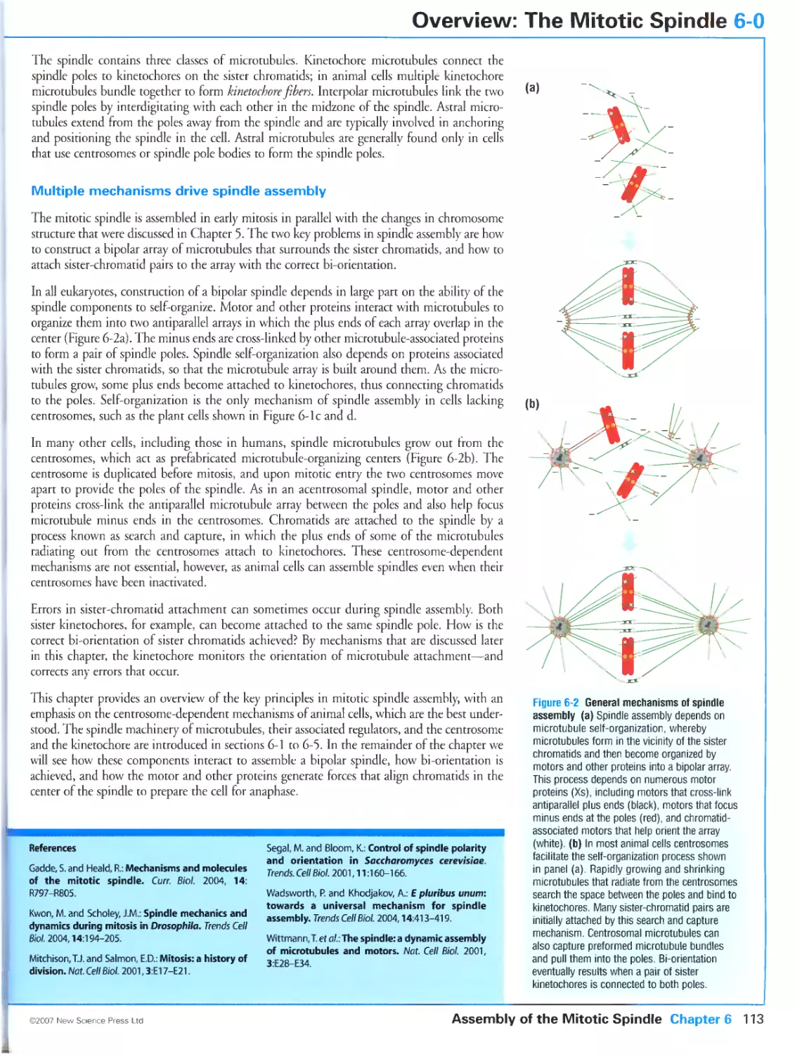
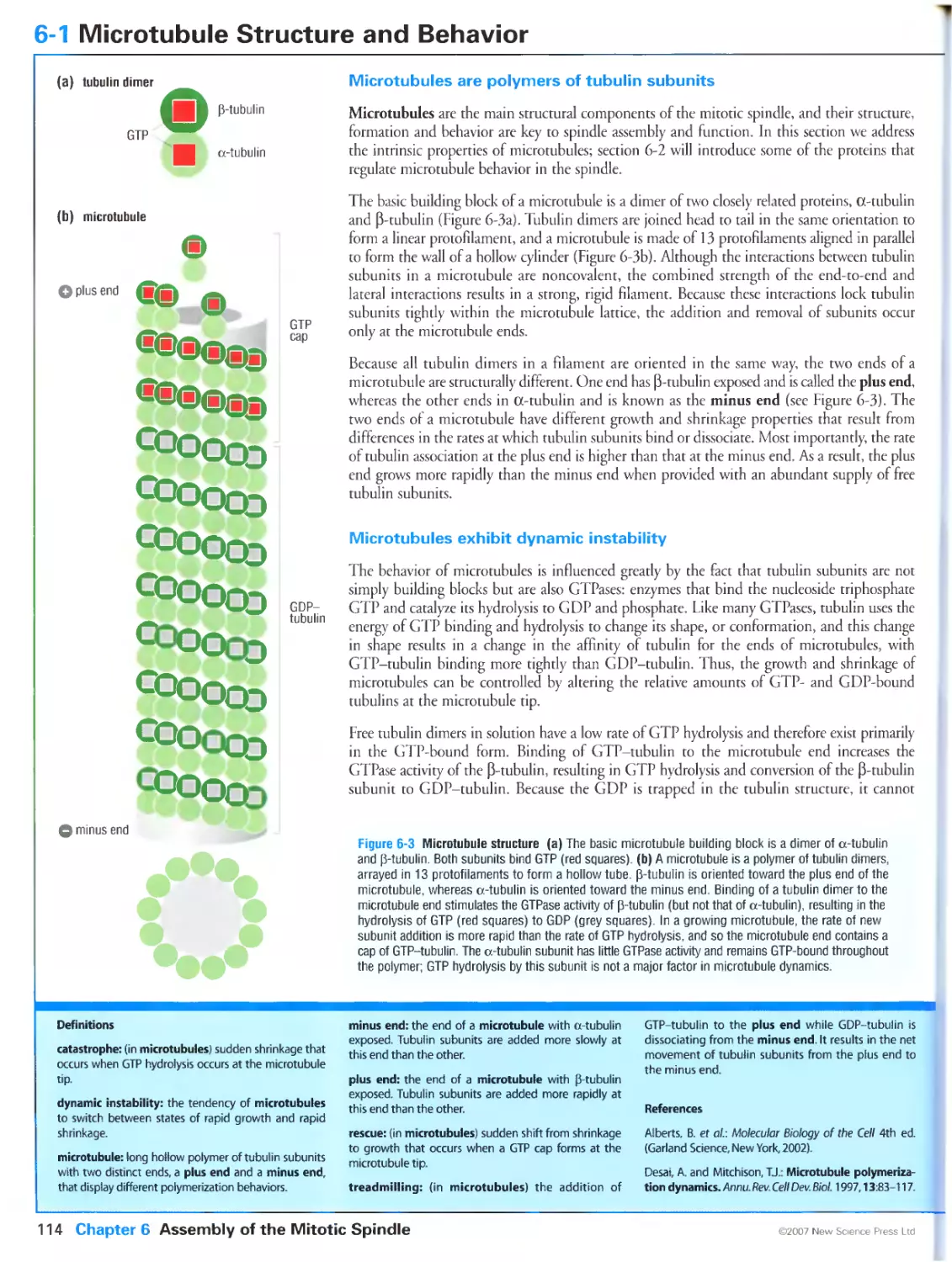
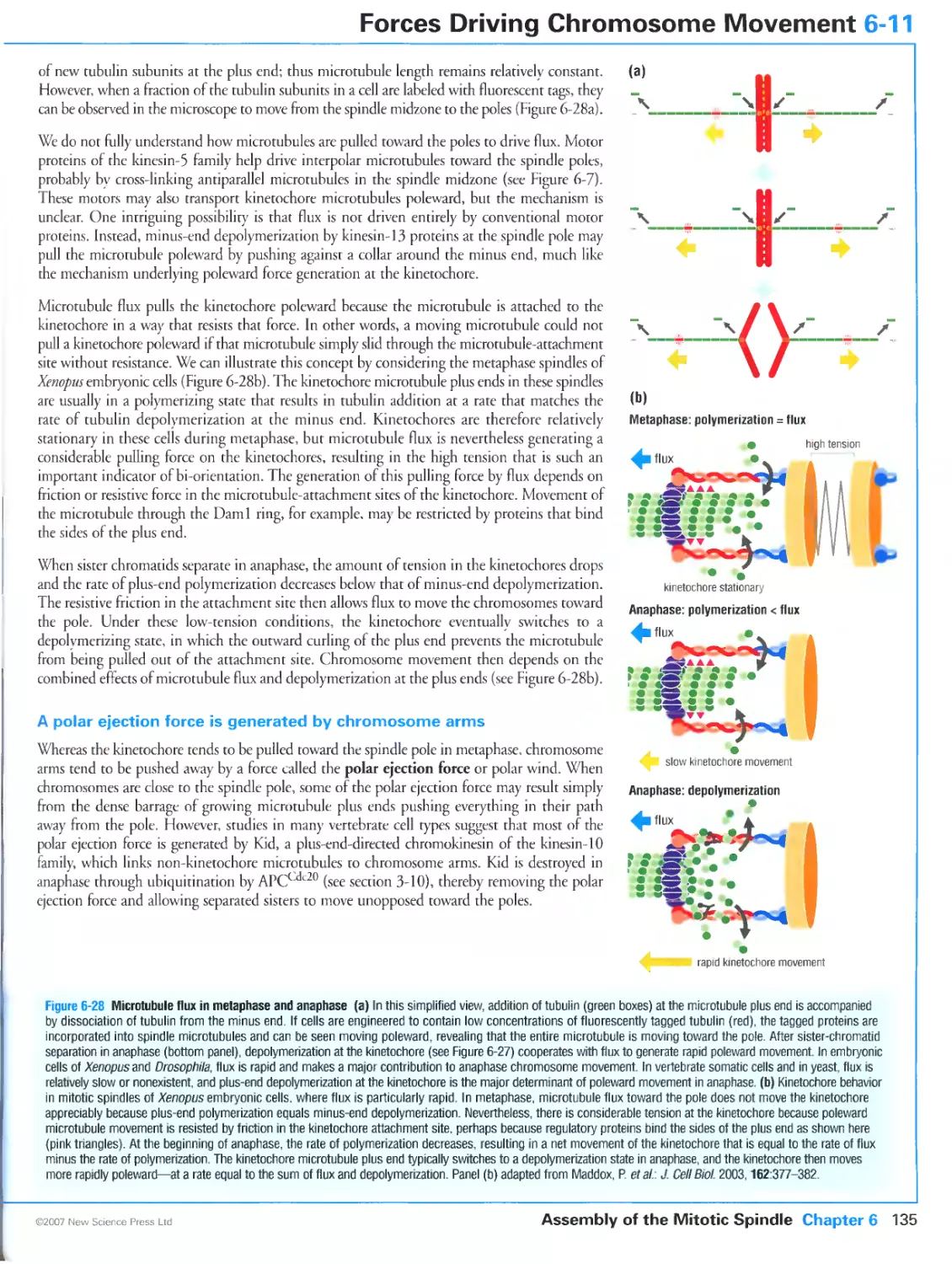
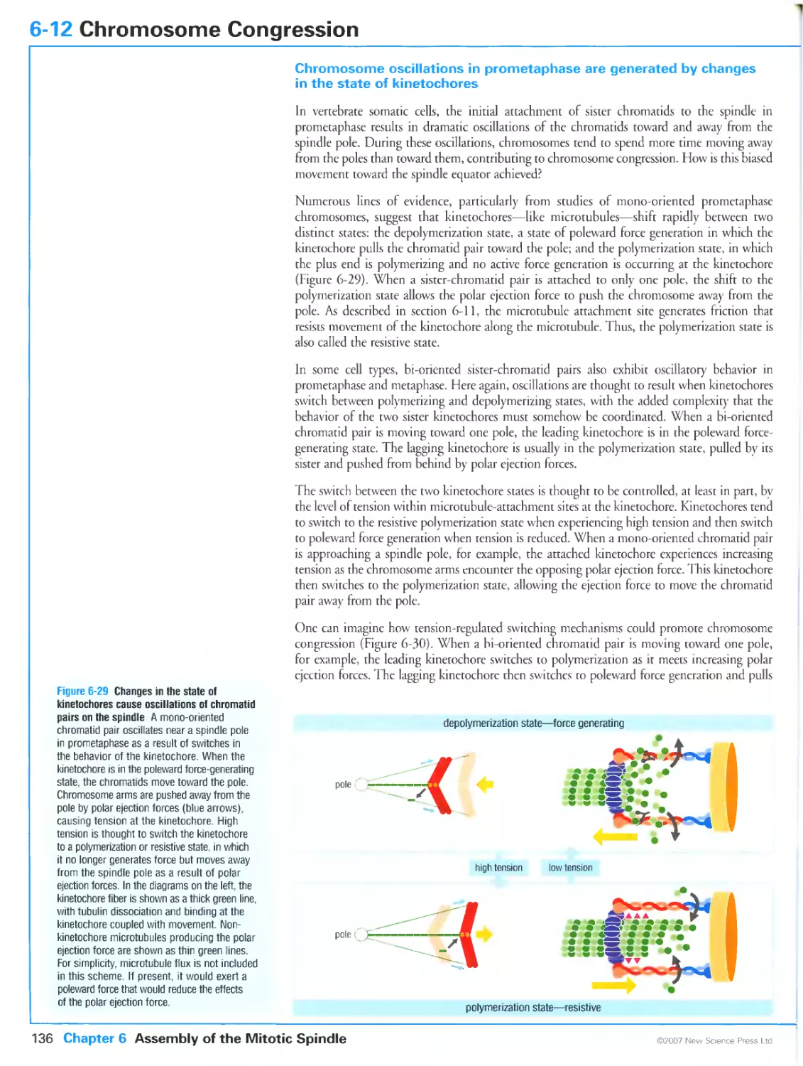
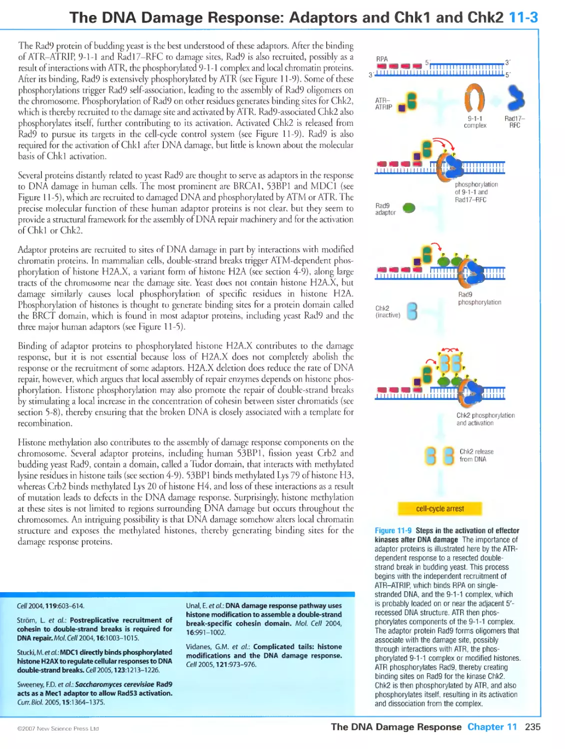
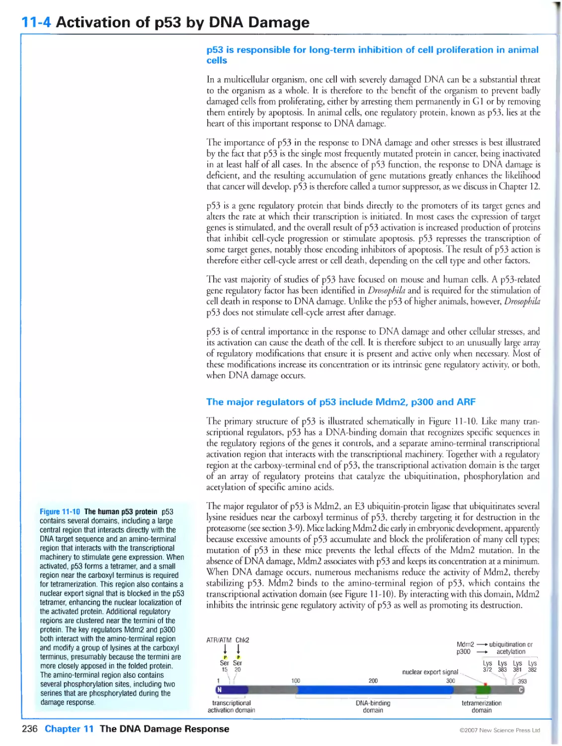
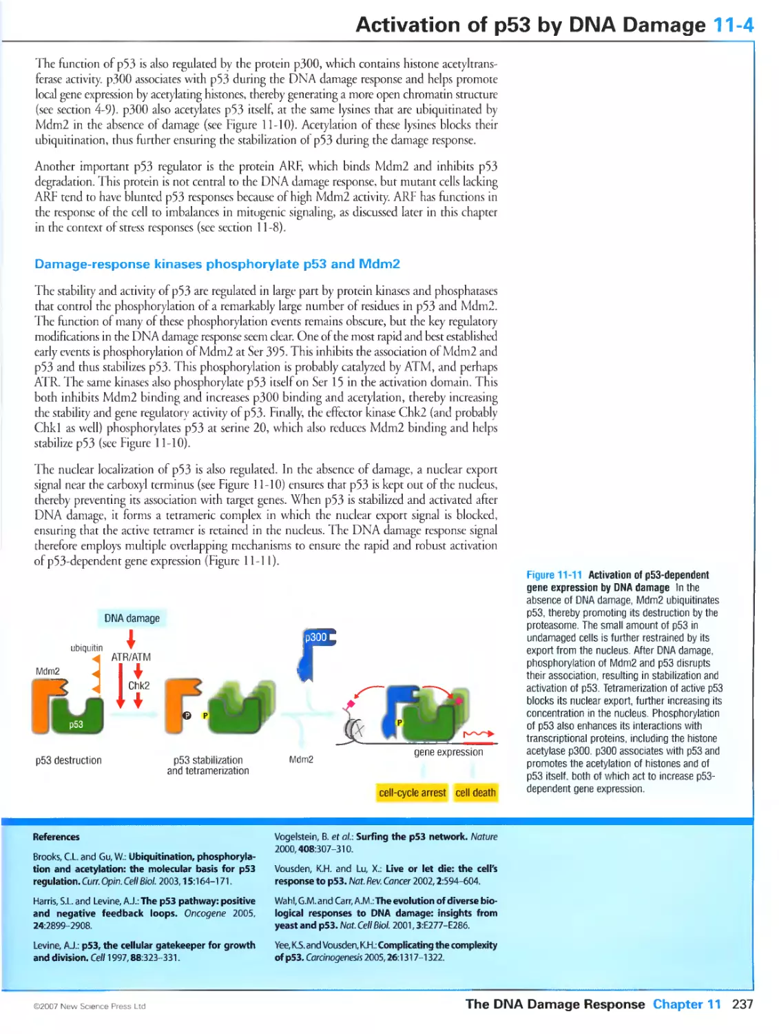
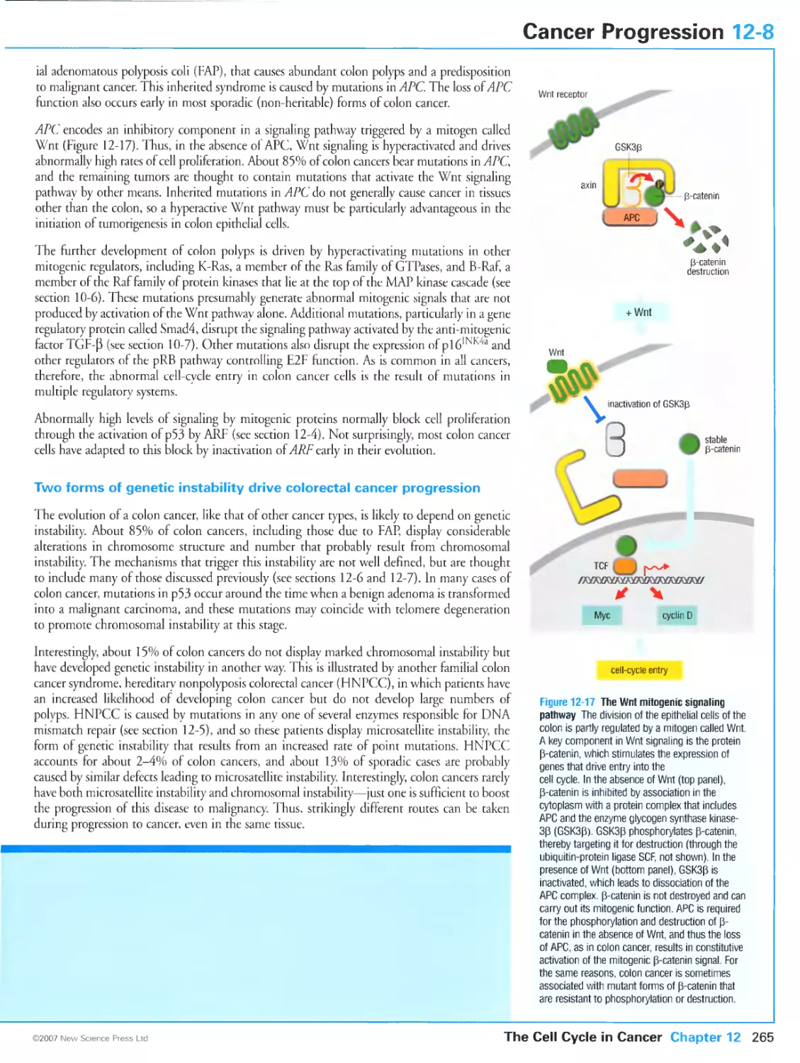
/


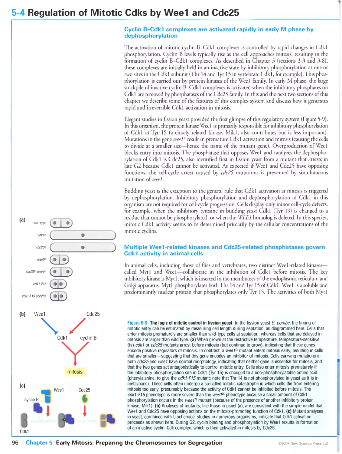


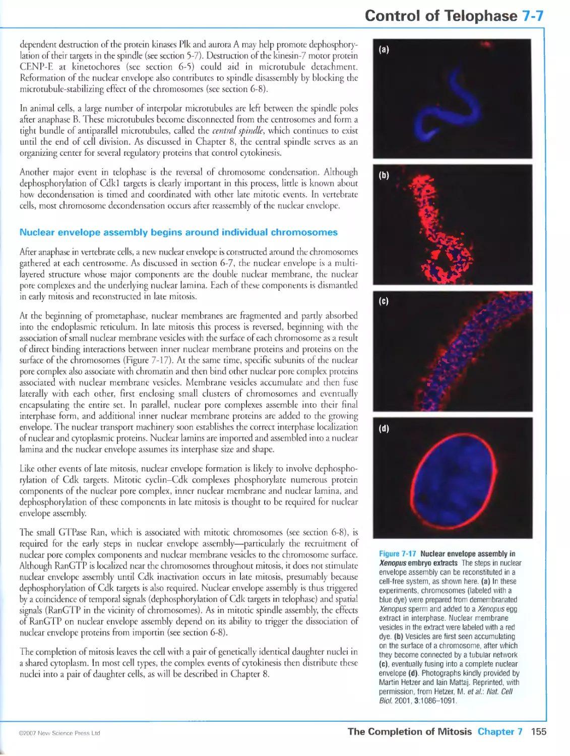


Автор: Morgan D.O.
Теги: biology microbiology cell biology oxford university press the cell cycle cytology
Год: 2007


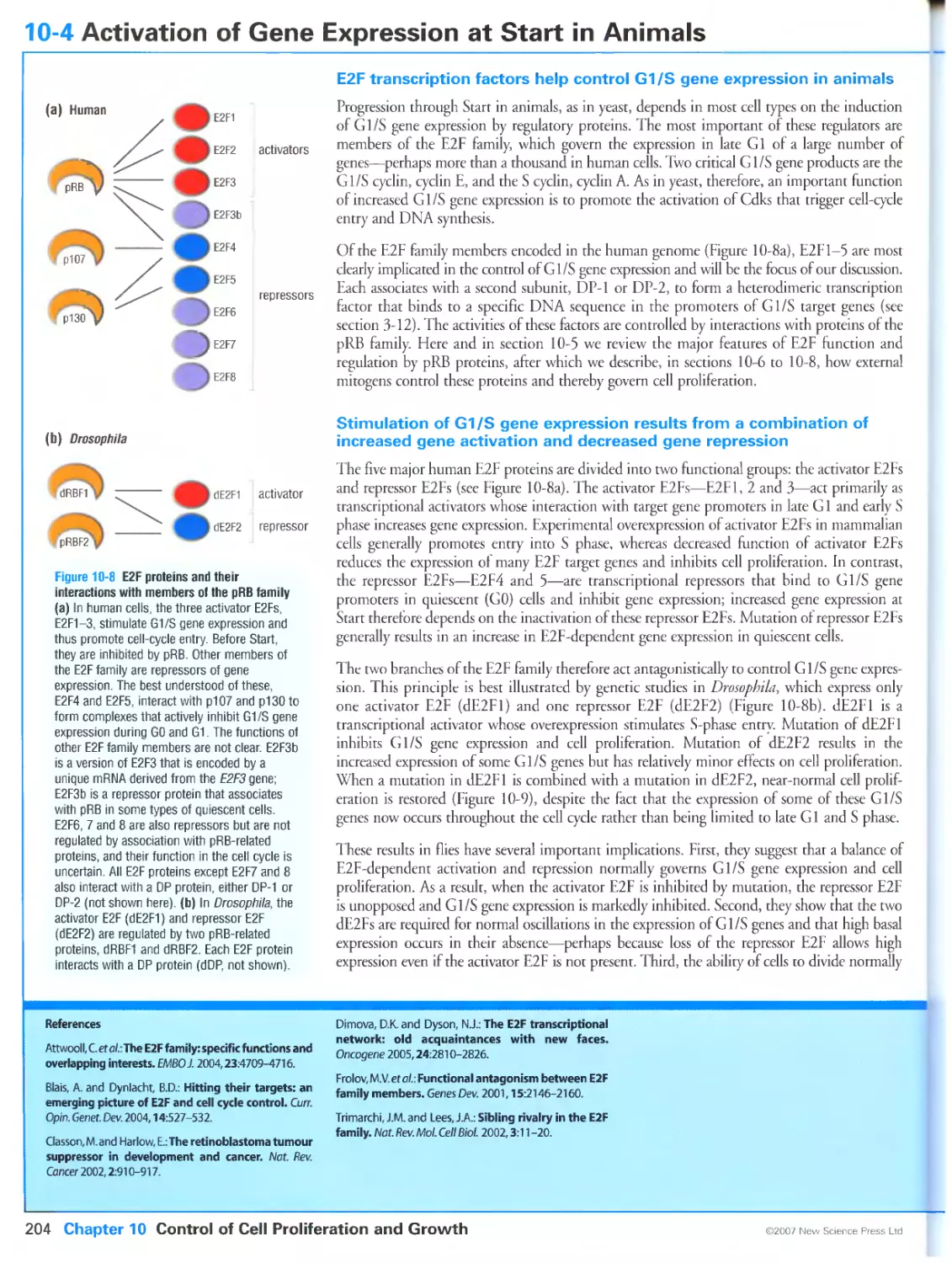
Текст
Primers in Biology:
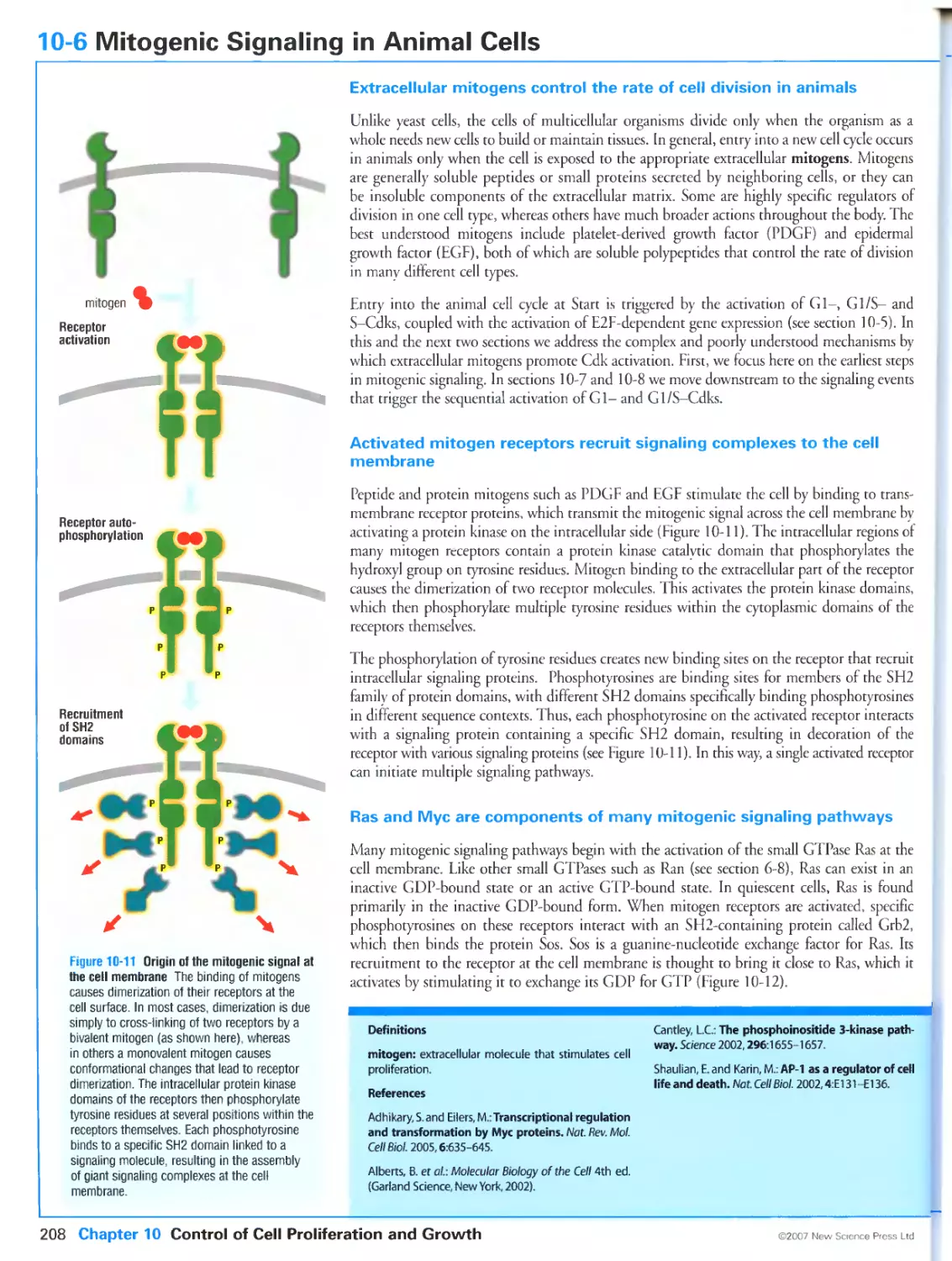
Published titles
Protein Structure and Function
Gregory A Petsko & Dagmar Ringe
Forthcoming titles:
Immunity
Anthony DeFranco Richard Locksley & Miranda Robertson
Molecular Biology
Nancy L Craig Orna Cohen-Fix Rachel Green Carol W Greider Gisela Storz & Cynthia Wolberger
Genetics
Philip Ingham & Tanya Whitfield
Cell Signaling
Wendell Lim Bruce Mayer & Anthony Pawson
ii
Editor: Eleanor Lawrence
Managing Editor: Karen Freeland
Editorial Assistants: Kerry Gardiner and Joanna Miles
Design and Illustration: Matthew McClements, Blink Studio Ltd
Structure Graphics: Lore Leighton
Copy Editor: Bruce Goatly
Indexer: Liza Furnival
Production Director: Adrienne Hanratty
© New Science Press Ltd 2007
All rights reserved. No part of this publication may be reproduced or transmitted in
any form or by any means without permission in writing from the publisher.
Distributors:
Inside North America:
Sinauer Associates, Inc., Publishers,
23 Plumtree Road, PO Box 407, Sunderland, MA 01375, USA
orders@sinauer.com
www.sinauer.com
Outside North America:
Oxford University Press
Saxon Way West
Corby, Northants
NN18 9ES
UK
Customers in the UK may use the OUP
freepost address:
Oxford University Press
FREEPOST NH 4051
Corby, Northants NN18 9BR
bookorders.uk@oup.com
www.oup.co.uk
ISBN-13:978-0-9539181 -2-6 (paperback) New Science Press Ltd
ISBN-10:0-9539181-2-2
ISBN-13:978-0-19-920610-0 (paperback) Oxford University Press
ISBN-10:0-19-920610-4
ISBN-13:978-0-87893-508-6 (paperback) Sinauer Associates, Inc.
ISBN-10:0-87893-508-8
British Library Cataloguing-in-Publication Data
A catalogue record for this book is available from the British Library
Published by New Science Press Ltd
Middlesex House
34-42 Cleveland Street
London W1P6LB
UK
www.new-science-press.com
in association with
Oxford University Press
and
Sinauer Associates, Inc., Publishers
Printed by Stamford Press PTE Singapore
15 14 13 12 11 10 98 7 6 5 4 3 2 1
iv
The Author
David O Morgan graduated in animal physiology from the University of Calgary in 1980
and then did his doctoral and postdoctoral work in endocrinology with Richard A Roth
and William J Rutter, and in virology with Harold Varmus at the University of California,
San Francisco, where he is now a Professor in the Departments of Physiology and
Biochemistry & Biophysics.
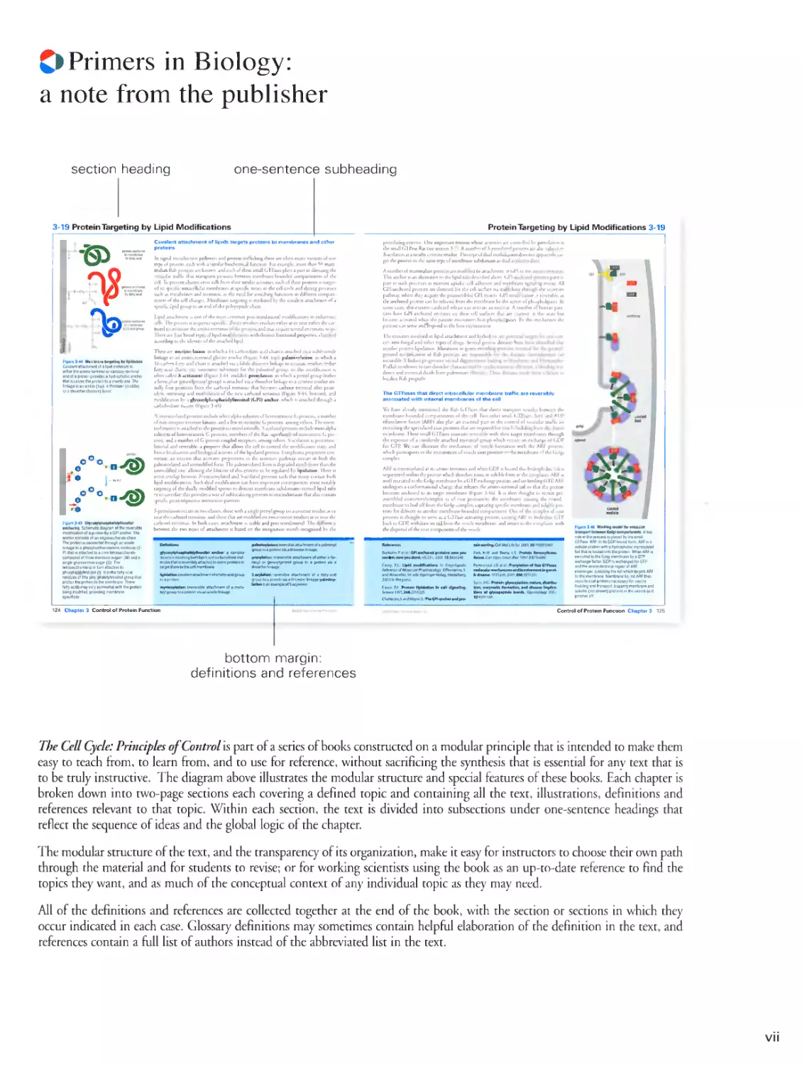
Cl Primers in Biology:
a note from the publisher
section heading
one-sentence subheading
3-19 Protein Targeting by Lipid Modifications
Figari 34 мил trini la г gelin) by hp idi h un
of lipids targets proteins to membranes and other
proteins
In mgn.il transduction pathwass and prolan tu Ilk king ihere are often mjnv variants of one
rvpc iif protein, each with j similar biochemical (unction For rumple. more ihan id mam
nutult Rah proteins arc known, and each ot these small 11I I'ascs plays j pjtt in directing the
vesicular traffic that transports protein-. heiwern membrane-hounded compartments of the
tell. To prevent ehaoin .toss-tall, lumi then mnul.it .n unties, each ul these proteins is target -
ed In specific intiacellular membranes at specific limes in the cell cycle and during processes
mi,:- .r nil-i.ilii'lism and sccrcriur,. .is r:ii need -wiiihiiig lurutiuns in ditterrjin compatì
menti of the cell changes Membrane t.iigcling is medial ul hv the divalent attachmcnl til a
spuilii lipid griiu]' in :in end n! ilk- puU peptide chain.
I ipid jtiaihmeni is one of the musi common pott nan sianone modifications in culuryoik
.ill' I In |i n scqiu n. .■ spi-, ili. .1ln.11. mi i lives icsulucs in her ji or near eithet the
salii, us I terminus ut the amimi lei minus ul ilk pii.lem. and may ttipnre several enzymatic steps.
Therein: fouthroad is pes ul lipid modi licit ions ss uh distinct functional properties, classified
according io the identity of rhe attached lipid.
these arr: myriiloylalion in sshieh a U carbon laitv and eliain isanachcd via a stable amide
linkage to an am ino-term ina I glycine residue (Figure 3-44, lop pal mi tosi at ion in ss In. Ii
Hi-car bori fairy-acid chain is attached via a labile ihioestci linkage to cisterne residues (other
fatty-acid chains san sometimes substitute (or ihe palmitoyl group, so this modiliualion is
..lien i.illcd ,s-acylation 'liguri i-i .1.1.c prcnylation .r: is lm h a prens I gr. .up culm
uallv lout positions troni the carinoci rcrriiuius lui liei.uno ci rhovy-term inai after prole-
re 3-44, bottom); and
i is attached through j
„1,1. „,m
Nn .iii
i.illi.ilisdl.
in hv a glycoiylphospli.il uli i innsi ii г i i (.i11: .u
te moiety (Figure 345).
FwinJ-tt Grnotrtslvaii.riairiylinoiiiol
wclartm Sciumjtic diagram a №• пмшьн
in try a GPI ancrio. The
oligosaccharide chain
lirikaos lo a priospho
single alueosamn. s
gar <G|. the
m|ta?tchmanv-jCitl
specificity.
.V-mynsioylated proteins include select alpha subuiius ul heininumeric 11 proteins, a number
of non-tcceptut tyrosine kinases, and a less monomelic (. proteins, among others. The myrts-
tovl moiety is attached to the protein co-translaiionallv snsljied piuieins include most alpha
suhunils of hetenytnmciii (j proteins, members olthe Ras superfimiry of monomelic G pro-
tcins, and a number of G-prorein-couplcd recepiors. among others, .tacylation is post-trans-
laliotul and reversible, a property thai allows the cell to control the modification state, and
hence localization and biolugical actis its. ot rhe lipul.ucd pnuein is mphoma proprotein con-
yenaje. an enzyme that activates ptoproteins in the secretory pathway, occur) in both the
palmitijylated and unmuditied torni i he p.lmuni l.ued i.nm is degraded much taster lhan the
11 ri ir:...Ii! k.i un, .ilUsmg :1k htclihic ..i this i unii» n. hi updated in lipidalliiu I inn .i
some overlap between .Vuivn-.nivi.ncil and s ,i, viand piote in- such th.it many contain both
lipid modifications. Such dual nhh.litk.uiun san lue imprimili consequences, must nntahlv
largiririg ut ihe dil.illv modified species in disuikl mi nlimine siiliilum.ims rerrned lipid rails
or to caveolae; this provides a way ol sublocai i/mg pi mi in- m in kn .domains that also euntain
speliti, pii.teiimpruleiii imenei inn p.uriiers.
.Vptcnvlarinn occurs in isso classes illuse willi .< single primi gr. nip ,.:i . e wei in residue al ,.r
near the carbniyl leiminus. and those thai arc modified on two cysteine residues at or near the
carboni terminus In both cases, attachment is stable and posi-iranslauonal. The difference
between the two Types of attachmcnl is based on the recognition motifs recognized bv the
Protein Targeting by Lipid Modifications 3-19
ptcnylaling enzyme One important protein whose activities are controlled bv prenvlation is
the small (¡TPa.se Ras I see section 3-"). Л number ot S-picnvlated ptoteiniare aim subject to
-Vacylarion at a nearbs Cysteine residue I Ins rcpe of dual modificaiion does not appaierulv
target the protein 10 the same type ot membrane subdomain as dual acs latum does.
a number of mammalian proteins are modified bv attachmcnl ul i ¡14 lu ihr ammo terminus.
This anchor is an allcrnatise in iln hpi.l i.ul- dc-inbcd .ihusc I ill jnilioted proteins
participate in such processes as nutrient uptake, cell adhesion and membrane signaling events. all
ljpi-anchored piuieins .ue destined for rhe cell surface si.i rral'iilinn through rhe secretory
pathway, where rhev .injur re i hi picric in hied (il'l moicn. (ill ruuilitn. s reversible, .is
the anchored protein can lie tcleascd from the membrane by the action of phospholipids. In
some cases, tills en гуте-catalyzed release can activate an enzyme. a number of human
parasites have (¡14-anchored enzymes on then cell surfaces that ate Inactive in this state but
become activated when the patasile encounters host phospholipases. Hv this mechanism the
parasite can sense andlcspond to the host environment.
The enzymes involved in lipid attachment and hydrolysis ate potential targets for
anti-cancer, anti-fungal and other types ot drags Several genet is diseases have been identified (hat
involve protein lipidanuii Mutations in genes encoding proteins essential lot the gcranvl-
gcranyl modification of Rib proteins ate responsible tot the diseases ihotoideremia Ian
insurable \ linked piugn -sue пина! dipiretjrr.m k.id.ng m :,.,nducssi and Hcrmanskv-
I'lidLik ч ridrurru- ..i tare dis..rdvr . lur.kriti/cd in ...ulosutancous albinism,a bleeding ten-
luiah/e Rab properly.
e leversibly
We have already mentioned rhe H.il. GTI'ases that dueer transport vesicles between the
membrane-bounded compartments of the cell. Two other small GTPascs. Seri and ADP
nbosylation factor (aru also play an essential part in ihe sonttol nf vesicular traffic by
lecrainng the specialized coat proteins thai ate required for vesicle budding from the donor
membrane. These small GTPasci associate icvetsibty with then target membranes through
the eiposure of a covalenti! attached msnsiovl group which occurs on cichange of GDP
Ibi GTP. We can lllustiate the mechanism of vesicle formation with the arf protein,
which participates in the recruitment ol vesicle coat proteins to the membrane of the Golgi
ARF is mynstoylated at its amino terminus and when GDI' is bound this hydrophobic tail is
sen uc.s re led ssilhill [In jinncm sslm h drerelure c:sis(. in mil u hie lumi m ij. e- . vl. .pi.mil. .Wit n
itself recruited to the (iolgi membr.ine by j <.'[ I'-cschangc protei n, .uni mi binding ( .'I'll A.R¥
undergoes a contotmaiiuii.il Jiarige rh.u nlc.i-es the .imiriu-ieimui.il lail so that the protein
becomes anchoted m its target membrane (Figure 34G). It is then thought to rctruit pre-
assemhled coatometsiicomplci es ol coat piotcinstito ihe membrane, causing the coated
membrane [u bud ull llulli ihe t .nig. 11 iln pies .j;"uling •peeltie membrane all J soluble
proteins fot delivery in another mcmbunc-hounJcJ compartment One ol the complex nf coat
proteins is thought to serve as a I, I i'.isc- .nnsaimg pioiem Ljiismg ARf to hydrolyze GTP
hack to GDP. withdraw us tail from the vesicle membtanc. and return to ihe cytoplasm, wiih
ihe dispersal of ihe coai eomponcnts ol the vesicle.
If
1
bottom margin:
definitions and references
The Cell Cycle: Principles of ControlIs part of a series of books constructed on a modular principle that is intended to make them
easy to teach from, to learn from, and to use for reference, without sacrificing the synthesis that is essential for any text that is
to be truly instructive. The diagram above illustrates the modular structure and special features of these books. Each chapter is
broken down into two-page sections each covering a defined topic and containing all the text, illustrations, definitions and
references relevant to that topic. Within each section, the text is divided into subsections under one-sentence headings that
reflect the sequence of ideas and the global logic of the chapter.
The modular structure of the text, and the transparency of its organization, make it easy for instructors to choose their own path
through the material and for students to revise; or for working scientists using the book as an up-to-date reference to find the
topics they want, and as much of the conceptual context of any individual topic as they may need.
All of the definitions and references are collected together at the end of the book, with the section or sections in which they
occur indicated in each case. Glossary definitions may sometimes contain helpful elaboration of the definition in the text, and
references contain a full list of authors instead of the abbreviated list in the text.
vii
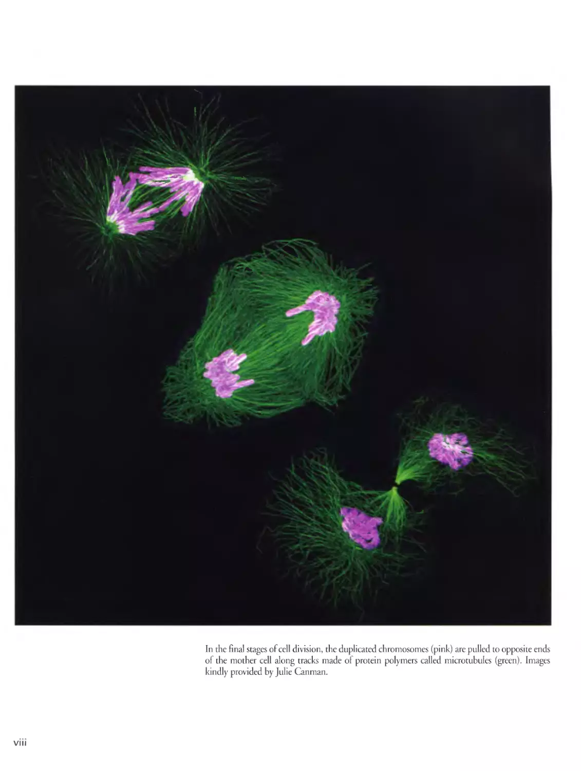
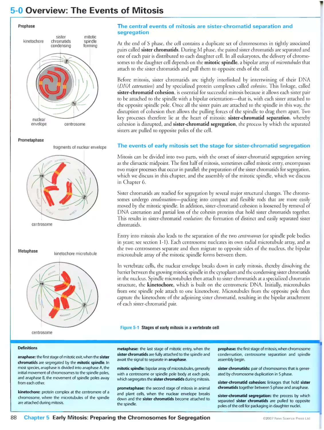
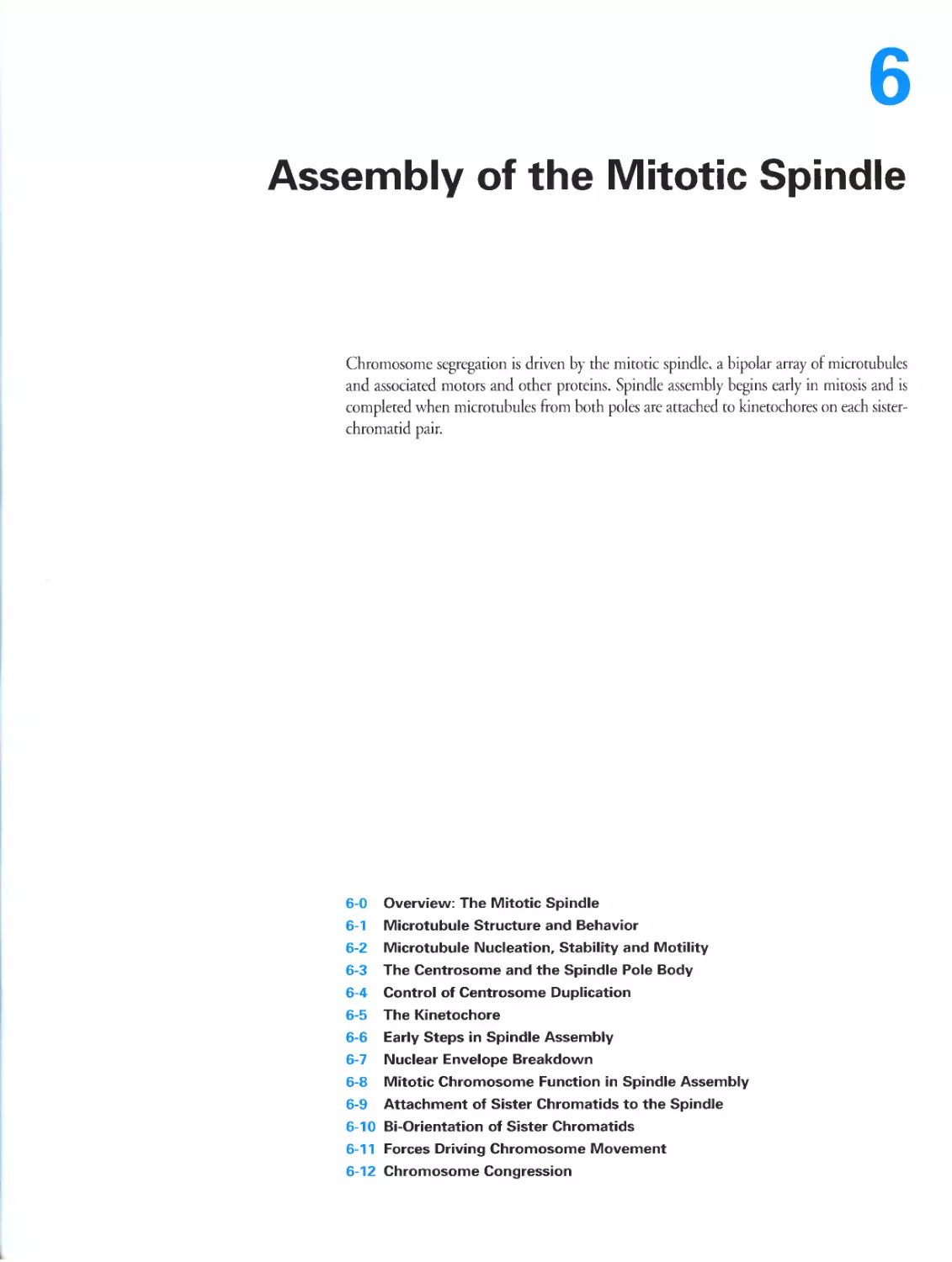
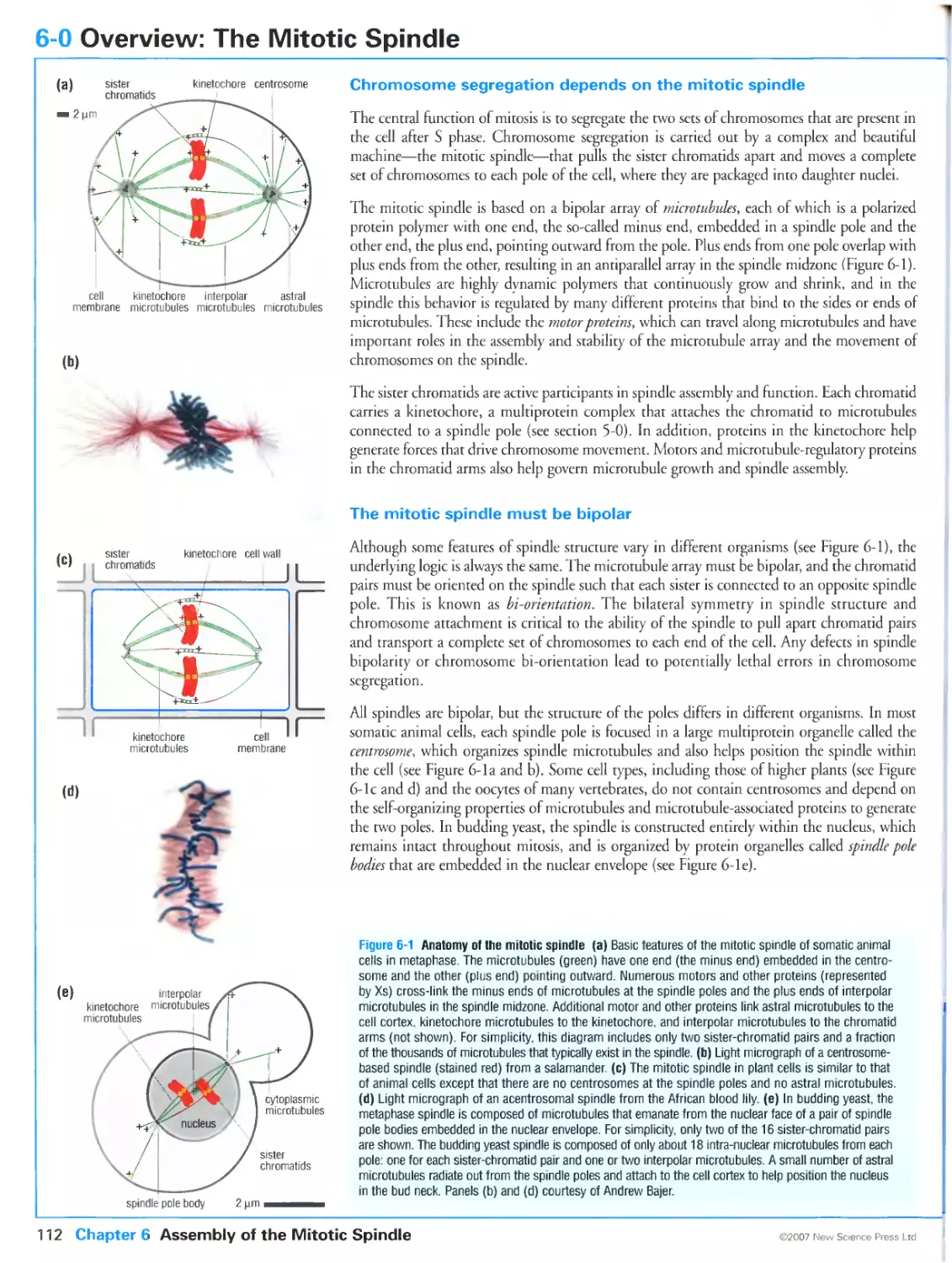
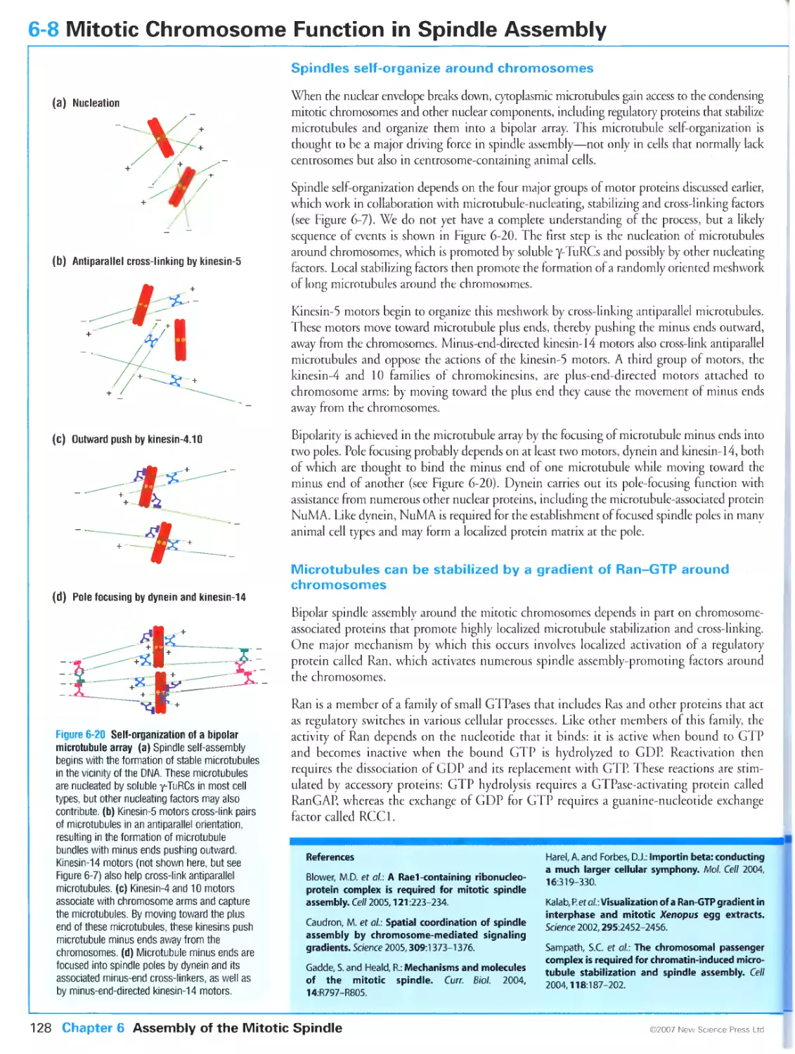
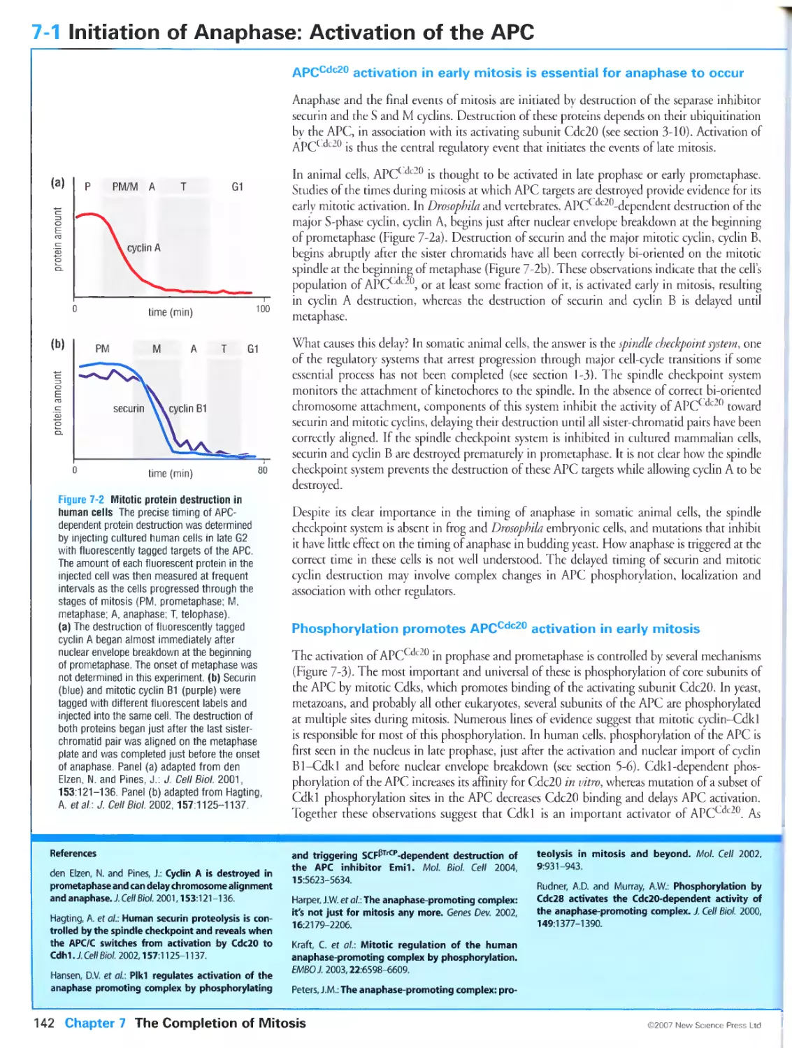
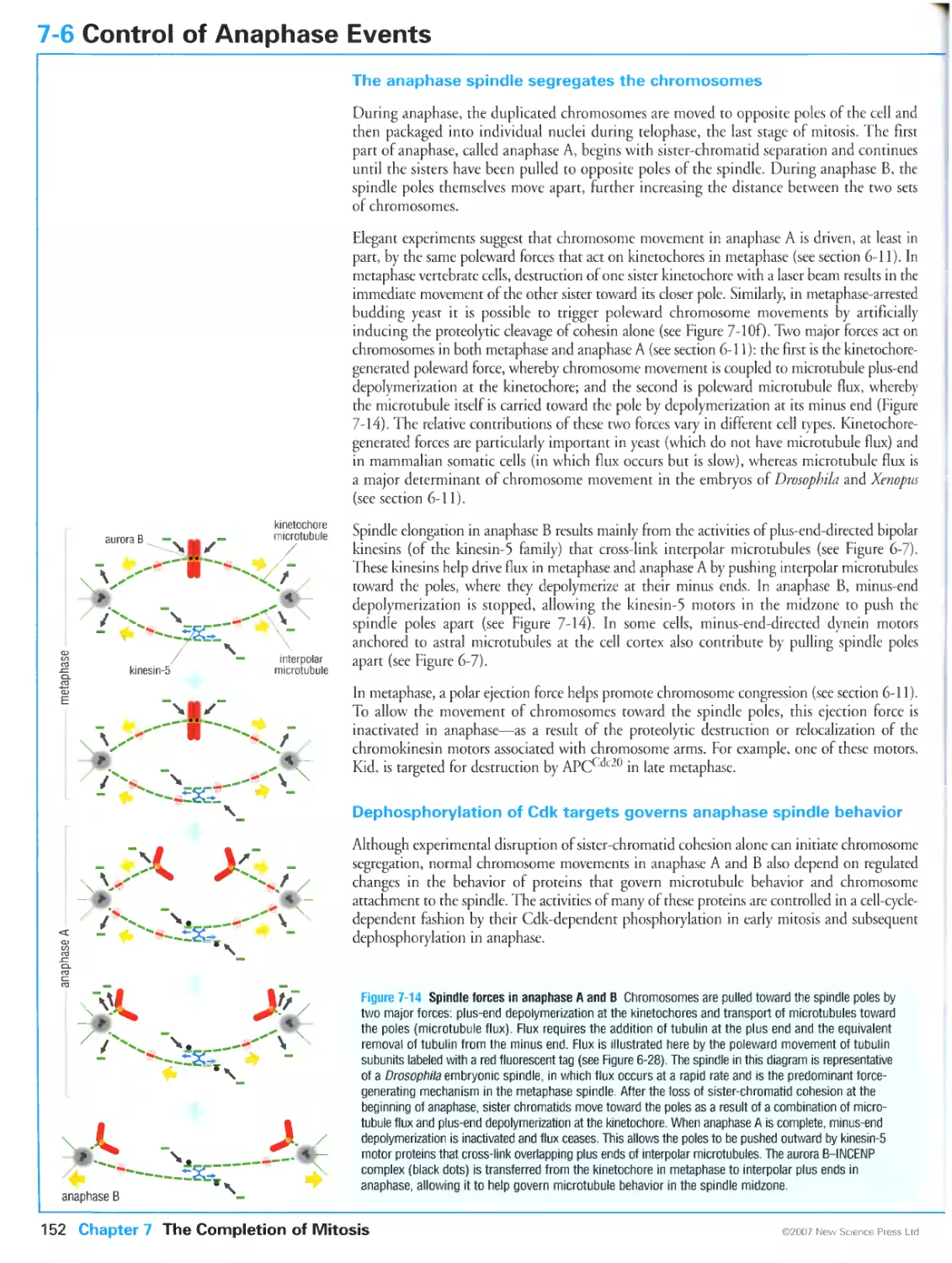
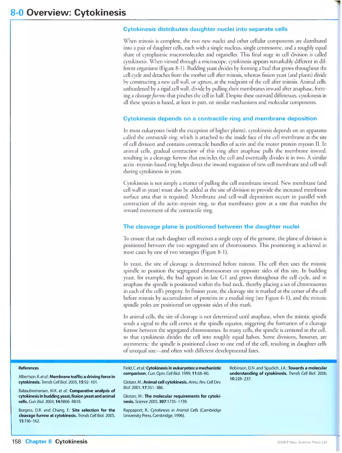
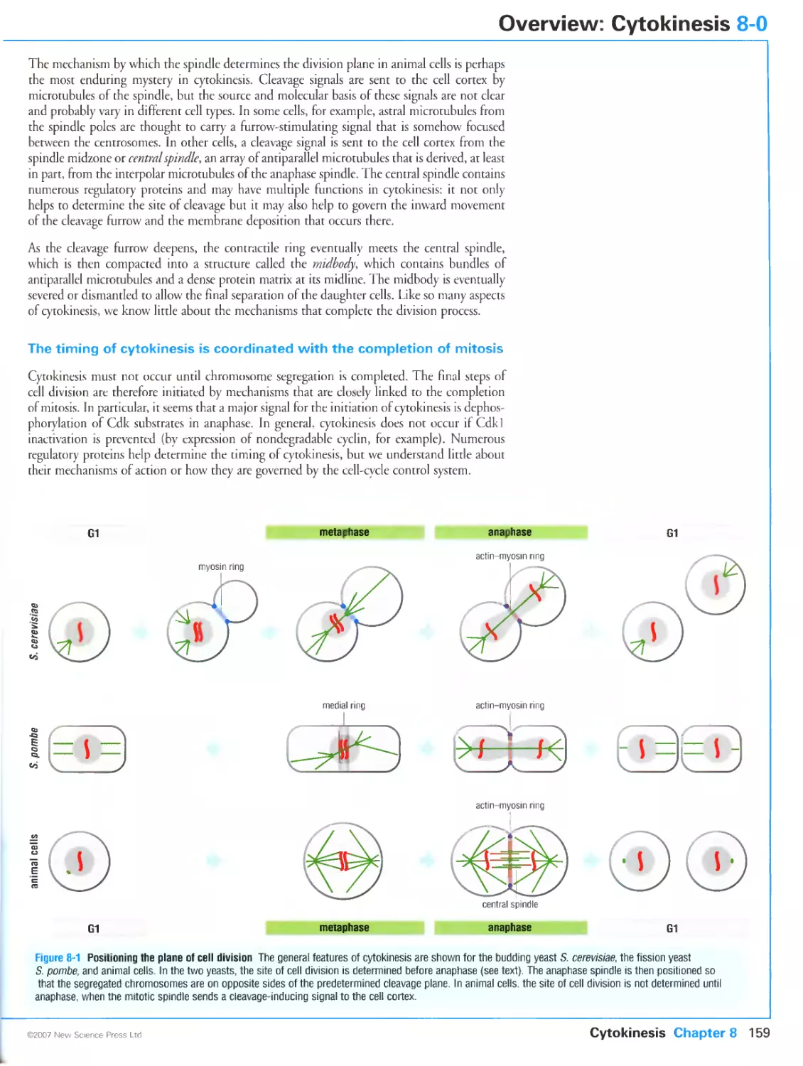
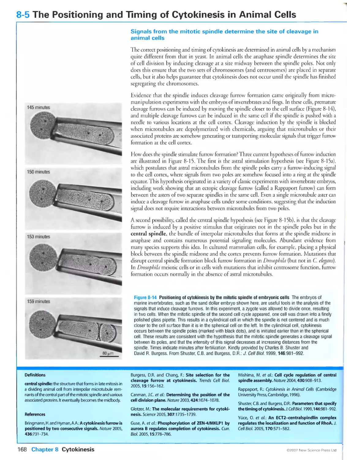
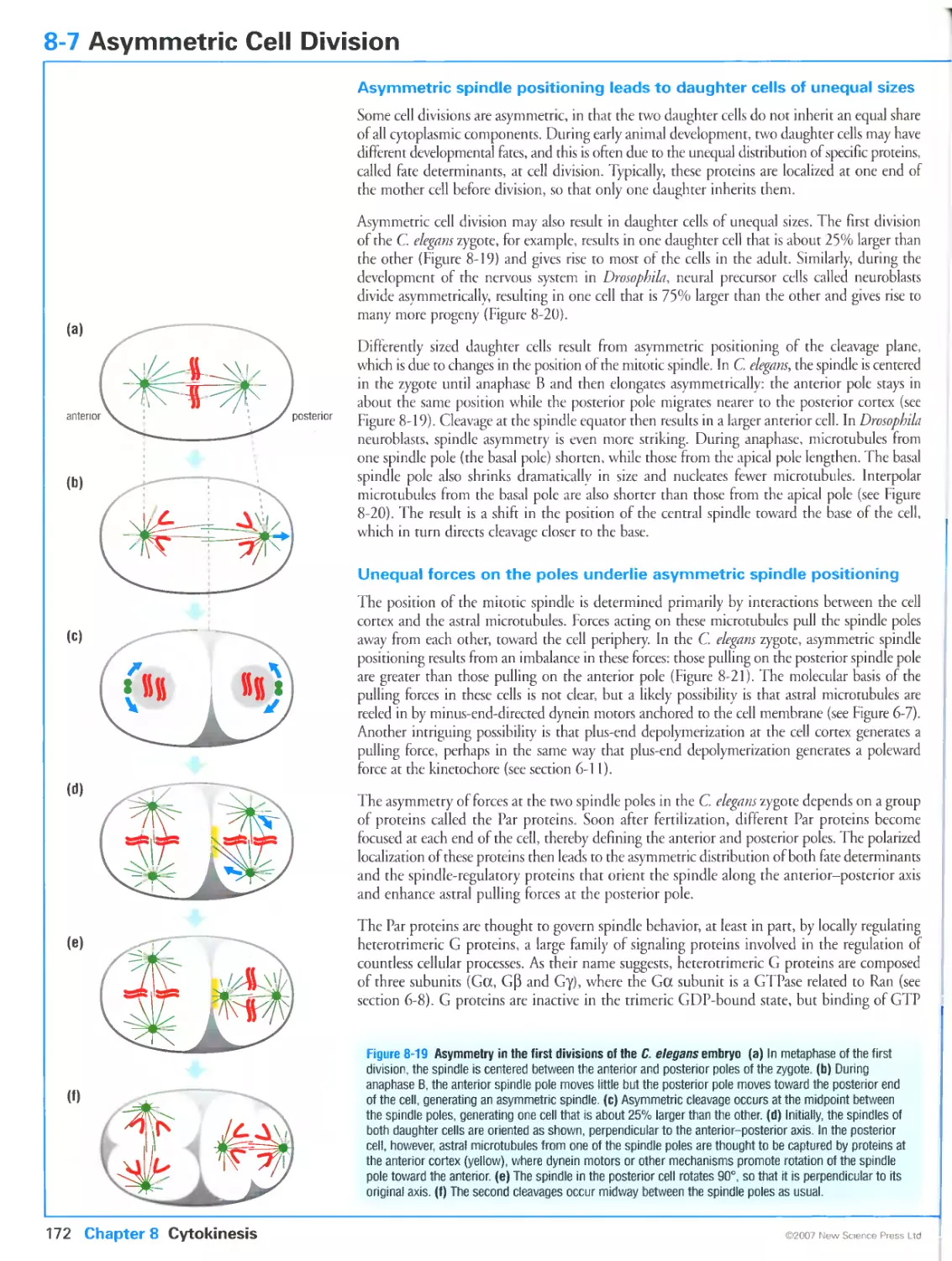
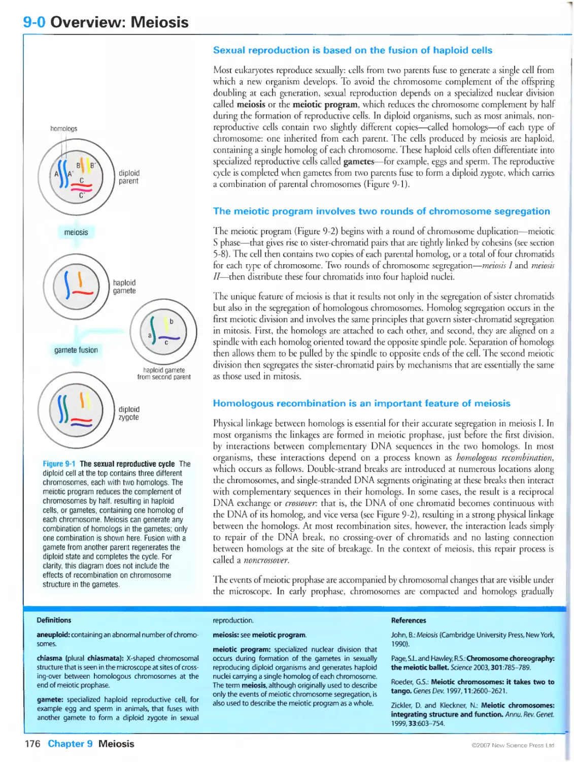
In the final stages of cell division, the duplicated chromosomes (pink) are pulled to opposite ends
of the mother cell along tracks made of protein polymers called microtubules (green). Images
kindly provided by Julie Canman.
Preface
The first century of cell biology belonged to cytologists, whose painstaking observations with
microscopes revealed that all living things are composed of fundamental units called cells, that
all cells arise by the division of preexisting cells, and that each daughter cell contains a set of
chromosomes like that of the mother cell. At the turn of the 20th century, the collision of
cytology and the newly minted field of genetics led to the discovery that chromosomes are the
physical determinants of heredity. Later in that century came the next great convergence, when
the disparate fields of cytology, genetics and biochemistry together gave rise to the realization
that all eukaryotic cells use similar molecular machines and regulatory mechanisms to perform
and guide the events of chromosome duplication and cell division. We can now look back on
twenty years of astonishing expansion in our knowledge of these mechanisms. But a new problem
has arisen: the flood of information has not been accompanied by a clear understanding of how
the pieces fit together into a coherent whole.
This book is an attempt to help solve this problem. My goal is to provide a clear and concise
guidebook that organizes our vast knowledge on a coherent framework that emphasizes the key
problems in cell division and the molecular mechanisms that have evolved to solve those problems.
Although organized around key principles, the book does not avoid the so-called details: on
the contrary, it includes a glimpse of every layer in our knowledge of cell division, ranging from
the cytologist's descriptions of major events to the biochemist's atomic-level analysis of the
protein structures and chemical reactions underlying those events. All of these layers are
important—and fascinating. The architect Le Corbusier, writing in 1935 of the stunning
confluence of form and function in modern aircraft, was more eloquent: "There are no 'details'.
Everything is an essential part of a whole. In nature microcosm and macrocosm are one."
I owe a major debt of gratitude to the many colleagues who provided thoughtful and constructive
suggestions during the writing of this book (see Acknowledgements). The information contained
herein remains the full responsibility of the author, however. It is well known that the teaching
of principles requires exaggeration of some facts and omission of others. My apologies to those
scientists whose discoveries I have over- or under-emphasized.
The writing of this book began, in a state of wilful ignorance about the effort involved, during
an eight-month sabbatical leave at the University of Uppsala in Sweden. In the six years since
then, I have returned to Uppsala every summer to do much of the writing in the calm confines
of the Biomedical Center library. I am grateful to my hosts on those wonderful visits: Carl-
Hendrik Heldin and his colleagues at the Uppsala branch of the Ludwig Institute for Cancer
Research, with whom I enjoyed countless discussions—and lunches—between paragraphs.
Every step in the synthesis of this book was catalyzed by the inspirational guidance of Miranda
Robertson and her team at New Science Press. I am immensely grateful for the brilliant
editing of Eleanor Lawrence and the beautiful illustrations of Matthew McClements. And
thanks also to Karen Freeland, who skillfully and enthusiastically piloted the project with the
help of Kerry Gardiner and Joanna Miles. Lore Leigh ton provided valuable assistance with protein
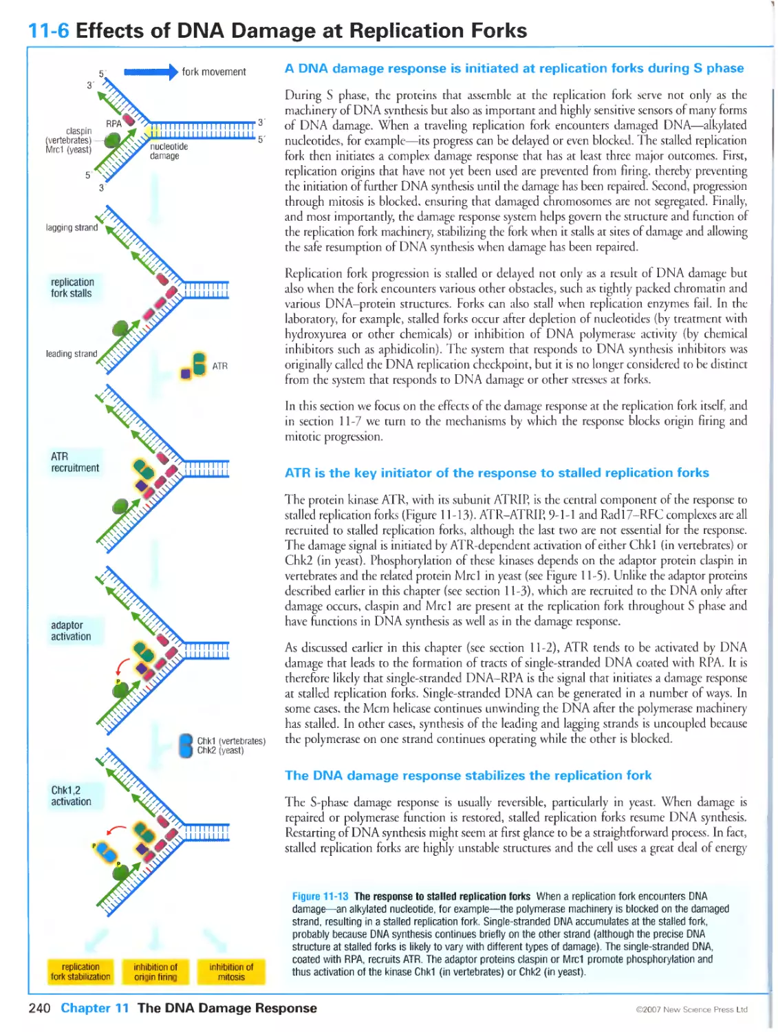
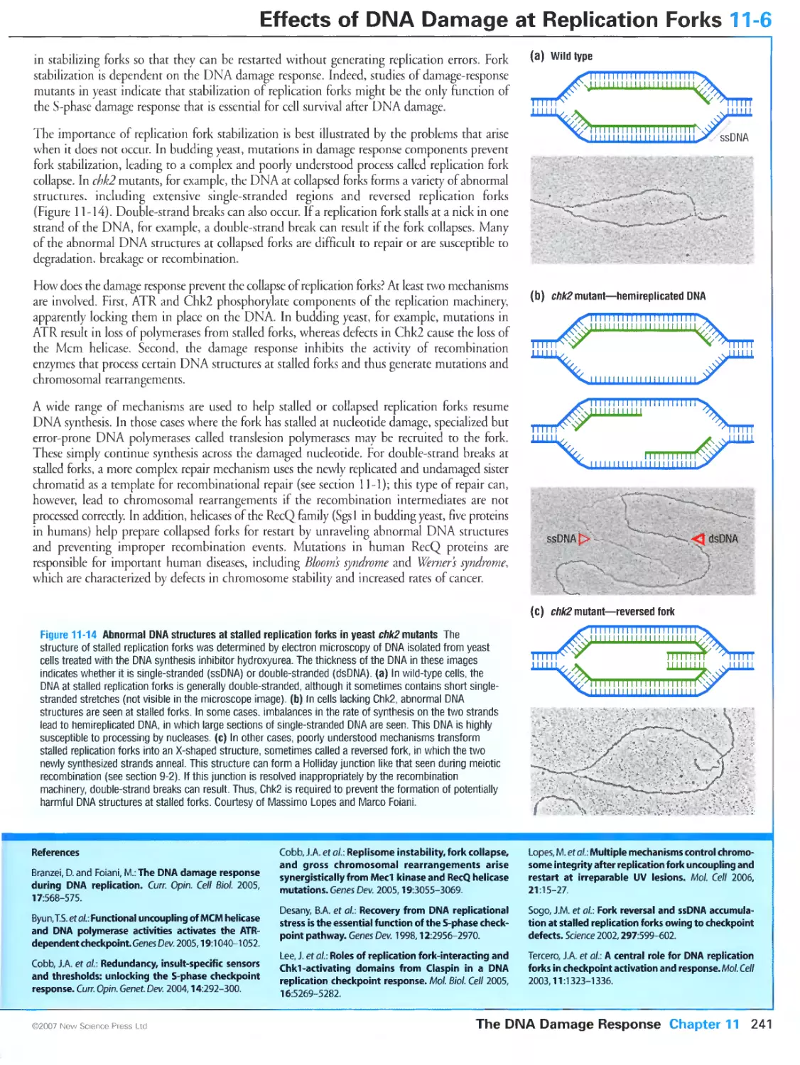
structural figures and Bruce Goatly did a masterful job of copy-editing.
And finally, my apologies and heartfelt thanks to my family, who stood by me with patient
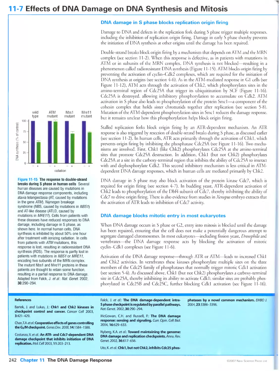
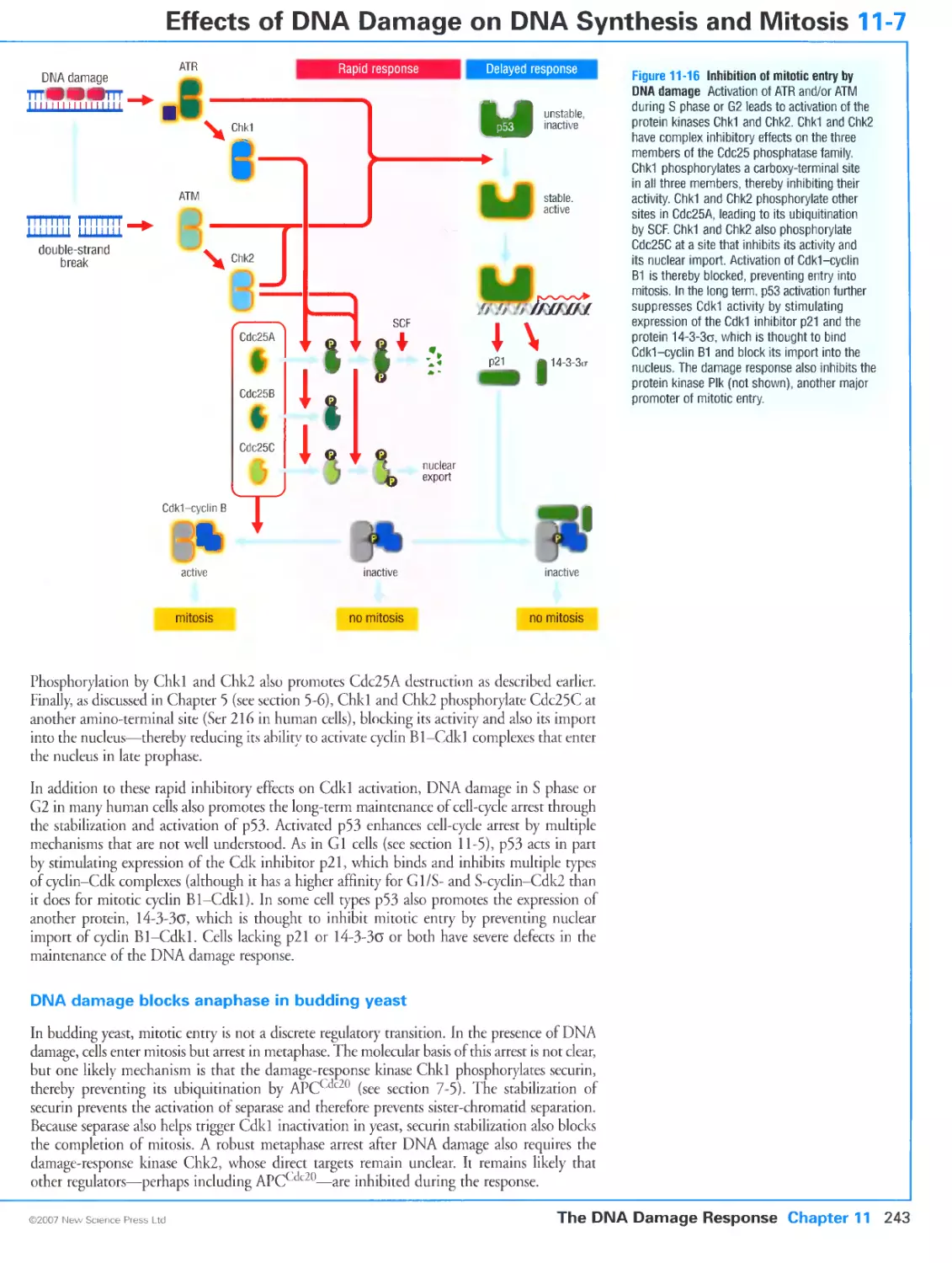
understanding and unwavering support.
David O Morgan
Ci Online resources for The Cell Cycle
For everybody
All of the 273 colour illustrations in this book are freely available in the Oxford University Press Online
Resource Centre and on the New Science Press website and can be downloaded for use in teaching.
Visit http://www.oxfordtextbooks.co.uk/orc/morgan/ or
http://www.new-science-press.com/browse/cellcycle/resources
For instructors
For instructors adopting the book for courses with enrolments of fifteen or more students:
Free access to
• the full text online for a year, for personal use only
• updates - revised, expanded, or new sections and updated references available online only
• PowerPoint functionality allowing instructors to compile any selection of illustrations into a slide show
Visit http://www.oxfordtextbooks.co.uk/orc/morgan/ to register for access to the instructor resources.
Access the resources, once registered, by visiting
http://www.new-science-press.com/browse/cellcycle/resources
xi
Acknowledgements
The following individuals provided expert advice on entire chapters or parts of chapters:
Chapter 1 John Gerhart, University of California, Berkeley; Rebecca Heald, University of California, Berkeley;
Andrew Murray, Harvard University; Kim Nasmyth, University of Oxford
Chapter 2 Bruce A. Edgar, Fred Hutchinson Cancer Research Center
Chapter 3 William G. Dunphy, California Institute of Technology; Bruce Futcher, Stony Brook University;
J. Wade Harper, Harvard Medical School; Douglas Kellogg, Sinsheimer Laboratories, University of California,
Santa Cruz; Charles Sherr, St. Jude Children's Research Hospital; Michael D. Tyers, Samuel Lunenfeld Research
Institute, Mount Sinai Hospital
Chapter 4 John Diffley, Cancer Research UK; Paul Kaufman, University of Massachusetts Medical School;
Matthew Michael, Harvard University; Johannes Walter, Harvard Medical School
Chapter 5 William G. Dunphy, California Institute of Technology; Tatsuya Hirano, Cold Spring Harbor
Laboratory; Douglas Koshland, Carnegie Institution of Washington; Jonathon Pines, University of Cambridge
Chapter 6 Arshad Desai, University of California, San Diego; Rebecca Heald, University of California,
Berkeley; Tarun Kapoor, Rockefeller University;
Chapter 7 Angelika Anion, Massachusetts Institute of Technology; Sue Biggins, University of Washington;
Jonathon Pines, University of Cambridge; Frank Uhlmann, Cancer Research UK
Chapter 8 Bruce Bowerman, University of Oregon; Christine Field, Harvard Medical School; Michael Glotzer,
The University of Chicago; Rong Li, Stowers Institute for Medical Research
Chapter 9 Angelika Amon, Massachusetts Institute of Technology; R. Scott Hawley, Stowers Institute for
Medical Research; Neil Hunter, University of California, Davis; Nancy Kleckner, Harvard University
Chapter 10 Nicholas Dyson, Massachusetts General Hospital Cancer Center; Bruce A. Edgar, Fred Hutchinson
Cancer Research Center; Martin Raff, University College London
Chapter 11 Karlene Cimprich, Stanford University School of Medicine; Stephen J. Elledge, Harvard Medical
School; David Toczyski, University of California, San Francisco; Rodney Rothstein, Columbia University
Chapter 12 J. Michael Bishop, The George Williams Hooper Foundation, University of California, San
Francisco; Paul Edwards, Hutchison/MRC Research Centre, University of Cambridge; Gerard Evan, University
of California, San Francisco
We are grateful to the following for providing or permitting the use of illustrations:
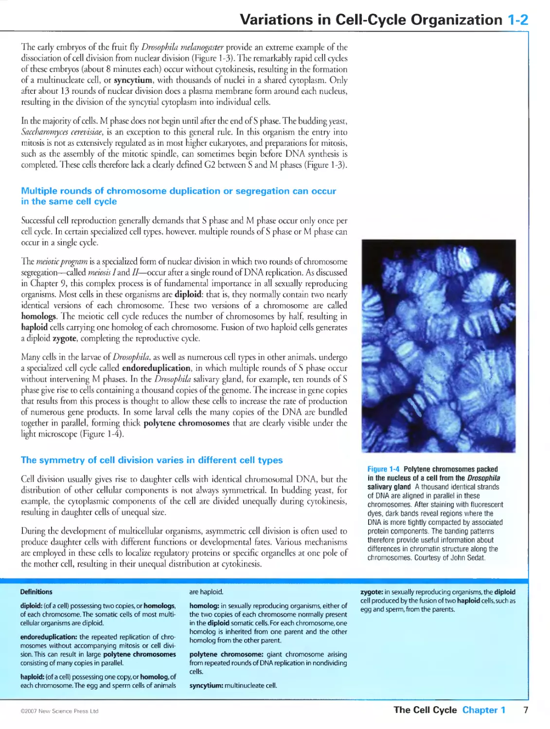
Figure 1-4 Polytene chromosomes packed in the nucleus of a cell from the Drosopbila salivary gland. Courtesy
of John Sedat.
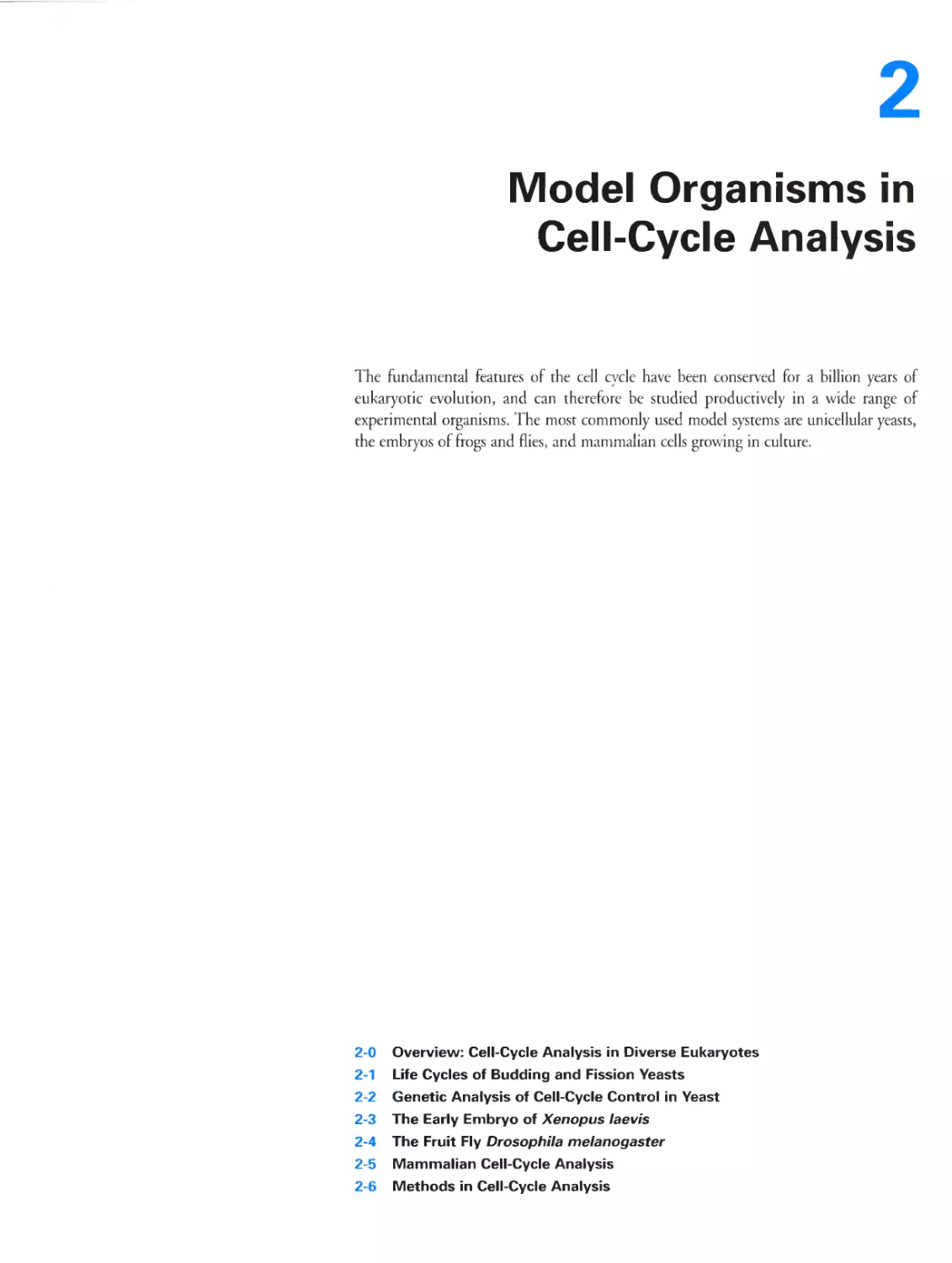
Figure 2-1 The budding yeast Saccharomyces cerevisiae and the fission yeast Schizosaccharomyces pombe. Panel (a)
courtesy of Greg Tully; panel (b) courtesy of Kathleen Gould.
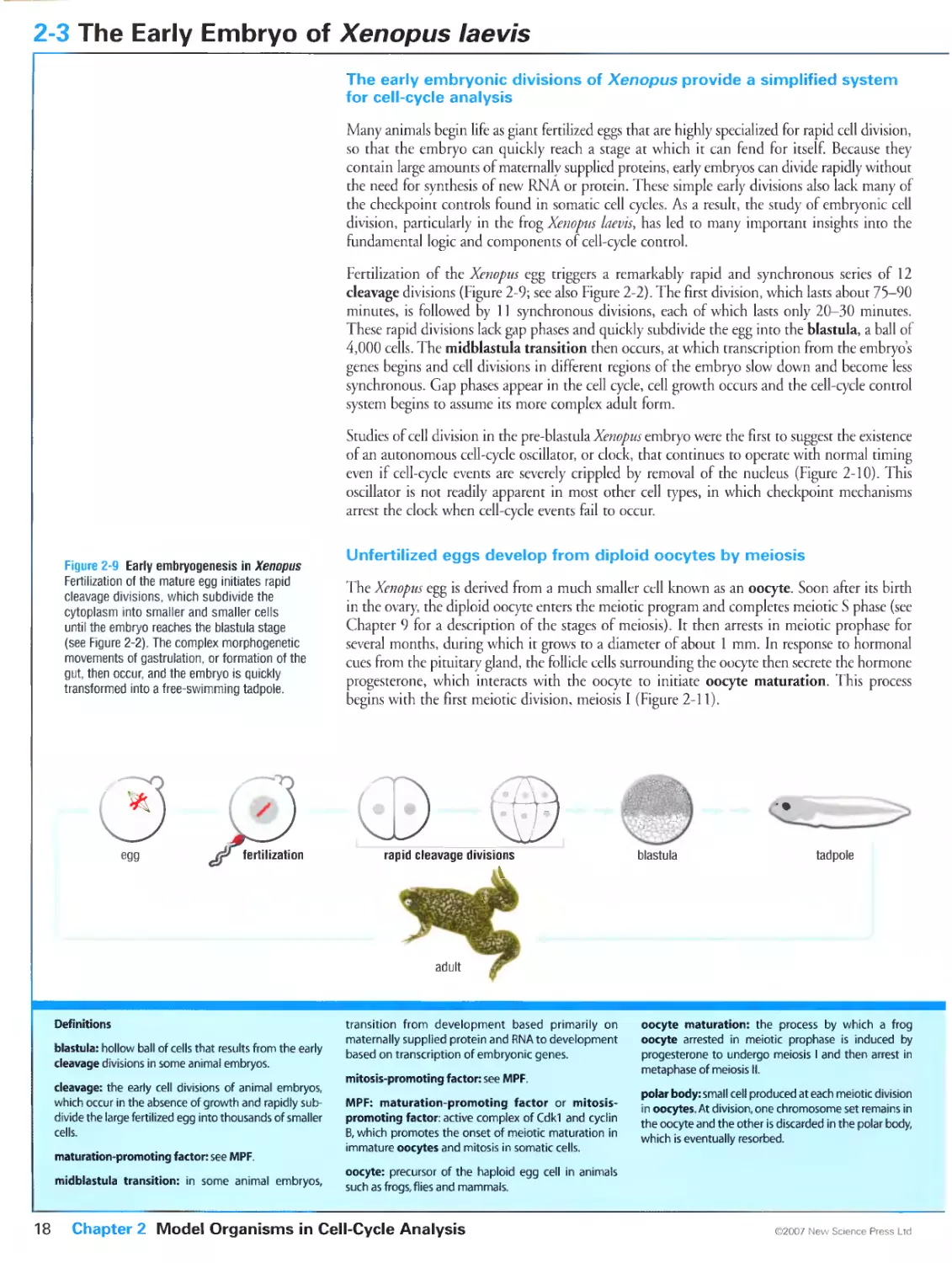
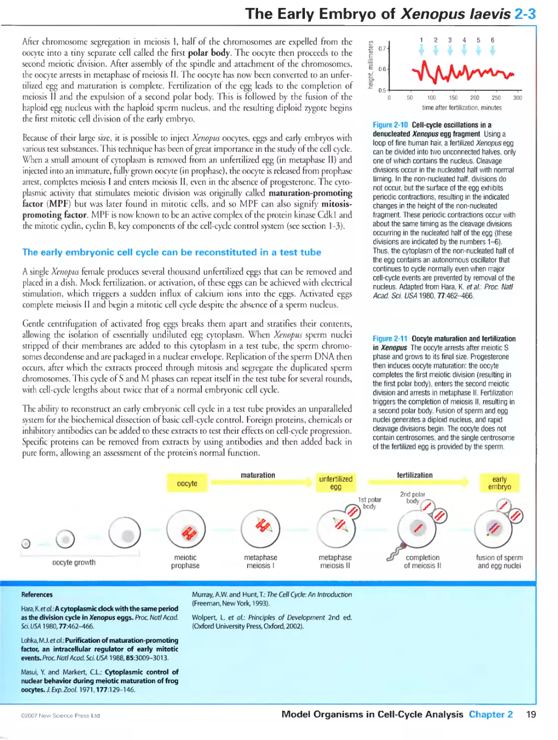
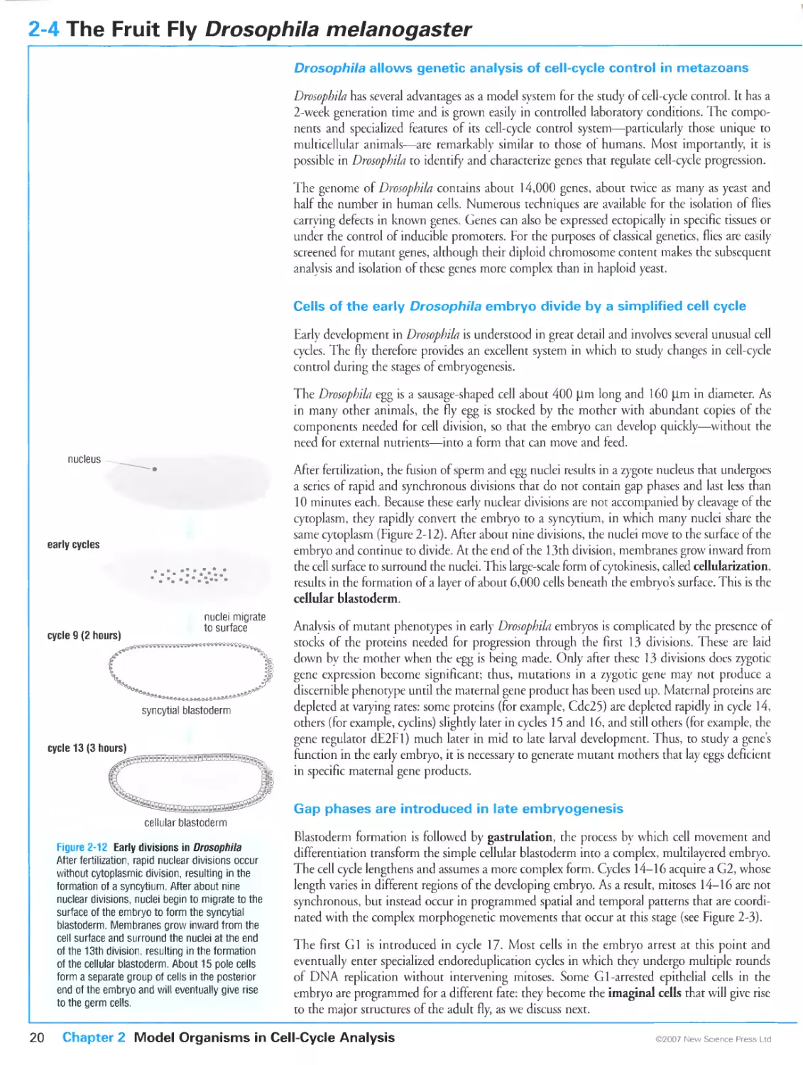
Figure 2-2 Early divisions in the frog Xenopus Levis. Courtesy of James C. Smith and Huw Williams.
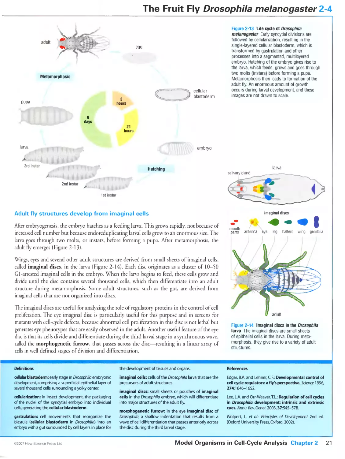
Figure 2-3 Patterns of cell division in the early embryo of the fly Drosopbila melanogaster. Courtesy of Tony
Shermoen and Patrick O'Farrell.
Figure 2-4 Mammalian cells growing in culture. Courtesy of Susanne Steggerda.
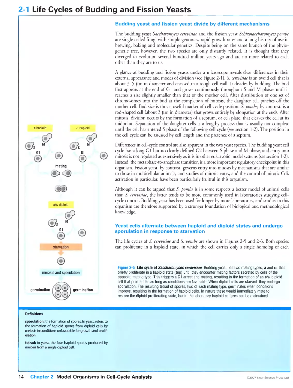
Figure 2-7 Cell-cycle arrests in budding yeast ede mutants. Courtesy of Greg Tully.
Figure 2-17 Analysis of cellular DNA content by flow cytometry. Courtesy of Liam Holt.
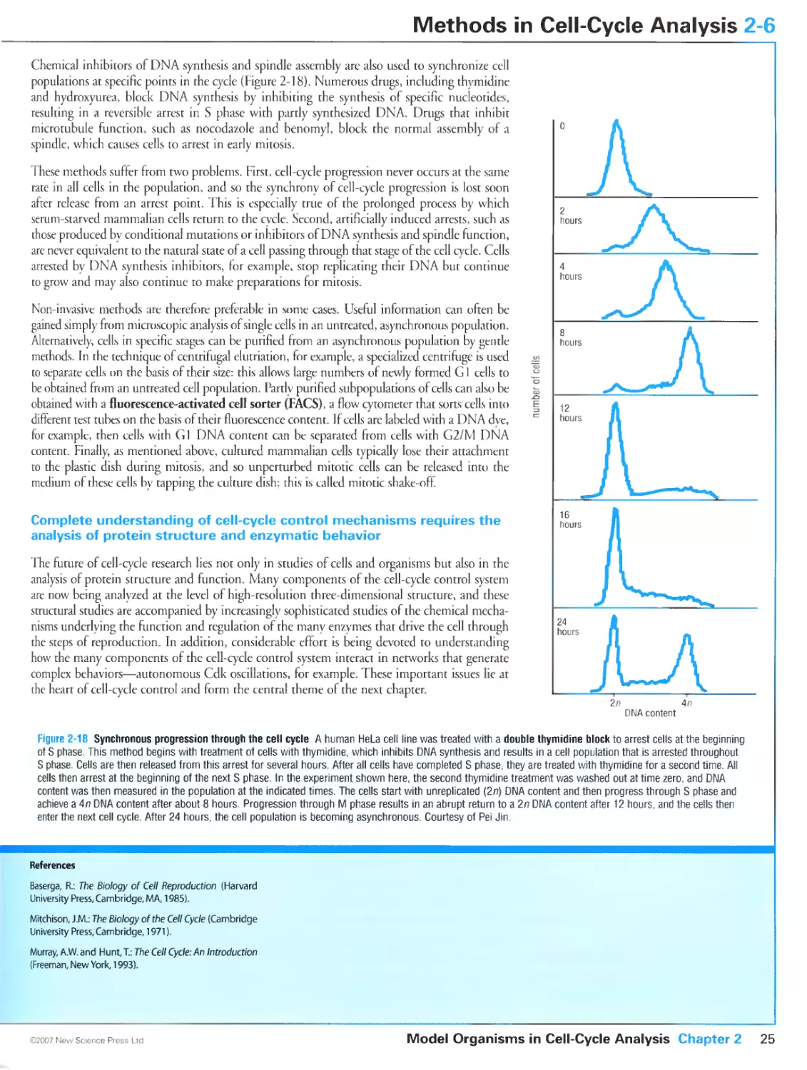
Figure 2-18 Synchronous progression through the cell cycle. Courtesy of Pei Jin.
Figure 4-6 Identification of a replicón cluster by radioactive labeling. Adapted from Huberman, J.A. and Tsai,
A.: Direction of DNA replication in mammalian cells. / Mol. Biol. 1973, 75:5-12.
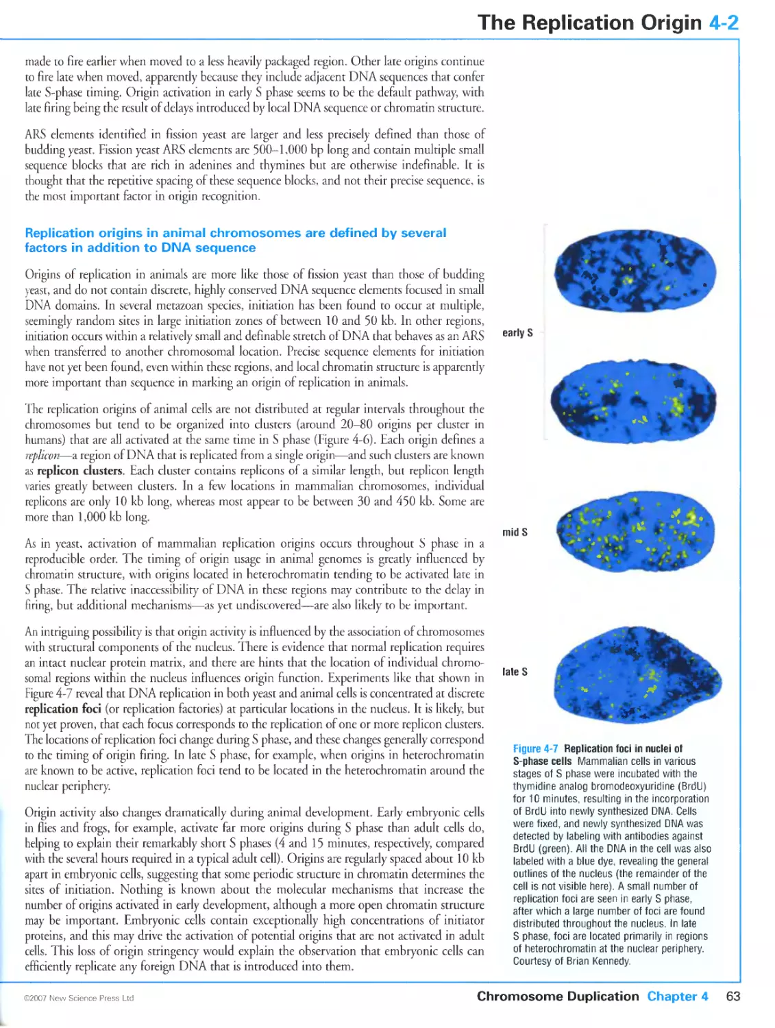
Figure 4-7 Replication foci in nuclei of S-phase cells. Courtesy of Brian Kennedy.
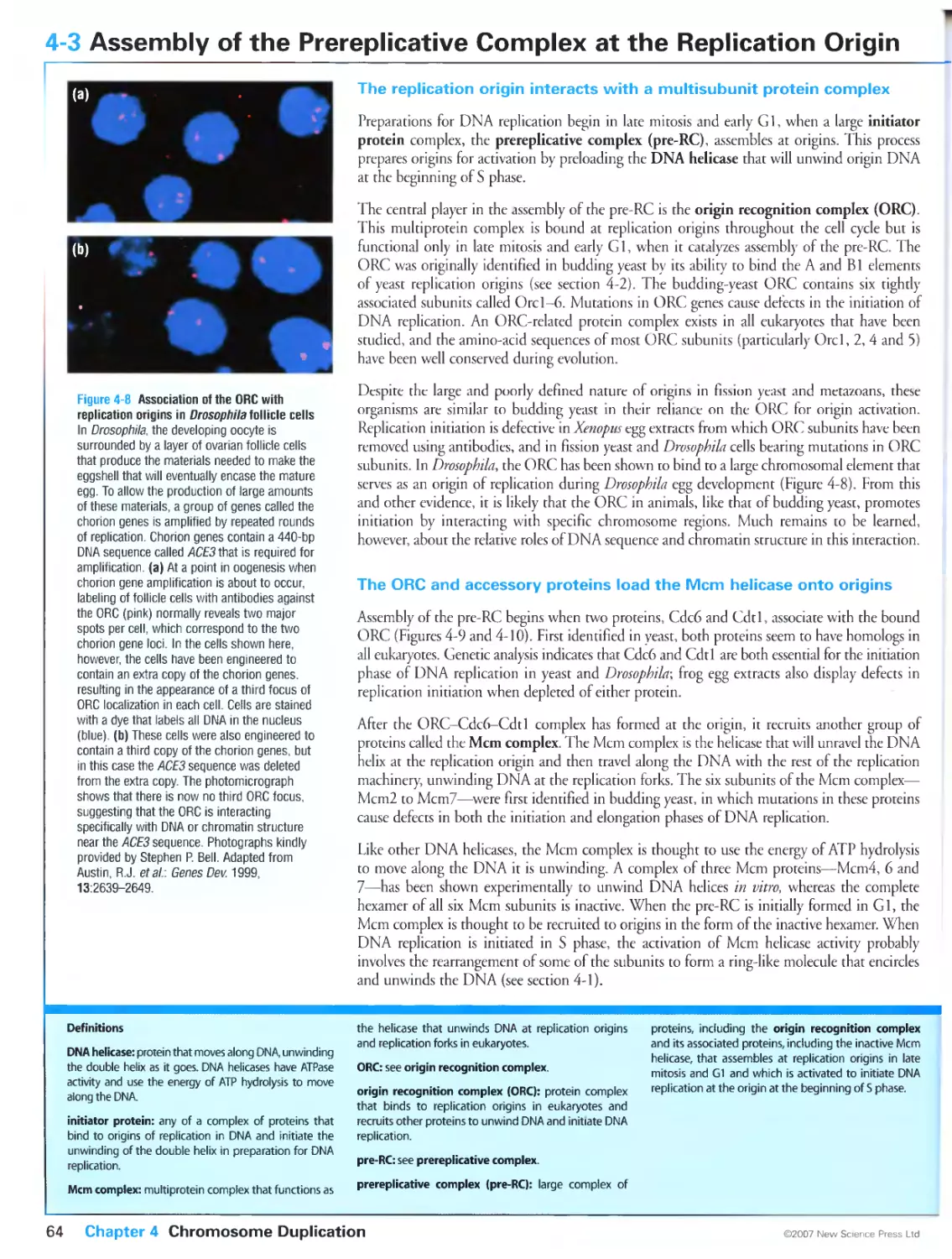
Figure 4-8 Association of the ORG with replication origins in Drosopbila follicle cells. Photographs kindly
provided by Stephen P. Bell. From Austin, R.J., Orr-Weaver, T.L. and Bell, S.P.: DrosopbiLi ORC specifically
binds to ACE3, an origin of DNA replication control element. Genes Dev. 1999, 13:2639-2649. ©1999 Cold
Spring Harbor Laboratory Press.
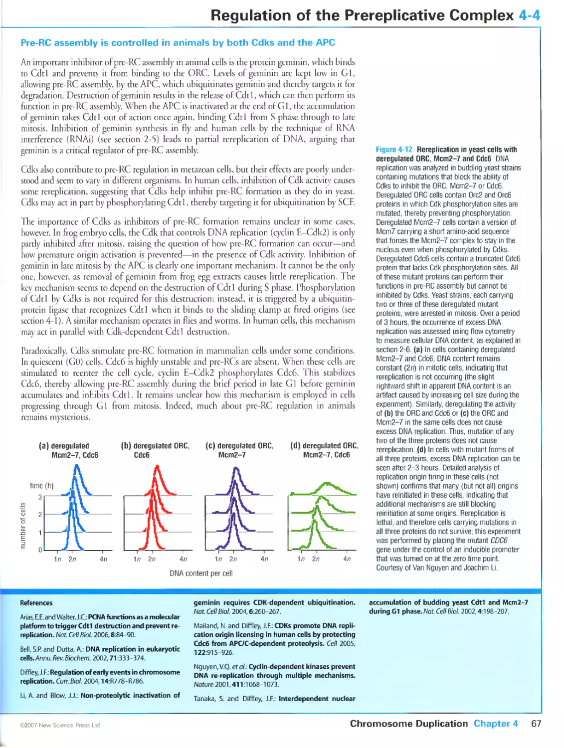
Figure 4-12 Rereplication in yeast cells with deregulated ORC, Mcm2-7 and Cdc6. Courtesy of Van Nguyen
and Joachim Li.
Figure 4-25b Basic units of chromatin structure. From Bednar, J., Horowitz, R.A., Grigoryev, S.A., Carruthers,
L.M., Hansen, J.C., Koster, A.J. and Woodcock, C.L.: Nucleosomes, linker DNA, and linker histone form a
unique structural motif that directs the higher-order folding and compaction of chromatin. Proc. Natl Acad.
Sci. USA 1998, 95:14173-14178. Copyright 1998 National Academy of Sciences, U.S.A.
Figure 5-16 Localization of Plk in mitotic cells. Kindly provided by Francis Barr and Ulrike Grunewald. From
Barr, F.A., Sillje, H.H. and Nigg, E.A.: Polo-like kinases and the orchestration of cell division. Nat. Rev. Mol.
Cell Biol. 2004, 5:429-440.
Figure 5-18 Localization of aurora kinases in mitotic cells. Courtesy of Thru Hirota.
Figure 5-21 Models of SMC and cohesin structure. Panels (a) and (b) reproduced from The Journal of Cell
Biology, 2002, 156, 419-424 by copyright permission ofThe Rockefeller University Press.
Figure 5-22 Condensation and resolution of human sister chromatids in early mitosis. Kindly provided bv
Adrian T. Sumner. From Sumner, A.T.: Scanning electron microscopy of mammalian chromosomes from
prophase to telophase. Chromosoma 1991, 100:410-418. With kind permission of Springer Science and
Business Media.
Figure 5-25 Structure of condensin. Panels (a) and (b) reproduced from The Journal of Cell Biology, 2002,
156, 419-424 by copyrighr permission of The Rockefeller University Press.
Figure 5-26 Plk and aurora B are required for the removal of cohesin from chromosome arms in early mitosis.
From Losada, A., Hirano, M. and Hirano, T.: Cohesin release is required for sister chromatid resolution, but
not for condensin-mediated compaction, at the onset of mitosis. Genes Dev. 2002, 16:3004-3016. ©2002
Cold Spring Harbor Laboratory Press.
Figure 6-1 Anatomy of the mitotic spindle. Panels (b) and (d) courtesy of Andrew Bajer.
Figure 6-6 Control of microtubule dynamics by associated proteins. Courtesy of Kazuhisa Kinoshita.
Figure 6-8 The mammalian centrosome Micrograph kindly provided by William R. Brinkley. Reprinted from
Ultrastruct. Res., Volume 57, McGill, M., Highfield, D.P., Monahan, T.M. and Brinkley, B.R.: Effects of nucleic
acid specific dyes on centrioles of mammalian cells, Pages 43-53, ©1976, with permission from Elsevier.
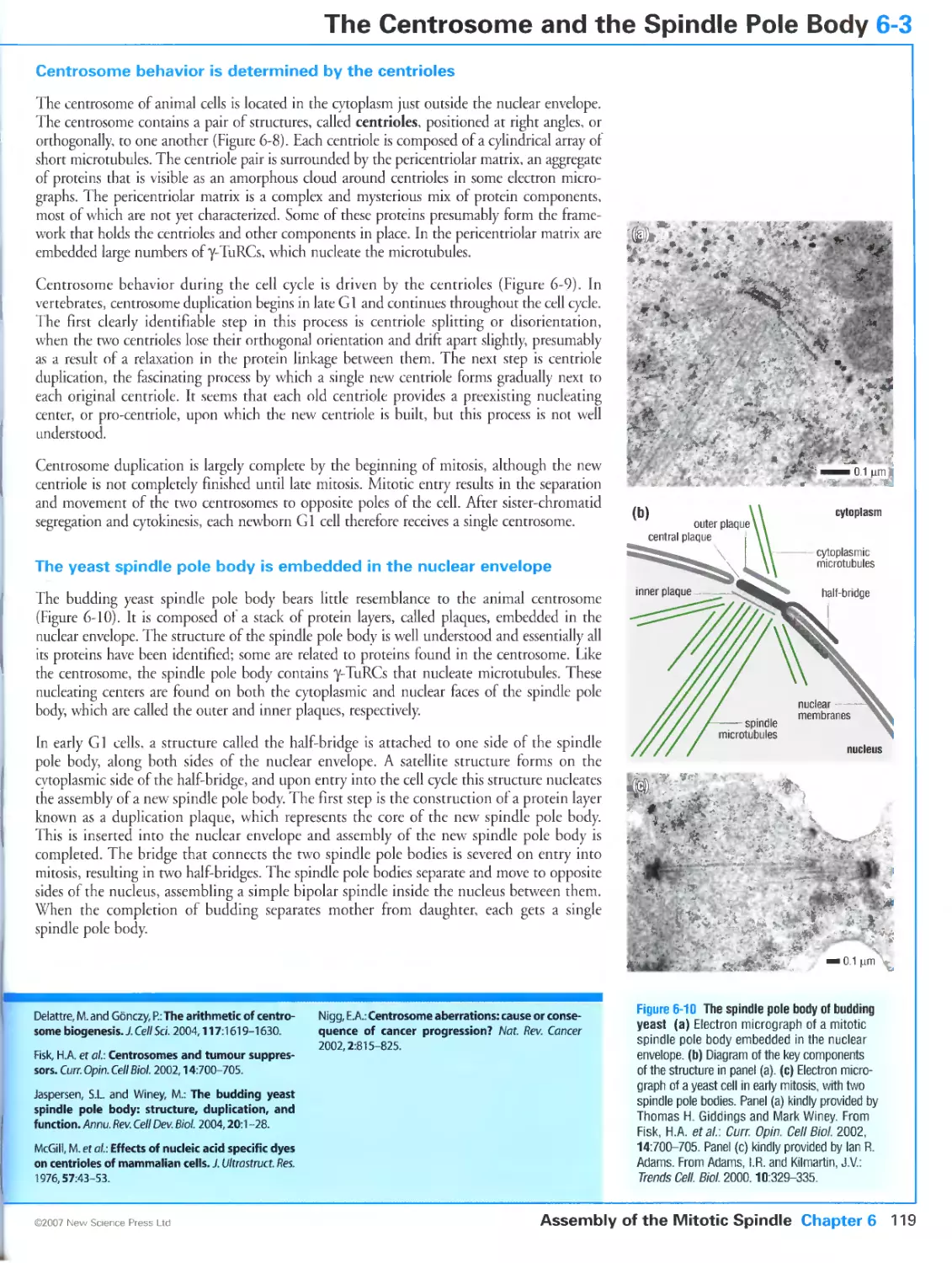
Figure 6-10 The spindle pole body of budding yeast. Panel (a) kindly provided by Thomas H. Giddings and Mark
Winey. Reprinted from Curr. Opin. Cell Biol, Volume 14, Fisk, H.A., Mattison, CP. and Wincy, M.: Centrosomes
and tumour suppressors, Pages 700-705, ©2002, with permission from Elsevier. Panel (c) kindly provided bv
Ian R. Adams. Reprinted from Trends Cell. Biol., Volume 10, Adams, I.R. and Kilmartin, J.V.: Spindle pole body
duplication: a model for centrosome duplication?, Pages 329-335, ©2000, with permission from Elsevier.
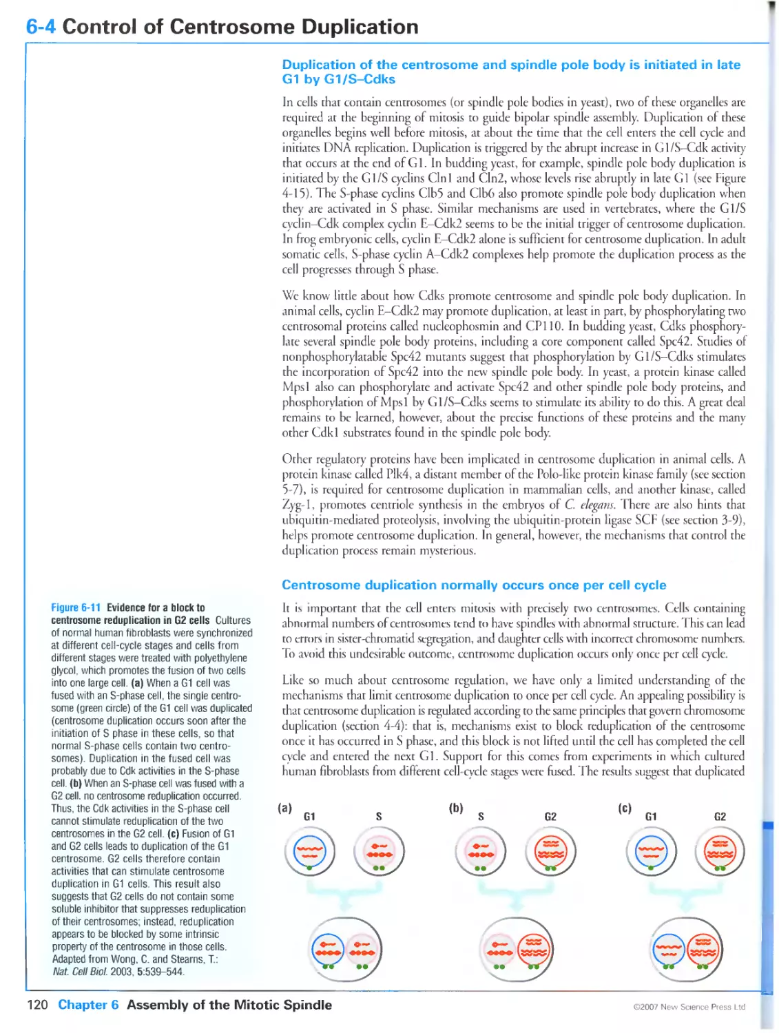
Figure 6-12 Reduplication of centrosomes in prolonged S-phase arrest. Courtesy of Edward H. Hinchcliffe.
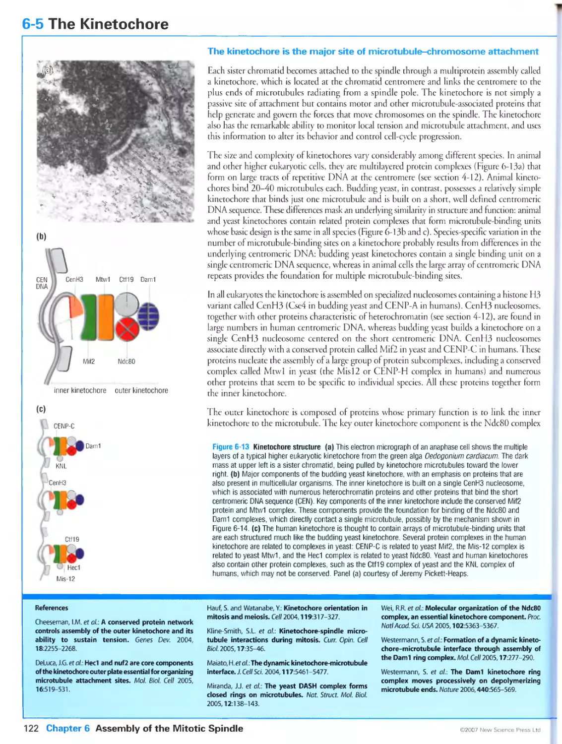
Figure 6-13a Kinetochore structure. Courtesy of Jeremy Pickett-Heaps.
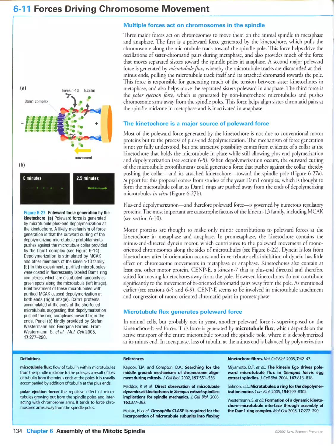
Figure 6-l4a A possible mechanism for dynamic kinetochore-microtubule attachment. Photograph kindly
provided by Stefan Westermann and Georjana Barnes. Reprinted from Mol. Cell, Volume 17, Westermann, S.,
Avila-Sakar, A., Wang, H.W, Niederstrasser, H., Wong, J., Drubin, D.G., Nogales, E. and Barnes, G.:
Formation of a dynamic kinetochore-microtubule interface through assembly of the Daml ring complex,
Pages 277-290, ©2005, with permission from Elsevier.
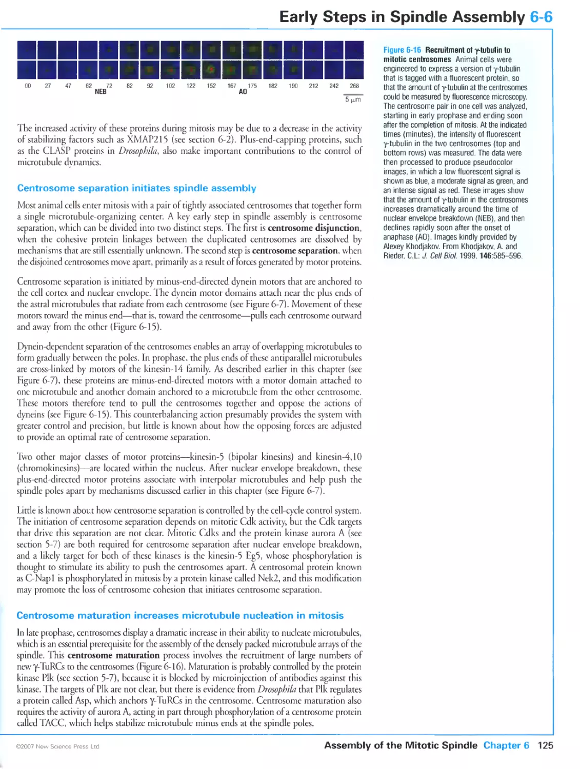
Figure 6-16 Recruitmenr ol y-tubulin to mitotic centrosomes. Images kindly provided by Alexey Khodjakov.
Reproduced from The Journal of Cell Biology, 1999, 146, 585-596 by copyright permission of The
Rockefeller University Press.
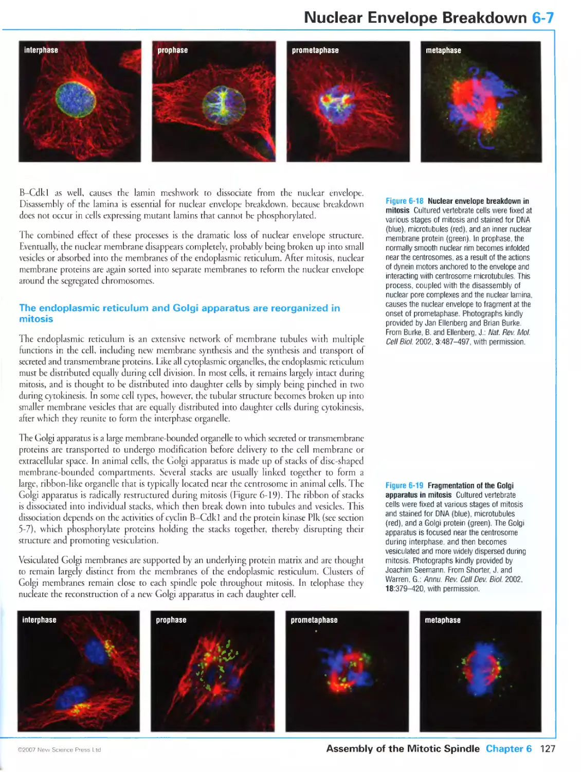
Figure 6-18 Nuclear envelope breakdown in mirosis. Photographs kindly provided by Jan Ellenberg and Brian
Burke. From Burke, B. and Ellenberg, J.: Remodelling the walls of the nucleus. Nat. Rev. Mol. Cell Biol. 2002,
3:487-497. Reprinted with cops-right permission from Nature.
Figure 6-19 Fragmentation of the Golgi apparatus in mitosis. Photographs kindly provided by Joachim
Seemann. Reprinted, with permission, from the Annual Review of Cell and Developmental Biology, Volume 18
©2002 by Annual Reviews www.annualreviews.org
Figure 6-2lb Stabilization of microtubules around chromosomes by Ran-GTP. Kindly provided by Rebecca
Heald. Reprinted with permission from Kalab, P., Weis, K. and Heald, R.: Visualization of a Ran-GTP gradient
in interphase and mitotic Xenopus egg extracts. Science 2002, 295:2452-2456. Copyright 2002 AAAS.
Figure 6-23b Kinetochore-derived microtubule formation. Kindly provided by Helder Maiato and Alexey
Khodjakov. Reproduced from The Journal of Cell Biology, 2004, 167, 831-840 by copyright permission of
The Rockefeller University Press.
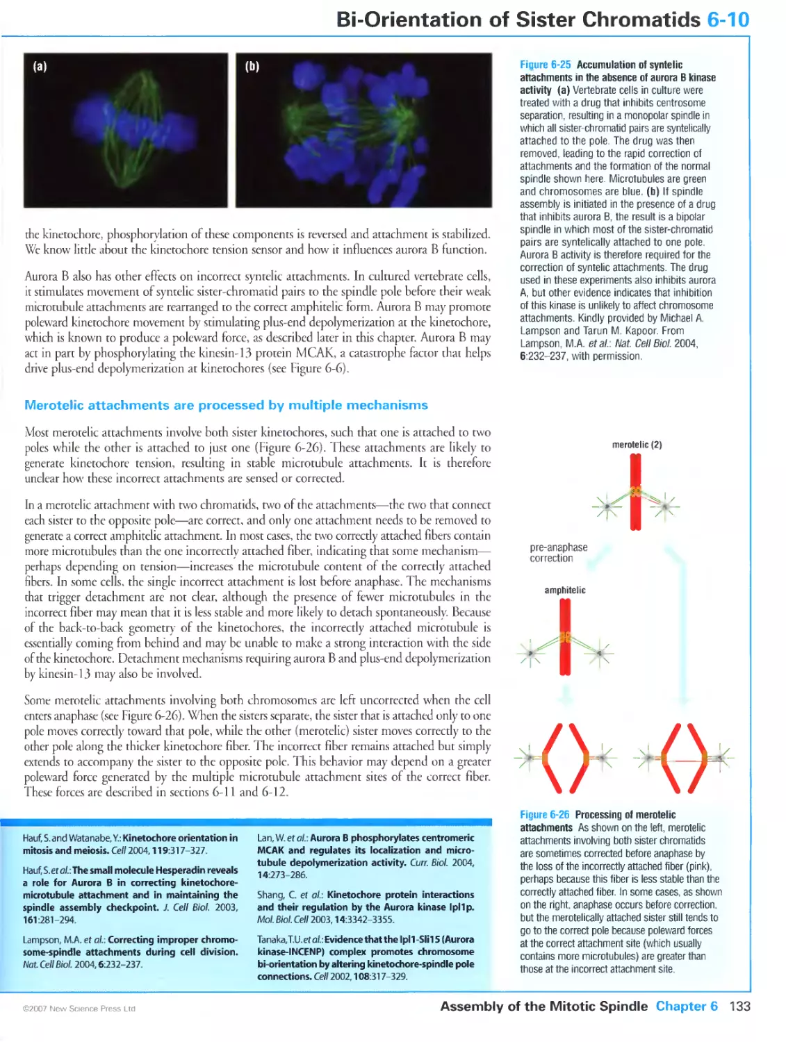
Figure 6-25 Accumulation of syntelic attachments in the absence of aurora B kinase activity. Kindly provided
by Michael A. Lampson and Tarun M. Kapoor. From Lampson, M.A., Renduchitala, K, Khodjakov, A. and
Kapoor, T.M.: Correcting improper chromosome-spindle attachments during cell division. Nat. Cell Biol.
2004, 6:232-237. Reprinted with copyright permission from Nature.
Figure 6-27b Poleward force generation by the kinetochore. Kindly provided by Stefan Westermann and
Georjana Barnes. Reprinted from Mol. Cell, Volume 17, Westermann, S., Avila-Sakar, A., Wang, H.W.,
Niederstrasser, H., Wong, J., Drubin, D.G., Nogales, E. and Barnes, G.: Formation of a dynamic kinetochore-
microtubule interface through assembly of the Daml ring complex, Pages 277-290, ©2005, with permission
from Elsevier.
Figure 7-6 Spindle checkpoint component Mad2 at unattached kinetochores. Kindly provided by Jennifer C.
Waters. Reproduced from The Journal of Cell Biology, 1998, 141, 1181-1191 by copyright permission of The
Rockefeller University Press.
Figure 7-7 Alternative conformations of the Mad2 protein. Adapted from Curr. Biol., Volume 15, De Antoni,
A., Pearson, C.G., Cimini, D., Canman, J.C., Sala, V, Nezi, L., Mapelli, M., Sironi, L., Faretta, M., Salmon,
E.D. and Musacchio, A.: The Madl/Mad2 complex as a template for Mad2 activation in the spindle assembly
checkpoint, Pages 214-225, ©2005, with permission from Elsevier. Original structure graphics kindly provided
by Andrea Musacchio.
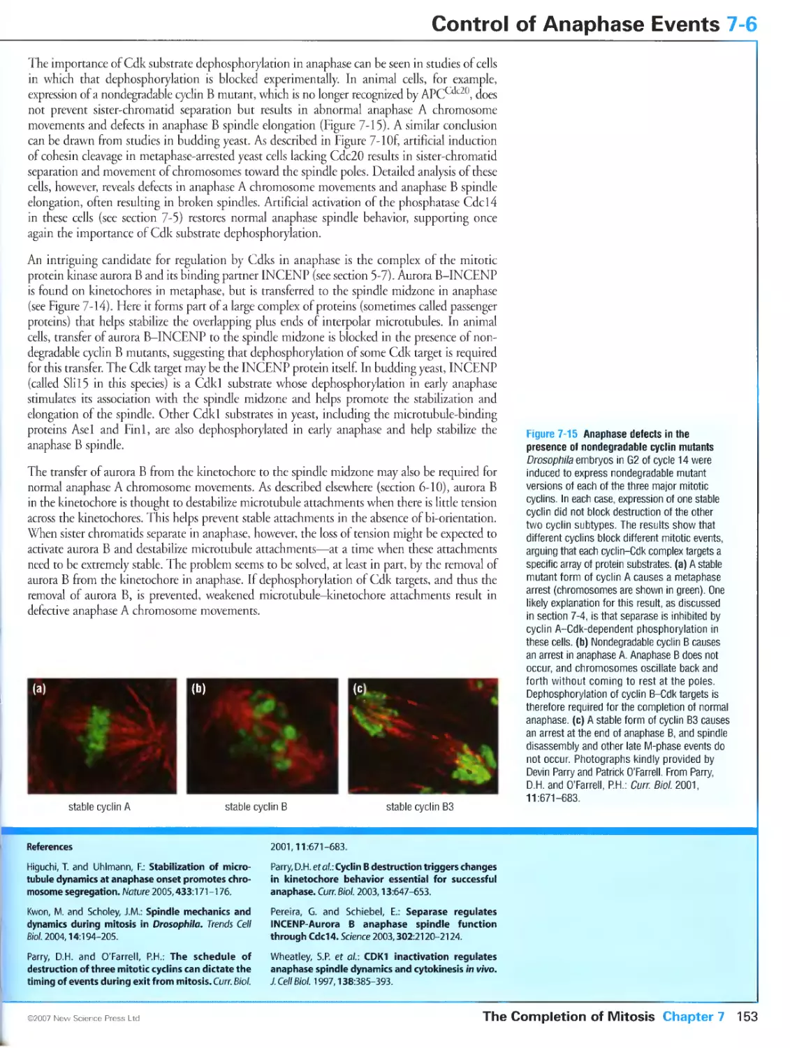
Figure 7-15 Anaphase defects in the presence of nondegradable cyclin mutants. Photographs kindly provided
by Devin Parry and Patrick O'Farrell. Reprinted from Curr. Biol., Volume 11, Parry, D.H. and O'Farrell, PH.:
The schedule of destruction of three mitotic cyclins can dictate the timing of events during exit from mitosis.
Pages 671-683, ©2001, with permission from Elsevier.
Figure 7-16 The APC helps promote spindle disassembly in budding yeast. Photographs kindly provided by
David Pellman. Reprinted, with permission, from Juang, Y.-L., Huang, J., Peters, J.-M., McLaughlin, M.E., Tai,
C.-Y. and Pellman, D.: APC-mediated proteolysis of Asel and the morphogenesis of the mitotic spindle.
Science 1997, 275:1311-1314. Copyright 1997 AAAS.
Figure 7-17 Nuclear envelope assembly in Xenopus embryo extracts. Photographs kindly provided by Martin
Hetzer and lain Mattaj. From Hetzer, M., Meyer, H.H., Walther, T.C., Bilbao-Cortes, D., Warren, G. and
Mattaj, l.W: Distinct AAA-ATPase p97 complexes function in discrete steps of nuclear assembly. Nat. Cell
Biol. 2001, 3:1086-1091. Reprinted with copyright permission from Nature.
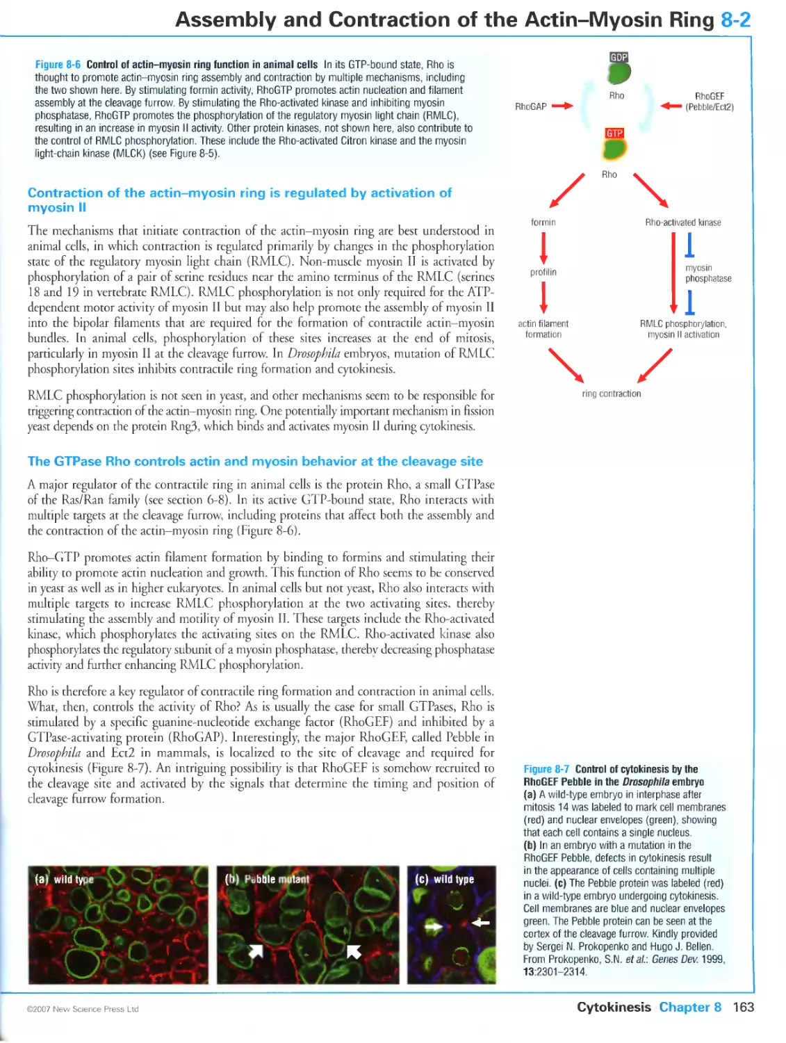
Figure 8-7 Control of cytokinesis by the RhoGEF Pebble in the Drosophila embryo. Kindly provided by Sergei
N. Prokopenko and Hugo J. Bellen. From Prokopenko, S.N., Brumby, A., O'Keefe, L., Prior, L., He, Y, Saint,
R. and Bellen, H.J.: A putative exchange factor for Rhol GTPase is required for initiation of cytokinesis in
Drosophila. Genes Dev. 1999, 13:2301-2314. ©1999 Cold Spring Harbor Laboratory Press.
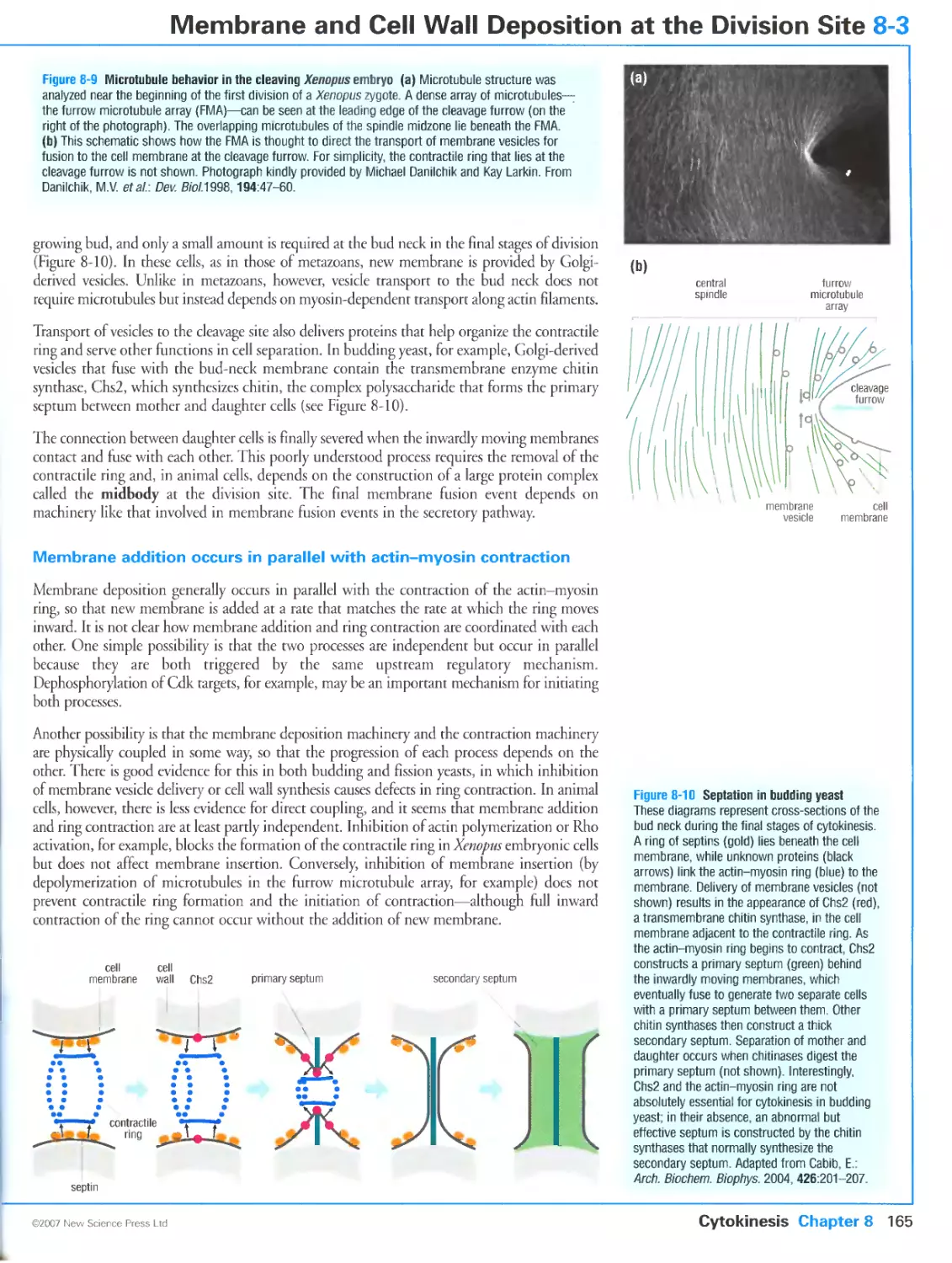
Figure 8-9a Microtubule behavior in the cleaving Xenopus embryo. Photograph kindly provided by Michael
Danilchik and Kay Larkin. Reprinted from Dev. Biol., Volume 194, Danilchik, M.V., Funk, W.C., Brown, E.E.
and Larkin, K: Requirement for microtubules in new membrane formation during cytokinesis of Xenopus
embryos, Pages 47-60, ©1998, with permission from Elsevier.
Figure 8-13 Positioning the contractile ring in S. pombe. Photographs kindly provided by Rafael R. Daga
and Fred Chang. From Daga, R.R. and Chang, E: Dynamic positioning of the fission yeast cell division plane.
Proc. Natl Acad. Sci. USA 2005, 102:8228-8232. Copyright 2005 National Academy of Sciences, U.S.A.
Figure 8-14 Positioning of cytokinesis by the mitotic spindle of embryonic cells. Kindly provided by Charles
B. Shuster and David R. Burgess. Reproduced from The Journal of Cell Biology, 1999, 146, 981-992 by
copyright permission of The Rockefeller University Press.
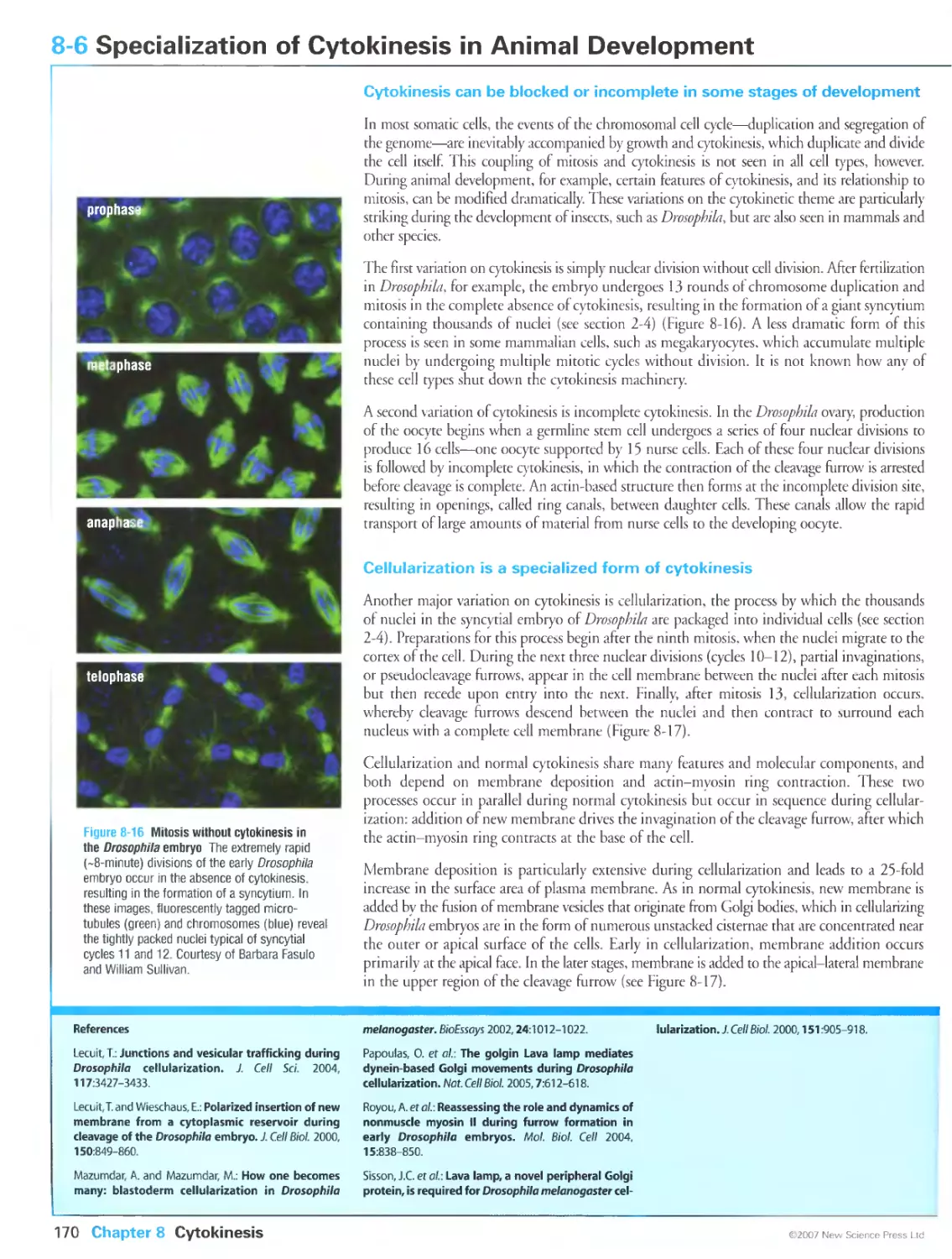
Figure 8-16 Mitosis without cytokinesis in the Drosophila embryo. Courtesy of Barbara Fasulo and William
Sullivan.
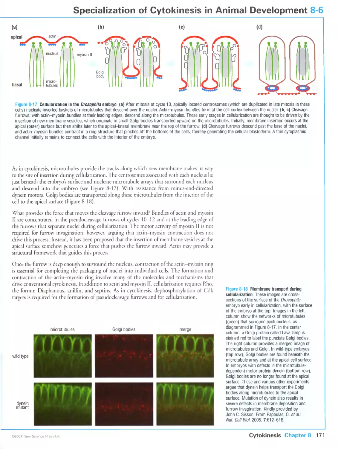
Figure 8-18 Membrane transport during cellularization. Kindly provided by John C. Sisson. From Papoulas,
O., Hays, T.S. and Sisson, J.C.: The golgin Lava lamp mediates dynein-based Golgi movements during
Drosophila cellularization. Nat. Cell Biol. 2005, 7:612-618. Reprinted with copyright permission from Nature.
Figure 8-20 Asymmetric division in a Drosophila neuroblast. Kindly provided by Silvia Bonaccorsi. From
Giansanti, M.G., Gatti, M. and Bonaccorsi, S.: The role of centrosomes and astral microtubules during
asymmetric division of Drosophila neuroblasts. Development 2001, 128:1137-1145. Reprinted with permission
from The Company of Biologists Ltd.
Figure 9-6 Early steps in homolog pairing. Photographs kindly provided by Denise Zickler. From Tesse, S.,
Storlazzi, A., Kleckner, N., Gargano, S. and Zickler, D.: Localization and roles of SkiSp protein in Sordaria
meiosis and delineation of three mechanistically distinct steps of meiotic homolog juxtaposition. Proc. Natl
Acad. Sci. USA 2003, 100:12865-12870. Copyright 2005 National Academy of Sciences, U.S.A.
Figure 9-7 Homolog pairing defects in a spoil mutant. Photographs kindly provided by Denise Zickler. From
Storlazzi, A., Tesse, S., Gargano, S., James, E, Kleckner, N. and Zickler, D.: Meiotic double-strand breaks at
the interface of chromosome movement, chromosome remodeling, and reductional division. Genes Dev. 2003,
17:2675-2687. ©1999 Cold Spring Harbor Laboratory Press.
Figure 9-8 Electron microscopic analysis of chromosome structure in leptotene and early zygotene.
Photographs kindly provided by Jim Henle and Nancy Kleckner. Panel (b) from Stack, S.M. and Anderson,
L.K.: Two-dimensional spreads of synaptonemal complexes from solanaceous plants. II. Synapsis in
Lycopersicon esculentum (tomato). Am. J. Bot. 1986, 73:264-281. Panels (c) and (d) from Albini, S.M. and
Jones, G.H.: Synaptonemal complex spreading in Allium cepa and A. fistulosum. 1. The initiation and
sequence of pairing. Chromosoma 1987, 95:324-338.
Figure 9-9 The synaptonemal complex. Photographs in panel (b) kindly provided by Karin Schmekel. Top
photograph from Schmekel, K. and Daneholt, B.: Evidence for close contact between recombination nodules and
the central element of the synaptonemal complex. Chromosome Res. 1998, 6:155-159; with kind permission from
Springer Science and Business Media. Photographs in panel (c) kindly provided by Carole Rogers and Shirleen
Roeder. Reproduced from The Journal of Cell Biology, 2000, 148, 417-426 by copyright permission of The
Rockefeller University Press.
Figure 9-10 Chiasmata. Reprinted from Cell, Volume 111, Blat, Y., Protacio, R., Hunter, N. and Kleckner, N.:
Physical and functional interactions among basic chromosome organizational features govern early steps of
meiotic chiasma formation, Pages 791-802, ©2002, with permission from Elsevier. Photograph taken from
John, B.: Meiosis (Cambridge University Press, New York, 1990).
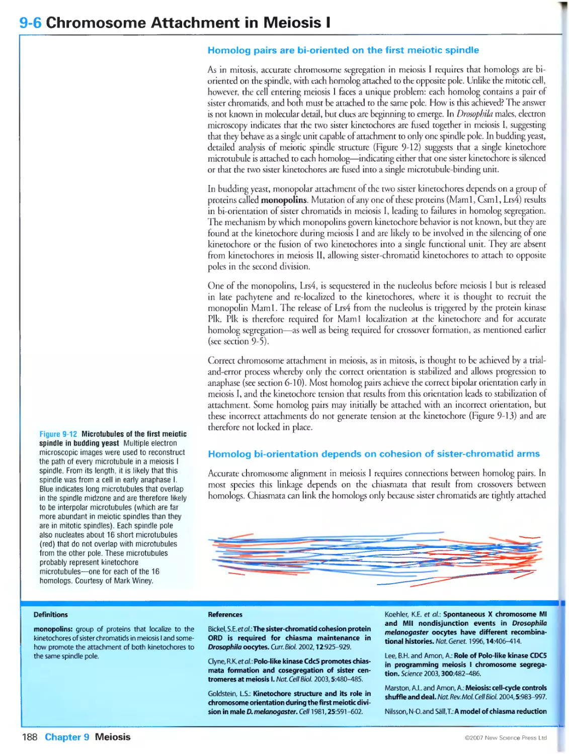
Figure 9-12 Microtubules of the first meiotic spindle in budding yeast. Courtesy of Mark Winey.
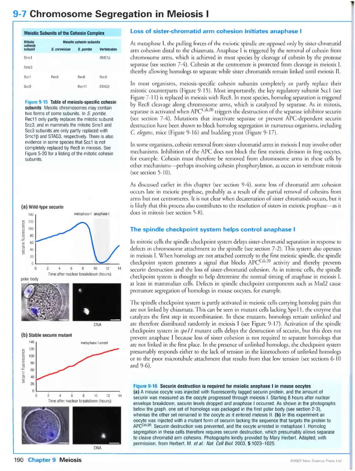
Figure 9-16 Securin destruction is required for meiotic anaphase I in mouse oocytes. Photographs kindly
provided by Mary Herbert. From Herbert, M., Levasseur, M., Homer, H., Yallop, K., Murdoch, A. and
McDougall, A.: Homologue disjunction in mouse oocytes requires proteolysis of securin and cyclin Bl.
Nat. Cell Biol. 2003, 5: 1023-1025. Reprinted with copyright permission from Nature.
Figure 9-18 Inhibition of Cdkl triggers DNA synthesis after meiosis I, Photographs kindly provided by Keita
Ohsumi. Reprinted by permission from Macmillan Publishers Ltd: The EMBO Journal, Iwabuchi, M„ Ohsumi,
K., Yamamoto, T.M., Sawada, W. and Kishimoto, T.: Residual Cdc2 activity remaining at meiosis I exit is
essential for meiotic M-M transition in Xenopus oocyte extracts. EMBO]. 2000, 19:4513-4523, copyright 2000.
Figure 10-9 Antagonistic functions of the two E2F homologs in Drosophila. Photographs kindly provided by
Maxim Frolov. From Frolov, M.V., Huen, D.S., Stevaux, O., Dimova, D., Balczarek-Strang, K., Elsdon, M. and
Dyson, N.J.: Functional antagonism between E2F family members. Genes Dev. 2001, 15:2146-2160. ©2001
Cold Spring Harbor Laboratory Press.
Figure 10-20 Patterns of cdc25 (string) expression in the fly embryo. Photographs kindly provided by Bruce
Edgar. From Edgar, B.A., Lehman, D.A. and O'Farrell, PH.: Transcriptional regulation of string (cdc25): a link
between developmental programming and the cell cycle. Development 1994, 120:3131-3143. Reprinted with
permission from The Company of Biologists Ltd.
Figure 10-24 Analysis of growth control in the Drosophila eye. Kindly provided by Duojia Pan. Reprinted by
permission from Macmillan Publishers Ltd: Nature Cell Biology, Gao, X., Zhang, Y, Arrazola, P., Hino, O.,
Kobayashi, T., Yeung, R.S., Ru, B. and Pan, D.: Tsc tumour suppressor proteins antagonize amino-acid-TOR
signalling. Nat. Cell Biol. 2002, 4:699-704, copyright 2002.
Figure 11-7 RPA-dependent recruitment of ATR to sites of DNA damage. Kindly provided by Stephen J.
Elledge. Reprinted with permission from Zou, L. and Elledge, S.J.: Sensing DNA damage through ATRIP
recognition of RPA-ssDNA complexes. Science 2003, 300:1542-1548. Copyright 1997 AAAS.
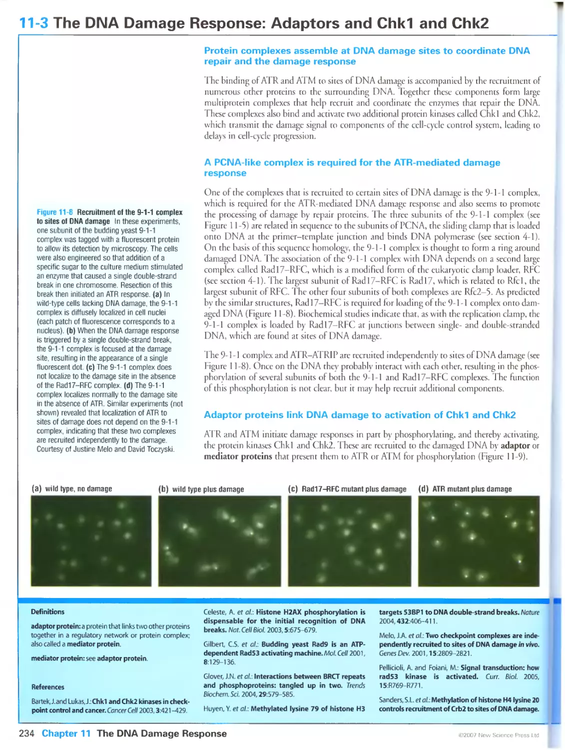
Figure 11-8 Recruitment of the 9-1-1 complex to sites of DNA damage. Courtesy of Justine Melo and David
Toczyski.
Figure 11-14 Abnormal DNA structures at stalled replication forks in yeast chk2 mutants. Courtesy of Massimo
Lopes and Marco Foiani.
Figure 11-18 Generation of a DNA damage response in senescent human cells. Photographs kindly provided
by Fabrizio d Adda di Fagagna. Reprinted by permission from Macmillan Publishers Ltd: Nature, d'Adda di
Fagagna, F„ Reaper, P.M., Clay-Farrace, L., Fiegler, H, Carr, P, Von Zglinicki, T., Saretzki, G., Carter, N.P. and
Jackson, S.P: A DNA damage checkpoint response in telomere-initiated senescence. Nature 2003,
426:194-198, copyright 2003.
Figure 12-11 Chromosomal abnormalities in cancer cells. Courtesy of Kylie Gorringe, Mira Grigorova and Paul
Edwards.
Figure 12-13 Telomere degeneration in the formation of carcinomas. Phorograph kindly provided by Ronald
A. DePinho. From Artandi, S.E., Chang, S., Lee, S.L., Alson, S., Gottlieb, G.J., Chin, L. and DePinho, R.A.:
Telomere dysfunction promotes non-reciprocal translocations and epithelial cancers in mice. Nature 2000,
406:641-645. Reprinted with copyright permission from Nature.
Figure 12-15 Mitotic spindle defects arising from abnormal centrosome number. From Pihan, G.A., Wallace, J.,
Zhou, Y. and Doxsey, S.J.: Centrosome abnormalities and chromosome instability occur together in pre-invasive
carcinomas. Cancer Res. 2003, 63:1398-1404.
Figure 12-18 Inhibition of the protein kinase Abl by imatinib. Reprinted from Cancer Cell, Volume 2, Shah, N.
P., Nicoll, J.M., Nagar, B., Gorre, M.E., Paquette, R.L., Kuriyan, J. and Sawyers, C.L.: Multiple BCR-ABL kinase
domain mutations confer polyclonal resistance to the tyrosine kinase inhibitor imatinib (STI571) in chronic
phase and blast crisis chronic myeloid leukemia, Pages 117-125, ©2002, with permission from Elsevier.
Contents summary
CHAPTER 1 The Cell Cycle
1-0 Overview: Cell Reproduction 2
1-1 Events of the Eukaryotic Cell Cycle 4
1-2 Variations in Cell-Cycle Organization 6
1-3 The Cell-Cycle Control System 8
CHAPTER 2 Model Organisms in Cell-Cycle Analysis
2-0 Overview: Cell-Cycle Analysis in Diverse Eukaryotes 12
2-1 Life Cycles of Budding and Fission Yeasts 14
2-2 Genetic Analysis of Cell-Cycle Control in Yeast 16
2-3 The Early Embryo of Xenopus laevis 18
2-4 The Fruit Fly Drosophila melanogaster 20
2-5 Mammalian Cell-Cycle Analysis 22
2-6 Methods in Cell-Cycle Analysis 24
CHAPTER 3 The Cell-Cycle Control System
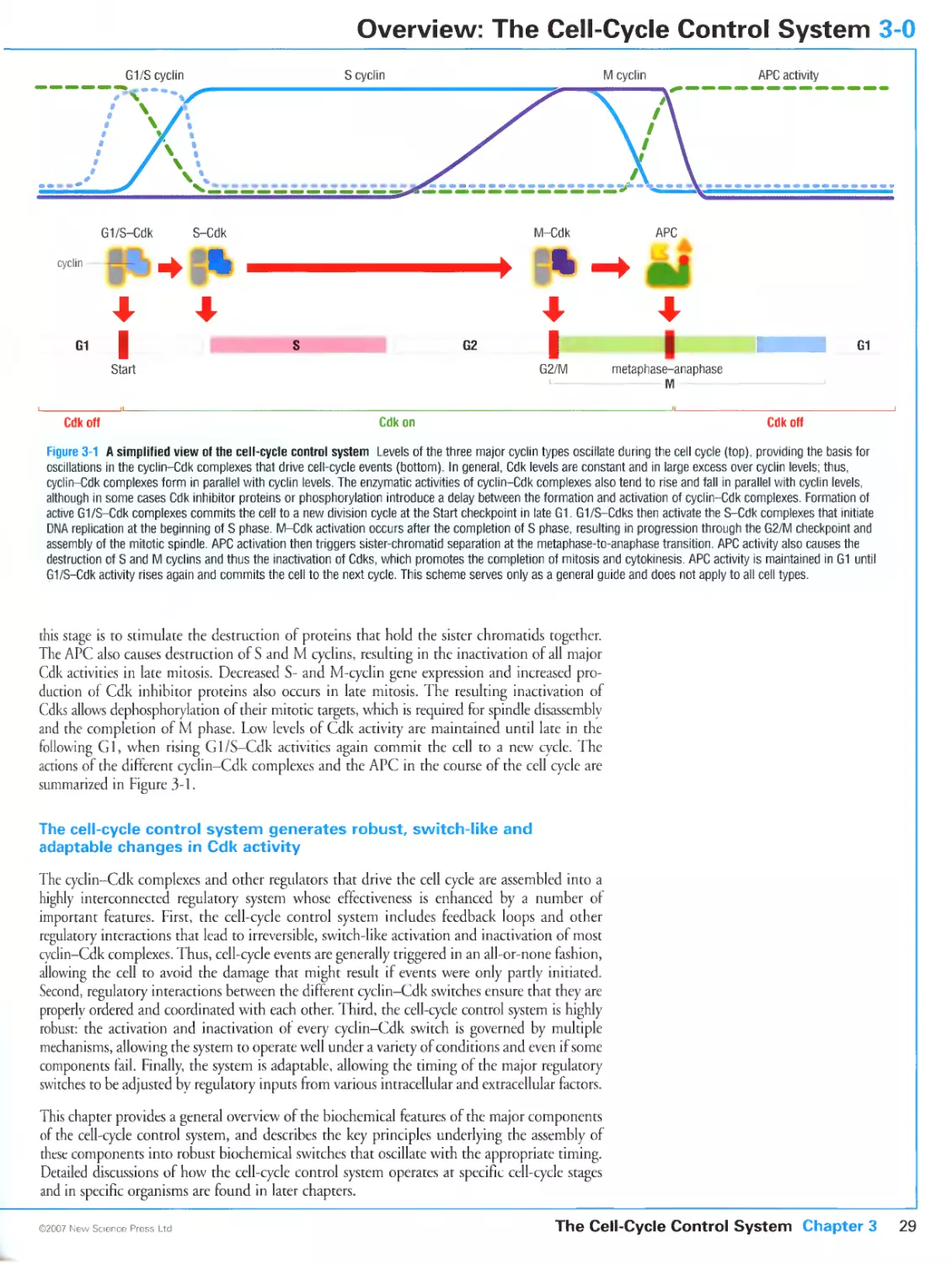
3-0 Overview: The Cell-Cycle Control System 28
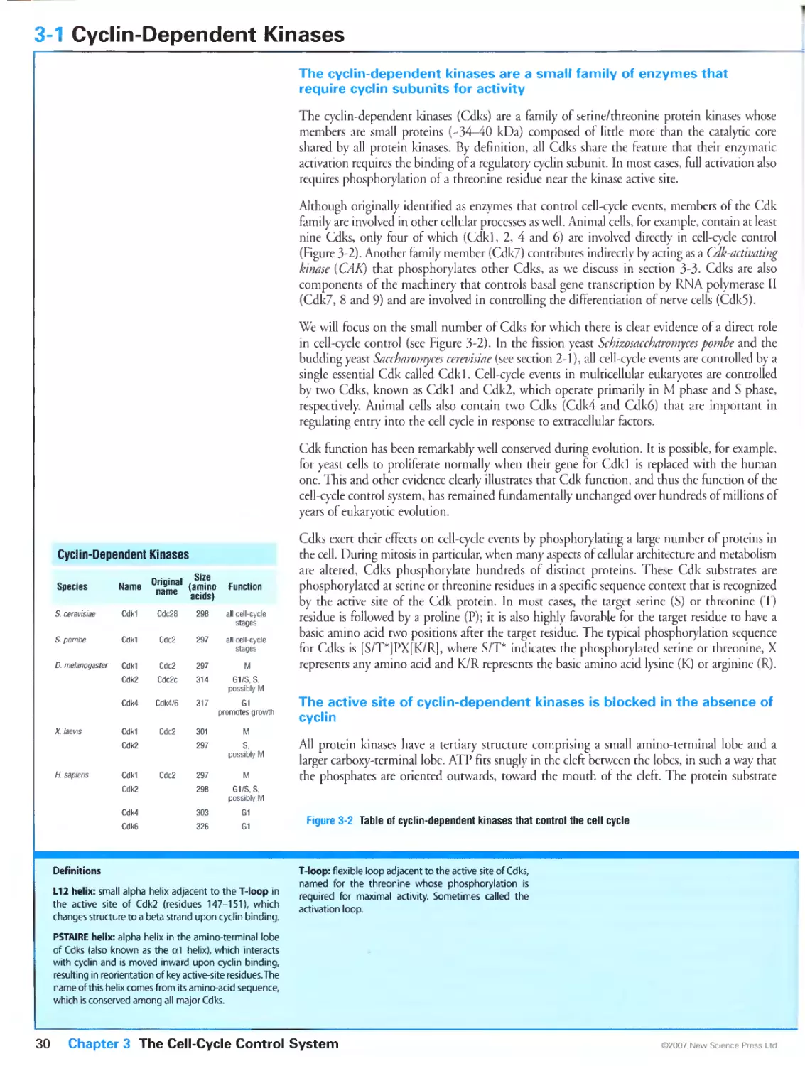
3-1 Cyclin-Dependent Kinases 30
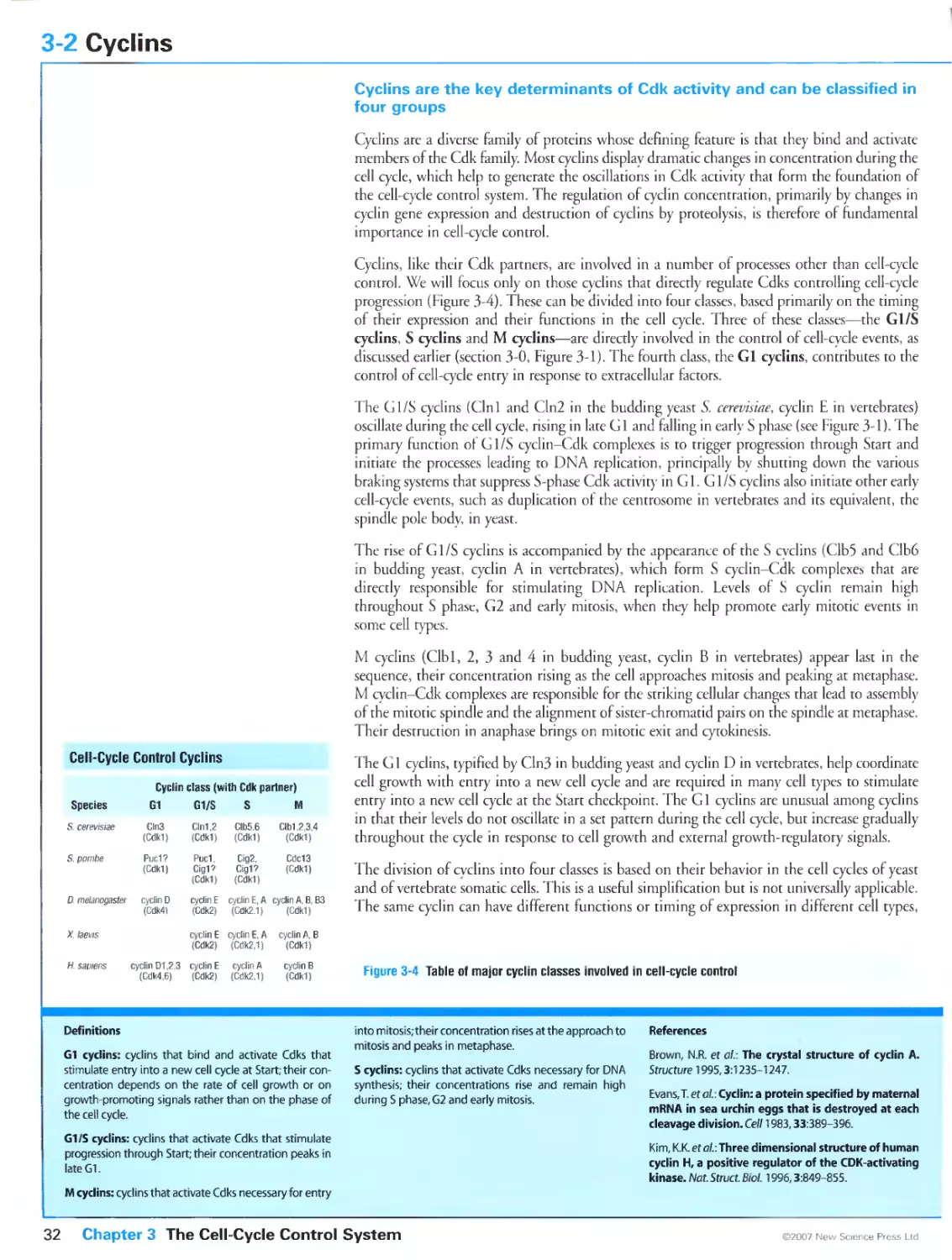
3-2 Cyclins 32
3-3 Control of Cdk Activity by Phosphorylation 34
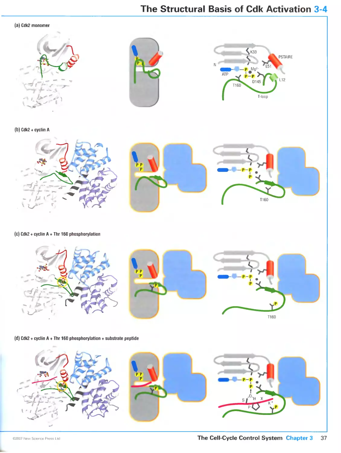
3-4 The Structural Basis of Cdk Activation 36
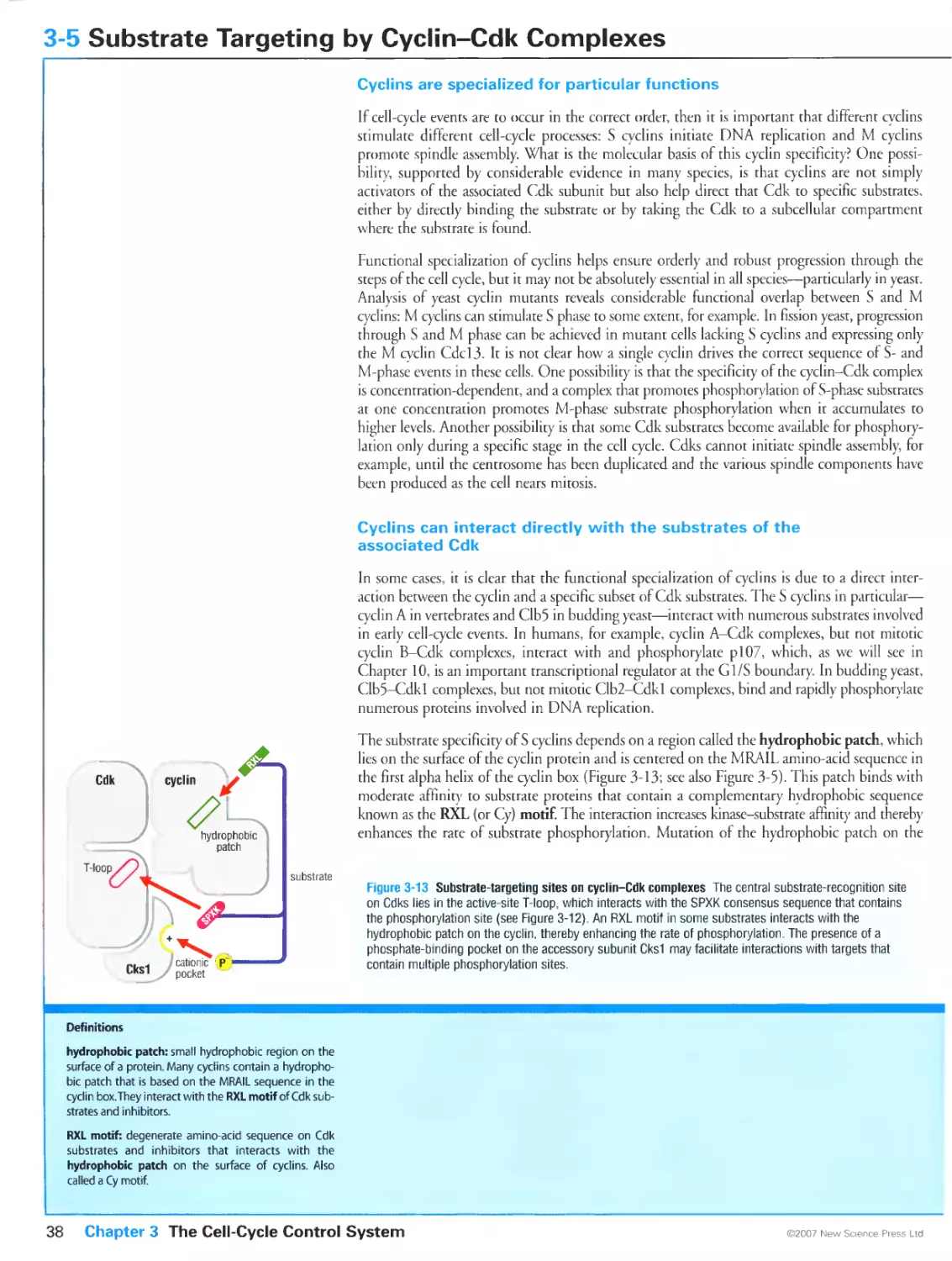
3-5 Substrate Targeting by Cyclin-Cdk Complexes 38
3-6 Cdk Regulation by Inhibitory Subunits 40
3-7 Biochemical Switches in Signaling Systems 42
3-8 Switch-Like Activation of Cdk1 44
3-9 Protein Degradation in Cell-Cycle Control 46
3-10 The Anaphase-Promoting Complex 48
3-11 Assembling and Regulating a Cell-Cycle Oscillator 50
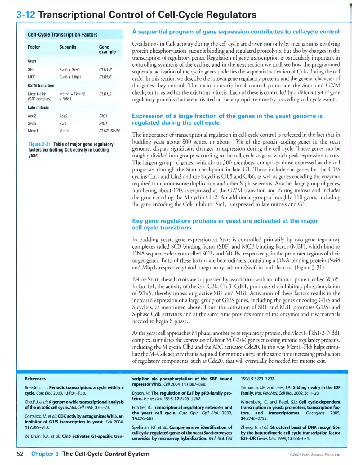
3-12 Transcriptional Control of Cell-Cycle Regulators 52
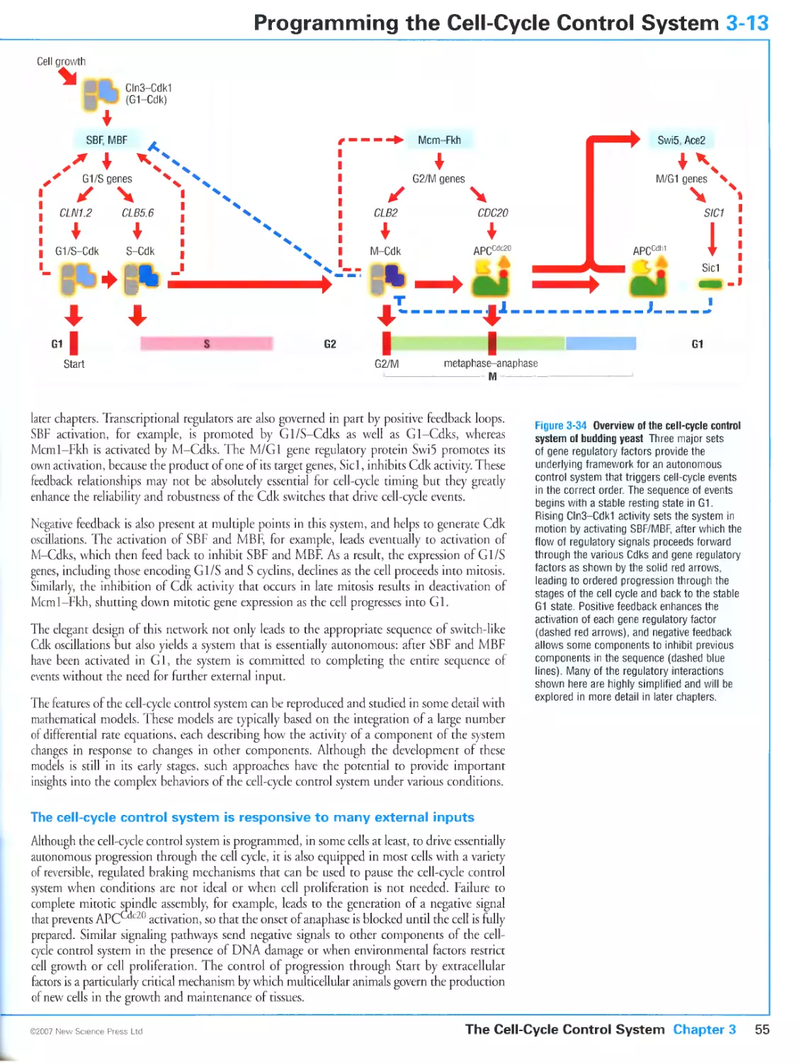
3-13 Programming the Cell-Cycle Control System 54
CHAPTER 4 Chromosome Duplication
4-0 Overview: Chromosome Duplication and its Control 58
4-1 Basic Mechanisms of DNA Synthesis 60
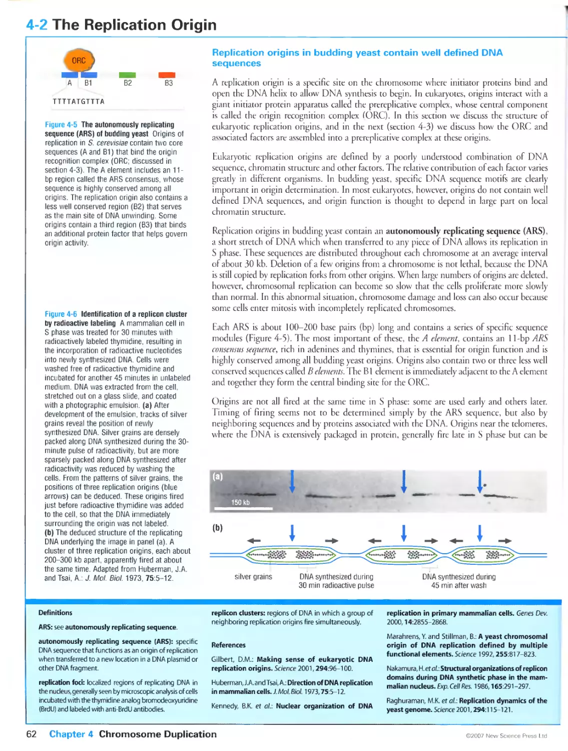
4-2 The Replication Origin 62
4-3 Assembly of the Prereplicative Complex at the
Replication Origin 64
4-4 Regulation of the Prereplicative Complex 66
4-5 Cyclins Required for Activation of Replication
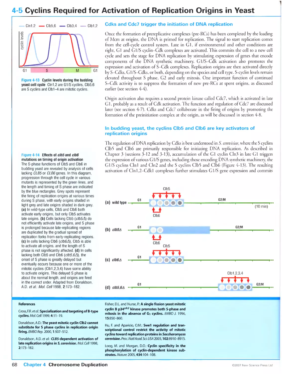
Origins in Yeast 68
4-6 Cyclins Required for Activation of Replication
Origins in Metazoans 70
4-7 Control of Replication by the Protein Kinase
Cdc7-Dbf4 72
4-8 Activation of the Replication Origin 74
4-9 Basic Chromatin Structure 76
4-10 Histone Synthesis in S phase 78
4-11 Nucleosome Assembly on Nascent DNA 80
4-12 Heterochromatin at Telomeres and Centromeres 82
4-13 Molecular Mechanisms of Heterochromatin
Duplication 84
CHAPTER 5 Early Mitosis: Preparing the Chromosomes
for Segregation
5-0 Overview: The Events of Mitosis 88
5-1 Overview: Principles of Mitotic Regulation 90
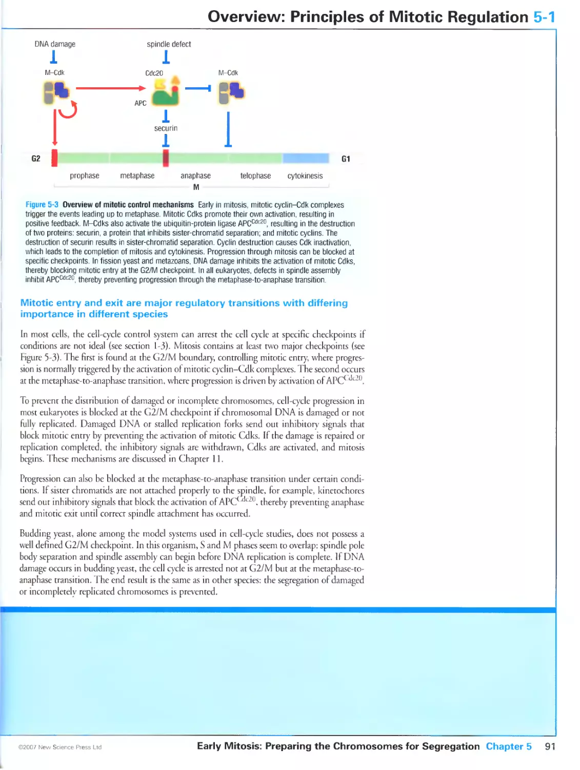
5-2 Cyclins that Promote Mitotic Entry in Yeast 92
5-3 Cyclins that Promote Mitotic Entry in Metazoans 94
5-4 Regulation of Mitotic Cdks by Wee1 and Cdc25 96
5-5 Switch-like Activation of Cyclin B-Cdkl at Mitosis 98
5-6 Subcellular Localization of Mitotic Regulators 100
5-7 Protein Kinases of the Polo and Aurora Families 102
5-8 Preparations for Mitosis: Sister-Chromatid Cohesion 104
5-9 Entry into Mitosis: Sister-Chromatid Condensation and
Resolution 106
5-10 Regulation of Chromosome Condensation and
Resolution 108
CHAPTER 6 Assembly of the Mitotic Spindle
6-0 Overview: The Mitotic Spindle 112
6-1 Microtubule Structure and Behavior 114
6-2 Microtubule Nucleation, Stability and Motility 116
6-3 The Centrosome and the Spindle Pole Body 118
6-4 Control of Centrosome Duplication 120
6-5 The Kinetochore 122
6-6 Early Steps in Spindle Assembly 124
6-7 Nuclear Envelope Breakdown 126
6-8 Mitotic Chromosome Function in Spindle Assembly 128
6-9 Attachment of Sister Chromatids to the Spindle 130
6-10 Bi-Orientation of Sister Chromatids 132
6-11 Forces Driving Chromosome Movement 134
6-12 Chromosome Congression 136
CHAPTER 7 The Completion of Mitosis
7-0 Overview: The Completion of Mitosis 140
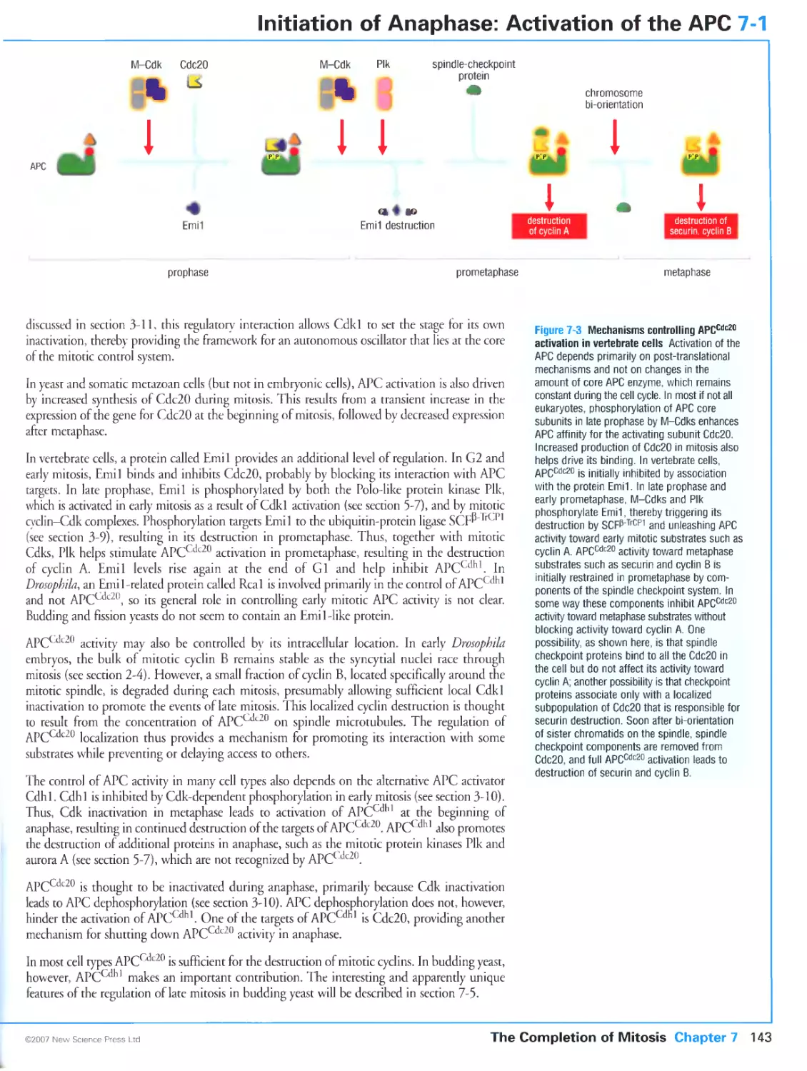
7-1 Initiation of Anaphase: Activation of the APC 142
7-2 Initiation of Anaphase: The Spindle Checkpoint 144
7-3 Inhibition of APCcdc20 by the Spindle Checkpoint 146
7-4 Control of Sister-Chromatid Separation 148
7-5 Control of Late Mitosis in Budding Yeast 150
7-6 Control of Anaphase Events 152
7-7 Control of Telophase 154
CHAPTER 8 Cytokinesis
8-0 Overview: Cytokinesis 158
8-1 The Actin-Myosin Ring 160
8-2 Assembly and Contraction of the Actin-Myosin Ring 162
8-3 Membrane and Cell Wall Deposition at the Division Site 164
8-4 The Positioning and Timing of Cytokinesis in Yeast 166
8-5 The Positioning and Timing of Cytokinesis in Animal Cells 168
8-6 Specialization of Cytokinesis in Animal Development 170
8-7 Asymmetric Cell Division 172
xvi
CHAPTER 9 Meiosis
9-0 Overview: Meiosis
9-1 Regulation of Early Meiotic Events in Yeast
9-2 Homologous Recombination in Meiosis
9-3 Homolog Pairing in Meiotic Prophase
9-4 Chiasma Formation in Late Meiotic Prophase
9-5 Controlling Entry into the First Meiotic Division
9-6 Chromosome Attachment in Meiosis I
9-7 Chromosome Segregation in Meiosis I
9-8 Finishing Meiosis
176
178
180
182
190
192
Glossary
References
Index
2G9
275
287
CHAPTER 10 Control of Cell Proliferation and Growth
10-0 Overview: Control of Cell Proliferation and Growth 196
10-1 Activation of Gene Expression at Start in Budding Yeast 198
10-2 Activation of S-Cdks in Budding Yeast 200
10-3 Extracellular Control of Start in Yeast: Mating Factor
Signaling 202
10-4 Activation of Gene Expression at Start in Animals 204
10-5 Regulation of E2F-pRB Complexes 206
10-6 Mitogenic Signaling in Animal Cells 208
10-7 Activation of G1-Cdks by Mitogens 210
10-8 Activation of G1/S- and S-Cdk Complexes in Animal Cells 212
10-9 Developmental Control of Cell Proliferation 214
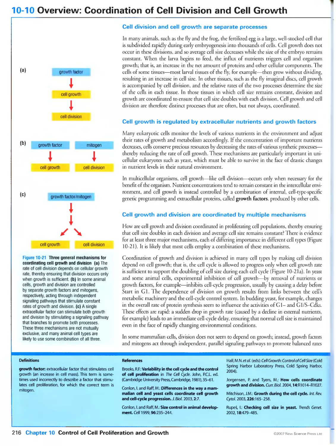
10-10 Overview: Coordination of Cell Division and Cell Growth 216
10-11 Control of Cell Growth 218
10-12 Coordination of Cell Growth and Division in Yeast 220
10-13 Coordination of Growth and Division in Animal Cells 222
10-14 Control of Cell Death 224
CHAPTER 11 The DNA Damage Response
11-0 Overview: The DNA Damage Response 228
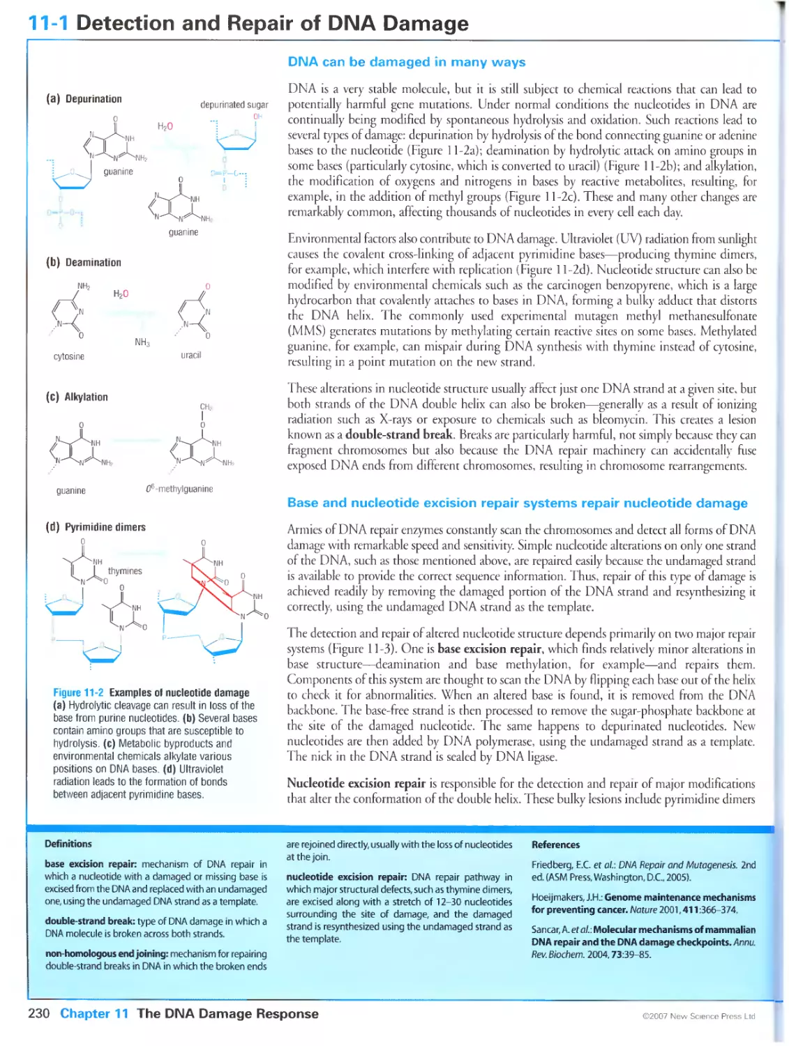
11-1 Detection and Repair of DNA Damage 230
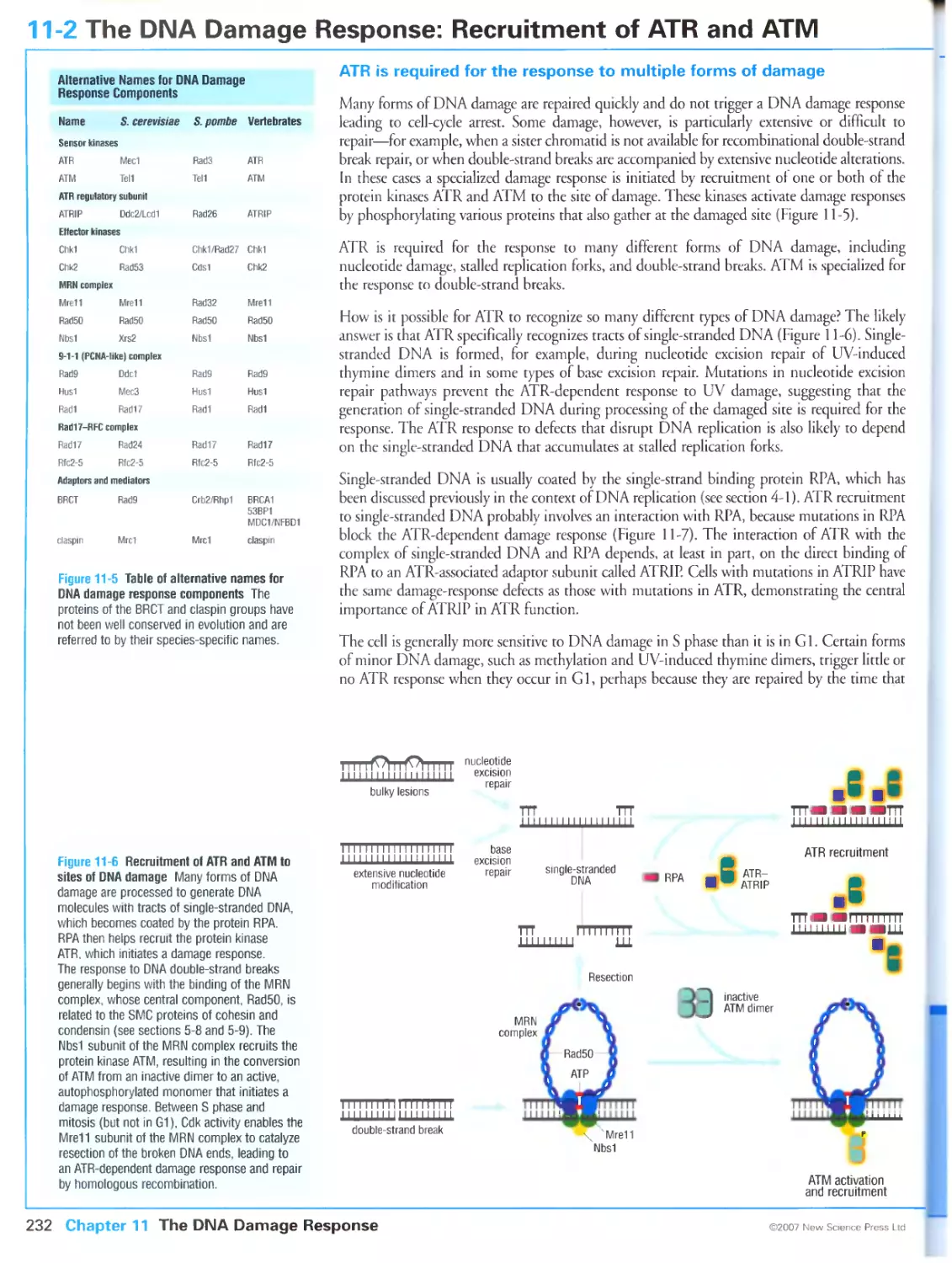
11-2 The DNA Damage Response: Recruitment of ATR and ATM 232
11-3 The DNA Damage Response: Adaptors and Chkl and Chk2 234
11-4 Activation of p53 by DNA Damage 236
11-5 Effects of DNA Damage on Progression through Start 238
11-6 Effects of DNA Damage at Replication Forks 240
11-7 Effects of DNA Damage on DNA Synthesis and Mitosis 242
11-8 Responses to Mitogenic and Telomere Stress 244
CHAPTER 12 The Cell Cycle in Cancer
12-0 Overview: Cell-Cycle Defects in Cancer 248
12-1 Gene Mutations that Drive Cancer 250
12-2 Tissue Specificity in Cancer 252
12-3 Stimulation of Cell-Cycle Entry in Cancer Cells 254
12-4 Cell Growth and Survival in Tumors 256
12-5 Genetic Instability in Cancer 258
12-6 Telomeres and the Structural Instability of Chromosomes 260
12-7 Instability in Chromosome Number 262
12-8 Cancer Progression 264
12-9 Stopping Cancer 266
XVÜ
Contents in full
The Author
A Note from the Publisher on Primers in Biology
Preface
Acknowledgements
CHAPTER 1 The Cell Cycle
1-0 Overview: Cell Reproduction
Cell reproduction is a fundamental feature of all living things
Cells reproduce in discrete steps
The ordering of cell-cycle events is governed by an independent control
system
1-1 Events of the Eukaryotic Cell Cycle
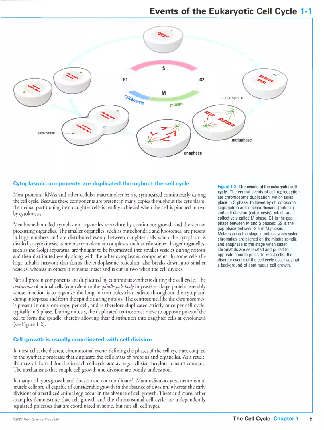
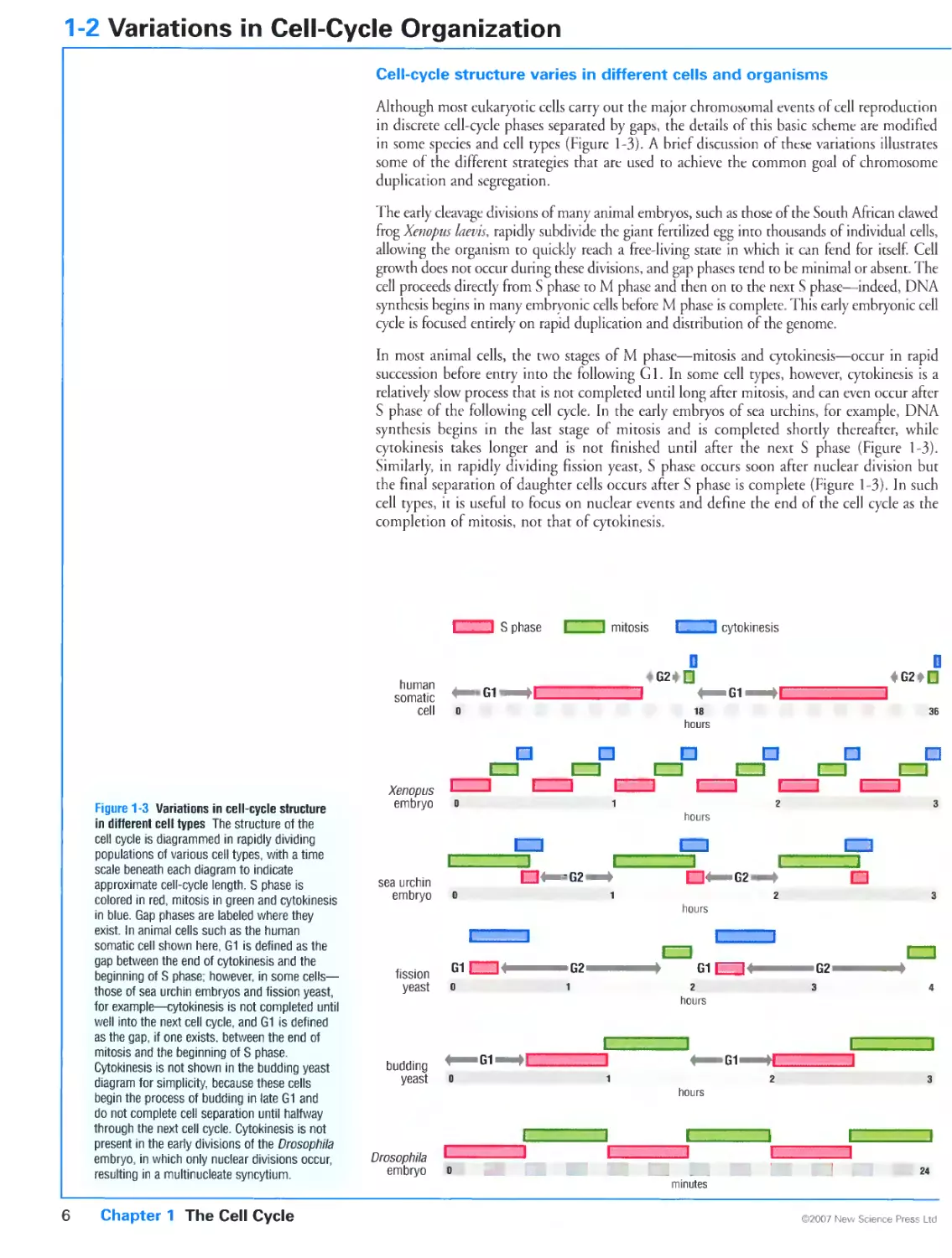
Chromosome duplication and segregation occur in distinct cell-cycle phases
that are usually separated by gap phases
Cytoplasmic components are duplicated throughout the cell cycle
Cell growth is usually coordinated with cell division
1-2 Variations in Cell-Cycle Organization
Cell-cycle structure varies in different cells and organisms
Multiple rounds of chromosome duplication or segregation can occur in the
same cell cycle
The symmetry of cell division varies in different cell types
1-3 The Cell-Cycle Control System
Cell-cycle events are govetned by an independent control system
The cell-cycle control system is based on oscillations in the activities of
cyclin-dependent protein kinases
Cell-cycle events are initiated at three regulatory checkpoints
Cell-cycle progression in most cells can be blocked at checkpoints
CHAPTER 2 Model Organisms in Cell-Cycle Analysis
2-0 Overview: Cell-Cycle Analysis in Diverse Eukaryotes
Mechanisms of cell-cycle control are similar in all eukaryotes
Budding and fission yeasts provide powerful systems for the genetic
analysis of eukaryotic cell-cycle control
Early animal embryos are useful for the biochemical characterization of
simple cell cycles
Control of cell division in multicellular organisms can be dissected
genetically in Drosophila
Cultured cell lines provide a means of analyzing cell-cycle control in
mammals
2-1 Life Cycles of Budding and Fission Yeasts
Budding yeast and fission yeast divide by different mechanisms
Yeast cells alternate between haploid and diploid states and undergo
sporulation in response to starvation
2-2 Genetic Analysis of Cell-Cycle Control in Yeast
Cell biological processes are readily dissected with yeast genetic methods
Conditional mutants ate used to analyze essential cell-cycle processes
Homologous genes have different names in fission yeast and budding yeast
2-3 The Early Embryo of Xenopus laevis
The early embryonic divisions of Xenopus provide a simplified system for
cell-cycle analysis
Unfertilized eggs develop from diploid oocytes by meiosis
The early embryonic cell cycle can be reconstituted in a test tube
2-4 The Fruit Fly Drosophila melanogaster
Drosophila allows genetic analysis of cell-cycle control in metazoans
Cells of the early Drosophila embryo divide by a simplified cell cycle
Gap phases are introduced in late embryogenesis
Adult fly structures develop from imaginal cells
2-5 Mammalian Cell-Cycle Analysis
Mammalian cell-cycle control can be analyzed In cells growing in culture
Mutations lead to immortalization and transformation of mammalian cells
Specific gene disruption is the ideal approach for assessing protein function
in mammalian cells
2-6 Methods in Cell-Cycle Analysis
Cell-cycle position can be assessed by many approaches
Cell populations can be synchronized at specific cell-cycle stages
Complete understanding of cell-cycle control mechanisms requires the
analysis of protein structure and enzymatic behavior
CHAPTER 3 The Cell-Cycle Control System
3-0 Overview: The Cell-Cycle Control System
The cell-cycle control system is a complex assembly of oscillating protein
kinase activities
Multiple regulatory mechanisms govern Cdk activity during the cell cycle
The cell-cycle control system generates robust, switch-like and adaptable
changes in Cdk activity
3-1 Cyclin-Dependent Kinases
The cyclin-dependent kinases are a small family of enzymes that require
cyclin subunits for activity
The active site of cyclin-dependent kinases is blocked in the absence of
cyclin
3-2 Cyclins
Cyclins are the key determinants of Cdk activity and can be classified in four
groups
Cyclins contain a conserved helical core
3-3 Control of Cdk Activity by Phosphorylation
Full Cdk activity requires phosphorylation by the Cdk-activating kinase
Cdk function is regulated by inhibitory phosphorylation by Wee1 and
dephosphorylation by Cdc25
3-4 The Structural Basis of Cdk Activation
The conformation of the Cdk active site is dramatically rearranged by cyclin
binding and phosphorylation by CAK
3-5 Substrate Targeting by Cyclin-Cdk Complexes
Cyclins are specialized for particular functions
Cyclins can interact directly with the substrates of the associated Cdk
Cyclins can direct the associated Cdk to specific subcellular locations
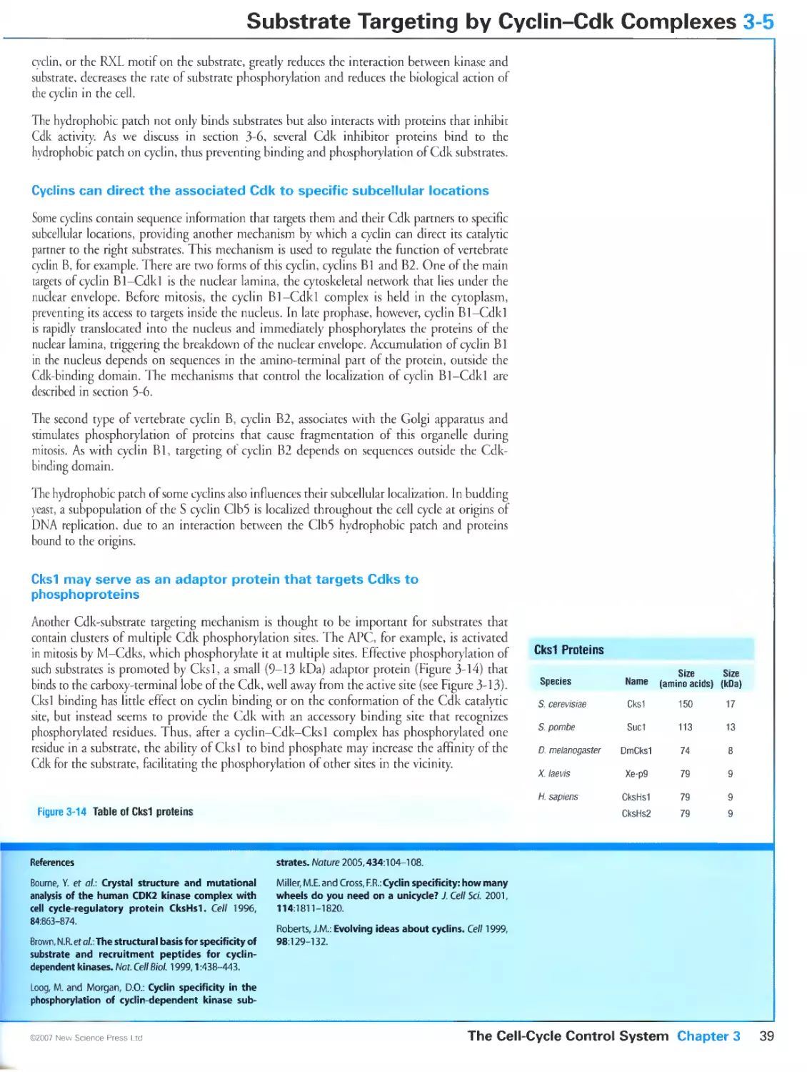
Cks1 may serve as an adaptor protein that targets Cdks to phosphoproteins
3-6 Cdk Regulation by Inhibitory Subunits
Cdk inhibitors help suppress Cdk activity in G1
Cip/Kip proteins bind both subunits of the cyclin-Cdk complex
G1-Cdks are activated by Cip/Kip proteins and inhibited by INK4 proteins
3-7 Biochemical Switches in Signaling Systems
Components of the cell-cycle control system are assembled into biochemical
switches
Switch-like behavior can be generated by various mechanisms
Bistability is required for an effective binary switch
3-8 Switch-Like Activation of Cdkl
Cdk1 activation at mitosis is based on positive feedback
Cdk switches are robust as a result of multiple partly redundant mechanisms
3-9 Protein Degradation in Cell-Cycle Control
Many cell-cycle regulators are destroyed by ubiquitin-dependent proteolysis
SCF catalyzes ubiquitination of phosphorylated substrates using
interchangeable substrate-targeting subunits
3-10 The Anaphase-Promoting Complex
The APC initiates anaphase and mitotic exit
Cdc20 activates the APC in anaphase
APC activity is maintained in G1 by Cdh1
APC targets contain specific recognition sequences
3-11 Assembling and Regulating a Cell-Cycle Oscillator
Negative feedback can generate a repeating oscillator
Regulated braking mechanisms allow the Cdk oscillator to be paused in G1
3-12 Transcriptional Control of Cell-Cycle Regulators
A sequential program of gene expression contributes to cell-cycle control
Expression of a large fraction of the genes in the yeast genome is regulated
during the cell cycle
Key gene regulatory proteins in yeast are activated at the major cell-cycle
transitions
The E2F family controls cell-cycle-dependent changes in gene expression in
metazoans
3-13 Programming the Cell-Cycle Control System
The order of cell-cycle events is determined by regulatory interactions
between multiple oscillators
The cell-cycle control system is responsive to many external inputs
CHAPTER 4 Chromosome Duplication
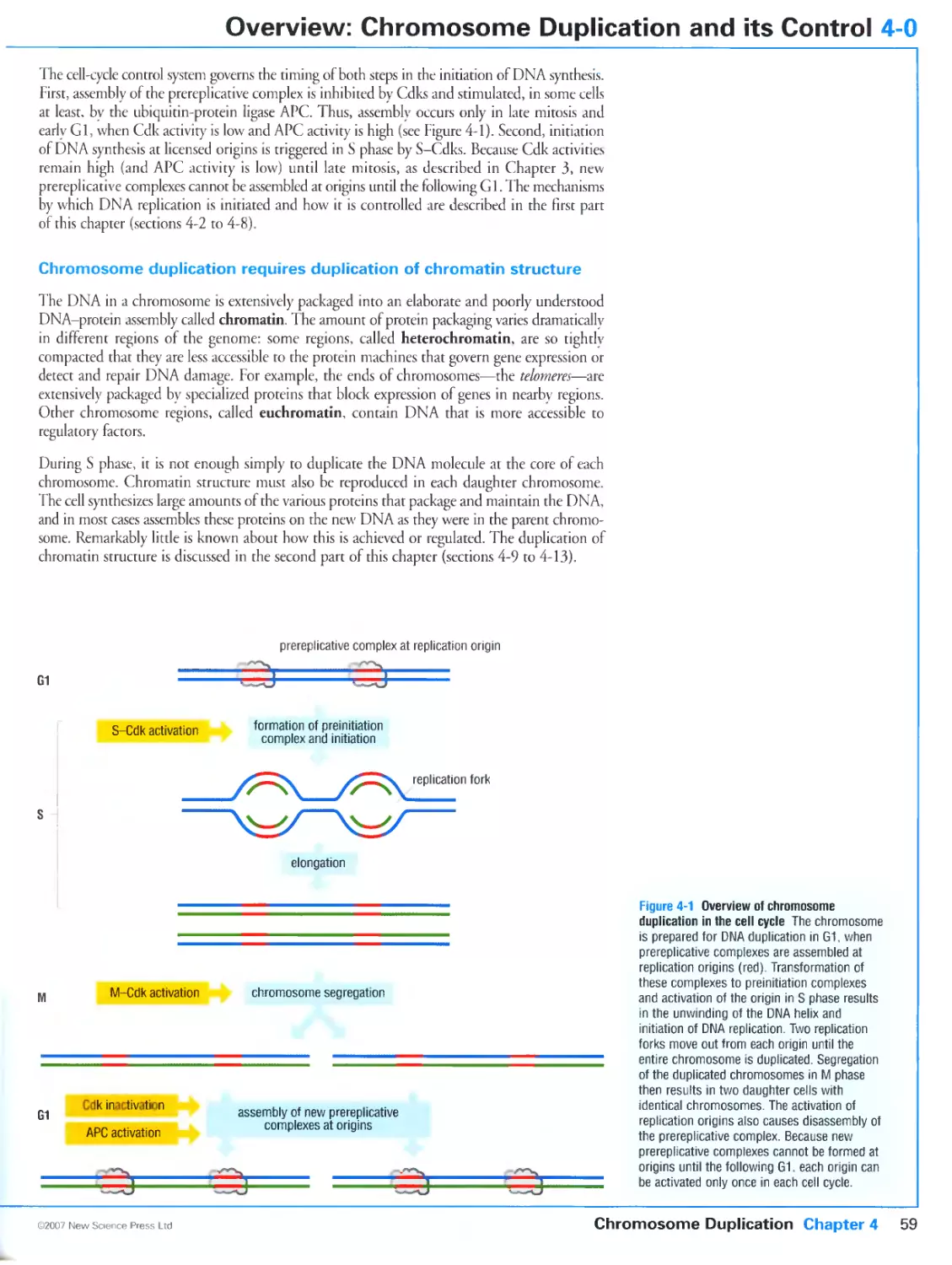
4-0 Overview: Chromosome Duplication and its Control
DNA synthesis begins at replication origins
The cell-cycle control system activates replication origins only once in each
S phase
Chromosome duplication requires duplication of chromatin structure
4-1 Basic Mechanisms of DNA Synthesis
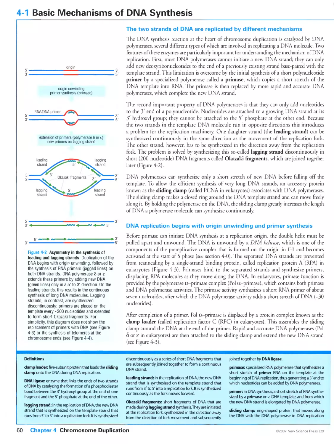
The two strands of DNA are replicated by different mechanisms
DNA replication begins with origin unwinding and primer synthesis
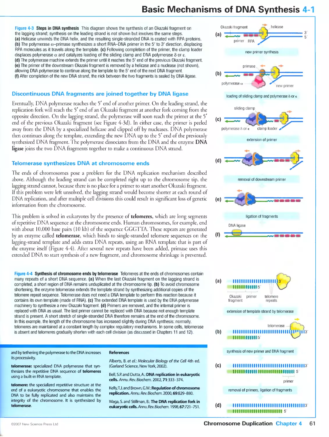
Discontinuous DNA fragments are joined together by DNA ligase
Telomerase synthesizes DNA at chromosome ends
4-2 The Replication Origin
Replication origins in budding yeast contain well defined DNA sequences
Replication origins in animal chromosomes are defined by several factors in
addition to DNA sequence
4-3 Assembly of the Prereplicative Complex at the Replication Origin
The replication origin interacts with a multisubunit protein complex
The ORC and accessory proteins load the Mem helicase onto origins
Mem loading involves ATP-dependent protein remodeling
4-4 Regulation of the Prereplicative Complex
Assembly of prereplicative complexes is restricted to G1 by multiple
mechanisms
Prereplicative complex components are destroyed or inhibited in yeast as a
result of Cdk activity
Pre-RC assembly is controlled in animals by both Cdks and the APC
4-5 Cyclins Required for Activation of Replication Origins in Yeast
Cdks and Cdc7 trigger the initiation of DNA replication
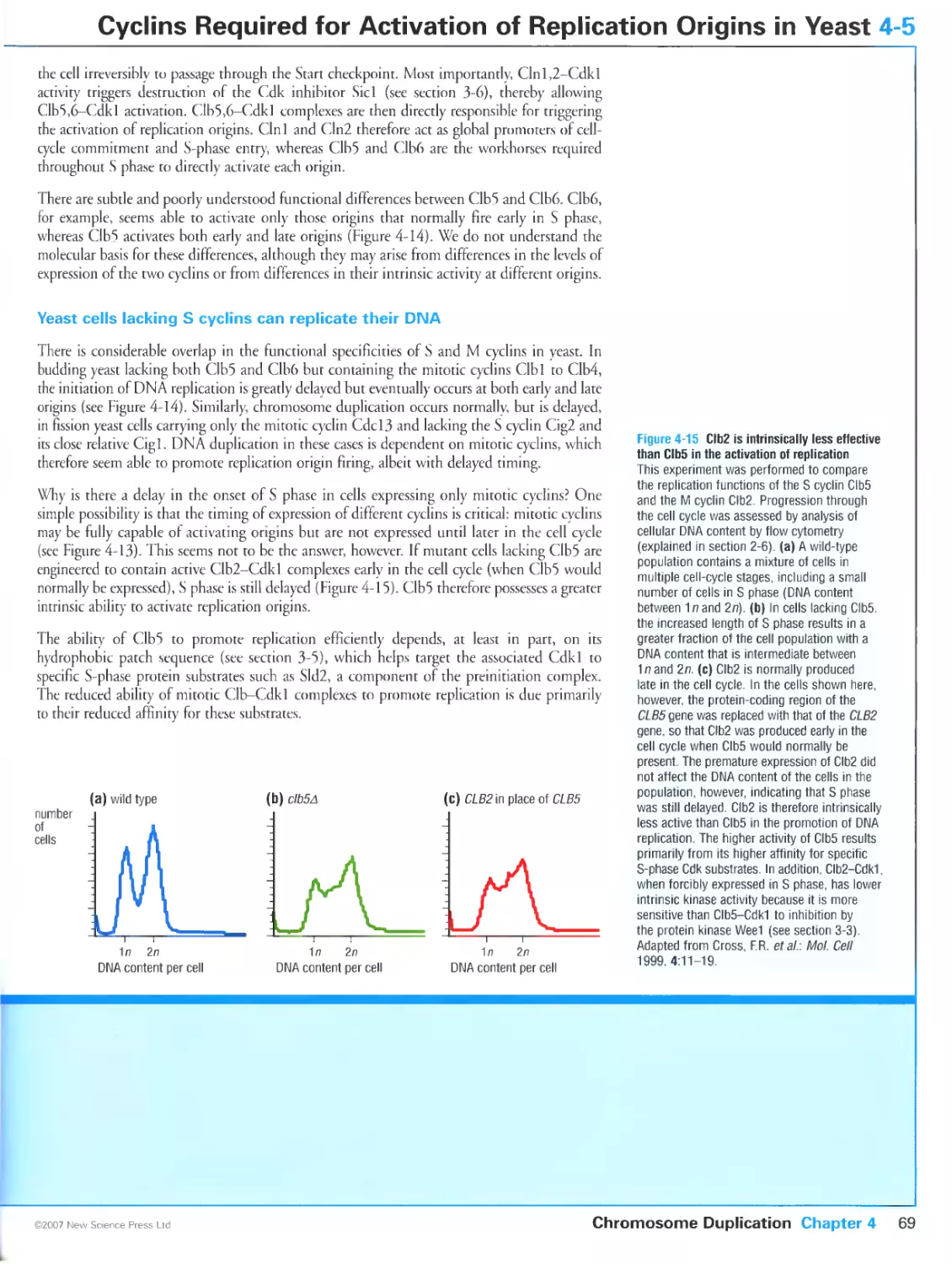
In budding yeast, the cyclins Clb5 and Clb6 are key activators of replication
origins
Yeast cells lacking S cyclins can replicate their DNA
4-6 Cyclins Required for Activation of Replication Origins in Metazoans
Different cyclins control initiation of DNA replication in different stages of
animal development
Cyclin A is a major regulator of replication initiation in cultured mammalian
cells
DNA replication in frog embryos is triggered by cyclin E-Cdk2
Cyclin E-Cdk2 is a major regulator of DNA replication in Drosophila
4-7 Control of Replication by the Protein Kinase Cdc7-Dbf4
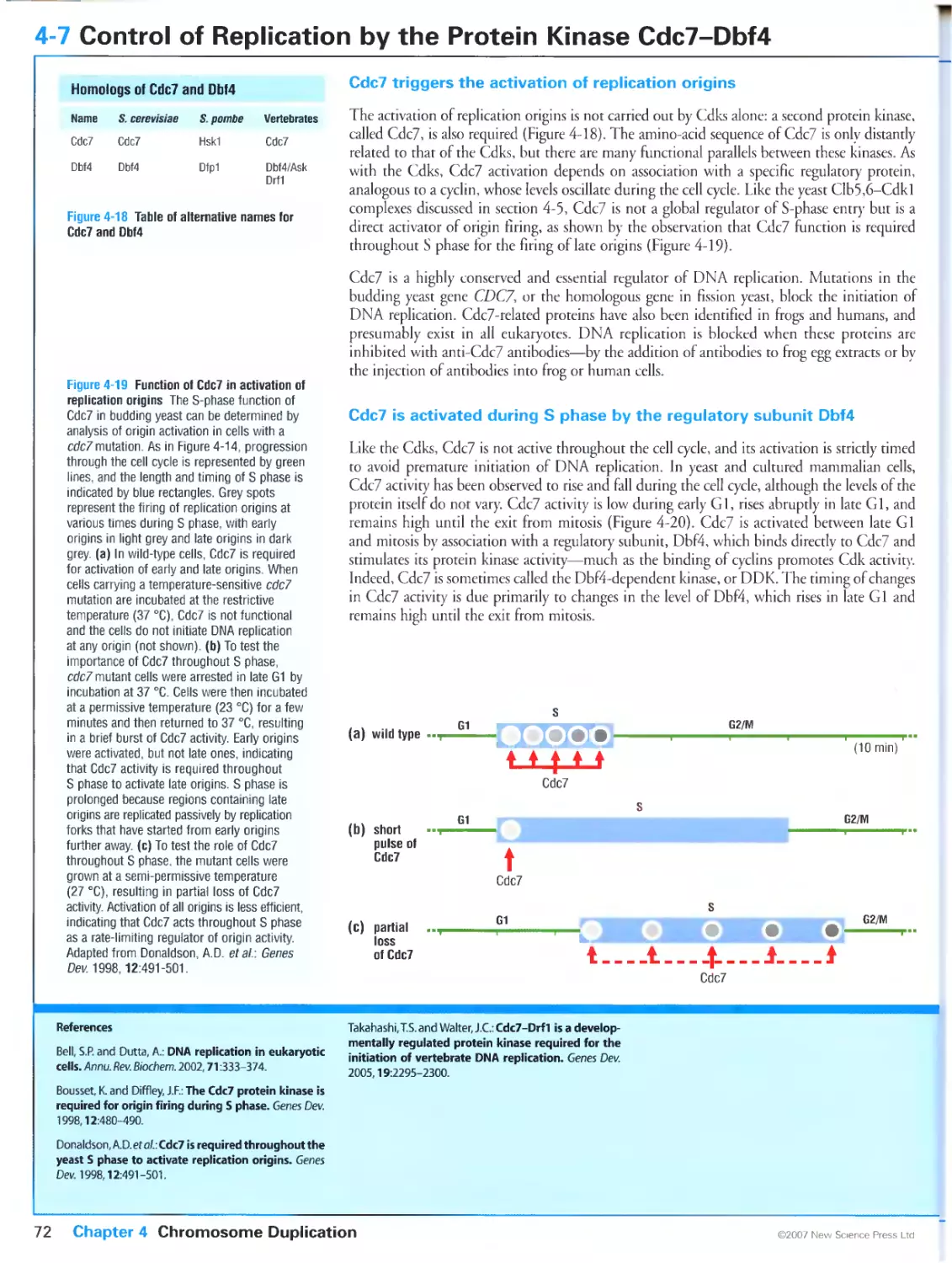
Cdc7 triggers the activation of replication origins
Cdc7 is activated during S phase by the regulatory subunit Dbf4
Dbf4 levels are regulated by multiple mechanisms
4-8 Activation of the Replication Origin
Replication begins with DNA unwinding at the origin
Late-firing origins are regulated independently
Replication must be completed before chromosome segregation occurs
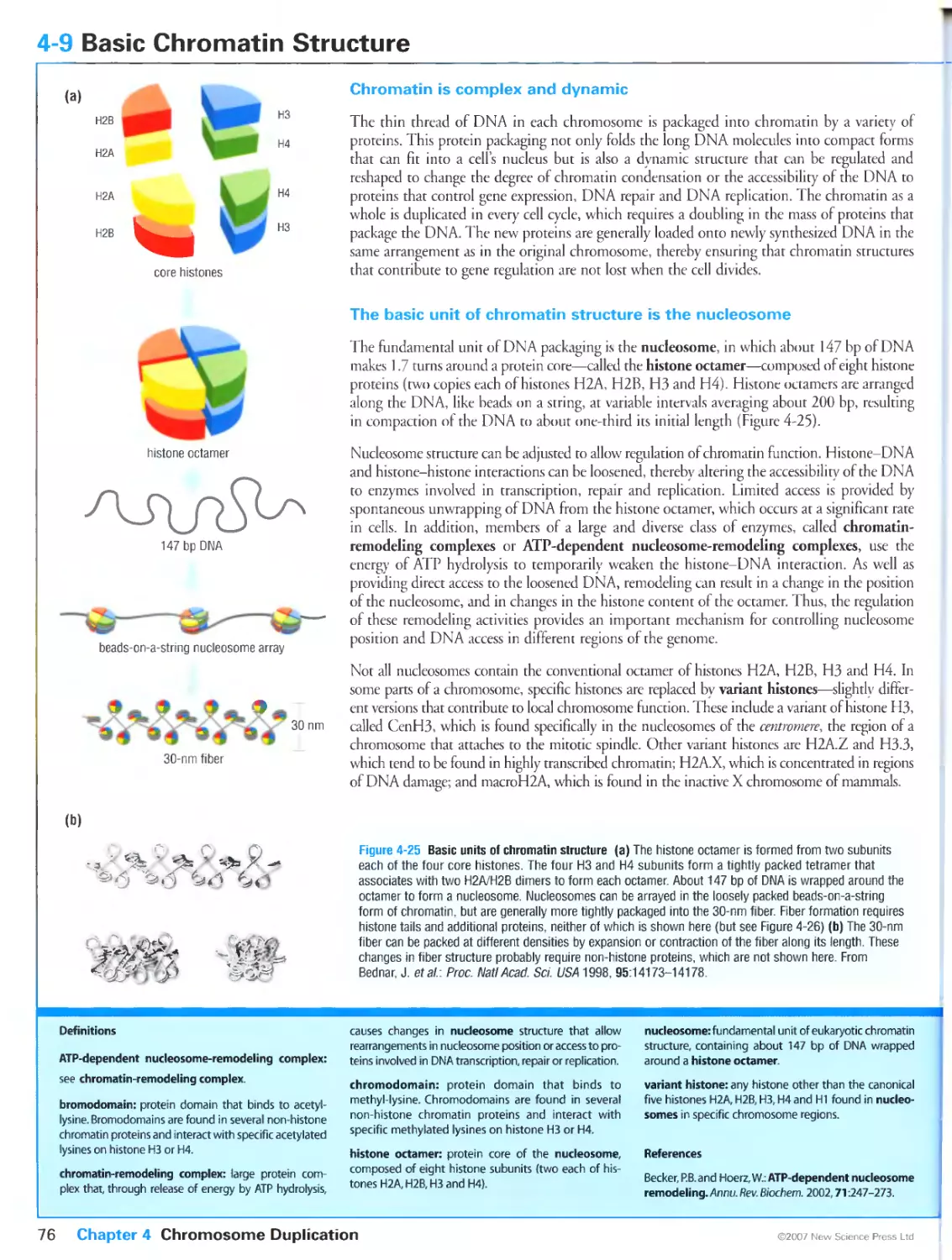
4-9 Basic Chromatin Structure
Chromatin is complex and dynamic
The basic unit of chromatin structure is the nucleosome
Higher-order chromatin structure is also controlled by non-histone proteins,
histone H1 and histone modifications
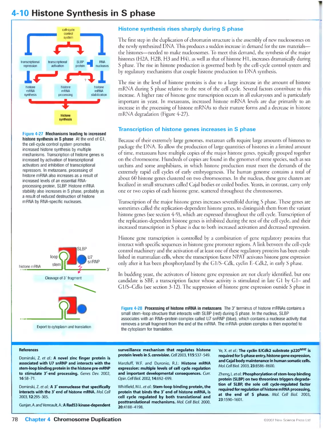
4-10 Histone Synthesis in S phase
Histone synthesis rises sharply during S phase
Transcription of histone genes increases in S phase
Histone mRNA processing and stability increase in S phase
The level of free histones in the cell acts as a signal to link histone synthesis
to DNA synthesis
4-11 Nucleosome Assembly on Nascent DNA
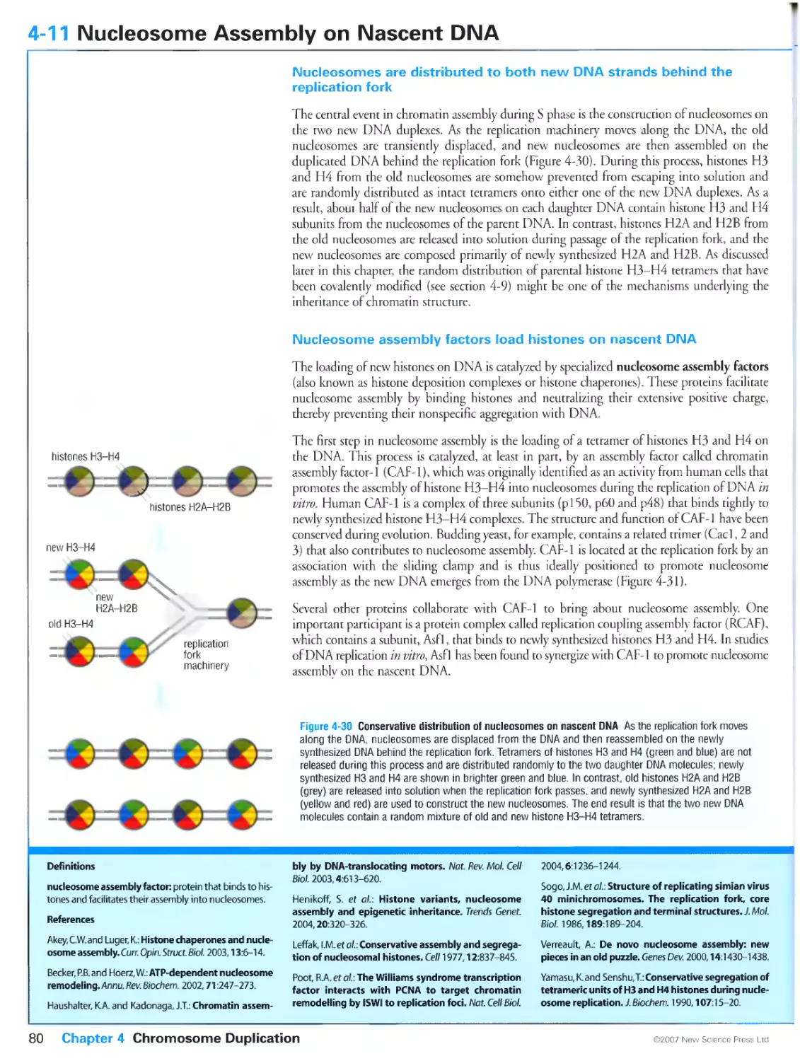
Nucleosomes are distributed to both new DNA strands behind the
replication fork
Nucleosome assembly factors load histones on nascent DNA
4-12 Heterochromatin at Telomeres and Centromeres
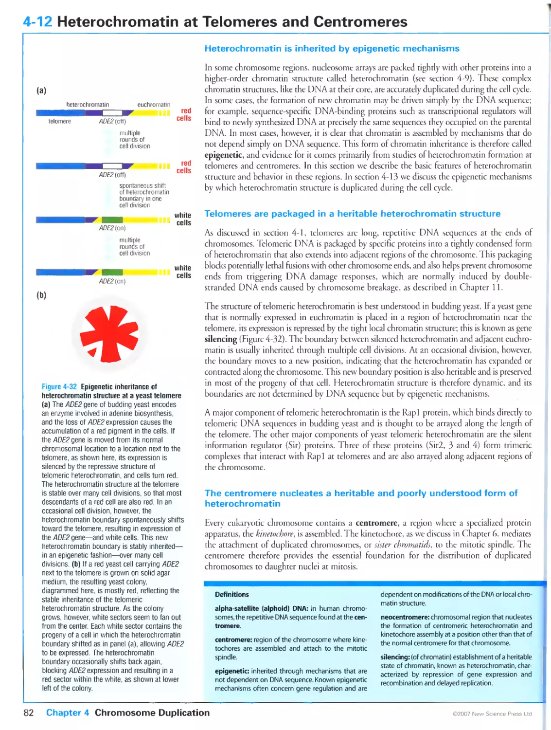
Heterochromatin is inherited by epigenetic mechanisms
Telomeres are packaged in a heritable heterochromatin structure
The centromere nucleates a heritable and poorly understood form of
heterochromatin
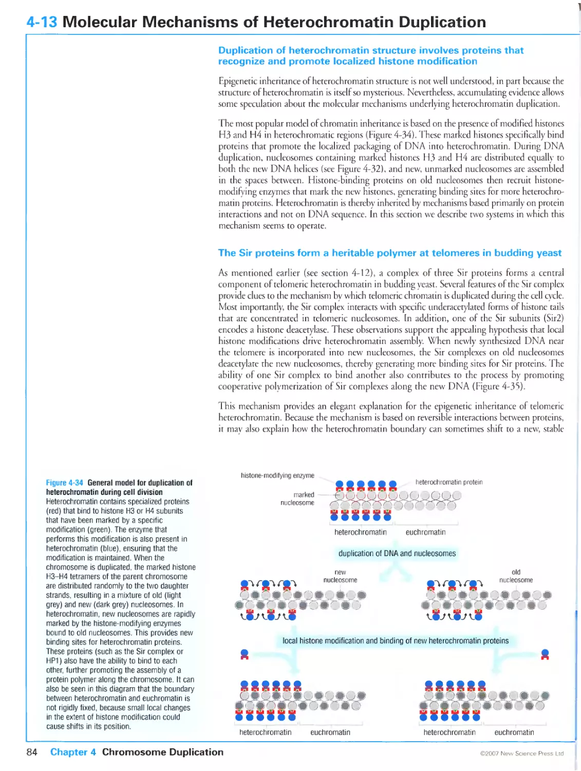
4-13 Molecular Mechanisms of Heterochromatin Duplication
Duplication of heterochromatin structure involves proteins that recognize
and promote localized histone modification
The Sir proteins form a heritable polymer at telomeres in budding yeast
HP1 may nucleate heritable chromatin structure at the centromere and other
regions
Sister-chromatid cohesion in S phase prepares the cell for mitosis
CHAPTER 5 Early Mitosis: Preparing the Chromosomes for Segregation
5-0 Overview: The Events of Mitosis 88
The central events of mitosis are sister-chromatid separation and
segregation
The events of early mitosis set the stage for sister-chromatid segregation
The completion of mitosis begins with sister-chromatid segregation
5-1 Overview: Principles of Mitotic Regulation 90
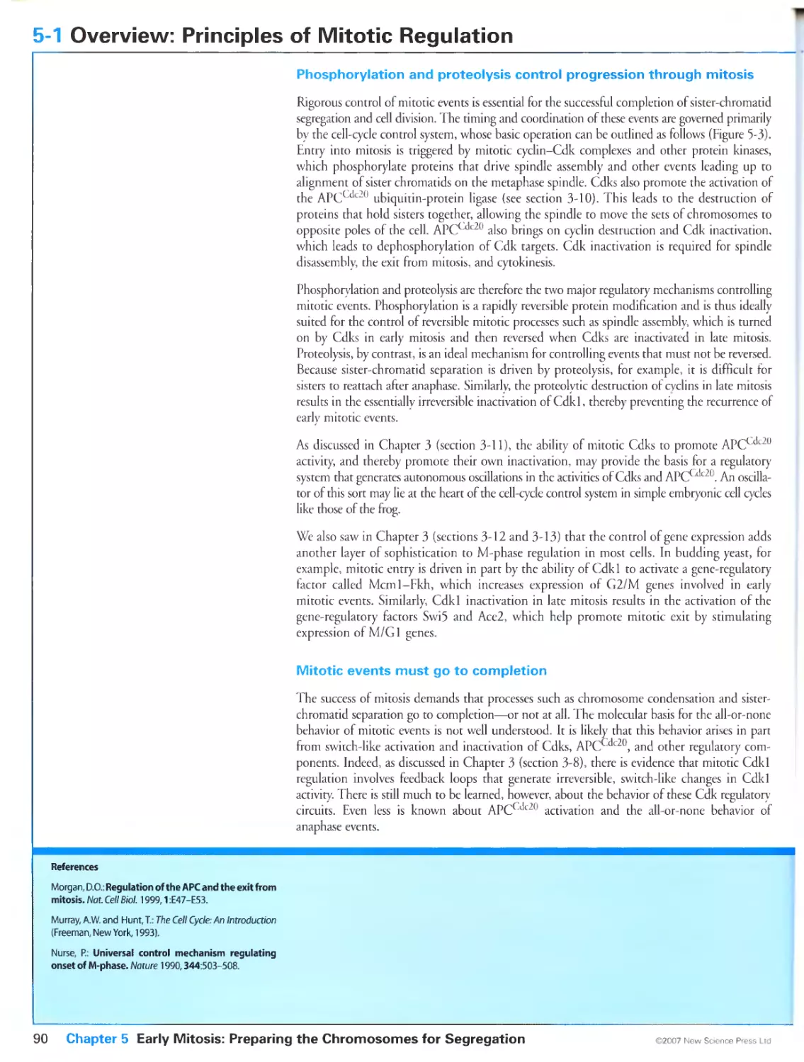
Phosphorylation and proteolysis control progression through mitosis
Mitotic events must go to completion
Mitotic entry and exit are major regulatory transitions with differing
importance in different species
5-2 Cyclins that Promote Mitotic Entry in Yeast 92
Cyclin-Cdk complexes trigger mitotic entry in all eukaryotes
Fission yeast cells trigger mitosis with a single mitotic cyclin
Two pairs of mitotic cyclins control budding yeast mitosis
5-3 Cyclins that Promote Mitotic Entry in Metazoans 94
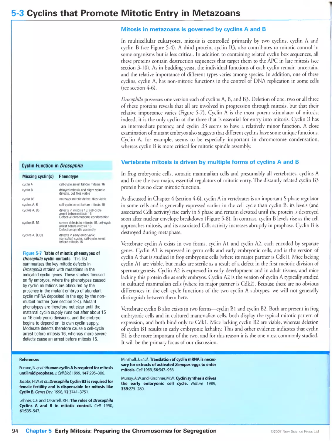
Mitosis in metazoans is governed by cyclins A and B
Vertebrate mitosis is driven by multiple forms of cyclins A and B
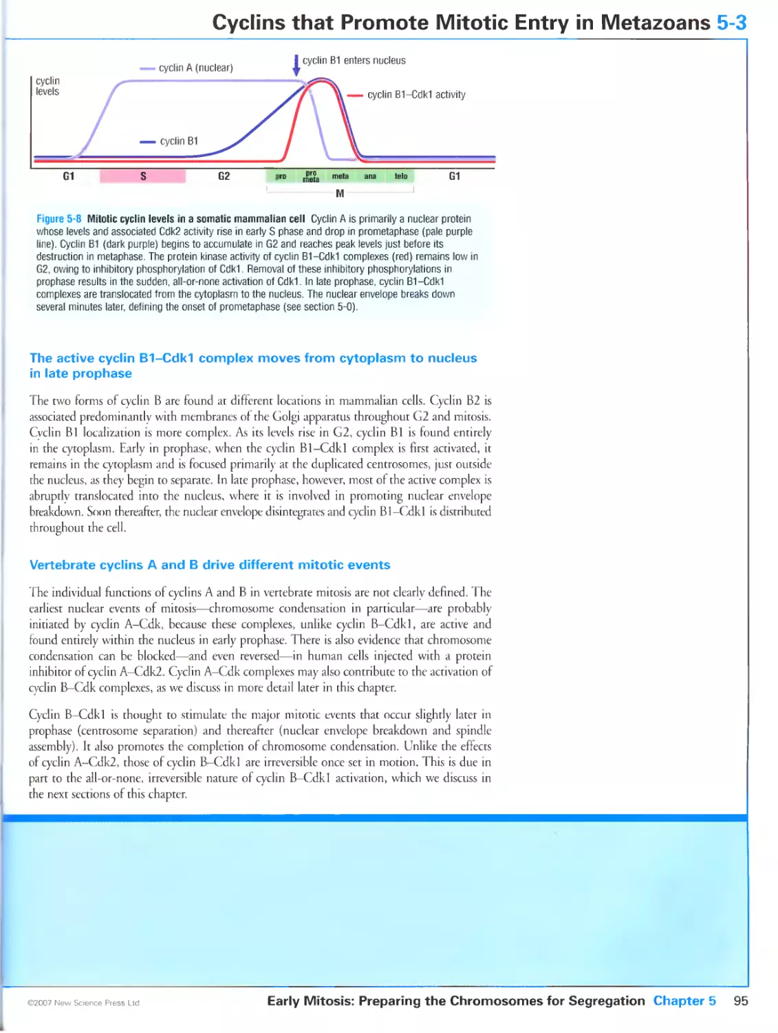
The active cyclin B1-Cdk1 complex moves from cytoplasm to nucleus in late
prophase
Vertebrate cyclins A and B drive different mitotic events
5-4 Regulation of Mitotic Cdks by Weel and Cdc25 96
Cyclin B-Cdk1 complexes are activated rapidly in early M phase by
dephosphorylation
Multiple Wee 1-related kinases and Cdc25-related phosphatases govern Cdk1
activity in animal cells
5-5 Switch-like Activation of Cyclin B-Cdkl at Mitosis 98
Mitotic Cdk1 activation involves multiple positive feedback loops
Cdc25B and cyclin A-Cdk help trigger cyclin B-Cdk1 activation
5-6 Subcellular Localization of Mitotic Regulators 100
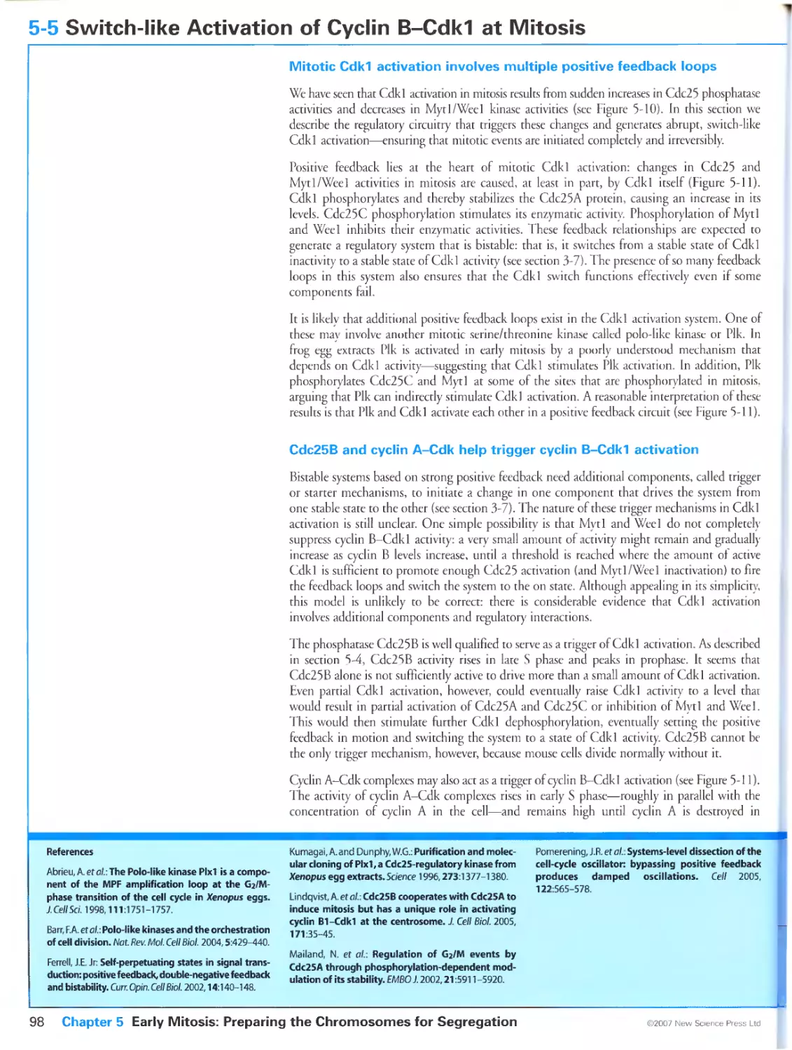
Cyclin B1-Cdk1 is regulated by changes in its subcellular localization
/8
80
87
xxi
Cyclin B1-Cdk1 location is controlled by phosphorylation of cyclin B1
Cdc25C localization is regulated by phosphorylation
Cyclin B1-Cdk1 activation and nuclear accumulation are partly interdependent
5-7 Protein Kinases of the Polo and Aurora Families
Polo-like kinases (Plks) help control spindle assembly and mitotic exit
Spindle function and sister-chromatid segregation are controlled in part by
aurora kinases
5-8 Preparations for Mitosis: Sister-Chromatid Cohesion
Sister chromatids are held together by two mechanisms
Cohesin is a key mediator of sister-chromatid cohesion
Cohesion is established during DNA replication
DNA decatenation prepares sister chromatids for separation
5-9 Entry into Mitosis: Sister-Chromatid Condensation and Resolution
Chromosomes are dramatically reorganized in mitosis
Condensin complexes drive chromosome condensation and resolution
5-10 Regulation of Chromosome Condensation and Resolution
Mitotic Cdks act on condensin to govern the timing of chromosome
condensation
Sister-chromatid resolution is governed by Plk and aurora B in animal cells
CHAPTER 6 Assembly of the Mitotic Spindle
6-0 Overview: The Mitotic Spindle
Chromosome segregation depends on the mitotic spindle
The mitotic spindle must be bipolar
Multiple mechanisms drive spindle assembly
6-1 Microtubule Structure and Behavior
Microtubules are polymers of tubulin subunits
Microtubules exhibit dynamic instability
6-2 Microtubule Nucleation, Stability and Motility
Cellular microtubules originate on preformed protein complexes that are
usually concentrated in a microtubule-organizing center
Microtubule dynamics are governed by a variety of stabilizing and
destabilizing proteins
Motor proteins move along microtubules
6-3 The Centrosome and the Spindle Pole Body
The centrosome cycle resembles the chromosome cycle
Centrosome behavior is determined by the centrioles
The yeast spindle pole body is embedded in the nuclear envelope
6-4 Control of Centrosome Duplication
Duplication of the centrosome and spindle pole body is initiated in late G1
by G1/S-Cdks
Centrosome duplication normally occurs once per cell cycle
6-5 The Kinetochore
The kinetochore is the major site of microtubule-chromosome attachment
The kinetochore provides a stable attachment to a dynamic microtubule plus
end
6-6 Early Steps in Spindle Assembly
Spindle assembly begins in prophase
Mitotic microtubules are highly dynamic
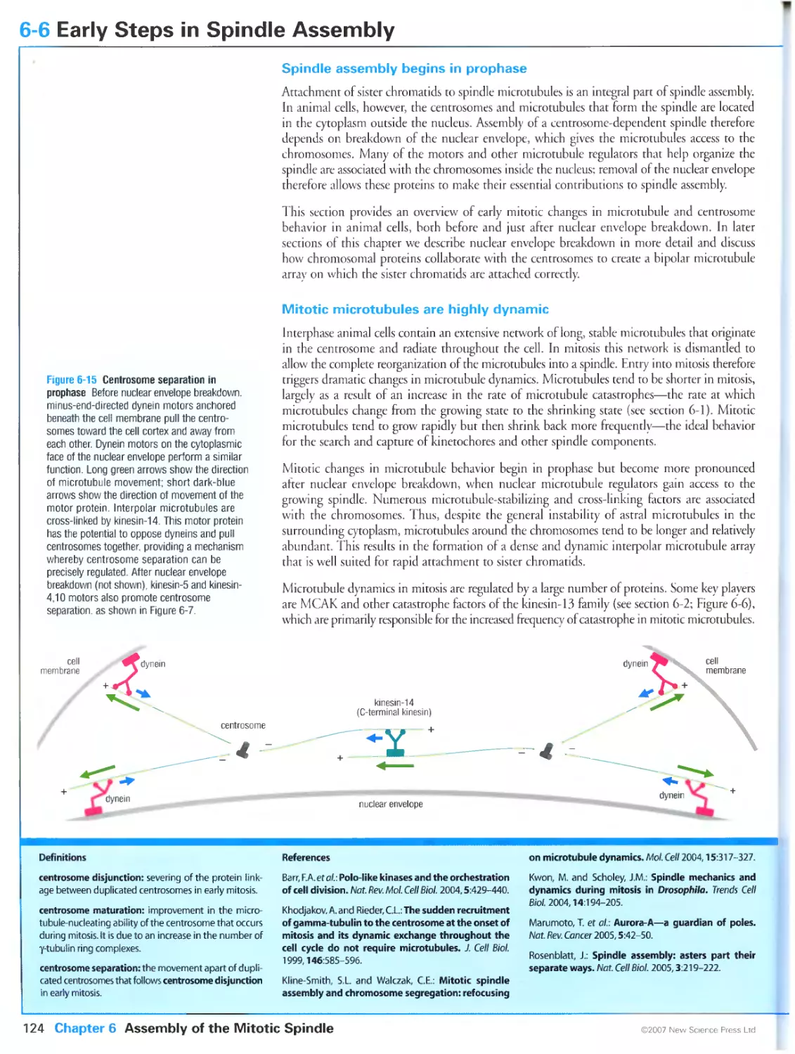
Centrosome separation initiates spindle assembly
Centrosome maturation increases microtubule nucleation in mitosis
6-7 Nuclear Envelope Breakdown
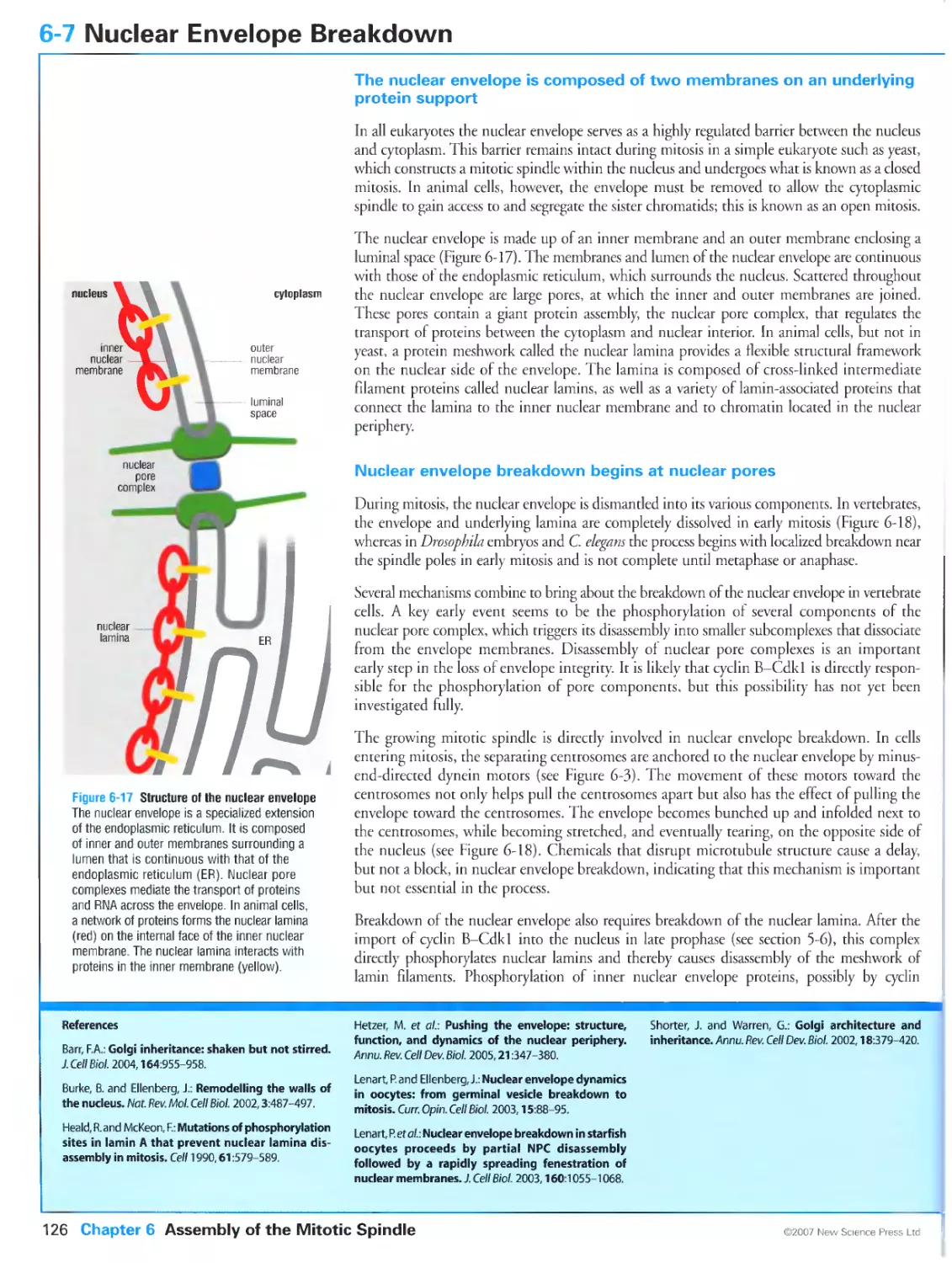
The nuclear envelope is composed of two membranes on an underlying
protein support
Nuclear envelope breakdown begins at nuclear pores
The endoplasmic reticulum and Golgi apparatus are reorganized in mitosis
6-8 Mitotic Chromosome Function in Spindle Assembly
Spindles self-organize around chromosomes
Microtubules can be stabilized by a gradient of Ran-GTP around
chromosomes
6-9 Attachment of Sister Chromatids to the Spindle
Centrosomes search for and capture kinetochores in prometaphase
Some kinetochore microtubules originate at the kinetochore
Chromosome attachment results in tension between sister kinetochores
6-10 Bi-Orientation of Sister Chromatids
Kinetochore-microtubule attachment is stabilized by tension
Aurora B is required for the correction of syntelic attachments
Merotelic attachments are processed by multiple mechanisms
6-11 Forces Driving Chromosome Movement
Multiple forces act on chromosomes in the spindle
The kinetochore is a major source of poleward force
Microtubule flux generates poleward force
A polar ejection force is generated by chromosome arms
6-12 Chromosome Congression
Chromosome oscillations in prometaphase are generated by changes in the
state of kinetochores
Microtubule flux may promote chromosome congression
CHAPTER 7 The Completion of Mitosis
7-0 Overview: The Completion of Mitosis
The final events of mitosis occur in anaphase and telophase
The metaphase-to-anaphase transition is initiated by ubiquitination and
destruction of regulatory proteins
Dephosphorylation of Cdk targets drives the events of late M phase
APCCdc20 initiates Cdk inactivation
7-1 Initiation of Anaphase: Activation of the APC
APCCdc20 activation in early mitosis is essential for anaphase to occur
Phosphorylation promotes APCCdc20 activation in early mitosis
7-2 Initiation of Anaphase: The Spindle Checkpoint
Unattached kinetochores generate a signal that prevents anaphase
The spindle checkpoint monitors defects in microtubule attachment and
kinetochore tension
7-3 Inhibition of APCcdc20 by the Spindle Checkpoint
Unattached kinetochores catalyze the formation of inhibitory signaling
complexes
The spindle checkpoint signal is rapidly turned off once kinetochores are
attached
7-4 Control of Sister-Chromatid Separation
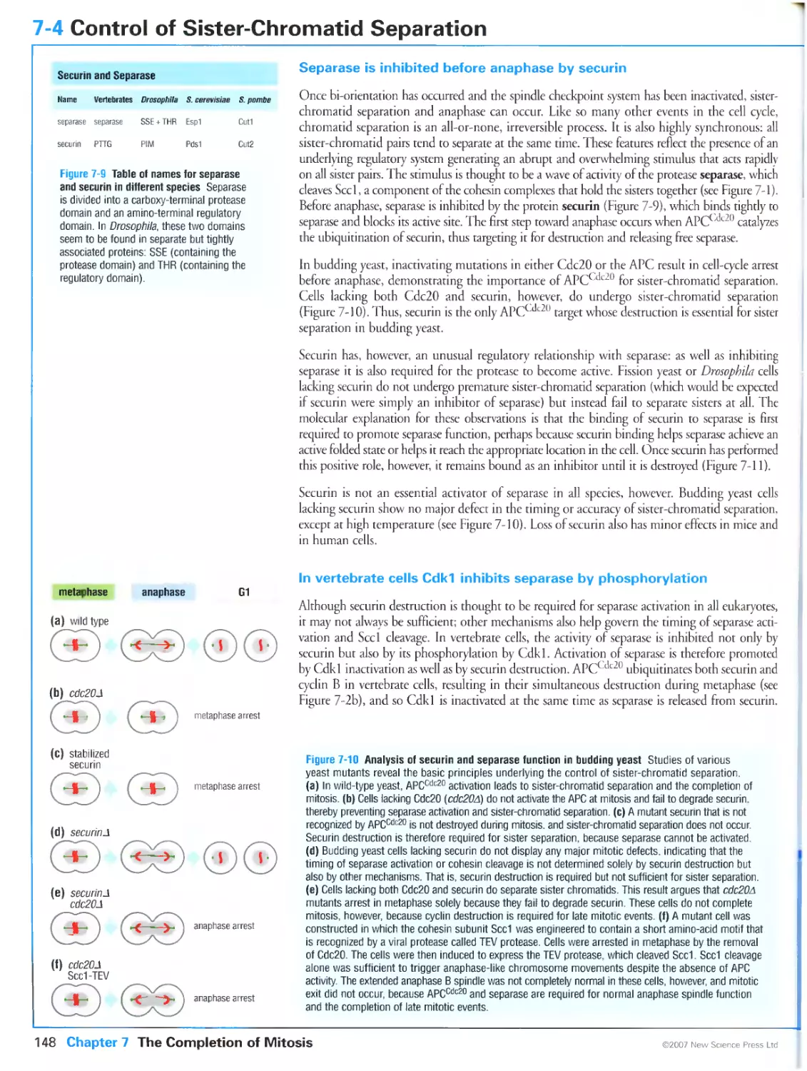
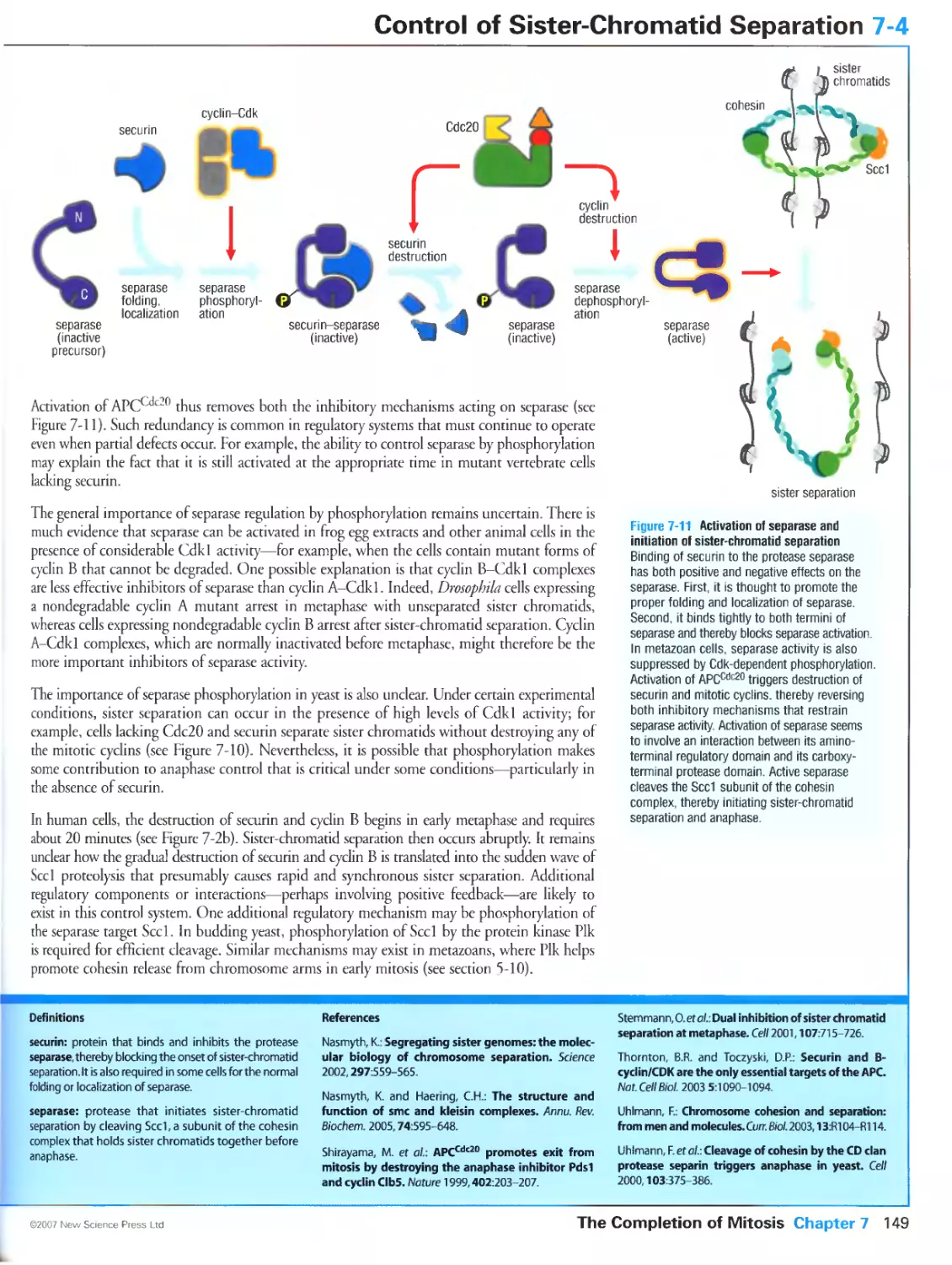
Separase is inhibited before anaphase by securin
In vertebrate cells Cdk1 inhibits separase by phosphorylation
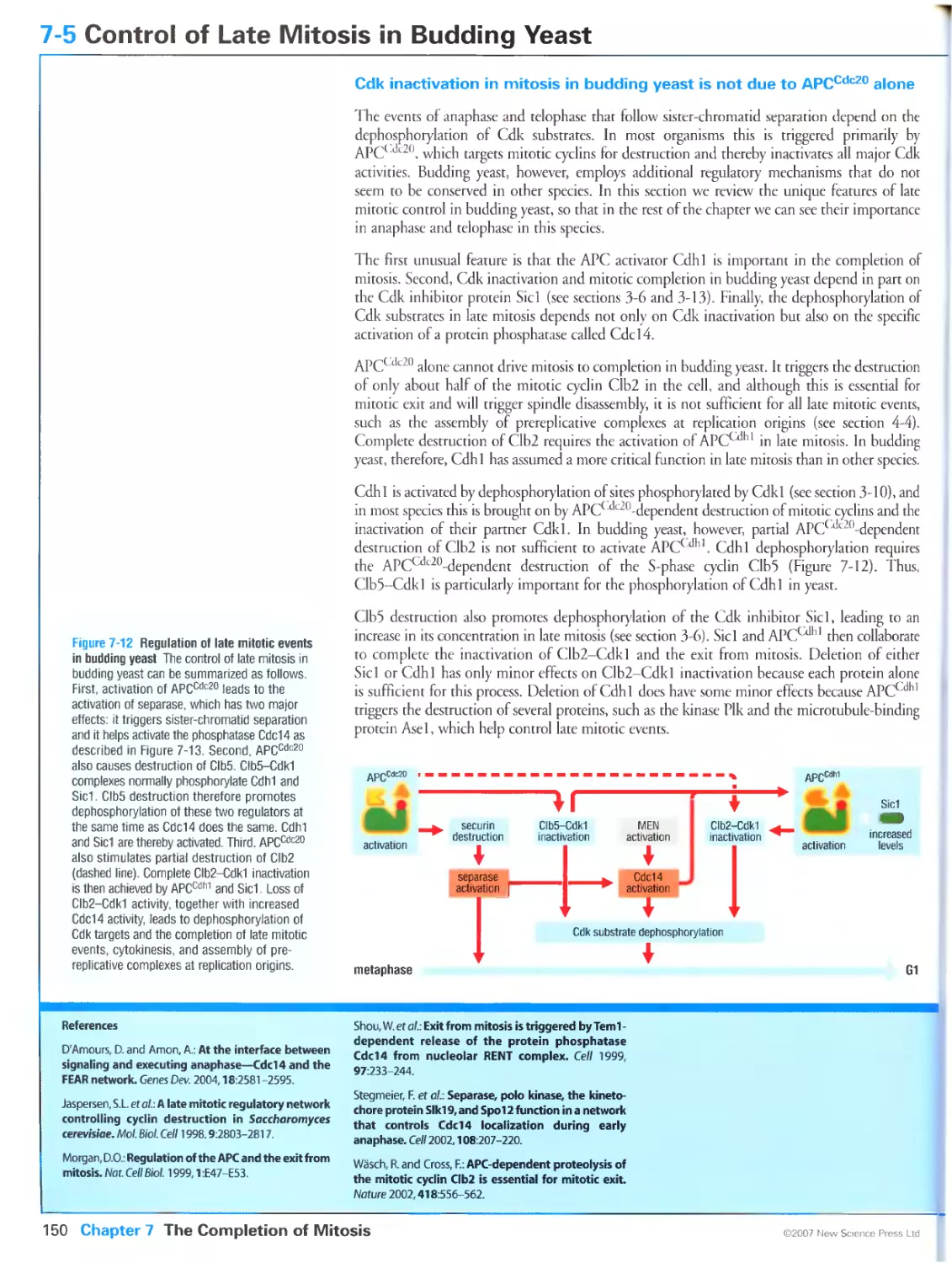
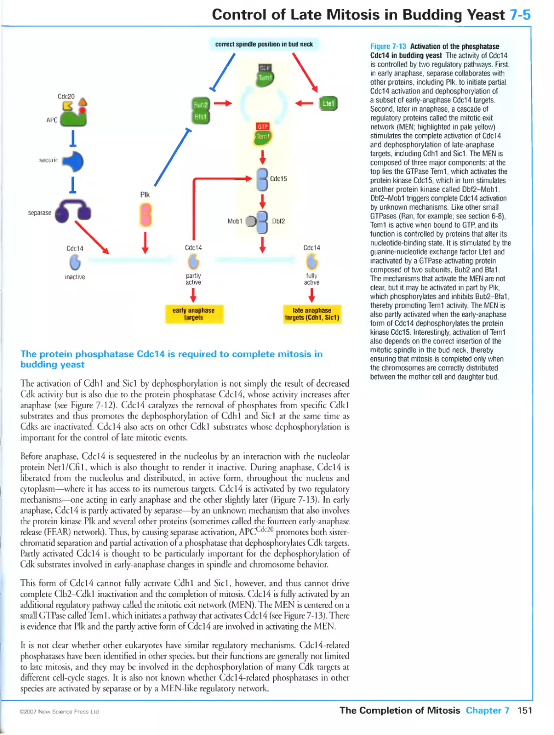
7-5 Control of Late Mitosis in Budding Yeast
Cdk inactivation in mitosis in budding yeast is not due to APCcdc20 alone
The protein phosphatase Cdc14 is required to complete mitosis in budding
yeast
7-6 Control of Anaphase Events
The anaphase spindle segregates the chromosomes
Dephosphorylation of Cdk targets governs anaphase spindle behavior
7-7 Control of Telophase
Dephosphorylation of Cdk substrates drives the final steps of mitosis
Spindle disassembly is the central event of telophase
Nuclear envelope assembly begins around individual chromosomes
CHAPTER 8 Cytokinesis
8-0 Overview: Cytokinesis
Cytokinesis distributes daughter nuclei into separate cells
Cytokinesis depends on a contractile ring and membrane deposition
The cleavage plane is positioned between the daughter nuclei
The timing of cytokinesis is coordinated with the completion of mitosis
132
134
13G
140
142
I 44
146
148
150
152
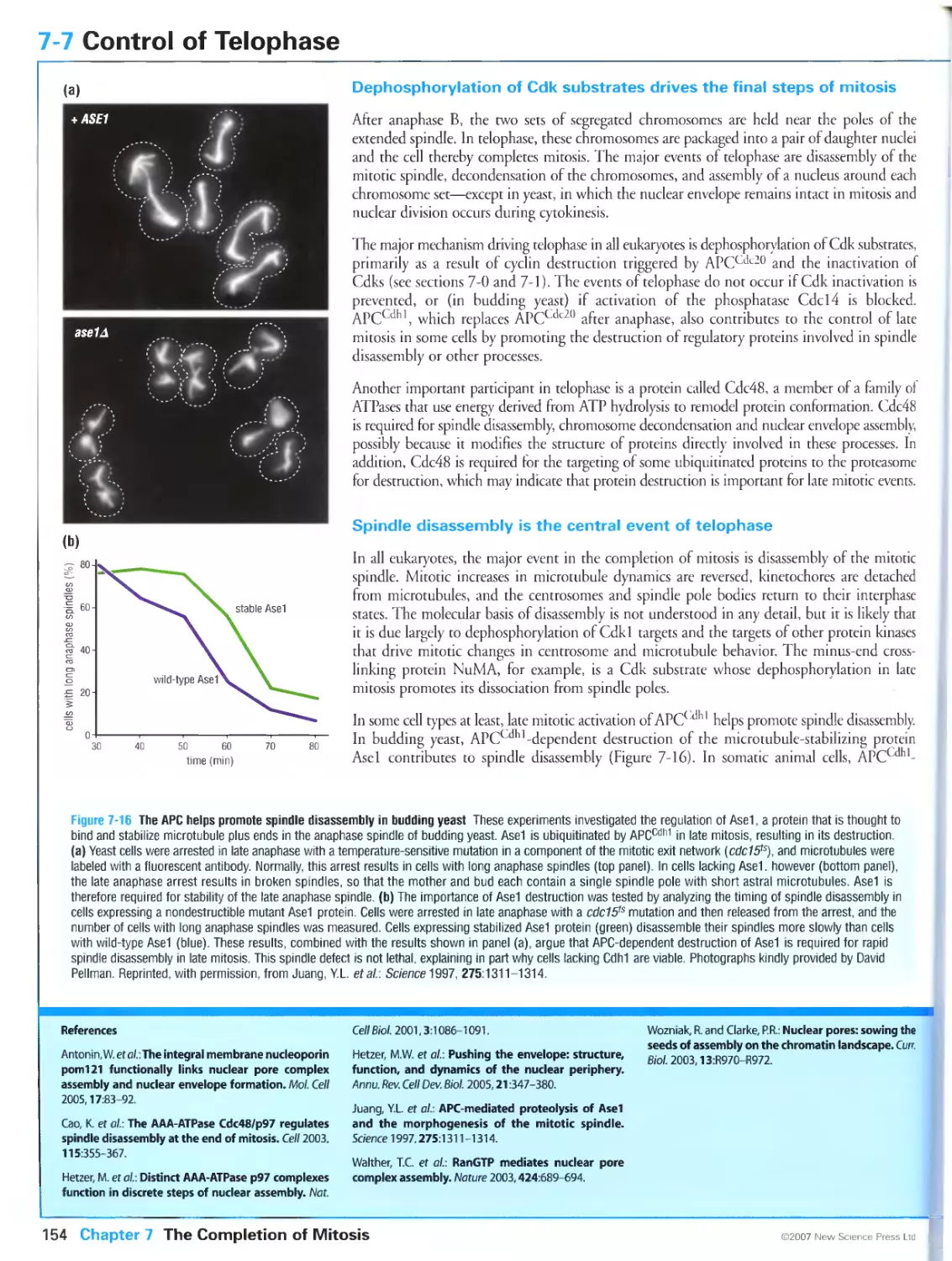
154
158
xxiii
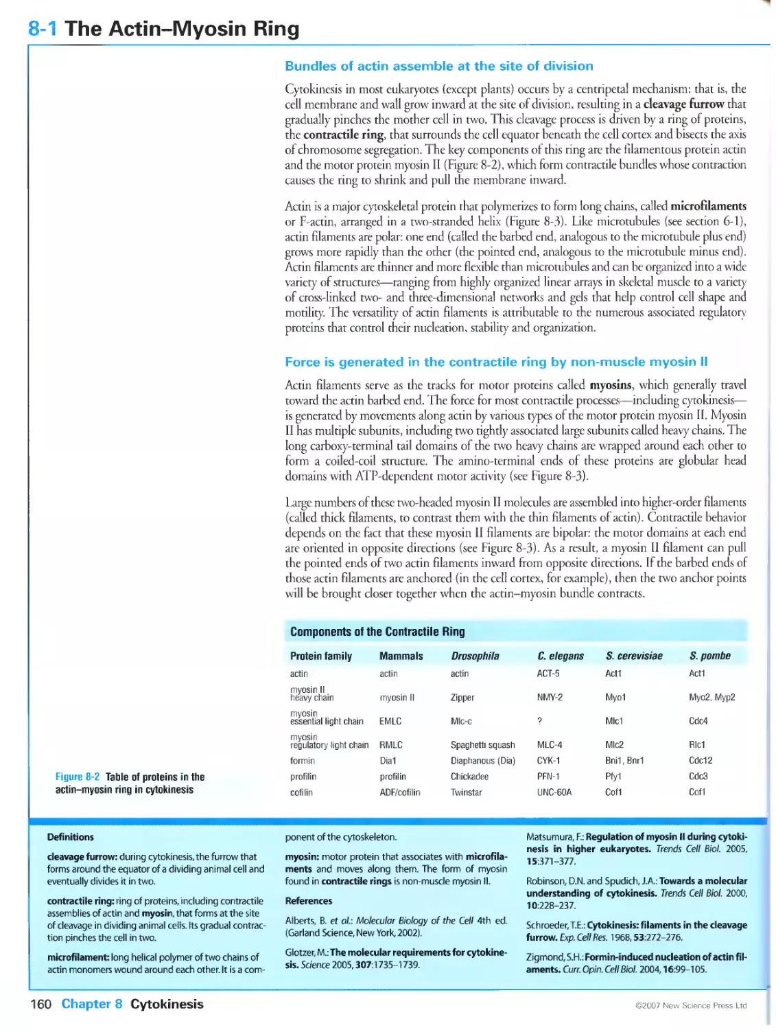
8-1 The Actin-Myosin Ring
Bundles of actln assemble at the site of division
Force is generated in the contractile ring by non-muscle myosin II
Actin filament formation depends on formins
8-2 Assembly and Contraction of the Actin-Myosin Ring
Contractile ring function depends on accessory factors whose importance
varies in different species
Contraction of the actin-myosin ring is regulated by activation of myosin II
The GTPase Rho controls actin and myosin behavior at the cleavage site
8-3 Membrane and Cell Wall Deposition at the Division Site
Membrane deposition is required during cytokinesis
Membrane addition occurs in parallel with actin-myosin contraction
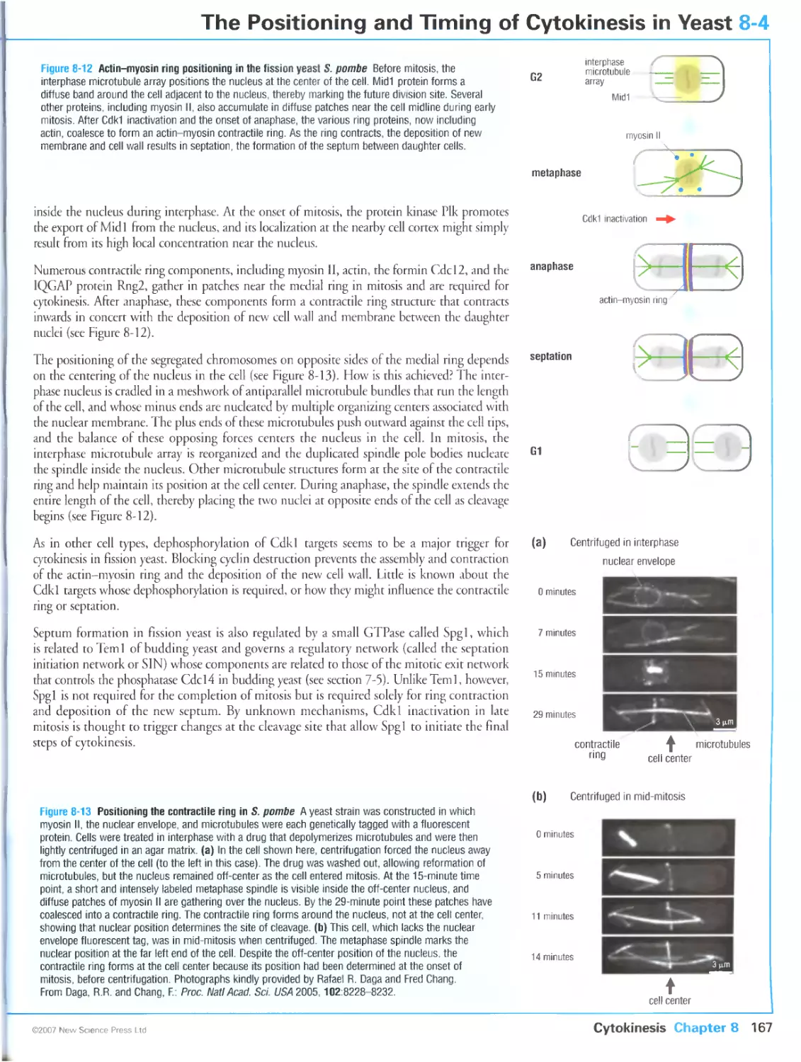
8-4 The Positioning and Timing of Cytokinesis in Yeast
Preparations for cytokinesis in budding yeast begin in late G1
Fission yeast uses the nucleus to mark the division site in early mitosis
8-5 The Positioning and Timing of Cytokinesis in Animal Cells
Signals from the mitotic spindle determine the site of cleavage in animal
cells
Multiple regulatory components at the central spindle help control
cytokinesis
Cytokinesis is coordinated with mitosis by the spindle and Cdk1 inactivation
8-6 Specialization of Cytokinesis in Animal Development
Cytokinesis can be blocked or incomplete in some stages of development
Cellularization is a specialized form of cytokinesis
8-7 Asymmetric Cell Division
Asymmetric spindle positioning leads to daughter cells of unequal sizes
Unequal forces on the poles underlie asymmetric spindle positioning
The orientation of cell division is controlled by the mitotic spindle
CHAPTER 9 Meiosis
9-0 Overview: Meiosis
Sexual reproduction is based on the fusion of haploid cells
The meiotic program involves two rounds of chromosome segregation
Homologous recombination is an important feature of meiosis
Defects in meiosis lead to aneuploidy
9-1 Regulation of Early Meiotic Events in Yeast
The meiotic program is controlled at multiple checkpoints
The transcription factor Ime1 initiates the budding yeast meiotic program
Entry into the meiotic program is driven by the protein kinase Ime2
9-2 Homologous Recombination in Meiosis
Homologous recombination is a central feature of meiotic prophase
9-3 Homolog Pairing in Meiotic Prophase
Stages of meiotic prophase are defined by cytological landmarks
Homolog pairing occurs in two successive stages
9-4 Chiasma Formation in Late Meiotic Prophase
A small number of recombination sites are selected for crossover formation
in zygotene
Crossover sites nucleate the synaptonemal complex in some species
Chiasmata appear in diplotene
9-5 Controlling Entry into the First Meiotic Division
Meiosis I is initiated by M-Cdk activity
Entry into the first meiotic division of animal cells is controlled in diplotene
Ndt80 and Cdk1 promote entry into the meiotic divisions of budding yeast
Recombination defects block entry into meiosis I
9-6 Chromosome Attachment in Meiosis I
Homolog pairs are bi-oriented on the first meiotic spindle
Homolog bl-orientation depends on cohesion of sister-chromatid arms
Homolog linkage does not involve chiasmata in some species
9-7 Chromosome Segregation in Meiosis I
Loss of sister-chromatid arm cohesion initiates anaphase I
The spindle checkpoint system helps control anaphase I
Centromeric cohesin is protected from cleavage in meiosis I
190
9-8 Finishing Meiosis
Meiosis I is followed by meiosis II
192
Partial Cdk1 inactivation occurs after meiosis I
The meiotic program is coordinated with gametogenesis
CHAPTER 10 Control of Cell Proliferation and Growth
10-0 Overview: Control of Cell Proliferation and Growth
Cell proliferation is controlled at a checkpoint in late G1
Progression through Start depends on an irreversible wave of Cdk activity
Progression through Start requires changes in gene expression
Cell division is often coordinated with cell growth
I 96
10-1 Activation of Gene Expression at Start in Budding Yeast
198
The gene regulatory proteins SBF and MBF drive expression of Start-specific
genes in yeast
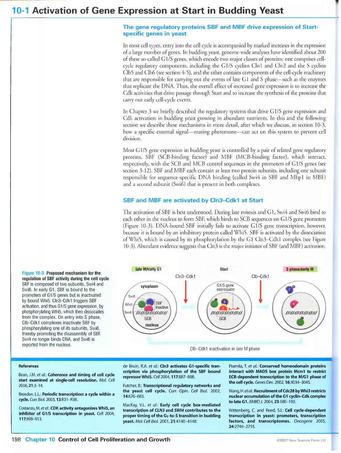
SBF and MBF are activated by Cln3-Cdk1 at Start
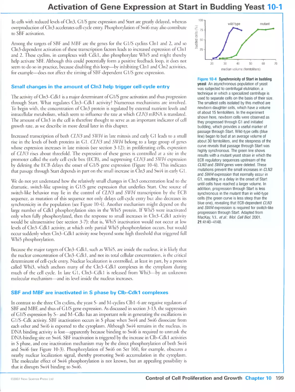
Small changes in the amount of Cln3 help trigger cell-cycle entry
SBF and MBF are inactivated in S phase by Clb-Cdk1 complexes
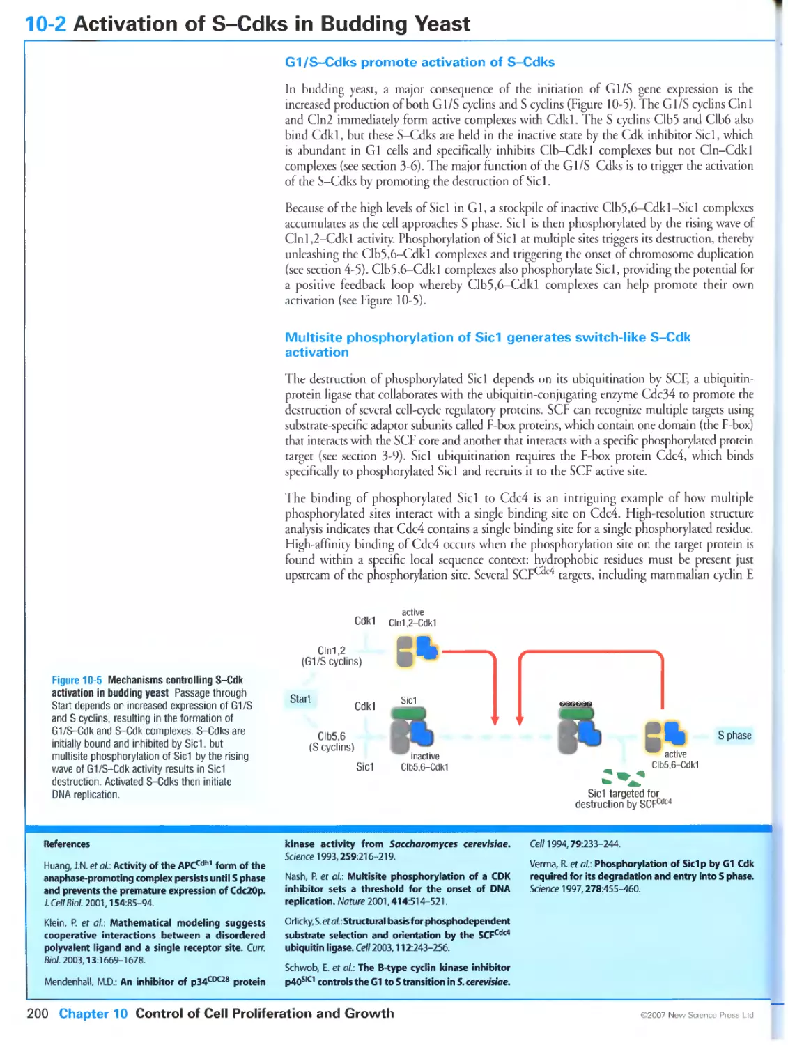
10-2 Activation of S-Cdks in Budding Yeast 200
Gl/S-Cdks promote activation of S-Cdks
Multisite phosphorylation of Sid generates switch-like S-Cdk activation
G1/S- and S-Cdks collaborate to inactivate APCCdh1 after Start
10-3 Extracellular Control of Start in Yeast: Mating Factor Signaling 202
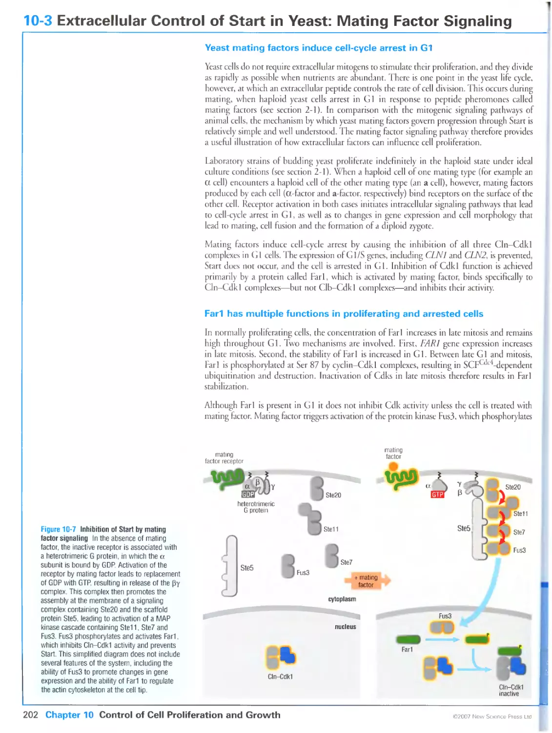
Yeast mating factors induce cell-cycle arrest in G1
Far1 has multiple functions in proliferating and arrested cells
Far1 phosphorylation is triggered by a G-protein signaling pathway
10-4 Activation of Gene Expression at Start in Animals 204
E2F transcription factors help control G1/S gene expression in animals
Stimulation of G1/S gene expression results from a combination of
increased gene activation and decreased gene repression
E2F function is regulated by pRB proteins
10-5 Regulation of E2F-pRB Complexes 206
G1/S gene expression at Start involves the replacement of repressor E2Fs
with activator E2Fs
Phosphorylation of pRB proteins releases E2F
Multiple mechanisms of E2F activation provide robust regulation of Start
10-6 Mitogenic Signaling in Animal Cells 208
Extracellular mitogens control the rate of cell division in animals
Activated mitogen receptors recruit signaling complexes to the cell
membrane
Ras and Myc are components of many mitogenic signaling pathways
Activation of the PI3 kinase helps promote mitogenesis
10-7 Activation of G1-Cdks by Mitogens 210
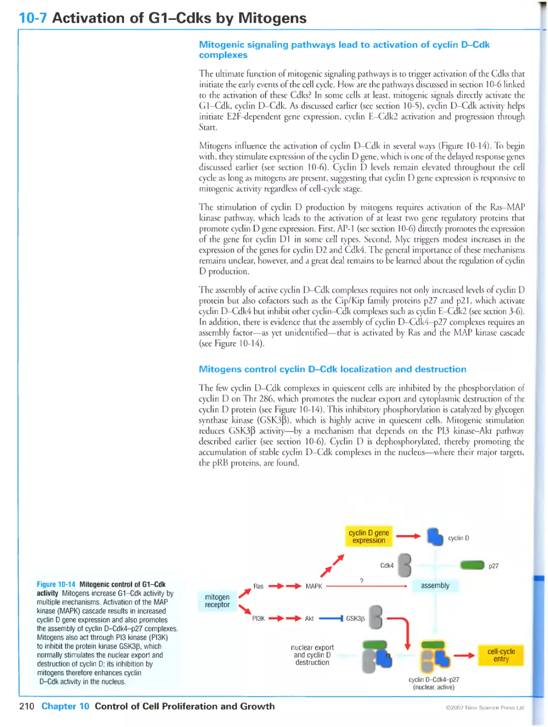
Mitogenic signaling pathways lead to activation of cyclin D-Cdk complexes
Mitogens control cyclin D-Cdk localization and destruction
Mitogens and anti-mitogens control the concentrations of Cdk inhibitor
proteins
10-8 Activation of G1/S-and S-Cdk Complexes in Animal Cells 212
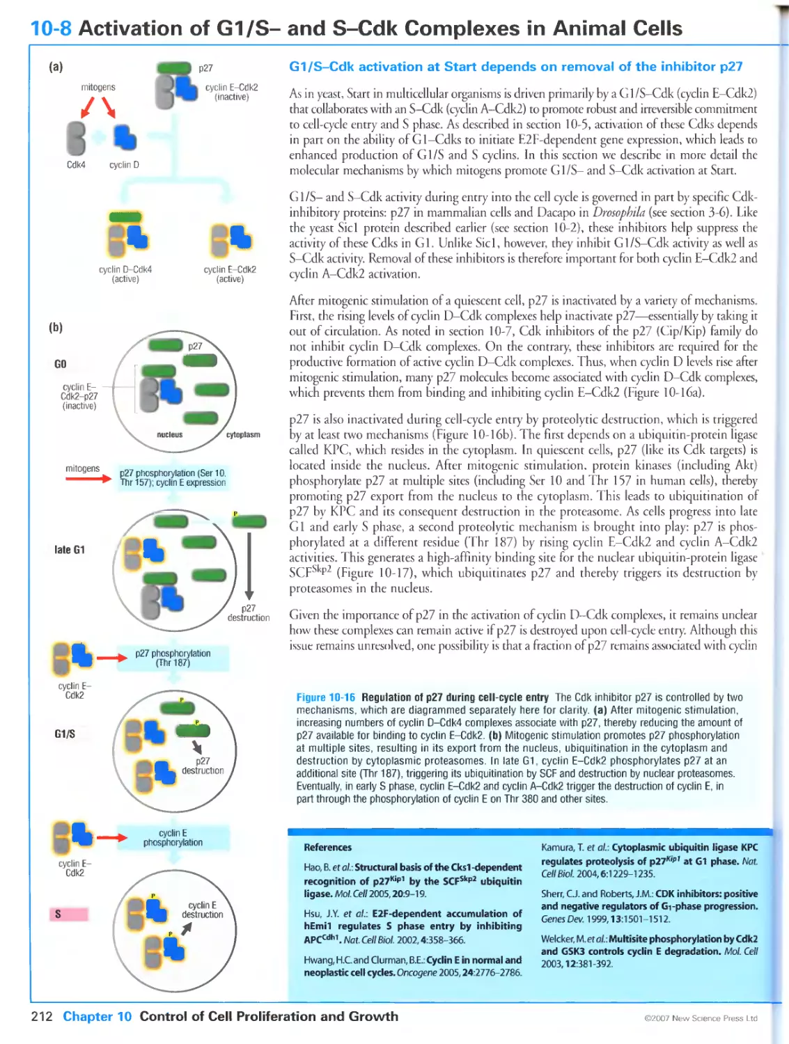
G1/S-Cdk activation at Start depends on removal of the inhibitor p27
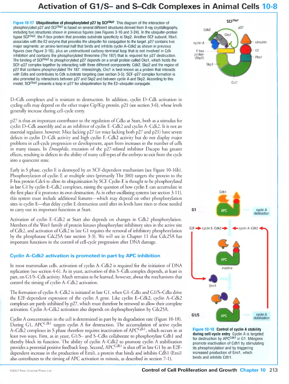
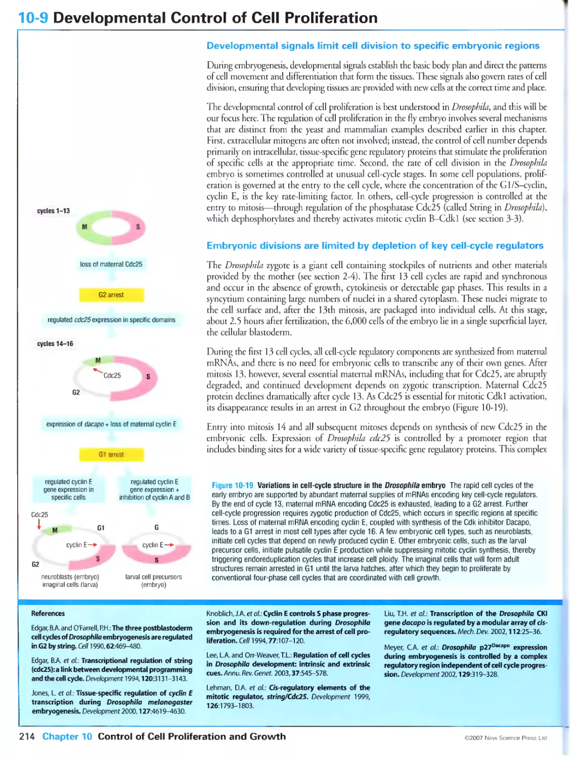
Cyclin A-Cdk2 activation is promoted in part by APC inhibition
10-9 Developmental Control of Cell Proliferation 214
Developmental signals limit cell division to specific embryonic regions
Embryonic divisions are limited by depletion of key cell-cycle regulators
10-10 Overview: Coordination of Cell Division and Cell Growth 216
Cell division and cell growth are separate processes
XXV
Cell growth is regulated by extracellular nutrients and growth factors
Cell growth and division are coordinated by multiple mechanisms
The size of a cell depends on its genomic content
10-11 Control of Cell Growth 218
Cell growth rate is determined primarily by the rate of protein synthesis
Extracellular nutrients and growth factors stimulate cell growth by activating
the protein kinase TOR
TOR affects cell growth mainly by stimulating ptotein synthesis
Growth factors stimulate protein synthesis through the activation of
PI3 kinase
10-12 Coordination of Cell Growth and Division in Yeast 220
Yeast cell growth and division are tightly coupled
Yeast cells monitor translation rates as an indirect indicator of cell size
Growth thresholds are rapidly adjustable
10-13 Coordination of Growth and Division in Animal Cells 222
Growth and division are coordinated by multiple mechanisms in animal cells
Division depends on growth in many animal cell types
Animal cell growth and division are sometimes controlled independently
10-14 Control of Cell Death 224
Animal cell numbers are determined by a balance of cell birth and death
Survival factors suppress the mitochondrial pathway of apoptosis
DNA damage and other stresses can trigger apoptosis
CHAPTER 11 The DNA Damage Response
11-0 Overview: The DNA Damage Response 228
The DNA damage response helps maintain the genome
ATR and ATM are conserved protein kinases at the heart of the DNA damage
response
Replication defects trigger a DNA damage response
11-1 Detection and Repair of DNA Damage 230
DNA can be damaged in many ways
Base and nucleotide excision repair systems repair nucleotide damage
Double-strand breaks are repaired by two main mechanisms
11-2 The DNA Damage Response: Recruitment of ATR and ATM 232
ATR is required for the response to multiple forms of damage
ATM is specialized for the response to unprocessed double-strand breaks
11-3 The DNA Damage Response: Adaptors and Chkl and Chk2 234
Protein complexes assemble at DNA damage sites to coordinate DNA repair
and the damage response
A PCNA-like complex is required for the ATR-mediated damage response
Adaptor proteins link DNA damage to activation of Chk1 and Chk2
11-4 Activation of p53 by DNA Damage 236
p53 is responsible for long-term inhibition of cell proliferation in animal cells
The major regulators of p53 include Mdm2, p300 and ARF
Damage response kinases phosphorylate p53 and Mdm2
11-5 Effects of DNA Damage on Progression through Start 238
DNA damage blocks cell-cycle progression at multiple points
DNA damage has minor effects on progression through Start in budding
yeast
DNA damage in vertebrate cells triggers a robust G1 arrest
p53 has different effects in different cell types
11-6 Effects of DNA Damage at Replication Forks 240
A DNA damage response is initiated at replication forks during S phase
ATR is the key initiator of the response to stalled replication forks
The DNA damage response stabilizes the replication fork
11-7 Effects of DNA Damage on DNA Synthesis and Mitosis 242
DNA damage in S phase blocks replication origin firing
xxvi
DNA damage blocks mitotic entry in most eukaryotes
DNA damage blocks anaphase in budding yeast
11-8 Responses to Mitogenic and Telomere Stress
244
Hyperproliferative signals trigger the activation of p53
Imbalances in mitogenic stimuli promote replicative senescence in mouse
cells
Telomere degeneration promotes cell-cycle arrest in human cells
CHAPTER 12 The Cell Cycle in Cancer
12-0
12-1
Overview: Cell-Cycle Defects in Cancer
Cancer cells break the communal rules of tissues
Cancer progression is an evolutionary process driven by gene mutation
Genetic instability accelerates cancer progression
Gene Mutations that Drive Cancer
248
250
Mutations in oncogenes and tumor suppressors stimulate tumor
progression
Oncogenes can be activated by many different mechanisms
Multiple mutations are required to cripple tumor suppressor genes
Cancer can be initiated by mechanisms other than gene mutation
12-2 Tissue Specificity in Cancer 252
Cancers are a complex group of diseases
The molecular basis of tumorigenesls can vary in different tissues
12-3 Stimulation of Cell-Cycle Entry in Cancer Cells 254
Tumor cells are independent of mitogens and resistant to anti-mitogens
G1/S gene regulation is defective in most cancers
Multiple mitogenic defects are required for tumor formation
12-4 Cell Growth and Survival in Tumors 256
Cell growth is stimulated in tumors
Tumor cells are less dependent than normal cells on survival factors
Differentiation is often inhibited in tumor cells
Tumor cells are resistant to the hyperproliferation stress response
12-5 Genetic Instability in Cancer 258
Most cancer cells have unstable genomes
Defects in the DNA damage response promote genetic instability in cancer
Genetic instability sometimes results from an increased rate of point
mutation
Chromosomal instability is the major form of genetic instability
12-6 Telomeres and the Structural Instability of Chromosomes 260
Defective DNA damage responses can lead to chromosomal instability
Degenerating telomeres can lead to chromosomal instability
12-7 Instability in Chromosome Number 262
Cancer cells often become aneuploid through a tetraploid intermediate
Cancer cells often contain excessive numbers of centrosomes
Mutations in mitotic spindle components contribute to chromosomal
instability
12-8 Cancer Progression 264
There are many genetic routes to a malignant cancer
Colon cancer progression usually begins with mutations in the gene APC
Two forms of genetic instability drive colorectal cancer progression
12-9 Stopping Cancer 266
Reducing cancer mortality begins with prevention and early diagnosis
Therapies must kill cancer cells but leave healthy cells intact
A detailed understanding of the molecular basis of cancer may lead to
rational and more specific cancer therapies
Glossary 269
References 275
Index 287
XXVII
The Cell Cycle
Cell reproduction occurs by an elaborate series of events called the cell cycle, whereby
chromosomes and other components are duplicated and then distributed into two
daughter cells. A complex network of regulatory proteins governs progression through
the steps of the cell cycle.
1-0 Overview: Cell Reproduction
1-1 Events of the Eukaryotic Cell Cycle
1-2 Variations in Cell-Cycle Organization
1-3 The Cell-Cycle Control System
1-0 Overview: Cell Reproduction
Cell reproduction is a fundamental feature of all living things
All cells arise by division of existing cells, and every cell living today is thought to be descended
from a single ancestral cell that lived 3 or 4 billion years ago. Throughout this vast period of
time, the evolution of cells and organisms—and thus the continued success of life on Earth—
has depended on the transmission of genetic infotmation by cell division.
Cell reproduction is fundamental to the development and function of all life. In single-celled
organisms, cell division generates an entire new organism. In the development of multicellular
organisms, countless cell divisions transform a single founder cell into the diverse communities
of cells that make up the tissues and organs. In the adult, cell division provides the cells that
replace those that die from natural causes or are lost to environmental damage.
Cells reproduce in discrete steps
How can a machine as complex as a cell reproduce itself, and do so with such remarkable
precision? Part of the answer is that the problem of cell teproduction is simplified—both for the
cell and for those of us who hope to understand it—by dividing the process into a series of
disrinct and more easily managed events: first, duplication of the cell's contents, and second,
equal distribution of those contents, by division, into a pair of daughter cells.
The highly regulated series of events that leads to eukaryotic cell reproduction is called the cell
cycle (Figure 1-1). The first part of the cycle, and generally by far the longest, is devoted to
duplication of the cell's components. Most of a cell's components—cytoplasmic organelles,
membrane, structural proteins and RNAs—are replicated continuously throughout the cell
cycle, resulting in the gradual doubling of cell size by the end of the cycle. The chromosomes,
however, are present in only single copies and must therefore be duplicated only once per cycle.
This occurs during a discrete stage called the synthetic or S phase. The distribution of
duplicated components into individual daughter cells occurs in a brief but spectacular final
stage called the mitotic or Mphase.
The ordering of cell-cycle events is governed by an independent control
system
The duplication and division of cellular components must be achieved with extreme precision
and reliability over countless generations. This is especially true of the genetic information
stoted in the DNA of the chromosomes, whose near-perfect transmission is so important for the
perpetuation and evolution of species. One important solution to this problem of precision is
that the machinery that carries out the events of the cell cycle—such as the enzymes of DNA
synthesis and the apparatus that segregates the duplicated chromosomes—have evolved an
astonishing level of speed and accuracy.
The fidelity of cell reproduction also depends on regulatory mechanisms that ensure that the
events of the cell cycle occur in the correct order. Chromosome duplication, for example,
should begin and end before an attempt is made to distribute the chromosomes into daughter
cells. To ensure the correct order of events, the eukaryotic cell contains a complex regulatory
network—called the cell-cycle control system—that controls their timing and coordination.
This control system is essentially a robust and reliable biochemical timer that is activated at the
beginning of a new cell cycle and is programmed to switch on cell-cycle events at the correct
time and in the correct order.
Definitions
cell cycle: sequence of events that leads to the
reproduction of the cell.
cell-cycle control system: network of regulatory proteins
that controls the timing and coordination of cell-cycle
events.
2 Chapter 1 The Cell Cycle
©2007 New Science Press Ltd
Overview: Cell Reproduction 1-0
duplication of cell contents
distribution of cell contents
into two daughter cells
Figure 1-1 The cell cycle Cell reproduction begins with duplication of the cell's components, including
the exact duplication of each chromosome In S phase. These components are then divided equally
between two daughter cells In M phase.
In its simplest form, the programming of this control system is independent of the events it
controls. In most cells, however, the order and alternation of cell-cycle events ate reinforced by
the dependence of one event on another and by feedback from the cell-cycle machinery to the
control system. Entry into M phase is dependent on the completion of DNA synthesis, for
example, ensuring that M phase occurs after S phase and does not overlap with it. The cell has
mechanisms for monitoring the progress of cell-cycle events and transmitting this information
to the cell-cycle control system. If the control system detects problems in the completion of an
event, it will delay the initiation of later events until those problems are solved.
A daunting array of problems must be overcome to achieve the accurate duplication and
distribution of a cell's contents. Chromosomes must be duplicated only once per cell cycle and
no more. Duplicated chromosomes must be separated from each other and distributed accurately
into daughter cells so that each cell gets a complete copy of the genome, and nothing more or
less. The cell cycle must be coordinated with cell growth to maintain cell size. Cell growth
and proliferation in multicellular organisms must be regulated in such a way that new cells
ate produced only when needed. In the past two or three decades, the solutions to these and
many other problems in cell division have begun to emerge, resulting in a deluge of information
that can be as confusing to those outside the field as it is enlightening to the experts within.
The putpose of this book is to synthesize this information into a concise and current overview
of the general principles underlying the events of the eukaryotic cell cycle, with an emphasis
on how these events are governed by the cell-cycle control system. We begin in this chapter
with a simple overview of cell-cycle events and key concepts in cell-cycle control. In Chapters
2 and 3 we continue with a discussion of the organisms in which the cell cycle is studied, and
the components and design of the cell-cycle control system. With these important foundations
in place, we then progress in the remainder of the book to a more detailed discussion of the
stages of cell division, beginning with chromosome duplication in Chapter 4 and cell division
in Chapters 5 to 8. Chapters 9 to 11 address a variety of related topics, until finally, in
Chapter 12, we discuss cancer, a complere understanding of which is not possible without an
understanding of all the chapters that come before.
References
Baserga, R.: The Biology of Cell Reproduction (Harvard
University Press, Cambridge, MA, 1985).
Harris, H.: The Birth of the Cell (Yale University Press, New
Haven, 2000).
Mitchison, J.M.: The Biology of the Cell Cycle (Cambridge
University Press, Cambridge, 1971 ).
Murray, A.W. and Hunt,!: The Cell Cycle: An Introduction
(Freeman, New York, 1993).
Prescott, D.M.: Reproduction of Eukaryotic Cells
(Academic Press, New York, 1976).
Wilson, E.B.: The Cell in Development and Heredity 3rd ed.
(Macmillan, New York, 1925).
©2007 New Science Press Ltd
The Cell Cycle Chapter 1 3
1-1 Events of the Eukaryotic Cell Cycle
Chromosome duplication and segregation occur in distinct cell-cycle
phases that are usually separated by gap phases
The stages of the eukaryotic cell cycle are typically defined on the basis of chromosomal events
(Figure 1-2). Early in the cell cycle, the DNA is replicated and chromosomes are duplicated in
S phase. This process begins at specific DNA sites called replication origins, which are scattered
in large numbers along the chromosomes. At these sites, proteins open the DNA double
helix, exposing it to the enzymes that carry out DNA synthesis, which move outward in both
directions from the origins to copy the two DNA strands. Chromosome duplication also
requires increased synthesis of the proteins, such as histones, that package the DNA into
chromosomes. Additional proteins are deposited along the duplicated chromosomes during S
phase, resulting in a tight linkage, or cohesion, between them. The duplicated chromosomes
are called sister chromatids.
The second major phase of the cell cycle is M phase, which is typically composed of two majot
events: nuclear division (mitosis) and cell division (cytokinesis). The period between the end
of one M phase and the beginning of the next is called interphase.
Mitosis is the complex and beautiful process that distributes the duplicated chromosomes
equally into a pair of daughter nuclei. The pairs of sister chromatids are attached in early
mitosis to the mitotic spindle, a bipolat array of protein polymers called microtubules. By the
midpoint of mitosis (metaphase), sister chromarids in each pair are attached to microtubules
coming from opposite poles of the spindle. At the next stage (anaphase), sister-chromatid
cohesion is destroyed, resulting in sister-chromatid separation. The microtubules of the spindle
pull the separated sisters to opposite ends of the cell (sister-chromatid segregation) and the two
sets of chromosomes are each packaged into new daughter nuclei.
Following mitosis, the cell itself divides by cytokinesis. Although the appearance of this process
varies greatly among different organisms, the underlying requirement in all cells is the deposition
of new plasma membrane, and new cell wall in some cases, at a position that bisects the long
axis of the mitotic spindle, ensuring that the newly separated chromosome sets are distributed
into individual cells. In many organisms, deposition of new membrane components is guided
by a conttactile ring of actin filaments and myosin motor proteins that forms beneath the cell
membrane at the site of cell division. The cell cycle is completed when contraction of this ring
pinches the cell in two.
Most cell cycles contain additional phases, known as gap phases, between S and M phases. The
first gap phase, Gl, occurs before S phase, whereas G2 occurs before M phase. Gap phases
provide additional time for cell growth, which generally requires much more time than is
needed to duplicate and segregate the chromosomes. Gap phases also serve as important
regulatory transitions, in which progression to the next cell-cycle stage can be controlled by
a variety of intracellular and extracellular signals.
Gl is a particularly important regulatory period because it is here that most cells become
committed to either continued division or exit from the cell cycle. In the presence of unfavorable
growth conditions or inhibitory signals from other cells, cells may pause for extended periods
in Gl or even enter a prolonged nondividing state, sometimes called GO (G zero). Many of
the cells in the human body, for example, are in a nondividing, terminally differentiated
state from which it is difficult or impossible to reenter the cell cycle. Such cells originate in
populations of cells called stem cells, which retain the capacity to divide.
Definitions
cytokinesis: cell division, the process in late M phase
by which the duplicated nuclei and cytoplasmic
components are distributed into daughter cells by division
of the mother cell.
GO: a prolonged nondividing state that is reached from
Gl when cells are exposed to extracellular conditions
that arrest cell proliferation.
Gl: the cell-cycle gap phase between M phase and S
phase.
G2: the cell-cycle gap phase between S phase and M
phase.
interphase: the period between the end of one M
phase and the beginning of the next.
mitosis: nuclear division, the process in early M phase
by which the duplicated chromosomes are segregated
by the mitotic spindle and packaged into daughter
nuclei.
M phase: the cell-cycle phase during which the
duplicated chromosomes are segregated and packaged into
daughter nuclei (mitosis) and distributed into daughter
cells (cytokinesis).
S phase: the cell-cycle phase during which DNA
replication and chromosome duplication occurs.
4 Chapter 1 The Cell Cycle
©2007 New Science Press Ltd
Events of the Eukaryotic Cell Cycle 1-1
centrosome
metaphase
anaphase
Cytoplasmic components are duplicated throughout the cell cycle
Most proteins, RNAs and other cellular macromolecules are synthesized continuously during
the cell cycle. Because these components are present in many copies throughout the cytoplasm,
their equal partitioning into daughter cells is readily achieved when the cell is pinched in two
by cytokinesis.
Membrane-bounded cytoplasmic organelles reproduce by continuous growth and division of
preexisting organelles. The smaller organelles, such as mitochondria and lysosomes, are present
in large numbers and are distributed evenly between daughter cells when the cytoplasm is
divided at cytokinesis, as are macromolecular complexes such as ribosomes. Larger organelles,
such as the Golgi apparatus, are thought to be fragmented into smaller vesicles during mitosis
and then distributed evenly along with the other cytoplasmic components. In some cells the
large tubular network that forms the endoplasmic reticulum also breaks down into smallet
vesicles, whereas in others it remains intact and is cut in two when the cell divides.
Not all protein components are duplicated by continuous synthesis during the cell cycle. The
centrosome of animal cells (equivalent to the spindle pole body in yeast) is a large protein assembly
whose function is to organize the long microtubules that radiate throughout the cytoplasm
during interphase and form the spindle during mitosis. The centrosome, like the chromosomes,
is present in only one copy per cell, and is therefore duplicated strictly once per cell cycle,
typically in S phase. During mitosis, the duplicated centrosomes move to opposite poles of the
cell to form the spindle, thereby allowing their distribution into daughter cells at cytokinesis
(see Figure 1-2).
Figure 1-2 The events of the eukaryotic cell
cycle The central events of cell reproduction
are chromosome duplication, which takes
place in S phase, followed by chromosome
segregation and nuclear division (mitosis)
and cell division (cytokinesis), which are
collectively called M phase. G1 is the gap
phase between M and S phases; G2 is the
gap phase between S and M phases.
Metaphase is the stage in mitosis when sister
chromatids are aligned on the mitotic spindle
and anaphase Is the stage when sister
chromatids are separated and pulled to
opposite spindle poles. In most cells, the
discrete events of the cell cycle occur against
a background of continuous cell growth.
Cell growth is usually coordinated with cell division
In most cells, the discrete chromosomal events defining the phases of the cell cycle are coupled
to the synthetic processes that duplicate the cell's mass of proteins and organelles. As a result,
the mass of the cell doubles in each cell cycle and average cell size therefore remains constant.
The mechanisms that couple cell growth and division are poorly understood.
In many cell types growth and division are not coordinated. Mammalian oocytes, neurons and
muscle cells are all capable of considerable growth in the absence of division, whereas the eatly
divisions of a fertilized animal egg occur in the absence of cell growth. These and many other
examples demonstrate that cell growth and the chromosomal cell cycle are independently
regulated processes that are coordinated in some, but not all, cell types.
©2007 New Science Press Ltd
The Cell Cycle Chapter 1
5
1-2 Variations in Cell-Cycle Organization
Cell-cycle structure varies in different cells and organisms
Although most eukaryotic cells carry out the major chromosomal events of cell reproduction
in discrete cell-cycle phases separated by gaps, the details of this basic scheme are modified
in some species and cell types (Figure 1-3). A brief discussion of these variations illustrates
some of the different strategies that are used to achieve the common goal of chromosome
duplication and segregation.
The early cleavage divisions of many animal embryos, such as those of the South African clawed
frog Xenopus Levis, rapidly subdivide the giant fertilized egg into thousands of individual cells,
allowing the organism to quickly reach a free-living state in which it can fend for itself. Cell
growth does not occur during these divisions, and gap phases tend to be minimal or absent. The
cell proceeds directly from S phase to M phase and then on to the next S phase—indeed, DNA
synthesis begins in many embryonic cells before M phase is complete. This early embryonic cell
cycle is focused entirely on rapid duplication and distribution of the genome.
In most animal cells, the two stages of M phase—mitosis and cytokinesis—occur in rapid
succession before entry into the following Gl. In some cell types, however, cytokinesis is a
relatively slow process that is not completed until long after mitosis, and can even occur after
S phase of the following cell cycle. In the early embryos of sea urchins, for example, DNA
synthesis begins in the last stage of mitosis and is completed shortly thereafter, while
cytokinesis takes longer and is not finished until after the next S phase (Figure 1-3).
Similarly, in rapidly dividing fission yeast, S phase occurs soon aftet nuclear division but
the final separation of daughter cells occurs after S phase is complete (Figure 1-3). In such
cell types, it is useful to focus on nuclear events and define the end of the cell cycle as the
completion of mitosis, not that of cytokinesis.
I I S phase I I mitosis
cytokinesis
Figure 1-3 Variations in cell-cycle structure
in different cell types The structure of the
cell cycle is diagrammed in rapidly dividing
populations of various cell types, with a time
scale beneath each diagram to indicate
approximate cell-cycle length. S phase is
colored in red, mitosis in green and cytokinesis
in blue. Gap phases are labeled where they
exist. In animal cells such as the human
somatic cell shown here, G1 is defined as the
gap between the end of cytokinesis and the
beginning of S phase; however, in some cells—
those of sea urchin embryos and fission yeast,
for example—cytokinesis is not completed until
well into the next cell cycle, and G1 is defined
as the gap, if one exists, between the end of
mitosis and the beginning of S phase.
Cytokinesis is not shown in the budding yeast
diagram for simplicity, because these cells
begin the process of budding in late G1 and
do not complete cell separation until halfway
through the next cell cycle. Cytokinesis is not
present in the early divisions of the Drosophila
embryo, In which only nuclear divisions occur,
resulting in a multinucleate syncytium.
human
somatic
Xenopus
fission
Drosophila
G1 -C
D
G 2 □
G1
G2 □
cell 0
18
hours
□ □□□□□
I I I I I I I I I I I I
I I I 1 I 1
I I I 1 I I
embryo o
I I
hours
I I
J
sea urchin
embryo o
1^— G2—4 □«■— G2—4
1 2
hours
I I
I I
G1 I I
G2
4 G1
yeast o
budding
yeast
2
hours
■GIGI
hours
embryo o
minutes
6 Chapter 1 The Cell Cycle
©2007 New Science Press Ltd
Variations in Cell-Cycle Organization 1-2
The early embryos of the fruit fly Drosophila melanogaster provide an extreme example of the
dissociation of cell division from nuclear division (Figure 1-3). The remarkably rapid cell cycles
of these embryos (about 8 minutes each) occur without cytokinesis, resulting in the formation
of a multinucleate cell, or syncytium, with thousands of nuclei in a shared cytoplasm. Only
after about 13 rounds of nuclear division does a plasma membrane form around each nucleus,
resulting in the division of the syncytial cytoplasm into individual cells.
In the majority of cells, M phase does not begin until after the end of S phase. The budding yeast,
Saccharomyces cerevisiae, is an exception to this general rule. In this organism the entry into
mitosis is not as extensively regulated as in most higher eukaryotes, and preparations for mitosis,
such as the assembly of the mitotic spindle, can sometimes begin before DNA synthesis is
completed. These cells therefore lack a clearly defined G2 between S and M phases (Figure 1-3).
Multiple rounds of chromosome duplication or segregation can occur
in the same cell cycle
Successful cell reproduction generally demands that S phase and M phase occur only once per
cell cycle. In certain specialized cell types, however, multiple rounds of S phase or M phase can
occur in a single cycle.
The meioticprogram is a specialized form of nuclear division in which two rounds of chromosome
segregation—called meiosis / and II—occur after a single round of DNA replication. As discussed
in Chapter 9, this complex process is of fundamental importance in all sexually reproducing
organisms. Most cells in these organisms are diploid: that is, they normally contain two nearly
identical versions of each chromosome. These two versions of a chromosome are called
homologs. The meiotic cell cycle reduces the number of chromosomes by half, resulting in
haploid cells carrying one homolog of each chromosome. Fusion of two haploid cells generates
a diploid zygote, completing the reproductive cycle.
Many cells in the larvae of Drosophila, as well as numerous cell types in other animals, undergo
a specialized cell cycle called endoreduplication, in which multiple rounds of S phase occur
without intervening M phases. In the Drosophila salivary gland, for example, ten rounds of S
phase give rise to cells containing a thousand copies of the genome. The increase in gene copies
that results from this process is thought to allow these cells to increase the rate of production
of numerous gene products. In some larval cells the many copies of the DNA are bundled
together in parallel, forming thick polytene chromosomes that are clearly visible under the
light microscope (Figure 1-4).
The symmetry of cell division varies in different cell types
Cell division usually gives rise to daughter cells with identical chromosomal DNA, but the
distribution of other cellular components is not always symmetrical. In budding yeast, for
example, the cytoplasmic components of the cell are divided unequally during cytokinesis,
resulting in daughter cells of unequal size.
During the development of multicellular organisms, asymmetric cell division is often used to
produce daughter cells with different functions or developmental fates. Various mechanisms
are employed in these cells to localize regulatory proteins or specific organelles at one pole of
the mother cell, resulting in their unequal distribution at cytokinesis.
Definitions
diploid: (of a cell) possessing two copies, or homologs,
of each chromosome. The somatic cells of most
multicellular organisms are diploid.
endoreduplication: the repeated replication of
chromosomes without accompanying mitosis or cell
division. This can result in large polytene chromosomes
consisting of many copies in parallel.
haploid: (of a cell) possessing one copy, or homolog, of
each chromosome. The egg and sperm cells of animals
are haploid.
homolog: in sexually reproducing organisms, either of
the two copies of each chromosome normally present
in the diploid somatic cells. For each chromosome, one
homolog is Inherited from one parent and the other
homolog from the other parent.
polytene chromosome: giant chromosome arising
from repeated rounds of DNA replication In nondividing
cells.
syncytium: multinucleate cell.
Figure 1-4 Polytene chromosomes packed
in the nucleus of a cell from the Drosophila
salivary gland A thousand identical strands
of DNA are aligned in parallel in these
chromosomes. After staining with fluorescent
dyes, dark bands reveal regions where the
DNA Is more tightly compacted by associated
protein components. The banding patterns
therefore provide useful information about
differences in chromatin structure along the
chromosomes. Courtesy of John Sedat.
zygote: in sexually reproducing organisms, the diploid
cell produced by the fusion of two haploid cells, such as
egg and sperm, from the parents.
©2007 New Science Press Ltd
The Cell Cycle Chapter 1 7
1-3 The Cell-Cycle Control System
mitosis
cytokinesis
Cell-cycle events are governed by an independent control system
The army of protein machines that execute the events of the cell cycle is undet the strict control
of a regulatory network called the cell-cycle control system. The basic features of this system
are readily apparent in the simple cell cycles of early animal embryos. There the control system
behaves like an autonomous biochemical timer that is precisely programmed to initiate cell-cycle
events in the correct order and at specific intervals, such that each event is allowed just enough
time to be completed before the next event is triggered. In these cells, the control system is
independent of cell-cycle events and continues to operate even if those events fail (Figure 1-5).
DNA synthesis
Figure 1-5 A simple cell-cycle control system
In some early embryonic cell cycles, the cell-
cycle control system behaves like an
autonomous one-handed clock whose rotation
is programmed to sequentially trigger cell-cycle
events with appropriate timing. The clock
continues to operate normally even when cell-
cycle events fail.
The cell-cycle control system is based on oscillations in the activities of
cyclin-dependent protein kinases
The central components of the cell-cycle control system are a family of enzymes called the
cyclin-dependent kinases (Cdks). Like other protein kinases, Cdks catalyze the covalent
attachment of phosphate groups derived from ATP to protein substrates. This phosphorylation
results in changes in the substrate's enzymatic activity or its interaction with other proteins.
Cdk activities rise and fall as the cell progresses through the cell cycle. These oscillations lead
directly to cyclical changes in the phosphorylation of components of the cell-cycle machinery,
resulting in the initiation of cell-cycle events. Thus, for example, an increase in Cdk activity at the
beginning of S phase causes the phosphorylation of proteins that then initiate DNA synthesis.
Cdks are activated by binding to regulatory proteins called cyclins (Figure 1-6). Oscillations
in Cdk activity during the cell cycle are due primarily to changes in the amounts of cyclins.
Diffetent types of cyclins are produced at different cell-cycle phases, resulting in the periodic
formation of distinct cyclin-Cdk complexes that trigger different cell-cycle events. A wide
range of mechanisms contributes to the control of cyclin levels and Cdk activity, resulting in a
complex Cdk regulatory network that forms the core of the cell-cycle control system.
Ì
Cdk
cyclin
activated
6*
Figure 1-6 Cyclin-dependent kinase activation
The cell-cycle control system is based on
cyclin-dependent kinases (Cdks) that are
activated at specific cell-cycle stages by
regulatory subunits called cyclins. For
simplicity, this diagram does not include
changes in Cdk phosphorylation that also
contribute to Cdk activation. These are
discussed in Chapter 3. The activated
complex is indicated by a gold outline.
Cell-cycle events are initiated at three regulatory checkpoints
The cell-cycle control system drives progression through the cell cycle at regulatory transitions
called checkpoints (Figure 1-7). The first is called Start or the Gl/S checkpoint. When
conditions are ideal for cell proliferation, Gl/S- and S-phase cyclin-Cdk complexes are activated,
resulting in the phosphorylation of proteins that initiate DNA replication, centrosome
duplication and other early cell-cycle events. Eventually, Gl/S- and S-phase Cdks also promote
the activation of M-phase cyclin-Cdk complexes, which drive progression through the second
major checkpoint at the entry into mitosis (G2/M checkpoint). M-phase cyclin-Cdks
phosphorylate proteins that promote spindle assembly, bringing the cell to metaphase.
The third major checkpoint is the metaphase-to-anaphase transition, which leads to sister-
chromatid segregation, completion of mitosis and cytokinesis. Progression through this
checkpoint occurs when M-phase cyclin-Cdk complexes stimulate an enzyme called the anaphase-
promoting complex, which causes the proteolytic destruction of cyclins and of proteins that hold
the sister chromatids togethet. Activation of this enzyme therefore triggers sister-chromatid
separation and segregation. Destruction of cyclins leads to inactivation of all Cdks in the cell,
which allows phosphatases to dephosphorylate Cdk substrates. Dephosphorylation of these
substrates is required for spindle disassembly and the completion of mitosis, and for cytokinesis.
Definitions
Cdk: see cyclin-dependent kinase.
checkpoint: regulated transition point in the cell cycle,
where progression to the next phase can be blocked by
negative signals. This term is sometimes defined to
include the signaling mechanisms that monitor cell-cycle
events and transmit the Information to the control
system; in this book the term is used to define the transition
point in the cell cycle where these mechanisms act.
cyclin: positive regulatory subunit that binds and
activates cyclin-dependent kinases, and whose levels
oscillate In the cell cycle.
cyclin-dependent kinase (Cdk): protein kinase whose
catalytic activity depends on an associated cyclin sub-
unit. Cyclin-dependent kinases are key components of
the cell-cycle control system.
G2/M checkpoint: important regulatory transition
where entry into M phase can be controlled by various
factors such as DNA damage or the completion of DNA
replication.
metaphase-to-anaphase transition: cell-cycle
transition where the initiation of sister-chromatid separation
can be blocked if the spindle is not fully assembled. Also
called the M/G1 checkpoint, but this is not an ideal term
because it does not coincide with the boundary
between M phase and G1.
Start: major regulatory transition at the entry into the
cell cycle in mid to late G1, also called the G1/S
checkpoint or the restriction point (In animal cells).
Progression past this point is prevented if cell growth Is
insufficient, DNA Is damaged or other preparations for
8 Chapter 1 The Cell Cycle
©2007 New Science Press Ltd
The Cell-Cycle Control System 1-3
G1/S- and ttk^< M-phase destruction of
11 S-Phase WL cyclin-Cdk cyclins and
IF*- cyclin-Cdk ; activation sister-chromatid
activation cohesion
G2/M metaphase-to-anaphase
Start checkpoint transition
Cell-cycle progression in most cells can be blocked at checkpoints
In the cells of the early animal embryo, the Cdk activities of the cell-cycle control system are
linked together to form a rigidly programmed oscillator that is essentially autonomous—that
is, it can generate appropriately timed waves of Cdk activity without external input. This system
is ideal for cells that must divide as rapidly as possible and are not affected by external influences.
The control system of most cell types, however, includes additional levels of regulation that
allow cell-cycle progression to be adjusted by various intracellular and extracellular signals.
Most cells, for example, initiate a new cell cycle only when stimulated by external signals, thus
ensuring that new cells are made only when needed. Similarly, initiation of cell-cycle events in
most cells is responsive to surveillance mechanisms that monitor the progress of previous
events. If the cell fails to complete DNA replication, for example, a negative signal blocks the
initiation of mitosis. Later events are thus dependent on the completion of earlier events.
To allow regulation of cell-cycle progression, the cell-cycle control system of most cells is
supplemented by molecular braking mechanisms that can be used, if necessary, to inhibit the
Cdks and other regulators that drive progression through the three major checkpoints. If
environmental conditions are not appropriate for cell proliferation, inhibitory signals prevent
activation of Gl/S- and S-phase Cdks—thereby blocking progression through Start. Similarly,
the failure to complete DNA replication blocks entry into mitosis by inhibiting M-phase
cyclin-Cdk activation. Delays in spindle assembly inhibit the proteolytic machinery that drives
the metaphase-to-anaphase transition, thereby preventing sister-chromatid segregation until the
spindle is ready. By these and numerous other mechanisms, the cell arrests cell-cycle progression
at an appropriate point when conditions are not ideal and continues it when they are.
The cell-cycle control system can thus be viewed as a linked series of tightly regulated molecular
switches, each of which triggers the initiation of cell-cycle events at a specific regulatory
checkpoint. We discuss the molecular components and design of this system in Chapter 3.
First, in Chapter 2, we review the wide range of experimental organisms in which cell-cycle
control is studied.
Figure 1-7 Overview of cell-cycle control
Progression through the cell cycle Is governed
at three major checkpoints. In mid to late G1,
Cdks are activated by G1/S- and S-phase
cyclins, resulting in entry into the cell cycle
at Start. Entry Into mitosis (at the G2/M
checkpoint) is triggered by activation of M-
phase Cdk-cyclin complexes. Finally, the
metaphase-to-anaphase transition is driven
by a regulatory enzyme called the anaphase-
promoting complex, which triggers the
destruction of cyclins and other regulators.
cell-cycle entry are not complete. Unlike cells arrested
at the G2/M checkpoint or metaphase-to-anaphase
transition, cells prevented from passing Start do not
arrest at this point but typically exit the cell cycle Into a
prolonged nondividing state from which a return to the
cycle is a lengthy process.
References
Hartwell, L.H. and Weinert, T.A.: Checkpoints: controls
that ensure the order of cell cycle events. Science
1989,246:629-634.
Morgan, D.O.: Cyclin-dependent kinases: engines,
clocks, and microprocessors. Annu. Rev. Cell Dev. Biol.
1997,13:261-291.
Murray, A.W. and Kirschner, M.W.: Dominoes and
clocks: the union of two views of the cell cycle.
Science 1989,246:614-621.
©2007 New Science Press Ltd
The Cell Cycle Chapter 1 9
2
Model Organisms in
Cell-Cycle Analysis
The fundamental features of the cell cycle have been conserved for a billion years of
eukaryotic evolution, and can therefore be studied productively in a wide range of
experimental organisms. The most commonly used model systems are unicellular yeasts,
the embryos of frogs and flies, and mammalian cells growing in culture.
2-0 Overview: Cell-Cycle Analysis in Diverse Eukaryotes
2-1 Life Cycles of Budding and Fission Yeasts
2-2 Genetic Analysis of Cell-Cycle Control in Yeast
2-3 The Early Embryo of Xenopus laevis
2-4 The Fruit Fly Drosophila melanogaster
2-5 Mammalian Cell-Cycle Analysis
2-6 Methods in Cell-Cycle Analysis
2-0 Overview: Cell-Cycle Analysis in Diverse Eukaryotes
Figure 2-1 The budding yeast
Saccharomyces cerevisiae and the fission
yeast Schizosaccharomyces pombe
(a) In a typical population of S. cerevisiae cells,
the size of the bud provides a rough indication
of the cell's position in the cell cycle: unbudded
cells are in early G1, small-budded cells in late
G1 and larger-budded cells are in S phase or
early M phase, (b) The cell-cycle stage in
S. pombe can be estimated by cell length
and by the presence of a septum, which
indicates progression through cytokinesis.
Panel (a) courtesy of Greg Tully; panel (b)
courtesy of Kathleen Gould.
Mechanisms of cell-cycle control are similar in all eukaryotes
All eukaryotic cells employ similar machinery to duplicate and divide themselves, and all have
a similar control system for the timing and coordination of cell-cycle events. It is therefore
possible to obtain a comprehensive and unified view of cell-cycle control by studying cell
division in widely different species, thereby exploiting the experimental advantages of each.
The most important systems for the study of basic cell-cycle control mechanisms are the single-
celled yeasts and the early frog embryo. This chapter provides a brief overview of these model
systems and of two others—the fruit fly and mammalian cells in culture—that are used
extensively for the study of cell growth and division in multicellular organisms.
Budding and fission yeasts provide powerful systems for the genetic
analysis of eukaryotic cell-cycle control
Yeast are small, single-celled fungi and are among the simplest eukaryotes (Figure 2-1). Two
species in particular—the budding yeast Saccharomyces cerevisiae and the fission yeast
Schizosaccharomyces pombe—have proved to be valuable model organisms for the study of cell-
cycle control. They share a number of experimental advantages, but their greatest strength is
the ease of genetic analysis. Both organisms are able to proliferate rapidly in simple culture
conditions, and both have small, fully defined genomes. Most importantly, both organisms can
proliferate in a haploid state, in which only a single copy, or homolog, of each chromosome
is present in the cell. This makes it easy to generate mutations that inactivate a gene and to
analyze them without the complications of a second gene copy. It is also relatively easy to delete
specific genes, replace them with defined mutant versions, or express them under the control
of promoters that are responsive to chemicals added to the medium.
Early animal embryos are useful for the biochemical characterization of
simple cell cycles
The giant fertilized eggs of many animals carry large stockpiles of the proteins needed for cell
division, and are capable of several rapid cell divisions in the absence of growth, gap phases
and many of the control mechanisms that operate in most cells (Figure 2-2). These early
embryonic cell cycles—particularly those of the frog Xenopus laevis—have been important in
the discovery of the core components and behaviors of the cell-cycle control system. A key
Figure 2-2 Early divisions in the frog Xenopus laevis Courtesy of James C. Smith and Huw Williams.
References
Baserga, R.: The Biology of Cell Reproduction (Harvard
University Press, Cambridge, MA, 1985).
Mitchison, J.M.: The Biology of the Cell Cycle (Cambridge
University Press, Cambridge, 1971).
Murray, A.W. and Hunt,!: The Cell Cycle: An Introduction
(Freeman, New York, 1993).
12 Chapter 2 Model Organisms in Cell-Cycle Analysis
©2007 New Science Press Ltd
Overview: Cell-Cycle Analysis in Diverse Eukaryotes 2-0
Figure 2-3 Patterns of cell division in the early embryo of the fly Drosophila melanogaster
In this image of a fly embryo In the early stages of gastrulation, the DNA in all cells is stained light
blue, while the chromosomes of cells in mitosis 14 are labeled red. Each stage in embryogenesis is
characterized by a highly programmed pattern of cell division like that seen here. Courtesy of Tony
Shermoen and Patrick O'Farrell.
advantage of eggs is their large size: the fertilized Xenopus egg, for example, is a millimetet
in diameter and is easy to inject with test substances to determine their effect on cell-cycle
ptogression. It is also possible to prepare almost pure cytoplasm from these eggs and reconstitute
many events of the cell cycle in a test tube. These cell-cycle extracts provide a simple and
highly controlled system in which cell-cycle events can be observed and manipulated.
Control of cell division in multicellular organisms can be dissected
genetically in Drosophila
Although fundamental principles of cell-cycle control are most easily uncovered in yeast and
frog embryos, some unique features of cell-cycle control in somatic animal cells are best studied
in more complex systems. An important model organism for this purpose is the fruit fly
Drosophila melanogaster, which has been particularly useful for studying mechanisms controlling
cell growth and division in developing tissues (Figure 2-3). Drosophila has a relatively short
generation time, a fully sequenced genome and well studied genetics, which can be deployed
to generate mutants and analyze gene function in vivo.
The nematode worm Caenorhabditis elegans is another model invertebrate in which the
fundamental features of metazoan biology can be revealed by genetic analysis. Considerable
insights into the mechanics of mitosis and cytokinesis have come from studies of the early
embryonic divisions of C. elegans. We will not discuss this organism in detail in this chapter, but
it will be mentioned latet in the book, particularly in our discussion of cytokinesis in Chapter 8.
Cultured cell lines provide a means of analyzing cell-cycle control in
mammals
The ultimate goal of cell-cycle research is to understand the human cell cycle, which is more
complex than that of any non-mammalian system. As experiments with intact humans are not
possible, it is necessary to study mammalian systems that can be manipulated in the laboratory.
Most commonly used are normal or tumor cells, typically mouse or human, that have been
removed from the animal and grown in plastic dishes in the presence of essential nutrients
and other factors (Figure 2-4). Cells taken from normal tissue {primary cells) cannot divide
indefinitely in culture, whereas tumor cells and other immortalized cell lines carry mutations
that allow unlimited proliferation in the culture environment.
Cell lines growing in culture often possess abnormal cell-cycle control mechanisms, in part
because these cells carry genetic defects in these mechanisms, and in part because life in cultute
medium can never fully mimic that in the cell's normal environment in the animal. Nevertheless,
cell lines are extremely useful for the biochemical and cytological characterization of features
of cell-cycle control that are unique to mammals. Studies of tumor cells in culture can also
provide important insights into mechanisms responsible for the inappropriate proliferative
behavior of cancer.
(a)
(b)
Figure 2-4 Mammalian cells growing in
culture In these photographs of a cultured
mammalian cell line, the DNA of the
chromosomes is labeled blue and the actin
filaments that help form the cytoskeleton are
labeled red. (a) A cell in interphase. The cells
shown in (b) are completing M phase.
Courtesy of Susanne Steggerda.
©2007 New Science Press Ltd
Model Organisms in Cell-Cycle Analysis Chapter 2 13
2-1 Life Cycles of Budding and Fission Yeasts
Budding yeast and fission yeast divide by different mechanisms
a haploid
a haploid
®>s
® ® ® ®
mating
a/a diploid
G1
®
starvation
The budding yeast Saccharomyces cerevisiae and the fission yeast Schizosaccharomyces pombe
are single-celled fungi with simple genomes, rapid growth rates and a long history of use in
brewing, baking and molecular genetics. Despite being on the same branch of the phylo-
genetic tree, however, the two species are only distantly related. It is thought that they
diverged in evolution several hundred million years ago and are no more related to each
other than they are to us.
A glance at budding and fission yeasts under a microscope reveals clear differences in their
external appearance and modes of division (see Figure 2-1). S. cerevisiae is an ovoid cell that is
about 3-5 Llm in diameter and encased in a tough cell wall. It divides by budding. The bud
first appears at the end of Gl and grows continuously throughout S and M phases until it
teaches a size slightly smaller than that of the mothet cell. After distribution of one set of
chromosomes into the bud at the completion of mitosis, the daughter cell pinches off the
mother cell. Bud size is thus a useful marker of cell-cycle position. S. pombe, by contrast, is a
tod-shaped cell (about 3 pirn in diameter) that grows entirely by elongation at the ends. After
mitosis, division occurs by the fotmation of a septum, or cell plate, that cleaves the cell at its
midpoint. Separation of the daughter cells is a lengthy process that is usually not complete
until the cell has entered S phase of the following cell cycle (see section 1-2). The position in
the cell cycle can be assessed by cell length and the presence of a septum.
Differences in cell-cycle control are also apparent in the two yeast species. The budding yeast cell
cycle has a long Gl but no clearly defined G2 between S phase and M phase, and entry into
mitosis is not regulated as extensively as it is in other eukaryotic model systems (see section 1-2).
Instead, the metaphase-to-anaphase transition is a more important regulatory checkpoint in this
organism. Fission yeast, by contrast, governs entry into mitosis by mechanisms that are similar
to those in multicellular animals, and studies of mitotic entry, and the control of mitotic Cdk
activation in particular, have been particularly fruitful in this organism.
Although it can be argued that S. pombe is in some respects a bettet model of animal cells
than S. cerevisiae, the latter tends to be more commonly used in laboratories studying cell-
cycle control. Budding yeast has been used for longer by more laboratoties, and studies in this
organism are therefore supported by a stronger foundation of biological and methodological
knowledge.
Yeast cells alternate between haploid and diploid states and undergo
sporulation in response to starvation
The life cycles of S. cerevisiae and S. pombe are shown in Figures 2-5 and 2-6. Both species
can proliferate in a haploid state, in which the cell carries only a single homolog of each
®
meiosis and sporulation
germination
germination
Figure 2-5 Life cycle of Saccharomyces cerevisiae Budding yeast has two mating types, a and a, that
briefly proliferate in a haploid state (top) until they encounter mating factors secreted by cells of the
opposite mating type. This triggers a G1 arrest and mating, resulting in the formation of an a/cx diploid
cell that proliferates as long as conditions are favorable. When diploid cells are starved, they undergo
sporulation. The resulting tetrad of spores, two of each mating type, germinates when conditions
improve, resulting in the formation of haploid cells. In nature these would immediately mate to
restore the diploid proliferating state, but in the laboratory haploid cultures can be maintained.
Definitions
sporulation: the formation of spores. In yeast, refers to
the formation of haploid spores from diploid cells by
meiosis in conditions unfavorable for growth and
proliferation.
tetrad: in yeast, the four haploid spores produced by
meiosis from a single diploid cell.
14 Chapter 2 Model Organisms in Cell-Cycle Analysis
©2007 New Science Press Ltd
Life Cycles of Budding and Fission Yeasts 2-1
chromosome. Haploid cells exist in two mating types (a and a in budding yeast; P and M in
fission yeast), which secrete specific mating factors or pheromones (a-factor and Ot-factor in
budding yeast, for example). When cells of opposite mating types encounter one another, these
pheromones bind to cell-surface receptors on the opposite cell and trigger arrest in Gl and the
synthesis of proteins tequired for mating. The two arrested cells then fuse to form a diploid cell
carrying two homologs of each chromosome.
In response to adverse conditions such as starvation, diploid budding yeast cells undergo
sporulation. This process begins with a specialized form of nuclear division, called meiosis,
that reduces chromosome content to a haploid state (as discussed in Chapter 9). Following
meiosis, the four haploid progeny are encased in individual cell walls, thereby forming a tetrad
of dormant spores in a tough protective container called the ascus, which protects the spores
from heat and desiccation. When conditions become favorable, the spores germinate and the
cells teturn to the haploid proliferative state.
Despite the general similarities of budding and fission yeast life cycles, the relative importance
of haploid and diploid states is very different in the two species (see Figures 2-5 and 2-6).
Fission yeast cells normally proliferate in the haploid state and mate only in starvation conditions.
The tesulting diploid cells do not proliferate but immediately undergo sporulation. Budding
yeast cells, by contrast, normally proliferate in the diploid state and undergo sporulation in
tesponse to starvation. After spore germination, haploid cells mate immediately to form new
diploids.
In both yeast species, each spore tetrad contains two cells of each mating type, allowing
immediate mating even if only a single ascus germinates. A mixture of mating types in a
population is also promoted by the process of mating-type switching, by which the progeny
of many haploid cells are converted genetically from one mating type to the other. As a result,
even the progeny of a single spore can mate and form diploids.
Experimental analysis of gene function is greatly simplified in haploid cells (as discussed in
section 2-2), and so yeast cells are generally studied in the haploid state. This is straightforward
in fission yeast, whose cells normally proliferate as haploids and do not mate while growth
conditions remain favorable. In budding yeast, however, stable proliferation of haploid cells
requires some manipulation in the laboratory. First, spore tetrads must be physically separated
with a microneedle to isolate single spores for propagation. Second, mating-type switching
must be prevented to ensure that the progeny of a single spore will be the same mating type
and unable to mate, so that they remain in a stable haploid state. Most laboratory strains
therefore carry mutations that block mating-type switching.
P haploid M haploid
® ®
®SG1 ®® ®SG1 ®®
® ^ ®
i
®
®
starvation
mating
® ®
Figure 2-6 Lite cycle of Schizosaccharomyces pombe Fission yeast normally proliferates in a haploid
state. When starved, cells of opposite mating types (P and M) fuse to form a diploid zygote that immediately
enters meiosis to generate four haploid spores. When conditions improve, these spores germinate to
produce proliferating haploid cells. Note that the cell cycle is simplified here for clarity: G1 is normally
very short or nonexistent, and cytokinesis is not normally completed until after S phase of the following
cell cycle (see Figure 1-3).
P/M diploid
meiosis and sporulation
germination
germination
References
Forsburg, Si. and Nurse, P.: Cell cycle regulation in the
yeasts Saccharomyces cerevisiae and
Schizosaccharomyces pombe. Anna. Rev. Cell Biol. 1991,
7:227-256.
Mitchison, J.M.: The Biology of the Cell Cycle (Cambridge
University Press, Cambridge, 1971).
Murray, A.W. and Hunt, T.: The Cell Cycle: An Introduction
(Freeman, New York, 1993).
©2007 New Science Press Ltd
Model Organisms in Cell-Cycle Analysis Chapter 2 15
2-2 Genetic Analysis of Cell-Cycle Control in Yeast
Cell biological processes are readily dissected with yeast genetic methods
Budding and fission yeasts are remarkably powerful tools for the analysis of cell-cycle control
and other fundamental cellular processes. Yeast cells divide rapidly in the laboratoty, with a
cell-cycle time of about 90 minutes to 2 hours, when cultured in a rich medium containing
glucose, amino acids, nucleotides and other compounds. Because yeast can be propagated in
a haploid state, it is easy to isolate and characterize mutations whose phenotypes might be
difficult to analyze in the presence of a second, wild-type, copy of the gene. Identifying the
mutant gene is straightforward in most cases, allowing the gene and its protein product to be
characterized more fully. The power of yeast also lies in their relatively small, simple and fully
sequenced genomes (about 15 million base pairs on 16 chromosomes in budding yeast; about
15 million base pairs on three chromosomes in fission yeast).
The expression of specific genes can be manipulated by a variety of methods in yeast. Most
importantly, yeast have a high rate of homologous recombination, a complex process by which the
gene sequence on a chromosome can be replaced with closely related sequences from anothet
chromosome or from an artificial DNA plasmid. Thus, it is straightforward to use targeted
recombination methods in yeast to disrupt specific genes, replace them with defined mutant
forms or integrate genes at various defined locations in the genome. Genes can also be carried on
plasmid DNA circles that can be stably propagated in the yeast cell without being integrated
into a chromosome. Whether carried on a plasmid or inserted into a chromosome, specific genes
can be expressed under the control of their natural promoter sequences or under the control of a
conditional promoter that can be regulated by the addition of chemicals to the growth medium.
Conditional mutants are used to analyze essential cell-cycle processes
Many fundamental insights into cell-cycle control have been gained from studies of mutations in
genes required for cell-cycle progression in yeast. Because these cell-division cycle {cdc) mutations
inactivate functions that are essential for cell proliferation, mutant cells can be propagated only
if the mutations are conditional: that is, the gene product is non-functional only in certain
Figure 2-7 Cell-cycle arrests in budding
yeast cdc mutants Two cdc mutants were
grown at the restrictive temperature and
analyzed by light microscopy (left panels).
Cells were stained to reveal DNA (blue,
middle) and microtubules of the mitotic
spindle (green, right). A temperature-sensitive
cdc16 mutant (top) arrests with a short pre-
anaphase spindle and chromosomes that have
not segregated, because Cdc16 (a subunit of
the APC) is required for slster-chromatid
separation. Budding and bud growth, however,
are not blocked by a cdc16 mutation (see
Figure 2-8), so a large bud is apparent in
these mutants. A cdc15 mutant (bottom)
arrests In late mitosis with segregated
chromosomes and an elongated anaphase
spindle. Cdc15 Is therefore required for the
completion of mitosis and for cytokinesis
(see Figure 2-8). Courtesy of Greg Tully.
References
Beach, D. et al.: Functionally homologous cell cycle
control genes in budding and fission yeast. Nature
1982,300:706-709.
Forsburg, Si. and Nurse, P.: Cell cycle regulation in the
yeasts Saccharomyces cerevisiae and Schizo-
saccharomyces pombe. Annu. Rev. Cell Biol. 1991,
7:227-256.
Hartwell, L.H.: Twenty-five years of cell cycle genetics.
Genetics 1991,129:975-980.
Hartwell, L.H. ef al.: Genetic control of the cell division
cycle in yeast. Science 1974,183:46-51.
Hartwell, L.H. ef al.: Genetic control of the cell-division
cycle in yeast, I. Detection of mutants. Proc. Natl Acad.
Sci.USA 1970,66:352-359.
Nurse, P.: Genetic control of cell size at cell division in
yeast. Nature 1975,256:547-551.
16 Chapter 2 Model Organisms in Cell-Cycle Analysis
©2007 New Science Press Ltd
Genetic Analysis of Cell-Cycle Control in Yeast 2-2
CDC24
CDC28
(CDK1) CDC7
CDC31
budding
chromosome
duplication
spindle pole
body duplication
CDC3,
10, 11
CDC16, CDC5,
23, 27 14, 15
chromosome
segregation
cytokinesis
spindle
assembly
entry
into next
cell cycle
specific conditions. Most cdc mutants are temperature-sensitive, so that the gene product is
functional at low temperature (the permissive condition, typically room temperature) but can
be inactivated by shifting the cells to high temperature (the restrictive condition, typically 37 °C).
After the tempetature is increased, cdc mutants continue to progress through the cell cycle until
they arrest at the point at which the gene is required for further progress (Figure 2-7).
Some cdc genes encode enzymatic and structural machinery (DNA synthetic enzymes, for
example) that carry out essential cell-cycle processes. Many cdc genes, however, encode
components of the cell-cycle control system. One of the early searches for cdc mutants in fission yeast, for
example, led to the discovery of a gene, named cdc2+, that is required for entry into mitosis. A
homologous gene, CDC28, was identified in mutant scteens in budding yeast. The products of
these genes are now known as Cdkl, the central component of the cell-cycle control system.
Early studies of cdc mutant phenotypes in budding yeast revealed that blocking early cell-cycle
events, such as DNA replication or spindle pole body duplication, blocks the onset of later
events, such as mitosis (Figure 2-8). These results provided the first clear evidence that later
events in the cell cycle are dependent on the completion of earlier events. We now know that
this dependence is due largely to surveillance mechanisms that monitor the completion of
cell-cycle events and block the cycle at specific checkpoints when these events fail.
Figure 2-8 Functions of CDC genes in budding
yeast Analysis of cell-cycle arrests in cdc
mutants provides a general outline of the
relationships between different cell-cycle events.
The names of various CDC genes are shown
above the points at which they are required for
further progress in the cell cycle. All events
flowing right from those points are blocked
In cells with mutations in the indicated genes.
Mutation of CDC7, for example, not only
blocks DMA synthesis but also prevents the
segregation of chromosomes; mitosis is thus
dependent on completion of DNA synthesis.
Spindle assembly and budding still occur in a
cdc7mutant, however, because these events
are not dependent on DNA synthesis. Similarly,
inhibition of budding (in a cdc24 mutant) does
not prevent DNA synthesis or mitosis (although
it does delay mitosis, which is not shown here).
The events and gene products shown here are
discussed In greater detail in later chapters.
Homologous genes have different names in fission yeast and budding
yeast
Fission yeast gene names are typically written in lower-case italics, with a plus sign to signify
the wild-type gene (for example cdc2+ for the wild type, cdc2-l for a mutant allele of that gene).
This convention is different from that in budding yeast, for which wild-type genes are written
in upper-case italics (for example CDC28) and mutant alleles in lower-case italics (for example
cdc28-13). The convention for protein names is less established, but they are generally not
italicized and are written with only the first letter capitalized (for example Cdc28); the letter p
is sometimes added to signify protein (for example Cdc28p).
Because cdc genes were numbered simply in the order of their isolation in mutant screens, the
name for a gene in fission yeast (for example cdc2*) will not be the same as the name applied
to the homolog of that gene in budding yeast (CDC28). Similarly, completely unrelated
genes in the two species can have the same name: fission yeast cdc25+, for example, bears no
relationship to budding yeast CDC25.
©2007 New Science Press Ltd
Model Organisms in Cell-Cycle Analysis Chapter 2 17
2-3 The Early Embryo of Xenopus laevis
The early embryonic divisions of Xenopus provide a simplified system
for cell-cycle analysis
Many animals begin life as giant fertilized eggs that are highly specialized for rapid cell division,
so that the embryo can quickly reach a stage at which it can fend for itself. Because they
contain large amounts of maternally supplied proteins, early embryos can divide rapidly without
the need fot synthesis of new RNA or protein. These simple early divisions also lack many of
the checkpoint controls found in somatic cell cycles. As a result, the study of embtyonic cell
division, particularly in the frog Xenopus laevis, has led to many important insights into the
fundamental logic and components of cell-cycle control.
Fertilization of the Xenopus egg triggers a remarkably rapid and synchronous series of 12
cleavage divisions (Figure 2-9; see also Figure 2-2). The first division, which lasts about 75-90
minutes, is followed by 11 synchronous divisions, each of which lasts only 20-30 minutes.
These rapid divisions lack gap phases and quickly subdivide the egg into the blastula, a ball of
4,000 cells. The midblastula transition then occurs, at which transcription from the embryo's
genes begins and cell divisions in different regions of the embryo slow down and become less
synchronous. Gap phases appear in the cell cycle, cell growth occurs and the cell-cycle control
system begins to assume its more complex adult form.
Studies of cell division in the pte-blastula Xenopus embryo were the first to suggest the existence
of an autonomous cell-cycle oscillator, or clock, that continues to operate with normal timing
even if cell-cycle events are severely crippled by removal of the nucleus (Figure 2-10). This
oscillator is not readily apparent in most other cell types, in which checkpoint mechanisms
arrest the clock when cell-cycle events fail to occur.
Unfertilized eggs develop from diploid oocytes by meiosis
The Xenopus egg is derived from a much smaller cell known as an oocyte. Soon after its birth
in the ovary, the diploid oocyte enters the meiotic program and completes meiotic S phase (see
Chapter 9 for a description of the stages of meiosis). It then arrests in meiotic prophase for
several months, during which it grows to a diameter of about 1 mm. In response to hormonal
cues from the pituitary gland, the follicle cells surrounding the oocyte then secrete the hotmone
progesterone, which interacts with the oocyte to initiate oocyte maturation. This process
begins with the first meiotic division, meiosis I (Figure 2-11).
Figure 2-9 Early embryogenesis in Xenopus
Fertilization of the mature egg Initiates rapid
cleavage divisions, which subdivide the
cytoplasm into smaller and smaller cells
until the embryo reaches the blastula stage
(see Figure 2-2). The complex morphogenetic
movements of gastrulation, or formation of the
gut, then occur, and the embryo is quickly
transformed into a free-swimming tadpole.
egg
fertilization
rapid cleavage divisions
blastula
tadpole
Definitions
blastula: hollow ball of cells that results from the early
cleavage divisions In some animal embryos.
cleavage: the early cell divisions of animal embryos,
which occur In the absence of growth and rapidly
subdivide the large fertilized egg into thousands of smaller
cells.
maturation-promoting factor: see MPF.
midblastula transition: in some animal embryos,
transition from development based primarily on
maternally supplied protein and RNA to development
based on transcription of embryonic genes.
mitosis-promoting factor: see MPF.
MPF: maturation-promoting factor or mitosis-
promoting factor: active complex of Cdkl and cyclln
B, which promotes the onset of meiotic maturation In
Immature oocytes and mitosis in somatic cells.
oocyte: precursor of the haploid egg cell in animals
such as frogs, flies and mammals.
oocyte maturation: the process by which a frog
oocyte arrested in meiotic prophase is induced by
progesterone to undergo meiosis I and then arrest In
metaphase of meiosis II.
polar body: small cell produced at each meiotic division
in oocytes.At division.one chromosome set remains In
the oocyte and the other is discarded in the polar body,
which is eventually resorbed.
18 Chapter 2 Model Organisms in Cell-Cycle Analysis
©2007 New Science Press Ltd
The Early Embryo of Xenopus laevis 2-3
After chromosome segregation in meiosis I, half of the chromosomes are expelled from the
oocyte into a tiny separate cell called the first polar body. The oocyte then proceeds to the
second meiotic division. After assembly of the spindle and attachment of the chromosomes,
the oocyte arrests in metaphase of meiosis II. The oocyte has now been converted to an
unfertilized egg and maturation is complete. Fertilization of the egg leads to the completion of
meiosis II and the expulsion of a second polar body. This is followed by the fusion of the
haploid egg nucleus with the haploid sperm nucleus, and the resulting diploid zygote begins
the first mitotic cell division of the early embryo.
Because of their large size, it is possible to inject Xenopus oocytes, eggs and early embtyos with
various test substances. This technique has been of great importance in the study of the cell cycle.
When a small amount of cytoplasm is removed from an unfertilized egg (in metaphase II) and
injected into an immature, fully grown oocyte (in prophase), the oocyte is released from prophase
arrest, completes meiosis I and enters meiosis II, even in the absence of progesterone. The
cytoplasmic activity that stimulates meiotic division was originally called maturation-promoting
factor (MPF) but was later found in mitotic cells, and so MPF can also signify mitosis-
promoting factor. MPF is now known to be an active complex of the protein kinase Cdkl and
the mitotic cyclin, cyclin B, key components of the cell-cycle control system (see section 1-3).
The early embryonic cell cycle can be reconstituted in a test tube
A single Xenopus female produces several thousand unfertilized eggs that can be removed and
placed in a dish. Mock fertilization, or activation, of these eggs can be achieved with electrical
stimulation, which triggers a sudden influx of calcium ions into the eggs. Activated eggs
complete meiosis II and begin a mitotic cell cycle despite the absence of a sperm nucleus.
Gentle centtifugation of activated frog eggs breaks them apart and stratifies their contents,
allowing the isolation of essentially undiluted egg cytoplasm. When Xenopus sperm nuclei
stripped of theit membranes are added to this cytoplasm in a test tube, the sperm
chromosomes décondense and are packaged in a nuclear envelope. Replication of the sperm DNA then
occurs, after which the exttacts proceed through mitosis and segregate the duplicated spetm
chromosomes. This cycle of S and M phases can repeat itself in the test tube for several rounds,
with cell-cycle lengths about twice that of a notmal embryonic cell cycle.
The ability to reconstruct an eatly embryonic cell cycle in a test tube provides an unparalleled
system for the biochemical dissection of basic cell-cycle control. Foreign proteins, chemicals or
inhibitory antibodies can be added to these extracts to test their effects on cell-cycle progression.
Specific proteins can be removed from extracts by using antibodies and then added back in
pute fotm, allowing an assessment of the protein's normal function.
S 0.7
E
0 50 100 150 200 250 30
time after fertilization, minutes
Figure 2-10 Cell-cycle oscillations in a
denucleated Xenopus egg fragment Using a
loop of fine human hair, a fertilized Xenopus egg
can be divided into two unconnected halves, only
one of which contains the nucleus. Cleavage
divisions occur in the nucleated half with normal
timing. In the non-nucleated half, divisions do
not occur, but the surface of the egg exhibits
periodic contractions, resulting in the indicated
changes in the height of the non-nucleated
fragment. These periodic contractions occur with
about the same timing as the cleavage divisions
occurring in the nucleated half of the egg (these
divisions are Indicated by the numbers 1-6).
Thus, the cytoplasm of the non-nucleated half of
the egg contains an autonomous oscillator that
continues to cycle normally even when major
cell-cycle events are prevented by removal of the
nucleus. Adapted from Hara, K. etal: Proc. Natl
Acad. Scl. USA 1980, 77:462-466.
Figure 2-11 Oocyte maturation and fertilization
in Xenopus The oocyte arrests after meiotic S
phase and grows to its final size. Progesterone
then Induces oocyte maturation: the oocyte
completes the first meiotic division (resulting in
the first polar body), enters the second meiotic
division and arrests in metaphase II. Fertilization
triggers the completion of meiosis II, resulting in
a second polar body. Fusion of sperm and egg
nuclei generates a diploid nucleus, and rapid
cleavage divisions begin. The oocyte does not
contain centrosomes, and the single centrosome
of the fertilized egg is provided by the sperm.
oocyte
oocyte growth
meiotic
prophase
maturation
metaphase
meiosis I
unfertilized
egg
1st polar
body
metaphase
meiosis II
fertilization
2nd polar
body,
completion
of meiosis II
early
embryo
fusion of sperm
and egg nuclei
References
Hara, K. et al:. A cytoplasmic clock with the same period
as the division cycle in Xenopus eggs. Proc. Natl Acad.
Sci.USA 1980,77:462-466.
Lohka, M J. et al.: Purification of maturation-promoting
factor, an intracellular regulator of early mitotic
events. Proc. Natl Acad. Scl. USA 1988,85:3009-3013.
Masui, Y. and Markert, C.L.: Cytoplasmic control of
nuclear behavior during meiotic maturation of frog
oocytes. J. Exp. Zool. 1971,177:129-146.
Murray, A.W. and Hunt, T.: The Cell Cycle: An Introduction
(Freeman, New York, 1993).
Wolpert, L. et al:. Principles of Development 2nd ed.
(Oxford University Press, Oxford, 2002).
©2007 New Science Press Ltd
Model Organisms in Cell-Cycle Analysis Chapter 2 19
2-4 The Fruit Fly Drosophila melanogaster
Drosophila allows genetic analysis of cell-cycle control in metazoans
Drosophila has several advantages as a model system for the study of cell-cycle control. It has a
2-week generation time and is grown easily in controlled laboratory conditions. The
components and specialized features of its cell-cycle control system—particularly those unique to
multicellular animals—are remarkably similar to those of humans. Most impottantly, it is
possible in Drosophila to identify and characterize genes that regulate cell-cycle progression.
The genome of Drosophila contains about 14,000 genes, about twice as many as yeast and
half the number in human cells. Numerous techniques are available for the isolation of flies
carrying defects in known genes. Genes can also be expressed ectopically in specific tissues or
under the control of inducible promoters. For the purposes of classical genetics, flies are easily
screened for mutant genes, although their diploid chromosome content makes the subsequent
analysis and isolation of these genes more complex than in haploid yeast.
nucleus
early cycles
cycle 9 (2 hours)
nuclei migrate
to surface
syncytial blastoderm
cycle 13 (3 hours)
cellular blastoderm
Figure 2-12 Early divisions in Drosophila
After fertilization, rapid nuclear divisions occur
without cytoplasmic division, resulting in the
formation of a syncytium. After about nine
nuclear divisions, nuclei begin to migrate to the
surface of the embryo to form the syncytial
blastoderm. Membranes grow Inward from the
cell surface and surround the nuclei at the end
of the 13th division, resulting in the formation
of the cellular blastoderm. About 15 pole cells
form a separate group of cells in the posterior
end of the embryo and will eventually give rise
to the germ cells.
Cells of the early Drosophila embryo divide by a simplified cell cycle
Early development in Drosophila is understood in great detail and involves several unusual cell
cycles. The fly therefore provides an excellent system in which to study changes in cell-cycle
control during the stages of embryogenesis.
The Drosophila egg is a sausage-shaped cell about 400 Llm long and 160 |im in diameter. As
in many other animals, the fly egg is stocked by the mother with abundant copies of the
components needed for cell division, so that the embryo can develop quickly—without the
need for external nutrients—into a form that can move and feed.
After fertilization, the fusion of spetm and egg nuclei results in a zygote nucleus that undergoes
a series of rapid and synchronous divisions that do not contain gap phases and last less than
10 minutes each. Because these early nuclear divisions are not accompanied by cleavage of the
cytoplasm, they rapidly convert the embryo to a syncytium, in which many nuclei share the
same cytoplasm (Figure 2-12). After about nine divisions, the nuclei move to the surface of the
embryo and continue to divide. At the end of the 13th division, membranes grow inward from
the cell surface to surround the nuclei. This latge-scale form of cytokinesis, called cellularization,
results in the formation of a layer of about 6,000 cells beneath the embryo's surface. This is the
cellular blastoderm.
Analysis of mutant phenotypes in early Drosophila embryos is complicated by the presence of
stocks of the proteins needed for progression through the first 13 divisions. These are laid
down by the mothet when the egg is being made. Only after these 13 divisions does zygotic
gene exptession become significant; thus, mutations in a zygotic gene may not produce a
discernible phenotype until the maternal gene product has been used up. Maternal proteins are
depleted at varying rates: some proteins (for example, Cdc25) are depleted rapidly in cycle 14,
others (for example, cyclins) slightly later in cycles 15 and 16, and still others (for example, the
gene regulator dE2Fl) much later in mid to late larval development. Thus, to study a gene's
function in the early embryo, it is necessary to generate mutant mothers that lay eggs deficient
in specific maternal gene products.
Gap phases are introduced in late embryogenesis
Blastoderm formation is followed by gastrulation, the process by which cell movement and
differentiation transform the simple cellulat blastoderm into a complex, multilayered embryo.
The cell cycle lengthens and assumes a more complex form. Cycles 14-16 acquire a G2, whose
length varies in different regions of the developing embryo. As a result, mitoses 14-16 are not
synchronous, but instead occur in programmed spatial and temporal patterns that are
coordinated with the complex morphogenetic movements that occur at this stage (see Figure 2-3).
The first Gl is introduced in cycle 17. Most cells in the embryo arrest at this point and
eventually enter specialized endoreduplication cycles in which they undergo multiple rounds
of DNA replication without intervening mitoses. Some Gl-arrested epithelial cells in the
embryo are programmed for a different fate: they become the imaginal cells that will give rise
to the major stiuctures of the adult fly, as we discuss next.
20 Chapter 2 Model Organisms in Cell-Cycle Analysis
©2007 New Science Press Ltd
The Fruit Fly Drosophila melanogaster 2-4
adult
Metamorphosis
pupa
3
hours
9
days
21
hours
larva
> • j
3rd instar
cellular
blastoderm
embryo
Hatching
2nd instar
1st instar
Adult fly structures develop from imaginal cells
After embryogenesis, the embryo hatches as a feeding larva. This grows rapidly, not because of
incteased cell numbet but because endoreduplicating larval cells grow to an enotmous size. The
larva goes through two molts, or instars, before forming a pupa. After metamorphosis, the
adult fly emetges (Figure 2-13).
Wings, eyes and several other adult structures are derived from small sheets of imaginal cells,
called imaginal discs, in the larva (Figute 2-14). Each disc originates as a cluster of 10-50
Gl-arrested imaginal cells in the embryo. When the larva begins to feed, these cells grow and
divide until the disc contains several thousand cells, which then differentiate into an adult
sttuctute during metamorphosis. Some adult structures, such as the gut, are derived from
imaginal cells that are not organized into discs.
The imaginal discs are useful for analyzing the role of regulatory proteins in the control of cell
proliferation. The eye imaginal disc is particularly useful for this purpose and in screens for
mutants with cell-cycle defects, because abnormal cell proliferation in this disc is not lethal but
genetates eye phenotypes that are easily observed in the adult. Another useful feature of the eye
disc is that its cells divide and differentiate during the third larval stage in a synchronous wave,
called the morphogenetic furrow, that passes across the disc—resulting in a linear array of
cells in well defined stages of division and differentiation.
Figure 2-13 Life cycle of Drosophila
melanogaster Early syncytial divisions are
followed by cellularization, resulting in the
single-layered cellular blastoderm, which is
transformed by gastrulation and other
processes into a segmented, multilayered
embryo. Hatching of the embryo gives rise to
the larva, which feeds, grows and goes through
two molts (Instars) before forming a pupa.
Metamorphosis then leads to formation of the
adult fly. An enormous amount of growth
occurs during larval development, and these
images are not drawn to scale.
larva
salivary gland
Imaginal discs
mouth
parts antenna
haltère wing genitalia
Figure 2-14 Imaginal discs in the Drosophila
larva The imaginal discs are small sheets
of epithelial cells in the larva. During
metamorphosis, they give rise to a variety of adult
structures.
Definitions
cellular blastoderm: early stage in Drosophila embryonic
development, comprising a superficial epithelial layer of
several thousand cells surrounding a yolky center.
cellularization: in insect development, the packaging
of the nuclei of the syncytial embryo into individual
cells, generating the cellular blastoderm.
gastrulation: cell movements that reorganize the
blastula (cellular blastoderm in Drosophila) into an
embryo with a gut surrounded by cell layers In place for
the development of tissues and organs.
imaginal cells: cells of the Drosophila larva that are the
precursors of adult structures.
imaginal discs: small sheets or pouches of imaginal
cells in the Drosophila embryo, which will differentiate
into major structures of the adult fly.
morphogenetic furrow: in the eye imaginal disc of
Drosophila, a shallow indentation that results from a
wave of cell differentiation that passes anteriorly across
the disc during the third larval stage.
References
Edgar, B.A. and Lehner, C.F.: Developmental control of
cell cycle regulators: a fly's perspective. Science 1996,
274:1646-1652.
Lee, L.A. and Orr-Weaver, T.L.: Regulation of cell cycles
in Drosophila development: intrinsic and extrinsic
cues. Annu. Rev. Genet. 2003,37:545-578.
Wolpert, L. et al:. Principles of Development 2nd ed.
(Oxford University Press, Oxford, 2002).
©2007 New Science Press Ltd
Model Organisms in Cell-Cycle Analysis Chapter 2 21
2-5 Mammalian Cell-Cycle Analysis
Mammalian cell-cycle control can be analyzed in cells growing in culture
30 60
time (days)
Figure 2-15 Proliferation of mouse
embryonic fibroblasts in culture Cells from a
mouse embryo were plated In a culture dish at
low density (300,000 cells) In the presence of
growth medium. Every 3 days, the cells were
transferred to new dishes at the original
density. This is the so-called 3T3 protocol of
mouse cell culture, which has been used to
generate numerous 3T3 cell lines. Cells were
counted at each transfer to measure the
number of population doublings occurring over
a period of 90 days. The cells proliferated
rapidly for the first ten generations but then
slowed their rate of division after about 15
generations, a state known as senescence.
Soon afterward, mutations led to the
emergence from the population of an
immortalized cell line that proliferated rapidly
to the end of the experiment. Adapted from
Todaro, G.J. and Green, H.: J. Cell Biol.
1963,17:299-313.
The study of cell-cycle control in mammalian cells has one clear and unassailable advantage:
direct relevance to human cells. Simple model systems are more effective for uncovering basic
mechanisms of cell-cycle control, but the specialized features of the human cell cycle can be
investigated only in cells from a human—or at least from a mouse or some other model
mammal. Mammalian cells are particularly important fot the study of the complex signaling
networks that control the rate of cell division. Although the general features of these networks
can often be revealed by genetic approaches in Drosophila, only in mammalian cells can we
observe and dissect in detail the molecular components and signaling systems that are most
relevant to humans. Understanding these systems is a key step toward the design of rational
therapies to combat cancer and other diseases of uncontrolled cell proliferation.
Mammalian cell-cycle analysis is best performed with cells that have been taken directly from the
animal and grown in plastic dishes containing essential nutrients and serum, which provides the
regulatory peptides required for cell division, growth and survival. These freshly removed cells—
called primary cells—have a limited proliferative lifespan in culture: primary human cells, for
example, stop dividing in culture after 25-50 divisions, and many rodent cell types stop dividing
after even fewer divisions (Figure 2-15). This cell-cycle arrest is called replicative senescence.
Cell senescence results from at least two general mechanisms (discussed in detail in Chapter 11).
First, many cell types (particulatly rodent cells) are thought to gradually arrest the cell cycle in
response to the nonphysiological conditions of the culture dish, in which cells lack normal
cell-cell contacts and are usually bathed in abnormal levels of serum regulatory factors. There
is evidence, for example, that many cell types can proliferate for longer periods, and sometimes
indefinitely, if provided with a culture environment that more closely mimics that of the intact
animal (Figure 2-16). Second, senescence in some cells, particularly human cells, is due to
changes in the structure of the telomere, a complex DNA-protein structure that caps the end
of each chromosome. Many human primary cells, for example, do not express telomerase, the
enzyme that is primarily responsible for maintaining telomere structure. As a result, telomeres
gradually degenerate during proliferation of these cells, until the DNA damage response system
senses the damage and triggers cell-cycle arrest (see Figure 2-16). Telomere attrition is less
important in cultured rodent cells, which often express higher levels of telomerase and have
longer and more stable telomeres.
Mutations lead to immortalization and transformation of mammalian cells
When grown for long periods in culture, cells in a population can accumulate spontaneous
mutations that prevent senescence, resulting in immortalized or established cell lines that
grow indefinitely in culture. The nature of the mutations that cause immortalization vaties
from cell line to cell line. In many rodent cells, immortalization is a relatively rapid event
(see Figure 2-15) that is achieved by mutations in components that arrest the cell cycle in
response to nonphysiological culture conditions. In human cells, immortalization is mote
difficult because it requires multiple mutations that both inactivate these components and
increase the activity of telomerase, allowing the formation of stable telomeres.
A wide range of immortal cell lines, derived from a variety of tissues, are used in cell-cycle
analysis. Although immortalized cells possess defects in certain cell-cycle checkpoint
mechanisms, they are immensely useful because, unlike primary cells, they provide an essentially
unlimited supply of a cell type that is genetically homogeneous.
Definitions
cell line: a genetically homogeneous cell population
that can proliferate Indefinitely In culture. It Is also
called an Immortalized or established cell line.
dominant negative: refers to a mutant gene product
that inhibits the function of the wild-type gene product
in a genetically dominant fashion, often by interfering
with the ability of the wild-type protein to interact with
other proteins.
knock out: to render a gene Inactive by disrupting it in
the animal, usually by replacing most of the gene with
an inactivating insertion.
primary cells: cells taken directly from the tissue of an
intact animal.They are generally susceptible to
replicative senescence after several generations of
proliferation in culture.
replicative senescence: general term for the eventual
cessation of division by primary cells when grown in
artificial culture conditions.
RNAi: see RNA interference.
RNA interference (RNAi): mechanism by which short
fragments of double-stranded RNA lead to the
degradation of homologous mRNAs.
transformed cell line: cell line that has acquired
mutations that render it independent of normal
proliferation controls, and typically capable of forming
tumors when injected into mice.
22 Chapter 2 Model Organisms in Cell-Cycle Analysis
©2007 New Science Press Ltd
Mammalian Cell-Cycle Analysis 2-5
Transformed cell lines have accumulated widespread genetic damage that not only
tesults in immottalization but also disrupts the controls that normally limit the fate of cell
proliferation. For example, transformed cells fail to stop dividing when cultured in the
absence of serum or when deprived of attachment to a culture dish; they also form tumors
when injected into mice. Such cells are not ideal for studying the normal control of cell
proliferation, but are clearly useful for the direct analysis of molecular defects underlying
the cancer phenotype.
Specific gene disruption is the ideal approach for assessing protein
function in mammalian cells
A variety of methods are available for performing cytological and molecular analyses of cell-cycle
control in cultured cell lines. The large size of mammalian cells makes them ideal for detailed
microscopic analysis of cell-cycle events and for studying the subcellular location of regulatory
molecules. Plasmid-borne foreign genes are readily expressed from natural or inducible
promoters, and can be integrated into chromosomes to produce stable cell lines expressing various
mutant versions of the desired protein.
The major weakness of mammalian cells as an experimental system is the lack of rapid and
specific methods for blocking gene and protein function in the intact cell. Proteins are typically
inhibited in the cell by injection of inhibitory antibodies or by the overexpression of a mutant
form of the protein—called a dominant negative—that interferes with the function of the
notmal protein. These methods rarely offer the high specificity and rigorous intetpretations
possible with specific mutations of the gene encoding the protein.
A more specific approach to disrupting gene expression in mammalian cells is RNA interference
(RNAi), which triggers the destruction of a targeted mRNA in the intact cell. Cultuted
cells are treated with short double-stranded RNA fragments (called short intetfering RNAs
or siRNAs) carrying part of the sequence of the target RNA. With assistance from cellular
proteins, the antisense strand of the siRNA targets the desired mRNA and cellular enzymes
destroy it, thus preventing its translation and reducing the amount of the target protein in
the cell.
Although more difficult than in yeast, specific gene disruption can be achieved in mammalian
cells and in the whole animal. The standard approach is homologous recombination using
specialized DNA vectors that target and disrupt, ot knock out, the desired gene in cultured
cells. This procedure is most frequently applied to cultured mouse embryonic stem cells (ES
cells), enabling strains of mice with the target gene knock-out to be subsequently generated by
transgenic techniques. It is then possible to assess the effect of the gene deletion in the context
of the whole animal, which is particularly important in the study of cell-cycle regulatory
proteins involved in the control of cell proliferation and cancer. The role of the disrupted
gene in individual cells can also be analyzed in primary cell cultures, typically of embryonic
fibroblasts, taken from the transgenic embryo.
However, gene disruption cannot be used to study gene products that are essential for cell-cycle
progression. There are no simple methods for generating conditional mutant alleles of essential
genes in mice ot in mammalian cell lines, although RNAi can sometimes be a useful approach.
In general, it is still difficult to apply rigorous genetic approaches to determining the normal
function of essential genes in mammals.
(a)
40
.1 30
» 20
10
0
(b)
160
" 120
feeder layer
plastic substrate
time (days)
telomerase
Y I shift s-
i» —* I to plas
shift some cells
to plastic substrate
0 80 160 240 320 400 4i
time (days)
Figure 2-16 Effects of culture conditions and
telomerase expression on human cell
proliferation (a) Human keratlnocytes (an
epithelial cell type from skin) were grown in
plastic culture dishes in the presence of a
chemically defined medium (blue dots), leading
to cell-cycle arrest after about 12-15
generations. In a parallel experiment, another
batch of keratlnocytes was cultured in dishes
precoated with a layer of fibroblast cells, known
as a feeder layer, in an attempt to better mimic
the normal environment of the body (red dots).
Keratlnocytes growing on this layer did not
stop dividing until about 35 generations,
revealing the importance of culture conditions
in the timing of senescence, (b) Human
keratinocytes were cultured on a feeder layer of
fibroblasts as In (a), until their rate of division
began to slow (after about 35 generations). The
cells were then engineered to produce human
telomerase (red arrow). As a result, the
population continued to proliferate indefinitely
(red dots), indicating that telomere
degeneration is the primary cause of
senescence In human cells growing on feeder
layers. After 280 days (blue arrow), some cells
were transferred to uncoated plastic dishes
instead of feeder layers, causing a cell-cycle
arrest (blue dots). Thus, telomerase alone does
not Immortalize cells growing under suboptimal
culture conditions. Adapted from Ramirez, R.D.
era/.: Genes Dev. 2001,15:398-403.
References
Novina, CD. and Sharp, P.A.: The RNAi revolution.
Nature 2004,430:161-164.
Ramirez, R.D. et ai: Putative telomere-independent
mechanisms of replicative aging reflect inadequate
growth conditions. Genes Dev. 2001,15:398-403.
Todaro, G.J. and Green, H.: Quantitative studies of the
growth of mouse embryo cells in culture and their
development into established lines. J. Cell Biol. 1963,
17:299-313.
©2007 New Science Press Ltd
Model Organisms in Cell-Cycle Analysis Chapter 2 23
2-6 Methods in Cell-Cycle Analysis
1fl 2n
DNA content
Figure 2-17 Analysis of cellular DNA content
by flow cytometry A large population of
haploid budding yeast cells was incubated
with a fluorescent DNA dye and then passed
through a flow cytometer, which measures the
fluorescence content of each cell in the
population. The number of cells with specific
DNA contents is plotted as shown here. In an
asynchronous, rapidly proliferating cell
population, two peaks of cells are normally
apparent. Cells in the left peak contain a single
copy of each chromosome and are therefore
in G1. Cells in the right peak contain twice as
much DNA and are therefore in G2 or early M
phase. Cells with DNA contents between the
two peaks have partly duplicated their DNA
and are therefore in S phase. The number of
cells in the various positions reveals the
fraction of cells In different cell-cycle phases.
The actual chromosome content of cells will
depend on whether the cells are haploid or
diploid. The left peak in a haploid cell
population, as shown here, represents a
haploid state (called 1n), whereas the right
peak represents the temporary diploid state
(2n) in G2/M cells before they undergo
mitosis. The chromosome content of a diploid
population (see Figure 2-18) is doubled from
diploid (2n) in G1 cells to tetraploid (An) in
G2/M cells. Courtesy of Liam Holt.
Cell-cycle position can be assessed by many approaches
Experimental analysis of the cell cycle generally requires a way of determining the cell-cycle stage
of the cells being studied. The simplest approach is conventional light microscopy. As
mentioned earlier (see section 2-1), the cell-cycle position of budding yeast and fission yeast can be
estimated by the size of the bud and the length of the cell, respectively. Cultured mammalian
cells tend to remain flat and attached to the dish during most of the cell cycle, but cells in M
phase often reveal themselves by reducing their attachments and becoming round and refractile.
Microscopic analysis is made easier and more precise by fluorescent labeling of specific cellulat
components such as the chromosomes or mitotic spindle. Numerous fluorescent DNA dyes
are available and can be added to cells that have been chemically fixed on a microscope slide.
In a mammalian cell population, such dyes deafly teveal the condensed chromosomes of mitosis,
and thus can be used to measure the mitotic index—the fraction of cells in a population
that are in mitosis. Immunofluorescence methods are also very useful in cell-cycle analysis.
Fixed cells can be incubated with an antibody that recognizes a specific cellular component,
such as the mitotic spindle, and these antibodies can then be detected by a fluorescently
tagged secondary antibody. Protein structures can also be fluorescently labeled in living cells.
Typically, the gene encoding the desired protein is joined by molecular genetic methods to the
gene encoding green fluorescent protein (GFP), a naturally fluorescent protein from jellyfish.
The result is a fluorescent fusion protein that labels the desired intracellular structure. Such
methods allow the continuous microscopic observation of the spindle or other structure in cells
traversing the cell cycle.
Another powerful method in cell-cycle analysis involves the ptecise measurement of cellular
DNA content by flow cytometry. A large population of cells is treated with a fluorescent
DNA dye and then injected into an instrument called a flow cytometer, which can rapidly
measure the fluorescence intensity—and thus the DNA content—of every cell in the population
(Figure 2-17). Cells in S phase can also be specifically labeled by treatment with bromo-
deoxyuridine (BrdU), an analog of the nucleotide thymidine. DNA that incorporates this
nucleotide during S phase can be detected with fluorescent antibodies against BrdU, and a
microscope or flow cytometer can reveal the fraction of cells that are actively synthesizing
DNA during the treatment period.
Cell populations can be synchronized at specific cell-cycle stages
It is often necessary in cell-cycle research to synchronize a population of cells at a particulat
stage in the cell cycle, so that the cellular or biochemical features of that stage can be analyzed.
There are various methods for doing this. In general, these involve treatment of the cells with
a chemical or hormone, or a change in an environmental condition (such as temperature in
the case of tempetatute-sensitive mutants) that blocks cell-cycle progression at some specific
point. Ideally, these arrests are reversible, so that removal of the arresting condition allows the
progression of cells into the next cell-cycle stage.
Many cultured mammalian cells can be arrested in a quiescent Gl-like state by depleting the
culture medium of serum factors that drive growth and division. Adding serum back to the
medium results in the gradual progression of cells back into the cell cycle and S phase.
Similarly, budding yeast cells can be arrested reversibly in a Gl-like state by treatment with
mating pheromone. The quiescent Gl state produced by these treatments is different from the
relatively ttansient Gl state that occurs in cells traversing directly from mitosis to S phase.
Definitions
double thymidine block: a method for synchronously
arresting a mammalian cell population at the beginning
of S phase. Asynchronous cells are treated with thymidine,
which causes arrest throughout S phase. These cells are
released from arrest to allow progression out of S phase. A
second thymidine treatment is then used to arrest all cells
at the beginning of the subsequent S phase.
FACS: see fluorescence-activated cell sorter.
flow cytometer: instrument through which a stream of
cells is passed and their fluorescence measured in the
technique of flow cytometry.
flow cytometry: technique used to enumerate and
analyze a sample of cells by incubating them with one or
more fluorescently labeled antibodies and/or other
molecules that can bind to cellular components and
measuring the fluorescence Intensity of each fluor for each
cell. It is used to count the numbers of cells of different
types, or at different stages in development or the cell
cycle.
fluorescence-activated cell sorter (FACS): a modified
flow cytometer that sorts individual cells into different
containers according to their fluorescence.
mitotic index: fraction of cells in a population that are
undergoing mitosis.
24 Chapter 2 Model Organisms in Cell-Cycle Analysis
©2007 New Science Press Ltd
Methods in Cell-Cycle Analysis 2-6
Chemical inhibitors of DNA synthesis and spindle assembly are also used to synchronize cell
populations at specific points in the cycle (Figure 2-18). Numerous drugs, including thymidine
and hydtoxyutea, block DNA synthesis by inhibiting the synthesis of specific nucleotides,
resulting in a reversible arrest in S phase with partly synthesized DNA. Drugs that inhibit
microtubule function, such as nocodazole and benomyl, block the normal assembly of a
spindle, which causes cells to arrest in early mitosis.
These methods suffer from two problems. First, cell-cycle ptogtession never occurs at the same
rate in all cells in the population, and so the synchrony of cell-cycle progression is lost soon
after release from an arrest point. This is especially true of the prolonged process by which
serum-starved mammalian cells return to the cycle. Second, artificially induced arrests, such as
those produced by conditional mutations or inhibitors of DNA synthesis and spindle function,
are nevet equivalent to the natural state of a cell passing through that stage of the cell cycle. Cells
arrested by DNA synthesis inhibitors, for example, stop teplicating their DNA but continue
to grow and may also continue to make preparations for mitosis.
Non-invasive methods are therefore preferable in some cases. Useful information can often be
gained simply from microscopic analysis of single cells in an untreated, asynchronous population.
Alternatively, cells in specific stages can be purified from an asynchronous population by gentle
methods. In the technique of centrifugal elutriation, for example, a specialized centrifuge is used
to separate cells on the basis of their size: this allows large numbers of newly formed Gl cells to
be obtained from an untreated cell population. Partly purified subpopulations of cells can also be
obtained with a fluorescence-activated cell sorter (FACS), a flow cytometer that sorts cells into
different test tubes on the basis of their fluorescence content. If cells are labeled with a DNA dye,
for example, then cells with G1 DNA content can be separated from cells with G2/M DNA
content. Finally, as mentioned above, cultured mammalian cells typically lose their attachment
to the plastic dish during mitosis, and so unperturbed mitotic cells can be released into the
medium of these cells by tapping the culture dish; this is called mitotic shake-off.
Complete understanding of cell-cycle control mechanisms requires the
analysis of protein structure and enzymatic behavior
The future of cell-cycle research lies not only in studies of cells and organisms but also in the
analysis of protein structure and function. Many components of the cell-cycle control system
are now being analyzed at the level of high-resolution three-dimensional structure, and these
structural studies are accompanied by increasingly sophisticated studies of the chemical
mechanisms undetlying the function and regulation of the many enzymes that drive the cell through
the steps of reproduction. In addition, considerable effort is being devoted to understanding
how the many components of the cell-cycle control system interact in networks that generate
complex behaviors—autonomous Cdk oscillations, for example. These important issues lie at
the heart of cell-cycle control and form the central theme of the next chapter.
DNA content
Figure 2-18 Synchronous progression through the cell cycle A human HeLa cell line was treated with a double thymidine block to arrest cells at the beginning
of S phase. This method begins with treatment of cells with thymidine, which Inhibits DNA synthesis and results in a cell population that is arrested throughout
S phase. Cells are then released from this arrest for several hours. After all cells have completed S phase, they are treated with thymidine for a second time. All
cells then arrest at the beginning of the next S phase. In the experiment shown here, the second thymidine treatment was washed out at time zero, and DNA
content was then measured in the population at the indicated times. The cells start with unreplicated (2/7) DNA content and then progress through S phase and
achieve a 4/7 DNA content after about 8 hours. Progression through M phase results in an abrupt return to a 2n DNA content after 12 hours, and the cells then
enter the next cell cycle. After 24 hours, the cell population is becoming asynchronous. Courtesy of Pei Jin.
References
Baserga, R.: The Biology of Cell Reproduction (Harvard
University Press, Cambridge, MA, 1985).
Mitchison, J.M.: The Biology of the Cell Cycle (Cambridge
University Press, Cambridge, 1971).
Murray, A.W. and Hunt, t.: The Cell Cycle: An Introduction
(Freeman, New York, 1993).
©2007 New Science Press Ltd
Model Organisms in Cell-Cycle Analysis Chapter 2 25
The Cell-Cycle Control System
The cell-cycle control system is composed of a series of biochemical switches that trigger
the events of the cycle in the correct order. The key components of this system are the
cyclin-dependent kinases and their regulators, which are assembled into a robust and
versatile regulatory network that is responsive to a variety of intracellular and extracellular
information.
3-0 Overview: The Cell-Cycle Control System
3-1 Cyclin-Dependent Kinases
3-2 Cyclins
3-3 Control of Cdk Activity by Phosphorylation
3-4 The Structural Basis of Cdk Activation
3-5 Substrate Targeting by Cyclin-Cdk Complexes
3-6 Cdk Regulation by Inhibitory Subunits
3-7 Biochemical Switches in Signaling Systems
3-8 Switch-Like Activation of Cdkl
3-9 Protein Degradation in Cell-Cycle Control
3-10 The Anaphase-Promoting Complex
3-11 Assembling and Regulating a Cell-Cycle Oscillator
3-12 Transcriptional Control of Cell-Cycle Regulators
3-13 Programming the Cell-Cycle Control System
3-0 Overview: The Cell-Cycle Control System
The cell-cycle control system is a complex assembly of oscillating
protein kinase activities
The cell-cycle control system is the regulatory network that controls the order and timing of
cell-cycle events. A series of biochemical switches triggers progression through the three major
regulatory checkpoints of the cell cycle: Start, which defines the entry into the cycle in late Gl;
the G2/M checkpoint, where entry into mitosis is controlled; and the metaphase-to-anaphase
transition, where the final events of mitosis are initiated. We will see in this chapter how the
biochemical switches that comprise the cell-cycle control system are put togethet and regulated.
The central components of the cell-cycle control system are the cyclin-dependent kinases
(Cdks). As the cell progresses through the cycle, abrupt changes in the enzymatic activities of
these kinases lead to changes in the phosphorylation state, and thus the state of activation, of
proteins that control cell-cycle processes. Concentrations of Cdk proteins are constant
throughout the cell cycle; oscillations in their activity depend primarily on corresponding
oscillations in levels of the regulatory subunits known as cyclins, which bind tightly to Cdks
and stimulate their catalytic activity. Different cyclin types are produced at different cell-cycle
stages, resulting in the fotmation of a series of cyclin-Cdk complexes. These complexes
govern distinct cell-cycle events and we will therefore call them G1-, G1/S-, S- and
M-Cdks. In this chapter we will be concerned chiefly with the latter three complexes,
which control passage through the three major checkpoints (Figure 3-1).
Multiple regulatory mechanisms govern Cdk activity during the cell cycle
Each cyclin-Cdk complex promotes the activation of the next in the sequence, thus ensuring
that the cycle progresses in an ordered fashion. The precise timing of changes in Cdk activity
is governed by multiple mechanisms. Cyclin concentrations are particularly important, and we
will see how these are regulated by a combination of changes in cyclin gene expression and rates
of cyclin degradation. The activity of cyclin-Cdk complexes is further modulated by the addition
or removal of inhibitory phosphorylation, and by changes in the levels of Cdk inhibitor proteins.
The G1/S-, S- and M-Cdks are inactive in Gl, ensuring that cell-cycle events are not
triggered inappropriately before the cell commits itself to a new cell cycle. Three inhibitory
mechanisms suppress the activity of these Cdks during G1. Two of these affect cyclins: expression
of the major cyclin genes is suppressed by inhibitory gene regulatory proteins, and rates of
cyclin degradation are greatly increased through the activation of an impottant protein complex
called the anaphase-promoting complex or APC, which specifically targets the S and M cyclins
(but not the Gl/S cyclins) for degradation (see Figure 3-1). The third is the presence of high
concentrations of Cdk inhibitors in Gl.
Entry into a new cell cycle begins when signals from outside the cell (mitogens, for example)
and inside (systems monitoring cell growth, for example) trigger a combination of events that
unleash Gl/S- and S-cyclin gene expression and activation of Gl/S-Cdks. Gl/S-Cdk activity
rises immediately because the Gl/S cyclins are not targeted by the APC and because the Gl
Cdk inhibitor proteins either do not act on Gl/S-Cdks (in yeast and flies) or are removed from
Gl/S-Cdks by other mechanisms (in mammals). The Gl/S-Cdks directly initiate some early
cell-cycle events, but their major function is to activate the S-Cdks—primarily by triggering the
destruction of Cdk inhibitor proteins and the inactivation of the APC, both of which restrain
S-Cdk activity in Gl. S-Cdks then phosphorylate the proteins that initiate chromosome
duplication, thereby launching S phase. As S phase proceeds, Gl/S-Cdks promote their own
inactivation by stimulating desttuction of Gl/S cyclins, and Gl/S-cyclin gene expression is reduced.
Toward the end of S phase, M-cyclin gene expression is switched on and M-cyclin
concentration rises, leading to the accumulation of M-Cdk complexes during G2. In most cell types,
these complexes are initially held in an inactive state by inhibitory phosphorylation of the
Cdk subunit. At the onset of mitosis, the abrupt removal of this phosphotylation leads to the
activation of all M-Cdks. These then trigger progression through the G2/M checkpoint.
Spindle assembly and other early mitotic events lead to the alignment of duplicated sister
chromatids on the mitotic spindle in metaphase.
In addition to driving the cell to metaphase, M-Cdks eventually stimulate activation of the
APC, which triggers the metaphase-to-anaphase transition. A central function of the APC at
28 Chapter 3 The Cell-Cycle Control System
©2007 New Science Press Ltd
Overview: The Cell-Cycle Control System 3-0
G1/S cyclin S cyclin M cyclin APC activity
G1/S-Cdk S-Cdk M-Cdk APC
~ ► ;* ij
I * î Î
Start G2/M metaphase-anaphase
— m —
ii ii
Cdk off Cdk on Cdk off
Figure 3-1 A simplified view of the cell-cycle control system Levels of the three major cyclin types oscillate during the cell cycle (top), providing the basis for
oscillations in the cyclin-Cdk complexes that drive cell-cycle events (bottom). In general, Cdk levels are constant and in large excess over cyclin levels; thus,
cyclin-Cdk complexes form in parallel with cyclin levels. The enzymatic activities of cyclin-Cdk complexes also tend to rise and fall in parallel with cyclin levels,
although in some cases Cdk inhibitor proteins or phosphorylation introduce a delay between the formation and activation of cyclin-Cdk complexes. Formation of
active G1/S-Cdk complexes commits the cell to a new division cycle at the Start checkpoint in late G1. G1/S-Cdks then activate the S-Cdk complexes that initiate
DNA replication at the beginning of S phase. M-Cdk activation occurs after the completion of S phase, resulting in progression through the G2/M checkpoint and
assembly of the mitotic spindle. APC activation then triggers sister-chromatid separation at the metaphase-to-anaphase transition. APC activity also causes the
destruction of S and M cyclins and thus the inactivation of Cdks, which promotes the completion of mitosis and cytokinesis. APC activity is maintained in G1 until
G1/S-Cdk activity rises again and commits the cell to the next cycle. This scheme serves only as a general guide and does not apply to all cell types.
this stage is to stimulate the destruction of proteins that hold the sister chromatids together.
The APC also causes destruction of S and M cyclins, resulting in the inactivation of all major
Cdk activities in late mitosis. Decreased S- and M-cyclin gene expression and increased
production of Cdk inhibitor proteins also occurs in late mitosis. The resulting inactivation of
Cdks allows dephosphorylation of their mitotic tatgets, which is required for spindle disassembly
and the completion of M phase. Low levels of Cdk activity are maintained until late in the
following Gl, when rising Gl/S-Cdk activities again commit the cell to a new cycle. The
actions of the different cyclin-Cdk complexes and the APC in the course of the cell cycle are
summafized in Figure 3-1.
The cell-cycle control system generates robust, switch-like and
adaptable changes in Cdk activity
The cyclin-Cdk complexes and othet regulators that drive the cell cycle are assembled into a
highly interconnected regulatory system whose effectiveness is enhanced by a number of
important features. First, the cell-cycle control system includes feedback loops and other
regulatory interactions that lead to irreversible, switch-like activation and inactivation of most
cyclin-Cdk complexes. Thus, cell-cycle events are generally triggered in an all-or-none fashion,
allowing the cell to avoid the damage that might result if events were only partly initiated.
Second, regulatory interactions between the different cyclin-Cdk switches ensure that they are
property ordered and coordinated with each other. Third, the cell-cycle control system is highly
robust: the activation and inactivation of every cyclin-Cdk switch is governed by multiple
mechanisms, allowing the system to operate well under a variety of conditions and even if some
components fail. Finally, the system is adaptable, allowing the timing of the majot regulatory
switches to be adjusted by regulatory inputs from various intracellular and extracellular factors.
This chapter provides a general overview of the biochemical features of the majot components
of the cell-cycle control system, and describes the key principles underlying the assembly of
these components into robust biochemical switches that oscillate with the appropriate timing.
Detailed discussions of how the cell-cycle control system operates at specific cell-cycle stages
and in specific organisms are found in later chaptets.
©2007 New Science Press Ltd The Cell-Cycle Control System Chapter 3 29
3-1 Cyclin-Dependent Kinases
The cyclin-dependent kinases are a small family of enzymes that
require cyclin subunits for activity
The cyclin-dependent kinases (Cdks) are a family of serine/threonine protein kinases whose
members are small proteins (-34-40 kDa) composed of little more than the catalytic core
shared by all protein kinases. By definition, all Cdks share the feature that their enzymatic
activation tequires the binding of a regulatory cyclin subunit. In most cases, full activation also
requires phosphorylation of a threonine residue near the kinase active site.
Although originally identified as enzymes that control cell-cycle events, members of the Cdk
family are involved in other cellular processes as well. Animal cells, for example, contain at least
nine Cdks, only four of which (Cdkl, 2, 4 and 6) are involved directly in cell-cycle control
(Figure 3-2). Another family member (Cdk7) contributes indirectly by acting as a Cdk-activating
kinase (CAK) that phosphotylates othet Cdks, as we discuss in section 3-3. Cdks are also
components of the machinery that controls basal gene transcription by RNA polymerase II
(Cdk7, 8 and 9) and are involved in controlling the differentiation of nerve cells (Cdk5).
We will focus on the small number of Cdks for which there is clear evidence of a direct role
in cell-cycle control (see Figure 3-2). In the fission yeast Schizosaccharomyces pombe and the
budding yeast Saccharomyces cerevisiae (see section 2-1), all cell-cycle events are controlled by a
single essential Cdk called Cdkl. Cell-cycle events in multicellular eukaryotes are controlled
by two Cdks, known as Cdkl and Cdk2, which operate primarily in M phase and S phase,
respectively. Animal cells also contain two Cdks (Cdk4 and Cdk6) that are important in
regulating entry into the cell cycle in response to extracellular factors.
Cdk function has been remarkably well conserved during evolution. It is possible, for example,
for yeast cells to proliferate normally when their gene for Cdkl is replaced with the human
one. This and other evidence clearly illustrates that Cdk function, and thus the function of the
cell-cycle control system, has remained fundamentally unchanged over hundreds of millions of
years of eukaryotic evolution.
Cdks exert their effects on cell-cycle events by phosphorylating a large number of proteins in
the cell. Duting mitosis in particular, when many aspects of cellular architecture and metabolism
are altered, Cdks phosphorylate hundreds of distinct proteins. These Cdk substrates are
phosphorylated at serine or threonine residues in a specific sequence context that is recognized
by the active site of the Cdk protein. In most cases, the target serine (S) or threonine (T)
residue is followed by a proline (P); it is also highly favorable for the target residue to have a
basic amino acid two positions after the target residue. The typical phosphorylation sequence
for Cdks is [S/T*]PX[K/R], where S/T* indicates the phosphorylated serine ot threonine, X
represents any amino acid and K/R represents the basic amino acid lysine (K) ot arginine (R).
The active site of cyclin-dependent kinases is blocked in the absence of
cyclin
All protein kinases have a tertiary structure comprising a small amino-terminal lobe and a
larger carboxy-terminal lobe. ATP fits snugly in the cleft between the lobes, in such a way that
the phosphates are oriented outwards, towatd the mouth of the cleft. The protein substrate
Figure 3-2 Table of cyclin-dependent kinases that control the cell cycle
Cyclin-Dependent Kinases
Species Name "li9!"?' (amino Function
name acids)
S. cerevisiae Cdkl Cdc28 298 all cell-cycle
stages
S. pombe Cdkl Cdc2 297 all cell-cycle
stages
D. melanogaster Cdkl Cdc2 297 M
Cdk2 Cdc2c 314 G1/S, S,
possibly M
Cdk4 Cdk4/6 317 G1
promotes growth
Cdkl Cdc2
Cdk2
301
297
s,
possibly M
H. sapiens
Cdkl Cdc2 297
Cdk2 298
CrikJ
Cdk6
303
326
G1/S, S,
possibly M
'31
G1
Definitions
L12 helix: small alpha helix adjacent to the T-loop in
the active site of Cdk2 (residues 147-151), which
changes structure to a beta strand upon cyclin binding.
PSTAIRE helix: alpha helix in the amino-terminal lobe
of Cdks (also known as the c.1 helix), which interacts
with cyclin and is moved inward upon cyclin binding,
resulting in reorientation of key active-site residues.The
name of this helix comes from its amino-acid sequence,
which is conserved among all major Cdks.
T-loop: flexible loop adjacent to the active site of Cdks,
named for the threonine whose phosphorylation is
required for maximal activity. Sometimes called the
activation loop.
30 Chapter 3 The Cell-Cycle Control System
©2007 New Science Press Ltd
Cyclin-Dependent Kinases 3-1
binds at the entrance of the cleft, interacting mainly with the surface of the carboxy-terminal
lobe. Nearby residues catalyze the transfer of the terminal y-phosphate of ATP to a hydroxyl
oxygen in the protein substrate.
Cdks have the same two-lobed structure as other protein kinases (Figure 3-3), but with two
modifications that make them inactive in the absence of cyclin. These modifications have been
tevealed by detailed crystallographic studies of the structure of human Cdk2. First, a large,
flexible loop—the T-loop or activation loop—rises from the carboxy-terminal lobe to block
the binding of protein substrate at the entrance of the active-site cleft. Second, in the inactive
Cdk several important amino-acid side chains in the active site are incorrectly positioned, so
that the phosphates of ATP are not ideally oriented for the kinase reaction. Cdk activation
thetefore tequires extensive structural changes in the Cdk active site.
Two alpha helices make a particularly important contribution to the control of Cdk activity.
The highly conserved PSTAIRE helix of the upper kinase lobe (also known as the CXl helix)
interacts directly with cyclin and moves inward upon cyclin binding, causing the reorientation
of residues that interact with the phosphates of ATP. The small L12 helix, just before the T-loop
in the primary sequence, changes structure to become a beta strand upon cyclin binding, also
contributing to reconfigutation of the active site and T-loop. We discuss the structural basis
of Cdk activation in more detail in section 3-4. First, we will describe the cyclins and other
regulators that influence activation.
(a)
(b)
HsCdk2
HsCdkl
SpCdkl
ScCdkl
HsCdk2
HsCdkl
SpCd k1
ScCdkl
y]g v v y k a r n k lt g e vvalkkirldtetegv]pstaire|isllkeln
jITYJGVVYKGRHK ttgq vvamkktri FSFFFfiMP
lsgr
msgelanykrlekvgegìty|gvvykaldlrpgqgqrvvalkkirlesedegv|pstaireisllkelk
menfqkvekigegty
MEDYTKIEKIGEGlTYl
menyqkvekigeStygvvykarhk
vvamkkirleseeegvpstaireisllkelr
ivamkkirledesegvipstaireisllkevndennrsncvrlld
ddnivrlyd
hpnivklld
hpnivslpd
m—I
VIHTE NKLYLVFEFLHQDLKKFMDASAL TGIPLPLIKSYLFQ.LLQGLAFCHSHRVLHRDLKPQNLLINTEGAIK
VLMQD SRLYLIFEFLSMDLKKYLDSIPP GQYMDSSLVKSYLYQILQGIVFCHSRRVLHRDLKPQNLLIDDKGTIK
I LH A E SKLYLVFEFLDMDLKKYMDRISETGATSLDPRLVQKFTYQLVNGVNFCHSRRIIHRDLKPQNLLIDKEGNLK
IVHSPAHKLYLVFEFLDLDLKRYMEGIPK DQPLGADIVKKFMMQLCKGIAYCHSHRILHRDLKPQNLLINKDGNLK
68
68
72
75
1 42
1 43
1 49
1 51
PSTAIRE
helix
helix L12
-T-loop-
HsCdk2 LAD FGLARAFGVPVRTYTTIHEVVTLWYRAPEI L LG C K Y Y S T A V D I WSLGC I F A E M V T R R A L F PG D S E I DQ L F R I FRTLG 220
HsCdkl LADFGLARAFGIPIRVYTHEVVTLWYRSPEVLLGSARYSTPVDIWSIGTIFAELATKKPLFHGDSEIDQLFRIFRALG 221
SpCdkl LADFGLARSFGVPLRNYTHEIVTLWYRAPEVLLGSRHYSTGVDIWSVGCIFAEMIRRSPLFPGDSEIDEIFKIFQVLG 22 7
ScCdkl LGDFGLARAFGVPLRAYITHEIVTLWYRAPEVLLGGKQYSTGVDTWSIGCIFAEMCNRKPIFSGDSEIDQIFKIFRVLG 2 29
HsCdk2 TPDEVVWPGVTSMPDYKPSFPKWARQDFSKVVPPLDEDGRSLLSQMLHYDPNKRISAKAALAHPFFQPVTKPVPHLRL 298
HsCdkl TPNNEVWPEVESLQDYKNTFPKWKPGSLASHVKNLDENGLDLLSKMLIYDPAKRISGKMALNHPYFNDLDNQIKKM 297
SpCdkl TPNEEVWPGVTLLQDYKSTFPRWKRMDLHKVVPNGEEDAIELLSAMLVYDPAHRISAKRALQQNYLRDFH 297
ScCdkl TPNEAIWPDIVYLPDFKPSFPQWRRKDLSQVVPSLDPRGIDLLDKLLAYDPINRISARRAAIHPYFQES 298
Figure 3-3 Cyclin-dependent kinase structure (a) Amino-acid sequences of major Cdks controlling the cell cycle in humans (H. sapiens (Hs) Cdk1 and Cdk2)
and yeast (S. pombe (Sp) Cdk1 and S. cerevisiae (Sc) Cdk1). Yellow residues are identical in all four kinases. Above the alignment, secondary structure
elements In human Cdk2 are shown for comparison with the tertiary structure in panel (b). Key landmarks are highlighted, including the PSTAIRE or o1
helix, the inhibitory phosphorylation sites Thr 14 and Tyr 15, the activating phosphorylation site (Thr 160 in human Cdk2) and the T-loop or activation loop
where Thr 160 is found, (b) Tertiary structure of human Cdk2, determined by X-ray crystallography. Like other protein kinases, Cdk2 is composed of two
lobes: a smaller amino-terminal lobe (top) that is composed primarily of beta sheet and the PSTAIRE helix, and a large carboxy-terminal lobe (bottom) that is
primarily made up of alpha helices. The ATP substrate is shown as a ball-and-stick model, located deep within the active-site cleft between the two lobes.
The phosphates are oriented outward, toward the mouth of the cleft, which is blocked In this structure by the T-loop (highlighted in green). (PDB 1 hck)
References
De Bondt, Hi. et al:. Crystal structure of cyclin-
dependent kinase 2. Nature 1993,363:595-602.
Morgan, D.O.: Cyclin-dependent kinases: engines,
clocks, and microprocessors. Annu. Rev. Cell Dev. Biol.
1997,13:261-291.
Ubersax, J.A. et al:. Targets of the cyclin-dependent
kinase Cdk1. Nature 2003,425:859-864.
L
©2007 New Science Press Ltd
The Cell-Cycle Control System Chapter 3 31
3-2 Cyclins
Cell-Cycle Control Cyclins
Cyclin class (with Cdk partner)
Species
G1
G1/S
S
M
S. cerevisiae
Cln3
(Cdk1)
Cln1,2
(Cdkl)
ClbS.6
(Cdkl)
Clbl ,2.3,4
(Cdkl)
S. pombe
Pud?
(Cdk1)
Pud,
Clg1?
(Cdk1)
Cig2,
Cig1?
(Cdkl)
Cdd3
(Cdkl)
D. melanogaster
cyclin D
(Cdk4)
cyclin E
(Cdk2)
cyclin E, A
(Cdk2.1)
cyclin A, B, B3
(Cdkl)
X. laevis
cyclin E
(Cdk2)
cyclin E, A
(Cdk2,1)
cyclin A, B
(Cdkl)
H. sapiens
cyclin D1.2.3
(Cdk4;6)
cyclin E
(Cdk2)
cyclin A
(Cdk2,1)
cyclin B
(Cdkl)
Cyclins are the key determinants of Cdk activity and can be classified in
four groups
Cyclins are a diverse family of proteins whose defining feature is that they bind and activate
members of the Cdk family. Most cyclins display dramatic changes in concentration during the
cell cycle, which help to generate the oscillations in Cdk activity that form the foundation of
the cell-cycle control system. The regulation of cyclin concentration, ptimarily by changes in
cyclin gene expression and destruction of cyclins by proteolysis, is thetefore of fundamental
importance in cell-cycle control.
Cyclins, like their Cdk pattners, are involved in a number of processes other than cell-cycle
control. We will focus only on those cyclins that directly regulate Cdks controlling cell-cycle
progression (Figure 3-4). These can be divided into four classes, based primarily on the timing
of their expression and their functions in the cell cycle. Three of these classes—the Gl/S
cyclins, S cyclins and M cyclins—are directly involved in the control of cell-cycle events, as
discussed earlier (section 3-0, Figure 3-1). The fourth class, the Gl cyclins, contributes to the
control of cell-cycle entry in response to extracellular factors.
The Gl/S cyclins (Clnl and Cln2 in the budding yeast 5. cerevisiae, cyclin E in vettebrates)
oscillate during the cell cycle, rising in late Gl and falling in early S phase (see Figure 3-1). The
primary function of Gl/S cyclin-Cdk complexes is to trigger progression through Start and
initiate the processes leading to DNA replication, principally by shutting down the various
braking systems that supptess S-phase Cdk activity in Gl. Gl/S cyclins also initiate othet early
cell-cycle events, such as duplication of the centrosome in vertebrates and its equivalent, the
spindle pole body, in yeast.
The tise of Gl/S cyclins is accompanied by the appearance of the S cyclins (Clb5 and Clb6
in budding yeast, cyclin A in vertebrates), which form S cyclin-Cdk complexes that are
directly responsible for stimulating DNA replication. Levels of S cyclin remain high
throughout S phase, G2 and early mitosis, when they help promote early mitotic events in
some cell types.
M cyclins (Clbl, 2, 3 and 4 in budding yeast, cyclin B in vettebrates) appear last in the
sequence, their concentration rising as the cell approaches mitosis and peaking at metaphase.
M cyclin-Cdk complexes are responsible for the striking cellular changes that lead to assembly
of the mitotic spindle and the alignment of sistet-chromatid pairs on the spindle at metaphase.
Theit destruction in anaphase brings on mitotic exit and cytokinesis.
The Gl cyclins, typified by Cln3 in budding yeast and cyclin D in vertebrates, help cootdinate
cell growth with entry into a new cell cycle and are required in many cell types to stimulate
entry into a new cell cycle at the Start checkpoint. The G1 cyclins are unusual among cyclins
in that their levels do not oscillate in a set pattern during the cell cycle, but increase gradually
throughout the cycle in response to cell growth and external growth-tegulatoty signals.
The division of cyclins into four classes is based on theit behavior in the cell cycles of yeast
and of vertebrate somatic cells. This is a useful simplification but is not universally applicable.
The same cyclin can have different functions or timing of expression in different cell types,
Figure 3-4 Table of major cyclin classes involved in cell-cycle control
Definitions
Gl cyclins: cyclins that bind and activate Cdks that
stimulate entry into a new cell cycle at Start; their
concentration depends on the rate of cell growth or on
growth-promoting signals rather than on the phase of
the cell cycle.
G1/S cyclins: cyclins that activate Cdks that stimulate
progression through Start; their concentration peaks in
late Gl.
M cyclins: cyclins that activate Cdks necessary for entry
into mitosis;their concentration rises at the approach to
mitosis and peaks in metaphase.
S cyclins: cyclins that activate Cdks necessary for DNA
synthesis; their concentrations rise and remain high
during S phase,G2 and early mitosis.
References
Brown, N.R. et al:. The crystal structure of cyclin A.
Structure 1995,3:1235-1247.
Evans,T. et al:. Cyclin: a protein specified by maternal
mRNA in sea urchin eggs that is destroyed at each
cleavage division. Cell 1983,33:389-396.
Kim, K.K. et al:. Three dimensional structure of human
cyclin H, a positive regulator of the CDK-activating
kinase. Nat. Struct. Biol. 1996,3:849-855.
32 Chapter 3 The Cell-Cycle Control System
©2007 New Science Press Ltd
3-3 Control of Cdk Activity by Phosphorylation
Cdk
|H jl^k CAK
cyclin
Figure 3-6 Two steps in Cdk activation Cyclin
binding alone causes partial activation of
Cdks, but complete activation also requires
activating phosphorylation by CAK. In animal
cells, CAK phosphorylates the Cdk subunit
only after cyclin binding, and so the two
steps in Cdk activation are usually ordered as
shown here, with cyclin binding occurring
first. Budding yeast contains a different
version of CAK that can phosphorylate the
Cdk even in the absence of cyclin, and so the
two activation steps can occur in either order.
In all cases, CAK tends to be in constant
excess in the cell, so that cyclin binding is the
rate-limiting step in Cdk activation.
Cdk-Activating Kinases
Species
Name
Alternative
name
Comments
S. cerevisiae
Cak1
(Kln28)
Civ1
monomer,
no cyclin partner
(Cdk7-related, but
no CAK activity)
S. pombe
Csk1
Mcs6
monomer,
related to Cak1
Cdk7-related,
binds cyclin Mcs2
D. melanogaster
Cdk7
forms trimer with
cyclin H and Mat1
X. laevis
Cdk7
M015
forms trlmer with
cyclin H and Mat1
H. sapiens
Cdk7
torms trlmer with
cyclin H and Mat1
Full Cdk activity requires phosphorylation by the Cdk-activating kinase
Cyclin binding alone is not enough to fully activate Cdks involved in cell-cycle control.
Complete activation of a Cdk, and notmal Cdk function in the cell, also tequites
phosphorylation of a threonine residue adjacent to the kinase active site (Figure 3-6). Phosphorylation at
this site is catalyzed by enzymes called Cdk-activating kinases (CAKs).
We are accustomed to thinking of phosphorylation as a reversible modification that is used to
change enzyme activity under different conditions. Surprisingly, activating phosphorylation of
Cdks does not seem to behave in this way. CAK activity is maintained at a constant high level
throughout the cell cycle and is not regulated by any known cell-cycle control pathway. In
addition, in mammalian cells, phosphorylation can occur only after cyclin is bound, whereas
in budding yeast cells phosphorylation occurs before cyclin binding. In both cases, howevet,
cyclin binding and not phosphorylation is the highly tegulated, rate-limiting step in Cdk
activation (see Figure 3-6). Activating phosphorylation can therefore be viewed as simply a
post-translational modification that is required for enzyme activity. It is not clear why the
requirement for Cdk phosphorylation has been so highly conserved during evolution if it is
not exploited for regulatory purposes. The constant high level of activity of CAK may,
however, be explained by the fact that it has a role in transcription as well as in cell-cycle
regulation.
The identity of CAK varies dramatically in different species (Figute 3-7). In vertebrates and
Drosophila, the major CAK is a trimetic complex containing a Cdk-related protein kinase
known as Cdk7, along with its activating partner, cyclin H, and a third subunit, Matl. In
budding yeast, however, CAK is a small monomeric protein kinase known as Cakl, which
bears only distant homology to the Cdks. The Cdk7 homolog in budding yeast (known as
Kin28) does not possess CAK activity. Fission yeast seems to be intermediate between vertebrates
and budding yeast, in that it uses two CAKs: one (a complex of Mcs6 and Mcs2) that is related
to the vertebrate Cdk7-cyclin H complex, and another (Cskl) that more closely resembles
budding yeast Cakl (see Figure 3-7).
In addition to activating Cdks involved in cell-cycle control, CAK fulfills an entirely separate
function in the control of basal gene transcription (Figute 3-8). Some of the vertebrate
Cdk7-cyclin H-Matl complex is associated with TFIIH, a multimeric complex that is part of
a giant protein assembly associated with RNA polymerase II at gene promoters and is involved
in the control of polymerase II function or responses to DNA damage. In budding yeast, Cakl
indirectly influences basal transcription by phosphorylating and activating the Cdk7 homolog
Kin28, which is also associated with yeast TFIIH. Fission yeast again seems to share features
of both vertebrate and budding yeast systems (see Figure 3-8).
Cdk function is regulated by inhibitory phosphorylation by Wee1 and
dephosphorylation by Cdc25
Whereas the activating phosphorylation of Cdks is not regulated, two inhibitory
phosphorylations do have important functions in the regulation of Cdk activity. One is at a conserved
tyrosine residue (Tyr 15 in human Cdks) that is found in all major Cdks. In animal cells,
Figure 3-7 Table of Cdk-activating kinases
Definitions
CAK: see Cdk-activating kinase.
Cdc25: protein phosphatase that activates cyclin-
dependent kinases by removing phosphate from
specific residues In the Cdk active site (Tyr 15 in most Cdks;
also Thr 14 in animals).
Cdk-activating kinase (CAK): protein kinase that
activates cyclin-dependent kinases by phosphorylating a
threonine residue (Thr 160 in human Cdk2) in the T-
loop.
Weel: protein kinase that inhibits cyclin-dependent
kinases by phosphorylating a tyrosine residue In the
Cdk active site (Tyr 15 in most Cdks); a related protein
kinase, Mytl, also phosphorylates this site and an
adjacent threonine (Thr 14) In animals.
References
Dunphy, W.G.: The decision to enter mitosis. Trends
Cell Biol. 1994,4:202-207.
Harper, J.W. and Elledge, S.J.: The role of Cdk7 in CAK
function, a retro-retrospective. Genes Dev. 1998,
12:285-289.
Morgan, D.O.: Cyclin-dependent kinases: engines,
clocks, and microprocessors. Annu. Rev. Cell Dev. Biol.
1997,13:261-291.
Nurse, P.: Universal control mechanism regulating
onset of M-phase. Nature 1990,344:503-508.
34 Chapter 3 The Cell-Cycle Control System
©2007 New Science Press Ltd
Control of Cdk Activity by Phosphorylation 3-3
H. sapiens
S. pombe
S. cerevisiae
Cdk activation
Transcription
Cdk activation
Transcription
Cdk activation
Transcription
Figure 3-8 The functions of CAK in different species In animals (for example, H. sapiens, left), a
trimerlc CAK enzyme containing Cdk7 functions both in the activation of Cdks and in the regulation
of transcription by RNA polymerase II. In the budding yeast S. cerevisiae (right) the homologous
enzyme, Kin28, does not contribute to Cdk activation but is focused entirely on control of
transcription. In this species, an unrelated protein kinase, Cak1, activates Cdks. The fission yeast S. pombe
(center) occupies an intermediate position, in which Cdk activation can be achieved both by the Cdk7
homolog Mcs6 and by a Cak1 homolog, Csk1. Cdk7, Kin28 and Mcs6 are all Cdks whose activities are
also enhanced by phosphorylation of residues in their T-loops. In budding and fission yeasts, this
phosphorylation is carried out by Cak1 and Csk1, respectively. The kinase that phosphorylates Cdk7 In
animals Is not clear.
Wee1 Enzymes
additional phosphorylation of an adjacent threonine residue (Thr 14) further blocks Cdk activity.
Thr 14 and Tyr 15 are located in the roof of the kinase ATP-binding site and their
phosphorylation probably inhibits activity by interfering with the otientation of ATP phosphates.
Changes in the phosphorylation of these sites are particularly important in the activation of
M-Cdks at the onset of mitosis, and they are also thought to influence the timing of Gl/S- and
S-phase Cdk activation.
The phosphorylation state of Tyr 15 and Thr 14 is controlled by the balance of opposing
kinase and phosphatase activities acting at these sites. One enzyme responsible for Tyr 15
phosphorylation is Weel, which is present (under various names) in all eukaryotes (Figute 3-9).
Dephosphorylation of inhibitory sites is carried out by phosphatases of the Cdc25 family,
which has three members in vertebrates (Figure 3-10). The actions of these enzymes are shown
in Figure 3-11. Fission yeast contains two kinases, Weel and Mikl, that both contribute to
Tyt 15 phosphorylation. Vertebrates also contain a second protein kinase, Mytl, related to
Weel, which catalyzes the phosphorylation of both Thr 14 and Tyr 15.
Weel and Cdc25 provide the basis for the switch-like features of M-Cdk activation, which
allows abrupt and irreversible entry into mitosis. Both enzymes are regulated by theit
mitotic substrate, the M-phase cyclin-Cdk complex: phosphorylation by M-Cdk inhibits
Weel and activates Cdc25. Thus, M-Cdk activates its own activator and inhibits its
inhibitot, and the resulting feedback loops ate thought to generate switch-like Cdk activation
at the beginning of mitosis, which is explained more fully later in this chapter. Weel and
Cdc25 ate also important targets for regulation of Cdk activity in response to factors such
as DNA damage, as discussed in Chapter 11. We will next describe in more detail the structural
basis of Cdk activation and the part played by cyclins in targeting activated Cdk molecules
to theit substrates.
Species
S. cerevisiae
S. pombe
D. melanogaster
vertebrates
Name
Swei
Weel
Mik1
Dweel
Dmytl
Weel
Myt1
Comments
phosphorylates Tyr 15
phosphorylates Thr 14 and Tyr 15
Figure 3-9 Table of inhibitory Cdk kinases in
the Weel family
Cdc25 Enzymes
Species
Name
Comments
S. cerevisiae
Mihi
S. pombe
Cdc25
D- melanogaster
String
control of mitosis
Twine
control of meiosls
vertebrates
Cdc25A
control of G1/S and G2/M
Cdc25B
control of G2/M
Cdc25C
control of G2/M
Figure 3-10 Table of stimulatory
phosphatases in the Cdc25 family
q Mytl Weel £&ja
Cdc25 w Cdc25 V )
Figure 3-11 Control of Cdk activity by inhibitory phosphorylation The fully active cyclin-Cdk complex
(center) can be inhibited by further phosphorylation at one or two sites in the active site of the enzyme.
Phosphorylation of Tyr 15 by Weel, or phosphorylation of both Thr 14 and Tyr 15 by Myt1, Inactivates
the cyclin-Cdk complex. Oephosphorylation by the phosphatase Cdc25 leads to reactivation.
L
©2007 New Science Press Ltd
The Cell-Cycle Control System Chapter 3 35
3-4 The Structural Basis of Cdk Activation
The conformation of the Cdk active site is dramatically rearranged by
cyclin binding and phosphorylation by CAK
Cdk activation is understood in structural detail from X-ray crystallographic studies of human
Cdk2 in various states of activity (Figure 3-12). As described earlier (see section 3-1), the active
site of Cdk2 is located in a cleft between the two lobes of the kinase (Figure 3-12a). ATP binds
deep within the cleft, with its phosphates oriented outward. The protein substrate would
normally interact with the entrance of the active-site cleft, but this region is obscured in the
inactive Cdk2 monomer by the T-loop. Key residues in the ATP-binding site ate also misotiented
in the Cdk2 monomer, further suppressing its activity.
Cyclin A binding has a major impact on the conformation of the Cdk2 active site (Figure 3-12b).
Several helices in the cyclin box contact both lobes of Cdk2 in the region adjacent to the
active-site cleft, resulting in extensive conformational changes in Cdk2. The most obvious
change occurs in the T-loop, in which the L12 helix has been changed into a beta strand, and
which no longer occludes the binding site for the protein substrate but lies almost flat at the
entrance of the cleft. Major changes also occur in the ATP-binding site, leading to the correct
positioning of the ATP phosphates fot the phosphottansfer reaction. Cyclin A sttucture is
unaffected by Cdk2 binding but provides a rigid framework against which the pliable Cdk2
subunit is molded.
The T-loop of Cdk2 contains Thr 160, the threonine residue whose phosphorylation by the
Cdk-activating kinase (CAK) futther increases the activity of the cyclin A-Cdk2 complex
(see section 3-3). Aftei phosphorylation, the phosphate on Thr 160 is inserted in a cationic
pocket and acts as the central node for a network of hydrogen bonds spreading outward to
stabilize neighboring interactions in both the Cdk and cyclin. The T-loop is flattened and
moves closer to cyclin A (Figure 3-12c), and this region serves as a key part of the binding site
for protein substrates containing the [S/T*]PX[K/R] phosphorylation site described in section
3-1 (Figure 3-12d).
Crystallographic studies of Cdk activation have so far focused primarily on human Cdk2 and
its partner cyclin A. This complex probably serves as a good representative for the entire Cdk
family, but the details of Cdk activation seem to be different in some complexes. There is
evidence, for example, that the same Cdk, when bound by different cyclins, possesses different
amounts of kinase activity toward the [S/T*]PX[K/R] sequence. It is therefore likely that
different cyclins do not induce precisely the same conformational changes in the associated
Cdk subunit.
Figure 3-12 The structural basis ot Cdk activation These diagrams illustrate the structure of human Cdk2 in various states of activity. In each case, the
complete structure is represented in the left column (PDB 1hck, 1 fin, 1jst, 1gy3), while the right columns provide schematic views that emphasize key
substructures, including the ATP in the active site, the T-loop (green) and the PSTAIRE helix (red), (a) In the Inactive Cdk2 monomer, the small L12 helix next to
the T-loop pushes out the large PSTAIRE helix, which contains glutamate 51 (E51), a residue important in positioning the ATP phosphates. The T-loop also
blocks the active-site cleft, (b) When cyclin A binds, the PSTAIRE helix moves inward and the L12 helix changes structure to form a small beta strand; as a
result, E51 moves inward to interact with lysine 33 (K33), while aspartate 145 (D145) also shifts position. These changes lead to the correct orientation of the
ATP phosphates. The T-loop is also shifted out of the active-site entrance, (c) The phosphorylation of threonine 160 (T160) in the T-loop (yellow circle in left
column) then causes the T-loop to flatten and interact more extensively with cyclin A. (d) Phosphorylation allows the T-loop to interact effectively with a protein
substrate containing the SPXK consensus sequence (pink). The proline at the second position in this sequence interacts with the backbone of the T-loop, while
the positively charged lysine residue at the fourth position (K+) interacts, in part, with the negatively charged phosphate on T160. The hydroxyl oxygen of the
serine residue (S) in the substrate is now positioned for nucleophllic attack on the y-phosphate of ATP, which is catalyzed by several amino-acid side chains in
the active-site region.
References
Brown, N.R. etal:. The structural basis for specificity of
substrate and recruitment peptides for cyclin-
dependent kinases. Nat. Cell Biol. 1999,1:438-443.
De Bondt, H.L. et al.: Crystal structure of cyclin-depen-
dent kinase 2. Nature 1993,363:595-602.
Honda, R.eta/.:The structure of cyclin El /CDK2:
implications for CDK2 activation and CDK2-independent
roles. EMBOJ. 2005,24:452-463.
Jeffrey, P.D.etaL: Mechanism of CDK activation revealed
by the structure of a cyclin A-CDK2 complex. Nature
1995,376:313-320.
Pavletich, N.P.: Mechanisms of cyclin-dependent
kinase regulation: structures of Cdks, their cyclin
activators, and CIP and Ink4 inhibitors. J. Mol. Biol.
1999,287:821-828.
Russo, A.A. et al: Structural basis of cyclin-dependent
kinase activation by phosphorylation. Nat. Struct.
Biol. 1996,3:696-700.
36 Chapter 3 The Cell-Cycle Control System
©2007 New Science Press Ltd
3-5 Substrate Targeting by Cyclin-Cdk Complexes
Cyclins are specialized for particular functions
If cell-cycle events are to occut in the correct otdet, then it is important that different cyclins
stimulate different cell-cycle processes: S cyclins initiate DNA replication and M cyclins
promote spindle assembly. What is the molecular basis of this cyclin specificity? One
possibility, supported by considerable evidence in many species, is that cyclins are not simply
activators of the associated Cdk subunit but also help ditect that Cdk to specific substrates,
either by directly binding the substrate or by taking the Cdk to a subcellular compartment
where the substrate is found.
Functional specialization of cyclins helps ensure orderly and robust progression through the
steps of the cell cycle, but it may not be absolutely essential in all species—particularly in yeast.
Analysis of yeast cyclin mutants reveals considerable functional overlap between S and M
cyclins: M cyclins can stimulate S phase to some extent, for example. In fission yeast, progression
through S and M phase can be achieved in mutant cells lacking S cyclins and expressing only
the M cyclin Cdcl3. It is not clear how a single cyclin drives the correct sequence of S- and
M-phase events in these cells. One possibility is that the specificity of the cyclin-Cdk complex
is concentration-dependent, and a complex that promotes phosphotylation of S-phase substrates
at one concentration promotes M-phase substrate phosphorylation when it accumulates to
higher levels. Another possibility is that some Cdk substrates become available for
phosphorylation only during a specific stage in the cell cycle. Cdks cannot initiate spindle assembly, for
example, until the centrosome has been duplicated and the various spindle components have
been produced as the cell nears mitosis.
Cyclins can interact directly with the substrates of the
associated Cdk
In some cases, it is clear that the functional specialization of cyclins is due to a direct intet-
action between the cyclin and a specific subset of Cdk substrates. The S cyclins in patticular—
cyclin A in vertebrates and Clb5 in budding yeast—interact with numerous substrates involved
in early cell-cycle events. In humans, for example, cyclin A-Cdk complexes, but not mitotic
cyclin B-Cdk complexes, interact with and phosphorylate pi07, which, as we will see in
Chapter 10, is an important transcriptional regulator at the Gl/S boundaty. In budding yeast,
Clb5-Cdkl complexes, but not mitotic Clb2-Cdkl complexes, bind and rapidly phosphorylate
numerous proteins involved in DNA replication.
The substrate specificity of S cyclins depends on a region called the hydrophobic patch, which
lies on the surface of the cyclin protein and is centered on the MRAIL amino-acid sequence in
the first alpha helix of the cyclin box (Figure 3-13; see also Figure 3-5). This patch binds with
moderate affinity to substrate proteins that contain a complementary hydrophobic sequence
known as the RXL (or Cy) motif. The interaction increases kinase-substrate affinity and thereby
enhances the rate of substrate phosphorylation. Mutation of the hydrophobic patch on the
Figure 3-13 Substrate-targeting sites on cyclin-Cdk complexes The central substrate-recognition site
on Cdks lies In the active-site T-loop, which interacts with the SPXK consensus sequence that contains
the phosphorylation site (see Figure 3-12). An RXL motif in some substrates interacts with the
hydrophobic patch on the cyclin, thereby enhancing the rate of phosphorylation. The presence of a
phosphate-binding pocket on the accessory subunit Cks1 may facilitate interactions with targets that
contain multiple phosphorylation sites.
substrate
Definitions
hydrophobic patch: small hydrophobic region on the
surface of a protein. Many cyclins contain a
hydrophobic patch that is based on the MRAIL seguence in the
cyclin box.They interact with the RXL motif of Cdk
substrates and inhibitors.
RXL motif: degenerate amino-acid seguence on Cdk
substrates and inhibitors that interacts with the
hydrophobic patch on the surface of cyclins. Also
called a Cy motif.
38 Chapter 3 The Cell-Cycle Control System
©2007 New Science Press Ltd
Substrate Targeting by Cyclin-Cdk Complexes 3-5
cyclin, or the RXL motif on the substrate, greatly reduces the interaction between kinase and
substrate, decreases the rate of substrate phosphorylation and reduces the biological action of
the cyclin in the cell.
The hydrophobic patch not only binds substrates but also interacts with proteins that inhibit
Cdk activity. As we discuss in section 3-6, several Cdk inhibitor proteins bind to the
hydrophobic patch on cyclin, thus preventing binding and phosphorylation of Cdk substrates.
Cyclins can direct the associated Cdk to specific subcellular locations
Some cyclins contain sequence information that targets them and their Cdk partnets to specific
subcellular locations, providing another mechanism by which a cyclin can direct its catalytic
pattnet to the right substtates. This mechanism is used to regulate the function of vertebrate
cyclin B, for example. There are two fotms of this cyclin, cyclins Bl and B2. One of the main
targets of cyclin Bl-Cdkl is the nuclear lamina, the cytoskeletal network that lies under the
nuclear envelope. Before mitosis, the cyclin Bl-Cdkl complex is held in the cytoplasm,
preventing its access to targets inside the nucleus. In late prophase, however, cyclin Bl-Cdkl
is rapidly translocated into the nucleus and immediately phosphorylates the proteins of the
nucleat lamina, triggering the breakdown of the nuclear envelope. Accumulation of cyclin Bl
in the nucleus depends on sequences in the amino-terminal part of the protein, outside the
Cdk-binding domain. The mechanisms that control the localization of cyclin Bl-Cdkl ate
described in section 5-6.
The second type of vertebtate cyclin B, cyclin B2, associates with the Golgi apparatus and
stimulates phosphorylation of proteins that cause fragmentation of this otganelle during
mitosis. As with cyclin Bl, tatgeting of cyclin B2 depends on sequences outside the Cdk-
binding domain.
The hydrophobic patch of some cyclins also influences their subcellular localization. In budding
yeast, a subpopulation of the S cyclin Clb5 is localized throughout the cell cycle at origins of
DNA replication, due to an interaction between the Clb5 hydrophobic patch and proteins
bound to the origins.
Cks1 may serve as an adaptor protein that targets Cdks to
phosphoproteins
Anothet Cdk-substrate tatgeting mechanism is thought to be important for substrates that
contain clusters of multiple Cdk phosphorylation sites. The APC, for example, is activated
in mitosis by M-Cdks, which phosphorylate it at multiple sites. Effective phosphorylation of
such substrates is promoted by Cksl, a small (9-13 kDa) adaptor protein (Figute 3-14) that
binds to the carboxy-terminal lobe of the Cdk, well away from the active site (see Figure 3-13).
Cksl binding has little effect on cyclin binding or on the conformation of the Cdk catalytic
site, but instead seems to provide the Cdk with an accessoty binding site that tecognizes
phosphotylated residues. Thus, after a cyclin-Cdk-Cksl complex has phosphorylated one
tesidue in a substrate, the ability of Cksl to bind phosphate may increase the affinity of the
Cdk for the substrate, facilitating the phosphorylation of other sites in the vicinity.
Figure 3-14 Table of Cksl proteins
Cksl Proteins
Species
Name
Size
(amino acids)
Size
(kDa)
S. cerevisiae
Cksl
150
17
S. pombe
Sud
113
13
D. melanogaster
DmCksl
74
co
X. laevis
Xe-p9
79
9
H. sapiens
CksHsl
79
CT>
CksHs2
79
9
References
strates. Nature 2005,434:104-108.
Bourne, Y. et al:. Crystal structure and mutational
analysis of the human CDK2 kinase complex with
cell cycle-regulatory protein CksHsl. Cell 1996,
84:863-874.
Brown, N.R. et al: The structural basis for specificity of
substrate and recruitment peptides for cyclin-
dependent kinases. Not. Cell Biol. 1999,1:438-443.
Miller, M.E. and Cross, F.R.: Cyclin specificity: how many
wheels do you need on a unicycle? J. Cell Sci. 2001,
114:1811-1820.
Roberts, J.M.: Evolving ideas about cyclins. Cell 1999,
98:129-132.
Loog, M. and Morgan, D.O.: Cyclin specificity in the
phosphorylation of cyclin-dependent kinase sub-
©2007 New Science Press Ltd
The Cell-Cycle Control System Chapter 3 39
3-6 Cdk Regulation by Inhibitory Subunits
Cdk inhibitors help suppress Cdk activity in G1
In actively proliferating cells, most Cdk activity is suppressed during Gl, resulting in a stable
transition period during which cell growth and other regulatory influences, such as exttacellular
factors, can govern entry into the next cell cycle. As discussed earlier (section 3-0), a combination
of three mechanisms suppresses Cdk activity during Gl. Two of these mechanisms—increased
cyclin destruction and decreased cyclin gene expression—are discussed latet in this chapter. In
this section we describe the third: the inhibition of Cdk activity by Cdk inhibitor proteins
(CKIs) that bind and inactivate cyclin-Cdk complexes. These proteins are also important
for promoting the arrest of the cell cycle in Gl in response to unfavotable environmental
conditions or intracellular signals such as DNA damage.
Most, if not all, eukaryotic organisms possess a CKI that contributes to the establishment of a
stable Gl: these include Sicl in budding yeast, Ruml in fission yeast and Roughex (Rux) in
Drosophila (Figure 3-15). Although these proteins display few if any similarities in amino-acid
sequence, they share several important functional features. First, they are all potent inhibitors
of the major S- and M-Cdk complexes, and all are expressed at high levels in G1 cells to help
ensure that no S- or M-Cdk activity exists in those cells. Second, these proteins do not inhibit
Gl/S-Cdks; as a result, they do not block the activation of these kinases at the Start
checkpoint. Finally, these inhibitors are all targeted for destruction when phosphotylated by Cdks.
In late Gl, rising Gl/S-Cdk activity therefore leads to destmction of these inhibitors—allowing
S-Cdk activation at the beginning of S phase.
Given the clear importance of Sicl, Ruml and Rux in yeast and Drosophila, it is perhaps
surprising that a clear functional homolog of these proteins has not been identified in mammalian
cells. However, animal cells do possess anothet CKI protein—called Dacapo in Drosophila and
p27 in mammals—that helps govern Cdk activity in Gl, although by mechanisms that are
somewhat distinct from those used by yeast Sicl. Most impottantly, p27 inhibits Gl/S-Cdks
(cyclin E-Cdk2) as well as the S-Cdk cyclin A-Cdk2, but has relatively little effect on the
M-Cdk cyclin B-Cdkl. Thus, in mammals the rise of Gl/S-Cdks in late Gl requires the
temoval of p27, which is achieved by a combination of mechanisms. First, as we discuss below,
Cdk Inhibitors
Figure 3-15 Table of Cdk inhibitor proteins
Species
Name
Alternatives
Relatives
Targets, function
S. cerevisiae
Sid
Rumi
Inhibits S- and M-Cdks, suppresses Cdk activity in G1
Fari
no relatives
inhibits G1/S-Cdk in response to mating pheromone
S. pombe
Rumi
Sid
Inhibits S- and M-Cdks, suppresses Cdk activity in G1
D. melanogaster
Roughex/Rux
no relatives
inhibits S- and M-Cdks, suppresses Cdk activity in G1
Dacapo/Dap
Clp/Klp
inhibits G1/S-Cdks, suppresses Cdk activity In G1
X. laevis
Xicl
Kix1
Clp/Kip
inhibits G1/S-and S-Cdks
H. sapiens
p21
Cip1/Waf1
Clp/Kip
Inhibits G1/S-and S-Cdks, activates cyclin D-Cdk4
p27
Kip1
Cip/KIp
inhibits G1/S-and S-Cdks, activates cyclin D-Cdk4
p57
Kip2
Cip/Kip
inhibits G1/S- and S-Cdks. activates cyclin D-Cdk4
p15l»k4b
INK4
inhibits Cdk4, Cdk6
p16ink4a
INK4
Inhibits Cdk4, Cdk6
p18ink<c
INK4
inhibits Cdk4, Cdk6
p19INK4a
INK4
inhibits Cdk4, Cdk6
Definitions
Cdk inhibitor protein (CKI): protein that interacts with
Cdks or Cdk-cyclin complexes to block activity, usually
during Gl or In response to inhibitory signals from the
environment or damaged DNA.
Cip/Kip: small family of CKIs in animal cells, Including
mammalian p21 (Cipl) and p27 (Kipl), that inhibit Cdk
activity by Interaction with both subunits of the
Cdk-cyclin complex.
CKI: see Cdk inhibitor protein.
INK4: small family of mammalian CKIs, including
pi 5INK4b and p16INK4a, that bind the Cdk4 and Cdk6
proteins and reduce their binding affinity for cyclin D.
References
Brotherton, D.H. et al.: Crystal structure of the complex
of the cyclin D-dependent kinase Cdk6 bound to the
cell-cycle inhibitor pi 9INK4d. Nature 1998,395:244-250.
Russo, A.A. et al.: Crystal structure of the p27Kip1
cyclin-dependent-kinase inhibitor bound to the
cyclin A-Cdk2 complex. Nature 1996,382:325-331.
Russo, A.A. et al:. Structural basis for inhibition of the
cyclin-dependent kinase Cdk6 by the tumour
suppressor pi 6INK4a. Nature 1998,395:237-243.
Sherr, C.J. and Roberts, J.M.: CDK inhibitors: positive
and negative regulators of Gi-phase progression.
Genes Dev. 1999,13:1501-1512.
40 Chapter 3 The Cell-Cycle Control System
©2007 New Science Press Ltd
Cdk Regulation by Inhibitory Subunits 3-6
the Gl-Cdks (cyclin D-Cdk4) remove p27 from Gl/S-Cdks. Second, p27 is destroyed in late
Gl as a result of phosphorylation by multiple protein kinases, including the Gl/S-Cdks
themselves. We will discuss these regulatory mechanisms in Chapter 10.
Othet CKIs help promote Gl arrest in response to specific inhibitory signals. Farl in budding
yeast and the INK4 proteins of mammals inhibit Gl-Cdk activity when cells encounter anti-
proliferative signals in the environment. The p21 protein of mammals blocks Gl/S- and
S-Cdks, and thus cell-cycle entry, in response to DNA damage, giving the cell time to repair
the damage before starting to replicate its DNA.
p27 N terminus
Cip/Kip proteins bind both subunits of the cyclin-Cdk complex
The CKIs of animal cells are grouped into two major structural families, each with a distinct
mechanism of Cdk inhibition. Members of the Clp/Kip family, including Dacapo and p27
(see Figure 3-15), control multiple cyclin-Cdk complexes by interacting with both the cyclin
and Cdk. These proteins have complex biochemical actions: their primary function is to block
cell-cycle progression by inhibiting Gl/S- and S-Cdks, as just described, but they can also
promote cell-cycle entry by activating Gl-Cdks, as we discuss below. In conttast, members of
the INK4 family (see Figure 3-15) are strictly inhibitors that display a clear specificity for the
monomeric fotms of Cdk4 and Cdk6, and act in part by reducing cyclin binding affinity.
The amino-tetminal half of the mammalian Cip/Kip proteins p21 and p27 is tesponsible for
their Cdk inhibitory function and is composed of two key subregions: a short sequence
containing an RXL motif that is required for cyclin binding, and a longer segment requited
tot binding to the Cdk subunit. The structute of the Cdk2-cyclin A-p27 complex (Figure 3-16)
reveals that the cyclin-binding portion of p27 interacts with the hydrophobic patch of cyclin
A (see section 3-5). The Cdk-binding region of p27 interacts extensively with the kinase
subunit. These interactions thoroughly distort and partly dismantle the structure of the amino-
terminal lobe of the kinase above the active site, and also directly block the ATP-binding site,
completely disrupting the catalytic function of the enzyme.
G1-Cdks are activated by Cip/Kip proteins and inhibited by INK4
proteins
Somewhat surprisingly, in view of their inhibition of cyclin-Cdk2 complexes, Cip/Kip proteins
help activate the Gl kinases Cdk4 and Cdk6. Unlike most cyclin-Cdk pairs, cyclin D and
Cdk4 or Cdk6 do not bind each other with high affinity in the absence of additional proteins.
Assembly of cyclin D-Cdk4,6 complexes requires the assistance of Cip/Kip proteins, which
enhance binding by interacting with both subunits. We do not know the structure of a
Cip/Kip inhibitor bound to cyclin D-Cdk4,6, but it is cleat that the Cdk-binding region of
the inhibitor must interact with Cdk4 or 6 without creating the disruptive conformational
changes seen in Cdk2.
In contrast to Cip/Kip proteins, members of the INK4 family are inhibitors of only Cdk4
and 6, binding preferentially to the Cdk monomer. Crystallographic structural studies indicate
that these inhibitors bind both lobes of the Cdk on the side opposite the cyclin-binding site,
disrupting the binding and orientation of ATP. The INK4 protein also twists the upper lobe
of the kinase into an orientation that is incompatible with cyclin binding (Figure 3-17). INK4
proteins may also reduce cyclin D binding in vivo by blocking access of Cip/Kip proteins, and
by blocking interactions with molecular chaperone proteins that are required for normal
folding of Cdk4 or 6.
The fact that Cip/Kip proteins activate cyclin D-Cdk4,6 but inhibit cyclin E,A-Cdk2 has
interesting implications for the tegulation of the different Cdk classes. As we will discuss in
Chapter 10, extracellular mitogens often stimulate cell-cycle entry at Start by increasing the
levels of cyclin D in the cell. This not only increases the activity of Cdk4 and 6 but also
sequestets Cip/Kip proteins away from cyclin-Cdk2 complexes, thereby increasing their activity
as well. Convetsely, some extracellular anti-mitogens increase the levels of an INK4 protein in
proliferating cells. This leads to disassembly of cyclin D-Cdk4,6 complexes and also induces
the release from these complexes of Cip/Kip proteins, which can then inhibit cyclin-Cdk2
complexes—thereby preventing progression into S phase.
cyclin A
Cdk2
Figure 3-16 Structure of Cdk2-cyclin A-p27
The structure of the Cdk2—cyclin A-p27
complex, as determined by X-ray
crystallography, reveals that the inhibitor p27
(red) stretches across the top of the cyclin-Cdk
complex. Only the amino-terminal region of
p27 is shown in the structure. The amino-
terminal end of this fragment contains an RXL
motif that interacts with the hydrophobic patch
of cyclin A. The carboxy-terminal end of the
p27 fragment interacts extensively with the
beta sheet of Cdk2, causing extensive
disruptions to Its structure; p27 also inserts
into the ATP-binding site of Cdk2 and directly
inhibits ATP binding. To appreciate the effects
of p27, compare this structure with that of the
active Cdk2—cyclin A complex in Figure 3-12.
(PDB 1jsu)
15°
ink4
n
Cdk6
Cdk6
Figure 3-17 Schematic of a Cdk6-INK4
complex Crystal structures of Cdk6-p16INK4a
and Cdk6-p19INK4d complexes reveal that
INK4 proteins bind the face of the Cdk
subunit that is opposite the active site and
cyclin-binding site, resulting in the partial
twisting of the amino-terminal lobe away
from the correct orientation. This twist is
predicted to reduce cyclin binding and
distort the active site of the kinase.
©2007 New Science Press Ltd
The Cell-Cycle Control System Chapter 3 41
3-7 Biochemical Switches in Signaling Systems
■
Hyperbolic
o *
o
stimulus Inactive
ligand kinase
active
kinase
0 12 3 4
stimulus
(ligand concentration)
Figure 3-18 Hyperbolic signal processing in a
simple signaling system The diagram depicts
a simple signaling system containing a protein
kinase, K, that Is activated by the reversible
binding of a stimulus ligand, L. As shown in
the graph, increasing the concentration of L
initially results in a linear increase in kinase
activity—the response—until binding sites are
saturated and the curve reaches a plateau,
resulting in a hyperbola. The kinase is 50%
bound and activated at a ligand concentration
of 1 unit, which provides a useful measure of
binding affinity called the dissociation constant.
The hyperbolic response is seen only if kinase
concentration is well below this concentration.
Ultrasensitive
2 3
stimulus
(ligand concentration)
Components of the cell-cycle control system are assembled into
biochemical switches
Many cell-cycle events must be triggered in a complete, all-or-none fashion. It would clearly
be disastrous if, for example, chromosome duplication or spindle assembly were only partly
initiated and then abandoned before completion. The cell-cycle control system has therefore
evolved to promote clear and irreversible transitions, so that the initiation of each cell-cycle
event sets in motion mechanisms fot ensuring that the event proceeds rapidly to completion.
How is this achieved? Part of the answer is that Cdk activation behaves as a biochemical switch.
Cdk activity rises abruptly and irreversibly to maximum levels at the onset of each cell-cycle event
and is maintained at those levels until that event is complete. This behavior is best illustrated by
the activation of mammalian Cdkl at the onset of mitosis, which will be described in the next
section. Before describing the mechanisms that are thought to opetate in this case, we will briefly
sketch here how switch-like behavior is generated in molecular signaling systems in general.
Switch-like behavior can be generated by various mechanisms
All signal ttansduction systems receive an input signal (the stimulus) and then process that
information to produce an output signal (the response). In general, an increase in the stimulus
signal leads to an increase in the response signal. The precise relationship between stimulus and
response can, however, be different in different systems.
In signaling systems whete little information processing occurs, there is a simple relationship
between the stimulus and response. Consider, for example, a hypothetical system in which the
stimulus is a small ligand that reversibly binds and activates a protein kinase, whose enzyme
activity is the response (Figure 3-18). In this system, a linear increase in stimulus leads initially
to a linear increase in the response. As the stimulus rises to high levels, however, the binding
sites on the kinase become saturated and there is less response for a given increase in stimulus.
The result is a hyperbolic response. This stimulus-response relationship is sometimes tetmed
Michaelian because it resembles the well known Michaelis-Menten relationship between the
concentration of substrate and enzyme activity.
More complex signaling systems have information-processing steps between stimulus and
tesponse, and are therefore able to convert a simple graded stimulus into a mote abrupt,
switch-like response. These systems respond poorly to small amounts of stimulus, but as the
stimulus increases they suddenly begin to respond more and more strongly before reaching a
plateau at which incteasing the stimulus has little effect. The tesult is a sigmoidal response
curve (Figute 3-19). These systems are termed ultrasensitive because they respond so effectively
at intermediate stimulus concenttations where the curve is steepest. The steepness of the
stimulus-tesponse curve will vary depending on the precise design of the signaling system. In
some cases it can be so steep that the system behaves essentially like a binary switch. These
switches will generally be reversible: that is, when stimulus is reduced the system returns to the
off state. Such switches are therefore analogous to a buzzer that stays on only when the stimulus
(a finget pushing a button) is maintained.
Ultrasensitive stimulus-response curves can be generated by many mechanisms. Consider,
for example, our simple system composed of a ligand that binds to a protein kinase; now
modify the system slightly by adding a small amount of an inhibitor that reversibly binds
and inactivates the stimulatory ligand (see Figure 3-19). At low stimulus concentrations, this
inhibitot binds all the stimulatory ligand and reduces kinase activation, tesulting in a poor
response. But as ligand concentration increases, ligand molecules begin to outnumber the
inhibitor molecules and the response abruptly increases. Our ligand-kinase system would
also produce a sigmoidal response curve if kinase activation required the binding of two
Figure 3-19 Ultrasensitivity in a signaling system The simple system of Figure 3-18 can be converted
to an ultrasensitive system by adding a small amount of an Inhibitor (I) that binds and inhibits the ligand.
The shape of the stimulus-response curve in this system depends on the strength of the ligand—inhibitor
interaction. If this affinity is extremely high (not shown here), the inhibitor will completely block the
response at low ligand concentrations, and when the concentration of ligand exceeds that of the inhibitor,
a hyperbolic response will occur. In the case of a moderate ligand—inhibitor affinity, however, the
stimulus-response curve assumes a sigmoidal shape (shown here), such that the response is blunted at
low ligand concentrations and then rises abruptly at ligand concentrations near that of the inhibitor.
42 Chapter 3 The Cell-Cycle Control System
©2007 New Science Press Ltd
Biochemical Switches in Signaling Systems 3-7
ligands, and the binding of the first increased affinity for the second—a phenomenon
known as cooperativity. Many other circuit designs can yield an ultrasensitive tesponse.
Bistability is required for an effective binary switch
Now imagine a version of our model signaling system in which the kinase can be activated by
phosphorylation even if it has not bound the stimulatory ligand. One kinase molecule activated
by ligand binding is therefore able to phosphorylate and activate more kinase molecules (Figure
3-20). The system now contains positive feedback. Also assume that the system contains a
phosphatase that dephosphorylates and inactivates the phosphotylated kinase. The impact of
the positive feedback will vary a great deal depending on the relative rates of the various binding,
phosphorylation and dephosphorylation reactions. If these rates are balanced correctly, then
this system can generate a switch-like response curve like that shown in Figure 3-20. At low
ligand concentrations, ligand-activated kinase will phosphotylate other kinases at a rate that
is not sufficient to overcome the opposing phosphatase activity, resulting in a simple linear
response. At some threshold level of stimulus, however, there will be enough kinase activity to
stimulate significant kinase phosphorylation, and the resulting activated kinases will then
phosphorylate more inactive kinases, in a repeated cycle, until the entire kinase population is active.
The system thereby generates a maximum signal in response to a small increase in stimulus.
Positive feedback therefore has the potential to yield a binaty switch that moves in a complete,
all-or-none fashion from low activity to maximal activity. It is impossible for such a system to
exist in a stable state of intermediate activity: it will always exist at one extreme state or the
other. These systems are therefore called bistable.
After positive feedback has been triggered in this system (at about two units of stimulus in our
example in Figure 3-20), the kinase population will remain fully active even if the amount of
stimulus decreases; this is because the kinases do not need ligand binding to remain
phosphotylated. Given appropriate rates for the various reactions, this system will not switch off
even if all stimulating ligand is removed. Thus, bistable systems, unlike the ultrasensitive
systems discussed in the previous section, can produce an irreversible switch—analogous to a
toggle switch that, once flipped to the on state, will remain there even if the stimulus (the finger
that flipped the switch) is withdrawn.
Not all bistable systems generate irreversible switches. In all bistable systems, however, the
response to a given stimulus concentration will be different depending on whether stimulus is
being added to the system in an off state (as in Figure 3-20) or being removed from a system
in the on state. In general, bistable systems in the on state will temain on when stimulus
concentrations decrease below the level originally required to flip them on in the first place.
This behavior is termed hysteresis; irreversibility is simply an extreme form of this behavior.
When a positive feedback loop in a signaling system is too strong, it has the potential to be too
sensitive: that is, the slightest stimulus triggers a full response. If, for example, our hypothetical
system in Figure 3-20 did not contain a phosphatase to inactivate the kinase, one can imagine
that the positive feedback loop would be itreversibly triggered at very low stimulus concentrations.
To avoid this problem, systems that contain positive feedback are generally supplemented by
mechanisms that prevent the feedback loop from being triggered prematurely by small,
unphysiological amounts of stimulus. In our system, the phosphatase serves this putpose.
Alternatively, a small amount of inhibitot protein that binds and inactivates the stimulatory
ligand, as shown in Figure 3-19, would have the same effect.
Bistable (positive feedback)
^0
t *
100
0 12 3 4
stimulus
(ligand concentration)
Figure 3-20 Positive feedback in a signaling
system can lead to a bistable system Positive
feedback is introduced into the system from
Figure 3-18 if the ligand-activated protein
kinase is able to phosphorylate other kinase
molecules, resulting in their activation even if
they are not bound by stimulatory ligand. The
system also contains a phosphatase that
reverses the phosphorylation. If the rates of the
various reactions are ideally balanced, there will
be a threshold stimulus concentration at which
the positive feedback loop is triggered and
maximal kinase activity is rapidly achieved. The
stimulus-response curve is discontinuous
(shown as a dashed line) because this system
produces an all-or-none response and cannot
exist in a stable intermediate state between
these two extremes.
Definitions
bistable: able to exist stably in one of two alternative
states, but cannot come to rest in an intermediate state
between them.
hyperbolic response: response to an increasing stimulus
that is initially linear but levels off as the system becomes
saturated.
hysteresis: (in the context of bistable signaling systems)
tendency of a system to respond differently to the same
stimulus depending on the initial state of the system.
positive feedback: process whereby an action induces
the same action. In a signaling system, for example, a
component may activate itself and this can allow full
activation at low stimulus levels.
ultrasensitive: property of a system that displays a
sigmoidal dose-response curve because low levels of
stimulus generate a poor response but higher levels
generate an abrupt increase in the response.
References
Ferrell, J.E. Jr: Tripping the switch fantastic: how a
protein kinase cascade can convert graded inputs
into switch-like outputs. Trends Biochem. Sci. 1996,
21:460-466.
Tyson, J.J. et c?/.: Sniffers, buzzers, toggles and blinkers:
dynamics of regulatory and signaling pathways in
the cell. Curr. Opin. Cell Biol. 2003,15:221 -231.
L
©2007 New Science Press Ltd
The Cell-Cycle Control System Chapter 3 43
3-8 Switch-Like Activation of Cdkl
Cdk1 protein
Cdk1 activity
0 5 10
cyclin B concentration
Figure 3-21 Cdk1 activation by cyclin B
In a simple system comprising just Cdk1 and
cyclin B, the activation of Cdk1 by cyclin B is
similar to the ligand-dependent activation of
the kinase shown in Figure 3-18. However, the
stimulus-response relationships in the two
systems are different because of differences
in the concentration of the kinase. In the
previous system, a hyperbolic response was
generated because the concentration of the
kinase was very low—far below the
dissociation constant, or ligand concentration
at which the kinase is 50% bound and
activated. The cellular concentration of Cdk1
is, however, much higher than the very low
dissociation constant for the cyclin B-Cdk1
interaction (cyclin B binds Cdk1 with very
high affinity). As the cyclin B concentration
increases, all cyclin B binds to Cdk1 to form
active kinase molecules: there is essentially no
free cyclin B in the cell. The result Is a simple
linear (not hyperbolic) stimulus-response
relationship. When the cyclin B concentration
reaches that of Cdk1, all Cdk1 is activated and
activity does not rise further.
Cdk1 activation at mitosis is based on positive feedback
The activation of Gl/S- S- and M-Cdks all display switch-like behaviot that is thought to
be based on ultrasensitive and feedback mechanisms like those just described in section 3-7.
In this section we describe the activation of cyclin B-Cdkl, a vettebrate M-Cdk that triggers
spindle assembly and other early events at the onset of mitosis and is the best-undetstood
example of a biochemical switch in cell-cycle control. In Chapter 10 we will discuss the
mechanisms that lead to the switch-like activation of Gl/S- and S-Cdks as the cell progresses
through the Start checkpoint in late Gl.
In vertebrate Cdkl activation, cyclin B can be viewed as the stimulus and Cdkl activity as the
response. Let us first consider a simple hypothetical system containing only Cdkl and cyclin
B (Figure 3-21). We can ignore the Cdk-activating kinase because it is constitutively active.
The state of Cdk activity therefore depends only on the binding of cyclin B, and so this
system is similar to the simple ligand-activated kinase system described in the previous section
(see Figure 3-18). The two systems behave differently, however, because in the cell the Cdkl
concentration is very high and the affinity of Cdkl for cyclin B is also very high. Any cyclin
present is thetefore bound immediately by the kinase. Thus, a linear increase in cyclin B
concentration results in a linear, not hyperbolic, increase in kinase activity. Now consider the
response of this system to a gradual increase in cyclin B levels over time—like the linear
increase that occurs in a frog embryonic cell cycle as it progresses through S phase (Figure 3-22a).
As cyclin B levels rise, Cdkl activity rises in parallel. Because the cyclin B concentration in the
cell never reaches that of Cdkl, Cdkl activity does not level off.
Our previous hypothetical system could be converted to a bistable, switch-like system by the
addition of positive feedback (see Figure 3-20). Similarly, the cyclin Bl-Cdkl system just
desctibed can be modified to include positive feedback that generates switch-like increases in
Cdkl activity. The mechanism that genetates positive feedback in Cdkl activation is slightly
different from that in our previous example, but the effect is the same: Cdkl is able to activate
itself. This is achieved in our cyclin B-Cdkl system by adding the enzymes Weel and Cdc25,
the kinase and phosphatase that act on the inhibitory phosphorylation sites in Cdkl as
desctibed in section 3-3. The key to the effects of these enzymes on the dynamics of kinase
activation is the ability of Cdkl to activate its own activator (Cdc25) and inhibit its inhibitor
(Weel).
Figure 3-22b illustrates the effects of Weel and Cdc25 on our model system when we gradually
increase cyclin B levels over time as before. Initially, when cyclin B is absent and Cdkl activity is
low, Weel activity is high and Cdc25 activity is low. As the concentration of cyclin B increases,
Weel phosphorylates and inactivates the cyclin B-Cdkl complexes as they accumulate, thereby
keeping Cdkl activity at a minimum. Eventually, the cell contains a large stockpile of inactive,
phosphorylated cyclin B-Cdkl complexes.
In our previous model system (see Figure 3-20), the positive feedback loop was initiated by the
kinase itself when its activity rose above some thteshold. This mechanism may not be used
in the activation of Cdkl. Instead, Cdkl activation is thought to involve a separate trigger
mechanism that unleashes the positive feedback loop at the beginning of mitosis. The nature
of this trigger mechanism is not well understood, but it is likely to involve multiple regulatoty
molecules. One possibility is that positive feedback is initiated in this system by cyclin
A-Cdk2, which is active in G2 and can phosphorylate and partly activate the phosphatase
Cdc25. According to this scheme, activation of some Cdc25 molecules by cyclin A-Cdk2 leads
References
Ferrell, J.E. Jr: Tripping the switch fantastic: how a
protein kinase cascade can convert graded inputs
into switch-like outputs. Trends Biochem. Sci. 1996,
21:460-466.
Ferrell, J.E. Jr: Self-perpetuating states in signal
transduction: positive feedback, double-negative
feedback and bistability. Curr. Opin. Cell Biol. 2002,
14:140-148.
Pomerening, J.R. et al:. Building a cell cycle oscillator:
hysteresis and bistability in the activation of Cdc2.
Nat. Cell Biol. 2003,5:346-351.
Sha, W. ef al:. Hysteresis drives cell-cycle transitions in
Xenopus laevis egg extracts. Proc. Natl Acad. Sci. USA
2003,100:975-980.
Tyson, J J. ef al:. Sniffers, buzzers, toggles and blinkers:
dynamics of regulatory and signaling pathways in
the cell. Curr. Opin. Cell Biol. 2003,15:221 -231.
44 Chapter 3 The Cell-Cycle Control System
©2007 New Science Press Ltd
Switch-Like Activation of Cdkl 3-8
to activation of some cyclin B-Cdkl complexes. The active Cdkl then phosphorylates and
activates more Cdc25 molecules, while at the same time inactivating Weel molecules. Mote
Cdkl is activated, triggering the positive feedback loop. The system thus switches abruptly
from a stable state of low Cdkl activity to a stable state of high Cdkl activity (see Figure 3-22b).
Once activated, Cdkl in this system will remain active even if the trigger stimuli are removed.
In other words, Cdkl activation is essentially irreversible until some other regulatory component
is introduced. In the cell this is the degradation of cyclin, which will be described in the next
section. Irreversibility is a key requirement in a Cdk switch, as it helps ensure that cell-cycle
events are completed in an all-or-none fashion.
Many features of the Cdkl activation switch remain poorly understood. We have only a
superficial understanding, for example, of the trigger mechanisms that initiate the feedback
loop. Most importantly, we know little about the ultrasensitive mechanisms that presumably
exist to prevent small fluctuations in Cdc25 or Weel activities from prematurely triggering the
feedback loop. Numerous additional mechanisms and regulatory molecules are involved in
Cdkl activation. In Chapter 5 we will discuss these mechanisms and molecules in the context
of mitotic control.
Cdk switches are robust as a result of multiple partly redundant
mechanisms
Cdk activation is generally governed by multiple overlapping mechanisms, ensuring that Cdk
switches are robust and reliable even if some components fail. In some cell types, for example,
the rise in cyclin B-Cdkl activity at mitosis promotes expression of the cyclin B gene, yielding
another positive feedback loop that supplements the loop discussed above. Multiple mechanisms
also contribute to the activation of Cdks at other cell-cycle stages. In every case, the loss of one
tegulatory mechanism seems to have only minor consequences for cell-cycle timing, because
back-up mechanisms are present to ensure that notmal Cdk regulation is maintained. Even the
removal of both Weel and Cdc25, for example, has only minor effects on the cell cycle in
fission yeast. It is, however, likely that every regulatory subsystem is critical for the long-tetm
fidelity and reliability of cell-cycle control.
(a)
cd -
CJ -
- o
s «
E °
'cd
o
Cdkl protein
cyclin B protein
Cdkl activity
trigger;
Weel
Cdc25
t
S s
c o
Weel
time
Cdkl protein
cyclin B protein
time
activate
trigger
Figure 3-22 Assembling a Cdkl switch at mitosis (a) Here we consider the behavior ot the simple cyclin B-Cdk1 model system of Figure 3-21 as cyclin B
levels are increased over time, as in the cell cycle. Note that the graph shown here is a plot of response against time, not a stimulus-response graph like that
shown in Figure 3-21. The gradual accumulation of cyclin B protein (purple dashed line) leads to a parallel increase in Cdkl activity (yellow line). In this example,
as in the cell, Cdkl activity does not level off because cyclin B levels never reach those of Cdkl. (b) The addition of Weel and Cdc25 to the system leads to the
appearance of more switch-like Cdkl activation, because Cdkl activity inhibits Weel and stimulates Cdc25, resulting in the potential for positive feedback. This
feedback loop is initiated by a trigger mechanism that is not well understood but Is likely to act by stimulating Cdc25.
©2007 New Science Press Ltd
The Cell-Cycle Control System Chapter 3 45
3-9 Protein Degradation in Cell-Cycle Control
ATP
AMP
+
PP
2s ©
target
destruction in proteasome
Figure 3-23 Steps in protein ubiquitination
Protein ubiquitination begins with the ATP-
dependent covalent attachment of the carboxyl
terminus of the 76-residue ubiquitin protein to
the ubiquitin-activating enzyme (E1), which
then transfers the ubiquitin to one of a family
of ubiquitin-conjugating enzymes (E2). The E2
then collaborates with a target-specific
ubiquitln-protein ligase (E3) to catalyze the
formation of a peptide bond between the
activated carboxy-terminal glycine of ubiquitin
and a lysine side chain on the target protein.
Additional ubiquitins are then added to other
lysines on the target or on ubiquitin itself,
resulting in multiple ubiquitin polymers on the
protein surface. These chains are recognized by
the 26S proteasome, which degrades the
ubiquitinated protein to small peptides.
Many cell-cycle regulators are destroyed by ubiquitin-dependent
proteolysis
Transitions from one cell-cycle stage to the next should ideally be unidirectional and irreversible.
This is achieved in part through mechanisms that provide all-or-none, irreversible Cdk
activation, as discussed in section 3-8. Irreversibility is also achieved by the proteolytic
destruction of regulatory proteins. Proteolysis is particularly critical at the metaphase-to-
anaphase transition, where sister-chromatid separation and mitotic exit are triggered by the
irreversible destruction of mitotic cyclins and proteins that control sister-chromatid cohesion.
Destruction of cyclins also helps establish the state of low Cdk activity in Gl, as we shall see
in the next section. In addition, the destruction of Cdk inhibitot proteins at the end of Gl
helps drive the itteversible activation of S-Cdks.
Cyclins, Cdk inhibitor proteins and other cell-cycle regulators are targeted for degradation
by the attachment of multiple copies of the small protein ubiquitin, in a process known as
ubiquitination. Ubiquitinated proteins are tecognized and destroyed by giant protease
complexes called proteasomes. Ubiquitination is carried out in a series of reactions—ubiquitin
activation, ubiquitin conjugation and ubiquitin-protein ligation—by a corresponding series of
enzymes known generically as El, E2 and E3 enzymes (Figure 3-23).
In the first step, ubiquitin activation, ubiquitin is covalently attached through its catboxyl
tetminus to the sulfhydryl group of a cysteine in the active site of the ubiquitin-activating
enzyme (El). This reaction is poweted by ATP hydrolysis. The El-ubiquitin conjugate
then interacts with a ubiquitin-conjugating enzyme (E2) that catalyzes the transfer of the
ubiquitin to an active-site cysteine in E2. The final step is the transfer of the ubiquitin to
the target protein. This depends on a third enzyme, the ubiquitin-protein ligase (E3),
which helps catalyze transfer of ubiquitin from the E2-ubiquitin conjugate to the amino
group of a lysine side chain in the target protein. Remarkably, a single ubiquitin-protein
ligase molecule bound to the target protein can catalyze the successive transfer of several
ubiquitin molecules, resulting in the ubiquitination of multiple lysines in the target. In
addition, ubiquitin-protein ligases catalyze the attachment of ubiquitin to lysine residues
within ubiquitin itself, resulting in the formation of long polyubiquitin chains on the target.
These are tecognized by teceptors on the proteasome, which then binds the target protein and
destroys it by proteolysis.
Ubiquitin-protein ligases are generally the most important targets of regulation in the
ubiquitination machinery, both because they tend to be the primary determinants of target specificity
in ubiquitination and because they catalyze the rate-limiting step in the process.
Two large, multisubunit ubiquitin-protein ligases are crucial for the Gl/S and metaphase-
to-anaphase transitions. For the ubiquitination and proteolysis of tatgets such as Cdk
inhibitors at the Gl/S transition, the key ubiquitin-protein ligase is an enzyme called SCF,
whose name is derived from three of its central components—Skpl, cullin and the F-box.
The metaphase-to-anaphase transition is promoted by an even latget and more complex
ubiquitin-protein ligase known as the anaphase-promoting complex (APC) or cyclosome.
SCF and the APC are distantly related members of the same enzyme family but use quite
distinct mechanisms of substrate recognition and regulation of enzyme activity. We will
describe the structute and mechanism of action of SCF hete; the APC will be described in
the next section.
Definitions
anaphase-promoting complex (APC): large,
multisubunit ubiquitin-protein ligase (also known as an E3
enzyme) that catalyzes the attachment of ubiquitin to
mitotic proteins such as M cyclin and securin,thus
promoting their ubiquitin-dependent proteolysis. It is also
called the cyclosome or APC/C.
APC: see anaphase-promoting complex.
cullin: large sequence motif found in a core subunit of
some ubiquitin-protein ligases,including SCF and the APC.
It interacts with the RING finger subunit of these enzymes.
F-box protein: any of the numerous alternative
substrate-targeting and binding subunits of SCF, which
contain an amlno-acid sequence called the F-box. It
mediates interaction of the protein to be ubiquitinated
with core SCF subunits.
proteasome: large multisubunit enzyme complex that
degrades cytosolic proteins into short peptides.
RING finger: small structural domain that binds zinc
ions and is a central component in a large class of
ubiquitin-protein ligases, including SCF and the APC.
It mediates interaction of the enzyme with the
E2-ubiquitin conjugate.
SCF: multisubunit ubiquitin-protein ligase that catalyzes
the attachment of ubiquitin to a number of proteins
involved in Gl/S control and other processes, thus
promoting their ubiquitin-dependent proteolysis. It is
targeted to substrates by the associated F-box protein.
ubiquitin: a small protein that, when attached to other
proteins in multiple copies, targets them to the
proteasome for degradation. Sometimes ubiquitin tagging
46 Chapter 3 The Cell-Cycle Control System
©2007 New Science Press Ltd
Protein Degradation in Cell-Cycle Control 3-9
SCF catalyzes ubiquitination of phosphorylated substrates using
interchangeable substrate-targeting subunits
SCF and the APC are members of a family of ubiquitin-protein ligases whose active site is
partly composed of a subunit containing a small zinc-binding domain called a RING finger,
to which the E2-ubiquitin conjugate binds. Human SCF contains three core subunits—Skpl,
cullin (Cull) and the RING protein Rbxl. The protein to be ubiquitinated is bound by
anothet subunit called the F-box protein, which binds the Skpl subunit of the enzyme,
opposite the ubiquitin that is conjugated to E2 (Figure 3-24). We do not fully understand the
mechanism by which ubiquitin transfer is catalyzed by SCF ot other RING-type ubiquitin-
protein ligases such as the APC. These enzymes may act simply as landing platforms that
position the target and E2-ubiquitin conjugate next to each other, allowing amino acids
within the E2 enzyme to catalyze the transfer reaction. It is not clear how these enzymes
ubiquitinate multiple lysines in a target during a single substrate-binding event.
The F-box subunit determines the target specificity of SCF. There are a number of different,
interchangeable, F-box proteins, each of which recruits a specific subset of target proteins to
SCF for ubiquitination. In budding yeast, for example, binding of the F-box protein Cdc4 to
SCF promotes ubiquitination of the Cdk inhibitor Sicl, thereby triggering S-Cdk activation
and the initiation of S phase. The F-box protein Grrl targets Gl/S cyclins for destruction in
early S phase (Figure 3-25).
The F-box protein is also central to the regulation of SCF-dependent ubiquitination during
the cell cycle. The rate of substrate ubiquitination by SCF is not controlled by changing
the activity of the core enzyme, which remains constant throughout the cell cycle. Instead, the
tate of ubiquitination is tegulated by changing the affinity of the substrate for its cottesponding
F-box protein. In general, SCF targets bind their F-box protein and are ubiquitinated only if
those tatgets have been phosphorylated at a specific site or cluster of sites. SCF tatgets are
typically phosphorylated at these interaction sites by Cdks, thus linking ubiquitination and
proteolytic destruction to specific times in the cycle. In yeast, for example, ubiquitination
and destruction of the Cdk inhibitor Sicl by SCF 4 is triggered when Sicl is phosphorylated
at multiple sites by Gl/S-Cdks at the end of Gl. Similatly, the SCF-mediated destruction
of Cdk inhibitors and Gl/S cyclins in vertebrates tequires their phosphorylation by Cdks.
SCF serves as the foundation for a remarkably large and diverse family of ubiquitin-protein
ligases. In addition to the three or four F-box proteins involved directly in cell-cycle control
(see Figure 3-25), there are dozens of other F-box proteins that interact with targets involved
in othet cellular processes. There is also a small family of Cull-related proteins, each used as
the scaffold for an SCF-related enzyme that catalyzes ubiquitination of various proteins. The
Rbxl and Skpl subunits can also be replaced by related proteins in some of these enzymes.
The highly adaptable modular nature of SCF-like ubiquitin-protein ligases has thus allowed
them to evolve functions in many regulatory pathways in the cell.
(a)
F-box target .. ...
protein protein ubl£lultln
Skpl
Rbxl
Cull
Cull
Figure 3-24 SCF is a multisubunit enzyme
(a) SCF contains three core subunits—the
RING protein Rbxl, the cullin Cull, and Skpl.
Rbxl binds the E2—ubiquitin conjugate. The
target protein binds to an F-box protein that is
bound to the enzyme core via interactions with
the Skpl subunit. After binding of a target
protein to the F-box protein, the ubiquitin is
transferred from E2 and attached via a peptide
bond to a lysine side chain in the target protein.
(b) A composite model structure for human
SCF derived from X-ray structures of human
Rbx1-Cul1-Skp1-Skp2 complex and the E2
enzyme Ubc7. The target protein (not shown
here) Interacts with the F-box protein Skp2,
which thereby positions the substrate for
ubiquitination by the E2 enzyme. Ubiquitin is
not shown in this model but at the start of the
reaction it would be bound to the E2 enzyme
at the active-site cysteine shown in blue.
(Adapted from Zheng, N. etal:. Nature 2002,
416:703-709.) (PDB 1fbv, 1ldk, 1fqr)
Figure 3-25 Table of SCF subunits in different organisms The interchangeable F-box proteins
determine the substrate specificity of SCF at different times in the cell cycle. The W040 motif and the
leucine-rich repeat (LRR) are involved in protein-protein interactions.
targets a protein to other fates such as endocytosls.
ubiquitination: attachment of ubiquitin to a protein.
References
Cardozo.T. and Pagano, M.: The SCF ubiquitin ligase:
insights into a molecular machine. Nor. Rev. Mol. Cell
Biol. 2004,5:739-751.
Hershko, A.and Ciechanover, A.:The ubiquitin system.
Annu. Rev. Biochem. 1998,67:425-479.
Jin,J.etal:. Systematic analysis and nomenclature
of mammalian F-box proteins. Genes Dev. 2004,
18:2573-2580.
Orlicky, S. et at. Structural basis for phosphodepen-
dent substrate selection and orientation by the
SCFcdc4 ubiquitin ligase. Cell 2003,112:243-256.
Pickart, CM.: Mechanisms underlying
ubiquitination. Annu. Rev. Biochem. 2001,70:503-533.
Zheng, N. ef al:. Structure of the Cull -Rbxl -Skpl -F
boxskp2 SCF ubiquitin ligase complex. Nature 2002,
416:703-709.
SCF Subunits
S. cerevisiae H. sapiens Motifs Targets
Core subunits:
Cull Cdc53 Cull Cullin
Skpl Skpl Skpl
Rbxl Rbx1/Hrt1 Rbx1/Roc1 RING
F-box proteins:
Cdc4
Grr1
WD40 Sid,
Fari,
Cdc6
Cdc4/Fbw7/ WD40 cyclin E
SEL1O/Ago
Skp2
ß-TrCP1
LRR
E2F-1, p27
WD40 Cdc25A,
Wee1,
ErnM
©2007 New Science Press Ltd
The Cell-Cycle Control System Chapter 3 47
3-10 The Anaphase-Promoting Complex
target
protein
Apc2
Figure 3-26 The anaphase-promoting
complex (APC) The APC is a large protein
complex containing 11-13 subunits, including
a RING subunit (Apc11) and a cullin (Apc2).
APC activity requires association with activator
subunits (Cdc20 or Cdh1) that contribute to
substrate binding.
The APC initiates anaphase and mitotic exit
The anaphase-promoting complex (APC) is a giant ubiquitin-protein ligase or E3 enzyme that
triggers the metaphase-to-anaphase transition. Although the APC catalyzes the ubiquitination
of many proteins, it has two major substrates whose destruction is particularly critical. The fust
of these is the protein securin, whose destruction unleashes a protease, called separase, that
destroys sister-chromatid cohesion and thereby allows the sister chromatids to be drawn to
opposite ends of the dividing cell. The second comprises the S and M cyclins, whose
degradation inactivates S- and M-Cdks, thereby allowing the completion of mitosis and cytokinesis.
The APC is a complex of 11-13 subunits and is much larger than the SCF ubiquitin-protein
ligase described in section 3-9. Like SCF, however, the APC has at its core a cullin subunit
(Apc2) and a RING subunit (Apcll) that binds the E2-ubiquitin conjugate (Figure 3-26).
There is no high-resolution crystallographic structure of the APC as yet, and the functions of
most other APC subunits are not clear, although most have been well conserved in evolution
(Figure 3-27). It remains a mystery why this enzyme is so large and complex.
Unlike ubiquitination by SCF, which is tegulated primarily by phosphorylation of the target
protein (see section 3-9), ubiquitination by the APC is controlled by activator subunits that
bind the APC core at different stages of the cell cycle. These subunits, like the F-box proteins
of SCF, seem to promote the ubiquitin-protein ligase activity of the APC by targeting it to the
appropriate set of substrates. Two activators, Cdc20 and Cdhl, are particularly important. A
third telated protein in yeast, called Amal, has functions in meiosis that are poorly understood.
Anaphase-Promoting Complex Subunits
Core subunits:
Apd
Apd
Cut4
Apd/
Tsg24
Apc2
Apc2/
Rsi2
Apc2
Apc2
Cullin
domain
АрсЗ
Cdc27
Nuc2
Cdc27
TPRs
Apc4
Apc4
Lidi
Apc4
Apc5
Apc5
Apc5
Apc5
Арсб
Cdc16
Cut9
Cdc16
TPRs
Apc7
-
-
Apc7
TPRs
Apc8
Cdc23
Cut23
Cdc23
TPRs
Apc9
Apc9
-
-
Ape 10
Dod
ApdO
ApdO
Doc
domain
Apc11
Apc11
Apc11
Apc11
RING
finger
Cdc26
Cdc26
Hcn1
Cdc26
Apc13
Swm1
Apc13
-
Mnd2
Mnd2
Apc15
-
Apc14
-
Apc14
-
Activating subunits:
Cdc20
Cdc20
Slp1
Cdc20/
p55CDC
Fizzy/
FZY
WD40
repeats
Cdh1
Hct1/
Cdh1
Srw1/
Ste9
Cdh1
Fizzy-
related/FZR
WD40
repeats
Amai
Amai
Mfr1
meiosis
specific
activator
Cdc20 activates the APC in anaphase
Cdc20 activates the APC at the metaphase-to-anaphase transition to allow sister-chromatid
segregation and to initiate the exit from mitosis. Cdhl then activates the APC in late mitosis
and early G1 to maintain cyclin destruction until entry into the next cell cycle. Each step in
the control of progression from metaphase to Gl is activated by the preceding step, as summarized
in Figure 3-28.
As the cell reaches metaphase, M-Cdks activate the APC by phosphorylating core APC subunits,
which enhances Cdc20 binding. APCcdc20 then targets securin and the M cyclins for
destruction, thereby inactivating M-Cdks. Thus, M-Cdks initiate the chain of events that
lead to their eventual destruction, resulting in a negative feedback loop. APC activation and
cyclin desttuction follow M-Cdk activation only after a delay, so that premature destruction
of cyclins does not occur early in mitosis.
Because APC activation by Cdc20 depends on phosphotylation of the enzyme by M-Cdks,
M-Cdk inactivation leads to decreased phosphorylation of the APC, dissociation of Cdc20
and the consequent inactivation of the APC. Thus APCcdc20, like M-Cdk, promotes its own
inactivation. By the end of mitosis, APC^20 is no longet active.
Figure 3-27 Table of APC subunits in different organisms Dashes indicate that no homologous subunit
has been identified in that species. The two yeast APCs contain 13 subunits each, whereas 11 subunits
have been identified in the human enzyme. Most core subunits are not yet identified in Dwsophila.
Motifs in APC subunits include tetratricopeptide repeats (TPRs) and WD40 repeats, both of which form
domains that are typically involved in protein-protein interactions.
Definitions
activator subunits: two regulatory subunits, Cdc20
and Cdhl, that bind to and stimulate the ubiquitin-
protein ligase activity of the anaphase-promoting
complex (APC).
Cdc20: activator subunit for the anaphase-promoting
complex (APC), primarily responsible for promoting the
destruction of proteins that control the metaphase-to-
anaphase transition.
Cdhl: activator subunit for the anaphase-promoting
complex (APC), primarily responsible for maintaining
APC activity in late mitosis and G1.
D-box: see destruction box.
destruction box (D-box): sequence motif
(RXXLXXXXN) found in many targets of the anaphase-
promoting complex (APC).
KEN-box: sequence motif (KENXXXN) found in many
targets of the anaphase-promoting complex (APC).
References
Harper, J.W. et a/.: The anaphase-promoting complex:
it's not just for mitosis any more. Genes Dev. 2002,
16:2179-2206.
Peters, J.M.: The anaphase-promoting complex:
proteolysis in mitosis and beyond. Mol. Cell 2002,
9:931-943.
48 Chapter 3 The Cell-Cycle Control System
©2007 New Science Press Ltd
The Anaphase-Promoting Complex 3-10
M-Cdk
progression to
metaphase
G2
I
APCCdc20
securin
■
destruction
■
cyclin destruction,
Cdk inactivation
I
prophase
metaphase
anaphase
— IVI —
telophase cytokinesis
Figure 3-28 Control of late mitotic events by
the APC M-Cdk activity promotes the events
of early mitosis, resulting in the metaphase
alignment of sister chromatids on the spindle.
M-Cdk activity also promotes the activation of
APCCdc20, which triggers anaphase and mitotic
exit by stimulating the destruction of regulatory
proteins, such as securin and cyclins, that
govern these events. By promoting cyclin
destruction and thus Cdk inactivation, APCCdc20
also triggers activation of APCCdh1, thereby
ensuring continued APC activity in G1.
APC activity is maintained in G1 by Cdhl
In all cells except those of early embryos, which enter the next cell cycle immediately after
mitosis, reactivation of Cdks must be ptevented after mitosis to provide a stable Gl state in
which growth and other factors can control entry into the next cycle. As described earlier
(section 3-6), Cdk inhibition in Gl is achieved by multiple mechanisms, including Cdk
inhibitor proteins and decreased cyclin gene expression. In addition, cyclin accumulation is
prevented in Gl by continued activation of the APC by the second activator subunit, Cdhl.
Cdhl, unlike Cdc20, does not require the APC to be phosphorylated in order to bind: on the
conttary, phosphorylation of Cdhl by Cdks prevents it from binding to the APC from S phase
to metaphase. The active APCcdtl 1 complex therefore forms only when Cdks are inactivated
by APC at the metaphase-to-anaphase transition, allowing Cdhl to be dephosphorylated.
Thus, the destruction of M-Cdk results in both the release of Cdc20 from the APC and the
binding of Cdhl, thereby maintaining APC activity as the cell enters Gl.
^pQCdhl zaw'ity remains high throughout Gl, ensuring that S and M cyclin destruction—
and therefore S- and M-Cdk inactivity—continues until the cell is committed to another cell
cycle. Gl/S cyclins, however, are not recognized by APCcdhl. Thus the activity of Gl/S-Cdks
rises unopposed in late Gl and they phosphorylate Cdhl, thereby inactivating the APC until
the next metaphase.
These interactions between Cdk and APC activities—particularly the negative feedback
between M-Cdks and APCcdc20—are thought to provide the basis for the oscillations in
M- and S-cyclin concentration, and thus in Cdk activity, during the cell cycle, as we discuss
in section 3-11.
APC targets contain specific recognition sequences
The targets of the APC, including securin, cyclins and numerous other proteins, contain
amino-acid sequence motifs that are required for their ubiquitination. The most widespread
motif is the destruction box (D-box), a somewhat degenerate motif that includes some
version of the sequence RXXLXXXXN (where R is arginine, X is any amino acid, L is leucine,
and N is asparagine). Another important motif is the KEN-box (KENXXXN, where K is
lysine and E is glutamate), in which the last position is particularly variable. Most APC targets
contain one or both of these sequences, and mutations in these motifs tend to inhibit the
destruction of the protein in vivo. Cdc20 and Cdhl bind directly to several APC targets, and
in most cases this binding is dependent on D-box or KEN-box sequences in the target. These
sequences alone are generally not sufficient to promote APC-dependent destruction, however,
and much remains to be learned about the features of a target protein that allow its recognition
by the APC.
The APC core, independently of activator subunits, also recognizes the D-box sequence and
contributes to substrate binding. The APC-activator complex is therefore likely to contain
multiple substrate-interaction sites. Perhaps this diversity of binding sites allows the enzyme to
bind the substrate in a variety of orientations, permitting the processive ubiquitination of
multiple lysines on the target protein and in ubiquitin itself (see section 3-9).
©2007 New Science Press Ltd The Cell-Cycle Control System Chapter 3 49
3-11 Assembling and Regulating a Cell-Cycle Oscillator
■
Negative feedback can generate a repeating oscillator
Oscillations in Cdk activity are fundamental to the cell-cycle control system. The mechanisms
underlying these oscillations involve a combination of positive feedback, whereby Cdks
promote their own activation (as discussed in section 3-8), with subsequent negative feedback,
whereby they indirectly trigger their own inactivation. These mechanisms are best illustrated by
the regulation of the ubiquitin-protein ligase APCcdc20 by activated Cdks, and the consequent
effect of the APC on Cdk activity, as described in section 3-10.
Regulatory networks that depend in some way on a negative feedback loop ate often capable
of generating repeated oscillations in the activity of one of their components. Such systems are
called negative feedback oscillators. We can illustrate how these oscillators work by returning
to the simple ligand-kinase model system containing a positive feedback loop, as described
earlier in our discussion of switches (see Figute 3-20). Imagine that this system also includes a
negative feedback loop: the activated kinase is able to phosphorylate and activate an inhibitor
protein that then binds the kinase and blocks its activity (Figure 3-29). The activation of the
kinase therefore leads indirectly to its own inactivation, and can thereby bring kinase activity
back down to zero. A transient spike in kinase activity is the result.
If conditions are right, systems containing negative feedback have the potential to generate
repeated oscillations. Imagine, for example, that our system also contains a phosphatase that
slowly dephosphotylates both the inhibitor and the kinase, deactivating them (Figute 3-29a).
When kinase activity is very high, this phosphatase is overwhelmed and extensive inhibitor
phosphorylation occurs. When negative feedback inhibits the kinase, however, the reduced
kinase activity allows the phosphatase to dephosphotylate both the kinase and the
inhibitor The system theteby returns to its basal state with unphosphorylated, inactive
kinase and inhibitor. The presence of a small amount of activating ligand then triggers
kinase activation, leading to another cycle of activation and inactivation. If the timing of
the various reactions is optimized, this system will generate repeated spikes of kinase activity
(Figure 3-29b).
Oscillatory behavior is, however, not the only possible outcome in a system with negative
feedback. If, for example, the kinase activates its inhibitor too rapidly, then the kinase will be
inhibited before it can achieve a high level of activity. The activities of the kinase and its
inhibitor will not oscillate but will settle at some constant intermediate level. To generate
oscillations, negative feedback systems must contain certain additional features. Two are
particularly important: first, there must be a delay between activation of the kinase and activation
of the inhibitor, and second, there must be a mechanism in the system that generates bistability
in kinase activity. Both of these features are present in the model system illustrated in Figure
3-29 and can be summarized as follows.
There are numerous ways of introducing some sort of time delay between kinase activation and
activation of its inhibitor. The simplest approach is to add further steps between the kinase and
inhibitor. For example, the active kinase might phosphotylate some other protein kinase,
which then phosphorylates the inhibitor. As a result, the signal generated by the first kinase
takes some time to reach the inhibitor. This kinase would then be able to reach the fully
activated state and remain there for a little time before delayed activation of the inhibitor
brought kinase activity back down to zero.
Negative feedback systems can produce particularly robust oscillations if they also include
mechanisms that generate bistability in the oscillating activity. In out model system, fot example,
the positive feedback loop allows the kinase to rapidly achieve maximal activity once initial
activation by ligand has occurred (as discussed in section 3-7). If the strength of this positive
feedback loop is optimal, it will generate a rapid, all-or-none kinase activation that will be
Figure 3-29 A biochemical oscillator (a) The positive feedback signaling system shown in Figure
3-20 is modified here to include a negative feedback loop: the activated kinase (K) is able to
phosphorylate and activate an inhibitor (I), which inhibits the kinase. Activation of the kinase
therefore leads, after a short delay, to its inactivation. A small amount of phosphatase (Ph) is also
present, allowing dephosphorylation of the kinase and the inhibitor after the negative feedback loop
is triggered, (b) This system has the potential, If the various parameters are optimal, to generate
repeated oscillations in kinase activity.
(a)
50 Chapter 3 The Cell-Cycle Control System
©2007 New Science Press Ltd
Assembling and Regulating a Cell-Cycle Oscillator 3-11
sustained until sufficient inhibitor accumulates to flip the switch back to the kinase-off state.
Negative feedback oscillators that flip between stable states are sometimes called relaxation
oscillators because they telax from one state to the other.
These principles are clearly apparent in the oscillatory behavior of M-Cdk activity, particulatly
as seen in early embryonic cell cycles. Negative feedback is introduced into the M-Cdk
activation system by the APCcdc20 enzyme. As described in section 3-10, M-Cdks initiate the
activation of APC 20, triggering the ubiquitination and destruction of cyclins—and thereby
causing M-Cdk inactivation. Because APCcdc20 activity is dependent on Cdk activity, its
activity will also drop after Cdk inactivation. Cyclin can then begin to accumulate again, and
the result is a system that has the potential to generate repeated oscillations in M-Cdk activity
(Figure 3-30a). As in our model system, the M-Cdk oscillator includes two important features
that help it generate robust oscillations: first, a delay, the mechanism of which is not well
understood, occurs between M-Cdk activation and APCcdc20 activation; and second, positive
feedback (through Cdc25 activation and Weel inactivation—see section 3-8) generates
bistability in M-Cdk activity.
Regulated braking mechanisms allow the Cdk oscillator to be paused
in G1
Unlike embryonic animal cells, most cells do not begin to accumulate new cyclin immediately
after mitosis, but instead pause in Gl, allowing cell growth and other factors to regulate
progression into the next cycle. As we have seen, this pause is due in part to the activation of
the APC by Cdhl, which is prevented duting early mitosis when Cdk activity is high but is
ttiggered aftet the destruction of cyclins in late mitosis. Activation of APC^1 ensures continued
cyclin destruction and Cdk inactivity despite the absence of APCcdc20 (Figure 3-30b).
Exit from this stable G1 state into a new cell cycle is regulated by growth or extracellular factors,
which promote the accumulation of Gl/S cyclins that are not targeted for destruction by
APC0"11. The resulting increase in Gl/S-Cdk activity inactivates APCcdhl, allowing the
accumulation of APC targets, such as S and M cyclins, to begin again.
A numbet of other factors stabilize the Gl state. These include Cdk inhibitors such as Sicl of
budding yeast (see section 3-6): like Cdhl, they are inhibited by Cdk activity, so their
inhibitory effects are maximal in Gl. In addition, the absence of Cdk activity in Gl prevents
the exptession of Gl/S- and S-phase cyclin genes, because the gene tegulatory proteins that
promote their expression are activated by Cdks, as we will see in section 3-12.
The cell-cycle control system therefore has the potential to generate autonomous Cdk oscillations,
particularly in simple cell cycles. Most cells, however, contain regulated braking systems that can
block these oscillations and arrest the cell in Gl or at other checkpoints later in the cell cycle.
Figure 3-30 Assembling a Cdk oscillator (a) This simple model system resembles the cell-cycle control
system of frog embryonic cells. Oscillations in the activity of cyclin B-Cdk1, an M-Cdk, occur when
increased Cdk1 activity triggers the activation of APCcdc2°, causing rapid cyclin B destruction and Cdk1
inactivation. After Cdk1 inactivation, APCCdc20 activity also declines, allowing cyclin B accumulation—and
a new cycle—to begin again, (b) The addition of APCcdh1 to the system in (a) leads to the appearance of a
prolonged state of Cdk inactivity after mitosis, because Cdhl is activated by Cdk inactivation. Only when
some externally regulated mechanism inactivates Cdhl in late G1 (G1/S-Cdk activity, as shown here by
the red arrow) can cyclin B accumulate and the cell cycle begin again.
(a)
Cdk1
delay _ IEEE
APCC
cyclin B protein
Cdk1 activity
APCC*20 activity
time
cyclin B protein
Cdk1 activity
APCCdc2° activity G1/s
APCcdh1 activity Cdk activity
Definitions
negative feedback oscillator: regulatory system in
which a regulatory component activates its own
inhibitor after a delay, resulting in oscillations in the
activity of that component.
relaxation oscillator: oscillating system in which a
regulatory component oscillates between two stable
states, generally as a result of positive feedback.
References
Cross, F.R.:Two redundant oscillatory mechanisms in
the yeast cell cycle. Dev. Cell 2003,4:741-752.
Ferrell, J.E.Ir: Self-perpetuating states in signal
transduction: positive feedback, double-negative
feedback and bistability. Curr. Opin. Cell Biol. 2002,
14:140-148.
Pomerening, J.R. et ah. Building a cell cycle oscillator:
hysteresis and bistability in the activation of Cdc2.
Nat. Cell Biol. 2003,5:346-351.
Pomerening, J.R. er ah Systems-level dissection of the
cell-cycle oscillator: bypassing positive feedback
produces damped oscillations. Cell 2005,
122:565-578.
Tyson, J.J. ef al:. Sniffers, buzzers, toggles and blinkers:
dynamics of regulatory and signaling pathways in
the cell. Curr. Opin. Cell Biol. 2003,15:221 -231.
©2007 New Science Press Ltd
The Cell-Cycle Control System Chapter 3 51
3-12 Transcriptional Control of Cell-Cycle Regulators
1
Cell-Cycle Transcription Factors
Factor
Start
SBF
MBF
g2/M transition
Mcm1-Fkh
(SFF complex)
Late mitosis
Ace2
Swi5
Mcm1
Subunits
Swi6 + Swi4
Swi6 + Mbp1
Mcm1 + Fkh1/2
+ NdrJ1
Ace2
Swi5
Mcm1
Gene
example
CLN1.2
CLB5.6
cm.2
SIC1
SIC1
CLN3. SWI4
Figure 3-31 Table of major gene regulatory
factors controlling Cdk activity in budding
yeast
A sequential program of gene expression contributes to cell-cycle control
Oscillations in Cdk activity during the cell cycle ate driven not only by mechanisms involving
protein phosphorylation, subunit binding and regulated proteolysis, but also by changes in the
transcription of regulatory genes. Regulation of gene transcription is particularly important in
controlling synthesis of the cyclins, and in the next section we shall see how the programmed
sequential activation of the cyclin genes underlies the sequential activation of Cdks during the cell
cycle. In this section we describe the known gene regulatoty proteins and the general character of
the genes they control. The main transcriptional control points are the Start and G2/M
checkpoints, as well as the exit from mitosis. Each of these is controlled by a different set of gene
regulatory proteins that are activated at the appropriate time by pteceding cell-cycle events.
Expression of a large fraction of the genes in the yeast genome is
regulated during the cell cycle
The importance of transcriptional regulation in cell-cycle control is reflected in the fact that in
budding yeast about 800 genes, or about 15% of the protein-coding genes in the yeast
genome, display significant changes in expression during the cell cycle. These genes can be
roughly divided into groups according to the cell-cycle stage at which peak expression occurs.
The largest group of genes, with about 300 members, comprises those expressed as the cell
progresses through the Start checkpoint in late Gl. These include the genes for the Gl/S
cyclins Clnl and Cln2 and the S cyclins Clb5 and Clb6, as well as genes encoding the enzymes
required for chromosome duplication and other S-phase events. Another large group of genes,
numbering about 120, is expressed at the G2/M transition and during mitosis and includes
the gene encoding the M cyclin Clb2. An additional group of roughly 110 genes, including
the gene encoding the Cdk inhibitor Sicl, is expressed in late mitosis and Gl.
Key gene regulatory proteins in yeast are activated at the major
cell-cycle transitions
In budding yeast, gene expression at Start is controlled primarily by two gene regulatory
complexes called SCB-binding factor (SBF) and MCB-binding factot (MBF), which bind to
DNA sequence elements called SCBs and MCBs, respectively, in the promoter regions of their
tatget genes. Both of these factors are heterodimers containing a DNA-binding protein (Swi4
and Mbpl, respectively) and a regulatory subunit (Swi6 in both factors) (Figure 3-31).
Before Start, these factors are suppressed by association with an inhibitor protein called Whi5.
In late G1, the activity of the G1 -Cdk, Cln3-Cdk 1, promotes the inhibitory phosphorylation
of Whi5, thereby unleashing active SBF and MBF. Activation of these factors results in the
increased expression of a large group of Gl/S genes, including the genes encoding Gl/S and
S cyclins, as mentioned above. Thus, the activation of SBF and MBF promotes Gl/S- and
S-phase Cdk activities and at the same time provides some of the enzymes and raw materials
needed to begin S phase.
As the yeast cell approaches M phase, another gene regulatory protein, the Mcml-Fkhl/2-Nddl
complex, stimulates the expression of about 35 G2/M genes encoding mitotic regulatory proteins,
including the M cyclin Clb2 and the APC activator Cdc20. In this way Mcml-Fkh helps
stimulate the M-Cdk activity that is required for mitotic entry, at the same time increasing production
of regulatory components, such as Cdc20, that will eventually be needed fot mitotic exit.
References
Breeden, L.L.: Periodic transcription: a cycle within a
cycle. Curr.Biol. 2003,13:R31-R38.
Cho, R J. et al:. A genome-wide transcriptional analysis
of the mitotic cell cycle. Mol. Cell 1998,2:65-73.
Costanzo, M. et al: CDK activity antagonizes Whi5, an
inhibitor of Gl/S transcription in yeast. Cell 2004,
117:899-913.
de Bruin, R.A. et al: Cln3 activates Gl-specific
transcription via phosphorylation of the SBF bound
repressor Whi5. Cell 2004,117:887-898.
Dyson, N.: The regulation of E2F by pRB-family
proteins. Genes Dev. 1998,12:2245-2262.
Futcher, B.: Transcriptional regulatory networks and
the yeast cell cycle. Curr. Opln. Cell Biol. 2002,
14:676-683.
Spellman, P.T. et ah Comprehensive identification of
cell cycle-regulated genes of the yeast Saccharomyces
cerevisiae by mlcroarray hybridization. Mol. Biol. Cell
1998,9:3273-3297.
Trimarchi, J.M. and Lees, J.A.: Sibling rivalry in the E2F
family. Wat. Rev. Mol. Cell Biol. 2002,3:11 -20.
Wittenberg, C. and Reed, S.I.: Cell cycle-dependent
transcription in yeast: promoters, transcription
factors, and transcriptomes. Oncogene 2005,
24:2746-2755.
Zheng, N. et ah. Structural basis of DNA recognition
by the heterodimeric cell cycle transcription factor
E2F-DP. Genes Dev. 1999,13:666-674.
52 Chapter 3 The Cell-Cycle Control System
©2007 New Science Press Ltd
Transcriptional Control of Cell-Cycle Regulators 3-12
Figure 3-32 E2F family members and DP family members Schematic representations of the five major dna binding dimerization to pRB
mammalian E2F proteins and two mammalian DP proteins are aligned to show their DMA-binding and
dimerization domains, as well as domains that interact with members of the pRB family of regulatory E2F1
proteins. Three of the E2F proteins contain short RXL motifs (yellow) that are thought to enhance their E2F2
phosphorylation by cyclin-Cdk complexes (see section 3-5). Several additional E2F proteins exist, as p-2F3
discussed In Chapter 10.
E2F4
E2F5 m
In late mitosis, the activation of two gene regulatory factors, Swi5 and Ace2, leads to increased
expression of about 30 genes—called the M/Gl genes—that encode various components
involved in mitotic exit and the establishment of the next Gl phase. An important target of
Swi5 and Ace2 is the gene encoding Sicl, the Cdk inhibitor protein that helps suppress Cdk
activity in late mitosis and early Gl.
Another group of M/Gl genes is regulated by Mcml, the same factor that helps control G2/M
gene expression. Mcml binds a DNA sequence called an early cell cycle box (ECB) in the
promoters of several M/Gl target genes. From late Gl to early mitosis, expression of these
genes is reduced because these promoters also contain binding sites for repressor proteins called
Yoxl and Yhpl. In late mitosis, removal of these repressors allows Mcml to stimulate expression
of its M/Gl target genes—including the genes encoding Cln3 and Swi4, whose slightly
increased levels at the end of mitosis help ptepate the cell for entry into the next cell cycle.
Although we have some knowledge of the regulatory proteins controlling the expression of
many cell-cycle-regulated genes in yeast, several hundred genes seem to be controlled by
unidentified mechanisms, emphasizing how much remains to be learned about this important
aspect of cell-cycle control.
The E2F family controls cell-cycle-dependent changes in gene
expression in metazoans
Cell-cycle-dependent gene regulation in animals is less well understood than in yeast and is
focused primarily on the mechanisms governing cell-cycle entry at the Start checkpoint, where
gene expression is controlled by members of a family of protein complexes collectively referred
to as E2F. E2F complexes are heterodimers containing one subunit from the E2F family and
one from the DP family (Figure 3-32). The three-dimensional structure of one E2F complex
is shown in Figure 3-33. Each of these families contains multiple members: for example, there
are at least eight E2F genes and two DP genes in mammals, and two E2F genes and one DP
gene in Drosophila. In many respects, the function of E2F is roughly equivalent to that of SBF
and MBF in yeast: E2F complexes regulate the expression of Gl/S and S cyclins (cyclins E and
A, respectively) as well as the expression of genes encoding various enzymes and other components
required for the initiation of DNA synthesis.
The functions of E2F-DP complexes are regulated in part by interactions with members of the
pRB family of proteins. These interact with E2F during Gl phase to inhibit the expression of
Gl/S genes that promote entry into the cell cycle. Lack of the protein pRB, for example, helps
promote unregulated cell proliferation, leading to the retinal tumor retinoblastoma. The precise
mechanisms by which pRB-family members inhibit gene expression vary with the different
pRB and E2F proteins. In some cases, E2F complexes act as direct activators of Gl/S gene
expression, and the binding of the pRB protein simply inhibits this function. In other cases,
the binding of a pRB protein to E2F creates a transcriptional repressor complex that prevents
the exptession of certain Gl/S genes.
The pRB proteins thus serve as inhibitors of Gl/S gene expression and entry into the cell cycle.
In this respect they are functionally analogous to the Whi5 protein of budding yeast. Like Whi5,
pRB proteins are inactivated at Start when they are phosphorylated by Gl-Cdks—primarily
cyclin D-Cdk4 complexes. Phosphotylation triggers dissociation of pRB-E2F complexes,
thereby initiating Gl/S gene expression and progression through Start and into S phase.
Figure 3-33 Structure of the E2F4-DP2-DNA complex The DNA-binding and dimerization domains of
the E2F4-DP2 dimer were analyzed by X-ray crystallography, revealing similarities in the tertiary
structure of the two proteins and showing the symmetrical basis of DNA binding. (PDB 1cf7)
©2007 New Science Press Ltd The Cell-Cycle Control System Chapter 3 53
3-13 Programming the Cell-Cycle Control System
The order of cell-cycle events is determined by regulatory interactions
between multiple oscillators
Ancestral eukaryotic cell cycles may have operated with only a single cyclin-Cdk oscillator, but
modern eukaryotes employ multiple different cyclin-Cdk complexes that are activated and
inactivated in a fixed sequence. How is this order achieved? The answer lies in the intrinsic
programming of the cell-cycle control system, which ensures that each Cdk promotes the
activation of the next Cdk in the sequence. These mechanisms are best illustrated in budding yeast,
in which the otder of cyclin-Cdk activities is established by a complex regulatoty netwotk based
on the gene regulatoty factors discussed in section 3-12. The following brief description of this
network serves as a foundation for more detailed discussions in later chapters.
In early Gl, the activity of most Cdks is suppressed by three mechanisms: the low level of cyclin
gene expression, the presence of the Cdk inhibitor Sicl and cyclin ubiquitination by APCcdl11
(see section 3-11). These inhibitory factors do not prevent the growth-dependent accumulation
of the Gl cyclin Cln3, however, and thus they do not block the gradual increase in Cln3-Cdkl
activity in Gl. In late Gl, this activity reaches a threshold level that triggers activation of the gene
regulatory factots SBF and MBF, which stimulate the expression of genes encoding Gl/S cyclins
(Clnl and 2) and S cyclins (Clb5 and 6) (Figure 3-34).
A complex interplay between Cdks and their inhibitors at Start leads to the sequential activation
of Gl/S- and S-Cdks. Because Gl/S cyclin-Cdk complexes are resistant to Sicl and are not
targeted by A?Ccdh \ Gl/S-Cdk activity rises unchallenged in late Gl and begins to phospho-
rylate Cdhl, thereby reducing APC activity. Coupled with increased S-cyclin gene exptession,
this allows the accumulation of S-Cdk complexes. The kinase activity of these complexes is
blocked initially by Sicl, resulting in the brief accumulation of a stockpile of inactive S-Cdks.
Once Gl/S-Cdk activity accumulates to high levels in late Gl, howevet, Gl/S-Cdks phos-
phorylate Sicl, resulting in its SCF-dependent ubiquitination and destruction (see section 3-9).
Sicl destruction unleashes the S-Cdks, which initiate chromosome duplication in S phase.
Also in late Gl, activated S-Cdks collaborate with Gl/S-Cdks to complete Cdhl
phosphorylation and thus APC inactivation. Together with Sicl desttuction this allows M cyclins to start
accumulating despite low levels of M cyclin gene expression. The activation of Gl/S gene
expression therefore leads not only to the execution of S-phase events but also initiates a more gradual
progression towatd M phase. Rising M-Cdk activity activates the next gene tegulatory factor in
the sequence, Mcml-Fkh, which stimulates the expression of M cyclin and other genes required
for mitosis (see Figure 3-34). The resulting wave of M-Cdk activity initiates entry into mitosis.
The completion of mitosis, as discussed earlier, occurs when the mitotic Cdks activate
APCcdc20, which occurs after a delay and triggers the metaphase-to-anaphase transition and
cyclin destruction (see section 3-10). Cdk inactivation in late mitosis also activates APCcdhl and
prevents destruction of the Cdk inhibitor Sicl. Reduced Cdk activity also leads to activation of
the M/Gl gene regulators, Swi5 and Ace2, which are normally inhibited by phosphorylation by
Cdks. Swi5 and Ace2 stimulate the expression of Sicl and other proteins that cause Cdk
inactivation (see Figure 3-34). After cell division, therefore, the system has returned to a stable Gl
state of low Cdk activity, where it is poised to begin the cycle again.
An important feature of this system is that activation of each regulatory component, including
gene regulatory factors and Cdks, is governed by ultrasensitive tesponses and positive feedback
loops, resulting in switch-like behavior. We have already seen some of the mechanisms by
which switch-like Cdk activation is achieved (see section 3-8), and others will be discussed in
References
Chen, K.C. et al: Integrative analysis of cell cycle
control in budding yeast. Mol. Biol. Cell 2004,
15:3841-3862.
Cross, F.R.: Two redundant oscillatory mechanisms in
the yeast cell cycle. Dev. Cell 2003,4:741-752.
Cross, F.R. et al:. Testing a mathematical model of the
yeast cell cycle. Mol. Biol. Cell 2002,13:52-70.
Futcher, B.: Transcriptional regulatory networks and
the yeast cell cycle. Curt. Opin. Cell Biol. 2002,
14:676-683.
Li, F. ef al: The yeast cell-cycle network is robustly
designed. Proc. Natl Acad. Sci. USA 2004,
101:4781-4786.
Tyson, J.J.etal: Network dynamics and cell physiology.
Nat. Rev. Mol. Cell Biol. 2001,2:908-916.
Tyson, J.J. et al: Sniffers, buzzers, toggles and blinkers:
dynamics of regulatory and signaling pathways in
the cell. Cure Opin. Cell Biol. 2003,15:221 -231.
54 Chapter 3 The Cell-Cycle Control System
©2007 New Science Press Ltd
Programming the Cell-Cycle Control System 3-13
Cell growth
^ (ml Cln3-Cdk1
■php (G1-Cdk)
"7
SBF, MBF ^
G1/S genes v
G1/S-Cdk S-Cdk
I
G2
Start
■ - Mcm-Fkh
G2/M genes
✓ \
clb2 CDC20
M-Cdk APCr
later chapters. Transcriptional regulators are also governed in part by positive feedback loops.
SBF activation, for example, is promoted by Gl/S-Cdks as well as Gl-Cdks, whereas
Mcml-Fkh is activated by M-Cdks. The M/Gl gene regulatory protein Swi5 promotes its
own activation, because the product of one of its target genes, Sicl, inhibits Cdk activity. These
feedback relationships may not be absolutely essential for cell-cycle timing but they greatly
enhance the reliability and robustness of the Cdk switches that drive cell-cycle events.
Negative feedback is also ptesent at multiple points in this system, and helps to generate Cdk
oscillations. The activation of SBF and MBF, for example, leads eventually to activation of
M-Cdks, which then feed back to inhibit SBF and MBF. As a result, the expression of Gl/S
genes, including those encoding Gl/S and S cyclins, declines as the cell proceeds into mitosis.
Similarly, the inhibition of Cdk activity that occurs in late mitosis results in deactivation of
Mcml-Fkh, shutting down mitotic gene expression as the cell progresses into Gl.
The elegant design of this network not only leads to the appropriate sequence of switch-like
Cdk oscillations but also yields a system that is essentially autonomous: after SBF and MBF
have been activated in Gl, the system is committed to completing the entire sequence of
events without the need for further external input.
The features of the cell-cycle control system can be reproduced and studied in some detail with
mathematical models. These models are typically based on the integration of a large numbet
of differential rate equations, each describing how the activity of a component of the system
changes in response to changes in other components. Although the development of these
models is still in its early stages, such approaches have the potential to provide important
insights into the complex behaviors of the cell-cycle control system under various conditions.
Figure 3-34 Overview of the cell-cycle control
system of budding yeast Three major sets
ot gene regulatory factors provide the
underlying framework for an autonomous
control system that triggers cell-cycle events
in the correct order. The sequence of events
begins with a stable resting state in G1.
Rising Cln3-Cdk1 activity sets the system in
motion by activating SBF/MBF, after which the
flow of regulatory signals proceeds forward
through the various Cdks and gene regulatory
factors as shown by the solid red arrows,
leading to ordered progression through the
stages of the cell cycle and back to the stable
G1 state. Positive feedback enhances the
activation of each gene regulatory factor
(dashed red arrows), and negative feedback
allows some components to inhibit previous
components in the sequence (dashed blue
lines). Many of the regulatory interactions
shown here are highly simplified and will be
explored in more detail in later chapters.
The cell-cycle control system is responsive to many external inputs
Although the cell-cycle control system is programmed, in some cells at least, to drive essentially
autonomous progression through the cell cycle, it is also equipped in most cells with a variety
of reversible, regulated braking mechanisms that can be used to pause the cell-cycle control
system when conditions are not ideal or when cell proliferation is not needed. Failure to
complete mitotic spindle assembly, for example, leads to the generation of a negative signal
that prevents APC 20 activation, so that the onset of anaphase is blocked until the cell is fully
prepared. Similar signaling pathways send negative signals to other components of the cell-
cycle control system in the presence of DNA damage or when environmental factors restrict
cell growth or cell proliferation. The control of progression through Start by extracellular
factors is a particularly critical mechanism by which multicellular animals govern the production
of new cells in the growth and maintenance of tissues.
©2007 New Science Press Ltd
The Cell-Cycle Control System Chapter 3 55
4
Chromosome Duplication
During S phase, the long DNA molecule at the heart of each chromosome is copied with
remarkable speed and accuracy. DNA synthesis begins at replication origins, where a
complex assembly of proteins initiates replication, strictly once per cell cycle, in response
to signals from the cell-cycle control system. The protein apparatus that packages the
chromosomal DNA is also duplicated during S phase.
4-0 Overview: Chromosome Duplication and its Control
4-1 Basic Mechanisms of DNA Synthesis
4-2 The Replication Origin
4-3 Assembly of the Prereplicative Complex at the Replication Origin
4-4 Regulation of the Prereplicative Complex
4-5 Cyclins Required for Activation of Replication Origins in Yeast
4-6 Cyclins Required for Activation of Replication Origins in Metazoans
4-7 Control of Replication by the Protein Kinase Cdc7-Dbf4
4-8 Activation of the Replication Origin
4-9 Basic Chromatin Structure
4-10 Histone Synthesis in S phase
4-11 Nucleosome Assembly on Nascent DNA
4-12 Heterochromatin at Telomeres and Centromeres
4-13 Molecular Mechanisms of Heterochromatin Duplication
4-0 Overview: Chromosome Duplication and its Control
■
DNA synthesis begins at replication origins
A eukaryotic chromosome is an immense molecular assembly containing a long thread of
DNA packaged by proteins into a durable and compact structute. Armies of enzymes move
along the DNA, controlling gene expression or repairing DNA damage. Duplicating this vast
and dynamic DNA-protein machine—like duplicating the cell itself—is not a simple process.
Chromosome duplication occurs in the S phase of the cell cycle. The central event in this
process is the replication of the DNA (Figure 4-1). DNA polymerases, the enzymes that copy
the template DNA strands into new complementary DNA strands, are assembled with a host
of accessoty components into a protein machine that travels along the DNA molecule,
unwinding the helix and synthesizing complementary copies of each strand. Before DNA
replication can start, however, the DNA helix must be opened and unwound. This occuts at
specialized sites called replication origins, where a complex of initiator proteins binds and opens
the DNA, making two Y-shaped DNA structures called replication forks (see Figure 4-1).
Polymerases and othet replication proteins are recruited to these fotks, which then move
outwards in both directions from the origin as the DNA is teplicated. Duplication of chromosomal
DNA therefore occurs in two distinct steps: initiation at replication origins and elongation
away from them.
The giant DNA molecules of eukaryotic chromosomes need to be copied rapidly to ensure that
teplication is completed in a telatively short period of time. The DNA synthetic machinery
replicates DNA at a rapid rate of about 5-100 nucleotides per second, depending on the
amount of protein packaging present. In addition, eukaryotic chromosomes contain large
numbers of teplication origins, allowing many different regions in the same chromosome to be
duplicated simultaneously. Replication forks ttaveling out from one origin meet fotks from
adjacent origins, or reach the end of a chromosome, until the entire chromosome is duplicated.
To ensure the accurate transmission of the genome, the DNA synthetic appatatus makes very
few errors—about one in 109 nucleotides. The integrity of the DNA is also maintained by
DNA damage response mechanisms, discussed in Chapter 11, which detect damaged DNA
and delay its duplication until the damage is repaired.
The cell-cycle control system activates replication origins only once in
each S phase
DNA replication is an all-or-none process. Once the duplication of a DNA molecule begins,
it normally proceeds to completion. If this were not the case, then incompletely teplicated
chromosomes might be pulled apart and broken during mitosis. Once replication is complete,
it does not occur again in the same cell cycle. This prevents the daughter cell from inheriting
an excessive, and potentially unhealthy, amount of any DNA sequence.
The once-and-only-once natute of DNA teplication is achieved by dividing the initiation
process into two temporally distinct steps. First, in late mitosis and early G1, a large complex
of initiator proteins, called the prereplicative complex, assembles at origins and prepares them
for firing. This is sometimes called origin licensing. Second, in early S phase, the prereplicative
complex is transformed into an active preinitiation complex that unwinds the origins and
loads the DNA synthesis machinery (see Figure 4-1). Once an origin has been activated, the
ptereplicative complex disassembles, and its reassembly is prevented until the next Gl. As a
result, each origin is used once, and only once, per cell cycle.
Definitions
chromatin: the complex of DNA and protein that forms
a chromosome.
DNA polymerase: enzyme that synthesizes new DNA by
copying a single-stranded DNA template.The polymerase
moves along the template and synthesizes a new strand
of complementary DNA sequence by adding nucleotides,
one at a time, onto the 3' OH end of the new strand.
euchromatin: chromatin in which DNA Is packaged in
such a way as to be accessible to enzymes and gene
regulatory proteins.
heterochromatin: chromatin in which DNA is
packaged in such a way as to be poorly accessible to
enzymes and gene regulatory proteins.
replication fork: the site at which DNA strands are
separated and new DNA is synthesized. It is a Y-shaped
structure and moves away from the site of replication
initiation. Both strands of the DNA are copied at the
replication fork.
replication origin: site or region in a chromosome where
DNA synthesis is initiated by unwinding of the double
helix and assembly of the DNA synthetic machinery.
References
Alberts, B. ef al:. Molecular Biology of the Cell 4th ed.
(Garland Science, New York, 2002).
Bell, S.P. and Dutta, A.: DNA replication in eukaryotic
cells. Annu. Rev. Biochem. 2002,71:333-374.
Waga, S. and Stillman, B.: The DNA replication fork in
eukaryotic cells. Annu. Rev. Biochem. 1998,67:721 -751.
58 Chapter 4 Chromosome Duplication
©2007 New Science Press Ltd
Overview: Chromosome Duplication and its Control 4-0
The cell-cycle control system governs the timing of both steps in the initiation of DNA synthesis.
First, assembly of the prereplicative complex is inhibited by Cdks and stimulated, in some cells
at least, by the ubiquitin-ptotein ligase APC. Thus, assembly occuts only in late mitosis and
early Gl, when Cdk activity is low and APC activity is high (see Figure 4-1). Second, initiation
of DNA synthesis at licensed origins is triggered in S phase by S-Cdks. Because Cdk activities
temain high (and APC activity is low) until late mitosis, as described in Chapter 3, new
prereplicative complexes cannot be assembled at origins until the following G1. The mechanisms
by which DNA replication is initiated and how it is controlled are described in the first part
of this chaptet (sections 4-2 to 4-8).
Chromosome duplication requires duplication of chromatin structure
The DNA in a chromosome is extensively packaged into an elaborate and poorly understood
DNA-protein assembly called chromatin. The amount of protein packaging varies dramatically
in different regions of the genome: some regions, called heterochromatin, are so tightly
compacted that they are less accessible to the protein machines that govern gene expression or
detect and repair DNA damage. For example, the ends of chromosomes—the telomeres—are
extensively packaged by specialized proteins that block expression of genes in nearby regions.
Other chromosome regions, called euchromatin, contain DNA that is more accessible to
tegulatory factors.
Duting S phase, it is not enough simply to duplicate the DNA molecule at the core of each
chromosome. Chromatin structure must also be reproduced in each daughter chromosome.
The cell synthesizes large amounts of the various proteins that package and maintain the DNA,
and in most cases assembles these proteins on the new DNA as they were in the parent
chromosome. Remarkably little is known about how this is achieved ot regulated. The duplication of
chromatin structure is discussed in the second part of this chapter (sections 4-9 to 4-13).
g1
prereplicative complex at replication origin
S-Cdk activation
formation of preinltiation
complex and initiation
replication fork
elongation
M-Cdk activation chromosome segregation
g1
Cdk inactivation
APC activation
assembly of new prereplicative
complexes at origins
wj '\ j
Figure 4-1 Overview of chromosome
duplication in the cell cycle The chromosome
is prepared for DNA duplication in G1, when
prereplicative complexes are assembled at
replication origins (red). Transformation of
these complexes to preinitiation complexes
and activation of the origin in S phase results
in the unwinding of the DNA helix and
initiation of DNA replication. Two replication
forks move out from each origin until the
entire chromosome is duplicated. Segregation
of the duplicated chromosomes in M phase
then results in two daughter cells with
identical chromosomes. The activation of
replication origins also causes disassembly of
the prereplicative complex. Because new
prereplicative complexes cannot be formed at
origins until the following G1, each origin can
be activated only once in each cell cycle.
©2007 New Science Press Ltd
Chromosome Duplication Chapter 4 59
4-1 Basic Mechanisms of DNA Synthesis
origin
origin unwinding
primer synthesis (primase)
RNA/DNA primer
extension of primers (polymerase 8 or <
new primers on lagging strand
leading
strand
lagging
5' '""""^"v strand
ging
strand
leading
strand
Figure 4-2 Asymmetry in the synthesis of
leading and lagging strands Duplication of the
DNA begins with origin unwinding, followed by
the synthesis of RNA primers (jagged lines) on
both DNA strands. DNA polymerase 8 or e
extends these primers by adding new DNA
(green lines) only in a 5' to 3' direction. On the
leading strands, this results in the continuous
synthesis of long DNA molecules. Lagging
strands, in contrast, are synthesized
discontinuously: primers are placed on the
template every -200 nucleotides and extended
to form short Okazaki fragments. For
simplicity, this diagram does not show the
replacement of primers with DNA (see Figure
4-3) or the synthesis of telomeres at the
chromosome ends (see Figure 4-4).
The two strands of DNA are replicated by different mechanisms
The DNA synthesis reaction at the heart of chromosome duplication is catalyzed by DNA
polymerases, several different types of which are involved in replicating a DNA molecule. Two
features of these enzymes are particularly important for understanding the mechanism of DNA
replication. First, most DNA polymerases cannot initiate a new DNA strand; they can only
add new deoxyribonucleotides to the end of a pteviously existing sttand base-paired with the
template strand. This limitation is overcome by the initial synthesis of a shott polynucleotide
primer by a specialized polymerase called a primase, which copies a short stretch of the
DNA template into RNA. The primase is then replaced by more rapid and accurate DNA
polymerases, which complete the new DNA strand.
The second important property of DNA polymerases is that they can only add nucleotides
to the 3' end of a polynucleotide. Nucleotides are attached to a growing DNA strand at its
3' hydroxyl group; they cannot be attached to the 5' phosphate at the other end. Because
the two strands in the template DNA molecule run in opposite ditections this introduces
a problem for the replication machinery. One daughter strand (the leading strand) can be
synthesized continuously in the same ditection as the movement of the teplication fotk.
The other strand, however, has to be synthesized in the direction away from the replication
fork. The problem is solved by synthesizing this so-called lagging strand discontinuously in
short (200-nucleotide) DNA fragments called Okazaki fragments, which are joined together
later (Figure 4-2).
DNA polymerases can synthesize only a short stretch of new DNA befote falling off the
template. To allow the efficient synthesis of very long DNA strands, an accessory protein
known as the sliding clamp (called PCNA in eukaryotes) associates with DNA polymerases.
The sliding clamp makes a closed ring around the DNA template strand and can move freely
along it. By holding the polymerase on the DNA, the sliding clamp greatly increases the length
of DNA a polymerase molecule can synthesize continuously.
DNA replication begins with origin unwinding and primer synthesis
Before primase can initiate DNA synthesis at a replication origin, the double helix must be
pulled apart and unwound. The DNA is unwound by a DNA helicase, which is one of the
components of the prereplicative complex that is formed on the origin in Gl and becomes
activated at the start of S phase (see section 4-0). The separated DNA sttands are prevented
from teannealing by a single-strand binding protein, called replication protein A (RPA) in
eukaryotes (Figure 4-3). Primases bind to the separated strands and synthesize primers,
displacing RPA molecules as they move along the DNA. In eukaryotes, primase function is
provided by the polymerase (X-primase complex (Pol a-primase), which contains both primase
and DNA polymerase activities. The primase activity synthesizes a short RNA primer of about
seven nucleotides, after which the DNA polymerase activity adds a shott stretch of DNA (-30
nucleotides).
After completion of a primer, Pol a-primase is displaced by a protein complex known as the
clamp loader (called replication factor C (RFC) in eukaryotes). This assembles the sliding
clamp around the DNA at the end of the primer. Rapid and accurate DNA polymerases (Pol
8 ot £ in eukaryotes) are then attached to the sliding clamp and extend the new DNA strand
(see Figure 4-3).
Definitions
clamp loader: five-subunit protein that loads the sliding
clamp onto the DNA during DNA replication.
DNA ligase: enzyme that links the ends of two strands
of DNA by catalyzing the formation of a phosphodiester
bond between the 3' hydroxyl group at the end of one
fragment and the 5' phosphate at the end of the other.
lagging strand: in the replication of DNA, the new DNA
strand that is synthesized on the template strand that
runs from 5' to 3' into a replication fork. It Is synthesized
discontinuously as a series of short DNA fragments that
are subsequently joined together to form a continuous
DNA strand.
leading strand: in the replication of DNA, the new DNA
strand that is synthesized on the template strand that
runs from 3' to 5' into a replication fork. It is synthesized
continuously as the fork moves forward.
Okazaki fragments: short fragments of DNA that are
made during lagging strand synthesis.They are initiated
at the replication fork, synthesized in the direction away
from the direction of fork movement and subsequently
joined together by DNA ligase.
primase: specialized RNA polymerase that synthesizes a
short stretch of primer RNA on the template at the
beginning of DNA replication, thus generating a 3' end to
which nucleotides can be added by DNA polymerases.
primer: In DNA synthesis, a short stretch of RNA
synthesized by a primase on a DNA template, and from which
the new DNA strand is elongated by DNA polymerase.
sliding clamp: ring-shaped protein that moves along
the DNA with the DNA polymerase In DNA replication
60 Chapter 4 Chromosome Duplication
©2007 New Science Press Ltd
Basic Mechanisms of DNA Synthesis 4-1
Figure 4-3 Steps in DNA synthesis This diagram shows the synthesis of an Okazaki fragment on
the lagging strand; synthesis on the leading strand is not shown but involves the same steps.
(a) Helicase unwinds the DNA helix, and the resulting single-stranded DNA is coated with RPA proteins.
(b) The polymerase a-primase synthesizes a short RNA-DNA primer in the 5' to 3' direction, displacing
RPA molecules as It travels along the template, (c) Following completion of the primer, the clamp loader
displaces polymerase a and catalyzes loading of the sliding clamp and DNA polymerase 8 or e.
(d) The polymerase machine extends the primer until it reaches the 5' end of the previous Okazaki fragment.
(e) The primer of the downstream Okazaki fragment is removed by a helicase and a nuclease (not shown),
allowing DNA polymerase to continue along the template to the 5' end of the next DNA fragment.
(f) After completion of the new DNA strand, the nick between the two fragments is sealed by DNA ligase.
Discontinuous DNA fragments are joined together by DNA ligase
Eventually, DNA polymerase reaches the 5' end of another primer. On the leading strand, the
replication fork will reach the 5' end of an Okazaki fragment at another fork coming from the
opposite direction. On the lagging strand, the polymerase will soon teach the primer at the 5'
end of the previous Okazaki fragment (see Figure 4-3d). In either case, the primer is peeled
away from the DNA by a specialized helicase and clipped off by nucleases. DNA polymerase
then continues along the template, extending the new DNA up to the 5' end of the previously
synthesized DNA fragment. The polymerase dissociates from the DNA and the enzyme DNA
ligase joins the two DNA fragments together to make a continuous DNA strand.
Telomerase synthesizes DNA at chromosome ends
The ends of chromosomes pose a problem for the DNA replication mechanism described
above. Although the leading strand can be completed right up to the chromosome tip, the
lagging strand cannot, because there is no place for a primer to start another Okazaki fragment.
If this problem were left unsolved, the lagging strand would become shorter at each round of
DNA replication, and after multiple cell divisions this could result in significant loss of genetic
information from the chromosome.
This problem is solved in eukaryotes by the presence of telomeres, which are long segments
of repetitive DNA sequence at the chromosome ends. Human chromosomes, for example, end
with about 10,000 base pairs (10 kb) of the sequence GGGTTA. These repeats are generated
by an enzyme called telomerase, which binds to single-stranded telomere sequences on the
lagging-sttand template and adds extra DNA repeats, using an RNA template that is part of
the enzyme itself (Figure 4-4). After several new repeats have been added, primase uses this
extended DNA to start synthesis of a new fragment, and chromosome shrinkage is prevented.
Okazaki fragment
helicase
primer RPA
new primer synthesis
primase
polymerase a
new primer
loading of sliding clamp and polymerase 8 ore
sliding clamp
(c)
polymerase 8 or e clamp loader
extension of primer
removal of downstream primer
(e) —
ligation of fragments
DNA ligase
(f) t±±
Figure 4-4 Synthesis of chromosome ends by telomerase Telomeres at the ends of chromosomes contain
many repeats of a short DNA sequence, (a) When the last Okazaki fragment on the lagging strand is
completed, a short region of DNA remains unduplicated at the chromosome tip. (b) To avoid chromosome
shortening, the enzyme telomerase extends the template strand by synthesizing additional copies of the
telomere repeat sequence. Telomerase does not need a DNA template to perform this reaction because it
contains its own template (made of RNA). (c) The extended DNA template is used by the DNA polymerase
machinery to synthesize a new Okazaki fragment, (d) Primers are removed, and the internal primer is
replaced with DNA as usual. The last primer cannot be replaced with DNA because not enough template
strand is present. A short stretch of single-stranded DNA therefore remains at the end of the chromosome.
In this example, the length of the chromosome has increased slightly during DNA synthesis; normally,
telomeres are maintained at a constant length by complex regulatory mechanisms. In some cells, telomerase
Is absent and telomeres gradually shorten with each cell division (as discussed in Chapters 11 and 12).
iiiiiiiiiiiiiiiiiiiiii3
1111111111115 W
' 1 —' ii
Okazaki primer telomere
fragment repeats
extension of template strand by telomerase
telomerase - t
(b) lllllllllllllllllllllllllllllli'lfls
minimus \y
and by tethering the polymerase to the DNA increases
its processlvity.
telomerase: specialized DNA polymerase that
synthesizes the repetitive DNA sequence of telomeres
using a built-in RNA template.
telomere: the specialized repetitive structure at the
end of a eukaryotlc chromosome that enables the
DNA to be fully replicated and also maintains the
integrity of the chromosome. It Is synthesized by
telomerase.
References
Alberts, B. et at: Molecular Biology of the Cell 4th ed.
(Garland Science, New York, 2002).
Bell, S.P. and Dutta, A.: DNA replication in eukaryotic
cells. Annu. Rev. Blochem. 2002,71333-374.
Kelly,TJ.and Brown,G.W.: Regulation of chromosome
replication. Annu. Rev. Blochem. 2000,69:829-880.
Waga, S. and Stlllman, B.: The DNA replication fork in
eukaryotic cells. Annu.Rev.Biochem. 1998,67:721-751.
synthesis of new primer and DNA fragment
(c) iiiiiiiiiiiiiiiiiiiiiiiiiiiiiiiiiii3
iiiiiiimiiiiiiiiimiiimii 5
primer
removal of primers, ligation of fragments
(d) iiiiiiiiiiiiiiiiiiiiiiiiiiiiiiiiiii3
mmimmiimmm 5
New Science Press Ltd
Chromosome Duplication Chapter 4 61
4-2 The Replication Origin
mu
ja [ B1
B2
B3
TTTTATGTTTA
Figure 4-5 The autonomously replicating
sequence (ARS) of budding yeast Origins of
replication in S. cerevisiae contain two core
sequences (A and B1) that bind the origin
recognition complex (ORC; discussed in
section 4-3). The A element includes an 11-
bp region called the ARS consensus, whose
sequence is highly conserved among all
origins. The replication origin also contains a
less well conserved region (B2) that serves
as the main site of DMA unwinding. Some
origins contain a third region (B3) that binds
an additional protein factor that helps govern
origin activity.
Figure 4-6 Identification of a replicon cluster
by radioactive labeling A mammalian cell in
S phase was treated for 30 minutes with
radioactively labeled thymidine, resulting in
the incorporation of radioactive nucleotides
into newly synthesized DNA. Cells were
washed free of radioactive thymidine and
incubated for another 45 minutes in unlabeled
medium. DNA was extracted from the cell,
stretched out on a glass slide, and coated
with a photographic emulsion, (a) After
development of the emulsion, tracks of silver
grains reveal the position of newly
synthesized DNA. Silver grains are densely
packed along DNA synthesized during the 30-
minute pulse of radioactivity, but are more
sparsely packed along DNA synthesized after
radioactivity was reduced by washing the
cells. From the patterns of silver grains, the
positions of three replication origins (blue
arrows) can be deduced. These origins fired
just before radioactive thymidine was added
to the cell, so that the DNA immediately
surrounding the origin was not labeled,
(b) The deduced structure of the replicating
DNA underlying the image in panel (a). A
cluster of three replication origins, each about
200-300 kb apart, apparently fired at about
the same time. Adapted from Huberman, J.A.
and Tsai, A.: J. Mol. Biol. 1973, 75:5-12.
Replication origins in budding yeast contain well defined DNA
sequences
A replication origin is a specific site on the chromosome where initiator proteins bind and
open the DNA helix to allow DNA synthesis to begin. In eukaryotes, origins interact with a
giant initiator protein apparatus called the ptereplicative complex, whose central component
is called the origin tecognition complex (ORC). In this section we discuss the sttucture of
eukaryotic replication origins, and in the next (section 4-3) we discuss how the ORC and
associated factors are assembled into a prereplicative complex at these origins.
Eukaryotic replication origins are defined by a poorly understood combination of DNA
sequence, chromatin structure and othet factors. The relative contribution of each factor varies
greatly in different organisms. In budding yeast, specific DNA sequence motifs are clearly
important in origin determination. In most eukaryotes, however, origins do not contain well
defined DNA sequences, and origin function is thought to depend in large part on local
chromatin structure.
Replication origins in budding yeast contain an autonomously replicating sequence (ARS),
a short stretch of DNA which when ttansfetted to any piece of DNA allows its replication in
S phase. These sequences are distributed throughout each chromosome at an average interval
of about 30 kb. Deletion of a few origins from a chromosome is not lethal, because the DNA
is still copied by replication forks from other origins. When large numbers of origins are deleted,
however, chromosomal replication can become so slow that the cells proliferate mote slowly
than notmal. In this abnormal situation, chromosome damage and loss can also occur because
some cells entet mitosis with incompletely replicated chromosomes.
Each ARS is about 100-200 base pairs (bp) long and contains a series of specific sequence
modules (Figure 4-5). The most important of these, the A element, contains an 11 -bp ARS
consensus sequence, rich in adenines and thymines, that is essential for origin function and is
highly conserved among all budding yeast origins. Origins also contain two or three less well
conserved sequences called B elements. The B1 element is immediately adjacent to the A element
and together they form the central binding site for the ORC.
Origins are not all fired at the same time in S phase: some are used early and others later.
Timing of firing seems not to be determined simply by the ARS sequence, but also by
neighboring sequences and by proteins associated with the DNA. Origins near the telomeres,
where the DNA is extensively packaged in protein, generally fire late in S phase but can be
i
i-
(b)
silver grains
DNA synthesized during
30 min radioactive pulse
DNA synthesized during
45 min after wash
Definitions
ARS: see autonomously replicating sequence.
autonomously replicating sequence (ARS): specific
DNA sequence that functions as an origin of replication
when transferred to a new location in a DNA plasmid or
other DNA fragment.
replication foci: localized regions of replicating DNA in
the nucleus, generally seen by microscopic analysis of cells
incubated with the thymidine analog bromodeoxyuridine
(BrdU) and labeled with anti-BrdU antibodies.
replicon clusters: regions of DNA in which a group of
neighboring replication origins fire simultaneously.
References
Gilbert, D.M.: Making sense of eukaryotic DNA
replication origins. Science 2001,294:96-100.
Huberman, J.A.and Tsai, A.: Direction of DNA replication
in mammalian cells. J. Mol. Biol. 1973,75:5-12.
Kennedy, B.K. et al:. Nuclear organization of DNA
replication in primary mammalian cells. Genes Dev.
2000,14:2855-2868.
Marahrens, Y. and Stillman, B.: A yeast chromosomal
origin of DNA replication defined by multiple
functional elements. Science 1992,255:817-823.
Nakamura.H.et al: Structural organizations of replicon
domains during DNA synthetic phase in the
mammalian nucleus. Exp. Cell Res. 1986,165:291-297.
Raghuraman, M.K. ef al: Replication dynamics of the
yeast genome. Science 2001,294:115-121.
62 Chapter 4 Chromosome Duplication
©2007 New Science Press Ltd
The Replication Origin 4-2
made to fire earlier when moved to a less heavily packaged region. Other late origins continue
to fire late when moved, apparently because they include adjacent DNA sequences that confer
late S-phase timing. Origin activation in early S phase seems to be the default pathway, with
late firing being the result of delays introduced by local DNA sequence or chromatin structure.
ARS elements identified in fission yeast are larger and less precisely defined than those of
budding yeast. Fission yeast ARS elements are 500-1,000 bp long and contain multiple small
sequence blocks that are rich in adenines and thymines but are otherwise indefinable. It is
thought that the repetitive spacing of these sequence blocks, and not their precise sequence, is
the most important factor in origin recognition.
Replication origins in animal chromosomes are defined by several
factors in addition to DNA sequence
Origins of replication in animals are more like those of fission yeast than those of budding
yeast, and do not contain discrete, highly conserved DNA sequence elements focused in small
DNA domains. In several metazoan species, initiation has been found to occur at multiple,
seemingly random sites in large initiation zones of between 10 and 50 kb. In other regions,
initiation occurs within a relatively small and definable stretch of DNA that behaves as an ARS
when transferred to another chromosomal location. Precise sequence elements for initiation
have not yet been found, even within these regions, and local chromatin structure is apparently
more important than sequence in marking an origin of replication in animals.
The replication origins of animal cells are not distributed at regular intervals throughout the
chromosomes but tend to be organized into clusters (around 20-80 origins per cluster in
humans) that are all activated at the same time in S phase (Figure 4-6). Each origin defines a
replicon—a region of DNA that is replicated from a single origin—and such clusters are known
as replicon clusters. Each cluster contains replicons of a similar length, but replicon length
varies greatly between clusters. In a few locations in mammalian chromosomes, individual
replicons are only 10 kb long, whereas most appear to be between 30 and 450 kb. Some are
more than 1,000 kb long.
As in yeast, activation of mammalian replication origins occurs throughout S phase in a
reproducible order. The timing of origin usage in animal genomes is greatly influenced by
chromatin structure, with origins located in heterochromatin tending to be activated late in
S phase. The relative inaccessibility of DNA in these regions may contribute to the delay in
firing, but additional mechanisms—as yet undiscovered—are also likely to be important.
An intriguing possibility is that origin activity is influenced by the association of chromosomes
with structural components of the nucleus. There is evidence that normal replication requires
an intact nuclear protein matrix, and there are hints that the location of individual
chromosomal regions within the nucleus influences origin function. Experiments like that shown in
Figure 4-7 reveal that DNA replication in both yeast and animal cells is concentrated at discrete
replication foci (or replication factories) at particular locations in the nucleus. It is likely, but
not yet proven, that each focus corresponds to the replication of one or more replicon clusters.
The locations of replication foci change during S phase, and these changes genetally correspond
to the timing of origin firing. In late S phase, for example, when origins in heterochromatin
are known to be active, replication foci tend to be located in the heterochromatin around the
nuclear periphery.
Origin activity also changes dramatically during animal development. Early embryonic cells
in flies and frogs, for example, activate far more origins during S phase than adult cells do,
helping to explain their remarkably short S phases (4 and 15 minutes, respectively, compared
with the several hours required in a typical adult cell). Origins are regularly spaced about 10 kb
apatt in embryonic cells, suggesting that some periodic structure in chromatin determines the
sites of initiation. Nothing is known about the molecular mechanisms that increase the
numbet of origins activated in early development, although a more open chromatin structure
may be important. Embryonic cells contain exceptionally high concentrations of initiator
proteins, and this may drive the activation of potential origins that are not activated in adult
cells. This loss of origin stringency would explain the observation that embryonic cells can
efficiently replicate any foreign DNA that is introduced into them.
early S
mid S
late S
Figure 4-7 Replication foci in nuclei of
S-phase cells Mammalian cells in various
stages of S phase were incubated with the
thymidine analog bromodeoxyuridine (BrdU)
for 10 minutes, resulting in the incorporation
of BrdU into newly synthesized DNA. Cells
were fixed, and newly synthesized DNA was
detected by labeling with antibodies against
BrdU (green). All the DNA in the cell was also
labeled with a blue dye, revealing the general
outlines of the nucleus (the remainder of the
cell is not visible here). A small number of
replication foci are seen in early S phase,
after which a large number of foci are found
distributed throughout the nucleus. In late
S phase, foci are located primarily in regions
of heterochromatin at the nuclear periphery.
Courtesy of Brian Kennedy.
©2007 New Science Press Ltd
Chromosome Duplication Chapter 4 63
4-3 Assembly of the Prereplicative Complex at the Replication Origin
Figure 4-8 Association of the ORC with
replication origins in Drosophila follicle cells
In Drosophila, the developing oocyte is
surrounded by a layer of ovarian follicle cells
that produce the materials needed to make the
eggshell that will eventually encase the mature
egg. To allow the production of large amounts
of these materials, a group of genes called the
chorion genes is amplified by repeated rounds
of replication. Chorion genes contain a 440-bp
DNA sequence called ACE3 that is required for
amplification, (a) At a point in oogenesis when
chorion gene amplification is about to occur,
labeling of follicle cells with antibodies against
the ORC (pink) normally reveals two major
spots per cell, which correspond to the two
chorion gene loci. In the cells shown here,
however, the cells have been engineered to
contain an extra copy of the chorion genes,
resulting in the appearance of a third focus of
ORC localization in each cell. Cells are stained
with a dye that labels all DNA in the nucleus
(blue), (b) These cells were also engineered to
contain a third copy of the chorion genes, but
in this case the ACE3 sequence was deleted
from the extra copy. The photomicrograph
shows that there Is now no third ORC focus,
suggesting that the ORC is interacting
specifically with DNA or chromatin structure
near the ACE3 sequence. Photographs kindly
provided by Stephen P. Bell. Adapted from
Austin, R.J. etal:. Genes Dev. 1999,
13:2639-2649.
The replication origin interacts with a multisubunit protein complex
Preparations for DNA replication begin in late mitosis and early Gl, when a large initiator
protein complex, the prereplicative complex (pre-RC), assembles at origins. This process
prepares origins for activation by preloading the DNA helicase that will unwind origin DNA
at the beginning of S phase.
The central player in the assembly of the pre-RC is the origin recognition complex (ORC).
This multiprotein complex is bound at replication origins throughout the cell cycle but is
functional only in late mitosis and early Gl, when it catalyzes assembly of the pte-RC. The
ORC was originally identified in budding yeast by its ability to bind the A and Bl elements
of yeast replication origins (see section 4-2). The budding-yeast ORC contains six tightly
associated subunits called Ore 1-6. Mutations in ORC genes cause defects in the initiation of
DNA teplication. An ORC-related protein complex exists in all eukaryotes that have been
studied, and the amino-acid sequences of most ORC subunits (particularly Orel, 2, 4 and 5)
have been well conserved during evolution.
Despite the large and poorly defined nature of origins in fission yeast and metazoans, these
organisms are similar to budding yeast in their reliance on the ORC for origin activation.
Replication initiation is defective in Xenopus egg extracts from which ORC subunits have been
removed using antibodies, and in fission yeast and Drosophila cells bearing mutations in ORC
subunits. In Drosophila, the ORC has been shown to bind to a large chromosomal element that
serves as an origin of replication during Drosophila egg development (Figure 4-8). From this
and other evidence, it is likely that the ORC in animals, like that of budding yeast, promotes
initiation by interacting with specific chromosome regions. Much remains to be learned,
however, about the relative roles of DNA sequence and chromatin structure in this interaction.
The ORC and accessory proteins load the Mem helicase onto origins
Assembly of the pte-RC begins when two proteins, Cdc6 and Cdtl, associate with the bound
ORC (Figures 4-9 and 4-10). First identified in yeast, both proteins seem to have homologs in
all eukaryotes. Genetic analysis indicates that Cdc6 and Cdtl are both essential for the initiation
phase of DNA replication in yeast and Drosophila; frog egg extracts also display defects in
teplication initiation when depleted of either protein.
After the ORC-Cdc6-Cdtl complex has formed at the origin, it recruits another group of
proteins called the Mem complex. The Mem complex is the helicase that will unravel the DNA
helix at the replication origin and then travel along the DNA with the rest of the replication
machinery, unwinding DNA at the replication forks. The six subunits of the Mem complex—
Mcm2 to Mcm7—were first identified in budding yeast, in which mutations in these proteins
cause defects in both the initiation and elongation phases of DNA replication.
Like other DNA helicases, the Mem complex is thought to use the energy of ATP hydrolysis
to move along the DNA it is unwinding. A complex of three Mem proteins—Mcm4, 6 and
7—has been shown experimentally to unwind DNA helices in vitro, whereas the complete
hexamer of all six Mem subunits is inactive. When the pre-RC is initially formed in Gl, the
Mem complex is thought to be recruited to origins in the form of the inactive hexamer. When
DNA replication is initiated in S phase, the activation of Mem helicase activity probably
involves the rearrangement of some of the subunits to form a ring-like molecule that encircles
and unwinds the DNA (see section 4-1).
Definitions
DNA helicase: protein that moves along DNA, unwinding
the double helix as it goes. DNA helicases have ATPase
activity and use the energy of ATP hydrolysis to move
along the DNA.
initiator protein: any of a complex of proteins that
bind to origins of replication in DNA and initiate the
unwinding of the double helix in preparation for DNA
replication.
Mem complex: multiprotein complex that functions as
the helicase that unwinds DNA at replication origins
and replication forks in eukaryotes.
ORC: see origin recognition complex.
origin recognition complex (ORC): protein complex
that binds to replication origins in eukaryotes and
recruits other proteins to unwind DNA and Initiate DNA
replication.
pre-RC: see prereplicative complex.
prereplicative complex (pre-RC): large complex of
proteins, Including the origin recognition complex
and Its associated proteins, Including the inactive Mem
helicase, that assembles at replication origins in late
mitosis and Gl and which is activated to initiate DNA
replication at the origin at the beginning of S phase.
64 Chapter 4 Chromosome Duplication
©2007 New Science Press Ltd
Assembly of the Prereplicative Complex at the Replication Origin 4-3
Proteins in the Prereplicative Complex (pre-RC)
Name
S. cerevisiae
S. pombe
D. melanogaster
Vertebrates
0rc1-6
0rc1-6
0rc1-6
Orel-6
Orel-6
Cdc6
Cdc6
Cdc18
Cdc6
Cdc6
Cdt1
Cdt1/Tah11/Sid2
Cdt1
DUP
Cdt1/RLF-B
Mcm2
Mcm2
Mcm2/Cdc19/Nda1
Mcm2
Mcm2
Mcm3
Mcm3
Mcm3
Mcm3
Mcm3
Mcm4
Cdc54/Mcm4
Cdc21
DPA
Mcm4
Mcm5
Cdc46/Mcm5
Mcm5/Nda4
Mcm5
Mcm5
Mcm6
Mcm6
Mcm6/Mis5
Mcm6
Mcm6
Mcm7
Cdc47/Mcm7
Mcm7
Mcm7
Mcm7
Figure 4-9 Table of alternative names for components of the prereplicative complex
Once the Mem complex has been loaded onto origin DNA, the formation of the pre-RC is
complete. The ORC-Cdc6-Cdtl complex seems to have no further function, although it does
stay bound to the origin until replication is initiated. The main function of pte-RC formation
is therefore to load the Mem complex onto the DNA, where it is then poised to become a key
player in the subsequent initiation process.
Mem loading involves ATP-dependent protein remodeling
Many of the components of the pre-RC, including several ORC subunits, Mem proteins and
Cdc6, ate members of a large family of enzymes called the AAA+ ATPases. Enzymes of this
family use the energy of ATP hydrolysis to change their shape, or conformation. Such confor-
mational switches often function in the assembly or remodeling of large protein complexes.
Indeed, the specific binding of the ORC to yeast origins is dependent on its ATPase activity,
and the loading of the sliding clamp by the clamp loader at the replication fork, as discussed
in section 4-1, is also thought to involve ATP-dependent temodeling reactions of this sort. It
is likely that pre-RC assembly—and particularly the loading of the Mem complex on DNA—
requires similar ATP-dependent protein-remodeling teactions that are catalyzed by
components of the complex.
Figure 4-10 Assembly of the prereplicative complex When bound to the origin, ORC recruits at least
two additional proteins, Cdc6 and Cdt1, to form a larger complex that is then responsible for loading the
six-subunit Mem complex onto the DNA. In budding yeast, Cdt1 and Mcm2-7 are bound to each other
and probably bind simultaneously to the origin. It is not clear how many copies of each component are
present in the complex. For simplicity, two copies of the Mcm2-7 complex are shown here, positioned
to move in opposite directions along the DNA after origin activation (see Figure 4-23).
origin
0rc1-6
o
Cdt1 ^ h
Cdc6
DNA helicase
(Mcm2-7)
References
Austin, R.J. ex ai: Drosophila ORC specifically binds to
ACE3, an origin of DNA replication control element.
Genes Dev. 1999,13:2639-2649.
Bell, S.P. and Dutta, A.: DNA replication in eukaryotic
cells. Annu. Rev. Biochem. 2002,71:333-374.
Bell, S.P. and Stlllman, B.: ATP-dependent recognition
of eukaryotic origins of DNA replication by a multi-
protein complex. Nature 1992,357:128-134.
Bowers, J.L. ef al.: ATP hydrolysis by ORC catalyzes
reiterative Mcm2-7 assembly at a defined origin of
replication. Mol. Cell 2004,16:967-978.
Kearsey, S.E. and Cotterill, S.: Enigmatic variations:
divergent modes of regulating eukaryotic DNA
replication. Mol. Cell 2003,12:1067-1075.
Randell, J.C.W. et al : Sequential ATP hydrolysis by Cdc6
and ORC directs loading of the Mcm2-7 helicase.
Mol. Cell 2006,21:29-39.
©2007 New Science Press Ltd
Chromosome Duplication Chapter 4 65
4-4 Regulation of the Prereplicative Complex
Orel-6
Cdt1 DNA helicase (Mcm2-7)
Cdc6
G1
Cdc6
ÀÀ
APC motivation
inactivation of ORC, Cdc6, Cdt1, Mem
initiation of DNA replication
G2
M
Cdk inactivation
APC activation
activation ot ORC. Cdc6. Cdti, Mem
assembly of prereplicative complex
G1 ; -## = ; =**
Figure 4-11 Mechanisms that limit
prereplicative complex formation to G1
At the end of G1, the activation of Cdks triggers
origin activation (see Figure 4-23) and also
causes inactivation of the proteins that form
the pre-RC. Inactivation is caused either by the
direct action of Cdks on these proteins, or, in
metazoans, by the indirect actions of Cdks in
inactivating the APC and thereby stabilizing
geminin. an inhibitor of Cdt1. The proteins that
form the pre-RC remain inactive throughout
S phase and into M phase, until Cdk activity
declines in late mitosis and its inhibitory effects
are lifted. APC activity also rises in late mitosis,
promoting geminin destruction and thereby
allowing Cdt1 to take part in pre-RC assembly.
The ORC remains at the origin after G1 but is
not shown here for simplicity.
Assembly of prereplicative complexes is restricted to G1 by multiple
mechanisms
A simple and elegant mechanism ensures that DNA is teplicated only once in each cell cycle.
When DNA synthesis is initiated and the Mem complex moves away from an origin along
with the two replication forks, the pre-RC at that origin is dismantled and pte-RC components
are destroyed or inhibited. A new Mem complex cannot be loaded at a fired origin until pre-RC
subunits are reactivated at the end of mitosis, thereby ensuring that one origin cannot be used
twice in the same cell cycle.
In most cells, pre-RC assembly is limited to late mitosis and early Gl by the simple fact that
assembly is inhibited by Cdk activity. When S-Cdks are activated in early S phase and trigger
the initiation of DNA synthesis, they also promote the destruction or inhibition of individual
pre-RC components, preventing immediate reassembly. S- and M-Cdks continue to block
pre-RC assembly even after S phase is complete, theteby ensuring that assembly cannot
occur again until all Cdk activities are reduced in late mitosis (Figute 4-11). In multicellular
eukaryotes, pre-RC assembly is also limited to late mitosis and early Gl by the ubiquitin-protein
ligase APC, which promotes pre-RC assembly by triggering the destruction of an inhibitor of
assembly called geminin. Thus, APC inactivation in late Gl (see section 3-10) contributes to
the inhibition of pre-RC assembly, and APC activation in late mitosis helps to promote it.
Prereplicative complex components are destroyed or inhibited in yeast
as a result of Cdk activity
In budding yeast, in which regulation of pre-RC assembly is best understood, inhibition of
assembly is due mainly to Cdk-dependent phosphorylation of various pre-RC components.
This has a variety of consequences. Phosphotylation of Cdc6 by Cdkl, which is activated in
late Gl, results in binding of Cdc6 to the ubiquitin-protein ligase SCFcdc4 (see section 3-9),
leading to its proteolytic destruction. Cdc6 is therefore present in the cell only during Gl,
when Cdkl is inactive, and is rapidly degraded at the onset of S phase and duting the rest of
the cell cycle. There is also evidence that phosphorylation of Cdc6 teduces its intrinsic ability
to promote Mem loading at the origin.
The assembly of Mem proteins is ptevented by their export out of the nucleus, which is
promoted by Cdk-dependent phosphorylation. The pre-RC component, Cdtl, associates with
the Mem proteins and is thus also exported from the nucleus during S phase. By promoting
the export of Mem proteins rather than their inhibition, Cdks help prevent the loading of new
Mem complexes at origins during S phase but do not affect the Mem complexes that ate
already in place on the DNA—where they are carrying out a vital function as DNA helicases
in DNA replication (see section 4-3).
Pte-RC assembly is also inhibited by phosphotylation of ORC subunits, but the mechanism
of inhibition is not clear in this case. Because the ORC is bound to replication origins
throughout the cell cycle, even when Cdk activity is high, phosphorylation presumably acts
by inhibiting its ability to bind other components of the pte-RC, tathet than its binding to
DNA. This inhibition of protein-protein intetactions may also occur, at least in part, as a
result of direct binding between the hydrophobic patch of the S-cyclin Clb5 and an RXL
motif on ORC subunits (see section 3-5).
Multiple mechanisms thus ensute that the pte-RC is assembled in budding yeast only in Gl.
The system is an excellent example of a highly robust regulatory network that continues to
function even if some mechanisms fail. If any single mechanism is blocked experimentally,
pre-RC assembly is still limited to Gl and inappropriate teinitiation at origins does not occur.
For example, budding yeast catrying a mutant Cdc6 protein that cannot be phosphorylated
by Cdks still do not replicate their DNA more than once in each cell cycle, even though
the mutant Cdc6 protein is stable throughout the cell cycle. Similarly, single mutations that
prevent ORC phosphorylation ot block Mem nuclear export do not cause rereplication.
Mutation of all three proteins in the same cell, however, does trigger reinitiation at many
origins (Figure 4-12), indicating that the system eventually fails if several inhibitory mechanisms
are inactivated.
66 Chapter 4 Chromosome Duplication
©2007 New Science Press Ltd
Regulation of the Prereplicative Complex 4-4
Pre-RC assembly is controlled in animals by both Cdks and the APC
An important inhibitor of pre-RC assembly in animal cells is the protein geminin, which binds
to Cdtl and prevents it from binding to the ORC. Levels of geminin are kept low in Gl,
allowing pre-RC assembly, by the APC, which ubiquitinates geminin and thereby targets it for
degradation. Destruction of geminin results in the release of Cdtl, which can then petfotm its
function in pre-RC assembly. When the APC is inactivated at the end of Gl, the accumulation
of geminin takes Cdtl out of action once again, binding Cdtl from S phase through to late
mitosis. Inhibition of geminin synthesis in fly and human cells by the technique of RNA
interference (RNAi) (see section 2-5) leads to partial tereplication of DNA, arguing that
geminin is a critical regulator of pre-RC assembly.
Cdks also conttibute to pre-RC regulation in metazoan cells, but their effects are poorly
understood and seem to vary in different organisms. In human cells, inhibition of Cdk activity causes
some rereplication, suggesting that Cdks help inhibit pte-RC formation as they do in yeast.
Cdks may act in part by phosphorylating Cdtl, theteby targeting it for ubiquitination by SCR
The impottance of Cdks as inhibitors of pre-RC formation remains unclear in some cases,
however. In frog embryo cells, the Cdk that controls DNA replication (cyclin E-Cdk2) is only
partly inhibited after mitosis, raising the question of how pre-RC formation can occur—and
how premature origin activation is prevented—in the presence of Cdk activity. Inhibition of
geminin in late mitosis by the APC is clearly one important mechanism. It cannot be the only
one, however, as removal of geminin from frog egg exttacts causes little rereplication. The
key mechanism seems to depend on the destruction of Cdtl during S phase. Phosphorylation
of Cdtl by Cdks is not required for this destruction; instead, it is triggered by a ubiquitin-
protein ligase that recognizes Cdtl when it binds to the sliding clamp at fired origins (see
section 4-1). A similar mechanism operates in flies and worms. In human cells, this mechanism
may act in parallel with Cdk-dependent Cdtl destruction.
Paradoxically, Cdks stimulate pre-RC formation in mammalian cells under some conditions.
In quiescent (GO) cells, Cdc6 is highly unstable and pre-RCs are absent. When these cells ate
stimulated to reenter the cell cycle, cyclin E-Cdk2 phosphotylates Cdc6. This stabilizes
Cdc6, thereby allowing pre-RC assembly during the brief period in late Gl before geminin
accumulates and inhibits Cdtl. It temains unclear how this mechanism is employed in cells
ptogtessing through Gl from mitosis. Indeed, much about pre-RC regulation in animals
temains mysterious.
(a) deregulated
Mcm2-7, Cdc6
(b) deregulated ORC,
Cdc6
(c) deregulated ORC,
Mcm2-7
(d) deregulated ORC,
Mcm2-7, Cdc6
time (h)
^n 2n
1n 2n
DNA content per cell
■\n 2n An
Figure 4-12 Rereplication in yeast cells with
deregulated ORC, Mcm2-7 and Cdc6 DNA
replication was analyzed in budding yeast strains
containing mutations that block the ability of
Cdks to inhibit the ORC, Mcm2-7 or Cdc6.
Deregulated ORC cells contain 0rc2 and 0rc6
proteins in which Cdk phosphorylation sites are
mutated, thereby preventing phosphorylation.
Deregulated Mcm2-7 cells contain a version of
Mcm7 carrying a short amino-acid sequence
that forces the Mcm2-7 complex to stay in the
nucleus even when phosphorylated by Cdks.
Deregulated Cdc6 cells contain a truncated Cdc6
protein that lacks Cdk phosphorylation sites. All
of these mutant proteins can perform their
functions in pre-RC assembly but cannot be
inhibited by Cdks. Yeast strains, each carrying
two or three of these deregulated mutant
proteins, were arrested in mitosis. Over a period
of 3 hours, the occurrence of excess DNA
replication was assessed using flow cytometry
to measure cellular DNA content, as explained in
section 2-6. (a) In cells containing deregulated
Mcm2-7 and Cdc6, DNA content remains
constant (2ri) in mitotic cells, Indicating that
rereplication is not occurring (the slight
rightward shift in apparent DNA content is an
artifact caused by increasing cell size during the
experiment). Similarly, deregulating the activity
of (b) the ORC and Cdc6 or (c) the ORC and
Mcm2-7 in the same cells does not cause
excess DNA replication. Thus, mutation of any
two of the three proteins does not cause
rereplication. (d) In cells with mutant forms of
all three proteins, excess DNA replication can be
seen after 2-3 hours. Detailed analysis of
replication origin firing in these cells (not
shown) confirms that many (but not all) origins
have reinitiated in these cells, indicating that
additional mechanisms are still blocking
reinitiation at some origins. Rereplication is
lethal, and therefore cells carrying mutations in
all three proteins do not survive; this experiment
was performed by placing the mutant CDC6
gene under the control of an inducible promoter
that was turned on at the zero time point.
Courtesy of Van Nguyen and Joachim Li.
References
Arias, E.E. and Walter, J.C.: PCNA functions as a molecular
platform to trigger Cdtl destruction and prevent
rereplication. Nat. Cell Biol. 2006,8:84-90.
Bell, S.P. and Dutta, A.: DNA replication in eukaryotic
cells. Annu. Rev. Biochem. 2002,71:333-374.
Diffley, J.F.: Regulation of early events in chromosome
replication. Curr.Biol. 2004,14:R778-R786.
geminin requires CDK-dependent ubiquitination.
Nat. Cell Biol. 2004,6:260-267.
Mailand, N. and Diffley, J.F.: CDKs promote DNA
replication origin licensing in human cells by protecting
Cdc6 from APC/C-dependent proteolysis. Cell 2005,
122:915-926.
Nguyen, V.Q. ef ai: Cyclin-dependent kinases prevent
DNA re-replication through multiple mechanisms.
Nature 2001,411:1068-1073.
accumulation of budding yeast Cdtl and Mcm2-7
during Gl phase. Nat. Cell Biol. 2002,4:198-207.
Li, A. and Blow, J.J.: Non-proteolytlc inactivation of Tanaka s and Diffley jf. Interdependent nuclear
©2007 New Science Press Ltd
Chromosome Duplication Chapter 4 67
4-5 Cyclins Required for Activation of Replication Origins in Yeast
Figure 4-13 Cyclin levels during the budding
yeast cell cycle Cln1,2 are G1/S cyclins, Clb5,6
are S cyclins and Clb1-4 are mitotic cyclins.
Cinl 2 — Clb5 6 — cib3 4 Clbl 2 Cdks and Cdc7 trigger the initiation of DNA replication
Once the formation of prereplicative complexes (pte-RCs) has been completed by the loading
of Mem at origins, the DNA is primed for replication. The signal to start replication comes
from the cell-cycle control system. Late in Gl, if environmental and othet conditions are
right, Gl and Gl/S cyclin-Cdk complexes are activated. This commits the cell to a new cell
cycle and sets the stage for DNA replication by stimulating exptession of genes that encode
components of the DNA synthetic machinery. Gl/S-Cdk activation also promotes the
expression and activation of S-Cdk complexes. Replication origins are then activated directly
by S-Cdks, Gl/S-Cdks, or both, depending on the species and cell type. S-cyclin levels remain
elevated throughout S phase, G2 and early mitosis. One important function of continued
S-Cdk activity is to suppress the formation of new pre-RCs at spent origins, as discussed
earlier (see section 4-4).
Origin activation also requires a second protein kinase called Cdc7, which is activated in late
Gl, probably as a result of Cdk activation. The function and regulation of Cdc7 are discussed
later (see section 4-7). Cdks and Cdc7 collaborate in the firing of origins by promoting the
formation of the preinitiation complex at the origin, as will be discussed in section 4-8.
In budding yeast, the cyclins Clb5 and Clb6 are key activators of
replication origins
The regulation of DNA teplication by Cdks is best understood in S. cerevisiae, where the S cyclins
Clb5 and Clb6 are primarily responsible for initiating DNA replication. As described in
Figure 4-14 Effects of clb5and clbB Chapter 3 (sections 3-12 and 3-13), accumulation of the Gl cyclin Cln3 in late Gl triggers
mutations on timing of origin activation the expression of various Gl/S genes, including those encoding DNA synthetic machinety, the
The S-phase functions of Clb5 and Clb6 in G1/S cycjins Cml anc[ Cin2 and tne S cyclins Clb5 and CM (Figure 4-13). The resulting
budding yeast are revealed by analysis of cells activation of Clnl,2-Cdkl complexes further stimulates Gl/S gene expression and commits
lacking CLB5 or CLB6 genes. In this diagram, > r & r
progression through the cell cycle in various
mutants is represented by the green lines, and
the length and timing of S phase are indicated
by the blue rectangles. Grey spots represent
the firing of replication origins at various times
during S phase, with early origins shaded in
light grey and late origins shaded in dark grey,
(a) In wild-type cells, Clb5 and Clb6 both
activate early origins, but only Clb5 activates
late origins, (b) Cells lacking Clb5 (clb5A) do
not efficiently activate late origins, and S phase
is prolonged because late-replicating regions
are duplicated by the gradual spread of
replication forks from early-replicating regions,
(c) In cells lacking Clb6 (clb6A), Clb5 is able
to activate all origins, and the length of S
phase is not significantly affected, (d) In cells
lacking both Clb5 and Clb6 (clb5,6A), the
onset of S phase is greatly delayed but
eventually occurs because one or more of the
mitotic cyclins (Clb1,2,3,4) have some ability
to activate origins. This delayed S phase is
about the normal length, and origins are fired
in the correct order. Adapted from Donaldson,
A.D. era/.: Mol. Ce//1998, 2:173-182.
Clb5
(a) wild type
(b) clbSA
G1
Clb6
ooo
G2/M
■ I ——r
I I 1
(10 min)
G1
G2/IV1
1
Clb6
Clb5
(c) ClbB A
G1
[ mom
' 1111
Clb1,2,3,4
(d) clb5,6A
G1
References
Cross, F.R. etal.: Specialization and targeting of B-type
cyclins. Mol. Cell 1999,4:11-19.
Donaldson, A.D.: The yeast mitotic cyclin Clb2 cannot
substitute for S phase cyclins in replication origin
firing. EMBO Rep. 2000,1:507-512.
Donaldson, A.D. et at CiB5-dependent activation of
late replication origins in S. cerevisiae. Mol. Cell 1998,
2:173-182.
Fisher, D.L. and Nurse, P.: A single fission yeast mitotic
cyclin B p34"'ci kinase promotes both S-phase and
mitosis in the absence of Gi cyclins. EMBO J. 1996,
15:850-860.
Hu, F. and Aparicio, O.M.: Swel regulation and
transcriptional control restrict the activity of mitotic
cyclins toward replication proteins in Saccbaromyces
cerevisiae. Proc. Natl Acad. Sci. USA 2005,102:8910-8915.
Loog, M. and Morgan, D.O.: Cyclin specificity in the
phosphorylation of cyclin-dependent kinase
substrates. Nature 2005,434:104-108.
68 Chapter 4 Chromosome Duplication
©2007 New Science Press Ltd
Cyclins Required for Activation of Replication Origins in Yeast 4-5
the cell irreversibly to passage through the Start checkpoint. Most impottantly, Clnl,2-Cdkl
activity triggers destruction of the Cdk inhibitor Sicl (see section 3-6), theteby allowing
Clb5,6-Cdkl activation. Clb5,6-Cdkl complexes are then directly responsible for triggering
the activation of replication origins. Cln 1 and Cln2 therefore act as global promoters of cell-
cycle commitment and S-phase entry, whereas Clb5 and Clb6 are the workhorses required
throughout S phase to directly activate each origin.
There are subtle and poorly understood functional differences between Clb5 and Clb6. Clb6,
for example, seems able to activate only those origins that normally fire early in S phase,
whereas Clb5 activates both early and late origins (Figure 4-14). We do not understand the
molecular basis for these differences, although they may arise from differences in the levels of
expression of the two cyclins or from differences in their intrinsic activity at different origins.
Yeast cells lacking S cyclins can replicate their DNA
There is considerable overlap in the functional specificities of S and M cyclins in yeast. In
budding yeast lacking both Clb5 and Clb6 but containing the mitotic cyclins Clbl to Clb4,
the initiation of DNA replication is greatly delayed but eventually occurs at both eatly and late
otigins (see Figure 4-14). Similarly, chromosome duplication occurs normally, but is delayed,
in fission yeast cells carrying only the mitotic cyclin Cdcl3 and lacking the S cyclin Cig2 and
its close relative Cigl. DNA duplication in these cases is dependent on mitotic cyclins, which
therefore seem able to promote replication origin firing, albeit with delayed timing.
Why is there a delay in the onset of S phase in cells expressing only mitotic cyclins? One
simple possibility is that the timing of expression of different cyclins is critical: mitotic cyclins
may be fully capable of activating origins but are not expressed until later in the cell cycle
(see Figute 4-13). This seems not to be the answer, however. If mutant cells lacking Clb5 are
engineered to contain active Clb2-Cdkl complexes early in the cell cycle (when Clb5 would
notmally be expressed), S phase is still delayed (Figure 4-15). Clb5 therefore possesses a greater
intrinsic ability to activate replication origins.
The ability of Clb5 to promote replication efficiently depends, at least in part, on its
hydrophobic patch sequence (see section 3-5), which helps target the associated Cdkl to
specific S-phase protein substrates such as Sld2, a component of the preinitiation complex.
The teduced ability of mitotic Clb-Cdkl complexes to promote replication is due primarily
to their reduced affinity fot these substrates.
number
of
cells
(b) clb5A
(c) CLB2m place of CLB5
1n 2n
DNA content per cell
T"
^n in
DNA content per cell
1fl 2n
DNA content per cell
Figure 4-15 Clb2 is intrinsically less effective
than Clb5 in the activation of replication
This experiment was performed to compare
the replication functions of the S cyclin Clb5
and the M cyclin Clb2. Progression through
the cell cycle was assessed by analysis of
cellular DNA content by flow cytometry
(explained in section 2-6). (a) A wild-type
population contains a mixture of cells in
multiple cell-cycle stages, including a small
number of cells in S phase (DNA content
between 1 /7 and 2n). (b) In cells lacking Clb5,
the increased length of S phase results in a
greater fraction of the cell population with a
DNA content that is intermediate between
1nand In. (c) Clb2 is normally produced
late In the cell cycle. In the cells shown here,
however, the protein-coding region of the
CLB5 gene was replaced with that of the CLB2
gene, so that Clb2 was produced early in the
cell cycle when Clb5 would normally be
present. The premature expression of Clb2 did
not affect the DNA content of the cells in the
population, however, indicating that S phase
was still delayed. Clb2 is therefore intrinsically
less active than Clb5 In the promotion of DNA
replication. The higher activity of Clb5 results
primarily from its higher affinity for specific
S-phase Cdk substrates. In addition, Clb2-Cdk1,
when forcibly expressed in S phase, has lower
intrinsic kinase activity because it is more
sensitive than Clb5-Cdk1 to inhibition by
the protein kinase Wee1 (see section 3-3).
Adapted from Cross, F.R. et at. Mol. Cell
1999, 4:11-19.
©2007 New Science Press Ltd
Chromosome Duplication Chapter 4 69
4-6 Cyclins Required for Activation of Replication Origins in Metazoans
Different cyclins control initiation of DNA replication in different stages
of animal development
The control of replication initiation by Cdks is less well understood in animals than it is in yeast,
and it also seems to be regulated differently in different stages of embryonic development. In
the adult cells of vertebrates, as in yeast, a Gl/S-Cdk (cyclin E-Cdk2) acts as the global
regulatot of cell-cycle entty at the Start checkpoint, where it stimulates the exptession of cyclin
genes and the activation of S-Cdks (cyclin A-Cdk2) that directly activate replication otigins.
In the eatly embryos of frogs and Drosophila, however, there is no G1, no Start checkpoint and
no cyclin gene expression (see sections 2-3 and 2-4), and cyclin E and A have functions that
are different from those in adult cells: cyclin E-Cdk2 is the majot trigger of replication origin
firing, whereas cyclin A-Cdkl serves primarily in the control of mitosis. In the adult forms of
these organisms, it seems that cyclin E and A assume more conventional Gl/S- and S-cyclin
functions, but these issues remain unclear.
(a) human somatic cell
cyclin - - - cyclin E
levels
cyclin A
G1
G2
G1
(b) frog early embryonic cell
cyclin
cyclin E cyclin A
levels
A
A
[A
Cyclin A is a major regulator of replication initiation in cultured
mammalian cells
The control of replication origin activation in adult mammalian cells is poorly understood, in
part because Gl/S-regulatory mechanisms in these cells are so complex and in part because
rigorous genetic or biochemical dissection of these mechanisms is difficult. Current evidence
suggests a method of control similar to that described in the previous section for budding yeast.
As we will discuss in more detail in Chapter 10, cell-cycle commitment in late G1 in mammalian
cells is ttiggered by several regulatory mechanisms, including the accumulation of Gl cyclin
(cyclin D, which binds and activates Cdk4 and Cdk6). This leads to an increase in the
exptession of proteins required for DNA synthesis, as well as an increase in the exptession of
Gl/S cyclin (cyclin E, bound to Cdk2). Cyclin E acts as a global tegulatot of cell-cycle entry
at the Start checkpoint and promotes further expression of S-phase proteins, including cyclin
A, an S cyclin that binds Cdk2.
Cyclin A levels increase in S phase (Figure 4-16a), and cyclin A-Cdk2 complexes are concentrated
at replication foci on the chromosomes. Injection of Gl cells with antibodies against cyclin A
blocks the onset of DNA teplication. It is therefore likely that, like the Clb5,6-Cdks of
budding yeast, cyclin A-Cdk2 complexes are directly involved in firing replication origins in
somatic mammalian cells.
DNA replication in frog embryos is triggered by cyclin E-Cdk2
Cyclin E, not cyclin A, is the majot controller of replication in the cells of developing frogs and
flies. The activation of replication origins in the eatly frog embryo, for example, is controlled
primarily by cyclin E. In early embryonic divisions, the levels of cyclin E (bound to Cdk2)
Figure 4-16 Levels of cyclins A and E in vertebrate cells (a) In human somatic cells, levels of cyclin E
generally rise transiently at the end of G1 (although in some proliferating cells cyclin E is expressed
throughout the cell cycle). Cyclin A levels increase in early S phase and remain elevated until the protein is
destroyed in mitosis, (b) In the early embryonic divisions of the frog, cyclin E levels are constant during the
cell cycle, whereas those of cyclin A (and cyclin B, not shown here) rise during S phase and peak in mid-
mitosis. Cyclin E is concentrated inside the cell nucleus during S phase and is spread throughout the cell
when the nuclear envelope breaks down in mitosis. The dilution of cyclin E reduces its ability to stimulate
replication Initiation in late mitosis, and may also help allow the formation of prereplicative complexes.
References
Follette, P.J. et al.: Fluctuations in cyclin E levels are
required for multiple rounds of endocycle S phase in
Drosophila. Curr.Biol. 1998,8:235-238.
Follette, PJ.and O'Farrell, P.H.:Cdks and the Drosophila
cell cycle. Curr. Opin. Genet. Dev. 1997,7:17-22.
Girard, F. et al:. Cyclin A is required for the onset of
DNA replication in mammalian fibroblasts. Cell 1991,
67:1169-1179.
Jackson, P.K. et al:. Early events in DNA replication
require cyclin E and are blocked by p21clpi. J. Cell
Biol. 1995,130:755-769.
Lilly, M.A. and Spradling, A.C.: The Drosophila
endocycle is controlled by cyclin E and lacks a checkpoint
ensuring S-phase completion. Genes Dev. 1996,
10:2514-2526.
70 Chapter 4 Chromosome Duplication
©2007 New Science Press Ltd
Cyclins Required for Activation of Replication Origins in Metazoans 4-6
remain relatively constant (Figure 4-16b), whereas those of cyclins A and B (bound to Cdkl)
oscillate and peak in mitosis. If the production of cyclins A and B is blocked in frog embryo
exttacts by inhibitots of protein synthesis, DNA replication still occurs, but mitosis does not.
If cyclin E is removed with antibodies, replication is blocked. Cyclin E is therefore necessary
and sufficient for DNA duplication in the early embryonic cells of the frog.
It is likely, however, that cyclin A makes some contribution to origin activation in frog cells.
If DNA replication is blocked in frog embryo extracts by the removal of Cdks, the addition
of purified cyclin A-Cdkl (but not cyclin B-Cdkl) stimulates DNA synthesis, indicating
that frog cyclin A possesses S-phase-promoting activity. This activity may not be essential
for embryonic DNA replication but could be impottant, and perhaps even essential, for a
complete S phase in adult cell cycles.
Cyclin E-Cdk2 is a major regulator of DNA replication in Drosophila
In many tissues of the growing Drosophila larva, cells do not reproduce by typical fout-phase
cell cycles but instead undergo endoreduplication or endocycles (see sections 1-2 and 2-4), in
which repeated rounds of DNA duplication occur in the absence of M phases. S phase in these
cells is dependent on cyclin E, whose levels rise abruptly at the onset of S phase and decline as
S phase proceeds (Figure 4-17a). Cyclin A is not produced in these cells and is not required for
DNA duplication, clearly demonstrating that origin firing depends entirely on cyclin E-Cdk2
complexes.
An interesting feature of the Drosophila system provides further insight into the importance of
cyclin E throughout S phase. In endoreduplicating cells, the chromosomes are not completely
teplicated as they are in normal mitotically cycling cells: the DNA in certain regions of the
genome, primarily composed of heterochromatin, is not replicated. These tegions can be
duplicated, however, in experiments in which cyclin E expression is forced to continue for
longer than normal (Figure 4-17b and c). In these cells, therefore, cyclin E is a limiting regulator
of origin firing whose continuous presence is required for complete DNA synthesis.
The picture is less clear in Drosophila cell types that undergo the typical four-phase mitotic
cycle (Figure 4-17d). Cyclin E expression does not continue throughout S phase in these cells,
raising the question of how replication is completed. The most likely answer is that complete
replication is promoted by cyclin A, which is expressed during S phase in these cells and has
the capacity to stimulate DNA synthesis when forcibly overexpressed in certain cell types.
(a) endocycle (wild type)
cyclin
level
(b) endocycle (mutant cyclin E)
cyclin E
>
I
i *
i
cyclin
level
... cyclin E (mut)
!
I
t,
1
-
>
:
c) endocycle (overexpressed cyclin E)
cyclin - - - cyclin E
level
(d) mitotic cycle (wild type)
cyclin cyclin E ^— cyclin A
levels
G2 M
G1
G2 M
Figure 4-17 The functions of cyclins E and A
in Drosophila DNA replication
(a) In endoreduplicating cells of the Drosophila
larva, cyclin E levels rise at the onset of S phase
and drop shortly thereafter. Cyclin A is not
expressed in these cells. Deletion of the cyclin
E gene prevents DNA replication (not shown).
(b) Certain regions of heterochromatin are not
duplicated during endocycle S phases.
However, these regions can be duplicated in
cells carrying a mutant form of cyclin E that is
expressed for a longer time than wild-type cyclin
E (indicated by dark pink extension of S phase).
These results suggest that the levels of cyclin E
normally limit the length of endocycle S phases,
and that cyclin E can act throughout S phase to
trigger origin activation. They also raise the
question of how heterochromatic DNA
duplication is prevented in wild-type cells: why, for
example, are these regions not duplicated by
replication forks from other regions? Finally,
these results indicate that endocycling cells
do not possess functional DNA damage
response mechanisms, which block cell-cycle
progression in the presence of unreplicated
DNA (see Chapter 11). (c) In experiments where
cyclin E is overexpressed at constant high levels
in endocycling cells, a single S phase occurs
but further replication Is blocked. This effect is
probably due to the ability of Cdk activity to
block the assembly of pre-RCs at fired origins
(see section 4-4). (d) In Drosophila cells with
typical four-phase cell cycles, levels of cyclin E
decrease before S phase is complete, raising
the possibility that cyclin A contributes to
replication control, at least in late S phase.
©2007 New Science Press Ltd
Chromosome Duplication Chapter 4 71
4-7 Control of Replication by the Protein Kinase Cdc7-Dbf4
Homologs of Cdc7 and Dbf4
Name
S. cerevisiae
S. pombe
Vertebrales
Cdc7
Cdc7
Hsk1
Cdc7
Dbf4
Dbf4
Dfp1
Dbf4/Ask
Drf1
Figure 4-18 Table of alternative names for
Cdc7 and Dbf4
Figure 4-19 Function of Cdc7 in activation of
replication origins The S-phase function of
Cdc7 in budding yeast can be determined by
analysis of origin activation in cells with a
cdc7 mutation. As In Figure 4-14, progression
through the cell cycle is represented by green
lines, and the length and timing of S phase is
indicated by blue rectangles. Grey spots
represent the firing of replication origins at
various times during S phase, with early
origins in light grey and late origins in dark
grey, (a) In wild-type cells, Cdc7 is required
for activation of early and late origins. When
cells carrying a temperature-sensitive cdc7
mutation are incubated at the restrictive
temperature (37 °C), Cdc7 is not functional
and the cells do not initiate DNA replication
at any origin (not shown), (b) To test the
importance of Cdc7 throughout S phase,
cdc7 mutant cells were arrested in late G1 by
incubation at 37 °C. Cells were then incubated
at a permissive temperature (23 °C) for a few
minutes and then returned to 37 °C, resulting
in a brief burst of Cdc7 activity. Early origins
were activated, but not late ones, Indicating
that Cdc7 activity is required throughout
S phase to activate late origins. S phase is
prolonged because regions containing late
origins are replicated passively by replication
forks that have started from early origins
further away, (c) To test the role of Cdc7
throughout S phase, the mutant cells were
grown at a semi-permissive temperature
(27 °C), resulting in partial loss of Cdc7
activity. Activation of all origins is less efficient,
indicating that Cdc7 acts throughout S phase
as a rate-limiting regulator of origin activity.
Adapted from Donaldson, A.D. etal:. Genes
Dev. 1998,12:491-501.
Cdc7 triggers the activation of replication origins
The activation of replication origins is not carried out by Cdks alone: a second protein kinase,
called Cdc7, is also required (Figure 4-18). The amino-acid sequence of Cdc7 is only distantly
related to that of the Cdks, but there are many functional parallels between these kinases. As
with the Cdks, Cdc7 activation depends on association with a specific regulatory protein,
analogous to a cyclin, whose levels oscillate duting the cell cycle. Like the yeast Clb5,6-Cdkl
complexes discussed in section 4-5, Cdc7 is not a global tegulator of S-phase entry but is a
direct activator of origin firing, as shown by the observation that Cdc7 function is required
throughout S phase for the firing of late origins (Figure 4-19).
Cdc7 is a highly conserved and essential tegulator of DNA replication. Mutations in the
budding yeast gene CDC7, or the homologous gene in fission yeast, block the initiation of
DNA replication. Cdc7-related proteins have also been identified in frogs and humans, and
presumably exist in all eukaryotes. DNA replication is blocked when these proteins are
inhibited with anti-Cdc7 antibodies—by the addition of antibodies to frog egg extracts or by
the injection of antibodies into frog or human cells.
Cdc7 is activated during S phase by the regulatory subunit Dbf4
Like the Cdks, Cdc7 is not active throughout the cell cycle, and its activation is strictly timed
to avoid premature initiation of DNA replication. In yeast and cultured mammalian cells,
Cdc7 activity has been observed to rise and fall during the cell cycle, although the levels of the
protein itself do not vary. Cdc7 activity is low during early Gl, rises abruptly in late Gl, and
remains high until the exit from mitosis (Figure 4-20). Cdc7 is activated between late Gl
and mitosis by association with a regulatory subunit, Dbf4, which binds directly to Cdc7 and
stimulates its protein kinase activity—much as the binding of cyclins promotes Cdk activity.
Indeed, Cdc7 is sometimes called the Dbf4-dependent kinase, or DDK. The timing of changes
in Cdc7 activity is due primarily to changes in the level of Dbf4, which rises in late Gl and
temains high until the exit from mitosis.
(a) wild type
g1
ooo
Cdc7
g2/M
(10 min)
(b) short
pulse of
Cdc7
(c) partial ..T_
loss
of Cdc7
g1
g2/M
t
Cdc7
g1
f*\ f\ /"^
Cdc7
g2/M
References
Bell, S.P. and Dutta, A.: DNA replication in eukaryotic
cells. Annu. Rev. Biochem. 2002,71333-374.
Bousset, K. and Diffley, J.F.: The Cdc7 protein kinase is
required for origin firing during S phase. Genes Dev.
1998,12:480-490.
Donaldson, A.D.efo/.:Cdc7 is required throughout the
yeast S phase to activate replication origins. Genes
Dev. 1998,12:491-501.
TakahashiJ.S. and Walter, J.C.: Cdc7-Drf 1 is a develop-
mentally regulated protein kinase required for the
initiation of vertebrate DNA replication. Genes Dev.
2005,19:2295-2300.
72 Chapter 4 Chromosome Duplication
©2007 New Science Press Ltd
Control of Replication by the Protein Kinase Cdc7-Dbf4 4-7
Figure 4-20 Changes in Cdc7 activity during the budding yeast cell cycle The amount of Cdc7 protein
does not change during the cell cycle. The levels of its activator, Dbf4, increase In late G1 and decline in
late M phase, resulting in parallel changes In Cdc7 activity.
Dbf4, like the cyclins, is probably more than just an activator of Cdc7 enzymatic activity: it may
also serve as a targeting subunit that directs the kinase to the prereplicative complex (pre-RCs)
at replication origins. There is evidence that Cdc7-Dbf4 phosphorylates Mem subunits at
activated replication origins, as discussed in section 4-8.
Dbf4 levels are regulated by multiple mechanisms
The levels of Dbf4 in the cell, like those of cyclins, are regulated by changes in rates of synthesis
and degradation. The fall in Dbf4 levels in Gl is due both to decreased transcription of the
gene DBF4 and to increased degradation of the Dbf4 protein. Decreased DBF4 expression in
Gl is known to occur in fission yeast and humans and, to a lesser extent, in budding yeast,
while a dramatic increase in Dbf4 degradation has been observed only in budding yeast. Dbf4
is thought to be tatgeted for degradation during Gl by the ubiquitin-protein ligase APC'"'"11
(see section 3-10), as Dbf4 degradation does not occur during Gl in yeast cells carrying APC
mutations.
The evidence that Dbf4 degradation is dependent on the APC may provide an answer to the
impottant question of how the activation of Cdc7 is triggered at the onset of a new cell cycle.
In budding yeast, APC1"1"1' is inactivated late in Gl by the rise in Gl/S- and S-Cdk activities
(see section 3-11). It is therefore likely that Dbf4 stabilization, and thus the activation of Cdc7,
is the indirect result of Cdk activation (Figure 4-21).
IVI
active
Cdh1
APC
Cln1,2—Cdk phosphorylates
Cdh1, inactivating APC, and
ni activates expression
of G1/S cyclins Clb5 and Clb6
Dbf4 destruction
inactive
Dbf4 stabilization
G1/S
Clb5,6—Cdk maintains
phosphorylation of Cdh1
and activates the
replication origin
\
active
DNA replication
■ Cdc7 protein
• Dbf4 protein
• Cdc7 activity
Figure 4-21 Control of Cdc7 activation by Cdk
activity The activation of the ubiquitin-protein
ligase APC in mitosis leads to destruction of
Dbf4, thereby causing Cdc7 inactivation. In
late G1, the activation of Cln1,2—Cdk1 and
Clb5,6-Cdk1 complexes results in inhibitory
phosphorylation of the APC activator Cdh1 (see
section 3-13). The APC is thereby inactivated,
allowing Dbf4 levels to rise and activate Cdc7.
Cdc7-Dbf4 and Clb5,6-Cdk1 then collaborate
to trigger the activation of replication origins.
This scheme does not apply to the frog
embryonic cell cycle, in which Dbf4 levels do
not oscillate and Cdc7 regulation Is not well
understood.
©2007 New Science Press Ltd
Chromosome Duplication Chapter 4 73
4-8 Activation of the Replication Origin
Proteins in the Preinitiation Complex
Name V ^
Mcm10 Mcm10/Dna43 Cdc23
Cdc45 complex
Cdc45 Cdc45/Sld4
Sld3 Sld3
Dbp11 complex
Dbp11 Dbp11
Sld2 Sld2/Drc1
GINS complex
Sld5 Sld5
Psf1 Psf1
Psf2 Psf2
Psf3 Psf3
Mcm10 McmlO
Sna41/Cdc45 Cdc45 Cdc45
Sld3
Cu15/Rad4
Drc1
Mus101? TopBPI?
SI(I5
Psfl
Psl2
Psf3
SldS
Psf1
Psl2
Psf3
Sld5
Psf1
Psf2
Psf3
Figure 4-22 Table of components of the
preinitiation complex Dashes indicate that a
higher eukaryotic homolog has not been clearly
identified.
Replication begins with DNA unwinding at the origin
By late Gl, replication origins are loaded with ptereplicative complexes (pre-RCs) in which the
Mem helicase is present but inactive (see section 4-4). For DNA replication to begin, the DNA
helix must be separated, the helicase activated and DNA polymerases and the rest of the DNA
synthetic machinery loaded onto the opened origin. This all depends on the assembly of a massive
protein assembly called the preinitiation complex, which forms at the otigin in response to the
S-Cdk and Cdc7 activities discussed earlier (sections 4-5 to 4-7). The preinitiation complex
activates the Mem helicase and binds DNA polymerases, thereby recruiting them to the origin.
The components of the preinitiation complex are listed in Figure 4-22 and illustrated
schematically in Figure 4-23. Most were first identified in budding yeast, where they are essential
for the initiation of DNA replication, and homologs of almost all of these proteins have been
identified in animals. Interaction of these proteins with the origin depends on Mcm2-7,
suggesting that it is Mcm2-7, rather than other components of the pre-RC, that nucleates
formation of the pteinitiation complex. It is likely that most subunits of the complex are
assembled simultaneously, as the association of several proteins with the complex is dependent
on the presence of others. Binding of the four-subunit GINS complex, for example, depends on
both Cdc45 and Dpbl 1, whereas Cdc45 and Dpbl 1 do not bind well in the absence of GINS.
Loading of the preinitiation complex onto the origin activates the Mem helicase. We do not
yet know how the preinitiation complex catalyzes activation of the Mem helicase and its loading
onto the DNA, or even the precise mechanism of action of Mcm2-7. As discussed earlier
(section 4-3), a subcomplex of Mcm4, 6 and 7 possesses helicase activity in vitro. This activity
is inhibited by Mcm2, and a complex containing all six Mem proteins seems to have little
helicase activity. It is likely that all six Mem proteins are present in the pre-RC as an inactive
hexamer, which is then activated in S phase by a rearrangement of inhibitory subunits, allowing
an active helicase ring to form around the DNA.
As well as activating Mem, the preinitiation complex loads polymerase oc-primase and the othet
DNA polymerases onto the DNA. Dpbll binds directly to polymetase £, for example, and
Cdc45 and McmlO bind polymerase a-primase (see Figure 4-23). After primase has completed
the synthesis of the first primers, the primer-template junctions interact with the clamp loader,
Cdt1 Cdc6
Figure 4-23 Initiation of DNA replication
The assembly of the pre-RC during G1 readies
the replication origin for firing (see Figures
4-10 and 4-11). The activation of S-Cdks and
Cdc7 triggers origin activation by promoting
the formation of the preinitiation complex, a
large group of proteins, including the DNA
polymerases, that assembles at the origin,
probably through an interaction with the
Mcm2-7 complex. Some of the components
of the preinitiation complex bind as dlmers
(Dpb1-Sld2 and Cdc45-Sld3) and tetramers
(the GINS complex) and it is likely that these
core proteins of the complex, together with
McmlO, bind simultaneously. Formation of
the preinitiation complex results in the
displacement of Cdc6 and Cdt1 from the ORC,
which remains at the origin. Preinitiation
complex formation activates the Mem
helicase, leading to unwinding of the DNA
helix. On activation, Mem subunits may
be lost (not illustrated, for simplicity) or
rearranged. RPA proteins (not shown) bind
the single-stranded DNA, preventing its
reannealing, and primase is recruited to begin
primer synthesis and DNA replication (see
Figure 4-3). Synthesis of the leading strand
begins first. The Mcm2-7 helicase and many
components of the preinitiation complex travel
out from the origin with the replication fork.
DMA helicase (Mcm2-7)
S-Cdkand Cdc7
activation
formation of the preinitiation complex
and activation of the Mem helicase
Mcm10 Cdc45 GINS Dpb11
Sld3 complex Sld2
♦ ^
Pol f Pol 8
pol i.
preinitiation
complex
primer synthesis by Pol a-primase, initiation of DNA synthesis by Pol a, loading
of the sliding clamp, and continuation of DNA synthesis by Pol 8 and Pol e
74 Chapter 4 Chromosome Duplication
©2007 New Science Press Ltd
Activation of the Replication Origin 4-8
which loads the sliding clamp onto the DNA. DNA polymerases 8 or £ then interact with the
DNA and extend the primers to start synthesis of the two leading strands, after which synthesis
of the lagging strands is initiated by a similar mechanism (see section 4-1). Most components
of the preinitiation complex remain associated with the replication forks as they move out from
the origin, and they seem to be important for the elongation phase of DNA synthesis.
All these events depend on the activities of S-Cdks and Cdc7, and a few potential substrates
for these kinases have been identified. In budding yeast, phosphorylation of Sld2 by the S-Cdk
Clb5—Cdkl is required for DNA replication to occur. This phosphorylation enhances the
affinity of Sld2 for its partner Dpbl 1 and may stimulate the recruitment of polymerase £ to
the origin. If the phosphorylation sites on Sld2 are mutated, DNA replication is prevented. A
likely target of Cdc7 is the Mcm2-7 complex. Phosphorylation of the inhibitory Mcm2
subunit, in particular, seems to be dependent on Cdc7, but it is not clear whether this
phosphorylation promotes Mem activation or other aspects of origin activation.
Late-firing origins are regulated independently
We know little about why origins fire at different times in S phase. In budding yeast, the order
of origin firing is established in Gl, indicating that late-firing origins are somehow marked as
such before S phase begins. The marking of late origins is not simply a matter of local
chromatin structure blocking all access to the DNA, because the Mem complex is loaded at late
origins, as it is at early origins, during Gl. In contrast, the preinitiation complex in budding
yeast does not assemble at late origins until late in S phase (Figure 4-24). This explains why
S-Cdks and Cdc7 are required throughout S phase: they are needed to promote preinitiation
complex formation and origin activation (see sections 4-6 and 4-7). We do not know what
prevents preinitiation complex formation at late origins in early S phase even though S-Cdks
and Cdc7 are active, or how this block is relieved in late S phase.
Replication must be completed before chromosome segregation occurs
When all origins have been activated, the resulting array of replication forks moves out along
the DNA to complete the replication process. Normally, the timing of the cell-cycle control
system is programmed to ensure that replication is completed before chromosome segregation
is triggeted. If, however, replication fails during S phase (as a result of depletion of nucleotides,
for example), a regulatory system called the DNA damage response detects stalled teplication
forks and sends out a signal that blocks the firing of other replication origins and prevents
entry into mitosis. The DNA damage response is discussed in Chapter 11.
Figure 4-24 Binding of replication proteins to early and late origins of replication To measure their
binding to origin DNA, proteins are purified from cells with specific antibodies and the amount of origin
DNA associated with each protein is determined. In the experiment shown here, the binding of Mcm7,
Cdc45 and primase was measured at two different origins: one that is normally fired in early S phase
(purple) and one that is normally fired in late S phase (blue). Budding-yeast cells were arrested in G1
with mating pheromone and then released into the cell cycle at time zero; entry into S phase occurred
after 36-48 minutes, (a) The amount of Mcm7 is initially high at all origins, reflecting the presence of
the prereplicative complex in G1. Mem binding drops as each origin is activated during S phase,
presumably because the Mem complex moves away with the replication forks, (b) Cdc45, a component
of the preinitiation complex, Is initially absent from origins in G1 but then binds to early origins in early
S phase and late origins in late S phase. Like Mcm7, Cdc45 disappears from origins as it moves away
with the replication forks, (c) Like Cdc45, primase associates with origins as they are activated. Adapted
from Aparlcio, O.M. era/.: Proc. Natl Acad. Scl. USA 1999, 96:9130-9135.
(a) PVIcm7
early
late
(b) Cdc45
early
late
(c) primase
early
Definitions
preinitiation complex: large complex of proteins that
assembles at the replication origin when the origin is
activated by S-Cdks and Cdc7. It includes DNA
polymerases and other components that initiate DNA
replication.
References
Aparicio, O.M. et al.: Differential assembly of Cdc45p
and DNA polymerases at early and late origins of
DNA replication. Proc. Natl Acad. Sci. USA 1999,
96:9130-9135.
Bell, 5.P. and Dutta, A.: DNA replication in eukaryotic
cells. Annu. Rev. Biochem. 2002,71:333-374.
Masumoto,H.etc7/.:S-Cdk-dependent phosphorylation
of Sld2 essential for chromosomal DNA replication in
budding yeast. Nature 2002,415:651-655.
Rlcke, R.M. and Blelinsky, A.K.: McmlO regulates the
stability and chromatin association of DNA poly-
merase-alpha. Mol. Cell 2004,16:173-185.
Takayama.Y. ef o/.:GINS,a novel multiprotein complex
required for chromosomal DNA replication in
budding yeast. Genes Dev. 2003,17:1153-1165.
Walter, J. and Newport, J.: Initiation of eukaryotic DNA
replication: origin unwinding and sequential
chromatin association of Cdc45, RPA, and DNA
polymerase alpha. Mol. Cell 2000,5:617-627.
©2007 New Science Press Ltd
Chromosome Duplication Chapter 4 75
4-9 Basic Chromatin Structure
(a)
h2b
h2a
h2a
h2b
h3
h4
h4
h3
core histones
Chromatin is complex and dynamic
The thin thread of DNA in each chromosome is packaged into chromatin by a variety of
proteins. This protein packaging not only folds the long DNA molecules into compact forms
that can fit into a cell's nucleus but is also a dynamic structure that can be regulated and
reshaped to change the degree of chromatin condensation or the accessibility of the DNA to
proteins that control gene expression, DNA repair and DNA replication. The chromatin as a
whole is duplicated in every cell cycle, which requites a doubling in the mass of proteins that
package the DNA. The new proteins are generally loaded onto newly synthesized DNA in the
same arrangement as in the original chromosome, thereby ensuring that chromatin structures
that contribute to gene regulation are not lost when the cell divides.
histone octamer
147 bp DNA
beads-on-a-string nucleosome array
30 nm
30-nm fiber
The basic unit of chromatin structure is the nucleosome
The fundamental unit of DNA packaging is the nucleosome, in which about 147 bp of DNA
makes 1.7 turns around a protein core—called the histone octamer—composed of eight histone
proteins (two copies each of histones H2A, H2B, H3 and H4). Histone octamers are arranged
along the DNA, like beads on a string, at variable intervals averaging about 200 bp, resulting
in compaction of the DNA to about one-third its initial length (Figure 4-25).
Nucleosome structure can be adjusted to allow regulation of chromatin function. Histone-DNA
and histone-histone interactions can be loosened, thereby altering the accessibility of the DNA
to enzymes involved in transcription, repair and replication. Limited access is provided by
spontaneous unwrapping of DNA from the histone octamer, which occurs at a significant rate
in cells. In addition, members of a large and diverse class of enzymes, called chromatin-
remodeling complexes or ATP-dependent nucleosome-remodeling complexes, use the
energy of ATP hydrolysis to temporarily weaken the histone-DNA interaction. As well as
providing direct access to the loosened DNA, remodeling can result in a change in the position
of the nucleosome, and in changes in the histone content of the octamer Thus, the regulation
of these remodeling activities provides an important mechanism for controlling nucleosome
position and DNA access in different regions of the genome.
Not all nucleosomes contain the conventional octamer of histones H2A, H2B, H3 and H4. In
some parts of a chromosome, specific histones are replaced by variant histones—slightly
different versions that conttibute to local chromosome function. These include a variant of histone H3,
called CenH3, which is found specifically in the nucleosomes of the centromere, the region of a
chromosome that attaches to the mitotic spindle. Other variant histones are H2A.Z and H3.3,
which tend to be found in highly transcribed chromatin; H2A.X, which is concenttated in tegions
of DNA damage; and macroH2A, which is found in the inactive X chromosome of mammals.
(b)
i'4 ;
Figure 4-25 Basic units of chromatin structure (a) The histone octamer is formed from two subunits
each of the four core histones. The four H3 and H4 subunits form a tightly packed tetramer that
associates with two H2A/H2B dimers to form each octamer. About 147 bp of DNA is wrapped around the
octamer to form a nucleosome. Nucleosomes can be arrayed in the loosely packed beads-on-a-string
form of chromatin, but are generally more tightly packaged into the 30-nm fiber. Fiber formation requires
histone tails and additional proteins, neither of which is shown here (but see Figure 4-26) (b) The 30-nm
fiber can be packed at different densities by expansion or contraction of the fiber along its length. These
changes in fiber structure probably require non-histone proteins, which are not shown here. From
Bednar, J. etal:. Proc. Natl Acad. Scl. USA 1998, 95:14173-14178.
Definitions
ATP-dependent nucleosome-remodeling complex:
see chromatin-remodeling complex.
bromodomain: protein domain that binds to acetyl-
lysine. Bromodomains are found in several non-histone
chromatin proteins and interact with specific acetylated
lysines on histone H3 or H4.
chromatin-remodeling complex: large protein
complex that, through release of energy by ATP hydrolysis,
causes changes in nucleosome structure that allow
rearrangements in nucleosome position or access to
proteins involved in DNA transcription, repair or replication.
chromodomain: protein domain that binds to
methyl-lysine. Chromodomains are found In several
non-histone chromatin proteins and interact with
specific methylated lysines on histone H3 or H4.
histone octamer: protein core of the nucleosome,
composed of eight histone subunits (two each of
histones H2A,H2B,H3 and H4).
nucleosome:fundamental unit of eukaryotic chromatin
structure, containing about 147 bp of DNA wrapped
around a histone octamer.
variant histone: any histone other than the canonical
five histones H2A, H2B, H3, H4 and H1 found in
nucleosomes in specific chromosome regions.
References
Becker, P.B.and Hoerz.W.: ATP-dependent nucleosome
remodeling. Annu. Rev. Biochem. 2002,71:247-273.
76 Chapter 4 Chromosome Duplication
©2007 New Science Press Ltd
Basic Chromatin Structure 4-9
Higher-order chromatin structure is also controlled by non-histone
proteins, histone H1 and histone modifications
Few chromatin regions contain the simple beads-on-a-stting structure. Typically, chromatin in
the cell seems to be organized into more compact fotms called 30-nm fibers. The structure of
these fibers is not clear, but one possibility is that adjacent nucleosomes interact with each
other in a zigzag arrangement (Figure 4-25a). According to this model, the density of packing
in these fibers can be adjusted by their accordion-like expansion or contraction (Figure 4-25b).
The formation of 30-nm fibers depends on interactions between adjacent nucleosomes and
between histones and other proteins. These interactions are mediated largely by the histone
tails—flexible, positively charged stretches of 10-30 amino acids at the amino terminus of each
histone, which extend from the surface of the histone octamer (Figure 4-26a). Formation of
30-nm fibets involves tails from one octamer interacting directly with the histones and DNA
of neighboting nucleosomes, while other tails intetact with a variety of non-histone proteins
(Figure 4-26b). Chromatin structure also depends on an additional linket histone, called histone
H1, which is bound to the DNA where it emerges from the nucleosome core (see Figure 4-26b).
Histone HI stabilizes the 30-nm fiber by neutralizing the negative charge on DNA and by
interacting with the histone octamer and with non-histone proteins.
Much of the DNA is composed of loops of dynamic 30-nm fibers that are accessible to gene
regulators and other DNA-binding proteins. Significant amounts of chromatin are, however,
organized by non-histone chromosomal proteins into highly condensed and inactive heteto-
chromatin. Examples of heterochromatic regions are telomeres and centromeres, whose
sttucture and duplication are discussed at the end of this chapter (sections 4-12 and 4-13).
A major mechanism for controlling all levels of chromatin structure is the covalent modification
of histone tails. These modifications both alter the propetties of the tail itself and create binding
sites for non-histone proteins that control chromatin sttucture and function. Histone tails undetgo
a variety of modifications, including serine phosphorylation and the acetylation, methylation and
monoubiquitination of lysine residues. The best-characterized modification is acetylation, which
can occur on several lysines in each histone, primarily in histones H3 and H4. Acetyl groups are
added by enzymes called histone acetyltransferases and removed by histone deacetylases. Acetylation
generally promotes a more open chromatin structute that enables genes to be expressed (see Figure
4-26b). It acts in part by neutralizing positive chatge in the histone tail, thereby reducing
intetactions between the tail and the negatively chatged DNA of other nucleosomes.
Chemical modifications of histone tails also influence chromatin structure and function by
providing binding sites for non-histone proteins. Acetylation ot methylation on specific lysines,
fot example, are recognized by non-histone proteins that promote more open or more condensed
chromatin sttucture, respectively. Specialized domains in these proteins—bromodomains
(which recognize acetylated lysines) and chromodomains (which recognize methylated
lysines)—bind to modified residues and thus recruit the proteins to the modified histone.
We will see in section 4-13, for example, that histone methylation promotes heterochromatin
formation in part by recruiting the HP1 protein, which binds via its chromodomain to specific
methylated lysines. The Sir protein complex of budding yeast, in contrast, helps induce
heterochromatin formation by deacetylating histone tails, thereby preventing their interaction
with the non-histone proteins associated with open chromatin. Before we discuss these
mechanisms of heterochromatin structure and its replication, we will describe in the next two
sections the duplication of the basic chromatin unit, the nucleosome.
Bednar, J. et al:. Nucleosomes, linker DNA, and linker
histone form a unique structural motif that directs
the higher-order folding and compaction of
chromatin. Proc. Natl Acad. Sci. USA 1998,95:14173-14178.
Dorigo, B. et al. Nucleosome arrays reveal the two-
start organization of the chromatin fiber. Science
2004,306:1571-1573.
Hayes, 1.1. and Hansen, J.C.: Nucleosomes and the
chromatin fiber. Curr. Opin. Genet. Dev. 2001,
11:124-129.
Jenuwein, T. and Allis, CD.: Translating the histone
code. Science 2001,293:1074-1080.
Kamakaka, R.T. and Biggins, S.: Histone variants:
deviants? Genes Dev. 2005,19:295-310.
Khorasanizadeh, S.: The nucleosome: from genomic
organization to genomic regulation. Cell 2004,
116:259-272.
Woodcock, C.L. and Dimitrov, S.: Higher-order
structure of chromatin and chromosomes. Curr. Opin.
Genet.Dev. 2001,11:130-135.
(a)
H2B tail H3 tail
H2A tail
H2B tall
H4 tail
H4 tail
H3 tail
(b)
non-histone protein
histone H1
histone tail modification (for example acetylation)
acetylation
Figure 4-26 Histone tails and their function in
chromatin formation (a) The flexible amino-
terminal tail of each histone extends from the
surface of the histone octamer. (b) In 30-nm
fibers, the histone tails of one nucleosome
interact with the histones and DNA of adjacent
nucleosomes. In some chromatin, histone tails
also interact with non-histone proteins (green)
that help package the DNA. Linker histones
(histone H1, red) also contribute to chromatin
formation. Acetylation of histone tails alters
their interaction with other nucleosomes and
non-histone proteins, generally resulting in a
more open chromatin structure.
©2007 New Science Press Ltd
Chromosome Duplication Chapter 4 77
4-10 Histone Synthesis in S phase
cell-cycle
control
ri
system
t-^i
n
transcriptional
transcriptional
SLBP j RNA
repression
activation
protein ti nucleases
histone
histone
histone
mRNA
mRNA
mRNA
synthesis
processing
stabilization
+
histone
synthesis
Figure 4-27 Mechanisms leading to increased
histone synthesis in S phase At the end of G1,
the cell-cycle control system promotes
Increased histone synthesis by multiple
mechanisms. Transcription of histone genes is
increased by activation of transcriptional
activators and inhibition of transcriptional
repressors. In metazoans, processing of
histone mRNA also increases as a result of
increased levels of an essential RNA-
processing protein, SLBP. Histone mRNA
stability also increases in S phase, probably as
a result of reduced destruction of histone
mRNA by RNA-specific nucleases.
histone mRNA
stemw^*^^
u7
snRNP
Cleavage of 3' fragment
Export to cytoplasm and translation
Histone synthesis rises sharply during S phase
The first step in the duplication of chromatin structure is the assembly of new nucleosomes on
the newly synthesized DNA. This produces a sudden increase in demand for the raw materials—
the histones—needed to make nucleosomes. To meet this demand, the synthesis of the major
histones (H2A, H2B, H3 and H4), as well as that of histone HI, incteases dramatically during
S phase. The rise in histone production is governed both by the cell-cycle control system and
by tegulatoty mechanisms that couple histone production to DNA synthesis.
The rise in the level of histone proteins is due to a large increase in the amount of histone
mRNA during S phase relative to the rest of the cell cycle. Several factors contribute to this
increase. A higher rate of histone gene transcription occurs in all eukaryotes and is particularly
important in yeast. In metazoans, increased histone mRNA levels are due primarily to an
increase in the processing of histone mRNAs to their mature forms and a decrease in histone
mRNA degradation (Figure 4-27).
Transcription of histone genes increases in S phase
Because of their extremely large genomes, metazoan cells require large amounts of histones to
package the DNA. To allow the production of large quantities of histones in a limited amount
of time, metazoans have multiple copies of the major histone genes, typically grouped togethet
on the chromosome. Hundreds of copies are found in the genomes of some species, such as sea
urchins and some amphibians, in which histone production must meet the demands of the
extremely rapid cell cycles of early embryogenesis. The human genome contains a total of
about 60 histone genes clustered on two chromosomes. In the nucleus, these gene clusters are
localized in small structures called Cajal bodies or coiled bodies. Yeasts, in contrast, carty only
one or two copies of each histone gene, scattered throughout the chromosomes.
Transcription of the major histone genes increases severalfold during S phase. These genes are
sometimes called the replication-dependent histone genes, to distinguish them from the variant
histone genes (see section 4-9), which are expressed throughout the cell cycle. Transcription of
the teplication-dependent histone genes is inhibited during the test of the cell cycle, and their
increased transcription in S phase is due to both increased activation and decreased repression.
Histone gene transcription is controlled by a combination of gene regulatory proteins that
interact with specific sequences in histone gene promoter regions. A link between the cell-cycle
control machinery and the activation of at least one of these regulatoty proteins has been
established in mammalian cells, whete the ttanscription factot NPAT activates histone gene expression
only after it has been phosphorylated by the Gl/S-Cdk, cyclin E-Cdk2, in early S phase.
In budding yeast, the activators of histone gene expression are not clearly identified, but one
candidate is SBF, a transcription factor whose activity is stimulated in late Gl by Gl- and
Gl/S-Cdks (see section 3-12). The suppression of histone gene exptession outside S phase in
Figure 4-28 Processing of histone mRNA in metazoans The 3' terminus of histone mRNAs contains a
small stem-loop structure that interacts with SLBP (red) during S phase. In the nucleus, SLBP
associates with an RNA-protein complex called U7 snRNP (blue), which contains a nuclease activity that
removes a small fragment from the end of the mRNA. The mRNA-protein complex is then exported to
the cytoplasm for translation.
References
Dominski, Z. et al: A novel zinc finger protein is
associated with U7 snRNP and interacts with the
stem-loop binding protein in the histone pre-mRNP
to stimulate 3'-end processing. Genes Dev. 2002,
16:58-71.
Dominski, Z. et al.: A 3' exonuclease that specifically
interacts with the 3' end of histone mRNA. Mol. Cell
2003,12:295-305.
Gunjan, A. and Verreault, A.: A RadS3 kinase-dependent
surveillance mechanism that regulates histone
protein levels in S. cerevlsiae. Cell 2003,115:537-549.
Marzluff, W.F. and Duronio, R.J.: Histone mRNA
expression: multiple levels of cell cycle regulation
and important developmental consequences. Curr.
Opin. Cell Biol. 2002,14:692-699.
Whitfield, Ml. et al:. Stem-loop binding protein, the
protein that binds the 3' end of histone mRNA, is
cell cycle regulated by both translational and
posttranslational mechanisms. Mol. Cell Biol. 2000,
20:4188-4198.
Ye, X. et al: The cyclin E/Cdk2 substrate p220NPAT is
required for S-phase entry, histone gene expression,
and Cajal body maintenance in human somatic cells.
Mol. Cell Biol. 2003,23:8586-8600.
Zheng, L. et al: Phosphorylation of stem-loop binding
protein (SLBP) on two threonines triggers
degradation of SLBP, the sole cell cycle-regulated factor
required for regulation of histone mRNA processing,
at the end of S phase. Mol. Cell Biol. 2003,
23:1590-1601.
78 Chapter 4 Chromosome Duplication
©2007 New Science Press Ltd
Histone Synthesis in S phase 4-10
budding yeast is better understood, and involves changes in chromatin structure. Histone gene
tepression depends on a small group of proteins called the histone-tegulatory (Hir) proteins.
Together with othet proteins involved in nucleosome assembly (discussed in section 4-11), the
Hir proteins promote the formation of inactive chromatin structure in chromosomal regions
containing histone genes, thereby blocking access to transcriptional activators. In S phase the
inhibition is lifted by the removal of this repressive chromatin structure, probably by an ATP-
dependent nucleosome-remodeling complex (see section 4-9) called the Swi/Snf complex. It is
likely that similat mechanisms govern histone gene expression in metazoans.
Histone mRNA processing and stability increase in S phase
As with transcription, histone mRNA processing and degradation are both coupled to the cell-
cycle control system. Newly transcribed metazoan RNAs genetally have to be processed, by the
addition of poly(A) tracts and removal of introns, before they can be exported from the nucleus
to the cytoplasm for translation. Replication-dependent histone mRNAs, however, do not
have introns or poly(A) tracts; instead, the only processing required for their matutation is
the removal of a short piece at the 3' end. This occurs at a more rapid rate during S phase,
contributing to the increase in mature histone mRNA. This mechanism is not used in yeast,
in which histone mRNAs end in the conventional poly(A) tract.
Histone mRNA processing in metazoans depends on the association of the RNA with a protein
called the stem-loop binding protein (SLBP), whose levels are highest during S phase. SLBP
binds to a sttuctute called a stem-loop ot hairpin at the 3' end of the histone mRNA, and
recruits ribonucleoproteins that process the RNA (Figure 4-28). The high levels of SLBP during
S phase are the result of its increased synthesis at the end of Gl and increased degradation at
the end of S phase; these processes are under the control of the cell-cycle control system but
seem to be independent of DNA synthesis (Figure 4-29). SLBP degradation is triggered by
phosphorylation, which seems to be catalyzed by S-Cdks, but we do not yet know why it
occurs only at the end of S phase, long after S-Cdks are activated.
SLBP is also involved in the stabilization of histone mRNAs during S phase. In mammalian
cells, histone mRNA half-life increases from about 10 minutes in Gl to about an hout in S
phase. Stability then drops abruptly at the end of S phase, when the mRNA is destroyed by an
RNA-specific exonuclease called 3' hExo, which binds to the histone mRNA stem-loop.
Because SLBP is bound to the stem-loop throughout S phase, this inhibits mRNA degradation
by 3' hExo until the level of SLBP drops at the end of S phase.
The level of free histones in the cell acts as a signal to link histone
synthesis to DNA synthesis
As we have seen, histone mRNA levels are regulated in vatious ways by components of the cell-
cycle control system. In addition, histone production is closely linked to DNA synthesis
(Figure 4-2% and c). The level of free histones seems to be one of the signals that the cell
monitors during S phase to ensure that replication-dependent histones are produced only
when needed fot packaging new DNA. If there are more histones than can be bound into
nucleosomes, either because DNA synthesis is experimentally stopped or through the artificial
addition of excess copies of histone genes to cells, histone gene transcription is rapidly
repressed by the Hir-dependent change in chromatin structure described earlier. In metazoan
cells, histone mRNA degradation is also triggered, probably by 3' hExo. Degradation will
occur even in the presence of SLBP, indicating that SLBP alone cannot stabilize mRNA when
DNA synthesis is inhibited (Figure 4-29c). The nucleosome assembly machinery, discussed in
section 4-11, may also contribute to the control of histone levels.
Figure 4-29 Independent regulation of SLBP and histone mRNA levels in mammalian cells
(a) In cultured Chinese hamster ovary cells, the levels of SLBP rise at the end of G1 (owing to Increased
synthesis of the protein) and decrease after S phase (owing to increased degradation of the protein).
Histone mRNA levels oscillate roughly in parallel with those of SLBP. (b) If chemical inhibitors of DNA
synthesis are added before S phase is complete, histone mRNA levels drop rapidly owing to decreased
gene transcription and increased mRNA degradation. SLBP levels remain elevated, (c) If DNA synthesis
inhibitors are added before S phase begins, SLBP levels still rise on schedule but histone mRNA levels
remain low. Adapted In part from Whitfield, Ml. etal:. Mol. Cell Biol. 2000, 20:4188-4198.
©2007 New Science Press Ltd
Chromosome Duplication Chapter 4 79
4-11 Nucleosome Assembly on Nascent DNA
Nucleosomes are distributed to both new DNA strands behind the
replication fork
The central event in chromatin assembly during S phase is the construction of nucleosomes on
the two new DNA duplexes. As the replication machinery moves along the DNA, the old
nucleosomes are transiently displaced, and new nucleosomes are then assembled on the
duplicated DNA behind the replication fork (Figure 4-30). During this process, histones H3
and H4 from the old nucleosomes are somehow prevented from escaping into solution and
are randomly distributed as intact tetramers onto either one of the new DNA duplexes. As a
result, about half of the new nucleosomes on each daughtet DNA contain histone H3 and H4
subunits from the nucleosomes of the parent DNA. In contrast, histones H2A and H2B from
the old nucleosomes are released into solution during passage of the Replication fork, and the
new nucleosomes are composed primarily of newly synthesized H2A and H2B. As discussed
later in this chapter, the random distribution of parental histone H3-H4 tettamers that have
been covalently modified (see section 4-9) might be one of the mechanisms undetlying the
inheritance of chromatin structure.
Nucleosome assembly factors load histones on nascent DNA
histones H3-H4
histones H2A-H2B
new H3-H4
replication
fork
machinery
The loading of new histones on DNA is catalyzed by specialized nucleosome assembly factors
(also known as histone deposition complexes or histone chaperones). These proteins facilitate
nucleosome assembly by binding histones and neutralizing their extensive positive charge,
thereby preventing their nonspecific aggregation with DNA.
The first step in nucleosome assembly is the loading of a tetramer of histones H3 and H4 on
the DNA. This process is catalyzed, at least in part, by an assembly factor called chromatin
assembly factor-1 (CAF-1), which was originally identified as an activity from human cells that
promotes the assembly of histone H3-H4 into nucleosomes duting the replication of DNA in
vitro. Human CAF-1 is a complex of three subunits (pi 50, p60 and p48) that binds tightly to
newly synthesized histone H3-H4 complexes. The structure and function of CAF-1 have been
conserved duting evolution. Budding yeast, for example, contains a telated trimer (Cacl, 2 and
3) that also contributes to nucleosome assembly. CAF-1 is located at the teplication fork by an
association with the sliding clamp and is thus ideally positioned to promote nucleosome
assembly as the new DNA emerges from the DNA polymerase (Figure 4-31).
Several other proteins collaborate with CAF-1 to bring about nucleosome assembly. One
important participant is a protein complex called replication coupling assembly factot (RCAF),
which contains a subunit, Asfl, that binds to newly synthesized histones H3 and H4. In studies
of DNA teplication in vitro, Asfl has been found to synergize with CAF-1 to promote nucleosome
assembly on the nascent DNA.
Figure 4-30 Conservative distribution of nucleosomes on nascent DNA As the replication fork moves
along the DNA, nucleosomes are displaced from the DNA and then reassembled on the newly
synthesized DNA behind the replication fork. Tetramers of histones H3 and H4 (green and blue) are not
released during this process and are distributed randomly to the two daughter DNA molecules; newly
synthesized H3 and H4 are shown in brighter green and blue. In contrast, old histones H2A and H2B
(grey) are released into solution when the replication fork passes, and newly synthesized H2A and H2B
(yellow and red) are used to construct the new nucleosomes. The end result is that the two new DNA
molecules contain a random mixture of old and new histone H3-H4 tetramers.
Definitions
nucleosome assembly factor: protein that binds to
histones and facilitates their assembly Into nucleosomes.
References
Akey.C.W.and Luger.K.:Histone chaperones and
nucleosome assembly. Curr. Opin. Struct. Biol. 2003,13:6-14.
Becker, P.B. and Hoerz,W: ATP-dependent nucleosome
remodeling. Annu. Rev. Biochem. 2002,71:247-273.
Haushalter, K.A. and Kadonaga, J.T.: Chromatin
assembly by DNA-translocating motors. Nat. Rev. Mol. Cell
Biol. 2003,4:613-620.
Henikoff, S. et al: Histone variants, nucleosome
assembly and epigenetic inheritance. Trends Genet.
2004,20:320-326.
Leffak, I.M. et al:. Conservative assembly and
segregation of nucleosomal histones. Cell 1977,12:837-845.
Poot, R.A. et al: The Williams syndrome transcription
factor interacts with PCNA to target chromatin
remodelling by ISWI to replication foci. Nat. Cell Biol.
2004,6:1236-1244.
Sogo, I.M. et al.: Structure of replicating simian virus
40 minichromosomes. The replication fork, core
histone segregation and terminal structures. J. Mol.
Biol. 1986,189:189-204.
Verreault, A.: De novo nucleosome assembly: new
pieces in an old puzzle. Genes Dev. 2000,14:1430-1438.
Yamasu, K.and Senshu.T.: Conservative segregation of
tetrameric units of H3 and H4 histones during
nucleosome replication. J. Biochem. 1990,107:15-20.
80 Chapter 4 Chromosome Duplication
©2007 New Science Press Ltd
Nucleosome Assembly on Nascent DNA 4-11
Deposition of a histone H3-H4 tetramer on newly synthesized DNA is followed by the addition
of two H2A-H2B dimers (see Figure 4-31). Neither CAF-1 nor Asfl, which seem to be
specific for histones H3 and H4, are involved in this process. The best candidate for a histone
H2A-H2B chaperone is a protein called nucleosome assembly protein-1 (NAP-1), which
interacts preferentially with these histones and catalyzes their deposition on DNA in vitro.
Nucleosome assembly also depends on the acetylation of newly synthesized histones H3 and
H4. This acetylation occurs at different lysines from those at which acetylation helps control
the accessibility of mature chromatin (see section 4-9). Acetylation of new histones increases
the efficiency of nucleosome assembly, but how it does this is not clear; acetylation does not,
fot example, improve binding of histones to nucleosome assembly factors. New histones are
gradually deacetylated after nucleosome assembly and this is necessary for the maturation of
chromatin. Inhibition of this deacetylation prevents the formation of functional heterochromatin
at centromeres.
The formation of nucleosomes on newly synthesized DNA is called replication-coupled
nucleosome assembly. Some histones, however, are deposited on DNA at other times by a
process called replication-independent nucleosome assembly. In certain transcriptionally active
chromosome regions, for example, replication-independent nucleosome assembly factors
replace the histone H3 subunit of some nucleosomes with the variant histone H3.3, which
may help promote the open chromatin structure in these regions.
In addition to nucleosome assembly factors, the loading and spacing of nucleosomes on newly
synthesized DNA depend on ATP-dependent nucleosome-remodeling complexes (see section 4-9).
Members of one class of these enzymes—the ISWI class—are found at the replication fork and
are likely to be involved in nucleosome arrangement during S phase. Chromatin-remodeling
complexes are also involved in the poorly understood processes by which the string of
nucleosomes is assembled with additional non-histone proteins into higher-order chromatin structure
(see section 4-9). We discuss the higher-order structure of heterochromatin in section 4-12, aftet
which we describe mechanisms by which this structure may be duplicated in the cell cycle.
H3 H4 H3 H4
assembly, acetylation
CAF-1
(+Asf1,
other factors?)
H2A H2B H2A H2B
assembly
H2A-H2B
dimers
NAP-1
CAF-1
association of
CAF-1-histone
complex with
sliding clamp
loading of
histones H3-H4
NAP-1
Q
loading of
histones H2A-H2B
nucleosome spacing by
chromatin-remodeling complexes,
deacetylation
Figure 4-31 Steps in nucleosome assembly
Nucleosome assembly begins with the binding
of acetylated histone H3-H4 complexes to the
assembly factor CAF-1. This complex is then
recruited to the replication fork by an
Interaction with the sliding clamp, and a
tetramer of histones H3 and H4 Is loaded onto
the nascent DNA to form half of a new
nucleosome. Dimers of histones H2A and H2B
are then loaded onto the DNA, probably with
the assistance of other assembly factors such
as NAP-1, to form the complete histone
octamer. Chromatin-remodeling complexes
contribute to nucleosome assembly and
spacing. The number of histone H3 and H4
subunits that interact with CAF-1 is not clear.
These subunits form highly stable tetramers in
solution (unlike histones H2Aand H2B, which
form dimers) and It is therefore thought that
CAF-1 loads a complete H3-H4 tetramer as
shown here. There is some evidence, however,
that CAF-1 can interact with an H3-H4 dimer, in
which case the loading of a tetramer would
require two CAF-1-histone complexes.
©2007 New Science Press Ltd
Chromosome Duplication Chapter 4 81
4-12 Heterochromatin at Telomeres and Centromeres
1
heterochromatin
euchromatin
telomere
ADE2 (off)
multiple
rounds of
cell division
/IDE? (Off)
spontaneous shift
of heterochromatin
boundary In one
cell division
ADE2 (on)
multiple
rounds of
cell division
ADE2 (on)
red
cells
red
cells
white
cells
white
cells
(b)
Figure 4-32 Epigenetic inheritance of
heterochromatin structure at a yeast telomere
(a) The /IDE? gene of budding yeast encodes
an enzyme involved in adenine biosynthesis,
and the loss of ADE2 expression causes the
accumulation of a red pigment in the cells. If
the /IDE? gene is moved from its normal
chromosomal location to a location next to the
telomere, as shown here, its expression is
silenced by the repressive structure of
telomeric heterochromatin, and cells turn red.
The heterochromatin structure at the telomere
is stable over many cell divisions, so that most
descendants of a red cell are also red. In an
occasional cell division, however, the
heterochromatin boundary spontaneously shifts
toward the telomere, resulting in expression of
the ADE2 gene—and white cells. This new
heterochromatin boundary is stably inherited—
in an epigenetic fashion—over many cell
divisions, (b) If a red yeast cell carrying ADE2
next to the telomere is grown on solid agar
medium, the resulting yeast colony,
diagrammed here, is mostly red, reflecting the
stable inheritance of the telomeric
heterochromatin structure. As the colony
grows, however, white sectors seem to fan out
from the center. Each white sector contains the
progeny of a cell in which the heterochromatin
boundary shifted as in panel (a), allowing ADE2
to be expressed. The heterochromatin
boundary occasionally shifts back again,
blocking ADE2 expression and resulting in a
red sector within the white, as shown at lower
left of the colony.
Heterochromatin is inherited by epigenetic mechanisms
In some chromosome regions, nucleosome arrays are packed tightly with other proteins into a
higher-order chromatin structure called heterochromatin (see section 4-9). These complex
chromatin structures, like the DNA at their core, are accurately duplicated during the cell cycle.
In some cases, the formation of new chromatin may be driven simply by the DNA sequence;
for example, sequence-specific DNA-binding proteins such as transcriptional regulators will
bind to newly synthesized DNA at precisely the same sequences they occupied on the parental
DNA. In most cases, however, it is clear that chromatin is assembled by mechanisms that do
not depend simply on DNA sequence. This form of chromatin inheritance is therefore called
epigenetic, and evidence for it comes primarily from studies of heterochromatin formation at
telomeres and centromeres. In this section we describe the basic features of heterochromatin
structure and behavior in these regions. In section 4-13 we discuss the epigenetic mechanisms
by which heterochromatin sttucture is duplicated during the cell cycle.
Telomeres are packaged in a heritable heterochromatin structure
As discussed in section 4-1, telomeres are long, repetitive DNA sequences at the ends of
chromosomes. Telomeric DNA is packaged by specific proteins into a tightly condensed form
of heterochromatin that also extends into adjacent tegions of the chromosome. This packaging
blocks potentially lethal fusions with othet chromosome ends, and also helps prevent chromosome
ends from triggering DNA damage tesponses, which are normally induced by double-
stranded DNA ends caused by chromosome breakage, as described in Chapter 11.
The structure of telomeric heterochromatin is best understood in budding yeast. If a yeast gene
that is normally expressed in euchromatin is placed in a region of heterochromatin near the
telomere, its expression is repressed by the tight local chromatin structure; this is known as gene
silencing (Figure 4-32). The boundary between silenced heterochromatin and adjacent
euchromatin is usually inherited through multiple cell divisions. At an occasional division, however,
the boundary moves to a new position, indicating that the heterochromatin has expanded ot
contracted along the chromosome. This new boundary position is also heritable and is preserved
in most of the progeny of that cell. Heterochromatin structute is therefore dynamic, and its
boundaries are not determined by DNA sequence but by epigenetic mechanisms.
A major component of telomeric heterochromatin is the Rapl protein, which binds directly to
telomeric DNA sequences in budding yeast and is thought to be arrayed along the length of
the telomete. The other major components of yeast telomeric heterochromatin are the silent
information regulatot (Sir) proteins. Three of these proteins (Sir2, 3 and 4) form trimeric
complexes that interact with Rapl at telomeres and are also arrayed along adjacent tegions of
the chromosome.
The centromere nucleates a heritable and poorly understood form of
heterochromatin
Every eukaryotic chromosome contains a centromere, a region where a specialized protein
apparatus, the kinetochore, is assembled. The kinetochore, as we discuss in Chapter 6, mediates
the attachment of duplicated chromosomes, or sister chromatids, to the mitotic spindle. The
centromere therefore provides the essential foundation for the distribution of duplicated
chromosomes to daughter nuclei at mitosis.
Definitions
alpha-satellite (alphoid) DNA: in human
chromosomes, the repetitive DNA sequence found at the
centromere.
centromere: region of the chromosome where kine-
tochores are assembled and attach to the mitotic
spindle.
epigenetic: inherited through mechanisms that are
not dependent on DNA sequence. Known epigenetic
mechanisms often concern gene regulation and are
dependent on modifications of the DNA or local
chromatin structure.
neocentromere: chromosomal region that nucleates
the formation of centromeric heterochromatin and
kinetochore assembly at a position other than that of
the normal centromere for that chromosome.
silencing: (of chromatin) establishment of a heritable
state of chromatin, known as heterochromatin,
characterized by repression of gene expression and
recombination and delayed replication.
82 Chapter 4 Chromosome Duplication
©2007 New Science Press Ltd
Heterochromatin at Telomeres and Centromeres 4-12
Although the function of the centromere is highly conserved, its DNA sequence varies widely
in different species. Among the eukaryotes studied so far, only chromosomes in budding yeast
contain a well defined centromeric DNA sequence, of about 125 bp, that provides all the
information needed for chromosome transmission in mitosis (see section 6-5). In most othet
species, centromeric DNA is much larger, ranging in size from about 40-100 kb in fission
yeast to hundteds and thousands of kilobases in flies and humans. Human centromeres, for
example, contain a 171 -bp sequence called alpha-satellite, or alphoid. DNA, that is tandemly
arrayed in higher-order repeats that range in size from 200 to 9,000 kb. In some species, such
as the nematode C. elegans, the centromere and its associated kinetochore-spindle attachments
are scattered diffusely over the entire chromosome.
In most species, a centromeric DNA sequence does not seem to be an essential determinant of
centromere function. In some human cells, for example, fragments of chromosomes are
generated that do not contain conventional centromeric DNA but are still correctly transmitted
in mitosis (Figure 4-33a). These chromosomes have apparently constructed a new centromere,
or neocentromere, in a region where none existed before and where no alpha-satellite DNA is
present. In contrast, other human cells contain a chromosome with two centromeric DNA
regions as a result of abnormal chromosome fusion events. These abnormal chromosomes are
also transmitted normally in mitosis, apparently because only one centromeric region per
chromosome can mediate kinetochote assembly (Figure 4-33b). Thus, centromeric alpha-
satellite DNA is neither necessary nor sufficient for centromere function in human cells.
How can we explain these findings? The answer is not clear, but the most likely explanation is
that the protein composition of centromeric chromatin is the crucial determinant of centromere
function and inheritance. Neocentromeres, for example, might form by the rare, spontaneous
assembly of centromeric proteins at DNA that has some special feature, such as repetitive
sequences. These neocenttomeric chromatin structures are heritable: they are duplicated in
every cell division, even when the underlying DNA sequence is not conventional centromeric
DNA.
-satellite
DNA
centromeric
heterochromatin
breakage
rare formation
of centromeric
heterochromatin
at new site
stable
chromosome
transmission
neocentromere
stable
chromosome
transmission
Many of the proteins that form centromeric chromatin are not yet identified. One known key
protein is the variant histone CenH3 (CENP-A in mammals and Cse4 in budding yeast),
which is found specifically in centromeric nucleosomes and is required for kinetochore
assembly during mitosis (see section 6-5). Other centromeric proteins include HP1, which is
present in many other regions of heterochromatin in fission yeast and animals. HP1 contains
a chromodomain, which specifically binds methylated histone tails. In the next section we
discuss how the binding of histones to non-histone proteins such as HP1 may contribute to
the duplication of chromatin structure in S phase.
(b)
fusion
Figure 4-33 Epigenetic inheritance of centromeric chromatin structure (a) A normal chromosome
(top) contains one region of centromeric DNA containing alpha-satellite repeats (red), which is
packaged in heterochromatin (yellow). During mitosis, the centromere serves as the site of microtubule
attachment (blue). If a chromosome breaks in two, the fragment carrying the centromere is propagated
normally. The other fragment is usually lost, but in rare cases centromeric heterochromatin assembles
spontaneously, despite the absence of alpha-satellite DNA, and the fragment is propagated normally,
(b) A fusion can lead to a chromosome with two centromeres, but such chromosomes are usually not
transmitted successfully at mitosis because they attach to both poles of the spindle, resulting in
breakage. Occasionally, the heterochromatin at one centromere is spontaneously inactivated, resulting in
a monocentric chromosome that is transmitted normally.
I
rare inactivatlon of
heterochromatin
at one site
■
stable
chromosome
transmission
References
Moazed, D.: Common themes in mechanisms of gene
silencing. Mol. Cell 2001,8:489-498.
Cleveland, D.W. et al:. Centromeres and kinetochores:
from epigenetics to mitotic checkpoint signaling.
Rusche, L.N. et al: The establishment, inheritance, and
Cell 2003,112:407-421.
function of silenced chromatin in Saccharomyces
Gottschllng, D.E. et al: Position effect at S. cerevisiae
cerevisiae. Annu. Rev. Biochem. 2003,72:481 -516.
telomeres: reversible repression of Pol II transcription.
Sullivan, K.F.: A solid foundation: functional
specialCell 1990,63:751-762.
ization of centromeric chromatin. Curr. Opin. Genet.
Maison, C. and Almouzni, G.: HP1 and the dynamics of
Dev. 2001,11:182-188.
heterochromatin maintenance. Nat. Rev. Mol. Cell Biol.
2004,5:296-304.
©2007 New Science Press Ltd
Ï
Chromosome Duplication Chapter 4 83
4-13 Molecular Mechanisms of Heterochromatin Duplication
1
Duplication of heterochromatin structure involves proteins that
recognize and promote localized histone modification
Epigenetic inheritance of heterochromatin structure is not well understood, in patt because the
structure of heterochromatin is itself so mysterious. Nevertheless, accumulating evidence allows
some speculation about the molecular mechanisms underlying hetetochtomatin duplication.
The most popular model of chromatin inhetitance is based on the presence of modified histones
H3 and H4 in heterochromatic regions (Figure 4-34). These marked histones specifically bind
proteins that promote the localized packaging of DNA into heterochromatin. During DNA
duplication, nucleosomes containing marked histones H3 and H4 are distributed equally to
both the new DNA helices (see Figure 4-32), and new, unmarked nucleosomes are assembled
in the spaces between. Histone-binding proteins on old nucleosomes then recruit histone-
modifying enzymes that mark the new histones, generating binding sites for more
heterochromatin proteins. Heterochromatin is thereby inherited by mechanisms based primarily on protein
interactions and not on DNA sequence. In this section we describe two systems in which this
mechanism seems to operate.
The Sir proteins form a heritable polymer at telomeres in budding yeast
As mentioned earlier (see section 4-12), a complex of three Sir proteins forms a central
component of telomeric heterochromatin in budding yeast. Several features of the Sit complex
provide clues to the mechanism by which telomeric chromatin is duplicated during the cell cycle.
Most importantly, the Sir complex interacts with specific underacetylated forms of histone tails
that are concentrated in telometic nucleosomes. In addition, one of the Sir subunits (Sir2)
encodes a histone deacetylase. These observations support the appealing hypothesis that local
histone modifications drive heterochromatin assembly. When newly synthesized DNA near
the telomere is incorporated into new nucleosomes, the Sir complexes on old nucleosomes
deacetylate the new nucleosomes, thereby generating more binding sites for Sir proteins. The
ability of one Sir complex to bind another also contributes to the process by promoting
cooperative polymerization of Sir complexes along the new DNA (Figure 4-35).
This mechanism provides an elegant explanation for the epigenetic inheritance of telomeric
heterochromatin. Because the mechanism is based on reversible interactions between proteins,
it may also explain how the heterochromatin boundary can sometimes shift to a new, stable
Figure 4-34 General model for duplication of
heterochromatin during cell division
Heterochromatin contains specialized proteins
(red) that bind to histone H3 or H4 subunits
that have been marked by a specific
modification (green). The enzyme that
performs this modification is also present in
heterochromatin (blue), ensuring that the
modification is maintained. When the
chromosome is duplicated, the marked histone
H3-H4 tetramers of the parent chromosome
are distributed randomly to the two daughter
strands, resulting In a mixture of old (light
grey) and new (dark grey) nucleosomes. In
heterochromatin, new nucleosomes are rapidly
marked by the histone-modifying enzymes
bound to old nucleosomes. This provides new
binding sites for heterochromatin proteins.
These proteins (such as the Sir complex or
HP1 ) also have the ability to bind to each
other, further promoting the assembly of a
protein polymer along the chromosome. It can
also be seen in this diagram that the boundary
between heterochromatin and euchromatin is
not rigidly fixed, because small local changes
in the extent of histone modification could
cause shifts in its position.
histone-modifying enzyme
marked
nucleosome
£ ft s ft s ft heterochromatin protein
' 1 " 1 1
heterochromatin euchromatin
duplication of DNA and nucleosomes
local histone modification and binding of new heterochromatin proteins
******
tftftftftft
heterochromatin euchromatin
heterochromatin euchromatin
84 Chapter 4 Chromosome Duplication
©2007 New Science Press Ltd
Molecular Mechanisms of Heterochromatin Duplication 4-13
position. A small number of Sir complexes at the boundaty might dissociate for a sufficiently
long time to allow local histone acetylation, thereby shrinking the hetetochtomatic tegion in
a heritable fashion. Similarly, excessive deacetylation in adjacent regions might allow Sir
complexes to extend too far in some cells, resulting in expansion of heterochromatin.
Mechanisms exist to limit the distance that Sir complexes can reach along the chromosome.
Specialized chromatin regions, called heterochromatin barriers or boundary elements, contain
DNA that binds protein complexes that inhibit local nucleosome assembly. In budding yeast,
highly ttansctibed regions next to silenced chromatin contain the histone variant H2A.Z,
which blocks local spreading of Sir proteins by some unknown mechanism. Spreading is also
prevented by the promotion of local histone acetylation.
Although the properties of Sir proteins alone suggest a model for the epigenetic inheritance
of local chromatin structure, it is clear that telomeric heterochromatin is established in the
first place by sequence-specific binding of binding of proteins to DNA. The Sir complex is
localized at the telomere because it interacts with the protein Rapl, which binds specifically to
the telomeric DNA sequence.
HP1 may nucleate heritable chromatin structure at the centromere and
other regions
HP1 is a small protein found in regions of heterochromatin in many species, including fission
yeast, flies and humans. In both yeast and flies, mutations in HP1 reduce gene silencing in
heterochromatic regions, suggesting that HP1 is required for local chromatin packaging.
Vatious lines of evidence suggest that the function of HP1 in heterochromatin duplication
is similar to that of Sir complexes at yeast telomeres. First, HP1 binds specifically, via its
chromodomain (see section 4-9), to histone H3 tails that contain a methyl group at a specific
lysine, lysine 9; this modification is found only in regions of heterochromatin. Second, HP1
is normally found in a complex with a histone methyltransferase, SUV39H, which adds the
methyl group to lysine 9 on histone H3. Finally, HP1 is capable of self-association: one HP1
molecule can bind to another. Together, these features of HPl suggest a mechanism by which
heterochromatin can be self-perpetuating: the local methylation of histones provides binding
sites for HP1-SUV39H complexes, which maintain the methylated histone state. When
DNA is duplicated, the presence of these complexes on old nucleosomes ensures that new
nucleosomes are quickly methylated as well, providing more binding sites that nucleate the
local polymerization of HPl atrays. Nascent DNA is thereby packaged in the same protein
structure as that of the mother cell.
Sister-chromatid cohesion in S phase prepares the cell for mitosis
S phase is not only the period when interphase chromatin is duplicated, but is also a time when
specialized chromatin structures are consttucted to prepare the chromosomes for segregation
in mitosis. During and after DNA replication, the two new sister chromatids (see section 1-1)
are linked together by sister-chromatid cohesion. Proteins called cohesins, which are concentrated
at the centromere and are also present along the chromatid arms, attach the two sister chromatids
along their length. The molecular basis of sister-chromatid cohesion is discussed in Chapter 5,
where we will also describe the changes in chromatin structure that lead to chromosome
condensation, another important step in the preparation of chromosomes fot segtegation.
deacetylation
Rapt SiM
telomeric DNA
Figure 4-35 Formation of heterochromatin
structure by the Sir complex at budding yeast
telomeres Sir proteins are initially recruited to
telomeres by the DNA-binding protein Rap1,
which is bound at intervals along telomeric
DNA. The Sir2, Sir3 and Sir4 proteins can then
spread along the chromatin independently of
Rap1 by binding to each other and to
underacetylated histones. The Sir2 histone
deacetylase maintains histones in the
underacetylated state typical of
heterochromatin. When a chromosome is duplicated,
the presence of the Sir complex on old
nucleosomes promotes the deacetylation of
new nucleosomes, thereby providing binding
sites for new Sir complexes and allowing the
inheritance of chromatin structure.
References
Maison, C. and Almouzni, G.: HPl and the dynamics of
heterochromatin maintenance. Nor. Rev. Mol. Cell Biol.
2004,5:296-304.
Meneghinl, M.D. et ai: Conserved histone variant
H2A.Z protects euchromatin from the ectopic
spread of silent heterochromatin. Cell 2003,
112:725-736.
Moazed, D.: Common themes in mechanisms of gene
silencing. Mol. Cell 2001,8:489-498.
Richards, EJ. and Elgin, S.C.: Epigenetic codes for
heterochromatin formation and silencing: rounding up
the usual suspects. Cell 2002,108:489-500.
Rusche, L.N. et al.: The establishment, inheritance, and
function of silenced chromatin in Saccharomyces
cerevisiae. Annu. Rev. Blochem. 2003,72:481 -516.
©2007 New Science Press Ltd
Chromosome Duplication Chapter 4 85
5
Early Mitosis:
Preparing the Chromosomes
for Segregation
In early mitosis, the tightly linked pairs of duplicated chromosomes are prepared for
separation and segregation in late mitosis. These events are governed by a complex
regulatory system based on multiple mitotic cyclin-Cdk complexes.
5-0 Overview: The Events of Mitosis
5-1 Overview: Principles of Mitotic Regulation
5-2 Cyclins that Promote Mitotic Entry in Yeast
5-3 Cyclins that Promote Mitotic Entry in Metazoans
5-4 Regulation of Mitotic Cdks by Weel and Cdc25
5-5 Switch-like Activation of Cyclin B-Cdk1 at Mitosis
5-6 Subcellular Localization of Mitotic Regulators
5-7 Protein Kinases of the Polo and Aurora Families
5-8 Preparations for Mitosis: Sister-Chromatid Cohesion
5-9 Entry into Mitosis: Sister-Chromatid Condensation and Resolution
5-10 Regulation of Chromosome Condensation and Resolution
5-0 Overview: The Events of Mitosis
Prophase
kinetochore
sister
chromatids
condensing
mitotic
spindle
forming
nuclear
envelope
Prometaphase
centrosome
fragments of nuclear envelope
centrosome
Metaphase
kinetochore microtubule
The central events of mitosis are sister-chromatid separation and
segregation
At the end of S phase, the cell contains a duplicate set of chromosomes in tightly associated
paits called sister chromatids. During M phase, the paired sister chromatids are separated and
one of each pair is distributed to each daughtet cell. In all eukaryotes, the delivery of
chromosomes to the daughtet cell depends on the mitotic spindle, a bipolar array of microtubules that
attach to the sister chromatids and pull them to opposite ends of the cell.
Before mitosis, sister chromatids are tightly interlinked by intertwining of their DNA
(DNA catenation) and by specialized protein complexes called cohesins. This linkage, called
sister-chromatid cohesion, is essential for successful mitosis because it allows each sister pair
to be attached to the spindle with a bipolar orientation—that is, with each sister attached to
the opposite spindle pole. Once all the sister pairs are attached to the spindle in this way, the
disruption of cohesion then allows the pulling forces of the spindle to drag them apart. Two
key processes therefore lie at the heart of mitosis: sister-chromatid separation, whereby
cohesion is disrupted, and sister-chromatid segregation, the process by which the separated
sisters are pulled to opposite poles of the cell.
The events of early mitosis set the stage for sister-chromatid segregation
Mitosis can be divided into two parts, with the onset of sister-chromatid segregation setving
as the climactic midpoint. The first half of mitosis, sometimes called mitotic entry, encompasses
two major processes that occur in parallel: the preparation of the sister chromatids for segregation,
which we discuss in this chapter, and the assembly of the mitotic spindle, which we discuss
in Chapter 6.
Sister chromatids are readied for segregation by several major structural changes. The
chromosomes undergo condensation—packing into compact and flexible rods that are more easily
moved by the mitotic spindle. In addition, sister-chromatid cohesion is loosened by removal of
DNA catenation and partial loss of the cohesin proteins that hold sister chromatids together.
This results in sister-chromatid resolution: the formation of distinct and easily separated sister
chromatids.
Entry into mitosis also leads to the separation of the two centrosomes (or spindle pole bodies
in yeast; see section 1-1). Each centrosome nucleates its own radial microtubule atray, and as
the two centrosomes separate and then migrate to opposite sides of the nucleus, the bipolar
microtubule array of the mitotic spindle forms between them.
In vertebrate cells, the nuclear envelope breaks down in early mitosis, thereby dissolving the
barrier between the growing mitotic spindle in the cytoplasm and the condensing sister chromatids
in the nucleus. Spindle microtubules then attach to sister chromatids at a specialized chromatin
structure, the kinetochore, which is built on the centromeric DNA. Initially, microtubules
from one spindle pole attach to one kinetochore. Microtubules from the opposite pole then
capture the kinetochore of the adjoining sister chromatid, resulting in the bipolar attachment
of each sister-chromatid pair.
Figure 5-1 Stages of early mitosis in a vertebrate cell
centrosome
Definitions
anaphase: the first stage of mitotic exit, when the sister
chromatids are segregated by the mitotic spindle. In
most species, anaphase Is divided into anaphase A, the
initial movement of chromosomes to the spindle poles,
and anaphase B, the movement of spindle poles away
from each other.
kinetochore: protein complex at the centromere of a
chromosome, where the microtubules of the spindle
are attached during mitosis.
metaphase: the last stage of mitotic entry, when the
sister chromatids are fully attached to the spindle and
await the signal to separate in anaphase.
mitotic spindle: bipolar array of microtubules, generally
with a centrosome or spindle pole body at each pole,
which segregates the sister chromatids during mitosis.
prometaphase: the second stage of mitosis in animal
and plant cells, when the nuclear envelope breaks
down and the sister chromatids become attached to
the spindle.
prophase: the first stage of mitosis, when chromosome
condensation, centrosome separation and spindle
assembly begin.
sister chromatids: pair of chromosomes that is
generated by chromosome duplication in S phase.
sister-chromatid cohesion: linkages that hold sister
chromatids together between S phase and anaphase.
sister-chromatid segregation: the process by which
separated sister chromatids are pulled to opposite
poles of the cell for packaging in daughter nuclei.
88 Chapter 5 Early Mitosis: Preparing the Chromosomes for Segregation
©2007 New Science Press Ltd
Overview: The Events of Mitosis 5-0
Yeast do not dismantle their nuclear envelope in mitosis, and the mitotic spindle is a telatively
primitive structure that forms inside the nucleus. There is evidence that kinetochores are
linked to the spindle pole bodies throughout the cell cycle; bipolar attachment to the spindle
may simply occur when the centromeres are duplicated in S phase.
In vertebrate cells, the events of early mitosis are traditionally divided into three phases
(Figure 5-1). Prophase begins at the onset of chromosome condensation, the first mitotic
event that is apparent through a microscope. Centrosome separation and the initiation of
spindle assembly occur in mid to late prophase. Prometaphase begins with nuclear
envelope breakdown and continues until the sister chromatids are completely attached to the
spindle and have migrated to its central region. Metaphase is the period during which the
sisters are aligned at the center of the spindle (the metaphase plate) awaiting the signal to
separate.
These classical terms for the stages of mitotic entry are not useful in all eukaryotes. In particular,
the distinction between prophase and prometaphase cannot be made in yeast, in which the
nuclear envelope does not break down; similarly, nuclear envelope breakdown occurs only
partly or later in mitosis in Drosophila and C. elegans. It is therefore appropriate in some cases
to use the term prophase to describe all early mitotic events. In addition, sister-chromatid pairs
do not align on a central metaphase plate in yeast, but instead are scattered throughout the
spindle when segregation begins. The term metaphase can therefore be used more generally to
describe the brief period just before sister-chromatid separation.
The completion of mitosis begins with sister-chromatid segregation
The most dramatic event in the cell cycle occurs at the onset of anaphase, when the cohesin
links between sister chromatids are abruptly dissolved and the separated sisters are pulled to
opposite poles of the spindle (Figure 5-2). This is known as anaphase A. In anaphase B, the
spindle poles themselves move farther apart from each other, completing the segregation of the
sister chromatids into the two halves of the dividing cell.
Mitosis is completed in telophase, when the chromosomes and other nuclear components are
repackaged into identical daughter nuclei. The spindle is disassembled, leaving a single
centrosome associated with each set of chromosomes. In vertebrates, a nuclear envelope reforms
around the decondensing chromosomes; in yeast, the elongated anaphase nucleus is pinched
in two during cytokinesis.
The events of mitosis, and the mechanisms that control them, are discussed in this chaptet
and in Chapters 6 and 7. In this chapter we begin with an overview of the key regulatoty
components—particularly the mitotic cyclin-Cdk complexes—that control mitotic entry,
after which we discuss the processes that prepare the sister chromatids for segregation. In
Chapter 6, we continue with the events of mitotic entry and review how a mitotic spindle
is assembled and attached to the sister chromatids. Finally, in Chapter 7, we describe the
segregation of sister chromatids in anaphase and the regulatory mechanisms that govern the
completion of mitosis.
Figure 5-2 Stages of late M phase in a vertebrate cell
Anaphase
kinetochore
microtubule
shortening
centrosome
moving outward
Telophase
centrosome
nuclear envelope reforming
around chromosomes
contractile ring
forming
Cytokinesis
contractile ring
creating cleavage
furrow
completed nuclear
envelope surrounds
decondensing chromosomes
sister-chromatid separation: the process by which
sister-chromatid cohesion is dissolved and sister-
chromatid pairs dissociate at the metaphase-to-
anaphase transition.
telophase: the final stage of mitosis, when the spindle
Is disassembled and, in multicellular eukaryotes, the
chromosomes décondense and the nuclear envelope
reforms.
References
Mazia, D.: Mitosis and the physiology of cell division
in The Ceil Vol. Ill Brächet, J. and Mirsky, A.E. eds
(Academic Press, New York, 1961 ), 77-412.
Mitchison, T.J. and Salmon, E.D.: Mitosis: a history of
division. Nat.CellBiol. 2001,3:E17-E21.
Wi Ison, E.B.: The Cell in Development and Heredity 3rd ed.
(Macmillan, New York, 1925).
©2007 New Science Press Ltd
Early Mitosis: Preparing the Chromosomes for Segregation Chapter 5 89
5-1 Overview: Principles of Mitotic Regulation
Phosphorylation and proteolysis control progression through mitosis
Rigorous control of mitotic events is essential for the successful completion of sister-chromatid
segregation and cell division. The timing and coordination of these events are governed primarily
by the cell-cycle control system, whose basic operation can be outlined as follows (Figure 5-3).
Entry into mitosis is triggered by mitotic cyclin-Cdk complexes and othet protein kinases,
which phosphorylate proteins that drive spindle assembly and other events leading up to
alignment of sister chromatids on the metaphase spindle. Cdks also promote the activation of
the APCCc'c2() ubiquitin-protein ligase (see section 3-10). This leads to the destruction of
proteins that hold sisters together, allowing the spindle to move the sets of chromosomes to
opposite poles of the cell. APCCJc20 also brings on cyclin destruction and Cdk inactivation,
which leads to dephosphotylation of Cdk targets. Cdk inactivation is required for spindle
disassembly, the exit from mitosis, and cytokinesis.
Phosphorylation and proteolysis are therefore the two majot regulatory mechanisms controlling
mitotic events. Phosphorylation is a rapidly reversible protein modification and is thus ideally
suited for the control of reversible mitotic processes such as spindle assembly, which is turned
on by Cdks in early mitosis and then revetsed when Cdks are inactivated in late mitosis.
Proteolysis, by contrast, is an ideal mechanism for controlling events that must not be reversed.
Because sister-chromatid separation is driven by proteolysis, for example, it is difficult for
sisters to reattach after anaphase. Similarly, the proteolytic destruction of cyclins in late mitosis
results in the essentially irreversible inactivation of Cdkl, thereby preventing the recurrence of
early mitotic events.
As discussed in Chapter 3 (section 3-11), the ability of mitotic Cdks to promote
APCCdc20
activity, and thereby promote their own inactivation, may provide the basis for a regulatory
system that generates autonomous oscillations in the activities of Cdks and APCcdc2<). An
oscillator of this sort may lie at the heart of the cell-cycle control system in simple embryonic cell cycles
like those of the frog.
We also saw in Chapter 3 (sections 3-12 and 3-13) that the control of gene expression adds
another layer of sophistication to M-phase regulation in most cells. In budding yeast, for
example, mitotic entry is driven in part by the ability of Cdkl to activate a gene-regulatory
factor called Mcml-Fkh, which increases expression of G2/M genes involved in early
mitotic events. Similarly, Cdkl inactivation in late mitosis results in the activation of the
gene-regulatory factors Swi5 and Ace2, which help promote mitotic exit by stimulating
exptession ofM/Gl genes.
Mitotic events must go to completion
The success of mitosis demands that processes such as chromosome condensation and sister-
chromatid separation go to completion—or not at all. The molecular basis for the all-or-none
behavior of mitotic events is not well understood. It is likely that this behavior arises in part
from switch-like activation and inactivation of Cdks, APC^020, and other regulatory
components. Indeed, as discussed in Chapter 3 (section 3-8), there is evidence that mitotic Cdkl
tegulation involves feedback loops that generate irreversible, switch-like changes in Cdkl
activity. There is still much to be learned, however, about the behavior of these Cdk regulator}'
circuits. Even less is known about APCCc'c20 activation and the all-or-none behavior of
anaphase events.
References
Morgan, D.O.: Regulation of the APC and the exit from
mitosis. Nat. Cell Biol. 1999,1 :E47-E53.
Murray, A.W. and Hunt, T.: The Cell Cycle: An Introduction
(Freeman, New York, 1993).
Nurse, P.: Universal control mechanism regulating
onset of M-phase. Nature 1990,344:503-508.
90 Chapter 5 Early Mitosis: Preparing the Chromosomes for Segregation
©2007 New Science Press Ltd
Overview: Principles of Mitotic Regulation 5-1
DNA damage spindle defect
1 1
M-Cdk Cdc20 M-Cdk
5*
APC
I
G2 G1
prophase metaphase anaphase telophase cytokinesis
M
Figure 5-3 Overview of mitotic control mechanisms Early in mitosis, mitotic cyclin-Cdk complexes
trigger the events leading up to metaphase. Mitotic Cdks promote their own activation, resulting in
positive feedback. M-Cdks also activate the ubiquitin-protein ligase APCCdc20, resulting in the destruction
of two proteins: securin, a protein that inhibits sister-chromatid separation; and mitotic cyclins. The
destruction of securin results in sister-chromatid separation. Cyclin destruction causes Cdk inactivation,
which leads to the completion of mitosis and cytokinesis. Progression through mitosis can be blocked at
specific checkpoints. In fission yeast and metazoans, DNA damage inhibits the activation of mitotic Cdks,
thereby blocking mitotic entry at the G2/M checkpoint. In all eukaryotes, defects in spindle assembly
inhibit APCcdc20, thereby preventing progression through the metaphase-to-anaphase transition.
Mitotic entry and exit are major regulatory transitions with differing
importance in different species
In most cells, the cell-cycle control system can arrest the cell cycle at specific checkpoints if
conditions are not ideal (see section 1-3). Mitosis contains at least two major checkpoints (see
Figure 5-3). The first is found at the G2/M boundary, controlling mitotic entry, where
progression is normally triggered by the activation of mitotic cyclin-Cdk complexes. The second occurs
at the metaphase-to-anaphase transition, where progression is driven by activation of APC 20.
To prevent the distribution of damaged or incomplete chromosomes, cell-cycle progression in
most eukaryotes is blocked at the G2/M checkpoint if chromosomal DNA is damaged or not
fully replicated. Damaged DNA or stalled replication forks send out inhibitory signals that
block mitotic entry by preventing the activation of mitotic Cdks. If the damage is repaired or
replication completed, the inhibitory signals are withdrawn, Cdks are activated, and mitosis
begins. These mechanisms are discussed in Chapter 11.
Progression can also be blocked at the metaphase-to-anaphase transition under certain
conditions. If sister chromatids are not attached properly to the spindle, for example, kinetochores
send out inhibitory signals that block the activation of APC , thereby preventing anaphase
and mitotic exit until correct spindle attachment has occurred.
Budding yeast, alone among the model systems used in cell-cycle studies, does not possess a
well defined G2/M checkpoint. In this organism, S and M phases seem to overlap: spindle pole
body separation and spindle assembly can begin before DNA replication is complete. If DNA
damage occurs in budding yeast, the cell cycle is arrested not at G2/M but at the metaphase-to-
anaphase transition. The end result is the same as in othet species: the segregation of damaged
or incompletely replicated chromosomes is prevented.
©2007 New Science Press Ltd
Early Mitosis: Preparing the Chromosomes for Segregation Chapter 5 91
5-2 Cyclins that Promote Mitotic Entry in Yeast
1
Cyclin-Cdk complexes trigger mitotic entry in all eukaryotes
Sc
Cibi
Sc
Clb2
Sp
Cdc13
Sp
Cig2
Sc
Clb3
Sc
Clb4
SP
Cig1
Sc
CI 1)5
Sc
CID6
Dm
Cyclin B
X!
Cyclin B1
XI
Cyclin B2
Dm
Cyclin A
XI
Cyclin A1
XI
Cyclin A2
Dm
Cyclin B3
XI
Cyclin B3
Figure 5-4 The B-Iype cyclin family This
evolutionary tree summarizes the relationships
among B-type cyclins from the budding yeast
S. cerevisiae (Sc), the fission yeast S. pombe
(Sp), the fruit fly D. melanogaster (Dm), and a
vertebrate (the frog X. laevis, XI). The amino-
acid sequences of cyclin boxes from the
indicated cyclins were compared. The total
length of the horizontal lines separating two
cyclins on this diagram provides an indication
of how different they are. For example, closely
related cyclins such as Clb1 and Clb2 have very
short distances between them, whereas
distantly related cyclins such as Clb1 and Clb5
are farther apart. Adapted from Jacobs, H.W. et
al:. Genes Dev. 1998,12:3741-3751.
The eukaryotic cell undergoes a dramatic reorganization as it enters M phase. In multicellular
organisms in particular, almost every subcellular organelle and macromolecular structure is
tebuilt or alteted in some way as the cell assembles a spindle and prepares the sister chromatids
for segregation. Rematkably, all of these processes depend on a single group of master regulators:
the mitotic cyclin-Cdk complexes. At the onset of mitosis, these protein kinases phosphotylate
a broad array of protein substtates—including structural components and regulatory
enzymes—that bring about the events of early mitosis.
As we saw in Chaptet 3, Cdk catalytic subunits are present in the cell at high concenttations
that do not vaty significantly during the cell cycle. The formation of mitotic cyclin-Cdk
complexes is driven by cyclin concentration, which increases as the cell approaches mitosis. In
most cell types, mitotic cyclin-Cdk complexes are initially held in an inactive state by inhibitory
phosphorylation, and are then abruptly activated at the onset of mitosis. As the cell exits from
mitosis, mitotic cyclins are destroyed by proteolysis and Cdks are thereby inactivated.
Mitotic cyclins are classified according to primary sequence relationships, particularly in the
100-amino-acid cyclin box, the Cdk-binding domain that lies at the heart of all cyclins (see
section 3-2). Many mitotic cyclins are related to cyclin B, the major mitotic cyclin first
identified in early studies of invertebrate embryos, and are thetefore called B-type cyclins
(Figure 5-4). All B-type cyclins contain short sequences that target these proteins to the APC
in late mitosis, triggering their desttuction.
Fission yeast cells trigger mitosis with a single mitotic cyclin
Fission yeast cells possess a clear-cut G2/M transition that is controlled by a simplified version
of the system found in metazoans. Unlike any other of the model organisms used in cell-cycle
studies, fission yeast controls mitosis with just one cyclin-Cdk complex. Although this otganism
contains three proteins with B-type cyclin sequence (Cigl, Cig2 and Cdcl3), only one—
Cdcl3—drives the periodic formation of cyclin-Cdkl complexes that control mitotic entry.
Temperature-sensitive mutations in the gene cdc!3* prevent entry into mitosis—much like
mutations in the gene encoding Cdkl.
Two pairs of mitotic cyclins control budding yeast mitosis
In contrast to fission yeast, budding yeast and metazoans control mitosis with multiple mitotic
cyclins whose levels rise and fall at slightly different times. It is likely, but unproven, that
differences in the timing of mitotic cyclin expression reflect differences in their function: mitotic
cyclins expressed early in mitosis, for example, probably control early mitotic events, whereas
later events are triggered by cyclin-Cdk complexes that are activated slightly later.
Budding yeast cells contain six B-type cyclins, Clbl to Clb6, which all associate with a single
Cdk, Cdkl. As discussed in Chapter 4 (section 4-5), Clb5 and Clb6 levels rise at the beginning
of S phase, and these proteins function primarily in the control of DNA replication. The other
four B-type cyclins, Clbl to Clb4, are involved in the control of mitotic events. Of these, Clb3
and Clb4 are a closely related pair whose levels increase in mid S phase—at about the same
time as sepatation of the spindle pole bodies occurs. Tevels of the remaining pair, Clbl and
Clb2, rise shortly theteafter, as mitotic spindle assembly progresses (see Figure 4-13).
References
Fisher, D. and Nurse, P.: Cyclins of the fission yeast
Schizosaccharomyces pombe. Semin. Cell Biol. 1995,
6:73-78.
Fitch, I. et al:. Characterization of four B-type cyclin
genes of the budding yeast Saccharomyces
cerevisiae. Mol. Biol. Cell 1992,3:805-818.
Jacobs, H.W. et al.: Drosophila Cyclin B3 is required for
female fertility and is dispensable for mitosis like
Cyclin B. Genes Dev. 1998,12:3741-3751.
Richardson, H. ef al:. Cyclin-B homologs in
Saccharomyces cerevisiae function in S phase and in G2. Genes
Dev. 1992,6:2021-2034.
92 Chapter 5 Early Mitosis: Preparing the Chromosomes for Segregation
©2007 New Science Press Ltd
Cyclins that Promote Mitotic Entry in Yeast 5-2
Figure 5-5 Viability of budding yeast cells lacking B-type cyclins Diploid yeast cells were
engineered to remove one of their two copies of a mitotic cyclin gene. These heterozygous mutant
diploids were then induced to undergo meiosis and sporulation, resulting in two wild-type haploid
cells and two haploid cells lacking the selected cyclin gene. The ability of mutant haploids to
proliferate on culture plates was then used to determine whether individual cyclins, or various double
and triple combinations, are essential for cell viability. Note that viability in this experiment does not
necessarily imply normal cell division, as demonstrated by analysis of DNA content (see Figure 5-6)
and spindle morphology. Adapted from Richardson, H. et al.: Genes Dev. 1992, 6:2021-2034; and
Fitch, I. era/.; Mol. Biol. Ce//1992, 3:805-818.
It is difficult to assign specific functions to each mitotic cyclin in budding yeast. Some clues
can be gleaned from studies of mutant yeast cells lacking one or more of the four mitotic
cyclins (Figure 5-5). Cells lacking any one cyclin are viable, indicating that no single cyclin
is absolutely essential for mitosis. Analysis of various double and triple cyclin deletions,
however, reveals that many mutant combinations are lethal, owing to a block in spindle pole
body separation and mitotic entry. One cyclin in particular, Clb2, seems most important
for progression into mitosis. Cells lacking Clb2 are viable but display delayed mitotic entry
(Figure 5-6), and mitosis is blocked in cells lacking both Clb2 and either Clbl or Clb3. The
only triple mutant combination that gives rise to viable cells is the clblAclb3Aclb4A mutant
(see Figures 5-5 and 5-6), indicating that Clb2 is the only mitotic cyclin that can promote
entry into mitosis on its own.
These results should not be taken as evidence that Clb2 normally stimulates all mitotic events.
They simply mean that Clb2 has the capacity to promote these functions well enough to allow
survival if other cyclins are missing. It is likely, as mentioned above, that different cyclins have
unique functions that are not essential when simply testing the ability of a mutant cell to
proliferate in ideal laboratory conditions. A more complete understanding of the normal
functions of each cyclin requires more detailed analyses of mutant phenotypes, and also
requires more knowledge of the protein substrates phosphorylated by the various cyclin-Cdk 1
complexes in the cell.
Cyclin Function in Budding Yeast
Missing cyclin(s)
Clbl
Clb2
Clb3
Clb4
Clbl, 2
Clb1,3
Clb1,4
Clb2,3
Clb2, 4
Clb3, 4
Clbl, 2, 3
Clbl, 2, 4
Clb1,3, 4
Clb2, 3, 4
Citri, 2, 3,4
Viability
+
+
+
+
+/-
+
+
+/-
+
+/-
+
(a) wild type
(b) clb2A
(c) clblAclb3Aclb4A
2/7 An
DNA content per cell
2n
DNA content per cell
2/7 4/7
DNA content per cell
Figure 5-6 Importance of Clb2 in mitotic entry
(a) A normal population of diploid, wild-type
budding yeast cells was analyzed for DNA
content by flow cytometry (as explained In
section 2-6). (b) A diploid yeast strain with a
homozygous deletion of CLB2 is viable but
has a higher content of An DNA than wild-
type cells, Indicating a delay between S phase
and the completion of M phase. Analysis of
spindles in these cells also indicates a defect
in mitotic entry, (c) A diploid yeast strain
lacking CLB1, CLB3anti CLB4 does not have
a significant mitotic delay. Clb2 Is the only
mitotic cyclin in this strain, indicating that
this cyclin alone has the ability to drive
mitosis. Adapted from Richardson, H. era/.:
Genes Dev. 1992, 6:2021-2034.
©2007 New Science Press Ltd
Early Mitosis: Preparing the Chromosomes for Segregation Chapter 5 93
5-3 Cyclins that Promote Mitotic Entry in Metazoans
l
Mitosis in metazoans is governed by cyclins A and B
In multicellular eukaryotes, mitosis is controlled primarily by two cyclins, cyclin A and
cyclin B (see Figure 5-4). A third protein, cyclin B3, also contributes to mitotic control in
some organisms but is less critical. In addition to containing related cyclin box sequences, all
these proteins contain destruction sequences that target them to the APC in late mitosis (see
section 3-10). As in budding yeast, the individual functions of each cyclin remain uncertain,
and the relative impottance of different types varies among species. In addition, one of these
cyclins, cyclin A, has non-mitotic functions in the control of DNA replication in some cells
(see section 4-6).
Drosophila possesses one version each of cyclins A, B, and B3. Deletion of one, two ot all three
of these proteins reveals that all ate involved in progression through mitosis, but that their
relative importance varies (Figure 5-7). Cyclin A is the most potent stimulator of mitosis;
indeed, it is the only cyclin of the three that is essential for entry into mitosis. Cyclin B has
an intermediate potency, and cyclin B3 seems to have a relatively minor function. A close
examination of mutant embryos also suggests that different cyclins have some unique functions.
Cyclin A, for example, seems to be especially impottant in chromosome condensation,
whereas cyclin B is more critical fot mitotic spindle assembly.
Cyclin Function in Drosophila
Missing cyclin(s) Phenotype
cyclin A cell-cycle arrest before mitosis 16
cyclin B delayed mitosis and slight spindle
defects, but flies viable
cyclin B3 no major mitotic defect, flies viable
cyclins A, B cell-cycle arrest before mitosis 15
cyclins A, B3 defects in mitosis 15; cell-cycle
arrest before mitosis 16.
Defective chromosome condensation
cyclins B, B3 severe defects in mitosis 15. cell-cycle
arrest before mitosis 16.
Defective spindle assembly
cyclins A, B, B3 defects in early embryonic
(syncytial) cycles: cell-cycle arrest
before mitosis 15
Figure 5-7 Table of mitotic phenotypes of
Drosophila cyclin mutants This list
summarizes the key mitotic defects in
Drosophila strains with mutations in the
indicated cyclin genes. These studies focused
on fly embryos, where the phenotypes caused
by cyclin mutations are obscured by the
presence in the mutant embryo of abundant
cyclin mRNA deposited in the egg by the non-
mutant mother (see section 2-4). Mutant
phenotypes are therefore not clear until the
maternal cyclin supply runs out after about 15
or 16 embryonic divisions, and the embryo
begins to depend on its own cyclin supply.
Moderate defects therefore cause a cell-cycle
arrest before mitosis 16, whereas more severe
defects cause an arrest before mitosis 15.
Vertebrate mitosis is driven by multiple forms of cyclins A and B
In frog embryonic cells, somatic mammalian cells and presumably all vertebtates, cyclins A
and B are the two major, essential regulators of mitotic entry. The distantly related cyclin B3
protein has no clear mitotic function.
As discussed in Chapter 4 (section 4-6), cyclin A in vertebtates is an important S-phase regulator
in some cells and is generally expressed earlier in the cell cycle than cyclin B: its levels (and
associated Cdk activity) rise early in S phase and remain elevated until the protein is destroyed
soon after nuclear envelope breakdown (Figure 5-8). In contrast, cyclin B levels rise as the cell
approaches mitosis, and its associated Cdk activity increases abruptly in prophase. Cyclin B is
destroyed during metaphase.
Vertebtate cyclin A exists in two forms, cyclin Al and cyclin A2, each encoded by separate
genes. Cyclin Al is expressed in germ cells and early embryonic cells, and is the version of
cyclin A that is studied in frog embryonic cells (where its major partner is Cdkl). Mice lacking
cyclin Al are viable, but males are sterile as a result of a defect in the first meiotic division of
spetmatogenesis. Cyclin A2 is expressed in early development and in adult tissues, and mice
lacking this protein die as early embryos. Cyclin A2 is the version of cyclin A typically studied
in cultured mammalian cells (where its major partner is Cdk2). Because there are no obvious
differences in the cell-cycle functions of the two cyclin A subtypes, we will not generally
distinguish between them here.
Vertebrate cyclin B also exists in two forms—cyclin Bl and cyclin B2. Both are present in frog
embryonic cells and in cultured mammalian cells, both display the typical mitotic pattern of
expression, and both bind only to Cdkl. Mice lacking cyclin B2 are viable, whereas deletion
of cyclin B1 results in early embryonic lethality. This and other evidence indicates that cyclin
Bl is the more important of the two, and for this reason it is the one most commonly studied.
It will be the primary focus of our discussion.
References
Furuno, N.et al.: Human cyclin A is required for mitosis
until mid prophase. J. Cell Biol. 1999,147:295-306.
Jacobs, H.W. et al:. Drosophila Cyclin B3 is required for
female fertility and is dispensable for mitosis like
Cyclin B. Genes Dev. 1998,12:3741 -3751.
Lehner, C.F. and O'Farrell, P.H.: The roles of Drosophila
Cyclins A and B in mitotic control. Cell 1990,
61:535-547.
Minshull, J. ef al: Translation of cyclin mRNA is
necessary for extracts of activated Xenopus eggs to enter
mitosis. Cell 1989,56:947-956.
Murray, A.W. and Kirschner,M.W.: Cyclin synthesis drives
the early embryonic cell cycle. Nature 1989,
339:275-280.
94 Chapter 5 Early Mitosis: Preparing the Chromosomes for Segregation
©2007 New Science Press Ltd
Cyclins that Promote Mitotic Entry in Metazoans 5-3
G1 S G2 pro Ä mela ana telo G1
— M—
Figure 5-8 Mitotic cyclin levels in a somatic mammalian cell Cyclin A is primarily a nuclear protein
whose levels and associated Cdk2 activity rise in early S phase and drop in prometaphase (pale purple
line). Cyclin B1 (dark purple) begins to accumulate in G2 and reaches peak levels just before its
destruction in metaphase. The protein kinase activity of cyclin B1-Cdk1 complexes (red) remains low in
G2, owing to inhibitory phosphorylation of Cdk1. Removal of these inhibitory phosphorylations in
prophase results in the sudden, all-or-none activation of Cdk1. In late prophase, cyclin B1-Cdk1
complexes are translocated from the cytoplasm to the nucleus. The nuclear envelope breaks down
several minutes later, defining the onset of prometaphase (see section 5-0).
The active cyclin B1-Cdk1 complex moves from cytoplasm to nucleus
in late prophase
The two forms of cyclin B are found at diffetent locations in mammalian cells. Cyclin B2 is
associated predominantly with membranes of the Golgi apparatus throughout G2 and mitosis.
Cyclin Bl localization is more complex. As its levels rise in G2, cyclin Bl is found entitely
in the cytoplasm. Early in prophase, when the cyclin Bl-Cdkl complex is first activated, it
remains in the cytoplasm and is focused primarily at the duplicated centrosomes, just outside
the nucleus, as they begin to separate. In late prophase, however, most of the active complex is
abruptly translocated into the nucleus, where it is involved in promoting nuclear envelope
breakdown. Soon thereafter, the nuclear envelope disintegrates and cyclin Bl-Cdkl is distributed
throughout the cell.
Vertebrate cyclins A and B drive different mitotic events
The individual functions of cyclins A and B in vettebrate mitosis are not clearly defined. The
earliest nuclear events of mitosis—chromosome condensation in particular—are probably
initiated by cyclin A-Cdk, because these complexes, unlike cyclin B-Cdkl, are active and
found entirely within the nucleus in early prophase. There is also evidence that chromosome
condensation can be blocked—and even reversed—in human cells injected with a protein
inhibitor of cyclin A-Cdk2. Cyclin A-Cdk complexes may also contribute to the activation of
cyclin B-Cdk complexes, as we discuss in more detail later in this chapter.
Cyclin B-Cdkl is thought to stimulate the majot mitotic events that occur slightly later in
prophase (centtosome separation) and thereafter (nuclear envelope breakdown and spindle
assembly). It also promotes the completion of chromosome condensation. Unlike the effects
of cyclin A-Cdk2, those of cyclin B-Cdkl are irreversible once set in motion. This is due in
part to the all-or-none, irreversible nature of cyclin B-Cdkl activation, which we discuss in
the next sections of this chapter.
©2007 New Science Press Ltd
Early Mitosis: Preparing the Chromosomes for Segregation Chapter 5 95
5-4 Regulation of Mitotic Cdks by Wee1 and Cdc25
Cyclin B-Cdkl complexes are activated rapidly in early M phase by
dephosphorylation
The activation of mitotic cyclin B-Cdkl complexes is controlled by rapid changes in Cdkl
phosphorylation. Cyclin B levels typically rise as the cell approaches mitosis, resulting in the
formation of cyclin B-Cdkl complexes. As described in Chapter 3 (sections 3-3 and 3-8),
these complexes are initially held in an inactive state by inhibitory phosphotylation at one or
two sites in the Cdkl subunit (Thr 14 andTyr 15 in vertebrate Cdkl, fot example). This
phosphorylation is carried out by protein kinases of the Weel family. In early M phase, the large
stockpile of inactive cyclin B-Cdkl complexes is activated when the inhibitory phosphates on
Cdkl ate temoved by phosphatases of the Cdc25 family. In this and the next two sections of this
chapter we describe some of the features of this complex system and discuss how it generates
rapid and itreversible Cdkl activation in mitosis.
Elegant studies in fission yeast provided the first glimpse of this regulatory system (Figure 5-9).
In this organism, the protein kinase Weel is primarily responsible for inhibitory phosphorylation
of Cdkl at Tyr 15 (a closely related kinase, Mikl, also contributes but is less important).
Mutations in the gene weeV result in premature Cdkl activation and mitosis (causing the cells
to divide at a smaller size—hence the name of the mutant gene). Overproduction of Weel
blocks entry into mitosis. The phosphatase that opposes Weel and catalyzes the
dephosphorylation of Cdkl is Cdc25, also identified first in fission yeast from a mutant that arrests in
late G2 because Cdkl cannot be activated. As expected if Weel and Cdc25 have opposing
functions, the cell-cycle arrest caused by cdc25 mutations is prevented by simultaneous
mutation of weel.
Budding yeast is the exception to the general tule that Cdkl activation at mitosis is triggered
by dephosphorylation. Inhibitory phosphorylation and dephosphorylation of Cdkl in this
organism ate not required for cell-cycle progression. Cells display only minor cell-cycle defects,
fot example, when the inhibitory tyrosine in budding yeast Cdkl (Tyr 19) is changed to a
residue that cannot be phosphorylated, or when the WEE1 homolog is deleted. In this species,
mitotic Cdkl activity seems to be determined primarily by the cellular concentrations of the
mitotic cyclins.
Multiple Weel -related kinases and Cdc25-related phosphatases govern
Cdkl activity in animal cells
In animal cells, including those of flies and vertebrates, two distinct Weel-related kinases—
called Mytl and Weel—collaborate in the inhibition of Cdkl before mitosis. The key
inhibitory kinase is Mytl, which is inserted in the membranes of the endoplasmic reticulum and
Golgi apparatus. Myt 1 phosphorylates both Tht 14 and Tyr 15 of Cdk 1. Wee 1 is a soluble and
predominantly nuclear protein that phosphorylates only Tyr 15. The activities of both Mytl
a
wild type
cdkl*
cdc25's
wee Vs
cdc25's weeVs
cdk1-F15
Cdk1-F15cdc25's
(® m)
c_
czz
œ)
®
(•IO
(b) Weel
Cdc25
Cdkl cyclin B
mitosis
(c)
Weel Cdc25
cyclin B %
'S
Cdkl
Figure 5-9 The logic of mitotic control in fission yeast In the fission yeast S. pombe, the timing of
mitotic entry can be estimated by measuring cell length during septation, as diagrammed here. Cells that
enter mitosis prematurely are smaller than wild-type cells at septation, whereas cells that are delayed in
mitosis are larger than wild type, (a) When grown at the restrictive temperature, temperature-sensitive
(ts) cdkl or cdc25 mutants arrest before mitosis (but continue to grow), indicating that these genes
encode positive regulators of mitosis. In contrast, a wee1ts mutant enters mitosis early, resulting in cells
that are smaller—suggesting that this gene encodes an inhibitor of mitosis. Cells carrying mutations in
both cdc25and weel have normal morphology, indicating that neither gene is essential for mitosis, and
that the two genes act antagonistically to control mitotic entry. Cells also enter mitosis prematurely if
the inhibitory phosphorylation site in Cdkl (Tyr 15) is changed to a non-phosphorylatable amino acid
(phenylalanine, to give the cdk1-F15 mutant; note that Thr 14 is not phosphorylated in yeast as It Is In
metazoans). These cells often undergo a so-called mitotic catastrophe in which cells die from entering
mitosis too early, presumably because the activity of Cdkl cannot be inhibited before mitosis. The
cdk1-F15 phenotype is more severe than the wee1ts phenotype because a small amount of Cdkl
phosphorylation occurs in the wee1's mutant (because of the presence of another inhibitory protein
kinase, Mik1). (b) Analyses of mutants, like those in panel (a), are consistent with the simple model that
Weel and Cdc25 have opposing actions on the mitosis-promoting function of Cdkl. (c) Mutant analyses
in yeast, combined with biochemical studies in numerous organisms, indicate that Cdkl activation
proceeds as shown here. During G2, cyclin binding and phosphorylation by Weel results in formation
of an Inactive cyclin-Cdk complex, which is then activated in mitosis by Cdc25.
96 Chapter 5 Early Mitosis: Preparing the Chromosomes for Segregation
©2007 New Science Press Ltd
Regulation of Mitotic Cdks by Wee1 and Cdc25 5-4
and Weel are high during most of the cell cycle but then decrease abruptly during mitosis,
thereby allowing Cdkl dephosphorylation (and thus activation) by phosphatases of the Cdc25
family (Figure 5-10).
Because cyclin Bl-Cdkl complexes are found almost entirely in the cytoplasm until late
prophase (see section 5-3), Mytl is best positioned to serve as the critical inhibitory kinase.
The function of Weel may be to maintain the inhibition of the small amount of Cdkl that
is found in the nucleus before mitosis. This function does not seem to be essential, because
deletion of the Drosophila gene for Weel is not lethal, presumably because Mytl provides
sufficient Cdkl inhibition before mitosis. In addition, Weel is not present in Xenopus oocytes;
inhibitory Cdkl phosphorylation in these cells is controlled by Mytl alone.
Animal cells contain multiple Cdc25-related phosphatases. In Drosophila, a single Cdc25,
called String, is essential for Cdkl activation in mitosis. A related enzyme called Twine is
involved primarily in the control of Cdk activity in meiotic cells. Vertebrate cells contain three
versions of Cdc25, called Cdc25A, Cdc25B and Cdc25C. All these enzymes contribute to the
activation of cyclin B-Cdkl in mitosis, but it is not clear whether any single one is essential.
Deletion of both the Cdc25B and Cdc25C genes has little apparent effect in mouse cells,
atguing that the activation of Cdc25A alone (coupled with Mytl and Weel inhibition) is
sufficient to allow normal mitotic entry. It is likely, however, that the robustness of Cdkl
regulation depends on the presence of all three Cdc25 isoforms.
The three vertebrate Cdc25 isoforms display different patterns of activity before and during
mitosis, providing clues to their specific functions in Cdkl activation. Cdc25B is activated
early and is therefore thought to be involved in the initiation of Cdkl activation. Cdc25B
levels and activity increase in late S phase and G2, peak in prophase and decline in
prometaphase (Figure 5-10). Part of the Cdc25B population is cytoplasmic and thus co-
localizes with the cyclin B-Cdkl complex when it first becomes activated in prophase cells.
However, given that Cdc25B is active in G2 cells, in which Cdkl is largely inactive, there is
likely to be little activation of Cdkl by Cdc25B before mitosis—perhaps because Mytl and
Weel counteract it.
Cdc25A and Cdc25C are relatively inactive in G2 and are activated abruptly in prophase.
These phosphatases therefore seem to be important for generating the dramatic increase in
Cdkl activity that occurs in early mitosis (see Figure 5-10). Cdc25A activity rises in mitosis
as a result of an increase in Cdc25A protein levels, caused by a decrease in the rate of its
degradation. The level of Cdc25C does not change during the cell cycle but its catalytic
activity increases in mitosis. Cdc25A and Cdc25C are both found at locations where they have
access to cyclin Bl-Cdkl: Cdc25A is located mainly in the nucleus and to a lesser extent in
the cytoplasm, wheteas Cdc25C—like cyclin Bl-Cdkl—resides in the cytoplasm in early
prophase and then moves into the nucleus in late prophase.
Rapid Cdkl activation in prophase therefore results from sudden increases in the activities of
Cdc25A and Cdc25C, combined with simultaneous decreases in the activities of Mytl and
Weel (see Figure 5-10). How are these abrupt changes generated? The major mechanism is
phosphorylation of all these enzymes, which activates the Cdc25 proteins and inhibits the
Mytl and Weel kinases. As discussed in Chapter 3 (section 3-8), phosphorylation of these
proteins is catalyzed, at least in part, by Cdkl—resulting in positive feedback loops by
which Cdkl activates its own activators and inhibits its inhibitors. We describe this complex
tegulatory system in section 5-5.
Weel Mytl
100% -
0°n
Cdc25A,C
Cdc25A,C
Weel Mytl
G2 prophase prometaphase
M
Figure 5-10 Activities of key regulators during
entry into mitosis Before mitosis, Mytl and
Weel activities are high and Cdc25A and
Cdc25C activities are low, resulting in inhibitory
phosphorylation of cyclin B-Cdk1 complexes.
Cdc25B activity is present in G2 but promotes
only minor amounts of Cdkl activation in the
face of abundant Mytl and Weel activity. In
early mitosis, Weel and Mytl are Inhibited by
phosphorylation and Cdc25A and Cdc25C are
stimulated by it. As a result, Cdkl is rapidly
dephosphorylated and activated. The regulatory
circuits underlying these changes are described
in section 5-5. For simplicity, only one
inhibitory phosphorylation site on Cdkl is
shown, and the activating phosphate (see
section 3-3) is not shown.
References
Ferguson, A.M. et ai: Normal cell cycle and checkpoint
responses in mice and cells lacking Cdc25B and
Cdc2SC protein phosphatases. Mol. Cell Biol. 2005,
25:2853-2860.
Gould, K.L. and Nurse, P.: Tyrosine phosphorylation of
the fission yeast cdc2* protein kinase regulates
entry into mitosis. Nature 1989,342:39-45.
Lindqvist, A. et al.\ Characterisation of Cdc25B
localisation and nuclear export during the cell cycle and
in response to stress. J. Cell Sei. 2004,117:4979-4990.
Lindqvist, A. et al:. Cdc25B cooperates with Cdc25A to
induce mitosis but has a unique role in activating
cyclin B1-Cdk1 at the centrosome. J. Cell Biol. 2005,
171:35-45.
Mailand, N. et al:. Regulation of G2/M events by Cdc25 A
through phosphorylation-dependent modulation of
its stability. EMBOJ. 2002,21:5911-5920.
Nurse, P.: Universal control mechanism regulating
onset of M-phase. Nature 1990,344:503-508.
Russell, P. and Nurse, P.: Negative regulation of mitosis
by weel*, a gene encoding a protein kinase
homolog. Cell 1987,49:559-567.
Solomon, M. et al: Cyclin activation of p34cdc2. Cell
1990,63:1013-1024.
©2007 New Science Press Ltd
Early Mitosis: Preparing the Chromosomes for Segregation Chapter 5 97
5-5 Switch-like Activation of Cyclin B-Cdk1 at Mitosis
Mitotic Cdkl activation involves multiple positive feedback loops
We have seen that Cdkl activation in mitosis results from sudden increases in Cdc25 phosphatase
activities and decreases in Mytl/Weel kinase activities (see Figute 5-10). In this section we
describe the regulatory circuitry that triggers these changes and generates abrupt, switch-like
Cdkl activation—ensuring that mitotic events are initiated completely and irreversibly.
Positive feedback lies at the heart of mitotic Cdkl activation: changes in Cdc25 and
Mytl/Weel activities in mitosis are caused, at least in part, by Cdkl itself (Figure 5-11).
Cdkl phosphorylates and theteby stabilizes the Cdc25A protein, causing an increase in its
levels. Cdc25C phosphorylation stimulates its enzymatic activity. Phosphorylation of Mytl
and Weel inhibits their enzymatic activities. These feedback relationships ate expected to
generate a regulatory system that is bistable: that is, it switches from a stable state of Cdkl
inactivity to a stable state of Cdkl activity (see section 3-7). The presence of so many feedback
loops in this system also ensures that the Cdkl switch functions effectively even if some
components fail.
It is likely that additional positive feedback loops exist in the Cdkl activation system. One of
these may involve another mitotic serine/threonine kinase called polo-like kinase or Plk. In
frog egg extracts Plk is activated in early mitosis by a poorly understood mechanism that
depends on Cdkl activity—suggesting that Cdkl stimulates Plk activation. In addition, Plk
phosphorylates Cdc25C and Mytl at some of the sites that are phosphorylated in mitosis,
arguing that Plk can indirectly stimulate Cdkl activation. A teasonable interpretation of these
results is that Plk and Cdkl activate each other in a positive feedback circuit (see Figure 5-11).
Cdc25B and cyclin A-Cdk help trigger cyclin B-Cdkl activation
Bistable systems based on strong positive feedback need additional components, called trigget
or starter mechanisms, to initiate a change in one component that drives the system from
one stable state to the other (see section 3-7). The nature of these trigger mechanisms in Cdkl
activation is still unclear. One simple possibility is that Mytl and Weel do not completely
suppress cyclin B-Cdkl activity: a very small amount of activity might remain and gtadually
increase as cyclin B levels increase, until a threshold is reached where the amount of active
Cdkl is sufficient to promote enough Cdc25 activation (and Mytl/Weel inactivation) to fire
the feedback loops and switch the system to the on state. Although appealing in its simplicity,
this model is unlikely to be correct: there is considerable evidence that Cdkl activation
involves additional components and tegulatoty interactions.
The phosphatase Cdc25B is well qualified to serve as a trigger of Cdkl activation. As described
in section 5-4, Cdc25B activity rises in late S phase and peaks in prophase. It seems that
Cdc25B alone is not sufficiently active to drive more than a small amount of Cdkl activation.
Even partial Cdkl activation, however, could eventually raise Cdkl activity to a level that
would result in partial activation of Cdc25A and Cdc25C or inhibition of Mytl and Weel.
This would then stimulate further Cdkl dephosphorylation, eventually setting the positive
feedback in motion and switching the system to a state of Cdkl activity. Cdc25B cannot be
the only trigger mechanism, however, because mouse cells divide normally without it.
Cyclin A-Cdk complexes may also act as a trigger of cyclin B-Cdkl activation (see Figure 5-11).
The activity of cyclin A-Cdk complexes rises in early S phase—roughly in parallel with the
concentration of cyclin A in the cell—and remains high until cyclin A is destroyed in
References
Abrieu, A. et al: The Polo-like kinase Plxl is a
component of the MPF amplification loop at the G2/M-
phase transition of the cell cycle in Xenopus eggs.
J.CellSci. 1998,111:1751-1757.
Barr, F.A. ef al: Polo-like kinases and the orchestration
of cell division. Not. Rev. Mol. Cell Biol. 2004,5:429-440.
Ferrell, J.E. Jr: Self-perpetuating states in signal
transduction: positive feedback, double-negative feedback
and bistability. Curr. Opin. Cell Biol. 2002,14:140-148.
Kumagai, A. and Dunphy, W.G.: Purification and
molecular cloning of Plxl, a Cdc25-regulatory kinase from
Xenopus egg extracts. Science 1996,273:1377-1380.
Lindqvist, A. et al: Cdc25B cooperates with Cdc25A to
induce mitosis but has a unique role in activating
cyclin B1-Cdk1 at the centrosome. J. Cell Biol. 2005,
171:35-45.
Mailand, N. et al:. Regulation of G2/M events by
Cdc25A through phosphorylation-dependent
modulation of its stability. EMBO J. 2002,21:5911 -5920.
Pomerening, J.R. ef al: Systems-level dissection of the
cell-cycle oscillator: bypassing positive feedback
produces damped oscillations. Cell 2005,
122:565-578.
98 Chapter 5 Early Mitosis: Preparing the Chromosomes for Segregation
©2007 New Science Press Ltd
Switch-like Activation of Cyclin B-Cdkl at Mitosis 5-5
cyclin B-Cdk1
Mytl
Wee1
Figure 5-11 Multiple feedback loops
governing Cdk1 activation in early mitosis
Cdk1 enhances its own activity by a variety of
feedback loops. Cdk1 phosphorylates and thus
stimulates its activator Cdc25C and inhibits its
inhibitors Myt1 and Wee1. Phosphorylation by
Cdk1 also stabilizes the levels of another
activator, Cdc25A. Cdk1 may also promote its
own activation by stimulating Plk activity, which
further stimulates Cdc25C and Myt1. Cdc25B
and cyclin A-Cdk complexes may help trigger
Cdk1 activation by promoting partial Cdk1
dephosphorylation in late G2. For simplicity,
only one inhibitory phosphorylation is shown
on Cdk1, and the activating phosphorylation is
not shown (see section 3-3).
prometaphase. One might imagine, therefore, that cyclin A-Cdk activity in late G2 cells could
help phosphorylate Cdc25A, Cdc25C, Mytl or Weel, thereby helping to trigger the feedback
loops that activate cyclin B-Cdkl. Consistent with this possibility, there is evidence that
inhibition of cyclin A-Cdk2 delays cyclin B-Cdkl activation in human cells. In frog embryonic
cells a similar function may be provided by cyclin E-Cdk2, which is active throughout S phase
in these cells. Some contribution from cyclin A- or E-Cdks therefore seems likely, although it
is not clear why these kinases do not activate cyclin B-Cdkl when they are first turned on in
S phase.
As discussed in section 3-7, regulatory systems containing strong positive feedback have the
potential to be too sensitive: the feedback loop can be triggered accidentally by minor, random
fluctuations in some input signal (such as Cdc25 activity, for example). It is therefore essential
to incorporate mechanisms that reduce the effects of inappropriate signals and ensure that the
feedback loops are fired only when appropriate. Little is known about the mechanisms that
provide this filtering function in Cdkl activation. One speculative possibility is that filtering
is provided by employing multisite phosphorylation at several points in the system. Full
activation of Cdc25C, for example, may occur only if the protein is fully phosphorylated at
several sites by both Cdks and Plk. As a result, activation will not occur when small, random
fluctuations in Cdk or Plk activity cause a low level of Cdc25C phosphorylation. It is also likely
that these low levels of phosphorylation are also reduced by a basal level of phosphatase activity
that removes this phosphorylation.
The switch-like features of cyclin Bl-Cdkl activation are also influenced by the cellulat
location of the complex and its regulators, as we discuss in the next section.
©2007 New Science Press Ltd
Early Mitosis: Preparing the Chromosomes for Segregation Chapter 5 99
5-6 Subcellular Localization of Mitotic Regulators
Cyclin B1-Cdk1 is regulated by changes in its subcellular localization
nuclear accumulation promoted by phosphorylation
ot cyclin B1 and Cdc25C and dephosphorylation of
Ser 216 in Cdc25C
late prophase
Figure 5-12 Nuclear accumulation of cyclin
B1—Cdkl and Cdc25C in late prophase
In G2, inactive cyclin B1-Cdk1 complexes and
Cdc25C are located primarily in the cytoplasm
because their rates of nuclear export are
greater than the rates at which they are
imported. When Cdc25C and Cdk1 are
activated in late prophase, Cdk1 and Plk
phosphorylate both cyclin B1 and Cdc25C.
This obscures nuclear export signals on both
proteins, reducing their nuclear export rate
(see Figure 5-13). Phosphorylation of cyclin
B1 also stimulates import of the protein by
unknown mechanisms. Dephosphorylation of
Cdc25C at a specific serine (Ser 216 in
humans) activates a nuclear import signal on
the protein, thereby enhancing the rate of its
import into the nucleus. The overall effect is a
rapid accumulation of cyclin B1-Cdk1 and
Cdc25C inside the nucleus in late prophase.
A key concept in cell-cycle control—and in cellular regulation in general—is that the function
of regulatory proteins can be controlled not only by changing their inttinsic activity but also
by changing their location in the cell. This concept is illustrated nicely during vertebrate mitosis,
when the action of cyclin Bl-Cdkl is regulated in part by changes in its subcellular location.
As mentioned in section 5-3, inactive cyclin Bl-Cdkl accumulates in the cytoplasm during
G2 and early prophase. Cyclin Bl-Cdkl activation begins in the cytoplasm and is particularly
prominent at the centrosomes. In late prophase, most cyclin Bl-Cdkl suddenly entets the
nucleus, after which the nuclear envelope dissolves. By this mechanism, cyclin Bl-Cdkl is
kept away from its nuclear targets until late prophase, theteby providing an additional layer of
inhibition before prometaphase.
Not all cyclin Bl-Cdkl entets the nucleus in late prophase: some remains in the cytoplasm to
promote other mitotic processes such as centrosome separation and reorganization of the Golgi
apparatus. Golgi reorganization is also stimulated by cyclin B2-Cdkl complexes, which are
associated with the Golgi throughout early mitosis.
Cyclin B1-Cdk1 location is controlled by phosphorylation of cyclin B1
The nuclear-cytoplasmic ratio of cyclin Bl-Cdkl, like that of most proteins, is governed by
the telative rates of the protein's nuclear import and export. Before mitosis, the rate of cyclin
Bl-Cdkl import is low and its rate of expoit is high, resulting in net localization in the
cytoplasm. In late prophase, howevet, the rate of import increases and the rate of export
decreases, resulting in accumulation of cyclin Bl-Cdkl inside the nucleus (Figure 5-12).
Import and export of cyclin Bl-Cdkl complexes are directed by the cyclin subunit. Most
proteins that are imported into the nucleus from their sites of synthesis in the cytoplasm
contain a sequence called a nucleat localization signal, which is recognized by nuclear import
receptors. No such sequence has been found in cyclin Bl, and its mechanism of import
remains unclear. The increased rate of impott in early mitosis depends on phosphorylation at
several sites in the cyclin amino-terminal region (Figure 5-13), including one site (Ser 113)
that is also involved in the control of cyclin export, as we discuss next.
The export of cyclin Bl-Cdkl from the nucleus is bettet understood. Export is directed by a
short sequence, called a nuclear export signal, that is located in the amino-terminal region of
cyclin Bl. This sequence binds to a transporter protein, Crml, which carries cyclin Bl-Cdkl
out of the nucleus. In early mitosis, Ser 113 within the nuclear export signal is phosphorylated,
thereby blocking binding to Crml and reducing the rate of expott (see Figure 5-13).
Interestingly, the kinases that phosphorylate the amino-tetminal sites in cyclin B1 seem to be
Cdkl itself and Plk. Thus, there may be a striking parallel between the activation of Cdc25C,
as discussed in section 5-5, and the nuclear accumulation of cyclin Bl-Cdkl: both seem to be
triggered in early mitosis by a synergistic combination of Cdkl and Plk activities.
Cdc25C localization is regulated by phosphorylation
The subcellular localization of Cdc25C roughly parallels that of cyclin Bl-Cdkl: it is cytoplasmic
before mitosis and is translocated into the nucleus in prophase. Like cyclin Bl, the localization
of Cdc25C depends on the relative rates of its nuclear import and export (see Figure 5-12).
References
Ferrell, J.E. Jr: How regulated protein translocation
can produce switch-like responses. Trends Biochem.
Sci. 1998,23:461-465.
Hagting, A. et al:. Translocation of cyclin B1 to the
nucleus at prophase requires a phosphorylation-
dependent nuclear import signal. Curr. Biol. 1999,
9:680-689.
Jackman, M. et al:. Active cyclin Bl-Cdkl first appears
on centrosomes in prophase. Nat. Cell Biol. 2003,
5:143-148.
Li, J. et al:. Nuclear localization of cyclin Bl mediates
its biological activity and is regulated by
phosphorylation. Proc. Natl Acad. Sci. USA 1997,94:502-507.
Takizawa, C.G. and Morgan, D.O.: Control of mitosis by
changes in the subcellular location of cyclin
Bl-Cdkl and Cdc25C. Curr. Opin. Cell Biol. 2000,
12:658-665.
Toyoshima-Morimoto, F. et al: Plkl promotes nuclear
translocation of human Cdc25C during prophase.
EMBO Rep. 2002,3:341-348.
Yang, J. eto/.: Combinatorial control of cyclin B1 nuclear
trafficking through phosphorylation at multiple sites.
J. Biol. Chem. 2001,276:3604-3609.
100 Chapter 5 Early Mitosis: Preparing the Chromosomes for Segregation
©2007 New Science Press Ltd
Subcellular Localization of Mitotic Regulators 5-6
Cdc25C is imported and expotted by sepatate mechanisms, but both are regulated by
phosphorylation. Like many proteins that can shuttle between nucleus and cytoplasm, Cdc25C contains
both a nuclear localization signal, which interacts with nuclear import transporters, and a nuclear
export signal, which interacts with the Crml exporter. These signals are located in separate parts
of the amino-terminal region of the protein. During interphase, Cdc25C is phosphorylated on Ser
216 (human) 01 Ser 287 (Xenopus), which is near the nuclear localization signal. Phosphorylation
here creates a binding site for a small phosphoserine-binding protein called 14-3-3, which
obscures the nuclear localization signal, thereby reducing nuclear import of Cdc25C (without
affecting the nuclear export signal). Phosphorylation of Ser 216/287 probably inhibits the
phosphatase activity of Cdc25C as well. This site is dephosphorylated at the onset of mitosis, which
enhances Cdc25C import into the nucleus and may also contribute to its activation.
The kinases and phosphatases acting at Ser 216/287 are not well understood. Two protein
kinases that phosphorylate this site, Chkl and Chk2, are activated after DNA damage; their
phosphorylation of Ser 216/287 is thought to provide one mechanism by which DNA damage
inhibits mitotic entry, as discussed in Chaptet 11. It is likely that these kinases ate also key
regulators of Ser 216/287 in normal cell cycles.
The nuclear export signal on Cdc25C is near the multiple sites at which phosphorylation triggers
Cdc25C activation during mitosis. Phosphotylation in this region—probably by Plk and
Cdkl—not only stimulates Cdc25C activity but also obscures the nuclear export signal, thereby
decreasing the rate of nuclear export. Plk thus seems able to promote simultaneous nuclear
accumulation and activation of both Cdc25C and its target, cyclin Bl-Cdkl (see Figure 5-12).
Weel and Mytl, the kinases that inhibit Cdkl, are also found in specific subcellular locations.
Mytl is associated throughout the cell cycle with membranes of the Golgi apparatus and
endoplasmic reticulum. Weel is found in the nucleus during most of the cell cycle but seems to be
exported to the cytoplasm during prophase, by an unknown mechanism.
Cyclin B1-Cdk1 activation and nuclear accumulation are partly
interdependent
Although the activation and nuclear accumulation of cyclin Bl-Cdkl occur at roughly the
same time, activation does not depend on import. In Xenopus embryos or egg extracts, for
example, oscillations in cyclin Bl-Cdkl activity continue even in the absence of a nucleus, and
careful measurements in numerous other cell types indicate that cyclin Bl-Cdkl activation
begins before it is translocated into the nucleus.
It remains possible, however, that nuclear translocation enhances the rate and switch-like
properties of cyclin Bl-Cdkl activation. The coincident nuclear import of cyclin Bl-Cdkl
and its activator Cdc25C results in a sudden increase in their concentrations inside the nucleus,
which alone is likely to enhance the rate of activation. Other features of this system are also
expected to make the activation process more switch-like. First, nuclear impott of cyclin
Bl-Cdkl may saturate the Weel in the nucleus, reducing its effectiveness as an inhibitor; the
export of Weel from the nucleus in prophase would enhance this effect. Second, nuclear
import of Cdkl also moves the kinase away from its cytoplasmic inhibitor, Mytl. This is
particularly effective in Xenopus oocytes, which do not contain nuclear Weel.
(a) Xenopus cyclin B1
S94 S96 S101 S113
i
NES
Cdk-binding
(b) Import defects in cyclin B1 mutants
- - - - wild type
S94Ala/3-Glu
S96Ala/3-Glu
S101Ala/3-Glu
S113Ala/3-Glu
= 30-
o
i25-
120
£ 15
o
cu
S 10
c=
CD
o
« c:
3 6
time (hours)
(c)
Export Defects in Cyclin B1 Mutants
Cyclin B1 mutant
wild type
4-Ala
4-Glu
Ser113Glu
Ser113Ala + 3-Glu
Crml binding
++
++
Export rate
++
++
Figure 5-13 Phosphorylation of cyclin B1 controls its localization The effects of cyclin B1 phosphorylation on its localization are studied with cyclin B1
mutants in which serine phosphorylation sites are changed either to alanine (to prevent phosphorylation) or to glutamate (to produce a negative charge that
partly mimics phosphorylation). This figure provides an example of such studies with the Xenopus cyclin B1 protein (a), which contains four serines near its
amino terminus that are phosphorylated during mitosis (human cyclin B1 has one additional site in the same region). One of these serines (Ser 113 in Xenopus,
Ser 147 in humans) is located within the nuclear export signal (NES). (b) Wild-type and mutant cyclin B1 proteins were injected into the cytoplasm of Xenopus
oocytes treated with a drug that blocks nuclear export. The amount of each protein in the nucleus was then determined at various times after injection to assess
the rate of nuclear import. When all four phosphorylation sites in cyclin B1 were changed to glutamate (the 4-Glu mutant), cyclin was imported more rapidly
than wild-type cyclin B1, suggesting that phosphorylation increases the rate of import. Interestingly, changing any three serines to glutamate, as in the various
3-Glu mutants, does not stimulate import. This evidence suggests that all four serines must be phosphorylated to stimulate import. The nuclear import signal on
cyclin B1 is not clearly identified, (c) Wild-type and mutant forms of the amino terminus of cyclin B1 were added to interphase extracts of Xenopus oocytes and
tested for their ability to bind the nuclear export transporter Crml. In parallel experiments, the various proteins were injected into oocyte nuclei and their rates of
nuclear export were measured. No phosphorylation of cyclin B1 occurs in this extract and so the wild-type protein binds well to Crml and is exported efficiently.
The same Is true for the non-phosphorylatable mutant in which all serines have been changed to alanine (4-Ala). Changing all four serines to glutamate (4-Glu)
inhibits Crml binding and export, suggesting that phosphorylation inhibits export. Closer analysis of mutants in individual sites reveals that just the mutation of
Ser 113 to glutamate (Ser113Glu) blocks export, whereas mutation of the other three serines (Ser113Ala + 3-Glu) does not. Phosphorylation of Ser 113 alone
thus seems sufficient to block cyclin B1 export. Adapted from Yang, J. et al: J. Biol. Chem. 2001, 276:3604-3609.
©2007 New Science Press Ltd
Early Mitosis: Preparing the Chromosomes for Segregation Chapter 5
101
5-7 Protein Kinases of the Polo and Aurora Families
Polo-Related Protein Kinases
Species
Name Synonyms Comments
S. cerevisiae
CclcS
required for mitotic exit,
not entry
S. pombe
Plo1
regulation of mitosis
and cytokinesis
D. melanogaster
Polo
regulation of mitosis
and cytokinesis
X. laevis
Plx1
control of mitotic entry
and exit
mammals
Plk1
regulation of mitosis
and cytokinesis
Plk2
Snk
no clear function
in mitosis
Pk3
Fnk, Prk
no clear function
in mitosis
Plk4
Sak
control of centrosome
duplication
Figure 5-14 Table of polo-related protein
kinases
603
kinase domain
PB1
PB2
polo box domain
Figure 5-15 General structure of Plk
The human Plk1 protein, like all members of
the Plk family, contains an amino-terminal
kinase domain and a carboxy-terminal polo box
domain that interacts with other proteins. The
polo box domain is composed of two distinct
polo box sequences, PB1 and PB2.
Figure 5-16 Localization of Plk in mitotic
cells Human HeLa cells growing in culture
were stained with an antibody against Plk1
(red). DNA is stained in blue and microtubules
of the spindle are stained green. From prophase
to metaphase, Plk co-localizes with the
microtubules at the spindle poles, resulting in a
yellow color. In prometaphase and metaphase,
faint Plk staining is also seen at kinetochores
(red patches). In telophase, Plk is localized at
the spindle midzone. Kindly provided by Francis
Barr and Ulrike Grunewald. From Barr, F.A. et
al:. Nat. Rev. Mol. Cell Biol. 2004, 5:429-440.
Polo-like kinases (Plks) help control spindle assembly and mitotic exit
Cdks are the master regulators of mitotic entry, but they do not act alone. Several other protein
kinases are activated at the onset of mitosis and help control a subset of early mitotic events.
The most important of these mitotic kinases ate the polo-like kinase, Plk (see section 5-5),
and two protein kinases called aurora A and aurora B. In this section we provide a brief
overview of Plk and aurora regulation and function. Their functions will be described in more
detail when we discuss specific mitotic processes later in this chaptet and in Chapters 6 and 7.
A single Plk is employed in mitotic control in all eukaryotes (Figute 5-14) and these proteins
all have a similar sttucture (Figure 5-15). The amino-terminal half of a Plk contains a protein
kinase catalytic domain similat to that of other kinases, and the carboxy-terminal half contains
a domain called a polo box domain, which targets the kinase to specific substrates and subcellular
locations. The polo box domain has a high affinity for proteins that ate phosphorylated at
setine or threonine residues within specific sequence contexts. The interaction of Plk with
these proteins therefore requires prior phosphorylation, or priming, by Plk itself ot by some
other kinase. Phosphorylation by Cdkl, for example, may promote an interaction between
some proteins and Plk, providing one mechanism by which Cdkl could influence the activity
or location of Plk.
In most species, Plk is activated in early mitosis. Plk activation is thought to depend on prior
Cdkl activation, but the underlying mechanisms of activation remain unclear. Plk synthesis
increases in early mitosis as a result of increased expression of the gene. The enzymatic activity
of Plk also increases in mitosis, probably as a result of phosphotylation at specific activating
sites. Several phosphorylation sites have been identified in Plk proteins of some species, but the
identities of the protein kinases that act at these sites in vivo remain unclear Plks also contain
sequence motifs that target them to the ubiquitin-protein ligase APCcdhl, resulting in their
proteolytic destruction in late mitosis and Gl.
Plk has a wide range of functions in early and late M phase, in particular in spindle assembly
and cytokinesis. In most eukaryotes, except budding yeast, Plk is required for centrosome
separation and for the construction of a bipolar spindle. Mutational inactivation of Plk genes
in Drosophila or fission yeast, as well as injection of anti-Plk antibodies into human cells or frog
embryos, results in spindles that are monopolar or otherwise abnormal. Plk is located at the
centrosomes in early mitosis, furthet supporting a function at that site (Figure 5-16).
Plk is also involved in late M-phase events. The inactivation of mitotic Cdkl in late mitosis
requires Plk function in several species, and Plk also helps to control cytokinesis. In fission yeast,
for example, there is clear genetic evidence that Plk is a key promoter of the septation process
that divides the cell in late M phase. In Drosophila and vettebrate cells, Plk is localized in late
mitosis at the spindle midzone (see Figure 5-16), and cytokinesis fails in cells lacking Plk function.
(a) prophase
(c) metaphase
(d) telophase
Definitions
aurora A: serine/threonine protein kinase that is
activated at the beginning of M phase and Is involved in
centrosome function and spindle assembly.
aurora B: serine/threonine protein kinase that is
activated at the beginning of M phase and is involved in
chromosome condensation, spindle assembly,
attachment of kinetochores, slster-chromatid segregation and
cytokinesis.
Plk: see polo-like kinase.
polo-like kinase (Plk): serine/threonine protein
kinase that is activated at the beginning of M phase
and inactivated in late mitosis and Gl.and is involved
in a variety of mitotic processes including spindle
assembly and klnetochore function, and in cytokinesis.
102 Chapter 5 Early Mitosis: Preparing the Chromosomes for Segregation
©2007 New Science Press Ltd
Protein Kinases of the Polo and Aurora Families 5-7
Spindle function and sister-chromatid segregation are controlled in
part by aurora kinases
Another important group of mitotic kinases is the aurora family. Metazoans have two major
members of this family, called aurora A and aurora B, whereas yeast have a single family
member that most closely resembles metazoan aurora B (Figure 5-17). A third membet,
aurora C, is found in mammalian germ cells and its functions are not well understood. All
aurora kinases contain a telated protein kinase catalytic domain plus amino-terminal
extensions of various sizes and sequences. As in Plks, the non-catalytic region of these proteins is
thought to regulate their localization and activity.
Like Plks, aurora kinases are activated in mitosis. The levels and enzyme activities of both
autota A and B increase in mitosis, and both are phosphotylated at multiple sites, although
little is known about the functions of these sites or the kinases tesponsible for phosphorylation.
Aurora A and B also interact with numerous regulatory proteins that govern theit activity. A
particularly important activator of aurora A is a protein called TPX2, which targets the active
kinase to spindle microtubules and thereby promotes its function in spindle assembly. Aurora
B also interacts with activating proteins: throughout mitosis, it forms a complex with two
other proteins, INCENP and survivin, that stimulate its activity and are required for its proper
localization.
In metazoans, aurora A is found at the centrosome and on the spindle (Figure 5-18), and helps
control bipolar spindle assembly and stability. Mutation of aurora A in Drosophila or C. elegans,
or inhibition of aurora A function in vertebrate cells, results in spindles that are unstable and,
in some cases, monopolar.
Aurora B helps control sister-chromatid structure and segtegation. The aurora B protein
(together with its partnets INCENP and survivin) is found in early mitosis on condensing
chromosome arms, and then becomes focused primarily at the centromeres and kinetochores
during metaphase. At these locations, aurora B has at least two functions: first, it contributes
to the stimulation of chromosome condensation and resolution, and second, it helps control
kinetochore attachment to the spindle. Aurora B is probably involved in the regulation of
cytokinesis as well. After sister-chromatid segtegation, aurora B resides at the midzone of the
spindle (see Figure 5-18) and then at the neck of the dividing cell during cytokinesis (much
like Plk). Mutations ot other defects in aurora B often lead to a failure of cytokinesis.
The single aurora kinase of budding and fission yeasts has functions that seem roughly
equivalent to those of metazoan aurora B. Mutations in the yeast aurora kinases do not cause
major spindle problems but result primarily in chromosome segregation defects.
Thus fat in this chapter we have focused on the major regulators of mitosis and provided a
brief overview of their regulation and functions. In the remainder of this chapter, and in
Chapters 6 and 7, we turn to the mechanical and regulatory processes that ptepate the cell for
mitosis and then carry out the separation and segregation of sister chromatids.
Aurora-Related Protein Kinases
/
J?
t
aurora A
aurora A
Eg2
(aurora2.
Airki.Arki.
Aik. Aykl.
BTAK, IAK1.
STK15)
aurora B
aurora B
Air k2
(auroral,
Airk2, Ark2.
Aik2. Aimi,
STK12, STK1)
aurora C
aurora C
(aurora3.
Airk3, Aik3,
Aie1, STK13)
/
aurora AIR-1
Ì
AIR-2 Ipl1 Ark1
Figure 5-17 Table of aurora-related protein
kinases Note that each yeast species contains
only one aurora-like protein kinase. On the
basis of their functions these proteins are
classified with aurora B of metazoans.
(b) metaphase
Figure 5-18 Localization of aurora kinases in mitotic cells Human HeLa cells growing in culture were
stained with an antibody against aurora A (green) and aurora B (red). DNA was stained blue. Throughout
mitosis, aurora A is found at the centrosomes. Aurora B stains the kinetochores in prophase and
metaphase and Is found at the spindle midzone in telophase. Courtesy of Toru Hirota.
References
Barr, FA. et al: Polo-like kinases and the orchestration
of cell division. Not Rev. Mol. Cell Biol. 2004,5:429-440.
Bayliss, R. et al.: Structural basis of Aurora-A activation
by TPX2 at the mitotic spindle. Mol. Cell 2003,
12:851-862.
Carmena, M. and Earnshaw, W.C.: The cellular
geography of aurora kinases. Not. Rev. Mol. Cell Biol. 2003,
4:842-854.
Elia, A.E. ef ah: Proteomic screen finds pSer/pThr-
binding domain localizing Plkl to mitotic
substrates. Science 2003,299:1228-1231.
Marumoto, T. ef al.: Aurora-A—a guardian of poles.
Not Rev. Cancer 2005,5:42-50.
Meraldi, P. et al:. Aurora kinases link chromosome
segregation and cell division to cancer
susceptibility. Curr. Opin. Genet. Dev. 2004,14:29-36.
©2007 New Science Press Ltd
Early Mitosis: Preparing the Chromosomes for Segregation Chapter 5 103
5-8 Preparations for Mitosis: Sister-Chromatid Cohesion
1
3^ Sister chromatids are held together by two mechanisms
Long before M phase begins, the cell prepares for mitosis by initiating two processes: the
3. establishment of sister-chromatid cohesion, which is discussed in this section, and the
duplication of the centrosome or spindle pole body, which is discussed in Chapter 6.
To appreciate the importance of sister-chromatid cohesion, one need only imagine the chaos that
would ensue if duplicated sister chromatids drifted apart after S phase. The reliable attachment
of each of the two sisters to a different spindle pole—a prerequisite for accurate segregation—
would be difficult to ensure under those conditions. Indeed, a variety of experimental evidence
indicates that defects in sister-chromatid cohesion lead to errors in chromosome segregation.
At least two mechanisms are involved in sister-chromatid cohesion. The first, DNA catenation,
is the extensive intertwining of duplicated DNA molecules that occuts when two adjacent
replication forks meet during DNA synthesis (Figure 5-19). The enzyme topoisomerase II removes
most of this catenation by the time of metaphase, so that it makes only a minor contribution
to cohesion at this point. The second cohesive mechanism depends on a protein complex called
cohesin, which links the duplicated DNA molecules together as they are synthesized. These
complexes are almost solely responsible for holding sister chromatids together in metaphase,
and their removal is the central event in sister separation at the metaphase-to-anaphase ttansition.
Cohesin is a key mediator of sister-chromatid cohesion
Cohesin is a complex of four subunits—Smcl, Smc3, Sccl and Scc3—whose amino-acid
sequences and cohesive function have been highly conserved in evolution (Figure 5-20).
Studies of yeast mutants, as well as frog egg exttacts depleted of these proteins, indicate that
all four cohesin subunits are essential for sister-chromatid cohesion.
Two of the cohesin subunits, Smcl and Smc3, are members of a structurally related family of
proteins called SMC proteins (for structutal maintenance of chromosomes). In prokaryotes as
well as eukaryotes, SMC proteins contribute to numerous aspects of chromosome structure
and dynamics (two other members of this family, Smc2 and Smc4, are involved in
chromosome condensation, as we will discuss in section 5-9). All SMC proteins, including Smcl and
Smc3, are long thin proteins containing a coiled-coil region flanked by a globular domain with
ATPase activity at one end and a dimerization domain at the other. The dimerization domain
allows two SMC proteins to interact and form V-shaped dimers (Figure 5-21). When bound
to ATP, the ATPase domains of the two proteins in a dimer interact, resulting in a giant ring
structure (see Figure 5-21). ATP hydrolysis triggers a conformational change that causes
separation of the ATPase domains. Cycles of ATP binding and hydrolysis might therefore drive
opening and closing of the ring.
The ATPase domains of SMC proteins are regulated by interactions with non-SMC proteins.
In cohesin, these are Sccl and Scc3, which bind the ATPase domains of Smcl and Smc3 (see
Figure 5-21). Sccl is a member of a family of telated SMC-associated proteins called kleisins.
Binding of the Sccl subunit is particularly important and is thought to lock the ring sttucture
in the closed position, perhaps by inhibiting ATP hydrolysis. Sister-chromatid separation in
anaphase occurs when Sccl is proteolytically cleaved and thereby released from cohesin.
The structural mechanism by which cohesin links sister chromatids is not understood in
detail. One appealing possibility is that the cohesin ring encircles the two sister chromatids
(see Figure 5-21). It is likely that cohesin function depends on direct interactions not only
completion ot
DNA replication
DNA catenation
topoisomerase II
Figure 5-19 Formation of catenated DNA
during DNA replication DNA catenation occurs
wherever two replication forks collide, resulting
in extensive DNA intertwining all along the
chromosomes. This tangle is resolved by the
enzyme topoisomerase II. When this enzyme
encounters a pair of DNA strands that cross
one another, it performs a three-step reaction:
first, it creates a double-stranded break in one
DNA strand; second, it passes the unbroken
DNA strand through the break in the other; and
third, it reseals the broken DNA strand and
releases the two intact DNA molecules.
Subunits of the Cohesin Complex
Name
S. cerevisiae
S. pombe
Drosophila
Vertebrates
Smd
Smcl
Psm1
DmSmd
Smd
Smc3
Smc3
Psm3
DmSmc3
Smc3
Scd
Mcd1/Pds3
Rad21
DmRad21
Rad21
Scc3
Scc3/lrr1
Psc3
DmSA
SA1 and SA2
Figure 5-20 Table of cohesin complex
subunits This table does not include related
proteins that function In meiosis (see Chapter 9).
Definitions
cohesin: a complex of four proteins that links sister
chromatids together after S phase.
DNA catenation: the intertwining of sister DNA
molecules that occurs when replication forks meet.
SMC (structural maintenance of chromosomes)
proteins: family of large proteins composed of a long
coiled-coil region with a terminal ATPase domain made
up of the amino and carboxyl termini of the protein, and
which play a part in chromosome segregation and DNA
recombination and repair. SMC proteins typically form
dimers with another SMC protein to form large ring
structures that may encircle chromosomes and position
them with respect to each other, as well as recruiting
other proteins essential for their maintenance.
References
Anderson, D.E. ef a/.: Condensin and cohesin display
different arm conformations with characteristic
hinge angles. J. Cell Biol. 2002,156:419-424.
Hopfner, K.P.: Chromosome cohesion: closing time.
Cm. Biol. 2003,13:R866-R868.
Hopfner, K.P. and Tainer, J.A.: Rad50/SMC proteins and
ABC transporters: unifying concepts from
high-resolution structures. Curr. Opin. Struct. Biol. 2003,13:249-255.
Ivanov, D. and Nasmyth, K.: A topological interaction
between cohesion rings and a circular minichromo-
some. Cell 2005,122:849-860.
Nasmyth, K. and Haering, CH.: The structure and
function of smc and kleisin complexes. Annu. Rev. Biochem.
2005,74:595-648.
104 Chapter 5 Early Mitosis: Preparing the Chromosomes for Segregation
©2007 New Science Press Ltd
Preparations for Mitosis: Sister-Chromatid Cohesion 5-8
with DNA or nucleosomes but also with other chromatin proteins. There is evidence from
yeast, for example, that kinetochore and heterochromatin proteins are requited for cohesion
in centromeric regions.
Cohesion is established during DNA replication
The cohesin linkage between sister chromatids is established during S phase and seems to be tied
closely to DNA replication. Cohesin complexes associate with the chromosome before replication
begins, but passage through S phase is required to transform these complexes into the cohesive
sttuctures that link the duplicated sister chromatids until metaphase. If yeast cells are engineered
so that cohesin is not expressed until after S phase, sister-chromatid cohesion does not occur,
indicating that cohesion can be established only if the cohesin complex is present during S phase.
We know little about how cohesin is initially loaded onto chromosomes in Gl or how it is
rearranged to establish cohesion during S phase. The ATPase domains of the SMC subunits
are known to bind DNA, and it is conceivable that the initial association between cohesin and
chromosomes is mediated, at least in part, by this interaction. Given the impottance of ATP
binding and hydrolysis in the conformation of the ATPase domains, it has been proposed that
regulation of the ATPase activity of these domains provides a mechanism by which the cohesin
ring might be opened and then closed around the sisters during S phase.
Cohesin loading and sister linkage are likely to involve a complex series of steps that depend on
assistance from other proteins. In budding yeast, a complex of two proteins, Scc2 and Scc4
(unrelated to Sccl and Scc3), is required for cohesin loading on chromosomes, whereas the
establishment of cohesion in S phase depends on a protein called Ecol. Normal cohesion also requires a
protein complex called RFC-Ctfl8, a modified fotm of the RFC clamp loader that promotes
loading of the sliding clamp at the replication fork (see section 4-1). Cohesion in yeast also
depends on chromatin-remodeling complexes (see section 4-9). We can only speculate about the
molecular mechanisms used by these various proteins to govern cohesin function, and it remains
unclear whether similar proteins contribute to cohesin regulation in organisms other than yeast.
When chromosome duplication is complete, cohesin complexes are arrayed at intetvals of
10-15 kb along the arms of the sister chromatids, with a greater concentration of cohesin in
centromeric regions—where it seems that the high levels of heterochromatin proteins contribute
to the maintenance of cohesion. The high degree of cohesion at centromeres is ptesumably
required to oppose the strong pulling forces exerted by the mitotic spindle on this region.
DNA decatenation prepares sister chromatids for separation
A central purpose of mitosis is to distribute the duplicated sister chromatids into two daughtet
nuclei. This clearly requires chromatid separation. Although the final, most important, step in
separation occurs at the beginning of anaphase, the loosening of sister-chromatid cohesion
actually begins long before, in S phase, when the enzyme topoisomerase II begins to disentangle
the catenated sister DNA molecules (see Figure 5-19). DNA decatenation continues throughout
G2 and is largely complete in the chromatid arms by early mitosis. Some catenation remains
until the end of metaphase, particularly at the centromeres. Mutation oi chemical inhibition
of topoisomerase II therefore results in defective sister segregation in anaphase.
Figure 5-21 Models of SMC and cohesin structure (a) Electron microscopy of Xenopus Smc1-Smc3
dimers illustrates the V-shaped structure commonly seen with SMC proteins. The flexible 'hinge' region is
at the bottom of the V and the two globular ATPase domains are at the top. The arrow indicates a kink that
is sometimes seen in one arm. (b) Addition of the two non-SMC subunits (Scc1 and Scc3) of the cohesin
complex results in the appearance of a globular structure next to the two heads of the Smc1-Smc3 dimer.
(c) The linear structure of an SMC protein includes two globular domains at each terminus, linked by a long
repetitive sequence and a central dimerization or hinge domain. When the SMC protein is folded, the two
domains at the termini join to form a complete ATPase domain, while the arm regions form a helical coiled-
coil. The hinge domain that forms at the other end of the arm interacts with the hinge domain of another
SMC protein. In cohesin, this results in the formation of a Smc1-Smc3 heterodimer. (d) Binding of ATP
(red) promotes binding of the two ATPase domains, resulting in closure of the SMC ring. The non-SMC
protein Scc1 interacts with both ATPase domains and holds them together. Cleavage of Scc1 in anaphase
therefore opens the ring, (e) The cohesin complex may form a 50-nm ring around two sister chromatids.
Because of its small size, however, this ring could only link nucleosomal DNA and not more complex
chromatin structures. Panels (a) and (b) from Anderson, D.E. era/.: J. Cell Biol. 2002,156:419-424.
(c)
ATPase
domain
Smd
hinge
Smd
Smc3
(d)
hinge
Smd ) ATp f Smc3
1 f
See!
Scc3
(e)
sister chromatids
©2007 New Science Press Ltd
Early Mitosis: Preparing the Chromosomes for Segregation Chapter 5 105
5-9 Entry into Mitosis: Sister-Chromatid Condensation and Resolution
Figure 5-22 Condensation and resolution of
human sister chromatids in early mitosis
Scanning electron microscopy reveals that
sister-chromatid pairs first condense into single
rod-like structures during prophase. As mitosis
proceeds, chromatid arms are gradually
resolved and become almost completely
distinct by the end of metaphase. Kindly
provided by Adrian T. Sumner. From Sumner,
A.T.: Chromosomal, 100:410-418.
30 nm
400-700 nm
Figure 5-23 The hierarchical folding model of
chromosome condensation This model of
chromosome condensation in animal cells
proposes that the 30-nm fiber of interphase
chromatin (see section 4-9) is folded into
larger and larger coils to form the thick,
compact metaphase sister chromatid.
Chromosomes are dramatically reorganized in mitosis
Entry into mitosis initiates two major processes that occur in parallel. One of these is the
assembly of the mitotic spindle, which we discuss in Chapter 6. Here we focus on the other
major early mitotic event: the preparation of sister chromatids for segregation.
When S phase is complete, the cell contains several sister-chromatid pairs that fotm a fragile
mass of tangled DNA and protein. Any attempt to segregate the intertwined chromatids in this
state would almost certainly lead to DNA bteakage. The immense length of interphase
chromosomes would also cause problems: when the centromeres were being pulled apart by the
spindle, the ends of the long chromatid arms would lie across the midline of the cell—and
would be cut to pieces during cytokinesis.
To avoid these problems, entry into mitosis triggers dramatic structural changes in the
chromosomes. The sister chromatids are compacted by chromosome condensation into durable, rod-like
structutes that are less likely to become entangled with each other and are short enough to ensure
that chromatid arms are safely within the future daughtet cells before cytokinesis. The intettwined
sister chromatids are also reorganized by the process of sister-chromatid resolution into distinct
units that can be pulled apart easily in anaphase. Resolution depends on decatenation of sister
DNAs and the partial rearrangement or loss of cohesin complexes holding the sisters togethet.
Condensation and resolution generally occur in parallel throughout early mitosis.
In animal cells, where chromosomes are particularly long, condensation results in a 10,000-fold
reduction in chromosome length. In early prophase, condensation first results in the formation
of thick, rod-like chromosomes in which the two sisters are not apparent (Figure 5-22). As
condensation and resolution continue in parallel during prometaphase and metaphase, the
chromosomes are gradually transformed into compact and distinct sister chromatids joined
primarily at their centtometes. During these stages, most cohesin complexes are lost from sister-
chromatid arms, which are almost completely distinct when the cell reaches metaphase.
Cohesion remains strong at the centromeres, allowing bipolar attachment of sisters to the
spindle and providing resistance to spindle pulling forces. Sister separation in anaphase therefore
depends mainly on loss of cohesion at the centromeres. In both budding and fission yeasts,
which have relatively small chromosomes, chromosome condensation is less extensive but is
still important for facilitating the resolution and segregation of sister chromatids. In yeast,
cohesin does not dissociate from chromatid arms, which remain linked along their entire
length until anaphase.
The higher-order structure of condensed mitotic chromosomes is poorly understood. It has
been proposed that the compaction of the mitotic chromosome involves the gradual folding of
chromatin fibers into progressively more compact structures: 30-nanometer fibers (see section
4-9) are coiled to form thicker and shorter fibers, which are then coiled further to form even
thicket fibers (Figure 5-23). This hierarchical folding may be guided by a structutal protein
scaffold that forms at the core of each sister chromatid and organizes the condensing chromatin
around it. Several proteins, including the condensin protein discussed next, are concentrated
along the midline of condensing chromatids and may be a part of some axial chromosome core.
Condensin complexes drive chromosome condensation and resolution
We know little about the structural basis of mitotic chromosome changes, but a central player
in this process is condensin, a five-subunit protein complex that is related, both sttucturally
and functionally, to the cohesin complex (Figures 5-24 and 5-25). The condensin subunits
Smc2 and Smc4 are members of the SMC family and form Y-shaped heterodimers with
globulat ATPase domains at the ends of the arms. The structural integrity and function of
condensin depend on three non-SMC subunits called CAP-D2, CAP-G, and CAP-H. CAP-H
is a distantly related member of the kleisin family of proteins, which includes the Sccl subunit
of cohesin (see section 5-8); it is therefore possible that CAP-H, like Sccl, cross-links the
ATPase domains of the SMC subunits (see Figure 5-25).
Animal cells possess two condensin complexes, called condensin I and II, that contain the same
SMC heterodimer (Smcl-Smc3) but different non-SMC subunits. Condensin I contains the
three non-SMC subunits named above, whereas condensin II contains related non-SMC
subunits called CAP-D3, CAP-G2 and CAP-H2 (see Figure 5-24).
106 Chapter 5 Early Mitosis: Preparing the Chromosomes for Segregation
©2007 New Science Press Ltd
Entry into Mitosis: Sister-Chromatid Condensation and Resolution 5-9
Figure 5-24 Table of subunits of the condensin complex The canonical condensin, found in all
eukaryotes, is called condensin I in animal cells to distinguish it from a related complex called condensin
II. Condensin II contains the same two SMC subunits as condensin I but different non-SMC subunits.
Dashes Indicate that a homolog of that subunit has not been Identified in that species.
The condensin complex was first discovered on the basis of its ability to promote chromosome
condensation in frog egg extracts—removal of condensin I from these extracts results in striking
defects in chromosome condensation. Inhibition of condensin I or II in human cells (by RNA
intetfetence) also causes condensation defects. Similarly, mutations in any one of the five
condensin subunits of fission yeast or budding yeast lead to defects in the relatively limited
chromosome condensation that occurs in these species. It is therefore clear that both the
structure and function of condensins have been well conserved during evolution.
Although discovered through their role in chromosome condensation, condensins are also
required for sister-chromatid resolution. Mutations in condensin proteins are lethal in yeast
and Drosophila, not simply because of defective chromosome condensation but also because of
defects in chtomatid segtegation. These segregation defects are very similar to those seen in
topoisometase II mutants, suggesting that condensins are requited, at least in patt, to allow
sister-chromatid decatenation, pethaps by governing the function of topoisomerase II.
Like cohesin, condensin is thought to form a ring structure in which the behavior of the
ATPase domains is controlled by cycles of ATP binding and hydrolysis. One speculative
possibility (see Figure 5-25) is that the condensin ring, like cohesin, encircles or cross-links DNA—
except that condensin cross-links different parts of the same DNA molecule in a single sister
chromatid, thereby compacting it, whereas cohesin cross-links DNA from different sister
chromatids. Both condensin and cohesin should be viewed as dynamic protein machines that use
energy provided by ATP hydrolysis to rearrange chromatin structure. Purified frog condensin I
promotes the ATP-dependent supercoiling of DNA molecules in vitro, and it seems likely that
this enzymatic activity is the basis for its ability to condense and resolve sister chromatids in vivo.
One might expect that compaction of chromosomes by a mechanism such as hierarchical
folding (see Figure 5-23) would require removal of most sister-chromatid cohesion.
Surprisingly, however, there is evidence from frog egg extracts that cohesin removal is not
tequired for chromosome compaction but only for sister-chromatid resolution, arguing that
the condensation machinery can somehow package a pair of tightly linked sisters into a single
tod-like sttucture. In section 5-10, we discuss how this poorly understood interplay between
condensation and cohesion may be regulated in mitosis.
Figure 5-25 Structure of condensin (a) Electron microscopy of Xenopus Smc2-Smc4 dimers shows
that the coiled-coil arms of condensin tend to interact with each other more than the arms of cohesin
SMC subunits (see Figure 5-21 ), resulting in rods or Y-shaped structures, (b) Addition of the three non-
SMC subunits (CAP-D2, CAP-G and CAP-H) of condensin I results in the appearance of a globular
structure at the heads of the Smc2-Smc4 dimer. (c) Model of condensin structure, based on studies of
cohesin and other SMC proteins (see Figure 5-21). (d) Experimental analysis of DNA rearrangements
caused by purified condensin suggest that the complex coils the DNA twice around Itself. One speculative
model of condensin function proposes that cycles of ATP binding and hydrolysis cause conformational
changes in condensin ATPase domains, resulting not only in ring opening and closure but also causing
the twisting of two coils of DNA inside the ring as shown here. Panels (a) and (b) from Anderson, D.E. et
al:. J. Cell Biol. 2002,156:419-424.
Subunits of the Condensin Complex
Name S. cerevisiae S. pombe
Core subunits
Smc2 Smc2 Cut14
Smc4 Smc4 Cut3
Non-SMC subunits (condensin 1}
CAP-D2 Ycs4 Cnd1
CAP-G Ycs5/Ycg1 Cnd3
CAP-H Brn1 Cnd2
Non-SMC subunits (condensin II)
CAP-D3 -
CAP-G2 -
CAP-H2 -
Drosophila Vertebrates
DmSmc2 CAP-E
DmSmc4 CAP-C
DmCAP-D2 CAP-D2
DmCAP-G CAP-G
Barren CAP-H
DmCAP-D3 CAP-D3
CAP-G2
DmCAP-H2 CAP-H2
(C)
Smc2
Smc4
(d)
DNA
Definitions
chromosome condensation: in mitosis, the structural
changes that result in compaction of the chromosomes
into short, thick structures.
condensin: complex of five proteins that helps condense
and resolve the sister chromatids in mitosis.
sister-chromatid resolution: the gradual disentangling
of sister chromatids before separation in anaphase,
which makes them visible as distinct structures under
the microscope.
References
Anderson, D.E. et al.: Condensin and cohesin display
different arm conformations with characteristic
hinge angles. J. Cell Biol. 2002,156:419-424.
Hirano, T: Condensins: organizing and segregating
the genome. Curr. Biol. 2005,15:R265-R275.
Kireeva, N. et al.: Visualization of early chromosome
condensation: a hierarchical folding, axial glue
model of chromosome structure. J. Cell Biol. 2004,
166:775-785.
Lavoie, B.D. et ai: In vivo requirements for rDNA
chromosome condensation reveal two cell-cycle-
regulated pathways for mitotic chromosome folding.
Genes Dev. 2004,18:76-87.
Sumner, A.T.: Scanning electron microscopy of
mammalian chromosomes from prophase to
telophase. Chromosoma 1991,100:410-418.
Swedlow,J.R.and Hirano,T.: The making of the mitotic
chromosome: modern insights into classical
questions. Mol. Cell 2003,11:557-569.
©2007 New Science Press Ltd
Early Mitosis: Preparing the Chromosomes for Segregation Chapter 5 107
5-10 Regulation of Chromosome Condensation and Resolution
Mitotic Cdks act on condensin to govern the timing of chromosome
condensation
Mitotic changes in chromosome structute depend on coordinated changes in chromosome
compaction, DNA decatenation, and partial loss of sister-chromatid cohesion. The control of
these processes is achieved primarily through the regulation of condensin and cohesin by the
mitotic protein kinases of the Cdk, Plk and aurora families (see section 5-7). These kinases act
through multiple pathways to govern chromosome behavior in mitosis. The relative importance
of each pathway seems to vary in different otganisms.
The initiation of chromosome condensation in early mitosis is driven primarily by mitotic
cyclin-Cdk complexes. In vertebrates, as discussed earlier in this chapter (see section 5-3),
cyclin A-Cdk2 complexes are active in the nucleus at the beginning of mitosis and ate thought
to initiate condensation during prophase. Following their import into the nucleus and nuclear
envelope breakdown, cyclin Bl-Cdkl complexes then seem to accelerate condensation.
Because the structural basis of chromosome condensation is so poorly understood, it is perhaps
not surprising that we know little about how Cdks promote it. Nevertheless, the evidence so far
indicates that condensin complexes are an important target of cell-cycle regulation. In vertebrates,
two of the non-SMC subunits of condensin I (CAP-D2 and CAP-H) are phosphorylated by
Cdkl during mitosis, and this phosphorylation enhances the ability of condensin to supercoil
DNA in vitro. It therefore seems likely that Cdkl promotes condensation in part by acting
directly on condensin.
In vertebrate cells, the steps in chromosome condensation may be controlled, at least in part,
by condensin localization inside the cell. In prophase, condensin I is found in the cytoplasm
and condensin II associates with the chromosomes inside the nucleus. Condensin II is therefore
positioned to initiate chromosome condensation in prophase, perhaps in response to stimulation
by cyclin A-Cdk. After breakdown of the nuclear envelope, condensin I gains access to the
chromosomes and may be important for the cyclin B-dependent maturation of mitotic
chromosome structure in prometaphase and metaphase.
Less is known about how Cdks regulate condensin function in yeast. In fission yeast, phos-
photylation of the Smc4 subunit promotes the nuclear localization of the condensin complex
during mitosis. In budding yeast, condensin is associated with chromatin throughout the cell
cycle and therefore seems to be regulated, by unknown means, at some step beyond chromosome
binding.
The regulation of chromosome condensation may depend in some species on sister-chromatid
cohesion. In budding yeast, where cohesin remains bound to chromosome arms throughout
early mitosis, mutation of cohesin results in defects in chromosome condensation. In this
species, therefore, cohesin complexes are required for the normal function of condensin,
perhaps because they stimulate condensin activation. This does not seem to be the case in
vertebrates, however, where most of the cohesin is removed from chromosome arms as
they condense, and where experimental removal of cohesins has only minor effects on
chromosome compaction.
Species-specific differences in condensin regulation can also be seen in studies of the protein
kinase aurora B. As discussed earlier (see section 5-7), this protein is found along chromosome
arms as they condense in early mitosis. Loss of aurora B function in Drosophila cells reduces
References
Lavoie, B.D. et al:. In vivo requirements for rDNA
chromosome condensation reveal two
cell-cycle-regulated pathways for mitotic chromosome folding. Genes
Dev. 2004,18:76-87.
Swedlow, J.R.and Hirano,T.:The making of the mitotic
chromosome: modern insights into classical
questions. Mol. Cell 2003,11:557-569.
Hauf, S. ef al : Dissociation of cohesin from chromosome
arms and loss of arm cohesion during early mitosis
depends on phosphorylation of SA2. PLoS Biol.
2005,3:e69.
Losada, A. et al: Cohesin release is required for sister
chromatid resolution, but not for conden si n-med iated
compaction, at the onset of mitosis. Genes Dev. 2002,
16:3004-3016.
Watanabe, Y.: Shugoshin: guardian spirit at the
centromere. Сип. Opin. Cell Biol. 2005,17:590-595.
Hirano, I: Condensins: organizing and segregating
the genome. Curr. Biol. 2005,15:R265-R275.
Kimura, K. et al: Phosphorylation and activation of
13S condensin by Cdc2 in vitro. Science 1998,
282:487-490.
Sumara, I. et o/.:The dissociation of cohesin from
chromosomes in prophase is regulated by Polo-like
kinase. Mol. Cell 2002,9:515-525.
108 Chapter 5 Early Mitosis: Preparing the Chromosomes for Segregation
©2007 New Science Press Ltd
Regulation of Chromosome Condensation and Resolution 5-10
the binding of condensin to chromosomes and causes defects in chromosome structure. In
contrast, depletion of aurora B from Xenopus egg extracts, or mutation of aurora B in budding
yeast, has little impact on condensin loading or chromosome compaction in early mitosis. It
seems likely that chromosome condensation is governed by multiple mechanisms whose
importance varies in different organisms.
A potentially important target of aurora B is histone H3, which in all species tested is phospho-
rylated by the kinase at a conserved serine at position 10 in the histone tail. Phosphorylation
at this site generally correlates with chromosome condensation. Mutation of this site has no
significant effect on condensation in budding yeast, but it is possible that phosphorylation of
H3 helps promote chromosome condensation in other species.
Sister-chromatid resolution is governed by Plk and aurora B in animal
cells
Chromosome condensation occurs in parallel with sister-chromatid resolution. As might be
expected, sister-chromatid resolution depends on the partial loss of sister-chromatid cohesion,
which results from decatenation of DNA by topoisomerase II and, in vertebrate cells, by
removal of most cohesin complexes along chromatid arms.
The partial removal of cohesin from chromatid arms is triggered by the protein kinases Plk
and aurora B. If either of these kinases is removed from a mitotic frog egg extract, cohesin
temoval is somewhat reduced (Figure 5-26a). Inhibition of both kinases, however, completely
blocks its removal. Inhibition of Plk and aurora B has little effect on condensin tectuitment
and chromosome compaction, but it does inhibit the normal resolution of sister-chromatid
arms into distinct rods, suggesting that cohesin removal is required for resolution but not for
condensation (Figure 5-26b).
It is likely that Plk and aurora B promote cohesin removal by different mechanisms. Aurora B
may destabilize sister-chromatid cohesion by phosphorylating histone H3, as mentioned
above. Plk, in contrast, seems to act by phosphorylating Scc3, one of the non-SMC subunits
of cohesin. If mitotic Scc3 phosphorylation is prevented by mutation of its phosphorylation
sites, cohesin remains partly associated with sister-chromatid arms in early mitosis and sister-
chromatid resolution is defective. It therefore seems likely that the effects of Plk on sister
cohesion and resolution are mediated through direct effects on cohesin.
Cohesin is not removed from centromeric regions in early mitosis, thereby ensuring that
cohesion temains strong in the region where spindle forces are strongest. The loss of
centromeric cohesin is prevented by a mechanism that depends on a protein called Sgol,
which localizes to centromeric chromatin and may act, at least in part, by blocking cohesin
phosphorylation by Plk.
By metaphase, the sister chromatids are almost completely decatenated and held together only
by cohesin complexes along their entire length (in yeast) or focused primarily at the centrometes
(in animal cells). The sisters are now ready for the final step in separation, which is triggered
at the metaphase-to-anaphase transition by proteolysis of the Sccl subunit of cohesin. We
describe the mechanism of sister-chromatid separation in Chapter 7. First, in Chapter 6, we
discuss the other major process that occurs during early mitosis: the assembly of the mitotic
spindle and its attachment to the sister-chromatid pairs.
Figure 5-26 Plk and aurora B are required for the removal of cohesin from chromosome arms in
early mitosis When interphase extracts from Xenopus eggs are incubated with sperm DNA it becomes
packaged into intact nuclei, (a) Nuclei are stimulated to enter mitosis by the addition of mitotic cell
extract. (Under these conditions, the many condensed chromosomes are seen as a tangled mass.) DNA
in isolated nuclei is labeled with a red dye, and antibodies are used to label the Scc3 subunit of cohesin
(green). In control experiments (top row), there is very little cohesin on chromosomes in mitosis—what
little that remains is focused primarily at the centromeres, which are barely visible in these images.
When either Plk or aurora B is depleted from the extracts with the use of antibodies, a small amount of
cohesin remains bound to the chromatin in mitosis, whereas depletion of both kinases at the same time
(bottom row) completely blocks cohesin removal, (b) Detailed analysis of chromosomes from the
experiments in panel (a) reveals that depletion of both Plk and aurora B prevents the resolution of
sister chromatids into distinct pairs. From Losada, A. etal.: Genes Dev. 2002,16:3004-3016.
(a)
DNA
cohesin
control
©2007 New Science Press Ltd
Early Mitosis: Preparing the Chromosomes for Segregation Chapter 5 109
6
Assembly of the Mitotic Spindle
Chromosome segregation is driven by the mitotic spindle, a bipolar array of microtubules
and associated motors and other proteins. Spindle assembly begins early in mitosis and is
completed when microtubules from both poles are attached to kinetochores on each sister-
chromatid pair.
6-0 Overview: The Mitotic Spindle
6-1 Microtubule Structure and Behavior
6-2 Microtubule Nucleation, Stability and Motility
6-3 The Centrosome and the Spindle Pole Body
6-4 Control of Centrosome Duplication
6-5 The Kinetochore
6-6 Early Steps in Spindle Assembly
6-7 Nuclear Envelope Breakdown
6-8 Mitotic Chromosome Function in Spindle Assembly
6-9 Attachment of Sister Chromatids to the Spindle
6-10 Bi-Orientation of Sister Chromatids
6-11 Forces Driving Chromosome Movement
6-12 Chromosome Congression
6-0 Overview: The Mitotic Spindle
(a)
kinetochore centrosome
2 urn
cell kinetochore interpolar astral
membrane microtubules microtubules microtubules
(b)
(**) j j chromatic
sister kinetochore cell wall
(d)
kinetochore cell
microtubules membrane
(e) interpolar
kinetochore microtubules
microtubules
spindle pole body 2 pm
Chromosome segregation depends on the mitotic spindle
The central function of mitosis is to segregate the two sets of chromosomes that ate ptesent in
the cell after S phase. Chromosome segregation is carried out by a complex and beautiful
machine—the mitotic spindle—that pulls the sistet chromatids apart and moves a complete
set of chromosomes to each pole of the cell, where they are packaged into daughter nuclei.
The mitotic spindle is based on a bipolar array of microtubules, each of which is a polarized
protein polymer with one end, the so-called minus end, embedded in a spindle pole and the
other end, the plus end, pointing outward from the pole. Plus ends from one pole overlap with
plus ends from the other, resulting in an antiparallel array in the spindle midzone (Figure 6-1).
Microtubules are highly dynamic polymers that continuously grow and shrink, and in the
spindle this behavior is regulated by many different proteins that bind to the sides or ends of
microtubules. These include the motor proteins, which can travel along microtubules and have
important toles in the assembly and stability of the microtubule array and the movement of
chromosomes on the spindle.
The sister chromatids are active participants in spindle assembly and function. Each chromatid
carries a kinetochore, a multiptotein complex that attaches the chromatid to microtubules
connected to a spindle pole (see section 5-0). In addition, proteins in the kinetochore help
generate forces that drive chromosome movement. Motors and microtubule-regulatoty proteins
in the chromatid arms also help govern microtubule growth and spindle assembly.
The mitotic spindle must be bipolar
Although some features of spindle structure vary in different organisms (see Figure 6-1), the
underlying logic is always the same. The microtubule array must be bipolat, and the chromatid
pairs must be oriented on the spindle such that each sister is connected to an opposite spindle
pole. This is known as bi-orientation. The bilateral symmetty in spindle structure and
chromosome attachment is critical to the ability of the spindle to pull apart chromatid pairs
and transport a complete set of chromosomes to each end of the cell. Any defects in spindle
bipolarity or chromosome bi-orientation lead to potentially lethal etrors in chromosome
segregation.
All spindles are bipolar, but the structure of the poles differs in different organisms. In most
somatic animal cells, each spindle pole is focused in a large multiprotein organelle called the
centrosome, which organizes spindle microtubules and also helps position the spindle within
the cell (see Figure 6-la and b). Some cell types, including those of higher plants (see Figure
6-lc and d) and the oocytes of many vertebrates, do not contain centrosomes and depend on
the self-organizing properties of microtubules and microtubule-associated proteins to generate
the two poles. In budding yeast, the spindle is constructed entirely within the nucleus, which
remains intact throughout mitosis, and is organized by protein organelles called spindle I
bodies that are embedded in the nuclear envelope (see Figure 6-le).
Figure 6-1 Anatomy of the mitotic spindle (a) Basic features of the mitotic spindle of somatic animal
cells in metaphase. The microtubules (green) have one end (the minus end) embedded in the
centrosome and the other (plus end) pointing outward. Numerous motors and other proteins (represented
by Xs) cross-link the minus ends of microtubules at the spindle poles and the plus ends of interpolar
microtubules in the spindle midzone. Additional motor and other proteins link astral microtubules to the
cell cortex, kinetochore microtubules to the kinetochore, and interpolar microtubules to the chromatid
arms (not shown). For simplicity, this diagram includes only two sister-chromatid pairs and a fraction
of the thousands of microtubules that typically exist in the spindle, (b) Light micrograph of a centrosome-
based spindle (stained red) from a salamander, (c) The mitotic spindle in plant cells is similar to that
of animal cells except that there are no centrosomes at the spindle poles and no astral microtubules,
(d) Light micrograph of an acentrosomal spindle from the African blood lily, (e) In budding yeast, the
metaphase spindle is composed of microtubules that emanate from the nuclear face of a pair of spindle
pole bodies embedded in the nuclear envelope. For simplicity, only two of the 16 sister-chromatid pairs
are shown. The budding yeast spindle is composed of only about 18 intra-nuclear microtubules from each
pole: one for each sister-chromatid pair and one or two interpolar microtubules. A small number of astral
microtubules radiate out from the spindle poles and attach to the cell cortex to help position the nucleus
in the bud neck. Panels (b) and (d) courtesy of Andrew Bajer.
112 Chapter 6 Assembly of the Mitotic Spindle
©2007 New Science Press Ltd
Overview: The Mitotic Spindle 6-0
The spindle contains three classes of microtubules. Kinetochore microtubules connect the
spindle poles to kinetochores on the sister chromatids; in animal cells multiple kinetochore
microtubules bundle together to form kinetochore fibers. Interpolar microtubules link the two
spindle poles by interdigitating with each other in the midzone of the spindle. Astral
microtubules extend from the poles away from the spindle and are typically involved in anchoring
and positioning the spindle in the cell. Astral microtubules are generally found only in cells
that use centrosomes or spindle pole bodies to form the spindle poles.
Multiple mechanisms drive spindle assembly
The mitotic spindle is assembled in early mitosis in parallel with the changes in chromosome
structure that were discussed in Chapter 5. The two key problems in spindle assembly are how
to construct a bipolar array of microtubules that surrounds the sister chromatids, and how to
attach sister-chromatid pairs to the array with the correct bi-orientation.
In all eukaryotes, construction of a bipolar spindle depends in large part on the ability of the
spindle components to self-organize. Motor and other proteins interact with microtubules to
organize them into two antiparallel arrays in which the plus ends of each array overlap in the
center (Figure 6-2a). The minus ends are cross-linked by other microtubule-associated proteins
to form a pair of spindle poles. Spindle self-organization also depends on proteins associated
with the sister chromatids, so that the microtubule array is built around them. As the
microtubules grow, some plus ends become attached to kinetochores, thus connecting chromatids
to the poles. Self-otganization is the only mechanism of spindle assembly in cells lacking
centrosomes, such as the plant cells shown in Figure 6-lc and d.
In many other cells, including those in humans, spindle microtubules grow out from the
centrosomes, which act as prefabricated microtubule-organizing centers (Figute 6-2b). The
centrosome is duplicated before mitosis, and upon mitotic entry the two centrosomes move
apart to provide the poles of the spindle. As in an acentrosomal spindle, motor and other
proteins cross-link the antiparallel microtubule array between the poles and also help focus
microtubule minus ends in the centrosomes. Chromatids are attached to the spindle by a
process known as search and capture, in which the plus ends of some of the microtubules
radiating out from the centrosomes attach to kinetochores. These centrosome-dependent
mechanisms are not essential, however, as animal cells can assemble spindles even when their
centrosomes have been inactivated.
Etrors in sister-chromatid attachment can sometimes occur during spindle assembly. Both
sister kinetochores, for example, can become attached to the same spindle pole. How is the
correct bi-orientation of sister chromatids achieved? By mechanisms that are discussed later
in this chapter, the kinetochore monitors the orientation of microtubule attachment—and
corrects any errors that occur.
This chapter provides an overview of the key principles in mitotic spindle assembly, with an
emphasis on the centrosome-dependent mechanisms of animal cells, which are the best
understood. The spindle machinery of microtubules, their associated regulators, and the centrosome
and the kinetochore are introduced in sections 6-1 to 6-5. In the remainder of the chapter we
will see how these components interact to assemble a bipolar spindle, how bi-orientation is
achieved, and how the motor and other proteins genetate forces that align chromatids in the
center of the spindle to prepare the cell for anaphase.
References
Gadde, S. and Heald, R.: Mechanisms and molecules
of the mitotic spindle. Curr. Biol. 2004, 14:
R797-R805.
Kwon, M. and Scholey, J.M.: Spindle mechanics and
dynamics during mitosis in Drosophila. Trends Cell
Biol. 2004,14:194-205.
Mitchison.TJ. and Salmon, E.D.: Mitosis: a history of
division. Nat. Cell Biol. 2001,3:E17-E21.
Segal, M. and Bloom, K.: Control of spindle polarity
and orientation in Saccharomyces cerevisiae.
Trends. Cell Biol. 2001,11:160-166.
Wadsworth, P. and Khodjakov, A.: E pluribus unum:
towards a universal mechanism for spindle
assembly. Trends Cell Biol. 2004,14:413-419.
Wittmann.T. et al.:The spindle: a dynamic assembly
of microtubules and motors. Nat. Cell Biol. 2001,
3:E28-E34.
(a)
;
Figure 6-2 General mechanisms of spindle
assembly (a) Spindle assembly depends on
microtubule self-organization, whereby
microtubules form in the vicinity of the sister
chromatids and then become organized by
motors and other proteins Into a bipolar array.
This process depends on numerous motor
proteins (Xs), including motors that cross-link
antiparallel plus ends (black), motors that focus
minus ends at the poles (red), and chromatid-
associated motors that help orient the array
(white), (b) In most animal cells centrosomes
facilitate the self-organization process shown
in panel (a). Rapidly growing and shrinking
microtubules that radiate from the centrosomes
search the space between the poles and bind to
kinetochores. Many sister-chromatid pairs are
initially attached by this search and capture
mechanism. Centrosomal microtubules can
also capture preformed microtubule bundles
and pull them into the poles. Bi-orientation
eventually results when a pair of sister
kinetochores is connected to both poles.
©2007 New Science Press Ltd
Assembly of the Mitotic Spindle Chapter 6 113
6-1 Microtubule Structure and Behavior
(a) tubulin dimer Microtubules are polymers of tubulin subunits
®
P-tubulin Microtubules are the main structural components of the mitotic spindle, and their structure,
GTP ^""^ formation and behavior are key to spindle assembly and function. In this section we address
a-tubulin the intrinsic properties of microtubules; section 6-2 will introduce some of the proteins that
regulate microtubule behavior in the spindle.
The basic building block of a microtubule is a dimer of two closely related proteins, a-tubulin
(b) microtubule anJ p-tubulin (Figure 6-3a). Tubulin dimers are joined head to tail in the same orientation to
form a linear protofdament, and a microtubule is made of 13 protofilaments aligned in parallel
to form the wall of a hollow cylinder (Figure 6-3b). Although the intetactions between tubulin
subunits in a microtubule are noncovalent, the combined strength of the end-to-end and
©plus end {5jfi| Literal interactions results in a strong, rigid filament. Because these interactions lock tubulin
subunits tightly within the microtubule lattice, the addition and temoval of subunits occur
cap onb' at tne microtubule ends.
Because all tubulin dimers in a filament are oriented in the same way, the two ends of a
microtubule are structurally different. One end has P-tubulin exposed and is called the plus end,
whereas the other ends in a-tubulin and is known as the minus end (see Figure 6-3). The
two ends of a microtubule have different growth and shtinkage ptopetties that result from
differences in the rates at which tubulin subunits bind ot dissociate. Most importantly, the rate
of tubulin association at the plus end is higher than that at the minus end. As a result, the plus
end grows more rapidly than the minus end when provided with an abundant supply of free
tubulin subunits.
CDOOco
Co°oco
CDOOCO
Co°ooo
c°ooco
cöooco
c°ooco
Microtubules exhibit dynamic instability
The behavior of microtubules is influenced greatly by the fact that tubulin subunits are not
simply building blocks but are also GTPases: enzymes that bind the nucleoside triphosphate
GDP- GTP and catalyze its hydrolysis to GDP and phosphate. Like many GTPases, tubulin uses the
energy of GTP binding and hydrolysis to change its shape, or conformation, and this change
in shape results in a change in the affinity of tubulin for the ends of microtubules, with
GTP-tubulin binding more tightly than GDP-tubulin. Thus, the growth and shrinkage of
microtubules can be controlled by altering the relative amounts of GTP- and GDP-bound
tubulins at the microtubule tip.
Free tubulin dimers in solution have a low rate of GTP hydrolysis and therefore exist primarily
in the GTP-bound form. Binding of GTP-tubulin to the microtubule end increases the
GTPase activity of the P-tubulin, resulting in GTP hydrolysis and conversion of the p-tubulin
subunit to GDP-tubulin. Because the GDP is trapped in the tubulin structure, it cannot
O minus end
Figure 6-3 Microtubule structure (a) The basic microtubule building block is a dimer of a-tubulin
and (5-tubulin. Both subunits bind GTP (red squares), (b) A microtubule is a polymer of tubulin dimers,
arrayed in 13 protofilaments to form a hollow tube, p-tubulin is oriented toward the plus end of the
microtubule, whereas a-tubulin is oriented toward the minus end. Binding of a tubulin dimer to the
microtubule end stimulates the GTPase activity of p-tubulin (but not that of a-tubulin), resulting in the
hydrolysis of GTP (red squares) to GDP (grey squares). In a growing microtubule, the rate of new
subunit addition Is more rapid than the rate of GTP hydrolysis, and so the microtubule end contains a
cap of GTP-tubulin. The a-tubulin subunit has little GTPase activity and remains GTP-bound throughout
the polymer; GTP hydrolysis by this subunit is not a major factor in microtubule dynamics.
Definitions
catastrophe: (in microtubules) sudden shrinkage that
occurs when GTP hydrolysis occurs at the microtubule
tip.
dynamic instability: the tendency of microtubules
to switch between states of rapid growth and rapid
shrinkage.
microtubule: long hollow polymer of tubulin subunits
with two distinct ends, a plus end and a minus end,
that display different polymerization behaviors.
minus end: the end of a microtubule with a-tubulin
exposed. Tubulin subunits are added more slowly at
this end than the other.
plus end: the end of a microtubule with p-tubulin
exposed. Tubulin subunits are added more rapidly at
this end than the other.
rescue: (in microtubules) sudden shift from shrinkage
to growth that occurs when a GTP cap forms at the
microtubule tip.
treadmilling: (in microtubules) the addition of
GTP-tubulin to the plus end while GDP-tubulin is
dissociating from the minus end. It results In the net
movement of tubulin subunits from the plus end to
the minus end.
References
Alberts, B. et al: Molecular Biology of the Cell 4th ed.
(Garland Science, New York, 2002).
Desai, A. and Mltchlson, T.J.: Microtubule
polymerization dynamics. Annu. Rev. Cell Dev. Biol. 1997,13:83-117.
114 Chapter 6 Assembly of the Mitotic Spindle
©2007 New Science Press Ltd
Microtubule Structure and Behavior 6-1
o®
®3®
®®®@ cap
®=®®@
COOOOD
cooood
cooooo
COOOOD
cooooo
cooooo
Ocooooo
GTP
cap
(a)
rapid growth
of GTP-capped
plus end
e®®
coo
CDOO
®®®@®
coooo
s®®®®®
cooooo
(=®®®®®
COOOQ3
®5t®®®®
cOOOQ3
®®®®@®
cooooo
®s®®®®
cooooo
C00003
C°OOOD
c°oooo
c°oooo
C00003
c°oooo
COOOOD
(b)
cooooo
c°oood
COOOOO
cooooo
GTP hydrolysis
cooooo
C00003
faster than new
tubulin addition
cooooo
o
o
o
o
o o
(c)
rapid
shrinkage
(catastrophe;
o w o
o o
ooo00q
°°Ooo0
cooooo
c°oooo
c°oooo
cooooo
cooooo
cooooo
c°oooo
cooooo
(d)
®
®s®
GTP
cap
addition of
GTP-tubulin
(rescue)
®®®®®
®®®@®S
cooooo
cooooo
COOOOD
cooooo
cooooo
cooooo
cooooo
dissociate and be replaced by GTP. Most (3-tubulin in microtubules therefore exists in the GDP-
bound form, although the plus end of the microtubule is often composed of newly added
GTP-tubulin molecules that have not yet hydrolyzed their GTP. This is called a GTP cap.
The different microtubule-binding affinities of GDP- and GTP-tubulin can lead to a
behavior known as dynamic instability, which, as we will see later in the chapter, is important
for the assembly and function of the mitotic spindle. Dynamic instability is the rapid inter-
conversion between extended periods of growth and shrinkage at the microtubule end
(Figute 6-4). When the rate of addition of GTP-tubulin is greater than the rate of GTP
hydrolysis, the GTP cap is maintained and the microtubule will continue to grow rapidly.
When the rate of GTP-tubulin association is similar to the rate of GTP hydrolysis, then
some microtubule ends will be composed of relatively low-affinity GDP-tubulin. This will
not only be less likely to bind a new tubulin dimer but will tend to dissociate from the
microtubule, causing the latter to shrink. Even within a single microtubule, small random
changes in the rates of tubulin binding and GTP hydrolysis will lead to seemingly random
intetconversions between a tapidly shrinking state, called catastrophe, and a rapidly growing
state called rescue.
Another interesting microtubule behavior also results from the higher rate of tubulin association
at the plus end than at the minus end. At certain concentrations of free tubulin, the plus end
grows tapidly while the minus end shrinks, for the reasons outlined above. As a tesult, subunits
added to the plus end make their way down the polymet and are released from the minus end.
This is known as treadmilling. Treadmilling behavior is well established in actin filaments, the
other major cytoskeletal polymer, but its importance in microtubule function in the cell
remains uncertain.
Although superficially similar, treadmilling should not be confused with the dynamic
process known as microtubule flux, which is seen in animal cell spindles. Kinetochore
microtubules depolymetize at their minus ends at the spindle pole, while new tubulin is
added at the plus ends attached to the kinetochores. The result is a net flux of tubulin subunits
from the kinetochore to the pole while the microtubule as a whole remains intact. Unlike
treadmilling, which results from the intrinsic properties of the microtubule, flux is driven
by microtubule-associated motors and other proteins that move the microtubules poleward
and govern tubulin binding at the minus and plus ends. We will discuss the function of
microtubule flux in section 6-11.
Figure 6-4 Dynamic instability of
microtubules Microtubules switch between
periods of rapid growth and rapid shrinkage,
due to changes in the relative rates of new
subunit addition and GTP hydrolysis, (a) At high
tubulin concentrations, rapid growth occurs
because new GTP-tubulin dimers are added
at a rate that exceeds the rate at which GTP
hydrolysis occurs at the microtubule end.
(b) At intermediate tubulin concentrations, the
rate of new GTP-tubulin addition can be similar
to that of GTP hydrolysis. Hydrolysis can then
occur in tubulin subunits at the plus end of the
microtubule, resulting in loss of the GTP cap.
(c) GDP-tubulin is less tightly bound to the
microtubule and so rapidly dissociates,
resulting in a switch from rapid growth to
rapid shrinkage (catastrophe). GDP-tubulin
protofilaments have a curved shape, so that
they curl away from the microtubule as they
dissociate, (d) Eventually, new GTP-tubulin
subunits are added to the shrinking
microtubule tip, allowing the reformation of a stable
GTP cap and rapid growth (rescue). GTP is red;
GDP is grey.
©2007 New Science Press Ltd
Assembly of the Mitotic Spindle Chapter 6 115
6-2 Microtubule Nucleation, Stability and Motility
p-tubulin
a-tubulin
microtubule
7-tubulin
-y-TuRC
Dgrips Dgrips
84,91 163,128,75s
Figure 6-5 Structure of the -/-tubulin ring
complex Proposed structure for the y-tubulin
ring complex (y-TuRC) based on electron
microscopic analysis of purified complexes
from Drosophila embryos. A circular array of
y-tubulin subunits nucleates the assembly of
the microtubule and caps the minus end. Other
accessory subunits, called Dgrip proteins in
Drosophila, stabilize the ring, help anchor it in
the microtubule-organizing center, and are also
likely to be involved in regulation of y-TuRC
activity. Adapted from Moritz, M. etal:. Nat.
Cell Biol. 2000,2:365-370.
Cellular microtubules originate on preformed protein complexes that
are usually concentrated in a microtubule-organizing center
Assembly of the mitotic spindle requires the formation of many new microtubules, and theit
efficient initiation is aided by accessory protein complexes that provide a foundation on which
the microtubule can be built. The formation of a new microtubule from free tubulin dimets
in vitro begins with the process of nucleation, the self-assembly of tubulin subunits into a
stable aggregate, or nucleation center, upon which the new microtubule grows. Spontaneous
nucleation of microtubules is very slow, however, and does not occur to any significant extent
in cells. Instead, cellular microtubules usually originate from preexisting nucleation centers,
the most important of which is called the y-tubulin ring complex (y-TuRC). This large
protein complex contains a specialized version of tubulin called y-tubulin, which nucleates a
microtubule at its minus end, allowing the plus end to grow outward (Figure 6-5).
In animal somatic cells and in budding yeast, y-TuRCs ate concentrated in subcellulat
organelles—the centrosome and the spindle pole body, respectively—which act as
microtubule-organizing centers. These organelles nucleate an array of microtubules with their plus
ends radiating outward. When cells enter the cell cycle, duplication of the centrosome or
spindle pole body is an important step toward the construction of a bipolar mitotic spindle.
Nucleating centers do not, however, have to be focused in specialized organelles to be able
to organize microtubules into bipolai arrays. Centrosomes are not present in plant cells and
frog oocytes (see section 6-0), and yet these cells construct functional spindles in which
minus ends nucleated by y-TuRCs are focused at the two poles. In such cells, microtubule
formation is initiated by y-TuRCs scattered around the sister chromatids, after which motor
and other proteins organize the bipolar microtubule array and draw together the minus ends
at the poles. In some acentrosomal spindles, such as those of some meiotic cells in
Drosophila, y-tubulin is not present at the spindle poles and the nucleating center is not
clearly identified.
+XMAP215
+MCAK
Figure 6-6 Control of microtubule dynamics
by associated proteins This experiment
reveals the opposing functions of XMAP215,
a stabilizing factor, and MCAK, a catastrophe
factor of the kinesin-13 family, (a) Purified
centrosomes were incubated with purified
tubulin dimers, resulting in the formation of
microtubule asters: arrays of microtubules
radiating outward from the centrosomes.
(b) Addition of purified XMAP215 to these
preparations Increases the rate of microtubule
growth, resulting in longer microtubules.
(c) Addition of purified MCAK increases the
frequency of catastrophe, resulting in
decreased average microtubule length.
(d) Addition of both proteins results in an
intermediate microtubule length, revealing
that the two regulatory proteins antagonize
each other's function. This balance of activities
provides the basis for the rapid growth and
frequent catastrophes seen in early mitotic
cells. Courtesy of Kazuhisa Kinoshita.
Microtubule dynamics are governed by a variety of stabilizing and
destabilizing proteins
The assembly and function of the spindle also depend on a broad range of accessory proteins
that modify the dynamic properties of microtubules—particularly their rates of growth, the
frequency of microtubule catastrophe (see section 6-1), and their association with each othet
and with other proteins. The length of microtubules in the spindle can be restrained by proteins
that increase the frequency of catastrophe. An impoitant group of these so-called catastrophe
factors is the kinesin-13 family (also called the KinI family). Although kinesin-13 proteins are
related sttucturally to kinesin motor proteins, which will be discussed below, they are not
motors but are proteins that bind the ends of microtubules and induce catastrophe by triggering
a conformational change that distupts lateral interactions between protofilaments. Another
well known microtubule destabilizet is a small, highly conserved protein known as Opl8 or
stathmin, which can promote catastrophes when added to pure microtubules or frog egg
extracts. Opl8 may act in part by binding and inactivating tubulin dimers in solution, and
may also act by binding to microtubule plus ends and enhancing tubulin dissociation.
The destabilizing effects of catastrophe factots are opposed by the stabilizing influences of
other microtubule-associated proteins that bind along the sides or at the ends of microtubules.
Particularly important in spindle function is the XMAP215 protein of frogs (called Disl in
fission yeast and TOG in human cells). XMAP215 has complex effects on microtubules undet
different conditions and in different cell types, but one of its major functions is to promote
microtubule growth. It acts in part by binding to microtubule plus ends and thus blocking the
binding of destabilizers such as the kinesin-13 proteins (Figure 6-6).
Several important proteins bind to the plus ends of microtubules and control plus-end
dynamics or link the plus end to other cellular structures. A prominent member of this family
is the protein EB1, which binds to growing plus ends but not to shrinking ones, and interacts
with a range of other proteins found at the kinetochore and the cell cortex. Another important
group of microtubule-regulatory proteins stabilizes and cross-links microtubule minus ends to
form focused spindle poles, often with the assistance of motor proteins that travel along
microtubules in the direction of the minus ends.
116 Chapter 6 Assembly of the Mitotic Spindle
©2007 New Science Press Ltd
Microtubule Nucleation, Stability and Motility 6-2
Motor proteins move along microtubules
Perhaps the most fascinating of the microtubule-associated proteins are the motor proteins.
These move along microtubules, transporting a molecular cargo or linking microtubules into
force-generating arrays such as the spindle. There are two families of microtubule motors.
The larger family, the kinesins or kinesin-related proteins, contains many members in all
eukatyotes. Most kinesin-related motors move along microtubules toward the plus end,
although there are some that move in the opposite direction. Members of the other major
family of microtubule motors, the dyneins, all move toward the minus end. Microtubule
motors are typically dimers of two identical subunits, each containing a globular head or
motor domain that is also an ATPase. The two head domains associate to form a functional
motor domain that is in direct contact with the microtubule and uses energy provided by the
binding and hydrolysis of ATP to move along it. Motor proteins typically contain other
domains or associated subunits that link the motor domain to a protein cargo or anchor it to
a subcellular organelle, thus enabling the motor to move cargo along the microtubule or move
the microtubule past an anchor point.
Four main classes of motor proteins are particularly important in spindle assembly (Figure 6-7).
Kinesin-5 proteins (also called the bipolar or BimC kinesins) are plus-end-directed kinesins
that contain two complete motor domains, each of which binds a separate microtubule.
These multivalent motor proteins cross-link antiparallel microtubules in the spindle midzone
and push the microtubules poleward to help separate the poles. Kinesin-14 proteins (also
called C-terminal or Ned kinesins) move toward the microtubule minus end. In addition to
their motor domain, they contain other domains that can interact with another microtubule,
enabling them to cross-link microtubules and slide them past each other or focus them at the
spindle pole. Kinesin-4 and kinesin-10 proteins (also called chromokinesins) are plus-end-
directed kinesins found in animal cells but not in yeast. They contain domains that associate
with chromosome arms, linking them to the spindle and contributing to chromosome
positioning and the sepatation of spindle poles. Cytoplasmic dynein is a minus-end-directed motot
that links microtubules to other proteins at various cellular locations. At the cell cortex, fot
example, dynein associates with the plus ends of astral microtubules and connects them to
proteins in the actin cytokeleton—thereby providing a way to position and separate the spindle
poles. With assistance from other proteins, dynein also helps cross-link minus ends at the
spindle pole.
Figure 6-7 Motor proteins in the spindle
Four types of motor proteins are particularly
important in spindle function. Kinesin-5
crosslinks antiparallel interpolar microtubules and
pushes the spindle poles apart. Kinesin-14
also cross-links interpolar microtubules but
pulls the poles together, thereby balancing the
actions of the kinesin-5 proteins. Cytoplasmic
dynein anchors plus ends at the cortex and
focuses minus ends at the spindle poles.
Kinesin-4 and 10 family members link
interpolar microtubules to chromosome arms and
pull them toward the poles. The long green
arrows indicate the direction of microtubule
movement; the short dark-blue arrows indicate
direction of movement of the motor along the
microtubule. The motor domains are depicted
by circles.
Definitions
y-tubulin ring complex (y-TuRC): large, multisubunit
protein complex containing a ring of y-tubulin subunits.
It is thought to serve as a template for microtubule
nucleation.
y-TuRC: see y-tubulin ring complex.
motor protein: any of a wide range of proteins with
ATPase activity that can move along microtubules or actin
filaments.Those connected with spindle assembly are the
microtubule motors of the kinesin and dynein families.
References
Gadde, S. and Heald, R.: Mechanisms and molecules of
the mitotic spindle. Curr.Biol. 2004,14:R797-R805.
Kinoshita, K. et al: Reconstitution of physiological
microtubule dynamics using purified components.
Science 2001,294:1340-1343.
Kline-Smith, S.L. and Walczak, C.E.: Mitotic spindle
assembly and chromosome segregation: refocusing
on microtubule dynamics. Mol. Cell 2004,15:317-327.
Kwon, M. and Scholey, J.M.: Spindle mechanics and
dynamics during mitosis in Drosophila. Trends Cell
Biol. 2004,14:194-205.
Lawrence, C.J. et al: A standardized kinesin
nomenclature. J. Cell Biol. 2004,167:19-22.
Moritz, M. et al: Structure of the gamma-tubulin ring
complex: a template for microtubule nucleation.
Wot. Cell Biol. 2000,2:365-370.
©2007 New Science Press Ltd
Assembly of the Mitotic Spindle Chapter 6 117
6-3 The Centrosome and the Spindle Pole Body
Figure 6-8 The mammalian centrosome This
electron micrograph shows a duplicated pair of
centrosomes in an S-phase mammalian cell.
Each centrosome contains a pair of centrioles
at right angles to each other. Each centriole Is
a short cylindrical array of nine microtubule
triplets, which are best illustrated in the two
centrioles shown in cross-section. The peri-
centriolar matrix is not seen clearly in this
Image. Micrograph kindly provided by
William R. Brinkley. From McGill, M. era/.:
J. Ultrastruct. Res. 1976, 57:43-53.
Figure 6-9 The centrosome cycle in an animal
cell (a) A G1 cell contains a single centrosome
that nucleates the cell's microtubules (green).
When the cell commits itself to a new cell cycle
late in G1, the two centrioles (blue cylinders)
lose their orthogonal orientation and split
slightly apart, but are still linked by protein
components of the pericentriolar matrix (grey),
(b) Each centriole nucleates the formation of
a pro-centriole (red), (c) During S phase,
elongation of the pro-centrioles results in the
formation of new centrioles at right angles to
the original centrioles. (d) At the onset of
mitosis, the first step in spindle assembly is the
severing of the connections between the two
centrosomes, which is known as centrosome
disjunction, (e) The two centrosomes begin to
separate, and a microtubule array forms
between them, (f) Spindle assembly leads to the
formation of a dense microtubule array between
the centrosomes. (g) Mitosis and cell division
result in the distribution of one centrosome to
each daughter cell.
The centrosome cycle resembles the chromosome cycle
The centrosome of animal cells and the spindle pole body (SPB) of yeast ate large protein
organelles that nucleate most of the cell's microtubules and help form the spindle poles in
mitosis. In this section and section 6-4 we describe these structures and the changes they
undergo as the cell prepares for mitosis.
Although animal centrosomes and yeast spindle pole bodies have distinct structures, the general
features of their life cycles are the same. Gl cells contain a single centrosome (or spindle pole
body) that nucleates most of the microtubules in the cell. Upon entry into the cell cycle, the
centrosome is duplicated by a poorly understood process in which the otiginal organelle
remains intact and provides the seed for the synthesis of a new copy. The two centrosomes
remain tightly associated until early mitosis, when their separation allows the formation of a
bipolar microtubule artay between them. After segregation of the sister chromatids in late
mitosis, cytokinesis splits the cell across the long axis of the spindle, leaving each newborn Gl
cell with a single centrosome.
The centrosome and the spindle pole body ate the only major subcellular organelles—othet
than the chromosomes—that are present in only a single copy in the interphase cell. There are
therefore striking parallels between their life cycle and that of the chromosomes. They are all
duplicated early in the cell cycle, resulting in a pait of tightly associated copies. In both cases
the duplicate copies are separated during mitosis and distributed equally to daughter cells
during cytokinesis. It is essential in both cases that the duplication process is tightly regulated
to ensure that no more or less than one copy of each is synthesized. Errors in centrosome or
spindle pole body duplication or distribution, for example, can lead to cells with abnormal
spindles that cannot segregate sister chromatids effectively.
Definitions
centriole: cylindrical array of microtubules, typically
found in pairs In the centrosomes of animal cells.
centrosome: large protein organelle that serves as the
major microtubule-organlzing center of most animal cells.
It contains a pair of orthogonally oriented centrioles, as
well as a surrounding matrix containing y-tubulin ring
complexes.
SPB: see spindle pole body.
spindle pole body (SPB): the major microtubule-
organlzing center of yeast cells. In budding yeast it is
a multilayered structure embedded In the nuclear
envelope throughout the cell cycle.
References
Adams, I.R. and Kilmartin, J.V.: Spindle pole body
duplication: a model for centrosome duplication?
Trends Cell Biol. 2000,10:329-335.
Andersen, J.S. ef a/.: Proteomic characterization of the
human centrosome by protein correlation profiling.
Nature 2003,426:570-574.
Bornens, M.: Centrosome composition and microtubule
anchoring mechanisms. Curr. Opin. Cell Biol. 2002,
14:25-34.
118 Chapter 6 Assembly of the Mitotic Spindle
©2007 New Science Press Ltd
The Centrosome and the Spindle Pole Body 6-3
Centrosome behavior is determined by the centrioles
The centrosome of animal cells is located in the cytoplasm just outside the nuclear envelope.
The centrosome contains a pair of structures, called centrioles, positioned at right angles, ot
orthogonally, to one another (Figure 6-8). Each centriole is composed of a cylindrical array of
short microtubules. The centriole pair is surrounded by the pericentriolar matrix, an aggregate
of proteins that is visible as an amorphous cloud around centrioles in some electron
micrographs. The pericentriolar matrix is a complex and mysterious mix of protein components,
most of which are not yet characterized. Some of these proteins presumably form the
framework that holds the centrioles and other components in place. In the pericentriolar matrix are
embedded large numbers of y-TuRCs, which nucleate the microtubules.
Centrosome behavior during the cell cycle is driven by the centrioles (Figure 6-9). In
vertebrates, centrosome duplication begins in late Gl and continues throughout the cell cycle.
The first clearly identifiable step in this process is centriole splitting ot disorientation,
when the two centrioles lose their orthogonal orientation and drift apart slightly, presumably
as a result of a relaxation in the protein linkage between them. The next step is centriole
duplication, the fascinating process by which a single new centriole forms gradually next to
each original centriole. It seems that each old centriole provides a preexisting nucleating
center, or pro-centriole, upon which the new centriole is built, but this process is not well
understood.
Centrosome duplication is largely complete by the beginning of mitosis, although the new
centriole is not completely finished until late mitosis. Mitotic entry results in the separation
and movement of the two centrosomes to opposite poles of the cell. After sister-chromatid
segregation and cytokinesis, each newborn Gl cell therefore receives a single centrosome.
The yeast spindle pole body is embedded in the nuclear envelope
The budding yeast spindle pole body bears little resemblance to the animal centrosome
(Figure 6-10). It is composed of a stack of protein layers, called plaques, embedded in the
nuclear envelope. The structure of the spindle pole body is well understood and essentially all
its proteins have been identified; some are related to proteins found in the centrosome. Like
the centrosome, the spindle pole body contains y-TuRCs that nucleate microtubules. These
nucleating centers are found on both the cytoplasmic and nuclear faces of the spindle pole
body, which are called the outer and inner plaques, respectively.
In early Gl cells, a structure called the half-bridge is attached to one side of the spindle
pole body, along both sides of the nuclear envelope. A satellite structure forms on the
cytoplasmic side of the half-bridge, and upon entry into the cell cycle this structure nucleates
the assembly of a new spindle pole body. The first step is the construction of a protein layer
known as a duplication plaque, which represents the core of the new spindle pole body.
This is inserted into the nuclear envelope and assembly of the new spindle pole body is
completed. The bridge that connects the two spindle pole bodies is severed on entry into
mitosis, resulting in two half-bridges. The spindle pole bodies separate and move to opposite
sides of the nucleus, assembling a simple bipolar spindle inside the nucleus between them.
When the completion of budding separates mother from daughter, each gets a single
spindle pole body.
Delattre, M.and Gonczy, P.: The arithmetic of
centrosome biogenesis. J. Cell Sri. 2004,117:1619-1630.
Fisk, H.A. et al:. Centrosomes and tumour
suppressors. Curr. Opin. Cell Biol. 2002,14:700-705.
Jaspersen, S.L. and Winey, M.: The budding yeast
spindle pole body: structure, duplication, and
function. Annu. Rev. Cell Dev. Biol. 2004,20:1-28.
McGill, M. et al:. Effects of nucleic acid specific dyes
on centrioles of mammalian cells. J. Ultrastruct. Res.
1976,57:43-53.
Nigg, E.A.: Centrosome aberrations: cause or
consequence of cancer progression? Not. Rev. Cancer
2002,2:815-825.
nucleus
«fr.
I
' ™0-1 ■L,m
Figure 6-10 The spindle pole body of budding
yeast (a) Electron micrograph of a mitotic
spindle pole body embedded in the nuclear
envelope, (b) Diagram of the key components
of the structure in panel (a), (c) Electron
micrograph of a yeast cell in early mitosis, with two
spindle pole bodies. Panel (a) kindly provided by
Thomas H. Giddings and Mark Winey. From
Fisk, H.A. era/.: Curr. Opin. Cell Biol. 2002,
14:700-705. Panel (c) kindly provided by Ian R.
Adams. From Adams, I.R. and Kilmartin, J.V.:
Trends Cell. Biol. 2000,10:329-335.
©2007 New Science Press Ltd
Assembly of the Mitotic Spindle Chapter 6 119
6-4 Control of Centrosome Duplication
Duplication of the centrosome and spindle pole body is initiated in late
G1 by Gl/S-Cdks
In cells that contain centrosomes (or spindle pole bodies in yeast), two of these organelles are
required at the beginning of mitosis to guide bipolat spindle assembly. Duplication of these
otganelles begins well before mitosis, at about the time that the cell enters the cell cycle and
initiates DNA replication. Duplication is triggered by the abrupt increase in Gl/S-Cdk activity
that occurs at the end of Gl. In budding yeast, for example, spindle pole body duplication is
initiated by the Gl/S cyclins Clnl and Cln2, whose levels rise abruptly in late Gl (see Figure
4-15). The S-phase cyclins Clb5 and Clb6 also promote spindle pole body duplication when
they are activated in S phase. Similar mechanisms are used in vertebrates, where the Gl/S
cyclin-Cdk complex cyclin E-Cdk2 seems to be the initial trigger of centrosome duplication.
In frog embryonic cells, cyclin E-Cdk2 alone is sufficient for centrosome duplication. In adult
somatic cells, S-phase cyclin A-Cdk2 complexes help promote the duplication process as the
cell progresses through S phase.
We know little about how Cdks promote centrosome and spindle pole body duplication. In
animal cells, cyclin E-Cdk2 may promote duplication, at least in part, by phosphorylating two
centrosomal proteins called nucleophosmin and CP 110. In budding yeast, Cdks phosphory-
late several spindle pole body proteins, including a core component called Spc42. Studies of
nonphosphorylatable Spc42 mutants suggest that phosphorylation by Gl/S-Cdks stimulates
the incorporation of Spc42 into the new spindle pole body. In yeast, a protein kinase called
Mpsl also can phosphorylate and activate Spc42 and other spindle pole body proteins, and
phosphorylation of Mpsl by Gl/S-Cdks seems to stimulate its ability to do this. A great deal
remains to be learned, however, about the ptecise functions of these proteins and the many
other Cdkl substrates found in the spindle pole body.
Other regulatory proteins have been implicated in centrosome duplication in animal cells. A
protein kinase called Plk4, a distant membet of the Polo-like protein kinase family (see section
5-7), is tequired for centrosome duplication in mammalian cells, and another kinase, called
Zyg-1, promotes centriole synthesis in the embryos of C. elegans. There are also hints that
ubiquitin-mediated proteolysis, involving the ubiquitin-protein ligase SCF (see section 3-9),
helps promote centrosome duplication. In general, however, the mechanisms that control the
duplication process remain mysterious.
Figure 6-11 Evidence for a block to
centrosome reduplication in G2 cells Cultures
of normal human fibroblasts were synchronized
at different cell-cycle stages and cells from
different stages were treated with polyethylene
glycol, which promotes the fusion of two cells
into one large cell, (a) When a G1 cell was
fused with an S-phase cell, the single
centrosome (green circle) of the G1 cell was duplicated
(centrosome duplication occurs soon after the
initiation of S phase in these cells, so that
normal S-phase cells contain two
centrosomes). Duplication in the fused cell was
probably due to Cdk activities in the S-phase
cell, (b) When an S-phase cell was fused with a
G2 cell, no centrosome reduplication occurred.
Thus, the Cdk activities in the S-phase cell
cannot stimulate reduplication of the two
centrosomes In the G2 cell, (c) Fusion of G1
and G2 cells leads to duplication of the G1
centrosome. G2 cells therefore contain
activities that can stimulate centrosome
duplication in G1 cells. This result also
suggests that G2 cells do not contain some
soluble inhibitor that suppresses reduplication
of their centrosomes; instead, reduplication
appears to be blocked by some Intrinsic
property of the centrosome in those cells.
Adapted from Wong, C. and Stearns, I:
Nat. Cell Biol. 2003, 5:539-544.
Centrosome duplication normally occurs once per cell cycle
It is important that the cell enters mitosis with precisely two centrosomes. Cells containing
abnotmal numbers of centrosomes tend to have spindles with abnotmal sttuctute. This can lead
to etrors in sister-chromatid segregation, and daughter cells with incorrect chromosome numbers.
To avoid this undesirable outcome, centrosome duplication occurs only once per cell cycle.
Like so much about centrosome regulation, we have only a limited understanding of the
mechanisms that limit centrosome duplication to once per cell cycle. An appealing possibility is
that centrosome duplication is regulated according to the same principles that govern chromosome
duplication (section 4-4): that is, mechanisms exist to block reduplication of the centrosome
once it has occurred in S phase, and this block is not lifted until the cell has completed the cell
cycle and enteted the next Gl. Support for this comes from experiments in which cultured
human fibroblasts from different cell-cycle stages were fused. The results suggest that duplicated
120 Chapter 6 Assembly of the Mitotic Spindle
©2007 New Science Press Ltd
Control of Centrosome Duplication 6-4
Figure 6-12 Reduplication of centrosomes in prolonged S-phase arrest An extract of frog eggs was
mixed with a large number of frog sperm nuclei, which provide centrosomes and DMA. At the same
time, the extract was treated with an inhibitor of DNA synthesis, resulting in a prolonged S-phase arrest.
At the times indicated, the number of centrosomes in the extract was measured using polarization
microscopy, in which radial microtubule asters can be directly observed as bright structures without
special treatment or fixation. Repeated rounds of duplication result in the accumulation of large numbers
of centrosomes. Treatment with an inhibitor of cyclin E-Cdk2 inhibits centrosome reduplication in these
extracts (not shown). Courtesy of Edward H. Hinchcliffe.
centrosomes from G2 cells are unable to reduplicate, despite the presence in these cells of
activities that stimulate centrosome duplication in a Gl cell (Figure 6-11). Similarly, if normal
human somatic cells are arrested in S phase with chemical inhibitors of DNA synthesis, they
will duplicate their centrosomes only once. Presumably, the mechanisms that block centrosome
reduplication, like those that block DNA rereplication, are removed when the cell enters the
next Gl, theteby allowing centrosome duplication to resume in the next S phase. The molecular
basis of this tegulation is not known and is likely to remain mysterious until we have a better
understanding of centrosome duplication.
Mechanisms that limit centrosome duplication to once per cycle are absent from some cell types.
In the early embryos of Drosophila, frogs and sea urchins, multiple rounds of centrosome
duplication can occur if S phase is prolonged by inhibitors of DNA synthesis (Figute 6-12). Why,
then, do these cells duplicate their centrosomes only once in a normal cell cycle? One simple
possibility is that in these cells duplication is programmed to require the same amount of time as
chromosome teplication, so that the cell enters mitosis before centrosome reduplication can
occur. In addition, there is evidence that entry into mitosis in these cells suppresses centrosome
reduplication, while stimulating the centrosomal changes that are required for spindle assembly.
Some cultured mammalian cells also undergo excess rounds of centrosome duplication when
arrested in S phase—presumably because they carry mutations that weaken the normal block
to centrosome reduplication. Such mutations may be important in cancer. Defects in the control
of centrosome number are thought to cause occasional abnormalities in spindle pole number
and chromosome segregation. As we will discuss in Chaptet 12, such defects can lead to
chromosome abnotmalities that accelerate the evolution of tumor cells.
Centrosome duplication is completely uncoupled from DNA duplication in some cell types.
In the endocycling cells of Drosophila larvae, for example, multiple rounds of DNA replication
occur without centrosome duplication. In contrast, some cells undergo multiple rounds of
centriole duplication in the absence of cell division. In ciliated cells, for example, the organelles
called basal bodies that nucleate the microtubules of each cilium contain centrioles. In these
cells, the formation of new cilia depends on multiple rounds of centriole duplication that occur
in the absence of cell division.
In most cells, the control of centrosome number, like the control of chromosome number, also
depends on the fact that centrosomes do not generally form de novo: that is, without being
nucleated by a preexisting centriole. There are interesting exceptions, however. The early mouse
embryo does not contain centrosomes because none is present in eithet gamete, so they must be
assembled de novo early in embryogenesis. Assembly of new centrioles can also occur—albeit
slowly—in cultured mammalian cells from which the centrosomes have been removed by laser
surgery. These pathways of centrosome assembly de novo are apparently suppressed in the presence
of a normal centrosome, which helps ensure that centrosome number is tightly regulated.
References
Bettencourt-Dias, M. et al:. SAK/PLK4 Is required for
centriole duplication and flagella development.
G/rr. 6/0/. 2005,15:2199-2207,
Delattre, M. and Gònczy, P.: The arithmetic of
centrosome biogenesis. J. Cell Sci. 2004,117:1619-1630.
Habedanck, R. et al.: The Polo kinase Plk4 functions in
centriole duplication. Nor. Cell Biol. 2005,7:1140-1146.
understanding how centrosome duplication is
regulated throughout the cell cycle. Genes Dev. 2001,
15:1167-1181.
Hinchcliffe, E.H. ef a/.: Requirement of Cdk2-cyclin E
activity for repeated centrosome reproduction in
Xenopus egg extracts. Science 1999,283:851-854.
Jaspersen, Si. ef at. Cdc28/Cdk1 regulates spindle
pole body duplication through phosphorylation of
Spc42 and Mps1. Dev. Cell 2004,7:263-274.
nematodes: recent insights from an old relationship.
Dev. Cell 2005,9:317-325.
Tsou, M-F.B. and Stearns, T.: Controlling centrosome
number: licenses and blocks. Cure Opin. Cell Biol. 2006,
18:74-78.
Wong, C. and Stearns, T.: Centrosome number is
controlled by a centrosome-intrinsic block to
reduplication. Nof. Cell Biol. 2003,5:539-544.
Hinchcliffe, E.H. and Sluder, G.: "It takes two to tango": Le|de| s and Gönczy p. Centrosome dup|ication and
©2007 New Science Press Ltd
Assembly of the Mitotic Spindle Chapter 6 121
6-5 The Kinetochore
- «A*
(b)
inner kinetochore outer kinetochore
CENP-C
■^ft Dami
yU KNL
JCenH3
J Ctf19
Hed
Mis-12
The kinetochore is the major site of microtubule-chromosome attachment
Each sister chromatid becomes attached to the spindle through a multiprotein assembly called
a kinetochore, which is located at the chromatid centromere and links the centromere to the
plus ends of microtubules radiating from a spindle pole. The kinetochore is not simply a
passive site of attachment but contains motor and other microtubule-associated proteins that
help generate and govern the forces that move chromosomes on the spindle. The kinetochore
also has the temarkable ability to monitor local tension and microtubule attachment, and uses
this information to alter its behavior and control cell-cycle progression.
The size and complexity of kinetochores vary considerably among different species. In animal
and othet higher eukaryotic cells, they are multilayered protein complexes (Figure 6-13a) that
form on large tracts of repetitive DNA at the centromere (see section 4-12). Animal
kinetochores bind 20-40 microtubules each. Budding yeast, in contrast, possesses a relatively simple
kinetochore that binds just one microtubule and is built on a shott, well defined centtomeric
DNA sequence. These differences mask an underlying similarity in structure and function: animal
and yeast kinetochores contain related protein complexes that form microtubule-binding units
whose basic design is the same in all species (Figure 6-13b and c). Species-specific variation in the
number of microtubule-binding sites on a kinetochore probably results from differences in the
underlying centromeric DNA: budding yeast kinetochotes contain a single binding unit on a
single centromeric DNA sequence, whereas in animal cells the large array of centromeric DNA
repeats provides the foundation for multiple microtubule-binding sites.
In all eukaryotes the kinetochote is assembled on specialized nucleosomes containing a histone H3
variant called CenH3 (Cse4 in budding yeast and CENP-A in humans). CenFB nucleosomes,
together with other proteins characteristic of heterochromatin (see section 4-12), are found in
large numbers in human centromeric DNA, whereas budding yeast builds a kinetochore on a
single CenFB nucleosome centered on the short centromeric DNA. CenFB nucleosomes
associate directly with a conserved protein called Mif2 in yeast and CENP-C in humans. These
proteins nucleate the assembly of a large group of protein subcomplexes, including a conserved
complex called Mtwl in yeast (the Misl2 or CENP-H complex in humans) and numerous
other proteins that seem to be specific to individual species. All these proteins together form
the inner kinetochore.
The outer kinetochote is composed of proteins whose primary function is to link the innet
kinetochore to the microtubule. The key outet kinetochore component is the Ndc80 complex
Figure 6-13 Kinetochore structure (a) This electron micrograph of an anaphase cell shows the multiple
layers of a typical higher eukaryotic kinetochore from the green alga Oedogonium cardiacum. The dark
mass at upper left is a sister chromatid, being pulled by kinetochore microtubules toward the lower
right, (b) Major components of the budding yeast kinetochore, with an emphasis on proteins that are
also present in multicellular organisms. The inner kinetochore is built on a single CenH3 nucleosome,
which is associated with numerous heterochromatin proteins and other proteins that bind the short
centromeric DNA sequence (CEN). Key components of the inner kinetochore include the conserved Mif2
protein and Mtw1 complex. These components provide the foundation for binding of the Ndc80 and
Dam1 complexes, which directly contact a single microtubule, possibly by the mechanism shown in
Figure 6-14. (c) The human kinetochore is thought to contain arrays of microtubule-binding units that
are each structured much like the budding yeast kinetochore. Several protein complexes in the human
kinetochore are related to complexes in yeast: CENP-C is related to yeast Mif2, the Mis-12 complex Is
related to yeast Mtw1, and the Hed complex is related to yeast Ndc80. Yeast and human kinetochores
also contain other protein complexes, such as the Ctf19 complex of yeast and the KNL complex of
humans, which may not be conserved. Panel (a) courtesy of Jeremy Pickett-Heaps.
References
Cheeseman, I.M. et al: A conserved protein network
controls assembly of the outer kinetochore and its
ability to sustain tension. Genes Dev. 2004,
18:2255-2268.
DeLuca, J.G. etal:. Heel and nuf2 are core components
of the kinetochore outer plate essential for organizing
microtubule attachment sites. Mol. Biol. Cell 2005,
16:519-531.
Hauf, S. and Watanabe, Y: Kinetochore orientation in
mitosis and meiosis. Cell 2004,119:317-327.
Kline-Smith, S.L. ef al:. Kinetochore-spindle
microtubule interactions during mitosis. Curr. Opin. Cell
Biol. 2005,17:35-46.
Maiato, H. et al.: The dynamic kinetochore-microtubule
interface. J. Cell Sri. 2004,117:5461-5477.
Miranda, J.J. et al.: The yeast DASH complex forms
closed rings on microtubules. Nat. Struct. Mol. Biol.
2005,12:138-143.
Wei, R.R. ef al:. Molecular organization of the Ndc80
complex, an essential kinetochore component. Proc.
Natl Acad. Sci. USA 2005,102:5363-5367.
Westermann, S. ef al: Formation of a dynamic
kinetochore-microtubule interface through assembly of
the Daml ring complex. Mol. Cell 2005,17:277-290.
Westermann, S. ef al: The Dam1 kinetochore ring
complex moves processively on depolymerizing
microtubule ends. Nature 2006,440:565-569.
122 Chapter 6 Assembly of the Mitotic Spindle
©2007 New Science Press Ltd
The Kinetochore 6-5
(the Heel complex in humans), an assembly of fout proteins with conserved relatives in yeast
and animal cells. An additional ten-subunit complex called the Daml complex (also called the
DASH complex) associates with the Ndc80 complex. These two protein assemblies, together with
a variety of associated microtubule-regulating and motor proteins, are essential for microtubule
attachment and the regulation of microtubule plus-end behavior.
CenH3-containing nucleosomes and inner kinetochore proteins are stably associated with
centromeric heterochromatin throughout the cell cycle, and this structure is duplicated in
S phase when centromeric DNA is replicated (see section 4-12). In budding yeast, the other
components of the kinetochore are also assembled in S phase, so that chromatid pairs are
already connected to the mother cell's spindle pole body as it duplicates in S phase. In animal
cells, electron microscopy indicates that the kinetochore does not assume its final multilayeted
fotm until prometaphase, suggesting that construction of the outer kinetochore is completed at
about the time that microtubule attachment first occurs. We know little about how kinetochote
assembly is tegulated by the cell-cycle control system.
The kinetochore provides a stable attachment to a dynamic microtubule
plus end
An intriguing feature of microtubule attachment at the kinetochore is that the plus end of the
microtubule can grow and shrink while remaining firmly attached, allowing the chromosome
to move back and forth on the spindle and allowing tubulin subunits to be added at the plus
end duting microtubule flux (see section 6-1). How is this achieved? The most appealing
possibility is that the kinetochore is linked to a sliding collat ot ting surrounding the microtubule
close to the tip—leaving the plus end exposed so that tubulin dimers can be lost or added.
Biochemical studies of outer kinetochore proteins support the existence of a microtubule collar.
The purified ten-subunit Daml complex associates to form large assemblies of 12-16 complexes
in vitro. Electron microscopy provides striking evidence that these assemblies can form rings
around microtubules (Figute 6-l4a), and thus that they could serve as collars around
microtubule plus ends in the cell.
The Daml collar is thought to be anchored to the inner kinetochore by the Ndc80 complex.
Electron microscopic analysis of this complex suggests that it forms a long, rod-like structute
with globulat domains at both ends (Figure 6-14b). One end is thought to bind the Daml
complex while the other anchors the complex on proteins of the inner kinetochote. These
intetactions provide the basis for the speculative but appealing structure shown in Figure 6-l4c.
For this attachment to be effective, the end of the microtubule must not be able to slide out
of the Daml collar. In polymerizing kinetochore microtubules, numerous proteins associated
with the plus end could form a bulky cap that prevents the microtubule from slipping out of
the ring. When the plus end is depolymerizing, the outward curling of the microtubule
protofilaments (see Figure 6-4) would serve the same putpose. Indeed, the outward curling
might even provide a mechanism by which depolymerization pushes the ring toward the spindle
pole, moving the sister chromatid with it.
Stable attachment of the microtubule plus end to the kinetochore also depends on motot proteins
in the kinetochote. Animal kinetochores contain the plus-end-directed kinesin-7 motot
CENP-E and the minus-end-directed motor dynein, which are anchored to components of the
outer kinetochore. By cross-linking the microtubule to the kinetochore, these proteins are
thought to promote microtubule attachment and to contribute some of the forces that guide
kinetochore movement back and forth along the microtubule.
Figure 6-14 A possible mechanism for dynamic kinetochore-microtubule attachment
(a) Electron microscopy suggests that 12-16 Daml complexes associate to form rings around
microtubules in vitro, (b) Electron microscopy and other evidence indicates that the four-subunit
yeast Ndc80 complex is a rod-like structure with globular end domains, probably as diagrammed here,
(c) The Daml ring may provide a sliding microtubule collar that is anchored in the kinetochore by the
Ndc80 complex. The exposed microtubule plus end can interact with regulatory factors that influence
microtubule polymerization and depolymerization. Panel (a) kindly provided by Stefan Westermann and
Georjana Barnes. From Westermann, S. era/.: Mol. Cell 2005,17:277-290. Panel (b) adapted from Wei,
R.R. etal:. Proc. Natl Acad. Sci. USAZ005,102:5363-5367.
(a)
50 nm
Daml ||
complex W £ #VBk
microtubule 'A-
W t!L 4EW<8P Daml complex ring
V •*
(b)
Ndc80
Nuf2
(c)
Spc24
Spc25
Inner kinetochore
microtubule
plus end
Dam ring ^HjjJ^jJ*
1 Ndc80 complex
©2007 New Science Press Ltd
Assembly of the Mitotic Spindle Chapter 6 123
6-6 Early Steps in Spindle Assembly
Spindle assembly begins in prophase
Attachment of sister chromatids to spindle microtubules is an integral part of spindle assembly.
In animal cells, however, the centrosomes and microtubules that form the spindle are located
in the cytoplasm outside the nucleus. Assembly of a centrosome-dependent spindle thetefore
depends on breakdown of the nuclear envelope, which gives the microtubules access to the
chromosomes. Many of the motors and other microtubule tegulators that help organize the
spindle are associated with the chromosomes inside the nucleus; removal of the nuclear envelope
therefore allows these proteins to make their essential contributions to spindle assembly.
This section provides an overview of early mitotic changes in microtubule and centrosome
behavior in animal cells, both before and just after nuclear envelope breakdown. In later
sections of this chapter we describe nuclear envelope breakdown in more detail and discuss
how chromosomal proteins collaborate with the centrosomes to create a bipolar microtubule
array on which the sister chromatids ate attached cottectly.
Figure 6-15 Centrosome separation in
prophase Before nuclear envelope breakdown,
minus-end-directed dynein motors anchored
beneath the cell membrane pull the
centrosomes toward the cell cortex and away from
each other. Dynein motors on the cytoplasmic
face of the nuclear envelope perform a similar
function. Long green arrows show the direction
of microtubule movement; short dark-blue
arrows show the direction of movement of the
motor protein. Interpolar microtubules are
cross-linked by kinesin-14. This motor protein
has the potential to oppose dyneins and pull
centrosomes together, providing a mechanism
whereby centrosome separation can be
precisely regulated. After nuclear envelope
breakdown (not shown), klnesin-5 and kinesin-
4,10 motors also promote centrosome
separation, as shown in Figure 6-7.
Mitotic microtubules are highly dynamic
Interphase animal cells contain an extensive network of long, stable microtubules that originate
in the centrosome and radiate throughout the cell. In mitosis this network is dismantled to
allow the complete reorganization of the microtubules into a spindle. Entry into mitosis therefore
triggers dramatic changes in microtubule dynamics. Microtubules tend to be shorter in mitosis,
largely as a result of an increase in the rate of microtubule catastrophes—the rate at which
microtubules change from the growing state to the shrinking state (see section 6-1). Mitotic
microtubules tend to grow rapidly but then shrink back more frequently—the ideal behavior
for the search and capture of kinetochores and other spindle components.
Mitotic changes in microtubule behaviot begin in prophase but become more pronounced
after nuclear envelope breakdown, when nuclear microtubule regulators gain access to the
growing spindle. Numerous microtubule-stabilizing and cross-linking factors are associated
with the chromosomes. Thus, despite the general instability of astral microtubules in the
sutrounding cytoplasm, microtubules around the chromosomes tend to be longer and relatively
abundant. This results in the formation of a dense and dynamic interpolar microtubule array
that is well suited for rapid attachment to sister chromatids.
Microtubule dynamics in mitosis are regulated by a large number of proteins. Some key players
are MCAK and other catastrophe factors of the kinesin-13 family (see section 6-2; Figure 6-6),
which are primarily responsible for the increased frequency of catastrophe in mitotic microtubules.
Definitions
centrosome disjunction: severing of the protein
linkage between duplicated centrosomes in early mitosis.
centrosome maturation: improvement in the micro-
tubule-nucleating ability of the centrosome that occurs
during mitosis. It Is due to an increase in the number of
y-tubulin ring complexes.
centrosome separation: the movement apart of
duplicated centrosomes that follows centrosome disjunction
in early mitosis.
References
Barr, F.A.et al:. Polo-like kinases and the orchestration
of cell division. Nat. Rev. Mol. Cell Biol. 2004,5:429-440.
Khodjakov, A. and Rieder,C.L.:The sudden recruitment
of gamma-tubulin to the centrosome at the onset of
mitosis and its dynamic exchange throughout the
cell cycle do not require microtubules. J. Cell Biol.
1999,146:585-596.
Kline-Smith, S.L. and Walczak, C.E.: Mitotic spindle
assembly and chromosome segregation: refocusing
on microtubule dynamics. Mol. Cell 2004,15:317-327.
Kwon, M. and Scholey, J.M.: Spindle mechanics and
dynamics during mitosis in Drosophila. Trends Cell
Biol. 2004,14:194-205.
Marumoto, T. et at: Aurora-A—a guardian of poles.
Nat. Rev. Cancer 2005,5:42-50.
Rosenblatt, J.: Spindle assembly: asters part their
separate ways. Nat. Cell Biol. 2005,3:219-222.
124 Chapter 6 Assembly of the Mitotic Spindle
©2007 New Science Press Ltd
Early Steps in Spindle Assembly 6-6
62 72
NEB
167 175
AO
182 190 212 242 268
5 (j.m
The increased activity of these proteins during mitosis may be due to a decrease in the activity
of stabilizing factors such as XMAP215 (see section 6-2). Plus-end-capping proteins, such
as the CLASP proteins in Drosopbila, also make important contributions to the control of
microtubule dynamics.
Centrosome separation initiates spindle assembly
Most animal cells enter mitosis with a pair of tightly associated centrosomes that togethet form
a single microtubule-organizing center. A key early step in spindle assembly is centrosome
separation, which can be divided into two distinct steps. The first is centrosome disjunction,
when the cohesive protein linkages between the duplicated centrosomes are dissolved by
mechanisms that are still essentially unknown. The second step is centrosome separation, when
the disjoined centrosomes move apart, primarily as a result of forces generated by motor proteins.
Centrosome separation is initiated by minus-end-ditected dynein motors that are anchored to
the cell cortex and nuclear envelope. The dynein motot domains attach neat the plus ends of
the astral microtubules that radiate from each centrosome (see Figure 6-7). Movement of these
motors toward the minus end—that is, towatd the centrosome—pulls each centrosome outwatd
and away from the other (Figure 6-15).
Dynein-dependent separation of the centrosomes enables an array of overlapping microtubules to
form gradually between the poles. In prophase, the plus ends of these antiparallel microtubules
are cross-linked by motors of the kinesin-14 family. As described earlier in this chapter (see
Figure 6-7), these proteins ate minus-end-directed motors with a motor domain attached to
one microtubule and another domain anchored to a microtubule from the other centrosome.
These motors therefore tend to pull the centrosomes together and oppose the actions of
dyneins (see Figure 6-15). This counterbalancing action presumably provides the system with
greater control and precision, but little is known about how the opposing forces are adjusted
to provide an optimal rate of centrosome separation.
Two other major classes of motor proteins—kinesin-5 (bipolar kinesins) and kinesin-4,10
(chromokinesins)—are located within the nucleus. After nuclear envelope breakdown, these
plus-end-directed motor proteins associate with interpolar microtubules and help push the
spindle poles apart by mechanisms discussed earlier in this chapter (see Figure 6-7).
Little is known about how centrosome separation is controlled by the cell-cycle control system.
The initiation of centrosome separation depends on mitotic Cdk activity, but the Cdk targets
that drive this separation are not clear. Mitotic Cdks and the protein kinase aurora A (see
section 5-7) are both required for centrosome separation after nuclear envelope breakdown,
and a likely tatget for both of these kinases is the kinesin-5 Eg5, whose phosphorylation is
thought to stimulate its ability to push the centrosomes apart. A centrosomal protein known
as C-Napl is phosphotylated in mitosis by a protein kinase called Nek2, and this modification
may promote the loss of centrosome cohesion that initiates centrosome separation.
Figure 6-16 Recruitment of y-tubulin to
mitotic centrosomes Animal cells were
engineered to express a version of y-tubulin
that is tagged with a fluorescent protein, so
that the amount of y-tubulin at the centrosomes
could be measured by fluorescence microscopy.
The centrosome pair in one cell was analyzed,
starting In early prophase and ending soon
after the completion of mitosis. At the indicated
times (minutes), the intensity of fluorescent
y-tubulin in the two centrosomes (top and
bottom rows) was measured. The data were
then processed to produce pseudocolor
images, in which a low fluorescent signal is
shown as blue, a moderate signal as green, and
an intense signal as red. These images show
that the amount of y-tubulin in the centrosomes
increases dramatically around the time of
nuclear envelope breakdown (NEB), and then
declines rapidly soon after the onset of
anaphase (AO). Images kindly provided by
Alexey Khodjakov. From Khodjakov, A. and
Rieder, C.L: J. Cell Biol. 1999,146:585-596.
Centrosome maturation increases microtubule nucleation in mitosis
In late prophase, centrosomes display a dramatic increase in their ability to nucleate microtubules,
which is an essential ptetequisite for the assembly of the densely packed microtubule arrays of the
spindle. This centrosome maturation process involves the recruitment of large numbers of
new y-TuRCs to the centrosomes (Figure 6-16). Maturation is probably controlled by the protein
kinase Plk (see section 5-7), because it is blocked by microinjection of antibodies against this
kinase. The targets of Plk are not clear, but there is evidence from Drosopbila that Plk regulates
a protein called Asp, which anchors y-TuRCs in the centrosome. Centrosome maturation also
tequires the activity of aurora A, acting in part through phosphorylation of a centrosome protein
called TACC, which helps stabilize microtubule minus ends at the spindle poles.
©2007 New Science Press Ltd Assembly of the Mitotic Spindle Chapter 6 125
6-7 Nuclear Envelope Breakdown
nucleus
cytoplasm
outer
nuclear
membrane
Figure 6-17 Structure of the nuclear envelope
The nuclear envelope is a specialized extension
of the endoplasmic reticulum. It is composed
of inner and outer membranes surrounding a
lumen that is continuous with that of the
endoplasmic reticulum (ER). Nuclear pore
complexes mediate the transport of proteins
and RNA across the envelope. In animal cells,
a network of proteins forms the nuclear lamina
(red) on the internal face of the inner nuclear
membrane. The nuclear lamina interacts with
proteins in the inner membrane (yellow).
The nuclear envelope is composed of two membranes on an underlying
protein support
In all eukaryotes the nuclear envelope serves as a highly regulated barrier between the nucleus
and cytoplasm. This barrier remains intact during mitosis in a simple eukaryote such as yeast,
which constructs a mitotic spindle within the nucleus and undergoes what is known as a closed
mitosis. In animal cells, however, the envelope must be removed to allow the cytoplasmic
spindle to gain access to and segtegate the sister chromatids; this is known as an open mitosis.
The nuclear envelope is made up of an inner membrane and an outer membrane enclosing a
luminal space (Figure 6-17). The membranes and lumen of the nuclear envelope are continuous
with those of the endoplasmic reticulum, which surrounds the nucleus. Scattered throughout
the nucleat envelope are large pores, at which the inner and outer membranes are joined.
These pores contain a giant protein assembly, the nuclear pore complex, that regulates the
transport of proteins between the cytoplasm and nuclear interior. In animal cells, but not in
yeast, a protein meshwork called the nuclear lamina provides a flexible structural framework
on the nuclear side of the envelope. The lamina is composed of cross-linked intermediate
filament proteins called nuclear lamins, as well as a variety of lamin-associated proteins that
connect the lamina to the inner nuclear membrane and to chromatin located in the nuclear
periphery.
Nuclear envelope breakdown begins at nuclear pores
During mitosis, the nuclear envelope is dismantled into its various components. In vertebrates,
the envelope and underlying lamina are completely dissolved in early mitosis (Figure 6-18),
whereas in Drosopbila embryos and C. elegans the process begins with localized breakdown near
the spindle poles in early mitosis and is not complete until metaphase or anaphase.
Several mechanisms combine to bring about the breakdown of the nuclear envelope in vertebtate
cells. A key early event seems to be the phosphorylation of several components of the
nuclear pore complex, which triggers its disassembly into smaller subcomplexes that dissociate
from the envelope membranes. Disassembly of nuclear pore complexes is an important
early step in the loss of envelope integrity. It is likely that cyclin B-Cdkl is directly
responsible for the phosphorylation of pore components, but this possibility has not yet been
investigated fully.
The growing mitotic spindle is directly involved in nuclear envelope breakdown. In cells
entering mitosis, the separating centrosomes are anchored to the nuclear envelope by minus-
end-directed dynein motors (see Figure 6-3). The movement of these motors toward the
centrosomes not only helps pull the centrosomes apart but also has the effect of pulling the
envelope toward the centrosomes. The envelope becomes bunched up and infolded next to
the centrosomes, while becoming stretched, and eventually tearing, on the opposite side of
the nucleus (see Figure 6-18). Chemicals that disrupt microtubule structure cause a delay,
but not a block, in nuclear envelope breakdown, indicating that this mechanism is important
but not essential in the process.
Breakdown of the nuclear envelope also requires breakdown of the nuclear lamina. After the
import of cyclin B-Cdkl into the nucleus in late prophase (see section 5-6), this complex
directly phosphorylates nuclear lamins and thereby causes disassembly of the meshwork of
lamin filaments. Phosphorylation of inner nuclear envelope proteins, possibly by cyclin
■
References
Barr, F.A.: Golgi inheritance: shaken but not stirred.
I Cell Biol. 2004,164:955-958.
Burke, B. and Ellenberg, J.: Remodelling the walls of
the nucleus. Nat. Rev. Mol. Cell Biol. 2002,3:487-497.
Heald, R. and McKeon, F: Mutations of phosphorylation
sites in lamin A that prevent nuclear lamina
disassembly in mitosis. Cell 1990,61:579-589.
Hetzer, M. ef al:. Pushing the envelope: structure,
function, and dynamics of the nuclear periphery.
Annu. Rev. Cell Dev. Biol. 2005,21:347-380.
Lenart,Rand Ellenberg, J.:Nuclear envelope dynamics
in oocytes: from germinal vesicle breakdown to
mitosis. Cure Opin. Cell Biol. 2003,15:88-95.
Lenart, P. et al:. Nuclear envelope breakdown in starfish
oocytes proceeds by partial NPC disassembly
followed by a rapidly spreading fenestration of
nuclear membranes. J. Cell Biol. 2003,160:1055-1068.
Shorter, J. and Warren, G.: Golgi architecture and
inheritance. Annu. Rev. Cell Dev. Biol. 2002,18:379-420.
126 Chapter 6 Assembly of the Mitotic Spindle
©2007 New Science Press Ltd
Nuclear Envelope Breakdown 6-7
B-Cdkl as well, causes the lamin meshwork to dissociate from the nuclear envelope.
Disassembly of the lamina is essential for nuclear envelope breakdown, because breakdown
does not occur in cells expressing mutant lamins that cannot be phosphorylated.
The combined effect of these processes is the dramatic loss of nuclear envelope structute.
Eventually, the nuclear membrane disappears completely, probably being broken up into small
vesicles ot absorbed into the membranes of the endoplasmic reticulum. After mitosis, nuclear
membrane proteins are again sorted into separate membranes to reform the nuclear envelope
around the segregated chromosomes.
The endoplasmic reticulum and Golgi apparatus are reorganized in
mitosis
The endoplasmic reticulum is an extensive network of membrane tubules with multiple
functions in the cell, including new membrane synthesis and the synthesis and transport of
secreted and transmembrane proteins. Like all cytoplasmic organelles, the endoplasmic teticulum
must be distributed equally during cell division. In most cells, it remains largely intact during
mitosis, and is thought to be distributed into daughter cells by simply being pinched in two
during cytokinesis. In some cell types, howevet, the tubulat structure becomes broken up into
smaller membrane vesicles that are equally distributed into daughtet cells during cytokinesis,
after which they reunite to form the interphase organelle.
The Golgi apparatus is a large membrane-bounded organelle to which secreted or transmembrane
proteins are transported to undergo modification before delivety to the cell membtane or
extracellular space. In animal cells, the Golgi apparatus is made up of stacks of disc-shaped
membrane-bounded compartments. Several stacks are usually linked togethet to form a
large, ribbon-like organelle that is typically located near the centrosome in animal cells. The
Golgi apparatus is radically restructured during mitosis (Figure 6-19). The ribbon of stacks
is dissociated into individual stacks, which then break down into tubules and vesicles. This
dissociation depends on the activities of cyclin B-Cdkl and the protein kinase Plk (see section
5-7), which phosphorylate proteins holding the stacks together, thereby disrupting their
structure and promoting vesiculation.
Vesiculated Golgi membtanes are supported by an underlying protein matrix and are thought
to remain largely distinct from the membranes of the endoplasmic resticulum. Clusters of
Golgi membranes remain close to each spindle pole throughout mitosis. In telophase they
nucleate the reconsttuction of a new Golgi apparatus in each daughter cell.
Figure 6-18 Nuclear envelope breakdown in
mitosis Cultured vertebrate cells were fixed at
various stages of mitosis and stained for DNA
(blue), microtubules (red), and an inner nuclear
membrane protein (green). In prophase, the
normally smooth nuclear rim becomes infolded
near the centrosomes, as a result of the actions
of dynein motors anchored to the envelope and
interacting with centrosome microtubules. This
process, coupled with the disassembly of
nuclear pore complexes and the nuclear lamina,
causes the nuclear envelope to fragment at the
onset of prometaphase. Photographs kindly
provided by Jan Ellenberg and Brian Burke.
From Burke, B. and Ellenberg, J.: Nat. Rev. Mol.
Cell Biol. 2002, 3:487-497, with permission.
Figure 6-19 Fragmentation of the Golgi
apparatus in mitosis Cultured vertebrate
cells were fixed at various stages of mitosis
and stained for DNA (blue), microtubules
(red), and a Golgi protein (green). The Golgi
apparatus is focused near the centrosome
during interphase, and then becomes
vesiculated and more widely dispersed during
mitosis. Photographs kindly provided by
Joachim Seemann. From Shorter, J. and
Warren, G.: Annu. Rev. Cell Dev. Biol. 2002,
18:379-420, with permission.
interphase
1
©2007 New Science Press Ltd
Assembly of the Mitotic Spindle Chapter 6 127
6-8 Mitotic Chromosome Function in Spindle Assembly
(a) Nucleation
(b) Antiparallel cross-linking by kinesin-5
(c) Outward push by kinesin-4,10
(d) Pole focusing by dynein and kinesin-14
Figure 6-20 Self-organization of a bipolar
microtubule array (a) Spindle self-assembly
begins with the formation of stable microtubules
in the vicinity of the DNA. These microtubules
are nucleated by soluble y-TuRCs in most cell
types, but other nucleating factors may also
contribute, (b) Kinesin-5 motors cross-link pairs
of microtubules in an antiparallel orientation,
resulting in the formation of microtubule
bundles with minus ends pushing outward.
Kinesin-14 motors (not shown here, but see
Figure 6-7) also help cross-link antiparallel
microtubules, (c) Klnesin-4 and 10 motors
associate with chromosome arms and capture
the microtubules. By moving toward the plus
end of these microtubules, these klneslns push
microtubule minus ends away from the
chromosomes, (d) Microtubule minus ends are
focused into spindle poles by dynein and its
associated minus-end cross-linkers, as well as
by minus-end-directed kinesin-14 motors.
Spindles self-organize around chromosomes
When the nuclear envelope breaks down, cytoplasmic microtubules gain access to the condensing
mitotic chromosomes and other nuclear components, including regulatory proteins that stabilize
microtubules and organize them into a bipolat array. This microtubule self-organization is
thought to be a major driving force in spindle assembly—not only in cells that normally lack
centrosomes but also in centrosome-containing animal cells.
Spindle self-organization depends on the fout major groups of motot proteins discussed earlier,
which work in collaboration with microtubule-nucleating, stabilizing and cross-linking factors
(see Figure 6-7). We do not yet have a complete understanding of the process, but a likely
sequence of events is shown in Figure 6-20. The first step is the nucleation of microtubules
around chromosomes, which is promoted by soluble y-TuRCs and possibly by othet nucleating
factors. Local stabilizing factors then promote the formation of a tandomly oriented meshwork
of long microtubules around the chromosomes.
Kinesin-5 motors begin to otganize this meshwork by cross-linking antiparallel microtubules.
These motors move toward microtubule plus ends, thereby pushing the minus ends outward,
away from the chromosomes. Minus-end-directed kinesin-14 motots also cross-link antiparallel
microtubules and oppose the actions of the kinesin-5 motors. A third group of motors, the
kinesin-4 and 10 families of chromokinesins, are plus-end-directed motors attached to
chromosome arms: by moving toward the plus end they cause the movement of minus ends
away from the chromosomes.
Bipolarity is achieved in the microtubule array by the focusing of microtubule minus ends into
two poles. Pole focusing probably depends on at least two motors, dynein and kinesin-14, both
of which are thought to bind the minus end of one microtubule while moving toward the
minus end of another (see Figure 6-20). Dynein carries out its pole-focusing function with
assistance from numerous othet nuclear proteins, including the microtubule-associated protein
NuMA. Like dynein, NuMA is tequired for the establishment of focused spindle poles in many
animal cell types and may fotm a localized protein mattix at the pole.
Microtubules can be stabilized by a gradient of Ran-GTP around
chromosomes
Bipolar spindle assembly around the mitotic chromosomes depends in part on chromosome-
associated proteins that promote highly localized microtubule stabilization and cross-linking.
One major mechanism by which this occurs involves localized activation of a regulatory
protein called Ran, which activates numerous spindle assembly-promoting factors around
the chromosomes.
Ran is a member of a family of small GTPases that includes Ras and other proteins that act
as regulatoty switches in various cellular processes. Like other members of this family, the
activity of Ran depends on the nucleotide that it binds: it is active when bound to GTP
and becomes inactive when the bound GTP is hydrolyzed to GDP. Reactivation then
requires the dissociation of GDP and its replacement with GTP. These reactions are
stimulated by accessory proteins: GTP hydrolysis requires a GTPase-activating protein called
RanGAP, whereas the exchange of GDP for GTP requires a guanine-nucleotide exchange
factor called RCC1.
References
Blower, M.D. et al.: A Rae1 -containing ribonucleo-
protein complex is required for mitotic spindle
assembly. Cell 2005,121:223-234.
Caudron, M. et ai: Spatial coordination of spindle
assembly by chromosome-mediated signaling
gradients. Science 2005,309:1373-1376.
Gadde, S. and Heald, R.: Mechanisms and molecules
of the mitotic spindle. Curr. Biol. 2004,
14:R797-R805.
Harel, A. and Forbes, D.J.: Importin beta: conducting
a much larger cellular symphony. Mol. Cell 2004,
16:319-330.
Kalab, P.ef al:. Visualization of a Ran-GTP gradient in
interphase and mitotic Xenopus egg extracts.
Science 2002,295:2452-2456.
Sampath, S.C. et al.: The chromosomal passenger
complex is required for chromatin-induced
microtubule stabilization and spindle assembly. Cell
2004,118:187-202.
128 Chapter 6 Assembly of the Mitotic Spindle
©2007 New Science Press Ltd
Mitotic Chromosome Function in Spindle Assembly 6-8
The amount of active Ran-GTP is therefore determined by the relative amounts of RanGAP
and RCC1. In interphase cells, RanGAP is located in the cytoplasm and RCC1 is associated
with chromatin inside the nucleus. Active Ran-GTP therefore tends to be concentrated inside
the nucleus, whereas inactive Ran-GDP is in the cytoplasm.
Apart from its specific role in spindle assembly, Ran-GTP is involved in the general process of
nuclear import. Proteins destined for transport into the nucleus bind in the cytoplasm to soluble
ttansporter proteins, such as the dimeric complex called importin, and are transported across
the nucleat pore. Inside the nucleus, Ran-GTP binds to the complex of importin and associated
cargo, causing its dissociation and thereby releasing the cargo into the nuclear space.
During interphase and early mitosis, numerous microtubule-regulating proteins are imported
by this mechanism into the nucleus, where they have no effect on cytoplasmic microtubules.
Following nuclear envelope breakdown, these proteins spread throughout the cell but are
bound and inhibited by cytoplasmic importin. However, RCC1 on the mitotic chromosomes
generates a high concentration of Ran-GTP in the immediate vicinity. Ran-GTP triggers local
dissociation of importin from the regulatory proteins, which then act to promote spindle
assembly around the chromosomes (Figure 6-21).
Among the microtubule-regulating proteins thought to be liberated in this way by Ran-GTP
are NuMA and a protein called TPX2, which stimulates microtubule bundling and spindle
pole formation, in part by activating the protein kinase aurora A (see section 5-7). A kinesin-14
motor called XCTK2 also collaborates with dynein and NuMA in the cross-linking of
microtubule minus ends at spindle poles. Another microtubule regulator liberated from
importin by Ran-GTP is a protein called Rael, which associates with several other proteins to
form a large microtubule-stabilizing complex that also contains RNA. Experimental destruction
of RNA inhibits spindle assembly, raising the possibility that RNA is somehow involved in
spindle consttuction or maintenance.
Ran-GTP-dependent spindle assembly has so fat been demonstrated primarily in extracts of
Xenopus eggs, and it remains unclear whether it is the predominant mechanism by which
chromosomes stimulate spindle assembly in all cells. Other mechanisms clearly exist. As
mentioned earlier, kinesins on chromosome arms help promote spindle assembly. In addition,
the protein kinase aurora B, together with its partners INCENP, survivin and other proteins
(see section 5-7), associates with chromosomes in early mitosis and promotes the stability of
local microtubules by inhibiting the catastrophe factor MCAK (see Figure 6-6). Through these
and probably several other mechanisms, chromosome-associated proteins make a variety of
important contributions to spindle assembly.
As microtubule self-organization around chromosomes can be so effective at constructing a
bipolar spindle, what is the advantage to animal cells of having centrosomes? First, centrosomes
provide a pair of preassembled spindle poles upon which self-organization mechanisms can
more efficiently construct the bipolar microtubule array. A second advantage is that centrosomes,
unlike acentrosomal spindle poles, nucleate astral microtubules that connect the spindle to the
cell cortex, where dynein motors can pull on the microtubules. This provides a mechanism fot
pulling spindle poles apart and, perhaps more importantly, provides a means of positioning the
spindle in the cell. As we will see in Chapter 8, the position of the spindle determines the plane
of division in animal cell cytokinesis; during early embryonic development, for example, the
regulation of spindle position provides an important way of changing the orientation of cell
division and the relative sizes of the daughter cells.
Wadsworth, P. and Khodjakov, A.: £ pluribus unum:
towards a universal mechanism for spindle
assembly. Trends Cell Biol. 2004,14:413-419.
Walczak, C.E. et al:. A model for the proposed roles
of different microtubule-based motor proteins in
establishing spindle bipolarity. Curr. Biol. 1998,
8:903-913.
(a)
chromosome
RCC1
Ran-GDP
Ran-GTP
Ena
ma
GAP
importin
k
microtubule-
stabilizing
protein (inactive)
Figure 6-21 Stabilization of microtubules
around chromosomes by Ran-GTP (a) The
presence of the guanine-nucleotide exchange
factor RCC1 on chromosomes results in local
production of the active GTP-bound form of
Ran. Ran-GTP causes local activation of
microtubule-stabilizing proteins by releasing
them from importin. (b) To measure Ran-GTP
concentration around chromosomes, a protein
was constructed that is fluorescent when
dissociated from importin by Ran-GTP. In
mitotic Xenopus egg extracts, the fluorescence
of this protein Is most intense around
chromatin (left panel), reflecting the high local
concentration of Ran-GTP. In the right panel,
an image of the mitotic spindle is
superimposed on the chromatin. Panel (b) kindly
provided by Rebecca Heald. Reprinted, with
permission, from Kalab, P. et al:. Science
2002, 295:2452-2456.
©2007 New Science Press Ltd
Assembly of the Mitotic Spindle Chapter 6 129
6-9 Attachment of Sister Chromatids to the Spindle
1
Centrosomes search for and capture kinetochores in prometaphase
The construction of the mitotic spindle involves two key processes. The first is the construction
of a bipolar microtubule array around the chromosomes, which is summarized in section 6-8.
The second is the correct attachment of sister-chromatid pairs to the opposite poles of this
array, which we describe in this section and in section 6-10.
The best-understood mechanism of chromosome attachment is that of centtosome-containing
animal cells. In these cells, mitotic changes in microtubule dynamics, combined with centtosome
maturation, result in a dense array of microtubules radiating from each centrosome. When the
nuclear envelope dissolves at the end of prophase, these microtubules are stabilized by the
sister chromatids, which are thereby surrounded by large numbers of microtubule plus ends
from both poles. The growth and shrinkage of microtubules then provides a tematkably
efficient mechanism for chromatid attachment that is known as search and captute.
Attachment to the spindle generally begins when one kinetochore in a sister-chromatid pair is
captured by a microtubule from the nearest centtosome (Figure 6-22). Minus-end-directed
dynein and kinesin-14 motors then transport the chromatid pair rapidly along the side of the
microtubule toward the pole (Figure 6-22b). Near the pole, microtubule attachment is
converted to the standard end-on orientation, with the plus end of the microtubule embedded
in the kinetochore. Additional microtubules from the same pole are then attached to the
kinetochore, resulting in the formation of a kinetochore fiber containing several microtubules
(Figure 6-22c). This mono-oriented chromatid pair oscillates near the spindle pole and then
moves toward the center of the spindle (a movement called chromosome congression). It is
not clear how this movement occurs, but one simple possibility is that a microtubule from the
opposite pole captures the unoccupied sister kinetochore and pulls it to the spindle center
(Figure 6-22d and e). Alternatively, an unoccupied kinetochore can bind to the side of a
kinetochore fiber of a preexisting bi-oriented pair. A plus-end-directed kinesin-7 motor called
CENP-E then transports the mono-oriented pair along the fiber to the spindle center, where
it is captured by a microtubule from the opposite pole. The correct bi-oriented attachment is
stabilized by the addition of more microtubules to both kinetochore fibers, which eventually
contain 10-40 microtubules each.
Some kinetochore microtubules originate at the kinetochore
Search and capture is not the only mechanism of chromosome attachment; indeed, this
mechanism is unlikely to be effective in cells lacking centrosomes. An alternative attachment
mechanism, which can be used in the presence or absence of centrosomes, depends on the
Figure 6-22 Search and capture of chromosomes by the centrosomal spindle (a) In early prometaphase,
large numbers of microtubules bombard the chromosomes, (b) Some sister-chromatid pairs rapidly
achieve bi-orientation and oscillate near the center of the spindle. Others are attached initially to
a single pole and move rapidly toward that pole along the side of a microtubule, (c) The lateral
attachment of mono-oriented chromatid pairs is converted to an end-on attachment, and additional
microtubules are added to strengthen the kinetochore fibers, (d) The unattached kinetochore of some
mono-oriented pairs may be captured by a microtubule from the opposite pole, resulting in movement
toward the center of the spindle. Alternatively, some mono-oriented pairs are transported to the
spindle center along the side of the kinetochore fiber of a bi-oriented pair, (e) Eventually, all
chromatid pairs are bi-oriented and congregate at the center of the spindle (the metaphase plate).
Definitions
chromosome congression: (in animal cells) the
alignment of sister-chromatid pairs at the center of
the spindle In metaphase.
kinetochore fiber: bundle of microtubules that links a
chromatid kinetochore to a spindle pole.
References
Gadde, S. and Heald, R.: Mechanisms and molecules of
the mitotic spindle. Curr. Biol. 2004,14:R797-R805.
Kapoor, T.M. et al:. Chromosomes can congress to the
metaphase plate before biorientation. Science 2006,
311:388-391.
Maiato, H. et al:. Kinetochore-driven formation of
kinetochore fibers contributes to spindle assembly
during animal mitosis. J. Cell Biol. 2004,167:831-840.
Rieder, C.L. and Alexander, S.P.: Kinetochores are
transported poleward along a single astral microtubule
during chromosome attachment to the spindle in
newt lung cells. J. Cell Biol. 1990,110:81-95.
Tulu, U.S. et al: Peripheral, non-centrosome-associated
microtubules contribute to spindle formation in
centrosome-containing cells. Curr. Biol. 2003,
13:1894-1899.
Wadsworth, P. and Khodjakov, A.: E pluribus unum:
towards a universal mechanism for spindle assembly.
Trends Cell Biol. 2004,14:413-419.
Waters, J.C. et al: Localization of Mad2 to
kinetochores depends on microtubule attachment, not
tension. J. Cell Biol. 1998,141:1181-1191.
130 Chapter 6 Assembly of the Mitotic Spindle
©2007 New Science Press Ltd
Attachment of Sister Chromatids to the Spindle 6-9
- ft m microtubule
» * * •
AWL
inner
kinetocbore
tubulin
• dimers
oooocfl
Darn!
«•••••• 9> • • •
«•••••• «M • • •
a • • a • • • «3E> • • i
9 ££s í j ? j J
kinetocbore
capture of small microtubule by kinetochore
plus-end polymerization at kinetochore
extension of microtubule away from kinetochore
(b)
J
BLgi^VR ft
16
1
HHP %»■ ^HHHHHHM
growth of kinetochore microtubule
capture of kinetochore microtubule minus end
growth of microtubules at the kinetochore (Figure 6-23a and b). Short microtubules, probably
nucleated by soluble y-tubulin ring complexes, form near the chromosomes and are captured
by dynein or other proteins at the unattached kinetochote. The plus ends are then embedded
end-on in the kinetochore. Regulatory proteins associate with the microtubule plus end and
stimulate polymerization, resulting in extension of the microtubule away from the kinetochore.
Several microtubule minus ends from the same kinetochore are then bundled together by
minus-end-associated motors and cross-linkers like those involved in spindle pole focusing
(Figure 6-23c). These minus-end proteins eventually associate with the side of a microtubule
that originates in the spindle pole, and minus-end-directed motors then carry the kinetochore
microtubule towards the pole, to which its minus ends become attached. Although centto-
somes are not essential for attachment of kinetochore-derived microtubules to the pole, they
ate likely to facilitate it. Microtubules radiating from the centrosome can capture bundled
kinetochore microtubule minus ends in much the same way as they capture kinetochores
(see Figure 6-23c).
Chromosome attachment results in tension between sister
kinetochores
When a sister-chromatid pair is correctly attached to opposite spindle poles, poleward forces
at the kinetochores pull the sisters in opposite directions, resulting in tension in the protein and
DNA structures between them. This tension is revealed by microscopic analysis of the distance
between sister kinetochores, which increases dramatically when bi-orientation is achieved. This
local separation of the kinetochores is limited to a short region of the chromosome near the
kinetochore and occurs despite the presence of abundant cohesin linkages at the centromere
(see section 5-8). Centromeric chromatin may have unique elastic properties that allow
kinetochore separation to occur without compromising sister-chromatid cohesion.
Sister-chromatid pairs sometimes become incorrectly attached to the spindle: both
kinetochores can be attached to the same pole, for example, or one kinetochore can attach to both
poles. In section 6-10 we describe the elegant mechanisms that are used to correct these etrors.
transport of kinetochore microtubule
minus end to spindle pole
Figure 6-23 Kinetochore-derived microtubule
formation (a) Microtubule formation at a
kinetochore is thought to begin when a short
microtubule is captured by proteins at the
kinetochore. After assuming the correct end-on
attachment, plus-end polymerization leads to
growth of the microtubule away from the
kinetochore. (b) Microtubule growth at the
kinetochore can be seen in these video images
of a centrosome-containing Drosophila cell in
prometaphase; microtubules are green,
chromosomal DNA red, and the numbers
indicate time (minutes:seconds). In the first two
images, a mono-oriented chromosome (arrow) is
attached to the right-hand spindle pole but not to
the other spindle pole at lower left. At time 4:30,
a short microtubule appears at the unattached
kinetochore. The microtubule grows toward the
upper left until the last time point, when it
suddenly becomes oriented toward the spindle
pole at lower left, presumably because it has
been captured by a microtubule from the pole,
(c) These diagrams illustrate the behavior of the
kinetochore fiber at the later time points in panel
(b). Attachment of the kinetochore microtubule
bundle to the spindle pole occurs when a polar
microtubule captures the bundled minus ends of
the kinetochore microtubules and reels them in
to the pole, using a minus-end-directed motor
protein such as dynein. This mechanism does
not depend on the presence of centrosomes at
the spindle poles and is likely to be the primary
chromosome attachment mechanism in
acentrosomal spindles. Panel (b) kindly provided
by Helder Maiato and Alexey Khodjakov. From
Malato, H. era/.: J. Cell Biol. 2004,167:831-840.
©2007 New Science Press Ltd
Assembly of the Mitotic Spindle Chapter 6 131
6-10 Bi-Orientation of Sister Chromatids
Kinetochore-microtubule attachment is stabilized by tension
Unstable
syntellc
merotelic (1)
monotelic
amphitelic
Figure 6-24 Stable and unstable chromosome
attachments In the early stages of spindle
assembly, unstable chromosome attachments
like those shown here can occur. Monotelic
attachment, although unstable on its own, is
the first step to correct bipolar attachment.
The strength of attachment is governed in
part by kinetochore tension. Some incorrect
attachments (syntelic and the simple merotelic
attachment at top right) do not result In
significant tension and are thus unstable, so
that microtubule-kinetochore bonds dissociate
rapidly. When amphitelic attachment is
achieved, tension across the kinetochores
stimulates microtubule attachment, thereby
locking the correct orientation in place. Merotelic
attachments involving both sister chromatids
(center right) may also cause kinetochore
tension, resulting in the stabilization of
attachments. These merotelic attachments are
corrected as shown in Figure 6-26.
The success of chromosome segregation requires that each sister kinetochore in a pair is
attached to the opposite spindle pole. Although incorrect attachments do sometimes occur, most
are switched to the correct one before anaphase begins. When chromosome attachment begins
in prometaphase, sister-chromatid pairs tend to start out, if only briefly, with one kinetochore
attached to one spindle pole, a state known as monotelic attachment (Figure 6-24). Ideally,
the second kinetochote then becomes attached to the opposite pole, tesulting in bi-orientation
or amphitelic attachment. The second attachment is not always the right one, however: both
sister kinetochores can attach to the same spindle pole (syntelic attachment), ot one
kinetochore can attach to both spindle poles (merotelic attachment), which is possible only when
kinetochores contain multiple microtubule-attachment sites.
Incorrect attachments are prevented to some degree by the geometry of sister kinetochores.
The centtomeric regions of the paired sisters are assembled into a heterochromatic structure that
results in back-to-back orientation of the two kinetochores, thereby increasing the likelihood
that the sisters will attach to opposite poles. Unattached kinetochores are, however, large
crescent-shaped structures that extend slightly around the sides of the chromatid. It is therefore
possible, if unlikely, for microtubules from opposite poles to contact binding sites in the same
kinetochore, or for both kinetochores to attach to the same pole.
When incorrect attachments occur, they are corrected by a form of positive reinforcement.
Incorrect attachments are thought to be weak and easily reversed, whereas proper amphitelic
attachment triggers an increase in the strength of the kinetochore-microtubule bond.
Bi-orientation is thereby locked in place once it occurs.
How is correct attachment detected by the kinetochore? The answer probably lies in the fact
that only bi-orientation results in the generation of tension at kinetochores, as discussed in
section 6-9. By mechanisms that remain unclear, it is thought that components of the
kinetochore, possibly within each microtubule-attachment site, can detect the degree of tension. At
low levels of tension, signals are generated to destabilize microtubule attachment; high tension
shuts off these signals to stabilize attachment.
Aurora B is required for the correction of syntelic attachments
The mitotic protein kinase aurora B (see section 5-7) is an essential component of the regulatory
system that governs chromosome attachments. In budding yeast and in human cells, inhibition
of aurora B activity results in the accumulation of incorrect syntelic attachments (Figure 6-25),
and reactivation of the kinase in these experiments leads to the rapid correction of these
attachments to the amphitelic form.
How might aurora B promote the correction of improper attachments? The most likely
possibility, based on numerous lines of evidence, is that aurora B destabilizes microtubule
binding to kinetochores that are not under tension. Aurora B, together with its associated
regulatory subunits, is located at the kinetochore and phosphorylates numerous kinetochote
components, including subunits of the Daml and Ndc80 complexes (see Figure 6-14). In
yeast, these phosphorylations decrease the binding of Daml complexes to Ndc80 without
affecting Ndc80 localization to the kinetochore. An attractive possibility, therefore, is that in the
absence of tension, aurora B reduces the affinity of microtubule binding by phosphorylating key
components of the microtubule-attachment site. When bi-orientation results in tension across
Definitions
form of bi-orientation.
References
amphitelic attachment: see bi-orientation.
bi-orientation: correct bipolar attachment of a sister-
chromatid pair to the spindle, with the two kinetochores
attached to opposite poles.
merotelic attachment: incorrect attachment of a sister-
chromatid pair to the spindle, with one kinetochore
attached to both poles. Merotelic attachment is often
seen In a form where one sister is attached to both poles
and the other sister to just one, resulting in an incorrect
monotelic attachment: attachment of a sister-chromatid
pair to the spindle, with one kinetochore attached to one
pole and the other kinetochore unattached.
syntelic attachment: Incorrect attachment of a sister-
chromatid pair to the spindle, with both kinetochores
attached to the same pole.
Andrews, P.D. et al:. Aurora B regulates MCAK at the
mitotic centromere. Dev. Cell 2004,6:253-268.
Cheeseman, I.M. ef al: Phospho-regulation of
kinetochore-microtubule attachments by the Aurora
kinase Ipl 1 p. Cell 2002,111:163-172.
Cimini, D. et al: Anaphase spindle mechanics prevent
mis-segregation of merotelically oriented
chromosomes. Curr.Biol. 2004,14:2149-2155.
132 Chapter 6 Assembly of the Mitotic Spindle
©2007 New Science Press Ltd
Bi-Orientation of Sister Chromatids 6-10
the kinetochore, phosphorylation of these components is reversed and attachment is stabilized.
We know little about the kinetochore tension sensor and how it influences aurora B function.
Aurora B also has other effects on incorrect syntelic attachments. In cultured vertebtate cells,
it stimulates movement of syntelic sister-chromatid pairs to the spindle pole before their weak
microtubule attachments are rearranged to the correct amphitelic form. Aurora B may promote
poleward kinetochote movement by stimulating plus-end depolymerization at the kinetochore,
which is known to produce a poleward force, as described later in this chapter. Aurora B may
act in part by phosphorylating the kinesin-13 protein MCAK, a catastrophe factor that helps
drive plus-end depolymerization at kinetochores (see Figure 6-6).
Merotelic attachments are processed by multiple mechanisms
Most merotelic attachments involve both sister kinetochotes, such that one is attached to two
poles while the other is attached to just one (Figure 6-26). These attachments are likely to
generate kinetochore tension, tesulting in stable microtubule attachments. It is therefore
unclear how these incorrect attachments are sensed or corrected.
In a merotelic attachment with two chromatids, two of the attachments—the two that connect
each sister to the opposite pole—are correct, and only one attachment needs to be removed to
generate a correct amphitelic attachment. In most cases, the two correctly attached fibers contain
more microtubules than the one incorrectly attached fiber, indicating that some mechanism—
perhaps depending on tension—increases the microtubule content of the correctly attached
fibers. In some cells, the single incorrect attachment is lost before anaphase. The mechanisms
that trigger detachment are not clear, although the presence of fewer microtubules in the
incorrect fiber may mean that it is less stable and more likely to detach spontaneously. Because
of the back-to-back geometry of the kinetochores, the incorrectly attached microtubule is
essentially coming from behind and may be unable to make a strong interaction with the side
of the kinetochote. Detachment mechanisms requiring aurora B and plus-end depolymerization
by kinesin-13 may also be involved.
Some merotelic attachments involving both chromosomes are left uncorrected when the cell
enters anaphase (see Figure 6-26). When the sisters separate, the sister that is attached only to one
pole moves correctly toward that pole, while the other (merotelic) sister moves correctly to the
other pole along the thicker kinetochore fiber. The incorrect fiber remains attached but simply
extends to accompany the sister to the opposite pole. This behavior may depend on a greater
poleward force generated by the multiple microtubule attachment sites of the correct fiber.
These forces are described in sections 6-11 and 6-12.
Häuf, S.and Watanabe, Y.: Kinetochore orientation in
mitosis and meiosis. Cell 2004,119:317-327.
Hauf, S. et ol.: The small molecule Hesperadin reveals
a role for Aurora B in correcting kinetochore-
microtubule attachment and in maintaining the
spindle assembly checkpoint. J. Cell Biol. 2003,
161:281-294.
Lampson, M.A. et al:. Correcting improper
chromosome-spindle attachments during cell division.
Nat. Cell Biol. 2004,6:232-237.
Lan, W. et al:. Aurora B phosphorylates centromeric
MCAK and regulates its localization and
microtubule depolymerization activity. Curr. Biol. 2004,
14:273-286.
Shang, C. ef al: Kinetochore protein interactions
and their regulation by the Aurora kinase IpHp.
Mol. Biol. Cell 2003,14:3342-3355.
Tanaka.T.U. era/.: Evidence that the Ipll -Slil 5 (Aurora
kinase-INCENP) complex promotes chromosome
bi-orientation by altering kinetochore-spindle pole
connections. Cell 2002,108:317-329.
Figure 6-25 Accumulation of syntelic
attachments in the absence of aurora B kinase
activity (a) Vertebrate cells in culture were
treated with a drug that inhibits centrosome
separation, resulting in a monopolar spindle in
which all sister-chromatid pairs are syntelically
attached to the pole. The drug was then
removed, leading to the rapid correction of
attachments and the formation of the normal
spindle shown here. Microtubules are green
and chromosomes are blue, (b) If spindle
assembly is initiated in the presence of a drug
that inhibits aurora B, the result is a bipolar
spindle in which most of the sister-chromatid
pairs are syntelically attached to one pole.
Aurora B activity is therefore required for the
correction of syntelic attachments. The drug
used in these experiments also inhibits aurora
A, but other evidence indicates that inhibition
of this kinase is unlikely to affect chromosome
attachments. Kindly provided by Michael A.
Lampson and Tarun M. Kapoor. From
Lampson, M.A. era/.: Nat. Cell Biol. 2004,
6:232-237, with permission.
merotelic (2)
pre-anaphase
correction
amphitelic
Figure 6-26 Processing of merotelic
attachments As shown on the left, merotelic
attachments involving both sister chromatids
are sometimes corrected before anaphase by
the loss of the incorrectly attached fiber (pink),
perhaps because this fiber is less stable than the
correctly attached fiber. In some cases, as shown
on the right, anaphase occurs before correction,
but the merotelically attached sister still tends to
go to the correct pole because poleward forces
at the correct attachment site (which usually
contains more microtubules) are greater than
those at the incorrect attachment site.
©2007 New Science Press Ltd
Assembly of the Mitotic Spindle Chapter 6 133
6-11 Forces Driving Chromosome Movement
(a)
Dami complex
kinesin-13 tubulin
» • • • • i« « * . :
»•••••
w •• • •
(b)
movement
2.5 minutes
Figure 6-27 Poleward force generation by the
kinetochore (a) Poleward force is generated
by microtubule plus-end depolymerization at
the kinetochore. A likely mechanism of force
generation is that the outward curling of the
depolymerizing microtubule protofilaments
pushes against the microtubule collar provided
by the Daml complex (see Figure 6-14).
Depolymerization is stimulated by MCAK
and other members of the kinesin-13 family,
(b) In this experiment, purified microtubules
were coated in fluorescently labeled Daml ring
complexes, which are distributed randomly as
green spots along the microtubule (left image).
Brief treatment of these microtubules with
purified MCAK caused depolymerization at
both ends (right image). Daml proteins
accumulated at the ends of the shortened
microtubule, suggesting that depolymerization
pushed the ring complexes inward from the
ends. Panel (b) kindly provided by Stefan
Westermann and Georjana Barnes. From
Westermann, S. et al:. Mol. Cell 2005,
17:277-290.
Multiple forces act on chromosomes in the spindle
Three major forces act on chromosomes to move them on the animal spindle in metaphase
and anaphase. The first is a poleward force generated by the kinetochore, which pulls the
chromosome along the microtubule track toward the spindle pole. This force helps drive the
oscillations of sister-chromatid pairs during metaphase, and also provides much of the force
that moves separated sisters toward the spindle poles in anaphase. A second major poleward
force is generated by microtubule flux, whereby the microtubule tracks are dismantled at their
minus ends, pulling the microtubule track itself and its attached chromatid towards the pole.
This force is responsible for generating much of the tension between sister kinetochores in
metaphase, and also helps move the separated sistets poleward in anaphase. The third force is
the polar ejection force, which is generated by non-kinetochore microtubules and pushes
chromosome arms away from the spindle poles. This force helps align sister-chromatid pairs at
the spindle midzone in metaphase and is inactivated in anaphase.
The kinetochore is a major source of poleward force
Most of the poleward force generated by the kinetochore is not due to conventional motor
proteins but to the process of plus-end depolymerization. The mechanism of force generation
is not yet fully understood, but one attractive possibility comes from evidence of a collar at the
kinetochore that holds the microtubule in place while still allowing plus-end polymerization
and depolymerization (see section 6-5). When depolymerization occurs, the outward curling
of the microtubule protofilaments could generate a force that pushes against the collar, thereby
pushing the collar—and its attached kinetochore—toward the spindle pole (Figure 6-27a).
Support for this proposal comes from studies of the yeast Daml complex, which is thought to
form the microtubule collar, as Daml rings are pushed away from the ends of depolymerizing
microtubules in vitro (Figure 6-27b).
Plus-end depolymerization—and therefore poleward force—is governed by numerous regulatory
proteins. The most important are catastrophe factors of the kinesin-13 family, including MCAK
(see section 6-10).
Motor proteins are thought to make only minor contributions to poleward forces at the
kinetochore in metaphase and anaphase. In prometaphase, the kinetochore contains the
minus-end-directed dynein motor, which contributes to the poleward movement of mono-
oriented chromosomes along the sides of microtubules (see Figure 6-22). Dynein is lost from
kinetochores after bi-orientation occurs, and in vertebrate cells inhibition of dynein has little
effect on chromosome movements in metaphase or anaphase. Kinetochores also contain at
least one other motor protein, CENP-E, a kinesin-7 that is plus-end directed and therefore
suited for moving kinetochores away from the pole. However, kinetochores do not contribute
significantly to the movement of bi-oriented chromatid pairs away from the pole. As mentioned
earlier (see sections 6-5 and 6-9), CENP-E seems to be involved in microtubule attachment
and congression of mono-oriented chromatid pairs in prometaphase.
Microtubule flux generates poleward force
In animal cells, but probably not in yeast, another poleward force is superimposed on the
kinetochore-based forces. This force is generated by microtubule flux, which depends on the
active transport of the entire microtubule toward the spindle pole, where it is depolymerized
at its minus end. In metaphase, loss of tubulin at the minus end is balanced by polymerization
Definitions
microtubule flux: flow of tubulin within microtubules
from the spindle midzone to the poles, as a result of loss
of tubulin from the minus ends at the poles. It is usually
accompanied by addition of tubulin at the plus ends.
polar ejection force: the repulsive effect of
microtubules growing out from the spindle poles and
interacting with chromosome arms. It tends to force
chromosome arms away from the spindle poles.
References
Kapoor, T.M. and Compton, D.A.: Searching for the
middle ground: mechanisms of chromosome
alignment during mitosis. J. Cell Biol. 2002,157:551-556.
Maddox, P. ef al:. Direct observation of microtubule
dynamics at kinetochores in Xenopus extract spindles:
implications for spindle mechanics. J. Cell Biol. 2003,
162:377-382.
Maiato, H. ef al: Drosophila CLASP is required for the
incorporation of microtubule subunits into fluxing
kinetochore fibres. Nat. Cell Biol. 2005,7:42-47.
Miyamoto, D.T. ef al: The kinesin Eg5 drives
poleward microtubule flux in Xenopus laevis egg
extract spindles. J. Cell Biol. 2004,167:813-818.
Salmon, E.D.: Microtubules: a ring for the
depolymerization motor. Cure Biol. 2005,15:R299-R302.
Westermann, S. et al: Formation of a dynamic
kinetochore-microtubule interface through assembly of
the Daml ring complex. Mol. Cell 2005,17:277-290.
134 Chapter 6 Assembly of the Mitotic Spindle
©2007 New Science Press Ltd
Forces Driving Chromosome Movement 6-11
of new tubulin subunits at the plus end; thus microtubule length remains relatively constant.
Howevet, when a fraction of the tubulin subunits in a cell are labeled with fluorescent tags, they
can be observed in the microscope to move from the spindle midzone to the poles (Figure 6-28a).
We do not fully understand how microtubules are pulled toward the poles to drive flux. Motor
proteins of the kinesin-5 family help drive interpolar microtubules toward the spindle poles,
probably by cross-linking antiparallel microtubules in the spindle midzone (see Figure 6-7).
These motors may also transport kinetochote microtubules poleward, but the mechanism is
unclear. One intriguing possibility is that flux is not driven entirely by conventional motor
proteins. Instead, minus-end depolymerization by kinesin-13 proteins at the spindle pole may
pull the microtubule poleward by pushing against a collar around the minus end, much like
the mechanism underlying poleward force generation at the kinetochore.
Microtubule flux pulls the kinetochore poleward because the microtubule is attached to the
kinetochote in a way that resists that force. In other words, a moving microtubule could not
pull a kinetochore poleward if that microtubule simply slid through the microtubule-attachment
site without resistance. We can illustrate this concept by considering the metaphase spindles of
Xenopus embryonic cells (Figure 6-28b). The kinetochore microtubule plus ends in these spindles
ate usually in a polymerizing state that tesults in tubulin addition at a rate that matches the
rate of tubulin depolymerization at the minus end. Kinetochores are therefore relatively
stationary in these cells during metaphase, but microtubule flux is nevertheless generating a
considetable pulling force on the kinetochotes, resulting in the high tension that is such an
important indicator of bi-orientation. The generation of this pulling force by flux depends on
friction or resistive force in the microtubule-attachment sites of the kinetochore. Movement of
the microtubule through the Daml ring, fot example, may be restricted by proteins that bind
the sides of the plus end.
When sister chromatids separate in anaphase, the amount of tension in the kinetochotes drops
and the rate of plus-end polymerization decreases below that of minus-end depolymerization.
The resistive friction in the attachment site then allows flux to move the chromosomes toward
the pole. Under these low-tension conditions, the kinetochore eventually switches to a
depolymerizing state, in which the outward curling of the plus end prevents the microtubule
from being pulled out of the attachment site. Chromosome movement then depends on the
combined effects of microtubule flux and depolymerization at the plus ends (see Figute 6-28b).
A polar ejection force is generated by chromosome arms
Whereas the kinetochore tends to be pulled toward the spindle pole in metaphase, chromosome
arms tend to be pushed away by a force called the polar ejection force or polar wind. When
chromosomes are close to the spindle pole, some of the polar ejection force may result simply
from the dense barrage of growing microtubule plus ends pushing everything in their path
away from the pole. However, studies in many vertebrate cell types suggest that most of the
polar ejection force is generated by Kid, a plus-end-directed chtomokinesin of the kinesin-10
family, which links non-kinetochore microtubules to chromosome arms. Kid is destroyed in
anaphase through ubiquitination by APC 020 (see section 3-10), thereby removing the polar
ejection force and allowing separated sisters to move unopposed toward the poles.
(a)
\
0
(b)
Metaphase: polymerization = flux
flux • .
high tension
kinetochore stationary
Anaphase: polymerization < flux
influx •
• «ss • •
• Sä S • •
slow kinetochore movement
Anaphase: depolymerization
f 8 «M • \ •
• «SS .• •
rapid kinetochore movement
Figure 6-28 Microtubule flux in metaphase and anaphase (a) In this simplified view, addition of tubulin (green boxes) at the microtubule plus end Is accompanied
by dissociation of tubulin from the minus end. If cells are engineered to contain low concentrations of fluorescently tagged tubulin (red), the tagged proteins are
incorporated into spindle microtubules and can be seen moving poleward, revealing that the entire microtubule is moving toward the pole. After sister-chromatid
separation in anaphase (bottom panel), depolymerization at the kinetochore (see Figure 6-27) cooperates with flux to generate rapid poleward movement. In embryonic
cells of Xenopus and Drosophila, flux is rapid and makes a major contribution to anaphase chromosome movement. In vertebrate somatic cells and in yeast, flux is
relatively slow or nonexistent, and plus-end depolymerization at the kinetochore is the major determinant of poleward movement in anaphase, (b) Kinetochore behavior
in mitotic spindles of Xenopus embryonic cells, where flux is particularly rapid. In metaphase, microtubule flux toward the pole does not move the kinetochore
appreciably because plus-end polymerization equals minus-end depolymerization. Nevertheless, there is considerable tension at the kinetochore because poleward
microtubule movement is resisted by friction in the kinetochore attachment site, perhaps because regulatory proteins bind the sides of the plus end as shown here
(pink triangles). At the beginning of anaphase, the rate of polymerization decreases, resulting in a net movement of the kinetochore that is equal to the rate of flux
minus the rate of polymerization. The kinetochore microtubule plus end typically switches to a depolymerization state in anaphase, and the kinetochore then moves
more rapidly poleward—at a rate equal to the sum of flux and depolymerization. Panel (b) adapted from fvladdox, P. et al:. J. Cell Biol. 2003,162:377-382.
©2007 New Science Press Ltd Assembly of the Mitotic Spindle Chapter 6 135
1
Chromosome oscillations in prometaphase are generated by changes
in the state of kinetochores
In vertebrate somatic cells, the initial attachment of sistet chromatids to the spindle in
prometaphase results in dramatic oscillations of the chromatids toward and away from the
spindle pole. During these oscillations, chromosomes tend to spend more time moving away
from the poles than toward them, contributing to chromosome congression. How is this biased
movement toward the spindle equator achieved?
Numerous lines of evidence, particularly from studies of mono-oriented prometaphase
chromosomes, suggest that kinetochores—like microtubules—shift rapidly between two
distinct states: the depolymerization state, a state of poleward force generation in which the
kinetochore pulls the chromatid pair toward the pole; and the polymerization state, in which
the plus end is polymerizing and no active force generation is occurring at the kinetochote
(Figure 6-29). When a sister-chromatid pair is attached to only one pole, the shift to the
polymerization state allows the polar ejection force to push the chromosome away from the
pole. As described in section 6-11, the microtubule attachment site generates friction that
resists movement of the kinetochote along the microtubule. Thus, the polymerization state is
also called the resistive state.
Figure 6-29 Changes in the state of
kinetochores cause oscillations of chromatid
pairs on the spindle A mono-oriented
chromatid pair oscillates near a spindle pole
in prometaphase as a result of switches in
the behavior of the kinetochore. When the
kinetochore is in the poleward force-generating
state, the chromatids move toward the pole.
Chromosome arms are pushed away from the
pole by polar ejection forces (blue arrows),
causing tension at the kinetochore. High
tension is thought to switch the kinetochore
to a polymerization or resistive state, in which
it no longer generates force but moves away
from the spindle pole as a result of polar
ejection forces. In the diagrams on the left, the
kinetochore fiber is shown as a thick green line,
with tubulin dissociation and binding at the
kinetochore coupled with movement. Non-
kinetochore microtubules producing the polar
ejection force are shown as thin green lines.
For simplicity, microtubule flux is not included
in this scheme. If present, it would exert a
poleward force that would reduce the effects
of the polar ejection force.
In some cell types, bi-oriented sister-chromatid pairs also exhibit oscillatory behavior in
prometaphase and metaphase. Here again, oscillations are thought to result when kinetochores
switch between polymerizing and depolymerizing states, with the added complexity that the
behavior of the two sister kinetochores must somehow be coordinated. When a bi-oriented
chromatid pair is moving toward one pole, the leading kinetochore is in the poleward force-
generating state. The lagging kinetochore is usually in the polymerization state, pulled by its
sister and pushed from behind by polar ejection forces.
The switch between the two kinetochore states is thought to be controlled, at least in part, by
the level of tension within microtubule-attachment sites at the kinetochore. Kinetochores tend
to switch to the resistive polymerization state when experiencing high tension and then switch
to poleward force generation when tension is teduced. When a mono-oriented chromatid pair
is approaching a spindle pole, for example, the attached kinetochote experiences increasing
tension as the chromosome arms encounter the opposing polar ejection force. This kinetochore
then switches to the polymerization state, allowing the ejection force to move the chromatid
pair away from the pole.
One can imagine how tension-regulated switching mechanisms could promote chromosome
congression (Figure 6-30). When a bi-oriented chromatid pair is moving toward one pole,
for example, the leading kinetochore switches to polymerization as it meets increasing polar
ejection forces. The lagging kinetochore then switches to poleward force generation and pulls
depolymerization state—force generating
high tension
low tension
9 8 MA8S 8 «
• • ts • • • •
• • «ss • • • _
polymerization state—resistive
136 Chapter 6 Assembly of the Mitotic Spindle
©2007 New Science Press Ltd
6-12 Chromosome Congression
Chromosome Congression 6-12
Figure 6-30 Polar ejection forces in chromosome congression (a) A bi-oriented chromatid
pair moves toward one spindle pole, its leading kinetochore in the poleward force-generating state,
(b) Polar ejection forces (blue arrows) near the pole trigger the switch to polymerization in the leading
kinetochore. The lagging kinetochore switches to the poleward state, pulling the chromo-somes back
toward the other pole, (c) When the chromosomes are near the opposite pole, polar ejection forces
again increase tension and trigger the switch in kinetochore behaviors, (d) Eventually, chromosomes
tend to gather at the center of the spindle, where polar ejection forces are equal and minimal. For
simplicity, microtubule flux is not shown.
the chromatids toward the opposite pole, whete polar ejection forces again increase tension
and trigger the shift to polymerization. Repeated episodes of this behavior should lead to the
accumulation of sister chromatids at the spindle equator, where polar ejection forces are equal
and minimal. Although appealing in its simplicity, this model is not supported fully by studies
of Kid, the chtomokinesin (kinesin-10) that generates most of the polar ejection force (see section
6-11). Inhibition of Kid abolishes chromosome oscillations in prometaphase but does not
prevent chromosome congression. Thus, although it remains likely that polat ejection forces
contribute to congression, they are not strictly essential. Other mechanisms—involving a
balance of poleward forces—are likely to be more important.
Microtubule flux may promote chromosome congression
Kinetochore force generation and microtubule flux, the two main mechanisms for generating
poleward forces, are likely to influence chromosome congression. At present, however, there is
no clear understanding of how they might do this. One proposal is that the poleward force
generated at the kinetochore by microtubule flux is proportional to the length of the kinetochore
fiber. If this were the case, then the flux-dependent forces pulling sister kinetochores toward
opposite poles would be maximal and equal at the spindle equatot, resulting in chromosome
congression. Our knowledge of the mechanisms that generate microtubule flux is limited,
however, and it is not clear that the force generated by flux is greater for longer microtubules.
Nevertheless, it remains probable that congression is driven by multiple force gradients—
involving both flux and polar ejection, for example (Figure 6-31). It is also possible that
kinetochores can somehow sense their position on the spindle and are able to maximize poleward
force generation—by increasing the polymerization rate, for example—when they are positioned
at the middle. Such mechanisms could result from gradients of microtubule-regulatory proteins
in the spindle.
At metaphase, spindle assembly is complete. Sister chromatids, pulled in opposite directions
by poleward forces, await the signal to separate. The molecular basis and regulation of this
anaphase signal will be discussed in Chapter 7.
polar ejection force
Figure 6-31 Force gradients driving chromosome congression One proposed mechanism for
chromosome congression is that it results from a balance of polar ejection forces, which are maximal at the
poles, and poleward forces driven by microtubule flux, which are proportional to microtubule length
and therefore minimal near the poles. Chromosomes congregate at the spindle center, where these
two forces act equally on the two sister chromatids. Adapted from Kapoor, T.M. and Compton, DA:
J. Cell Biol. 2002,157:551-556.
poleward force from microtubule flux
References
Cimini, D. et at. Anaphase spindle mechanics prevent
mis-segregation of merotelically oriented
chromosomes. Curr. Biol. 2004,14:2149-2155.
Hays, T.S. et al: Traction force on a kinetochore at
metaphase acts as a linear function of kinetochore
fiber length. J. Cell Biol. 1982,93:374-389.
Kapoor, T.M. and Compton, D.A.: Searching for the
middle ground: mechanisms of chromosome
alignment during mitosis. J. Cell Biol. 2002,157:551-556.
Maddox, P. et ai: Direct observation of microtubule
dynamics at kinetochores in Xenopus extract spindles:
Implications for spindle mechanics. J. Cell Biol. 2003,
162:377-382.
Ostergren, G.: The mechanism of co-orientation in
bivalents and multivalents. The theory of pulling.
Hereditas 1951,37:85-156.
Rieder, C.L. and Salmon, E.D.:The vertebrate cell
kinetochore and its roles during mitosis. Trends Cell Biol.
1998,8:310-318.
Salmon, E.D.: Microtubules: a ring for the depolymer-
ization motor. Curr. Biol. 2005,15:R299-R302.
[
©2007 New Science Press Ltd
Assembly of the Mitotic Spindle Chapter 6 137
7
The Completion of Mitosis
In the final stages of mitosis the duplicated chromosomes are separated, pulled to opposite
poles of the cell and packaged in identical daughter nuclei. These events are initiated by
activation of the anaphase-promoting complex and by inactivation of mitotic
cyclin-Cdkl complexes.
7-0 Overview: The Completion of Mitosis
7-1 Initiation of Anaphase: Activation of the APC
7-2 Initiation of Anaphase: The Spindle Checkpoint
7-3 Inhibition of APCcdc20 by the Spindle Checkpoint
7-4 Control of Sister-Chromatid Separation
7-5 Control of Late Mitosis in Budding Yeast
7-6 Control of Anaphase Events
7-7 Control of Telophase
7-0 Overview: The Completion of Mitosis
The final events of mitosis occur in anaphase and telophase
The events of early mitosis—spindle assembly and the pteparation of the chromosomes for
separation—bring the cell to metaphase. The bi-oriented sister chromatid pairs wait at the
spindle midzone, pulled in opposite ditections by intense poleward forces acting primarily at
the kinetochores (see section 6-12). The events of late mitosis then lead to sepatation and
segtegation of the sister chromatids, resulting in a pair of daughter nuclei that each possess a
complete and accutate copy of the genome. Late mitosis is also the time at which the cell-cycle
control system is reset to the state in which it will enter Gl.
The first stage of late mitosis is anaphase A, which is ttiggered by the destruction of sister-
chtomatid cohesion. Sister chromatids separate, allowing spindle forces to pull them to opposite
ends of the spindle. To increase the distance between the chromosome sets, the spindle poles
themselves move farthet apart during anaphase B. Finally, in telophase, the spindle is dismantled,
the two sets of segtegated chromosomes are packaged into individual daughter nuclei, and
chromatin returns to its relatively uncondensed interphase state (Figure 7-1). Cytokinesis,
which we describe in Chapter 8, then divides the cell itself to complete cell division.
We have only a limited understanding of the mechanical events of anaphase and telophase—
particularly the processes underlying spindle disassembly and packaging of chromosomes into
new nuclei. Much of the research in this field is directed instead toward unveiling the
mechanisms by which the cell-cycle control system governs progression through late mitosis, and
these mechanisms form the dominant theme of this chapter. As we will see, the general outlines
of late mitotic regulation are similar in all eukaryotes, although some species, budding yeast in
particular, use some regulatory strategies that are not used by othet organisms.
The metaphase-to-anaphase transition is initiated by ubiquitination
and destruction of regulatory proteins
The initiation of sister-chromatid separation defines a majot cell-cycle regulatory checkpoint
called the metaphase-to-anaphase transition (see sections 3-0, 3-10 and 5-2). The basic
principles underlying the control of this transition can be summatized as follows. As the cell
teaches metaphase, robust inhibitory mechanisms prevent sister-chromatid separation and
inhibit the events of late mitosis. When sister-chromatid bi-orientation is achieved, these
btaking mechanisms are removed by the ubiquitin-protein ligase APC^20 (see section 3-10),
which ubiquitinates proteins and targets them fot destruction, thereby unleashing irreversible
progression into anaphase and out of mitosis (see Figute 7-1). The most important targets of
APCcdc20 are the protein securin, whose destruction leads to the loss of sister-chromatid
cohesion, and mitotic cyclins, whose destruction causes Cdk inactivation. This inactivation
allows phosphatases to dephosphorylate Cdk targets, which stimulates the completion of late
mitotic events. Cdk inactivation also resets the cell-cycle control system to a state of low Cdk
activity, preparing the cell for the activation of Cdks that drive entry into the next cell cycle
(see Figure 3-1).
How does securin destruction lead to sister-chromatid separation? Before anaphase, securin
binds and inhibits a protease called separase, which is in turn responsible for cleaving Sccl, one
of the subunits in the cohesin complexes that hold sister chromatids together (see section 5-8).
The ubiquitination and destruction of securin liberates separase, which then cleaves Sccl into
two fragments. Cleavage of Sccl causes dismantling of the cohesin complex, disrupting sister-
chromatid cohesion and allowing sisters to separate.
Definitions
anaphase A: the stage in mitosis in which sister
chromatids separate and move to opposite poles of the
mitotic spindle. It follows metaphase.
anaphase B: the stage in mitosis In which the poles of
the spindle move further apart. It follows anaphase A.
References
Harper, J.W. etal.: The anaphase-promoting complex:
it's not just for mitosis any more. Genes Dev. 2002,
16:2179-2206.
Morgan, D.O.: Regulation of the APC and the exit from
mitosis. Not. Cell Biol. 1999,1 :E47-E53.
Murray, A.W. ef al:. The role of cyclln synthesis and
degradation in the control of maturation-promoting
factor activity. Nature 1989,339:280-286.
Nasmyth, K.: Segregating sister genomes: the
molecular biology of chromosome separation. Science
2002,297:559-565.
Peters, J.M.: The anaphase-promoting complex:
proteolysis in mitosis and beyond. Mol. Cell 2002,
9:931-943.
140 Chapter 7 The Completion of Mitosis
©2007 New Science Press Ltd
Overview: The Completion of Mitosis 7-0
ApCCdc20
activation
V
securin
destruction
cyclin destruction,
Cdk inactivation
separase
activation
Cdk substrate
dephosphorylation
sister-chromatid
separation
chromosome
movement to poles
T
spindle
elongation
4
anaphase A
spindle disassembly
nuclear assembly
4
telophase
Dephosphorylation of Cdk targets drives the events of late M phase
After sister chromatids have separated, dephosphotylation of proteins that have been phos-
phorylated by Cdks is the major regulatory mechanism driving the completion of mitosis. It
is easy to imagine why this might be so: some of the steps of late mitosis may result, at least in
part, from the ditect reversal of events in early mitosis. If phosphorylation of a protein promotes
chromosome condensation, for example, then dephosphotylation of the same protein might
be expected to be required for decondensation. Most late mitotic events, however, are unlikely
to result simply from direct teversal of earlier events. Nuclear envelope assembly, for example,
is a complex, multi-step process that depends on regulatory components that are not involved
in mitotic entty but are activated in late mitosis, directly or indirectly, by the removal of Cdk-
dependent phosphorylation. Some late mitotic processes also depend on regulation by othet
protein kinases or by the destruction of regulatory proteins by the APC.
Dephosphorylation of Cdk substrates is also required for two other late M-phase events:
cytokinesis (see Chapter 8) and the assembly of prereplicative complexes at replication origins,
which prepares the cell for the next S phase (see section 4-4).
APCCdc20 initiates Cdk inactivation
Dephosphorylation of Cdk substrates in late mitosis is due primarily to the inactivation of
Cdks, which allows phosphatases in the cell to act unopposed. The major mechanism of Cdk
inactivation is the APC-dependent destruction of S and M cyclins. The S cyclins, such as cyclin
A of animal cells, are generally destroyed earlier in mitosis than M cyclins, such as cyclin B.
The destruction of M cyclins is typically the final and most critical step in Cdk inactivation.
The importance of this step is illustrated by experiments with cells containing mutant M
cyclins that are not recognized by the APC (see section 3-10). When APCCclc20 is activated and
sister chromatids separate, the mutant cyclins remain stable and Cdkl activity remains high,
resulting in cell-cycle arrest in anaphase.
The protein phosphatases that dephosphorylate Cdk targets in late mitosis are not well
understood, particularly in multicellular organisms. Some Cdk substrate dephosphorylation
may simply be catalyzed by general phosphatases whose activities do not vary in the cell cycle.
In budding yeast, dephosphorylation of several Cdk targets depends on the activation in
late mitosis of a specific phosphatase called Cdcl4. It remains unclear, however, if specific
phosphatases like Cdcl4 are important for the regulation of late mitosis in other organisms.
In most cell types the APC activator protein Cdc20 is replaced by the alternative activator
Cdhl in late mitosis (see section 3-10). APCCc"11 is not essential for mitotic progression but is
required primarily for the continued destruction of cyclins—and thus Cdk inactivity—in Gl.
APCcdl1' also ubiquitinates several regulatory proteins that are not targeted by APCCc'c20, and
destruction of these proteins may provide a nonessential contribution to the control of late
M-phase events. In the embryonic cells of frogs and flies, however, Cdhl is not present.
APCcdc20 alone is therefore capable of driving all proteolytic events required for progression
through the embryonic cell cycle.
Figure 7-1 The completion of mitosis In
anaphase A, separation of sister chromatids
triggers their movement to the spindle poles,
after which the poles themselves move apart in
anaphase B. In telophase, disassembly of the
spindle and the assembly of two daughter
nuclei completes mitosis. All these events are
initiated by the activation of APCCdc20, which
triggers the ubiquitination and destruction of
two key regulatory proteins. First, destruction
of securin unleashes the protease separase,
which cleaves a cohesin subunit and thereby
disrupts sister-chromatid cohesion. Second,
destruction of mitotic cyclins leads to
dephosphorylation of numerous Cdk substrates, which
is required for all the main events of late mitosis.
Chromosome movement in anaphase A depends
in large part on spindle forces already present
in metaphase, but dephosphorylation of Cdk
substrates also contributes to the control of
these movements.
New Science Press Ltd
The Completion of Mitosis Chapter 7 141
7-1 Initiation of Anaphase: Activation of the APC
(a)
(b)
3
time (min)
i
100
PM
M
A
T G1
secu
rin ^\ cyclin B1
)
time (min)
80
Figure 7-2 Mitotic protein destruction in
human cells The precise timing of APC-
dependent protein destruction was determined
by injecting cultured human cells in late G2
with fluorescently tagged targets of the APC.
The amount of each fluorescent protein in the
injected cell was then measured at frequent
intervals as the cells progressed through the
stages of mitosis (PM, prometaphase; M,
metaphase; A, anaphase; T, telophase),
(a) The destruction of fluorescently tagged
cyclin A began almost immediately after
nuclear envelope breakdown at the beginning
of prometaphase. The onset of metaphase was
not determined in this experiment, (b) Securin
(blue) and mitotic cyclin B1 (purple) were
tagged with different fluorescent labels and
injected into the same cell. The destruction of
both proteins began just after the last sister-
chromatid pair was aligned on the metaphase
plate and was completed just before the onset
of anaphase. Panel (a) adapted from den
Elzen, N. and Pines, J.: J. Cell Biol. 2001,
153:121-136. Panel (b) adapted from Hagting,
A. era/.: J. Cell Biol. 2002,157:1125-1137.
APCCdc20 activation in early mitosis is essential for anaphase to occur
Anaphase and the final events of mitosis are initiated by destruction of the sepatase inhibitor
securin and the S and M cyclins. Destruction of these proteins depends on their ubiquitination
by the APC, in association with its activating subunit Cdc20 (see section 3-10). Activation of
APC
Cdc20
thus the central regulatory event that initiates the events of late mitosis.
In animal cells, APC^20 is thought to be activated in late prophase or early prometaphase.
Studies of the times during mitosis at which APC targets are destroyed provide evidence for its
early mitotic activation. In Drosophila and vertebrates, APC' ^"-dependent destruction of the
majot S-phase cyclin, cyclin A, begins just after nuclear envelope breakdown at the beginning
of prometaphase (Figure 7-2a). Destruction of securin and the major mitotic cyclin, cyclin B,
begins abruptly aftet the sister chromatids have all been correctly bi-oriented on the mitotic
spindle at the beginning of metaphase (Figure 7-2b). These observations indicate that the cell's
population of APC , or at least some fraction of it, is activated early in mitosis, resulting
in cyclin A destruction, whereas the destruction of securin and cyclin B is delayed until
metaphase.
What causes this delay? In somatic animal cells, the answer is the spindle checkpoint system, one
of the regulatory systems that arrest progression through major cell-cycle transitions if some
essential process has not been completed (see section 1-3). The spindle checkpoint system
monitots the attachment of kinetochores to the spindle. In the absence of correct bi-oriented
chromosome attachment, components of this system inhibit the activity of APCcdc20 toward
securin and mitotic cyclins, delaying their destruction until all sister-chromatid pairs have been
correctly aligned. If the spindle checkpoint system is inhibited in cultuted mammalian cells,
securin and cyclin B ate destroyed ptematurely in prometaphase. It is not clear how the spindle
checkpoint system prevents the destruction of these APC tatgets while allowing cyclin A to be
destroyed.
Despite its clear importance in the timing of anaphase in somatic animal cells, the spindle
checkpoint system is absent in frog and Drosophila embryonic cells, and mutations that inhibit
it have little effect on the timing of anaphase in budding yeast. How anaphase is triggered at the
correct time in these cells is not well understood. The delayed timing of securin and mitotic
cyclin destruction may involve complex changes in APC phosphorylation, localization and
association with other regulators.
Phosphorylation promotes APCCdc20 activation in early mitosis
The activation of APCcdc20 in prophase and prometaphase is controlled by several mechanisms
(Figure 7-3). The most important and universal of these is phosphotylation of core subunits of
the APC by mitotic Cdks, which promotes binding of the activating subunit Cdc20. In yeast,
metazoans, and probably all other eukaryotes, several subunits of the APC are phosphorylated
at multiple sites during mitosis. Numerous lines of evidence suggest that mitotic cyclin-Cdkl
is responsible for most of this phosphorylation. In human cells, phosphorylation of the APC is
first seen in the nucleus in late prophase, just after the activation and nuclear import of cyclin
Bl-Cdkl and befóte nuclear envelope breakdown (see section 5-6). Cdkl-dependent
phosphorylation of the APC increases its affinity for Cdc20 in vitro, whereas mutation of a subset of
Cdkl phosphorylation sites in the APC decreases Cdc20 binding and delays APC activation.
Together these observations suggest that Cdkl is an important activator of APCC'dc20. As
References
den Elzen, N. and Pines, J.: Cyclin A is destroyed in
prometaphase and can delay chromosome alignment
and anaphase. J. Cell Biol. 2001,153:121 -136.
Hagting, A. et al:. Human securin proteolysis is
controlled by the spindle checkpoint and reveals when
the APC/C switches from activation by Cdc20 to
Cdhl. J. Cell Biol. 2002,157:1125-1137.
Hansen, D.V. et al:. Plkl regulates activation of the
anaphase promoting complex by phosphorylating
and triggering SCFPTrCP-dependent destruction of
the APC inhibitor Emil. Mol. Biol. Cell 2004,
15:5623-5634.
Harper, J.W. et al:. The anaphase-promoting complex:
it's not just for mitosis any more. Genes Dev. 2002,
16:2179-2206.
Kraft, C. et al: Mitotic regulation of the human
anaphase-promoting complex by phosphorylation.
EMBOJ. 2003,22:6598-6609.
Peters, J.M.: The anaphase-promoting complex:
proteolysis in mitosis and beyond. Mol. Cell 2002,
9:931-943.
Rudner, A.D. and Murray, A.W.: Phosphorylation by
Cdc28 activates the Cdc20-dependent activity of
the anaphase-promoting complex. J. Cell Biol. 2000,
149:1377-1390.
142 Chapter 7 The Completion of Mitosis
©2007 New Science Press Ltd
Initiation of Anaphase: Activation of the APC 7-1
M-Cdk Cdc20
APC
1
Emi1
^^^^
M-Cdk Plk
1 i
spindle-checkpoint
protein
«*#«)
Emi1 destruction
chromosome
bi-orientation
m
LS «
I
destruction
of cyclin A
i
Li J
i
destruction of
securin, cyclin B
prophase
prometaphase
metaphase
discussed in section 3-11, this regulatory interaction allows Cdkl to set the stage fot its own
inactivation, thereby providing the framework for an autonomous oscillator that lies at the core
of the mitotic control system.
In yeast and somatic metazoan cells (but not in embryonic cells), APC activation is also driven
by increased synthesis of Cdc20 during mitosis. This results from a transient increase in the
expression of the gene for Cdc20 at the beginning of mitosis, followed by decreased expression
after metaphase.
In vertebrate cells, a protein called Emil provides an additional level of regulation. In G2 and
early mitosis, Emil binds and inhibits Cdc20, probably by blocking its interaction with APC
targets. In late prophase, Emil is phosphorylated by both the Polo-like protein kinase Plk,
which is activated in early mitosis as a result of Cdkl activation (see section 5-7), and by mitotic
cyclin-Cdk complexes. Phosphorylation targets Emil to the ubiquitin-protein ligase SCF^TrCT1
(see section 3-9), resulting in its destruction in prometaphase. Thus, together with mitotic
Cdks, Plk helps stimulate APCcdc20 activation in prometaphase, resulting in the destruction
of cyclin A. Emil levels rise again at the end of Gl and help inhibit APCcdhl. In
Drosophila, an Emil-related protein called Real is involved primarily in the control of APCcdhl
and not APCcdc20, so its general role in controlling early mitotic APC activity is not clear.
Budding and fission yeasts do not seem to contain an Emil-like protein.
APCcdc20 activity may also be controlled by its intracellular location. In early Drosophila
embryos, the bulk of mitotic cyclin B remains stable as the syncytial nuclei race through
mitosis (see section 2-4). However, a small fraction of cyclin B, located specifically around the
mitotic spindle, is degraded during each mitosis, presumably allowing sufficient local Cdkl
inactivation to promote the events of late mitosis. This localized cyclin destruction is thought
to result from the concentration of APCcdc20 on spindle microtubules. The regulation of
APCC dc2° localization thus provides a mechanism for promoting its interaction with some
substrates while preventing or delaying access to others.
The control of APC activity in many cell types also depends on the alternative APC activatot
Cdhl. Cdhl is inhibited by Cdk-dependent phosphorylation in early mitosis (see section 3-10).
Thus, Cdk inactivation in metaphase leads to activation of APC 1 at the beginning of
anaphase, resulting in continued destruction of the targets of APCcdc20. APCcdl11 also promotes
the desttuction of additional proteins in anaphase, such as the mitotic protein kinases Plk and
aurora A (see section 5-7), which are not recognized by APCcdc20.
APC(dc20 is thought to be inactivated during anaphase, primarily because Cdk inactivation
leads to APC dephosphorylation (see section 3-10). APC dephosphorylation does not, however,
hinder the activation of APCcdl11. One of the targets of APC is Cdc20, providing another
mechanism for shutting down APCcdc20 activity in anaphase.
In most cell types APCcdc20 is sufficient for the destruction of mitotic cyclins. In budding yeast,
however,
APCcdhi makes
an important contribution. The interesting and apparently unique
features of the regulation of late mitosis in budding yeast will be described in section 7-5.
Figure 7-3 Mechanisms controlling APC"020
activation in vertebrate cells Activation of the
APC depends primarily on post-translational
mechanisms and not on changes in the
amount of core APC enzyme, which remains
constant during the cell cycle. In most if not all
eukaryotes, phosphorylation of APC core
subunits in late prophase by M-Cdks enhances
APC affinity for the activating subunit Cdc20.
Increased production of Cdc20 in mitosis also
helps drive its binding. In vertebrate cells,
APCCdc20 Is initially Inhibited by association
with the protein Emil. In late prophase and
early prometaphase, M-Cdks and Plk
phosphorylate ErnM, thereby triggering its
destruction by SCF^f1 and unleashing APC
activity toward early mitotic substrates such as
cyclin A. APCcdc20 activity toward metaphase
substrates such as securin and cyclin B is
initially restrained in prometaphase by
components of the spindle checkpoint system. In
some way these components inhibit APCCdc20
activity toward metaphase substrates without
blocking activity toward cyclin A. One
possibility, as shown here, is that spindle
checkpoint proteins bind to all the Cdc20 in
the cell but do not affect its activity toward
cyclin A; another possibility is that checkpoint
proteins associate only with a localized
subpopulation of Cdc20 that is responsible for
securin destruction. Soon after bi-orientation
of sister chromatids on the spindle, spindle
checkpoint components are removed from
Cdc20, and full APCCdc20 activation leads to
destruction of securin and cyclin B.
©2007 New Science Press Ltd
The Completion of Mitosis Chapter 7 143
7-2 Initiation of Anaphase: The Spindle Checkpoint
(c) matl2 mutant + benomyl
(d) mad? mutant, no treatment
;
(a) wild type, no treatment
v % $
(b) wild type + benomyl
mitotic
viable
viable,
small colony
microcolony
of dead cells
Unattached kinetochores generate a signal that prevents anaphase
Accurate sister-chromatid segregation demands that all sister-chromatid pairs be properly
attached to both spindle poles before they ate separated. This dependence is ensuted in most
cells by a regulatory system—the spindle checkpoint system—that monitors the attachment
of sistet chromatids to the spindle and allows anaphase to occur only after bi-orientation has
been achieved. The basic features of the spindle checkpoint system are clear. During
prometaphase, incorrectly attached kinetochores generate a signal, sometimes called the wait
anaphase signal, that inhibits APCcdc2°, thereby preventing securin destruction and sister-
chromatid separation. Remarkably, a single unattached kinetochote in the cell can block
anaphase. If that kinetochore is destroyed with a laser, anaphase occurs rapidly. The unattached
kinetochore is therefore thought to be the source of a diffusible signal that blocks sufficient
ApCCdc20
to prevent securin destruction.
As discussed earlier (section 7-1), the spindle checkpoint system determines the time at which
sister-chromatid separation occurs in somatic animal cells. Anaphase normally begins some
fixed period (about 20 minutes in somatic vertebrate cells) after the last sister-chromatid pair
becomes correctly bi-oriented on the spindle. Inhibition of spindle checkpoint function—by
inhibitory antibody injection or gene mutation, for example—tesults in premature securin
destruction and sister-chromatid separation. In the mouse, deletion of spindle checkpoint
components causes death of the early embryo.
In budding yeast, the spindle checkpoint system is not essential for normal cell-cycle progression
but becomes important when spindle assembly fails. Yeast cells with spindle checkpoint
mutations are viable and undergo anaphase with normal timing—such mutations are lethal
only in the presence of spindle damage. The embryonic cells of frogs and flies seem to lack the
checkpoint entirely: normal oscillations in Cdk and APC activities occur in the presence of
drugs or mutations that disrupt spindle assembly. In these cell types, therefore, the timing of
sister-chromatid separation must be determined by othet mechanisms.
The molecular framework of the spindle checkpoint system is coming into focus. The key
components were first identified in screens for budding yeast mutants that do not delay in
mitosis when treated with drugs that inhibit spindle assembly (Figute 7-4). The proteins
identified in these screens (Madl, 2, 3 and Bubl, 3), as well as additional proteins identified
by other means (notably Mpsl), are essential for spindle checkpoint function in yeast, and
highly conserved homologs are also required for this function in metazoans (Figure 7-5).
Most of these proteins are bound to unattached kinetochores and then teleased when bipolar
attachment occurs, suggesting that they are directly involved in the generation of the wait
anaphase signal at the kinetochote.
Figure 7-4 Behavior of yeast cells carrying mutations in spindle checkpoint components
(a) A wild-type yeast cell on a culture plate divides normally to generate progeny that eventually
produce a colony on the plate, (b) If cells are treated with low doses of the microtubule-destabilizing
drug benomyl, spindle assembly is slower. The spindle defect causes a delay in mitotic progression, and
la small colony results, (c) If a cell carries a mutation in a spindle checkpoint component such as Mad2,
mitotic progression is not inhibited in the presence of benomyl and cell division occurs at the normal
rate. The spindle defect in these cells causes severe errors in chromosome segregation, however, so that
cells eventually die after forming a microcolony. Sensitivity to benomyl is therefore a useful method for
the identification of spindle checkpoint mutants, (d) Cells carrying a spindle checkpoint mutation do not
display major defects in the timing of mitosis in the absence of spindle defects. This is not true in
vertebrate somatic cells, where checkpoint defects result in premature anaphase and lethality.
Definitions
spindle checkpoint system: regulatory system that
restrains progression through the metaphase-to-
anaphase transition until all sister-chromatid pairs have
been bi-oriented correctly on the mitotic spindle. It Is
also called the spindle assembly checkpoint or SAC.
References
Hoyt, M.A. ef al: S. cerevisiae genes required for cell
cycle arrest in response to microtubule function. Cell
1991,66:507-517.
Li, R. and Murray, A.W.: Feedback control of mitosis in
budding yeast. Cell 1991,66:519-531.
Li, X. and Nicklas, R.B.: Mitotic forces control a cell-cycle
checkpoint. Nature 1995,373:630-632.
Musacchio, A. and Hardwick, K.G.: The spindle
checkpoint: structural insights into dynamic signalling.
Nat. Rev. Mol. Cell Biol. 2002,3:731-741.
Pinsky, B.A. and Biggins, S.: The spindle checkpoint:
tension versus attachment. Trends Cell Biol. 2005,
15:486-493.
Waters, J.C. et al: Localization of Mad2 to
kinetochores depends on microtubule attachment, not
tension. J. Cell Biol. 1998,141:1181-1191.
Yu, H.: Regulation of APC-Cdc20 by the spindle
checkpoint. Curr. Opin. Cell Biol. 2002,14:706-714.
144 Chapter 7 The Completion of Mitosis
©2007 New Science Press Ltd
Initiation of Anaphase: The Spindle Checkpoint 7-2
The spindle checkpoint monitors defects in microtubule attachment
and kinetochore tension
The function of the spindle checkpoint system is to block anaphase until correct bi-orientation
has occurred. How does the system detect bipolar attachment of chromosomes? One possibility
is that it monitors the attachment of microtubule ends to the kinetochore, and blocks anaphase
when microtubule binding is absent or incomplete. Alternatively, the system could monitor the
amount of tension at the kinetochore and delay anaphase when no tension is detected, as this is
an indirect indication that bi-orientation has not occurred (see section 6-10). There is
experimental evidence for both these possibilities, but their relative importance remains unclear.
The importance of microtubule attachment in checkpoint function is illustrated by the behavior
of spindle checkpoint components at the kinetochore. Mad2, for example, is found on all
unattached kinetochores during prometaphase and then disappears from each kinetochore
when it becomes attached to the spindle (Figure 7-6). When only one of the two kinetochores
in a pair is attached, Mad2 binding is reduced at the attached kinetochore but maintained on
the unattached one. Thus, the attachment of microtubules, even in the absence of tension,
results in a partial loss of Mad2 from kinetochores. Given that Mad2 dissociation from the
kinetochore is likely to be an indicator of checkpoint inactivation, these observations argue
that the checkpoint system can monitor microtubule attachment.
A role for tension sensing in the spindle checkpoint was first suggested by elegant studies of
insect spermatocytes. In these cells, the presence of mono-oriented chromosomes normally
blocks anaphase, but if a fine glass microneedle is used to pull the mono-oriented chromosome
away from the nearby spindle pole, thereby generating tension at the kinetochore, anaphase
begins. Tension can therefore shut off the inhibitory checkpoint signal. The possibility that
lack of tension generates an inhibitory signal is also supported by the observation that some
spindle checkpoint components (BubRl, for example) remain localized at kinetochores when
they become attached to microtubules but are not under tension.
The relative importance of tension and microtubule attachment is obscured by the fact that
they are interdependent. As discussed in section 6-10, the tension that results from correct
chromosome bi-orientation is thought to increase the stability of microtubule attachment and,
in animal cells, to cause an increase in the number of microtubules attached to the kinetochore;
conversely, low tension reduces the strength of attachment. Because of this relationship
between tension and attachment, it is difficult to be sure if tension has direct effects on spindle
checkpoint function or whether it acts indirectly by changing microtubule attachment.
One simple possibility is that the spindle checkpoint system senses the presence of unoccupied
microtubule-binding sites on the kinetochore. In the absence of tension, these sites are only
partly or transiently occupied by microtubules—even in budding yeast kinetochores containing
a single binding site. This unstable form of microtubule attachment depends on kinetochore
phosphorylation by the protein kinase aurora B (see section 6-10). These weakly occupied and
phosphoiylated microtubule-binding sites could serve as sites at which spindle checkpoint
components bind the kinetochore and generate the wait anaphase signal. When bi-orientation
occurs, the high-affinity occupation of all microtubule-binding sites would displace spindle
checkpoint components, thereby stopping production of the inhibitory wait anaphase signal
and allowing anaphase to occur.
Although the precise kinetochore defects that are sensed by the spindle checkpoint system
temain unclear, we are beginning to achieve some understanding of the mechanisms by which
spindle checkpoint components at the kinetochore generate a wait anaphase signal. These
mechanisms will be discussed in section 7-3.
Figure 7-6 Spindle checkpoint component Mad2 at unattached kinetochores (a) A metaphase
mammalian cell was treated with the spindle-depolymerizing drug nocodazole. The resulting loss ot
microtubules resulted in the appearance of Mad2 protein (pink) at the kinetochores of all sister-chromatid
pairs, (b) This photograph shows a mammalian cell in late prometaphase (DNA is stained blue and
microtubules green). Most sister chromatids are bioriented at the metaphase plate, but one pair Is
mono-oriented near the bottom spindle pole. The unattached kinetochore that faces away from the pole
contains Mad2 (stained in pink, indicated by white arrow), whereas the attached kinetochore that faces
the pole has very weak Mad2 staining (red arrow). Kindly provided by Jennifer C. Waters. From
Waters, J.C. era/.: J. Cell Biol. 1998,141:1181-1191.
Components of the Spindle Checkpoint System
Metazoans
S. cerevisiae
Comments
Mad1
Mad1
coiied-coil protein, binds Mad2
at kinetochore
Mad2
Mad2
binds Mad1 and Cdc20
independently
BubRl
Mad3
binds Cdc20and Bub3; BubRl
(but not Mad3) contains an
Inactive kinase domain
Bub1
Bub1
protein kinase, binds Bub3
Bub3
Bub3
WD40 repeats, binds BubRl
and Bub1
Mps1
Mps1
protein kinase
CENP-E
-
klnesin-7 motor protein,
binds BubRl
Zw10
-
forms complex with Rod
Rod
-
forms complex with Zw10
Figure 7-5 Table of spindle checkpoint system
components This is a selective list of proteins
known to be essential for spindle checkpoint
function. Members of the Mad and Bub families,
as well as Mps1, are clearly implicated in
spindle checkpoint function in all eukaryotes.
Other proteins listed here are required for
checkpoint function only in metazoans, and
clear homologs are not present in yeast.
©2007 New Science Press Ltd
The Completion of Mitosis Chapter 7 145
7-3 Inhibition of APCCdc20 by the Spindle Checkpoint
0-Mad2
(b)
safety belt closes^^ CdtfV*
intermediate
(c) c
N
ligand-bound C-Mad2
Unattached kinetochores catalyze the formation of inhibitory signaling
complexes
A remarkable feature of the spindle checkpoint system is that a single unattached kinetochore
can generate a signal that blocks the onset of anaphase—the wait anaphase signal. The cuttent
hypothesis is that unattached kinetochores act as catalysts for the generation of a diffusible
inhibitory signal that inactivates
ApGCdc20
activity toward its majot metaphase targets securin
and M cyclin (see section 7-1). This diffusible signal is thought to be composed of proteins
that bind tightly to Cdc20 and block its function as an APC activator
The spindle checkpoint component Mad2 provides the best-undetstood example of how an
unattached or poorly attached kinetochote might generate an inhibitory signal. In the presence
of spindle defects, Mad2 binds directly and tightly to Cdc20 and inhibits its function. Undet
these conditions, some fraction of Mad2 is also found at unattached kinetochores, where some
of it seems to be rapidly coming on and off the kinetochore. These and other observations led
to an intriguing hypothesis: that the unattached kinetochore acts like an enzyme that binds
transiently to Mad2 to catalyze a change in its shape, ot conformation, releasing it to bind and
inhibit Cdc20, thus producing a diffusible inhibitor of APC. According to this hypothesis, one
unattached kinetochore modifies enough Mad2 to inhibit most Cdc20 in the cell—or at least
the Cdc20 that is responsible for the destruction of secutin and M cyclin.
But how does an unattached kinetochore modify Mad2 conformation to make it into a
Cdc20 inhibitor? The likely answer is based on the interactions between Mad2 and its binding
partners. During prometaphase, a fraction of Mad2 is stably bound to anothet spindle
checkpoint protein called Madl at unattached kinetochores. A separate fraction of Mad2 is
bound to and inhibits Cdc20. Interestingly, Mad2 uses the same site to bind Madl and
Cdc20 and can thetefore bind only one of the two proteins at a time. Structural studies reveal
that Mad2 exists in two distinct conformations (Figure 7-7). When bound to Madl or
Cdc20, Mad2 assumes a closed conformation (called C-Mad2) in which its carboxy-terminal
region is wrapped, or closed, around a narrow region of Madl or Cdc20 like a safety belt.
When free of a binding partner, Mad2 exists in an open conformation (0-Mad2), in which
the safety belt is withdrawn and held tightly against the side of Mad2. Binding of 0-Mad2
to a partner does not occur efficiently until a conformational change has loosened the safety
belt so that it can be repositioned around the partner
This conformational change is proposed to occur as a result of interaction with the
C-Mad2-Madl complexes at unattached kinetochores. There is evidence that the C-Mad2
subunit in the Madl complex can dimerize with a soluble 0-Mad2 protein and, most
importantly, promote its interaction with Cdc20. On the basis of this evidence, it has been
suggested that the Mad2-Madl complex at unattached kinetochores catalyzes the formation
of Mad2-Cdc20 complexes as shown in Figure 7-8. The C-Mad2 protein in the Madl complex
binds a soluble 0-Mad2 protein, inducing a conformational change that loosens the safety
Figure 7-7 Alternative conformations of the Mad2 protein (a) In this view of the unbound open
conformation (0-Mad2), the carboxy-terminal safety belt of Mad2 (pink) is held tightly against the right
side of the protein, (b) Mad2 binding to a ligand (yellow, for example Madl or Cdc20) must begin with a
conformational change that loosens the safety belt, allowing it to be wrapped around the binding protein,
(c) In the closed conformation (C-Mad2), the safety belt wraps around the binding partner and interacts
with a different region of Mad2. 0-Mad2 and C-Mad2 are sometimes known as N1-Mad2 and IM2-Mad2,
respectively. Adapted from De Antoni, A. etal:. Curt. Biol. 2005,15:214-225.
References
De Antoni, A. ef al:. The Mad1/Mad2 complex as a
template for Ma d 2 activation in the spindle assembly
checkpoint. Curr. Biol. 2005,15:214-225.
Luo, X. et al:. The Mad2 spindle checkpoint protein
has two distinct natively folded states. Nor. Struct.
Mol.Biol. 2004,11:338-345.
Musacchio, A. and Hardwlck, K.G.: The spindle
checkpoint: structural insights into dynamic signalling.
Nat. Rev. Mo/. Cell Biol. 2002,3:731 -741.
Nasmyth, K.: How do so few control so many? Cell
2005,120:739-746.
Rieder, C.L. et al: Mitosis in vertebrate somatic cells
with two spindles: implications for the
metaphase/anaphase transition checkpoint and
cleavage. Proc. Natl Acad. Sri. USA 1997,
94:5107-5112.
Vanoosthuyse, V. and Hardwick, K.G.: Bubl and the multi-
layered inhibition of Cdc20-APC/C in mitosis. Trends
Cell Biol. 2005,15:231-233.
Xia, G. et al: Conformation-specific binding of
p31 comet antagonizes the function of Mad2 in the
spindle checkpoint. EMBO J. 2004,23:3133-3143.
Yu, H.: Regulation of APC-Cdc20 by the spindle
checkpoint. Curr. Opin. Cell Biol. 2002,14:706-714.
146 Chapter 7 The Completion of Mitosis
©2007 New Science Press Ltd
Inhibition of APCCdc20 by the Spindle Checkpoint 7-3
belt, allowing it to become wrapped around Cdc20. The resulting C-Mad2-Cdc20 complex
is released from the kinetochore, leaving the C-Mad2-Madl complex at the kinetochore
to repeat the cycle. A patticularly appealing feature of this hypothesis is that the soluble
C-Mad2-Cdc20 complexes can also interact with free 0-Mad2 proteins to generate more
C-Mad2-Cdc20, resulting in a feedback loop that rapidly amplifies the inhibitory wait
anaphase signal.
Unattached kinetochores also produce inhibitory complexes containing Cdc20 bound to the
spindle checkpoint proteins BubRl and Bub3. Formation of this complex synergizes with the
Mad2-Cdc20 complex in some way to suppress APC function fully. Several other spindle
checkpoint proteins are also involved in APC inhibition. Two of these, Bubl and Mpsl, are
protein kinases, and phosphorylation of several checkpoint proteins, including Madl, occurs
in the presence of spindle defects. Bubl also phosphorylates Cdc20, providing an additional
mechanism of Cdc20 inhibition that helps suppress APC activation. Detailed exploration of
these and other phosphorylation events is likely to unveil additional layers in the generation of
the wait anaphase signal.
Mad2-Cdc20 and BubRl-Bub3-Cdc20 complexes both interact with the APC to block, or
fail to promote, the formation of active APCcdc20. For reasons that are still not known, these
inhibitory complexes do not inhibit the ubiquitination and destruction of the S-phase
cyclin A by APC^20 in prometaphase animal cells (see section 7-1). One possibility is that
cyclin A destruction depends on a distinct subpopulation of APCCclc20 that is sequestered
in a location inaccessible to spindle checkpoint proteins. Alternatively, the binding of these
ptoteins may inhibit the interaction of Cdc20 with metaphase substrates but not with cyclin A.
Another interesting and mysterious feature of the wait anaphase signal is that it does not diffuse
throughout the whole cell. If two mammalian cells are fused to produce a single cell with two
separate spindles, an unattached kinetochore on one spindle delays anaphase on that spindle but
not on the other. Mechanisms therefore exist to restrict the movement or activity of inhibitory
checkpoint complexes: perhaps they associate in some way with the spindle or are inactivated
by proteins in the cytosol.
The spindle checkpoint signal is rapidly turned off once kinetochores
are attached
Almost immediately after the last sister-chromatid pair is bi-oriented on the mammalian spindle,
the destruction of securin and cyclin B begins, indicating that APCCc'c20 is no longer inhibited
(see Figure 7-2b). Inhibitory checkpoint complexes must therefore disappear rapidly once
kinetochores are correctly attached. The sudden drop in theit numbers results, at least in part,
from a decrease in their rate of production at the kinetochore. Attachment to the spindle is
quickly followed by the disappearance of many checkpoint proteins from the kinetochore—in
part because the minus-end-directed motor protein dynein transports them to the centrosome.
The reduced production of spindle checkpoint complexes after bi-orientation may depend
specifically on a Mad2-binding protein called p3 lcomet (also called CMT2). In vivo, p31comet
antagonizes Mad2 function and, when overexpressed, promotes mitotic progression in the
presence of spindle defects. In vitro, p31comet binds the C-Mad2 conformation. Activation of
p31comet after bi-orientation might therefore block the formation of additional Mad2-Cdc20
complexes.
In principle, decreased production of checkpoint complexes alone will not result in rapid
inactivation of the checkpoint signal unless other mechanisms exist to dismantle those complexes
quickly. One simple possibility is that inhibitory checkpoint complexes are intrinsically
unstable and fall apatt soon after their formation. Additional factors, perhaps including protein-
temodeling enzymes, might also be required for the rapid disassembly of these complexes.
Figure 7-8 A proposed scheme for the generation of the wait anaphase signal at unattached
kinetochores Unattached kinetochores are occupied by a stable complex of Mad1 and C-Mad2.
Although two copies of each protein are present in the complex, single subunits are shown here for
clarity. C-Mad2 dimerizes with soluble 0-Mad2, triggering a conformational change that loosens the
safety belt and allows binding to Cdc20. The resulting C-Mad2-Cdc20 complex dissociates and then
serves as a catalyst for the production of more C-Mad2-Cdc20 complexes.
unattached
kinetochore
C-Mad2
Mad1
UM 0-Mad2
loosened
safety belt
Cdc20
lo o
0-Mad2
Cdc20
o §
■■■■■■■■MB
inhibition of
ApCCdc20
©2007 New Science Press Ltd
The Completion of Mitosis Chapter 7 147
7-4 Control of Sister-Chromatid Separation
Securin and Separase
Name Vertebrates Drosophila S. cerevisiae S. pombe
separase separase SSE + THR Esp1 Cut1
securin PTTG PIM Pds1 Cut2
Figure 7-9 Table of names for separase
and securin in different species Separase
is divided into a carboxy-terminal protease
domain and an ammo-terminal regulatory
domain. In Drosophila, these two domains
seem to be found in separate but tightly
associated proteins: SSE (containing the
protease domain) and THR (containing the
regulatory domain).
metaphase anaphase
G1
(a) wild type
(b) cdc20A
© ©
(c) stabilized
securin
© ©
metaphase arrest
metaphase arrest
(d) securin A
© e© 0©
(e) securin A
cdc20A
© e©
anaphase arrest
(f) cdc20A
Scd-TEV
j anaphase arrest
Separase is inhibited before anaphase by securin
Once bi-orientation has occurred and the spindle checkpoint system has been inactivated, sister-
chromatid separation and anaphase can occur. Like so many other events in the cell cycle,
chromatid separation is an all-or-none, irreversible process. It is also highly synchronous: all
sister-chromatid pairs tend to separate at the same time. These features reflect the presence of an
underlying regulatory system generating an abrupt and overwhelming stimulus that acts rapidly
on all sister pairs. The stimulus is thought to be a wave of activity of the protease separase, which
cleaves Sccl, a component of the cohesin complexes that hold the sisters together (see Figure 7-1).
Before anaphase, separase is inhibited by the protein securin (Figure 7-9), which binds tightly to
separase and blocks its active site. The first step toward anaphase occurs when APCcdc20 catalyzes
the ubiquitination of securin, thus targeting it for destruction and releasing free separase.
In budding yeast, inactivating mutations in eithet Cdc20 or the APC result in cell-cycle arrest
before anaphase, demonsttating the importance of APC^20 for sister-chromatid separation.
Cells lacking both Cdc20 and securin, however, do undergo sister-chromatid separation
(Figure 7-10). Thus, securin is the only APCCc*c2° target whose destruction is essential for sistet
separation in budding yeast.
Securin has, however, an unusual regulatory relationship with separase: as well as inhibiting
separase it is also tequired for the protease to become active. Fission yeast or Drosophila cells
lacking securin do not undergo premature sister-chromatid separation (which would be expected
if securin were simply an inhibitor of separase) but instead fail to separate sisters at all. The
molecular explanation for these observations is that the binding of securin to separase is first
required to promote separase function, perhaps because securin binding helps separase achieve an
active folded state or helps it reach the appropriate location in the cell. Once securin has performed
this positive role, however, it remains bound as an inhibitor until it is destroyed (Figure 7-11).
Securin is not an essential activator of separase in all species, however. Budding yeast cells
lacking securin show no major defect in the timing of accuracy of sister-chromatid separation,
except at high temperature (see Figure 7-10). Loss of securin also has minor effects in mice and
in human cells.
In vertebrate cells Cdk1 inhibits separase by phosphorylation
Although securin destruction is thought to be required for separase activation in all eukaryotes,
it may not always be sufficient; other mechanisms also help govern the timing of separase
activation and Sccl cleavage. In vertebrate cells, the activity of separase is inhibited not only by
secutin but also by its phosphorylation by Cdkl. Activation of separase is therefore promoted
by Cdkl inactivation as well as by securin destruction. APCCc*c2° ubiquitinates both securin and
cyclin B in vertebrate cells, resulting in their simultaneous destruction during metaphase (see
Figure 7-2b), and so Cdkl is inactivated at the same time as separase is released from securin.
Figure 7-10 Analysis of securin and separase function in budding yeast Studies of various
yeast mutants reveal the basic principles underlying the control of sister-chromatid separation,
(a) In wild-type yeast, APCcdc20 activation leads to sister-chromatid separation and the completion of
mitosis, (b) Cells lacking Cdc20 (cdc20A) do not activate the APC at mitosis and fail to degrade securin,
thereby preventing separase activation and sister-chromatid separation, (c) A mutant securin that is not
recognized by APCCdc20 is not destroyed during mitosis, and sister-chromatid separation does not occur.
Securin destruction is therefore required for sister separation, because separase cannot be activated.
(d) Budding yeast cells lacking securin do not display any major mitotic defects, indicating that the
timing of separase activation or cohesin cleavage is not determined solely by securin destruction but
also by other mechanisms. That is, securin destruction is required but not sufficient for sister separation.
(e) Cells lacking both Cdc20 and securin do separate sister chromatids. This result argues that cdc20A
mutants arrest in metaphase solely because they fail to degrade securin. These cells do not complete
mitosis, however, because cyclin destruction is required for late mitotic events, (f) A mutant cell was
constructed in which the cohesin subunit Sccl was engineered to contain a short amino-acid motif that
is recognized by a viral protease called TEV protease. Cells were arrested in metaphase by the removal
of Cdc20. The cells were then induced to express the TEV protease, which cleaved Sccl Sccl cleavage
alone was sufficient to trigger anaphase-like chromosome movements despite the absence of APC
activity. The extended anaphase B spindle was not completely normal in these cells, however, and mitotic
exit did not occur, because APCCdc20 and separase are required for normal anaphase spindle function
and the completion of late mitotic events.
148 Chapter 7 The Completion of Mitosis
©2007 New Science Press Ltd
Control of Sister-Chromatid Separation 7-4
/ sister
~ chromatids
securm
separase
folding,
localization
separase
(inactive
precursor)
cohesin
cyclin
destruction
separase
phosphorylation
separase
dephosphoryl-
ation
securm-separase
(inactive)
separase
(inactive)
Activation of APCCc*c2() thus removes both the inhibitory mechanisms acting on separase (see
Figure 7-11). Such redundancy is common in regulatory systems that must continue to operate
even when partial defects occur. For example, the ability to control separase by phosphorylation
may explain the fact that it is still activated at the appropriate time in mutant vettebtate cells
lacking secutin.
The genetal importance of separase regulation by phosphorylation remains uncertain. There is
much evidence that separase can be activated in frog egg extracts and othet animal cells in the
presence of considerable Cdkl activity—for example, when the cells contain mutant forms of
cyclin B that cannot be degraded. One possible explanation is that cyclin B-Cdkl complexes
ate less effective inhibitors of separase than cyclin A-Cdkl. Indeed, Drosophila cells expressing
a nondegradable cyclin A mutant arrest in metaphase with unseparated sister chromatids,
whereas cells expressing nondegradable cyclin B arrest after sister-chromatid separation. Cyclin
A-Cdkl complexes, which are normally inactivated before metaphase, might therefore be the
more important inhibitors of separase activity.
The importance of separase phosphorylation in yeast is also unclear. Under certain experimental
conditions, sister separation can occur in the presence of high levels of Cdkl activity; fot
example, cells lacking Cdc20 and securin separate sister chromatids without destroying any of
the mitotic cyclins (see Figure 7-10). Nevertheless, it is possible that phosphorylation makes
some contribution to anaphase control that is critical under some conditions—particularly in
the absence of securin.
In human cells, the destruction of securin and cyclin B begins in early metaphase and requires
about 20 minutes (see Figure 7-2b). Sistet-chtomatid separation then occurs abruptly. It remains
unclear how the gradual destruction of securin and cyclin B is ttanslated into the sudden wave of
Sccl proteolysis that presumably causes rapid and synchronous sister separation. Additional
regulatory components or interactions—perhaps involving positive feedback—are likely to
exist in this control system. One additional regulatory mechanism may be phosphorylation of
the separase target Sccl. In budding yeast, phosphorylation of Sccl by the protein kinase Plk
is requited for efficient cleavage. Similar mechanisms may exist in metazoans, where Plk helps
promote cohesin release from chromosome arms in early mitosis (see section 5-10).
sister separation
Figure 7-11 Activation of separase and
initiation of sister-chromatid separation
Binding of securin to the protease separase
has both positive and negative effects on the
separase. First, it is thought to promote the
proper folding and localization of separase.
Second, It binds tightly to both termini of
separase and thereby blocks separase activation.
In metazoan cells, separase activity is also
suppressed by Cdk-dependent phosphorylation.
Activation of APCCdc20 triggers destruction of
securin and mitotic cyclins, thereby reversing
both inhibitory mechanisms that restrain
separase activity. Activation of separase seems
to involve an interaction between its amino-
terminal regulatory domain and its carboxy-
terminal protease domain. Active separase
cleaves the Sccl subunit of the cohesin
complex, thereby initiating sister-chromatid
separation and anaphase.
Definitions
securin: protein that binds and inhibits the protease
separase, thereby blocking the onset of sister-chromatid
separation. It is also required in some cells for the normal
folding or localization of separase.
separase: protease that Initiates sister-chromatid
separation by cleaving Sccl, a subunit of the cohesin
complex that holds sister chromatids together before
anaphase.
References
Nasmyth, K.: Segregating sister genomes: the
molecular biology of chromosome separation. Science
2002,297:559-565.
Nasmyth, K. and Haering, C.H.: The structure and
function of smc and kleisin complexes. Annu. Rev.
Biochem. 2005,74:595-648.
Shirayama, M. ef at:. APCcdc20 promotes exit from
mitosis by destroying the anaphase inhibitor Pdsl
and cyclin Clb5. Nature 1999,402:203-207.
Stemmann, O. et ai:. Dual inhibition of sister chromatid
separation at metaphase. Cell 2001,107:715-726.
Thornton, B.R. and Toczyski, D.P.: Securin and B-
cyclin/CDK are the only essential targets of the APC.
Nat.CellBiol. 2003 5:1090-1094.
Uhlmann, F.: Chromosome cohesion and separation:
from men and molecules. Curt. Biol. 2003,13:R104-R114.
Uhlmann, F. et al: Cleavage of cohesin by the CD clan
protease separin triggers anaphase in yeast. Cell
2000,103:375-386.
New Science Press Ltd
The Completion of Mitosis Chapter 7 149
7-5 Control of Late Mitosis in Budding Yeast
Cdk inactivation in mitosis in budding yeast is not due to APCCdc20 alone
Figure 7-12 Regulation of late mitotic events
in budding yeast The control of late mitosis in
budding yeast can be summarized as follows.
First, activation of APCCdc20 leads to the
activation of separase, which has two major
effects: it triggers sister-chromatid separation
and it helps activate the phosphatase Cdc14 as
described in Figure 7-13. Second, APCCdc20
also causes destruction of Clb5. Clb5-Cdk1
complexes normally phosphorylate Cdh1 and
Slc1. Clb5 destruction therefore promotes
dephosphorylation of these two regulators at
the same time as Cdc14 does the same. Cdh1
and Sid are thereby activated. Third, APCCdc20
also stimulates partial destruction of Clb2
(dashed line). Complete Clb2-Cdk1 inactivation
is then achieved by APCCdh1 and Sid. Loss of
Clb2-Cdk1 activity, together with increased
Cdc14 activity, leads to dephosphorylation of
Cdk targets and the completion of late mitotic
events, cytokinesis, and assembly of
prereplicative complexes at replication origins.
The events of anaphase and telophase that follow sister-chtomatid separation depend on the
dephosphotylation of Cdk substtates. In most organisms this is triggered primarily by
APCcdc20, which targets mitotic cyclins for destruction and thereby inactivates all major Cdk
activities. Budding yeast, however, employs additional regulatoty mechanisms that do not
seem to be conserved in other species. In this section we review the unique features of late
mitotic control in budding yeast, so that in the rest of the chaptet we can see their importance
in anaphase and telophase in this species.
The first unusual feature is that the APC activator Cdhl is important in the completion of
mitosis. Second, Cdk inactivation and mitotic completion in budding yeast depend in part on
the Cdk inhibitor protein Sicl (see sections 3-6 and 3-13). Finally, the dephosphorylation of
Cdk substrates in late mitosis depends not only on Cdk inactivation but also on the specific
activation of a protein phosphatase called Cdcl4.
APCCJc2° alone cannot drive mitosis to completion in budding yeast. It triggers the destruction
of only about half of the mitotic cyclin Clb2 in the cell, and although this is essential for
mitotic exit and will trigger spindle disassembly, it is not sufficient for all late mitotic events,
such as the assembly of prereplicative complexes at replication origins (see section 4-4).
Complete destruction of Clb2 requires the activation of APC("dhl in late mitosis. In budding
yeast, therefore, Cdhl has assumed a more critical function in late mitosis than in other species.
Cdhl is activated by dephosphotylation of sites phosphorylated by Cdkl (see section 3-10), and
in most species this is brought on by APC<:'c'c2<)-dependent destruction of mitotic cyclins and the
inactivation of their partner Cdkl. In budding yeast, however, partial APCC'c'c20-dependent
destruction of Clb2 is not sufficient to activate
APCCdhi cdhl dephosphorylation requires
the APCcdc20-dependent destruction of the S-phase cyclin Clb5 (Figure 7-12). Thus,
Clb5—Cdkl is particularly important for the phosphorylation of Cdhl in yeast.
Clb5 desttuction also promotes dephosphorylation of the Cdk inhibitor Sicl, leading to an
increase in its concentration in late mitosis (see section 3-6). Sicl and APCcdhl then collaborate
to complete the inactivation of Clb2-Cdkl and the exit from mitosis. Deletion of either
Sicl or Cdhl has only minor effects on Clb2-Cdkl inactivation because each protein alone
is sufficient for this process. Deletion of Cdhl does have some minor effects because APCCt"1'
triggers the destruction of several proteins, such as the kinase Plk and the microtubule-binding
protein Asel, which help control late mitotic events.
APCLl
activation
secunn
destruction
separase
activation
Clb5-Cdk1
Inactivation
MEN
activation
Cdc14
activation
Clb2-Cdk1
inactivation
metaphase
I
APCCdm
gf «
Sid
*•
increased
activation
levels
Cdk substrate dephosphorylation
G1
References
D'Amours, D. and Amon, A.: At the interface between
signaling and executing anaphase—Cdc14 and the
FEAR network. Genes Dev. 2004,18:2581-2595.
Jaspersen, Si. ef al: A late mitotic regulatory network
controlling cyclin destruction in Saccharomyces
cerevisiae. Mol. Biol. Cell 1998,9:2803-2817.
Morgan, D.O.: Regulation of the APC and the exit from
mitosis. Nat. Cell Biol. 1999,1 :E47-E53.
Shou, W. ef al:. Exit from mitosis is triggered by Tern 1 -
dependent release of the protein phosphatase
Cdc14 from nucleolar RENT complex. Cell 1999,
97:233-244.
Stegmeier, F. et al: Separase, polo kinase, the kineto-
chore protein Slk19,and Spol 2 function in a network
that controls Cdc14 localization during early
anaphase. Cell 2002,108:207-220.
Wasch, R. and Cross, F: APC-dependent proteolysis of
the mitotic cyclin Clb2 is essential for mitotic exit.
Nature 2002,418:556-562.
150 Chapter 7 The Completion of Mitosis
©2007 New Science Press Ltd
Control of Late Mitosis in Budding Yeast 7-5
Cdc20
correct spindle position in bud neck
separase
Plk
1
Mob1
, Cdc15
Dbf2
inactive
Cdc14
partly
active
\
early anaphase
targets
Cdc14
fully
active
i
late anaphase
targets (Cdh1, Sid)
The protein phosphatase Cdc14 is required to complete mitosis in
budding yeast
The activation of Cdhl and Sicl by dephosphorylation is not simply the result of decreased
Cdk activity but is also due to the protein phosphatase Cdcl4, whose activity increases aftet
anaphase (see Figure 7-12). Cdcl4 catalyzes the removal of phosphates from specific Cdkl
substrates and thus promotes the dephosphorylation of Cdhl and Sicl at the same time as
Cdks ate inactivated. Cdcl4 also acts on other Cdkl substrates whose dephosphorylation is
important for the control of late mitotic events.
Before anaphase, Cdcl4 is sequestered in the nucleolus by an interaction with the nucleolar
protein Netl/Cfil, which is also thought to render it inactive. During anaphase, Cdcl4 is
liberated from the nucleolus and distributed, in active form, throughout the nucleus and
cytoplasm—where it has access to its numerous targets. Cdcl4 is activated by two regulatory
mechanisms—one acting in early anaphase and the other slightly later (Figure 7-13). In early
anaphase, Cdcl4 is partly activated by separase—by an unknown mechanism that also involves
the protein kinase Plk and several other proteins (sometimes called the fourteen early-anaphase
release (FEAR) network). Thus, by causing sepatase activation, APCcdc20 promotes both sister-
chromatid separation and partial activation of a phosphatase that dephosphorylates Cdk targets.
Partly activated Cdcl4 is thought to be particularly important for the dephosphorylation of
Cdk substrates involved in eatly-anaphase changes in spindle and chromosome behavior.
This form of Cdcl4 cannot fully activate Cdhl and Sicl, howevet, and thus cannot drive
complete Clb2—Cdkl inactivation and the completion of mitosis. Cdcl4 is fully activated by an
additional regulatory pathway called the mitotic exit netwotk (MEN). The MEN is centered on a
small GTPase called Tern 1, which initiates a pathway that activates Cdc 14 (see Figure 7-13). There
is evidence that Plk and the partly active form of Cdcl4 are involved in activating the MEN.
It is not cleat whether other eukaryotes have similar regulatory mechanisms. Cdcl4-related
phosphatases have been identified in other species, but their functions are generally not limited
to late mitosis, and they may be involved in the dephosphorylation of many Cdk targets at
different cell-cycle stages. It is also not known whethet Cdcl4-related phosphatases in othet
species are activated by separase or by a MEN-like regulatory network.
Figure 7-13 Activation of the phosphatase
Cdc14 in budding yeast The activity of Cdc14
is controlled by two regulatory pathways. First,
in early anaphase, separase collaborates with
other proteins, including Plk, to initiate partial
Cdc14 activation and dephosphorylation of
a subset of early-anaphase Cdc14 targets.
Second, later in anaphase, a cascade of
regulatory proteins called the mitotic exit
network (MEN; highlighted in pale yellow)
stimulates the complete activation of Cdc14
and dephosphorylation of late-anaphase
targets, including Cdhl and Sid. The MEM is
composed of three major components: at the
top lies the GTPase Tem1, which activates the
protein kinase Cdc15, which in turn stimulates
another protein kinase called Dbf2-Mob1.
Dbf2-Mob1 triggers complete Cdc14 activation
by unknown mechanisms. Like other small
GTPases (Ran, for example; see section 6-8),
Tem1 is active when bound to GTP, and Its
function is controlled by proteins that alter its
nucleotide-binding state. It is stimulated by the
guanine-nucleotide exchange factor Lte1 and
inactivated by a GTPase-activating protein
composed of two subunits, Bub2 and Bfa1.
The mechanisms that activate the MEM are not
clear, but It may be activated in part by Plk,
which phosphorylates and inhibits Bub2-Bfa1,
thereby promoting Tem1 activity. The MEM is
also partly activated when the early-anaphase
form of Cdc14 dephosphorylates the protein
kinase Cdc15. Interestingly, activation of Tem1
also depends on the correct insertion of the
mitotic spindle in the bud neck, thereby
ensuring that mitosis is completed only when
the chromosomes are correctly distributed
between the mother cell and daughter bud.
©2007 New Science Press Ltd
The Completion of Mitosis Chapter 7 151
7-6 Control of Anaphase Events
The anaphase spindle segregates the chromosomes
During anaphase, the duplicated chromosomes are moved to opposite poles of the cell and
then packaged into individual nuclei during telophase, the last stage of mitosis. The first
part of anaphase, called anaphase A, begins with sister-chromatid separation and continues
until the sisters have been pulled to opposite poles of the spindle. During anaphase B, the
spindle poles themselves move apart, further increasing the distance between the two sets
of chromosomes.
aurora B
kinetochore
microtubule
kinesin-5
interpolar
microtubule
Elegant experiments suggest that chromosome movement in anaphase A is driven, at least in
part, by the same poleward forces that act on kinetochores in metaphase (see section 6-11). In
metaphase vertebrate cells, destruction of one sister kinetochore with a laser beam results in the
immediate movement of the other sister toward its closer pole. Similarly, in metaphase-attested
budding yeast it is possible to trigger poleward chromosome movements by artificially
inducing the proteolytic cleavage of cohesin alone (see Figure 7-1 Of). Two major forces act on
chromosomes in both metaphase and anaphase A (see section 6-11): the first is the kinetochore-
generated poleward force, whereby chromosome movement is coupled to microtubule plus-end
depolymerization at the kinetochore; and the second is poleward microtubule flux, whereby
the microtubule itself is carried toward the pole by depolymerization at its minus end (Figure
7-14). The telative contributions of these two forces vary in different cell types. Kinetochore-
generated forces are particularly important in yeast (which do not have microtubule flux) and
in mammalian somatic cells (in which flux occurs but is slow), whereas microtubule flux is
a major determinant of chromosome movement in the embtyos of Drosophila and Xenopus
(see section 6-11).
Spindle elongation in anaphase B results mainly from the activities of plus-end-directed bipolat
kinesins (of the kinesin-5 family) that cross-link interpolar microtubules (see Figure 6-7).
These kinesins help drive flux in metaphase and anaphase A by pushing interpolar microtubules
toward the poles, where they depolymerize at their minus ends. In anaphase B, minus-end
depolymerization is stopped, allowing the kinesin-5 motors in the midzone to push the
spindle poles apart (see Figure 7-14). In some cells, minus-end-directed dynein motots
anchored to astral microtubules at the cell cortex also contribute by pulling spindle poles
apart (see Figure 6-7).
In metaphase, a polar ejection force helps promote chromosome congression (see section 6-11).
To allow the movement of chromosomes toward the spindle poles, this ejection force is
inactivated in anaphase—as a result of the proteolytic destruction or relocalization of the
chromokinesin motors associated with chromosome arms. For example, one of these motors,
Kid, is targeted for destruction by APCC('c20 in late metaphase.
Dephosphorylation of Cdk targets governs anaphase spindle behavior
Although experimental disruption of sister-chromatid cohesion alone can initiate chromosome
segregation, normal chromosome movements in anaphase A and B also depend on regulated
changes in the behaviot of proteins that govern microtubule behaviot and chromosome
attachment to the spindle. The activities of many of these proteins are controlled in a cell-cycle-
dependent fashion by their Cdk-dependent phosphorylation in early mitosis and subsequent
dephosphorylation in anaphase.
^ 'v>
anaphase B
Figure 7-14 Spindle forces in anaphase A and B Chromosomes are pulled toward the spindle poles by
two major forces: plus-end depolymerization at the kinetochores and transport of microtubules toward
the poles (microtubule flux). Flux requires the addition of tubulin at the plus end and the equivalent
removal of tubulin from the minus end. Flux Is illustrated here by the poleward movement of tubulin
subunits labeled with a red fluorescent tag (see Figure 6-28). The spindle in this diagram is representative
of a Drosophila embryonic spindle, in which flux occurs at a rapid rate and is the predominant force-
generating mechanism in the metaphase spindle. After the loss of sister-chromatid cohesion at the
beginning of anaphase, sister chromatids move toward the poles as a result of a combination of
microtubule flux and plus-end depolymerization at the kinetochore. When anaphase A Is complete, minus-end
depolymerization is inactivated and flux ceases. This allows the poles to be pushed outward by kinesin-5
motor proteins that cross-link overlapping plus ends of Interpolar microtubules. The aurora B-INCENP
complex (black dots) Is transferred from the kinetochore in metaphase to interpolar plus ends in
anaphase, allowing it to help govern microtubule behavior in the spindle midzone.
152 Chapter 7 The Completion of Mitosis
©2007 New Science Press Ltd
Control of Anaphase Events 7-6
The importance of Cdk substrate dephosphorylation in anaphase can be seen in studies of cells
in which that dephosphorylation is blocked experimentally. In animal cells, for example,
expression of a nondegradable cyclin B mutant, which is no longer recog nizedbyAPCcdc20,does
not prevent sister-chromatid separation but tesults in abnormal anaphase A chromosome
movements and defects in anaphase B spindle elongation (Figure 7-15). A similar conclusion
can be drawn from studies in budding yeast. As described in Figure 7-1 Of, artificial induction
of cohesin cleavage in metaphase-arrested yeast cells lacking Cdc20 results in sistet-chtomatid
separation and movement of chromosomes toward the spindle poles. Detailed analysis of these
cells, however, reveals defects in anaphase A chromosome movements and anaphase B spindle
elongation, often resulting in broken spindles. Artificial activation of the phosphatase Cdcl4
in these cells (see section 7-5) restores normal anaphase spindle behavior, supporting once
again the importance of Cdk substrate dephosphorylation.
An intriguing candidate for regulation by Cdks in anaphase is the complex of the mitotic
protein kinase aurora B and its binding partner INCENP (see section 5-7). Aurora B-INCENP
is found on kinetochores in metaphase, but is transferred to the spindle midzone in anaphase
(see Figure 7-14). Here it forms part of a large complex of proteins (sometimes called passenger
proteins) that helps stabilize the overlapping plus ends of interpolar microtubules. In animal
cells, transfer of aurora B-INCENP to the spindle midzone is blocked in the presence of
nondegradable cyclin B mutants, suggesting that dephosphorylation of some Cdk target is required
for this transfer. The Cdk target may be the INCENP protein itself. In budding yeast, INCENP
(called Sli 15 in this species) is a Cdkl substrate whose dephosphorylation in early anaphase
stimulates its association with the spindle midzone and helps promote the stabilization and
elongation of the spindle. Other Cdkl substrates in yeast, including the microtubule-binding
proteins Asel and Finl, are also dephosphorylated in early anaphase and help stabilize the
anaphase B spindle.
The transfer of aurora B from the kinetochore to the spindle midzone may also be required for
normal anaphase A chromosome movements. As described elsewhere (section 6-10), aurora B
in the kinetochore is thought to destabilize microtubule attachments when there is little tension
across the kinetochores. This helps prevent stable attachments in the absence of bi-orientation.
When sister chromatids separate in anaphase, however, the loss of tension might be expected to
activate aurora B and destabilize microtubule attachments—at a time when these attachments
need to be extremely stable. The problem seems to be solved, at least in part, by the removal of
aurora B from the kinetochore in anaphase. If dephosphorylation of Cdk targets, and thus the
temoval of aurora B, is prevented, weakened microtubule-kinetochore attachments result in
defective anaphase A chromosome movements.
stable cyclin A
stable cyclin
stable cyclin B3
Figure 7-15 Anaphase defects in the
presence of nondegradable cyclin mutants
Drosophila embryos in G2 of cycle 14 were
Induced to express nondegradable mutant
versions of each of the three major mitotic
cyclins. In each case, expression of one stable
cyclin did not block destruction of the other
two cyclin subtypes. The results show that
different cyclins block different mitotic events,
arguing that each cyclin-Cdk complex targets a
specific array of protein substrates, (a) A stable
mutant form of cyclin A causes a metaphase
arrest (chromosomes are shown in green). One
likely explanation for this result, as discussed
in section 7-4, is that separase is inhibited by
cyclin A-Cdk-dependent phosphorylation in
these cells, (b) Nondegradable cyclin B causes
an arrest in anaphase A. Anaphase B does not
occur, and chromosomes oscillate back and
forth without coming to rest at the poles.
Dephosphorylation of cyclin B-Cdk targets is
therefore required for the completion of normal
anaphase, (c) A stable form of cyclin B3 causes
an arrest at the end of anaphase B, and spindle
disassembly and other late M-phase events do
not occur. Photographs kindly provided by
Devin Parry and Patrick O'Farrell. From Parry,
D.H. and O'Farrell, P.H.: Curr. Biol. 2001,
11:671-683.
References
Higuchi, T. and Uhlmann, R: Stabilization of
microtubule dynamics at anaphase onset promotes
chromosome segregation. Nature 2005,433:171 -176.
Kwon, M. and Scholey, J.M.: Spindle mechanics and
dynamics during mitosis in Drosophila. Trends Cell
Biol. 2004,14:194-205.
Parry, D.H. and O'Farrell, P.H.: The schedule of
destruction of three mitotic cyclins can dictate the
timing of events during exit from mitosis. Curr. Biol.
2001,11:671-683.
Parry, D.H. et al.: Cyclin B destruction triggers changes
in kinetochore behavior essential for successful
anaphase. Curr. Biol. 2003,13:647-653.
Pereira, G. and Schiebel, E.: Separase regulates
INCENP-Aurora B anaphase spindle function
through Cdc14. Science 2003,302:2120-2124.
Wheatley, S.P. et al.: CDK1 inactivation regulates
anaphase spindle dynamics and cytokinesis in vivo.
J. Cell Biol. 1997,138:385-393.
©2007 New Science Press Ltd
The Completion of Mitosis Chapter 7 153
7-7 Control of Telophase
(a)
Dephosphorylation of Cdk substrates drives the final steps of mitosis
(b)
e 20
3
stable Ase1
wild-type Asel
40 50 60
time (min)
extended spindle. In telophase, these chromosomes are packaged into a pair of daughter nuclei
and the cell thereby completes mitosis. The major events of telophase are disassembly of the
mitotic spindle, decondensation of the chromosomes, and assembly of a nucleus around each
chromosome set—except in yeast, in which the nuclear envelope remains intact in mitosis and
nuclear division occurs during cytokinesis.
The majot mechanism driving telophase in all eukaryotes is dephosphorylation of Cdk substrates,
primarily as a result of cyclin destruction triggered by APCcdc2° and the inacdvation of
Cdks (see sections 7-0 and 7-1). The events of telophase do not occur if Cdk inactivation is
prevented, of (in budding yeast) if activation of the phosphatase Cdcl4 is blocked.
APCcdhl, which replaces APCcdc2° after anaphase, also contributes to the control of late
mitosis in some cells by promoting the destruction of regulatory proteins involved in spindle
disassembly or other processes.
Another important participant in telophase is a protein called Cdc48, a member of a family of
ATPases that use energy derived from ATP hydrolysis to temodel protein confotmation. Cdc48
is required for spindle disassembly, chromosome decondensation and nucleat envelope assembly,
possibly because it modifies the structute of proteins directly involved in these processes. In
addition, Cdc48 is required for the targeting of some ubiquitinated proteins to the ptoteasome
fot destruction, which may indicate that protein destruction is important fot late mitotic events.
Spindle disassembly is the central event of telophase
In all eukatyotes, the major event in the completion of mitosis is disassembly of the mitotic
spindle. Mitotic increases in microtubule dynamics are reversed, kinetochores are detached
from microtubules, and the centrosomes and spindle pole bodies return to their interphase
states. The molecular basis of disassembly is not understood in any detail, but it is likely that
it is due largely to dephosphorylation of Cdkl targets and the targets of other protein kinases
that drive mitotic changes in centrosome and microtubule behavior. The minus-end cross-
linking protein NuMA, for example, is a Cdk substrate whose dephosphorylation in late
mitosis promotes its dissociation from spindle poles.
In some cell types at least, late mitotic activation of APCcdl11 helps promote spindle disassembly.
In budding yeast, APCCclhl-dependent destruction of the microtubule-stabilizing protein
Asel contributes to spindle disassembly (Figure 7-16). In somatic animal cells, APCCt"1'-
Figure 7-16 The APC helps promote spindle disassembly in budding yeast These experiments investigated the regulation of Ase1, a protein that is thought to
bind and stabilize microtubule plus ends in the anaphase spindle of budding yeast. Ase1 is ubiquitinated by APCcdh1 in late mitosis, resulting in its destruction,
(a) Yeast cells were arrested in late anaphase with a temperature-sensitive mutation in a component of the mitotic exit network (cdd^), and microtubules were
labeled with a fluorescent antibody. Normally, this arrest results In cells with long anaphase spindles (top panel). In cells lacking Ase1, however (bottom panel),
the late anaphase arrest results In broken spindles, so that the mother and bud each contain a single spindle pole with short astral microtubules. Ase1 is
therefore required for stability of the late anaphase spindle, (b) The importance of Ase1 destruction was tested by analyzing the timing of spindle disassembly in
cells expressing a nondestructible mutant Ase1 protein. Cells were arrested in late anaphase with a cdcUfi mutation and then released from the arrest, and the
number of cells with long anaphase spindles was measured. Cells expressing stabilized Ase1 protein (green) disassemble their spindles more slowly than cells
with wild-type Ase1 (blue). These results, combined with the results shown in panel (a), argue that APC-dependent destruction of Ase1 is required for rapid
spindle disassembly in late mitosis. This spindle defect is not lethal, explaining in part why cells lacking Cdhl are viable. Photographs kindly provided by David
Pellman. Reprinted, with permission, from Juang, Y.L. etal:. Science 1997, 275:1311-1314.
References
Antonin,W. etol:.The integral membrane nucleoporin
poml21 functionally links nuclear pore complex
assembly and nuclear envelope formation. Mol. Cell
2005,17:83-92.
Cao, K. et ah. The AAA-ATPase Cdc48/p97 regulates
spindle disassembly at the end of mitosis. Cell 2003,
115:355-367.
Hetzer, M. ef al:. Distinct AAA-ATPase p97 complexes
function in discrete steps of nuclear assembly. Nat.
Cell Bhl. 2001,3:1086-1091.
Hetzer, M.W. et ah Pushing the envelope: structure,
function, and dynamics of the nuclear periphery.
Annu. Rev. Cell Dev. Biol. 2005,21:347-380.
Juang, Y.L. ef al:. APC-mediated proteolysis of Asel
and the morphogenesis of the mitotic spindle.
Science 1997,275:1311-1314.
Walther, T.C. ef ah RanGTP mediates nuclear pore
complex assembly. Nature 2003,424:689-694.
Wozniak, R. and Clarke, PR.: Nuclear pores: sowing the
seeds of assembly on the chromatin landscape. Curr.
Biol. 2003,13-.R970-R972.
154 Chapter 7 The Completion of Mitosis
©2007 New Science Press Ltd
Control of Telophase 7-7
dependent destruction of the protein kinases Plk and aurora A may help promote dephosphory-
lation of their targets in the spindle (see section 5-7). Destruction of the kinesin-7 motor protein
CENP-E at kinetochores (see section 6-5) could aid in microtubule detachment.
Reformation of the nuclear envelope also contributes to spindle disassembly by blocking the
microtubule-stabilizing effect of the chromosomes (see section 6-8).
In animal cells, a large number of interpolar microtubules are left between the spindle poles
after anaphase B. These microtubules become disconnected from the centrosomes and form a
tight bundle of antiparallel microtubules, called the central spindle, which continues to exist
until the end of cell division. As discussed in Chapter 8, the central spindle serves as an
organizing center for several regulatory proteins that control cytokinesis.
Another major event in telophase is the reversal of chromosome condensation. Although
dephosphorylation of Cdkl targets is clearly important in this process, little is known about
how decondensation is timed and coordinated with other late mitotic events. In vertebrate
cells, most chromosome decondensation occurs after reassembly of the nuclear envelope.
Nuclear envelope assembly begins around individual chromosomes
After anaphase in vertebrate cells, a new nuclear envelope is constructed around the chromosomes
gathered at each centrosome. As discussed in section 6-7, the nuclear envelope is a multi-
layered structure whose major components are the double nuclear membrane, the nuclear
pore complexes and the underlying nuclear lamina. Each of these components is dismantled
in early mitosis and reconstructed in late mitosis.
At the beginning of prometaphase, nuclear membranes are fragmented and partly absorbed
into the endoplasmic reticulum. In late mitosis this process is reversed, beginning with the
association of small nuclear membrane vesicles with the surface of each chromosome as a result
of direct binding interactions between inner nuclear membrane proteins and proteins on the
surface of the chromosomes (Figure 7-17). At the same time, specific subunits of the nuclear
pore complex also associate with chromatin and then bind other nuclear pore complex proteins
associated with nuclear membrane vesicles. Membrane vesicles accumulate and then fuse
laterally with each other, first enclosing small clusters of chromosomes and eventually
encapsulating the entire set. In parallel, nuclear pore complexes assemble into their final
intetphase form, and additional inner nuclear membrane proteins are added to the growing
envelope. The nuclear transport machinery soon establishes the correct interphase localization
of nuclear and cytoplasmic proteins. Nuclear lamins are imported and assembled into a nuclear
lamina and the nuclear envelope assumes its interphase size and shape.
Like other events of late mitosis, nuclear envelope formation is likely to involve dephospho-
tylation of Cdk targets. Mitotic cyclin-Cdk complexes phosphorylate numerous protein
components of the nuclear pore complex, inner nuclear membrane and nuclear lamina, and
dephosphorylation of these components in late mitosis is thought to be required for nuclear
envelope assembly.
The small GTPase Ran, which is associated with mitotic chromosomes (see section 6-8), is
tequired for the early steps in nuclear envelope assembly—particularly the recruitment of
nuclear pore complex components and nuclear membrane vesicles to the chromosome surface.
Although RanGTP is localized near the chromosomes throughout mitosis, it does not stimulate
nuclear envelope assembly until Cdk inactivation occurs in late mitosis, presumably because
dephosphorylation of Cdk tatgets is also required. Nuclear envelope assembly is thus triggered
by a coincidence of temporal signals (dephosphorylation of Cdk targets in telophase) and spatial
signals (RanGTP in the vicinity of chromosomes). As in mitotic spindle assembly, the effects
of RanGTP on nuclear envelope assembly depend on its ability to trigger the dissociation of
nuclear envelope proteins from importin (see section 6-8).
The completion of mitosis leaves the cell with a pair of genetically identical daughter nuclei in
a shared cytoplasm. In most cell types, the complex events of cytokinesis then distribute these
nuclei into a pair of daughter cells, as will be described in Chapter 8.
Figure 7-17 Nuclear envelope assembly in
Xenopus embryo extracts The steps in nuclear
envelope assembly can be reconstituted in a
cell-free system, as shown here, (a) In these
experiments, chromosomes (labeled with a
blue dye) were prepared from demembranated
Xenopus sperm and added to a Xenopus egg
extract in interphase. Nuclear membrane
vesicles in the extract were labeled with a red
dye. (b) Vesicles are first seen accumulating
on the surface of a chromosome, after which
they become connected by a tubular network
(c), eventually fusing into a complete nuclear
envelope (d). Photographs kindly provided by
Martin Hetzer and lain Mattaj. Reprinted, with
permission, from Hetzer, M. etal:. Nat. Cell
Biol. 2001, 3:1086-1091.
©2007 New Science Press Ltd
The Completion of Mitosis Chapter 7 155
8
Cytokinesis
After the formation of daughter nuclei in mitosis, cytoplasmic division (cytokinesis)
divides the cell itself into two daughter cells, each with identical chromosomes and, in
most cases, equal amounts of other cellular components.
8-0 Overview: Cytokinesis
8-1 The Actin-Myosin Ring
8-2 Assembly and Contraction of the Actin-Myosin Ring
8-3 Membrane and Cell Wall Deposition at the Division Site
8-4 The Positioning and Timing of Cytokinesis in Yeast
8-5 The Positioning and Timing of Cytokinesis in Animal Cells
8-6 Specialization of Cytokinesis in Animal Development
8-7 Asymmetric Cell Division
8-0 Overview: Cytokinesis
Cytokinesis distributes daughter nuclei into separate cells
When mitosis is complete, the two new nuclei and other cellular components are distributed
into a pair of daughter cells, each with a single nucleus, single centrosome, and a roughly equal
share of cytoplasmic macromolecules and organelles. This final stage in cell division is called
cytokinesis. When viewed through a microscope, cytokinesis appears remarkably different in
different organisms (Figure 8-1). Budding yeast divides by forming a bud that grows throughout the
cell cycle and detaches from the mother cell after mitosis, whereas fission yeast (and plants) divide
by constructing a new cell wall, or septum, at the midpoint of the cell after mitosis. Animal cells,
unburdened by a rigid cell wall, divide by pulling their membranes inward after anaphase,
forming a cleavage furrow that pinches the cell in half. Despite these outward differences, cytokinesis in
all these species is based, at least in part, on similar mechanisms and molecular components.
Cytokinesis depends on a contractile ring and membrane deposition
In most eukaryotes (with the exception of higher plants), cytokinesis depends on an apparatus
called the contractile ring, which is attached to the inside face of the cell membrane at the site
of cell division and contains contractile bundles of actin and the motor protein myosin II. In
animal cells, gradual contraction of this ting after anaphase pulls the membrane inward,
resulting in a cleavage furrow that encircles the cell and eventually divides it in two. A similar
actin-myosin-based ring helps direct the inward migration of new cell membrane and cell wall
during cytokinesis in yeast.
Cytokinesis is not simply a matter of pulling the cell membrane inward. New membrane (and
cell wall in yeast) must also be added at the site of division to provide the increased membrane
surface area that is required. Membrane and cell-wall deposition occurs in parallel with
conttaction of the actin-myosin ring, so that membranes grow at a rate that matches the
inward movement of the contractile ting.
The cleavage plane is positioned between the daughter nuclei
To ensure that each daughter cell receives a single copy of the genome, the plane of division is
positioned between the two segregated sets of chromosomes. This positioning is achieved in
most cases by one of two strategies (Figure 8-1).
In yeast, the site of cleavage is detetmined before mitosis. The cell then uses the mitotic
spindle to position the segregated chromosomes on opposite sides of this site. In budding
yeast, for example, the bud appears in late Gl and grows throughout the cell cycle, and in
anaphase the spindle is positioned within the bud neck, theteby placing a set of chromosomes
in each of the cell's progeny. In fission yeast, the cleavage site is marked at the center of the cell
before mitosis by accumulation of proteins in a medial ring (see Figure 6-1), and the mitotic
spindle poles are positioned on opposite sides of this mark.
In animal cells, the site of cleavage is not determined until anaphase, when the mitotic spindle
sends a signal to the cell cortex at the spindle equator, triggering the formation of a cleavage
furrow between the segregated chromosomes. In many cells, the spindle is centered in the cell,
so that cytokinesis divides the cell into roughly equal halves. Some divisions, however, are
asymmetric: the spindle is positioned closer to one end of the cell, resulting in daughter cells
of unequal size—and often with different developmental fates.
References
Albertson.R.etal:. Membrane traffic: a driving force in
cytokinesis. Trends Cell Biol. 2005,15:92-101.
Balasubramanian, M.K. et al.: Comparative analysis of
cytokinesis in budding yeast, fission yeast and animal
cells. Cure Biol. 2004,14:R806-R818.
Burgess, D.R. and Chang, F.: Site selection for the
cleavage furrow at cytokinesis. Trends Cell Biol. 2005,
15:156-162.
158 Chapter 8 Cytokinesis ©2007 New Science Press Ltd
Field,C.et a/.:Cytokinesis in eukaryotes:a mechanistic Robinson, D.N. and Spudich, J.A.: Towards a molecular
comparison. Cure Opin. Cell Biol. 1999,11:68-80. understanding of cytokinesis. Trends Cell Biol. 2000,
Glotzer, M.: Animal cell cytokinesis. Annu. Rev. Cell Dev.
Biol. 2001,17:351-386.
Glotzer, M.: The molecular requirements for
cytokinesis. Science 2005,307:1735-1739.
Rappaport, R.: Cytokinesis in Animal Cells (Cambridge
University Press, Cambridge, 1996).
10:228-237.
Overview: Cytokinesis 8-0
The mechanism by which the spindle determines the division plane in animal cells is perhaps
the most enduring mystery in cytokinesis. Cleavage signals are sent to the cell cottex by
microtubules of the spindle, but the source and molecular basis of these signals are not clear
and probably vary in different cell types. In some cells, for example, astral microtubules from
the spindle poles are thought to carry a furrow-stimulating signal that is somehow focused
between the centrosomes. In other cells, a cleavage signal is sent to the cell cortex from the
spindle midzone or central spindle, an array of antiparallel microtubules that is derived, at least
in patt, from the interpolar microtubules of the anaphase spindle. The central spindle contains
numerous regulatory proteins and may have multiple functions in cytokinesis: it not only
helps to determine the site of cleavage but it may also help to govern the inward movement
of the cleavage furrow and the membrane deposition that occurs there.
As the cleavage furrow deepens, the contractile ring eventually meets the central spindle,
which is then compacted into a structure called the midbody, which contains bundles of
antiparallel microtubules and a dense protein matrix at its midline. The midbody is eventually
severed or dismantled to allow the final separation of the daughter cells. Like so many aspects
of cytokinesis, we know little about the mechanisms that complete the division process.
The timing of cytokinesis is coordinated with the completion of mitosis
Cytokinesis must not occur until chromosome segregation is completed. The final steps of
cell division are therefore initiated by mechanisms that are closely linked to the completion
of mitosis. In particular, it seems that a major signal for the initiation of cytokinesis is dephos-
phorylation of Cdk substrates in anaphase. In general, cytokinesis does not occur if Cdkl
inactivation is prevented (by expression of nondegradable cyclin, for example). Numerous
regulatoty proteins help determine the timing of cytokinesis, but we understand little about
their mechanisms of action or how they are governed by the cell-cycle control system.
medial ring actin-myosin ring
central spindle
G1 metaphase anaphase G1
Figure 8-1 Positioning the plane of cell division The general features of cytokinesis are shown for the budding yeast S. cerevisiae, the fission yeast
S. pombe, and animal cells. In the two yeasts, the site of cell division is determined before anaphase (see text). The anaphase spindle is then positioned so
that the segregated chromosomes are on opposite sides of the predetermined cleavage plane. In animal cells, the site of cell division is not determined until
anaphase, when the mitotic spindle sends a cleavage-inducing signal to the cell cortex.
©2007 New science Press ud Cytokinesis Chapter 8 159
8-1 The Actin-Myosin Ring
Bundles of actin assemble at the site of division
Cytokinesis in most eukaryotes (except plants) occurs by a centripetal mechanism: that is, the
cell membrane and wall grow inward at the site of division, resulting in a cleavage furrow that
gtadually pinches the mother cell in two. This cleavage process is driven by a ring of proteins,
the contractile ring, that surrounds the cell equator beneath the cell cortex and bisects the axis
of chromosome segregation. The key components of this ring are the fdamentous protein actin
and the motor protein myosin II (Figure 8-2), which form contractile bundles whose contraction
causes the ring to shrink and pull the membrane inward.
Actin is a major cytoskeletal protein that polymerizes to form long chains, called microfilaments
or F-actin, arranged in a two-stranded helix (Figure 8-3). Like microtubules (see section 6-1),
actin fdaments are polar: one end (called the barbed end, analogous to the microtubule plus end)
grows more rapidly than the other (the pointed end, analogous to the microtubule minus end).
Actin filaments are thinner and more flexible than microtubules and can be organized into a wide
variety of structures—ranging from highly organized linear arrays in skeletal muscle to a variety
of cross-linked two- and three-dimensional networks and gels that help control cell shape and
motility. The versatility of actin filaments is attributable to the numerous associated regulatory
proteins that control their nucleation, stability and organization.
Force is generated in the contractile ring by non-muscle myosin II
Actin filaments serve as the tracks for motor proteins called myosins, which generally travel
toward the actin barbed end. The force for most contractile processes—including cytokinesis—
is generated by movements along actin by various types of the motor protein myosin II. Myosin
II has multiple subunits, including two tightly associated large subunits called heavy chains. The
long carboxy-terminal tail domains of the two heavy chains are wrapped around each other to
form a coiled-coil structure. The amino-terminal ends of these proteins are globular head
domains with ATP-dependent motor activity (see Figure 8-3).
Large numbers of these two-headed myosin II molecules are assembled into higher-order filaments
(called thick filaments, to contrast them with the thin filaments of actin). Contractile behavior
depends on the fact that these myosin II filaments are bipolar: the motor domains at each end
are oriented in opposite directions (see Figure 8-3). As a result, a myosin II filament can pull
the pointed ends of two actin filaments inward from opposite directions. If the barbed ends of
those actin filaments are anchored (in the cell cortex, for example), then the two anchor points
will be brought closer together when the actin-myosin bundle contracts.
Components of the Contractile Ring
Figure 8-2 Table of proteins in the
actin-myosin ring in cytokinesis
Protein family
Mammals
Drosophila
C. elegans
S. cerevisiae
S. pombe
actin
actin
actin
ACT-5
Act!
Act1
myosin II
heavy chain
myosin II
Zipper
MMY-2
Myo1
Myo2, Myp2
myosin
essential light chain
EMLC
Mlc-c
?
Mid
Cdc4
myosin
regulatory light chain
RMLC
Spaghetti squash
MLC-4
Mlc2
Rid
formin
Dial
Diaphanous (Dia)
CYK-1
Bni1, Bnr1
Cdc12
profilin
profilin
Chickadee
PFN-1
Cdc3
cofilin
ADF/cofilin
Twlnstar
UNC-60A
Cofl
Cof1
Definitions
cleavage furrow: during cytokinesis, the furrow that
forms around the equator of a dividing animal cell and
eventually divides it in two.
contractile ring: ring of proteins, Including contractile
assemblies of actin and myosin, that forms at the site
of cleavage in dividing animal cells. Its gradual
contraction pinches the cell in two.
microfilament: long helical polymer of two chains of
actin monomers wound around each other. It is a
component of the cytoskeleton.
myosin: motor protein that associates with
microfilaments and moves along them. The form of myosin
found in contractile rings is non-muscle myosin II.
References
Alberts, B. ef al: Molecular Biology of the Cell 4th ed.
(Garland Science, New York, 2002).
Glotzer, M.: The molecular requirements for
cytokinesis. Science 2005,307:1735-1739.
Matsumura, F.: Regulation of myosin II during
cytokinesis in higher eukaryotes. Trends Cell Biol. 2005,
15:371-377.
Robinson, D.N. and Spudich, J.A.: Towards a molecular
understanding of cytokinesis. Trends Cell Biol. 2000,
10:228-237.
Schroeder.T.E.: Cytokinesis: filaments in the cleavage
furrow, fxp. Cell Res. 1968,53:272-276.
Zigmond, S.H.: Formin-induced nucleation of actin
filaments. Curr. Opin. Cell Biol. 2004,16:99-105.
160 Chapter 8 Cytokinesis
©2007 New Science Press Ltd
The Actin-Myosin Ring 8-1
aclin
b p
actin actin
monomer filament
barbed end anchored at cell cortex
m»~ ...p.
;- V.»W/.-.-.V/.V..V.V.--.V.V.-AV.V.-V
non-muscle myosin II
coiled-coil tail
J
motor
domain
heavy chain
C-terminus
light heavy chain
chains N-termlnus
assembly of thick filament
assembly of actin-myosin bundles
P b<5,
.%.W.V.V.%V..VAV>'.V.V.'.V.V.-'A >
contraction
Figure 8-3 The actin-myosin ring As shown
on the left, an actin filament is a polar, two-
stranded helical polymer. The ends of the
filament are called barbed (b) and pointed
(p) on the basis of their appearance in the
electron microscope when they are decorated
with head domains of myosin II. During
cytokinesis, the barbed ends of some
filaments are anchored at the cell cortex by
unknown proteins. As shown on the right,
non-muscle myosin II contains two heavy
chains whose elongated tail domains
intertwine to form a coiled-coil structure.
The globular motor domains of the heavy
chains interact with two myosin light chains.
The tail domains of multiple myosin II
molecules interact to form thick filaments
with motor domains exposed on the surface.
The thick filament is polar, with the motor
domains at one end oriented in the opposite
direction to the motor domains at the other.
Bundles of actin and myosin II are assembled
at the cell cortex in the orientation shown
here. Contraction occurs when the myosin II
motors of the thick filament move toward the
barbed ends of the actin. As a result, the
anchor points on the cell cortex are brought
closer together, resulting in the puckering of
the cell membrane.
The myosin II in the contractile ring is a specialized version called non-muscle or cytoplasmic
myosin II. Each of the two motor domains of non-muscle myosin II associates with two light
chains—the essential myosin light chain and the regulatory myosin light chain. Phosphorylation
of the regulatory light chain is a major mechanism by which contractile ring function is controlled.
Actin filament formation depends on formins
In principle, the appearance of actin filaments at the site of division could result either from the
recruitment of preexisting filaments from elsewhere or from the generation of new filaments
locally. Both possibilities seem to be true, but the latter is probably more important. Actin
filament formation at the cleavage site depends primarily on a small group of proteins called
formins (see Figure 8-2). Formins nucleate the assembly of new actin filaments from soluble
actin monomers. Interestingly, formins interact with the bathed end of actin filaments while still
promoting the addition of actin monomers at the same end. The addition of actin monomers
to a formin-capped barbed end requires another protein called profilin, which binds to actin
monomers and helps load them onto the filament end.
Actin and myosin II in the contractile ring do not form the highly organized arrays of intetleaved
filaments seen in skeletal muscle. Instead, the filaments of the contractile ring are more loosely
organized in actin-myosin bundles that are roughly perpendicular to the spindle axis
(Figure 8-4). During the gradual ring contraction that occurs in cytokinesis, these dynamic
actin-myosin networks are dismantled and rearranged, so that the overall thickness of the
contractile ring does not change significantly as its diameter shrinks. The removal of actin
filaments from the shrinking conttactile ring depends in part on an actin-destabilizing protein
called cofilin (see Figure 8-2).
Figure 8-4 The orientation of actin-myosin
bundles in the contractile ring Individual
actin-myosin bundles are arranged
circumferentially around the ring. As they
contract, the ring pulls the membrane
inward to form the cleavage furrow.
©2007 New Science Press Ltd
Cytokinesis Chapter 8 161
8-2 Assembly and Contraction of the Actin-Myosin Ring
Contractile ring function depends on accessory factors whose importance
varies in different species
The actin-myosin ring is a central component of the cytokinesis machinery, and every aspect
of its behavior is governed by a host of other proteins. This section provides an overview of the
major classes of proteins involved in contractile ring function; their species-specific functions
will be addressed in greater detail later in the chapter. Our discussion here focuses on the two
most impottant aspects of conttactile ring behavior: its assembly at the correct location and its
contraction at the correct time.
Contractile ring positioning and assembly are not well understood, but we are beginning to
identify some of the proteins that guide this process. These include the septins, a small family
of related proteins that are essential for cytokinesis in S. cerevisiae and are important, but not
always essential, for cell division in other species. Septins ate GTPases that assemble into large
complexes and filaments. In budding yeast, septins form a ring at the future bud site in late
G1 and ate required for the formation of the actin-myosin ring and for the deposition of new
cell-wall material at the bud neck. In animal cells, septins are required for normal cytokinesis
and co-localize with actin filaments at the cleavage furrow. We do not understand the moleculat
basis of septin function not the purpose of their GTPase activity, although one possibility is
that septins serve as structutal scaffolds on which the cytokinesis apparatus is organized.
Contractile ring organization in animal cells also depends partly on a protein called anillin.
Anillin is a multidomain protein that associates with actin, myosin II and septins. In Drosophila,
anillin is required for the notmal localization of septins, and loss of anillin function tesults in
partial cytokinesis defects. In fission yeast, a distantly telated protein called Midi forms the
medial ring and is essential for the normal positioning of the contractile ting (Figure 8-5).
Like the septins, anillin and its relatives may help form a structutal linkage between different
components of the contractile ring.
The IQGAP proteins are another family that interact with actin filaments and are required
for normal contractile ring formation, at least in yeast. In budding yeast, the protein Iqgl
appears at the bud neck in anaphase, just before actin, and is required for actin recruitment
to that site. In fission yeast, the related Rng2 protein is required for the normal organization
of actin filaments at the septum.
Proteins that Organize and Regulate the Contractile Ring
Figure 8-5 Table of proteins that control the
assembly and function of the actin-myosin ring
Protein family
Mammals
Drosophila
C. elegans
S. cerevisiae
S. pombe
septins
SeptMO
Peanut, Sep1, 2, 4, 5
UNC-59, UNC-61
Cdc3,10,11,12, Sep7
Spn1-7
anillin
anillin
Scraps
anillin
"o
Midi, Mid2
IQGAP
-
-
-
Iqg1/Cyk1
Rng2
Rho
RhoA
Rho1
RhoA
Rho1
Rho1
RhoGEF
Ect2
Pebble
LET-21
Rom1, Rom2
Gef1, Scd1
Rho-activated
kinase
ROCK
dROK
LET-502
-
-
Citron kinase
Citron-K
Citron kinase
Citron kinase
-
-
myosin light-
chain kinase
MLCK
MLCK
MLCK
-
-
myosin
phosphatase
MYPT
dMYPT
MEL-11
-
-
References
Balasubramanian, M.K. et al.: Comparative analysis of
cytokinesis in budding yeast, fission yeast and
animal cells. Curr. Biol. 2004,14:R806-R818.
Bement, W.M. er a/.: A microtubule-dependent zone of
active RhoA during cleavage plane specification. J.
Cell Biol. 2005,170:91-101.
Field, CM. et al:. Characterization of anillin mutants
reveals essential roles in septin localization and
plasma membrane integrity. Development 2005,
132:2849-2860.
Glotzer, M.: The molecular requirements for
cytokinesis. Science 2005,307:1735-1739.
Kinoshita, M. ef al.: Self- and actin-templated assembly
of mammalian septins. Dev. Cell 2002,3:791-802.
Matsumura, F.: Regulation of myosin II during
cytokinesis in higher eukaryotes. Trends Cell Biol.
2005,15:371-377.
Matsumura, F. et al.: Specific localization of serine 19
phosphorylated myosin II during cell locomotion
and mitosis of cultured cells. J. Cell Biol. 1998,
140:119-129.
Prokopenko, S.N. ef al.: A putative exchange factor for
Rhol GTPase is required for initiation of cytokinesis
in Drosophila. Genes Dev. 1999,13:2301-2314.
Straight, A.F. ef al.: Anillin binds nonmuscle myosin II
and regulates the contractile ring. Mol. Biol. Cell 2005,
16:193-201.
162 Chapter 8 Cytokinesis
©2007 New Science Press Ltd
Assembly and Contraction of the Actin-Myosin Ring 8-2
Figure 8-6 Control of actin-myosin ring function in animal cells In its GTP-bound state, Rho is
thought to promote actin-myosin ring assembly and contraction by multiple mechanisms, including
the two shown here. By stimulating formin activity, RhoGTP promotes actin nucleation and filament
assembly at the cleavage furrow. By stimulating the Rho-activated kinase and inhibiting myosin
phosphatase, RhoGTP promotes the phosphorylation of the regulatory myosin light chain (RMLC),
resulting in an increase in myosin II activity. Other protein kinases, not shown here, also contribute to
the control of RMLC phosphorylation. These include the Rho-activated Citron kinase and the myosin
light-chain kinase (MLCK) (see Figure 8-5).
Contraction of the actin-myosin ring is regulated by activation of
myosin II
The mechanisms that initiate contraction of the actin-myosin ring are best understood in
animal cells, in which contraction is regulated primarily by changes in the phosphorylation
state of the regulatory myosin light chain (RMLC). Non-muscle myosin II is activated by
phosphorylation of a pair of serine residues near the amino terminus of the RMLC (serines
18 and 19 in vertebrate RMLC). RMLC phosphorylation is not only required for the ATP-
dependent motor activity of myosin II but may also help promote the assembly of myosin II
into the bipolar fdaments that are required for the formation of contractile actin-myosin
bundles. In animal cells, phosphorylation of these sites increases at the end of mitosis,
particularly in myosin II at the cleavage furrow. In Drosophila embryos, mutation of RMLC
phosphorylation sites inhibits contractile ring formation and cytokinesis.
RMLC phosphorylation is not seen in yeast, and other mechanisms seem to be responsible for
triggering contraction of the actin-myosin ring. One potentially important mechanism in fission
yeast depends on the protein Rng3, which binds and activates myosin II during cytokinesis.
RhoGAP
formin
i
profilin
1
actin filament
formation
IS
§
Rho
rara
RhoGEF
(Pebble/Ect2)
/ ,0 \
Rho-activated kinase
1
my
ph
u
myosin
phosphatase
RMLC phosphorylation
myosin II activation
ring contraction
The GTPase Rho controls actin and myosin behavior at the cleavage site
A major regulator of the contractile ring in animal cells is the protein Rho, a small GTPase
of the Ras/Ran family (see section 6-8). In its active GTP-bound state, Rho interacts with
multiple targets at the cleavage furrow, including proteins that affect both the assembly and
the contraction of the actin-myosin ring (Figure 8-6).
Rho-GTP promotes actin filament formation by binding to formins and stimulating their
ability to promote actin nucleation and growth. This function of Rho seems to be conserved
in yeast as well as in higher eukaryotes. In animal cells but not yeast, Rho also interacts with
multiple targets to increase RMLC phosphorylation at the two activating sites, theteby
stimulating the assembly and motility of myosin II. These targets include the Rho-activated
kinase, which phosphorylates the activating sites on the RMLC. Rho-activated kinase also
phosphorylates the regulatory subunit of a myosin phosphatase, thereby decreasing phosphatase
activity and further enhancing RMLC phosphorylation.
Rho is therefore a key regulator of contractile ring formation and contraction in animal cells.
What, then, controls the activity of Rho? As is usually the case for small GTPases, Rho is
stimulated by a specific guanine-nucleotide exchange factor (RhoGEF) and inhibited by a
GTPase-activating protein (RhoGAP). Interestingly, the major RhoGEF, called Pebble in
Drosophila and Ect2 in mammals, is localized to the site of cleavage and required for
cytokinesis (Figure 8-7). An intriguing possibility is that RhoGEF is somehow recruited to
the cleavage site and activated by the signals that determine the timing and position of
cleavage furrow formation.
(a) wild type
Figure 8-7 Control of cytokinesis by the
RhoGEF Pebble in the Drosophila embryo
(a) A wild-type embryo in interphase after
mitosis 14 was labeled to mark cell membranes
(red) and nuclear envelopes (green), showing
that each cell contains a single nucleus.
(b) In an embryo with a mutation in the
RhoGEF Pebble, defects in cytokinesis result
in the appearance of cells containing multiple
nuclei, (c) The Pebble protein was labeled (red)
in a wild-type embryo undergoing cytokinesis.
Cell membranes are blue and nuclear envelopes
green. The Pebble protein can be seen at the
cortex of the cleavage furrow. Kindly provided
by Sergei N. Prokopenko and Hugo J. Bellen.
From Prokopenko, S.N. era/.: Genes Dev. 1999,
13:2301-2314.
©2007 New Science Press Ltd
Cytokinesis Chapter 8 163
8-3 Membrane and Cell Wall Deposition at the Division Site
Membrane deposition is required during cytokinesis
G2
cell wall cell membrane
late anaphase
late telophase
phragmoplast microtubules
membrane cell plate cell plate
vesicles assembly
matrix
G1
Cytokinesis in yeast and animal cells depends on extensive remodeling of the cell membrane
at the cleavage furrow. The inward movement of the furrow generally results in an increase in
the surface area of the cell membrane, and the additional membrane seems to be provided by
the insertion of new membrane at the cleavage site. In yeast, new cell wall materials are also
deposited at this site, resulting in the process of septation—the formation of a new cell wall,
or septum, between the daughter cells.
Addition of new membrane occurs by the fusion of membrane vesicles with the plasma
membrane near the inner edge of the cleavage furrow. These small vesicles originate in the
Golgi apparatus and are targeted to the plasma membrane at the cleavage site by components
of the secretory apparatus, including members of the syntaxin family of vesicle-tatgeting proteins.
In most cell types, microtubules provide the tracks along which membrane vesicles are
transported to the site of cleavage. The importance of microtubules for membrane delivery
is particularly apparent in the cells of higher plants, in which cytokinesis is entirely a process
of membrane and wall deposition—without any need for a contractile ring. Cytokinesis in
these cells is directed by an organelle called the phragmoplast, an array of microtubules,
derived from the anaphase spindle, whose plus ends are embedded in a protein matrix along
the cell midline (Figure 8-8). Membrane vesicles carrying the raw materials for cell wall synthesis
travel along these microtubules to the center of the phragmoplast, where they promote the
deposition of a new membrane and cell wall to form the cell plate. Unlike the case in yeast
and animal cells, where all new membrane is added to the preexisting cell membrane, new
membrane formation in plants starts in the center of the cell and spreads outward until it
meets the plasma membrane.
The importance of microtubules in membrane addition is also illustrated by studies of
Xenopus embryonic cells. These cells contain a specialized microtubule array, called the furrow
microtubule array, that forms at the inner edge of the cleavage furrow (Figure 8-9). It is
required for membrane addition at the furrow and provides the tracks along which membrane
vesicles are carried to the site of membrane fusion. The furrow microtubule array may be a
specialized structure for enhancing membrane addition in the large and rapidly dividing cells
of animal embryos, in which the spindle is often quite distant from the cell membrane. In
smaller and more slowly dividing somatic cells, astral microtubules and microtubules of the
central spindle are thought to provide a similar function.
Deposition of new membrane in cytokinesis is less extensive in yeast, in which the amount of
new membrane needed represents a small ftaction of total membrane surface area. In budding
yeast, for example, most new membrane addition occurs throughout the cell cycle in the
Figure 8-8 Cytokinesis in a higher plant ceil Just before mitosis, a band of microtubules and actin,
called the preprophase band, forms around the cell at Its midline. The preprophase band disappears as
the cell reaches metaphase, but its position at the cortex remains marked and will determine the future
site of division. After anaphase, the microtubules of the spindle form the phragmoplast. These microtubules
serve as tracks on which membrane vesicles are carried to the middle of the cell from the Golgi apparatus.
These vesicles contain the various glycoproteins and other components that will form the new cell wall.
Vesicle fusion results in the formation of a disc-shaped membrane compartment called the cell plate,
which expands outward until it eventually contacts and fuses with the cell membrane, thereby separating
the daughter cells. A new cell wall is then completed between the daughter cell membranes.
Definitions
midbody: large protein complex, derived from the
spindle midzone, that is involved in the final stages of
cell separation in dividing animal cells.
phragmoplast: organelle in a dividing plant cell upon
which the new cell membranes and cell walls between
the two daughter cells are constructed. It corresponds
to the central spindle of animal cells.
septum: the extracellular wall that forms between two
daughter cells in fungi during cell division.
References
Albertson, R.ef al: Membrane traffic: a driving force in
cytokinesis. Trends Cell Biol. 2005,15:92-101.
Cabib, E.: The septation apparatus, a chitin-requiring
machine in budding yeast. Arch. Biochem. Biophys.
2004,426:201-207.
Danilchlk, M.V. et al:. Requirement for microtubules in
new membrane formation during cytokinesis of
Xenopus embryos. Dev. Biol. 1998,194:47-60.
Gromley, A. ef al: Centriolin anchoring of exocyst and
SNARE complexes at the midbody is required for
secretory-vesicle-mediated abscission. Cell 2005,123:75-87.
Jurgens,G.: Plant cytokinesis: fission by fusion. Trends
Cell Biol. 2005,15:277-283.
Straight, A.F. and Field, CM.: Microtubules, membranes
and cytokinesis. Cure Biol. 2000,10:R760-R770.
VerPlank, L. and Li, R.: Cell cycle-regulated trafficking
of Chs2 controls actomyosin ring stability during
cytokinesis. Mol. Biol. Cell 2005,16:2529-2543.
164 Chapter 8 Cytokinesis
©2007 New Science Press Ltd
Membrane and Cell Wall Deposition at the Division Site 8-3
Figure 8-9 Microtubule behavior in the cleaving Xenopus embryo (a) Microtubule structure was
analyzed near the beginning of the first division of a Xenopus zygote. A dense array of microtubules—
the furrow microtubule array (FMA)—can be seen at the leading edge of the cleavage furrow (on the
right of the photograph). The overlapping microtubules of the spindle midzone lie beneath the FMA.
(b) This schematic shows how the FMA is thought to direct the transport of membrane vesicles for
fusion to the cell membrane at the cleavage furrow. For simplicity, the contractile ring that lies at the
cleavage furrow is not shown. Photograph kindly provided by Michael Danilchlk and Kay Larkin. From
Danilchik, M.V. etal:. Dev. B/0/.1998,194:47-60.
growing bud, and only a small amount is required at the bud neck in the final stages of division
(Figure 8-10). In these cells, as in those of metazoans, new membrane is provided by Golgi-
derived vesicles. Unlike in metazoans, however, vesicle transport to the bud neck does not
require microtubules but instead depends on myosin-dependent transport along actin filaments.
Transport of vesicles to the cleavage site also delivers proteins that help organize the contractile
ring and serve other functions in cell separation. In budding yeast, for example, Golgi-derived
vesicles that fuse with the bud-neck membrane contain the transmembrane enzyme chitin
synthase, Chs2, which synthesizes chitin, the complex polysaccharide that forms the ptimary
septum between mother and daughter cells (see Figure 8-10).
The connection between daughter cells is finally severed when the inwatdly moving membranes
contact and fuse with each other. This poorly understood process requires the removal of the
contractile ring and, in animal cells, depends on the construction of a large protein complex
called the midbody at the division site. The final membrane fusion event depends on
machinery like that involved in membrane fusion events in the secretory pathway.
Membrane addition occurs in parallel with actin-myosin contraction
Membrane deposition generally occurs in parallel with the contraction of the actin-myosin
ring, so that new membrane is added at a rate that matches the rate at which the ring moves
inwaid. It is not clear how membrane addition and ring contraction are coordinated with each
other. One simple possibility is that the two processes are independent but occur in parallel
because they are both triggered by the same upstream regulatory mechanism.
Dephosphorylation of Cdk targets, for example, may be an important mechanism for initiating
both processes.
Another possibility is that the membrane deposition machinery and the contraction machinery
are physically coupled in some way, so that the progression of each process depends on the
other. There is good evidence for this in both budding and fission yeasts, in which inhibition
of membrane vesicle delivery or cell wall synthesis causes defects in ring contraction. In animal
cells, however, there is less evidence for direct coupling, and it seems that membrane addition
and ring conttaction are at least partly independent. Inhibition of actin polymerization or Rho
activation, for example, blocks the formation of the contractile ring in Xenopus embryonic cells
but does not affect membrane insertion. Conversely, inhibition of membrane insertion (by
depolymerization of microtubules in the furrow microtubule array, for example) does not
prevent contractile ring formation and the initiation of contraction—although full inward
contraction of the ring cannot occur without the addition of new membrane.
cell cell
membrane wall Chs2 primary septum
secondary septum
• • •
J
septin
(b)
central
spindle
furrow
microtubule
array
membrane cell
vesicle membrane
Figure 8-10 Septation in budding yeast
These diagrams represent cross-sections of the
bud neck during the final stages of cytokinesis.
A ring of septins (gold) lies beneath the cell
membrane, while unknown proteins (black
arrows) link the actin-myosin ring (blue) to the
membrane. Delivery of membrane vesicles (not
shown) results in the appearance of Chs2 (red),
a transmembrane chitin synthase, In the cell
membrane adjacent to the contractile ring. As
the actin-myosin ring begins to contract, Chs2
constructs a primary septum (green) behind
the inwardly moving membranes, which
eventually fuse to generate two separate cells
with a primary septum between them. Other
chitin synthases then construct a thick
secondary septum. Separation of mother and
daughter occurs when chitinases digest the
primary septum (not shown). Interestingly,
Chs2 and the actin-myosin ring are not
absolutely essential for cytokinesis in budding
yeast; in their absence, an abnormal but
effective septum is constructed by the chitin
synthases that normally synthesize the
secondary septum. Adapted from Cabib, E.:
Arch. Biochem. Biophys. 2004, 426:201-207.
©2007 New Science Press Ltd
Cytokinesis Chapter 8 165
8-4 The Positioning and Timing of Cytokinesis in Yeast
early G1
t
Cln-Cdk1
septins
late G1
I*
metaphase
myosin II ■
Clb-Cdk1
Cdk1 inactivation
Cdc14 activation
late
anaphase
actin-
G1
Figure 8-11 Actin-myosin ring assembly in
the budding yeast S. cerevisiae In late G1,
activation of Cln—Cdk1 triggers the formation of
a septin ring at the future bud site. Septins then
promote the recruitment of myosin II as
budding is initiated. As S phase progresses,
the nucleus is positioned with the daughter-
bound spindle pole body in the bud neck; the
completion of anaphase therefore leads to
insertion of this spindle pole body, and its
associated set of chromosomes, in the
daughter, while the opposite spindle pole body
remains In the mother. The second major phase
in contractile ring formation occurs in late
anaphase, when Iqg1 localizes to the bud neck
and recruits actin to form the actin-myosin
ring. These final steps In ring assembly may be
promoted by the dephosphorylation of Cdk1
targets as well as other regulatory mechanisms.
Preparations for cytokinesis in budding yeast begin in late G1
Cytokinesis must occur in the right place (between the segregated chromosomes) and at the
right time (after mitosis). Tight regulation of the positioning and timing of cytokinesis is
therefore critical for the successful completion of cell division. In this section we describe how
these regulatory mechanisms are thought to operate in yeast, where the site of cleavage is
determined before mitosis and the spindle poles are then positioned on opposite sides of the
predetermined division site. In section 8-5 we discuss the positioning of the cleavage plane in
animal cells.
In the budding yeast S. cerevisiae, the cleavage site is the narrow neck between the mothet cell
and daughter bud. Preparations for budding begin in late Gl, about 15 minutes before the bud
first emerges, when a broad ring of septins and other proteins appears at the futute bud site
(Figure 8-11). A ring of myosin II, whose formation is dependent on septins, forms at this site
as the new bud emerges. These early steps in the preparation fot cytokinesis are triggered by
Gl/S-Cdk activities that rise in late Gl.
As the bud grows during S phase and early mitosis, the correct positioning of the division
plane is achieved by movement of the nucleus to the bud neck. Astral microtubules from one
spindle pole body are captured by proteins that carry the microtubule plus ends to the bud
tip along actin filaments, thereby pulling that spindle pole body to the bud neck. Astral
microtubules from the other spindle pole body become anchored to the cell cortex opposite
the bud, further ensuring that the nucleus is positioned with the metaphase spindle parallel
to the cell's axis. When the spindle elongates in anaphase, the two sets of chromosomes are
therefore pulled to opposite sides of the division plane. At this point, the assembly of the
contractile ring is completed by the addition of actin to the myosin II that is already at the
bud neck. This process is dependent on the IQGAP protein Iqgl (see section 8-2) and
numerous other regulators, including a formin (Bnil) and the GTPase Rho. Contraction of
the actin-myosin ring, accompanied by the deposition of new cell wall and membrane (see
Figure 8-10), then results in the completion of cytokinesis.
In budding yeast, the regulatory mechanisms that trigger the completion of cytokinesis aftet
mitosis are not clear. Cytokinesis is coupled with the completion of mitosis because both
processes depend, at least in part, on the dephosphorylation of Cdkl targets—which is the
result of cyclin desttuction and activation of the phosphatase Cdcl4 (see section 7-5). Some
events in cytokinesis may also require the activation of othet regulatory components, including
the polo-like protein kinase Plk. The mitotic exit network (MEN), whose primary function is
to activate the phosphatase Cdcl4 (see section 7-5), may also have separate Cdcl4-independent
functions in the control of late events in cytokinesis. Much remains to be learned about the
regulatory connections that govern the timing and order of the various steps in contractile ring
assembly and contraction, as well as the deposition of membrane and wall components at the
bud neck.
Fission yeast uses the nucleus to mark the division site in early mitosis
In the fission yeast S. pombe, the future site of cell division is marked in early mitosis by the
appearance of a broad band of the protein Midi at the midline of the cell (Figure 8-12). The
position of this medial ring is determined by the position of the nucleus, which is generally
found at the cell center (Figure 8-13). It is not clear how the nucleus determines the location of
the Midi ting. Midi is known to shuttle in and out of the nucleus and is largely concentrated
References
Daga, R.R. and Chang, F.: Dynamic positioning of the
fission yeast cell division plane. Proc. Natl Acad. So.
USA 2005,102:8228-8232.
Feierbach, B. and Chang, F.: Cytokinesis and the
contractile ring in fission yeast. Curr. Opin.
Microbiol. 2001,4:713-719.
Segal, M. and Bloom, K.: Control of spindle polarity
and orientation in Saccharomyces cerevisiae.
Trends Cell Biol. 2001,11:160-166.
Tolliday, N. ef al:. Assembly and regulation of the
cytokinetic apparatus in budding yeast. Curr. Opin.
Microbiol. 2001,4:690-695.
Wolfe, B.A. and Gould, K.L.: Split decisions:
coordinating cytokinesis in yeast. Trends Cell Biol. 2005,
15:10-18.
Wu, J.Q. et al.: Spatial and temporal pathway for
assembly and constriction of the contractile ring
in fission yeast cytokinesis. Dev. Cell 2003,
5:723-734.
166 Chapter 8 Cytokinesis
©2007 New Science Press Ltd
The Positioning and Timing of Cytokinesis in Yeast 8-4
Figure 8-12 Actin-myosin ring positioning in the fission yeast 5. pombe Before mitosis, the
interphase microtubule array positions the nucleus at the center of the cell. Midi protein forms a
diffuse band around the cell adjacent to the nucleus, thereby marking the future division site. Several
other proteins, Including myosin II, also accumulate in diffuse patches near the cell midline during early
mitosis. After Cdk1 Inactivation and the onset of anaphase, the various ring proteins, now including
actin, coalesce to form an actin-myosin contractile ring. As the ring contracts, the deposition of new
membrane and cell wall results in septation, the formation of the septum between daughter cells.
inside the nucleus during interphase. At the onset of mitosis, the protein kinase Plk promotes
the export of Midi from the nucleus, and its localization at the nearby cell cortex might simply
result from its high local concentration near the nucleus.
Numerous contractile ring components, including myosin II, actin, the formin Cdcl2, and the
IQGAP protein Rng2, gather in patches near the medial ring in mitosis and ate required for
cytokinesis. After anaphase, these components form a contractile ring structure that contracts
inwards in concett with the deposition of new cell wall and membrane between the daughter
nuclei (see Figure 8-12).
The positioning of the segregated chromosomes on opposite sides of the medial ring depends
on the centering of the nucleus in the cell (see Figure 8-13). How is this achieved? The
interphase nucleus is cradled in a meshwork of antiparallel microtubule bundles that run the length
of the cell, and whose minus ends are nucleated by multiple otganizing centets associated with
the nuclear membrane. The plus ends of these microtubules push outwatd against the cell tips,
and the balance of these opposing forces centers the nucleus in the cell. In mitosis, the
interphase microtubule array is reorganized and the duplicated spindle pole bodies nucleate
the spindle inside the nucleus. Other microtubule structures form at the site of the conttactile
ring and help maintain its position at the cell center. During anaphase, the spindle extends the
entire length of the cell, thereby placing the two nuclei at opposite ends of the cell as cleavage
begins (see Figure 8-12).
As in other cell types, dephosphorylation of Cdkl targets seems to be a major trigger for
cytokinesis in fission yeast. Blocking cyclin destruction prevents the assembly and conttaction
of the actin-myosin ting and the deposition of the new cell wall. Little is known about the
Cdkl targets whose dephosphorylation is required, or how they might influence the conttactile
ring or septation.
Septum formation in fission yeast is also regulated by a small GTPase called Spgl, which
is related to Teml of budding yeast and governs a regulatory network (called the septation
initiation network or SIN) whose components are related to those of the mitotic exit network
that controls the phosphatase Cdcl4 in budding yeast (see section 7-5). Unlike Teml, howevet,
Spgl is not required for the completion of mitosis but is required solely for ring conttaction
and deposition of the new septum. By unknown mechanisms, Cdkl inactivation in late
mitosis is thought to trigger changes at the cleavage site that allow Spgl to initiate the final
steps of cytokinesis.
Figure 8-13 Positioning the contractile ring in S. pombe A yeast strain was constructed in which
myosin II, the nuclear envelope, and microtubules were each genetically tagged with a fluorescent
protein. Cells were treated in interphase with a drug that depolymerlzes microtubules and were then
lightly centrifuged in an agar matrix, (a) In the cell shown here, centrlfugation forced the nucleus away
from the center of the cell (to the left In this case). The drug was washed out, allowing reformation of
microtubules, but the nucleus remained off-center as the cell entered mitosis. At the 15-minute time
point, a short and intensely labeled metaphase spindle is visible inside the off-center nucleus, and
diffuse patches of myosin II are gathering over the nucleus. By the 29-minute point these patches have
coalesced into a contractile ring. The contractile ring forms around the nucleus, not at the cell center,
showing that nuclear position determines the site of cleavage, (b) This cell, which lacks the nuclear
envelope fluorescent tag, was in mid-mitosis when centrifuged. The metaphase spindle marks the
nuclear position at the far left end of the cell. Despite the off-center position of the nucleus, the
contractile ring forms at the cell center because its position had been determined at the onset of
mitosis, before centrifugation. Photographs kindly provided by Rafael R. Daga and Fred Chang.
From Daga, R.R. and Chang, R: Proc. Natl Acad. Sci. USA 2005,102:8228-8232.
interphase
r7 microtubule (——
array '
Midi V
J
metaphase
Cdkl inactivation
anaphase
actin-myosin ring
\
/
\
J
septation
>
3
G1
Centrifuged in interphase
nuclear envelope
0 minutes
7 minutes
15 minutes
29 minutes
contractile ^ microtubules
rin9 cell center
(b) Centrifuged in mid-mitosis
0 minutes
5 minutes
11 minutes
14 minutes
©2007 New Science Press Ltd
Cytokinesis Chapter 8 167
8-5 The Positioning and Timing of Cytokinesis in Animal Cells
145 minutes
150 minutes
153 minutes
Signals from the mitotic spindle determine the site of cleavage in
animal cells
The correct positioning and timing of cytokinesis are determined in animal cells by a mechanism
quite different from that in yeast. In animal cells the anaphase spindle determines the site
of cell division by inducing cleavage at a site midway between the spindle poles. Not only
does this ensure that the two sets of chromosomes (and centrosomes) are placed in separate
cells, but it also helps guarantee that cytokinesis does not occur until the spindle has finished
segregating the chromosomes.
Evidence that the spindle induces cleavage furrow formation came originally from
micromanipulation experiments with the embryos of invertebrates and frogs. In these cells, prematute
cleavage furrows can be induced by moving the spindle closer to the cell surface (Figure 8-14),
and multiple cleavage furrows can be induced in the same cell if the spindle is pushed with a
needle to various locations at the cell cortex. Cleavage induction by the spindle is blocked
when microtubules are depolymerized with chemicals, arguing that microtubules or theit
associated proteins ate somehow generating or transporting molecular signals that trigger furrow
formation at the cell cortex.
How does the spindle stimulate furrow formation? Three current hypotheses of furrow induction
are illustrated in Figure 8-15. The first is the astral stimulation hypothesis (see Figure 8-15a),
which postulates that astral microtubules from the spindle poles carry a furrow-inducing signal
to the cell cortex, where signals from two poles are somehow focused into a ring at the spindle
equator. This hypothesis originated in a variety of classic experiments with invertebrate embryos,
including work showing that an ectopic cleavage furrow (called a Rappaport furrow) can form
between the asters of two separate spindles in the same cell. Even a single microtubule astet can
induce a cleavage furrow in anaphase cells under some conditions, suggesting that the induction
signal does not require interactions between microtubules from two poles.
A second possibility, called the central spindle hypothesis (see Figure 8-15b), is that the cleavage
furrow is induced by a positive stimulus that originates not in the spindle poles but in the
central spindle, the bundle of interpolar microtubules that forms at the spindle midzone in
anaphase and contains numerous potential signaling molecules. Abundant evidence from
many species supports this idea. In cultured mammalian cells, for example, placing a physical
block between the spindle midzone and the cortex prevents furrow formation. Mutations that
distupt central spindle formation block futrow formation in Drosophila (but not in C. elegans).
In Drosophila meiotic cells or in cells with mutations that inhibit centrosome function, furrow
formation occurs normally in the absence of asttal microtubules.
Figure 8-14 Positioning of cytokinesis by the mitotic spindle of embryonic cells The embryos of
marine invertebrates, such as the sand dollar embryo shown here, are useful tools in the analysis of the
signals that induce cleavage furrows. In this experiment, a zygote was allowed to divide once, resulting
in two cells. When the mitotic spindle of the second cell cycle appeared, one cell was drawn into a finely
polished glass pipette. This results in a cylindrical cell in which the spindle is not centered and is much
closer to the cell surface than it is in the spherical cell on the left. In the cylindrical cell, cytokinesis
occurs between the spindle poles (marked with black dots), and Is Initiated earlier than in the spherical
cell. These results are consistent with the hypothesis that the mitotic spindle generates a cleavage signal
between its poles, and that the intensity of this signal decreases at increasing distances from the
spindle. Times indicate minutes after fertilization. Kindly provided by Charles B. Shuster and
David R. Burgess. From Shuster, C.B. and Burgess, D.R.: J. Cell Biol. 1999,146:981-992.
Definitions
central spindle: the structure that forms in late mitosis in
a dividing animal cell from Interpolar microtubule
remnants of the central part of the mitotic spindle and various
associated proteins. It eventually becomes the midbody.
References
Bringmann, H.and Hyman, A.A.: A cytokinesis furrow is
positioned by two consecutive signals. Nature 2005,
436:731-734.
Burgess, D.R. and Chang, F.: Site selection for the
cleavage furrow at cytokinesis. Trends Cell Biol.
2005,15:156-162.
Canman, J.C. ef al: Determining the position of the
cell division plane. Nature 2003,424:1074-1078.
Glotzer, M.: The molecular requirements for
cytokinesis. Science 2005,307:1735-1739.
Guse, A. et al:. Phosphorylation of ZEN-4/MKLP1 by
aurora B regulates completion of cytokinesis. Curr.
Biol. 2005,15:778-786.
Mishima, M. et al: Cell cycle regulation of central
spindle assembly. Nature 2004,430:908-913.
Rappaport, R.: Cytokinesis In Animal Cells (Cambridge
University Press, Cambridge, 1996).
Shuster, C.B. and Burgess, D.R.: Parameters that specify
the timing of cytokinesis. J. Cell Biol. 1999,146:981-992.
Ylice, O. ef al: An ECT2-centralspindlin complex
regulates the localization and function of RhoA. J.
Cell Biol. 2005,170:571-582.
168 Chapter 8 Cytokinesis
©2007 New Science Press Ltd
The Positioning and Timing of Cytokinesis in Animal Cells 8-5
A third hypothesis, the astral relaxation hypothesis, postulates that astral microtubules generate
a negative signal that increases cortical relaxation close to the poles (see Figure 8-15c). According
to this proposal, active actin-myosin bundles are distributed throughout the cell cortex, and
inhibition of their contraction near the spindle poles results in a gradient of contractile activity
that is highest at the midpoint between poles. There is some experimental evidence for this idea.
In the embryos of C. elegans, and to a lesser extent in cultured mammalian cells, defects in astral
microtubule formation can lead to multiple ectopic cleavage furrows and widespread contractile
activity, suggesting that microtubules normally inhibit contractility at the cell cortex in these cell
types.
No single proposal explains all the observations, and thus the positioning of the cleavage furrow
is likely to be determined by some combination of these mechanisms and perhaps others—with
variations in the importance of different mechanisms in different cell types. In the embryonic
cells of C. elegans, for example, there is considerable evidence that astral stimulation and central
spindle mechanisms are both important. A fuller understanding of these mechanisms is likely
to arise from the complete identification and analysis of the signaling molecules that are
involved in furrow positioning.
Multiple regulatory components at the central spindle help control
cytokinesis
Clues to the molecular basis of cleavage furrow positioning—and the control of cytokinesis in
general—may be found in studies of the structure, assembly, and function of the central
spindle. Although poorly understood at present, it is becoming clear that this structure fulfills
multiple functions in animal cell cytokinesis—in the control of cleavage furrow positioning,
the delivery of membrane vesicles to the cleavage furrow, and in the formation of the midbody
structure that is required for the final steps of division. We have little undetstanding of the
mechanisms underlying any of these functions, but the molecular composition of the central
spindle is coming into focus.
The core of the central spindle is an antiparallel bundle of microtubules with a dense protein
matrix at its center. A key component of this structure is a plus-end-directed kinesin-6
motor called MKLP-1 (named Pavarotd in Drosopkila and ZEN-4 in C. elegans). MKLP-1
forms a tight complex with a protein called CYK-4, and together the two proteins cross-link
antiparallel microtubules of the central spindle. Another microtubule-bundling protein
called Prcl also contributes. The CYK-4 protein may also provide a regulatory link between
the central spindle and regulation of the GTPase Rho, the key regulator of contractile ring
function (see section 8-2). In some cell types, CYK-4 binds and activates the RhoGEF
Pebble/Ect2, thereby contributing to the stimulation of furrow formation. Surprisingly, the
CYK-4 protein also contains GTPase-activating protein (GAP) activity, which would be
expected to inhibit Rho, but the function of this activity remains unclear.
The central spindle contains several other potential regulatory proteins, called passenger
proteins, that are located at the kinetochores during metaphase but then appear at the central
spindle at the onset of anaphase. These proteins include the two mitotic protein kinases Plk
and aurora B (see section 5-7). Aurora B is essential for the completion of cytokinesis and acts
in part by phosphorylating and thereby promoting the function of MKLP-1.
(a)
(b)
(c)
Figure 8-15 Alternative hypotheses for
cleavage signal generation by the mitotic
spindle The three main hypotheses to explain
the ability of the mitotic spindle to induce
cleavage between its poles are shown here.
These hypotheses are not mutually exclusive,
and It Is likely that some cell types use several
mechanisms to position the cleavage plane,
(a) Astral microtubules carry a cleavage-
stimulating signal to the cell cortex, and this
signal is somehow focused at the spindle
equator, (b) The central spindle generates a
cleavage-inducing signal that is focused at the
nearest site on the cell cortex, (c) Astral
microtubules inhibit contractile activity at the cell
cortex. The astral relaxation signal is minimal at
the spindle equator, resulting in cleavage.
Cytokinesis is coordinated with mitosis by the spindle and Cdkl
inactivation
The link between the anaphase spindle and furrow formation helps ensure that cytokinesis
occurs at the correct time. Cytokinesis also depends on the inactivation of Cdkl and the
dephosphorylation of Cdkl targets, and this provides an additional mechanism for regulating
its timing. Cleavage furrow formation is generally prevented by the expression or injection of
nondegradable forms of mitotic cyclins. Two components of the central spindle—MKLP-1
and Prcl—are inhibited by Cdk-dependent phosphorylation before anaphase and are then
activated by dephosphorylation in anaphase, providing one mechanism by which the timing
of furrow formation is controlled. Numerous additional substrates of Cdkl and other mitotic
kinases are also likely to be involved.
©2007 New science Press Ltd Cytokinesis Chapter 8 169
8-6 Specialization of Cytokinesis in Animal Development
Cytokinesis can be blocked or incomplete in some stages of development
prophase
Figure 8-16 Mitosis without cytokinesis in
the Drosophila embryo The extremely rapid
(~8-minute) divisions of the early Drosophila
embryo occur in the absence of cytokinesis,
resulting in the formation of a syncytium. In
these images, fluorescently tagged
microtubules (green) and chromosomes (blue) reveal
the tightly packed nuclei typical of syncytial
cycles 11 and 12. Courtesy of Barbara Fasulo
and William Sullivan.
In most somatic cells, the events of the chromosomal cell cycle—duplication and segregation of
the genome—are inevitably accompanied by growth and cytokinesis, which duplicate and divide
the cell itself. This coupling of mitosis and cytokinesis is not seen in all cell types, however
During animal development, for example, certain features of cytokinesis, and its relationship to
mitosis, can be modified dramatically. These variations on the cytokinetic theme are particularly
striking during the development of insects, such as Drosophila, but are also seen in mammals and
other species.
The first variation on cytokinesis is simply nuclear division without cell division. After fertilization
in Drosophila, for example, the embryo undergoes 13 rounds of chromosome duplication and
mitosis in the complete absence of cytokinesis, tesulting in the formation of a giant syncytium
containing thousands of nuclei (see section 2-4) (Figure 8-16). A less dramatic form of this
process is seen in some mammalian cells, such as megakaryocytes, which accumulate multiple
nuclei by undergoing multiple mitotic cycles without division. It is not known how any of
these cell types shut down the cytokinesis machinery.
A second variation of cytokinesis is incomplete cytokinesis. In the Drosophila ovaty, production
of the oocyte begins when a germline stem cell undergoes a series of four nuclear divisions to
produce 16 cells—one oocyte supported by 15 nurse cells. Each of these four nuclear divisions
is followed by incomplete cytokinesis, in which the contraction of the cleavage furrow is arrested
before cleavage is complete. An actin-based structure then forms at the incomplete division site,
tesulting in openings, called ring canals, between daughter cells. These canals allow the rapid
transport of large amounts of material from nurse cells to the developing oocyte.
Cellularization is a specialized form of cytokinesis
Another major variation on cytokinesis is cellularization, the process by which the thousands
of nuclei in the syncytial embryo of Drosophila are packaged into individual cells (see section
2-4). Preparations fot this process begin after the ninth mitosis, when the nuclei migrate to the
cortex of the cell. During the next three nuclear divisions (cycles 10-12), partial invaginations,
ot pseudocleavage futtows, appear in the cell membtane between the nuclei after each mitosis
but then recede upon entry into the next. Finally, after mitosis 13, cellularization occurs,
whereby cleavage furrows descend between the nuclei and then contract to surround each
nucleus with a complete cell membtane (Figure 8-17).
Cellularization and normal cytokinesis share many features and molecular components, and
both depend on membrane deposition and actin-myosin ring contraction. These two
processes occur in parallel during normal cytokinesis but occut in sequence during
cellularization: addition of new membrane drives the invagination of the cleavage furrow, after which
the actin-myosin ring contracts at the base of the cell.
Membrane deposition is particularly extensive during cellularization and leads to a 25-fold
increase in the surface area of plasma membrane. As in normal cytokinesis, new membrane is
added by the fusion of membrane vesicles that otiginate from Golgi bodies, which in cellularizing
Drosophila embryos are in the form of numerous unstacked cisternae that are concentrated neat
the outer or apical surface of the cells. Early in cellularization, membrane addition occurs
primarily at the apical face. In the later stages, membrane is added to the apical-lateral membtane
in the upper region of the cleavage futtow (see Figure 8-17).
References
Lecuit, T.: Junctions and vesicular trafficking during
Drosophila cellularization. J. Cell Sci. 2004,
117:3427-3433.
Lecuit,T. and Wieschaus, E.: Polarized insertion of new
membrane from a cytoplasmic reservoir during
cleavage of the Drosophila embryo. J. Cell Biol. 2000,
150:849-860.
Mazumdar, A. and Mazumdar, M.: How one becomes
many: blastoderm cellularization in Drosophila
melanogaster. BioEssays 2002,24:1012-1022. lularization. J. Cell Biol. 2000,151:905-918.
Papoulas, O. et al.: The golgin Lava lamp mediates
dynein-based Golgi movements during Drosophila
cellularization. Nat. Cell Biol. 2005,7:612-618.
Royou, A. et al.: Reassessing the role and dynamics of
nonmuscle myosin II during furrow formation in
early Drosophila embryos. Mol. Biol. Cell 2004,
15:838-850.
Sisson, J.C. et al.: Lava lamp, a novel peripheral Golgi
protein, is required for Drosophila melanogaster cel-
170 Chapter 8 Cytokinesis
©2007 New Science Press Ltd
Specialization of Cytokinesis in Animal Development 8-6
0»
(d)
myosin II
1
Golgi
body
Figure 8-17 Cellularizatlon in the Drosophila embryo (a) After mitosis of cycle 13, apically located centrosomes (which are duplicated in late mitosis in these
cells) nucleate inverted baskets of microtubules that descend over the nuclei. Actin-myosin bundles form at the cell cortex between the nuclei, (b, c) Cleavage
furrows, with actin-myosin bundles at their leading edges, descend along the microtubules. These early stages in cellularization are thought to be driven by the
insertion of new membrane vesicles, which originate in small Golgi bodies transported upward on the microtubules. Initially, membrane insertion occurs at the
apical (outer) surface but then shifts later to the apical-lateral membrane near the top of the furrow, (d) Cleavage furrows descend past the base of the nuclei,
and actin-myosin bundles contract in a ring structure that pinches off the bottoms of the cells, thereby generating the cellular blastoderm. A thin cytoplasmic
channel initially remains to connect the cells with the interior of the embryo.
As in cytokinesis, microtubules provide the tracks along which new membrane makes its way
to the site of insertion during cellularization. The centrosomes associated with each nucleus lie
just beneath the embtyo's surface and nucleate microtubule arrays that surround each nucleus
and descend into the embryo (see Figure 8-17). With assistance from minus-end-directed
dynein motots, Golgi bodies are transported along these microtubules from the interior of the
cell to the apical surface (Figure 8-18).
What provides the force that moves the cleavage furrow inward? Bundles of actin and myosin
II are concentrated in the pseudocleavage furrows of cycles 10-12 and at the leading edge of
the furrows that separate nuclei during cellularization. The motor activity of myosin II is not
required for furrow invagination, however, arguing that actin-myosin contraction does not
drive this process. Instead, it has been proposed that the insertion of membrane vesicles at the
apical surface somehow generates a force that pushes the furrow inward. Actin may provide a
structural framework that guides this process.
Once the futrow is deep enough to surround the nucleus, contraction of the actin-myosin ring
is essential for completing the packaging of nuclei into individual cells. The formation and
contraction of the actin-myosin ring involve many of the molecules and mechanisms that
drive conventional cytokinesis. In addition to actin and myosin II, cellularization requires Rho,
the formin Diaphanous, anillin, and septins. As in cytokinesis, dephosphorylation of Cdk Figure 8-18 Membrane transport during
• r r l c ■ c j i c i r || i ■ cellularization These images are cross-
targets is required for the formation of pseudocleavage furrows and for cellularization. sectiQns Qf (he surface Qf ^ Drosophiia
embryo early in cellularization, with the surface
of the embryo at the top. Images in the left
column show the networks of microtubules
(green) that surround each nucleus, as
diagrammed in Figure 8-17. In the center
column, a Golgi protein called Lava lamp is
stained red to label the punctate Golgi bodies.
The right column provides a merged image of
microtubules and Golgi. In wild-type embryos
(top row), Golgi bodies are found beneath the
microtubule array and at the apical cell surface.
In embryos with defects in the microtubule-
dependent motor protein dynein (bottom row),
Golgi bodies are no longer found at the apical
surface. These and various other experiments
argue that dynein helps transport the Golgi
bodies along microtubules to the apical
surface. Mutation of dynein also results in
severe defects in membrane deposition and
furrow Invagination. Kindly provided by
John C. Sisson. From Papoulas, 0. era/.:
Nat. Cell Biol. 2005, 7:612-618.
microtubules
Golgi bodies
merge
wild type
dynein
mutant
©2007 New Science Press Ltd
Cytokinesis Chapter 8 171
8-7 Asymmetrie Cell Division
(a)
anterior
(b)
posterior
(C)
(d)
(e)
(f)
Asymmetric spindle positioning leads to daughter cells of unequal sizes
Some cell divisions are asymmetric, in that the two daughter cells do not inherit an equal share
of all cytoplasmic components. During early animal development, two daughtet cells may have
different developmental fates, and this is often due to the unequal distribution of specific proteins,
called fate determinants, at cell division. Typically, these proteins are localized at one end of
the mother cell before division, so that only one daughtet inherits them.
Asymmetric cell division may also result in daughter cells of unequal sizes. The first division
of the C. elegans zygote, fot example, results in one daughter cell that is about 25% larger than
the other (Figure 8-19) and gives rise to most of the cells in the adult. Similarly, during the
development of the nervous system in Drosopbila, neutal precursor cells called neuroblasts
divide asymmetrically, resulting in one cell that is 75% larger than the other and gives rise to
many more progeny (Figute 8-20).
Differently sized daughtet cells result from asymmetric positioning of the cleavage plane,
which is due to changes in the position of the mitotic spindle. In C. elegans, the spindle is centered
in the zygote until anaphase B and then elongates asymmetrically: the anterior pole stays in
about the same position while the posterior pole migrates nearer to the posterior cortex (see
Figute 8-19). Cleavage at the spindle equator then results in a larger anterior cell. In Drosopbila
neuroblasts, spindle asymmetry is even more striking. During anaphase, microtubules from
one spindle pole (the basal pole) shorten, while those from the apical pole lengthen. The basal
spindle pole also shrinks dramatically in size and nucleates fewer microtubules. Interpolar
microtubules from the basal pole are also shorter than those from the apical pole (see Figute
8-20). The result is a shift in the position of the central spindle towatd the base of the cell,
which in turn directs cleavage closer to the base.
Unequal forces on the poles underlie asymmetric spindle positioning
The position of the mitotic spindle is determined primarily by interactions between the cell
cottex and the astral microtubules. Forces acting on these microtubules pull the spindle poles
away from each other, toward the cell periphery. In the C. elegans zygote, asymmetric spindle
positioning tesults from an imbalance in these forces: those pulling on the postetior spindle pole
are greater than those pulling on the anterior pole (Figure 8-21). The molecular basis of the
pulling forces in these cells is not clear, but a likely possibility is that astral microtubules are
reeled in by minus-end-directed dynein motors anchored to the cell membrane (see Figure 6-7).
Another intriguing possibility is that plus-end depolymerization at the cell cortex generates a
pulling force, perhaps in the same way that plus-end depolymerization generates a poleward
force at the kinetochore (see section 6-11).
The asymmetry of forces at the two spindle poles in the C. elegans zygote depends on a group
of proteins called the Par proteins. Soon after fertilization, different Par proteins become
focused at each end of the cell, thereby defining the anterior and posterior poles. The polarized
localization of these proteins then leads to the asymmetric distribution of both fate determinants
and the spindle-regulatory proteins that orient the spindle along the anterior-posterior axis
and enhance astral pulling forces at the posterior pole.
The Par proteins are thought to govern spindle behavior, at least in patt, by locally regulating
heterotrimeric G proteins, a large family of signaling proteins involved in the regulation of
countless cellular processes. As their name suggests, heterotrimeric G proteins are composed
of three subunits (Ga, Gfj and Gy), where the G(X subunit is a GTPase related to Ran (see
section 6-8). G proteins are inactive in the trimeric GDP-bound state, but binding of GTP
Figure 8-19 Asymmetry in the first divisions of the C. elegans embryo (a) In metaphase of the first
division, the spindle is centered between the anterior and posterior poles of the zygote, (b) During
anaphase B, the anterior spindle pole moves little but the posterior pole moves toward the posterior end
of the cell, generating an asymmetric spindle, (c) Asymmetric cleavage occurs at the midpoint between
the spindle poles, generating one cell that is about 25% larger than the other, (d) Initially, the spindles of
both daughter cells are oriented as shown, perpendicular to the anterior-posterior axis. In the posterior
cell, however, astral microtubules from one of the spindle poles are thought to be captured by proteins at
the anterior cortex (yellow), where dynein motors or other mechanisms promote rotation of the spindle
pole toward the anterior, (e) The spindle in the posterior cell rotates 90°, so that it is perpendicular to its
original axis, (f) The second cleavages occur midway between the spindle poles as usual.
172 Chapter 8 Cytokinesis
©2007 New Science Press Ltd
Asymmetrie Cell Division 8-7
Figure 8-20 Asymmetric division in a Drosophila neuroblast These images of a dividing neuroblast
show the mitotic spindle (green), chromosomes (blue), and the protein Miranda (red), an important
fate determinant that is localized at the basal pole, (a) In early anaphase, the spindle is symmetrically
positioned between the apical and basal poles of the cell, (b) During anaphase, the basal spindle pole
nucleates fewer and shorter microtubules than the apical pole, resulting in a basal shift in the position
of the central spindle—and therefore a basal shift in the cleavage plane. Kindly provided by Silvia
Bonaccorsi. From Giansanti, M.G. etal:. Development 2001,128:1137-1145.
causes dissociation of the trimer, allowing the free Got (and the Gfjy dimer in some cases) to
activate cell processes. G-protein activity is governed by various proteins that influence GTP
binding, hydrolysis and subunit association. In the C. elegans zygote, the key Go. proteins in
spindle positioning are called GOA-1 and GPA-16. Their activity is controlled by sevetal
proteins, including two known as GPR-1 and GPR-2, which trigger the dissociation of the
heterotrimeric G-protein complex.
How do these parts fit together into a regulatory system that enhances astral pulling forces at
the posterior pole of the C. elegans zygote? The current hypothesis is that GOA-1 and GPA-16
stimulate astral pulling forces at both poles but are more active at the posterior. The activating
GPR proteins are preferentially localized at the posterior pole, providing one mechanism by
which Got activity is enhanced at that pole. The Pat proteins are required for the polarized
localization of GPR proteins.
Pat proteins and heterotrimeric G proteins are also important fot asymmetric spindle behavior
in the Drosophila neuroblast. The striking differences in spindle pole size and microtubule
length at the two poles in these cells are likely to depend on the asymmetric distribution of
proteins controlling microtubule nucleation and stability. The underlying regulatory components
and mechanisms remain mysterious.
The orientation of cell division is controlled by the mitotic spindle
The position of the mitotic spindle can be used to determine the orientation of division as well
as its symmetry. In the second divisions of the early C. elegans embryo, for example, the mitotic
spindle in one daughter cell is rotated by 90° before cytokinesis, tesulting in a cleavage plane
perpendicular to that in the other cell (see Figure 8-19). Similar spindle movements seem be
used by all metazoans to orient the plane of division in particular cell types, thereby allowing
the correct positioning of cells and their progeny in multicellular tissues. In the C. elegans
zygote, spindle rotation is thought to depend, at least in part, on cortical dynein motors that
pull the astral microtubules of one spindle pole toward the anterior of the cell. Other factors
thought to influence spindle rotation are the shape of the cell and the localization of othet
microtubule-fegulating proteins at specific cortical sites.
Figure 8-21 The importance of astral microtubule forces in spindle positioning in C. elegans
In this experiment, the distance between spindle poles was measured over a brief period just after the
beginning of anaphase. The graphs show the relative positions of the two poles during the experiment,
(a) In a normal embryo, the distance between poles during early anaphase was relatively constant during
the time course of the experiment, (b) In this embryo, the central spindle was destroyed by intense
irradiation at the zero time point. The spindle poles then moved apart. The posterior pole moved more
rapidly and farther than the anterior pole, revealing an asymmetry in the astral pulling forces acting on
the two spindle poles. Adapted from Grill, S.W. etal.: Nature 2001, 409:630-633.
(a) untreated
V
time
(seconds) 1Q
20
0.5
relative position
1.0
(b) irradiated
time
(seconds)
20
0.5
relative position
1.0
References
Betschinger, J. and Knoblich, J.A.: Dare to be different:
asymmetric cell division in Drosophila, C. elegans
and vertebrates. Curr. Biol. 2004,14:R674-R685.
Giansanti, M.G. ef al:. The role of centrosomes and
astral microtubules during asymmetric division of
Drosophila neuroblasts. Development 2001, 128:
1137-1145.
Gbnczy, P.: Mechanisms of spindle positioning: focus
on flies and worms. Trends Cell Biol. 2002,12:332-339.
Grill, S.W. and Hyman, A.A.: Spindle positioning by
cortical pulling forces. Dev. Cell 2005,8:461-465.
Grill, S.W. ef al:. Polarity controls forces governing
asymmetric spindle positioning in the Caenorhabditis
elegans embryo. Nature 2001,409:630-633.
Labbe, J.C. et al: PAR proteins regulate microtubule
dynamics at the cell cortex in C. elegans. Curr. Biol.
2003,13:707-714.
Tsou, M-F.B. ef al: PAR-dependent and geometry-
dependent mechanisms of spindle positioning.
J. Cell Biol. 2003,160:845-855.
Wodarz, A.: Molecular control of cell polarity and
asymmetric cell division in Drosophila neuroblasts.
Curr. Opin. Cell Biol. 2005,17:475-481.
©2007 New Science Press Ltd
Cytokinesis Chapter 8 173
9
Meiosis
Meiosis is a specialized form of nuclear division that generates nuclei carrying half the
normal complement of chromosomes. The meiotic program involves many of the same
mechanisms as the mitotic cell cycle, but also includes several unique features that allow
two rounds of chromosome segregation without an intervening S phase.
9-0 Overview: Meiosis
9-1 Regulation of Early Meiotic Events in Yeast
9-2 Homologous Recombination in Meiosis
9-3 Homolog Pairing in Meiotic Prophase
9-4 Chiasma Formation in Late Meiotic Prophase
9-5 Controlling Entry into the First Meiotic Division
9-6 Chromosome Attachment in Meiosis I
9-7 Chromosome Segregation in Meiosis I
9-8 Finishing Meiosis
9-0 Overview: Meiosis
1
Sexual reproduction is based on the fusion of haploid cells
homologs
diploid
parent
meiosis
haploid
gamete
gamete fusion
haploid gamete
from second parent
diploid
zygote
Figure 9-1 The sexual reproductive cycle The
diploid cell at the top contains three different
chromosomes, each with two homologs. The
melotlc program reduces the complement of
chromosomes by half, resulting In haploid
cells, or gametes, containing one homolog of
each chromosome. Meiosis can generate any
combination of homologs In the gametes; only
one combination is shown here. Fusion with a
gamete from another parent regenerates the
diploid state and completes the cycle. For
clarity, this diagram does not include the
effects of recombination on chromosome
structure in the gametes.
Most eukaryotes reproduce sexually: cells from two parents fuse to generate a single cell from
which a new organism develops. To avoid the chromosome complement of the offspring
doubling at each generation, sexual reproduction depends on a specialized nucleat division
called meiosis or the meiodc program, which reduces the chromosome complement by half
during the formation of reproductive cells. In diploid organisms, such as most animals, non-
reproductive cells contain two slightly different copies—called homologs—of each type of
chromosome: one inherited from each parent. The cells produced by meiosis are haploid,
containing a single homolog of each chromosome. These haploid cells often differentiate into
specialized reproductive cells called gametes—for example, eggs and sperm. The reproductive
cycle is completed when gametes from two parents fuse to form a diploid zygote, which carries
a combination of parental chromosomes (Figure 9-1).
The meiotic program involves two rounds of chromosome segregation
The meiotic program (Figure 9-2) begins with a round of chromosome duplication—meiotic
S phase—that gives rise to sister-chromatid pairs that are tightly linked by cohesins (see section
5-8). The cell then contains two copies of each parental homolog, or a total of four chromatids
for each type of chromosome. Two rounds of chromosome segregation—meiosis I and meiosis
II—then distribute these four chromatids into four haploid nuclei.
The unique feature of meiosis is that it results not only in the segtegation of sistet chromatids
but also in the segregation of homologous chromosomes. Homolog segregation occurs in the
first meiotic division and involves the same principles that govern sister-chromatid segtegation
in mitosis. First, the homologs are attached to each other, and second, they are aligned on a
spindle with each homolog oriented toward the opposite spindle pole. Separation of homologs
then allows them to be pulled by the spindle to opposite ends of the cell. The second meiotic
division then segregates the sister-chromatid pairs by mechanisms that are essentially the same
as those used in mitosis.
Homologous recombination is an important feature of meiosis
Physical linkage between homologs is essential for their accurate segregation in meiosis I. In
most organisms the linkages are formed in meiotic prophase, just before the first division,
by interactions between complementary DNA sequences in the two homologs. In most
organisms, these interactions depend on a process known as homologous recombination,
which occurs as follows. Double-strand breaks are introduced at numerous locations along
the chromosomes, and single-stranded DNA segments originating at these breaks then interact
with complementary sequences in their homologs. In some cases, the result is a reciprocal
DNA exchange or crossover: that is, the DNA of one chromatid becomes continuous with
the DNA of its homolog, and vice versa (see Figure 9-2), resulting in a strong physical linkage
between the homologs. At most recombination sites, however, the interaction leads simply
to repair of the DNA break, no crossing-over of chromatids and no lasting connection
between homologs at the site of breakage. In the context of meiosis, this repair process is
called a noncrossover.
The events of meiotic prophase are accompanied by chromosomal changes that ate visible under
the microscope. In early prophase, chromosomes are compacted and homologs gradually
Definitions
aneuploid: containing an abnormal number of
chromosomes.
chiasma (plural chiasmata): X-shaped chromosomal
structure that is seen in the microscope at sites of
crossing-over between homologous chromosomes at the
end of meiotic prophase.
gamete: specialized haploid reproductive cell, for
example egg and sperm in animals, that fuses with
another gamete to form a diploid zygote in sexual
reproduction.
meiosis: see meiotic program.
meiotic program: specialized nuclear division that
occurs during formation of the gametes in sexually
reproducing diploid organisms and generates haploid
nuclei carrying a single homolog of each chromosome.
The term meiosis, although originally used to describe
only the events of meiotic chromosome segregation, is
also used to describe the meiotic program as a whole.
References
John, B.: Meiosis (Cambridge University Press, New York,
1990).
Page, Si. and Hawley, R.S.: Chromosome choreography:
the meiotic ballet. Science 2003,301:785-789.
Roeder, G.S.: Meiotic chromosomes: it takes two to
tango. Genes Dev. 1997,11:2600-2621.
Zickler, D. and Kleckner, N.: Meiotic chromosomes:
integrating structure and function. Annu. Rev. Genet.
1999,33:603-754.
176 Chapter 9 Meiosis
©2007 New Science Press Ltd
Overview: Meiosis 9-0
Figure 9-2 The meiotic program The diploid cell shown here has only one chromosome, represented by
two homologs. Meiotic S phase results In two sister chromatids per homolog, linked tightly by cohesins
along their entire lengths (black dots). In meiotic prophase, DNA recombination results in crossovers
that become visible as chiasmata after the synaptonemal complex dissolves. During biorientation in
meiosls I, homolog pairs are held together only by cohesion along sister-chromatid arms distal to the
chiasmata. Loss of this cohesion therefore results in the separation of homologs in anaphase I. The first
meiotic division is sometimes called a reductional division, because the resulting cells contain only one
copy of each parental homolog and are therefore reduced to a genetically haploid state. Sister chromatids
are segregated in meiosis II, which Is sometimes called an equatlonal division because the number of
homologs is not changed.
migrate to positions near each other in the nucleus. The two homologs of each chromosome
become aligned in parallel in a process called homolog pairing, which depends in part on the
DNA interactions described above. Pairing is followed by synapsis: homologs are brought closer
together at sites of recombination and linked tightly along their entire length by a protein
scaffold called the synaptonemal complex. Late in ptophase this complex dissolves and
crossovers become visible in the microscope as X-shaped structutes called chiasmata (singular
chiasma). Each homolog pair contains at least one chiasma, ensuting that all homolog pairs
are linked as the cell begins the first meiotic division.
After meiotic prophase, homolog pairs become bi-oriented on the first meiotic spindle.
Because each homolog contains two sister chromatids, there are specialized mechanisms to
ensute that both kinetochores of a sister-chromatid pair attach to the same spindle pole and
the homologous sister pair attaches to the opposite pole.
Chiasmata hold homologs together in meiosis I only because the sister-chromatid arms distal
to the chiasmata (that is, on the side away from the centromere) are linked by cohesins (see
Figure 9-2). The removal of cohesins from chromosome arms in anaphase I therefore separates
the homologs, allowing their segregation to opposite poles of the first meiotic spindle. The
spindle is disassembled and the cell proceeds directly to the second meiotic division.
Although homologs are separated, cohesion between sister-chromatid pairs is maintained at
the centromeres throughout the first meiotic division. Sister chromatids therefore remain
linked and can be bi-oriented on the second meiotic spindle. Loss of centromeric cohesion
triggers sister-chromatid separation and segregation in anaphase II, just as it does in mitotic
anaphase.
Meiosis is an important source of genetic variation. During the first meiotic division, a random
assortment of homologs is distributed to the daughter nuclei (see Figure 9-1). Thus, in organisms
with many chromosomes, the haploid gametes may contain any one of a large number of
possible combinations of maternal and paternal homologs. Crossovers between homologs
also generate new combinations of parental genes.
Defects in meiosis lead to aneuploidy
Errors in meiotic chromosome segtegation are rare in most organisms but more common in
humans, where they can lead to gametes with abnormal numbers of chromosomes—known as
aneuploid cells. Several percent of human oocytes and sperm are aneuploid, and the frequency
of errors in females increases with age. Aneuploid zygotes generally fail to survive, and meiotic
segregation errors probably account for nearly half of the spontaneous abortions, or miscarriages,
that are relatively common in the first trimester of pregnancy. In some cases, aneuploid gametes
give rise to a viable but partly defective embryo. Embryos with three copies of chromosome 21,
for example, develop into children with Down syndrome, a condition associated with mental
tetardation and alteted physical appearance.
In this chapter we describe the majoi events of meiosis and how they are regulated. We begin
with early meiosis and then discuss the events of meiotic ptophase and the mechanism of
homologous recombination. The remainder of the chapter addresses the mechanisms by which
homologs and sistet chromatids are segregated during the meiotic divisions.
diploid
parent
meiotic S phase
meiotic prophase
synaptonemal
complex
entry into meiosis I
homolog segregation
entry into meiosis I
second
meiotic
spindle
completion of meiosis II
©2007 New Science Press Ltd
Meiosis Chapter 9 177
9-1 Regulation of Early Meiotic Events in Yeast
The meiotic program is controlled at multiple checkpoints
Progression through the stages of the meiotic program is controlled at several checkpoints that
are roughly equivalent to the majot control points of the mitotic cell cycle (Figute 9-3). As in
the mitotic cycle, these transitions are regulated by combinations of gene regulatory factors,
cvclin-Cdk complexes and the APC.
In multicellular organisms, entty into the meiotic program occurs only in a small population
of cells, called the getm line, which are the precursors of non-proliferating haploid gametes.
The rest of the cells—the somatic cells—teproduce only by the mitotic cell cycle and
cannot enter meiosis. In sexually reproducing unicellular organisms, however, all diploid
cells possess the ability to undergo meiosis to generate haploid offspring. Diploid budding
yeast, for example, switch on the meiotic program when nutrients become scarce, resulting
in the formation of haploid spores (see section 2-1). Because the initiation of meiosis and
spore formation are easily controlled and analyzed in the laboratoty, the mechanisms that
control meiotic entty in budding yeast are particularly well understood and ate the ptimaty
focus of this section.
G1
Ime1
Ime2 S-Cdk
meiotic S phase
meiotic prophase
8
Ndt80 M-Cdk metaphase I
* ir
ApCCdc20
anaphase
M-Cdk
APCL
metaphase I
anaphase I
The transcription factor Imel initiates the budding yeast meiotic
program
A gene regulatory factor called Imel triggers increased expression of a large number of genes
that promote the early events of the meiotic program (Figure 9-4). These genes encode the
protein machinery required for meiotic DNA synthesis and homolog recombination.
Activation of Imel requires two coincident regulatory signals. First, a genetic signal must
indicate that the cell is diploid—clearly, meiosis should not be initiated in a cell that is already
haploid. Second, a nutritional signal should indicate that the cell is experiencing severe
shortages of important nutrients. These two signals influence Imel activation by multiple
mechanisms that act on both 1ME1 expression and Imel protein activity.
Entry into the meiotic program is also coordinated with the mechanisms that control entry
into the mitotic cycle. As discussed in Chaptet 3 (section 3-13), entry into the mitotic cycle
at Start occurs when the concentration of the Gl cyclin, Cln3, rises to a threshold that triggers
expression of the Gl/S cyclins Clnl and Cln2. The activation of Gl and Gl/S cyclins is
tightly regulated by nutritional conditions, so that starvation—and the resulting reduction
in growth rate—represses the production of these cyclins and thereby blocks Statt. Thus,
entry into the mitotic cycle is inhibited by the same conditions that stimulate entry into
meiosis.
Figure 9-3 Control of the meiotic program This general scheme shows the four major regulatory
transitions at which progression through the meiotic program is controlled (dark blue arrows).
S-Cdk activity, driven by the gene regulatory factor Imel in yeast, triggers meiotic S phase and the
interactions between homologs in meiotic prophase. A second gene regulatory protein, MdtSO,
stimulates the M-Cdk activity that drives entry into meiosis I, as described later in this chapter.
As in the mitotic cycle, initiation of anaphase in both meiotic divisions is triggered by APCcdc20.
Progress through any of these four checkpoints is influenced by external factors. In budding yeast,
entry into the meiotic program occurs only in starved diploid cells, and entry into meiosis I Is
blocked by DNA damage or incomplete recombination. Progression through these checkpoints is
governed by similar mechanisms in vertebrates.
References
Benjamin, K.R. ef ai: Control of landmark events in
meiosis by the CDK Cdc28 and the meiosis-specific
kinase Ime2. Genes Dev. 2003,17:1524-1539.
Chu, S. et ai: The transcriptional program of
sporulation in budding yeast. Science 1998,282:699-705.
Honigberg, 5.M.: Ime2p and Cdc28p: co-pilots driving
meiotic development. J. Cell Biochem. 2004,
92:1025-1033.
Honigberg, S.M. and Purnapatre, K.: Signal pathway
integration in the switch from the mitotic cell cycle
to meiosis in yeast. J. Cell Sci. 2003,116:2137-2147.
Marston, A.L. and Amon, A.: Meiosis: cell-cycle controls
shuffle and deal. Nat. Rev. Mol. Cell Biol. 2004,5:983-997.
Primig, M. et ai: The core meiotic transcriptome in
budding yeasts. Nat. Genet. 2000,26:415-423.
Vershon.A.K.and Pierce,M Transcriptional regulation
of meiosis in yeast. Curr. Opin. Cell Biol. 2000,
12:334-339.
178 Chapter 9 Meiosis
©2007 New Science Press Ltd
Regulation of Early Meiotic Events in Yeast 9-1
Sid
Figure 9-4 Regulatory circuit controlling the
onset of meiotic S phase in budding yeast
The meiotic program is initiated by the gene
regulatory factor Ime1, which is activated only
when two conditions are met: first, the cell must
be diploid, and second, it must be exposed to
low levels of nutrients. These two signals act by
various mechanisms to increase expression of
IME1 and also to increase the activity of the
Ime1 protein, which then triggers increased
expression of the early meiotic genes. One of
these genes encodes the protein kinase Ime2,
which triggers the activation of Clb5,6-Cdk1
complexes by phosphorylating the Cdk inhibitor
Sid. Clb5,6-Cdk1 complexes are directly
responsible for initiating meiotic DNA synthesis
and may also help to activate the recombination
machinery. DNA synthesis and recombination
are dependent on each other, as inhibition of
either process causes defects in the other.
diploidy
inactive Clb5,6-Cdk1
starvation
Ime2
Sid destruction
1
Start of
mitotic cycle
meiotic DNA synthesis
homolog pairing and recombination
Conversely, entry into the mitotic cycle inhibits entry into meiosis. By mechanisms that
remain unclear, the Gl/S cyclins Clnl and Cln2 inhibit expression of IME1 and the nuclear
localization of the Imel protein. Robust mechanisms therefore exist to ensure that the mitotic
and meiotic programs cannot be initiated in the same cell.
Entry into the meiotic program is driven by the protein kinase Ime2
Progression through Start in the mitotic cell cycle is triggered by Clnl,2-Cdkl complexes, whose
primary function is to phosphorylate and thereby stimulate the destruction of the Cdk inhibitor
Sicl (see section 3-6; also described in more detail in Chapter 10). Destruction of Sicl unleashes
Clb5,6~Cdkl complexes, which then stimulate the initiation of DNA replication. The same
general scheme holds true in the meiotic progtam, with one major difference. As desctibed above,
the Gl/S cyclins Clnl and Cln2 ate not required for the meiotic program—in fact, they are
inhibitors of meiosis. Their function is performed by a meiosis-specific protein kinase called Ime2.
Ime2 is encoded by one of the early meiotic genes whose expression is stimulated by Imel. The
rise in Ime2 activity in early meiosis is required for the initiation of meiotic DNA replication,
primarily because Ime2 phosphorylates Sicl, thereby triggering its destruction (see Figure 9-4).
Sicl protein is stabilized in ime2 mutants, but the deletion of Sicl allows meiotic DNA
synthesis to occur in the absence of Ime2.
Meiotic DNA replication is initiated by complexes of Cdkl and Clb5 or Clb6. Unlike DNA
replication in the mitotic cell cycle, however, meiotic DNA replication cannot occur in the
absence of Clb5 and Clb6, indicating that other B-rype cyclins (primarily Clbl in meiotic
cells) cannot trigger meiotic S phase (see section 4-5).
The basic mechanisms of DNA synthesis ate similar in mitotic and meiotic cells. In budding
yeast, initiation of DNA synthesis occurs at the same replication origins in both cell programs,
and DNA synthesis is catalyzed by the same enzymes. Pre-replicative complexes containing
Otc and Mem proteins (see section 4-3) are required for origin function in meiotic S phase,
and phosphotylation of these proteins by Cdks is likely to block re-replication in the meiotic
cell as it does in mitosis.
Despite the similarities to premitotic S phase, however, it is clear that meiotic chromosome
duplication is a specialized process that is uniquely integrated into the meiotic program.
Meiotic S phase in all eukaryotes is several times longer than premitotic S phase, in part
because meiotic S phase includes preparations for the homolog interactions that immediately
follow DNA synthesis. Meiotic S phase is shortened, for example, by mutation of the enzyme
Spoil, which generates the double-strand DNA breaks that are required for recombination.
Conversely, delaying DNA replication—by deleting replication origins, for example—results
in delays in the formation of the DNA breaks required for recombination. This and othet
evidence suggests that meiotic homologous recombination is somehow coupled to meiotic
DNA synthesis. The mechanisms underlying this coupling are not known.
©2oo7 New science Press ud Meiosis Chapter 9 179
9-2 Homologous Recombination in Meiosis
Homologous recombination is a central feature of meiotic prophase
During meiotic prophase, homologous chromosomes—each represented by a sistet-chromatid
pair—become linked together in preparation for their segregation in the fitst meiotic division.
These linkages generally depend on homologous recombination, in which regions of
complementary DNA on the two homologs interact with each other. In this section we describe
the basic mechanics of homologous recombination in meiosis, as a foundation fot subsequent
discussions of meiotic prophase and its regulation.
In most organisms, meiotic DNA recombination is thought to occur as illustrated in Figure 9-5.
There are two main outcomes: either a noncrossover event that helps promote homolog pairing
but does not generate a lasting interhomolog connection, or a crossover event that yields the
strong intethomolog connection that is so important fot homolog segregation in meiosis I (and
in which the reciprocal exchange of large segments of homologous chromosomes occurs,
resulting in genetic recombination). Only a small fraction of recombination events lead to
crossovers. The noncrossover/crossover decision is rigorously controlled to ensure that each
pair of homologs has at least one crossover and that crossovers tend to be distributed far apart
from each other on the chromosomes.
Meiotic recombination begins when both strands of the DNA double helix are cleaved at many
locations by the enzyme Spoil. The exposed DNA 5' ends are trimmed back, or resected,
resulting in single-sttanded 3' overhangs that are then coated by recombinases called Rad51 and
Dmcl. The resulting protein-DNA filaments, called strand-transfer complexes, have the
remarkable ability to invade another double-sttanded DNA and scan it for complementaty
regions of DNA sequence. In meiosis, a single strand from one homolog invades a chromatid
of the other homolog; recombination between identical sequences on sister chromatids is
suppressed. On locating a homologous sequence, the invading filament pairs with the
complementaty strand, displacing the other strand of the DNA duplex to form a single-stranded
displacement loop known as a D-loop (see Figure 9-5).
Whether a crossover or a noncrossovet will be made is thought to be determined at this step.
At the latge number of sites that are designated to form noncrossovers, DNA repair enzymes
extend the 3' end of the invading strand on the homologous template, but the invading
strand eventually dissociates and returns to the chromatid from which it came. DNA repair
then fills in the gaps to generate an intact chromosome that is no longer attached to its
homolog.
At the few sites that are designated to become crossovers, a different sequence of events
unfolds. The first detectable step in this sequence is the stabilization, by unknown means, of
the interaction between the invading single strand and its homolog—resulting in a structure
called a single-end invasion. Extension of the single strand then leads to expansion of the D-loop,
which interacts with the second single-stranded end generated at the original double-strand
break. Further DNA synthesis and ligation fills in the gaps, and the final result is a DNA
structure called a double Holliday junction. A specific pair of DNA cleavage reactions, followed
by DNA repair, transforms this structure into a crossover (see Figure 9-5).
These molecular events are accompanied by chromosome movements and structural changes
that provide further insights into the important problems of how crossover sites are assembled
and controlled. We discuss the progression of meiotic chromosome changes in sections 9-3
and 9-4.
Definitions
crossover: in meiosis, a homologous recombination
event that results in the reciprocal exchange of DNA
between two homologs.
homologous recombination: interaction between
broken and Intact homologous DNA molecules that
promotes repair, exchange and pairing.
noncrossover: in meiosis, a homologous
recombination event that results in repair of a double-strand
DNA break without formation of a crossover.
References
Allers.T. and Lichten, M.: Differential timing and control
of noncrossover and crossover recombination during
meiosis. Cell 2001,106:47-57.
Bishop, D.K. and Zickler, D.: Early decision: meiotic
crossover interference prior to stable strand
exchange and synapsis. Cell 2004,117:9-15.
Hollingsworth, N.M. and Brill, S.J.: The Mus81 solution
to resolution: generating meiotic crossovers without
Holliday junctions. Genes Dev. 2004,18:117-125.
Hunter, N.and Kleckner,N.:The single-end invasion:an
asymmetric intermediate at the double-strand
break to double-Holliday junction transition of
meiotic recombination. Cell 2001,106:59-70.
Schwacha, A. and Kleckner, N.: Interhomolog bias
during meiotic recombination: meiotic functions
promote a highly differentiated interhomolog-
only pathway. Cell 1997,90:1123-1135.
180 Chapter 9 Meiosis
©2007 New Science Press Ltd
Homolog Pairing in Meiotic Prophase 9-3
breaks and single-strand transfer complexes (see section 9-2). In most otganisms, including
fungi and vertebrates, prevention of double-sttand break formation by mutation of Spoil
causes major defects in presynaptic alignment—as well as defects in synapsis and homolog
segregation (Figure 9-7).
Analysis of leptotene chromosome structure provides a number of clues to the molecular events
underlying presynaptic alignment. Leptotene chromosomes are thin, thread-like structures
composed of long chromatin loops radiating outwatd from a central protein axis (Figure 9-8a).
Each homolog is composed of two sister chromatids, but the sisters are so tightly linked that
only a single protein axis is appatent—although individual sister axes can be detected in cells
with defects in sistet-chromatid cohesion. In some species the homolog axis of leptotene
chromosomes is visible in electron micrographs as a dark axial element (Figute 9-8b). Light
and electron microscopy also reveal that large particles containing recombination proteins
(Spol 1 and Dmcl, for example) associate with the axial element in leptotene (see Figure 9-8b).
These particles, or nodules, are thought to reflect the formation of double-strand breaks and
strand-transfer complexes on DNA loops emanating from the homolog axis.
Double-sttand breaks do not occur randomly along the chromosomes but are focused at
preferred sites, or recombination hotspots, that are generally found in chromatin that is relatively
accessible to proteins. Recombination is thought to occur in the chromatin loops away from
the homolog axis. The presence of recombination complexes on the homolog axis therefore
implies that the loops containing double-strand breaks are folded back and linked to the axis
by the recombination machinery. At the end of leptotene, the large nodules seen in electron
micrographs are no longer associated with a single homolog axis but are positioned between
the two aligned homolog axes (Figure 9-8c). These nodules can be associated with interhomolog
fibets, ot bridges, that link the two axes. Light microscopy also reveals the presence of interaxis
bridges containing recombination proteins, and formation of bridges is blocked by mutations
in these proteins.
These observations support the hypothesis that recombination complexes are initially anchored
on the chromosome axis in leptotene, where they eventually interact with complementary DNA
loops radiating from the homologous chromosome. This leads to intetaction between the
sttand-transfer complex and the homologous DNA (see Figure 9-5). The recombination
machinery might then act as a winch that reels in the chtomatin loops of the other homolog,
resulting in the formation of interaxis bridge complexes that establish presynaptic alignment.
In zygotene, homolog axes are brought even closer together, to a distance of about 100 nm
(Figure 9-8d). This close alignment is accompanied by assembly of the synaptonemal complex
between the homologs, as we discuss in section 9-4.
Figure 9-8 Electron microscopic analysis of chromosome structure in leptotene and early zygotene
(a) Diagram of the general structure of homologs in early meiotic prophase, with the two sister
chromatids of each homolog linked by a central protein axis, (b) In this electron micrograph, leptotene
homologs from tomato plant cells are decorated with dark particles, such as those indicated by arrows,
that are directly connected to chromosome axes, (c) At the end of leptotene in onion plant cells, nodules
lie between the aligned homologs, and bridge structures can sometimes be seen (arrows), (d) In zygotene,
homologs converge toward one another in regions containing nodules (arrows), setting the stage for
synaptonemal complex assembly. Other studies (not shown) indicate that the nodules in these images
contain recombination proteins such as Dmcl Photographs kindly provided by Jim Henle and Nancy
Kleckner. Panel (b) from Stack, S.M. and Anderson, L.K.: Am. J. Bot. 1986, 73:264-281. Panels (c) and
(d) from Albini, S.M. and Jones, G.H.: Chromosoma 1987, 95:324-338.
regulatory surveillance at the leptotene/zygotene
transition of meiosis. Cell 2004,117:29-45.
Gerton, J.L. and Hawley, R.S.: Homologous
chromosome interactions in meiosis: diversity amidst
conservation. Nor. Rev. Genet. 2005,6:477-487.
Kauppi, L. et ai: Where the crossovers are:
recombination distributions in mammals. Nat. Rev. Genet.
2004,5:413-424.
MacQueen, A.J. et ai: Chromosome sites play dual
roles to establish homologous synapsis during
meiosis in Celegans. Cell 2005,123:1037-1050.
Stack, S.M. and Anderson, L.K.: Two-dimensional
spreads of synaptonemal complexes from solana-
ceous plants. II. Synapsis in Lycopenkon esculentum
(tomato). Am. J. Bot. 1986,73:264-281.
Storlazzi, A. et ai: Meiotic double-strand breaks at the
interface of chromosome movement, chromosome
remodeling, and reductional division. Genes Dev.
2003,17:2675-2687.
Tessé, S. et al: Localization and roles of Ski8p pro-
(a)
(c) Leptotene/zygotene
tein in Sordaria meiosis and delineation of three
mechanistically distinct steps of meiotic homolog
juxtaposition. Proc. Natl Acad. Sci. USA 2003,
100:12865-12870.
Tsubouchi, T. and Roeder, G.S.: A synaptonemal
complex protein promotes homology-independent
centromere coupling. Science 2005,308:870-873.
Zickler, D. and Kleckner, N.: Meiotic chromosomes:
integrating structure and function. Annu. Rev. Genet.
1999,33:603-754.
©2007 New Science Press Ltd
Meiosis Chapter 9 183
9-4 Chiasma Formation in Late Meiotic Prophase
transverse filaments
0-
Zip1 N terminus
A small number of recombination sites are selected for crossover
formation in zygotene
By the end of leptotene, homologs are closely aligned by large numbers of intetaxis bridges that
ate thought to correspond to sites where DNA recombination has been initiated. A majot
regulatory decision then occurs. As described earlier (section 9-2), a small number of
recombination sites—perhaps one to four in a typical human chromosome—are selected to
become stable interactions leading to crossovets. Most tecombination sites remain in a relatively
unstable state that leads to nonctossover events, and by the end of pachytene the homologous
DNAs ate no longer connected at these sites. Thus, rather surprisingly, the function of most
recombination events is not to generate DNA recombinants but to promote homolog interactions.
The designation of crossover sites is an important regulatory event because it determines the
number of chiasmata that will hold the homologs together at the end of prophase. The number
of crossovers is regulated to achieve two outcomes. First, it is essential that every homolog pair
is connected by at least one chiasma, to ensure that homolog bi-orientation can be achieved in
meiosis I. Second, in most organisms crossovers are limited in number and well spaced along
the homologs by a phenomenon called crossover interference, whereby crossovers tend to
suppress the formation of othet crossovers in their vicinity. The mechanisms undetlying the
control of the nonctossover/crossover decision are not clear, but are likely to involve positive
signals that promote crossovers at some sites and negative signals that spread outward from
future crossover sites to block crossover formation in nearby regions.
Crossover sites nucleate the synaptonemal complex in some species
After presynaptic alignment of the homologs in leptotene (see section 9-3), the homologs ate
brought closer to one another in zygotene by a process called synapsis, which depends on the
assembly of a protein scaffold called the synaptonemal complex (SC) between the homologs,
bringing them to a distance of 100 nm apart. In some species, assembly of the synaptonemal
complex begins near future sites of crossover formation at the time that these sites become
committed to the crossover pathway. In budding yeast, several proteins are known to form
complexes at future crossover sites in early zygotene, and mutations in these proteins block
both crossover formation and synaptonemal complex assembly, so it is likely that the two
processes are coupled. This coupling is not seen in all organisms, however: there are far more
sites of synaptonemal complex nucleation than crossovers in many species, and synaptonemal
complex formation is often seen to begin at the telomeres, which do not form crossovers.
Figure 9-9 The synaptonemal complex (a) Transverse filaments of the synaptonemal complex contain
coiled-coil dimers of Zip1 (in yeast) affixed to the chromosome axes (the lateral elements of the complex),
(b) Electron microscopy of synaptonemal complexes from the beetle Blaps cribrosa reveals the three
major components of the complex: lateral elements (LE), central element (CE), and transverse filaments
(TF). In the bottom image, a late recombination nodule (Rl\l) lies across the top of the complex, (c) In
these electron microscopic images of the budding yeast synaptonemal complex, the termini of the Zip1
protein are labeled with 12 nm gold beads (black dots). In the top image, the amino-terminal domain is
labeled, revealing that this part of the protein is found along the central element. In the bottom image,
labeling of the carboxy-terminal domain suggests that this region is found along the lateral elements.
Photographs in panel (b) kindly provided by Karin Schmekel. Top photograph from Schmekel, K. and
Daneholt, B.: Chromosome Res. 1998, 6:155-159. Photographs in panel (c) kindly provided by Carole
Rogers and Shirleen Roeder. From Dong, H. and Roeder, G.S.: J. Cell Biol. 2000,148:417-426.
Definitions
SC: see synaptonemal complex.
synapsis: (of chromosomes) the close linkage of two
homologous chromosomes along their lengths during
zygotene of meiotic prophase.
synaptonemal complex (SC): protein structure that
links a pair of homologous chromosomes along their
length in meiotic prophase.
References
Bishop, D.K. and Zickler, D.: Early decision: meiotic
crossover interference prior to stable strand
exchange and synapsis. Cell 2004,117:9-15.
Blat, Y. et ai: Physical and functional interactions
among basic chromosome organizational features
govern early steps of meiotic chiasma formation.
Cell 2002,111:791-802.
Borner, G.V. et ai: Crossover/noncrossover
differentiation, synaptonemal complex formation, and
regulatory surveillance at the leptotene/zygotene
transition of meiosis. Cell 2004,117:29-45.
Dong, H. and Roeder, G.S.: Organization of the yeast
Zip 1 protein within the central region of the
synaptonemal complex. J. Cell Biol. 2000,148:417-426.
Fung, J.C. et ai: Imposition of crossover interference
through the nonrandom distribution of synapsis
initiation complexes. Cell 2004,116:795-802.
John, B.: Meiosis (Cambridge University Press, New York,
1990).
184 Chapter 9 Meiosis
©2007 New Science Press Ltd
Chiasma Formation in Late Meiotic Prophase 9-4
The major structural component of the synaptonemal complex is a double row of protein
filaments that bridge the gap between homologs and ate known as transverse filaments
(Figure 9-9). Transverse filaments are composed primarily of a protein called Zipl in
budding yeast and Scpl in mammals. These proteins form dimers containing a central rod-like
coiled-coil region flanked at each end by globular domains: two amino-terminal domains at
one end and two carboxy-terminal domains at the other. The caiboxy-terminal domains
interact with the chromosomes (the lateral elements of the synaptonemal complex), while
the amino-terminal regions interact at a structure called the central element, midway
between the two homologs (see Figure 9-9). The composition of the central element is not
clear, but it could simply represent the increased density of amino-terminal head domains of
the transverse filaments.
Electron micrographs of the synaptonemal complex reveal that large particles, called early
recombination nodules, lie between the homologs as the complex assembles. These nodules are
similar in size and number to the recombination complexes that are seen in leptotene and that
largely disappear by the end of zygotene. It is likely, but unproven, that they represent all
recombination sites or perhaps just the sites of noncrossover events. A small number of nodules
mature into much larger late recombination nodules that lie atop the synaptonemal complex
and correspond to sites of crossover formation (see Figure 9-9).
What is the function of the synaptonemal complex? It may provide a stable structural foundation
for the maturation of crossovers—formation of double Holliday junctions, for example—
or for the teorganization of the homolog axes at these sites. Mutations in transverse element
proteins greatly reduce the frequency of recombination, and it seems likely that these proteins
act in part by providing binding sites for the various enzymes that form the late recombination
nodules. By keeping the homologs tightly linked along their entire lengths, the synaptonemal
complex may also ptevent different homolog pairs from becoming tangled together.
A synaptonemal complex is not present in all organisms. The fission yeast S. pombe, for example,
does not construct a synaptonemal complex, and crossovers in this organism clearly form in
the absence of extensive structural assemblies between the homologs.
Figure 9-10 Chiasmata The top image shows
an electron micrograph of a grasshopper
homolog pair, or bivalent, with five chiasmata,
stained to reveal the proteins of the
chromosome axes. As shown in the bottom
diagram, this bivalent includes exchanges
Involving all four chromatids. From Blat, Y. et
ai: Cell 2002,111:791-802. Photograph taken
from John, B.: Meiosis (Cambridge University
Press, New York, 1990).
Chiasmata appear in diplotene
After the diffuse stage that occurs after pachytene, the homologs are condensed once again in
diplotene, and chiasmata can be seen where crossovers have formed. Electron microscopy
reveals several important features of chromosome structure at this stage (Figure 9-10). First,
the protein axes of the chromatids are continuous across exchange points, which makes it
likely that crossover formation does not simply result in the exchange of DNA but also in the
exchange of the chromatin axes that organize that DNA. Second, these images reveal that the
axes of the sister chromatids are no longer fused into a single unit but are distinct, indicating
that partial loss of sister cohesion has occurred. Sister-chromatid cohesion is completely absent
at the chiasmata. Consistent with these observations, evidence from light microscopy indicates
that cohesin protein concentrations are greatly reduced along chromatid arms and are lost at
chiasmata in late prophase, presumably preparing the homologs for more rapid separation at
the end of metaphase I. This partial loss of cohesin depends on condensin (see section 5-9).
Condensin is thought to recruit the kinase Plk, which then phosphorylates cohesin—much
like the mechanism underlying the loss of cohesins from chromosome arms in vertebrate
mitosis (see section 5-10).
Roeder, G.S.: Meiotic chromosomes: it takes two to
tango. Genes Dev. 1997,11:2600-2621.
Schmekel, K. and Daneholt, B.: Evidence for close
contact between recombination nodules and the
central element of the synaptonemal complex.
Chromosome Res. 1998,6:155-159.
Schmekel, K. et at: Organization of SCP1 protein
molecules within synaptonemal complexes of the
rat. Exp. Cell Res. 1996,226:20-30.
Yu, H.G. and Koshland, D.: Chromosome morphogenesis:
condensin-dependent cohesin removal during
meiosis. Cell 2005,123:397-407.
Zickler, D. and Kleckner, N.: Meiotic chromosomes:
integrating structure and function. Annu. Rev. Genet.
1999,33:603-754.
©2007 New Science Press Ltd
Meiosis Chapter 9 185
9-5 Controlling Entry into the First Meiotic Division
Meiosis I is initiated by M-Cdk activity
The first major regulatory transition of the meiotic program occurs in late Gl, when the
diploid cell becomes committed to meiotic S phase, as described earlier in this chapter (see
section 9-1). The second major transition occurs at entry into the first meiotic division ot
meiosis I. Progression through this checkpoint leads to the separation of centrosomes ot
spindle pole bodies (already duplicated during meiotic S phase and prophase) and assembly
of the first meiotic spindle. In animal cells the nucleus bteaks down, allowing the highly
condensed homolog pairs to become attached to spindle microtubules. These events are similar
to the events of early mitosis and, like those events, ate triggered by the abrupt activation in
late prophase of M-Cdk complexes. For this reason, entry into meiosis I is sometimes called
the meiotic G2/M transition, although this term is not ideal because it implies, somewhat
inaccurately, that meiotic prophase is analogous to premitotic G2.
recombination defects
1
Ime1 Ime2 Ndt80
middle meiotic genes
/1 1\
IME2 CLB1 PLK NC
\ \ \
Ime2 Cdk1-Clb1 Plk
I IM
completion of prophase and entry into meiosis I
Entry into the first meiotic division of animal cells is controlled in
diplotene
Studies of vertebrate oocytes indicate that entry into the first meiotic division depends on
M-Cdk activation. In many species, oocytes progress through meiotic S phase and meiotic
prophase but then arrest at the end of meiotic prophase, in late diplotene, with highly
condensed homolog pairs linked by chiasmata. These so-called immatute oocytes can remain
arrested in diplotene for many years—or even decades in humans. Eventually, arrested oocytes
are induced to enter meiosis I—called oocyte maturation—by hormonal signals that activate
M-Cdk. In frogs, for example, progesterone stimulates oocyte maturation through signaling
pathways that lead to activation of cyclin B-Cdkl complexes. Indeed, these complexes were
once called maturation-promoting factot because of their ability to stimulate entry into meiosis
1 when injected into immature oocytes (see section 2-3).
Ndt80 and Cdk1 promote entry into the meiotic divisions of budding
yeast
In budding yeast, entry into meiosis I is triggered by a gene regulatory factor called Ndt80 (see
section 9-1), which stimulates the expression of numerous middle meiotic genes that encode
proteins required for the two meiotic divisions. These proteins include the B-type cyclin Clbl,
which is the major partner for Cdkl during the meiotic divisions (its close relative Clb2 is
less important in meiosis). Active complexes of Clbl and Cdkl are directly responsible for
triggering the events of the first meiotic division.
The Cdkl-dependent meiotic entry checkpoint is not well defined in budding yeast and seems
to have different featutes from the equivalent transition in vertebrates. When Ndt80 ot Cdkl
is inactivated by mutation in budding yeast, meiotic cells display a prolonged delay in late
pachytene with intact synaptonemal complexes but eventually arrest in the diffuse chromatin
Figure 9-11 Regulation of entry into meiosis I in budding yeast The completion of prophase and entry
into meiosis I depend on the gene regulatory factor Ndt80, which stimulates the expression of a large
number of middle meiotic genes. Some of these genes encode other regulatory proteins, including the
cyclin Clbl, which partners with Cdkl to drive the first meiotic division. Ndt80 also stimulates expression of
itself and that of the protein kinase Ime2, both of which feed back to stimulate Ndt80 activity further. Ime2
and the protein kinase Plk also help drive meiotic events. By uncertain mechanisms, the recombination
checkpoint system blocks activation of Ndt80 and Cdkl in response to defects in recombination.
Definitions
meiosis I: the first meiotic division, the stage in the
meiotic program that includes assembly of the first
meiotic spindle, bl-orlentation of paired homologs on
the spindle,and homolog separation and segregation.
recombination checkpoint system: in meiotic cells, a
regulatory system that blocks entry into meiosis I when
recombination fails.
186 Chapter 9 Meiosis
©2007 New Science Press Ltd
Controlling Entry into the First Meiotic Division 9-5
stage that follows pachytene in this organism (a highly condensed diplotene chromosome state
is not observed in budding yeast). Ndt80 mutants also display defects in the resolution of
double Holliday junctions into crossovers. Ndt80 and Cdkl are therefore required not simply
for entry into meiosis I, as in vettebrates, but are also needed for normal rates of crossover
formation and disassembly of the synaptonemal complex.
The regulatory system controlling entry into meiosis I includes circuitry that generates switch-like
behavior (Figure 9-11). Ndt80 stimulates the expression of its own gene, providing the potential
for positive feedback. In addition, the protein kinase Ime2, discussed earlier as a key regulator of
the Gl/S transition in meiosis (see section 9-1), also contributes to the control of entry into
meiosis I. Ime2 activity rises in early meiosis I and is required for the notmal accumulation and
activity of Ndt80. Ndt80 also stimulates IME2 expression, giving rise to another potential source
of positive feedback. The ability of Ime2 to stimulate Ndt80 also provides a potentially important
mechanism by which entry into meiosis I is coupled to progression through Gl/S.
Ndt80 also stimulates expression of the gene encoding the polo-like kinase, Plk, of budding
yeast (see section 5-7). Plk is activated in late pachytene and is required for the completion of
crossover formation and, as mentioned earlier (see section 9-4), for the partial loss of cohesin
from chromosome arms. Plk also regulates kinetochore behavior in meiosis I, as described later
in this chapter.
Recombination defects block entry into meiosis I
When a meiotic cell initiates recombination but then fails to complete the process successfully,
the cell is able to detect the problem and block hitthet progression. When the recombinase
Dmcl is mutated in budding yeast, for example, double-strand breaks form normally but
subsequent single-strand invasion is defective (see section 9-2); under these conditions, cells
display a prolonged delay in pachytene and eventually arrest before entry into meiosis I. In
several animal species, mutation of the recombination machinery also leads to a meiotic
prophase arrest and, in some cases, eventually causes programmed cell death by apoptosis. In
all cases, the meiotic cells do not attempt to segregate homologs that are partly recombined and
therefore not properly connected.
The response to recombination defects is mediated by a recombination checkpoint system
that is analogous to the checkpoint systems controlling mitotic cell-cycle progression (see
section 1-3). This system depends on components of the same DNA damage response system
that blocks the mitotic cell cycle in response to double-strand breaks or othet abnormalities in
DNA structure (described in Chapter 11). It is therefore likely that the meiotic prophase arrest
that results from recombination defects is triggered by the presence of DNA breaks or single-
stranded regions that are normal intermediates in the recombination process. Consistent with
this possibility, prophase arrest is not triggered in yeast by mutations in Spoil, the enzyme
responsible for the initial double-strand breaks in the DNA. Instead, spoil mutants proceed
to meiosis I but then fail to segregate the homologs correctly because no interhomolog linkages
exist, as we discuss later in this chapter.
Recombination defects block entry into meiosis I by preventing the activation of M-Cdk
complexes. Budding yeast has at least two mechanisms fot inhibiting M-Cdk. First, tecombination
defects somehow trigger activation of Wee 1, the protein kinase that phosphorylates Cdkl at its
inhibitory tyrosine (see section 3-3). Second, recombination defects reduce the levels and activity
of Ndt80, thereby inhibiting the synthesis of cyclins and othet proteins required for meiosis I.
References
Benjamin, K.R. ef al: Control of landmark events In
meiosis by the CDK Cdc28 and the meiosis-specific
kinase Ime2. Genes Dev. 2003,17:1524-1539.
Clyne, R.K. et al:. Polo-like kinase Cdc5 promotes chias-
mata formation and cosegregation of sister
centromeres at meiosis I. Nat. Cell Biol. 2003,5:480-485.
Lee, B.H. and Amon, A.: Role of Polo-like kinase CDC5 in
programming meiosis I chromosome segregation.
Science 2003,300:482-486.
Leu, J.Y. and Roeder, G.S.: The pachytene checkpoint in
S. cerevisiae depends on Swel-mediated
phosphorylation of the cyclin-dependent kinase Cdc28. Mol.
Cell 1999,4:805-814.
Roeder, G.S. and Bailis, J.M.: The pachytene checkpoint.
Trends Genet. 2000,16:395-403.
Tung, K.S. et al: The pachytene checkpoint prevents
accumulation and phosphorylation of the meiosis-
specific transcription factor Ndt80. Proc. Natl Acad.
Sci.USA 2000,97:12187-12192.
Vershon.A.K.and Pierce,M Transcriptional regulation
of meiosis in yeast. Curr. Opin. Cell Biol. 2000,
12:334-339.
Xu, L. et al: NDT80, a meiosis-specific gene required
for exit from pachytene In Saccharomyces cerevisiae.
Mol. Cell Biol. 1995,15:6572-6581.
©2007 New Science Press Ltd
Meiosis Chapter 9 187
9-6 Chromosome Attachment in Meiosis I
Homolog pairs are bi-oriented on the first meiotic spindle
As in mitosis, accurate chromosome segregation in meiosis I tequites that homologs ate bi-
oriented on the spindle, with each homolog attached to the opposite pole. Unlike the mitotic cell,
however, the cell entering meiosis I faces a unique problem: each homolog contains a pair of
sister chromatids, and both must be attached to the same pole. How is this achieved? The answet
is not known in molecular detail, but clues are beginning to emerge. In Drosophila males, electron
microscopy indicates that the two sister kinetochotes are fused together in meiosis I, suggesting
that they behave as a single unit capable of attachment to only one spindle pole. In budding yeast,
detailed analysis of meiotic spindle structure (Figure 9-12) suggests that a single kinetochore
microtubule is attached to each homolog—indicating either that one sister kinetochore is silenced
or that the two sister kinetochores are fused into a single microtubule-binding unit.
In budding yeast, monopolar attachment of the two sister kinetochores depends on a group of
proteins called monopolins. Mutation of any one of these proteins (Mam 1, Csm 1, Lrs4) results
in bi-orientation of sister chromatids in meiosis I, leading to failures in homolog segregation.
The mechanism by which monopolins govern kinetochore behavior is not known, but they are
found at the kinetochore during meiosis I and are likely to be involved in the silencing of one
kinetochore or the fusion of two kinetochores into a single functional unit. They are absent
from kinetochores in meiosis II, allowing sister-chromatid kinetochores to attach to opposite
poles in the second division.
One of the monopolins, Lrs4, is sequestered in the nucleolus before meiosis I but is released
in late pachytene and re-localized to the kinetochores, where it is thought to recruit the
monopolin Maml. The release of Lrs4 from the nucleolus is triggered by the protein kinase
Plk. PIk is therefore required for Maml localization at the kinetochore and for accurate
homolog segregation—as well as being required for crossover formation, as mentioned earlier
(see section 9-5).
Correct chromosome attachment in meiosis, as in mitosis, is thought to be achieved by a trial-
and-error process whereby only the correct orientation is stabilized and allows progression to
anaphase (see section 6-10). Most homolog paits achieve the correct bipolar orientation early in
meiosis I, and the kinetochore tension that results from this orientation leads to stabilization of
attachment. Some homolog pairs may initially be attached with an incorrect orientation, but
these incottect attachments do not generate tension at the kinetochore (Figure 9-13) and are
therefore not locked in place.
Homolog bi-orientation depends on cohesion of sister-chromatid arms
Accurate chromosome alignment in meiosis I requires connections between homolog pairs. In
most species this linkage depends on the chiasmata that result from crossovers between
homologs. Chiasmata can link the homologs only because sister chromatids are tightly attached
Figure 9-12 Microtubules of the first meiotic
spindle in budding yeast Multiple electron
microscopic images were used to reconstruct
the path of every microtubule in a meiosis I
spindle. From its length, it is likely that this
spindle was from a cell in early anaphase I.
Blue indicates long microtubules that overlap
in the spindle midzone and are therefore likely
to be interpolar microtubules (which are far
more abundant in meiotic spindles than they
are in mitotic spindles). Each spindle pole
also nucleates about 16 short microtubules
(red) that do not overlap with microtubules
from the other pole. These microtubules
probably represent kinetochore
microtubules—one for each of the 16
homologs. Courtesy of Mark Winey.
Definitions
monopolins: group of proteins that localize to the
kinetochores of sister chromatids in meiosis I and
somehow promote the attachment of both kinetochores to
the same spindle pole.
188 Chapter 9 Meiosis
References
BIckefS.E.et o/.:The sister-chromatid cohesion protein
ORD is required for chiasma maintenance in
Drosophila oocytes. Curr. Biol. 2002,12:925-929.
Clyne, R.K. etal.: Polo-like kinase Cdc5 promotes
chiasmata formation and cosegregation of sister
centromeres at meiosis I. Nat. Cell Biol. 2003,5:480-485.
Goldstein, L.S.: Kinetochore structure and its role in
chromosome orientation during the first meiotic
division in male D. melanogaster. Cell 1981,25:591-602.
Koehler, K.E. et al:. Spontaneous X chromosome Ml
and MM nondisjunction events in Drosophila
melanogaster oocytes have different recombina-
tional histories. Nat. Genet. 1996,14:406-414.
Lee, B.H. and Amon, A.: Role of Polo-like kinase CDC5
in programming meiosis I chromosome
segregation. Science 2003,300:482-486.
Marston, A.L. and Amon, A.: Meiosis: cell-cycle controls
shuffle and deal. Nat. Rev. Mol. Cell Biol. 2004,5:983-997.
Nilsson, N-O.and SallJ.: A model of chiasma reduction
©2007 New Science Press Ltd
Chromosome Attachment in Meiosis I 9-6
along their arms distal to the chiasmata: that is, on the side of the chiasmata away from the
kinetochores. Cohesion of sister-chromatid arms is therefore essential for accurate homolog
segregation and must be maintained until metaphase I. As mentioned earlier (see section 9-4),
some cohesion is removed in meiotic prophase, but sufficient cohesion remains to support
normal homolog bi-orientation in meiosis I.
Effective homolog linkage depends on the positioning of chiasmata along the chromosomes
(Figure 9-l4a). If homologs are linked by a single chiasma that is very close to the end of the
chromosome (Figure 9-14b), then the small amount of arm cohesion distal to that chiasma may
not be sufficient to hold the homologs together before metaphase, and errors in segregation
may occur. Conversely, a single chiasma that is too close to the centromere might not be
resolved effectively in meiosis I because sister-chromatid cohesion is maintained near the
centromere until meiosis II. For these teasons, crossover formation is suppressed near telomeres
and centromeres.
Crossover interference—the ability of one crossover to inhibit the formation of other
crossovers in the vicinity (see section 9-4)—may enhance the effectiveness of homolog linkage
in some species. The amount of arm cohesion may be insufficient, for example, when two
crossovers occur close to each other between the same chromatids (Figure 9-14c). On the othet
hand, many species—including the fission yeast S. pombe—do not have crossover interference
and achieve accurate segregation despite having very large numbers of crossovers in each
homolog pair. In these cases the total amount of arm cohesion, although scattered over multiple
regions, is clearly sufficient to prevent premature homolog separation.
amphitelic
tension, but prevented by sister-kinetochore fusion
syntelic, no bi-orientation
no tension, unstable
syntelic, bi-orientation
k-
tension, stable
Homolog linkage does not involve chiasmata in some species
Although crossover formation is critical for homolog linkage and segregation in most species,
there are a few cases in which homologs are linked by mechanisms other than chiasmata. This
achiasmate segregation is seen, for example, in the oocytes of the silk moth, in which
homolog pairing and synapsis occur despite the absence of recombination. The synaptonemal
complex in these cells is not disassembled before metaphase I, so that homologs temain in
close contact until they are separated in anaphase I. Achiasmate segregation can also occur in
Drosophila meiosis. In females, an occasional achiasmate homolog pair is tolerated because
heterochromatin proteins continue to connect homologs after the synaptonemal complex has
disintegrated. In Drosophila males there is no recombination or synaptonemal complex assembly,
and it is likely that other chromatin proteins ate responsible for homolog interactions. In
none of these cases is it known how homolog linkages are released in anaphase I.
Figure 9-13 Homolog attachment to the first
meiotic spindle Because each homolog
contains two sister kinetochores, there are, in
principle, numerous incorrect ways in which
the homologs can attach to the spindle (see
also Figure 6-24). Some incorrect forms of
attachment—like the amphitelic attachment
shown at top—are prevented by modifying the
two sister kinetochores to form a single unit
that can attach to only one pole. Two forms of
attachment can then occur. The incorrect form
(center), in which bi-orientation has not
occurred and tension is not generated, is
thought to be unstable. The correct bi-oriented
attachment (bottom) generates tension that
leads to stabilization of attachment.
Figure 9-14 Variations in the amount of homolog linkage in meiosis I Accurate homolog segregation
in meiosis I occurs only if the homologs are linked by sufficient amounts of sister-chromatid cohesion
distal to chiasmata—that is, on the side of the chiasma opposite the centromere, (a) If a single chiasma
is present at the center of the chromosome arm, the homologs will be held together by the abundant
amount of cohesion distal to the chiasma (orange dots), (b) If a single chiasma is too close to the end
of the chromosome, then the homologs are held together by only a short region of sister cohesion. This
small amount of cohesion may not withstand the pulling forces of the spindle, resulting in premature
separation, (c) Insufficient homolog linkage can also result if two crossovers are very close to each
other on the same two sister chromatids.
of closely formed crossovers. J. Theor. Biol. 1995,
173:93-98.
Page, S.L. and Hawley, R.S.: Chromosome
choreography: the meiotic ballet. Science 2003,301:785-789.
Petronczki, M. et al:. Un ménage à quatre: the
molecular biology of chromosome segregation in
meiosis. Cell 2003,112:423-440.
Toth, A. et al: Functional genomics identifies
monopolin: a kinetochore protein required for
segregation of homologs during meiosis I. Cell
2000,103:1155-1168.
WineyM.efa/.: Three-dimensional ultrastructure of
Saccharomyces cerevisiae meiotic spindles. Mol.
Biol. Cell 2005,16:1178-1188.
Yokobayashi, S. and Watanabe, Y: The kinetochore
protein Moal enables cohesion-mediated
monopolar attachment at meiosis I. Cell 2005,
123:803-817.
00 A. ßp
©2007 New Science Press Ltd
Meiosis Chapter 9 189
9-7 Chromosome Segregation in Meiosis I
Meiotic Subunits of the Cohesin Complex
Mitotic
cohesin
subunit
Srnd
Smc3
Sect
Scc3
Meiotic cohesin subunits
S. cerevisiae S. pombe Vertebrates
SMCIß
Rec8
Rec8 Rec8
Red 1 STAG3
Figure 9-15 Table of meiosis-specific cohesin
subunits Meiotic chromosomes may contain
two forms of some subunits. In S. pombe,
Red 1 only partly replaces the mitotic subunit
Scc3, and in mammals the mitotic Smc1 and
Scc3 subunits are only partly replaced with
Smcip and STAG3, respectively. There is also
evidence in some species that Scc1 is not
completely replaced by Rec8 in meiosis. See
Figure 5-20 for a listing of the mitotic cohesin
subunits.
(a) Wild-type securin
MO
metaphase I anaphase I
polar body
2 4 6 8 10 12 14
Time after nuclear breakdown (hours)
i securin mutant
DNA
metaphase I arrest
2 4 6 8 10 12 14
Time after nuclear breakdown (hours)
DNA
Loss of sister-chromatid arm cohesion initiates anaphase I
At metaphase i, the pulling fotces of the meiotic spindle are opposed only by sister-chromatid
arm cohesion distal to the chiasmata. Anaphase I is triggered by the temoval of cohesin ftom
chromosome arms, which is achieved in most species by cleavage of cohesin by the protease
separase (see section 7-4). Cohesin at the centromere is protected from cleavage in meiosis I,
thereby allowing homologs to separate while sistet chromatids remain linked until meiosis II.
In most organisms, meiosis-specific cohesin subunits completely or partly replace their
mitotic counterparts (Figure 9-15). Most importantly, the key regulatory subunit Sccl (see
Figure 7-11) is replaced in meiosis with Rec8. In most species, homolog separation is triggered
by Rec8 cleavage along chromosome arms, which is catalyzed by separase. As in mitosis,
separase is activated when APC1"^20 triggers the destruction of the separase inhibitor securin
(see section 7-4). Mutations that inactivate separase or prevent APC-dependent securin
destruction have been shown to block homolog segregation in numerous organisms, including
C. elegans, mice (Figure 9-16) and budding yeast (Figure 9-17).
In some organisms, cohesin removal from sistet-chromatid arms in meiosis i may involve othet
mechanisms. Inhibition of the APC does not block the first meiotic division in frog oocytes,
for example. Cohesin must therefore be removed from chromosome arms in these cells by
other mechanisms—perhaps involving cohesin phosphorylation, as occurs in vertebrate mitosis
(see section 5-10).
As discussed earlier in this chapter (see section 9-4), some loss of chromatid arm cohesion
occurs late in meiotic prophase, probably as a result of the partial removal of cohesins from
arms but not centromeres. It is not clear when decatenation of sister chromatids occurs, but it
is likely that this process also contributes to the resolution of sisters in meiotic prophase—as it
does in mitosis (see section 5-8).
The spindle checkpoint system helps control anaphase I
In mitotic cells the spindle checkpoint system delays sister-chromatid separation in response to
defects in chromosome attachment to the spindle (see section 7-2). This system also operates
in meiosis I. When homologs are not attached correctly to the first meiotic spindle, the spindle
checkpoint system generates a signal that blocks APCCt^c20 activity and thereby prevents
securin destruction and the loss of sister-chromatid cohesion. As in mitotic cells, the spindle
checkpoint system is thought to help determine the notmal timing of anaphase in meiosis I,
at least in mammalian cells. Defects in spindle checkpoint components such as Mad2 cause
premature segregation of homologs in mouse oocytes, for example.
The spindle checkpoint system is partly activated in meiotic cells carrying homolog pairs that
are not linked by chiasmata. This can be seen in mutant cells lacking Spol 1, the enzyme that
catalyzes the first step in recombination. In these mutants, homologs remain unlinked and
are therefore distributed randomly in meiosis i (see Figure 9-17). Activation of the spindle
checkpoint system in spoil mutant cells delays the destruction of securin, but this does not
prevent anaphase i because loss of sister cohesion is not required to separate homologs that
are not linked in the first place. In the presence of unlinked homologs, the checkpoint system
presumably responds either to the lack of tension in the kinetochores of unlinked homologs
or to the poot microtubule attachment that results from that low tension (see sections 6-10
and 9-6).
Figure 9-16 Securin destruction is required for meiotic anaphase I in mouse oocytes
(a) A mouse oocyte was injected with fluotescently tagged securin protein, and the amount of
securin was measured as the oocyte progressed through meiosis I. Starting 8 hours after nuclear
envelope breakdown, securin levels dropped and anaphase I occurred. As shown in the photographs
below the graph, one set of homologs was packaged in the first polar body (see section 2-3),
whereas the other set remained in the oocyte as it entered meiosis II. (b) In this experiment an
oocyte was injected with a mutant form of securin lacking the sequence that targets the protein to
APCCdc2°. Securin destruction was prevented, and the oocyte arrested in metaphase I. Homolog
segregation in these cells therefore requires securin destruction, which presumably allows separase
to cleave chromatid arm cohesins. Photographs kindly provided by Mary Herbert. Adapted, with
permission, from Herbert, M. era/.: Nat. Cell Biol. 2003, 5:1023-1025.
190 Chapter 9 Meiosis
©2007 New Science Press Ltd
Chromosome Segregation in Meiosis I 9-7
Centromeric cohesin is protected from cleavage in meiosis I
Cohesion is maintained at centromeres in meiosis I because the cohesins linking sister centromeres
are protected from separase. The mechanisms underlying centromeric protection involve the
meiosis-specific cohesin subunit Rec8, as revealed in studies of a mutant yeast strain in which
meiotic cells are engineered to express the mitotic Sccl cohesin subunit in place of Rec8 (see
Figure 9-17). After meiotic S phase, sister-chromatid cohesion seems notmal in these cells,
indicating that Sccl is fully functional in this respect. During anaphase I, however, Sccl cleavage
occurs at centromeres as well as at chromatid arms, revealing that protection from cleavage at the
centromeres is conferred by some unique feature of Rec8.
Centromeric Rec8 is protected from cleavage in meiosis I by a protein called Sgol (MEI-S332
in Drosophila), which is localized at the centromeres in meiosis I and associates with Rec8. In
fission and budding yeasts, mutation of Sgol allows centromeric Rec8 cleavage to occur during
the first meiotic division, resulting in a loss of centromeric cohesion that leads to tandom
sister-chromatid segregation in the second meiotic division (see Figure 9-17). Sgol disappears
from the cell in the second meiotic division, theteby exposing Rec8 to cleavage in anaphase II.
We know little about the mechanisms that govetn Sgo 1 localization to the centromere or its
destruction after meiosis I.
(a) wild type
(b)separase
mutant or
non-cleavable
Rec8
(c) spoil mutant
(d) non-cleavable
Rec8 + spoil
mutant
(e) Sccl in place
of Rec8 +
spoil mutant
(f) sgol mutant
aXEXD©
no homolog segregation
in meiosis I
random homolog segregation
in meiosis I
normal sister segregation
in meiosis II
random homolog segregation
in meiosis I
no sister segregation
in meiosis II
random homolog segregation
in meiosis I
random sister segregation
in meiosis II
normal homolog segregation
in meiosis I
random sister segregation
in meiosis II
Figure 9-17 Mechanisms controlling
homolog segregation in budding yeast
Analysis of various mutant yeast strains
provides a variety of insights into the
mechanisms that control sister-chromatid
cohesion in meiosis. For simplicity, these
diagrams show only one homolog pair,
(a) In wild-type cells, the two meiotic divisions
generate four haploid gametes, (b) Mutation of
separase, or mutation of the separase cleavage
sites in the cohesin subunit Rec8, prevents the
loss of cohesin from sister-chromatid arms
and therefore blocks homolog separation and
segregation, (c) In cells lacking the enzyme
Spoil, recombination does not occur and
homologs are not linked by chiasmata.
Homologs therefore attach to either spindle
pole and segregate randomly, with both going
to one pole (as shown here) or each to
separate poles (not shown). Securin
destruction is delayed in these cells by the
spindle checkpoint system, but cohesion is
eventually lost from chromatid arms and
sisters are segregated normally In meiosis II.
(d) In cells carrying both a spoil mutation
and a version of Rec8 that is resistant to
separase, homologs segregate randomly in
meiosis I because, In the absence of
chiasmata, there is no need to remove
chromatid arm cohesion. In meiosis II,
however, cohesion remains intact and sisters
cannot separate, (e) In this experiment
meiotic cells were engineered to produce the
mitotic cohesin subunit Sccl instead of Rec8.
Because Rec8 has complex functions in
recombination as well as sister cohesion,
clearer results also required mutation of
SP011. In anaphase I, cohesion was lost both
on chromatid arms and at centromeres, and
separated sisters therefore attached incorrectly
to the second meiotic spindle, resulting In
random segregation. Sccl is therefore not
protected from cleavage in the first division,
(f) In cells lacking Sgol, homologs segregate
normally in meiosis I. Because centromeric
cohesion is also lost in meiosis I, the sister
chromatids separate prematurely and
segregate randomly in meiosis II.
References
Buonomo, S.B. et al:. Disjunction of homologous
chromosomes in meiosis I depends on proteolytic
cleavage of the meiotic cohesin Rec8 by separin.
Cell 2000,103:387-398.
Herbert, M. et al.: Homologue disjunction in mouse
oocytes requires proteolysis of securin and cyclin
B1. Nat. Cell Biol. 2003,5:1023-1025.
Homer, H.A. ef al.: Mad2 prevents aneuploidy and
premature proteolysis of cyclin B and securin during
©2007 New Science Press Ltd
meiosis I in mouse oocytes. Genes Dev. 2005,
19:202-207.
Kitajima.T.S.ef al:. The conserved kinetochore protein
shugoshin protects centromeric cohesion during
meiosis. Nature 2004,427:510-517.
Petronczki, M. ef al: Un ménage à quatre: the
molecular biology of chromosome segregation in meiosis.
Cell 2003,112:423-440.
Revenkova, E.ef o/.: Cohesin SMC1 beta is required for
meiotic chromosome dynamics, sister chromatid
cohesion and DNA recombination. Nor. Cell Biol. 2004,
6:555-562.
Shonn, M.A. ef al: Spindle checkpoint component
Mad2 contributes to biorientation of homologous
chromosomes. Curr. Biol. 2003,13:1979-1984.
Watanabe, Y.: Sister chromatid cohesion along arms
and at centromeres. Trends Genet. 2005,21:405-412.
Meiosis Chapter 9 191
9-8 Finishing Meiosis
Wild type
+ Wee1
DNA
- *
n
*
Cy3-
dUTP
Figure 9-18 Inhibition of Cdk1 triggers DNA
synthesis after meiosis I Xenopus oocytes
were induced to mature with progesterone.
When the oocytes entered meiosis I (as judged
by nuclear envelope breakdown, which is
visible on the cell surface), they were chilled
and crushed to produce a concentrated
metaphase I extract with high levels of cyclin
B-Cdk1 activity. Sperm chromosomes were
added to the extract to monitor chromosome
behavior, and the extract was then warmed to
room temperature to allow progress out of
metaphase I and into meiosis II. The top
images in this figure show the condensed
chromosomes of metaphase II. To monitor
the synthesis of DNA, a fluorescently tagged
nucleotide (Cy3-dUTP) was also added. In
normal extracts (left), no fluorescence is
seen on the chromosomes because there is
normally no DNA synthesis between the two
meiotic divisions. If a small amount of the
protein kinase Wee1 is added, however, DNA
synthesis does occur (right). These results
indicate that the small amount of Cdk1 activity
that is normally present after meiosis I Is
required to suppress DNA synthesis before the
next division. Photographs kindly provided by
Keita Ohsumi. Reprinted, with permission,
from Iwabuchi, fvl. era/.: EMBOJ. 2000,
19:4513-4523.
Meiosis I is followed by meiosis II
The segregation of homologs in the first meiotic division is followed immediately by entry into
meiosis II. As in mitosis, the central function of meiosis II is to segregate sister chromatids,
and the events of meiosis II are therefore closely related to those of mitosis—and are regulated
by similar mechanisms.
Homolog segregation in anaphase I is followed by disassembly of the first meiotic spindle.
Soon thereafter, spindle pole bodies or centrosomes duplicate and separate to begin the
construction of the second meiotic spindle. Sister-chromatid kinetochores, no longer fused as
they are in meiosis I, are attached to the spindle with the correct bi-orientation.
The final regulatory checkpoint of the meiotic program occurs at the second metaphase-to-
anaphase transition. As in mitosis, APCcdc20-dependent destruction of securin leads to the
activation of separase, which then cleaves the Rec8 subunit of cohesin complexes at the sistet
centromeres—which are no longer protected as they are in meiosis I. Sister-chromatid separation
and segregation occur, and exit from meiosis II then completes the meiotic program.
Partial Cdk1 inactivation occurs after meiosis I
The mechanisms driving the completion of meiosis I are related to but distinct from those
controlling the completion of mitosis. As in mitosis, spindle disassembly must occur at the
end of meiosis I, so that the two sets of sister-chromatid pairs are segregated on separate new
spindles in meiosis II. On the other hand, some events of late mitosis in animal cells—
nuclear reformation and chromosome decondensation, for example—are not strictly
required to prepare for the second division, and in many cases these events do not occur or
occur only partly.
There is one major event that occurs after mitosis but must never occur aftet meiosis I: DNA
synthesis. Robust mechanisms exist to suppress the assembly of pre-replicative complexes
(preRCs; see section 4-4) after meiosis I, thereby preventing the abundant Cdk activity of
meiosis II from driving a new round of DNA replication.
How are some but not all late mitotic events triggered during the completion of meiosis I?
The answer probably lies in Cdkl regulation. As described in Chapter 7 (see section 7-0), the
events of late mitosis are triggered primarily by inactivation of S-Cdks and M-Cdks, which
leads to dephosphorylation of Cdkl targets. After meiosis I, however, Cdkl inactivation is
incomplete. In Xenopus oocytes, for example, a low but significant amount of Cdkl activity
remains between the meiotic divisions. If complete Cdkl inactivation is forced upon these
cells by addition of the Cdkl inhibitory kinase Weel (see section 3-3), the oocyte undergoes
DNA synthesis after the first meiotic division (Figure 9-18). It therefore seems likely that
partial Cdkl inactivation at the end of meiosis I results in dephosphorylation of only a
subset of Cdkl targets. Targets involved in spindle disassembly, for example, may be more
effectively dephosphorylated, particularly if phosphatases acting specifically on these proteins
are generally more active. Components of the preRC, however, must remain phosphorylated
after meiosis I, thereby blocking origin resetting.
The partial inactivation of Cdkl after meiosis I in frog oocytes is due, at least in part, to
mechanisms that suppress the cyclin-ubiquitination activity of APCcdc20. A complex network
of regulatory proteins, including protein kinases called Mos and Rsk, is responsible for
Definitions
meiosis II: the second meiotic division, during which
the sister chromatids are segregated on the second
meiotic spindle.
References
Buonomo, S.B. et al: Division of the nucleolus and its
release of CDC 14 during anaphase of meiosis I
depends on separase, SP012, and SLK19. Dev. Cell
2003,4:727-739.
Iwabuchi, M.ef al: Residual Cdc2 activity remaining at
meiosis I exit is essential for meiotic M-M transition
in Xenopus oocyte extracts. EMBO J. 2000,
19:4513-4523.
Izawa, D. et al: Fission yeast Mesl p ensures the onset
of meiosis II by blocking degradation of cyclin
Cdcl 3p. Nature 2005,434:529-533.
Marston, A.L. ef al: The Cdc14 phosphatase and the
FEAR network control meiotic spindle disassembly
and chromosome segregation. Dev. Cell 2003,
4:711-726.
Marston, A.L. and Amon, A.: Meiosis: cell-cycle controls
shuffle and deal. Nat. Rev. Mol. Cell Biol. 2004,
5:983-997.
Rauh, N.R. etal: Calcium triggers exit from meiosis II by
targeting the APC/C inhibitor XErpI for degradation.
Nature 2005,437:1048-1052.
Tunquist, B.J. and Mailer, J.L.: Under arrest: cytostatic
factor (CSF)-mediated metaphase arrest in vertebrate
eggs. Genes Dev. 2003,17:683-710.
192 Chapter 9 Meiosis
©2007 New Science Press Ltd
Finishing Meiosis 9-8
restraining the activity of APCCc'c20 after meiosis I. These proteins may also suppress the
activity of the Mytl kinase that inhibits Cdkl (see section 3-3). In fission yeast, and perhaps
in other organisms as well, a protein called Mesl interacts with the Cdc20 subunit and thus
reduces APCcdc20 activity during meiosis I, ensuring that M-cyclin levels decline only partly
between the meiotic divisions.
The completion of meiosis I is also stimulated by the activation of phosphatases that
dephosphorylate a subset of Cdkl substrates. In budding yeast, late mitotic events depend
on the phosphatase Cdcl4, which is activated by separase (see section 7-5). The same
regulatory mechanism is important in late meiosis I, in which mutation of Cdcl4 causes
defects in disassembly of the spindle (Figure 9-19). In Xenopus oocytes, exit from meiosis I
seems to occut normally even if cyclin B destiuction is prevented by inhibition of the APC,
and meiotic exit may be driven in these cells by activation of a phosphatase that targets
Cdkl substrates.
Surprisingly, there does not seem to be a major regulatory checkpoint between the two
meiotic divisions. Entry into meiosis I represents a commitment to both meiotic divisions,
regardless of whether the first division is carried out successfully or not. In various yeast
mutants—including spoil and cdcl4, for example—cells enter meiosis II despite major
defects in the segregation of homologs in meiosis I (see Figures 9-17 and 9-19). This lack of
control may reflect the fact that entry into meiosis II is not governed by a robust molecular
switch—such as total Cdkl inactivation and reactivation—like the one that triggers entry
into meiosis I.
The meiotic program is coordinated with gametogenesis
After meiosis II, haploid nuclei are packaged into cells that differentiate into specialized cell
types, such as spores in yeast or gametes (sperm and eggs) in animals. The formation of these
cells depends on the coordination of the meiotic program with cytokinesis and differentiation.
In budding yeast, each of the four haploid nuclei within the mother cell is packaged inside a
tough spore wall that is built around the nuclear envelope, starting at each spindle pole body.
The mother cell then collapses around the four spores, forming an ascus.
In most animals, spermatogenesis also involves specialized forms of division and differentiation.
Partial cytokinesis occurs after both meiotic divisions, resulting in multiple spermatids that are
joined by cytoplasmic bridges as they complete their transformation into spermatozoa.
During vertebrate oogenesis, the first meiotic division is followed by an asymmetric division
in which one set of homologs is packaged into a tiny cellular remnant called a polar body (see
section 2-3 and Figure 9-16), which will eventually degenerate. The oocyte enters meiosis II
and in most vertebrates remains arrested at metaphase II until fertilization. The metaphase II
arrest in frog eggs (called a cytostatic factor arrest) is due primarily to inhibition of APCcdc20
by an inhibitory protein, Erpl, that is related to the Emil protein that restrains APCCc*c2° before
mitosis (see section 7-1). Fertilization triggers the destruction of Erpl and thus the activation
of APCCl*c20, allowing the completion of meiosis II. One set of chromosomes is again pinched
off into a polar body, leaving the egg with a single haploid chromosome set that fuses with the
haploid sperm nucleus. The diploid zygote then enters the first embryonic cell division—and
thereby returns to the mitotic cell cycle.
Figure 9-19 The phosphatase Cdc14 is required for disassembly of the first meiotic spindle in
budding yeast This diagram summarizes the complex phenotypes seen in meiotic budding yeast cells
with a mutation in the phosphatase Cdc14, which normally helps promote the completion of mitosis by
dephosphorylatlng Cdkl targets—and by stimulating APCCdh1 (see section 7-5). In the absence of
Cdc14, the spindle is not disassembled after meiosis I, but the cell continues into meiosis II, apparently
using the same spindle. Numerous defects occur, however. Some homolog pairs (blue) are segregated
normally in the first division but fail to segregate in the second division, perhaps because it is difficult
for these sister chromatids to attach to the distant opposite pole In time. Some homolog pairs, however,
fail to segregate completely in the first division because the cdcU mutation delays the loss of cohesion
in some chromosome regions. These non-segregated homologs, which remain In the middle of the
spindle, are more successful in bipolar spindle attachment In meiosis II, thereby allowing sister
segregation to opposite poles. Adapted from Marston, A.L. etal.: Dev. Cell 2003, 4:711-726.
Metaphase I
Anaphase I
Metaphase I
lo
F
^ 1
Anaphase
©2007 New Science Press Ltd
Meiosis Chapter 9 193
10
Control of Cell Proliferation and Growth
In late Gl, activation of Gl/S- and S-Cdks triggers an irreversible commitment to a new
cell cycle. Activation of these kinases is tightly regulated by extracellular factors, so that
cells proliferate only when needed. In most proliferating cell populations, cell division is
coordinated with cell growth, ensuring that cell size remains constant.
10-0 Overview: Control of Cell Proliferation and Growth
10-1 Activation of Gene Expression at Start in Budding Yeast
10-2 Activation of S-Cdks in Budding Yeast
10-3 Extracellular Control of Start in Yeast: Mating Factor Signaling
10-4 Activation of Gene Expression at Start in Animals
10-5 Regulation of E2F-pRB Complexes
10-6 Mitogenic Signaling in Animal Cells
10-7 Activation of Gl-Cdks by Mitogens
10-8 Activation of G1/S- and S-Cdk Complexes in Animal Cells
10-9 Developmental Control of Cell Proliferation
10-10 Overview: Coordination of Cell Division and Cell Growth
10-11 Control of Cell Growth
10-12 Coordination of Cell Growth and Division in Yeast
10-13 Coordination of Growth and Division in Animal Cells
10-14 Control of Cell Death
10-0 Overview: Control of Cell Proliferation and Growth
1
Cell proliferation is controlled at a checkpoint in late G1
s
I 20
E
cu
e
15
• • •••••• •
• •
f ,
• •
i i
4
i i i
8 12 16
cell age (hours)
35
7 15.5
G1
S + G2 + M G1
restriction initiation completion
point of S phase of M phase
Figure 10-1 Commitment to cell division in
cultured mammalian cells A population of
mouse 3T3 fibroblast cells was cultured in
standard medium containing serum, which
provides the mitogens and growth factors
required for proliferation and growth. Culture
dishes were placed on a microscope heated to
the optimal temperature for cell growth and
proliferation (37 °C) and equipped with a video
camera. After 24 hours of video recording, the
culture medium was replaced with serum-free
medium for 1 hour, after which the serum was
added back and cells were filmed for an
additional 48 hours. The video record of the
24 hours before serum removal was used to
determine the time of birth for all cells in the
population. The age of every cell at the
beginning of serum starvation was calculated
(plotted along the horizontal axis). The time of
the next mitosis was then determined for every
cell, allowing calculation of cell-cycle length
(plotted along the vertical axis). Each dot
represents a single cell. Average cell-cycle time
for these cells in serum-containing medium is
15.5 hours (dashed yellow line). The results
reveal that young cells, less than 3.5 hours old,
undergo a prolonged cell-cycle delay of about
8 hours when deprived of serum for 1 hour.
Other experiments showed that this delay
occurs In G1. Once cells are more than 3.5
hours old, however, they do not experience any
G1 delay and divide at the normal time. These
elegant studies suggest that cells are able to
respond to changes in extracellular factors only
during the first few hours of G1. After this
point—called Start or the restriction point in
mammals—the cells complete cell division on
time regardless of culture conditions. In
addition, the lengthy delay after a brief serum
starvation suggests that cells exit from the cell
cycle into a quiescent state, GO, from which it
takes hours to return. The timeline underneath
the graph indicates the average length of the
various cell-cycle stages In these cells. Adapted
from Zetterberg, A. and Larsson, 0.: Proc. Natl
Acad. Sci. USA 1985, 82:5365-5369.
When environmental conditions ate ideal, yeast and other unicellular organisms reproduce as
rapidly as possible, progressing from one cell cycle to the next without delay. This unrestrained
cell proliferation is also seen in the cleavage divisions of the early animal embryo, or when
cultured mammalian cells are provided with unlimited space and resources in a plastic dish.
Such unbridled cell proliferation is rare in nature, however. In multicellular organisms, the rate
of cell division is tightly controlled to ensure that new cells are produced only when needed.
During early animal development, for example, rates of cell division vary widely in different
regions of the embryo, providing one mechanism by which the sizes of different otgans are
determined. In the adult animal, cell division is limited strictly to those tissues in which new
cells are required to maintain organ size or for other functions.
The rate of cell division is governed by a combination of intracellular and extracellular factots.
Yeast proliferation is limited primarily by external nutrient levels, which can vary widely in
the wild. Animal cells, by contrast, are usually bathed in constant high concentrations of
nutrients, and their proliferation depends on both tissue-specific genetic programming and
signals provided by extracellular proteins produced by other cells. In early embryos, for
example, extracellular and intracellular developmental patterning signals influence the rate
of cell division in different regions. Extracellular proteins that stimulate cell division are
known as mitogens.
The rate of cell division in most cell types is governed at Start, the major cell-cycle checkpoint
in late Gl (see section 1-3). As cells progress through the cell cycle, they monitor various
aspects of their external environment and internal programming. When the appropriate nutrients
and extracellular mitogens are present, and their internal program permits, Gl cells pass
through the Start checkpoint and enter a new cell cycle. When conditions are unfavorable,
progression through Start is delayed or blocked, thereby reducing the rate of division. In some
cases, as in cultuted mammalian cells deprived of mitogens (Figure 10-1), cells exit from the
cell cycle entirely and enter a specialized resting state called GO (G-zero). The length and
stability of this quiescent state vary widely in different tissues. In adult tissues in which new
cells are rarely produced, as in the nervous system, cells may remain in GO for the lifetime of
the organism.
Start represents an irreversible commitment to cell-cycle entry, after which the cell will complete
the entire cell cycle even if environmental conditions become unfavorable (see Figure 10-1).
Progression through Start depends on an irreversible wave of Cdk
activity
As we have seen in previous chapters (see, for example, section 3-13), the central molecular
event underlying Start is the activation of Gl/S cyclin-Cdk complexes—Clnl-Cdkl and
Cln2-Cdkl in budding yeast; cyclin E-Cdk2 in metazoans (Figure 10-2). These Cdks have
several important functions. In most species they directly initiate the early events of the cell
cycle—notably the duplication of the centtosome or spindle pole body (SPB) and the initiation
of budding in yeast (see Chapter 6). Directly or indirectly, they also trigger the initiation of
DNA replication. In Drosophila, and perhaps some mammalian cells, cyclin E-Cdk2 seems to
be a direct regulator of replication origin firing. In yeast and most mammalian cells, DNA
replication is initiated by the S-phase Cdks, and Gl/S-Cdks trigger DNA synthesis indirectly—
by causing the desttuction of proteins that inhibit S-Cdks (see Chapter 4).
Start is controlled by a regulatoty system containing positive feedback loops and other control
mechanisms that generate highly switch-like, irreversible Cdk activation—tesulting in irreversible
entry into the cell cycle (see section 3-13). The robustness of this system ensures that it
continues to operate effectively when important components fail.
Progression through Start requires changes in gene expression
Several regulatory events are integrated to bring on the wave of Cdk activity that catapults the
cell into a new cell cycle. A central event is the activation in Gl of gene regulatory proteins
that increase the expression of the genes encoding the Gl/S cyclins and other cell-cycle
196 Chapter 10 Control of Cell Proliferation and Growth
©2007 New Science Press Ltd
Overview: Control of Cell Proliferation and Growth 10-0
components. These transcription factors are activated, at least in part, by Gl cyclin-Cdk
complexes. In principle, therefore, the circuitry that controls Start can be reduced to a series
of three cyclin-Cdk complexes (GÌ-, Gl/S- and S-Cdks), each of which helps activate the
next in the series (see Figure 10-2).
Mitogens and other regulatory factors influence the rate of cell division by controlling the
components that govern progression through Start. In budding yeast, for example, mating
pheromone arrests haploid cells in Gl in preparation for mating by activating a protein that
inhibits Gl- and Gl/S-Cdks. In mammalian cells, mitogens stimulate cell proliferation by
increasing the production of Gl-Cdks or by acting on othet pathways that influence gene
regulatory proteins ot Gl/S-Cdks. Genetic changes that detegulate these mitogenic signaling
pathways are often associated with diseases of excess cell proliferation, such as cancer
Cell division is often coordinated with cell growth
Cell growth is the process by which a cell increases its size—by synthesizing the proteins,
membranes, otganelles and other components that make up the bulk of cell mass. Like cell
division, cell growth is controlled by a combination of intrinsic programming and exttacellular
signals. The key growth-regulating factors are external nutrient concentrations (in yeast) and
extracellular growth factors produced by other cells (in animals).
In a proliferating cell population, maintenance of cell size tequites that a cell double in mass
during each cell cycle. To coordinate growth and division, the signaling pathways controlling
cell growth are generally linked in some way to those controlling cell division. In yeast, for
example, the rate of cell division depends on the tate of cell growth, so that cells divide only
when a threshold growth rate is achieved.
The coordination of cell growth and division in multicellular organisms is poorly understood.
Growth and division are clearly coordinated in cell populations in which cell size must be
maintained, but the mechanism is not yet clear. In contrast, the two processes are often
unlinked in tissues in which cell size must be changed. Cell growth occurs in the absence
of cell division to produce large cells, such as neurons and oocytes, whereas the early cleavage
divisions of the embryo occur in the absence of growth, producing smaller and smaller cells at
each division. Both during embryonic development and in the adult animal, cell growth and
cell division are also governed by more global regulatory mechanisms that determine the sizes
of organs and organisms. The molecular mechanisms underlying the control of cell, tissue and
organism size are among the most fascinating and mysterious problems in biology.
This chaptet focuses on the molecular mechanisms governing the rate of cell division. In
Chapters 3 and 4 we desctibed the basic featutes of the regulatory systems that drive progression
through Start and initiate chromosome duplication in S phase. In the first sections of this
chapter (10-1 to 10-5) we discuss these mechanisms in more detail, with an emphasis on
the control of Gl/S gene expression and Cdk activation at Start in budding yeast and animal
cells. In sections 10-6 to 10-9 we describe how progression through Start in animal cells is
governed by external influences, such as mitogens, and by intrinsic developmental
programming. Next, in sections 10-10 to 10-13, we turn to the problem of how cell growth is
controlled and how it is coordinated with cell division. Finally, in section 10-14, we discuss
briefly how the number of cells in a population can be governed in part through regulated
changes in the rate of cell death.
mitogens
growth
G1-Cdk
I
l/Sge
dk
G1/S gene regulators
G1/S-Cdk
S-Cdk
DNA replication
centrosome/SPB duplication
Figure 10-2 Regulatory scheme governing
cell proliferation In most cells, G1 cyclin-Cdk
complexes trigger the activation of gene
regulatory proteins that stimulate the expression
of numerous G1/S genes, including genes
encoding G1/S cyclins, S cyclins and the
proteins that carry out the events of the
early cell cycle. The resulting waves of G1/S-
and S-Cdk activities result in irreversible
commitment to cell division. This is a highly
simplified representation of a complex and
robust regulatory system whose composition
and behavior vary greatly among different
species and cell types.
References
Baserga, R.: The Biology of Cell Reproduction (Harvard
University Press, Cambridge, MA, 1985).
HalfM.N.ef al. (eds):CellGrowth:ControlofCellSize (Cold
Spring Harbor Laboratory Press, Cold Spring Harbor,
2004).
Pardee, A.B.: Gl events and regulation of cell
proliferation. Science 1989,246:603-608.
Zetterberg, A. and Larsson, O.: Kinetic analysis of
regulatory events in 61 leading to proliferation or
quiescence of Swiss 3T3 cells. Proc. Natl Acad. Sci. USA 1985,
82:5365-5369.
©2007 New Science Press Ltd
Control of Cell Proliferation and Growth Chapter 10 197
10-1 Activation of Gene Expression at Start in Budding Yeast
The gene regulatory proteins SBF and MBF drive expression of Start-
specific genes in yeast
In most cell types, entry into the cell cycle is accompanied by marked increases in the expression
of a large number of genes. In budding yeast, genome-wide analyses have identified about 200
of these so-called Gl/S genes, which encode two major classes of proteins: one comprises cell-
cycle regulatory components, including the Gl/S cyclins Clnl and Cln2 and the S cyclins
Clb5 and Clb6 (see section 4-5), and the other contains components of the cell-cycle machinery
that are responsible for carrying out the events of late Gl and S phase—such as the enzymes
that replicate the DNA. Thus, the overall effect of increased gene expression is to increase the
Cdk activities that drive passage through Start and to increase the synthesis of the proteins that
carry out early cell-cycle events.
In Chapter 3 we briefly described the regulatoty systems that drive Gl/S gene expression and
Cdk activation in budding yeast growing in abundant nutrients. In this and the following
section we describe these mechanisms in more detail, after which we discuss, in section 10-3,
how a specific external signal—mating pheromone—can act on this system to prevent cell
division.
Most Gl/S gene expression in budding yeast is controlled by a pair of related gene regulatory
proteins, SBF (SCB-binding factor) and MBF (MCB-binding factor), which interact,
respectively, with the SCB and MCB control sequences in the promoters of Gl/S genes (see
section 3-12). SBF and MBF each contain at least two protein subunits, including one subunit
responsible for sequence-specific DNA binding (called Swi4 in SBF and Mbpl in MBF)
and a second subunit (Swi6) that is present in both complexes.
SBF and MBF are activated by Cln3-Cdk1 at Start
The activation of SBF is best understood. During late mitosis and Gl, Swi4 and Swi6 bind to
each other in the nucleus to form SBF, which binds to SCB sequences on Gl/S gene promoters
(Figure 10-3). DNA-bound SBF initially fails to activate Gl/S gene transcription, however,
because it is bound by an inhibitory protein called Whi5. SBF is activated by the dissociation
of Whi5, which is caused by its phosphorylation by the Gl Cln3-Cdkl complex (see Figure
10-3). Abundant evidence suggests that Cln3 is the major initiator of SBF (and MBF) activation.
Figure 10-3 Proposed mechanism for the
regulation of SBF activity during the cell cycle
SBF is composed of two subunits, Swi4 and
Swi6. In early G1, SBF is bound to the
promoters of G1/S genes but is inactivated
by bound Whi5. Cln3-Cdk1 triggers SBF
activation, and thus G1/S gene expression, by
phosphorylatlng Whi5, which then dissociates
from the complex. On entry into S phase,
Clb—Cdk1 complexes inactivate SBF by
phosphorylating one of its subunits, Swi6,
thereby promoting the disassembly of SBF,
Swi4 no longer binds DNA, and Swi6 Is
exported from the nucleus.
Start
S phase/early M
Cln3-Cdk1
I
Clb-Cdk1
Clb—Cdk1 inactivation in late M phase
References
Bean, J.M. et al.: Coherence and timing of cell cycle
start examined at single-cell resolution. Mol. Cell
2006,21:3-14.
Breeden, L.L.: Periodic transcription: a cycle within a
cycle. Curr.Biol. 2003,13:R31-R38.
Costanzo, M. ef al.: CDK activity antagonizes Whi5, an
inhibitor of G1/S transcription in yeast. Cell 2004,
117:899-913.
de Bruin, R.A. ef ai: Cln3 activates G1-specific
transcription via phosphorylation of the SBF bound
repressor Whi5. Cell 2004,117:887-898.
Futcher, B.: Transcriptional regulatory networks and
the yeast cell cycle. Can. Opin. Cell Biol. 2002,
14:676-683.
MacKay, V.L. et al:. Early cell cycle box-mediated
transcription of CLN3 and SWI4 contributes to the
proper timing of the Gi-to-S transition in budding
yeast. Mol. Cell Biol. 2001,21:4140-4148.
Pramila, T. ef al: Conserved homeodomain proteins
interact with MADS box protein Mcml to restrict
ECB-dependent transcription to the M/G1 phase of
the cell cycle. Genes Dev. 2002,16:3034-3045.
Wang, H. et al.: Recruitment of Cdc28 by Whi3 restricts
nuclear accumulation of the Gl cyclin-Cdk complex
to late Gl. EMBO J. 2004,23:180-190.
Wittenberg, C. and Reed, S.I.: Cell cycle-dependent
transcription in yeast: promoters, transcription
factors, and transcriptomes. Oncogene 2005,
24:2746-2755.
198 Chapter 10 Control of Cell Proliferation and Growth
©2007 New Science Press Ltd
Activation of Gene Expression at Start in Budding Yeast 10-1
In cells with reduced levels of Cln3, Gl/S gene expression and Start are greatly delayed, wheteas
overproduction of Cln3 accelerates cell-cycle entry. Phosphorylation of Swi6 may also contribute
to SBF activation.
Among the targets of SBF and MBF are the genes for the Gl/S cyclins Clnl and 2, and so
Cln3-dependent activation of these transcription factors leads to increased expression of Clnl
and 2. These cyclins, in complexes with Cdkl, also phosphotylate Whi5 and might thereby
help activate SBF. Although this could potentially form a positive feedback loop, it does not
seem to do so in practice, because disabling this loop—by inhibiting Clnl and Cln2 activities,
for example—does not affect the timing of SBF-dependent Gl/S gene expression.
Small changes in the amount of Cln3 help trigger cell-cycle entry
The activity of Cln3-Cdkl is a major determinant of G l IS gene activation and thus progression
through Statt. What regulates Cln3-Cdkl activity? Numerous mechanisms are involved.
To begin with, the concentration of Cln3 protein is tegulated by external nutrient levels and
intracellular metabolism, which seem to influence the rate at which CLN3 mRNA is ttanslated.
The amount of Cln3 in the cell is therefore thought to serve as an important indicator of cell
growth rate, as we describe in more detail latet in this chapter.
Increased transcription of both CLN3 and SWI4 in late mitosis and early Gl leads to a small
rise in the levels of both proteins in Gl. CLN3 and SWI4 belong to a large group of genes
whose expression increases in late mitosis (see section 3-12); in proliferating cells, expression
of CLN3 rises about threefold. The expression of these genes is controlled at a site in the
promoter called the early cell cycle box (ECB), and suppressing CLN3 and SWI4 expression
by deleting the ECB delays the onset of Gl/S gene exptession (Figure 10-4). This indicates
that passage through Start depends in part on the small inctease in Cln3 and Swi4 in early Gl.
We do not yet undetstand how the relatively small changes in Cln3 concentration lead to the
dramatic, switch-like upswing in Gl/S gene expression that underlies Start. One source of
switch-like behavior may lie in the control of CLN3 and SWI4 transcription by the ECB
sequence, as mutation of this sequence not only delays cell-cycle entry but also decreases its
synchronicity in the population (see Figure 10-4). Another mechanism might depend on the
large number of Cdkl phosphorylation sites in the Whi5 protein. If Whi5 were inactivated
only when fully phosphorylated, then the response to small increases in Cln3-Cdkl activity
would be ultrasensitive (see section 3-7): that is, Whi5 inactivation would not occur at low
levels of Cln3-Cdkl activity, at which only partial Whi5 phosphorylation occurs, but would
occur suddenly when Cln3-Cdkl activity rose beyond some high thteshold that triggered full
Whi5 phosphorylation.
Because the major targets of Cln3—Cdkl, such as Whi5, ate inside the nucleus, it is likely that
the nuclear concenttation of Cln3-Cdkl, and not its total cellular concentration, is the ctitical
determinant of cell-cycle entry. Nuclear localization is controlled, at least in part, by a protein
called Whi3, which anchors many of the Cln3-Cdkl complexes in the cytoplasm during
much of the cell cycle. In late Gl, Cln3-Cdkl is released from Whi3—by an unknown
molecular mechanism—and its level inside the nucleus increases.
mutant
40 50 60 ;
median volume (femtoliters)
Figure 10-4 Synchronicity of Start in budding
yeast An asynchronous population of yeast
was subjected to centrifugal elutriation, a
technique in which a specialized centrifuge is
used to separate cells on the basis of their size.
The smallest cells isolated by this method are
newborn daughter cells, which have a volume
of about 15 femtoliters. In the experiment
shown here, newborn cells were observed as
they progressed through G1 and initiated
budding, which provides a useful marker of
passage through Start. Wild-type cells (blue
line) began to bud at an average volume of
about 30 femtoliters, and the steepness of the
curve reveals that passage through Start was
highly synchronous. The green line shows
results with a mutant yeast strain in which the
ECB regulatory sequences upstream of the
CLN3 and SWI4 genes were deleted. These
mutations prevent the small increases in CLN3
and SWI4 expression that normally occur in
G1, resulting in a delay in the onset of Start
until cells have reached a larger volume. In
addition, progression through Start is less
synchronous in the mutant than in wild-type
cells (the green curve Is less steep than the
blue one), revealing that ECB-dependent CLN3
and SWI4 expression is required for switch-like
progression through Start. Adapted from
MacKay, V.L. et at. Mol. Cell Biol. 2001,
21:4140-4148.
SBF and MBF are inactivated in S phase by Clb-Cdkl complexes
In contrast to the three Cln cyclins, the yeast S- and M-cyclins Clb 1-6 are negative regulators of
SBF and MBF, and thus of Gl/S gene expression. As discussed in section 3-13, the suppression
of Gl/S expression by S- and M-Cdks has an important role in generating the oscillations in
Gl/S-Cdk activity. SBF inactivation occurs in S phase when Swi4 and Swi6 dissociate from
each other and Swi6 is exported to the cytoplasm. Although Swi4 remains in the nucleus, its
DNA binding activity is lost—apparently because binding to Swi6 is required to unmask the
DNA-binding site on Swi4. SBF inactivation is triggered by the increase in Clb-Cdkl activities
in S phase, and one inactivation mechanism may be the direct phosphorylation of both Swi4
and Swi6 (see Figure 10-3). Phosphorylation of Swi6 on Ser 160, for example, obscures a
nearby nuclear localization signal, thereby promoting Swi6 accumulation in the cytoplasm.
The molecular effect of Swi4 phosphorylation is not known, but an appealing possibility is
that it distupts Swi4 binding to Swi6.
©2007 New Science Press Ltd
Control of Cell Proliferation and Growth Chapter 10 199
10-2 Activation of S-Cdks in Budding Yeast
G 1/S-Cdks promote activation of S-Cdks
In budding yeast, a major consequence of the initiation of Gl/S gene expression is the
increased production of both Gl/S cyclins and S cyclins (Figure 10-5). The Gl/S cyclins Clnl
and Cln2 immediately form active complexes with Cdkl. The S cyclins Clb5 and Clb6 also
bind Cdkl, but these S-Cdks are held in the inactive state by the Cdk inhibitor Sicl, which
is abundant in Gl cells and specifically inhibits Clb-Cdkl complexes but not Cln-Cdkl
complexes (see section 3-6). The major function of the Gl/S-Cdks is to trigger the activation
of the S-Cdks by promoting the destruction of Sicl.
Because of the high levels of Sicl in Gl, a stockpile of inactive Clb5,6—Cdkl—Sic 1 complexes
accumulates as the cell approaches S phase. Sicl is then phosphorylated by the rising wave of
Clnl,2-Cdkl activity. Phosphorylation of Sicl at multiple sites triggers its desttuction, thereby
unleashing the Clb5,6-Cdkl complexes and triggering the onset of chromosome duplication
(see section 4-5). Clb5,6—Cdkl complexes also phosphorylate Sicl, providing the potential fot
a positive feedback loop whereby Clb5,6—Cdkl complexes can help promote their own
activation (see Figure 10-5).
Multisite phosphorylation of Sicl generates switch-like S-Cdk
activation
The destruction of phosphorylated Sicl depends on its ubiquitination by SCF, a ubiquitin-
protein ligase that collaborates with the ubiquitin-conjugating enzyme Cdc34 to promote the
destruction of several cell-cycle regulatory proteins. SCF can recognize multiple targets using
substrate-specific adaptor subunits called F-box proteins, which contain one domain (the F-box)
that interacts with the SCF core and anothet that interacts with a specific phosphorylated protein
tatget (see section 3-9). Sicl ubiquitination requires the F-box protein Cdc4, which binds
specifically to phosphorylated Sicl and recruits it to the SCF active site.
The binding of phosphorylated Sicl to Cdc4 is an intriguing example of how multiple
phosphorylated sites interact with a single binding site on Cdc4. High-resolution structure
analysis indicates that Cdc4 contains a single binding site for a single phosphorylated residue.
High-affinity binding of Cdc4 occurs when the phosphorylation site on the target protein is
found within a specific local sequence context: hydrophobic residues must be present just
upstream of the phosphorylation site. Several SCF1^4 targets, including mammalian cyclin E
Figure 10-5 Mechanisms controlling S-Cdk
activation in budding yeast Passage through
Start depends on increased expression of G1/S
and S cyclins, resulting in the formation of
G1/S-Cdk and S-Cdk complexes. S-Cdks are
initially bound and inhibited by Sid, but
multisite phosphorylation of Sid by the rising
wave of G1/S-Cdk activity results in Sid
destruction. Activated S-Cdks then initiate
DNA replication.
active
Cdkl Cln1,2-Cdk1
Clnl ,2
(G1/S cyclins)
Start
Cdkl
Clb5,6
(S cyclins)
Sid
inactive
Sid Clb5,6-Cdk1
H
active
Clb5.6-Cdk1
S phase
Sid targeted for
destruction by SCFcdc4
References
Huang, J.N. et al: Activity of the APCcdh1 form of the
anaphase-promoting complex persists until S phase
and prevents the premature expression of Cdc20p.
J. Cell Biol. 2001,154:85-94.
Klein, P. et al: Mathematical modeling suggests
cooperative interactions between a disordered
polyvalent ligand and a single receptor site. Curr.
Biol. 2003,13:1669-1678.
Mendenhall, M.D.: An inhibitor of p34CDC28 protein
kinase activity from Saccharomyces cerevisiae.
Science 1993,259:216-219.
Nash, P. et al: Multisite phosphorylation of a CDK
inhibitor sets a threshold for the onset of DNA
replication. Nature 2001,414:514-521.
Orlicky, S. et al: Structural basis for phosphodependent
substrate selection and orientation by the SCFcdc4
ubiquitin ligase. Cell 2003,112:243-256.
Schwob, E. et al: The B-type cyclin kinase inhibitor
p4gSici controls the Gl to S transition in S. cerevisiae.
Cell 1994,79:233-244.
Verma, R. et al: Phosphorylation of Sicl p by Gl Cdk
required for its degradation and entry into S phase.
Science 1997,278:455-460.
200 Chapter 10 Control of Cell Proliferation and Growth
©2007 New Science Press Ltd
Activation of S-Cdks in Budding Yeast 10-2
and yeast Gcn4, contain a single phosphorylation site with these ideal properties for
recognition by Cdc4, and phosphorylation at this one site results in rapid protein ubiquitination
and destruction.
Sicl, by contrast, does not contain such a site. Instead, its nine or so Cdk-dependent
phosphorylation sites are each suboptimal, low-affinity Cdc4-binding sites. Detailed analysis of
these sites led to the remarkable finding that Sicl must be phosphorylated at a minimum
of six sites to be recognized efficiently by SCFCclc4 and targeted for destruction. This finding
suggested that the multiple low-affinity sites on Sicl can, by a mechanism as yet unknown,
lead to sufficient occupation of the Cdc4-binding site to allow Sicl ubiquitination.
Why are multiple low-affinity sites used on Sicl instead of the single high-affinity site found
on other SCF*^4 targets? The answer may lie in the stimulus-response relationship between
rising Clnl,2-Cdkl activity and Sicl destruction (Figure 10-6a). The requirement for multiple
phosphotylation events results in an ultrasensitive tesponse (see section 3-7). At low levels of
Clnl,2-Cdkl activity, Sicl is phosphorylated at only a few sites and is not destroyed: the
system thereby filters out random, low-level stimuli. As CInl,2-Cdkl activity teaches a
highet level, however, Sicl phosphorylation reaches the multisite threshold for Cdc4 binding
and destruction. When combined with the positive feedback loops that help control Gl/S-
and S-Cdks, this system should, in principle, provide a robust, switch-like activation of
S-Cdks (Figure 10-6b).
The importance of this regulatory mechanism is revealed by analysis of yeast strains carrying
mutant forms of Sicl. If Sicl is engineered to contain a single high-affinity Cdc4-binding site
instead of multiple low-affinity sites, destruction of Sicl occurs in an asynchronous fashion
throughout Gl, as expected if random fluctuations in Clnl,2-Cdkl activity lead to low levels
of Sicl phosphorylation (Figure 10-6c). These cells also tend to have defects in replication
origin tesetting because S-Cdk activity rises prematurely and prevents normal assembly of pre-
replicative complexes at origins (see section 4-4). A long-term outcome of these problems is
chromosome instability and lethality. Such defects are even more severe in cells lacking Sicl
entirely. Robust, switch-like S-Cdk activation in late Gl is therefore critical for successful
chromosome inheritance in cell division.
G1/S- and S-Cdks collaborate to inactivate APCCdh1 after Start
Another key regulatory event in late Gl is the inactivation of the ubiquitin-protein ligase
APCCdha, wnjch is responsible for the ubiquitination and destruction of mitotic cyclins and
other important targets in late mitosis and throughout Gl (see section 3-10). Inactivation of
APC^1 is required to allow the renewed accumulation of these targets as cells enter S phase
and approach mitosis.
AP(]Cdhi js inactivated by Cdkl-dependent phosphorylation of its Cdhl subunit.
Phosphorylation and inactivation of APC'0"1' are thought to be initiated at Start by
Clnl,2-Cdkl complexes, which are not targeted for destruction by APCCt"1'. Complete
APCcah' inactivation also requires the activation in early S phase of Clb5,6—Cdkl complexes.
Clb5 and 6 are thought to be partly resistant to APCcdhl-mediated destruction (they are
tare eted primarily by APCcdc20), but inactivation of APCc'dhl may be required for them to
accumulate to the high levels required for S phase.
Figure 10-6 Multisite phosphorylation of Sicl results in an ultrasensitive response (a) These curves
portray the theoretical effects of increasing Cln1,2-Cdk1 activity on Sid degradation, when degradation
requires one or six separate phosphorylation events. Theoretically, if Sid phosphorylation at a single
optimal site is sufficient for degradation (yellow curve), Sid degradation will display a hyperbolic
response (see section 3-7) as Cln1,2-Cdk1 activity Increases. If six sites must be phosphorylated (red
curve), the result Is an ultrasensitive system that responds poorly to low levels of Cln1,2-Cdk1 activity
but responds more abruptly at higher stimulus levels, (b) In the cell, the ultrasensitivity that results
from multisite phosphorylation converts the rise in Cln1,2-Cdk1 activity to a switch-like drop in Sid
levels and rise in Clb5,6-Cdk1 activity, (c) If Sid Is mutated to contain a single optimal recognition site
for Cdc4 binding, Sid destruction and Clb5,6-Cdk1 activation occur prematurely and less abruptly,
resulting In premature and asynchronous entry into S phase. Adapted from Mash, P. etal:. Nature 2001,
414:514-521.
six suboptimal
phosphorylation sites
one optimal
phosphorylation site
Cln1,2—Cdkl concentration
(b) six suboptimal phosphorylation sites
Sid level
G1
(c) one optimal phosphorylation site
G1
©2007 New Science Press Ltd
Control of Cell Proliferation and Growth Chapter 10 201
10-3 Extracellular Control of Start in Yeast: Mating Factor Signaling
Yeast mating factors induce cell-cycle arrest in G1
Yeast cells do not require extracellular mitogens to stimulate their proliferation, and they divide
as rapidly as possible when nutrients are abundant. Thete is one point in the yeast life cycle,
however, at which an extracellular peptide controls the rate of cell division. This occurs during
mating, when haploid yeast cells arrest in Gl in response to peptide pheromones called
mating factors (see section 2-1). In comparison with the mitogenic signaling pathways of
animal cells, the mechanism by which yeast mating factors govern progression through Start is
relatively simple and well undetstood. The mating factor signaling pathway therefore provides
a useful illustration of how extracellular factors can influence cell proliferation.
Laboratory strains of budding yeast proliferate indefinitely in the haploid state undet ideal
culture conditions (see section 2-1). When a haploid cell of one mating type (for example an
a cell) encounters a haploid cell of the other mating type (an a cell), however, mating factots
produced by each cell (cx-factor and a-factor, respectively) bind receptors on the surface of the
other cell. Receptor activation in both cases initiates intracellular signaling pathways that lead
to cell-cycle arrest in Gl, as well as to changes in gene expression and cell morphology that
lead to mating, cell fusion and the fotmation of a diploid zygote.
Mating factors induce cell-cycle arrest by causing the inhibition of all three Cln-Cdkl
complexes in Gl cells. The expression of Gl/S genes, including CLNl and CLN2, is prevented,
Start does not occur, and the cell is arrested in Gl. Inhibition of Cdkl function is achieved
primarily by a protein called Farl, which is activated by mating factor, binds specifically to
Cln-Cdkl complexes—but not Clb-Cdkl complexes—and inhibits their activity.
Far1 has multiple functions in proliferating and arrested cells
In normally proliferating cells, the concentration of Farl increases in late mitosis and remains
high throughout Gl. Two mechanisms are involved. First, FAR] gene expression increases
in late mitosis. Second, the stability of Farl is increased in Gl. Between late Gl and mitosis,
Farl is phosphorylated at Ser 87 by cyclin-Cdkl complexes, resulting in SCFcdc4-dependent
ubiquitination and destruction. Inactivation of Cdks in late mitosis therefore results in Farl
stabilization.
Although Farl is present in Gl it does not inhibit Cdk activity unless the cell is treated with
mating factor. Mating factor triggers activation of the protein kinase Fus3, which phosphorylates
Figure 10-7 Inhibition of Start by mating
factor signaling In the absence of mating
factor, the inactive receptor is associated with
a heterotrimeric G protein, in which the a
subunit is bound by GDP. Activation of the
receptor by mating factor leads to replacement
of GDP with GTP, resulting in release of the Py
complex. This complex then promotes the
assembly at the membrane of a signaling
complex containing Ste20 and the scaffold
protein Ste5, leading to activation of a MAP
kinase cascade containing Ste11, Ste7 and
Fus3. Fus3 phosphorylates and activates Farl,
which inhibits Cln—Cdkl activity and prevents
Start. This simplified diagram does not include
several features of the system, including the
ability of Fus3 to promote changes in gene
expression and the ability of Farl to regulate
the actin cytoskeleton at the cell tip.
mating
factor receptor
mating
factor
heterotrimeric
G protein
11
I
V
Ste20
Ste5
I
I
Ste7
Fus3
+ mating
factor
cytoplasm
nucleus
Cln-Cdk1
202 Chapter 10 Control of Cell Proliferation and Growth
©2007 New Science Press Ltd
Extracellular Control of Start in Yeast: Mating Factor Signaling 10-3
Farl at Thr 306, thereby allowing Farl to bind and inhibit Cln-Cdkl complexes. Not only
does this block Start, but it also ptevents Cln-Cdkl complexes from phosphorylating Farl and
triggering its destruction.
Farl also interacts with and inhibits the guanine-nucleotide exchange factor Cdc24. In
proliferating cells, destruction of Farl in late Gl liberates Cdc24, which then activates the small
GTPase Cdc42 to promote actin rearrangements at the bud site. In cells treated with mating
factot, some Farl protein ttavels with Cdc24 to the cell membtane to stimulate the formation
of the mating projection, or schmoo, that becomes the site of fusion with the othet cell.
Farl phosphorylation is triggered by a G-protein signaling pathway
How does mating factor trigger the activation of Fus3 and the consequent phosphorylation of
Farl? The signaling process begins at the mating factor receptor, a large protein that spans the
cell membrane seven times (Figure 10-7). The cytoplasmic face of the receptor is coupled to a
heterotrimeric G protein (see section 8-7). These proteins, like the related Ras-like small
GTPases, undergo conformational changes in response to guanine-nucleotide binding and
hydrolysis, and are used as molecular switches in many signaling pathways. The G protein
associated with the mating factor receptor contains three subunits: an a subunit (Scgl) that
binds guanine nucleotides and a fiy subunit pait (Ste4 and Stel8). In the absence of mating
factot, the three subunits ate bound together and the G(X subunit is occupied by GDP. Binding
of mating factor alters the shape of its receptor, resulting in the release of GDP by the Gcx
subunit. Got then binds GTP from the cytoplasm, which causes a conformational change that
ttiggers release of the Gfjy pair.
Once released from the G(X subunit, the primary function of the Gfj/y pait is to activate a
cascade of kinase activities that ultimately results in the activation of Fus3. Fus3 is a member
of a large family of protein kinases, called the MAP kinases or extracellular signal-regulated
kinases (ERKs), which ate found in a wide range of signaling pathways in all eukatyotes.
Typically, members of the MAP kinase family are controlled by a signaling module called
a MAP kinase cascade, a series of three protein kinases each of which phosphorylates and
thereby activates the next in the series. Fus3, like other MAP kinases, is found at the bottom
of such a series (see Figure 10-7). Fus3 is activated by the protein kinase Ste7, which is
activated by the protein kinase Stell. The three kinases of this cascade are all bound next to
each othet on a scaffold protein, Ste5, which thereby promotes a productive interaction
between the three kinases while insulating them from related kinases involved in other
functions.
After its release by the activated mating factor receptor, the Gfjy complex recruits Ste5—and
its associated kinases—to the cell membrane. At the same time, Gfjy also binds another
protein kinase, Ste20, that is likewise found at the cell membtane. Ste20 is an activator of
Stell. By bringing Ste5 and Ste20 together, Gfiy allows Ste20 to activate Stell, leading
ultimately to activation of the final kinase in the series, Fus3 (see Figure 10-7).
Active Fus3 travels to the nucleus, where it phosphorylates a fraction of the Farl molecules,
thereby allowing them to inhibit Cln-Cdkl complexes (some Farl-Cdc24 complexes leave
the nucleus to help initiate schmoo formation, as noted above). Fus3 also phosphorylates
gene regulatory proteins, resulting in the increased expression of genes required for the
mating process.
References
Chang, F. and Herskowitz, I.: Identification of a gene
necessary for cell cycle arrest by a negative growth
factor of yeast: FAR1 is an inhibitor of a Gl cyclin,
CLN2.Ce« 1990,63:999-1011.
Dohlman, H.G.andThornerJ.W.: Regulation of G protein-
initiated signal transduction in yeast: paradigms and
principles. Annu. Rev. Biochem. 2001,70:703-754.
Gartner, A. et ah Pheromone-dependent Gi cell cycle
arrest requires Farl phosphorylation, but may not
involve inhibition of Cdc28-Cln2 kinase, in vivo. Mol.
Cell Biol. 1998,18:3681-3691.
Henchoz, S. et ah. Phosphorylation- and ubiquitin-
dependent degradation of the cyclin-dependent
kinase inhibitor Farlp in budding yeast. Genes Dev.
1997,11:3046-3060.
Peter, M. et ah FAR1 links the signal transduction
pathway to the cell cycle machinery in yeast. Cell
1993,73:747-760.
van Drogen, F. ef al:. MAP kinase dynamics in response
to pheromones in budding yeast. Nor. Cell Biol. 2001,
3:1051-1059.
Winters, M.J. et ah. A membrane binding domain in
the ste5 scaffold synergizes with Gffy binding to
control binding to control localization and signaling
in pheromone response. Mol. Cell 2005,20:21-32.
©2007 New Science Press Ltd
Control of Cell Proliferation and Growth Chapter 10 203
10-4 Activation of Gene Expression at Start in Animals
repressors
E2F transcription factors help control
Progression through Start in animals, as in yeast,
of Gl/S gene expression by regulatory proteins,
members of the E2F family, which govern the
genes—perhaps more than a thousand in human
Gl/S cyclin, cyclin E, and the S cyclin, cyclin A.
of increased Gl/S gene expression is to promote
entry and DNA synthesis.
G1/S gene expression in animals
depends in most cell types on the induction
The most important of these regulators are
expression in late Gl of a latge number of
cells. Two critical Gl/S gene products are the
As in yeast, therefore, an important function
the activation of Cdks that trigger cell-cycle
Of the E2F family membets encoded in the human genome (Figure 10-8a), E2F1-5 are most
clearly implicated in the control of Gl/S gene expression and will be the focus of our discussion.
Each associates with a second subunit, DP-1 or DP-2, to form a heterodimeric transcription
factor that binds to a specific DNA sequence in the promoters of Gl/S target genes (see
section 3-12). The activities of these factors are controlled by interactions with proteins of the
pRB family. Here and in section 10-5 we review the major features of E2F function and
regulation by pRB proteins, after which we describe, in sections 10-6 to 10-8, how external
mitogens control these proteins and thereby govern cell proliferation.
(b) Drosophila
^^rlf^ —
f'pRBF2V
dE2Fi activator
dE2F2 repressor
Figure 10-8 E2F proteins and their
interactions with members of the pRB family
(a) In human cells, the three activator E2Fs,
E2F1-3, stimulate G1/S gene expression and
thus promote cell-cycle entry. Before Start,
they are inhibited by pRB. Other members of
the E2F family are repressors of gene
expression. The best understood of these,
E2F4 and E2F5, interact with p107 and p130 to
form complexes that actively Inhibit G1/S gene
expression during GO and G1. The functions of
other E2F family members are not clear. E2F3b
is a version of E2F3 that is encoded by a
unique mRNA derived from the E2F3 gene;
E2F3b is a repressor protein that associates
with pRB in some types of quiescent cells.
E2F6, 7 and 8 are also repressors but are not
regulated by association with pRB-related
proteins, and their function in the cell cycle is
uncertain. All E2F proteins except E2F7 and 8
also interact with a DP protein, either DP-1 or
DP-2 (not shown here), (b) In Drosophila, the
activator E2F (dE2F1) and repressor E2F
(dE2F2) are regulated by two pRB-related
proteins, dRBF1 and dRBF2. Each E2F protein
interacts with a DP protein (dDP, not shown).
Stimulation of Gl/S gene expression results from a combination of
increased gene activation and decreased gene repression
The five major human E2F proteins are divided into two functional groups: the activator E2Fs
and repressor E2Fs (see Figure 10-8a). The activator E2Fs—E2F1, 2 and 3—act primarily as
transcriptional activators whose interaction with target gene promoters in late Gl and early S
phase increases gene expression. Experimental overexpression of activator E2Fs in mammalian
cells generally promotes entry into S phase, whereas decreased function of activator E2Fs
reduces the expression of many E2F target genes and inhibits cell proliferation. In contrast,
the repressor E2Fs—E2F4 and 5—are transcriptional repressors that bind to Gl/S gene
promoters in quiescent (GO) cells and inhibit gene expression; increased gene expression at
Start therefore depends on the inactivation of these repressor E2Fs. Mutation of repressor E2Fs
generally results in an increase in E2F-dependent gene expression in quiescent cells.
The two branches of the E2F family therefore act antagonistically to control Gl/S gene
expression. This principle is best illustrated by genetic studies in Drosophila, which express only
one activator E2F (dE2Fl) and one repressor E2F (dE2F2) (Figure 10-8b). dE2Fl is a
transcriptional activator whose overexpression stimulates S-phase entry. Mutation of dE2Fl
inhibits Gl/S gene expression and cell proliferation. Mutation of dE2F2 results in the
increased expression of some Gl/S genes but has relatively minor effects on cell proliferation.
When a mutation in dE2Fl is combined with a mutation in dE2F2, near-normal cell
proliferation is restored (Figure 10-9), despite the fact that the expression of some of these Gl/S
genes now occurs throughout the cell cycle rather than being limited to late Gl and S phase.
These results in flies have several important implications. First, they suggest that a balance of
E2F-dependent activation and repression normally governs Gl/S gene expression and cell
proliferation. As a result, when the activator E2F is inhibited by mutation, the repressor E2F
is unopposed and Gl/S gene expression is markedly inhibited. Second, they show that the two
dE2Fs are required for normal oscillations in the expression of Gl/S genes and that high basal
expression occurs in their absence—perhaps because loss of the repressor E2F allows high
expression even if the activator E2F is not present. Third, the ability of cells to divide normally
References
Attwooll, C.et al:.The E2F family: specific functions and
overlapping interests. EMBO J. 2004,23:4709-4716.
Blais, A. and Dynlacht, B.D.: Hitting their targets: an
emerging picture of E2F and cell cycle control. Curr.
Opin. Genet. Dev. 2004,14:527-532.
Classon, M. and Harlow, E.:The retinoblastoma tumour
suppressor in development and cancer. Nat. Rev.
Cancer 2002,2:910-917.
Dimova, D.K. and Dyson, N.J.: The E2F transcriptional
network: old acquaintances with new faces.
Oncogene 2005,24:2810-2826.
Frolov.M.V.etafunctional antagonism between E2F
family members. Genes Dev. 2001,15:2146-2160.
Trimarchi, J.M. and Lees, J.A.: Sibling rivalry in the E2F
family. Nat. Rev. Mol. Cell Biol. 2002,3:11 -20.
204 Chapter 10 Control of Cell Proliferation and Growth
©2007 New Science Press Ltd
Activation of Gene Expression at Start in Animals 10-4
in the absence of both E2Fs indicates that E2F-dependent oscillations in gene expression are
not essential for cell proliferation: high basal levels of gene expression seem to be sufficient.
It is likely, however, that E2F-dependent mechanisms are required for robust, reliable Start
control in Drosophila and other animals. In mice, for example, mutation of individual activator
E2Fs results in tissue-specific defects in cell proliferation, whereas mutation of repressor E2Fs
causes defects in the ability of some cell types to exit from the cell cycle and differentiate. Thus,
the finely tuned balance of positive and negative E2F activities is an important feature of the
regulatory networks governing cell number and differentiation in animal tissues.
Although E2F proteins are most clearly implicated in the control of Gl/S gene expression, they
also contribute to the control of other cell-cycle stages. Many E2F target genes in human cells
are involved in mitosis, and mutations in some E2F proteins result in mitotic defects. In
Drosophila wing imaginal discs, and probably in other tissues as well, the activator dE2Fl is a
key regulator of the G2/M transition. A major target of dE2Fl is the gene for the phosphatase
Cdc25 (see section 3-3), whose increased expression is required for entry into mitosis.
E2F function is regulated by pRB proteins
The activities of the E2F proteins are regulated to ensure that Gl/S genes are expressed
only at the appropriate time in the cell cycle—and are repressed in non-proliferating cells. The
function of the major E2Fs is controlled primarily by binding to the pRB proteins, which are
sometimes called the pocket proteins because they contain an E2F-binding pocket. Mammals
contain three pRB-related proteins—pRB, pi07 and pi30—and Drosophila contains two,
dRBFl and dRBF2. Each pRB protein interacts with specific members of the E2F family (see
Figure 10-8) to inhibit the expression of E2F target genes in quiescent cells: in mammals, pRB
inhibits the activator E2Fs, whereas pl07 and p 130 act as co-repressors for the repressor E2Fs.
Re-entry into the cell cycle from quiescence therefore requires removal of the inhibitory effects
of the pRB proteins.
pRB-related proteins inhibit E2F-dependent gene expression by at least two mechanisms.
First, and most importantly, pRB proteins bound to repressor E2Fs interact with nucleosome-
modifying enzymes (such as histone deacetylases) and chromatin-remodeling complexes (such
as the Swi/Snf complex) (see section 4-9). By recruiting these factors to the promoters of
E2F-responsive genes, pRB proteins stimulate the local formation of a chromatin structure
that inhibits gene expression. Second, one member of the pRB family, pRB itself, inhibits
E2F-dependent gene expression by binding the transcriptional activation domain at the carboxy
terminus of the activator E2Fs, thereby blocking their action.
pRB proteins provide braking mechanisms that are not essential for regulating cell-cycle
progression but are involved primarily in the control of cell-cycle exit and in maintaining the
quiescent state. Mutation of all three pRB proteins in mouse cells, or mutation of pRB proteins
in Drosophila, results in small decreases in the length of Gl in proliferating cell populations
growing under ideal conditions. Progression directly from mitosis to Start occurs with only
minot defects when E2F-dependent gene expression is not restrained by pRB proteins. Cells
lacking pRB proteins, however, have profound defects in their ability to exit the cell cycle in
tesponse to extracellular signals or stresses such as DNA damage. Mutations affecting the pRB-
mediated braking mechanism are important in human health. Almost all human cancers are
associated with some defect in this pathway, as discussed in Chapter 12.
Figure 10-9 Antagonistic functions of the two E2F homologs in Drosophila (a) In Drosophila, mutation of
the activator dE2F1 results in defects in cell proliferation and tissue growth, as revealed here by the small
size of the mutant fly larva. Mutation of dE2F2 suppresses the defects of the dE2F1 mutant, suggesting that
these defects result from unchecked repression of G1/S genes by dE2F2. (b) This experiment demonstrates
the minor effects of E2F mutations on the expression of the G1/S gene cyclin E in the eye imaginal disc, a
flat sheet of cells in the larva that is the precursor of the adult eye (see section 2-4). Cyclin E mRNA is
stained black. In wild-type embryos (top panel), it is found throughout the disc but is particularly focused in
a vertical line of proliferating cells adjacent to the morphogenetic furrow, which passes anteriorly (left)
across the cells of the disc as it develops (see section 2-4). Mutation of dE2F1 causes a significant drop in
cyclin E expression (not shown), whereas mutation of dE2F2 has little effect (middle panel). Mutation of both
dE2Fs together also results in only minor defects (bottom panel). Thus, the reduced expression in the
absence of the activator dE2F1 can be restored by mutation of the repressor dE2F2. Photographs kindly
provided by Maxim Frolov. From Frolov, M.V. etal:. Genes Dev. 2001,15:2146-2160.
(a) Larval growth
wild type
dE2F1
mutant
dE2F2
mutant
dE2F1 + dE2F2
double mutant
(b) Cyclin E expression
wild type
dE2F2
mutant
dE2F1 + dE2F2
double mutant
©2007 New Science Press Ltd
Control of Cell Proliferation and Growth Chapter 10 205
10-5 Regulation of E2F-pRB Complexes
G1/S gene expression at Start involves the replacement of repressor
E2Fs with activator E2Fs
Before Start in animal cells, expression of the Gl/S genes is inhibited by the binding of
repressor E2F proteins. Increased Gl/S gene expression at Start depends on the removal of the
repressor E2Fs from Gl/S gene promoters and their replacement at those promoters by
activator E2Fs. This depends on the inactivation of pRB proteins, which, as we saw in section
10-4, inhibit Gl/S gene expression in quiescent or Gl cells by binding E2F proteins. Mitogens
ptomote entry into the cell cycle by activating cyclin-Cdk complexes that phosphotylate pRB
proteins, causing their dissociation from E2Fs and enabling Gl/S gene expression.
Much of our understanding of the regulation of E2F and pRB proteins comes from studies of
cultured mammalian cells. When these cells are deprived of mitogens, they exit from the cell
cycle in Gl and enter the quiescent GO state. E2F-dependent gene expression is inhibited,
primarily because gene promoters are occupied by repressor E2Fs (E2F4 and 5), bound by the
pRB family members pi 07 or pi30 and their associated chromatin-remodeling complexes (see
section 10-4). Activator E2Fs are present at very low levels (if at all) in quiescent cells, because
the genes encoding these proteins are also repressed by E2F4 and E2F5. The small population
of activator E2Fs in quiescent cells is inhibited by interactions with pRB.
After the mitogenic stimulation of quiescent cells, the dissociation of pRB proteins from E2Fs
leads to a shift in the composition of E2F proteins at Gl/S gene promoters (Figure 10-10).
First, repressor E2Fs and their associated chromatin-modifying enzymes dissociate from these
promoters, thereby enabling gene expression. Even in the absence of activator E2Fs, removal
of repressor E2Fs can result in the derepression, or higher basal transcription, of some Gl/S
genes. Second, dissociation of pRB from activator E2Fs, coupled with increased production of
activator E2Fs, triggers the binding of activator E2Fs to gene promoters, greatly stimulating
Gl/S gene expression. Genes encoding activator E2Fs are also E2F-responsive, resulting in a
potential positive feedback loop that enhances the production of activator E2Fs and furthet
stimulates Gl/S gene expression.
Phosphorylation of pRB proteins releases E2F
The key to the regulation of Gl/S genes at Start is that the phosphorylation of pRB proteins
results in their dissociation from E2Fs (see Figute 10-10). As mitogen-stimulated mammalian
cells enter a new cell cycle from quiescence, the initial phosphorylation of pRB proteins is
catalyzed primarily by Gl cyclin-Cdk complexes—cyclin D and its partners Cdk4 and Cdk6.
Cyclin D-Cdk complexes are activated in response to mitogens, and thus provide one of the
main links between mitogens and the cell-cycle control system.
The function of Gl cyclin-Cdk complexes in progression through Start is illustrated by the
effects of theit inhibition. In many mammalian cell lines, specific inhibition of cyclin D-Cdk
activity—by overexpression of the Cdk-inhibitory protein pl6'N , for example (see section
3-6)—results in Gl delay or arrest. Inhibition of cyclin D-Cdk has little effect in cells lacking
pRB proteins, however. This shows that the central function of cyclin D-Cdk complexes is to
remove the inhibitory effects of pRB family members. The pRB proteins serve primarily as a
braking mechanism to help induce exit from the cell cycle, and cyclin D-Cdk complexes may
have evolved as one major mechanism by which these brakes are released. If the brakes are not
present in the first place, however, no mechanism is required to release them.
References
Attwooll,C.ef al:The E2F family: specific functions and
overlapping interests. EMBOJ. 2004,23:4709-4716.
Classon, M. and Harlow, E: The retinoblastoma tumour
suppressor in development and cancer. Nat. Rev.
Cancer 2002,2:910-917.
Kozar, K. et al.: Mouse development and cell
proliferation in the absence of D-cyclins. Cell 2004,
118:477-491.
Rayman, J.B. et al:. E2F mediates cell cycle-dependent
transcriptional repression in vivo by recruitment of
an HDAC1/mSin3B corepressor complex. Genes Dev.
2002,16:933-947.
Rubin, S.M. et al: Structure of the Rb C-terminal
domain bound to E2F1-DP1: a mechanism for phos-
phorylation-induced E2F release. Cell 2005,
123:1093-1106.
Sherr, C.J. and Roberts, J.M.: CDK inhibitors: positive
and negative regulators of Gi-phase progression.
Genes Dev. 1999,13:1501-1512.
Takahashi, Y. etal: Analysis of promoter binding by the
E2F and pRB families in vivo: distinct E2F proteins
mediate activation and repression. Genes Dev. 2000,
14:804-816.
Trimarchl, J.M. and Lees, J.A.: Sibling rivalry in the E2F
family. Nat. Rev. Mol. Cell Biol. 2002,3:11 -20.
206 Chapter 10 Control of Cell Proliferation and Growth
©2007 New Science Press Ltd
Regulation of E2F-pRB Complexes 10-5
GO mitogens Start
WSl t •
E2F1-3
Figure 10-10 Control of E2F-dependent gene expression at mammalian Start In quiescent cells (G0/G1), the promoters of G1/S genes are repressed by
complexes of repressor E2Fs (blue ovals) and the pRB family members p107 or p130 (which recruit chromatin-modifying enzymes, not shown). Small
amounts of activator E2Fs (red ovals) may be present at low levels in quiescent cells but are inactivated by pRB binding. Treatment with mitogens results in
Cdk-dependent phosphorylation of pRB family members, thereby promoting their dissociation from E2Fs. Repressor E2Fs on the DIMA are then replaced with
pRB-free activator E2Fs, resulting in activation of G1/S gene expression. This leads to increased expression of activator E2Fs and the cyclins E and A, which
then feed back to enhance the levels and activity of activator E2Fs.
Some cell types in mammals, and almost all in flies, can divide normally in the absence of
cyclin D-Cdk activities. In Drosophila, mutation of cyclin D or Cdk4 (the sole partner for
cyclin D in this species) has remarkably little effect in any tissue, suggesting that cyclin
D-Cdk4 complexes are not required for the proliferation of any majot cell type, although they
are required for cell growth. The same is true for several cell types in mice lacking Cdk4 or
specific D-type cyclins; indeed, even mice lacking all three D-type cyclins survive for most of
embryogenesis. There are at least two possible explanations for these results. First, it seems likely
that, in some cell types, pRB proteins are either not present or have relatively weak effects on
cell proliferation, rendering cyclin D unnecessary. Alternatively, in some cells the inhibitory
effects of pRB proteins are overcome by mechanisms that are independent of cyclin D. In
particular, it is clear that other cyclin-Cdk complexes, notably cyclin E-Cdk2, can also
phosphorylate pRB proteins, as we discuss next.
Multiple mechanisms of E2F activation provide robust regulation of Start
In many mammalian cell types, cyclin D-Cdk catalyzes only partial pRB phosphorylation and
E2F activation. Complete E2F activation occurs when the activity of the Gl/S-Cdk, cyclin
E-Cdk2, rises in late Gl and completes the phosphorylation of pRB proteins (see Figure 10-10).
Cyclin E-Cdk2 thus makes a major contribution to the stimulation of E2F-dependent gene
expression, and in cells lacking cyclin D-Cdk activity, cyclin E-Cdk2 is likely to be the
primary activator of this gene expression. This also seems to be the case in Drosophila, where
cyclin E-Cdk2 (unlike cyclin D-Cdk4) is essential for cell-cycle entry.
S-phase cyclin A-Cdk complexes can also phosphorylate pRB proteins and are thought to
maintain this phosphorylation as the cell progresses through S phase and into mitosis. There
is also evidence that cyclin A-Cdk complexes phosphorylate activator E2Fs and thereby inhibit
their activities in S phase, thus providing a mechanism for generating the transient pulse of
E2F activity that is seen in late Gl.
Despite the abundant evidence for the central regulatory role of cyclin E-Cdk2 in progression
through Statt, the loss of cyclin E or Cdk2 in mouse cells has surprisingly minor effects on cell
proliferation and even on the viability of the whole animal. It is therefore likely that there is
some redundancy in this regulatory system, such that othet complexes—perhaps a combination
of cyclin D-Cdk4 and cyclin A-Cdkl—can carry out the functions of cyclin E-Cdk2 in its
absence. As we have seen, many other important regulators, such as cyclin D-Cdk and
members of the E2F and pRB families, are also not essential for cell proliferation. Thus,
the regulatory system driving Start in animal cells is remarkably robust and can withstand the
failure of seemingly critical components.
©2007 New science Press ud Control of Cell Proliferation and Growth Chapter 10 207
10-6 Mitogenic Signaling in Animal Cells
Extracellular mitogens control the rate of cell division in animals
Receptor auto-
phosphorylation
Recruitment
of SH2
domains
Figure 10-11 Origin of the mitogenic signal at
the cell membrane The binding of mitogens
causes dimerization of their receptors at the
cell surface. In most cases, dimerization is due
simply to cross-linking of two receptors by a
bivalent mitogen (as shown here), whereas
in others a monovalent mitogen causes
conformational changes that lead to receptor
dimerization. The intracellular protein kinase
domains of the receptors then phosphorylate
tyrosine residues at several positions within the
receptors themselves. Each phosphotyrosine
binds to a specific SH2 domain linked to a
signaling molecule, resulting in the assembly
of giant signaling complexes at the cell
membrane.
Unlike yeast cells, the cells of multicellular organisms divide only when the organism as a
whole needs new cells to build 01 maintain tissues. In general, entry into a new cell cycle occurs
in animals only when the cell is exposed to the appropriate extracellular mitogens. Mitogens
are generally soluble peptides or small proteins secreted by neighboring cells, or they can
be insoluble components of the exttacellular matrix. Some are highly specific regulators of
division in one cell type, whereas others have much broader actions throughout the body. The
best understood mitogens include platelet-derived growth factot (PDGF) and epidermal
growth factor (EGF), both of which are soluble polypeptides that control the rate of division
in many different cell types.
Entry into the animal cell cycle at Start is triggered by the activation of G1-, Gl/S- and
S-Cdks, coupled with the activation of E2F-dependent gene expression (see section 10-5). In
this and the next two sections we address the complex and poorly understood mechanisms by
which extracellular mitogens promote Cdk activation. First, we focus here on the earliest steps
in mitogenic signaling. In sections 10-7 and 10-8 we move downstream to the signaling events
that trigger the sequential activation of Gl- and Gl/S-Cdks.
Activated mitogen receptors recruit signaling complexes to the cell
membrane
Peptide and protein mitogens such as PDGF and EGF stimulate the cell by binding to
transmembrane receptor proteins, which transmit the mitogenic signal across the cell membrane by
activating a protein kinase on the intracellular side (Figure 10-11). The intracellular regions of
many mitogen receptors contain a protein kinase catalytic domain that phosphorylates the
hydroxyl group on tyrosine residues. Mitogen binding to the exttacellulat part of the receptor
causes the dimerization of two receptor molecules. This activates the protein kinase domains,
which then phosphotylate multiple tyrosine residues within the cytoplasmic domains of the
receptors themselves.
The phosphorylation of tyrosine residues creates new binding sites on the receptor that recruit
intracellular signaling proteins. Phosphotyrosines are binding sites for members of the SH2
family of protein domains, with different SH2 domains specifically binding phosphotyrosines
in different sequence contexts. Thus, each phosphotyrosine on the activated receptor interacts
with a signaling protein containing a specific SH2 domain, resulting in decoration of the
receptor with various signaling proteins (see Figure 10-11). In this way, a single activated teceptot
can initiate multiple signaling pathways.
Ras and Myc are components of many mitogenic signaling pathways
Many mitogenic signaling pathways begin with the activation of the small GTPase Ras at the
cell membrane. Like other small GTPases such as Ran (see section 6-8), Ras can exist in an
inactive GDP-bound state or an active GTP-bound state. In quiescent cells, Ras is found
primarily in the inactive GDP-bound form. When mitogen receptors are activated, specific
phosphotyrosines on these receptors interact with an SH2-containing protein called Grb2,
which then binds the protein Sos. Sos is a guanine-nucleotide exchange factor for Ras. Its
recruitment to the receptor at the cell membrane is thought to bting it close to Ras, which it
activates by stimulating it to exchange its GDP for GTP (Figure 10-12).
Definitions
mitogen: extracellular molecule that stimulates cell
proliferation.
References
Adhikary.S.and Eilers,M.:Transcriptional regulation
and transformation by Myc proteins. Nat. Rev. Mol.
Cell Biol. 2005,6:635-645.
Alberts, B. et a!:. Molecular Biology of the Cell 4th ed.
(Garland Science, New York, 2002).
Cantley, L.C.: The phosphoinositide 3-kinase
pathway. Science 2002,296:1655-1657.
Shaulian, E. and Karin, M.: AP-1 as a regulator of cell
life and death. Nat. Cell Biol. 2002,4:E131-E136.
208 Chapter 10 Control of Cell Proliferation and Growth
©2007 New Science Press Ltd
Mitogenic Signaling in Animal Cells 10-6
Figure 10-12 Mitogenic signaling by the Ras pathway Activated mitogen receptors bring the guanine-
nucleotide exchange factor Sos to the membrane, resulting In the local activation of membrane-bound
Ras. Ras recruits and activates the protein kinase Raf, thereby triggering the activation of a MAP kinase
cascade. The final kinase in this cascade, MAP kinase, is transported to the nucleus, where it activates
gene regulatory proteins that increase transcription of a series of immediate early genes. Some of these
genes encode additional gene regulatory proteins (such as Myc and Fos) that activate more genes
(including genes encoding G1 cyclins), eventually leading to the activation of G1- and G1/S-Cdks and
G1/S gene expression.
Ras promotes cell division by a variety of downstream pathways, but the most important is the
stimulation of a three-component MAP kinase cascade similar to that in the yeast mating
pheromone pathway (see section 10-3). The first kinase in this cascade, Raf, is activated at the
cell membtane by binding to activated Ras. Raf phosphorylates and activates the second
kinase, MEK, which then activates the third kinase, MAP kinase. MAP kinase relays the
mitogenic signal to the nucleus, where it probably phosphorylates multiple targets. These
include gene regulatory proteins that are activated by the phosphorylation and induce the
expression of a set of genes that are called the immediate early genes because their expression
is the earliest transcriptional event after mitogen stimulation. One of these gene regulatory
proteins is serum-response factor (SRF), which is directly phosphorylated by MAP kinase.
One of the most important immediate early genes encodes the transcription factor Fos.
Increased production of Fos, in combination with various post-translational mechanisms,
promotes the assembly and activation of a transcription factor complex called AP-1. AP-1 is a
dimer of Fos (or a Fos-related protein) and a member of the Jun family of gene regulatory
proteins. AP-1 ttiggers the expression of a second wave of genes called the delayed response
genes (see Figure 10-12). These encode proteins such as the Gl cyclin, cyclin Dl, providing a
link to the activation of Gl-Cdks and thus the stimulation of cell-cycle entry.
Another immediate early gene product is the protein Myc, whose levels in the cell rise after
mitogenic stimulation and remain high throughout the cell cycle. Increased Myc concentrations
result in part from increased MYC expression and in part from stabilization of Myc protein,
which depends on its phosphorylation by mitogen-stimulated protein kinases.
Myc interacts with various related proteins to form gene regulatory complexes that increase the
expression of a large number of target genes. Some of these genes encode cell-cycle regulatory
molecules such as cyclin D2 and Cdk4, providing a mechanism whereby Myc may promote
passage through Start. In addition, many Myc target genes encode regulators of cell growth and
metabolism, and there is considerable evidence from many different cell types that Myc is a
major promoter of cell growth—indeed, this may be its major function, as we discuss later in
this chapter.
Activation of PI3 kinase helps promote mitogenesis
In addition to activating the Ras-MAP kinase pathway, activated mitogen receptors interact
with another signaling protein complex to activate a second signaling pathway (Figure 10-13).
One subunit of this complex, p85, binds the activated receptor through an SH2 domain. The
other subunit, pi 10, contains an enzyme called phosphoinositide-3-kinase, or PI3 kinase.
PI3 kinase catalyzes the phosphorylation of a hydroxyl at the 3-position on the inositol ring of
various forms of phosphatidylinositol, a phospholipid in the cell membrane. During mitogenic
signaling, for example, PI3 kinase catalyzes the conversion of phosphatidylinositol 4,5-bis-
phosphate (PIP2) to phosphatidylinositol 3,4,5-trisphosphate (PIP3). Thus, the recruitment of
PI3 kinase to the cell membrane by activated receptors results in the local formation of PIP3
in the membtane. This reaction may be enhanced by activated Ras, which also binds the pi 10
subunit of PI3 kinase and may stimulate its activity.
PIP3 in the cell membrane then serves as a docking site for a number of signaling proteins that
contain a protein domain called the pleckstrin-homology (PH) domain. One of these is the
protein kinase Akt (also called PKB), whose activity is stimulated when it binds PIP3. Akt then
stimulates mitogenic pathways in various ways, some of which may involve the direct control
of Gl-Cdk activity. It also helps stimulate cell growth, and, in some cell types, promotes cell
survival, as we discuss later in this chapter.
cytoplasm
nucleus
gene
regulatory p
proteins
Immediate early genes
gene
regulatory
proteins
/X»QO?x7Qf?jOTrÄrÄ№fÄÖW
delayed response genes
G1-and G1/S-Cdk activation
PI 4,5-
bisphosphate
(pip2)
PI 3,4,5-
trisphosphate
(pip3)
p85 p110
(PI3K)
mitogenic
signal
Figure 10-13 Mitogenic signaling by the PI3
kinase pathway Activated mitogen receptors
interact with the two-subunit pi3 kinase complex,
which phosphorylates phosphatidylinositol (PI)
at the 3-position on the inositol ring. In most
signaling pathways, the key function of pi3
kinase is to catalyze the conversion of PI 4,5-
bisphosphate (pip2) to PI 3,4,5-trisphosphate
(pip3) In the cell membrane. pip3 acts as a
binding site for the PH domains of signaling
proteins, such as the protein kinase Akt.
©2007 New Science Press Ltd
Control of Cell Proliferation and Growth Chapter 10 209
10-7 Activation of G1-Cdks by Mitogens
Mitogenic signaling pathways lead to activation of cyclin D-Cdk
complexes
The ultimate function of mitogenic signaling pathways is to trigger activation of the Cdks that
initiate the early events of the cell cycle. How are the pathways discussed in section 10-6 linked
to the activation of these Cdks? In some cells at least, mitogenic signals directly activate the
Gl-Cdk, cyclin D-Cdk. As discussed earlier (see section 10-5), cyclin D-Cdk activity helps
initiate E2F-dependent gene expression, cyclin E-Cdk2 activation and progression through
Statt.
Mitogens influence the activation of cyclin D-Cdk in several ways (Figure 10-14). To begin
with, they stimulate expression of the cyclin D gene, which is one of the delayed response genes
discussed earlier (see section 10-6). Cyclin D levels remain elevated throughout the cell
cycle as long as mitogens ate present, suggesting that cyclin D gene expression is responsive to
mitogenic activity regardless of cell-cycle stage.
The stimulation of cyclin D production by mitogens requires activation of the Ras-MAP
kinase pathway, which leads to the activation of at least two gene tegulatory proteins that
promote cyclin D gene expression. First, AP-1 (see section 10-6) directly promotes the exptession
of the gene for cyclin Dl in some cell types. Second, Myc triggers modest increases in the
expression of the genes for cyclin D2 and Cdk4. The general importance of these mechanisms
remains unclear, however, and a great deal remains to be learned about the regulation of cyclin
D production.
The assembly of active cyclin D-Cdk complexes requires not only increased levels of cyclin D
protein but also cofactors such as the Cip/Kip family proteins p27 and p21, which activate
cyclin D-Cdk4 but inhibit other cyclin-Cdk complexes such as cyclin E-Cdk2 (see section 3-6).
In addition, there is evidence that the assembly of cyclin D-Cdk4-p27 complexes requires an
assembly factot—as yet unidentified—that is activated by Ras and the MAP kinase cascade
(see Figure 10-14).
Mitogens control cyclin D-Cdk localization and destruction
The few cyclin D-Cdk complexes in quiescent cells are inhibited by the phosphorylation of
cyclin D on Thr 286, which promotes the nuclear export and cytoplasmic destruction of the
cyclin D protein (see Figure 10-14). This inhibitoty phosphorylation is catalyzed by glycogen
synthase kinase (GSK3f3), which is highly active in quiescent cells. Mitogenic stimulation
reduces GSK3(3 activity—by a mechanism that depends on the PI3 kinase-Akt pathway
described earlier (see section 10-6). Cyclin D is dephosphorylated, thereby promoting the
accumulation of stable cyclin D-Cdk complexes in the nucleus—where their major targets,
the pRB proteins, are found.
Figure 10-14 Mitogenic control of G1-Cdk
activity Mitogens increase G1—Cdk activity by
multiple mechanisms. Activation of the MAP
kinase (МАРК) cascade results In increased
cyclin D gene expression and also promotes
the assembly of cyclin D-Cdk4-p27 complexes.
Mitogens also act through PI3 kinase (PI3K)
to inhibit the protein kinase GSK3p\ which
normally stimulates the nuclear export and
destruction of cyclin D; its inhibition by
mitogens therefore enhances cyclin
D-Cdk activity in the nucleus.
cyclin D gene
expression
Cdk4
cyclin D
I
mitogen ^
receptor
Ras
МАРК
assembly
PI3K
nuclear export
and cyclin D
destruction
I
.1
cell-cycle
entry
cyclin D-Cdk4-p27
(nuclear, active)
210 Chapter 10 Control of Cell Proliferation and Growth
©2007 New Science Press Ltd
Activation of G1-Cdks by Mitogens 10-7
GSK3P also phosphorylates and inhibits other important signaling components, including the
transcription factots AP-1 and Myc. Mitogenic inhibition of GSK3(3 therefore helps promote
the exptession of genes that ate targets of AP-1 and Myc.
Mitogens and anti-mitogens control the concentrations of Cdk inhibitor
proteins
Cyclin D-Cdk function is suppressed in many cell types by members of the INK4 family of
Cdk inhibitors (see section 3-6), which bind specifically to the cyclin D partners Cdk4 and
Cdk6 (but not Cdk2 and Cdkl) and block cyclin binding. One INK4 protein in particular—
the pl5INK4b protein—seems to be an important inhibitor of cyclin D-Cdk activation. In
some cell types, expression of the pl5INK4b gene is stimulated during quiescence by the gene
regulatory protein Mizl. When these cells are treated with mitogens, the Myc protein binds to
Mizl and inhibits its effects, thereby reducing pl5,N levels and unleashing Cdk4 or Cdk6.
Cell proliferation in many tissues is governed not only by external mitogens but also by
anti-mitogens, which act through various mechanisms to inhibit passage through Statt. One
such anti-mitogen is transforming growth factor (3 (TGF-P), which binds to specific cell-surface
receptors to initiate anti-mitogenic signals. In many cell types TGF-P treatment triggers a marked
increase in the intracellular concentration of pl5INK4'). The effects of TGF-P are mediated by
gene regulatory proteins called the Smad proteins, which increase pi 5INK4b production in at least
two different ways. First, Smad proteins collaborate with the repressor E2Fs, E2F4 and 5, to
inhibit the production of Myc, thereby removing its inhibitory effects on Mizl. Second, Smad
proteins interact positively with Mizl and further enhance pi5INK4b production (Figure 10-15).
In TGF-P-treated cells, the ability of pl5INK4b to block the formation of cyclin D-Cdk
complexes not only inhibits their kinase activity but also prevents these complexes from
sequestering p27 and p21. These proteins are therefore free to act as Cdk inhibitors and inhibit
cyclin E-Cdk2 and cyclin A-Cdk2 (see section 3-6). TGF-P also stimulates expression of the
p21 gene, thereby promoting further inhibition of cyclin E-Cdk2 and cyclin A-Cdk2.
Despite the clear involvement of cyclin D-Cdk in cell proliferation control, mice lacking all
three cyclin D proteins are capable of near-normal responses to mitogens in many cell types
(see section 10-5). Mitogens must therefore act, at least in part, by cyclin D-independent
mechanisms to influence the activation of cyclin E-Cdk2 and cyclin A-Cdk2. We will discuss
some of these mechanisms in section 10-8.
I 0
increased levels
f MYC protein
\
decreased levels
of P15INK4b
TGF-ß
Smad
:
decreased levels
of MYC protein
E2F4.5 p107
increased levels
ofp15INK4b
MYC
(active)
INK4B
(inactive)
MYC
(repressed)
INK4B
(active)
Proliferating cells
G1 arrest
Figure 10-15 Anti-mitogenic actions of TGF-B
In proliferating cells (left), high levels of Myc
protein bind and inhibit the gene regulatory
protein Mizl, thereby blocking activation of
the gene encoding p15INK4b. Treatment of cells
with TGF-p stimulates the activation of Smad
regulatory proteins, which interact with E2F4,
5 and p107 to repress expression of MYC. The
decrease in Myc protein then releases Mizl,
which together with Smads triggers activation
of INK4B.
Definitions
anti-mitogen: extracellular molecule that inhibits cell
proliferation.
References
Adhikary, S. and Eilers, M.: Transcriptional regulation
and transformation by Myc proteins. Nat. Rev. Mol. Cell
Biol. 2005,6:635-645.
Diehl, J.A.ef a/.: Glycogen synthase kinase-3P regulates
cyclin D1 proteolysis and subcellular localization.
Genes Dev. 1998,12:3499-3511.
Sherr.CJ.and McCormick,F.:The RB and pS3 pathways
in cancer. Cancer Cell 2002,2:103-112.
Sherr, C.J. and Roberts, J.M.: CDK inhibitors: positive
and negative regulators of Gl-phase progression.
Genes Dev. 1999,13:1501-1512.
Shi, Y. and Massague, J.: Mechanisms of TGF-beta
signaling from cell membrane to the nucleus. Cell 2003,
113:685-700.
©2007 New Science Press Ltd
Control of Cell Proliferation and Growth Chapter 10 211
10-8 Activation of Gì /S- and S-Cdk Complexes in Animal Cells
(a)
mitogens
I %
Cdk4 cyclin D
1*
p27
cyclin E-Cdk2
(inactive)
1 *
cyclin D-Cdk4
(active)
cyclin E-Cdk2
(active)
(b)
GO
cyclin E-
Cdk2-p27
(inactive)
cytoplasm
m"ogens ^ p27 phosphorylation (Ser 10,
Thr 157); cyclin E expression
late G1
P27
destruction
8*
cyclin E-
Cdk2
G1/S
p27 phosphorylation
(Thr 187)
8*1
cyclin E-
Cdk2
cyclin E
phosphorylation
G1/S-Cdk activation at Start depends on removal of the inhibitor p27
As in yeast, Start in multicellular organisms is driven primarily by a Gl/S-Cdk (cyclin E-Cdk2)
that collaborates with an S-Cdk (cyclin A-Cdk2) to promote robust and irreversible commitment
to cell-cycle entry and S phase. As described in section 10-5, activation of these Cdks depends
in part on the ability of Gl-Cdks to initiate E2F-dependent gene expression, which leads to
enhanced production of Gl/S and S cyclins. In this section we describe in more detail the
molecular mechanisms by which mitogens promote Gl/S- and S-Cdk activation at Stan.
Gl/S- and S-Cdk activity during entry into the cell cycle is governed in part by specific Cdk-
inhibitory proteins: p27 in mammalian cells and Dacapo in Drosophila (see section 3-6). Like
the yeast Sicl protein described earlier (see section 10-2), these inhibitors help suppress the
activity of these Cdks in Gl. Unlike Sicl, however, they inhibit Gl /S-Cdk activity as well as
S-Cdk activity. Removal of these inhibitors is therefore important fot both cyclin E-Cdk2 and
cyclin A-Cdk2 activation.
After mitogenic stimulation of a quiescent cell, p27 is inactivated by a variety of mechanisms.
First, the rising levels of cyclin D-Cdk complexes help inactivate p27—essentially by taking it
out of circulation. As noted in section 10-7, Cdk inhibitors of the p27 (Cip/Kip) family do
not inhibit cyclin D-Cdk complexes. On the contrary, these inhibitors ate tequired for the
productive formation of active cyclin D-Cdk complexes. Thus, when cyclin D levels rise aftet
mitogenic stimulation, many p27 molecules become associated with cyclin D-Cdk complexes,
which prevents them from binding and inhibiting cyclin E-Cdk2 (Figure 10-16a).
p27 is also inactivated during cell-cycle entry by proteolytic desttuction, which is triggered
by at least two mechanisms (Figure 10-l6b). The first depends on a ubiquitin-protein ligase
called KPC, which resides in the cytoplasm. In quiescent cells, p27 (like its Cdk targets) is
located inside the nucleus. Aftet mitogenic stimulation, protein kinases (including Akt)
phosphorylate p27 at multiple sites (including Ser 10 and Thr 157 in human cells), thereby
promoting p27 export from the nucleus to the cytoplasm. This leads to ubiquitination of
p27 by KPC and its consequent desttuction in the proteasome. As cells progress into late
Gl and early S phase, a second proteolytic mechanism is brought into play: p27 is phos-
phorylated at a different residue (Thr 187) by rising cyclin E-Cdk2 and cyclin A-Cdk2
activities. This generates a high-affinity binding site for the nuclear ubiquitin-protein ligase
SCFskp2 (Figure 10-17), which ubiquitinates p27 and thereby triggers its destruction by
proteasomes in the nucleus.
Given the importance of p27 in the activation of cyclin D-Cdk complexes, it remains unclear
how these complexes can remain active if p27 is destroyed upon cell-cycle entry. Although this
issue remains unresolved, one possibility is that a fraction of p27 remains associated with cyclin
Figure 10-16 Regulation of p27 during cell-cycle entry The Cdk Inhibitor p27 is controlled by two
mechanisms, which are diagrammed separately here for clarity, (a) After mitogenic stimulation,
increasing numbers of cyclin D-Cdk4 complexes associate with p27, thereby reducing the amount of
p27 available for binding to cyclin E-Cdk2. (b) Mitogenic stimulation promotes p27 phosphorylation
at multiple sites, resulting in its export from the nucleus, ubiquitination in the cytoplasm and
destruction by cytoplasmic proteasomes. In late G1, cyclin E-Cdk2 phosphorylates p27 at an
additional site (Thr 187), triggering its ubiquitination by SCF and destruction by nuclear proteasomes.
Eventually, in early S phase, cyclin E-Cdk2 and cyclin A-Cdk2 trigger the destruction of cyclin E, In
part through the phosphorylation of cyclin E on Thr 380 and other sites.
References
Hao, B. et al.: Structural basis of the Cksl -dependent
recognition of p27Ki<" by the SCFskP2 ubiquitin
ligase. Mol. Cell 2005,20:9-19.
Hsu, J.Y. et al.: E2F-dependent accumulation of
hEmil regulates S phase entry by inhibiting
Apccdht _ Nflt Cen e/o/. 2002,4:358-366.
Hwang, H.C. and Clurman, B.E.: Cyclin E in normal and
neoplastic cell cycles. Oncogene 2005,24:2776-2786.
Kamura, T. et al: Cytoplasmic ubiquitin ligase KPC
regulates proteolysis of p27K,pl at Gl phase. Nat.
Cell Biol. 2004,6:1229-1235.
Sherr, C.J. and Roberts, J.M.: CDK inhibitors: positive
and negative regulators of Gi-phase progression.
Genes Dev. 1999,13:1501-1512.
Welcker, M. et al.: Multisite phosphorylation by Cdk2
and GSK3 controls cyclin E degradation. Mol. Cell
2003,12:381-392.
212 Chapter 10 Control of Cell Proliferation and Growth
©2007 New Science Press Ltd
Activation of Gl/S- and S-Cdk Complexes in Animal Cells 10-8
Figure 10-17 Ubiquitination of phosphorylated p27 by SCFSkP2 This diagram of the interaction of
phosphorylated p27 and SCFskf2 is based on several different structures derived from X-ray crystallography,
including two structures shown in previous figures (see Figures 3-16 and 3-24). In the ubiquitin-protein
ligase SCFSkP2, the F-box protein that provides substrate specificity is Skp2. Another SCF subunit, Rbx1,
associates with the E2 enzyme that provides the ubiquitin for conjugation to the target. p27 contains two
major segments: an amino-terminal half that binds and inhibits cyclin A-Cdk2 as shown in previous
figures (see Figure 3-16), plus an unstructured carboxy-terminal loop that is not involved in Cdk
inhibition and contains the phosphorylated threonine (Thr 187) that Is required for p27 destruction.
The binding of SCFskp2 to phosphorylated p27 depends on a small protein called Cks1, which holds the
SCF-p27 complex together by interacting with three different components: Cdk2, Skp2 and the region of
p27 that contains phosphorylated Thr 187. Interestingly, Cks1 is best known as a protein that interacts
with Cdks and contributes to Cdk substrate targeting (see section 3-5). SCF-p27 complex formation is
also promoted by interactions between p27 and Skp2 and between cyclin A and Skp2. According to this
model, SCFSkP2 presents a loop in p27 for ubiquitination by the E2—ubiquitin conjugate.
SCpSkp2
Cdk2
cyclin A
F-box
protein
(Skp2)
ubiquitin
Cull
D-Cdk complexes and is resistant to destruction. In addition, cyclin D-Cdk activation in
cycling cells may depend on the other major Cip/Kip protein, p21 (see section 3-6), whose levels
generally inctease during cell-cycle entry.
p27 is thus an important contributor to the regulation of Cdks at Start, both as a stimulus for
cyclin D-Cdk assembly and as an inhibitor of cyclin E-Cdk2 and cyclin A-Cdk2. It is not an
essential regulator, however. Mice lacking p27 (or mice lacking both p27 and p21) have severe
defects in cyclin D-Cdk activity and high cyclin E-Cdk2 activity but do not display majot
problems in cell-cycle progression or development, apart from increases in the number of cells
in many tissues. In Drosophila, mutation of the p27-related inhibitor Dacapo has greatet
effects, tesulting in defects in the ability of many cell types of the embryo to exit from the cycle
into a quiescent state.
Early in S phase, cyclin E is destroyed by an SCF-dependent mechanism (see Figute 10-16b).
Phosphorylation of cyclin E at multiple sites (primarily Thr 380) targets the protein to the
F-box protein Cdc4 to allow its ubiquitination by SCF. Cyclin E is thought to be phosphorylated
in late Gl by cyclin E-Cdk2 complexes, raising the question of how cyclin E can accumulate in
the first place if it promotes its own destruction. As in other oscillating systems (see section 3-11),
this system must include additional features—which may depend on other phosphotylation
sites in cyclin E—that delay cyclin E destruction until after its levels have risen to those needed
to carry out its important functions at Start.
Activation of cyclin E-Cdk2 at Start also depends on changes in Cdk2 phosphotylation.
Membets of the Weel family of protein kinases phosphorylate inhibitoty sites in the active site
of Cdk2, and activation of Cdk2 in late G1 requires the removal of inhibitory phosphorylation
by the phosphatase Cdc25A (see section 3-3). We will see in Chapter 11 that Cdc25A has
important functions in the control of cell-cycle progression aftet DNA damage.
Cyclin A-Cdk2 activation is promoted in part by APC inhibition
In most mammalian cells, activation of cyclin A-Cdk2 is required for the initiation of DNA
replication (see section 4-6). As in yeast, activation of this S-Cdk complex depends, at least in
part, on Gl/S-Cdk activity. Much remains to be learned, however, about the mechanisms that
control the timing of cyclin A-Cdk2 activation.
The fotmation of cyclin A-Cdk2 is initiated in late Gl, when Gl-Cdks and Gl/S-Cdks dtive
the E2F-dependent expression of the cyclin A gene. Like cyclin E-Cdk2, cyclin A-Cdk2
complexes are partly inhibited by p27, which must therefore be removed to allow their complete
activation. Cyclin A-Cdk2 activation also depends on dephosphorylation by Cdc25A.
Cyclin A concenttation in the cell is determined in part by its degradation rate (Figute 10-18).
During Gl, APCCt"1' targets cyclin A for destruction. The accumulation of active cyclin
A-Cdk2 complexes in S phase therefore requires inactivation of APCCtlhl, which occurs in at
least two ways. First, as in yeast, Gl/S- and S-Cdks collaborate to phosphorylate Cdhl and
thereby block its function. The ability of cyclin A-Cdk2 to promote cyclin A stabilization
provides a potential positive feedback loop. Second, APCcdhl is shut off in late G1 by an E2F-
dependent increase in the production of Emil, a protein that binds and inhibits Cdhl (Emil
also contributes to the timing of APC activation in mitosis, as described in section 7-1).
G1
Cdhl
cyclin A
destruction
E2F —► cyclin E-Cdk2-
■ cyclin A-Cdk2 ■♦«w
inactive
cyclin A
stabilization
Figure 10-18 Control of cyclin A stability
during cell-cycle entry Cyclin A is targeted
for destruction byAPCCdh1 in G1. Mitogens
promote inactivation of Cdhl by stimulating
its phosphorylation and by triggering
increased production of Emil, which
binds and inhibits Cdhl.
©2007 New Science Press Ltd
Control of Cell Proliferation and Growth Chapter 10 213
10-9 Developmental Control of Cell Proliferation
cycles 1-13
loss of maternal Cdc25
G2 arrest
regulated cdc2S expression In specific domains
cycles 14-16
G2
~Cdc25 s
expression of dacapo + loss of maternal cyclln E
G1 arrest
Developmental signals limit cell division to specific embryonic regions
During embryogenesis, developmental signals establish the basic body plan and direct the patterns
of cell movement and differentiation that fotm the tissues. These signals also govern rates of cell
division, ensuring that developing tissues are provided with new cells at the correct time and place.
The developmental control of cell proliferation is best understood in Drosopbila, and this will be
out focus here. The regulation of cell proliferation in the fly embryo involves several mechanisms
that are distinct from the yeast and mammalian examples described earlier in this chapter.
First, extracellular mitogens ate often not involved; instead, the control of cell number depends
primarily on intracellular, tissue-specific gene regulatory proteins that stimulate the proliferation
of specific cells at the appropriate time. Second, the rate of cell division in the Drosopbila
embryo is sometimes controlled at unusual cell-cycle stages. In some cell populations,
proliferation is governed at the entry to the cell cycle, where the concentration of the Gl/S-cyclin,
cyclin E, is the key rate-limiting factor. In others, cell-cycle progression is controlled at the
entry to mitosis—through regulation of the phosphatase Cdc25 (called String in Drosopbila),
which dephosphorylates and thereby activates mitotic cyclin B-Cdkl (see section 3-3).
Embryonic divisions are limited by depletion of key cell-cycle regulators
The Drosopbila zygote is a giant cell containing stockpiles of nutrients and other materials
provided by the mother (see section 2-4). The first 13 cell cycles are rapid and synchronous
and occur in the absence of growth, cytokinesis or detectable gap phases. This results in a
syncytium containing large numbers of nuclei in a shared cytoplasm. These nuclei migrate to
the cell surface and, after the 13th mitosis, are packaged into individual cells. At this stage,
about 2.5 hours after fertilization, the 6,000 cells of the embryo lie in a single superficial layer,
the cellular blastoderm.
During the first 13 cell cycles, all cell-cycle regulatory components are synthesized from maternal
mRNAs, and there is no need for embryonic cells to transcribe any of their own genes. After
mitosis 13, however, several essential maternal mRNAs, including that for Cdc25, are abruptly
degraded, and continued development depends on zygotic transcription. Maternal Cdc25
protein declines dramatically after cycle 13. As Cdc25 is essential for mitotic Cdkl activation,
its disappearance results in an arrest in G2 throughout the embryo (Figure 10-19).
Entty into mitosis 14 and all subsequent mitoses depends on synthesis of new Cdc25 in the
embtyonic cells. Expression of Drosopbila cdc25 is controlled by a promotet region that
includes binding sites for a wide variety of tissue-specific gene regulatory proteins. This complex
regulated cyclin E
gene expression in
specific cells
Cdc25
\
M G1
cyclin E—►
G2
neuroblasts (embryo)
imaginai cells (larva)
regulated cyclin E
gene expression +
inhibition of cyclin A and f
cyclin E
larval cell precursors
(embryo)
Figure 10-19 Variations in cell-cycle structure in the Drosophila embryo The rapid cell cycles of the
early embryo are supported by abundant maternal supplies of mRNAs encoding key cell-cycle regulators.
By the end of cycle 13, maternal mRIMA encoding Cdc25 is exhausted, leading to a G2 arrest. Further
cell-cycle progression requires zygotic production of Cdc25, which occurs in specific regions at specific
times. Loss of maternal mRNA encoding cyclin E, coupled with synthesis of the Cdk inhibitor Dacapo,
leads to a G1 arrest in most cell types after cycle 16. A few embryonic cell types, such as neuroblasts,
initiate cell cycles that depend on newly produced cyclin E. Other embryonic cells, such as the larval
precursor cells, initiate pulsatile cyclin E production while suppressing mitotic cyclin synthesis, thereby
triggering endoreduplication cycles that increase cell ploidy. The imaginal cells that will form adult
structures remain arrested in G1 until the larva hatches, after which they begin to proliferate by
conventional four-phase cell cycles that are coordinated with cell growth.
References
Edgar, B.A. and O'Farrell, P.H.: The three postblastoderm
cell cycles of Drosophila embryogenesis are regulated
in G2 by string. Cell 1990,62:469-480.
Edgar, B.A. et at. Transcriptional regulation of string
(cdc25): a link between developmental programming
and the cell cycle. Development 1994,120:3131-3143.
Jones, L. et a\:. Tissue-specific regulation of cyclin E
transcription during Drosophila melanogaster
embryogenesis. Development 2000,127:4619-4630.
Knoblich, J.A. ef al: Cyclin E controls S phase
progression and its down-regulation during Drosophila
embryogenesis is required for the arrest of cell
proliferation. Cell 1994,77:107-120.
Lee, L.A. and Orr-Weaver, T.L.: Regulation of cell cycles
in Drosophila development: intrinsic and extrinsic
cues. Annu. Rev. Genet. 2003,37:545-578.
Lehman, D.A. ef al:. C/s-regulatory elements of the
mitotic regulator, string/Cdc2S. Development 1999,
126:1793-1803.
Liu, T.H. et al: Transcription of the Drosophila CKI
gene dacapo is regulated by a modular array of c/s-
regulatory sequences. Mech. Dev. 2002,112:25-36.
Meyer, C.A. et al: Drosophila p27Dacapo expression
during embryogenesis is controlled by a complex
regulatory region independent of cell cycle
progression. Development 2002,129:319-328.
214 Chapter 10 Control of Cell Proliferation and Growth
©2007 New Science Press Ltd
Developmental Control of Cell Proliferation 10-9
promoter enables the timing of Cdc25 synthesis, and thus the rate of entry into mitosis, to vary
in different regions of the embryo (Figure 10-20). cdc25 expression in cycle 14 occurs in
distinct pulses, so that Cdc25 protein levels rise abruptly in late G2 and decline after mitosis.
Because the key Gl/S regulator, cyclin E, continues to be produced in constant large quantities
from maternal mRNA during cycles 14 to 16, entry into S phase is not restricted and cells
progress directly to S phase after mitosis. These cycles are therefore S-G2-M cycles in which
the rate of proliferation is controlled by the timing of cdc25 exptession at the G2/M transition
(see Figure 10-19). Cycles 14-16 take about 5-6 hours, so the first 16 cell cycles in the
developing fly occupy a total of about 8-9 hours out of the 24 hours it takes to complete
embryogenesis (see Figure 2-13).
After cycle 16, changes in several regulatoty components trigger a prolonged G1 arrest in many
cell types. Most importantly, maternal mRNA encoding cyclin E is degraded, and maternal
cyclin E protein levels decline. In addition, the Cdk inhibitor protein Dacapo (see section 3-6)
is first synthesized at this time and helps suppress cyclin E-Cdk activity. Cell proliferation
beyond cycle 16 depends on new cyclin E synthesis by the embryonic cells. The promoter of
CycE, like that of cdc25, is responsive to a variety of tissue-specific gene regulatory proteins,
thereby allowing pulses of CycE expression, and thus entry into the cell cycle, to be ttiggered
in a precise spatiotemporal pattern.
For many cells, the division of cycle 16 is the last embryonic cell division, and many of the
cellular rearrangements of gastrulation occur in the absence of further division. A small number
of cell types, however, continue to divide using newly synthesized cyclin E (see Figure 10-19).
Cells of the developing nervous system, for example, contain gene tegulatory proteins that trigger
CycE expression, allowing their continued proliferation during late embryogenesis.
Many cells in the embryo are destined to form larval tissues (see section 2-4). After cycle 16,
these cells enter endoreduplication cycles (see section 1-2) in which tepeated S phases occur in the
absence of M phases (see Figure 10-19). These cycles require the pulsatile exptession of CycE
(see section 4-6), which depends on the interaction of tissue-specific gene regulatory proteins,
as well as E2F, with the CycE promoter. Endoreduplication also requires the suppression of
mitotic cyclin synthesis. In most larval precursor cells, repeated endoreduplication generates
marked increases in cell ploidy, which helps support the rapid growth of these cells after the
larva hatches.
The imaginal cells in the embryo, which will eventually produce the tissues of the adult fly
(section 2-4), remain arrested in Gl aftet cycle 16 and do not begin to proliferate until aftet
the larva hatches and begins to feed, triggering mitogenic signals. These cells proliferate by a
four-phase cell cycle that is governed in late Gl by the transient expression of CycE under the
control of E2F and othet factors (see Figure 10-19). Imaginal cell-cycle progression can also be
controlled in G2 by changes in the production of Cdc25. Unlike the embryonic divisions,
imaginal cell divisions in the larva are accompanied by cell growth, and in most cases growth
and division are coordinated to maintain a roughly constant cell size. Each imaginal cell type
is ptesent in small numbers in the embryo (typically 10-50 cells). In the larva they grow and
proliferate, typically forming sheets of thousands of cells called imaginal discs, which are the
precursors of adult structures such as eyes and wings.
The coordination of growth and division in imaginal cell populations depends on complex
interactions between the regulatory pathways driving cell division and those driving cell growth.
We discuss this important and pooily understood problem in the next sections of this chapter.
Figure 10-20 Patterns of cdc25 (string) expression in the fly embryo These images of the Drosophila
embryo indicate the level of cdc25 mRNA (dark color) at the indicated times after fertilization. The cell-cycle
number and stage are indicated In parentheses (i, Interphase; m, mitosis). In the early syncytial embryo
(top image, cycle 12 interphase), maternal cdc25 mRNA is abundant throughout the embryo. After cycle
13, however, maternal cdc25 mRNA is degraded (second image, cycle 14 Interphase), and progression
through mitoses 14 to 16 and beyond depends on bursts of zygotic cdc25 mRNA production, which
occur in specific spatiotemporal patterns that generate the appropriate cell numbers in different cell
populations. The bottom two images show embryos at later stages, when most cells are arrested In G1
of cycle 17 but some cells, primarily neural cells, express cdc25 and divide. Some G1 cells In these later
stages also retain residual levels of cdc25 mRNA from the previous mitosis. Photographs kindly
provided by Bruce Edgar. From Edgar, B.A. era/.: Development 1994,120:3131-3143.
100 min
(121)
160 min
(141)
180 min
(14m)
200 min
(14m)
210 min
(14m)
225 min
(14m)
260 min
(15m)
320 min
(15m)
340 min
(16m)
600 min
900 min
©2007 New Science Press Ltd
Control of Cell Proliferation and Growth Chapter 10 215
10-10 Overview: Coordination of Cell Division and Cell Growth
Cell division and cell growth are separate processes
(a)
growth factor
cell growth
i
cell division
(b)
growth factor
mitogen
cell growth
cell division
(c) growth factor/mitogen
\
\
/ \
cell growth cell division
Figure 10-21 Three general mechanisms for
coordinating cell growth and division (a) The
rate of cell division depends on cellular growth
rate, thereby ensuring that division occurs only
when growth is sufficient, (b) In some animal
cells, growth and division are controlled
by separate growth factors and mitogens,
respectively, acting through independent
signaling pathways that stimulate constant
rates of growth and division, (c) A single
extracellular factor can stimulate both growth
and division by stimulating a signaling pathway
that branches to promote both processes.
These three mechanisms are not mutually
exclusive, and many animal cell types are
likely to use some combination of all three.
In many animals, such as the fly and the frog, the fertilized egg is a large, well-stocked cell that
is subdivided rapidly during early embryogenesis into thousands of cells. Cell growth does not
occur in these divisions, and so average cell size decreases while the size of the embryo remains
constant. When the larva begins to feed, the influx of nutrients triggers cell and organism
growth; that is, an increase in the net amount of proteins and other cellular components. The
cells of some tissues—most larval tissues of the fly, for example—then grow without dividing,
resulting in an increase in cell size. In other tissues, such as the fly imaginal discs, cell growth
is accompanied by cell division, and the relative rates of the two processes determine the size
of the cells in each tissue. In those tissues in which cell size remains constant, division and
gtowth ate coordinated to ensute that cell size doubles with each division. Cell growth and cell
division are therefore distinct processes that are often, but not always, coordinated.
Cell growth is regulated by extracellular nutrients and growth factors
Many eukaryotic cells monitor the levels of various nutrients in the environment and adjust
their rates of growth and metabolism accordingly. If the concentration of important nutrients
decreases, cells conserve precious resources by decreasing the rates of various synthetic processes—
thereby reducing the rate of cell growth. These mechanisms are particularly important in
unicellular eukaryotes such as yeast, which must be able to survive in the face of drastic changes
in nutrient levels in their natural environment.
In multicellular organisms, cell growth—like cell division—occurs only when necessary for the
benefit of the organism. Nutrient concentrations tend to remain constant in the intercellular
environment, and cell growth is instead controlled by a combination of internal, cell-type-specific
genetic programming and extracellular proteins, called growth factors, produced by other cells.
Cell growth and division are coordinated by multiple mechanisms
How are cell growth and division coordinated in proliferating cell populations, thereby ensuring
that cell size doubles in each division and average cell size remains constant? There is evidence
for at least three major mechanisms, each of differing importance in different cell types (Figure
10-21). It is likely that most cells employ a combination of these mechanisms.
Coordination of growth and division is achieved in many cell types by making cell division
depend on cell growth; that is, the cell cycle is allowed to progress only when cell growth rate
is sufficient to support the doubling of cell size during each cell cycle (Figure 10-2 la). In yeast
and some animal cells, experimental inhibition of cell growth—by removal of nutrients or
growth factors, for example—inhibits cell-cycle progression, usually by causing a delay before
Start in Gl. The dependence of division on growth results from links between the cell's
metabolic machinery and the cell-cycle control system. In budding yeast, for example, changes
in the overall rate of protein synthesis seem to influence the activities of Gl- and Gl/S-Cdks.
These effects are rapid: a sudden drop in growth rate (caused by a decline in external nutrients,
for example) leads to an immediate cell-cycle delay, ensuring that normal cell size is maintained
even in the face of rapidly changing environmental conditions.
In some mammalian cells, division does not seem to depend on growth; instead, growth factors
and mitogens act through independent, parallel signaling pathways to promote balanced rates
Definitions
growth factor: extracellular factor that stimulates cell
growth (an increase in cell mass). This term is
sometimes used incorrectly to describe a factor that
stimulates cell proliferation, for which the correct term is
mitogen.
References
Brooks, R.F.: Variability in the cell cycle and the control
of cell proliferation in The Cell Cycle. John, P.C.L. ed.
(Cambridge University Press, Cambridge, 1981), 35-61.
Conlon, I. and Raff, M.: Differences in the way a
mammalian cell and yeast cells coordinate cell growth
and cell-cycle progression. J. Biol. 2003,2:7.
Conlon, I. and Raff, M.: Size control in animal
development. Cell 1999,96:235-244.
Hall, M.N. etal. (eds): Cell Growth: Control of Cell Size (Cold
Spring Harbor Laboratory Press, Cold Spring Harbor,
2004).
Jorgensen, P. and Tyers, M.: How cells coordinate
growth and division. Curr.Biol. 2004,14:R1014-R1027.
Mltchison, J.M.: Growth during the cell cycle. Int. Rev.
Cytol. 2003,226:165-258.
Rupes, I.: Checking cell size in yeast. Trends Genet.
2002,18:479-485.
216 Chapter 10 Control of Cell Proliferation and Growth
©2007 New Science Press Ltd
Overview: Coordination of Cell Division and Cell Growth 10-10
of growth and division (Figure 10-21 b). In other words, growth and division are correlated
but not coupled to each other in these cells. In this case, the maintenance of cell size in a
proliferating population depends on the maintenance of constant concentrations of the
extracellular growth factor and mitogen.
This strategy of correlating growth and division can work only if cell growth rate is constant
tegardless of cell size: that is, big and small cells must add an equal amount of mass per unit
time. We can illustrate this concept by considering what happens when cell division accidentally
results in daughter cells of unequal size (Figure 10-22). If the rates of division and growth are
constant, then random variations in daughter cell size will be corrected—over a period of several
cell generations—so that the offspring of abnormally sized daughter cells eventually return to
the mean size in the population, even if there is no communication between growth rate and
the cell cycle. Studies of some mammalian cell types indicate that growth rate does not vary
significantly with cell size. Thus, in these cells at least, a constant cell size can be achieved simply
by using constant exttacellular growth factor and mitogen concentrations to drive constant
rates of growth and division, respectively.
Finally, in many animal cell types there is a third mechanism for coordinating cell growth and
division (Figure 10-21 c). In these cases, a single extracellular protein acts both as a growth factor
and as a mitogen by triggering intracellular signaling pathways that stimulate both growth and
division. As described earlier in this chapter, mitogens often stimulate cell division by activating
the small GTPase Ras, the transcription factor Myc, and the kinase Akt. In many cell types these
proteins can also stimulate cell growth. Thus, activation of these signaling molecules by a single
extracellular protein triggers a coordinated increase in the rates of both growth and division.
The size of a cell depends on its genomic content
One of the most mysterious featutes of cell-size control is that, within a given species, the size
of a cell depends on its chromosome content. In a wide range of eukaryotes, haploid cells are
smaller than theit diploid counterparts, and experimental increases in ploidy generally result
in increases in cell size. The polyploid cells that result from endoreduplication (see section 1-2)
are generally very large. This relationship between nuclear DNA content and cell size has been
well established for many decades, but we do not have a good mechanism to explain it. One
possibility is that chromosome content determines cell growth rate, so that cells with more
chromosomes grow faster relative to cell division and therefore become larger. The solution to
this problem is likely to be found as we develop a better understanding of growth regulation
and the connections between growth and the cell cycle.
In the following sections of this chapter we provide a basic overview of the coordination of
growth and division in yeast and animal cells. We begin with a description of the mechanisms
by which external nutrients and growth factors stimulate cell growth.
Figure 10-22 The maintenance of cell size at constant rates of growth and division These diagrams
reveal that constant cell size can be maintained by separate growth factors and mitogens, as in Figure
10-21 b, If cell growth rate does not vary with cell size, (a) Consider a theoretical cell population in which
average cell size at birth is 5.5 mass units. The growth rate of all cells in this population is constant, so
that each cell grows 5.5 mass units per cell cycle—resulting in a mass of 11 units at division. Now
imagine that a cell in this population undergoes an unequal division, resulting in one daughter cell (blue
curve) of 10 mass units and another (green curve) of 1 mass unit. If the growth rate for both daughters
remains constant at 5.5 mass units per cell cycle, then the offspring of these asymmetric daughter cells
will—over a period of several cell generations—return to the normal average birth size of 5.5 units. For
this to be true, cells of all sizes must have the same growth rate of 5.5 units per cell cycle. If larger cells
grow faster than small cells (as in yeast), then unequal daughter cells will never return to the same size,
and some kind of coupling mechanism between growth and division (Figure 10-21a) is required to
maintain constant cell size, (b) Consider a cell growing at the rate of 5.5 mass units per cell cycle as
in panel (a). Now Imagine that this cell is treated at division 5 with an extracellular growth factor that
increases growth rate to 8 units per cell cycle. The cell will gradually reach a larger constant cell size
of 8 units. The same result could be achieved if the cell were exposed to a reduced concentration of
mitogen, resulting in an increase in cell-cycle time (not shown). If the concentration of growth factor is
sharply reduced at division 14 (reducing growth rate to 3 mass units per cell cycle in this example), cell
size will decline to a lower set point. Thus, constant cell size can be achieved at various rates of growth
and division. Adapted from Conlon, I. and Raff, M.: J. Biol. 2003, 2:7 and Brooks, R.F.: In: The Ceil Cycle
John, P.C.L. ed. (Cambridge University Press, Cambridge, 1981), 35-61.
1 2 3 4 5 6 7 8
cell divisions
9 10 11 12
(b)
6
a,
.a 5-
co
"03 4 -
o
3 -
2
1
0
decrease
growth rate
1
9 11 13 15 17 19 21 23
cell divisions
©2007 New Science Press Ltd
Control of Cell Proliferation and Growth Chapter 10 217
10-11 Control of Cell Growth
Figure 10-23 Growth control by insulin-like
growth factors Insulin-like growth factors
(IGFs), including the insulin-related peptides of
Drosophila, activate cell-surface receptors.
Unlike the conventional mitogen receptors
described earlier in this chapter (Figure 10-11),
the IGF receptor is a dimer in the absence of
Its ligand, and IGF binding to two separate
sites triggers a conformational change that
causes activation and phosphorylation of the
internal kinase domains. This leads to binding
of a phosphotyrosine-binding protein called
IRS, which recruits the two-subunit PI3 kinase
(see Figure 10-13). PI3 kinase produces pip3
from pip2 in the membrane, and the increased
pip3 recruits the protein kinases PDK1 and
Akt. Akt is thought to inhibit a GTPase-
activating protein complex called Tsc1-Tsc2,
resulting in increased GTP binding by the
small GTPase Rheb. By mechanisms that
remain unclear, Rheb-GTP stimulates the
nutrient-sensitive protein kinase TOR, which
stimulates protein synthesis and other
metabolic processes. Two well established
targets of TOR are the protein kinase S6K and
the eukaryotic initiation factor binding protein
4E-BP. Other targets are thought to mediate
the effects of TOR on ribosome synthesis and
other processes. S6K activation also depends
directly on PDK1, but It remains unclear
whether the actions of PDK1 depend simply on
PI3 kinase as shown here: in Drosophila,
analysis of various mutants indicates that S6K
activation requires PDK1 and TOR, but not PI3
kinase or Akt.
Cell growth rate is determined primarily by the rate of protein synthesis
Many eukaryotic cells are able to adjust their rate of growth in response to changes in various
regulatory factors, including the concentration of nutrients or growth factots in the
environment. Given that the growth of a cell depends on a remarkably complex array of metabolic
processes, it is not surprising that growth-promoting factors have a wide range of effects inside
the cell—from increasing the uptake of raw materials to stimulating the incorporation of these
materials into proteins, membranes and other macromolecules. Because most of the cell's dry
mass is protein, the major determinant of cell growth is the rate of protein synthesis, and for
this reason the protein synthetic machinety—particularly the ribosome—is a key destination
in all growth-regulatory pathways. In this section we briefly review the major mechanisms of
cell-growth regulation, with an emphasis on the control of protein synthesis.
Extracellular nutrients and growth factors stimulate cell growth by
activating the protein kinase TOR
Cells monitor the levels of the many types of nutrients in the environment and adjust their
metabolism accordingly, leading to changes in growth rate. Nutrient-sensing systems are
particularly important in controlling the growth of unicellular organisms such as yeast,
which experience marked changes in external nutrient concentrations. Acute nutrient
responses are less necessary for cells in multicellular animals, in which extracellular nutrient
concentrations tend to be kept constant by physiological mechanisms. In this case, growth
is governed primarily by extracellular growth factors.
Centtal to the control of growth in response to both nutrients and growth factors is a protein
kinase called TOR, which increases the rate of protein synthesis and thus promotes growth
(Figure 10-23). In budding yeast, TOR is activated (by unknown mechanisms) in tesponse to
increasing levels of nutrients and is required for the stimulation of growth that occurs under
high-nutrient conditions. In Drosophila and mammals, TOR is similarly activated in response
to nutrients but can also be activated by growth factors, and loss of TOR function results in
decreased cell growth and cell size.
Several other protein kinases are sensitive to nutrient levels and help regulate cell metabolism
and growth. In budding yeast, the most important of these is the cAMP-dependent protein
kinase (PKA). Mutant analyses in yeast provide evidence that PKA is a downstream target
of TOR and is responsible, at least in part, for carrying out the positive effects of TOR on
protein synthesis.
TOR affects cell growth mainly by stimulating protein synthesis
TOR stimulates growth through a variety of mechanisms, including increased uptake of amino
acids from the environment, increased expression of genes encoding metabolic enzymes, and
inhibition of protein degradation. Its primary growth-promoting effect is the stimulation of
increased
ribosome
4E-BP production
developmental signals or mitogens
protein synthesis, growth
218 Chapter 10 Control of Cell Proliferation and Growth
Control of Cell Growth 10-11
protein synthesis, chiefly through increasing the production of tibosomes. Activation of TOR
results in an increase in the expression of genes for ribosomal proteins and ribosomal RNAs
(rRNAs), as well as genes encoding proteins required for ribosome assembly. In budding yeast,
TOR stimulates the expression of these genes by activating several gene regulatory proteins,
including the proteins Fhll and Sfpl, which interact with ribosomal gene promoters.
In animal cells, TOR promotes protein synthesis not just by stimulating ribosome synthesis but
also by sevetal other mechanisms (see Figure 10-23). One is the regulation of the eukaryotic
initiation factor 4E (eIF-4E), an important component of a tegulatory complex that helps initiate
the translation of most mRNAs. The activity of eIF-4E is inhibited by interaction with a small
protein called 4E-binding protein (4E-BP). When TOR is activated, 4E-BP is phosphotylated
at multiple sites, liberating eIF-4E and thus stimulating translation. TOR also helps activate a
protein kinase called ribosomal protein S6 kinase, which phosphorylates a ribosomal subunit
called S6, theteby enhancing the ttanslation of a major subset of mRNAs. Other signaling
pathways also contribute to the activation of S6 kinase independently of TOR, and we considet
these pathways next.
Growth factors stimulate protein synthesis through the activation of
PI3 kinase
The best understood of the extracellular growth factors are the members of the insulin-like
family of proteins, including insulin itself and the related polypeptides IGF-I and IGF-II.
Studies in Drosophila and cultured mammalian cells have begun to provide clues about the
signaling pathways through which these factors stimulate growth (see Figure 10-23 and
Figure 10-24). These pathways, like the mitogenic pathways described earlier (see section
10-6), begin with the autophosphorylation of the IGF receptor at several tyrosine residues.
This leads to the recruitment of a phosphotyrosine-binding protein (called IRS in mammals
and Chico in Drosophila), which is then phosphotylated on tyrosines by the active receptor.
Phosphorylated IRS binds the PI3 kinase complex (see section 10-6), which catalyzes the
formation of PIP3 in the membrane. pip3 acts primarily to recruit the protein kinases PDK1
and Akt to the membrane. PDK1 phosphorylates and helps activate Akt, which then has
multiple effects, including the stimulation of a small GTPase called Rheb, which acts
through TOR to stimulate protein synthesis and cell growth. PDK1 can also phosphorylate
the S6 kinase directly, contributing to its activation (see Figure 10-23).
External growth factors and internal developmental signals also influence the rate of cell
growth through signaling pathways involving Ras, Myc and other proteins described earlier as
components of mitogenic pathways (see section 10-6). Myc, for example, directly regulates the
exptession of genes for metabolic enzymes and other proteins involved in cell growth. It also
stimulates ribosome synthesis by directly activating genes encoding ribosomal proteins and
tRNAs. The stimulation of Myc thus results in a wide range of growth-promoting signals.
Interestingly, Myc has considerably more growth-promoting tatgets than mitogenic targets,
perhaps indicating that its major function in many cell types is to promote growth, not
division. In those cells whose division depends on growth (see Figure 10-2la), the effects
of Myc on cell proliferation may be an indirect effect of its growth-promoting actions.
References
Edgar, B.A. and Orr-Weaver,T.L: Endoreplication cell
cycles: more for less. Cell 2001,105:297-306.
Gao, X. et al:. Tsc tumour suppressor proteins
antagonize amino-acid-TOR signalling. Not. Cell
Biol. 2002,4:699-704.
Hall, M.N. et al. (eds): Cell Growth: Control of Cell Size
(Cold Spring Harbor Laboratory Press, Cold Spring
Harbor, 2004).
Jorgensen, P. and Tyers, M.: How cells coordinate
growth and division. Curr.Biol. 2004,14:R1014-R1027.
Martin, D.E. and Hall, M.N.: The expanding TOR
signaling network. Curr.Opin.Cell Biol. 2005,17:158-166.
Martin, D.E. et al.: TOR regulates ribosomal protein
gene expression via PKA and the Forkhead
transcription factor FHL1.Ce// 2004,119:969-979.
Oskarsson.T.and Trumpp, A.:The Myc trilogy: lord of
RNA polymerases. Nat. Cell Biol. 2005,7:215-217.
(a) TOR" x=0.26
(C) TOR/Tsd" Jf = 0.25
Figure 10-24 Analysis of growth control in the
Drosophila eve These microscope images
show cross-sections of the developing
Drosophila compound eye, constructed of
numerous clusters of photoreceptor cells (seen
here as groups of 6-8 grey nuclei). In these
experiments the effects of specific gene
mutations were analyzed by creating mixed
populations of wild-type and mutant cells
(genetic mosaics), as follows: early in the
growth of the eye imaginal disc, the desired
gene was deleted in a few random cells, which
then proliferated to form clones of mutant cells
that gave rise to several mutant photoreceptor
cell clusters. The blue line in these images
indicates the boundary between yellow wild-type
cells on the left and grey mutant cells on the
right, (a) If the gene encoding TOR is deleted,
mutant cells are much smaller than wild type
(about 0.26 times the size of wild-type cells, as
indicated by the x-value), so TOR is required for
normal cell growth, (b) Mutation of the signaling
component Tsc1 (see Figure 10-23) results in
cells 1.9 times larger than wild type, revealing
that Tsc1 is an inhibitor of cell growth, (c) Cells
with mutations in both TOR and Tsc1 are as
small as cells with mutations in TOR alone, as
expected If Tsc1 inhibits cell growth by inhibiting
TOR (see Figure 10-23). Kindly provided by
Duojia Pan. Reprinted, with permission, from
Gao, X. etal:. Nat. Cell Biol. 2002, 4:699-704.
©2007 New Science Press Ltd
Control of Cell Proliferation and Growth Chapter 10 219
10-12 Coordination of Cell Growth and Division in Yeast
nutrients
TOR, PKA
ribosome
synthesis
ribosome
activity
ploidy
? i
rate of
translation
\
synthesis of
unstable growth
indicator (Cln3?)
growth threshold
set point
SBF, MBF
activation
\
Start
cell cycle
Figure 10-25 Possible mechanisms for
coupling growth and division in budding yeast
cells This is a speculative overview of the
mechanisms by which growth pathways may
influence cell-cycle progression. As described
in section 10-11, nutrients stimulate growth by
promoting ribosome synthesis and translational
activity, which leads to an increased rate of
protein synthesis and thus to an increased
growth rate. Higher translation rates lead to
increased concentrations of unstable regulatory
proteins such as the G1 cyclin Cln3. When
Cln3 levels reach some threshold, the gene
regulatory proteins SBF and MBF are activated.
This threshold can be adjusted by nutrients or
other factors, resulting in a different cell size.
One mechanism by which the threshold is reset
may depend on the rate of ribosome synthesis,
as shown here. By unknown mechanisms, an
increased rate of ribosome synthesis suppresses
SBF and MBF activities, so that more Cln3 is
required to trigger Start. Higher cell ploidy may
also result in larger cells because this also
somehow reduces SBF and MBF activation.
Yeast cell growth and division are tightly coupled
Yeast cells can encounter sudden large changes in the levels of nutrients in their environment—
and must respond quickly with widely varying rates of growth. Yeast cell size, however, changes
relatively little in different conditions. It is therefore clear that robust mechanisms exist in yeast
to coordinate cell growth and division, so that cell size is maintained within a narrow range in
the face of much greater variations in growth rate.
The coordination of yeast cell growth and division is achieved primarily by directly coupling
cell division to cell growth (see Figure 10-2la): progression through the yeast cell cycle depends
on cell growth. In the budding yeast S. cerevisiae this regulatory coupling is thought to involve
an array of regulatory interactions between the protein synthetic machinery and the regulators
of Gl/S gene expression at Start. Although our knowledge of this system is limited, numerous
lines of evidence suggest that it operates, at least in outline, as shown in Figure 10-25. Two key
features can be discerned in this scheme. First, cell growth leads to the accumulation of a
regulatory protein (probably the Gl cyclin Cln3) that triggers Start when its concenttation
teaches some threshold. Second, as we discuss later in this section, it is possible to reset the
threshold required for cell-cycle entry, providing a mechanism for adjusting cell size.
Yeast cells monitor translation rates as an indirect indicator of cell size
There is abundant evidence that progression through Start in budding yeast occurs only when
a cell grows to some minimal size threshold. But how can a yeast cell measure its size? The most
likely answer is that size itself is not the relevant factor; instead, the overall rate of protein
translation, which is generally proportional to cell volume in yeast, serves as an indirect
indicator of size. Only when some threshold translation rate is achieved is the cell allowed to
progress through Start.
How, then, does a change in the cell's translation rate control progression through Start? Our
current view is that cell-cycle progression is triggered by a threshold concentration of a highly
unstable regulatory protein, whose levels in the cell are determined primarily by its rate of
translation. The major candidate for this growth-sensing regulator is the Gl cyclin Cln3, a
highly unstable protein whose concentration is clearly a critical determinant in cell-cycle
progression (see section 10-1).
Translation of the CLN3 mRNA is regulated by the growth rate of the cell in such a way that
small changes in the rate of general protein synthesis result in disproportionately large changes
in the rate of Cln3 synthesis. The 5' noncoding region of the CLN3 mRNA contains a short
upstream open reading frame (uORF) that hinders the progress of ribosomes. When growth
rates are high, ribosomes are abundant in the cell and are capable of bypassing the uORF,
resulting in high rates of CLN3 translation. When ribosome numbers decrease under nutrient-
poor conditions, the uORF slows the translational apparatus more drastically, resulting in
decreased rates of Cln3 synthesis. By these and other mechanisms, the rate of protein synthesis
directly influences the concentration of Cln3 and thus progression through Start.
Nutrients also control the amount of Cln3 protein by other mechanisms. Incteased amounts
of glucose, for example, activate the cAMP-dependent protein kinase PKA (see section 10-11),
which initiates a signaling pathway leading to the activation of gene regulatory proteins that
stimulate CLN3 expression. These nutrients therefore stimulate passage through Start at the
same time as they promote cell growth, providing another mechanism for coordinating growth
and division.
In the fission yeast S. pombe, cell growth regulates cell division at the G2/M transition.
Changes in the growth rate of fission yeast cells lead to changes in the length of G2, suggesting
that entry into mitosis cannot occur until the cell achieves some minimal growth rate. In this
organism, cell growth rate influences the activation of the major mitotic cyclin-Cdk complex,
Cdcl3-Cdkl (see section 5-2), whose activity is regulated by inhibitory phosphorylation by
the kinase Weel and dephosphorylation by the phosphatase Cdc25 (see section 3-3). The
translation of both CDC25 and CDC13 mRNAs is highly sensitive to changes in the general
rate of protein synthesis, probably as a result of regulatory sequences in the 5' noncoding
regions of the mRNAs. In addition, increased concentrations of nutrients decrease Weel activity
by stimulating two protein kinases, Cdrl and Cdr2, that phosphorylate and thereby inhibit
220 Chapter 10 Control of Cell Proliferation and Growth
©2007 New Science Press Ltd
Coordination of Cell Growth and Division in Yeast 10-12
Weel. Thus, the concentrations and activities of several key Cdkl activators are coupled to
cell growth in fission yeast, in much the same way that Cln3 levels are tied to growth in
budding yeast.
Growth thresholds are rapidly adjustable
Yeast cell size is not always constant: small changes in the average size of cells in a population
can occur under certain conditions. This implies that the growth threshold for cell-cycle
progression can be reset: larger cells must have a higher threshold, for example. The resetting
of growth thresholds is best illustrated by the behavior of yeast cells in different nuttient
conditions. Cells growing in nutrient-rich medium do not just grow faster but are also slightly
larger than cells in nutrient-poor medium (Figure 10-26). At first glance this might seem
surprising: if cells growing in high nutrient concenttations have higher ribosome concentrations
and thus highet ttanslation rates, then one might expect that they would achieve the thteshold
growth rate at a smaller volume, not a larger one. The solution to this puzzle is that high nutrient
concentrations, in addition to stimulating growth, also act through separate signaling pathways
to slightly inhibit cell-cycle progression, thereby delaying division until cells are larger. In other
words, nutrient levels can reset the threshold growth rate, or Cln3 concentration, at which
Start is triggered (see Figure 10-25).
In budding yeast, high nutrient levels increase the size threshold by delaying the activation of
Clnl,2-Cdkl. In the presence of a nutrient-rich environment, activation of PKA suppresses
the transcription of CLN1 and CLN2. Nutrients may govern Clnl,2-Cdkl activation through
their ability to stimulate the tate of ribosome synthesis. Mutations in genes required for ribosome
synthesis result in small cells whose size no longer increases in high nutrient concenttations.
From these and other lines of evidence, it has been proposed that mechanisms exist to monitor
rates of ribosome synthesis, and that increased rates of synthesis somehow inhibit the gene
regulatory proteins SBF and MBF that are required for most Gl/S gene expression (see section
10-1), thereby delaying Start until cells grow larger. Little is known as yet about the molecular
basis of these regulatory interactions, but the exploration of these mechanisms should reveal
much about the control of cell size.
cell type
nutrients cell size
cell-cycle length
wild type
Cln3
overproduction
wild type
rich
rich
poor
normal
small
smal
wild type
rich
normal
G1 S
G2
M
weel mutant
rich
small
G1
S G2
M
wild type
poor
small
G1
S
G2 M
Figure 10-26 Size control in budding and
fission yeasts Features of cell-size control are
revealed by studies of yeast mutants. In
budding yeast, overexpresslon of CLN3 causes
cells to pass Start at a smaller size—resulting
in a shortened G1. These cells have a normal
cell-cycle length, however, because later cell-
cycle stages are extended—suggesting that
additional growth-sensing mechanisms delay
progression through M phase of budding yeast
(recall from section 1-2 that S and M phases
overlap in budding yeast, and there is no
distinct G2/IV1 checkpoint). In fission yeast,
where growth governs progression through
G2/M, a weel mutation causes entry into
mitosis at an abnormally small size, resulting in
a shorter G2. G1 is lengthened, suggesting that
progression through Start is responsive to
growth when the G2/M control is lost. Changes
in nutrient conditions have effects on both cell
growth rate and cell-cycle time. As shown here,
cells in poor-nutrient conditions grow slowly
and therefore take longer to double in size and
divide; low nutrient levels also reset the growth
threshold so that cells progress through the
cell cycle at a smaller size. Ci/l/3-overexpressing
budding yeast cells or weel mutant fission
yeast cells are smaller than wild-type cells at all
cell-cycle stages—despite having roughly the
same cell-cycle length. For this to be so, small
mutant cells and large wild-type cells must take
the same amount of time to double in size. It
follows that small cells must grow at a slower
rate (that is, they must add less total mass per
unit time) than large cells. Indeed, it is known
that small yeast cells grow more slowly than
large yeast cells.
References
Daga, R.R. and Jimenez, J.: Translational control of
the Cdc25 cell cycle phosphatase: a molecular
mechanism coupling mitosis to cell growth. J, Cell
Sci. 1999,112:3137-3446.
Hall, M.N. efa/. (eds):Ce//Growth.-Contro/of CeHS/ze (Cold
Spring Harbor Laboratory Press, Cold Spring Harbor,
2004).
Jorgensen, P. ef a/.: Systematic identification of
pathways that couple cell growth and division in
yeast. Science 2002,297:395-400.
Jorgensen, P. and Tyers, M.: How cells coordinate
growth and division. Curr.Biol. 2004,14:R1014-R1027.
Mltchison, J.M.: Growth during the cell cycle. Int. Rev.
Cytol. 2003,226:165-258.
Nurse, P.: Genetic control of cell size at cell division in
yeast. Nature 1975,256:547-551.
Polymenis, M. and Schmidt, E.V.: Coupling of cell division
to cell growth by translational control of the Gi cyclin
CLN3 in yeast. Genes Dev. 1997,11:2522-2531.
Rupes, I.: Checking cell size in yeast. Trends Genet.
2002,18:479-485.
©2007 New Science Press Ltd
Control of Cell Proliferation and Growth Chapter 10 221
10-13 Coordination of Growth and Division in Animal Cells
Cell size
t Rheb
(1.65X)
100 150
cell size
DNA content
Rheb
(cell
cycle
13.7 h)
2n 4n
DNA content
Figure 10-27 Evidence for the coupling of
growth and cell-cycle progression in
Drosophila These experiments were performed
to test the effects of overexpression of the
growth promoter Rheb (see section 10-11) on
cell size and cell-cycle progression. Cells of the
Drosophila wing imaginal disc were engineered
to express high levels of the Rheb protein,
and the wing disc was then dissociated into
individual cells and analyzed by flow cytometry
to determine cell size and DNA content (see
section 2-6). Overproduction of Rheb resulted
in a larger cell size and also caused a decrease
in the number of cells with a G1 DNA content,
suggesting that the stimulation of growth
caused an increase in progression through
Start. Cell-cycle time was not changed by
Rheb, however, indicating that the shortening
of G1 was compensated for by a lengthening of
later cell-cycle stages. Similar changes occur
after overexpression of numerous other growth
promoters, including Myc and Ras. These
proteins increase the levels of cyclin E, which
might therefore be responsible for the increased
rate of cell-cycle entry. Adapted from Saucedo,
L.J. era/.: Nat. Cell Biol. 2003, 5:566-571.
Growth and division are coordinated by multiple mechanisms in animal
cells
The coordination of growth and division is achieved in animal cells by various combinations of
the general mechanisms described earlier (see Figure 10-21). As in yeast, division is coupled to
growth in many animal cell types, such that entry into a new cell cycle depends on reaching some
threshold growth rate or cell size (see Figure 10-2la). In other animal cell types, division does not
seem to depend on growth, and cell size may be maintained by constant levels of growth factors
and mitogens acting through independent signaling pathways (see Figute 10-2lb). Finally, there
is abundant evidence that some extracellular factors act as both growth factors and mitogens,
stimulating intracellular signals that promote both growth and division (see Figure 10-21c).
Division depends on growth in many animal cell types
In many cultured vertebrate cell lines, the length of Gl, and thus entry into the cell cycle,
seems to depend on some threshold rate of protein synthesis or cell size. Early studies of mouse
fibroblasts, for example, revealed that cell size after mitosis is highly variable, and that smallet
G1 cells tend to require more time and accumulation of mass before they enter the next cell
cycle. Similarly, if cultured avian erythroblasts or mouse fibroblasts are treated with low
concentrations of DNA synthesis inhibitors, S phase is prolonged by a few hours but the cells
continue to grow at a normal rate. These cells complete mitosis at an abnormally large size and
enter the next cell cycle more quickly than untreated cells, suggesting that the length of Gl
can be shortened to compensate for increased cell size.
Coupling of division and growth are clearly apparent in studies of Drosophila development.
The initiation of endoreduplication cycles in larval tissues depends on the cell growth that
results when the larva begins to feed. Experimental inhibition of growth in these cells inhibits
entry into these cell cycles, and this delay can be abolished by overproduction of growth-
promoting signaling proteins of the IGF pathway. There are also hints of growth-dependent
division in the mitotic cycles of the developing wing imaginal disc. Flies carrying mutations
in the IGF pathway (such as mutations in S6 kinase; see section 10-11) display defects in
cellular growth rate and have smaller cells. Progression through all stages of the cell cycle is
also slowed in these cells, suggesting that the reduced growth rate results in a reduced rate of
division.
Additional evidence for a connection between growth and division comes from studies of
imaginal cells in flies engineered to overexpress various growth promoters (such as PI3 kinase
or Pvheb; see section 10-11). These cells display increased growth rates, increased size, and an
increased rate of cell-cycle entry at the Gl/S transition (Figure 10-27), suggesting that growth
rate might influence cell-cycle entry. A likely possibility is that growth rate influences the
cellular concentration of cyclin E, which is the critical determinant of Gl/S progression in
both endoreduplicating larval cells and wing imaginal cells.
The overproduction of growth promoters shortens Gl in wing imaginal cells but does not
affect overall cell-cycle length because G2 is lengthened (see Figure 10-27). Progression into
mitosis seems to be controlled by separate factors acting through the phosphatase Cdc25 (see
section 10-9). It remains unclear whether growth rate can also influence Cdc25 levels, and thus
G2/M progression, under certain conditions. If it can, this could explain the general extension
of all cell-cycle stages in cells carrying defects in S6 kinase, as noted earlier.
References
Conlon, I. and Raff, M.: Differences in the way a
mammalian cell and yeast cells coordinate cell
growth and cell-cycle progression. J. Biol. 2003,2:7.
Conlon, I.J. et al:. Extracellular control of cell size.
Nat. Cell Biol. 2001,3:918-921.
Dolznig, H. et al.: Evidence for a size-sensing
mechanism in animal cells. Nat. Cell Biol. 2004,6:899-905.
Edgar, B.A. and Nijhout, H.F.: Growth and cell cycle
control in Drosophila. in Cell Growth: Control of Cell
Size. Hall, M.N. et al. eds (Cold Spring Harbor, Cold
Spring Harbor Laboratory Press, 2004), 23-83.
Jorgensen, P. and Tyers, M.: How cells coordinate
growth and division. Cure Biol. 2004,14:R 1014-R1027.
KHIander, D. and Zetterberg, A.: A quantitative cyto-
chemical investigation of the relationship
between cell mass and initiation of DNA synthesis
in mouse fibroblasts in vitro. Exp. Cell Res. 1965,
40:12-20.
222 Chapter 10 Control of Cell Proliferation and Growth
©2007 New Science Press Ltd
Coordination of Growth and Division in Animal Cells 10-13
We saw earlier (see section 10-12) that in budding yeast it is possible to reset the growth
threshold required for entry into the cell cycle, resulting in changes in average cell size. In
animal cells, control of this threshold is also likely to be an impottant mechanism for changing
the size at which cells enter the cycle. As in yeast, growth rate itself may help conttol the growth
threshold. Drosophila imaginal cells with higher growth rates are also larger than usual.
Similarly, in avian erythroblasts, a sudden increase in growth rate (achieved by changing the
balance of growth factor and mitogen stimulation) leads to a larger cell size at the beginning
of the next cell cycle.
Animal cell growth and division are sometimes controlled independently
In some animal cell types, cell growth and division seem to be completely independent of each
other: growth is stimulated by a growth factor through one signaling pathway and division is
stimulated by a mitogen acting through a separate pathway (see Figute 10-2lb). In cultuted
mouse fibroblast lines, for example, EGF is a potent mitogen but a poor growth factor, whereas
IGF-I is a more effective growth factor and a poor mitogen.
In cultured rat Schwann cells, glial growth factor (GGF) stimulates cell division but not growth,
whereas IGF-I is primarily a growth factor. When these cells are cultured in a fixed concentration
of IGF-I, increasing the concentration of GGF stimulates cell division without affecting
growth rate, resulting in smaller cells (Figure 10-28). The change in cell size occurs over several
generations. Thus, a sudden change in growth rate does not result in a change in cell size in
the next cell cycle, as would be expected if cell-cycle entry depended on some threshold growth
rate. These cells are thetefore different from the avian erythroblasts discussed earlier, in which
cell size can be changed within a single cell cycle because division is coupled to growth.
In many cultured mammalian cell lines, a single extracellular factor will stimulate both growth
and division. Indeed, most of the best-known growth factots, including PDGF, EGF and
IGF-I, are both mitogens and growth factors for many cell types, although a combination of
different factors may be needed to achieve an optimal balance of the two processes.
Multifunctional factors promote both growth and division by triggering signaling pathways
that begin at a single receptor and branch out to stimulate the cell-growth machinery on one
branch and the cell-cycle machinery controlling Start on the other. Several major signaling
components stimulate both growth and division in certain mammalian cells. The protein
kinase Akt, the GTPase Ras, and the gene regulatory factor Myc are all thought to possess both
growth-promoting and mitogenic activities. These multifunctional regulators lie near the top
of the signaling network, and their activation by a single upstteam receptot brings about the
coordinated stimulation of both growth and division.
Figure 10-28 Separate control of growth and division in mammalian cells Cultured Schwann cells
from rat were treated with the mitogen GGF (which has no growth factor activity) and the growth
factor IGF-I (which is a very weak mitogen), (a) Cells growing in a constant IGF-I concentration were
treated with two different concentrations of GGF. Higher GGF levels promoted Increased cell numbers.
(b) The increase in cell number at high GGF concentration was accompanied by a decrease in cell
size. Thus, a simple change in the ratio of mitogen to growth factor resulted in a change in cell size.
(c) When Schwann cells are grown for long periods in serum-containing culture medium, growth factors
are depleted more rapidly than mitogens. Cell size declines (red line) unless the medium is replaced with
fresh medium (blue line). Competition for limited amounts of growth factors can therefore have a
significant impact on cell size. Panels (a) and (b) adapted from Conlon, I.J. etal:. Nat. Cell Biol. 2001,
3:918-921; panel (c) adapted from Conlon, I. and Raff, M.: J. Biol. 2003, 2:7.
(a)
350
300
250
200
150
•oo
low GGF
high GGF
(b)
400
380
S 360
i 340
320
300
low GGF
high GGF
Reis.T. and Edgar, B.A.: Negative regulation of dE2F1
by cyclin-dependent kinases controls cell cycle
timing. Cell 2004,117:253-264.
I.CJ
2,500-
2,000 -
fresh medium
on day 4
Saucedo, L.J. et a/.: Rheb promotes cell growth as a
component of the insulin/TOR signalling network.
NatCellBiol. 2003,5:566-571.
E
QJ
E
=3
O
1,500-
1,000-
\
Zetterberg, A. ex ab The relative effects of different
types of growth factors on DNA replication, mitosis,
and cellular enlargement. CyXometry 1984,5:368-375.
>
500 -
o
no fresh medium
on day 4
i i i i i i
1 2 3 4 5 6 7
time (days)
©2007 New Science Press Ltd
Control of Cell Proliferation and Growth Chapter 10 223
10-14 Control of Cell Death
Animal cell numbers are determined by a balance of cell birth and
death
The number of cells in a tissue, and thus the size of that tissue, is determined not only by the
rate of cell birth by division but also by the rate of cell death. Cell death in most tissues is not
a passive process but is achieved by a complex and tightly scripted death program called
apoptosis. Apoptosis results from the irreversible activation of a group of intracellular proteases
and nucleases, which digest various components of the cell, including the nuclear lamina, parts
of the cytoskeleton, and the DNA of the chromosomes. Apoptotic cells shrink and come loose
from their neighbors, and eventually break apart into membrane-bound fragments that are
engulfed by neighboring cells or extruded from the tissue.
Most animal cells contain the moleculat machinery required for apoptosis and are therefore
prepared to destroy themselves. Whether a cell lives or dies depends on developmental cues
and othei signals that promote or inhibit cell death depending on the needs of the organism.
In many growing tissues, increasing the cell number depends on the local production of
extracellular proteins called survival factors, which suppress apoptosis in that tissue. Survival
factors bind to cell-surface receptors and initiate intracellular signaling pathways that block the
activation of the apoptotic program. The best-understood survival signaling mechanisms
involve proteins that are also used in mitogenic and growth-promoting signaling pathways;
thus, mitogens and growth factors often promote cell survival at the same time as they stimulate
division and growth.
Survival factors suppress the mitochondrial pathway of apoptosis
Survival signaling pathways act by suppressing the intrinsic or mitochondrial pathway of
apoptosis, as illustrated in Figure 10-29. This apoptotic mechanism begins with the release
from mitochondria of several proteins, including cytochrome c, one of the components of the
mitochondrial electron-transport chain. Once in the cytoplasm, cytochrome c interacts with a
protein called Apafl, and together the two proteins form a complex that binds and activates a
protease called caspase-9. Caspase-9 (called the initiatot caspase) activates additional members
of the caspase family (the effector caspases), resulting in an overwhelming, irreversible wave of
protease activity that commits the cell to death by apoptosis. Other proteins released from the
mitochondria promote apoptosis by other means.
Release of cytochrome c from mitochondria, and thus the initiation of the intrinsic apoptotic
pathway, is controlled by a large family of proteins called the Bcl-2 family. Members of this
family all contain regions of sequence homology called Bcl-2 homology (BH) domains. Each
member of the Bcl-2 family is classified into one of three subfamilies according to the number
of BH domains it contains and whether it stimulates or inhibits apoptosis. The first subfamily
comprises the multidomain proapoptotic proteins, including two proteins called Bax and Bak,
which contain several BH domains and bind directly to mitochondrial membranes, where
they trigger cytochrome c release and thus apoptosis. The second subfamily comprises the
multidomain antiapoptotic proteins, including Bcl-2 itself and Bc1-Xl, which inhibit apoptosis
primarily by binding to antiapoptotic proteins such as Bax and Bak. The third subfamily is the
BH3-only proapoptotic proteins, a large group that includes the proteins Bad, Bim, Bid and
others. Members of the BH3-only subfamily promote apoptosis by one of two mechanisms.
Some BH3-only proteins bind and inhibit the multidomain antiapoptotic proteins (such as
Bcl-2), thereby enabling apoptosis. Others bind and stimulate the multidomain proapoptotic
Definitions
apoptosis: regulated cell death in which activation of
specific proteases and nucleases leads to death
characterized by chromatin condensation, protein and DNA
degradation, loss of plasma membrane lipid asymmetry
and disintegration of the cell into membrane-bound
fragments.
survival factor: extracellular factor that inhibits cell
death by apoptosis.
References
Adams, J.M.: Ways of dying: multiple pathways to
apoptosis. Genes Dev. 2003,17:2481-2495.
Chittenden, 1: BH3 domains: intracellular death-lig-
ands critical for initiating apoptosis. Cancer Cell 2002,
2:165-166.
Danial, N.N. and Korsmeyer, S.J.: Cell death: critical
control points. Cell 2004,116:205-219.
224 Chapter 10 Control of Cell Proliferation and Growth
©2007 New Science Press Ltd
Control of Cell Death 10-14
Bcl-2
family
survival signals
Bad, Bid
(BH3-only,
proapoptosis)
1
Bcl-2
(multidomain,
antiapoptosls)
Bax
(multidomain,
proapoptosis)
\
Apaf-1
cytochrome c
other
mitochondrial
proteins
•
anti-
lAPs
lAPs
î / \
Sprocaspase 9
effector m
procaspase >
apoptosis
Figure 10-29 Suppression of the intrinsic
apoptotic pathway by survival signals
Activation of the intrinsic apoptotic pathway
begins with the mitochondrial release of
various proteins, including cytochrome c,
which triggers the activation of a cascade of
caspases that carry out the proteolysis of
numerous proteins in the cell. The release of
cytochrome c is controlled by members of the
Bcl-2 protein family, which can be divided into
three subfamilies, whose members control
each other as shown here. Apoptosis can also
be regulated by lAPs that inhibit caspase
activity and anti-IAPs that block this inhibition.
Survival factors and other antiapoptotic
signals suppress apoptosis primarily by
reducing the activity of the BH3-only members
of the Bcl-2 family. In Drosophila, survival
factors act by inhibiting anti-IAPs.
proteins (such as Bax), thereby directly activating apoptosis (see Figure 10-29). The BH3-only
ptoapoptotic proteins are generally the key targets of the regulatory pathways that control
apoptosis in mammalian cells.
The rate of apoptosis is also regulated in some cell types—particularly in Drosophila—by
proteins called IAPs (inhibitors of apoptosis), which bind and thereby inhibit the activity of
the caspases that drive apoptosis. The survival of many cells depends on the ptesence of IAPs
in the cytoplasm. When the intrinsic apoptotic pathway is triggered, mitochondria release
proteins called anti-IAPS, which bind IAPs and thereby block their effects, allowing caspases
to trigger cell death. In Drosophila, signals that control the tate of apoptosis often act by changing
the levels of IAPs or anti-IAPs.
In mammalian cells, survival factors act primarily by inhibiting the concentrations or activities
of BH3-only proteins (see Figure 10-29). The best characterized survival signaling mechanism
involves the protein kinase Akt. We saw earlier in this chapter that activation of Akt by PI3
kinase is used in some cell types to promote cell division (see section 10-6) or growth (see
section 10-11). Akt also serves as a key promoter of cell survival in many cell types by catalyzing
the phosphorylation of Bad, a member of the BH3-only subfamily. Phosphorylation of Bad
blocks its ability to promote apoptosis. Some survival factors act by inhibiting another protein
kinase called Jnk, which otherwise stimulates expression of the gene encoding Bim, another
BH3-only protein. In Drosophila, survival signals generally act by triggering the phosphorylation
of anti-IAPs, thereby blocking their effects on IAPs and thus inhibiting caspase function.
DNA damage and other stresses can trigger apoptosis
Apoptosis is often used to remove cells that are potentially harmful to the organism. Cells that
suffer severe DNA damage, for example, are destroyed by apoptosis; they might otherwise
acquire mutations that would enable them to grow and proliferate uncontrollably. If left
unchecked these cells can give rise to a tumor that threatens the organism's survival. For similar
reasons, cell death can also be triggered in cells that produce excessive amounts of mitogenic
signaling proteins, such as Myc or E2F1. DNA damage and other stresses generally trigger
apoptosis by activating a gene tegulatory protein called p53, which triggers apoptosis in part
by stimulating the exptession of BH3-only proteins. We describe this and the many other
cellular responses to DNA damage in Chaptet 11.
©2007 New Science Press Ltd Control of Cell Proliferation and Growth Chapter 10 225
11
The DNA Damage Response
Cell-cycle progression is blocked when the chromosomal DNA is damaged, thereby
ensuring that potentially lethal genetic errors are not passed on to the cell's offspring.
This important safeguard mechanism is founded on a complex network of regulatory
proteins that sense many forms of genomic damage and send inhibitory signals to the
cell-cycle control system.
11-0 Overview: The DNA Damage Response
11-1 Detection and Repair of DNA Damage
11-2 The DNA Damage Response: Recruitment of ATR and ATM
11-3 The DNA Damage Response: Adaptors and Chkl and Chk2
11-4 Activation of p53 by DNA Damage
11-5 Effects of DNA Damage on Progression through Start
11-6 Effects of DNA Damage at Replication Forks
11-7 Effects of DNA Damage on DNA Synthesis and Mitosis
11-8 Responses to Mitogenic and Telomere Stress
11-0 Overview: The DNA Damage Response
The DNA damage response helps maintain the genome
The information encoded in our DNA must be maintained faithfully through countless cell
divisions. This is achieved in part by the remarkable accuracy of the molecular machines that
carry out chromosome duplication in S phase and segregation in M phase. The accutacy of
these machines is not enough, howevet, to ensure the integrity of the genome. Every cell, dividing
or not, must also be equipped to prevent the potentially hatmful effects of DNA damage.
DNA is a latge and complex entity and is subject to a variety of chemical changes that are
either spontaneous or catalyzed by the chemicals and radiation that bombard every cell. These
alterations can lead to gene mutations that cripple essential cellular processes or, in the case of
multicellular organisms, alter the cell's behavior in a way that threatens the survival of the
organism. To avoid this problem, most damaged DNA is repaired before it is replicated or
segregated, and thus alterations in gene sequence are only rarely passed on to a cell's offspring.
DNA damage can take many forms, ranging from subtle changes in nucleotide base structure
to breaks in both strands of the double helix, and it can occur at all phases of the cell cycle. All
cells possess sensor proteins that scan the genome, detect DNA damage and recruit specialized
enzymes to repair it. If the DNA is extensively damaged and not easily repaired, the damage
sensors trigger a more extensive tesponse called the DNA damage response. Signaling pathways
are activated that transmit the damage signal to a variety of effectot proteins, some of which
trigger increased production of DNA repair enzymes. Other effectors inhibit the cell-cycle
control system, thereby blocking cell-cycle progression. This branch of the DNA damage response
is sometimes called the DNA damage checkpoint. If the damage is tepaired, the cell-cycle block
is lifted and cell proliferation resumes.
In some cases the damage is particularly extensive and cannot be tepaired. The response to
irreparable DNA damage varies in different otganisms. In yeast and other unicellular species,
each cell is an individual organism, the survival of which depends on continued proliferation
even in the face of severe damage. Thus, the cell-cycle arrest that occurs after DNA damage in
these cells is not permanent, and yeast cells with itreparably damaged DNA eventually resume
proliferation despite the risks. Multicellular organisms, however, are social communities of
cells in which the survival of the organism as a whole is more important than the survival of
an individual cell. Unrepaired DNA damage in one cell can lead to mutations that cause its
uncontrolled proliferation or other behavior that could kill the organism. Thus, the response
to irreparable damage in animal cells is often a permanent cell-cycle arrest or, in many cases,
the death of the cell by apoptosis.
ATR and ATM are conserved protein kinases at the heart of the DNA
damage response
In all eukatyotes the DNA damage response is centered on a pair of related protein kinases
called ATR and ATM, whose sequence and function have been well conserved in evolution
(Figure 11-1). Early in the response, these proteins bind to the chromosomes at sites of DNA
damage, together with accessory proteins that provide platforms on which damage-response
components and DNA repair complexes are assembled. Association of ATR or ATM with damaged
DNA leads to their phosphorylation of regulatory proteins and ultimately triggers the activation
of two other protein kinases called Chkl and Chk2, which are known as the effector kinases of
the damage response. These initiate signaling pathways that inhibit cell-cycle progression and
stimulate the expression of large numbers of genes encoding proteins involved in DNA repair.
Definitions
aging agents. Annu. Rev. Microbiol. 2005,59:357-377.
Wahl, G.M. and Carr, A.M.: The evolution of diverse
biological responses to DNA damage: insights from
yeast and p53. Nat. Cell Biol. 2001, 3:E277-E286.
DNA damage response: cellular response to extensive
DNA damage, in which DNA repair is promoted and cell-
cycle progression is delayed.
Melo, J. and Toczyski, D.: A unified view of the DNA-
damage checkpoint. Curr. Opin. Cell Biol. 2002,
14:237-245.
Weinert,T.A. and Hartwell, L.H.:The RAD9 gene controls
the cell cycle response to DNA damage in
Saccharomyces cerevisiae. Science 1988,241:317-322.
References
Nyberg, K.A. et al:. Toward maintaining the genome:
DNA damage and replication checkpoints. Annu. Rev.
Genet. 2002,36:617-656.
Zhou, B.B. and Elledge, SJ.: The DNA damage response:
putting checkpoints in perspective. Nature 2000,
408:433-439.
Bakkenist, CJ. and Kastan, M.B.: Initiating cellular
stress responses. Cell 2004,118:9-17.
Rouse, J. and Jackson, S.P.: Interfaces between the
detection, signaling, and repair of DNA damage.
Science 2002,297:547-551.
Fry, R.C. et al.: Genome-wide responses to DNA-dam-
228 Chapter 11 The DNA Damage Response
©2007 New Science Press Ltd
Overview: The DNA Damage Response 11-0
Animal cells, but not yeast, possess an additional DNA damage response that enables petmanent
cell-cycle arrest or cell death when damage cannot be repaired. This response is based on a gene
regulatory protein called p53, which is activated by ATR or ATM and triggers the increased
expression of numerous target genes involved in DNA repair, cell-cycle arrest and apoptosis
(see Figure 11-1).
The importance of the DNA damage response for the sutvival of multicellular organisms is
illustrated by the human diseases that result when components of the response are missing.
Because so much spontaneous DNA damage occurs in every cell, a defective DNA damage
response inevitably leads to the accumulation of damaged DNA, which eventually generates
mutations that lead to inappropriate cell behavior. Thus, mutations in DNA damage-response
components such as ATR, ATM, Chkl, Chk2 and p53 generally result in an incteased sensitivity
to DNA damage and an incteased likelihood of developing cancer—as discussed in Chaptet 12.
Replication defects trigger a DNA damage response
One of the consequences of certain types of DNA damage is the stalling of replication forks at
the damaged site when the DNA is replicated. Stalled replication forks generate abnormal
DNA structures that are sensed by the DNA damage response, resulting in the rectuitment and
activation of ATR. This leads to a complex response that blocks cell-cycle progression, prevents
the firing of other replication origins, and stabilizes the stalled replication fork so that it can
safely restart when the damage has been repaired.
The cell does not entet mitosis if DNA replication is not completed successfully, as we saw in
earlier chapters (see section 5-1). This important feature of cell-cycle control is based on the
ability of stalled replication forks to trigger a DNA damage response, which sends inhibitoty
signals to the M-Cdks that promote entry into mitosis. Any treatment that produces stalled
replication forks and blocks replication—such as chemical inhibition of nucleotide synthesis
or DNA polymerase—will induce the DNA damage tesponse and halt cell-cycle progression.
This chapter provides a brief review of the DNA damage response, with an emphasis on the
effects of DNA damage on the cell-cycle regulatory mechanisms discussed in earlier chapters.
We begin with a discussion of the most common forms of DNA damage and then make our
way through the regulatory pathways that detect this damage and send inhibitory signals to the
cell-cycle control system.
stalled replication fork
production of DNA repair
enzymes
DNA damage
DNA double-strand break
(metazoans)
4
reversible cell-cycle arrest
permanent cell-cycle arrest
or cell death
Figure 11-1 The DNA damage response DNA damage of various types leads to the recruitment of the
protein kinases ATR or ATM to the damaged site. With assistance from numerous accessory proteins
(not shown), ATR and ATM generate a damage signal that leads to activation of the protein kinases Chk1
and Chk2. These kinases then trigger increased expression of large numbers of genes encoding DNA
repair enzymes and other repair proteins; they also block further progression through the cell cycle. In
multicellular organisms, the gene regulatory protein p53 is also activated during the damage response,
resulting in the increased expression of genes that promote long-term cell-cycle arrest or apoptosis.
©2007 New Science Press Ltd
The DNA Damage Response Chapter 11 229
11-1 Detection and Repair of DNA Damage
(a) Deputation depurinated sugar
o ... oh
H20 :
guanine
<XX
0
:
I
c==-c-
0=p-0-
I
0
nh
guanine
(b) Deamination
nh2
H,0
0
,N-
cytosine
(c) Alkylation
NH,
( \
,N-
uracil
ch
■
o
nh
" ^nh2
guanine
(XX
06-methylguanine
(d) Pyrimidine dimers
Figure 11-2 Examples ol nucleotide damage
(a) Hydrolytlc cleavage can result in loss of the
base from purine nucleotides, (b) Several bases
contain amino groups that are susceptible to
hydrolysis, (c) Metabolic byproducts and
environmental chemicals alkylate various
positions on DNA bases, (d) Ultraviolet
radiation leads to the formation of bonds
between adjacent pyrimidine bases.
DNA can be damaged in many ways
DNA is a very stable molecule, but it is still subject to chemical reactions that can lead to
potentially harmful gene mutations. Under notmal conditions the nucleotides in DNA are
continually being modified by spontaneous hydrolysis and oxidation. Such reactions lead to
several types of damage: depurination by hydrolysis of the bond connecting guanine or adenine
bases to the nucleotide (Figure 1 l-2a); deamination by hydrolytic attack on amino groups in
some bases (particularly cytosine, which is converted to uracil) (Figure 11 -2b); and alkylation,
the modification of oxygens and nitrogens in bases by reactive metabolites, resulting, for
example, in the addition of methyl groups (Figure 1 l-2c). These and many other changes are
remarkably common, affecting thousands of nucleotides in every cell each day.
Environmental factors also conttibute to DNA damage. Ultraviolet (UV) radiation from sunlight
causes the covalent cross-linking of adjacent pyrimidine bases—producing thymine dimers,
for example, which interfere with replication (Figure 1 l-2d). Nucleotide structure can also be
modified by environmental chemicals such as the carcinogen benzopytene, which is a large
hydrocarbon that covalently attaches to bases in DNA, forming a bulky adduct that distorts
the DNA helix. The commonly used experimental mutagen methyl methanesulfonate
(MMS) generates mutations by methylating certain reactive sites on some bases. Methylated
guanine, for example, can mispair during DNA synthesis with thymine instead of cytosine,
resulting in a point mutation on the new strand.
These altetations in nucleotide structute usually affect just one DNA strand at a given site, but
both strands of the DNA double helix can also be broken—generally as a result of ionizing
tadiation such as X-rays or exposure to chemicals such as bleomycin. This creates a lesion
known as a double-strand break. Bteaks are particularly harmful, not simply because they can
fragment chromosomes but also because the DNA repair machinery can accidentally fuse
exposed DNA ends from different chromosomes, tesulting in chromosome rearrangements.
Base and nucleotide excision repair systems repair nucleotide damage
Armies of DNA repair enzymes constantly scan the chromosomes and detect all forms of DNA
damage with remarkable speed and sensitivity. Simple nucleotide alterations on only one sttand
of the DNA, such as those mentioned above, are repaired easily because the undamaged strand
is available to provide the correct sequence information. Thus, repair of this type of damage is
achieved readily by removing the damaged portion of the DNA strand and resynthesizing it
correctly, using the undamaged DNA strand as the template.
The detection and repair of altered nucleotide structure depends primarily on two major repair
systems (Figure 11-3). One is base excision repair, which finds relatively minor alterations in
base structure—deamination and base methylation, for example—and repairs them.
Components of this system are thought to scan the DNA by flipping each base out of the helix
to check it for abnormalities. When an altered base is found, it is removed from the DNA
backbone. The base-free strand is then processed to remove the sugar-phosphate backbone at
the site of the damaged nucleotide. The same happens to depurinated nucleotides. New
nucleotides are then added by DNA polymerase, using the undamaged strand as a template.
The nick in the DNA strand is sealed by DNA ligase.
Nucleotide excision repair is tesponsible for the detection and repair of major modifications
that alter the conformation of the double helix. These bulky lesions include pytimidine dimers
Definitions
base excision repair: mechanism of DNA repair in
which a nucleotide with a damaged or missing base is
excised from the DNA and replaced with an undamaged
one, using the undamaged DNA strand as a template.
double-strand break: type of DNA damage in which a
DNA molecule is broken across both strands.
non-homologous end joining: mechanism for repairing
double-strand breaks in DNA In which the broken ends
are rejoined directly, usually with the loss of nucleotides
at the join.
nucleotide excision repair: DNA repair pathway in
which major structural defects, such as thymine dimers,
are excised along with a stretch of 12-30 nucleotides
surrounding the site of damage, and the damaged
strand is resynthesized using the undamaged strand as
the template.
References
Friedberg, E.C. et at DNA Repair and Mutagenesis. 2nd
ed. (ASM Press, Washington, D.C., 2005).
Hoeijmakers, J.H.: Genome maintenance mechanisms
for preventing cancer. Nature 2001,411:366-374.
Sancar, A. ef al:. Molecular mechanisms of mammalian
DNA repair and the DNA damage checkpoints. Annu.
Rev. Biochem. 2004,73:39-85.
230 Chapter 11 The DNA Damage Response
©2007 New Science Press Ltd
The DNA Damage Response: Recruitment of ATR and ATM 11-2
the DNA begins to be replicated, or because minor repairs, such as base excision repair, do not
always generate large tracts of single-stranded DNA. During S phase, however, these types of
damage do activate ATR and stimulate a damage response, probably because replication forks
are delayed ot stalled at damaged sites, resulting in extensive formation of single-stranded
DNA.
The intrinsic kinase activity of ATR does not seem to change on its rectuitment to DNA.
Instead, it is thought that the binding of active ATR to sites of damage promotes its phos-
photylation of target proteins that are also recruited to those sites.
ATM is specialized for the response to unprocessed double-strand
breaks
The kinase ATM is tequired primarily for the response to double-strand breaks. Mutation of
the gene for ATM in humans results in the disease ataxia telangiectasia, which is characterized
by, among other things, a greatly reduced ability to repair radiation-induced double-strand
breaks—and an increased risk of developing cancer. ATM is recruited to sites of double-strand
break formation, where it phosphorylates effector molecules that carry out the damage
response.
The ATM response to double-strand breaks depends on a trimeric complex of thtee proteins:
Mrell, Rad50 and Nbsl (called Xrs2 in yeast; see Figure 11-6). This complex, called the
MRN complex in humans, assembles at double-strand breaks immediately after their formation
and helps hold the two ends together. Mutations in genes encoding subunits of the MRN
complex block the ATM-dependent damage response in yeast, and partial defects in the Nbsl
subunit reduce the response of human cells to double-strand breaks. ATM interacts with the
Nbsl subunit of the MRN complex, which results not only in recruitment of ATM to the site
of damage but also in its conversion from an inactive dimer into a monomer with protein
kinase activity. ATM activation is accompanied by, and may depend on, autophosphorylation
of the kinase. Active ATM initiates the damage response by phosphorylating target proteins
that are found both at the damage site and in the nucleoplasm.
The cellular response to double-strand breaks also depends partly on the ATR kinase. Undet
cettain conditions, double-strand breaks undergo 5' to 3' resection of one strand, resulting in
single-sttanded DNA tracts that provide the signal to recruit ATR (see Figure 11-6). Resection
of double-strand breaks is thought to be catalyzed primarily by nuclease activity within the
Mrel 1 subunit of the MRN complex, and inhibition of this enzyme blocks the ATR response
to this damage.
The response to double-strand breaks varies during the cell cycle, primarily because the ability
of the MRN complex to catalyze resection of double-strand breaks is in some way promoted
by Cdk activity. When Cdk activity is high between S phase and mitosis, enhanced resection
of double-strand breaks leads not only to an ATR response but also generates the single-
stranded overhang that initiates homologous recombination—at a stage in the cell cycle when
the ideal repair template, a sister chromatid, is available (see section 11-1). When Cdk activity
is low during Gl, double-strand break resection is suppressed, thereby preventing the ATR
response and suppressing recombinational repair. The MRN complex, together with other
repair proteins, then directs repair of the double-strand break by non-homologous end joining.
normal
RPA inhibition
-,
+ IR
Figure 11-7 RPA-rJependent recruitment of
ATR to sites of DNA damage Cultured human
cells were either left untreated (top panels) or
treated with Ionizing radiation (+IR, bottom
panels) to generate double-strand breaks,
which in these experiments were resected to
trigger an ATR response. The location of ATR
was revealed by staining with antibodies. As
seen in the upper left image, ATR is normally
localized throughout the nucleus; irradiation
leads to the appearance of ATR foci at sites of
damage. In the right column, cells were treated
with a short interfering RNA (sIRIMA; see
section 2-5) that inhibits the synthesis of RPA
In the cell. The resulting decrease in RPA
protein prevented the recruitment of ATR to
DMA damage sites and blocked the damage
response. In other experiments, the ATRIP
regulatory subunit of ATR was found to bind
directly to RPA on single-stranded DNA. These
and other results argue that the ATR-ATRIP
complex is recruited to sites of DNA damage by
a direct interaction with RPA-coated DNA, as
shown in Figure 11-6. Kindly provided by
Stephen J. Elledge. From Zou, L. and Elledge,
S.J.: Science 2003, 300:1542-1548.
References
Abraham, R.T.: Cell cycle checkpoint signaling
through the ATM and ATR kinases. Genes Dev. 2001,
15:2177-2196.
Bakkenist, C.J. and Kastan, M.B.: DNA damage activates
ATM through intermolecular autophosphorylation
and dimer dissociation. Nature 2003,421:499-506.
Bakkenist, CJ. and Kastan, M.B.: Initiating cellular
stress responses. Cell 2004,118:9-17.
Garber, P.M. et ah. Damage in transition. Trends
Biochem. Sci. 2005,30:63-66.
Ira, G. et ah. DNA end resection, homologous
recombination and DNA damage checkpoint activation
require CDK1. Nature 2004,431:1011-1077.
Jazayeri, A. et ai: ATM- and cell cycle-dependent
regulation of ATR in response to DNA double-strand
breaks. Nat. Cell Biol. 2006,8:37-45.
Lee, J.-H. and Paull, T.T.: ATM activation by DNA
double-strand breaks through the Mre11-Rad50-Nbs1
complex. Science 2005,308:551 -554.
Lisby, M. ef al:. Choreography of the DNA damage
response: spatiotemporal relationships among
checkpoint and repair proteins. Cell 2004,
118:699-713.
Zou, L. and Elledge, S.J.: Sensing DNA damage through
ATRIP recognition of RPA-ssDNA complexes. Science
2003,300:1542-1548.
©2007 New Science Press Ltd
The DNA Damage Response Chapter 11 233
11-3 The DNA Damage Response: Adaptors and Chkl and Chk2
Protein complexes assemble at DNA damage sites to coordinate DNA
repair and the damage response
The binding of ATR and ATM to sites of DNA damage is accompanied by the recruitment of
numerous other proteins to the surrounding DNA. Together these components form large
multiprotein complexes that help recruit and coordinate the enzymes that repair the DNA.
These complexes also bind and activate two additional protein kinases called Chkl and Chk2,
which transmit the damage signal to components of the cell-cycle control system, leading to
delays in cell-cycle progression.
Figure 11-8 Recruitment of the 9-1-1 complex
to sites of DNA damage In these experiments,
one subunit of the budding yeast 9-1-1
complex was tagged with a fluorescent protein
to allow its detection by microscopy. The cells
were also engineered so that addition of a
specific sugar to the culture medium stimulated
an enzyme that caused a single double-strand
break in one chromosome. Resection of this
break then initiated an ATR response, (a) In
wild-type cells lacking DNA damage, the 9-1-1
complex Is diffusely localized in cell nuclei
(each patch of fluorescence corresponds to a
nucleus), (b) When the DNA damage response
is triggered by a single double-strand break,
the 9-1-1 complex is focused at the damage
site, resulting in the appearance of a single
fluorescent dot. (c) The 9-1-1 complex does
not localize to the damage site in the absence
of the Rad17-RFC complex, (d) The 9-1-1
complex localizes normally to the damage site
in the absence of ATR. Similar experiments (not
shown) revealed that localization of ATR to
sites of damage does not depend on the 9-1-1
complex, indicating that these two complexes
are recruited independently to the damage.
Courtesy of Justine Melo and David Toczyski.
A PCNA-like complex is required for the ATR-mediated damage
response
One of the complexes that is tecruited to certain sites of DNA damage is the 9-1-1 complex,
which is required for the ATR-mediated DNA damage response and also seems to promote
the processing of damage by repair proteins. The three subunits of the 9-1-1 complex (see
Figure 11-5) are related in sequence to the subunits of PCNA, the sliding clamp that is loaded
onto DNA at the primer-template junction and binds DNA polymerase (see section 4-1).
On the basis of this sequence homology, the 9-1-1 complex is thought to form a ring around
damaged DNA. The association of the 9-1-1 complex with DNA depends on a second large
complex called Radl7-RFC, which is a modified form of the eukaryotic clamp loader, RFC
(see section 4-1). The largest subunit of Radl7-RFC is Radl7, which is related to Rfcl, the
largest subunit of RFC. The other four subunits of both complexes are Rfc2-5. As predicted
by the similar structures, Radl7-RFC is required for loading of the 9-1-1 complex onto
damaged DNA (Figure 11-8). Biochemical studies indicate that, as with the replication clamp, the
9-1-1 complex is loaded by Radl7-RFC at junctions between single- and double-sttanded
DNA, which are found at sites of DNA damage.
The 9-1-1 complex and ATR-ATRIP are recruited independently to sites of DNA damage (see
Figure 11-8). Once on the DNA they probably interact with each other, resulting in the
phosphorylation of several subunits of both the 9-1-1 and Radl7-RFC complexes. The function
of this phosphorylation is not clear, but it may help recruit additional components.
Adaptor proteins link DNA damage to activation of Chkl and Chk2
ATR and ATM initiate damage responses in part by phosphorylating, and thereby activating,
the protein kinases Chkl and Chk2. These are recruited to the damaged DNA by adaptor or
mediator proteins that present them to ATR or ATM fot phosphorylation (Figute 11-9).
(a) wild type, no damage (b) wild type plus damage (c) Rad17-RFC mutant plus damage (d) ATR mutant plus damage
Definitions
adaptor protein: a protein that links two other proteins
together in a regulatory network or protein complex;
also called a mediator protein.
mediator protein: see adaptor protein.
References
Bartek, J.and Lukas, J.:Chkl and Chk2 kinases in
checkpoint control and cancer. Cancer Cell 2003,3:421-429.
Celeste, A. et al:. Histone H2AX phosphorylation is
dispensable for the initial recognition of DNA
breaks. Nat. Cell Biol. 2003,5:675-679.
Gilbert, C.S. et al:. Budding yeast Rad9 is an ATP-
dependent Rad53 activating machine.Mol. Cell 2001,
8:129-136.
Glover, J.N. et al: Interactions between BRCT repeats
and phosphoproteins: tangled up in two. Trends
Biochem.Sci. 2004,29:579-585.
Huyen, Y. et al: Methylated lysine 79 of histone H3
targets 53BP1 to DNA double-strand breaks. Nature
2004,432:406-411.
Melo, J.A. ef al: Two checkpoint complexes are
independently recruited to sites of DNA damage in vivo.
Genes Dev. 2001,15:2809-2821.
Pellicioli, A. and Foiani, M.: Signal transduction: how
rad53 kinase is activated. Curr. Biol. 2005,
15:R769-R771.
Sanders, Si. etal: Methylation of histone H4 lysine 20
controls recruitment of Crb2 to sites of DNA damage.
234 Chapter 11 The DNA Damage Response
©2007 New Science Press Ltd
The DNA Damage Response: Adaptors and Chkl and Chk2 11-3
The Rad9 protein of budding yeast is the best understood of these adaptors. After the binding
of ATR-ATRIP, 9-1-1 and Radl7-RFC to damage sites, Rad9 is also recruited, possibly as a
result of interactions with ATR, the phosphorylated 9-1-1 complex and local chromatin proteins.
After its binding, Rad9 is extensively phosphorylated by ATR (see Figure 11-9). Some of these
phosphorylations trigger Rad9 self-association, leading to the assembly of Rad9 oligomers on
the chromosome. Phosphorylation of Rad9 on other residues generates binding sites for Chk2,
which is thereby recruited to the damage site and activated by ATR. Rad9-associated Chk2 also
phosphorylates itself, further contributing to its activation. Activated Chk2 is teleased from
Rad9 to pursue its targets in the cell-cycle control system (see Figure 11-9). Rad9 is also
required for the activation of Chkl after DNA damage, but little is known about the molecular
basis of Chkl activation.
Sevetal proteins distantly related to yeast Rad9 are thought to serve as adaptors in the response
to DNA damage in human cells. The most prominent are BRCA1, 53BP1 and MDC1 (see
Figure 11-5), which are recruited to damaged DNA and phosphorylated by ATM or ATR. The
precise molecular function of these human adaptor proteins is not clear, but they seem to
provide a structural framework for the assembly of DNA repair machinery and for the activation
of Chkl or Chk2.
Adaptor proteins are recruited to sites of DNA damage in part by interactions with modified
chromatin proteins. In mammalian cells, double-sttand breaks trigger ATM-dependent
phosphorylation of histone H2A.X, a variant form of histone H2A (see section 4-9), along large
tracts of the chromosome near the damage site. Yeast does not contain histone H2A.X, but
damage similarly causes local phosphorylation of specific residues in histone H2A.
Phosphorylation of histones is thought to generate binding sites for a protein domain called
the BRCT domain, which is found in most adaptor proteins, including yeast Rad9 and the
three major human adaptors (see Figure 11-5).
Binding of adaptor proteins to phosphorylated histone H2A.X contributes to the damage
response, but it is not essential because loss of H2A.X does not completely abolish the
response or the recruitment of some adaptors. H2A.X deletion does reduce the rate of DNA
repair, however, which argues that local assembly of repair enzymes depends on histone
phosphorylation. Histone phosphorylation may also promote the repair of double-strand breaks
by stimulating a local increase in the concentration of cohesin between sister chromatids (see
section 5-8), thereby ensuring that the broken DNA is closely associated with a template for
recombination.
Histone methylation also contributes to the assembly of damage response components on the
chromosome. Several adaptor proteins, including human 53BP1, fission yeast Crb2 and
budding yeast Rad9, contain a domain, called a Tudor domain, that interacts with methylated
lysine residues in histone tails (see section 4-9). 53BP1 binds methylated Tys 79 of histone H3,
whereas Crb2 binds methylated Lys 20 of histone H4, and loss of these interactions as a result
of mutation leads to defects in the DNA damage response. Surprisingly, histone methylation
at these sites is not limited to regions surrounding DNA damage but occurs throughout the
chromosomes. An intriguing possibility is that DNA damage somehow alters local chromatin
structure and exposes the methylated histones, thereby generating binding sites for the
damage response proteins.
Cell 2004,119:603-614.
Strom, L. et al:. Postreplicative recruitment of
cohesin to double-strand breaks is required for
DNA repair. Mol. Cell 2004,16:1003-1015.
Stucki, M. et al: MDC1 directly binds phosphorylated
histone H2AX to regulate cellular responses to DNA
double-strand breaks. Cell 2005,123:1213-1226.
Sweeney, F.D. et al: Saccharomyces cerevisiae Rad9
acts as a Med adaptor to allow Rad53 activation.
Curr. Biol. 2005,15:1364-1375.
Unal, E. et al: DNA damage response pathway uses
histone modification to assemble a double-strand
break-specific cohesin domain. Mol. Cell 2004,
16:991-1002.
Vidanes, G.M. et al: Complicated tails: histone
modifications and the DNA damage response.
Cell 2005,121:973-976.
RPA q. ,.
■■■■ 0iiiiiiiiiiiiimiiiiimj
;"' nummi :-
ATR-
ATRIP ■
0 >
9-1-1 Rad17-
complex RFC
■ r
11
llllllllllll
1
Rad9
adaptor
phosphorylation
of 9-1-1 and
Rad17-RFC
«5
Mi *■ WE WE
111111
Chk2
(inactive)
Rad9
phosphorylation
mumm
Chk2 phosphorylation
and activation
cell-cycle arrest
Figure 11-9 Steps in the activation of effector
kinases after DNA damage The importance of
adaptor proteins is illustrated here by the ATR-
dependent response to a resected double-
strand break in budding yeast. This process
begins with the independent recruitment of
ATR-ATRIP, which binds RPA on single-
stranded DNA, and the 9-1-1 complex, which
is probably loaded on or near the adjacent 5'-
recessed DNA structure. ATR then
phosphorylates components of the 9-1-1 complex.
The adaptor protein Rad9 forms oligomers that
associate with the damage site, possibly
through interactions with ATR, the
phosphorylated 9-1-1 complex or modified histones.
ATR phosphorylates Rad9, thereby creating
binding sites on Rad9 for the kinase Chk2.
Chk2 is then phosphorylated by ATR, and also
phosphorylates itself, resulting in Its activation
and dissociation from the complex.
©2007 New Science Press Ltd
The DNA Damage Response Chapter 11 235
11-4 Activation of p53 by DNA Damage
p53 is responsible for long-term inhibition of cell proliferation in animal
cells
In a multicellular otganism, one cell with severely damaged DNA can be a substantial thteat
to the organism as a whole. It is therefore to the benefit of the organism to prevent badly
damaged cells from proliferating, either by arresting them permanently in Gl or by removing
them entitely by apoptosis. In animal cells, one regulatory protein, known as p53, lies at the
heart of this important response to DNA damage.
The impottance of p53 in the response to DNA damage and other stresses is best illustrated
by the fact that p53 is the single most frequently mutated protein in cancer, being inactivated
in at least half of all cases. In the absence of p53 function, the response to DNA damage is
deficient, and the resulting accumulation of gene mutations greatly enhances the likelihood
that cancer will develop. p53 is therefore called a tumor suppressor, as we discuss in Chapter 12.
p53 is a gene regulatory protein that binds directly to the promotets of its target genes and
altets the rate at which theit transcription is initiated. In most cases the exptession of target
genes is stimulated, and the overall result of p53 activation is increased production of proteins
that inhibit cell-cycle progression or stimulate apoptosis. p53 represses the transcription of
some target genes, notably those encoding inhibitors of apoptosis. The tesult of p53 action is
therefore either cell-cycle arrest or cell death, depending on the cell type and other factors.
The vast majority of studies of p53 have focused on mouse and human cells. A p53-related
gene regulatory factor has been identified in Drosophila and is required for the stimulation of
cell death in response to DNA damage. Unlike the p53 of higher animals, however, Drosophila
p53 does not stimulate cell-cycle arrest after damage.
p53 is of central importance in the response to DNA damage and other cellular stresses, and
its activation can cause the death of the cell. It is therefore subject to an unusually large array
of regulatoty modifications that ensure it is present and active only when necessary. Most of
these modifications increase its concentration or its intrinsic gene regulatoty activity, ot both,
when DNA damage occurs.
The major regulators of p53 include Mdm2, p300 and ARF
Figure 11-10 The human p53 protein p53
contains several domains, including a large
central region that Interacts directly with the
DNA target sequence and an amino-terminal
region that interacts with the transcriptional
machinery to stimulate gene expression. When
activated, p53 forms a tetramer, and a small
region near the carboxyl terminus is required
for tetramerization. This region also contains a
nuclear export signal that is blocked in the p53
tetramer, enhancing the nuclear localization of
the activated protein. Additional regulatory
regions are clustered near the termini of the
protein. The key regulators Mdm2 and p300
both interact with the amino-terminal region
and modify a group of lysines at the carboxyl
terminus, presumably because the termini are
more closely apposed in the folded protein.
The amino-terminal region also contains
several phosphorylation sites, including two
serines that are phosphorylated during the
damage response.
The primary structure of p53 is illustrated schematically in Figure 11-10. Like many
transcriptional tegulators, p53 has a DNA-binding domain that recognizes specific sequences in
the regulatory regions of the genes it controls, and a separate amino-terminal transcriptional
activation region that interacts with the transcriptional machinery. Together with a regulatoty
region at the carboxy-terminal end of p53, the transcriptional activation domain is the target
of an array of regulatory proteins that catalyze the ubiquitination, phosphorylation and
acetylation of specific amino acids.
The major regulator of p53 is Mdm2, an E3 ubiquitin-protein ligase that ubiquitinates several
lysine residues near the carboxyl terminus of p53, thereby targeting it for desttuction in the
proteasome (see section 3-9). Mice lacking Mdm2 die early in embryonic development, appatently
because excessive amounts of p53 accumulate and block the proliferation of many cell types;
mutation of p53 in these mice ptevents the lethal effects of the Mdm2 mutation. In the
absence of DNA damage, Mdm2 associates with p53 and keeps its concentration at a minimum.
When DNA damage occurs, numerous mechanisms reduce the activity of Mdm2, thereby
stabilizing p53. Mdm2 binds to the amino-terminal region of p53, which contains the
transcriptional activation domain (see Figure 11-10). By interacting with this domain, Mdm2
inhibits the intrinsic gene regulatory activity of p53 as well as promoting its destruction.
ATR/ATM Chk2
1 1
p p
Ser Ser
15 20
1 \(
transcriptional
activation domain
Mdm2 —► ubiquitination or
p300 —► acetylation
100
200
DNA-binding
domain
nuclear export signal.
300
Lys Lys Lys Lys
372 383 381 382
tetramerization
domain
236 Chapter 11 The DNA Damage Response
©2007 New Science Press Ltd
Activation of p53 by DNA Damage 11-4
The function of p53 is also regulated by the protein p300, which contains histone acetylttans-
ferase activity. p300 associates with p53 during the DNA damage response and helps promote
local gene expression by acetylating histones, thereby generating a more open chromatin structure
(see section 4-9). p300 also acetylates p53 itself, at the same lysines that are ubiquitinated by
Mdm2 in the absence of damage (see Figure 11-10). Acetylation of these lysines blocks their
ubiquitination, thus further ensuring the stabilization of p53 during the damage tesponse.
Another important p53 regulator is the protein ARF, which binds Mdm2 and inhibits p53
degradation. This protein is not central to the DNA damage response, but mutant cells lacking
ARF tend to have blunted p53 responses because of high Mdm2 activity. ARF has functions in
the tesponse of the cell to imbalances in mitogenic signaling, as discussed later in this chaptet
in the context of stress responses (see section 11-8).
Damage-response kinases phosphorylate p53 and Mdm2
The stability and activity of p53 are regulated in large part by protein kinases and phosphatases
that control the phosphorylation of a remarkably large number of residues in p53 and Mdm2.
The function of many of these phosphorylation events remains obscure, but the key regulatory
modifications in the DNA damage response seem clear. One of the most rapid and best established
early events is phosphorylation of Mdm2 at Ser 395. This inhibits the association of Mdm2 and
p53 and thus stabilizes p53. This phosphorylation is probably catalyzed by ATM, and perhaps
ATR. The same kinases also phosphorylate p53 itself on Ser 15 in the activation domain. This
both inhibits Mdm2 binding and increases p300 binding and acetylation, thereby increasing
the stability and gene regulatory activity of p53. Finally, the effectot kinase Chk2 (and probably
Chkl as well) phosphorylates p53 at serine 20, which also reduces Mdm2 binding and helps
stabilize p53 (see Figure 11-10).
The nuclear localization of p53 is also regulated. In the absence of damage, a nuclear export
signal near the carboxyl terminus (see Figure 11-10) ensures that p53 is kept out of the nucleus,
thereby preventing its association with target genes. When p53 is stabilized and activated after
DNA damage, it forms a tetrameric complex in which the nuclear export signal is blocked,
ensuring that the active tetramer is retained in the nucleus. The DNA damage response signal
therefore employs multiple overlapping mechanisms to ensute the rapid and robust activation
of p53-dependent gene expression (Figure 11-11).
p53 destruction
p53 stabilization
and tetramerization
Mdm2
gene expression
cell-cycle arrest cell death
Figure 11-11 Activation of p53-dependent
gene expression by DNA damage In the
absence of DMA damage, Mdm2 ublquitinates
p53, thereby promoting its destruction by the
proteasome. The small amount of p53 in
undamaged cells is further restrained by its
export from the nucleus. After DMA damage,
phosphorylation of Mdm2 and p53 disrupts
their association, resulting in stabilization and
activation of p53. Tetramerization of active p53
blocks its nuclear export, further increasing its
concentration in the nucleus. Phosphorylation
of p53 also enhances its interactions with
transcriptional proteins, including the histone
acetylase p300. p300 associates with p53 and
promotes the acetylation of histones and of
p53 itself, both of which act to increase p53-
dependent gene expression.
References
Brooks, C.L. and Gu, W.: Ubiquitination,
phosphorylation and acetylation: the molecular basis for p53
regulation. Curr. Opin. Cell Biol. 2003,15:164-171.
Harris, S.L. and Levine, A.J.: The p53 pathway: positive
and negative feedback loops. Oncogene 2005,
24:2899-2908.
Levine, A.J.: p53, the cellular gatekeeper for growth
and division. Cell 1997,88:323-331.
Vogelstein, B. et al:. Surfing the p53 network. Nature
2000,408:307-310.
Vousden, K.H. and Lu, X.: Live or let die: the cell's
response to p53. Nor. Rev. Cancer 2002,2:594-604.
Wahl, G.M. and Carr, A.M.:The evolution of diverse
biological responses to DNA damage: insights from
yeast and p53. Nat. Cell Biol. 2001,3:E277-E286.
Yee, K.S. and Vousden, K.H.: Complicating the complexity
of p53. Carcinogenesis 2005,26:1317-1322.
©2007 New Science Press Ltd
The DNA Damage Response Chapter 11 237
11-5 Effects of DNA Damage on Progression through Start
DNA damage blocks cell-cycle progression at multiple points
The survival of any organism is more likely if its cells are prevented from dividing with
damaged DNA. One immediate effect of severe DNA damage is therefore cell-cycle arrest,
caused by the activated protein kinases of the DNA damage response system acting on the
cell-cycle control system to block progression through all major cell-cycle transitions. The
effects of DNA damage at each of these transitions varies in different species: arrest in Gl
is the principal effect of DNA damage in mammals, whereas a delay in progression through
mitosis is more important in yeast. In this section we will discuss the effects of DNA damage
on progression through Start, and then turn in later sections to the effects of damage on
DNA replication and mitosis.
DNA damage has minor effects on progression through Start in
budding yeast
In budding yeast, most forms of DNA damage cause little ot no response in Gl—depending on
the type and severity of damage. Extensive amounts of nucleotide methylation or cross-linking
trigger a brief Gl delay that depends on ATR and Chk2, partly because Chk2 phosphotylates
the gene regulatory factor Swi6, theteby delaying expression of the Gl/S cyclins Clnl and
Cln2 (see section 10-1). Typically, however, minor nucleotide damage in Gl is either repaired
rapidly or stimulates a damage response only when the cell reaches S phase, when replication
forks encounter the damage and initiate an ATR-dependent response—as discussed later in
this chapter (section 11-6).
After a double-strand break in a Gl yeast cell, the MRN complex and ATM are recruited to
the break but there is little cell-cycle response. As mentioned earlier in this chapter (see section
11-2), processing and recombinational repair of double-strand bteaks are suppressed in Gl,
and non-homologous end joining is not well developed in yeast: as a result, a double-strand
break in Gl yeast cells is often left unrepaired, leading to potentially lethal chromosome damage
on progression through the cell cycle.
DNA damage in vertebrate cells triggers a robust G1 arrest
Extensive DNA damage in Gl vertebrate cells tesults in a strong and often irreversible block
to cell-cycle entry. The Gl damage response can be divided into two phases (Figure 11-12).
The first is the tapid response, which occurs within minutes of the damage and is mediated by
changes in the phosphorylation state of key cell-cycle regulatots. The second pan of the
tesponse is the delayed or maintenance phase, which involves the activation of p53 and the
increased expression of regulatory proteins governing progression through Start.
Vertebrate Gl cells are particularly responsive to double-strand breaks, which ttigger an ATM-
dependent damage response leading to activation of the effector kinase Chk2. A key target fot
Chk2 is the phosphatase Cdc25A, one of the three members of the Cdc25 family of Cdk-
activating phosphatases (see section 5-4). Cdc25A normally contributes to the activation of
cyclin E-Cdk2 and cyclin A-Cdk2 at Start by dephosphorylating an inhibitory tyrosine
residue in Cdk2. Phosphorylation of Cdc25A by Chk2, however, targets the protein for
ubiquitination by the ubiquitin-protein ligase SCF (coupled with the F-box protein (3-TRCP;
see section 3-9). The DNA damage response theteby promotes the desttuction of Cdc25A,
allowing inhibitory phosphorylation to accumulate on Cdk2 and ptevent its activation at Start.
References
Bakkenist, C.J. and Kastan, M.B.: Initiating cellular
stress responses. Cell 2004,118:9-17.
Bartek, J. and Lukas, J.: Mammalian G1- and S-phase
checkpoints in response to DNA damage. Curr. Opin.
CellBlol. 2001,13:738-747.
Chipuk, J.E. et ah PUMA couples the nuclear and
cytoplasmic proapoptotic function of p53. Science
2005,309:1732-1735.
Jazayeri, A. et ah ATM- and cell cycle-dependent logical responses to DNA damage: insights from
regulation of ATR in response to DNA double-
strand breaks. Nat. Cell Biol. 2006,8:37-45.
Lisby, M. et ah. Choreography of the DNA damage
response: spatiotemporal relationships among
checkpoint and repair proteins. Cell 2004,
118:699-713.
Vousden, K.H. and Lu, X.: Live or let die: the cell's
response to p53. Nat. Rev. Cancer 2002,2:594-604.
Wahl, G.M. and Carr, A.M,:The evolution of diverse bio-
yeast and p53. Nat. Cell Biol. 2001,3:E277-E286.
Yee, K.S. and Vousden, K.H.: Complicating the complexity
of pS3. Carcinogenesis 2005,26:1317-1322.
238 Chapter 11 The DNA Damage Response
©2007 New Science Press Ltd
Effects of DNA Damage on Progression through Start 11-5
Rapid response i Delayed response
mm ' '
double-strand break
ATM
I
\
\
Chk2
|J
Cdk2-cyclin
8%
!5A^^
\
SCF
'J
stable,
active
active
inactive
inactive
Start
no Start
no Start
Figure 11-12 Inhibition of progression
through Start after DNA damage in human
cells Double-strand breaks in G1 lead
to activation ot ATM and of Chk2, which
phosphorylates Cdc25A, targeting It for
ubiquitination and destruction. As a result,
inhibitory phosphorylation accumulates on
Cdk2 and blocks progression through Start.
ATM activation also causes the stabilization
and activation of p53 (see section 11-4),
resulting in increased expression of the gene
encoding the Cdk inhibitor p21, which further
suppresses Cdk activities and helps maintain
long-term cell-cycle arrest.
ATR seems to be a less critical component of the DNA damage response in G1. As mentioned
earlier (section 11-2), double-strand breaks are usually not resected during Gl and do not
trigger a secondary ATR response as they do in later cell-cycle stages. In many vertebtate cell
types, minor nucleotide damage (methylation or UV-induced thymine dimers, for example)
only triggers an ATR response during S phase and not during Gl. There is evidence, however,
that an ATR-dependent response can occur during Gl in some cell types, and this tesponse is
thought to be mediated primarily through the activation of Chkl.
ATM and ATR initiate a delayed damage response by stabilizing and activating p53, as
described earlier (see section 11-4). Increased p53 levels lead to changes in the expression of
numerous p53 tatget genes, which eventually promote prolonged cell-cycle arrest. A key target
of p53 is the gene encoding the Cdk inhibitor p21 (see section 3-6). Activation of p53 typically
leads to increased expression of p21, which binds and inhibits the cyclin-Cdk2 complexes
required for passage through Start (see Figure 11-12).
In many cell types, the long-term response to DNA damage and p53 activation is cell death by
apoptosis. Several of the p53 target genes encode proteins that promote apoptosis or inhibit
signaling by survival factors. Chief among these is the gene encoding the protein PUMA, a
proapoptotic BH3-only member of the Bcl-2 family of apoptosis regulators (see section 10-14).
In addition, p53 itself is thought to possess BH3-like activity that allows it to promote cell
death through direct activation of proapoptotic members of the Bcl-2 family.
p53 has different effects in different cell types
The tesponse to p53 activation varies markedly in different cell types, ranging from a reversible,
p21-dependent cell-cycle arrest to cell death. The mechanisms underlying the variation in the
p53 response are not well understood, but several potential explanations are taking shape.
Survival signaling pathways may be particularly intense in some cells, resulting in suppression
of the apoptotic response to p53. There may be cell-specific differences in co-activators, chromatin
structure, and othet regulatory molecules that affect some p53 target genes and not othets.
Similarly, phosphorylation of p53 at certain sites promotes expression of specific subsets of
target genes, indicating that cell-specific differences in the activity of p53 kinases could affect
the pattern of gene expression in different cell types. p53 thus lies at the heart of an immensely
complex network of regulatory interactions, and its precise effects in different cells vary as a
result of differences in the concentrations and behaviors of the components of this network.
©2007 New Science Press Ltd
The DNA Damage Response Chapter 11 239
11-6 Effects of DNA Damage at Replication Forks
fork movement
RPA% | 1111111111 3'
ciaspm _ jf \ 1111 j 111111111111111111 _.
vertebrates) <y'111 " 11111111' " 111111 5
Mrd (yeast) Z^S* nucleotide
damage
lagging strand
replication
fork stalls
leading strand
ATR
recruitment
adaptor
activation
Chk1 (vertebrates)
Chk2 (yeast)
replication
fork stabilization
inhibition of
origin firing
inhibition of
mitosis
A DNA damage response is initiated at replication forks during S phase
During S phase, the proteins that assemble at the replication fork serve not only as the
machinery of DNA synthesis but also as important and highly sensitive sensors of many forms
of DNA damage. When a traveling replication fork encounters damaged DNA—alkylated
nucleotides, for example—its progress can be delayed or even blocked. The stalled replication
fork then initiates a complex damage response that has at least three major outcomes. First,
teplication origins that have not yet been used are prevented from firing, thereby preventing
the initiation of further DNA synthesis until the damage has been repaired. Second, progression
through mitosis is blocked, ensuring that damaged chromosomes are not segregated. Finally,
and most importantly, the damage response system helps govern the structure and function of
the teplication fork machinery, stabilizing the fotk when it stalls at sites of damage and allowing
the safe resumption of DNA synthesis when damage has been repaired.
Replication fork progression is stalled or delayed not only as a tesult of DNA damage but
also when the fork encounters various othet obstacles, such as tightly packed chromatin and
various DNA-protein structures. Forks can also stall when teplication enzymes fail. In the
laboratory, for example, stalled fotks occur after depletion of nucleotides (by treatment with
hydroxyurea or othet chemicals) or inhibition of DNA polymerase activity (by chemical
inhibitots such as aphidicolin). The system that responds to DNA synthesis inhibitors was
originally called the DNA teplication checkpoint, but it is no longer considered to be distinct
from the system that responds to DNA damage or other stresses at fotks.
In this section we focus on the effects of the damage response at the replication fork itself, and
in section 11-7 we turn to the mechanisms by which the response blocks origin firing and
mitotic progression.
ATR is the key initiator of the response to stalled replication forks
The protein kinase ATR, with its subunit ATRIP, is the centtal component of the response to
stalled replication forks (Figure 11-13). ATR-ATRIP, 9-1-1 and Radl7-RFC complexes are all
recruited to stalled replication forks, although the last two are not essential for the response.
The damage signal is initiated by ATR-dependent activation of either Chkl (in vertebrates) or
Chk2 (in yeast). Phosphotylation of these kinases depends on the adaptot protein claspin in
vertebrates and the related protein Mrcl in yeast (see Figure 11-5). Unlike the adaptor proteins
described earlier in this chapter (see section 11-3), which are recruited to the DNA only after
damage occurs, claspin and Mrcl are present at the replication fotk throughout S phase and
have functions in DNA synthesis as well as in the damage response.
As discussed earlier in this chapter (see section 11-2), ATR tends to be activated by DNA
damage that leads to the formation of tracts of single-sttanded DNA coated with RPA. It is
therefore likely that single-stranded DNA-RPA is the signal that initiates a damage tesponse
at stalled replication forks. Single-stranded DNA can be generated in a number of ways. In
some cases, the Mem helicase continues unwinding the DNA after the polymerase machinery
has stalled. In other cases, synthesis of the leading and lagging strands is uncoupled because
the polymerase on one strand continues operating while the other is blocked.
The DNA damage response stabilizes the replication fork
The S-phase damage response is usually reversible, particularly in yeast. When damage is
tepaited ot polymerase function is restored, stalled replication forks resume DNA synthesis.
Restarting of DNA synthesis might seem at first glance to be a straightforward process. In fact,
stalled teplication forks are highly unstable structures and the cell uses a great deal of energy
Figure 11-13 The response to stalled replication forks When a replication fork encounters DNA
damage—an alkylated nucleotide, for example—the polymerase machinery is blocked on the damaged
strand, resulting in a stalled replication fork. Single-stranded DNA accumulates at the stalled fork,
probably because DNA synthesis continues briefly on the other strand (although the precise DNA
structure at stalled forks is likely to vary with different types of damage). The single-stranded DNA,
coated with RPA, recruits ATR. The adaptor proteins claspin or Mrd promote phosphorylation and
thus activation of the kinase Chk1 (in vertebrates) or Chk2 (in yeast).
240 Chapter 11 The DNA Damage Response
©2007 New Science Press Ltd
Effects of DNA Damage at Replication Forks 11-6
in stabilizing forks so that they can be restarted without generating replication errors. Fork
stabilization is dependent on the DNA damage response. Indeed, studies of damage-tesponse
mutants in yeast indicate that stabilization of replication forks might be the only function of
the S-phase damage tesponse that is essential for cell survival after DNA damage.
The importance of teplication fork stabilization is best illustrated by the problems that arise
when it does not occur. In budding yeast, mutations in damage tesponse components prevent
fork stabilization, leading to a complex and pootly undetstood process called replication fork
collapse. In chk2 mutants, for example, the DNA at collapsed forks forms a variety of abnormal
structures, including extensive single-stranded regions and reversed replication forks
(Figure 11-14). Double-strand breaks can also occur. If a replication fork stalls at a nick in one
sttand of the DNA, for example, a double-strand break can result if the fork collapses. Many
of the abnormal DNA sttuctures at collapsed forks are difficult to repair or are susceptible to
degtadation, breakage or recombination.
How does the damage response prevent the collapse of replication forks? At least two mechanisms
are involved. First, ATR and Chk2 phosphorylate components of the teplication machinery,
apparently locking them in place on the DNA. In budding yeast, for example, mutations in
ATR result in loss of polymerases from stalled forks, whereas defects in Chk2 cause the loss of
the Mem helicase. Second, the damage response inhibits the activity of tecombination
enzymes that process certain DNA structures at stalled fotks and thus generate mutations and
chromosomal rearrangements.
A wide range of mechanisms are used to help stalled ot collapsed replication forks resume
DNA synthesis. In those cases where the fork has stalled at nucleotide damage, specialized but
error-prone DNA polymerases called translesion polymerases may be recruited to the fork.
These simply continue synthesis across the damaged nucleotide. For double-strand breaks at
stalled forks, a more complex repair mechanism uses the newly replicated and undamaged sister
chromatid as a template fot recombinational repair (see section 11-1); this type of repair can,
however, lead to chromosomal rearrangements if the recombination intermediates are not
processed correctly. In addition, helicases of the RecQ family (Sgsl in budding yeast, five proteins
in humans) help prepare collapsed forks for restart by unraveling abnormal DNA structures
and preventing improper recombination events. Mutations in human RecQ proteins are
responsible for important human diseases, including Bloom's syndrome and Werner's syndrome,
which are characterized by defects in chromosome stability and increased rates of cancer.
Figure 11-14 Abnormal DNA structures at stalled replication forks in yeast chk2 mutants The
structure of stalled replication forks was determined by electron microscopy of DNA isolated from yeast
cells treated with the DNA synthesis inhibitor hydroxyurea. The thickness of the DNA in these images
indicates whether it is single-stranded (ssDNA) or double-stranded (dsDNA). (a) In wild-type cells, the
DNA at stalled replication forks is generally double-stranded, although It sometimes contains short single-
stranded stretches (not visible in the microscope image), (b) In cells lacking Chk2, abnormal DNA
structures are seen at stalled forks. In some cases, imbalances in the rate of synthesis on the two strands
lead to hemireplicated DNA, in which large sections of single-stranded DNA are seen. This DNA is highly
susceptible to processing by nucleases, (c) In other cases, poorly understood mechanisms transform
stalled replication forks into an X-shaped structure, sometimes called a reversed fork, In which the two
newly synthesized strands anneal. This structure can form a Holliday junction like that seen during meiotic
recombination (see section 9-2). If this junction is resolved Inappropriately by the recombination
machinery, double-strand breaks can result. Thus, Chk2 is required to prevent the formation of potentially
harmful DNA structures at stalled forks. Courtesy of Massimo Lopes and Marco Foiani.
(a) Wild type
11111 i^^^
miiiimimiMiiiMii
\j 111111111JIJ i 1111111111-,
•h 1111111111111111111111 fv.
(b) chk2mutant—hemireplicated DNA
llllllllllllllll
mulinimi
1111 111111
1111111 M 111111
iiiiiiiiiiiiini
TTTTTTT
JIHU
TTTTTT
111111
1111111
^^^111111
(c) chk2 mutant—reversed fork
iiiimiiiiiiiiiii
111111111111111111
111111111111111111
"""
■ri
References
Branzei, D. and Foiani, M.: The DNA damage response
during DNA replication. Curr. Opin. Cell Biol. 2005,
17:568-575.
ByunJ.S. et al: Functional uncoupling of MCM helicase
and DNA polymerase activities activates the ATR-
dependent checkpoint. Genes Dev. 2005,19:1040-1052.
Cobb, J.A. et al:. Redundancy, insult-specific sensors
and thresholds: unlocking the S-phase checkpoint
response. Curr. Opin. Genet. Dev. 2004,14:292-300.
Cobb, J.A. et al: Replisome instability, fork collapse,
and gross chromosomal rearrangements arise
synergistically from Mecl kinase and RecQ helicase
mutations. Genes Dev. 2005,19:3055-3069.
Desany, B.A. ef al: Recovery from DNA replicational
stress is the essential function of the S-phase
checkpoint pathway. Genes Dev. 1998,12:2956-2970.
Lee, J. et al: Roles of replication fork-interacting and
Chk1 -activating domains from Claspin in a DNA
replication checkpoint response. Mol. Biol. Cell 2005,
16:5269-5282.
Lopes, M. er al: Multiple mechanisms control
chromosome integrity after replication fork uncoupling and
restart at irreparable UV lesions. Mol. Cell 2006,
21:15-27.
Sogo, J.M. ef al: Fork reversal and ssDNA
accumulation at stalled replication forks owing to checkpoint
defects. Science 2002,297:599-602.
Tercero, J.A. ef al: A central role for DNA replication
forks in checkpoint activation and response. Mol. Cell
2003,11:1323-1336.
©2007 New Science Press Ltd
The DNA Damage Response Chapter 11 241
11-7 Effects of DNA Damage on DNA Synthesis and Mitosis
DNA damage in S phase blocks replication origin firing
Nbs1 Mre11
mutant mutant
radiation
Figure 11-15 The response to double-strand
breaks during S phase in human cells Several
human diseases are caused by mutations in
DNA damage response components, including
ataxia telangiectasia (AT, caused by mutations
in the gene ATM), Nijmegen breakage
syndrome (NBS, caused by mutations in NBS1)
and AT-like disease (ATLD, caused by
mutations in MRE11). Cells from patients with
these diseases have reduced responses to DNA
damage, including damage in S phase, as
shown here. In normal human cells, DNA
synthesis Is inhibited by about 50% one hour
after treatment with ionizing radiation. In cells
from patients with ATM mutations, this
response is lost, resulting in radioresistant DNA
synthesis (RDS). The response is partly lost in
patients with mutations in NBS1 or MRE11,
encoding two subunits of the MRN complex.
The mutant Nbs1 and Mre11 proteins in these
patients are thought to retain some function,
resulting in a partial response to DNA damage.
Adapted from Falck, J. et al:. Nat. Genet. 2002,
30:290-294.
Damage to DNA and defects in the replication fork during S phase trigger multiple tesponses,
including the inhibition of replication origin firing. Damage in early S phase thereby prevents
the initiation of DNA synthesis at othet origins until the damage has been repaired.
Double-strand breaks block origin firing by a mechanism that depends on ATM and the MRN
complex (see section 11-2). When this response is defective, as in patients with mutations in
ATM or in subunits of the MRN complex, DNA synthesis is not blocked—resulting in a
phenomenon called radioresistant DNA synthesis (Figure 11-15). ATM blocks origin firing by
preventing the activation of cyclin-Cdk2 complexes, which are required for the initiation of
DNA synthesis at origins (see section 4-6). As in the ATM-mediated response in Gl cells (see
Figure 11-12), ATM acts through the activation of Chk2, which phosphorylates sites in the
amino-terminal region of Cdc25A that trigger its ubiquitination by SCF (Figute 11-16).
Cdc25A is destroyed, allowing inhibitory phosphorylation to accumulate on Cdk2. ATM
activation in S phase also leads to phosphorylation of the protein Smcl—a component of the
cohesin complex that holds sister chromatids together after replication (see section 5-8).
Mutation of the ATM-dependent phosphorylation sites in Smcl reduces the damage response,
but it remains unclear how this phosphorylation helps block origin firing.
Stalled replication forks block origin firing by an ATR-dependent mechanism. An ATR
tesponse is also triggered by resection of double-strand breaks during S phase, as discussed earlier
(see section 11-2). In human cells, ATR acts primarily through the activation of Chkl, which
prevents origin firing by inhibiting the phosphatase Cdc25A (see Figure 11-16). Two
mechanisms are involved. First, Chkl (like Chk2) phosphorylates Cdc25A at the amino-terminal
sites that promote Cdc25A destruction. In addition, Chkl (but not Chk2) phosphorylates
Cdc25A at a site in the catboxy-terminal region that inhibits the ability of Cdc25A to interact
with and dephosphorylate Cdk2. This second inhibitory mechanism is less critical in ATM-
dependent DNA damage responses, which in human cells are mediated primarily by Chk2.
DNA damage in S phase may also block activation of the protein kinase Cdc7, which is
required for origin firing (see section 4-7). In budding yeast, ATR-dependent activation of
Chk2 leads to phosphorylation of the Dbf4 subunit of Cdc7, thereby inhibiting the ability of
Cdc7 to drive origin firing. There is also evidence from studies in Xenopus embryo extracts that
the activation of ATR leads to inhibition of Cdc7 activity.
DNA damage blocks mitotic entry in most eukaryotes
When DNA damage occurs in S phase or G2, entry into mitosis is blocked until the damage
has been repaired, ensuring that the cell does not make a potentially dangerous attempt to
segregate damaged chromosomes. In most eukaryotes—including fission yeast, Drosophila and
vertebrates—the DNA damage response acts by blocking the activation of mitotic
cyclin-Cdkl complexes (see Figure 11-6).
Activation of the DNA damage response—through ATR or ATM—leads to increased Chkl
and Chk2 activities. In vertebrates these kinases phosphorylate multiple sites on the three
members of the Cdc25 family of phosphatases that normally trigger mitotic Cdkl activation
(see section 5-4). As discussed above, Chkl (but not Chk2) phosphorylates a carboxy-terminal
site in Cdc25A, theteby inhibiting its ability to activate Cdkl; similar sites are probably phos-
phorylated in Cdc25B and Cdc25C, further blocking Cdkl activation (see Figute 11-16).
References
Bartek, J. and Lukas, J.: Chkl and Chk2 kinases in
checkpoint control and cancer. Cancer Cell 2003,
3:421-429.
ChanJ.A.efo/.: Cooperative effects of genes controlling
the G2/M checkpoint. Genes Dev. 2000,14:1584-1588.
Costanzo, V. ef al.: An ATR- and Cdc7-dependent DNA
damage checkpoint that inhibits initiation of DNA
replication. Mol. Cell 2003,11:203-213.
Falck, J. et al.: The DNA damage-dependent intra
S phase checkpoint is regulated by parallel pathways.
Nat. Genet. 2002,30:290-294.
McGowan, C.H. and Russell, P.: The DNA damage
response: sensing and signaling. Curr. Opin. Cell Biol.
2004,16:629-633.
Nyberg, K.A. et al:. Toward maintaining the genome:
DNA damage and replication checkpoints. Anna Rev.
Genet. 2002,36:617-656.
Uto, K. et al: Chkl, but not Chk2, inhibits Cdc25
phosphatases by a novel common mechanism. EMBO J.
2004,23:3386-3396.
242 Chapter 11 The DNA Damage Response
©2007 New Science Press Ltd
Effects of DNA Damage on DNA Synthesis and Mitosis 11-7
DNA damage
tttMMBttt,
uj
ATR
Rapid response
Delayed response
LLLLLLl intuii
double-strand
break
unstable,
inactive
stable,
active
Cdk1-cyclin B
Cdc25A
*
Cdc25B
»
Cdc25C
5
T
1 ÍÍ7/7Í/
71 \ \
\* p21 «14-3-3(1
I
nuclear
export
active
inactive
Figure 11-16 Inhibition of mitotic entry by
DNA damage Activation of ATR and/or ATM
during S phase or G2 leads to activation of the
protein kinases Chkl and Chk2. Chkl and Chk2
have complex inhibitory effects on the three
members of the Cdc25 phosphatase family.
Chkl phosphorylates a carboxy-terminal site
in all three members, thereby inhibiting their
activity. Chkl and Chk2 phosphorylate other
sites in Cdc25A, leading to its ubiquitination
by SCF. Chkl and Chk2 also phosphorylate
Cdc25C at a site that inhibits its activity and
its nuclear import. Activation of Cdkl—cyciin
B1 is thereby blocked, preventing entry into
mitosis. In the long term, p53 activation further
suppresses Cdkl activity by stimulating
expression of the Cdkl inhibitor p21 and the
protein 14-3-3c, which is thought to bind
Cdkl—cyciin B1 and block its import into the
nucleus. The damage response also inhibits the
protein kinase Plk (not shown), another major
promoter of mitotic entry.
inactive
mitosis
no mitosis
no mitosis
Phosphorylation by Chkl and Chk2 also promotes Cdc25A destruction as described earlier.
Finally, as discussed in Chapter 5 (see section 5-6), Chkl and Chk2 phosphorylate Cdc25C at
another amino-terminal site (Ser 216 in human cells), blocking its activity and also its import
into the nucleus—thereby reducing its ability to activate cyciin Bl-Cdkl complexes that enter
the nucleus in late prophase.
In addition to these rapid inhibitory effects on Cdkl activation, DNA damage in S phase or
G2 in many human cells also promotes the long-term maintenance of cell-cycle arrest through
the stabilization and activation of p53. Activated p53 enhances cell-cycle arrest by multiple
mechanisms that are not well understood. As in Gl cells (see section 11-5), p53 acts in part
by stimulating expression of the Cdk inhibitor p21, which binds and inhibits multiple types
of cyclin-Cdk complexes (although it has a higher affinity for Gl/S- and S-cyclin-Cdk2 than
it does for mitotic cyciin Bl-Cdkl). In some cell types p53 also promotes the expression of
another protein, 14-3-30, which is thought to inhibit mitotic entry by preventing nuclear
import of cyciin Bl-Cdkl. Cells lacking p21 or 14-3-3cj or both have severe defects in the
maintenance of the DNA damage response.
DNA damage blocks anaphase in budding yeast
In budding yeast, mitotic entry is not a discrete regulatory transition. In the presence of DNA
damage, cells enter mitosis but arrest in metaphase. The molecular basis of this arrest is not clear,
but one likely mechanism is that the damage-response kinase Chkl phosphorylates securin,
thereby preventing its ubiquitination by APC^20 (see section 7-5). The stabilization of
secutin prevents the activation of separase and therefore prevents sister-chromatid separation.
Because separase also helps trigger Cdkl inactivation in yeast, securin stabilization also blocks
the completion of mitosis. A robust metaphase arrest after DNA damage also requires the
damage-response kinase Chk2, whose direct targets remain unclear. It remains likely that
othet regulators—perhaps including APCc^c2°—are inhibited during the response.
©2007 New Science Press Ltd
The DNA Damage Response Chapter 11 243
11-8 Responses to Mitogenic and Telomere Stress
(a)
mitogens
Ras
Myc
(b)
excess mitogen
Ras Myc
E2F1
cell division
cell division
cell death
Figure 11-17 The hyperproliferation stress
response in mammalian cells (a) In normal
mammalian cells, mitogens promote cell
division through various signaling proteins
such as Ras and Myc (see section 10-6),
whose activities lead eventually to activation
of the gene regulatory factor E2F1 and related
proteins (see section 10-4). (b) When cells
are grown in culture or are engineered to
contain overactive Ras, Myc or E2F1, the
excessive mitogenic signal triggers increased
expression of ARF and, in some cases, the
Cdk inhibitor p16INK4a. ARF activates p53,
resulting In a permanent cell-cycle arrest
(replicative senescence) or, under some
conditions, cell death.
Hyperproliferative signals trigger the activation of p53
As well as the DNA damage tesponse, mammalian cells possess a related stress-response system
that prevents inappropriate cell proliferation in response to excessive mitogenic stimuli. This
response is revealed when normal cells, such as fibroblasts taken from a mouse embryo, are
engineered to overproduce key mitogenic signaling proteins such as the gene regulatory protein
Myc or the small GTPase Ras (see section 10-6). Surprisingly, overproduction of these proteins
in normal cells causes cell-cycle arrest or apoptosis rather than increased proliferation. This
response, sometimes termed the hyperproliferation stress response or oncogene checkpoint,
provides an important mechanism for removing cells from the population if they display
inappropriate proliferative behavior. The mechanism is also important foi preventing cancer
and so is also referred to as a tumor suppressor or tumor surveillance system—as we discuss in
Chapter 12.
Hyperproliferative signals block cell-cycle progression or induce cell death in part by activating
p53 (Figure 11-17). The underlying mechanism depends on a small protein called ARF, whose
concentration in the cell incteases in response to excessive mitogenic stimuli. ARF binds and
inhibits the ubiquitin-protein ligase Mdm2, thereby reducing the rate of p53 ubiquitination
and destruction. As in the DNA damage response, increased p53 activity leads to increased
expression of the Cdk inhibitor p21 and proapoptotic proteins. Depending on the cell type
and the presence of survival factors or other influences on the apoptotic machinery, the result
is either permanent cell-cycle arrest or cell death.
The gene regulatory factor E2F1, which is activated by mitogenic signals (see section 10-4),
can also stimulate apoptosis when overproduced. One of its targets is the gene encoding ARF,
providing one mechanism by which E2F1, and thus mitogenic stimuli such as Myc, could
drive p53 activation. E2F1 also stimulates apoptosis through mechanisms that are independent
of ARF or p53. The ability of E2F1 to promote apoptosis may be important in limiting the
proliferation of tumor cells, because deletion of the E2F1 gene increases cancer incidence in
mice.
The overactivity of mitogenic signaling proteins such as Myc and Ras is believed to make an
important contribution to the excessive proliferation of cancer cells. Indeed, the genes
encoding these proteins were originally termed oncogenes because they were found to promote
excessive proliferation in certain cultured cell lines. It is now clear that oncogenic proteins
promote proliferation only when ARF or p53 is absent, which cripples the hyperproliferation
stress response. We discuss this issue further in Chapter 12.
Imbalances in mitogenic stimuli promote replicative senescence in
mouse cells
Fibroblasts taken directly from a mouse and grown in culture divide approximately 15 times
and then undergo a stable cell-cycle arrest termed replicative senescence (see section 2-5). In
many mouse cell types, this phenomenon results primarily from an increase in the production
of ARF and the consequent inctease in p53 levels. Senescence in these cells is thought to result
from the nonphysiological conditions in the cultute dish, in which high levels of serum seem
to generate a hyperproliferation stress response like that seen when Myc or Ras are overactive
(see Figure 11-17). Senescence might also result from other nonphysiological aspects of life in
cell culture—such as the lack of cell-cell contacts, insufficient extracellular matrix components
and inappropriate oxygen levels. Replicative senescence is not seen in mouse cells if effotts are
made to culture them in a more physiological environment (see section 2-5).
The Cdk inhibitor pl6INKzia (see section 3-6) also contributes to the cell-cycle arrest that
occurs after prolonged cell culture—although its importance varies in different cell types.
Although ARF is the major component required for senescence in many mouse cells,
pl6IN and ARF make equal contributions in other mouse cell types and in many human
cells. The concentration of pl6,NK4a in the cell gradually increases as cultured cells are passaged,
presumably because of the nonphysiological conditions of the cultute environment.
The molecular mechanism that leads to the accumulation of ARF and pl6INK4a in cell culture
is not yet clear. The expression of both proteins is actively repressed in fibroblasts in vivo by
chromatin-remodeling enzymes, and a gtadual loss of repressive chromatin structure that
244 Chapter 11 The DNA Damage Response
©2007 New Science Press Ltd
Responses to Mitogenic and Telomere Stress 11-8
occurs during cell culture may contribute to their increased expression. Numerous gene
regulatoty proteins that help govern INK4A expression have been identified, but their regulation
during senescence is not well understood.
Mouse cells lacking ARF ot p53 do not undergo senescence when subjected to nonphysiological
culture conditions. Such cells proliferate indefinitely in culture and are therefore termed
immortal (see section 2-5).
Telomere degeneration promotes cell-cycle arrest in human cells
Another important regulatory mechanism that limits cell proliferation centers on telomeres,
the complex DNA-protein structures that cap the ends of chromosomes (see section 4-1).
This mechanism is most clearly observed in human somatic cells, which often do not exptess
telomerase, the enzyme primarily responsible for maintaining telomere length. In these cells,
a gradual decrease in telomere length over many cell generations leads to deterioration of the
protein cap on the telomere. Eventually, telomere dysfunction in human cells triggers a
permanent cell-cycle arrest (which can be prevented by artificially activating telomerase expression,
leading to telomere lengthening, as discussed in section 2-5). Cell-cycle arrest as a result of
telomere deterioration does not normally occur in mouse cells, which generally express
abundant telometase, but does occur if telomere function is compromised (by engineering
mouse cells with mutations in telomerase or telomere-capping proteins, for example).
The cell-cycle arrest brought about by telomere degeneration is dependent in latge part on the
DNA damage tesponse. Exposed mammalian telomeres are interpreted as double-strand breaks,
leading to the rectuitment of ATM and the initiation of a p53-dependent cell-cycle arrest like
that seen in response to double-sttand breaks elsewhere in the genome (Figure 11-18). Exposed
telomeres may also, in some cases, contain single-stranded DNA tracts that activate the ATR
branch of the damage response pathway. In yeast, defective telomeres contain abundant single-
stranded DNA and the damage response in these cells is mediated primarily by ATR.
In human cells (but not in mice), telomere dysfunction is also thought to promote the
accumulation of pl6INK4a. Little is known about the molecular basis of this tesponse,
although it does seem to depend on activation of the DNA damage response but not on p53.
The tesponse to telomere shortening, like the hyperproliferation stress response, is an important (b) phospho-H2A.X ATM activity
tumot supptessor mechanism that limits the ptolifetative potential of human cells. The loss
these response systems is an important step in the formation of cancer cells, as we discuss
Chapter 12.
Figure 11-18 Generation of a DNA damage response in senescent human cells In these experiments,
normal human fibroblasts were grown in culture until they reached senescence and stopped dividing. To
determine if DNA damage was present, cells were then labeled with antibodies against various damage
response components, (a) In this experiment, cells were labeled with antibodies against phosphorylated
hlstone H2A.X, which is found in large quantities at sites of DNA damage in human cells (see section 11-3).
In addition, the same cells were co-labeled with antibodies against Nbs1, a component of the MRN complex
(see section 11-2). (b) These cells were co-labeled with antibodies raised against phosphorylated substrates
of ATM, allowing detection of ATM activity, (c) These cells were co-labeled with antibodies against 53BP1, an
adaptor protein (see section 11-3). Numerous DNA damage components therefore appear at discrete foci in
senescent cells, suggesting that a damage response has been initiated. Other studies (not shown) revealed
that these foci are found primarily at the telomeres. Photographs kindly provided by Fabrizio d'Adda di
Fagagna. Reprinted, with permission, from d'Adda di Fagagna, F. era/.: Nature2003,426:194-198.
(a) phospho-H2A.X NBS1
Definitions
hyperproliferation stress response: cell-cycle arrest
or apoptosis seen in response to excessive mitogenic
signals.
References
Campisi, J.: Senescent cells, tumor suppression, and
organismal aging: good citizens, bad neighbors. Cell
2005,120:513-522.
Celli, G.B. and de Lange, T.: DNA processing is not
required for ATM-mediated telomere damage
response after TRF2 deletion. Nat. Cell Biol. 2005,
7:712-718.
d'Adda di Fagagna, Eetal: A DNA damage checkpoint
response in telomere-initiated senescence. Nature
2003,426:194-198.
d'Adda di Fagagna, F. et al:. Functional links between
telomeres and proteins of the DNA-damage
response. Genes Dev. 2004,18:1781 -1799.
Dimova, D.K. and Dyson, N.J.: The E2F transcriptional
network: old acquaintances with new faces.
Oncogene 2005,24:2810-2826.
Sherr, C.J.: The INK4a/ARF network in tumour
suppression. Nat. Rev. Mol. Cell Biol. 2001,2:731-737.
Sherr, C.J. and DePinho, R.A.: Cellular senescence: mitotic
clock or culture shock? Cell 2000,102:407-410.
Takai, H. et al: DNA damage foci at dysfunctional
telomeres. Cure Biol. 2003,13:1549-1556.
©2007 New Science Press Ltd
The DNA Damage Response Chapter 11 245
12
The Cell Cycle in Cancer
Cancer is a complex group of diseases characterized by abnormal increases in cell number.
Progression of this disease is an evolutionary process driven by the gradual accumulation
of gene mutations, which increase the activity of regulatory proteins that stimulate cell
proliferation and decrease the activity of proteins that normally restrain it.
12-0 Overview: Cell-Cycle Defects in Cancer
12-1 Gene Mutations that Drive Cancer
12-2 Tissue Specificity in Cancer
12-3 Stimulation of Cell-Cycle Entry in Cancer Cells
12-4 Cell Growth and Survival in Tumors
12-5 Genetic Instability in Cancer
12-6 Telomeres and the Structural Instability of Chromosomes
12-7 Instability in Chromosome Number
12-8 Cancer Progression
12-9 Stopping Cancer
12-0 Overview: Cell-Cycle Defects in Cancer
1
Cancer cells break the communal rules of tissues
The human body is made up of populations of cells organized into tight-knit communities
called tissues. Every tissue has an optimal size, which is based on the body's Requirements and
determined by the number and size of each cell type in that tissue. To achieve and maintain
this ideal size, the growth, division, death and differentiation of every cell must be tightly
controlled—which is achieved by a complex blend of intrinsic programming and extracellular
factors produced by neighboring cells. The cells of each tissue also remain tightly associated ot
compartmentalized, preventing the cells of one tissue from invading another and disrupting its
function.
Cancer is a disease in which cells no longer respond to the social signals that normally govern
their behavior in a tissue. Tumor cells grow and divide when they should not, and fail to die
when they should. They lose their attachments to the tissue and spread to other tissues. The
goal of cancer research is to undetstand the molecular mechanisms that lead to this behavior,
and then develop sophisticated methods to prevent or reverse it. The past few decades have
seen great strides in the former, but far less progress in the latter.
Cancer progression is an evolutionary process driven by gene mutation
Like the evolution of species, the development of a tumor is based on the natural selection of
new genetic traits that confer some competitive advantage on the cell (Figure 12-1). Most
cancers begin when a single gene mutation—in some mitogenic tegulatoty pathway, fot
example—allows a cell to reproduce slightly more vigorously than othet cells in the tissue.
Tumor evolution continues as the descendants of the original mutant cell acquire additional
genetic defects that allow them to overcome, one by one, the regulatory barriers that normally
restrain their proliferation and survival. Because all features of cell behavior are governed by
complex and robust regulatory networks, multiple gene mutations are generally necessary to
break down each of the barriers that limit the ability of the developing tumot cell to proliferate
and spread.
Gene mutations generally accumulate with age, and for this and other reasons the incidence
of most cancers increases later in life. Most cancers are thought to atise as a result of six to
ten mutations, which accumulate over a period of several decades. Some cancers, such as
certain leukemias, may depend on only two mutations and can therefore arise in childhood.
Others, such as prostate cancer, are likely to require as many as 30 mutations and tend to
appeat in old age.
The underlying foundation for all cancer progression is an abnormal increase in cell number,
which can be attributed to three major changes in cell behavior. First, the cancer cell grows and
divides at an inappropriate rate, either because it no longer requires stimulation by mitogens
and growth factors (see Chapter 10) or because it has developed a resistance to extracellular
factots that inhibit proliferation or stimulate terminal differentiation. Second, cancer cells
carry mutations that allow them to survive under conditions that normally trigger cell death
by apoptosis; cancer cells often display a reduced dependence on extracellular survival factots,
for example. Thiid, the cells of most established tumors are not restrained by the telomere
degeneration that normally limits the number of times a cell can divide (see section 11-8)—
often because cancer cells, unlike most normal human cells, express telometase ot have other
mechanisms that maintain telomere integrity.
Definitions
angiogenesis: formation of new blood vessels, often
governed by extracellular angiogenic factors produced
by local cells.
metastasis: in cancer biology, the spread of cells from a
tumor to locations in other regions of the body, resulting
in secondary tumors called metastases.
neoplasm: a tumor—a mass of cells that are proliferating
at an inappropriate rate,generally as a result of mutations
In mitogenic signaling pathways.
248 Chapter 12 The Cell Cycle in Cancer
©2007 New Science Press Ltd
Overview: Cell-Cycle Defects in Cancer 12-0
An abnormal increase in cell number leads to the formation of a tumor, or neoplasm: a mass of
highly proliferative cells that is initially benign, or nonlethal. As solid tumots increase in size, cells
in the tumor experience a lack of oxygen and nutrients. This initially limits their proliferation but
also leads to the local production of extracellular angiogenic factors that promote angiogenesis:
the formation of new blood vessels that infdtrate and nourish the tumor. Eventually, cells may
escape from the primary tumor, spread to adjacent tissues and travel through the bloodstream or
lymphatic system to other sites in the body. This is known as metastasis, and tesults in the
formation of secondary tumors, or metastases. Metastatic tumors are often malignant, or lethal,
if left untreated. The term cancer is generally reserved for malignant tumors.
New gene mutations may help promote tumor angiogenesis and metastasis, but in many cases
they may not be required. Angiogenesis could result simply from natural responses to increased
tissue size, and the sptead of tumot cells to other tissues may depend in some cases on normal
mechanisms of cell motility and invasiveness. In addition, angiogenesis and metastasis are
sometimes promoted by mutations already present in the tumot cells: mutations in certain
mitogenic signaling proteins are known to stimulate the production of angiogenic factors, for
example, and mutations that promote cell survival could help tumot cells proliferate and
sptead outside the primary tumor. Thus, early mutations that increase cell proliferation can set
the stage for later angiogenesis and metastasis to occur without further mutation.
Genetic instability accelerates cancer progression
Mutation is clearly a key driving force in tumor evolution. How do these mutations arise? In
a small fraction of cases (about 5-10%), a tumor-promoting mutation is inherited—resulting
in a familial cancer syndrome in which there is an increased incidence of cancer in specific
tissues and often at an earlier age than usual. In most cases, however, cancer-promoting mutations
arise spontaneously in somatic cells as a result of natural errors in the duplication, repair or
segregation of DNA and chromosomes.
Environmental factors that damage DNA can increase the rate of mutation and accelerate tumot
evolution. Exposure to ultraviolet radiation in sunlight, fot example, triggers the formation of
pyrimidine dimers (see section 11-1) and promotes skin cancer. Chemical carcinogens, like those
in tobacco smoke, cause various forms of nucleotide damage that accelerate tumorigenesis in
exposed tissues.
The rate of mutation can also be increased by the acquisition of defects in the cellular
machinery. Most cancer cells display varying degrees of genetic instability—an increase in the
rate at which DNA and chromosomes are damaged, lost or rearranged. In most cases, this
genetic instability seems to arise from mutations in regulatory proteins that govern DNA
repair, the DNA damage response and the behavior of chromosomes during mitosis.
This chapter focuses on the mechanisms that drive abnormal cell proliferation in cancer, with
an emphasis on cancer-associated defects in the regulatory systems discussed in earlier chapters.
Figure 12-1 Evolution of a tumor Tumor evolution begins with a mutation that provides a cell with a
competitive advantage—perhaps, as shown here, by enabling the cell to proliferate more rapidly than its
neighbors. Over a period of many years, the descendants of that mutant cell acquire additional mutations
that allow them to overcome the various regulatory barriers that restrain cell proliferation. In addition,
mutations in genes required for genome maintenance trigger an increase in genetic instability, thereby
enhancing the rate of mutation.
cell with mutation
that causes
- mitogen
independence
cell with mutation
that suppresses
apoptosis
cell with mutation
that provides
resistance to
anti-mitogens
mutation that promotes
genetic instability
cell with mutation
that stabilizes
telomeres
References
Cell 2000,100:57-70.
Human Cancer 2nd ed. (McGraw-Hill, New York, 2002).
Alberts, B. ef al:. Molecular Biology of the Cell 4th ed.
(Garland Science, New York, 2002).
Devita, V.T. et al:. Cancer: Principles and Practice of
Oncology 7th ed. (Lippincott, Williams and Wilklns,
Philadelphia, 2004).
Evan, G. and Vousden, K.H.: Proliferation, cell cycle and
apoptosis in cancer. Nature 2001,411:342-348.
Hanahan,D.andWeinberg,R.A.:Thehallmarksofcancer. Vogelstein, B. and Kanzler, K.W.: The Genetic Basis of
Nowell, P.C.: The clonal evolution of tumor cell
populations. Science 1976,194:23-28.
Ramaswamy, S. ef al: A molecular signature of
metastasis in primary solid tumors. Nat. Genet. 2003,
33:49-54.
Varmus, H. and Weinberg, R. A.: Genes and the Biology of
Cancer (Scientific American Library, New York, 1993).
Weinberg, R.A.: The Biology of Cancer (Garland Science,
New York, 2006).
©2007 New Science Press Ltd
The Cell Cycle in Cancer Chapter 12 249
12-1 Gene Mutations that Drive Cancer
Mutations in oncogenes and tumor suppressors stimulate tumor
progression
Vast numbers of intracellular and extracellular proteins govern cell division, cell growth, cell
death and the DNA damage tesponse. In general, it is possible to divide these proteins into
two groups: the positive regulators that promote an increase in cell number and the negative
tegulators that restrain it. The increase in cell numbet that occurs in cancer is driven by mutations
that affect both these groups: mutations that cause excessive activity of positive growth tegulators
and mutations that reduce the activity of negative regulators.
Mutations that cause hyperactivity in a positive growth tegulatot tend to be genetically dominant;
that is, a mutant gene on one chromosome promotes tumor formation even in the ptesence of a
normal gene on the homologous chromosome. The mutant, cancer-promoting forms of these
genes are sometimes called oncogenes and their normal versions are called proto-oncogenes.
Negative regulatoty genes whose mutation promotes cancer are called tumor suppressor
genes. A mutation in a tumor suppressor gene usually causes a loss of function that is
genetically recessive, so that both copies—or alleles—of the gene must be mutated to promote
tumotigenesis.
Oncogenes can be activated by many different mechanisms
Oncogenes
Gene
Function of
gene product
Mechanism of
activation in cancer
RAS
GTPase in mitogenic
signaling
mutation that
blocks GTPase
MY С
gene regulatory factor
in mitogenic signaling
gene overexpression
FOS.
JUN
gene regulatory factors
in mitogenic signaling
gene overexpression
RAF
protein kinase
in mitogenic signaling
mutations that
activate kinase
EGFR
receptor for mitogen
EGF
gene overexpression or
activating mutation
PDGF
mitogen
gene overexpression
BCL2
inhibitor of apoptosis
gene overexpression
ABL
protein kinase with
multiple functions
gene overexpression or
activating mutation
SHC
protein kinase with
multiple functions
mutations that
activate kinase
PIK3CA
catalytic sub-unit of
PI3 kinase
gene overexpression or
activating mutation
АКТ
protein kinase with
multiple functions
gene overexpression or
activating mutation
Proto-oncogenes can be converted to cancer-causing oncogenes by multiple mechanisms
(Figure 12-2). First, errors in DNA replication or repair can generate a single base change in
the protein-coding sequence of the gene, tesulting in a hyperactive protein. Many cancer cells,
for example, have a single amino-acid change in the GTPase Ras, a positive tegulatot of many
mitogenic pathways (see section 10-6). This mutation blocks the GTPase activity of Ras, theteby
locking the protein in the active GTP-bound form. Similarly, point mutations in certain
protein kinases, such as the tyrosine kinase Src, not only enhance enzymatic activity but also
broaden substrate specificity so that the kinase phosphorylates and activates mitogenic proteins
that are not normally its targets.
Another important oncogenic mechanism is gene amplification—an increase in the number of
copies of a gene in the cell, which leads to the synthesis of the gene product in excessive
amounts. The mitogenic gene regulatoty protein Myc, for example (see section 10-6), is
sometimes overproduced in cancer cells as a result of massive MYC amplification.
Amplification occurs by many mechanisms. Errors in DNA repair or chromosome segregation
can cause changes in chromosome structure or number, thereby increasing gene copy number. In
addition, errors in DNA replication can lead to genetic recombination events that dramatically
increase the number of gene copies.
In some cancers, chromosomal rearrangements have altered the expression of a mitogenic
protein by placing its coding sequence undet the control of another regulatoty region. Here
again, Myc provides a classic example: Burkitt's lymphoma is associated with a chromosome
Figure 12-2 Table of representative oncogenes This partial list illustrates some of the major positive
regulatory mechanisms that are over-stimulated in cancer cells. These genes are all capable of inducing
a cancer-like phenotype when overexpressed in cultured mammalian cells. Countless other positive
regulatory genes, not listed here, probably contribute to cancer but are not sufficient to induce the
transformation of cultured cells.
Definitions
oncogene: a gene whose protein product promotes
cancer, generally because mutations or rearrangements in a
normal gene (the proto-oncogene) have resulted In a
protein that is overactive or overproduced.
proto-oncogene: a gene that when dysregulated or
mutated can promote malignancy (see oncogene).
Proto-oncogenes generally regulate cell growth, cell
division, cell survival, or cell differentiation.
tumor suppressor gene: a gene that encodes a protein
that normally restrains cell proliferation or tumorigenesis,
such that loss of the gene increases the likelihood of
cancer formation.
References
Beachy, P.A. ef al: Tissue repair and stem cell renewal
in carcinogenesis. Nature 2004,432:324-331.
Coussens, L.M. and Werb, Z: Inflammation and cancer.
Nature 2002,420:860-867.
Lowe, S.W. et al:. Intrinsic tumour suppression. Nature
2004,432:307-315,
Rowley, J.D.: Chromosome translocations: dangerous
liaisons revisited. Nat. Rev. Cancer 2001,1:245-250.
Sherr, C.J.: Principles of tumor suppression. Cell 2004,
116:235-246.
Vogelstein, B. and Kinzler, K.W.: Cancer genes and the
pathways they control. Nat. Med. 2004,10:789-799.
Weir, B. era/.: Somatic alterations in the human cancer
genome. Cancer Cell 2004,6:433-438.
250 Chapter 12 The Cell Cycle in Cancer
©2007 New Science Press Ltd
Gene Mutations that Drive Cancer 12-1
translocation that places MYC under the control of immunoglobulin gene regulatoty
sequences, resulting in abnormally high Myc production in lymphoid cells.
Chromosome rearrangements can also generate fusion proteins with oncogenic potential. In
chronic myeloid leukemia (CML), for example, a translocation between chromosomes 9 and 22
generates a hybrid chromosome—called the Philadelphia chromosome—in which two genes,
BCR and ABL, are fused. The Abl protein is a protein kinase that becomes overactive when a
small region at its amino terminus is deleted, apparently because this region normally suppresses
kinase activity. In the Bcr-Abl fusion protein, this amino-terminal region in Abl is replaced with
sequence from the Bcr protein, resulting in a hyperactive Abl protein that contributes to the
development of the cancer.
Multiple mutations are required to cripple tumor suppressor genes
Loss of tumor suppressor gene function usually requires the inactivation of both alleles of the
gene—which requires two separate and independent mutation events. Simple point mutations
or small deletions in critical regions of the protein-coding or regulatory sequences are often the
cause of inactivation of the first copy of a tumor suppressor gene. The remaining normal allele
can be lost by similar gene-specific mutations, but in most cases it is lost by more general
mechanisms. Errors in chromosome segregation or DNA repait, for example, can lead to
complete or partial loss of the homologous chromosome carrying the normal allele.
Chromosome rearrangements can result in the replacement of sequences from the normal
allele with homologous regions from the mutant chromosome.
In rare cases, mutations in one allele of a tumot suppressor gene are inherited from one parent.
This greatly accelerates tumor progression, as complete loss of gene function then requires
mutation of only the normal copy inherited from the other parent, an event that is known as
loss of heterozygosity (LOH). Recessive mutations in tumor suppressor genes are found in
several familial cancers (Figure 12-3). Patients with these diseases tend to have an increased
likelihood of developing cancer in specific tissues. Although familial cancer syndromes
account for only a small fraction of cancers, mapping and identifying the mutant genes in
these diseases has been one of the most effective means of discovering tumor suppressor genes.
Mutation of both alleles of a tumor suppressor gene is not always required for cancer progression.
In some cases, loss of function of only one allele will suffice, indicating that a single intact copy
of the gene is not sufficient for normal function (a condition termed haploinsufficiency). In
other cases, one allele of a tumor suppressor gene can be mutated in such a way that the gene
product gains the ability to interfere with the function of the normal protein. The mutant gene
is thetefore genetically dominant, and such mutations are called dominant-negative mutations.
Cancer can be initiated by mechanisms other than gene mutation
Although gene mutation is clearly the major driving force in tumorigenesis, not all cancer-
promoting agents act by altering chromosomal gene sequences. A class of chemicals called
phorbol esters, for example, enhance tumor formation by binding and activating protein
kinases that promote mitogenic signaling. Some viruses can also cause tumors by mechanisms
that do not involve the mutation of genes in the host cell. In a cell infected with papillomavirus,
for example, the proteins E6 and E7 are expressed at high levels from the viral genome. These
bind and inhibit pRB and p53, respectively, thus inactivating two major tumor suppressor
proteins. The genomes of some animal retroviruses contain mutant, activated oncogenes
whose overexpression in the infected cell promotes tumor formation.
Chronic viral infections and other forms of chronic tissue injury are also thought to promote
tumot fotmation by non-mutational mechanisms. Long-term infection with hepatitis virus B
or C is associated with liver cancer, in part because chronic infection triggers inflammatory and
repair tesponses that promote liver cell proliferation. Similar responses may be responsible for
the cancers that are associated with chronic infection with certain bacteria and parasites.
Similarly, persistent irritation, such as that caused by long-term exposure of lung cells to
asbestos fibers or tobacco smoke particles, promotes inflammatory and proliferative responses.
Over the long term, these proliferative tesponses may provide fertile ground in which gene
mutations can trigger tumor formation.
Tumor Suppressor Genes
Gene
Function of
gene product
Familial
cancer syndrome
p53
gene regulatory factor
In stress responses
Li-Fraumeni
syndrome
HB
Inhibitor of
G1/S gene expression
retinoblastoma
INK4A
Cdk inhibitor pl6INK4a
melanoma
ARF
positive regulator of p53
melanoma
APC
inhibitor of mitogenic
signaling (not related to
anaphase-promoting
complex)
familial
adenomatous
polyposis coli
PTEN
antagonist of PI3
kinase
Cowden
syndrome
NF1
GTPase-activating
protein for Ras,
mitogenic signaling
neurofibromatosis
TSC1.2
GTPase-activating
protein for Rheb,
growth signaling
tuberous
sclerosis
DPC4/
SMAD4
gene regulatory
factor in anti-
mitogenic signaling
ATM
DMA damage
response kinase
' telangiectasia
NBS1
DNA repair,
damage response
Nijmegen breakage
syndrome
F1RCA1
DNA repair,
damage response
familial breast and
ovarian cancet
Figure 12-3 Table of representative tumor
suppressor genes This partial list illustrates
some of the major negative regulatory
mechanisms that are lost In cancer cells.
Although the loss of tumor suppressor gene
function generally results from sporadic
mutations of both alleles, mutations in these
genes are sometimes associated with inherited
syndromes, named at right, that result in an
increased likelihood of developing cancer in
specific tissues. As discussed later In this
chapter, two genes on this list, INK4A and ARF,
overlap in the same chromosome region, and
some cancers are associated with deletions
that remove both genes. Adapted from Sherr,
C.J.: Ce/72004,116:235-246.
©2007 New Science Press Ltd
The Cell Cycle in Cancer Chapter 12 251
12-2 Tissue Specificity in Cancer
Cancers are a complex group of diseases
(a) Skin
sloughing
dead outer layer
• ' . • keratinocytes . ,
basal layer
differentiating cells
stroma
(b) Colon
» • i 0
sloughing * ák
direction of
cell movement *
nondividlng
differentiated cells
rapidly dividing and
differentiating cells
slowly dividing
stem cells
Cancer can arise in almost any human tissue, and even within the same tissue it can take different
forms. Cancer is therefore a large and complex group of related but distinct diseases, each with
unique features that depend on the biological characteristics of the cell type or tissue in which
the disease originates.
Cancers are named on the basis of several factors, including the tissue of origin, the pathological
features of the tumor tissue and the stage in tumor progression. Carcinomas are malignant
tumots of epithelia—the sheets of cells that line the lung, gut, skin, reproductive ttact and
other organs (Figure 12-4). The vast majority (about 90%) of human cancers are carcinomas,
probably because epithelial tissues tend to proliferate throughout life and are most directly
exposed to chemicals and other insults that help promote tumorigenesis. Sarcomas are cancers
of the connective tissue and muscle, and leukemias and lymphomas are cancers of the
hematopoietic cells of the blood and lymphatic system (Figure 12-5).
Benign tumors are named differently from malignant tumors in the same tissue (see Figure 12-5).
In the breast ot colon, most benign tumots are adenomas—a name generally applied to benign
epithelial tumors with a glandular origin. Malignant carcinomas in glandular tissues are called
adenocarcinomas. Benign and malignant tumots in the same tissue do not necessarily represent
different stages in the evolution of a cancer In some tissues, such as colon, a malignant
adenocarcinoma typically originates in a benign adenoma, whereas in other tissues, such as breast and
liver, adenomas are rarely pre-malignant. Malignant breast tumors instead tend to originate in
a diffetent type of benign tumot called a carcinoma-/«-«'i«.
In most tissues, new cells are produced by division of undifferentiated cells called stem cells.
Differentiation of stem-cell progeny yields the specialized cell types—many of them incapable
of division—that function in that tissue. Because stem cells and their immediate descendants
tetain the capacity to divide, it is often in this population that tumor cells arise. Tumor cells
may undergo varying degrees of differentiation, resulting in cancer cells that appeat moderately
or well differentiated. In many cases, however, mutations block the differentiation process,
resulting in a lack of differentiation that is called anaplasia. A related phenomenon is dysplasia,
which refers to the disordered growth, disruption of tissue architecture, and variability in cell
size and shape that often occurs in cancer—particularly in epithelia.
Epithelial cells are supported by a foundation of stromal tissue containing a variety of non-
epithelial cell types, including fibroblasts, which sectete proteins that help govern epithelial cell
growth, proliferation and movement. Carcinomas often contain large numbers of stromal
fibroblasts, and tumor growth is thought to depend in part on factors produced by these cells.
Epithelial cancer progression may be promoted in some cases by mutations in fibroblasts that
trigger increased production of mitogens that drive epithelial cell division.
The molecular basis of tumorigenesis can vary in different tissues
Cancers of all tissues share the same antisocial patterns of proliferative and invasive behaviot,
which result from the gradual loss by mutation of the mechanisms that normally limit these
behaviors. Because different cell types often use the same regulatory proteins to control their
proliferation, a cancer-promoting mutation in the gene encoding one of these proteins can
promote tumotigenesis in many diffetent tissues. It is cleat, for example, that mutations in the
tumot suppressors p53 or pRB can enhance the progression of most, if not all, types of human
cancer. Similarly, hyperactivating mutations in widely used mitogenic signaling proteins, such
as Ras and Myc, are associated with many different tumor types.
Figure 12-4 Examples of epithelial tissues Carcinomas arise in the epithelial linings of many tissues, including the two tissues shown here, (a) The epidermal
layer of the skin rests on a dermal layer of stromal connective tissue. The basal cell layer of the epidermis contains stem cells that divide to produce a population
of rapidly dividing cells that differentiate into keratinocytes, which continue to divide a few times as they migrate toward the skin surface, eventually dying and
falling off. Numerous carcinomas can arise in the epidermis, including basal cell carcinoma (arising in the basal cell layer) and squamous cell carcinoma (in the
upper layers). The basal layer also contains specialized pigment cells called melanocytes (not shown), which can give rise to a highly metastatic carcinoma called
malignant melanoma, (b) The epithelium that lines the colon also undergoes constant renewal by division of stem cells, which are located In invaginations called
crypts. These stem cells give rise to a rapidly dividing cell population that differentiates and migrates toward the surface, where cells are shed into the gut
lumen. Tumors typically arise in the rapidly dividing cell populations along the walls of the crypts, resulting in benign adenomas, or polyps, that protrude from
the epithelium into the lumen of the colon.
252 Chapter 12 The Cell Cycle in Cancer ©2007 New science Press Ltd
Tissue Specificity in Cancer 12-2
It is also clear, however, that there are tissue-specific differences in the ability of certain mutations
to promote cancer. A mutation in some mitogenic pathways, for example, may initiate
tumotigenesis in only one cell type and have little effect in others. This concept is patticulatly
well illustrated by the familial cancet syndromes, in which a mutation shared by every somatic
cell in the body tends to result in cancer in only a limited subset of tissues (see Figure 12-3).
The causes of this tissue specificity are not understood in much molecular detail, but several
possibilities can be imagined. To begin with, different cell types may have different regulatory
systems to govetn their growth, division and survival: one cell type may be much more
dependent than another on a particular anti-mitogenic protein to suppress its proliferation,
and thus more sensitive to the loss of that protein. Different cell types may also display diffetent
sensitivities to cancet-promoting mutations because of differences in their normal proliferative
behavior. Highly proliferative cell types, such as stem cells of the colon epithelium, contain
molecular systems that enable constant growth and division; such cells are more readily
ttansformed into tumor cells than are nondividing cell types such as neurons in which these
systems are irreversibly shut down. Other biological features of specific cell types are also
important. In the epidermis, for example, cells achieve a terminally differentiated state and are
soon lost from the surface of the skin; this acts as a protective mechanism that rapidly removes
any cell that has acquired oncogenic mutations (see Figure 12-4). Mutations that specifically
block epidermal cell differentiation are an important step toward skin cancer but may have
little impact in other tissues. In general, therefore, a complete understanding of the molecular
basis of each type of cancer requires an understanding of the anatomy and physiology of the
tissue in which that cancer originates.
In sections 12-3 and 12-4 we discuss the general mechanisms that lead to excessive cell numbets
in most cancers, aftet which we turn to the question of how genetic instability arises in tumots.
Later in the chapter (section 12-8) we look at the mechanisms underlying cancer progression in
a specific tissue: the epithelium of the colon.
Types of Cancers
Tissue
Epithelia
glands or ducts (e.g. gut, breast, lung, uterus)
stratified sguamous tissue (e.g. skin, lung)
basal cells (skin)
melanocytes (skin)
renal epithelium
liver cells
Connective tissue and derivatives
fibroblasts
cartilage
bone
striated muscle
blood vessels
Bone marrow, spleen
hematopoietic (blood-forming) cells
Nerves
peripheral nervous system
central nervous system
Benign
adenoma
carcinoma-//7-s/ftr
squamous cell papilloma
nevus
renal tubular adenoma
liver cell adenoma
fibroma
chondroma
osteoma
rhabdomyoma
hemangioma
Malignant
adenocarcinoma
squamous cell or epidermoid carcinoma
basal cell carcinoma
malignant melanoma
renal cell carcinoma
hepatocellular carcinoma
fibrosarcoma
chondrosarcoma
osteogenic sarcoma
rhabdomyosarcoma
angiosarcoma
leukemias, erythroleukemia, myeloma, lymphomas
neuroblastomas
gliomas, astrocytomas, medulloblastomas
Figure 12-5 Table of cancer nomenclature
Note that benign tumors do not necessarily
represent the precursors of malignant tumors
in the same tissue.
Definitions
adenoma: benign tumor of epithelial cells, typically of
glandular origin.
carcinoma: malignant tumor of epithelial tissue.
sarcoma: malignant tumor of connective tissue or
muscle.
References
American Cancer Society: Cancer Facts and Figures
(American Cancer Society, Atlanta, 2006).
Beachy, P.A. et al.: Tissue repair and stem cell renewal
in carcinogenesis. Nature 2004,432:324-331.
Bhowmick, N.A. ef o/.: Stromal fibroblasts in cancer
initiation and progression. Nature 2004,432:332-337.
Cotran, R.S. et al.: Robbins Pathologic Basis of Disease
(Elsevier, Philadelphia, 1998).
Oevlta, V.T. et ai: Cancer: Principles and Practice of
Oncology 7th ed. (Lippincott, Williams and Wllklns,
Philadelphia, 2004).
Huntly, B.J. and Gilliland, D.G.: Leukaemia stem cells
and the evolution of cancer-stem-cell research. Nat.
Rev. Cancer 2005,5:311-321.
Tlsty, T.D. and Hein, P.W.: Know thy neighbor: stromal
cells can contribute oncogenic signals. Curr. Opin.
Genet. Dev. 2001,11:54-59.
American Cancer Society website:
www.cancer.org
©2007 New Science Press Ltd
The Cell Cycle in Cancer Chapter 12 253
12-3 Stimulation of Cell-Cycle Entry in Cancer Cells
Tumor cells are independent of mitogens and resistant to anti-mitogens
p16INK4a
pRB module
Cdc4
cyclin E
cell-cycle entry
Figure 12-6 Sites of tumorigenic mutations in
mitogenic signaling pathways Many mitogens
act through signaling pathways that include the
regulatory proteins shown here (see Chapter 10).
Mutations in these proteins are associated with
cancer: green components are proto-oncogene
products whose overactivation promotes
cancer; red components are tumor suppressor
gene products whose loss contributes to
cancer. For simplicity, mitogenic signaling is
shown here as a simple linear pathway. In
reality, many of the components in this diagram
initiate a broad range of signals that govern
numerous downstream targets involved in
mitogenesis, growth, survival and other cellular
processes. As a result, hyper-activation of Ras
and Myc will each have different effects on cell
behavior, and mutation of both components is
more oncogenic than mutation of either
component alone. This does not seem to be
true for the components of the pRB regulatory
module, in which the mutation of any one
component is sufficient for tumorigenesls
(see Figure 12-7).
The cells of multicellular organisms do not proliferate unless they are instructed to do so by
extracellular mitogens, growth factors and survival factors. Every tissue contains a specific
blend of factors on which the cells of that tissue depend, and any cell that escapes the unique
microenvironment of that tissue will not proliferate. Cancer cells have lost this dependence on
extracellular signals and therefore divide, grow and survive in their absence, resulting in the
increased cell numbers that are the central chatacteristic of this disease.
Cell proliferation in most tissues depends not only on soluble mitogens but on components
of the extracellular matrix to which the cells are attached. Tumor cells acquire the ability to
proliferate in the absence of these attachments, and analysis of this so-called anchorage
independence is often used in the laboratory to assess the transformation of normal cells
into tumor cells.
Defects in mitogenic signaling lie at the heart of uncontrolled proliferation of tumot cells.
As discussed in Chapter 10, numerous mitogenic pathways govern the rate of cell division
by controlling the activation of Gl- and Gl/S-Cdks that drive entry into the cell cycle
(Figure 12-6). Dominant oncogene mutations have been found in cancer cells at nearly every
step in these pathways, beginning with mutations in the genes encoding the mitogens themselves.
One of the earliest oncogenes to be identified, in the genome of the tumor-inducing simian
sarcoma virus, encodes a form of the mitogen PDGF, whose overproduction in infected cells
contributes to tumorigenesis. It is now clear that many tumors depend on mutations that
cause excessive production of mitogens by the tumor cell—resulting in a form of positive
feedback known as autocrine stimulation.
Cell-surface receptots fot mitogens are also common targets of oncogenic mutations. The
cytoplasmic protein kinase domain that is often found in these receptors (see section 10-6)
can be made hypetactive by point mutations of deletions in the exttacellular domain, resulting
in mitogen-independent kinase activity. Alternatively, some cancers are associated with greatly
increased production of mitogen receptors, resulting in cells that can be stimulated by low
levels of mitogen that normally fail to trigger division.
Many of the signaling molecules that lie downstream of mitogen teceptors are also hyperactive
in tumor cells. One particularly important mitogenic signaling pathway involves the activation
of Ras (see section 10-6). Ras has effects on several subsequent signaling pathways, including
activation of a MAP kinase cascade that begins with the protein kinase Raf. It is likely that most,
if not all, human cancers have some oncogenic defect in the Ras-Raf-MAPK signaling module.
Mutations that activate Ras are particularly common, occurring in about 25% of cancers.
Later steps in the mitogen-tesponse pathway—the activation of gene regulatory proteins—are
also frequent targets for dominant oncogenic mutations. As discussed in section 12-1, the
transcriptional regulator Myc in particular is overproduced in many different cancers, resulting
in excessive expression of its target genes—which produce proteins that help drive cell growth
and division (see section 10-6).
The proliferation of many cell types is governed both by mitogens that promote division and
by anti-mitogenic factors that inhibit it. Tumor cells tend not only to be independent of
mitogens but also resistant to anti-mitogens. Part of this resistance may result simply from the
overstimulation by positive mitogenic signals. In addition, tumor cells sometimes contain
loss-of-function mutations in components of anti-mitogenic signaling pathways. Some cancer
References
Hahn, S.A. et al: DPC4, a candidate tumor suppressor
gene at human chromosome 18q21.1. Science 1996,
271:350-353.
Hanahan, D. and Weinberg, R.A.: The hallmarks of
cancer. Cell 2000,100:57-70.
Lowe, S.W. et al: Intrinsic tumour suppression. Nature
2004,432:307-315.
Massagué, J.: Gl cell-cycle control and cancer. Nature
2004,432:298-306.
Sherr, C.J.: Principles of tumor suppression. Cell 2004,
116:235-246.
Sherr,C.J.and McCormick, F.:The RBand p53 pathways
in cancer. Cancer Cell 2002,2:103-112.
Vogelstein, B. and Kinzler, K.W.: Cancer genes and the
pathways they control. Nat. Med. 2004,10:789-799.
254 Chapter 12 The Cell Cycle in Cancer
©2007 New Science Press Ltd
Stimulation of Cell-Cycle Entry in Cancer Cells 12-3
cells, for example, carry defects in Smad proteins, which are required for the effects of the
anti-mitogen TGF-ß (see section 10-7).
G1/S gene regulation is defective in most cancers
Cancer cells usually carry mutations that affect the final step in mitogenic signaling: the
increased Gl/S gene expression that is driven by gene regulatory proteins of the E2F family.
These proteins are normally restrained, at least in part, by membets of the pRB protein family,
and mitogens release the btakes on cell-cycle entry by stimulating Gl- and Gl/S-Cdk activities,
which trigger the phosphorylation of pRB proteins (see section 10-4). In cancer cells, the pRB
brakes are often lost or defective, resulting in E2F-dependent Gl/S gene expression even in the
absence of mitogens (see Figure 12-6).
It is likely that all cancer cells carry a mutation that disrupts some feature of pRB control
(Figure 12-7). Dominant oncogenic mutations can occur in the cyclins and Cdks that promote
pRB phosphorylation: cyclin D or Cdk4, for example, are overproduced in some tumors as a
result of gene amplification ot other mechanisms. Cdk4 can also carry point mutations that
rendet it insensitive to the Cdk inhibitors of the INK4 family, which normally help restrain
this kinase (see section 3-6). More commonly, tumor cells have simply lost the gene for
pjgINK4a or> jn fewer caseS) that for pl51NK4''. Indeed, loss of pl6IN is among the most
common defects in human cancers. Finally, the gene encoding pRB is lost or defective in many
cancer cells. This gene was the first tumor suppressor to be identified—in a search for the
genetic basis of retinoblastoma, a familial syndrome that leads to cancels of the retina.
Activation of the Gl/S-Cdk complex, cyclin E-Cdk2, is an important event in progression
through Start, and some cancer cells carry mutations that directly promote the activity of this
complex (see Figure 12-6). Tumor cells sometimes contain abnormally high levels of cyclin E,
resulting either from gene overexpression or from defects in Cdc4, the protein that targets
cyclin E to SCF for ubiquitination (see section 10-8). The Cdk inhibitor p27, an important
negative regulator of cyclin E-Cdk2, is also lost in some tumot cells.
Multiple mitogenic defects are required for tumor formation
Tumor formation generally requires mutations in multiple mitogenic components.
Simultaneous activation of both Ras and Myc, fot example, is far more tumorigenic in a mouse
fibroblast than is the activation of either protein alone, and additional mutations in mitogen
receptots and pRB are even more effective. These results atgue against the simple view that
all mitogenic components transmit a simple linear signal to a common target. Instead, it is
likely that each component governs a partly nonoverlapping subset of functions in mitogenic
signaling—and that each component also has functions in other cellular processes such as
cell growth and survival. As a result, mutational activation of each component provides an
additional selective advantage during tumor evolution.
Whereas multiple mutations in different mitogenic subsystems can enhance tumorigenesis,
multiple mutations in the same subsystem do not. This concept is best illustrated by the pRB
pathway. Rarely does a cancer cell carry mutations in both pRB and pl6INK4a, for example,
because these two mutations seem to provide a roughly equivalent advantage to the evolving
tumor cell (see Figure 12-7).
Loss of pRB Module Components in Cancer
Cancer type Loss of p16INK4a
small cell lung cancer 15%
non-small cell lung cancer 58%
pancreatic cancer 80%
breast cancer 30%
glioblastoma multiforme 60%
acute lymphocytic leukemia 75%
mantle cell lymphoma
Overproduction of
Cdk4 or cyclin D
5% cyclin D
50% cyclin D
40% Cdk4
90% cyclin D
Loss of pRB
80%
20-30%
Figure 12-7 Frequency of mutations in pRB
pathway components in human cancers
Many human cancers are associated with
mutations in one of the components of the
pRB module shown in Figure 12-6. In
general, this system seems to be completely
deregulated by mutation of any one
component: thus, a tumor that has lost pRB
rarely loses p16INK4a as well, presumably
because loss of a second component does not
provide a selective advantage to the evolving
tumor cell. There are, however, clear
differences in the components that are most
commonly mutated in specific cancers: pRB
loss is most common in small cell lung cancer,
whereas p16INK4a is most frequently absent in
pancreatic cancer. We do not understand the
reason for this tissue specificity. Adapted from
Sherr, C.J. and McCormick, R: Cancer Cell
2002, 2:103-112.
©2007 New Science Press Ltd
The Cell Cycle in Cancer Chapter 12 255
12-4 Cell Growth and Survival in Tumors
Cell growth is stimulated in tumors
mitogen
growth factor
survival factor
receptor
* v
Ras PI3K
Myc PIP3 h
Akt
PTEN
Tsd
Tsc2
1
TOR
Bcl-2
cell division
cell growth
cell survival
Figure 12-8 Sites of tumorigenic mutations in
signaling pathways controlling cell growth and
survival Cell growth and survival are
controlled in many cell types by signaling
pathways involving PI3 kinase (PI3K) and the
protein kinase Akt (see, for example, section
10-11). Mutations in components of these
pathways are associated with cancer: green
components are proto-oncogene products
whose overactivation promotes cancer; red
components are tumor suppressor gene
products whose loss contributes to cancer. The
pathways controlling growth and division often
overlap with those controlling survival; indeed,
as indicated here, the same extracellular factor
can often drive all three processes. Thus,
overactivation of these signals in a tumor cell
can result in a coordinated increase in cell
division, growth and survival.
In normal tissues, cell division is coordinated with cell growth to maintain cell size. The
same is generally true in tumors, although the mechanisms of cell size control in tumor cells
have been explored in even less detail than in normal cells. In general, growth of a tumor
mass requires that increased rates of cell division be matched by an increase in the rate of
cell growth. These coordinated increases in growth and division are thought to be achieved
in tumors, as in normal tissues, by the simple fact that many mitogenic signaling pathways
drive growth as well as division (Figure 12-8). For example, in many cell types the mitogen
PDGF and its downstream targets Ras and Myc stimulate cell growth as well as entry into
the cell cycle.
Cancer cells frequently carry mutations in the signaling pathways that promote growth.
Growth factors typically activate the enzyme PI3 kinase, which catalyzes the synthesis of
pip3 in the cell membrane (see section 10-11). pip3 then promotes growth through a pathway
that leads through the protein kinase Akt to the protein kinase TOR. Several components in
this pathway can be deregulated in cancer cells, resulting in the overstimulation of growth-
ptomoting pathways (see Figure 12-8). The lipid phosphatase PTEN inactivates pip3, thereby
opposing the actions of PI3 kinase (see Figure 10-23), and an important cancer-promoting
defect in this system is a loss-of-function mutation in PTEN. Other cancer cells carry activating
mutations in PI3 kinase or the protein kinase Akt, whereas others contain loss-of-function
mutations in Tscl and Tsc2, two negative regulators that normally inhibit the growth signal
(see Figure 10-24).
The PI3 kinase-Akt signaling module is also an important part of cettain mitogenic signaling
pathways (see section 10-6). Oncogenic activation of this module therefore promotes both
growth and division in some cell types, providing another means by which the two processes
are coordinated.
Tumor cells are less dependent than normal cells on survival factors
Cell proliferation in normal tissues is governed not only by local mitogens and growth factors
but also by survival factors that suppress apoptosis (see section 10-14). Death awaits any cell
that escapes the tissue into an environment lacking the appropriate survival factors. Tumors
grow and spread more readily when their cells acquire the ability to thrive in the absence of
these factors.
Tumor cells acquire independence from survival factors through the selection of mutations that
activate intracellular survival signaling pathways. The best understood of these pathways is the
one just discussed that involves PI3 kinase and Akt. Oncogenic activation of this pathway—
through activation of PI3 kinase or inactivation of PTEN, for example—inhibits apoptosis as
well as promoting growth and division (see Figure 12-8).
Apoptosis can also be more directly prevented in cancer cells by mutation of the apoptotic
machinery itself. The antiapoptotic proteins Bcl-2 and Bcl-Xr, are overproduced in certain
cancers, thereby suppressing the mitochondrial pathway of apoptotic activation. In addition,
the cells of some tumors, such as melanomas, have lost the adaptot protein Apafl, which is
normally required for mitochondrial cytochrome c to activate the caspases that trigger
apoptosis (see section 10-14).
References
Campisi, J.: Senescent cells, tumor suppression, and
organismal aging: good citizens, bad neighbors. Cell
2005,120:513-522.
Haber, D.A.: Splicing into senescence: the curious case
of p16 and p19ARF. Cell 1997,91:555-558.
Hanahan, D. and Weinberg, R.A.: The hallmarks of
cancer. Cell 2000,100:57-70.
Lowe, S.W. et al: intrinsic tumour suppression. Nature
2004,432:307-315.
Samuels, Y. and Ericson, K.: Oncogenic PI3K and its role
in cancer. Curr. Opin. Oncol. 2006,18:77-82.
Sharpless, N.E. and DePinho, R.A.: Cancer: crime and
punishment. Nature 2005,436:636-637.
Sherr, C.J.: Principles of tumor suppression. Cell 2004,
116:235-246.
Sherr, C.J.: The INK4a/ARF network in tumour
suppression. Nat. Rev. Mol. Cell Biol. 2001,2:731 -737.
Vogelstein, B. and Kinzler, K.W.: Cancer genes and the
pathways they control. Nat. Med. 2004,10:789-799.
256 Chapter 12 The Cell Cycle in Cancer
©2007 New Science Press Ltd
Cell Growth and Survival in Tumors 12-4
Differentiation is often inhibited in tumor cells
The cells of many tissues cease dividing and enter terminally differentiated states from which
they rarely return. Tumor cells often acquire mutations that inhibit differentiation, theteby
allowing the cell to remain in a proliferative state that contributes to cancer progression. In
many cases it seems that the same oncogenic mutations that drive proliferation also inhibit
diffetentiation. Overexpression of Myc, for example, not only promotes the expression of
genes involved in cell division and growth but also prevents the formation of gene regulatory
complexes that promote differentiation. Similarly, the pRB protein interacts in some cells
with specific gene regulatory proteins that stimulate differentiation. Loss of pRB therefore
prevents differentiation while releasing the brakes on proliferation.
Tumor cells are resistant to the hyperproliferation stress response
Overactivation of mitogenic signaling components such as Ras and Myc does not stimulate the
proliferation of normal cells but instead causes either cell death or a permanent cell-cycle arrest
called senescence (see section 11-8). This hyperproliferation stress response provides a mechanism
by which cells with oncogenic mutations are prevented from initiating tumor formation.
The hyperproliferation stress response tends to be suppressed in cancer cells, allowing the
continued proliferation of cells carrying overactive mitogenic signals. The apoptosis that
normally results from Myc overproduction in some cells, for example, can be suppressed in
tumor cells by oncogenic mutations that stimulate survival signals or directly inhibit the
apoptotic machinery, as described above (see Figure 12-8). Another mechanism by which
cancer cells bypass the hypetproliferation sttess response is through inactivation of the stress-
response protein ARK Increased production of ARF in response to hyperproliferative signals
normally leads to activation of p53, which triggers either cell-cycle arrest ot apoptosis (see
Figure 11-17). Loss of either of the tumor suppressors ARF or p53 therefore prevents this
response (Figure 12-9).
In many cell types—particularly in humans—the Cdk inhibitor pl6INK4a also contributes to
the cell-cycle arrest that occurs after hyperproliferative stress (see section 11-8). Thus, the loss
of pl6IN , which occurs in many cancers (see section 12-3), helps abolish this response in
some cell types. Interestingly, pl6^K4a and ARF are encoded by overlapping genes at the same
chromosomal locus (Figure 12-10). Some cancer-associated chromosomal deletions disrupt
both genes, thereby knocking out regulators of both the pRB and p53 pathways.
Loss of p53 function is a remarkably common event in tumor cells—and an event of unparalleled
importance in tumor evolution, because it allows cell ptolifetation to continue in the face of
many different forms of stress and DNA damage. This can contribute to genetic instability, as
we discuss in the next three sections of this chapter.
deletions found in cancers
point mutations found in cancers
IHM
exon 1 (j
exon1a
exon 2
exon 3
INK4A
ARF
excess mitogenic signals
ARI
1
Mdm2
p16INK4a
1
cyclin D
Cdk4
1
pRB
J. S~L
cell division
Figure 12-9 The hyperproliferation stress
response in cancer Hyperproliferative
signals lead to increased levels of ARF and
p16INK4a (see section 11-8), resulting in cell-
cycle arrest or cell death through the
pathways shown here (see also Figure 12-6
for additional details of p16INK4a function).
Mutations In components of these pathways
are associated with cancer: green
components are proto-oncogene products
whose overactivation promotes cancer; red
components are tumor suppressor gene
products whose loss contributes to cancer.
ARF
promoter
INK4A
promoter
Figure 12-10 The INK4A/ARF locus
p16INK4a and ARF are encoded by a single
chromosomal locus with the four exons
shown here. Transcription of INK4A begins
upstream of exon 1a, and processing of the
resulting RNA leads to splicing of exons 1a, 2
and 3 as shown (the p16INK4a open reading
frame is colored blue). Transcription of ARF
begins upstream of exon 1p\ and the resulting
RNA is spliced to exons 2 and 3 as shown.
The ARF protein is encoded by the open
reading frame colored green; exon 2 is used
to encode both proteins. The p53-stimulatory
function of ARF is thought to reside in the
amino-terminal region encoded by exon ip,
and so mutation of this exon Is required for
loss of ARF function. Human cancers are
associated with various types of mutations
and deletions in the INK4A/ARFlocus. Familial
melanoma, for example, is caused by point
mutations in the exons required for p16iNK4a
function. Some cancers are associated with
gene deletions that disrupt one or both genes.
Cancer may also be driven in some cases by
inhibition of INK4A promoter function by
nucleotide methylation. Adapted from Haber,
D.A.: Ce//1997, 91:555-558.
©2007 New Science Press Ltd
The Cell Cycle in Cancer Chapter 12 257
12-5 Genetic Instability in Cancer
Most cancer cells have unstable genomes
Tumor evolution is driven by the gradual accumulation of mutations. It is unlikely, however,
that the rate of spontaneous mutation in most human cells is sufficient to produce the large
number of defects required for progression to malignant cancer. Tumorigenesis is therefore
thought to depend on the acquisition of genetic instability: an increase in the rate at which
genes and chromosomes are mutated, lost, amplified or rearranged. There is considerable
evidence that this genetic instability contributes to tumor formation—although its importance
in some types of cancer remains uncertain.
Excessive amounts of DNA and chromosome damage are lethal, even to a seemingly invincible
cancet cell. Thus, tumor evolution requires some optimal level of genetic instability that
accelerates the rate of gene mutation without killing all cells in the population.
Defects in the DNA damage response promote genetic instability in
cancer
Genetic instability is accentuated in most cancer cells by defects in the DNA damage response
(see Chapter 11). In particular, the gene regulatory protein p53 is lost or defective in most, if
not all, cancer cells. Other components, such as the damage-response kinase ATM, can also be
mutated in some familial cancer syndromes. Cells with damage-response defects fail to
undergo cell-cycle arrest or apoptosis in response to increased levels of DNA damage, which
allows genetically unstable cells to continue proliferating.
The DNA damage response may be an important barrier to cancer progression even at the
earliest stages of tumor formation. Cells from certain early-stage tumors display molecular
markers of a DNA damage response, such as phosphorylated Chk2 and histone H2A.X (see
section 11-3). DNA damage might arise early in tumor development because hyperactive
mitogenic signaling leads to defects in the control of replication fork progression, resulting in
replication fork defects and cell-cycle delays (see section 11-6). Thus, even in the early stages
of tumor progression, there seem to be selective pressures that lead to the emergence of
mutant cells lacking p53 or other components of the DNA damage response.
Genetic instability sometimes results from an increased rate of point
mutation
Point mutations in specific genes are common contributors to many, if not all, types of cancer.
One might therefore expect that tumorigenesis would be accelerated by increased rates of point
mutation, pethaps arising out of defects in DNA repair. There is good evidence for this in only
a small number of cancers, however.
About 15% of colon cancers, and a smaller fraction of other cancers, carry mutations in the
enzymes responsible for a process called mismatch repair. These enzymes correct the occasional
nucleotide-mispaiting errors that occur naturally during DNA teplication. In the absence of
this repair system, mismatched nucleotide paits accumulate, generating point mutations.
These mutations are particularly abundant in repetitive DNA regions called microsatellites,
and so the increased mutation rate due to mismatch repair defects is sometimes called
microsatellite instability (MIN).
Definitions
genetic instability: an abnormal increase in the rate at
which genes and chromosomes are mutated,
rearranged or lost. Also called genomic Instability.
mismatch repair: repair of nucleotide mismatches that
are generated during DNA replication.
chromosomal instability (CIN): an abnormally high
incidence of defects in chromosome number
(numerical CIN) or chromosome structure (structural CIN).
karyotype: the set of condensed metaphase
chromosomes in a eukaryotic cell stained so that each
chromosome can be uniquely identified.
chromosomal translocation: alteration in
chromosome structure in which pieces of one chromosome
become exchanged with or attached to another
chromosome.
microsatellite instability (MIN): an abnormally high
frequency of point mutations in repetitive DNA regions,
generally as a result of defects In mismatch repair.
CIN: see chromosomal instability.
MIN: see microsatellite instability.
258 Chapter 12 The Cell Cycle in Cancer
©2007 New Science Press Ltd
Genetic Instability in Cancer 12-5
Two other major DNA repair systems are nucleotide excision repair, which is responsible
primarily for the repair of bulky lesions such as the pyrimidine dimers caused by UV radiation,
and base excision repair, which replaces damaged nucleotide bases (see section 11-1). Defects
in these types of DNA repair do not seem to make a majot conttibution to genetic instability
in cancer. An exception is a rare inherited disease called xeroderma pigmentosum, which is
caused by mutations in a group of proteins that are required for nucleotide excision repair.
Patients with this disease are at a high risk of developing skin cancer as a result of exposure to
sunlight—as expected from a defect in the system that repairs UV-induced DNA damage.
Chromosomal instability is the major form of genetic instability
Most cancet cells ate aneuploid; that is, they contain abnormal numbers of chromosomes, and
in most cases these chromosomes display major structutal abnormalities—particularly
chromosomal translocations, in which large pieces of one chromosome become exchanged with or
attached to another chromosome (Figure 12-11). These chromosomal altetations can contribute
to cancer progression by altering the expression of proto-oncogenes or tumor suppressor genes.
Aneuploidy in cancer generally results from chromosomal instability (CIN), which is an
increase in the rate of gains or losses of whole chromosomes, or large portions of them. Unlike
genetic instability due to an increased rate of point mutation, chromosomal instability is likely
to be widespread and important in many human cancers, although diffetent cancer types display
varying degrees of instability. Some cancers, such as the small number of colon or breast cancers
in which mismatch repair mechanisms are defective, have relatively stable chromosomes (see
Figure 12-11). Many cancers progress through a transient period of extteme instability—when
chromosome content varies widely in the tumor-cell population—followed by a relatively stable
state in which many cells in the tumor have a similar abnormal chromosome content. These
bursts of chromosomal instability may be triggered by the erosion of telomeres, the protective
caps on the ends of chromosomes.
In the next two sections of this chapter we focus on two classes of defects that are thought to
give rise to chromosomal instability, patticularly in the carcinomas that make up the majority
of human cancers. First, we address the mechanisms by which DNA damage—patticularly
double-sttand breaks and the exposure of chromosome ends as a result of degenerating
telomeres—can lead to instability in chromosome structure (structural CIN). Second, we
discuss how defects in mitotic chromosome segregation and cytokinesis can generate instability
in chromosome number (numerical CIN).
Figure 12-11 Chromosomal abnormalities
in cancer cells To assess the number and
structure of chromosomes in a cell, condensed
sister-chromatid pairs are isolated from a
metaphase cell, spread on a glass slide, and
treated with a set of fluorescent labels that each
Interact with a specific human chromosome.
Images of the 23 human chromosomes, each
represented by a pair of homologs, can then
be aligned and ordered as shown here. The
resulting image of the cell's chromosome
complement is called a karyotype, and
the method used here is called spectral
karyotyping, (a) This is the apparently normal
karyotype of a cell line derived from a breast
cancer with microsatellite instability but not
chromosomal instability, (b) This is a karyotype
from a cell line derived from a breast cancer
with chromosomal instability. Extra copies
of several chromosomes are present, and
translocations between chromosomes are
revealed by the presence of chromosomes with
multiple colors. Courtesy of Kylie Gorringe,
Mira Grigorova and Paul Edwards.
(a)
(b)
))
1
u
2
)!
3
o
i)
5
iui
m IL m
2
in:
3
!
!>i
4
• lift
5
)(
e
(t
7
1) till
\\ ft i| 9 I] 10 I
u
11
u
12
ili! lui hit il
e 7 ss
111
10
liti
11
(ili
12
II
13
»•
14 I
H
10 I
16
II
11 17
1 1
1 18 j
I 1 li
* • • m
• è ♦ t
14
* » i •
M
\i
1 • 1 •
ut 1
• 11»
17
«•«1
11
,3
t 1
20
« *
21
22
X Y
* • • *
19
«»**»*
20
♦ » » t
21
.1.1
22
X Y
References
Abdel-Rahman, W.M. ef al: Spectral karyotyping
suggests additional subsets of colorectal cancers
characterized by pattern of chromosome
rearrangement. Proc. Natl Acad. Scl. USA 2001,98:2538-2543.
Bartkova, J. et al.: DNA damage response as a candidate
anti-cancer barrier in early human tumorigenesis.
Nature 2005,434:864-870.
Gorgoulis, V.G. et al:. Activation of the DNA damage
checkpoint and genomic instability in human
precancerous lesions. Nature 2005,434:907-913.
Hoeijmakers, J.H.: Genome maintenance mechanisms
for preventing cancer. Nature 2001,411:366-374.
Kastan, M.B. and Bartek, J.: Cell-cycle checkpoints and
cancer. Nature 2004,432:316-323.
Liyanage, M. et al: Multicolour spectral karyotyping
of mouse chromosomes. Nat. Genet. 1996,14:312-315.
Pihan, G. and Doxsey, S.J.: Mutations and aneuploidy:
co-conspirators in cancer? Cancer Cell 2003,4:89-94.
Rajagopalan, H. et al: The significance of unstable
chromosomes in colorectal cancer. Nat. Rev. Cancer
2003,3:695-701.
Storchova, Z. and Pellman, D.: From polyploidy to
aneuploidy, genome instability and cancer. Nat.
Rev. Mol. Cell Biol. 2004,5:45-54.
©2007 New Science Press Ltd
The Cell Cycle in Cancer Chapter 12 259
12-6 Telomeres and the Structural Instability of Chromosomes
Figure 12-12 Scrambling chromosome
structure with breakage-fusion-bridge cycles
In the absence of a normal DNA damage
response, faulty DNA repair can result in the
formation of a dicentric chromosome
containing two centromeres. Some
mechanisms by which this can occur are
shown on the left side of this diagram. A
double-strand break or degraded telomere,
for example, can fuse with other exposed
DNA ends to form a dicentric chromosome.
Alternatively, a dicentric chromosome can
sometimes result from a failed attempt to
repair a double-strand break by recombination
with a non-homologous chromosome. If the
two centromeres are attached to different
spindle poles during mitosis, the chromosome
will be torn apart during anaphase. One broken
end can then fuse with a broken end on
another chromosome to produce another
dicentric chromosome (bridge formation),
thereby initiating another cycle of breakage,
fusion and bridge formation (the BFB cycle).
The end result in most cases is non-reciprocal
translocations: the transfer of a piece of one
chromosome onto a different chromosome.
Large sections of chromosomes can be lost
or amplified during multiple rounds of this
process.
Defective DNA damage responses can lead to chromosomal instability
Many cancer cells undergo chromosomal rearrangements that cause large losses, amplifications
or exchanges of chromosome segments (see Figure 12-11). In many cases, these gross
rearrangements of chromosome structure are thought to result from defects in the system that
repairs two similar forms of DNA damage: double-strand breaks in DNA and eroded telomeres,
which expose the ends of a DNA molecule.
Several inherited genetic diseases in which there is a predisposition to cancer are caused by
mutations in the cellular systems that respond to and tepair double-strand breaks. These
familial syndromes include ataxia telangiectasia, which is caused by mutations in the damage-
response kinase ATM, as well as syndromes caused by mutations in BRCA1 or in the MRN
complex, which ate involved in the DNA damage response to double-strand breaks (see
Chapter 11; see also Figure 12-3). Defects in these components not only lead to faulty DNA
repair but also prevent the cell-cycle arrest or apoptosis that normally follows damage. Thus,
additional DNA damage may occur because the cell attempts to replicate or segregate defective
chromosomes.
How might defective double-strand break repair lead to gross chromosome rearrangements?
To begin with, rearrangements can arise from failures in the repair of double-strand breaks by
homologous recombination (see sections 9-2 and 11-1). Because human chromosomes contain
so much repetitive DNA, a broken DNA end in a repetitive region of one chromosome can
accidentally recombine with similar sequences on a non-homologous chromosome. If such
recombination events are processed incorrectly as a result of defects in repair enzymes the result
can sometimes be a non-reciprocal translocation in which a part of one chromosome is joined
to another. Translocations can also result when broken ends from two different chromosomes
are joined together by non-homologous end joining (see section 11-1).
Non-reciprocal translocations can be particularly dangerous if they produce a chromosome with
two centromeres (a dicentric chromosome). In the next mitosis the two centromeres of one
sister chromatid could become attached to opposite spindle poles, so that the chromatid is torn
in two when the spindle elongates in anaphase. This would generate a pair of broken DNA ends
DSB
or
degraded
telomere
DNA
replication
fusion with
a second
exposed
DNA end
fusion
of sister
ends
break
DNA
replication
!j i
7r-
failed attempt to repair
DSB by recombination
with non-homologous
chromosome
DSB
II
t
bridge m
BFB
cycle
fusion
Definitions
breakage-fusion-bridge (BFB) cycle: complex process
that generates chromosome rearrangements. It begins
when a dicentric chromosome is attached to both
spindle poles and then torn in two in anaphase. Fusion
of the broken ends can generate another dicentric
chromosome that begins the process again.
dicentric chromosome: chromosome that contains two
centromeres and therefore two klnetochores, usually as a
result of chromosomal rearrangements.
References
Artandi, S.E. and Attardi, L.D.: Pathways connecting
telomeres and p53 in senescence, apoptosis, and
cancer. Biochem. Blophys. Res. Commun. 2005,331:881 -890.
Artandi, S.E. et al:. Telomere dysfunction promotes
non-reciprocal translocations and epithelial cancers
in mice. Nature 2000,406:641-645.
Chin, K. et al: In situ analyses of genome instability in
breast cancer. Nat. Genet. 2004,36:984-988.
Feldser, D.M. et al: Telomere dysfunction and the
initiation of genome instability. Nat. Rev. Cancer
2003,3:623-627.
Kastan, M.B. and Bartek, J.: Cell-cycle checkpoints and
cancer. Nature 2004,432:316-323.
Plhan, G. and Doxsey, S.J.: Mutations and aneuploidy:
co-conspirators in cancer? Cancer Cell 2003,4:89-94.
Sharpless, N.E. and DePinho, R.A.: Telomeres, stem cells,
senescence, and cancerj. Clin. Invest. 2004,113:160-168.
260 Chapter 12 The Cell Cycle in Cancer
©2007 New Science Press Ltd
Telomeres and the Structural Instability of Chromosomes 12-6
that can then fuse with other broken ends to generate more translocations, leading to another
round of chromosome breakage and fusion (Figure 12-12). It can be imagined that these events,
known as breakage-fusion-bridge cycles, might continue indefinitely—generating all sorts of
chromosome translocations and resulting in the loss or amplification of large chromosomal
regions. Most of these chromosomal arrangements will be lethal, and so large fractions of the
cell population will die. In a few rare cases, however, chromosomal rearrangements can result in
a viable cell that has lost tumor suppressor genes or has amplified proto-oncogenes.
Degenerating telomeres can lead to chromosomal instability
Telomeres are the tracts of repetitive DNA at the ends of chromosomes, which are packaged
in specific proteins to provide a protective cap on the end of the DNA molecule (see section
4-1). They ate shortened at each round of DNA replication, but can be maintained by the
enzyme telomerase, which adds new telomere DNA sequence. Telomerase is not expressed in
most human somatic cells, however, and as a result their telomeres progressively shorten at
each cell division. After about 25-50 divisions, telomere function can be lost, triggering a
p53-dependent DNA damage response that leads either to apoptosis or to a permanent cell-
cycle arrest called senescence (see section 11-8). This response is thought to act as a tumor
suppressor mechanism: any cell that displays unbridled proliferation will eventually be
stopped by the loss of its telomeres. The importance of this mechanism is revealed in studies
of mice engineered to lack telomerase. Unlike humans, mice normally continue to express
telomerase in most somatic tissues, and significant telomere shortening does not occur in
proliferating mouse cells. In mice engineered to lack telomerase, however, telomeres shrink
gradually over several generations. Severe telomere erosion in the later generations of these
mice reduces the incidence of tumors in particulat tissues, presumably by causing senescence
or apoptosis. Shortened telomeres therefore seem to suppress the formation of some types of
tumors.
Paradoxically, telomere degeneration can promote tumorigenesis in some tissues while preventing
it in others. Later generations of mice lacking telomerase, for example, display an increased
incidence of cancer in some tissues. How do we reconcile the two opposing effects of
telomere dysfunction? The answer probably lies in whether or not the incipient tumor cell
possesses a p53-dependent DNA damage response. The ability of telomete dysfunction to
block tumor formation depends on this response. In tumot cells that carry a mutational
defect in p53, cells with eroded telomeres continue to proliferate instead of undergoing
apoptosis, leading to chromosomal rearrangements that accelerate tumorigenesis. These
rearrangements are thought to occur when poorly protected chromosome ends, like the
broken DNA ends discussed above, fuse with each other or recombine with other
chromosomes, thereby initiating breakage-fusion-bridge cycles that generate drastic chromosomal
rearrangements (see Figure 12-12).
The chromosomal instability that results from telomere loss in the absence of p53 seems to be
temporary in many cancers. Although chromosomal rearrangements in most cells are lethal, a
few cells emerge that have somehow restored expression of telomerase before chromosome
damage was sufficient to kill them. By tepairing the protective caps on chromosome ends,
telomerase helps teduce the bteakage-fusion-bridge cycles and stabilizes chromosome structure.
Thus, telomere degeneration seems to trigger a transient surge in chromosomal instability that
accelerates tumor progression.
The effects of telomere degeneration ate particularly striking in mice engineered to lack both
telomerase and p53 (Figure 12-13). These animals have an increased incidence of epithelial cell
cancers that display severe chromosomal instability, as predicted if telomere degradation, when
accompanied by defects in the DNA damage response, is carcinogenic. Studies with these mice
may also help explain the differences in the types of cancers seen in humans compared with
those in mice. Most mouse cancers are normally soft-tissue sarcomas and lymphomas that do
not display extensive chromosomal instability. In mice lacking telomerase and p53, however,
most of the resulting cancers are carcinomas with extensive chromosomal instability (see
Figute 12-13). Loss of telomere function in mice therefore results in a shift in tumor type
toward the carcinomas that are most common in humans, arguing that telomere regulation
may be a key determinant of the cancer spectrum in both species.
Figure 12-13 Telomere degeneration in the
formation of carcinomas (a) Mice were
engineered to contain mutations in both p53
and telomerase. In early generations, when
telomeres were still intact, the p53 mutation
alone resulted in the formation of various
types of sarcomas and lymphomas, but no
carcinomas. In later generations, when
telomere degeneration became significant, a
large number of carcinomas was observed,
(b) Analysis of tumor-cell karyotypes in these
mice revealed that cells from the sarcomas and
lymphomas of early generations had normal
or near-normal karyotypes (not shown). In
contrast, spectral karyotyping revealed that
cells from the carcinomas that appeared after
telomere degeneration displayed abnormal
karyotypes with numerous non-reciprocal
translocations (as revealed by the presence of
multiple colors in the same chromosome; see
also Figure 12-11). These results are consistent
with the idea that telomere degeneration in the
absence of p53 results In chromosomal
rearrangements, probably through breakage-
fusion-bridge mechanisms like those shown in
Figure 12-12. Photograph kindly provided by
Ronald A. DePinho. From Artandl, S.E. etal:.
Nature 2000, 406:641-645, with permission.
©2007 New Science Press Ltd
The Cell Cycle in Cancer Chapter 12 261
12-7 Instability in Chromosome Number
g1
normal
diploid
cell
Metaphase
Anaphase
failure in cyti
tokinesis
g1
tetraploid,
two
centrosomes
S phase
g2
defective spindles
chromosome instability
Figure 12-14 Tetraploidization as a potential
source of chromosome instability If a cell
duplicates its chromosomes but then fails to
complete cytokinesis, the result is a cell with
twice the normal number of chromosomes and
centrosomes. When centrosomes are
duplicated in the following S phase, multipolar
spindles may result. Chromosome segregation
defects will then generate a state of numerical
chromosome instability in which daughter cells
receive abnormal numbers of chromosomes.
For clarity, the cell shown in this diagram
contains homologs of one chromosome only.
Cancer cells often become aneuploid through a tetraploid intermediate
Human cancer cells usually have abnormal numbers of chromosomes, a condition called
aneuploidy. In the later stages of cancer, chromosome number can increase markedly—to 60
to 90 instead of the usual 46. The most likely explanation is that cancer cells occasionally
undergo a defective cell division in which their chromosome number is doubled to a tetraploid
state, after which defects in chromosome segregation lead to decreases in chromosome number
to an unstable state between diploid and tetraploid. Tumor progression could then be promoted
by the shifts in gene expression that arise from the gain and loss of chromosomes.
Tetraploidy is thought to result mainly from failures in cytokinesis (Figure 12-14). In the
mammalian cell cycle, progression from mitosis to Gl does not depend on the successful
completion of cytokinesis, and so it is conceivable that occasional errors in cytokinesis in
proliferating tumor cells generate tetraploid cells. Tetraploidization may sometimes depend
on a pootly understood regulatoty system that inhibits cytokinesis when chromosome segtegation
fails. Even minor errors in chromosome segregation, such as nondisjunction of a single
chromosome, can block the completion of cytokinesis in cultured human cell lines, resulting
in tetraploid Gl cells. This system may also be responsible for the tetraploidy that results
when cells are forced to progress through mitosis without segregating the sister chromatids.
Cells that ate treated with microtubule poisons, for example, initially arrest in mitosis in
response to activation of the spindle checkpoint (see section 7-2) but eventually escape the
mitotic arrest and progress to Gl without segregating sister chromatids or undergoing
cytokinesis.
What triggers tetraploidization in tumor cells? Mutations may not be necessary: tetraploidization
occurs at a small but significant rate even in normal cells, perhaps as a response to minot
defects in chromosome segtegation. On the othet hand, the frequency of tetraploidization may
be enhanced in tumors by mutations in regulatoty proteins that govern late M-phase events.
Overexpression of the protein kinase aurora A (see section 5-7), for example, can cause an
abortive mitosis that leads to tetraploidization of cultured mammalian cells, and aurora A is
found overexpressed in many breast cancers. Mutational defects in the spindle checkpoint
system are also found in many cancer cells and may cause chromosome-segregation defects that
lead to tetraploidy.
In most normal human tissues, tetraploidy is rarely observed or tolerated, suggesting that
mechanisms exist to detect and remove the tetraploid cells that occasionally appear. Indeed,
there is evidence that a failute in cytokinesis, and thus conversion to tetraploidy, triggers a
p53-dependent cell-cycle arrest in Gl. Unlike normal cells, p53-deficient cells are often
tetraploid or aneuploid. When p53-containing tetraploid cells are experimentally generated
by overproduction of aurora A or by treatment with microtubule inhibitors, they attest in the
following G1. If p53 is inactivated, such cells enter the next cell cycle. The mechanism
underlying this p53 response, sometimes called the tetraploidy checkpoint, is not known.
Cancer cells often contain excessive numbers of centrosomes
A failure of cytokinesis produces a cell that not only has double the normal complement of
chromosomes but also has twice the number of centrosomes (see Figure 12-14). If a tetraploid
cell progresses into a new cell cycle and duplicates its centrosomes as usual, it will then entet
mitosis with four centrosomes—possibly resulting in a multipolat spindle that could cause
major errors in chromosome segtegation (Figure 12-15). Tetraploidy might thetefore lead to a
Definitions
tetraploid: (of a cell) possessing four copies, or
homologs, of each chromosome, often as a result of a
failure In cytokinesis in diploid cells.
References
Fujiwara, T. ef al: Cytokinesis failure generating
tetraploids promotes tumorigenesis in p53-null
cells. Nature 2005,437:1043-1047.
Kops, G.J. et ai: On the road to cancer: aneuploidy
and the mitotic checkpoint. Nat. Rev. Cancer 2005,
5:773-785.
Margolis, R.L.: Tetraploidy and tumor development.
Cancer Cell 2005,8:353-354.
262 Chapter 12 The Cell Cycle in Cancer
©2007 New Science Press Ltd
Instability in Chromosome Number 12-7
state of considetable instability in chromosome number. In cells lacking p53 function, failure
of cytokinesis and the subsequent proliferation of tettaploid cells leads not only to aneuploidy
but also to major structural rearrangements in chromosomes, much like those discussed in
section 12-6. Why tetraploidy promotes structural chromosome instability is not clear.
Cancer cells with high numbers of centrosomes can also arise as a result of excessive centrosome
replication. In notmal cells, centrosome duplication is triggered in early S phase by cyclin
E-Cdk2 (see section 6-4). Some cancer cells contain mutations that cause the ovetptoduction
of cyclin E, either because of the overexpression of the gene for cyclin E or because of defects
in the ubiquitin-protein ligase SCFcdc4, which targets cyclin E for destruction. These mutations
also cause chromosome instability, perhaps because they promote excessive centrosome
duplication.
An excess of centrosomes need not result in mitotic defects. Some cancer cells carrying high
numbers of centrosomes manage to construct bipolar spindles in which multiple centrosomes
are clustered at each pole. This clustering may be associated with the mechanisms that drive
the formation of bipolar spindles in the absence of centrosomes (see section 6-8).
Mutations in mitotic spindle components contribute to chromosomal
instability
Small changes in chromosome number usually result from errors in chromosome segregation.
These errors can occur when abnormal centrosome numbers lead to multipolar mitotic spindles,
as described above. Other spindle defects may also contribute to the conversion of a tetraploid
(or diploid) cell to a cell with an unbalanced chromosome content. Components of the spindle
checkpoint system, including Mad2 and Bubl (see section 7-2), are partly defective in some
cancer cells, resulting in premature separation of sister chromatids in cells that have not
completed spindle assembly. It is likely that other aspects of spindle behaviot—microtubule
motor function, kinetochore attachment, sister-chromatid cohesion and resolution, to name a
few—will also turn out to be defective in some cancer cells and could contribute to instability
of chromosome number.
uterine cervix
breast
mHÊÊÊÊma
4^
Figure 12-15 Mitotic spindle defects arising
from abnormal centrosome number Tumor
cells were labeled with antibodies against
gamma-tubulin (brown) to reveal the number
of spindle poles and the general structure of
the spindle. The left panels show cells from a
carcinoma-;/7-s/fu of the cervix, and the right
panels show cells from a ductal carcinoma-Zn-
situ of the breast. Normal spindles are shown
In the top row, and abnormal spindles in the
bottom row. From Pihan, G.A. era/.: Cancer
Res. 2003, 63:1398-1404.
Meraldl, P. et al: Aurora-A overexpression reveals
tetraploidization as a major route to centrosome
amplification in p53
483-492.
cells. EMBO J. 2002, 21:
Nigg, E.A.: Centrosome aberrations: cause or
consequence of cancer progression? Nat. Rev. Cancer 2002,
2:815-825.
Pihan, G.A. et al: Centrosome abnormalities and
chromosome instability occur together in pre-invasive
carcinomas. Cancer Res. 2003,63:1398-1404.
Quintyne, N.J. et al: Spindle multipolarity is prevented
by centrosomal clustering. Science 2005,307:127-129.
Rajagopalan, H. and Lengauer, C: Aneuploidy and
cancer. Nature 2004,432:338-341.
Rajagopalan, H. et al: Inactivation of hCDC4 can cause
chromosomal instability. Nature 2004,428:77-81.
Shi, Q. and King, R.W.: Chromosome nondisjunction
yields tetraploid rather than aneuploid cells in
human cell lines. Nature 2005,437:1038-1042.
Storchova, Z. and Pellman, D.: From polyploidy to
aneuploidy, genome instability and cancer. Nat.
Rev. Mol. Cell Biol. 2004,5:45-54.
Stukenberg, P.T.: Triggering p53 after cytokinesis
failure. J. Cell Biol. 2004,165:607-608.
©2007 New Science Press Ltd
The Cell Cycle in Cancer Chapter 12 263
12-8 Cancer Progression
There are many genetic routes to a malignant cancer
loss of APC
activation of K-Ras
loss of Smad4
loss of p53?
unknown mutations
normal epithelium
early adenoma
Intermediate adenoma
late adenoma
metastasis
Figure 12-16 Cancer progression in the colon
epithelium There are many different routes to
metastatic cancer in the colon, but loss of the
gene APC Is likely to be the initiating event In
most cases. The chronology of mutations that
occur in later stages is variable and not well
defined, but stimulation of mitogenesls by
activation of K-Ras and inhibition of Smad4 are
important in many cases. Loss of p53 and the
appearance of chromosomal instability are also
likely to be important early events, but the time
at which these events occur remains
controversial. See Figure 12-4 for a diagram of the
colon epithelial crypts where tumors arise.
All cancers eventually acquire mutations that allow them to bypass the various proliferative and
invasive barriers that normally restrain their behavior. It is clear, however, that all cancers need
not take the same route to that final outcome. In one tumor, for example, hyperactivation of
Ras may initiate tumorigenesis, after which loss of pRB might provide the second step; in
another tumor, this order of events could be reversed. Much of this variation may depend on
the tissue involved: certain mutations will be more tumorigenic in some tissues than in others,
for the reasons discussed in section 12-2. Nevertheless, cancer progression can occur by
remarkably different sequences of mutational events even within the same tissue or cell type.
Understanding the variable routes to a malignant cancer is simply a matter of evolutionary
principles. The evolving tumor cell acquires, through a form of natural selection, mutations
that provide some selective advantage in the face of environmental pressures. Mutations that
activate mitogenic signaling pathways, for example, allow adaptation to the normally limited
amounts of mitogenic factors. In most cases, multiple mutations in these pathways are required
for the cell to arrive at complete independence of mitogens, but the order in which these
mutations occur, or the precise components that are mutated, may not be impottant. In the
same way, a mutation that activates mitogenic signaling can be selected before or after a mutation
that inhibits apoptosis (in some cell types at least), as both mutations may be advantageous
regardless of the order in which they occur.
In conttast, some selective pressures change as a tumor evolves, and some mutations are
advantageous only at later stages of tumor evolution. Telomere degeneration, for example,
requires many rounds of cell division to show an effect and is less likely to limit the proliferation
of early tumor cells. Thus, telometase reactivation is expected to be advantageous only at later
stages. The same can be said for mutations that enhance angiogenesis or invasive behavior.
Some mutations that are acquired early in tumorigenesis can provide additional advantages
later. Mutations that activate mitogenic signaling pathways are clearly advantageous in early
stages; these same mutations can also promote angiogenesis, which becomes important later.
Similarly, inactivation of p53 can provide an early advantage in overcoming the cell-cycle delay
caused by excess mitogenic signaling (see section 12-4) and will also allow cells to continue
proliferating when telomere degeneration occurs in later stages.
Colon cancer progression usually begins with mutations in the gene APC
Colon cancer is unusually well understood because it has been possible to analyze in con-
siderable molecular detail its progression from the earliest stages through to the malignant
state (Figure 12-16). It typically appears first as small adenomas, or polyps, that bud from the
epithelial lining of the colon. These benign tumors can gradually progress—over a period of
20-40 years—to a malignant carcinoma that breaks through the basal membtane, invades the
surrounding muscle, and finally metastasizes to othet tissues. Tumors at all stages of progression
can be surgically removed and their genes, proteins and chromosomes studied, giving
considerable insight into the molecular defects that arise at each stage.
A large number of genes are known to be mutated in colon cancers, and it is clear that the route
to cancer can vary greatly. Nevertheless, loss of one tumor suppressor gene in particular—
APC—is thought to be the earliest event in most colon cancer. The name APC (not to be
confused with the anaphase-promoting complex) is derived from an inherited syndrome, famil-
References
Evan, G. and Vousden, K.H.: Proliferation, cell cycle and
apoptosis in cancer. Nature 2001,411:342-348.
Hanahan, D. and Weinberg, R.A.: The hallmarks of
cancer. Cell 2000,100:57-70.
Kinzler, K.W. and Vogelstein, B.: Lessons from hereditary
colorectal cancer. Cell 1996,87:159-170.
Parsons, D.W. et al:. Colorectal cancer: mutations in a
signalling pathway. Nature 2005,436:792.
Rajagopalan, H. et al: The significance of unstable
chromosomes in colorectal cancer. Nat. Rev. Cancer.
2003,3:695-701.
Reya, T. and Clevers, H.: Wnt signalling in stem cells
and cancer. Nature 2005,434:843-850.
264 Chapter 12 The Cell Cycle in Cancer
©2007 New Science Press Ltd
Cancer Progression 12-8
ial adenomatous polyposis coli (FAP), that causes abundant colon polyps and a predisposition
to malignant cancer. This inherited syndrome is caused by mutations in APC. The loss of APC
function also occurs early in most sporadic (non-heritable) forms of colon cancer.
APC encodes an inhibitory component in a signaling pathway triggered by a mitogen called
Wnt (Figure 12-17). Thus, in the absence of APC, Wnt signaling is hypetactivated and drives
abnormally high rates of cell proliferation. About 85% of colon cancers bear mutations in APC,
and the remaining tumors are thought to contain mutations that activate the Wnt signaling
pathway by other means. Inherited mutations in APC do not generally cause cancer in tissues
other than the colon, so a hyperactive Wnt pathway must be patticularly advantageous in the
initiation of tumorigenesis in colon epithelial cells.
The further development of colon polyps is driven by hyperactivating mutations in other
mitogenic regulators, including K-Ras, a member of the Ras family of GTPases, and B-Raf, a
member of the Raf family of protein kinases that lie at the top of the MAP kinase cascade (see
section 10-6). These mutations presumably generate abnormal mitogenic signals that are not
produced by activation of the Wnt pathway alone. Additional mutations, particularly in a gene
regulatoiy protein called Smad4, disrupt the signaling pathway activated by the anti-mitogenic
factot TGF-fi (see section 10-7). Other mutations also disrupt the expression of pl61NKz"a and
other regulators of the pRB pathway controlling E2F function. As is common in all cancers,
thetefore, the abnormal cell-cycle entry in colon cancer cells is the result of mutations in
multiple regulatory systems.
Abnormally high levels of signaling by mitogenic proteins normally block cell proliferation
through the activation of p53 by ARF (see section 12-4). Not surprisingly, most colon cancer
cells have adapted to this block by inactivation of ARF early in their evolution.
Two forms of genetic instability drive colorectal cancer progression
The evolution of a colon cancer, like that of other cancer types, is likely to depend on genetic
instability. About 85% of colon cancers, including those due to FAP, display considerable
alterations in chromosome structure and number that probably result from chromosomal
instability. The mechanisms that trigger this instability are not well defined, but are thought
to include many of those discussed previously (see sections 12-6 and 12-7). In many cases of
colon cancer, mutations in p53 occur around the time when a benign adenoma is transformed
into a malignant carcinoma, and these mutations may coincide with telomere degeneration
to promote chromosomal instability at this stage.
Interestingly, about 15% of colon cancers do not display marked chromosomal instability but
have developed genetic instability in another way. This is illustrated by another familial colon
cancer syndrome, hereditary nonpolyposis colorectal cancer (HNPCC), in which patients have
an increased likelihood of developing colon cancer but do not develop large numbers of
polyps. HNPCC is caused by mutations in any one of several enzymes responsible for DNA
mismatch repair (see section 12-5), and so these patients display mictosatellite instability, the
form of genetic instability that results from an increased rate of point mutations. HNPCC
accounts for about 2-4% of colon cancers, and about 13% of sporadic cases are probably
caused by similar defects leading to microsatellite instability. Interestingly, colon cancers rarely
have both microsatellite instability and chromosomal instability—just one is sufficient to boost
the progression of this disease to malignancy. Thus, strikingly different routes can be taken
during progression to cancer, even in the same tissue.
Wnt receptor
GSK3ß
ml
-4 - ß-catenin
ciüj\,
v,'
p-catenin
destruction
+ Wnt
stable
p-catenln
/AYAYAWAYAYAYXyÄYÄY/yy
Myc
cyclin D
cell-cycle entry
Figure 12-17 The Wnt mitogenic signaling
pathway The division of the epithelial cells of the
colon is partly regulated by a mitogen called Wnt.
A key component in Wnt signaling Is the protein
p-catenin, which stimulates the expression of
genes that drive entry into the
cell cycle. In the absence of Wnt (top panel),
p-catenin is inhibited by association in the
cytoplasm with a protein complex that includes
APC and the enzyme glycogen synthase kinase-
3p (GSK3p). GSK3P phosphorylates p-catenin,
thereby targeting it for destruction (through the
ubiqultin-protein ligase SCF, not shown). In the
presence of Wnt (bottom panel), GSK3p is
inactivated, which leads to dissociation of the
APC complex, p-catenin is not destroyed and can
carry out its mitogenic function. APC Is required
for the phosphorylation and destruction ot p-
catenln in the absence of Wnt, and thus the loss
of APC, as in colon cancer, results in constitutive
activation of the mitogenic p-catenin signal. For
the same reasons, colon cancer is sometimes
associated with mutant forms of p-catenin that
are resistant to phosphorylation or destruction.
©2007 New Science Press Ltd
The Cell Cycle in Cancer Chapter 12 265
12-9 Stopping Cancer
Reducing cancer mortality begins with prevention and early diagnosis
Millions of people will be killed by cancer this year. How can this death rate be slowed? The
first and most effective line of defense is prevention. Some common cancers are triggered by
clearly identified environmental factors, and these risk factors can be avoided. There is little
doubt, for example, that tobacco smoke is an underlying cause in most cases of lung cancer,
and the abolition of smoking would greatly reduce worldwide cancer deaths. It is impottant
that we continue to identify and eradicate the preventable causes of this disease.
Cancet is lethal in most cases because it invades and colonizes tissues, eventually destroying
theit function. Many tumors are curable if they are found at an early stage when sutgery and
othet methods can be used to remove or destroy them. Early diagnosis is thetefote a powetful
approach to teducing cancer mortality. Most early tumors do not produce any symptoms,
however, and are difficult to detect. The development of more sensitive detection tools—
advanced imaging methods and molecular markers, for example—that can spot tumors when
they first develop is an important direction for future research. Even existing detection
methods could save countless lives if they were applied more effectively.
Therapies must kill cancer cells but leave healthy cells intact
The ideal cancer treatment must achieve two goals. First, the treatment must focus specifically
on tumor cells and leave notmal cells unscathed. Second, all cells in the tumot must be killed,
as just one survivor has the potential to seed anothet tumor.
The most straightforward approach to treatment is the surgical removal of a tumot. This can
be very effective when metastasis has not occurred, and even in early metastatic cancer can be
moderately successful if both the primary and secondary (metastatic) tumors are removed.
When tumors are not accessible to surgery, they can often be destroyed non-surgically by
ionizing radiation.
Treatments that damage DNA or inhibit DNA synthesis, including radiation and cytotoxic
drugs, are also commonly used against cancer. Although these therapies produce side effects in
normal cells, cancer cells are generally more sensitive to them. In many cases this is because the
cancer cells are genetically unstable and have lost their DNA damage response—and therefore
continue to progress through the cell cycle despite the damage, tesulting in their death by
apoptosis. Alternatively, some tumor cells not only retain their damage response but are
extremely sensitive to any proapoptotic insult—presumably because proapoptotic signals are
constitutively active in tumor cells with hyperactive mitogenic signaling (see section 12-4).
Some microtubule poisons, such as the drug Taxol, are effective cancet tteatments that are
thought to act by disrupting spindle function. Tumor cells often have a defective spindle
checkpoint system (see section 7-2) and therefore attempt to progress through mitosis despite
the presence of spindle defects—resulting in massive chromosome segregation errors that kill
the cell.
Tumots contain a mixture of cell populations with different proliferative properties and thus
diffetent impottance in therapy. Some types of tumor are thought to contain cancer stem cells
that retain the capacity to proliferate indefinitely and are the major source of new cells in the
tumor. Therapies that target this cell population are likely to be the most effective.
References
Alberts, B. et al: Molecular Biology of the Cell 4th ed.
(Garland Science, New York, 2002).
Dean, M. eto/.:Tumour stem cells and drug resistance.
Nat. Rev. Cancer 2005,5:275-284.
Devita, V.T. et al: Cancer: Principles and Practice of
Oncology 7th ed. (Llppincott, Williams and Wilkins,
Philadelphia, 2004).
Ferrara, N. and Kerbel, R.S.: Angiogenesis as a
therapeutic target. Nature 2005,438:967-974.
Hingorani, S.R. and Tuveson, D.A.: Targeting oncogene
dependence and resistance. Cancer Cell 2003,
3:414-417.
Klein, S. et al: Killing time for cancer cells. Not. Rev.
Cancer 2005,5:573-580.
Sawyers, C: Targeted cancer therapy. Nature 2004,
432:294-297.
Shah, N.P. et al: Multiple BCR-ABL kinase domain
mutations confer polyclonal resistance to the
tyrosine kinase inhibitor imatinib (STI571) in chronic
phase and blast crisis chronic myeloid leukemia.
Cancer Cell 2002,2:117-125.
266 Chapter 12 The Cell Cycle in Cancer
©2007 New Science Press Ltd
Stopping Cancer 12-9
A detailed understanding of the molecular basis of cancer may lead to
rational and more specific cancer therapies
A great deal of effort is being devoted to the development of more rational and sophisticated
approaches to cancer treatment, based on the immense body of knowledge we have acquited
about the molecular mechanisms underlying cell proliferation in normal and cancerous tissues.
It is hoped that if we identify the proteins that are primarily responsible for driving progression
in each type of cancer, then we can find ways of specifically inhibiting these proteins and thus
stop that cancer. This approach has had some success: the drug imatinib (more commonly
known as Gleevec) specifically inhibits the enzymatic activity of the protein kinase Abl, which
is abnotmally active in chronic myeloid leukemia (CML, see section 12-1) (Figure 12-18).
Imatinib has been remarkably successful in the treatment of early-stage CML, and this success
has stimulated other attempts to develop specific inhibitors for enzymes that are hyperactivated
in other cancers. Inhibitors of numerous other mitogenic and cell-cycle regulatory components,
including protein kinases, GTPases and other signaling components, are currently being
developed and tested in the tteatment of various cancers, and it seems likely that some of these
efforts will eventually yield useful treatments. Efforts are also being made to develop chemicals
that alter other processes—apoptosis, telomere maintenance, angiogenesis and metastasis, for
example—that are important in cancer biology.
Efforts to develop tational therapies may be hindered by a number of problems. First, some of
the targeted enzymes are important in the control of notmal cell proliferation, and inhibitors
of these enzymes will therefore have undesirable side effects in normal tissues. Second, cancer
cell misbehavior is rarely dependent on a single hyperactive enzyme as in early CML; instead,
the malignant cancer is typically a robust proliferative and invasive machine whose behavior
results from the overactivity of multiple positive regulatory components, and often the lack of
negative regulation as well. Inhibition of only one component is unlikely to have much effect,
and it will probably be necessary to develop cocktails of therapies that target multiple cellulat
pathways and are tailored specifically for each cancer type. Third, most advanced cancet cells
are genetically unstable, which in some cases helps them acquire mutations that provide resistance
to specific chemical inhibitors—some late-stage CML patients have developed resistance to
imatinib, for example (see Figure 12-18). Fourth, many of these treatments will inhibit the
proliferation of cancer cells without killing them, in patt because proapoptotic or other stress
response systems are often absent in these cells. Chronic drug treatment is therefore required,
which provides the time for a cell in the tumot to develop resistance.
Many cancers arise through the loss of tumor suppressor gene function as well as the activation
of proto-oncogenes. How might we develop therapies that restore the function of mutant tumor
suppressors? One possibility is gene therapy: the replacement of a defective gene with a normal
version. Cancer is, after all, a genetic disease, and the ultimate solution to genetic defects would
seem to be gene repair—at least in the early stages of cancer, before genetic instability runs
amok. Unfortunately, there have been only minor successes in developing effective methods for
introducing genes into diseased cells, and so the great promise of gene therapy remains
unfulfilled. Given the pace of scientific progress in the recent past, however, there is good reason
to hope that these and othet approaches, combined with effective prevention and diagnosis, will
eventually succeed.
Figure 12-18 Inhibition of the protein kinase
Abl by imatinib (a) Chemical structure of
Imatinib. (b) The structure of Abl (grey) bound
to imatinib (orange). Like other protein kinases
(see section 3-1), Abl contains an amino-
terminal lobe dominated by beta-sheet
structure (top) and a larger carboxy-terminal
lobe composed of alpha helices (bottom).
Imatinib occupies the ATP-binding site that lies
between the two kinase lobes, and thus Inhibits
enzyme activity. In many patients, resistance to
imatinib arises because the Abl protein
acquires point mutations that block the effects
of the drug. Some mutations (labeled 1-3,
purple) directly inhibit imatinib binding. Other
mutations (4-13, green) are thought to inhibit
protein conformational changes that are
required for the kinase to bind the drug with
high affinity. From Shah, N.P. era/.: Cancer Cell
2002, 2:117-125.
©2007 New Science Press Ltd
The Cell Cycle in Cancer Chapter 12 267
Glossary
activator subunits: two regulatory subunlts, Cdc20
and Cdh1, that bind to and stimulate the ubiquitin-
protein llgase activity of the anaphase-promoting
complex (APC). (3-10)
adaptor protein: a protein that links two other proteins
together in a regulatory network or protein complex;
also called a mediator protein. (11-3)
adenoma: benign tumor of epithelial cells, typically of
glandular origin. (12-2)
alpha-satellite (alphoid) DNA: in human
chromosomes, the repetitive DNA sequence found at the
centromere. (4-12)
amphitelic attachment: see bi-orientation. (6 10)
anaphase: the first stage of mitotic exit, when the
sister chromatids are segregated by the mitotic
spindle. In most species, anaphase Is divided into
anaphase A, the initial movement of chromosomes to
the spindle poles, and anaphase B, the movement of
spindle poles away from each other. (5-0)
anaphase A: the stage in mitosis in which sister
chromatids separate and move to opposite poles of the
mitotic spindle. It follows metaphase. (7-0)
anaphase B: the stage In mitosis in which the poles of
the spindle move further apart. It follows anaphase A.
(7-0)
anaphase-promoting complex (APC): large, multisub-
unit ubiquitin-proteln llgase (also known as an E3
enzyme) that catalyzes the attachment of ubiquitin to
mitotic proteins such as M cyclin and securin.thus
promoting their ubiquitin-dependent proteolysis. It is also
called the cyclosome or APC/C. (3-9)
aneuploid: containing an abnormal number of
chromosomes. (9-0)
angiogenesis: formation of new blood vessels, often
governed by extracellular angiogenic factors produced
by local cells. (12-0)
anti-mitogen: extracellular molecule that inhibits cell
proliferation. (10-7)
APC: see anaphase-promoting complex. (3-9)
apoptosis: regulated cell death in which activation of
specific proteases and nucleases leads to death
characterized by chromatin condensation, protein and DNA
degradation, loss of plasma membrane lipid asymmetry
and disintegration of the cell into membrane-bound
fragments. (10-14)
ARS: see autonomously replicating sequence. (4-2)
ATP-dependent nucleosome-remodeling complex:
see chromatin-remodeling complex. (4 9)
aurora A: serine/threonine protein kinase that is
activated at the beginning of M phase and is involved
in centrosome function and spindle assembly. (5-7)
aurora B: serine/threonine protein kinase that is
activated at the beginning of M phase and is Involved
In chromosome condensation, spindle assembly,
attachment of kinetochores, sister-chromatid
segregation and cytokinesis. (5-7)
autonomously replicating sequence (ARS): specific
DNA sequence that functions as an origin of replication
when transferred to a new location in a DNA plasmid or
other DNA fragment. (4-2)
base excision repair: mechanism of DNA repair in
which a nucleotide with a damaged or missing base is
excised from the DNA and replaced with an undamaged
one, using the undamaged DNA strand as template.
(1M)
bi-orientation: correct bipolar attachment of a sister-
chromatid pair to the spindle, with the two kinetochores
attached to opposite poles. (6-10)
bistable: able to exist stably in one of two alternative
states, but cannot come to rest in an intermediate state
between them. (3-7)
blastula: hollow ball of cells that results from the early
cleavage divisions in some animal embryos. (2-3)
breakage-fusion-bridge (BFB) cycle: complex
process that generates chromosome rearrangements. It
begins when a dicentric chromosome Is attached to
both spindle poles and then torn in two in anaphase.
Fusion of the broken ends can generate another dicentric
chromosome that begins the process again. (12-6)
bromodomain: protein domain that binds to acetyl-
lysine. Bromodomains are found in several non-histone
chromatin proteins and interact with specific acetylated
lysines on histone H3 or H4. (4-9)
CAK: see Cdk-activating kinase. (3-3)
carcinoma: malignant tumor of epithelial tissue. (12-2)
catastrophe: (in microtubules) sudden shrinkage that
occurs when GTP hydrolysis occurs at the microtubule
tip. (6-1)
Cdc20: activator subunit for the anaphase-promoting
complex (APC), primarily responsible for promoting the
destruction of proteins that control the metaphase-to-
anaphase transition. (3-10)
Cdc25: protein phosphatase that activates cyclin-
dependent kinases by removing phosphate from specific
residues in the Cdk active site (Tyr 15 in most Cdks;also
Thr 14 in animals). (3-3)
Cdhl: activator subunit for the anaphase-promoting
complex (APC), primarily responsible for maintaining
APC activity in late mitosis and G1. (3-10)
Cdk: see cyclin-dependent kinase. (1-3)
Cdk-activating kinase (CAK): protein kinase that
activates cyclin-dependent kinases by phosphorylating
a threonine residue (Thr 160 in human Cdk2) in the
T-loop.(3-3)
Cdk inhibitor protein (CKI): protein that Interacts with
Cdks or Cdk-cyclln complexes to block activity, usually
during G1 or in response to Inhibitory signals from the
environment or damaged DNA. (3-6)
cell cycle: sequence of events that leads to the
reproduction of the cell. In eukaryotic cells, it includes two
major phases: S phase, in which the chromosomes are
duplicated, and M phase, In which the duplicated
chromosomes are segregated and the cell divides. In many
cells, additional gap phases separate 5 and M phase:G1
before S phase and G2 before M phase. (1-0)
cell-cycle control system: network of regulatory
proteins that controls the timing and coordination of
cell-cycle events. (1-0)
cell line: a genetically homogeneous cell population
that can proliferate indefinitely in culture. It is also
called an immortalized or established cell line. (2-5)
cellular blastoderm: early stage in Drosophila
embryonic development, comprising a superficial
epithelial layer of several thousand cells surrounding
a yolky center. (2-4)
cellularization: in insect development, the packaging
of the nuclei of the syncytial embryo into individual
cells, generating the cellular blastoderm. (2-4)
central spindle: the structure that forms in late mitosis
in a dividing animal cell from interpolar microtubule
remnants of the central part of the mitotic spindle and
various associated proteins. It eventually becomes the
midbody. (8-5)
centriole: cylindrical array of microtubules, typically
found In pairs in the centrosomes of animal cells. (6-3)
centromere: region of the chromosome where
kinetochores are assembled and attach to the mitotic spindle.
(4-12)
centrosome: large protein organelle that serves as the
major mlcrotubule-organizing center of most animal
cells. It contains a pair of orthogonally oriented
centrioles, as well as a surrounding matrix containing
■/-tubulin ring complexes. ¡6-3;
centrosome disjunction: severing of the protein linkage
between duplicated centrosomes in early mitosis. (6-6)
centrosome maturation: improvement in the micro-
tubule-nucleating ability of the centrosome that occurs
during mitosis. It is due to an increase in the number of
f tubulin ring complexes. (6-6)
centrosome separation: the movement apart of
duplicated centrosomes that follows centrosome
disjunction in early mitosis. (6-6)
checkpoint: regulated transition point in the cell cycle,
where progression to the next phase can be blocked by
negative signals. This term is sometimes defined to
include the signaling mechanisms that monitor cell-cycle
events and transmit the Information to the control
system; In this book the term is used to define the
transition point in the cell cycle where these
mechanisms act. (1-3)
chiasma (plural chiasmata): X-shaped chromosomal
structure that is seen in the microscope at sites of
crossing-over between homologous chromosomes at the
end of meiotlc prophase. (9-0)
chromatin: the complex of DNA and protein that forms
a eukaryotic chromosome. (4-0)
chromatin-remodeling complex: large protein complex
that, through release of energy by ATP hydrolysis,
causes changes in nucleosome structure that allow
rearrangements in nucleosome position or access to
proteins involved in DNA transcription, repair or
replication. (4-9)
chromodomain: protein domain that binds to methyl-
lysine. Chromodomalns are found in several non-histone
chromatin proteins and interact with specific methylated
lysines on histone H3 or H4. (4-9)
chromosomal instability (CIN): an abnormally high
incidence of defects in chromosome number (numerical
CIN) or chromosome structure (structural CIN). (12-5)
chromosomal translocation: alteration in chromosome
structure in which pieces of one chromosome become
exchanged with or attached to another chromosome.
(12-5)
chromosome condensation: in mitosis, the structural
changes that result in compaction of the chromosomes
into short, thick structures. (5-9)
chromosome congression: (in animal cells) the
alignment of sister-chromatid pairs at the center of
the spindle in metaphase. (6-9)
CIN: see chromosomal instability. (12 5)
©2007 New Science Press Ltd
Glossary 269
Glossary: Cip/Kip
DNA helicase: protein that moves along DNA, unwinding
the double helix as it goes. DNA helicases have ATPase
activity and use the energy of ATP hydrolysis to move
along the DNA.The helicases involved in DNA replication
in bacteria and eukaryotes are six-subunit ring structures.
Many of the helicases involved in other cellular processes
such as repair and recombination are monomeric. (4-3)
DNA ligase: enzyme that links the ends of two strands
of DNA by catalyzing the formation of a phosphodiester
bond between the 3' hydroxyl group at the end of
one fragment and the 5' phosphate at the end of the
other. (4-1)
fluorescence-activated cell sorter (FACS): a modified
flow cytometer that sorts individual cells Into different
containers according to their fluorescence. (2-6)
y-tubulin ring complex (y-TuRC): large, multisubunit
protein complex containing a ring of y-tubulln subunits.
It is thought to serve as a template for microtubule
nucleation.(6-2)
y-TuRC: see Y-tubulin ring complex. (6-2)
GO: a prolonged nondividing state that is reached from
G1 when cells are exposed to extracellular conditions
that arrest cell proliferation. (1 -1)
Cip/Kip: small family of CKIs In animal cells, including
mammalian p21 (Cipl) and p27 (Kipl),that Inhibit Cdk
activity by interaction with both subunits of the
Cdk-cyclin complex. ¡3-6:
CKI: see Cdk inhibitor protein.
clamp loader: five-subunit protein that loads the sliding
clamp onto the DNA during DNA replication. (4-1)
cleavage: the early cell divisions of animal embryos,
which occur In the absence of growth and rapidly
subdivide the large fertilized egg into thousands of
smaller cells. (2-3)
cleavage furrow: during cytokinesis, the furrow that
forms around the equator of a dividing animal cell and
eventually divides it in two. (8-1)
cohesin: a complex of four proteins that links sister
chromatids together after S phase. (5-8)
condensin: complex of five proteins that helps condense
and resolve the sister chromatids in mitosis. (5-9)
contractile ring: ring of proteins, including contractile
assemblies of actin and myosin, that forms at the site of
cleavage in dividing animal eel Is. Its gradual contraction
pinches the cell in two. (8-1)
crossover: in meiosis, a homologous recombination
event that results in the reciprocal exchange of DNA
between two homologs. (9-2)
cullin: large sequence motif found in a core subunit of
some ubiquitin-protein ligases, including SCF and the
APC. It interacts with the RING finger subunit of these
enzymes. (3-9)
cyclin: positive regulatory subunit that binds and
activates cyclin-dependent kinases, and whose levels
oscillate in the cell cycle. ;i-3!
cyclin-dependent kinase (Cdk): protein kinase whose
catalytic activity depends on an associated cyclin
subunit. Cyclin-dependent kinases are key components
of the cell-cycle control system. (1-3)
cytokinesis: cell division, the process in late M phase
by which the duplicated nuclei and cytoplasmic
components are distributed into daughter cells by
division of the mother cell. (1-1)
D-box: see destruction box. (3-10)
destruction box (D-box): sequence motif
(RXXLXXXXN) found in many targets of the anaphase-
promoting complex (APC). (3-10)
diakinesis: stage in meiosis following diplotene, in
which the meiotic spindle Is formed and pairs of
homologous chromosomes become oriented on the
spindle.
dicentric chromosome: chromosome that contains
two centromeres and therefore two kinetochores, usually
asa result of chromosomal rearrangements.(12-6)
diploid: (of a cell) possessing two copies, or homologs,
of each chromosome. The somatic cells of most
multicellular organisms are diploid. (1-2)
diplotene: stage in meiotic prophase following
pachytene, in which chromosomes condense and
chiasmata become apparent. ¡9-3 i
DNA catenation: the intertwining of sister DNA
molecules that occurs when replication forks meet. (5-8)
DNA damage response: cellular response to extensive
DNA damage, in which DNA repair is promoted and
cell-cycle progression is delayed. (11-0)
270 Glossary
DNA polymerase: enzyme that synthesizes new DNA by
copying a single-stranded DNA template.The polymerase
moves along the template and synthesizes a new
strand of complementary DNA sequence by adding
nucleotides, one at a time, onto the 3' OH end of the
new strand. Which nucleotide is added is determined
by correct base pairing with the nucleotide template.
There are many different types of DNA polymerase,
each specialized for a different cellular function. (4-0)
dominant negative: refers to a mutant gene product
that inhibits the function of the wild-type gene product
in a genetically dominant fashion, often by interfering
with the ability of the wild-type protein to interact with
other proteins. (2-5)
double-strand break: type of DNA damage in which a
DNA molecule is broken across both strands. (11-1)
double thymidine block: a method for synchronously
arresting a mammalian cell population at the beginning
of S phase. Asynchronous cells are treated with
thymidine, which causes arrest throughout S phase.
These cells are released from arrest to allow progression
out of S phase. A second thymidine treatment is then
used to arrest all cells at the beginning of the subsequent
S phase. (2-6)
dynamic instability: the tendency of microtubules to
switch between states of rapid growth and rapid
shrinkage. (6-1)
endoreduplication: the repeated replication of
chromosomes without accompanying mitosis or cell
division.This can result In large polytene chromosomes
consisting of many copies in parallel. (1-2)
epigenetic: Inherited through mechanisms that are
not dependent on DNA sequence. Known epigenetic
mechanisms often concern gene regulation and are
dependent on modifications of the DNA or local
chromatin structure.(4-12)
euchromatin: chromatin in which DNA is packaged in
such a way as to be accessible to enzymes and gene
regulatory proteins. (4-0)
FACS: see fluorescence-activated cell sorter. (2-6)
F-box protein: any of the numerous alternative substrate-
targeting and binding subunits of SCF, which contain
an amino-acid sequence called the F-box. It mediates
interaction of the protein to be ubiquitinated with core
SCF subunits. (3-9)
flow cytometer: instrument through which a stream of
cells is passed and their fluorescence measured in the
technique of flow cytometry. (2-6)
flow cytometry: technique used to enumerate and
analyze a sample of cells by Incubating them with one or
more fluorescently labeled antibodies and/or other
molecules that can bind to cellular components and measuring
the fluorescence intensity of each fluor for each cell. It is
used to count the numbers of cells of different types, or at
different stages in development or the cell cycle. (2-6)
G1: the cell-cycle gap phase between M phase and S
phase. (1-1)
G1 cyclins: cyclins that bind and activate Cdks that
stimulate entry into a new cell cycle at Start; their
concentration depends on the rate of cell growth or on
growth-promoting signals rather than on the phase of
the cell cycle. (3-2)
G1/S cyclins: cyclins that activate Cdks that stimulate
progression through Start; their concentration peaks in
late G1. (3-2)
G2: the cell-cycle gap phase between S phase and M
phase. (1-1)
G2/M checkpoint: important regulatory transition
where entry into M phase can be controlled by various
factors such as DNA damage or the completion of DNA
replication. (1-3)
gamete: specialized haploid reproductive cell, for
example egg and sperm In animals, that fuses with
another gamete to form a diploid zygote in sexual
reproduction. (9-0)
gastrulation: cell movements that reorganize the
blastula (cellular blastoderm in Drosophila) into an
embryo with a gut surrounded by cell layers in place for
the development of tissues and organs. (2-4)
genetic instability: an abnormal increase in the rate at
which genes and chromosomes are mutated,
rearranged or lost. Also called genomic instability. 112-
5;
growth factor: extracellular factor that stimulates cell
growth (an increase in cell mass).This term is sometimes
used incorrectly to describe a factor that stimulates cell
proliferation, for which the correct term Is mitogen. (10-
10)
haploid: (of a cell) possessing one copy, or homolog, of
each chromosome.The egg and sperm cells of animals
are haploid. (1-2)
heterochromatin: chromatin in which DNA is packaged
in such a way as to be poorly accessible to enzymes and
gene regulatory proteins. (4-0)
histone octamer: protein core of the nucleosome,
composed of eight histone subunits (two each of
histones H2A, H2B, H3 and H4). (4-9)
homolog: in sexually reproducing organisms, either of
the two copies of each chromosome normally present
in the diploid somatic cells. For each chromosome, one
homolog is inherited from one parent and the other
homolog from the other parent. (1-2)
homologous recombination: Interaction between
broken and intact homologous DNA molecules that
promotes repair, exchange and pairing. It Involves the
invasion of the intact DNA helix by a complementary
single strand originating at a double-strand break in
the other DNA molecule, and leads either to reciprocal
©2007 New Science Press Ltd
Glossary: negative feedback oscillator
exchange of DNA sequence (a crossover) or to a non-
crossover event in which repair but not exchange
occurs. It occurs between duplicated homologous
chromosomes in meiotic prophase,and between sister
chromatids or homologous chromosomes in somatic
cells. (9-2)
hydrophobic patch: small hydrophobic region on the
surface of a protein. Many cyclins contain a hydrophobic
patch that is based on the MRAIL sequence in the cyclin
box.They interact with the RXL motif of Cdk substrates
and inhibitors. (3-5)
hyperbolic response: response to an increasing stimulus
that is initially linear but levels off as the system
becomes saturated. (3-7)
hyperproliferation stress response: cell-cycle arrest
or apoptosis seen in response to excessive mitogenlc
signals. (11 -8)
hysteresis: (in the context of bistable signaling systems)
tendency of a system to respond differently to the
same stimulus depending on the initial state of the
system. (3-7)
imaginal cells: cells of the Drosophila larva that are the
precursors of adult structures. (2-4)
imaginal discs: small sheets or pouches of imaginal
cells in the Drosophila embryo, which will differentiate
into major structures of the adult fly. (2-4)
initiator protein: any of a complex of proteins that
bind to origins of replication in DNA and initiate the
unwinding of the double helix in preparation for DNA
replication. (4-3)
INK4: small family of mammalian CKIs, including
pl5iNK4b and p16iNK4a| tnat bind tne cdk4 and cdk6
proteins and reduce their binding affinity for cyclin D.
(3-6)
interphase:the period between the end of one M phase
and the beginning of the next. (1-1)
karyotype: the set of condensed metaphase
chromosomes in a eukaryotic cell stained so that each
chromosome can be uniquely identified. (12-5)
KEN-box: sequence motif (KENXXXN) found in many
targets of the anaphase-promoting complex (APC). (3-10)
kinetochore: protein complex at the centromere of a
chromosome, where the microtubules of the spindle
are attached during mitosis. (5-0)
kinetochore fiber: bundle of microtubules that links a
chromatid kinetochore to a spindle pole. (6-9)
knock out: to render a gene inactive by disrupting it in
the animal, usually by replacing most of the gene with
an inactivating insertion. (2-5)
L12 helix: small alpha helix adjacent to the T-loop in
the active site of Cdk2 (residues 147-151), which
changes structure to a beta strand upon cyclin binding.
(3-1)
lagging strand: in the replication of DNA, the new DNA
strand that is synthesized on the template strand that
runs from 5' to 3' into a replication fork. It is synthesized
discontlnuously as a series of short DNA fragments that
are subsequently joined together to form a continuous
DNA strand. (4-1)
leading strand: in the replication of DNA, the new DNA
strand that is synthesized on the template strand that
runs from 3' to 5' into a replication fork. It is synthesized
continuously as the fork moves forward. (4-1)
leptotene:first stage in meiotic prophase, In which the
pairing of homologous chromosomes occurs. (9-3)
maturation-promoting factor: see MPF. (2-3)
Mem complex: multiprotein complex that functions as
the helicase that unwinds DNA at replication origins
and replication forks in eukaryotes. (4-3)
M cyclins: cyclins that activate Cdks necessary for entry
into mitosis; their concentration rises at the approach
to mitosis and peaks in metaphase. (3-2)
mediator protein: see adaptor protein. (11-3)
meiosis: see meiotic program. (9-0)
meiosis I: the first meiotic division, the stage in the
meiotic program that includes assembly of the first
meiotic spindle, bi-orientation of paired homologs on
the spindle, and homolog separation and segregation.
(9-5)
meiosis II: the second meiotic division, during which
the sister chromatids are segregated on the second
meiotic spindle. (9-8)
meiotic program: specialized nuclear division that
occurs during formation of the gametes in sexually
reproducing diploid organisms and generates haploid
nuclei carrying a single homolog of each chromosome.
The term meiosis, although originally used to describe
only the events of meiotic chromosome segregation, is
also used to describe the meiotic program as a whole.
(9-0)
merotelic attachment: Incorrect attachment of a sister-
chromatid pair to the spindle, with one kinetochore
attached to both poles. Merotelic attachment is often
seen in a form where one sister is attached to both
poles and the other sister to just one, resulting in an
incorrect form of bi-orientation. (6-10)
metaphase: the last stage of mitotic entry, when the
sister chromatids are fully attached to the spindle and
await the signal to separate in anaphase. (5-0)
metaphase-to-anaphase transition: cell-cycle
transition where the initiation of sister-chromatid
separation can be blocked if the spindle is not fully
assembled. Also called the M/G1 checkpoint, but this is
not an ideal term because it does not coincide with the
boundary between M phase and G1.(1-3)
metastasis: in cancer biology,the spread of cells from a
tumor to locations in other regions of the body, resulting
in secondary tumors called metastases. (12-0)
microfilament: long helical polymer of two chains of
actin monomers wound around each other. It is a
component of the cytoskeleton.(8-l)
microsatellite instability (MIN): an abnormally high
frequency of point mutations in repetitive DNA regions,
generally as a result of defects in mismatch repair. (12-5)
microtubule: long hollow polymer of tubulin subunits
with two distinct ends, a plus end and a minus end,
that display different polymerization behaviors.
Microtubules are part of the cytoskeleton of
interphase cells and form the mitotic and meiotic spindles.
There are three classes of spindle microtubule. Astral
microtubules have their minus ends in a spindle pole
and plus ends in the cytoplasm. The plus ends often
contact the cell cortex, providing a mechanism for
anchoring and positioning the spindle in the cell.
Interpolar microtubules interdigitate in the spindle
midzone but are not directly involved in attachment to
sister-chromatid kinetochores. Kinetochore microtubules
connect the spindle poles and the kinetochores of sister
chromatids. (6-1)
microtubule flux: flow of tubulin within microtubules
from the spindle midzone to the poles, as a result of loss
of tubulin from the minus ends at the poles. It is usually
accompanied by addition of tubulin at the plus ends.
(6-11)
midblastula transition: in some animal embryos,
transition from development based primarily on
maternally supplied protein and RNA to development
based on transcription of embryonic genes. (2-3)
midbody: large protein complex, derived from the
spindle midzone, that Is involved in the final stages of
cell separation in dividing animal cells. 8-3
MIN: see microsatellite instability. (12-5)
minus end: the end of a microtubule with a-tubulln
exposed. Tubulin subunits are added more slowly at
this end than the other. (6-1)
mismatch repair: repair of nucleotide mismatches that
are generated during DNA replication. (12-5)
mitogen: extracellular molecule that stimulates cell
proliferation. (10-6)
mitosis: nuclear division, the process in early M phase
by which the duplicated chromosomes are segregated
by the mitotic spindle and packaged into daughter
nuclei. (1-1)
mitosis-promoting factor: see MPF. (2-3)
mitotic index: fraction of cells in a population that are
undergoing mitosis. (2-6)
mitotic spindle: bipolar array of microtubules, generally
with a centrosome or spindle pole body at each pole,
which segregates the sister chromatids during mitosis.
(5-0)
monopolins: group of proteins that localize to the
kinetochores of sister chromatids in meiosis I and
somehow promote the attachment of both
kinetochores to the same spindle pole. (9-6)
monotelic attachment:attachment of a sister-chromatid
pair to the spindle, with one kinetochore attached to
one pole and the other kinetochore unattached. (6-10)
morphogenetic furrow: In the eye imaginal disc of
Drosophila, a shallow indentation that results from a
wave of cell differentiation that passes anteriorly across
the disc during the third larval stage. (2-4)
motor protein: any of a wide range of proteins with
ATPase activity that can move along microtubules or
actin filaments.Those connected with spindle assembly
are the microtubule motors of the kinesin and dynein
families. (6-2)
MPF: maturation-promoting factor or mitosis-
promoting factor: active complex of Cdkl and cyclin
B, which promotes the onset of meiotic maturation in
immature oocytes and mitosis in somatic cells. (2-3)
M phase: the cell-cycle phase during which the
duplicated chromosomes are segregated and packaged
into daughter nuclei (mitosis) and distributed into
daughter cells (cytokinesis). (1 -1)
myosin: motor protein that associates with
microfilaments and moves along them. The form of
myosin found in contractile rings is non-muscle
myosin II. (8-1)
negative feedback oscillator: regulatory system In
which a regulatory component activates its own
inhibitor after a delay, resulting in oscillations in the
activity of that component. (3-11)
©2007 New Science Press Ltd
Glossary 271
Glossary: neocentromere
neocentromere: chromosomal region that nucleates
the formation of centromeric heterochromatin and
kinetochore assembly at a position other than that of
the normal centromere for that chromosome. (4-12)
neoplasm: a tumor—a mass of cells that are proliferating
at an inappropriate rate, generally as a result of
mutations in mitogenic signaling pathways. (12-0)
noncrossover: in meiosis, a homologous
recombination event that results in repair of a double-strand DNA
break without formation of a crossover, (9-2)
non-homologous end joining: mechanism for repairing
double-strand breaks in DNA in which the broken ends
are rejoined directly, usually with the loss of nucleotides
at the join. (11-1)
nucleosome:fundamental unit of eukaryotic chromatin
structure, containing about 147 bp of DNA wrapped
around a histone octamer. (4-9)
nucleosome assembly factor: protein that binds to
histones and facilitates their assembly into nucleo-
somes. (4-11)
nucleotide excision repair: DNA repair pathway in
which major structural defects, such as thymine dimers,
are excised along with a stretch of 12-30 nucleotides
surrounding the site of damage, and the damaged
strand is resynthesized using the undamaged strand as
the template. (11-1)
Okazaki fragments: short fragments of DNA that are
made during lagging strand synthesis.They are initiated
at the replication fork, synthesized in the direction away
from the direction of fork movement and subsequently
joined together by DNA ligase. i4-1 )
oncogene: a gene whose protein product promotes
cancetgenerally because mutations or rearrangements
In a normal gene (the proto-oncogene) have resulted
In a protein that is overactive or overproduced. (12-1)
oocyte: precursor of the haploid egg cell In animals
such as frogs, flies and mammals. 2-3;
oocyte maturation: the process by which a frog
oocyte arrested in meiotic prophase is induced by
progesterone to undergo meiosis I and then arrest in
metaphase of meiosis II. (2-3)
ORC: see origin recognition complex.
origin recognition complex (ORC): protein complex
that binds to replication origins in eukaryotes and
recruits other proteins to unwind DNA and initiate DNA
replication. ¡4-3;
pachytene: stage in meiotic prophase following
zygotene, in which homologous chromosome pairs are
tightly linked by the synaptonemal complex. (9-3)
pairing: (In meiosis) the initial interaction of homologous
chromosomes with each other in early meiotic
prophase. (9-3)
phragmoplast: organelle in a dividing plant cell upon
which the new cell membranes and cell walls between
the two daughter cells are constructed. It corresponds
to the central spindle of animal cells. (8-3)
Plk: see polo-like kinase. (5-7)
plus end: the end of a microtubule with B-tubulin
exposed. Tubulin subunits are added more rapidly at
this end than the other. (6-1 )
polar body: small cell produced at each meiotic division
in oocytes. At division, one chromosome set remains in
the oocyte and the other is discarded in the polar body,
which is eventually resorbed. (2-3)
polar ejection force: the repulsive effect of microtubules
growing out from the spindle poles and interacting with
chromosome arms. It tends to force chromosome arms
away from the spindle poles. (6-11)
polo-like kinase (Plk): serine/threonine protein kinase
that is activated at the beginning of M phase and
inactivated in late mitosis and Gl.and is involved in a
variety of mitotic processes including spindle assembly
and kinetochore function,and in cytokinesis. (5-7)
polytene chromosome: giant chromosome arising
from repeated rounds of DNA replication in nondividing
cells. (1-2)
positive feedback: process whereby an action induces
the same action. In a signaling system, for example, a
component may activate itself and this can allow full
activation at low stimulus levels. (3-7)
preinitiation complex: large complex of proteins that
assembles at the replication origin when the origin is
activated by S-Cdks and Cdc7. It includes DNA
polymerases and other components that Initiate DNA
replication. (4-8)
pre-RC: see prereplicative complex.
prereplicative complex (pre-RC): large complex of
proteins, including the origin recognition complex
and its associated proteins, including the inactive Mem
helicase, that assembles at replication origins In late
mitosis and G1 and which is activated to initiate DNA
replication at the origin at the beginning of S phase. (4-3)
primary cells: cells taken directly from the tissue of an
intact animal.They are generally susceptible to replica-
tive senescence after several generations of
proliferation in culture. (2-5)
primase: specialized RNA polymerase that synthesizes
a short stretch of primer RNA on the template at the
beginning of DNA replication, thus generating a 3' end
to which nucleotides can be added by DNA
polymerases. In eukaryotes, RNA primer synthesis is carried out by
a polymerase rx-primase complex, which has primase
and DNA polymerase activity. (4-1)
primer: in DNA synthesis, a short stretch of RNA
synthesized by a primase on a DNA template, and from
which the new DNA strand is elongated by DNA
polymerase. It is extended by DNA polymerases to form
the new DNA. Primers are subsequently removed and
filled in with DNA. (4-1)
prometaphase: the second stage of mitosis in animal
and plant cells, when the nuclear envelope breaks
down and the sister chromatids become attached to
the spindle.'3-0;
prophase: the first stage of mitosis, when chromosome
condensation, centrosome separation and spindle
assembly begin. (5-0)
proteasome: large multisubunit enzyme complex that
degrades cytosolic proteins into short peptides. (3-9)
proto-oncogene: a gene that when dysregulated or
mutated can promote malignancy (see oncogene).
Proto-oncogenes generally regulate cell growth, cell
division, cell survival, or cell differentiation. (12-1)
PSTAIRE helix: alpha helix in the amino-terminal lobe
of Cdks (also known as the al helix), which interacts
with cyclin and is moved inward upon cyclin binding,
resulting In reorientation of key active-site residues.The
name of this helix comes from itsamino-acid sequence,
which is conserved among all major Cdks. (3-1)
recombination checkpoint system: in meiotic cells, a
regulatory system that blocks entry into meiosis I
when recombination fails. (9-5)
relaxation oscillator: oscillating system in which a
regulatory component oscillates between two stable
states, generally as a result of positive feedback. (3-11)
replication foci: localized regions of replicating DNA in
the nucleus, generally seen by microscopic analysis of
cells incubated with the thymidine analog bromo-
deoxyuridine (BrdU) and labeled with anti-BrdU
antibodies. (4-2)
replication fork: the site at which DNA strands are
separated and new DNA is synthesized. It is a Y-shaped
structure and moves away from the site of replication
initiation. Both strands of the DNA are copied at the
replication fork. (4-0)
replication origin: site or region in a chromosome
where DNA synthesis is initiated by unwinding of the
double helix and assembly of the DNA synthetic
machinery. (4-0)
replicative senescence: general term for the eventual
cessation of division by primary cells when grown in
artificial culture conditions.(2-5)
replicon clusters: regions of DNA in which a group of
neighboring replication origins fire simultaneously. (4-2)
rescue: (In microtubules) sudden shift from shrinkage
to growth that occurs when a GTP cap forms at the
microtubule tip. (6-1)
RING finger: small structural domain that binds zinc
ions and is a central component in a large class of
ubiquitin-proteln ligases, including SCF and the APC.
It mediates interaction of the enzyme with the
E2-ubiquitin conjugate. (3-9)
RNAi: see RNA interference. (2-5)
RNA interference (RNAi): mechanism by which short
fragments of double-stranded RNA lead to the
degradation of homologous mRNAs. Also called RNA
silencing. RNAi is widely used experimentally to
decrease the expression of a gene of interest to study
its function. Many plant and some insect viruses have
been shown to encode proteins that block RNAi. (2-5)
RXL motif: degenerate amino-acid sequence on Cdk
substrates and inhibitors that interacts with the
hydrophobic patch on the surface of cyclins. Also
called a Cy motif. (3-5)
sarcoma: malignant tumor of connective tissue or
muscle. (12-2)
SC: see synaptonemal complex. (9-4)
SCF: multisubunit ubiquitin-protein ligase that catalyzes
the attachment of ubiquitin to a number of proteins
involved in G1/S control and other processes, thus
promoting their ubiquitin-dependent proteolysis. It is
targeted to su bstrates by the associated F-box protein. (3-9)
S cyclins: cyclins that activate Cdks necessary for DNA
synthesis; their concentrations rise and remain high
during S phase, G2 and early mitosis. 3 2
securin: protein that binds and inhibits the protease
separase, thereby blocking the onset of sister-chromatid
separation. It Is also required in some cells for the
normal folding or localization of separase. (7-4)
separase: protease that initiates sister-chromatid
separation by cleaving Scc1,a subunit of the cohesin
complex that holds sister chromatids together before
anaphase. (7-4)
septum: the extracellular wall that forms between two
daughter cells In fungi during cell division. (8-3)
272 Glossary
©2007 New Science Press Ltd
Glossary: zygotene
silencing: (of chromatin) establishment of a heritable
state of chromatin, known as heterochromatin,
characterized by repression of gene expression and
recombination and delayed replication. (4-12)
sister chromatids: pair of chromosomes that is
generated by chromosome duplication in S phase. (5-0)
sister-chromatid cohesion: linkages that hold sister
chromatids together between S phase and anaphase.
(5-0)
sister-chromatid resolution: the gradual
disentangling of sister chromatids before separation in
anaphase, which makes them visible as distinct
structures under the microscope. (5-9)
sister-chromatid segregation: the process by which
separated sister chromatids are pulled to opposite
poles of the cell for packaging in daughter nuclei. (5-0)
sister-chromatid separation: the process by which
sister-chromatid cohesion is dissolved and sister-
chromatid pairs dissociate at the metaphase-to-
anaphase transition. (5-0)
sliding clamp: ring-shaped protein that moves along
the DNA with the DNA polymerase in DNA replication
and by tethering the polymerase to the DNA increases
its processivity. (4-1)
SMC (structural maintenance of chromosomes)
proteins: family of large proteins composed of a long
coiled-coil region with a terminal ATPase domain made
up of the amino and carboxyl termini of the protein,
and which play a part in chromosome segregation and
DNA recombination and repair. SMC proteins typically
form dimers with another SMC protein to form large ring
structures that may encircle chromosomes and position
them with respect to each other, as well as recruiting
other proteins essential for their maintenance. (5-8)
SPB: see spindle pole body. (6-3)
S phase: the cell-cycle phase during which DNA
replication and chromosome duplication occurs. (1 -1)
spindle checkpoint system: regulatory system that
restrains progression through the metaphase-to-
anaphase transition until all sister-chromatid pairs have
been bi-oriented correctly on the mitotic spindle. It is
also called the spindle assembly checkpoint or SAC. (7-2)
spindle pole body (SPB): the major microtubule-orga-
nizing center of yeast cells. In budding yeast It is a mul-
tilayered structure embedded in the nuclear envelope
throughout the cell cycle. (6-3)
sporulation: the formation of spores. In yeast, refers
to the formation of haploid spores from diploid cells
by meiosis in conditions unfavorable for growth and
proliferation. (2-1)
Start: major regulatory transition at the entry into the
cell cycle in mid to late G1,also called the G1/S
checkpoint or the restriction point (in animal cells).
Progression past this point is prevented if cell growth Is
insufficient, DNA is damaged or other preparations for
cell-cycle entry are not complete. Unlike cells arrested
at the G2/M checkpoint or metaphase-to-anaphase
transition, cells prevented from passing Start do not
arrest at this point but typically exit the cell cycle Into a
prolonged nondividing state from which a return to the
cycle is a lengthy process. (1-3)
survival factor: extracellular factor that inhibits cell
death byapoptosis.(10-14)
synapsis: (of chromosomes) the close linkage of two
homologous chromosomes along their lengths during
zygotene of meiotic prophase. (9-4)
synaptonemal complex (SC): protein structure that
links a pair of homologous chromosomes along their
length in meiotic prophase. (9-4)
syncytium: multinucleate cell. (1-2)
syntelic attachment: incorrect attachment of a sister-
chromatid pair to the spindle, with both kinetochores
attached to the same pole. (6-10)
telomerase: specialized DNA polymerase that
synthesizes the repetitive DNA sequence of telomeres using a
built-in RNA template. Conventional replication
enzymes replicate most of the DNA of the telomere.
However the terminal-most repeats are added by
telomerase. (4-1)
telomere: the specialized repetitive structure at the
end of a eukaryotic chromosome that enables the DNA
to be fully replicated and also maintains the integrity of
the chromosome. It is synthesized by telomerase. (4-1)
telophase: the final stage of mitosis, when the spindle
is disassembled and, in multicellular eukaryotes, the
chromosomes décondense and the nuclear envelope
reforms. (5-0)
tetrad: In yeast, the four haploid spores produced by
meiosis from a single diploid cell. (2-1 )
tetraploid: (of a cell) possessing four copies, or
homologs, of each chromosome, often as a result of a
failure in cytokinesis In diploid cells. (12-7)
T-loop: flexible loop adjacent to the active site of Cdks,
named for the threonine whose phosphorylation Is
required for maximal activity. Sometimes called the
activation loop. (3-1)
transformed cell line: cell line that has acquired
mutations that render it independent of normal
proliferation controls, and typically capable of forming
tumors when injected into mice. (2-5)
treadmilling: (in microtubules) the addition of
GTP-tubulin to the plus end while GDP-tubulin is
dissociating from the minus end. It results in the net
movement of tubulin subunits from the plus end to the
minus end. (6-1)
tumor suppressor gene: a gene that encodes a
protein that normally restrains cell proliferation or
tumorigenesis,such that loss of the gene Increases the
likelihood of cancer formation. (12-1)
ubiquitin: a small protein that, when attached to
other proteins in multiple copies, targets them to the
proteasome for degradation. Sometimes ubiquitin
tagging targets a protein to other fates such as
endocytosis. (3-9)
ubiquitination: attachment of ubiquitin to a protein.
(3-9)
ultrasensitive: property of a system that displays a
sigmoidal dose-response curve because low levels of
stimulus generate a poor response but higher levels
generate an abrupt Increase in the response. (3-7)
variant histone: any histone other than the canonical
five histones H2A,H2B,H3, H4and HI found in nucleo-
somes in specific chromosome regions. (4-9)
Weel: protein kinase that inhibits cyclin-dependent
kinases by phosphorylating a tyrosine residue in the
Cdk active site (Tyr 15 in most Cdks); a related protein
kinase, Mytl, also phosphorylates this site and an
adjacent threonine (Thr 14) in animals. (3-3)
zygote: in sexually reproducing organisms, the diploid
cell produced by the fusion of two haploid cells, such
as egg and sperm, from the parents. (1-2)
zygotene: stage in meiotic prophase following
leptotene, in which the synaptonemal complex begins
to form between paired homologous chromosomes. (9-3)
©2007 New Science Press Ltd
Glossary 273
References
Abdel-Rahman, W.M., Katsura, K., Rens, W, Gorman, P.A.,
Sheer, D„ Bicknell, D„ Bodmer, W.F., Arends, M.J., Wyllie,
AH.and Edwards, P.A.: Spectral karyotyping suggests
additional subsets of colorectal cancers
characterized by pattern of chromosome rearrangement.
Proc. Natl Acad. Sci. USA 2001,98:2538-2543. (12-5)
Abraham, R.T.: Cell cycle checkpoint signaling
through the ATM and ATR kinases. Genes Dev. 2001,
15:2177-2196.(11-2)
Abrieu, A., Brassac.T., Galas, S., Fisher, D., Labbe, J.C. and
Doree, M.: The Polo-like kinase Plxl is a component
of the MPF amplification loop at the G2/M-phase
transition of the cell cycle in Xenopus eggs. J. Cell Sci.
1998,111:1751 -1757. (5-5)
Adams, I.R. and Kilmartin, J.V.: Spindle pole body
duplication: a model for centrosome duplication?
Trends Cell Biol. 2000,10:329-335. (6-3)
Adams, J.M.: Ways of dying: multiple pathways to
apoptosis. Genes Dev. 2003,17:2481-2495. (10-14)
Adhikary, S. and Eilers, M.: Transcriptional regulation
and transformation by Myc proteins. Nat. Rev. Mol.
Cell Biol. 2005,6:635-645. (10-6,10-7)
Akey, C.W. and Luger, K.: Histone chaperones and
nucleosome assembly. Curr. Opin. Struct. Biol. 2003,
13:6-14. (4-11)
Alberts, B., Johnson, A„ Lewis, J„ Raff, M, Roberts, K. and
Walter, P.: Molecular Biology of the Cell 4th ed. (Garland
Science, New York, 2002), (4-0,4-1,6-1,8-1,10-6,12-0,12-9)
Albertson,R„Riggs,B.and Sullivan, W.: Membrane traffic:
a driving force in cytokinesis. Trends Cell Biol. 2005,
15:92-101.(8-0,8-3)
Albini, S.M. and Jones, G.H.: Synaptonemal complex
spreading in Allium cepa and A. fistulosum. I. The
initiation and sequence of pairing. Chromosoma
1987,95:324-338. (9-3)
Allers.T. and Lichten, M.: Differential timing and control
of noncrossover and crossover recombination during
meiosis. Cell 2001,106:47-57. (9-2)
American Cancer Society: Cancer Facts and Figures
(American Cancer Society, Atlanta, 2006). (12-2)
Andersen, J.S., Wilkinson, C.J., Mayor, T„ Mortensen, P.,
Nigg, E.A. and Mann, M.: Proteomic characterization
of the human centrosome by protein correlation
profiling. Nature 2003,426:570-574.(6-3)
Anderson, D.E., Losada, A., Erickson, H.P. and Hirano, T:
Condensin and cohesin display different arm
conformations with characteristic hinge angles. J. Cell
Biol. 2002,156:419-424. (5-8,5-9)
Andrews, P.D., Ovechkina.Y, Morrice, N.,Wagenbach, M.,
Duncan, K„ Wordeman, L. and Swedlow, J.R.: Aurora B
regulates MCAK at the mitotic centromere. Dev. Cell
2004,6:253-268.(6-10)
Antonin, W., Franz, C, Haselmann, U., Antony, C. and
Mattaj, I.W.: The integral membrane nucleoporin
pom121 functionally links nuclear pore complex
assembly and nuclear envelope formation. Mol. Cell
2005,17:83-92.(7-7)
Aparicio, O.M., Stout, A.M. and Bell, S.P.: Differential
assembly of Cdc45p and DNA polymerases at early
and late origins of DNA replication. Proc. Natl Acad.
Sci. USA 1999,96:9130-9135. (4-8)
Arias, E.E.and Walter, J.C: PCNA functions as a molecular
platform to trigger Cdtl destruction and prevent
re-replication. Nat. Cell Biol. 2006,8:84-90. (4-4)
Artandi, S.E. and Attardi, L.D.: Pathways connecting
telomeres and p53 in senescence, apoptosis, and
cancer. Biochem. Biophys. Res. Commun. 2005,
331:881-890.(12-6)
Artandi,S.E.,Chang,S, Lee, S.L.,Alson,S., Gottlieb, G.J.,Chin,
L. and DePinho, R.A.: Telomere dysfunction promotes
non-reciprocal translocations and epithelial cancers
in mice. Nature 2000,406:641-645.(12-6)
Attwooll, C, Lazzerini Denchi, E. and Helin, K.: The E2F
family: specific functions and overlapping interests.
EMBOJ. 2004,23:4709-4716.(10-4,10-5)
Austin, R.J., Orr-Weaver, T.L. and Bell, S.P.: Drosophila
ORC specifically binds to ACE3, an origin of DNA
replication control element. Genes Dev. 1999,
13:2639-2649.(4-3)
Bakkenist,C.J.and Kastan,M.B.:DNA damage activates
ATM through intermolecular autophosphorylation
and dimer dissociation. Nature 2003, 421:499-506.
(11-2)
Bakkenist, C.J. and Kastan, M.B.: Initiating cellular
stress responses. Cell2004,118:9-17.(11-0,11-2,11-5)
Balasubramanian, M.K., Bi, E. and Glotzer, M.:
Comparative analysis of cytokinesis in budding
yeast, fission yeast and animal cells. Curr. Biol. 2004,
14:R806-R818.(8-0,8-2)
Barr, F.A.: Golgi inheritance: shaken but not stirred.
J. Cell Biol. 2004,164:955-958. (6-7)
Barr, F.A., Slllje, H.H. and Nigg, E.A.: Polo-like kinases
and the orchestration of cell division. Nat. Rev. Mol.
Cell Biol. 2004,5:429-440. (5-5,5-7,6-6)
Bartek, J. and Lukas, J.: Chk1 and Chk2 kinases in
checkpoint control and cancer. Cancer Cell 2003,
3:421-429.(11-3,11-7)
Bartek, J. and Lukas, J.: Mammalian G1- and S-phase
checkpoints in response to DNA damage. Curr. Opin.
Cell Biol. 2001,13:738-747. (11-5)
Bartkova, J., Hofejsi, Z, Koed, K., Kramer, A.,Tort, F„ Zieger,
K., Guldberg, P., Sehested, M., Nesland, J.M., Lukas, C,
0rntoft, T., Lukas, J., Bartek, J.: DNA damage response
as a candidate anti-cancer barrier in early human
tumorigenesis. Nature 2005,434:864-870. (12-5)
Baserga, R.: The Biology of Cell Reproduction (Harvard
University Press, Cambridge, MA, 1985). (1 -0,2-0,2-6,10-0)
Bayliss, R„ Sardon, T„ Vernos, I. and Conti, E.: Structural
basis of Aurora-A activation by TPX2 at the mitotic
spindle. Mol. Cell 2003,12:851 -862. (5-7)
Beach, D., Durkacz, B. and Nurse, P.: Functionally
homologous cell cycle control genes in budding and
fission yeast. Nature 1982,300:706-709. (2-2)
Beachy, P.A., Karhadkar, S.S. and Berman, D.M.: Tissue
repair and stem cell renewal in carcinogenesis.
Nature 2004,432:324-331. ¡12-1.12-2)
Bean, J.M., Siggia, E.D. and Cross, F.R.: Coherence and
timing of cell cycle start examined at single-cell
resol ution. Mol. Cell 2006,21:3-14.(10-1)
Becker.P.B.and Hoerz,W.:ATP-dependent nucleosome
remodeling. Annu. Rev. Biochem. 2002, 71:247-273.
(4-9,4-11)
Bednar, J., Horowitz, R.A., Grigoryev, S.A., Carruthers, L.M.,
Hansen, J.C, Koster, A.J. and Woodcock, C.L.:
Nucleosomes, linker DNA, and linker histone form a
unique structural motif that directs the higher-order
folding and compaction of chromatin. Proc. Natl
Acad. Sci. USA 1998,95:14173-14178. (4-9)
Bell, S.P. and Dutta, A.: DNA replication in eukaryotic
cells. Annu. Rev. Biochem. 2002,71333-374. (4-0,4-1,4-3,
4-4,4-7,4-8)
Bell, S.P. and Stillman, B.: ATP-dependent recognition
of eukaryotic origins of DNA replication by a multi-
protein complex. Nature 1992,357:128-134. (4-3)
Bement, W.M., Benink, H.A.and von Dassow, G.: A micro-
tubule-dependent zone of active RhoA during
cleavage plane specification. J. Cell Biol. 2005,
170:91-101.(8-2)
Benjamin, K.R.,Zhang,C,Shokat, K.M.and Herskowitz, I.:
Control of landmark events in meiosis by the CDK
Cdc28 and the meiosis-specific kinase Ime2. Genes
Dev. 2003,17:1524-1539. (9-1,9-5)
Betschinger, J. and Knoblich, J.A.: Dare to be different:
asymmetric cell division in Drosophila, C. elegans
and vertebrates. Curr. Biol. 2004,14:R674-R685. (8-7)
Bettencourt-Dias, M., Rodrigues-Martins, A., Carpenter,
L., Riparbelli, M„ Lehmann, L„ Gatt, M.K., Carmo, N.,
Balloux, F, Callaini, G. and Glover, D.M.: SAK/PLK4 is
required for centriole duplication and flagella
development. Curr. Biol. 2005,15:2199-2207. (6-4)
Bhowmick, N.A., Neilson, E.G. and Moses, H.L.: Stromal
fibroblasts in cancer initiation and progression.
Nature 2004,432:332-337. (12-2)
Bickel, S.E., Orr-Weaver,T.L. and Ballcky,E.M.:The sister-
chromatid cohesion protein ORD is required for
chiasma maintenance in Drosophila oocytes. Curr.
Biol. 2002,12:925-929.(9-6)
Bishop, D.K. and Zickler, D.: Early decision: meiotic
crossover interference prior to stable strand exchange
and synapsis. Cell 2004,117:9-15. ¡9-2,9-3,9-4)
Blais, A. and Dynlacht, B.D.: Hitting their targets: an
emerging picture of E2F and cell cycle control. Curr.
Opin. Genet. Dev. 2004,14:527-532. (10-4)
Blat, Y, Protacio, R„ Hunter, N. and Kleckner, N.:
Physical and functional interactions among basic
chromosome organizational features govern early
steps of meiotic chiasma formation. Cell 2002,
111:791-802.(9-4)
Blower, M.D., Nachury, M„ Heald, R. and Wels, K.: A
Rael -containing ribonucleoprotein complex is
required for mitotic spindle assembly. Cell 2005,
121:223-234.(6-8)
Bornens, M.: Centrosome composition and
microtubule anchoring mechanisms. Curr. Opin. Cell Biol.
2002,14:25-34.(6-3)
Borner, G. V., Kleckner, N. and Hunter, N.: Crossover/non-
crossover differentiation, synaptonemal complex
formation, and regulatory surveillance at the lep-
totene/zygotene transition of meiosis. Cell 2004,
117:29-45.(9-3,9-4)
Bourne, Y, Watson, M.H., Hickey, M.J., Holmes, W„
Rocque, W., Reed, S.I. and Talner, J.A.: Crystal structure
and mutational analysis of the human CDK2 kinase
complex with cell cycle-regulatory protein CksHsl.
Cell 1996,84:863-874. (3-5)
Bousset, K. and Dlffley, J.F.: The Cdc7 protein kinase is
required for origin firing during S phase. Genes Dev.
1998,12:480-490.(4-7)
Bowers, J.L, Randell, J.C, Chen, S. and Bell, S.P.: ATP
hydrolysis by ORC catalyzes reiterative Mcm2-7
assembly at a defined origin of replication. Mol. Cell
2004,16:967-978. (4-3)
©2007 New Science Press Ltd
References 275
Branzei, D. and Foiani, M.: The DNA damage response
during DNA replication. Curr. Opin. Cell Biol. 2005,
17:568-575.(11-6)
Breeden, L.L.: Periodic transcription: a cycle within a
cycle. Curr. Biol. 2003,13:R31-R38. (3-12,10-1)
Bringmann.H.and Hyman.A.A.: A cytokinesis furrow is
positioned by two consecutive signals. Nature 2005,
436:731-734.(8-5)
Brooks, C.L and Gu, W.: Ubiquitination,
phosphorylation and acetylation: the molecular basis for p53
regulation.Curr.Opin.CellBiol. 2003,15:164-171.(11-4)
Brooks, R.F.: Variability in the cell cycle and the control
of cell proliferation in The Cell Cycle John, P.C.L. ed.
(Cambridge University Press, Cambridge, 1981), 35-61.
(10-10)
Brotherton, D.H., Dhanaraj, V., Wick, S., Brizuela, L, Domaille,
P.J., Volyanik, E„ Xu, X., Parisini, E., Smith, B.O., Archer, S.J.,
Serrano, M., Brenner, S.L, Blundell, T.L. and Laue, E.D.:
Crystal structure of the complex of the cyclin D-
dependent kinase Cdk6 bound to the cell-cycle
inhibitor p19INK4d. Nature 1998,395:244-250.(3-6)
Brown, N.R., Noble, M.E.M., Endicott, J.A. and Johnson,
L.N.: The structural basis for specificity of substrate
and recruitment peptides for cyclin-dependent
kinases. Nat. Cell Biol. 1999,1:438-443. (3-4,3-5)
Brown, N.R., Noble, M.E.M., Endicott, J.A., Garman, E.F.,
Wakatsuki, S„ Mitchell, E„ Rasmussen, B„ Hunt, T. and
Johnson, L.N.: The crystal structure of cyclin A.
Structure 1995,3:1235-1247. (3-2)
Buonomo, S.B., Clyne, R.K., Fuchs, J„ Loidl, J„ Uhlmann, F.
and Nasmyth, K.: Disjunction of homologous
chromosomes in meiosis I depends on proteolytic cleavage
of the meiotic cohesin Rec8 by separin. Cell 2000,
103:387-398.(9-7)
Buonomo, S.B., Rabitsch, K.P., Fuchs, J., Gruber, S„
Sullivan, M., Uhlmann, F„ Petronczki, M., Toth, A. and
Nasmyth, K.: Division of the nucleolus and its release
of CDC14 during anaphase of meiosis I depends on
separase, SP012, and SLK19. Dev. Cell 2003,
4:727-739.(9-8)
Burgess, D.R. and Chang, F: Site selection for the
cleavage furrow at cytokinesis. Trends Cell Biol. 2005,
15:156-162.(8-0,8-5)
Burke, B.and Ellenberg, J.: Remodelling the walls of the
nucleus. Nat. Rev. Mol. Cell Biol. 2002,3:487-497. (6-7)
Byun, T.S., Pacek, M., Yee, M.C., Walter, J.C. and Clmprich,
K.A.: Functional uncoupling of MCM helicase and DNA
polymerase activities activates the ATR-dependent
checkpoint. Genes Dev. 2005,19:1040-1052. (11-6)
Cabib, E.: The septation apparatus, a chitin-requiring
machine in budding yeast. Arch. Biochem. Biophys.
2004,426:201-207. (8-3)
Campisi, J. Senescent cells, tumor suppression, and
organismal aging: good citizens, bad neighbors. Cell
2005,120:513-522.(11-8,12-4)
Canman, J.C, Cameron, L.A., Maddox, P.S., Straight, A„
Tirnauer, J.S., Mitchison, T.J., Fang, G„ Kapoor, T.M. and
Salmon, E.D.: Determining the position of the cell
division plane. Nature 2003,424:1074-1078. (8-5)
Cantley, L.CThe phosphoinositide 3-kinase pathway.
Science 2002,296:1655-1657. (10-6)
Cao, K., Nakajima, R., Meyer, H.H.and Zheng,Y.:The AAA-
ATPase Cdc48/p97 regulates spindle disassembly at
the end of mitosis. Cell 2003,115:355-367. (7-7)
Cardozo, T. and Pagano, M.: The SCF ubiquitin ligase:
insights into a molecular machine. Nat. Rev. Mol. Cell
Biol. 2004,5:739-751.(3-9)
Carmena, M. and Earnshaw, W.C.: The cellular
geography of aurora kinases. Nat. Rev. Mol. Cell Biol. 2003,
4:842-854,(5-7)
Caudron,M„ Bunt,G„Bastiaens,P.and Karsenti,E.:Spatial
coordination of spindle assembly by chromosome-
mediated signaling gradients. Science 2005
309:1373-1376.(6-8)
Celeste, A„ Fernandez-Capetillo, O., Kruhlak, M.J., Pilch,
D.R., Staudt, D.W., Lee, A., Bonner, R.F., Bonner, W.M. and
Nussenzwelg, A.: Histone H2AX phosphorylation is
dispensable for the initial recognition of DNA
breaks. Nat. Cell Biol. 2003,5:675-679. (11-3)
Celll, G.B. and de Lange, T: DNA processing is not
required for ATM-mediated telomere damage
response after TRF2 deletion. Nat. Cell Biol. 2005,
7:712-718.(11-8)
Chan, T.A., Hwang, P.M., Hermeklng, H., Kinzler, K.W.
and Vogelstein, B.: Cooperative effects of genes
controlling the G2/M checkpoint. Genes Dev. 2000,
14:1584-1588.(11-7)
Chang, F. and Herskowitz, I.: Identification of a gene
necessary for cell cycle arrest by a negative growth
factor of yeast: FAR1 is an inhibitor of a G1 cyclin,
CLN2. Cell 1990,63:999-1011.(10-3)
Cheeseman, I.M., Anderson, S., Jwa, M., Green, E.M., Kang,
J„ Yates, J.R. 3rd, Chan, C.S., Drubin, D.G. and Barnes, G.:
Phospho-regulation of kinetochore-microtubule
attachments by the Aurora kinase Ipllp. Cell 2002,
111:163-172.(6-10)
Cheeseman, I.M., Niessen, S„ Anderson, S., Hyndman, F,
Yates, J.R. 3rd, Oegema, K. and Desai, K.: A conserved
protein network controls assembly of the outer
kinetochore and its ability to sustain tension. Genes
Dev. 2004,18:2255-2268. (6-5)
Chen, K.C., Calzone, L., Cslkasz-Nagy, A„ Cross, F.R.,
Novak, B. and Tyson, J.J.: Integrative analysis of cell
cycle control in budding yeast. Mol. Biol. Cell 2004,
15:3841-3862.(3-13)
Chin, K., de Solorzano, CO., Knowles, D., Jones, A., Chou,
W„ Rodriguez, E.G., Kuo, W.-L, Ljung, B.-M., Chew, K„
Myambo, K., Miranda, M., Krig, S., Garbe, J„ Stampfer, M„
Yaswen, R.GrayJ.W.and Lockett, S.J.:/ns/fu analyses of
genome instability in breast cancer. Wat. Genet. 2004,
36:984-988.(12-6)
Chipuk, J.E., Bouchier-Hayes, L., Kuwana,!, Newmeyer,
D.D. and Green, DR.: PUMA couples the nuclear and
cytoplasmic proapoptotic function of p53. Science
2005,309:1732-1735.(11-5)
Chittenden, T.: BH3 domains: intracellular death
ligands critical for initiating apoptosis. Cancer Cell
2002,2:165-166.(10-14)
Cho, R.J., Campbell, M.J., Winzeler, E.A., Steinmetz, L.,
Conway, A„ Wodicka, L., Wolfsberg.T.G., Gabrlellan, A.E.,
Landsman, D., Lockhart, DJ. and Davis, R.W.: A genome-
wide transcriptional analysis of the mitotic cell
cycle. Mol. Cell 1998,2:65-73. (3-12)
Chu, S, DeRisi, J„ Eisen, M„ Mulholland, J., Botstein, D,
Brown, P.O. and Herskowitz, I.: The transcriptional
program of sporulation in budding yeast. Science
1998,282:699-705.(9-1)
Cimini, D., Cameron, L.A. and Salmon E.D.: Anaphase
spindle mechanics prevent mis-segregation of
merotelically oriented chromosomes. Curr. Biol. 2004,
14:2149-2155.(6-10,6-12)
Classon, M. and Harlow, E.: The retinoblastoma
tumour suppressor in development and cancer. Nat.
Rev. Cancer 2002,2:910-917. (10-4,10-5)
Cleveland, D.W., Mao, Y. and Sullivan, K.F.: Centromeres
and kinetochores: from epigenetics to mitotic
checkpoint signaling. Cell 2003,112:407-421. (4-12)
Clyne, R.K., Katis, V.L., Jessop, L„ Benjamin, K.R.,
Herskowitz, I., Lichten, M. and Nasmyth, K.: Polo-like
kinase Cdc5 promotes chiasmata formation and
cosegregation of sister centromeres at meiosis I.
Nat. Cell Biol. 2003,5:480-485. (9-5,9-6)
CobbJA, SchlekerJ., Rojas.V., Bjergbaek, L.,Tercero,J.A.
and Gasser, S.M.: Replisome instability, fork collapse,
and gross chromosomal rearrangements arise syn-
ergistically from Med kinase and RecQ helicase
mutations. Genes Dev. 2005,19:3055-3069. (11-6)
Cobb, J.A., Shimada, K. and Gasser, S.M.: Redundancy,
insult-specific sensors and thresholds: unlocking
the S-phase checkpoint response. Curr. Opin. Genet.
Dev. 2004,14:292-300.(11-6)
Conlon, I. and Raff, M.: Differences in the way a
mammalian cell and yeast cells coordinate cell
growth and cell-cycle progression. J. Biol. 2003,2:7.
(10-10,10-13)
Conlon, Land Raff, M.: Size control in animal
development. Cell 1999,96:235-244. (10-10)
Conlon, I.J., Dunn, G.A., Mudge, A.W. and Raff, M.C:
Extracellular control of cell size. Nat. Cell Biol. 2001,
3:918-921.(10-13)
Costanzo, M., Nishikawa, J.L., Tang, X„ Millman, J.S.,
Schub, O., Breitkreuz, K„ Dewar, D„ Rupes, I., Andrews, B.
and Tyers, M.: CDK activity antagonizes Whi5, an
inhibitor of G1/S transcription in yeast. Cell 2004,
117:899-913.(3-12,10-1)
Costanzo, V., Shechter, D., Lupardus, P.J., Cimprich, K.A.,
Gottesman, M. and Gautier, J.: An ATR- and Cdc7-
dependent DNA damage checkpoint that inhibits
initiation of DNA replication. Mol. Cell 2003,
11:203-213.(11-7)
Cotran, R. S., Kumar, V. and Collins,T.: Robbins Pathologic
Basis of Disease (Elsevier, Philadelphia, 1998). (12-2)
Coussens, L.M.and Werb, Z.: Inflammation and cancer.
Nature 2002,420:860-867. (12-1)
Cross, F.R.: Two redundant oscillatory mechanisms in
the yeast cell cycle. Dev. Cell 2003,4:741-752. (3-11,3-13)
Cross, F.R., Archambault, V., Miller, M. and Klovstad, M.:
Testing a mathematical model of the yeast cell cycle.
Mol. Biol. Cell 2002,13:52-70. (3-13)
Cross, F.R., Yuste-Rojas, M„ Gray, S. and Jacobson, M.D.:
Specialization and targeting of B-type cyclins. Mol.
Cell 1999,4:11-19. (4-5)
d'Adda dl Fagagna, F, Reaper, P.M., Clay-Farrace, L„
Fiegler, H.,Carr, P., Von ZgllnicklJ., Saretzki.G., Carter, N.P.
and Jackson, S.P.: A DNA damage checkpoint
response in telomere-initiated senescence. Nature
2003,426:194-198.(11-8)
d'Adda di Fagagna, F, Teo, S.H. and Jackson, S.P.:
Functional links between telomeres and proteins of
the DNA-damage response. Genes Dev. 2004,
18:1781-1799.(11-8)
Daga, R.R. and Chang, F: Dynamic positioning of the
fission yeast cell division plane. Proc. Natl Acad. Sci.
USA 2005,102:8228-8232. (8-4)
276 References
©2007 New Science Press Ltd
Daga, R.R. and Jimenez, J.: Translational control of the
Cdc25 cell cycle phosphatases molecular mechanism
coupling mitosis to cell growth. J. Cell Sci. 1999,
112:3137-3446.(10-12)
D'Amours, D. and Amon, A.: At the interface between
signaling and executing anaphase—Cdc14 and the
FEAR network. Genes Dev. 2004,18:2581 -2595. (7-5)
Danial, N.N. and Korsmeyer, S.J.: Cell death: critical
control points. Cell 2004,116:205-219. (10-14)
Danilchik, M.V., Funk, W.C., Brown, E.E. and Larkin, K.:
Requirement for microtubules in new membrane
formation during cytokinesis of Xenopus embryos.
Dev. Biol. 1998,194:47-60. (8-3)
Dean, M., Fojo, T. and Bates, 5.: Tumour stem cells and
drug resistance. Nat. Rev. Cancer 2005,5:275-284. (12-9)
De Antoni, A., Pearson, C.G.,Cimini,D., Canman,J.C.,Sala,
V., Nezi, L., Mapelli, M., Sironi, L, Faretta, M., Salmon, E.D.
and Musacchlo, A.: The Mad1/Mad2 complex as a
template for Mad2 activation in the spindle assembly
checkpoint. Curr. Biol. 2005,15:214-225. (7-3)
De Bondt, H.L., Rosenblatt, J., Jancarik, J., Jones, H.D.,
Morgan, D.O. and Kim, S.-H.: Crystal structure of
cyclin-dependent kinase 2. Nature 1993,
363:595-602.(3-1,3-4)
de Bruin, R.A., McDonald, W.H., Kalashnikova,T.I., Yates,
J.R. 3rd and Wittenberg, C: Cln3 activates G1 -specific
transcription via phosphorylation of the SBF bound
repressor Whi5. Cell 2004,117:887-898. (3-12,10-1)
Delattre, M. and Gonczy, P.: The arithmetic of
centrosome biogenesis. J. Cell Sci. 2004, 117:1619-1630.
(6-3,6-4)
DeLuca, J.G., Dong, Y, Hergert, P., Strauss, J., Hickey, J.M.,
Salmon, E.D. and McEwan, B.F.:Hec1 and nuf2 are core
components of the kinetochore outer plate essential
for organizing microtubule attachment sites. Mol.
Biol. Cell 2005,16:519-531. (6-5)
den Elzen, N. and Pines, J.: Cyclin A is destroyed in
prometaphase and can delay chromosome alignment
and anaphase. J. Cell Biol. 2001,153:121-136. (7-1)
Desal, A. and MitchisonJJ.: Microtubule polymerization
dynamics. Annu. Rev. Cell Dev. Biol. 1997,13:83-117. (6-1)
Desany, B.A., Alcasabas, A.A.,Bachant, J.B.and Elledge,S.J.:
Recovery from DNA replicational stress is the
essential function of the S-phase checkpoint
pathway. Genes Dev. 1998,12:2956-2970. (11 -6)
Devita, V.T., Hellman, S. and Rosenberg, S.A.: Cancer:
Principles and Practice of Oncology 7th ed. (Lippincot,
Williams and Wilkins, Philadelphia, 2004). (12-0,12-2,12-9)
Diehl, J.A., Cheng, M., Roussel, M.F. and Sherr, C.J.:
Glycogen synthase kinase-3(3 regulates cyclin D1
proteolysis and subcellular localization. Genes Dev.
1998,12:3499-3511.(10-7)
Diffley, J.F.: Regulation of early events in
chromosome replication. Curr. Biol. 2004,14:R778-R786. (4-4)
Dimova, D.K. and Dyson, N.J.: The E2F transcriptional
network: old acquaintances with new faces.
Oncogene 2005,24:2810-2826. (10-4,11 -8)
Dohlman, H.G. and Thorner, J.W.: Regulation of G
protein-initiated signal transduction in yeast:
paradigms and principles. Annu. Rev. Biochem. 2001,
70:703-754.(10-3)
Dolznig, H., Grebien, F, Sauer, T„ Beug, H. and Mullner,
E.W.: Evidence for a size-sensing mechanism in
animal cells. Nat. Cell Biol. 2004,6:899-905. (10-13)
Dominski, Z., Erkmann, J.A., Yang, X., Sanchez, R. and
Marzluff.W.F: A novel zinc finger protein is associated
with U7 snRNP and interacts with the stem-loop
binding protein in the histone pre-mRNP to stimulate
3'-end processing. Genes Dev. 2002,16:58-71. (4-10)
Dominski, Z., Yang, X.C., Kaygun, H., Dadlez, M. and
Marzluff, W.F.: A 3' exonuclease that specifically
interacts with the 3' end of histone mRNA. Mol. Cell 2003,
12:295-305.(4-10)
Donaldson, A.D.: The yeast mitotic cyclin Clb2 cannot
substitute for S phase cyclins in replication origin
firing. £M80 Rep. 2000,1:507-512. (4-5)
Donaldson, A.D., Fangman, W.L.and Brewer, B.J.:Cdc7 is
required throughout the yeast S phase to activate
replication origins. Genes Dev. 1998,12:491 -501. (4-7)
Donaldson, A.D.,Raghuraman,M.K., Friedman, K.L., Cross,
F.R., Brewer, BJ. and Fangman, W.L.: O.B5 dependent
activation of late replication origins in 5. cerevisiae.
Mol. Cell 1998,2:173-182.(4-5)
Dong, H. and Roeder, G.S.: Organization of the yeast
Zipl protein within the central region of the
synaptonemal complex. J. Cell Biol. 2000,
148:417-426.(9-4)
Dorigo, B., Schalch.T., Kalungara, A., Duda.S., Schroeder,
R.R. and Richmond,T.J.: Nucleosome arrays reveal the
two-start organization of the chromatin fiber.
Science 2004,306:1571 -1573. (4-9)
Dunphy, W.G.: The decision to enter mitosis. Trends
Cell Biol. 1994,4:202-207.(3-3)
Dyson, N.: The regulation of E2F by pRB-family
proteins. Genes Dev. 1998,12:2245-2262. (3-12)
Edgar, B.A. and Lehner, C.F.: Developmental control of
cell cycle regulators: a fly's perspective. Science 1996,
274:1646-1652. (2-4)
Edgar, B.A. and Nijhout, H.F: Growth and cell cycle
control in Drosophila in Cell Growth: Control of Cell
Size. Hall, M.N. ef al. eds (Cold Spring Harbor, Cold
Spring Harbor Laboratory Press, 2004), 23-83. (10-13)
Edgar, B.A.andO'Farrell,P.H.:The three postblastoderm
cell cycles of Drosophila embryogenesis are regulated
in G2 by string. Cell 1990,62:469-480. (10-9)
Edgar, B.A. and Orr-Weaver, T.L.: Endoreplication cell
cycles: more for less. Cell 2001,105:297-306. (10-11)
Edgar, B.A., Lehman, D.A. and O'Farrell, PH.:
Transcriptional regulation of string (cdc25): a link
between developmental programming and the cell
cycle. Development 1994,120:3131 -3143. (10-9)
Elia, A.E., Cantley, L.C. and Yaffe, M.B.: Proteomic screen
finds pSer/pThr-binding domain localizing Plk 1 to
mitotic substrates. Science 2003,299:1228-1231. (5-7)
Evan, G. and Vousden, K.H.: Proliferation, cell cycle and
apoptosis in cancer. Nature 2001,411:342-348. (12-0,
12-8)
Evans, T., Rosenthal, E.T., Youngblom, J., Distel, D. and
Hunt, T.: Cyclin: a protein specified by maternal
mRNA in sea urchin eggs that is destroyed at each
cleavage division. Cell 1983,33:389-396. (3-2)
Falck,J.,PetriniJ.H.,Williams,B.R.,Lukas,J.and Bartek, J.:
The DNA damage-dependent intra-S phase
checkpoint is regulated by parallel pathways. Nat. Genet.
2002,30:290-294.(11-7)
Feierbach, B. and Chang, F: Cytokinesis and the
contractile ring in fission yeast. Curr. Opin. Microbiol.
2001,4:713-719.(8-4)
Feldser, D.M., Hackett, J.A. and Greider, C.W.: Telomere
dysfunction and the initiation of genome instability.
Nat. Rev. Cancer 2003,3:623-627. (12-6)
Ferguson, A.M., White, L.S., Donovan, P.J. and Piwnica-
Worms, H. Normal cell cycle and checkpoint responses
in mice and cells lacking Cdc25B and Cdc25C protein
phosphatases. Mol. Cell Biol. 2005,25:2853-2860. (5-4)
Ferrara, N. and Kerbel, R.S.: Angiogenesis as a
therapeutic target. Nature 2005,438:967-974. (12-9)
Ferrell, J.E. Jr: How regulated protein translocation
can produce switch-like responses. Trends Biochem.
Sci. 1998,23:461-465.(5-6)
Ferrell, J.E. Jr: Self-perpetuating states in signal
transduction: positive feedback, double-negative
feedback and bistability. Curr. Opin. Cell Biol. 2002,
14:140-148. (3-8,3-11,5-5)
Ferrell, J.E. Jr: Tripping the switch fantastic: how a
protein kinase cascade can convert graded inputs
into switch-like outputs. Trends Biochem. Sci. 1996,
21:460-466.(3-7,3-8)
Field, C, Li, R.and Oegema, K.: Cytokinesis in eukaryotes:
a mechanistic comparison. Curr. Opin. Cell Biol. 1999,
11:68-80.(8-0)
Field, CM., Coughlln, M., Doberstein, S., Marty, T. and
Sullivan, W.: Characterization of anillin mutants
reveals essential roles in septin localization and
plasma membrane integrity. Development 2005,
132:2849-2860. (8-2)
Fisher, D. and Nurse, P.: Cyclins of the fission yeast
Schizosaccharomyces pombe. Semin. Cell Biol. 1995,
6:73-78.(5-2)
Fisher, D.L. and Nurse, P.: A single fission yeast mitotic
cyclin B p3Acdc2 kinase promotes both S-phase and
mitosis in the absence of Gi cyclins. EMBO J. 1996,
15:850-860.(4-5)
Flsk, H.A., Mattlson, CP. and Wlney, M.: Centrosomes
and tumour suppressors. Curr. Opin. Cell Biol. 2002,
14:700-705.(6-3)
Fitch, I., Dahmann, C, Surana, U., Amon, A., Nasmyth, K.,
Goetsch, L., Byers, B. and Futcher, B.: Characterization of
four B-type cyclin genes of the budding yeast
Saccharomyces cerevisiae. Mol. Biol. Cell 1992,
3:805-818.(5-2)
Follette, P.J.and O'Farrell, PH.:Cdks and the Drosophila
cell cycle. Curr. Opin. Genet. Dev. 1997,7:17-22. (4-6)
Follette, P.J., Duronlo, R.J. and O'Farrell, PH.:
Fluctuations in cyclin E levels are required for
multiple rounds of endocycle S phase in Drosophila.
Curr. Biol. 1998,8:235-238. (4-6)
Forsburg, S.L. and Nurse, P.: Cell cycle regulation in
the yeasts Saccharomyces cerevisiae and
Schizosaccharomyces pombe. Annu. Rev. Cell Biol.
1991,7:227-256.(2-1,2-2)
Friedberg, EC, Walker, G.C, Slede, W., Wood, R.D.,
Schultz, R.A. and Ellenberger, T.: DNA Repair and
Mutagenesis. 2nd ed. (ASM Press, Washington, DC,
2005). (11-1)
Frolov, M.V., Huen, D.S., Stevaux, O., Dimova, D.,
Balczarek-Strang, K., Elsdon, M. and Dyson, N.J.:
Functional antagonism between E2F family members.
Genes Dev. 2001,15:2146-2160. (10-4)
Fry, R.C, Begley, T.J. and Samson, L.D.: Genome-wide
responses to DNA-damaging agents. Annu. Rev.
Microbiol. 2005,59:357-377. (11-0)
©2007 New Science Press Ltd
References 277
Fujiwara, T., Bandi, M., Nitta, M., Ivanova, E.V., Branson,
R.T. and Pellman, D.: Cytokinesis failure generating
tetraploids promotes tumorigenesis in p53 null
cells. Nature 2005,437:1043-1047. (12-7)
Fung, J.C, Rockmlll, B„ Odell, M. and Roeder, G.S.:
Imposition of crossover interference through the
nonrandom distribution of synapsis initiation
complexes. Cell 2004,116:795-802. (9-4)
Furuno,N.,den Elzen.N.and Plnes.J.: Human cyclin A is
required for mitosis until mid prophase. J. Cell Biol.
1999,147:295-306.(5-3)
Futcher, B.: Transcriptional regulatory networks and
the yeast cell cycle. Curr. Opin. Cell Biol. 2002,
14:676-683.(3-12,3-13,10-1)
Gadde, S. and Heald, R.: Mechanisms and molecules
of the mitotic spindle. Curr. Biol. 2004,14:R797-R805.
(6-0,6-2,6-8,6-9)
Gao, X., Zhang, Y., Arrazola, P., Hlno, 0., Kobayashl, T.,
Yeung, R.S., Ru, B. and Pan, D.: Tsc tumour suppressor
proteins antagonize amino-acid-TOR signalling.
Nat. Cell Biol. 2002,4:699-704. (10-11)
Garber, P.M.,Vidanes,CM.and Toczyski, D.P.: Damage in
transition. Trends Biochem. Sci. 2005,30:63-66. (11-2)
Gartner, A., Jovanovlc, A„ Jeoung, D.I., Bourlat, S., Crass,
FR. and Ammerer, G.: Pheromone-dependent Gi cell
cycle arrest requires Farl phosphorylation, but may
not involve inhibition of Cdc28-Cln2 kinase, in vivo.
Mol. Cell Biol. 1998,18:3681-3691. (10-3)
Gerton, J.L. and Hawley, R.S.: Homologous chromosome
interactions in meiosis: diversity amidst conservation.
Nat. Rev. Genet. 2005,6:477-487. (9-3)
Giansantl, M.G., Gattl, M. and Bonaccorsi, S.: The role
of centrosomes and astral microtubules during
asymmetric division of Drosophila neuroblasts.
Development 2001,128:1137-1145. (8-7)
Gilbert, C.S., Green, CM. and Lowndes, N.F: Budding
yeast Rad9 is an ATP-dependent Rad53 activating
machine. Mol. Cell 2001,8:129-136. (11 -3)
Gilbert, D.M.: Making sense of eukaryotic DNA
replication origins. Science 2001,294:96-100. (4-2)
Glrard, F, Strausfeld, U., Fernandez, A. and Lamb, N.J.C.:
Cyclin A is required for the onset of DNA replication in
mammalian fibroblasts. Cell 1991,67:1169-1179. (4-6)
Glotzer, M.: Animal cell cytokinesis. Annu. Rev. Cell Dev.
Biol. 2001,17:351-386.(8-0)
Glotzer, M.: The molecular requirements for
cytokinesis. Science 2005,307:1735-1739. (8-0,8-1,8-2,8-5)
Glover, J.N, Williams, R.S. and Lee, M.S.: Interactions
between BRCT repeats and phosphoproteins: tangled
up in two. Trends Biochem. Sci. 2004,29:579-585. (11 -3)
Goldstein, L.S.: Kinetochore structure and its role in
chromosome orientation during the first meiotic
division in male D. melanogaster. Cell 1981,
25:591-602.(9-6)
Gonczy, P.: Mechanisms of spindle positioning:
focus on flies and worms. Trends Cell Biol. 2002,
12:332-339.(8-7)
Gorgoulis,V.G.,Vassillou, L.V, Karakaidos, P., Zacharatos,
P., Kotsinas, A., Liloglou, T, Venere, M„ Ditullio, R.A. Jr,
Kastrinakis, N.G., Levy, B., Kletsas, D, Yoneta, A., Herlyn,
M„ Kittas, C. and Halazonetls, T.D.: Activation of the
DNA damage checkpoint and genomic instability in
human precancerous lesions. Nature 2005,
434:907-913.(12-5)
Gottschling, D.E., Apariclo, O.M., Billington, B.L. and
Zakian, V.A.: Position effect at S. cerevisiae telomeres:
reversible repression of Pol II transcription. Cell
1990,63:751-762.(4-12)
Gould, K.L and Nurse, P.: Tyrosine phosphorylation of
the fission yeast cdc2~ protein kinase regulates
entry Into mitosis. Nature 1989,342:39-45. (5-4)
Grill, S.W. and Hyman, A.A.: Spindle positioning by
cortical pulling forces. Dev. Cell 2005,8:461 -465. (8-7)
Grill, S.W., Gonczy, P., Stelzer, E.H. and Hyman, A.A.:
Polarity controls forces governing asymmetric
spindle positioning in the Caenorhabditis elegans
embryo. Nature 2001,409:630-633. (8-7)
Gromley, A, Yeaman, C, Rosa, J„ Redick, S., Chen, C.T.,
Mirabelle, S., Guha, M., Sillibourne, J. and Doxsey, S.J.:
Centriolin anchoring of exocyst and SNARE
complexes at the midbody is required for secretory-
vesicle-mediated abscission. Cell 2005, 123:75-87.
(8-3)
Gunjan, A. and Verreault, A.: A Rad53 kinase-dependent
surveillance mechanism that regulates histone
protein levels in S. cerevisiae. Cell 2003,
115:537-549. (4-10)
Guse, A, Mlshima, M. and Glotzer, M.: Phosphorylation
of ZEN-4/MKLP1 by aurora B regulates completion
of cytokinesis. Curr. Biol. 2005,15:778-786.(8-5)
Habedanck, R„ Stierhof, Y.D., Wilkinson, C.J. and Nigg,
E.A.: The Polo kinase Plk4 functions in centriole
duplication. Nat. Cell Biol. 2005,7:1140-1146. (6-4)
Haber, D.A.: Splicing into senescence: the curious
case of p16 and pi 9ARF. Cell 1997,91:555-558. (12-4)
Hagting, A, Den Elzen, N, Vodermaier, H.C,
Waizenegger, I.C., Peters, J.M. and Pines, J.: Human
securin proteolysis is controlled by the spindle
checkpoint and reveals when the APC/C switches
from activation by Cdc20 to Cdhl. J. Cell Biol. 2002,
157:1125-1137.(7-1)
Hagting, A, Jackman, M., Simpson, K. and Pines, J.:
Translocation of cyclin B1 to the nucleus at
prophase requires a phosphorylation-dependent
nuclear import signal. Curr. Biol. 1999,9:680-689. (5-6)
Hahn, S.A., Schutte, M., Hoque, A.T., Moskaluk, C.A., da
Costa, L.T., Rozenblum, E„ Welnstein, C.L., Fischer, A„ Yeo,
C.J., Hruban, R.H. and Kern, S.E.: DPC4, a candidate
tumor suppressor gene at human chromosome
18q21.1. Science 1996,271:350-353. (12-3)
Hall, M.N., Raff, M. and Thomas, G. (eds): Cell Growth:
Control of Cell Size (Cold Spring Harbor Laboratory Press,
Cold Spring Harbor, 2004). (10-0,10-10,10-11,10-12)
Hanahan, D. and Weinberg, R.A.: The hallmarks of
cancer. Cell 2000,100:57-70. (12-0,12-3,12-4,12-8)
Hansen, D.V., Loktev, A.V., Ban, K.H.and Jackson, P.K.: Plk1
regulates activation of the anaphase promoting
complex by phosphorylating and triggering
SCFPTrCP-dependent destruction of the APC
inhibitor Emil. Mol.Biol.Cell2004,15:5623-5634.(7-1)
Hao, B., Zheng, N„ Schulman, B.A., Wu, G„ Miller, J.J.,
Pagano, M, Pavletich, N.P.: Structural basis of the Cksl -
dependent recognition of p27K,pl by the SCFskp2
ubiquitin ligase. Mol. Cell 2005,20:9-19. (10-8)
Hara, K, Tydeman, P. and Kirschner, M.: A cytoplasmic
clock with the same period as the division cycle in
Xenopus eggs. Proc. Natl Acad. Sci. USA 1980,
77:462-466.(2-3)
Harel, A. and Forbes, D.J.: Importin beta: conducting a
much larger cellular symphony. Mol. Cell. 2004,
16:319-330.(6-8)
Harper, J.W. and Elledge, S.J.: The role of Cdk7 in CAK
function, a retro-retrospective. Genes Dev. 1998,
12:285-289.(3-3)
Harper, J.W., Burton, J.L. and Solomon, M.J.: The
anaphase-promoting complex: it's not just for
mitosis any more. Genes Dev. 2002, 16:2179-2206.
(3-10,7-0,7-1)
Harris, H.:The Birth of the Cell (Yale University Press, New
Haven, 2000). (1-0)
Harris, S.L and Levlne, A.J.:The p53 pathway: positive
and negative feedback loops. Oncogene 2005,
24:2899-2908.(11-4)
Hartwell, L.H.:Twenty-five years of cell cycle genetics.
Genetics 1991,129:975-980.(2-2)
Hartwell, L.H. and Weinert, T.A.: Checkpoints: controls
that ensure the order of cell cycle events. Science
1989,246:629-634.(1-3)
Hartwell, L.H, Culotti, J. and Reid, B.: Genetic control of
the cell-division cycle in yeast, I. Detection of
mutants. Proc. Natl Acad. Sci. USA 1970,66:352-359. (2-2)
Hartwell, L.H, Culotti, J, Pringle, J.R. and Reid, B.J.:
Genetic control of the cell division cycle in yeast.
Science 1974,183:46-51.(2-2)
Hauf, S. and Watanabe, Y: Kinetochore orientation in
mitosis and meiosis. Cell 2004,119:317-327. (6-5,6-10)
Hauf, S, Cole, R.W., LaTerra, S„ Zimmer, C, Schnapp, G„
Walter, R„ Heckel, A,van MeelJ, Rieder.C.L.and Peters,
J.M.: The small molecule Hesperadin reveals a role
for Aurora B in correcting kinetochore-microtubule
attachment and in maintaining the spindle assembly
checkpoint. J. Cell Biol. 2003,161:281 -294. (6-10)
Hauf, S, Roltinger, E., Koch, B, Dittrich, CM, Mechtler,
K. and Peters, J.M.: Dissociation of cohesin from
chromosome arms and loss of arm cohesion during
early mitosis depends on phosphorylation of SA2.
PLoSBiol. 2005,3:e69. (5-10)
Haushalter.K.A.and Kadonaga,J.T.:Chromatin assembly
by DNA-translocating motors. Nat. Rev. Mol. Cell Biol.
2003,4:613-620.(4-11)
Hayes, JJ. and Hansen, J.C: Nucleosomes and the
chromatin fiber. Curr. Opin. Genet. Dev. 2001,
11:124-129. (4-9)
Hays,IS, Wise, D. and Salmon, E.D.: Traction force on a
kinetochore at metaphase acts as a linear function
of kinetochore fiber length. J. Cell Biol. 1982,
93:374-389. (6-12)
Heald, R. and McKeon, F: Mutations of phosphorylation
sites in lamin A that prevent nuclear lamina
disassembly in mitosis. Cell 1990,61:579-589. (6-7)
Henchoz, S, Chi, Y„ Catarin, B, Herskowitz, l„ Deshaies,
RJ. and Peter, M.: Phosphorylation- and ubiquitin-
dependent degradation of the cyclin-dependent
kinase inhibitor Farlp in budding yeast. Genes Dev.
1997,11:3046-3060.(10-3)
Henikoff, S, Furuyama.T.and Ahmad, K.: Histone variants,
nucleosome assembly and epigenetic inheritance.
Trends Genet. 2004,20:320-326. (4-11)
Herbert, M, Levasseur, M, Homer, H, Yallop, K, Murdoch,
A. and McDougall, A.: Homologue disjunction in
mouse oocytes requires proteolysis of securin and
cyclin SI. Nat. Cell Biol. 2003,5:1023-1025.(9-7)
278 References
©2007 New Science Press Ltd
Hershko, A.and Ciechanover, A.:The ubiquitin system.
Annu. Rev. Biochem. 1998,67:425-479. (3-9)
Hetzer, M„ Meyer, H.H., Walther, T.C., Bilbao-Cortes, D,
Warren, G. and Mattaj, I.W.: Distinct AAA-ATPase p97
complexes function in discrete steps of nuclear
assembly. Nat. Cell Biol. 2001,3:1086-1091. (7-7)
Hetzer, M., Walther, T.C. and Mattaj, I.W.: Pushing the
envelope: structure, function, and dynamics of the
nuclear periphery. Annu. Rev. Cell Dev. Biol. 2005,
21:347-380.(6-7,7-7)
Higuchi.T.and Uhlmann, F.: Stabilization of microtubule
dynamics at anaphase onset promotes chromosome
segregation. Nature 2005,433:171 -176. (7-6)
Hinchcllffe, E.H., and Sluder, G.: "It takes two to tango":
understanding how centrosome duplication is
regulated throughout the cell cycle. Genes Dev.
2001,15:1167-1181.(6-4)
Hinchcliffe, E.H., Li, C, Thompson, E.A., Mailer, J.L. and
Sluder, G. Requirement of Cdk2-cyclin E activity for
repeated centrosome reproduction in Xenopus egg
extracts. Science 1999,283:851 -854. (6-4)
Hingorani, S.R. and Tuveson, D.A.: Targeting oncogene
dependence and resistance. Cancer Cell 2003,
3:414-417.(12-9)
Hirano, I: Condensins: organizing and segregating
the genome. Curr. Biol. 2005,15:R265-R275. (5-9,5-10)
Hoeijmakers, J.H.: Genome maintenance mechanisms
for preventing cancer. Nature 2001, 411:366-374.
(11-1,12-5)
Hollingsworth, N.M. and Brill, S.J.: The Mus81 solution
to resolution: generating meiotic crossovers without
Holliday junctions. Genes Dev. 2004,18:117-125. (9-2)
Homer, H.A., McDougall, A, Levassuer, M, Yallop, K.,
Murdoch, A.P. and Herbert, M.: Mad2 prevents aneu-
ploidy and premature proteolysis of cyclin B and
securin during meiosis I in mouse oocytes. Genes
Dev. 2005,19:202-207.(9-7)
Honda, R., Lowe, E.D., Dubinina, E„ Skamnaki, V„ Cook,
A„ Brown, N.R. and Johnson, L.N.: The structure of
cyclin E1/CDK2: implications for CDK2 activation
and CDK2-independent roles. EMBO J. 2005,
24:452-463.(3-4)
Honigberg, S.M.: Ime2p and Cdc28p: co-pilots driving
meiotic development. J. Cell Biochem. 2004,
92:1025-1033.(9-1)
Honigberg, S.M. and Purnapatre, K.: Signal pathway
integration in the switch from the mitotic cell cycle
to meiosis in yeast. J. Cell Sci. 2003, 116:2137-2147.
(9-1)
Hopfner, K.P.: Chromosome cohesion: closing time.
Curr. Biol. 2003,13:R866-R868. (5-8)
Hopfner, K.P. and Tainer, J.A.: Rad50/SMC proteins
and ABC transporters: unifying concepts from
high-resolution structures. Curr. Opin. Struc. Biol.
2003,13:249-255.(5-8)
Hoyt, M.A., Totis, L. and Roberts, B.T.: S. cerevisiae
genes required for cell cycle arrest in response to
microtubule function. Cell 1991,66:507-517.(7-2)
Hsu, J.Y., Reimann, J.D., Sorensen, C.S., Lukas, J. and
Jackson, P.K.: E2F-dependent accumulation of hEmil
regulates S phase entry by inhibiting APCcdhl. Nat.
Cell Biol. 2002,4:358-366. (10-8)
Hu, F. and Aparlcio, O.M.: Swel regulation and
transcriptional control restrict the activity of mitotic
cyclins toward replication proteins in
Saccharomyces cerevisiae. Proc. Natl Acad. Sci. USA 2005,
102:8910-8915.(4-5)
Huang, J.N., Park, I, Elllngson, E, Littlepage, L.E. and
Pellman, D.: Activity of the APCcdh1 form of the
anaphase-promoting complex persists until S phase
and prevents the premature expression of Cdc20p.
J. Cell Biol. 2001,154:85-94. (10-2)
Huberman, J.A. and Tsal, A.: Direction of DNA replication
in mammalian cells. J. Mol. Biol. 1973,75:5-12. (4-2)
Hunter, N. and Kleckner, N.: The single-end invasion:
an asymmetric intermediate at the double-strand
break to double-Holliday junction transition of
meiotic recombination. Cell 2001,106:59-70. (9-2)
Huntly, B.J. and Gilliland, D.G.: Leukaemia stem cells
and the evolution of cancer-stem-cell research. Nat.
Rev. Cancer 2005,5:31 1 -321. (12-2)
Huyen, Y, Zgheib, O., Ditullio, R.A. Jr, Gorgoulis, V.G.,
Zacharatos, P., Petty, T.J, Sheston, E.A., Mellert, H.S.,
Stavridi, E.S.and HalazonetisJ.D.: Methylated lysine 79
of histone H3 targets 53BP1 to DNA double-strand
breaks. Nature 2004,432:406-411.(11-3)
Hwang, H.C. and Clurman, B.E.: Cyclin E in normal
and neoplastic cell cycles. Oncogene 2005,
24:2776-2786.(10-8)
Ira, G„ Pelllcioll, A, Balijja, A., Wang, X, Fiorani, S„
Carotenuto, W., Liberi, G., Bressan, D., Wan, L,
Hollingsworth, N.M., Haber, J.E.and Foiani, M.: DNA end
resection, homologous recombination and DNA
damage checkpoint activation require CDK1. Nature
2004,431:1011-1077.(11-2)
Ivanov, D. and Nasmyth, K.: A topological interaction
between cohesion rings and a circular minichromo-
some. Cell 2005,122:849-860. (5-8)
Iwabuchl, M., Ohsumi, K„ Yamamoto, T.M., Sawada, W.
and KishimotoJ:Residual Cdc2 activity remaining at
meiosis I exit is essential for meiotic M-M transition
in Xenopus oocyte extracts. EMBO J. 2000,
19:4513-4523.(9-8)
Izawa, D., Goto, M., Yamashita, A., Yamano, H. and
Yamamoto, M.: Fission yeast Mesl p ensures the onset
of meiosis II by blocking degradation of cyclin
Cdcl 3p. Nature 2005,434:529-533. (9-8)
Jacobs, H.W., Knoblich, J.A. and Lehner, C.F: Drosophila
Cyclin B3 is required for female fertility and is
dispensable for mitosis like Cyclin B. Genes Dev. 1998,
12:3741-3751.(5-2,5-3)
Jackman, M, Lindon, C, Nigg, E.A. and Pines, J.: Active
cyclin Bl-Cdkl first appears on centrosomes in
prophase. Nat. Cell Biol. 2003,5:143-148. (5-6)
Jackson, P.K., Chevalier, S„ Philippe, M. and Kirschner,
M.W.: Early events in DNA replication require cyclin E
and are blocked by p21clpi. J. Cell Biol. 1995,
130:755-769.(4-6)
Jaspersen.S.L.and Winey,M.:The budding yeast spindle
pole body: structure, duplication, and function.
Annu. Rev. Cell Dev. Biol. 2004,20:1-28. (6-3)
Jaspersen, S.L., Charles, J.F., Tinker-Kulberg, R.L. and
Morgan, DO.: A late mitotic regulatory network
controlling cyclin destruction in Saccharomyces
cerevisiae. Mol. Biol. Cell 1998,9:2803-2817. (7-5)
Jaspersen, S.L., Huneycutt, B.J., Glddings, T.H. Jr, Resing,
K.A., Ahn, N.G. and Wlney, M.: Cdc28/Cdk1 regulates
spindle pole body duplication through
phosphorylation of Spc42 and Mpsl. Dev. Cell 2004,7:263-274. (6-4)
Jazayeri, A, Falck, J, Lukas, C, Bartek, J, Smith, G.C.,
Lukas, J. and Jackson, S.P.: ATM- and cell cycle-
dependent regulation of ATR in response to DNA
double-strand breaks. Nat. Cell Biol. 2006, 8:37-45.
(11-2,11-5)
Jeffrey, P.D., Russo, A.A., Polyak, K„ Glbbs, E„ Hurwitz,
J„ Massague, J. and Pavletich, N.P.: Mechanism of
CDK activation revealed by the structure of a
cyclin A-CDK2 complex. Nature 1995,376:313-320.
(3-4)
Jenuwein, T. and Allls, CD.: Translating the histone
code. Science 2001,293:1074-1080.(4-9)
Jin, J„ Cardozo, T„ Lovering, R.C, Elledge, S.J, Pagano, M.
and HarperJ.W.: Systematic analysis and nomenclature
of mammalian F-box proteins. Genes Dev. 2004,
18:2573-2580.(3-9)
John, B:. Meiosis (Cambridge University Press, New York,
1990). (9-0,9-4)
Jones, L., Richardson, H. and Saint, R.: Tissue-specific
regulation of cyclin E transcription during Drosophila
melanogaster embryogenesis. Development 2000,
127:4619-4630.(10-9)
Jorgensen, P. and Tyers, M.: How cells coordinate
growth and division. Curr. Biol. 2004,14:R1014-R1027.
(10-10,10-11,10-12,10-13)
Jorgensen, P., Nishikawa, J.L., Breitkreutz, B.J. and Tyers,
M.: Systematic identification of pathways that
couple cell growth and division in yeast. Science 2002,
297:395-400.(10-12)
Juang,Y.-L„ Huang, J., Peters, J.-M, McLaughlin, M.E.Jai,
C.-Y. and Pellman, D.: APC-mediated proteolysis of
Asel and the morphogenesis of the mitotic spindle.
Science 1997,275:1311-1314. (7-7)
Jurgens, G.: Plant cytokinesis: fission by fusion. Trends
Cell Biol. 2005,15:277-283.(8-3)
Kalab,P.,Weis,K.and Heald, R.: Visualization of a Ran-GTP
gradient in interphase and mitotic Xenopus egg
extracts. Science 2002,295:2452-2456. (6-8)
Kamakaka, R.T. and Biggins, S.: Histone variants:
deviants? Genes Dev. 2005,19:295-310. (4-9)
Kamura, T., Hara, T, Matsumoto, M, Ishida, N„
Okumura, F, Hatakeyama, S„ Yoshida, M„ Nakayama, K.
and Nakayama, K.I.: Cytoplasmic ubiquitin ligase
KPC regulates proteolysis of p27K,pl at G1 phase.
Nat. Cell Biol. 2004,6:1229-1235. (10-8)
Kapoor, T.M. and Compton, D.A.: Searching for the
middle ground: mechanisms of chromosome
alignment during mitosis. J. Cell Biol. 2002,
157:551-556.(6-9,6-11,6-12)
Kastan, M.B. and Bartek, J.: Cell-cycle checkpoints and
cancer. Nature 2004,432:316-323. (12-5,12-6)
Kauppi, L., Jeffreys, A.J. and Keeney, S.: Where the
crossovers are: recombination distributions in
mammals. Nat. Rev. Genet. 2004,5:413-424. (9-3)
Kearsey, S.E. and Cotterill, S.: Enigmatic variations:
divergent modes of regulating eukaryotic DNA
replication. Mol. Cell 2003,12:1067-1075. (4-3)
Kelly, T.J. and Brown, G.W.: Regulation of chromosome
replication. Annu. Rev. Biochem. 2000,69:829-880. (4-1)
Kennedy, B.K., Barbie, D.A, Classon, M., Dyson, N. and
Harlow, E. Nuclear organization of DNA replication
in primary mammalian cells. Genes Dev. 2000,
14:2855-2868.(4-2)
©2007 New Science Press Ltd
References 279
Khodjakov.A.and Rieder.C.L.: The sudden recruitment
of gamma-tubulin to the centrosome at the onset of
mitosis and its dynamic exchange throughout the
cell cycle, do not require microtubules. J. Cell Biol.
1999,146:585-596.(6-6)
Khorasanizadeh, S.: The nucleosome: from genomic
organization to genomic regulation. Cell 2004,
116:259-272.(4-9)
Killander, D. and Zetterberg, A.: A quantitative cyto-
chemical investigation of the relationship between
cell mass and initiation of DNA synthesis in mouse
fibroblasts in vitro. Exp. Cell Res. 1965,40:12-20. (10-13)
Kim, K.K., Chamberlin, H.M., Morgan, D.O. and Kim, S.H.:
Three dimensional structure of human cyclin H, a
positive regulator of the CDK-activating kinase. Nat.
Struct. Biol. 1996,3:849-855. (3-2)
Kimura, K., Hirano, M„ Kobayashi, R. and Hirano, T.:
Phosphorylation and activation of 13S condensin
by Cdc2 in vitro. Science 1998,282:487-490. (5-10)
Kinoshita, K., Arnal, I, Desai, A, Drechsel, D.N. and
Hyman, A.A.: Reconstitution of physiological
microtubule dynamics using purified components. Science
2001,294:1340-1343.(6-2)
Kinoshita, M„ Field, C, Coughlin, M, Straight, A. and
Mitchison, T.: Self- and actin-templated assembly of
mammalian septins. Dev. Cell 2002,3:791-802. (8-2)
Kinzler, K.W.and Vogelstein, B.: Lessons from hereditary
colorectal cancer. Cell 1996,87:159-170. (12-8)
Kireeva, N., Lakonishok, M, Kireev, I, Hirano, T. and
Belmont, AS.: Visualization of early chromosome
condensation: a hierarchical folding, axial glue
model of chromosome structure. J. Cell Biol. 2004,
166:775-785.(5-9)
Kitajima, T.S, Kawashima, S.A. and Watanabe, Y.: The
conserved kinetochore protein shugoshin protects
centromeric cohesion during meiosis. Nature 2004,
427:510-517.(9-7)
Klein, P, Pawson.T.andTyers,M.:Mathematical modeling
suggests cooperative interactions between a
disordered polyvalent ligand and a single receptor
site. Curr. Biol. 2003,13:1669-1678. (10-2)
Klein, S., McCormlck, F. and Levitzki, A.: Killing time for
cancer cells. Nat. Rev. Cancer 2005,5:573-580. (12-9)
Kline-Smith, S.L. and Walczak, C.E.: Mitotic spindle
assembly and chromosome segregation: refocusing
on microtubule dynamics. Mol. Cell 2004,15:317-327.
(6-2,6-6)
Kline-Smith, S.L., Sandall, S. and Desai, A.: Kinetochore-
spindle microtubule interactions during mitosis.
Curr. Opin. Cell Biol. 2005,17:35-46. (6-5)
Knoblich, J.A., Sauer, K., Jones, L, Richardson, H, Saint, R.
and Lehner,C.F.:Cyclin E controls S phase progression
and its down-regulation during Drosophila embryo-
genesis is required for the arrest of cell
proliferation. Cell 1994,77:107-120. (10-9)
Koehler, K.E., Boulton, C.L., Collins, H.E., French, R.L.,
Herman, K.C., Lacefield, S.M, Madden, L.D., Schuetz, CD.
and Scott Hawley, R.: Spontaneous X chromosome Ml
and Mil nondisjunction events in Drosophila
melanogaster oocytes have different recombinational
histories. Nat. Genet. 1996,14:406-414. (9-6)
Kops, G.J., Weaver, B.A. and Cleveland, D.W.: On the road
to cancer: aneuploidy and the mitotic checkpoint.
Nat. Rev. Cancer 2005,5:773-785. (12-7)
Kozar, K., Ciemerych, M.A., Rebel, V.I., Shlgematsu, H„
Zagozdzon, A„ Sicinska, E„ Geng, Y„ Yu, Q„
Bhattacharya,S, Branson, R.T, Akashi, K.and Slcinski, P.:
Mouse development and cell proliferation in the
absence of D-cyclins. Cell 2004,118:477-491 .(10-5)
Kraft, C, Herzog, F, Gieffers, C, Mechtler, K, Hagting, A,
Pines, J.and Peters, J.M.: Mitotic regulation of the human
anaphase-promoting complex by phosphorylation.
EMBO J. 2003,22:6598-6609. (7-1)
Kumagai, A. and Dunphy.W.C: Purification and
molecular cloning of Plxl, a Cdc25-regulatory kinase
from Xenopus egg extracts. Science 1996,
273:1377-1380.(5-5)
Kwon, M. and Scholey, J.M.: Spindle mechanics and
dynamics during mitosis in Drosophila. Trends Cell
Biol. 2004,14:194-205. (6-0,6-2,6-6,7-6)
Labbe, J.C, Maddox, P., Salmon, E.and Goldstein, B.: PAR
proteins regulate microtubule dynamics at the cell
cortex in C. elegans. Curr. Biol. 2003,13:707-714. (8-7)
Lampson, M.A, Renduchitala, K, Khodjakov, A. and
Kapoor, T.M.: Correcting improper chromosome-
spindle attachments during cell division. Nat. Cell
Biol. 2004,6:232-237, (6-10)
Lan, W, Zhang, X, Kline-Smith, S.L, Rosasco, S.E,
Barrett-Wilt, G.A, Shabanowltz, J, Hunt, D.F., Walczak,
CE. and Stukenberg, P.T.: Aurora B phosphorylates
centromeric MCAK and regulates its localization
and microtubule depolymerization activity. Curr.
Biol. 2004,14:273-286.(6-10)
Lavole, B.D, Hogan, E. and Koshland, D.: In vivo
requirements for rDNA chromosome condensation
reveal two cell-cycle-regulated pathways for
mitotic chromosome folding. Genes Dev. 2004,
18:76-87.(5-9,5-10)
Lawrence, C.J., Dawe, R.K., Christie, K.R., Cleveland, D.W,
Dawson, SC., Endow, S.A, Goldstein, L.S, Goodson, H.V,
Hirokawa, N, Howard, J, Malmberg, R.L, Mcintosh, J.R,
Miki, H., Mitchison, TJ., Okada, Y, Reddy, A.S, Saxton, W.M.,
Schllwa, M, Scholey, J.M, Vale, R.D, Walczak, CE. and
Wordeman, L: A standardized kinesin nomenclature.
J. Cell Biol. 2004,167:19-22. (6-2)
Leant, T.: Junctions and vesicular trafficking during
Drosophila cellularization. J. Cell Sci. 2004,
117:3427-3433.(8-6)
Lecuit,T. and Wieschaus, E.: Polarized insertion of new
membrane from a cytoplasmic reservoir during
cleavage of the Drosophila embryo. J. Cell Biol. 2000,
150:849-860.(8-6)
Lee, B.H. and Amon, A.: Role of Polo-like kinase CDC5 in
programming meiosis I chromosome segregation.
Science 2003,300:482-486. (9-5,9-6)
Lee, L.A. and Orr-Weaver,T.L.: Regulation of cell cycles
in Drosophila development: intrinsic and extrinsic
cues. Annu. Rev. Genet. 2003,37:545-578. (2-4,10-9)
Lee, J.-H. and PaullJX: ATM activation by DNA double-
strand breaks through the Mrel 1-Rad50-Nbs1
complex. Science 2005,308:551-554. (11-2)
Lee, J, Gold, D.A., Shevchenko, A, Shevchenko, A. and
Dunphy, W.G.: Roles of replication fork-interacting
and Chkl-activating domains from Claspin in a DNA
replication checkpoint response. Mol. Biol. Cell 2005,
16:5269-5282.(11-6)
Leffak, I.M, Grainger, R. and Weintraub, H.: Conservative
assembly and segregation of nucleosomal histones.
CeW 1977,12:837-845. (4-11)
Lehman, DA, Patterson, B, Johnston, L.A, Balzer.T, Britton,
J.S, Saint, R. and Edgar, B.A.: G's-regulatory elements of
the mitotic regulator, string/Cdc2S. Development
1999,126:1793-1803.(10-9)
Lehner, CF. and O'Farrell, PH.: The roles of Drosophila
Cyclins A and B in mitotic control. Cell 1990,
61:535-547.(5-3)
Leidel, S. and Gonczy, P.: Centrosome duplication and
nematodes: recent insights from an old relationship.
Dev.Cell2005,9:317-325. (6-4)
Lenart, P. and Ellenberg, J.: Nuclear envelope dynamics
in oocytes: from germinal vesicle breakdown to
mitosis. Curr. Opin. Cell Biol. 2003,15:88-95. (6-7)
Lenart, P., Rabut, G, Daigle, G, Hand, A.R.Jerasaki, M. and
Ellenberg, J.: Nuclear envelope breakdown in starfish
oocytes proceeds by partial NPC disassembly followed
by a rapidly spreading fenestration of nuclear
membranes. J. Cell Biol. 2003,160:1055-1068. (6-7)
Leu, J.Y. and Roeder, G.S.: The pachytene checkpoint in
S. cerevisiae depends on Swe1 -mediated
phosphorylation of the cyclin-dependent kinase Cdc28. Mol.
Cell 1999,4:805-814.(9-5)
Levine, A.J.: p53, the cellular gatekeeper for growth
and division. Cell 1997,88:323-331.(11-4)
Li, A. and Blow, J.J.: Non-proteolytic inactivation of
geminin requires CDK-dependent ubiquitination.
Nat. Cell Biol. 2004,6:260-267. (4-4)
Li, J, Meyer, A.N.and Donoghue, D.J.: Nuclear localization
of cyclin B1 mediates its biological activity and is
regulated by phosphorylation. Proc. Natl Acad.Sci. USA
1997,94:502-507.(5-6)
Li, R. and Murray, A.W.: Feedback control of mitosis in
budding yeast. Cell 1991,66:519-531. (7-2)
Li, X. and Nicklas, R.B.: Mitotic forces control a cell-
cycle checkpoint. Nature 1995,373:630-632. (7-2).
Li, F, Long, T, Lu, Y„ Ouyang, Q. and Tang, C: The yeast
cell-cycle network is robustly designed. Proc. Natl
Acad. Sci. USA 2004,101:4781 -4786. (3-13)
Lilly, M.A. and Spradling,A.C.:TheDrosoph/7oendocycle
is controlled by cyclin E and lacks a checkpoint
ensuring S-phase completion. Genes Dev. 1996,
10:2514-2526. (4-6)
Lindqvist, A, Kallstrom, H. and Karlsson Rosenthal, C:
Characterisation of Cdc25B localisation and nuclear
export during the cell cycle and in response to
stress. J. Cell Sci. 2004,117:4979-4990. (5-4)
Lindqvist, A, Kallstrom, H, Lundgren, A, Barsoum, E. and
Rosenthal, C.K.: Cdc25B cooperates with Cdc25A to
induce mitosis but has a unique role in activating
cyclin B1-Cdk1 at the centrosome. J. Cell Biol. 2005,
171:35-45.(5-4,5-5)
Lisby, M, Barlow, J.H., Burgess, RC and Rothstein, R.:
Choreography of the DNA damage response:
spatiotemporal relationships among checkpoint and
repair proteins. Cell 2004,118:699-713. (11 -2,11 -5)
Liu, T.H., Li, L. and Vaessin, H.: Transcription of the
Drosophila CKI gene dacapo is regulated by a modular
array of c/s-regulatory sequences. Mech. Dev. 2002,
112:25-36.(10-9)
Liyanage, M, Coleman, A, du Manoir, S, Veldman, T,
McCormack, S, Dickson, R.B, Barlow.C, Wynshaw-Boris, A,
Janz,S,Wienberg, J, Ferguson-Smith, M.A, Schrock, E.and
Ried, T: Multicolour spectral karyotyping of mouse
chromosomes. Nat. Genet. 1996,14:312-315. (12-5)
280 References
©2007 New Science Press Ltd
Lohka, M.J., Hayes, M.K. and Mailer, J.L: Purification
of maturation-promoting factor, an intracellular
regulator of early mitotic events. Proc. Natl Acad.
Sci. USA 1988,85:3009-3013. ¡2-3)
Loog, M. and Morgan, D.O.: Cyclin specificity in the
phosphorylation of cyclin-dependent kinase
substrates. Nature 2005,434:104-108. (3-5,4-5)
Lopes, M.,Folanl,M.and SogoJ.M.: Multiple mechanisms
control chromosome integrity after replication fork
uncoupling and restart at irreparable UV lesions. Mol.
Cell 2006,21:15-27.(11-6)
Losada, A., Hirano, M.and Hlrano.T.: Cohesin release is
required for sister chromatid resolution, but not for
condensin-mediated compaction, at the onset of
mitosis. Genes Dev. 2002,16:3004-3016. (5-10)
Lowe, S.W., Cepero, E. and Evan, G.: Intrinsic tumour
suppression. Nature 2004, 432:307-315. (12-1, 12-3,
12-4)
Luo, X.Jang, Z., Xia, G., Wassmann, K., Matsumoto.T., Rizo,
J. and Yu, H.: The Mad2 spindle checkpoint protein has
two distinct natively folded states. Nat. Struct. Mol.
Biol. 2004,11:338-345. (7-3)
MacKay, V.L, Mai, B., Waters, L. and Breeden, L.L.: Early
cell cycle box-mediated transcription of CLN3 and
SWI4 contributes to the proper timing of the d-to-S
transition in budding yeast. Mol. Cell Biol. 2001,
21:4140-4148.(10-1)
MacQueen, A.J., Phillips, C.M., Bhalla, N., Weiser, P.,
Vllleneuve, A.M. and Dernburg, A.F.: Chromosome sites
play dual roles to establish homologous synapsis
during meiosis in C. elegans. Cell 2005,
123:1037-1050.(9-3)
Maddox, P., Straight, A., Coughlin, P., Mltchlson, T.J. and
Salmon, E.D.: Direct observation of microtubule
dynamics at kinetochores in Xenopus extract
spindles: implications for spindle mechanics. J. Cell
Biol. 2003,162:377-382. (6-11,6-12)
Malato, H., DeLuca, J., Salmon, E.D. and Earnshaw, W.C.:
The dynamic kinetochore-microtubule interface.
J. Cell Sci. 2004,117:5461-5477. (6-5)
Maiato, H., Khodjakov, A. and Rleder, C.L.: Drosophila
CLASP is required for the incorporation of
microtubule subunits into fluxing kinetochore fibres. Nor.
Cell Biol. 2005,7:42-47.(6-11)
Maiato, H., Rieder, C.L. and Khodjakov, A.: Kinetochore-
driven formation of kinetochore fibers contributes
to spindle assembly during animal mitosis. J. Cell
Biol. 2004,167:831-840. (6-9)
Malland, N. and Diffley, J.F.: CDKs promote DNA
replication origin licensing in human cells by protecting
Cdc6 from APC/C-dependent proteolysis. Cell 2005,
122:915-926. (4-4)
Malland, N., Podtelejnlkov, A.V., Groth, A., Mann, M., Bartek,
J. and Lukas, J.: Regulation of G2/M events by Cdc25A
through phosphorylation-dependent modulation of
its stability. EMBO J. 2002,21:5911 -5920. (5-4,5-5)
Malson, C. and Almouznl, G.: HP1 and the dynamics of
heterochromatin maintenance. Nat. Rev. Mol. Cell Biol.
2004,5:296-304. (4-12,4-13)
Marahrens, Y. and Stillman, B.: A yeast chromosomal
origin of DNA replication defined by multiple
functional elements. Science 1992,255:817-823.(4 2)
Margolis, R.L.: Tetraploidy and tumor development.
Cancer Cell 2005,8:353-354. (12-7)
Marston, A.L and Amon, A.: Meiosis:cell-cycle controls
shuffle and deal. Nat. Rev. Mol. Cell Biol. 2004,
5:983-997. (9-1,9-6,9-8)
Marston, A.L, Lee, B.H. and Amon, A.: The Cdc14
phosphatase and the FEAR network control meiotic
spindle disassembly and chromosome segregation.
Dev. Cell 2003,4:711 -726. (9-8)
Martin, D.E.and Hall, M.N.:The expanding TOR signaling
network. Curr. Opin. Cell Biol. 2005,17:158-166. (10-11)
Martin, D.E., Soulard, A. and Hall, M.N.: TOR regulates
ribosomal protein gene expression via PKA and the
Forkhead transcription factor FHL1. Cell 2004,
119:969-979.(10-11)
Marumoto, T., Zhang, D. and Saya, H.: Aurora-A—a
guardian of poles. Nat. Rev. Cancer 2005,5:42-50. (5-7,
6-6)
Marzluff, W.F. and Duronlo, R.J.: Histone mRNA
expression: multiple levels of cell cycle regulation
and important developmental consequences. Curr.
Opin. Cell Biol. 2002,14:692-699. (4-10)
Massague, J.: G1 cell-cycle control and cancer. Nature
2004,432:298-306.(12-3)
Masul, Y. and Markert, C.L.: Cytoplasmic control of
nuclear behavior during meiotic maturation of frog
oocytes. J. Exp.Zool. 1971,177:129-146. ¡2-3!
Masumoto, H., Muramatsu,S., Kamimura,Y.and Arakl, H.:
S-Cdk-dependent phosphorylation of Sld2 essential
for chromosomal DNA replication in budding yeast.
Nature 2002,415:651 -655. (4-8)
Matsumura, R: Regulation of myosin II during
cytokinesis in higher eukaryotes. Trends Cell Biol. 2005,
15:371-377.(8-1,8-2)
Matsumura, F., Ono, S., Yamakita, Y, Totsukawa, G. and
Yamashlro, S.: Specific localization of serine 19
phosphorylated myosin II during cell locomotion
and mitosis of cultured cells. J. Cell Biol. 1998,
140:119-129. (8-2)
Mazla, D.: Mitosis and the physiology of cell division
In The Cell Vol. Ill Brachet, J. and Mlrsky, A.E. eds
(Academic Press, New York, 1961), 77-412. (5-0)
Mazumdar, A. and Mazumdar, M.: How one becomes
many: blastoderm cellularization in Drosophila
melanogaster. BioEssays 2002,24:1012-1022. (8-6)
McGill, M., Hlghfield, D.P., Monahan, T.M. and Brlnkley,
B.R.: Effects of nucleic acid specific dyes on centrioles
of mammalian cells. J. Ultrastruct. Res. 1976,
57:43-53.(6-3)
McGowan, C.H. and Russell, P.: The DNA damage
response: sensing and signaling. Curr. Opin. Cell Biol.
2004,16:629-633.(11-7)
Melo, J. and Toczyskl, D.: A unified view of the DNA-
damage checkpoint. Curr. Opin. Cell Biol. 2002,
14:237-245.(11-0)
Melo, J.A., Cohen, J. and Toczyskl, D.P.: Two checkpoint
complexes are independently recruited to sites of
DNA damage in vivo. Genes Dev. 2001,15:2809-2821.
(11-3)
Mendenhall, M.D.: An inhibitor of p34CDC2S protein
kinase activity from Saccharomyces cerevisiae.
Science 1993,259:216-219.(10-2)
Meneghinl, M.D., Wu, M. and Madhanl, H.D.: Conserved
histone variant H2A.Z protects euchromatin from
the ectopic spread of silent heterochromatin. Cell
2003,112:725-736.(4-13)
Meraldl, P., Honda, R. and Nigg, E.A.: Aurora-A overex-
pression reveals tetraploidization as a major route
to centrosome amplification in p53';' cells. EMBO J.
2002,21:483-492.(12-7)
Meraldl, P, Honda, R. and Nlgg, E.A.: Aurora kinases link
chromosome segregation and cell division to cancer
susceptibility. Curr. Opin. Genet. Dev. 2004,14:29-36. 5 7;
Meyer, C.A., Kramer, I., Dlttrlch, R„ Marzodko, S.,
Emmerich, J. and Lehner, C.F.: Drosophila p27dacap°
expression during embryogenesis is controlled by a
complex regulatory region independent of cell
cycle progression. Development 2002, 129:319-328.
(10-9)
Miller, M.E. and Cross, F.R.:Cyclin specificity: how many
wheels do you need on a unicycle? J. Cell Sci. 2001,
114:1811-1820.(3-5)
Mlnshull, J., Blow, J.J. and Hunt, T.: Translation of
cyclin mRNA is necessary for extracts of activated
Xenopus eggs to enter mitosis. Cell 1989,
56:947-956.(5-3)
Miranda,J.J.,De Wulf,P.,Sorger,P.K.and Harrison,S.C.:The
yeast DASH complex forms closed rings on
microtubules. Nat. Struct. Mol. Biol. 2005,12:138-143. (6-5)
Mlshlma, M., Pavlclc, V., Gruneberg, U., Nlgg, E.A. and
Glotzer, \i. Cell cycle regulation of central spindle
assembly. Nature 2004,430:908-913. (8-5)
Mitchlson, J.M.: Growth during the cell cycle. Int. Rev.
Cytol. 2003,226:165-258. (10 10,10-12)
Mitchlson, J.M.: The Biology of the Cell Cycle (Cambridge
University Press,Cambridge, 1971). (1-0,2-0,2-1,2-6)
Mitchlson, T.J. and Salmon, E.D.: Mitosis: a history of
division. Nat. Cell Biol. 2001,3:E17-E21. (5-0,6-0)
Miyamoto, D.T., Perlman, Z.E., Burbank, K.S., Groen, A.C.
and Mitchlson,T.J.: The kinesin Eg5 drives poleward
microtubule flux in Xenopus laevis egg extract
spindles. J. Cell Biol. 2004,167:813-818. (6-11)
Moazed, D. Common themes in mechanisms of gene
silencing. Mol. Cell 2001,8:489-498. (4-12,4-13)
Morgan, D.O.: Cyclin-dependent kinases: engines,
clocks, and microprocessors. Annu. Rev. Cell Dev. Biol.
1997,13:261-291. (1-3,3-1,3-3)
Morgan, DO.: Regulation of the APC and the exit from
mitosis. Nat. Cell Biol. 1999,1 :E47-E53. (5-1,7-0,7-5)
Morltz, M., Braunfeld, M.B., Guenebaut, V., Heuser, J. and
Agard, D.A.: Structure of the gamma-tubulin ring
complex: a template for microtubule nucleation.
Not. Cell Biol. 2000,2:365-370. (6-2)
Murray, A.W. and Hunt.T.: The Cell Cycle: An Introduction
(Freeman, New York, 1993). (1-0,2-0,2-1,2-3,2-6,5-1)
Murray, A.W.and Kirschner,M.W.:Cyclin synthesis drives
the early embryonic cell cycle. Nature 1989,
339:275-280.(5-3)
Murray, A.W. and Klrschner, M.W.: Dominoes and
clocks: the union of two views of the cell cycle.
Science 1989,246:614-621.(1-3)
Murray, A.W., Solomon, M. and Klrschner, M.:The role of
cyclin synthesis and degradation in the control of
maturation-promoting factor activity. Nature 1989,
339:280-286. (7-0)
Musacchlo, A. and Hardwlck, K.G.: The spindle
checkpoint: structural insights into dynamic signalling.
Nor. Rev. Mol. Cell Biol. 2002,3:731-741. (7-2,7-3)
©2007 New Science Press Ltd
References 281
Nakamura, H, Morita.T.and Sato,C: Structural
organizations of replicón domains during DNA synthetic
phase in the mammalian nucleus. Exp. Cell Res. 1986,
165:291-297. (4-2)
Nash, P., Tang, X„ Orlicky, S„ Chen, Q„ Gertler, F.B.,
Mendenhall, M.D., Sicheri, F, Pawson, T. and Tyers, M.:
Multisite phosphorylation of a CDK inhibitor sets a
threshold for the onset of DNA replication. Nature
2001,414:514-521.(10-2)
Nasmyth, K.. How do so few control so many? Cell
2005,120:739-746.(7-3)
Nasmyth, K.: Segregating sister genomes: the
molecular biology of chromosome separation. Science
2002,297:559-565.(7-0, 7-4)
Nasmyth, K. and Haering, C.H.: The structure and
function of smc and kleisin complexes. Annu. Rev.
Biochem. 2005,74:595-648. (5-8, 7-4)
Nguyen,V.Q.,Co,C.and L¡, J.J.:Cyclin-dependent kinases
prevent DNA re-replication through multiple
mechanisms. Nature 2001,411:1068-1073. (4-4)
Nigg, E.A.: Centrosome aberrations: cause or
consequence of cancer progression? Nat. Rev. Cancer 2002,
2:815-825.(6-3,12-7)
Nilsson.N.-O.and Sail,!: A model of chiasma reduction
of closely formed crossovers. J. Theor. Biol. 1995,
173:93-98. (9-6)
Novina, CD. and Sharp, P.A.: The RNAi revolution.
Nature 2004,430:161 -164. (2-5)
Nowell, PC: The clonal evolution of tumor cell
populations. Science 1976,194:23-28. (12-0)
Nurse, P.: Genetic control of cell size at cell division in
yeast. Nature 1975,256:547-551.2-2,10-12!
Nurse, P.: Universal control mechanism regulating
onset of M-phase. Nature 1990,344:503-508. (3-3,5-1,
5-4)
Nyberg, K.A., Michelson, R.J., Putnam, CW. and Weinert,
T.A.: Toward maintaining the genome: DNA damage
and replication checkpoints. Annu. Rev. Genet. 2002,
36:617-656.(11-0,11-7)
Orlicky, S., Tang, X., Wlllems, A., Tyers, M. and Sicheri, F:
Structural basis for phosphodependent substrate
selection and orientation by the SCFcdc4 ubiquitin
ligase. Cell 2003,112:243-256. (3-9,10-2)
Oskarsson.T. and Trumpp, A.: The Myc trilogy: lord of
RNA polymerases. Nat. Cell Biol. 2005,7:215-217. (10-11)
Ostergren, G.: The mechanism of co-orientation in
bivalents and multivalents. The theory of pulling.
Hereditas 1951,37:85-156.(6-12)
Page,S.L.and Hawley,R.S.:Chromosomechoreography:
the meiotic ballet. Science 2003,301:785-789. (9-0,9-6)
Papoulas, 0., Hays,IS. and Sisson, J.C:The golgin Lava
lamp mediates dynein-based Golgi movements
during Drosophila cellularization. Nat. Cell Biol. 2005,
7:612-618.(8-6)
Pardee, A.B.: G1 events and regulation of cell
proliferation. Science 1989,246:603-608. (10-0)
Parry, D.H. and O'Farrell, P.H.:The schedule of destruction
of three mitotic cyclins can dictate the timing of
events during exit from mitosis. Curr. Biol. 2001,
11:671-683.(7-6)
Parry, D.H., Hickson, G.R. and O'Farrell, PH.: Cyclin B
destruction triggers changes in kinetochore behavior
essential for successful anaphase. Curr. Biol. 2003,
13:647-653.(7-6)
Parsons, D.W., Wang, T.L., Samuels, Y„ Bardelll, A„
Cummins, J.M., DeLong, L., Silliman, N., Ptak, J., Szabo, S„
Willson, J.K., Markowitz, S„ Kinzler, K.W., Vogelstein, B,
Lengauer, C. and Velculescu, V.E.: Colorectal cancer:
mutations in a signalling pathway. Nature 2005,
436:792.(12-8)
Pavletich, N.P.: Mechanisms of cyclin-dependent
kinase regulation: structures of Cdks, their cyclin
activators, and CIP and Ink4 inhibitors. J. Mol. Biol.
1999,287:821-828.(3-4)
Pelllcioli, A. and Foiani, M.: Signal transduction: how
rad53 kinase is activated. Curr. Biol. 2005,
15:R769-R771.(l1-3)
Pereira, G. and Schlebel, E.: Separase regulates
INCENP-Aurora B anaphase spindle function
through Cdc14. Science 2003,302:2120-2124. (7-6)
Peter, M., Gartner, A., Horecka, J., Ammerer, G. and
Herskowitz, . FAR1 links the signal transduction
pathway to the cell cycle machinery in yeast. Cell
1993,73:747-760.(10-3)
Peters, J.M.: The anaphase-promoting complex:
proteolysis in mitosis and beyond. Mol. Cell 2002,
9:931-943.(3-10,7-0,7-1)
Petronczki,M„Siomos,M.F.and Nasmyth,K.: Un menage
a quatre: the molecular biology of chromosome
segregation in meiosis. Cell 2003,112:423-440. (9-6,9-7)
Pickart, CM.: Mechanisms underlying ubiquitination.
Annu. Rev. Biochem. 2001,70:503-533. (3-9)
Pihan, G. and Doxsey, S.J.: Mutations and aneuploidy:
co-conspirators in cancer? Cancer Cell 2003, 4:89-94.
(12-5,12-6)
Pihan, G.A., Wallace, J., Zhou, Y. and Doxsey, S.J.:
Centrosome abnormalities and chromosome
instability occur together in pre-invasive carcinomas.
Cancer Res. 2003,63:1398-1404.(12-7)
Pinsky, B.A. and Biggins, S.: The spindle checkpoint:
tension versus attachment. Trends Cell Biol. 2005,
15:486-493.(7-2)
Polymenis, M. and Schmidt, E.V.: Coupling of cell division
to cell growth by translational control of the Gi cyclin
CLN3 in yeast. Genes Dev. 1997,11:2522-2531. (10-12)
Pomerening, J.R., Kim, S.Y and Ferrell, J.E. Jr: Systems-
level dissection of the cell-cycle oscillator: bypassing
positive feedback produces damped oscillations. Cell
2005,122:565-578.(3-11,5-5)
Pomerening, J.R.,Sontag, E.D.and Ferrell, J.E.Jr:Building
a cell cycle oscillator: hysteresis and bistability in
the activation of Cdc2. Nat. Cell Biol. 2003, 5:346-351.
(3-8,3-11)
Poot, R.A., Bozhenok, L, van den Berg, D.L., Steffensen, S.,
Ferreira, F, Grimaldi, M„ Gilbert, N„ Ferreira, J. and Varga-
Weisz, P.D.:The Williams syndrome transcription factor
interacts with PCNA to target chromatin remodelling
by ISWI to replication foci. Nat. Cell Biol. 2004,
6:1236-1244.(4-11)
Pramila, I Miles, S., GuhaThakurta, D., Jemlolo, D. and
Breeden L.L.: Conserved homeodomain proteins
interact with MADS box protein Mcml to restrict
ECB-dependent transcription to the M/G1 phase of
the cell cycle. Genes Dev. 2002,16:3034-3045. (10-1)
Prescott, D.M.: Reproduction of Eukaryotic Cells
(Academic Press, New York, 1976). (1-0)
Primig, M., Williams, R.M., Winzeler, E.A., Tevzadze, G.G.,
Conway, A.R., Hwang, S.Y, Davis, R.W. and Esposito, R.E.:
The core meiotic transcriptome in budding yeasts.
Not. Genet. 2000,26:415-423. (9-1)
Prokopenko, S.N., Brumby, A., O'Keefe, L, Prior, L, He, Y„
Saint, R. and Bellen, H.J.: A putative exchange factor
for Rhol GTPase is required for initiation of
cytokinesis in Drosophila. Genes Dev. 1999,
13:2301-2314.(8-2)
Qulntyne,N.J.,Reing,J.E.,Hoffelder,D.R.,Gollin,S.M.and
Saunders, W.S.: Spindle multipolarity is prevented
by centrosomal clustering. Science 2005,
307:127-129.(12-7)
Raghuraman, M.K., Winzeler, E.A., Collingwood, D„ Hunt,
S„ Wodicka, L, Conway, A., Lockhart, D.J., Davis, R.W.,
Brewer, B.J. and Fangman, W.L.: Replication dynamics
of the yeast genome. Science 2001,294:115-121. (4-2)
Rajagopalan, H. and Lengauer, C: Aneuploidy and
cancer. Nature 2004,432:338-341.(12-7)
Rajagopalan, H., Jallepalli, P.V., Rago, C, Velculescu, V.E.,
Kinzler, K.W., Vogelstein, B. and Lengauer, C:
Inactivation of hCDC4 can cause chromosomal
instability. Nature 2004,428:77-81.(12-7)
Rajagopalan,H.,Nowak,M.A.,Vogelstein,B.and Lengauer,
C: The significance of unstable chromosomes in
colorectal cancer. Nat. Rev. Cancer 2003, 3:695-701.
(12-5,12-8)
Ramaswamy, S., Ross K.N., Lander, E.S. and Golub,T.R.:A
molecular signature of metastasis in primary solid
tumors. Nat. Genet. 2003,33:49-54. (12-0)
Ramirez, R.D., Morales, CP., Herbert, B.S., Rohde, J.M.,
Passons, C, Shay, J.W. and Wright, W.E.: Putative telomere-
independent mechanisms of replicative aging
reflect inadequate growth conditions. Genes Dev.
2001,15:398-403.(2-5)
Randell, J.C.W., Bowers J.L., Rodriguez, H.K. and Bell, S.P.:
Sequential ATP hydrolysis by Cdc6 and ORC directs
loading of the Mcm2-7 helicase. Mol. Cell 2006,
21:29-39.(4-3)
Rappaport, R.: Cytokinesis in Animal Cells (Cambridge
University Press, Cambridge, 1996). (8-0,8-5)
Rauh,N.R.,Schmidt, A„ Bormann, J„Nigg, E.A.and Mayer,
T.U.: Calcium triggers exit from meiosis II by targeting
the APC/C inhibitor XErpI for degradation. Nature
2005,437:1048-1052.(9-8)
Rayman, J.B., Takahashi, Y„ Indjelan, V.B., Dannenberg,
J.H., Catchpole, S„ Watson, R.J., te Riele, H. and
Dynlacht, B.D.: E2F mediates cell cycle-dependent
transcriptional repression in vivo by recruitment
of an HDAC1/mSin3B corepressor complex. Genes
Dev. 2002,16:933-947.(10-5)
Reis, I and Edgar, B.A.: Negative regulation of dE2F1
by cyclin-dependent kinases controls cell cycle
timing. Cell 2004,117:253-264. (10-13)
Revenkova, E., Eijpe, M., Heyting, C, Hodges, C.A., Hunt,
P.A., Liebe, B„ Scherthan, H. and Jessberger, R.: Cohesin
SMC1 beta is required for meiotic chromosome
dynamics, sister chromatid cohesion and DNA
recombination. Nat. Cell Biol. 2004,6:555-562. (9-7)
Reya, I and Clevers, H.: Wnt signalling in stem cells
and cancer. Nature 2005,434:843-850. (12-8)
Richards, E.J. and Elgin, SC.: Epigenetic codes for
heterochromatin formation and silencing: rounding
up the usual suspects. Cell 2002,108:489-500. (4-13)
282 References
©2007 New Science Press Ltd
Richardson, H., Lew, D.J., Henze, M., Sugimoto, K. and
Reed S.I.: Cyclin-B homologs in Saccharomyces
cerevisiae function in S phase and in G2. Genes Dev.
1992,6:2021-2034. (5-2)
Ricke, R.M. and Blelinsky, A.K.: McmlO regulates the
stability and chromatin association of DNA poly
merase-alpha. Mol. Cell 2004,16:173-185. (4-8)
Rleder, C.L. and Alexander, S.P.: Kinetochores are
transported poleward along a single astral
microtubule during chromosome attachment to the spindle
in newt lung cells. J. Cell BiolA 990,110:81-95. (6-9)
Rleder, C.L. and Salmon, E.D.: The vertebrate cell
kinetochore and its roles during mitosis. Trends
Cell Biol. 1998,8:310-318.(6-12)
Rleder, C.L., Khodjakov, A., Paliulis, L.V., Fortler.T.M., Cole,
R.W. and Sluder, G.: Mitosis in vertebrate somatic cells
with two spindles: implications for the metaphase/
anaphase transition checkpoint and cleavage. Proc.
Natl Acad. Sci. USA 1997,94:5107-5112. (7-3)
Roberts, J.M.: Evolving ideas about cyclins. Cell 1999,
98:129-132. ;3-5)
Robinson, D.N. and Spudlch, J.A.: Towards a molecular
understanding of cytokinesis. Trends Cell Biol. 2000,
10:228-237.(8-0,8-1)
Roeder, G.S.: Meiotic chromosomes: it takes two to
tango. Genes Dev. 1997,11:2600-2621. (9-0,9-4)
Roeder, G.S. and Bailis, J.M.: The pachytene checkpoint.
Trends Genet. 2000,16:395-403. (9-5)
Rosenblatt, J.: Spindle assembly: asters part their
separate ways. Nat. Cell Biol. 2005,3:219-222. (6-6)
Rouse, J. and Jackson, S.P.: Interfaces between the
detection, signaling, and repair of DNA damage.
Science 2002,297:547-551.( 11 -0)
Rowley, J.D.: Chromosome translocations: dangerous
liaisons revisited. Nat. Rev. Cancer 2001,1:245-250. ( 12-1 )
Royou, A., Field, C, Sisson, J.C., Sullivan, W. and Karess, R.:
Reassessing the role and dynamics of nonmuscle
myosin II during furrow formation in early Drosophila
embryos. Mol. Biol. Cell 2004,15:838-850. (8-6)
Rubin, S.M., Gall, A.L., Zheng, N. and Pavletich, N.P.:
Structure of the Rb C-terminal domain bound to
E2F1-DP1: a mechanism for phosphorylation-
induced E2F release. Cell 2005,123:1093-1106. (10-5)
Rudner, A.D. and Murray, A.W.: Phosphorylation by
Cdc28 activates the Cdc20 dependent activity of
the anaphase-promoting complex. J. Cell Biol. 2000,
149:1377-1390.(7-1)
Rupes, I.: Checking cell size in yeast. Trends Genet.
2002,18:479-485.(10-10,10-12)
Rusche, L.N., Klrchmaler, A.L. and Rine, J.: The
establishment, inheritance, and function of silenced chromatin
in Saccharomyces cerevisiae. Annu. Rev. Biochem. 2003,
72:481-516. (4-12,4-13)
Russell, P. and Nurse, P.: Negative regulation of mitosis
by weeî*, a gene encoding a protein kinase
homolog. Cell 1987,49:559-567. (5-4)
Russo, A.A., Jeffrey, P.D. and Pavletich, N.P.: Structural
basis of cyclin-dependent kinase activation by
phosphorylation. Nat. Struct. Biol. 1996,3:696-700. (3-4)
Russo, A.A., Jeffrey, P.D., Patten, A.K., Massagué, J. and
Pavletich, N.P.: Crystal structure of the p27Kipl cyclin-
dependent-kinase inhibitor bound to the cyclin
A-Cdk2 complex. Nature 1996,382:325-331. (3-6)
Russo, AA.Tong, L, Lee, J.O., Jeffrey, P.D. and Pavletich,
N.P.: Structural basis for inhibition of the cyclin-
dependent kinase Cdk6 by the tumour suppressor
p16iNK4a Nature ! 998| 395:237-243. (3-6)
Salmon, E.D.: Microtubules: a ring for the depoly-
merization motor. Curr. Biol. 2005,15:R299-R302.
fell, 6-12)
Sampath, S.C., Ohl, R., Leismann, O., Salic, A.,
Pozniakovskl, A. and Funablkl, H.: The chromosomal
passenger complex is required for chromatin-
induced microtubule stabilization and spindle
assembly. Cell 2004,118:187-202. (6-8)
Samuels,Y.and Erlcson, K.:Oncogenic PI3K and its role
in cancer. Curr. Opin. Oncol. 2006,18:77-82. (12-4)
Sancar, A., Lindsey-Boltz.L.A., Unsal-Kacmaz, K.and Linn,
S.: Molecular mechanisms of mammalian DNA repair
and the DNA damage checkpoints. Annu. Rev.
Biochem. 2004,73:39-85.(11-1)
Sanders, S.L., Portoso, M., Mata, J., Bahler, J., Allshlre, R.C.
and Kouzarldes, T.: Methylation of histone H4 lysine
20 controls recruitment of Crb2 to sites of DNA
damage. Cell 2004,119:603-614. (11-3)
Saucedo, L.J., Gao, X., Chlarelli, D.A., LI, L, Pan, D. and
Edgar, B.A.: Rheb promotes cell growth as a
component of the insulin/TOR signalling network. Nat. Cell
Biol. 2003,5:566-571. (10-13)
Sawyers, C: Targeted cancer therapy. Nature 2004,
432:294-297.(12-9)
Schmekel, K.and Daneholt, B.: Evidence for close contact
between recombination nodules and the central
element of the synaptonemal complex. Chromosome
Res. 1998,6:155-159.(9-4)
Schmekel, K., Meuwlssen, R.L.J., Dietrich, A.J.J., Vlnk,
A.C.G., van Marie, J., van Veen, H. and Heytlng, C:
Organization of SCP1 protein molecules within
synaptonemal complexes of the rat. Exp. Cell Res.
1996,226:20-30. (9-4)
Schroeder.T.E.: Cytokinesis: filaments in the cleavage
furrow. Exp. Cell Res. 1968,53:272-276. (8-1)
Schwacha, A. and Kleckner, N.: Interhomolog bias
during meiotic recombination: meiotic functions
promote a highly differentiated interhomolog-
only pathway. Cell 1997,90:1123-1135. (9-2)
Schwob, E., Bohm.T., Mendenhall, M.D. and Nasmyth, K.:
The B-type cyclin kinase inhibitor p40SIC1 controls
the G1 to S transition in S. cerevisiae. Cell 1994,
79:233-244.; 10-2)
Segal, M. and Bloom, K.: Control of spindle polarity
and orientation in Saccharomyces cerevisiae. Trends
Cell Biol. 2001,11:160-166. (6-0,8-4)
Sha, W., Moore, J., Chen, K„ Lassaletta, A.D., Yi, C.S., Tyson,
J J. and Sible.J.C: Hysteresis drives cell-cycle transitions
in Xenopus laevis egg extracts. Proc. Natl Acad. Sci. USA
2003,100:975-980. (3-8)
Shah, N. P., Nicoll, J.M., Nagar, B., Gorre, M.E., Paquette,
R.L., Kuriyan, J. and Sawyers, C.L.: Multiple BCR-ABL
kinase domain mutations confer polyclonal
resistance to the tyrosine kinase inhibitor imatinib
(STI571) in chronic phase and blast crisis chronic
myeloid leukemia. Cancer Cell 2002,2:117-125. (12-9)
Shang, C, Hazbun, T.R., Cheeseman, I.M., Aranda, J.,
Fields, S., Drubin, D.G. and Barnes, G.: Kinetochore
protein interactions and their regulation by the Aurora
kinase Ipllp.Mol. Biol. Cell 2003,14:3342-3355. (6-10)
Sharpless, N.E. and DePlnho, R.A.: Cancer: crime and
punishment. Nature 2005,436:636-637. (12-4)
Sharpless, N.E. and DePinho, R.A.: Telomeres, stem
cells, senescence, and cancer. J. Clin. Invest. 2004,
113:160-168.(12-6)
Shaullan, E.and Karln.M.: AP-1 as a regulator of cell
life and death. Nat. Cell Biol. 2002, 4:E131-E136.
(10-6)
Sherr, C.J.: Principles of tumor suppression. Cell 2004,
116:235-246.(12-1,12-3,12-4)
Sherr, C.J.: The INK4a/ARF network in tumour
suppression. Nat. Rev. Mol. Cell Biol. 2001, 2:731-737.
(11-8,12-4)
Sherr, C.J. and DePinho, R.A.: Cellular senescence:
mitotic clock or culture shock? Cell 2000,
102:407-410.(11-8)
Sherr,C.J.and McCormlck,F.:TheRBand p53 pathways
in cancer. Cancer Cell 2002,2:103-112. (10-7,12-3)
Sherr, C.J. and Roberts, J.M.: CDK inhibitors: positive
and negative regulators of Gi-phase progression.
Genes Dev. 1999,13:1501-1512. (3-6,10-5,10-7,10-8)
Shi, Q. and King, R.W.: Chromosome nondisjunction
yields tetraploid rather than aneuploid cells in
human cell lines. Nature 2005,437:1038-1042. (12-7)
Shi, Y. and Massagué, J.: Mechanisms of TGF-beta
signaling from cell membrane to the nucleus. Cell
2003,113:685-700.(10-7)
Shlrayama, M., Toth, A., Galova, M. and Nasmyth, K.:
APCcdc20 promotes exit from mitosis by destroying
the anaphase inhibitor Pdsl and cyclin Clb5. Nature
1999,402:203-207.(7-4)
Shonn, M.A., Murray, A.L. and Murray, A.W.: Spindle
checkpoint component Mad2 contributes to
biorientation of homologous chromosomes. Curr.
Biol. 2003,13:1979-1984.(9-7)
Shorter, J. and Warren, G.: Golgi architecture and
inheritance. Annu. Rev. Cell Dev. Biol. 2002,
18:379-420. ¡6-7)
Shou, W., Seol, J.H., Shevchenko, A., Baskerville, C,
Moazed, D., Chen, Z.W., Jang, J., Charbonneau, H. and
Deshales, R.J.: Exit from mitosis is triggered byTeml-
dependent release of the protein phosphatase
Cdc14 from nucleolar RENT complex. Cell 1999,
97:233-244. ;7-5)
Shuster, C.B. and Burgess, D.R.: Parameters that specify
the timing of cytokinesis. J. Cell Biol. 1999,
146:981-992.(8-5)
Sisson, J.C., Field,C,Ventura, R., Royou, A.and Sullivan, W.:
Lava lamp, a novel peripheral Golgi protein, is
required for Drosophila melanogaster cellularization.
J.CellBiol. 2000,151:905-918.(8-6)
Sogo, J.M., Stahl, H., Koller, T. and Knlppers, R.: Fork
reversal and ssDNA accumulation at stalled
replication forks owing to checkpoint defects.
Science 2002,297:599-602. (11-6)
Sogo, J.M., Stahl, H., Koller,T.and Knlppers, R.: Structure
of replicating simian virus 40 minichromosomes.
The replication fork, core histone segregation and
terminal structures. J. Mol. Biol. 1986,189:189-204.
(4-11)
Solomon, M., Glotzer, M„ Lee, T.H., Philllpe, M. and
Klrschner, M.: Cyclin activation of p34cdc2. Cell 1990,
63:1013-1024.i5-4:
©2007 New Science Press Ltd
References 283
Spellman, P.T., Sherlock, G„ Zhang, M.Q., Iyer, V.R., Anders,
K„ Elsen, M.B., Brown, P.O., Botsteln, D. and Futcher, B.:
Comprehensive identification of cell cycle-regulated
genes of the yeast Saccharomyces cerevisiae by
microarray hybridization. Mol. Biol. Cell 1998,
9:3273-3297.(3-12)
Stack,S.M.and Anderson, L.K.:Two-dimensional spreads
of synaptonemal complexes from solanaceous plants.
II. Synapsis in Lycopersicon esculentum (tomato). Am. J.
Bot. 1986,73:264-281.(9-3)
Stegmeler, F, Vislntin, R. and Amon, A.: Separase, polo
kinase, the kinetochore protein Slkl 9, and Spo12
function in a network that controls Cdc 14 localization
during early anaphase. Cell 2002,108:207-220. (7-5)
Stemmann, O., Zou, H., Gerber, S.A., Gygl, S.P. and
Kirschner, M.W.: Dual inhibition of sister chromatid
separation at metaphase. Cell 2001,107:715-726. (7-4)
Storchova, Z. and Pellman, D.: From polyploidy to
aneuploidy, genome instability and cancer. Nat. Rev.
Mol. Cell Biol. 2004,5:45-54. (12-5,12-7)
Storlazzl, A.,Tesse, S., Gargano, S„ James, F, Kleckner, N.
and Zickler, D.: Meiotic double-strand breaks at the
interface of chromosome movement, chromosome
remodeling, and reductional division. Genes Dev.
2003,17:2675-2687. (9-3)
Straight, A.F and Field, CM: Microtubules, membranes
and cytokinesis. Curr. Biol. 2000,10:R760-R770. (8-3)
Straight, A.F, Field,CM and Mitchison,T.J.:Anillin binds
nonmuscle myosin II and regulates the contractile
ring. Mol. Biol. Cell 2005,16:193-201. (8-2)
Strom, L, Llndroos, H.B., Shirahige, K. and Sjogren, C:
Postreplicative recruitment of cohesin to double-
strand breaks is required for DNA repair. Mol. Cell
2004,16:1003-1015.(11-3)
Stucki, M., Clapperton, J.A., Mohammad, D., Yaffe, M.B.,
Smerdon, S.J. and Jackson, S.P.: MDC1 directly binds
phosphorylated histone H2AX to regulate cellular
responses to DNA double-strand breaks. Cell 2005,
123:1213-1226.(11-3)
Stukenberg, P.T.: Triggering p53 after cytokinesis
failure. J. Cell Biol. 2004,165:607-608. (12-7)
Sullivan, K.F.: A solid foundation: functional
specialization of centromeric chromatin. Curr. Opin. Genet.
Dev. 2001,11:182-188. (4-12)
Sumara, l„ Vorlaufer, E„ Stukenberg, P.T., Kelm, 0„
Redemann, N„ Nigg, E.A. and Peters, J.M.: The dissociation
of cohesin from chromosomes in prophase is regulated
by Polo-like kinase. Mol. Cell 2002,9:515-525. (5-10)
Sumner, AT.: Scanning electron microscopy of
mammalian chromosomes from prophase to
telophase. Chromosoma 1991,100:410-418. (5-9)
Swedlow, J.R.and Hirano,T.:The making of the mitotic
chromosome: modern insights into classical
questions. Mol. Cell 2003,11:557-569. (5-9,5-10)
Sweeney, F.D., Yang, F, Chi, A., Shabanowitz, J., Hunt, D.F,
and Durocher, D.: Saccharomyces cerevisiae Rad9 acts
as a Mecl adaptor to allow Rad53 activation. Curr.
B/oA 2005,15:1364-1375. (11-3)
Takahashi.T.S.and Walter, J.C: Cdc7-Drf1 is a develop-
mentally regulated protein kinase required for the
initiation of vertebrate DNA replication. Genes Dev.
2005,19:2295-2300. (4-7)
Takahashi, Y, Rayman, J.B. and Dynlacht, B.D.: Analysis
of promoter binding by the E2F and pRB families in
vivo: distinct E2F proteins mediate activation and
repression. Genes Dev. 2000,14:804-816.(10-5)
Takal, H., Smogorzewska, A. and de Lange, T.: DNA
damage foci at dysfunctional telomeres. Curr. Biol.
2003,13:1549-1556. (11-8)
Takayama, Y., Kamimura, Y, Okawa, M„ Muramatsu, S.,
Sugino, A. and Araki, H.: GINS, a novel multiprotein
complex required for chromosomal DNA replication
in budding yeast. Genes Dev. 2003,17:1153-1165. (4-8)
Takizawa, CG. and Morgan, D.O.: Control of mitosis by
changes in the subcellular location of cyclin
B1-Cdk1 and Cdc25C. Curr. Opin. Cell Biol. 2000,
12:658-665.(5-6)
Tanaka, S. and Diffley, J.F.: Interdependent nuclear
accumulation of budding yeast Cdtl and Mcm2-7
during G1 phase. Nat. Cell Biol. 2002,4:198-207. (4-4)
Tanaka, T.U., Rachidi, N„ Janke, C, Pereira, G„ Galova, M„
Schiebel, E„ Stark, M.J. and Nasmyth, K.: Evidence that the
Ipll-SIM 5 (Aurora kinase-INCENP) complex promotes
chromosome bi-orientation by altering kinetochore-
spindle pole connections. Cell 2002,108:317-329. (6 10)
Tercero, J.A., Longhese, M.P. and Diffley, J.F: A central
role for DNA replication forks in checkpoint activation
and response. Mol. Cell 2003,11:1323-1336. (11-6)
Tesse,S„Storlazzi, A„Kleckner, N„Gargano,S.and Zickler,
D.: Localization and roles of Ski8p protein in Sordaria
meiosis and delineation of three mechanistically
distinct steps of meiotic homolog juxtaposition.
Proc. Natl Acad. Sci. USA 2003,100:12865-12870. (9-3)
Thornton, B.R. and Toczyski, D.P.: Securin and B-
cyclin/CDK are the only essential targets of the APC.
Nat. Cell Biol. 2003 5:1090-1094. (7-4)
TIsty.T.D. and Hem, P.W.: Know thy neighbor: stromal
cells can contribute oncogenic signals. Curr. Opin.
Genet. Dev. 2001,11:54-59. (12-2)
Todaro, G.J. and Green, H.: Quantitative studies of the
growth of mouse embryo cells in culture and their
development into established lines. J. Cell Biol. 1963,
17:299-313.(2-5)
Tolliday, N„ Bouquin, N. and Li, R: Assembly and
regulation of the cytokinetic apparatus in budding
yeast. Curr. Opin. Microbiol. 2001,4:690-695.(8-4)
Toth, A., Rabitsch, K.P., Galova, M„ Schleiffer, A„
Buonomo, S.B. and Nasmyth, K.: Functional genomics
identifies monopolin: a kinetochore protein
required for segregation of homologs during
meiosis I. Cell 2000,103:1155-1168. (9-6)
Toyoshima-Morimoto, F.Janiguchi, E.and Nishida, E.: Plkl
promotes nuclear translocation of human Cdc25C
during prophase. EMBO Rep. 2002,3:341 -348. (5-6)
Trimarchi, J.M.and Lees, J.A.: Sibling rivalry in the E2F
family. Nat. Rev. Mol. Cell Biol. 2002,3:11 -20. (3-12,10-4,
10-5)
Tsou, M.-F.B. and Stearns, T.: Controlling centrosome
number: licenses and blocks. Curr. Opin. Cell Biol. 2006,
18:74-84. (6-4)
Tsou, M.-F.B., Ku, W„ Hayashi, A. and Rose, L.S.: PAR-
dependent and geometry-dependent mechanisms of
spindle positioning. J.CellBiol. 2003,160:845-855.(8-7)
TsubouchiJ.and Roeder,G.S.: A synaptonemal complex
protein promotes homology-independent
centromere coupling. Science 2005,308:870-873. (9-3)
Tulu, U.S., Rusan, N.M. and Wadsworth, P.: Peripheral,
non-centrosome-associated microtubules contribute
to spindle formation in centrosome-containing cells.
Curr. Biol. 2003,13:1894-1899. (6-9)
Tung, K.-S., Hong, E.-J.E. and Roeder, G.S.: The pachytene
checkpoint prevents accumulation and
phosphorylation of the meiosis-specific transcription factor Ndt80.
Proc. Natl Acad. Sci. USA 2000,97:12187-12192. (9-5)
Tunquist, B.J. and Mailer, J.L.: Under arrest: cytostatic
factor (CSF)-mediated metaphase arrest in vertebrate
eggs. Genes Dev. 2003,17:683-710. (9-8)
Tyson, J.J., Chen, K. and Novak, B.: Network dynamics
and cell physiology. Nat. Rev. Mol. Cell Biol. 2001,
2:908-916. (3-13)
Tyson, J.J., Chen, K.C and Novak, B.: Sniffers, buzzers,
toggles and blinkers: dynamics of regulatory and
signaling pathways in the cell. Curr. Opin. Cell Biol.
2003,15:221 -231. (3-7,3-8,3-1 1,3-13)
Ubersax, J.A., Woodbury, E.L., Quang, P.N., Paraz, M„
Blethrow, J.D., Shah, K., Shokat, K.M. and Morgan, D.O.:
Targets of the cyclin-dependent kinase Cdkl. Nature
2003,425:859-864.(3-1)
Uhlmann, F: Chromosome cohesion and separation:
from men and molecules. Curr. Biol. 2003,
13:R104-R114.(7-4)
Uhlmann, F, Wernic, D., Poupart, M.A., Koonin, E.V. and
Nasmyth, K.: Cleavage of cohesin by the CD clan
protease separin triggers anaphase in yeast. Cell
2000,103:375-386.(7-4)
Unal, E., Arbel-Eden, A., Sattler, U„ Shroff, R„ Llchten, M.,
Haber, J.E. and Koshland, D.: DNA damage response
pathway uses histone modification to assemble a
double-strand break-specific cohesin domain. Mol.
Ce« 2004,16:991 -1002. (11-3)
Uto, K., Inoue, D., Shimuta, K„ Nakajo, N. and Sagata, N.:
Chkl, but not Chk2, inhibits Cdc25 phosphatases by
a novel common mechanism. EMBO J. 2004,
23:3386-3396.(11-7)
van Drogen, F, Stucke, V.M., Jorritsma, G. and Peter, M.:
MAP kinase dynamics in response to pheromones in
budding yeast. Nat. Cell Biol. 2001,3:1051 -1059. (10-3)
Vanoosthuyse, V. and Hardwick, K.G.: Bub1 and the
multilayered inhibition of Cdc20-APC/C in mitosis.
Trends Cell Biol. 2005,15:231-233. (7-3)
Varmus, H. and Weinberg, R. A.: Genes and the Biology of
Cancer (Scientific American Library, New York, 1993). (12-0)
Verma, R., Annan, R.S., Huddleston, M.J., Carr, S.A.,
Reynard, G. and Deshaies, R.J.: Phosphorylation of
Sic 1 p by G1 Cdk required for its degradation and
entry into S phase. Science 1997,278:455-460. (10-2)
VerPlank, L. and Li, R.: Cell cycle-regulated trafficking
of Chs2 controls actomyosin ring stability during
cytokinesis. Mol. Biol. Cell 2005,16:2529-2543. (8-3)
Verreault, A.: De novo nucleosome assembly: new
pieces in an old puzzle. Genes Dev. 2000,
14:1430-1438. (4-11)
Vershon, A.K. and Pierce, M.: Transcriptional regulation
of meiosis in yeast. Curr. Opin. Cell Biol. 2000,
12:334-339. (9-1,9-5)
Vidanes, G.M., Bonilla, CY. and Toczyski, D.P.:
Complicated tails: histone modifications and the
DNA damage response. Cell 2005,121:973-976. (11 -3)
Vogelstein, B. and Kinzler, K.W.: Cancer genes and the
pathways they control. Nat. Med. 2004, 10:789-799.
(12-1,12-3,12-4)
284 References
©2007 New Science Press Ltd
Vogelstein, B.and Klnzler,K.W.:Trie Genetic Basis of Human
Cancer 2nd ed. (McGraw-Hill, New York, 2002). (12-0)
Vogelstein, B., Lane, D.and Levlne, A.J.: Surfing the p53
network. Nature 2000,408:307-310.(11-4)
Vousden, K.H. and Lu, X.: Live or let die: the cell's
response to pS3. Not. Rev. Cancer 2002, 2:594-604.
(11-4, 11-5)
Wadsworth, P. and Khodjakov, A.: E pluribus unum:
towards a universal mechanism for spindle assembly.
Trends Cell Biol. 2004,14:413-419. ¡6-0,6-8,6-9)
Waga, S. and Stlllman, B.: The DNA replication fork in
eukaryotic cells. Annu. Rev. Biochem. 1998,67:721 -751.
(4-0,4-1)
Wahl, G.M. and Carr, A.M.: The evolution of diverse
biological responses to DNA damage: insights from
yeast and p53. Nat. Cell Biol. 2001, 3:E277-E286. (11-0,
11-4,11-5)
Walczak, C.E., Vernos, I., Mitchlson, T.J., Karsenti, E., and
Heald, R.: A model for the proposed roles of different
microtubule-based motor proteins in establishing
spindle bipolarity. Curr. Biol. 1998,8:903-913. (6-8)
Walter, J. and Newport, J.: Initiation of eukaryotic
DNA replication: origin unwinding and sequential
chromatin association of Cdc45, RPA, and DNA
polymerase alpha. Mol. Cell 2000,5:617-627. (4-8)
Walther, T.C., Askjaer, P., Gentzel, M., Habermann, A„
Griffiths,G.,Wilm,M.,Mattaj,I.W.and Hetzer,M.:RanGTP
mediates nuclear pore complex assembly. Nature
2003,424:689-694. (7-7)
Wang, H„ Gari, E., Verges, E., Gallego, C. and Aldea, M.:
Recruitment of Cdc28 by Whi3 restricts nuclear
accumulation of the G1 cyclin-Cdk complex to late
Gl.fMfiOJ. 2004,23:180-190.(10-1)
Wasch, R. and Cross, F.: APC-dependent proteolysis of
the mitotic cyclin Clb2 is essential for mitotic exit.
Nature 2002,418:556-562. (7-5)
Watanabe, Y: Shugoshin: guardian spirit at the
centromere. Curr. Opin. Cell Biol. 2005,17:590-595. (5-10)
Watanabe, Y: Sister chromatid cohesion along arms and
at centromeres. Trends Genet. 2005,21:405-412. (9-7)
Waters, J.C., Chen, R.-H., Murray, A.W. and Salmon, E.D.:
Localization of Mad2 to kinetochores depends on
microtubule attachment, not tension. J. Cell Biol.
1998,141:1181-1191.(6-9,7-2)
Wei, R.R., Sorger, P.K. and Harrison, S.C.: Molecular
organization of the Ndc80 complex, an essential
kinetochore component. Proc. Natl Acad. Sci. USA
2005,102:5363-5367. (6-5)
Weinberg, R.A.: The Biology of Cancer (Garland Science,
New York, 2006).
WeinertJ.A.and Hartwell, L.H.:The RAD9 gene controls
the cell cycle response to DNA damage in
Saccharomyces cerevisiae. Science 1988, 241:317-322.
(11-0)
Weir, B., Zhao, X. and Meyerson, M.: Somatic alterations
in the human cancer genome. Cancer Cell 2004,
6:433-438.(12-1)
Welcker, M., Singer, J., Loeb, K., Grim, J., Bloecher, A.,
Gurlen-West, M., Clurman, B. and Roberts, J.: Multisite
phosphorylation by Cdk2 and GSK3 controls cyclin
E degradation. Mol. Cell 2003,12:381 -392. (10-8)
Westermann,S,Avila-Sakar, A., Wang, H.W.,Niederstrasser,
H., Wong, J., Drubln, D.G., Nogales, E. and Barnes, G.:
Formation of a dynamic kinetochore-microtubule
interface through assembly of the Daml ring
complex. Mol. Cell 2005,17:277-290. (6-5,6-11)
Westermann, S., Wang, H.W., Avlla-Sakar, A., Drubln, D.G.,
Nogales,E.and Barnes,G.:The Dam1 kinetochore ring
complex moves processively on depolymerizing
microtubule ends. Nature 2006,440:565-569. (6-5)
Wheatley, S.P., Hlnchcliffe, E.H., Glotzer, M., Hyman, A.A.,
Sluder, G. and Wang, Y: CDK1 inactivation regulates
anaphase spindle dynamics and cytokinesis in vivo.
J.CellBiol. 1997,138:385-393.(7-6)
Whitfield, Ml., Zheng, L.X., Baldwin, A., Ohta, Т., Hurt,
M.M. and Marzluff, W.F.: Stem-loop binding protein,
the protein that binds the 3' end of histone mRNA, is
cell cycle regulated by both translational and post-
translational mechanisms. Mol. Cell Biol. 2000,
20:4188-4198.(4-10)
Wilson, E.B.: The Cell In Development and Heredity 3rd ed.
(Macmlllan, New York, 1925). (1 -0,5-0)
Wlney, M„ Morgan, G.P., Straight, P.D., Glddlngs.T.H. Jr and
Mastronarde, D.N.: Three-dimensional ultrastructure
of Saccharomyces cerevisiae meiotic spindles. Mol.
Biol. Cell 2005,16:1178-1188. (9-6)
Winters, M.J., Lamson, R.E., Nakanlshl, H., Neiman, A.M.,
Pryclak, P.M.: A membrane binding domain in the
ste5 scaffold synergizes with G(3y binding to control
localization and signaling in pheromone response.
Mol. Cell 2005,20:21-32. (10-3)
Wittenberg, C. and Reed, S.I.: Cell cycle-dependent
transcription in yeast: promoters, transcription
factors, and transcriptomes. Oncogene 2005,
24:2746-2755.(3-12,10-1)
Wlttmann, Т., Hyman, A. and Desal, A.: The spindle: a
dynamic assembly of microtubules and motors. Not.
Cell Biol. 2001,3:E28-E34. (6-0)
Wodarz, A.: Molecular control of cell polarity and
asymmetric cell division in Drosophila neuroblasts.
Curr. Opin. Cell Biol. 2005,17:475-481. (8-7)
Wolfe, B.A. and Gould, K.L.: Split decisions: coordinating
cytokinesis in yeast. Trends Cell Biol. 2005,15:10-18.(8-4)
Wolpert, L., Beddlngton, R., Jessell, Т., Lawrence, P.,
Meyerowltz, E. and Smith, J.: Principles of Development
2nd ed. (Oxford University Press,Oxford, 2002). (2-3,2-4)
Wong, C. and Stearns, Т.: Centrosome number is
controlled by a centrosome-intrinsic block to
reduplication. Nat. Cell Biol. 2003,5:539-544. (6-4)
Woodcock, C.L. and Dimltrov, S.: Higher-order structure
of chromatin and chromosomes. Curr. Opin. Genet. Dev.
2001,11:130-135.(4-9)
Woznlak, R.and Clarke, PR.: Nuclear pores: sowing the
seeds of assembly on the chromatin landscape. Curr.
Biol. 2003,13:R970-R972. (7-7)
Wu, J.Q., Kuhn, J., Kovar, D. and Pollard, Т.: Spatial and
temporal pathway for assembly and constriction of
the contractile ring in fission yeast cytokinesis. Dev.
Cell 2003,5:723-734. (8-4)
Xia, G., Luo, X., Habu, Т., Rizo, J., Matsumoto, T. and Yu,
H.: Conformation-specific binding of p31comet
antagonizes the function of Mad2 in the spindle
checkpoint. EMBO J. 2004,23:3133-3143.(7-3)
Xu, L, Ajimura, M., Padmore, R., Klein,C.and Kleckner, N.:
NDT80, a meiosis-specific gene required for exit
from pachytene in Saccharomyces cerevisiae. Mol.
Cell Biol. 1995,15:6572-6581.(9-5)
Yamasu, K.and Senshu,T.:Conservative segregation of
tetrameric units of H3 and H4 histones during
nucleosome replication. J. Biochem. 1990,107:15-20.
(4-11)
Yang, J., Song, H., Walsh, S, Bardes, E.S. and Kornbluth, S.:
Combinatorial control of cyclin 61 nuclear trafficking
through phosphorylation at multiple sites. J. Biol.
Chem. 2001,276:3604-3609. (5-6)
Ye, X., Wei, Y, Nalepa, G. and Harper, J.W.: The cyclin
E/Cdk2 substrate p220NPAT is required for S-phase
entry, histone gene expression, and Cajal body
maintenance in human somatic cells. Mol. Cell Biol.
2003,23:8586-8600. (4-10)
Yee, K.S. and Vousden, K.H.: Complicating the
complexity of p53. Carcinogenesis 2005, 26:1317-1322.
(11-4,11-5)
Yokobayashi, S. and Watanabe, Y: The kinetochore
protein Moal enables cohesion-mediated
monopolar attachment at meiosis I. Cell 2005,
123:803-817.(9-6)
Yu, H.: Regulation of APC-Cdc20 by the spindle
checkpoint. Curr. Opin. Cell Biol. 2002, 14:706-714.
(7-2,7-3)
Yu, H.G.and Koshland, D.: Chromosome morphogenesis:
condensin-dependent cohesin removal during
meiosis. Cell 2005,123:397-407. (9-4)
Yuce, 0., Piekny, A and Glotzer, M.: An ECT2-central-
spindlin complex regulates the localization and
function of RhoA. J. Cell Biol. 2005,170:571-582. (8-5)
Zetterberg, A. and Larsson, O.: Kinetic analysis of
regulatory events in G1 leading to proliferation or
quiescence of Swiss 3T3 cells. Proc. Natl Acad. Sci.
USA 1985,82:5365-5369. (10-0)
Zetterberg, A.,Engstrom.W.and Dafgard, E.:The relative
effects of different types of growth factors on DNA
replication, mitosis, and cellular enlargement.
Cytometry 1984,5:368-375. (10-13)
Zheng, L, Dominski, Z., Yang, X.C., Elms, P., Raska, C.S.,
Borchers, C.H. and Marzluff, W.F.: Phosphorylation of
stem-loop binding protein (SLBP) on two threonines
triggers degradation of SLBP, the sole cell cycle-
regulated factor required for regulation of histone
mRNA processing, at the end of S phase. Mol. Cell Biol.
2003,23:1590-1601.(4-10)
Zheng, N., Fraenkel, E., Pabo, CO. and Pavletich, N.P.:
Structural basis of DNA recognition by the het-
erodimeric cell cycle transcription factor E2F-DP.
Genes Dev. 1999,13:666-674. (3-12)
Zheng, N., Schulman, B.A., Song, L., Miller, J.J., Jeffrey,
P.D., Wang, P., Chu, C, Koepp, D.M., Elledge, S.J., Pagano,
M., Conaway, R.C., Conaway, J.W., Harper, J.W. and
Pavletich, N.P.: Structure of the Cul1-Rbx1-Skp1-F
(jOXSkp2 ubiquitin ligase complex. Nature 2002,
416:703-709.(3-9)
Zhou, B.B. and Elledge, S.J.: The DNA damage
response: putting checkpoints in perspective.
Nature 2000,408:433-439. (11-0)
Zlckler, D. and Kleckner, N.: Meiotic chromosomes:
integrating structure and function. Annu. Rev. Genet.
1999,33:603-754. (9-0,9-3,9-4)
Zlgmond, S.H.: Formin-induced nucleation of actin
filaments. Curr. Opin. Cell Biol. 2004,16:99-105. (8-1)
Zou, L.and Elledge, S.J.: Sensing DNA damage through
ATRIP recognition of RPA-ssDNA complexes. Science
2003,300:1542-1548.(11-2)
©2007 New Science Press Ltd
References 285
Index
References to figures are in bold,
for example F4-12 refers to Figure 4-12
3T3 protocol of mouse cell culture F2-15
4E-blnding protein (4E-BP) F10-23, 219
9-1-1 complex 234, F11-8, F11-9
alternative names F11-5
response to stalled replication forks 240
14-3-3 101
14-3-3o 243, F11-16
53BP1 F11-5,235,F11-18
Abl
activation In leukemia 251
Inhibition by Imatlnlb 267, F12-18
ABLoncogene F12-2, 251
Ace2 F3-31,53
In cell-cycle sequence 54, F3-34
regulation of mitotic exit 90
ACE3 F4-8
actin 160.F8-2
assembly Into filaments 160,161, F8-3
bundle formation, In contractile ring 161, F8-4
actln-myosin ring see contractile ring
adaptor proteins Fl 1-5, 234-235,240
ADE2 gene F4-32
adenocarcinoma 252, Fl2-5
adenomas 252,253
Akt (PKB)
in cell survival 225
defects in cancer 256, F12-8
In growth control F10-23, 219
In mltogenic signaling 209, Fl0-13
AKT oncogene F12-2
alpha-satellite (alphoid) DNA 82,83,F4-33
Amal 48
amphltellc attachment 132, F6-24, F9-13
see also bl-orientatlon
anaphase 4.F1-2, 89.F5-2, 140
A 89,140.F7-1
control 152, F7-14
B 89,140, F7-1
control 152, F7-14
chromosome movement 134,135, F6-28
control of events 152-153
defined 88
effects of DNA damage 243
Initiation 48,142-145
APC activation 142-143
spindle checkpoint system 144-145
meiotic 177,190
wait signal see wait anaphase signal
anaphase-promotlng complex (APC) 8,46,48-49
activation by Cdc20 48.F3-28
activation byCdhl 49,51
activator subunits 48
Cdc20 complex see APCcdc2° complex
Cdhl complex see APCcdh1 complex
Cksl-mediated phosphorylation 39
control of DNA replication 59, F4-1
Dbf4 regulation 73.F4-21
functions In cell cycle 28-29, F3-1
prerepllcatlve complex regulation 66, F4-11, 67
structure 48.F3-26
subunits In different organisms F3-27
target recognition sequences 49
anaplasla 252
aneuploldy 176
in cancer 259,262
generation in tumor cells 262, F12-14
meiosis defects causing 177
angiogenesis 248,249
anillin 162.F8-5
animal cells
anaphase 142,144,145,153
Cdk regulation 34-35,40-41, F3-6
Cdks 30
cell-cycle structure 6, F1-3
cell growth/cell division coupling 222-223, F10-27,
F10-28
centrosome 5,112,118-121,129
chromosome attachment to spindle 130-131
chromosome duplication 63,67
contractile ring 162, F8-6, F8-5
control of numbers 224
cytokinesis 158-159, F8-1
asymmetric 172-173
position and timing 168-169
specialization 170-171
DNA damage response 228,229,238-239
early mitosis 96-97,106, F5-23, 109
kinetochore 122,123
meiosis 186
mitotic spindle assembly 113, F6-2,114-119,
124-125
mitotic spindle structure 112, F6-1
nuclear envelope breakdown 126-127
p53 function 236
Start 204-213
activation of Gl/S- and S-Cdk complexes
212-213
activation of gene expression 204-205
regulation of E2F-pRB complexes 206-207,
F10-10
role of mitogens 208-211
telophase 154-155
antl-IAPs 225, Fl0-29
anti-mltogens 211, Fl0-15
tumor cell resistance 254-255
AP-1 209,210,211
Apafl 224, Fl 0-29, 256
APC see anaphase-promoting complex
APCcdc20 comp|ex 4g, F3-28
Cdk inactlvatlon in late mitosis 141,150,154
In cell-cycle sequence 54, F3-34
generation of Cdk oscillation 51, F3-30
inactlvation in anaphase 143
Inhibition by spindle checkpoint 146-147, F7-8
initiation of anaphase 140, F7-1,142
in meiosis F9-3,192-193
regulation of activation In mitosis 142-143, F7-3
regulation of mitosis 90, F5-3
In sister-chromatld separation 148-149, F7-10
Apccdhi comp|ex 49
in cell-cycle sequence 54, F3-34
control of telophase 154-155, F7-16
cyclin A stabilization at Start 213, F10-18
generation of Cdk oscillation 51, F3-30
inactlvation after Start 201
in late mitosis 141,143,150, F7-12
APC gene
mutations F12-3, 264-265, F12-16
product 265, Fl 2-17
apoptosis 224-225
avoidance by tumor cells 256
hyperprollferatlve stress response 244
mitochondrial pathway 224, F10-29
p53-mediated 239
stresses triggering 225
suppression by survival factors 224-225, Fl0-29
see also cell death
ARF 237
gene locus 257.F12-10
hyperproliferative stress response 244, Fl 1-17
inactlvatlon In cancer F12-3, 257, Fl2-9, 265
triggering replicative senescence 244-245
ARS see autonomously replicating sequence
ascus 15,193
Asel 153
Asfl 80
astral relaxation hypothesis 169, F8-15
astral stimulation hypothesis 168, F8-15
ataxia telangiectasia (AT) 233, F11 -15, Fl 2-3, 260
ataxia telangiectasia (AT)-like disease (ATLD) Fl 1-15
ATM 228-229, F11-1
alternative names F11-5
Inherited mutations 233,242, Fl 1 -15, 260
loss of function In cancer Fl 2-3, 258
mediated DNA damage response 234-235,237
recruitment to DNA damage site 232-233, F11-6
response to double-strand breaks 233,238,
F11-12, 242
ATP
Cdk activation state and 36, F3-12
Cdk binding site 30-31, F3-3
ATP-dependent nucleosome-remodeling complexes
76,81
ATP hydrolysis
condensln 107
prerepllcatlve complex assembly 65
sister-chromatld cohesion 104,105
ATR 228-229, Fl 1-1
alternative names Fl 1-5
mediated DNA damage response 234-235, Fl 1-8,
Fl 1-9, 237
recruitment to DNA damage site 232-233, Fl 1-6,
F11-7
response In G1 cells 239
S-phase response 240, Fl 1 -13,241,242
ATR-ATRIP 234,240, Fl 1-9
ATRIP 232.F11-5, F11-7
aurora A 102,103, F5-17
In centrosome separation and maturation 125
destruction In anaphase 143
localization In mitotic cells F5-18
overexpresslon in cancer 262
in spindle disassembly 155
aurora B 102,103, F5-17
In anaphase regulation 153
condensin regulation 108-109
correction of syntellc attachments 132-133, F6-25
In cytokinesis 169
localization in mitotic cells F5-18
in sister-chromatid resolution 109, F5-26
In spindle assembly 129
aurora B-INCENP F7-14,153
aurora C 103,F5-17
aurora kinases 103, F5-17
autonomously replicating sequence (ARS) 62,63, F4-5
Bad 224,225
Bak 224
basal cell carcinoma F12-4
base excision repair 230, Fl 1-3
Bax 224
Bcl-2 224,256
BCL2 oncogene F12-2
Bcl-2 proteins 224-225, F10-29
bci-xl 224,256
BCR oncogene 251
benomyl 25
benzopyrene 230,231
Bfal F7-13
BH3-only proapoptotlc proteins 224-225
BH domains 224
Bim 224,225
biochemical switches see switches, biochemical
bl-orientation
homolog pairs on meiotic spindle 177,188-189, F9-13
on mitotic spindle 112,113, F6-2,132-133
bistable signaling systems 43, F3-20
Blaps cribrosa F9-9
blastoderm
cellular 20,21,F2-12
©2007 New Science Press Ltd
Index 287
syncytial 20.F2-12
blastula 18
Bloom's syndrome 241
Bnll 166
B-Raf 265
BRCA1 F11-5, 235,260
8flCA I gene F12-3
BRCT domain F11-5, 235
breakage-fusion-bridge (BFB) cycle 260, F12-12, 261
breast cancer
chromosomal instability 259, F12-11
familial F12-3
bromodeoxyuridine (BrdU) 24
bromodomains 76,77
Bub1 144.F7-5,147
defects in cancer 263
Bub2 F7-13
Bub3 144.F7-5,147
BubRI 145,147.F7-5
budding yeast see Saccharomyces cerevisiae
Burkitt's lymphoma 250-251
Cacl-3 80
Caenorhabditis elegans 13
asymmetric cell division 172-173, F8-19, F8-21
contractile ring F8-5
cytokinesis F8-2,169
Cajal bodies 78
CAK see Cdk-activating kinases
Cak1 34.F3-8
cAMP-dependent protein kinase (PKA) 218,220,221
cancer 248-267
cell-cycle defects 248-249
familial syndromes 249,251, F12-3,253
gene mutations driving 248-249,250-251
genetic instability 249,258-263
non-mutational mechanisms 251
prevention/early diagnosis 266
progression 248-249, F12-1, 264-265, F12-16
tissue specificity 252-253
treatment 266-267
types 252.F12-5
CAP proteins 106.F5-24, F5-25
carcinogens 230,249
carcinoma 252,253, F12-4
carcinoma-m-s/'ru 252, F12-5
caspase-9 224, F10-29
catastrophe, microtubule 114,115, F6-4
catastrophe factors 116, F6-6, 124-125
p-catenin F12-17
Cdc4 F3-25, 200-201,213
Cdc6
prereplicative complex formation 64, F4-9, F4-10
regulation by Cdk 66,67,F4-12
Cdc7
activation of replication origins 68,72, F4-19, 75
effect of DNA damage 242
homologs F4-18
regulation by Dbf4 72-73, F4-20
Cdc7-Dbf4 72-73
Cdc12 167
Cdc13 69,92
Cdc13-Cdk1 220
Cdc14
in late meiosis I 193.F9-19
mechanism of activation 151, F7-13
regulation of late mitosis 141,150,151,153
Cdcl5 F7-13
Cdc20 48
APC activation 48.F3-28,143
APC complex see APCcdc2° complex
inhibition by spindle checkpoint 146-147, F7-7
transcriptional control 52
Cdc24 203
Cdc25 34-35, F3-10,F3-11
In Cdkl activation 44-45, F3-22
cell growth rate and 222
control of cell proliferation 214-215, F10-19, F10-20
regulation of activity 98
regulation of cyclin B-Cdkl activation 98-99, F5-11
regulation of mitotic Cdks 96-97, F5-9, F5-10
related kinases in animals 96-97
Cdc25A 97,98
activation of G1/S-and S-Cdks 213
activity in early mitosis 97, F5-10
effects of DNA damage 238, F11-12, 242-243,
F11-16
Cdc25B
activity in early mitosis 97, F5-10
effects of DNA damage 242, F11-16
regulation of cyclin B-Cdkl activation 98-99, F5-11
Cdc25C 97,98,99
activity in early mitosis 97, F5-10
effects of DNA damage 242-243, F11-16
subcellular localization 100-101, F5-12
Cdc34 200
Cdc42 203
Cdc45 74, F4-22, F4-24
Cdc48 154
cdc/CDC genes 17
functions 17.F2-8
mutations 16-17, F2-7
terminology 17
Cdhl 48
activation by dephosphorylation 150,151
APC activation
in G1 49,51,F3-28
in mitosis 143,150
APC complex see APCcdnl complex
in cell-cycle sequence 54, F3-34
Cdkl 17,30, F3-2
APCcdc2° activation in mitosis 142-143
effects of DNA damage 242-243, F11-16
inactivation regulating cytokinesis 169
inhibition of separase 148-149
partial inactivation after meiosis I 192-193, F9-18
regulation of meiosis 179, F9-4,186-187, F9-11
regulation of mitosis 90
structure F3-3
switch-like activation 44-45, F3-21, 98
targets, dephosphorylation 150,166,167,169
Wee1 and Cdc25-mediated regulation 96-97, F5-9,
F5-10
Cdk2 30.F3-2
structural basis of activation 36, F3-12
structure F3-3
Cdk4 30,F3-2
activation by Cip/Kip proteins 41
inhibition by INK4 proteins 41
mutations In cancer 255, F12-7
Cdk6 30.F3-2
activation by Cip/Kip proteins 41
inhibition by INK4 proteins 41, F3-17
Cdk7 30,34, F3-7, F3-8
Cdk-activating kinase (CAK) 30,34
additional functions 34, F3-8
Cdk activation 34,36,F3-6
in different species 34, F3-7
Cdk inhibitor proteins (CKIs) 40-41, F3-15
regulation by mitogens and anti-mltogens 211,
F10-15
stabilization of G1 state 51
ubiquitination 46,47
Cdks see cyclin-dependent kinases
Cdrl 220-221
Cdr2 220-221
Cdtl 64.F4-9, F4-10
regulation 66,67
cell cycle 1-9.F1-1
defects in cancer 248-249
defined 2
events, eukaryotes 4-5, F1-2
experimental methods in analysis 24-25
model organisms 11-25
stimulation of entry in cancer cells 254-255
variations In organization 6-7, F1 -3
cell-cycle arrest
after DNA damage 229,238-239,243
experimentally Induced reversible 24-25
hyperprollferative stress response 244, F11-17
mating factor-induced, in yeast 202-203
telomere degeneration promoting 245
see also replicative senescence
cell-cycle control system 2-3,8-9,27-55, F3-1
activation of DNA replication 58-59
biochemical switches 42,44-45
checkpoints see checkpoints
fission and budding yeasts 14
genetic analysis in yeast 16-17
model organisms 11-25
molecular basis 8, F1-6
overview F1-7, 28-29
programming 54-55
protein degradation 46-47
responsiveness to external inputs 55
simple 8.F1-5
transcriptional mechanisms 52-53
cell-cycle oscillators see oscillators, cell-cycle
cell-cycle stages 4-5, F1-2
methods of determining 14,24
synchronizing cells at specific 24, F2-18
cell death 224-225
after DNA damage 225,229
hyperproliferative stress response 244.F11-17
p53-mediated 236,239
see also apoptosis
cell division 2
asymmetric 7,172-173
commitment to 196, F10-1
coordination with cell growth see under cell
growth
separation from cell growth 216
variations in rate 196
see also cytokinesis
cell growth 197,216-223
cell size and 217, F10-22, 223
control 218-219
coordination with cell division 5,197,216-223,
F10-21
In animal cells 222-223, F10-27, F10-28
In yeast 220-221,F10-25
separation from cell division 216
tumor 256, F12-8
cell lines,cultured 13,F2-4, 22-23
see also human cells; mammalian cells
cell proliferation 196-215
checkpoint in late G1 see Start
developmental control 214-215, F10-19, F10-20
p53-mediated inhibition 236
regulatory system 196-197, F10-2
cell reproduction 2-3
cell size
cell growth rate and 217, F10-22, 223
control 217
in animal cells 222
In yeasts 220-221, F10-26
cellularization
defined 21
Drosophila embryo 20,170-171, F8-17, F8-18
cell wall, deposition in cytokinesis 158,164-165, F8-8
CenH3 76,83,F6-13,123
CENP-A 83
CENP-C F6-13
CENP-E 134.F7-5
in chromosome congression 130
288 Index
©2007 New Science Press Ltd
in microtubule attachment 123,155
central spindle 155,159,168
components regulating cytokinesis 169
hypothesis, cleavage signal generation 168, F8-15
centrifugal elutriatlon 25
centrloles F6-8
control of centrosome behavior F6-9,119
defined 118
centromeres
cohesion 106,109
defined 82
heterochromatin formation at 82-83,85, F4-33
histone protein 76
In meiosis I 191
centrosomes 112.F6-1,118-121
advantages of having 129
cycle 118.F6-9
defined 118
disjunction 124,125
duplication 5,119,120-121
Initiation In late G1 120
regulation 120-121, F6-11, F6-12
excess numbers In cancer cells 262-263, F12-15
maturation 124,125, F6-16
In mitotic spindle assembly 113, F6-2
In nuclear envelope breakdown 126, F6-18
regulation by centrloles F6-9,119
search and capture of klnetochores 130, F6-22
separation 88,124,125, F6-15
structure F6-8
checkpoints 8-9, Fl-7
defined 8
Inhibitory blockade 9
meiotic program 178, F9-3
mitosis 91
transcriptional control 52-53
chiasma(ta) 176,177, F9-10
chromosome linkage In meiosis I 188-189, F9-14
formation In late meiotic prophase 184-185
Chico 219
chitin synthase (Chs2) 165, F8-10
Chk1 101,228, Fll-1
activation In DNA damage response 234-235
alternative names Fl 1-5
effects on mitosis 242-243, F11 -16
S-phase response 240,242
Chk2 101,228, Fl 1-1
activation in DNA damage response 234-235, Fl 1-9
alternative names F11-5
effects on mitosis 242-243, Fl 1-16
effects on progression through Start 238
p53 phosphorylation 237
S-phase DNA damage response 240,241, Fl 1-14,247
chorion genes F4-8
chromatids, sister see sister chromatids
chromatin 58,59
30-nm fibers F4-25, 77
beads-on-a-string form 76, F4-25
condensation 106.F5-23
duplication 59,78,80-81, F4-30, F4-31
structure 76-77, F4-25
see also heterochromatin
chromatin assembly factor-1 (CAF-1) 80.F4-31
chromatin-remodellng complexes 76,81,F4-31
chromodomains 76,77
chromoklnesins (kinesin-4 and kinesln-10) 117,
F6-7, 125
In spindle assembly 128,F6-20
chromosomal Instability (CIN) 258
In cancer 259, F12-11, 260-263
numerical 262-263
structural 260-261
chromosomal translocations 258
in cancer cells 259, F12-11
mechanisms of generation 260-261, Fl 2-12
non-reciprocal 260-261
chromosome(s)
arms, polar ejection force 134,135
condensation 88,106-107, F5-22
hierarchical folding model 106, F5-23
regulation 108-109, F5-26
congresslon 130.F6-22,136-137, F6-31
decondensation 155
dicentric 260-261, F12-12
forces driving movement on spindle 134-135, F6-27
homologous see homologous chromosomes
leptotene 183.F9-8
In mitotic spindle assembly 128-129
polytene 7, F1-4
Ran-GTP gradient around 128-129, F6-21
rearrangements, in cancer 250-251,260-261
spindle attachment
in meiosis I 188-189, F9-13
in mitosis see sister chromatids, mitotic spindle
attachment
spindle self-organization around 128, F6-20
zygotene 183.F9-8
chromosome duplication 2,4,57-85, F4-1
control 3,58-59
multiple rounds in single cycle 7
see also sister chromatids; S phase
chromosome segregation 4
meiotic 176
achlasmate 189
errors 177
meiosis I 190-191
mitotic 88,89
aurora kinase functions 103
completion of DNA replication before 75
control 3,152.F7-14
dependence on spindle 112
preparation for 87-109
multiple rounds in single cycle 7
chronic myeloid leukemia (CML) 251,267
Chs2 165, F8-10
Clp/Klp proteins 40.F3-15
activation of Gl-Cdks 41
cyclln-Cdk complex Interactions 41, F3-16
citron kinase F8-5, F8-6
CKIs see Cdk Inhibitor proteins
Cks1 39.F3-13, F3-14
clamp loader 60, F4-3
claspln 240, F11-5
CLASP proteins 125
Clb1 32
control of meiosis I entry 186, F9-11
control of mitotic entry 92-93
Clb2 32,52
activation of replication origins 69, F4-15
control of mitotic entry 92-93, F5-6
Inactivatlon In late mitosis 150, F7-12
Clb3 32,92-93
Clb4 32,92-93
Clb5 32,92
activation at Start 200
activation of replication origins 68-69, F4-14, F4-15
in cell-cycle sequence 54, F3-34
destruction in late mitosis 150, F7-12
direct interactions with Cdk substrates 38
in regulation of meiosis 179, F9-4
in spindle pole body duplication 120
Clb6 32,92
activation at Start 200
activation of replication origins 68-69, F4-14
In cell-cycle sequence 54, F3-34
in regulation of meiosis 179, F9-4
in spindle pole body duplication 120
Clb-Cdkl complexes
activation of replication 69, F4-15
inhibition at Start 200.F10-5
SBF and MBF Inactlvatlon 199
cleavage 18
furrow 158,160
plane, positioning 158-159.F8-1,166-169
Clnl 32
activation at Start 199
In cell-cycle sequence 54, F3-34
In meiotic program 179
In spindle pole body duplication 120
Cln1,2-Cdk1 complexes
APCcdhl Inactlvation 201
cell size regulation 221
effect on Sicl degradation 201.F10-6
formation at Start 200, Fl 0-5
Cln2 32
activation at Start 199
In cell-cycle sequence 54, F3-34
In meiotic program 179
In spindle pole body duplication 120
Cln3 32,53
In cell-cycle sequence 54, F3-34
cell size regulation 221, Fl 0-26
growth-sensing function 220-221, F10-25, F10-26
regulation of activity 199, Fl 0-4
Cln3-Cdk1
regulation of activity 199
SBF and MBF activation at Start 198-199, F10-3
clock, early Xenopus embryo 18, F2-10
CMT2 147
C-Napl 125
cofllln F8-2,161
cohesins 85,88,104-105
chromosome condensation and 108
function 104-105
in meiosis 177,185,190,191
removal 106,109, F5-26
structure 104, F5-21
subunits F5-20,190, F9-15
colled bodies 78
colon cancer
cellular origins 252, Fl 2-4,253
genetic instability 258,259,265
progression 264-265, Fl2-16
condensin 106-107
In meiosis 185
regulation of activity 108-109
structure 107, F5-25
subunits 106, F5-24
condensin I 106-107, F5-24
regulation of activity 108
condensin II 106-107, F5-24
regulation of activity 108
conditional mutants 16-17
contractile ring (actln-myosln ring) 158,160-163, F8-3
assembly 162.F8-6
during cellularlzatlon in Drosophila 170-171, F8-17
components 160, F8-2
contraction 163, F8-6
mechanism of force generation 160-161, F8-3
membrane addition during contraction 165
orientation of actin-myosin bundles 161, F8-4
regulation in yeast 166, F8-11, 167, F8-13
regulatory proteins 162, F8-5
control system, cell cycle see cell-cycle control system
cooperativity 43
Cowden syndrome F12-3
CP110 120
Crb2 235
crossover(s) 176,177,180, F9-5
interference 184,189
nucleation of synaptonemal complex 184-185
selection of recombination sites for 184
Cse4 83
Cskl 34.F3-8
Csml 188
©2007 New Science Press Ltd
Index 289
Ctfl 9 F6-13
cullin 46
APC (Apc2) 48
SCF (Cull) 47.F3-24, F3-25
cultured cell lines 13.F2-4, 22-23
see also human cells; mammalian cells
cyclin A 32,33
activation at Start 204
APCcdc20-dependent inactivation 142, F7-2,143,147
Cdk2 binding and activation 36, F3-12
control of mitotic entry 94,95, F5-8, F7-15
direct interactions with Cdk substrates 38
regulation of DNA replication 70, F4-16, 71
stabilization at Start 213.F10-18
structure F3-5
cyclin Al 94
cyclin A2 94
cyclin A-Cdk2 complex
activation at Start 212-213
control of mitosis 95
E2F regulation 207
in p27 inactivation 212, F10-16
p27 interaction 41.F3-16
regulation of cyclin B-Cdk1 activation 98-99, F5-11
regulation of DNA replication 70
structure 36.F3-12
cyclin B 32
APCcdc20-dependent inactivation 142,143,148-149
Cdkl activation 44-45, F3-21, F3-22
control of anaphase 153, F7-15
control of mitotic entry 94,95, F5-8
related cyclins 92, F5-4
structure F3-5
subcellular targeting 39
cyclin Bl 94,95,F5-8
destruction in late mitosis F7-2
cyclin Bl -Cdkl complex
control of mitosis 95, F5-8
effects of DNA damage 243, F11-16
interdependence of activation and nuclear import
101
regulation 100.F5-12
subcellular localization 100, F5-12, F5-13
cyclin B2 94,95
cyclin B3 94
cyclin B-Cdkl complex
activation in early M phase 96,97, F5-10
control of mitosis 95
in nuclear envelope breakdown 126
oscillatory behavior F3-30
switch-like activation at mitosis 98-99, F5-11
cyclin box 33
cyclin-Cdk complexes 8,28, F3-1
Cdk activation 36.F3-12
Cip/Kip proteins binding 41.F3-16
promoting mitotic entry 92-95
substrate targeting 38-39, F3-13
switch-like activation/inactivation 29
see also G1 -Cdks; Gl/S-Cdks; M-Cdks; S-Cdks;
specific complexes
cyclin D 32
mitogenic control 210, F10-14
mutations in cancer 255, F12-7
structure F3-5
cyclin D-Cdk complexes
Cip/Kip-mediated assembly 41
mitogenic control 210-211, F10-14
p27 Interactions 212-213, F10-16
regulation of Start 206-207, F10-10
cyclin-dependent kinases (Cdks) 8,28,30-31, F3-2
activation 34, F3-6
cell cycle initiation 196
structural basis 36.F3-12
switch-like nature 44-45
active site 30-31, F3-3
control of DNA replication 59, F4-1, 68
control of telophase 154
dephosphorylation 34-35,141
functions 30
inactivation in late mitosis 141,150
inhibitory subunits regulating 40-41
mechanisms of regulation 28-29,45
oscillations in activities 8, F1-6, 28,50-51
phosphorylation 34-35, F3-6, F3-11
prereplicative complex regulation 66-67, F4-11, F4-12
regulation of mitosis 90, F5-3
structure 31.F3-3
targets, dephosphorylation
in anaphase 152-153, F7-15
driving completion of mitosis 141, F7-1,150-151
initiating cytokinesis 159,165,166,167
in telophase 154-155
see also specific Cdks
cyclin E 32,33
activation at Start 204, F10-9
Cdc4 recognition 200-201
production in Drosophila embryos F10-19, 215
SCF-dependent destruction F10-16, 213
structure F3-5
cyclin E-Cdk2 complex
activation at Start 212-213
in centrosome duplication 120
defective regulation in cancer 255,263
in p27 inactivation 212, F10-16
regulation of DNA replication 70-71, F4-16, F4-17
regulation of Start 196,207
cyclin fold 33
cyclin H 34.F3-5
cyclins 8,28,32-33, F3-4
activation of replication origins 68-71
B-type 92, F5-4
classes 32-33, F3-4
functional specialization 38
hydrophobic patch 38-39, F3-13
interactions with Cdk substrates 38-39, F3-13
mechanisms of inhibition 28
promoting mitotic entry 92-95
structure 33, F3-5
subcellular targeting of associated Cdk 39
ubiquitin-dependent proteolysis 46
see also specific cyclins
cyclosome see anaphase-promoting complex
CYK-4 169
Cy motif see RXL motif
cytochrome c 224, F10-29
cytokinesis 4,89,F5-2, 158-173, F8-1
asymmetric 7,172-173
cleavage plane positioning 158-159, F8-1
contractile ring see contractile ring
control of orientation 173
control of timing 159
in different cells/organisms 6-7, F1-3
in gametogenesis 193
incomplete, in tumor cells 262, F12-14
membrane and cell wall deposition 164-165
positioning and timing in animal cells 168-169
positioning and timing in yeast 166-167
specialization in animal development 170-171
cytoplasmic components, duplication 5
Dacapo/Dap 40,41,F3-15, 212
in Drosophila embryos F10-19, 215
mutations 213
Daml (DASH) complex F6-13,123
In chromosome movement 134, F6-27
In microtubule attachment F6-14, 132
Dbf2-Mobl F7-13
Dbf4
homologs F4-18
mechanisms of regulation 73, F4-21
regulation of Cdc7 activity 72-73, F4-20
Dbf4-dependent kinase (DDK) see Cdc7
dE2F1 204,F10-8,F10-9
dE2F2 204, F10-8, F10-9
delayed response genes 209
destruction box (D-box) 48,49
development, animal
control of cell proliferation 214-215, F10-19, F10-20
specialization of cytokinesis 170-171
diakinesis 182
dicentric chromosomes 260-261, F12-12
differentiation, inhibition in tumor cells 257
diploid (cells) 7
flow cytometry F2-17
yeasts 14-15, F2-5, F2-6
diplotene 182
chiasma formation 185
control of entry to meiosis I 186
Disl 116
D-loop 180.F9-5
Dmcl 180.F9-5, 183.F9-8,187
DNA
catenation 88,104, F5-19
centromeric 83, F4-33
content
cell size and 217
flow cytometry 24, F2-17
decatenation 105
double-strand breaks see double-strand breaks
meiotic recombination see homologous
recombination
nascent, nucleosome assembly on 80-81, F4-30
single-stranded, in DNA damage response 232,
F11-6, 240, F11-13
unwinding at origin 60,74-75
DNA damage 228
cancer therapies causing 266
causes 230, F11-2
checkpoint 228
detection and repair 230-231, F11-3, F11-4
effects on DNA synthesis and mitosis 91,242-243
effects on progression through Start 238-239,
F11-12
irreparable 228
triggering apoptosis 225
DNA damage response 75,228-245, F11-1
adaptors and Chk1/Chk2 234-235
ATP, and ATM recruitment 232-233, F11-6, F11-7
components F11-5
defective 229
in cancer 258
chromosomal instability 260-261, F12-12
p53 activation 236-237, F11-11
replication defects triggering 229
senescent human cells 245.F11-18
S phase 232-233,240-242, F11 -15
DNA helicase 60.F4-3
activation by prelnitiation complex 74, F4-23
loading onto origins 64-65.F4-10
DNA ligase 60
DNA repair 230,231
mechanism of action 61.F4-3
DNA ligation, homologous recombination 180, F9-5
DNA mismatch repair 258,265
DNA polymerases 58
initiation of DNA replication 74-75, F4-23
mechanisms of action 60,61, F4-2, F4-3
translesion 241
DNA repair
base excision system 230, F11-3
defects in cancer 258-259,265
nucleotide excision system 230-231, F11-3
DNA replication see DNA synthesis
DNA replication checkpoint 240
DNA synthesis 58-59,60-75
290 Index
©2007 New Science Press Ltd
basic mechanisms 60-61, F4-2, F4-3
control by Cdc7-Dbf4 72-73
defects, DNA damage response 229
DNA unwinding at origin prior to 60,74-75
effects of DNA damage 242
Incomplete, blockade of mitotic entry 91
Inhibitors 25
initiation at replication origins 74-75, F4-23
leading/lagging strands 60, F4-2
linkage to histone synthesis 79, F4-29
meiotic, regulation of onset 179, F9-4
prevention after meiosis I 192, F9-18
radioresistant 242.F11-15
see also chromosome duplication
dominant negative (protein) 22,23
double Hollldayjunction 180.F9-5
double-strand breaks 230
ATM response 233
defective repair, causing chromosome Instability
260-261, F12-12
effects on progression through Start 238-239,
Fl 1-12
repair mechanisms 231, F11-4, 235,241
replication fork collapse 241
S-phase response 242, F11-15
double thymidine block 24, F2-18
Down syndrome 177
Dpbl 1 74-75, F4-22
DPC4/SMAD4 gene F12-3
DP proteins 53.F3-32
dRBF1 F10-8, 205
dRBF2 F10-8,205
Drosophila melanogaster 20-21
apoptosis 225
Cdk-actlvatlng kinase 34, F3-7
Cdks F3-2
cell growth/cell division coupling 222-223, F10-27
cell growth control 218,219, Fl0-24
centrosome duplication 121
cycllns F3-4, F5-4
cytokinesis F8-2,168,169
asymmetric 172,173, F8-20
contractile ring function 162, F8-5,163, F8-7
specialized forms 170-171, F8-16, F8-17
E2F proteins 204-205, F10-8, F10-9
egg 20
embryos 13,20, F2-3
cell-cycle structure F1-3, 7
cellularlzation 20,170-171, F8-17, F8-18
control of cell proliferation 214-215, F10-19,
F10-20
early development 20, F2-12
late mitosis 142,143
later development 20
syncytium formation 7,20,170, F8-16
endoredupllcation see endoredupllcation
Imaginal cells 20,21,215
larva 21, F2-14
life cycle F2-13
meiosis 188,189
ovary, ring canal 170
p53-related protein 236
polytene chromosomes 7, F1-4
prerepllcatlve complex 64, F4-8, F4-9
regulation of DNA replication 71, F4-17
regulation of mitotic entry
cycllns 94.F5-7
Wee1 and Cdc25-related kinases 97
sister-chromatid separation 148
Start regulation 207
dynamic instability, microtubules 114-115, F6-4
dyneln(s) 117,123
In cellularlzation F8-17
centrosome separation 125, F6-15
in chromosome movement 134
cytoplasmic 117, F6-7
In spindle assembly 128, F6-20
in spindle elongation 152
dysplasia 252
E2F-DP complexes 53.F3-33
E2F-pRB complexes F10-8
regulation 206-207, Fl0-10
E2F proteins 53, F3-32, 204-205
activator 204, Fl0-8
control of mitosis 205
control of Start 204-205
defective regulation in cancer 255
hyperprollferatlve stress response 244, F11-17
multiple mechanisms of regulation 207
regulation by pRB F10-8, 205
release from pRB proteins 206-207, Fl 0-10
repressor 204, Fl0-8
shift from repressor to activator, at Start 206, F10-10
early cell cycle box (ECB) 53,199, Fl 0-4
EB1 116
Ecol 105
Ect2 163,169
effector kinases, DNA damage response Fl 1-5,
234-235,Fl1-8
Eg5 125
EGFR oncogene F12-2
embryonic stem (ES) cells 23
embryos, early animal 12-13
activity of replication origins 63
control of cell proliferation 214-215
see also Drosophila, embryos; Xenopus laevis, embryos
Emil 143, F7-3, 213, F10-18
endoplasmic reticulum 5,126
reorganization in mitosis 127
endoredupllcation 7,215
cyclins regulating 71.F4-17
enzymes 25
epidermal growth factor (EGF) 208,223
eplgenetlc inheritance
defined 82
heterochromatin 82-83,84-85
epithelial tissues, malignant tumors 252, F12-4
Erpl 193
euchromatln 58,59
eukaryotic Initiation factor 4E (elF-4E) 219
extracellular signal-regulated kinases (ERKs) 203
eye Imaginai disc 21, F10-9
familial adenomatous polyposis coll F12-3, 264-265
familial cancer syndromes 249,251, F12-3, 253
Farl 41.F3-15
activation by mating factor 203, Fl0-7
functions In proliferating and arrested cells
202-203
F-box proteins 46,47, F3-25, 200
fertilization 19.F2-11, 193
Fhll 219
fibroblasts
block to centrosome reduplication 120-121, F6-11
commitment to cell division F10-1
DNA damage response Fl 1-18
mouse embryonic F2-15
repllcatlve senescence 244-245
tumor 252
Fini 153
fission yeast see Schizosaccharomyces pombe
flow cytometer 24
flow cytometry 24, F2-17
fluorescence-activated cell sorter (FACS) 24,25
fluorescent dyes 24
formins F8-2,161
Fos 209, F10-12
FOS oncogene F12-2
fourteen early-anaphase release (FEAR) network 151
frog see Xenopus laevis
furrow microtubule array (FMA) 164, F8-9
Fus3 202-203, F10-7
fusion proteins,oncogenic 251
GO 4,196
G1 4.F1-2
Cdhl activation of APC 49,51
Cdk Inhibitors suppressing Cdk activity 40-41
cell-cycle checkpoint 196
In different cells/organisms F1-3
DNA damage response 232-233,238-239, Fl 1-12
mating factor-Induced arrest 202-203
mechanisms of Cdk regulation 28,51, F3-30
prerepllcatlve complex assembly 66, F4-11
synchronization of cells at 24
Gl-Cdks 28
activation by Cip/Kip proteins 41
activation by mitogens 210-211, F10-14
inhibition by INK4 proteins 41, F3-17
G1 cycllns 32.F3-4
actlvatlon/lnactlvatlon sequence 54, F3-34
G1/S-Cdks 28.F3-1
actlvatlon/lnactlvation sequence 54, F3-34
activation In animal cells 212-213
activation of S-Cdks in budding yeast 200
cell cycle initiation 196-197, F10-2
In centrosome/spindle pole body duplication 120
G1/S checkpoint see Start
G1/S cyclins 32.F3-4,198
activation at Start 199,200, F10-5, 204
actlvatlon/lnactivation sequence 54, F3-34
cell cycle initiation 196-197, F10-2
G1/S genes 52,54,55
activation at Start 198-199,204-206
defective regulation In cancer 255
Inactivation in S phase 199
regulation by E2F proteins 53
G2 4,F1-2
in different cells/organisms F1-3, 7
effects of DNA damage 242-243, Fl 1-16
G2/M checkpoint 8.F1-7
conditions for blockade 91
inhibitory blockade 9
meiotic 186
transcriptional control 52, F3-31
triggering 28
G2/M genes 52
gametes 176, F9-1
gametogenesis 193
gap phases 4
In different cells/organisms F1-3
Drosophila embryos 20, F10-19
gastrulation F2-3, 20,21
Gcn4 201
geminin 67
gene
amplification 250
conversion F9-5
loss of heterozygosity 231,251
mutations see mutations
gene therapy, cancer 267
genetic instability
In cancer 249,258-263
colorectal cancer progression 265
defined 258
genomic Instability see genetic Instability
germ line 178
GINS complex F4-22, F4-23
Gleevec (Imatinlb) 267, F12-18
glial growth factor (GGF) 223, F10-28
glycogen synthase kinase (GSK3|3) 210-211.F10-14,
F12-17
GOA-1 173
Golgi apparatus 5
©2007 New Science Press Ltd
Index 291
during cellularization in Drosophila 171.F8-17
provision of new membrane 164,165
reorganization in mitosis 100,127, F6-19
GPA-16 173
GPR-1 173
GPR-2 173
G proteins, heterotrimeric 172-173
mating factor signaling F10-7, 203
see also GTPases
Grb2 208, F10-12
green fluorescent protein (GFP) 24
growth factors 197,216
extracellular, cell growth control 218-219
mitogens acting as 217,223
non-mitogenic 223, F10-28
signaling pathway deregulation in cancer 256, F12-8
stimulation of protein synthesis 219
Grrl 47.F3-25
GTPases
small, Ran/Ras family 128-129,163,208
tubulin 114-115, F6-4
see also G proteins
GTP cap, microtubule 115, F6-4
half-bridges, spindle pole body 119, F6-10
haploid (cells) 7
flow cytometry F2-17
fusion, sexual reproduction 176, F9-1
yeasts 14-15, F2-5, F2-6
haploinsufficiency 251
Hec-1 complex F6-13,123
HeLa cells, double thymidine block F2-18
helicase see DNA helicase
hepatitis B and C viruses 251
hereditary nonpolyposls colorectal cancer (HNPCC) 265
heterochromatin 58,59,77
barriers/boundary elements 85
duplication 82-85
at centromeres 82-83, F4-33, 85
molecular mechanisms 84-85, F4-34, F4-35
at telomeres 82, F4-32, 84-85
epigenetic inheritance 82-83,84-85
replication origins in 63
3'hExo 79
Hir proteins 79
histone(s)
acetylation 77, F4-26,81
distribution to new nucleosomes 80,84, F4-30
DNA synthesis interactions 79, F4-29
genes 78
replication-dependent 78
transcription during S phase 78-79
interactions in chromatin formation 77, F4-26
loading onto nascent DNA 80-81, F4-31
methylation 77,235
modifications 77
mRNA processing F4-28, 79, F4-29
octamer 76, F4-25
phosphorylation 235
synthesis in S phase 78-79, F4-27
variant 76
histone acetyltransferases 77
histone chaperones see nucleosome assembly factors
histone deacetylases 77,84
histone deposition complexes see nucleosome
assembly factors
histone H1 77.F4-26
histone H2A 76.F4-25, 235
in nucleosome assembly 80, F4-30
histone H2A.X 76,235,F11-18
histone H2A.Z 76,85
histone H2B 76, F4-25
in nucleosome assembly 80, F4-30
histone H3 76.F4-25
chromosome condensation and 109
distribution to new nucleosomes 80, F4-30, 84
DNA damage response 235
loading onto nascent DNA 80-81, F4-31
histone H3.3 76
histone H4 76.F4-25
distribution to new nucleosomes 80, F4-30, 84
DNA damage response 235
loading onto nascent DNA 80-81, F4-31
histone macroH2A 76
histone methyltransferase 85
histone-regulatory (Hir) proteins 79
Holliday junction, double 180, F9-5
homologous chromosomes (homologs) 7,176, F9-1
attachment in meiosis I 188-189, F9-13
bl-orientatlon 177,188-189, F9-13
linkage in meiosis I 188-189, F9-14
pairing 177,182-183, F9-6
presynaptic alignment 182, F9-7
segregation 176
synapsis 177,182,184
homologous recombination 16
meiotic 176,180, F9-5
repair of double-strand breaks 231.F11-4
HP1 77,83,85
human cells
Cdk-activating kinase 34, F3-7, F3-8
Cdks F3-2, F3-3
cell-cycle structure F1-3
cyclins F3-4
DNA damage response 233, F11-7, 235,242, F11-15
double thymidine block F2-18
primary 22
senescence 22, F2-16
telomere degeneration 245, F11-18
Hus1 F11-5
hydrophobic patch 38-39
hydroxyurea 25
hyperbolic response 42,43.F3-18
hyperproliferation stress response 244, F11-17
resistance of tumor cells 257, F12-9
hysteresis 43
lAPs 225.F10-29
imaginal cells,Drosophila 20,21,215
imaginal Asa,Drosophila larva 21, F2-14
cell growth/division coupling 222, F10-27
imatinib 267, F12-18
Ime1 178-179, F9-3, F9-4
Ime2 179, F9-11,187
immediate early genes 209
immortalized cells 22, F2-15
immunofluorescence methods 24
importin 129, F6-21
INCENP 103,129,153
see also aurora B-INCENP
infections, chronic 251
initiator proteins 58,64
INK4A gene F12-3
chromosomal locus 257, F12-10
product seep16INK4a
INK4 proteins 40.F3-15
inhibition of G1-Cdks 41.F3-17
mutations in cancer 255, F12-7
regulation by mitogens and anti-mitogens 211,
F10-15
see also pi 5INK4b; pi 6INK4a
insulin-like growth factor (IGF-I) 219,223, F10-28
insulin-like growth factors (IGFs) F10-23, 219
interphase 4
Iqgl 162,166
IQGAP proteins 162, F8-5,166
IRS FIO-23,219
ISWI 81
Jnk 225
JUN oncogene F12-2
karyotype 258, F12-11
KEN-box 48,49
keratinocytes, human F2-16
Kid 135,137,152
Kin28 34,F3-8
kinesin(s) 117
kinesin-4 proteins 117, F6-7,125
in spindle assembly 128, F6-20
kinesin-5 proteins (bipolar or BimC kinesins) 117, F6-
7,125
in chromosome movement 135
in spindle assembly 128, F6-20
in spindle elongation 152, F7-14
kinesin-7 123
kinesln-10 proteins 117, F6-7,125
polar ejection force generation 135,137
in spindle assembly 128, F6-20
kinesin-13 (Kinl) proteins 116,F6-6,124-125
in chromosome movement 134, F6-27,135
kinesin-14 proteins (C-terminal or Ned kinesins) 117, F6-7
centrosome separation 125, F6-15
in spindle assembly 128,129,130, F6-20
kinetochore(s) 82,88,122-123
depolymerization state 136, F6-29
fibers 113,130, F6-22
generation of chromosome oscillations 136-137,
F6-29
microtubule attachment 123, F6-14
in meiosis I 188
monitoring by spindle checkpoint 145, F7-6
stability 132.F6-24
microtubule flux-generated movement 115,135,
F6-28
microtubules originating at 130-131, F6-23
in mitotic spindle assembly 112, F6-1
poleward force generation 134, F6-27, 152, F7-14
polymerization (resistive) state 136, F6-29
search and capture by centrosomes 130, F6-22
structure 122-123, F6-13
tension 131,132,145
unattached, wait anaphase signal 144,146-147, F7-8
Kip proteins see Cip/Kip proteins
klelsins 104,106
KNL F6-13
knock-out animals 22,23
KPC 212
K-Ras 265
L12 helix 30,31,F3-3
role in Cdk activation 36, F3-12
lagging strand 60, F4-2
lamins, nuclear 126-127,155
1 ava lamp F8-17
leading strand 60, F4-2
leptotene 182-183, F9-6, F9-8
leukemia 251,252,267
Li-Fraumeni syndrome F12-3
light microscopy, cell-cycle staging 24
liver cancer 251
loss of heterozygosity (LOH) 231,251
Lrs4 188
Ltel F7-13
lymphoma 250-251,252
lysosomes, duplication 5
Madl 144, F7-5,146-147
Mad2 144,F7-5
APCcdc2° inhibition 146-147, F7-8
conformational changes 146-147, F7-7
defects In cancer 263
at unattached kinetochores 145, F7-6
Mad3 144.F7-5
Maml 188
mammalian cells 13.F2-4, 22-23
292 Index
©2007 New Science Press Ltd
activation of replication origins 70, F4-16
apoptosls 225
cell-cycle staging 24
cell growth/cell division control 216-217,222,223,
F10-27
centrosome reduplication 121
commitment to cell division F10-1
contractile ring F8-2
gene disruption methods 23
histone synthesis 78
Immortalization 22, F2-15
mitotic entry 94, F5-8
primary cells 22, F2-15
replication origins F4-6,63, F4-7
synchronization of cell-cycle stage 24,25, F2-18
transformation 23
see also human cells; mouse, cells
MAP kinase cascade 203,209,F10-12
see also Ras-Raf-MAP kinase signaling pathway
MAP kinases 203,209
Matl 34
maternal proteins 20
maternal RNAs (mRNAs) 214-215, F10-19
mating factors (pheromones) 15,197,202-203
Induction of cell-cycle arrest 202
signaling pathway Fl0-7, 203
mating types,yeast 15,202
maturation-promoting factor see MPF
MBF see MCB-bindlng factor
Mbp1 52
MCAK 124-125,129,133
In chromosome movement 134, F6-27
MCB-binding factor (MBF) 52.F3-31
activation at Start 198-199
in cell-cycle sequence 54,55.F3-34
inactlvatlon In S phase 199
M-Cdks 28-29, F3-1
activation/inactivatlon sequence 54, F3-34
activation of APC 48, F3-28
In centrosome separation 125
control of activation 35
meiosis I Initiation 186,187
oscillatory behavior 51.F3-30
regulation by Wee1 and Cdc25 96-97
regulation of chromosome condensation 108-109
regulation of mitosis 90, F5-3
switch-like activation 44-45
Mcm1 F3-31,53
Mcml-Fkh 52,F3-31
In cell-cycle sequence 54, F3-34
regulation 55
regulation of mitotic entry 90
Mcm2-7 F4-9
assembly 64, F4-10
Initiation of DNA replication 74
regulation 66, F4-12
Mem 10 F4-22
Mem complex 64-65, F4-10
activation 74, F4-23
early- and late-firing origins 75, F4-24
regulation 75
Mcs2 34
Mcs6 34.F3-8
M cycllns 32, F3-4
actlvatlon/inactlvatlon sequence 54, F3-34
activation of replication origins 69
APC-dependent inactlvation 141
control of mitotic entry, yeast 92-93, F5-5, F5-6
destruction in mitotic exit 90, F5-3
functional specialization 38
metazoans 94-95
MDC1 FH-5,235
Mdm2
hyperproliferative stress response 244
p53 regulation 236, Fl 1-10
phosphorylation 237, Fl 1-11
mediator proteins see adaptor proteins
megakaryocytes 170
meiosis 7,176-193
checkpoints 178, F9-3
chiasma formation In late prophase 184-185
defects causing aneuploidy 177
early, regulation in yeast 178-179, F9-3
homologous recombination 176,180, F9-5
homolog pairing In prophase 182-183, F9-6
I (first division) 7,176,186-191
chromosome attachment 188-189, F9-13
chromosome segregation 190-191, F9-17
control of entry 186-187, F9-11
II (second division) 7,176,192-193
Xenopus laevis oocyte 18-19, F2-11
yeast 15,178-179, F9-3
meiotic program 7,176, F9-2
coordination with gametogenesls 193
meiotic spindle 188.F9-12
bl-orientatlon of homolog pairs 177,188-189, F9-13
disassembly after meiosis I 192, F9-19
MEI-S332 191
MEK 209,F10-12
melanoma F12-4, 256
familial F12-3, F12-10
membrane deposition, in cytokinesis 158,164-165
during cellularlzation 170-171, F8-18
merotellc attachments 132, F6-24
processing 133.F6-26
Mesl 193
metaphase 4.F1-2, F5-1,89
chromosome movement 134-135, F6-28
defined 88
mechanisms of regulation 28-29
metaphase-to-anaphase transition 8, Fl-7
APC-mediated regulation 48-49, F3-28
conditions for blockade 91
inhibitory blockade 9
Initiation 140, F7-1
proteolysis-medlated regulation 46
regulation 28-29
metastasis 248,249
methyl methanesulfonate 230
M/G1 checkpoint see metaphase-to-anaphase transition
M/G1 genes 53
microfilaments 160
mlcrosatellite Instability (MIN) 258, F12-11, 265
mlcrotubule(s) 4,88,114-117
astral F6-1, 113,166
in asymmetric cell division 172,173, F8-21
cleavage signal generation 168,169, F8-15
catastrophe 114,115,F6-4
In cytokinesis 164, F8-8, F8-9
definitions 114
dynamic Instability 114-115, F6-4
dynamics in mitosis 124-125
Interpolar F6-1,113,155
kinetochore F6-1,113,130-131, F6-23
kinetochore attachment see klnetochore(s),
microtubule attachment
kinetochore binding sites 122-123
meiosis I spindle 188, F9-12
minus end 114
mitotic spindle structure 112, F6-1
motility 117
nucleatlon 116,125, F6-5
plus end 114
poisons, cancer therapy 266
Ran-GTP gradient stabilizing 128-129, F6-21
rescue 114,115, F6-4
self-organization, In spindle assembly 113, F6-2,
128, F6-20
stability 116.F6-6
structure 114.F6-3
treadmilling 114,115
mlcrotubule-assoclated proteins
in mitotic spindle assembly 113
stabilizing and destabilizing 116, F6-6
see also motor proteins
microtubule flux 115
poleward force generation 134-135, F6-28, 152,
F7-14
promoting chromosome congresslon 137, F6-31
mlcrotubule-organlzing centers 116,118-121
see also centrosomes; spindle pole bodies
mlcrotubule-regulating proteins 129
Midi 162,166-167, F8-12
mldblastula transition 18
mldbody 159,164,165
Mif2 F6-13
Mlkl 96
Miranda F8-20
Mls-12 F6-13
mismatch repair 258,265
mitochondria
apoptosls pathway 224, F10-29
duplication 5
mitogens 196.F10-1, 197,208-213
acting as growth factors 217,223
activation ofG1-Cdks 210-211,F10-14
E2F regulation 206, Fl 0-10
excessive, stress response 244, Fl 1-17
Gl/S-and S-Cdkactivation 212-213,Fl0-16
Imbalances promoting repllcative senescence
244-245
independence of tumor cells 254-255
membrane receptors 208, F10-11
oncogenic mutations 254
non-growth stimulating 223, F10-28
regulation of Cdk Inhibitor proteins 211
signaling pathways 208-209, F10-12, F10-13
cancer-causing mutations 250-251,254-255,
F12-6
multiple defects In tumorigenesls 255
mitosis 4
all-or-none nature 90
closed and open 126
completion 159
In different cells/organisms 6-7, F1-3
early see mitotic entry
effects of DNA damage 242-243, Fl 1-16
events 88-89, F5-1.F5-2
late see mitotic exit
preparation for 104-105
principles of regulation 90-91, F5-3
see also M phase
mitosis-promoting factor see MPF
mitotic entry (early mitosis) 87-109
Cdk regulation by Wee 1 and Cdc25 96-97
coordination with meiotic entry 178-179
cycllns promoting 92-95
in different species 91
DNA damage blocking 242-243, Fl 1-16
events 88-89, F5-1
model for cyclin B-Cdkl activation 98-99, F5-11
polo and aurora family kinases 102-103
regulation 90.F5-3
sister-chromatld condensation and resolution
106-109
subcellular localization of regulators 100-101, F5-12
mitotic exit (late mitosis) 140-155, F7-1
in different species 91
events 89, F5-2
Plk function 102
regulation by APC 48.F3-10
regulation In budding yeast 150-151, F7-12
transcriptional control 52-53, F3-31
mitotic exit network (MEN) 151.F7-13,166
mitotic index 24
©2007 New Science Press Ltd
Index 293
mitotic phase see M phase
mitotic shake-off 25
mitotic spindle 4,88
anaphase behavior, regulation 152-153, F7-14
anatomy 112-113, F6-1
assembly 88-89,112-137
attachment of sister chromatids 130-131, F6-22
bi-orientation of sister chromatids 132-133, F6-24
chemical inhibitors 25
early steps 124-125
general mechanisms 113, F6-2
mitotic chromosome function 128-129, F6-21
Plk function 102
in asymmetric cell division 172-173, F8-19, F8-20,
F8-21
aurora kinase functions 103
bipolarity 112-113
cleavage plane determination 158-159,168-169,
F8-14, F8-15
defects in cancer 263, F12-15
dependence of chromosome segregation on 112
disassembly 154-155, F7-16
forces driving chromosome movement 134-135,
F6-27
in nuclear envelope breakdown 126
orientation of cell division plane 173
poisons, cancer therapy 266
see also central spindle
Mizl 211, F10-15
MKLP-1 169
model organisms 11-25
monopolins 188
monotelic attachment 132, F6-24
morphogenetic furrow 21
Mos 192-193
motor proteins 112,117
in chromosome movement 134,135
in cytokinesis 160-161
mitotic spindle 117, F6-7
in spindle assembly 128, F6-20
mouse
cells
3T3 protocol of culture F2-15
commitment to cell division F10-1
growth/division coordination 222,223
replicative senescence 244-245
Start regulation 207
embryos, centrosomes 121
oocytes, meiosis 190, F9-15
telomere degeneration 261, F12-13
MPF (maturation- or mitosis-promoting factor) 18,19,
186
M phase 2, F1-1, 4, F1-2
in different cells/organisms 6-7, F1-3
mechanisms of regulation 28
multiple rounds in single cycle 7
see also mitosis
Mps1 120,144.F7-5, 147
MRAIL sequence F3-5
Mrcl 240, F11-5
Mre11 F11-5, 233
MRE11 gene mutations F11-15
MRN complex F11-5, 233
mutations 260
Mtw1 F6-13
Mus81 F9-5
mutagens 230
mutations
causes 230,249
conditional 16-17
driving cancer 248-249,250-251
Immortalized cells 22
point 230
increased rates in cancer 258
oncogenes 250
transformed cell lines 23
Myc
cell growth control 219
cyclin D regulation 210
hyperproliferation stress response 244
inhibition of Mlz1 211.F10-15
mechanisms of activation in cancer 250-251
In mitogenic pathway 208-209, F10-12, 211
mutations in cancer 252-253,254
overexpression inhibiting differentiation 257
MYC oncogene 250-251, F12-2
myosin 160-161
bundles, contractile ring 161.F8-4
myosin II (non-muscle)
force generation in contractile ring 160-161, F8-3
heavy chains 160.F8-2, F8-3
regulatory function 163
myosin light chain
essential F8-2,161
regulatory (RMLC) F8-2,161,163, F8-6
myosin light-chain kinase F8-5, F8-6
myosin phosphatase F8-5,163
Mytl F3-11, 96-97,193
regulation of activity 98
subcellular localization 101
NAP-1 see nucleosome assembly protein-1
Nbsl F11-5, 233, F11-18
NBSI gene mutations F12-3, F11-15
Ndc80 complex 122-123, F6-13, F6-14,132
Ndt80 F9-3,186-187, F9-11
negative feedback
cell-cycle oscillators 50-51, F3-29
in cell-cycle regulation 55
Nek2 125
neocentromere 82,83
neoplasms 248,249
see also cancer; tumors
Netl/Cfil 151
neurofibromatosis F12-3
NFI gene F12-3
Nijmegen breakage syndrome (NBS) F12-3, F11-15
nocodazole 25, F7-6
noncrossovers 176,180, F9-5
non-homologous end joining 230,231, F11-4
NPAT 78
nuclear envelope
assembly in late mitosis 89,155, F7-17
breakdown in early mitosis 88,126-127, F6-18
in mitosis in yeasts 89
spindle pole body 119, F6-10
structure 126, F6-17
nuclear lamina 126
breakdown in mitosis 126-127
nuclear pore complex 126, F6-17
disassembly 126
in late mitosis 155
nuclear pores 126
nucleophosmin 120
nucleosome assembly factors 80,81
nucleosome assembly protein-1 (NAP-1) 81.F4-31
nucleosomes 76, F4-25
30-nm fibers F4-25, 77
assembly of new 78,80-81, F4-30, F4-31
beads-on-a-string form 76, F4-25
replication-coupled assembly 81
replication-independent assembly 81
nucleotide damage 230, F11-2
repair mechanisms 230-231, F11-3
stalling of replication forks 240, F11-13, 241
nucleotide excision repair 230-231, F11-3
nucleus
control of cell division site 166-167, F8-12, F8-13
export of mitotic regulators 100,101
import of mitotic regulators 100-101, F5-12,
F5-13
protein transport into 129
NuMA 128,129,154
nutrients
cell growth regulation 216,218,220
cell proliferation and 196
cell size regulation 221,F10-26
control of meiosis 178, F9-4
Oedogonium cardiacum, kinetochore F6-13
Okazaki fragments 60, F4-2, F4-3
oncogenes 244,250-251, F12-2
mechanisms of activation 250-251, F12-2
stimulating cell-cycle entry 254-255, F12-6
oocytes 18
immature 186
maturation 18-19, F2-11, 186
oogenesis 193
OP18 116
ORC see origin recognition complex
Orel-6 64.F4-9
organelles, cellular, duplication 5
origin recognition complex (ORC) 62
association with replication origins 64, F4-8
prereplicative complex assembly 64-65, F4-10
regulation by Cdks 66.F4-12
oscillators, cell-cycle 50-51
assembling 51, F3-30
early Xenopus embryo 18, F2-10
mechanisms of pause in G1 51, F3-30
negative feedback 50-51, F3-29
regulatory interactions between 54-55
relaxation 51
ovarian cancer, familial F12-3
pl5iNK4b F3-15, 211
defects in cancer 255
pl6INK4a F3-15, 244-245
cyclin D-Cdk4,6 complex inhibition 206
defects in cancer 255, F12-7
see also INK4A gene
pl8iNK4c F3.15
plglNK4d Fj.15
p21 41, F3-15
actions of TGF-P 211
cyclin D regulation 210
effects of DNA damage 239, F11 -12, 243, F11 -16
p27 40-41,F3-15
actions of TGF-P 211
Cdk2-cyclin A interaction 41, F3-16
cyclin D regulation 210
defects in cancer 255
regulation at Start 212-213, F10-16, F10-17
p3, comet 147
p53 229, F11-1
in DNA damage response 236-237, F11-11, 243,
F11-16
effects on progression through Start 239.F11-12
hyperproliferation stress response 244, F11-17
loss of function in cancer 236, F12-3, 252
allowing cell proliferation 257
in colon cancer progression F12-16, 265
promoting genetic instability 258,261, F12-13
major regulators 236-237, F11-10
phosphorylation 237, F11-11
replicative senescence and 244,245
response in different cell types 239
structure 236, F11-10
tetraploidy checkpoint response 262
triggering apoptosis 225
p57 F3-15
p83 209.F10-13
p107 38,F10-8,205
pi 10 209.F10-13
pi 30 F10-8, 205
294 Index
©2007 New Science Press Ltd
p300 Fl 1-10, 237
pachytene 182
Par proteins 172-173
passenger proteins 153,169
Pavarotti 169
PCNA 60,234
PDGF oncogene F12-2
PDK1 F10-23, 219
Pebble 163,169
pericentriolar matrix 119
pheromones, mating see mating factors
Philadelphia chromosome 251
phorbol esters 251
phosphatldyllnositol 3,4,5-trlsphosphate (pip3)
In cell growth control F10-23, 219
defective regulation In cancer 256, F12-8
in mitogenlc signaling 209, F10-13
phospholnosltide-3 (PI3) kinase
In cell growth control F10-23, 219
deregulation In cancer 256, F12-8
in mitogenlc signaling 209, F10-13
phosphorylation
APCcdc20 complex in mitosis 142-143, F7-3
Cdks 34-35, F3-6, F3-11
control of mitosis 90,97
phragmoplast 164, F8-8
PIK3CA oncogene F12-2
PKB see Akt
plant cells
cytokinesis 164.F8-8
meiosis F9-8
mitotic spindle F6-1
platelet-derived growth factor (PDGF) 208,223,254
pleckstrin-homology (PH) domain 209, F10-13
Plk see polo-like kinase
Plk4 120
pocket proteins see pRB proteins
polar body 18,19,193
polar ejection force (polar wind) 134,135
In chromosome congresslon 136-137, F6-30
regulation 152
polo-like kinase (Plk) 102, F5-15
In anaphase initiation 143.F7-3
in Cdc14 activation 151, F7-13
in Cdkl activation system 98
in centrosome maturation 125
in cytokinesis 166,167
destruction In anaphase 143
functions In mitosis 102
localization in mitotic cells 102, F5-17
In meiosis 185,187,188, F9-11
mitotic spindle disassembly 155
nuclear location of mitotic regulators and 100,
F5-12, 101
regulation of sister-chromatld resolution 109, F5-26
structure 102, F5-16
polymerase a-primase complex (Pol a-primase) 60,
F4-3
loading onto DNA 74-75
polytene chromosomes 7, F1-4
positive feedback 43, F3-20
Cdkl activation 44-45,98
in cell-cycle regulation 54-55
pRB F10-8, 205
defects in cancer 252-253,255, F12-7
loss Inhibiting tumor cell differentiation 257
mutations 205
pRB proteins 53,204
E2F regulation F10-8, 205
phosphorylatlon-induced release of E2F 206-207,
F10-10
Prcl 169
prelnltlatlon complex 58, F4-1.74
components F4-22
defined 75
initiation of DNA replication 74-75, F4-23
prerepllcatlve complex (pre-RC) 58, F4-1, 62
assembly 64-65, F4-8, F4-10
components F4-9
defined 64
meiotic S phase 179
regulation 59,66-67, F4-11, F4-12
primary cells 22, F2-15
prlmase 60, F4-24
primers 60, F4-2
profilln F8-2, 161
progesterone 18,186, F2-11
prometaphase F5-1, 89
chromosome oscillations 136-137, F6-29
defined 88
mitotic spindle assembly 130, F6-22
prophase
meiotic F9-2
arrest 186,187
chlasma formation 184-185
homologous recombination 176-177,180, F9-5
homolog pairing 182-183, F9-6
stages 182
mitotic F5-1, 89
defined 88
Initiation of spindle assembly 124-125, F6-15
proteasomes 46
proteln(s)
degradation, in cell-cycle control 46-47
structural analysis 25
ubiquitinatlon 46-47, F3-23
protein kinase, cAMP-dependent (PKA) 218,220,221
protein synthesis
rate, cell growth and 218
rate monitoring by yeasts 220-221, F10-25
stimulation by growth factors 219
stimulation by TOR 218-219
proteolysis
control of mitosis 90
ublquitln-dependent 46, F3-23
proto-oncogenes 250
PSTAIRE helix 30,31, F3-3
role in Cdk activation 36, F3-12
PTEN F12-3,256,F12-8
PUMA 239
Radl Fl1-5
Rad9 F11-5, 235, F11-9
Radl7 Fl1-5, 234
Radl7-RFC complex F11-5, 234.F11-8
response to stalled replication forks 240
Rad50 Fl1-5, 233
Rad51 180, F9-5, 231
radiation, ionizing, DNA damage 230
radioresistant DNA synthesis 242, Fl 1-15
Rael 129
Raf 209, Fl0-12
RAF oncogene F12-2
Ran 128,155
RanGAP 128,129
Ran-GTP 128-129, F6-21, 155
Rap1 82,85,F4-35
Rappaport furrow 168
Ras
hyperprollferation stress response 244
mutations in cancer 250,252-253,254
Ras-Raf-MAP kinase signaling pathway 208-209, F10-
12
oncogenic defects 250, F12-2, 254, Fl2-6
R6 gene F12-3
product see pRB
Rbxl 47.F3-24, F3-25
Real 143
RCC1 128,129, F6-21
Rec8 190.F9-15
In meiosis I 191.F9-17
in meiosis II 192
Reel 1 F9-15
recomblnases 180, F9-5, 231
recombination
checkpoint system 186,187
defects blocking entry to meiosis I 187
homologous see homologous recombination
nodules F9-9, 185
sites, noncrossover/crossover decision 180, F9-5,184
RecQ helicases 241
regulatory myosin light chain (RMLC) F8-2,161,163,
F8-6
replication coupling assembly factor (RCAF) 80
replication factor C (RFC) 60
replication foci 62,63.F4-7
replication forks 58
collapse 241
effects of DNA damage 240-241
reversed Fl 1-14
stabilization, DNA damage response 240-241, Fl 1-14
stalled, DNA damage response 229,240-241, F11-13
replication origins 4,58,62-63
activation 74-75, F4-23
byCdc7-Dbf4 72-73, F4-19, F4-20, F4-21
cyclins required 68-71
early- versus late-firing origins 75, F4-24
animal cells 63, F4-7
DNA damage In S phase and 242
prereplicative complex assembly 64-65, F4-8, F4-10
yeast 62-63, F4-5
replication protein A (RPA) 60, F4-3
In DNA damage response 232, Fl 1-6, Fl 1-7
response to stalled replication forks 240
replicative senescence 22, F2-15, F2-16
mitogenlc stimuli promoting 244-245
replicon clusters 62,63,F4-6
replicons 63
reproduction
cell 2-3
sexual 176.F9-1
rescue, microtubule 114,115, F6-4
restriction point 8, F10-1
see also Start
retinoblastoma Fl 2-3, 255
Rfc2-5 234, Fl 1-5
RFC-Ctf 18 105
Rheb Fl0-23, 219
overexpresslon studies 222, Fl0-27
Rho F8-5, 163.F8-6,166,169
Rho-activated kinases F8-5,163, F8-6
RhoGAP 163
RhoGEF F8-5, 163,169, F8-7
rlbosomal protein S6 kinase (S6K) F10-23, 219
ring canals 170
RING finger 46,47,48
RNA
maternal (mRNA) 214 215, F10-19
short Interfering (sIRNA) 23
RNA Interference (RNAi) 22,23
RNA polymerase II 34.F3-8
Rng2 162,167
Rng3 163
Rod F7-5
Roughex/Rux 40.F3-15
RPA see replication protein A
Rsk 192-193
Ruml 40,F3-15
RXL motif 38-39, F3-13
S6 kinase (S6K) F10-23, 219
SAC see spindle checkpoint system
Saccharomyces cerevisiae (budding yeast) 12, F2-1
activation of replication origins 74-75
by Cdc7-Dbf4 72-73, F4-19, F4-20, F4-21
©2007 New Science Press Ltd
Index 295
cyclins required 68-69, F4-13, F4-14
a/a mating types 14-15, F2-5, 202
arrest at specific cell-cycle stage 24
CDC genes 17.F2-8
cdc mutants 16-17, F2-7
Cdkl 30,F3-2,F3-3
Cdk-activating kinase 34, F3-7, F3-8
cell-cycle control system 52-53,54-55, F3-34
cell-cycle structure F1-3,7
cell growth/cell division coupling 216,220-221,
F10-25
cell size control 221, F10-26
compared to fission yeast 14
cyclins F3-4,92-93, F5-4, F5-5
cytokinesis (budding) 7,158, F8-1, F8-2
contractile ring 162, F8-S
positioning and timing 168, F8-11
septation 164-165, F8-10
DNA damage response F11-5, 238,241,243
genetic analysis techniques 16-17
heterochromatin duplication 82, F4-32, 84-85
histone synthesis 78-79
kinetochore 122-123, F6-13
life cycle 14-15, F2-5
meiosis
checkpoints 178.F9-3
chromosome segregation 190, F9-15, F9-16
completion 193, F9-19
control of entry to meiosis I 186-187, F9-11
regulation of early events 178-179, F9-4
synaptonemal complex 184-185, F9-9
meiotic spindle 188, F9-12
mitosis
APC activation 143
control 91,150-151, F7-12
early 92-93,96, F5-6
sister-chromatid separation 148, F7-10, 149
spindle checkpoint system 144.F7-4, F7-5
mitotic spindle 112, F6-1
disassembly 154-155, F7-16
prereplicative complex
assembly 64-65, F4-9
regulation 66.F4-11, F4-12
replication origins 62-63, F4-5
spindle pole bodies see spindle pole bodies
Start 196
activation of gene expression 198-199, F10-3
activation of S-Cdks 200-201, F10-5, F10-6
extracellular control 202-203, F10-7
synchronicity F10-4
salamander, mitotic spindle F6-1
sarcomas 252,253
SBF see SCB-binding factor
SC see synaptonemal complex
SCB-binding factor (SBF) 52.F3-31
activation at Start 55,198-199, F10-3
In cell-cycle sequence 54,55, F3-34
in histone synthesis 78-79
inactivation in S phase 199
Sid regulation 200-201
Sccl 104.F5-20, F5-21,140
meiotic counterpart 190, F9-15, 191
regulation by phosphorylation 149
in sister-chromatid separation 148, F7-11
Scc2 105
Scc3 104,F5-20,F5-21
Scc4 105
S-Cdks 28, F3-1
activation/inactlvation sequence 54, F3-34
activation in animal cells 212-213
activation In budding yeast 200-201, F10-5
activation of replication origins 68-69,70-71,75
control of DNA replication 59, F4-1
SCF 46
in centrosome duplication 120
mechanism of action 47
structure 47.F3-24
subunits in different organisms F3-25
SCFB-TrCPl mF7.3
SCFskP2,p27 ubiqultination 212,F10-17
Schizosaccharomyces pombe (fission yeast) 12, F2-1
cdcgenes 17
Cdkl 30.F3-2, F3-3
Cdk-activating kinases 34.F3-7, F3-8
cell-cycle structure 6, F1-3
cell growth/cell division coupling 220-221
cell size control 221.F10-26
compared to budding yeast 14
cyclins F3-4, F5-4
cytokinesis 158.F8-1, F8-2
contractile ring 162, F8-5
positioning and timing 168-169, F8-12, F8-13
DNA damage response F11-5
early mitosis 92,96,F5-9
genetic analysis techniques 16-17
life cycle 14-15, F2-6
meiosis 185,189
P/M mating types 15, F2-6
prereplicative complex F4-9
replication origins 63
sister-chromatid separation 148
Schwann cells 223, F10-28
Scpl 185
S cyclins 32.F3-4, 198
activation at Start 200, F10-5, 204
activation/inactlvation sequence 54, F3-34
activation of replication origins 68-69
APC-dependent inactivation 141,142
functional specialization 38-39
sea urchin embryos 6, F1-3
securin 48,F3-28,140.F7-9,149
destruction In anaphase I 190, F9-16
destruction in late mitosis F5-3,142, F7-2,148
effects of DNA damage 243
inhibition of separase 148.F7-10
senescence
mechanisms 22
replicative 22,F2-15, F2-16, 244-245
triggering DNA damage response 245, F11-18
separase 48,140,148-149, F7-9
activation 148,149.F7-11, F7-12
Cdc14 activation 151.F7-13
Cdk1-mediated phosphorylation 148-149
effects of DNA damage 243
in meiosis F9-17
securin-mediated inhibition 148, F7-10
septation, yeast 164-165, F8-10,167, F8-12
septation initiation network 167
septins 162,F8-5,F8-10
septum 158,164
serum reponse factor (SRF) 209
sexual reproduction 176, F9-1
Sfpl 219
Sgo1 109,191,F9-17
Sgs1 241
SH2 domains 208,209,F10-11
Slcl F3-15,47,51,53
In cell-cycle sequence 54, F3-34
in late mitosis 150, F7-12,151
multislte phosphorylation 200-201, F10-6
regulation of meiosis 179, F9-4
regulation of S-Cdk activation 200, F10-5
signaling systems
biochemical switches 42-43
bistable 43,F3-20
hyperbolic response 42,43,F3-18
ultrasensitive 42-43, F3-19
silencing, chromatin 82, F4-32
silk moth oocytes 189
single-end invasion 180.F9-5
Sir protein complex 77,82,84-85, F4-35
sister chromatids 4
bi-orientation see bi-orientation
cohesion 4,88,104-105
chromosome condensation and 108-109
loss of 106,109
mechanisms 104-105, F5-19
In meiosis 177,185,188-189, F9-14,190-191,
F9-17
in S phase 85,105
condensation see chromosome(s), condensation
meiotic program 176
in mitotic spindle 112, F6-1
mitotic spindle attachment 130-131, F6-22
correction of syntelic, by aurora B 132-133, F6-25
processing of merotelic 133, F6-26
stability 132,F6-24
resolution 88,106-107, F5-22
regulation 109,F5-26
segregation see chromosome segregation
separation 4,88,89
control 148-149, F7-11
determination of timing 140,144
preparation for 105
skin cancer F12-4, 253
Skp1 47, F3-24, F3-25
Sld2 75
Sli 15 153
sliding clamp 60-61, F4-3
Smad proteins 211, F10-15
defects in tumor cells 255,265
Smcl 104,F5-21,242,F5-20
Smcip F9-15
Smc2 106.F5-24, F5-25
Smc3 104.F5-20, F5-21
Smc4 106.F5-24, F5-25
SMC proteins 104,106
structure 104, F5-21
smoking, tobacco 266
somatic cells 178
Sordaria macrospora F9-6
Sos 208, F10-12
Spc42 120
spermatogenesis 193
Spg1 167
S phase 2.F1-1,4.F1-2, 57-85
in different cells/organisms 6-7, F1-3
DNA damage response 232-233,240-242, F11 -15
heterochromatin duplication 82-85
histone synthesis 78-79, F4-27
mechanisms of regulation 28
meiotic 176
compared to mitotic S phase 179
regulation of onset 178-179, F9-4
multiple rounds In single cycle 7
nucleosome assembly 80-81
SBF and MBF inactivation 199
sister-chromatid cohesion 85,105
see also DNA synthesis
spindle see meiotic spindle; mitotic spindle
spindle checkpoint system (spindle assembly
checkpoint; SAC) 142,144-147
APCcdc20 inhibition 146-147
components 144, F7-5
detection of attachment defects 145, F7-6
inactivation mechanism 147
meiosis I regulation 190, F9-17
mutations 144, F7-4
spindle pole bodies (SPB) 112, F6-1,118-119
defined 118
duplication 119,120, F6-10
separation 88,119, F6-10
structure 119, F6-10
Spoil 179,180, F9-5,187
anaphase I regulation 190, F9-17
296 Index
©2007 New Science Press Ltd
in presynaptic alignment 182-183, F9-7
speculation, yeast 14-15,193
squamous cell carcinoma F12-4
Src 250
SRC oncogene F12-2
STAG3 F9-15
Start 8-9, Fl-7,196, Fl 0-1
activation of gene expression at
In animals 204-205
In budding yeast 198-199, F10-3, F10-4
effects of DNA damage 238-239, Fl 1-12
extracellular control In yeast 202-203, F10-7
regulation of E2F-pRB complexes 206-207, Fl0-10
regulatory system 196-197, F10-2
transcriptional control 52, F3-31, 53
ubiquitln-dependent proteolysis 46
starvation, triggering meiosis 178, F9-4
stathmin 116
Ste5 F10-7, 203
Ste7 F10-7,203
Stell F10-7,203
Ste20 Fl0-7, 203
stem cells 4
cancer 266
cancer arising in 252, F12-4, 253
stem-loop binding protein (SLBP) F4-28, 79, F4-29
strand-transfer complexes 180
stress
response, hyperproliferation see hyperprollferation
stress response
triggering apoptosis 225
String 97,214
survival factors 224-225, Fl 0-29
independence of tumor cells 256
survlvin 103,129
SUV39H 85
Swi4 52,53
activation of Start-specific genes 198-199, F10-3
regulation 199.F10-4
Swl5
activation 55
in cell-cycle sequence 54, F3-34
regulation of mitotic exit F3-31, 53,90
Swi6 52,238
activation of Start-specific genes 198-199, F10-3
inactlvation In S phase 199
Swl/Snf complex 79
switches, biochemical 42-43
cell-cycle control system 42,44-45
controlling entry into meiosis I 187
mechanisms of generating 42-43, F3-19, F3-20
regulation of mitosis 98
synapsis 177,182,184
synaptonemal complex (SC) 177,182,184-185, F9-9
syncytium
defined 7
Drosophila embryo 7,20,F2-12,170, F8-16
syntellc attachments 132, F6-24
role of aurora B in correction 132-133, F6-25
synthetic phase see S phase
TACC 125
Taxol 266
telomerase 61
cancer prevention role 261, Fl2-13
cultured mammalian cells 22, F2-16
mechanism of action 61.F4-4
telomeres 59,61
cell senescence and 22
degeneration in cancer formation 261, Fl 2-13
degeneration-related cell-cycle arrest 245
heterochromatin formation at 82,84-85, F4-32
synthesis 61.F4-4
telophase 89.F5-2, 140.F7-1
control 154-155
Temi 151.F7-13, 167
temperature-sensitive mutants 17, F2-7
tetrad, spore 14,15
tetraploldy 262
cancer cells 262
causing chromosomal instability 262-263, Fl2-14
checkpoint 262
TFIIH 34
thymidine 25
double block 24.F2-18
tissues 248
specificity of cancer 252-253
T-loop 30,31,F3-3
role In Cdk activation 36.F3-12
TOG 116
topoisomerase II F5-19,105
TOR 218-219, F10-23, F10-24
defective regulation In cancer 256, F12-8
TPX2 103,129
transcription factors, cell-cycle 52-53, F3-31
transformed cell lines 22,23
transforming growth factor p (TGF-R) 211, F10-15
transitions, regulatory see checkpoints
translocations, chromosomal see chromosomal
translocations
Tscl F10-23, Fl 0-24, 256
TSC1.2 gene F12-3
Tsc2 256
tuberous sclerosis F12-3
tubulin
dimers, microtubule formation 114, F6-3
GTPase activity 114-115, F6-4
a-tubulin 114
p-tubulin 114
y-tubulln ring complex (y-TuRC) 114.F6-5, 117,119
in centrosome maturation 125, F6-16
In spindle assembly 128, F6-20
Tudor domain 235
tumor cells 13
failure to differentiate 257
growth and survival 256-257
resistance to hyperproliferation stress response
257, Fl 2-9
stimulation of cell-cycle entry 254-255
tumorlgenesis
multiple mitogenlc defects 255
mutations driving 250-251
non-mutatlonal mechanisms 251
tissue-specific variations 252-253
tumors
benign 252, Fl 2-5
malignant 252, Fl2-5
see also cancer
tumor suppressor genes 250, F12-3
cancer therapies targeting 267
mechanisms of Inactivatlon 251
Twine 97
tyrosine phosphorylation, mitogen receptors 208,
F10-11
UVsnRNP F4-28
ublquitln 46-47
ubiquitination 46-47, F3-23
APC-dependent 48
initiating metaphase-to-anaphase transition 140
KPC-dependent 212
SCF-dependent 47.F3-24
ubiquitln-protein ligases (E3) 46, F3-23
see also anaphase-promoting complex; SCF
ultrasensitive signaling systems 42-43, F3-19
ultraviolet (UV) radiation 230, Fl 1-2, 249
viruses,tumor-causing 251
wait anaphase signal 144
generation 146-147, F7-8
Weel 34-35, F3-9,F3-11
Cdkl activation 44-45, F3-22
Cdk2 activation 213
cell growth control 220-221
control of meiosis 187,192, F9-18
regulation of activity 98
regulation of mitotic Cdks 96-97, F5-9
related kinases in animal cells 96-97
subcellular localization 101
Werner's syndrome 241
Whi3 199
Whi5 52
activation of Start-specific genes 198-199, F10-3
regulation of activity 199
Wnt signaling pathway 265, F12-17
XCTK2 129
Xenopus laevis (frog) 12-13, F2-2
Cdk-actlvatlng kinase 34, F3-7
Cdks F3-2
cycllns F3-4, F5-4
development 18, F2-9
eggs 18
metaphase II arrest 193
methods of studying 19
unfertilized 19
embryos 18-19.F2-9
activation of replication origins 70-71, F4-16
cell-cycle structure 6.F1-3
centrosome duplication 121.F6-12
chromosome movement 135, F6-28
cleavage 164, F8-9
late mitosis 142.F7-17
methods of studying 19
mitotic entry 94,97
oocytes 18-19
fertilization 19.F2-11
maturation 18-19, F2-11
meiosis II 192-193.F9-18
prerepllcative complex assembly 64
xeroderma pigmentosum 259
Xicl F3-15
XMAP215 116,125, F6-6
Xrs2 233
yeast 12,14-17, F2-1
budding see Saccharomyces cerevisiae
cell-cycle staging F2-1,14,24
cyclins in activation of replication origins 68-69,
F4-13
cytokinesis 164-165,166-167
DNA damage response 228
fission see Schizosaccharomyces pombe
genetic analysis techniques 16-17
life cycles 14-15, F2-5, F2-6
meiosis 15,178-179, F9-3
mitosis 89,92-93
sporulation 14-15,193
Yhp1 53
Yox1 53
ZEN-4 169
Zipl F9-9, 185
ZwlO F7-5
Zyg-1 120
zygote 7
formation 176.F9-1
Xenopus 19
zygotene 182.F9-8
selection of crossover sites 184
©2007 New Science Press Ltd
Index 297
"It's really excellen t and succinct. "
Gerard Evan,UCSF
"Overall, I think that it is excellent. It is very clear and concise, and exactly the kind of
book that I would like to be able to give to new people coming into the lab who need
to learn more about the cell cycle. It also would be an excellent book for an advanced
undergraduate course or a graduate course. I'm looking forward to getting the final
edition."
Douglas Kellogg, Sinsheimer Labs, UCSC
"I think this book works great. The format is simple and accessible and provides just
enough detail ...I was concerned it might not be thorough enough, but since there are
so many sub-topics it provides a very good overview of the field."
Michael Glotzer, University of Chicago
"It will be a wonderful addition to the field, and I'm looking forward to using it in the
Cell Cycle course we teach to graduate students here."
Bruce Edgar, Fred Hutchinson Cancer Research Center
"First, I think the book in general is a good idea, and I was impressed by the synopsis
which seemed well structured and comprehensive. This is a book I would like my lab
and students to have access to!"
Paul Edwards, Hutchison/MRC Research Centre, University of Cambridge
"... it includes a far better coverage ofmeiosis than is found in any comparable text.
A truly terrific chapter. I can't wait to buy the book!"
R Scott Hawley, Stowers Institute for Medical Research
"My first reaction on reading this chapter is—where can I buy a copy of the book?
This Is just the sort of reference book that I need. I especially like the way that the
critical pieces of data have been taken from the original papers—that's a very nice
touch—people often make sweeping statements when summarizing the cell cycle
field and its not always clear where they come from."
Nicholas Dyson, Massachusetts General Hospital Cancer Center
"... the chapter is extremely good: clear, well focused,... and easy to read. The figures
are exemplary: readily accessible, clear, and well coordinated with the text."
J Michael Bishop,The George Williams Hooper Foundation, UCSF