/


Текст
Female Genital
Tumours
Edited by the WHO Classification of Tumours Editorial Board
World Health Organization
WHO Classification of Tumours • 5th Edition
Contents
List of abbreviations xi
Foreword xii
WHO classification of tumours of the female genital tract
Tumours of the ovary 1
Tumours of the peritoneum 3
Tumours of the fallopian lube 4
Tumours of the broad ligament and other uterine ligaments 5
Tumours of the uterine corpus 6
Gestational trophoblastic disease 7
Tumours of the uterine cervix 8
Tumours of the vagina 9
Tumours of the vulva 10
Neuroendocrine neoplasia 11
Haematotymphoid proliferations and neoplasia 12
Mesenchymal tumours of the lower genital tract 13
Melanocytic lesions 14
TNM staging of gynaecological tumours 15
Ovarian, fallopian tube, and primary peritoneal carcinoma 16
Tumours of the uterus - endometrium 18
Uterine sarcomas 20
Gestational trophoblastic neoplasms 22
Tumours of the cervix uteri 23
Tumours of the vagina 25
Tumours of the vutva 26
TNM staging of tumours of soft tissues 27
General introduction 29
1 Tumours of the ovary 31
Introduction 32
Serous tumours
Benign serous tumours
Serous cystadenoma, adenofibroma and surface papilloma 36
Borderline serous tumours
Serous borderline tumour 38
Malignant serous tumours
Low-grade serous carcinoma 43
High-grade serous carcinoma 45
Mucinous tumours
Benign mucinous tumours
Mucinous cystadenoma and adenofibroma 48
Borderline mucinous tumours
Mucinous borderline tumour 50
Malignant mucinous tumours Mucinous carcinoma 53
Endometrioid tumours
Benign endomelrioid tumours Endometrioid cystadenoma and adenofibroma 55
Borderline endometrioid tumours
Endometrioid borderline tumour 56
Malignant endometrioid tumours Endometrioid carcinoma 58
Clear cell tumours
Benign clear cell tumours
Clear cell cystadenoma and adenofibroma 62
Borderline clear cell tumours
Clear cell borderline tumour 63
Malignant clear cell tumours Clear cell carcinoma 65
Seromucinous tumours
Benign seromucinous tumours Seromucinous cystadenoma and adenofibroma 68
Borderline seromucinous tumours Seromucinous borderline tumour 69
Malignant seromucinous tumours Seromucinous carcinoma 70
Brenner tumours
Benign Brenner tumours Brenner tumour 71
Borderline Brenner tumours Borderline Brenner tumour 73
Malignant Brenner tumours Malignant Brenner tumour 75
Other carcinomas
Mesonephric-like adenocarcinoma 77
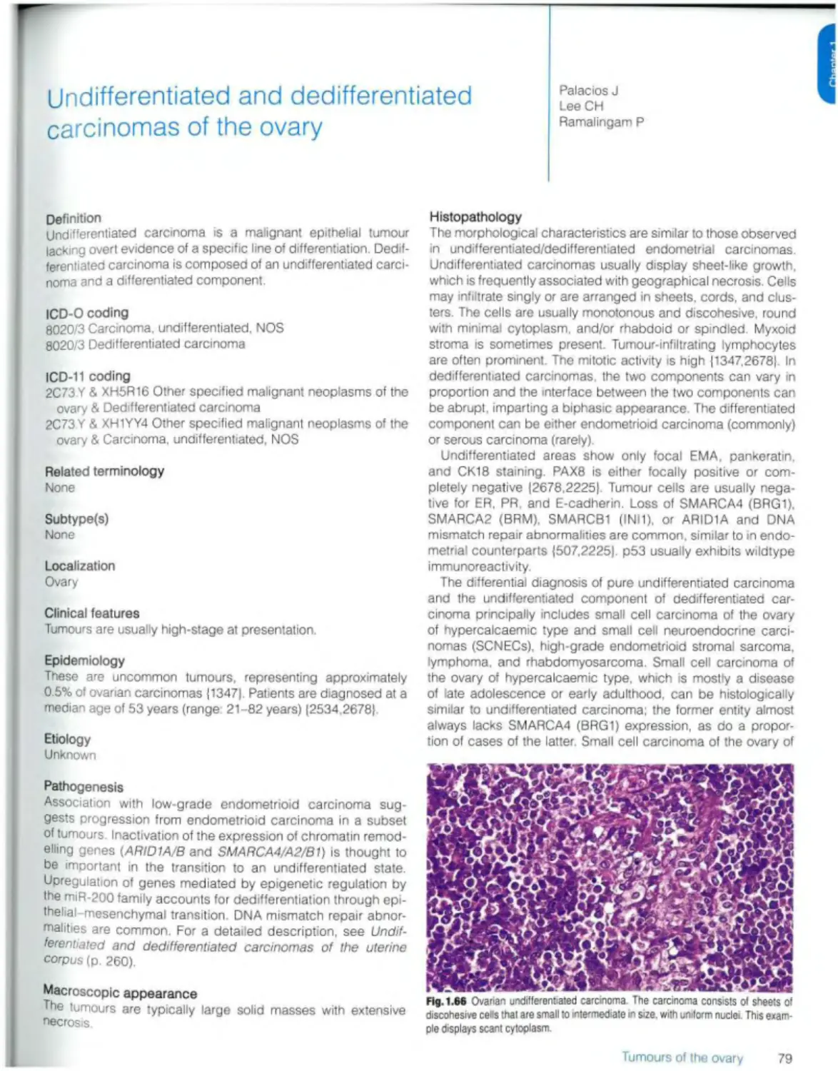
Undifferentiated and dedifferentiated carcinomas 79
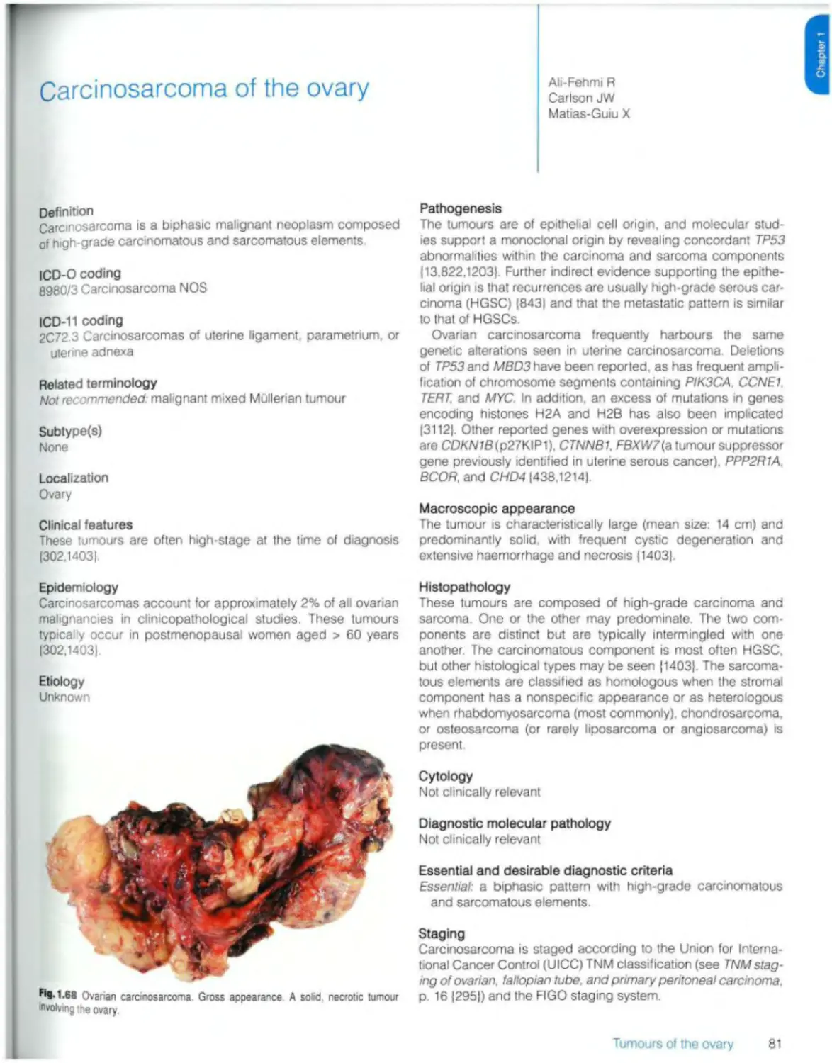
Carcinosarcoma 81
Mixed carcinoma 83
Mesenchymal tumours
Endometrioid stromal sarcoma 85
Smooth muscle tumours 87
Ovarian myxoma 89
Other ovarian mesenchymal tumours 90
Mixed epithelial and mesenchymal tumours
Mixed malignant epithelial and mesenchymal tumours Adenosarcoma 91
Sex cord stromal tumours
Pure stromal tumours Ovarian fibroma 93
Thecoma 94
Luteinized thecoma associated with sclerosing peritonitis 95
Sclerosing stromal tumour 96
Microcystic stromal tumour 97
Signet-ring stromal tumour 99
Leydig cell tumour 100
Steroid cell tumour 102
Ovarian fibrosarcoma 104
Pure sex cord tumours
Adult granulosa cell tumour 105
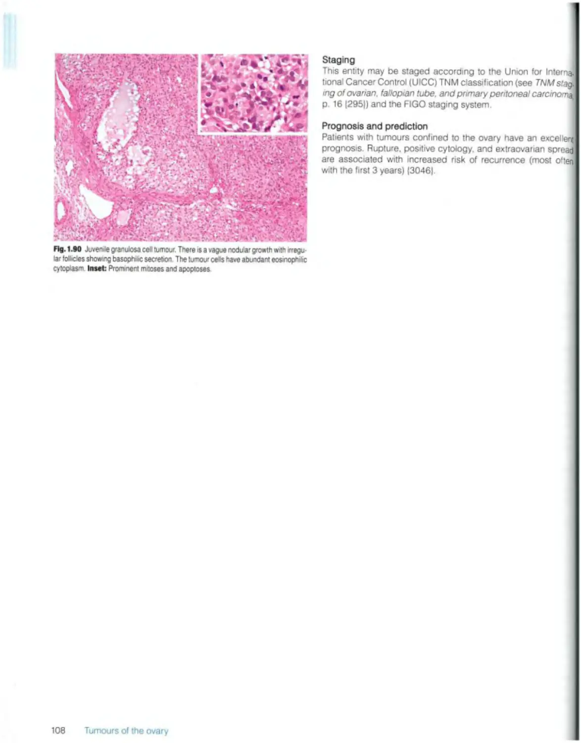
Juvenile granulosa cell tumour 107
Sertoli cell tumour Ю9
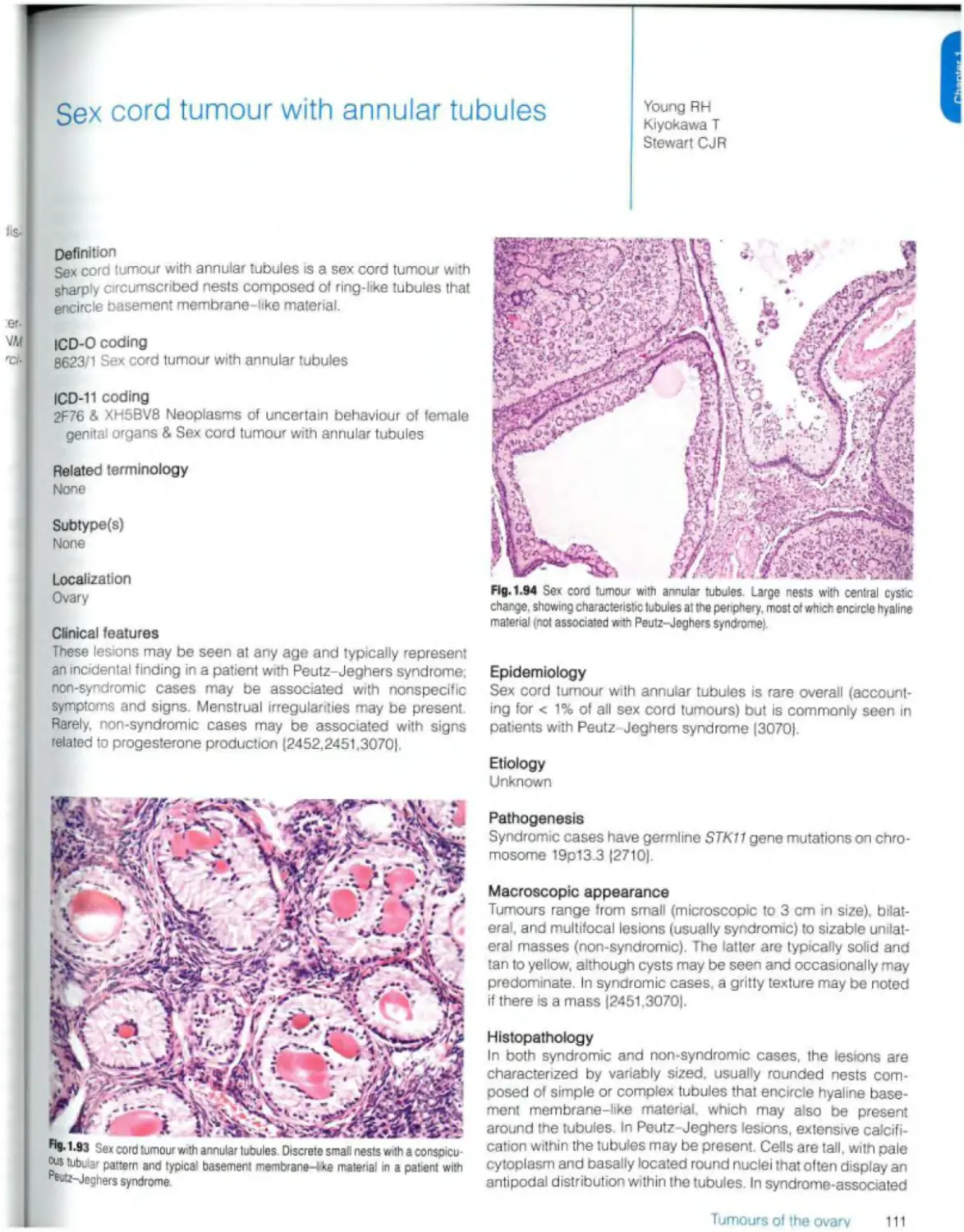
Sex cord tumour with annular tubules 111
M.xed sex cord stromal tumours
Sertoli-Leydig cell tumour 113
Sex cord-stromal tumour NOS 116
Gynandroblastoma 117
Germ cell tumours
Mature teratoma 119
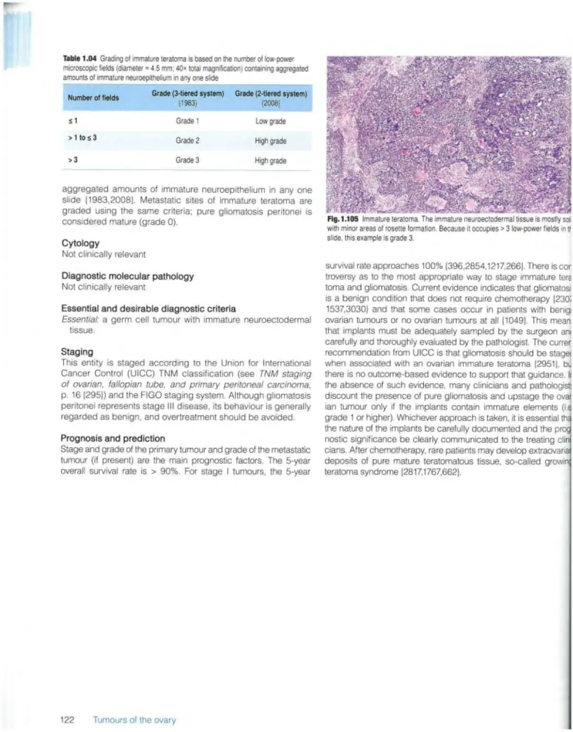
Immature teratoma 121
Dysgerminoma 123
Yolk sac tumour 125
Embryonal carcinoma 127
Non-gestational choriocarcinoma 129
Mixed germ cell tumour 131
Monodermal teratomas and somatic-type tumours arising from a dermoid cyst Struma ovarii 132
Ovarian carcinoid 134
Neuroectodermal-type tumours 136
Monodermal cystic teratoma 137
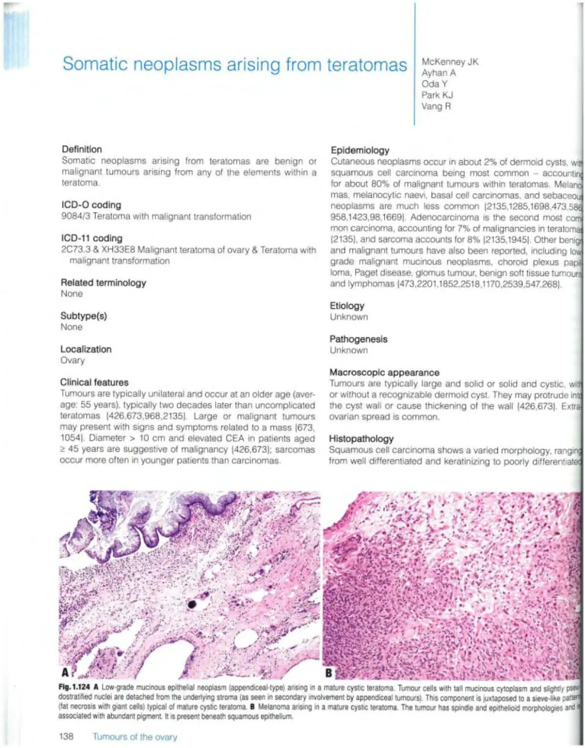
Somatic neoplasms arising from teratomas 138
Germ cell-sex cord stromal tumours
Gonadoblastoma 140
Mixed germ cell-sex cord-stromal tumour, unclassified 143
Miscellaneous tumours Rete cystadenoma, adenoma, and adenocarcinoma 144
Wolffian tumour 145
Solid pseudopapiliary tumour 147
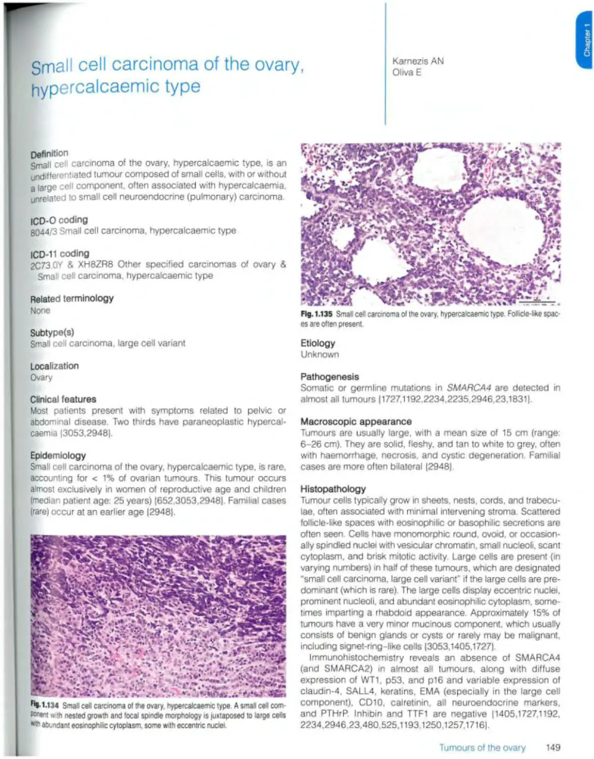
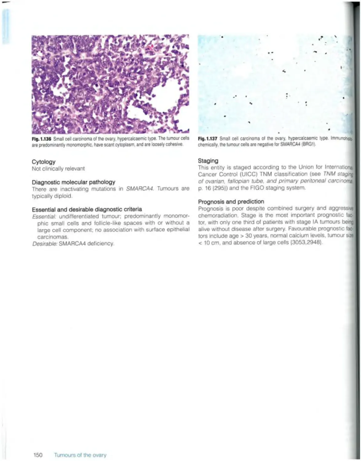
Small ceil carcinoma of the ovary, hypercaicaemic type 149
Wilms tumour 151
Mesothelial tumours (see Ch 3)
Tumour-like lesions
Follicle cyst 153
Corpus luteum cyst 154
Large solitary luteinized follicle cyst 155
Hyperreactio luteinalis 156
Pregnancy luteoma 158
Stromal hyperplasia and hyperthecosis 160
Fibromatosis and massive oedema 161
Leydig ceil hyperplasia 162
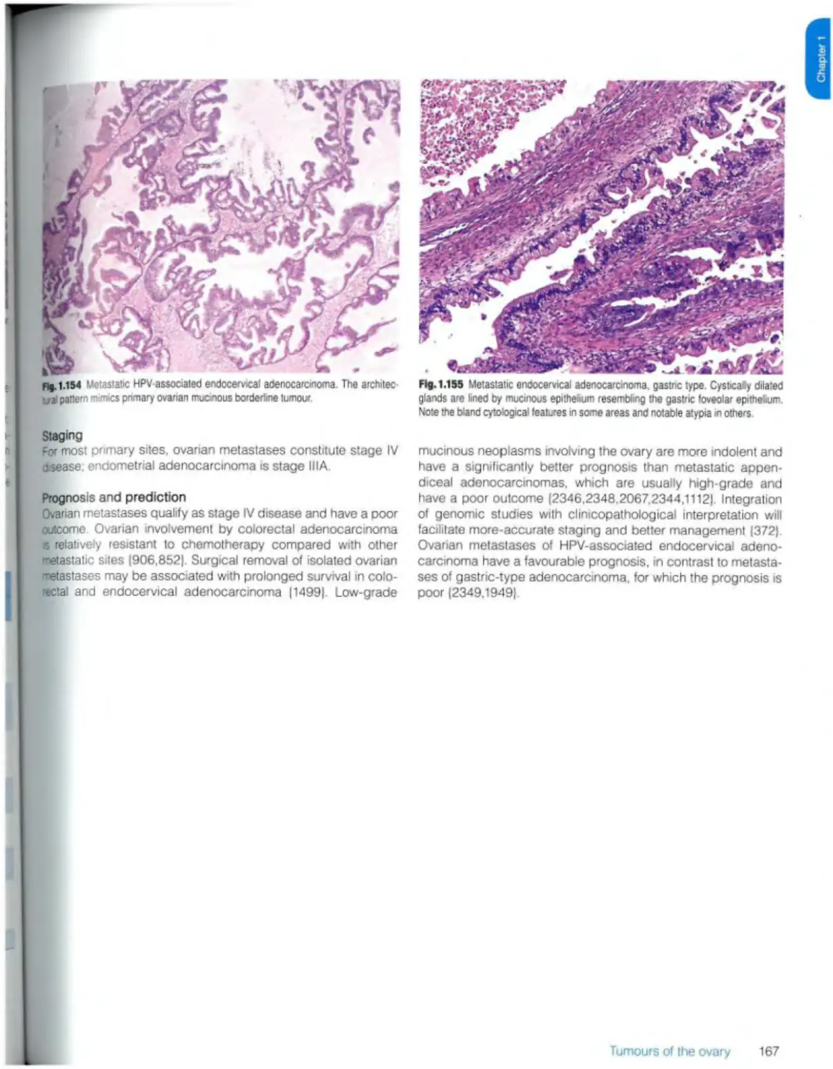
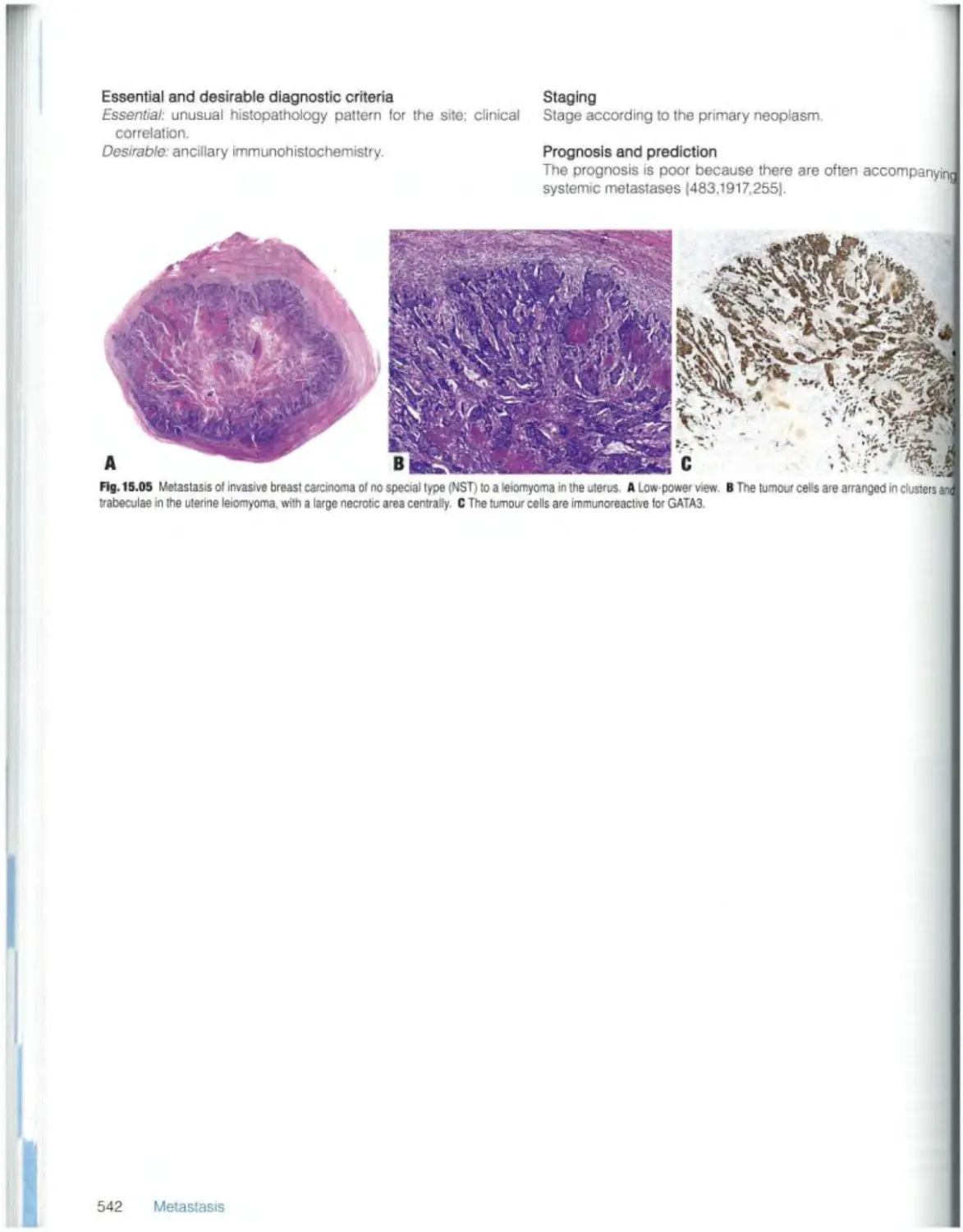
Metastases 163
2 Endometriosis and related conditions 169
Endometriosis and derived tumours 170
3 Tumours of the peritoneum 175
Introduction 176
Mesothelial tumours
Adenomatoid tumour 177
Well-differentiated papillary mesothelial tumour 179
Mesothelioma 181
Epithelial tumours Epithelial tumours of Mullerian type
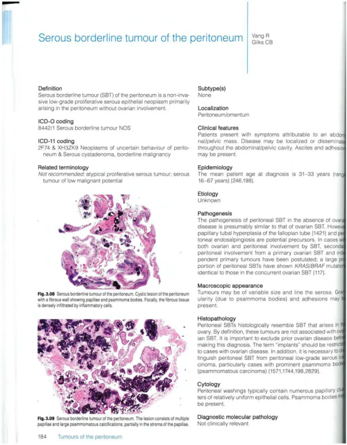
Serous borderline tumour 184
Low-grade serous carcinoma 186
High-grade serous carcinoma 187
Mesenchymal tumours specific to peritoneum
Smooth muscle tumours Leiomyomatosis peritonealis disseminata 188
Miscellaneous primary tumours
Desmoid fibromatosis 190
Calcifying fibrous tumour 192
Extragastrointestinal stromal tumour 193
Solitary fibrous tumour 195
Endometrioid stromal sarcoma 197
Desmoplastic small round cell tumour 199
Tumour-like lesions
Mesothelial hyperplasia 201
Peritoneal inclusion cysts 202
Transitional cell metaplasia 203
Endosalpingiosis 204
Histiocytic nodule 205
Ectopic decidua 206
Sptenosis 207
Other tumour-like lesions 208
Metastases
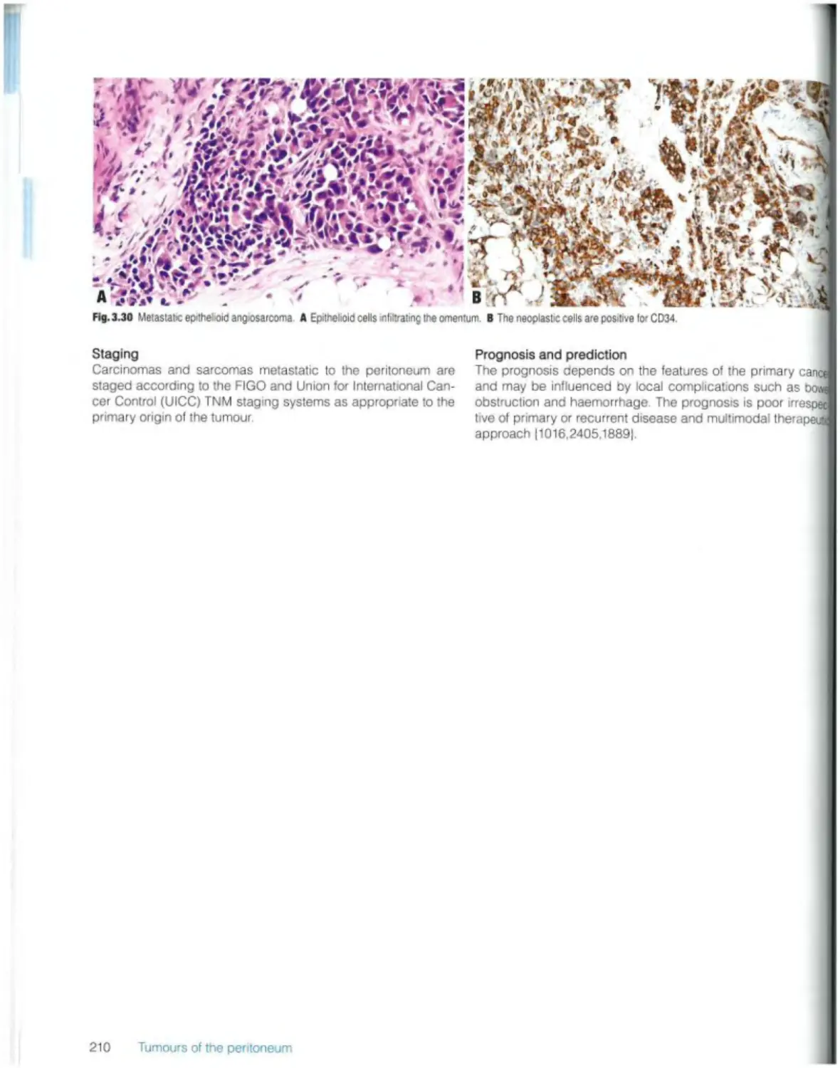
Carcinomas and sarcomas 209
Pseudomyxoma peritonei 211
Gliomatosis 214
4 Tumours of the fallopian tube 215
Introduction 216
Epithelial tumours
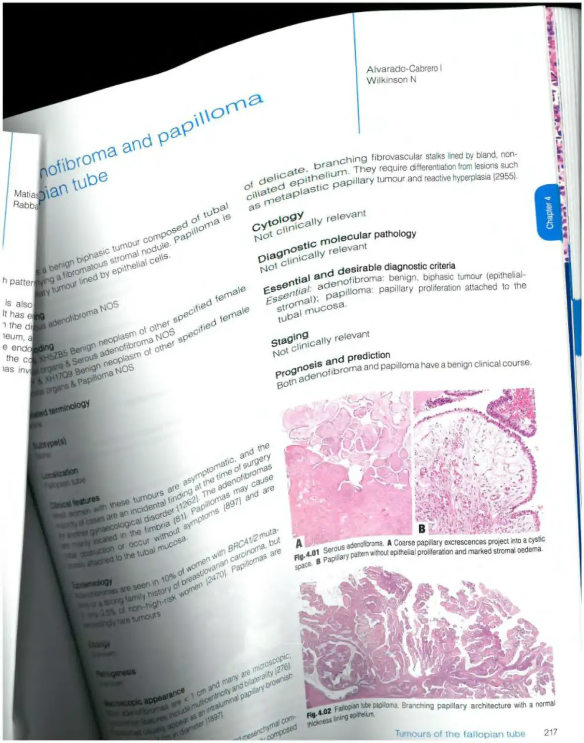
Benign serous tumours Serous adenofibroma and papilloma 217
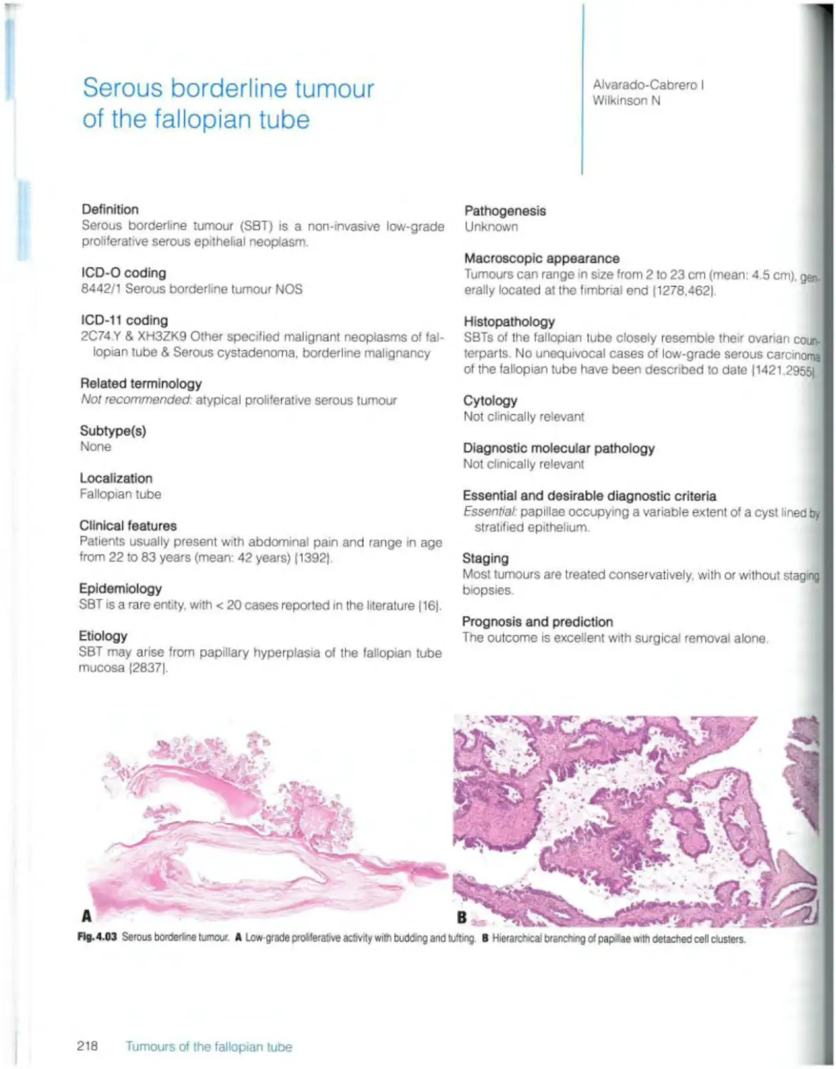
Borderline serous tumours Serous borderline tumour 218
Malignant epithelial tumours
High-grade serous carcinoma 219
Endometrioid carcinoma 221
Carcinosarcoma 222
Tumour-like lesions
Paralubal cysts 223
Tubal hyperplasia 224
Tubo-ovarian abscess 225
Salpingitis islhmica nodosa 226
Metaplastic papillary lesion 227
Placental site nodule 228
Mucinous metaplasia 229
Endosalpingiosis 230
Mixed epithelial and mesenchymal tumours Adenosarcoma 230
Germ ceil tumours Teratoma 231
5 Tumours of the broad ligament and other uterine ligaments 233
Introduction 234
Mesenchymal and mixed tumours
Leiomyoma 235
Adenomyoma 236
Adenosarcoma 237
Leiomyosarcoma 238
Other mesenchymal and mixed tumours 238
Miscellaneous tumours
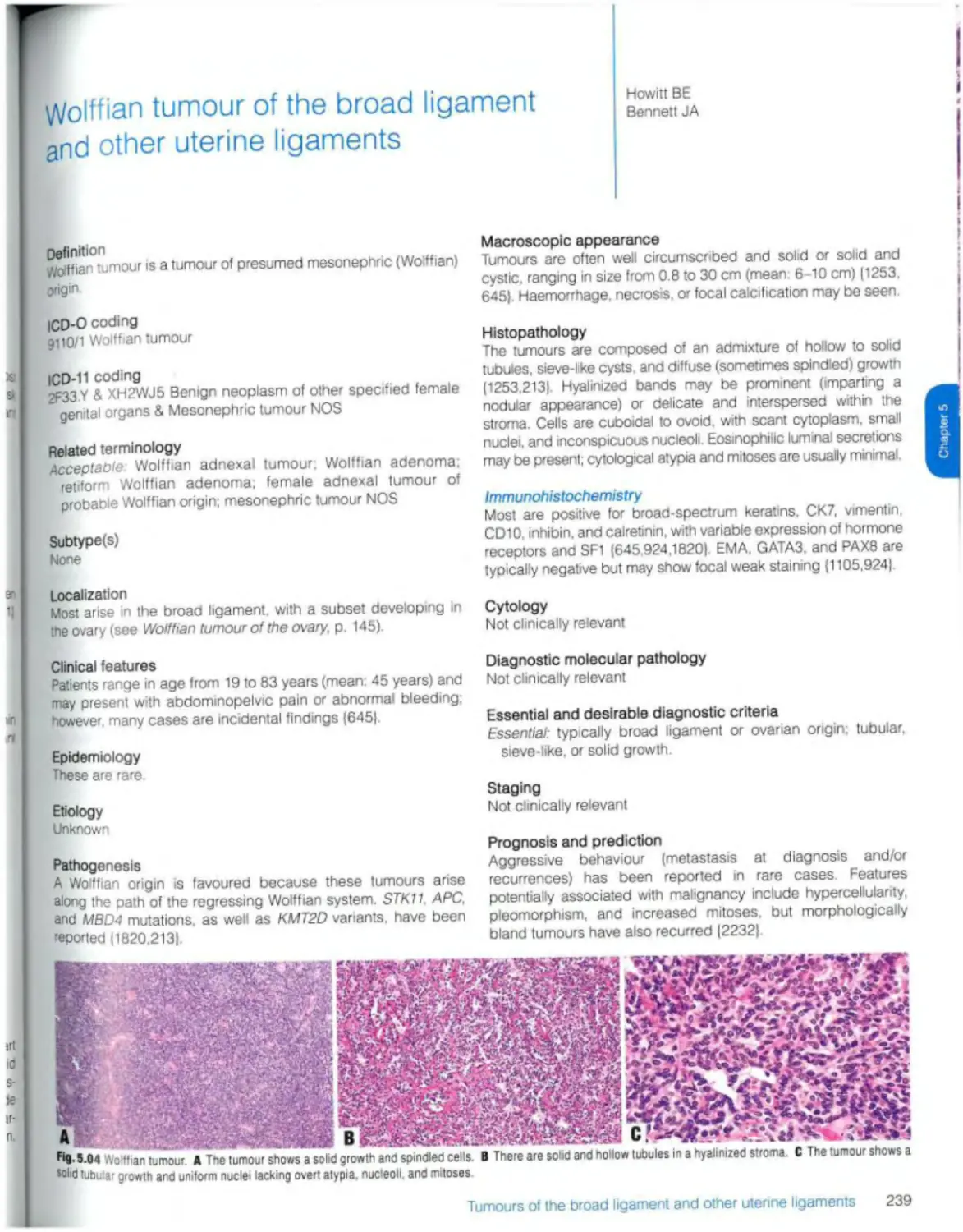
Wolffian tumour 239
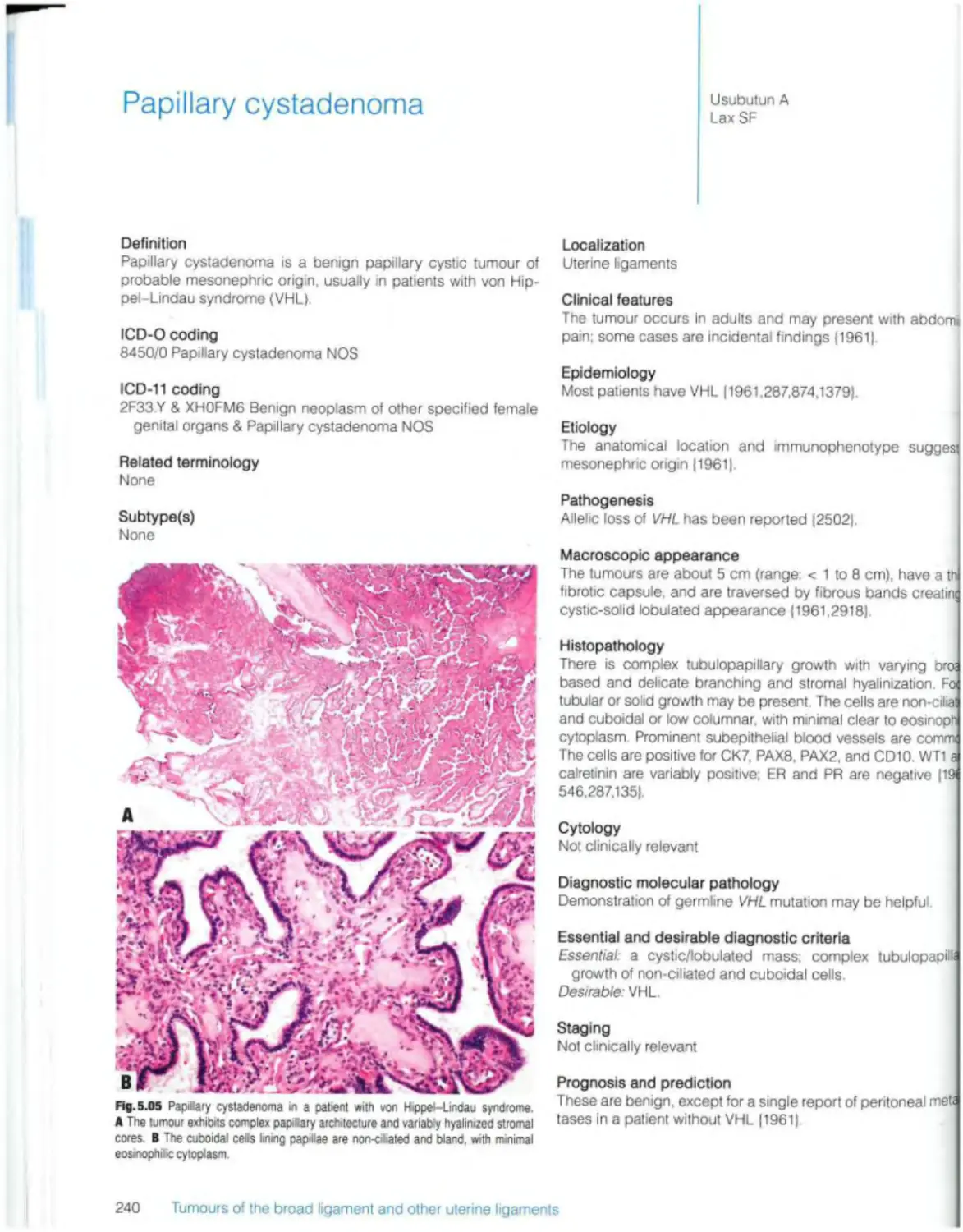
Papillary cystadenoma 240
Ependymoma 242
Tumour-iike lesions Adrenocortical remnants 243
6 Tumours of the uterine corpus 245
Introduction 246
Endometrial epithelial tumours and precursors
Precursor lesions
Endometrial hyperplasia without atyp-a 248
Endometrial atypical hyperplas’a / endometrioid intraepithelial neoplasia 250
Endometrial carcinomas
Endometrioid carcinoma 252
Serous carcinoma 256
Clear cell carcinoma 258
Undifferentiated and dedifferentiated carcinomas 260
Mixed carcinoma 262
Other endometrial carcinomas 264
Carcinosarcoma 266
Tumour-like lesions
Endometrial polyp 268
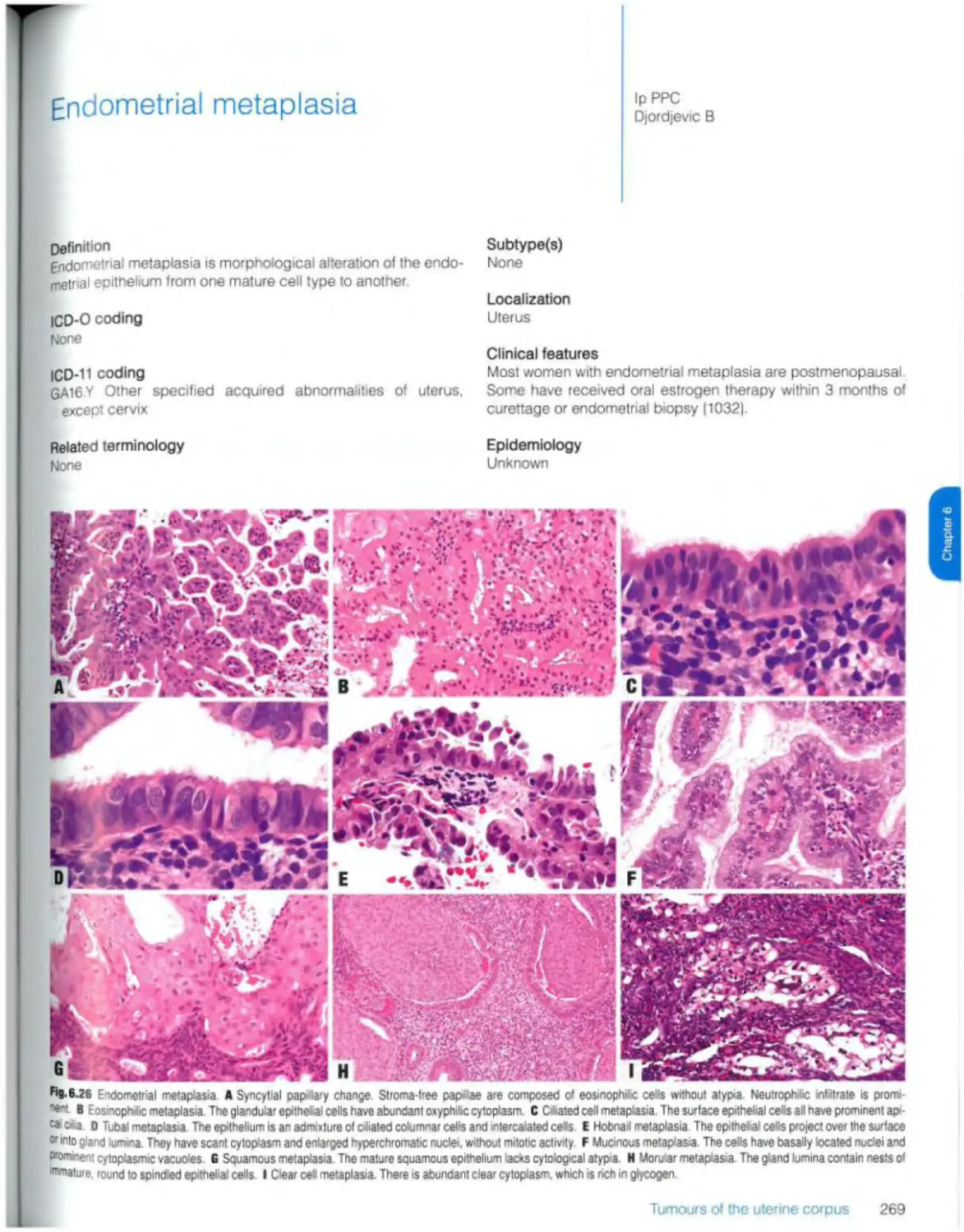
Endometrial metaplasia 269
Arias-Stella reaction 271
Mesenchymal tumours of the uterus
Smooth muscle tumours
Uterine leiomyoma 272
Intravenous leiomyomatosis 277
Smooth muscle tumour of uncertain malignant potential 279
Metastasizing leiomyoma 281
Uterine leiomyosarcoma 283
Endometrial stromal and retaied tumours
Endometrial stromal nodule 286
Low-grade endometrial stromal sarcoma 287
High-grade endometrial stromal sarcoma 289
Undifferentiated uterine sarcoma 292
Miscellaneous mesenchymal tumours
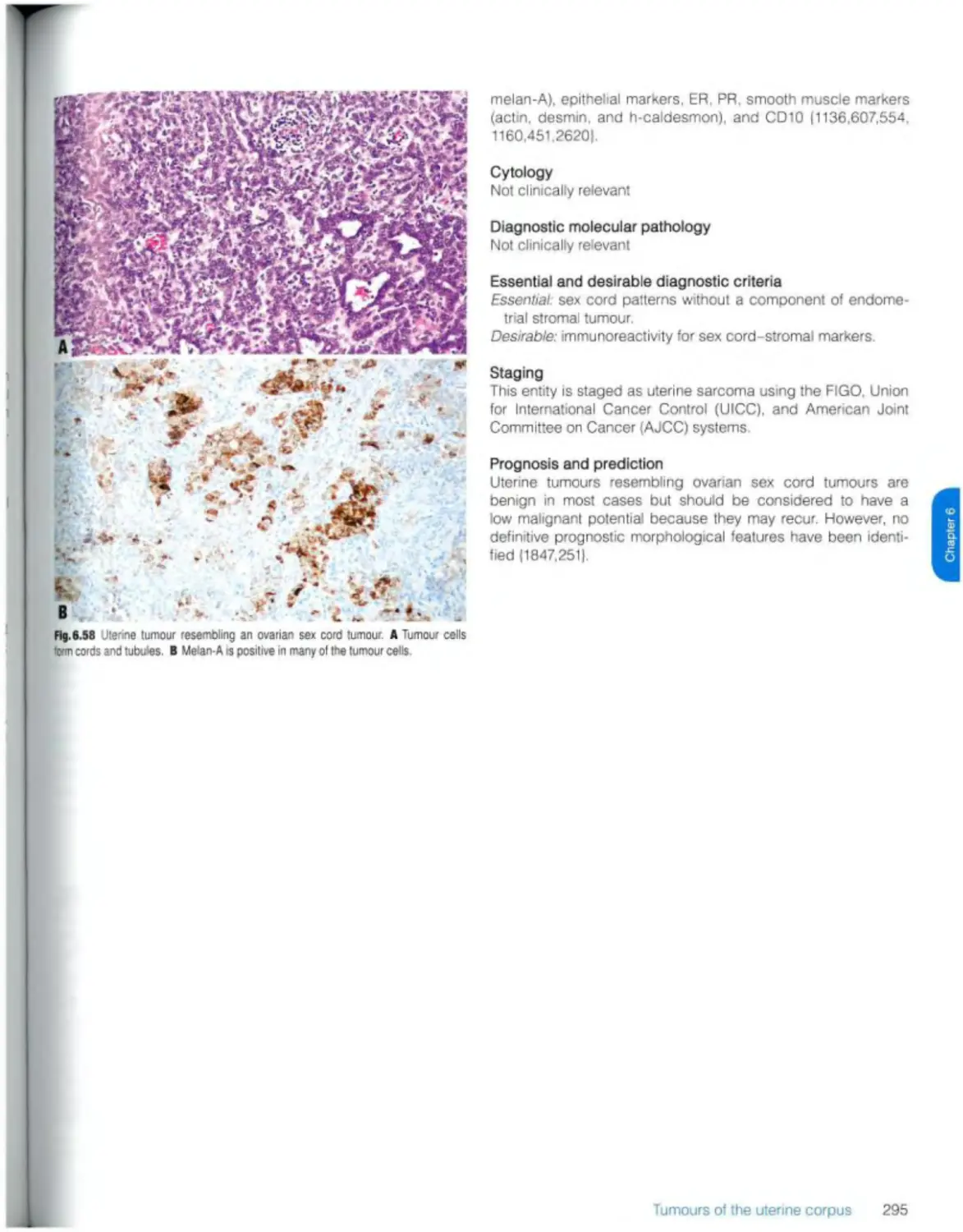
Uterine tumour resembling ovarian sex cord tumour 294
Perivascular epithelioid cell tumour (PEComa) 296
Inflammatory myofibroblastic tumour 298
Other mesenchymal tumours of the uterus 300
Mixed epithelial and mesenchymal tumours
Adenomyoma 301
Atypical polypoid adenomyoma 303
Adenosarcoma 305
Miscellaneous tumours Central primitive neuroectodermal tumour) CNS
embryonal tumour 307
Germ cell tumours 308
7 Gestational trophoblastic disease 309
Introduction 310
Tumour-like lesions
Non-neoplastic lesions
E xaggerated placental site reaction 311
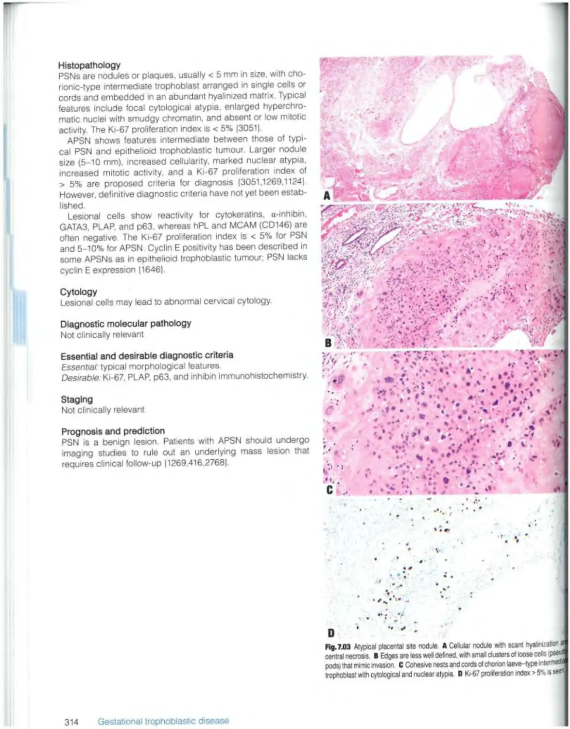
Placental site nodule and plaque 313
Abnormal (non-molar) villous lesions 315
Molar pregnancies
Partial hydatidiform mole 317
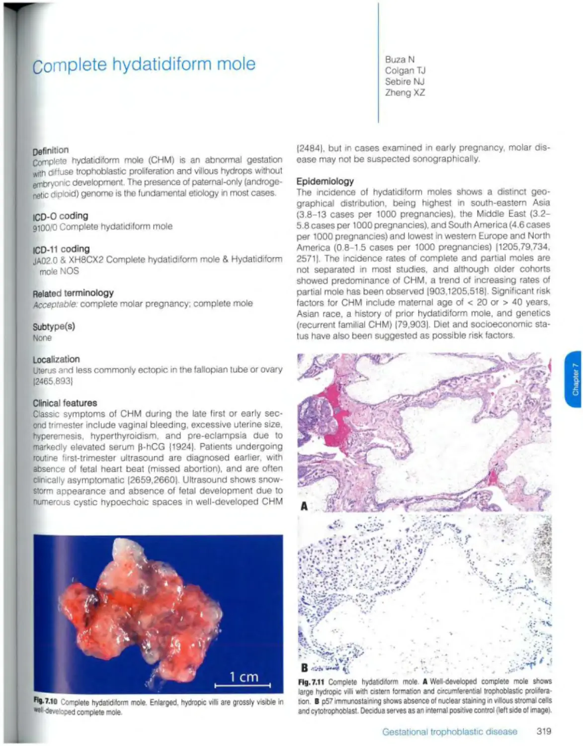
Complete hydatidiform mole 319
Invasive and metastatic hydatidiform motes 322
Gestational trophoblastic neoplasms
Epithelioid trophoblastic tumour 323
Placental site trophoblastic tumour 325
Gestational choriocarcinoma 327
Mixed trophoblastic tumour 332
8 Tumours of the uterine cervix 335
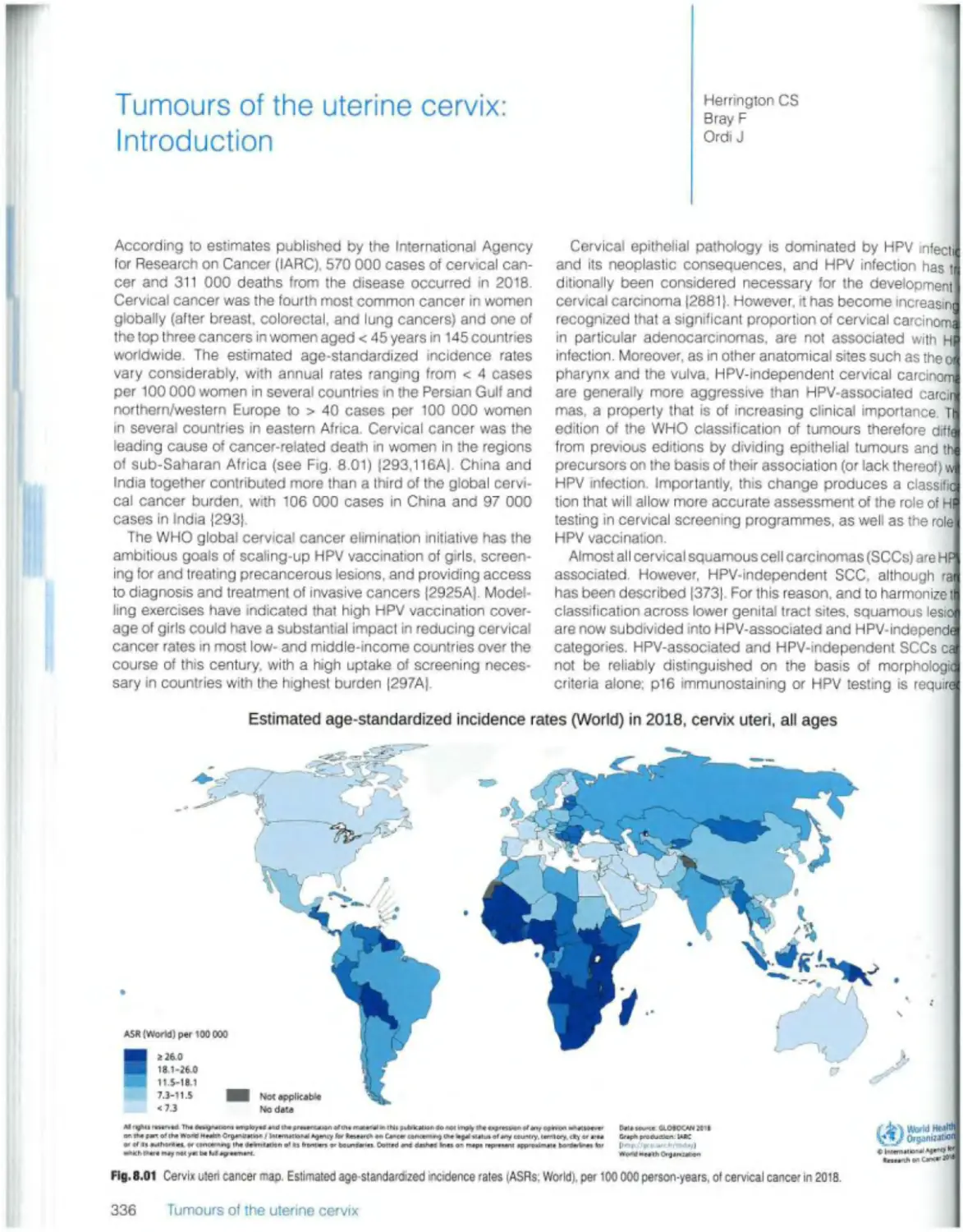
Introduction 336
Squamous epithelial tumours
Mimics of squamous precursor lesions
Squamous metaplasia 338
Atrophy 341
Squamous ceil tumours and precursors
Condyloma acuminatum (see Ch. 10)
Squamous intraepithelial lesions 342
Squamous cell carcinoma. HPV-associated 347
Squamous ceil carcinoma. HPV-mdependent 350
Squamous cell carcinoma NOS 351
Glandular tumours and precursors
Benign glandular lesions
Endocervicai polyp 352
Mullerian papilloma 353
Nabothian cyst 354
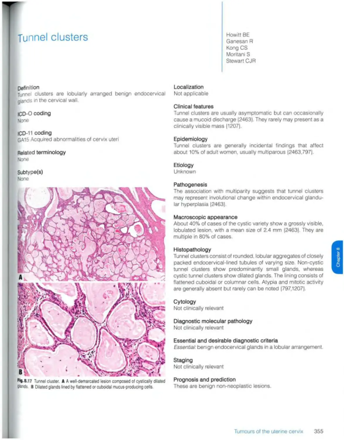
Tunnel clusters 355
Microglandular hyperplasia 356
Lobular endocervicai glandular hyperplasia 357
Diffuse laminar endocervicai hyperplasia 358
Mesonephric remnants and hyperplasia 359
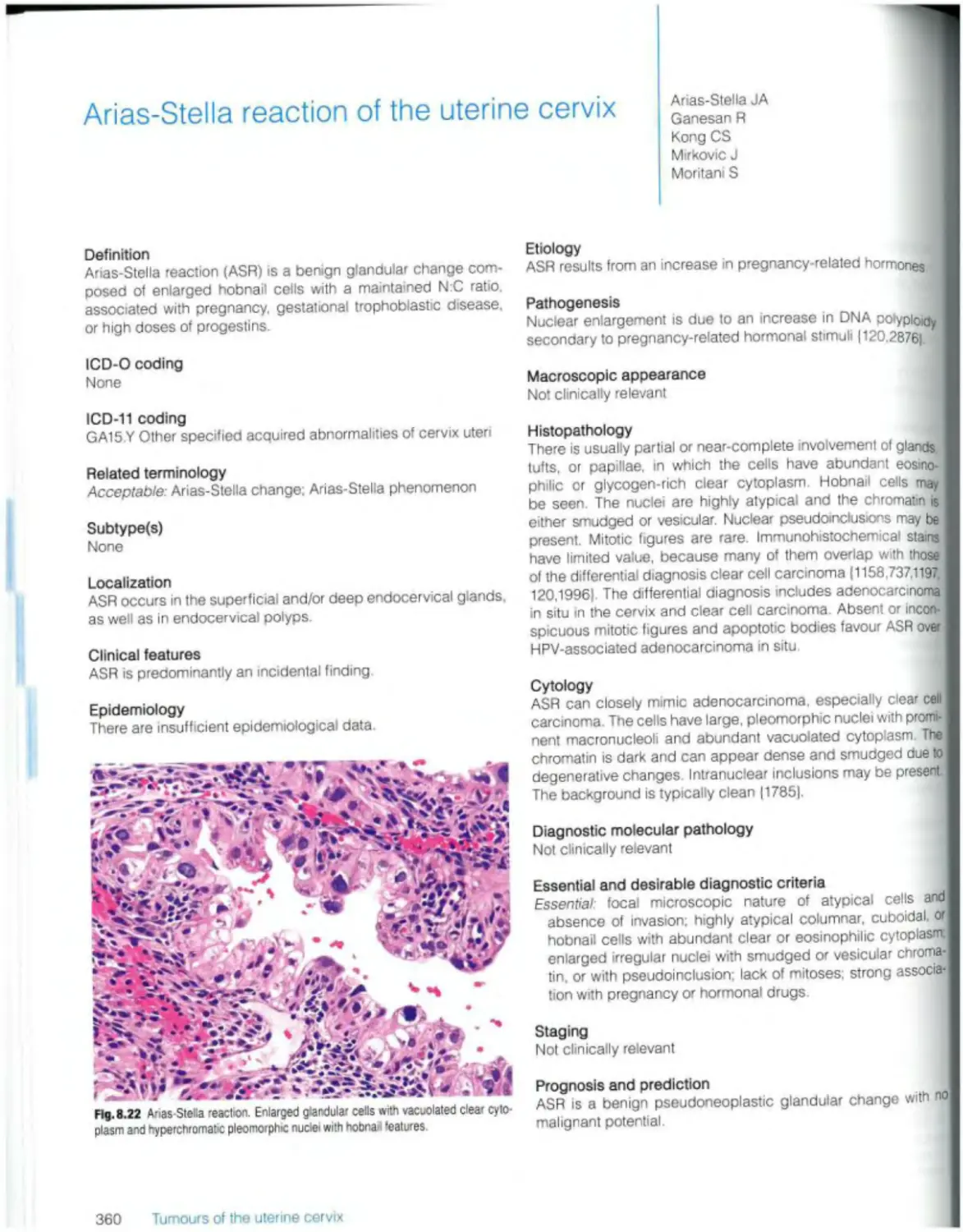
Arias-Stella reaction 360
Endocervicosrs 361
Tuboendometnoid metaplasia 362
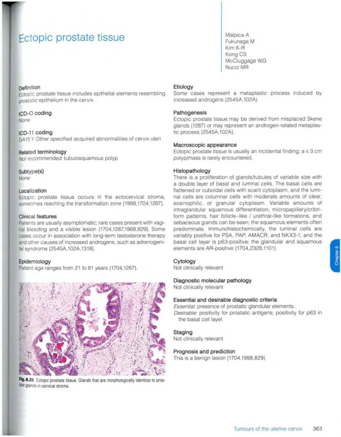
Ectopic prostate tissue 363
Adenocarcinomas
Adenocarcinoma in situ, HPV-associated 364
Adenocarcinoma, HPV-associated 367
Adenocarcinoma in situ, HPV-independent 372
Adenocarcinoma, HPV-indepeodent. gastric type 374
Adenocarcinoma. HPV-independent, clear cell type 376 Adenocarcinoma, HPV-independent, mesonephric
type 378
Other adenocarcinomas 380
Other epithelial tumours Carcinosarcoma 382
Adenosquamous and mucoepidermoid carcinomas 383
Adenoid basal carcinoma 384
Carcinoma, unctassifiable 386
Mixed epithelial and mesenchymal tumours Adenomyoma 387
Adenosarcoma 388
Germ cell tumours 389
9 Tumours of the vagina 391
Introduction 392
Epithelial tumours
Benign squamous lesions
Condyloma acuminatum (see Ch 10)
Squamous papilloma 393
Atrophy 394
Tubulosquamous polyp 395
Squamous cell tumours and precursors
Squamous intraepithelial lesions 396
Squamous cell carcinoma. HPV-associated 398
Squamous cell carcinoma. HPV-independent 400
Squamous cell carcinoma NOS 401
Benign glandular lesions Villous adenoma 402
Mullerian papilloma 403
Vaginal adenosis 404
Endocervicosis 405
Cysts 406
Glandular tumours Adenocarcinoma, HPV-associated 407
Endometrioid carcinoma 408
Clear cell carcinoma 409
Mucinous carcinoma, gastric type 410
Mucinous carcinoma, intestinal type 411
Mesonephric adenocarcinoma 412
Carcinosarcoma 413
Other epithelial tumours
M ixed tumour of the vagina 414
Adenocarcinoma of Skene gland origin 415
Adenosquamous carcinoma 416
Adenoid basal carcinoma 416
Mixed epithelial and mesenchymal tumours Adenosarcoma 417
Miscellaneous tumours
Germ cell tumours 418
10 Tumours of the vulva 419
Introduction 420
Epithelial tumours
Benign squamous lesions Seborrhoeic keratosis 421
Condyloma acuminatum 422
Squamous cell tumours and precursors
Squamous intraepithelial lesions. HPV-associated 424
Vulvar intraepithelial neoplasia, HPV-independent 426
Squamous cell carcinoma. HPV-associated 429
Squamous cell carcinoma, HPV-independent 432
Squamous cell carcinoma NOS 434
Basal cell carcinoma 434
Glandular tumours and cysts
Mammary-type glandular lesions
Papillary hidradenoma 435
Chondroid syringoma 436
Fibroadenoma 437
Phyllodes tumour 438
Adenocarcinoma of mammary gland type 439
Bartholin gland lesions Bartholin gland cyst 440
Hyperplasia, adenoma, and adenomyoma 441
Bartholm gland carcinomas 442 Skeletal muscle tumours
Other cysts 444 Rhabdomyoma
Adenocarcinomas of other types Rhabdomyosarcoma
Paget disease 445 Peripheral nerve sheath tumours
Carcinomas of sweat gland origin 447 Benign peripheral nerve sheath tumours
Adenocarcinoma of intestinal type 448 Granular cell tumour
Germ cell tumours 449 Tumours of uncertain differentiation
Superficial angiomyxoma
11 Neuroendocrine neoplasia 451 Deep (aggressive) angiomyxoma
Introduction 452 Epithelioid sarcoma
Neuroendocrine tumour 453 Alveolar soft part sarcoma
Neuroendocrine carcinoma Undifferentiated small round cell sarcomas
Small ceil neuroendocrine carcinoma 455 Ewing sarcoma
Large cell neuroendocrine carcinoma 457
Mixed neuroendocnne-noo-neuroendocrine neoplasms 14 Melanocytic lesions
Carcinoma admixed with neuroendocrine carcinoma 459 Naevi
Acquired melanocytic naevus
12 Haematolymphoid proliferations and neoplasia 461 Congenital melanocytic naevus
Introduction 462 Blue naevus
Reactive lymphoid hyperplasia Atypical melanocytic naevus of genital type
Florid reactive lymphoid hyperplasia 465 Dysplastic melanocytic naevus
Lymphomas Melanoma
Diffuse large В-cell lymphoma 467 Mucosal melanoma
Exfranodal marginal zone tymphoma 469
Follicular lymphoma 471 15 Metastasis
Burkitt lymphoma 473 Metastasis to the lower female genital tract
Myeloid leukaemia
Myeloid sarcoma 474 16 Genetic tumour syndromes of the female genital tract
eRCAf/2-associated hereditary breast and ovarian cancer
13 Mesenchymal tumours of the lower genital tract 477 syndrome
Introduction 478 Lynch syndrome
Adipocytic tumours Cowden syndrome
Lipoma 480 Li-Fraumeni syndrome
Lipoblastoma-like tumour ol the vulva 481 Peutz- Jcghers syndrome
Uposarcoma 483 Ataxia-telangiectasia
Fibroblastic and myofibroblastic tumours Carney complex
Postoperative spindle cell nodule 485 DICER1 syndrome
Fibroepithehal stromal polyp Ovarian dysgenesis
Prepubertal fibroma 488 Von Hippel-Lindau syndrome
Superficial myofibroblastoma 490 Hereditary leiomyomatosis and renal cell carcinoma
Myofibroblastoma 491 Other genetic tumour syndromes
Cellular angiofibroma 493
Angiomyofibroblasloma 495 Contributors
Solitary fibrous tumour 497
Dermatofibrosarcoma protuberans 498 Declaration of interests
NTRK-rearranged spindle cell neoplasm (emerging) 500
Vascular tumours IARC/WHO Committee for ICD-0
Kaposi sarcoma 502
Angiosarcoma 504 Sources
Smooth muscle tumours
Leiomyoma References
Smooth muscle tumour of uncertain malignant potential 508
Leiomyosarcoma 509 Subject index
Previous volumes in the series
511
512
515
517
519
520
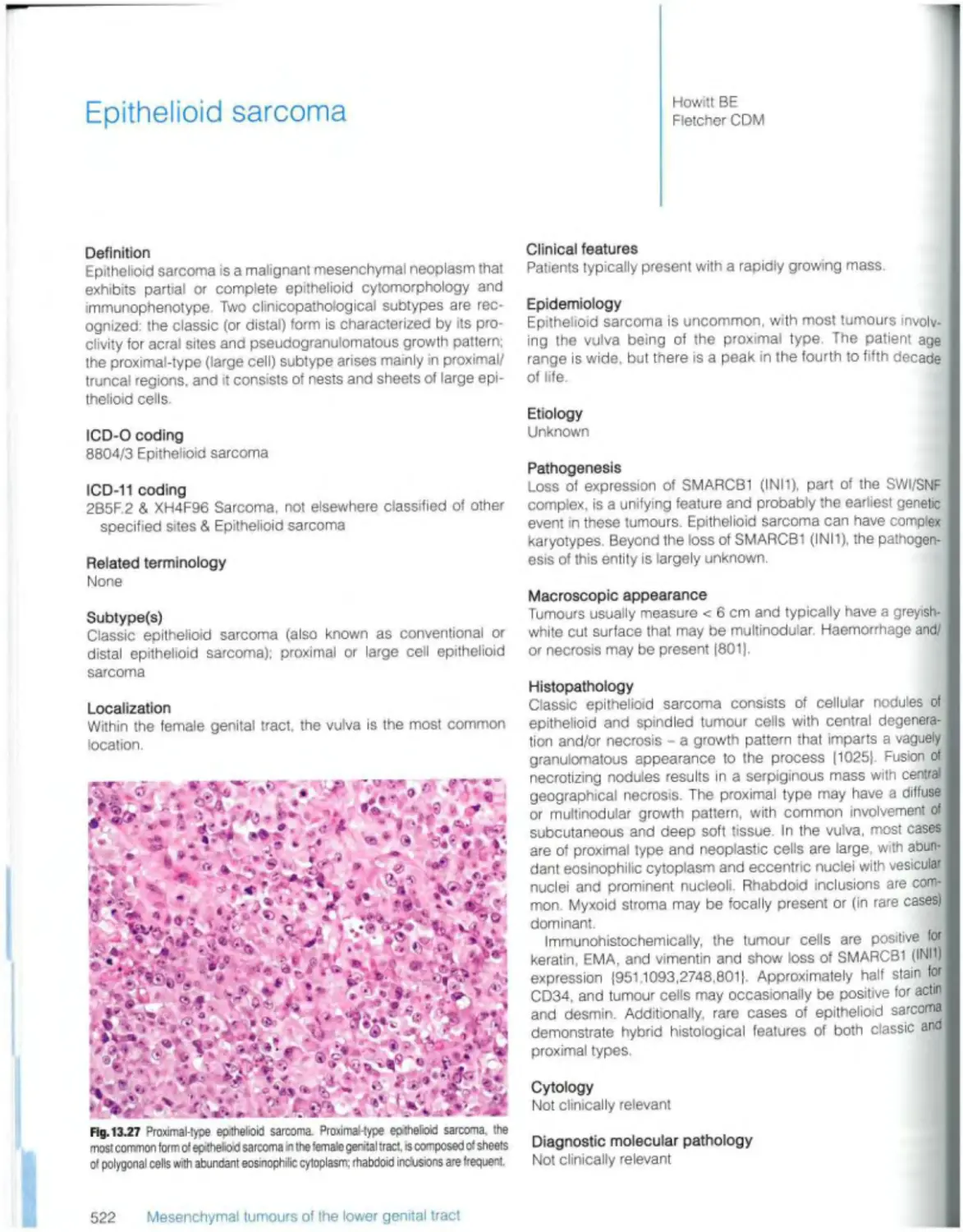
522
524
526
527
528
530
532
534
535
537
539
540
543
544
546
548
550
552
554
555
556
558
560
561
563
564
570
571
572
577
625
632
List of abbreviations
30 AIDS AR cAMP Cl CIN CNS CT dMMR DNA EBV ER FGT FIGO FISH FNA GCB H&E HIV HPF HPV HR-HPV IARC ICD-11 ICD-O ICD-0-3
three-dimensional
acquired immunodeficiency syndrome
androgen receptor
cyclic adenosine monophosphate confidence interval
cervical intraepithelial neoplasia central nervous system computed tomography mismatch repair-deficient deoxyribonucleic acid Epstein Barr virus estrogen receptor female genital tract International Federation of Gynecology and Obstetrics fluorescence in situ hybridization fine-needle aspiration germmal-centre В cell haematoxylin and eosin human immunodeficiency virus high-power fioldfs) human papillomavirus high-risk human papillomavirus International Agoncy for Research on Cancer International Classification o* Diseases 11th revision International Classification of Diseases for Oncology International Classification of Diseases for Oncology 3rd edition
|g immunoglobulin
ITD internal tandem duplication
kb kifobasefs)
LR-HPV low-risk human papillomavirus
MR I magnetic resonance imaging
mRNA messenger ribonucleic acid
MSI microsatellrte instability
MSS microsatellrte stability
N:C ratio nuclear-to-cytopiasmic ratio
NK cell natural killer cell
NMDAR /V-methylD-aspartate receptor
NOS not otherwise specified
NSE neuron-specific enolase
PAS periodic acid-Schiff
PASD periodic acid-Schiff with diastase
PCR polymerase chain reaction
PR progesterone receptor
RNA ribonucleic acid
RT-PCR reverse transcriptase polymerase chain reaction
SEER Program Surveillance Epidemiology and End Results Program
STR short tandem repeat
TCGA The Cancer Genome Atlas
TNM tumour, node, metastasis
UICC Union for International Cancer Control
UV ultraviolet
ValN vaginal intraepithelial neoplasia
VIN vulvar intraepithelial neoplasia
List of abbreviations
XI
Foreword
The WHO Classification of Tumours, published as a series of books (also known as the WHO Blue Books) and now as a website (https;//tumourciassification iarc.who.int), is an essential tool for standardizing diagnostic practice worldwide. It also serves as a vehicle for the translation of cancer research into practice The diagnostic criteria and standards that make up the classification are underpinned by evidence evaluated and debated by experts in the field. About 200 authors and editors participate m the production of each book, and they give their time freely to this task. I am very grateful for their help; it is a remarkable team effort.
This fourth volume of the fifth edition of the WHO Blue Books has. like the preceding three, been led by the WHO Classification of Tumours Editorial Board, which is composed of standing members nominated by pathology organizations and expert members selected on the basis of informed bibliometric analysis. The diagnostic process is increasingly multidisciplinary, and we are delighted that several radiology and clinical experts have joined us to address specific needs.
The most conspicuous change to the format of the books in the fifth edition is that tumour types common to multiple systems are dealt with together - so there are separate chapters on neuroendocrine neoplasia, haematolymphoid proliferations and neoplasia, mesenchymal tumours, and melanocytic lesions There is also a chapter on genetic tumour syndromes. Genetic disorders are of increasing importance to diagnosis in individual patients, and the study of these disorders has undoubtedly informed our understanding of tumour biology and behaviour over the past decade
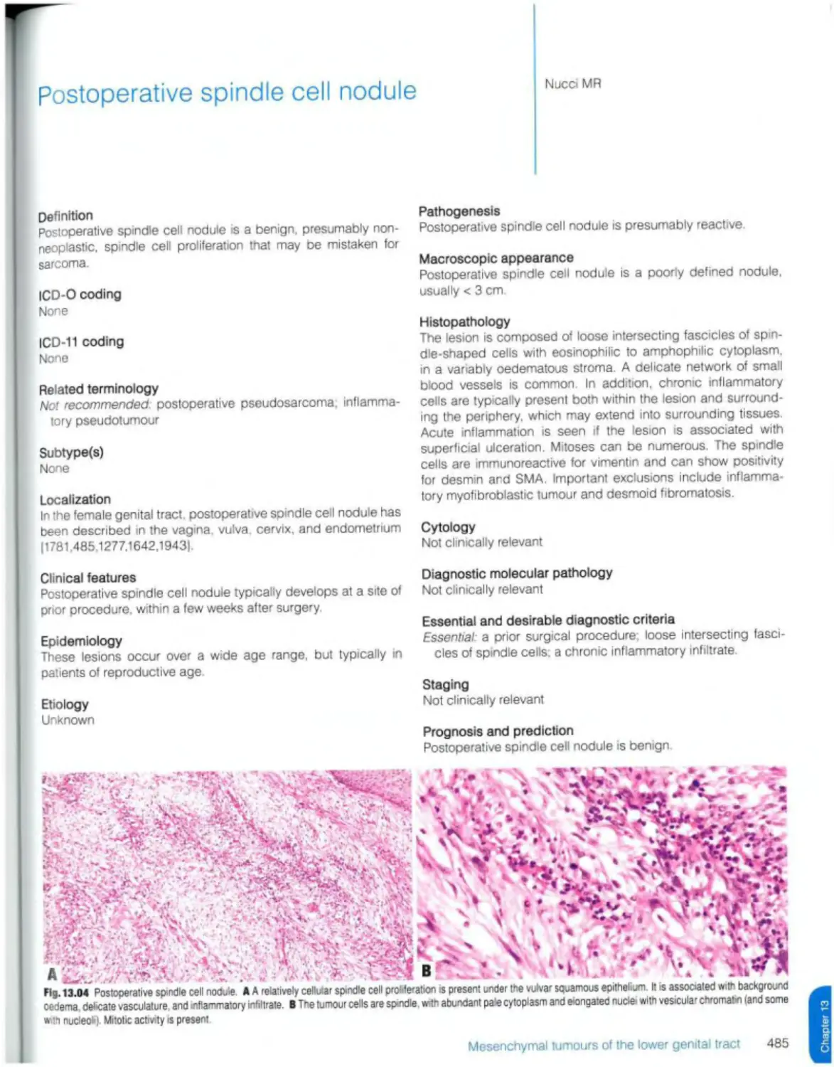
We have attempted to take a more systematic approach to the multifaceted nature of tumour classification, each tumour type is described on the basis of its localization, clinical features, epidemiology, etiology, pathogenesis, histopathology, diagnostic molecular pathology, staging, and prognosis and prediction. We have also included information on macroscopic appearance and cytology, as well as essential and desirable diagnostic criteria. This standardized, modular approach makes it easier tor the books to be accessible online, but it also enables us to call attention to areas in which there is little information, and where serious gaps in our knowledge remain to be addressed.
The organization of the WHO Blue Books content now follows the normal progression from benign to malignant - a break with the fourth edition, but one we hope will be welcome
The volumes are still organized by anatomical site (digestive system, breast, soft tissue and bone, etc), and each tumour type is listed within a taxonomic classification that follows the format below, which helps to structure the books in a systematic manner
• Site; e.g ovary
• Category; e g endometrioid tumours
• Family (class); e g. malignant endometrioid tumours
• Type: e g endometrioid carcinoma
• Subtype; e g seromucinous carcinoma
The issue of whether a given tumour type represents a distinct entity rather than a subtype continues to exercise pathologists, and it is the topic of many publications in the literature. We continue to deal with this issue on a case-by-case basis, but we believe there are inherent rules that can be applied. For example, tumours in which multiple histological patterns contain shared truncal mutations are clearly of the same type, despite the differences in their appearance. Equally, genetic heterogeneity within the same tumour type may have implications for treatment. A small shift in terminology in the fifth edition is that the term “variant" in reference to a specific kind of tumour has been wholly superseded by “subtype', in an effort to more clearly differentiate this meaning from that of “variant" in reference to a genetic alteration
The WHO Blue Books are much appreciated by pathologists and of increasing importance to practitioners of other clinical disciplines involved in cancer management, as well as to researchers. The editorial board and I certainly hope that the series will continue to meet the need for standards in diagnosis and to facilitate the translation of diagnostic research into practice worldwide It is particularly important that cancers continue to be classified and diagnosed according to the same standards internationally so that patients can benefit from multicentre clinical trials, as well as from the results of local trials conducted on different continents
Dr Ian A. Cree
Head, WHO Classification of Tumours Group International Agency for Research on Cancer
August 2020
xii
Foreword
WHO classification of tumours of the ovary
Serous tumours
8441/0 Serous cystadenoma NOS
8461/0 Serous surface papilloma
9014/0 Serous adenofibroma NOS
9014/0 Serous cystadenof'broma NOS
8442/1 Serous borderline tumour NOS
8460/2 Serous borderline tumour, micropapillary variant
8460/2 Serous carcinoma, non mvasrve, low grade
8460/3 Low-grade serous carcinoma
8461/3 High-grade serous carcinoma
Mucinous tumours
8470/0 Mucinous cystadenoma NOS
9015/0 Mucinous adenofibroma NOS
8472/1 Mucinous borderline tumour
8480/3 Mucinous adenocarcinoma
Endometrioid tumours
8380/0 Endometrioid cystadenoma NOS
8381/0 Endometrioid adenofibroma NOS
8380/1 Endometrioid tumour, borderline
8380/3 Endometrioid adenocarcinoma NOS
8474/3 Seromucinous carcinoma
Clear cell tumours
8443/0 Clear cell cystadenoma
8313/0 Clear cell cystadenofibroma
8313/1 Clear cell borderline tumour
8310/3 Clear cell adenocarcinoma NOS
Seromucinous tumours
8474/0 Seromucinous cystadenoma
9014/0 Seromucinous adenofibroma
8474/1 Seromucinous borderline tumour
Brenner tumours
9000/0 Brenner tumour NOS
9000/1 Brenner tumour, borderline malignancy
9000/3 Brenner tumour, malignant
Other carcinomas
9111/3* Mesonephric-like adenocarcinoma
8020/3 Carcinoma, undifferentiated. NOS
8020/3 Dedifferentiated carcinoma
8980/3 Carcinosarcoma NOS
8323/3 Mixed cell adenocarcinoma
Mesenchymal tumours
8931/3 Endometrioid stromal sarcoma, low grade
8930/3 Endometrioid stromal sarcoma, high grade
8890/0 Leiomyoma NOS
8890/3 Leiomyosarcoma NOS
8897/1 Smooth muscle tumour of uncertain malignant potential
8840/0 Myxoma NOS
Mixed epithelial and mesenchymal tumours
8933/3 Adenosarcoma
Sex cord-stromal tumours
Pure stromal tumours
8810/0 Fibroma NOS
8810/1 Cellular fibroma
8600/0 Thecoma NOS
8601/0 Thecoma, luteinized
8602/0 Sclerosing stromal tumour
8590/0 Microcystic stromal tumour
8590/0 Signet-ring stromal tumour
8650/0 Leydig ceil tumour of the ovary NOS
8670/0 Steroid cell tumour NOS
8670/3 Steroid cell tumour, malignant
88Ю/3 Fibrosarcoma NOS
Pure sex cord tumours
8620/3 Adult granulosa cell tumour of ovary
8622/1 Granulosa cell tumour, juvenile
8640/1 Sertoli cell tumour NOS
8623/1 Sex cord tumour with annular tubules
Mixed sex cord-stromal tumours
8631/1 Sertoli-Leydig cell tumour NOS
8631/0 Sertoli-Leydig cell tumour, well differentiated
8631/1 Sertoli-Leydig cell tumour, moderately differentiated
8631/3 Sertoli-Leydig cell tumour, poorty differentiated
8633/1 Sertoli-Leydig cell tumour retiform
8590/1 Sex cord tumour NOS
8632/1 Gynandroblastoma
Germ cell tumours
9080/0 Teratoma, benign
9080/3 Immature teratoma NOS
9060/3 Dysgermmoma
9071/3 Yolk sac tumour NOS
9070/3 Embryonal carcinoma NOS
9100/3 Choriocarcinoma NOS
9085/3 Mixed germ cell tumour
Monodermai teratomas and somatic-type tumours arising from a dermotd cyst
9090/0 Struma ovarii NOS
9090/3 Struma ovarii, malignant
9091/1 Strumal carcinoid
9084/3 Teratoma with malignant transformation
9080/0 Cystic teratoma NOS
9084/3 Teratoma with malignant transformation
Germ cell-sex cord-stromal tumours
9073/1 Gonadoblastoma
Dissecting gonadoblastoma
Undifferentiated gonadal tissue
8594/1 Mixed germ cell -sex cord stromal tumour NOS
Miscellaneous tumours
9110/0 Adenoma of rate ovarii
9110/3 Adenocarcinoma ol rate ovarii
9110/1 Wolffian tumour
8452/1 Solid pseudopapillary tumour of ovary
8044/3 Small ceil carcinoma, hypercalcaemic type
Small cell carcinoma large cell variant
8960/3 Wilms tumour
Tumour-like lesions
Folhcle cyst
Corpus lutoum cyst
Large solitary luteinized follicle cyst
Hyperreactio luteinalis
8610/0 Pregnancy luteoma
Stromal hyperplasia and hyperthecosis
Fibromatosis and massive oedema Leydig cell hyperplasia
Metastases to the ovary
These morphology codes are from the International Classification of Diseases for Oncology, third edition second revision (ICD-O-3.2) 11149|. Behaviour is coded /0 for benign tumours; /1 tor unspecified, borderline, or uncertain behaviour; /2 tor carcinoma in situ and grade III intraepithelial neoplasia. /3 for malignant tumours, primary site; and /6 for malignant tumours, metastatic site Behaviour code /6 is not generally used by cancer registries
This classification is modified from the previous WHO classification, taking into account changes m our understanding of these lesions
* Codes marked with an asterisk were approved by the IARC/WHO Committee for ICD-0 at its meeting in June 2020.
Mesothelial tumours
9054/0 Adenomatoid tumour NOS
9052/0 Well-differentiated papillary mesothelioma, Benign
9050/3 Mesothelioma, malignant
9052/3 Epithelioid mesothelioma, malignant
9051/3 Sarcomatoid mesothelioma
9053/3 Mesothelioma, biphasic, malignant
Epithelial tumours (of MOIIerian type) 8442/1 Serous borderline tumour NOS
8460/3 Low-grade serous carcinoma
8461/3 High-grade serous carcinoma
Tumour-like lesions
Mesothelial hyperplasia 9055/0 Peritoneal inclusion cysts
Transitional cell metaplasia Endosalpingiosis
Histiocytic nodule Ectopic decidua
Spienosis
Metastases to the peritoneum
Carcinomas and sarcomas
8480/6 Pseudomyxoma peritonei
Gliomatosis
Mesenchymal tumours specific to peritoneum 8890/1 Leiomyomatosis, peritonealis disseminata 8822/1 Abdominal fibromatosis
8817/0 Calcifying fibrous tumour
8936/3 Gastrointestinal stromal tumour
8815/1 Solitary fibrous tumour NOS
Fat-forming (lipomatous) solitary fibrous tumour
Giant cell-rich solitary fibrous tumour
Dedifferentiated solitary fibrous tumour
8815/3 Solitary fibrous tumour, malignant
8931/3 Endometrioid stromal sarcoma, low grade
8930/3 Endometrioid stromal sarcoma, high grade
8806/3 Desmoplastic small round cell tumour
These morphology codes are from the International Classification of Diseases for Oncology, third edition, second revision (ICD-O-3 2) |1149| Behaviour is coded /0 for benign tumours: /1 for unspecified, borderline, or uncertain behaviour; /2 for carcinoma in situ and grade III intraepithelial neoplasia. /3 tor malignant tumours, primary site, and /6 for malignant tumours, metastatic site Behaviour code /6 is not generally used by cancer registries
This classification is modified from the previous WHO classification, taking into account changes in our understanding of these lesions
Epithelial tumours
9014/0 Serous adenofibroma NOS 8442/1 Serous borderline tumour NOS
8461/3 High-grade serous carcinoma 8380/3 Endometrioid adenocarcinoma NOS 8980/3 Carcinosarcoma NOS
Tumour-like lesions
Paratubal cysts
Tubal hyperplasia
Tubo-ovarian abscess
Salpingitis islhmica nodosa Metaplastic papillary lesion Placental site nodule Mucinous metaplasia Endosalpingiosis
Mixed epithelial and mesenchymal tumours
8933/3 Adenosarcoma
Germ cell tumours
9080/0 Mature teratoma NOS
9080/3 Immature teratoma NOS
These morphology codes are from the International Classification of Oseases for Oncology, third edition, second revision (ICD-O-3.2) 11149| Behaviour is coded /0 for benign tumours; /1 for unspecified, borderline, or uncertain behaviour; /2 for carcinoma in situ and grade III intraepithelial neoplasia: /3 for malignant tumours, primary site; and /6 for malignant tumours, metastatic site Behaviour code /6 is not generally used by cancer registries.
This classification is modified from lhe previous WHO classification taking into account changes in our understanding of these lesions
Mesenchymal and mixed tumours
8890/0 Leiomyoma NOS
8932/0 Adenomyoma NOS
8933/3 Adenosarcoma
8890/3 Leiomyosarcoma NOS
Miscellaneous tumours
9110/1 Wolffian tumour
8450/0 Papillary cystadenoma NOS
9391/3 Ependymoma NOS
Tumour-like lesions
Adrenocortical remnants
These morphology codes are from the International Classification of Diseases for Oncology, third edition, second revision (ICD-O-3.2) 11149) Behaviour is coded 10 for benign tumours; /1 for unspecified, borderline, or uncertain behaviour; t2 for carcinoma In situ and grade III intraepithelial neoplasia. /3 for malignant tumours, primary site; and /6 for malignant tumours, metastatic site. Behaviour code /6 is not generally used by cancer registries
The classification is modified from the previous WHO classification, taking into account changes in our understanding of these lesions
Endometrial epithelial tumours and precursors
Endometrial hyperplasia without atypia
8380/2 Atypical hyperplasia of the endometrium
8380/3 Endometrioid adenocarcinoma NOS
POLE-ultramutated endometrioid carcinoma
Mismatch repair-deficient endometrioid carcinoma p53-mutant endometrioid carcinoma
No specific molecular profile (NSMP) endometrioid carcinoma
8441/3 Serous carcinoma NOS
8310/3 Clear cell adenocarcinoma NOS
8020/3 Carcinoma, undifferentiated, NOS
8323/3 Mixed cell adenocarcinoma
9110/3 Mesonephric adenocarcinoma
8070/3 Squamous cell carcinoma NOS
8144/3 Mucinous carcinoma, intestinal type
9111/3’ Mesonephric-like adenocarcinoma
8980/3 Carcinosarcoma NOS
Tumour-like lesions
Endometrial polyp
Endometrial metaplasia
Arias-Stella reaction
Mesenchymal tumours specific to the uterus
8890/0 Leiomyoma NOS
8890/0 Lipoleiomyoma
8890/0 Leiomyoma, apoplectic
8890/0 Leiomyoma, hydropic
8890/0 Dissecting leiomyoma
8892/0 Cellular leiomyoma
8896/0 Myxoid leiomyoma
8891/0 Epithelioid leiomyoma
8893/0 Symplastic leiomyoma
8890/1 Leiomyomatosis NOS
8890/1 Intravenous leiomyomatosis
8897/1 Smooth muscle tumour of uncertain malignant potential
8891/Г Epithelioid smooth muscle tumour of uncertain malignant potential
8896/1’ Myxoid smooth muscle tumour of uncertain malignant potential
Spindle smooth muscle tumour of uncertain malignant potential
8898/1 Metastasizing leiomyoma
8890/3 Leiomyosarcoma NOS
Spindle leiomyosarcoma
8891/3 Epithelioid leiomyosarcoma
8896/3 Myxoid leiomyosarcoma
8930/0 Endometnal stromal nodule
8931/3 Endometnal stromal sarcoma, low grade
8930/3 Endometnal stromal sarcoma, high grade
8805/3 Undifferentiated sarcoma
8590/1 Uterine tumour resembling ovarian sex cord tumour
8714/0 Perivascular epithelioid tumour benign
8714/3 Perivascular epithelioid tumour, malignant
8825/1 Inflammatory myofibroblastic tumour
Epithelioid myofibroblastic sarcoma
Mixed epithelial and mesenchymal tumours
8932/0 Adenomyoma NOS
8932/0 Atypical polypoid adenomyoma
8933/3 Adenosarcoma
Miscellaneous tumours
94Z3/3 Primitive neuroectodermal tumour NOS
9064/3 Germ cell tumour NOS
9071/3 Yolk sac tumour NOS
9080/0 Mature teratoma NOS
9080/3 Immature teratoma NOS
These morphology codes are from the international Classification of Diseases for Oncology, third edition, second revision (ICD-O-3.2) 11149). Behaviour is coded /0 for benign tumours. /1 for unspecified, borderline, or uncertain behaviour; /2 for carcinoma in situ and grade III intraepithelial neoplasia; /3 for malignant tumours, primary site, and /6 for malignant tumours, metastatic site Behaviour code /6 is not generally used by cancer registries.
This classification is modified from the previous WHO classification, taking into account changes in our understanding of these lesions.
Codes marked with an asterisk were approved by the IARC/WHO Committee for ICD-0 at its meeting in June 2020
Tumour-like lesions
Exaggerated placental site reaction
Placental site nodule and plaque
Abnormal (non-molar) villous lesions
Molar pregnancies
9ЮЗ/0 Partial hydatidiform mole
9100/0 Complete hydatidiform mole
9Ю0/1 Invasive hydatidiform mole
Gestational trophoblastic neoplasms
9105/3 Trophoblastic tumour, epithelioid
9Ю4/1 Placental site trophoblastic tumour
9100/3 Choriocarcinoma NOS
9101/3 Choriocarcinoma combined with other germ cell elements
These morphology codes are from the International Classification of Diseases for Oncology third edition, second revision (ICD-O-3 2) (1149| Behaviour is coded /0 tor benign tumours; /1 for unspecified, borderline, or uncertain behaviour; /2 for carcinoma in situ and grade III miraepithehal neoplasia. /3 for malignant tumours, primary site, and /6 for malignant tumours, metastatic site. Behaviour code /6 is not generally used by cancer registries.
This classification is modified from the previous WHO classification, taking into account changes in our understanding of these lesions.
r-loccifiratirvn rtf nocfotinnal dica-эол
7
Squamous epithelial tumours
Squamous metaplasia
Atrophy
Condyloma acuminatum
8077/0 Low-grade squamous intraepithelial lesion
8077/0 Cervical intraepithelial neoplasia, grade 1
8077/2 High grade squamous intraepithelial lesion
8077/2 Cervical intraepithelial neoplasia, grade 2
8077/2 Cervical intraepithelial neoplasia, grade 3
8085/3 Squamous cell carcinoma HPV-associated
8086/3 Squamous cell carcinoma. HPV-independent
8070/3 Squamous cell carcinoma NOS
Glandular tumours and precursors
Endocervical polyp
Mullerian papilloma
Nabothian cyst
Tunnel clusters
Microglandular hyperplasia
Lobular endocervical glandular hyperplasia
Diffuse laminar endocervical hyperplasia Mesonephric remnants and hyperplasia Arias-Stella reaction
Endocervcosis
Tuboendometrioid metaplasia Ectopic prostate tissue
8140/2 Adenocarcinoma in situ NOS
8483/2' Adenocarcinoma in situ HPV-associated
8484/2' Adenocarcinoma in situ, HPV-mdependent
8140/3 Adenocarcinoma NOS
8483/3' Adenocarcinoma, HPV-associated
8482/3 Adenocarcinoma, HPV-independent, gastric type
8310/3 Adenocarcinoma, HPV-independent. clear ceil type
9110/3 Adenocarcinoma HPV-independent mesonephric
type
8484/3" Adenocarcinoma, HPV-independent, NOS
8380/3 Endometrioid adenocarcinoma NOS
8980/3 Carcinosarcoma NOS
8560/3 Adenosquamous carcinoma
8430/3 Mucoepidermoid carcinoma
8098/3 Adenoid basal carcinoma
8020/3 Carcinoma, undifferentiated. NOS
Mixed epithelial and mesenchymal tumours
8932/0 Adenomyoma NOS
Mesonephric-type adenomyoma Endocervical-type adenomyoma
8933/3 Adenosarcoma
Germ cell tumours
9064/3 Germ cell tumour NOS
9080/0 Mature teratoma NOS
9084/0 Dermoid cyst NOS
9071/3 Endodermal sinus tumour
9071/3 Yolk sac tumour NOS
9100/3 Choriocarcinoma NOS
These morphology codes are from the International Classification of Diseases for Oncology, third edition, second revision (ICD-O-3.2) |t 1491 Behaviour is coded /0 for benign tumours; /1 for unspecified borderline, or uncertain behaviour: /2 fex carcinoma in situ and grade III intraepithelial neoplasia. /3 for malignant tumours, primary site, and /6 for malignant tumours, metastatic site Behaviour code /6 is not generally used by cancer registries
This classification is modified from the previous WHO classification, taking into account changes in our understanding of these lesions
’ Codes marked with an asterisk were approved by the IARC/WHO Commitlee for ICD-0 at its meeting in June 2020
Epithelial tumours
Condyloma acuminatum
8052/0 Squamous ceil papilloma NOS
Vestibular micropapillomatosis Solitary vaginal papilloma
Atrophy
8560/0 Tubulosquamous polyp
8077/0 Low-grade squamous intraepithelial lesion
8077/0 Vaginal intraepithelial neoplasia, grade 1
8077/2 High-grade squamous intraepithelial lesion
8077/2 Vaginal intraepithelial neoplasia, grade 2
8077/2 Vaginal intraepithelial neoplasia, grade 3
8085/3 Squamous cell carcinoma, HPV-associated
8086/3 Squamous cell carcinoma. HPV-independent
8070/3 Squamous cell carcinoma NOS
8261/0 Villous adenoma NOS
8263/0 Tubulovillous adenoma NOS
Mullerian papilloma
Vaginal adenosis
Endocervicosis
Cysts
8140/3 Adenocarcinoma NOS
8483/3* Adenocarcinoma. HPV-associated
8380/3 Endometrioid adenocarcinoma NOS
83Ю/3 Clear cell adenocarcinoma NOS
8482/3 Mucinous carcinoma, gastric type
8480/3 Mucinous adenocarcinoma
9110/3 Mesonephric adenocarcinoma
8980/3 Carcinosarcoma NOS
8940/0 Mixed tumour NOS
8140/3 Carcinoma ot Skene. Cowper, and Littre glands
8560/3 Adenosquamous carcinoma
8098/3 Adenoid basal carcinoma
Mixed epithelial and mesenchymal tumours
8933/3 Adenosarcoma
Miscellaneous tumours
9064/3 Germ cell tumour NOS
9071/3 Yolk sac tumour, pre-pubertal type
9080/0 Mature teratoma NOS
9084/0 Dermoid cyst NOS
These morphology codes are from the international Classification of Diseases for Oncology, third edition, second revision (ICD-O-3.2) [1149). Behaviour is coded /0 for benign tumours; /1 lor unspecified, borderline, or uncertain behaviour; /2 for carcinoma m situ and grade III intraepithelial neoplasia; 13 for malignant tumours, primary site; and /6 for malignant tumours, metastatic site Behaviour code /6 is not generally used by cancer registries
This classification is modified from the previous WHO classification, taking into account changes in our understanding ot these lesions
' Codes marked with an asterisk were approved by the IARC/WHO Committee for ICD-0 at its meet.ng in June 2020.
WHO classification of tumours of the vulva
Epithelial tumours
Seborrhoetc keratosis
Condyloma acuminatum
8077/0 Low-grade squamous intraepithelial lesion
8077/0 Vulvar intraepithelial neoplasia, grade 1
8077/2 High-grade squamous intraepithelial lesion
8077/2 Vulvar intraepithelial neoplasia, grade 2
8077/2 Vulvar intraepithelial neoplasia, grade 3
8071/2 Differentiated vulvar intraepithelial neoplasia (VIN) Differentiated exophytic vulvar intraepithelial lesion Vulvar acanthosis with altered differentiation
8085/3 Squamous cell carcinoma. HPV-associated
8086/3 Squamous cell carcinoma. HPV-independent
8070/3 Squamous cell carcinoma NOS
8090/3 Basal celt carcinoma NOS
8405/0 Papillary hidradenoma
8940/0 Chondroid syringoma NOS
9010/0 Fibroadenoma NOS
9020/1 Phyllodes tumour NOS
9020/0 Phyllodes tumour, benign
9020/1 Phyllodes tumour, borderline
9020/3 Phyllodes tumour, malignant
8500/3 Adenocarcinoma of anogenital mammary-like glands
Bartbohn gland lesions
Barthotin gland cyst
8140/0 Adenoma NOS
8932/0 Adenomyoma NOS
8070/3 Squamous cell carcinoma NOS
8200/3 Adenoid cystic carcinoma
8020/3 Carcinoma poorly differentiated. NOS
8560/3 Adenosquamous carcinoma
8240/3 Neuroendocrine tumour NOS
8982/3 Myoepithelial carcinoma
8562/3 Epithelial-myoepithelial carcinoma
8085/3 Squamous cell carcinoma. HPV-positive
8542/3 Paget disease, extrarnammary
8400/3 Sweat gland adenocarcinoma
8401/3 Apocrine adenocarcinoma
8413/3 Eccrine adenocarcinoma
8409/3 Porocarcinoma NOS
8200/3 Adenoid cystic carcinoma
8144/3 Adenocarcinoma, intestinal type
Germ cell tumours
9064/3 Germ cell tumour NOS
9071/3 Yolk sac tumour NOS
These morphology codes are from the International Classification of Diseases for Oncology third edition, second revision (lCD-O-3.2) {1149). Behaviour is coded /0 for benign tumours; /1 for unspecified borderline, or uncertain behaviour: /2 for carcinoma in situ and grade III intraepithelial neoplasia; 13 for malignant tumours, primary site; and /6 for malignant tumours, metastatic site. Behaviour code /6 is not generally used by cancer registries
This classification is modified from the previous WHO classification, taking into account changes in our understanding of these lesions.
8240/3 Neuroendocrine tumour NOS
8240/3 Neuroendocrine tumour grade 1
8249/3 Neuroendocrine tumour, grade 2
8041/3 Small cell neuroendocrine carcinoma
8013/3 Large cell neuroendocrine carcinoma
8045/3 Combined small cell neuroendocrine carcinoma
8013/3 Combined large cell neuroendocrine carcinoma
These morphology codes are from the international Classification of Diseases for Oncology, third edition, second revision (ICD-O-3 2) 11149) Behaviour is coded /0 for benign tumours; /1 tor unspecified, borderline, or uncertain behaviour; /2 for carcinoma in situ and grade III Intraepithelial neoplasia; /3 for malignant tumours, primary site; and /6 for malignant tumours, metastatic site Behaviour code /6 is not generally used by cancer registries
This classification is modified from the previous WHO classification, taking into account changes in our understanding of these lesions
WHfl rlaccifir'atinn nf nei irnenrlnnrinn nennlacia in the female nenital Irani
11
Florid reactive lymphoid hyperplasia
9680/3 Diffuse large В-cell lymphoma NOS
9699/3 Extranodal marginal zone lymphoma of mucosa-associated lymphoid tissue
9690/3 Follicular lymphoma NOS
9687/3 Burkitt tymphoma NOS
Sporadic Burkitt lymphoma
Endemic Burkitt lymphoma
Immunodeficiency-associated Burkitt lymphoma
9930/3 Myeloid sarcoma
These morphology codes are from the International Classification of Diseases for Oncology, third edition, second revision (ICD-O-3 2) 111491 Behaviour is coded /0 for benign tumours; /1 for unspecified, borderline, or uncertain behaviour. /2 for carcinoma in situ and grade III intraepithelial neoplasia; /3 for malignant tumours, primary site, and /6 for malignant tumours, metastatic site Behaviour code /6 Is not generally used by cancer registries
This classification is modified from the previous WHO classification, taking into account changes in our understanding of these lesions
WHO classification of mesenchymal tumours of the lower genital tract
Adipocytic tumours
8850/0 Lipoma NOS
8881/0 Lipobfastoma-like tumour
8850/3 Liposarcoma NOS
8850/1 Atypical lipomatous tumour
8852/3 Myxoid liposarcoma
8851/3 Liposarcoma. well differentiated, NOS
8858/3 Dedifferentiated liposarcoma
8854/3 Pleomorphic liposarcoma
88Ю/0 8825/0
9160/0 8826/0 8815/1
8815/3 8832/1 8833/1
8832/3
Fibroblastic and myofibroblastic tumours
Postoperative spindle cell nodule
Fibroepithelial stromal polyp
Fibroma NOS
Myofibrobfastoma
Cellular angiofibroma
Angiomyofibrobiastoma
Solitary fibrous tumour NOS
Solitary fibrous tumour, malignant Dermatofibrosarcoma protuberans NOS
Pigmented dermatofibrosarcoma protuberans
Dermatofibrosarcoma protuberans, fibrosarcomatous
NTRK-rearranged spindle cell neoplasm (emerging)
Skeletal muscle tumours
8905/0 Genital rhabdomyoma
8900/3 Rhabdomyosarcoma NOS
8910/3 Embryonal rhabdomyosarcoma NOS
8920/3 Alveolar rhabdomyosarcoma
8901/3 Pleomorphic rhabdomyosarcoma NOS
Peripheral nerve sheath tumours
9540/0 Neurofibroma NOS
9560/0 Schwannoma NOS
9580/0 Granular cell tumour NOS
9580/3 Granular cell tumour, malignant
Tumours of uncertain differentiation
8841/0 Superficial angiomyxoma
8841/0 Aggressive angiomyxoma
8804/3 Epithelioid sarcoma
Classic epithelioid sarcoma
Proximal or large cell epithelioid sarcoma
9581/3 Alveolar soft part sarcoma
Undifferentiated small round cell sarcomas
9364/3 Ewing sarcoma
Vascular tumours
9140/3 Kaposi sarcoma
9120/3 Angiosarcoma
Smooth muscle tumours
8890/0 Leiomyoma NOS
8891/0 Epithelioid leiomyoma
8896/0 Myxoid leiomyoma
8897/1 Smooth muscle tumour of uncertain malignant potential
8890/3 Leiomyosarcoma NOS
8891/3 Epithelioid leiomyosarcoma
8896/3 Myxoid leiomyosarcoma
These morphology codes are from the International Classification of Diseases tor Oncology, third edition, second revision (ICO-O-3 2) {1149). Behaviour is coded /0 for benign tumours; /1 for unspecified, borderline or uncertain behaviour; /2 for carcinoma in situ and grade III intraepithelial neoplasia; /3 for malignant tumours, primary site; and /6 for malignant tumours, metastatic site Behaviour code /6 is not generally used by cancer registries
This classification is modified from the previous WHO classrfication. taking into account changes in our understanding of these lesions.
WHO classification of melanocytic lesions in the female genital tract
8720/0 Naevus NOS
8740/0 Junctional naevus NOS
8750/0 Intradermal naevus
8760/0 Compound naevus
8761/0 Congenital melanocytic naevus NOS
8761/1 Giant pigmented naevus NOS
8780/0 Blue naevus NOS
8790/0 Cellular blue naevus
8720/0 Atypical melanocytic naevus of genital type
8727/0 Dysplastrc naevus
8720/3 Malignant melanoma NOS
8745/3 Desmoplastic melanoma
8721/3 Nodular melanoma
8746/3 Mucosal lentiginous melanoma
These morphology codes are from the International Classification of Diseases for Oncology third edition, second revision (ICDO-32) 11149) Behaviour is coded /0 for benign tumours; /1 for unspecified, borderline, or uncertain behaviour; /2 for carcinoma in situ and grade III intraepithelial neoplasia; /3 for malignant tumours, primary site, and /6 for malignant tumours, metastatic site Behaviour code /6 is not generally used by cancer registries.
This classification is modified from the previous WHO classification, taking mto account changes in our understanding of these lesions
TNM staging of gynaecological tumours
Gynaecological Tumours
Introductory Notes
The following sites are included:
• Vulva
• Vagina
• Cervix uteri
• Corpus uteri
• Endometrium
• Uterine sarcomas
• Ovary, fallopian tube and primary pentoneal carcinoma
• Gestational trophoblastic tumours
Cervix uteri and corpus uteri were among the first sites to be classified by the TNM system. Originally, carcinoma ot the cervix uteri was staged following the rules suggested by the Radiological Sub-Commission of the Cancer Commission of the Health Organization of The League ot Nations These rules were then adopted, with minor modifications, by the newly formed Federation Internationale de Gynecologie et d'Obstetnque (FIGO) Finally. UICC brought them into the TNM in order to correspond to the FIGO stages. FIGO. UICC and AJCC work in close collaboration in the revision process The classification of tumours ol ovaty and fallopian tube has been revised in tine with the recent FIGO update '
Each site is described under the following headings: • Rules for classification with the procedures for assessing T N.
and M categories: additional methods may be used when they enhance the accuracy of appraisal before treatment
• Anatomical subsites where appropriate
• Definition of the regional lymph nodes
• TNM clinical classification
• pTNM pathological classification
• Stage
Histopathological Grading
The definitions of the G categories apply to a* carcinomas These are
G - Histopathological Grading
GX Grade of differentiation cannot be assessed
G1 Weil differentiated
G2 Moderately differentiated
G3 Poorly differentiated or undifferentiated
Reference
1 Prat J, FIGO Committee on Gynecologic Oncology Staging classification for cancer of the ovary, fallopian tube and peritoneum Int J Gynecol Obstet 2014, 124 1-5
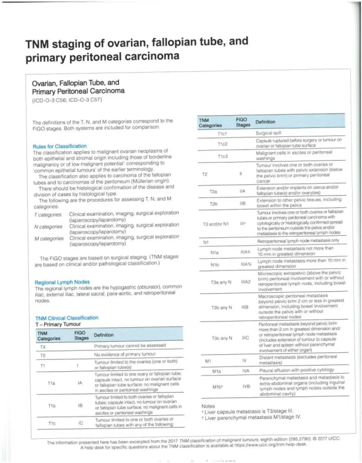
TNM staging of ovarian, fallopian tube, and primary peritoneal carcinoma
Ovarian, Fallopian Tube, and Primary Peritoneal Carcinoma (ICD-O-3 C56; ICD-O-3 C67)
The definitions of the T. N, and M categories correspond to the FIGO stages Both systems are included for comparison TNM Categories FIGO Stages Definition
Tlcl Surgical spilt
Rules for Classification The classification applies to malignant ovarian neoplasms of both epithelial and stromal origin including those of borderline malignancy or of low malignant potential' corresponding to common epithelial tumours' of the earlier terminology. The classification also applies to carcinoma of the fallopian tubes and to carcinomas of the peritoneum (Mullerian origin) There should be histological confirmation of the disease and division of cases by histological type The following are the procedures for assessing T, N. and M categories: T1c2 Capsule ruptured before surgery or tumour on ovarian v falopan tube surface
T1c3 Mabgnant cells In ascites or peritoneal washings
T2 It Tumour involves one or both ovanes or fallopian tubes with pelvic extension (below the petvic brm) or primary pentoneai cancer
T2a HA Extension and/or implants on uterus and/or fallopian tubefs) and/br ovaryties)
T2b IlB Extension to othe» pelvic tissues, including oowef within the pelvis
T categories Clinical examination, imaging surgical exploration (laparoscopy/laparotomy) N categories Clinical examination imaging, surgical exploration (laparoscopy/laparotomy) M categories Clinical examination, imaging, surgical exploration (laparoscopy/laparotomy) T3 and/or Ni III- Turrvxr nvdves one or both ovanes or fallopian tubes or primary peritonea carcinoma with cytoiogically or histologically confrmeo spread to the pentonetm outside the peh'is and/or metastasis Io the retroperitoneal lymph nodes
N1 Retroperitoneal lymph node metastasis only
The FIGO stages are based on surgical staging (TNM stages are based on clinical and/or pathological classification.) Nla IIIAIi Lymph node metastasis not more than Ю mm in greatest dimension
Nib IIIAlii Lymph node metastasis more than 10 mm in greatest dimension
Regional Lymph Nodes The regional lymph nodes are the hypogastric (obturator), common iliac, external iliac lateral sacral, para-aortic and retroperitoneal nodes TNM Clinical Classification T - Primary Tumour T3a any N IIIA2 Microscopic extraoelvic (above the pevic brm) peritoneal involvement with or without retropentoneal lymph node. including bowe* involvement
T3b any N 1118 Macroscopic peritoneal metastasis beyond pelvic brim 2 cm or less in greatest dimension including bowel involvement outside the pefvrs with or without retroperitoneal nooes
Peritoneal metastasis beyond pelvic brim more than 2 cm n greatest dvnensiori and/ O' retroperitoneal lymph node metastasis (includes extension of tumour to capsule of (ver and spleen without parenchymal
TNM FIGO n Categories Stages T3c anyN me
TX Primary tumour cannot be assessed
TO No evidence of primary tumour rrvolvement of either organ) |
_ Tumour limited to the ovaries (one ex both) or fallopian tube(s| M1 IV Distant metastasis (excludes peritoneal melastas si
Tumour limited to one ovary or fallopian tube Mia IVA Pteurai effusion with positive cytology
., capsule intact, no tumour on ovarian surface or fallopian tube surface no maignant ce«s in ascites or peritoneal washings Mlb1 IVB Parenchymal metastasis and metastasis to extra-abdominal organs (including inguina' lymph nooes and lymph nodes outside the
Tixnour limited to both ovaries o' fallopian abdominal cavity)
T („ tubes; capsule intact, no tumour on ovarian or fallopian tube surface, no malignant cells m asexes or peritonea' washings Notes “ Liver capsule metastasis is T3/stage III
T1 Tumour limited to one or both ovaries or fallopian tubes with any ol tl«e following: • Liver parenchymal metastasis Ml/stage IV
The mltxmabon presented here has been excerpted from the 2017 TNM classification of malignant tumours, eighth edition (295,27901 © 2017 UICC A help desk for specilic ewestrons about the TNM classification is available at https //www wcc org/inm-help-desx
N - Regional Lymph Nodes
NX Regional lymph nodes cannot be assessed
NO No regional tymph node metastasis
N1 Regional lymph node metastasis
N1 IIIA1 Retroperitoneal lymph node metastasis only
N1a IIIAIi Lymph node metastasis no more than 10 mm in greatest dimension
N1b IIIAlii Lymph node metastasis more than 10 mm in greatest dimension
M - Distant Metastasis
MO No distant metastasis
M1 Distant metastasis
Mia Pleural effusion with positive cytology
Mib Parenchymal metastasis and metastas s to extraabdominal organs (including inguinal lymph nodes and lymph nodes outside the abdominal cavity)
pTNM Pathological Classification
The pT and pN categories correspond to the T and N categories
pNO Histological examination of a pelvic lymphadenectomy specimen will ordinarily include Ю or more lymph nodes If the lymph nodes are negative, but the number ordinarily examined is not met. classify as pNO.
pM - Distant Metastasis’
pM 1 Distant metastasis microscopically confirmed
Note
• pMO and pMX are not valid categories
Stage
Stage I T1 NO MO
Stage IA Tla NO MO
Stage IB T1b NO MO
Stage IC T1c NO MO
Stage II T2 NO MO
Stage IIA T2a NO MO
Stage ПВ T2b NO MO
Stage III Al T1/2 N1 MO
Stage IIIA2 T3a NO Nt MO
Stage III В тзь N0.N1 MO
Stage IIIC T3c NO N1 MO
Stage IV Any T Any N M1
Stage IVA AnyT Any N M1a
Stage IVB AnyT Any N M1b
Reference
1 Tavassoli FA. Devilee P (eds) WHO Classification of Tumours Pathology and Genetics Tumours of the Breast and Female Genital Organs Lyon, France IARC Press, 2003.
TNM ctaninn nf ovarian fallnnian tnho and nrimarv naritnnaal rarr.inoma
17
TNM staging of tumours of the uterus - endometrium
Uterus - Endometrium
(ICD-O-3 C54.0, 1, 3, 8. 9, C55)
The definitions ol the T. N and M categories correspond to the FIGO stages Both systems are included for comparison
Rules for Classification
The classification applies to endometrial carcinomas and carcinosarcomas (malignant mixed mesodermal tumours) There should be histological verification with subdivision by histological type and grading of the carcinomas The diagnosis should be based on examination of specimens taken by endometrial biopsy
The following are the procedures for assessing T, N and M categories
T categories Physical examination and imaging including urography and cystoscopy
N categories Physical examination and imaging including urography
M categories Physical examination and imaging
The FIGO stages are based on surgical staging (TNM stages are based on clinical and/or pathological classification.)
Anatomical Subsites
1 Isthmus uteri (C54 0)
2 Fundus uteri (C54.3)
3 Endometrium (C54 1)
Regional Lymph Nodes
The regional lymph nodes are the pervic (hypogastric [obturator, internal iliac), common and external diac, paramelnal, and sacral) and the para-aortic nodes
TNM Clinical Classification T - Primary Tumour
TNM Categories FIGO Stages Definition
TX Primary tumour cannot be assessed
TO No evidence of primary tumour
T1 I* Tumour conlmod to the corpus uteri'
Tia IA“ Tumour Ixnlted to endometrium or invading less than half of myometrium
T1b IB Tumour invades one half or more of myometrium
T2 II Tirnour invades cervical stroma, but does not extend beyond the uterus
TNM Categories FIGO Stages Definition
T3 III local and/or regional stxead as specified here:
T3a IllA Tumour invades the serosa ol the corpus uteri or adnexae (direct extension or metastasis)
T3b IUB Vaginal or oaramotnal involvement (direct extension or metastasis)
Nt N2 IIIC Metastasis to pelvic or para-aortic lymph nodes’-
N1 IIIC1 Metastasis to pelvic tymph nodes
N2 IIIC2 Metastasis to para-aortic lymph nodes with or without metastasis to pelvic lymph nodes
T4* IV Tumour invades bladder/bowel mucosa
Notes
• Endocervical glandular involvement only should be considered as stage I.
” Positive cytology has to be reported separately without changing the stage
c The presence of buHous oedema is not sufficient evidence to classify as T4.
N - Regional Lymph Nodes
NX Regional lymph nodes cannot be assessed
NO No regional lymph node metastasis
N1 Regional lymph node metastasis to pelvic lymph nodes
N2 Regional lymph node metastasis to para-aortic lymph nodes with or without metastasis to pelvic lymph nodes
M - Distant Metastasis
MO No distant metastasis
M1 Distant metastasis (excluding metastasis to vagina, pelvic serosa, or adnexa, including metastasis to inguinal lymph nodes, mtra-abdominal lymph nodes other than para-aortic or pelvic nodes)
pTNM Pathological Classification
The pT and pN categories correspond to the T and N categories
pNO Histological examination of a pelvic lymphadenectomy specimen will ordinarily include 10 or more lymph nodes If the lymph nodes are negative, but the number ordinarily examined is not met. classify as pNO
pM - Distant Metastasis*
pM 1 Distant metastasis microscopically confirmed
Note
pMO and pMX are not valid categories
G Histopathological Grading
For Histopathological grading use G1, G2. or G3 For details see
Creasman etal 2006’
Reference
1 Creasman WT, Odrcino F. Maisoneuve P, Quinn MA, Beller U, Beoedet JL, Heintz АРМ, Ngan HYS, Pecorell S FIGO Annual Report on the results of treatment m gynaecological cancer. Vol 26. Carcinoma of the corpus uteri Int J Gynecol Obstet 2006; 95 (Suppl 1) 105-143
Stage
Staged Tis NO M0
Stage IA Tia NO M0
Stage IB Tib NO M0
Stage II T2 NO MO
Stage IIIA T3a NO MO
Stage IU0 T3b NO MO
Stage НЮ T1.T2.T3 N1.N2 MO
Stage IIIC1 T1.T2.T3 N1 M0
Stage IIIC2 T1.T2.T3 N2 MO
Stage IVA T4 Any N MO
Stage IVB Any T Any N Ml
TNM staging of uterine sarcomas
Uterine Sarcomas
(Leiomyosarcoma. Endometrial Stromal Sarcoma. Adenosarcoma) (ICD-O-3 C53, 54. 54.1, 54.2, 55)
The definitions of the T, N. and M categories correspond to the FIGO stages Both systems are included for comparison TNM FIGO Der!f,1Uon Categories Stages T3 III Tumour infiltrates abdominal tissues
Rules for Classification The classification applies to sarcomas except for carcinosarcoma, which is classified as carcinoma of the endometrium. There should be histological confirmation and division of cases by histological type The following are the procedures for assessing T. N, and M categories T categories Physical examination ano imaging N categories Physical examination and imaging M categories Physical examination and imaging T3a IIIA Ono site T3b IIIB More than one site N1 IIIC Metastasis to regional lymph nodes T4 IVA Tumour invades bladder or rectum M1 IVB Distant metastasis Note Simultaneous tumours of the uterine corpus and ovary/pelvis in association with ovarian/pelvic endometriosis should be classified as independent primary tumours
The FIGO stages are based on surgical staging. (TNM stages are based on clinical and/or pathological classification) Adenosarcoma T - Primary Tumour
Anatomical Subsites 1 Cervix uteri (C53) 2 Isthmus uteri (C54 0) 3 Fundus uteri (C54 3) . 2®° Definition Categories Stages Tl 1 Tumour limited to the uierus . Tumour limited to the endometrium/ Tla A endoccrvix
Histological Types of Tumours Leiomyosarcoma 8890/3 Endometrial stromal sarcoma 8930/3 Adenosarcoma 8933/3 . _ Tumour invades lo less than half of the myometrium T _ Tumour invades more than hall of tne c myometrium T .. Tixnour extends beyond the uierus. within
Regional Lymph Nodes The regional lymph nodes are the pelvic (hypogastric [obturator internal iliac], common and external iliac, parametnal. and sacral) and the para-aortic nodes. z the pelvis T2a IIA Tumour mvotves adnexa T2b IIB Tumour involves other pelvic Issues T3 III Tumour involves abdominal tissues
TNM Clinical Classification Leiomyosarcoma. Endometrial Stromal Sarcoma T - Primary Tumour TNM FIGO Categories Stages T1 I Tumour limited Ю the uierus T3a IIIA One site T3b IIIB More than one site N1 НЮ Metastasis to regional lymph nodes T4 IVA Tumour invades bladoer or rectum Mt IVB Distant metastasis
Tla IA Tumour 5 cm or less m greatest dimension Tib IB Tumour more than 5 cm Note Simultaneous tumours of the uterine corpus and ovary/petvis in association with ovarian/pelvic endometriosis should be
_ Tumour extends beyond the uterus, within the TZ petvis T2a fl A Tumour involves adnexa classified as independent primary tumours
T2b IIB Tumour involves other pelvic tissues
N - Regional Lymph Nodes
NX Regional lymph nodes cannot be assessed
NO No regional lymph node metastasis
Ni Regional lymph node metastasis
M - Distant Metastasis
MO No distant metastasis
Mt Distant metastasis (excluding adnexa, pelvic and abdominal tissues)
pTNM Pathological Classification
The pT and pN categories correspond to the T and N categories
pM - Distant Metastasis*
pMt Distant metastasis microscopically confirmed
Note
• pMO and pMX are not valid categories.
Stage - Uterine Sarcomas
Stage I T1 NO M0
Stage IA Па NO M0
Stage I8 T1b NO MO
Stage IC* Tic NO MO
Stage II T2 NO MO
Stage HA T2a NO MO
Stage ПВ T2b NO MO
Stage ll IA T3a NO MO
Stage HIB T3b NO MO
Slags UIC T1.T2.T3 N1 MO
Stage IVA T4 Any N MO
Stage IVB Any T Any N M1
Note
Stage IC does not apply for leiomyosarcoma and endometrial stromal sarcoma
References
1 Prat J FIGO staging for uterine sarcomas Int J Gynaecol Obstet 2009; 104 177-178
2 FIGO Committee on Gynecologic Oncology Report FIGO staging for uterine sarcomas. Int J Gynaecol Obstet 2009: 104 179
TNM cfanino nf Torino porr/wae
TNM staging of gestational trophoblastic neoplasms
Gestational Trophoblastic Neoplasms
(ICD-0-3 C58)
The following classification for gestational trophoblastic tumours is based on that of FIGO adopted in 1992 and updated in 2002.’ The definitions of T and M categones correspond to the FIGO stages Both systems are included for comparison. In contrast to other sites, an N (regional lymph node) classification does not apply to these tumours A prognostic scoring index, which is based on factors other than the anatomic extent of the disease, is used to assign cases to high-risk and low-risk categories, and these categories are used in stage grouping
Rules for Classification
The classification applies to choriocarcinoma (9100/3). invasive hydatidiform mole (9100/1), and placental site trophoblastic tumour (9104/1) Placental site tumours should be reported separately Histological confirmation is not required If the human chorionic gonadotropin (phCG) level is abnormally elevated History of prior chemotherapy for this disease should be noted
The following are the procedures for assessing T and M categones:
7 categories Clinical examination, imaging and endoscopy, and serum/urine phCG level
M categones Clinical examination, imaging, and assessment
of serum/unne phCG level
Risk categories: Age, type ol antecedent pregnancy, interval months from index pregnancy pretreatment serum/urine phCG, diameter of largest tumour, site of metastasis, number of metastases, and previous failed chemotherapy are integrated to provide a prognostic score that divides cases into low and high-risk categories
TM Clinical Classification T - Primary Tumour
TM Categones FIGO Stages* Definition
TX Primary tumour cannot be assessoo
TO No evidence ol prima-y tumour
T1 1 Tumour confined to uterus
T2° II Tumour extends to other genital structures vagina, ovary, broad '«gamont fallopian tube by metastasis or direct extension
Mia III Metastasis 10 lungts)
Mtir IV Other distant metastasis
Notes
“ Stages I to IV are subdivided into A and В according to the prognostic score,
b Genital metastasis (vagina, ovary broad ligament fallopian tube) is classified T2.
Any involvement of non-genital structures, whether by direct invasion or metastasis is described using the M classification.
pTM Pathological Classification
The pT categories correspond to the T categories
pM - Distant Metastasis*
pM1 Distant metastasis microscopically confirmed
Note
' pMO and pMX are not valid categories
Stage
Stage 1 TI MO
Stage II T2 M0
Stage III Алу T M1a
Stage IV Any T Mlb
Reference
1 Ngan HYS. Bender H, Benedet JL, Jones H, Montrucolli GC. Pecoreili S: FIGO Committee on Gynecologic Oncology Gestational trophoblastic neoplasia Ini J Gynecol Obstet 2002:77 285 287.
TNM staging of tumours of the cervix uteri
Cervix Uteri
(ICD-O-3C53)
The definitions of the T and M categories correspond to the FIGO stages Both systems are included for comparison
Rules for Classification
The classification applies only to carcinomas. There should be histological confirmation of the disease
The following are the procedures for assessing T, N, and M categories
T categories Clinical examination and imaging' N categories Clinical examination and imaging M categories Clinical examination and imaging
Note
' The use of diagnostic imaging techniques to assess the size ol the primary tumour is encouraged but is not mandatory Other investigations, e g examination under anaesthesia, cystoscopy, sigmoidoscopy, intravenous pyelography, are optional and no longer mandatory
The FIGO stages are based on clinical staging. For some Stage I subdivisions (IA IB 1) are mainly pathological, including the histological examination of the cervix. (TNM stages are based on clinical and/or pathological classification.)
Anatomical Subsites
1 Endocervix (C53 0)
2 Exocervix (C53.1)
Regional Lymph Nodes
The regional lymph nodes are the paracervical, parametria!, hypogastric (internal iliac, obturator), common and external iliac, presacral, lateral sacral nodes, and para-aortic nodes ’
Note
In the 7th edition the para-aortic nodes were considered to be distant metastatic but to be consistent with advice from FIGO the para aortic nodes are now classified as regional.
TNM Clinical Classification
T - Primary Tumour
TNM Categories FIGO Stages Definition
Tiau- IA Invasive carcinoma diagnosed only by microscopy Stromal invasion with a maximal depth of 5 0 mm measured from the base ol lhe epithelium and a horizontal spread ol 7.0 mm or less"
Tiai IA1 Measured stromal invasion 3.0 mm or less m depth and 70 mm or less in horizontal spreao
Tta2 IA2 Measired stromal invasion more than 3 0 mm and not more than 5.0 mm wilh a horizontal spread of 7.0 mm or less
Tib IB Clinically visible lesxxi confined to the cervix or microscopic lesion greater than T1a/IA2
T1b1 IB1 Clinically visible les»on 4.0 cm or less in 9'catcst dimension
T1b2 IB2 Clinically visible lesion more than 4 .0 cm in greatest dimension
T2 11 Tumour invades beyond uterus but not to pelvic waH or to lower thrrd of vagina
T2a HA Tumour without parametrial invasion
T2a1 IIA1 Clinically visible lesion 4.0 cm or less in greatest dimension
T2a2 IIA2 Clinically visible lesion more than 4 0 cm in greatest dimension
T2b IIB Tumour with parametrial invasion
T3 III Tumour involves tower third of vagina, or extends to pelvic wall, or causes hydronephrosis or non-functioning кюпеу
T3a IIIA Tumour involves ower third of vagina
T3b IIIB Tumour extends to pelvic wail, or causes hydronephrosis or non-functioning kdney
T4 IVA Tumour invades mucosa of the blander or rectum, or extends beyond true pelvis"
TNM Categories FIGO Stages Definition
TX Primary tumour cannot be assessed
TO No evidence of primary tumour
Tis Carcinoma in situ (preinvasive carcinoma)
T1 I Tumour confined to the cervix"
Notes
' Extension to corpus uteri should be disregarded.
The depth ol invasion should be taken from the base of the epithelium, either surface or glandular, from which it originates The depth of invasion is defined as the measurement of the tumour from the epithehal-stromal junction of the adjacent most superficial papillae to the doepost point of invasion.
All macroscopically visible lesions even with superficial invasion are Tib/IB
’ Vascular space involvement, venous or lymphatic, does not affect classification
* Bullous oedema is not sufficient to classify a tumour as T4
The information presented here has been excerpted from the 2017 TNM classification of malignant tumours, eighth edition I295.2790) © 2017 UiCC A help desk 1or specific questions about lhe TNM classification is available at https://www.uicc.org/tnm-halp-desk.
N - Regional Lymph Nodes* Stage Tis У1 NO NO
NX Regional lymph nodes cannot be assessed Stage 0 MU MO
NO No regional lymph node metastasis Stage I S'agt* IA Tta NO MO
N1 Regional'ymph node metastasis Stage I At T1a1 NO MO
Stage IA2 Tia2 NO MO
Note Stage IB Tib NO MO
* No FIGO equivalent Stage IB1 T1b1 NO MO
Stage IB2 T1b2 NO MO
M - Distant Metastasis MO No distant metastasis Mt Distant metastasis (includes inguinal lymph nodes and Stage II Stage HA Stage IIA1 Stage IIA2 T2 T2a Т2а1 T2a2 NO NO NO NO MO MO MO MO
intraperitoneal disease). It excludes metastasis to vagina, Stage ПВ T2b NO MO
pelvic serosa, ano adnexa Stage III T3 NO MO
Stage IIIA T3a NO MC
Stage 11 IB T3b Any N MO
pTNM Pathological Classification The pT and pN categories correspond to the T and N categories Stage IVA Stage IVB T1.T2T3 TA Any 1 N1 Any N Any N MO MO M1
pNO Histological examination ot a pelvic lymphadenectomy specimen will ordinarily include 10 or more lymph nodes If the lymph nodes are negative but the number ordinarily examined is not met, classify as pNO
pM - Distant Metastasis*
рМ1 Distant metastasis microscopically confirmed
Note
• pMO and pMX are not valid categories.
TNM staging of tumours of the vagina
Vagina
(ICD-O-3C52)
The definitions of the T and M categories correspond to the FIGO stages Both systems are included for comparison
N - Regional Lymph Nodes
NX Regional lymph nodes cannot be assessed
NO No regional lymph node metastasis
N1 Regional lymph node metastasis
Rules for Classification
The classification applies to primary carcinomas only. Tumours present in the vagina as secondary growths from either genital o< extragenital sites are excluded A tumour that has extended to the portio and reached the external os (orifice of uterus) is classified as carcinoma of the cervix A vaginal carcinoma occurring 5 years alter successful treatment (complete response) of a carcinoma of the cervix uteri is considered a primary vaginal carcinoma A tumour involving the vulva is classified as carcmoma of the vulva There should be histological confirmation of the disease
The following are the procedures for assessing T. N. and M categories:
Г categories Physical examination, endoscopy, and imaging
N categories Physical examination and imaging M categories Physical examination and imaging
The FIGO stages are based on surgical staging (TNM stages are based on clinical and/or pathological classification)
M - Distant Metastasis
MO No distant metastasis
M1 Distant metastasis
TNM Pathological Classification
The pT and pN categories correspond to the T and N categories.
pNO Histological examination of an inguinal lymphadenectomy specimen will ordinarily include 6 or more lymph nodes, a pelvic lymphadenectomy specimen will ordinarily include 10 or more lymph nodes If the lymph nodes are negative, but the number ordinarily examined is not met classify as pNO
pM - Distant Metastasis*
pM1 Distant metastasis microscopically confirmed
Note
pMO and pMX are not valid categories
Regional Lymph Nodes
Upper two-thirds of vagina the pew nodes including obturator.
ntemai iliac (hypogastric) external iliac. and pelvic nodes, NOS
Lower third of vagina the inguinal and femoral nodes
TNM Clinical Classification T - Primary Tumour
TNM Categories FIGO Stages Definition
TX Primary tumour cannot be assessed
TO No evidence of primary tumour
TIs Carcinoma in situ (premvasive carcinoma)
T1 1 Tumour confined Ю vagina
T2 II Tumour nvaocs paravaginal tissues (paracolpium)
T3 III Tumour exlenos to pelvic wai
T4 IVA Tumour invades mucosa ot bladder or rectum or extends beyond the true pelws"
Ml IVB Distant metastasis
Stage
Stage 0 Tis NO MO
Stage 1 T1 NO MO
Stage II T2 NO MO
Stage III T3 NO MO
T1.T2.T3 N1 MO
Stage IVA T4 Any N MO
Stage IVB Any T Any N M1
Note
’ The presence of bullous oedema is not sufficient evidence to classify a tumour as T4
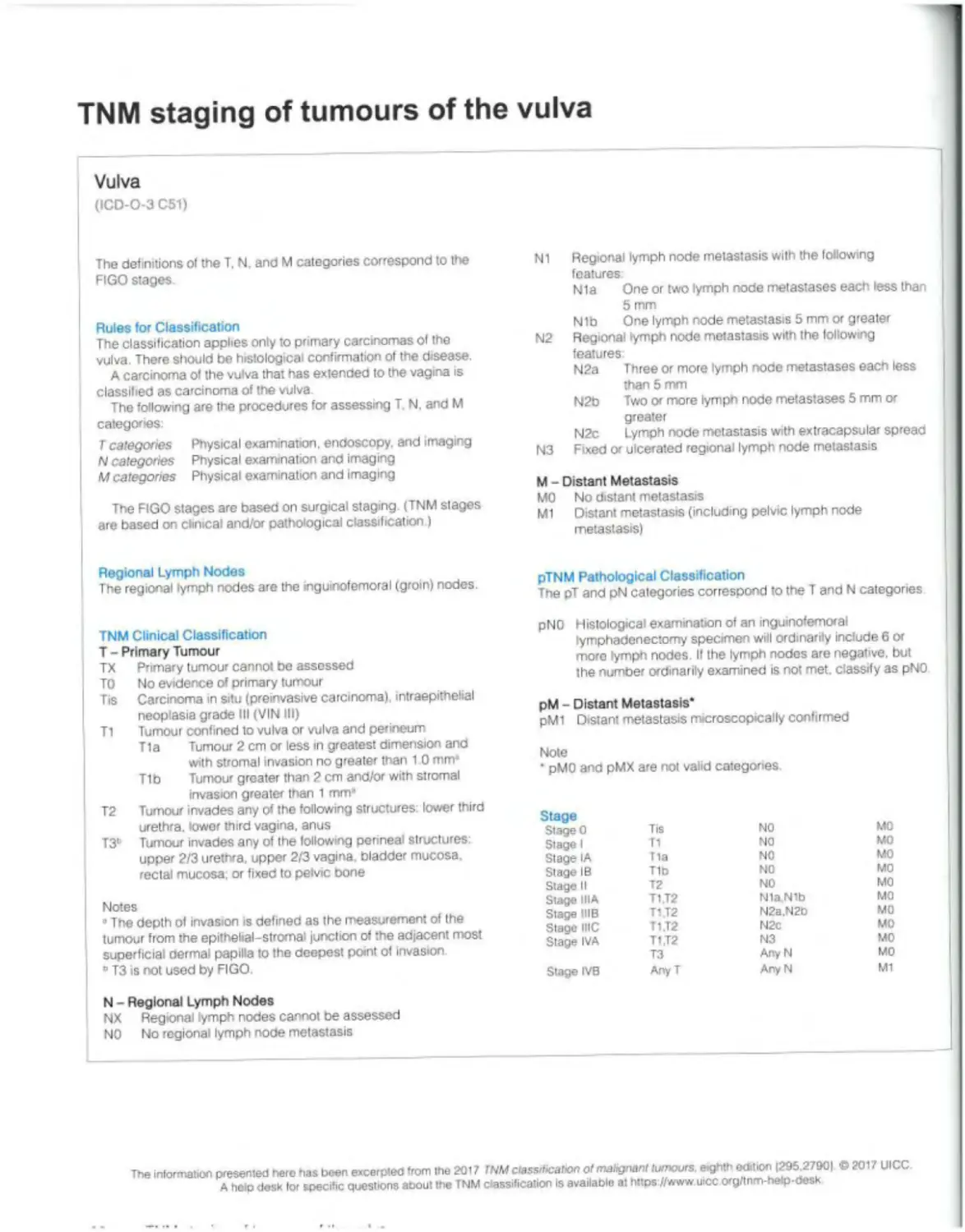
TNM staging of tumours of the vulva
Vulva
(ICD-0-3 C51)
The detritions of the T, N. and M categories correspond to the FIGO stages
Rules for Classification
The classification applies only to primary carcinomas of the vulva. There should be histological confirmation of the disease.
A carcinoma of the vulva that has extended to the vagina is classified as carcinoma of the vulva
The following are the procedures for assessing T N, and M categories
Tcategories Physical examination, endoscopy, and imaging
N categories Physical examination and imaging
M categories Physical examination and imaging
The FIGO stages are based on surgical staging (TNM stages are based on clinical and/or pathological classification )
N1 Regional lymph node metastasis with the following features
N1a One or two lymph node metastases each less than 5 mm
Nib One lymph node metastasis 5 mm or greater N2 Regional lymph node metastasis with the following features
N2a Three or more lymph node metastases each less than 5 mm
N2b Two or more lymph node metastases 5 mm or greater
N2c Lymph node metastasis with extracapsular spread N3 Fixed or ulcerated regional lymph node metastasis
M - Distant Metastasis
MO No distant metastasis
Ml Distant metastasis (including pelvic lymph node metastasis)
Regional Lymph Nodes
The regional lymph nodes are the inguinofemoral (groin) nodes.
TNM Clinical Classification
T - Primary Tumour
TX Primary tumour cannot bo assessed
TO No evidence of primary tumour
Tis Carcinoma m situ (preinvasive carcinoma), intraepithelial neoplasia grade III (VIN III)
Tl Tumour confined to vulva or vulva and perineum
Tla Tumour 2 cm or less in greatest dimension and with stromal invasion no greater than 1 0 mm-
T1b Tumour greater than 2 cm and/or with stromal invasion greater than 1 mm»
T2 Tumour invades any of the fotlowing structures: lower third urethra, lower third vagina, anus
T3b Tumour invades any of the following penneai structures: upper 2/3 urethra, upper 2/3 vagina, bladder mucosa rectal mucosa, or fixed to pelvic bone
Notes
»The depth of invasion is defined as the measurement of the tumour from the epithelial-stromal junction of the adjacent most superficial dermal papilla to the deepest point of invasion
° T3 is not used by FIGO.
N - Regional Lymph Nodes
NX Regional lymph nodes cannot be assessed
NO No regional lymph node metastasis
pTNM Pathological Classification
The pT and pN categories correspond to the T and N categories
pNO Histological examination of an inguinofemoral fymphadenectorny specimen will ordinarily include 6 or more lymph nodes. If the lymph nodes are negative, but the number ordinarily examined is not met. classify as pNO
pM - Distant Metastasis*
pM1 Distant metastasis microscopically confirmed
Note
pMO and pMX are not valid categories.
Stage
Stage 0 Tis NO MO
Stage 1 T1 NO MO
Stage IA Т1а NO MO
Stage IB T1b NO MO
Stage II T2 NO MO
Stage IIIA T1.T2 N1a Nib MO
Stage IIIB T1.T2 N2a,N2b MO
Stage IHC T1.T2 N2c MO
Stage IVA T1.T2 N3 MO
T3 Any N MO
Stage IVB Any T Any N M1
Soft Tissues
(ICD-0-3 C38.1, 2, 3. C47-49)
Rules for Classification
There should be histological confirmation of the disease and division of cases by histological type and grade
The following are the procedures for assessing T. N. and M categories
Г categones Physical examination and imaging
N categories Physical examination and imaging
M categories Physical examination and imaging
Anatomical Sites
1 Connective, subcutaneous, and other soft tissues (C49) peripheral nerves (C47)
2 Retroperitoneum (C48.0)
3 Mediastinum anterior (C38 1); posterior (C38.2); mediastinum. NOS (C38 3)
Histological Types of Tumour
The following histological types are not included
• Kaposi sarcoma
• Dermatofibrosarcoma (protuberans)
• Fibromatosis (desmoid tumour)
• Sarcoma arising from the dura mater or bram
• Angiosarcoma, an aggressive sarcoma, is excluded because its natural history is not consistent with the classification.
Note
Cystosarcoma phyllodes is staged as a soft tissue sarcoma of the superficial trunk.
Regional Lymph Nodes
The regional lymph nodes are those appropriate to the site of the primary tumour Regional node involvement is rare and cases in which nodal status is not assessed either clinically or pathologically could be considered NO instead of NX or pNX
TNM Clinical Classification
T - Primary Tumour
TX Primary tumour cannot be assessed
TO No evidence of primary tumour
Extremity and Superficial Trunk
T t Tumour 5 cm or less in greatest dimension
T2 Tumour more than 5 cm but no more than 10 cm in greatest dimension
T3 Tumour more than Ю cm but no more than 15 cm in greatest dimension
T4 Tumour more than 15 cm in greatest dimension
Retroperitoneum
T1 Tumour 5 cm or less in greatest dimension
T2 Tumour more than 5 cm but no more than Ю cm in greatest dimension
T3 Tumour more than 10 cm but no more than 15 cm in greatest dimension
T4 Tumour more than 15 cm in greatest dimension
Head and Neck
T1 Tumour 2 cm or less in greatest dimension
T2 Tumour more than 2 cm but no more than 4 cm in greatest dimension
T3 Tumour more than 4 cm in greatest dimension
T4a Tumour invades the orbit, skull base or dura, central compartment viscera facial skeleton, and/or pterygoid muscles
T4b Tumour invades the bram parenchyma, encases the carotid artery, invades prevertebral muscle or involves the central nervous system by perineural spread
Thoracic and Abdominal Viscera
T1 Tumour confined to a single organ
T2a Tumour invades serosa or visceral peritoneum
T2b Tumour with microscopic extension beyond the serosa
T3 Tumour invades another organ or macroscopic extension beyond the serosa
T4a Multifocal tumour involving no more than two sites in one organ
T4b Multifocal tumour involving more than two sites but not more than 5 sites
T4c Multifocal tumour involving more than five sites
N - Regional Lymph Nodes
NX Regional lymph nodes cannot be assessed
NO No regional lymph node metastasis
Nl Regional lymph node metastasis
M - Distant Metastasis
MO No distant metastasis
M1 Distant metastasis
pTNM Pathological Classification
The pT and pN categories correspond to the T and N categories
pM - Distant Metastasis’
pM1 Distant metastasis microscopically confirmed
Note
' pMO and pMX are not valid categories.
Stage - Extremity and Superficial Trunk and Retroperitoneum
Stage lA T1 NO MO G1 GX Low Grade
Stage IB Т2.ТЗТ4 NO MO G1.GX Low Grade
Stage II T1 NO MO G2.G3 High G’aoe
Stage IIIA T2 NO MO G2.G3 High G-ade
Stage ШВ T3.T4 NO MO G2.G3 High Grade
Any T N1* MO AnyG
Stage IV Any T Any N Mt Any G
Stage - Head and Neck and Thoracic and Abdominal Viscera There is no stage for soft tissue sarcoma of the head and neck and thoracic and abdomina' viscera
Note
• AJCC classifies N1 as stage IV for extremity and superficial trunk
The information presenter; ha»e has been excerpted from the 2017 TNM classification of malignant tumours, eighth edition 1295.27901 ©2017 UICC A help desk tor specific questions about me TNM classification is availaole at https INnavi jicc.org/tnm-help-desk
TNM ctaninn nf ti imra ire nf cnft tisci ioc
General introduction
Lax SF
Cheung AN Oliva E
The classification of female genital tumours has evolved sig-nifcantly over the past decade in light of new and often key molecular discoveries that have influenced the categorization of a number of these neoplasms. Distinctive molecular altera-i ons are now known for many female genital tumours and have proved to be helpful for their correct categorization. Therefore, as in other organ systems, the morphological classification continues to advance into an integrated morphological-molecular classification that will have an impact on diagnosis and treatment. One of the best-known applications of molecular pathology pertains to endometrial carcinomas: surrogate immunohistochemical markers (except for POLE) are used to stratify these tumours into four subgroups with distinctive prognoses, as defined by The Cancer Genome Atlas (TCGA) project However, it is recommended that the molecular characterization of endometrial carcinoma should be restricted to high-grade tumours, because more than two thirds of endometrial carcinomas are low-stage and low-grade endometrioid carcinomas associated with an excellent prognosis and do not need molecular profiling. The classification of female genital tumours as well as those of other organ systems, is intended for worldwide use, and there are many areas where, for various reasons, the application of immunohistochemistry or molecular pathology is limited Additionally, standard aspects of conventional pathology remain the bedrock of the interpretation and classification of female genital tract (FGT) neoplasms in routine practice, and to emphasize this point, the essential diagnostic criteria for each entity in this volume are based on these characteristics.
The organization of this fifth edition of the female genital tumours volume follows the general structure of other volumes in this series and differs from the prior edition in several respects. To avoid repetition, new chapters have been created to amalgamate entities that have similar morphological and molecular findings at different sites, including mesenchymal tumours of the lower FGT, metastases to the lower FGT. haematopoietic neoplasms, neuroendocrine neoplasms (NENs), and associated hereditary syndromes. NENs of the FGT (with the exception of ovarian carcinoid tumours, due to their specific characteristics and germ cell origin) are now classified following the terminology of their pulmonary and pancreatic counterparts and a recently published consensus proposal initiated by the International Agency tor Research on Cancer (IARC) and WHO (2292), although studies are still needed with regard to optimal terminology in the FGT. Among the haematopoietic neoplasms, emphasis is given to those entities more commonly occurring within the FGT and specifically in each organ.
Terminology is the common language used by pathologists and treating physicians for optimal patient care, and it snould be consistently applied. Therefore, before making a change to terminology, it is important to consider the impact
of that change. For this reason, the editorial board preferred to retain the existing, well-established terminology for several tumours. For example, in the ovary, the term “small cell carcinoma of hypercalcaemic type" has been retained over the proposed rhabdoid tumour terminology, because although most of these tumours are associated with SMARCA4 mutations, only a minority show overt rhabdoid morphology, and it is still controversial whether they are identical to rhabdoid tumours. On the other hand, some important revisions have been made since the fourth edition of the WHO classification. For example, serous borderline tumours with micropap-illary architecture are now included within the broad group of serous borderline tumours, and the term "non-invasive low-grade serous carcinoma" is not recommended for them. Low-grade and high-grade serous ovarian carcinomas are considered distinct tumour types rather than a spectrum, because they have different origins, as well as different morphology and molecular and genetic signatures. Among the uterine neoplasms, carcinosarcoma is now classified as an endometrial carcinoma rather than a mixed tumour, on the basis of molecular and biological similarity to high-grade endometrial carcinomas. Undifferentiated endometrial carcinoma. including dedifferentiated carcinoma, is considered a distinct entity supported by molecular alterations involving the SWl/SNF pathway. Minor changes include the reclassification of different subtypes of endometrioid carcinoma (e g. with squamous differentiation) as simply patterns of endometrioid carcinoma, leading to a leaner classification. Among the mesenchymal tumours, the category of smooth muscle tumours has been expanded with the new category of fumarate hydratase deficient leiomyomas, and the category of high-grade endometrial stromal sarcoma has been expanded to include neoplasms with novel gene fusions (in particular involving the BCOR gene), whereas the undifferentiated uterine sarcoma entity has been redefined. In the category of mixed tumours of the uterus, the term ''adenofibroma" is no longer recommended.
HPV has dramatically changed the approach to epithelial tumours of the lower FGT, and this shift is reflected herein. Adenocarcinomas of the uterine cervix are now divided into HPV-associated and HPV-independent adenocarcinomas, with implications for prognosis. More importantly, recent studies have put forward a reproducible histological surrogate for HPV infection allowing for such classification without HPV analysis. Squamous tumours of the lower FGT are also divided into HPV-associated and HPV-independent. because important differences in outcome exist Squamous precursor lesions throughout the lower FGT are primarily classified according to the binary Lower Anogenital Squamous Terminology (LAST) standardization project proposal, but the three-tiered intraepithelial neoplasia classification can still be used.
Most grading systems rely on mitotic activity and necrosis. An important change in this edition of the WHO Classification of Tumours series is the conversion of mitotic count from the traditional denominator of 10 HPF to a defined area expressed in mm* This serves to standardize the true area over which mitoses are enumerated, because different microscopes have high-power fields of different sizes. This change will also be helpful for anyone reporting using digital systems The approximate number of fields per 1 mm2 based on the field diameter and its corresponding area is presented in Table A.
Finally, increased knowledge about hereditary tumour syndromes has enabled us to detect earners at an earty age through counselling and genetic testing, more commonly in Lynch
Table A Approximate number of fekfc per 1 mm; based on the field diameter and is corresponding area
Field diameter (mm) Field area (mm7) Approximate number of fields per 1 mm1
0.40 0126 8
0.41 0.132 8
0.42 0138 7
0.43 0.145 7
0.44 0.152 7
0.45 0.159 6
0.46 0166 6
0.47 0173 6
0.48 0181 6
0.49 0.188 5
0.50 0.196 5
0.51 0204 5
0.52 0-212 5
0.53 0.221 5
0.54 0.229 4
0.55 0.237 4
0.56 0.246 4
0.57 0.255 4
058 0 264 4
0.59 0.273 4
0.60 0.283 4
0.61 0.292 3
0.62 0.302 3
0.63 0.312 3
0.64 0.322 3
0.65 0.332 3
0.66 0.342 3
067 0.352 3
068 0363 3
0 69 0.374 3
syndrome but also for other rare syndromes. Lynch syndrome patients may be the best-known example, because endometrial cancer is not infrequently their first presentation. Immunohistochemistry. which can be performed in most pathology settings s key for triaging these patients for further studies. Another example is patients with Peutz-Jeghers syndrome, who typically have bilateral, small, and multifocal sex cord tumours with annular tubules and may have cervical gastric-type mucinous adenocarcinoma. typically associated with STK11 alterations. These examples illustrate the increasingly important role of pathologists in tne multidisciplinary assessment of hereditary diseases.
The ICD-0 topographical coding for the anatomical sites covered in this volume is presented in Box A.
Box A ICO-O topographical coOng lor the mar anatomical sites covered in ths volume {817}
C51 Vulva
C51.0 Labium majus
C51.1 Labium minus
C51.2 Clitoris
C51 8 Overlapping lesion ot vulva
C51.9 Vulva NOS
C52 Vagina
C52 9 Vagxta NOS
C53 Cervix uteri
C53 0 Endocervix
C53.1 Exooervix
C53 8 Overlapping lesion ot cervix uteri
C53.9 Cervix uteri
C54 Corpus uteri
C54.0 Isthmus liter
C54 1 Endometrium
C54 2 Myometrium
C54 3 Fundus uteri
C54 8 Overlapping lesion of corpus uteri
C54.9 Corpus uteri
CS5 Uterus NOS
C55 9 Uterus NOS
C56 Ovary
C56.9 Ovary
CS7 Other and unspecified female genital organs
C57.0 Fallopian tube
C57.1 Broad Sgament
C57.2 Round ligament
C57.3 Parametrium
C57.4 Uterine adnexa
C57.7 Other specified parts of female genitaj organs
C57.8 Overlapping lesion of female genital organs
C57.9 Female genital tract NOS
C58 Placenta
C58.9 Placenta
Tumours of the ovary
Edited by: Cheung AN. Ellenson LH, Gilks CB, Kim K-R, Kong CS. Lax SF, Longacre TA, Malpica A, McCluggage WG. Oliva E. Rabban JT, Soslow RA
Serous tumours
Mucinous tumours
Endometrioid tumours
Clear cell tumours
Seromucinous tumours
Brenner tumours
Other carcinomas
Mesenchymal tumours
Mixed epithelial and mesenchymal tumours
Sex cord-stromal tumours
Germ cell tumours
Miscellaneous tumours
Mesothelial tumours (see Chapter 3)
Tumour-like lesions
Metastases
Tumours of the ovary: Introduction
McCluggage WG LaxSF Longacre TA Malpica A Soslow RA
Estimated age-standardized incidence rates (World) in 2018, ovary, all ages
Fig. 1.01 Ovary cancer map. Estimated age standardized incidence rates (ASRs; World) per 100 000 person-years, of ovarian cancer in 2018.
In 2018, ovarian cancer ranked as the eighth most common cancer diagnosis and cause of cancer death in women, with an estimated 295 000 cases and 184 000 deaths worldwide (293) The most common histological type is high-grade serous carcinoma (HGSC) |337). There is geographical variation, with high incidence rates in North America, central-eastern Europe, and south-eastern Asia, and low rates in sub-Saharan Africa and western Asia (293) In most countries where long historical data exist, a gradual decline in the incidence of ovarian cancer where rates were traditionally highest has been reported (337). The observed decline has been mainly attributed to the widespread use of oral contraceptive pills that have long-lasting protective effects against ovarian cancer after several years of use |337,2753). In addition, other reproductive factors such as higher parity (higher number of children) and breastfeeding have also been reported as protective factors against ovarian cancer. In Israel, where women are reported to have some of the highest frequencies of germline BRCA1/2 mutation (337,2753). which also increases ovarian cancer risk by 8-48% (2138|. a decreasing trend has been noted. This finding indicates that high rates of risk-reducing surgery can be added to the factors mentioned above. Other factors, such as tobacco smoking and obesity, have been related to an increased risk of certain types of ovarian cancers (131), with obesity being related to an observed increase in ovarian cancer in younger cohorts in some
populations. Finally, there have been temporal changes linkec to changes in the classification of borderline ovarian tumours (337). which, since ICD-O-3 in 1990, are now grouped as nori-malignant. This might have contributed to the decreasing rates in some countries, which by now should have stabilized.
The classification of ovarian neoplasms in this fifth edition ol the WHO classification of tumours is largely unchanged from the previous edition. One addition to the current classification is mesonephric-like carcinoma (1737|. The category of mixed carcinoma has also been reintroduced in the current classification, although it is stressed that mixed carcinomas of the ovary are very uncommon and that most carcinomas that look mixed from a morphological viewpoint in fact represent a single neoplastic type with areas morphologically mimicking another tumour type (984,1807).
The previous WHO classification first divided ovarian serout carcinoma into low-grade and high-grade carcinomas, whict represent two completely different tumour types (with differen morphology, pathogenesis, molecular events, and progno sis) rather than low-grade and high-grade forms of the samr neoplasm (2543| It is now well established that a significan majority of so-called ovarian HGSCs arise from the distal fim brial end of the fallopian tube from a precursor lesion knowi as serous tubal intraepithelial carcinoma, whereas almost a low-grade serous carcinomas (LGSCs) arise within the ovar
Asia
AusIraSa/New Zealand
Central & South America
Perod 1988-1992
Tnailand Hiilippoe» Japan Israel India Chra
cuadc.r
Costa Rica
Cotomoia
Eastern EuropeCaKjl Repub(c
USA<WM»)
North America usA(Biac*>
Canada
Northern Europe
Southern Europe
Western Europe
Spain Stavenia Italy Croatia
Switzerland France
0% 20% 40% 60% 80% 100%
Thailand Philippines Japan Israel India China
hew Zea and Australia
Ecuador
Costa Rica Colombia
Slovakia Czech Republic
USAiWMel USA(Back)
Canada
Scotland Norway Estonia
Denmark
Spain Slovenia
Italy Croatia
Switzerland France
Period 2008-2012
0% 20% 40% 60% 80% 100%
1ИВ Sercui oa-c noma ИИ Muatioui caranom*
EndoMtikM caronoma Oar ert carctxxna
^^1 Adenooa'^nofna CW*< wolfed ca»r«xr.e
ИНН 1*Ж>аоЛеб catwcma ЦЦ Ohe*
Untciaoftad ячхрл
Rg. 1.02 Ovarian cancer Dy histological type Distribution of ovarian cancer by histotogicai type in selected countries'regions, 1988-1992 and 2008-2012.
from benign and borderline serous tumours. Criteria lor site assignment in extrauterine HGSC have been proposed (see Table 1.01. p. 34) (2550.1709,2549,2548.25471, and the use of these criteria results in a high proportion of cases (—80%) being classified as tubal in origin, whereas primary peritoneal HGSCs are exceedingly rare. This diagnosis should be made only when there is no mucosal serous tubal intraepithelial carcinoma or HGSC within either tube - both of which should be macroscopically visualized in their entirety and examined in total histologically using the SEE-FIM (sectioning and extensively examining the fimbriated end) protocol and no ovarian parenchymal HGSC.
Seromucinous carcinoma was included in the previous classification and was defined as a carcinoma composed predominantly of serous and endocervical-type mucinous epithelium, often with foci containing clear cells and areas of endometrioid and squamous differentiation |2735|. It has been removed from this classification because it is a poorly reproducible diagnosis and there is significant morphological overlap with other tumour types, especially endometrioid carcinoma. Immunohistochemical and molecular studies have also suggested that most of these tumours represent unusual morphological patterns of endometrioid carcinoma (2230|; therefore, seromucinous carcinoma is now considered a subtype of endometrioid carcinoma and is discussed in that section.
Increasingly, immunohistochemistry (and molecular testing) is used to classify ovarian carcinomas, although morphology remains the mainstay in diagnosis. Previously there was poor interobserver agreement among pathologists in the
classification of ovarian carcinomas, but the classification is now highly reproducible using modern diagnostic criteria supplemented (when necessary) by immunohistochemistry (1343, 1345). Table 1.02 (p 34) and Fig. 1.03 (p 35) illustrate the immunophenotype of the most common primary ovarian carcinomas; however, it should be remembered that aberrant staining patterns are always liable to occur. There have also been advances in the panel of immunomarkers that can be used in the differential diagnosis of primitive germ cell tumours, and this is shown in Table 1.03 (p 34).
The classification of ovarian sex cord-stromal tumours, germ cell tumours, miscellaneous tumours, and tumour-like lesions is largely unchanged from the previous WHO classification. The category of gynandroblastoma has now been reintroduced after being removed from the previous classification. One area of ovarian pathology m which significant recent advances have been made is the elucidation of the molecular events in ovarian sex cord-stromal tumours. These represent a heterogeneous group of relatively uncommon neoplasms that in their classic forms are relatively easy to diagnose. However, there may be considerable morphological overlap between the various tumour types, and immunohistochemistry, although useful in confirming a sex cord-stromal tumour, is of little value in distinguishing between the types Recent significant advances include the finding that adult granulosa cell tumours contain somatic FOXL2 mutations in > 90% of cases (2490), and a significant proportion of moderately and poorly differentiated Sertoli-Leydig cell tumours contain DICER1 mutations (somatic or germline) (604) Ongoing studies are rapidly elucidating the molecular events in several
other tumour types within the sex cord-stromal category. For example, microcystic stromal tumour contains CTNNB1 or less frequently APC mutations and may occasionally be an extracolonic manifestation of familial adenomatous polyposis (1498. 1706,1162). In problematic cases, demonstration of the appropriate molecular abnormality assists in tumour classification. Similarly, small cell carcinoma of the ovary of hypercalcaemic type has been shown to be characterized by deleterious germline or somatic mutations in a single gene, SMARCA4 (2946, 2234.1192.29501, which is part of the SWI/SNF complex.
implicated in the pathogenesis of a growing number of other malignancies. Demonstration of this mutation and/or loss of immunohistochemical staining for SMARCA4 may, in the appropriate morphological context, be crucial for the diagnosis of this highly aggressive neoplasm |525,480|.
In this edition of the WHO classification of tumours, the category of lymphoid and myeloid tumours is addressed in Chapter 12: Haematolymphoid proliferations and neoplasia, where these neoplasms are dealt with at all sites within the femae genital tract.
Fallopian tube
Ovary
Table 1.01 Criteria lor assigning primary sue m extrauterine high-grade serous carcinoma _____ ___________ _____
Primary site Criteria for diagnosis
STIC present or
Mucosal HGSC present
or
Part or entire length of tube inseparable from tubo-ovarian mass
Both fallopan tubes separate from ovanan mass and
No STIC or mucosal HGSC in either tube1
Fallopian tubes and ovaries not available for complete examination”
and
Pathological findings consistent with exlrauterine HGSC
Both tubes and both ovaries fully examined and
No gross or microscopic evidence of STIC or HGSC in tubes' or ovaries ____________________________
HGSC. high-grade serous carcinoma. STIC, serous tubal intraepithelial carcinoma • Grossly normal fallopian tubes have been examined using the SEE-FIM (section mg and extensively examining the fimbriated end) protocol to exclude microscopic disease.
‘ This applies in small biopsy samples or HGSC developing after previous salpingo-oophorectomy with incomplete tuba examination, and it may apply n postchemolher apy surgical specimens.
Tubo-ovarian
Peritoneal
Table 1.02 Immunohistochemical staining of major ovanan carcinoma histotypes
(1351,1348.1751,1740.1350) ___ ___________ ______
Proportion ot cases showing immunohistochemical expression
Hlstotype PAX8 WT1 p53, abnormal' Napsln A PR
HGSC 95% 97% 94-98% 1% 37-42%
LGSC 87-100% 98-100% 0% 0% 59-60%
EC 82% 10-14% 14-15% 3-8% 81-85%
CCC 95% 1% 11-12% 92% 5-7%
MC 39-47%’ 0-1% 61-66% 0-3% 0-4%
CCC, dear cell carcinoma: EC. endometrioid carcinoma: HGSC. high-grade serous caronoma: LGSC, low-grade serous carcinoma; MC, mucinous carcinoma.
’ Abnormal p53 expression (associated with 7P53 mutation) refers to overexpression (strong nuclear expression involving > 80% of tumour cell nuclei) complete absence ol expression in tumour cell nuclei with retained internal control, or unequivocal cytoplasmic expression; this is different from wptype p53 expression (not associated with TP53 mutation).
- PAX8 expression m mucinous carcinoma is often local and weak
Table 1.03 immunohistochemical profile ot ovanan germ cell tumours
Tumour type Immunohistochemistry
SALL4 UN28 OCT4 KIT(CD117) 02*40 CD30 SOX2 AFP Glypican-3 (GPC3) hCG
Dysgermmoma ♦ 4 4 4 4 - - - - _»
Yolk sac tumour 4 ♦ - -/4 - ” - 4 4 -
Embryonal carcinoma 4 4 4 - - 4 4 - - —1
Choriocarcinoma 4/-* 4/- - - - ~ - - - 4
Immature teratoma ♦/- 4/- - - - 4/- - _*
* If syncytiot'oohoblastic giant cells are present, they are nCG-posilive.
WT1 p53 Napsin A PR
Fig.1-03 Typical immunoNstochemicai profile of the 5 principal histotypes of ovarian carcinoma First row: High-grade serous carcinomas (HGSCs) express WT1 diffusely in almost ail cases; however, occasional cases exhibit local or negative staining. The. in combination with mutation type p53 staining, is highly specific for HGSC and can be used lor confirmation of a morphotogical diagnosis. Mutation type p53 expression (associated noth TP53 mutation) refers to overexpresswn (strong nuctear expression involving > 80% of tumour cef nuclei); less commonly, complete absence of expression in tumour cea nuclei with retained internal control; or, rarefy, unequivocal cytoplasmic expression Naps* A expression is absent in HGSC, and only a minority of cases express PR Second row: Low grade serous carcinomas (LGSCs) express WT1 in virtually al cases, in combination with midtype p53 expression; the latter is characterized by variable stainmg distribution and intensity in tumour cell nuclei. Napsin A is absent and PR is expressed m most cases Third row: Endometrioid caranomas (ECs> are usually WTi-negative (note that 10-14% can express WT1, some m a diffuse manner) and usually show wildtype p53 expression Arthougn a minority of ECs exhibit mutaWn-type p53 staining, the combination of WTt staining and mutation-type p53 staining occurs only rarely ECs do not express napsin A Iwit- are exceptions), but PR is expressed in a large majority of cases Fourth row: Clear ceil carcinomas (CCCs) almost never express WT1, and they show wildtype p53 staining m the vast majority of cases Napsm A e frequently positive, although only focal m a subset, which can lead to a false negative result on small bopsies. PR is usually assent Fifth row: Mucinous carcinomas (MCs) are virtually always WTI negative. Many cases exhibit mutation-type p53 staining. Napsin A and PR are almost always negative.
Serous cystadenoma, adenofibroma, and surface papilloma of the ovary
Longacre TA Davidson В Kong CS Maipica A Vang R
Definition
Serous cystadenoma, adenofibroma. and surface papilloma are benign serous tumours composed ot cells resembling fallopian tube epithelium.
ICD-0 coding
8441/0 Serous cystadenoma NOS
ICD-11 coding
2F32 3 Serous ovarian cystadenoma
2F32 Y & XH5ZB5 Other specified benign neoplasm of ovary & Serous adenofibroma NOS
2F32.Y & XH38C4 Other specified benign neoplasm of ovary & Serous surface papilloma
Related terminology
None
Subtype(s)
Serous surface papilloma, serous adenofibroma NOS; serous cystadenofibroma NOS
Localization
Ovary
Clinical features
Patients often present with symptoms related to an ovarian mass, or tumours may be found incidentally.
Epidemiology
Patients present over a wide age range
Etiology
Unknown
Pathogenesis
DNA copy-number changes may be seen in the stromal fibromatous cells and (rarely) in epithelial cells [1135.434).
Macroscopic appearance
Tumours vary widely in size. Cystadenomas exhibit smooth outer and inner surfaces, they may be septated and filled with watery fluid Cysts < 1 cm are designated as cortical inclusion cysts. Cystadenofi bromas contain cysts surrounded by a variable amount of solid areas Adenofibromas are typically solid and punctuated with small cysts. Surface papillomas are exophytic.
Histopathology
The epithelial lining of these tumours consists of non-stratified cuboidal or columnar cells (resembling tubal secretory or ciliated cells) in varying proportions. In some tumours, the epithelial lining is flattened and nondescript. Serous cystadenomas are predominantly cystic. Serous adenofibromas are composed of small glands and cysts in a prominent fibromatous stroma. Serous cystadenofibromas have cysts and broad and simple papillae embedded in prominent fibromatous stroma. Surface papillomas are characterized by small simple papillae. If < 10% of the total tumour volume (except for surface involvement) shows epithelial proliferation within the cysts that would otherwise qualify as serous borderline tumour, the tumour is designated as serous cystadenoma with focal epithelial proliferation {1571.799.69).
Cytology
Not clinically relevant
Fig. 1.04 A Ovarian serous cystadenofitxoma. The tumouris predom-nantly cystic and the cyst lining is mostly smooth, with some nodular areas В Serous cystaoenofibroma Glands <ned by bland tubal-type epithelium, with occasional ciliated cells, are present in a fibromatous stroma.
Diagnostic molecular pathology
Not relevant
Essential and desirable diagnostic criteria
Essential: tumour with benign serous epithelium, with or without fibromatous stroma; for cystadenoma, the cysts should be > 1 cm.
Staging
Not clinically relevant
Prognosis and prediction
Serous cystadenomas, adenofibromas. cystadenofibromas, and surface papillomas are benign, but they may recur after incomplete excision Tumours with focal intracystic epithelial proliferation are also considered to be benign, but data are limited. Focal epithelial proliferation on the ovarian surface, where it is exposed to the peritoneal cavity, may potentially be associated with recurrent disease (69)
Fig. 1.05 Serous surface papiloma. Stromal papillae covered by bland tubal-type epithekum protect from the ovarian surface
Serous borderline tumour of the ovary
Definition
Serous borderline tumour (SBT) is a non-invasive. low-grade, proliferative serous epithelial neoplasm
ICD-0 coding
8442/1 Serous borderline tumour NOS
8460/2 Serous carcinoma, non-invasive. tow grade
ICD-11 coding
2073 .4 Serous cystadenoma borderline malignancy of ovary
Related terminology
Not recommended: non-invasive low-grade serous carcinoma; atypical proliferative serous tumour; serous tumour of low malignant potential
Subtype(s)
Serous borderline tumour, micropapillary/cribnform
Localization
Ovary
Clinical features
Patients present with symptoms attributable to an ovarian mass
Epidemiology
Patients present over a wide age range. The median age is 50 years [1571,992|.
Etiology
Unknown
Fig. 1.06 Serous borderline tumour A Papillae are architecturally complex with hierarchical branching В Papillae are lined by stratified epithelium with detached smai clusters of tumour cells.
Fig. 1.07 Micropapihary serous borderline tumour. A Micropaoillary architecture is prominent. В Elongated nxropapillae directly emanate from a large pap Ila produc ng i so-called Medusa head appearance
•JO
Ti imni arc r\f th о rivftrv
Fig. 1.08 Epdhekal type non-invasive implants associated with ovarian serous borderline tumour. A Complex papillary architecture e present wdhm an epithelium lined space. В Papiiiae contain cells with low-grade nuclear features Psammoma bodies are present.
Rg.1.09 Desmoplastic non-invasive implants associated wlh ovarian serous borderline tumour A Low-grade simple glands with sight epithelial stratifcabcn are present within abundant desmoplastic stroma В Individual epthetaf cells (arrows) are present within abundant desmoplastic stroma This finding does not fulN the cnlena for invasive implants.
Pathogenesis
Somatic mutations of KRAS or BRAF are most common in these tumours, although BRAF mutations are less frequent in advanced stage than in stage I tumours (2544,1060.105.1215, 1134,2981).
Macroscopic appearance
Tumours are generally > 5 cm and may be intracystic (and lined by excrescences) and/or exophytic with surface involvement. Approximately one third are bilateral (more common in the micropapillary/cribriform subtype) (1571,2829).
Fig. 1.10 Invasive implants associated with ovarian serous borderline tumour A There is destructive infiltration of underlying tissue. Small papillae and micropapillae are markedly crowded and haphazardly arranged. The histological appearance is akin to that of invasive low grade serous carcinoma В Small papillae and micropapillae are present within dear lacuna’ spaces and demonstrate low-grade atypia. Psammoma bodies are also seen.
Fig. 1.11 Vxzrc-nvason in a seious borderline tumour Small nests and inbrvidua- tumour cells with abundant eosinophilic cytoplasm larrowsi are present within spaces devoid ot epnheliai hning in the stroma This is the most common pattern ol rmcroinvason.
Fig. 1.12 Micnxwasive lew-grade serous cardnoma Crowded small and medum-sized papillae in clear lacunar spaces i< 5 mm in greatest extent) are similar to lhe morphology ot an invasive kjw-graoe serous carcinoma
Histopathology
Typical SBT is characterized by hierarchical branching papillae with variable amounts of stroma in the cores The epithelium is stratified, with tufting and cell detachment. There is an admixture of cell types and the nuclei are heterogeneous. Variable numbers of eosinophilic cells containing moderate amounts of cytoplasm are often seen as budding/free-floating clusters and single cells. If this intracystic epithelial proliferation accounts for < 10% of the tumour, the neoplasm should be classified as serous cystadenoma / adenofibroma with focal epithelial proliferation |2474.69|. Some tumours have a prominent adenofi-bromatous appearance.
The micropapillary/cribriform subtype has elongated micropapillae without stromal cores (at least 5 times longer than
Fig. 1.13 Semes borderline tumour In a pelvic lymph node. Papillary proliferation of bland serous epithelium and psammoma bodes, at the tell The'e is also endosalpm-дю$г5 involving ttus node at the right
wide) that directly emanate from large papillae (the so-called Medusa head appearance) and/or small punched-out cribriform spaces. An area of pure micropapillary/cribriform growth measuring > 5 mm is required for the tumour to be classified as the micropapillary/cribriform subtype (1571.2829]. The cells have a high N:C ratio and the nuclei are small and uniform, with small nucleoli. The nuclear features should not be high-grade, and mitoses are infrequent
Stromal microinvasion is defined as invasion < 5 mm in greatest dimension in any single focus. Patterns of microinvasion include small clusters of cells or individual cells with abundant dense eosinophilic cytoplasm within stroma, as well as small papillae within clear lacunar spaces cytologi-cally similar to the non-mvasive component. No desmoplasia should be present. These cells may sometimes be within lymphatic spaces 1197.2396A). This pattern is diagnosed as SBT with microinvasion. However, if the morphology resembles low-grade serous carcinoma (LGSC) and invasion measures < 5 mm in greatest dimension in any single focus, the tumour should be classified as micromvasive LGSC. and extensive sampling should be performed to exclude larger foci of invasion (2473).
The term “implant'' is used in the context of extraovarian disease associated with SBT of the ovary. Implants of serous borderline are. by definition, non-invasive: if there is invasion, a diagnosis of LGSC should be made (198A|. Non-invasive implants that display hierarchically branching papillae or detached clusters of cells not associated with stroma are designated as epithelial implants. Desmoplastic implants consist of single cells or clusters of cells embedded in reactive-appearing (granulation tissue-like or fasciitis-like) or desmoplastic stroma that predominates over the epithelial component and appears tacked on to the peritoneal surface The epithelium frequently blends into the surrounding stroma. If a desmoplastic implant is present on the ovarian surface or within the cyst of an ovarian SBT. the term 'autoimplant' is used.
Fig. 1.14 Ertoosalpingiosis Epithelial stratification. ce4 tufwg petachmect, and papillary architecture of epithelial-type non-mvasrve implants are absent Endosalptngiosis does not qualify as advanced-stage in the setting of an ovahan serous borderline tumour
Invasive implants (i.e. extraovarian LGSCs) exhibit a variety of patterns, including densely packed small nests or papillae, small or haphazard micropapillae, and inverted macropapil-lary and glandular patterns (1744) In most cases the epithe-l.al component predominates, especially with a micropapillary/ cribriform pattern associated with retraction artefact, and there is destructive invasion of underlying structures (e g invasion into myometrium or muscularis propria of bowel) or obliteration of normal omental architecture by invasive tumour. Unlike with desmoplastic implants, the epithelium is sharply demarcated from the surrounding stroma. Rare implants cannot be classified as either non-invasive or invasive because of ambiguous morphology and are designated as indeterminate (1571.1744).
Lymph node involvement by SBT, which is more common in subcapsular sinuses than parenchyma, may be seen as single ceils, papihary clusters, or glandular structures that resemble non-invasive epithelial implants. Rarely, features similar to those of invasive implants (LGSC) may be seen. Lymph node involvement by SBT is not equivalent to metastatic carcinoma but should be staged as N1 disease Lymph node involvement is frequently associated with endosalpingiosis; however, endosal-pmgiosis in isolation does not warrant assigning a higher stage
Cytology
SBT is characterized by papillary structures with relatively uniform small cells, mild to moderate nuclear atypia, and increased N C ratios Psammoma bodies are commonly seen. On the basis of morphological features alone SBT cannot be distinguished from LGSC A diagnosis of ’involved by serous neoplasm" can be issued with definitive classification deferred to the surgical specimen.
Diagnostic molecular pathology
Not clinically relevant
c
Fig. 1.15 A Low graoe serous neoplasm. Pap Uary structures with relabvety uniform small cells and tow-grade nuclear atypia в Serous borderline tumour. Peritoneal fluid cytology. Note papillae with mild epithelial atypia encompassing a psammoma oody C Serous txxderline tumour Peritoneal fluid cytology. Note the 3D epithelial clusters with mild epithelial atypia and discernible nucleoli.
Essential and desirable diagnostic criteria
SBT
Essential: epithelial proliferation (with papillary hierarchical branching or mcropapiliary/cribriform pattern, low-grade cytology) with no stromal invasion
SBT with microinvasion
Essential: stromal microinvasion < 5 mm in greatest dimension in any single focus with small cell clusters / individual cells with abundant dense eosinophilic cytoplasm or small papillae within clear lacunar spaces, cytologically similar to the non-invasive component of SBT.
Staging
SBT is staged according to the Union for International Cancer Control (UICC) TNM class fication (see TNM staging olovarian, fallopian tube, and primary peritoneal carcinoma p. 16 (295|) and the FIGO staging system
Prognosis and prediction
Patients with FIGO stage I SBT show no significant difference in overall survival compared with the general population, in contrast, women with advanced disease have decreased survival |992| A reported 4-7% of women with SBT develop
subsequent carcinoma, usually low-grade but rarely highgrade (1571,2829.472). Risk factors for subsequent development of carcinoma include micropapillary/cribriform subtype, advanced stage, bilaterality, ovarian surface involvement, and residual disease after surgery. The presence of invasive implants (LGSC) is the most important adverse prognostic factor (991,28291 Lymph node involvement is not considered an adverse prognostic factor (2471,194,1742). In most but not all studies, stromal microinvasion has not negatively affected outcome 12471,194.2829.1743).
Low-grade serous carcinoma of the ovary
Longacre TA Davidson В Folkins A К Kong CS Malpica A
| Vang R
Definition
Low-grade serous carcinoma (LGSC) is an invasive serous neoplasm with low-grade malignant features.
ICD-0 coding
8460/3 Low-grade serous carcinoma
ICD-11 coding
2C73.02 Low-grade serous adenocarcinoma of ovary
Related terminology
None
Subtype(s)
None
Localization
Ovary
Clinical features
Patients may be symptomatic (secondary to mass effect) or asymptomatic Ascites may be present.
Epidemiology
Patients present over a wide age range (median: 43 years) approximately a decade younger than those with high-grade serous carcinoma (HGSC). A small subset of patients have a prior history of a serous borderline tumour (SBT). LGSC accounts for approximately 5% of all ovarian carcinomas (1347|.
Etiology
Unknown
Pathogenesis
LGSC may exhibit KRAS. NRAS. BRAF. USP9X, and EIF1AX mutations |1134) High-stage tumours are less frequently associated with BRAF mutations. KRAS mutation is associated with tumour recurrence (2769). Loss of 9p and homozygous deletions of the CDKN2A/2B locus have also been reported |1134). A significant proportion of LGSCs arise in association with an SBT |799.36).
Macroscopic appearance
LGSC is often bilateral and exhibits papillary growth. Tumours may be gritty due to calcification.
Histopathology
LGSC exhibits a variety of patterns, including small nests, glands, papillae or micropapillae, and inverted macropapil-lae frequently free-floating within unlined clear spaces (36|. Often, several patterns of invasion are present in an individual tumour. The nuclei exhibit mild to moderate atypia, with < 3-fold variation in nuclear size, occasionally with a central nucleolus (1631) Mitotic figures are present, usually 1-2 mitoses/mm-', equating to 3-5 mitoses/10 HPF of 0.55 mm in diameter and 0.24 mm2 in area (with as many as 5 mitoses/mm2, equating to 12 mitoses/10 HPF of 0 55 mm in diameter and 0.24 mm2 in area) (1631). Psammoma bodies are often present. Necrosis is rare. These tumours are frequently associated with a coexisting SBT (799,36). Tumours are typically diffusely positive for CK7, PAX8, ER. and WT1; p16 expression is patchy and p53 exhibits wildtype immunoreactivity (1690,1348).
Cytology
LGSC cannot be distinguished from SBT on the basis of cytological features. Additionally, both entities typically exhibit wildtype p53 expression and lack block-positive p16 (CDKN2A) |80|. Fluid specimens involved by serous tumours with low-grade
Rf. 1.16 Low-grade serous carcinoma A large mulbcyslic mass with nodular areas ana excrescences
Fig. 1.17 Low-grade serous carcinoma. Small oap'lae containing cells with uniform nuclei and inconspicuous mitotic activity
nuclear atypia are best characterized as "involved by serous neoplasm”, with definitive classification deferred to an excisional specimen.
Diagnostic molecular pathology
Not clinically relevant
Essential and desirable diagnostic criteria
Essential: invasive serous tumour with small nests, glands, papillae or micropapillae, and inverted macropapillae, frequently free-floating within unlined clear spaces; low-grade cytological atypia (< 3-fold variation in nuclear size); low mitotic activity.
Staging
LGSC is staged according to the Union for International Cancer Control (UICC) TNM classification (see TNM staging ot ovanan, fallopian tube, and primary peritoneal carcinoma, p. 16 |295|) and the FIGO staging system
Prognosis and prediction
LGSC is associated with a more indolent clinical course than HGSC (877) When confined to the ovary. LGSC has an excellent prognosis, but long-term follow-up indicates tnat the prognosis for patients with stage lll-IV disease is poor (876.62). For patients with stage II IV disease, the median progression-free and overall survival times, respectively, are 28 and 100 months (876).
Fig. 1.18 Low grade serous carcinoma This example exhibits an inverted macropap-illary pattern of invasion. The macropapillae are surrounded by unfined clear spaces. Scattered small papillae, clusters, and individual cells are also present.
Fig. 1.19 Low-grade serous carcinoma. This pentoneai washing specimen shows numerous papillary clusters of uniform cells, as well as psammoma bodies. Note that the findings in this specimen are not diagnostic of low-grade serous carcinoma and could also be seen in association with a serous borderline tumour < H&E on cell block)
High-grade serous carcinoma of the ovary
Soslow RA
Brenton JD
Davidson В
Folkins AK
Kong CS Maipica A Soerjomataram I
Vang R
Definition
High-grade serous carcinoma (HGSC) is a high-grade epithelial neoplasm demonstrating serous differentiation,
ICD-0 coding
8461/3 High-grade serous carcinoma
ICD-11 coding
2C73 03 High-grade serous adenocarcinoma of ovary
Related terminology
None
Subtype(s)
None
Localization
Ovary
Clinical features
Presenting symptoms are nonspecific and related to involvement of abdominopelvic organs. About 80% of patients present with FIGO stage III or IV disease. Elevated serum CA125 levels (> 35 units/mL [> 35 kU/LJ) are found in > 90% of patients, but this is a nonspecific marker.
Epidemiology
Patients present over a wide age range (mean age: -65 years). HGSC is the most common ovarian carcinoma (-70%) (2127|. with higher incidence in white populations. Age > 60 years, a family history of breast/ovarian carcinoma, and infertility are risk factors, whereas multiparity, breastfeeding, oral contraceptive use ate menarche, and early menopause are protective factors |1132,2123.2130|.
Etiology
Unknown
Fig. 1.20 H»gh grade serous carcinoma Papillary structures containing cells with markedly pleomorphic nuclei and conspicuous mitotic activity
Pathogenesis
These tumours arise from tubal-type epithelium, usually in the fallopian fimbria and less commonly on the ovarian surface or within ovarian epithelial inclusion cysts. Nearly every tumour harbours a deleterious TP53 mutation 133,28311 (with non-synonymous mutations being more common than frameshift mutations or deletions) and complex high-level copy-number alterations Homologous recombination-deficient tumours lack the ability to repair doublestrand DNA breaks. About 15% of cases are attributable to germline mutations in BRCA1 or BRCA2 (2852,1472), and a small percentage (-1%) are associated with germline mutations in moderate penetrance genes such as RAD51C/Dan(j BRIP1 (1209). A smaller percentage of tumours contain somatic BRCA1 or BRCA2 mutations, BRCA1 methylation, and genomic aberrations in other homologous recombination genes (343) Other patients may have an enigmatic familial predisposition to developing ovarian carcinoma.
Macroscopic appearance
These tumours are usually bilateral, large, and exophytic, and they demonstrate a solid and papillary growth and fluid-filled
Fig. 1.21 Hgh-grade serous carcinoma A Papillary architecture В Sobd architecture with tumour-infiltrating lymphocytes. C Prominent dear cell change, mimicking clear cell carcinoma This finding is often focal, and the clear cell areas snow the same -mmunophenotype as conventional high-grade serous carcinoma D Intracytopiasmic muon in some tumour cells (mucicarmme staining).
Fig. 1.22 High-grade serous carcinoma (HGSC) A SET (solid, endometrial-like. transitiona) pattern ol HGSC with glandular (pseudoendometrtoid) architecture В SET pattern of HGSC with transitional cell-like architecture C Diffuse WTt expression in an HGSC with solid and psevdoendometnoid (cribriform) architecture
cysts. The solid areas are tan to white and frequently display extensive necrosis. The fallopian tube is commonly embedded within the ovarian tumour and cannot be identified grossly A small tumour nodule is sometimes found in the tubal fimbria There is commonly extensive extraovarian involvement |185O|
Histopathology
These tumours typically exhibit solid, papillary, labyrinthine (with slit-like spaces), glandular, or cribriform architecture. Nuclei are large and markedly atypical (nuclear size variability of > 3-fold), with high mitotic activity, typically > 5 mitoses/mm- (equating to > 12 mitoses/10 HPF of 0.55 mm in diameter and 0.24 mm' in area), including atypical mitoses. Necrosis and multinucleated cells are common. Unusual morphologies include microcystic architecture with intraluminal and intracytopiasmic mucin, cytoplasmic clearing, and (very rarely) signet-ring ceils (413): follicular pattern; and spindle cells. On low power, occasional tumours have an architecture resembling that of a serous borderline tumour and do not demonstrate overt destructive invasion but should be diagnosed as HGSC on the basis of the nuclear features (1148|.
Homologous recombination-deficient tumours frequently display variant architecture, which includes SET (solid, endo-metnal-like. transitional) patterns (2299,2588) Micropapillae
and giant cells may also be seen. These tumours often display geographical necrosis and a prominent lymphocytic infiltrate.
Approximately 90% of tumours demonstrate nuclear expression of WT1 (usually diffuse) and approximately 95% exhibit an abnormal pattern of p53 expression (mutation-type labelling) represented by strong diffuse staining in at least 80% of cells, no expression (with an intact internal control), or (rarely) diffuse cytoplasmic staining with weak nuclear staining (1350. 1614). CK7. CA125, and PAX8 are typically positive and ER is frequently expressed. p16 demonstrates strong diffuse immunoreactivity in г 50% of tumours (1351,1803.193]. CK20. CEA, HNF10, and napsin A are typically negative in these tumours, while PTEN and ARID1A expression is retained |1346|.
Cytology
HGSCs are characterized by 3D clusters of pleomorphic tumour cells with numerous variably sized cytoplasmic vacuoles, marked nuclear atypia, and a range of N:C ratios, with high N:C ratios being most common in dissociated cells. Cells may be seen in papillary structures, as nondescript cohesive groups, or lying singly. Psammoma bodies are commonly seen. Aberrant p53 (overexpression or null) and block-positive p16 (CDKN2A) can be helpful in distinguishing high-grade from low-grade serous carcinoma (LGSC) when morphological features are indeterminate (80). although it should be
Flfl.1 .23 High-grade serous carcmoma. A Mutant-pattern p53 immunostaimng with strong nuclear expression in almost all tumour cell nuclei (> 80%) В Mutant-pattern p53 >m munostaimng with complete toss ot expression <n tumour cells Note that the retained staining tn benign cells serves as an internal control C Mutant-pattern p53 immunostaining with variable cytoplasmic staining. This is the least common staining pattern seen in high-grade serous carcinoma.
Fig. 1.24 High-grade serous carcinoma Pfeomorphc tumour cells with large cytoplasmic vacuoles and marked nuclear atypia (Oiff-Quik)
noted that strong diffuse pi6 immunoreactivity is not present in all HGSCs |1803,193|.
Diagnostic molecular pathology
Not clinically relevant
Essential and desirable diagnostic criteria
Essential serous tumour with solid (with sfit-like spaces), papillary, glandular, or cribriform architecture, large, markedly atypical nuclei (nuclear size variability of > 3-fold); high mitotic activity.
Desirable (in selected cases) WT1 immunoreactivity; mutationtype p53 expression
Staging
HGSC is staged according to the Union for International Cancer Control (UICC) TNM classification (see TNM staging of ovarian.
fallopian tube, and primary peritoneal carcinoma, p. 16 |295|) and the FIGO staging system.
Prognosis and prediction
More than 95% of patients have extraovanan disease at presentation. Median 5-year survival rates are between 15% and 55%, with stage and degree of debulking being the major prognostic factors (2882|. Despite high initial responses to chemotherapy in about 65% of patients, most cases recur and are clinically characterized as platinum-sensitive (favourable; recurrence > 6 months after treatment) or platinum-resistant (unfavourable; recurrence < 6 months after treatment) (1652). Homologous recombination-deficient tumours are more platinum-sensitive than proficient tumours (2125|, and patients with BRCA1/2 germline mutations have significantly better prognosis (265.347|. Recent therapeutic approaches have focused on maintenance therapy after chemotherapy. Targeting DNA damage repair with poly (ADP-nbose) polymerase (PARP) inhibitors as a maintenance therapy significantly improves pro gression-free survival for both BRCA1/2 carriers and women with homologous recombination deficiency {1845,914|. providing the largest magnitude of clinical benefit for patients with germline mutations. Somatic testing for homologous recombination deficiency or its genomic markers may determine indications for PARP therapy. About 10-15% of patients have long-term survival, but molecular explanations for exceptional response remain unclear.
The response to chemotherapy may be assessed by clinical, radiological, serological, and histological methods. The pathological chemotherapy response score (CRS) has been widely adopted |1709|. because it shows good correlation with overall and progression-free survival (2934|. CRS1 corresponds to no or minimal response. CRS2 to appreciable tumour response but with residual tumour, and CRS3 to complete or near-complete response (514|.
Mucinous cystadenoma and adenofibroma
Vang R
Khunamompong S KdbelM
Longacre TA Ramal ingam P
Definition
Mucinous cystadenoma and adenofibroma are benign tumours with gastrointestinal or Mullerian-type mucinous epithelium.
ICD-0 coding
8470/0 Mucinous cystadenoma NOS
9015/0 Mucinous adenofibroma NOS
ICD-11 coding
2F32.Y & XH6H73 Other specified bemgn neoplasm of ovary & Mucinous cystadenoma NOS
2F32.Y & XH59X8 Other specified benign neoplasm of ovary & Mucinous adenofibroma NOS
Related terminology
None
Subtype(s)
None
Localization
These tumours are localized in the ovary: similar tumours can be found in the retroperitoneum
Clinical features
The most common symptoms are abdominal or pelvic pain and symptoms related to an abdominal or pelvic mass. Rarely, patients present with estrogenic or androgenic manifestations secondary to stromal luteinization {1003}.
Epidemiology
These tumours are seen in patients over a wide age range (median age: 50 years). Benign mucinous neoplasms account for approximately 80% of all primary ovarian mucinous
tumours. Mucinous cystadenomas are far more common than adenofibromas (1003,389).
Etiology
Unknown
Pathogenesis
The association of some ol these tumours with dermoid cysts suggests a germ cell origin. Some arise in association with Brenner tumours (2469|. KRAS mutations are found in a significant number of tumours (564}
Macroscopic appearance
Mucinous cystadenomas are usually unilateral (95%) and mul-tilocular with a smooth outer surface. They range in size from a few centimetres to > 30 cm (mean size 10 cm). Adenofibromas are usually smaller, predominantly solid, and punctuated with small cysts (1003.1817).
Histopathology
Mucinous cystadenomas are composed of multiple cysts and glands lined by simple non-stratified mucinous epithelium resembling Mullerian, gastric foveolar-type, or intestinal epithelium containing goblet cells and sometimes neuroendocrine cells or Paneth cells Rare simple papillae may be seen Adenofibromas have small cysts and glands embedded in a prominent fibromatous stroma Nuclei are small and basally located without cytological atypia, with absent or minima mitotic activity and apoptotic bodies. The adjacent stroma may be cellular and show luteinization. Focal extravasated mucin or mucinous granulomas may be present secondary to cyst rupture. A component of dermoid cyst or Brenner tumour may be found Rarely, mucinous cystadenomas can contain mural nodules (see Mucinous borderline tumour, p. 50) (390
Fig. 1.25 Mucinous cystadenoma ot the ovary. A large muitilocular cyst with a smooth inner lining.
Fig. 1.26 Mucinous adenoiitxoma. The cut surface is predominantly soW, tirm. and white, punctuated with small cysts.
Fig. 1.27 Mucinous cystadenoma. A The cyst is imed by tall columnar cells with abundant mucinous cytoplasm and bland basally situated nuclei В Stromal luteinization. Stromal cells adjacent to the mucinous epithelium snow abundant granular eosinophilic or pale cytoplasm and round nuclei.
2178.10851 or be associated with clear cell carcinoma |693,2786, 2899). large cell neuroendocrine carcinoma|1206), endometrioid carcinoma (1575), and sex cord-stromal tumour (32.1741).
Rg.1.28 Mucinous cystadenoma with muon granuloma The mucinous glands are ruptured, wrth tumour epithelium displaced into stroma and surrounded by extracellular mucin and histiocytes, including multinucleated giant cells. The finding should not be reinterpreted as stromal invasion
Cytology
Peritoneal washings in benign mucinous ovarian tumours often yield negative cytology or sometimes reactive atypia of mesothelial cells.
Diagnostic molecular pathology
Not clinically relevant
Essential and desirable diagnostic criteria
Essential: tumour with cysts and glands lined by benign mucinous epithelium with no architectural complexity or cytologies! atypia.
Staging
Not clinically relevant
Prognosis and prediction
These tumours are benign; however, recurrences may be seen after cystectomy or rupture and spillage of the contents (168.203).
Mucinous borderline tumour
Vang R Khunamornpong S
Kobel M Longacre TA Ramalingam P
Definition
Mucinous borderline tumour (МВТ) is an architecturally complex non-invasive mucinous neoplasm with gastrointestinal-type differentiation.
ICD-0 coding
8472/1 Mucinous borderline tumour
ICD-11 coding
2F76 & XH2FF0 Neoplasms of uncertain behaviour of ovary & Mucinous cystic tumour of borderline malignancy
Related terminology
Not recommended: atypical proliferative mucinous tumour, mucinous tumour of low malignant potential
Subtype(s)
None
Localization
These tumours are localized in the ovary and may also arise in the retroperitoneum.
Clinical features
Patients most often present with symptoms related to an abdom-inopelvic mass (2557.2295.1488.2320,1317.13061
Epidemiology
MBTs occur across a wide age range, including paediatric patients (mean patient age 45 years) (2557,2295.1488.1317, 1306.1018) These are the second most common type of borderline tumour in North America and Europe, accounting for 30-50% of such tumours, but they are the most common
subtype m Asia, where they account for about 70% of borderline tumours (1306.2582|.
Etiology
Unknown
Pathogenesis
These tumours arise from mucinous cystadenomas and have similar associations with dermoid cysts and Brenner tumours, KRAS mutations are present in 30 75% of the tumours (1837, 564.1144.8661, and the frequency of TP53 mutations is much lower than in mucinous carcinomas (2380).
Macroscopic appearance
The mean tumour size is about 20 cm, with some cases as large as 50 cm (3019) The tumours are nearly always unilateral They have a smooth external surface and are multiloculated. with smooth walls and mucinous contents; solid areas may be seen and should be preferentially sampled, because these may represent areas of invasion or mural nodules (2557.2295,1488 2320,1317,1306). It is recommended to sample one section per 1 cm of tumour for tumours < 10 cm and two sections per 1 cm for tumours > 10 cm.
Histopathology
These tumours contain multiple cysts lined by gastrointestinal-type mucinous epithelium showing variable degrees of stratification. tufting, and villous or slender filiform papillae The epithelium may resemble gastric pyloric epithelium or may contain goblet cells and (more rarely) Paneth cells. There is generally low-grade nuclear atypia. Mitotic activity is present predominantly in crypts and is less prominent on the luminal surface. A component of benign mucinous cystadenoma is often present.
Fig. 1.29 Mucinous borderhne ti/nour A The cyst lining shows architectural complexity, with some v4ous architecture and epithelial slratfcation В Mucinous borderkre tumour hoed Oy stratified epithelium with mild to moderate atypia Mitotic figures are present.
xn
Ti irYifu ir© rJ Ihci
Fig. 1.30 Mucinous borderline tumour with intraepithelial carcinoma. The epithelium has a reduced amount of muon, an increased N:C rat», and enlarged nuclei with severe atypia characterized by vesicular chromatin and prominent nucleo’i.
Epithelial proliferation must account for > 10% of the epithelial volume for a tumour to qualify as МВТ; otherwise, the tumour should be classified as mucinous cystadenoma with focal epithelial proliferation |2474|. The presence of focal or patchy marked cytological atypia, often associated with mitotic activity. warrants a diagnosis of intraepithelial carcinoma in these tumours 12343.1003,1382.1306). In the presence of diffuse marked cytological atypia, a diagnosis of mucinous carcinoma (primary or metastatic) should be considered. Microinvasion is defined by small foci of stromal invasion (which may be multiple), comprising single cells or small groups of cells, measuring < 5 mm in greatest linear extent, and showing the same degree of cytological atypia as the borderline tumour (946,1911,1078.2295,2320,1304.1306,1488) The presence of marked cytological atypia in such small invasive foci warrants a diagnosis of microinvasive carcinoma (2343) Stromal luteinization. focal mucin extravasation, and mucin granulomas may be seen Some tumours are associated with a Brenner tumour or a mature cystic teratoma When associated with a teratoma, МВТ may have prominent mucin extravasation in the ovarian stroma and peritoneum (2828,1745).
Mural nodules may be present and classified as reactive sar-coma-like, anaplastic carcinoma, or sarcoma (2178,2778,155,
Fig. 1.31 Muonous borderline tumour with microinvasive carcinoma At the base of the mucinous borderline tumour, a small locus withm stroma contains markedly crowded and haphazardly oriented nests The qualitative features are akin to frankly mvasive mucinous carcinoma imfiltratmg pattern i Inset: The clusters of tumour cells m the focus of microinvasive carcinoma snow marked cytological atypia
2189|. Rarely, nodules with mixed features are found. Sarcoma-like nodules are composed of a variable mixture of spindled/ round mononucleated cells and multinucleated epulis-like giant cells, often associated with marked inflammation; variable mitotic activity may be present (155|. Anaplastic carcinoma shows a diffuse growth of cells with a variety of morphologies. including rhabdoid, spindle, and pleomorphic. Sarcomas are rare and usually display no specific line of differentiation, although rhabdomyosarcoma and leiomyosarcoma have been described (150).
МВТ has the same immunohistochemical profile as mucinous carcinoma (see Mucinous carcinoma of the ovary, p. 53). Anaplastic carcinomas are usually positive for broad-spectrum cytokeratm and EMA, although staining may be focal. Sarcoma-like nodules are CD68-positive and typically negative for epithelial markers (2189). Sarcomatous nodules are also negative for epithelial markers and may be positive for muscle markers.
Cytology
Cytology findings are incorporated into stage designation.
Hg. 1.32 Mucinous borderline tumour with miawtvasion. A The dusters of tumour cells in the area of micromvasion measure < 5 mm in linear dimension. В The clusters of tumour cells in the area of micro«nvasion show mild cytological atypia
Fig. 1.33 Ruotufed mucinous txxdenme lumour A Peritoneal fluid cytology shows epithelial aggregates with nuclear stratification 710661316 epithelial atypia. and conspicuous nvcteoli В Peritoneal fluid cytology shows epithelial ousters with nuclear atyoia and focal glandular configuration.
Diagnostic molecular pathology
Not clinically relevant
Essential and desirable diagnostic criteria
Essential: tumour with gastrointestinal-type mucinous epithelium lining cysts with variable degrees of epithelial stratification, tufting, and villous or slender filiform papillae, in at least 10% of the tumour; low-grade nuclear atypia (except if intraepithelial carcinoma); absence of stromal invasion (except if microinvasion or microinvasive carcinoma).
Staging
МВТ is staged according to the Union for International Cancer Control (UICC) TNM classification (see TNM staging of ovarian, fallopian tube, and primary peritoneal carcinoma, p. 16 (295)) and the FIGO staging system.
Prognosis and prediction
The prognosis of these tumours is excellent because they are stage I at diagnosis. Recurrences have been reported after
cystectomy or adhesions They can be seen as borderline or carcinoma; the latter is rare and in most instances is probably related to inadequate sampling of the primary tumours |455,11611. The overall survival rate for women with MBTs with intraepithelial carcinoma is 95 100% (1488) On the basis of limited data, the recurrence rate for МВТ with microinvasion is 5% and the tumour-related death rate is < 5%. with adverse behaviour restricted to FIGO stage IC tumours (1317). There are insufficient data regarding the behaviour of microinvasive carcinoma; however, rare patients have been reported to have tumour recurrences and have died of disease (1973,13061 MBTs arising in teratomas may be associated with extraovar-ian disease, including pseudomyxoma peritonei, but they do not seem to be associated with an adverse outcome; however, experience is limited (2828.1745) Anaplastic carcinoma often exhibits aggressive behaviour: it has been suggested that stage IA tumours may have a better prognosis, but data are limited (2189)
Mucinous carcinoma of the ovary
Definition
Mucinous carcinoma is an invasive mucinous neoplasm composed of gastrointestinal-type cells.
ICD-0 coding
84SO/3 Mucinous adenocarcinoma
(CD-11 coding
2C73.04 Mucinous adenocarcinoma of ovary
Related terminology
None
Subtype(s)
None
Localization
These tumours are usually localized in the ovary but may also arise in the retroperitoneum
Clinical features
Patients typically present with symptoms related to a pelvic mass Most tumours are confined to the ovary at presentation Advanced-stage disease is very rare [3079).
Epidemiology
The median patient age at presentation is 55 years |1666| Mucinous carcinoma accounts for 3-4% of all primary ovarian carcinomas (1347| in North America but is more common in Indonesia. Singapore, and the Republic of Korea (2663).
Etiology
Unknown
Rgl .34 Mucinous carcinoma The tumour exhibits mfiltratwe.'destructive growth, with irregularly shaped glands arranged haphazardly w.tr»n altered stroma
Pathogenesis
Many mucinous carcinomas develop from mucinous borderline tumours, although some may arise from a mature cystic teratoma or a Brenner tumour The most common molecular alterations are copy-number loss of CDKN2A (76%) and KRAS mutations (64%), both considered to be an early event because they have been identified in precursor lesions at similar frequencies (414). TP53 mutations occur in 64% of these tumours, a higher frequency than in mucinous borderline tumours; this implicates TP53 mutation in the progression from mucinous borderline tumour to mucinous carcinoma |414|. ERBB2 (HER2) amplifications are detected in 15 26% of tumours and occur almost exclusively in a TP53-mutated background |414|.
Macroscopic appearance
The tumours are typically large, unilateral, solid, and cystic, with an intact and smooth outer surface and mucod contents; however, some tumours may rupture and be associated with adhesions.
Histopathology
There is often a continuum of architectural and cytological atypia that includes benign, borderline, and carcinomatous areas. Carcinomas are characterized by two different patterns of invasion expansile/confluent and infiltrabve/destructive, each measuring at least 5 mm in linear extent. The two patterns may coexist; however the expansile/confluent pattern is more common. The expansile/confluent invasive pattern displays marked glandular crowding, with little or absent intervening stroma, creating a labyrinthine appearance Papillary and cribriform areas may be present. The infiltrative/destructive pattern is characterized by irregular glands, nests, and single cells with malignant cytological features, often in a desmoplastic stroma. An infiltrative pattern. in particular in the setting of bilateral ovarian involvement, should raise suspicion for metastatic mucinous carcinoma and prompt evaluation for an extraovarian source |3019|. Currently, there is no standardized grading system for primary ovarian mucinous carcinoma Mural nodules as described in benign and borderline mucinous tumours may occur (see Mucinous borderline tumour, p. 50).
Ovarian mucinous carcinomas are typically diffusely positive for CK7 and variably positive tor CK20, CEA, and CDX2 |28241. CA19-9 is often diffusely positive; CA125. WT1, napsin A, vimen-tin, ER, and PR are usually negative. PAX8, usually focal and weak, is positive in a subset of tumours 117511 p53 may show wildtype or mutation-type staining; p16 is usually negative or focally positive (non-block-type immunoreactivity) [2826,2231) Diffuse strong positivity for SATB2 can be seen in mucinous tumours associated with mature teratomas [2129,1832,1196, 2826.2825.2962I
Cytology
Cytology findings are incorporated into stage designation
Diagnostic molecular pathology
Not clinically relevant
Essential and desirable diagnostic criteria
Essential mucinous tumour with cytological atypia and expan-sile/confluent and/or infiltrative/destructive invasion, measuring at least 5 mm in linear extent.
Staging
Mucinous carcinoma is staged according to the Union for International Cancer Control (UICC) TNM classification (see TNM staging of ovarian, fallopian tube, and primary peritoneal carcinoma. p 16 |295|) and the FIGO staging system.
Prognosis and prediction
Most mucinous carcinomas are confined to the ovary (stage I) at presentation, and prognosis is very favourable. In a SEER analysis, the 5-year survival rate was 91% for stage I. 76% for stage II, and 17% for stage lll/IV disease |2128|. Carcinomas with expansile/confluent invasion have a better prognosis than those displaying infiltrative/destructive stromal invasion (2320, 1885.1307,9221 Recurrences tend to occur within 3 years of diagnosis and have tow rates of response to chemotherapy
Fig. 1.35 Mucinous carcinoma. The tumour exhibits expansile.'confluent growth, with anastomosing labyrinthine architecture and minimal to absent stroma.
|2423,186O|. Most patients with extraovanan disease at presen tation die of disease |3079,1307|. Anaplastic carcinoma, when found within unruptured stage I mucinous cystic tumours, may be associated with a favourable prognosis, although data are limited (2189).
Endometrioid cystadenoma and adenofibroma
Kobel M McCluggage WG Rabban JT
Shih I
Definition
Endometrioid cystadenoma and adenofibroma are benign epithelial tumours exhibiting endometrioid differentiation.
ICD-0 coding
8380 '0 Endometrioid cystadenoma NOS
8381'0 Endometrioid adenofibroma NOS
ICD-11 coding
GA10.3 & XH6ZD0 Deep ovarian endometriosis & Endometrioid adenoma NOS
2F32 Y & XH1CX5 Other specified benign neoplasm of ovary & Endometrioid adenofibroma NOS
Related terminology
None
Subtype(s)
None
Localization
Ovary
^9.1.36 downoid adenofibroma. A Endometrioid glands arc widely spaced within fibromaious stroma В Bland endometrioid glands show focal squamous differentiate
Clinical features
Patients may present with symptoms related to a pelvic mass, or the tumour may be an incidental finding.
Epidemiology
These are uncommon tumours.
Etiology
Unknown
Pathogenesis
Some endometrioid cystadenomas may represent endometri-otic cysts in which the endometrial stroma is inapparent Endometrioid adenofibromas are sometimes associated with endometriosis or an endometriotic cyst.
Macroscopic appearance
Endometrioid cystadenomas are usually unilocular cysts. Endometrioid adenofibromas are typically solid with a firm, white cut surface, sometimes with small cysts They may form a nodule in the wall of an endometriotic cyst.
Histopathology
Endometrioid cystadenomas are cysts lined by endometrioid epithelium without underlying endometrial-type stroma Endometrioid cystadenomas and adenofibromas are both often associated with endometriosis. Endometrioid adenofibromas are characterized by fibromatous stroma containing widely spaced, cytologically bland, endometrioid glands |1978|. Squamous (morular or non-morular) or mucinous differentiation can occur. Dystrophic stromal calcifications may be present
Cytology
Not clinically relevant
Diagnostic molecular pathology
Not clinically relevant
Essential and desirable diagnostic criteria
Essential: cystadenoma: cyst lined by benign endometrioid epithelium without endometrial stroma; adenofibroma: widely spaced benign endometrioid glands associated with fibromatous stroma.
Desirable: endometriosis.
Staging
Not clinically relevant
Prognosis and prediction
These tumours are benign
Endometrioid borderline tumour
Kbbel M McCluggage WG Minamiguchi S Rabban JT
Shih I
Definition
Endometrioid borderline tumour is an epithelial tumour composed of crowded endometrioid glands and lacking confluent or destructive invasion.
ICD-0 coding
8380/1 Endometrioid tumour, borderline
ICD-11 coding
2F76 & XH5DQ2 Neoplasms of uncertain behaviour of ovary & Endometrioid adenoma, borderline malignancy
Related terminology
Not recommended atypical proliferative endometrioid tumour; endometrioid tumour of low malignant potential
Subtype(s)
None
Localization
Ovary
Clinical features
Patients may present with symptoms related to a pelvic mass, or the tumour may be an incidental finding.
Epidemiology
Endometrioid borderline tumours are rare |1818|. The mean patient age is 46-55 years (200,2355) Coexisting endometrial hyperplasia and/or endometrial endometrioid carcinoma is common (39%) (2355,195,2578.2001
Etiology
Unknown
Pathogenesis
These tumours are commonly associated with endometriosis and endometrioid adenofibromas. In one study, CTNNB1 mutations were identified in 7 of 8 cases (-90%) of endometrioid borderline tumour (2028|.
Macroscopic appearance
Endometrioid borderline tumours are usually unilateral, with exceptions (3-10% are bilateral) (200,2355), and they typically have a smooth outer surface. The tumours are often large (mean size: 9 cm) (200,2355) and have a solid cut surface, but they may be locally or even predominantly cystic, with friable haemorrhagic contents.
Histopathology
Endometrioid borderline tumours exhibit two growth patterns adenofibromatous and intracystic. with the former being more common (200,2355,3100). A component of endometrioid adenofibroma is present in approximately half of the tumours with an adenofibromatous appearance The glands in borderline tumours with an adenofibromatous pattern are crowded, vary in size, and have irregular contours, resembling atypical hyperplasia of the endometrium but with a lobular architecture The nuclei exhibit mild or moderate cytological atypia, usually with low mitotic activity. Squamous (especially morular) metaplasia is common; mucinous metaplasia may also occur. The intervening stroma is fibromatous. In intracystic tumours, the epithelial proliferation typically has a simple papillary architecture and protrudes into an endometriotic cyst. Microinvasion (< 5 mm) has been described but criteria are difficult to apply |200.2355| More than 5 mm of confluent invasion (back-to-back glandular architecture uninterrupted by stroma) or destructive invasion is diagnostic of endometrioid carcinoma; however, confluent morular metaplasia does not warrant a diagnosis of carcinoma
Fig. 1.37 Endometrioid border.ne tumour. Proliferation of endometrioid glands (right) arming m an endometrioid adenofibroma (left).
Fig. 1.Зв Endometrioid borderline tumour Proliferation ol endometrioid glands wlh intracystic papillary growth embedded in a fibrous stroma.
Fig. 1.ЗЯ Endometrioid borderline turnout. Proliferation of endometrial glands with features resembling atypical hyperplasia
Fig. 1.40 Endome(rio«d borderline tumour Proliferation of endometrioid glands with extensive squamous metaplasia.
Cytology
Peritoneal washings show negative cytology or endometrial-type epithelium due to associated endometriosis.
Diagnostic molecular pathology
Not clinically relevant
Essential and desirable diagnostic criteria
Essential: adenofibromatous pattern closely packed, crowded endometrioid glands varying in size, with irregular contours, in a vaguely lobular architecture surrounded by fibromatous stroma; intracystic pattern simple papillae protruding into an endometriotic cyst, low to moderate nuclear atypia; non-confluent architecture; no destructive stromal invasion.
Desirable associated endometriosis I endometriotic cyst.
Staging
Endometrioid borderline tumour is staged according to the Union for International Cancer Control (UICC) TNM classification (see TNM staging of ovarian, fallopian tube, and primary peritoneal carcinoma, p. 16 |295|) and the FIGO staging system.
Prognosis and prediction
The prognosis is excellent. Malignant behaviour has not been reported in well-documented cases (200.2355).
Endometrioid carcinoma of the ovary
KObel M Huntsman DG Lim D
McCluggage WG Rabban JT Shih I
Definition
Endometrioid carcinoma of the ovary is a carcinoma resembling endometrioid carcinoma of the uterine corpus
ICD-0 coding
8380/3 Endometrioid adenocarcinoma NOS
ICD-11 coding
2C73.01 Endometrioid adenocarcinoma of ovary
Related terminology
None
Subtype(s)
Seromucinous carcinoma
Localization
Ovary
Clinical features
Patients typically present with symptoms related to a pelvic mass, or the tumour may be an incidental finding.
Epidemiology
The mean patient age is 55 years (22311. Endometrioid carcinoma accounts for approximately 10% of ovarian carcinomas (1347,21281. Endometnosis is associated with a significantly increased risk of ovarian endometrioid carcinoma (odds ratio: 2.04. 95% Cl: 1.67-2.48) |2120| These tumours may occur in the setting of Lynch syndrome (471,1950). A lower-risk genetic susceptibility locus (5q12 3) has also been proposed for endometrioid carcinoma |2144|. Earfy menopause, tubal ligation, and high parity are protective factors, whereas hormone therapy and a first-degree family history of breast cancer increase the nsk for endometrioid carcinoma (2916|.
Fig. 1.41 Endometrioid carcinoma. The lining ceils exhibit abundant cytoplasm and low nuclear atypia: squamous morules are present.
Etiology
Unknown
Pathogenesis
Most tumours (85-90%) originate in endometriosis, which can show shared mutations (2101,1953.212). A smaller number arise from benign or borderline endometrioid adenofibromas. As many as one quarter of patients have a concurrent endometrial endometrioid carcinoma or endometrial hyperplasia (1339.1284,2441 108). The most common molecular alterations in ovarian endo metrioid carcinomas affect the WNT/|)-catenm signalling pathway (CTNNB1 mutations. 53%). the PI3K pathway (PIK3CA, 40% PTEN 17%). the МАРК pathway (KRAS. 33%). and the SWI/SNF complex (ARID1A, 30%) (1731,2968) Analogous to the molecular subtypes of endometrial endometrioid carcinoma defined by The Cancer Genome Atlas (TCGA), four molecular subtypes ot ovarian endometrioid carcinoma have been proposed: hypermutated due to mismatch repair deficiency (-13%), ultramutated due
F1g.1.42 Endometrioid carcinoma A Confluent tubular glandular growth without intervening stroma В The Лплд cells exhibl abundant non-mucirxxs cytoplasm with moderate atypia and prominent nucleoli
Rg. 1.43 Endometrioid carcinoma A Solid architecture tgrade 3> showing geographical necrose, which can cause confusion with tubo-ovarian high grade serous or metastatic coc- ectal adenocarcinoma * Solid growth with squamous differentiation and necrosis.
to POLE exonuclease domain mutations (-5%), TP53-mutated (9 13%), and no specific molecular profile (NSMP; 69 73%) 12101.139.2229.1065,2121. The lower rate of mismatch repair deficiency (—13%) than seen in endometrial endometrioid carcinomas is probably due to a lower incidence of somatically methylated tumours (139,2229.212).
Macroscopic appearance
Endometrioid carcinomas are usually unilateral and large (mean size 11 cm), with a smooth outer surface |2228) They typically have a solid and cystic cut surface. Haemorrhage or necrosis may be extensive. If arising in an endometnotic cyst, lhe tumours form a polypoid nodule projecting into the lumen of a blood-filled cyst.
Histopathology
Endometrioid carcinomas show a wide spectrum of appearances similar to those seen in endometrioid carcinomas of the uterus. Most commonly, a confluent back-to-back arrangement of variably sized glands is seen The glands are lined by endometrioid epithelium and have smooth luminal borders. The nuclei are usually round to oval, with open chromatin and low or moderate cytoiogical atypia. Less commonly, tumours are associated with a destructive pattern of invasion, with desmoplastic stroma. Solid architecture usually constitutes a minor proportion of the tumour,
but it is predominant in approximately 10% ot cases and is often associated with geographical necrosis. Squamous differentiation occurs in approximately half of these tumours, usually in the form of morules (round aggregates of bland ovoid to spindled cells lacking keratimzatwn) and less often with overt squamous differentiation with keratin formation and intercellular bridges. Occasionally, keratin granulomas are present. Mucinous differentiation may be seen and. when extensive, may result in misclassification of the tumour as mucinous carcinoma. Other cytoplasmic changes include secretory change, characterized by supranu-clear/subnuclear vacuoles resembling secretory endometrium, and nonspecific clear cell change, which may result in misclassification of a tumour as clear cell carcinoma. Some tumours are characterized by small tubular glands or nested and corded arrangements, eliciting a differential with a sex cord stromal tumour (Sertoli cell tumour, Sertoli-Leydig cell tumour, or adult granulosa cell tumour). The presence of luteinized stromal cells can increase the diagnostic difficulties Other tumours have a prominent spindle cell morphology that merges with endometrioid glands or squamous differentiation |2757|. Other unusual morphologies include oxyphilic (2158|, ciliated (702). and corded and hyalinized |1879). Rare associations with undifferentiated carcinoma (dedifferentiated carcinoma) |2534|, neuroendocrine carcinoma (NEC) (701,706|. somatically derived yolk sac tumour (1749.19571, or trophoblastic elements (1749,10521 have been
R9-1.44 Endometrioid carcinoma. Trabecular pattern resembling a sex cord-stromal tumour
Fig. 1.45 Endometrioid carcinoma. Trabecular pattern with local gland formation reminiscent of sex cord-stromal tumour.
Rg. 1.46 Endometrioid carcinoma A Corded and hyalinized pattern, as well as a spindled celt component В Spincred cel s are often seen in the corded and hyalinized pattern.
Fig. 1.47 Endometrioid carcinoma A The tumour exhibits a pronounced papillary architecture, в Papillary pattern but the cytological alypa is less than m tubo-ovanan high grade serous carcinoma.
reported. A component of benign or borderline endometrioid adenofibroma is sometimes present
Seromucinous carcinoma was included in the prior classification, defined as a carcinoma composed predominantly of serous and endocervical-type mucinous epithelium, often with foci containing clear cells and areas of endometrioid and squamous differentiation (2735). Seromucinous carcinoma has been removed from the current classification because this is a poorly reproducible diagnosis and there is significant morphological overlap with other tumour types, especially endometrioid carcinoma. Immunohistochemical and molecular studies have also suggested that most of these cases represent unusual morphological patterns of endometrioid carcinoma |2230|: therefore, seromucinous carcinoma is now considered a subtype of endometrioid carcinoma (with mucinous differentiation).
Endometrioid carcinomas are usually, but not always, negative for WT1 (note that 10-14% can express WT1, some in a diffuse manner) and napsin A (although 3-8% do express napsin A, usually in areas with secretory changes), and they are positive for hormone receptor (ER or PR) in 81-85% of cases (see Table 1.02, p. 34) (1351.1348). For the differential diagnosis from high-grade serous carcinoma, a combination of WT1 and p53 is recommended |125|. Endometrioid carcinomas are usually WTl-negative, and most cases exhibit wildtype immunoreactivity with p53, whereas high-grade serous carcinomas are usually WT1-positive and exhibit abnormal/mutation-type immunoreactivity with p53; as with all markers, exceptions to the rule
exist. Cases with contradictory morphological and immunohistochemical findings should undergo further predictive testing (i.e somatic BRCA1/2 sequencing, mismatch repair deficiency testing). For the differential diagnosis from clear cell carcinoma, a combination of napsin A and PR as first-line markers is recommended Endometrioid carcinomas are usually positive for PR and negative for napsin A, whereas the converse is true for clear cell carcinomas. WT1 is the most useful marker in the distinction from low-grade serous carcinoma, and a combination of PR and vimentin is most effective in the distinction from mucinous carcinoma; endometrioid carcinomas are usually positive with these two markers and mucinous carcinomas negative (2962|. For the distinction from lower gastrointestinal metastasis, a combination of PAX8, ER, CK7, CDX2, and SATB2 could be used. Note that about 15% of endometrioid carcinomas are negative for PAX8. and a subset of cases are positive for CDX2 (48%) or SATB2 (15%) (2887,17511
Cytology
Cytology reveals cellular specimens, often with large cohesive cell clusters exhibiting gland formation with lumina Pap staining reveals round to oval/elongated nuclei with inconspicuous nucleoli, mild nuclear membrane irregularities, low to intermediate N:C ratios, and wispy cytoplasm. Squamous differentiation may be present. It may be impossible to distinguish a grade 3 endometrio.d carcinoma from high-grade serous carcinoma cell blocks are helpful.
Diagnostic molecular pathology Not clinically relevant
Essential and desirable diagnostic criteria
Essential: a tumour with confluent or destructive stromal invasion by atypical endometrioid glands with variable solid component.
Desirable squamous differentiation, associated endometriosis, or an endometrioid adenofibroma component.
Staging
Endometrioid carcinoma Is staged according to the Union for International Cancer Control (UICC) TNM classification (see TNM staging of ovarian, fallopian tube, and primary peritoneal carcinoma, p 16 [295)) and the FIGO staging system.
Prognosis and prediction
Stage s the most important prognostic factor At presentation, most tumours are confined to the ovary (stage I) 122281. In a SEER analysis, the survival rate was > 95% for stage I А/IB disease. 89% for stage IC/11, and 51% for stage lll/IV [21281 Peritoneal keratin granulomas, in the absence of tumour cells, do not warrant upstag-rg (1320). Candidate prognostic biomarkers include ER/PR (2531. 2228] p16 |2231], p53, POLE |2101|, CDX2. CTNNB1 |2887|, and CD8 {2058). Squamous differentiation is associated with CTNNB1 mutation, low tumour grade, and favourable outcome |845 2382, 2887I It has been demonstrated that synchronous endometrioid carcinomas of the endometrium and ovary are mostly clonally related |2441,108|. Their indolent behaviour (1027| supports conserve: ve management when the following criteria are met: both tumours are low-grade, there is < 50% myometrial invasion, there is no involvement of any other site, and extensive lymphovascular invasion is not present at any location |252|. For treatment purposes, neoplasms fulfilling these criteria should be managed as independent synchronous (rather than metastatic) tumours.
Fig. 1.48 Endometrioid carcinoma A Glandular pattern and secretory changes BEn-dometnoid-type cells with subnudear vacuolization, similar to day 16 secretory endo-metnum.
Clear cell cystadenoma and adenofibroma
DeLair DF Kiyokawa T
KObel M Shih I
Definition
Clear cell cystadenoma and adenofibroma are benign epithelial tumours composed of small, round glands containing bland clear or oxyphilic cells with a vanable fibromatous stroma
ICD-0 coding
8443/0 Clear cell cystadenoma
8313/0 Clear cell cystadenofibroma
ICD-11 coding
2F32 Y & XH9JJ4 Other specified benign neoplasm of ovary & Clear cell adenofibroma
2F32 Y & XH6ZU1 Other specified benign neoplasm of ovary & Clear cell cystadenoma
Related terminology
Acceptable: clear cell cystadenofibroma
Subtype(s)
None
Localization
Ovary
Clinical features
Pure clear cell cystadenoma may be an incidental finding, or patients may present with symptoms of a pelvic mass. The
Fig. 1.49 Clear cell adenofibroma. The cells show no significant nuclear atypia.
median patient age is early in the sixth decade of life (range 18-63 years). Almost all cases are unilateral.
Epidemiology
The incidence is unknown due to rarity, only small case series exist in the literature |1246.2360.196.3111|.
Etiology
Unknown
Pathogenesis
Clear cell cystadenoma has been reported to arise m association with endometriosis 12360,196.3111|
Macroscopic appearance
These tumours are almost always unilateral. Clear cell cystadenoma forms a solid, fibromatous mass, which often contains small cysts. The surface is smooth and tabulated. Reported tumour sizes range from 3.5 to 26 cm (1246.2360,196.3111).
Histopathology
Clear cell cystadenomas contain small to medium-sized, widely spaced glands in a fibromatous stroma. Constituent cells are bland, flat or low cuboidal, clear, or eosinophilic. Nuclei are small, without chromatin abnormalities or nucleoli Mitotic activity is typically absent. Extensive sampling is required to exclude borderline tumour or carcinoma. Papillae, crowded glands nuclear atypia and stromal desmoplasia are exclusionary
Cytology
Not clinically relevant
Diagnostic molecular pathology
Not clinically relevant
Essential and desirable diagnostic criteria
Essential: cystic/adenofibromatous clear cell tumour lacking nuclear atypia and stromal invasion
Staging
Not clinically relevant
Prognosis and prediction
By definition, these tumours are benign.
Clear cell borderline tumour
DeLair DF
Kiyokawa T KObel M Shih I
Definition
Clear cell borderline tumour (CCBT) is an adenotibromatous clear cell tumour with glandular crowding and low-grade nuclear atypia but no stromal invasion.
ICD-0 coding
8313/1 Clear cell borderline tumour
ICD-11 coding
2C73.Y Other specified malignant neoplasms of the ovary
Related terminology
Not recommended: atypical proliferative clear cell tumour; clear cell tumour of low malignant potential
Subtype(s)
None
Localization
Ovary
Clinical features
Patients may present with symptoms of a pelvic mass, including increased abdominal girth and pelvic pain, or the tumour may be an incidental finding.
Epidemiology
Pure CCBTs account for < 1% of all ovarian borderline tumours (2797| and are rarely found as the exclusive element in a
tumour. In all other cases. CCBTs are components of clear cell carcinoma.
Etiology
Unknown
Pathogenesis
Most patients are postmenopausal |2797.3111,196,23601 Clear cell adenofibroma and CCBT are typically present in combination and are sometimes associated with endometriosis CCBTs that accompany clear cell carcinoma show genetic changes similar to those of the carcinomatous component including PIK3CA and ARID1A abnormalities |2995, 2996|
Macroscopic appearance
CCBTs are solid or predominantly solid, with small to large cysts and a firm white, tan. or grey cut surface. The mean size is 6 cm |2797,3111,196.2360|.
Histopathology
Variably sized glands, embedded in a fibromatous stroma, contain low cuboidal or flat cells with enlarged nuclei, sometimes with nucleoli. The cells have clear or eosinophilic cytoplasm. Mitotic activity is low. The glands are more irregular and crowded, and less uniform in size, than in adenofibroma. Minimal nuclear stratification is permitted but densely aggregated glands and papillary or tubulocystic architecture are absent by definition.
F*9-1-50 Clear cell borderline tumour Focaily crowded nests and glands with clear cells showing ow-grade nuclear atypia and prominent nucleoli in a fibromatous stroma
Fig. 1.51 Clear cell borderline tumour. Pentoneal fluid cytology. Note the epithelial aggregates with nuclear enlargement and overlapping, as well as conspicuous nucleoli.
Cytology
If the tumour is ruptured or there is surface involvement, peritoneal fluid cytology may show epithelial aggregates with nuclear enlargement and overlapping, as well as conspicuous nucleoli
Diagnostic molecular pathology
Not clinically relevant
Essential and desirable diagnostic criteria
Essential adenofibromatous clear ceil tumour with glandular crowding, nuclear atypia. and a low mitotic count: no stromal invasion or papillary architecture.
Staging
CCBT is staged according to the Union for International Cancer Control (UICC) TNM classification (see TNM staging of ovarian, fallopian tube, and primary peritoneal carcinoma p. 16 |295|) and the FIGO staging system
Prognosis and prediction
There have been no reported recurrences 12797.196,2360|.
Clear cell carcinoma of the ovary
KObel M Bennett JA Cheung AN DeLair DF Kiyokawa T Shih I
Definition
Clear ceil carcinoma (CCC) is composed of clear, eosinophilic, and hobnail cells, with tubulocystic. papillary, and solid architecture
ICD-0 coding
83Ю/3 Clear cell adenocarcinoma NOS
ICD-11 coding
2C73 00 Clear cell adenocarcinoma of ovary
Related terminology
None
Subtype(s)
None
Localization
Ovary
Clinical features
The mean patient age is 56 years (2231). Patients typically present with symptoms related to a pelvic mass, or the mass may be an incidental finding. CCC is the ovarian carcinoma most commonly associated with paraneoplastic hypercalcaemia (2416) and venous thromboembolism (1676|.
Epidemiology
CCC accounts for 10-12% of ovarian carcinomas in North America 11347,2128|. with a higher prevalence in Asia (27% in Japan) (1605|. Endometriosis is an important risk factor (odds ratio: 3.05; 95% Cl: 2.43-3 84) |2120|. Lynch syndrome is a predisposition, but not BRCA1/2 germline mutations (1194 471,2688). Menarche after the age of 15 years, menopause before the age of 40 years, tubal ligation, parity, hysterectomy, and oral contraceptive use lower the risk (2916). The genetic susceptibility locus, HNF1B when expressed, has been proposed to lower risk through epigenetic mechanisms (25011.
«9 <1.52 Clear cel carcinoma A Meopiastic glands inflating fibromatous stroma В Tubules lined by a single layer of hobnail cells with eosinophilic cytoolasm
Fig. 1.53 Clear cel carcnoma. A Papillary architecture with smple papillae showing stromal hyalinuatxm В Papillae Ined by a single layer of cutwoai cells with dear cytoplasm
Fig. 1.54 Clear cell carcinoma A Solid architecture composed ot sheets ot clear cells separated by defecate septa. В Sheets o' polyhedral clear cells with eccentrxially placed nuclei Hyatme bodies are presem
Etiology
Endometriosis (most commonly ovarian endometriotic cyst) (1953| is found in 50-74% of CCCs (138,208.2100). Age-related (40%) and APOBEC (26%) mutation signatures predominate (2897).
Pathogenesis
About 40 50% of cases harbour loss-of-function mutations in ARID1A. accurately assessed by immunohistochemistry (2930, 1216,1297) ARID1A is a tumour suppressor and part of the SWI/ SNF chromatin remodelling complex (940). PIK3CA mutations are common and often occur with ARID1A loss (341,1404,2995, 815} TERT promoter mutations (16%) (2969}, KRAS mutations (10%) (2897), TP53 mutations (< 10%) (2100). and mismatch repair deficiency (0-6%) are uncommon (2229,2100,2688, 208|.
Macroscopic appearance
CCCs are typically unilateral, with a mean size of 13 cm (wide range). The cut surface varies from solid, to solid and cystic, to mainly cystic with fleshy, pale-yellow nodules protruding into an endometriotic cyst. The solid foci may be purely carcinoma or a clear cell adenofibroma.
Histopathology
CCC displays tubulocystic. papillary, and solid architecture, frequently admixed (623.2010.208). The tubulocystic pattern exhibits tubules and cysts, sometimes with dense eosinophilic secretions, lined by a single layer of hobnail, cuboidal, or attenuated and deceptively benign-appearing cells. The papillary pattern comprises simple, non-branching papillae with round stromal cores seen m cross-section; some hierarchical branching is permitted. Cells are cuboidal and/or hobnail (but not columnar), arranged in a single layer with only minimal stratification. Stromal hyalinization and myxoid stroma are frequent (2398,623). The solid pattern exhibits sheets of polyhedral cells separated by delicate septa. The cytoplasm ranges from clear to (less commonly) eosinophilic (the oxyphilic form). The glycogen-rich clear cytoplasm is PAS-positive and diastase-sensitive, with only rare diastaseresistant mucin along apical cell membranes (2010} or within the cytoplasm. This may impart a signet-ring appearance
(so-called targetoid cells) Nuclei are large and monomorphic, with rounded to angulated contours hyperchromasia, prominent nucleoli, and (rarely) nuclear pseudoinclusions Nuclear pleomorphism may be present focally or in scattered cells, but it is typically not diffusely distributed Mitotic activity is variable but usually low. Hyaline and psammoma bodies are infrequent. A pontumoural and/or diffuse lymphoplasma-cytic infiltrate may be present
CCCs are typically positive for PAX8, napsin A, and HNFlp and negative for WT1, ER, and PR (see Table 1.02. p. 34) (1346, 1351,1348,1547). WT1, napsin A. and ER together are recommended for distinction from high-grade serous carcinoma (1346,1351), and napsin A and PR for distinction from endometrioid carcinoma |1547|.
Cytology
There are cells with abundant, pale-staining, finely granular, and vacuolated cytoplasm and eosinophilic, globular, and hyaline extracellular substance with formation of so-called raspberry bodies (126,577).
Fig. 1.55 C»ear cell carcinoma, tubuocystic pattern. Cystic gands are lined by a single layer ol cuboidal. Rattened, and hobnail cells wrtn clear cytoplasm
Diagnostic molecular pathology
Not clinically relevant
Essential and desirable diagnostic criteria
Essential: a combination of architectural patterns (tubulocystic, papillary, and solid architecture) with flat or cuboidal cells (clear eosinophilic, hobnail) having uniform nuclear features overall and low mitotic counts.
Desirable associated clear cell adenofibroma; HNFip and nap-sin A positivity: WT1 and PR negativity.
Staging
CCC is staged according to the Union for International Cancer Control (UICC) TNM classification (see TNM staging of ovarian, fallopian tube, and primary peritoneal carcinoma, p. 16 (295)) and the FIGO staging system.
Prognosis and prediction
Stage is most important prognostically (206). Most CCCs are limited to the ovary (FIGO stage I) (1472,2128) with 72 74% confined to the pelvis (stage I—II). The cause-specific 5-year survival rates are 87%, 70%, and 24%, respectively, for stage IA/IB, IC/II, and lll/IV ovarian cancers (2128) Grading is not applicable.
Seromucinous cystadenoma and adenofibroma
Kbbei M
Kim K-R McCluggage WG
Shih I
Singh N
Definition
Seromucinous cystadenoma s a benign cystic neoplasm composed of glands and cysts lined by an admixture of bland Mul-lerian-type epithelia Seromucinous adenofibroma differs from cystadenoma in that it has a prominent fibromatous stromal component.
ICD-0 coding
8474/0 Seromucinous cystadenoma
9014/0 Seromucinous adenofibroma
ICD-11 coding
2F32.Y & XH9BE7 Other specified benign neoplasm of ovary & Seromucinous cystadenoma
2F32.Y & XH1VJ0 Other specified benign neoplasm of ovary & Seromucinous adenofibroma
Related terminology
Not recommended mixed Mullerian cystadenoma/cystadenofi-broma
Subtype(s)
None
Localization
Ovary
Clinical features
Patients typically present with symptoms related to a pelvic mass, or the mass may be an incidental finding on imaging
Epidemiology
Seromucinous adenofibromas and cystadenomas are rare.
Etiology
Seromucinous adenofibromas are sometimes associated with endometriosis or an endometriotic cyst and may arise from these.
Pathogenesis
Unknown
Macroscopic appearance
Adenofibromas are predominantly solid, with a dense fibrous cut surface. They may form a nodule in the wall of an endometriotic cyst. Cystadenomas are usually unilocular, but sometimes multilocular, cystic lesions.
Histopathology
Adenofibromas are characterized by a prominent fibromatous hypocellular stroma containing widely spaced glands lined by an admixture of cytologically bland Miillerian-type epithelia in varying proportions, including ciliated, endocervical-type
Fig. 1.56 Seromucmous cysradenoma A Benign seromucinous tumour with cysts lined by mucinous epitMum (right) and attenuated serous-type epithelium (left) В Benign seromucinous tumour with cysts lined by mucinous epithelium and serous-type epithelium.
mucinous, and endometrioid (which may have squamous and/ or mucinous differentiation) Cystadenomas are composed of cysts lined by a mixture of benign MOHerian-type epithelia, as described above
Cytology
Not clinically relevant
Diagnostic molecular pathology
Not clinically relevant
Essential and desirable diagnostic criteria
Essential glands within a fibromatous stroma or cysts lined by an admixture of Muilerian-type epithelia with bland cylologi-cal features.
Staging
Not clinically relevant
Prognosis and prediction
These tumours are benign
Seromucinous borderline tumour
KObel M Kim K-R McCluggage WG Shih I
Singh N
Definition
Seromucinous borderline tumour is an architecturally complex papillary neoplasm composed of an admixture of Mullerian-type epithelia, lacking confluent or destructive invasion
ICD-0 coding
8474/1 Seromucinous borderline tumour
ICD-11 coding
2C73.Y & XH0RB9 Other specified malignant neoplasms of the ovary & Seromucinous borderline tumour
Related terminology
Not recommended: endocervical-type mucinous borderline tumour; Mullerian mucinous borderline tumour
Subtype(s)
None
Localization
Ovary
Clinical features
Tumours present over a wide age range (mean age: 34-39 years) 12378.2379,2494,2319). Patients typically present with symptoms related to a pelvic mass, or the mass may be an incidental finding on imaging
Epidemiology
Seromucinous borderline tumours are uncommon ovarian neoplasms (2378.2379) A significant number of patients have associated endometriosis, most commonly an endometnotic cyst (31-35%) |2494,2319|
Etiology
Many seromucinous borderline tumours arise from endometri-otrc cysts
Pathogenesis
Loss of ARID1A expression, a surrogate for ARID1A mutations, has been reported in one third of these tumours - a frequency similar to that seen in endometrioid neoplasms, supporting their close relationship (2967|. KRAS mutations are often present (found in 69% of tumours) |1316|.
Macroscopic appearance
Seromucinous borderline tumours are usually confined to the ovary typically with a smooth outer surface As many as 30% are bilateral |2494|. The mean tumour size is 9 cm |2494|. The tumours typically comprise a unilocular cyst with friable papillary excrescences involving the inner cyst lining. Solid areas are occasionally seen.
Ffg. 1.57 Seromucinous borderline turnout. A Plump papillae with a neutrophilic intiltrate. Epithelial tufting. В The epithelial lining consists ol Mullerian mucinous cells A tew neutrophils are present in the stroma C Admixture of MQilerian-lype epithelia with endocervical cells, squamous differentiation, and nondescript eosmo philic cells.
Histopathology
Seromucinous borderline tumours are composed of papillae exhibiting hierarchical branching, with variably oedematous or fibrous stromal cores (2378,2379.2494.2319) Larger papillae tend to have a swollen oedematous stroma containing conspicuous neutrophils. The epithelium lining the papillae is
cytologically bland stratified, and often tufted. It represents an admixture of Mullerian cell types in varying proportions, including endometrioid (with foci of squamous and mucinous differentiation / endocervical-type mucinous), ciliated, hobnail. clear, and indifferent (nondescript) eosinophilic cells Sometimes, mucinous differentiation greatly predominates. Microinvasion has been described 12494,2319). A portion of the cyst lining may exhibit features ot an endometriotic cyst. The epithelial cells are generally positive with PAX8. ER, and PR and usually WT1-negative; p53 exhibits wildtype immuno-reactivity |1419|
Cytology
Not clinically relevant
Diagnostic molecular pathology
Not clinically relevant
Essential and desirable diagnostic criteria
Essential papillae exhibiting hierarchical branching with oedematous and fibrous stromal cores lined by an admixture of Mullerian cell types exhibiting proliferation, but without stm-mai invasion.
Desirable evidence of endometriosis.
Staging
Seromucinous borderline tumour is staged according to the Union for International Cancer Control (UICC) TNM classification (see TNM staging of ovarian, fallopian tube, and primary peritoneal carcinoma, p. 16 (295}) and the FIGO staging system.
Prognosis and prediction
The prognosis is excellent. Malignant behaviour has not been reported in well-documented cases 12494.2319}
Seromucinous carcinoma
Kdbei M Kim K-R McCluggage WG Shih I
Singh N
Seromucinous carcinoma was included in the previous classification and was defined as a carcinoma composed predominantly of serous and endocervical-type mucinous epithelium, often with foci containing clear cells and areas of endometrioid and squamous differentiation |2735|. It has been removed from this classification because this is a poorly reproducible diagnosis and there is significant morphological overlap with other
tumour types, especially endometrioid carcinoma. Immunohistochemical and molecular studies have also suggested that most cases represent unusual morphological patterns of endometrioid carcinoma |2230|; therefore, seromucinous carcinoma is now considered a subtype of endometrioid carcinoma and is discussed in that section (see Endometrioid carcinoma of the ovary, p. 58).
Brenner tumour
s
Definition
Brenner tumour is a tumour composed of nests of bland tran-sitionai/urotheiial epithelium set within a dense fibromatous stroma
ICD-0 coding
9000/0 Brenner tumour NOS
ICD-11 coding
2F32 Y & XH5DX3 Other specified benign neoplasm of ovary & Brenner tumour NOS
Related terminology
None
Subtype(s)
None
Localization
These tumours occur in the ovary Extraovanan Brenner tumours have also been reported 1983,21911.
Clinical features
These are usually unilateral ovarian tumours but are occasionally bilateral (807,1449). Most patients are asymptomatic, and the tumours are typically found incidentally in ovaries removed
Rfl. 1.58 Brenner tumour. Small nests and cysts in a fibromatous background.
for other reasons (269). Larger tumours may result in abdominal enlargement or pain |2905|. Occasionally. Brenner tumours with functioning stroma are associated with endocrine symptoms (609,9801
Epidemiology
Brenner tumours account for approximately 5% of benign ovarian epithelial tumours in clinical studies (700.1375|. The majority of these tumours arise in adults in the fifth to seventh decade of life, although they may occur in patients aged < 30 years or > 80 years 1807,3031).
Etiology
The etiology of these tumours is not well understood [ 169,2469. 2505.2905). Some may be derived from Waithard rests, which are nests of metaplastic transitional epithelium in paratubal tissue (14O1| The rare cases associated with a teratoma may originate from germ cells (290.2352I.
Pathogenesis
Waithard nests have been described in association with ovarian mucinous tumours and Brenner tumours (2469). Mutations in a variety of genes occur at a low frequency in these tumours |563. 2142|. MYC amplification has been reported (2679)
Macroscopic appearance
Most Brenner tumours are small (< 2 cm), and only rare examples are > 10cm in dimension |2371|. Brenner tumours are solid, well-circumscribed nodules with a firm rubbery consistency The cut surface is greyish-white or yellow and can be calcified. Small cysts are common, and in rare cases the tumour can be predominantly cystic (164|. One quarter of Brenner tumours are associated with other tumour types, with mucinous being the most common |319,3034|
Histopathology
Brenner tumours are typically composed of small oval to irregular nests of bland transitional/urothelial epithelium in dense fibromatous stroma. The nests often show microcystic spaces containing eosinophilic or mucinous material. These lumina may be lined by transitional, mucinous, ciliated, or cuboidal cells. Mitotic activity is low. Mucinous metaplasia within the lesion is not uncommon (2354), and when Brenner tumours are associated with mucinous neoplasms, the mucinous component is usually a cystadenoma Focal or extensive calcification is frequently seen, and the stroma can be hyalinized.
Immunohistochemistry
Brenner tumours express GATA3, CK7. p63, S100P, AR, uro-plakin, and thrombomodulin, but they do not express (or only focally express) CK20 (720,2288,2401.2334.1367) PAX8, ER. and PR are typically negative |2896,2333,2334).
Rg. 1.59 Brenner tumour A Transitional urothelial nests (some of which show cyst formation) are in a titxomatous stroma В Characteristic cytological features include uniform oval ceils with pate cytoplasm, nuclei with occasional grooves, fme chromatin and small nucteoli.
Cytology
The cells are uniform, with elongated oval nuclei, occasional nuclear grooves, and fine chromatin. Nucleoli can be present There should not be significant epithelial atypia
Diagnostic molecular pathology
Not clinically relevant
Essential and desirable diagnostic criteria
Essential: tumours with nests of bland transitional/urothelial epithelium set within fibromatous stroma.
Staging
Not clinically relevant
Prognosis and prediction
These are benign tumours with no risk of recurrence or progression
Borderline Brenner tumour
Definition
Borderline Brenner tumour is a tumour of transitional/urothelial epithe ium displaying papillary architecture and lacking stromal invasion
ICD-O coding
9000/1 Brenner tumour, borderline malignancy
ICD-11 coding
2C73 Y & XH2CH8 Other specified malignant neoplasms of the ovary & Brenner tumour, borderline malignancy
Related terminology
Not recommended: atypical proliferative Brenner tumour
Subtype(s)
None
Пв-1.6О Border ne Brenner tumour A This tumour shows a prominent papillary 9rowth pattern в The papillae are lined by transitiona-type epithelium with scattered Pleomorphic cells.
Localization
Tumours are unilateral and confined to the ovary |2796(
Clinical features
Patients often present with a pelvic mass |2796|
Epidemiology
This is a very uncommon tumour and patients are usually > 50 years old |2796|
Etiology
These tumours are presumed to arise from benign Brenner tumours.
Pathogenesis
Disease-specific mutations are not known. Alterations in CDKN2A (the gene encoding pl6lNK4a) have been reported, resulting in loss of p16 staining by immunohistochemistry |14021. Mutations may be present in KRAS and PIK3CA |1402|. but not in TP53 (2142,5621.
Macroscopic appearance
Unlike benign Brenner tumours, borderline Brenner tumours are typically large (median size: 12 cm) and cystic (2796) Papillary masses project into the cyst lumen. Solid areas often reflect a benign Brenner component Rarely, the tumour is completely solid.
Histopathology
The tumours resemble low-grade papillary urothelial neoplasms of the urothelial tract. The cytological features can vary, but the cells are usually uniform and show elongated nuclei with fine chromatin and visible nucleoli; occasionally, a moderate to severe degree of cytological atypia is present. Mucinous or squamous metaplasia is often found. Areas of benign Brenner tumour are nearly always present An uncommon pattern is one with marked crowding of the nests of transitional epithelium, which may be large and tortuous. Although there is increased mitotic activity and/or cytological atypia in these lesions, there should be no evidence of stromal invasion. Tumours with stromal invasion, as defined by infiltrative nests with a desmoplastic stromal response should be designated as malignant Brenner tumours.
Immunohistochemistry
Immunohistochemistry is usually positive for p63 and GATA3 and negative for ER. PR and WT1 (562.720,1540,1566). p53 shows a wildtype immunoreactive pattern, and p16 can show loss of staining |14O2|
Cytology
Not clinically relevant
Fig. 1.61 Borderline Brenner tumour Altus tumour shows an endophyte nbbon like growth pattern Cytologically. this tumour shows uniform transitional-lype epithelium.
Diagnostic molecular pathology
Not clinically relevant
Essential and desirable diagnostic criteria
Essential: papillary growth resembling low-grade papillary urothelial neoplasms of the urothelial tract or (rarely) crowded nests of transitionai/urothelial epithelium, without stromal invasion.
Desirable, a benign Brenner component.
Staging
Borderline Brenner tumour is staged according to the Union for International Cancer Control (UICC) TNM classification (see TNM staging of ovarian, fallopian tube, and primary peritoneal carcinoma, p 16 (295|) and the FIGO staging system
Prognosis and prediction
The behaviour is usually benign; local recurrence is rare |2796, 979|.
Malignant Brenner tumour
Definition
Malignant Brenner tumour is an ovanan carcinoma resembling an invasive urothelial carcinoma, associated with benign or borderline Brenner tumour,
ICD-O coding
9000/3 Brenner tumour, malignant
ICD-11 coding
2C73 Y & XH6NJ7 Other specified malignant neoplasms of the ovary & Brenner tumour, malignant
Related terminology
None
Subtype(s)
None
Localization
Ovary
Clinical features
Patients present with an abdominal mass or pain Some may have abnormal vaginal bleeding {133,2353). Tumours are usually unilateral (> 80%) and occasionally bilateral (133,1906|.
Epidemiology
According to pathological case series, malignant Brenner tumours account for < 5% of all Brenner tumours (1804,25351 These tumours occur in women aged > 50 years.
Etiology
Unknown
Pathogenesis
Malignant Brenner tumours arise from benign and borderline Brenner tumours, but key molecular alterations and the exact pathway have not yet been elucidated, PIK3CA mutations and MDM2 amplification have been reported, but mutations in the TERT promoter or TP53 have not been found {562,2142,1299}
Macroscopic appearance
These tumours have a median size of 10 cm (1906). They may be solid or cystic with mural nodules.
Histopathology
The tumour is composed of irregularly shaped masses of cyto-logicaily atypical transitional/urothelial-type cells, sometimes with -ocal squamous differentiation. Cystic areas within the tumour are lined by multilayered epithelium, which resembles •nvas ve urothelial carcinoma with hyperchromatic pleomorphic nuclei, variably prominent nucleoli, dense eosinophilic
% ♦ 5
eth *
Fig. 1.62 Malignant Brenner tumour. A Solid confluent and papillary tumour growth with residues of a benign Brenner tumour at the lower edge В Confluent sheets ot tumour with solid growth and pseudoglandular structures C GATA3 is positive in the residual benign component but lost in the malignant Brenner tumour.
cytoplasm, and prominent mitotic activity. Invasion may be difficult to detect because of the densely fibromatous background of the tumour, but a desmoplastic stromal reaction is
helpful in identifying unequivocal stromal invasion Rarely, the invasive component appears to arise directly from a benign Brenner tumour, without an atypical proliferative (borderline) component |2600|. Mucinous glandular elements and (more rarely) mucinous adenocarcinoma may coexist within the Brenner component. The absence of a benign or borderline Brenner component should raise the possibility of high-grade serous or endometrioid carcinoma with transitional cell like differentiation (63.1254).
Immunohistochemistry
These tumours are usually negative for WT1. negative or weakly positive for ER and PR. and focaliy positive for p16, showing a wildtype pattern for TP53 (63.562). The data for GATA3 and p63 are very limited, because only one case was published and negative for GATA3, in contrast to benign and borderline Brenner tumours, which were positive p63 was positive m all Brenner tumours (720)
Cytology
Not clinically relevant
Diagnostic molecular pathology
Not clinically relevant
Essential and desirable diagnostic criteria
Essential: malignant tumour with urothelial differentiation; a benign or borderline Brenner tumour component in the background
Staging
Malignant Brenner tumour is staged according to the Union for International Cancer Control (UICC) TNM classification (see TNM staging of ovanan, fallopian tube, and primary peritoneal carcinoma, p. 16 (295)) and the FIGO staging system.
Prognosis and prediction
Most tumours are confined to the ovary at diagnosis (1906|. Patients with stage I tumours have a disease-specific 5-year survival rate of 94 5%, this drops to 51.3% in patients with extraovarian spread (stages II. Ill, IV) |1906|.
Mesonephric-like adenocarcinoma
Quick CM Hoang LN Malpica A
Definition
Mesonephric-like adenocarcinoma is an adenocarcinoma displaying mesonephric differentiation.
ICD-O coding
9111/3 Mesonephric-like adenocarcinoma
ICD-11 coding
2C73 Y & XH5WG5 Other specified malignant neoplasms of the ovary & Mesonephric adenocarcinoma
Related terminology
None
Subtype(s)
None
Localization
Ovary
Clinical features
The clinical presentation is usually pelvic or abdominal pain. Most cases are stage I. with a rare stage III case at presentation 11737.17261
Epidemiology
No epidemiological data are available on this very rare tumour. The majority of these tumours occur in postmenopausal women |1737|.
Etiology
Unknown
FHl-l •63 Ovarian mesonephric-like cardnoma. Varied architectural patterns including tubules lined by cuboidal cells and filled wth eosmophixc col<nd-i«e material, as weh as ar-gulated glands and papillary structures lined by columnar cells.
Fig. 1.64 Ovarian mesonephric-like caronoma. Examples showing tubular (A), mixed ductal and papillary (Bi. and mixed tubular and solid (C, patterns.
Pathogenesis
The pathogenesis remains unclear. Some tumours are thought to arise from mesonephric remnants in the paraovarian area, others may arise from Mullerian carcinomas that exhibit secondary mesonephric transdifferentiation. The latter hypothesis
Fig. 1.65 Ovarian mesoneohric-liie carcinoma. GATA3 immunostaining is positive
Histopathology
The histological features, which are identical to those of mesonephric carcinomas occurring at other sites in the female genital tract, include tubular, glandular (pseudoendometrioid) ductal (with slit-like or angulated glands), papillary, and solid patterns. Eosinophilic colloid-like material can be present within the lumen. The nuclei show either dense or vesicular chromatin, inconspicuous nucleoli, and crowding. No squamous or mucinous elements are seen 11737,2167). No association with mesonephric remnants has been described. Associated endometriosis is common. By immunohistochemistry, most cases are positive for GATA3, TTF1, CD10 (luminal staining), and PAX8; are negative for ER. PR. and WT1; and show wildtype immuno-reactivity for p53 [1737,2167|.
Cytology
Not clinically relevant
Diagnostic molecular pathology
Not clinically relevant
is supported by the association of mesonephric-like carcinomas with endometriosis, cystadenomas, adenofibromas. borderline tumours, and low-grade serous carcinomas (1737.17261. Of note, ovarian mesonephric-like carcinomas with coexisting serous borderline tumours or low-grade serous carcinomas show common molecular alterations (KRAS or NRAS mutations) in both components (1726.407). PIK3CA mutations have been identified in some tumours (1822). KRAS mutations, loss of Ip. and gam of iq are the most common molecular alterations; NRAS or PIK3CA mutations occur less frequently |1822|
Macroscopic appearance
The tumours are usually unilateral, with variable size (4-32 cm). The cut surface is solid or mixed solid and cystic, with a greyish-white or yellowish-tan appearance (1737|
Essential and desirable diagnostic criteria
Essential: histological features prototypical for mesonephric carcinoma.
Desirable: positivity for GATA3 or TTF1; negativity for ER and PR.
Staging
Mesonephric-like carcinoma is staged according to the Union for international Cancer Control (UICC) TNM classification (see TNM staging of ovarian, fallopian tube, and primary peritoneal carcinoma, p. 16 [295}) and the FIGO staging system.
Prognosis and prediction
Because of their rarity, the clinical behaviour of these tumours is unknown.
Undifferentiated and dedifferentiated carcinomas of the ovary
Palacios J
Lee CH
Ramalingam P
Definition
Undifferentiated carcinoma is a malignant epithelial tumour lacking overt evidence of a specific line of differentiation. Dedifferentiated carcinoma is composed of an undifferentiated carcinoma and a differentiated component.
ICD-O coding
8020 '3 Carcinoma, undifferentiated. NOS
8020/3 Dedifferentiated carcinoma
ICD-11 coding
2C73Y & XH5R16 Other specified malignant neoplasms of the ovary & Dedifferentiated carcinoma
2C73 Y & XH1YY4 Other specified malignant neoplasms of the ovary & Carcinoma, undifferentiated, NOS
Related terminology
None
Subtype(s)
None
Localization
Ovary
Clinical features
Tumours are usually high-stage at presentation.
Epidemiology
These are uncommon tumours, representing approximately 0 5% of ovarian carcinomas {1347|. Patients are diagnosed at a median age of 53 years (range 21-82 years) (2534.2678).
Etiology
Unknown
Pathogenesis
Association with low-grade endometrioid carcinoma suggests progression from endometrioid carcinoma in a subset of tumours Inactivation of the expression of chromatin remodelling genes (ARID1A/B and SMARCA4/A2/B1) is thought to be important in the transition to an undifferentiated state. Upregulation of genes mediated by epigenetic regulation by the miR-200 family accounts for dedifferentiation through epithelial-mesenchymal transition. DNA mismatch repair abnormalities are common. For a detailed description, see Undifferentiated and dedifferentiated carcinomas of the uterine corpus (p. 260).
Macroscopic appearance
The tumours are typically large solid masses with extensive necrosis
Histopathology
The morphological characteristics are similar to those observed in undifferentiated/dedifferentiated endometrial carcinomas. Undifferentiated carcinomas usually display sheet-like growth, which is frequently associated with geographical necrosis. Cells may infiltrate singly or are arranged in sheets, cords, and clusters. The cells are usually monotonous and discohesive, round with minimal cytoplasm, and/or rhabdoid or spindled Myxoid stroma is sometimes present. Tumour-infiltrating lymphocytes are often prominent. The mitotic activity is high |1347.2678| In dedifferentiated carcinomas, the two components can vary in proportion and the interface between the two components can be abrupt, imparting a biphasic appearance. The differentiated component can be either endometrioid carcinoma (commonly) or serous carcinoma (rarely).
Undifferentiated areas show only focal EMA, pankeratin, and CK18 staining. PAX8 is either focally positive or completely negative (2678,2225) Tumour cells are usually negative for ER, PR and E-cadherin. Loss of SMARCA4 (BRG1). SMARCA2 (BRM), SMARCB1 (INI1), or ARID1A and DNA mismatch repair abnormalities are common, similar to in endometrial counterparts |507,2225|. p53 usually exhibits wildtype immunoreactivity.
The differential diagnosis of pure undifferentiated carcinoma and the undifferentiated component of dedifferentiated carcinoma principally includes small cell carcinoma of the ovary of hypercalcaemic type and small cell neuroendocrine carcinomas (SCNECs). high-grade endometrioid stromal sarcoma, lymphoma, and rhabdomyosarcoma. Small cell carcinoma of the ovary of hypercalcaemic type, which is mostly a disease of late adolescence or early adulthood, can be histologically similar to undifferentiated carcinoma; the former entity almost always lacks SMARCA4 (BRG1) expression, as do a proportion of cases of the latter. Small cell carcinoma of the ovary of
Hg. 1.66 Ovarian undifferentiated carcinoma The carcinoma consists ot sheets of discohesive cells that are small to mte'mediate in size, with uniform nuclei This example displays scant cytoplasm.
Fig. 1.67 Ovanan dedifferentiated carcinoma A Large portions of this tumour are undifferentiated: they display sheet-like growth ol monotonous round cells. The differentiated component is FIGO grade 1 endometrioid adenocarcinoma Together, these findings are referred to as "dedifferentiated carcinoma' BSMARCA4 BRGi I loss in the undifferentiated component
hypercaicaemic type is WT1-pos>tive |1716| more often than undifferentiated carcinoma (see Small cell carcinoma of the ovary, hypercaicaemic type. p. 149). SCNECs of the ovary are neuroendocrine carcinomas (NECs) with morphological similarity to SCNECs that arise in other sites, most notably in the lung (see Small cell neuroendocrine carcinoma, p. 455). Undifferentiated carcinoma tends to display considerable intercellular dyshesion, unlike SCNEC, in which nuclear moulding is characteristic. SCNECs tend to show convincing expression of synaptophysm/chromogranin (although it may often be muted), whereas focal expression may be seen in undifferentiated carcinomas |2727|. SCNECs usually display loss of RB1 expression, and (in the lower genital tract) an association with HPV is apparent. Lymphomas, endometrioid stromal sarcomas, and rhabdomyosarcomas each have characteristic microscopic appearances and immunophenotypes. Caution is advised when using CD138 as part of a panel to investigate plasmacy-toid differentiation, because this marker may be expressed in undifferentiated carcinoma |2678|.
Cytology
Not clinically relevant
Diagnostic molecular pathology
Mismatch repair deficiency is found in about one third of cases. Inactivating mutations in SWI/SNF chromatin remodelling
genes (ARID1A/B and SMARCA4/A2/B1) are common For a detailed description, see Undifferentiated and dedifferentiated carcinomas of the uterine corpus (p 260)
Essential and desirable diagnostic criteria
Essential: undifferentiated: a malignant epithelial tumour of monotonous and discohesive cells, with exclusion of mimics; dedifferentiated: a biphasic neoplasm with a differentiated endometrioid component, usually gland-forming, and an undifferentiated component.
Desirable: loss of expression of mismatch repair proteins (and/ or inactivating mutations) and/or SWI/SNF chromatin remodelling complex proteins |2678,507| is very characteristic but is neither specific nor sensitive for diagnosis.
Staging
These tumours are staged according to the Union for International Cancer Control (UICC) TNM classification (see TNM staging of ovarian, fallopian tube, and primary peritoneal carcinoma. p. 16 (295)) and the FIGO staging system,
Prognosis and prediction
These are highly aggressive neoplasms. Patients typically present with advanced-stage disease, usually with bulky pelvic and para-aortic lymph node metastases (2678) In one senes, the median survival time was 9 months (2678).
Carcinosarcoma of the ovary
Definition
Carcinosarcoma is a biphasic malignant neoplasm composed of high-grade carcinomatous and sarcomatous elements
ICD-O coding
8980'3 Carcinosarcoma NOS
ICD-11 coding
2C72.3 Carcinosarcomas of uterine ligament parametrium, or uterine adnexa
Related terminology
Not recommended: malignant mixed Mullerian tumour
Subtype(s)
None
Localization
Ovary
Clinical features
These *umours are often high-stage at the time of diagnosis (302.14031,
Epidemiology
Carcinosarcomas account for approximately 2% of all ovarian malignancies in clinicopathological studies. These tumours typically occur in postmenopausal women aged > 60 years (302.14031.
Etiology
Unknown
09-1.68 Ovanan carcinosarcoma Gross appearance A solid, necrotic tumour involving the ovary.
Pathogenesis
The tumours are of epithelial cell origin, and molecular studies support a monoclonal origin by revealing concordant TP53 abnormalities within the carcinoma and sarcoma components (13.822,12031 Further indirect evidence supporting the epithelial origin is that recurrences are usually high-grade serous carcinoma (HGSC) (843| and that the metastatic pattern is similar to that of HGSCs.
Ovarian carcinosarcoma frequently harbours the same genetic alterations seen in uterine carcinosarcoma Deletions ot TP53 and MBD3 have been reported, as has frequent amplification of chromosome segments containing PIK3CA. CCNE1, TERT, and MYC. In addition, an excess of mutations in genes encoding histones H2A and H2B has also been implicated (3112|. Other reported genes with overexpression or mutations are CDKN1B(p27KIP1), CTNNB1, FBXW7(a tumour suppressor gene previously identified in uterine serous cancer), PPP2R1A. BCOR, and CHD4 (438.1214).
Macroscopic appearance
The tumour is characteristically large (mean size: 14 cm) and predominantly solid, with frequent cystic degeneration and extensive haemorrhage and necrosis (1403).
Histopathology
These tumours are composed of high-grade carcinoma and sarcoma. One or the other may predominate. The two components are distinct but are typically intermingled with one another. The carcinomatous component is most often HGSC. but other histological types may be seen (1403). The sarcomatous elements are classified as homologous when the stromal component has a nonspecific appearance or as heterologous when rhabdomyosarcoma (most commonly), chondrosarcoma, or osteosarcoma (or rarely liposarcoma or angiosarcoma) is present
Cytology
Not clinically relevant
Diagnostic molecular pathology
Not clinically relevant
Essential and desirable diagnostic criteria
Essential: a biphasic pattern with high-grade carcinomatous and sarcomatous elements.
Staging
Carcinosarcoma is staged according to the Union for International Cancer Control (UICC) TNM classification (see TNM staging of ovarian. fallopian tube, and primary peritoneal carcinoma, p. 16 |295|) and the FIGO staging system.
Fig. 1.69 Ovarian carcinosarcoma. A Ths lumour shows a biphasic pattern with high’grade epithelial and mesenchyme elements. В Anothe' example showing a malignant chondroid component as a heterologous element.
Prognosis and prediction
The median survival time is < 24 months and the 5-year survival rate is 15-30%, which compare unfavourably with those of HGSC (302,1403.1504.22451 Most tumours are diagnosed at advanced stage (stage III), and optimal tumour debulking
is the most important prognostic factor |302|. The presence of extraovarian sarcomatous elements is thought to be an adverse prognostic factor |1403). Tumours with an epithelial component of HGSC may exhibit BRCA1 or BRCA2 mutations, with potent al therapeutic impact.
Mixed carcinoma of the ovary
Kobel M
Malpica A
Definition
Mixed carcinoma of the ovary is an ovarian carcinoma composed of two or more different histological types.
ICD-0 coding
8323/3 M'xed cell adenocarcinoma
ICD-11 coding
2C73 Y & XH2AM6 Other specified malignant neoplasms of the ovary & Mixed cell adenocarcinoma
Related terminology
None
Subtype(s)
None
Localization
Ovary
Clinical features
Patients usually present with symptoms related to an ovarian mass
Epidemiology
Mixed ovarian carcinomas are rare (< 1% ot ovarian carcinomas m pathological studies) (16O7|.
Etiology
The etiology is related to the histological types present: most mixed carcinomas are composed of histological types that are associated with endometriosis.
Pathogenesis
There has been only one small study on the pathogenesis ot mixed ovarian carcinoma, in which the large majority of mixed tumours showed a common clonal origin [1607|. Conceivably, a mixed carcinoma could develop through transdifferentiation (plasticity) of one histological type to another or through divergence of two histological types from a common precursor Endometriosis-associated mixed endometrioid / clear cell carcinomas are the most common, favouring the latter possibility.
Macroscopic appearance
The macroscopic appearance is related to the histological types present
Histopathology
At least two histological types must be clearly recognizable on H&E-stained sections. The different histological components should be confirmed by ancillary testing when appropriate. Any percentage of a second histological type that can be confidently demonstrated is sufficient to label the tumour as mixed The types present and their percentages should be stated in the diagnostic report.
Mixed carcinoma should be distinguished from pure histological types with ambiguous histomorphology (i.e. features intermediate between two histological types) and/or heterogeneous histomorphology (morphological mimicry) [1607|. Mixed carcinomas should also be distinguished from independent collision tumours, which do not share molecular alterations; these are usually spatially distinct.
The most common mixed tumour is endometrioid and clear cell 11607j. A combination of napsin A (expressed in the clear cell component) and PR (expressed in the endometrioid component) helps to discriminate between the two components (13511 Mixed endometrioid and clear cell carcinomas often exhibit
hg. 1.70 Mixed ovarian carcinoma, dear cell carcinoma component. Tubulocystic pattern hned by cubo«dal cells with cytoplasmic clearing.
Fig. 1.71 Mixed ovanan carcinoma, endometrioid component. Tubular glands lined by endometrioid-type epithelium.
mismatch repair deficiency. Of note, mismatch repair deficient endometrioid carcinomas often exhibit morphological heterogeneity, which can be misinterpreted as mixed endometrioid and dear cell carcinoma |2229)
Dedifferentiated carcinoma is a special type of mixed carcinoma and is considered separately (see Undifferentiated and dedifferentiated carcinomas of the ovary, p. 79). Mixed neuroendocrine neoplasms (NENs) are also considered separately (see Carcinoma admixed with neuroendocrine carcinoma, p 459),
Cytology
The cytology is related to the histological types present.
Diagnostic molecular pathology
Not clinically relevant
Essential and desirable diagnostic criteria
Essential the presence of two (or more) ovarian carcinoma h tological types, with the components showing distinct ar unequivocal differences by histomorphology
Desirable differences between the two components based c ancillary testing.
Staging
Mixed carcinoma is staged according to the Union for Intern, tional Cancer Control (UICC) TNM class.fication (see TNMstac ing of ovarian, fallopian tube, and primary peritoneal carcinom, p. 16 (295)) and the FIGO staging system.
Prognosis and prediction
Prognosis and prediction are dependent on the histologic, types present.
Endometrioid stromal sarcoma ld of the ovary >n
i-
7-
3,
Definition
Endometrioid stromal sarcoma is a low-grade mesenchymal neoplasm with a morphology resembling that of proliferative-type endometnal stroma.
ICD-0 coding
8931/3 Endometrioid stromal sarcoma, low grade
8930/3 Endometrioid stromal sarcoma, high grade
ICD-11 coding
2C73 Y & XH1S94 Other specified malignant neoplasms of the ovary & Endometrial stromal sarcoma, low grade
Related terminology None
Subtype(s)
None
Localization
Ovary
Clinical features
Patients are typically perimenopausal or postmenopausal and typically present with abdominal swelling or pain.
Epidemiology
These tumours are uncommon (1664,2024).
1-72 Low-grade endometrioid stromal sarcoma arising from endometriosis Tumour cells grow m sheets; they are small and uniform, with b*and cylologcai features. The lunxxi' is juxtaposed to endometriosis.
Etiology
Unknown
Pathogenesis
These neoplasms arise from endometriosis 13054,1664,2024, 2977| and have genetic alterations similar to those seen in their uterine counterparts, including JAZF1-SUZ12 (JJAZ1). EPC1-PHF1. and PHF1 rearrangements (446,87,2411).
Macroscopic appearance
The tumours are typically unilateral (with a mean size of 10 cm), and most have a solid (sometimes multinodular) or solid and cystic cut surface. The solid areas are soft and uniformly tan to yellow Rarely, tumours are predominantly cystic, with wall thickening. Haemorrhage and necrosis may be seen [2977. 1664,2024).
Histopathology
A diffuse growth of uniform small cells (sometimes with nodular accentuation) is most commonly seen, sometimes whor-ling around arteriole-like vessels; however, a fibromatous or oedematous background (mimicking a fibroma) can be found, imparting an alternating cellularity. Smooth muscle, sex cord-like (mimicking a granulosa cell tumour), and glandular differentiation can occur. Cells are typically small, with scant cytoplasm and bland oval nuclei with inconspicuous nucleoli; mitotic activity is typically low but can be brisk. In areas of smooth muscle differentiation, cells have abundant eosinophilic cytoplasm with round to spindled nuclei; areas of sex cord differentiation show cells with abundant vacuolated to eosinophilic cytoplasm and round nuclei. Arteriole-like vessels are common and sometimes hyalinized. but vessels can be curvilinear Foamy cells and collagen plaques are frequent 12024.1664, 2977,3054,1517) Rare transition to high-grade areas has been reported |1664). Tumours often merge with areas of endometriosis (2024,1664,2977.3054.1517). Unlike in uterine tumours, invasion is only seen in the hilus These tumours typically express CD10, ER, and PR, with variable expression of keratin, nuclear WT1, and actins Desmin is positive mostly in areas of smooth muscle differentiation while melan-A, calretinin, and inhibin are positive in areas of sex cord-like differentiation (2024)
Cytology
Not clinically relevant
Diagnostic molecular pathology
Not clinically relevant
Essential and desirable diagnostic criteria
Essential: diffuse growth of uniform small cells, sometimes with whoriing around arteriole-like vessels, resembling proliferative-type endometrial stroma; exclusion of a utenne tumour.
Desirable: presence of endometriosis.
Staging
This entity is staged according to the Union for International Cancer Control (UICC) TNM classification (see TNM staging
of ovarian, fallopian tube, and primary peritoneal p. 16 |295)) and the FIGO staging system.
Prognosis and prediction
Although patients often present with extraovanan disease, thes< tumours typically have an indolent course and are associatec with late recurrences. Surgery is the mainstay of treatment, bu radiation and hormonal treatment have also been used (1664 2024|.
Smooth muscle tumours of the ovary
Definition
Smooth muscle tumours of the ovary include benign, low-malig-nant-potential, and malignant mesenchymal tumours showing smooth muscle differentiation.
ICD-O coding
8890/0 Leiomyoma NOS
8890/3 Leiomyosarcoma NOS
8897/1 Smooth muscle tumour of uncertain malignant potential
ICD-11 coding
2E86.1 Leiomyoma of other or unspecified sites
2658 Y Leiomyosarcoma, other specified primary site
Related terminology
None
Subtype(s)
None
Localization
Ovary
Clinical features
Tumours occur in reproductive-aged to postmenopausal (more commonly with leiomyosarcoma) women, who may present with signs and symptoms related to a petvic mass. The mass may be rapidly growing and/or associated with ascites (including Meigs
H®. 1.73 Cellular eiomyoma The tumour s well circumscribed from adjacent ovar-'an parenchyma It is cellular, with uniform elongated and bland tumour celts forming intersect rg fascicles
syndrome). Smooth muscle tumours of the ovary can also be an incidental finding 11512.2182.675|.
Epidemiology
Smooth muscle tumours account for < 1% of all ovarian tumours (1813.644)
Etiology
Unknown
Pathogenesis
Smooth muscle tumours may originate from smooth muscle metaplasia within the ovarian parenchyma or endometriosis, from the wall of ovarian vessels, or within a teratoma 1676,2400, 1459,1512) Leiomyomas often have MED 12 mutations (1535).
Macroscopic appearance
Leiomyomas are typically unilateral (with exceptions) |1239|. They range widely in size (mean: 5 cm) and have a firm, white, often whorled cut surface (675,1512.2182). Cellular leiomyomas may be yellow 11512|, and lipoleiomyomas can have soft yellow areas (1819). Leiomyosarcomas are typically larger than leiomyomas, with a mean size of 14 cm; they have a greyish-white, fleshy cut surface, with or without areas of haemorrhage and necrosis (1512). Myxoid tumours may have a gelatinous cut surface (1956,15121
Histopathology
Typical leiomyomas, characterized by intersecting short fascicles of bland spindled cells with cigar-shaped nuclei, are most common (675,1512), but cellular, mitotically active, myxoid, epithelioid, bizarre nuclei, and lipoleiomyoma forms also exist (1512.2182,675,29801. Collagen deposition, hyaline plaques, oedema, and infarct-type necrosis may occur (1512). Spindle leiomyosarcomas are often hypercellular and have a fascicular and/or storiform growth of cells with striking cytological atypia and brisk mitotic activity; however, some tumours may show leio-myoma-like areas. They may display tumour cell and/or infarcttype necrosis. Myxoid leiomyosarcomas may be hypocellular and associated with minimal cytological atypia and mitotic counts (1512,1956). Smooth muscle tumours of uncertain malignant potential are often hypercellular and associated with one atypical feature (rupture, an epithelioid component, cytological atypia. or increased mitotic activity) |1512). Tumours are positive for smooth muscle markers. ER. and PR, and they may show positivity for keratins (1512|.
Cytology
Not clinically relevant
Diagnostic molecular pathology
Not clinically relevant
Essential and desirable diagnostic criteria
Essential: fascicular growth; bland cytology, low mitotic count, and absence of tumour cell necrosis (leiomyoma); cytological atypia with or without brisk mitoses and with or without tumour cell necrosis (spindle leiomyosarcoma).
Staging
This entity is staged according to the Union for International Cancer Control (UICC) TNM classification (see TNM staging
of ovarian, fallopian tube, and primary peritoneal carcinoma p. 16 |295|) and the FIGO staging system.
Prognosis and prediction
Leiomyomas are benign, whereas leiomyosarcomas are associ ated with aggressive behaviour. In the largest series to date about 70% of patients developed recurrences and about 604 died of disease within 2 years (1512).
Ovarian myxoma
Oliva E
Definition
Ovarian myxoma is a benign tumour with abundant myxoid matrix, resembling its soft tissue counterpart.
ICD-0 coding
8840/0 Myxoma NOS
ICD-11 coding
2F32 Y & XH6Q84 Other specified benign neoplasm of ovary & Myxoma NOS
Related terminology
None
Subtype(s)
None
Localization
Ovary
Clinical features
These tumours typically occur in women of reproductive age, who present with nonspecific symptoms and signs related to a pelvic/abdominal mass, although ovarian myxoma may be an incidental finding |703).
Epidemiology
These tumours are rare (2357,703|.
fig. 1.74 Ovarian myxoma. The tumour is paucicelluiar, with spindle to stellate cells displaying small and bland nuclei. Cells are embedded m an abundant myxoid background. Small capillary vessels are seen at the bottom left.
Etiology
Unknown
Pathogenesis
It has been postulated that ovarian myxomas may be related to the fibroma/thecoma and/or sclerosing stromal tumour category of tumours, because some myxomas have been reported to show transition to sclerosing stromal tumour or thecoma (540.23561.
Macroscopic appearance
Tumours range in size but are typically < 10 cm and show a mucoid/gelatinous cut surface, sometimes punctuated by cysts |703|.
Histopathology
The tumours are well circumscribed, although not encapsulated from the surrounding ovarian parenchyma. They have abundant background myxoid matrix and are paucicelluiar overall, but cellularity varies from area to area, with ovarian myxomas being cellular more often than their intramuscular counterparts. Spindle to stellate cells are haphazardly arranged and display long tapering cytoplasmic processes and oval to indented bland nuclei with occasional nuclear pseudoinclusions and absent to minimal mitotic activity, Small capillary-type vessels, sometimes with a plexiform arrangement, are typically seen throughout the tumour. Small cysts/pseudocysts and recent haemorrhage with or without fibrin deposition may be seen Tumour cells are positive for vimentin and frequently for SMA, but they are negative for desmin. S100. keratins, and EMA (2745,540.7031 By electron microscopy, the tumour cells show fibrobiastic/myofibro-biastic differentiation |541|.
Cytology
Not clinically relevant
Diagnostic molecular pathology
Not clinically relevant
Essential and desirable diagnostic criteria
Essential: paucicelluiar tumour with striking and extensive myxoid matrix, oval to spindle cells with bland cytological features; abundant capillaries with or without plexiform arrangement.
Staging
Not clinically relevant
Prognosis and prediction
These tumours are benign |703|.
Other ovarian mesenchymal tumours
Other benign and malignant mesenchymal tumours that may occur in the ovary include high-grade endometrial stromal sarcoma (2977), Ewing sarcoma (1273.2054.1534). rhabdomyosarcoma (may be part of DICER1 syndrome) (602,770,19391, benign and malignant vascular tumours (2435.1305,1993. 1945|. lipomatous tumours (857.1538). nerve sheath tumours (including those associated with neurofibromatosis) |918), solitary fibrous tumour |3004|. perivascular epithelioid cell tumour (PEComa) / lymphangiomyomatosis (1539,29211. inflammatory
myofibroblastic tumour {757), low-grade fibromyxoid sarcoma (2941), and secondary involvement by gastrointestinal stromal tumour (1163). Some of these tumours may arise in association with another ovarian tumour, more commonly a mature cystic teratoma Before establishing a diagnosis of certain primary ovarian sarcomas, it is important to rule out metastases, as well as sarcomatous overgrowth in a mesodermal adenosarcoma or a carcinosarcoma (3067).
Adenosarcoma of the ovary
Oliva E
Young RH
Definition
Adenosarcoma is a biphasic neoplasm with benign to atypical epithelial and low-grade malignant stromal components.
ICD-0 coding
8933/3 Adenosarcoma
ICD-11 coding
2C73 & XH5544 Malignant neoplasms of ovary & Adenosarcoma
Related terminology
Not recommended: mesodermal adenosarcoma
Subtype(s)
None
Localization
Ovary
Clinical features
Patients are typically early postmenopausal, with the usual signs and symptoms of an adnexal mass, but some patients may present with vaginal bleeding or incidentally 1705,16911.
Epidemiology
These are uncommon tumours |2455|.
Etiology
Unknown
Pathogenesis
A subset of tumours arise from endometriosis (2612,1670|.
Macroscopic appearance
Ovanan adenosarcomas are typically unilateral and large, ranging 'rom uniformly solid to solid and cystic. Cysts are often small Polypoid fronds may be seen within cysts or on the ovarian surface, Expansile fleshy areas may indicate sarcomatous overgrowth Haemorrhage and necrosis may be seen |705|.
Histopathology
Low-power examination reveals evenly distributed glands and cysts within a predominant stromal component. The glands are often elongated, compressed, and branching, imparting a phyllodes-like appearance. The stroma may form polypoid projections within cysts, and it is often more cellular adjacent to the epithelial component (cuffing). The cells lining the glands and cysts are cytologically bland and exhibit tubal, endometrioid, mucinous, or squamous differentiation; sometimes there is an admixture of cell types. Rarely, the glands exhibit features of atypical hyperplasia or low-grade endometrioid
Fig. 1.75 Adenosarcoma A At lower magnification, the tumour shows a sinking phyf-lodes-like configuration 8 The tumour shows polypoid protections of cellular stroma «to a large cystic space lined by endometrioid type epithelium associated with squa mous metaplasia
adenocarcinoma. The stroma (which may be decidualized) resembles neoplastic endometrial stroma and may show fibroblastic or smooth muscle differentiation or be myxoid. Cytological atypia in the stromal cells is usually mild to moderate but in rare instances is severe; mitotic activity is variable, occasionally being conspicuous (705,1691 (. Sex cord-like differentiation (sometimes extensive, more often granulosa-like) (358.2639|. foamy histiocytes, and multinucleated stromal cells may be seen Heterologous elements, including skeletal muscle, cartilage, and adipocytic elements, may be present (2521.705). Sarcomatous overgrowth is defined as areas of pure sarcoma without glands, occupying at least 25% of the tumour: the pure sarcoma is most commonly high-grade either undifferentiated sarcoma or rhabdomyosarcoma (484, 705.1691.3015.10531.
The stromal component is typically positive for CD10. WT1, ER, and PR Areas of high-grade sarcomatous overgrowth are usually negative for these markers; desmin, myogenin, and MYOD1
may be positive in areas of skeletal muscle differentiation [2587, 2816| Sex cord-like areas may be positive for inhibin ana other sex cord markers.
Cytology
Not clinically relevant
Diagnostic molecular pathology
Not clinically relevant
Essential and desirable diagnostic criteria
Essential: biphasic tumour composed of benign or atypical Mullerian epithelium and a malignant (typically low-grade) stromal component.
Staging
This entity is staged according to the Union for International Cancer Control (UICC) TNM classification (see TNM staging of ovarian. fallopian tube, and primary peritoneal carcinoma, p 161295|) and the FIGO staging system.
Prognosis and prediction
The 5-year overall survival rate is about 65%, indicative of more-aggressive behaviour than seen with uterine adenosar-comas; ovarian adenosarcomas often rupture or show surface involvement. The presence of sarcomatous overgrowth or high-grade stroma is also associated with a worse prognosis [705|.
Ovarian fibroma
Irving JA
Definition
Ovarian fibroma is a benign stromal tumour composed of fibroblastic cells within a variably collagenous stroma.
ICD-O coding 88Ю/0 Fibroma NOS
ICD-11 coding 2F321 Ovarian fibroma
Related terminology
None
Subtype(s)
Cellular fibroma
Localization
Ovary
Clinical features
Most ovarian fibromas are unilateral, but occasional cases are bilateral, most commonly in young patients with naevord basal cell carcinoma syndrome (Gorlin syndrome) (919) Patients may present with symptoms attributable to an ovarian mass or occasionally with hormonal manifestations; Meigs syndrome (ascites and pleural effusion) occurs in a small proportion of cases.
Epidemiology
Fibroma is the most common ovarian stromal tumour, accounting for 4% of all ovarian neoplasms in clinicopathological studies Fibromas occur in approximately 75% of female patients with naevoid basal cell carcinoma syndrome |919). Fibroma may occur at any age: it most frequently arises in middle age (average patient age: 48 years) and less commonly before the age of 30 years (2177)
^9-1.76 A Ovarian fibroma. Fascicles of Wand spindle cells with hyalinized P aques в Mitotcaily active cellular fibroma Cellular fascicles ol mildly atypical spindle cells with mitoses
Etiology
Unknown
Pathogenesis
Trisomy and/or tetrasomy 12 is often found, and rarely ЮН1 mutation (848,1291). In cellular fibromas, loss of heterozygosity at 9q22.3 (PTCHI) and 19p13 3 (STK11) is frequent (2776|. FOXL2 mutations are absent {1721).
Macroscopic appearance
The ovarian capsule is usually smooth, with a hard, chalky, white or yellowish-white cut surface; cellular tumours may be tan and soft. Areas of oedema, cystic degeneration, haemorrhage, or necrosis may be present
Histopathology
Fibromas are composed of intersecting fascicles of cells with bland, spindled to ovoid nuclei and scant cytoplasm within a variably collagenous stroma, sometimes including hyalinized plaques (2177|. Calcifications may be present, and rare tumours contain eosinophilic hyaline globules (1787) or melanin pigment (27361. Mitoses are uncommon in most cases. Approximately 10% of fibromas are densely cellular; when only mild nuclear atypia is present, such cases are referred to as cellular fibromas, and they may exhibit high mitotic activity (mitotically active cellular fibroma) {1159J. Haemorrhage and infarct-type necrosis may occur and should not be misinterpreted as coagulative tumour cell necrosis Rarely, luteinized cells or a minor component (< 10%) of sex cord elements is seen Fibromas may be immunoreactive for inhibin, calretinin, WT1, F0XL2. CD56 SF1 and hormone receptors; positive staining with inhibin and calretinin is often focal and/or weak 11364.619).
Cytology
Not clinically relevant
Diagnostic molecular pathology
Not clinically relevant
Essential and desirable diagnostic criteria
Essential; fascicles of bland fibromatous spindle cells in a variably collagenous stroma.
Staging
Not clinically relevant
Prognosis and prediction
These are benign tumours, but a small proportion of cases (most commonly cellular fibromas) are associated with ovarian surface rupture and extraovarian adhesions. Such tumours are at risk of local recurrence, often after a long interval (2177,1159|.
Thecoma
Burandt E
Definition
Thecoma is an ovarian stromal tumour composed predominantly of cells resembling theca cells.
ICD-0 coding
8600/0 Thecoma NOS
ICD-11 coding
2F32.Y & XH34A0 Other specified benign neoplasm of ovary & Thecoma NOS
Related terminology
None
Subtype(s)
None
Localization
Ovary
Clinical features
Almost all cases are unilateral, with only about 3% being bilateral. Thecomas may present with hormonal manifestations (estrogenic or less commonly androgenic) or with symptoms related to an adnexal mass. A significant percentage are associated with endometrial proliferative lesions 1249.317|.
Epidemiology
Thecomas are uncommon, accounting for a small proportion of all sex cord-stromal tumours. They typically occur in postmenopausal women (mean patient age. 59 years) (317).
Etiology
Unknown
Pathogenesis
FOXL2mutations have been demonstrated in occasional cases, but it is unclear whether these cases represent misdiagnosed adult granulosa cell tumours (2490.1365).
Macroscopic appearance
Thecomas are usually 5-Ю cm in diameter. In the largest reported series, only 7% measured > 10 cm. The cut surface is typically solid, occasionally lobulated, and yellowish-tan. but it may be focally white. Some cases contain cystic, haemorrhagic, or necrotic areas (317|.
Histopathology
Thecomas typically exhibit a diffuse (or less often lobulated/ nested) growth of uniform cells with appreciable pale-grey cytoplasm and indistinct cell membranes, resulting in a syncytial appearance. Only rarely are the cells conspicuously lipid-nch.
The tumour cell nuclei are predominantly ovoid to round, sometimes with small nucleoli. Nuclear grooves can be seen but are only rarely frequent. Mitotic activity is either absent or minimal. Usually there is little or no nuclear atypia. but uncommonly, thecomas have small numbers of pleomorphic bizarre nuclei, which are degenerative in nature and unassociated with increasefl mitotic activity (3064.317) The cells are sometimes interrupted by a stromal component, most often in the form of hyaline plaques, sometimes forming larger confluent zones of keloid-like sclerosis. Focal calcification and even adipose metaplasia may be seen (317|. Some thecomas contain small clusters ot steroid-tyos cells with eosinophilic or clear cytoplasm; in the past these were referred to as luteinized thecomas, but this term is not recommended Rarefy, a minor component of sex cord elements is present |3065). Reticulin usually surrounds individual cells (2606). Immunohistochemically. thecomas are typically positive for inhibin, calretmin. and other sex cord markers [619.1364).
Cytology
Not clinically relevant
Diagnostic molecular pathology
Not clinically relevant
Essential and desirable diagnostic criteria
Essential: predominant population of stromal cells with blant ovoid to round nuclei with appreciable pale-grey cytoplasm
Desirable reticulin surrounding individual cells; positivity fo inhibin. calretinin, and other sex cord markers.
Staging
Not clinically relevant
Prognosis and prediction
Thecomas are almost always benign Rare malignant thecomas have been reported, but these may be misdiagnosed cases of other neoplasms |2906|.
Fig. 1.77 Thecoma A Typical thecoma showing diffuse growrh of tumour cells л'Т indistinct ceil membranes resulting n a syncytial appearance В Uniterm tumour cells with the charactenstic pate-grey cytoplasm.
Luteinized thecoma associated with sclerosing peritonitis
Staats PN
Definition
Luteinized thecoma associated with sclerosing peritonitis is a distinctive ovanan stromal tumour typically associated with sclerosing peritonitis.
ICD-0 coding
8601/0 Thecoma, luteinized
ICD-11 coding
2F32.Y & XH0Z30 Other specified benign neoplasm of ovary & Thecoma luteinized
Related terminology
Not recommended: luteinized thecomatosis associated with sclerosing peritonitis
Subtype(s)
None
Localization
These tumours are localized in the ovary: when small, they may be confined to the ovarian cortex.
Clinical features
Luteinized thecoma associated with sclerosing peritonitis is almost always bilateral, and the usual presentation is with abdommal swelling, ascites, and symptoms of bowel obstruction. These symptoms are secondary to sclerosing peritonitis, which is nearly universal but may be subclinical and subtle Hormonal manifestations are usually absent (2603,498).
H91.78 Luteinized thecoma associated with sclerosing peritonitis. A densely cel-Mar area 'oo left) transitions abruptly to an area of marked oedema (bottom right; Nests of weakly luteinized cells are more apparent in the oedematous area but are present throughout the image.
Epidemiology
This is an extremely rare lesion It most commonly occurs in young women (median age: 28 years) |2603|.
Etiology
Unknown
Pathogenesis
The bilaterality, predominant cortical involvement, and envelopment of pre-existing ovanan structures suggest a non-neoplastic process in at least some cases (2603).
Macroscopic appearance
Both ovaries are usually involved, although the degree of ovarian enlargement varies. The enlarged ovaries are usually soft and sometimes cerebriform, with a tan to red sectioned surface.
Histopathology
The ovaries are hypercellular, usually with scattered, often conspicuous zones of oedema, sometimes imparting a microcystic appearance. The cells are predominantly spin-died. often with brisk mitotic activity A minority of the cells are rounded, with pale or eosinophilic cytoplasm, representing luteinized or weakly luteinized cells. Entrapment of ovarian follicles and diffuse involvement of the cortex with sparing of the medulla may be conspicuous, especially when the ovaries are only mildly enlarged. The spindle cells are usually negative for inhibin and calretinin but positive for SF1 and FOXL2, whereas the luteinized cells are positive for inhibin and calretinin (2603.17221
Cytology
Not clinically relevant
Diagnostic molecular pathology
Not clinically relevant
Essential and desirable diagnostic criteria
Essential: cellular spindle cell proliferation with luteinized or weakly luteinized cells.
Staging
Not clinically relevant
Prognosis and prediction
Several patients have died of complications related to intestinal obstruction due to the sclerosing peritonitis, but there has been no recurrence or metastasis of the ovarian lesion |2603|.
Sclerosing stromal tumour
Bennett JA
Weigelt В
Definition
Sclerosing stromal tumour is a benign stromal neoplasm composed of cellular nodules with an admixture ot epithelioid and spindled cells, separated by oedematous to collagenous stroma
ICD-O coding
8602/0 Sclerosing stromal tumour
ICD-11 coding
2F32.Y & XH6NZ8 Other specified benign neoplasm of ovary &
Sclerosing stromal tumour
Related terminology
None
Subtype(s)
None
Localization
Ovary
Clinical features
Patients typically present with abnormal uterine bleeding or symptoms related to an ovarian mass, but some tumours are incidental findings; the large majority of cases are unilateral. Hormonal symptoms are uncommon, but virilization and precocious puberty have been reported Rare tumours are associated with Meigs syndrome |240.97|.
Epidemiology
These tumours are uncommon. Sclerosing stromal tumours generally occur in young women and girls, with a mean patient age of 29 years in small clinicopathological senes (391.3090.905.20911
Etiology
Unknown
Pathogenesis
FISH studies on a small subset of tumours have revealed a subpop. ulation of tumour cells with trisomy 12 |1276,1385.977|. Recently, recurrent FHL2-GLI2tus>on genes have been demonstrated in 17 of 26 sclerosing stromal tumours (65%) and other GLI2 rearrangements in an additional 15% of cases (4 of 26) |1324|.
Macroscopic appearance
Tumours range from 1.5 to 19 cm (mean: 11 cm) 1391.3090.905, 20911 They are well circumscribed and usually show a sold yellow to white cut surface Central oedema and cyst formation may be seen
Histopathology
Sclerosing stromal tumour has a pseudolobular appearance, with cellular nodules separated by a hypocellular oedematous, collagenous, or occas*onally myxotd stroma Thin-walted dilated vessels, often witn a haemangiopericytoma-like appearance, are typicahy present Nodules are composed ot an admixture of bland epithelioid and spindled cells The former have clear to eosinophilic vacuolated cytoplasm, sometimes resulting m a signet-ring appearance, and may show prominent luteinization, espec ally during pregnancy {391,211|. Mitotic activity is low but significant numbers of mitoses are seen in a small subset of cases (9O5|. Tumours are usually positive for sex cord markers, such as inhibin and calretinin. but negative for cytokeratms and EMA ГРЕЗ expression has been reported in a subset of tumours |2091|.
Cytology
Not clinically relevant
Fig. 1.79 Sclerosing stroma tumour. A Cellular oseudolotxiles are presents an oedematous to coAagenous stroma В Cellular lobules are composed of spmdled and epi theiioid cells with clear to eosinophilic vacuolated cytoplasm: thin and dilated vessels are also present
Diagnostic molecular pathology
Not clinically relevant
Essential and desirable diagnostic criteria
Essential: pseudolobules with epithelioid and spindled cells hypocellular oedematous. collagenous, or myxoid stroma thin-walled dilated vessels, often with a haemangiopericy toma-like appearance.
Staging
Not clinically relevant
Prognosis and prediction
Only one sclerosing stromal tumour recurrence has bee< reported, which had capsular disruption, necrosis, and signifi cant mitotic activity (905).
Microcystic stromal tumour
Irving JA
Maeda D
Definition Clinical features
Microcystic stromal tumour is an ovarian stromal tumour char- Most patients present with symptoms related to a pelvic mass actenzed by a distinctive microcystic appearance. Hormonal manifestations are usually absent |1165|
ICD-0 coding
8590/0 Microcystic stromal tumour
ICD-11 coding
2F76 & XH35B3 Neoplasms of uncertain behaviour of female genual organs & Microcystic stromal tumour
Related terminology
None
Subtype(s)
None
Localization
Ovary
Epidemiology
Patients have ranged in age from 23 to 71 years (mean: 45 years). The tumour may rarely occur as an extracolonic manifestation of familial adenomatous polyposis 11498.1706.1559|.
Etiology
These tumours may be associated with familial adenomatous polyposis.
Pathogenesis
These are presumed to arise from ovarian stromal cells, although the histogenesis is not firmly established. Mutually exclusive mutations in CTNNB1 and APC result in aberrant nuclear immunoreactivity for [J-catenin and cyclin D1 through activation of the WNT/p-catenin signalling pathway. Most tumours exhibit either a
hg. 1.80 Microcystic stromal tumour. A Cellular islands with coalescing microcysts intersected by bands of hyatmized stroma. В Rounded microcysts and ovoio cells with mtracytoplasmic vacuoles merge with the solid cellular component (upper left) C There is diffuse nuclear and cytoplasmic 0-catenin immunoreactivity 0 Diffuse nuclear staining with cyclin Di.
point mutation in CTNNB1 (encoding 0-catenm) or. less frequently mutation in APC (1706,1162,1609,234) Mutations in F0XL2 and DICER1 have been absent in the tumours tested 11775|.
Macroscopic appearance
The tumours are typically unilateral, with a mean size of about 9 cm. The ovarian surface is smooth and on cut section usually predominantly solid, firm, and tan or tan-white, sometimes with cystic or haemorrhagic foci (1165}.
Histopathology
Most tumours exhibit a classic triad of microcysts, solid cellular zones, and fibrous stroma, which vary in proportion (1165) Microcysts are composed of small rounded to oval cystic spaces, in areas coalescing to larger irregular channels; intracytoplas-mic vacuoles are also frequently present. The microcystic areas and solid cellular areas are well demarcated and intersected by fibrocollagenous stroma with hyaline plaques. Infrequently, tumours are focally. predominantly, or almost exclusively composed of diffuse, corded, and nested arrangements, with minimal o< absent microcysts |1699) The cells contain a moderate amount of finely granular, lightly eosinophilic cytoplasm, with generally bland, round to oval or short spindle-shaped nuclei with small indistinct nucleoli. Bizarre nuclei with a symplastic appearance are sometimes present Mitotic activity is low. The
tumour cells are usually diffusely positive for p-catenin (nuclear and cytoplasmic). CD10, WT1, F0XL2, cyclin D1, and SF1, while inhibin, calretinin. and EMA are typically negative 11165.1162 3005). Tumours may be positive for AR but are usually negative for ER and PR 11699).
Cytology
Not clinically relevant
Diagnostic molecular pathology
Not clinically relevant
Essential and desirable diagnostic criteria
Essential: stromal neoplasm with variable microcystic morphology.
Desirable: diffuse nuclear 0-catenin and cyclin D1 immunoreactivity; lack of expression of inhibin and calretinin.
Staging
Not clinically relevant
Prognosis and prediction
Of approximately 40 reported tumours, outcome has been uneventful in all cases except one, in which pelvic recurrence was detected 9 years after initial diagnosis (3105).
ar le
gnet-ring stromal tumour
Vang R
Buza N
2, 'e
n e
Definition
Signet-nng stromal tumour is a benign stromal tumour containing cells with signet-ring morphology in a background of fibromatous stroma.
ICD-0 coding
8590/0 Signet-ring stromal tumour
ICD-11 coding
2F76 & XH69N5 Neoplasms of uncertain behaviour of female gemtai organs & Signet-ring stromal tumour
Related terminology
None
Subtype(s)
None
Localization
Ovary
Clinical features
Presentation is usually with symptoms attributable to an ovarian mass. Hormonal manifestations are usually absent.
Epidemiology
These tumours are rare. They have occurred in adults with ages ranging from 21 to 83 years |653.2822.803.1377.1738.425|
Etiology
Unknown
Pathogenesis
The exact pathogenesis is uncertain, but signet-ring stromal tumour probably represents a degenerative cytoplasmic change within a stromal tumour.
Macroscopic appearance
Signet-' ng stromal tumour is typically unilateral, but rare bilateral cases have been reported |425,803|. The cut surface is usually solid, whitish-yellow, and firm.
Histopathology
Variable numbers of signet-ring cells are present in a back-Qround stromal component, which often resembles cellular fibroma *653,2822|. The nuclei are eccentrically located, small, uniform, and bland. Small nucleoli and occasional nuclear grooves may be present. Mitotic figures are rare or absent. The sgnet-nng cells contain a single large cytoplasmic vacuole. V'hich does not contain mucin, glycogen, or lipid. Eosinophilic hyaline globules may be present. A collision tumour with compo-hents of signet-ring stromal tumour and steroid cell tumour has
Fig. 1.81 Signet ring stromal tumour A Sheets ot signet-ring cells are present within a fibromatous background. В The tumour cells have Wand eccentric nuclei with small nucleoli and abundant clear cytoplasm containing eosinophilic globules.
been reported |1738|. PAS and mucin histochemical stains are negative. Immunohistochemically. the tumour cells are usually positive for calretinin, SF1, and SMA. They may be locally positive for pancytokeratin and are typically negative for inhibin and EMA. Nuclear p-catenin and cyclin D1 expression, along with CD10 immunoreactivity, has been reported (1377). Abnormalities of CTNNB1 have been reported in occasional cases 11377) Distinction from metastatic signet-ring cell carcinoma is based on negative EMA and absence of intracytoplasmic mucin
Cytology
Not clinically relevant
Diagnostic molecular pathology
Not clinically relevant
Essential and desirable diagnostic criteria
Essential: signet-nng cells within a background fibromatous stroma: lack of expression of EMA; absence o< intracytoplasmic mucin.
Staging
Not clinically relevant
Prognosis and prediction
The reported cases have shown benign behaviour.
Leydig cell tumour
Kommoss F
Definition
Leydig cell tumour is a benign steroid cell tumour confined to or predominantly located in the ovarian hilus, which often contains cytoplasmic Reinke crystals.
ICD-0 coding
8650/0 Leydig cell tumour of the ovary NOS
ICD-11 coding
2F32.Y & XH5XQ2 Other specified benign neoplasm of ovary & Leydig cell tumour of the ovary NOS
Related terminology
Not recommended: hilus cell tumour
Subtype(s)
None
Localization
They are centred in and predominantly confined to the ovarian hilus, sometimes with extension into the ovarian stroma {2085}.
Clinical features
The usual presentation is with endocrine manifestations, most commonly androgenic and rarely estrogenic {2085}.
Epidemiology
The average patient age is 58 years (range: 32-82 years) |2085|.
Etiology
Unknown
Pathogenesis
Unknown
Macroscopic appearance
Tumours are usually small (average size: 2 cm). The cut surface is typically brown, red. or pink and typically shows a solid, well-1 circumscribed, tabulated, soft appearance |2085|.
Histopathology
These tumours typically comprise a well-circumscribed nodu-1 lar growth of large polyhedral or rounded epithelioid cells with abundant eosinophilic, but occasionally pale, cytoplasm. । Cytoplasmic lipochrome pigment is commonly seen. Clustering of nuclei, creating intervening eosinophilic nuclear-free zones, is a characteristic feature. Cytoplasmic Reinke crystals I (rod-shaped elongated eosinophilic crystals) are often seen I but may be rare or absent. The diagnosis is appropriate tar I tumours that lack identifiable Reinke crystals but show otherwise-typical features and are located in the ovarian hilus. I Nuclei are typically round, with a single prominent nucleolus; nuclear pseudoinclusions may be present, and bizarre । nuclear atypia is sometimes seen Mitoses are rare or absent. I In one third of cases, fibrinoid material is seen within blood I vessel walls. Occasional tumours have a conspicuous fibrous! stroma. Hyperplasia of non-neoplastic hilus/Leydig cells is commonly present in the uninvolved ovarian hilus {2085}.
Fig. 1.82 Leydig cell tumour. The tumour is composed of epitheliod cells with prominent nucleoli, abundant eosinophilic cytoplasm, and fibrinoid material within vessel wan.
Fig. 1.83 Ovarian Leydig celt tumour The tumour cells have abundant eosinopHF cytoplasm with easily identifiable cytoplasmic Reinke crystals.
immunohistochemically, Leydig cell tumours are usually positive for sex cord-stroma» markers, such as inhibin (1364), cal-retinin (352|. and melan-A (1210).
Cytology
Not clinically relevant
Diagnostic molecular pathology
Not clinically relevant
Essential and desirable diagnostic criteria
Essential: well-circumscribed tumour centred in the ovarian hilus consisting of bland steroid cells.
Desirable: cytoplasmic Reinke crystals.
Staging
Not clinically relevant
Prognosis and prediction
If strict diagnostic criteria are used, Leydig cell tumours are almost always benign.
Steroid cell tumour
Kommoss F
LiuAJ
Definition
Steroid cell tumour is an ovarian parenchymal tumour composed of steroid cells.
ICD-0 coding
8670/0 Steroid cell tumour NOS
8670/3 Steroid cell tumour, malignant
ICD-11 coding
2C73 Y & XH4L39 Other specified malignant neoplasms of the ovary & Steroid cell tumour, malignant
Related terminology
Not recommended: lipid cell tumour; lipoid cell tumour
Subtype(s)
None
Localization
Ovary
Clinical features
Tumours are usually unilateral but rarely may be bilateral (1015|. Although presentation may be with symptoms related to an ovarian mass, patients frequently present with endocrine manifestations About half of patients present with androgenic symptoms and 10% with estrogenic symptoms, and in rare cases there are progestational effects or Cushing syndrome (1015). Occasional steroid cell tumours have been reported in patients with von Hippel-Lindau syndrome (2877.18491
Epidemiology
The average patient age is 43 years (1015).
Etiology
Unknown
Pathogenesis
These tumours are presumed to be of ovarian stromal cell origin.
Macroscopic appearance
Tumours have a mean diameter of about 8 4 cm. The cut surface is solid and may show areas of haemorrhage and necrosis; it is typically yellow, orange, red, brown, or black (1015).
Histopathology
These tumours typically comprise an expansile diffuse growth of large polygonal cells, but nests, cords, and pseudoglan-dular and follicle-like arrangements of tumour cells may also be seen. Stroma ranges from scant to prominent with fibrous bands, rarely with areas morphologically in keeping with fibroma (2958). The tumour cells have abundant cytoplasm
Rg. 1.84 Steroid ce« tumour A Low power of ovarian steroid ceil tumour composed of nests ot cells with abundant eosinophilic cytoplasm. В The tumour is composed d cells with abundant eosinophilic cytoplasm, exhibiting diffuse and follicle-bke architecture. C Diffuse cytoplasmic immunoreactivity for inhibm.
that ranges from eosinophilic (lipid-poor) to pale and vacuolated (lipid-rich). Variable amounts of intracytoplasmic lipochrome pigment can be seen The nuclei are typically round, with a prominent central nucleolus; substantial nuclear
aiypia and necrosis are sometimes present, usually accompanied by increased mitotic activity. Prediction of malignant behaviour on the basis of pathological features is difficult, but factors predicting malignant behaviour include size > 7 cm, significant mitotic activity, necrosis, haemorrhage, and significant nuclear atypia (1O15| In some steroid cell tumours, m particular small tumours, stromal hyperthecosis may be seer m the adjacent ovarian stroma and contralateral ovary 110151 Immunohistochemically. steroid cell tumours are typically positive for sex cord stromal markers, such as inhibin 1136-41. calretinin {352). and melan-A (1210). but they are usually negative for FOXL2 |44|
Cytology
Not clinically relevant
Diagnostic molecular pathology
Not clinically relevant
Essential and desirable diagnostic criteria
Essential: a tumour consisting entirely of cells with steroid-secreting morphology involving the ovarian parenchyma
Desirable: positive immunohistochemical staining for inhibin and other sex cord markers
Staging
This tumour ts staged according to the Union for International Cancer Control (UICC) TNM classification (see TNM staging of ovarian, fallopian tube, and primary pentoneal carcinoma, p 16 |295|) and the FIGO staging system
Prognosis and prediction
Steroid cell tumours exhibit malignant behaviour in approximately one third of cases.
Ovarian fibrosarcoma
Irving JA
Definition
Ovanan fibrosarcoma is a malignant fibroblastic tumour of the ovary
ICD-0 coding
8810/3 Fibrosarcoma NOS
ICD-11 coding
2C73.Y & XH4EP1 Other specified malignant neoplasms of the ovary & Fibrosarcoma NOS
Related terminology
None
Subtype(s)
None
Localization
Ovary
Clinical features
Most patients are postmenopausal and present with symptoms attributable to a pelvic or abdominal mass.
Epidemiology
These are extremely rare ovarian neoplasms. Ovarian fibrosarcomas are rarely associated with Maffucci syndrome or naevoid basal cell carcinoma syndrome |467.1389|
Etiology
Unknown
Pathogenesis
Trisomy 12 and trisomy 8 have been reported in one case (2777) A single reported case was associated with DICER1 mutation and DICER 1 syndrome (1762)
Macroscopic appearance
These tumours are usually unilateral and are typically large and predominantly solid, often with extensive haemorrhage or necrosis. Surface adhesions are common, and extraovanan spread may be present
Histopathology
These neoplasms are usually densely hypercellular and composed of disorderly fascicles of spindle cells with scant
cytoplasm |2177|. The nuclei exhibit moderate to marked atypia; mitotic figures are typically numerous and often include abnormal forms. Areas of haemorrhage and necrosis are common. High mitotic activity in an ovanan cellular t'bromatous neoplasm, in the absence of moderate to severe atypia. does not signify an ovarian fibrosarcoma (1159); in such cases, a diagnosis of mitotically active cellular fibroma is made. Ovarian fibrosarcomas may exhibit positive staining (usually focal) with calretinin or inhibin and are usually negative for CD10 11858,2026).
Cytology
Not clinically relevant
Diagnostic molecular pathology
Not clinically relevant
Essential and desirable diagnostic criteria
Essential: an overtly malignant fibroblastic neoplasm
Staging
This tumour is staged according to the Union for International Cancer Control (UICC) TNM classification (see TNM staging of ovarian fallopian tube, and primary peritoneal carcinoma, p. 16 (295)) and the FIGO staging system.
Prognosis and prediction
These are aggressive neoplasms with a poor prognosis, with death occurring within < 2 years in half of the reported cases (2177).
Fig. 1.85 Ovanan fibrosarcoma. Markedly atypical spindle cells witt numerous mioses
Adult granulosa cell tumour
Rabban JT
Buza N
Devouassoux-Shisheboran M
Huntsman DG
Kommoss F
Definition
Adult granulosa cell tumour is a tumour composed of granulosa cells growing in a variety of patterns, admixed with a variable population of fibroblasts or theca cells.
ICD-O coding
8620/3 Adult granulosa cell tumour of ovary
ICD-11 coding
2073 Y & XH7DV5 Other specified malignant neoplasms of the ovary & Granulosa cell tumour, adult type
Related terminology
None
Subtype(s)
None
Localization
Ovary
Clinical features
Tumours can occur at any age but are most common in peri-menopausal women who usually present with abdominal pain or estrogenic manifestations, typically uterine bleeding 11485,3039). Concurrent endometrial hyperplasia (seen in about one third of cases) or, rarely, endometrial endometrioid carcinoma can occur (2057). Androgenic manifestations (more common in cystic tumours) and haemoperitoneum due to rupture each occur in about 10% of patients (1895, 3039) Patents may have elevated serum levels of 0-inhibin (1840).
Epidemiology
Adult granulosa cell tumour is the most common sex cord-stromal tumour and accounts for about 1% of all ovarian tumours.
Etiology
Unknown
Pathogenesis
Nearly ail adult granulosa cell tumours harbour a recurrent somatic FOXL2 missense mutation (p.Cys134Trp) (2490.1183, 9211; however the mechanism of tumour formation remains unknown (2427.338).
Macroscopic appearance
Most tumours are unilateral, with an average size of 10 cm. They are typically solid and cystic but may be solid or rarely entirely cystic, The solid areas are usually soft and tan to yellow. Haem-<*rhage is common (1895).
Histopathology
Tumours may show a variety of architectural patterns, which may be admixed. The most common pattern is diffuse, but cells may be arranged in cords or trabeculae, may form discrete islands (insular pattern), or may surround small spaces containing eosinophilic (or rarely basophilic) fluid or hyalin-ized material (Call-Exner bodies, microfollicular pattern) Less common patterns include gyriform and watered silk (undulating rows and cords), macrofollicular, sarcomatoid, and pseu-dopapillary (3040). A variable amount of fibromatous or the-comatous stroma is present (2606). Some adult granulosa cell tumours are predominantly spindled, closely mimicking a cellular fibroma (1164| others are predominantly cystic, typically lined by multilayered granulosa cells that may be associated with Call-Exner bodies or nests of tumour cells within the cyst wall |1895|. Tumour cells usually have uniform, pale, round to oval nuclei with an irregular nuclear membrane and nuclear grooves, and scant cytoplasm Mitotic activity is usually low Some tumours have a striking nodular growth of cells with moderate to abundant pale cytoplasm that closely resembles thecoma (2606) Luteinized tumours show abundant eosinophilic cytoplasm and often lack nuclear grooves (849.3052) In these tumours, cords of granulosa cells are often seen at the periphery of the tumour, albeit frequently limited in number. Increased mitotic activity, bizarre nuclei, mucinous epithelium, and hepatic differentiation can occur (3064,34,1959,1741, 2649). A minor component resembling juvenile granulosa cell tumour may be seen. Reticulin stain highlights reticulin fibres around tumour nests, unlike in fibroma and thecoma (where there is staining around individual cells) (334). High-grade transformation characterized by marked cytological atypia and h>gh mitotic count has been described |762|.
Hg. 1.86 Adult granuosa cell tumour The tunxxz has a nodular appearance, with extensive areas ol haemorrhage and necrosis The nodules have a yellow and soft cut surface
Fig. 1.87 Adutt granulosa cell tumour A Insular growth pattern В Trabecular growth pattern C Macrofollicular growth pattern 0 Microfollicular growth pattern (Call-Emer bodies) composed of monotonous tumour cells with nuclear grooves and fords and scant cytoplasm disposed around spaces Mied with hyalinized material.
Tumours are typically positive for FOXL2 |44|, calretinin. inhibin (although staining may vary in distribution and intensity), and SF1 |3110,2213|. ER, pancytokeratin, CD99. and WT1 are frequently positive 1539,2056,1954,3110), and tumours can be positive for SMA, desmin, and CD10 |2026.2400|. PAX8. CK7, and EMA are typically negative |619,945)
Cytology
Not clinically relevant
Diagnostic molecular pathology
Demonstration of the characteristic F0XL2 point mutation may be helpful in selected cases (221O|.
Essential and desirable diagnostic criteria
Essential granulosa cells with typical nuclear features, growing in a variety of patterns.
Desirable: reticulin surrounding groups of cells: immunohistochemical positivity for sex cord markers.
Staging
This entity is staged according to the Union for International Cancer Control (UICC) TNM classification (see TNM staging
ot ovarian, fallopian tube, and primary peritoneal carcinoma! p. 16 (295)) and the FIGO staging system.
Prognosis and prediction
Most patients present with stage I disease, which is associated] with a 10-year survival rate of about 90 95% and a recurrence! rate of about 10 15%; the overall recurrence rate for all stages, combined is 20-30%. Tumours have a propensity for late recurrence, typically > 5 years later, but in some cases after 20 years) or more (2940,1732). Peritoneum, omentum, liver, and lung] are common sites of extraovarian spread. Tumour stage is tne most important prognostic factor, and tumour rupture in stage I tumours is particularly important. Tumour morphology, mitotic] activity, and atypia are not independent prognostic factors of । survival |2656,310,1732.309| For tumours with extraovarian spread, the presence of residual tumour after surgery appears to be significantly associated with recurrence (2656) Patients may be followed for recurrence by monitoring ot serum p-inh bin* levels |184O|. High-grade transformation may be associatedi with aggressive behaviour |762|.
Fig. 1.88 Adult granulosa cell tumour A Ebroma-ke growth В Reticulin fibres surround groups of granulosa cells, unlike in fforoma. where reticulin fibres are present around individual celts
Juvenile granulosa cell tumour
Stewart CJR Ganesan R Irving JA
Definition
juvende granulosa cell tumour is a sex cord tumour composed of primitive-appearing granulosa cells growing in solid and follicular patterns.
ICD-0 coding
8622/1 Granulosa cell tumour, juvenile
ICD-11 coding
2F76 & XH2KH2 Neoplasms of uncertain behaviour of female genital organs & Granulosa cell tumour, juvenile
Related terminology
None
Subtype(s)
None
Localization
Ovary
Clinical features
Patents may present with signs and symptoms related to a pelvic mass, estrogenic manifestations including precocious pseu-dopuberfy or menstrual disturbances |3046|, or androgenic manifestations Haemoperitoneum secondary to rupture is an occasional presentation (3046|. Rare patients have Maffucci syndrome or Ollier disease |2715,2705|.
Epidemiology
These tumours usually occur within the first three decades of life (mean patient age 13 years) They account for approximately 5% of all granulosa cell tumours (3046)
Etiology
Unknown
Pathogenesis
Activating alterations in AKT1 and GNAS (gsp mutations) have been detected in 60% and 30% of tumours, respectively |229. 1231) Somatic mosaic IDH1 and IDH2 mutations have been reported in tumours associated with Ollier disease or Maffucci syndrome |2161| Rare reports describe an association with tuberous sclerosis and germline TP53 and PTEN mutations (2442,956.2162). A minority of tumours occur in patients with DICEP1 syndrome |2442|. Somatic DICER1 mutations are rare (1035.921.15481
Macroscopic appearance
The vast majority (> 95%) of tumours are unilateral, with a mean size of 12 cm (range: 3-32 cm). The tumours are typically solid
or solid and cystic and rarely predominantly cystic, with a tan to yellow to white cut surface, commonly with areas of haemorrhage (especially if ruptured) (3046).
Histopathology
Tumours display a nodular or diffuse architecture, with scattered interspersed follicles of varying size that have irregular contours and often contain basophilic secretions Occasionally, a pseudopapillary architecture is noted. Although stroma is typically inconspicuous, there is occasionally extensive sclerosis, particularly in tumours with a nodular growth. Tumour cells have round vesicular nuclei that typically lack grooves and usually have abundant pale or eosinophilic cytoplasm. Tumours with brisk mitotic activity (including atypical forms and nuclear atypia) account for about 10% of cases (3046). A variable component of theca cells may be seen. Juvenile granulosa cell tumour may be present as a very minor component of adult granulosa cell or Sertoli cell tumours Tumours usually express SF1, inhibin, calretinin, WT1, CD99. and CD56, and some express F0XL2 (but lack associated mutation) and EMA
Cytology
See Prognosis and prediction, below.
Diagnostic molecular pathology
Not clinically relevant
Essential and desirable diagnostic criteria
Essential primitive granulosa cells with diffuse or nodular growth punctuated by irregularly shaped follicles.
Desirable: positive staining for sex cord markers.
Fig. 1.89 Juvenile granulosa cell tumour. The tumour has a predominantly nodular, solid, yellow cut surface with local cystic and haemorrhagic areas.
Fig. 1.90 Ju ven. e granulosa cell fumour. There is a vague nodular growth with irregu lar follicles showing basophilic secretion The tumour ceos have abundant eosinophilic cytoplasm. Inset: Prominent mitoses and apoptoses
Staging
This entity may be staged according to the Union for Interna tionai Cancer Control (UICC) TNM classification (see TNMstag <ng of ovarian, fallopian tube, and primary peritoneal carcinoma p. 16 [295)) and the FIGO staging system.
Prognosis and prediction
Patients with tumours confined to the ovary have an excellen prognosis. Rupture, positive cytology, ana extraovarian spreac are associated with increased risk of recurrence (most otter with the first 3 years) (3046).
Sertoli cell tumour
Vang R Kommoss F Weigelt В Young RH
Definition
Sertoli cell tumour is a sex cord neoplasm composed of Sertoli cells arranged in a variety of patterns but most commonly as hollow or solid tubules.
ICD-O coding
8640/1 Sertoli cell tumour NOS
ICD-11 coding
2F32.Z & XH4H24 Benign neoplasm of ovary, unspecified & Sertoli cell tumour NOS
Related terminology
None
Subtype(s)
None
Localization
Ovary
Clinical features
Tumours can occur at any age (mean: 30 years). Patients may present with signs or symptoms related to a pelvic mass or with estrogenic or (less commonly) androgenic manifestations, or Sertoli cell tumour may be an incidental finding. Rarely, renin, progesterone, or aldosterone production by the tumour may be responsible of symptoms |2731). A subset of patients have Peutz-Jeghers syndrome.
Epidemiology
Sertoli cell tumours are rare [2019).
Etiology
Unknown
Pathogenesis
A subset of Sertoli cell tumours harbour DICER1 mutations |524|.
Macroscopic appearance
These are unilateral neoplasms with a mean size of 8 cm. They are typically solid with a tan to yellow cut surface, but they may be sold and cystic or rarely cystic. Haemorrhage and necrosis may be seen |2731.2019|.
Histopathology
Sertoh cell tumours may show a broad array of patterns, with tubules (either hollow or solid) present at least focally in most neoplasms. Other patterns include trabecular, diffuse, a veolar. pseudopapiiiary, retiform, and (rarely) pseu-doendometrioid and spindled Cuboidal or columnar cells
usually have pale, sometimes lipid-rich to eosinophilic cytoplasm and bland oval to round nuclei, typically with a small nucleolus Lipid-nch and oxyphilic tumours may be associated with Peutz-Jeghers syndrome Rarely, bizarre nuclei or marked cytological atypia and brisk mitotic activity are seen The stroma ranges from scant to abundant and hyalinized A minor granulosa component or areas resembling a sex cord tumour with annular tubules may be seen, the latter in patients with Peutz Jeghers syndrome (2248). The presence of occasional Leydig cells within the tumour does not exclude the diagnosis of Sertoli cell tumour. Most tumours are positive for inhibin, SF1, calretinin, CD99. WT1, and broad-spectrum cytokeratins. EMA. CK7, PAX8. GATA3, and chromogranin are negative 13108.3109.3107,3110). Features predictive of malignant behaviour include size > 5 cm, mitotic count of > 2 mitoses/mnv' (equating to > 5 mitoses/10 HPF of 0.55 mm in diameter and 0.24 mm? in area), nuclear atypia, and necrosis (20191
Fig. 1.91 Sertoh cell tumour A The tumour shows small, round Io e*ongateo tubules imed by cuboidal cells with scant eosinophilic cytoplasm separated by minima! col lagenous stroma. В Tubules and cords contain tumour cells with pa« cytoplasm. The small to medium-s-zed nuclei are round to oval and bland.
Fig. 1.92 Se*toii cell tumour lipid-rich The tumour is composed of tightly packed tubules with abundant pale i npid-rlch) cytoplasm and basally located small and round nuclei with bland cy totogicai features
Cytology
Not clinically relevant
Diagnostic molecular pathology
Not clinically relevant
Essential and desirable diagnostic criteria
Essential sertoliform tubules with or without other patterns displaying low-cuboidal cells with bland round nuclei.
Desirable: positivity for sex cord markers.
Staging
Malignant tumours are staged according to the Union for Inter national Cancer Control (UICC) TNM classification (see TNi staging of ovarian, fallopian tube, and primary pentoneal caret noma. p. 16 |295|) and the FIGO staging system
Prognosis and prediction
Sertoli cell tumours are usually benign.
Sex cord tumour with annular tubules
Young RH Kiyokawa T Stewart CJR
Definition
Sex cord tumour with annular tubules is a sex cord tumour with sharply circumscribed nests composed of ring-like tubules that encircle basement membrane-like material.
ICD-O coding
8623/1 Sex cord tumour with annular tubules
ICD-11 coding
2F76 & XH5BV8 Neoplasms of uncertain behaviour of female genital organs & Sex cord tumour with annular tubules
Related terminology
None
Subtype(s)
None
Localization Ovary
Clinical features
These lesions may be seen at any age and typically represent an incidental finding in a patient with Peutz-Jeghers syndrome, non-syndromic cases may be associated with nonspecific symptoms and signs. Menstrual irregularities may be present. Rarely, non-syndromic cases may be associated with signs related to progesterone production (2452,2451,30701.
1.93 Sex cord tumour wdh annular tubules Discrete small nests w>th a conspicu-°vs tubular pattern and typical basement memtxane-x ke material in a patient with •wr-Jeghers syndrome
Fig. 1.94 Sex cord tumour with annular tubules. Large nests with central cystic change, showing characteristic tubules at the periphery, most ot which encircle hyaline material (not associated vrth Peutz-Jeghers syndrome)
Epidemiology
Sex cord tumour with annular tubules is rare overall (accounting for < 1% of all sex cord tumours) but is commonly seen in patients with Peutz Jeghers syndrome (3070).
Etiology
Unknown
Pathogenesis
Syndromic cases have germline STK11 gene mutations on chromosome 19p13.3 (2710).
Macroscopic appearance
Tumours range from small (microscopic to 3 cm in size), bilateral. and multifocal lesions (usually syndromic) to sizable unilateral masses (non-syndromic). The latter are typically solid and tan to yellow, although cysts may be seen and occasionally may predominate. In syndromic cases, a gritty texture may be noted if there is a mass (2451,3070).
Histopathology
In both syndromic and non-syndromic cases, the lesions are characterized by variably sized, usually rounded nests composed of simple or complex tubules that encircle hyaline basement membrane-like material, which may also be present around the tubules. In Peutz Jeghers lesions, extensive calcification within the tubules may be present. Cells are tall, with pale cytoplasm and basally located round nuclei that often display an antipodal distribution within the tubules. In syndrome-associated
cases, a solid proliferation of indifferent sex cord cells or a Sertoli-like tubular pattern is occasionally seen. In non-syndromic cases, classic morphology may focaily transition to granulosa or Sertoli cell morphology Cytological atypia and mitotic activity may be seen, rarely, in the non-syndromic setting (2451,3070). The tumour cells are typically positive for calretinin, WT1, inhibin. SF1, FOXL2, and CD56 (3011.1714,44 619). They are typically negative for EMA and CD10 |2026|.
Cytology
Not clinically relevant
Diagnostic molecular pathology
Not clinically relevant
Essential and desirable diagnostic criteria
Essential: characteristic tubular pattern with antipodal distritx lion of nuclei and basement membrane-like material.
Staging
Malignant tumours are staged according to the Union for inte national Cancer Control (UICC) TNM classification (see staging of ovarian, fallopian tube, and primary peritoneal care noma, p 16 |295|) and the FIGO staging system.
Prognosis and prediction
Syndrome-associated tumours are typically benign, wherea non-syndromic cases exhibit extraovanan spread in as many a 20% of patients 12451,30701
Sertoli-Leydig cell tumour
Kommoss F Buza N Kamezis AN Shen DH
Definition
Sertoli Leydig cell tumours (SLCTs) are tumours composed of varying proportions of Sertoli and Leydig cells.
ICD-O coding
8631/1 Sertoli-Leydig cell tumour NOS
ICD-11 coding
2F32.Y Other specified benign neoplasm of ovary &
XH6FQ9 Sertoli-Leydig cell tumour NOS
XH0UP7 Sertoli-Leydig cell tumour, intermediate differentiation
XH8U56 Sertoli-Leydig cell tumour, intermediate differentiation, with heterologous elements
XH6XB6 Sertoli-Leydig cell tumour, retiform
XH7E53 Sertoli-Leydig cell tumour, well differentiated
XH29E0 Sertoli Leydig cell tumour, poorly differentiated
XH3PN1 Sertoli-Leydig cell tumour, retiform with heterologous elements
XH3BT2 Sertoli-Leydig cell tumour, poorly differentiated, with heterologous elements
Related terminology
None
Subtype(s)
Sertoli Leydig cell tumour, well differentiated: Sertoli-Leydig cell tumour, moderately differentiated; Sertoli-Leydig cell tumour, poorly differentiated; Sertoli-Leydig ceil tumour, retiform
Localization
About 97% of these tumours are unilateral.
Clinical features
Presentation may be with hormonal manifestations or symptoms related to the presence of an ovarian mass; 40-60% of patients have androgenic manifestations and occasional patients have estrogenic manifestations {3063,947,30801. Patients with retiform tumours tend to be younger Patients may present with abdominal pain, ascites, or tumour rupture.
Epidemiology
These tumours account for < 0.5% of ovarian neoplasms in clinicopathological studies. Patients present at ages of 1-84 years (mean 25 years) |3063,947,3080| Moderately and poorly differentiated forms are most common. Tumours with a retiform pattern or germline DICER1 mutation occur at a younger age
Fig. 1.95 Sertol>-Leyd»g cell tumour (SLCT A Well-differentiated SLCT, with open sertoliform tubules and Leydig ceil clusters between tubules В Moderately differentiated SLCT, with regular anastomosing cords and closed sertoliform tubules admixed with plump eosinophilic Leydig ceils. C Poorly differentiated SLCT, with stonform arrange-•heot of primitive gonadal stromal ceils and rare individual Leydig cells. Other areas of this tumour showed closed sertoliform tubules and Leydig cells. 0 Heterologous infest -a mucinous dfferenhation in a moderately differentiated tumour E Retiform differentiation in a moderately differentiated tumour. Panels B-E show tumours harbouring hotspot mutations in the RNase IIlb domain of DICER1 F F0XL2 immunohistochemistry showing staining of the sertoliform component and no staining of the Leydig cells a well-differentiated SLCT.
Etiology
Tumours are mostly sporadic but can occur in DICER1 syndrome (1050,2294,2442.2566,805).
Pathogenesis
Hotspot mutations in the RNase lllb domain of DICER1. an endonbonuclease required for proper rmcroRNA processing, may alter global gene expression, differentiate an ovarian cell into a sertoliform phenotype, and induce androgenic symptoms |2893,107|. The FOXL2 c.402C>G (p.Cys134Trp) mutation upregulates CYP19A1 (encoding aromatase), which may cause estrogenic manifestations |792|.
There are three molecular subtypes of SLCT: DICER1-mutant (younger patient age, moderately/poorly differentiated tumour, retiform or heterologous elements). F0XL2 c 402C>G (p.Cysi34Trp)-mutant (postmenopausal patients, moderately/ poorly differentiated tumour, no retiform or heterologous elements). and DICER1IFOXL2-wildtype (intermediate patient age, no retrform or heterologous elements, including all well-differentiated tumours). Somatic hotspot mutations in the RNase lllb domain of DICER1 are present in approximately half of cases (range 15-97%) (604,1035.524.921.1322.2443.3123,29491; as many as 69% of these mutations are present in the germhne (604|, a figure that may be artificially high in the literature as a result of referral bias and more-intensive study of hereditary cases in younger patients F0XL2 c.402C>G (p.Cys134Trp) mutation has been reported in 0 22% of tumours and is mutually exclusive with DICER1 mutations (604,1256,334]. Retiform differentiation and heterologous elements are highly predictive of DICER1 mutations (604,1256,1266). Both DICER1 and FOXL2 mutations have been reported only in moderately and poorly differentiated tumours.
Macroscopic appearance
Tumours range in size from 2 to 35 cm (mean. 12-14 cm). They may be solid, solid and cystic, or rarely cystic. Solid areas are
Fig. 1.96 Sertoli—Leydig cell lunw Cerh-iar areas alternate with hypoceliuiar areas in this intermediate-grade Sertoli-Leydig cel tumotx imparting a multinoduar appearance on low-power examination,
fleshy and pale yellow, pink, or grey Haemorrhage and necro sis may be present.
Histopathology
SLCTs are subdivided into well-differentiated, moderately d ffer-entiated and poorly differentiated forms based on the degree of tubular differentiation of the Sertoli cell component (decreas mg with inc'easmg grade) and the quantity of primitive gonadal stroma (increasing with increasing grade), Leydig ceils also decrease with increasing grade. Well-differentiated tumours almost always occur in pure form, whereas moderately and poorly differentiated tumours may form part of a spectrum with elements of both.
Well-differentiated SLCTs are composed of Sertoli celts in open or closed tubules without significant nuclear atypia о» mitotic activity. Delicate fibrous stroma contains Leydig сеЦ in clusters, in cords, and singly; the cells may be vacuolated, contain lipofuscin, and have Reinke crystals Moderately differ, entiated SLCTs have a lobular pattern and contain Sertoli ce«s growing as nests; hollow or solid tubules; or cords with mild, moderate, or (rarely) bizarre degenerative cytological atypia and modest mitotic activity. Leydig cells may be present in clusters at the periphery of the lobules Poorly differentiated SLCTs consist of sarcomatoid stroma resembling primitive gonadal stroma, typically with a minor component of moderately differentiated SLCT, Mitotic figures are conspicuous Leydig cells are typically sparse.
Retiform SLCTs are composed of anastomosing, slit like spaces or papillae lined by cuboidal or columnar epithelium or they may have a multicystic pattern with sieve-like spaces lined by flattened cells 13063 947.3080,2179.3056,30621 Small retiform areas may also be seen in otherwise-typical moderately and poorly differentiated SLCTs.
Moderately and poorly differentiated tumours may contain heterologous elements, either admixed with the sex cord areas or present as discrete areas (3063 947,3080 2179,3056 3C62). The heterologous elements may be epithelial or (less com monly) mesenchymal and are seen in about 20% of tumours. Benign enteric mucinous epithelium is most common; however, borderline change and carcinoma may be seen Rarely, carcinoid tumours occur Heterologous mesenchymal elements usually consist of cartilage or skeletal muscle, often cellular and ol fetal type.
Immunohistochemistry
The Sertoli cells are typically positive for vimentin and pancy tokeratin as well as (to varying degrees) tor the sex cord mark ers a-mhibin, calretinin, SF1. WT1, and FOXL2 (44,378,619,1048, 1364,1858.31101. Retiform and poorly differentiated tumours are more likely to be negative. Leydig cells typically show either no or only minimal staining for FOXL2 and WT1, but they usually express a-inhibin and melan-A Heterologous elements exhibrt the immunoprofile of their constituent tissues.
Cytology
Not clinically relevant
Diagnostic molecular pathology
Not clinically relevant
Essential and desirable diagnostic criteria
Essential: a sex cord stromal tumour consisting of an admixture of Sertoli cell and Leydig cell components.
Desirable positivity for sex cord markers
Staging
Malignant tumours are staged according to the Union for International Cancer Control (UICC) TNM classification (see TNM staging of ovarian, fallopian tube and primary peritoneal carcinoma. p 16 |295|) and the FIGO staging system
Prognosis and prediction
Patients with well-differentiated tumours have a survival rate of almost 100%. Moderately and poorly differentiated tumours are clinically malignant in about 10% and 60% of cases, respectively: recurrence usually occurs in the peritoneal cavity within 2 years Poor prognostic features include advanced stage, higher grade, retiform pattern, and heterologous skeletal muscle or cartilaginous differentiation |3063, 3062,2533|.
Sex cord-stromal tumour NOS
Kommoss F Buza N Karnezis AN Shen DH
Definition
Sex cord-stromal tumour NOS is a tumour that lacks definitive characteristics of a specific tumour type.
ICD-0 coding
8590/1 Sex cord tumour NOS
ICD-11 coding
2F76 & XH9G57 Neoplasms of uncertain behaviour of female genital organs & Sex cord-gonadal stromal tumour NOS
Related terminology
None
Subtype(s)
None
Localization
Ovary
Clinical features
They may be estrogenic, androgenic, or non-functioning
Fig. 1.97 Sex cord-stroma' tumour NOS. Both sex cord and stroma! elements are present; the former can be highlighted by reticuhn staintng. The sex cord component lacks the cylological features of adult granulosa ceil tumour and the tubular or cord-like architecture of Sertoli—Leydig cell tumour This tumour is negative for mutations in FOXL2 and DICERl
Epidemiology
No data on epidemiology are available
Etiology
Unknown
Pathogenesis
Tumours are usually wildtype for DICERl and FOXL2. with on( tumour reported to harbour a DICER1 mutation (2949|.
Macroscopic appearance
The appearance is variable; the tumours have a yellow or tai cut surface and are solid or solid and cystic. Haemorrhage and or necrosis may be present.
Histopathology
Histological features are variable, but distinctive features of 1 specific sex cord stromal tumour are not identifiable Change! seen in pregnancy include prominent oedema, luteinization and prominent Leydig cells [3047).
Cytology
Not clinically relevant
Diagnostic molecular pathology
Not clinically relevant
Essential and desirable diagnostic criteria
Essential: immunohistochemical findings that support a diagno sis of sex cord-stromal tumour, but histopathological featurei of a specific tumour type are not present.
Staging
Malignant tumours are staged according to the Union for inter national Cancer Control (UICC) TNM classification (see TNb staging of ovarian, fallopian tube, and primary peritoneal caret noma. p. 16 [295}) and the FIGO staging system
Prognosis and prediction
A few such tumours have been clinically malignant, with th( reported 5-year survival rate being 92% [2468}.
Gynandroblastoma
Kommoss F
Karnezis AN
Definition
Gynandroblastoma is a sex cord-stromal tumour with elements ot both female and male differentiation
ICD-O coding
8632/1 Gynandroblastoma
ICD-11 coding
2F76 & XH0Q64 Neoplasms of uncertain behaviour of female genital organs & Gynandroblastoma
Related terminology
Acceptable mixed sex cord stromal tumour
Subtype(s)
None
Localization
Ovary
Clinical features
Patients can present with abdominal pain or distension and
androgenic or estrogenic symptoms (2894,2685,2992).
Epidemiology
The reported patients with gynandroblastoma have ranged in age from 14 to 80 years (median: 24.5 years) |2894|.
Etiology
Unknown
^9-1,98 Gynandroblastoma composed of Senob-LeyOg cell tumour |iefi) and adult tKanutosa cell tumour (fight).
Fig. 1.99 Gynandroblastoma composed of juvenile granulosa cell tumour (A) and moderately differentiated Sertoli-Leydig cell tumour with heterologous intestinal mucinous differentiation IB:
Pathogenesis
In the largest study to date, heterozygous hotspot mutations in the RNase lllb domain of DICER1 were discovered in both tumour components in 3 of 16 cases, all of which showed an admixture of juvenile granulosa cell tumour and Sertoli-Leydig cell tumour (SLCT). All tumours were FOXL2-wildtype, including 7 cases with an adult granulosa cell tumour component, and none showed a mutation within the pleckstrm-homology domain of AKT112894|. Several additional D/CERl-mutant (but only very few FOXL2-mutant) gynandroblastomas have been reported |524,2038,2443). Therefore, gynandroblastomas containing SLCT and juvenile granulosa cell tumour may represent variant morphologies of pure SLCT, which is characterized by high frequency of mutations in DICER1 (1035) Gynandroblastoma containing a component of adult granulosa cell tumour seems to be different from pure adult granulosa cell tumour, almost all cases of which harbour hotspot mutations in FOXL2 (c 402C>G p Cys134Trp) (2490,1732).
Macroscopic appearance
Tumours are usually unilateral. The average tumour size is 11 cm (range 5.5-20 cm). The cut surface is solid or cystic and pale yellow or white (1918).
Histopathology
These tumours comprise a mixture of female elements (adult or juvenile granulosa cell tumour) and male elements (Sertoli cell tumour or SLCT). The most common combination is a predominant SLCT component and a smaller juvenile granulosa cell tumour component (2894|. Immunohistochemically both tumour components are usually positive for sex cord markers, such as inhibin (1364| and F0XL2 {2894).
Cytology
Not clinically relevant
Diagnostic molecular pathology
Not clinically relevant
Essential and desirable diagnostic criteria
Essential: a sex cord-stromal tumour showing an admixture ( female and male elements.
Staging
Benign tumours are not staged. Malignant tumours are stage according to the Union for International Cancer Control (UIC( TNM classification (see TNM staging of ovarian, fallopian tubt and primary peritoneal carcinoma, p. 16 (295)) and the FlGt staging system.
Prognosis and prediction
Most tumours are benign. Only rare recurrences have bee reported (4581.
Mature teratoma of the ovary
Definition
Mature teratoma is a tumour composed exclusively of mature tissues derived from two or three germ layers (ectoderm, mesoderm and/or endoderm).
ICD-0 coding
9080/0 Teratoma, benign
ICD-11 coding
2F32.Y & XH3GV5 Other specified benign neoplasm of ovary & Teratoma, benign
Related terminology
Acceptable: mature cystic teratoma
Not recommended: dermoid cyst; mature solid teratoma
Subtype(s)
None
Localization
Ovary
Clinical features
Most cases occur in women of reproductive age. Abdominal pain ex mass is common; some cases are detected incidentally Rarely, young women may present with anti-NMDAR encephalitis 1569.477.19711. About 10% of cases are bilateral 12137,2364. 24541
Epidemiology
These tumours account for 20% of all ovarian neoplasms in pathological studies (2137,2364.2454).
Etiology
Unknown
Pathogenesis
The favoured explanation is the parthenogenetic theory, which suggests an origin from the primordial germ cell (1556.1557, 2243,2105,2577).
Macroscopic appearance
Most are cystic (mature cystic teratoma) but some can be solid. They are usually 5 10 cm but are much smaller (average: 1.9 cm) in women with anti-NMDAR encephalitis (2137, 1971). The cysts contain sebaceous material, hair, and sometimes teeth or cartilage A solid nodule lined by hair-bearing skm (Rokitansky protuberance) is typically present along the cyst lining. Mature solid teratoma is solid with interspersed cysts (2942.2136). Those resembling a malformed human fetus have been termed fetiform teratoma (6). Solid areas should be liberally sampled (ideally, one section per centimetre of solid area)
Histopathology
Ectodermal derivatives include squamous epithelium and cutaneous adnexal structures, as well as neuroectoderm (glia, ependyma, and cerebellum). Mesodermal derivatives include adipose, bone, cartilage, and smooth muscle. Endodermal derivatives include gastrointestinal and respiratory/bronchial epithelium, thyroid, and salivary glands Rarely, prostate (972). pituitary, adrenal, and parathyroid tissue can be seen. Rare microscopic foci of immature neural tissue may be present |2999| Fat necrosis and foreign body reaction to keratin are common. In patients with anti-NMDAR encephalitis, neuroglial tissues are surrounded by lymphoid aggregates with germinal centres 11971,569).
Rfj-1.100 Mature cystic teratoma Д Gross specimen with multicystic appearance Some cysts are tilled with hair and sebaceous material В Mature cystic teratoma com-"*oniy conta ns a solid nodule (the Rokitansky tubercle), which is lined by hair bearing skm and contams cutaneous adnexal structures and adipose tissue A variety o* other issues (e.g neuroglial elements, bone, cartilage, and endodermal tissues) may aiso be seen within the nodule.
Fig. 1.101 Mature cystic teratoma common elements seen in these tumours A Squamous epithelium, sebaceous glands, and hair totides. В Respiratory-type op-melium an glands. C Neural-type tissue D Cartilage and bone
Cytology
Not clinically relevant
Diagnostic molecular pathology
Not clinically relevant
Essential and desirable diagnostic criteria
Essential: mature tissue representing at least two germ layers.
Staging
Not clinically relevant
Prognosis and prediction
These are benign except in rare instances of concurrent malignant transformation (see Somatic neoplasms arising from teratomas, p. 138) and rare cases of development of immature teratoma in residual ovary after partial excision of a mature cystic teratoma (associated with cyst rupture) |2999|. The presence of rare microscopic foci of immature neural tissue in a mature teratoma is associated with an excellent outcome and does not merit overall classification of the tumour as immature teratoma |2999). Gliomatosis peritonei can occur in women with mature solid teratoma but does not adversely affect prognosis 123071
Fig. 1.102 Mature cystic teratoma This patient presented with anti NMDAR ei cephalitis. Prominent lymphoid aggregates with germinal centres surround reuro glial tissue, a finding not generally seen in typical presentations of mature cysti teratoma.
Vang R
Zatoudek C
Definition
Immature teratoma is a teratoma containing immature and variable amounts of mature tissues.
ICD-0 coding
9080/3 Immature teratoma NOS
ICD-11 coding
2C73 3 & XH0N49 Malignant teratoma of ovary & Immature teratoma, malignant
Related terminology
None
Subtype(s)
None
Localization
Ovary
Clinical features
Immature teratoma usually presents as a pelvic mass (1960|. Elevated serum AFP should prompt more-extensive sampling of the tumour to rule out yolk sac tumour, but tumours with hepatoid components may have elevated AFP (1563|.
Epidemiology
This is the second most common malignant ovarian germ cell tumour in the USA (2572|. usually presenting within the first three decades of life (1960)
Etiology
Unknown
Re-1.103 Immature teratoma. Immature neuroectodermal tssue is seer ac.acent to cartilage
Fig. 1.104 Immature teratoma. The immature neuroectodermal tissue consists of a rosetie composed of primitive, nwtotically active cells (arrows) with increased N:C ratios and hyperchromatic nuclei.
Pathogenesis
Immature teratomas may develop similarly to mature cystic teratoma via a common origin involving germ cells at the same developmental stage 13120.2577). They usually do not exhibit gain of 12p or isochromosome 12p unless they are part of a mixed germ cell tumour |2296.l390.2172|.
Macroscopic appearance
These tumours are usually unilateral, large, fleshy, greyish-tan, and solid-cystic, with haemorrhage and necrosis (1983).
Histopathology
Variable amounts of immature tissues, mostly neuroectodermal tubules and rosettes, are admixed with ectodermal and endodermal tissues of varying maturation. The tubules and rosettes are composed of mitoticaiiy active hyperchromatic cells Cellular, mitotically active glia may also be present. Immature mesodermal and, less commonly, endodermal tissues may be present. Immature teratoma should not have foci of yolk sac tumour. If such foci are present, the case should be classified as mixed germ cell tumour; if the patient has an apparent immature teratoma but elevated serum levels of AFP, that should prompt additional sampling of the specimen to find a focus/component of yolk sac tumour, which would change the classification to mixed germ cell tumour
SALL4 can be positive m immature neuroectoderm and intestinal elements (350.2213). SOX2 and glypican-3 (GPC3) can be positive in neuroectoderm. AFP may stain immature gas-trointestinai-type glands. A trabecular or nested proliferation of thin-walled blood vessels may be present, mimicking a vascular neoplasm (163,1955).
Grading (see Table 1 04, p 122) is based on the number of low-power microscopic fields (diameter = 4.5 mm) containing
Table 1.04 Grading ol immature teratoma is Based on the numoer ot lowpowe» microscopic telds (diameter - 4 5 mm. 40» lota) magnification) containing aggregated amounts of immature neuroepihelium in any one slide
Number of fields Grade (3-tiered system) (1983) Grade (2-tiered system) (2008)
Si Grade 1 Low grade
> 1 to s 3 Grade 2 High grade
>3 Grades High grade
aggregated amounts of immature neuroepithelium in any one slide [1983,2008). Metastatic sites of immature teratoma are graded using the same criteria; pure gliomatosis peritonei is considered mature (grade 0).
Cytology
Not clinically relevant
Diagnostic molecular pathology
Not clinically relevant
Essential and desirable diagnostic criteria
Essential: a germ cell tumour with immature neuroectodermal tissue.
Staging
This entity is staged according to the Union for International Cancer Control (UICC) TNM classification (see TNM staging of ovarian, fallopian tube, and primary peritoneal carcinoma, p 16 (295}) and the FIGO staging system Although gliomatosis peritonei represents stage III disease, its behaviour is generally regarded as benign, and overtreatment should be avoided.
Prognosis and prediction
Stage and grade of the primary tumour and grade of the metastatic tumour (if present) are the main prognostic factors. The 5-year overall survival rate is > 90% For stage I tumours, the 5-year
Fig. 1.1 os Immature teratoma. The immature neiroectooermal tissue is mostly sol with minor areas ol rosette formation Because it occupies > 3 low-powe» fielOs <n tt sliOe. this example is grade 3.
survival rate approaches 100% [396,2854,1217,266|. There is cor troversy as to the most appropriate way to stage immature terg toma and gliomatosis Current evidence indicates that gliomatosi is a benign condition that does not require chemotherapy |230'. 1537,3030) and that some cases occur in patients with bemg ovarian tumours or no ovarian tumours at all [1049) This mean that implants must be adequately sampled by the surgeon am carefully and thoroughly evaluated by the pathologist. The currer recommendation from UICC is that gliomatosis should be stager when associated with an ovarian immature teratoma (2951) be there is no outcome-based evidence to support that guidance. Ii the absence of such evidence, many clinicians and pathologist: discount the presence of pure gliomatosis and upstage the oval ian tumour only if the implants contain immature elements (i t grade 1 or higher) Whichever approach is taken, it is essential tht the nature of the implants be carefully documented and the proj nostic significance be clearly communicated to the treating clin cians After chemotherapy, rare patients may develop extraovara deposits of pure mature teratomatous tissue, so-called growin teratoma syndrome |2817,1767.662|.
Dysgerminoma
Definition
Dysgerminoma is a primitive germ cell tumour composed of cells showing no specific differentiation.
ICD-O coding
9060/3 Dysgerminoma
ICD-11 coding
2C731 Dysgerminoma of ovary
Related terminology
None
Subtype(s)
None
Localization
Ovary
Clinical features
It arises in children and young women, as well as in some phenotypical у female individuals with gonadal dysgenesis |137,2855|. It typically presents with abdominal pain or an abdominal mass and elevated serum LDH. Rarely, serum hCG may be elevated, particularly if syncytiotrophoblastic giant cells are present. Paraneoplastic hypercalcaemia may occur rarely. Most patients present with stage I disease. Bilateral involvement is grossly evident in about 10% (137,2855,6901 The serum AFP level is not elevated in patients with pure dysgerminoma.
Epidemiology
Dysgerminoma is the most common malignant germ cell tumour. It accounts for about 1% of all ovarian malignancies |2205|.
Etiology
Unknown
Pathogenesis
Chromosome 12 abnormalities, typically isochromosome 12p or 12p amplification, are seen in 80% of dysgermmomas |435|. KIT mutation (exon 17. codon 816) is present in 30-50% of dysger-minomas |435,1047.10761 and KIT amplification in 30% (435|. In patients with gonadal dysgenesis, dysgerminoma may arise in association with gonadoblastoma (see Gonadoblastoma, p. 140).
Macroscopic appearance
Tumours are typically about 15 cm and appear fleshy, yellow w cream-colored, solid, and lobuiated. Cystic degeneration, haemorrhage, and necrosis may be present. Calcified areas may indicate a focus of gonadoblastoma in the tumour. About 20% are bilateral, although the contralateral tumour may not be evident on macroscopic examination
Histopathology
Sheets and nests of monotonous tumour cells are separated by thin fibrous septa containing lymphocytes. Less common patterns include cords, trabeculae, solid tubules, and pseudoglands. Tumour cells are polygonal, with well-defined cell borders, abundant clear or eosinophilic cytoplasm, and one
Fig. 1.106 Dysgerminoma. A Monotonous polygonal tumour cells are arranged in nests defined by thin fibrous septa В F brous septa between aggregates ol tumour cells contain variable numbers of lymphocytes C The nuclei of dysgerminoma tumour cells often exhibit angular or squared-off contours and prominent nucleoli. Mitoses are common.
Fig. 1.107 Dysgerminoma The tumour cebs grow in a corded and trabecular pattern
central nucleus with one or two prominent nucleoli- Nuclear contours may have an angular, squared-off appearance. Mitoses are common Scattered syncytiotrophoblastic cells are present in a minority of tumours |3081|. The surrounding stroma may contain poorly formed granulomas, which may obscure the tumour, especially in metastatic sites Rarely, dysgerminoma may contain foci of spermatocytic tumour-like cells |938J Dysgerminoma is immunohistochemically positive for SALL4 [350|, OCT4 [436}, LIN28 [2987), NANOG, KIT (CD117) [2483|, and D2-40 |404|. Cytokeratins may be focally positive |538|. EMA, CD30. and glypican-3 (GPC3) are negative (see Table 1.03. p. 34).
Cytology
Not clinically relevant
Diagnostic molecular pathology
Not clinically relevant
Fig. 1.108 Dysgerminoma Granulomatous inflammation (epithelioid histiocytes an lymphocytes) in the fibrous septa of dysgerminoma may be extensive in some case obscuring the tumour cells
Essential and desirable diagnostic criteria
Essential: uniform rounded primitive germ cells with clear cyto plasm and macronucleoli, arranged in nests or cords sepa rated by thin fibrous septa containing lymphocytes.
Desirable immunohistochemical confirmation by positive Siam mg for OCT4 or SALL4. KIT (CD117), and/or D2-40.
Staging
This entity is staged according to the Union for Internationa Cancer Control (UICC) TNM classification (see TNM stagin, of ovarian, fallopian tube, and primary peritoneal carcinomt p. 16 |295|) and the FIGO staging system
Prognosis and prediction
The 10-year progression free survival rate s > 90% Recurreno occurs in about 10% of cases, typically within 2 years ot initial pre sentatton Stage is the main prognostic factor [690,2855,1,1371
Ftg. 1.109 Dysgerminoma exhibits nuclear staining lor SALL4 (A) and 0CT4 В and membranous staining for KIT (CD117) (Ci ano D2-40 i0
Yolk sac tumour
Definition
Yoik sac tumour (YST) is a primitive germ cell tumour displaying multiple patterns reflecting endodermal extraembryonai differentiation (secondary yolk sac and allantois) or, less commonly, endodermal somatic tissues (intestine, liver, and mesenchyme).
ICD-0 coding
9071/3 Yolk sac tumour NOS
ICD-11 coding
2C73Y & XH09W7 Other specified malignant neoplasms of the ovary & Yolk sac tumour
Related terminology
Not recommended: endodermal sinus tumour primitive endodermal tumour
Subtype(s)
None
Localization
Ovary
Clinical features
It occurs mostly in the second and third decades of life, presenting with abdominal pain and/or pelvic mass (30411. Most patients have a high level of serum AFP. YST may coexist with ovarian or endometrial cancer in older patients (2365,1749. 196519571
R®. 1.11 о Yolk sac tumour. Yolk sac tumour typically exhibits multiple patterns: retxtu-lar.m<rocystic pattern (topi, ondodermat sinus pattern (centre), and endometnoid-nke Slandular pattern (bottom).
Epidemiology
It accounts for 20% of malignant ovarian germ cell tumours (2572|.
Fig. 1.111 Yolk sac tumour. A The reticulan'microcystic pattern consists of Intera-nastomosmg spaces and cysts lined by tumour cells. В Eosinophilic hyaline globules are common in yolk sac tumour, although not specittc ftx this tumour type. C Glypi can-3 (GPC3) immunohistochemistry,
Fig. 1.112 Yolk sac tumour A The Schilief-Ouvai body is a tumour cell-lined papilla with a large central vessel occupying a cystic space withn endodermal sinus-pattern yolk sac tumour В Papillary pattern. C The po<yvesicular vitelline pattern ol yok sac tumour e a honeycomo like pattern ot numerous variably sued vesicles and cysts vmthin fibrous stroma. 1
Etiology
Unknown
Pathogenesis
About 75% of cases have chromosome 12 abnormality, mostly isochromosome 12p |2296|. In older women with coexisting ovarian or endometrial carcinoma, the YST is thought to be of somatic tumour origin (2365,1749.19651
Macroscopic appearance
It is unilateral, large (average: 15 cm), solid, and cystic, with a friable, haemorrhagic, necrotic appearance |1413|.
Histopathology
Multiple patlerns are usually present, most commonly a reticular/ microcystic pattern (a meshwork of anastomosing spaces and cysts lined by a single layer of tumour cells) (1413,1966,3041). Other patterns include the endodermal sinus pattern (a proliferation of Schiller Duval bodies, which are round or elongated tumour cell-lined papillae with a large central vessel, protruding into a cystic space surrounded by tumour ceils) (1966), papillary pattern, solid pattern, festoon pattern (complex ribbons and undulating cords), and glandular pattern (endometrioid-like or intestinal-type) (501.1966,2523). Less common patterns are the polyvesicular-vitelline pattern (numerous vesicles and cysts within cellular stroma) (1413,3069). parietal pattern (tumour cells embedded in linear bands of basement membrane material), mesenchyme-like pattern (tumour cells scattered in oedematous or myxomatous connective tissue), and hepatoid pattern (2176). Tumour cell appearance depends on the growth pattern, but the cells usually exhibit variable atypia, clear cytoplasm,
and hyaline globules. In older women with a coexisting ovanan or endometrial carcinoma, YST typically exhibits a reticular pattern |2975,1957|
Positive immunohistochemical markers (see Table 1.03, p 34) include SALL4 |350|, LIN28 (2987), AFP (often focal and weak),I glypican-3 (GPC3) (721). and ZBTB16 (2974), as well as CDXZ in the intestinal-type pattern. Hep Par-1 in the hepatoid pattern] and TTF1 in the foregut/respiratory pattern (1967.25231
Cytology
Not clinically relevant
Diagnostic molecular pathology
Not clinically relevant
Essential and desirable diagnostic criteria
Essential: primitive germ cell tumour exhibiting endodermal extraembryonic or somatic patterns.
Desirable GPC3 or AFP positivity by immunohistochemistry. I
Staging
This entity is staged according to the Union for Intematoral Cancer Control (UICC) TNM classification (see TNM staging of ovarian, fallopian tube, and primary peritoneal carcinoma!. p. 16 |295|) and the FIGO staging system
Prognosis and prediction
The 5-year survival rates are > 95% for stage l-ll, 70% for stage III. and 50% for stage IV (1904,763). YST coexisting wtn ovarian or endometrial cancer has a worse prognosis {2365, 1957.1749.19651.
Embryonal carcinoma
Definition
Embryonal carcinoma is a primitive malignant germ cell tumour that may exhibit somatic or extraembryonal differentiation.
ICD-O coding
9070/3 Embryonal carcinoma NOS
ICD-11 coding
2C73 Y & XH8MB9 Other specified malignant neoplasms of the ovary & Embryonal carcinoma NOS
Related terminology
None
Subtype(s)
None
Localization
Ovary
Clinical features
The pure form occurs in children and young women (average age: 12-15 years) |1412| and rarely in older adults (1236]. It presents with abdominal mass and abdominal pain. Endocrine alterations are common, including precocious puberty and menstrual abnormalities (1412]. Serum p-hCG and/or AFP may be elevated 11412).
Epidemiology
Pure embryonal carcinoma is rare: in a small clinicopathological study from 1976. embryonal carcinoma mostly occurred as a component of a mixed germ cell tumour (1412).
Etiology
Unknown
Pathogenesis
Chromosome 12 abnormalities are common (isochromosome 12p or 12p amplification) (437).
Macroscopic appearance
The lumours are unilateral, 16 cm on average, solid, haemorrhagic. and necrotic 11412|
Histopathology
Embryonal carcinoma is composed of monomorphic to pleomorphic cells growing in solid, nested, glandular, and papillary patterns, which are often admixed. The tumour cells are polygonal. contain abundant amphophilic or clear cytoplasm, and are mitotically active, with hyperchromatic and severely atypical nuclei Syncytiotrophoblastic giant cells may be present 11412). Ovanan embryonal carcinoma mostly presents admixed with other malignant germ cell components and is therefore classified as a mixed germ cell tumour. Immunohistochemically, embryonal carcinoma is positive for CD30, OCT4 |437|. SALL4 (350). SOX2. and LIN28 (2987] (see Table 1.03, p. 34).
”#1.113 Embryonal caronoma. Embryonal carcinoma with glandular and papillary patterrs
Fig. 1.114 EmDryonai carcinoma. Embryonal carcinoma glandular pattern composed o' pleomorphic tumour cells and syncytiotrophobiasiic giant cells.
Flfl.1 .115 Embryonal carcinoma A CD30 stains embryonal carcinoma In a membranous pattern В $0X2 stains embryonal carcinoma m a nuclear pattern.
Cytology
Not clinically relevant
Diagnostic molecular pathology
Not clinically relevant
Essential and desirable diagnostic criteria
Essential: nigh-grade primitive malignant germ cells ar'angcd n glandular, papillary, and/or solid patterns.
Desirable positivity for OCT4 and CD30 or SOX2 by immuno histochemistry
Staging
This entity is staged according to the Union for Internationa Cancer Control (UICC) TNM classification (see TNM staging q ovarian, fallopian tube and primary peritoneal carcinoma, o. 16 |295|) and the FIGO staging system
Prognosis and prediction
Limited outcome data are available for pure embryonal car cinoma, because it is an exceedingly rare tumour. A study of 15 cases predating contemporary immunohistochemistry reported a 39% survival rate (1412|. Current studies are ol the mixed form of embryonal carcinoma (see Mixed germ cell tumour of the ovary, p. 131).
Mon-gestational choriocarcinoma
Euscher ED
Liu AJ
Definition
Non-gestational choriocarcinoma is a malignant tumour composed of cytotrophoblast and syncytiotrophoblast that is not of gestational origin.
(CD-0 coding
9100/3 Choriocarcinoma NOS
ICD-11 coding
2C73 Y & XH8PK7 Other specified malignant neoplasms of the ovary & Choriocarcinoma NOS
Related terminology
None
Subtype(s)
Ncre
Localization
Most arise in the ovary, but they rarely may involve the penad-nexal or pelvic soft tissue |2413,3102|.
Clinical features
This tumour affects children and young adults. Patients present with abdominal pain, pelvic mass, and vaginal bleeding or (rarely) haemoperitoneum. Serum p-hCG is typically elevated As many as half of premenarchal patients have isosexual precocious puberty. Rarely, ovarian epithelial cancers in older women may contain foci of choriocarcinoma, but such foci are probably of somatic tumour origin rather than germ cell origin |2096, 1033.2413.3102).
Epidemiology
Non-gestational choriocarcinoma is very rare, with only a few reported cases and accounting for < 1% of all ovarian neoplasms |2888|.
Etiology
Unknown
Pathogenesis
In children and young women, these tumours are of germ cell origin. In rare cases occurring in older women with an ovarian epithelial cancer that contains foci of choriocarcinoma, the foci probably derive from the somatic tumour {2413,1052, 3102| Short tandem repeat (STR) DNA genotyping of non-gestational choriocarcinoma reveals an allelic pattern identical to that of the patient’s normal tissue. Allelic imbalances may also be seen
Macroscopic appearance
Tumours are typically large, solid, haemorrhagic, and necrotic.
Histopathology
The tumour consists of an admixture of mononuclear cytotrophoblast and multinucleated syncytiotrophoblast. often accompanied by extensive haemorrhage and necrosis, which may obscure the tumour cells. Some tumours are composed predominantly of mononuclear trophoblast. Tumour cells are positive by hCG immunohistochemistry. The tumour may be pure or may present as a component of a mixed germ cell tumour In older women, foci of choriocarcinoma may be present within an ovanan epithelial carcinoma (1052,970,1116|.
1.116 Non-gestational choriocarcinoma of ovary a Non-gestational choriocarcinoma of ovary consists of cytotrophoblast and syncytiotrophoblast В Nor'-gestational cho-'«carcinoma of ovary showing intimate admixture of syncytiotrophoblastic and cytotrophoblastic cells
Cytology
Not clinically relevant
Diagnostic molecular pathology
STR DNA genotyping can be used to distinguish non-gestational choriocarcinoma from an ovarian metastasis of uterine or tubal gestational choriocarcinoma, which would be expected to contain non-maternal alleles of paternal origin (2413.3102)
Essential and desirable diagnostic criteria
Essential: bipha&c tumour composed of mononuclear trophoblast and syncytiotrophoblast of non-gestational origin
Staging
This entity is staged according to the Union for Internatior Cancer Control (UICC) TNM classification (see TNM stagii of ovarian, fallopian tube and primary peritoneal carcinorr p 16 (295|) and the FIGO staging system.
Prognosis and prediction
Non-gestational choriocarcinoma has more-frequent lympha and intraperitoneal spread and may be less chemosensiti than gestational choriocarcinoma Choriocarcinoma associati with ovarian epithelial carcinoma has a poor prognosis (37,111 24131
Mixed germ cell tumour of the ovary
Zaloudek C
Vang R
Definition
Mixed germ cell tumour is a tumour composed of two or more malignant germ cell components.
ICD-0 coding
9085/3 Mixed germ cell tumour
ICD-11 coding
2C73 Y & XH2PS1 Other specified malignant neoplasms of the ovary & Mixed germ cell tumour
Related terminology
None
Subtype(s)
None
Localization
Ovary
Clinical features
Abdominal pain, abdominal mass, and menstrual disorder are typical presenting features. Premenarchal children may present with precocious pseudopuberty. Serum LDH, p-hCG, or AFP can be elevated, depending on what components are present in the tumour.
Epidemiology
These account for 10-20% of all malignant germ cell tumours. They occur mainly in children and young women. Rare cases occur in patients with abnormal karyotypes, sometimes arising m a gonadoblastoma 11415,2008,2172,2093,114).
Etiology
Unknown
1.117 Mixed germ cell tumour. Mixed germ cell tumour containing immatixe teratoma (neuroectodermal rosettes composed of primitive cells), embryonal carcinoma 'SfanAjiar proliferation ol highy atypical cells), and yolk sac tumour (microcystic pattern).
Pathogenesis
Many tumours exhibit a gain of 12p or isochromosome 12p 12172). Some tumours arise in a gonadoblastoma in patients with a normal or abnormal karyotype |114|.
Macroscopic appearance
These are large tumours, averaging 15 cm. with solid and cystic areas. The appearance depends on the elements that are present. Dysgerminoma is tan and fleshy. Yolk sac tumour areas contain small cysts and foci of necrosis. A choriocarcinoma component is haemorrhagic and necrotic. Embryonal carcinoma contains zones of necrosis. Immature teratoma may contain gritty areas of bone or cartilage.
Histopathology
Most tumours contain two or more malignant germ cell components; the rest contain three or more components Specific quantitative criteria have not been established for the minimal amount of a second component for a case to qualify as a mixed germ cell tumour. However, immature teratomas containing a focus of yolk sac tumour or embryonal carcinoma measuring > 3 mm have been classified as mixed germ cell tumours <2008) The most common combination is dysgerminoma and yolk sac tumour; other components may include embryonal carcinoma, choriocarcinoma, and immature teratoma. The morphological and immunohistochemical features of each individual component are identical to those of their counterparts in pure tumours 11415). The components are often intimately admixed but may be present in separate areas of the mass
Cytology
Not clinically relevant
Diagnostic molecular pathology
Not clinically relevant
Essential and desirable diagnostic criteria
Essential: two or more malignant germ cell components.
Staging
This entity is staged according to the Union for International Cancer Control (UICC) TNM classification (see TNM staging of ovarian, fallopian tube, and primary peritoneal carcinoma, p. 16 |295|) and the FIGO staging system
Prognosis and prediction
The most important prognostic factor is stage. With current chemotherapy, the components that are present have less impact on the prognosis than in the past, but because the chemosensitivity of the various components differs (2093|, the percentage of each component in the overall tumour should be reported.
Struma ovarii
Shaco-Levy R Fukunaga M Stewart CJR
Definition
Struma ovarii is a mature teratoma in which thyroid tissue is the predominant or sole component.
ICD-0 coding
9090/0 Struma ovarii NOS
9090/3 Struma ovarii, malignant
ICD-11 coding
2F76 & XH22M4 Neoplasms of uncertain behaviour of female genital organs & Struma ovarii NOS
Related terminology
None
Subtype(s)
None
Localization
Ovary
Clinical features
Struma ovarii is usually an incidental finding (with peak incidence in the fifth decade of life), but patients may present with signs and symptoms related to a pelvic mass |3027,2676|. Ascites (with or without pleural effusion) is seen in about one third of patients, and hyperthyroidism in < 10% |2310)
Epidemiology
Struma ovarii was the most common type of monodermai teratoma (~3%) in a pathological case series (2675|.
Etiology
Unknown
Pathogenesis
BRAF and KRAS mutations and ЯЕГ/РТС and PAX8-PPARG rearrangements have been identif ed, respectively, in papillary (including follicular variant) carcinoma and follicular carcinoma arising in struma ovarii (281,2428,2708.2779).
Macroscopic appearance
Struma ovarii is typically unilateral, measures < 10 cm, and has a solid beefy (sometimes nodular) red to brown cut surface resembling normal thyroid or goitre. It may have cysts or (rarely) be entirely cystic (2675) A dermoid cyst may be seen (2676).
Histopathology
Struma ovarii usually resembles normal thyroid tissue, with variably sized follicles (most frequently macrotollicles) lined by cuboidal to flat cells and filled with colloid. Other patterns include solid, trabecular, pseudopapillary, pseudotubular
|2676|, and predominantly cystic The predominantly cystic pa tern mimics a serous cystadenoma, with only rare follicles see in its wall (2675). Hyperplastic changes (proliferative strung and adenomatous changes may be seen (6411 Cytoplasm i typically scant and pale but may occasionally be abundai and clear or oxyphilic. Nuclei are typically bland, round an small (rarely they may be optically clear), and mitoses are ran Stroma is typically scant and collagenous but can be promina and oedematous Stromal luteinization may be seen penphe ally |2377|. Papillary, follicular (including highly differentiater (2310.861.2361.281,2428,2488,2358.641 2911|, or anaplasti carcinoma may rarely develop (8311. Struma ovarii may be see tn association with mature cystic teratoma carcinoid (typical trabecular) Brenner tumour, or mucinous cystadenoma |274( 2386,2676). Tumour cells express thyroglobulin, TTF1 an PAX8 (1453.17281
Cytology
Not clinically relevant
Fig. 1.118 Struma ovarii A A multicystic ovarian tumour tilled with collodmucon! n* terial В Variably sized thyroid follicles I'Ned with collo*d and hyperplastic paoillae a characteristic of ectopic thyrox) tissue
Diagnostic molecular pathology
Not clinically relevant
Essential and desirable diagnostic criteria
Essente benign thyroid tissue alone or constituting > 50% of 8 dermod cyst.
Staging
Not clinically relevant
Prognosis and prediction
The great majority ot tumours are clinically benign, but behav-юиг cannot always be predicted on the basis of the histological
appearances. Struma ovarii may have a protracted clinical course when associated with secondary malignancies (2310, 2911.2487,2488,8611 Other adverse prognostic factors include adhesions, surface defects, and > 1 L of peritoneal fluid 12310]. No single histological feature correlates with prognosis |2488(. The presence of peritoneal implants of well-differentiated thyroid tissue in a patient with a histologically benign struma ovarii, known as strumosis is now thought to represent metastasis from a highly differentiated follicular carcinoma arising in struma ovarii |2488.2358|.
Ovarian carcinoid
Baker PM
Matias-Guiu X
Rabban JT
Definition
Ovarian carcinoid is a well-differentiated neuroendocrine tumour (NET) resembling those arising in the gastrointestinal tract.
ICD-0 coding
9091/1 Strumal carcinoid
ICD-11 coding
2F76 & XH2XW3 Neoplasms of uncertain behaviour of female genital organs & Strumal carcinoid
Related terminology
Acceptable: strumal carcinoid
Not recommended: grade 1 neuroendocrine tumour- well-differentiated neuroendocrine tumour
Subtype(s)
None
Localization
Ovary
Clinical features
Age at diagnosis varies, with most patients being postmenopausal (mean age; 53 years) |2692,2309|. The tumour may be an incidental finding or may cause signs and symptoms related to a pelvic mass or ascites Carcinoid syndrome (including cardiac manifestations) is seen in 30% of insular carcinoid cases
(in contrast to metastatic carcinoid, which is often associate! with carcinoid syndrome) (2305.5871; chronic constipation du to PYY secretion rarely occurs in strumal and trabecular care noid (in contrast to metastatic carcinoid that often is associate with carcinoid syndrome) (1864,718). Rare association with mul tiple endocrine neoplasia type 1 is reported |2592|.
Epidemiology
Carcinoids are uncommon (1% of primary ovarian tumour |1830|. The most common type is insular carcinoid (-504 (2305). followed by strumal carcinoid (-40%) |2308|. Trabecula carcinoid 12309.2693} and mucinous carcinoid (162) are rare.
Etiology
Unknown
Pathogenesis
These tumours are considered monodermal teratomas, arising from neuroendocrine cells within intestinal-type epithelium ( mature cystic teratoma or rarely other tumours.
Macroscopic appearance
Carcinoids are unilateral and small (average size: 3.4 cm but they may be larger (> 7 cm) if associated with carcinoit syndrome (2305| They are typically solid but occasionally at cystic or form a nodule within a dermoid cyst. They have] homogeneous yellow to tan cut surface, which can be glisten ing if mucinous |162|.
Fig. 1.119 Insular carcinoid. Closely apposed nests punctuated by multiple acini some displaying eosinophilic sec'etwns. Acini are lined by cells with round nuclei showing satt-and-pepper chromatin. Note the presence of neuroendocrine granules at the base of cells
Fig. 1.120 Trabecular carcmod Interanastomosing trabeculae ot uniform cells ented perpendicularly to the main axis of the trabeculae are separated by mirii amounts of stroma.
Fig. 1.121 Strumai carcinoid Trabecular carcinoid is ;uxiaposed to cystic thyroid follicles filled w№ colloid.
Fig. 1.122 Mucinous carcinod Note the glands lined by goblet cefis with basatiy located compressed nuclei floating in pools of mucin There is abundant intermingled fibromatous stroma.
Histopathology
Insular carcinoid displays solid nests that are often punctuated by peripheral acini, whtch if dilated appear glandular or tubular-ltke. In trabecular carcinoid, cells form parallel ribbons, cords, or trabeculae Cells are uniform and round to oval, with pink cytoplasm and centrally located nuclei with salt-and-pepper chromatin Reddish-brown argentaffin granules may be seen at the cell base Strumai carcinoid is composed of insular or trabecular carcinoid intimate у admixed or juxtaposed with thyroid follicles |2308| Focal gastrointestinal-type mucinous glands are seen in about 40% of cases. Well-differentiated mucinous carcinoid is composed of small glands/acini floating in pools of mucin, lined by goblet cells with compressed nuclei admixed with columnar cells, some showing neuroendocrine granules. Atypical mucinous carcinoids show crowded glands, confluent growth, and cnbnforming A minor insular, strumai. or trabecular carcinoid component may be seen Carcinoma arisrng in mucinous carcinoid shows solid growth, sngle or signet-ring cells, severe cytological atypia, brisk mitotic activity, and mudn depletion Teratomatous elements in the ipsilateral or contralateral ovary |2692.2309,2308| and fibromatous stroma, sometimes striking, may be present in all types. Carcinoids ate variably positive for neuroendocrine markers and CDX2 and are typically CK7+/CK20- (not helpful in the distinction from metastatic carcinoid), with the exception of mucinous carcinoids, which are typically CK20+ |2211.11081
Cytology
Not clinically relevant
Diagnostic molecular pathology
Not clinically relevant
Essential and desirable diagnostic criteria
Essential: insular architecture (if insular carcinoid), trabecular or corded architecture (if trabecular carcinoid), thyroid follicles intimately admixed or juxtaposed with carcinoid (if strumai carcinoid), or acini or glands with goblet cells free-floating in mucin (if mucinous carcinoid); salt-and-pepper chromatin pattern of the nuclei, with or without cytoplasmic granules.
Desirable: positivity for neuroendocrine markers.
Staging
Not clinically relevant
Prognosis and prediction
The prognosis is generally excellent, with rare exceptions (if the tumour is insular or poorly differentiated mucinous carcinoid).
Neuroectodermal-type tumours
Chiang S
Young RH
Definition
Neuroectodermal-type tumours are malignant tumours of central neuroectodermal derivation
ICD-0 coding
9084/3 Teratoma with malignant transformation
ICD-11 coding
2F76 & XH7K24 Neoplasms of uncertain behaviour of female genital organs & Neuroectodermal tumour NOS
Related terminology
None
Subtype(s)
None
Localization
Ovary
Clinical features
The median patient age is 22 years (range 6-70 years) typically younger than seen with uterine tumours, Patients frequently present with a pelvic mass. Abdominal pain, ascites, and vaginal bleeding are less common manifestations. Rarely, patients present with extraovarian disease (1338,1273,2054,450,1534,1875|.
Epidemiology
These are rare tumours.
Etiology
Unknown
Pathogenesis
The frequent association of central-type primitive neuroectodermal tumour and glioma with teratoma suggests a germ cell derivation (1338.45O|.
Macroscopic appearance
Tumours are typically large and solid or solid and cystic (450. 1338| The solid component is white, tan. or pink. Haemorrhage and necrosis can be prominent. Teratomatous elements, such as waxy sebaceous or gelatinous material, hair, bone, or teeth may be present (450.1338,31)
Histopathology
These tumours are characterized either by a small round cell proliferation or by a variable degree of neuronal or glial differentiation Differentiated central-type tumours may look like ependymoma, astrocytoma, oligodendroglioma, or neurocytoma (2040.1057,1875,1337.30221. Less-differentiated tumours
have an appearance reminiscent of medulloblastoma, ep dymoblastoma, medulloepithelioma. or glioblastoma A com nent of mature teratoma may be present. In general, tumo show membranous CD99 and nuclear FLI1 expression, i they are often positive for CD56, NSE, and synaptophysm Ti are rarely positive for broad-spectrum cytokeratin. and they negative for desmin (450,1875) About 50% of tumours (m commonly differentiated ones) express GFAP |450| Epen momas may be positive for ER and PR {1875)
Cytology
Not clinically relevant
Diagnostic molecular pathology
Not clinically relevant
Essential and desirable diagnostic criteria
Essential: primitive tumour with a variable degree of neuroec dermal differentiation, as seen in brain tumours, positivity CD99 FLI1, and GFAP
Staging
This entity is staged according to the Union for Internatioi Cancer Control (UICC) TNM classification (see TNM stag of ovarian, fallopian tube, and primary peritoneal carcmor p. 16 (295|) and the FIGO staging system
Prognosis and prediction
Limited clinical data suggest that stage is the most importi prognostic factor for these tumours (1338.450). Differential tumours are associated with better prognosis (1338,450.187!
Fig. 1.123 Central-type primitive neuroectodermal tumour A The presence ot r pil indicates glial differentiation in a background of primtrve small blue cells plasmic GFAP staining confirms the presence of neuropil
Monodermal cystic teratoma
Rabban JT
Matias-Guiu X
Definition
Monooermal cystic teratoma is a benign, usually cystic tumour composed of tissues derived from one germ layer, either ectoderm or endoderm, excluding struma ovarii, carcinoid, and neuroectodermal-type tumours.
ICD-O coding
9080/0 Cystic teratoma NOS
ICD-11 coding
2F32 0 Cystic teratoma
Related terminology
None
Subtype(s)
None
Localization
Ovary
Clinical features
Neuroectodermal cysts are reported in children and young women; epidermoid cysts occur across a wide age range Abdominal distension and/or pain are the usual presenting features Prolactinoma can be responsible for amenorrhoea due to hyperprolactinaemia |2070,1233|; patients with a corticotroph aoenoma may present with signs and symptoms related to hyper-cortisolaemia, including central obesity and hirsutism 1134}.
Epidemiology
All of these monodermal teratomas are rare.
Etiology
Unknown
Pathogenesis
Unknown
Macroscopic appearance
Neuroectodermal cysts are simple thin-walled cysts filled with clear yellow fluid, ranging from 8 to 15 cm. Epidermoid cysts are also simple thin-walled cysts but they contain whitish-grey cheesy material and may be as small as 1 cm, ranging up to 15 cm
Histopathology
Neuroectodermal cysts are lined by ependymal cells and may exhibit choroid plexus-like epithelium along the cyst lining |798,2936,2593|. Astrocytes, oligodendrocytes, microglia, and ganglion cells may be present adjacent to the cyst lining. Epidermoid cysts are lined by mature, often keratinizing, stratified squamous epithelium and are surrounded by a rim of collagenous stroma containing fibroblasts. Keratmaceous debris fills the cyst lumen 1756.1968.3055). Rare cysts lined purely by respiratory-tyioe epithelium have been reported 12175,487}. Prolactinomas show closely packed nests of small cells with eosinophilic cytoplasm as seen in anterior hypophysis |2070); corticotroph adenoma is composed of monomorphous cells with round nuclei and vacuolated or eosinophilic cytoplasm, growing in nests or cords |134|.
Cytology
Not clinically relevant
Diagnostic molecular pathology
Not clinically relevant
Essential and desirable diagnostic criteria
Essential: cyst lined by benign keratinizing squamous epithelium or by neuroectodermal tissues.
Staging
Not clinically relevant
Prognosis and prediction
These are benign tumours.
Somatic neoplasms arising from teratomas
McKenney JK
Ayhan A
Oda Y
Park KJ
Vang R
Definition
Somatic neoplasms arising from teratomas are benign or malignant tumours arising from any of the elements within a teratoma
ICD-0 coding
9084/3 Teratoma with malignant transformation
ICD-11 coding
2C73.3 & XH33E8 Malignant teratoma of ovary & Teratoma with malignant transformation
Related terminology
None
Subtype(s)
None
Localization
Ovary
Clinical features
Tumours are typically unilateral and occur at an older age (average: 55 years), typically two decades later than uncomplicated teratomas (426.673,968.21351 Large or malignant tumours may present with signs and symptoms related to a mass {673 1054| Diameter > 10 cm and elevated CEA in patients aged > 45 years are suggestive of malignancy (426,673); sarcomas occur more often in younger patients than carcinomas.
Epidemiology
Cutaneous neoplasms occur in about 2% of dermoid cysts, will squamous cell carcinoma being most common accountin for about 80% of malignant tumours within teratomas. Melanc mas. melanocytic naevi, basal cell carcinomas, and sebaceou neoplasms are much less common (2135,1285,1698,473 58( 9581423,98 16691 Adenocarcinoma is the second most con mon carcinoma, accounting for 7% of malignancies in teratoma (2135), and sarcoma accounts for 8% (2135,19451 Other benig and malignant tumours have also been reported, including low grade malignant mucinous neoplasms, choroid plexus papil loma, Paget disease, glomus tumour, benign soft tissue tumours and lymphomas |473,22011852.2518 1170,2539,547.2681
Etiology
Unknown
Pathogenesis
Unknown
Macroscopic appearance
Tumours are typically large and solid or solid and cystic, will or without a recognizable dermoid cyst. They may protrude inti the cyst wall or cause thickening of the wall (426.673). Extra ovarian spread is common.
Histopathology
Squamous cell carcinoma shows a varied morphology, ranginj from well differentiated and keratinizing to poorly different atet
Fig. 1.124 A Low-grade mucinous epithelial neoplasm (appenscealtype) arising m a mature cystic teratoma Tumour cells with tail mucinous cytoplasm and slightly psw dostraWied nuclei are detached from the underlying stroma (as seen m secondary involvement oy appendiceal tumours). This component is juxtaposed to a s*ve-hke parted (tat necrosis with giant cells) typical ol mature cystic teratoma В Melanoma arising in a mature cyshc teratoma The tumour has spmdle and epithelioid morphologies and I associated wilh abundant pigment. It is present beneath squamous epithelium.
to anaplastic (including sarcomatoid). Melanomas may show usual (epithelioid and spindle) or unusual appearances, including pseudopapillary architecture follicle-like spaces, and a myxoid background |1698|. Sebaceous lesions include sebaceous hyperplasia, adenoma, sebaceoma. and carcinoma (473I Adenocarcinomas most commonly arise from gastrointestinal-' ,pe epithelium (825) and respiratory-type epithelium (283 508,2135,22391. Benign and low-grade mucinous epithe-l,ai neoplasms with mucin extravasation (more common within the ovary but also seen outside it) resembling appendiceal prima' es may arise in teratomas and mimic metastases |2828, 1745,2345) Finding a teratomatous component is critical An assoc a ted appendceal primary should be excluded, because rare collision tumours are reported |3009| Sarcomas include the subtypes described in soft tissues 12415.3014,526.136|.
Cytology
Not cl nically relevant
Diagnostic molecular pathology
Not cl r cally relevant
Essential and desirable diagnostic criteria
The essential and desirable diagnostic criteria are based on the histological type of the somatic neoplasm (see Histopathology. above)
Staging
Malignant tumours are staged according to the Union for International Cancer Control (UICC) TNM classification (see TNM staging of ovarian, fallopian tube and primary peritoneal carcinoma. p 16 (295)) and the FIGO staging system
Prognosis and prediction
Prognosis is highly dependent on stage, and most data are derived from squamous cell carcinoma Overall patients with tumours limited to the ovary have a favourable outcome. The 5-year overall survival rate is 15-52% for al stages combined (1054) and 75.7% for stage I tumours (426.445.673,968) The prognosis ot advanced disease is worse than that of more common ovarian cancers |426,673.968.1137,8381.
Gonadoblastoma
Ulbright TM
Kao CS
Definition
Gonadoblastoma is a distinctive form of in situ germ cell neoplasia consisting of germ cells, at least some of which are similar to those of germ cell neoplasia in situ (GCNIS) of lhe testis, arranged in nests with incompletely differentiated sex cord cells
ICD-0 coding
9073/1 Gonadoblastoma
ICD-11 coding
2C73.Y & XH0K61 Other specified malignant neoplasms of the ovary & Gonadoblastoma
Related terminology
None
Subtype(s)
Dissecting gonadoblastoma; undifferentiated gonadal tissue
Localization
Gonads
Clinical features
Patient age ranges from neonatal to the fourth decade of life |2450|. About 50% of patients appear as virilized females, 30% as non-virilized females, and 20% as males with hypospadias and cryptorchidism [2450). Most cases are found during investigation for a possible disorder of sex development, usually because of ambiguous external genitalia in infancy. Some cases present later, typically with primary amenorrhoea or
findings related to an invasive germ cell tumour. Features of । syndrome (see Etiology, below) may also lead to discovery
Epidemiology
Gonadoblastoma is rare and restricted to patients with gonada maldeveiopment caused by anomalies in genes involved ii gonadal embryogenesis. Such so-called dysgenetic gonad lead to gonadoblastoma in as many as 50-60% of patient [2450,1 Ю2,527,2793). There are no established geographies racial, or environmental associations
Etiology
Patients with germline or acquired mutations in the genes И/7 (Denys-Drash syndrome, Frasier syndrome) and SRY (Swye syndrome) develop dysgenetic gonads and gonadoblastornas Also required is the GBY region of the Y chromosome, includ ing the candidate TSPY1 gene, in gonadal tissue 12775,1464 Approximately 25 35% of patients with Turner syndrome arx Y-chromosomal material develop gonadoblastornas [3091 548|. as do about 5% of patients with androgen insensitivity syndrome [1118 1199|.
Pathogenesis
Most patients are phenotyprcally female, but the involve gonads carry Y-chromosomal genes; they are identifiable a testes in 20% and as streaks in 20% of cases |2450|. Nont are recognizable ovaries. Because of mutations in the path way of testicular development or absence of functional andro gen, SOX9 expression is inadequate to support formation о normal seminiferous tubules and Sertoli cells, with FOXLI
Fig. 1.125 Gonadoblastoma A Gonadoblastoma with germinoma. Gonadoblastoma shows a variegated. p*nk to tan to white granular cut surface Hop) and is associated with < soW white area (bottom) corresponding to a germinoma, в Class* pattern of gonadoblastoma with discrete nests consisting of small, dark sex cord cells, germ ceta with ciea cytoplasm; and round deposits of basement membrane matenal. One nest (bottom right) contains a calcification. C The sex cord cells form a partial palisade at the periphery 0 a nest and are also arranged around basement membrane deposits and germ cons.
Fig. 1.126 "icnaooblastoma A Burnt out gonadoblastoma consisting of islands of irregular and mulberry shaped calcifications devoid of tumour cells >n a cellular st'cma В Large confluent nests of gonadobiastoma closely resemble germinoma at tow power Classic gonadoblastoma nests are seen at the top-right corner C Im-munostainmg tor SF1 highlights the consistent sex cord component in the tumour shown in Panel B. D Cords and small nests icaltod dissecting gonadoblastoma or. alternative iy undifferentiated gonadal tissue) resemble a common pattern of germinoma
predominating 11046.313). As a consequence, germ cells have de ayed maturation, with coexpression of OCT3/4 (POU5F1) and TSP'- |1294.529|. which, with enhanced KIT/KITLG signalling, are beleved to promote their neoplastic transformation 12640,2194) It is hypothesized that the TSPY1 gene acts as an oncogene in germ cells lacking adjacent functioning Sertoli cells 11463| Activating KIT mutations are found in > 50% of dysgerrrunomas in patients without disorders of sex development but in only 6% of dysgerminomas in patients with disorders of sex development |1047|
Macroscopic appearance
They are mostly ill-defined tan to grey areas with a gritty texture. With progression to invasive germ cell tumour, features specific to that entity are seen.
Histopathology
Multiple variably sized round nests are distributed in a fibrous to cellular gonadal stroma. The nests contain three components: germ cells, small sex cord ceils, and globular basement membrane deposits. Some germ cells are identical to those of dysgerminoma/seminoma/GCNIS, but others may resemble spermatogonia. The sex cord cells may surround the germ cells or the basement membrane deposits or form a palisade at the periphery of nests. Leydig-like cells may be present in the surrounding stroma, and laminated calcifications are frequent on the basement membrane deposits, sometimes the only residual of a burnt-out gonadoblastoma Occasional cases show large, anastomosing nests resembling germinoma (1243). Testis or streak gonad may be found at the periphery. In two thirds of cases, the surrounding areas have a corded pattern of the same component cells |1243|. These have been termed both “dissecting gonadoblastoma" and “undifferentiated gonadal tissue"
F|1- 1.127 A Gonadoblastoma and germinoma Gonadoblastoma with admixed germinoma В Gonadoblastoma and germinoma Inhibin staining highlights the sex cord Wnporent >n the nesls of gonadoolastoma and the lack thereof m the germinoma C Gonadoblastoma The germ cels of gonadoblastoma are variably positive for OCT3'4 •Gonadob astoma An .nhibm immunostam highlights the sex cord cells within small nests and II -defined cords of undifferentiated gonadal tissue or dissecting gonadoblastoma
(1243,5281 In the single largest study, about half of the cases showed invasive germinoma (dysgemninoma/seminoma), with other invasive germ cell tumours in 10% |2450| These areas show the characteristic morphology of those tumours.
Immunohistochemical staining is positive in a subset of the germ cells for markers characteristic of germinoma/dysgermi-noma/GCNlS (OCT3/4. podoplanm. PLAP). The sex cord cells are positive for inhibin, calretinm, SF1, F0XL2, and (weakly) SOX9 |1244,313|-
Differentiai diagnostic considerations include germinoma for cases with large anastomosing nests or in areas with corded growth, but admixed non-neoplastic germ cells and sex cord eels indicate gonadoblastoma. For cases with testicular differentiation distinction from a Sertoli cell nodule populated by GCNIS is based on the clinical absence of features of a disorder of sex development along with strong SOX9 and absent F0XL2 reactiv ity in the sex cord (Sertoli) cells in the Sertoli cell nodule 11244).
Cytology
Not clinically relevant
Diagnostic molecular pathology
Not clinically relevant
Essential and desirable diagnostic criteria
Essential (in patients with disorders of sex development arrangement in rounded nests; nests contain heterogenerx germ cells (some resembling germinoma, dysgerminorm or GCNIS), small sex cord cells, round deposits of bast ment membrane, and frequent calcifications; many gen cells express OCT3/4, PLAP and podoplanin; sex cord cel express inhibin, calretinin, SF1, and FOXL2.
Staging
Gonadoblastoma should be staged as an in situ malignant according to the Union for International Cancer Control (UICl TNM classification (see TNM staging of ovarian, fallopian tub and primary peritoneal carcinoma, p. 16 (295)) and the FIG staging system
Prognosis and prediction
Surgical excision of gonadoblastoma is curative Bilateral i ectomy is indicated because of the high risk of contr; gonadoblastoma. If an invasive germ cell tumour has oped, the prognosis depends on its nature and stage
Mixed germ cell-sex cord-stromal tumour, unclassified
Kommoss F
Zaioudek C
Definition
Mixed germ ceisex cord-stromal tumour, unclassified, is a neoplasm composed of germ cells and sex cord cells occurring m pnenotyp’caliy and genetically female patients that does not have the distinctive appearance of a gonadoblastoma
ICD-O coding
8594/1 Mixed germ cell-sex cord-stromal tumour NOS
ICD-11 coding
2C73 Y & XH27A8 Other specified malignant neoplasms of the ovary & Mixed germ cell-sex cord-stromal tumour, unclas-sifiec
Related terminology
None
Subtype(s)
None
Localization
Ovary
Clinical features
Most tumours occur in infants or children aged < Ю years. Occasionally, tumours are associated with isosexual pseudo-precoc ty (2695,2694).
Epidemiology
These are rare tumours.
Etiology
Unknown
Pathogenesis
Unknown
Macroscopic appearance
The tumours are typically large, unilateral, solid masses with a greyish-pink or yellow to pale-brown cut surface
Histopathology
Microscopically, these tumours show a variable and haphazard admixture of germ cells and sex cord cells. The germ cells are found singly or in small clusters and are large, with pleomorphic nuclei and ample, often clear and PAS-positive cytoplasm, resembling dysgerminoma tumour cells. Sex cord cells may form cords or trabeculae, hollow or solid tubules (sometimes resembling sex cord tumours with annular tubules), or cysts, or they may grow dif fusefy. Immunohistochemically. the sex cord elements are typically positive for inhibin. The germ cells resemble dysgerminoma cells and are immunoreactive for PLAP, OCT4. and KIT (1789)
Cytology
Not clinically relevant
Diagnostic molecular pathology
Not clinically relevant
Essential and desirable diagnostic criteria
Essential: an ovarian tumour consisting of an irregular admixture of dysgerminoma-like germ cells and sex cord cells not exhibiting the distinctive appearance of a gonadoblastoma.
Staging
This entity may be staged according to the Union for International Cancer Control (UICC) TNM classification (see TNM staging of ovanan, fallopian tube and primary peritoneal carcinoma. p. 16 |295|) and the FIGO staging system
Prognosis and prediction
Most of the lesions are clinically benign, and the development of malignant germ cell tumours and metastasis is rare. Dysgerminoma or another malignant germ cell tumour develops in about 10% of all patients, more frequently in postpubertal patients |2694|.
f*9 1.128 Mixed germ cell-sex cord-stromal tumour, unclassified. A Mixed geon cell-sex cord-stromal tumour, unclassified showing an admixture of germ ce«s and sex cord «Ils (top left and right). Sex cord cells also grow in clustered hollow or solid tubules (centre and bottom). В Mixed germ cell-sex cord-stromal tumour showing an admixture ot 9*m ce' and sex cord components arranged in a haphazard fashion The germ cells are large, with pleomorphic nuclei and ample cytoplasm, resembling dysgerminoma tumour celts. The smaller sex cord cells surround the germ cells forming cords or trabeculae
Rete cystadenoma, adenoma, and adenocarcinoma
Definition
Rete cystadenoma, adenoma, and adenocarcinoma are benign and malignant tumours derived from the rete ovarii that mirror their testicular counterparts.
ICD-0 coding
9110/0 Adenoma of rete ovarii
9110/3 Adenocarcinoma of rete ovarii
ICD-11 coding
2F32 Y & XH3SX7 Other specified benign neoplasm of ovary & Adenoma of rete ovarii
2C73.Y & XH71B5 Other specified malignant neoplasms of the ovary & Adenocarcinoma of rete ovarii
Related terminology
None
Subtype(s)
None
Localization
These tumours are typically localized near the rete ovarii.
Clinical features
Rete cysts (cystadenomas) are typically encountered in postmenopausal women, who may present with pelvic/abdominal discomfort or signs of virilization due to elevated serum levels of testosterone (2223,2377|. Rete adenomas are typically incidental findings in perimenopausal or postmenopausal women |1958,2376|. The rete adenocarcinoma reported occurred in a 52-year-old woman who presented with abdominal swelling and discomfort <2376)
Epidemiology
These lesions are very rare, without sufficient data on incidence, cystadenoma being most common <2376).
Etiology
Unknown
Pathogenesis
Unknown
Macroscopic appearance
Rete cysts are typically hilar and unilateral but may be bilateral. They range in size (mean: 9 cm) and may be unilocular or mul-tilocular. with thin walls, smooth lining, and clear fluid <2376.19). Rete adenomas may be seen as well-circumscribed white and spongy lesions centred in the ovarian hilus 12376,1958). The only adenocarcinoma reported formed large bilateral masses with solid and cystic areas |2376|.
Fig. 1.129 8ete cyst The inner wall shows spaced crevices lined by innocuous cub dal cells lacking alia. Note the presence ot a band ol Leydig cells within the cyst wt
Histopathology
Rete cysts are characterized by irregularly spaced crevices alor their inner lining, which displays flat to cuboidal cells with sea eosinophilic to clear (uncommon) cytoplasm and minimal nucte stratification. Cytological atypia and mitoses are rare or abse (2376.1958) Hilus cells may form a band peripheral to the cyst wa Rete ovarii can also be seen in the vicinity <2377). Rete adenomt are centred in the ovarian hilus; they are well circumscribed ar composed of closely packed small elongated to round and d late tubules, some with focal, variably complex intraluminal papilla The lining cells are cubordal to slightly columnar, with scant eosin philic to pale cytoplasm, typically lacking cytological atypia ar mitotic activity They may contain Leydig/luteinized cells within tl scant intervening stroma <2376.19581 The reported rete carcoon was characterized by branching tubules, cysts, papillae, and sd growth with cytological atypia and mitotic activity |2376).
Cytology
Not clinically relevant
Diagnostic molecular pathology
Not clinically relevant
Essential and desirable diagnostic criteria
Essential: rete cystadenoma: hilar location, crevices in inner hrwi and no cilia, with or without Leydig cells within wall: rete ad noma: hilar location, circumscription, small compact tubule and no cytological atypia; rete carcinoma: hilar location cyl logical atypia, and mitoses - this is a diagnosis of exclusion
Staging
Rete carcinoma is staged according to the Union for Intern tional Cancer Control (UICC) TNM classification (see TNMstai Ing of ovarian, fallopian tube, and primary peritoneal carcmom p. 16 |295|) and the FIGO staging system.
Prognosis and prediction
Rete cystadenomas and adenomas are benign; the dod mented rete carcinoma had an aggressive course <2376 1958
Wolffian tumour of the ovary
Devouassoux-Shisheboran M
Definition
у/offfian tumour is a rare but distinctive epithelial tumour of Wolffian (mesonephric) origin.
ICD-0 coding
9110/1 Wolffian tumour
ICD-11 coding
2C72Y & XH2WJ5 Other specified malignant neoplasms of uterine I gament. parametrium, and uterine adnexa & Meso-neporic tumour NOS
Related terminology
Acceptable: Wolffian adnexal tumour female adnexal tumour of probable Wolffian origin; Wolffian adenoma; retiform Wolffian adenoma
Subtype(s)
None
Localization
Most adnexal Wolffian tumours are located in the broad ligament and in the mesosalpinx. Only 20% are found in the ovary, where they are localized within the hilum, near the rete ovarii |645|.
Clinical features
Patient age ranges from 18 to 83 years (mean: 45.4 years). Most tumours (60%) are found incidentally (645). Less frequently, patients present with abdominal pain, an abdominal mass, or vagmal bleeding.
Epidemiology
Since 1973, when this tumour was first described by Karimine-ad and Scully (12531. > 100 cases have been reported in the literature
Etiology
Unknown
Pathogenesis
A Wotfflan origin has been suggested, on the basis of the location of this tumour in the broad ligament, the mesosalpinx, and the ovarian hilum (near the rete ovarii) where mesonephric remnants may be present The tumour seems to derive from mesonephric remnants in the upper zone of the Wolffian system that differ from those found in the cervix. The latter show positivity for EMA, GATA3, and PAX8. whereas the former are positive for a-inhibin and negative for PAX8, GATA3, and EMA (645.11051.
Several immunohistochemical investigations have demonstrated strong or weak expression of KIT (CD117) in a limited number of cases; however, no mutations were found in exon 9. 11. 13. or 17 of KIT or in exon 12 or 18 of PDGFRA (995,2671}. Mutation analysis of 3 cases by next-generation sequencing revealed genetic heterogeneity, with pathogenetic missense mutations in different genes belonging to distinct molecular pathways CTNNB1 and MET in one case. PIK3CA in the second, and BRAF and CDKN2A in the third (537| There seems to be no specific molecular mechanism underlying the pathogenesis Recently, targeted genomic profiling of 7 cases revealed KMT2D mutations of unknown biological significance in 4 cases (—57%) and STK11 frameshift mutations in 2 cases (-29%) one of which occurred in a patient with Peutz-Jegh-ers syndrome - as well as an ARID1B mutation in 1 case (-14%) 11820| KRAS/NRAS. DICER1. or FOXL2 mutations were not found Ц820.1109}.
Macroscopic appearance
The size ranges from 0 8 to 25 cm (mean: 6 cm). Most tumours are solid, but a predominantly cystic appearance is not unusual. The cut surface is whitish tan and tabulated Large lesions may exhibit haemorrhage or necrosis |645,2232|.
r,91.130 Wolffian turnout of the ovary. A (Vote the mixture of solid, retiform. and sieve-like patterns в Note the retitorm pattern snowing elongated and branching tubules with "wchobgca similarity to the rete ovaru C Note the well-differentiated tubules lined by columnar cells without nuclear atypia at higher magnification.
Fig. 1.131 Wolffian lumour ol The ovary A Mote the wet circumscribed nocule witnn the ovarian hilum В Note the solid pattern composed of spindle cels without nuclear atypia.
Histopathology
Microscopic examination typically reveals a well-circumscribed lesion composed of varying proportions of four distinct patterns: (1) a diffuse or solid pattern characterized by a spindle cell population; (2) a tubular pattern showing tubular structures of various sizes and shapes, some of which are lined by columnar cells with basally located nuclei, and compressed tubules with a slitlike lumen lined by small cuboidal cells; (3) a retiform (sieve-like) pattern characterized by a network of elongated and branching tubules occasionally in a sieve-like arrangement: and (4) a multicystic pattern with variable cystic spaces lined by a single flattened layer of cuboidal cells. The nuclei are bland, without nucleoli, and the mitotic count is low (< 1 mrtosis/mm2. equating to < 3 mitoses/10 HPF of 0.55 mm in diameter and 0.24 mm*' in area). Occasionally, as many as 3-4 mitoses/mm-’, equating to 7-9 mitoses/10 HPF of 0.55 mm in diameter and 0 24 mm! in area, can be found. A colloid-like. PAS-positive secretion may be seen within the lumrna of tubules and cystic spaces. The stroma varies from a delicate network of reticulin fibres, separating the solid tubules and unmasking a tubular pattern in what appears to be a solid proliferation, to large areas of hyalinized collagen, sometimes with calcifications I645.2232)
Pankeratin (AE1/AE3) and vimentin are diffusely positive, whereas CK7 usually shows focal staining. EMA, ER, and PR are negative or only focally positive, whereas AR is more diffusely
expressed. Calretinin. a-inhibin. FOXL2, and WT1 are usua positive, albeit focally (645,44|. CD10 shows a luminal patte of staining |2042| GATA3 has been shown to be express® in mesonephric remnants of the cervix and the fallopian tut) and in cervical mesonephric carcinomas, whereas in adnej® Wolffian tumours it is usually negative or shows weak multifoq staining (in 17% of cases) 11105). PAX8 and SF1 are usually щ expressed |924|
Adnexal Wolffian tumour should be distinguished from ovari; mesonephric-like adenocarcinoma 11737) Features that may t) helpful in the diagnosis of mesonephric-like carcinoma inciuc atypia, a high mitotic count. GATA3 and TTF1 positivity, an molecular studies showing KRAS/NRASmutation, Endometrio carcinoma should also be distinguished from Wolffian tumor EMA, ER. PR, and PAX8 positivity, as well as the presence । squamous, mucinous, or ciliated metaplasia, points towards diagnosis of Mullerian-derived carcinoma Sex cord tumoq such as Sertoli-Leydig cell tumour and Sertoli cell tumour mu be distinguished from Wolffian tumour because they expres u-inhibin. calretinin. FOXL2, and WT1, However, the absence о SF1 positivity and the absence of a Leydig cell component, ai well as the lack of somatic DICER 1 mutation, are characterisd of Wolffian tumours.
Cytology
Not clinically relevant
Diagnostic molecular pathology
Not clinically relevant
Essential and desirable diagnostic criteria
Essential: a well-circumscribed lesion with varying proportior of four distinct patterns (spindle cells in diffuse/solid. tubula retiform, multicystic): bland cell nuclei without nucleoli an usually a low mitotic count; colloid-like. PAS-positive secia tions within lumina of tubules and cystic spaces; stroma vary ing from a delicate network of reticulin fibres to large areas о hyalinized collagen
Desirable: immune marker expression as described above
Staging
Malignant tumours are staged according to the Union for intel national Cancer Control (UICC) TNM classification (see staging of ovarian, fallopian tube, and primary peritonea: cart noma. p. 16 |295|) and the FIGO staging system.
Prognosis and prediction
Most adnexal Wolffian tumours have a benign behaviour Hoe ever, one fifth of cases are associated with a more aggresshd behaviour with about half of such cases presenting with recut rence or residual tumour and one quarter of patients die of die ease (Ю22|. Local pelvic recurrences, as well as liver and lun metastases, nave been reported |2232) Cellular pleomorohisn an increased number of mitoses, and tumour rupture have bee associated with malignant behaviour, but cases with minimal atypia and low mitotic activity have also recurred (2232I, eating that adnexal Wolffian tumour should be considered to DA a tumour of low malignant potential.
Solid pseudopapillary tumour
Oliva E
Young RH
Definition
Solid pseudopapillary tumour is a tumour with varied histological features, resembling its pancreatic counterpart
ICD-0 coding
8452/1 Soid pseudopapillary tumour of ovary
ICD-11 coding
2F32 Y & XH3FD4 Other specified benign neoplasm of ovary & Solid pseudopapillary tumour
Related terminology
Acceptable solid and pseudopapillary neoplasm
Subtype(s)
None
Localization
Ovary
Clinical features
Symptoms are related to the presence of an adnexal mass 12672.637441.25451.
Epidemiology
This is a rare tumour, with insufficient epidemiological data available Only a few case reports have been published |637, 2672,441.25451
Etiology Unknown
Pathogenesis
CTNNB1 mutations are present in most tumours |2545|
Macroscopic appearance
The tumours range in size, but most are > 5 cm (up to 25 cm), with an intact external surface. On sectioning, they are characteristically solid and cystic; the cystic areas are friable and yellow to tan. They may show areas of haemorrhage 1637.441, 2545|.
Histopathology
These tumours show three main histological patterns: solid (sometimes punctuated by cysts), nests (separated by stroma, which may be conspicuously hyalinized), and pseudopapillae (due to dyscohesion; sometimes with myxoid to hyalinized cores). Cells are polygonal, with cytoplasm that varies from eosinophilic to pale and foamy with frequent paranuclear vacuoles. Intracellular and extracellular hyaline globules may be seen. Nuclei are round (sometimes with grooves) and cytologically bland. There is typically minimal or no mitotic activity. There may be an antipodal distribution of the nuclei at the base of the papillae. The stroma may contain thin arborizing blood vessels. Tumour cells are usually positive for vimentin. CD10, CD56, CD99. WT1, p-catenin (nuclear and cytoplasmic), and a1 -antitrypsin, and they may stain for CAM5.2 (sometimes dot-like perinuclear). PR,
“>1.132 Solid pseudopapillary tumour. Note the discohesion of cells resulting a oseudopapiilary architecture associated with multiple intracytopiasmc hyaline
Slohuies
Fig. 1.133 Solid pseudopapillary tumour. Cells may have abundant foamy cytoplasm.
synaptophysin, and KIT (CD117), Chromogranin. calretinin, and inhibin are negative. There is loss of membranous E-cad-henn expression |637,441,2545|.
Cytology
Not clinically relevant
Diagnostic molecular pathology
Not clinically relevant
Essential and desirable diagnostic criteria
Essential: admixture of solid and pseudopapillary patterns.
Desirable eosinophilic hyaline globules
Staging
Not clinically relevant
Prognosis and prediction
With rare exceptions |2672|. the tumours are confined to ovary and benign (637.441.25451
Small cell carcinoma of the ovary, hypercalcaemic type
Kamezis AN
Oliva E
Definition
Small cell carcinoma of the ovary, hypercalcaemic type, is an undifferentiated tumour composed of smalt cells, with or without a large cell component, often associated with hypercalcaemia, unrelated to small cell neuroendocrine (pulmonary) carcinoma.
ICD-O coding
8044/3 Small cell carcinoma, hypercalcaemic type
ICD-11 coding
2C73 OY & XH8ZR8 Other specified carcinomas of ovary & Small cell carcinoma, hypercalcaemic type
Relate a terminology
None
Subtype(s)
Small cell carcinoma, large cell variant
Localization
Ovary
Clinical features
Most patients present with symptoms related to pelvic or abdominal disease. Two thirds have paraneoplastic hypercaf-caemia |3053.2948|.
Epidemiology
Small cell carcinoma of the ovary, hypercalcaemic type, is rare, accounting for < 1% of ovarian tumours. This tumour occurs almost exclusively in women of reproductive age and children (median patient age: 25 years) |652,3053.2948|. Familial cases (rare) occur at an earlier age |2948|.
fy-1.134 Small cell carcinoma of the ovary hypercalcaemic type A small ce« com-Pcrent w ih nested growth and focal spindle morphology is juxtaposed to large cells *ittr abundant eosinophilic cytoplasm, some with eccentnc nuclei.
Fig. 1.138 Small cei carcinoma of tne ovary, hypercalcaemic type. Follicle-like spaces are often present.
Etiology
Unknown
Pathogenesis
Somatic or germhne mutations in SMARCA4 are detected in almost all tumours Ц727.1192.2234.2235.2946 23,1831|.
Macroscopic appearance
Tumours are usually large, with a mean size of 15 cm (range: 6-26 cm). They are solid, fleshy, and tan to white to grey, often with haemorrhage, necrosis, and cystic degeneration. Familial cases are more often bilateral |2948|
Histopathology
Tumour cells typically grow in sheets, nests, cords, and trabeculae, often associated with minimal intervening stroma. Scattered follicle-like spaces with eosinophilic or basophilic secretions are often seen Cells have monomorphic round, ovoid, or occasionally spindled nuclei with vesicular chromatin, small nucleoli, scant cytoplasm, and brisk mitotic activity. Large cells are present (in varying numbers) in half of these tumours, which are designated "small cell carcinoma, large cell variant" if the large cells are predominant (which is rare) The large cells display eccentric nuclei, prominent nucleoli, and abundant eosinophilic cytoplasm, sometimes imparting a rhabdoid appearance Approximately 15% of tumours have a very minor mucinous component, which usually consists of benign glands or cysts or rarely may be malignant, including signet-ring-like cells 13053,1405,1727).
Immunohistochemistry reveals an absence of SMARCA4 (and SMARCA2) in almost all tumours, along with diffuse expression of WT1, p53. and p16 and variable expression of claudin-4, SALL4, keratins, EMA (especially in the large cell component), CD10, calretinin, all neuroendocrine markers, and PTHrP Inhibin and TTF1 are negative 11405.1727.1192, 2234.2946.23,480.525.1193.1250,1257.17161.
Fig. 1.136 Small cell carcinoma ol the ovary, hypercaicaemic type. The tumour cells are predominantly monomorphic, have scant cytoplasm, and are loosely cohesive
• •
Fig. 1.137 Small cel carcinoma ol the ovary, hypercaicaemic type Immunoh®: chemically, the tumour cells are negative tor SMARCA4 ;BRG1 :
Cytology
Not clinically relevant
Diagnostic molecular pathology
There are inactivating mutations in SMARCA4. Tumours are typically diploid.
Essential and desirable diagnostic criteria
Essential: undifferentiated tumour; predominantly monomorphic small cells and folhde-like spaces with or without a large cell component, no association with surface epithelial carcinomas
Desirable: SMARCA4 deficiency
Staging
This entity is staged according to the Union for Internationajl Cancer Control (UICC) TNM classification (see TNM staging of ovanan, fallopian tube, and primary peritoneal carci norrJ p. 16 (295)) and the FIGO staging system.
Prognosis and prediction
Prognosis is poor despite combined surgery and aggressive chemoradiation. Stage is the most important prognostic lab tor. with only one third of patients with stage IA tumours being alive without disease after surgery Favourable prognostic factors include age > 30 years, normal calcium levels, tumour size < 10 cm, and absence of large cells (3053.2948}
Wilms tumour
Malpica A
Chapter 1
Definition
Wilms tumour is a tumour composed of a variable mixture of blastemal. epithelial, and mesenchymal elements mimicking its renal counterpart.
ICD-0 coding
8960'3 Wilms tumour
ICD-11 coding
2073 Y & XH5QN3 Other specified malignant neoplasms of the ovary & Nephroblastoma NOS
Related terminology
Acceptable: nephroblastoma
Subtypef s)
None
Localization
Ovary 12782,55,18931
Clinical features
The usual presentation is abdominal pain/mass or menorrhagia |2782|. Rare cases have presented either with calf pain due to deeo venous thrombosis or with fever |2782|.
Epidemiology
Three cases have been reported: one in a 16-year-old. one in a 36-year old. and one in which the patient's age was not specified (2782,1893}
Etiology Unknown
Pathogenesis
Ovarian pure Wilms tumour may arise from persistent mesonephric duct remnants or cells with persistent embryogenic potential; teratoid Wilms tumour (a combination of Wilms tumour and teratoma in which the heterologous elements represent > 50% of the neoplasm (774)) appears to arise from misplaced totipotent nephrogenic blastemal elements |55).
Macroscopic appearance
Pure Wilms tumour occurs as a unilateral, cystic, and solid mass that tends to be > 10 cm teratoid Wilms tumour can occur as a small component in a teratoma (2782.55).
Histopathology
Wilms tumour typically shows a combination of blastemal, epithelial, and mesenchymal elements; however, some tumours only show one or two of these elements The blastemal component consists of densely packed, small to medium-sized cells with scant cytoplasm, nuclei with coarse chromatin, numerous mitoses, and apoptotic bodies arranged in different patterns (solid, serpentine, nodular, or basaloid). The epithelial component includes rosette-like and glomerulus-like structures, variably differentiated tubules, and cystic spaces or papillary structures lined by columnar or cuboidal cells with hyperchromatic nuclei The mesenchymal component is represented by spindle cells, skeletal or smooth muscle, cartilage, fat. or bone In addition, ganglion cells or neuroglia can be seen (58,2782,1166) Immunohistochemicaliy, the
Fig. 1,139 Wilms tumour of the ovary Spindie cell component.
blastemal and epithelial components are positive for WT1, PAX8. CAM52, and CD56, and the epithelial cells are positive for cytokeratins. Blastemal cells may stain with desmin but do not stain with myogenin or MY0D1. Wilms tumour is negative for synaptophysin, chromogranin, calretinin, and inhibin |2782|.
Cytology
Not clinically relevant
Diagnostic molecular pathology
Not clinically relevant
Essential and desirable diagnostic criteria
Essential: an ovarian tumour with a variable mixture of blasterrj and epithelial and mesenchymal elements, without a ren^l mass.
Staging
The US National Wilms Tumor Study considers all extrarenal] Wilms tumours to be at least stage II; therefore, all cases require] chemotherapy |2782|
Prognosis and prediction
Experience is limited; however, no documented deaths of dis-1 ease have been reported after a follow-up ranging from 3 t0 108 months, even in those patients who had advanced- stage disease or recurrence shortly after surgery |2782.1893|.
Follicle cyst
Definition
Follicle cyst is a physiological cyst lined by granulosa cells, measuring > 3 cm
ICD-0 coding
None
ICD-11 coding
GA18.0 Follicular cyst of ovary
Related terminology
Acceptable follicular cyst
Not recommended: functional cyst
Subtype(s)
None
Localization
Ovary
Clinical features
Follicle cyst is most often asymptomatic but may present with petwc pain due to rupture or torsion. Rarely, isosexuai precocity may occur {2322)
Epidemiology
Follicle cyst is most common in women of reproductive age, although it may occur at any age, including in neonates and children I: may be associated with McCune-Albright syndrome 12001,29.289)
Etiology
Follicle cyst is caused by failure in ovulation, probably due to disturbances in pituitary hormone production.
Pathogenesis
The fluid of the incompletely developed follicle is not reabsorbed
Macroscopic appearance
Follicle cyst is usually solitary, ranging from 3 to 8 cm. The cyst
wall is thin, with a smooth
inner lining, and it contains clear or
irrhagic fluid Cysts associated with McCune-Albright
home may be multiple and bilateral.
Histopathology
The lining consists of a variable number of layers of granulosa cells, which may be luteinized. An outer layer of luteinized theca cells is commonly present. The differential diagnosis includes cystic adult and juvenile granulosa cell tumours.
Cytology
Not clinically relevant
Diagnostic molecular pathology
Not clinically relevant
Essential and desirable diagnostic criteria
Essential: a cyst, lined by granulosa cells, measuring > 3 cm. exclusion of cystic adult and juvenile granulosa cell tumours.
Staging
Not clinically relevant
Prognosis and prediction
Follicle cysts usually resolve spontaneously |465,16061. Cysts associated with McCune-Albright syndrome may recur.
Fig. 1.140 Fo*de cyst The cyst lirnng consists of eosinophilic luteinized granuiosa cells and an outer layer of theca cells.
Corpus luteum cyst
Definition
Corpus luteum cyst is a large cystic corpus luteum measuring > 3 cm.
ICD-0 coding
None
ICD-11 coding
GA18 1 Corpus luteum cyst
Related terminology
None
Subtype(s)
None
Localization
Ovary
Clinical features
Corpus luteum cyst most often occurs in women of reproductive age. although it may rarely be seen in neonates or after
Fig. 1.141 Corpus luteum cyst. The cyst is lined by a thick layer ot luteinized granulosa cells with abundant eosinophnc cytoplasm
menopause. Most patients are asymptomatic, but corp luteum cyst may present with irregular menstruation, abdomii pain, or haemoperitoneum due to rupture (978}
Epidemiology
Corpus luteum cysts are common.
Etiology
Unknown
Pathogenesis
They result from delay in involution and cystic degeneration of a large haemorrhagic corpus luteum.
Macroscopic appearance
The cut surface is yellow and convoluted, with a central cystc and haemorrhagic cavity
Histopathology
Corpus luteum cysts have an undulating thick lining composed of large luteinized granulosa cells with uniform small nuclei and abundant large eosinophilic cytoplasm An outer layer of smaller luteinized theca interna cells is also present The differential diagnosis includes pregnancy luteoma, Leydig cell tumour, and steroid cell tumour NOS.
Cytology
Not clinically relevant
Diagnostic molecular pathology
Not clinically relevant
Essential and desirable diagnostic criteria
Essential a large cyst (> 3 cm); an undulating thick lining composed of large luteinized granulosa cells; exclusion of pregnancy luteoma, Leydig cell tumour, and steroid cell tumrxM NOS
Staging
Not clinically relevant
Prognosis and prediction
They usually resolve spontaneously.
Large solitary luteinized follicle cyst
Definition
Large solitary luteinized follicle cyst is a large, unilateral, soli-tar. cyst hned by luteinized cells, occurring during pregnancy or puerperium.
ICD-0 coding
None
ICD-11 coding
GA18.6 Other or unspecified ovarian cysts
Related terminology
None
Subtype(s)
None
Localization
Ovary
Clinical features
The cyst is usually discovered incidentally during pregnancy, caesarean section, or puerperium |490). Most patients are asymptomatic, but large solitary luteinized follicle cyst may present with abdominal discomfort or pain |969,758). Endocrine manifestations have not been reported
Epidemiology
This is a rare lesion.
Etiology
Unknow
Pathogenesis
The association with pregnancy suggests that increased hCG levels play a role, although the mechanism is unknown.
Macroscopic appearance
Large solitary luteinized follicle cyst is a unilocular, thin-walled cyst ranging from 8 to 26 cm, with a smooth inner lining and serous or serosanguinous contents |490|
Histopathology
The cyst lining consists of one to several luteinized granulosa cell layers with small, uniform, round nuclei and abundant eosinophilic cytoplasm Focal nuclear enlargement, hyperchromasia, and bizarre nuclear shapes may be present Rare mitotic figures have been reported 11568].
Cytology
Cyst fluid aspirate shows moderate cellularity, with monolayers of uniform luteinized granulosa cells showing occasional nuclear enlargement and hyperchromasia |969|.
Diagnostic molecular pathology
Not clinically relevant
Essential and desirable diagnostic criteria
Essential: appropriate clinical context; a large, unilateral, solitary cyst lined by layers of luteinized granulosa cells.
Staging
Not clinically relevant
Prognosis and prediction
All reported cases had a benign clinical course after surgical removal.
^•1-142 Large solitary luteinized follicle cyst. A The cyst was removed at 19 weeks of gestational age Low magnification shows a thin cyst wafi lined by luteinized granulosa M,s В High magnification shows luteinized granulosa cells with round nuclei and abundant eosinophilic cytoplasm. Focal nuclear enlargement may oe seen.
Hyperreactio luteinalis
Definition
Hyperreactio luteinalis is bilateral ovarian enlargement due to numerous luteinized follicle cysts related to pregnancy Ovulation induction may result in a similar condition (ovarian hyperstimulation syndrome).
ICD-0 coding
None
ICD-11 coding
GA18 1 Corpus luteum cyst
Related terminology
Not recommended: theca lutein cysts; multiple luteinized follicle cysts, ovarian hyperstimulation syndrome
Subtype(s)
None
Localization
Ovary
Clinical features
Hyperreactio luteinalis occurs during the second or third trimester of pregnancy; it may be asymptomatic or may cause abdominal pam due to ovarian torsion or rupture (1627) Rare cases may be associated with maternal or fetal virilization, preeclampsia, thyroid abnormalities, or gestational diabetes (1599, 2542). Ultrasound shows bilateral enlarged ovaries with multiple cysts without a solid component (spoke-wheel appearance) |1599)
Epidemiology
Hyperreactio luteinalis is rare. Ovarian hyperstimulation synJ drome is more frequent in patients undergoing treatment tor infertility.
Etiology
Hyperreactio luteinalis is usually associated with an abnormally elevated hCG level due to gestational trophoblastic disease, fetal hydrops, or multiple gestations, but it may also develop during a spontaneously conceived singleton pregnancy 11627,1 2532|. Ovarian hyperstimulation syndrome may occur after ovu3 lation induction with FSH. hCG or clomifene.
Pathogenesis
Rare patients with familial spontaneous ovarian hyperstimute-tion syndrome have a mutation in the FSH receptor leaning to hypersensitivity to hCG |2845|. The exact mechanism by whch the increased hCG levels (or hypersensitivity to hCG) result m ovarian hyperstimulation is unknown.
Macroscopic appearance
Hyperreactio luteinalis manifests as enlargement of ovaries, with multiple thin-walled cysts filled with clear or haemor-i rhagic fluid.
Histopathology
Histopathology reveals ovaries with multiple cystic follicles lined by luteinized granulosa and theca interna cells in an oedema-tous stroma. Aggregates of luteinized stromal cells are often seen between cysts.
Cytology
Not clinically relevant
Fig. 1.143 Hyperreactio luteinalis A An ultrasound .mage shows multiple ovarian cysts al Ю weeks ot gestation В Multiple luteinized cystic lofccles are charactenstic
Diagnostic molecular pathology
Not clinically relevant
Essential and desirable diagnostic criteria
Essential classic clinical and ultrasonographic presentation.
Desirable excision and histological examination is rarely necessary if excised, the specimen shows multiple cystic follicles lined by luteinized granulosa and theca interna cells in an oedematous ovarian stroma.
Staging
Not clinically relevant
Prognosis and prediction
Patients with hyperreactio luteinalis are at risk for pre-eclamp-sia and preterm delivery. The condition is usually self-limited and can be managed conservatively |96|. Surgical intervention is usually limited to cases with torsion or haemoperitoneum
Pregnancy luteoma
Definition
Pregnancy luteoma is a hyperplastic proliferation of large lutein ized ovarian cells during pregnancy
ICD-0 coding
8610/0 Pregnancy luteoma
ICD-11 coding
2F32.Y & XH40J2 Other specified benign neoplasm of ovary &
Luteoma NOS
Related terminology
Acceptable luteoma of pregnancy
Subtype(s)
None
Localization
Ovary
Clinical features
Pregnancy luteoma is usually an incidental finding near term, during caesarean section, or immediately postpartum in patients 15 44 years of age (316} Rare androgenic manifestations - maternal and/or fetal - have been reported 12873,2595. 1682,3161. Ultrasound and MRI show a predominantly solid, heterogeneous mass with prominent vascularity, which may mimic ovarian neoplasia |2724|.
Epidemiology
This is a rare lesion, but it has been reported to be more com-mon in multiparous and African-American patients |1982|
Etiology
A potential role of hCG has been postulated
Pathogenesis
Pregnancy luteoma may evolve from nodular hyperplasia ot theca interna cells, but the precise pathogenesis is unknown.
Macroscopic appearance
Pregnancy luteoma is most often unilateral, rarely bilateral.
The size ranges from 1.5 to > 20 cm and the cut surface is multinodular, soft, and brown and may show focal haemorrhage (316}.
Histopathology
Histopathology reveals a multinodular proliferation of luteinized cells with round nuclei and abundant eosinophilic cytoplasm Follicle-like spaces are common and may contain eosmophile secretions. Occasional prominent nucleoli and increased! mitotic activity may be seen. The cytoplasm may contain] eosinophilic globules Reinke crystals are absent. Lesions] removed postpartum typically show regressive changes.] Reticulin staining shows fibres around groups of cells. The dif- ] ferential diagnosis includes corpus luteum of pregnancy and] steroid cell tumour NOS.
Hg. 1.144 Pregnancy luteoma Note the proliferation ot large eo^noph- ic cells with several foftclelike spaces.
Fig. 1.145 Pregnancy luteoma. Note the -uteal cells with round uniform nuclei visible nucleoli and rare mitotic figures
Cytology
Not clinically relevant
Diagnostic molecular pathology
Not clinically relevant
Essential and desirable diagnostic criteria
Essentia: occurrence during late pregnancy or postpartum; characteristic gross appearance and hyperplastic proliferation of large luteinized ovarian cells on microscopy.
Staging
Not clinically relevant
Prognosis and prediction
These are benign lesions that usually regress spontaneously postpartum.
Stromal hyperplasia and hyperthecosis
Definition
Stromal hyperplasia is a non-neoplastic proliferation of ovarian stromal cells Stromal hyperthecosis in addition shows increased luteinized cells.
ICD-0 coding
None
ICD-11 coding
5A80.Y Other specified ovarian dysfunction
Related terminology
None
Subtype(s)
None
Localization
Ovarian stroma
Clinical features
Stromal hyperplasia and hyperthecosis are typically incidental and almost always bilateral Endocrine manifestations occur frequently in patients with stromal hyperthecosis but rarely in stromal hyperplasia (3000,691).
Epidemiology
Stromal hyperplasia alone is uncommon. Stromal hyperthecosis occurs in one third of patients aged > 55 years and is associated with stromal hyperplasia
Etiology
Rarely, stromal hyperthecosis can be familial or associated with HAIR-AN syndrome (hyperandrogenism, insulin resistance, and acanthosis nigricans) (691(.
Pathogenesis
Unknown
Macroscopic appearance
There is nodular or diffuse enlargement of the cortex. medtla or both.
Histopathology
There is a dense proliferation of uniform stromal spindle cells in both lesions. Hyperthecosis contains single cells, clusters, oi small (< 1 cm) nodules of luteinized stromal cells The stromal proliferation incorporates normal ovarian structures, unlike in sex cord-stromal tumours, which are more frequently unilaterj and form discrete masses that displace normal ovarian structures |1164|.
Cytology
Not clinically relevant
Diagnostic molecular pathology
Not clinically relevant
Essential and desirable diagnostic criteria
Essential: stromal proliferation incorporating normal ovarian structures; dense proliferation of uniform stromal spindle cells; single cells, clusters or small nodules (< 1 cm) of lutem-ized stromal cells in hyperthecosis.
Staging
Not clinically relevant
Prognosis and prediction
Stromal hyoerplasia and hyperthecosis show benign behaviour.
Fibromatosis and massive oedema
Definition
Fibromatosis and massive oedema are tumour-like enlargement of the ovaries due to fibroblastic proliferation with collagen deposition (fibromatosis) or stromal accumulation of oedema fluid (massive oedema).
ICD-O coding
None
ICD-11 coding
FB51 Z Fibroblastic disorders, unspecified
Related terminology
None
Subtype(s)
None
Localization
Ovarian stroma
Clinical features
Fibromatosis and massive oedema present in premenopausal patents, with menstrual abnormalities, abdominal pain, or androgenic manifestations (3059} Most cases are unilateral (3059,1963}.
Epidemiology
These are rare lesions.
Etiology
See below
Pathogenesis
Ovarian fibromatosis is a stromal proliferation unrelated to the fibromatosis of soft tissue type (1942). It may rarely be
associated with overgrowth disorders (230|. Massive oedema is associated with ovarian torsion, and obstruction of venous/ lymphatic drainage is thought to play an etiological role (3059, 1164). It is also associated with fibromatosis and benign/malig-nant neoplastic conditions, possibly through similar mechanisms 13059,1391.1000).
Macroscopic appearance
The ovary is enlarged (average: 8 cm), with a smooth nodular surface and a cut surface that is firm (fibromatosis) or watery (massive oedema) (3059,1963).
Histopathology
Fibromatosis is a proliferation of ovarian stromal fibroblastic cells with variable collagen deposition Massive oedema shows markedly oedematous stroma that spares the outermost cortex. Luteinized stromal cells can be seen. Both processes preserve pre-existing ovarian structures (3059.1963.1164)
Cytology
Not clinically relevant
Diagnostic molecular pathology
Not clinically relevant
Essential and desirable diagnostic criteria
Essential: fibromatosis: fibroblastic stromal proliferaton with collagen deposition; massive oedema: stromal accumulation of oedema fluid
Staging
Not clinically relevant
Prognosis and prediction
These are benign lesions.
Leydig cell hyperplasia
Definition
Leydig cell hyperplasia is an increased number of Leydig cells in the hilar region of the ovary.
ICD-0 coding
None
ICD-11 coding
None
Related terminology
Acceptable: hilar cell hyperplasia
Subtype(s)
None
Localization
Ovarian hilus
Clinical features
Androgenic and estrogenic manifestations are possible. An increased number of Leydig cells has been observed in association with endometrioid carcinomas of the uterus |2268).
Epidemiology
The lesion is most commonly seen in pregnant, perimenopau-sal, and postmenopausal women.
Etiology
See below.
Pathogenesis
Leydig cell hyperplasia can be a physiological event due to elevated serum hCG or LH. Derivation from hilus nerves has been postulated |367|.
Macroscopic appearance
Leydig cell hyperplasia is not grossly visible.
Histopathology
Histopathology reveals nodules of Leydig cells within the ovarian hilus that may encircle nerve fibres and rete ovarii The cells are typically polygonal, with central round nuclei showing
prominent nucleoli and abundant eosinophilic cytoplasm, whij may contain Reinke crystals |367|. There may be associate stromal hyperplasia or stromal hyperthecosis.
Cytology
Not clinically relevant
Diagnostic molecular pathology
Not clinically relevant
Essential and desirable diagnostic criteria
Essential: proliferation of Leydig cells in the hilar region of M ovary.
Staging
Not clinically relevant
Prognosis and prediction
This is a benign lesion.
Fig. 1.146 Leydig cell hyperplasia Note the nodular prol'lerabon ol Leydig cells r cling nerve fibres in the hilar region of the ovary. Lipofuscin pigment and rare Rai crystals are seen
Metastases to the ovary
Yemelyanova A Kiyokawa T
Cao D Nucci MR
Howitt BE Staebler AE
Khunamornpong S
Definition
Ovarian metastases are malignant tumours metastasizing to the ovary from extraovarian primary sites
ICD-O coding
None
ICD-11 coding
2E05.0 Malignant neoplasm metastasis in ovary
Related terminology
Acceptable metastatic carcinoma of the respective site involving ovary. Krukenberg tumour (metastatic signet-ring cell carcinoma, which may originate from many anatomical sites, most commonly stomach)
Subtype(s)
None
Localization
Ovarian metastases are commonly surface and cortex lesions or involve the ovarian hilum.
Clinical features
Clinical findings are often related to the primary tumour. However. a pelvic mass can be the first manifestation of disease in some patients |2565,1564|. Bilateral ovarian involvement detected on imaging studies can be helpful in suggesting secondary involvement (1251}. Low-grade mucinous neoplasms of appendix usually present with abundant jelly-like mucinous material in the abdomen (pseudomyxoma peritonei) and variably prominent mucoid nodules ot peritoneal tumour, whereas metastatic high-grade carcinoma less often presents as pseudomyxoma peritonei (2617,2344). In most cases, the
Н9.1.147 r^etastatc gastnc adenocarcinoma Poorly differentiated carcinoma with signet ring cell morphology.
appendiceal primary tumours were detected concurrently with ovarian metastatic lesions, but the appendix may not be strikingly enlarged |2344.1112|.
Epidemiology
The proportion of malignant neoplasms of the ovary that are of metastatic origin ranges from 3% to 30% 1618,634,1848.2989, 2565.306|, Ovary is the most common site of metastasis within the gynaecological tract |1258). Metastases in the ovaries can present synchronously or metachronousiy with the primary neoplasm In some cases, an ovarian mass represents the first manifestation of disease from a clinically occult non-ovanan primary (1564.25651.
Colorectal adenocarcinoma is the primary tumour that metastasizes to the ovary most commonly (618.306). Breast cancer metastasis is uncommon in surgical specimens but common at autopsy (306,1848.2565); ductal and lobular carcinomas,
4-1.148 Metastatic colon adenocarcinoma A Confluent glandular pattern that may manic primary ovanan mucinous or endometrioid caroooma В Typical ganand pattern dirty necrose.
Fig. 1.149 Metastatic pancreatic adenocarcinoma. Relatively simple smaf to medium-sized cysbcally dilated glands distributed m the ovarian stroma.
respectively, account for 75% and 25% of metastases The incidence of occult breast cancer metastases in risk-reducing salpingo-oophorectomy specimens from patients with BRCA1/2 mutations was not higher than the incidence m a hospitalbased breast cancer patient population 12206) Metastasis from appendiceal adenocarcinoma is less common than metastatic colorectal or breast carcinoma (1398,306). Metastasis to the ovary from primary pancreatobiliary carcinoma is relatively uncommon (634,1310,25651 Ovarian metastases of endocervicai and endometrial adenocarcinoma are also uncommon. Other uncommon primaries include carcinomas of lung, urinary bladder, and kidney; cervical (and other rare primary site) squamous cell carcinoma: melanoma, carcinoid, non-gynaecolog-ical and gynaecological sarcomas, lymphoma, and mesothelioma (3038|
Etiology
The cause is related to the primary tumour type and location.
Pathogenesis
The pathogenesis depends on the primary tumour. Ovariar metastases can develop via direct invasion or via lymphovaj. cular/haematogenous. transcoelomic/transperitoneal, or tran$. tubal spread (372). Some low-grade ovarian endometri^ tumours may be clonally related to synchronous low-stage low-grade endometnal endometrioid carcinomas (2441,1Q8| suggesting the possibility of tumour cell exfoliation and trans-tubal spread from the endometrial tumour or its precursor (372j Bilateral massive ovarian involvement (Krukenberg tumour) car result from transperitoneal or haematogenous spread of gastrc carcinoma |2927|. Colon cancer with ovarian metastases hec a significantly higher frequency of KRAS. SMAD4 {DPC4\ anc NTRK1 mutations than did colon cancer without ovarian meias tases |852).
Macroscopic appearance
The gross features that favour metastasis include small tumoir size (often < 10 13 cm), bilaterality, a nodular growth pattern, ar; the presence of tumour on the surface and/or in the superficia cortex of the ovary 11489,2472,3019,306). In contrast, pnrw, ovarian mucinous tumours are unilateral and large (> 10-13 cm; 13019.1564,1115) Not infrequently, however, metastases can be large, unilateral, and cystic, simulating a primary ovarian neoplasm. The presence of extraovarian metastatic disease favours secondary ovarian involvement (1502.11151.
Histopathology
Histological features that favour metastasis include an infiltrative growth pattern with stromal desmoplasia, a nodular growth pattern, involvement of the ovarian surface and superficial cortex, and hilar and lymphovascular space involvement 11489). In contrast primary ovarian tumours lack these features and have a confluent glandular growth pattern However, some metastatc
Fig. 1.150 A Low-grade appendiceal muonous neoplasm involving ovary Ovarian tissue is nearly completely replaced by gelatinous material В Ovarian involvement by Ю*' grade appendiceal mucinous neoplasm. Note the cystic spaces ,ned by bland mucinous epilhekum with foci of gland rupture and pseudomyxoma ovarii.
Fig. 1.151 Metastatic melanoma involving Doth ovaries. Both ovaries are enlarged by daik multinodular masses. The ovarian serosa is smooth and uterus is not involved
Fig. 1.192 Metastatic appendiceal carcinoma Poorly differentiated carcinoma with signet ring cell morphology.
carcinomas display growth patterns seen in the primary ovarian tumours (including confluent glandular growth) and display lower-grade component appearing areas, simulating a primary ovarian benign or borderline tumour-like background lesion. Not Infrequently, the morphological features in the metastases differ from those in the primary tumour (1564) The presence of signet-ring cells strongly favours metastatic disease, with rare exceptions (1729,28281
Metastatic gastric carcinoma is frequently composed pre dominant у of signet-ring cells (1082,1335). Various other archi-
tectural patterns can also be seen, including glands tubules, trabeculae, nests, sheets of cells, and intestinal-type glands 13141513| There is often a variably conspicuous stromal component that ranges from densely cellular to acellular (resembling a fibroma) or strikingly oedematous. The oedema tends to be central. Stromal luteinization is relatively common.
Metastatic colorectal carcinoma is typically composed of glands of moderate size, but the glands can range from small and tubular to large and cystically dilated (595.1456,1521,1225). A characteristic pattern in which the epithelium is draped along fhe periphery of luminal eosinophilic necrotic material contain-ng karyorrhectic debris (garland pattern with dirty necrosis) is often seen |1521|. There may be simple individual glands or a complex architecture with cribriform growth. The epithelium is ivwcaliy stratified and highly atypical and is usually not overtly mucinous Stromal condensation and luteinization around the glands are common.
Not uncommonly, metastatic pancreatobiliary tumours display a OOrnponen* of. or are composed predominantly of, patterns resem-'-ing pnmary bentgn or borderline ovarian mucinous tumours. Ttlese patterns include well-differentiated mucinous glands with n**nai nuclear atypia and areas of mtracystic glandular mfold-:n9s / papi iary architecture. The glands may be infiltrative within 1 desmoc astic stroma. Nuclear atypia can vary from minimal to "Wd 11308,1303.1769.3050.3061.20901. Rarely, pancreatic aci-и® cell carc noma can metastasize to the ovary |2799|.
Metastases from low-grade mucinous neoplasms of appen-* show hypermucinous epithelial cells (tall mucin-rich) with
low-grade nuclear atypia Abundant, dissecting, extracellular mucin (pseudomyxoma ovarii) and irregularly distributed incomplete glands that exhibit retraction from the adjacent basement membrane aro seen (2617,2347,23501 Metastases of appendiceal carcinomas more commonly have signet-ring cells and are associated with extracellular mucin collections. Some cases have a goblet cell carcinoid like appearance and can have cords, trabeculae, and intestmal-type glands (2344.1112,2267) Neuroendocrine markers (chromogranin and synaptophysin) are positive in 20-40% ot goblet cell adenocarcinomas (1112|.
A variety of growth patterns can be encountered with metastatic breast carcinoma Ductal carcinomas often display glandular, papillary, cribriform, or diffuse patterns. Lobular carcinomas exhibit characteristic single-file linear cords and trabecular and diffuse patterns; signet-ring cell differentiation can be seen (842,216,3042)
Metastatic HPV-associated endocervical adenocarcinoma can display mucinous, endometrioid, or usual-type features Involvement of lower uterine segment and endometrium is not uncommon The tumour architecture often mimics that of primary ovarian borderline tumour or well-differentiated confluent glandular carcinoma, including villoglandular, papillary, and cribriform growth patterns. The tumours often display hyperchromatic atypical nuclei, conspicuous apical mitoses, and basal apop-totic bodies (2349.2280). Metastatic gastric-type endocervical adenocarcinoma demonstrates cells with abundant clear and/ or pale eosinophilic cytoplasm with distinct cell borders Cytological atypia can vary from mild to marked (1249,1949).
The two most common histological types of endometrial carcinoma involving the ovaries are endometrioid and serous, resembling the uterine counterpart. The features that suggest metastasis include bilateral ovarian involvement, small ovarian tumour size, capsular/surface involvement, predominantly hilar distribution, vascular invasion, absence of a precursor lesion (endometriosis, endometrioid adenofibroma), and high tumour grade. Deep myometrial invasion and the presence of lympho-vascular invasion in the uterine corpus also suggest metastasis (2628).
Fig. 1.153 Metastatic breast carcinoma. A Invasive breast carcinoma of no special type (NST) Note the tumour nests with cribriform growth within ovarian stroma. В U carcinoma Tumour cells essentially replace the ovanan tissue in typical singte-hle arrangement.
The immunohistochemical profiles of primary and metastatic ovarian carcinomas are summarized in Table 1.05 (1196.2097, 935,2825.2824,2827,2826,1974.469 722,1832,3002.2430|. Additionally. the immunoprofile of primary ovarian mucinous carcinoma is described in Mucinous carcinoma of the ovary (p 53)
Cytology
Not clinically relevant
Diagnostic molecular pathology Not clinically relevant
Essential and desirable diagnostic criteria
Essential: clinical features and macroscopic appearant favouring a metastasis.
Desirable: ovarian surface and superficial cortex involvemei hilar and lymphovascular space involvement; histological fe tores suggestive of a primary tumour other than an ovarii primary; an infiltrative growth pattern with stromal desrn plasia / nodular growth pattern; immunohistochemical profi (see Table 1.05).
Table 1.05 Typical mmunohistocnemical profiles of primary and secondary carcmomas involving the ovary
Primary site Immunohistochemical markers
ot origin CK7 CK20 SATB2 SMAD4 (DPC4| p16 PAX8 ERTPR J
Ovarian ♦ (СК7>СК20Г ♦/- (’75% are +| - ♦ - / focal, patchy + Endometrioid: ♦ Mucinous: -/♦ {focal in -50% of cases) Endometioid: ♦ 1 Mucinous. 1 (-50% c1 cases) ]
Colorectal (•901'» of cases) (CK20»CK7) ♦ (-75% of cases; strong, dtfuse'p +/- (loss m up to 20% ot cases) -! focal * - -
Appendiceal ('70-90% Ol casesI ♦ (CK20>CK7) ♦ 4- -1 focal + - -
Pancreatobiliary + |CK7>CK20) ♦ {-80% ol cases) - (-50% of cases i - / focal ♦ - -
Gastric w- ♦У- (-95% of cases) ♦ - / focal * - -
Endocervical, HPV-associated + (CK7 > CK20I -?♦ - ♦ Diffuse - +. often wear -t'4-
Endocervical, HPV-independent I-50% of cases) - - >' focal ♦ Diffuse m rare cases ♦ (’70% of cases) (-90-95% ol I casesi
Breast 4- - rVa rva rv'a - Often*
n'a no! apoicabte
' Rare primary ovanan mucinous tumours ol leraiomaious origin are CK7negativeCK20-postive {2828}.
0 Absence of SATB2 expression in colorectal adenocarcinoma is associated with mismaKh repair deficiency and BRAF mutation (1600}
• Loss of SMAD41DPC4) has been reported in a rare case of gastric-type endocervical adenocarcinoma {3104}.
’ Breast carcinomas are usuaiy also positive tor GATA3, mammaglobin. and GCDFP-15 and negative tor WT1
Fi|. 1.154 Metastatic HPV-associated endocervcal adenocarcinoma. The architec pattern nwnics pnmary ovarian mucinous borderline tumour.
Fig. 1.155 Metastatic endocervicai adenocarcinoma, gastnc type. CysticaHy dilated glands are lined by mucinous epithelium resembling the gastric foveolar epithelium. Note the bland cytotogical features in some areas and notable atypia m others
Chapter 1
e
Staging
for most primary sites, ovarian metastases constitute stage IV disease endometrial adenocarcinoma is stage IIIA.
Prognosis and prediction
Ovarian metastases qualify as stage IV disease and have a poor outcome Ovarian involvement by colorectal adenocarcinoma is relatively resistant to chemotherapy compared with other "etastatic sites (906,852|. Surgical removal of isolated ovarian "etastases may be associated with prolonged survival in colorectal and endocervicai adenocarcinoma |1499| Low-grade
mucinous neoplasms involving the ovary are more indolent and have a significantly better prognosis than metastatic appendiceal adenocarcinomas, which are usually high-grade and have a poor outcome (2346,2348.2067.2344,11121. Integration of genomic studies with clinicopathologicai interpretation will facilitate more-accurate staging and better management (372) Ovarian metastases of HPV-associated endocervicai adenocarcinoma have a favourable prognosis, in contrast to metastases of gastric-type adenocarcinoma, for which the prognosis is poor (2349.1949).
Endometriosis and related conditions
Edited by: Oliva E
Endometriosis and derived tumours
Endometriosis and derived tumours
Definition
Endometriosis is the presence of endometrial glandular and stromal elements at extrauterine sites, including the cervix.
ICD-0 coding
None
ICD-11 coding
GA10 Endometriosis
Related terminology
None
Subtype(s)
None
Localization
Endometriosis is usually seen in the pelvis, including the peritoneum and adnexae. and less commonly in the bowel (particularly in the rectosigmoid colon), ureter, and caesarean section
Fig. 2.01 Endometriosis involving appenox A Endometriosis in the colonic wall has an infiltrative appearance (right). В In the appendix, endometriosis is associated with muscular hyperplasia
scar Endometriosis-related tumours almost always arise >n the ovary, with the rectovaginal septum being the most commo! extraovanan site 1129,585,621,104.409,2170).
Clinical features
Patients most commonly present with infertility, dysmenorrhoea petvic pain, and dyspareunia (can be related to the menstrual cycle), or with symptoms related to adhesions or an abdominal mass (in cases of ovarian endometriotic cysts) Patients with endometriosis-related tumours usually present with abdominal pain or swelling, and serum CA125 may be elevated.
Fig. 2 .02 Nodule of endometriosis with xanthoma cells and haemosiderin. Stroma endometriosis is seen at the upper right. Endometrial glands are absent
Fig. 2.03 Endometriosis ot ovary. There is surrounding metaptastic-'hype'P'as’J-smooth muscle.
R|. 2.04 Ei’domeuiosis with myxoid change mimicking malignancy Endometriosis (especially m the abdominal wall or omentum) may be associated with a prominent myxoid stromal reaction, mimicking the desmoplastic reaction associated with a natgnancv
Epidemiology
Endometriosis affects approximately 10% of women of reproductive age |279) Related neoplasms develop in about 1% of patients (840,2722|. The distribution and prevalence of endo-’ metriosis is summarized in Table 2.01 (p. 172).
Etiology
Most often, endometriosis results from transtubal displacement of endometrial tissue, with peritoneal implantation and persistence in susceptible women (2895); a minority of cases result from traumatic displacement of endometrial tissue (including due to surgery) or vascular dissemination. It has also been proposed that migratory stem cells of endometrial or bone marrow origin may contribute to the development of some endometriotic | lesions (1123).
Pathogenesis
In patients with endometriosis, the eutopic endometrium I shows altered biological features, which may explain the I propensity for successful implantation in the peritoneal cav-
ity I370) Immunological dysfunction may also play a permissive role in the development and persistence of endometriotic lesions |2670| Most endometnosis-related neoplasms arise in the ovary, and it has been suggested that endometriotic | Cysts provide a microenvironmental milieu that potentiates [ heop^astic transformation, including high estrogen and iron I levels cytokine and chemokine production, and immune
lectors |2914|. Ovarian and extraovarian endometriosis (particularly atypical endometriosis) can demonstrate molecular aite
rations typical of neoplasia, including somatic mutations and moroclonality, and the clonal relationship between endo-I Metriosis and associated cancers is well established (1200,
Fig. 2.05 Endometriotic cyst, A The lumen includes blood clot, imparling the gross appearance ol a so-called chocolate cyst. В The cyst lining ts mostly single-layered, with small bud-like papillae C The cyst lining locally shows mucinous (arrowi and clear ceil (double arrowsl metaplasia.
2930,106,138) However, recent findings of canonical oncogenic mutations in KRAS. PTEN, PPP2R1A, or ARID1A, as well as upregulation of HNF10 in the epithelium of deep-infiltrating endometriosis and incisional endometriosis (which are rarely associated with neoplasia), and even in histologically normal endometrium, suggest that such changes may be inherent in endometrial tissue and not directly associated with either the development of endometriosis or neoplastic transformation (106.1432,1433.1953.1200.2930.138). Endometrioid and clear cell carcinomas in the ovary may be associated with Lynch syndrome (1670).
Fig. 2.06 A Ovarian low-grade endometrioid carcinoma within an endometriotic cyst. The cyst lining shows transition from conventional endometriosis (right) to atypical endometriosis (left, arrow) В Atypical ovarian endometriosis. There is stratification and atypia of the epithelium transitioning to early carcinoma (right). Note the underlying endometrial-type stroma.
Macroscopic appearance
Endometriosis is most often an incidental microscopic finding, but it can form a mass as a result of cystic change, associated muscle hypertrophy, fibrosis, and/or congestion. If the endometriosis is visible, then punctate blue, brown, or red patches or nodules with a puckered surface are characteristic. Ovarian endometriotic cysts are often characterized by altered luminal blood (chocolate cyst). Polypoid endometriosis may present as an exophytic nodule, especially in mucosal sites such as the bowel (closely mimicking a neoplasm), but also at serosal sites (2098| Endometriosis-related tumours usually demonstrate solid and/or papillary areas.
Histopathology
Conventional endometriosis resembles normal endometrium, including glandular and stromal elements (in varying proportions), the latter with typical arterioles, often associated with haemorrhage Both elements can show morphological changes associated with endogenous or exogenous hormonal effect (486. 1688A). The stroma may be subtle (if atrophic) and is sometimes the only component, so-called stromal endometriosis, which often occurs in the peritoneum and less commonly in the cervix (284,1001,504). Stromal endometriosis may form serosal nodules (so-called micronodular endometriosis) that may raise concern for an endometrioid stromal sarcoma {497). Associated smooth
muscle hypertrophy may impart a circumscribed, leiomyon like or adenomyoma-like appearance around ligaments or in t bowel wall, with resulting distortion of the muscularis propria Г can lead to obstruction or intussusception The epithelium । show a range of metaplastic changes (ciliated, mucinous, li nail, eosinophilic, squamous, and clear cell), sometimes ass ated with limited tufting |832A|. So-called pink cells associs with abundant eosinophilic cytoplasm and smudgy nuclei r be seen and interpreted as reactive. In the appendix, the en metrioid epithelium may be replaced by intestinal-type epi lium, mimicking an appendiceal mucinous neoplasm |1824|. 1 stroma can also be associated with a range of changes includi myxoid or elastotic alteration, fibrosis, decidualization, associat pigmented histiocytes, bizarre cytological atypia, pseucioxa thomatous nodules, calcification, and (rarely) ossification {486| Decidualization, if florid, especially in pregnant women, ma, mimic signet-ring cells. Adhesions, peritoneal inclusion cysir and mesothelial hyperplasia can be seen, with mesothelial hype piasia sometimes being sufficiently florid to mimic a neoplast process (2039.161). Polypoid endometriosis is characterizes by an exophytic growth from a serosal surface or may protruCe into a mucosal-lmed or cystic cavity. The glandular and stromal elements may have conventional appearances but often closely resemble those of a eutopic endometrial polyp, including cystic glands and collagenous stroma (2098.2618). Endometriosis can
Table 2.01 Endometriosis: dstribulion and prevalence %ofall _ .
Location . . References
endometriosis
Pelvic (2-30% of women of reproductive age)
Ovary 17-65% (Ю4129) |
Utenne ligaments (uterosacral > broad > mguinal) 3-69% (621.129)
Fallopian tube 1044% (2195)
Peritoneum superficial 6.4-15.2% (1298)
Pelvic nerves’ More common than expected (2170|
Extrapeivic' (0.25-3.6% of women of reproductive age) <585.1922.3931
Abcomiral
Gastrointestinal tract (sigmoid > rectum > ileum > appendix > caecum) 3 8-37% (2937409)
Unnary tract (bladder > ureter > kdney > urethra) 0.3—12% (183,409 129)
Diaphragm 0.7-1% {2255]
Thoracic (diaphragm > pleura > lungs > pericardium) 0.01-0.7% (2255)
Skin (r’Cvding umbilical area) 0.5-1% (585)
External gemtaha । vagina > penneum > vulva) 03-1.4% (424)
Others (muscle, txeast, brain, other) Rare —
Pelvic nerves include the sciatic, obturator, femoral, and pudendal nerves arc the nferior hypogastric and lumbosacral plexuses.
• Exfapehnc: '12% of women with pelvic endometrioses have extrapeivic disease <e 9 in the liver, gallbladder, or pancreas): extrapeMc endometnosis is typically associated with pelvic disease
Fig. 2.07 Ovarian endometrioid carcinoma arising in an endometriotic cyst * There is a large sohd white area representing the carcinoma juxtaposed to cysts with a smooth linmg. В There is complex papillary growth within some cysts C The eprtheiium next to endometrioid carcinoma (box) shows atypical endometriosis larrow).
snow permcu’al and perivascular distribution, which may mimic evasion Immunohistochemistry for CD10 or IFITM1 may be helpful n highlighting subtle lesions |2653,2655A|.
Atyp'C i endometriosis has been recorded in 1.7 4.4% of [ bdomet'KJtic lesions, almost always in the ovary, and it has been proposed as a potential precursor in the development of fcndomctriosis-related neoplasia 1955,832,1670| There may be a localized proliferation o' crowded glands lined by atypical epithelium resemblmg atypical hyperplasia or intraepithelial neoplas a in the endometrium More commonly, atypical endometriosis is characterized by an alteration in the lining of endometriotic cysts, where me epithelium shows varying degrees of stratification, disorganization and cytological atypia, often associated with metaplastic alterations Concurrent inflammatory and degenerative changes are usually present. However, atypical endometriosis may be associated with, and can bo in anatomical continuity with, an endometriosis-related neoplasm, and m some instances it has seeded the development of carcinoma in the ipsilateral ovary.
The majority of endometriosis-associated neoplasms are malignant, with endometrioid and clear cell carcinomas being most common f 1856.744.1670.778,2895.138|. Benign and borderline seromucinous tumours seem to occur less frequently Rarely endometrioid stromal sarcoma adenosarcoma. or car-|. onosarcoma may occur |2967,1670|, whereas squamous cell carcmoma is uncommon |2986|.
Cytology
Endometriosis can be suspected on FNA if tubular organoid groups of columnar cells are present with no or minimal cytological atypia. Occasionally, cells with apical cilia can be found If cytological atypia is marked or the groups are not well organized, adenocarcinoma should be considered in the differential diagnosis (177,25811.
Diagnostic molecular pathology
Not clinically relevant
Essential and desirable diagnostic criteria
Essential: benign endometrioid glands surrounded by endometrial stroma, with or without recent haemorrhage or haemosiderin.
Desirable in the absence of epithelium, endometrial stromal cells can be highlighted by CD10 or IFITMl. if needed.
Staging
Associated neoplasms are staged according to the appropriate organ sites if applicable.
Prognosis and prediction
Endometriosis is clinically benign if not complicated by secondary neoplasms.
FndnrrwMrin^ic snd rolntad rnndifinne
179
Tumours of the peritoneum
Edited by: Cheung AN. Kim K-R, Longacre TA. Malpica A
Mesothelial tumours
Adenomatoid tumour
Well-differentiated papillary mesothelial tumour Mesothelioma
Epithelial tumours
Serous borderline tumour
Low-grade serous carcinoma
High-grade serous carcinoma
Mesenchymal tumours specific to peritoneum Leiomyomatosis peritonealis disseminata Desmoid fibromatosis Calcifying fibrous tumour Extragastrointestinal stromal tumour Solitary fibrous tumour Endometrioid stromal sarcoma Desmoplastic small round cell tumour
Tumour-like lesions Mesothelial hyperplasia Peritoneal inclusion cysts Transitional cell metaplasia Endosalpingiosis Histiocytic nodule Ectopic decidua Splenosis
Other tumour-like lesions Metastases
Carcinomas and sarcomas Pseudomyxoma peritonei Gliomatosis
Tumours of the peritoneum: Introduction
Longacre TA Kim K-R Malpica A
The classification of peritoneal neoplasms in the fifth edition of the WHO classification of tumours is largely unchanged from the previous edition. The use of the criteria for site assignment proposed for extrautenne high-grade serous carcinoma (HGSC; see Table 1 01, p. 34) results in a high proportion of cases (-80%) being classified as tubal in origin, whereas primary peritoneal HGSCs are exceedingly rare (2550,1709,2549.
2548.2547) The diagnosis of primary peritoneal HGSC shouli be made only when there is no mucosal serous tubal intraep thelial carcinoma or HGSC within either tube - both of whict should be grossly visible in their entirety and examined in iota histologically using the SEE-FIM (sectioning and extensive!] examining the fimbriated end) protocol - and no ovarian parenchymal HGSC
Adenomatoid tumour
Quick CM
Solomon DA
Definition
Adenomatoid tumour is a benign neoplasm with a distinct histology and a mesothelial origin.
ICD-O coding
9054/0 Adenomatoid tumour NOS
ICD-11 coding
2F10 & XH6BY3 Benign neoplasm of mesothelial tissue & Adenomatoid tumour NOS
Related terminology
None
Subtype(s)
None
Localization
Uterus fallopian tube, ovary, and rarely in the peritoneum 11962, 2397,2871A,549,3017)
Clinical features
These tumours are usually asymptomatic and incidentally discovered during surgeries for other conditions, however, they can present as a mass or be detected in endometrial curettings |1962 2871 A) Uterine tumours can be associated with immunosuppression. which is seen more commonly with large or multi-iocal/d>ffuse tumours in this site (18,915.2707|.
Epidemiology
и Adenomatoid tumour is an uncommon neoplasm 11962,2871 A), and its occurrence in the peritoneum is even rarer (3017,549).
The median ages of patients with uterine and fallopian tube tumours, respectively, are 44 years and 56 years (2871A|.
Etiology
Unknown
Pathogenesis
Adenomatoid tumours are genetically defined by somatic missense mutations in the TRAF7 gene |915), similar to well-differentiated papillary mesothelial tumours |2614|. Adenomatoid tumours demonstrate intact/retained nuclear expression of BAP1 and uniformly lack BAP1. CDKN2A. and NF2mutations or deletions that characterize the majority of mesotheliomas (915. 1218|.
Macroscopic appearance
Tubal tumours may measure up to 1 2 cm and are subserosal. Most uterine tumours are located in the outer myometrium; they are usually solitary, < 4 cm, and solid but rarely can be diffuse, multicentric, multifocal, large (up to 11 cm), or cystic |989,1962, 2871 A|. Regardless of site of origin most tumours have relatively ill-defined borders and a nodular, greyish-white, firm cut surface |1962|.
Histopathology
These tumours show a variable combination of slit-like, tubular. or cystic branching spaces and solid areas intermixed with smooth muscle bundles. The neoplastic cells are eosinophilic and cuboidai or flattened, with mild to moderate atypia. Short papillae are seen in grossly cystic tumours Usually, there are thread-like bridging strands within the luminal spaces. Signet-ring cells, vacuolated lipoblast-like cells, and a lymphoid
Fl|. 3.01 Adenomatoid tumour. A The tumour s poorly demarcated and centred m the Grtodai cei-s.
wall of the lal opian tube. В On higher magnification, there are slit like spaces lined by
Fig. 3.02 Adenomatoid tumour Pseudoglandular spaces lined by attenuated meso lhelial cells.
infiltrate may be seen Immunohistochemistry is not necessary for the diagnosis, but tumour cells express calretinin, WT1, D2-40, cytokeratrn AE1/AE3. and CAM5.2 |1962.2397.2871A( Adenomatoid tumours of the genital tract uniformly have intact/ retained nuclear expression of BAP1, in contrast to mesothelioma. which frequently demonstrates loss of nuclear BAP1
expression |915,1218,103|. These tumours should be diffi tiated from leiomyoma, adenocarcinoma, well-different papillary mesothelial tumour, peritoneal inclusion cysts, mesothelioma with adenomatoid-like areas (2871A|.
Cytology
Not clinically relevant
Diagnostic molecular pathology
Not clinically relevant
Essential and desirable diagnostic criteria
Essential: variably sized slit-like, tubular, and cystic spaces tl may have papillae, lined by mesothelial cells, tumours ansi in the peritoneum have no invasion into the adjacent tissue Desirable immunohistochemistry of mesothelial markers, needed; L1CAM. a marker of the TRAF7 mutation, may I useful |915|.
Staging
Not clinically relevant
Prognosis and prediction
This is a benign tumour.
Well-differentiated papillary mesothelial tumour
Quick CM Solomon DA
Wang J
Definition
Well-differentiated papillary mesothelial tumour (WDPMT) is a rare, benign, papillary neoplasm of mesothelial origin.
ICD-O coding
9052/0 Well-differentiated papillary mesothelioma, benign
ICD-11 coding
2F10 & XH67N8 Benign neoplasm of mesothelial tissue & Well-differentiated papillary mesothelioma, benign
Related terminology
Not recommended: well-differentiated papillary mesothelioma
Subtype(s)
None
Localization
Abdominal or pelvic peritoneum (1634,430,26581
Clinical features
WDPMT is usually an incidental finding, but rare cases can cause pain (594,1634|
Epidemiology
Patients range in age from 23 to 75 years (median: 47 years) (2658)
Etiology
The etiology is unknown. There is no proven association with asbestos exposure (594 1634,2614.1077,2864)
Pathogenesis
Most WDPMTs demonstrate somatic missense TRAF7 mutations. localized in one of a few hotspots within the WD40 repeats at the C-terminus of the protein (26141 WDPMTs without TRAF7 mutations instead harbour missense mutations in CDC4212614).
Fi9.3•03 Weil-differentiated papillary mesothelial tumour A The tumour is composed of fibrovascular cores with minimal branching, lined by bland cuboidal cells. В intact nuclear expression by immunohistochemistry. C Diffuse calretinin positivity. D LICAMpositive membranous immunohistochemical staining
Macroscopic appearance
The tumours are usually solitary but may be multiple. They are grey to white, firm, papillary or nodular lesions, often < 2 cm 11634,26581
Histopathology
Common patterns include papillary, tubulopapiilary. adenoma-toid-bke areas, and branching cords. A single layer of flattened to cuboidal mesothelial cells with bland nuclei lines the papillae. Mitoses are rare and psammoma bodies are occasionally present. The main differential diagnoses include mesothelioma, which may focally have a well-differentiated papillary pattern, and serous borderline tumour. Stromal infiltration, areas of solid growth, coalescent and complex papillae, extensive pseudostratification of nuclei, nuclear enlargement, more than mild atypia. or increased mitoses are indicative ot mesothelioma |2614)
Whereas mesothelioma frequently demonstrates loss of nuclear BAP1 expression by immunohistochemistry, WDPMT uniformly has intact/retained nuclear expression of BAP1 |1483, 2614 1218.21501. WDPMTs are positive for CK7, CK5/6. calretinin. D2-40, and HBME1. EMA and PAX8 are variably positive (2658. 2979|. Unlike carcinomas and ovarian serous tumours. WDPMTs are usually negative for CEA. B72 3. BerEP4. CD15 (LeuM1), ER, PR, and M0C31 (180,430,475,20461. Immunohistochemistry for
L1CAM is diffusely positive m this tumour, whereas it is nega m normal mesothelium and mesothelioma 1915,2614)
Cytology
Not clinically relevant
Diagnostic molecular pathology
Absence of BAP1 mutations and CDKN2A (p16) homozyg< deletions may be useful in the differential diagnosis with me thelioma
Essential and desirable diagnostic criteria
Essential an exophytic papillary lesion lacking stromal invas or coalescent papillae; cytologically bland and without diffi involvement of the peritoneum.
Desirable: intact/retained nuclear BAP1 immunostaining
Staging
Not clinically relevant
Prognosis and prediction
This is a benign tumour (1634|. The occasional cases that hi shown an aggressive course may represent misclassified m otheliomas
Gilks CB
Oliva E
Solomon DA
h Definition
Mesolneiioma is a malignant tumour arising from the mesothelium
ICD-O coding
e 9050/3 Mesothelioma, malignant
ICD-11 coding
2C51 2 & XHOXVO Mesotheliomas of peritoneum & Mesothelioma. malignant
Related terminology
iNore
Subtype (s)
Epithelia d mesothelioma, sarcomatoid mesothelioma- biphasic mesome'ioma
Localization
This tumour is localized m the peritoneum The ovaries may 1^4 rarely represent the main site of involvement, mimicking a car-I onoma (503|.
Clinical features
The presenting symptoms/signs of mesothelioma are typically nonspecihc (abdominal distention or pain, gastrointestinal disturbances weight loss), but it may be an incidental finding Ascites is present in most patients |1313|.
Epidemiology
Mesothelioma has been reported across a wide age range, including in children, but most patients are 20-84 years old (median 52 years) at diagnosis (2118). Peritoneal mesothelioma represents approximately 15-20% of all mesotheliomas I355I
Etiology
The association with asbestos is uncommon in women (714. «43|,
Pathogenesis
A Subset of tumours arise in patients harbouring germline BAP1 mutations as part of the BAP1 tumour predisposition syndrome 12744,2078) It has been estimated that about 10% of peritoneal mesotheliomas are associated with germhne BAP1 mutations 1207'8]. Approximately 40-80% harbour biaiiehc somatic muta-0(1 or homozygous deletion of the BAP1 tumour suppressor 9®re 146.1218,1474). Less frequent genetic alterations include homozygous deletion and mutations of NF2. SETD2, ana DDX3X (2554.12181. In children and young adults, tumours offer acx BAP1 or NF2 inactivation but may show recurrent ALK № EWSR1/FUS-ATF1 gene fusions (638,1131).
Fig. 3.04 Peritoneal mesothelioma. epithelioid type A Confluent pap' ae lined by columnar ceils with mild, focally moderate atypia and inconspicuous mitotic activity В The tumour Is caireiinin-positive, with nucea’ and cytoplasms starring C The tumour shows positive «nmunchestochemical cytoplasmic staining tor CK5'6.
Macroscopic appearance
Nodules and plaques variably involve visceral and parietal peritoneum (503|.
Histopathology
Most tumours are epithelioid, and the most common architectural patterns are tubular, papillary, and solid (often admixed)
Fig. 3.05 Peritoneal mesothelioma. biphasic type. A The epithelioid component shows confluent papillae В The sarcomatoid component shows spinded ce«s m a diffuse growth pattern.
|160|. Tubules and papillae are usually lined by one to two cell layers with minimal stratification and cell budding (unlike in serous carcinoma) Cells are polygonal, cuboidal, or low columnar, with scant or moderate amounts of eosinophilic or amphophilic cytoplasm and typically with mild to moderate nuclear atypia and variable mitotic activity. Cysts resembling peritoneal inclusion cysts or areas resembling an adenomatoid tumour or well-differentiated papillary mesothelial tumour may be present. Unusual patterns include biphasic, sarcomatoid (spindle and/or desmoplastic appearance), and deciduoid (cells with abundant cytoplasm reminiscent of decidua). Heterologous elements, psammoma bodies, and a prominent lymphohistiocytic or histiocytic infiltrate may be seen (160). Mesothelioma must be distinguished from serous and clear cell carcinomas.
The tumours are positive for calretinin, WT1, CK5/6. mesothe-lin. D2-40. and CK7. Unlike carcinomas, they are usually negative for B72.3, BerEP4, claudin-4, MOC31, CD15 (LeuM1), BAP1. ER. and PR |2717,2045.2044| Of note, a small number of cases can be positive for PAX8 |2717|. Approximately half show loss of nuclear BAP1 expression, in contrast to well-differentiated papillary mesothelial tumour, reactive mesothelial proliferations, and serous carcinomas, which always retain BAP1 expression 12717,1218.2150.1483,2614.103).
Fig. 3.06 Peritoneal mesothelioma, sarcomatoid type There are spindled messihe lial cells growing m a solid pattern
Cytology
Large 3D clusters of cells without fibrovascular cores, high N:C ratio, macronucleoli. and irregular nuclear membranes are characteristic features |2110|.
Fig. 3.07 Peritoneal mesothelioma, epdhelioid type. A The tumour shows papillae lined by a single row of atypical cuboidal ceils В The tumour demonstrates loss of Б*3 nuclear expression by immunohistochemistry.
Diagnostic molecular pathology
Homozygous deletion of BAP1 or CDKN2A (p16) by FISH is spe-eific lor malignant peritoneal mesothelioma (vs histological mimics) but is only found in a minority of tumours and does not detect furnours harbouring BAP1 point mutations (1142,2554).
Essential and desirable diagnostic criteria
Essera.il. exuberant mesothelial proliferation showing underlying invasion, mass formation with complex architecture, or extensive involvement of peritoneal surfaces, appropriate markers of mesothelial differentiation.
Desirable BAP1 loss by immunohistochemistry and/or CDKN2A (p16) homozygous deletion by FISH confirms the diagnosis but is not present in all cases.
Staging
A novel TNM staging system has been proposed, to address challenges in the application of conventional staging approaches (2998). However, in the absence of a standard staging system, the peritoneal cancer index (PCI) is usually used to codify the extent of disease in the abdomen |54|
Prognosis and prediction
Its prognosis is better than that of its pleural counterpart, with a median overall survival time of approximately 50 months (714|. Favourable prognostic factors include epithelioid subtype and young age. whereas extraperitoneal spread, lymph node metastasis, and sarcomatoid morphology are associated with worse outcomes (2997.382,1561.714).
Serous borderline tumour of the peritoneum
Vang R
Gilks CB
Definition
Serous borderline tumour (SBT) of the peritoneum is a non-inva-sive low-grade proliferative serous epithelial neoplasm primarily arising in the peritoneum without ovarian involvement
ICD-0 coding
8442/1 Serous borderline tumour NOS
ICD-11 coding
2F74 & XH3ZK9 Neoplasms of uncertain behaviour of peritoneum & Serous cystadenoma, borderline malignancy
Related terminology
Not recommended: atypical proliferative serous tumour; serous tumour of low malignant potential
Subtype(s)
None
Localization
Peritoneum/omentum
Clinical features
Patients present with symptoms attributable to an abdon nal/pelvic mass Disease may be localized or disseminate throughout the abdominal/pelvic cavity. Ascites and adhes a may be present.
Epidemiology
The mean patient age at diagnosis is 31-33 years (rang 16-67 years) (246.198}
Fig. 3.08 Serous bo'derfme tumour ol the peritoneum Cystic lesion ol tre oentoneum with a fibrous wall showing papillae and psammoma bodies Focally the fibrous tissue is densely infillrated by inflammatory cells
Etiology
Unknown
Pathogenesis
The pathogenesis of peritoneal SBT m the absence of ovarii disease is presumably similar to that of ovarian SBT. Howevi papillary tubal hyperplasia of the fallopian tube (1421) ana pe toneal endosalpingiosis are potential precursors. In cases wi both ovarian and peritoneal involvement by SBT, seconda peritoneal involvement from a primary ovarian SBT and Ind pendent primary tumours have been postulated; a large pr portion of peritoneal SBTs have shown KRAS/BRAF mutatia identical to those m the concurrent ovarian SBT |117|.
Macroscopic appearance
Tumours may be of variable size and line the serosa. Gra ularity (due to psammoma bodies) and adhesions may present.
Fig. 3.09 Serous borderline tumour of the peritoneum. The es«n consists of multiple papillae and large psammomatous calcifications, partially In the stroma ol the papillae
Histopathology
Peritoneal SBTs histologically resemble SBT that arises in tl ovary. By definition, these tumours are not associated with ovi lan SBT. It is important to exclude prior ovarian disease beta making this diagnosis. The term “implants" should be restrict) to cases with ovarian disease. In addition, it is necessary to di tinguish peritoneal SBT from peritoneal low-grade serous cl cinoma, particularly cases with prominent psammoma bod« (psammomatous carcinoma) (1571,1744.198.2829).
Cytology
Peritoneal washings typically contain numerous papillary ck ters of relatively uniform epithelial cells. Psammoma bodies m be present.
Diagnostic molecular pathology
Not clinically relevant
Яд. 3.10 Serous borderline tumour ot the peritoneum. The dense fibrous tissue contains small psammoma bodies and glandular structures
Fig.3.11 Serous borderlnetumoir of the peritoneum The papillae are covered by proliferate serous epithelium, whch shows mdd atypia. tufting, and detachment from the surface.
Essential and desirable diagnostic criteria
Essential epithelial proliferation with a branching pattern, non-invasive architecture, and low-grade atypia; no ovarian SBT.
Staging
Peritoneal SBT is staged according to the Union for International Cancer Control (UICC) TNM classification (see TNM staging oi ovanan. fallopian tube, and primary peritoneal carcinoma, p 16 (295)) and the FIGO staging system
Prognosis and prediction
Primary peritoneal SBTs have a more favourable outcome than low-grade serous carcinomas, but they carry a risk of recurrence and progression to low-grade serous carcinoma over long-term follow-up |246,198|.
Low-grade serous carcinoma of the peritoneum
Maipica A
Gilks CB
Definition
Low-grade serous carcinoma (LGSC) of the peritoneum is an invasive serous neoplasm with low-grade malignant features and no gross or microscopic disease in the ovaries
ICD-0 coding
8460/3 Low-grade serous carcinoma
ICD-11 coding
2F74 & XH12V5 Neoplasms of uncertain behaviour of peritoneum & Low-grade serous carcinoma
Related terminology
None
Subtype(s)
None
Localization
Peritoneum
Clinical features
Most patients present with abdominal pain or distension although in rare cases patients can present with lymphadenopathy |730,2252| or be asymptomatic (412).
Epidemiology
Patient age ranges from 27 to 82 years (median: 52 years) |2425|.
Etiology
Unknown
Pathogenesis
Peritoneal LGSC can occur de novo or after the diagnosis of serous borderline tumour. The МАРК pathway appears to play an important role in the pathogenesis of this tumour |875).
Macroscopic appearance
LGSC typically occurs as multiple nodules of variable size involving the peritoneum or omentum; however, rare cases are not associated with a distinct gross abnormality but are gritty or firm on tissue palpation or sectioning.
Histopathology
LGSC shows neoplastic cells that are relatively uniform, with mild to moderate cytological atypia and a low mitotic count 116311 A variety of architectural patterns can be seen, including papillary, micropapiliary, cribriform, solid, glandular, and nested patterns as well as single cells. Extracytoplasmic or mtracy-toplasmic mucin, as well as a variable number of psammoma bodies, can be seen. Invasion into underlying tissues such as
omentum, pelvic tissue, or abdominal/pelvic organs is presen There is no involvement of the ovaries (2425|.
Cytology
Cytology samples show variable cellularity. large 3D papilla groups, and a variable number of single cells, as well as vai able pleomorphism. N:C ratio, nuclear size, chromatin patten and psammoma bodies. Mitoses are rare (2318)
Diagnostic molecular pathology
Not clinically relevant
Essential and desirable diagnostic criteria
Essential: an invasive epithelial proliferation in the form of oaj illary/giandular structures; nests or single cells with mild moderate cytological atypia; low mitotic count; gross or mien scopic involvement of the ovaries excludes this diagnosis.
Staging
LGSC is staged according to the Union for International Cano Control (UICC) TNM classification (see TNM staging ofovanai fallopian tube, and primary peritoneal carcinoma, p 16 |295 and the FIGO staging system.
Prognosis and prediction
A reported 72% of patients have persistent disease or pr< gressive disease after the completion of primary treatmer despite an optimal cytoreduction rate of 75%. Like its ova ian counterpart, peritoneal LGSC shows a low response ra to conventional platinum-based chemotherapy (2425). Tt presence ol KRAS mutations in peritoneal serous borderlii tumour and LGSC also appears to be an adverse prognost factor |3127).
Fig. 3.12 Low-grade serous carcinoma of lhe peritoneum. Papillary structures single cells, showing mild to moderate cytological atypia and rare mitoses, inM fibroconnective tissue
186 Tumours of the peritoneum
High-grade serous carcinoma of the peritoneum
Folkins AK
Gilks CB
Definition
High-grade serous carcinoma (HGSC) of the peritoneum is a ma’ianant serous epithelial neoplasm with marked cytological atypia and a high mitotic count, arising in the peritoneum with no gross or microscopic disease in the ovaries or fallopian tubes.
ICD-O coding
8461/3 High-grade serous carcinoma
ICD-11 coding
2C51 .Y & XH24N6 Other specified malignant neoplasms of peritoneum & High-grade serous carcinoma
Related terminology
Acceptable peritoneal serous carcinoma, high-grade
Subtype (s)
None
Localization
Peritoneum
Clinical features
The clinical features are the same as those of HGSC of the ovary / fallopian tube
Epidemiology
The epidemiology is the same as that of HGSC of the ovary / fallopian tube.
Etiology
Most HGSCs are now thought to arise from precursor lesions in the fallopian tube (serous tubal intraepithelial carcinoma) or from the ovary (2325.2190,2583.419.454) A serous tubal intraepithelial carcinoma lesion can be identified in as many as half of all tumours with a peritoneal distribution, pointing to a tubal orig.n (2476.361). Serous tubal intraepithelial carcinomas or even earlier precursors in the tube may exfoliate and result in peritoneal HGSCs. True primary peritoneal carcinomas are rare 12190,853) From a practical viewpoint, the distinction of ovarian
or tubal carcinoma from peritoneal carcinoma is not critical, because the behaviour and treatment are similar
Pathogenesis
Like ovary / fallopian tube HGSC. most peritoneal tumours have TP53 mutations. In these cases, the cells are presumably from either Mullerian metaplasia or deposits of Mullerian epithelium from the fallopian tube or ovary.
Macroscopic appearance
The peritoneum is involved by greyish-white, friable, papillary nodules. Both ovaries and fallopian tubes must be grossly normal or enlarged only by a benign process.
Histopathology
The histopathology is the same as that of HGSC of the ovary / fallopian tube See figures in High-grade serous carcinoma of the ovary (p. 45).
Cytology
The cytology is the same as that of HGSC of the ovary / fallopian tube
Diagnostic molecular pathology
Not clinically relevant
Essential and desirable diagnostic criteria
Essential: HGSC with peritoneal involvement; no gross or microscopic evidence of HGSC in ovaries or fallopian tubes (both should be fully examined microscopically).
Staging
The Union for International Cancer Control (UICC). American Joint Committee on Cancer (AJCC), and FIGO staging systems are used
Prognosis and prediction
Prognosis and prediction are the same as for HGSC of the ovary / fallopian tube
Leiomyomatosis peritonealis disseminata
IpPPC
Definition
Leiomyomatosis peritonealis disseminata is a condition characterized by the presence of multiple benign smooth muscle nodules on the peritoneal surface of pelvic and abdominal cavities.
mild nuclear atypia and low mitotic activity (2730.823) Rarely epithelioid differentiation is encountered (1602). In preg. nant patients, decidual cells may be prominent. Coexisting!
ICD-0 coding
8890/1 Leiomyomatosis, peritonealis disseminata
ICD-11 coding
2F74 & XH8MR2 Neoplasms ot uncertain behaviour of peritoneum & Leiomyomatosis, peritonealis disseminata
Related terminology
Acceptable: disseminated peritoneal leiomyomatosis
Subtype(s)
None
Localization
Pelvic and abdominal peritoneum, uterine, adnexal, and intestinal surfaces: omentum
Clinical features
Leiomyomatosis peritonealis disseminata is often an incidental finding at the time of caesarean section or postpartum tubal ligation. In some cases patients may have symptoms related to uterine leiomyomas.
Epidemiology
Patients are of reproductive age or (rarely) postmenopausal More than 70% are pregnant or using oral contraceptives, and exceptionally there is an underlying estrogen-producing ovarian neoplasm (999,681,1332)
Etiology
The etiology is presumed to involve metaplasia from submeso-thelial smooth muscle or mesenchymal cells, initiated or promoted by estrogen and progesterone (325,326,27301 Some rare cases may be iatrogenic {76}.
Pathogenesis
MED12 mutations have been demonstrated, but the mutation status m individual peritoneal nodules and concurrent uterine leiomyomas may be different |2289|.
Macroscopic appearance
There are multiple, granular, white or tan miliary nodules, usually < 1 cm or rarely much larger.
Fig. 3.13 Leiomyomatosis pe<itoneai>s disseminata A Multiple confluent small □les are seen They show a uniform while cut surface В Multiple smooth musd** mour nodules are present я the omentum C Spindle cells are cytotogically ben? and tack mitotic figures
Histopathology
The nodules resemble typical or cellular leiomyomas. The smooth muscle cells are arranged in fascicles with absent to
endomet'iosts or endosalpingiosis may be found in 10% of cases (325.813|. Peritoneal tumours that have bland cytology and an increased number of mitoses but do not fulfil the criteria for leiomyosarcoma have been considered low-grade smooth muscle tumours of probable multicentric origin (2169). The tumours are positive for myogenic markers.
Cytology
Not clinically relevant
Diagnostic molecular pathology
Not clinically relevant
Essential and desirable diagnostic criteria
Essential miliary nodules of benign smooth muscle.
Desirable: positivity for markers of smooth muscle differentiation.
Staging
Not clinically relevant
Prognosis and prediction
These tumours are typically self-limiting, but if incompletely excised they may recur in a subsequent pregnancy (2730|. Successful treatment with GnRH agonists or aromatase inhibitors has been achieved (482.2686) Rarely, malignant transformation occurs (2495,1447).
Desmoid fibromatosis
Khunamornpong S
Fritchie KJ
Shen DH
Definition
Desmoid fibromatosis is a locally aggressive but non-metastasizing deep-seated (myo)fibroblastic neoplasm with infiltrative growth and a propensity for local recurrence.
ICD-0 coding
8822/1 Abdominal fibromatosis
ICD-11 coding
2F74 & XH6116 Neoplasms of uncertain behaviour of peritoneum & Abdominal (mesenteric) fibromatosis
Related terminology
Not recommended: desmoid tumour; extra-abdominal fibromatosis
Subtype(s)
None
Localization
These tumours most commonly arise from the pelvic floor (1648).
Clinical features
As many as 50% of patients present with pain, whereas urinary symptoms are the chief presentation in a smaller subset of patients, and obstructed labour may occur in pregnant patients (1648.2482.27231. Nearly one third of cases are asymptomatic. The mean size is 11 cm (range: 3 30 cm) (1648|.
Epidemiology
Desmoid fibromatosis is uncommon. Patients are usually women of reproductive age (mean age 30 years; range: 17-62 years) (1648)
Etiology
The etiology is multifactorial, including hormonal factors (including pregnancy), physical factors (surgery) {1648}, and genetic factors (sporadic somatic CTNNB1 mutations and germlme APC mutations in Gardner syndrome).
Pathogenesis
Activating mutations in CTNNB1 (the gene that encodes (J-catenin) and inactivating mutations in APC (a tumour suppressor gene) cause decreased proteosomal degradation and increased nuclear accumulation of [i-catenin, which ultimately promotes cell proliferation and survival through upregulation of the WNT/0-catenin pathway (178.1467,2002. 1386).
Macroscopic appearance
Tumours are solitary, firm, and relatively circumscribed gros The cut surface is whorled and white, with occasional myx< cystic change
Fig. 3.14 Desmoid toromaiosis A Long sweeping fascicles of bland fbrcdas’s myofibroblasts в High-power examination reveals the lack of nuclear atypa- * tumour celis show nuclear expression ot 0-catenm by immunohistochemistry.
is an infiltrative proliferation of bland fibroblasts/myofi-Droblasts with variable mitotic activity arranged in long sweep-ng fascicles Lymphoid aggregates are often present at the advancing tumour edge Extensive hyaiinization or keloid-like совадеп may be present (3124). The tumour cells are immunoreactive for actin, with occasional desmin staining. The vast majority of tumours show nuclear expression of p-catenin (360, 2331
Cytology
Not clinically relevant
Diagnostic molecular pathology
Because most sporadic cases harbour point mutations in CTNNB1. mutation analysis may be useful when morphological taatures are not diagnostic and p-catenin immunohistochemistry is equivocal.
Essential and desirable diagnostic criteria
Essential: fibroblasts and myofibroblasts without atypia arranged in long sweeping fascicles; infiltrative growth.
Desirable nuclear p-catenin expression.
Staging
Not clinically relevant
Prognosis and prediction
Almost 40% of tumours recur after surgery, often within 2 years but the natural course is unpredictable (1648| Currently, conservative management (specifically with close surveillance) has been recommended, with medical treatment (antihormone or targeted therapy, chemotherapy) in cases with progression |1261,639| Surgery may be considered in resectable cases not associated with compromised function. Some studies have suggested that CTNNB1 (p-catenin) mutation may be predictive of tumour recurrence 1260.523,668.14681
Calcifying fibrous tumour
Shen DH
Definition
Calcifying fibrous tumour (CFT) is a benign lesion composed of abundant hyalinized collagen, lymphoplasmacytic infiltration, and calcifications.
ICD-0 coding
8817/0 Calcifying fibrous tumour
ICD-11 coding
2F74 & XH7TH6 Neoplasms of uncertain behaviour of peritoneum & Calcifying fibrous tumour
Related terminology
Not recommended: calcifying fibrous pseudotumour
Subtype(s)
None
Localization
CFT usually occurs in the gastrointestinal tract, peritoneum, pleura, and mesentery [463,19021, and it has been rarely reported in the ampulla of fallopian tube (2976).
Clinical features
Tumours are typically identified incidentally but may cause symptoms related to an abdominal mass |2140| Patients with multiple tumours have been reported (463)
Epidemiology
CFT seems to have a female predilection (M:F ratio: 0 79:1), most commonly in infants, young children, and young adults. The peritoneum is the fifth most common site of involvement, after the stomach, small intestine, pleura, and mesentery (463).
Etiology
CFT may be related to trauma.
Pathogenesis
When CFT occurs at an early age. genetic predisposition h been suggested 1463.421)
Macroscopic appearance
Most lesions are solitary, but they can be multiple. CFT appet as a well-defined, solid, and firm mass, with a mean size 4.6 cm and a wide size range (463) The cut surface is tan-wh and gritty.
Histopathology
The tumour is characterized by abundant, hypocellular. hyal ized collagen, with scattered calcifications that may be psa momatous, and a chronic lymphoplasmacytic infiltrate It al contains scattered spindle cells with bland nuclei fine chron tin, and inconspicuous nucleoli Tumours are usually diffuse positive lor factor Xllla and CD34.
Cytology
Not clinically relevant
Diagnostic molecular pathology
Not clinically relevant
Essential and desirable diagnostic criteria
Essential abundant dense well-circumscribed hyalinized lagen with lymphoplasmacytic infiltrate, spindle cells, psammomatous or dystrophic calcification.
Staging
Not clinically relevant
Prognosis and prediction
CFT is benign and curable by local surgical resection, but 10% recurrence rate has been reported (463).
4
A
Fig. 3.15 Calcifying ‘ibrous tumour A The tumour is hypocellular and assooateo witn hyalinized collagen and cak?licat«on в The tumour shows scattered psammomatous tions wrthm dense collagenous stroma. C There is often chronic lymphoplasmacytic infiltration with lymphoid follicle formation.
Extragastrointestinal stromal tumour
Xue WC
Dei Tos AP
Hornick JL
Definition
Extragastrointestinal stromal tumour (EGIST) is a mesenchymal -.eoplasm with variable behaviour, characterized by differentiation towards the interstitial cells of Cajal, but occurring outside the gastrointestinal tract.
ICD-O coding
8936/3 Gastrointestinal stromal tumour
ICD-11 coding
2F74 & XH8RP6 Neoplasms of uncertain behaviour of peritoneum & Gastrointestinal stromal tumour NOS
Related terminology
None
Subtype(s)
None
Localization
EGlSTs mainly affect the omentum, mesentery, and retroperi-toneum 11792.1790| They may represent metastasis from an unrecogn zed primary or a detached mass from the gastrointestinal tract Exceptionally, EGlSTs can involve solid organs, such as the liver and pancreas.
Clinical features
Patients commonly present with an abdominopelvic mass, abdominal discomfort, and/or pam Asymptomatic smalt tumours are detected incidentally during surgery or radiological examination.
Epidemiology
EGlSTs are rare and may occur at any age.
Etiology
Unknown
Pathogenesis
Most EGlSTs harbour gain-of-function mutations of the KIT or PDGFRA oncogene.
Macroscopic appearance
EGlSTs can be soltary or multiple, and they may be of cons der-able size. The cut section can be solid or cystic w th haemorrhage and necrosis.
Histopathology
The histopathological features of EGlSTs are similar to those of their counterpart in the gastrointestinal tract. Solitary tumours are similar to gastric gastrointestinal stromal tumours (GISTs), whereas multinodular EGlSTs are more likely to show histological features similar to those of small intestinal GISTs. Some cases have indeterminate histology (1792,1790). The histological patterns include spindled, epithelioid, vacuolated, nested, and myxoid (1792.17901.
Immunophenotypically. > 90% of EGlSTs are positive for KIT (CD117). DOG1 antibody may rescue diagnostically as many as 50% of KIT-negative GISTs (2920,724.1793.1984) Most spindle cell GISTs are positive for CD34, whereas epithelioid examples are less consistently positive.
Cytology
Not clinically relevant
Diagnostic molecular pathology
Approximately 70% of EGlSTs harbour mutations of the KIT or PDGFRA gene (1792,1790).
RS. зле E <"a;asirc«ntestinai stromal tumour A primary omentai gastrointestinal stromal tumour with mixed spindte cell and epitheioid morphology (A) immunohistochemistry ''x Kir is negative IB: whereas D0G1 (AN011 is diffusely positive (C| KIT-negalrve gastrointest.nal stromal tumours often harbour PDGFRA mutations
Fig. 3.17 ExtragastroirHestinai st'omal tumour. A primary omental gastrointestinal stromal tumour of epithelioid type wtfh rrryxord stroma. This hxnour harboured a PDGFRA pA$p842Vai mutation.
Essential and desirable diagnostic criteria
Essential circumscribed morphologically uniform spindle ceB epithelioid tumour with KIT (CD117) and/or D0G1 immunopi itivity.
Staging
Risk stratification is preferred to anatomical staging {3711
Prognosis and prediction
Inoperable EGlSTs have a worse prognosis (1790|. Turnout with small intestinal GIST features are more likely to hay an aggressive course [2271.1792]. Brisk mitotic activity ant necrosis are also adverse prognostic factors (22711793 1790,113].
Solitary fibrous tumour of the peritoneum
Malpica A
Demicco EG
Definition
Sotitary fibrous tumour (SFT) is a neoplasm with fibroblastic differentiation, with prominent, branching, thin-walled vessels and NAB2-STAT6 gene rearrangement.
ICD-O coding
5,315/1 So itary fibrous tumour NOS
ICD-11 coding
2F74 & XH7E62 Neoplasms of uncertain behaviour of peritoneum & Solitary fibrous tumour NOS
Related terminology
Not recommended: haemangiopericytoma; giant cell angiofibroma; benign solitary fibrous tumour
Subtype(s)
Fat-forming (lipomatous) solitary fibrous tumour; giant cell-rich solitary fibrous tumour; dedifferentiated solitary fibrous tumour; soktary fibrous tumour, malignant
Localization
Peritoneum, omentum, mesentery, retroperitoneum. and abdominal wall (2714,380.1562.2991 1122)
Clinical features
SFT usually presents with signs and symptoms related to an abdominal mass; it is often associated with pain and occasionally with hypoglycaemia 12339,2444,2050,380).
Epidemiology
SFT is an uncommon tumour seen over a wide age range, with peak incidence in the fifth and sixth decades of life (2339|.
Etiology
Unknown
Pathogenesis
There is a NAB2 and STAT6 gene fusion due to paracentric inversion on chromosome 12q 1459,1833,2313)
Macroscopic appearance
SFT tends to be circumscribed, solid, and variably sized (2339, 2991.27141
Histopathology
This tumour is composed of spmdled to ovoid cells with indistinct, eosinophilic cytoplasm growing in a patternless fashion within a variably collagenous stroma. There are branching,
fll9-3.i8 Solitary fibrous tumour. A proliferation ot spinoled and ovoid cells growing in a patternless fashion, with branching, thin-walled staghorn blood vessels within a coi-a9en0us stroma.
thin-walled staghorn blood vessels, which may be hyalinized Myxoid change may be present- Tumours usually show a low mitotic count and no significant nuclear pleomorphism or necrosis, although ceilularity may be high. Rare tumours may contain mature adipose tissue (fat-forming), an admixed population of multinucleated giant cells (giant cell), or transition to sarcoma (dedifferentiated). SFT is typically positive for CD34 and STAT6, but expression can be lost in dedifferentiated cases |2339|. STAT6 immunohistochemistry is a sensitive and specific surrogate for all fusions seen in these tumours |680,1355,2447|.
Cytology
Cytology material shows oval, elongated, or rounded cells with wispy cytoplasm and eosinophilic collagenous stroma (2721J.
Diagnostic molecular pathology
NAB2-STAT6 gene fusions are pathognomonic for SFT, but the detection of fusion types is difficult without multiplexed sequencing assays.
Essential and desirable diagnostic criteria
Essential spmdled/ovai-shaped cells; branching thm vessj variable collagen deposition; CD34 and/or STAT6 express (where available).
Desirable (in selected cases) demonstration of WA82-S7« gene fusion
Staging
Risk stratification models are preferred over anatomical s'agiu (630).
Prognosis and prediction
Across all sites of involvement, the recurrence rate is about 30 1630.881,2390,2109,816), and as high as 40% after 5 yea (1746.28021; recurrence has rarely occurred after 15 year 18811. The most widely used multivariate model for metasta risk incorporates mitotic count (> 2 mitoses/mm-, equating г 4 mitoses/10 HPF of 0.55 mm in diameter and 0 24 mnv area), patient age (a 55 years), and tumour size stratified | 5 cm tiers to classify tumours into low-risk, intermediate-risk, an high-risk groups (630|. Adding necrosis as a variable results in higher number of cases being classified as low-risk (631,2270
Endometrioid stromal sarcoma
Oliva E
of the peritoneum
Definition
Endometrioid stromal sarcoma is a low-grade malignant mesenchymal neoplasm resembling proliferative-type endometrial stroma
lCD-0 coding
6931/3 Endometrioid stromal sarcoma, low grade
8930/3 Endometrioid stromal sarcoma, high grade
ICD-11 coding
2C51Y & XH1S94 Other specified malignant neoplasms of peritoneum & Endometrial stromal sarcoma, low grade
Related terminology None
Subtype(s)
None
hj.J.u Endometrioid stroma sarcoma ot the peritoneum. A Irregularly shaped 'sards ol tumour infiltrate per toneal fibrocormecfrve tissue I The tumour is com-P°s«d cf uniform oval cells with scant cytoplasm associated with a proliferation ol ttocc vessels.
Localization
These tumours involve the abdomen/pentoneum. bowel wall, retroperitoneum, and other sites (in decreasing frequency) |1664,1663|.
Clinical features
There are usually signs and symptoms related to a pelvic/ abdominal mass, followed by gastrointestinal and urinary symptoms, but the tumour may present as an incidental finding |1664.402|.
Epidemiology
Endometrioid stromal sarcoma of the peritoneum is rare
Etiology
Unknown
Pathogenesis
These tumours usually arise m association with endometriosis (1664,1697.3010,2792). Because molecular studies have not been performed on endometrioid stromal sarcomas of the peritoneum, it is unknown whether they have the same molecular alterations as their uterine counterparts.
Macroscopic appearance
Tumours may involve one or multiple locations and range widely in size (up to 25 cm), typically infiltrating underlying organs/tissues |402.1664|. They have a homogeneous solid and tan to yellow cut surface, but cystic degeneration may be seen, especially in cases associated with endometriosis |402|.
Histopathology
These tumours may lack the typical tongue pattern of invasion seen in their uterine counterparts, but they often display a multinodular growth. They are characterized by a diffuse growth of uniform small cells with scant cytoplasm, bland oval nuclei, and variable mitotic activity, resembling proliferative-type stroma. However, a fibromatous background (as reported in the ovary) can be seen. Other features include hyaline plaques, sex cord-like differentiation, endometrioid-type glands, smooth muscle differentiation, pseudopapillary growth, myxoid change, and prominence of foamy histiocytes. Vessels are typically arteriole-like and sometimes hyalinized, and tumour cells may be whorled around them (1664,402} Transformation Io a higher-grade sarcoma may occur |1664). Tumours are typically positive for CD10, ER. and PR; they may express smooth muscle markers (typically actin and desmin) and keratins, while areas of sex cord differentiation are often positive for inhibin and calretinin, among other sex cord markers 11664|
Cytology
Not clinically relevant
Staging
This neoplasm is not usually staged.
Diagnostic molecular pathology
Not clinically relevant
Essential and desirable diagnostic criteria
Essential: diffuse growth of small uniform cells with minimal cytological atypia, resembling proliferative-type endometrial stroma
Desirable: presence of associated endometriosis.
Prognosis and prediction
These tumours are associated with recurrences but have J indolent behaviour overall. In the largest series, more than of the patients were alive without disease at the last follow^ In that study, adverse prognostic factors included gastroini tinal involvement and association with a high-grade com, nent (1664). Mitotic activity is not associated with prognat (1664,402).
Desmoplastic small round cell tumour
McCluggage WG Ip PPC Mills AM
Definition
Desmoplastic small round cell tumour (DSRCT) is a malignant Esenchymal neoplasm composed of small round tumour cells associated with prominent stromal desmoplasia, a polypheno-typic differentiation, and EWSR1-WT1 gene fusion
ICD-O coding
8806/3 Desmoplastic small round cell tumour
ICD-11 coding
2C51Y & XH5SN6 Other specified malignant neoplasms of peritoneum & Desmoplastic small round cell tumour
Related terminology
Acceptable mtra-abdominal desmoplastic round cell tumour
Subtype(s)
None
h>-3.2o Desmoplastic small round cell tumour. A The tumour <s composed of sheets of ceils mt- scant cytoplasm embedded m a fibrous stroma. В Punctate cytoplasmic «armg with desmm.
Localization
This tumour most commonly involves the pelvic/abdominal cavity. frequently including the retroperitoneum, pelvis, omentum, and mesentery. Multiple deposits to diffuse studding is common.
Clinical features
Patients usually present with abdominal pain and distension, a palpable mass, ascites, or symptoms of bowel obstruction. In female patients. DSRCT often mimics an ovarian neoplasm with pelvic/abdominal metastasis, both clinically and on imaging |3048).
Epidemiology
DSRCT primarily affects children and young adults, who usually present with widespread abdominal/peritoneal involvement |871). It is exceedingly rare in females, with peak incidence m the third decade of life, although this tumour type occurs over a wide age range.
Etiology
Unknown
Pathogenesis
DSRCT is characterized by a recurrent chromosomal translocation, t(11 22)(p13;q12) (2419,236,2317), resulting in fusion of the EWSR1 gene on 22q12 and the Wilms tumour gene, И/Т7. on 11p13 (1436.597,872). The most common chimeric transcript is composed of an in-frame fusion of exons 1-7 of EWSR1, encoding the potential transcription modulating domain, and exons 8-Ю of WT1, encoding the last three zinc fingers of the DNA-binding domain. Rare variants including additional exons of EWSR1 can also occur.
Macroscopic appearance
There are typically multiple tumour nodules to diffuse studding of the peritoneal surfaces, including ovaries. There is often a dominant tumour mass accompanied by smaller satellite nodules. The cut surface is firm and greyish white, with foci of haemorrhage and necrosis.
Histopathology
DSRCT is characterized by variably sized and shaped, sharply demarcated nests of small round cells (or much more uncommonly, epithelioid or spindled cells) surrounded by a prominent desmoplastic stroma. The nests may be small to quite large irregular confluent sheets, and uncommonly there is minimal associated desmoplastic stroma. Central necrosis is common and cystic degeneration can also be seen. Some tumours focally exhibit pseudoglandular or rosette-like formation. Celis are typically uniform, with small hyperchromatic nuclei, scant cytoplasm, and indistinct cytoplasmic borders. In a subset.
intracytoplasmic eosinophilic inclusions are present, either focally or widespread, resulting in a rhabdoid morphology. Some tumours have larger cells with greater pleomorphism. Mitoses are frequent. Prominent stromal vascularity <s sometimes present.
DSRCT exhibits multiphenotypic differentiation, expressing epithelial, muscle, and neural markers. Most tumours are immunoreactive for cytokeratins, EMA. and desmin. A distinctive dot-iike cytoplasmic localization is seen with desmin and occasionally with other intermediate filaments Myogenin and MYOD1 are consistently negative Nuclear expression of WT1 (using antibodies to the C-terminus but not the N-terminus) is usually seen 1181,10511.
Cytology
Ascitic fluid is typically cellular and shows malignant cells with a high N:C ratio |551|. The densely desmoplastic stroma may make it difficult to obtain an adequately cellular sample on FNA.
Diagnostic molecular pathology
The presence of EWSRt and WT1 gene rearrangements alternatively of the EWSR1-WT1 chimeric transcript is a use diagnostic tool.
Essential and desirable diagnostic criteria
Essential: nests of small round cells in a desmoplastic strom immunohistochemical positivity for cytokeratin, desmin (oo<-< cytoplasmic pattern), and WT1 (antibody to the C-terminus).
Desirable: molecular studies to demonstrate EWSR1 ge< rearrangement or EWSR1-WT1 fusion transcript.
Staging
Not clinically relevant
Prognosis and prediction
Overall survival is poor, despite multimodality therapy (2145.1441 2648). The 5-year overall survival rate is approximately 10-15%
Mesothelial hyperplasia
Definition
Mesothelial hyperplasia is a non-neoplastic mesothelial prolif-| eration
ICD-0 coding
None
ICD-11 coding
None
Related terminology
None
Subtype(s)
None
Localization
| Peritoneum [2159|
Clinical features
This is usually silent clinically and seen at surgery or on biopsy for other conditions.
Epidemiology
Mesothelial hyperplasia is a common incidental finding.
hj.3.21 Mesothelial hyperplasia. Proliferation ot mesotnelial cells on the surface of MP=$e tissue
Etiology
Mesothelial hyperplasia is a reactive condition that occurs in response to pelvic inflammation, peritoneal effusion, endometriosis, and tumours |496,2039.161).
Pathogenesis
Unknown
Macroscopic appearance
It can show small nodules or coat the peritoneum 11611
Histopathology
The hyperplastic mesothelial cells grow as small aggregates/ sheets, single cells, cords, and tubulopapiliary structures admixed with inflammation. An infiltrative appearance, lympho-vascular invasion, enlarged nuclei, multinucleation, prominent nucleoli scattered mitoses, and deciduoid features may be seen 12039,2615,161). Cells are typically positive for desmin and negative for EMA, GLUT1, and IMP3, whereas mesothelioma shows the opposite staining pattern (476|. Unlike well-differentiated papillary mesothelial tumour, mesothelial hyperplasia lacks expression of L1CAM (2614|. UnliKe mesothelioma, mesothelial hyperplasia shows no loss ot BAP1 by immunohistochemistry |476|.
Cytology
In cytology samples, the mesothelia^ cells show mild to moderate nuclear pleomorphism, multinucleation, mitotic figures, cytoplasmic vacuoles, and psammoma bodies |2318|.
Diagnostic molecular pathology
Mesothelial hyperplasia lacks BAP1 mutations and homozygous deletion of CDKN2A (p16) by FISH [476|.
Essential and desirable diagnostic criteria
Essential: benign cytological features and lack of invasive growth.
Desirable BAP1 immunohistochemistry and CDKN2A (p16> FISH in selected cases.
Staging
Not clinically relevant
Prognosis and prediction
This is a benign lesion
Peritoneal inclusion cysts
Definition
Peritoneal inclusion cysts are cysts lined by benign mesothelium.
ICD-0 coding
9055/0 Peritoneal inclusion cysts
ICD-11 coding
DC51Y Other specified disorders of peritoneum or retroperito-neum
Related terminology
Acceptable: multilocular peritoneal inclusion cysts
Not recommended benign cystic mesothelioma
Subtype(s)
None
Localization
Peritoneum (2803.22421
Clinical features
Peritoneal inclusion cysts are commonly incidental findings and rarely present as abdominai/peivic masses, with or without nonspecific symptoms. Ascites is typically absent (2242).
Epidemiology
Peritoneal inclusion cysts are uncommon and seen over a wide age range (4-92 years) (2803.22421.
Etiology
This is probably a reactive lesion, because it tends to be associated with previous abdominal/pelvic surgery or inflammation, pregnancy, and endometriosis (2803,2242|.
Pathogenesis
Unknown
Macroscopic appearance
There are single or multiple cysts, ranging from a few mi limetree to 30 cm (2803,22421
Histopathology
Peritoneal inclusion cysts are lined by bland, fiattened/cuboi-dal mesothelial cells. Metaplastic or reactive changes, hobnai cells, and adenomatoid areas can be seen There is no invasion into the adjacent tissue. Calretinin, CK5/6, and BAP1 are post live; PAX8. ER. and PR are variably positive (2242.406 297Д 2417,4761.
Cytology
Cytology reveals reactive mesothelia! cells
Diagnostic molecular pathology
CDKN2A (p 16) FISH shows no CDKN2A deletion (476I
Essential and desirable diagnostic criteria
Essential: cyst(s) lined by benign mesothelium.
Desirable: appropriate immunohistochemical staining
Staging
Not clinically relevant
Prognosis and prediction
Peritoneal inclusion cysts are benign. As many as 50% of cases can recur (2242).
Fig. 3.22 Pemoneal inclusion cyst A Gross appearance: a mu'tnocuiated cyst with thin walls and dear fluid adjacent to a piece of omenta! tissue, в Cystic spaces lined ft bland mesothelial cells.
Transitional cell metaplasia
Definition
Transitional cell metaplasia (TCM) is a change of mesothelium to transitional epithelium resembling that of the urinary tract.
ICD-O coding
None
ICD-11 coding
DC51Y Other specified disorders of peritoneum or retroperitoneum
Related terminology
Acceptable: Walthard (cell) nests
Subtype(s)
None
Localization
TCM usually occurs at the tuboperitoneal junction, serosa of the tube, tuba fimbriae, mesosalpinx, and rete ovarii, and rarely on the ovarian surface |2475,2208,698.1892.23341 Nests or cysts formed by TCM are termed Walthard nests
Fig. 3.23 Transitional cell metaplasia. This metaplastic process e seen al the tubo-pentoneai junction, <wih Walthard nest formation.
Clinical features
TCM is an incidental microscopic finding (2475,2208,6981.
Epidemiology
Patients are in their fourth to seventh decade of life |2475.2208|.
Etiology
Unknown
Pathogenesis
Unknown
Macroscopic appearance
Not clinically relevant
Histopathology
TCM consists of bland transitional epithelium Distinct small nucleoli can be seen, but no nuclear atypia, hyperchromasia, macronucleoli, mitoses, or apoptotic bodies are observed. Microlumina or small cysts with amphophilic material or umbrella-like cells are present only occasionally |2208|
Cytology
Not clinically relevant
Diagnostic molecular pathology
Not clinically relevant
Essential and desirable diagnostic criteria
Essential: the presence of transitional-type epithelium, sometimes forming small nests or cysts.
Staging
Not clinically relevant
Prognosis and prediction
This is a benign lesion.
Endosalpingiosis of the peritoneum
Definition
Endosalpmgiosis is the presence of glands lined by benign tubal-type epithelium outside the fallopian tube.
ICD-0 coding
None
ICD-11 coding
DC51Y Other specified disorders of peritoneum or retropento-neum
Related terminology
None
Subtype(s)
None
Localization
Peritoneum, pelvic or abdominal organs, skin, nerves, lymph nodes (966A.1536.3006.184.2632.533I
Clinical features
It is usually an incidental finding (2697,3006,15361.
Epidemiology
It occurs in as many as 12.5% of women, with a wide age rai |725|.
Fig. 3.24 Endosalpingiosis A Glandular structure lined by bland tubal-type epithe Hum In lymph node capsule. В Cystic structure lined by bland tubal-type epthefcum in omentum
Etiology
Unknown
Pathogenesis
The pathogenesis is unknown. Endosalpingiosis of the о toneum has been suggested to originate through metapla transformation, direct implantation of exfoliated tubal epit lium, or transit via lymphatics |2697|
Macroscopic appearance
It usually occurs as multiple small nodules or cysts (3OO6J.
Histopathology
Endosalpingiosis is lined by a single layer of tubal-like epitl lium. sometimes associated with psammoma bodies Sri papillae or epithelial proliferation without cell detachment ( be seen (2697|.
Cytology
Not clinically relevant
Diagnostic molecular pathology
Not clinically relevant
Essential and desirable diagnostic criteria
Essential: benign tubal-type epithelium located outside of l fallopian tube
Staging
Not clinically relevant
Prognosis and prediction
Endosalpingiosis is benign Rarely, serous neoplasms ha been observed in association with endosalpingiosis (663 238
Histiocytic nodule
Malpica A Baker PM Cheung AN Djordjevic В
Definition
Histwcyt c nodule is an aggregate of benign histiocytes in the peritoneum
ICD-O coding
None
ICD-11 coding
2B31 V Other specified histiocytic or dendritic cell neoplasms
Related terminology
Acceptable: nodular histiocytic hyperplasia; nodular histiocyte aggregate
Subtype(s)
None
Localization
Various locations in the peritoneum (1788,1596,456)
Clinical features
Histiocytic nodules are usually an incidental finding (1788,1596. 456).
Epidemiology
This lesion is seen in patients ranging in age from 6 to 70 years 11788,15961.
Etiology
Histiocytic nodules result from a nonspecific reaction to injury (including trauma), inflammation, or the presence of a tumour 11788).
Pathogenesis
Unknown
Macroscopic appearance
Histiocytic nodules present as small pinkish-yellow nodules measuring < 1 cm or as a solitary, filmy adhesion (1596,456).
Histopathology
Histiocytic nodules are characterized by a compact and nodular aggregate of histiocytes Mesothelial cells may be intermixed. The histiocytes have a moderate amount of amphophilic, eosinophilic. or basophilic cytoplasm and oval or kidney-shaped nuclei that can show grooves. Fibrinous material is sometimes seen. By immunohistochemistry, histiocytes are highlighted by CD68. CD16, CD163, lysozyme. CD4. and CD64 (1788,1596), but unlike mesothelial cells, they do not stain for calretinin and CK5/6.
Cytology
Not clinically relevant
Diagnostic molecular pathology
Not clinically relevant
Essential and desirable diagnostic criteria
Essential: an aggregate of benign histiocytes in peritoneum
Staging
Not clinically relevant
Prognosis and prediction
This is a benign lesion.
"МЛ5 Histiocytic nodule. A Nodular and compact aggregates of histiocytes enmeshed m f*rin frequently resembing an epithelial neoplasm В Compact proliferation of sfocytes n th oval or kidney shaped nuclei C CD68 immunoreactwiy m histiocytes
Ectopic decidua
Definition
Ectopic decidua is the extrauterine location of decidual tissue.
ICD-0 coding
None
ICD-11 coding
JA01.Z Ectopic pregnancy, unspecified
Related terminology
Acceptable: deciduosis decidualized endometriosis
Subtype(s)
None
Localization
By definition, this occurs outside the uterine body 11764)
Clinical features
Ectopic decidua is usually an asymptomatic, incidental finding (1544.1632).
Epidemiology
Ectopic decidua usually occurs in women of reproductive age. Microscopically, it occurs in all women during pregnancy; macroscopically, it can be seen in as many as 10% of pregnant women |1653|,
Etiology
It occurs during pregnancy or progestin use.
Pathogenesis
It probably originates from decidualization in endometriosis. In addition, it could arise in decidual metaplasia of subserosal
mesenchymal ceils or lymphatic dissemination of endomel cells 11764|.
Macroscopic appearance
Ectopic decidua presents as single or multiple small nodu es<j variable colour. 0.2-2 cm in size (1653). Rarely it can be large {1764)
Histopathology
There is solid growth of polygonal / focally spindled cells wilt well-delineated borders; eosinophilic, basophilic, or amphl philrc cytoplasm; and nuclei with fine chromatin and prominert nucleoli Necrosis, low mitotic activity, signet-nng cells, foca nuclear pleomorphism, and hyperchromasia can be seen. Immunohistochemical studies show that the cells are positrw for ER and PR and typically negative for keratin and calretnn (1653,1764)
Cytology
Not clinically relevant
Diagnostic molecular pathology
Not clinically relevant
Essential and desirable diagnostic criteria
Essential: nodules of decidualized cells outside the utern body.
Staging
Not clinically relevant
Prognosis and prediction
It is benign and usually regresses postpartum |1764)
Fig. 3.26 Ectopic decidua. A Micronodules on the ovanan surface, composed of polygonal and o» spindled ceils wih distinct cell horde's and variably sized nuclei В Polygon1 and spmdled cells with distinct ce'l borders and variably sized nuclei.
Definition
Splenosis is the presence of benign splenic tissue in the peritoneal cavity outside the spleen
ICD-O coding
None
ICD-11 coding
3B81 4 Splenosis
Related terminology
Acceptaoie autotransplanted splenic tissue
Subtype(s)
None
Localization
Abdomen and/or pelvis |660)
Clinical features
Splenosis is usually an incidental finding, although it can occasionally produce pelvic pain, intestinal obstruction, bleeding, etc |66O,1397| or can be mistaken for endometriosis or malignancy |1397,2718|.
hi-3.27 Spienos»s Distorted splenic tissue embedded in fibrovascular issue of pentone.! it
Epidemiology
Splenosis is uncommon m women (2718) and is typically seen in patients ranging in age from 29 to 60 years 11506.660.109}
Etiology
It results from the disruption of the spleen due to trauma, splenectomy, or spontaneous rupture due to haematological or infectious processes (1397|.
Pathogenesis
Splenosis is caused by direct seeding of splenic tissue or haematogenous spread (1397). The interval between the causative incident and the diagnosis of splenosis ranges from a few months to > 50 years (1397.1448)
Macroscopic appearance
Splenosis presents as dark-red to bluish, solitary or multiple lesions ranging from a few millimetres to > 10 cm, although lesions > 3 cm are rare (660,873,2718).
Histopathology
The tissue may mimic normal spleen or show a distorted architecture 16601.
Cytology
Not clinically relevant
Diagnostic molecular pathology
Not clinically relevant
Essential and desirable diagnostic criteria
Essential: splenic tissue outside the spleen.
Staging
Not clinically relevant
Prognosis and prediction
This lesion is benign and usually does not require treatment unless symptomatic (1397).
Chapter 3
Other tumour-like lesions of the peritoneum
Definition
Squamous or cartilaginous metaplasia is a mesothelial transformation into squamous epithelium or mature hyaline cartilage. Endocervicosis is an uneven arrangement of endocervical-type glands and cysts in ectopic sites. Keratin granulomas represent a reactive process to laminated keratin.
ICD-0 coding
None
ICD-11 coding
None
Related terminology
None
Subtype(s)
None
Localization
Peritoneum
Clinical features
These are all incidental findings.
Fig. 3.28 Keratin granuloma Keratinous debris surrounded by foreign body-type o-antcefls.
Epidemiology
The epidemiology depends on the etiology
Etiology
Squamous metaplasia and cartilaginous metapiasia are due|| irritation of the peritoneum in patients with peritoneal diaiyseJ pelvic disease |1O96.738| Keratin granulomas are caused Я keratm debris I non-viable squamous cells displaced from erxjjJ metrial/ovarian endometrioid adenocarcinoma with squarn^ differentiation, atypical polypoid adenomyoma. cervical sqia-mous cell carcinoma, or ruptured ovarian dermoid cyst |1320|Г1
Pathogenesis
Unknown
Macroscopic appearance
Keratin granulomas and squamous metaplasia are .>suaiJ 2-3 mm nodules (1320.1857). Cartilaginous metaplasia ma» form nodules as large as 2.5 cm (738,3044|.
Histopathology
Keratin granulomas show laminated keratin or ghost squama®*] ceils surrounded by foreign body type giant ceils, histiocytes and inflammatory cells. Squamous metaplasia may have a diffuse or micronoduiar pattern; the former is associated wit band-like subsurface fibrosis in peritoneal dialysis patients ало the latter with inflammatory conditions (1096).
Cytology
Not clinically relevant
Diagnostic molecular pathology
Not clinically relevant
Essential and desirable diagnostic criteria
Essential: ectopic location of the above types of metaplastic tissue with benign features.
Staging
Not clinically relevant
Prognosis and prediction
These are all benign lesions
Carcinomas and sarcomas metastatic to the peritoneum
Cheung AN
Malpica A Goh RCH Khunamornpong S
Definition
Metastatic carcinomas and sarcomas in the peritoneum are (malignant neoplasms spreading to the peritoneum.
ICD-0 coding
None
IC0-11 coding
2D9t Malignant neoplasm metastasis in peritoneum
Related terminology
None
Subtype(s)
None
Localization
All sites of the peritoneal cavity (including the omentum and viscera)
Clinical features
The clinical features are often nonspecific, including abdominal discomfort, symptoms of intestinal obstruction, ascites, and weight loss (1519|.
Epidemiology
Pentoneal metastases are found in 50-80% of patients with advanced ovanan. colorectal, pancreatic, and gastric cancers 11378.509 2122.1802). Peritoneal sarcomatosis is particularly associated with recurrence of intra-abdominal sarcomas, which represent about one third of all soft tissue sarcomas 11889). It is more common in children than in adults (Ю16|.
Чз•29 Peritoneal metastatic cervical gastric-type adenocarcinoma. Metastatc 9*ands *rom gastnc-type adenocarcinoma of cervix invade the peritoneum
Etiology
Metastatic cancers to the peritoneum are mainly derived from abdominopelvic organs such as the ovary, fallopian tube, uterus. coIorectum, pancreas, biliary tract and stomach |1802, 220.2583.911. as well as from the retroperitoneum. and less commonly from the breast, lung, liver, and uterine cervix |509, 1249,1802,2699,4.686.27391 or the extremities |3085|.
Pathogenesis
Peritoneum is a common site of metastasis due to its extensive area and the presence and movement of the peritoneal fluid 12650,1582,1802) Cancer cells can disseminate to the peritoneum by direct intraperitoneal spread in peritoneal fluid or via a lymphovascular route (1582|. They adhere to mesothelium, permeate through mesothelial epithelium and stroma, and proliferate in a favourable tumour microenvironment (2805,2583).
Macroscopic appearance
Peritoneal carcinomatosis is characterized by small, often superficial, whitish tumour deposits (2805). Peritoneal sarcomatosis forms discrete masses of variable size or diffuse thickening of the peritoneal surface.
Histopathology
The histopathology is the same as that of the primary malignancy and includes a broad variety of carcinomas and sarcomas. Peritoneal carcinomatosis of ovanan origin is most commonly of serous type (1347|. Among cervical cancers, the unusual tendency of gastric-type adenocarcinoma for peritoneal and omental dissemination has been noted (1249,2699). In cases with dominant mucinous morphology and scant neoplastic cellularity, the differential diagnosis of pseudomyxoma peritonei should be considered (see Pseudomyxoma peritonei, p 211).
Cytology
Cytological features reflect those of the primary neoplasm. Immunohistochemistry on cell block sections may help in ascertaining the site of origin Sarcomas show a wide variety of cytological features depending on the type |2318|.
Diagnostic molecular pathology
Diagnostic molecular pathology may be useful in selected cases to determine the origin of the tumour or to assess therapeutic targets.
Essential and desirable diagnostic criteria
Essential: cytologically malignant tumour cells; infiltrative growth may be recognizable
Desirable: in a subset of cases, immunohistochemistry may be needed to determine the site of origin.
Fig. 3.30 Metastatc epchesoid angiosarcoma Д Epitnelioid cells infiltrating the omentum В The neoplasty cells are positive lor CD34
Staging
Carcinomas and sarcomas metastatic to the peritoneum are staged according to the FIGO and Union for International Cancer Control (UICC) TNM staging systems as appropriate to the primary origin of the tumour.
Prognosis and prediction
The prognosis depends on the features of the primary can and may be influenced by local complications such as b obstruction and haemorrhage. The prognosis is poor irres tive of primary or recurrent disease and multimodal therapy approach |1016,2405.1889|
pseudomyxoma peritonei
Misdraji J Carr NJ Pai RK Vang R
Definition
The term "pseudomyxoma peritonei" refers to deposits of mucinous tumour within the peritoneal cavity secondary to a mucin-produi. ng epithelial neoplasm, usually of appendiceal origin.
ICD-0 coding
8480/6 Pseudomyxoma peritonei
ICD-11 coding
2D91 & XH83J5 Malignant neoplasm metastasis in peritoneum
& Pseudomyxoma peritonei
Related terminology
Acceptable mucinous carcinoma peritonei
Not recommended, disseminated peritoneal adenomucinosis, pentoneal mucinous carcinoma
Subtype(s)
None
Localization
Ail sites of the peritoneal cavity can be affected. The mucinous tumour deposits may accumulate in dependent areas in the pelvic cavity due to gravity, or in sites at which ascitic fluid is resorbed, such as the omentum and the right hemidiaphragm, a pattern known as the redistribution phenomenon.
Clinical features
There can be symptoms of appendicitis, increasing abdominal girth, abdominal pain, or mucin within a hernia sac.
Epidemiology
Pseudomyxoma peritonei affects women somewhat more often than men. usually in the fourth to seventh decade of life 1364}
F1H-3.31 Pseudomyxoma peritonei, grace 1. A Low-power view shows aoundant mucir wrjh scant cohesive strips of low-grade mucinous epithelium, в High-power ' ** ot per loneal mucinous tumour depose shows a stnp of mucinous epithelium with '’•grade cyiological atypia.
Fig. 3.32 Pseudomyxoma pentone grade 2 A Low-power view shows numerous small muem pools with many sma» glandular epimehal cell groups and dusters в High-power view of th«s mucnous tumour deposit shows a complex eptheal group with high grade cyiological features, including enlarged vesicular nuclei and nudeot.
Etiology
Most cases arise from an appendiceal mucinous neoplasm. Extra-appendiceal primary sites include the colon, urachus, ovarian teratomas, pancreas, and gallbladder 12499.1835.967,3072).
Pathogenesis
The genetic alterations in pseudomyxoma peritonei reflect the primary organ Most of the data relate to appendiceal tumours that have spread to the peritoneum |364|. The vast majority of low-grade tumours have KRAS mutations, and most have GNAS mutations, which lead to constitutive activation of the PKA pathway |45.1998,1970| High-grade tumours often have mutations similar to those in low-grade tumours, but with greater frequency and a higher tumour mutation burden They often have additional mutations in other genes, including TP53. SMAD4, MYC. DAXX. and MYB11440,5911. A notable exception is GNAS mutations, which are less common in high-grade tumours, suggesting that high-grade tumours do not necessarily arise from low-grade tumours |45|.
Macroscopic appearance
Mucinous globules of tumour usually range in consistency from watery mucus to more solid mucinous masses, depending on the degree of fibrosis and tumour cellulanty.
Histopathology
To be considered pseudomyxoma peritonei, tumours must consist of abundant mucin, with varying degrees of tumour cel-lularity Histological grading is based on tumour cellularity, cyto-togical and architectural atypia, and the presence of signet-ring
Fig. 3.33 Pseudomyxoma peritonei, grade 3. A muonous tumour deposit withr peritoneum composed of small glandular clusters, including many with a signet-eng morphology. The stroma shows a desmoplastic response.
cells (see Table 3.01) (590.365) However, true signet-ring cells must be distinguished from pseudo-signet-ring cells, which are degenerated tumour cells floating in mucin (usually a focal find ing). For this reason, signet-ring cells may be more prognosli-caliy important when they infiltrate tissue rather than only f'oat r mucin |2558). Rarely, grade 3 tumours have other growth pal terns, such as sheets of tumour cells.
Table 3.01 Histological classification and grading of pseudomyxoma pentone
Pentoneal mucinous tumour grade
Acceptable terminology
Former terminology
Usual primary tumour
Histological criteria
Acellular mucin only (grade not applicable)
Low-graoe appendiceal mucinous neodasm
Grade 1 mucinous carcinoma ol diverse sites
Acellular mucin in the peritoneal cavity without identifiable mucinous epithelial cats
Pseudomyxoma peritonei, grade t
Mucinous carcinoma peritone, grade t
Disseminated pentoneal adenomuanosis
Low-grade appendiceal muonous neoplasm
Grade 1 mucinous carcinoma ol diverse sites
Hypccellular muonous deposits
Neoplastic epithelial elements composed of steps of low-grade mucinous epithelium
Pseudomyxoma peritonei, grade 2
Mucinous carcinoma
pentone! grade 2
Peritoneal mucinous carcinoma
High-grade appendiceal mucinous neoplasm
Grade 2 muemous adenocarcinoma ot diverse sites
Pseudomyxoma pemonei. grade 3
Mucinous carcinoma peritonei, grade 3
Signet-nog cell care noma of diverse stes Appendiceal goblet cell adenocarcinoma
Hypercellular mucinous deposis as .udged at 20* magnification
Fbgh-grade cyto^ogica features involving »10% of the tumour
Infiltrative-type invasion characterized by angutated glands in a desmoplastic stroma complex glandular growth, or a pattern of numerous mucin pools containing clusters of tumour ce«s
Mucinous tumour deposits with signel-rmg cellsf or sheets of tumour cells
• Generally, the grades of the peritonea: tumour and the primary tumour that produced it are concordant. If they are not. then the grades of the primary and pemoneal tumours should be recorded individually. The grade of the peritoneal tumour is fkely to have a greater effect on prognoses than that of the primary tumour.
* The presence of signet-ring cells in mucin pools may be less prognosticalty important than signet-ring ce*s <nvadmg tissue. Although a lower limit for the percentage of sgnet-rbS cells has not been set, focal signet -ring cells should be distinguished from so-called pseudo-sgnet-ring cells which are degenerate single cells sloughed into mucin pools In an otherwise lower-grade tumour.
Cytology
mere s mucin with varying numbers of atypical cells, singly and <n 3D clusters
Diagnostic molecular pathology
Mot clinically relevant
Essential and desirable diagnostic criteria
Esse-' 31 accumulation of intra-abdommal mucin due to mucinous epithehal neoplasia (at least 50% of the neoplasm con-f sists of extracellular mucin)
Staging
Staging is according to the primary site of the neoplasm For I ^penaiceal primaries, the stage of pseudomyxoma peritonei depends on the presence or absence of mucinous epithelium within the mucinous deposits (see Table 3 02).
Table 3.02 Staging ot pseudomyxoma pentonei of appendiceal origin
Stage Criteria
pMla Acel,uar rr’uan on peroneal surfaces other than the appendix
₽ mesoappendix
рМ1 b Cel,Ljlaf muonous tumour deposits on pemonea1 surfaces other than
p the appenpixmesoappendix
pMic Mucinous tumour deposits to sites other than the peritoneum
Prognosis and prediction
Prognostic factors include the celiularity of the peritoneal mucm. histological tumour grade, and whether complete cytoreduction can be achieved. Patients with low-grade tumours and complete cytoreduction have a median survival time of 59-170 months, whereas patients with grade 2 and 3 tumours have median survival times of 45 59 months and 18 9-26 months, respectively 1923.1898,1146,1869).
Gliomatosis
Definition
Gliomatosis is characterized by the presence of mature glial tissue in the peritoneum.
ICD-0 coding
None
ICD-11 coding
None
Related terminology
Acceptable: gliomatosis peritonei
Subtype(s)
None
Localization
Peritoneum, omentum, lymph nodes 113231
Clinical features
Patients usually have symptoms related to an ovarian teratoma. However, gliomatosis can cause abdominal/pelvc pain due to the formation of masses, or abnormalities may be seen on imaging studies when gliomatosis occurs as recurrent disease after ovarian immature teratomas have been treated and the patient has normal serum markers. In these circumstances, gliomatosis becomes part of the so-called growing teratoma syndrome 11537).
Epidemiology
Gliomatosis is rare. It occurs over a wide age range but is i common in young patients. It can be associated with immi teratomas, mature cystic teratomas, mixed germ cell tumi and ventriculoperitoneal shunts |1537|.
Etiology
Proposed theories include maturation of implanted ovarian ti tomatous elements and metaplasia of pluripotent MQIIenan s cells or supraperitoneal mesenchymal cells 11428.769).
Pathogenesis
Unknown
Macroscopic appearance
It typically occurs as miliary lesions, measuring a few mill» tres in size; however, larger lesions can be encountered
Histopathology
There are discrete nodules of mature glial tissue that are oc sionally associated with inflammation, haemorrhage, and til sis. Infrequently, gliomatosis is associated with endometh 11323). Immunohistochemistry shows the nodules to be pos for GFAP and SOX2 and variably positive for S100 115371
Cytology
Not clinically relevant
Fig.3.34 Gliomatosis Nodules of mature glial tissue embedded in peritoneal fibrous stroma
Diagnostic molecular pathology
Not clinically relevant
Essential and desirable diagnostic criteria
Essential: mature glial cell aggregates; absence of other ter; matous elements.
Staging
See comment on the staging of gliomatosis peritonei in imn ture teratoma of the ovary (p. 121)
Prognosis and prediction
Gliomatosis has a favourable outcome (218,217). Howev morbidity related to the expansive and compressive nature enlarged glial masses (2276) and rare malignant transform tion, usually to high-grade gliomas, have been document (2497,571).
CCi 'ЮГ J
Tumours of the fallopian tube
Edited by: Cheung AN. Matias-Guiu X
Epithelial tumours
Serous adenofibroma and papilloma Serous borderline tumour
High-grade serous carcinoma Endometrioid carcinoma Carcinosarcoma
Tumour-like lesions
Paratubal cysts
I Tubal hyperplasia
Tubo-ovarian abscess
Salpingitis isthmica nodosa Metaplastic papillary lesion Placental site nodule Mucinous metaplasia Endosalpingiosis
Mixed epithelial and mesenchymal tumours Ж Adenosarcoma
Germ cell tumours
Teratoma
Tumours of the fallopian tube: Introduction
Matias-Guiu X
Rabban JT
The epithelium of the fallopian tube has been recognized as the origin for a significant proportion of high-grade serous carcinomas of the ovaries and peritoneum The application of the SEE-FIM (sectioning and extensively examining the fimbriated end) protocol has allowed us to recognize the role of serous tubal intraepithelial carcinoma in the development of highgrade serous carcinomas 11500,25461, as well as to understand that pelvic high-grade serous carcinoma is a single tumour entity, which may involve any pelvic tissue Undifferentiated carcinoma is no longer listed separately; these tumours
show a diffuse growth pattern and (rarely) may contain tun giant cells (84).
The fallopian tube is also the site of some miscellant tumours and lesions. It has eme'ged as an important anato cal structure to explain the dissemination of uterine tumour the ovaries ana peritoneum, as well as the implantation of о ian neoplasms into the endometrial lining. Transtubai tun migration may explain the common clonality of simultane endometrioid carcinomas involving the endometrium and ovaries
Pattei
a/soi
NOS
,eum eenai
Stag|f19
NOt clinically relevant
O'Hare оЯ0'^а
benign biphasic tumour compel g a I'bromatous stromal nodule tumour lined by epithelial cells
Diagnostic molecular PathoiOni/
Not clinically relevant °9У
Essential and desirable dian
Essentia! adenofibroma t J °stic criteria stromal): papilloma p°^ b,ph^ tubal mucosa. «У Prollfera; ^г1вр^
at,ache« Ю the
Not clinicaily relevant
surg^
an incidental'" „x;.’1, 1 -^ .1^ .. arfl
«•
Serous adenahorona • Coarse papillary excrescences project into a cystic R»-*0! p _ nary patten without epitheial proMerahon and marked stromal oedema s₽ac€ *
нм h,
"*»<**, n',ecture with a normal
in and many are m croscopic, ffinlnaty ana c rre^wiy /2761 i mtrarammal pap'rjry orownisn
11897/
1ЛТ1О1
Offhe^oplantube
217
Serous adenofibroma and papilloma of the fallopian tube
Alvarado-Cabrero I
Wilkinson N
Definition
Adenofibroma is a benign biphasic tumour composed of tubal epithelium overlying a fibromatous stromal nodule. Papilloma is a benign papillary tumour lined by epithelial cells.
ICD-0 coding
9014/0 Serous adenofibroma NOS
ICD-11 coding
2F33Y & XH5ZB5 Benign neoplasm of other specified female genital organs & Serous adenofibroma NOS
2F33 Y & XH17Q9 Benign neoplasm of other specified female genital organs & Papilloma NOS
Related terminology
None
Subtype(s)
None
Localization
Fallopian tube
Clinical features
Most women with these tumours are asymptomatic, and the majority of cases are an incidental finding at the time of surgery for another gynaecological disorder |1262|. The adenofibromas are mainly located in the fimbria |81[. Papillomas may cause tubal obstruction or occur without symptoms |897| and are loosely attached to the tubal mucosa.
Epidemiology
Adenofibromas are seen in 10% of women with SffCzV/Zmuta-tons or a strong family history of breast/ovanan carcinoma, but in only 2 5% of non-high-nsk women |2470). Papillomas are exceedingly rare tumours
Etiology
Unknown
Pathogenesis
I Unknown
Macroscopic appearance
Most adenofibromas are < 1 cm and many are microscopic; uncommon features include multicentricity and bilaterality |276). Papi lomas usually appear as an intraluminal papillary brownish proliferation < 3 cm in diameter |1897|
Histopathology
Adenofibromas have admixed epithelial and mesenchymal components and may be cystic. Papillomas are typically composed
of delicate, branching fibrovascuiar stalks lined by bland, non-ciliated epithelium. They require differentiation from lesions such as metaplastic papillary tumour and reactive hyperplasia |2955|.
Cytology
Not clinically relevant
Diagnostic molecular pathology
Not clinically relevant
Essential and desirable diagnostic criteria
Essential: adenofibroma: benign, biphasic tumour (epithelial-stromal); papilloma: papillary proliferation attached to the tubal mucosa.
Staging
Not clinically relevant
Prognosis and prediction
Both adenofibroma and papilloma have a benign clinical course
Hg 4.01 Serous adenofibroma A Coarse papillary excrescences pr&ect into a cystic space В Papillary pattern without epithelial pn> leration and marked stromal oedema.
Fig. 4.02 Fallopian tube papaoma Branching papillary architecture with a normal thickness lining epithelium.
Serous borderline tumour of the fallopian tube
Alvarado-Cabrero I
Wilkinson N
Definition
Serous borderline tumour (SBT) is a non-invasive low-grade proliferative serous epithelial neoplasm.
ICD-0 coding
8442/1 Serous borderline tumour NOS
ICD-11 coding
2C74 Y & XH3ZK9 Other specified malignant neoplasms of fallopian tube & Serous cystadenoma, borderline malignancy
Related terminology
Not recommended: atypical proliferative serous tumour
Subtype(s)
None
Localization
Fallopian tube
Clinical features
Patients usually present with abdominal pam and range m age from 22 to 83 years (mean 42 years) (1392)
Epidemiology
SBT is a rare entity, with < 20 cases reported in the literature (16).
Etiology
SBT may arise from papillary hyperplasia of lhe fallopian tube mucosa |2837|.
Pathogenesis
Unknown
Macroscopic appearance
Tumours can range in size from 2 to 23 cm (mean: 4 5 cm), erally located at the fimbnai end (1278,462).
Histopathology
SBTs of the fallopian tube closely resemble their ovarian counJ terparls No unequivocal cases of low-grade serous carcinoma of the fallopian tube have been described to date 11421 2955)1
Cytology
Not clinically relevant
Diagnostic molecular pathology
Not clinically relevant
Essential and desirable diagnostic criteria
Essential papillae occupying a variable extent of a cyst med by stratified epithelium.
Staging
Most tumours are treated conservatively, with or without staging biopsies.
Prognosis and prediction
The outcome is excellent with surgical removal alone
Ftg. 4.03 Serous boroe'-e tumour A Low grade proMerative activity with budc -g and tufting В Hierarchical branchwig ot paprae wlh detached cell clusters.
High-grade serous carcinoma of the fallopian tube
Crum CP Davidson В
Konishi I van Diest PJ
Vang R
Definition
High-grade serous carcinoma (HGSC) is a high-grade epithe-lal neoplasm demonstrating serous differentiation originating in the fallopian tube. The non-invasive form is called serous tubal intraepithelial carcinoma (STIC). Precursor lesions with less atypia are termed serous tubal intraepithelial lesions (STILs)
ICD-0 coding
8461/3 H gh-grade serous carcinoma
ICD-11 coding
2074 0 & XH24N6 Adenocarcinoma of fallopian tube & Highgrade serous carcinoma
Related terminology
Acceptable adenocarcinoma of the tube
Subtype(s)
None
Localization
Fallopian tube
Clinical features
Clinical features include the rare triad of hydrops tubae proflu-ens (pain watery discharge, and pelvic mass) and the presence of a sausage-like adnexal mass. This tumour typically affects the fallopian tube, including the fimbria, and it is bilateral in 5% of cases |84,148(.
Epidemiology
The epidemiology is similar to that of other extrauterine HGSCs (see High-grade serous carcinoma of the ovary, p. 45). The occurrence
rate of STIC in fallopian tubes from prophylactic salpingo-oopho-rectomy is as high as 5-10% in the high-risk population (BRCA earners), about 40% in association with invasive pelvic HGSC, and < 1% in the general population (1314,419.23931
Etiology
This tumour is thought to arise from genotoxic injury to fimbrial epithelium during reproductive life or menopause. Risk is also increased with hormone replacement therapy 11383,1342,1555, 1120) A reported 16-43% of cases have been associated with germline BRCA mutations (140.15161 There is evidence for lineage continuity between STIC and STIL in disseminated HGSC I2584.402A)
Pathogenesis
The presence of TP53 mutations in non-ciliated tubal epithelium (common) is thought to be followed by (uncommon) expansile growth linked to loss of function of other regulatory genes (including BRCA 1/2. PIK3CA. and PTEN) (189,1342). A deleterious TP53 mutation is detected in > 90% of cases and is an early event in high-grade serous carcinogenesis, reflected in aberrant p53 expression (diffuse or nearly diffuse nuclear staining or complete loss of nuclear staining) (2831}.
Macroscopic appearance
There is hydrosalpinx with papillary or solid intraluminal growth and fusion of the fimbrial end. Detection of small STICs or carcinomas is maxini'zed using the SEE-FIM (sectioning and extensively examining the fimbriated end) protocol (1753|.
Histopathology
The histopathology is identical to that of other HGSCs (see Highgrade serous carcinoma of the ovary p. 45) and may include
Rj.4•04 A High-grade serous carcinoma A single focus ot high-grade serous carcinoma on the ovanan surface adjacent to the tube with the lesion illustrated in the tube m panels 8 and C in th.s scenario, lhe extratubal malignancy develops after the escape of cells from early serous precursors (serous tuba intraepithelial lesion) from the tube. This is detinct frem careers that anse m the tube per se. В Serous tubal intraepithelial lesion Pseudostratified growth of coumnar cells with increased N:C ratio, mild nuclear pleomorphism, and preserved ce* polarity. C Serous tubal intraepithelial lesion. The lesion stains strongly for p53.
Fig. 4.05 A Carcinoma of The tube with serous tuoal intraepithelial carcinoma. Serous tubal mtraeprtheual carcinoma (lefti merging with earty invasive serous caroncma r n, fimbria В High-grade serous carcinoma Vc/oscooc image of high grade serous carcinoma of the lube, with the characteristic poorty formed glands. sM forming spaces and hgi nuclear grade
classic papillary and SET (solid, endometrial-like, transitional) cytomorphology. STIC is a non-invasive subtype of HGSC, usually localized to the tubopentoneal junction (2467) It is characterized by abnormal morphological features (hign N.C ratio, nuclear enlargement, pleomorphism, hyperchromasia. lack of ciliated cells, loss of polarity with or without epithelial stratification, and occasional mitotic figures), aberrant p53 expression, and increased Ki-67 immunostain mg (> 10%). STIL is a related lesion that shows variable morphology. p53 expression, and Ki-67 immunostaining, falling short of the full criteria for STIC 12861,28401
Cytology
Vaginal/cervical cytology is positive in 14% of cases (2406).
Essential and desirable diagnostic criteria
Essential: a dominant tubal mass with minimal ovarian or peritoneal disease and concurrent STIC; for STIC abnormal morphological features (high N:C ratio, nuclear enlargement pleomorphism, hyperchromasia. lack of ciliated cells, lossofl polarity with or without epithelial stratification, and occasioned mitotic figures), aberrant p53 expression (> 75%, strong <jrj moderate to complete absence), and increased Ki-67 immunostaining (> 10%)
Staging
The entity is staged according to the Union for International Cancer Control (UICC) TNM classification (see TNM staging of ovar-j ran. fallopian tube, and primary peritoneal carcinoma, p. 16 (295]
Diagnostic molecular pathology
Diagnostic molecular pathology s not usually necessary for clinical diagnosis.
Prognosis and prediction
The 5-year survival rate is > 80% with stage I disease in par lar with adherent fimbria (2795.84.148,2761) Isolated STIC sh subsequent development of HGSC in 4 5% of patients (2114),
Endometrioid carcinoma of the fallopian tube
Definition
Endometrioid carcinoma is a malignant epithelial neoplasm display ng varying proportions of glandular, papillary, and solid architecture, with cells showing endometrioid differentiation.
ICD-O coding
8380/3 Endometrioid adenocarcinoma NOS
ICD-11 coding
2074.0 & XH0SD2 Adenocarcinoma of fallopian tube & Endo-
) metrioid adenocarcinoma NOS
Related terminology
Acceptable endometrioid adenocarcinoma
Subtype(s)
None
Localization
Fallopian tube
Clinical features
Endometrioid carcinoma of the fallopian tube can present with a pelvic mass or abdominal pain, or it can be an incidental finding, mostly in the distal two thirds of the fallopian tube |1909|.
Epidemiology
Endometrioid carcinoma is the second most common type of tubal carcinoma. Endometrial sampling should exclude synchronous or metastatic carcinoma 11363|.
Etiology
Unknown
Pathogenesis
Type II stem cell outgrowths can be associated with endometrial endometrioid carcinomas, р-catenin-positive tubal endometrioid intraepithelial neoplasia / atypical hyperplasia, and ₽-catenin positive endometrioid tubal carcinomas; this is similar to endometrial endometrioid carcinomas (3011, suggesting a possib e carcinogenic sequence
Macroscopic appearance
Endomet-old carcinoma is a yellow friable mass causing tubal dilatation ranging from 0.4 to 6 cm.
Histopathology
•hemorphology is similar to that of endometrial endometrioid carci-20fnas (see Endometrioid carcinoma of the uterine corpus, p 252) Endometrioid carcinomas of the fallopian tube often show areas ot solid growth spindled cells, and small glands that may resemble female adnexal tumours ot probable Wolffian origin (1909) On rare
occasions, they may have a prominent spindle cell appearance with squamous-like differentiation or osseous metaplasia, leading to confusion with carcinosarcoma (2374). Unlike serous carcinomas. endometrioid carcinomas are WT1- negative and usually p53-wildtype The differential diagnosis includes mainly metastatic endometrioid carcinoma, including spread from an ovarian endometrioid tumour, microscopic extraovarian sex cord proliferations, and female adnexal tumour of probable Wolffian origin (1363).
Cytology
Malignant cells may be identified in pelvic washings in advanced cases (895.1098).
Diagnostic molecular pathology
Not clinically relevant
Essential and desirable diagnostic criteria
Essential: tubal mural proliferation of closely packed endometrioid glands with or without squamous metaplasia; in low-grade tumours, endometrioid differentiation can be diagnosed on H&E-stained specimens.
Desirable diffuse high graae atypia out of proportion to architectural features raises serous carcinoma as a possibility, WT1 negativity; wildtype p53; normal endometrium.
Staging
This entity is staged according to the Union for International Cancer Control (UICC) TNM classification (see TNM staging of ovarian, fallopian tube, and primary pentoneal carcinoma, p. 161295|).
Prognosis and prediction
Prognosis depends on stage Most cases present at a low stage, do not invade the fimbria, and have a favourable prognosis (1363| Location in the fimbria and/or deep invasion are adverse prognostic features (83,2374).
Fig. 4.06 Endometrioid carcinoma. Grade 1 endometrioid adenocarcinoma, composed ot closely packed glands, ansing n the wall of the fallopian tube
Carcinosarcoma of the fallopian tube
Definition
Carcinosarcoma is a biphasic tumour composed of high-grade carcinomatous and sarcomatous components.
ICD-0 coding
8980/3 Carcinosarcoma NOS
ICD-11 coding
2C72.3 Carcinosarcomas of uterine ligament, parametrium, or uterine adnexa
Macroscopic appearance
This is a mass lesion of the fallopian tube.
Histopathology
This is a biphasic neoplasm composed of high-grade epithe and mesenchymal elements similar to those seen in the ov< endometrium and other sites in the female genital tract. Hei ologous elements, most frequently chondrosarcoma, predo nate in the sarcomatous component of the tumour (3026}. S Carcinosarcoma of the uterine corpus (p. 266).
Related terminology
Not recommended: malignant mixed Mullerian tumour
Subtype(s)
None
Cytology
Not clinically relevant
Diagnostic molecular pathology
Not clinically relevant
Localization
Fallopian tube
Clinical features
Carcnosarcoma of the fallopian tube presents as a pelvic mass, with abdominal pain or abdominal distension
Epidemiology
This is a rare tumour, and the bulk of the literature consists of case reports.
Etiology
A possible origin from concurrent serous tubal intraepithelial carcinoma within the fallopian tube (2913,2462) has been suggested.
Essential and desirable diagnostic criteria
Essential: high-grade malignant epithelial and mesenchyr components.
Desirable immunohistochemistry to confirm specific mesi chymal differentiation.
Staging
This entity is staged according to the Union for Internatio Cancer Control (UICC) TNM classification (see TNM staff of ovarian, fallopian tube, and primary peritoneal carcmor p. 16 |295|).
Prognosis and prediction
The prognosis is poor compared with that of primary fallop tube carcinoma.
Pathogenesis
An association with serous tubal intraepithelial carcinoma has been reported |2462|.
Fig. 4.07 Carcinosarcoma A Gross appearance of a faitopan lube carcinosarcoma. A tumour mass involving exclusively the fallopian tube В Carcinosarcoma involving he loptar tube A high-graoe mesenchymal component invadmg the la»opian tube mucosa C Mesenchymal component of carcinosarcoma involving the fallopian tube
Paratubal cysts
Cheung AN
О»***1 p^nr struct ' । t •
nf,PP imed by abated eoiinel um
ыЦДО1 u“’’
CO-0 coding
'l ^y aJebcified acqir't-d ' il‘i'.< । l.-diso,;-
... rated terminology . .. :.»r < hydatid cysts
^^ywwwrxJt' hydatid'
$uMyp«<S)
localization
|HpMitube (paratubal location)
Cl -i-cal features
present as adnexal masses or cysts They may I^HSMiptomaiic if there is rapid enlargement, torsion, rup-BA^tapnorrhage (1331|.
Epidemiology
MPtnce is not certain but car be as high as 7.3% n pae-^^^Kttfoiescent populations 11870|.
Etiology
Paratubal cysts are considered to be derived from mesothelium or remnants of Mullerian (paramesonephric) duct.
Pathogenesis
Unknown
Macroscopic appearance
Paratubal cysts are simple thin-walled fluid-filled cysts found between ovary and fallopian tube
Histopathology
Paratubal cysts are lined by cuboidal or oliated epithelium similar to that of the fallopian tube. They may rarely be the site ot a cystadenoma. serous borderline tumour, or caronoma 11331.401,1495}
Cytology
Not clinically relevant
Diagnostic molecular pathology
Not clinically relevant
Essential and desirable diagnostic criteria
Essential paratubal location: cysts lined by ciliated epithelium.
Staging
Not clinically relevant
Prognosis and prediction
Paratubal cysts are benign.
ВИч* Cystic strucrurc
Fig. 4.09 Paratuoal cyst. Paratubal cyst lined by ci aed epithelium sim>la' to that ol the fallopian tube
Ti rftini ifQ fifths 1ЛI Inman tiihxa
ООО
paratubal cysts
Cheung AN
Definition
Paratubal cysts are cystic structures found between the ovary and fallopian tube, often lined by ciliated epithelium.
ICD-0 coding
None
ICD-11 coding
GA17Y Other specified acquired abnormalities of fallopian tube
Related terminology
Acceptable: hydatid cysts
Nol recommended: hydatid of Morgagni
Subtype(s)
None
Localization
Fallopian tube (paratubal location)
Clinical features
Paratubal cysts present as adnexal masses or cysts. They may become symotomatic if there is rapid enlargement torsion rupture. or haemorrhage 11331).
Epidemiology
The incidence is not certain but can be as high as 7.3% in paediatric and adolescent populations |187O|.
Etiology
Paratubal cysts are considered to be derived from mesothelium or remnants of Mullerian (paramesonephric) duct.
Pathogenesis
Unknown
Macroscopic appearance
Paratubal cysts are simple thm-walied fluid-filled cysts found between ovary and fallopian tube
Histopathology
Paratubal cysts are lined by cubcudal or ciliated epithelium similar to that ol lhe fa(lop<an tube. They may rarely be the site of a cystadenoma. serous borderline tumour, or carcinoma (1331 40114951
Cytology
Not clinically relevant
Diagnostic molecular pathology
Not clinically relevant
Essential and desirable diagnostic criteria
Essential: paratubal location: cysts lined by ciliated epithelium.
Staging
Not clinically relevant
Prognosis and prediction
Paratubal cysts are benign.
Fig. 4.08 Paratubal cyst. Cystic structure found near fallopian tube
Fig. 4.09 Paratubal cyst. Paratubal cyst med by ciliated epithelium similar to that ot the faiiopan tube
Tubal hyperplasia
Definition
Tubal hyperplasia is benign epithelial proliferation of the fallopian tube.
ICD-0 coding
None
ICD-11 coding
GA17.Y Other specified acquired abnormalities of fallopian tube
Related terminology
None
Subtype(s)
None
Localization
Fallopian tube
Clinical features
Tubal hyperplasia is an incidental finding.
Epidemiology
Florid hyperplasia can be seen in association with inflammation, often with mesothelial proliferation {443} This is a common non-neoplastic phenomenon.
Etiology
There is no association with BRCA mutations.
Pathogenesis
Unknown
Macroscopic appearance
Not clinically relevant
Histopathology
The most common pattern is the presence of discrete foci of pseudostratification and loss of polarity without nuclear atypia or mitotic activity. When florid and generalized, tubal
hyperplasia mimics neoplasia and is associated with acute a chronic salpingitis (443|. Tubal hyperplasia is termed papillary tubal hyperplasia when there are papillae lined by hyperplasia tubal cells and small rounded cell clusters within the lumen, with or without psammoma bodies
Cytology
Not clinically relevant
Diagnostic molecular pathology
Not clinically relevant
Essential and desirable diagnostic criteria
Essential: foci of pseudostratification: loss of polarity no evidence of nuclear atypia or mitotic activity
Staging
Not clinically relevant
Prognosis and prediction
Tubal hyperplasia is benign. Clinical attention should be given to the possibility of associated inflammation and ovarian serous borderline tumours
Fig. 4.10 Tubal hyperplasia. High-power view of a focus of tubal hyperplasia sno«: lack of atypia and mitotic activity.
Tubo-ovarian abscess
Definition
Tubo-ovarian abscess is a fibroinflammatory mass, involving distal fallopian tube, ovary, and occasionally other pelvic organs
ICD-0 coding
None
ICD-11 coding
GA05 3 Tubo-ovarian abscess
Related terminology
Acceptable: salpingitis
Subtype(s)
None
Localization
Fallopian tube
Clinical features
Tubo-ovanan abscess classically presents as an adnexal mass (often bilateral), with fever, lower abdominal or pelvic pain vaginal discharge, and leukocytosis
Epidemiology
Tubo-ovarian abscesses occur in women of reproductive age after upper genital tract infection
Etiology
Tubo-ovarian abscess is a late complication of pelvic inflammatory disease. It is often polymicrobial, typically containing anaerob c bacteria; Actinomyces and mycobacteria can be causative. There is an association with instrumentation and diabetes (2418,649,298,28571.
Pathogenesis
Tubo-ovarian abscess may be associated with endometriosis and rarely with an intrauterine device.
Macroscopic appearance
There is marked distortion of tubal and ovarian anatomy.
Histopathology
There is destructive acute and chronic inflammation, necrosis, abscess formation, and fibrosis involving the fallopian tube and the ovary Coexistent endometriosis may be present.
Cytology
Not clinically relevant
Diagnostic molecular pathology
Not clinically relevant
Essential and desirable diagnostic criteria
Essential: inflammation involving tube and ovary.
Staging
Not clinically relevant
Prognosis and prediction
Tubo-ovarian abscess is benign. There is an association with endometriosis and diabetes.
Fig. 4.11 Tubo-ovarian abscess. The mass contains a large abscess cavity
Salpingitis isthmica nodosa
Definition
Salpingitis isthmica nodosa is diverticulosis of the epithelium of the fallopian tube, with associated smooth muscle hypertrophy and/or hyperplasia.
ICD-0 coding
None
ICD-11 coding
GA10 9 Salpingitis isthmica nodosa
Related terminology
None
Subtype(s)
None
Localization
Fallopian tube
Clinical features
There is nodular scarring of the fallopian tubes. The tubes may be radiologicaily unremarkable or show scarring and nodularity as the condition progresses
Epidemiology
Its incidence ranges from 0.6% to 11%. There is a strong association with both infertility and ectopic pregnancy (2470.1427). It is located predominantly in the isthmus (72%) and in both the isthmus and the ampulla in 28% of cases It is more common in the right side (63%) (264}
Etiology
Unknown
Pathogenesis
Unknown
Macroscopic appearance
The lesion comprises one or more discrete firm nodules in the proximal tube, covered by smooth normal serosa.
Histopathology
The isthmic/proximal segment of the tube shows variably sized, dilated glandular structures lined by ciliated, columnar epithelium. The glandular structures are surrounded by hypertrophic
muscle. A histological classification based on the micros depth of the lumen lined with tubal epithelium within the m pinx has been proposed |264).
Cytology
Not clinically relevant
Diagnostic molecular pathology
Not clinically relevant
Essential and desirable diagnostic criteria
Essential: variably sized, dilated glandular structures lined by ciliated, columnar epithelium surrounded by hypertrophii smooth muscle.
Staging
Not clinically relevant
Prognosis and prediction
Salpingitis isthmica nodosa is benign, its association with infertility is of note.
Fig. 4.12 Saipingdis sthmica nodosa. A central dilated lumen with surrounding gland-like structures and hypertrophic smooth muscle
Metaplastic papillary lesion
Definition
Metaplastic papillary lesion is a rare, metaplastic lesion of postpartum women with a benign intraluminal papillary proliferation of atypical cells.
ICD-O coding
None
ICD-11 coding
GA17.Y Other specified acquired abnormalities of fallopian tube
Related terminology
None
Subtype(s)
None
Localization
FaUop'an tube
Clinical features
Metaplast c papillary lesion is typically seen in the tubes of postpartum women.
Epidemiology
Metaplastic papillary lesion is rare (case reports only).
Etiology
Unknown
Pathogenesis
One study using microsatellite analysis showed a low fractional allelic imbalance rate index, indicating minimal chromosomal
alterations as seen in serous borderline tumours, suggesting a relationship between the two entities (570). A recent study showed loss of PTEN expression and cytoplasmic WT1 immunoreactivity in a case of metaplastic papillary tumour. No mutations in KRAS or BRAF were found (1185|.
Macroscopic appearance Not clinically relevant
Histopathology
It is characterized by a microscopic, intraluminal, papillary proliferation composed of distinctive, columnar cells with abundant eosinophilic cytoplasm, lack of significant nuclear atypia or mitoses, minimal budding, and pseudostratification bearing a resemblance to ovarian serous borderline tumour {2374). Oncocytic and mucinous metaplasia may be present.
Cytology
Not clinically relevant
Diagnostic molecular pathology
Not clinically relevant
Essential and desirable diagnostic criteria
Essential: an intratubal luminal papillary proliferation lined by mildly atypical columnar cells with abundant cytoplasm
Staging
Not clinicalty relevant
Prognosis and prediction
This is a benign condition.
He.4•13 Uetaptastic papillary lesion A Low-power view of microscopic, intraluminal. papillary proliferation with minimal buoC'ng. В The protieration has a hierarchical ранет and в lined by columnar cells with abundant eosinoph c cytoplasm
Placental site nodule occurring in the fallopian tube
Shih I
Hui P
Mao TL
Definition
Placental site nodule is a tumour-like benign lesion of chorionic-type intermediate trophoblast.
ICD-0 coding
None
ICD-11 coding
GA17 & XH1RM5 Acquired abnormalities of fallopian tube & Placental site trophoblastic tumour
Related terminology
None
Subtype(s)
None
Localization
Fallopian tube
Clinical features
Placental site nodule occurring in the fallopian tube is an incidental finding, usually weeks or even years after pregnancy (461.1912)
Epidemiology
Placental site nodule occurring in the fallopian tube is very infrequent.
Etiology
Placental site nodule occurring in the fallopian tube originates from remnants of an ectopic placental implantation site.
Pathogenesis
Clinicopathological and molecular studies demonstrate that placental site nodule/plaque most likely develops from the
chorionic-type intermediate trophoblastic cells (associated chorion laeve) rather than from the intermediate trophoblasts cells in the implantation sites.
Macroscopic appearance
The lesion presents as a nodule of fallopian tube
Histopathology
Placental site nodule/plaque presents as a small discrete nod. ule or multiple lesions containing nests of trophoblastic cels' embedded in abundant eosinophilic extracellular matrix (see Placental site nodule and plaque, p. 313). It is frequently associ-\ ated with a relatively intense lymphocytic infiltrate surrounding the lesion. Immunohistochemistry using the markers specific» intermediate trophoblast (especially the chorionic-type), include ing HSD3B1 and p63. is useful in differential diagnosis. These markers are also positive m epithelioid trophoblastic tumour and other trophoblastic neoplasms.
Cytology
Not clinically relevant
Diagnostic molecular pathology
Not clinically relevant
Essential and desirable diagnostic criteria
Essentia! a small discrete nodule or multiple lesions containing nests of trophoblastic cells embedded in abundant eosinophilic extracellular matrix.
Staging
Not clinically relevant
Prognosis and prediction
Placental site nodule/plaque can be transformed into epithelioid trophoblastic tumour, which has malignant potential.
Mucinous metaplasia
Definition
Mucinous metaplasia is replacement of part of the normal lining of the fallopian tube by bland mucinous cells
ICD-O coding
None
ICD-11 coding
GA17 Acquired abnormalities of fallopian tube
Related terminology
None
Subtype(s)
None
Localization
Fallopian tube
Clinical features
This is an underreported entity. It is mostly an incidental finding It can be seen in association with gynaecological neoplasia This may be a result of extensive sampling of the fallopian tube [2212,2957] Mucinous metaplasia is usually unilateral and unifocal |2957).
Epidemiology
The epidemiology is unknown.
Etiology
Mucmous metaplasia may occur in association with Peutz-Jeghers syndrome [1267).
Pathogenesis
Unknown
Macroscopic appearance
There is no macroscopic lesion.
Histopathology
The tubal lining is partly replaced by a monolayer of bland mucinous cells. Accompanying chronic inflammation and other
metaplasias, such as transitional cell metaplasia, may be seen (2957). The differential diagnosis includes spread of well-differentiated mucinous tumours to the tubal mucosa, which can produce an appearance that is indistinguishable, but the immunoprofile refects the primary carcinoma 12212,2374]. Mucinous metaplasia is positive for CK7 and CK20. In one report HIK1083, claudin-18, and MUC6 expression was reported |2922|
Cytology
Not clinically relevant
Diagnostic molecular pathology
Not clinically relevant
Essential and desirable diagnostic criteria
Essential, bland proliferation of mucinous cells in the absence of a mucinous carcinoma.
Staging
Not clinically relevant
Prognosis and prediction
Mucinous metaplasia is benign
Fig. 4.14 Mucinous metaplasia Fallopian tube mucosa with replacement ol normal epithelial lining cells by mucinous cells
Endosalpingiosis of the fallopian tube
Endosalpingiosis is a benign condition seen less commonly in the fallopian tube than in the peritoneum (see Endosalpingiosis of the peritoneum, p 204)
Adenosarcoma of the fallopian tube
Van de Vijver К Hardisson D
Definition
Adenosarcoma is a biphasic tumour with benign glandular epithelium and malignant, usually low-grade, mesenchymal elements.
ICD-0 coding
8933/3 Adenosarcoma
ICD-11 coding
2C74.Y & XH5544 Other specified malignant neoplasms of fallopian tube & Adenosarcoma
Related terminology
Acceptable: mesodermal adenosarcoma
Subtype(s)
None
Localization
Fallopian tube (mostly intraluminal)
Clinical features
No clinical features are specific; the tumour may present as a pelvic or uterine mass (compare with Adenosarcoma of the ovary, p. 91. and Adenosarcoma of the uterine corpus, p. 305).
Epidemiology
Adenosarcoma is extremely uncommon in the fallopian tube.
Etiology
Unknown
Pathogenesis
Unknown
Macroscopic appearance
Rare tumours present as an intratubal polypoid mass (1691).
Histopathology
The histopathology of fallopian tube adenosarcoma is similar 1 that of its endometrial counterpart (1691}. See Adenosarcoma: the uterine corpus (p. 305).
Cytology
Not clinically relevant
Diagnostic molecular pathology
Not clinically relevant
Essential and desirable diagnostic criteria
Essential: low-grade malignant stroma admixed with oenig glands.
Staging
This entity is staged according to the Union for Internationt Cancer Control (UICC) TNM classification (see TNM stagin of ovarian, fallopian tube, and primary peritoneal carcinorm p. 16 {295})
Prognosis and prediction
Not clinically relevant
Teratoma of the fallopian tube
Van de Vijver К Hardisson D
Definition
Teratoma is a tumour derived from two or three germ ceil layers: ectoderm, mesoderm, and/or endoderm.
ICD-O coding
908C/0 Mature teratoma NOS
ICD-11 coding
2F33 Y & XH3GV5 Benign neoplasm of other specified female gen tai organs & Teratoma, benign
Related terminology
Acceptable mature cystic teratoma: dermoid cyst
Subtype(s)
Immature teratoma NOS
Localization
Fallopian tube
Clinical features
Patients are often nulhparous women.
Epidemiology
Teratoma of the fallopian tube is a rare tumour; immature teratoma or malignant transformation is extremely rare
Etiology
Unknown
Pathogenesis
Unknown
Macroscopic appearance
Teratoma of the fallopian tube is usually a small and solid mtra-tubai mass, but it may be cystic and as large as 20 cm in diameter 11300}.
Histopathology
The histopathology of teratomas of the fallopian tube is similar to that of their ovarian counterparts {1300,2183} See Mature teratoma of the ovary (p. 119) and Immature teratoma of the ovary (p 121).
Cytology
Not clinically relevant
Diagnostic molecular pathology
Not clinically relevant
Essential and desirable diagnostic criteria
Essential benign mature ectodermal, mesodermal, and/or endodermal components: immature teratomas include immature neuroectodermal tissue.
Staging
Not clinically relevant
Prognosis and prediction
These are usually benign tumours, except for immature teratomas and in cases of malignant transformation (1530).
Tumours of the broad ligament and other uterine ligaments
Edited by Lax SF. Rabban JT
Mesenchymal and mixed tumours
Leiomyoma
Adenomyoma
Adenosarcoma
Leiomyosarcoma
Other mesenchymal and mixed tumours
Miscellaneous tumours
Wolffian tumour
Papillary cystadenoma
Ependymoma
Tumour-like lesions
Adrenocortical remnants
Tumours of the broad ligament and other uterine ligaments: Introduction
Rabban JT
Nucci MR
Tumours of the broad ligament are uncommon. Most are mesenchymal or mixed epithelial and mesenchymal tumours that resemble their counterparts in the uterine corpus. Leiomyoma and adenomyoma are the most common benign entities, and leiomyosarcoma is the most common sarcoma. Although papillary cystadenoma of the broad ligament is rare, recognition of this entity is clinically significant because papillary cystadenoma is strongly associated with von Hippel-Lindau syndrome and may be the sentine tumour for some patients Wolffian tumour (also known as female adnexal tumour of probable Wolffian
origin) may present a diagnostic challenge, because it cat> morphologically mimic a wide variety of much more comrnor ovanan epithelial and sex cord-stromal neoplasms. Although mesonephric (Wolffian) origin has long been presumed these tumours remain an enigma with recent studies demonstrate that they neither share the molecular signature of mesonephri carcinomas arising elsewhere in the female genital tract no show significant immunoexpression of GATA3, a robust marks of mesonephric differentiation.
Leiomyoma of the broad ligament
Ip RPC
Nucc MR
and other uterine ligaments
Definition
leiomyomas at these sites are benign smooth muscle tumours arising from the uterine ligaments
ICD-O coding
8890'0 Leiomyoma NOS
ICD-11 coding
2E86 1 Leiomyoma of other or unspecified sites
Related terminology
None
Subtype (s)
None
Localization
Broad and other uterine ligaments
Clinical features
The presentation is similar to that of uterine leiomyoma (see Uterine leiomyoma, p. 272) and these tumours often coexist with uterine leiomyoma. Massive ovarian oedema and pseudo Meigs syndrome have been described 11000,304|.
Epidemiology
The epicemiology is similar to that of uterine leiomyoma (see Uterine leiomyoma, p 272).
Etiology
See Uterine leiomyoma (p 272).
Pathogenesis
The tumours develop either from submesometnat smooth muscle layers, which run from the outer layer of the uterine myometrium to the pelvic foot, or from the round ligaments (2902.1947.2112).
Macroscopic appearance
Most are circumscribed, firm masses similar to their uterine counterparts. Some may exhibit prominent hydropic and degenerative cystic changes
Histopathology
The spectrum of histological changes encountered is the same as seen in leiomyomas from the uterus, including histological patterns such as leiomyoma with bizarre nuclei, leiomyoma with epithelioid morphology, and leiomyoma with lipomatous differentiation (1679.411,1661).
Cytology
Not clinically relevant
Diagnostic molecular pathology
Not clinically relevant
Essential and desirable diagnostic criteria
See Box 6.01 (p 276) in the section on uterine leiomyoma.
Staging
Not clinically relevant
Prognosis and prediction
Most are benign The uterine criteria for malignancy have been proposed for the classification of smooth muscle tumours at this site (2111).
R,•5.01 Leiomyoma. The tumour s well ixcumscnbed. with a white firm cut surface present only at the periphery because most ot the tumour is associated with prominent ®«»na *he tumour arises m the broad ligament and stretches the overtying taiico-an “Ле Hop)
Fig. 5.02 Leiomyoma A The tumour shows a typical appearance in the uppe' left corner of this view and extensive hydropic change in the lower-right area В The tumour shows smooth muscle cells wfth focal bizarre nuclei and focal oedema juxtaposed to benign cartilaginous differentiation (lower left).
Adenomyoma of the broad ligament
IpPPC
Nucci MR
and other uterine ligaments
Definition
Adenomyoma is a mass formed by an admixture of endometnal-type glands (with or without endometrial stroma) and prominent smooth muscle.
ICD-0 coding
8932/0 Adenomyoma NOS
ICD-11 coding
2F38 & XH4ZH4 Benign neoplasm of other or unspecified sites
& Adenomyoma
Related terminology
Acceptable: extrautenne adenomyoma
Not recommended: uterus-hke mass, endomyometriosis
Subtype(s)
None
Localization
Broad and other uterine ligaments
Clinical features
Patients may present with abdominal pain, a pelvic mass, and/ or vaginal bleeding 11179.25281 Rarely, the tumour is discovered many years after hysterectomy and bilateral salpmgo-oophorectomy |2254|.
Fig. 5.03 Adenomyoma A cavity lined by endometrial glandular epithelium and stro ma overlying hypertrophied smootn muscle that mwrocs myometrxjm.
Epidemiology
This is an uncommon tumour The average patient age is аЬоц I 50 years, but some cases occur m young women (2559.2624,ид I
Etiology
Unknown I
Pathogenesis
Extrautenne adenomyoma may be a result o> failure of comolele fusion of Mullerian ducts and is occasionally associated withl other congenita* urogenital aonormalities И may also be due lol coelomic metaplasia )2559.2254| Some lesions are considered! smooth muscle metaplasia surrounding a focus of endorretrij sis (486.2193) I
Macroscopic appearance
Adenomyomas are circumscribed and wed demarcated froml the surrounding structures. The median size is 5 5 cm. Adenoa myoma resembles leiomyoma with a whoned cut surface Out! aiso with scattered small cysts or foci of haemorrhage 12559,1 2624.887). I
Histopathology
The mass consists of endometriai-type glands and stroma scat-И terea tnroughoul a background of smooth muscle (12 2 7 2254)1 Smooth muscle cells with Dizarre nuclei have been described (2624| Cases in which the endometnal-type epithelium and stroma form a central cyst surrounded by smooth muscie may resemble a uterus (a so-called uterus-hke mass)
Cytology
Not clinically relevant
Diagnostic molecular pathology
Not clinically relevant
• в
Essential and desirable diagnostic criteria
Essential endometnal-type glands and stroma within a mass of smooth muscie
Staging
Not clinically relevant
Prognosis and prediction
Adenomyomas are benign, but rare cases of adenocarcrnorm have been reported (2216.2759).
Definition
Adenosarcoma is a biphasic tumour composed of benign epithelium and malignant stroma.
ICD-0 coding
8933/3 Adenosarcoma
ICD-11 coding
2B5D1 & XH5544 Malignant mixed epithelial and mesenchymal tumou’ of corpus uteri & Adenosarcoma
Related terminology
Not recommended: extrauterme mixed mesodermal tumour
Subtype(s)
None
Localization
Broao ligament
Clinical features
Patients usually present with pelvic pain.
Epidemiology
Patients are ol reproductive age (much younger than patients with uterine adenosarcoma) and often have endometriosis. Extrauterme adenosarcoma rarely involves the broad ligaments |2373,1293,1245|; most cases arise in the pelvic peritoneum, pouch of Douglas, or retropentoneum |489,1880|.
Etiology
Unknown
Pathogenesis
Unknown
Macroscopic appearance
There is a mass lesion of uterine ligaments.
Histopathology
The tumour grows in a leaf-like papillary branching pattern or in a periglandular pattern in which the glands are haphazardly distributed in the sarcomatous stroma. The epithelium is benign and is usually of endometrioid type or tubal type The stromal component is typically low-grade. Stromal overgrowth, heterologous elements, or sex cord elements may be present {1268, 1293) Endometriosis is often present.
Cytology
Not clinically relevant
Diagnostic molecular pathology
Not clinically relevant
Essential and desirable diagnostic criteria
Essential: proliferation of malignant Mullerian stroma accompanied by non-neoplastic Mullerian epithelium usually forming broad, leaf-hfe structures projecting into cystic spaces resembling phyllodes tumour of breast; periglandular cuffing of hyperceilular stroma; stromal mitotic activity.
Staging
This entity is staged according to the Union for International Cancer Control (UICC) TNM schema for uterine sarcomas.
Prognosis and prediction
Tumours may recur, particularly if sarcomatous overgrowth is present 12373.1293,1245).
Ip PPC
Nucci MR
Definition
Leiomyosarcoma at this site is a malignant smooth muscle tumour arising from the uterine ligaments.
ICD-0 coding
8890/3 Leiomyosarcoma NOS
ICD-11 coding
2B58Y Leiomyosarcoma, other specified primary site
Related terminology
None
Subtype(s)
None
Localization
Not applicable
Clinical features
Most patients are penmenopausal and present with pelvic pain.
Epidemiology
Although rare overall, leiomyosarcoma is the most common sarcoma of the broad ligament 1388.1036,27,1876).
Etiology
Unknown
Pathogenesis
Unknown
Macroscopic appearance
Not clinically relevant
Histopathology
The tumours resemble their counterparts in the uterus Mo are of spindle cell type and exhibit diffuse nuclear atypia, bri mitotic activity, and tumour cell necrosis. Osteoclastic-like g,a cells and rhabdoid cells have been reported |689.479.2489|.
Cytology
Not clinically relevant
Diagnostic molecular pathology
Not clinically relevant
Essential and desirable diagnostic criteria
Classification using uterine-type smooth muscle criteria has bet proposed for smooth muscle tumours of the broad ligament |211
Staging
Not clinically relevant
Prognosis and prediction
These are aggressive tumours; about half of patients die with 12 months of diagnosis. The presence of osteoclastic-like giar cells and rhabdoid cells is an adverse finding (479|.
Other mesenchymal and mixed tumours of the broad ligament and other uterine ligaments
Rare tumours in the broad and round ligaments include those proposed to be of Wolffian duct or supernumerary ovary origins, such as fibroma with heterotopic bone formation {2738}, fibroma with minor sex cord elements 12034), and fibrosarcoma |2886| Miscellaneous benign mesenchymal tumours include lipoleiomyoma |2892,2391,1661). solitary fibrous tumour (3125. 428), and angiomyofibroblastoma (1119|. Sarcomas include
endometrial stromal sarcoma (2133,2411), alveolar soft pa sarcoma (1938), malignant fibrous histiocytoma (1619|. myxoii liposarcoma (2553), Ewing sarcoma (1487), malignant perivas cular epithelioid cell tumour (PEComa) (787), and low-grad fibromyxoid sarcoma (410). Several cases of rhabdomyosa coma, including a case with an underlying DICER1 mutaticX have also been reported (532,534.399,603).
Howitt BE
Bennett JA
Definition
l^otftian tumour is a tumour of presumed mesonephric (Wolffian) origin
ICD-0 coding
9110/1 Wolffian tumour
ICD-11 coding
2F33.Y & XH2WJ5 Benign neoplasm of other specified female genital organs & Mesonephric tumour NOS
Related terminology
Acceptable Wolffian adnexal tumour; Wolffian adenoma, retiform Wolffian adenoma, female adnexal tumour of probable Wolffian origin; mesonephric tumour NOS
Subtype (s)
None
Localization
Most arise in the broad ligament, with a subset developing in the ovary (see Wolffian tumour of the ovary, p. 145).
Clinical features
Patients range in age from 19 to 83 years (mean 45 years) and may present with abdominopelvic pain or abnormal bleeding; however, many cases are incidental findings |645|.
Epidemiology
These are rare
Etiology
Unknown
Pathogenesis
A Wo'ffian origin is favoured because these tumours arise along the path of the regressing Wolffian system. STK11. APC. and MBD4 mutations, as well as KMT2D variants, have been reported (1620.213).
Macroscopic appearance
Tumours are often well circumscribed and solid or solid and cystic, ranging in size from 0.8 to 30 cm (mean 6 -10 cm) [1253, 645). Haemorrhage, necrosis, or focal calcification may be seen.
Histopathology
The tumours are composed of an admixture of hollow to solid tubules, sieve-like cysts, and diffuse (sometimes spindled) growth (1253,2131. Hyalimzed bands may be prominent (imparting a nodular appearance) or delicate and interspersed within the stroma. Cells are cuboidal to ovoid, with scant cytoplasm, small nuclei, and inconspicuous nucleoli. Eosinophilic lummal secretions may be present; cytological atypia and mitoses are usually minimal.
Immunohistochemistry
Most are positive for broad-spectrum keratins. CK7, vimentin, CD10. inhibin. and calretinin, with variable expression of hormone receptors and SF1 (645.924.1820) EMA. GATA3. and PAX8 are typically negative but may show focal weak staining (1105,9241
Cytology
Not clinically relevant
Diagnostic molecular pathology
Not clinically relevant
Essential and desirable diagnostic criteria
Essential: typically broad ligament or ovarian origin; tubular, sieve-like, or solid growth
Staging
Not clinically relevant
Prognosis and prediction
Aggressive behaviour (metastasis at diagnosis and/or recurrences) has been reported in rare cases Features potentially associated with malignancy include hypercellulanty, pleomorphism, and increased mitoses, but morphologically bland tumours have also recurred |2232)
ТЦ).5.04 Wolffian tumou' Д The tumour shows a solid growth and spindled cells. В There are solid and hollow tubules in a hyallnized stroma. C The tumour shows a solid tubu ar growth and uniform nuclei lacking overt atypia. nucleoli, and mitoses
Papillary cystadenoma
Usubutun A
Lax SF
Definition
Papillary cystadenoma is a benign papillary cystic tumour of probable mesonephric origin, usually m patients with von Hip-pel-Lindau syndrome (VHL).
ICD-0 coding
8450/0 Papillary cystadenoma NOS
ICD-11 coding
2F33 Y & XH0FM6 Benign neoplasm of other specified female genital organs & Papillary cystadenoma NOS
Related terminology
None
Subtype(s)
None
Fig. 5.05 Papillary cystadenoma in a patient with von Hippei-Linoau syndrome. A The tumour exhibits complex papillary architecture and variab у hyalinued stromal cores В The cuboidai cells lining papillae are non-ciUated and bland, with minimal eosinophilic cytoplasm
Localization
Uterine ligaments
Clinical features
The tumour occurs in adults and may present with abdomi pan; some cases are incidental findings (19611.
Epidemiology
Most patients have VHL (1961.287,874.1379)
Etiology
The anatomical location and immunophenotype sugges mesonephric origin (19611
Pathogenesis
Allelic loss of VHL has been reported (2502|.
Macroscopic appearance
The tumours are about 5 cm (range: < 1 to 8 cm), have a th fibrotic capsule, and are traversed by fibrous bands creatini cystic-solid tabulated appearance (1961,2918)
Histopathology
There is complex tubulopapillary growth with varying bro based and delicate branching and stromal hyalinization. Fo tubular or solid growth may be present The cells are non-cilia and cuboidai or low columnar, with minimal clear to eosinopti cytoplasm Prominent subepithelial blood vessels are comm The cells are positive for CK7, PAX8. PAX2, and CD10. WT11 calretinin are variably positive. ER and PR are negative (19 546.287.135).
Cytology
Not clinically relevant
Diagnostic molecular pathology
Demonstration of germline VHL mutation may be helpful
Essential and desirable diagnostic criteria
Essential a cystic/lobulated mass; complex tubulopapill growth of non-ciliated and cuboidai cells.
Desirable VHL.
Staging
Not clinically relevant
Prognosis and prediction
These are benign, except for a single report of peritoneal met! tases in a patient without VHL {1961).
Fig.5.06 Papillary cystadenoma in a patient with von Hippet-Lmdau syndrome A Variably sized cystic spaces containing papil-ae. Notice the cohoid material in some otine cysts В Lining epithelium shows cuboidallow co umnar cells w>lh clear cytoplasm and bland nuclei.
ck ta
8. з к 8. fe*
ry
Ependymoma
Malpica A
Definition
Ependymoma is a neoplasm showing ependymal differentiation.
ICD-0 coding
9391/3 Ependymoma NOS
ICD-11 coding
2F33.Y & XI-11511 Benign neoplasm of other specified female genital organs & Ependymoma NOS
Related terminology
None
Subtype(s)
None
Localization
A few cases have been reported m the broad ligament {199, 2924,1677|; ependymoma also occurs in the mesovanum, uter-osacral ligament, and pelvis |3116|
Clinical features
Patients present with a pelvic mass and/or pain. Serum CA125 may be elevated (199.2924,1677}.
Epidemiology
The patient age range is 13 45 years (median: 24.5 years) (199, 2924,1677}.
Etiology
Unknown
Pathogenesis
Unknown
Macroscopic appearance
The tumours are cystic/multicystic or solid, and they range I size from 7 to 14 cm (199,2924,1677|.
Histopathology
The tumour cells are columnar/cuboidal, with eosinophilic cyt plasm. Atypia is mild/moderate and the mitotic count is usu^y low There is a combination of architectural patterns - cysfc microcystic, cribriform, papillary, and solid. Ependymal pseu-doroseltes are seen. Spindle/flattened cells, cilia, ependynw rosettes/canals. cartilage, psammoma bodies, extraceiult mucin, eosinophilic material, and myxoid areas may be present This tumour is positive for GFAP, ER, PR, CK18, CK7, S100, arc WT1 and variably positive for CD99 |1145|.
Cytology
Not clinically relevant
Diagnostic molecular pathology
Not clinically relevant
Essential and desirable diagnostic criteria
Essential columnar/cuboidal tumour cells with eosinophilic cytoplasm; varying architectural pattern; ependymal pseudorosettes are seen.
Desirable: GFAP positivity.
Staging
Not clinically relevant
Prognosis and prediction
Multifocal involvement or recurrences several decades afte' diagnosis may occur (3116).
Fig. S.07 Ependymoma A Ependymal (per vascular) pseudorosettes В GFAP-posdive staining m the neoplastic cells
Adrenocortical remnants
Ip PPC
Definition
Adrenocortical remnants are adrenocorticai-type tissue in the broad ligament.
ICD-O coding
None
ICD-11 coding
None
Related terminology
Acceptable: adrenocortical rests; adrenal rests
Subtype(s)
None
Localization
Uterine I gaments
Clinical features
These are typically an incidental finding
Epidemiology
About one quarter of thoroughly examined broad ligaments contain adrenocortical remnants (755).
Etiology
К Unknown
Pathogenesis
It is uncertain whether these are a result of abnormal detachment of cortical tissue during the embryonic migration of the
adrenal glands (3025) or a result of coelomic epithelial metaplasia (20).
Macroscopic appearance
These are usually grossly occult, but if visible, they are a few millimetres in size and appear yellow.
Histopathology
The remnants form round nests or cords of pale lipid-rich cells resembling the cortex of the adrenal gland. They are usually S'ngle, although sometimes multiple. Hyperplasia and adenoma are rare.
Cytology
Not clinically relevant
Diagnostic molecular pathology
Not clinically relevant
Essential and desirable diagnostic criteria
Essential: round nests or cords of pale lipid-rich cells resembling the cortex of the adrenal gland.
Staging
Not clinically relevant
Prognosis and prediction
These are benign, although rare cases may undergo hyperplasia m response to an ACTH-secreting pituitary tumour (Nelson syndrome) (176,2851,2932) or develop into a non-functionai tumour (2408,1259,3025).
R9.5,08 Adrenocortical remnants A There is a round nest ol lipid-nch cells within paratubal adipose tissue. В The histology mimics that of the cortex of the adrenal gland
6
Tumours of the uterine corpus
Edited by: Kim K-R. Lax SF. Lazar AJ, Longacre TA. Malpica A, Matias-Guiu X, Nucci MR. Oliva E
Endometrial epithelial tumours and precursors
Tumour-like lesions
Mesenchymal tumours of the uterus
Mixed epithelial and mesenchymal tumours
Miscellaneous tumours
Tumours of the uterine corpus: Introduction
Matias-Guiu X Longacre TA McCluggage WG Nucci MR Oliva E
Endometrial carcinoma is currently diagnosed on the basis of morphology According to Bokhman (263|. two broad pathogenetic types have been recognized. Type I tumours are low-grade, estrogen-related, often clinically indolent, endometrioid carcinomas. Type II tumours are non-endometrioid, clinically aggressive carcinomas that are unrelated to estrogen stimulation and include serous and clear cell carcinomas. Although the type I versus type II classification is interesting for educational and epidemiological purposes, it is not useful for tumour stratification, because there are significant overlapping features at the clinical, pathological, and molecular levels. The microscopic diagnosis of endometrial carcinoma is reproducible in most cases, in particular among low-grade tumours, but there is considerable interobserver variability in a subset of high-grade carcinomas (1064,888) a group of tumours with overlapping molecular features. The Cancer Genome Atlas (TCGA), integrating genomic characterization (345), identified four groups of carcinomas: group 1. with POLE mutations, is associated with a good prognosis; group 2, with microsatellite instability, is associated with an intermediate prognosis; group 3, showing low-copy-number alterations, is also associated with an
intermediate prognosis; and group 4 tumours, with high copy-number alterations and TP53 mutations (see Fig 60’ are associated with a poor prognosis Several groups hav attempted to introduce the TCGA approach into clinical practic by using a surrogate approach composing a limited panel < immunohistochemistry and POLE mutation analysis (2696) Th has been shown to be particularly useful to assess prognosi in grade 3 endometrioid carcinoma (274) and is potentially als useful in other types of endometrial carcinoma. Integration t microscopic features with molecular characteristics is the bet approach to stratify patients to predict prognosis in regions wit the resources available to incorporate such techniques Th new WHO classification includes novel tumour types, such a mesonephric-like adenocarcinoma and gastric-type mucina carcinoma.
Important molecular developments have also influenced th classification of mixed epithelial and mesenchymal turnout! as well as that of pure stromal tumours of the uterus. Thei are many lines of evidence showing that carcinosarcoma in fact a very aggressive type of endometrial carcinoma, which tumour cells express epithelial mesenchymal transitio
Endometrial cancer
(histological subtype-independent)
POLE status*
MMR status1*
p53 status'
Integrated diagnosis
Fig. 6.01 Diagnostic algorithm tor me integrated histomolecular enoometr al carcinoma (EC) classification This algorithm can be applied *or all histological endometrial car subtypes (including carcinosarcomas) MMR, mismatch repair; MMRd, mismatch repair-deficient; NSMP no specific molecular profile 'Pathogenic POLE variants mP p.Pro286Arg, p.Val4HLeu. p Ser297Phe, p Ala456Pro. and p Se<459Phe (1509.1508) :MMR deficiency is defined by loss ol one or more MMR proteins (MLH1, PMS2. MSH2. MSH6). p53 immunohistochemistry s an acceptable surrogate marker for TP53 mutation status in MMR-profioent, POtE-wildtype EC {2552}
features lor ,ha' reason' carcinosarcoma is now included as з lyoe o* endometrial carcinoma rather than a mixed epithelial •pd mesenchymal neoplasm in the newest WHO classification In the Held of endometrial stromal tumours, numerous gene fusions have been identified, which has allowed for a much-^npfoved classification of these tumours, with well-defined subsets of high-grade endometrial stromal tumours that we are now aDle to diagnose on the basis of morphological and immunohis-lochemcal features that correlate with specific molecular findings. Better characterization of these tumours has resulted in a decrease n the diagnosis of undifferentiated uterine sarcoma, a tumour category that may vanish in the near future.
There is continued debate about the existence of adenofi-oroma for which the differential diagnosis includes benign endometrial or cervical polyps with unusual morphology (focal phyllodes-hke architecture, increased stromal cellulanty surrounding glands) on the one hand and adenosarcoma with a lew degree of stromal mitotic activity on the other. These lesions ate much more common than adenofibroma. and a diagnosis of adenofibroma should be made with caution 11110.16871691|.
Mesenchymal tumours are the second most common category of tumours within the uterine corpus, but in recent years there has been an explosion of new entities described at this site. Smooth muscle tumours are by far the most common subtype of mesenchymal tumours. Within this category of neop asms it is important to highlight the recently described leiomyoma with fumarate hydratase deficiency, which can be seen as part of the hereditary leiomyomatosis and renal cell carcinoma syndrome and has a relatively specific constellation of morphological features that in the appropriate context should prompt genetic counselling for the patient and her family Although the differential diagnosis between benign and malignant smooth muscle neoplasms is still based on morphological criteria (which differ from those used in soft tissue tumours), strides have been made in elucidating the molecular underp nnmgs of these benign and malignant tumours, especially with the implementation of next-generation sequencing
identifying MED 12 mutations in many benign smooth muscle tumours, whereas leiomyosarcomas have been shown to harbour mutations in TP53, ATRX. and (much less commonly) MED12 Molecular studies have also helped to identify specific translocations involving PGR and PLAG1, respectively, in subsets of epithelioid and myxoid leiomyosarcomas (449A|. Within the endometrial stromal category of tumours, important advances have been made in widening the spectrum of highgrade endometrial stromal sarcomas with the adjunct of molecular genetic findings. A new group of such tumours includes ZC3H7B-BCOR and BCOR internal tandem duplication (ITD) tumours with a morphology that closely overlaps that of myxoid leiomyosarcomas In the near future, it is likely that the group of undifferentiated uterine sarcoma will disappear with the integration of new molecular information along with morphological findings. Penvascuiar epithelioid cell tumour (PEComa) and inflammatory myofibroblastic tumour are discussed within this chapter oecause the uterus is the site where they have been most extensively studied. Uterine PEComas may be associated with tuberous sclerosis and lymphangioleiomyomatosis. and although they have morphologies similar to those described in other locations, they have been shown to have diagnostic criteria for malignancy that differ from those at other sites, with a specific type showing clear ceils and TFE3 fusions and lacking TSC mutations. Inflammatory myofibroblastic tumour has also recently been described in the uterus In this location, the most common differential diagnosis is with smooth muscle tumours, which are far more common Because this tumour also occurs in other locations, the diagnosis is established by ALK positivity and ALK rearrangements of inflammatory myofibroblastic tumour. Other mesenchymal tumours include rhabdomyosarcomas. which are discussed within Chapter 13: Mesenchymal tumours of the lower genital tract because they are more common in that location, and other rare tumours, most of which are only mentioned within the category of other miscellaneous mesenchymal tumours.
Endometrial hyperplasia without atypia
Ellenson LH
Matias-Guiu X
Mutter GL
Definition
Endometrial hyperplasia without atypia is a proliferation of endometrial glands of irregular size and shape without significant cytological atypia
ICD-0 coding
None
ICD-11 coding
GAI 6 0 Endometrial glandular hyperplasia
Related terminology
Acceptable: benign endometrial hyperplasia, simple endometrial hyperplasia without atypia. complex endometrial hyperplasia without atypia
Subtype(s)
None
Localization
Endometrium
Clinical features
It is most commonly diagnosed in penmenopause, with symptoms of abnormal, non-cyclicai vaginal bleeding The endometrium shows increased thickness on ultrasound.
Epidemiology
Risk factors include perimenopause, obesity, polycystic ovarian syndrome, and diabetes |716|.
Etiology
Prolonged exposure to unopposed estrogen is a risk factor
Pathogenesis
Hyperplasia without atypia is the result of prolonged estrogen exposure unopposed by progesterone or progestational agents acting on the entire endometrial field 1143-4)
Macroscopic appearance
The endometrium varies from having the uniform, tan appearance of the late proliferative phase to appearing highly thickened and sometimes polypoid or spongy with cysts.
Histopathology
Tubular glands are admixed with branching and/or cystically dilated glands, often irregularly distributed, creating increased gland density with variable ratios of glands to stroma The epithelium is simple, but staggered nuclei can confer a pseudostratified appearance Higher estrogen levels drive mitoses and round up nuclei, whereas waning levels produce mitotically quiescent round or elongated nuclei. Key to the diagnosis is
Fig.6.02 Endometrial hyperplasia without atypia A Note the architectural chat es (b-anched and dilated glands) and decreased amount of stroma between I glands. В The endometrial glands are branched and dilated The glandular epitheli lacks cetular atypia. C Note (he dialed endometnal glands with artificial intraiun' papillae (the telescope phenomenon).
the uniform distribution in all submitted tissue, sometimes pun tuated by randomly scattered tubal metaplasia, fibrin throml and stromal breakdown.
Hg.6.03 Hyperpias a without atypia A Architectural changes include glandular branching, cysts, and crowding В Celts lining the glands are columnar and pseudostratified. wth cigar shaped nuclei oriented perpendicular to the basement membrane.
Cytology
Not clinically relevant
Diagnostic molecular pathology
Not clinically relevant
Essential and desirable diagnostic criteria
Essential: increased endometrial gland-to-stroma ratio; tubular. branching, and/or cystically dilated glands resembling proliferative endometrium; uniform distribution of nuclear features across submitted tissue.
Staging
Not clinically relevant
Prognosis and prediction
Women exposed to unopposed estrogen have a 3-4-fold increased risk of endometrial carcinoma, rising to 10-fold after a decade 11411| Progression to well-differentiated endometrial carcinoma occurs in 1-3% of women with hyperplasia without atypia.
Endometrial atypical hyperplasia I endometrioid intraepithelial neoplasia
Lax SF
Mutter GL
Definition
Endometrial atypical hyperplasia I endometrioid intraepithelial neoplasia (EAH/EIN) is simultaneous change of epithelial cytology and an increased number of endometrial glands in comparison with the stroma (crowded gland architecture) within a morphologically defined region, distinct from the surrounding endometrium or from entrapped normal glands.
ICD-0 coding
8380/2 Atypical hyperplasia of the endometrium
ICD-11 coding
GA16 0 & XH4Z68 Endometrial glandular hyperplasia & Endometrioid intraepithelial neoplasia
Related terminology
Not recommended complex atypical endometrial hyperplasj simple atypical endometrial hyperplasia; endometrial intraq ithehal neoplasia
Subtype(s)
None
Localization
Endometrium
Clinical features
The average patient age is 50-55 years Postmenopausal c perimenopausal bleeding is the most common presentin symptom
Fig. 6.04 Endometrial atypical hyperplasia endotnetno<d intraepitheial neoplasia A Dense’y packed endometrial glands with little ntervenmg stroma, forming a lesion. 6 The nuclear and cytoplasmic features ot the crowded glands differ from those of the entrapped non-atypcal gland in the centre. Cellular atypia is characterized by loss of polarity of the glandular epithelium. The nuclei are irregularly distributed. enlarged, and often vesicular, or they may show distinct nucleoli.
Epidemiology
Hereditary susceptibility parallels that of heritable synorome associated with increased risk for endometrioid endometri carcmoma, including Cowden syndrome and Lynch syndrom 11184,17761.
Etiology
Hyperestrogenism is a risk factor (716.1434).
Pathogenesis
EAH/EIN emerges as a clonal expansion of mutated glarx that begins as a localized lesion and may expand to occuj the entire endometrial compartment (1883) EAH/EIN contain many of the genetic changes seen in endometrioid endom trial carcinoma (1884). These include microsatellite instabi ity (including Lynch syndrome); PAX2 inactivation, and PTB KRAS, and CTNNB1 mutation (1842,1851)
Macroscopic appearance
Many lesions have no distinguishing macroscopic features; oth ers present as a focal thickening resembling a polyp, or within diffusely thickened endometrium |2217). The gross appearanc is often obscured by hyperplasia without atypia, endometria polyp, or carcinoma.
Histopathology
EAH/EIN is composed of crowded aggregates of altered tubU lar or branching glands, cytologically distinct from the bacH ground architectural and cytological pattern (142). In an area of EAH/EIN. the glands exceed the stroma, which leads to reduction of the stromal volume The lesion must be of sufl cient size (admittedly a subjective measure) that artefact can W excluded and coincident change of architectural and cy‘olo(j cal alterations is evident Nuclear appearance varies betwee patients but is always distinct from that of the background froi which the lesion has emerged Cytoplasmic changes (varied
F46.05 A Endometrial atypical hyperplasia / endometrioid intraepithelial neoplasia with secretory changes The crowded glands show secretory changes simlar to early secretory-phase endometnum The lesion is distinct from adjacent non lesional gands. which lack these extensive secretory changes В Endometrial atypical hyperplasia en-ijonetnoid Hraepithelial neoplasia with secretory changes. The atypical glands are characterized by extensive cytoplasmic clearing and enlarged, irregularly distributed nuclei, irequertly with distinct nucleoli C Endometnal atypical hyperpasa endometrioid .ntraepithefcal neoplasia The atypical gland with en arged. irregularly distributed, round to oval nuclei (left) ts distinct from an adjacent non-lesional gland with smaller, etongated, and pseudostratified nuclei (right).
types of metaplasia) may accompany nuclear atypia in ЕАН/ EIN (362). Common mimics (basahs, polyp, dyssynchronous-phasc endometrium) must be excluded. Endometrial hyperplasia without atypia and EAH/EIN can be distinguished on the basis of a combination of architectural features (whole-field vs geographical, respectively) and cytological features (uniform tietd vs changed from background, respectively) (1414.1569). Loss of immunoreactivity for PTEN. PAX2, or mismatch repair proteins may be a helpful diagnostic tool (1842.998,408).
Cytology
Not clinically relevant
Diagnostic molecular pathology
Not clinically relevant
Essential and desirable diagnostic criteria
Essentia/: morphologically defined endometnal changes with crowded glandular architecture and altered epithelial cytology, distinct from the surrounding endometrium and/or entrapped non-neoplastic glands.
Desirable: loss of immunoreactivity for PTEN, PAX2, or mismatch repair proteins.
Staging
Not clinically relevant
Prognosis and prediction
One quarter to one third of women with a biopsy of EAH/EIN will be diagnosed with cancer at immediate hysterectomy or during the first year of follow-up (2764). Longer-term risk elevation estimates vary from 14-fold in earlier 4-class hyperplasia studies to 45-fold in 2-ciass endometrioid intraepithelial neoplasia stud-(1411,2481.1435). The efficacy of conservative treatment of 9fade 1 endometrioid carcinoma or EAH/EIN with hormonal teents may be monitored by histology, but this is not yet standard clinical practice (2934).
Fig. 6.06 Endometrial atypical hyperplasia endometrioid ntraepithelial neoplasia A The geographical region of endometrioid intraepithelial neoplasia tesen is visible at low magnification: its architecture shows aggregates of glands that exceed the volume of stroma В Within the crowded focus, the nuclear and cytoplasmic features of the atypical glands differ from those of the low-density background glands.
Endometrioid carcinoma of the uterine corpus
Bosse T Davidson В Euscher ED
Liu CR
Lortet-Tieulent j Raspollini MR Singh N
Definition
Endometrioid carcinoma is a malignant epithelial neoplasm displaying varying proportions of glandular, papillary, and solid architecture, with the neoplastic cells showing endometrioid differentiation.
ICD-0 coding
8380/3 Endometrioid adenocarcinoma NOS
ICD-11 coding
2C76.0 Endometrial endometrioid adenocarcinoma
Related terminology
None
Subtype(s)
There are four molecular subtypes: POLE-ultramutated endometrioid carcinoma, mismatch repair-deficient endometrioid carcinoma, p53-mutant endometrioid carcinoma, and no specific molecular profile (NSMP) endometrioid carcinoma.
Localization
Uterus
Clinical features
Endometrioid endometrial carcinoma (EEC) mostly affects poa menopausal women (90% > 50 years), with a median pati« age of 63 years. Cases typically present with abnormal/pos menopausal bleeding. Advanced disease presents with pelvic abdominal symptoms resembling those of ovarian carcinoma.
Epidemiology
Endometrial cancer makes up the vast majority (80% in Europ (122| and > 90% in the USA) of corpus uteri malignancies; is the sixth most commonly diagnosed cancer in women an the second most commonly diagnosed female genital orga cancer It is estimated that about 382 000 women were diaj nosed with corpus uteri cancer globally in 2018 and thi age-standardized (World) incidence rates varied from 1 । 25 cases per 100 000 person-years |293). The highest rate were found in very high Human Development Index (HD countries (where more than half of the cases occurred) an in central and eastern Europe (e.g. Belarus), and the low est rates were found in low-HDI countries (e g. Yemen, Th Gambia, and Malawi) (see Fig 6.07). In several countries endometrial cancer incidence rates have been increasing ove the past decades and in successive generations, especially i counlries undergoing rapid socioeconomic transitions, such a
Estimated age-standardized incidence rates (World) in 2018, corpus uteri, all ages
Fig. 6.07 Corpus uteri cancer map Estimated age-standardized incidence rates lASRs: World), per 100 000 person-years, of corpus uteri cancer m 2018
I World Hc«Wi у*/
China Brazil, and South Africa {1579}. Women who have had a hysterectomy are no longer at risk of developing uterine cancer, failure to exclude them from the population at risk artificially lowers incidence rates (481}. but hysterectomy prevalence is seldom available for correcting the rates accordingly. Corpus uteri cancer is often diagnosed after menopause and at an early stage, fo lowing bleeding In high-mcome countries, the 5-year survival -ate is high (-80% in the USA). Worldwide, corpus uteri cancer is the 14th leading cause of cancer death, with close to 90 000 deaths per year. The estimated age-standardized f mortality rates for 2018 range from < 1 death per 100 000 person-years (in the countries with the lowest incidence) to 6 per 100 000 in the countries with high incidence or intermediate incidence and limited access to care [293}
Etiology
prolonged exposure to unopposed estrogen is a risk factor.
Pathogenesis
There are shared molecular alterations with endometrial atypical hyperplasia I endometrioid intraepithelial neoplasia An increased risk is conferred by exposure to higher total concentrations of estrogens, such as with early menarche. late inenopajse. nulliparity, obesity, tamoxifen, polycystic ovary syndrome, or estrogen-produemg ovarian tumours (60.3126}. There is an association with Lynch syndrome and Cowden syndrome (187}.
Macroscopic appearance
The tumours are exophytic or diffusely infiltrative. Varying degrees of necrosis and haemorrhage can be seen. Some cases arise within the lower uterine segment.
Histopathology
EEC typically displays (villo)glandular architecture, composed of cells that are usually columnar with pseudostratified nuclei These (villo)glandular structures typically show a smooth luminal outline. The cytoplasm of the neoplastic cells is usually eosinophilic and granular. Nuclear atypia is mild to moderate, with inconspicuous nucleoli, except in high-grade EEC. The mitotic count is highly variable. Squamous differentiation is frequent (occunng in 10-25% of cases), manifesting as morules or as solid sheets of eosinophilic cells and even with keratimzabon. Histological patterns, which are not associated with different prognosis, include secretory patterns (resembling those of early secretory endometrium, either focal or diffuse, mimicking clear cell carcinoma), small non-vdlous papillae, microglandular pattern, spindle cell pattern, sertoliform pattern and sex cord-like formations and hyalinization. Mucinous pattern may be present *i varying degrees and may predominate.
The distinction of weII-differentiated EEC from endometrial atypical hyperplasia / endometrioid intraepithelial neoplasia is basec on the presence of stromal invasion, which is defined by loss of intervening stroma (a confluent glandular, cribriform, Of maze-like pattern), an altered endometrial stroma (desmo-Pfostic stromal reaction), or a complex (mostly villoglandular) architecture EEC with mucinous differentiation may be difficult fo distinguish from atypical mucinous glandular proliferations; cribriform or confluent architecture and cytological atypia are distinguishing features (1994}
Grading
EEC is graded using FIGO grading criteria: grade 1, 2, and 3 tumours, respectively, exhibit < 5%. 6-50%, and > 50% solid поп-glandular, non-squamous growth. The presence of severe cytological atypia in the majority of cells (> 50%) increases the grade by one level, but serous carcinoma should be excluded in cases with nuclear atypia that is out of proportion to the architecture (188) B.nary grading (2589} is recommended, whereby grade 1 and 2 tumours are classified as low-grade and grade 3 tumours as high-grade. In low-grade EEC. endometrioid differentiation can be diagnosed on routine histology. In high-grade tumours, squamous differentiation strongly favours EEC over other histological endometrial carcinoma types.
Immunohistochemistry
Low-grade EEC shows diffuse strong immunoreacbvity for ER/ PR and patchy positivity for p16. This profile can be used to differentiate it from endocervical adenocarcinoma, which typically shows diffuse p16 immunoreactivity and negativity for ER and PR (HPV-associated) or negativity for p16, ER. and PR (HPV-inde-pendent) 12628} High-grade EEC may be difficult to distinguish from serous endometrial carcinoma, but it is usually solid and shows less-pronounced nuclear pleomorphism. Diffuse highgrade atypia out of proportion to architectural features raises serous carcinoma as a possibility. Loss of immunoreactivity for ARID1A. PTEN. or one of the mismatch repair proteins favours high-grade EEC. Abnormal p53 expression is reported in 2-5% of low-grade and 20% of high-grade EECs (274.1465B.1465D).
Lymphovascular invasion
Lymphovascular invasion is present in 5-15% of cases and is frequently associated with the microcystic. elongated, and fragmented (MELF) pattern of invasion and with mismatch repair deficiency {666} Particular artefacts, such as displacement of tumour cells in spaces and retraction of tissue due to delayed fixation, may mimic lymphovascular invasion and should be excluded (1692).
Fig. 6.08 Endometrioid carcinoma. Gross appearance of a grade 1 endometrioid carcinoma
Table 6.01 Molecuiar classification ol endometrioid carcinoma (EC) and its typical features
POLE-ultramutated EC
MMR-dehclent EC p53-mutant EC NSMP EC
Associated molecular features
> 100 mutations Mb SCNA very low. MSS
Associated histological features Often high-grade, ambiguous morphology with scattered tumour giant cells, prominent TILs Often tngh grade, prommem TILs. mucinous differentiation. MELF-type invasion, LVSI
Diagnostic tests NGS / Sanger sequencing / hotspot ana'yss includes p.Pro286Arg, p. Val411Leu. p.Ser297Phe p Ala456Pro and p Ser459Phe MMR-IHC: MLH1.MSH2.MSH6 and PMS2; MSI assay NGS
Associated clinical features Younger age at presentation May be associated with Lynch syndrome
Prognosis Excellent
Intermeddle
10-100 mutations Mb SCNA low.
MSI
< 10 mutatarsMb. SCNA high. MSS
Mostly high-grade with effuse cytonudear atypia glandular and sold forms exist
p53-IHC: mutant-like staining’
Advanced stage at presentation
Poor
< 10 mutations Mb, SCNA low MSS. 30-40% with CTNNB1 mutations
Mostly low-grade with frequent squamous differentiation or moi absence of TILs
MMR-prolioent p53-wildtype, arc pathogenic POLE variant absent
Higher body mass index
Intermediate to excellent
IHC. immunohistochemistry; LVSI lymphovascutar space invasion: MELF, microcystic. elongatec. and fragmented MMR mismatch repair: MSI microsatellite instabtty: MSS. microsatellile stability: NGS. next-generation sequencing; NSMP. no speofic molecular prc'ile SCNA. somatic copy-number alteration. TIL. tumour-inNtratmg lymphocyte • Diffuse strong nuclear expression complete absence ol nuclear staining or cytoplasmic expression
Cytology
Not clinically relevant
Diagnostic molecular pathology
The incorporation of well-established molecular parameters can sharply separate four biologically distinct EECs (see Table 6.01. above, and Fig 6.01, p. 246), adding relevant prognostic information (see Fig 6 09) (2609.2696,1684 1508A). The availability of surrogate markers for all molecular subtypes (targeted POLE sequencing; MSH6, PMS2. and p53 immunohistochemistry) facilitates adoption.
Essential and desirable diagnostic criteria
Essential: invasive endometrial carcinoma with endometrx differentiation.
Desirable some degree of squamous, secretory, or mucinc differentiation.
Staging
EEC is staged according to the Union for International C« cer Control (UICC) TNM classification (see TNM staging o' tumours of tbe uterus - endometrium, p 18 (295)) and the FIGO staging system
Fig.6.09 Molecular subgroup prevalence lAi and recurrence free survival (ti in FIGO grade 3 endometrioid endometrial carcinoma (N • 410) Molecular classification c‘ grad»3 endometnod endometrial cancers dentines osimct prognosis subgroups.
Prognosis and prediction
FIGO and UICC staging is based on depth of myometrial invasion (< 50% or a 50%) and on endocervical stromal, adnexal, and lymph rode involvement. Unequivocal lymphovascular invasion is used in treatment algorithms, and the distinction between focal and extensive lymphovascular invasion (a 5 vessels) may have prognostic significance (275,2197). It has been demonstrated that synchronous endometrioid carcinomas of endometrium and ovaries are mostly clonally related [2441.108|. Their indolent behaviour supports (1027| conservative management when the following four criteria are met: (1) both tumours are low-grade. (2) < 50% myometrial invasion, (3) no involvement of any other site, and (4) absence of extensive lymphovascular invasion at any location (252). For treatment purposes, these neoplasm should be managed as independent tumours. The efficacy of conservative treatment of grade 1 endometrioid carcinoma or endometrial atypical hyperplasia / endometrioid intraepithelial neoplasia with hormonal agents may be monitored by histology, but this is not yet standard clinical practice (29341
Chapter 6
fig. 6.11 Endometrioid carcinoma. A High grade endometnoid endometnal carcinoma (EEC) (FIGO graoe 3i. mismatch repair-deficient. Note the abundance of tumour-mlil-trating lymphocytes and the substantial tymphovascular space invasion frequently observed in mismatch repai'-detoent EEC. В low-grade EEC (FIGO grade 1), no specific molecular proble iNSMP). This tumour was mismatch repan-profioent and p53-wildtype and did not carry a pathogenic POLE variant it was therefore designated NSMP. Note the squamous differentiation frequently observed in low-grade EEC
Flg.B.13 Endometrioid carcinoma High-grade endometrioid endometrial carcinoma (FIGO grade 3), POlE-mutant. Note the solid non-squamous growth.
Serous carcinoma of the uterine corpus
Ellenson LH
Parkash V
Stewart CJR
Definition
Serous carcinoma is a carcinoma with diffuse, marked nuclear pleomorphism, typically exhibiting papillary and/or glandular growth patterns.
ICD-0 coding
8441/3 Serous carcinoma NOS
ICD-11 coding
2C76.3 Endometrial serous adenocarcinoma
Related terminology
Acceptable: uterine serous carcinoma: serous adenocarcinoma
Subtype(s)
None
Localization
Uterus
Clinical features
Most patients present with postmenopausal bleeding. Extra-uterine metastasis is present in 40-50% of surgically staged cases, most frequently involving lymph nodes or peritoneal sites and omentum (2787.752).
Epidemiology
Serous carcinomas represent approximately 10% of all endometrial carcinomas but account for as many as 40% of endometrial cancer-related deaths |2787.752|. When corrected for hysterectomy incidence, the incidence of serous carcinoma of the uterine corpus is higher in black women than in other populations (667). Affected women are more often multiparous and have a history of breast carcinoma and/or tamoxifen use.
and the relative risk associated with obesity is less than that of endometrioid carcinoma. Some cases are associated wm prior pelvic irradiation. There is a possible link to germime and somatic BRCA mutations (2464,600.599).
Etiology
Unknown
Pathogenesis
The vast majority of tumours demonstrate TP53 mutaJ tions (2440). Additional common genetic alterations Involve PIK3CA, PP2R1A. and FSXW7|345). ERBB2(HER2) amplid cation is present in 30% of cases, frequently heterogeneously distributed 1331,565). In The Cancer Genome Atlas (TCGAM cohort, all serous carcinomas fell within the copy-number-high subgroup (345|
Macroscopic appearance
The macroscopic appearance is variable. Some tumours present as an obvious endometrial malignancy, often with overt invasion of the myometrium and cervix and sometimes with adnexal involvement. Other cases, arising in atrophic uteri, may be evident only microscopically and are sometimes confined to a polyp.
Histopathology
Serous carcinoma typically arises in a background of atrophic endometrium or in an endometrial polyp |92|. Complex papillary and/or glandular architectural features are present in most cases, with the glands typically being elongated and rregiJte' with slit-like luminal spaces Less commonly, the glands are rounded, with smooth luminal borders resembling endometrioid carcinoma, and a solid growth pattern can also be present The cytology is high-grade, with marked nuclear pleomorphism,
Ftg. 6.14 Serous carcinoma A Serous carcinoma confined to rhe glands of an endometrial polyp. The glands are lined by cells with marked cytological atypia. without oetir trve stromal invasion В Serous carcinoma with papillary and glandular patterns and hobnail features with definitive stromal invasion. C Serous carcinoma with cells exhibiting MT grade cytology, with marked nuclear pleomorphism, macronucleoli, and conspicuous mitotic activity.
jjiacronucleoli. and conspicuous mitotic activity Multinuclea-lion and psammomatous calcifications are sometimes seen Eyoinvasive tumours may have a gapmg-gland pattern and often show prominent lymphovascular space invasion Carcinoma replacing the native surface and glandular epithelium without associated invasion (serous endometnal intraepithelial carcinoma) may be present adjacent to serous carcinoma or identified in the absence of invasive disease. In the absence of demonstrable invasion, the intraepithelial lesions can shed malignant cells and metastasize to extrautenne sites (2923 152|. and they should be considered as potentially metastatic Serous carcinomas almost invariably show mutation-pattern p53 immunostaining, and there is typically diffuse expression of p16 IMP3. and HMGA2. ERBB2 (HER2) overexpression is sometimes present, and ER/PR staining is variable. Unlike in grade 3 endometrioid carcinoma, aberrant staining for PTEN p-catenn, ARID1A (BAF250a), and mismatch repair proteins is very uncommon Serous carcinoma may be a component of mixed carcinoma, and serous-like features can be present in histologically ampiguous tumours and occasionally in POLE-mutated carcinomas.
Cytology
Not clinically relevant
Diagnostic molecular pathology
The presence of TP53 mutations detected either by molecular analysis or indirectly with immunohistochemistry, is supportive of serous carcinoma.
Essential and desirable diagnostic criteria
Essential: a cytologicaily high-grade endometrial carcinoma with complex papillary and/or glandular architecture
Desirable: abnormal p53 and diffuse p16 immunohistochemistry.
Staging
This entity is staged according to the Union for International Cancer Control (UICC) TNM classification (see TNM staging of tumours ot the uterus - endometrium, p. 18 (295|) and the FIGO staging system
Prognosis and prediction
Endometrium-limited carcinoma has a better prognosis, but patients with extrautenne spread, including patients with serous endometrial intraepithelial carcinoma and minimally invasive disease. have poor outcomes. ERBB2 (HER2) overexpression and/ or gene amplification is seen in > 30% of endometrial serous carcinomas (329,753). Patients with recurrent or advanced-stage ERBB2 (HER2)-positive endometrial serous carcinoma have been shown to benefit from the addition of trastuzumab to a carboplatm and paclitaxel regimen |753|.
I
I
Clear cell carcinoma of the uterine corpus
Fadare О
Stewart CJR
Definition
Clear cell carcinoma is a carcinoma demonstrating papillary, tubuiocystic, and/or solid architectural patterns and variably pleomorphic polygonal, cuboidai. flat, or hobnail cells with clear or eosinophilic cytoplasm.
ICD-0 coding
8310/3 Clear cell adenocarcinoma NOS
ICD-11 coding
2C76.2 Endometrial clear cell adenocarcinoma
Related terminology
None
Subtype(s)
None
Localization
Uterus
Clinical features
The mean patient age is in the mid to late seventh decade of life (751,9). Postmenopausal bleeding is the most frequent presenting symptom, but cervical cytology screening may be abnormal, especially in advanced-stage disease |385|. About 50-60% of the tumours are early-stage at presentation (981,9 225). Patients may have an increased risk of venous thromboembolic events (748.1490).
Epidemiology
This is a rare tumour, accounting for < 10% of all endometrial carcinomas (981,225).
Etiology
Unknown
Pathogenesis
There is no known histotype-specific molecular profile; fume subsets demonstrate molecular heterogeneity and can ov< lap with serous carcinoma, endometrioid carcinoma. neith< or both 11066,1471.624.3086). Recurrent somatic mutatia include mutations in TP53 (36 59%), PPP2R1A (16-37, PIK3CA (11-36%), PIK3R1 (16-21%). KRAS (11-14%), ARlDl (15.9-27%), and SPOP(14-18%) (624.1471,1066.3086,174,1Q( Reported findings on PTEN mutation (0-14%) and exonucleat domain of POLE mutation (0-7%) are conflicting (624,1066,17. 100). A reported 11-14% of cases show a microsatellite instal ity-high profile (100,1471), but the reported frequency of DM mismatch repair protein deficiency has var.ed significan (0-33%) 1174,624.1066,30861
Macroscopic appearance
Most cases produce a friable mass without features distin from those of other endometrial carcinomas |9).
Histopathology
Strict adherence to diagnostic criteria, architectural and cyK logical, is required to distinguish clear cell carcinoma fro! potential histological mimics and to maximize diagnostic reprt ducibility (746.986). The major architectural patterns (tubuh cystic, papillary, and solid) are frequently admixed (1417,75 1872|. Papillae are typically short and rounded and often ha hyalinized stroma. Tubuiocystic areas display variably confit ent glands and cysts Constituent tumour cells show cubad* polygonal, hobnail, or flat appearances, with clear or eosin philic cytoplasm. However, neither cytoplasmic clearing гм hobnail cells are required for the diagnosis. Tall columnar сев abundant stratification, intraglandular tufting, and detache budding are either absent or only present focally; squama differentiation is absent (1872,751). Nuclear pleomorphism variable, but at least focal moderate to severe atypia is type cally present 11872.751). There may be striking intratumoie
Fig. 6.15 Clear cell carcmoma A Solid pattern. В Papillary pattern. C Tubuiocystic pattern.
and -ntertumoural variability in mitotic activity, but most tumours display < 5 mitoses/2 mm2 (equating to < 6 mitoses/10 HPF of 055 mm in diameter and 0.24 mm2 in area) (1417,751.1872). Immunohistochemically. tumours are positive for HNF10. nap-sin a. and AMACR (P504S) in 67-100%, 56-93%, and 75-88% of cases respectively, usually in the majority of cells (1066.1223, 740,2994.742.1062.747,3086.9851. ER and PR are usually negative or only focally positive (1062,985) 22-72% of cases display fnutation-pattern p53 staining (1062,1465C.741,429,9851
Cytology
Not clinically relevant
Diagnostic molecular pathology
Not clinically relevant
Essential and desirable diagnostic criteria
Essential an admixture of tubulocystic, papillary, and/or solid patterns: clear to eosinophilic cuboidai. polygonal, hobnail, or flat cells.
Desirable: confirmation by immunohistochemistry.
Staging
This entity is staged according to the Union for International Cancer Control (UICC) TNM classification (see TNM staging of tumours of the uterus endometrium, p. 18 (295|) and the FIGO staging system.
Prognosis and prediction
The overall 5-year survival rate is 55 78% (751,9.2016| Most relapses occur outside the pelvis (12,1929). Advanced patient age and tumour stage are accepted poor prognostic factors (9, 12.751,9811. Other possible prognostic factors, with preliminary or conflicting data, include The Cancer Genome Atlas (TCGA) subgroupmgs (in particular the presence of POLE mutations) (624); high expression of L1CAM |750|, IMP3 |743|, and cyclin E (3087); loss of expression of ARID1A (BAF250a) (1024, 741|; aberrant p53 phenotype (741.1024); positive peritoneal cytology in otherwise early-stage disease (2405A|: race (1946, 85); adjuvant therapeutic modalities |1946,1114,1929); tumour size (1946); tumour architectural patterns (12). and lymphovascular invasion |9,10,225|.
Undifferentiated and dedifferentiated carcinomas of the uterine corpus
Definition
Undifferentiated carcinoma of the endometrium is a malignant epithelial neoplasm with no overt cell lineage differentiation Dedifferentiated carcinoma is composed of an undifferentiated carcinoma and a differentiated component (typically of FIGO grade 1 or 2 endometrioid carcinoma).
ICD-0 coding
8020/3 Carcinoma, undifferentiated. NOS
ICD-11 coding
2C76.Y & XH1YY4 Other specified malignant neoplasms of corpus uteri & Carcinoma, undifferentiated
Related terminology
None
Subtype(s)
None
Localization
Uterus
Clinical features
Most patients are perimenopausal or postmenopausal, with a reported median patient age of about 55 years (range: 30-80 years). Most patients report postmenopausal bleeding at presentation, with a minority reporting abdominal pain.
Epidemiology
Undifferentiated carcinomas are uncommon, accounting for about 2% of endometrial cancer. An association with Lynch syndrome has been suggested (2678|.
Etiology
Unknown
Pathogenesis
In dedifferentiated cases, undifferentiated carcinoma is clonally related to the accompanying differentiated carcinoma, indicating that it arises through a process of dedifterentiation from the underlying differentiated carcinoma component (1400.2351, 1255). There appears to be a greater propensity for endometrial carcinoma with mismatch repair protein deficiency / high microsatellite instability to dedifferentiate, because about half to two thirds of dedifferentiated and half of undifferentiated carcinomas are mismatch repair-deficient / microsatellite-unstable (2678.1585,2351.1255.13441.
Dedifferenliated/undifferentiated carcinomas can also arise in other molecular settings, with copy-number-low endometrioid carcinoma being the next most common, followed less frequently by POLE-mutated or 7P53-mutated carcinoma
(723,2351). Activating PI3K pathway mutations involving PTEN, PIK3CA, and/or PIK3R1 are seen in more than haff of the tumours (1400,2351), and these mutations are present in both the differentiated and undifferentiated components, inactivating mutations involving core SWI/SNF complex proteins are associated with dedifferentiation in about two thirds ol dedifferentiated carcinomas and half of undifferentiated carcinomas. which results in the loss of expression of SMARCA4 (BRG1). SMARCB1 (IN11). and both ARID1A and ARID1B in the undifferentiated carcinoma component 12644,2623,1255,507. 2224,1344). Further attempts to define subsets of this tumour type on the basis of molecular characteristics are ongoing 1557.1525,1409.544,448.447).
Macroscopic appearance
Most undifferentiated carcinomas form large, polypoid intraluminal masses ranging from 2 to 15 cm in size Necrosis is common. Most tumours involve the uterine corpus; however many involve the lower uterine segment.
Fig. 6.16 Dedifferentiated endometrial carcinoma A The tumour shows demarcated differentiated and dedifferentiated areas. В High power view showing small round cells in the undifferentiated component.
Histopathology
Monomorphic undifferentiated carcinoma
This tumour is composed of small to intermediate-sized, disco-I hesive cells of relatively uniform size arranged in sheets without any obvious nested or trabecular architecture resembling | Ejnphoma, plasmacytoma, high-grade endometnal stromal sar-t coma or small cell carcinoma No gland formation is present. | Ewever. abrupt keratmization can oe seen The nuclear chro-matin is usually condensed, and mitotic figures are frequently identified (> 25 mitoses/2 mm-', equating to > 30 mitoses/10 HPF of 0.55 mm in diameter and 0.24 mm2 in area). Occasionally, tumours can have rhabdoid morphology Although the stroma is generally unapparent, some tumours have a myxoid matrix. ' Tumo-r-infiltrating lymphocytes are often numerous. Geographical necrosis is frequently seen.
Dedifferentiated carcinoma
Almost 40% of otherwise monomorphic undifferentiated carcinomas contain a second component o< differentiated carcinoma, which is most frequently a FIGO grade 1 or 2 endometrioid carcinoma although rare association with high-grade carcinoma (e g FIGO grade 3 endometrioid carcinoma and serous carcinoma) has been reported (2351,1063) The two components can vary in propodion. and the interphase between the two components can be abrupt (imparting a biphasic appearance) or admixed
Immunohistochemistry
Undifferentiated carcinomas display evidence of epithelial differentiation in only occasional tumour cells, typically with very focal but intense and often perinuclear dot-like staining for EMA and keratm diffuse strong staining with pancytokeratin should not be presert Because pancytokeratin can often be completely negative in these tumours, using more than one epithelial marker is recommended to confirm epithelial origin. Among the markers ofeprthel al differentiation, CK8/18, and EMA are more likely to be positive Tumour cells express vimentm but not ER. PR. or E-cadherin |2338|. PAX8 is usually negative, but it can be focal
h|.8.17 DecMterentiated endometnal carcinoma A The tumour shows admixed *dometrio«j and undifferentiated components В High-power view showing rhab-features.
with positive staining in scattered single or small clusters of cells {22251 Chromogranin and/or synaptophysin staining can be present m a minority of tumour cells, usually < 10%. Loss of SMARCA4 (BRG1) expression is seen in approximately one third of endometrial undifferentiated carcinomas |2224,2623.l063|.
Differential diagnosis
Undifferentiated carcinoma can be confused with FIGO grade 3 endometrioid adenocarcinoma, high-grade neuroendocrine carcinoma (NEC), and the solid component of serous carcinoma Features supporting a diagnosis of undifferentiated carcinoma include discohesive cell morphology, immunohistochemical lack of PAX8, reduced/lost keratin, and < 10% reactivity for neuroendocrine markers.
Cytology
Not clinically relevant
Diagnostic molecular pathology
The presence of inactivating mutations involving SMARCA4. SMARCB1, or both ARID 1A and ARID 1B (or loss of protein expression by immunohistochemistry) can provide support for the diagnosis of dedifferentiated/undifferentiated endometrial carcinoma |5O7). Microsatellite instability / mismatch repair immunohistochemistry analysis and POLE exonuclease domain analysis may be considered when appropriate to guide clinical management
Essential and desirable diagnostic criteria
Essential undifferentiated histology and immunophenotypo (for the undifferentiated carcinoma/component).
Desirable: genetic analysis or immunohistochemistry showing inactivating mutations or loss of expression of SMARCA4 (BRG1), SMARCB1 (IN11). or both ARID1A and ARID1B
Staging
This entity is staged according to the Union for International Cancer Control (UICC) TNM classification (see TNM staging of tumours of the uterus endometrium, p. 18 (295,) and the FIGO staging system
Prognosis and prediction
These tumours are generally highly aggressive, with recurrence o< death from disease occuring in 55-95% of cases. The presence of an undifferentiated carcinoma component, regardless of the percentage, can portend a worse prognosis; therefore, distinguishing this tumour from FIGO grade 3 endometrioid carcinoma is of clinical importance However, the presence of POLE exonuclease domain mutation is associated with a favourable prognosis |723|. Tumours showing core SWI/SNF protein deficiency appear to behave more aggressively than SWI/SNF-proficient tumours 12224.1344) The presence of mismatch repair protein deficiency does not appear to be prognostic, but it may prompt additional therapeutic options (i.e. immunotherapy) in suitable patients, although the clinical efficacy of immunotherapy in mismatch repair-deficient dedifferentiated/undifferentiated endometrial cancer has not yet been specifically demonstrated.
Mixed carcinoma of the uterine corpus
Parkash V
Katabuchi H
Rabban JT
Definition
Mixed carcinoma is a carcinoma composed of two or more discrete histological types of endometnal carcinoma, where at least one component is either serous or clear cell.
ICD-0 coding
8323/3 Mixed cell adenocarcinoma
ICD-11 coding
2C76.4 Endometrial mixed adenocarcinoma
Related terminology
None
Subtype(s)
None
Localization
Uterus
Clinical features
There are no distinctive clinical features.
Epidemiology
Mixed endometrial carcinomas are rare; mixed endometnolj and serous carcinomas account for about 10% of endometri carcinomas |1465A|.
Etiology
Unknown
E Ла j
Fig. 6.18 Endometnal carcinoma with mixed endometrioid and serous patterns A Note a low-grade endometnoid carcinoma (top of held) and a serous carcinoma itxXKr 4 field I Immunohistochemistry is critical tor proving that the two tumours are distinct, because the glandular component of a serous carcinoma can appear spuriously low grad* relative to the papillary component. B-E Images from a single case ot endometrial carcinoma with mixed endometnoid and serous patterns В Endometrial carcinoma wit mixed endometrioid (left) and serous (right) patterns The serous carcinoma has a glandular pattern mimicking an endometrioid carcinoma. C Higher magnification of 1* endometnoid component shows a low nuclear grade, with squamous metaplasia D The serous component shows a high nuclear grade, with prominent nucleoli and high n»-tolic activty. E Endometrial carcinoma with mixed endomet'ioid (left) and serous (right) patterns. p53 staining is strongly positive «the serous component and wildtype mw endometnoid component. A transitional staining pattern is seen between the two disparate tumour types The transition from one tumour type to the other suggests perhaps*! transformation from endometrioid to serous carcmoma.
Fig. 6.11 Endometrial carcinoma with mixed endometrioid (left) and serous (right| pat terns This case shows two separate fragments of tumour with different histomorpho-togical patterns. A The fragment to the left shows cw grade endometrioid carcinoma with squamous metaplasia, whereas the fragment to the nght shows papillary serous oarcnoma В Immunohistochemicaliy, the endometrioid adenocarcinoma shows wildtype p53 staining and the serous carcinoma shows strong diffuse p53 positively.
Pathogenesis
Some mixed endometnal carcinomas represent synchronous, biologically unrelated collision tumours. Others represent divergent differentiation or progression from low-grade endometrioid carcinoma (2209.510.1349) Usually, the tumour exhibits the molecular features of each component, but there may be some overlapping features and some degree of molecular ambiguity (51 O|.
Macroscopic appearance
Not clinically relevant
Histopathology
At least two spatially distinct histotypes of endometrial carcinoma must be identified by histology and immunohistochemistry. This diagnostic category should not be used for morphological variations of endometrioid, clear cell, or serous carcinoma or for carcinoma with ambiguous morphology, dedifferentiated carcinoma, or carcinosarcoma The most commonly encountered admixture is of endometrioid and serous carcinoma. The other distinctive subtype that may be admixed is clear cell carcinoma Any percentage of high-grade carcinoma that can be confidently demonstrated is sufficient to label the tumour as a mixed endometrial carcinoma, because even a small percentage of serous or clear cell carcinoma may confer an adverse prognosis and outcome (1532,2203).
Cytology
Not clinically relevant
Diagnostic molecular pathology
Not clinically relevant
Essential and desirable diagnostic criteria
Essential, endometnal carcinoma with two distinct histological types, in which at least one component is either serous or clear cell, exclusion of dedifferentiated carcinoma and carcinosarcoma
Desirable: immunohistochemical demonstration of the two distinct carcinoma types.
Staging
This entity is staged according to the Union for International Cancer Control (UICC) TNM classification (see TNM staging of tumours of the uterus - endometrium, p. 18 (295)) and the FIGO staging system
Prognosis and prediction
The behaviour of these tumours is dictated by the highest-grade component (1532|. Therefore, these tumours are graded as high-grade carcinoma irrespective of the relative percentages of serous or clear cell carcinoma present.
Chapter 6
Other endometrial carcinomas
Singh N Euscher ED Hoang LN IpPPC Park KJ
Definition
Mesonephric adenocarcinoma is an adenocarcinoma originating from mesonephric remnants Mesonephric-like adenocarcinoma is an adenocarcinoma resembling mesonephric differentiation. Primary squamous carcinoma is a carcinoma composed exclusively of cells with squamous differentiation Primary gastric (gastrointestmal)-type mucinous carcinoma is a carcinoma with mucinous gastric/gastromtestinai features
ICD-0 coding
9110/3 Mesonephric adenocarcinoma
8070/3 Squamous cell carcinoma NOS
8144/3 Mucinous carcinoma, intestinal type
9111/3 Mesonephric-like adenocarcinoma
ICD-11 coding
2C76.Y & XH5WG5 Other specified malignant neoplasms of corpus uteri & Mesonephroma, malignant
2C76.Y & XH0945 Other specified malignant neoplasms of corpus uteri & Squamous cell carcinoma NOS
2C761 Endometrial mucinous adenocarcinoma
Related terminology
None
Subtype(s)
None
Localization
Uterus
Clinical features
The clinical features are similar to those of other endometrial carcinomas, except that mesonephric carcinomas may arise in the uterine wall |3096|
Epidemiology
Primary uterine mesonephric carcinomas are exceedingly rare and cervical origin should be excluded Mesonephric, like adenocarcinomas are estimated to represent about 1% of endometrial carcinomas 11362). Primary endometnal squamous cell carcinomas (SCCs) constitute < 0 5% of ail care», nomas. Primary gastric (gastrointestinai)-type carcinomas are rare |2960|
Etiology
The etiological factors of mesonephric and mesonephric-like carcinomas are unknown. Primary endometnal SCCs are associated with chronic inflammatory conditions, longstanding pyometra, and ichthyosis uteri; prior irradiation and HPV have been implicated [917.1092|. No specific etiological features are known for gastric mucinous carcinomas Rare cases have been reported in association with synchronous mucinous metaplasia of the female genital tract {1797).
Pathogenesis
A high proportion of mesonephric and mesonephric-like adeno-carcinomas show KRAS mutations with gain of 1q, and a lower proportion have ARID1A mutations (1822,1888,7281. althoufl these mutations are nonspecific. Other than high-risk HPV in rare cases, no distinctive molecular changes are described for primary endometrial SCCs. Gastrointestinal-type mucinous endometrial carcinomas may be mismatch repair-defective |2766|.
Macroscopic appearance
SCCs may show a condyloma-like appearance, a white cut surface due to keratmization, or no distinctive features |917. 1092). The other tumours do not have distinctive macroscopic appearances.
Fig. 6.20 Mesonephnctike endometrial adenocarcinoma A Small glands and tubules with luminal eos.nopr.hc colloidlike material predominate В Tumour cells nave moder' ately atypical vesicular nuclei, often showing anguiaton or overlapping.
Hg.e.21 Gastric (gastrtxnestinaii-type endometnal adenocarcinoma Tumours are composed d glands formed by mucin-secreting epitnetum, wrich may contain goblet ceUs
Histopathology
Mesonephric and mesonephric-like adenocarcinomas typically snow a variety of histological patterns; small glands and tuOuies with luminal eosinophilic colloid-like material predominate. with an admixture of papillary, ductal, retiform. solid, or spndled architecture. Tumour cells have moderately atypical vesicular nuclei, often showing angulation or overlapping Immunohistochemistry is typically completely negative for ER and PR. with a wildtype pattern of p53 expression There is typically diffuse GATA3 expression (except in the more solid and spindled areas), with variable numbers of cases showing positive staining for TTF1, calretinin, and CD10 (luminal) |1737.
1822,2167,7281. GATA3 and TTF1 can show an inverse staining pattern |2167), Primary endometrial SCCs show obvious malignant features typical of squamous carcinomas at other sites, but they may have a deceptively bland appearance, composed of glycogenated epithelium and invading along a broad front (917, 1092|. Mucinous carcinomas of gastric (gastrointestinal) type are composed of glands formed by mucin-secreting epithelium, which may contain goblet cells (2960) Nuclei are typically low-grade (see Adenocarcinoma. HPV-independent. gastric type, of the uterine cervix, p. 374).
Cytology
Not clinically relevant
Diagnostic molecular pathology
Not clinically relevant
Essential and desirable diagnostic criteria
The diagnosis of these rare carcinomas is established by morphological features, exclusion of cervical origin (and/or metastasis from the gastrointestinal tract in case of mucinous carcinoma). and the absence of an endometrioid component.
Staging
This entity is staged according to the Union for International Cancer Control (UICC) TNM classification (see TNM staging of tumours of the uterus - endometrium, p. 18 (2951) and the FIGO staging system.
Prognosis and prediction
Mesonephric-like adenocarcinoma is a newly described entity; the limited data suggest aggressive behaviour (1362,728) Squamous and mucinous carcinomas show stage-dependent behaviour.
Chapter 6
Carcinosarcoma of the uterine corpus
Palacios J
Ali-Fehmi R
Carlson JW
Definition
Carcinosarcoma is a biphasic tumour composed of high-grade carcinomatous and sarcomatous components.
ICD-0 coding
8980/3 Carcinosarcoma NOS
ICD-11 coding
2C72.3 Carcinosarcomas of uterine ligament, parametrium, or uterine adnexa
Related terminology
Not recommended malignant mixed Mullerian tumour
Subtype(s)
None
Localization
Uterus
Clinical features
Patients usually present with vaginal bleeding, uterine enlargement. or a pelvic mass (580.1577). Approximately 45% of cases are stage III or IV at diagnosis (1675,8).
Epidemiology
Carcinosarcomas account for 5% of all uterine malignancies (1674). Patients, who are typically postmenopausal, share the same predisposing risk factors as for endometrial carcinoma.
Etiology
About 6% of women with endometrial carcinosarcoma have a history of tamoxifen use (1695,16731 Carcinosarcomas may also occur as a long-term complication of pelvic radiotherapy. with a time interval from irradiation of 5-20 years (2842, 2878|.
Pathogenesis
Similar genetic alterations are present in both the carcinomatous and sarcomatous components (247,1733). It is now accepted that the sarcoma is derived from the carcinoma as a result of transdifferentiation (epithelial mesenchymal transition) during tumour evolution |1514.375,2337,438) Most cases are characterized by TP53 mutations (90%). similar to endometrial serous carcinoma. Mutations typically associated with endometrioid endometrial carcinoma are less frequent. Accordingly. 60-78% of carcinosarcomas are classified as copy-number-high, and 22-38% as copy-number-low; < 5% of endometrial carcinosarcomas belong to the ultramutated (POLE-mutated) or hypermutated (mismatch repair deficient) groups 11733,438)
В
Fig. 6.22 Carcinosarcoma. A Gross appearance of a uterine carcinosarcoma A large polypod mass in the endometrial cavity В The tumour s present as a large intracav tan fungatmg mass.
Macroscopic appearance
There is a large, polypoid mass, usually filling the uterine cavity and often prolapsing through the cervical os Areas of haemofk rhage. necrosis, and cystic degeneration are frequent.
Hg. 6.23 Ca'cinosarcoma A The tumour has a txphasrc pattern, with a high-grade epAhef ial carcinoma component and sarcomatous elements В The tumour shows two maligiani components: epithelial and sarcomatous.
Histopathology
The tumours are composed of an admixture of malignant epithelium and mesenchyme, which is typically sharply jux-taposeti Sarcoma-predominant tumours occur in 40-60% of cases '1872}. The carcinomatous component most often shows endometrioid or serous differentiation, but clear cell and undifferentiated carcinoma may be encountered. The mesenchymal Component most commonly consists of high-grade sarcoma NOS. but heterologous elements (including rhabdomyosarcoma chondrosarcoma, and rarely osteosarcoma) may be see'- Deep myometrial and lymphovascular invasion, respectively are present in 30-45% and 36-40% of the tumours (1675, 8) The morphology of metastases from carcinosarcomas is variable. but most metastases (-90%) contain the carcinomatous
Fig. 6•24 Carcinosarcoma. The tumour is composed ol an admixture ol h^gh grade carcinoma cosety juxtaposed to a mavgnant mesenchymal component, which in ths «airipe shows heterologous chondrosarcomatous differentiation.
Cytology
Not clinically relevant
Diagnostic molecular pathology
Not clinically relevant
Essential and desirable diagnostic criteria
Essential: high-grade malignant epithelial and mesenchymal components.
Desirable: in rare cases, immunohistochemistry can be helpful to confirm specific mesenchymal differentiation (e g. rhabdo-myoblastic).
Staging
This entity is staged according to the Union for International Cancer Control (UICC) TNM classification (see TNM staging of tumours of the uterus - endometrium, p 18 {295)) and the FIGO staging system.
Prognosis and prediction
Patients with FIGO stage l-ll disease have a 5-year diseasespecific survival rate of 60%. while patients with stage III and IV disease, respectively, have rates of 25% and 10% (1675,911). In addition to advanced stage, other independent factors associated with poor prognosis are size > 5 cm, myometrial invasion > 50%, lymphovascular invasion, and sarcoma predominance. Some large recent series suggest that serous histology and heterologous rhabdomyobiastic differentiation are significantly associated with worse survival (1675,771,1422).
Chapter 6
Endometrial polyp
Ip PPC
Djordjevic В
Definition
Endometrial polyp is polypoid, localized, and benign, with disorganized proliferation of endometrial glands and altered stroma, often with prominent blood vessels.
ICD-0 coding
None
ICD-11 coding
GA16.V Other specified acquired abnormalities of uterus, except cervix
Related terminology
None
Subtype(s)
None
Localization
Most arise in the fundus
Clinical features
Clinical features include abnormal uterine bleeding and infertility. or endometnal polyp may be asymptomatic.
Epidemiology
Most affected women are penmenopausal or postmenopausal. Polyp is a common endometrial lesion in patients who are on tamoxifen therapy (1288.512).
Etiology
Unknown
Pathogenesis
The stromal component is clonal |794|. There are rearrangements of one of the high mobility group protein genes - HMGA1 (6p21-p22) or HMGA2(12q13-q15) - or of the 7q22 region [5741.
Macroscopic appearance
There is a narrow or broad-based staik, with a fibrotic cut surface Tamoxifen-related polyps are large and often multiple.
Histopathology
Clusters of irregularly shaped glands with the long axis parallel to the surface alternate with thick-walled blood vessels and hypercellular, hypoceliular. or fibrous stroma (1319,1151|, which may (rarely) contain bizarre degenerated stromal cells. Glands are usually inactive and cystic, with tubal or ciliated metaplasia, but they may occasionally be functional. Torsion of polyp, which is common, results in haemorrhagic infarction, epithelial
metaplasia, and reactive atypia In patients who are on hor mones, there may be coexisting papillary proliferation |1i54| Glands in tamoxifen-treated polyps are commonly staghorn-shaped, with a periglandular cuff of stromal cells, but unlike in adenosarcoma, the stromal cells are benign and mitoticaty inactive |1110,2682|. When stromal smooth muscle is prominent. the polyp is considered adenomyomatous 112901
Cytology
Not clinically relevant
Diagnostic molecular pathology
Not clinically relevant
Essential and desirable diagnostic criteria
Essential branched and/or cystic-ally dilated glands in alter stroma.
Desirable: there may be thick-walled blood vessels
Staging
Not clinically relevant
Prognosis and prediction
Although polyps are benign, concurrent/subsequent hyperplasia and carcinoma, respectively, occur in 11-30% and 0.5-3% of patients with endometrial polyps (2217.167.1286) Polyps И postmenopausal women with abnormal vaginal bleeding are more likely to be malignant (1826.2789). The frequency of carcinoma reaches 10 7% in tamoxifen-related cases (512). involvement by serous carcinoma within a polyp may be subtle and may require 'mmunohistochemistry for confirmation (2765|.
Fig. 6.25 Endometnal polyp. A Glands ol various sizes w-th»n a hypercellular art fibrous stroma. В The glands are lined by benign and inactive epithelial cells Stronii thick-walled blood vessels are striking.
Definition
Endometrial metaplasia is morphological alteration of the endometrial epithelium from one mature cell type to another,
ICD-O coding
None
ICD-11 coding
GA16Y Other specified acquired abnormalities of uterus, except cervix
Related terminology
None
Subtype(s)
None
Localization
Uterus
Clinical features
Most women with endometrial metaplasia are postmenopausal Some have received oral estrogen therapy within 3 months of curettage or endometrial biopsy (1032).
Epidemiology
Unknown
fij.6.26 Endometrial metaplasia A Syncytial pap lary change Stroma-tree papii ae are composed of eosinophil c ceils without atyp«a. Neutrophilic infiltrate is prom, nerr в Eosinophilic metaplasia The glandular epithelial cells have abundant oxyphilic cytoplasm. С C rated cell metaplasia The surface epithelial ceils all have prominent apical alia 0 Tubal metaplasia The epithelium is an admixture of dialed columnar cells and intercalated ce«s E Hobnax metaplasia The epithelial cells project over the surface O' into gland lumma. They have scant cytoplasm and enlarged hyperchromatic nuclei, without mitotic activity F Mucinous metaplasia. The cells have basally located nuclei and promnent cytoplasmic vacuoles G Squamous metaplasia. The mature squamous epithelium tacks cytological atypa И Uoru'ar metaplasia The gland lumina contain nests of '"’mature, round to spindted epithelial cells I Clear cell metaplasia. There is abundant clear cytoplasm, which is rich in glycogen
Etiology
Hormonal or irritative stimuli are the man factors inducing endometnal metaplasia, with a mutational origin in some cases (1934|.
Pathogenesis
Endometrial metaplasia occurs secondary to altered hormonal levels, repair, endometrial breakdown, chronic inflammation, or polyp infarction (1032.1934,749)
Macroscopic appearance Not clinically relevant
Histopathology
Eosinophilic (oncocytic) metaplasia has uniform central nuclei and abundant, densely eosinophilic or granular cytoplasm (with numerous mitochondria) |2537|. Ciliated (tubal) metaplasia shows numerous apical cilia (1934). In hobnail metaplasia, single-layered, mitotically inactive cuboidal cells project over the surface |749|. Mucinous metaplasia has columnar cells with mucin-rich cytoplasm and may be seen in polyps and in papillary proliferation (632.2781,1934) Squamous metaplasia may show mature squamous cells with keratinization and/or giyco-genation, or it may show morules, which are nests of immature spindle cells with indistinct cell borders and bland nuclei. The immunoprofile (p-catenin+. CDX2+, CD10+, p16+. EMA-, ER-. p63-) is the opposite of that of mature squamous metaplasia (452,1099.561). Clear cell metaplasia shows abundant clear, glycogen-nch cytoplasm or lipid-rich foamy cytoplasm {120). Papillary proliferation shows papillae with tibrovascutar stromal cores covered by cytologically bland epithelium, and it commonly occurs with mucinous or other metaplasias. Simple papillae have short and non-branching stalks; complex papillae have elongated stalks and complex branches 11154,1503,1151).
Some of these alterations are associated with metaplasia, an each may be associated with precursor lesions and/or cart noma, therefore, the context of identification of these metapla tic processes is important Syncytial papillary change (papilla syncytial metaplasia) is reparative rather than a true metapiasi Eosinophilic cells form syncytial aggregates, buds, or strom free papillae, with prominent neutrophils (3082.2491.1933)
Cytology
Not clinically relevant
Diagnostic molecular pathology
Not clinically relevant
Essential and desirable diagnostic criteria
Essential; A non-neoplastic endometrial lesion or a nor-nei plastic component in a neoplastic endometrial lesion; ide* tification of individual features of each histological pattern i endometrial metaplasia as described under Histopathola above.
Desirable Immunoprofiling in selected cases.
Staging
Not clinically relevant
Prognosis and prediction
If extensive and/or architecturally complex, metaplasias may b associated with an underlying hyperplasia/carcinoma. Thet metaplasias include syncytial papillary change (1713). compl mucinous glandular proliferation (2250|. morular metaplas (15531, and complex papillary proliferation (1154). Papilla proliferation with striking mucinous metaplasia overlaps mo phologicaliy with papillary mucinous metaplasia (a precursor mucinous carcinoma) (3028,1151.2627).
Arias-Stella reaction of the uterine corpus
Ip PPC
Djordjevic В
Definition
Arias-Stella reaction is a benign endometrial change composed of enlarged, hobnail cells with a maintained N:C ratio, associated with pregnancy, gestational trophoblastic disease, or high doses of progestins.
ICD-O coding
• | None
ICD-11 coding
к GA1Y Other specified non-infiammatory disorders of female
genital tract
Related terminology
Acceptable: Arias-Stella phenomenon; Arias-Stella effect
Subtype(s)
None
h«.e.J7 Arias-Stella reaction A Most of the affected cells m tn^s gland have eo-5 icph ic cytoplasm. Many also have nuclear pseudoinclusions В The epithelial cells heve dear cytoplasm and enlarged and irregular nuclei, with smudged chromatin
Localization
Uterine corpus and cervix and within endometriosis
Clinical features
The clinical features are related to the underlying etiology. Serum hCG is elevated m gestational cases
Epidemiology
There are insufficient epidemiological data
Etiology
Arias-Stella reaction results from an increase or imbalance of hormones.
Pathogenesis
The extent of changes and degree of atypia are dependent on the levels of hormones, stage of pregnancy and type and dosage of exogenous hormones
Macroscopic appearance
Not clinically relevant
Histopathology
There is usually partial or near-complete involvement of glands, tufts, or papillae, in which the cells have abundant eosinophilic or glycogen-nch clear cytoplasm. Hobnail cells may be seen. The nuclei are highly atypical and the chromatin is either smudged or vesicular. Nuclear pseudoinclusions may be present Mitotic figures are rare. Immunohistochemical stains have limited value. Decause many of them overlap with those of the differential diagnosis clear cell carcinoma (1158,737.1197(.
Cytology
Not clinically relevant
Diagnostic molecular pathology
Not clinically relevant
Essential and desirable diagnostic criteria
Essential: focal microscopic nature of atypical cells and absence of invasion; highly atypical columnar, cuboidai, or hobnail celts with abundant clear or eosinophilic cytoplasm; enlarged irregular nuclei with smudged or vesicular chromatin. or with pseudoinclusion; lack of mitoses; strong association with pregnancy or hormonal drugs
Staging
Not clinically relevant
Prognosis and prediction
Not clinically relevant
Uterine leiomyoma
IpPPC
Bennett JA Croce S Garg К Yang В
Definition
Uterine leiomyomas are benign mesenchymal tumours of smooth muscle derivation with a wide range of morphological patterns.
ICD-0 coding
8890/0 Leiomyoma NOS
ICD-11 coding
2E86 0 Leiomyoma of uterus
Related terminology
None
Subtype(s)
Cellular leiomyoma: leiomyoma with bizarre nuclei (symplastic): fumarate hydratase-deficient leiomyoma; mitotically active leiomyoma; hydropic leiomyoma; apoplectic leiomyoma, lipoleio-myoma; epithelioid leiomyoma: myxoid leiomyoma; dissecting leiomyoma: diffuse leiomyomatosis
Localization
Uterine leiomyomas may be intramural, submucosal, or subse-rosal.
Clinical features
Leiomyomas are asymptomatic, but one third of patients present with menorrhagia; pelvic pain; or symptoms related to size, location. and number of tumours. Leiomyomas occur more frequently in patients who are receiving progestogen therapy 1282,1979.2907}.
Epidemiology
Uterine leiomyomas usually affect women in their fifth decade of life. Leiomyoma, including subtypes, is the most common uterine tumour. The subtypes account for approximately 10% of leiomyomas. These tumours are most prevalent among
African-American women and least common among Asian women 12014.2630|. Geographical or racial variability may partly be related to different epigenetic factors <1610.1910.3007) Genome-wide association studies have also identified a single nucleotide polymorphism of I7q25.3 that leads to overexpres» sion of FAS m white people |699}. as well as three loci (10q24 33 22q 13.1. and 11p15.5) in Asians, all of which increase susceptibility to leiomyomas |386|. Other risk factors include a family History, premenopausal state, hypertension, and > 5 years since most recently giving birth. The use of oral contraceptives decreases the risk of leiomyomas |1654,1590|. Patients with hereditary leiomyomatosis and renal cell carcinoma (HLRCC) present with symptomatic uterine leiomyomas and cutaneous leiomyomas m the second to third decade of life.
Table 6.02 Frequent molecular alterations in uterine leiomyoma
Frequency Target(s) Mechanisms References
70% MED 12 (Xq13.1) Exon 2 mutations {1623.2131,553. 1739,1190.1543, 1014.1238.16221
25-29% HMGA2(12q15) and HMGA116q21) Multiple fusion transcripts, commonly HMGA2RAD51B {934.1757.16221
4% COL4A5and COL4A6 (Xq22) Somatic or germline X linked dominant Xq22 ceiebon iAipc--1 syndrome / diffuse leiomyomatosis) (1757.8561
1% FH(1q43) Somatic (lq43 deletion mutation, and txaitielic inactrvat ion) or germline autosomal dominant 1q43 mutation (hereditary (3098.215.1543, 1465 1 238.1002. 1757.1622}
leiomyomatosis and renal cell carcinoma)
Fig. 6.28 Leiomyoma. A Typical uterine leiomyomas Tumours are well orcumscnbed, with a firm whorled, and white cut surface. They are often intramyometnai but can be sib-serosal and pedunculated and may secondary torse, becommg a so-called parasitic leiomyoma В Leiomyoma w.th hydropic change Multiple irregular nodules are seoa'atedbr empty spaces due to watery exudate. The appearance may raise concern for the possdilily of intravenous leiomyomatosis Notice that the tumour <s well circumscrtoed C Dissect*? cotyiedonoid leiomyoma. The tumour has a prominent exophytic placental-like component with a multrodular growth and congested appearance.
Hf.6.29 Leiomyoma * Spindle cell leiomyoma. Low-power view shovmg a well circumscribed tumour nodule in me myometrium composed of broad fascicles of sp<ndle ceils В Uterine leiomyoma ispindle cell) The spmdle cells have biand cytologcal features, with elongated nuclei and fine nuclear chromatin.
Etiology
Rare tumours arise in the setting of HLRCC (see Hereditary leiomyomatosis and renal cell carcinoma, p. 561).
Pathogenesis
Most leiomyomas originate from a single transformed somatic stem cel' |1662,348,10101. although multiple concurrent tumours may have a common clonal origin with distinct secondary subclonal genetic aberrations (1756,1080) Leiomyomas can harbour MED Emulations, HMGA2and HMGA1 rearrangements, COL4A5 and COL4A6 deletions, and FH mutations (see Table 6 02) |1759| These alterations are mutually exclusive {1622) Other, less common alterations include chromosome 7q22 deletion (CUX1) {2431,1757), 22q deletion (DEPDC5 and SMARCB1) |1758,1757,1129|. and 1p deletion (NPHP4) 11070. 28t9|. Spindle and myxoid leiomyomas have relatively simple genomic profiles |559.3029|; in contrast, leiomyomas with bizarre nuclei have both simple genomic profiles (biallelic inac-irvaiion, loss of heterozygosity, and FH mutations) and complex genome profiles (TP53 and RB1 alterations) 13098,215,1543). A novel t(10:17)(q22q21) KAT6B-KANSL1 fusion has been described in cellular leiomyomas, and approximately one third of hydropic leiomyomas overexpress HMGA2 as a result of t(12.14)(q15;q24) |40.934.1758|
Macroscopic appearance
Leiomyomas are often multiple. They are well circumscribed but unencapsulated: they range widely in size and characteristically have a bulging, firm, whorled, white cut surface. Some tumours are soft, m particular if oedematous, highly cellular, or epithelioid. Cellular leiomyomas and lipoleiomyomas often have a tan to yellow cut surface. Large tumours may show haemorrhage and infarction Cystic change may be seen. Leiomyomas in pregnant patients may have a beefy-red appearance (so-called red degeneration). Progestogen therapy may induce multiple discrete haemorrhagic foci (apoplectic change) (207). Mitoti-cally active leiomyomas are often submucosal The serosal component of cotyledonoid dissecting leiomyoma may appear as beefy bulbous protrusions mimicking placental tissue Myxoid leiomyoma has a gelatinous and sticky cut surface, which does not exude oedematous fluid on compression Diffuse leiomyomatosis shows diffuse involvement of the myometrium by numerous ill-defined or confluent small nodules
Histopathology
Most leiomyomas have a well-demarcated border and are composed of spmdle cells arranged in intersecting fascicles. Cells have indistinct borders, eosinophilic fibrillary cytoplasm, and cigar-shaped nuclei with small nucleoli and rare mitoses. Nuclear
Chapter 6
h|.6.30 Cellular leiomyoma. The lumour is composed of “blue' cells with very scant WWasrr mimexng a stromal tumour Thee are associated thick blood vessels and cteh-iike spaces.
Fig. 6.31 Epithelioid leiomyoma. The tumour cells are polygonal with eosinophilic v clear cytoplasm and lack of cytoiogical atypa.
He- 6.32 Lewmyoma with bizarre nuclei A Mononucleated and multinuclealed bizarre cells show eosinophilic or globular (rhabdoidi cytoplasm and multiple pseudom-clusions В The celts with bizarre nuclei have a diffuse distribution within me tumour; they display smudged nuclear chromabn and some nuclear pseudoinclusions. Karyor-rhectc nuclei (often mistaken for atypical mitoses) are seen
palisading resembling schwannoma may be seen. Prominent hyalinization may lead to compartmentalization of tumour cells, imparting a pseudoepithelioid appearance (2018}. Infarct-type necrosis (defined by the presence ot a band of granulation tissue with or without associated haemorrhage or fibrosis between viable and non-viable tumour) may occur The non-viable areas have a mummified appearance in which both tumour cells and blood vessels are devitalized At an early stage, only single or groups of apoptotic cells are seen; in longstanding leiomyoma, calcification may occur (1155.1157|. Tumours treated with GnRH agonists may show an irregular border, increased cellularity.
infarction and hyalinization. a massive lymphoid infiltrate. vascular changes 1521.633.1686.28701 Uterine artery emtJJ zation usually results <n infarct-type necrosis and marked aqj inflammation (522.1625,2912) Antifibnnoiytic agents suchj tranexamic acid used in the treatment of menorrhagia andfol leiomyomas, can also induce infarction [1155.13991 No turn™J cell necrosis is seen Extensive sampling may be necossarye some tumours to exclude malignancy or other tumours A i>JrJ ber of histological subtypes are recognized
Cellular leiomyoma shows significantly increased cellular J compared with the surrounding myometrium (2030| The re J plastic cells have a diffuse arrangement (often in the centre! or grow in fascicles (typically at the periphery). Thick-waiinj vessels and cleft-like spaces are common Cells typically ha J scant cytoplasm and lack atypia, and mitoses are rare. "The ЬоД der is usually irregular and merges with the surrounding rnyef metnum (1004,2017).
Leiomyoma with bizarre nuclei (symplastic leiomyoma) cor! tains bizarre cells arranged in a multifocal to diffuse distribution in a background of typical leiomyoma |560|. Most tumours hay J a wef-circumscribed border. The bizarre cells may be monone cleated or multinucleated and may have eosinophilic or globule cytoplasm, smudged chromatin, and nuclear pseudoincluswri 12785.215,12831 Mitotic count is typically low (< 2 mitoses/mny equating to < 5 mitoses/Ю HPF of 0.55 mm in diameter am 0 24 mm2 in area), but karyorrhectic nuclei, which may rnimit atypical mitoses, are not uncommon (678,560).
Fumarate hydratase -deficient leiomyoma shows staghai vasculature, alveolar-pattern oedema, scattered bizarre nucle ovoid nuclei sometimes arranged in chains, eosinophilic cytc plasmic (rhabdoid) inclusions, and prominent eosinophili nucleoli surrounded by perinucleolar haloes |1791.1002 2279 These features are not entirely specific - they may also be see in conventional and cellular leiomyomas and leiomyoma wit bizarre nuclei (215,1791.1219,2404). Fumarate hydratase-def cient leiomyoma can be due to somatic or germline FH mutt lions (HLRCC); both mechanisms of fumarate hydratase los result in similar histological findings.
Mitoticaliy active leiomyomas are usually seen in women < reproductive age. They are associated with secretory endome trium. pregnancy, and drugs (progestagens and tamoxifen) The have increased mitotic activity, with a range of 2.5-6 mitosei mm? (equating to 6-14 mitoses/10 HPF of 0.55 mm in diameU and 0.24 mm in area). Cellularity may be increased, but there! no cytological atypia or tumour cell necrosis 12007,2132 2181 Extensive sampling is very important in these tumours.
Fig. 6.33 Fumarate hydratase-deficient lewmyoma. * Cellar tixnour with scattered txzarre cells and staghorn blood vessels В Prominent oedema with an alveolar-W morphology C Tumour cens have oval nuclei and are ananged n chains. Eosinophilic nucleoli with perinucleolar haloes are prominent.
Fig. 6.34 Leiomyoma A Leiomyoma with hydropic change Abundant oedema distorts the typical compact fascicular architecture of the leiomyoma imparting a nested and coded appearance. В Perinodular hydropic change in leiomyoma The tumour has a nodular architecture, wth nodutes free-floating in oedema
Fig. 6.36 Myxoid еютуота Cylotogicalty benign spindle cells are widely separated by myxoid stroma.
Rg.6.35 Uterine leiomyoma. Stromai hyalmrzalion results m compartmentalization and cording of tumour cells, imparting a pseudoepithelioid appearance
Hydropic leiomyoma has conspicuous zonal, watery oedema, which separates tumour cells into thin and delicate cords or nests and may surround thick-walled vessels in a perinodular pattern |934.500|.
Leiomyoma with apoplectic changes displays multiple stellate zones of haemorrhage surrounded by a hypercellular rim of cells - some with eosinophilic cytoplasm, pyknotic nuclei, and increased mitoses. The cells in these areas may be associated with a myxoid background Most importantly, away from
these areas, the smooth muscle cells have a banal appearance. imparting overall a zonation phenomenon (1979,1887, 2071 These changes are induced by progestogen therapy and pregnancy; there may be hydropic change and cells with a deciduoid or signet-ring appearance |282).
Lipoleiomyoma has variable numbers of mature adipocytes admixed with the smooth muscle cells (2892.43) Some tumours may have a chondroid appearance or resemble a hibernoma |254. 422|. The cells have no significant atypia and only rare mitoses (299).
hg.6.37 nfarct-type necrosis in uterine leiomyoma, area of central haemorrhage is surrounded by hyalm-®d tissue adjacent to a hypercellular band or smooth muscle.
Fig. 6.38 Apoplectic leiomyoma. Around a centra area of cell dropout associated with Hxw depostion, the tumour e hypercellular and may be associated with increasing mitotc activity Notice that away from that area the appearance of the tumour is that of a typical ieemyoma.
Fig. 6.39 Dissecting leiomyoma. Nooules o‘ tumour with hydropic change infiltrate the myometrium.
Fig. 6.40 bpoleiomyoma. Mature adipocytes are intermingled between tne spindle smooth musde cells
Epithelioid leiomyomas display round or polygonal cells with appreciable eosinophilic granular or clear cytoplasm arranged m sheets, cords, trabeculae, or nests. They may be admixed with a spindle cell component |1416.2180| Tumours with a plexiform growth and measuring < 1 cm are referred to as plexiform tumourlets |1235|. Tumours have < 0 8 mitoses/mm' (equating to < 2 mitoses/10 HPF of 0 55 mm in diameter and 0.24 mm; in area) Rarely, cells have a rhabdoid morphology 12180.2099)
Myxoid leiomyoma is a well-circumscribed and hypocellular tumour with cells widely separated by myxoid acid-mucin stroma (Alcian blue-positive). Cells lack cytological atypia and mitotic activity (318.3241
Dissecting leiomyoma shows irregular dissection by nodules of bland smooth muscle cells of the myometrium, often with conspicuous hydropic change and occasionally with intravenous extension |2363,2362,23591 There may be extension outside the uterus (cotyiedonoid dissecting leiomyoma) {2570}.
Diffuse leiomyomatosis is characterized by innumerable, poorly circumscribed hypercellular nodules of bland smooth muscle cells diffusely involving the myometrium {495,1865}. It may be seen in patients with HLRCC |395|
Leiomyoma subtypes can coexist. Heterologous elements such as bone {400}, cartilage {1387}, skeletal muscle (1659. 804|, haematopoietic or lymphoid cells (782), and osteoclastic-type giant cells |949,214| may rarely be found.
Immunohistochemistry
Leiomyomas express desmin, h-caidesmon, SMA, MSA, cai-ponin. and SMMHC 1369.1936) They are also positive for ER. PR. WT1 (nuclear), and oxytocin receptor (1481.1565). HDAC8 is a sensitive marker for epithelioid tumours 1608,29011. CD10 (a marker of endometrial stromal derivation) is expressed in as many as 40% of cellular leiomyomas (28,2029). Transgelm has been found to be expressed more frequently in smooth muscle than endometnal stromal tumours, but experience with this marker is limited (1141). Expression of p16 and p53 is rarely seen in usual leiomyomas, but these markers are not infrequently positive in leiomyomas with bizarre nuclei |423,2370|. Hyperce‘lular areas in apoplectic leiomyomas may show increased p16 expression (1156| HMGA2 expression may be seen {934|. Loss of fumarate hydratase staining may be observed in tumours harbouring an underlying germline or somatic mutation involving the FH gene (395.1219,22791. However, some tumours with missense FH
Box 6.01 Diagnostic cmena for leiomyoma and subtypes
Usual-type leiomyoma
intersecting 1asacies of spmOe ce«s with eosmoptulic fibrillary cytoplasm and ogar shaped nuclei lacking cytological atypia; very low mitotic count
Cellular leiomyoma
More cellu'ar than the surround ng myometrium; like spaces: cells have scant cytoplasm
thick-walled
vessels and
Leiomyoma with bizarre nuclei
Bizarre cells in a background of typical eiomyoma low mitotic count
(< 2 mitasesmm'. equating to < 5 mitoses 10 HPF of 0.55 mm in diameter anc 0 24 mm-’ in area)
Fumarate hydratase-deficient leiomyoma
Staghorn vessels: alveolar type oedema: may have cells with bizarre nude large nuclei with perinucleolar haloes; rhabdoid inclusions
Mitotically active leiomyoma
Leiomyoma with 2.5-6 mitoses mm! (equating 10 6-14 mitoses 10 HPF of
0 55 mm m diameter and 0 24 mm; m areai: no cytological atypia
Hydropic leiomyoma
Prominent oedematous stroma that causes compartmemai zation ol the smooth muscle cells
Apoplectic leiomyoma
Stellate zones of haemorrhage, zonation phenomenon, history ol progestogen treatment or pregnancy
Llpoleiomyoma
Admixture of mature adipocytes and smooth muscle celts
Epithelioid leiomyoma
Rounded or polygonal cells with eosinophilic granular or clear cytoplasm;
no cytological atypia < 0.8 mrioses'mm-' (equating to < 2 mitoses110 HPF of 0.55 mm m diameter ano 0 24 mm* in area)
Myxoid leiomyoma
Ciraumserted hypocellular anc myxoid tumour lacking cytoiogtcal atypia or mitoses
Cotyiedonoid dissecting leiomyoma
Irregular nodular dissection of bland smooth muscle cells within the myometrium
Diffuse leiomyomatosis
Innumerable, poorly circumscribed hypercellular tumour nodules with no cytological atypia within the myometrium
mutations may retain fumarate hydratase staining {2207.1i 215.12191. 2SC expression is sensitive and specific for an underlying fumarate hydratase deficiency |10O2,432.2279|.
Cytology
Not clinically relevant
Diagnostic molecular pathology
Not clinically relevant
Essential and desirable diagnostic criteria
See Box 6 01.
Staging
Not clinically relevant
Prognosis and prediction
Usual leiomyomas and subtypes typically have a benign course, although experience with some subtypes is limited (201.1153. 1157|. Tumours may recur after myomectomy 15601283.1416. 2180). Young women with symptomatic leiomyomas containing fumarate hydratase-deficient morphology should be refen for genetic counselling to exclude HLRCC (2207).
Intravenous leiomyomatosis
Buza N
Quade BJ
Definition
Intravenous leiomyomatosis is an intravascular growth of benign Kjnooth muscle cells in the absence of or beyond the confines ot a leiomyoma, sometimes with pelvic or extrapelvic extension.
ICD-O coding
889СЛ Intravenous leiomyomatosis
ICD-11 coding
2E86 .0 & XH60C2 Leiomyoma of uterus & Intravascular leiomyomatosis
Related terminology
Acceptable intravascular leiomyomatosis
Subtype(s)
None
Localization
Intravenous leiomyomatosis is more commonly seen in the [ uterus it rarely involves the broad ligament, pelvic veins, and vena cava |366|
Clinical features
Patients have symptoms similar to those encountered in patients with leiomyomas 1685,1866.502,2719) Less commonly, they present with chest pain, dyspnoea, syncope, or pulmonary embolism due to right heart or pulmonary artery involvement (366, । 2200.1877). Pelvic MRI may help to detect early-stage disease 11182.511); CT-angiography and contrast-enhanced CT are useful if there is extension to extrapelvic vasculature (2885,948|.
Fig. 6.42 Intravenous leiomyomatosis A Extens ve intravascular growth of focally cellular smooth muscle is noted, forming worm-like plugs within veins. There is local hydropic change as well as a component of endometrial glands {intravascular adeno myomatosisi В Intravascular growth of bland smooth muscle with hyalinization and thick-waned vasculature.
Л1 Intravenous leiomyomatosis The myometrium shows muttiple white to tan nodules
Epidemiology
Intravenous leiomyomatosis is uncommon (366,2719.1866, 1964). It occurs over a broad age range (median patient age: 45 years)
Etiology
Unknown
Pathogenesis
The monoclonality of this benign smooth muscle proliferation is supported by X chromosome inactivation. t(12;14). involving HMGA2, and HMGA2 protein overexpression have been identified m a subset of cases, suggesting a pathogenetic relationship with uterine lewmyomas (2200,573,2048). Recurrent 22q and 1р regional losses and I2q gains have also been reported, but no MED 12 mutations, unlike in leiomyomas (335|.
Macroscopic appearance
Multiple white, rubbery, and nodular myometrial masses are sometimes seen as worm-like tumour plugs in vessels, but this may be a subtle finding or not grossly visible (502}.
Histopathology
There is intravascular growth of benign smooth muscle cells, resembling typical leiomyoma or its subtypes, in the absence of or outside a leiomyoma |502,366); hydropic change, hyahniza-tion, and thick-walled vessels are frequent (1866,5021 Rarely, endometrial stroma and glands may be seen admixed with the smooth muscle component (termed intravascular adenomy-omatosis) 11058). Smooth muscle markers are generally positive and CD10 is negative.
Cytology
Not clinically relevant
Diagnostic molecular pathology
Not clinically relevant
Essential and desirable diagnostic criteria
Essential intravascular growth of benign smooth muscle turno cells in the absence of or outside a leiomyoma.
Staging
This neoplasm is not usually staged.
Prognosis and prediction
Extrautenne extension occurs in about 30% of patients, invoj ing pelvic veins, the inferior vena cava, and (rarely) heart or p, monary vessels, leading to sudden death (2336.2506). Rect rence, the risk of which is about 10%. may occur years late either within veins or rarely as benign metastasizing leiomyoc (366.685.502,1980). For unresectable lesions, tamoxifen, art matase inhibitors, or GnRH could be used |243|.
Smooth muscle tumour of uncertain malignant potential of the uterine corpus
Ip PPC Croce S Gupta M
Definition
Smooth muscle tumours of uncertain malignant potential (STUMPs) show morphological features that exceed criteria for leiomyoma or its subtypes, yet are insufficient for a diagnosis of leiomyosarcoma, and behave in a malignant fashion in only a mtnorily of cases.
ICD-0 coding
8897/1 Smooth muscle tumour of uncertain malignant potential
ICD-11 coding
2F76 & XH1EN1 Neoplasms of uncertain behaviour of female genital organs & Smooth muscle tumour of uncertain malignant potential
Related terminology
Not recommended: atypical smooth muscle neoplasm
Subtype(s)
Spmdle STUMP; myxoid STUMP; epithebo d STUMP
Localization
Uterus (arising in myometrium)
Clinical features
Patients' symptomatology is similar to that of patients with typical leiomyomas m the uterus
Epidemiology
These smooth muscle tumours are rare They typically occur in reproductive-aged or postmenopausal patients, with a mean patient age of approximately 43 years, a decade younger than patients with leiomyosarcoma |962). Patients diagnosed with a STUMP who develop recurrences are also as much as 10 years
younger than those with an uneventful follow-up 1954.1153. 1152|
Etiology
Unknown
Pathogenesis
STUMPs are genomically heterogeneous with features ranging from very simple profiles characterized by a few chromosomal alterations to high chromosomal instability and complexity, similar to uterine leiomyosarcoma (but chromosomal gains are not as frequent) [559,555,30291. These findings have been reported in spindle cell and myxoid subtypes in which copynumber variation is important in tumour pathogenesis and may serve as a useful diagnostic tool (559,1083). MED 12 mutations occur in as many as 10% of these smooth muscle tumours (2131,553.2448}
Macroscopic appearance
The gross features of these smooth muscle tumours often overlap with those seen in typical leiomyomas. The size of tumours associated with recurrence is similar to that of those that do not recur, but some neoplasms associated with recurrence have been noted to have irregular borders (962|.
Histopathology
This category of smooth muscle tumours is morphologically heterogeneous. It has been stated that tumours should have one of the criteria used for the diagnosis of leiomyosarcoma Other parameters that may be useful include the finding of atypical mitoses, vascular involvement, and infiltrative/irregular margins (962,128). When an unusual smooth muscle tumour is encountered, every effort should be made to establish a diagnosis of either le>omyoma (usually one of its subtypes) or
RS. 643 Epitheliod smooth muscle tumour of uncertain malignant potential. A The tumour has a muhinoxilar growth at its periphery and it recurred В The tumour has an •regular border with the surrounding myometnum The tumour recurred m the peritoneum as multiple nodules
Table 6.03 Utenne smooth muscle tumours of uncertain malignant potential, with spmdie morphology
Mitotic count'
Tumour cell necrosis Moderate to severe atypia Mitoses/mm- (mitoses/10 HPF) Mean mltoses/mm (mitoses/10 HPF) in tumours with a recurrence Frequency of recurrence References
Aose-f Focal multifocal < 4 (< 10) 1.4 (3.2) 17% (6 of 35 casesi (1153.128.962.576.12341
Absent bffjse <4(< 10) 1.5(35) 12% (10 of 81 cases) (201,226.2662 2849 962.12341
Present None (or mild atypial <4(<10) 1.1 (2.6) 28% 15 of 18 casesi (128.201.962.89.9651
Absent None >63(> 15) Not applicable 0% (0 of 42 casesi (201.1153.9621
• Mitotic counts a'e given for defied HPF of 0 55 mm in darnete' and 0.24 mm: in area: at least 24 mm‘‘ (10 HPF) should Be assessed
leiomyosarcoma by integrating gross and microscopic features with generous sampling (2O18|.
This is a challenging diagnostic area, and the following are general guidelines for spindled cell smooth muscle tumours to be placed in this group, but these should not be taken as strict diagnostic criteria:
(1) Tumours with focal/multifocal or diffuse nuclear atypia, with 2-4 mitoses/mm- (equating to 6-9 mitoses/10 HPF of 0 55 mm in diameter and 0.24 mm-' in area), and lacking tumour cell necrosis. Approximately 12-17% of reported tumours with these features have recurred It should be noted that some of these tumours with fewer mitoses have also recurred (see Table 6.03).
(2) Tumours with tumour cell necrosis (seen in -28%) but no other worrisome features. These have also recurred. Given the difficulty of assessment of tumour cell necrosis and its confusion with early infarct-type necrosis (1155,15461, this diagnosis is appropriate for any bland-appearing smooth muscle tumour containing unequivocal tumour cell necrosis or necrosis of uncertain type.
(3) Tumours lacking cytological atypia and tumour cell necrosis. but with > 6 mitoses/mm2 (equating to > 15 mitoses/10 HPF
Fig. 6.44 Uterine smooth muscle tumour of uncertain malignant potential The tumour shows necrosis of an uncertain nature
of 0.55 mm in diameter and 0 24 mm2 in area) Although none of the 42 such cases reported to date have recurred, expenenc is limited and these tumours are best placed in this catega 1201.1153,9621
(4) Tumours with diffuse nuclear atypia and uncertain mitotit counts, often due to brisk prominent karyorrhexis PHH3 imnx nohistochemistry may be helpful to evaluate mitoses in thes cases |464,2848.2077|
Within the epithelioid and myxoid categories, the criteria are more strict. Tumours that exceed the criteria for teiomyom (Box 6 01. p 276) but fall below the threshold of leiomyosa coma (Box 6 02, p 285) fall into the STUMP category |218O, 1416,2103.324).
Cytology
Not clinically relevant
Diagnostic molecular pathology
Not clinically relevant
Essential and desirable diagnostic criteria
See Table 6.03.
Staging
These tumours are not usually staged
Prognosis and prediction
Recurrence rates of STUMPs have ranged from 7% to 28% depending on the criteria applied (see Table 6 03) Epithelioid and myxoid subtypes may be more likely to recur (128,9621 The time to recurrence is longer (average 47 months) than for lek) myosarcomas, and the recurrent tumour may look histologically similar to the initial tumour or to leiomyosarcoma. The medial survival time for patients with recurrences is 5.5 years (1152). Immunohistochemical markers such as p16, p53, Ki-67, p21, BCL2, ER and PR have been used to enhance the diagnosis and prognosis in these tumours without success |423.1814. 1156). Loss of ATRX or DAXX expression is associated with an adverse outcome (2567,39 1621) A genomic index of > 35, chromosome 5p and 17p gam, and chromosome 13 loss have also been shown to be poor prognostic factors (559,5551
Metastasizing leiomyoma
Nucci MR
Quade BJ
Definition
Metastasizing leiomyoma is an extrautenne (most commonly in the lung), well-demarcated, often nodular proliferation of iDenign-appearing smooth muscie, often in patients with a history ot uterine leiomyoma(s).
ICD-O coding
8898/1 Metastasizing leiomyoma
ICD-11 coding
2E86 0 & XH1EX8 Leiomyoma of uterus & Metastasizing leiomyoma
Related terminology
None
Subtype(s)
None
Localization
Lung (most common), abdominopelvic and/or mediastinal lymph nodes
Clinical features
Lesions are incidentally discovered on radiological studies performed for other indications, but patients may present with respiratory symptoms (e g dyspnoea, cough) |2064,15}. Pulmonary lesions can be multiple and bilateral. They occur in patients with a history of poor hysterectomy or myomectomy, ranging from months to decades earlier |1806)
Epidemiology
Benign metastasizing leiomyoma is rare. The tumour typ cally affects women of reproductive age
Etiology
These lesions are considered to represent spread from a histologically benign uterine smooth muscle tumour.
Pathogenesis
It has been shown that benign metastasizing leiomyoma is clonally de- ved from uterine leiomyoma {2115}. Molecular analysis reveals broad similarities with the mutations and expression abnormalities found in leiomyoma, including MED12 mutations 11198.20001 However, cytogenetic analysis highlighted chromosomal aberrations not typically found in uterine leiomyomas, including i9q and 22q terminal deletion |1987|.
Macroscopic appearance
Metastasizing leiomyomas are well-circumscribed, white to tan nodules with a bulging cut surface, usua ly 2-5 mm in size.
Fig. 6.45 Benign metastasizing leiomyoma A Leiomyoma presenting with metastases to the lung 8 Histolot^caiiy bland-appearmg spodle smooth muscle prolileratior entrapping alveok
Histopathology
There is usually a well-demarcated proliferation of intersecting fascicles of spindle ceils with moderate eosinophilic cytoplasm and biunt-ended nuclei; the cells are cytologically bland, with minimal to absent mitoses. Metastasizing leiomyoma may be cellular or admixed with adipose tissue {826}. In the lung, it tends to have a peribronchiolar pattern and may entrap alveolar spaces peripherally (1987|. ER and PR are positive {726}
Cytology
Not clinically relevant
Diagnostic molecular pathology
Not clinically relevant
Essential and desirable diagnostic criteria
Essential smooth muscle tumour without atypia, or necrosis and minimal to absent mitosis, in lungs or lymph nodes in a patient with a history of myomectomy or hysterectomy for leiomyoma(s); no history of gynaecological or non-gynaeco-logical leiomyosarcoma or intravascular leiomyomatosis.
Staging
Metastasizing leiomyoma is not usually staged.
Prognosis and prediction
Most cases have an indolent course, but extensive disease lead to respiratory failure and death 11279,18061. Lesions are hormonally sensitive; they may respond to progestin, aromatase inhibitors, and/or GnRH agonists (2302,2915).
Epidemiology
Leiomyosarcoma is the most common uterine sarcoma (-40-50%) but only accounts for 12% of ail uterine malignancies 115521- Patients are typically aged > 50 years.
Macroscopic appearance
Uterine leiomyosarcoma is typically a single, soft, bulging, fleshy mass with a mean diameter of 10 cm. About 25% of these tumours measure < 5 cm The cut surface is often necrotic and haemorrhagic. Myxoid tumours may be gelatinous and friable.
Longacre TA Lim D Parra-Herran C
Etiology
There is rare association with tamoxifen therapy and pelvic irradiation |2082,2536|
sv
ICD-11 coding
2658 1 Leiomyosarcoma of uterus
Related terminology
None
”9,6.46 Uterine leiomyosarcoma, spindle. A The tumour is cellular, with a fascicular growth and enlarged hyperchromatic nuclei visible at low power magnification В The •чтоиг displays coagulative tumour cell necrosis, with a sharp interface between viable tumour (around feeding vesselsl and non-viable tumour (with noticeable tumour cell (fasts; C Tumour cells show significant pteomorpnism
Uterine leiomyosarcoma
Definition
gterine leiomyosarcoma is a malignant mesenchymal tumour ot myometrial smooth muscle derivation exhibiting spindle cell.
ithelioid, or myxoid morphology
ICD-0 coding
0/3 Leiomyosarcoma NOS
Subtype(s)
Spindle leiomyosarcoma, epithelioid leiomyosarcoma; myxoid tteiorTiyosarcoma
Localization
Most uterine leiomyosarcomas arise in the corpus: approximately 5% originate in the cervix. They may be intramural (two thirds), submucosal, or subserosal
Clinical features
Patients present with vaginal bleeding, a pelvic mass, or pelvic/ abdominal pain Gastrointestinal or urinary tract symptoms may occur as a result of regional spread. Presenting manifestations may also be related to tumour rupture (haemoperitoneum) or onary metastases (dyspnoea) 11552). Rarely, patients may
present with haematological manifestations or paraneoplastic romes.
Pathogenesis
A variety of molecular abnormalities, including complex numerical and structural chromosomal aberrations, have been identified in uterine leiomyosarcomas, bul none are considered diagnostic 1449,555,559|. The most frequently mutated genes include TP53 (-30%). AT/?X(-25%), and MED 12 (-20%) (3003, 101).
Histopathology
Three main subtypes are recognized. Spindle cell (conventional) tumours are typically cellular (rarely hypoceliular) and composed of fusocellular cells with eosinophilic cytoplasm (sometimes scant) arranged in long, interlacing, often compact but relatively disorganized fascicles. Nuclear pleomorphism is often striking, but a subset of tumours exhibit uniform cytological features and may appear deceptively benign on low magnification There are varying proportions of spindle and pleomorphic cells. Multinucfeated tumour cells and osteoclastlike cells may be present. The mitotic count is usually high
(> 4 mitoses/mm', equating to i 10 mitoses/10 HPF of 0 55 mm in diameter and 0 24 mm-’ in area), and atypical mitoses are present |201). Tumour cell necrosis, characterized by an abrupt transition from viable to non-viable tumour cells |201). is present in about one third of cases Distinction between tumour cell and infarct-type necrosis can be difficult, especially at an early stage (1546,1813). To establish a diagnosis of spindle leiomyosarcoma requires the presence of two of the following three features: tumour cell necrosis, marked cytological atypia, and > 4 mrtoses/mm2 (equating to 2 10 mitoses/10 HPF of 0.55 mm in diameter and 0.24 mtrf m area) |2018| Rarely, spindle leiomyosarcoma may arise from a background of leiomyoma.
Less commonly, uterine leiomyosarcomas exhibit a predominant epithelioid appearance (> 50%), consisting of round or polygonal cells with eosinophilic or clear cytoplasm arranged in nested, corded, nodular, or diffuse patterns (1813,2018). Cells with rhabdoid morphology or mimicking signet-ring cells may be focally present. Pseudoglandular spaces may be noted. Tumours may occasionally be extensively hyahnized. Diagnostic criteria include moderate to severe cytological atypia and/or tumour cell necrosis or > 1.6 mitoses/mm2 (equating to a 4 mitoses/10 HPF of 0.55 mm in diameter and 0.24 mm2 in area) (2180.1416).
Myxoid tumours are often pauciceiluiar, because they contain abundant myxoid stroma. They may display vague fascicular or nodular growth. Extensive sampling may be required to
identify regions diagnostic of malignancy (1813|. The P'eseru of any degree of cytological atypia, tumour cell necrosis, । >04 mi:oses/mm2 (equating to > 1 mitosrs/10 HPF of 0.55 in m diameter and 0.24 mm2 in area) should prompt a diagnosis myxoid leiomyosarcoma (318.2103,1327).
Not infrequently, there is an admixture of cell types. Turnout often have infiltrative margins, and vascular space invasion i present n as many as 20% of tumours. Tumour cell necrosis, characterized by an abrupt transition from viable to non-viable tumour cells (201). is present in about one third of cases.
Tumour cells express desmin, h-caldesmon (more specific) and SMA but expression may be weak and/or patchy if tumour is poorly differentiated or myxoid 11813.324) Positivity CD10. EMA. and cytokeratin is common, with EMA and cytokera-tin positivity being especially frequent in epithelioid tumour# (2029,1173) Spindle cell leiomyosarcomas often express ER ano PR. p16 and/or p53 overexpression is common |2029,324|.
Cytology
Not clinically relevant
Diagnostic molecular pathology
Not clinically relevant
Essential and desirable diagnostic criteria
See Box 6.02.
Fig. 8.47 Uterme ©«myosarcoma. A Epithe «id leiomyosarcoma Uterine mass with irregular interface with the myomelnum, composed of round to polygonal celts with grant lar eosinophilic cytoplasm Notice the presence of significant nuclear atypia and easily found mitoses. Cells were positive for desmin and h-caldesmon by immunohistoch«m« try. В Epithelioid leiomyosarcoma. Tumour cells are round to ovoid; they have eosinophihc granular cytoplasm and irregularly shaped nuclei C Myxo<d leiomyosarcoma. Tlw tumour is hypocellular and lacks significant cytologrcai atypia. It is associated with abundant myxoid matrix 0 Myxoid leomyosarcoma. The tumour is characterized by V irregular nfiltrative interface with the surrounding myometrium and appreciable myxoid background
lax 6.02 Essential diagnostic criteria tor utenne leiomyosarcoma
Conventional (spindle cell) uterine leiomyosarcoma
Teo or more of the Wowing:
. Ma'xed cytological atypia (2+Z3* nuclear atypia)
. Tumour cell necrosis
. г 4 mitosesmm: (equatng to a 10 mitoses. 10 HPF of 0 55 mm in diameter and 0 24 mm2 in area)
Epithelioid uterine leiomyosarcoma
One or more of the Wowing
. federate to severe cytoicgcai atypia (2*/3e atypia)
• Tumour cell necrosis
• i 1.6 mitoses mm: (equating to 2 4 mitoses 10 HPF ot 0 55 mm m diameter and 0 24 mm2 in area)
Myxoid uterine leiomyosarcoma
Oe or more ot the Wowing:
• Mzce'ate to severe cytotogcai atypia (2+/Э* atypia)
• Tumour cell necrosis
. > о 4 mitoses mm2 (equating to > 1 mitosis 10 HPF of 0 55 mm л diamete-and 0.24 mm’ in area)
. Inlilt'-ative borders / irregular margns
Staging
Uterine leiomyosarcoma is staged using FIGO and TNM systems.
Prognosis and prediction
These tumours are associated with poor prognosis, even when confined to the uterus at the time of initial diagnosis. Tumours < 5 cm, when confined to the uterus, have a more favourable prognosis (2890.1620.899,1248.111. The overall 5-year survival rate for all stages combined ranges from 15% to 25%. Women with stage I-II tumours have a more favourable outcome, with 5-year survival rates of 40-70%. Response to chemotherapy is limited (1552|.
Endometrial stromal nodule
Lee CH
Chiang S
Definition
Endometrial stromal nodule is a well-circumscribed endometrial stromal tumour resembling proliferative-phase endometrial stroma and lacking lymphovascular invasion.
ICD-0 coding
8930/0 Endometrial stromal nodule
ICD-11 coding
2E88 & XH8C13 Benign endometrial stromal nodule & Endometrial stromal nodule
Related terminology
Acceptable benign endometrial stromal tumour
Subtype(s)
None
Localization
These tumours are localized within tne uterus; they may be submucosal. intramural, or (rarely) subserosai.
Clinical features
Patients may present with abnormal uterine bleeding, abdominal pain, enlarged uterus, or a pelvic mass |403,2030.659,2729|.
Epidemiology
Endometrial stromal nodule is a rare tumour 1403.2030,659. 2729| It occurs more often in penmenopausal women (mean patient age: 53 years; range: 23 86 years)
Etiology
Unknown
Pathogenesis
Approximately 70% of tumours with classic histology, as well as some tumours with smooth muscle or sex cord differentiation harbour t(7;17)(p21 ;q15) resulting in JAZF1-SUZ12fusion (137Б 2023,1121.4461
Macroscopic appearance
The tumours are well circumscribed, solid, and yellow to tar with a mean size of 7 cm (range 1-22 cm) They may be poh ypoid or may show cystic change (403.2030,659.2729|
Histopathology
Tumours are circumscribed or at most may have s 3 fingerl*e projections < 3 mm from the margin. They are typically denser cellular, displaying uniform small cells with scant cytoplasm (except if decidualized), round to oval nuclei, and inconspicuous nucleoli that may whorl around arterioles (which may be hyalinized). Scattered large vessels may be seen at the tumou periphery. Mitotic activity is usually low but if increased ooesna exclude this diagnosis Collagen bands, foamy histiocytes, and less commonly cholesterol clefts may be present. Variant morphology includes smooth muscle differentiation (2023,1121|, fibromyxoid change (hypocellular appearance), sex cord-аи differentiation (including retiform), endometrioid glands, and rhabdoid or epithelioid morphology (403.659,2729,2031,2020, 1702.3083|. The immunoprofile mirrors that reported for low-grade endometrial stromal sarcoma.
Fig.6.46 Endorerna stromal nodule The tumour is well circumscribed from the sur-roundng myometrwn. Small cells with scant cytoplasm and oval nuclei show a diffuse growth
Cytology
Not clinically relevant
Diagnostic molecular pathology Not clinically relevant
Essential and desirable diagnostic criteria
Essential: a well-demarcated nodule; tumour cells rem nis of proliferative-type stroma, no lymphovascular invasion.
Staging
Not clinically relevant
Prognosis and prediction
Patients have excellent outcomes, providing the tumour ini face with myometrium is well sampled to exclude sarcoma |6i 403.2030,2729].
Low-grade endometrial stromal sarcoma
Lee CH
Chiang S
Definition
Low-g'ade endometrial stromal sarcoma is a malignant stromal tumour with cells resembling proliferative-phase endometrial stroma and displaying infiltrative (permeative) growth with or without lymphovascular invasion.
ICD-O coding
8931/3 Endometrial stromal sarcoma, low grade
CD-11 coding
285C.0 & XH1S94 Endometrial stromal sarcoma of uterus & Endometrial stromal sarcoma, low grade
Related terminology
None
Subtype(s)
None
Localization
These tumours involve the uterine corpus much more commonly than the cervix (402,1664).
Clinical features
Patents present with abnormal uterine bleeding, pelvic pain, a uterine mass, and/or lung metastases or adnexal and nodal metastasis |674|.
Epidemiology
Tumours occur over a wide age range (mean patient age: 52 years; range 16-83 years) (397). Risk factors include prolonged estrogen or tamoxifen intake and pelvic irradiation 12184,191,1768).
H9.649 Low-grade endomet'ial stromal sarcoma. Macroscopic appearance. The toje Ian mass is associated with prominent permeative growth into the myometrium >13 vascular spaces, seen as worm-like plugs.
Etiology
Unknown
Pathogenesis
Two thirds of low-grade endometrial stromal sarcomas harbour genetic fusions involving polycomb family genes (446,1376. 1784, 1121. 1113, 1782, 1992. 2023. 1409, 1181. 579. 2625, 61. 1069.1779), with JAZF1-SUZ12 being most common, followed by JAZF1-PHF1, EPC1PHF1. and MEAF6-PHF1 [2074,1780|. MBTDl-EZHIP(CXorf67). BRDd-PHFI, EPC2-PHF1, and EPC1-SUZ12 have also been documented, but experience is limited (< 3 reported cases each) (647.1778,307.655). with BRD8-PHF1 also implicated in a high-grade tumour (544).
Macroscopic appearance
These tumours often form intracavitary or intramural, poorly defined, coalescent, yellow to tan nodules (most being 5 Ю cm in size), typically associated wrth overt myometrial infiltration and/or intravascular tumour plugs, the latter sometimes more striking in parametrial veins |397.403.1005.765|.
Fig. 6.50 Low-grade endometrial stromal sarcoma A The tumour shows a tongue-like pattern of infiltration of ^regular islands of blue cells without associated stromal response. В Prominent lymphovascular invasion is seen
Histopathology
Irregular, densely cellular islands of tumour cells with a diffuse growth permeate the myometrium (tongue-1 ike) and may be associated with lymphovascular invasion (sometimes prominent). Cells have uniform, oval to fusiform nuclei with no or minimal atypia, scant cytoplasm, and a delicate arteriolar network. Sometimes tumour cells whorl around the vessels. The mitotic activity is usually low but can be brisk (1113). Hyaline plaques, foamy histiocytes, cystic change, and necrosis can be seen (403.1981). Common variant features include smooth muscle differentiation (1296.2022,3021.1121.446.2023.579), fibromyxoid/fibrous change 13021,2031,1121,1113.446), and sex cord-like differentiation |1113.2023.446.5791 Bizarre nuclei, epithelioid/rhabdoid change, endometrioid glands, and pseudopapillae are uncommon (17011409.446). Other reported variations (e g skeletal muscle / adipocytic differentiation and clear cell change) are not seen in molecularly confirmed cases |446).
Tumours usually show diffuse strong expression of CD10, IFITM1. ER. and PR, with focal cyclin D1 positivity (with rare exceptions) (468 1723.2029,2265,1475.322). Tumours may be positive for w>de-spectrum keratins |759| Desmin and h-caldesmon typically highlight smooth muscle differentiation |608| and are often positive in sex cord-like elements, which also express inhibrn, cai-retinin. mefan-A. WT1, and CD99 (159,165.1160,1836,2029.2652).
Cytology
Not clinically relevant
Diagnostic molecular pathology
Demonstration of the characteristic gene fusions may be nelp in selected cases.
Essential and desirable diagnostic criteria
Essential proliferative-phase endometnal stromal-type tum< cells permeating the myometrium, with or without lymphov; cular invasion.
Staging
Low-grade endometnal stromal sarcoma is staged as uteri sarcoma using the FIGO. American Joint Committee on Ct cer (AJCC), and Union for International Cancer Control (UIC systems
Prognosis and prediction
This is a relatively indolent sarcoma, with a protracted cini course even in patients with recurrent disease Stage is t most important prognostic factor, with a 5-year survival rate > 90% associated with stage l/ll versus 50% with stage III, (11) Some tumours showing myometrial infiltration exceedi that allowed in a nodule but relatively limited when compa with most sarcomas have been designated as endometrial st< mal tumours with limited infiltration: however, they should I diagnosed as low-grade endometrial stromal sarcomas for purposes, because they may also recur 11846).
Fig. 6.51 Low grade endometrial stromal sarcoma Д Small uniform tumour cells have scam cytoplasm and oval nuclei without cytotogical atypia There is focal whodmg о* И» mour cells around artenolartype vessels, reminiscent of proliferative-phase endometnal stroma В Smooth muscle differentiation seen as starburst morphology, wnh collagen bar radiating towards the periphery of the nodue embeoding round ceSs s present in a background of endometnal stromal neoplasia C The tumour is hypoceHular in contrast Ю typ endometnal stromal tumours H is associated with delicate background collagen and arlenolar-hke vessels D Extensve sex cord-like dherentiabon with reMorm architecture s nd*
High-grade endometrial stromal
sarcoma
Chiang S
Croce S Lee CH Rouzbahman M
Definition
High-grade endometrial stromal sarcoma is a malignant endometrial stromal tumour with uniform high-grade round and/or spindie morphology, sometimes with a low-grade component.
ICD-O coding
8930/3 Endometrioid stromal sarcoma, high grade
ICD-11 coding
2B5C 0 & XH2CV3 Endometrial stromal sarcoma of uterus & Endometrial stromal sarcoma, high grade
Related terminology
None
Subtype(s)
None
Localization
| Uterus
Clinical features
Patien's present with abnormal uterine bleeding, pelvic pain/ » pressure, or a uterine mass
Epidemiology
These tumours are rare [780| Patients range in age from 14 to 71 years, but patients with BCOR internal tandem duplication (ITD) high-grade endometrial stromal sarcomas are typically younger (median age 44 years; range: 14-59 years), although only a few cases have been reported
sarcoma that is associated with a tow-grade sarcoma with the appearance of a low-grade endometnal stromal sarcoma (centre).
Etiology
Unknown
Pathogenesis
Tumours harbour YWHAE-NUTM2A/B fusions 11480,1478,557. 2625,1779,10301, ZC3H7B-BCOR fusions 12075,1522,1061. 544|. or BCOR ITD |1650.448.1224| EPC1-BCOR, JAZF1-BCORL1, and BRD8-PHF1 fusions are rare [655,66,544|.
8J-8.53 YWHAE NUTM2A Вfusen-pos-tive h.gh-grade endometnal stromal sarcoma A There is diffuse growth o* small round tumour cells associated with delicate vascula-lure В The tumour cells are small, with scant eosmoph; ic cytoplasm and round to angulated nuclei delaying granular to vesicular chromatin. C Pseuoorosette formation may I* seen in these tumours and may cause confusion with primitive neuroectodermal tumour, confounded by the positivity of both pnmiuve neuroectodermal tumour and h^h-grade *ndome: a stromal sarcoma for CD99 However high-grade endometnal stromal sarcoma stains diffusely for cyclin Di and BCOR (in contrast to primitive neuroectodermal bhour-, ai-owing the distinction.
Fig. 8.54 High-grade endometrial stromal sarcoma A-0 ZC3H7B-SCOB fusion high grade endometnal stromal sarcoma A The tumour has a destructive pattern of invasion ( the myometrium and is associated «nth abundant myxoid matrix. В The tumour is composed of spmdle cefls set in a background stroma closely resembling me appearance rd myxoid leiomyosarcoma C In some areas of the tumour, spindle celts are compacted and associated with collagenous plaques D The cells have scant to moderate eosinoph^ cytoplasm and oval to spindle, relatively uniform nuclei Mitotic activity is typically brisk E BCOR ntemal tandem duplication ilTDi high-grade endometrial stromal sarcoma. Th tumour snows a component of small round cells with a very minor component of spindle cells (upper left) embedded in a slightly myxoid stroma.
Macroscopic appearance
High-grade endometrial stromal sarcomas are typically bulky, intracavitary or intramural, tan-yellow, fleshy masses, often displaying haemorrhage and necrosis (1478,1479,1522,1224|.
Histopathology
Tumours may show expansile, permeative, or infiltrative growth, and more than one pattern of invasion may be seen They typically display lymphovascular invasion, brisk mitotic activity, and necrosis. Tumours with YWHAE-NUTM2A/B fusion display round cells with eosinophilic cytoplasm and high-grade nuclei with irregular nuclear contours, vesicular chromatin, and variably distinct nucleoli They may be associated with a fibromyx-oid or conventional low-grade endometrial stromal component (1478.557,1479,41|. Pseudopapillary/glandular architecture, rhabdoid morphology, pseudorosettes, and sex cord-like differentiation are rare |9O,1478|. The high-grade component is positive for cyclin D1. BCOR, KIT, CD56, and CD99, and negative for D0G1 (1476,1478,4481
ZC3H7B-BCOR high-grade sarcomas are typically positive for cyclin D1. whereas only about 50% of cases express BCOR. They typically show diffuse CD10 positivity with variable ER and PR positivity. They may also show focal expression of SMA and caldesmon but are negative for desmin. Tumours may show pan-TRK staining but it is unrelated to NTRK rearrangement (2580(. BCOR ITD tumours have a different immunoprofile than ZC3H7B-BCOR tumours (only a few cases reported): they
may be less positive for CD10, and they are typically diffus positive for cyclin D1 and BCOR and negative for ER and PI Furthermore, they may express desmin but are negative f< SMA and caldesmon (1522,1650.4481 High-grade endometr« stromal sarcoma NOS is a high-grade sarcoma associated wil
Fig. 6.55 ZC3H7B-BCOR fusion high-grade endometrial stromal sarcoma. Th® mour cells are diffusely positive for cydin DI.
table 6.04 Immunophenotype of endometnal stromal tumours
UMno.rtype CDtO ER PR Cyclin 01 BCOR Desmin SMA Caldesmon
ESN ‘(D) - D -(DI -/♦ (F)‘ -/‘(F)* -/♦ (RDF ‘(D) ♦ (Ft))*
LGESS + (D) ‘(D) ‘(D) -/‘(F)* -/♦(F)* Ч* (RDp ‘(D) ♦ (F/Df
yWHAENt/TM2A®HGESS tow-g-ade component ♦(D) + (D) ‘(D) -/♦(F)* -/♦ (F)* - - -
YWHAE-NUTM2A/B HGESS high-g'ade component - - - + (Or ‘(D)* - er -
ZC3H7B-BCOP HGESS ♦ <0) -/♦(F) “/‘(F) ♦(DP -/♦ (F/Dr* - -/♦(F) -/♦(F)
flCOfl ITD HGESS ‘(F/0) - - ‘(Dr + (FDr -/♦(F) - -
0 dfee ESN. endometnal stromal nodule; F, focal; HGESS. high-grade endometrial stromal sarcoma ITD, internal tandem dupacatran LGESS. low-grade endometnal stromal sarcoma
• T (local) indicates nuclear stainmg in < 50% of tumour cells; "0‘ (drffusel, in a 70% of tumour cells.
«Desmin and caldesmon are diffuse only in the setting of variant smooth muscle differentiation
r Diffuse staining with vanable intensity in 50% of lesions.
a low-grade endometrial stromal component (1409). Table 6.04 I summarizes the immunophenotype for all of these tumours (1522.448.1061,1478.1475,17111.
Cytology
Not clinically relevant
Diagnostic molecular pathology
^Demonstration of gene fusion or ITD may be helpful for diagnosis High-grade endometnal stromal sarcomas harbour YWHAE-NUTM2A/B fusions (1480.1478,557.2625.1779,1030), ZC3H7B-BCOR fusions |2075,1522.1061,544|, or BCOR ITD (1650.448,1224). EPC1-BCOR, JAZF1-BCORL1, and BRD8-PHF1 fusions are rare |655,66,544).
Essential and desirable diagnostic criteria
Essential monomorphic high-grade round and/or spindle cells; bosk mitotic activity; cyclin D1 and BCOR immunohistochemical positivity if YWHAE-NUTM2A/B or ZC3H7B-BCORfusion or BCOR ITD, a low-grade endometrial stromal component if NOS.
Desirable: confirmatory genotype in selected cases.
Staging
This entity is staged as uterine sarcoma using the FIGO. Union for International Cancer Control (UICC), and American Joint Committee on Cancer (AJCC) systems.
Prognosis and prediction
High-grade endometrial stromal sarcomas are more aggressive than low-grade endometrial stromal sarcomas. Anthracycline-based therapy may benefit patients with recurrent YWHAE-NUTM2A/B sarcoma |1O3O).
Undifferentiated uterine sarcoma
Chiang S Carlson JW Kurihara S
Definition
Undifferentiated uterine sarcoma is a malignant mesenchymal tumour lacking evidence of specific lines of differentiation therefore, this is a diagnosis of exclusion.
ICD-0 coding
8805/3 Undifferentiated sarcoma
ICD-11 coding
2B5F0 & XH6HY6 Sarcoma, not elsewhere classified of uterus & Undifferentiated sarcoma
Related terminology
None
Subtype(s)
None
Localization
Endometrium and/or myometrium and rarely cervix
Clinical features
Patients are typically postmenopausal (2116,544.997,14091 and present with abnormal uterine bleeding, peivtc pain/pressure. and symptoms related to extrauterine disease (1774).
Epidemiology
These tumours are rare |2116.687.14301.
Etiology
Unknown
Pathogenesis
Tumours show activation of RNA expression pathways related to genital tract development, extracellular matrix, muscle function. and proliferation, as well as heterogeneous copy-number alterations. Some tumours show extensive chromosomal gains and losses: others are near diploid |242).
Macroscopic appearance
Undifferentiated uterine sarcomas form large intracavitary and polypoid or intramural fleshy masses with haemorrhage and necrosis
Histopathology
Like its soft tissue counterpart, undifferentiated uterine sarcoma shows no identifiable line of differentiation. At present, this is considered likely to be a heterogeneous group of
tumours and a diagnosis of exclusion Therefore, adequg sampling is important.
Undifferentiated uterine sarcomas typically display a destru twe pattern of myometrial invasion. In the past these tumou have been classified as uniform and pleomorphic types {1401 In tumours with uniform nuclear features. YWHAE-NUTK (FAM22) high-grade endometnal stromal sarcoma should I excluded. Some of these tumours are associated with a lot grade endometrial stromal component and diffusely expre cydin D1. However, others with pleomorphic histology are alt associated with a low-grade component, and are designatf high-grade endometrial stromal sarcoma NOS. Thus, current tumours within the category of undifferentiated uterine sarcon typically consist of sheets of uniform or pleomorphic epith lioid and/or spindled cells associated with brisk mitotic acti ity Necrosis and lymphovascular invasion are commonly se< (1408|. Tumours are often positive for p53 and p16, and son may show positivity for ER and/or PR, as well as variable positi ity for CD10 |973|.
Cytology
Not clinically relevant
Diagnostic molecular pathology
YWHAE. JAZF1, and NTRK rearrangements may underpin rai pleomorphic sarcomas that should not be classified in this ci egory (448,1376.447.5561.
Fig. 6.56 Undifferentiated uterine sarcoma. The tumour has a diffuse growth and hitoits marked nuclear pleomorphism. lacking any specific type of different aMn.
Essential and desirable diagnostic criteria
Essential: uniform or pleomorphic high-grade mesenchymal cells with brisk mitotic activity; exclusion of other high-grade ( tumours by extensive sampling (poorly differentiated carcinoma. carcinosarcoma, sarcomatous overgrowth in adeno-sarcoma) and immunohistochemistry for ZC3H7B-BCOR. YWHAE-NUTM2 (FAM22) and BCOR ITD high-grade endometrial stromal sarcomas and NTRK sarcomas.
Desirable exclusion of the presence of fusion genes associated with other sarcoma types may be required.
Staging
This entity is staged as uterine sarcoma using the FIGO, Union for International Cancer Control (UICC). or American Joint Committee on Cancer (AJCC) system.
Prognosis and prediction
These tumours are associated with a poor prognosis; however. mitotic count and hormone receptor expression aopear to define a group of cases in which long-term survival can be achieved (997.242). ER and/or PR expression is associated with improved overall survival |933,242| The expression of genes related to extracellular matrix and an elevated mitotic count, respectively, may correlate with decreased overall and 5-year survival (933.242,9971.
Uterine tumour resembling ovarian sex cord tumour
Staats PN
Irving JA
McCluggage WG
Definition
Uterine tumour resembling ovarian sex cord tumour is a uterine neoplasm with morphological patterns that resemble those seen in ovarian sex cord tumours, without a component of recognizable endometrial stroma.
ICD-0 coding
8590/1 Uterine tumour resembling ovarian sex cord tumour
ICD-11 coding
2F76 & XH8CW8 Neoplasms of uncertain behaviour of female genital organs & Uterine tumour resembling ovarian sex cord tumour
Related terminology
None
Subtype(s)
None
Localization
Uterine corpus or rarely cervix
Clinical features
These tumours typically occur in middle-aged women (mee age: 50 years), who usually present with abnormal vaginj bleeding or peivic pain, but uterine tumour resembling ovar® sex cord tumour can also be an incidental finding [1847492 2602.251).
Epidemiology
These are rare neoplasms (< 1% of uterine mesenchyme tumours).
Etiology
Unknown
Fig, 6.57 Uterine tumour resembung an ovarian sex cord tumour. A The tumour shows inieranastomosmg trabeculae growing between endometnal glanos В Most tumour cells show eosinophilic cytoplasm, but some have abundant pale vacuolated cytoplasm. Nuclei are small and cytologically bland.
Pathogenesis
These tumours nave been hypothesized to arise from pluripotfl mesenchymal, endometrial stromal, or displaced ovarian set cord celts {961), but the pathogenesis is uncertain. Rearrange ments involving either ESR1 or GREB1 have been reoortet with a variety of partners, including NCOA1. NCOA2, NCOA CTNNB1. NR4A3, and SS18 |558,654,1477,904). The tumou do net harbour the molecular alterations seen in low-grad endometrial stromal sarcoma (JAZF1-SUZ12), adult granules cell tumour (F0XL2 alterations), or Sertoli Leydig cell tumou {DICER1 alterations) (2602,554,451).
Macroscopic appearance
Tumours are typically intramural and/or submucosal, forming a polypoid mass with an average size of 6 cm. They common! display well-circumscribed margins and nave a predominant! solid, variably soft to firm, and yellow or tan cut surface.
Histopathology
These tumours are typically well circumscribed, but some have an irregular edge with entrapment of surrounding smooth muscle. They grow in sheets, cords, nests, insulae, trabeculai or tubules, sometimes with a retiform appearance {1969} Cells have scant to abundant cytoplasm and may be pale (indue ing foamy) or eosinophilic, sometimes resulting in a rhabdoi appearance. Nuclei are ovoid and show minimal cytoiogicl atypia, and mitotic activity is low. with the exception of malignant tumours, which may show prominent cytological atyp*l and brisk mitotic activity. Vascular invasion may be seen occasionally. Tumours have a polyphenotypic immunohis tochemical profile, with variable positivity for sex cord mar*-' ers (inhibin. calretinin, WT1. CD56, CD99, SF1, F0XL2, and
Rj.6.58 Ute-me tumour resembling an ovarian sex cord tumour. A Tumour cells form cords and tubules, в Meian-A <s positive in many of me tumour cells
melan-A), epithelial markers, ER, PR. smooth muscle markers (actin, desmin, and h-caldesmon), and CD10 (1136,607,554. 1160,451.2620).
Cytology
Not clinically relevant
Diagnostic molecular pathology
Not clinically relevant
Essential and desirable diagnostic criteria
Essential sex cord patterns without a component of endometrial stromal tumour.
Desirable: immunoreactivity for sex cord-stromal markers.
Staging
This entity is staged as uterine sarcoma using the FIGO. Union for International Cancer Control (UICC), and American Joint Committee on Cancer (AJCC) systems
Prognosis and prediction
Uterine tumours resembling ovarian sex cord tumours are benign in most cases but should be considered to have a low malignant potential because they may recur. However, no definitive prognostic morphological features have been identified (1847,251).
Perivascular epithelioid cell tumour (PEComa)
Bennett JA Schooimeester J К
Definition
Perivascular epithelioid cell tumour (PEComa) is a member of a family of mesenchymal neoplasms composed of perivascular epithelioid cells (PECs) that express melanocytic and smooth muscle markers.
ICD-0 coding
8714/0 Perivascular epithelioid tumour, benign
8714/3 Perivascular epithelioid tumour, malignant
ICD-11 coding
2F33 Y & XH4CC6 Benign neoplasm of other specified female genital organs & Perivascular epithelioid tumour benign
2C76 Y & XH9WD1 Other specified malignant neoplasms of corpus uteri & Perivascular epithelio d tumour, malignant
Related terminology
None
Subtype(s)
None
Localization
PEComas arise most frequently in the utenne corpus and less commonly in the cervix, vagina, ovary, and broad ligament.
Clinical features
Patients typically present with a pelvic mass, abnormal bleeding, or abdominopefvic pain.
Epidemiology
Patient age ranges from 16 to 77 years (mean 51 years) (800, 2436,205|
Etiology
Most cases are sporadic, but a subset (-10%) are associate with tuberous sclerosis (1549 2436,205}.
Pathogenesis
Inactivating mutations of TSCVTSC2 lead to upregulation Я mTOR signalling |2072|. Some tumours have TFE3. RAD5W »I HTR4-ST3GAL1 fusions (119,2433,25,205). TSC mutations aj TFE3 fusions are mutually exclusive (1626|.
Macroscopic appearance
Tumours are well circumscribed or have infiltrative margins, trie, span 0.2-17 cm (mean: 5.5 cm) and may have haemorrhage necrosis, or softening (205}
Histopathology
Conventional PEComas are composed of epithelioid and,1 or spindled cells with clear to eosinophilic granular cyto-| plasm. Epithelioid cells are arranged in dyscohesive nests surrounded by delicate thin-walled vessels and/or in s' eets.l whereas spindled cells often form fascicles. Other features include radiai/perivascuiar distribution of tumour cells, multinucleated cells, cells with lipid-rich or rhabdoid cytoplasm, and stromal hyalinization (if extensive, the tumour may be called sclerosing PEComa). Tumours may have expansile, permeative (tongue-like), or infiltrative growth |2830,2436,2051 Rare PEComas form a mass with a lymphangioleiomyomatosis-ke morphology |205|. TFE3 translocation-associated PEComas have uniform epithelioid cells with either purely clear or clear to eosinophilic cytoplasm and nested, alveolar, or solid arcN-l lecture. Variable cytological atypia and mitotic activity as well as melanin pigment (focal or diffuse), may be seen in both types (2433|.
Fig. 6.59 Perivascular eprtheiitxd cell hxnour (PEComa) A Conventional PEComa composed of epithelioid сел. л th clear to eosinophilic granular cytoplasm, that form small nests surrounded by delicate vasculature В Conventional PEComa composed predominantly of spindled cells, with pale eosmophihc granular cytoplasm, that form shod fascicles C ?TE3 translocaton-associated PEComa composed of epithetoid cells w>th clear or clear to eosinophilic granular cytoplasm with a sheet like growth. Melanin pigment -s seen
Conventional PEComas express HMB45, melan-A and smooth muscle markers (SMA, desmin, and h-caldesmon) with vanable intensity and extent. Among the melanocytic markers. HMB45 is most sensitive, being positive in nearly all tumours i^nercas melan-A is more specific, being positive (sometimes only focalty) in at east half of the tumours No particular muscle marker is more frequently positive than others. Cathepsin К is positive in essentially all tumours, whereas only a minority of cases express S100 (focal). MITF. and tyrosinase. Some liMnours may be positive for broad-spectrum keratins and hor-,none receptors |2436,205).
TFE3 translocation-associated PEComas are diffusely positive for TFE3, HMB45. and cathepsin К with focal to absent melan-A. Smooth muscle marker expression varies but is often weak to negative (2433|.
Cytology
Not clinically relevant
Diagnostic molecular pathology
In TFE3-rearranged tumours, confirmation of TFE3 rearrangement or fusion by in situ hybridization or other methods can be helpful
Essential and desirable diagnostic criteria
Essential cells with clear to eosinophilic granular cytoplasm, thin-walled vessels surrounding nests of cells expression of HMB45 or melan-A. as well as at least one myoid marker
Desirable: confirmation of TFE3 rearrangement or fusion in * ГРЕЗ-rearranged tumours.
Staging
Malignant PEComa is staged as uterine sarcoma.
Prognosis and prediction
Three algorithms have been proposed for stratifying malignant potent al (see Table 6.05) 1800,2436,205| mTOR inhibitors can be considered because of activation of the mTOR signalling pathway. Limited data indicate that tumours with RAD51B fusions may be more aggressive (25.205)
Table 6.05 Proposed algoritn-ns fa stratifying tre behaviour of uterine perivascular epife od cell tumours (PEComas): a proposal based on synthesis of mfamabon from the three sources cited below
General criteria (SOO] Modified gynaecologyspecific criteria (2436 205)
Benign < 5 cm. non-inMtrative. non-hgh nuclear grade, mitotc count of s 1 mtosis '50 mm;, no necrosis, no vascular invasion
Uncertain malignant potential Nuclear pleomorphism / multmueeated gant ceils a >5 cm < 3 of the following features г 5 cm. high nuclear grade, mitotic count of > 1 mitos»s-'5O mm-’, necrosis, vascular invasion
Malignant 2 2 of the following features: > 5 cm. intiitrawe, high nuclear grade, mdobc coart of > 1 mdosis.'50 mu’, necrosis, vascular invasion г 3 features
Inflammatory myofibroblastic tumour
Parra-Herran C
Lee CH
Bennett JA
Rabban JT
Definition
Inflammatory myofibroblastic tumour (IMT) is a distinctive, mesenchymal neoplasm composed of myofibroblastic and f bro-blastic spindle cells accompanied by an inflammatory infiltrate.
ICD-0 coding
8825/1 Inflammatory myofibroblastic tumour
ICD-11 coding
2F76 & XH66Z0 Neoplasms of uncertain behaviour of female genital organs & Myofibroblastic tumour NOS
Related terminology
Not recommended: inflammatory pseudotumour
Subtype(s)
Epithelioid inflammatory myofibroblastic sarcoma
Localization
IMT has been described in multiple anatomical sites, including the peritoneum and viscera. In the female genital tract. IMT usually arises in the corpus 12214,209,2102,971) and much less commonly in the cervix (2102,209|.
Clinical features
Large tumours may cause abnormal uterine bleeding (2214. 2102.209,971). but these may also be an incidental finding at the time of hysterectomy or caesarean section (173,971.2147, 14381 Tumours in the peritoneum may cause symptoms related to an abdominopelvic mass or are discovered as an incidental finding.
Epidemiology
Uterine IMTs are rare (< 100 cases reported) (324). They typically occur in adult, premenopausal women (median age: 38 years; range: 6-78 years) (2102.209). Some cases are associated with pregnancy and involve the placenta (2147,173.1438. 415A.642A).
Etiology
Unknown
Pathogenesis
Most (-95%) IMTs tested for ALK immunohistochemistry are positive for this marker, typically correlating with alterations of the ALK gene (8211. ALK rearrangements (2p23 2-p23.1) are detected by FISH in about 75% of cases. Common fusion partners include IGFBP5. THBS1. and TIMP3. Tumours negative by FISH often have complex genetic rearrangements, such as mtrachromo-somal inversions, which can be detected by RNA sequencing (971,643.209,3088,2147). ALK-negative uterine IMTs are rare and probably underrecognized, with only one case each harbouring
an ETV6-NTRK3or RETfusion |2684,415A|. ROS1 and POGfift fusions have not yet been identified in uterine tumours. Peritonea IMTs also frequently harbour genetic fusion involving receqb tyrosine kinases (1485A); ALK is most commonly involved, «м variable 5' fusion partners (821.971,209,2102), followed by гм NTRK3 fusion |2684) RANBP2-ALK and RRBP1-ALK fusiod have been reported in an aggressive form of IMT with epitheiid morphology (see description below) (1651,1485A|
Macroscopic appearance
Uterine IMTs have a mean size of 7 5 cm (range: 1-20 cm) |22t4 2102.209,6431 Most tumours are intramural, but polypoid intra cavitary growth can occur (209) They display a gelatinous anc soft or firm and whorled (simulating a smooth muscle turned cut surface, with frequent irregular margins (2214.2102.1637) Haemorrhage and necrosis are observed in about one third a tumours (2214,2102,209).
Histopathology
At low power, these tumours may be well circumscribed or tnaj show infiltrative margins Three main histological patterns can br seen, which may be admixed (209.971 2102): (1) a loose глухой pattern, characterized by hypocellular areas featuring mdivdu ally d spersed cells in an abundant myxoid matrix, imparting г fasciitis-tike or tissue culture like appearance (potentially mim icking a myxoid smooth muscle tumour): (2) a fascicular/compaC pattern, characterized by cellular areas arranged in intersect^ fascicles or less commonly stonform architecture (mim eking I conventional smooth muscle tumour and less commonly вг endometrial stromal tumour) (1834,6431; and (3) a hyahnized pat' tern, associated with dense collagenous matrix with inconspeu ous tumour cells 12214,2102,209) Tumour cells are spinced containing wispy eosinophilic cytoplasm and elongated nucle with evenly dispersed to open chromatin Ganglion-like cells epithelioid cells, and Touton giant cells may be present Nucieai atypia is usually minimal, but severe atypia may occur MitofiC figures can be present The inflammatory component is usual) lymphoplasmacytic but may be mixed, and it varies in intensit) and distribution within each tumour. Stromal decidualizaton car be observed during pregnancy (209).
ALK-posilive IMT with epithelioid morphology has beer described using the term “epithelioid inflammatory myofibroblastic sarcoma' (16511. This subtype is typically intra-abdominal mosttj ansing from mesentery or omentum. It has also been described ir the ovary, but not elsewhere in the female genital tract (757). It f composed of sheets of round epithelioid cells with vesicular nude and eosinophilic cytoplasm, often with abundant myxoid stromc and inflammation, as well as a minor spindle cell component.
Immunohistochemistry
ALK granular cytoplasmic positivity is highly sensitive and spe cific for the diagnosis of uterine IMT; however, the degree о
Я). 6.60 Intarrmaiory myofibroblastic tumour A Bland spindted and ovoid cells are embedded in a myxoid matrix, imparling a tissue culture-like appearance Notice the presence of a lymphoplasmacytic infiltrate throughout the tumour В The myxoid pattern is hypocellular, with oosely arranged spindle to epithelioid cels in a myxoid background showing conspcuous mixed inflammatory infiltrates (lymphocytes, plasma celts, histiocytes, and eosinophils;. C The fascicular pattern can be highly cefxular with no discernible intervening stroma and less-conspicuous mxed inflammatory infiltrates 0 Fascicutat areas are commonly more cellular and have less inflammation than myxoKi areas closely resembling a smooth muscle neoplasm E The hyalinized pattern is paucicelluiar win scattered spindte cells and prominent hyalinized stroma F Strong cytoplasmic ALK positivity
positivity varies, with some tumours showing onty focal staining 1209,971,1834,2147,6431 IMTs frequently express SMA. desmin, and caldesmon (and they are therefore often misdiagnosed as smooth muscle tumours if only these markers are considered). They also frequently express CD10, but they are negative for keratin, S100. CD34. and KIT (CD117) 12Ю2.209) The tumours are typically p53-wildtype {2421}
Cytology
Not clinically relevant
Diagnostic molecular pathology
Immunohistochemistry for ALK is usually sufficient for diagnosis, although demonstration of fusion genes may be useful m selected cases |3088|.
Essential and desirable diagnostic criteria
I Essential: bland spindle cells with myxoid and fascicular growth, lymphoplasmacytic inflammation. ALK immunohistochemical expression (with rare exceptions).
L Desirable: ALK rearrangement.
Staging
Not clinically relevant
Prognosis and prediction
Most uterine IMTs are benign and confined to the uterus: however, a minority present with extrauterine spread and/or recur {2102,209.643,14261. There is insufficient evidence to establish prognostic variables for uterine-confined tumours. Necrosis, tumour size > 7 cm. moderate to severe atypia, high mitotic activity, and lymphovascular invasion have been associated with aggressive course (2102,209|. IMTs that harbour ALK rearrangement may potentially respond to targeted therapy using tyrosine kinase inhibitors 11855,846). Peritoneal epithelioid inflammatory myofibroolastic sarcomas are highly aggressive, with rapid local recurrence and tumour-related mortality in a significant number of patients {757,1651).
Other mesenchymal tumours of the uterus
Oliva E
A variety of other benign and malignant mesenchymal tumours may also occur in the uterine corpus, including vascular tumours (2329,1396.2422), lipomatous tumours |1735|, alveolar soft part sarcoma (2432.1938|, solitary fibrous tumour (discussed in Chapter 13: Mesenchymal tumours of the lower
genital tract) (3004(. nerve sheath tumours - more comrr neurofibromas (associated with neurofibromatosis) (< NTRK sarcomas (discussed in Chapter 13: Mesench tumours of the lower genital tract) (1786,556,447). and t cell tumour (214).
Adenomyoma of the uterine corpus
McCluggage WG GdksCB
Definition
Adenomyoma is a tumour composed of an admixture of mor-phoogically benign endometnoid-type glands, endometnoid-type stroma, and smooth muscle.
ICD-O coding
8932/0 Adenomyoma NOS
ICD-11 coding
2F33 Y & XH4ZH4 Benign neoplasm of other specified female genital organs & Adenomyoma
Related terminology
Acceptable: adenomyomatous polyp; endometrioid-type adenomyoma; polypoid adenomyoma
Subtype(s)
None
Localization
These tumours are localized predominantly in the uterine corpus and occasionally in the uterine cervix. They may be polypoid, intramural, or serosal-based lesions. They may also occur in the broad ligament |2680.2624.2645,887|.
Clinical features
Adenomyomas occur over a wide age range. Most patients present with abnormal vaginal bleeding.
•61 Adenomyoma. These tumours are well circumscribed and predominant^ «olid, but they may show variably sized loci of haemorrhage and cyst formation.
Fig. 6.62 Adenomyoma A Low power view of a typical adenomyoma. containing cystic spaces в Adenomyomas are typica»y composed of endomelr>oid-type glands surrounded by encometnoid-type stroma, in turn surrounded by smooth muscle.
Chapter 6
Epidemiology
No epidemiological data are available
Etiology
Unknown
Pathogenesis
Some cases are associated with underlying adenomyosis and represent a localized form of adenomyosis; in one study, adenomyosis was present in 30 8% of cases |2680). One study found that these tumours lacked mutations in exon 2 of MED12 (characteristic of uterine leiomyomas) |2645|.
Macroscopic appearance
In one large senes, the tumours measured 0.3-17 cm in maximum dimension (887). They are nodular or polypoid tumours
with a predominantly solid white whorled cut surface, sometimes with small cysts.
Histopathology
Adenomyomas of the uterine corpus are nodules or polyps composed of glands and cysts lined by bland endometrioid-type epithelium surrounded by endometrioid-type stroma and smooth muscle, with smooth muscle typically predominating {887.2680J Various metaplasias may occur within the epithelial component, including tubal, mucinous, and squamous The endometnoid-type stroma surrounds the glands but may be inconspicuous throughout much of the lesion. The smooth muscle component is typically morphologically bland without increased celiuiarity, often with a component of thick-walled blood vessels, but occasionally it is cellular, has an epithelioid appearance, or contains occasional bizarre nuclei without mitotic activity (887.2680.1290). It is not uncommon for a minor component of smooth muscle to be present in the stroma of endometrial polyps, and these cases should not be termed adenomyomas Immunohistochemically, the
endometrioid-type stromal component is typically positive wi CD10, and the smooth muscle component with desmin. SM, and h-caldesmon.
Cytology
Not clinically relevant
Diagnostic molecular pathology
Not clinically relevant
Essential and desirable diagnostic criteria
Essential: nodule or polyp composed of endometrioid-tyj glands, endometnoid-type stroma, and smooth muscle.
Staging
Not clinically relevant
Prognosis and prediction
These are benign, and patients have uneventful outcomes aft hysterectomy, myomectomy, or polypectomy {887|.
Atypical polypoid adenomyoma
Longacre TA
McCluggage WG
Definition
Atypical polypoid adenomyoma is a polypoid lesion composed of atypical, architecturally complex endometrial glands set within benign myomatous or fibromyomatous stroma.
ICD-O coding
[ 8932/0 Atypical polypoid adenomyoma
ICD-11 coding
I 2F33 & XH7ZB1 Benign neoplasm of other or unspecified fema e genital organs & Atypical polypoid adenomyoma
Related terminology
Acceptable: atypical polypoid adenomyofibroma
Subtype(s)
t None
Localization
They tend to occur m the lower uterine segment but may arise in the corpus.
Clinical features
| Patients present with abnormal vaginal bleeding or infertility.
Epidemiology
Patient age ranges from 25 to 73 years (mean: 39 years) |1570|. Most patients are of reproductive age. Three cases have been assoc ated with Turner syndrome |493).
Etiology
| Unknown
Pathogenesis
At least some cases may arise secondarily to prolonged estrogenic stimulation |493| No specific molecular abnormality has been identified; however, atypical polypoid adenomyoma exhibits immunohistochemical and molecular features similar to those of atypical hyperplasia and low-grade endometrioid adenocarcinoma 11916).
Macroscopic appearance
Atypical polypoid adenomyoma is a polypoid, firm lesion It is typically exophytic, but an endophytic growth pattern may also occur. The average size is 2 cm, but lesions as large as 6 cm have been reported Most atypical polypoid adenomyomas are single but multiple polypoid lesions are occasionally present.
Histopathology
Atypical polypoid adenomyoma consists of irregular, often architecturally complex endometrioid glands set within myomatous
Fig. 6.63 Atypical polypoid adenomyoma A These lesions typically contain irregular, often architecturally complex endometrioid glands, often with squamous moruies. set within myomatous or hbromyomatovs stroma В The glandular component may show avid to moderate atypia, and the surrounding tissue is composed ol benign cellular myomatous or fibromyomatous stroma.
or fibromyomatous stroma (3068.1570). Squamous morular metaplasia is almost always present, often with central necrosis. The glandular component may have mild to moderate atypia and may exhibit a lobular arrangement. The surrounding tissue is composed of benign cellular myomatous or fibromyomatous stroma. In curettage specimens, fragments of atypical polypoid adenomyoma are often admixed with fragments of normal proliferative or secretory endometrium [1570) The stromal cells are negative for h-caldesmon (unlike the myometrium) [2645| and may show nuclear positivity for SATB2 [1725|.
Cytology
Not clinically relevant
Diagnostic molecular pathology
Not clinically relevant
Essential and desirable diagnostic criteria
Essential: a discrete lesion composed of atypical, often complex endometrioid glands with squamous morular metaplasia set in a benign fibromyomatous stroma.
Staging
Not clinically relevant
Prognosis and prediction
Most reproductive-aged patients can be treated with curetti and close follow-up (1570). Women may have successful pr< nancies despite persistent disease (1570). There is a 30% r of recurrence for usual atypical polypoid adenomyoma, but i risk is higher for lesions containing more architecturally comp glands 11570) An aggregated review of published series fou that the risk of progression to endometrial adenocarcinoma approximately 8 8% (10211. Almost all of these cancers are lc grade and minimally invasive.
Adenosarcoma of the uterine corpus
Howitt BE
Carlson JW
Quade BJ
Definition
J Adenosarcoma is a biphasic neoplasm composed of a benign epithelial component and a malignant stromal component.
ICD-O coding
8933/3 Adenosarcoma
ICD-11 coding
2B5D 1 & XH5544 Malignant mixed epithelial and mesenchymal [ tumour of corpus uteri & Adenosarcoma
Related terminology
Acceptable: Mullerian adenosarcoma
Subtype(s)
Adenosarcoma with sarcomatous overgrowth
Localization
In addition to occurring in the uterus, these tumours also occur •n the cervix and the vagina (see Adenosarcoma of the uterine cervix p 388, and Adenosarcoma of the vagina, p 417). Additionally. these tumours may be extrautenne, presumably arising from endometriosis (see Adenosarcoma of the ovary, p. 91).
Clinical features
Patients present with abnormal uterine bleeding, an enlarged uterus, a pelvic mass, or tissue protruding through the cervical os. A history indicating recurrent polyps is not uncommon.
Rfc6-M Adenosarcoma Mubple polypoid masses, some associated w-th haemor Ладе protrude into the endometnal cavity
Epidemiology
There is a wide age range, but patients are most often postmenopausal
Etiology
This neoplasm is presumed to arise from neoplastic transformation of a Mullerian mesenchymal cell (2156) that subsequently stimulates reactive growth of benign companion glands.
Pathogenesis
The pathogenesis is unknown, but some cases are associated with tamoxifen use (488).
Macroscopic appearance
The tumours are typically solitary, exophytic, polypoid masses filling the endometrial cavity (mean size: -5-6 cm) |491,844| they commonly prolapse through the cervical os. They may resemble typical endometrial polyps when small. With large adenosarcomas. necrosis and haemorrhage are common, particularly in the setting of sarcomatous overgrowth
Histopathology
Classically, the tumour has phyllodiform cleft-like or dilated glands (rigid cysts) lined by benign endometrial or ciliated epithelium surrounded by a distinct cuff of neoplastic stroma, which is typically hypercellular relative to nearby benign tissue |491|. Stromal proliferation and atypia are generally present but may be quite subtle The sarcomatous component is most often nondescript homologous type, but rhabdomyosarcoma-tous differentiation is possible and sex cord differentiation is rare. The sarcomatous component may overgrow the epithelial component (referred to as sarcomatous overgrowth): there may also be transformation to high-grade sarcoma. The epithelial component may display some cytological atypia and frequently has metaplastc changes. Immunohistochemically, the tumours are often positive for CD10. ER, and PR. although these are often negative in sarcomatous overgrowth (2587.844). The diagnosis of adenofibroma has been removed from this edition of the WHO classification, and it is believed that the majority of tumours previously diagnosed as adenofibromas are simply low-grade adenosarcomas 11110,1687,1691) or benign endometrial or cervical polyps with unusual morphology (i e. focal phyllodes-Iike architecture and/or increased stromal cellularity surrounding glands).
Cytology
Not clinically relevant
Diagnostic molecular pathology
This neoplasm lacks the genomic aberrations and specific chromosomal rearrangements found in other gynaecological sarcomas (1111). Amplification of 8qi3 and high-level copy-number
Chapter 6
Fig. 6.85 Adenosarcoma A Low-power view ol uterine adenosarcoma showing pryllodes-like architecture and increased cel.. ariiy surrounding glands В SuDeomelia a» densabon ol mhotically active hypercellular stroma is characteristic C High-power view of uterine adenosarcoma showing stromal mitotic activity
gams of MYBL1 are seen in some cases, most often associated with sarcomatous overgrowth (1104.1111). A small subset of cases harbour NCOA2/3 fusions |2156| TP53 mutation, which is uncommon, is associated with sarcomatous overgrowth and aggressive behaviour (1111,258,1072).
Essential and desirable diagnostic criteria
Essential: proliferation of malignant stroma accompanied by non-neoplastic Mullerian epithelium usually forming broad, leaf-life structures projecting into cystic spaces, resembling phyllodes tumour of breast: periglandular cuffing of hypercellular stroma: stromal mitotic activity (can be minimal or absent)
Staging
The staging of adenosarcoma differs from that of other uterine sarcomas, designation as pT 1 is based on the presence and depth of myometrial invasion, not tumour size,
Prognosis and prediction
The prognosis is generally favourable compared with that of other gynaecological sarcomas. Sarcomatous overgrowth, deep myometrial invasion, and extrautenne recurrence are adverse prognostic variables. High-grade atypia may also portend a worse outcome |1072|,
Central primitive neuroectodermal tumour / CNS embryonal tumour
Chiang S
Definition
Central primitive neuroectodermal tumour / CNS embryonal Knour is a malignant tumour of central-type neuroectodermal Eenvat on.
ICD-O coding
9473/3 Primitive neuroectodermal tumour NOS
ICD-11 coding
2B5F,0 & XH89C3 Sarcoma, not elsewhere classified of uterus
& Central primitive neuroectodermal tumour NOS
Related terminology
I None
Subtype(s)
I Nonю
Localization
These tumours are more common in the uterine corpus than in the cervix.
Clinical features
Presentations include abnormal uterine bleeding, pelvic pain/ pressure, and a uterine mass
R».6.66 Central-type primitive neuroectodermal tumour Small round eel's are associate; with loci ot neuropil consistent with glial differentiation.
Epidemiology
These are rare tumours and occur in women with a median age of 55 years (range: 26-81 years) 1729,450,1633|.
Etiology
Unknown
Pathogenesis
Unknown
Macroscopic appearance
These tumours often form a polypoid fleshy mass with haemorrhage |729,450.1633|.
Histopathology
The tumours are characterized by small round blue cells or may display neuronal/glial differentiation. Some are associated with another tumour type, such as carcinoma, adenosarcoma, or carcinosarcoma {729.450,5931 Synaptophysm, chromogranin. and S100 are often positive, and GFAP is positive in approximately half of ail cases. Membranous CD99 and FLI1 expression is common 1729,450|.
Differential diagnosis
Ewing sarcoma, often reported in the literature as peripheraltype primitive neuroectodermal tumour, should not be included in this category. Ewing sarcoma lacks GFAP immunoreactivity and shows characteristic gene fusions, most commonly EWSR1-FLI1.
Cytology
Not clinically relevant
Diagnostic molecular pathology
Lack of EWSR1-FLI1 fusion or variants may be helpful.
Essential and desirable diagnostic criteria
Essential: a malignant small cell neoplasm with any degree of neuroglial differentiation; exclusion of Ewing sarcoma
Staging
This entity is staged as uterine sarcoma.
Prognosis and prediction
These tumours are associated with poor prognosis |729|.
Germ cell tumours of the uterine corpus
Euscher ED
Buza N
Definition
Germ cell tumours are tumours composed of primitive and/or mature germ cell elements.
ICD-0 coding
9064/3 Germ cell tumour NOS
ICD-11 coding
2C76 Y & XH1E13 Other specified malignant neoplasms of corpus uteri & Germinoma
Related terminology
None
Subtype(s)
Yolk sac tumour NOS. mature teratoma NOS; immature teratoma NOS
Localization
Endometrium, myometrium
Clinical features
Uterine germ cell tumours occur over a wide patient age range {24-87 years), with most cases diagnosed after menopause, typically after presentation for abnormal uterine bleeding Elevated serum AFP has been reported in uterine yolk sac tumour (YST) (2247,25401.
Epidemiology
As many as 5% of germ cell tumours are extragonadal; those arising in the uterus account for a small percentage of these rare tumours (2247|.
Etiology
Unknown
Pathogenesis
The proposed histogenetic mechanisms include retrodifferenti-ation of somatic tumour cells to more-primitive cells, specialized differentiation from a somatic carcinoma, aberrant or stalled migration of germ cells, and origination from residual fetal tissue after incomplete abortion (2383,2247,2540).
Macroscopic appearance
The macroscopic appearance is variable.
Histopathology
YST and immature/mature teratoma have been reported in the uterus. YST is more frequent and often has an associated somatic carcinoma or carcinosarcoma. The histological features of the germ cell component are identical to those of their ovanan counterparts (2383,2247,2540).
Fig. 6.67 Endometnai yoft sac tumour. A The tumour exhibits a predominant capl lary architectural pattern papillary structures are 'med by cuboidai lo columnar ce№ with primitive appearing nuclei: supranuclear and'or subouclear vacuolization is р'И ent В Al higher power.
Cytology
Not clinically relevant
Diagnostic molecular pathology
Not clinically relevant
Essential and desirable diagnostic criteria
Essential: characteristic microscopic and immunohistochemica features, as seen in their ovarian counterparts.
Staging
These tumours are staged according to the criteria for endome trial carcinoma.
Prognosis and prediction
It is unknown whether the presence of YST in an otherwise somatic carcinoma worsens the prognosis, because the asso ciated somatic carcinoma is usually high-grade. Immature teratoma in the uterus is associated with aggressive behaviou
(2383.2247).
Gestational trophoblastic disease
Edited by: Cheung AN. Hui P, Shih I
Tumour-like lesions
Exaggerated placental site reaction Placental site nodule and plaque
Abnormal (non-molar) villous lesions
Molar pregnancies
Partial hydatidiform mole ' Complete hydatidiform mole
Invasive and metastatic hydatidiform moles Gestational trophoblastic neoplasms
Epithelioid trophoblastic tumour
Placental site trophoblastic tumour Gestational choriocarcinoma Mixed trophoblastic tumour
Gestational trophoblastic disease: Introduction
Hui P
Shih I
Gestational trophoblastic disease encompasses a spectrum of proliferative disorders ranging from non-neoplastic hydatidiform motes to malignant neoplastic conditions. Each tumour entity of gestational trophoblastic disease and two tumour-like lesions have been proposed to have histological and cytological phenotypes of distinct trophoblastic cell types in the placenta (2512|. Placental trophoblasts are generally classified into three categories: syncytiotrophobiasts. cytotrophoblasts, and intermediate trophoblasts. Syncytiotrophobiasts are fully matured cells responsible for most of the placental hormone production They are multinucleated cells with large amounts of cytoplasm that are located external to the cytotrophoblast layer on the surface of the chorionic villi. Cytotrophoblasts are primitive cells most likely with a stem cell profile. They are small to mediumsized cells with a polygonal to oval shape, are mitoticaliy active, and may rapidly proliferate. The intermediate trophoblast layer is composed of large mononuclear cells with abundant cytoplasm, classified mto three subtypes (2516}: implantation site intermediate trophoblasts are present at the implantation site, chorionic laeve intermediate trophoblasts in the chorionic
membrane, and villous (trophoblastic column) intermediate trophoblasts in anchoring villi.
Table 7.01 presents the classification of gestational tropho-blastic diseases. Hydatidiform moles, which are proliferations of villous trophoblasts, are subclassified into complete and Dart» moles. Invasive hydatidiform motes (which can be complete (y partial) invade the uterine myometrium and/or uterine vasculature Gestational trophoblastic tumours include gestationa choriocarcinoma, placental site trophoblastic tumour, eoithehia trophoblastic tumour, and mixed trophoblastic tumour Tumour like trophoblastic lesions include exaggerated placental site reaction and placental site nodule/plaque Choriocarcinoma is a trophoblastic neoplasm recapitulating the primitive cells of the previllous stage of the placenta The intermediate trophoblast at the implantation site is the putative cell type that gives rise to pia-cental site trophoblastic tumour and exaggerated placental sue reaction, whereas the intermediate trophoblast at the chorionic laeve is the cell type found in epithelioid trophoblastic tumour and placental site nodule Atypical placental site nodule can be a precursor lesion to epithelioid trophoblastic tumour |1269|.
Table 7.01 Classification of gestational trophoblastic diseases
Putative trophoblastic cells of origin Gestational trophoblastic disease classification Genetic features
Complete hydatxklorm mole Androgenic paternal only genome in scoradc cases: inherited mutations of NLRP7 (NALP7) or KHDC3L in tamial txparental complete moles
Chorionic villous trophoblasts Hydatidiform mole Panial rydaodiform mole Diandnc monogynic tnploicy in most cases
Invasive hydatidform mole Dependent on the prior mole
Atypical villous lesions Unknown in most cases
Villous intermediate trophoblasts Gestational choriocarchoma Androgenetic XX genome blowing compete moles in most cases
Implantation site intermediate Placental site trophoblastic tixnour Preferential requirement of paternal Xch-omosome
Intermediate trophoblasts Exaggerated ^plantation sie reaction Unknown
trophoblasts Chononc-type intermediate Epdhetod trophoblastic tumour Preferential requirement of paternal X chromosome
trophoblasts Placental site nodule atypical pacentai site nodule Unknown
Mixed intermediate trophoblasts Mixed trophoblastic tumours Unknown
Exaggerated placental site reaction
Kaur В
Baergen RN Cheung AN HuiP
Mao TL
Definition
Exaggerated placental site (EPS) reaction is a non-neoplastic condit on representing an exuberant proliferation of implantation site intermediate (extravilious) trophoblast after a recent | gestation.
ICD-O coding
None
ICD-11 coding
None
Related terminology
Na recommended: syncytial endometritis
Localization
EPS reaction occurs in the uterus, fallopian tube, and (rarely) at the sites of extrauterine gestation.
Clinical features
Most women are asymptomatic; a few present with vaginal bleeding. EPS reaction is associated with normal gestation or hydatidiform mole (2516|.
Epidemiology
The epidemiology is unknown.
Etiology
Unknown
Subtype(s)
None
Pathogenesis
EPS reaction represents the extreme end of a physiological process rather than a true neoplasm |2516|.
Rs iOt Exaggerated placental site reaction. A Decdualized stroma and superficial myometrium show a poorly circumscribed infiltrative lesion comprising individual ceils «nd nests of intermediate trophoblast В Implantation site-type multinucleated trophoblast giant cells are characteristically distributed evenly within the lesion The presence of ctiorion>; vih (not seen in th«s image) is helpful in differentiating this lesion from placental site trophoblastic tumour. C Implantation site-type multinucleated trophoblast giant ooffs are characteristically distributed evenly within the lesion. Trophoolastc cans may be seen withm the vascular spaces, with perivascular fibrinoid necrosis (endovascular PAjggirc p Double MCAMKi-67 stammg shows virtually no proliferation (red, nuclear) within MCAM lined trophoblast (brown, membranous).
Macroscopic appearance
No macroscopic lesion is seen 111241
Histopathology
EPS reaction is an infiltrative lesion consisting of atypical and pleomorphic trophoblastic cells not forming a mass. The ceils are mainly mononuclear, with a few interspersed multinucleated cells that are characteristically distributed evenly within the lesion The ceils are arranged in cords and small nests and display abundant eosinophilic cytoplasm, large hyperchromatic nuclei and prominent nucleoli. The lesional cells are diffusely positive for cytokeratins and trophoblastic markers.
The infiltrative pattern, cytological atyp a. and vascular invasion may mimic placental site trophoblastic tumour. However EPS reaction is distinguished from placental site trophoblastic tumour by the absence of a macroscopic lesion, the lack of confluent/sheet-like growth, preserved endomyometnal architecture. lack of mitoses, presence of chorionic villi, and low Ki-67 proliferation index (< 2%). EPS reaction is well described in association with complete nydatidiform mole, and such cases
may nave greater atypia and a slightly higher Ki-67 proliferatio index (-5 10%) than their non-molar counterparts In this cor text. EPS reaction should not be interpreted as a placental sit trophoblastic tumour (2515,2516|.
Cytology
Not clinically relevant
Diagnostic molecular pathology
Not clinically relevant
Essential and desirable diagnostic criteria
Essential, infiltrative trophoblastic cells with no formation of a masj Desirable: low Ki-67 proliferation index.
Staging
Not clinically relevant
Prognosis and prediction
This is a benign lesion.
placental site nodule and plaque
Kaur В Baergen RN Cheung AN HuiP Mao TL
Definition
placental site nodule and plaque represents a circumscribed nodule or plaque containing chorionic-type intermediate tro-hoblast
ICD-0 coding
None
ICD-11 coding
None
Related terminology
None
Subtype(s)
None
Localization
Uterus and rarely in sites of ectopic pregnancy (3051,342,591
Clinical features
Placental site nodule/piaque (PSN) and atypical placental site nodule (APSN) are generally incidental findings in women of childbearing age and may present with vaginal bleeding (3051|. There is usually no association with elevated hCG levels
Epidemiology
The true incidence is unknown (1269).
Etiology
Unknown
Pathogenesis
PSN arises from trophoblast with differentiation towards cho-rionic-type intermediate trophoblast. APSN is a recently proposed trophoblastic lesion that is considered a precursor to epithelioid trophoblastic tumour (1269).
Macroscopic appearance
The lesions are nodular and 4 -10 mm in size.
f*e-Z02 Pacental site nodule. A Smai (< 5 mm|, circumscribed, pauoceilular hyalmized nodules composed of intermediate trophoblast. В Paucicellular hyalimzed nodules contposK of intermediate trophoblast with mirwnai cytological atypia and no mitotic activity. C Higher power of small (< 5 mm| circumscribed, pauocellular hyahrszed nodules composec of intermediate trophoblast 0 There is a low Ki-67 proMe-aton ndex (< 5%i E Lesiona cells are strongly positive tor p63
Histopathology
PSNs are nodules or plaques, usually < 5 mm in size, with cho-rionic-type intermediate trophoblast arranged m single cells or cords and embedded in an abundant nyaiinized matrix. Typical features include focal cytologicai atypia, enlarged hyperchro-matic nuclei with smudgy chromatin, and absent or low mitotic activity. The Ki-67 proliferation index is < 5% (30511
APSN shows features intermediate between those of typical PSN and epithelioid trophoblastic tumour. Larger nodule size (5-10 mm), increased cellularity. marked nuclear atypia, increased mitotic activity, and a Ki-67 proliferation index of > 5% are proposed criteria for diagnosis 13051,1269,1124) However, definitive diagnostic criteria have not yet been established.
Lesional cells show reactivity for cytokeratins. u-inhibin, GATA3, PLAP. and p63. whereas hPL and MCAM (CD146) are often negative The Ki-67 proliferation index is < 5% for PSN and 5 10% for APSN. Cyclin E positivity has been described in some APSNs as in epithelioid trophoblastic tumour. PSN lacks cyclin E expression (1646).
Cytology
Lesional cells may lead to abnormal cervical cytology.
Diagnostic molecular pathology
Not clinically relevant
Essential and desirable diagnostic criteria Essential: typical morphological features.
Desirable: Ki-67, PLAP. p63. and inhibin immunohistochemistry.
Staging
Not clinically relevant
Prognosis and prediction
PSN is a benign lesion. Patients with APSN should undergo imaging studies to rule out an underlying mass lesion that requires clinical follow-up (1269.416.2768).
Rg.7.03 Atypical placental site nodule A Cellular nodule <wth scant nyatnizat'O' a central necrosis В Edges are less well defined with smat clusters of loose celts i oeeU pods) that rninxc invasion C Cohesive nests and cords of chow laeve-type interred i trophoblast wth cytological and nuclear atypia D Ki-67 proliferation index > 5% б seer
Abnormal (non-molar) villous lesions
Colgan TJ Buza N Sebire NJ Zheng XZ
Definition
I Abnormal (non-moiar) villous lesions are various non-molar villous lesions with histological features simulating a partial hydatidiform mole (PHM)
ICD-O coding
r None
ICD-11 coding
None
Related terminology
Not recommended: atypical or dysmorphic villous morphology
Subtype(s)
None
Localization
Uterus
Clinical features
Clinical features include abortion and abnormal antenatal ultrasound findings {870).
Epidemiology
Unknown
Etiology
Known causes include trisomy syndromes and non-molar trip-lo*dy In many cases, the etiology is unknown
Pathogenesis
Abnormal (non molar) villous lesions have diverse origins related to the specific diagnosis causing the abnormal villous
morphology. Various chromosomal or genetic alterations may be found {333). Hydropic (non-molar) abortions and chromosomal trisomy syndromes are often responsible for atypical/dysmorphic villous morphology. Less common causes include digymc triploid conception and placental mesenchymal dysplasia (332|.
Macroscopic appearance
They vary from normal gross findings to discernible vesicle formation.
Histopathology
Chorionic villi often display some degree of irregularity in size and shape, with enlargement and focal mild trophoblastic proliferation (sometimes manifesting as syncytiotrophoblastic snouts) and occasional trophoblastic pseudoinclusions. This atypical/ dysmorphic villous morphology shares features within the spectrum of the histological alterations of PHM Consequently, the morphological diagnosis of PHM is imprecise. Placental mesenchymal dysplasia, which is characterized by stem villi cystic dilatation, is usually seen later in gestation and is associated with intrauterine growth restriction, non-molar mosaic conceptions, and Beckwith-Wiedemann syndrome {2119| p57 is expressed in cytotrophoblast and villous stromal cells in both PHM and these conditions (with the exception of Beckwith-Wiedemann syndrome). Ki-67 immunostaining cannot reliably distinguish PHM from non-molar abortion {86).
Cytology
Not clinically relevant
Diagnostic molecular pathology
DNA genotyping analysis effectively distinguishes these lesions from PHM and may detect aneuploid conceptuses (1558.5191-
F|S- 7.M Abnormal villous lesion Paternal uniparental eodisomy of chromosome 11. emulating hydatidiform mote.
Fig. 7.05 Abnormal villous lesion. Non-molar triploid tdrgynic monoandrici gestation simulating partial hydatidiform mote.
Fig. 7.06 Abnormal villous-евюп Tnsomy 18 gestation simulating partia nydatiofovm mote.
Fig. 7.07 Abnormal villous lesion Tnsomy 21 gestaton simulating partial hydatdilorn mote.
Essential and desirable diagnostic criteria
Essential: abnormal villous features that overlap with those of partial mole
Staging
Not clinically relevant
Prognosis and prediction
These are non-moiar entities and therefore have the same low risl of persistent gestational trophoblastic disease as any other mor phologically unremarkable non-molar conception. When defirttM genotyping studies are not available, women in whom PHM it suspected because of the presence of atypical/dysmorphic villous morphology may be monitored by hCG surveillance |7911
Partial hydatidiform mole
Buza N Colgan TJ Seb ire NJ Zheng XZ
Definition
Partia nydatidiform mole (PHM) is an abnormal gestation with trophoblastic proliferation and villous hydrops involving part of the chorionic villi, usually with embryonic development. The presence of a diandric, monogynic tnpioid genome is the fundamental etiology in most cases
ICD-0 coding
9ЮЗ/0 Partial hydatidiform mole
ICD-11 coding
JA021 & XH5325 Incomplete or partial hydatidiform mole & Partial hydatidiform mole
Related terminology
Acceptable: partial molar pregnancy; partial mole
Subtype(s)
, None
Localization
Uterus and less commonly ectopic in the fallopian tube {2459)
Clinical features
Most patients present in the late first or early second trimester with missed or incomplete abortion. Vaginal bleeding is less common than with complete mole (2674,2660) Serum p-hCG is normal or mildly elevated |222|. Most cases are not suspected clinically prior to evacuation. Ultrasound may show focal placental cystic changes, and a fetus may be detectable (786,19081.
Epidemiology
No reliable ep demiology data are available. See Complete hydatidiform mole(p 319). The incidence of hydatidiform moles shows a distinct geograpnical distribution, being highest in south-eastern Asia, the Middle East, and South America and lowest in western Europe and North America {1205,79,734,25711. However, the incidence rates of complete and partial moles are not separated m most studies, and epidemiological data specific to partial mole are lacking. Improvement m diagnostic accuracy in recent years
8g. 7.08 Partial hydatidiform mole. A Parftai hydatidiform mole with mid to moderate circumferential trophoblast hyperplasia, in aodton Io irregular villous contours and hydrops В Two nloas popuatxxis are present: large hydropic vill with cistern formation and small fibrotic villi. C Irregular villous contours and rcund to oval trophoblastic pseudoncluskxis are charactered Note the presence of fetal vessels and rare nucleated red blood cells 0 p57 immunostanng shows nuclear staining in Wlous stromal cels and cytotrophoblast
Chapter 7
may result in increased detection rates and incidence of PHM 1518,734.1205). Maternal age is probably not a significant risk factor for PHM |79.903|.
Etiology
Partial motes have excess paternal chromosome complements in most cases, resulting in diandric monogynic triploidy Rare tetrapioid cases with three paternal haploid chromosome sets also exist 11878.791,172).
Pathogenesis
Most PHMs (> 95%) arise as a result of two spermatozoa fertilizing one egg (dispermic/heterozygous PHM); the remaining cases result from reduplication of the paternal chromosome set (mono-spermtc/homozygous PHM) due to failure of meiosis I or II after fertilization of one egg by one spermatozoon (1176,172.1125). The exact mechanism by which the excess paternal genome leads to abnormal villous development is not yet understood.
Macroscopic appearance
Villous tissue may appear grossly normal or may show focal hydropic vesicles. Gestational sac, fetal parts, or an intact fetus may be present and show intrauterine growth restriction and malformations |1736.581|.
Histopathology
PHM typically demonstrates an admixture of two villous populations: small, fibrotic villi and enlarged, irregularly shaped villi with scalloped contours, a variable degree of hydrops, and mild to moderate circumferential trophoblastic proliferation Cistern formation and trophoblastic pseudoinclusions are common 12457,332). Fetal blood vessels and nucleated red blood cells
are often present. Immunohistochemistry for p57 shows nuclei staining in cytotrophoblast and villous stromal cells |2342.33( The morphological features of PHM are not entirely specif and may show significant overlap with those of complete hyq tidiform mole, hydropic abortion, trisomy syndromes and oth chromosomal abnormalities, placental mesenchymal dyspi, sia, and twin gestations with complete hydatidiform mole and coexisting normal fetus. Digynic triploidy may also mimic PH microscopically and by DNA ploidy analysis or karyotyping.
Cytology
Not clinically relevant
Diagnostic molecular pathology
Short tandem repeat (STR) DNA genotyping provides preas diagnosis by identifying diandric triploidy (332,520,7911
Essential and desirable diagnostic criteria
Essential: characteristic morphological features; diandric trii loidy by STR genotyping; definitive diagnosis requires molei ular analysis.
Desirable: retained nuclear p57 immunohistochemical expre sion to exclude complete mole.
Staging
Not clinically relevant
Prognosis and prediction
Approximately 0.5-5% of PHMs are followed by persists gestational trophoblastic disease, mostly invasive hydatidifon mote 12660.990.767,24611 The risk of choriocarcinoma afti PHM is < 0.5% (2460|.
Fig. 7.09 Disperse (heterozygous) partial hydatidiform mole Genotyping shows tr ploidy of chorionic inllous tissue with two undue paternal alleges I’i al one short lam repeat (STR) locus and a unique paternal allele in duptcate quantity (’•) at another locus
Complete hydatidiform mole
Buza N Colgan TJ Sebire NJ Zheng XZ
Definition
I Complete hydatidiform mole (CHM) is an abnormal gestation hith diffuse trophoblastic proliferation and villous hydrops without
I Embryonic development. The presence of paternal-only (androge-netic dipKxd) genome is the fundamental etiology in most cases
ICD-O coding
9100/0 Complete hydatidiform mole
ICD-11 coding
JA02 0 & XH8CX2 Complete hydatidiform mole & Hydatidiform mole NOS
Related terminology
Acceptable: complete molar pregnancy; complete mole
Subtype(s)
None
(2484|, but in cases examined in early pregnancy, molar disease may not be suspected sonographically.
Epidemiology
The incidence of hydatidiform moles shows a distinct geographical distribution, being highest in south-eastern Asia (3.8-13 cases per 1000 pregnancies), the Middle East (3.2-5.8 cases per 1000 pregnancies), and South America (4.6 cases per 1000 pregnancies) and lowest in western Europe and North America (0.8-1.5 cases per 1000 pregnancies) (1205,79.734, 25711. The incidence rates of complete and partial moles are not separated in most studies, and although older cohorts showed predominance of CHM, a trend of increasing rates of partial mole has been observed (903,1205,518). Significant risk factors for CHM Include maternal age of < 20 or > 40 years, Asian race, a history of prior hydatidiform mole, and genetics (recurrent familial CHM) (79.903). Diet and socioeconomic status have also been suggested as possible risk factors.
Localization
Uterus and less commonly ectopic in the fallopian tube or ovary |2465.893|
Clinical features
Classic symptoms of CHM during the late first or early second trimester include vaginal bleeding, excessive uterine size, hyperemesis, hyperthyroidism, and pre-eclampsia due to markedly elevated serum 0-hCG |1924|. Patients undergoing routine first-trimester ultrasound are diagnosed earlier, with absence of fetal heart beat (missed abortion), and are often clinically asymptomatic |2659,2660|. Ultrasound shows snowstorm appearance and absence of fetal development due to numerous cystic hypoechoic spaces in well-developed CHM
Fig. 7.11 Complete hydabditorm mole A Wea-deveioped complete mole shows large hydropic villi with astern formation and orcumterential trophoblastic proliferation В p57 immuxtsfaming shows absence of nuclear staining m villous stromal cells and cytotrophoblast Decidua serves as an internal positive control (left side of image)
n»7.10 Complete hydahdilorm mole. Enlarged, hydropic villi are grossly visible in •elideve oped complete mole.
Table 7.02 Differential diagnosis of nydatdlorm moles
Complete hydatidiform mole Very early complete hydatidiform mole Partial hydatidiform mole Hydropic abortion (with normal karyotype) Chromosomal trisomies Digynic triploidy 1
Gross features Voluminous specimen, diffuse hydropic change No gross abnormality No gross abnormality or local hydropic change No gross abnormality No gross villous abnormality: fetal anoma es may be seen No gross abnormality
Fetus / fetal parts Absent Absent May be present , mifd to moderate symmetrical IUGR, syndactyly May be present May be present May be present, severe symmetrci IUGR
Chorionic villi Enlargeo with marked, generated hydrops Normal sze; polypoid shape with cellar, myxoid stroma and pronwient karyorrhexis Two populations: (1) enlarged v* with irreguar, scalloped contours and mild to moderate hydrops and (2) small fibrotic villi Round, smooth contour with mild focal hydrops Irregularly shaped with mad enlargement and hydrops May have irregular contours aro fibroses
Cistern formation Often present Absent Often present Typically absent Typically absent Absent
Trophoblast hyperplasia Cvcumferential. ma-xed Mild, may be limited to villous tips Circumferential, mid to moderate Polar, limned to anchoring villi May be present; mild to moderate Typcally absent
Trophoblast atypia Offen marked at the implantation site Mid to moderate Rare, mnimal Absent Absent Absent
Trophoblastic pseudoinclusions Present, irregularly shaped or oval Uncommon Present, round» oval Absent Often present May be present
Fetal (villus) vessels and RBCs Absent Typica«y absent Often present May be present Often present Offen present
p57 immunohistochemistry Negative in villous stromal cells and cytotrophoblast Negative in villous stromal colls and cytotrophoblast Positive Positive Positive Positive
Genotyping diagnosis Androgenetic diploid Androgenetic diploid ftandnc monogynic tnplod Biparental diploid isolated allelic gain at involved chromosome Dgynic morcandri triploid
IUGR intrauterme growth restriction; R8C, red Wood cell.
Etiology
Most CHMs are sporadic (with a diplord paternal-only genome and maternal-only mitochondrial DNA) and contain two paternal haploid chromosome sets of either monospermic/homozygous origin (80 90%) or dispermic/heterozygous origin (10 20%) Rare tetraplord sporadic CHMs have four paternal haploid chromosome sets (237,830). Familial biparental CHM is rare, representing 0 6-2.6% of all CHMs (1125,29351. Mutations in the maternal-effect genes NLRP7 (NALP7) on chromosome 19q13.4 and KHDC3L on chromosome 6q13 have been identified as causative events m familial biparental CHM (1874,2196,2106,1930).
Pathogenesis
CHM develops through global imprinting alteration in a preimplantation embryo due to de novo absence of the maternal genome (sporadic androgenetic CHM) or as a result of NLRP7 (NALP7} or KHDC3L mutations (familial biparental CHM). Overexpression of the paternal genome leads to trophoblastic hyperplasia and failure of fetal development (1125) although the exact mechanism by which this occurs is not yet fully understood.
Macroscopic appearance
The evacuation specimen in well-developed CHM is voluminous. with visibly enlarged, semitransparent, hydropic villi. Fetal
parts are absent, except in cases with a coexisting normal tv gestation (2798.2654). Very early CHMs have minimal or gross evidence of abnormal villi and are indistinguishable fr< non-molar products of conception.
Histopathology
Well-developed CHM is characterized by enlarged, hydropc’ with frequent cistern formation, as well as marked trophobli tic proliferation with abnormal distribution and loss of polar Significant cytological atypia and mitotic figures are comm at the implantation site. In a very early CHM, the villi may f be enlarged but have an irregular, bulbous or polypoid appe ance, with hypercellular myxoid stroma, prominent karyorrhex and trophoblast hyperplasia. No fetal or non-villous placental t sues are present, although rare cases with fetal vessels and r blood cells have been reported (2084). Immunohistochemica p57 (KIP2) nuclear staining is absent in cytotrophoblast and t lous stromal cells in both sporadic and familial biparental CHI |330.827|. The differential diagnosis includes partial hydabdifoi mole, hydropic abortion, and non-molar early gestation that m show overlapping morphology with villous hydrops and tropf blastic hyperplasia but show retained p57 nuclear staining cytotrophoblast and villous stromal cells. The differential diagn sis of hydatidiform moles is summarized in Table 7.02.
Cytology
Not clinically relevant
Diagnostic molecular pathology
Short tandem repeat (STR) genotyping provides precise diagnosis of sporadic CHM by identifying the absence of maternal genetic contribution (androgenetic-only genome) 11558.172, 1125.2342)
Essential and desirable diagnostic criteria
£$senMf. morphological features of well-deve oped CHM; in early CHM, absence of p57 expression in cytotrophoblast and/or villous stromal cells is needed to make the diagnosis
Desirable: paternal-only genome on genotyping analysis
Staging
Not clinically relevant
Prognosis and prediction
CHM carries a 15-20% risk of persistent gestational trophoblastic disease and a 2 3% risk of gestat.onal choriocarci-। noma 'egardless of gestational age at diagnosis (2461.223.
2659) Studies have shown a higher frequency of persistent gestational trophoblastic disease associated with heterozygous CHM than with homozygous cases (145,336) Because risk stratification for persistent gestational trophoblastic disease is not yet possible, all CHMs necessitate hCG surveillance
Fig. 7.12 Very early complete hydatditorm mole A Sma'I irregular vilt with myxoid stroma and circumferential trophoblastic proliferation в Small irregular villi with myx old, hype<ceilular stroma; prominent karyorrhexis; and ci'cumferential trophoblastic proliferation
Chapter 7
Invasive and metastatic hydatidiform moles
Sebire NJ Buza N Colgan TJ Zheng XZ
Definition
Invasive hydatidiform mole is a hydalidiform mole (usually complete or less often partial) that invades the myometrium and/or uterine vessels Metastatic hydatidiform mole may manifest as lesions containing abnormal molar chorionic villi at sites beyond the uterine cavity, most commonly the vaginal wall / pelvis,
ICD-0 coding
9100/1 Invasive hydatidiform mole
ICD-11 coding
2C75 0 & XH46G2 Malignant trophoblastic neoplasms of placenta & Invasive hydatidiform mole
Related terminology
None
Subtype(s)
None
Localization
Uterus and extrauterine sites in metastatic disease
Clinical features
Vaginal bleeding with persistent elevation of serum hCG after primary evacuation of a hydatidiform mole is characteristic |836,818.244|.
Epidemiology
No epidemiological data are available
Etiology
In the majority of cases, invasive moles develop from complete hydatidiform moles (336).
Pathogenesis
Unknown
Macroscopic appearance
The lesion consists of invasive molar tissue extending into tt myometrium. Hydropic villi may be visible and uterine perfor (ion may occur.
Histopathology
Most invasive hydatidiform motes are complete hydatidifor moles, which retain the villous histological characteristics b demonstrate myometrial and/or vascular invasion. Invasive pt tial moles also occur |818,1672|. In cases of metastatic mol the features are those of complete hydatidiform molar villi extrauterine locations.
Cytology
Not clinically relevant
Diagnostic molecular pathology
Not clinically relevant
Essential and desirable diagnostic criteria
Essential villi with abnormal trophoblastic proliferation with myometrium and/or uterine or extrauterine vessels.
Staging
See TNM staging of gestational trophoblastic neoplasn (p. 22)|295|.
Prognosis and prediction
In the absence of immediate complications (e g. severe haer orrhage). chemotherapy is highly effective, with a cure rate > 80%. depending on the extent of the disease (222,336,244
Fig. 7.13 Invasive complete mole Wyomevial invasion by complete mole with molar vili in direct contact with myometral smooth muscle fibres without intervening decidua
Fig. 7.14 Vascular invasion by complete mole. Uterine subserosal vessel mvoved invasive complete mole
Epithelioid trophoblastic tumour
Hui P
Baergen RN Cheung AN Kaur В Mao TL
Definrtion
Epithelioid trophoblastic tumour (ETT) is a malignant neoplasm of chorionic-type intermediate trophoblast.
ICD-0 coding
9105/3 Trophoblastic tumour, epithelioid
ICD-11 coding
2C75.O & XH8FW3 Malignant trophoblastic neoplasms of placenta & Trophoblastic tumour, epithelioid
Related terminology
None
Subtype(s)
None
Localization
Uterine corpus, lower uterine segment, cervix, fallopian tube, ovary and peritoneum
Clinical features
Vaginal bleeding is the most common symptom. Antecedent gestations include term pregnancy in 67% cases, spontaneous abortion in 16%, and hydatidiform mole in 16%. with latency ranging from 1 to 18 years (average: 6 years) 12513,1409.588, 70|. Midly to moderately elevated serum hCG (< 2500 mIU/mL) is detectable in 80% of cases 1745,2069).
Epidemiology
ETT is the rarest form of gestational trophoblastic tumour, representing 0.8 2.3% of gestational trophoblastic neoplasia 11597,1526). It develops in patients aged 15-66 years (mean: 37 years) and can occur in postmenopausal women (588, 3101)
Etiology
। Unknown
Pathogenesis
ETT arises from trophoblast with differentiation towards chon-onic-type intermediate trophoblast |2513) Atypical placental site nodule has been proposed as the precursor lesion (416, 2768,1269). An absence of Y chromosome complement is seen m the majority of tumours |3O13,3113|, and an undisturbed genome was reported in a comparative genomic hybridization study 12984).
Macroscopic appearance
ETT forms invasive, discrete nodules or cystic haemorrhagic , masses with a cut surface that is whitish-tan to brown in colour and shows varying amounts of haemorrhage and necrosis.
Ulceration and fistula formation are common. Nearly 50% of these tumours arise in the cervix or lower uterine segment (745, 2513|.
Histopathology
A well-circumscribed nodular proliferation of medium-sized trophoblastic cells is characteristic. Tumour cells form arrangements ranging from nests or cords to large masses. The cells are relatively uniform, with a moderate amount of finely granular eosinophilic to clear cytoplasm, distinct cell membranes, and round nuclei with distinct small nucleoli. Moderate nuclear atypia is generally present, and the mitotic count varies widely. Some cases lack mitotic activity. Extensive or geographical necrosis is common. Eosinophilic hyalme-like material is characteristically present in the
Fig. 7.15 Epithe oid trophoblastic tumour A 49-year-otd woman (gramda 4 para 4) presentee with vaginal bleeding and a large mass measuring 12 2 > 8 7 « 8 5 cm. involving the uterus and cervix. Serum 0-hCG at presentation was 24 310KJ/L She received 7 cycles ot chemotherapy, and within 6 months there was a dramatic reduction in the size (3.7 cm) of the lesion, and serum [i-hCG dropped to 6 IUL. Hysterectomy with bilateral salpngo-oophorectomy was performed A homogenous tan grey tumour measuring 35 mm m maximum dimension is present in the anterior lower uterine seg ment, extending to the cervix with a peripheral rim of yellowish-brown necrosis Histologically, the tumour cells invade beyond the macroscopic confines of the visible tumour mass and extend to < 1 mm from the parametrial paracerwcal margin.
Flfl. 7.16 Epitbeliod t'opnoblastic tumou- A There <s a destructive nooular mass lesen В There is a proliferaticn ol relatively uniform cells with geographical tumour сен necrosis. C There is often calcification D Tumour cells colonizing cervical mucosal epithekim may simulate a high-grade squamous intraepithelial ies»on E Eosinoohihc hyafne I*» material within tumour cell nests may simulate keratinizing squamous cell carcinoma F Presence of deciduauzed benign stromal cells around tumour nests
centre of tumour nests or between tumour cells 1745,2513, 2516). Deciduahzed stromal cells may be found at the tumour periphery. When involving the cervix, the tumour cells may colonize the mucosal epithelium, simulating high-grade squamous intraepithelial lesion |745|.
The tumour cells diffusely express H3D3B1, HLA-G. p63. cyclin E. a-inhibin, and GATA3 Scattered cells express MCAM and hPL, and the Ki-67 proliferation index is > 10% |2511,15311.
The differential diagnoses include choriocarcinoma (particularly after chemotherapy), placental site trophoblastic tumour, and cervical squamous cell carcinoma 11584,745|. ETT may be part of a mixed trophoblastic tumour I2500.2770).
Fig. 7.17 Epithelioid trophoblastic tumour. * Diffuse expression of p63 in tumour cells В Ki-67 proliferation index of > 10%.
Cytology
Cytological smears may show clusters of slightly discoheswe mononuclear tumour cells with irregularly enlarged hyperchro-matic nuclei, one or two inconspicuous nucleoli, and abundi vacuolated cytoplasm 11237.70).
Diagnostic molecular pathology
DNA genotyping provides a definitive diagnostic separation of ETT from somatic carcinoma 12984.115) and may be helpful.
Essential and desirable diagnostic criteria
Essential a nodular lesion with destructive growth consist ing of epithelioid intermediate trophoblastic cells, m some cases, a Ki-67 proliferation index of > 10% is important for diagnosis
Staging
The FIGO and Union for International Cancer Control (UICC) TNM classifications are used for staging
Prognosis and prediction
Metastasis occurs in 25-30% of cases, and patients die of disease in 10-24% of cases (588,303.1315). High mitotic count and an interval of > 4 years since antecedent pregnancy may portend a worse prognosis 11592,588). PDL1 is highly expressed in ETT 12847.3122.1583.882| The FIGO risk scoring system is not recommended for guiding treatment (303).
placental site trophoblastic tumour
Baergen RN Cheung AN Hui P Kaur В Mao TL
Definition
placental site trophoblastic tumour (PSTT) is a malignant neo-
’ plasm of implantation site intermediate trophoblast.
Localization
Uterine corpus, lower uterine segment and (rarely) in extrauter-
me sites 1149|
ICD-O coding
9104/1 Placental site trophoblastic tumour
ICD-11 coding
2C75 0 & XH1RM5 Malignant trophoblastic neoplasms of pla-I centa & Placental site trophoblastic tumour
Related terminology
None
Subtype(s)
None
Clinical features
Two thirds of the cases follow a full-term pregnancy, with a median latency of 12-18 months (ranging from a few months to 20 years) (151,405). Vaginal bleeding, uterine enlargement, or history of missed abortion is the most common presentation, followed by amenorrhoea and abdominal pain (1011,766,2080). A mild to moderate elevation of serum hCG, of 5-26 000 mIU/mL (average: 680 mIU/mL). is detectable in 80% of cases (151.766. 1011 2080| At presentation, > 80% of cases are FIGO stage I 1151). FIGO stage II commonly involves adnexa or pelvic lymph nodes. Recurrence or metastasis occurs in 25-30% of patients (766,2080).
F|9 7.18 Placental site trophoblastic tumour A Lowpowe' view showing infiltrative growth >nto the myometrium at the periphery ol the tumour. В High-power view showing in ^eo ate (extravillous trophoblast) infiltrating and solittng apart myometrial fibres Cells are predominantly mononuclear medium to arge polygonal cells, which are occasionaty rr'Jtlini»clear In this case there is abundant eosmopn<iic cytoplasm but amphophilic or clear cytoplasm can also be seen. Nuclei are vesicular with clumped chromabn C High-Power view showing ma'ked cylotogical atypia and p*eomorphism. wh<h can be seen in this neoplasm D Hgh-power view ol characteristic vascular invasion Note that the wall °*’he vessel has been replaced by intermediate textravrflous) trophoblast.
Epidemiology
PSTT occurs tn women ranging in age from 20 to 63 years (mean: 31 years) 1151.405,2453,3066). It is rare, representing 0.25-5% of all trophoblastic tumours (14841.
Etiology
Unknown
Pathogenesis
PSTT arises from invasive or implantation-type, intermediate (extravillous) trophoblast (2511). It shows rare genetic imbalances by comparative genomic hybridization (1127). There is a preferential requirement of paternal X chromosome in the tumour genome (1126.1128) and a female predominance (3113).
Macroscopic appearance
PSTT generally involves the endomyometrium and presents as wetl-circumscribed nodular masses as large as 10 cm. with a solid, fleshy, whitish-tan to light-yellow cut surface. Deep myome-tnal invasion is seen in 50% of the cases Focal haemorrhage and necrosis are present in nearly half of the cases (151,3066) Transmural invasion is seen in 10%. and perforation may occur 11511.
Histopathology
The tumour has an infiltrative growth pattern consisting of aggregates or sheets of large, polyhedral to round, predominantly mononucleated cells Scattered multinucleated cells are common. The tumour cells typically infiltrate the myometrium by separating the smooth muscle cells. Cytoiogicaily. the cells have abundant amphophilic (and occasionally eosinophilic or clear) cytoplasm. Nuclei are pleomorphic and nuclear atypia is generally pronounced, with frequent, large, convoluted nuclei and marked hyperchromasia. Most tumours have a low mitotic count, with 1-2 mitoses/mm7 (equating to 2-4 mitoses/Ю HPF of 0.5 mm in diameter and 0 2 mm2 in area) (151.2516.3066) Characteristic vascular invasion is often present, in which the tumour cells replace the vascular wall of myometrial vessels.
The d fferential diagnoses include exaggerated placental Sl>e other trophoblastic tumours (epithelioid trophoblastic tun3 and choriocarcinoma), squamous cell carcinoma, undif'ererrt ated carcinoma, epithelioid leiomyoma and leiomyosarcoma Tumour cells diffusely express hPL. MUC4. HSD3B1. HLA-G and MCAM (CD146). Expression of hCG and inhibin is тоц limited. Ki-67 is expressed in 10 30% of cells (2511,2516|
Cytology
Not clinically relevant
Diagnostic molecular pathology
DNA genotyping provides a definitive diagnostic separation of placental site trophoblastic tumour from other neoplasms anc may therefore be helpful 12984,115).
Essential and desirable diagnostic criteria
Essential an invasive trophoblastic neoplasm with compatiblj immunoprofile.
Staging
The FIGO and Union for International Cancer Control (UICC TNM classifications are used for staging
Prognosis and prediction
Simple hysterectomy is curative in most cases However, approximately 25-30% of patients may develop recurrent disease or metastasis, and about half of those patients may die of the tumour (766,788.2080) The mortality rate is 6 5-27% 1837} Poor prognostic features include tumour cells with clear cytoplasm, deep myometrial invasion, large tumour size, necrosis, and high mitotic count (> 2.5 mitoses/mm2, equating Io > 5 mitoses/10 HPF of 0.5 mm in diameter and 0,2 mm2 in area). Advanced stage, > 48 months since antecedent pregnancy age > 40 years, and the presence of tumour cells with clear cytoplasm are independent predictors of worse prognosis (151. 2426,3106).
Gestational choriocarcinoma
Cheung AN Baergen RN Hui P Kaur В MaoTL
Definition
Gestational choriocarcinoma is a malignant tumour composed of neoplastic villous syncytiotrophobtast, intermediate trophoblast, and cytotrophoblast.
ICD-0 coding
9100/3 Choriocarcinoma NOS
ICD-11 coding
2C75 0 & XH8PK7 Malignant trophoblastic neoplasms of placenta & Choriocarcinoma NOS
Related terminology
None
Subtype(s)
None
F|9- 719 Choriocarcinoma A Photomicrograph ol a pelvic tumour consistent with recurrent choriocarcinoma. Nodular aggregates ot mononuclear and multinucleated trophoblasts are found within fibnn deposits. В High power view shows marked cyto-logical atypia. with prominent nucleoli and brisk mitotic activity.
Localization
Gestational choriocarcinoma usually occurs in the uterus but rarely occurs at ectopic or metastatic sites, including fallopian tube, ovary, cervix lung liver, kidney, spleen, and gut (394, 2006.1012,2461).
Clinical features
Common presentations include vaginal bleeding or haemorrhage at ectopic or metastatic sites in association with a highly elevated serum hCG level, appearing weeks to several years after pregnancy |2573,1591.1638,2413.19251. Intraplacental choriocarcinoma is often asymptomatic until metastasis occurs within the patient or (rarely) in the infant (1177.442.2458.1201). With hCG monitoring for molar pregnancies, the association with term pregnancy increases. The diagnosis of choriocarcinoma after term delivery is usually made 1-3 months after delivery (1124).
Epidemiology
Gestational choriocarcinoma is the most common gestational trophoblastic neoplasm. The reported annual incidence per 40 000 pregnancies ranges from 1 case tn North America and Europe to 3.3 cases in Japan and 9.2 cases m south-eastern Asia |1591,1925|. Incidence has declined in recent years (2571, 296) Patients are usually of reproductive age (average age: 29-31 years) (2005.1597), but rare cases occunng in teenagers and postmenopausal women have also been reported (2005, 1597) Patients with intraplacental choriocarcinoma fall into a similar age group. Although intraplacental choriocarcinoma can occur in any trimester, most cases are diagnosed in the third trimester or postpartum, with a median gestational age of 37 weeks (1201); there is significant uncertainty regarding its incidence, as well as regarding its impact on obstetric morbidity and the development of metastatic maternal and/or neonatal choriocarcinoma {1201)
Fig. 7.20 Choriocarcinoma. Biopsy of brain tumour shows metastatic choriocarcinoma with a bilammar pattern ol mononuclear villous cytotrophoblast and intermediate trophoblasts nmmed by multinucleated syncytiotrophobiasts
r
He. 7.21 Chonocarcincxna * ImmunohiKOChetnislry shows srong irimunoreactivity lor hCG В lmmunohistocr'en>stry to* hPL e local C Immunohistochemistry shot! strong immunoreactnrtty for SAU.4.
Etiology
Risk factors include prior complete mole, race, and advanced maternal age [1591]. The risk is considered to be higher in women of Asian and Native American origin, as well as m African-Americans (1591)
Pathogenesis
Most choriocarcinomas are androgenetic XX and follow complete moles (3013.2413). Biparental cases may derive from intraplacental choriocarcinoma preceded by normal pregnancy |2414| and rarely from familial complete moles (2413). Occasionally, androgenetic choriocarcinoma may arise from an occult complete mole coexisting with a non-molar pregnancy as a dispermic twin pregnancy or from intraplacental mosaicism (1624.2413) More than 50% of choriocarcinomas are preceded by a complete mole other cases are preceded by spontaneous or induced abortion (25%). normal pregnancy (22.5%). or ectopic pregnancy (2 5%) 1153.1594,1672.221.2460.2585,1591. 1593.1527). It is estimated that 2-3% of complete moles and 0.5% of partial moles progress to choriocarcinoma (1594,1672. 221,2460,1591,1593).
Macroscopic appearance
Choriocarcinoma forms single or multiple dark-red masses with florid haemorrhage and necrosis |442,2573|. Deep myometrial invasion can cause uterine perforation (1124|. Intraplacental choriocarcinoma may look inconspicuous macroscopically, mimic a placental infarct or thrombus, or form friable papillary to solid lesions in term placentas (250.8471201,1925). Thorough examination of the entire term placenta, with 5 mm interval sections. is therefore recommended in order to identify intraplacental choriocarcinoma (1124).
Histopathology
Choriocarcinoma is characterized by infiltrative, destructive, solid aggregates of mononuclear villous cytotrophoblast and intermediate trophoblast rimmed by multinucleated syncy-tiotrophoblast (1124), with marked cytological atypia and brisk mitotic activity. There is often haemorrhage, necrosis, and lymphovascular permeation Choriocarcinoma may coexist with epithelioid trophoblastic tumour and placental site trophoblastic tumour (2500,2770). The presence of chorionic villi is considered to be discordant with the diagnosis of choriocarcinoma.
However, intraplacental choriocarcinoma is well recognize in term placentas, with aggregates of cytologically maligna trophoblast morphologically resembling choriocarcinorr extending from the chorionic villi into the intervillous spac (2458,250.847.1201.2413) A transition between bengn an malignant trophoblast has occasionally been identified, as ha» vascular invasion and invasion of villous stroma (12011. Simils intramolar or early choriocarcinoma coexisting with complete t invasive mole is increasingly recognized |336,2413.1124) Th molar villi are surrounded by a markedly atypical trophoblast proliferation with a locally biphasic pattern resembling that । choriocarcinoma (1124)
Strong immunoreactivity for hCG. HSD3B1, hPL. inhibit MCAM. SALL4. HLA-G, MUC4, and p63 in neoplastic syncytk trophoblasts and in intermediate trophoblasts is helpful |123 2514.1645,2517,3093.2631), The Ki-67 proliferation index typically high (> 90%) |1124|.
The differential diagnosis includes placental site trophobia tic tumour, epithelioid trophoblastic tumour, non-gestation choriocarcinoma of germ cell origin, and somatic carcmorr with trophoblastic differentiation, as well as trophoblast proliferation in early gestation, complete mole, exaggerate placental site, and placental site nodule/plaque (444 186 2249,2413,1124.1079.1925). These lesions necessitate diffe ent t'eatment approaches so accurate diagnosis is importa (see Table 7.03). Genotyping is useful for differentiating gest tional choriocarcinoma from non-gestational choriocarcinon and hCG-producing malignancies at intrauterine and extn uterine sites
Cytology
Choriocarcinoma cells may occur in cervical cytology and mu be distinguished from malignant epithelial tumours of the cerv 1431).
Diagnostic molecular pathology
Genotyping, including microsatellite (short tandem repe [STR]) analysis, is helpful for identifying the presence of uniqt paternal alleles of the index gestation, supporting the diagnos of gestational choriocarcinoma versus non-gestational ch nocarcinoma of germ cell origin and somatic carcinoma wi trophoblast differentiation (see Table 7 03) (790,2993,176 2413.3102,328).
рЫе 7.03 Diferential diagnoses ol gestational trophobastic neoplasms (continued on the next page1
Diagnostic features
Gestational choriocarcinoma
Age
Reproductive age (average: 29-31 years)
Antecedent pregnancy
Interval time from index gestation
Clinical presentation
Pretreatment hCG (mIU/mL)
Gross appearance
Term pregnancy, complete hydatxxtorm mole
A lew months to 14 years (average: 2 months after term pregnancy 13 months after complete mole)
Vag nal Weeding, persistent GTD
Non-gestational choriocarcinoma ol germ cell origin
Commonly occurs in children and young adults
Unrelated to a prior gestation
An adnexal mass, lower abdcm-nai pain mimicking an ectopic pregnancy
Non-gestational somatic carcinoma with trophoblastic differentiation and ectopic hCG production PSTT ETT Trophoblastic proliferations in early gestation or complete mole
Ulenne cases are rore common n postmenopause women 20-63 yea's (average: 30-32 years) 15—49 years (average: 36 years) Reproductive age
Unrelated to a pnor gestation Term pregnancy Term pregnancy Early pregnancy, complete hydatidiform mole
2 weeks to 17 years (median 12-18 months) 1-25 years (average 6.2 years)
Vaginal bleeding or symptoms of extrauteone bleed "g f.'ssed abortion, ame-o''hoea Vagmal bleeding
EPS and PSN
Reproductive age
Early or term pregnancy
Often asymptomatic
Tumour location
Histopathology
Tumour ceils
> 10«103
Ocu inscribed or nvasive haemorrhagic masses
< T"10’
< 3м10’
Not increased
Corpus
Infiltrative tumour border: extensive haemorrhage and necrosis; characteristic bilaminar pattern w4h mononuclear trophoblasts rimmed by multinucleated syncytctrophobiasts; marked cytoto^cai atypia noted
Circumscribed or invasive haemorrhagic masses
Ovary, extragonadal sites along me mdline. and rarey the fallopian tube
Infiltrative tumour border extensive haemormage and necross characteristic
Maminar pattern with mononuclear trophoblasts nmrned by multinucleated syncytiotraphobiasis; marked cytological atype noted; may coexist with germ cell tumour of other types, thorough sampling important
Circumscribed or invasive haemorrhagic masses
Corpus, extraulenne sites
Inf 'tlrative tumour Border presence of carcinoma o‘ discernible differentiation, thorough sampling important, marked cytological atypia usually present
Expansile to infiltrative so*d mass
Expansile solid mass
Absence cl mass lesion
Corpus
Infiltrative tumour border: sheets; tumour cells 'eplacing vascular wall; tumour cells splitting myometrial smooth muscle fibres at tumour pe'O-ery;
moderate to marked cytologica atypa present
Cerva, lower uterine segment, corpus
Pushing tumour border; sheets, nests, and cords, geographical necrosis;
deposition of hyahne-tWe materials, coionizing mucosal surface epithelium, moderate to marked cytological atypa present; presence of nearoy decidualized stromal cells
Endometrium or ectopic sites
Absence of atypia m trophobiasts of eany gestation
Villous intermediate trophoblast, syncytiotrophoblast. arc cytotrophootast
Villous intermediate trophoblast, syncytiotrophoblast. and cytotrophoblast
Poorly differentiated carcinoma with scattered hCG producing multinuceated giant cells
Impiantabor s4e-type intermediate trophoblast
Chorionic-type intermediate trophoblast
Implantation site-type intermediate trophoblast
Absence of mass lesion
Endometnum. occasionally cervix or faropan tube
Well circumscribed, wim absence of overtly malignant nuclear morphology
Choronictype intermediate trophoblast
*₽SN, atyacal placental site nodule: EPS. exaggerated placental see ETT epithelioid trophoblastic kxnour; GTD gestational trophoblastic disease: IHC. immunohistochemistry; PSN placental site nodule’olaque PSTT, placental site trophoblastic tumour; STR, short tandem repeat.
‘ Poss о e homozygous pattern in a choriocarcinoma ansing from a germ ceil after meiosis.
Chapter 7
ТаЫе 7.03 D'te-enta diagnosis of gestational frophobustc neoolasms (continued!
Non-gestational Trophoblastic
Diagnostic features Gestational choriocarcinoma Non-gestational choriocarcinoma of germ cell origin somatic carcinoma with trophoblastic differentiation and ectopic hCG production PSTT ETT proliferations In early gestation or complete mole EPS and PSN
IHC Diffusely positive fa hCG, hPL. HSD381, and SALLA in syncytiotrophoblast: Ki-67 proliferation index >90% Diffusely positive fa hCG. hPL. HSD3B1. and SALLA in syncytiotrophoblast Positive la hCG in multinucleated cells Diffusely positive *or hPLand MCAM. scattered multinuclear ceils pos-tive for hCG: Ki-67 proMerabon трех of 5-10%; negative fa SALL4 Diffusely positive for p63: rare individual ceils positive fa hPL and MCAM; Ki-67 proliferation index > 10%: negative fa SALL4 Diffusely positive fa hPLand MCAM. scattered multinuclear cells positive lahCG; Ki-67 proliferation index < 5%; negative fa SALL4 PSN s positive Iap63 and often negate fahPL, MCAM and SALLA Ki-67 prolife'ahon index is < 5% tor PSN and 5-10% for APSN:EPS s positive lor hPL and MCAM negative tor p63: Ki 67 prolife-aton index m EPS is low(< 1%| and can be > 5% tor EPS associated w® compete mole
STR DNA genotyping Presence ot jtque paternal alleles ot the index gestation В a elc (germ cell) pattern' Biaiielic (somatic cell) pattern Presence d unique paternal alleles of the index gestation Presence ol unique paternal a ees of the index gestation Monospermic or disoermic allelic pattern Presence of unique paternal aiie« of the index gestatoi
APSN, atypical piacentai site nodule: EPS. exaggerated Oacentai site: ETT, epife'od trophoblastic twnouc GTD gestational trophoblastic c sease IHC. immunoostochemistry. PSN. placental site nodule-plaque. PSTT. placental site trophoblasts tumour: STR. short tandem repeat Possible homozygous pattern m a chonocaronoma arising from a germ cell after meiosis
Table 7.04 The WHO risk scoring system (1925}
WHO risk factor scoring with FIGO staging 0 1 2 <
Age < 40 years > 40 years - -
Antecedent pregnancy Mole Abortion Term -
Interval from index pregnancy <4 months 4-6 months 7-12 months > 12 months
Pretreatment hCG < 10’ mIU’mL > HP to 10* mIU'mL > 1O*lo lO^mlUmL > 10-' mIU'mL
Largest tumour size Including uterus - 3-4 cm г 5 cm -
Site of metastases including uterus Lung Spleen киу-еу Gasfo-ntest nai tract Brain, liver
Number of metastases identified - 1-4 5-8 >8
Previous failed chemotherapy — - Smgledrug Two a more drugs
Hg.7.22 Intraplacental choriocarcinoma A Intraplacental choriocarcinoma is found in ths term placenta with aggregates of trophoblasts with cylotogcal atypia extending from the chorionic v* mto the intervillous space В High proliferation as indicated by Ki-67 immuncreactivity in foci of mtrapiacental choriocarcinoma contrasted wuh the absence Of Ki-67 staining of chonorac villi m this term placenta at left margin of photomicrograph
Essential and desirable diagnostic criteria
Essential: characteristic biphasic pattern, with malignant mononuclear wllous cytotrophoblast and intermediate trophoblast rimmed by multi nucleated syncytiotrophoblast.
Desirable immunohistochemistry can help in cases when the classic pattern is lacking, especially in small biopsy samples; genotyping, if available, can support the diagnosis of gestational choriocarcinoma.
Staging
The FIGO and Union for International Cancer Control (UICC) TNM classifications are used for staging. The WHO risk factor scoring system with FIGO staging s presented in Table 7 04 |1925|.
Prognosis and prediction
Chemotherapy yields an excellent response, with a cure rate approaching 100%, and relates to tumour stage and prognostic score {1925} Surgery is indicated when there is uncontrollable bleeding and for chemoresistant cases
Mixed trophoblastic tumour
Mao TL
Baergen RN
Cheung AN
Hui P
Kaur В
Definition
Mixed trophoblastic tumour is a gestational trophoblastic tumour composed of a mixture of two or three histological types of gestational trophoblastic neoplasia, including choriocarcinoma, placental site trophoblastic tumour (PSTT), and epithelioid trophoblastic tumour (ETT).
ICD-0 coding
9101/3 Choriocarcinoma combined with other germ cell elements
ICD-11 coding
2C75.0 Malignant trophoblastic neoplasms of placenta
Related terminology
None
Subtype(s)
None
Localization
Uterus, fallopian tubes, and ovaries
Clinical features
Abnormal vaginal bleeding is the presenting symptom in more than half of cases, The preceding gestational event was normal pregnancy in more than half of cases, and a history of previous molar pregnancy is documented in about one third of cases. Serum hCG level is usually slightly elevated with mixed ETT and PSTT and slightly or moderately elevated with tumours that have a choriocarcinoma component High levels of hCG are mainly observed m patients with lung metastasis.
Epidemiology
Patients are aged 15-60 years (median: 34 years) |2233|. These tumours are very rare (2770.517.864 271
Etiology
Unknown
Pathogenesis
The development of mixed trophoblastic tumour has been proposed to recapitulate the differentiation of trophoblast in early placenta Cytotrophoblast, the presumed trophob'as-tic stem cell, undergoes neoplastic transformation along different lineages: into PSTT when it differentiates towards implantation-site intermediate trophoblast and into ETT when it differentiates towards chorionic-type intermediate trophoblast |1644|.
Macroscopic appearance
There is a haemorrhagic mass lesion, with or without necrosis. Tumours not treated presurgically range from 2 to 8 cm in size
Histopathology
Histopathology shows the presence of discrete areas of choriocarcinoma, PSTT, and/or ETT. with characteristic histomorphol-ogy of each subtype as described above The most common] mixed trophoblastic tumour is mixed choriocarcinoma and ETT less common are mixed choriocarcinoma and PSTT and mixed ETT and PSTT 13103,2770). Least common is mixed chonocar-l cinoma. ETT, and PSTT, with only 2 reported cases (2513,517|. I
Cytology
Malignant trophoblastic cells may be seen in cervical cytology.
gradually merging with ETT in the upper left. Choriocarcinoma is present as sheets ol pleomorphic tumour ano ETT is present m the form of nests with eosinophilic hyaline-W material. В Choriocarcinoma comprising a haemorrhagic soho destructive mass with biphase proliferation ol pleomorphic mononuclear intermediate,cytotrophoblastic ced rimmed by multinucleated syncytiotrophoblasts C ETT showing a nodular growth pattern, with relatively uniform, medium-sized tumour cells: dear to eosinophilic cytoplasm, art bland-appeanng nuclei. Eosinophilic hyalmized material simulating keratinizing squamous cell carcinoma is seen within these nests
□(agnostic molecular pathology
Genotyping distinguishes gestational from non-gestational tumours (2413)
Essential and desirable diagnostic criteria
Essential: distinctive morphological features of each subtype of gestational trophoblastic neoplasia, further supported by immunohistochemistry.
Desirable: genotyping
Staging
The FIGO and Union for International Cancer Control (UICC) TNM classifications are used for staging
Prognosis and prediction
Data are limited because of the small number of cases. In one study, of the cases for which stage status was known. 50% were FIGO stage I and 37.5% were FIGO stage III (with lung metastasis). Mixed ETT and PSTT has the most indolent course (2770| Metastatic d'sease has been observed most commonly in cases with a chonocaronoma component (2513,517).
Hg. 7.24 M xed trophoblastic tumour imixed choriocarcinoma and ep>tnelioid trophoblastic tumour, A Nests of epithetoid trophoblastic tumour {left! are strongly positive with p63. whereas sheets of chonocarcinoma (right, show minimal nuclear staining. В There is heavy cytoplasmic staining for hCG in the choriocaronoma component (right), wnereas very few cells show hCG expression in the ep«thelio<d trophoolast-c tumour component (left).
Гц,. 7.25 Mixed trophoblastic tumour (mixed placental sue trophoblastic tumour and epithelioid trophoblastic tumour). A Low-power view of the placental site trophoblastic tumour component (lower) and epithelioid trophoblastic tumour (upper) В Medium-pcwer view of the epithehotd trophoolastic tumour component C Medium-power view ot the placental site trophoblastic tumour component.
fiU- 7.26 Mixed trophoblastic tumour (mixed placental site trophobastic tumour and epithewid trophoblastic tumour, A Positive immunostaming for p63 <n the epithelioid trophoblastic tumour component. В Negative immunostaining for p63 in the placental site trophoblastic tumour component.
Tumours of the uterine cervix
Edited by: Herrington CS. Kim K R, Kong CS, Longacre TA, McCluggage WG, Mikami Y, Ordi J, Soslow RA
Squamous epithelial tumours
Mimics of squamous precursor lesions
Squamous cell tumours and precursors
Glandular tumours and precursors
Benign glandular lesions
Adenocarcinomas
Other epithelial tumours
Mixed epithelial and mesenchymal tumours
Germ cell tumours
Tumours of the uterine cervix: Introduction
Herrington CS
Bray F
Ordi J
According to estimates published by the International Agency for Research on Cancer (IARC). 570 000 cases of cervical cancer and 311 000 deaths from the disease occurred in 2018 Cervical cancer was the fourth most common cancer in women globally (after breast, colorectal, and lung cancers) and one of the top three cancers in women aged < 45 years in 145 countries worldwide. The estimated age-standardized incidence rates vary considerably, with annual rates rang ng from < 4 cases per 100 000 women in several countries in the Persian Gulf and northern/western Europe to > 40 cases per 100 000 women in several countries in eastern Africa. Cervical cancer was the leading cause of cancer-related death m women in the regions of sub-Saharan Africa (see Fig. 8.01) |293,116AJ. China and India together contributed more than a third of the global cervical cancer burden, with 106 000 cases in China and 97 000 cases in India {293}
The WHO global cervical cancer elimination initiative has the ambitious goals of scaiing-up HPV vaccination of girls, screening for and treating precancerous lesions, and providing access to diagnosis and treatment of invasive cancers |2925A). Modelling exercises have indicated that high HPV vaccination coverage of girls could have a substantial impact in reducing cervical cancer rates in most low- and middle-income countries over the course of this century, with a high uptake of screening necessary in countries with the highest burden |297A|.
Cervical epithelial pathology is dominated by HPV infect and its neoplastic consequences, and HPV infection has t ditionaily been considered necessary for the development cervical carcinoma |2881}. However, it has become increasin recognized that a significant proportion of cervical carcinom in particular adenocarcinomas, are not associated with H infection. Moreover, as in other anatomical sues such as the о pharynx and the vulva. HPV-independent cervical carcinon are generally more aggressive than HPV-associated carcn mas. a property that is of increasing clinical importance. T edition of the WHO classification of tumours therefore diffi from previous editions by dividing epithelial tumours and th precursors on the basis of their association (or lack thereof) w HPV infection Importantly, this change produces a classify tion that will allow more accurate assessment of the role of H testing in cervical screening programmes, as well as the role HPV vaccination
Almost all cervical squamous cell carcinomas (SCCs) are HF associated. However, HPV-independent SCC. although ra has been described |373| For this reason, and to harmonize l classification across lower genital tract sites, squamous lesio are now subdivided into HPV-associated and HPV-independi categories. HPV-associated and HPV-independent SCCs ct not be reliably distinguished on the basis of morphology criteria alone. p16 immunostaining or HPV testing is require
Estimated age-standardized incidence rates (World) in 2018, cervix uteri, all ages
Fig. 8.01 Cervix uteri cancer mao Estimated age standardized incidence rates i ASRs World), per 100 000 person-years, of cervical cancer In 2018.
^’though the percentage of HPV-independent SCCs in the cervix is very low. and there is currently no difference in treatment between HPV-associated and HPV-independent tumours, it is ^con mended that the type of cervical SCC (HPV-associated Or HPV-independent) be documented on the pathology report. Nevertheless, a morphological diagnosis without differentiating ftje two categories is an acceptable alternative where the facilities necessary to make this distinction are not available. There is no evidence that an HPV-mdependent precursor lesion exists, and squamous intraepithelial lesions are therefore grouped into a single HPV-associated. category.
Recent comprehensive studies have demonstrated unequivocally that, although the majority of endocervical adenocarcinomas are associated with HPV infection, a significant minority are not 12636.1074}. The new classification recognizes this by separating HPV-associated and HPV-independent adenocarcinomas. dividing the latter into specific gastric, clear cell, mesonephric, and endometnoid types. Adenocarcinoma in situ has similarly been divided into HPV-associated and HPV-independent groups. It is important to stress that the large majority of adenocarcinomas that appear to be endometrioid represent HPV-associated adenocarcinomas with mucin depletion and that these tumours should be classified as HPV-associated on the basis of the essential and desirable criteria listed, such as the typical morphological features and diffuse p16 immunopos-itwity. True endometrioid carcinoma should be diagnosed only when HPV-associated adenocarcinoma and other mimics (e g. extension from an endometrial carcinoma) have been rigorously excluded
Serous carcinoma of the cervix has been removed from the classification because there is no evidence that it occurs as a pnmary tumour. Adenocarcinoma NOS has also been removed, because pathologists are encouraged to assign all cervcal adenocarcinomas to either the HPV-associated or one of the HPV-independent categories.
Adenofibroma is a rare and controversial entity that has been omitteo from the current classification. The differential diagnosis predominantly includes benign endometrial or cervical polyps with unusual morphology (focal phyllodes-like architecture.
increased stromal cellularity surrounding glands, mtraglanduiar stromal protrusions) on the one hand and adenosarcoma with a low degree of stromal mitotic activity on the other {844). These lesions are much more common than adenofibroma. and a diagnosis of adenofibroma should be made with caution; only in a hysterectomy specimen; and only when stromal cellularity, periglandular cuffing, and mitotic activity are absent {1110,1687, 16911.
Another change in this edition is the update of the staging of cervical carcinoma to the FIGO 2018 staging system (231,232). This update contains major changes, including the assessment of stage IA disease on the basis of depth of stromal invasion only, the subdivision of stage IB into three size categories, and the inclusion ol lymph node metastass (identified by imaging or pathology) in stage III.
Neuroendocrine tumours (NETs) of the cervix are included in Chapter 11 Neuroendocrine neoplasia, and lymphoid tumours in Chapter 12: Haematolymphoid proliferations and neoplasia Mixed epithelial and mesenchymal tumours are included in this chapter, but mesenchymal tumours are now included in Chapter 13: Mesenchymal tumours of the lower genital tract. which classifies and discusses mesenchymal tumours of the lower genital tract as a whole. Similarly, melanocytic lesions are included in Chapter 14: Melanocytic lesions, and metastases to the cervix in Chapter 15 Metastasis Chapter 16: Genetic tumour syndromes of the female genital tract includes tumours of the cervix that are associated with hereditary tumour syndromes, such as gastric-type adenocarcinoma in Peutz-Jeghers syndrome
Certain mesenchymal tumours are almost always limited to one site and are dealt with in the chapter about the site where they occur most commonly; see Chapter 1: Tumours of the ovary and Chapter 6: Tumours of the uterine corpus. In addition, perivascular epithelioid cell tumour (PEComa) and inflammatory myofibroblastic tumour have rarely been identified outside the uterus and are therefore dealt with in the chapter on the uterus - see Perivascular epithelioid cell tumour (PEComa) (p. 296) and Inflammatory myofibroblastic tumour (p. 298).
Squamous metaplasia
Regauer S Carnlho C Focchi GRA Kong CS Saco A
Definition
Squamous metaplasia is replacement of columnar, single-layered endocervicai epithelium by stratified squamous epithelium.
ICD-0 coding
None
ICD-11 coding
GA15.Y Other specified acquired abnormalities of cervix uteri
Related terminology
Not recommended: atypical immature metaplasia
Subtype(s)
None
Localization
Metaplasia begins in everted glandular tissue at the squai columnar junction |2264| and expands towards the endoce cal canal. The area covered by metaplastic epithelium can identified as transformation zone colposcopically.
Clinical features
Colposcopically. the area where squamous metaplas a occ is called the transformation zone and is bordered by the ong (exocervical) and new squamocolumnar junction.
Epidemiology
No epidemiological data are available
Etiology
Metaplasia is a common, physiological. HPV-indepenc benign process mainly related to estrogenic stimulation < vaginal pH changes after menarche and during pregnancy.
Fig. 8.02 Normal endocervicai mucosa and reserve cell proliferation A Endocervicai muccsa with tail columnar mucinous endocervicai cells and a row of basaily loc reserve cells. В Reserve ce* pro .fe<atior Basaily located reserve cells proliferate to form 1-2 rows of ceils C The row of reserve ceils underlying the single row of ta' coui endocervicai cells slams with antibody to CK17 0 individual reserve ce*s The first step of squamous metaplasia involves the occurrence of reserve cels below tne taU с<*и endocervicai cells, illustrated by immunohistochemical staining with antibody to p63.
Fig. 8.03 Squamous metaplasia A Immature squamous metaplasia Squamous metaplasia involving the enoocervicai surface and a gland. The thick metaplasbc epithelium lacw maturation glycogen deposition Note the absence of tai columnar epithe urn. mitotic activity, and atypia В Mature squamous metaplasia Thick, non keratinized, glyco-genated squamous epithelium with endocervcal glands in the underlying stroma.
Pathogenesis
Metapasia begins in everted glandular columnar epithelium near the squamocolumnar junction, where reserve cells occur most frequently The first step involves a reserve cell proliferation within the columnar epithelium, which subsequently develops into immature metaplastic epithelium several cell layers thick Under estrogenic stimulation, the intermediate and superficial cell layers subsequently become glycogenated, and a thick mature squamous metaplasia is established.
Macroscopic appearance
Colposcopically. mature metaplastic epitnehum can be distinguished from original exocervical squamous epithelium by the presence of gland openings. Immature metaplastic epithelium appears as a red area
Histopathology
Fu-ly matured metaplastic and original squamous epithelium may be indistinguishable, but squamous metaplasia is recognizable by the presence of underlying endocervical glands. The so-called last gland identifies the border between original and metaplastic glycogenated epithelium (16561 Metaplastic but not yet fully matured glycogenated epithelia may be composed of cells with more compact eosinophilic cytoplasm with a slightly higher N C ratio. Occasional normal mitoses may be present. Immature metaplasia is only a few cell layers thick and features uniform basaloid cells without glycogen, which explains previous descriptions of so-called transitional metaplasia. Remnants of columnar epithelium may overlie metaplastic epithelium. Nuclear enlargement may be seen, but nuclear contour irregularities and nuclear hyperchroma-sia should raise suspicion for squamous intraepithelial lesion (SIL) |2814], and this diagnosis should be confirmed with demonstration of immunohistochemical р16 overexpression Squamous metaplasia does not show block-type p16 positivity 12562,2814.2258.2262] Papillary immature metaplasia is now recognized as low-risk-HPV-induced low-grade SIL with patchy pi6 staining (1084). and most cases of atypical immature metaplasia can be reclassified as p16-overexpressing thin high-grade SIL (HSIL) (2258)
Cytology
Metaplastic squamous cells are small polygonal cells with uniform oval nuclei with smooth nuclear membranes. When they occur singly, they may raise concern for HSIL. The lack of hyperchromasia and presence of smooth nuclear contours are helpful features in supporting a benign process.
Fig. 8.04 Squamous metaplasia. This occurs at the transition between mature squamous and endocervical mucosa, and it is characterized by evenly spaced round nuclei and moderate amounts ol dense cytoplasm.
Fig. 8.05 Squamous metapeasa. Squamous metaplastic cells have centrally paced round nuclei, dense cytoplasm, and distinct ceil borders (ThinPrep Pap|.
Diagnostic molecular pathology
Not clinically relevant
Essential and desirable diagnostic criteria
Essential: underlying endocervicai glands in the stroma (histology); absence of significant atypia and mitotic activity (histology); cytologically, only small polygonal cells with uniform oval nuclei with smooth nuclear membranes and dense cytoplasm allow the unequivocal diagnosis of metaplasia
Desirable: in cases of immature metaplastic epithelium with mild atypia. p16 negativity allows distinction from thin HSIL; in smears diagnosed as atypical squamous cells of undetermined significance. pi6/Ki-67 dual staining may assist in making the correct diagnosis.
Staging
Not clinically relevant
Prognosis and prediction
Squamous metaplasia is a benign condition, but most carr nomas develop via HSIL after HPV infection of immature ar mature metaplastic epithelium. The maturation level of tt infected metaplastic epithelium at the time of transformatx determines which pattern of HSIL develops (thin HSIL afti infection of immature metaplasia, thick HSIL typically aft infection of mature squamous epithelium).
Atrophy of the uterine cervix
Regauer S Carrilho C Focchi GRA Kong CS Saco A
Definition
Atrophy of the uterine cervix is loss or lack of squamous epithelial maturation associated with states of estrogen deficiency.
(CD-0 coding
None
ICD-11 coding
GA15.Y Other specified acquired abnormalities of cervix uteri
Related terminology
None
Subtype(s)
None
Localization
Cervix
Clinical features
Cervcal atrophy is asymptomatic or associated with symptoms related to vaginal atrophy Colposcopically. glandular ectopy is absent because the transformation zone and squamocolumnar junction have repositioned proximally into the endocervicai сапа The thin, vulnerable squamous mucosa appears pale with subepithelial petechial haemorrhages {1604}
Epidemiology
Atrophy is present in prepuberty, after menopause, during the postpartum period, and in association with antiestrogenic therapy (including, occasionally, oral contraception).
Hs a.ot A Cervical atrophy. Cytologicai appearance of atrophy В Atrophy. The ep the bn is only a few cell layers thick and e composed ol parabasal ce*s wrthout maturabon
Etiology
Low estrogen levels are responsible for atrophy.
Pathogenesis
In the absence of estrogens, keratmocytes of the squamous epithelium do not mature beyond parabasal cells.
Macroscopic appearance
Not clinically relevant
Histopathology
The epithelium is only a few cell layers thick and is composed of parabasal cells with scant pale grey/blue cytoplasm and slightly enlarged uniform nuclet with minor variations in size and orientation (2704| Maturation into superficial cells with glycogen deposition does not occur. Mitoses are typically absent Severely atrophic epithelium can be densely infiltrated by neutrophils, occasionally accompanied by intraepithelial oedema (spongiosis) with consequent desquamation of monolayer sheets. Degenerative and regenerative changes can be considerable with occasional nuclear hyperchromasia and/or nuclear contour irregularities Atrophic squamous keratinocytes may show perinuclear haloes (pseudokoilocytosis) Atrophy can mimic thin or atrophic squamous intraepithelial lesions Atrophic squamous epithelium is p16-nega-tive and shows a low Ki-67 proliferation index 1315.1827.21981.
Cytology
Atrophy occurs as flat monolayer sheets or a predominance of single parabasal cells. The nuclei are generally enlarged and may be dark but they are round to oval and uniform, with smooth nuclear membranes and even chromatin. A background of granular debris, inflammation, amorphous basophilic material, and degenerating parakeratotic cells in the setting of atrophic vaginitis can mimic tumour diathesis.
Diagnostic molecular pathology
Not clinically relevant
Essential and desirable diagnostic criteria
Essential: thin stratified epithelium without maturabon and mitoses Desirable in cases with atypia. negative or patchy р16 staining and a low Ki-67 proliferation index.
Staging
Not clinically relevant
Prognosis and prediction
Atrophy can be reversed or improved by local or systemic hormone replacement Repeat biopsy/smear after estrogenization should reveal maturation and substantially decreased atypia.
Chapter 8
Squamous intraepithelial lesions of the uterine cervix
Mills AM Carrilho C Focchi GRA Kong CS
Park KJ Regauer S Saco A
Definition
Squamous intraepithelial lesions (SILs) of the uterine cervix, also known as cervical intraepithelial neoplasia (CIN), are proliferations of squamous cells driven by HPV infection, showing maturation abnormalities and/or viral cytopathic changes that do not extend beyond the basement membrane. They are divided into low-grade SILs (LSILs) and high-grade SILs (HSILs).
ICD-0 coding
8077/0 Low-grade squamous intraepithelial lesion 8077/2 High-grade squamous intraepithelial lesion
ICD-11 coding
2E66 Carcinoma in situ of cervix uteri
2E66.0 Cervical intraepithelial neoplasia grade II 2E66.1 Cervical intraepithelial neoplasia grade HI 2E66.Z Carcinoma in situ of cervix uteri, unspecified & XN8JY Human papillomavirus
Related terminology
LSIL (condyloma / CIN 1)
Acceptable: mild squamous dysplasia; koilocytic atypia; koilo-cytosis
HSIL (CIN 2)
Acceptable: moderate squamous dysplasia
HSIL (CIN 3)
Acceptable: severe squamous dysplasia; squamous cell carcinoma in situ
Subtype(s)
Cervical intraepithelial neoplasa, grade 1; cervical intraepithelial neoplasia, grade 2; cervical intraepithelial neoplasia, grade 3
Localization
SILs are located on the cervical squamous mucosa and most often arise in the transformation zone. HSIL, in particular, has a strong predilection for the squamocolumnar junction.
Clinical features
Cervical SILs are asymptomatic lesions identified through cytological screening and colposcopy or by visual inspection after 3-5% acetic acid application. Features such as acetowhite change, iodine negativity, mosaicism, and punctation may be identified colposcopicaliy |1896|. Coiposcopically. atypical vessels in addition to major changes such as rapid acetowhitening and a coarse mosaic / punctuation can be appreciated |272|.
Epidemiology
The epidemiology of SIL is linked to that of HPV infections, w a peak incidence observed in young women and a declinin rate m the ensuing decades: HPV DNA is detectable in as mar as 80% of women in their early 20s but falls to about 5% amor women in their sixth decade of life (2409.2668) In screens population in high-income countries, LSIL has a cross-section prevalence of 5-10% and the prevalence of HSIL is 0.5-15 HSIL typically occurs at an older age than LSIL. although the is broad overlap and HSIL has been demonstrated within a ye; or two of HPV infection in adolescents. Importantly, the rate i HSIL (CIN 2) regression is higher in adolescents and your women than in older populations (2683,1853).
Etiology
Both LSIL and HSIL are. by definition, attributable to an HP infection More than 40 HPV types can infect lhe cervix, althoug 18 high-risk (HR) and about 6 low-risk (LR) types account for tf majority of infections. Most clinically valid HR-HPV tests asset for the 14-18 most common HR types (2139,1808). The H types HPV16 and HPV18 and the LR types HPV6 and HPV have been identified in a significant proportion of SILs |166 677|. However, increased uptake of vaccination against the! types is anticipated to reduce the frequency of these mfectior and consequently the incidence of SIL |2071).
LR-HPV types can cause both exophytic LSILs, known i condylomata acuminata (genital warts), and flat LSILs howew the majority (80-90%) of flat LSILs are attributable to HR-HP types All HR-HPV-associated and rare LR-HPV associate LSILs bear a nsk of progression to HSIL and malignancy |952 this risk is greater for HPV16/18-positive lesions |2323| In co trast. HSIL arises exclusively in the context of HR-HPV nfectio
Whether HSILs develop from LSILs or evolve independent is controversial, and attempts to clarify this question are co founded by issues of interpretive variation and sampling. Impc tantly, HSIL can be identified in geographical continuity wi LSIL; however, microdissection studies have shown that in mo cases this reflects simultaneous infections with different HF types rather than evolution of LSIL to HSIL (2813}. Biological HSIL represents a clonal expansion of cells driven to prolifera by abnormal expression of the HPV oncoproteins E6 and E7.
Pathogenesis
The presence of HPV DNA is necessary but not sufficient f the development of SIL, because many HPV infections do n proceed to morphologically detectable lesions. LSIL occu when HPV infection becomes productive in cells that have ini ated maturation, whereas HSIL results from virally driven cion expansion of cells throughout the epithelium.
Viral gene expression is coordinated with squamous differei tiation in LSIL, whereas it is uncoupled from squamous differei tiation in HSIL Uncoordinated expression of the viral E6 and t
proteins of HR-HPV types is key to the induction of genetic and other cellular abnormalities associated with the development of HSIL |2424|. The E6 and E7 proteins exert their mam effects on cell-cycle control proteins m particular p53 and RB1 (pRB). respectively, and the interaction between E7 and RBt is responsible for the marked upregulation of p16 protein expression in lesions infected with HR-HPVs. LR-HPV E7 proteins do not induce р16 overexpression, explaining the absence of b ock-type p16 positivity in lesions infected with LR-HPV types (1336}
LSILs are generally DNA-stable. and their enlarged nuclei are usually euploid or polyploid. HSILs, in contrast, more often exh bit aneuploidy than polyploidy, reflecting their increased genetic instability. They also demonstrate DNA integration and abnormalities of 1p and 3q more often than LSIL 11088| Hypermethylation of CpG islands in the promoter regions of tumour suppressor genes appears to be important for malignant progression. because this molecular feature is common in HSIL but rarely seen in LSIL (622,1580,1501).
It has been suggested that HSILs may derive primarily from a spec alized type of squamocolumnar junctional cell that resides between the transformation zone and the columnar epithelium and bears a unique immunohistochemical signature {1045,
1044). However, the alternative view that they may arise from metaplastic epithelium also has its proponents [2263)
Macroscopic appearance
Most HR-HPV-associated cervical SILs are flat lesions that are not initially discernible with the naked eye from the background normal mucosa Grossly, there is no way to differentiate between LSIL and HSIL. and these two lesions may coexist. They can be visualized macroscopically with the help of a colposcope and usually only after the application of acetic acid, which renders the lesions white (with HSIL usually appearing more acetowhite and for a longer time than LSIL). Importantly, colposcopic sampling has imperfect sensitivity, because flat SIL can sometimes be challenging to identify. Therefore, a negative biopsy result does not negate or override concern prompted by a cytologi-cally detected lesion.
Histopathology
"LSIL" and “HSIL" are the preferred terminology in both tissue and cytology specimens from the cervix |582) This is because of the improved reproducibility and enhanced biological relevance of the two-tiered LSIL/HSIL system versus the previous three-tiered
89.8.07 Top row: Low-grade SIL (CIN 1), A The mucosal sur’ace is notable lor kouocytes with enlarged, irregular nuclei and perinuclear naioes В р16 staining can be regat<? with patchy nuclear and cytoplasmic staining only) or block positive C Koilocytes with enlarged hyperchromatic nuclei and sharply puncheo out perinuclear haloes iThmP-opi Middle row: High-grade SIL ICIN 2). Abnormal cells with a high NC rate extend above the ower third of the mucosa 0 and exhibit block-positive р16 reactivity (E)
The dysplastic ceils have a mode'ale-y released N:C ratio (1:1) and nuclei with irregular nuclear contours (ThmPrep Papi Bottom row: High grade SIL 1C1N 31 Abnormal cells with a high N.C ratio involve more than two thirds of the mucosal thekness (B) and exhibit block-positive р16 reactivity (И) I Dysplasbc cells with a high N:C rabo and irregular, fyoerchromatlc nuclei (ThinPrep)
CIN 1/2/3 system, as well as the clarity of tissue-agnostic terminology given the shared pathogenesis of HPV-driven SILs across anogenital anatomical sites (582,297|. HSIL may be subdivided into HSIL (CIN 2) and HSIL (CIN 3) in particular in young women (aged < 30 years), because there is evidence that the former shows significantly higher regression rates (2683).
LSIL
LSIL is characterized by the lower third ol the epithelium demonstrating a proliferation of basal/parabasal-like cells that may show mitotic activity, albeit usually without atypical mitoses, along with koilocytic atypia with clearly retained features of maturation/differentiation. Although the cells of the overlying two thirds demonstrate increased cytoplasm and loss of para-basal/basal morphology, nuclear enlargement persists such that the N:C ratio remains elevated The constituent nuclei also demonstrate hyperchromasia and nuclear membrane irregularities, and they may show binucleation or multinucleation Notably. these nuclei may be markedly atypical and may appear even in the top-most layers of the epithelium in LSIL, and this finding does not warrant a diagnosis of HSIL provided a significant amount of cytoplasm is still present. Many cells bear a well-defined halo-like vacuole around the nucleus These haloes typically display an irregular well-defined, dense border that helps distinguish them from glycogen-rich and reactive pseudohaloes (1384|. Koilocytosis is usually most prominent in the upper third of the epithelium but may extend deeper.
LSIL must be differentiated from morphologically similar entities. Firstly, benign squamous epithelium with reactive or inflammatory features may mimic LSIL. LSIL overdiagnosis is a known problem in this context, with as many as 50% of biopsies diagnosed as LSIL on initial read failing to demonstrate HPV RNA and earning reclassification as benign/reactve on expert review (2633,1807). p16 immunohistochemistry has no utility at this diagnostic interface, because many LSiLs are negative; however, direct HPV testing (e g HPV RNA in situ hybridization) may be informative (1807) Secondly, HSIL - in particular moderate HSIL (CIN 2) may be challenging to distinguish from LSIL (CIN 1). and interobserver agreement is notoriously poor at this interface, even among experts (2633). In contrast with LSIL (CIN 1). moderate HSIL (CIN 2) shows extension of
Rg. 8.08 Low-grade squamous intraefMhehal «s-оп (CIN 1). Low-grade squamous innaepitbelia esion (CIN 1( typically oemo^strates koiiocyiic atypia characterized by enlarged, hyperchromatic nuclei surrounded by irregular cytoplasmic halos.
basal/parabasal morphology and mitotic activity beyond t lower epithelial third However, diagnostic assessment may complicated in tangential sections or cases with partial epitt lial denudation. HSILs almost invariably exhibit block-type p staining, as do a significant proportion of LSILs. In the absen of block-type p16 staming, HSIL is extremely unlikely. It is help to use p16 immunohistochemistry in morphologically ambit; ous cases when HSIL is suspected, especially in young worn (aged < 30 years), because this should be downgraded in t absence of block-type p16 staining |582).
HSIL
HSIL (CIN 2) demonstrates full-thickness nuclear abnormaiiti (hyperchromasia, coarse chromatin, irregular membranes), increased N:C ratio, and mitotic activity extending into the lov two thirds of the epithelium, but with accumulation of cytopla in the uppermost cell layers HSIL (CIN 3) shows full-thicknt basal/parabasal-type atypia wthout an appreciable differer in maturation in the top-most versus the lower-most epithe layers. Both HSIL (ClN 2) and HSIL (CIN 3) can show atypii mitotic figures peppered throughout the epithelium, includi the superficial portions. Historically, pathologists attempted identify a distinct lesion known as carcinoma in situ, but t term has since been removed from the lexicon because il essentially encompassed by HSIL (CIN 3).
Notable patterns of HSIL include thin HSIL (2260|. keratir ing HSIL, pleomorphic HSIL |2626|. and papillary HSIL. T HSIL is usually < Ю cells thick. Keratinizing HSIL differs fn conventional basaloid HSIL in that its cells show more superfu differentiation with prominent keratinization, but without the ко cytosis of LSIL. HSIL may have a papillary configuration wt it I nes endocervicai papillae However, papillary squamous ( carcinoma (683) may be diagnosed without definitive stror invasion when there is a clinically visible lesion or the exoph) growth pattern is sufficient to indicate exophytic-type invasion
Distinguishing between HSIL (CIN 2) and HSIL (CIN 3) can challenging, with many CIN 2 cases being reclassified as Clf on expert review (2633) Although errors at this diagnostic bort have less clinical significance than those that occur at the U (CIN 1) / HSIL (CIN 2) interface (because HSIL treatment is ty cally standardized irrespective of the "moderate" vs “severe" d> ignation), there are some clinicians who consider treating Hi (CIN 2) less aggressively in women who wish to preserve ferlil A more clinically relevant diagnostic dilemma occurs in cat that share features with atrophy and/or immature metaplat These benign changes can masquerade as HSIL and vice ver The final differential diagnosis to consider for HSIL is squamc cell carcinoma. When there is expansive crypt involvement, vi or without necrosis/keratmization. carcinoma should be excluc with additional evaluation (711. When there is paradoxical matu tion, carcinoma should be considered.
Ancillary testing
The tumour suppressor protein р16 is overexpressed in пег all HSILs and some LSILs as a result of compensatory upre< lation related to interference of the oncoprotein E7 of HR-H types with RB1 (pRB). Positivity is defined as strong block-ty staining involving basal keratinocytes and extending beyo the lower third of the epithelium; some authors require at le 6 contiguous ceils in horizontal extent, but this has not be
^eil validated 11815}. Application of p16 immunohistochemistry i$ recommended by the Lower Anogenital Squamous Terminology (LAST) standardization project in three specific contexts: Ю aid in the distinction of HSIL from mimickers of precancer (rnmature metaplasia, atrophy, reparative changes, or tangential sectioning), to supplement morphological assessment for oopsy specimens interpreted as s LSIL that are at high risk tor missed high-grade disease based on the poor Pap or HPV testing result, and to inform the diagnosis of HSIL (CIN 2) versus LSIL in morphologically equivocal cases - block-type staining supports HSIL (CIN 2) (582|, However it is critical to emphasize that overapplication of p16 must be avoided in biopsy specimens with morphological diagnoses of HSIL (CIN 3) or LSIL because a significant proportion of morphologically unequivocal LSILs show block-type staining, which should not merl reclassification as HSIL (CIN 2) and which does not impart signif cantly increased risk |2384,1815). In general, pathologists should enlist p16 on a maximum of 20-40% of cervical biopsies to avoid HSIL (CIN 2) overdiagnosis, and some argue for an even lower utilization rate [2634|.
Direct HPV testing may also have value in selected cases, in particular those with a differential of LSIL versus negative/ reactive, because p16 has limited utility in this context In such nstances. HPV RNA in situ hybridization testing can prevent overdiagnosis of LSIL in morphologically equivocal cases, but its use should be restricted to morphologically challenging lesions to avoid unnecessary cost to patients |1807|.
Rg. 8.09 Low-grade squamous intraepithelial lesion On cytology, low grade squamous intraepithelial leson (CIN 1) cells demonstrate eniargeo hyperchromabc nuclei with coarse chromatin and a nuclear sue 2-3 times that of a normal intermediate cel. These cells contain abundant cytoplasm, often with a koilocytic halo characterized by an irregi ar calligraphy pen-*e border.
Cytology
LSIL is characterized by dysplastic cells with low N:C ratios and nuclei that are enlarged (> 3 times the size of an intermediate cel1 nucleus), hyperchromatic, and irregular Bmudeation and sharply punched-out perinuclear koilocytic haloes may be Pfesent HSIL is characterized by dysplastic cells with high N:C ratios (> 1:1) and irregular nuclear contours, occurring singly, in sheets, or as crowded clusters. Nuclei are typically hyperchromatic but may be normochromic or hypochromic.
Cytoplasm varies from delicate to dense or metaplastic. A large population-based study reported a 6.1% 5-year risk of CIN 3+ for women with HPV-associated LSIL on cytology and a 2 0% risk with HPV-independent LSIL |1264| For women with a Pap test diagnosis of HSIL, the 5-year risk of cancer was 6 6-6 8% (1265}.
Diagnostic molecular pathology
See the information about ancillary HPV testing in Histopathology. above.
Essential and desirable diagnostic criteria
LSIL (condyloma / CIN 1)
Essential: full-thickness atypia with moderate to abundant cytoplasm in cells within the upper two thirds of the epithelium; basaloid morphology and significant mitotic activity should be restricted to the lower epithelial third.
Desirable: koilocytic atypia within the middle and surface cells (highly desrable).
HSIL (CIN 2)
Essential: full-thickness atypia characterized by basaloid cells and mitotic activity extending into the upper half to upper two thirds of the epithelium, but with retained koilocytic change on the surface.
Fig.8.10 High grade squamous intraepithelial lesion (HSIL, CIN 3). A HSIL demonstrates lul-thickness basa type atypia without evidence ol maturation В HSIL involving endocervical glands. HSIL often extends into endocerv>cal glands. This endocervical gland involvement should not be mte'preted as invasion.
HSIL (CIN 3)
Essential: full-thickness atypia wherem the base of the lesion is often indistinguishable from the surface, mitotic activity may be identified throughout the epithelium; the upper portions of the epithelium show a significantly higher N:C ratio than in LSIL and HSIL (CIN 2).
Staging
Not clinically relevant
Prognosis and prediction
The vast majority of LSILs (-90%) will regress after biopsy without additional intervention, typically within 1 year. However, about 10% of LSILs (most often cases driven by HPV16) are associated with the subsequent development of HSIL These cases probably include an admixture of lesions with true progression and cases in which HSIL was not sampled at the time of original biopsy. Immunosuppression and smoking are both risk factors for LSIL progression Efforts to identify biomarkers predictive of LSIL progression have thus far been unsuccessful. Furthermore, the potential therapeutic impact of biopsy on natural history, by removing or inducing regression in one quarter to one third of cases, confounds the development of any such marker for clinical use. Studies have shown that, despite its diagnostic value, р16 does not have reproducible predictive utility for identifying LSIL that will progress to HSIL 12384.1815) CK7 immunohistochemistry has shown some promise in this arena, but it is not recommended for clinical use because of
the relatively low amplitude of risk increase associated with positivity, as well as its failure to identify all cases with progre ston 11816,1117)
The capacity of HSIL to regress to normal or LSIL is le well established; estimated regression rates vary from 30% 50% depending on patient age. lesion size, and HPV type b are confounded by the potential therapeutic impact of biops which may be as large as 30%. HSIL (CIN 2) lesions show s» nificantly higher regression rates, particularly in young wom< (2683). The risk of progression of untreated HSIL to canc is estimated to be 0 5-1% per year, with an overall maligna progression rate of about 30% for HSIL (CIN 3) over a 30-ye period (1734|. HSIL detection occurs an average of two de ades earlier than invasive carcinoma, suggesting a protract! window for development to malignancy. Limited data, derivi primarily from an unethical clinical study conducted at tf National Women's Hospital in New Zealand between 1965 ai 1974. suggest that approximately one third of inadequate treated HSIL (CIN 3) cases will progress to carcinoma ov 30 years |1734).
Most patients with SIL can be cured by adequate treatme using cryotherapy, laser ablation, loop electrosurgicai excisie procedure (LEEP). or surgical conization |116|. Recurrence predicted by the size of the lesion (which correlates with tl completeness of the excision/ablation) and whether HS reaches the margins (880). Testing for HPV DNA at 6-12 mont after therapy, or even intraoperatively, is the best predictor recurrent or residual disease (1353.2754).
Squamous cell carcinoma, HPV-associated, of the uterine cervix
Saco A Carniho C Focchi GRA Kong CS
Mills AM
Park KJ
Regauer S
Definition
|fSquarrous cell carcinoma (SCC), HPV-associated. is an HPV-I associated squamous tumour with stromal invasion and/or exophytic-type invasion.
ICD-O coding
8085/3 Squamous cell carcinoma. HPV-associated
ICD-11 coding
2C77.O & XH0EJ9 Squamous cell carcinoma of cervix uteri & L Squamous cell carcinoma, HPV-positive
Related terminology
None
Subtype(s)
None
Localization
I Cervix
Clinical features
t The average age at diagnosis in women from high-income countries is in the sixth decade of life (median age: 51 years |1935|) Patients with small tumours are usually asymptomatic; larger tumours can present with abnormal vaginal bleeding, discharge, and pain. Involvement of the parametrium can cause ureteric obstruction with uraemia Anterior and posterior growth can lead to urinary frequency, pain, haematuria. tenesmus, ano
I vesicovaginal or rectovaginal fistulas.
Epidemiology
Cervical cancer is the fourth most frequent cancer in women, with an incidence of 569 847 new cases and 311 365 deaths worldwide in 2018 (293), and 80 90% of cervical cancers are SCCs |615| The large majority of cervical SCCs (> 90-95%) are HPV-associated The disease is largely preventable, and in high- ncome countries cervical cancer incidence and mortality
have markedly diminished over the past 30 years, since the introduction of screening programmes and. in recent years. HPV vaccination (614|. Approximately 90% of cervical cancers occur in low- and middle-income countries (293) lacking organized screening and HPV vaccination programmes. The highest incidence and mortality rates are seen in sub-Saharan Africa, followed by south-eastern Asia |293|.
Etiology
High-risk HPV genotypes cause the vast majority (> 90-95%) of SCCs (1935,6151. Twelve HPV types are classified by WHO as oncogenic: 16. 18, 31, 33. 35, 39, 45. 51, 52, 56, 58. and 59 (1685|. but two types (16 and 18) are responsible for 70% of all SCCs (615.506). Another eight HPV types have been rarely but consistently identified as single HPV infections in about 3% of cervical carcinomas and are classified as probably/possi-Ыу carcinogenic: 26, 53. 66. 67, 68, 70, 73. and 82 {974). Very rarely, low-risk HPV genotypes such as 6 and 11 have been dentified as the sole cause of cervical SCC (952).
Pathogenesis
HPV-associated cervical SCC develops from a high-grade squamous intraepithelial lesion (HSIL) as a result of high expression levels of the viral oncogenes E6 and E7 in dividing epithelial cells (a so-called transforming infection). The progression of high-grade lesions to SCC requires the accumulation of additional, not yet completely understood epigenetic and genetic alterations, a process that may take 20-30 years. Hypermethylation of CpG islands in promoter regions of tumour suppressor genes has been recognized as a molecular change from HSIL towards cervical cancer (2806,622|. Several factors have been associated with an increased risk of HPV persistence and progression, including immunosuppression (particularly due to HIV), multiparity, smoking, and the use of oral contraceptives.
More than 70% of HPV-associated SCCs exhibit genomic alterations in either one or both of the PI3K/MAPK and TGF-0 signalling pathways (344). ERBB3 (HER3). CASP8. HLA-A. SHKBP1. and TGFBR2 have been reportea as significantly
Re. 8.11 Keratinizing squamous cell carcinoma A Keratinizmg squamous cell carcinoma shows formation of keratin pearts or marked ndnndual cell keratmization В Severely atypical ‘ eratinrzed squamous ce»s with necrotic background C Heavily keratinized cells with N:C ratio s ranging from low to high, with hyperchromatic nude, (ThinPrep).
mutated genes |344|. Almost all HPV-associated SCCs show strong and diffuse p16 overexpression in both the nuclei and the cytoplasm 11935).
Macroscopic appearance
Early invasive carcinomas may present as red. erosive, or raised areas. More-advanced tumours can be either exophytic (with papillary or polypoid morphology) or endophytic (with minimal surface changes and infiltrative growth). Tumour tissue frequently shows necrotic areas and has a bottle consistency.
Fig. 8.12 Non-kerabnizmg squamous cell carcinoma A This >s characterized by infiltrative nests of squamous cells without keratmization В Strong and diffuse pl 6 over expression is present m almost ad HPV-assooated squamous cell carcinomas C Cytology: large syncytial aggregates of tumour cells with katrhyorectc debris along the edges (SurePathl.
Histopathology
SCCs are characterized by infiltrating, angulated, irregulars sized and shaped nests, anastomosing cords, and solid sheets separated by an intervening desmoplastic or inflammatory stroma Nuclea' pleomorphism ana an increased mitotic coura are seen Early stromal invasion is associated with irregular rag. ged epithelial nests, increased epithelial cell eosinophilia (para-doxical maturation) stromal loosening, or desmoplasia Occasionally, definitive invasion cannot be confirmed on bioosy and the phrase "invasive carcinoma cannot be excluded" can bg used. Grading systems, based on either nuclear pleomorphism or differentiation, have not shown any consistent correlation w,ih behaviour (1694)
Several histological patterns have been described. Non-keratinizing SCCs are composed of polygonal squamous cells growing in nest or sheets Intercellular bridges or individual cel keratinization can be seen, but keratin pearls are not present, Keratinizing SCCs are characterized by the presence of keralin pearls Tumour cells show a mature appearance, with polygonal cytoplasm and intercellular bridges Basaloid SCCs show nests of immature, basal-type squamous cells with scant cytoplasm, resembling the cells commonly seen in HSIL. Some nd victual keratmizaton can be present, but keratin pearls are rarely seen |929). Occasional cases show as HSIL-like growth pattern and may be misinterpreted as non-invasive or minimally nvasive disease |2621|. Warty (condylomatous) SCC shows an exophytic surface and koilocyte-like changes. Papillary SCC shows exophytic growth of fibrovascuiar cores lined by a multilayered atypical epithelium with squamous differentiation. Stromal invasion may be present, but carcinoma can be diagnosed without definitive stromal invasion when there is a clinically visible lesion or the exophytic growth pattern is sufficient to indicate exo-pnytic-type invasion Occasionally, these tumours may show a transitional-iike appearance, ana the term “squamotransitiona carcinoma' has been used in the past |51.1357). The occasional repohed cases of SCC associated with low-risk HPV types tend to show this morphology (952) Giant conayioma should be rigorously excluded in these cases. Lymphoepithelioma-like SCC is a rare pattern showing a dense stromal inflammatory infiltrate. EBV is not involved in tnese tumours in the cervix 1158Л3581 Non-keratinizing and basaloid patterns are the most frequent» identified patterns in HPV-associated SCC
Fig. 8.13 Papillary squamous cell carcinoma The tumour is characterized by I** presence of fibrovascuiar cores lined by multilayered atypical epithelium.
Iifimunohistochemistry
jne vast majority of cervical SCCs are HPV-associated Howler p16 testing and/or molecular HPV typing is recommended the diagnosis of HPV-associated cervical SCC. If this is not available, the NOS category should be used (see Squamous Qgil carcinoma NOS of the uterine cervix, p. 351).
Cytology
Non-keratinizing and basaloid SCCs have syncytial aggregates end single cells with high N:C ratios, coarse chromatin, macro-
;teoli, and irregular nuclear contours Cell borders are poorly lined and cytoplasm is delicate. Tumour diathesis and haem-hage are commonly present and can lead to paucicellular t>* unsatisfactory specimens Keratinizing and warty (condylomatous) SCCs have tumour cells that are heavily keratinized ano have low to high N:C ratios, with intermixed keratin pearls, tadpole cells, and spindle cells. The nuclei are hyperchromatic, with dense or coarse chromatin. Nucleoli and tumour diathesis are uncommon. The low N:C ratios can lead to a misdiagnosis of low-grade squamous intraepithelial lesion.
Diagnostic molecular pathology
Not clinically relevant
Essential and desirable diagnostic criteria
Essential: infiltrating, angulated, irregularly sized and shaped nests, anastomosing cords, and solid sheets, nuclear pleomorphism and increased mitotic count; a desmoplastic or inflammatory stroma; overexpression of p16 as an acceptable surrogate marker of HPV association.
Desirable (in selected cases): HPV testing.
Staging
FIGO stage and lymph node status are the most important prognostic factors (516,3084).
Prognosis and prediction
Histological patterns, HPV type, and grading do not seem to have prognostic implications 11694.1195).
Squamous cell carcinoma, HPV-independent, of the uterine cervix
Saco A Carrilho C Focchi GRA Kong CS
Mills AM
Park KJ
Regauer S
Definition
Squamous cell carcinoma (SCC), HPV-independent, is an HPV-independent squamous tumour with stromal invasion and/or exophytic-type invasion.
ICD-0 coding
8086/3 Squamous cell carcinoma, HPV-independent
ICD-11 coding
2C77.0 Squamous cell carcinoma of cervix uteri
Related terminology
None
Subtype(s)
None
Localization
Uterine cervix
Clinical features
The clinical presentation does not differ from that of HPV-associated SCC. Usually, the patients are older, in the seventh decade of life (average age at presentation: 60 years). Clinical symptoms, such as haemorrhage and abdominal pain, are more frequent at diagnosis (2321,1935).
Epidemiology
About 5-7% of all SCCs of the uterine cervix are negative for HPV. even when very sensitive techniques for HPV detection are used |2321,1935,615|.
Etiology
Unknown
Pathogenesis
Little is known about the molecular abnormalities in HPV-independent SCC. These carcinomas show a higher rate of abnormal p53 immunostainmg suggestive of mutation (1935). Mutations in KRAS. ARID1A, and PTEN have been described |344|.
Macroscopic appearance
The macroscopic appearance of HPV-independent SCCs is same as that of HPV-associated SCCs
Histopathology
HPV-independent SCCs are frequently of keratinizing ty However, any histological patterns of SCC can be seen (1g 73|. No morphological criteria can reliably differentiate H associated and HPV-independent SCCs The absence of H demonstrated using highly sensitive molecular techniques the detection of HPV DNA or mRNA, is necessary for this di nosis. pt6 immunohistochemistry showing a negative resul an acceptable surrogate biomarker, although occasional Hi associated carcinomas show loss of p16 within the irwas component 11935).
Cytology
HPV-associated and HPV-independent SCCs are indistingui able on the basis of morphology 0935}
Diagnostic molecular pathology
Molecular HPV testing is advisable for diagnosis.
Essential and desirable diagnostic criteria
Essential: infiltrating angulated, irregularly sized and shaf nests, anastomosing cords, and solid sheets; nuclear ph morphism and increased mitotic count; a desmoplastic inflammatory stroma; negative p16 immunostaining v appropriate positive internal control.
Desirable: molecular testing for HPV is advisable.
Staging
FIGO staging and TNM staging apply.
Prognosis and prediction
HPV-independent cervical SCCs are frequently diagnosed an advanced stage, and they have a higher rate of lymph nc metastasis, which confers a reduced disease-free and ove survival 12321.1935.768.2149.2297.516.30841.
Squamous cell carcinoma NOS of the uterine cervix
Herrington CS
/«though the percentage of HPV-independent squamous cell carcinomas in the cervix is very low (2321.1935,615), and there is currently no difference in treatment between HPV-associated and HPV-independent tumours, it is recommended that the type of cervical squamous cell carcinoma (HPV-associated or HPV-independent) be documented in the pathology report. However.
a morphological diagnosis of squamous cell carcinoma NOS is an acceptable alternative where pi6 immunohistochemistry or HPV testing is not available.
ICD-0 coding
8070/3 Squamous cell carcinoma NOS
Endocervicai polyp
Definition
Endocervicai polyp is an exophytic lesion lined by benign endocervicai epithelium covering a fibrovascuiar core
ICD-0 coding
None
ICD-11 coding
GA15.0 Polyp of cervix uteri
Related terminology
None
Subtype(s)
None
Localization
Endocervix
Clinical features
Endocervicai polyp is usually asymptomatic but may be associated with vaginal bleeding (especially postcoital) and/or discharge.
Epidemiology
These are common lesions that may occur at any age. but they are more common among women aged > 40 years |3|.
Etiology
Unknown
Pathogenesis
Unknown
Macroscopic appearance
Most are solitary and < 1 cm |3).
Histopathology
Endocervicai polyps are composed of fibrovascuiar tissue IJ by benign endocervical-type epithelium, with glands permej the central stroma |3| There is commonly squamous metaoJ or microglandular change Rarely, surface papillary proliferatioM stromal decidual change may occur. The surface of polyps nJ be inflamed or eroded and associated with reactive/repare changes. Squamous intraepithelial lesion may be present НЬтД
Cytology
Reactive or reparative changes can be seen in the setting of J endocervicai polyp Repair in squamous cells is character zedfl monolayer sheets with round to oval nuclei and smooth "исЛ contours. Nucleoli may be present, and streaming can be sJ but is less apparent in liquid-based preparations ReactJ endocervicai cells are notable for mild nuclear enlargement an slight crowding but have round to oval nuclei with smooth nuclei contour ano even chromatin Marked reactive changes in a pdj can lead to a cytological diagnosis of atypical squamous oei atypical endocervicai cells, or atypical glandular cells.
Diagnostic molecular pathology
Not clinically relevant
Essential and desirable diagnostic criteria
Essential: fibrovascuiar connect, ve tissue lined by benign endoce vical-type mucinous epithelium, often with squamous rnetapl sia. glands are typically also present within the polyp stroma '
Staging
Not clinically relevant
Prognosis and prediction
Endocervicai polyps are benign lesions, although tney can rarel be involved by in situ lesions or invasive carcinomas, either solei or as part of more-generalized cervical neoplasia |457)
Fig. 8.14 Endocervicai polyp AB Two examples with pronounced cystic glands C Higher-power view of Panel В
Mullerian papilloma of the uterine cervix
□efinition
Mullerian papilloma is a benign papillary Mullerian epithelial tumour.
ICD-O coding
None
ICD-11 coding
GA15.0 Polyp of cervix uteri
Related terminology
Acceptable: Mullerian papilloma of infancy
No! recommended: mesonephric papilloma
Subtype(s)
None
Localization
Mullenan papilloma occurs in the upper vagina or cervix, usually posteriorly.
Clinical features
MGHerian papilloma is associated with vaginal bleeding or discharge
Fig. 8.15 Mullerian papilloma The lesion is composed oHine<y branching fibrous pa piiae lined by a single layer of cuboidal to columnar epithelial cells.
Epidemiology
Mullerian papilloma occurs almost exclusively in children, typically aged 2-5 years, but it rarely arises in older individuals (1750,11861.
Etiology
Unknown
Pathogenesis
Unknown
Macroscopic appearance
Mullenan papilloma is a friable polypoid or papillary frond-like cervical lesion, generally < 2 cm (14511
Histopathology
Finely branching fibrous papillae are lined by a single layer of benign cuboidal to columnar epithelial cells. Squamous metaplasia or hobnail-type cells may be present, but cytology is bland, without atypia or mitotic activity 11186,1750}.
Cytology
Not clinically relevant
Diagnostic molecular pathology
Not clinically relevant
Essential and desirable diagnostic criteria
Essential: a cervical or upper vaginal exophytic lesion occurring in children; histologically, branching fibrous papillae lined by a single layer of benign glandular epithelium, with or without squamous metaplasia.
Staging
Not clinically relevant
Prognosis and prediction
Mullerian papilloma is benign
Nabothian cyst
Definition
A nabothian cyst is a mucus-filled cystic structure in the cervical wail, lined by endocervical epithelium.
ICD-0 coding
None
ICD-11 coding
GA15.2 Nabothian cyst
Related terminology
Acceptable: Nabothian follicle
Subtype(s)
None
Fig. 8.16 Nabothian cyst A Large cysticaiiy dilated and mucus-'illed spaces beneath stratified squamous epitheeum 8 Mucus-producing columna' cells similar to those ol normal endocervical glandular celts lining cysticaily dilated spaces.
Localization
Cervix
Clinical features
Most lesions are asymptomatic, but they may present as polyp or be associated with chronic cervicitis and mucous dischargj Large or deeply situated nabothian cysts can be clinically coi cerning for a malignant process.
Epidemiology
Nabothian cysts are common especially in multiparous лоте»
Etiology
Unknown
Pathogenesis
Obstruction of an endocervical gland leads to mucus retenbo and cyst formation.
Macroscopic appearance
Nabothian cysts are single or multiple, filled with mucus, am usually 2 Ю mm in diameter They are generally near th mucosal surface but are occasionally located deep in the cerv cal wall |494|.
Histopathology
The cystic spaces are round to slightly irregular and lined by i single layer of non-atypical attenuated to columnar enaocend cai-type epithelium There is sometimes tubal metaplasia. N< associated stromal reaction is noted Deep nabothian cysts ma be rarely identified (494}.
Cytology
Not clinically relevant
Diagnostic molecular pathology
Not clinically relevant
Essential and desirable diagnostic criteria
Essential: cystic spaces are round to slightly irregular and Imei by a single layer of non-atypical attenuated to columnt endocervical-type epithelium.
Staging
Not clinically relevant
Prognosis and prediction
Nabothian cyst is benign.
Tunnel clusters
Definition
Tunne clusters are lobularly arranged benign endocervical aiands in the cervical wall.
ICD-O coding
None
ICD-11 coding
GA15 Acquired abnormalities of cervix uteri
Related terminology
None
Subtype(s)
None
H5.8.17 Tunnel cluster A A well-demarcated lesion composed ol cyclically dilated Stands В Dilated glands lined by flattened or cuboidal mucus-producing cells
Localization
Not applicable
Clinical features
Tunnel clusters are usually asymptomatic but can occasionally cause a mucoid discharge [2463). They rarely may present as a clinically visible mass 11207).
Epidemiology
Tunnel clusters are generally incidental findings that affect about 10% of adult women, usually multiparous |2463.797|.
Etiology
Unknown
Pathogenesis
The association with multiparity suggests that tunnel clusters may represent involutional change within endocervical glandular hyperplasia (2463J.
Macroscopic appearance
About 40% of cases of the cystic variety show a grossly visible, lobulated lesion, with a mean size of 2.4 mm |2463). They are multiple in 80% of cases
Histopathology
Tunnel clusters consist of rounded, lobular aggregates of closely packed endocervical-lined tubules of varying size Non-cystic tunnel clusters show predominantly small glands, whereas cystic tunnel clusters show dilated glands The lining consists of flattened cuboidal or columnar cells. Atypia and mitotic activity are generally absent but rarely can be noted (797,1207).
Cytology
Not clinically relevant
Diagnostic molecular pathology
Not clinically relevant
Essential and desirable diagnostic criteria
Essential: benign endocervical glands in a lobular arrangement.
Staging
Not clinically relevant
Prognosis and prediction
These are benign non-neoplastic lesions.
Microglandular hyperplasia
Stewart CJR Buza N Ganesan R Kong CS Moritani S
Definition
Microglandular hyperplasia is a non-neoplastic hyperplastic endocervicai lesion comprising closely packed, small glands lined by mucinous epithelium.
ICD-0 coding
None
ICD-11 coding
GA15 Y Other specified acquired abnormalities of cervix uteri
Related terminology
None
Subtype(s)
None
Localization
Cervix
Clinical features
Microglandular hyperplasia is usually an incidental finding, or may present with contact bleeding.
Epidemiology
Microglandular hyperplasia is typically identified in women । reproductive age.
Etiology
This lesion is often associated with pregnancy and progestogt therapy
Pathogenesis
Unknown
Macroscopic appearance
Microglandular hyperplasia may be multifocal. Larger lesior may be polypoid and ulcerated
Fig-8.18 Microgtanduiar hype-olasia A Closely packed variably sized glands lined by mucinous epithelium are separated by scant, focally inflamed stroma. В The mucinous cells show subnuclear vacuoles and there is associated reserve celt hyperplasia along the base Some glands show luminal neutrophils.
Histopathology
There are clusters of crowded small glands lined by cuboidal columnar mucinous epithelium. Subnuclear vacuolation ar reserve cell hyperplasia are often present The nuclei are uniforr with absent or scant mitoses. The stroma may be inflamed and hy linized. Rare cases demonstrate solid or trabecular growth pattern cytological atypia, signet-ring- like appearances, or increase mitotic activity 13057,14}. Immunohistochemistry is unnecessary к diagnosis, although the glandular epithelium is usually positive fi ER. PAX2, and cyclin D1 and negative for p16. vimentin, and CEj and it shows a Ki-67 proliferation index of < 10% (2622,340.2199
Cytology
Cytological changes are nonspecific but can lead to a misdat nosis of adenocarcinoma or high-grade squamous intraepitheli lesion (2801|. There is often a spectrum of clearly benign to 3 clusters of atypical glandular cells Nuclei are frequently enlarge hyperchromatic, and overlapping, but they have smooth nude contours and fine chromatin. Repair and degenerative change (including pseudoparakeratosis) may also be present
Diagnostic molecular pathology
Not clinically relevant
Essential and desirable diagnostic criteria
Essential: clusters of crowded small glands lined by cuboidal t columnar mucinous epithelium.
Staging
Not clinically relevant
Prognosis and prediction This is a benign lesion.
Lobular endocervical glandular hyperplasia
Nucci MR Ganesan R Kong CS Liao SY Moritani S
Definition
In lobular endocervical glandular hyperplasia (LEGH), there is lobular proliferation of endocervical glands, often with pyloric gland differentiation.
ICD-O coding
None
ICD-11 coding
GA15 Y Other specified acquired abnormalities of cervix uteri
Related terminology
None
Subtype(s)
None
Localization
LEGH typtcally involves the upper endocervix (2407) and is confined to the inner half of the cerwcal wal111986). It may be associated with multifocal mucinous lesions of the female genital tract 117971
Clinical features
Patients may present with mucoid watery discharge
Epidemiology
Patients present over a wide age range, typically in reproductive age
Etiology
Unknown
Pathogenesis
LEGH can occur in patients with Peutz Jeghers syndrome (germ-line STK11 mutation) (1055). Sporadic LEGH can show mutually exclusive mutations in GNAS (42%). KRAS (5%), and STK11 (10%) (16711. Atypical LEGH shows gain of 3q and loss of 1р, similar to adenoma malignum of mucinous type (1274)
Macroscopic appearance
It is most often an incidental finding or (less frequently) a well-demarcated mass
Histopathology
A central clefl-iike space is surrounded by a lobular proliferation of small to medium-sized glands containing tall columnar mucinous epithelium somet'mes with eosinophilic, granular cytoplasm and bland basally located nuclei (1986,1795). Atypical LEGH is characterized by epithelial infoldings, tufts, or papillae, as well as by nuclear hyperchromasia. enlargement, distinct nucleoli, apoptobc bodies, and occasional apical mitoses 11796).
Cytology
Bland endocervical cells with yellow mucm on conventional Pap smears have been proposed as a clue to the presence of gastric-type mucm and LEGH (2035| A caveat is that the yellow hue is less apparent on liquid-based preparations.
Diagnostic molecular pathology
Not clinically relevant
Essential and desirable diagnostic criteria
Essential: lobular growth of small to medium-sized mucinous glands surrounding a central cleft: atypical LEGH: epithelial infoldings, tufts, or papillae, as well as nuclear hyperchromasia and enlargement, distinct nucleoli, apoptotic bodies, and occasional apical mitoses.
Staging
Not clinically relevant
Prognosis and prediction
LEGH is a benign proliferation: however, atypical LEGH may be associated with adenocarcinoma showing gastnc differentiation and is best treated as in situ neoplasia 12701,1798,1800).
HS,8•19 lotxiiar endocervical glandular hyperplasia A A central cle<t is sumxxxled by a lobular proliferation of smal to medium-sized glands В Glands are lined by tall columnar njcinous epitbehim with bland, basally located nuclei C Atypical lobular endocervical glandular hyperplasia. Architectural atypia and nuclear atypia are required for diagnos-s
Diffuse laminar endocervical hyperplasia
Nuco MR Ganesan R Kong CS Mirkovic J Moritani S
Definition
In diffuse laminar endocervical hyperplasia, there is laminar proliferation of endocervical glands.
ICD-0 coding
None
ICD-11 coding
GA15.Y Other specified acquired abnormalities of cervix uteri
Subtype(s)
None
Localization
Diffuse laminar endocervical hyperplasia typically involves the inner third of the cervical wall 11208).
Clinical features
Patients may present with mucoid watery discharge 1’660). |
Related terminology
None
Epidemiology
Patients present over a wide age range, with a mean patient age in the fourth decade of life (1208.1660.572,761).
Fig, 8.20 Diffuse laminar endocervical hyperoiasia A Well-delineated laminar proliferation of irregularly shaped endocervical glands with associated inflammation В Reactive noctear enlargement with chromatin ciearing and nucleok may be seen in association with periglandular stromal oedema and chronic inflammation.
Etiology
Unknown
Pathogenesis
Unknown
Macroscopic appearance
Diffuse laminar endocervical hyperplasia is typically an incidental finding. An exophytic clinically visible lesion has been described (572|.
Histopathology
There is a well-demarcated diffuse proliferation of typical' round to oval but often irregular, angulated. or star-shapet endocervical glands. A dense acute and chronic infiammatot’ infiltrate with associated periglandular oedema may occur. I
Cytology
The findings on cervicovaginal smear are nonspecific {1660).
Diagnostic molecular pathology
Not clinically relevant
Essential and desirable diagnostic criteria
Essential well-demarcated laminar proliferation of endoci glands.
Staging
Not clinically relevant
Prognosis and prediction
Diffuse laminar endocervical hyperplasia is benign
Mesonephric remnants and hyperplasia
Howitt BE Ganesan R Kong CS Mirkowc J Moritani S
Definition
Mesonephric remnants are embryonic remnants of mesonephric duct, and mesonephric hyperplasia is their benign proliferation.
ICD-0 coding
None
ICD-11 coding
GA15 Y Other specified acquired abnormalities ol cervix uteri
Related terminology
None
Subtype(s)
None
Localization
Mesonephric remnants and hyperplasia are typically localized in the lateral cervical wall.
Clinical features
Mesonephric remnants and hyperplasia are usually an incidental finding. Hyperplasia may rarely present with abnormal cervical cytology or as a mass or expansion of the cervical wall.
Epidemiology
Mesonephric remnants and hyperplasia are detected m as many as 20% of cervixes.
Etiology
Unknown
Pathogenesis
Mesonephric remnants and hyperplasia are thought to arise from residual cells of the mesonephric duct.
Macroscopic appearance
Mesonephric remnants and hyperplasia are usually a microscopic finding, but they may form a mass.
Histopathology
Remnants are composed of clusters or linear arrays of small to medium-sized tubules lined by cuboidal cells with scant eosinophilic cytoplasm and round to ovoid bland nuclei, with dense eosinophilic PAS-posibve intraluminal secretions. Mitoses are generally absent. Hyperplasia is a proliferation of mesonephric tubules with features similar to those of remnants. It may be of lobular (most common), diffuse, ductal, or mixed type |783, 1Ю9) Diffuse hyperplasia lacks lobular growth and has tubules that are architecturally simple and separated by stroma. Ductal hyperplasia shows proliferation of ductal structures, which often have clefts; it may be associated with peripheral tubules. Although not necessary for diagnosis. CD10 (luminal/apical), calretinm. GATA3. TTF1, and PAX8 are frequently positive (1105, 2331,12891, and ER/PR and pl6 are negative (926|.
Cytology
Mesonephric glands tend to be located deep within the cervical stroma, where they are not accessible to cervical cytology sampling. However, on rare occasions they extend to the surface of the endocervicai canal, where mesonephric hyperplasia can lead to a diagnosis of atypical glandular cells 11028). Features that suggest benign mesonephric sampling include loosely cohesive clusters of cuboidal cells with hyperchromatic nuclei but smooth nuclear contours.
Diagnostic molecular pathology Not clinically relevant
Essential and desirable diagnostic criteria
Essential: small to medium-sized tubules lined by cuboidal cells with scant eosinophilic cytoplasm and round to ovoid bland nuclei.
Staging
Not clinically relevant
Prognosis and prediction
Mesonephric remnants and hyperplasia are benign.
Wrahrnna eosinophik: secret
Arias-Stella reaction of the uterine cervix
Arias-Stella JA Ganesan R Kong CS Mirkovic J Moritani S
Definition
Arias-Stella reaction (ASR) is a benign glandular change composed of enlarged hobnail cells with a maintained NC ratio, associated with pregnancy, gestational trophoblastic disease, or high doses of progestins.
ICD-0 coding
None
ICD-11 coding
GA15 Y Other specified acquired abnormalities of cervix uteri
Related terminology
Acceptable: Arias-Stella change; Arias-Stella phenomenon
Subtype(s)
None
Localization
ASR occurs in the superficial and/or deep endocervical glands, as well as in endocervical polyps
Clinical features
ASR is predominantly an incidental finding
Epidemiology
There are insufficient epidemiological data.
Flg.8.22 Anas Stella -eachon. Enlarged glandular cells with vacuolated clear cytoplasm and hyperchromatic pleomorphic nuclei with hobna>i features
Etiology
ASR results from an increase in pregnancy-related hormones
Pathogenesis
Nuclear enlargement is due to an increase in DNA potypioltjy secondary to pregnancy-related hormonal stimuli |1?0.2876|.
Macroscopic appearance
Not clinically relevant
Histopathology
There is usually partial or near-complete involvement of glands tufts, or papillae, in which the cells have abundant eosinophilic or glycogen-rich clear cytoplasm Hobnail cells may be seen. The nuclei are highly atypical and the chromatm is either smudged or vesicular. Nuclear pseudoinciusions may be present. Mitotic figures are rare. Immunohistochemical stains have limited value, because many of them overlap with those of the differential diagnosis clear cell carcinoma (1158,737.1197 12O,1996|. The differential diagnosis includes adenocarcinoma in situ m the cervix and clear cell carcinoma. Absent or inconspicuous mitotic figures and apoptotic bodies favour ASR over HPV-associated adenocarcinoma in situ
Cytology
ASR can closely mimic adenocarcinoma, especially clear cell carcinoma. The cells have large, pleomorphic nuclei with prominent macronucleoli and abundant vacuolated cytoplasm The chromatin is dark and can appear dense and smudged due to degenerative changes. Intranuclear inclusions may be present The background is typically clean (1785|.
Diagnostic molecular pathology
Not clinically relevant
Essential and desirable diagnostic criteria
Essential: focal microscopic nature of atypical cells and absence of invasion; highly atypical columnar, cuboidai or hobnail cells with abundant clear or eosinophilic cytoplasm: enlarged irregular nuclei with smudged or vesicular chromatin. or with pseudoinclusion; lack of mitoses; strong association with pregnancy or hormonal drugs
Staging
Not clinically relevant
Prognosis and prediction
ASR is a benign pseudoneoplastic glandular change with no malignant potential.
Endocervicosis of the uterine cervix
Howitt BE Ganesan R Kong CS McCluggage WG Mirkovic J Moritani S
Definition
In endocervicosis. benign endocervical-type mucinous glands involve the outer cervical wall.
ICD-O coding
None
ICD-11 coding
GA15 Y Other specified acquired abnormalities of cervix uteri
Related terminology
Not recommended: Mullerianosis
Subtype(s)
None
fy-8.23 Er«Jocerv«»s A Dilated glands dentitied in the outer portion of the cervix adjacent to parametrium В Deeply located glands lined by mucus-producing columnar cells similar to those of normal endocervical glands.
Localization
Endocervicosis is most commonly identified in the bladder, but when present in the genital tract, it most commonly involves the anterior outer cervix 11829,3044} It may also occur in the peritoneum.
Clinical features
Typically patients are of reproductive age and present with pelvic pain (1829), or they may be asymptomatic.
Epidemiology
There are insufficient epidemiological data
Etiology
Unknown
Pathogenesis
The frequent history of caesarean section suggests that prior surgery may cause displacement of endocervical epithelium into the outer cervical wall.
Macroscopic appearance
Endocervicosis of the uterine cervix is typically a nodular mass or cysts involving the anterior cervical wall.
Histopathology
Endocervicosis is composed of variably sized and shaped glands, often dilated, involving the outer cervix or beyond (urinary bladder wall) and discontinuous with the superficial endocervical mucinous glands The glands are lined by benign endocervical-type epithelium, with either a columnar or a flattened appearance. Mitoses are rare to absent. A stromal reaction, in the form of oedema and mild inflammation, may be seen with mucin extravasation (3044).
Cytology
Not clinically relevant
Diagnostic molecular pathology
Not clinically relevant
Essential and desirable diagnostic criteria
Essential: benign endocervical-type glands involving the outer cervix.
Staging
Not clinically relevant
Prognosis and prediction
Endocervicosis is benign.
Tuboendometrioid metaplasia
Howitt BE Kong CS Moritani S
Vang R
Definition
In tuboendometrioid metaplasia, there is a change from endocervicai epithelium to tubal (ciliated) and/or endometnoid-type epithelium.
ICD-0 coding
None
ICD-11 coding
GA15.Y Other specified acquired abnormalities of cervix uteri
Related terminology
Acceptable: tubal metaplasia
Subtype(s)
None
Localization
Cervix (usually endocervix)
Clinical features
Tuboendometrioid metaplasia is generally an incidental finding but may rarely present with abnormal cervical cytology [2669.2931)
Epidemiology
Tuboendometrioid metaplasia is most commonly found in women of reproductive age
Etiology
Tuboendometrioid metaplasia is thought to be associated with previous cervical instrumentation, most commonly in the form of loop or cone excision One study has suggested an association with diethylslilbestrol (DES) exposure |2839|
Macroscopic appearance
Tuboendometrioid metaplasia is generally an incidental find rx but rare cases may present with a macroscopic lesion |2O21|.
Histopathology
These lesions are composed of architecturally normal endoce vical glands containing ciliated cells, pseudostratified non-cil ated cells, and intercalated or peg cells similar to the epithel-j of the fallopian tube. Subtle stromal condensation around th glands is typical. Cytological atypia should be absent, an mitoses are rare [20211. ER is invariably positive and р16 show patchy staining but never a diffuse pattern of positivity such a that seen in adenocarcinoma in situ.
Cytology
Tubal metaplasia occurs as strips of glandular cells with roun to oval nuclei, smooth nuclear membranes, and even chromj tin The cells lack true nuclear stratification and crowding. b< the impression of nuclear palisading and pseudostratifcata can lead to a misdiagnosis of adenocarcinoma in situ Althoug not always apparent, cilia and terminal bars are characteristic < tubal metaplasia 11985,2961).
Diagnostic molecular pathology
Not clinically relevant
Essential and desirable diagnostic criteria
Essential: architecturally normal endocervicai glands contairm ciliated cells, pseudostratified non-ciliated cells, and intera tated or peg cells similar to the epithelium of the fallopian tuoi
Staging
Not clinically relevant
Pathogenesis
Unknown
Prognosis and prediction
Tuboendometroid metaplasia is benign.
Fig. 8.24 Tuboendcxneinod metaplasia A TuOoenoometnoid glands (right) are present in a background of normal endocervix. В Tuboendometrioid metaplasia is chan ized by a lack ol overt mucinous differentiation and the presence of coated and non-ciliated pseudostratified epithelia' cells
Ectopic prostate tissue
Maipica A Fukunaga M Kim K-R Kong CS McCluggage WG Nucci MR
Definition
Ectopic prostate tissue includes epithelial elements resembling prostatic epithelium in the cervix.
ICD-O coding
None
ICD-11 coding
GA15 Y Other specified acquired abnormalities of cervix uteri
Related terminology
Not recommended: tubulosquamous polyp
Subtype(s)
None
Localization
Ectopic prostate tissue occurs in the ectocervical stroma. I sometimes reaching the transformation zone |1988.1704,1287).
Clinical features
। Patients are usually asymptomatic; rare cases present with vaginal bleeding and a visible lesion [1704,1287,1988.829} Some cases occur in association with long-term testosterone therapy and other causes of increased androgens, such as adrenogenital syndrome (2545A,102A,1318|.
Epidemiology
Patient age ranges from 21 to 81 years {1704.1287}
Пев25 Eciop<c prostate tissue. Glands that are morphologically identical to prostate glancs л cervical stroma
Etiology
Some cases represent a metaplastic process induced by increased androgens |2545A,102A}.
Pathogenesis
Ectopic prostate tissue may be derived from misplaced Skene glands [1287| or may represent an androgen-related metaplastic process |2545A,102A|.
Macroscopic appearance
Ectopic prostate tissue is usually an incidental finding; a 3 cm polyp/mass is rarely encountered.
Histopathology
There is a proliferation of glands/tubules of variable size with a double layer of basal and luminal cells. The basal cells are flattened or cuboidal cells with scant cytoplasm, and the luminal cells are columnar cells with moderate amounts of clear, eosinophilic, or granular cytoplasm. Variable amounts of intraglandular squamous differentiation, micropapillary/cribri-form patterns, hair follicle-like / urethral-like formations, and sebaceous glands can be seen; the squamous elements often predominate. Immunohistochemically, the luminal cells are variably positive for PSA, PAP. AMACR, and NKX3-1, and the basal cell layer is p63-positive; the glandular and squamous elements are AR-positive [1704,2328.1101)
Cytology
Not clinically relevant
Diagnostic molecular pathology
Not clinically relevant
Essential and desirable diagnostic criteria
Essential: presence of prostatic glandular elements.
Desirable: positivity for prostatic antigens; positivity for p63 in the basal cell layer.
Staging
Not clinically relevant
Prognosis and prediction
This is a benign lesion (1704,1988,829).
Adenocarcinoma in situ, HPV-associated, of the uterine cervix
Crum CP Hoang LN Kong CS Park KJ Parra-Herran C
Definition
Adenocarcinoma in situ (AIS). HPV-associated, is an HPV-asso ciated glandular intraepithelial neoplasm.
ICD-0 coding
8140/2 Adenocarcinoma in situ NOS
8483/2 Adenocarcinoma in situ. HPV-associated
ICD-11 coding
2E66 Y & XH2L30 & XN8JY Other specified carcinoma in situ of cervix uteri & Adenocarcinoma in situ NOS & Human papillomavirus
Related terminology
Acceptable high-grade cervical glandular intraepithelial neoplasia
Subtype(s)
None
Localization
Squamocolumnar common)
junction and proximal endocervix (ii
Clinical features
The most common presentation is abnormal cervical cytology that shows atypical endocervical glandular cells, often assoc» ated with high-grade squamous intraepithelial lesions (HSils (1825|.
Epidemiology
The mean patient age at presentation is in the fourth decat of life. 10-15 years younger than the mean age for mvash endocervical adenocarcinoma Because of their location m и endocervical canal and difficulties in their cytological and a posccpic assessment, cervical AIS lesions are more likely to t missed in cytological cervical screening than their squamous о counterparts. As a resuit, they are diagnosed relatively rarely
Fig. 8.26 Adenocarcinoma m stu. A Supertioal tearlyi adenocarcinoma m s«tu. Note the Wand appearance of the enoocervcal papillae Overt diagnose nuclear features are seen at the right of the held В pi 6 staining ol the same field is strong and dffuse. C Higher magn fication of the same field shows some isoated nuclear atypia out ar absence of mitotic acwY or apoptosis 0 pl6 shows bock type staining in adenocarcmma in situ, patchy staring in tuboendometnoid metaplasia tbottomi and no staining in normal endocervical epitheium might:
Etiology
These neoplasms are associated with infection with high-risk HPV (HR-HPV). most notably HPV16 and HPV18, or HPV45. Of the AIS cases that are diagnosed by cytology and/or biopsied оу a colposcopist. 99% are HPV-associated (942|. Of these, > 90% are positive for HPV16 and/or HPV18, a proportion similar to that seen with invasive cervical adenocarcinoma (942). The established greater importance of HPV18 in invasive adenocarcinoma versus in invasive squamous cell carcinoma 11528 is also seen for AIS (-50%) versus its squamous prein-vasive equivalent (CIN 2/3; -20%) [942) Rare cases of HPV-independent AIS also exist (1107).
Pathogenesis
See Squamous intraepithelial lesions of the uterine cervix (p 342).
Macroscopic appearance
Colposcopically, AIS may be subtle Lesions can be difficult to appreciate high in the endocervical canal [2966). HSIL may also t>e present
Histopathology
AIS replaces epithelium and is confined to the pre-existing glandular architecture. The cardinal features are columnar cells, often mucm-depleted, with nuclei that are usually pseudostratified and
hyperchromatic, along with easily identified apical mitotic figures (floating mitoses) and basal karyorrhexis (1150.245,102.1188). Also encountered are obvious intracytoplasmic mucin with or without goblet cells; pinkish-red apical cytoplasm; cilia (very rare); variably enlarged hyperchromatic nuclei, either elongated or round, sometimes with irregular nuclear contours, and coarse chromatin multinucleated cells; and so-called early or superficial AIS in which the lesion is small and preferentially located in a monolayer along the luminal surface. This last pattern may display more uniform nuclear size with less conspicuous mitoses or apoptotic bodies (2945I Nucleoli are only rarely encountered. The extent to which AIS can display <ntragiandular cribriform structures and papillae is controversial. When these findings are present focally in a well-sampled specimen, they are acceptable for the diagnosis of AIS, but when these findings are extensive, one should consider a diagnosis of adenocarcinoma with non-destructive invasion, including adenocarcinoma with viltoglandular architecture.
Mucm-depleted AIS may create diagnostic uncertainty because of resemblance to endometnoid adenocarcinoma: AIS is HPV-associated and lacks confirmatory endometrioid features such as atypical hyperplasia, squamous metaplasia, and columnar cells with bland oval nuclei lacking conspicuous mitotic activity. AIS should be suspected when goblet cells are present, but they are not by themselves diagnostic of AIS, irrespective of HPV status. Ciliated AIS and examples of AIS lacking overt nuclear atypia may be confused with non-neoplaslic/
”94.27 Adenocarcinoma in situ (AIS). A Intraepitnehal. stratihed mucinous neoplastic cells constitute stratifeo mucin-producing ntraepthetai lesion. В A typical example of AIS.
depleted mucin, nowuniform epithelial thickness, varable nuclear spacing, nuclear hyperchromasa, apoptosis and mitoses C A1S with intestinal differentiation D AIS with "’ore uniform nuclear features and a pseudostratified epithelium (lower 1 misleadingly resempling endometrioid differentiation
Chapter в
reactive/hyperplastic endocervicai or tuboendometrioid glands. This diagnostic dilemma can usually be resolved in the presence of brisk mitotic activity, characteristic of AIS; ancillary testing is sometimes desirable for an unequivocal diagnosis Although AIS is strongly and diffusely positive for p16, it may be reduced with intestinal differentiation, along with an increased Ki-67 proliferation index |2290,110.3121]. Characteristically, there is loss of ER expression, and more specifically, PR expression 11689) Benign mimics of AIS tend to be positive for ER and PR.
An important AIS subtype is stratified mucin-producing intraepithelial lesion, which consists of stratified epithelium with mucin throughout all cell layers and (frequently) a peripheral cuff of basa-loid/reserve cells |2092|. At low-power magnification, the lesion resembles HSIL involving endocervicai glands/clefts, but at higher magnification, intracytoplasmic mucin is usually obvious. Nuclear atypia, hyperchromasia. mitoses, and apoptotic bodies are usually present stratified mucin-producing intraepithelial lesion may coexist with HSIL, conventional AIS. or invasive carcinoma (including invasive stratified mucin-producing carcinoma) |2037).
Ancillary testing
The tumour suppressor protein p16 is overexpressed in nearly all cases of HPV-associated AIS as a result of compensatory upregulation related to interference of the HPV oncoprotein E7 with RB1 (the retinoblastoma protein). Positivity is defined as strong blocktype staining This stain is not required, because morphology is usually sufficient for accurate diagnosis. p16 immunohistochemistry should be applied when there <s diagnostic uncertainty involving hyperplastic, reactive, and metaplastic lesions Because these non-neoplastic proliferations may show patcny p16 staining, it is critical to distinguish between block-type (or every-cell) staining and patchy staining, with the block-type pattern being confirmatory of the diagnosis given appropriate morphology. One must also be aware of other glandular neoplasms with block-type p16 staining, which feature in the differential diagnosis of AIS, mostly in biopsy or curettage specimens. Examples are serous carcinoma of endometrium or adnexa (95-100% p16-positive), highgrade endometrioid carcinoma of endometrium (about one third), and clear cell carcinoma of endometrium or endocervix (about one third). Direct HPV testing may also have value in selected cases, such as those with an equivocal p16 staining pattern or cases with block-type staining of a lesion lacking the characteristic morphology of AIS HR-HPV RNA in situ hybridization testing can help to adjudicate the HPV status of these problematic cases and inform the correct diagnosis (2636].
Cytology
AIS is characterized by strips and clusters of glandular cell with crowded palisading nuclei that are enlarged, elonga;^ and irregular Loss of polarity and feathering may also tie see» Chromatin is typically coarse and dark but can appear fine! granular and more open on liquid-based preparations. Give the significant morphological overlap of AIS with benign rrwi ics on cytology, glandular abnormalities are often categorize as atypical glandular cells (NOS or favour neoplastic) and nt diagnosed definitively as AIS (2961.575].
Pap tests have a high false negative rate for the detectio of endocervicai adenocarcinoma In a large popuiat on-base study, 60% of women with AIS and 85% with invasive adenoca cinoma had a negative Pap test |2608|. Cotesting helps 44% < women with AIS and 63% with invasive adenocarcinoma had concurrent positive HPV test.
Diagnostic molecular pathology
HR-HPV in situ hybridization will identify nuclear signals (DNA based) or nuclear and cytoplasmic signals (mRNA-based mRNA-based in situ hybridization is more specific than DNA based methods |2636|. PCR may be used to confirm HP infection, but sensitivity and specificity are lacking because th analysis may underperform in archival formalin-fixed tissue an does not provide for ascertainment that HPV is present, specif caliy, withm neoplastic cells |1808|.
Essential and desirable diagnostic criteria
Essential: pseudostratified, hyperchromatic nuclei with easil identified apical mitotic figures and karyorrhexis.
Desirable: HPV detection, either indirectly (block-type p16 stair ing) or directly (HR-HPV in situ hybridization using highly sen sitive methodology).
Staging
This entity is staged as T0N0M0.
Prognosis and prediction
In most instances. AIS can be successfully treated with loo excision and close cytological follow-up. Hysterectomy mayb preferred if childbearing is not desired. For conservative mar agement, close follow-up by colposcopic, cytological, and HP' testing is essential, with HPV status being a powerful indicate of successful removal (when negative) versus persistent/prt gressive disease (when positive) |543|.
Fig. 8.28 Adenocarcinoma in situ A Liqi>d based cervical cytology shows a cluster of crowded glandular ce's with overlapping of hyperchromatic nuclei. Nuclear protrusion । cluster margins (feathering! is a prominent feature В Liquid-based cervical cytology shows clusters of crowded glandular cells with loss of the typical endocervicai honeycorfl pattern and with overlapping of nuclei. The glandular epithei al ceris display an increased N:C ratio and obvious feathering C Endocervicai adenocarcinoma in srtu A cwde strip of endocervicai cells with feathering, palisading, and elongated and irregular nuclei (SurePath).
Adenocarcinoma, HPV-associated, of the uterine cervix
Parra-Herran C
Aivarado-Cabrero I
Hoang LN
Kiyokawa T
Kong CS Park KJ Stolnicu S Zannoni GF
Definition
Adenocarc noma. HPV-associated, is a glandular tumour with stromal invasion and/or exophytic expansile-type invasion, associated with high-risk HPV (HR-HPV) infection.
ICD-O coding
8140/3 Adenocarcinoma NOS
8483/3 Adenocarcinoma. HPV-associated
(CD-11 coding
20771 & XN8JY Adenocarcinoma of cervix uteri & Human pap-। Ulomavirus
Related terminology
None
Subtype(s)
None
Localization
Cervix (mostly in the transformation zone)
Clinical features
Symptomatic patients present with abnormal uterine bleeding and/or a mass. Some cases are detected through cytology screening, as either glandular or squamous cell abnormalities.
Epidemiology
In non-screened populations, adenocarcinoma accounts for approximately 5% of all cervical carcinomas. However the relative prevalence of adenocarcinoma has increased to 10-25% of all cervical carcinomas in developed countries, predominantly as a result of the impact of cytology-based screening on detecting and treating squamous precancers (2574,211. Primary HPV-based screening improves prevention of adenocarcinoma over cytology (2340|, presumably through better detection and treatment of HPV-associated glandular precancers. The mean patient age at presentation is 40-42 years, significantly younger than for HPV- ndependent adenocarcinomas (2636.1073|.
Etiology
These tumours are caused by infection with HR-HPV. The most common HPV types are 18, 16, and 45. accounting for about 95% of cases (942.941). Coinfection with multiple HPV types, as seen in HPV-associated squamous cell carcinoma, occurs in about 10% of cases [2154.991.
Pathogenesis
Most adenocarcinomas are etiologically associated with HPV clades A9 (typically HPV16) and A7 (typically HPV18). The HPV viral oncoproteins E6 and E7 inactivate p53 and RB1, respectively; this inactivation is associated with integration of HPV
Rfl. 8 .29 HPV-associated endocervical adenocarcinoma A Pattern A The esion, ahbougn clearly invasive given its marked glandular density, retains a clustered and vaguely tabulated architecture and lacks destructive infiltration 9 Pattern в Irregular nests and glands arising from well-formed glands in a background of dense inflammation C Pattern C Glandular confluence imparting a viltagandular and micropapillary appearance to the centre of the lesion, while the periphery displays e>ongated and angulated glands in a reactive stroma.
Fig. 8.30 HPV-associated endocervical adenocarcinoma A Pattern A. Weil-differentiated proliferation composed of we4-iormed glands, some vaguely tabulated Note the absence of inWtratve glands, confluence, non-gland-torming tumour stromal desmoplasia, and lymphatic vascular invasion, в Pattern 8 HPV-associated invasive endocervical adenocarcinoma infiltrative langueted. incomplete) glands eliciting nliammabon and stromal desmoplasia, originating from well-demarcated (pattern Ai glands.
into the host genome, genomic instability, and accumulation of somatic mutations |1843|. Both APOBEC-high and APOBEC-low signatures may be present HPV-associated adenocarcinomas tend to have low levels of copy-number alterations and low scores for epithelial-mesenchymal transition, assumed to be related to low expression of microRNA miR-205-5p; in contrast, miR-375 is expressed at high levels. CpG island hypermethyla-tion (CpG island methylator phenotype) is low in some cases and high in others; intermediate scores are rare. KRAS mutations are common (344.Ю72А)
Macroscopic appearance
Gross lesions present as an exophytic mass or ulceration in the distal cervix. Endophytic diffuse involvement is seen as expansion and induration of the wall (barrel-shaped cervix).
Histopathology
All forms of invasive HPV-associated adenocarcinoma can have destructive or non-destructive (adenocarcinoma in situ [AIS]-like) growth patterns 12341,3077). The pattern-based classification (Silva system) has shown associations between tumour invasive patterns and risk of nodal metastases, recurrence, and survival. It divides invasive tumours into three groups encompassing the spectrum of stromal invasion |650,2330|, The system has acceptable reproducibility |2104.2375) particularly in the distinction between indolent tumours (pattern A,
Fig. 8.31 Pattern CHPV-assooated invasive endocemcai adenocarcinoma Airv; tumour composed of irregular, angulated. and elongated (rather than round and smt contoyedi glands В Irregular glands and tumour cell clusters eliciting a significant mal desmoplastic reaction,
non-destructive invasion) and potentially aggressive tumou (patterns В and C, destructive invasion). Destructive cast exhibit poorly formed angulated glands and cellular clusters th haphazardly infiltrate the cervical stroma, with an accompan mg desmoplastic stromal response. These cases are usual easily distinguishable from AIS. Non-destructive forms indue highly differentiated glands, usually in a vaguely lobular co figuration, which can sometimes be difficult to distinguish fro AIS. indeed, the distinction between AIS and pattern A is ofk difficult and poorly reproducible (2104). An acceptable practu is to report the possibility of an invasive adenocarcinoma ar measure the depth of the focus in question (2052), particular when it is > 3 mm. By definition, lymphovascular invasion si, nifies destructive invasion (pattern В or C) The pattern-base classification of HPV-associated endocervical adenocarcinorr is summarized in Box 8.01.
Hallmarks of HPV-asso&ated endocervical adenocarcinorr architecture include apical mitoses and karyorrhexis, co spicuous and identifiable at low-power magnification (263€
Box 8.01 Pattern-based classification of HPV-associated endocervical aoenocarancrr
Pattern A (non-destructive invasion)
* Well-demarcated glands with rounded contours
* No lymphovascular invasion
• Complex intraglandular growth acceptable (i.e. enbotorm growth, papiiae;
• Lach of so<d growth t i.e. architecturally well to moderately diherentaied)
Pattern В (early I locally destructive invasion)
• indwidual or smai: groups of tumour celis. separated from the rounded glands; fccally desmcplastc or inflamed stroma
* Foci may be single, multiple, or linear al base ol tumour
• Lymphovascular invasion +/-
• Lack of solid growth (i.e. architecturally well to moderates differentiated)
Pattern C (diffusely destructive invasion)
• Diffusely infiltrative glands with associated extensive desmoplastic response
• Glands often angulated or with canalicular pattern, with interspersed epen glands
• Confluent growth filling a 4» field (5 mm): glands, papilae (stroma only within papillae), or muon lakes
• Sold, poorly differentiated component (architecturally high-gradei: nuclear grade is disregarded
• Lymphovascular invasion *1-
Nude are enlarged, elongated, and hyperchromatic Weil-differentiated to moderately differentiated areas feature glands lVith smooth luminal borders lined by pseudostratified columnar epithelium. The histological subtypes of HPV-associated adenocarcinoma of the uterine cervix are summarized in Box 8 02.
dot 8.02 Histological types of HPV-associated adenocarcinoma of the utenre cervx
I Usual type:
The ryoe accounts for -75% of all e^docervca adenocarcinomas (2636 3043;
О is with mucmous cytoplasm constitute only 0-60% of the tumour Pap'ary including villoglanduiar| and micropapllary growth can be obseved mimiclung $emus carcinoma architecturally but not cytologically
• v ’togtondutor variant: a 'are lesion characterized by exophytc papillary growth (1212). Papillae are lined by columnar epithelium o< usual type (most often) with mild atypa Lurmnai borders are smooth Invason of the underlying cervical stroma is absent or minimal
Mucinous type:
This type accounts for -10% of all endocervicai aoenocarcnomas (26361073].
There s intracytoplasmic mucm in г 50% of cefc, lypca»y with a minor component of usual adenocarcinoma. This type is subdivided "to t-e following variants
• Muonous NOS adenocarcinoma mucinous cytoplasm resembling normal endocervix with pale-purple staining on H&E (1073)
I * intestinal adenocarcinoma: goblet cell and1 or enteroendocrine cell dihe'entiation representing 2 50% of the tumour (1720)
• Sgnet-nng celt adenocarcinoma loose non-cohesive round ce s with a mucnous vacuole (tsplacing the nucleus. representing г 50% of the tumour
• Stratified mucin-producing carcinoma often associated with adjacent stratified muMt produang intraepilheka! lesion, composed of invasive nests of stratified epithelium with intracytoplasmic mucin, most are moderately or poorly differentiated {1461.2037)
Ancillary testing
There is a tight correlation between HPV-associated pathogenesis and morphology, meaning that HPV testing and related analyses are usually not required for diagnosis {2636) p16 immunohistochemistry is an effective (yet flawed) indirect test for HR-HPV infection. Approximately 95% of HPV-associated carcinomas show diffuse block-type or every-cell staining (overexpression) (2636). Rare pl6-negative cases may derive from methylation-induced inactivation (1999) Importantly. p16 results may not be reproducible using old or poorly preserved
Fig. 8.33 Pattern C HPV-associated invasive endocervica adenocarcinoma A Solid architecture is defined as a destructive pattern In this example, solid growth is assoo ated with micropapillary tufting В Micropapitary growth, seen as clusters of cells with peripheral empty clefts embedded m the stroma, is associated with adverse features and outcome
tissue blocks |2636). It is important to keep in mind that p16 can also be overexpressed by entities other than HPV-associated adenocarcinoma, such as drop metastases from serous carcinoma and a proportion ot high-grade endometrioid adenocarcinomas of endometrium and clear cell carcinomas of either corpus or cervix {2635). HR-HPV mRNA chromogenic in situ hybridization (mRNA). recognizing 18 types of HR-HPV.
Rg.8.32 HPV associated endocervicai adenocarcinoma usual type Irregular and con-fluent; ands composed of muon-depleted cells, which show indistinct cytoplasm and etongat&d, pseudostrati lied, hyperchromatic nude- Conspicuous apical mitoses and Spoptoses are present.
Fig. 8.34 HPV-associated endocervicai adenocarcinoma, villogfandular type Tumour with predominant exophytic papery growth Papillae are imed by mucin-depleted (usual type) epithelium with nuclear hyperchromasia.
Fig. 8.35 HPVassociaied endoce<vical аОелосагсшота mucinous NOS (endocer vical) type A Irregular glands lined by columnar mucinous epithelium displaying evident nuclear hyperchromasia. mitoses and apoptoses. 8 Cribriform proliferation composed ot columnar mucinous epithelium displaying evident nuclear hyperchromasia
is roughly as sensitive as p16 but is more specific |2636); unfortunately, this test is not yet widely available. Rare cases that are negative with this test are likely to be associated with HPV types not accounted for in the assay Other positive markers include CK7 and PAX8 (-75%). Most cases have normal p53 staining and are negative for ER. PR, vimentin. CK20. and SATB2 |2635,1074|. CDX2 can be positive, particularly in intestinal-type tumours (1720).
Differential diagnosis
The differential diagnosis includes low-grade endometrial endometrioid carcinoma and HPV-independent adenocarcinomas (gastric. clear cell, mesonephric). These entities generally lack conspicuous mitoses and apoptosis, have negative/patchy р16, and are negative for HPV Microglandular hyperplasia lacks nuclear atypia and mitoses. Gastric-type carcinoma and clear cell carcinoma are typically ER/PR-negative. Distinction from adenosquamous carcinoma, neuroendocrine carcinoma (NEC), serous carcinoma, and poorly differentiated squamous and endometrial endometrioid carcinomas requires proper sampling, careful examination, and (if necessary) ancillary testing.
Fig. 8.36 HPV-associaleb adenocarcinoma, mucinous intestinal type. Goblet cell intestinal mucinous differentiation is noted m an otherwise usual-type adenocarcinoma
Fig. 8Л7 HPV associated adenocarcinoma, signet nng cell type A Non cohesive lur ce*s with abundant mtracylopiasnuc mucin and peripherally located nuclei impar a close -esemblance to a signet ring. В Poorly differentiated carcinoma featuring i viduai nfiltrative tumour cells, some with a central space imparting a signet nng agp ance The mucinous nature of the central vacuole is highlighted by Aician hue star.)
Cytology
Invasive endocervical adenocarcinoma is characterized t syncytial aggregates and 3D clusters of malignant cells wit enlarged pleomorphic nuclei, coarse chromatin, macron^ cleof. and finely vacuolated cytoplasm. Tumour diathesis, present, is helpful in supporting invasion. It is less apparent о liquid-based preparations, appearing as granular and apoptoti debris clinging to the edges of tumour cell clusters. Identifying background of endocervical AIS and/or high-grade squamou intraepithelial lesion helps classify the adenocarcinoma a endocervical in origin. If an in situ component is not identiflet it is best to diagnose the case as adenocarcinoma NOS an defer definitive classification to a histological specimen.
Diagnostic molecular pathology
HR-HPV in situ hybridization (DNA or mRNA) will identify nuclei signals; mRNA-based in situ hybridization is more specific tha DNA-based methods |2636|. PCR may be used to confirm HP infection, but sensitivity and specificity are questionable oecauS the analysis may underperform in archival formalin-fixed t«su and does not provide for ascertainment that HPV is present spe cifically. within neoplastic cells (18O8| A specific PCR metho involving laser-capture microdissection of paraffm-embeddet formalin-fixed tissue appears to be sensitive and specific, but thi test cannot be used in a diagnostic setting [1838}.
Fig. 8.38 HPVassocated endocervical adenocarcinoma invasive stratified mucin-pro ducngtype Д Invasive proideraten of solid nests in an inflamed stroma. Tumour nests are composed of mucinous epitneUi ce*s forming aggregates В Tumour cells arranged in nests and aggregates Notice the mucinous cytoplasm throughout the lesion
Essential and desirable diagnostic criteria
I Essential stromal invasion, either destructive or non-destructive. apical mitoses and apoptotic bodies easily identifiable at low power; enlarged, hyperchromatic pseudostratified nuclei; conspicuous intracytoplasrruc mucin (mucinous patterns); exophytic papillary growth (villoglandular pattern); solid invasive nests of stratified mucinous cells (stratified mucin-producing pattern); absence of endometrioid-con-I firmalory features such as squamous metaplasia and endometriosis.
• Desirable: p16 overexpression; HPV detection; negative ER. PR, and (usually) vimentin; wildtype p53.
Staging
Staging follows the FIGO and Union for International Cancer Control (UICC) systems Microscopic adenocarcinoma staging relies on pathological features, mainly depth of invasion (FIGO). When in situ and invasive components cannot be reliably sepa-fated, the thickness of the entire lesion should be provided in *eu of the depth parameter.
Prognosis and prediction
The 5-year overall surwval rate is 77%. Stage is the most important prognostic variable. Survival nears 100% in early invasive (stage IA1) adenocarcinomas and is 93%, 34%, and 3% in stages IA2, III. and IV. respectively 1901,839.143) Other prognostic variables include invasive pattern, lymphovascular invasion. and patient age 1839,1871). Patterns В and C confer a risk of about 4% and 25%. respectively, of nodal spread Among pattern C lesions, those with micropapillary growth have a higher risk of nodal spread, and those with diffuse destructive and confluent architecture have a higher risk of recurrence and worse survival |82|. KRAS and PIK3CA mutations, which are frequent, are associated with destructive invasive patterns as well as prognosis 11071.2964.2973.2972). HPV-associated adenocarcinomas are associated with significantly better disease-free and disease-specific survival than their HPV-independent counterparts (2637,1073). Purely exophytic villoglandular carcinomas have an excellent prognosis; stromal invasion confers a prognosis similar to that of other HPV-associated adenocarcinomas (1023,1462). Mucinous types, including stratified mucin-producing carcinoma, may have a worse prognosis than other HPV-associated types (2637.1073). Early invasive lesions (stage IA1 and IA2 with no lymphovascular invasion) and pattern A lesions can be treated conservatively (cone without lymphadenectomy). Pattern A lesions are not associated with nodal metastases or recurrence; however, ovarian metastases have been reported in early, non-destructively invasive adenocarcinomas with an AIS-like appearance |2349|. warranting careful monitoring if conservative treatment is desired. Stage IA2 tumours with lymphovascular invasion, tumours of stage IB or greater, and tumours with destructive invasion are best treated with radical hysterectomy plus lymphadenectomy and/or chemoradiation 12052,144.2335. 82). Fertility-sparing options for stage IA tumours with lymphovascular invasion and selected stage IB1 tumours include repeat cone with negative margins or radical trachelectomy. both with sentinel lymph node evaluation.
Fig. 8.39 Endocervical adenocarcinoma. A 30 duster of malignant glandular cells together with a strip of adenocarcinoma in situ
Adenocarcinoma in situ, HPV-independent, of the uterine cervix
Park KJ Hoang LN Kryokawa T Kong CS Parra-Herran C Stolnicu S
Definition
Adenocarcinoma in situ (AIS). HPV-independent. is a non-inva-sive glandular neoplasm unrelated to HPV. Gastric-type differentiation is characteristic.
ICD-0 coding
8140/2 Adenocarcinoma in situ NOS
8484/2 Adenocarcinoma in situ. HPV-independent
ICD-11 coding
2C77.1 & XH2L30 Adenocarcinoma of cervix uteri & Adenocarcinoma in situ NOS
Related terminology
Acceptable gastric-type adenocarcinoma in situ; atypical lobular endocervical glandular hyperplasia
Subtype(s)
None
Localization
Gastric-type AIS (gAIS) is typically at or just proximal to the transformation zone (2701,30731 and can extend into the lower uterine segment and endometrium (27011.
Clinical features
These neoplasms can present with abnormal Pap smears or watery discharge 13073,2701,20151. Asymptomatic lesions are found incidentally on hysterectomy or cervical excisions performed for other indications (2701|.
Epidemiology
Patients present over a wide age range (25 74 years; mean: 51 years). Although AIS is virtually always associated with HPV |942|. HPV-independent cases have been rarely reported |1107|. However, because most cases of AIS are asymptomatic and diagnosed through cytology-based and/ or HPV-based cervical screening reports are likely to underrepresent precursors of HPV-independent types of invasive adenocarcinoma.
Etiology
Unknown
Pathogenesis
Unknown
Macroscopic appearance
gAIS does not form visible mass lesions.
Histopathology
gAIS is characterized by cuboidai to columnar cells wi distinct cell borders, eosinophilic to pale and/or foamy vao olated cytoplasm, and nuclear atypia that ranges from mi to severe (2701.1800|. Mitoses and apoptosis are present b not conspicuous, and they are usually identified under hi( magnification Intestinal (goblet cell) differentiation (to varyir degrees) is often observed. The proliferation involves pr existing normal endocervical glands and thus lacks hapha ard gland crowding or confluence However, intragiandu complexity (cribriform, papillary) can occur. On occasion, tf lesion grows proximally to involve endometrial glands |323|. related lesion, atypical lobular endocervical glandular hyp< plasia. is defined as cytological atypia within the confines a lobulated proliferation consistent with lobular endocervk glandular hyperplasia (see Lobular endocervical giandui hyperplasia, p. 357). It is likely that these entities represent spectrum of gAIS. By immunohistochemistry, gAIS and tobul endocervical glandular hyperplasia are positive for CK7 ar the pyloric gland marker HIK1083 (MUC6 lacks specifiot) PAX8 and CDX2 are often positive. ER and PR are negative only weakly positive. р16 expression is either absent or patch Abnormal p53 immunohistochemical staining can support tf diagnosis |2701,1796|. An in situ equivalent for clear cell cart noma has not been well characterized.
Cytology
HPV-independent AIS can appear morphologically similar HPV-associated AIS with enlarged, hyperchromatic nuclei ar feathering. However, it may be distinguished by the presence yellow gastric-type mucin, which is best seen on convention Pap smears |2035| Because mucin is paler on liqu d-basr preparations, it can be difficult to distinguish yellow gastric-tye mucm from pink normal endocervical mucin.
Fig. 8.40 Gastric-type adenocarcinoma in situ. Note the columnar cells with atypia
Diagnostic molecular pathology Not clinically relevant
Essential and desirable diagnostic criteria
essential, proliferation confined to normal endocervicai glands: glandular epithelium with distinct cell borders and eosinophilic to pale mucinous cytoplasm; nuclear atypia and proliferation; intraglandular complexity allowed; negative/patchy p16 or negative HPV testing, negative ER and PR.
Desirable: positivity for HIK1083, CK7. and MUC6 (MUC6 is Г nonspecific).
Staging
Not clinically relevant
Prognosis and prediction
The biological behaviour and malignant potential of gAIS is unknown. Complete excision with cone biopsy is recommended.
Adenocarcinoma, HPV-independent, gastric type, of the uterine cervix
Park KJ
Kong CS
Ohishi Y
Parra-Herran C
Definition
Adenocarcinoma, HPV-independent, gastric type, is an invasive adenocarcinoma showing gastric (pyloric) differentiation, unrelated to HPV infection.
ICD-0 coding
8482/3 Adenocarcinoma. HPV-independent. gastric type
ICD-11 coding
2C77.1 Adenocarcinoma of cervix uteri
Related terminology
Not recommended: minimal deviation adenocarcinoma; adenoma malignum; mucinous adenocarcinoma of endocervix (HPV-negative)
Subtype(s)
None
Localization
This tumour occurs in the upper endocervix (1311|, with frequent involvement of the corpus (13111.
Clinical features
Clinical features include bleeding and watery vaginal discharge.
Epidemiology
Gastric-type adenocarcinoma accounts for Ю 15% of all cervical adenocarcinomas worldwide (2636,1073), and lor 20 25% in Japan (1359.1425,2872). The mean patient age is 50-55 years (range: 37-84 years) - significantly older than for HPV-associated adenocarcinoma 11359,1249,2636|.
Etiology
These tumours are not caused by HPV They can be seen Peutz-Jeghers syndrome (germline STK11 mutation).
Pathogenesis
The pathogenesis may be associated with lobular endocervh glandular hyperplasia. Atypical lobular endocervicai glandu hyperplasia and gastric-type adenocarcinoma in situ appear be precursor lesions.
Macroscopic appearance
Tumours are large, with a mean size of 38 mm (range: 21 - 55 m |2636,1073|. and they can be polypoid or ulcerated. Replac ment of the wall causes circumferential induration, impartinc barrel shape to the cervix (1429).
Histopathology
The hallmark feature is glandular cells with abundant clear pa e eosinophilic cytoplasm and distinct cell borders (13! 12711. Apical mitoses and apoptosis are present but inconspic ous {2636|. Cytoplasm contains neutral mucins, which stain pi pmkish-red on Alcian blue / PAS special staining (in contrast the dark purple of acid mucins of normal endocervix) (136 Morphology ranges from extremely well differentiated ader carcinoma (previously termed “minimal deviation adenocar noma’), with deep haphazard claw-like gland distribution a limited desmoplasia, to poorly differentiated glands, clusle and single cells. Poorly differentiated glands are lined by ce with variable nuclear changes, including large vesicular nuc with visible nucleoli. Other histological features include usu type or endometrioid-like glands (2872,2155), foamy cytoplat 12155,2623A|, cuboidal cells with depleted cytoplasm |215 and goblet cells (2636) Lymphovascular invasion is frequ< (seen in 48% of cases) (1249).
Fig. 8.41 Gastric-type endocervica adenocarcinoma A Inegularty shaped, infiltrative mucinous glands and tumour clusters with evident nuclear atypia В Tumouralgia' are composed of mucinous cells with ample granular to foamy cytoplasm and distinct cell borders. C Poorly differentiated areas display frank infiltration and severe atyf Notice the relatively abundant eosmophilic to vacuolated cytoplasm
By immunohistochemistry, the gastric (pyloric) markers HIK1083 and MUC6 are frequently positive (although MUC6 lias poor specificity) (1075|. These pyloric markers may be focal у expressed in gastnc-type adenocarcinoma, in contrast to the diffuse expression seen in lobular endocervical glandular hyperplasia (2872). PAX8. CAIX, CEA. and CK7 are usually pos' ve (1588,357| CK20 and CDX2 may be locally positive (1568.357). HNFip is reportedly weakly positive (357). PAX2 expression is typically lost (357|. ER and PR are usually negative p16 is either negative or patchy in most cases; however.
Rg.8.42 Gastnc-type endocemcal adenocarcinoma, minima deviation type A Pro-Graton of very we«-differentiated glands with smooth conuxxs. haphazardly distributed В Glands are composed of tall columnar epithelium with abundant pale and loamy Cytoplasm and distmd ctt borders {compare with normal endocervical gland, top centre). Nuclear atypia (left aspect) can be encountered, but <$ usually a local finding.
overexpression can be seen |2095.357,2872(. Abnormal p53 (overexpression or null) is seen in about 50% of cases |2095, 2872,357.15881.
Cytology
Identification of two-toned mucin (yellow gastric-type and pink normal endocervical) on conventional Pap smears has been proposed as a diagnostic clue for lesions with gastric differentiation |2035|. Other features of gastnc-type adenocarcinoma include monolayer sheets of endocervical cells with foamy vacuolar cytoplasm, distinct cell borders, mtracytoplasmic neutrophils. and vesicular nucle with prominent nucleoli (1272,27831 Given the difficulty of diagnosing gastric-type adenocarcinomas, the cytological diagnosis often falls short of a definitive diagnosis of malignancy and is instead categorized as atypical glandular cells.
Diagnostic molecular pathology
These tumours are negative for HPV infection 11100.14251. STKV mutations are frequent. Some cases are related to Peutz Jeghers syndrome {1406). TP53 is frequently altered |862|. ERBB2 (HER2) and MDM2 gene amplification is detected in some tumours (862|.
Essential and desirable diagnostic criteria
Essential: abundant pale eosinophilic to clear cytoplasm and distinct cell borders (gastric phenotype) or well-formed glands in a haphazard distribution and minimal atypia (minimal deviation); destructive invasion and moderate to severe nuclear atypia (gastnc-type); negative ER/PR.
Desirable negative or patchy р16 and/or negative HPV testing; neutral-type mucm (pinkish-red on Alcian blue / PAS staining); abnormal p53 staining in -50%
Staging
Staging follows the FIGO and TNM systems.
Prognosis and prediction
Gastric-type adenocarcinomas are aggressive Compared with HPV-associated tumours, they have a higher prevalence of destructive invasion, extrauterine spread, and advanced stage at presentation Disease-free and overall survivals are significantly worse 11359.1073). Poor prognosis is not related to the degree of differentiation |1249,1949|.
Adenocarcinoma, HPV-independent, clear cell type, of the uterine cervix
Park KJ Kato N Kiyokawa T Kong CS
Definition
Clear cell carcinoma is a malignant glandular neoplasm composed of uniform, clear or eosinophilic, flat or cuboidal cells arranged in one or more patterns: tubuiocystic. papillary, solid.
ICD-0 coding
8310/3 Adenocarcinoma. HPV-mdependent. clear cell type
ICD-11 coding
2C771 & XH6L02 Adenocarcinoma of cervix uteri & Clear cell adenocarcinoma NOS
Related terminology
None
Subtype(s)
None
Localization
Sporadic tumours occur in the endocervix. Tumours arising in association with in utero diethylstilbestrol (DES) exposure occur in the ectocervix.
Clinical features
The clinical features are similar to those of other cervical adenocarcinomas.
Epidemiology
These neoplasms are rare, accounting for 3-4% of cervical adenocarcinomas They arise in two distinct settings: as sporadic tumours (median patient age: 51 years) (2747] and in association with in utero DES exposure (1037) (mean patient age: 19 years)
Etiology
This entity is not associated with high-risk HPV. Case reports of clear cell carcinomas thought by the authors to be of enoocer-vical origin describe rare POLE mutation with DES exposure1 |1482| and Lynch syndrome |1894|
Pathogenesis
Unknown
Macroscopic appearance
Grossly, tumours can present as endophytic with diffuse enlargement of the cervix, exophytic with a protruding mass, or without a clinically evident mass.
Histopathology
The tumour cells are arranged in a tubuiocystic, papil ary. anc, or solid architecture (often admixed to varying degrees), lined by cells with clear (intracytopiasmic glycogen), eosinophilic, granular cytoplasm, sometimes hobnailed, with minimal stratification. Cytoplasmic boundaries are prominent. The nuclei show hyperchromasia. occasional enlarged nucleoli, or (less corn-] monly) pseudonuciear inclusions Tubules or cysts lined by a single layer of flattened epithelial cells may appear deceptive у benign. The mitotic count is usually low. and stromal hyaiiniza-tion is common. Endocervical glands with Arias-Stella 'eaction or trophoblastic disease may mimic clear cell carcinoma.
Immunohistochemically, the tumours can be diffusely positive for p16 despite an absence of high-risk HPV infection |11OO, 2095,2788 2635|. HNF10 and napsin A are sensitive but nonspecific, because they may be expressed in gastric-type and HPV-associated adenocarcinomas |2095.2635|. AMACR $ reportedly positive in the majority of clear cell carcinomas, but it is negative in mesonephric carcinoma, which is commonly
Fig.8.43 Clear cell carcinoma of cervix HPV-mdependent. A The tumour invades mto the inner port-on ol the cervical wall В Tubules and glands lined by a monolayer^ cuboidal cells with uniform nuclei are present in myxohyaline stroma.
mistaken for clear cell carcinoma (2168| The tumours are usually negative for ER and GATA3 (2095.2635I
Cytology
Features characteristic of clear cell carcinoma include clear to vacuolated glycogen-rich cytoplasm, distinct cell borders, and hyaline globules. The nuclei are enlargea and hyperchromatic. with macronucleoli.
Diagnostic molecular pathology
Not clinically relevant
Essential and desirable diagnostic criteria
Essential a mixture of tubulocystic, papillary, and/or solid architecture: clear, eosinophilic, flat or cuboidal cells with mostly uniform but cytoiogically atypical nuclei, arranged m one or more patterns: tubulocystic. papillary, solid.
Desirable: low mitotic count: minimal cellular stratification; negative for ER and HPV.
Staging
This entity is staged the same way as other cervical carcinomas
Prognosis and prediction
Limited data indicate better overall survival and a lower recurrence rate in patients with stage I disease (2747.2788I Recurrence is usually detected within 1 year but may occur > 3 years after the initial surgery (2747.2266). Lymph node status is a strong predictor of overall survival and recurrence (2747.2788. 2266|.
Chaptei 8
Adenocarcinoma, HPV-independent, mesonephric type, of the uterine cervix
Park KJ Hoang LN Kong CS
Definition
Adenocarcinoma. HPV-independent. mesonephric type, is a malignant neoplasm with mesonephric (Wolffian) differentiation.
ICD-0 coding
9110/3 Adenocarcinoma. HPV-independent. mesonephric type
ICD-11 coding
2077.1 & XH5WG5 Adenocarcinoma of cervix uteri & Mesonephric adenocarcinoma
Related terminology
None
Subtype(s)
None
Localization
Mesonephric carcinomas are typically associated with mesonephric remnants along the course of the embryological mesonephric duct located deep in the cervical wall at the lateral positions 1783,154,12891
Clinical features
The average patient age at presentation is 53 years (499,2538. 154,1823}. Symptoms include abnormal vaginal bleeding, abdominal pain, uterine prolapse, and dyspareunia |2538 154. 499|. Patients can present with abnormal Pap smears or symptoms related to metastasis |2538.499.2900.3020|.
Epidemiology
These neoplasms are rare, accounting for < 1% of cervical adenocarcinomas |2636|.
Etiology
There is no association with high-risk HPV 11289,2153|.
Pathogenesis
These neoplasms are thought to arise from vestiges of the embryological male reproductive system (Wolffian/meso-nephric duct). Most harbour KRAS mutations and gam in chromosome 1q (with a subset showing concurrent loss of 1p). Two thirds have mutations in chromatin remodelling genes (ARID1A/B or SMARCA4) and one third in BCORIBCORL1 (1823) A minority have mutations in TP53 or CTNNB1 (1841, 1823}. They have a low mutation burden and absence of microsatellite instability (18411.
Macroscopic appearance
The tumour epicentre is usually within the cervical wall, where mesonephric remnants reside, with extension to overlying
cervical mucosa (2538} Full-thickness invasion, circumferentis involvement, ulceration, and extension into the lower uteriru segment are not uncommon (2538,154.499). Some tumour can form an exophytic or polypoid mass (154.499) On cut $&c tion. tumours are firm and white or grey, and they can be dil fusely haemorrhagic 1154,499}
Histopathology
The classic pattern is tubular, with back-to-back tubules line, by cuboidai cells with lumina filled with dense eosinophil» secretions (PAS-positive and mucicarmine-positive) |499| Th ductal (pseudoendometrioid) pattern consists of angutatei glands lined by columnar cells. Additional patterns include pap diary, retiform, sex cord like, hobnail, glomeruloid, sieve-likt spindled, and solid (2167.499,2900,783}. Nuclei are unifom1 with coarse or vesicular chromatin or grooves and hconspicu ous nucleoli |2538,154|. Mitotic activity is vanable Important!, squamous differentiation and cytoplasmic mucin are absen Sarcomatous differentiation, including chondrosarcoma, rhat domyosarcoma. and osteosarcoma, supports a diagnosis < mesonephric carcinosarcoma (499,2167,2538.1541 There i no grading system applicable to mesonephric carcinoma Distinction from benign mesonephric remnants and hyperpls sia is based on architectural crowding, haphazard infiltrativ growth, elevated mitotic activity, intraluminal necrotic debrt and nuclear atypia {2538}
Tumours are positive for GATA3. PAX8. and CD10 (lumini staining pattern); completely negative for ER; and usually regs tive for napsin A and AMACR 12331,926.1105.2167.2042,2160 They can be positive for TTF1 (rarely) and calretmin (2538.15^ 2167,1289}. p53 is wildtype (2327). p16 is not diffuse and HP is not detected (926}
Fig. 8.44 Mesonephric carcinoma Classic tubular and ductal patterns The tub. are kned by cuboidai cells and contain dense eos>nophiic secretions as well as ft ntralummal necrotic debris The ductal pattern comprises angulated glands trial lined with columnar celts.
Cytology
Loosely cohesive clusters of uniform cuboidal cells with hyperchromatic nuclei but smooth nuclear contours can suggest mesonephric differentiation on a Pap test, but distinguishing between mesonephric hyperplasia and mesonephric carcinoma requires histological examination. Additionally, high-grade mesonephric adenocarcinomas can result in a Pap diagnosis of malignancy on the basis of nuclear atypia. but classification as mesonephric would depend on histological and immunohistochemical findings 11028.1295).
Diagnostic molecular pathology
Not clmically relevant
Essential and desirable diagnostic criteria
Essential an admixture of architecture patterns; optically clear nuclei with grooves: p16 is not diffuse.
Desirable: association with adjacent mesonephric remnants and/or mesonephric hyperplasia; positivity for GATA3 and/or TTF1, negativity for ER and napsin A; HPV negativity
Staging
This entity is staged the same way as other cervical carcinomas.
Prognosis and prediction
In limited case series, approximately one third of cases recurred. Recurrences can occur over long periods (mean 4 years; range 1-11 years) and can include both local (pelvic and abdominal) and distant sites (lung mediastinum) 11823,2538,154,499).
Chapter 8
Other adenocarcinomas of the uterine cervix
Park KJ
Alvarado-Cabrero I
Kong CS
Stolnicu S
Definition
Adenocarcinoma is a malignant glandular neoplasm involving endocervix.
ICD-0 coding
8484/3 Adenocarcinoma. HPV-independent. NOS
ICD-11 coding
2C77.1 Adenocarcinoma of cervix uteri
2C77.Y Other specified malignant neoplasms of cervix uteri
Related terminology
Acceptable endometrioid adenocarcinoma of endocervix; endometrioid adenocarcinoma secondarily involving cervix serous carcinoma secondarily involving cervix; adenocarcinoma NOS
Subtype(s)
Endometrioid adenocarcinoma NOS
Localization
Endocervix and ectocervix
Clinical features
There is no information about primary endometnoid adenocarcinoma o< adenocarcinoma NOS. Carcinomas secondarily involving the cervix may be associated with vaginal bleeding, nonspecific pelvic symptoms, elevated serum CA125 levels, and/or positive Pap tests. The primary site usually displays a dominant mass.
Epidemiology
Endometrioid adenocarcinoma is rare, accounting for no more than 1% of all primary endocervical adenocarcinomas {2636}, whereas endometrioid adenocarcinoma of endometrium invades the cervical stroma in approximately 10% of cases (1138) Because of morphological similarities to usualtype adenocarcinoma and a lack of proper definitions, the reported prevalence was higher in older publications (3043, 57). Despite studies of primary cervical serous carcinoma in the older literature (31151, there is now a general consensus that most or all serous carcinomas detected in the cervix represent metastasis or direct extension from adnexal or endometrial serous carcinomas |1705|. although conclusive studies to support this have not yet been published. Nearly all endocervical adenocarcinomas (98%) can be subclassified into HPV-associated and HPV-independent types using H&E-stamed sections on the basis of the recommendations of the International Endocervical Adenocarcinoma Criteria and Classification (IECC) (2636,10741. meaning that the NOS category should be used only rarely. It is a diagnosis of exclusion
Etiology
Endometrioid adenocarcinoma of endocervix is thought to aris in endometriosis. Endometrioid adenocarcinoma seconda ily involving cervix extends directly (involving mucosa алел stroma) or indirectly (involving mucosa by lymphovascular sion) from the uterine corpus and tower uterine segment. Thes tumours are frequently intermediate/high-grade and deep invasive into myometrium with prominent lymphovascular inva sion (3078) Serous carcinoma secondarily involving cerv extends directly (involving mucosa and/or stroma) from corpt and lower uterine segment or indirectly (involving mucosa t implantation or lymphovascular invasion). Adenocarcinom NOS is a heterogeneous category
Pathogenesis
Unknown
Macroscopic appearance
Tumours of endometrioid subtype usually present as a visibl polypoid or ulcerated cervical mass
Histopathology
The endometrioid subtype must present confirmatory endt metrioid features: at least focal low-grade endometrioid gland lined by columnar cells with light eosinophilic cytoplasm an pseudostratified bland nuclei, with or without squamous di ferentiation and/or cervical endometriosis. Therefore, cervict endometnoid carcinomas must have an appearance that identical to that of endometrioid carcinomas in the endome trium. The one possible exception is stromal invasion by high» differentiated endometrioid glands without a stromal respons |3060|. A 2003 publication on the topic describes the sam not-infrequent pattern of endometrioid endometnal carcinora that secondarily involves cervix |2703|. Unlike HPV-associate< adenocarcinomas, endometrioid carcinomas do not displa apical mitoses and apoptotic bodies at scanning magnificatioi р16 may be block-positive in high-grade lesions; however, ma endometrioid adenocarcinomas show patchy positivity for pl< and all are negative for HPV. True endometrioid adenocaro noma of endocervix should not be reflexively diagnosed in th presence of a mucin-poor endocervical adenocarcinoma eve when cribriform architecture is present. This diagnosis can onl be established after a primary endometrial carcinoma and HP infection are excluded.
Serous carcinoma secondarily involving cervix is morpholog cally identical to serous and high-grade serous carcinomas IN arise elsewhere. A Pap test, endocervical curettage, or bioos reporting serous carcinoma is not sufficient for a primary serou carcinoma diagnosis. An aberrant p53 staining pattern with® HPV infection must be present in any putative primary cervid serous carcinoma, and pnmary serous carcinomas of endom» trium and adnexa must be excluded.
Fig. 8.49 Endometrioid adenocarcinoma ol cervix High power vew ol endometrioid gdenocarcinoma ot cervix composed of wet-loomed glands, tumour-infiltrating lym-pnocytes, round open vesicular nuclei, and minimal mitoses or apopiotic bodies HPV mRNA m situ hybridization and p!6 were negative (not showni
The morphology of endocervicai adenocarcinoma NOS is I obscure, but pathologists are strongly encouraged to classify I these tumours on the basis of the presence of HPV HPV-asso-I ciated adenocarcinomas, as discussed in other chapters, show I obvious floating mitoses and karyorrhexis. Ancillary testing for | the presence of HPV may require a specific assay (mRNA-I based high-risk HPV in situ hybridization) or an indirect assay I (p16 immunohistochemistry). The pitfalls of these methods are discussed in the section on HPV-associated endocervicai I adenocarcinoma (see Adenocarcinoma. HPV-associated. of I the uterine cervix, p. 367). HPV-independent adenocarcinomas I usually have characteristic histological appearances, but sub-I classification may be difficult in small samples A limited panel I of immunohistochemical stains (p16, ER, and GATA3) can be I used to narrow the differential diagnosis |2635|. When further I subclassification is not possible, the term "HPV-associated (or
HPV-independent) adenocarcinoma NOS" is acceptable
Cytology
Tumour diathesis and tumour cells occur singly or in small, tight. 3D clusters or papillary configurations Tumour cells with scant cytoplasm and intracytoplasmic neutrophils are common (72} Uterine serous carcinoma frequently shows papillary clusters composed of large pleomorphic tumour cells with prominent nucleoli in a background of necrosis Singles cells and bare nuclei can also be seen |2094|.
Diagnostic molecular pathology
Not clinically relevant
Essential and desirable diagnostic criteria
Essential: confirmatory endometrioid features (primary or secondary endometrioid carcinoma); HPV negativity (primary or secondary endometrioid carcinoma, serous carcinoma, HPV-independent adenocarcinoma NOS); association of cervical endometriosis (primary or secondary endometrioid carcinoma); tumour should not involve uterine corpus or lower uterine segment (primary endometrioid or serous carcinoma); tumour should not involve uterine adnexa (secondary highgrade serous carcinoma).
Desirable. р16 and/or mRNA-based high-risk HPV in situ hybridization (to ascertain HPV-associated); p53 immunohistochemistry (for the serous carcinoma differential diagnosis).
Staging
Adenocarcinoma of the uterine cervix is staged as cervical carcinoma if primary
Prognosis and prediction
The prognosis of primary endometrioid adenocarcinoma is unclear due to the previous lack of uniform criteria.
Carcinosarcoma of the uterine cervix
Yemelyanova A Kong CS Srinivasan R
Definition
Carcinosarcoma is a biphasic malignant neoplasm composed of epithelial and mesenchymal components.
ICD-0 coding
8980/3 Carcinosarcoma NOS
ICD-11 coding
2C77.Y & XH2W45 Other specified malignant neoplasms of cervix uteri & Carcinosarcoma NOS
Related terminology
Not recommended' malignant mesodermal mixed tumour; malignant mixed Mullerian tumour; metaplastic carcinoma
Subtype(s)
None
Localization
Uterine cervix
Clinical features
Cervical carcinosarcoma occurs mostly in postmenopausal women and presents as a large polypoid mass protruding from the cervical os |505.930,2689|.
Epidemiology
Primary cervical carcinosarcomas are much less frequent than tumours arising in the uterine corpus or ovary
Fig. 8.46 Carcinosarcoma, mesonephric type Tumour composed of a relatively low-grade-apoeanng epithelial component admixed with a high-grade sarcomatous com ponent. Areas of typical mesonephric carcinoma were present (not shown)
Etiology
Some tumours are associated with high-risk HPV infection (types 16 and 18) |930,2689.1868|.
Pathogenesis
The pathogenesis is similar to that of the uterine counterpart These tumours probably represent dedifferentiated (metaplastic) carcinomas.
Macroscopic appearance
Carcinosarcoma of the uterine cervix presents as a large fleshy polypoid mass, often with necrosis and haemorrhage
Histopathology
The carcinomatous component can be represented by a subtype seen in primary cervical epithelial malignancy (squamous cell carcinoma, adenocarcinoma, adenoid basal carcinoma, mesonephric carcinoma, neuroendocrine carcinoma [NEC]). The mesenchymal component is often homologous (f brosarcoma. endometrioid stromal sarcoma) but can occasionally show heterologous differentiation (rhabdomyosarcoma, osteosarcoma) (1868,2327). The epithelial component in tumours of mesonephric type is positive for CD10 and GATA3 (1755,2286 1576.2327). An osteosarcoma component can be confirmed with SATB2 (2396).
Cytology
Carcinosarcoma can be recognized by the presence of a malignant epithelial component plus a sarcoma component. However. the sarcoma component may be absent on Pap tests in 38-75% of cases (542|.
Diagnostic molecular pathology
Not clinically relevant
Essential and desirable diagnostic criteria
Essential: a biphasic admixture of sharply juxtaposed carcinomatous and sarcomatous components.
Staging
This entity is staged as cervical carcinoma.
Prognosis and prediction
The prognosis is poor, with a 2-year overall survival rate of 60% (1325). Tumour stage is the single most important prognosis indicator However, cervical carcinosarcomas usually present at an earlier stage (-50% of cervical carcinosarcomas are FIGO stage IB) than do uterine corpus tumours. Therefore, they may have a better prognosis (505,1325).
Adenosquamous and mucoepidermoid carcinomas of the uterine cervix
Stolnicu S
Felix A Kong CS
Definition
Adenosquamous and mucoepidermoid carcinomas are malignant epithelial tumours exhibiting both squamous and glandular differentiation.
ICD-0 coding
8560/3 Adenosquamous carcinoma
8430/3 Mucoepidermoid carcinoma
ICD-11 coding
2077 2 Adenosquamous carcinoma of cervix uteri
2C77Y & XH1J36 Other specified malignant neoplasms of cervix uteri & Mucoepidermoid carcinoma
Related terminology
Not recommended: glassy cell carcinoma
Subtype(s)
None
Localization
Most are located at the squamocolumnar junction.
Clinical features
The mean patient age at presentation is approximately 46 years (2638|. Most patients present with abnormal vaginal bleeding.
Epidemiology
These tumours are uncommon but not rare, accounting for about 5 6% of all cervical cancers 12638,2240.2520.2706,2750|.
Etiology
These tumours are associated with high-risk HPV. most commonly types 16 and 18 (2638|.
Pathogenesis
In situ hybridization or PCR can be used to detect HPV. Compared with squamous carcinoma, adenosquamous carcinoma exhibits lower ARID1A expression |2579|. whereas expression of EGFR (HER1) and PDGFRA is common j 1572| in the absence of activating mutations |1263|.
Macroscopic appearance
The tumour appears as an ulcerated, nodular, or polypoid firm mass.
Histopathology
Histologically, adenosquamous carcinomas should exhibit unequivocal areas of glandular and squamous differentiation. The glandular component is typically of usual HPV-associated type The squamous cells may exhibit abundant clear, glycogen-rich cytoplasm. The two tumour components are typically intimately admixed and should be recognizable on morphology without the need for special stains; scattered mucin-producing cells in a squamous carcinoma should not result in diagnosis of an adenosquamous carcinoma Areas of necrosis, inflammatory infiltrate, and lymphovascular invasion are frequently present. ‘Glassy cell carcinoma" is a term that has been used for poorly differentiated adenosquamous carcinomas, but this terminology is not recommended. Adenosquamous carcinoma should be distinguished from invasive stratified mucin-producmg carcinoma and cervical mucoepidermoid carcinoma - the latter being a very rare entity that exhibits three cell types (epidermoid. mucin-producmg, and intermediate) and shows MAML2 gene rearrangements 11507).
Both of the tumour components in adenosquamous carcinoma usually exhibit diffuse immunoreactivity for p16. The glandular component is typically positive for CK7, CEA. and PAX8. and the squamous component is usually positive for p63 and p40
Cytology
The finding of both malignant glandular cells and malignant squamous cells can raise the possibility of adenosquamous carcinoma. However, classification is best deferred to a histological specimen.
Diagnostic molecular pathology
Not clinically relevant
Essential and desirable diagnostic criteria
Essential: malignant glandular and squamous elements.
Staging
The staging is the same as for other cervical neoplasms.
Prognosis and prediction
Older literature suggested an aggressive clinical course and that adenosquamous carcinoma had a worse prognosis, stage for stage, than squamous carcinoma and adenocarcinoma (1574,760.1086.3118,3024,1486,1471 However, recent studies have shown a clinical outcome similar to that of cervical adenocarcinoma |2638|.
Adenoid basal carcinoma of the uterine cervix
Fadare О Kong CS Yang В
Definition
Adenoid basal carcinoma is an epithelial tumour composed of morphologically bland small rounded nests of basaloid cells.
ICD-0 coding
8098/3 Adenoid basal carcinoma
ICD-11 coding
2C771 & XH70J2 Adenocarcinoma of cervix uten & Adenoid basal carcinoma
Related terminology
Acceptable: adenoid basal epithelioma; low-grade adenoid basal tumour
Subtype(s)
None
Localization
Cervix (usually at the transformation zone)
Clinical features
Patients are generally asymptomatic. Unless associated with another tumour type, the tumour is usually discovered as an incidental finding in patients who are being managed for an abnormal Pap test result (2372.288).
Epidemiology
In the reported cases, patients have ranged in age from 21 to 91 years (mean: 64.6 years) |2372|.
Etiology
High-risk HPV has been detected in the majority of cases |2tQ? 1292.925)
Pathogenesis
Unknown
Macroscopic appearance
A gross abnormality is usually absent unless another type ci carcinoma is also present.
Histopathology
Adenoid basal carcinoma is an epithelial tumour composed of small, bland, well-differentiated, rounded nests or some 1 times cords of relatively monomorphic basaloid cells w.th scant cytoplasm (288) The nests of tumour cells may oe-play peripheral palisading, cystic change, clear cell change and focal glandular or squamous differentiation. A stromal reaction is usually absent. Adenoid basal carcinoma is often associated with a high-grade squamous intraepithe < lial lesion (> 90%) or with an invasive carcinoma of another! type, most commonly squamous cell carcinoma 12107,1292 925| Therefore, a definitive diagnosis of pure adenoid oasai1 carcinoma requires evaluation of the entire tumour Adenoid basal carcinoma associated with another invasive catcinoma type should be reported as a mixed carcinoma, iviin a description of the constituent histotypes and their proportions. Adenoid basal carcinoma should be distinguished from adenoid basal hyperplasia, a non-neopiastic lesion ot reserve cell origin that is negative for p16 and HPV |288
Flo. 8.47 Adenod basal carcinoma A This tumour shows a proliferation of sma rounded nesls of basaloid celts with some glandular differentiation. This case aso sm«‘ overlying high-grade squamous intraepithelial lesion. В Adenoid basal carcinoma composed of nests of band basaloid cells wrth central cystic change. There is a lac* c1 significant stromal reaction to the tumour nests.
1292|. Immunohistochemicaliy, the tumour cells are positive for broad-spectrum cytokeratins, p63, and p16 (block-type immunoreactivity) {2372.2107,1292.925)
Cytology
Rarely identified in Pap tests. adeno.d basal carcinoma has been described as crowded sheets of small, uniform cells with hyperchromatic oval nuclei (2173). The most common Pap test result preceding a diagnosis of adenoid basal carcinoma is High-grade squamous intraepithelial lesion {288).
Diagnostic molecular pathology
Not clinically relevant
Essential and desirable diagnostic criteria
Essential a non-mass-formmg lesion; infiltrative small nests or cords of morphologically bland basaloid cells; absence of stromal reaction.
Desirable: р16 and/or HPV determination.
Staging
The staging is the same as for other cervical tumours.
Prognosis and prediction
Adenoid basal carcinoma has no known metastatic potential when occurring in pure from |2372,288). and therefore the term "adenoid basal tumour' has been suggested. The outcome of mixed carcinomas with an adenoid basal tumour component depends on the prognostic features of the other component (2372,2107).
Chapter 8
Carcinoma of the uterine cervix, unclassifiable
Stewart CJR
Kong CS
Definition
Carcinoma of the uterine cervix, unclassifiable. is a malignant epithelial tumour of the cervix that cannot be further subclas-sified: diagnosis requires the exclusion of potential histological mimics, including primary and metastatic tumours.
ICD-0 coding
8020/3 Carcinoma, undifferentiated, NOS
ICD-11 coding
2C77.Z Mahgnant neoplasms of cervix uteri, unspecified
Related terminology
Acceptable undifferentiated carcinoma; poorly differentiated carcinoma
Pathogenesis
The pathogenesis is usually HPV-associated
Macroscopic appearance
The macroscopic appearance is similar to that of cervica cinoma generally.
Histopathology
The tumours demonstrate an epithelial phenotype but histological or immunohistochemical features of other sp< subtypes of cervical carcinoma (e g. squamous carcin adenocarcinoma, and neuroendocrine carcinoma [NEC some cases this may be a consequence of limited or non-n sentative tumour sampling. Diffuse p63 expression should consideration of a poorly differentiated squamous carcinoi
Subtype(s)
None
Cytology
Not clinically relevant
Localization
Cervix
Diagnostic molecular pathology
Not clinically relevant
Clinical features
The clinical features are similar to those of cervical carcinoma in general. In a SEER analysis, the mean patient age was 42 years (range: 32-58 years), and 70% of cases were American Joint Committee on Cancer (AJCC) stage I (2858|.
Epidemiology
These account for a very small percentage of cervical carcinomas.
Etiology
Data are limited, but most cases are HPV-associated and/or block-type p16-positive (1915|.
Essential and desirable diagnostic criteria
Essential: a primary malignant epithelial neoplasm Ic specific differentiating features; molecular analysis ; p16 immunohistochemistry may help in determining associated etiology.
Staging
The staging is the same as for other cervical tumours.
Prognosis and prediction
In a SEER analysis, the 10-year cause-specific survival 84 9%, not statistically different from that of cervical sc carcinoma [2858|
Adenomyoma of the uterine cervix
Definition
Adenomyoma is a benign mixed epithelial and mesenchymal tumour composed of endocervical-type glands and smooth muscle-rich stroma
ICD-O coding
8932/0 Adenomyoma NOS
ICD-11 coding
GA15.Y & XH4ZH4 Other specified acquired abnormalities of cervix uteri & Adenomyoma
Related terminology
None
Subtype(s)
Mesonephric-type adenomyoma; endocervical-type adenomyoma
Localization
Adenomyoma of the uterine cervix occurs as an intramural or polypoid mass protruding into the endocervical canal.
Clinical features
Patients are asymptomatic or have symptoms related to a mass, vaginal discharge, or bleeding. Patient age at presentation ranges from 21 to 55 years (mean 40 years) 1890.1799,374}
Epidemiology
There are insufficient epidemiological data
Etiology
FUnknown
^•8.48 Adenomyoma. The lesion shows widely spaced glands w>ttxn smooth mus-cfe-'ch stroma. Some glands are cysticaly dilated; others are arranged in small lob fHies. Intrag’andular papillary infoldings are seen.
Pathogenesis
Unknown
Macroscopic appearance
Adenomyomas range in size from 1.0 to 13 0 cm 1374,1678). They can be polypod or (less frequently) intramural, well-circumscribed nodules with a greyish-white or yellow trabecular cut surface Cystically dilated glands may be visible grossly I374|.
Histopathology
Histopathology reveals a weli-circumscnbed nodule composed of cystically dilated or widely spaced glands forming lobules The glands are lined by endocervical-type epithelium, but they also show tubal, endometrioid, squamous, and rarely mesonephric differentiation (890,374). Intraglandular papillary projections can be present The epithelium lining the glands lacks cytological atypia; occasional mitotic figures may be present [3741. The stroma is fibromuscular with bundles and fascicles of bland smooth muscle. Occasional bizarre symplastic nuclei, focal gland rupture, and mucin extravasation with stromal response can be seen in rare cases (887,374).
The differential diagnosis includes lobular endocervical glandular hyperplasia and minimal deviation endocervical adenocarcinoma. Lobular endocervical glandular hyperplasia is rarely polypoid and does not have notable smooth muscle stroma |2055|. Minimal deviation endocervical adenocarcinoma presents as a poorly defined mass infiltrating the cervical wall. The differential diagnosis also includes atypical polypoid adenomyoma of the uterus.
Cytology
Not clinically relevant
Diagnostic molecular pathology
Not clinically relevant
Essential and desirable diagnostic criteria
Essential, a well-circumscribed lesion; benign glands displaying endocervical, tubal, or mesonephric differentiation in lobular arrangement or cystically dilated; a stromal component rich in bland-appearing smooth muscle
Staging
Not clinically relevant
Prognosis and prediction
This is a benign lesion.
Adenosarcoma of the uterine cervix
Definition
Adenosarcoma is a biphasic neoplasm composed of a benign epithelial component and a sarcomatous, usually low-grade, stromal component.
ICD-0 coding
8933/3 Adenosarcoma
ICD-11 coding
2C77.Y & XH5544 Other specified malignant neoplasms of cervix uteri & Adenosarcoma
Related terminology
None
Subtype(s)
None
Localization
This tumour occurs in the uterme cervix, as well as in the uterus and the vagina. See Adenosarcoma of the uterine corpus (p 305) and Adenosarcoma of the vagina (p. 417).
Clinical features
Patients present across a wide age range (mean: 48 years) but tend to be younger than patients with uterine corpus tumours (2455). Clinical presentations include vaginal bleeding or discharge, pelvic pain, and a mass/polyp discovered on examination (1867.2751).
Epidemiology
Adenosarcoma is rare. It accounts for 0.16% of ail cervical cancers and approximately 10% of adenosarcomas of the female genital tract |2455|.
Etiology
The etiology is unknown. No relation to HPV has been documented |1867).
Pathogenesis
Unknown
Macroscopic appearance
The tumours can be polypoid/papillary or nodular and endophytic (2751). The median size is 3.0 cm (range: 2.0-5.0 cm) (2455|.
Histopathology
At tow-power examination, a phyllodes-like architectural pat» with mtraglandular polypoid projections and periglandular si mal cuffing is typical. Benign or mildly atypical glands are ( quently lined by endocervical-type epithelium, occasionally v areas of squamous differentiation. The stromal componen usually low-grade endometnal stromal or fibroblastic in aop< ance. M’totic figures are usually present; however, the diagnc can be considered in the absence of mitotic figures when cal architecture is seen (844) Heterologous stromal eleme (fetal-type cartilage and rhabdomyoblasts) and sex cordareas can be present (844,1211,1641.2236). The sarcoma» component may overgrow the epithelial component (referret as sarcomatous overgrowth); there may also be transforms! into high-grade sarcoma (484.844).
The differential diagnosis includes benign endometnal cervical polyps with unusual morphology (focal phyllodes-architecture. increased stromal cellularity surrounding glan on the one hand and adenosarcoma with a low degree ol s mal mitotic activity on the other. These lesions are much m common than adenofibroma. and a diagnosis of adenofibro should be made with caution (1110,1687,1691).
Cytology
Not clinically relevant
Diagnostic molecular pathology
Not clinically relevant
Essential and desirable diagnostic criteria
Essential: benign or mildly atypical glands and sarcomato usually low-grade stroma (biphasic pattern) or periglandi stromal cuffing (phyllodes-like or cystic pattern).
Desirable: stromal mitoses.
Staging
The staging is the same as for other cervical tumours
Prognosis and prediction
For completely resected tumours confined to the cervix, prognosis is favourable (2455|. Local recurrences can oc< but distant metastasis is rare. Deeper cervical wall invasi sarcomatous overgrowth, and lymphovascular space invas adversely affect prognosis (844,1211,30751. The presence tumour stalk is an independent protective factor for recurrel (3075).
Germ cell tumours of the uterine cervix
Euscher ED
Buza N
»•
1 s
s >
i > t )
Definition
i Germ cell tumours are tumours composed of primitive and/or I mature germ cell elements.
(CD-0 coding
9064/3 Germ cell tumour NOS
ICD-11 coding
2C7Y & XH1E13 Other specified malignant neoplasms of female genital organs & Germinoma
Related terminology
I None
Subtype(s)
Mature teratoma NOS, dermoid cyst NOS; endodermal sinus tumour: yolk sac tumour NOS; choriocarcinoma NOS
Localization
Not applicable
Clinical features
Mature cystic teratoma of the cervix mostly occurs in adult t women and presents as cervical masses or polyps. Yolk sac I tumour (YST) associated with somatic carcinoma typically К occurs in perimenopausal and postmenopausal women {2247}
Epidemiology
I Germ cell tumours arising in the cervix are rare.
Etiology
I Unknown
Pathogenesis
В Proposed origins of mature cystic teratoma include aberrantly migrated germ cells |2613|. uterine pluripotent stem cells (2889). and residual fetal tissue after incomplete abortion. A subset of
cervical YSTs in younger/premenopausai patients are probably true germ cell neoplasms, but for YSTs in perimenopausal/ postmenopausal patients, pluripotent somatic stem cell origin or retrodifferentiation of somatic carcinoma has been proposed because of the frequent association with somatic carcinomas (2247) Reports of choriocarcinoma associated with clear cell carcinoma and adenocarcinoma NOS support both the non-gestational and possible somatic origin of choriocarcinoma in some cases
Macroscopic appearance
Not clinically relevant
Histopathology
The histological features of teratoma (1551.1302), YST (1647), and non-gestational choriocarcinoma (1862,1140} of the cervix are similar to those of their ovarian counterparts, and metastasis from ovary must be excluded
Cytology
Not clinically relevant
Diagnostic molecular pathology
Not clinically relevant
Essential and desirable diagnostic criteria
Essential characteristic microscopic and immunohistochemical features similar to those of their ovarian counterparts.
Staging
The staging criteria for cervical carcinoma are applied
Prognosis and prediction
Mature teratomas of the cervix follow a benign course. YST in infant girts responds well to therapy, with a good long-term prognosis |531} Non-gestational choriocarcinoma may follow an aggressive course 11140).
Tumours of the vagina
Edited by: Herrington CS, Kim K-R, Kong CS, McCluggage WG, Ordi J
Epithelial tumours
Benign squamous lesions
Squamous cell tumours and precursors
Benign glandular lesions
Glandular tumours
Other epithelial tumours
Mixed epithelial and mesenchymal tumours
Miscellaneous tumours
Tumours of the vagina: Introduction
Herrington CS
Ordi J
Benign, premalignant, and malignant lesions are generally much less common in the vagina than at other sites in the female genital tract (see Epidemiology in Squamous cell carcinoma. HPV-associated. of the vagina, p 398). As is the case in the cervix, vaginal epithelial pathology is dominated by HPV infection and its neoplastic consequences. However, it is now clear that some primary vaginal carcinomas (a minority of squamous carcinomas and most adenocarcinomas) are unrelated to HPV infection; moreover, the presence of HPV is a favourable prognostic factor for vaginal squamous cell carcinoma (SCC) |73| This is reflected in this edition of the WHO classification, in which HPV-associated and HPV-independent SCCs and adenocarcinomas are recognized in most of the lower genital tract sites.
The classification of squamous lesions of the vagina parallels that of the cervix, with HPV-associated and HPV-independent SCCs recognized as separate entities Although there are currently no major differences in treatment between HPV-associated and HPV-independent tumours, it is recommended that the type of vaginal SCC (HPV-associated or HPV-mdependent) be documented on the pathology report Nevertheless, a
morphological diagnosis without differentiating the two cate ries is an acceptable alternative where the facilities to m this distinction are not available. Similarly, adenocarcmot include the very rare HPV-associated neoplasms and van HPV-independent tumours that are also rare and include newly described primary vaginal gastric-type adenocarcmc (29591; broadly, the same categories of adenocarcinomas included as for the cervix.
Various benign epithelial lesions (including the potent premalignant adenosis) and mixed epithelial and mesenchy tumours are included in this chapter, but pure mesenchy tumours are now included in Chapter 13: Mesenchymal turn of the lower genital tract, which classifies and discusses met chymal tumours of the lower genital tract as a whole. Simit melanocytic lesions are included in Chapter 14: Meianoc lesions, and metastases in Chapter 15: Metastasis. Chapter Genetic tumour syndromes of the female genital tract nclu tumours of the vagina that are associated with hereditary tun syndromes
Squamous papilloma of the vagina
Definition
Squamous papillomas are rare benign squamous epithelial proliferations commonly detected as multiple papillomas in the vulvar vestibule or vagina.
ICD-O coding
8052/0 Squamous cell papilloma NOS
ICD-11 coding
GA14 0 & XH50T2 Polyp of vagina & Squamous cell papilloma NOS
Related terminology
Acceptable: vestibular papillomatosis hirsuties papillaris vul-vae: micropapillomatosis labiahs
Subtype(s)
Vestibular micropapillomatosis. solitary vaginal papilloma
Localization
Micropapillomatosis typically occurs in the distal vagina and around the mtroitus vaginae (vulvar vestibule) in a linear and symmetrical distribution, as part of normal variation. The rare solitary squamous papilloma occurs most commonly in the lateral walls of the distal vagina.
Clinical features
Micropapillomatosis comprises pink, asymptomatic, painless, fine projections of vestibular epithelium. Solitary papillomas are larger pedunculated proliferations. They are most often discovered in young girls and young women.
Epidemiology
There are insufficient epidemiological data.
Etiology
The etiology of solitary squamous papilloma and micropapi lo-matosis is unknown but is unrelated to HPV infection (1859,598}.
Pathogenesis
Unknown
Macroscopic appearance
Squamous papilloma of the vagina is usually a pedunculated papillomatous lesion.
Histopathology
Solitary squamous papillomas have a thin, delicate fibrovascuiar stalk and are covered by mature glycogenated squamous epithelium Parakeratosis and hyperkeratosis, koilocytes, mitoses, and atypia are absent. There are two subtypes: the more common vestibular micropapillomatosis (1861} and the less common solitary vagmal papilloma
Cytology
Not clmicaily relevant
Diagnostic molecular pathology
Not clinically relevant
Essential and desirable diagnostic criteria
Essential: papilloma with a fibrovascuiar stalk covered by mature glycogenated squamous epithelium.
Staging
Not clinically relevant
Prognosis and prediction
Squamous papilloma of the vagina is benign
Atrophy of the vagina
Definition
Atrophy is loss or lack of squamous epithelial maturation associated with states of estrogen deficiency.
ICD-0 coding
None
ICD-11 coding
GA30.2 Postmenopausal atrophic vaginitis
Related terminology
None
Subtype(s)
None
Localization
Vagina and introitus vaginae (vulvar vestibule)
Clinical features
Thin atrophic squamous epithelium is susceptible to ascending infections. Patients report dryness and itchiness, pain with sexual intercourse, burning sensation with urination, and urge to urinate (atrophic vaginitis) |1518|.
Epidemiology
Atrophy is a ubiquitous process in prepuberty as we'l as postpartum and after menopause. Atrophy also occurs in premenopausal woman during antiestrogenic treatment.
Etiology
Atrophy of the vagina is caused by low levels of estrogens.
Pathogenesis
Atrophy of the vagina results from a lack of estrogenic effects on vaginal epithelium and secretions.
Macroscopic appearance
Not clinically relevant
Histopathology
A lack of maturation of superficial cell layers (e g. a lack of glycogen deposition) results in decreased thickness of squamous epithelium, which contains only basal and parabasal keratinocytes. The epithelium may be infiltrated by inflammatory cells and can be accompanied by considerable intraepithelial
oedema (spongiosis), occasional mucosal haemorrhage, at epithelial desquamation (desquamative vaginitis). Regenerate and degenerative nuclear atypia can be significant, with perm! clear haloes (pseudokoilocytosis) in the more superficial layer] Thin atrophic epithelium may mimic thin immature or atropH squamous intraepithelial lesions but lacks block-type р1б stall ing and mitotic activity. Atrophic epithelium shows no or or] patchy pl6 staining, as well as a low Ki-67 proliferation nd| (few cells in the lower third of the epithelium and no staining the upper two thirds).
Cytology
See Atrophy of the uterine cervix {p. 341)
Diagnostic molecular pathology
Not clinically relevant
Essential and desirable diagnostic criteria
Essential thin, stratified epithelium without mitotic activity.
Desirable: immunohistochemical staining for p16 and/or may be helpful in cases with atypia
Staging
Not clinically relevant
Prognosis and prediction
Atrophy >s a benign process. It can be improved by local systemic hormone replacement |669|.
Fig. 9.01 Atrophic vaginitis. There are clusters of parabasal cells in a bacrgrouni inflammation, granular debris, and degenerating parakeratotic cells.
Tubulosquamous polyp
Definition
Tubulosquamous polyp is a benign polyp composed of squamous and tubular elements within a variably collagenous stroma
ICD-0 coding
8560/0 Tubulosquamous polyp
ICD-11 coding
GA14 0 Polyp of vagina
Related terminology
Acceptable: ectopic prostatic tissue (used in cervix)
Subtype(s)
None
Localization
Upper vagina (most commonly but not exclusively) {1730)
Clinical features
Tubulosquamous polyps are typically an incidental finding ю patients within a wide age range (39 78 years in the only reported series) |1730|.
Epidemiology
There are insufficient epidemiological data
Etiology
Unknown
Pathogenesis
It has been suggested that these lesions are derived from misplaced periurethral Skene glands, the female equivalent of prostatic glands in males, given their positivity for prostatic markers 111730 2328,1287).
Macroscopic appearance
These are polypoid, small (1-3 cm) lesions, with a predominantly solid cut surface, sometimes with small cysts |173O|.
Histopathology
Tubulosquamous polyps are covered by squamous epithelium. Within the core of the polyp, nests of bland squamous epithe-kim are present, often with abundant glycogenation and central keratinous debris. Around the periphery of the squamous nests, small tubules (sometimes with a double layer of cells) are present; these typically constitute a minor component. Tubules not associated with squamous epithelium are sometimes present The surrounding stroma is hypocellular and collagenous or oedematous, semet mes with prominent vessels (1730). Goblet cells, sebaceous glands, hair follicle—like structures, and a basaloid epithe-kai appearance may occasionally be seen (1730.2752,2616).
Immunohistochemically. the inner layer of the tubules is typically positive with prostatic markers (PSA. PAP, and NKX3-1) However, staining with these markers may be very focal or even negative. The squamous elements are typically positive with p63 and GATA3 (1730,1287.2328).
Cytology
Not clinically relevant
Diagnostic molecular pathology
Not clinically relevant
Essential and desirable diagnostic criteria
Essential: a polyp composed of benign squamous and tubular elements with a collagenous stroma
Staging
Not clinically relevant
Prognosis and prediction
These are benign lesions with uneventful follow-up after polypectomy (1730|.
Fig. 9.02 Vaginal tubulosquamous polyp. A Low power view showing nesls ol epithelium within a collagenous stroma. В High-power view showing nests ot squamous epithelium with small tubules around the periphery
Squamous intraepithelial lesions of the vagina
Mills AM Carriiho C Focchi GRA Kong CS
Park KJ Regauer S Saco A
Definition
Squamous intraepithelial lesions (SILs) of the vagina, also known as vaginal intraepithelial neoplasia (ValN), are proliferations of squamous cells driven by HPV nfection, showing maturation abnormalities and/or viral cytopathic changes that do not extend beyond the basement membrane. They are divided into low-grade SILs (LSILs) and high-grade SILs (HSILs). See Squamous intraepithelial lesions of the uterine cervtx (p. 342) for further discussion.
ICD-0 coding
8077/0 Low-grade squamous intraepithelial lesion
8077/2 High-grade squamous intraepithelial lesion
ICD-11 coding
2F33.1 Vaginal intraepithelial neoplasia grade I
2E67.20 Vaginal intraepithelial neoplasia grade II
2E67.21 Vaginal intraepithelial neoplasia grade III
Related terminology
LSIL (condyloma I ValN 1)
Acceptable: mild squamous dysplasia; koilocytic atypia. koilo-cytosis
HSIL (ValN 2)
Acceptable moderate squamous dysplasia
HSIL (ValN 3)
Acceptable: severe squamous dysplasia; squamous cell carcinoma m situ
Subtype(s)
Vaginal intraepithelial neoplasia, grade 1; vaginal intraepithelial neoplasia, grade 2. vaginal intraepithelial neoplasia, grade 3
Localization
Vaginal SILs arise in the squamous mucosa of the vagina and are most common in the upper third and fornices.
Clinical features
Vaginal LSILs and HSILs (ValN 2 or 3) are usually asymj matic They may be associated with foci of SIL in other io tions. particularly in the cervix (913|.
Epidemiology
The exact prevalence of vaginal SIL is unknown. The mt patient age for LSIL is similar to that observed for cervical LS About one third of women with HSIL (ValN 2 or 3) have a hisu of previous cervical HSIL (CIN 2 or 3).
Etiology
LSIL is associated with both low-risk and high-risk HPV typ It is the morphological manifestation of a productive, transi HPV infection and therefore tends to regress HSIL (ValN 2 or is associated exclusively with high-risk HPV types: HPV16 m often, followed by HPV45 |228| It is the morphological ma festation of a transforming infection and therefore has grea potential to progress to invasive carcinoma than does LS However, the risk of progression of vaginal HSIL is lower th that of cervical HSIL. See Squamous intraepithelial lesions the uterine cervix (p. 342) for further details.
Pathogenesis
The pathogenesis mirrors that seen in the cervix; see Sqt mows intraepithelial lesions of the uterine cervix (p. 342) further details
Macroscopic appearance
Vaginal SILs are difficult to recognize with the naked eye and < be appreciated on colposcopy only after application of 3-! acetic acid or half-strength Lugol’s iodine solution. Unlike cei cal SIL, vaginal SIL is often multifocal and multicentric i.e. as ciated with cervical SIL or (less often) vulvar HSIL (VIN 2 or 3)
Histopathology
Vagmal SIL is divided using the two-tiered grading system r< ommended by the Lower Anogenital Squamous TermmoR (LAST) standardization project |582(. LSIL corresponds w
condyloma I ValN 1, and HSIL corresponds with ValN 2 or 3. The histological criteria mirror those described in the section on cervical SIL (see Squamous intraepithelial lesions of the uterine cervix, p. 342). For illustrations, see Squamous intraepithelial lesions of the uterine cervix (p 342), as well
Cytology
See Squamous intraepithelial lesions of the uterine cervix (p 342).
Diagnostic molecular pathology
Not clinically relevant
Essential and desirable diagnostic criteria
The essential and desirable diagnostic criteria are as described in the section on cervical SILs (see Squamous intraepithelial lesions of the uterine cervix, p. 342).
Staging
Not clinically relevant
Prognosis and prediction
LSIL typically regresses, although it may progress to HSIL (ValN 2 or 3), particularly if associated with high-risk HPV infection. Localized HSIL (ValN 3) responds well to ablative or excisional procedures (1321| Extensive, multifocal disease requires multiple ablation treatments, with or without intravaginal fluorouracil chemotherapy or 5% imiquimod cream. Vaginectomy/coipectomy is reserved as a last treatment resort. The risk of progression of vaginal HSIL (ValN 3) to invasion in immunocompetent patients is about 5%. significantly lower than that of cervical HSIL (CIN 3). The risk of recurrence is significantly higher in immunocompromised patients (285).
Squamous cell carcinoma, HPV-associated, of the vagina
Felix A Bray F Carrilho C Kong CS
Mills AM
Park KJ Regauer S Saco A
Definition
Squamous cell carcinoma (SCC), HPV-associated. is an HPV-associated squamous tumour with stromal invasion and/or exophytic expansile-type invasion. Coincident cervical or vulvar carcinoma, or a prior history of these tumours within 5 years, must be excluded
ICD-0 coding
8085/3 Squamous cell carcinoma, HPV-associated
ICD-11 coding
2C71 2 & XH0EJ7 Squamous cell carcinoma of vagina & Squamous cell carcinoma, HPV-positive
Related terminology
None
Subtype(s)
None
Localization
These tumours occur predominantly in the upper third of the vagina and are more common in the posterior wall (2970, 2677).
Clinical features
Most cases affect patients in their seventh to ninth decade < life. Signs and symptoms include vaginal bleeding/discha'gi postcoital bleeding, urinary symptoms, pelvic pam, and a Io eign body sensation within the vagina |64|.
Epidemiology
In 2018. vaginal cancers accounted for < 1% of all female carer cases worldwide with an estimated 17 500 new cases that yet |293|. Where measured (largely in higher-income countries), th estimated 5-year survival rate s 50-70%. with about 8000 vag nal cancer deaths per year worldwide 1293,2815). Most case of vagmal cancer (78%) are linked to HPV infection [611). Th age-standardized rates of vagmal cancer are mainly < 1 cas per 100 000 person-years, with elevated rates (1-3 cases p< 100 000 person-years) seen among black women m several II states and in indigenous populations in the USA. Canada, an New Zealand. Vaginal cancer incidence trends appear rather su ble 1292|. One possible explanation for this apparent stability ma stem from the relative rarity of the disease m most population! making it difficult to detect any change in temporal deveiopmen In the future, high-coverage HPV vaccination may counter a ns ing burden from these cancers, which is partly associated wit population agemg [611|. but such an effect will be observed onl after a few decades
Estimated age-standardized incidence rates (World) in 2018, vagina, all ages
V ’ л
Fig. 9.03 Vagina cancer map. Estimated age-standardized incidence rates (ASRs. World I per 100 000 person-years, of vagina cancer in 2018
Fig. 9.04 Squamous cell carcinoma of the vagina Gross section of a hysterectomy spec men with colpectomy containing a tumour. 6.5 cm n the largest dimension growing m the posterior wall of the vagina without uterine cervical involvement.
Etiology
This tumour is related to persistent HPV infection. The majority of cases have been associated with high-risk HPV types, and HPV16 6 the most frequent type (61%) |53,2556|. Low-risk HPV types have occasionally been described (781) Other risk factors are a pnor history of and treatment for an anogenital cancer (especially of the cervix), smoking, and immunosuppression {1029}.
Pathogenesis
See Squamous intraepithelial lesions of the uterine cervix (p. 342)
Macroscopic appearance
Vaginal SCC displays a variety of gross patterns, from ulcerated plaques to polypoid lesions or strictures.
Histopathology
Vaginal SCC displays patterns similar to those of cervical SCC. namely, keratinizing, non-keratinizing, warty, basaloid, and papillary patterns (834,1937.2238.2013.2449). Tumours with mixtures of patterns are common and morphology is not specific for the presence of HPV. See also Squamous cell carcinoma. HPV-associated of the uterine cervix (p 347).
Cytology
See Squamous cell carcinoma, HPV-associated, of the uterine cervix (p. 347).
Diagnostic molecular pathology
Not clinically relevant
Essential and desirable diagnostic criteria
Essential: no previous (< 5 years) or concomitant SCC of cervix or vulva; infiltrating, angulated, irregularly sized and shaped nests, anastomosing cords, and solid sheets; nuclear pleomorphism and increased mitotic count; a desmoplastic or inflammatory stroma; overexpression of р16 as an acceptable surrogate marker of HPV association
Desirable (in selected cases): HPV testing
Staging
Like that of other gynaecological malignancies, the staging of vaginal carcinoma is based on the FIGO/TNM staging system.
Prognosis and prediction
Prognosis depends on patient age. tumour stage, and tumour size (< 4 cm vsz 4 cm) |256.1029| For high-stage disease the prognosis is dismal, with a 5-year overall survival rate of approximately 50% for all stages (944|. Surgical treatment is usually performed only in low-stage disease and for tumours located in the upper vagina, because of the proximity of lower vaginal tumours to critical organs. Concurrent chemoradiation and combinations of external radiotherapy, intracavitary irradiation, and platinum-based chemotherapy have been used in high-stage disease |2219,809|. In several studies, the presence of HPV DNA has been found to be an independent favourable prognostic factor for disease-free and overall survival, and one study found HPV16-positive tumours to be associated with better survival (73,1455.3081 It is estimated that the use of prophylactic HPV vaccines may reduce the incidence of HPV-associated vaginal carcinoma by 60% (616|
Fig. 9.05 Poorly differentiated squamous cell carcinoma of the vagina HPV-asso-ciateo squamous ceil carcinoma located in the upper and rrudd-e vagma with deep invasion of the poster-or wall.
Ctiaote* 9
Squamous cell carcinoma, HPV-independent, of the vagina
Felix A Carrilho C Kong CS Mills AM
Park KJ Regauer S
Saco A
Definition
Squamous cell carcinoma (SCC), HPV-independent. is an HPV-independent squamous tumour with stromal invasion and/or exophytic expansile-type invasion. Coincident cervical or vulvar carcinoma, or a poor history of these tumours within 5 years, must be excluded
ICD-0 coding
8086/3 Squamous cell carcinoma. HPV-independent
ICD-11 coding
2C71 2 & XH2138 Squamous cell carcinoma of vagina & Squamous cell carcinoma. HPV-negative
Related terminology
None
Subtype(s)
None
Localization
These tumours occur predominantly in the upper third of the vagina and are more common in the posterior wall |2970, 2677).
Clinical features
The clinical features are similar to those of HPV-associated SCC
Epidemiology
Vaginal SCC constitutes 1-2% of all female genital tract malignancies. The proportion of HPV-independent SCC varies widely in different reports, ranging from 0% to 79%. This wide variation has been related to differences in HPV detection protocols and geographical variation in HPV prevalence (617.612) In the largest study, there was no HPV DNA identified in 26% of vaginal SCCs (53) The majority of patients with HPV-independent SCC are postmenopausal, with a median patient age at diagnosis of 73 years (834,781,1147)
Etiology
Unknown
Pathogenesis
Unknown
Macroscopic appearance
The macroscopic appearance is similar to that of HPV-associated SCC.
Fig. 9.06 Squamous cell carcinoma ol the vagina. HPV-independent A Lo wer » view ol an ulcerated moderately differentiated squamous cell carcinoma Med< oower view ol infiltrating, ang jlated -'regularly sized and shaped nests of malign squamous cell carcinoma vntn keratin oearis C High-power view Infiltrat ve »’ with desmoplastic and inflammatory stroma Malignant squamous cell *th nud pleomorphism and mitoses
Histopathology
The histopathology is similar to that of HPV-associated vaginal SCC The majority of cases are of the keratinizing type (834. 781| p16 negativity and p53 immunopostivity are common findings. Immunostaining with p16 is not entirely reliable (73); 38 9% of HPV-independent tumours were p16-immunopositive in a meta-analysis of vaginal SCCs [228|
Cytology
HPV associated and HPV-independent squamous cell carcrno-tnas are indistinguishable on the basis of morphology (1935)
Diagnostic molecular pathology
Not clinically relevant
Essential and desirable diagnostic criteria
Essential: no previous (s 5 years) or concomitant SCC of cervix or vulva; infiltrating, angulated, irregularly sized and shaped nests, anastomosing cords, and solid sheets, nuclear pleomorphism and increased mitotic count; a desmoplastic or inflammatory stroma; negative p16 immunostaining with appropriate positive internal control.
Desirable molecular testing for HPV is advisable.
Staging
The staging is the same as for other vaginal tumours.
Prognosis and prediction
Overall and progression-free survival rates are significantly lower with HPV-mdependent SCC than with HPV-associated tumours (308,1147.1455,73).
Squamous cell carcinoma NOS of the vagina
Herrington CS
Although there is currently no difference in treatment between HPV-associated and HPV-independent tumours, it is recommended that the type of vaginal squamous cell carcinoma (HPV-associated or HPV-independent) be documented in the pathology report. However, a morphological diagnosis of squamous
cell carcinoma NOS is an acceptable alternative where p16 immunoh stochemistry or HPV testing is not available.
ICD-0 coding
8070/3 Squamous cell carcinoma NOS
Definition
Villous adenoma of the vagina is a primary vaginal benign glandular tumour with villiform and/or tubular structures lined by intestinal-type columnar epithelium with pseudostratified elongated hyperchromatic nuclei.
ICD-0 coding
8261/0 Villous adenoma NOS
ICD-11 coding
2F33.Y & XH90D6 Benign neoplasm of other specified female genital organs & Villous adenoma NOS
Related terminology
Acceptable: tubulovilious adenoma; intestmal-lype adenoma; adenomatous polyp
Subtype(s)
None
Localization
Various locations within the vagina have been reported (2604|.
Clinical features
Villous adenomas of the vagina have been reported in adults aged 19-80 years (2604,2124] Most patients present with a vaginal mass or abnormal vaginal bleeding |2604).
Epidemiology
These are extremely rare vaginal lesions
Etiology
Speculated origins include intestinal metaplasia of the vaginal surface epithelium or adenosis, congenital rests of intestinal epithelium (cloacal remnants), and extension of intestinal epithelium through a rectovaginal fistula |2604|.
Pathogenesis
An activating mutation in KRAS and a frameshift truncating mutation in APC were demonstrated in one case {2525).
Macroscopic appearance
These are polypoid lesions, with reported cases measuring 0.9-3 cm (2604,21241.
Histopathology
Histology shows villiform and/or tubular structures lined by columnar epithelium with atypical pseudostratified elongated hyperchromatic nuclei, often containing scattered goblet cells
(2604|. No stromal invasion is present. Cases with significant nuclear atypia and/or architectural complexity should be diagnosed as adenomas with high-grade dysplasia using the criteria applicable to their colorectal counterparts (26041 Immuno-histochemically. they are usually positive for CK20 and CDX2 CK7 is variable (2604,2525).
Cytology
Not clinically relevant
Diagnostic molecular pathology
Not clinically relevant
Essential and desirable diagnostic criteria
Essential, tubulovillous/villous architecture; inteslinai-type epithelium with nuclear pseudostratification and hyperchronjffl sia; no stromal invasion.
Desirable: presence of goblet cells; positivity for CK20 andfor CDX2
Staging
Not clinically relevant
Prognosis and prediction
In some reported cases with follow-up, the follow-up was uneventful; in others, neoplasms recurred (2604.1497]. One patient developed mtestinal-type adenocarcinoma 2 years after diagnosis of adenoma with high-grade dysplasia (2604| Metachronous vulvar tubulovilious adenoma was reported in one case (25251.
Fig. 9.07 Vilous adenoma. Exophytic villiform structures are lined by tall colurn* epithelium with elongated hyperchromatic nuclei, the feature resembling a colored» adenoma
Mullerian papilloma of the vagina
Definition
Mullerian papilloma is a benign papillary Mullerian-type epithelial tumour.
ICD-O coding
None
lCD-11 coding
GA14 0 Polyp of vagina
Related terminology
Acceptable: Mullerian papilloma of infancy
Subtype(s)
None
Localization
Muller an papillomas may be mucosal-based or intramural, with a var able location in the vagina |1750|.
Clinical features
Most cases occur in prepubertal girls, with a few cases reported m adolescents or young adults (age range: 2-24 years) |1750, 2990 2780.513,17151. Most patients present with vaginal bleeding (17501.
Epidemiology
These are extremely rare vaginal neoplasms
Etiology
Unknown
Pathogenesis
The pathogenesis is uncertain, but this neoplasm is believed to arise from Mullerian epithelium 11715.2791|
Macroscopic appearance
Mucosal-based papillomas are friable polypoid lesions (1750, 2990,2253|, whereas intramural examples are solid-cystic mural masses (1715,27911.
Histopathology
Histology shows an exophytic or intramural lesion with a papillary architecture and slender fibrovascular cores lined by cuboidai or columnar epithelium with little or no cytological atypia (1715,513,22531 Nuclei are uniform, with fine chromatin. Mitoses are generally absent or sparse. Squamous metaplasia may be present
Cytology
Not clinically relevant
Diagnostic molecular pathology
Not clinically relevant
Essential and desirable diagnostic criteria
Essential papillary architecture; cuboidai or columnar epithelium with no cytological atypia.
Staging
Not clinically relevant
Prognosis and prediction
The clinical outcome is favourable (1750,2253), but recurrence, including multiple recurrences, has been reported (688,1595, 664). Malignant transformation to clear cell carcinoma after multiple recurrences was reported in one case (17|.
Chapter 9
Definition
Vaginal adenosis is the presence of benign glandular epithelium (unassociated with endometrioid-type stroma) involving the superficial lamina propria or vaginal epithelium.
ICD-0 coding
None
ICD-11 coding
GA14.Y Other specified acquired abnormalities of vagina
Related terminology
None
Subtype(s)
None
Localization
Adenosis associated with diethylstilbestrol (DES) exposure is typically in the upper two thirds of the vagina, whereas sporadic forms occur throughout, although there may be overlap |2794, 987|.
Clinical features
Adenosis occurs over a wide age range. Patients may present with vaginal discharge or pain, or they may be asymptomatic and the adenosis discovered as an incidental finding |987,2304, 1042.29591
Epidemiology
Adenosis is uncommon. Historically, it was more frequent in patients exposed in utero to DES 11418,1040.1454)
Etiology
In utero DES exposure was a common cause of adenosis before 1970 (2312.1418.1040.10431 Adenosis in patients not exposed to DES is most commonly idiopathic but can be congenital, sometimes associated with genitourinary malformations (1418, 2794.2959,2700) or rarely secondary to topical fluorouracil or CO_, laser treatment (916,2065).
Pathogenesis
Vaginal adenosis is speculated to result from a failure of squamous differentiation in Mullerian duct epithelium due to disruption of p63 (TRP63, transformation-related protein 63) |1454|. Adenosis may also be a response to mucosal trauma (2065).
Macroscopic appearance
Grossly, adenosis may exhibit a red granular or nodular appearance or may be occult (1418.987,2304).
Histopathology
Histology shows glands located within the superficial stroma replacing the squamous epithelium. The glands are tuboenc method or mucinous, the mucinous pattern may be endoc vical or gastric 12312,1418,2304.2959) The tuboendometr» pattern is characterized by non-mucinous cells, sometimes и cilia; endometrioid-type stroma must be absent Gastnc-ty adenosis displays pale/eosmophilic cytoplasm, prominent c membranes, and sometimes intestinal differentiation with got cells (2959). Atypical tuboendometrioid and gastric adeno (adenosis with nuclear atypia) has been reported (2312.29! 2700).
Cytology
Cytology reveals mucinous or non-mucinous columnar cells vaginal smear (2311).
Diagnostic molecular pathology
Not clinically relevant
Essential and desirable diagnostic criteria
Essential: benign glandular structures within the superfic vagina no endometrioid-type stroma.
Staging
Not clinically relevant
Prognosis and prediction
Adenosis can regress secondary to squamous metaplat |23111. Tuboendometrioid and gastric adenosis, respective! may progress via atypical adenosis to clear cell and gastril type adenocarcinoma (2312,1042,2959.2700).
Fig. 9.08 Vaginal adenosis The leson <s corposec of Wand mucmous glands
Endocervicosis of the vagina
Howitt BE Ganesan R Kong CS Mrkovic J Moritani S
Definition
Endocervicosis of the vagina is benign endocervical-type mucinous g'ands that involve the deep vaginal stroma.
ICD-0 coding
None
ICD-11 coding
GA14 Y Other specified acquired abnormalities of vagina
Related terminology
No! recommended: Mullenanosis
Subtype(s)
None
Localization
Deep vaginal stroma
Clinical features
In one case, presentation was with a painful vaginal mass (1658|
he-9.09 Vaginal endocervicosis Vaginal endocervicos-s is characterized by bland endocemcaHype mucmous giands located deep in the vaginal stroma.
Epidemiology
This is an extremely rare lesion, with only two reports in the literature (1658.1719).
Etiology
In one reported case, it was speculated that the endocervicosis was due to vagmal implantation of endocervicai tissue at hysterectomy 11658|
Pathogenesis
Unknown
Macroscopic appearance
Not clinically relevant
Histopathology
Endocervicosis is composed of variably sized and shaped glands lined by bland mucinous endocervical-type epithelium and located deep within the vaginal stroma Superficially located benign mucinous glands are designated as adenosis
Cytology
Not clinically relevant
Diagnostic molecular pathology
Not clinically relevant
Essential and desirable diagnostic criteria
Essential: benign endocervical-type mucinous glands deep within vaginal stroma.
Staging
Not clinically relevant
Prognosis and prediction
This is a benign lesion, although a single case of malignant transformation to adenocarcinoma has been reported |1719|.
Chapter 9
Fadare 0 Goh RCH Kong CS
Definition
Vaginal cysts are subepithelial cysts lined by squamous. Mullerian. mesonephric, or transitional epithelium.
ICD-0 coding
None
ICD-11 coding
GA14.Y Other specified acquired abnormalities of vagina
Related terminology
Acceptable: Mullerian cyst: mesonephric cyst: Gartner duct cyst; inclusion cyst; urothelial cyst
Subtype(s)
None
Localization
Mullerian cysts are most common in the anterior or lateral wall;
mesonephric cysts are typically located in the lateral wall
Clinical features
Vaginal cysts are most commonly diagnosed m adults (average patient age: 38 years; range: 19-75 years) (2174,1366|. Some patients are asymptomatic and the cyst is an incidental finding, but larger cysts may present with a lump, pain, or urinary symptoms.
Fig. 9.10 Mesonephric iGartne* duct) cyst. Mesonephric cysls are lined by non-cilial-ed, cutxudal or flattened cells without cytoplasmic mucin.
Epidemiology
Inclusion cysts (635| and Mullerian cysts |2174) are the moi common
Etiology
Unknown
Pathogenesis
Mesonephric (Gartner duct) cysts result from dilatation of meet nephric duct remnants. Some squamous-lined inclusion cysl are seen at sites of prior trauma and are secondary to epitheli; entrapment. Urothelial-lined cysts may derive from periurethr; glands
Macroscopic appearance
Grossly, there is a simple thin-walled cyst, typically contamin mucinous or serous fluid.
Histopathology
Mullerian cysts are lined by mucinous, tubal (ciliated), c endometrioid-type epithelium. Mesonephric cysts are lined t non-mucinous. non-ciliated cuboidai to flattened epitheliun Squamous inclusion cysts are lined by stratified squamous ep thelium Urothelial cysts are lined at least in part by transition; epithelium (635.2174,707,2808.13661 Immunohistochemical) Mullerian cysts are typically positive for PAX8. ER. and Pf mesonephric cysts are negative for hormone receptors an positive for GATA3
Cytology
Not clinically relevant
Diagnostic molecular pathology
Not clinically relevant
Essential and desirable diagnostic criteria
Essential: benign cysts lined by a variety of epithelial types.
Staging
Not clinically relevant
Prognosis and prediction
Complete excision is curative.
Longacre TA Kiyokawa T Kong CS
Definition
fltjenoc.ircinoma. HPV-associated, is an HPV-associated glan-dutar tumour with stromal invasion and/or exophytic expansile-lype invasion Cervical adenocarcinoma (previous or concur-jent) must be excluded
ICD-O coding
8140/3 Adenocarcinoma NOS
8483,3 Adenocarcinoma, HPV-associated
ICD-11 coding
2C71 0 & XH0EJ7 Adenocarcinoma of vagina & Squamous cell
Г carcinoma, HPV-positive
Related terminology
None
Subtype(s)
None
Localization
[Vagma
Clinical features
These have occurred in patients aged 38 51 years and usually .present with symptoms related to a vaginal mass.
Epidemiology
Tnese are extremely rare neoplasms, with only a single series of 4 cases reported |2866|.
Etiology
Unknown
Pathogenesis
The reported cases were associated with high-risk HPV (types 16 18, and 31). Some have arisen from vaginal adenosis.
Macroscopic appearance
•These typically present as a polypoid mass
Histopathology
Histologically, these neoplasms are characterized by papillary, villoglandular, and/or glandular architecture They exhibit "icrphological features characteristic of high-risk HPV-asso-tiated cervical adenocarcinomas, with columnar cells show-in9 mucinous or mucin-poor (so-called pseudoendometrioid)
features and crowded, cigar-shaped to ovoid irregular nuclei. Mitoses (most у apical) and apoptotic bodies are typically easily identified. The tumours exhibit diffuse immunoreac-tivity with p16, characteristic of a high-risk HPV -associated neoplasm.
Cytology
See Adenocarcinoma. HPV-associated. of the uterine cervix (P 367).
Diagnostic molecular pathology
Not clinically relevant
Essential and desirable diagnostic criteria
Essential: apical mitoses and apoptotic bodies easily identifiable at low power; enlarged, hyperchromatic pseudostratified nuclei, exophytic papillary growth (villoglandular); absence of endometrioid-confirmatory features (squamous metaplasia, endometriosis); a cervical primary must be excluded; p16 overexpression.
Desirable: HPV detection.
Staging
The staging is the same as for other vaginal neoplasms.
Prognosis and prediction
Data are limited, but in the only published series, one tumour exhibited local recurrence after surgery (2866).
Fig. 9.11 HPV-asscaaled adenocarcinoma of the vagina. HPV-associated adenocarcinoma of the vagina composed of columnar cells with mucin-poor cytoplasm and easily identifiable mitoses and apoptosis.
Definition
Endometrioid carcinoma of the vagina is a primary vaginal adenocarcinoma with histological features identical to those of endometrioid carcinoma of the endometrium.
ICD-0 coding
8380/3 Endometrioid adenocarcinoma NOS
ICD-11 coding
2C71.0 & XH0SD2 Adenocarcinoma of vagina & Endometrioid adenocarcinoma NOS
Related terminology
None
Subtype(s)
None
Localization
They most commonly occur in the upper vagina
Clinical features
Endometrioid carcinomas of the vagina predominantly occur in postmenopausal women, with a median patient age of 57 years in the largest series |26011. Patients present with abnormal vaginal bleeding, or an asymptomatic mass is identified on examination. These tumours frequently occur m women who have had a prior hysterectomy for benign disease
Epidemiology
No epidemiological data are available
Etiology
Most endometrioid carcinomas of the vagina arise in association with endometriosis or in women with a history of endometriosis.
Pathogenesis
Unknown
Macroscopic appearance
The tumour is most commonly polypoid or papillary but may | flat, and tumours as large as 7 cm have been reported. The ( surface is solid or solid and cystic.
Histopathology
The microscopic features are the same as those of endometm carcinomas elsewhere in the gynaecological tract, and ma of the same variations in morphology seen at other sites ha also been documented in the vagina, including squamous < ferentiation. mucinous differentiation, secretory change, srr non-villous papillae and the minimal deviation pattern (260 Adjacent endometriosis may be seen.
Cytology
Endometrioid adenocarcinoma is notable for its crowded сП ters of cells with markedly increased N:C ratios and hyp chromatic, irregular nuclei that are smaller than those seen endocervicai adenocarcinoma. Large cytoplasmic vacuo! stuffed with neutrophils are also characteristic. However, no of the features are entirely specific.
Diagnostic molecular pathology
Not clinically relevant
Essential and desirable diagnostic criteria
The essential and desirable diagnostic criteria are similar those in endometrium (see Endometrioid carcinoma of the ut> me corpus, p 252) The presence of endometriosis is suppo ive of the diagnosis.
Staging
The staging is the same as for other vagmal carcinomas
Prognosis and prediction
Data are limited, but the prognosis is favourable overall. 0 come is related to stage, but recurrences of low-stage turrxx have occurred.
Definition
Clear cell carcinoma (CCC) is a malignant glandular tumour with tubulocystic. papillary, or solid patterns, often with cells with dear cytoplasm.
ICD-O coding
83Ю/3 Clear cell adenocarcinoma NOS
ICD-11 coding
2C71 0 & XH6L02 Adenocarcinoma of vagina & Clear cell adenocarcinoma NOS
Related terminology
None
Subtype(s)
None
Localization
This tumour is most commonly localized in the anterior wall of the upper third of the vagina.
Clinical features
CCCs of the vagina were previously seen mostly in young women m association with in utero diethylstilbestrol (DES) exposure. but they are now more commonly seen in older women independent of DES exposure (1039.1041.10381. The tumours usually present with vaginal bleeding or discharge
Epidemiology
These are rare primary vaginal neoplasms
F|19.12 Clear cell carcinoma Clear cell carcinoma composed ol glandular arrange-of tumour cells with abundant dear cytoplasm and stromal hyalinization
Etiology
Previously, most primary vaginal CCCs arose in association with adenosis in women exposed to DES in utero. Since the discontinuation of DES use during pregnancy, the incidence of vaginal CCCs has markedly declined. Non DES-associated neoplasms may occur in association with adenosis or endometriosis. or without an identifiable precursor.
Pathogenesis
Unknown
Macroscopic appearance
They most commonly form a polypoid, exophytic mass but may be flat and ulcerated
Histopathology
The histological features are similar to those of the corresponding tumours elsewhere in the gynaecological tract, with tubulocystic. papllary. and solid arrangements of cells with clear or eosinophilic cytoplasm Stromal hyalinization, eosinophilic hyaline globules, and hobnail cells are sometimes seen. Vaginal CCCs tend to have a predominant tubulocystic pattern and hobnail cells The immunophenotype is identical to that of CCCs at other sites in the gynaecological tract, often with positive staining for napsm A and HNF10 (2702|.
Cytology
Vaginal smears are effective for early detection of vaginal CCC. Specific vaginal sampling must be performed; cervical-only sampling is not sufficient (993).
Diagnostic molecular pathology
Not clinically relevant
Essential and desirable diagnostic criteria
Essential: malignant glandular tumour with tubulocystic, papillary. or solid patterns, often with cells with clear cytoplasm.
Staging
The staging is the same as for other vaginal carcinomas
Prognosis and prediction
Recurrence occurs in about 25% of cases. The Ю-year overall survival rate for all stages is 80% for DES-reiated cases but only 60% for non- DES-related cases (2874) Favourable prognosis is seen with low stage, younger age, smaller size, minimal stromal invasion, and a tubulocystic pattern.
Definition
Mucinous carcinoma, gastric type, of the vagina >s a primary vaginal mucinous adenocarcinoma exhibiting gastric differentiation, analogous to its counterpart in the cervix
ICD-0 coding
8482/3 Mucinous carcinoma, gastric type
ICD-11 coding
2C71.0 & XH1S75 Adenocarcinoma of vagina & Mucinous adenocarcinoma
Related terminology
None
Subtype(s)
None
Localization
These tumours show variable localization within the vagina
Clinical features
In reported series, patients were aged 41-69 years (mean: 56 years) (2959,2702). Patients present with a vaginal mass or related symptoms such as vaginal discharge or urinary symptoms.
Epidemiology
These are rare neoplasms (2959,2702).
Etiology
The etiology is largely unknown although there is a potential link with congenital genitourinary malformations and adenosis exhibiting gastric differentiation (2700.2959). There is no association with HPV or in utero exposure to diethylstilbestrol (DES).
Pathogenesis
This tumour may derive from gastric-type adenosis via atypical adenosis (2959) TP53 mutation may be implicated in pathogenesis
Macroscopic appearance
In the reported cases, the tumours have ranged in size from 2.8 to 4 cm.
Histopathology
Histology shows a mucinous adenocarcinoma exhibiting gastric differentiation, identical to cervical gastric-type adenocarcinoma 11359.2702). The glands typically display voluminous
ciear/paie, eosinophilic or foamy cytoplasm, prominent membranes, and variable nuclear atypia Intestinal diffe tiation (goblet cells) and neuroendocrine differentiation sometimes seen Benign and atypical adenosis is sometii present (2959) Immunohistochemistry typically shows p live staining with MUC6, HIK1083, CDX2, CK7. CEA CAll and CA125 and variable staining with PAX8 and CK20 h mone receptors are usually negative and p16 negative focally positive. Approximately half of cases exhibit p53 mi tion-type staining
Cytology
See Adenocarcinoma, HPV-independent. gastric type, of uterine cervix (p. 374).
Diagnostic molecular pathology
Not clinically relevant
Essential and desirable diagnostic criteria
Essential: prototypical morphology and immunophenotype described for cervical gastric-type adenocarcinoma ( Adenocarcinoma. HPV-independent. gastric type, of the i ine cervix, p. 374).
Staging
The staging is the same as for other primary vaginal tumour
Prognosis and prediction
Data are limited, but the clinical behaviour appears io aggressive |2959|.
Fig. 9.13 Vag<nai gastric-type adenocarcinoma. The tumour shows irreg j arty sh glands lined by cells with prominent cell membranes and abundant clear oaie eo philic cytoplasm
Definition
Mucinous carcinoma, intestinal type, of the vagina is a primary vaginal adenocarcinoma with features similar or identical to those seen in colorectal carcinomas.
ICD-0 coding
8480/3 Mucinous adenocarcinoma
ICD-11 coding
2C71.0 & XH1S75 Adenocarcinoma of vagina & Mucinous adenocarcinoma
Related terminology
Not recommended: cloacogenic adenocarcinoma
Subtype(s)
None
Localization
Most commonly lower vagina, especially posteriorly
Clinical features
These tumours occur across a wide age range but usually in older women; in the largest study to date, the women were aged 36 86 years (median: 56 years) |2604|. Most parents present with vaginal bleeding or a mass
Epidemiology
No epidemiological data are available.
present; one reported tumour had prominent neuroendocrine cells |2604|. Tumours directly extending from the gastrointestinal tract should be excluded Intestinal-type mucinous carcinomas of the vagina typically express CDX2 and CK20 and are negative for ER, with variable CK7 staining |2604|.
Cytology
Similar to colorectal adenocarcinoma, intestinal-type adenocarcinoma is characterized by crowded sheets of malignant glandular cells wth enlarged vesicular nuclei, macronucleoli, and delicate cytoplasm, although these tumours are not typically diagnosed by cytological methods. The presence of intracyto-plasmic mucin and goblet cells supports intestinal differentiation but is not always seen.
Diagnostic molecular pathology
Not clinically relevant
Essential and desirable diagnostic criteria
Essential adenocarcinoma with intestinal differentiation.
Desirable: goblet cells are usually present but are not required.
Staging
The staging is the same as for other vaginal carcinomas
Prognosis and prediction
There are few reported cases with long-term follow-up. Occasional recurrences and deaths have been reported {2604).
Etiology
The etiology is unknown. Some cases arise in association with intestinal-type adenomas and/or benign-appearing intestinal-type epithelium, suggesting an adenoma-carcinoma sequence |26O4| Some tumours arise in or near the Bartholin gland duct. Identical vulvar tumours occur just outside the vaginal introitus and are probably closely rotated.
Pathogenesis Unknown
Macroscopic appearance
These tumours usually form a polypoid mass but are sometimes lacerated They are typically small (< 2 cm) |2604).
Histopathology
The tumours display intestinal adenocarcinoma features There is intracytoplasmic mucin, and goblet cells are frequently
Fig. 9.14 Mucinous carcinoma of the vagina, intestinal type Malignant intestinal-type glands displaying focal goblet celts.
Mesonephric adenocarcinoma of the vagina
Howitt BE
Ganesan R Kong CS Mirkovic J Moritani S
Definition
Mesonephric adenocarcinoma is a malignant neoplasm with mesonephric (Wolffian) differentiation
ICD-0 coding
9110/3 Mesonephric adenocarcinoma
ICD-11 coding
2C71 0 & XH5WG5 Adenocarcinoma of vagina & Mesonephric adenocarcinoma
Related terminology
Not recommended: mesonephroma, malignant
Subtype(s)
None
Localization
Lateral walls of the vagina
Clinical features
Most patients present with abnormal vaginal bleeding or a vaginal mass (2953.238.2522,154)
Epidemiology
These are extremely rare vaginal neoplasms
Etiology
Unknown
Pathogenesis
Mesonephric adenocarcinomas of the vagina arise from vaginal mesonephric remnants. See Adenocarcinoma, HPV-independent. mesonephric type, of the uterine cervix (p 378), which has the same derivation.
Macroscopic appearance
The macroscopic features are those of a mass.
Histopathology
Mesonephric adenocarcinoma typically exhibits various arcni turai patterns (including tubular, ductal, solid, papillary, retifc and sex cord-like) within the same tumour. Eosinophilic lum material may be present. The solid foci may be partly or end spindled and may contain heterologous elements, such as c« lage. The cells are usually cuboidai or columnar, with eosinopl cytoplasm. The nuclei often have irregular nuclear membrai and grooves, and nuclear pseudoinclusions may be seen Me nephric remnants may be seen in the background.
immunohistochemically. there is usually positivity for p cytokeratins, CD10 (apical/lummal staining), EMA, and P4 (1289,2042,1717). GATA3, TTF1, and calretmin are vana positive (1289.1105,2538). ER and PR are usually complel negative {1289.2538). p16 is negative or patchy/weak and H is negative (1289).
Refer also to Adenocarcinoma. HPV-independent. me. nephhc type, of the uterine cervix (p 378).
Cytology
See Adenocarcinoma. HPV-independent, mesonephric type, the uterine cervix (p. 378).
Diagnostic molecular pathology
No vaginal mesonephric carcinomas have been studied at I molecular level; however, cervical mesonephric carcmom have characteristic molecular alterations, most notably KR mutations (1823).
Essential and desirable diagnostic criteria
Essential: admixture of architectural patterns.
Desirable: association with adjacent mesonephric remnants ar or mesonephric hyperplasia; positivity for CD10, GATA3. ar or TTF1; negativity for ER/PR and napsm A; HPV negativity.
Staging
The staging is the same as for other vaginal neoplasms
Prognosis and prediction
Prognosis and prediction are unknown because of the limiM number of cases described in the literature
Carcinosarcoma of the vagina
Park KJ DeLair DF
Kong CS
Definition
Carcinosarcoma is a biphasic malignant neoplasm composed ol epithelial and mesenchymal components
ICD-0 coding
0980'3 Carcinosarcoma NOS
ICD-11 coding
2C71.Y & XH2W45 Other specified malignant neoplasms of vagina & Carcinosarcoma NOS
Related terminology
Not recommended: malignant mixed Mullerian or mesodermal tumour; sarcomatoid carcinoma
Subtype(s)
None
Localization
Vagina, wall, often deeply infiltrating into underlying soft tissue
Clinical features
Patents (primarily postmenopausal women, with an age range in reported cases of 37-74 years) commonly present with vaginal bleeding, vaginal discharge, and/or pelvic / lower abdominal pain Sometimes a mass is seen protruding through the vagina (2860,38,2456.2327).
Epidemiology
Vagmal carcinosarcoma is an extremely rare tumour, with < 30 cases reported in the literature (146,2860,2327,38,154. 2134,656,589.2456) Given the rarity of this tumour, metastasis from another site (especially the uterine corpus) should always be excluded.
Etiology
Some cases have been reported to be associated with HPV infection (2456) or prior irradiation (2134}.
Pathogenesis
The most widely accepted theory for the pathogenesis is that these are carcinomas that undergo sarcomatous transformation; the epithelial and mesenchymal components are clonally related |2456). No mutations were identified in a single case
of vagmal carcinosarcoma in which multiple genes were analysed. including PIK3CA. TP53. CTNNB1. PTEN, KRAS, and NRAS (937).
Macroscopic appearance
The tumour is typically an ulcerated, haemorrhagic mass that can be polypoid and/or invade deeply into underlying soft tissue and invade adjacent structures. Cut section reveals soft fleshy tissue that may be party necrotic.
Histopathology
The tumour has a biphasic appearance, typically with both high-grade malignant epithelial and mesenchymal components, which are often sharply demarcated. The epithelial component can be squamous (most common), glandular, adenosquamous. adenoid-cystic, undifferentiated, or anaplastic, and it may arise from vagmal high-grade squamous intraepithelial lesion (ValN 3) (38.2456,2590). In some tumours, the epithelial component is mesonephric adenocarcinoma (2327,2504). The sarcomatous component can be homologous or heterologous (chondrosarcoma, osteosarcoma, rhabdomyosarcoma) (2134. 2456,38,2509).
Cytology
See Carcinosarcoma of the uterine cervix (p. 382).
Diagnostic molecular pathology
Not clinically relevant
Essential and desirable diagnostic criteria
Essential: biphasic histology with malignant epithelial and sarcomatous components, which are typically high-grade.
Staging
The staging is the same as for other vaginal neoplasms.
Prognosis and prediction
Most cases present at stage I or II. but carcinosarcoma of the vagma can also present at a high stage (2456,2860). Radical surgery, including pelvic exenteration, is often necessary This is a highly aggressive tumour with a poor prognosis, often with early recurrence and metastasis despite radical surgery and radiotherapy and/or chemotherapy (21341. Recurrence tends to be local (pelvic), but distant metastases can also occur (2860).
Chapter 9
Mixed tumour of the vagina
Van de Vijver К Kong CS Oliva E
Definition
Mixed tumour of the vagina is a benign tumour composed of an admixture of spindle and epithelial (glandular or squamous) elements.
ICD-0 coding
8940/0 Mixed tumour NOS
ICD-11 coding
2F33.Y & XH2KC1 Benign neoplasm of other specified female genital organs & Pleomorphic adenoma
Related terminology
Not recommended: spindle cell epithelioma
Subtype(s)
None
Localization
Posterior and distal vaginal wall close to the hymen
Clinical features
Patient age has a wide range (mean: 40 years). Patients are asymptomatic or report nonspecific symptoms related to a mass
Epidemiology
These are rare vaginal neoplasms.
Etiology
Unknown
Pathogenesis
Unknown
Macroscopic appearance
Mixed tumours typically range in size from 1.5 to 5 cm and ar solid, soft, rubbery, and mostly well circumscribed
Histopathology
Mixed tumours are composed of spindled and epithelial elf ments in varying proportions and arrangements. A cellute spindle cell component, with fascicular, corded, nested, and/c reticular growths, typically predominates and is sharply aemai cated from the epithelial elements, glandular (typically muc nous) and/or mature squamous epithelium |2027,291| Eosine phihe hyaline material may be present within a myxomatous^ collagenous stroma. The cells show minimal nuclear atypi; and mitotic activity varies (although it is usually low). There i often coexpression of a variety of epithelial and mesenchyn; markers, including AE1/AE3, CK7, EMA. CD10. SMA. ctesrrw h-caldesmon. WT1, calretinin. BCL2. and CD34 (2027,219,291 The spindle cell elements typically strongly express epitheli; markers.
Cytology
Not clinically relevant
Diagnostic molecular pathology
Not clinically relevant
Essential and desirable diagnostic criteria
Essential: benign epithelial and spindle cell components.
Staging
Not clinically relevant
Prognosis and prediction
These are ben gn lesions but may recur if incompletely exciset
Fig. 9.15 Mixed tumour of the vagina. A The tumour is composed of benign spmdled ceils forming cords, as well as glandular structures В Mature squamous ерш closely admixed with Hand spmdle-shaped cets
Adenocarcinoma of Skene gland origin
Malpica A
Deavers MT
Definition
Adenocarcinoma of Skene gland origin is an adenocarcinoma arising in the Skene glands, morphologically and immunohisto-chemically similar to prostatic adenocarcinoma.
ICD-O coding
8140/3 Carcinoma of Skene. Cowper, and Littr6 glands
ICD-11 coding
2C71.0 & XH22Z8 Adenocarcinoma of vagina & Carcinoma of
Skene. Cowper, and Littr6 glands
Related terminology
Acceptable: Skene or periurethral gland adenocarcinoma
is positive for prostatic markers. PSA. PAP, NKX3-1, P501S, AMACR. and PIN4 cocktail (high-molecuiar-weight cytokeratin, p63 and AMACR) [754.2165.2568.665.2665.1882.3089.13801.
Cytology
Not clinically relevant
Diagnostic molecular pathology
Not clinically relevant
Essential and desirable diagnostic criteria
Essential: adenocarcinoma morphologically resembling a prostatic adenocarcinoma.
Desirable: positivity for prostatic markers.
Subtype(s)
None
Staging
The staging is the same as for other urethral neoplasms.
Localization
Periurethrai/paraurethrai. urethral, anterior vaginal wall, and posterior bladder neck [754,2165.2568 665.2665.1882.3089.13801
Clinical features
The usual presentation is with a mass and (less frequently) haematuria or incontinence. Serum PSA may be elevated [754. 2165.1380.665).
Epidemiology
This extremely rare tumour has been reported in patients ranging in age from 46 to 88 years (median: 70 years) [754.2165, 2568.665.2665.1882.3089.13801.
Etiology
Unknown
Pathogenesis
These tumours arise from the periurethral Skene glands, the female equivalent of the prostatic glands in males.
Macroscopic appearance
It is a polypoid or flat tumour 1.0-3.5 cm in size (665).
Histopathology
These neoplasms are usually morphologically similar to acinar prostatic adenocarcinoma. Typically, there is a population of well-formed to poorly formed glands lined by cuboidai or columnar epithelium, with pale or eosinophilic cytoplasm, round nuclei, and prominent nucleoli. A cribriform pattern is common, and individual glands solid areas, or a single-file pattern can be seen. The degree of nuclear atypia is variable. Goblet cells are rarely seen. Residual benign Skene glands may be present Immunohistochemically, usual Skene gland adenocarcinoma
Prognosis and prediction
Experience with this neoplasm is limited; a single case has been reported with metastases involving inguinal lymph nodes.
Fig. 9.16 Adenocarcnoma of Skene gland ongm. A The tumour s composed o* smai glands with cubotdal cells with clear to eosinophilic cytoplasm. В Tumour cells are positive tor PSA
Adenosquamous carcinoma of the vagina
Stolnicu S Duggan MA Kong CS
Definition
Adenosquamous carcinoma is a malignant epithelial tumour exhibiting both squamous and glandular differentiation.
ICD-0 coding
8560/3 Adenosquamous carcinoma
ICD-11 coding
2C71.Y & XH7873 Other specified malignant neoplasms of vagina & Adenosquamous carcinoma
Related terminology
None
Subtype(s)
None
Localization
The most common location is the upper third of the vagina, especially the anterior wall 11972.2677}
Clinical features
The mean patient age at presentation is 55 years, but these neoplasms may also occur in younger patients (1972,2283. 2496.26511. Patients typically present with painless vaginal bleeding and discharge.
Epidemiology
This tumour is extremely rare, representing a very small percentage of primary vaginal carcinomas (1972}.
Etiology
Some of these tumours are associated with infection with high-risk HPV.
Pathogenesis
In situ hybridization or PCR can be used to detect high-risk hpy types
Macroscopic appearance
The gross appearance is that of a tumorous mass
Histopathology
The histopathological features are similar to those of cervical adenosquamous carcinoma (see Adenosquamous ana mucoepidermoid carcinomas of the uterine cervix, p 383) Microscopically, both malignant squamous and glandular elements should be identifiable. In HPV-associated tumours, both components usually exhibit diffuse immunoreactivity for p16 The squamous component is usually positive for p63 anc p40. and the glandular component for CK7. PAX8. and CEA
Cytology
See Adenosquamous and mucoepidermoid carcinomas ol the uterine cervix (p. 383) for the cervical equivalent.
Diagnostic molecular pathology
Not clinically relevant
Essential and desirable diagnostic criteria
Essential malignant glandular and squamous elements.
Staging
The staging is the same as for other vaginal neoplasms.
Prognosis and prediction
This tumour is clinically aggressive and may develop distant metastases (especially to the lungs); the overall prognosis lis poor (1972,2283,26511.
Adenoid basal carcinoma of the vagina
Fadare О Kong CS Yang В
Adenoid basal carcinoma is extremely rare in the vagina; it shows histology identical to that of the more common cervical lesion (see Adenoid basal carcinoma of the uterine cervix, p 384).
ICD-0 coding
8098/3 Adenoid basal carcinoma
Park KJ
DeLair DF
Definition
Adenosarcoma is a biphasic neoplasm composed of a benign epithelial component and a malignant stromal component.
ICD-0 coding
8933/3 Adenosarcoma
ICD-11 coding
2СЛ Y & XH5544 Other specified malignant neoplasms of vagina & Adenosarcoma
Related terminology
Acceptable: Mullerian adenosarcoma
Subtype(s)
None
Localization
Vagina
Clinical features
Patents usually present with symptoms of a vaginal mass, including pain, vaginal bleeding, and urinary frequency or incontinence The mean patient age is 50 years (range 34 65 years).
Epidemiology
Primary vaginal adenosarcoma is extremely rare, with only 9 cases reported in the literature 11636.2166,2760).
Etiology
Unknown
Pathogenesis
Primary vaginal adenosarcoma may arise in association with endometriosis (1636.2166,2760|.
Macroscopic appearance
The tumour is usually polypoid and the cut surface solid and cystic. The mean size is 9 3 cm (range: 5-16 cm) 11636,2166, 2760|.
Histopathology
The morphology of vaginal adenosarcomas is identical to that of their utenne counterparts (see Adenosarcoma of the uterine cervix. p 388. and Adenosarcoma of the uterine corpus, p. 305).
Cytology
Not clinically relevant
Diagnostic molecular pathology
Not clinically relevant
Essential and desirable diagnostic criteria
Essential: biphasic tumour with benign Mullenan epithelial and malignant mesenchymal components
Staging
The staging is the same as for other vaginal malignancies.
Prognosis and prediction
Approximately two thirds of patients have experienced recurrences. Sarcomatous overgrowth has been reported in 2 cases; one patient died of disease (1636.2166,2760).
H«.9.17 Adenosarcoma A There is a broad phyttodes like ntraglandular projection and periglandular stromal cuffing. Stromal cells are uniform, with minima) cytotogicai atypia В There is high-grade nuclear atypia of the mesenchymal component and ntraglandular leaf-like projections with gland compression.
Germ cell tumours of the vagina
Buza N
Euscher ED
Definition
Germ cell tumour is a tumour of primitive and/or mature germ cell elements.
ICD-0 coding
9064/3 Germ cell tumour NOS
ICD-11 coding
2C71.Y & XH1E13 Other specified malignant neoplasms of vagina & Germinoma
Related terminology
None
Subtype(s)
Yolk sac tumour, prepubertal type; mature teratoma NOS; dermoid cyst NOS
Localization
Vagina, paravaginal space
Clinical features
Vaginal yolk sac tumour (YST) usually occurs in children aged < 4 years [3058.3074,27251 Most patients present with vaginal bleeding and have a polypoid mass on examination [2725, 1560|. Serum AFP is elevated. Vaginal mature teratoma has been reported in patients aged 15-30 years and presents with a vaginal or paravaginal mass lesion [2561,2871,1056).
Epidemiology
Primary vaginal germ cell tumours are very rare.
Etiology
Unknown
Pathogenesis
Germ cell tumours of the vagina probably arise from pluripotent germ cells in the extragonadal midline as a result of stated or aberrant migration [2247)
Macroscopic appearance
Vaginal YSTs measure 1.4 6 cm and have a soft, greyish-tan cut surface, often with necrosis and haemorrhage [3058.3074, 2725). Mature teratomas typically have a cystic cut surface [2561,2871,10561
Histopathology
The histopathology of germ cell tumours of the vagina is identi cal to that of their ovarian counterparts.
Cytology
Not clinically relevant
Diagnostic molecular pathology
Not clinically relevant
Essential and desirable diagnostic criteria
Essential: characteristic microscopic and immunohistoch features, as seen in their ovarian counterparts.
Staging
Vaginal YSTs are staged the same way as other vagina: primi
Prognosis and prediction
Vaginal YSTs are malignant, but most patients achieve compete remission with combination chemotherapy (with or without su-gical resection) (2725,3074.2741) Vagmal mature teratomas are benign
10
Tumours of the vulva
Edited by. Herrington CS, Kim K-R. McCluggage WG, Ordi J
Epithelial tumours
Benign squamous lesions
Squamous cell tumours and precursors
Glandular tumours and cysts
Adenocarcinomas of other types
Germ cell tumours
Tumours of the vulva: Introduction
Ordi J
Herrington CS
Squamous cell carcinoma (SCC) is by far the most frequent malignant vulvar tumour This edition of the WHO classification differs from previous editions by dividing SCCs on the basis of their association with HPV infection |627|. Approximately two thirds of vulvar SCCs arise via an HPV-independent pathway |613|, they behave more aggressively than HPV-associated carcinomas 11976). Although several morphological patterns are associated with each etiopathogenic subtype, the two categories cannot be confidently distinguished unless molecular HPV tests are used (2221). Block-type immunoreactivity for p16 is a reliable (although not perfect) surrogate of HPV-associated SCCs and can be assessed in most laboratories |433|. Although there are currently no differences in treatment between the two distinct etiopathogenic types, it is recommended that the type of SCC (HPV-associated or HPV-independent) be documented on the pathology report Nevertheless, a morphological diagnosis without differentiating the two categories is an acceptable alternative where facilities to make this distinction are not available.
As in previous editions, the precursors of SCC are divided into HPV-associated and HPV- ndependent |627|. The HPV-associated lesions are designated as squamous intraepithelial lesions (SILs), which is in keeping with the unified Lower Anogenital Squamous Terminology (LAST) standardization project recommendations [583|. Unlike in the cervix and vagina, flat low-grade SIL is rare in the vulva, and most HPV-associated vulvar precursors are high-grade SILs. The term “HPV-indepen-dent vulvar intraepithelial neoplasia (VIN)" is recommended for the HPV-independent precursors. Most lesions included in this category are morphologically classified as differentiated VIN. but there is some morphological overlap between differentiated VIN
and high-grade SIL (2220.2041). Recently, two other precursor of HPV-independent SCC have been described d fferentiatJ exophytic vulvar intraepithelial lesion and vulvar acanthosis uj altered differentiation |1901.2904,592.2903| These o-ecj sors nave been included as subtypes of HPV-independent Vll 12904).
Condyloma acuminatum can arise in the cervix and vagml but it is particularly frequent in the vulva and is therefore a J cussed in this chapter. Most seborrhoeic keratoses in younl women represent subtypes of condyloma acuminatum (272Я and should be diagnosed as such. However, a proportion 1 cases in older patients are not associated with HPV (22781 a J the entity has been maintained in this chapter
Other primary tumours (e g. epidermal cyst or basal cell cal cinoma) are identical to the same lesions at other cutanea] sites, and the reader is referred to the volume on skin tumour] Tumours originating in mammary-type glands (1281,1371) ar] lesions derived from Bartholin glands are covered m this chai] ter, as are Paget disease and other rare primary vulvar adenJ carcinomas. I
Melanocytic lesions of the vulva are discussed in Chapter 1 Melanocytic lesions. A number of mesenchymal tumours occ] in the vulva, and some are quite specific to this region. Thee are now included in Chapter 13; Mesenchymal tumours of № lower genital tract, which classifies and discusses mesenchl mal tumours of the lower female genital tract as a whole. Sirrl larly. neuroendocrine tumours (NETs) are covered in Cnapter ll Neuroendocrine neoplasia, haematolymphoid proliferation and neoplasia in Chapter 12. Haematolymphoid proliferate and neoplasia, and metastases in Chapter 15: Metastasis.
Seborrhoeic keratosis
Focchi GRA Bosse T Rakislova N Regauer S Saco A
Definition
Seborrhoeic keratosis is a benign lesion characterized by proliferation of the parabasal cells of the squamous epithelium.
ICD-0 coding
None
ICD-11 coding
2F21 0 Seborrhoeic keratosis
Related terminology
Acceptable: senile wart; seborrhoeic wart
Subtype(s)
None
Localization
Seborrhoeic keratosis arises in the hair-bearing skin of the vulva, mtertriginous areas, and inner side of thighs. Some les-ons with sebcrhoeic keratosis-like features were recently also reported in the cervix and vagina {2698).
Clinical features
These are asymptomatic, raised, maculopapular, nodular or plaque-like, and often pigmented lesions.
Epidemiology
Seborrhoeic keratosis can be seen in all age groups.
low-copy-number HPV infection in older patients cannot be excluded (2278}
Pathogenesis
In younger women, these lesions are more likely to be condyloma acuminatum.
Macroscopic appearance
Seborrhoeic keratoses are raised, waxy, maculopapular, nodular or plaque-like, and often pigmented lesions with a so-called stuck-on appearance
Histopathology
Lesions display an acanthotic epidermis with variable papillomatosis, hyperkeratosis, and no significant nuclear atypia; they may have areas of morphology overlapping with that of condyloma acumnatum. Usually, pseudohorn cysts (intralesional cysts of loose keratin) are easily identified. HPV positivity by in situ hybridization and/or PCR supports a diagnosis of condyloma acummatum simulating seborrhoeic keratosis
Cytology
Not clinically relevant
Diagnostic molecular pathology
Not clinically relevant
Essential and desirable diagnostic criteria
Essential: acanthosis; papillomatosis; hyperkeratosis with kera-tin-filled horn cysts; absence of significant atypia.
Staging
Not clinically relevant
Etiology
Most seborrhoeic keratoses, particularly in young women, are associated with genital-type low-risk HPV infection (2728I and should be considered to be condyloma acuminatum. However, | HPV is not identified in a significant proportion of cases in
Condyloma acuminatum
Focchi GRA Bosse T Kong CS Rakistova N Regauer S Saco A
Definition
Condyloma acuminatum is a benign verrucous papillary lesion caused by HPV.
ICD-0 coding
None
ICD-11 coding
1A95.1 & XA2GU7 Genital warts & Female genital organs
Related terminology
Acceptable: genital wart; anogenital wart, low-grade squamous intraepithelial lesion with condylomatous features
Subtype(s)
Giant condyloma acuminatum
Fig. 10.02 Condyloma acuminatum A Verrucous lesion with pap<llomatos>$, acanthosis. and hyperkeratosis В Presence ot koilocytic atypia in the superficial keratino-cytes.
Localization
Condyloma acuminatum is more often localized on the vuh skin, vestibule, perineum, and perianal skin, but it can also detected on the vaginal mucosa and (more uncommonly) on t cervix. Rarely, the lesion may involve the urethra |387|.
Clinical features
Genital warts may occur singly but are more often found in ck ters. They are usually small (1-5 mm in diameter) but can al grow or spread to become large masses in the genital or ai area They are generally asymptomatic but may cause itchir redness, or discomfort, especially when they occur around I anus. Multiple, extensive, non-regressive. and sometimes git lesions are more often associated with immunosuppressl |2507|.
Epidemiology
Condylomata acuminata are more commonly detected in you sexually active women. The majority of the lesions are estimal to be preventable by the quadrivalent or the nine-valent vs cines |684|
Etiology
Condyloma acuminatum is a result of productive infection w HPV usually low-nsk types. HPV6 and HPV11 are the rn< prevalent types associated with the lesion. Occasionally. h»g risk HPV types are identified (1226.49) Lesions indistmguis able from vulvar condyloma in children can occur secondary non-genital HPV (i.e. HPV2) |30|.
Pathogenesis
Condyloma acuminatum is an HPV-induced proliferation squamous epithelial cells, like other HPV-associated lesions
Macroscopic appearance
Condylomata acuminata are exophytic, verruciform, or papi lesions.
Histopathology
Acanthosis and papillomatosis, with formation of papillary stn tures, in addition to a variable degree of parakeratosis, hyp granulosis, and hyperkeratosis, are the key features 1 he n ridges are typically thick. Koilocytic change (keratmocytes w perinuclear clearing and enlarged nuclei that may be hyp chromatic) is variable, depending on the location and age the lesion, and may be difficult to detect Mitotic figures a apoptosis can also be observed The stroma/dermis ofl exhibits newly formed blood vessels and a chronic mflamn tory infiltrate. Occasionally, the condyloma acuminatum fl be ndistinguishable from seborrhoeic keratosis, especial young women and girls [2278,2698|. Giant condyloma acdf natum, also known as Buschke-Ldwenstein tumour, a «
subtype occasionally associated with immunosuppression, may be ocaliy aggressive and have a potential for malignant transformation 1567,1545) Vestibular papillomatosis, an anatomical subtype of the vestibular mucosa, may occasionally be confused wuth condyloma but lacks acanthosis and koilocytosis (2062).
Cytology
Low-grade squamous intraepithelial lesion (koilocytic atypia), atypical squamous cells of undetermined significance, or atypical parakeratosis is frequently seen in Pap smears when the lesions involve the uterine cervix or the vagina.
Diagnostic molecular pathology
HPV. usually low-risk types, can be identified by PCR or in situ hybridization HPV6 and HPV11 are the most prevalent types |1226.49) Immunostaining with р16 usually shows irregular, patchy, non-block-type staining (2152).
Essential and desirable diagnostic criteria
Essential acanthosis and papillomatosis, with formation of papillary structures and thickened rete ridges; parakeratosis, hyperkeratosis, and variable degrees of koilocytic atypia.
Staging
Not clinically relevant
Prognosis and prediction
Condyfoma acuminatum is a benign lesion. The disease can be managed by observation, local excision, cautery, or topical imiquimod. The majority of cases resolve spontaneously or do not recur after removal. Recurrences are more frequent when the lesions are multifocal |2929) or in immunosuppressed patients |2917|. Condyloma acuminatum increases the risk of HIV transmission (2192).
Squamous intraepithelial lesions, HPV-associated, of the vulva
Definition
Squamous intraepithelial lesions (SILs) of the vulva (also known as vulvar intraepithelial neoplasia [VIN]). HPV-associated. are proliferations of squamous cells driven by HPV infection, showing maturation abnormalities and nuclear hyperchromasia that do not extend beyond the basement membrane.
ICD-0 coding
8077/0 Low-grade squamous intraepithelial lesion
8077/2 High-grade squamous intraepithelial lesion
ICD-11 coding
GA13.1 & XN8JY Vulvar intraepithelial neoplasia NOS & Human papillomavirus
Related terminology
Acceptable: low-grade squamous intraepithelial lesion (VIN 1); high-grade squamous intraepithelial lesion (VIN 2); high-grade squamous intraepithelial lesion (VIN 3); VIN 2 or 3 of usual type Not recommended: moderate squamous dysplasia, severe squamous dysplasia; squamous cell carcinoma in situ; vulvar carcinoma in situ of usual type; Bowen disease; Bowenoid papulosis
Subtype(s)
Vulvar intraepithelial neoplasia, grade 1; vulvar intraepithelial neoplasia, grade 2. vulvar intraepithelial neoplasia, grade 3
Localization
These lesions are localized in the labia majora. labia minora, vestibule, clitoris, vaginal introitus, and perineum. Multifocal and
Fig. 10.03 High-grade squamous intraepnhefcai lesion with mixed basaloid and warty type features. The ep<ihe ium is Ihcxened and replaced by small atypical cells with hyperchromatic nuclei and scant cytoplasm There is no promment pleomorphism. There is architectural disorganization with a wind-bkwim appearance in the lower two thirds of the epithelium Superficial parakeratosis s present. Abundant melanophages are visible in the papillary dermis.
multicentric lesions (past, concomitant, or future high-grade Siu [HSILs] of the cervix, vagina, and/or anus |2185|) are treauenB Multrfocality is more frequent in immunocompromised women.
Clinical features
About 60% of patients present with pruritus and irritation Asia nificant percentage are asymptomatic.
Epidemiology
Most ntraepithehal lesions diagnosed in the vulva are HSld HPV-associated The worldwide incidence has risen m recenj decades 11221), but a decline is expected with uptake of HPV vaccination |865| The lesions usually affect younger woman. I
Fig. 10.04 High grade squamous mtraepithe-al lesion of the vulva A With min basaloid and differentiated features The lesion shows prominent hyperkeralo* hypergranulosis. and anastomosis of the interpapiilary ridges. The lower thirds the epidermis are disorganized and replaced by the immature hyperch'omatic ce with high N;C ratios. Some mitotic figures are easily identified. The epithelium s*6" to mature superficially. In some areas, atypia appears to be limited to the ca layer I Strong and diffuse block-type slain ng with pt6 There is both cytoplasm and nuclear positivty. The staining is particularly useful to rule out HPV- -cecende differentiated intraepithelial neoplasia with unusual basaloid or warty like ,ealu'1 p! 6 is typically overexpressed m high grade squamous intraepithelial lesions.
Fig. 10.05 High-grade squamous intraepithelial lesion of the vulva. A Basaloid type. The epidermis is fully rep aced by small atypical hyperchromatic cells with large nuclei scan: cytoplasm, and prominent mitotic activity. В Warty type The epidermis has a warty arch-lecture with deep and wide rete ridges There are koitocytic-iike changes
Etiology
High-risk HPVs are the cause of HSIL. HPV16 is identified m > 70% of cases Other high-risk HPV types are identified less frequently |613|. Cigarette smoking and immunocompromise are epidemio-logicaily associated with an increased risk of HSIL (2222)
Pathogenesis
The pathogenesis is similar to that of other high-grade HPV-assoc ated intraepithelial neoplasms; see Squamous cell carci-I noma, HPV-associated. of the uterine cervix (p. 347).
Macroscopic appearance
The lesions present as white, erythematous, or pigmented macules. papules, or verrucous plaques, which can coalesce
Histopathology
Features usually identified in HSIL include loss of maturation. I nuclear hyperchromasia, high N C ratios, cytological and architectural atypia. and mitoses. Extension into skin appendages is frequent and can mimic invasion |204|. Two morphological patterns of HSIL have been described: basaloid (undifferentiated) and warty (condylomatous, bowenoid). although overlap between the two patter s is frequent |2222|. Basaloid HSIL shows undifferentiated cells with scant cytoplasm replacing the epidermis. Warty-type HSIL is characterized by epithelial thickening with papillomatosis, pleomorphism, koilocyte-iike changes, and multinucleation Rarefy. HSIL can simulate differentiated VIN (2220| or extramammary Pagel disease (2387). Abundant stromal meianophages are fre-(quently seen. Dermal amyloid deposits |2202| and mucinous areas are occasionally identified (1708|. Low-grade SIL (LSIL) is mainly limited to condyloma acuminata. Rat LSiLs are rare in the vulva
Cytology
Not clinically relevant
Diagnostic molecular pathology
In vulvar HSIL lesions. HPV DNA or mRNA can be detected |613| and p16 is overexpressed (2403,2222|. p53 immunostaining usu-al-y shows a wildtype pattern, but accentuated p53 staining may be seen [1191). If p53 expression is increased, it typically spares the basal layer, with the strong expression conf med to suprabasal cells, in contract with the basal staining seen in differentiated VIN.
Essential and desirable diagnostic criteria
Essential: loss of maturation; nuclear hyperchromasia; high N C ratios; cytological and architectural atypia; increased mitotic activity, with mitoses in the upper layers of the epidermis.
Desirable p16 block-type immunohistochemistry.
Staging
Not clinically relevant
Prognosis and prediction
HSIL carries a significant but relatively low risk of progression to squamous cell carcinoma if untreated 11213,2879). Spontaneous regression is rare |2610|. Squamous cell carcinomas arising in HSIL are frequently of the basaloid and warty subtypes, but keratinizing carcinomas are also common Current treatment options include topical imiquimod. local excision, cidotovir. and photodynamic or laser therapy Risk factors for recurrence include age > 50 years, positive surgical margins, large size, and immunosuppression (2883,2410).
Vulvar intraepithelial neoplasia, HPV-independent
Rakislova N Bosse T Howitt BE Regauer S
Smgh N
Definition
Vulvar intraepithelial neoplasia (VIN). HPV-independent, is a non-invasive precursor of HPV-independent squamous cell carcinoma of the vulva, characterized by atypia of the basal and parabasal keratinocytes m an otherwise well-differentiated epithelium.
ICD-0 coding
8071/2 Differentiated vulvar intraepithelial neoplasia (VIN)
ICD-11 coding
GA13.1 Vulvar intraepithelial neoplasia NOS
Related terminology
Acceptable differentiated VIN
Not recommended simplex VIN; carcinoma in situ of simplex type
Subtype(s)
Differentiated exophytic vulvar intraepithelial lesion; vulvar acanthosis with altered differentiation
Localization
Not applicable
Clinical features
Pruritus, irritation, pain, and a long history of itching and burning are often reported, related to the pre-existing inflammatory disorder (usually anogenital lichen sclerosus or lichen planus). However, as many as 50% of cases are asymptomatic.
Epidemiology
HPV-independent VIN tends to affect postmenopausal women, but it can also occur in younger patients.
Etiology
The etiology of HPV-independent VIN is not well understood (627). Chronic inflammatory vulvar diseases are considered to be the main contributory factors (1067).
Pathogenesis
TP53 mutations are frequently identified; cyclin D1 amplification and copy-number variations in chromosomes 3 and 8 can be detected |627.2666|. There is no specific biomarker of HPV-independent VIN Abnormal nuclear p53 immunostaining in basal and suprabasal keratinocytes is relatively frequent (p53 overexpression, suggestive of TP53 missense mutation). but as many as 30% of cases of HPV-independent VIN can show a complete lack of p53 staining (p53-null pattern suggestive of nonsense or truncating TP53 mutation) |2551|
Somatic mutations in PIK3CA, NOTCH1, and HRAShave been identified m the TP53-wildtype HPV-independent intraepithelial lesions (1976.2904).
Macroscopic appearance
The lesions tend to be unifocal and unicentric, but HPV-independent VIN may arise multifocally in skin/mucosa affected by lichen planus or lichen sclerosus HPV-independent VIN can appear as thick keratinized plaques in cornified mucosa or as erosive erythematous macules or plaques in the vestibule.
Histopathology
The hallmark of differentiated VIN is the presence of abnormal] atypical keratinocytes, usually confined to the basal and parabasal layers, in a fully differentiated epithelium (3001,1202) The epidermis shows parakeratosis and can be atrophic or acanthotic with elongated and anastomosing rete ridges Abrupt abnormal keratinization in the form of abortive keratin pearls, or dyskeratosis, is a characteristic feature. The atypical cells are enlarged and eosinophilic, with large vesicular nuclei, macronucleoli, and prominent intercellular bridges. Basal hyperchromasia and atypical mitoses are Sequent. Occasionally, the basal layer can show striking cytological and architectural atypia, but there is still maturation towards the upper ayers Unlike in HPV-associated high-grade squamous intraepithelial lesion, extension into skin appendages is rare The recognition of differentiated VIN can be challenging even for experienced pathologists |2809.2222|. The histological features often overlap with those of benign reactive epithelium (lichen simplex chronicus, hypertrophic lichen sclerosus. or hchen planus) 11754| Differentiated VIN can show a oasakyd or warty/condyiomatous-like morphology, simulating HPV-associated high-grade squamous intraepithelial lesion (2041|.| Clinical history. p16. and p53 may be helpful in these cases! (2041). Notably. p53 overexpression in lichen sclerosus might I be related to ischaemia and should not be misinterpreted as! a marker of differentiated VIN |1542|. Recently, different sjO-| sets of HPV-independent precursor VIN have been oescr bedj differentiated exophytic vulvar intraepithelial lesion and vulvar I acanthosis with altered differentiation are characterized by exophytic growth, acanthotic or verruciform architecture, and! an absence of significant nuclear atypia (1901,2904,1976,592.1 2903|.
Cytology
Not clinically relevant
Diagnostic molecular pathology
Not clinically relevant
Fig. 10.07 Differentiated VIN A The epidermis is thickened and nas a parakeratotic surface reacnon. The lower layer is composed of abnormal atypcal keratnocyles with some mitotic figures В Occasionally, the basa1 layer can show strtung cytological and architec tural atypia. bul there is st* maturation towards the upper layers
Hg. 10.06 Differentiated VIN. A The epidermis shows hyperkeratosis. The rete ridges are elongated and anastomosing The basal atypia is notable, with some atypical milolc fgures Notably, abnormal keratinization is present with dyskeratosis and a small abortve keratm peart В The epdermis shows hyperkeratosis, elongated and anastomosing rete ridges and promnent miercetular bridges Abnormal basal atypical keratnocytes are confined to the basal ano parabasal layers, m a fully differentiated epithelium.
Essential and desirable diagnostic criteria
Essential atypia of the basal layer.
Desirable: p16 negativity; abnormal p53 immunostaining.
Staging
Not clinically relevant
Prognosis and prediction
Despite its subtle appearance, HPV-independent VIN has a high risk of malignant transformation (2879). Moreover, the time of progression to invasive carcinoma can be short (2879). Treatment of (residual) dermatosis may reduce the risk of invasive carcinoma {530). The prognosis of subtypes such as vulvar
hg. 1 о Л8 Differentiated VIN A Abnormal p53 staining characterized by averexpression n the basal layer with mid suprabasai extension В p53 nul staining There s transition between normal epithelium |wildtype p53 staining) and abnormal, with no p53 staining m the differentiated VIN lesion.
Fig. 10.09 Ditferenhated VIN A Wferertialod VIN with condylomatous and basabd <ke features simulating HPV associated high -grade squamous intraepthela esion The Osplays papillomatous arcttfecture, being notatty r ckeneo am showing wide and deep tele ridges Some areas are ot basaloid appearance. The histological findings are ndis atte from those of HPV associated high-grade squamous intraepitheiai eso- В pl6 stanng s negative this e particularly helpful for differentiated VIN with irusual features, m rule out HPV-associated high-grade squamous intraepithelial lesion C Dtfferentiaied VIN with unusual feati/es. Abnormal p53 staining cnaractenzed by nuclear positivity and ove Sion in the basal layer with prominent suprabasal extension.
acanthosis with altered differentiation or differentiated exophytic vulvar intraepithelial lesion is still not well documented, but progression towards 7P53-mutated carcinoma has been reported (2904) Most squamous cell carcinomas arising in
HPV-independent VIN are of keratinizing type, but progres to non-keratimzmg. basaloid, and warty subtypes of care has also been documented (670|. The treatment is con-excision
Squamous cell carcinoma, HPV-associated, of the vulva
Bosse T Rakislova N
Bray F Regauer S
Hollema H Soares FA
Khunamornpong S
Definition
Squamous cell carcinoma (SCC), HPV-associated, is an HPV-associated malignant squamous tumour with stromal invasion.
ICD-O coding
8085/3 Squamous cell carcinoma, HPV-associated
ICD-11 coding
2C70.2 & XH0EJ7 Squamous cell carcinoma of vulva & Squamous cell carcinoma. HPV-positive
Related terminology
None
Subtype(s)
None
Localization
External female genitaha. including labia majora, labia minora, vestibule, clitoris, vaginal mtroitus. perineum, and urethral meatus
Clinical features
Peak incidence is in the seventh decade of life (2856,2221), but this tumour can also occur in young people. Most tumours present as an asymptomatic mass or ulcer, but some patients
experience pruritus, a burning sensation, pain, or bleeding, especially in advanced disease.
Epidemiology
In 2018. vulvar cancer (independent of HPV status) was responsible for < 1% of all female cancer cases worldwide, with an estimated 44 000 new cases that year (293|. Where measured (largely in higher-income countries), the estimated 5-year survival rate is 50-70%, with about 15 000 vulvar cancer deaths per year worldwide |293,2815|. In geographical terms, there is a 30-fold variation in the recorded incidence rates of vulvar cancer. The highest age-standardized (World) incidence rates (4-6 cases per 100 000 person-years) are seen in several German populations, and the lowest rates (< 0 2 cases per 100 000 person-years) in specific Asian populations, as well as in the Middle East (292) The proportion of vulvar cancers associated with HPV infection is 25% (611). There is increasing interest in these tumours, with speculation that the increasing incidence rates among women aged < 60 years |1241| in some high-income countries is driven by increasing oncogenic HPV infection |894|. In the future, high-coverago HPV vaccination may counter a rising burden from these cancers, which is partty associated with population ageing (2983|. but such an effect will be observed only after a few decades.
Estimated age-standardized incidence rates (World) in 2018, vulva, all ages
Fi910.10 Vulva cancer map. Estimated age-standardized incidence rates lASRs; World), per 100 000 personyears, ot vulva cancer m 2018
Fig. 10.11 HPV-associaied vulvar squamous cell carcinoma, basaloid tyoe
Fig. 10.12 HPV-associated vukar squamous cell carcinoma with a keratinizing pj; tern. About one th'd of HPV associated vulvar squamous cefi carcinomas show thii unusual pattern.
Etiology
This tumour is caused by HPV infection, especially with high-risk types, with HPV 16 being involved in > 70% of all HPV-associated vulvar tumours {613). Exceptionally, low-risk HPV genotypes such as HPV6 and HPV11 are identified as the sole cause of vulvar SCC (613.952). Cigarette smoking and a compromised immune status are epidemiologically associated with an increased risk of HPV-associated SCC (2222).
Pathogenesis
The viral oncoproteins E6 and E7 lead to degradation of p53 and RBI, which in turn results in overexpression of p16. Therefore, immunohistochemistry typically shows strong and diffuse p16 staining and a wildtype pattern tor p53 staining {2221,2222). Although uncommon, TP53 mutations can sometimes be found 11976). See Squamous cell carcinoma, HPV-associated of the uterine cervix (p. 347)
Macroscopic appearance
These tumours frequently present as exophytic lesions. Multifo-cality is common
Histopathology
The tumour is composed of infiltrating islands of malignan squamous cells or extensive, complex, exophytic expanse growth. Typically. HPV-associated vulvar SCC is mooeratelyti poorly differentiated. The tumours may show several h.stolog* cal patterns. Basaloid tumours are composed of basaloid cohe sive nests of immature basal-type squamous ceils that maj resemble those in high-grade squamous intraepithelial esions Warty (condylomatous) tumours have conspicuous superficia cell atypia and koilocyte-iike changes 11420). Basaloid and con dylomatous patterns can be mixed and also show keratiniza tion. One third of HPV-associated SCCs are keratinizing |2221) There is considerable histomorphological overlap between HPV-associated and HPV-independent vulvar SCCs: therefore] this distinction cannot be made on H&E alone {2221)
The identification of stromal invasion may be challenging ir superficially invasive tumours and in biopsies |7|. Irregularly shaped nests, nests disconnected from the surface, loss о polarity, cytoplasmic eosinophilia of the invading cells, and the presence of desmoplasia favour invasion.
Fig. 10.13 HPV-assooated vulvar squamous cell carcinoma. A HPV associated vulvar squamo-s cell cardnoma w<h mxed basaloid and keratinizing features The kxnour согвйГ-rregularty shaped nests composed of poorty organized, mmatixe. undifferentiated cels that resemble high-grade squamous intraepithekal lesion, showng frequent mitotc figure*™ are fed of keratimzaicn Abundant meianopnages are seen on the fell. В The tumourai cells shows strong and diffuse, cytopiasmc and nuclear block type posrtivily tor p16 p 16 e hf** overexpressed in HPV-associated vulvar squamous cell cardnomas.
Ideally, the presence of HPV should be confirmed using validated molecular approaches with adequate sensitivity and controls. However, diffuse, continuous block-type p16 staining is an acceptable surrogate marker for the presence of HPV and is an important criterion for this diagnosis {2221.21871 Adjacent high-grado squamous intraepithelial lesion is desirable. but not essential.
Cytology
Not clinically relevant
Diagnostic molecular pathology
Not clinically relevant
Essential and desirable diagnostic criteria
Essential: infiltrating, angulated, irregularly sized and shaped nests, anastomosing cords, and solid sheets; nuclear pleomorphism and mitoses; a desmoplastic or inflammatory stroma; p16 block-type immunoreactivity and/or positive molecular testing for HPV.
Staging
The Union for International Cancer Control (UICC) and FIGO staging systems are applicable.
Prognosis and prediction
Prognosis in early-stage disease is good with a reported 5-year survival rate of 80 90% (2244 3095|. Stage and lymph node status are the most important prognostic factors for clinical outcome. Lymph node involvement decreases survival significantly. Depth of invasion is associated with lymph node involvement and is therefore included in clinical decision trees lor groin surgery. HPV-associated vulvar SCCs are associated with better progression-free survival than are HPV-independent tumours |2879|.
Disease recurrence rates are high (12-37%), and predictive factors for recurrence include non-radical resection or tumour-free margins < 3 mm (2218,1977.29521, tumour size, depth of invasion, and the presence of lymphovascular and perineural invasion. Recent data suggest that SCCs with infiltrative invasion and a fibromyxoid stroma are associated with an adverse prognosis, which may be related to epithelial-mesenchymal transformation (2316,10811.
Squamous cell carcinoma, HPV-independent, of the vulva
Bosse T Hollema H Rakislova N Regauer S Soares FA
Definition
Squamous cell carcinoma (SCC). HPV-mdependent, is an HPV-independent malignant squamous tumour with stromal invasion.
ICD-0 coding
8086/3 Squamous cell carcinoma, HPV-independent
ICD-11 coding
2C70.2 & XH2138 Squamous cell carcinoma of vulva & Squamous cell carcinoma. HPV-negative
Related terminology
None
Subtype(s)
None
Localization
External female genitalia, including abia majora. labia minora, vestibule, clitoris, vagmal mtroitus. perineum, and urethral meatus
Clinical features
Peak incidence is in about the eighth decade of life (2221), but HPV-independent vulvar SCC can also occur in younger women, particularly in the setting of lichen scierosus or bchen planus. Tumours present as asymptomatic masses or as ulcers. Some patients experience pruritus, a burning sensation, pain, or bleeding, especially in advanced disease. A high FIGO stage and/or multiple primary tumours are common at presentation.
Epidemiology
General epidemiology features of vulvar cancer (independent of HPV status) are provided in Squamous cell carcinoma.
HPV-associated. of the vulva (p 429). Risk factors are poorly defined, but HPV-independent vulvar SCC is often associate with lichen scierosus or lichen planus |2259| The reported proportion of vulvar SCCs that are HPV-independent vanes wiuely (25-80%) and is highly dependent on geographical diffe’ences in HPV distribution |613).
Etiology
The etiology is unknown. Despite the strong association between HPV-independent SCC and vulvar dermatoses, the role of persistent chronic inflammation is presently unclear
Pathogenesis
Differentiated vulvar intraepithelial neoplasia (VIN) is tne best-defined precursor lesion |1754.1360,2904|. Approximately 804 of HPV-independent vulvar SCCs feature somatic mutations in TP53|2257| but activating mutations in NOTCH1/2. HRAS, ano PIK3CA have also been reported to be common (1976.2904), j
Macroscopic appearance
The tumours are frequently exophytic masses, which may be ulcerated
Histopathology
Typically, HPV-independent SCCs are well-differentiated keratinizing carcinomas with keratin pearl formation. A subset of these tumours display a more infiltrative growth pattern, with budding el small nests or individual atypical tumour cells at the invasive border, often coexisting with extensive desmoplasia |2763|. Other features include acantholytic (adenoid or pseudogiandular) or somdie cel (pseudosarcomatous) differentiation with a prominent fibromyteifi stromal reaction |460|. Other histological patterns may be seen, including non-keratinizing, basaloid, and warty patterns DefiniWa. distinction between HPV-associated and HPV-independent vulva'
Fig. 10.14 Squamous cell carcinoma. HPV-independeni. A Keratiniang type. Well differentiated squamous ce carcinoma with prominent keratn pead kymaton and individual tion. The tumour was pf6negahve and snowed diffuse and strong nuclear staining for p53 * Verrucous type C p53 mmunohistocnemical stairwig shows scattered nuclear posWh the proliferating keratinocytes, representing wiidtype expression
Fig. 10.19 Squamous cel carcnoma. HPV indepe<'dent p53 immunohistochemistry shows positive slaving. Tumoural cells show strong nuclear posihwty (overexpres son). which is consistent with mssense mutation
SCC cannot be made confidently on H&E sections (22211. Differentiated VIN. lichen sclerosus, or lichen planus is frequently found in the adjacent surface epithelium
Unequivocal Invasion is required for the diagnosis of vulvar SCC This may be challenging in superficially invasive tumours |7| and in biopsies. Ideally, the absence of HPV should be confirmed by validated molecular approaches Alternatively, negative or non-block-type р1б immunostaining is an acceptable surrogate marker for the absence of high-risk HPV |433|. The presence of an HPV-independent precancerous lesion in the adjacent epithelium is desirable but not essential
Verrucous carcinomas are part of the spectrum of HPV-independent carcinomas These tumours are very well differentiated. show cohesive or verruciform growth patterns with invasion typically displaying bulbous pegs with a broad front, and display variable keratin ization |2904|. These tumours show minimal atypia, abundant eosinophilic cytoplasm, lack of abnormal mitotic figures, and wildtype p53 staining
Cytology
Not clinically relevant
Diagnostic molecular pathology
DNA sequencing can identify TP53 mutations. Immunohisto-cnemicaily. proliferating keratinocytes show either nuclear p53 overexpression (missense mutations) or no expression (truncating stop/gain mutations) pl6 staining is typically negative.
Essential and desirable diagnostic criteria
Essential infiltrating, angulated, irregularly sized and shaped nests, anastomosing cords, and solid sheets; nuclear pleomorphism and mitoses a desmoplastic or inflammatory stroma; pl6 negativity and/or negative molecular testing lor HPV.
Staging
The Union for International Cancer Control (UICC) and FIGO staging systems are applicable.
Prognosis and prediction
Stage and lymph node involvement are the most important prognostic factors, and surgical resection with tumour-free margins is the primary treatment choice HPV-independent vulvar SCC has a worse prognosis than HPV-associated vulvar SCC |433|. TP53 status has been suggested to refine prognosis in HPV-independent vulvar SCC (1976| Unilateral or bilateral inguinofemoral lymphadenectomy or sentinel node biopsy is recommended when depth of invasion is > 1 mm, because lymph node status (including the number of involved nodes, size of metastasis, and extranodal growth) significantly affects clinical outcome. Recurrence rates are higher than in HPV-associated SCC. ranging from 12% to 37% |545|, and are possibly related to residual dermatoses |3012,2256|. Multiple local recurrences and rapid progression are common. Treatment of (residual) dermatosis may reduce the risk of malignant transformation (530)
Chapter 10
Squamous cell carcinoma NOS
Herrington CS
of the vulva
The percentages of HPV-associated and HPV-independent squamous cell carcinomas (SCCs) in the vulva are highly variable, depending on geographical area |613,2222,627|; overall, approximately two thirds of vulvar SCCs arise via an HPV-independent pathway |613|. Although there is currently no difference in treatment between HPV-associated and HPV-independent tumours, it is recommended that the type of vulvar
SCC (HPV-associated or HPV-independent) be documented in the pathology report However, a morphological diagnosis of SCC NOS is an acceptable alternative where p16 immunohisto-chemistry or HPV testing is not available.
ICD-0 coding
8070/3 Squamous cell carcinoma NOS
Basal cell carcinoma
Basal cell carcinoma of the vulva is rare and similar to that at other skin sites. Please refer to the WHO classification of skin tumours for further details |709|.
Herrington CS
ICD-0 coding
8090/3 Basal cell carcinoma NOS
Papillary hidradenoma
Definition
papillary hidradenoma is a benign tumour characterized by branching and interconnecting epithelial and stromal elements
ICD-0 coding
8405/0 Papillary hidradenoma
ICD-11 coding
2F22 & XH4DX4 Benign neoplasms of epidermal appendages & Papillary hidradenoma
Related terminology
Acceptable: hidradenoma papilliferum
Subtype(s)
None
Localization
Pap llary hidradenoma is most commonly seen in the interlabial sulcus or labia majora (710]. See the WHO classification of skin tumours (709|
Clinical features
Papi lary hidradenoma most commonly affects women in the third or fourth decade of life. The usual presentation is with a solitary, firm mass, sometimes with overlying ulceration and bleeding. The lesion is well circumscribed and mobile. The overlying epidermis is usually normal.
Fig. 10.16 Papoiary hidradenoma Complex branching and nterconnectmg tubules and papillae lined by uniform, cuboidal to columnar secretory cells and underlying tnyoepittelial cells. Note the area ol apocrine change on the lower left
Epidemiology
This is the most common benign glandular neoplasm of the vulva. Papillary hidradenoma almost exclusively affects women aged 20-89 years (2823). It is extremely rare m children (2859) Hidradenoma papilliferum is mainly identified tn white populations; it is rare m black populations.
Etiology
Unknown
Pathogenesis
These lesions are thought to arise from anogenital mammarylike glands, normal constituents of the vulva (especially interlabial sulcus) perineal areas, and penanal areas. Tumorigenic alterations in the PI3K/AKT pathway have been identified as the most prevalent genetic aberration (2141,2143.1374|.
Macroscopic appearance
The tumour is typically a tan or red nodule with a solid to variably cystic cut surface, measuring on average 1.5 cm.
Histopathology
The tumour is composed of complex branching and interconnecting tubules and papillae consisting of central delicate fibrovascuiar cores lined by uniform, cuboidal to columnar secretory cells and underlying myoepithelial cells. Oxyphilic metaplasia is common. Mitotic activity is often present and is occasionally high 11372,25551
Cytology
Not clinically relevant
Diagnostic molecular pathology
Not clinically relevant
Essential and desirable diagnostic criteria
Essential complex growth of branching and interconnecting epithelial tubules and papillae, with subepithelial myoepithelial cells.
Staging
Not clinically relevant
Prognosis and prediction
This is a benign tumour, but it may recur locally if incompletely excised Cases of carcinoma in situ resembling ductal carcinoma in situ of the breast and low-grade phyllodes tumour arising in papillary hidradenoma have been reported (2846|.
Chondroid syringoma
Definition
Chondroid syringoma is a benign vulvar tumour with an admixture of epithelial, myoepithelial, and stromal elements.
ICD-0 coding
8940/0 Chondroid syringoma NOS
ICD-11 coding
GA13 Y & XH70N8 Other specified acquired abnormalities of vutva of perineum & Chondroid syringoma
Related terminology
Acceptable pleomorphic adenoma; mixed tumour of the vulva
Subtype(s)
None
Localization
There is wide distribution on the vulva |694| These tumours may arise from the Bartholin gland or other vestibular glands, or from anogenital mammary-like glands |2646|.
Clinical features
Patients are usually postmenopausal and typically present with a slow-growing, solitary, painless nodule (2646|.
Fie-10.17 Chondroid syringoma. A well-circumscr*ed lesion wrth epithekal. myoepithelial. and stromal elements. The epithelial celts form tubules or trabeculae with as sociated myoepithelial cells within f ibromy xoid stroma.
Epidemiology
These are uncommon vulvar neoplasms.
Etiology
Unknown
Pathogenesis
The PLAG1 gene on chromosome 8ql2 is consistently rearranged in pleomorphic adenomas of the salivary glands 12869} but has not been investigated in vulvar tumours.
Macroscopic appearance
Chondroid syringoma is typically a well-defined round to ovoio mass < 3 cm in diameter, with a solid or solid and cystic, white glistening cut surface
Histopathology
Histology typically shows a dermal-based, well-circumscribed lesion with epithelial, myoepithelial, and stromal elements The epithelial cells form tubules or trabeculae with associated myoepithelial cells, which may have a clear cell, plasmacytoid. or spindled appearance. The stromal element is typically fibromyxoid but can exhibit adipocytic, chondroid, or osseous dif ferentiation.
Cytology
The cytology is similar to that of the corresponding salivary gland tumours |2884|
Diagnostic molecular pathology
Not clinically relevant
Essential and desirable diagnostic criteria
Essential: admixture of epithelial, myoepithelial, and stromal elements.
Staging
Not clinically relevant
Prognosis and prediction
The tumour may recur if incompletely excised Rare cases d adenocarcinoma arising in chondroid syringoma of the vulva have been reported {869}.
Fibroadenoma
Definition
Fibroadenoma is a benign, circumscribed biphasic epithelial-stromal neoplasm resembling the corresponding tumour in the breast
ICD-0 coding
90Ю/0 Fibroadenoma NOS
ICD-11 coding
GA13.Y & XH9HE2 Other specified acquired abnormalities of vulva or perineum & Fibroadenoma NOS
Related terminology
None
Subtype(s)
None
Localization
The most common location is the interlabial sulcus, but fibroadenomas also occasionally arise elsewhere in the vulva as well as in the perineum and perianal region
Clinical features
Vulvar fibroadenomas occur predominantly in women of reproductive age, but they may be seen in postmenopausal women and (rarely) in prepubertal girls |1282|. Typically, the lesion is a solitary, firm, subcutaneous nodule that is not fixed to deeper tissues. Enlargement may occur during pregnancy.
Epidemiology
These are very uncommon vulvar neoplasms
Etiology
Unknown
Pathogenesis
Fibroadenomas of the vulva are thought to arise from anogenital mammary-like glands (1282,12811, and they show features similar to those in the breast (see the WHO classification of breast tumours |2926|).
Macroscopic appearance
Most reported cases are < 4 cm in greatest dimension (< 1 cm to 6 cm or larger). On cut section, they are fibrous and may have small cystic spaces
Histopathology
Like the corresponding breast lesions, vulvar fibroadenomas are well circumscribed and composed of glandular elements lined by bland epithelium within a relatively uniform, hypocel-lular stroma composed of spindled or stellate cells without nuclear atypia There is rare or absent stromal mitotic activity.
Potential morphological variations include columnar cell, apocrine. cystic, or lactational change; usual ductal hyperplasia: pseudoangiomatous stromal hyperplasia; and multinucteate stromal giant cells 11370.12821. Both the epithelial and stromal components typically express hormone receptors (ER and PR).
Cytology
Not clinically relevant
Diagnostic molecular pathology
Not clinically relevant
Essential and desirable diagnostic criteria
Essential biphasic histology, with epithelial and stromal elements.
Staging
Not clinically relevant
Prognosis and prediction
These are benign lesions, usually with uneventful follow-up, but they can rarely recur, especially if incompletely excised (1282.1523|.
Fig. 10.18 Fibroadenoma. A A bpr-asic eplheliai-stwial neoplasm lined by bland epithelium within a reat vety unikxm. hypooeAilar stroma composed of spaded or stellate cells without nucsear atypia В Fibroadenoma with prominent myxoid stroma.
Phyllodes tumour
Definition
Phyllodes tumour is a biphasic neoplasm composed of mammary-like epithelium with a prominent leaf-like growth and stromal hypercellulanty.
ICD-0 coding
9020/1 Phyllodes tumour NOS
9020/0 Phyllodes tumour, benign
9020/1 Phyllodes tumour, borderline
9020/3 Phyllodes tumour, malignant
ICD-11 coding
2F33.Y & XH50P7 Benign neoplasm ot other specified female genital organs & Phyllodes tumour, benign
GA13.Y & XH5NK4 Other specified acquired abnormalities of vulva or perineum & Phyllodes tumour, borderline
2C70Y & XH8HJ7 Other specified malignant neoplasms of vulva & Phyllodes tumour, malignant
Related terminology
None
Subtype(s)
None
Localization
The labium majus is the most common site of occurrence 11496|.
Clinical features
The average patient age at presentation is 39 years (range: 17-69 years) [1282,1496,2060 819) Patients present with a vulvar mass that may be rapidly growing [1282,1496.2060,819).
Fig. 10.19 Phytones tumour A Blare glandular elements lined by mammary- • e epithelium. with a promrent teat ime stromal growth protrudng nio the glandular lumina В Variable stromal oeiularity. with band-like areas ol hyper ceMar ity and ncreased miotic activity around the epithelial components.
Epidemiology
These are very uncommon vulvar neoplasms
Etiology
Unknown
Pathogenesis
These are thought to arise from anogenital mammary-| glands.
Macroscopic appearance
These are variably circumscribed masses with a solid or s< and cystic cut surface Leaf-like structures may be evidi grossly. The average size is 3.3 cm (range. 0.7-6 6 cm) (121 1496.2060.819).
Histopathology
Phyllodes tumours contain bland glandular elements lined mammary-like epithelium with a prominent leaf-like stror growth protruding into the glandular lumina They typio display regional variability in stromal cellularity, with band-l areas of hypercellulanty and increased mitotic activity arou the epithelial components. Phyllodes tumours are classif as benign, borderline, or malignant depending on the degi of stromal atypia and mitotic activity; it is recommended t similar criteria be used as are used in the breast |2709|. Seel WHO classification of breast tumours [2926).
Cytology
Not clinically relevant
Diagnostic molecular pathology
Not clinically relevant
Essential and desirable diagnostic criteria
Essential: biphasic histology; mammary-like epithelium with prominent leaf-like architecture; hypercellular stroma
Staging
Because the stromal component of phyllodes tumours rep< sents the neoplastic component, malignant phyllodes tumoi may be staged as sarcomas using the eighth edition of the Ur* for International Cancer Control (UICC) TNM classification 1'29!
Prognosis and prediction
Approximately 20% of reported cases have recurred local including some histologically "benign" tumours. There ha been no reported cases of distant metastases [128214961 И recommended that similar criteria be used to predict maligns behaviour as are used m the breast; however, because the are uncommon vulvar neoplasms, the criteria predicting mah nant behaviour are not well established |2709|.
Adenocarcinoma of mammary gland type
Definition
Adenocarcinoma of mammary gland lype is a group of primary vulvar tumours showing histopathological features identical to those of breast carcinomas
ICD-0 coding
8500/3 Adenocarcinoma of anogenital mammary-like glands
ICD-11 coding
2C7O.Z & XH9FX2 Malignant neoplasms of vulva, unspecified & Adenocarcinoma of mammary gland type
Related terminology
Acceptable: adenocarcinoma of anogenital mammary-like glands (see the WHO classification of skin tumours (709))
Subtype(s)
None
Localization
The most common location is the labia majora.
Clinical features
Most patients are postmenopausal and present with a solitary vulvar mass.
Epidemiology
These are uncommon vulvar neoplasms, typically occurring in multiparous women aged > 60 years.
Etiology
Unknown
Pathogenesis
Adenocarcinoma of mammary gland type is thought to arise from anogenital mammary-like glands (2743.13711. Mutations in the genes of the PI3K/AKT cascade have been found in a few studied cases. In particular, secretory carcinoma harbours ETV6-NTRK3 fusion (1373,19281.
Macroscopic appearance
The macroscopic appearance is that of a solitary nodule with a fibrous cut surface, similar to breast carcinomas
Histopathology
The most common type is adenocarcinoma identical to mammary invasive ductal carcinoma, uncommonly with an identifiable in situ component. Rare types include lobular, tubulolobu-lar, mixed ductal and lobular, and secretory carcinomas There is commonly expression of ER. PR, and ERBB2 (HER2) (5,777. 1280|. The lesion must be dermally centred, and there should not be extensive overlying epidermal involvement - which would instead be classified as invasive Paget disease. On the basis of the immunoprof de, these tumours can be classified, like their breast counterparts, into luminal A, luminal B, ERBB2 (HER2)-enriched, and basal-like subtypes (2743|.
Cytology
Not clinically relevant
Rj. 10.20 Adenocarcinoma of mammary gland type Tne tumour shows ductal differentiation, similar to breast carcinoma of invasive ductal type.
Diagnostic molecular pathology
Not clinically relevant
Essential and desirable diagnostic criteria
The essential and desirable diagnostic criteria are the same as for the corresponding tumours m the breast.
Staging
The Union for International Cancer Control (UICC) TNM system is applicable as for other vulvar carcinomas.
Prognosis and prediction
Adenocarcinomas of mammary gland type are locally aggressive tumours that often metastasize to regional lymph nodes. Distant metastases are relatively uncommon.
Definition
Bartholin gland cyst is a cyst involving the Bartholin giand/duct.
ICD-0 coding
None
ICD-11 coding
GA03 1 Cyst of Bartholin gland
Related terminology
Acceptable: Bartholin duct cyst
Subtype(s)
None
Localization
Bartholin gland cyst arises in the region of the Bartholin glands (bilateral symmetrically located glands in the posterolateral mtrortus).
Clinical features
Cysts may be asymptomatic: however, as they enlarge they may become noticeable as a painless mass If they become infected or large they may become painful {1494|.
Epidemiology
These are relatively common lesions.
Etiology
Unknown
Pathogenesis
Cysts form when the Bartholin duct becomes obstructed.
Macroscopic appearance
Grossly, these are cystic lesions with variably thick wa Is tl size can vary.
Histopathology
Bartholin giand/duct cysts are lined by transitional or squam epithelium, typically with normal Bartholin gland tissue in wail. Inflammation of the lining is common, as is ulceration । abscess formation
Cytology
Not clinically relevant
Diagnostic molecular pathology
Not clinically relevant
Essential and desirable diagnostic criteria
Essential: a cyst lined by squamous or transitional epithel and located in the region of the Bartholin gland.
Desirable: normal Bartholin gland tissue in the wall
Staging
Not clinically relevant
Prognosis and prediction
These are benign lesions but may recur after marsupeiiza or excision
Bartholin gland hyperplasia, adenoma, and adenomyoma
Definition
Bartholin gland hyperplasia is a benign expansion of normal Bartholin gland tissue and ducts. Benign tumours of the Bartholin gland without and with a smooth muscle component have been termed Bartholin gland adenoma and Bartholin gland adenomyoma, respectively, but these are extremely rare,
ICD-0 coding
8140/0 Adenoma NOS
8932/0 Adenomyoma NOS
ICD-11 coding
GA03.Y Other specified diseases of Bartholin gland
GA03.Y & XH3DV3 Other specified diseases of Barthohn gland & Adenoma NOS
GA03.Y & XH4ZH4 Other specified diseases of Barthohn gland & Adenomyoma
Related terminology
Acceptable: nodular hyperplasia of Barthohn gland
Subtype(s)
None
Localization
Bartholin gland hyperplasia, adenoma, and adenomyoma arise in the region of the Bartholin glands (bilateral symmetrically located glands in the posterolateral introitus).
Clinical features
In the largest reported senes, the average patient age was 35 years (range 19-56 years) |1356|. Presentation is usually with a painless swelling,
fig- Ю.21 Barthoim gland hyperplasia, Proliferation ol morphologically bland mucinous acini surrounding ducts
Epidemiology
These are uncommon lesions. Adenoma (2 cases reported) |2066.1356| and adenomyoma (1 case reported) are extremely rare |2066|.
Etiology
Unknown
Pathogenesis
Unknown
Macroscopic appearance
These are usually relatively small and predominantly solid lesions, sometimes with small cystic areas.
Histopathology
Hyperplasia consists of a nodular/tobular proliferation of morphologically bland mucmous acini with preservation of the normal acinar and ductal arrangement 11356.2772,2402|. Inflammation and squamous metaplasia of the ducts may be present Uncommonly, there is extensive mucin extravasation |2772).
True Bartholin gland adenoma consists of a lobular arrangement of small closely packed acini and tubules lined by bland epithelium, occasionally with colloid-like secretions |2066, 1356), and adjacent normal Barthohn gland elements. Bartholin gland adenomyoma has in addition a fibromuscular stromal element that is immunoreactive for desmin [1356|. Rare cases have been associated with adenoid cystic carcinoma or epithe-lial-myoepithehai carcinoma (2066,13561
Cytology
Not clinically relevant
Diagnostic molecular pathology
Not clinically relevant
Essential and desirable diagnostic criteria
Essential: hyperplasia: nodular/lobular proliferation within the Bartholin gland of morphologically bland mucinous acini, with preservation of the normal acinar and ductal arrangement; adenoma: closely packed acini and tubules contiguous with adjacent Bartholin gland elements in a circumscribed lesion; adenomyoma: an appearance similar to that of adenoma, but with a fibromuscular stromal component.
Staging
Not clinically relevant
Prognosis and prediction
These are benign lesions.
Chapter 10
Definition
Bartholin gland carcinomas are primary vulvar carcinomas arising within the Bartholin gland
ICD-0 coding
8070/3 Squamous cell carcinoma NOS
8200/3 Adenoid cystic carcinoma
8020/3 Carcinoma, poorly differentiated. NOS
8560/3 Adenosquamous carcinoma
8240/3 Neuroendocrine tumour NOS
8982/3 Myoepithelial carcinoma
8562/3 Epithelial-myoepithelial carcinoma
8085/3 Squamous cell carcinoma, HPV-positive
ICD-11 coding
2C70.Z & XH74S1 Malignant neoplasms of vulva, unspecified & Adenocarcinoma NOS
Related terminology
None
Subtype(s)
None
Localization
Bartholin gland carcinomas arise in the region of the Bartholin glands (bilateral symmetrically located glands in the posterolateral introitus).
Histopathology
The following criteria have been proposed for a tumour to । be considered a Bartholin gland primary: (1) the tumour] should involve the anatomical region of the Bartholin gland, (2) the tumour should be histologically compatible with an origin in the Bartholin gland, (3) an area of transition should be present between benign Bartholin gland and tumour, and (4) there should be no evidence of an alternative primary (1913). However, in practice, not all of these criteria are fulfilled in each case.
Squamous cell carcinoma is the most common morphological type, followed by adenocarcinoma. More uncommon histological types include poorly differentiated carcinoma, adenosquamous carcinoma, neuroendocrine carcinoma (NEC), and a variety of salivary gland-type carcinomas (including adenoid cystic carcinoma, myoepithelial carcinoma, and epithelial-1 myoepithelial carcinoma) (2978.1696,3008|. The squamouJ cell carcinomas are usually poorly differentiated, with a some-1 what basaloid morphology, and papillary and transitional- ike patterns have been described, they typically exhibit diffuse I Ыоск-type immunoreactivity with p16. probably secondary Io I high-risk HPV infection (1913| Adenocarcinomas are of variatfl differentiation and may be of mucinous type or have a papillary I architecture Occasional cases of Bartholin gland adenocar-l cinoma associated with vulvar extramammary Paget disease! have been reported |1013|. The morphological features of the! other neoplasms are identical to those seen when these reo-l plasms occur at other sites.
Clinical features
Bartholin gland carcinomas typically occur in middle-aged to elderly women, who present with a painless mass in the Bartholin gland area. Clinically, this tumour may be confused with a Bartholin gland cyst or abscess.
Epidemiology
These are rare tumours, accounting for < 1% of female genital tract malignancies and < 5% of vulvar neoplasms (1913,1-494).
Etiology
Unknown
Pathogenesis
Most squamous cell carcinomas of the Bartholin gland are thought to be high-risk HPV-associated neoplasms |1913|. Adenoid cystic carcinomas may be associated with /VF/8-asso-ciated gene rearrangement (2978).
Macroscopic appearance
Bartholin gland carcinomas form a mass of variable size that may be ulcerated or may be deep to the surface with intact overlying skin.
Fig. 10.22 Adenoid cystic carcinoma of Ba't-oi-n gland The tumour й comoc«<l cntjritom arrangements of uniform cells with round spaces containing mucoid nnaeena
Cytology
Not clinically relevant
Diagnostic molecular pathology
Not clinically relevant
Essential and desirable diagnostic criteria
Essential: involvement of the anatomical region of the Bartholin gland; histology compatible with an origin in the Bartholin gland.
Desirable: adjacent non-neoplastic Bartholin gland.
Staging
The staging is the same as for other vulvar carcinomas.
Prognosis and prediction
Prognosis is largely dependent on the stage at diagnosis. Ipsilateral inguinofemoral lymph node metastasis is identified at presentation in approximately 20% of cases. In one senes, the 5-year survival rate was 67%. with 54.5% of patients experiencing recurrence during a mean follow-up time of 73.5 months (356} In another study, it was concluded that most tumours present at an early stage and are associated with relatively favourable outcomes (1913|.
р
Other cysts of the vulva
Definition
Benign cysts that occur in the vulva, other than those arising in the Bartholin gland
ICD-0 coding
None
ICD-11 coding
GA13.3 Vulvar cyst
Related terminology
None
Subtype(s)
None
Localization
Vulva
Clinical features
Presentation is usually with a painless swelling. There may be pain if there is associated inflammation or ulceration.
Epidemiology
These are uncommon vulvar lesions.
Etiology
Cysts can arise secondary to obstruction of a duct or when epithelium becomes entrapped, for example after surgery or trauma Cysts of the canal of Nuck are secondary to prolapse of a mesotheliai-lmed cyst
Pathogenesis
Unknown
Macroscopic appearance
The macroscopic features are of a cyst containing fluid that may be watery, mucoid, inspissated, or sebaceous.
Histopathology
Cysts lined by benign mucinous and/or ciliated epithelium occur in the vulvar vestibule. Epidermal inclusion cysts are l.ned by squamous epithelium, and cysts of the canal of Nuck by benign mesothelium Cystic endometnosis may also occur <257,1260. 2306,3032.1907).
Cytology
Not clinically relevant
Diagnostic molecular pathology
Not clinically relevant
Essential and desirable diagnostic criteria
Essential: benign vulvar cysts lined by a variety of epithelia or mesothelium
Staging
Not clinically relevant
Prognosis and prediction
These are benign lesions that rarely recur after excision.
Paget disease
Gilks CB
Yang В Zannoni GF
Definition
Paget disease of the vulva is an in situ adenocarcinoma of the vulvar skin, with or without underlying invasive adenocarcinoma. Secondary involvement ol vulvar skin by carcinoma of rectal, bladder, or cervical origin is designated "secondary Paget disease"
ICD-0 coding
8542/3 Paget disease, extramammary
ICD-11 coding
2E6711 Vulvar Paget disease
Related terminology
Acceptable: extramammary Paget disease; primary vulvar Paget disease
Subtype(s)
None
Localization
Vulvar Paget disease can arise from either the labium minus or the iabium majus and can extend onto extravulvar skin and/or invo ve the vaginal or (rarely) cervical mucosa.
Clinical features
Patients present with a pruritic, erythematous, eczematous lesion mimicking a vulvar dermatosis or high-grade squamous intraepithelial lesion (VIN 3).
Epidemiology
Primary vulvar Paget disease is uncommon, typically affecting white women aged > 60 years [2811).
Etiology
Unknown
Pathogenesis
Vulvar Paget disease is thought to originate from a pluripotent stem cell in the epidermis or skin adnexa. Paget cells can express breast cancer biomarkers. Most express ER and PR; ERBB2 (HER2) expression is less common, and the reported frequency of ERBB2 (HERZ) amplification varies [2261.1091, 2743.8631. Mutations in genes encoding the PI3K/AKT cascade have been found to significantly correlate with CDH1 hyper-methylation {1242.1373}. A study using comparative genomic hybridization revealed amplification at chromosomes Xcent-q21 and 19. as well as loss at lOq24-qter {1492).
Macroscopic appearance
Paget aisease is typically slightly raised and reddened compared with adjacent normal skin, with ill-defined margins.
Histopathology
Large round Paget cells with pale cytoplasm and a large nucleus, frequently with a prominent nucleolus, are present within the epidermis; these are predominantly located in the basal layers of the epidermis, sometimes with extension of isolated cells or small nests of cells upwards through the superficial layers of the epidermis. Involvement of the epithelium of skin adnexa is common The cytoplasm contains mucin, demonstrable with PAS or mucicarmme stains, in most cases. A prominent host inflammatory response in the superficial dermis is common. with prominent neovascularization; this correlates with the eczematous clinical appearance. Dermal invasion is usually in the form of individual or small groups of cells, sometimes forming glandular structures.
The cells of primary Paget disease consistently express CK7. which serves to distinguish them from the cells of high-grade
Hg. 10.23 Vulvar Page! dsease A Clusters d atypical vacuolated cels are present n the basal layer, with mdivoual cells and sma«er clusters extendng upwards into the superlioal edderms. В Diffuse nuclear staking for ER in Paget cefc C The cers of vulvar Paget disease strongly express CK7
squamous intraepithelial lesion HPV-independent VIN, and melanoma (552| CK7 staining may assist in identifying small foci of dermal invasion. EMA, CAM5 2, CEA. and GATA3 are typically diffusely positive. Melanoma markers and HPV are negative, but p16 may be diffusely positive (2388,3092). The cells are also typically negative for uroplakm-3, CDX2, and CK20 (although occasional cases are focally positive for CK20) Expression of these markers, especially if diffuse, should suggest secondary involvement of vulvar skin by urothelial carcinoma or rectal adenocarcinoma (2811).
Cytology
Not clinically relevant
Diagnostic molecular pathology
Not clinically relevant
Essential and desirable diagnostic criteria
Essential large epithelial cells with abundant cytoplasm, concentrated in the lower layers of the epidermis but extending upwards, as single cells or small groups of cells, in a pagetoid pattern; these cells express CK7
Staging
The staging of invasive Paget disease is the same as that fc other vulvar carcinomas
Prognosis and prediction
Local recurrences are very common, especially, but not exdt, sively. after incomplete excision, which is common becaus the Paget cells often extend further than is appreciable cln cally (35-60%) |2036,1951,2812|. Disease-related mortality | uncommon (< 5%) unless there is invasion |2812|. but local cor trol can become increasingly difficult with multiple recurrence! Depth of invasion and inguinal lymph node metastasis are assc ciated with increased mortality. The presence of rare isolate tumour cells in the superficial dermis is not associated with worse prognosis [552,1169,610,2812). Patients with ERBB (HER2)-posnive tumours may benefit from targeted treatment clinically warranted (2160.1589).
Carcinomas of sweat gland origin
Roma A Fadare О Kazakov DV Selim MA Shah VI
Definition
Carcinomas of sweat gland origin are primary vulvar adenocarcinomas of sweat gland type
ICD-0 coding
8400/3 Sweat gland adenocarcinoma
ICD-11 coding
2C70 & XH5LY3 Malignant neoplasms of vulva & Sweat gland adenocarcinoma
Related terminology
None
Subtype(s)
Apocrine adenocarcinoma; eccrine adenocarcinoma; porocar-anoma NOS; adenoid cystic carcinoma
Localization
Carcinomas of sweat gland origin most commonly arise in the labia majora (1097|. Porocarcinomas arise from pre-existing poromas, derived from the intradermal portion of the sweat apparatus
Clinical features
Most patients are perimenopausal or postmenopausal (1097, 1220| Typical presentation is with a vulvar mass (1097].
Epidemiology
No epidemiological data are available.
Etiology
Unknown
Pathogenesis
Unknown
Macroscopic appearance
These lesions are typically firm, indurated masses, sometimes with overlying ulceration.
Histopathology
The histology is variable and identical to that of sweat gland carcinomas at other cutaneous sites, as covered in the WHO classification of skin tumours (7091 and published elsewhere (1097,1220,2720.824.1334.1229.112.156| An origin from Bartholin gland should be excluded.
Cytology
Not clinically relevant
Diagnostic molecular pathology
Not clinically relevant
Essential and desirable diagnostic criteria
See the individual tumour types in the WHO classification of skin tumours (709|.
Staging
The staging is the same as for other vulvar carcinomas.
Prognosis and prediction
Tumour recurrence is common and some patients develop distant metastasis (1097.824.1334).
Adenocarcinoma of intestinal type
Staats PN
Young RH
Definition
Adenocarcinoma of intestinal type of the vulva is a primary vulvar adenocarcinoma exhibiting intestinal differentiation
ICD-0 coding
8144/3 Adenocarcinoma, intestinal type
ICD-11 coding
2C70 & XH0349 Malignant neoplasms of vulva & Adenocarcinoma. intestinal type
Related terminology
Not recommended cloacogenic adenocarcinoma
Subtype(s)
None
Localization
Vutva
Clinical features
Most patients present with a mass or bleeding from the posterior vestibuie/fourchette.
Epidemiology
These are extremely rare vulvar neoplasms. Reported patient ages range from 42 to 69 years |1019|
Etiology
Unknown
Pathogenesis
Unknown
Macroscopic appearance
Grossly, there is an ulcerated or polypoid mass.
Histopathology
The morphology is similar to that of colorectal adenocarcinoma, typically with intracytoplasmic mucin and goblet cells (Ю19| There has been a report of a case with anaplastic and spindle cell carcinomatous components with heterologous chondrosar-comatous and osteosarcomatous elements (carcinosarcoma) (1578| These tumours are typically positive with CK20 and CDX2
Cytology
These tumours are not typically diagnosed by cytological methods. Cytological features are expected to be identical to those of colorectal carcinoma
Diagnostic molecular pathology
Not clinically relevant
Essential and desirable diagnostic criteria
Essential: primary vulvar adenocarcinoma with intestinal-type differentiation.
Staging
The staging is the same as for other vulvar carcinomas.
Prognosis and prediction
Data are limited because of the rarity of these neoplasms
Fig. 10.24 Adenocarcinoma of intestinal type A The honour is composed of malignant gancs within an mllamed stroma В Ths shows diffuse nuclear immunoreaclivity with CDX2
Germ cell tumours of the vulva
Ganesan R
Khunamornpong S
Definition
Germ cell tumours are tumours of primitive germ cell elements.
ICD-0 coding
9064/3 Germ cell tumour NOS
9071/3 Yolk sac tumour NOS
ICD-11 coding
2C70 & XH1E13 Malignant neoplasms of vulva & Germinoma
Related terminology
Not recommended endodermal sinus tumour
Subtype(s)
None
Localization
Vulva
Clinical features
Patients are mostly young women aged < 30 years (mean 19 years; range: 1-52 years) (2985.727), with 2 cases reported in pregnancy. Presentation is with a progressively growing vulvar mass, most commonly localized to the right labium majus. Serum AFP levels are not consistently elevated.
Epidemiology
Germ cell tumours of the vulva are rare.
F»g. 10.25 Yolk sac tumour Hetcuiat pattern of yolk sac tumour
Etiology
Unknown
Pathogenesis
Germ cell tumours of the vulva are believed to arise from germ cells in the vulva that persist as the result of error in migration.
Macroscopic appearance
The median size is 4 cm (2985.727). The border may be demarcated (1309,2985). The cut surface is solid, greyish-white, and fleshy, with a variegated appearance including haemorrhage and necrosis.
Histopathology
Almost all cases represent yolk sac tumour. The tumours show a variety of architectural patterns, with reticular being most common. The tumour cells are large, with hyperchromatic, irregular nuclei: prominent nucleoli; and clear cytoplasm that is positive for AFP, glypican-3 (GPC3), and SALL4. There are PAS-positive intracellular and extracellular hyaline globules. Schiller-Duval bodies, which resemble fetal glomeruli, can be seen. Rarely, there is an admixed component of other primitive germ cell tumours.
Cytology
Not clinically relevant
Diagnostic molecular pathology
Not clinically relevant
Essential and desirable diagnostic criteria
Essential: compatible architectural patterns, cellular detail, and immunohistochemistry.
Staging
The staging is similar to that of vulvar cancer.
Prognosis and prediction
Treatment is local excision with adjuvant chemotherapy Vulvar recurrence is seen mostly within 1 year of diagnosis, frequently accompanied by metastasis to lymph node (inguinal, pelvic, or mediastinal), bone, or lung/pleura |2985,727|. The recurrent or metastatic lesions are controlled by excision and chemotherapy |2985|.
Neuroendocrine neoplasia
Edited by: Cree IA, Malpica A, McCluggage WG
Neuroendocrine tumour
Neuroendocrine carcinoma
Small cell neuroendocrine carcinoma
Large cell neuroendocrine carcinoma
Mixed neuroendocrine-non-neuroendocrine neoplasms
Carcinoma admixed with neuroendocrine carcinoma
Neuroendocrine neoplasia:
McCluggage WG
Introduction
Neuroendocrine neoplasms (NENs) are uncommon or rare at all sites in the female genital tract In the current (2020) WHO classification, NENs are covered in a separate chapter rather than in the individual organ sections, with the exception of the ovary, where the term "carcinoid tumour” is still used (see Ovarian carcinoid. p. 134). This approach is in keeping with the decision to use the same terminology for NENs at all body sites throughout the fifth-edition volumes of the WHO Classification of Tumours, following the agreement reached at a consensus conference held at the International Agency for Research on Cancer (IARC) in November 2017 and subsequently published |2292)
The classification is intended to enable pathologists and clinicians to manage their patients with NENs consistently, as well as to facilitate comparisons between the different entities falling into this category of neoplasms The key feature of the new classification is the distinction between well-differentiated neuroendocrine tumours (NETs) also designated carcinoid tumours in some systems - and poorly differentiated neuroendocrine carcinomas (NECs). This dichotomous morphological subdivision into NETs and NECs is supported by genetic evidence at
specific anatomical sites, as well as by clinical epidemiologic histological, and prognostic differences {2292). The 2014 WH classification of NENs of the endometrium, cervix, vagina, ar vulva used the terms "low-grade NET" and "high-grade NEC. the endometrium and cervix, high-grade carcinomas are mu< more prevalent than low-grade neoplasms, and they are mo common in the cervix than in the endometrium {11O8| in tf ovary, low-grade neoplasms predominate, and the term “cart noid tumour" is still used in the 2020 WHO classification whe this entity is included in the category of monodermai teratomi and somatic-type tumours arising from a dermoid cyst (« Ovanan carcinoid, p 134) The ovarian classification remaii separate because of the excellent prognosis of the low-gra< neoplasms and the fact that there is no evidence for tumo grading. The term "ovanan small cell carcinoma of pulmona type" is no longer included but is encompassed within the c< egory of NEC. A category of carcinoma admixed with NEC also included m the current classification, because in the cervi endometrium, and ovary. NECs often occur in association other neoplasms and less commonly in pure form.
Neuroendocrine tumour
Maipica A
Definition
Neuroendocrine tumour (NET) is a low-grade or intermediate-grade epithelial neoplasm with morphological and immunohistochemical features of neuroendocrine differentiation
ICD-0 coding
8240/3 Neuroendocrine tumour NOS
ICD-11 coding
2C7Z & XH9LV8 Malignant neoplasms of female genital organs, unspecified & Neuroendocrine tumour, grade 1
Related terminology
Acceptable: carcinoid tumour; typical carcinoid tumour; atypical carcinoid tumour
Subtype(s)
Neuroendocrine tumour, grade 1; neuroendocrine tumour, grade 2
Localization
NETs arise in the cervix, uterine corpus, fallopian tube, vagina, and vulva. These neoplasms also occur in the ovary, where the term "carcinoid tumour" is used
Clinical features
The presentation is variable and depends on the site of origin. Patients with cervical lesions may present with an abnormal cer weal smear, vaginal bleeding, and/or a visible cervical lesion. In addition, serum chromogranin A and 24-hour urinary 5-HIAA may be elevated |2083.3035|. Carcinoid syndrome can occur if metastatic disease involves the liver |1352|. Patients with uterine tumours may present with abdominal distension or abnormal vaginal bleeding (440.912|, whereas vulvar tumours usually present with a painless mass (2599|. The neoplasms reported in the vagma or fallopian tubes have been incidental findings (2651.936).
Epidemiology
These are extremely rare neoplasms at all sites within the female genital tract except the ovary Patients with cervical tumours are usually in their fourth or fifth decade of life (2083,1352.1168). Patients with vulvar tumours have ranged in age from 38 to 56 years |2599|. and the rare uterine corpus tumours have been in elderly women (440,912}. Two fallopian tube tumours, not associated with teratomas, were detected in a 49-year-old woman and a 61-year-old woman |936|. A single example of vaginal mucinous NET was reported in a 32-year-old woman (26511
Etiology
HPV16 has been detected in a single carcinoid tumour of the ute’’ ne cervix (52|, and HPV16 and HPV18 have been found in atyp cal carcinoids at this anatomical site (2943,52).
Pathogenesis
These tumours appear to originate from neuroendocrine cells located in the cervical glandular and squamous epithelium (2083), in the major and minor vulvar vestibular glands |2599|. endometrial epithelium (440), and fallopian tube epithelium (936|.
Macroscopic appearance
Cervical tumours are usually < 2 cm and polypoid or indurated (966,2083), whereas tumours arising in the endometrium have been as large as 5 cm (440). Vulvar tumours are typically well circumscribed and small (0 8-1.1 cm), with no involvement of the overlying epithelium {2599} Fallopian tube tumours have been incidental microscopic findings |936|.
Histopathology
The tumours show a trabecular, nbbon-like. glandular, or nested growth pattern The neopiasbc cells are monotonous and cuboidal/columnar/polygonal, with a variable amount of cytoplasm. The nuclei can be either regular with uniform chromatin and small nucleoli (grade 1 tumours) or variable in size and shape with irregularly distributed chromatin (grade 2 tumours) Grade 1 tumours show rare mitotic figures, whereas grade 2 tumours can show 5-10 mitotic figures/2 mm’ (5-10 mitotic figures/10 HPF of 0.5 mm in diameter and 0.2 mm’ in area), as well as foe of necrosis (50). Clearing of the cytoplasm may be seen (2599). Immunohistochemicaliy. the tumours express at least one of the neuroendocrine markers synaptophysin, chromogranin, and CD56. In the cervix, the tumours can be associated with a component of in situ or invasive squamous carcinoma or adenocarcinoma (50).
Hg. 11.01 Neuroendocrine tumour (NET) ol cervix, grade 1 Nested pattern, focal glandular formabon. and cells with a rrccerate amount of cytoplasm and relatively uniform nuclei.
Cytology
Cytology shows cuboidal/columnar/polygonal cells with variable amounts of pale, granular cytoplasm and monotonous
or variable nuclei, growing in a solid, tubular, honeycomb, or rosette-like pattern |994|.
Fig. 11.02 Neuroendocrine tumour (NET) of ce«vx, grade 2. Nestec and trabecular
patterns, cans with a moderate amount of cytoplasm. variable nuclei, and mitobc figures.
Diagnostic molecular pathology
Not cinically relevant
Essential and desirable diagnostic criteria
Essential low- to intermediate-grade tumour with a monotonou cell population arranged in a trabecular, nested, ribbon-like or glandular pattern; immunohistochemical confirmation < neuroendocrine differentiation.
Staging
TNM or FIGO staging is used as for other female genitai tumourf
Prognosis and prediction
These are rare neoplasms with no large studies. The p-ognos depends predominantly on the stage Cervical tumours tend t present as FIGO stage I disease 12083,1168). Grade 2 tumou have been reported to be less responsive to chemotherapy tha high-grade cervical neuroendocrine carcinomas (NECs) <1168 The experience with endometrial, vaginal, vulvar, and fallopia tube tumours is very limited |2599 440,912.2651.936)
Small cell neuroendocrine carcinoma
Alvarado-Cabrero I
Euscher ED Ganesan R Howitt BE
Definition
Small cell neuroendocnne carcinoma (SCNEC) is a high-grade carcinoma composed of small to medium-sized cells with scant cytoplasm and neuroendocrine differentiation.
ICD-0 coding
8041/3 Small cell neuroendocrine carcinoma
ICD-11 coding
2C7Z & XH0U20 Malignant neoplasms of female genital organs, unspecified & Neuroendocrine carcinoma NOS
2C7Z & XHOYBO Malignant neoplasms of female genital organs, unspecified & Small cell carcinoma NOS
Related terminology
Acceptable small cell carcinoma; neuroendocrine carcinoma, small cell type
Not recommended high-grade neuroendocrine carcinoma
Subtype(s)
None
Localization
SCNEC can occur anywhere in the gynaecological tract, most commonly in the cervix |1108,706|.
Clinical features
Cervical and endometnal tumours typically present with abnormal vaginal bleeding, and ovarian tumours usually present with symptoms related to a pelvic mass. Vaginal and vulvar tumours usually present with a mass
Epidemiology
Cervical SCNEC is diagnosed at a mean age of 48.1 years [2412|, whereas SCNEC at other sites typically presents after menopause |2163,2367,936.280). SCNEC accounts for < 1% of all gynaecological malignancies |2367|.
fl9 11.03 Smail cell neuroendocrine carcinoma (SCNECi A Low-power view exhibiting a diffuse population of cells with scant cytoplasm В This cervical eson is composed ol cel s with hyperchromatic nuclei, scant cytoplasm, and abundant mitotic activity. There is nuclear moulding in areas C There is diffuse positive staining with synap tophysin. 0 This lesion exhibits ditfuse block type immunoreacbvity with p16.
Etiology
Most SCNECs of the cervix are associated with high-risk HPV infection, primarily with types 16 and 18 |377|. The etiology of SCNECs at other gynaecological sites is unknown.
Pathogenesis
Some cervical SCNECs have recurrent genetic alterations involving the МАРК PI3K/AKT/mT0R. and p53/BRCA pathways. as well as loss of heterozygosity involving the short arm of chromosome 3 (3p) |2982|. Abnormal mismatch repair protein expression has been described in endometrial SCNECs |2163|. Molecular alterations in SCNECs of the ovary, fallopian tube, vulva, and vagina have not been studied
Macroscopic appearance
SCNEC presents as a mass lesion.
Histopathology
SCNEC is composed of sheets of highly atypical cells with scant to indiscernible cytoplasm, ovoid to slightly spindled nuclei with hyperchromatic and dispersed chromatin, and inconspicuous nucleoli Nuclear moulding, numerous mitoses, apoptotic bodies, extensive necrosis, and crush artefact are frequent features)8501 Cells are arranged in solid, nested, trabecular pseudoglandular. and rosette-like patterns Vascular invasion is common. Areas of large cell neuroendocrine carcinoma (LCNEC) or other tumour histotypes may be present 1127,859,536) Ovar an SCNEC may be associated with teratoma |2369,155O|
Chromogranin, synaptophysin. and CD56 are the most frequently used immunohistochemical markers. No marker is expressed in 100% of SCNECs. Synaptophysin is most consistently expressed and chromogranin is most specific |171O|. CD56 is less specific and should only be regarded as evidence of neuroendocrine differentiation in the proper histological
context Broad-spectrum cytokeratins are usually positive TT and CDX2 expression does not exclude a gynaecological ong Diffuse block-type p16 immunoreactivity is common in cervx SCNECs because of the associat on with high-risk HPV, but it m also occur in SCNECs arising elsewhere SMARCA4. SMARC/ and SMARCB1 are retained. Detection of high-risk HPV may useful m helping to confirm a cervical primary |2163.377|,
Cytology
Smears are usually moderately to highly cellular The cells t arranged in sheets and as single dispersed cells with set cytoplasm. The nuclei have finely granular/stippled chromal often with nuclear moulding and chromatin streak artefact |89(
Diagnostic molecular pathology
Not clinically relevant
Essential and desirable diagnostic criteria
Essential: high-grade tumour cells with hyperchromatic nuc and scant cytoplasm.
Desirable: immunohistochemically confirmed neuroendocri differentiation is desirable but not required for SCNEC wh histological criteria are met |2163|.
Staging
The staging is as for other carcinomas at the correspond! site
Prognosis and prediction
SCNECs of the gynaecological tract are aggressive, with a hi propensity for lymphatic and haematogenous spread Disi< metastasis at presentation is common Mortality is high, ev among patients diagnosed at an early stage (2412)
Large cell neuroendocrine carcinoma
Alvarado-Cabrero I
Euscher ED Ganesan R Howitt BE
Definition
Large cell neuroendocrne carcinoma (LCNEC) is a high-grade carcinoma composed of large cells with neuroendocrine differentiation.
ICD-0 coding
8013/3 Large cell neuroendocrine carcinoma
ICD-11 coding
2C7Z & XH0U20 Malignant neoplasms of femaie genital organs, unspecified & Neuroendocrine carcinoma NOS
2C7Z & XH0NL5 Malignant neoplasms of female gen tai organs, unspecified & Large cell neuroendocrine carcinoma
Related terminology
Acceptable: neuroendocrine carcinoma, large cell type
Not recommended high-grade neuroendocrine carcinoma
Subtype(s)
None
Localization
LCNEC is rare in the female genital tract, but it most commonly arises in the cervix, followed by the endometrium, ovary, fallopian tube, vulva, and vagina (704),
Clinical features
Cervical and endometrial tumours usually present with abnormal vaginal bleeding ovarian tumours present with symptoms related to an adnexal mass (712.2163,2727,1108,936).
Epidemiology
LCNEC of the female genital tract is rare (2737]. Cervical primaries typically affect premenopausal women (mean age: 34 years), whereas endometrial and ovarian tumours affect perimenopausai and postmenopausal women (712,2163.2727,1108.936)
Etiology
Cervical LCNEC is associated with high-risk HPV infection, most commonly with HPV18 (1007). The etiology of other gynaecological LCNECs is unknown.
Pathogenesis
j Chromosome 3q gam or amplification has been detected in cerv cal LCNEC (1275| Defects in the mismatch repair system are common in endometrial LCNEC (2163|.
Macroscopic appearance
LCNEC presents as a mass.
Histopathology
LCNEC is composed of tumour cells with moderate amounts of cytop asm and large nuclei with coarse chromatin and prominent
nucleoli. There is typically an admixture of architectural patterns including diffuse (which often predominates), trabecular, insular.
Fig. 11.04 Large cell neuroendocrine carcinoma (LCNEC). A This cervcal tumour is composed ol cells with prominent nucleoli, abundant cytoplasm, and areas ot necrosis. В This tumour exhibits a fuse growth pattern of cells with abundant eosmoprelc cytoplasm and prominent nucleo* C This cervical tumour exhibits diffuse immunoreactivity with synaptophysin.
pseudoglandular. and rosette-like Numerous mitoses and areas of necrosis are common. Areas of small cell neuroendocrine carcinoma (SCNEC) or other histotypes may also be present (2163,2727). Ovanan LCNEC may be associated with teratoma (1828|. Expression of at least one neuroendocrine marker (chro-mogranin, synaptophysin. or CD56) is necessary to establish the diagnosis. When CD56 is the only marker expressed, histological features typical of LCNEC must be present. Diffuse block-type p16 immunoreactivity, secondary to high-risk HPV. is common in cervical LCNECs but may also be observed in LCNEC at other sites Detection of high-risk HPV may be useful in helping to confirm a cervical primary (1710,2392). Cases without neuroendocrine morphology should not be regarded as LCNEC even in the presence of neuroendocrine immunoreactivity. SMARCA4. SMARCA2, and SMARCB1 immunoreactiv ties are retained.
Cytology
Smears from LCNEC show highly atypical cells with moderate to abundant cytoplasm, in groups and singly.
Diagnostic molecular pathology
Not clinically relevant
Essential and desirable diagnostic criteria
Essential: high-grade neuroendocrine histomorphology with expression of at least one neuroendocrine marker.
Staging
The staging is as for other carcinomas at the corresponding site.
Prognosis and prediction
These are aggressive neoplasms, with a high propensity for lymphatic and haematogenous spread. Distant metastasis at presentation is common Mortality is high, even among patients diagnosed at an early stage (712).
Carcinoma admixed with neuroendocrine carcinoma
Alvarado-Cabrero I
Euscher ED
Ganesan R
Howitt BE
Definition
Carcinoma admixed with neuroendocrine carcinoma (NEC) is a carcinoma of non-neuroendocrine type admixed with a NEC component.
ICD-0 coding
8045/3 Combined small cell neuroendocrine carcinoma
8013/3 Combined large cell neuroendocrine carcinoma
ICD-11 coding
Code according to the site and the types of neoplasms involved.
Related terminology
Acceptable: mixed neuroendocrine and non-neuroendocrine carcinoma
Subtype(s)
None
Localization
These tumours occur most frequently in the uterine cervix, followed by the endometrium and ovary 11089,305,474,891,11081 They are extremely rare in the vagina and vulva.
Clinical features
Patient age and clinical symptoms are similar to those associated with pure NECs at the corresponding site.
Epidemiology
These are very rare neoplasms, without exact data.
Etiology
Cervical tumours are associated with high-risk HPV, most commonly types 16 and 18 11090).
Pathogenesis
Unknown
Macroscopic appearance
The tumour presents as a mass
Histopathology
In the cervix, adenocarcinoma of usual HPV-associated type is the most common non-neuroendocrine component; squamous carcinoma is much less common There has been a single report of a cervical mesonephric adenocarcinoma admixed with a NEC (379). In the endometrium, the non-neuroendocrine component is most commonly endometnoid or serous; in the ovary, it may be any of the carcinoma types (2367|. When reporting such mixed neoplasms. it is advisable to state the individual tumour types and their approximate percentages. The immunophenotype of each component mirrors that of the corresponding pure neoplasms (2367).
Cytology
Cervical smears may contain an admixture of cell types, which mirror those of the corresponding pure neoplasms.
Diagnostic molecular pathology
Not clinically relevant
Essential and desirable diagnostic criteria
Essential: a carcinoma with both neuroendocrine and non-neuroendocrine components.
Staging
The staging is as for other carcinomas at the corresponding site.
Prognosis and prediction
These tumours have an unfavourable outcome, similar to that of pure small cell NEC (SCNEC) or large cell NEC (LCNEC) |2368|.
He. 11.05 Cervical mixed small cell neuroendocrine carcinoma (SCNEC. left) and adenocarcinoma right) A Low-power view В The neuroendocrine carcinoma s mmunore-aaive with synaptophysin. while the adenocarcinoma component is negative C High-power v ew of another case.
Haematolymphoid proliferations and neoplasia
Edited by Chan JKC. Matias-Guiu X
Reactive lymphoid hyperplasia
Florid reactive lymphoid hyperplasia
Lymphomas
Diffuse large В-cell lymphoma
Extranodal marginal zone lymphoma
Follicular lymphoma
Burkitt lymphoma
Myeloid leukaemia
Myeloid sarcoma
table 12.01 Haemaidymphod neoplasms ot the female genital tract IFGT) {353.1381 1905.2832) (continued on the next page i
Type of lymphoma Frequency Cellular composition Usual immunophenotype' Genetic features Differential diagnosis
В-cell lymphomas
Diffuse large B oell lymphoma Most common type of lymphoma throughout the FGT Centroblasts, immunoblasts, spindle cells and or anaplastic large В cells: sclerosis is common, especially in сеплх and vagina Monotypic sig* * CD20+, BCL6+/-. CD10-/+, BCL2-W-, IRF4 (MUMl)i-i-, CD43-*'- IGH donal: MYD88 pLeu265Pro and'or CD79B р.ТуПЭб mutations in a subset Poorly differentiated carcinoma: florid reactive lymphoid hyperplasia |iymphoma-hke lesion), especially in cervix: spindle cell sarcoma; sex cord tumour in cases wrih cord-like growth
Follicular lymphoma Second most common type of FGT lymphoma: mainly occurs m ovanes and cervix Mixture of centrocytes and centrobfasts. tollicdar dendntic cells: scerosis is common Monotypc sig*. CD20* CDtO* BCL6* CD5-, CD43-. cyclin 01*. BCL2*or- ЮН clonal; I(14;18) (K3H-SCL2) found in some cases Chronic inflammatory process
Burkitt lymphoma Third mosl common type of FGT lymphoma: mainly occurs in ovaries Medium sized atypical lymphoid ce«s with roxnd nuclei coarse chromahn. basopn.bc cytoplasm, numerous mitoses trngWe body macrophages Monotypic slgM* CD20+ CD10*. BCL6*. BCL2-. MYC*. Ki-67 *100% IGH donal t(8,14), 1(2:8). or (8;22) (MYC): endemc cases: EBV*: sporadic and immimodeficiency-assocated cases minority EBV* Other ngh-grade lymphomas
Extranodal marginal zone lymphoma of mucosa-associate: lymphoid tissue (MALT lymphoma) Uncommon type of FGT lymphoma: most common in endometrium, rare in other sues Marg-a zone В cels, small lymphocytes, plasma cells, reactive follicles, lymphoepithelial lesions; in endometrium, nodular growth without residual reactive follicles Monotypic slg+, clg*-(IgM > IgG > IgA) CD20‘. CD5-. CD 10*. BCL6-. BCL2* CD43- *. cycin Ol In endometnum, CO43+ CO5-.” IGH donal: translocations of MALT! and IGH not described in FGT MALT lymphoma Chronic inflammatory process
B-lymphoblaste leukaemia' lymphoma Rare, more often secondary than prmary in the FGT Small to medum-sizec blasts with scant cytoplasm CO19* CD20-. TdT*. COlO*,CD3- MPO- IGH donal; other changes variable Other high-grade lymphomas; small round cell tumours
intravascular large B-cell lymphoma Rare type of large В-cell lymphoma that can involve the FGT Centroblasts, immunoblasts CO20* subset CD5+, usual-y -on-GCB phenotype ЮН clonal Leukaemia: caronoma
T-cell and NK/T-cell lymphomas
Peripheral T-cell lymphoma Rare Variable; small, medun-sized, andor large cells Variable expression of pan-T-cell antigens: often loss of one or more T-cell antigens; CD* or CD8 often + TR clonal, other changes variable For low-grace cases chrorvc inflammatory process; lor high-grade cases, other poorly differentiated neoplasms
’ lymphobfastc leukaemia' lymphoma Rare: more often secondary than primary in the FGT Small to medium-sized blasts with round, irregular, or convoluted nuclei; finely dispersed to dark chromatin, variably promment nudeot; scant cytoplasm, frequent mtoses CD3*. CO7+, TdT* CDla*. CD10+/-. often CD4. CD8 double • TR clonal IGH donal in a minority Other mgh-grade lymphomas: small round cell tumours
Extranodal NK/T-cell lymphoma, nasal-type Very rare Smal, medium-sized, and or large atypical lymphoid ceks. necrosis: vascua' damage cCO3+. CD2+, CD5-. CD56*. granzyme B*. perforin+ NK-lineage cases: TR germhne. EBV* Other high grade lymphomas: inflammatory process if necrosis is extensive and viable lymphoma is sparse
cig. rytopiasmc immunoglobulin: GCB. germinal-centre В cell: MPO. myeloperoxidase: sig. surface immunoglobulin.
* Most data are derived from neoplasms outside the female genital trad
Table 12.01 Haematolymphoid neoplasms of the female genial tract (FGT) (353.1381.1905-2832) (contim^d;
Type ol lymphoma Frequency Cellular composition Usual immunophenotype' Genetic features- Differential diagnosis
Hodgkin lymphoma
Classic Hodgkin lymphoma Vanishingly rare Reed-Stemoe<g cells and varams in a reactrve background CD15+/-. C030*. CD20-* PAX5-. IRF4 (MUM1)+, CD3- EBV* in -40% of cases Otter %g,'.^'ace lymphomas if tumox cells are abundant inflammatory process if tumour cells are scarce
Myeloid neoplasms
Acute myeloid leukaemia, myetoc sarcoma Rare Vanabfy sized, usuaty medium-sized blasts with. *me chromatin and nucleoli, maturing myeloid elements admixed in some cases Lysozyme*. CD68*-. MPO*.'-. CO34*'-, CD33+. KIT (CD117)*/-, CD43*. CD20-. CD3-, CAE* Vanea Large cell lymohoma: carcinoma, especially metastasis from the breast; extramedutary haematopoiesis
cig. cytoplasmic immurogfoouhn GCB. germ-nai-cenrre В cei; MPO myeloperoxidase sig. surface .mnvxigiobulin. Most data are oervec from neoplasms outside the female genital tract.
Florid reactive lymphoid hyperplasia
Zukerberg LR
Ma J
Definition
Flood reactive lymphoid hyperplasia is a dense, atypical-appearing 'ymphoid infiltrate simulating lymphoma.
ICD-0 coding
None
ICD-11 coding
None
Related terminology
Not recommended: pseudolymphoma; lymphoma-like lesion
Subtype(s)
None
Localization
These lesions occur most commonly in the cervix, less often in the endometrium, and rarely in the vulva.
Clinical features
ReacVve lymphoid hyperplasia almost always occurs in premenopausal woman: it may present with vaginal or postcoital bleeding or may be an incidental finding discovered during work-up of squamous dysplasia (3049,16011
Epidemiology
The epidemiology is unknown.
Etiology
Fior d reactive lymphoid hyperplasia may occur in the setting of infection (EBV, Chlamydia trachomatis. HIV, or HPV), with use of intrauterine devices, and after surgical procedures [878,2226!
Fig. 12.04 Reactive lymphoid hyperplasia ot endometrium. Endometnal tissue with dense atypical iympho<d infiltrate, characteristic of reactive lymphoid hyperplasia.
Pathogenesis
The lesion is a reactive inflammatory process
Macroscopic appearance
The cervix may be eroded and friable, but the lesions are usually superficial, and large masses are very uncommon.
Histopathology
Reactive lymphoid hyperplasia is superficially located, with or without ulceration, and extends only a few millimetres into the wall. It is composed of a polymorphous infiltrate of large and small lymphoid cells including immunoblasts, with admixed plasma cells and neutrophils. The appearance may
Hs. 12.03 Reactive lymphoxi hyperplasia A Dense superficial lymphoid infiltrate в characteristic В High power view snows numerous immunoolasts
be worrisome (suggestive of lymphoma), but the superficial distribution is reassuring. Immunostaining reveals a mixture of В cells, T cells, and polytypic plasma cells. A background of more-typical chronic cervicitis or endometritis is often present. The lesions may show clonal IGH rearrangements, but this does not indicate lymphoma, and patients remain disease-free on follow-up |878)
Cytology
There is a polymorphous inflammatory infiltrate
Diagnostic molecular pathology
Not clinically relevant
Essential and desirable diagnostic criteria
Essential superficial distribution; lack of sclerosis; no evi of a mass lesion; a polymorphous lymphoid infiltrate.
Staging
Not clinically relevant
Prognosis and prediction
This is a benign lesion.
Diffuse large В-cell lymphoma
Ferry JA
Vang R
Definition
Diffuse large В-cell lymphoma (DLBCL) is a clonal neoplasm of large В lymphocytes.
ICD-0 coding
9680/3 Diffuse large В-cell lymphoma NOS
ICD-11 coding
2A81 & XH3FE9 Diffuse large В-cell lymphomas & Mature В-cell lymphomas
Related terminology
None
Subtype(s)
None
Localization
Female genital tract (FGT) DLBCL can present with involvement of one or (less often) both ovaries, cervix, corpus, vagina, or vulva (in decreasing order of frequency) |3114.1905|. Ovanan lymphoma often involves the fallopian tubes as well; isolated tuba! involvement is rare DLBCL rarely involves the placenta, typically in cases with widespread disease (1948,420).
Clinical features
Patients most commonly present with symptoms related to the presence of a mass or abnormal vaginal bleeding. Fewer patients have constitutional symptoms Ovarian lymphomas may oe associated with ascites (2833.2834.3114| Most patients presenting with ovarian DLBCL have extraovarian spread, involving lymph nodes, peritoneum, other genital organs and/ or distant sites (658,2657|. Most uterine DLBCLs are localized 1398.1635). DLBCL can involve the FGT secondarily, at relapse or m the setting of widespread disease 12833,2834,1381).
Epidemiology
Primary FGT DLBCL occurs in adults (over a broad age range) 12833.3114 .353,1905), with few cases in adolescents (1381,353, 1301| and children (1301).
Etiology
Rare cases occur in immunocompromised patients {1491,420), but otherwise the etiology is unknown.
Pathogenesis
IG genes are clonally rearranged |2833| Most cases show no rearrangement of MYC. BCL2. or BCL6 (2657.1301); 50% of cases have MYD88 p Leu265Pro mutation and 25% have CD79B p Tyr196 mutation (with or without MYD88 p.Leu265Pro) Й53.1301). Most МУШв-mutated cases have a non-germinal-centre В-cell (non-GCB) immunophenotype |353). In the few
Fig.12.0S Diffuse large B-ceil lymphoma involving fallopian tube, A Lymphoma invades and replaces most of the tube: ovaries and other pelvic structures were also involved. 8 Neoplastic cells are large and atypical Mitotic figures are read iiy «dentifted C Immunostaming lor CD20 is diffusely positive This lymphoma had a non-germinal centre В-cell immunophenolype FISH showed rearrangement of BCL6, but not of MYC or BCL2.
Chapter 12
cases ot ovanan DLBCL studied, genomic analysis has revealed GCB-type and activated B-celi-type mutation profiles (13011.
Macroscopic appearance
Ovarian lymphomas usually form a mass (median size: 8 12 cm) (3114.2657|. Endometnal lymphomas are polypoid or infiltrative. Cervical lymphomas produce bulky lesions, with circumferential enlargement or submucosal (398|, polypoid (854|, or fungating masses. Vaginal lymphomas cause ill-defined mural thickening or induration. Cervical and vaginal DLBCLs often invade adjacent structures |2835|. Vulvar lymphomas form masses or indurated areas (2834|
Histopathology
Ovarian DLBCL invades diffusely sparing the ovarian surface and (often) normal structures. Cervical and vaginal lymphomas show deep invasion, with a band of uninvolved tissue beneath the surface; the overlying epithelium is usually intact (2186, 2215| DLBCL NOS is composed of centroblasts, immuno-blasts, and/or multilobated cells. Neoplastic cells may grow in cords and nests associated with sclerosis, simulating carcinoma (2833.2836.854.1441). DLBCL with spindle-shaped cells (called the spindle cell pattern (354| or the sarcomatoid pattern (1228,811)) mimics sarcoma
Rare FGT intravascular large В-cell lymphomas (2510,74| rare ovarian plasmablastic lymphoma |964|, ALK-positive DLBCL (2480). and teratoma with fibrin-associated DLBCL |2804| are reported.
The neoplastic cells express pan В-cell markers including CD20 12833,2836), with a GCB immunophenotype (CD10+ or
CD10-, BCL6+. IRF4 [MUM1]-) or a non-GCB immunopheni type (CD10-. BCL6+, IRF4 (MUM1]+) |2836,353|. Spindle c< DLBCLs have a GCB phenotype (354) The Ki-67 prolifer, tion index is usually > 50% (2836.1617,2657) EBV is typical absent (2657).
Cytology
Pap smears in cervical and vaginal DLBCLs may be abnonrj (398) or negative, because surface epithelium often femair intact 12186.2215).
Diagnostic molecular pathology
Molecular studies for IG gene rearrangement or MYD88rf\Ai tion may be helpful in selected cases.
Essential and desirable diagnostic criteria
Essential: a diffuse infiltrate of large atypical lymphoid cell immunoreactivity for B-lineage markers
Staging
The Lugano staging system is used.
Prognosis and prediction
The prognosis of ovarian DLBCL is similar to that of nodal lyn phomas of comparable stage (658.2836) Ovarian DLBCl with bilateral involvement, widespread disease, high Interni tional Prognostic Index (IPI), and a large mass are associate with worse outcome (3114). Localized uterine and vagina: yn phomas have a favourable prognosis (2835.398.1635) Cance directed surgery does not appear to improve prognosis (1905
Extranodal marginal zone lymphoma
Ferry JA
Dogan A
Definition
Extranodal marginal zone lymphoma (MZL) of mucosa-associated lymphoid tissue is a low-grade primary extranodal B-cell lymphoma recapitulating the features of mucosa-associated lymphoid tissue. II is composed mainly of small lymphoid cells, including marginal zone cells.
(CD-0 coding
9699/3 Extranodal marginal zone lymphoma of mucosa-associated lymphoid tissue
ICD-11 coding
2A85 3 & XH3FE9 Extranodal marginal zone В-cell lymphoma, primary site excluding stomach or skin & Mature B-celi lymphomas
Related terminology
Acceptable: MALT lymphoma
Subtype(s)
None
Localization
The most common primary site for MZL within the female gerntal tract (FGT) is the endometrium (1381,1905); endometrial MZL is rare but has distinctive features (210,2681,2807). Extranodal MZLs of the ovaries and/or tubes 11905.1923.1952), uterine cervix (1905.2691). vagina |1905.3036|, and vulva (1905| have also been reported, but in such small numbers that further evaluation is difficult. Secondary FGT involvement by extranodal MZL also occurs rarely (1381,1760).
Clinical features
It is typically an incidental finding
Epidemiology
FGT MZL is rare, accounting for 3 -5% of all primary FGT lymphomas (1381,1905) FGT MZL mainly affects older women, with a median age of 66 years (1905). Endometrial MZL affects women from the fifth to ninth decade of life (median: seventh decade) 1210,2681.1174,2807).
Etiology
Unknown
Pathogenesis
Extranodal MZL often arises in association with infection or autoimmune disease. One tubal MZL associated with salpingitis (1952| and one endometnal MZL in a rheumatoid arthritis Patient (1612) have been reported, but in general the stimulus in the FGT is unknown. FISH analysis of endometnal MZL has been negative for rearrangement of IGH and MALT1 (210,170, 1612|; trisomy 3 was reported in one case (939|.
Fig. 12.06 Endometrial marginal zone lymphoma. A Low power image shows multiple crowded nodules of lymphoid cells in the endometrium and infiltrating myometrium. В Higher-power image shows monotonous small lymphoid cells with pale cytoplasm adjacent to an endometrial gland.
Macroscopic appearance
Endometrial MZL is typically not grossly recognizable, but in one case it involved an endometrial polyp (210).
Histopathology
In general, extranodal MZL consists of a vaguely nodular to diffuse proliferation of CD5-. CD10- marginal zone В cells with a variable admixture of plasma cells and occasional large lymphoid cells. Endometrial MZL takes the form of discrete to coalescent nodules with a monotonous population of small lymphoid ceils with oval to slightly irregular nuclei and a scant to moderate amount of pale cytoplasm, typically without conspicuous lymphoepithelial lesions Plasma cells are present in some cases. Large lymphoid cells are rare. Immunohistochemistry shows a predominance of В cells (CD20+, CD5-/weak+, CD43+. BCL2+. lgM+. IgD- CD10-, CD23-, cyclin D1-) with admixed T cells. Nodules are associated with CD21+ or CD23+ follicular dendritic meshworks. A subset of cases have
Chapter 12
plasmacytic differentiation, with kappa-restricted or lambda-restricted plasma cells [210.2807,27461. In contrast with other extranodai MZLs, endometnal MZL has a more uniformly nodular architecture, lacks reactive follicles, coexpresses CD43 more consistently, and occasionally coexpresses CD5 [210.170)
Fig. 12.07 Endometnal marginal zone lymphoma Immunosiaming for 0020 shows staining of a nodule ol neoplastic cells.
Cytology
Not clinically relevant
Diagnostic molecular pathology
Clonal rearrangement of IGH can usually be demonstrated
Essential and desirable diagnostic criteria
Essential, proliferation of В cells with the morphology and immunophenotype of marginal zone cells.
Desirable proof of clonality by immunophenotyping or gene rearrangement studies.
Staging
The Lugano system for staging of lymphomas is used Endometrial MZL is almost always confined to the uterus, in most cases without myometrial invasion 1210.2681.1174)
Prognosis and prediction
Patients with endometrial MZL are almost always free of disease at follow-up after hysterectomy with or without bilateral salpingo-oophorectomy alone. In one case with extrauterine spread, the patient was alive with persistent disease at follow-up |21O) In another case high-grade transformation 6 years after hysterectomy was reported |939)
Follicular lymphoma
Dogan A
Definition
Follicular lymphoma (FL) is a malignant lymphoid neoplasm of follicle-centre В cells, typically showing follicular architecture.
ICD-0 coding
9690/3 Follicular lymphoma NOS
ICD-11 coding
2A80.Z & XH3FE9 Follicular lymphoma, unspecified & Mature В-cell lymphomas
Related terminology
None
Subtype(s)
None
Localization
The most frequently involved female genital tract (FGT) sites include the ovaries/adnexa. vulva, and vagina (1905,2832, 2063.1381)
Clinical features
Most cases present with symptoms and signs related to a localized mass and appear to be primary, but secondary involvement by systemic FL is also seen.
Epidemiology
FL accounts for 10-20% of all lymphomas of the FGT The disease affects adults. Other epidemiological features or risk factors nave not been well established.
Etiology
Unknown
Pathogenesis
Pathogenesis has been best studied in ovanan/adnexal FLs in which one of two distinct biological subsets, characterized by primary presentation and secondary involvement, has been identified (2063| Primary cases are of higher grade, often show a diffuse component, and lack IGHIBCL2 rearrangement, whereas secondary cases show typical features of systemic FL with lGH/SCL2rearrangement.
Macroscopic appearance
Not clinically relevant
Histopathology
The morphology of FL m the FGT is comparable to that in nodal and other extranodal sites Primary ovarian cases, compared with secondary involvement by systemic disease, tend to have a more mixed diffuse and follicular growth pattern and higher-grade cytology. FL cells express pan-B-celi markers (CD20. CD79a). BCL6. and (variably) CD10. BCL2 expression is variable and correlates with IGH/SCL2 translocation (2063)
Cytology
Not clinically relevant
Diagnostic molecular pathology
The characteristic t(14;18)(q32;q21) translocation is typically seen in secondary involvement by FL, whereas primary cases often lack this translocation.
Hg.12.oa Primary ovarian follicular lymphoma A Low power view. Back-to-back foTcles composed predominantly of centrocytes Inset ) replace the ovarian Parenchyma В The neoplastic foll<ctes lac* BCL2 expression, a characteristic feature of primary ovarian follicular lymphoma C The neoplastic cells express CD20 D The neop asbc В cells express CD10, Both follicular and interfollicular В cells are positive.
Essential and desirable diagnostic criteria
Essential a lymphoid proliferation with cytological features of follicle-centre cells; at least focal atypical follicle formation (crowding, lack of polanzation, lack of tingible-body macrophages); a B-Hneage neoplasm with expression of folliclecentre cell markers
Staging
Lymphoid neoplasms are staged according to the Lugano classification |439|.
Prognosis and prediction
FL of the FGT is an indolent disease associated with a 5-year cancer-specific survival rate of 88.6% (1905}
Burkitt lymphoma
Dogan A
Vang R
Definition
Burkitt lymphoma (BL) is an aggressive В-cell lymphoma characterized by frequent presentation in extranodal sites or as acute leukaemia, with high proliferative activity and usually MYC translocation.
ICD-0 coding
9687/3 Burkitt lymphoma NOS
ICD-11 coding
2A85 6 & XH3FE9 Burkitt lymphoma, including Burkitt leukaemia & Mature В-cell lymphomas
Related terminology
Burkitt cell leukaemia
Subtype(s)
Sporadic Burkitt lymphoma; endemic Burkitt lymphoma; immunodeficiency-associated Burkitt lymphoma
Localization
All organs of the female genital tract can be involved, with ovarian involvement accounting for most cases 11905,1381,2832|.
Clinical features
Pabents often present acutely with bulky d sease
Epidemiology
BL accounts for 5 20% of lymphomas involving the female genital tract [1905.1381.2832). Endemic BL occurs in equatorial Africa and in Papua New Guinea, with a peak incidence in children aged 4 -7 years (320.2965|. Sporadic BL is seen throughout the world, mainly in children and young adults [1683|. Immunodeficiency-associated BL is mostly seen in the setting of HIV infection (2241).
Etiology
Infectious agents (e g. EBV Plasmodium falciparum, and HIV), environmental factors, and genetic predispositions are known etiological cofactors.
Pathogenesis
EBV infection is the main pathogenic factor n 90% of endemic BL cases and 20 40% of sporadic and immunodeficiency-associated BL cases [640.1204,1613,982,2326). The molecular hallmark of BL is deregulation of MYC expression by translocation of MYC to an IG gene locus Gene expression profiling has defined characteristic molecular signatures for BL 1584.1130) Mutations of TCF3 {E2A) and ID3are seen in 70% of sporadic BL cases [1581,2287.2429. 2395| Mutations of MYC, CCND3, TP53. RHOA. SMARCA4. and ARID1A are found in 5-40% of BL cases (898)
Macroscopic appearance
Bulky masses in extranodal involvement of ovaries are seen
Histopathology
BL is characterized by diffuse sheets of monotonous mediumsized lymphoid cells with high mitotic activity and apoptosis, resulting in a starry-sky pattern due to the presence of numerous tmgible-body macrophages The tumour cells express B-cell antigens (CD19. CD20, CD22. CD79a. PAX5), germinal-centre markers (CD10. BCL6, CD38), and MYC but not BCL2, CD5, CD23, CD138. or TdT 1185,1443.1899,93). The Ki-67 proliferation index is high, virtually 100%.
Cytology
When ovaries are involved, the neoplastic cells can be identified in peritoneal fluid
Diagnostic molecular pathology
MYC translocation can be demonstrated
Essential and desirable diagnostic criteria
Essential: a diffuse monotonous infiltrate of medium-sized lymphoid cells with squared-off contours, round nuclei with finely clumped chromatin, multiple nucleoli, and basophilic cytoplasm. numerous mitotic figures and apoptotic bodies; immunophenotype B-lineage marker positivity, CD10+. BCL6+, BCL2-, MYC+ (> 80%). and Ki-67+ (> 95%).
Desirable: demonstration of MYCgene rearrangement by FISH.
Staging
BL is staged according to the Lugano staging system.
Prognosis and prediction
BL is a highly aggressive but potentially curable (with intensive chemotherapy) tumour. Adverse prognostic factors include advanced-stage disease, bulky tumour and high serum LDH levels 11805,376.312).
F<9 12.09 Burkitt lymphoma Characteristic histology with diffuse sheets of monotonous medium-sized lymphoid celts with numerous tmgibte-body macrophages, giving the so called starry-sky appearance
Chapter 12
Myeloid sarcoma
Medeiros LJ
Ferry JA
Definition
Myeloid sarcoma is a tumour mass composed of blasts and often partially maturing elements of myeloid lineage that effaces the architecture of an extramedullary site
ICD-0 coding
9930/3 Myeloid sarcoma
ICD-11 coding
2A60 39 & XH3L40 Myeloid sarcoma & Myeloid sarcoma
Related terminology
Not recommended granulocytic sarcoma: monocytic sarcoma; extramedullary myeloid cell tumour; chloroma
Subtype(s)
None
Localization
Any site in the female genital tract can be involved; ovary and uterus are most common {2025,855|.
Clinical features
Patients may present with vaginal bleeding, dysmenorrhoea, or a mass. Some patients have a history or simultaneous evidence of acute myeloid leukaemia (AML) in blood and/or bone marrow.
Epidemiology
The epidemiological features reflect those of AMl or the blast phase of other myeloid neoplasms |2939|. About 10% of patients with AML develop myeloid sarcoma (2012|. There is a wide age range, with the median patient age being in the third or fourth decade of life
Etiology
The etiology is similar to that of AML
Pathogenesis
Cytogenetic/molecular abnormalities are shared with AML [1270|.
Macroscopic appearance
Involved organs are often enlarged, have a fish-flesh cut surface, and are green when the neoplastic cells express myeloperoxidase
Histopathology
These neoplasms diffusely replace tissues A single-file pattern is common and mitoses are numerous. The ceils have thin nuclear membranes, fine (blastic) chromatin, and pinpoint
Fig. 12.10 Myeloid sarcoma A The uterine cervix is replaced by a tumour mass o' myeloxl cells The patient initially presented with vaginal bleed ng l-kgh power mag nitication showing many immature monocytic ce<*s
Diagnostic molecular pathology
Molecular analysis should be performed as needed
Essential and desirable diagnostic criteria
Essential: effacement of tissue architecture by an infiltrate of medium-sized blast cells; immunoreactivity for one or more granulocytic/monocytic markers.
Desirable: cytogenetic and molecular analysis.
nucleoli. There can be admixed granulocytic precursors (eg eosinophilic myelocytes). Tumour cells are positive for granu-locytic/monocytic-associated antigens. CD13, CD33. CO34 (blasts). CD43. CD68. KIT (CD117), myeloperoxidase and lysozyme |2025.855,2939|.
Cytology
Wright-Giemsa or Diff-Quik smears highlight cytoplasmic granules
Fig. 12.11 Myeotd sarcoma, Immunohistochemical analysis shows lhal the cells are strongly positive (O' CD68, supporting myeloid'monocytic lineage.
Staging
Evaluation for AML is required.
Prognosis and prediction
Patients require AML-type therapy [2939b Patients presenting with myeloid sarcoma have better outcomes than those with AML (2773I Complete remission and long-term survival have been achieved in some patients with myeloid sarcoma of the gynaecological tract |2025.855|.
Chapter 12
Mesenchymal tumours of the lower genital tract
Edited by: Lazar AJ, Nucci MR
Adipocytic tumours
Fibroblastic and myofibroblastic tumours
Vascular tumours
Smooth muscle tumours
Skeletal muscle tumours
Peripheral nerve sheath tumours
Tumours of uncertain differentiation
Undifferentiated small round cell sarcomas
Mesenchymal tumours of the lower genital tract: Introduction
Nucci MR
Lazar AJ
Although this chapter on mesenchymal tumours focuses on the entities most frequently encountered in the female genital organs and particularly in the lower genital tract, virtually any soft tissue tumour can arise at any site on at least rare occasions. Certain mesenchymal tumours are almost always limited to one site and are therefore dealt with in the chapters about those sites (see Chapter 1: Tumours of the ovary, Chapter 6: Tumours of the uterine corpus, and Chapter 8: Tumours of the uterine cervix). Perivascular epithelioid cell tumour (PEComa) and inflammatory myofibroblastic tumour have only rarely been identified outside the uterus and are therefore discussed in Chapter 6: Tumours of the uterine corpus as well.
Incidence
The incidence of soft tissue sarcomas is fairly consistent, at 30-50 cases per 1 million person-years 1867,963,1667,772]. Benign soft tissue neoplasms as a group show an incidence at least 100-fold higher in aggregate. Individual benign mesenchymal entities range from frequent (eg. lipoma) to rare (e g spindle cell haemangioma) (1886} Sarcomas are a less common subset of primary female genital organ tumours, far outnumbered by carcinomas and benign mesenchymal tumours.
The most common benign tumour in women is leiomyoma, which can occur throughout the female genital tract (FGT; including in the vulva and vagina) but is most frequently encountered in the uterus, demonstrable in nearly 80% of hysterectomy specimens, regardless of the surgical indication |550|. Leiomyomas are ubiquitously encountered throughout the gynaecological tract, whereas several other soft tissue tumours occur exclusively or predominantly in the distal FGT, such as angio-myofibroblastoma and cellular angiofibroma, and are therefore considered to be relatively site-specific. Other mesenchymal tumour types of the skin and soft tissue can also be seen, with those most frequently and/or characteristically encountered discussed in this volume. The full breadth of soft tissue tumours is covered in the soft tissue and bone volume of this series (2928].
Etiology and pathogenesis
Most soft tissue neoplasms arise spontaneously, with an unknown pathogenesis, but for some cases a clear etiology can be discerned Viruses associated with soft tissue tumours include EBV (associated with some smooth muscle tumours in immunosuppressed patients) (648I and HHV8 (associated with Kaposi sarcoma) (383). Angiosarcoma can complicate longstanding lymphoedema, and sarcomas can also arise in fields of prior therapeutic irradiation (as in the vulva) |2089). Soft tissue tumours (both benign and malignant) in the distal FGT can also be associated with inherited syndromes, such as DICER1 syndrome (embryonal rhabdomyosarcoma of the cervix). Li-Fraumeni syndrome (embryonal rhabdomyosarcoma), neurofibromatosis type 1 (neurofibroma), and Cowden syndrome
(lipomas and haemangiomas). These syndromes are discussed in more detail in the WHO classification of tumours of soft i ssue and bone volume (2928|. The cell of origin of most sarcomas $ unknown, and precursor or in situ lesions are not recognized, it remains uncertain whether some postrad ation atypical vascular lesions represent precursors to (or a risk factor for) cutaneous angiosarcoma {795} Somatic genetic factors such as recurrent chromosomal translocations drive the pathogenesis of some sarcomas, but how and in what cell these somatic genetic factors arise remain largely unknown.
Clinical features
Benign and malignant soft tissue tumours typically present as painless masses, and their growth rates vary In the distal FGT, especially in the vulva, most benign soft tissue tumours are often thought to represent the more commonly encountered benign Bartholin gland cyst, and the solid nature of the lesion is not appreciated until excision. Some lesions are characteristically polypoid and fall into the benign (fibroepitheiial stromal polyp, superficial angiomyxoma) and malignant (embryonal rhabdomyosarcoma) categories
Histopathology
Malignant soft tissue neoplasms may exhibit nuclear pieomor-phism, mitotic activity, and necrosis, although the significance of nuclear atypia should be assessed in the context of the tumour type. For example, in cellular angiofibroma, nuclear atypia can be seen and does affect behaviour; in other entities. such as solitary fibrous tumour, nuclear pleomorphism, elevated mitotic activity, and necrosis can be associated with aggressive behaviour {628}. In general, morphological assessment is key in the distinction between most relatively site-specific tumours of the distal FGT because their immunohistochemical profiles show significant overlap, with some rare exceptions. For example, loss of RB1 may be seen in cellular angiofibroma, spindle cell lipoma, and mammary-type myol -broblastoma.
Genetics
Benign soft tissue tumours feature simple genetic properties, with diploid karyotypes or a single characteristic chromosomal rearrangement. An intermediate tumour, deep angiomyxoma which has a propensity for local recurrence but does not metastasize, is characterized by karyotypic aberrations involving chromosomal region 12q13-q15 |2251|. In malignancy, two genetic classes exist: simple-karyotype sarcomas associated with a recurrent mutation or translocation (e.g. alveolar soft pad sarcoma) and complex-karyotype sarcomas (e.g leomyosaH coma) with numerous chromosomal aberrations but general^ lacking recurrent mutations other than loss-of-function mutations in genes such as TP53. RB1. and ATRX. depend ng on the tumour type (270,346].
Diagnostic procedures
Investigation includes clinical assessment of the size and depth of the tumour, the use of imaging modalities (CT. MRI. or ultrasound), and biopsy. Imaging can be used lo assess the extent of a primary tumour, to determine its relationship to anatomical structures, and to identify metastases. For example, imaging studies may be used to assess the extent of locally invasive tumours such as deep angomyxoma
Tumour behaviour
The WHO classification of tumours of soft tissue and bone (2928) recognizes four tumour behavioural categories: benign, intermediate (locally aggressive), intermediate (rarely metastasizing). and malignant. Benign tumours, such as angiomyofibro-blastoma, cellular angiofibroma, and superficial angiomyxoma, are usually cured by local excision and rarely recur locally (and any recurrences are non-destructive) Intermediate tumours can be either locally aggressive (e.g. desmoid fibromatosis and deep angiomyxoma, which locally infiltrates surrounding tissues) or rarely metastasizing (tumours with a very ow [< 2%] but definite risk of metastasis; e g. inflammatory myofibroblastic tumour). Malignant tumours, such as leiomyosarcoma, can recur locally and metastasize.
Grading
Grading is an attempt to predict clinical behaviour on the basis of histological variables, but it can only be performed using material from primary untreated neoplasms. It is not applicable to all sarcomas; for example, angiosarcomas, clear cell sarcomas. and epithelioid sarcomas are always considered to be
high-grade The most widely used system for grading soft tissue sarcomas is the three-tiered system developed by the French F6d6rabon Nationale des Centres de Lutte Centre le Cancer (FNCLCC) |515,192O|. It uses a combination of tumour differentiation, mitotic activity, and necrosis to categorize tumours as bemg of low, intermediate, or high grade However, this grading system is not commonly applied to gynaecological sarcomas. More-broadly applicable molecular approaches are currently in development (453.1515|.
Staging
The American Joint Committee on Cancer (AJCC) and the Union for International Cancer Control (UICC) TNM staging systems are widely used 194,295). Sarcoma staging incorporates histological grading and site of involvement along with histological type, tumour size, extent of lymph node involvement, and presence or absence of distant metastasis. Alternative staging and risk assessment approaches incorporating non-anatomical variables such as age and sex are also under consideration (294.2246)
Prognosis and predictive factors
Complete excision is the most important factor in preventing local recurrence, which is particularly relevant for tumours with infiltrative borders (deep angiomyxoma) or irregular borders (superficial angiomyxoma) (2661,339) Some sarcomas (notably epithelioid sarcoma) are relentlessly recurrent, often with late metastases (2389,2594). Factors generally associated with a greater risk of metastasis are larger tumour size and higher grade.
Chapter 13
Lipoma
Schoolmeester J К
Howitt BE
Definition
Lipoma is a benign tumour of mature adipocytes.
Etiology
Unknown
ICD-0 coding
8850/0 Lipoma NOS
ICD-11 coding
2E80.0 & XH1PL8 Lipoma & Lipoma NOS
Related terminology
Acceptable conventional or classic lipoma
Subtype(s)
None
Localization
These most commonly occur in the vulva but may occur in any location
Clinical features
Lipoma presents as a superficial, solitary, soft, mobile, and painless mass (1189,1222,2011).
Epidemiology
Lipomas occur in adults of reproductive age. as well as in children 11189.1222,2011|.
Fig. 13.01 Lipoma On gross examination, a lobulated yellow mass with a thin fibrous capsule is characteristic.
Pathogenesis
An abnormal karyotype is present in the majority of conventional lipomas. The most common alteration involves 12q13-qi5, resulting in fusion of HMGA2 with a variety of gene partners |2933). Other reported chromosomal abnormalities include 6p and 13q rearrangements (2933|.
Macroscopic appearance
Lipomas are lobulated. well circumscribed, and sometimes covered by a thin membrane. The cut surface is homogeneously yellow, with thin fibrous septa dividing the tumour into lobules Focal haemorrhage can be present (2011 j. Tumours have been reported to exceed 15 cm in greatest dimension (1222|.
Histopathology
Tumours consist of a relatively uniform population of mature adipocytes arranged in sheets and/or separated into lobules by thin fibrous septa 11189,1222,20111 Adipocytes and septal stromal cells lack cytological atypia. lipoblasts are not present. Degenerative changes, including myxoid stromal change and fat necrosis, can occur, particularly in larger tumours.
Cytology
Not clinically relevant
Diagnostic molecular pathology
In larger tumours, lack of MDM2 amplification may be helpful to exclude atypical lipomatous tumour.
Essential and desirable diagnostic criteria
Essential: superficial location; uniformly sized mature adipocytes; lack of atypical stromal cells in fibrous septa
Desirable in large tumours (> 10 cm), lack of MDM2 or CDK4 expression by immunohistochemistry or absence ot MDM2 amplification.
Staging
Not clinically relevant
Prognosis and prediction
These are benign tumours.
Lipoblastoma-like tumour of the vulva
Mirkovic J
Schoolmeester JK
Definition
Lipoblastoma-like tumour of the vulva is an adipocytic tumour that shares certain histomorphological features with lipoblas-toma, myxoid liposarcoma, and spindle cell lipoma.
ICD-0 coding
8881/0 Lipoblastoma-like tumour
ICD-11 coding
2ES01 Lipoblastoma
Related terminology
None
Subtype(s)
None
Localization
Vulva
Fig. 13.02 Lipoblastoma like tumour ol the vulva. A LoOulated architecture outlineO by thin or thick fibrous septa and variably prominent myxoid matrix. В Prominent thinwalled branching vessels.
Clinical features
Lipoblastoma-like tumour of the vulva is an enlarging, sometimes pamfui mass [1439.1821.2438).
Epidemiology
Lipobiastoma-iike tumour of the vulva typically occurs in women of reproductive age 11439,1821,24381.
Etiology
Unknown
Pathogenesis
Unknown
Macroscopic appearance
Lipoblastoma- ike tumour of the vulva is most frequently a unilateral. grossly myxoid or gelatinous, well-defined, tabulated vulvar mass 118211
Histopathology
These tumours characteristically show a tabulated growth delineated by variably sized sepia and are composed of a mixture of mature adipocytes, univacuolated and bivacuolated lipoblasts, and cylologically bland spindle cells that have blunt-ended to tapered nuclei and ample fibrillary cytoplasm. There is a variably prominent myxoid matrix, and the vasculature is thin-walled and often branching, resembling vessels found in myxoid liposarcoma and lipoblastoma. Stromal collagen can be found as wispy fibres or robust plaques.
Fig. 13.03 Lipobiastoma-lne tumour of the vulva Vanable proportions ol mature adipocytes, bland lipoblasts. and spindie cells with shon stubby nuclei
Mitotic activity is infrequent and no necrosis is present {1439. 1821,2438).
The vast majority of tumours exhibit a tack of both PLAG1 and HMGA2. and either a complete toss or a mosaic pattern of weak and negative nuclear expression of RB1 They are negative for S100 and predominantly negative (or rarely focally positive) for MDM2 and CDK4 in the limited number of cases studied. Spindle cells may be positive for CD34
Cytology
Not clinically relevant
Diagnostic molecular pathology
The absence of DDIT3 and PLAG1 rearrangements (respectively) by FISH |1821.2438| supports the distinction from myxo>d liposarcoma and lipoblastoma There is no evidence of RBI regional gam or loss by FISH, and there is a normal diploid profile in the majority of tumours by genomic copy-number analysis
by chromosomal microarray (2438), suggesting the possib-lity that lipoblastoma-like tumour of the vulva may not be 'elated to spindle cell lipoma.
Essential and desirable diagnostic criteria
Essential lobulated growth delineated by variably sized sep:a admixture of mature adipocytes, univacuolated and bivacuolated lipoblasts, and bland spindle cells; variably myxoid matrix and thin-walled branching vasculature
Desirable: no DDIT3or PLAG1 rearrangements or RB1 regional gam or loss by FISH; lack of PLAG1 and HMGA2 expression
Staging
Not clinically relevant
Prognosis and prediction
Tumours may infrequently recur, linked to resection margm involvement Metastasis has not been reported (1821)
Liposarcoma
Schoolmeester J К
Howitt BE
Definition
Liposarcoma is a malignant adipocytic neoplasm with potential for locally aggressive behaviour and distant metastasis
ICD-0 coding
8850/3 Liposarcoma NOS
8850/1 Atypical lipomatous tumour
8852/3 Myxoid liposarcoma
8851/3 Liposarcoma. well differentiated. NOS
8858/3 Dedifferentiated liposarcoma
8854/3 Pleomorphic liposarcoma
ICD-11 coding
2B59 Z Liposarcoma. unspecified primary site
Related terminology
"Atypical lipomatous tumour (ALT)” is the term used for extremity and other sites where surgical resection is readily performed. “Well-differentiated liposarcoma (WDLPSL is reserved for sites such as retroperitoneum where complete resection is challenging and there are elevated rates of both local recurrence and dedifferentiation.
Subtype(s)
None
Localization
Vulva, vagina
Clinical features
Liposarcoma of the lower genital tract is rare ALT and myxoid liposarcoma are the more frequently reported types at this site (1989.2437), whereas dedifferentiated liposarcoma and pleomorphic liposarcoma are exceptionally uncommon (2324}.
Epidemiology
Patients with ALT are in the fifth to sixth decade ol life at diagnosis Patients with myxoid liposarcoma are comparatively younger at diagnosis, ranging from the second to fourth decade of life (2324,130.2437).
Etiology
Refer to the WHO classification of tumours of soft tissue and bone (2928)
Pathogenesis
Refer to the WHO classification of tumours of soft tissue and bone (2928).
Macroscopic appearance
ALT/WDLPS is composed of lobulated adipocytic tissue with a yellow to tan cut surface that may contain grossly visible
strands of fibrous tissue (733,1989) Dedifferentiated areas are red-tan-grey and firm or fleshy (1009) Myxoid liposarcoma is well circumscribed and multinodular, with a gelatinous tan-white-grey cut surface (2437,789). Round cell regions in myxoid liposarcoma are firm or fleshy |789|. Pleomorphic liposarcoma has variable gross features, ranging from well defined to infiltrative, with a tan-yellow cut surface and with or without necrosis |868|
Histopathology
The liposarcoma group includes ALT/WDLPS. dedifferentiated liposarcoma. myxoid liposarcoma. and pleomorphic liposarcoma.
ALT has sheets and lobules of relatively well-differentiated adipocytes that vary m size and shape and at least focal nuclear enlargement and hyperchromasia. Fibrous bands separate adipocytes into lobules and contain scattered enlarged, hyperchromatic stromal cells Univacuolated or multivacuolated lipobiasts can be present but are not required for diagnosis In dedifferentiated liposarcoma, zones of dedifferentiation usually consist of higher-grade sarcoma that can take on a variety of homologous and heterologous morphological manifestations (cellular, pleomorphic, myxoid, etc) 11009,868)
Myxoid liposarcoma has a lobulated or multinodular mixture of non-lipogenic low-grade-appearing spindle cells, variable quantities of univacuolated or multivacuolated lipoblasts, ample myxoid stroma, and prominent arborizing thin-walled vasculature. The periphery of tumour lobuies/nodules tends to have increased cellularity In contrast, round cell / high-grade myxoid liposarcoma is defined by > 5% ot the tumour volume containing distinctive hypercellular areas with relatively higher-grade cytological atypia and more mitotic activity.
Pleomorphic liposarcoma is a morphologically heterologous high-grade sarcoma with lipoblastic differentiation in the form of large hyperchromatic multivacuolated lipoblasts |868| Homologous lipoblastic differentiation in a dedifferentiated liposarcoma is distinguished from pleomorphic liposarcoma by the presence of a conventional component of ALT (1649)
Cytology
Not clinically relevant
Diagnostic molecular pathology
ALT/WDLPS and dedifferentiated liposarcoma are associated with amplification of MDM2 and usually CDK4 (2597|. The majority (> 90%) of myxoid liposarcomas have recurrent t(12.22) (q13;p11). producing FUS-DDIT3 gene fusion; the remaining tumours have recurrent t(12,22)(q13;q12). generating EWSR1-DDIT3 gene fusion (1340,261). Pleomorphic liposarcoma has non-recurrent complex karyotypic abnormalities and frequent TP53 mutations (2597,879}
Essential and desirable diagnostic criteria
Essential: ALT/WDLPS: mature and atypical adipocytes and thickened fibrous septa with cells containing atypical nuclei bpoblasts may or may not be present dedifferentiated liposarcoma: non-tipocytic sarcoma that is higher-grade than the coexisting ALT/WDLPS; myxoid liposarcoma: a mixture of non-iipogenic spindle cells, lipoblasts, myxoid stroma, and thin-walied branched vasculature; pleomorphic liposarcoma: multivacuolated enlarged hpoblasts in a high-grade sarcoma and exclusion of other, more frequent tumours such as carcinosarcoma
Desirable ALT/WDLPS and dedifferentiated liposarcoma: MDM2 amplification, myxoid liposarcoma: DDIT3 rearrangement
Staging
This entity is staged using the Union for International Car Control (UICC) TNM staging system.
Prognosis and prediction
Most tower genital liposarcomas are associated with mfreqi local and distant recurrences (1989,2437|. Myxoid hposai mas that have > 5% round cell or hyperceilular areas are considered higher-grade, and patient outcome is generally unfavourable (789|.
Postoperative spindle cell nodule
Nucci MR
Definition
Postoperative spindle cell nodule is a benign, presumably non-neopiastic, spindle cell proliferation that may be mistaken for sarcoma.
ICD-0 coding
None
ICD-11 coding
None
Related terminology
No! recommended postoperative pseudosarcoma, inflammatory pseudotumour
Subtype(s)
None
Localization
In the female genital tract, postoperative spindle cell nodule has been described in the vagina, vulva cervix, and endometrium 11781.485,1277.1642.1943).
Clinical features
Postoperative spindle cell nodule typically develops at a site of prior procedure, within a few weeks after surgery.
Epidemiology
These lesions occur over a wide age range, but typically tn patients of reproductive age
Etiology
Unknown
Pathogenesis
Postoperative spindle cell nodule is presumably reactive
Macroscopic appearance
Postoperative spindle cell nodule is a poorly defined nodule, usually < 3 cm
Histopathology
The lesion is composed of loose intersecting fascicles of spindle-shaped cells with eosinophilic to amphophilic cytoplasm, in a variably oedematous stroma. A delicate network of small blood vessels is common. In addition, chronic inflammatory cells are typically present both within the lesion and surrounding the periphery, which may extend into surrounding tissues. Acute inflammation is seen if the lesion is associated with superficial ulceration. Mitoses can be numerous. The spindle cells are immunoreactive for vimentin and can show positivity for desmm and SMA. Important exclusions include inflammatory myofibroblastic tumour and desmoid fibromatosis.
Cytology
Not clinically relevant
Diagnostic molecular pathology
Not clinically relevant
Essential and desirable diagnostic criteria
Essential: a prior surgical procedure loose intersecting fascicles of spindle cells; a chronic inflammatory infiltrate
Staging
Not clinically relevant
Prognosis and prediction
Postoperative spindle cell nodule is benign
Rg. 13.04 Postoperative spindle cell nodule A A relatively cellular spindle cell proliferation e present under the vulvar squamous epithelium It is associated with background oedema delicate vasculature and inflammatory infiltrate. В Tne tumour cells arespmdle with abundant pale cytoplasm and elongated nuclei with vesicular chromatin (and some * th nucleon Mitotic activity is present
Fibroepithelial stromal polyp
Creytens D Howitt BE
Definition
Fibroepithelial stromal polyp is a benign polypoid mesenchymal neoplasm of the lower female genital tract.
ICD-0 coding
None
ICD-11 coding
None
Related terminology
Not recommended: fibroepithelial polyp; cellular pseudosarco-matous fibroepithelial stromal polyp; pseudosarcoma botry-oides; mesodermal stromal polyp
Subtype(s)
None
Localization
Fibroepithelial stromal polyp most commonly occurs in the vagina but can also be seen in the vulva or (rarely) cervix (1990).
Clinical features
Fibroepithelial stromal polyps are often discovered incidentally Symptoms may include bleeding, discharge, or discomfort 11990.2434.1688|. Cases usually present as a solitary lesion; however, multiple polyps may occur during pregnancy.
Epidemiology
Fibroepithelial stromal polyp frequently occurs during pregnancy but may also occur in postmenopausal women undergoing hormone replacement therapy, or rarely in infants (1990, 2434,1688.28981
Etiology
Unknown
Pathogenesis
Fibroepithelial stromal polyp is thought to arise from hormonally responsive subepithelial stromal cells of the lower female genital tract.
Macroscopic appearance
Fibroepithelial stromal polyps are usually < 5 cm but may be as large as 18.5 cm (1608). They range from sessile to pedunculated to villiform and are soft to rubbery and greyish pink.
Histopathology
Fibroepithelial stromal polyp is composed of three components: a central fibrovascular core, a pedunculated or polypoid proliferation of stroma, and overlying squamous epithelium. The most characteristic feature is the presence of stellate and
Fig. 13.0$ Fitxoepithelia stromal polyp Д Fibroepithelial stromal polyps are °* ypod ies<ons with a variably cellular stroma, central fibrovascular core, and overlyng squamous epithelium В Increasea stromal cellularity and mitotic activity are present. C The stromal cells are positive for desmin
multinucleated stromal cells, which tend to aggregate along the) stromal- epithelial junction and around the blood vessels of the I central fibrovascular core (1990.2434.16881. The polyp may
exhibit a significant degree of stromal celiulanty, nuclear pleomorphism, increased mitotic activity of > 5 mitoses/mm (equating to > Ю mitoses/10 HPF of 0.5 mm in diameter and 0.2 mm-’ in area), and atypical mitoses (pseudosarcomatous change of stromal cells) |1997.2053|. The overlying squamous epithelium may be hyperplastic. The stromal cells are consistently positive for desmin. ER, and PR and are sometimes positive for SMA (1990.2434,16881. Myogenin positivity may be rareiy seen (1712).
Cytology
Not clinically relevant
Diagnostic molecular pathology
Not clinically relevant
Essential and desirable diagnostic criteria
Essential a polyp with variably cellular stroma, a central fibrovascuiar core, and overlying squamous epithelium; stellate and multinucleated stromal cells, typically located at the stromal-epithelial junction and around blood vessels.
Staging
Not clinically relevant
Prognosis and prediction
The lesions are benign but may recur locally, particularly if incompletely excised or if there is continued hormonal stimulation (1990.2434.1688). Complete local excision is the treatment ot choice.
Prepubertal fibroma
Jo VY
Fletcher CDM
Definition
Prepubertal fibroma is a benign mesenchymal neoplasm, probably of fibroblastic differentiation, that presents as a poorly marginated, infiltrative proliferation of spindle cells in the vulvar region of (mostly) prepubertal girls
ICD-0 coding
8810/0 Fibroma NOS
ICD-11 coding
EE6Y & XH8E66 Other specified f bromatous disorders of skm and soft tissue & Fibroma NOS
Related terminology
Not recommended childhood asymmetrical labium majus enlargement
Subtype(s)
None
Localization
This lesion arises in the vulvar region as either a submucosal or a subcutaneous lesion. The labia majora are the most common site 11172,2843).
Clinical features
Prepubertal vulvar fibroma presents as a unilateral painless mass, swelling, or enlargement in the vulvar region of prepubertal girls, appearing as an asymmetrical enlargement ot one labium majus 11172,2843). Lesions are slow-growing, often enlarging over a period of months to 3 years (1172).
Epidemiology
Prepubertal vulvar fibroma is rare, and the literature probably includes cases reported as childhood asymmetrical lab-urn majus enlargement |2843| and prepubertal unilateral fibrous hyperplasia of the labium majus (77). Most patients are affected between the ages of 3 and 13 years (1172,2843). Occasionally, patients may develop lesions identical to prepubertal fibroma in adulthood, including after menopause |42|.
Etiology
Unknown
Pathogenesis
There are no known predisposing genetic or environmental risk factors. Normal karyotypes have been reported for 6 cases that have undergone cytogenetic analysis (2843.77). Some authors have suggested that lesions develop as asymmetrical physiological enlargement secondary to hormonal changes during prepuberty and early puberty (2843.77).
Fig. 13.01 Prepubertal vulva' litxoma * Tumours are hypocellular and patiemess. with infiltrabve growth through pre-existing adipose tissue and blood vessels В Blare spindie cells set within a Fibrous stroma witn abundant stromal collagen fibres.
Macroscopic appearance
Tumours range in size from 2.0 to 8.0 cm and have a pmkish-grey to tan cut surface with a soft or rubbery texture |1172. 2843) The stroma may be myxoid Necrosis and haemorrhage are not seen
Histopathology
Prepubertal vulvar fibroma is poorly marginated and shows diffuse infiltrative growth through pre-existing vulvar connective tissue (1172.2843). Lesions mainly involve submucosa and dermis, and they can entrap adnexal structures (11721 Pr®‘ pubertal vulvar fibroma shows a hypocellular and patternless growth of bland spindled cells set within a variably collagenous. oedematous, or occasionally myxoid stroma Collagen fibres, varying in appearance from thick to finely wispy, are common Uniform tumour cells are predominantly spindled or occasionally stellate and have ovoid nuclei with fine chromatin and inconspicuous nucleoli The cytoplasm is poorly defined and typically palely amphophilic in appearance Nuclear
atypia. pleomorphism, and necrosis are absent Mitotic activity is sparse. CD34 staining is typically multifocal to strong and diffuse |1172|. Immunohistochemical stains for S100, SMA. and desmin are consistently negative (1172,28431 Staining for ER and PR has been var ably reported as negative, focal, or positive {1172,2843,77}; these are not considered useful diagnostic markers
Cytology
Not clinically relevant
Diagnostic molecular pathology
Not clinically relevant
Essential and desirable diagnostic criteria
Essential hypocellular. patternless, and diffusely infiltrative growth of bland spindle cells: collagenous, oedematous or occasionally myxoid stroma.
Desirable: positivity for CD34.
Staging
Not clinically relevant
Prognosis and prediction
These lesions are clinically benign, although at least 30% of patients may have local recurrence, particularly after incomplete excision (1172.2843).
Superficial myofibroblastoma
Ganesan R Howitt BE
Definition
Superficial myofibroblastoma is a benign, well-circumscribed, unencapsulated spindle cell lesion of the tower genital tract with a patternless architecture, a grenz zone, and finely collagenous stroma.
ICD-0 coding
8825/0 Myofibroblastoma
ICD-11 coding
EE6Y & XH3NQ0 Other specified fibromatous disorders of skin and soft tissue & Myofibroblastoma
Related terminology
Acceptable: superficial cervicovaginal myofibroblastoma
Subtype(s)
None
Localization
Vagina (most commonty). cervix and vulva (less often)
Clinical features
Patients may present with a potyp or cystic mass; lesions may be incidental
Epidemiology
These tumours are rare and occur over a wide age range (23 80 years).
Etiology
Unknown
Pathogenesis
Superficial myofibroblastoma is thought to arise from subepi-thelial, hormonally sensitive, mesenchymal tissue of the lower
genital tract. It is occasionally described in women taking tamoxifen |851).
Macroscopic appearance
Superficial myofibroblastoma is typically well circumscnbec, nodular, or polypoid; generally < 5 cm; and solid, with a uniform firm cut surface.
Histopathology
Superficial myofibroblastoma is a well-circumscribed but unencapsulated lesion separated from epithelium by a grenz zone it is moderately cellular and lacks defining architectural patterns, but lace-like, sieve-like, and fascicular patterns may be seen There are uniform, bland stellate or spindle cells with a wavy nucleus and uniform chromatin; small multinucleate cells may be seen. There are few or no mitoses. The stroma is finely collagenous, with bands of thicker collagen but no entrapped la: 11615,851). Spmdle cells are positive for vimentm, CD34, ER PR, and desmin, but negative for S100.
Cytology
Not clinically relevant
Diagnostic molecular pathology
Not clinically relevant
Essential and desirable diagnostic criteria
Essentia/.-a well-circumscribed lesion; a grenz zone; patternless I architecture; finely collagenous stroma.
Staging
Not clinically relevant
Prognosis and prediction
Superficial myofibroblastoma is benign.
Fig. 13.07 Superficial myofibrotxastoma A Well oefined jnencapsuiatec lesion seoarated from the surface by a grenz zone. В Patlerniess arrangement of spindle ce*s •’H fine coAagenous stroma.
Myofibroblastoma
Howitt BE
Liegi-Atzwanger В
Definition
Myofibroblastoma of the lower genital tract is a benign neoplasm comparable to myofibroblastoma of breast, composed of myolibroblastic cells, bands of hyalinized collagen, and usually an adipocytic component.
ICD-0 coding
8825/0 Myofibroblastoma
ICD-11 coding
EE6Y & XH3NQ0 Other specified fibromatous disorders of skin and soft tissue & Myofibroblastoma
Related terminology
Acceptable: mammary-type myofibroblastoma
Subtype(s)
None
Localization
Approximately 50% of cases occur in the ingumal/groin area, including the vulva/vagina and perineum; however, there is a wide anatomical distribution [1748,11061.
Clinical features
Tumours generally present as slow-growing, painless (or occasionally pamful) masses or as incidental lesions [1106|.
Epidemiology
Myofibroblastoma predominantly affects adults in the fifth or sixth decade of life, but the age range is wide (4-96 years; median: 54 years).
Etiology
Unknown
Rg13.08 Myofibroblastoma A These tumours are characterisfically composed of spindle ce'ls arranged in short fascicles, with nterspersed broad bands of hyalinized collagen. В Proliferation of bland spindle celts with interspersed bands of hyalinized collagen, c The sp<ndle cells have short stubby nuclei with finely dispersed chromatin and eosinophilic to amphophric cytoplasm 0 A fibrofatty tumour with spindle cell lipoma-hke morphology: admixture of mature adipocytes with bland spindle cells sei m a fibrous stroma.
Pathogenesis
Loss of 13q14, including RB1, is characteristic of myofibroblas-tomas. which are pathogenetically related to other benign mesenchymal lesions within the 13q/ffS7 family of tumours, including spindle cell I pleomorphic lipoma and cellular angiofibroma (1616,1106.796,22|.
Macroscopic appearance
The size ranges from < 1 cm to 22 cm (median: 6.6 cm). The tumours are generally well circumscribed, mobile, and rubbery to gelatinous. The cut surface may be whorled or nodular with fatty areas and variable coloration.
Histopathology
Tumours are unencapsulated, well circumscribed, and composed of spindle cells arranged in variably sized but typically short fascicles, with interspersed broad bands of hyalinized collagen or (less commonly) ropey collagen, The spindle cells have short stubby nuclei with finely dispersed chromatm, small or inconspicuous nucleoli, eosinophilic to amphophilic cytoplasm, indistinct cell borders, and sometimes elongated cytoplasmic processes Scattered mast cells are often present. Tumours vary in cellularity from markedly cellular to pauocei-lular and hyalinized. There is usually an admixture of mature adipocytes, which can be minimal or occasionally the dominant component. Schwannoma-like palisading, focal epithelioid or round cell morphology, or degenerative-type nuclear atypia can occur |11O6|. Blood vessels are generally small and
inconspicuous and are often hyalinized with a perivascular lymphocytic infiltrate.
Myof broblastoma typically shows diffuse coexpression q( desmin and CD34 (each positive in 90% of cases) Rare cases are negative for one or both markers. Expression of SMA ls present in one third of cases About 90% of cases show loss of nuclear RB1 (417,1106).
Cytology
Not clinically relevant
Diagnostic molecular pathology
RBI loss (by immunohistochemistry or FISH) is seen m me majority of cases
Essential and desirable diagnostic criteria
Essential: circumscribed tumour; haphazardly intersect ng short fascicles of bland spindle cells; bands of hyalinized collagen; a variable adipocytic component.
Desirable: expression of CD34 and desmin; RB1 loss (by immunohistochemistry or FISH) in selected cases.
Staging
Not clinically relevant
Prognosis and prediction
Myofibroblastomas are benign tumours; local recurrence is rare even if the margins are positive
Cellular angiofibroma
Definition
Cellular angiofibroma is a benign, cellular, and richly vascularized fibroblastic neoplasm that usually arises in the superficial soft tissues of the vulva or inguinoscrotal region.
ICD-0 coding
9160/0 Cellular angiofibroma
ICD-11 coding
EE6Y & XH4E06 Other specified fibromatous disorders of skin and soft tissue & Cellular angiofibroma
Related terminology
Not recommended angiomyofibroblastoma-like tumour
Subtype(s)
None
Localization
These tumours typically arise in the superficial soft tissues of the vulvovaginal region and (in men) in the inguinoscrotal or paratesticular region Rare examples have been described in other sites
Clinical features
Patients usually present with a slow-growing, painless mass. The most frequent preoperative diagnosis is Bartholin gland cyst 1796,1171.1991).
Epidemiology
Cellular angiofibroma is a rare neoplasm arising in adults. Women and men are roughly equally affected, with peak incidence in the fifth decade of life for women and the seventh decade for men (796,1171.1457,1991).
Etiology
Unknown
Rg 13.09 Oeaular angiofibroma A Note the usual circumscription, varying cellularity. and prominent vessels with hya-me walls. В About 50% of cases contain variably prominent mature adipocytes C At high power, the spmdte cells have short stubby nuclei and indistinct cytoplasm, resembung spindle cell lipoma. Note the admixed mast ceils and rounded vessels. 0 Degenerative changes such as stromal oedema, patchy chronic inflammation, and mod nuclear atypia are more common in male patients E Rare cases shew small aggregates of highly atypical cells, as seen here, and occasional cases may show overtly sarcomatous morphology but appear ю retain ben»gn behaviour
Pathogenesis
Partial or complete losses of chromosome 13 (including the RB1 locus) and/or chromosome 16 are found in cellular angiofibroma as well as in spindle cell I pleomorphic lipoma and myofibroblastoma, suggesting pathogenetic similarity in these tumours (1611.796,417.2073).
Macroscopic appearance
The tumours range in Size from 0 6 to 25 cm Those in women are generally smaller (median size: 2.8 cm) than those in men (median size: 7 0 cm). The tumours appear as round, oval, or tabulated, well-circumscribed nodules. The consistency of the lesion varies from soft to rubbery, and the cut surface is solid with a greyish-pink to yellowish-brown colour (1171,1457). Foci of haemorrhage or necrosis are exceptional 11171).
Histopathology
The tumours are generally well circumscribed. Tumours are composed of uniform, short, spindle-shaped cells in an oedem-atous to fibrous stroma containing short bundles of delicate collagen fibres and numerous small to medium-sized thick-walled blood vessels with rounded, irregularly ectatic or branching lumina. The spindle cell component is usually moderately to highly cellular and randomly distributed throughout the lesion, occasionally with a fascicular arrangement or nuclear palisading. Spindle cells have short, oval to fusiform nuclei with inconspicuous nucleoli and scant, palely eosinophilic cytoplasm with ill-defined borders Mitoses are generally sparse but can be more frequent in some cases, The stroma consists primarily of wispy collagen, with occasional short bundles of densely eosinophilic collagen fibres Variable stromal oedema, hyalini-zation, or myxoid change is often seen. Perivascular lymphoid aggregates may be present. Mast cells are frequent. Small aggregates or individual adipocytes are observed in close to 50% of cases Degenerative changes such as slight nuclear enlargement and hyperchromasia, intravascular thrombi, and cystic change can be seen (1171.1457,1703).
Morphological sarcomatous transformation has bee described rarely, mostly in the vulva (418,796,1240). Abrupt tra sition from cellular angiofibroma to a discrete sarcomatous пси ule composed of multivacuolated hpoblasts has been repoqa as have pleomorphic and hyperchromatic spindle cells show» morphological features of pleomorphic liposarcoma. atyp«j lipomatous tumour or pleomorphic spindle cell sarcoma. Oth rare cases show severely atypical cells scattered in convention cellular angiofibroma No necrosis or haemorrhage is observe Mitoses in sarcomatous areas are often few (418.796)
By immunohistochemistry. CD34 expression has been do< umented in 30 60% of tumours Variable expression of SM and desmin is identified in 10 20% of cases. ER and PR ai expressed in many cases (1171,1457.1703). RB1 loss is fr< quently detected |417).
Cytology
Not clinically relevant
Diagnostic molecular pathology
Deletions of RB1, located in chromosome band 13q14, can b delected but are usually not required (1611,796,2073)
Essential and desirable diagnostic criteria
Essential: usually in vulvovaginal or inguinal regions; Dian spindle ceils, short bundles of delicate collagen fibres, an numerous small to medium-sized thick-walled vessels, withj without adipose tissue
Desirable loss of RB1 may be helpful in selected cases
Staging
Not clinically relevant
Prognosis and prediction
Local recurrence is very infrequent (1171,1457.17031 To dati the rare cases with atypia or sarcomatous transformation hav not developed recurrence or metastasis (418,796,1240).
Angiomyofibroblastoma
Schoolmeester JK
Howitt BE
Definition
Angiomyofibroblastoma is a benign, well-circumscribed myofibroblastic neoplasm, usually arising in the pelvipermeal region, especially the vulva, and apparently composed of stromal cells distinctive to this anatomical region.
ICD-0 coding
8826/0 Angiomyofibroblastoma
ICD-11 coding
2E891 & XH8A47 Benign tumours of uncertain differentiation, soft tissue & Angiomyofibroblastoma
Related terminology
None
Subtype(s)
None
Localization
Virtually all cases arise in pelviperineal subcutaneous tissue, with most arising in the vulva. About 10-15% of cases are located in the vagina. Lesions in men occur rarely.
Clinical features
Patients typically present with a painless mass misinterpreted as a cyst (793,1458). Occasional tumours are pedunculated (25411. Angiomyofibroblastoma is usually smaller than 5 cm
Epidemiology
Patients are commonly of reproductive age |1941 1458).
Etiology
Unknown
Pathogenesis
This neoplasm is thought to derive from the subepithelial mesenchyme of the lower genital tract.
Macroscopic appearance
Most tumours are well demarcated and surrounded by a thin pseudocapsule. The cut surface is tan-white and solid (793, 1059,1458)
Histopathology
Angiomyofibroblastoma has alternating zones of cellulanty and prominent small to medium-sized thin-walled vessels in an oedematous matrix (793.1059.1941.1458). Tumour cells often cluster adjacent to blood vessels, and areas with denser vasculature (hypercellular) are juxtaposed with areas of oedematous stroma, sparse blood vessels, and tumour cells (hypocellular). Tumour cells are spindled to ovoid, with a centrally to
Fig. 13.10 Ang«omyof'brobiastoma A The tumour is more cellular and vascular than aggressive angiomyxoma. Note the adipocytic component в Tumour ceils and vessels are set in a Icose oedematous stroma. C Smucleatec and multinucleated cells are frequent and may have a plasmacytoio appearance
eccentrically located round nucleus, giving them a plasmacy-toid appearance Binucleated and multinucleated cells can be present. Mitotic activity is infrequent to not identifiable There is
usually some quantity of stromal collagen, either fine fibrils or sclerotic plaques. A mast cell infiltrate is found in many tumours. Intratumourat mature adipocytes can be focal or (in rare examples) nearly diffuse, and they have been reported as a iipoma-tous pattern (351)
By immunohistochemistry, the majority of cases show strong and diffuse positive staining for desmin, whereas, at most, there is usually only focal positivity for SMA or pan-muscle actm |793| Desmin staining may be reduced or absent in cases m postmenopausal women Tumour cells are consistently positive for ER and PR and occasionally positive for CD34.
Cytology
Not clinically relevant
Diagnostic molecular pathology
Not clinically relevant
Essential and desirable diagnostic criteria
Essential: well-circumscribed tumour; prominent small to medium sized blood vessels with clustered cytoiogically bland ovoid tt spindled tumour cells: infrequent to absent mitotic figures.
Staging
Not clinically relevant
Prognosis and prediction
There is no potential for recurrence One case associated witl sarcomatous transformation locally recurred 2 years after mite excision (1944|.
Solitary fibrous tumour of the lower genital tract
Howitt BE
Demicco EG
Definition
Solitary fibrous tumour (SFT) is a fibroblastic tumour characterized by prominent thin-walled staghorn vasculature and NAB2-STAT6 gene rearrangement.
ICD-0 coding
8815/1 Solitary fibrous tumour NOS
ICD-11 coding
2F7C & XH7E62 Neoplasms of uncertain behaviour of connective or other soft tissue & Solitary fibrous tumour NOS
Related terminology
Not recommended: haemangopericytoma
Subtype(s)
Solitary fibrous tumour, malignant
Localization
SFT uncommonly involves the female genital tract (FGT) typically the vulva, vagina, or (rarely) cervix
Clinical features
Patients present with a slow-growing mass (3004).
Epidemiology
SFTs of the lower FGT occur over a wide patient age range (22-75 years), with peak incidence in the fifth decade of life (3004).
Etiology
Unknown
Pathogenesis
The genetic hallmark of SFT is a paracentric inversion involving chromosome 12q, resulting in fusion of NAB2 and STAT6 |459, 1833,2313|.
Macroscopic appearance
Tumours vary in size from 1 cm to > 10 cm |3004| and are generally well circumscribed, with a firm, homogeneous, yellowish-tan cut surface
Histopathology
SFT is composed of haphazardly arranged spindled to ovoid cells with indistinct, pale eosinophilic cytoplasm within a variably collagenous stroma, admixed with branching, staghornshaped (haemangiopericytoma-like) blood vessels. There is a wide histological spectrum, ranging from paucicelluiar lesions with abundant stromal keloidal-type collagen to highly cellular tumours consisting of closely spaced cells with little or no intervening stroma Generally, cellularity is variable within the tumour Histological patterns, including fat-forming and myxoid, have
been described in the FGT, SFT is positive for CD34 and nuclear STAT6 (2447.680,6291. STAT6 immunohistochemistry is a sensitive and specific surrogate for all fusions (680,1355,2447).
Cytology
Not clinically relevant
Diagnostic molecular pathology
NAB2-STAT6 gene fusions are pathognomonic for SFT (2313,4591.
Essential and desirable diagnostic criteria
Essential: spindled to ovoid-shaped cells arranged around a branching and hyalinized vasculature; variable stromal collagen deposition.
Desirable STAT6 and CD34 expression; NAB2-STAT6 fusion (in selected challenging cases).
Staging
Risk stratification models are preferred over anatomical staging.
Prognosis and prediction
Recurrence occurs in 30% of cases |630.881.2390.2109,816|, with a rate as high as 40% seen after 5 years 11746,2802); recurrence has rarely occurred after 15 years |8811. Risk stratification models used in soft tissue tumours can be applied to SFT arising in the lower FGT The most widely used multivariate model for metastatic risk incorporates mitotic count (> 2 mitoses/mm-1, equating to > 4 mitoses/10 HPF of 0.5 mm in diameter and 0 2 mm2 in area), patient age (> 55 years), and tumour size stratified by 5 cm tiers to classify tumours into low-risk, intermediate risk, and high-risk groups (630). Adding necrosis as a variable results in higher numbers of cases being classified as low-risk (631.2270)
Fig. 13.11 Solitary ‘itxous tumour. Solitary fibrous tumour is usually composed of bland spmd>e cells set within a variably collagenous matnx and associated with a sinking haemangiopercytoma-like vascular pattern.
Dermatofibrosarcoma protuberans
Billings SD Maipica A
Definition
Dermatofibrosarcoma protuberans (DFSP) is a superficial, locally aggressive fibroblastic neoplasm, having a cellular storiform appearance and carrying a COL1A1-PDGFB or related fusion.
ICD-0 coding
8832/1 Dermatolibrosarcoma protuberans NOS
ICD-11 coding
2B53 V & XH4QZ8 Other specified fibroblastic or myofibroblastic tumour, primary site & Dermatofibrosarcoma NOS
Related terminology
Not recommended. Bednar tumour
Subtype(s)
Pigmented dermatofibrosarcoma protuberans. dermatofibrosarcoma protuberans. fibrosarcomatous
Localization
In descending order: the labia majora. mons pubis, and chtoral/ paraclitoral area |1927|
Clinical features
DFSP typically presents as a slow-growing mass, ranging in size from 0.5 to 15 cm (median: 4 cm) (696|
Epidemiology
Patients range in age from 1 to 83 years (mean: 45 4 years) |883,696,1927|.
Etiology
Most of these tumours occur sporadically.
Pathogenesis
Most DFSP lesions contain COL1A1-PDGFB fusions resulting from a t(17;22)(q21 3;q13.1) (1178|.
Macroscopic appearance
DFSP usually has a fibrous appearance on cut section.
Histopathology
Conventional DFSP is a dermal-based infiltrative tumour of uniform, hyperchromatic spindled cells with a storiform growth pattern that infiltrates mto the subcutis in a lace-like or honeycomb pattern. The mitotic count is low. Myxoid DFSP is composed of spindle to stellate cells uniformly distributed m myxoid stroma with a more apparent capillary vasculature (1765,2269.2944) Pigmented DFSP has scattered dendritic pigmented S100-positive cells. Myoid DFSP has myoid
Fig. 13.12 Dermatofibrosarcoma protuberans A Dermatohbrosarcoma protiterans e composed ol slender spindle cells arranged m a storiform pattern that infiltrate » pose tissue in a honeycomb pattern В The tumour cells are diffusely CD34 podt»e
cells concentrically arranged around vessels (224). Giant cell fibroblastoma has multinucleated cells, myxoid stroma and pseudovascular spaces |21O8,988|. Vulvar giant cell fibroblastoma often has a conventional DFSP component Fibrosarcomatous DFSP shows an abrupt transition to a herringbone pattern of atypical spindled cells with increased mitotic activity (696) CD34 is positive, but expression may be reduced or absent in fibrosarcomatous or myxoid DFSP (1178.2944,6961.
Lesions similar to DFSP with fibrosarcomatous change arising in the cervix have been described (556). These show prominent storiform and herringbone architecture and no distinct vascular pattern The spindle cells express CD34 and cyclin D1 her® * little or no expression of S100, CD10, SMA. desmin, BCOR. 04 PR. or TRK (556.18091
Cytology
Not clinically relevant
Staging
Union for International Cancer Control (UICC) staging is applicable for fibrosarcomatous DFSP
Diagnostic molecular pathology
C0L1A1-PDGFB fusions can be detected in selected cases. Prognosis and prediction
There is a high rate of local recurrence. Fibrosarcomatous DFSP
Essential and desirable diagnostic criteria has metastabc potential.
Essential: conventional DFSP storiform pattern, spindled cells, and uniform CD34 positivity.
Chapter 13
NTRK-rearranged spindle cell neoplasm (emerging)
Longacre TA Chiang S
Definition
NTRK-rearranged spindle cell neoplasm is a low-grade spindle cell sarcoma harbouring NTRK gene rearrangements.
ICD-0 coding
None
ICD-11 coding
None
Related terminology
Not recommended: cervical low-grade fibrosarcoma neurofibrosarcoma
Subtype(s)
None
Localization
Cervix and/or lower uterine segment
Clinical features
Most patients present with vaginal bleeding or a cervical mass.
Epidemiology
Patients with NTRK-rearranged cervical sarcoma range in age from 23 to 44 years (mean: 31 years) |1809,447|.
Etiology
Unknown
Pathogenesis
Other than an association with NTRK rearrangement, the pathogenesis of these neoplasms is unknown.
Macroscopic appearance
NTRK-rearranged sarcomas are typically firm, slightly fleshy lesions centred in the cervical stroma. They range in size from3 to 12 cm. Similar lesions can involve the uterus.
Fig. 13.13 NTRK 'earranged sarcoma A NTRK-rearranged cervical neoplasm exhOits haphazard spindle cells with mild atypia > These tumours show diffuse txxsrtivity TRK. C This tumour is positive tor S10O. 0 These tumours are often positive for CD34.
Histopathology
NTRK-rearranged cervical sarcoma consists of a proliferation of predominantly uniform spindle cells with mild to moderate atypia and inconspicuous nucleoli Scattered atypical sym-plastic-like cells may be present. The mitotic count is variable (1-50 mitoses/mm2). A staghorn vascular pattern and necrosis may be present. To date, no lymphatic or vascular invasion has peen reported The spindle cells express S100, CD34, TRK. and cyclin DI. Importantly, there is no expression of CD10. SMA. desmin. BCOR. ER, or PR |556.1809|
Cytology
Not clinically relevant
Diagnostic molecular pathology
Demonstration of NTRK rearrangement is diagnostic
Essential and desirable diagnostic criteria
Essential spindle cells with ovoid nuclei and scant cytoplasm; the spindle cells are positive for S100 and CD34 NTRK rearrangement
Staging
NTRK-rearranged sarcoma is staged in the same way as other cervical sarcomas.
Prognosis and prediction
The clinical course of NTRK-rearranged cervical sarcoma is variable, but at least some tumours are associated with metastasis and an aggressive clinical course. Data are limited, but older patients appear to be at higher risk of disease progression. The number of reported cases is too low for prognostic or predictive markers to be defined
Kaposi sarcoma
Thway К
Seiim MA
Definition
Kaposi sarcoma (KS) is a locally aggressive endothelial proliferation that usually presents with cutaneous lesions in the form of multiple patches, plaques, or nodules, but it may also involve mucosal sites, lymph nodes, and visceral organs KS is uniformly associated with human herpesvirus (HHV8) infection, and it is an example of a virus-induced vascular proliferation,
ICD-0 coding
9140/3 Kaposi sarcoma
ICD-11 coding
2B57.Z & XH36A5 Kaposi sarcoma of unspecified primary site
& Kaposi sarcoma
Related terminology
None
Subtype(s)
None
Localization
KS can be limited to the vulva or disseminated {22911
Clinical features
Vulvar KS manifests as bluish-red single or multiple mass® {1603| or exophytic indurated nodules, or it can mimic a papii loma or abscess (1431). Lesions can be asymptomatic or asso ciated with pam or pruritus.
Epidemiology
Lesions typically occur m the second to fourth decade of life or rarely postmenopausal ly {975.719|. AI DS-associated KJ (1834A,2767,2560,1431} and classic KS |719| are frequen types.
Fig. 13.14 Kaposi sarcoma A Nodular-stage Kaposi sarcoma There is prominent expansion ol the dernvs by a cellular proliferation ol spindle cells, with small areas of haemo rhage Ectatic vessels and small sht ike spaces containing erythrocytes are also dscemible В Nodular-stage Kaposi sarcoma The cells are plump but uniform, win elongate ovoxt to spindled vesicular nuclei Smalt vascular spaces are interspersed. along with naemosiderin-iaden macrophages. C Occasional hyaline globules are noted, these can и intracellular or extracellular D Tumour ceil nuclei show strong expression of HHV8 in spndle cells
Etiology
Infection with HHV8 (KS-associated herpesvirus) is required, although not sufficient for disease induction
Pathogenesis
Disease expression results from complex interactions among genetic, immunological, and environmental factors <715. 23991 HHV8 (also called KS-associatea herpesvirus) which is a mainly sexually transmitted DNA virus, can be detected in ail forms of KS (> 95% of AIDS-related and non-related cases) 1273.1473,111), with genetic variants linked to specific populations |2756|.
Macroscopic appearance
KS presents as variably sized haemorrhagic nodules or necrotic masses
Histopathology
In the patch stage of KS. numerous small slit-like vascular spaces lined by flat to oval endothelial cells dissect surrounding collagen fibres. Ovoid and spindle endothelial cells with minimal or no atypia are admixed with lymphoplasmacytic infiltrates. extravasated erythrocytes, and haemosiderin deposits.
sometimes with intraceltuiar/extracellular hyaline globules In the plaque and nodular stages all features are exaggerated. Lesions express endothelial markers (eg CD31. CD34. ERG) and lymphatic markers (eg D2-40), with almost invariable nuclear HHV8 positivity
Cytology
Not clinically relevant
Diagnostic molecular pathology
Not clinically relevant
Essential and desirable diagnostic criteria
Essential: slit-like vascular spaces, dissection of collagen fibres, extravasated erythrocytes, and/or haemosiderin deposition.
Desirable HHV8 immunohistochemistry (in selected cases).
Staging
Not clinically relevant
Prognosis and prediction
Disease evolution depends on the KS type and extent. Rare metastatic cases have been reported (1431)
Angiosarcoma
Van de Vijver К Billings SD
Definition
Angiosarcoma is a malignant vascular neoplasm that variably recapitulates the morphological and immunohistochemical features of endothelial cells
ICD-0 coding
9120/3 Angiosarcoma
ICD-11 coding
2B56Y & XH6264 Angiosarcoma, other specified primary site
& Haemangiosarcoma
Related terminology
Not recommended: haemangiosarcoma: lymphangiosarcoma; malignant haemangioendothelioma malignant angioendo-thelioma
Subtype(s)
None
Localization
Angiosarcoma is more common in the uterus and ovary, but the vulva and vagina can be involved 11396.2329.286 1993}.
Clinical features
Patients present with vaginal bleeding, anaemia, and/or nonspecific gastrointestinal symptoms (1396} Vulvar tumours may present as white papules (2498).
Fig. 13.15 Eprtheiioic angiosarcoma of the vagina The tumour is composed o* atyp< cal epithelioid cells formmg irregular vascular channels associated with prominent haemorrhage
Epidemiology
The median patient age at presentation is 47 years (range-17-87 years) (1396}
Etiology
Most cases are sporadic. In a subset, radiation therapy anc chronic lymphoedema are predisposing factors (3037953 2690|.
Pathogenesis
Angiosarcomas are genetically heterogeneous Most harbou complex karyotypes, without recurrent chromosomal changes 1950.2850} High-level MYCgene amplifications (at 8о24)оссш in almost all postradiation and chronic lymphoedema associ-aled secondary angiosarcomas, and rarely in primary angiosar comas 1957,1639,1341.776|.
Macroscopic appearance
Most tumours form a large, friable haemorrhagic mass witt poorly defined margins and an ulcerated surface, but flat plaque-like tumours have been reported m the cervix |2492|
Histopathology
Most angiosarcomas have a tabulated architecture with closet) packed, anastomosing abortive vascular channels; cyst* spaces: and solid sheets of spindled and epithelioid cells witt haemorrhage and necrosis. Tumour cells are pleomorphic and contain large, hyperchromatic nuclei Mitoses are usually numerous Prominent stromal hyahnization may be present Immunohistochemically. the tumour cells stain for CD31. CD34 factor VIII and ERG Some angiosarcomas express D2-40 a cytokeratins. ER and PR are negative 11396}
Cytology
Not clinically relevant
Diagnostic molecular pathology
Demonstration of MYC amplification may be helpful althougt only m the setting of radiation-associated or lymphoedema assoc ated angiosarcoma (776,1766,957}.
Essential and desirable diagnostic criteria
Essential: architecturally complex vascular structures lined b) atypical endothelial cells; solid patterns and epithehoid mor phology may be seen.
Desirable: immunoreactivity for endothelial markers (in selectel cases).
Staging
Staging of angiosarcoma is not recommended, because thi neoplasm's typically aggressive natural history is not cons sten with soft tissue staging systems.
Prognosis and prediction
As is the case for soft tissue tumours in general, prognosis depends on stage (extent of disease) Higher-stage tumours are associated with a mean overall survival time of < 30 months (1396} Much of the evidence is from uterine and cervical cases Factors such as epithelioid change, necrosis, and margin status, which have been shown to be
prognosticaily significant for angiosarcomas in general, require specific validation at gynaecological sites (1442.764). Older patient age, retroperitoneal site, and large tumour size are associated with poor outcome (1761.764.1442). The most frequent sites of distant metastasis are the lungs, followed by lymph nodes, soft tissues, bone, liver and other sites (including the brain) (1761,241).
Leiomyoma of the lower genital tract
Schoolmeester JK
Howitt BE
Definition
Leiomyoma is a benign smooth muscle tumour.
ICD-0 coding
8890/0 Leiomyoma NOS
ICD-11 coding
2E86.1 & XH4CY6 Leiomyoma of other or unspecified sites & Leiomyoma NOS
Related terminology
None
Subtype(s)
Epithelioid leiomyoma: myxoid leiomyoma
Localization
They most frequently arise in the vulva {2420}
Clinical features
Lower genital tract leiomyomas are usually < 5 cm They may present as a slow-growing, painless mass; many are clinically suspected to be a cyst.
Epidemiology
Leiomyomas are the most common mesenchymal tumour of the lower genital tract. Most patients are in the fourth to sixth decade of life (2733,2732,1940.2420}.
Etiology
The etiology is unknown. Patients with Alport syndrome and germline deletion of COL4A5 and COL4A6 can develop diffuse leiomyomatosis of the oesophagus, tracheobronchial tree, vulva, and uterus 13117,735}
Pathogenesis
Unknown
Macroscopic appearance
Leiomyomas of the lower genital tract are typically well circumscribed. nodular, and rubbery, with a white to tan bulging cut surface, analogous to their uterine counterparts. Cystic change, softening from oedema, and (in large tumours) focal to patchy haemorrhage can be present (1940.24201.
Histopathology
Leiomyomas are composed of intersecting fascicles of cytoiogi-cally bland spindle cells with ample eosinophilic cytoplasm and elongated tapered to blunt-ended nuclei Most have minimal cytological atypia and a mitotic count of < 2.3 mnoses/mm-(equating to < 5 mitoses/10 HPF of 0.53 mm in diameter and
Fig. 13.18 Leiomyoma. A Le>omyomas of the lower genital tract most comiroiy have typical spinded morphology В Some leiomyomas of the lower genital tract ba»e prominent hyalinization
0.22 mm? in area). Ischaemic or hyaline necrosis may occur, but tumour cell necrosis is absent Lower genital tract tumours lend to have hyalinized or myxoedematous stroma focally 0' diffusely (2733.2732,1940,24201.
Subtypes such as myxoid leiomyoma, epithelioid leiomyoma leiomyoma with bizarre nuclei, and mitotically active le omyoma are rare (235.1230.1087}. The classification criteria for subtypes are probably similar to those for the uterus, but data are limited. Differing criteria for risk stratification have been proposed (2733.2732.1940,2420}. A recent study comparing the predictive accuracy of proposed criteria showed that uterine smooth muscle tumour criteria outperformed others, and the authors endorsed these crrtena |2420|. Malignant potential in atypic81 smooth muscle neoplasms of the lower genital tract can thus be defined per uterine criteria.
Cytology
Not clinically relevant
Diagnostic molecular pathology
Not clinically relevant
Essential and desirable diagnostic criteria
Essential: conventional leiomyoma; intersecting fascicles of spindle cells exhibiting mild to moderate cytological atypia. a mitotic count of < 4.6 mitoses/mm2 (equating to < 10 mitoses/10 HPF of 0 53 mm in diameter and 0 22 mm-’ in area), and an absence ot tumour cell necrosis.
Staging
Not clinically relevant
Prognosis and prediction
Leiomyomas are benign and complete resection is curative. Local recurrence is possible if leiomyoma is incompletely excised, but recent data indicate that the risk is low (1940, 2420|
Smooth muscle tumour of uncertain malignant potential of the lower genital tract
Schoolmeester JK
Howitt BE
Definition
Smooth muscle tumours of uncertain malignant potential (STUMPs) are tumours with features exceeding those of leiomyoma and its subtypes yet insufficient for a diagnosis of leiomyosarcoma that behave in a malignant fashion in only a minority of cases
ICD-0 coding
8897/1 Smooth muscle tumour of uncertain malignant potential
ICD-11 coding
2F9C & XH1EN1 Neoplasms of unknown behaviour of connective or other soft tissue & Smooth muscle tumour of uncertain malignant potential
Related terminology
Not recommended atypical smooth muscle neoplasm
Subtype(s)
None
Localization
Lower genital tract, primarily vulvar and vaginal regions
Clinical features
The clinical features are nonspecific and include presentation as a patient-identified asymptomatic mass, symptoms related to a mass, and presentation as an incidental finding in the course of clinical evaluation of an unrelated condition (2420). Most tumours of uncertain malignant potential are malignant in only a minority of cases.
Epidemiology
Similar to uterine smooth muscle tumours, leiomyoma subtypes and leiomyosarcomas are less common than spmdled leiomyomas in the lower genital tract
Etiology
Unknown
Pathogenesis
The pathogenesis is currently undefined for this location, but the pathogenetic mechanisms that underlie the development of STUMPs in the lower genital tract are presumably similar to those in other locations. See Uterine leiomyoma (p. 272) and Smooth muscle tumour of uncertain malignant potential of the uterine corpus (p 279).
Macroscopic appearance
The macroscopic appearance is heterogeneous ranging frorr leiomyoma-like (whitish-tan and firm with a whorled cut surface to leiomyosarcoma-like (whitish-tan and fleshy with variable necrosis and haemorrhage) (2420.2853).
Histopathology
STUMPs are tumours that are not unequivocally benign or malig. nant on the basis of histological criteria. Typically, the degreed cytoiog>cal atypia. the mitotic count, and the presence of tumou cell necrosis are considered when evaluating and classifyinj smooth muscle tumours of the gynaecological tract |201|. As a result of some combination of these three features hmdernj classification as leiomyoma or leiomyosarcoma, ‘smooth mus cie tumour of uncertain malignant potential" is often used as a diagnostic category to capture these tumours that have a low (yet non-zero) risk of recurrence 12420,2853.1153) Suggestions nave been made to assess additional morphological features in cases of potential uterine STUMPs because of their associatior with adverse patient outcome These features include atypca mitotic figures, epitheiioia morphology, vascular invasion, ant infiltrative/irregular tumour margins (962).
Cytology
Not clinically relevant
Diagnostic molecular pathology
Not clinically relevant
Essential and desirable diagnostic criteria
Criteria are not well defined for the diagnosis of STUMP ir the lower genital tract. However, contemporary studies haw validated and recommended the use of conventional uterrt methodology and criteria tor classification of lower genital trac smooth muscle tumours. See Table 6.03 (p. 280) in the sectxx on STUMP of the uterine corpus.
Staging
Not clinically relevant
Prognosis and prediction
Prognostic data on lower genital tract STUMPs are limned. How ever, tne few recent studies that classified lower genital smooth muscle tumours as STUMP by uterine smooth muscle tumou criteria found a low risk of malignant potential 12420,2853).
Leiomyosarcoma of the lower genital tract
Schoolmeester JK
Howitt BE
Definition
Leiomyosarcoma is a malignant neoplasm composed of cells showing smooth muscle differentiation.
ICD-0 coding
8890/3 Leiomyosarcoma NOS
ICD-11 coding
2B58.Y & XH7ED4 Leiomyosarcoma, other specified primary site & Leiomyosarcoma NOS
Related terminology
None
Subtype(s)
Epithelioid leiomyosarcoma, myxoid leiomyosarcoma
Localization
It arises more frequently in the vulva than in the vagina (2733, 2732.1940.2420).
Clinical features
Patients can present with vaginal bleeding or with rectal or urinary tract symptoms secondary to a mass.
Epidemiology
Leiomyosarcoma is the most common sarcoma of the lower genital tract. Patients are usually in the fifth to sixth decade of life at diagnosis, but patients in early adulthood have also been reported (2733.2732.1940.2420).
cytological atypia. mitotic count of > 4.6 mitoses/mnrT (equating to > 10 mitoses/Ю HPF of 0.53 mm in diameter and 0.22 mm-’ in area), and tumour cell necrosis. Ischaemic or hyaline necrosis can be present and must be distinguished from tumour cell necrosis.
Differing sets of criteria for risk stratification of vulvovaginal smooth muscle tumours have been proposed, each with a unique methodology (2733.2732,1940,2420). Classically, criteria for malignancy are based on the presence of at least three of the following features: size > 5 cm. infiltrative growth, moderate to severe cytological atypia, and mitotic count of > 2.3 mitoses/ mm2 (equating to > 5 mitoses/Ю HPF of 0.53 mm in diameter and 0.22 mm-’ in area) A recent study compared the predictive accuracy of these proposed criteria and found that when uterine smooth muscle tumour criteria were applied, vulvovaginal smooth muscle tumours stratified into contemporary categories of benign, uncertain malignant potential, and malignant, paralleling uterine tumours (2420|. It has therefore been suggested to classify lower genital tract smooth muscle tumours as is done for uterine tumours; infiltrative growth alone was not found to be a predictor of malignancy in this study (2420). Primary myxoid or epithelioid leiomyosarcomas of this site are rare; although classification criteria are probably similar to those of their uterine counterparts, data are limited.
Cytology
Not clinically relevant
Diagnostic molecular pathology
Not clinically relevant
Etiology
The etiology specifically for this location is unknown.
Pathogenesis
See the Pathogenesis subsections in Utenne leiomyosarcoma (p 283) and in the soft tissue and bone volume of this series 12928).
Macroscopic appearance
There is a solitary mass usually > 5 cm, with circumscribed or infiltrative borders. The cut surface is soft or firm and fleshy w^te-tan-grey to pmkish-tan with variable haemorrhage, necrosis, and cystic change Myxoid tumours typically exude a viscous substance or are gelatinous. Epithelioid tumours tend to be soft and yellowish-tan.
Histopathology
Leiomyosarcomas are composed of intersecting fascicles of spindle cells with ample eosinophilic cytoplasm and elongated tapered to blunt-ended nuclei The tumours have at least two of the following three features: moderate to severe
Fig. 13.17 Leiomyosarcoma. Spind'ed leiomyosarcoma ot the lower genital tract with marked cytological atypia and brisk mitotic activity, including atypical forms.
Essential and desirable diagnostic criteria
Essential
Spindled leiomyosarcoma: intersecting fascicles of spindle ceils exhibiting at least two of the following three features: moderate to severe cytological atypia, a mitotic count of > 4 6 mitoses/ mm2 (equating to > 10 mitoses/10 HPF of 0.53 mm in diameter and 0.22 mm2 in area), and tumour cell necrosis; or three of the following four features size > 5 cm, infiltrative growth, moderate to severe cytological atypia. and a mitotic count of > 2.3 mitoses/mm2 (equating to > 5 mitoses/10 HPF of 0.53 mm in diameter and 0.22 mm2 in area).
Myxoid or epithelioid leiomyosarcoma: data are limited, but the diagnostic criteria for these subtypes are probably equivalent to those for uterine myxoid or epithelioid leiomyosarcoma.
Staging
Leiomyosarcoma of the lower genital tract is staged accor to the eighth edition of the Union for International Cancer ( trol (UICC) TNM classification |295|
Prognosis and prediction
Prognosis and prediction are similar to those of leiomyosa mas of other sites in the gynaecological tract. Vulvovaginal I myosarcomas are definitionally high-grade, with frequent li and distant recurrence. A significant percentage of pat* ultimately die of disease
Rhabdomyoma
Barr FG
Howitt BE
Definition
Rhabdomyoma is a benign mesenchymal neoplasm showing skeletal muscle differentiation.
ICD-0 coding
8905/0 Genital rhabdomyoma
ICD-11 coding
2E86.2 & XH5AF2 Rhabdomyoma & Genital rhabdomyoma
Related terminology
Acceptable genital rhabdomyoma
Subtype(s)
None
Localization
Most cases arise in the vagina, whereas a smaller subset of cases involve the vulva or cervix.
Clinical features
Genital rhabdomyomas are often asymptomatic and discovered during clinical examination for other reasons (2113) When symptomatic, these lesions can cause dyspareunia. bleeding or urinary tract compression.
Fig. 13.18 Genital rhabdomyoma. Haphazard arangemen! of somdle and strapshaped cells with cross-striabons within subepnhelial fibrovascuiar stroma
Epidemiology
Genital rhabdomyoma is the least common subtype of extracardiac rhabdomyoma and generally occurs in women aged 30-55 years (2113,2439).
Etiology
In contrast to fetal rhabdomyomas, genital rhabdomyomas have not been reported in association with basal cell naevus syndrome.
Pathogenesis
Immunohistochemical analysis of 1 case revealed evidence of activation of the AKT/mTOR signalling pathway |1175). A genome-wide microarray analysis did not detect somatic copynumber alterations or allelic loss events in 2 cases of genital rhabdomyoma (2439|. There was no evidence of somatic changes involving hedgehog pathway genes.
Macroscopic appearance
Rhabdomyoma is typically a solitary polypoid or nodular lesion, usually < 3 cm, with a grey, rubbery to glistening cut surface.
Histopathology
These lesions are characterized by a subepithelial proliferation of spindle, ovoid, or strap-shaped cells arranged in a haphazard pattern (2439|. The tumour cells have abundant eosinophilic cytoplasm with distinct cross-stnations. Mitoses and nuclear pleomorphism are absent. The cells are positive for desmin and markers of skeletal muscle differentiation, including myogenin and MYOD1.
Cytology
Not clinically relevant
Diagnostic molecular pathology
Not clinically relevant
Essential and desirable diagnostic criteria
Essential: subepithelial demarcated proliferation of spindled to ovoid cells with a haphazard pattern; variable numbers of strap-shaped cells with cross-striations; no atypia or mitoses
Desirable skeletal muscle marker positivity (in selected cases).
Staging
Not clinically relevant
Prognosis and prediction
These are benign tumours and are generally cured by local excision (2113.24391.
Rhabdomyosarcoma
Barr FG Ip PPC Billings SD Howitt BE Soslow RA
Definition
Rhabdomyosarcomas are a family of malignant mesenchymal tumours exhibiting skeletal muscle differentiation
ICD-0 coding
8900/3 Rhabdomyosarcoma NOS
8910/3 Embryonal rhabdomyosarcoma NOS
8920/3 Alveolar rhabdomyosarcoma
8901/3 Pleomorphic rhabdomyosarcoma NOS
ICD-11 coding
2B55.Y & XH0GA1 Rhabdomyosarcoma, other specified primary site & Rhabdomyosarcoma NOS
2B55 & XH7099 Rhabdomyosarcoma & Alveolar rhabdomyosarcoma
2B55 & XH5SX9 Rhabdomyosarcoma, primary site & Pleomorphic rhabdomyosarcoma NOS
2B55 & XH83G1 Rhabdomyosarcoma & Embryonal rhabdomyosarcoma NOS
Related terminology
Not recommended: sarcoma botryoides; mixed embryonal and alveolar rhabdomyosarcoma
Subtype(s)
None
Нв. 13.19 Botryoid embryonal rhaodomyosarcoma The tumour shows multiple finger like projections and minimally infiltrates the myometrium, There is prominent oedema and an only subtle cambium layer under surface epithelium.
Localization
The frequency at different sites within the female genital trac (FGT) varies by histological subtype Pleomorphic rhabdc myosarcoma is typically seen in the corpus. In the lower FG' embryonal rhabdomyosarcoma (ERMS) is most common in th vag na in children and m the cervix and corpus in adolescent and adults: alveolar rhabdomyosarcoma (ARMS) is most con mon in the vulva (1329.1903,596.2151,7701
Clinical features
Patients with rhabdomyosarcomas often present witn bieeomr and are found to have a mass on pelvic examination Cervics and vaginal ERMS often presents as a polyp {620} and ma protrude through the introitus, sometimes with a grape-like c botryoid appearance |2381| Pleomorphic rnabdomyosarcom typically occurs in postmenopausal patients, who often oreser with abnormal vaginal bleeding {739,2043}.
Epidemiology
ERMS represents > 75% of lower FGT rhabdomyosarcoma) {1903}. Most ERMSs arise in children, whereas ARMS and pieo morphic rhabdomyosarcoma occur at an older age
Etiology
Individuals with DICER1 syndrome (germlme DICER1 muta tions) are at risk for cervical ERMS {620.2619} (see DICER syndrome, p. 556).
Pathogenesis
In ERMS RAS signalling pathway mutations activate the MAPI effector pathway to repress myogenin expression and blod myogenesis (3023,2503,24781 In ARMS. t(2;13) or t(1,13 translocations produce PAX3-FOXO1 or PAX7-FOXO1 fusioi genes encoding transcription factors that activate PAX3 PAX7 target genes involved in the regulation of proliferation survival, differentiation, and motility (2564|. Molecule' studie of a singie case of pleomorphic rhabdomyosarcoma revealer PIK3CA and TP53 mutations (2151). Somatic DICER1 muta tions have been reported in a small subset of cervical ERMS (672,601)
Macroscopic appearance
Rhabdomyosarcoma usually has a fleshy greyish-tan appear ance Botryoid ERMS often presents as clusters of submucosa multiple, polypoid, grape-like masses |2088|.
Histopathology
ERMS is usually polypoid and partially covered by squamoui or glandular epithelium )2088| It often has alternating cel lulanty, with hypocellular myxoedematous areas ano closet packed primitive cells beneath the surface epithelium ant around pre-existent entrapped glands (the latter in tne cervit
Fig. 13.20 Embryonal mabOomyosarcoma A Embryonal rhabdomyosarcoma exhibiting a hypercellular cambium layer beneath an attenuated epithelium and less-cellular myxod area. В Embryonal rhabdomyosarcoma composed of hyperchromatic round to spmoted cells with scant cytoplasm. C There are alternating hypercellular and hypocel-kilar areas Cells are hyperchromatic with scant cytoplasm D Embryonal rhabdomyosarcoma demonstrating nuctear immunoreactivity for myogenin
and corpus). In botryoid tumours, a hypercellular cambium layer beneath the surface epithelium (which may be subtle) and cuffing around inactive pre-existing entrapped endometrial glands may be seen If hypocellular areas with oedema-tous/myxoid matrix are predominant, the tumour may be confused with a polyp. Tumour cells have round, ovoid, or spindle-shaped nuclei and scant cytoplasm. Strap-shaped ceils are variably seen but are often not prominent. Cells with abundant eosinophilic cytoplasm, rhabdomyoblasts with cytoplasmic cross-striations, and multinucleated giant cells are occasionally present. Tumours show brisk mitotic and apoptotic rates Anaplasia (lobated. hyperchromatic nuclei > 3 times the size of those of adjacent cells) may occur (1354) Approximately 40% of tumours contain fetal-type cartilage (1529,596) Some tumours can also contain areas with a focal alveolar appearance, but molecular analysis is necessary to identify whether such cases contain a fusion gene (2301,2151).
ARMS is composed of small round blue cells with variable numbers of rhabdomyoblasts growing in loose cohesive nests or alveoli, separated by collagenous stroma I828.2088). The cells show dyscohesion centrally and adhere to septa at the periphery Multinucleate cells may be present. A solid pattern without fibrous septa may be seen. Pleomorphic rhabdomyosarcoma shows an admixture of round, polygonal, bizarre, or sp ndle cells, with marked atypia and with or without giant cells and rhabdomyoblasts {770 2151.739.20881.
Rhabdomyosarcomas are positive for myogenin (sparse if embryonal) (2088) and MYOD1. both are specific. Myogenin and MY0D1 expression is usually more diffuse in ARMS than ERMS. MSA. desmin, myoglobin, and myosin may be positive, but SMA is negative Wide-spectrum keratins and neuroendocrine markers can be positive in ARMS (1103} Rhabdomyosarcoma arising in the context of other neoplasms (e g. as sarcomatous overgrowth in an adenosarcoma or as heterologous sarcoma in a carcinosarcoma) should not be typed
Cytology
Not clinically relevant
Diagnostic molecular pathology
Demonstration of fusion genes may be helpful in selected cases (see Pathogenesis, above).
Essential and desirable diagnostic criteria
Essential: ERMS: alternating cellularity of primitive cells with subepitheiial condensation (cambium layer), with or without obvious skeletal muscle differentiation. ARMS: alveolar pattern (classic or solid); pleomorphic rhabdomyosarcoma: presence of bizarre cells with marked atypia, with or without giant cells and rhabdomyoblasts; all subtypes: skeletal muscle marker positivity (myogenin and MYOD1).
Desirable molecular confirmation (in selected cases).
Fig. 13.21 Alveolar rhabdomyosarcoma Alveolar rhaboomyosarcoma composed of nests of rounds cells with eosmoph>lic cytoplasm separated by forous septa.
Fig. 13.22 ’teomorphic rhabdomyosarcoma. Tumour cells show significant pleom phism. and some a-e multinucleated. RnabOomyoOiastc dil’erentiatior is seen
Staging
Pathological staging is based on tumour, nodal, and metastatic status, and clinical group is based on extent of disease {1777). In the intergroup Rhabdomyosarcoma Study Group (IRSG) clinical staging system, the FGT is a favourable site and non-metastatic tumours are considered stage 1.
Prognosis and prediction
Rhabdomyosarcoma of the lower FGT is associated with an overall 5-year survival rate of about 70%. which is consistent
with the majority being of embryonal type (1903), becaui ERMS has a better prognosis than pleomorphic rhabdomy sarcoma and ARMS (779 770,739,2009) Younger patient ag non-metastatic disease, ERMS histology, and treatment t cancer-directed surgery are associated with improved surviv 1121,1903.711). Tumours with ARMS histology but without ger fusions are associated with an outcome similar to that assoc ated with ERMS histology I2938.2563) Anaplasia in ERMS associated with poor prognosis (1354).
Benign peripheral nerve sheath tumours
Perry A
Fletcher CDM
Mills AM
Definition
Schwannoma is a benign peripheral nerve sheath tumour composed entirely or almost entirely of differentiated neoplastic Schwann cells. Neurofibroma is a benign peripheral nerve sheath tumour consisting of differentiated Schwann cells, peri-neunal/permeurial-like cells, fibroblasts, mast cells, and residual interspersed myelinated and unmyelinated axons embedded in a myxoid and collagenous extracellular matrix.
ICD-0 coding
9540/0 Neurofibroma NOS
9560/0 Schwannoma NOS
ICD-11 coding
2F3Y & XH98Z3 Benign non-mesenchymal neoplasms of other specified site & Schwannoma (neurilemmoma)
2F3Y & XH87J5 Benign non-mesenchymal neoplasms of other specified site & Neurofibroma NOS
Related terminology
Not recommended: neurilemmoma
Subtype(s)
None
Localization
Nerve sheath tumours are rare at all sites m the lower female genital tract The vulva is the most common site of involvement, although examples involving the vagina, uterine fundus uterine cervix, fallopian tubes, and ovaries have also been reported 12909.578.2188.910.908,5681. Other types of benign peripheral nerve sheath tumours have not been reported in the lower female genital tract
Clinical features
Clinical presentations have included menorrhagia, vaginal spotting, lower abdominal/pelvic pain, vulvar/iabiai/clitoral mass, infertility, and incidental finding on imaging 12909.578,2188. 910908,5681.
Epidemiology
Patients with schwannomas and neurofibromas range in age from 0 to 71 years. Those with plexiform neurofibromas (often arising in the setting of neurofibromatosis type 1) and/or chloral involvement tend to be younger, including paediatric and congenital examples 12909,1460,2493 2908|.
Etiology
Some neurofibromas of the lower female genital tract arise in the setting of neurofibromatosis type 1 12909,2188,1460.2493. 2908), and one rare plexiform nerve sheath tumour has been reported in a patient with neurofibromatosis type 213076| Other
neurofibromas and schwannomas are sporadic Additionally, a single case of melanotic schwannoma (now designated malignant melanotic nerve sheath tumour) of the uterine cervix has been reported, but it was not specified whether the patient had features of Carney complex (2742).
Pathogenesis
Schwannomas are predominantly associated with inactivation of the NF2gene, encoding merlin (NF2/schwannomin). whereas neurofibromas are associated with inactivation of the NF1 gene, encoding neurofibromin (NF1) (1573) In neurofibroma, only the Schwann cells are neoplastic.
Macroscopic appearance
Schwannomas are often encapsulated masses with a variegated cut surface, including variable components of tan-grey tissue, haemorrhage foci, cystic degeneration, and yellow discolouration from xanthomatous change. In contrast, neurofibromas are firm, unencapsulated masses with a homogeneous, glistening to mucoid, tan-grey cut surface
Histopathology
Schwannomas are characterized by a biphasic appearance, with aiternatng compact Antoni A and loose/microcystic Antoni В patterns. Verocay bodies are usually found in the former and consist of nuclear palisades. Inflammatory changes include subcapsular lymphocytes and foamy macrophages, the latter typically found in Antoni В regions. Degenerative vascular changes include hyalinized blood vessels, vascular telangiectasias, and perivascular microhaemorrhages with haemosiderin-laden macrophages. Tumour cells contain thin wavy nuclei and long, thin cytoplasmic processes. Scattered bizarre-appearing nuclei and mitotic activity have no prognostic significance. Plexiform schwannomas with a multinodular growth pattern have been reported more often in the region of the clitoris and vulva (24,470,3016,141,2394) The reticular pattern has also been reported (1541). Immunohistochemi-cally, schwannomas are positive for S100 (diffusely) and SOX10 (extensively). Unlike in neurofibromas, CD34 expression in schwannomas is usually limited to subcapsular regions and septal fibrous tissue.
Neurofibromas are unencapsulated tumours with small wavy nuclei and thin delicate cell processes, embedded in a variably myxoid or collagenous stroma. Aggregates of collagen have been likened to shredded carrots in appearance. Plexiform neurofibromas display a multinodular growth pattern due to involvement of multiple nerve fascicles, each encased by perineurium. Mitoses are generally inconspicuous Immunohistochemically, the Schwann cells are positive for S100 and SOX10 (less extensively than in schwannomas), whereas there is a typical latticelike network of CD34-posifive fibroblasts. Perineuriai cells stain for EMA and GLUT1.
Cytology
Aspirate smears of schwannoma typically yield cohesive syncytial fragments of spindle ceils (415| Schwannomas may be difficult to distinguish from other spindle cell neoplasms on cytological preparation alone, and their diagnosis requires correlation with core biopsy and/or S100 staining (35) Intraoperative smears and FNA specimens of neurofibromas are often paucicelluiar because of the increased collagen matrix.
Diagnostic molecular pathology
Not clinically relevant
Essential and desirable diagnostic criteria
Schwannoma
Essential Antoni A and В areas and/or Verocay bodies Desirable: extensive S100 and SOXIO expression.
Neurofibroma J
Essential: a low-cellularity spindle cell neoplasm with a variat myxoid to collagenous stroma.
Desirable. Si00 and/or SOX10 positivity in the Schwannian cor ponent and CD34 positivity in the fibroblastic component.
Staging
Not clinically relevant
Prognosis and prediction
Schwannomas and neurofibromas behave in a benign fashic Malignant transformation of conventional schwannoma is exce tionaliy rare; in the small number of cases reported to date, has most often taken the form of epithelioid malignant oeriphei nerve sheath tumour (2963.1747,3681 Less common exampl. feature foci of conventional malignant peripheral nerve shes tumour, primitive neuroectodermal cells, rhabdomyosarcorr and/or angiosarcoma |2963 2762.1747,1424.368,261
Granular cell tumour
Rubin BP
Selim MA
Definition
Granular cell tumour is a tumour that shows neuroectodermal differentiation and is composed of epithelioid to polygonal cells with copious eosinophilic, distinctively granular cytoplasm.
ICD-0 coding
9580/0 Granular cell tumour NOS
9580/3 Granular cell tumour, malignant
ICD-11 coding
2E8Y & XH09A9 Benign neoolasm of mesothelial tissue, other specified organs & Granular cell tumour NOS
2C7Y & XH90D3 Other specified malignant neoplasms of female genital organs & Granular cell tumour, malignant
Related terminology
Not recommended granular cell schwannoma; granular cell nerve sheath tumour; granular cell myoblastoma; Abrikossoff tumour
Subtype(s)
None
Localization
The vulva is the most commonly involved site within the female genital tract, with exceptional cases reported in the ovary, cervix. and vagina [1094}. The labium majus is the most common
location in aaults and children; rarely, mons pubis and clitorai involvement occur (1466,78.16401
Clinical features
Granular cell tumour presents as a slow-growing, mobile, nontender mass usually < 4 cm [23031 The overlying epidermis can be ulcerated, depigmented, or thicker with a cobblestone appearance, and the patient may report pruritus [2303.20811
Epidemiology
This tumour is most commonly found in black women in the fourth to sixth decade of life 11252} Paediatric vulvar granular cell tumours are exceedingly rare and have a median patient age of 8.5 years |1640|.
Etiology
Multiple granular cell tumours can arise in association with Noonan syndrome 11567,2227,25291.
Pathogenesis
Loss-of-function mutations affecting the V-ATPase accessory genes are found in 72% of granular cell tumours overall [2086, 2479|. These genes play pivotal roles in endosomal pH regulation. Loss-of-function mutations affecting these genes are found irrespective of anatomical site or whether the lesions are benign or malignant, and such mutations have not been detected in other neoplasms {2086}
Htl.13.Z3 Granular cell tumour. A Medium power view showing a weii-orcumscribed but unencapsulated tumour with large nests of eosmophilic ce«s with copious cyto-Pesrn в Pseudoeprtheiiomatous hyperplasia ovenymg a granular cell tumour from the vutva of a 45-year-old woman Extensive pseudoepitheliomatous hyperplasia can simulate famous cell carcinoma, a notorious ckagnostic pitfall C High power view showing features characteristic of granular cell tumour moderate-sited cells with abundant granular, eosinophilic cytoplasm and rounded nuclei with fine chromatin
Macroscopic appearance
Vulvar granular cell tumours are small, firm, frequently solitary, whitish nodules with well-defined borders involving dermis/sub-mucosa or subcutis (78].
Histopathology
Granular cell tumour has poorly defined borders and is composed of sheets, nests, and trabeculae of large, monotonous epithelioid to polygonal cells with intensely eosinophilic, granular cytoplasm. Cell borders may be indistinct, producing a syncytial appearance. Nuclei are usually centrally situated and range from uniformly small and mildly hyperchromatic to larger and vesicular with distinct nucleoli. Mitoses are variable in number but usually not prominent The finely granular appearance of the cytoplasm is due to massive accumulation of lysosomes, including larger intracytoplasmic granules highlighted by clear haloes (717). Perineural infiltration is common. Overlying pseu-doepitheliomatous hyperplasia is sometimes seen, and when extensive, can raise the consideration of squamous cell carcinoma |2081.2954|. Granular cell tumours are generally reactive for S100. SOX10. nestin, inhibit), and calretinin (2087.392|.
Malignant granular cell tumours are rare. Their features vary from overtly sarcomatous to morphologically bland For detailed histological and immunohistochemical features and a description of the histological features associated with aggressive
behaviour, see the WHO classification of tumours of soft tissue and bone |2928|.
Cytology
Not clinically relevant
Diagnostic molecular pathology
Loss-of-functron mutations in ATP6AP1 or ATP6AP2 are helptji m supporting the diagnosis of granular cell tumour in morphologically difficult cases (2086 2479).
Essential and desirable diagnostic criteria
Essential: nests and trabeculae of large oval to round cells abundant, intensely eosinophilic granular cytoplasm, positivity for S100 by immunohistochemistry
Staging
For a detailed explanation, see the WHO classification of tumours of soft tissue and bone (2928).
Prognosis and prediction
The majority of vulvar granular cell tumours and granular cell tumour at all sites are benign (1368). For a detailed description of prognosis, see the WHO classification of tumours of soft tissue and bone |2928|.
Superficial angiomyxoma
Schoolmeester J К
Howitt BE
Definition
Superficial angiomyxoma is a myxomatous tumour of superficial tissues composed of well-delineated lobules of cytologically bland spindled to stellate ceils and thin-walled curvilinear vessels set in ample myxoid stroma.
ICD-0 coding
8841/0 Superficial angiomyxoma
ICD-11 coding
2F7C & XH58A9 Neoplasms of uncertain behaviour of connective or other soft tissue & Superficial angiomyxoma
Related terminology
Acceptable: cutaneous myxoma
Subtype(s)
None
Localization
Superficial angiomyxoma arises in the dermis and/or superficial subcutis of the vulva
Clinical features
Superficial angiomyxomas classically present as slow-growing, painless, and often exophytic lesions. Most are < 5 cm {339 785}
Epidemiology
Patients are commonly of reproductive age |339.785|.
fl?. 13.24 Superficial angiomyxoma. A The tumour typically shows a weii-demar-йИес g'owth of myxoxi lobules. В The tumour is characteristically composed of bland spnoie ce«s and curvilinear vasculature set wrthm a copious myxoid matrix.
Etiology
Unknown
Pathogenesis
If multifocal, superficial angiomyxoma can be a manifestation of Carney complex, which is an autosomal dominant disorder typically caused by PRKAR1A (17q24) mutations or less commonly by alterations of 2p16 |133O,2641|. Carney complex encompasses a spectrum of skin pigmentation abnormalities, endocrine tumours or disturbances, myxomas, psammomatous melanotic schwannomas, and testicular large cell calcifying Sertoh cell tumours
Macroscopic appearance
Superficial angiomyxoma is frequently polypoid, with a gelatinous. lobulated cut surface (339.785}
Histopathology
Superficial angiomyxoma is characterized by a multinodular proliferation of well-delineated myxoid lobules composed of spindled and stellate cells and thin-walled curvilinear blood vessels (339.785|. The cells, which have ovoid to round nuclei and delicate cytoplasmic processes, usually lack cytological atypia and mitotic activity and are often accompanied by an infiltrate ot neutrophils Although some tumours have an intermixed epithelial component (e g entrapped squamous epithelial nests or adnexal structures), there is no evidence of frank invasion into adjacent tissue (67). Tumours are limited to the dermis and/or subcutis of cutaneous sites or the lamina propria of mucosal sites
Cytology
Not clinically relevant
Diagnostic molecular pathology
Not clinically relevant
Essential and desirable diagnostic criteria
Essential: involvement limited to superficial tissue; non-infil-trative growth, lobules of myxoid stroma with cytologically bland spindled to stellate cells; curvilinear vasculature stromal neutrophils
Staging
Not clinically relevant
Prognosis and prediction
Approximately 30% of tumours recur (339,785}. Complete excision reduces the risk of local, non-destructive recurrence
Deep (aggressive) angiomyxoma
Creytens D Bridge JA
Definition
Deep (aggressive) angiomyxoma is an infiltrative, benign, myxoid spindle cell neoplasm that occurs in the deep soft tissue of the pelvipenneal region.
ICD-0 coding
8841/0 Aggressive angiomyxoma
ICD-11 coding
2F7C & XH4V74 Neoplasms of uncertain behaviour of connective or other soft tissue & Aggressive angiomyxoma
Related terminology
Acceptable deep angiomyxoma: aggressive angiomyxoma
Subtype(s)
None
Localization
This tumour most commonly arises in the deep soft tissues of the vulvovaginal region, perineum, and pelvis of women.
Clinical features
Patients typically present with a large and painless mass or ill-defined swelling Rapid growth may occur during pregnancy The initial clinical impression is often that of a Bartholin gland cyst or hernia. Rare tumours are pedunculated. The tumours are often much larger and extend more deeply than is initially appreciable on pelvic examination.
Epidemiology
Deep (aggressive) angiomyxoma is typically most common in women of reproductive age, with median age in the fourth decade of life 1192,784,2607}
Etiology
Unknown
Pathogenesis
HMGA2 encodes for a DNA architectural factor important transcriptional regulation. Rearrangements of the HMG, (12qi4.3) locus have been identified in approximately one ttni of tumours and may be intragenic or extragenic (1995,175 Rearrangement partners include chromosomes 1,5. 7.8,11, ai 21, with identification of YAP1 at I1q22.1 as a novel fusion partr (1995,2251,14931 RT-PCR studies have confirmed the presen of aberrant HMGA2 transcript expression in deep angiomyxom exhibiting HMGA2 locus rearrangements {1995,1752.1783}.
5 HM6A2
3' HMCA2
Fig. 13.28 HMGA2(12q14.3) rearrangement m aggressive angiomyxoma ACnro some 12 schematic illustrating the HMGA2 locus and Hanking dual colour FISH pr set В Break apan HMGA2 FISH demonstrates the presence of a rearrangemer this locus m tumour interphase nuclei (split red and green signals! C Schematic re sentation of an HMGA2-YAP1 fusion identified in a case of aggressive ang omyxon the vulva with the junction between exon 3 of HMGA2 and exon 2 of Wt
Fig. 13.25 Deep laggressivej argomyxoma. A The tumour is charadenstcally pauocellular and composed of band spmde ceils set witrm an atxindant myxod matrix “terstx with prominent vessels В Medium sized to large vessels can de seen, with thickened wals. Note the presence of particuary distinct myod bundles wrapping around vessels of vai calibres C This tumour is characteristically paxicehJar and composed of bland spndte celts set within an abundant myxoid matrix merspersed with medium-sized to large vessels
Macroscopic appearance
The tumour is typically poorly circumscribed, with irregular extension into surrounding tissues It can be o< varying size but is often relatively large (> 10 cm), with a glistening gelatinous to fibrogelatinous cut surface Areas of congested blood vessels, haemorrhage, or fibrosis may be present |192.784,2607.2434. 2664.1693)
Histopathology
Aggressive angiomyxoma is a uniformly pauciceilular tumour composed of small oval, spindle, and stellate cells interspersed in abundant loose myxomatous stroma with de icate collagen fibrils Mitotic activity and cytonuclear atypia are virtually absent. The stroma often contains medium-sized blood vessels, some of which have thick, muscular walls or are hyalinized Perivascular cuffs of delicate fibrillary collagen and bundles of smooth muscle are common. Tumours lack a capsule, and soft tissue infiltration is a frequent finding, characterized by entrapment of muscle, nerve, and adipose tissue 12434,928.16931.
The cells are usually positive for desmin, SMA, ER, and PR. with variable CD34 reactivity, but S100 is negative 11718.239, 2434,2664,1688.28201 HMGA2 nuclear immunoreactivity is present in most cases |17OO,682| Positivity for CDK4 but not MDM2 has been described (2820|. The Ki-67 proliferation index is consistently low (< 1% of tumour cells).
Cytology
Not clinically relevant
Diagnostic molecular pathology
Not clinically relevant
Essential and desirable diagnostic criteria
Essential poorly marginated; uniformly pauciceliularproliferation of bland spindle cells in myxomatous stroma; medium-sized to large, often hyalinized blood vessels.
Desirable: bundles of smooth muscle cells and condensation of delicate fibrillary collagen around blood vessels.
Staging
Not clinically relevant
Prognosis and prediction
Local recurrence occurs in as many as 30-40% of cases, sometimes many years (often decades) after the initial excision |192,784,2607,2434), usually if the tumour is incompletely excised initially. Adequate local excision may be difficult because tumour extension into adjacent tissues may be greater than appreciated on clinical examination. Wide local excision (with 1 cm margins) is therefore recommended. GnRH agonist therapy nas been successfully used to treat unresectable primary or recurrent tumour |1707|.
Mesenchymal tirnours of the lower genital tract
8
Epithelioid sarcoma
Howitt BE
Fletcher CDM
Definition
Epithelioid sarcoma is a malignant mesenchymal neoplasm that exhibits partial or complete epithelioid cytomorphology and immunophenotype Two clinicopathoiogical subtypes are recognized the classic (or distal) form is characterized by its proclivity for acral sites and pseudogranulomatous growth pattern; the proximal-type (large cell) subtype arises mainly in proximal/ truncal regions, and it consists of nests and sheets of large epithelioid cells
ICD-0 coding
8804/3 Epithelioid sarcoma
ICD-11 coding
2B5F2 & XH4F96 Sarcoma, not elsewhere classified of other specified sites & Epithelioid sarcoma
Related terminology
None
Subtype(s)
Classic epithelioid sarcoma (also known as conventional or distal epithelioid sarcoma); proximal or large cell epithelioid sarcoma
Localization
Within the female genital tract, the vulva is the most common location
Rg. 13.27 3roximaHype epithelioid sarcoma ProximaHype eathefoid sarcoma, the most common form ol epthetad sarcoma n the ‘emale genital tract, is composed ol sheets о» polygonal cells with abundant eosinophilic cytoplasm, rhabdoid inclusions are frequent.
Clinical features
Patients typically present with a rapidly growing mass
Epidemiology
Epithelioid sarcoma is uncommon, with most tumours involving the vulva being of the proximal type The patient age range is wide, but there is a peak in the fourth to fifth decade of life
Etiology
Unknown
Pathogenesis
Loss of expression of SMARCB1 (INI1), part of the SWI/SNF complex, is a unifying feature and probably the earliest genetic event in these tumours. Epithelioid sarcoma can have complex karyotypes Beyond the loss of SMARCB1 (IN11), the pathogenesis of this entity is largely unknown.
Macroscopic appearance
Tumours usually measure < 6 cm and typically have a greyish-white cut surface that may be multinodular. Haemorrhage and/ or necrosis may be present |801|.
Histopathology
Classic epithelioid sarcoma consists of cellular nodules of epithelioid and spindled tumour cells with central degeneration and/or necrosis - a growth pattern that imparts a vaguely granulomatous appearance to the process |1025| Fusion of necrotizing nodules results in a serpiginous mass with central geographical necrosis. The proximal type may have a diffuse or multinodular growth pattern, with common involvement ol subcutaneous and deep soft tissue. In the vulva, most cases are of proximal type and neoplastic cells are large, with abundant eosinophilic cytoplasm and eccentric nuclei with vesicular nuclei and prominent nucleoli Rhabdoid inclusions are common Myxoid stroma may be focally present or (in rare cases) dominant.
Immunohistochemically, the tumour cells are positive lor keratin, EMA, and vimentin and show loss of SMARCB1 (INI') expression |951 1093,2748.8011 Approximately half stain for CD34, and tumour cells may occasionally be positive for actin and desmin. Additionally, rare cases of epithelioid sarcoma demonstrate hybrid histological features of both classic and proximal types
Cytology
Not clinically relevant
Diagnostic molecular pathology
Not clinically relevant
Essential and desirable diagnostic criteria
Essential: diffuse or nodular growth of epithelioid cells with abundant eosinophilic cytoplasm; some degree of EMA or keratin positivity and toss of SMARCB1 (IN11) expression.
Desirable: CD34 expression, generally focal.
Staging
Staging is similar to that for other sarcomas of the trunk and extremities.
Prognosis and prediction
Proxima -type epitheli&d sarcoma acts more aggressively than the conventional (distal) type of epithelioid sarcoma, with more frequent recurrences and a higher incidence of metastasis 11008.124).
Alveolar soft part sarcoma
Schooimeester J К
Definition
Alveolar soft part sarcoma (ASPS) is a rare tumour of uncertain histogenesis predominantly affecting the deep soft tissues of the extremities, featuring variably discohesive epithelioid cells arranged in nests, resulting in a distinctive alveolar appearance. It is characterized by a specific translocation, der(17)t(X;17)(p11.2;q25), which results in ASPSCR1-TFE3 gene fusion
ICD-0 coding
9581/3 Alveolar soft part sarcoma
ICD-11 coding
2B5F.2 & XH8V95 Sarcoma, not elsewhere classified of other specified sites & Alveolar soft part sarcoma
Related terminology
None
Subtype(s)
None
Localization
The most common sites in the genital tract include the uterine corpus and cervix, vagina, and vulva.
Clinical features
Patients present with nonspecific signs and symptoms related to a mass involving the uterus, vagina, or vulva |2432|.
Epidemiology
ASPS rarely involves the female genital tract It most frequently occurs in patients in their second to fourth decade of life (2432).
Etiology
Unknown
Pathogenesis
ASPS is characterized by an unbalanced der(17)t(X.17) (p11.2;q25) resulting in fusion of ASPSCR1 and TFE3 (1437}
Macroscopic appearance
Tumours have a yellow to tan or grey cut surface with occasional haemorrhage and/or necrosis.
Histopathology
ASPS typically has lobules with nested architecture in which tumour cells fill or occupy the centre of nests, and/or alveolar architecture in which there is loss of central tumour cell cohesion resulting in unoccupied spaces. Some tumours show pure
sheet-like growth. In lobulated tumours, a prominent sinusoida vascu ature surrounds individual nests Extracellular hyatimza tion can be present, usually focally. Classically, the cells are dyscohesive and uniformly polygonal, with ample granular eosinophilic cytoplasm and central or eccentrically ocatec round nuclei with prominent nucleoli. A small subset of cells have extensive clear cytoplasm. Mitotic activity is infrequen 12432,1938). Some tumours nave mtracytoplasmic rectangulai or rhomboid crystals that are highlighted by PASD. Immuno-histochemically, TFE3 is diffusely and strongly posit ve in al tumours Rare cases express muscle markers or focally express HMB45 (2432,1938)
Cytology
In cytological preparations, smears reveal large cells, mainly distributed singly, with granularity or vacuoles (2880.2485).
Hg.1128 Alveolar soft part sarcoma. Д Typical organoid nests of eosinopNic tu« cells with abundant cytoplasm В PASD staining. A a-pe centraly placed cell shows i gated rod-like intracytopasmic crystals in a sheal iike arrangement: ivnerous surroix cels snow iniracytoplasmic PAS-po&tive diastase-resstanl granules.
Diagnostic molecular pathology
Demonstration of TFE3 rearrangement |932| or ASPSCR1-TFE3 fusion transcripts (1446.31191 may be diagnostically helpful.
Essential and desirable diagnostic criteria
Essential: nested, pseudoalveolar or sheet-like growth; dysco-nesive tumour cells with clear to eosinophihc granular cytoplasm; infrequent mitotic activity.
Desirable TFE3 nuclear expression by immunohistochemistry (strong and diffuse); PAS-positive intracytoplasmic rectangular or rhomboid crystals; identification of der(l7)t(X;17) (p11.2;q25) or ASPSCR1-TFE3. in selected cases.
Staging
ASPS is staged according to the Union for International Cancer Control (UICC) staging system for sarcoma
Prognosis and prediction
ASPS is associated with late recurrence, sometimes decades after diagnosis Although data are limited, tumours arising in the female genital tract may have a better prognosis than tumours at other sites (2432,1938) To date, only one reported case arising in the uterine corpus had metastases (to pelvic lymph nodes) |3097)
Ewing sarcoma
Chiang S de Alava E
McC luggage WG
Definition
Ewing sarcoma is a small round cell sarcoma showing gene fusions involving a member of the FET family of genes (usually EWSR1) and a member of the ETS family of transcription factors.
ICD-0 coding
9364/3 Ewing sarcoma
ICD-11 coding
2B52 & XH8KJ8 Ewing sarcoma primary site & Peripheral neuroectodermal tumour (includes Ewing sarcoma)
Related terminology
Not recommended peripheral-type primitive neuroectodermal tumour
Some small round cell sarcomas previously considered subtypes of Ewing sarcoma (Ewing-like sarcomas) are genetically and clinically distinct entities, including C/C-fused and BCOR-rearranged sarcomas, which are described in detail in the fifth edition of the soft tissue and bone tumours volume 12928)
Subtype(s)
None
Localization
Cervix, vagina, and vulva
Clinical features
A cervical, vaginal, or vulvar mass is the most common presentation in the female genital tract (2838.1724.1469).
Epidemiology
These are extremely rare lesions that involve vulva |802,384, 450,2838.1469.1724,2272.22731 more frequently than vagina
(171,2274.2838.1724,2272.2275) and cervix (1665.2117,3 2272} They affect premenopausal women, with a median г of 29 years (range: 10-65 years) |450|.
Etiology
Rare predisposing germline mutations have been reportec paediatric patients (3094).
Pathogenesis
Most tumours harbour t(11;22)(q24 q12) 12784.132}. resultinc EWSR1 (EWS)-FLI1 fusion 11554).
Macroscopic appearance
There is a soft, circumscribed, unencapsulated mass with a i form and/or nodular lobulated cut surface.
Histopathology
Tumours consist of a monotonous population of small roi cells arranged in sheets or solid aggregates, separated fibrous septa. Focal rosettes are sometimes present. Ct have round nuclei, coarse chromatin, small nucleoli, and sc. to abundant eosinophilic or clear cytoplasm. Mitotic ac ity is brisk and necrosis common. Immunohistochemica there is diffuse, strong, membranous CD99 expression a nuclear FLI1 expression. Focal positivity with broad-spectn cytokeratins is common Desmin, myogenin, and MYOD11 negative.
Cytology
Aspirates are cellular, composed of a monotonous populat of small cells with hyperchromatic nuclei and scant cytopias
Diagnostic molecular pathology
EWSR1 (EWS)-FLI1 fusion is detected in most tumours atthoc other variants can also be encountered
Rg. 13.29 Ew>ng sarcoma. Smai- round cells with scant eosinophilic cytoplasm and hyperchromatic nuclei form sheets and are associated with brisk mitotic act v>ty
Essential and desirable diagnostic criteria
Essential: small cell morphology, membranous CD99 stainii demonstration of EWSR1-FLI1 or variant fusion (where av able).
Staging
Staging is the same as for other soft tissue neoplasms at l corresponding site.
Prognosis and prediction
Prognostic factors include stage at presentation, tumour for tion, tumour size, patient age, and response to therapy, molecularly confirmed cases, most patients have no eviden of disease after complete surgical resection (1724,450) Primi Ewing sarcoma of the vulvar skin may have a better progno (2519.626)
Melanocytic lesions
Edited by: Cree I A, Singh R
Naevi
Acquired melanocytic naevus
Congenital melanocytic naevus
Blue naevus
Atypical melanocytic naevus of genital type
Dysplastic melanocytic naevus
Melanoma
Mucosal melanoma
Acquired melanocytic naevus
de la Fouchardidre A
Selim MA Zembowicz A
Definition
Acquired melanocytic naevus is an acquired benign proliferation of melanocytes with features similar to those found in extragenital skin or mucosa.
ICD-0 coding
8720/0 Naevus NOS
ICD-11 coding
2F20.0 Common acquired melanocytic naevus
Related terminology
Acceptable: benign melanocytic naevus, common melanocytic naevus
Not recommended: naevoceilular naevus
Subtype(s)
Junctional naevus NOS; intradermal naevus; compound naevus
Fig. 14.01 Naevus A Compound naevus Typical polypoid appearance of a predominantly dermal compound naevus at low magnification iHPS staining) I Higher power of acquired compound naevus show ng small, regular celts.
Localization
Melanocytic naevi usually affect the labia majora. labia minor; and clitoris, and less frequently the perineum or mons (2314 Melanocytic naevi arising in a background of lichen sclerosi have been reported mainly in mucosal areas such as the lab minora and clitoris (359.708,1863|.
Clinical features
Naevi usually appear during infancy or in individuals of chil< bearing age, occurmg as well-defined flat or papular pij mented lesions Accelerated growth may be observed durin pregnancy. Melanocytic naevi usually present as single, ta or brown lesions that are uniform in colour, symmetrica an well-demarcated with regular borders measuring as larj as 5-6 mm in diameter (2315). Vulvar mucosal melanocyt naevi are usually flat-topped or macular, whereas naevi in nai bearing skin tend to be dome-shaped, flat-topped, or (lei commonly) macular
Epidemiology
Melanocytic neoplasms in the genitalia account for 10 12% vulvar pigmented lesions, with melanocytic naevi represer mg 2.3% of these lesions (2315,2314) All types of melanocyt naevi can be found, including acquired or congenital melan cytic naev dysplastic naevi, blue naevi, and atypical melarx cytic naevi of the genital type. They are frequently an incident finding during self-examination or a medical examination f another reason (pregnancy, etc ). Among children and adoie cents, the incidence is 3.5% |1133|. with a median patient a< at onset of 5.5 years. In adults, vulvar melanocytic naevi a generally found in women in the reproductive age group |6 1881). Acquired melanocytic naevi account for 2.3% of vulv pigmented lesions (2315,2314). Dermal naevi predomina (466).
Etiology
Unknown
Pathogenesis
BRAF p Val600Giu mutations have been detected |2771|.
Macroscopic appearance
Acquired melanocytic naevi are homogeneously pgmenK lesions that are typically solitary and flat, papular, or piaqu like. They can reach 1 cm in size.
Histopathology
Similar to naevi occurring elsewhere, genital naevi are coi posed of variable proportions of junctional and/or subei thelial/aermal components. Regularly sized and distnbuti melanocytic nests and single cells in the epidermis chara terize junctional melanocytic naevus. In dermal me anocy
Fig. 14.02 Compound naevus A Nests of small melanocytes are present in a sightly verrucous epidermis A few clusters are also visible in the upper dermis В This mainly p/ictional naevus has large intraepithelial nests of small melanocytes and a few dermal dusters
naevus, dermal melanocytes are seen displaying zonal maturation as the melanocytes become smaller and more spin-died and lose pigment with depth. Lesions displaying both described junctional and subepithelial/dermal components are classified as compound melanocytic naevi. Immunohistochemical melanocytic markers such as S100, SOX10, and i meian-A (MARTI) are positive. HMB45 demonstrates a topheavy staining profile
Melanocytic naevi superimposed on lichen sclerosus can share some architectural features (i.e. confluent growth pattern and impression of pagetoid growth pattern in atrophic epithelium or epidermis) with recurrent naevi and melanoma. However, they are sharply demarcated and symmetrical, and I pagetoid spread (if present) is rather focal. The trizonal pattern (confluent growth of predominantly single celts in the epidermis I with dermal sclerosis and an underlying dermal naevus) and I dermal melanocytic maturation can be seen. No mitotic figures are expected, and the Ki-67 proliferation index is < 5%. HMB45 in the dermal component is seen in the nests entrapped in the sclerosis
Cytology
Not clinically relevant
Diagnostic molecular pathology
Not clinically relevant
Essential and desirable diagnostic criteria
Essential naevus cells with symmetry and evidence of cellular maturation (compound and intradermal/stromal).
Staging
Not clinically relevant
Prognosis and prediction
Most naevi have a completely indolent course, and recurrence after excision is rare. Melanoma ex naevus is an exceptional event in this location.
fig. 14.03 Dermal naevus. Bland melanocytes are chscersed within the upper dermis
Congenital melanocytic naevus
Definition
Congenital melanocytic naevus is a benign melanocytic neoplasm present at birth or appearing in the first year of life.
ICD-O coding
8761/0 Congenital melanocytic naevus NOS
ICD-11 coding
2F20.2 Congenital melanocytic naevus
Related terminology
None
Subtype(s)
Giant pigmented naevus NOS
Localization
Giant congenital melanocytic naevus with garment distribution frequently affects the genitalia. Small lesions can involve the vulva.
Clinical features
Congenital melanocytic naevi are well-defined (but not sharply demarcated), tan to brown papules, nodules, or plaques. Within the first 6 years, congenital melanocytic naevi may continue to grow and change colour (2284). They usually darken, get thicker, and increase the volume of terminal hairs. Satellitosis is often seen. Most congenital melanocytic naevi arise in the vulvar skin. Small congenital naevi are < 1.5 cm in diameter, intermediate congenital naevi 1.5-20 cm, and giant congenital naevi > 20 cm (2868.775).
Epidemiology
Small congenital melanocytic naevi affect approximately 1% of newborn infants.
Etiology
Unknown
Pathogenesis
Most large congenital melanocytic naevi harbour exon 3 NRAS mutations; smaller congenital melanocytic naevi can also harbour BRAF p.Val600Glu mutations |2164|. Congenital naevi are related to the migration of neural crest precursors (meiano-blasts) during embryogenesis (190)
Macroscopic appearance
Not clinically relevant
Histopathology
Junctional congenital melanocytic naevi display intrae dermal single and nested melanocytes in a background variably hyperplastic epidermis. The dermal and compou types exhibit single and nested small dermal melanocy lacking cytological atypia and demonstrating maturatk Perivascular and periadnexal growth can be noted. Meiar cytes can be seen infiltrating arrector pili and/or nerves biopsy is performed within the first weeks of life, during i naevi’s active growth phase, a concerning pagetoid sc ter and high dermal mitotic activity (as many as 27 mitos mm2) can be present |1931|. Longstanding congenital rr anocytic naevi can develop dermal proliferating nodut some of which are composed of atypical melanocytes a show mitotic activity.
Cytology
Not clinically relevant
Diagnostic molecular pathology
Not clinically relevant
Essential and desirable diagnostic criteria
Essential: a pigmented lesion present at birth or appeari within the first year of life
Fig. 14.05 Congenital naevus High-power view showing maturation of naevus cells around a pilosebaceous unt.
Staging
Not clinically relevant
Prognosis and prediction
Giant congenital melanocytic naevus is the main risk factor for developing melanoma in childhood |202|. The risk of developing melanoma appears to be proportional to the size of the lesion. In giant lesions, the lifetime risk can be approaching 10%. The highest incidence of melanoma arising in congenital melanocytic naevi is within the first 5 years ot life, with a second peak from puberty into early adulthood (2868.321, 1328.13931
Chapter 14
Blue naevus
de la Fouchardtere A
Definition
Blue naevus is a benign subepithelial melanocytic neoplasm with a component of charactenst c dendritic cells.
ICD-0 coding
8780/0 Blue naevus NOS
ICD-11 coding
2F2O.Y & XH7QJ7 Other specific types of melanocytic naevus & Blue naevus NOS
Related terminology
None
Subtype(s)
Cellular blue naevus
Localization
Genital blue naevi are predominantly reported in the vaginal mucosa {2749). but they can also occur in the cervix area, including the endocervix |6511. and on the clitoris (20611. Vulvar lesions are rare.
Clinical features
Blue naevus typically presents as an incidental small brown or black macular or papular lesion with a b'ue nue. Cellular blue naevus is larger and can become a pedunculated mass protruding into the vaginal cavity |75|. Multiple lesions have been described 110261-
Epidemiology
Blue naevi are less common in the genital area than are other subtypes. In adults, vulvar melanocytic naevi are generally found in women in the reproductive age group {65,1881|.
Etiology
Unknown
Pathogenesis
Blue naevi are thought to arise from precursors derived from dermal melanobiasts migrating to the skin during embryogenesis |190| They are associated with activating mutations in GNAQ. GNA11. or PLCB4 (or less frequently in CYSLTR2) |2818|
Macroscopic appearance
Not clinically relevant
Histopathology
Blue naevi are located in the subepithelial tissue ana form poorly circumscribed aggregates of small, darkly pigmented dendritic or spindled melanocytes associated with stromal fibroplasia. Involvement of adnexal adventitia or perivascular space is common Melanophages can be present in some lesions Cellular blue naevi are defined by the presence of often well-demarcated nodules composed of less-pigmented uniform spindled or oval melanocytes with vesicular nuclei and small nucleoli, arising in the context of otherwise unremarkable common blue naevus
Fig. 14.OS 9iue naevus Dermal dendritic pigmentec melanocytes in a sclerotic background iHPS staining).
Cytology
No: clinically relevant
Diagnostic molecular pathology
Not clinically relevant
Essential and desirable diagnostic criteria
Essential poorly circumscribed aggregates of pigmented dendritic melanocytes with fibroplasia,
Staging
Not clinically relevant
Prognosis and prediction
The prognosis of blue naevus is excellent. As in other locations, transformation of a genital cellular blue naevus to melanoma can exceptionally occur [25911
Fig. 14.07 Cellular blue naevus Sheets of spindled bland ptgmenied melanocytes
Atypical melanocytic naevus of genital type
de la Foucharcndre A Zembowicz A
Definition
Atypical melanocytic naevus of genital type (AMNGT) is clinically and histologically an atypical melanocytic neoplasm occurring in the genital area, with a benign clinical course.
ICD-0 coding
8720/0 Atypical melanocytic naevus of genital type
ICD-11 coding
2F20.1 Atypical melanocytic naevus
Related terminology
Acceptable: atypical genital naevus; atypical melanocytic naevus of special site
Subtype(s)
None
Localization
AMNGT is most commonly located on the labia majora or mons pubis. Mucosal AMNGT affecting the labia minora is less frequent.
Clinical features
AMNGT presents as a slow-growing, irregularly pigmented (tan, brown) papule, often > 6 mm. It is usually an incidental finding Patients with AMNGT often have a personal or family history of dysplastic naevus |902|.
Epidemiology
AMNGT is mainly found in young women in the third decade of life
Etiology
Unknown
Pathogenesis
AMNGTs of genital type frequently harbour BRAF p.Val600Glu mutations 127711 but do not display the anomalies found in mucosal melanomas |3018)
Macroscopic appearance
There is a papular lesion, usually pigmented.
Histopathology
AMNGT is typically a compound, rather symmetrical melanr cytic proliferation with prominent architectural atypia of tr junctional and (to a lesser degree) dermal components. Tt junctional nests are usually large, irregularly shaped, and co fluent and often show a loss of cohesion between the melam cytes. The melanocytes tend to be larger than in a commc naevus and may show mild cytological atypia such as visib small nucleoli and abundant heavily pigmented or foamy cyti plasm. Pagetoid growth is usually absent The periphery of tt lesion sometimes displays a single-cell lentiginous growth pa tern. The dermai/subepitheiial component usually consists I dispersed nests, horizontal rows, or sheets of smaller melam cytes Prominent stromal fibroplasia is present in some case Cytotogicai atypia is less prominent in the subepithelial/derm component than in the junctional component Mitotic activity low but can be found m some cases.
Cytology
Not clinically relevant
Diagnostic molecular pathology
Not clinically relevant
Essential and desirable diagnostic criteria
Essential prominent architectural atypia in the junctional con ponent; a symmetrical lesion with maturation to the depth.
Staging
Not clinically relevant
Prognosis and prediction
AMNGT behaves m a benign fashion without recurrence whc completely removed.
Fig. 14.08 Atypical genital naevus A Confluent junctional nests ol medium-sized pigmented melanocytes with loss of intercellular cohesion Small dermal nests in a IM background with melanophages В A higher-power vew of the same lesion.
Dysplastic melanocytic naevus
de la Fouchardi&e A
Scolyer RA
Selim MA
Definition
Dysplastic melanocytic naevus is an acquired naevus in the genital area with architectural and cytological atypia
ICD-0 coding
8727/0 Dysplastic naevus
ICD-11 coding
2F72 2 Melanocytic naevus with severe melanocytic dysplasia
Related terminology
Acceptable: naevus with architectural and cytological atypia
Subtype(s)
None
Localization
Dysplastic melanocytic naevi are usually on the cutaneous laba majora and less commonly on the perineum, Mucosa is not involved |2285|
Clinical features
Dysplastic melanocytic naevi share many clinical and epidemiological features with atypical genital naevi. They are often > 6 mm irregularly bordered macules or papules with central pmk tones in a background of tan to brown at the periphery |697|. Asymmetry is often seen. In patients with dysplastic naevus syndrome, numerous dysplastic naevi can be observed in sun-protected sites such as the anogenital skin, as well as in sun-exposed sites (697|.
Epidemiology
Dysplastic melanocytic naevi are uncommon. They occur most frequently in women of reproductive age (average age: 32 years) |65,2285)
Etiology
The etiology is unknown, except for uncommon cases associated with dysplastic naevus syndrome.
Pathogenesis
Dysplastic melanocytic naevi, like other acquired melanocytic naevi. are associated with activating mutations, most often in BRAF and NRAS Genital dysplastic naev, are less studied
Macroscopic appearance
Dysplastic melanocytic naevus is a macular or papular lesion with variable pigmentation.
Histopathology
Dysplastic melanocytic naevi are characterized histologically by the presence of architectural disorder and cytological atypia Architectural atypia manifests as bridging of junctional nests between adjacent rete ridges and variation in the size, shape, and position of junctional nests. The epidermis often has a slightly papillomatous architecture. A lentiginous junctional component, of variable density, is frequently present at the periphery of the lesion and extends beyond the dermal component (the so-called shoulder phenomenon) There is usually associated concentric eosinophilic fibroplasia in the papillary dermis near the dermoepidermal junction. Pagetoid scatter is not a feature. Randomly distributed mild to moderate cytological
Fig• 14.0Я Dysplastic naevus Д The lesion consists of unctonal nests bridging the rete odges of a oap< pmatous epidermis В A higher-power view of the same lesion.
atypia can be seen in the melanocytes (22851 The dermal component. when present, is formed of bland melanocytes m the underlying papillary dermis. An inflammatory reaction is often present in the superficial dermis accompanying the lesion. A significant lentiginous component, a weh-deveioped shoulder, randomly distributed melanocytic atyp a, and a marked inflammatory reaction are features that may help distinguish dysplas-tic melanocytic naevi from atypicai genital melanocytic naevi. There is often histological overlap between dyspiastic melanocytic naevus and atypical genital melanocytic naevus, and distinction between them is not always possible.
Cytology
Not clinically relevant
Diagnostic molecular pathology
Not clinically relevant
Essential and desirable diagnostic criteria
Essential a benign melanocytic neoplasm with a signifiq lentiginous component; a well-developed shoulder; randon distributed melanocytic atypia; a marked inflammatory re; tion.
Staging
Not clinically relevant
Prognosis and prediction
Dyspiastic melanocytic naevus is a benign lesion that m regress with time. Dyspiastic melanocytic naevus with sew atypia may be a precursor of melanoma, and some authors n ommend excision of dyspiastic melanocytic naevus with narr but histologically clear margins (65)
Mucosal melanoma
Scolyer RA
Maipica A
Wilkinson N
Definition
Mucosal melanoma is a malignant melanocytic neoplasm arising within the female genital tract in a non-cutaneous site, usually >n the vulva or vagina and rarely in the cervix.
ICD-0 coding
8720/3 Malignant melanoma NOS
ICD-11 coding
I 2C7O.1 Melanoma of vulva
Related terminology
I Acceptable: mucosal melanoma; mucosal lentiginous melanoma. vaginal melanoma; vulvar melanoma; vulvovaginal melanoma
Subtype(s)
Desmoplastic melanoma; nodular melanoma; mucosal lentigi-I nous melanoma
Localization
These uncommon tumours mostly arise in mucosa of the vulva or vagina |958,2332.2477). Rare cases arise in the cervix (2655|.
Clinical features
Most patients are postmenopausal, with a median age of 65 years (2477| Symptoms and signs depend on the anatomical location and include the presence of a mass, a macule or a non-pigmented lesion, and/or bleeding |959|.
Epidemiology
Primary melanoma accounts for approximately 5% of malignan-I oes of the vulva and vagina Vulvovaginal melanomas represent approximately 1% of all melanomas in most European populations but about 25% of melanomas in dark-skinned individuals I248|.
Etiology
I The etiology is unknown. Unlike cutaneous melanomas, mucosal melanomas have no association with ultraviolet (UV) radiation exposure |1017.1921|.
Pathogenesis
In contrast to cutaneous melanomas, mucosal melanomas show a low mutation burden, without a UV signature. Instead, they are characterized by numerous chromosomal structural variants and abundant copy-number changes, including mul-I tiple high-level amplifications 11921.2204.1017,8331 The most I common somatic mutations involve SF3B1, KIT. ATRX, TP53.
ARID2. SETD2. and BRAF (occurring at a lower frequency than in cutaneous melanomas) (1017,157|.
Fig. 14.10 Vulvar melanoma A In situ melanoma showmg a confluent nested and lentigmous proliferation of atypical melanocytes involving the basal epidermis. An occasional cell shews pagetoid spread В Invasive component: the large and pleomorphic invasive melanoma cells are epithevoid and spindle m shape An occasional mitosis is present, and there e a small amount of melanin pigment.
Macroscopic appearance
Mucosal vulvar melanomas usually present as pigmented plaques, but amelanotic ulcerated nodules are also common. Vaginal melanomas usually present as large, ulcerated, nodular masses and often involve the cervix. An associated naevus is rarely seen.
Histopathology
The tumour is usually formed by sheets or expansive nodules of large pleomorphic epithelioid or (less commonly) spindle malignant melanocytic cells. Necrosis is uncommon. The nuclei often have vesicular chromatin and prominent nucleoli Occasionally, small or naevoid cells may predominate Melanin production is vanable but usually locally present within both melanoma cells and macrophages. Vulvar melanoma often shows a confluent nested and lentiginous growth pattern of the epidermal component, with pagetoid spread. In contrast, vaginal melanoma is
typically characterized by a lentiginous growth of single atypical melanocytes in the basal epithelial layer, sometimes with nests or confluent growth A subepithelial lymphocytic infiltrate is common. ImmunohistochemicaHy, there is usually reactivity of the tumour cells for S100, SOX10. HMB45, and melan-A (MARTI) |959)
Cytology
Typically there is a non-cohesive population of large epithelioid or sometimes spindle cells {1873} Occasional giant cells and intranuclear pseudoinclusions are often present. Cytoplasmic pigment is usually present in a minority of cells. Pigmented macrophages may be seen in the background.
Diagnostic molecular pathology
BRAFand K/Ttumour mutation testing may be useful in patients for whom targeted therapies are being considered
Essential and desirable diagnostic criteria
Essential melanocytic tumour with malignant features. Desirable: reactivity for melanocytic markers (if needed).
Staging
Vulvar melanoma is staged using the cutaneous melanoma staging system. There are no Union for International Cancer Control (UICC) staging criteria for melanomas occurring elsewhere in the femae genital tract. However, it has been recommended that the cutaneous melanoma staging system should also be applied to vaginal melanomas (2477}
Prognosis and prediction
Unlike for patients with cutaneous melanoma, the overall survival for patients with primary melanoma of the female genital tract is relatively poor {566.1034} This is partly attributable to delayed detection. Increased tumour thickness, ulceration, and high mitotic count are poor prognostic features m vulvovaginal melanoma {2477.1891). The reported 5-year survival rates for patients with vulvovaginal melanoma are approximately 20%. Data on outcomes after systemic immunotherapy for metastatic mucosal melanoma are limited, but the outcomes appear to be poorer than with cutaneous melanoma 125241
Fig. 14.11 Melanoma A In situ mucosa meia-oma component showmg large pleomorphic melanoma cel's with an epirheliod shape, involving al' layers of the vagina squamous mucosa A mild lymphocytic infiltrate is oresent in the superficial underlying stroma В A higher-oower view of the invasive component of a vagmal melanoma which is composed of large pleomorphic epiIheboxt cells.
15
Metastasis
Edited by: Cheung AN
Metastasis to the lower female genital tract
Metastasis to the lower female genital tract
Kiyokawa T
Fadare О Shen DH
Definition
Metastases to the lower female genital tract are tumours spreading to the uterus, vagina, or vulva from other primary sites.
ICD-0 coding
None
ICD-11 coding
Code according to the primary neoplasm.
Related terminology
None
Subtype(s)
None
Localization
Metastases to the uterine corpus or cervix are uncommon {1681, 1505,12581 whereas metastases to the vagina are much more common than primary vaginal malignancies |820|. Metastases to the vulva are also rare, accounting for 5-10% of all vulvar malignancies |1917|.
Clinical features
Patients may present with bleeding, abnormal cervical cytology [1505.1657,1630,12471, or a mass accompanied by ulceration or pain [1917|. Patients may be asymptomatic if the infiltration affects the myometrium only. Occasionally, such metastasis may represent the first manifestation of disease
Epidemiology
The reported patient age range is wide (10 86 years) (1681. 1917,8081
В
Fig. 15.12 Metastasis ot poorly differentiated gastric adenocarcinoma tocervu and endometrium * Small nests ol tumour cells are present in the endometria
stroma В The tumour cells are arranged m cords and small glands
Fig. 15.01 Metastasis of poorly dillerent ated gastnc adenocarcinoma to селлх and endometrium A Small nests of tumour cells lie in the cervical stroma В Small nests tumour cells are present in the cervical stroma, particularly around the blood vessels, showing focal s^net-ring morphology
Hg. 15.03 Metastasis of colonic adenocarcinoma to cervix. A The invasive adenocarcinoma infiltrates between normal endocervical glands Origin from cotonic carcinoma was confirmed by invnunohistochemetry and clincai investigation. В Cervical biopsy taken for abnormal cervical cytology. Cenncat tissue is infiltrated by adenocarcinoma extending to overtymg stratified squamous epithelium
Etiology
Unknown
Pathogenesis
Most secondary tumours in the uterine corpus occur by direct extension from neoplasms of neighbounng organs Among extragenital primary tumours metastatic to the uterine corpus the most frequently reported are mammary lobular carcinoma, gastric signet-ring cell carcinoma, and colonic carcinomas (1258,808.27741 Contiguous spread to the cervix from an endometrial primary carcinoma is most common 1349,2703}. followed by metastases from the ovary, gastrointestinal tract, breast, and kidney (1505} Metastases to the vagina most commonly originate from cervical carcinomas. Other sites of origin include the endometrium. coIorectum, ovary, vulva, urinary tract, and breast, as well as cutaneous melanoma, uterine
Fig. 15.04 Metastasis of lobular breast carcinoma to endometrium. Endometnal curettage performeo tor abnormal uterine bleeding in a patient without known breast career The endometrial stroma is infiltrated by dyscohes ve round atypical cells with eccentnc nuclei. Origin from lobular breast carcinoma was confirmed by immunohistochemistry and clinical Investigation.
leiomyosarcoma, and gestational trophoblastic tumours I960, 227|. Metastases to the vulva arise mainly from the gynaecological tract 11917| but also from the gastrointestinal tract, skin (melanoma), and breast (1917|.
Macroscopic appearance
The metastases may be grossly undetectable, or they may present as single or multiple nodules that may be ulcerated 11917}.
Histopathology
Features suggesting metastasis to the uterine corpus include a histological pattern that is unusual tor primary endometrial carcinoma, diffuse replacement of endometrial stroma with sparing of benign endometrial glands, lack of premalignant changes in endometrial glands, and disproportionate carcinomatous involvement of the serosa or outer myometrium. Features suggesting metastasis to the cervix include signetring cells, lack of an in situ component, and extensive lymphovascular invasion. Metastatic endometrial endometrioid carcinoma may show deceptively altered morphology and simulate primary cervical adenocarcinoma (including gastric-type adenocarcinoma) or even benign lesions such as mesonephric hyperplasia |1450,2703.1705|. The histopathology of metastases to the vagina vanes with the type of tumour. Vulvar metastases commonly involve the dermis and subcutis. Pagetoid spread of rectal or bladder carcinoma to the lower genital tract has been reported (2810,2282} and may mimic vulvar Paget disease {2282} Immunohistochemistry may help to confirm the diagnosis and the origin of the metastasis (see Table 1.05. p. 166).
Cytology
Isolated or clustered malignant tumour cells in a background of normal cervical cells without tumour cell diathesis are suggestive of metastatic nature |68| Melanoma and sarcoma cells have been reported in cervical smears (2486,2891}
Diagnostic molecular pathology
The diagnostic molecular pathology depends on the primary neoplasm
Essential and desirable diagnostic criteria Staging
Essential: unusual histopathology pattern for the site; clinical Stage according to the primary neoplasm, correlation.
Desirable ancillary immunohistochemistry. Prognosis and prediction
The prognosis is poor because there are often accompanying systemic metastases (483.1917.255).
Fig. 15.05 Metastasis of invasive breast carcinoma of no special type |NST) to a leiomyoma m the uterus A Low-power view В The tumour cet s are arranged in clusters an trabeculae m the uterine leiomyoma with a large necrotic area centrally C The tumour cells are immunoreactive for GATA3.
Genetic tumour syndromes of the female genital tract
Edited by: Brenton JD. Cheung AN, Lax SF
BRCA t/2-associated hereditary breast and ovarian cancer syndrome
Lynch syndrome
Cowden syndrome
Li Fraumeni syndrome
Peutz-Jeghers syndrome
Ataxi a -telangiectasia
Carney complex
DICER 1 syndrome
Ovarian dysgenesis
Von Hippel-Lmdau syndrome
Hereditary leiomyomatosis and renal cell carcinoma
Other genetic tumour syndromes
BRCA 7/2-associated hereditary breast and ovarian cancer syndrome
Reyes MC Crum CP van Diest PJ
Definition
BRCA f/2-associated hereditary breast and ovarian cancer syndrome is an autosomal dominant cancer predisposition syndrome caused by germline mutations of the BRCA1 and BRCA2 genes, with increased risk for breast and ovarian cancers and other cancer types.
MIM numbering
604370 Breast-ovarian cancer, familial, susceptibility to, 1; BROVCA1
612555 Breast-ovarian cancer, familial, susceptibility to, 2; BROVCA2
ICD-11 coding
2C65 Hereditary breast and ovarian cancer syndrome
Related terminology
Acceptable hereditary breast and ovarian cancer syndrome
Subtype(s)
None
Localization
BRCA 7Z2-associated hereditary breast and ovarian cancer syndrome is associated with carcinomas of the breast, ovary, fallopian tube, and peritoneum Patients may also develop prostatic, pancreatic, and endometrial carcinomas (600|, as well as other types of cancer.
Clinical features
The clinical features are related to site-specific disease.
Epidemiology
Approximately 10% of ovarian cancer is associated with inherited germline mutations of the BRCA1 or BRCA2 gene |3O99|. Germline mutations in BRCA 1 and BRCA2 are known to confer a high lifetime risk of breast and ovarian cancers |427|, as well as of contralateral breast cancer 11680,2596). The number of women who carry pathogenic BRCA1 and BRCA2 mutations is estimated to be 1 in 300-500 (1326|. BRCA1 mutations markedly increase ovarian / fallopian tube cancer risk (40-60%), with an average patient age of 50-53 years at diagnosis. The risk of ovarian / fallopian tube cancer for individuals with BRCA2 mutations is lower (11-35%), and the age of diagnosis is slightly higher (55-58 years) |2298| According to data from the Consortium of Investigators of Modifiers of BRCA1/2 (ClMBA), breast and ovarian cancer risks vary by type and location of BRCA 1/2mutations |2596| The incidence rate for breast cancer is 46% (BRCA1) and 52% (BRCA2). and for ovarian cancer 12% (BRCA1) and 6% (BRCA2). The incidence of both cancers in the same patient is 5% (BRCA1) and 2% {BRCA2) (2596|.
The prevalence of BRCA1 and BRCA2 mutations shows substantial variation in terms of mutation type and frequency by geographical region and race/ethnicity. For example, the prevalence of mutations is markedly higher among Ashkenazi Jews (2.5%) than in the general population (0.1%) (1520.1854J Other geographical locations around the world, such as Norway, the Netherlands, and Iceland, also have a higher prevalence of specific harmful BRCA1 and BRCA2 mutations (1.8%) (2673|. The prevalence of specific harmful BRCA1 and BRCA2 mutat ons may also vary among individual racial and ethnic groups in the USA, including African-American. Hispanic, Asian-American, and non-Hispanic white populations (1407.9761
Etiology
Inactivation of the BRCA 1/2genes by germline mutations results in defects in the homologous recombination repair pathway responsible for the accurate repair of DNA breaks using the sister chromatid as a template.
Pathogenesis
BRCA1 is localized to chromosome I7q2l and contains 23 coding exons. It encodes a nuclear protein of 1863 amino acids. A RING finger domain at the N-terminus mediates interactions with other proteins BRCA1 is often mutated in three domains: the RING finger domain, exons 11-13. and the BRCA1 C-ter-mmal domain |478) BRCA2 maps to chromosome 13q13.1 and consists of 27 coding exons. The BRCA2 protein includes 3418 amino acids. There is an association between mutations in the central part of BRCA2 (the ovarian cluster region) and an increase in the likelihood of ovarian carcinoma. The BRCA1 and BRCA2 proteins share no structural homology with any other protein (2821).
BRCA1/2 proteins play a critical role in maintaining genome stability by promoting error-free DNA repair {943} BRCA1 acts in cell-cycle checkpoint control and by binding with proteins that are involved in the repair of DNA damage, such as RAD51 BRCA2 regulates the function of RAD51-mediated recombination Functional BRCA proteins are tumour suppressors, maintaining genome stability through repair of double-strand breaks by homologous recombination, cell division control, and cell growth regulation (3033.2734) Germlme BRCA1/2 mutations predispose individuals to a two-hit biailelic inactivation, involving firstly the inherited pathogenic mutation of one BRCA allele and then the somatic inactivation of the second wildtype allele |2575,253,1975|. together with somatic mutation signatures, suggesting functional deficiency in homologous recombination |1975|. DNA double-strand breaks, when left unrepaired, can result in genomic rearrangements and cell death
Macroscopic appearance
Not clinically relevant
Histopathology
The most important neoplasm of the female genital tract is highgrade serous carcinoma (HGSC) of the ovary / fallopian tube / peritoneum. Carcinosarcomas are also associated. Other histological tumour types have been reported in BRCA mutation earners (e g. endometrioid, clear cell, and undifferentiated carcinomas), but the relation to biallehc inactivation has not been documented |2919, 1444). A localized serous tubal intraepithelia1 carcinoma (or serous tubal intraepithelial carcinoma with limited nvasion or spread) in the distal fallopian tube is found in 5 10% (2393) of risk-reducing salpingo-ooptxxectomy specimens if the tube is examined using the SEE-FIM (sectioning and extensively examining the fimbriated end) protocol (1753|. Some BRCA1 -associated HGSCs more frequently have SET (solid, endometriai-like, transitional) architectural patterns, high mitotic count, tumour-infiltrating lymphocytes, and geographical necrosis or comedonecrosis (see High-grade serous carcinoma of the ovary, p. 45). SftCA2-associated HGSC tends to have architectural patterns similar to those of 8RCA1-related cases, but less-pronounced tumour-infiltrating lymphocytes and necrosis (2281,1139 2588|.
Cytology
Not clinically relevant
Diagnostic molecular pathology
Germlme BRCA 1/2mutations are present.
Essential and desirable diagnostic criteria
Essential: germline BRCA 1/2 mutations.
Staging
Not clinically relevant
Prognosis and prediction
The 10-year survival of HGSC in carriers is comparable to that of non-carners 11388,347). Survival rates after 5 years decline, approximating those of non-carners. In addition, mutation carriers have a higher initial sensitivity to piatmum-based chemotherapy. Poly (ADP-ribose) polymerase (PARP) inhibitors targeting DNA damage repair improve survival in patients with BRCA1 and BRCA2 mutations |2667.1844,2059|.
Longacre TA
DeLair DF HuiP
Soslow RA
Definition
Lynch syndrome (LS) is an autosomal dominant disorder resulting from constitutional pathogenic alterations, almost always mutations, affecting the DNA mismatch repair genes MLH1, MSH2. MSH6, and PMS2
MIM numbering
120435 Lynch syndrome I (colorectal cancer, hereditary nonpolyposis, type 1; HNPCC1)
609310 Colorectal cancer, hereditary nonpolyposis, type 2. HNPCC2
614350 Colorectal cancer, hereditary nonpolyposis, type 5: HNPCC5
614337 Colorectal cancer, hereditary nonpolyposis, type 4 HNPCC4
613244 Colorectal cancer, hereditary nonpolyposis, type 8.
HNPCC8
ICD-11 coding
None
Related terminology
Not recommended: hereditary non-polyposis colorectal cancer
Subtype(s)
Muir-Torre syndrome; constitutional mismatch repair deficiency syndrome: allelic conditions due to biallelic mismatch repair gene mutations
Localization
Depending on which gene is involved, cancers occurring in LS can arise in the endometrium, ovary, coIorectum, stomach, small bowel, gallbladder, hepatobiliary tract, pancreas, renal pelvis and/or ureter, bladder, kidney, brain, or prostate
Clinical features
LS is characterized by predisposition to a wide variety of cancers. Endometrial and ovarian cancers occurring in this setting can develop at any age but often arise in younger women. Some women with LS develop multiple tumours; others develop no tumours at all. In women with LS, endometnal cancer is the index cancer in slightly more than 50% of cases (1587). Family history alone has poor predictive value (both positive and negative) Cases of LS due to de novo germline mutations are well described. Sporadic cancers can also occur in LS patients. Muir Torre syndrome is the coexistence of a sebaceous skin tumour (i.e. sebaceous adenoma, sebaceoma. sebaceous carcinoma, or keratoacanthoma) with any visceral cancer (2446). Cancer risk varies by mutation type (1839).
Epidemiology
Systematic testing of endometrial cancers suggests that LS is a cause of endometnal or ovarian cancer in approximately 2-6% of cases (2,262,2687) Founder mutations causing LS have been found in many populations, with the rates varying in several populations (2004,996.931.2867.695). In addition, some mutations are relatively more common |636|.
Etiology
Germlme mutations of the mismatch repair genes are the most common.
Pathogenesis
Cells lose mismatch repair function when both alleles of a mismatch repair gene are inactivated. Women with LS acquire a somatic hit in the corresponding normal mismatch repair allele and develop mismatch repair-deficient (dMMR) tumours. The dMMR tumours escape from the normal control of apoptosis and gam a relative growth advantage although this may be dependent on subsequent mutations in other genes (2171). Mismatch repair deficiency also leads to an increase in the point mutation rate, especially within repetitive stretches of DNA called microsatellites; this manifests as microsatellite instability I (MSI) Mismatch repair deficiency typically leads to abnormal mismatch repair protein expression, which is identifiable by immunohistochemistry.
A substantial proportion (25 30%) of non-LS endometnal carcinomas have mismatch repair deficiency (345). Most are due to sporadic somatic biallelic hypermethylation of the MLH1 gene promoter. Unlike colorectal carcinomas, these tumours do not acquire specific BRAF oncogene mutations (BRAFp Va 600Glu mutations). In some sporadic endometrial cancers, m smatcti repair deficiency is due to two somatic mismatch repair gene mutations or one somatic mutation with loss of heterozygosity of the other allele The term “LS-like syndrome’ is used for some cases in patients with a strong family history of dMMR tumours in whom there are no germline mutations identified (311,657). I
Constitutional hypermethylation of the MLH1 promoter can also cause LS. This is usually sporadic and not heritable, but some cases have heritable chromosomal rearrangements that cause MLH1 promoter methylation |657|.
Macroscopic appearance
The gross appearance is related to the tumour type and is not distinctive Endometnal cancers arising in the lower uterine segment, which are rare in the general population, are enriched in women with LS (1586|.
Histopathology
Unlike those of colorectal carcinomas, the histological features of endometrial carcinomas with DNA mismatch repair deficiency are nonspecific and include both endometrioid and
non-endometnoid subtypes, such as clear cell, mixed endometrioid and clear, undifferentiated, and dedifferentiated carcinomas and carcinosarcoma (1810,15861 The ovarian carcinomas tend to be endometrioid, undifferentiated, or clear cell (1194} Both the endometrial and ovarian carcinomas may exhibit the presence of tumour-infiltrating lymphocytes, but this feature is also seen >n sporadic cancers without mismatch repair deficiency
Because the histological features of LS-associated endometrial cancers are nonspecific, testing of patients with endometrial cancer is recommended by a large number of professional organizations. Although there are conflicting opinions as to whether immunohistochemistry or molecular testing should be the preferred first test in colorectal cancer, immunohistochemistry is the preferred test (alone or in combination with MSI testing) in endometrial cancer, because of the higher frequency of microsatellite-stable (MSS) endometrial cancers harbouring mutations in MSH6 (1812,1811|. Subsequent testing for hypermethytation and (in the absence of a germline mutation) somatic mutations should be used to further evaluate the risk of subsequent cancers in these patients. Unlike in colorectal cancer, BRAFmutation analysis is not an alternative to MLH1 hypermethylation testing in endometrial and ovarian cancers (1812,18111.
Expression of all four mismatch repair proteins on immunohistochemistry suggests MSS. Loss of nuclear staining for any of the proteins indicates MSI and suggests the most likely involved gene and the need for additional testing Loss of both MSH2 and MSH6 suggests a mutation in MSH2. Similarly loss of both MLH1 and PMS2 suggests an underlying mutation or methylation in MLH1. Occasionally, there is loss of MLH1 alone, which typically suggests underlying methylation Concomitant loss of both MSH2 and MSH6 (or of both MLH1 and PMS2) reflects the heterodimeric binding of MSH2 with MSH6 (or of MLH1 with PMS2) in mismatch repair complexes, such that toss of the first partner leads to relative instability and loss of the second 11524) The typical normal expression pattern includes diffuse staining m the tumour nuclei and many benign cells, including epithelial and stromal cells and lymphocytes.
The interpretation of mismatch repair protein immunohistochemistry is typically straightforward, but it should always be performed with adequate internal control staining Some pitfalls and unusual patterns of expression can occur, but awareness will prevent misinterpretation. There are a variety of staining patterns that are not considered to represent mismatch repair deficiency. Geographical loss of MLH1 and PMS2 can occur due to heterogeneous hypermethylation within the tumour. In occasional cases, geographical loss of MSH6 and/or MSH2 expression can be due to a secondary (non-germline) mutation in an MSH6 coding mononucleotide tract (927,2508,18111 or a mutation in POLE. Approximately 5% of LS tumours that have mismatch repair deficiency with MSI show no abnormality on immunohistochemistry (186}.
Cytology
Not clinically relevant
Diagnostic molecular pathology
LS results from autosomal dominant inheritance of a constitutional mutation in one of four DNA mismatch repair genes: MSH?(2p21). MLH1 (3p22 2). MSH6(2p16.3, only 300 kb from MSH2). and PMS2(7p221) (1598). In some cases, deletions of the 3' (terminal) end of EPCAM that extend into MSH2 result in a phenotype indistinguishable from that of cases resulting from mutations in MSH2.
Defective mismatch repair prevents the recognition and repair of insertions or deletions that naturally occur within repetitive DNA sequences during DNA replication. This can be detected as MSI. which is defined as deletions or insertions of bases at a microsatellite compared with normal DNA (from normal tissue or blood) from the same individual (812). Microsatellites vary in their propensity to show instability, and therefore the frequency with which the same microsatellite is affected in different tumour types vanes. Instability is better observed at mononucleotide repeats than at dmucleotide repeats The markers currently used in MSI diagnosis for colorectal carcinomas are not as sensitive for detecting MSI in endometrial cancers, especially in women with MSH6 mutations. Therefore, these LS-associated tumours may not appear to have MSI when these markers are used (although they might be recognized by abnormal mismatch repair immunohistochemistry). It is important to note that dMMR tumours do not always have abnormal immunohistochemistry or test positive for MSI {186|.
When these frameshifts occur in the protein-coding regions of genes, they can result in the expression of novel, antigenic peptides that stimulate the immune system (2277|. Local immune suppression in such tumours enables their survival; therefore, immune checkpoint inhibitors (e g PD1/PDL1 blockade), which inhibit such suppression, may be more effective in dMMR tumours (1470|.
DNA mismatch repair deficiency is associated with a specific mutation signature (56). This may eventually replace MSI testing Although gene panel testing may obviate the need for mismatch repair immunohistochemistry as a way of selecting which genes to test, mismatch repair immunohistochemistry will remain critically important in providing phenotypic data to enable the interpretation of genetic variants such as those of uncertain significance (2925.56).
Essential and desirable diagnostic criteria
Essential, germline mutation in a mismatch repair gene (MLH1, MSH2. MSH6. PMS2) or EPCAM: see above discussion for less common molecular abnormalities.
Staging
Not clinically relevant
Prognosis and prediction
Not clinically relevant
Cowden syndrome
Matias-Guiu X
Longacre TA
Definition
Cowden syndrome, also known as PTEN hamartoma tumour syndrome, is a heterogeneous group of disorders with autosomal dominant inheritance, caused by germline mutation of the PTEN gene and characterized by multip e hamartomas and a predisposition to cancer.
MIM numbering
158350 Cowden syndrome 1; CWS1
ICD-11 coding
LD2D.Y Other specified phakomatoses or hamartoneoplastic syndromes - Cowden syndrome
Related terminology
Acceptable: PTEN hamartoma tumour syndrome
Subtype(s)
Bannayan-Riley-Ruvalcaba syndrome; Proteus syndrome; Proteus-like syndrome
Localization
Cowden syndrome affects various organs, including the uterus, skin, breast, gastrointestinal tract, brain, thyroid, kidney, and soft tissue.
Clinical features
Cowden syndrome is difficult to recognize because of its diverse clinical features, which are summarized in Box 16 01 {267.907, 2148.2712) Affected individuals usually have macrocephaly, dermal trichilemmomas, and papillomatous papules. Gynaecological manifestations include endometrial carcinomas and uterine leiomyomas. Cases that fulfil some but not all diagnostic criteria are classified as Cowden-like syndrome.
Epidemiology
The prevalence is about 1 case per 200 000 population {713, 736). Affected individuals usually present by late in the third decade of life. Estimates of the frequency of de novo PTEN mutations range from 10% to 48% {1772). The lifetime risk of endometrial carcinoma is about 13 19% for patients with Cowden syndrome (vs 2.5% in the general population) {2956). Patients with endometrial carcinoma are usually 30-50 years of age. but sometimes < 20 years {1618,259.166.19261. The frequency of Cowden syndrome is very low among patients with apparently sporadic endometrial cancer, in one study, a single pathogenic PTEN germline mutation was found in only 1 of 381 consecutive cases (2293).
Etiology
Cowden and Cowden-like syndromes are caused by germline mutations in PTEN (85% of Cowden syndrome cases) or other
genes (SDH genes, PIK3CA. AKT1, or SEC23B) or by germline
KLLN promoter hypermethylation 11618.1932)
Pathogenesis
PTEN which is a virtually ubiquitously expressed tumour sup.l pressor, is a dual-specificity lipid and protein phosphatase that! regulates cell proliferation, cell migration, and apoptosis througtl inhibition of АКТ via the PI3K/AKT pathway 11773) InactivatioJ of the second copy of the gene allows deregulation of the Акт] pathway.
Macroscopic appearance
The macroscopic appearance is nonspecific.
Histopathology
Uterine carcinomas are usually of low-grade endometrioid type, but serous endometrial carcinomas may occasionally occur] |1618| Uterine leiomyomas are indistinctive.
Cytology
Not clinically relevant
Diagnostic molecular pathology
Cowden syndrome is caused primarily by germline PTENmutations. Other genes are rarely involved: SDH genes. PIK3CA AKT1. SEC23B, and KLLN |2049|
Essential and desirable diagnostic criteria
The International Cowden Consortium (ICC) operational diagnostic criteria are listed in Box 16.01.
A
Fig. 16.01 Cowden syndrome A Gross appearance of an endometna carcinoma' a patient with Cowden syndrome, в Microscope appearance of a low-yade nd trioxl endometrial carcinoma in a patent with Cowden syndrome
Box 16.01 The international Cowden Consortium (ЮС) operational diagnostic criteria (2711)
Pathognomonic criteria
• Adult Lhermrtte-Duclos disease (cerebellar tumours!
• Mucocutaneous lesions
• FaoaJ trichilemmomas, any number* (a 2 biopsy proven tnchilemmomas')
• Acral keratoses
» Pap«omatous papules
• Mucosal lesions
• Aulism spectrum disorder
Major criteria
• Breast cancer
• Non-medullary thyroid cancer
• Megalocephaly
• Endometrial carcinoma
• Mucocutaneous lesions1
• 1 biopsy-proven tncnlemmoma
• Multiple palmoplania- keratoses
• Multifocal cutaneous facial papufos
• Macular pigmentation of the glans penis
• Multiple gastrointestinal hamartomas or ganglioneuromas'
Minor criteria
• Other thyrod lesions (follicular adenomas, multinodular gortrei
• Mental retardation (i.e. IQ ol s 75)
• Gastrointestinal hamartomas* (single gashontestinal hamartoma or ganglioneuroma1')
• Fixocystic breast disease
• Lipomas
• FAxomas
• Genitourinary tumours (especially renal ce* carcinoma)
• Genitourinary malformations*
• Uterine fibroids
• Autism spectrum disorder*
Relaxed ICC operational diagnostic criteria for Cowden syndrome
i 1 pathognomonic criterion or
a 2 major or minor criteria
• Present m this section as defined by ЮС criteria only.
’ Present m this section as defined by National Comprehensive Cancer Nehvom INCCN) 2010 criteria only.
Staging
Not clinically relevant
Prognosis and prediction
For endometrial cancer and other malignant tumours, prognosis depends on histological type, grade, and stage. Uterine leiomyomas are benign.
Li-Fraumeni syndrome
Lax SF
Crum CP
Definition
7P53-associated Li Fraumeni syndrome (LFS) is an autosomal dominant cancer predisposition syndrome caused by germline mutations of the TP53 gene.
MIM numbering
151623 Li-Fraumeni syndrome; LFS
ICD-11 coding
None
Related terminology
Not recommended sarcoma family syndrome of Li and Fraumeni
Subtype(s)
None
Localization
7P53-associated LFS is associated with cancers of the breast, soft tissue, bone, brain, adrenal glands, and female genital tract.
Clinical features
LFS is characterized by the early onset of a broad spectrum of cancers and a high lifetime cancer risk. Breast and adrenocortical carcinomas, brain tumours (particularly choroid plexus carcinoma), leukaemia, and soft tissue and bone sarcomas are considered to be the core tumours, which constitute about 70% of LFS-related neoplasms (88| Cancers of the female reproductive organs, in particular ovarian and endometrial carcinomas, occur infrequently |2032.2051). The expanded clinical definition allows a broader recognition of individuals and families with LFS (see Box 16.02) (278.812A).
Epidemiology
Gynaecological tumours are rare, accounting for 2.7% of cases (57 of 2095 included tumours) in version R19 (August 2018) of the IARC TP53 Database |1143.277).
Etiology
LFS is caused by germlme mutation of the TP53gene 11629.2598) A broad spectrum of TP53 germline mutations involving the cod ing regions of the gene has been found in families with LFS. bu 20 40% of individuals with LFS and the majority of families with Li-Fraumeni-like syndrome may lack detectable mutations (2032 28441. The lack of 100% concordance between TP53 mutation! and the classic LFS phenotype may be explained in several ways, including posttranslational alterations, complete deletion, the effects of modifier genes, and alterations of other genes influencing the phenotype generated by the presence of specific germline alterations (1628) Mutations may occur at specific hotspot codons that either interfere with DNA binding or disrupt the structure of the binding surface, thus interfering with its ability to modulate the transcription of target genes (2033). Missense mutations lead toe codon change, posing challenges to the functional interpretation of new variants (1511). Further mutations may occur outside the DNA-bindmg domain and include rearrangements and deletions (2800). Future studies with novel sequencing technologies such as next-generation sequencing may be able to uncover a highei number of mutations in TP53. as well as in other genes in LFS anc Li Fraumeni like syndrome |2051|.
Pathogenesis
The activation of TP53. one of the most prominent tumour sup pressors. leads to protective cellular processes including cellcycle arrest, apoptosis, and senescence to prevent the prop» gation of genetically altered cells (2863|, but findings in mouse
Fig. 18.02 p53 signatures n the fallopian lube n Li-Fraumeni syndrome A Low-power view of a cross-section of lahopian tube from a patient with Li-Fraumeni syndr Numerous foci of staining for p53 are present, consistent with focal loss of heterozygosity. The abundance of p53 signatures presumably signifies the vulnerability of the lai tube mucosa to stirrxii promoting DNA damage. However, malignancy, which requires additional somatic mutations, is rare in the tube В High-power view of a cross section fallopian tube from a patient with Li-Fraumem syndrome. Numerous foci of staining for p53 are present, consistent with focal loss of heterozygosity
Box 16.02 Cancer pabents who should be rested for gernWne disease causing TP53 variants* (812A)
Recommendation 1
AH pabents who meet the modified Chomprel criteria should be tested tor germane TP53 vanants
• tamhal presentation proband with a TP53 core tumour (breast cancer, soft tissue sarcoma, osteosarcoma. CNS tumour, ad'enccortcai carcinoma! before the age ol 46 years AND at least one first-degree or second-degree 'elabve with a core tumour before the age of 56 years; or
• multiple pnmtive tumours: proband with multiple tumours, including 2 TP53 core tumours, the first o< which occurred before the age of 46 years irrespective of family history: or
• rare tumours, patient with adrenocortical carcmoma. choroid plexus carcinoma, or rhabdomyosarcoma of embryonal anaplastic subtype irrespective of family history; or
• very early onset breast cancer breast cancer before the age of 31 years, irrespective of family history
Recommendation 2
Children and adolescents should be tested for germfcne 7P53 variants if presenbng with
hypodipioid acute lymphoblastic leukaemia; or
• otherwise unexplained SHHdriven medulloblastoma, or
• aw osteosarcoma
Recommendation 3
Patients who develop a second primary tumour wnhin the radiotherapy field of a first core TP53 tumour that occurred before the age of 46 years should be tested for germline TP53 variants
Recommendation 4
Pabents older than 46 years presenbng with breast cancer without personal or familial history fulfilling the Chomprel cnteria should not be tested for germline TP53 variants
• Any pabent presenbng wih sclated breast cancer and not fulfilling the Chompret cntena, n whom a disease-causing TPS3 vanant has been identified. should be referred lo an expert multxJsapfcnary team tor dscussion
Recommendation 5
Children with any cancer from southern and southeastern Brazilian families should be tested for the p.Arg337His Brazilian founder germline TP53 variant.
•Tesfing for disease-causing TP53 variants should be performed before starting treatment m order to avoid m variant earners, if possible, radiotherapy and genotoxic chemotherapy and to pnoribze surgical treatments
models have recently challenged the importance of these cellular responses (1068). Under normal conditions, the p53 protein is maintained at low levels as a result of rapid turnover mediated by MDM2, its main negative regulator |1510| Recent evidence has Imked TP53 function to regulation of metabolism and the redox balance to maintain intracellular homeostasis. Cells from individuals with LFS exhibit genomic instability, telomere dysfunction, and spontaneous immortalization (2079|
Macroscopic appearance
Not clinically relevant
Histopathology
Gynaecological tumours are rare in LFS; they involve the ovaries most frequently, followed by the uterine corpus, the cervix, and the vagina LFS has been reported in serous carcinoma of the endometrium |2126| and arising from the fallopian tube, but a second genetic hit seems to be crucial for tumour development (2971| A case of concurrent small cell carcinoma of the ovary, hypercalcaemic type, and pleomorphic liposarcoma of the cervix has been reported (27161.
Cytology
Not clinically relevant
Diagnostic molecular pathology
Guidelines for the selection of candidates for TP53 germline testing have recently been updated (see Box 16.02).
Essential and desirable diagnostic criteria
Essential pathogenic germline mutation in TP53: appropriate family history.
Staging
Stage according to the affected organ site.
Prognosis and prediction
Not clinically relevant
Peutz-Jeghers syndrome
Talia KL
Oliva E
Definition
Peutz Jeghers syndrome (PJS) is an autosomal dominant polyp and cancer predisposition syndrome characterized by mucocutaneous melanin pigmentation and gastrointestinal polyposis, associated with STK11 mutation
MIM numbering
175200 Peutz Jeghers syndrome; PJS
ICD-11 coding
LD2D.0 Peutz-Jeghers syndrome
Related terminology
None
Subtype(s)
None
Localization
Cervix and ovary, gastrointestinal tract breast, skin, and lung
Clinical features
PJS is associated with a moderate or high risk of a range of malignancies, with an overall risk of any cancer by the age of 70 years of 81% (see Table 16.01) (1020.884,8861 Tumours of the female genital tract include cervical gastric-type mucinous adenocarcinoma and sex cord tumour with annular tubules (SCTAT) and much less commonly Sertoh cell tumours of the ovary. Other extraintestmal tumours include carcinomas of the breast and pancreas, as well as Sertoli cell tumour of the testis. PJS patients with gastric-type mucinous adenocarcinoma are younger (mean age; 33 years) than non-syndromic patients (175|. SCTATs occur in the second and third decades of life (860).
Presenting symptoms include abdominal pain, intestinal bleeding, anaemia, and intussusception, which typically manifest in the first two decades of life |300|. If present, the characteristic mucocutaneous pigmentation allows diagnosis of asymptomatic patients in familial cases, but the characteristic Peutz-Jeghers polyps are the main clinical hallmark The uterine cervical tumours are associated with abnormal vaginal bleeding and watery discharge |2699|. The ovarian tumours may be an incidental finding or may present with signs and symptoms related to a pelvic mass (2019)
Epidemiology
The prevalence of PJS is 1 case per 50 000-200 000 births {885). The cumulative risk of cervical gastric-type mucinous adenocarcinoma is 10% and of ovarian tumours 21% (885|. PJS is implicated in 10% of all cervical gastric-type mucinous adenocarcinomas and 36% of all SCTATs (175].
Etiology
An inherited germline mutation of the tumour suppressor gene STK11 (chromosome 19p13.3) is implicated in > 90% of cases (1031,175,1181. Most germline defects are point mutations anc small intragenic deletions, but larger deletions of one or more exons have also been described {606).
Pathogenesis
The direct precursor to gastrointestinal cancer in patients witf PJS remains unknown |1187). Peutz-Jeghers polyps are prob, ably an epiphenomenon to the cancer-prone condition and no( obligate malignant precursors (1187.1452). There are no data on the pathogenesis of breast or female genital tumours
Macroscopic appearance
Cervical tumours are often indurated, barrel-shaped, and centred m the transformation zone |889| SCTATs are often noi grossly visible, although they may form bilateral visible yellow nodules (3070,8601. while Sertoli cell tumours typically form a solid mass with a yellow cut surface (2019).
Histopathology
Cervical gastric-type mucinous adenocarcinoma encompasses a spectrum ranging from well-differentiated (minimal deva tion) to overtly malignant tumours (1359,2699,357]. Lobular endocervical glandular hyperplasia also occurs in PJS, includ ing atypical forms, and is considered a precursor to gastnc-type mucinous adenocarcinoma (1055.2699) Rarely, synchronous metaplastic and neoplastic mucinous lesions may occur in the endometrium, fallopian tube, ovary, peritoneum, bladder, anc intestine in this setting (2466,1312,1797]. SCTATs in syndromic
Table 16.01 Peutz-Jeghers syndrome cancer risks for specific anatomica loca.zators at 65-70 years of age
Site Cancer risk
CoorecLm 39%
Srrai' intest-ne 13%
Stomach 29%
Pancreas 11-36%
Breast 32-54%
Uterus 9%
Ovary 21%
Cervix 10%
Testis 9%
Lung 7-17%
patients are typically bilateral and multifocal, and they are often accompanied by microcalcifications |3070|. in contrast to non-syndromic neoplasms (860,3070). Sertoli ceil tumours may be oxyphilic or lipid-rich (20191
Refer also to the Histopathology subsections in Adenocarcinoma. HPV-independent, gastric type, of the uterine cervix (p 374) and Sex cord tumour with annular tubules (p. 111).
Cytology
Not clinically relevant
Diagnostic molecular pathology
Not clinically relevant
Essential and desirable diagnostic criteria
Essential fulfilment of the clinical diagnostic criteria for PJS.
Staging
Staging is the same as for other cervical and ovarian tumours.
Prognosis and prediction
Gastnc-type mucinous adenocarcinoma is associated with aggressive behaviour and poor outcome (1249|, In contrast, SCTATs are associated with an excellent prognosis (3070|. Sertoli cell tumours may rarely be malignant. Tumour surveillance guidelines recommend annual cervical cytology and transvagi-nal ultrasound (885.1771)
Ataxia-telangiectasia
Cheung AN
Malpica A
Definition
Ataxia-telangiectasia (AT) is an autosomal recessive syndrome characterized by progressive cerebellar ataxia oculomotor apraxia, choreoathetosis. sinopulmonary infections, oculocutaneous telangiectasia, variable immune deficiency, sterility, a high risk of malignancy, and sensitivity to ionizing radiation, caused by germhne mutations m the ATM gene.
MIM numbering
208900 Ataxia-telangiectasia; AT
ICD-11 coding
4A01.31 DNA repair defects other than combined T-cell or В-cell immunodeficiencies - Ataxia-telangiectasia
Related terminology
Acceptable immunodeficiency with ataxia-telangiectasia: Louis-Bar syndrome; cerebelio-oculocutaneous telangiectasia
Subtype(s)
Classic ataxia-telangiectasia; mild ataxia-telangiectasia
Localization
The ovary is the only gynaecological site with neoplasms (692, 909.1900,327.2121.646,13611. Absence/atrophy of ovary, fallopian tubes, and uterus has been described (692,909).
Clinical features
Abdominal mass/pain is the usual presentation of the ovarian tumours. AFP is typically elevated in AT 1692.909.1900,327, 2121.646.1361,2366.6421
Epidemiology
The prevalence of AT is estimated to be 1 case per 40 000-100 000 live births (2366). In reported cases, patients with ovarian tumours ranged in age from 10 to 17 years (692.909.1900. 327,2121,646,13611.
Etiology
AT is caused by mutations in the ATM gene, which encodes t ATM protein |2366|.
Pathogenesis
The ATM protein coordinates cellular signalling pathways response to DNA double-strand breaks and several forms ol stress Absence of ATM protein affects genomic instability or DNA damage response, leading to the development of tumours |2366).
Macroscopic appearance
Tumours are solid and range in size from 0.6 to
1900,327,2121.646.1361).
17 cm
(692.909,
Histopathology
Most ovarian tumours are dysgerminomas, with rare gonado-blastomas and yolk sac tumours (692.909,1900.327,2121.641 1361).
Cytology
Not clinically relevant
Diagnostic molecular pathology
Identification of pathological mutations in the ATM gene is con firmatory (2366.1394).
Essential and desirable diagnostic criteria
AT is suspected on the basis of a combination of clinical features and laboratory-specific abnormalities. Confirmatory test results include the identification of mutations in the ATM gene (2366.13941.
Staging
Staging is the same as for adnexal/peritoneai tumours
Prognosis and prediction
AT patients have a poor prognosis, with an average life exj tancy of 25 years (2366) There is no clear indication that gyi cological malignancies affect survival (2647).
Carney complex
Malpica A
Rabban JT
Definition
Carney complex (CNC) is a multiple neoplasia syndrome characterized by pigmented lesions of the skin and mucosa, myxomatous tumours of the heart and skin, and multiple endocrine and non-endocrine neoplasms.
MIM numbering
160980 Carney complex, type 1; CNC1
ICD-11 coding
None
Related terminology
None
Subtype(s)
None
Localization
Genital lentigines are the most common gynaecological manifestation (535) Ovarian lesions, myxoid uterine leiomyoma, atypical mesenchymal neoplasm of the cervix, vulvar blue naevus, and vulvar myxoma have been reported (2642.363,1914,179,2003,20761.
Clinical features
Genital lentigines, vulvar myxoma, follicular cysts, cystic teratomas, and serous cystadenomas are usually asymptomatic 12076.2642.535). The cutaneous and mucosal lentigines/ les ons are small Reported symptoms and signs are variable and depend on the lesion (1914.363,2003.20761. Leiomyomas with myxoid change can be encountered (2076,2642.1914,363, 2003). Full criteria for diagnosis have been developed |535|
Epidemiology
CNC is rare, with an unknown prevalence {535}. Its gynaecological manifestations are usually found at a young age (2076. 2642,363,1914,5351.
Etiology
CNC is usually inherited in an autosomal dominant manner, but it can occur de novo. Most cases are due to a germline mutation in the PRKAR1A gene (located on chromosome 17q) |535|
Pathogenesis
PRKAR1A defects lead to an increased cell proliferation in cAMP-responsive tissues and tumour formation (535).
Macroscopic appearance
The macroscopic appearance is nonspecific.
Histopathology
All the reported lesions have shown histological features compatible with their histotypes (2076.2642,363.1914).
Cytology
Not clinically relevant
Diagnostic molecular pathology
The identification of a pathogenic germline mutation of the PRKAR1A gene helps confirm the diagnosis, although such a mutation may be absent in 20-30% of cases (535,2643).
Essential and desirable diagnostic criteria
Essential: either two major criteria or one major criterion and one supplemental one.
Staging
Not clinically relevant
Prognosis and prediction
Most of the gynaecological lesions associated with CNC are benign.
Brenton JD McCluggage WG
Weigelt В
Definition
DICER1 syndrome is an autosomal dominant disorder characterized by the occurrence of diverse rare neoplasms, including pleuropulmonary blastoma, thyroid cancers, paediatric cystic nephroma, embryonal rhabdomyosarcoma of the uterine cervix. and ovarian Sertoli-Leydig cell tumour.
MIM numbering
606241 DICER 1. ribonuclease III; DICER1
ICD-11 coding
None
Table 16.02 Key clinical phenotypes associated irth germline DICER I pathogenic variants
Phenotype and relative frequency Malignant (M) or benign (В) Deaths associated in DICEWI-mutated cases?
suscep’iofiuy range [рчгак/
Most frequent phenotypes
Pleuropulmonary blastoma (PPB)
Type I (cystic) PPB Type II (cysticsolid) PPB Type III (solid) PPB Type Ir (cystic) PPB 0-24 months (8 months) 12-60 months (31 months) 18-72 months <44 months) Any age M M M BorM Yes, if it progresses to type II or III Yes. -40% Yes. -60% None observed
Multinodular goitre' 5-40 years (10-20 years I В No
Cystic nephroma 0-48 months (undetermined) В No (see anaplastic sarcoma ol kidney berowl
Sertoli-Leydig cell tumour ol ovary 2-45 years (10-25 years! м Yes, < 5% of cases
Moderate-frequency phenotypes
Cervix embryonal rhabdomyosarcoma 4-45 years (10-20 yearsl м None observed
Rare phenotypes
Differentiated thyroid carcinoma' 5-40 years (10-20 years) м None observed
Wilms tumour* 3-13 years (undetermined! м None observed
Juvenile hamartomatous intestinal polyps' 0-4 years (undetermined) в No
Ciliary body medusoepithelioma 3-10 years (undetermined) BorM None observed
Nasal chondromesenchymai hamartoma 6-18 years (undetermmed) В No
Pituitary blastoma 0-24 months (undetermined) Undetermined Yes, -50%
Pmeoblastoma 2-25 years (undetermined) М Yes
Very rare phenotypes
Anaplastic sarcoma of kidney Estimated 2-20 years м Yes
Medulloblastoma0 Undetermined м Unknown
Embryonal rhabdomyosarcoma - bladder Estimated < 5 years м None observed
Embryonal rhabdomyosarcoma - ovary Undetermined м No-e observed
Neuroblastoma' Estmated < 5 years м Yes
Congenital phthisis bulbP B»*1h в No
Juvenile granulosa cell tumour* Undetermined м None observed
Gynandroblastoma Undetermined м None observed
Cervix primstive neuroectodermal tumour Undetermined м None observed
* Multinodular goitre occurring before the age of 18 years may warrant DICER 1 testing, even if ocamng in the absence of other syndromic features m the patient or family
" These axd lions may not be sufficiently associated with DICER1 mutations to warrant testing m the absence of other personal or family history suggestive of DICER1 syndrome-
Related terminology
Acceptable: Dicer
Subtype(s)
None
Localization
Not applicable
Clinical features
Affected individuals are predominantly children and young adults. Approximately 5% ot carriers develop their first neoplasm by the time they are 10 years old (females: 4%; males; 7%). The average incidence of neoplasms by the age of 50 years is 19%, with substantially higher risk in females (females 27%. males: 10%). Gynaecological neoplasms predominate in older patients (2629|. Key clinical phenotypes associated with germ-line DICER1 pathogenic variants are presented in Table 16.02.
Epidemiology
The incidence is unknown
Etiology
DICER 1 syndrome is caused by heritable pathogenic mutations in DICER 111050|. which may also arise de novo or cause somatic mosaicism |605|.
Pathogenesis
DICER1 is an RNase III endoribonuclease and is essential for processing microRNAs in the RNA interference pathway. Altered function of DICER1 disrupts processing of the hairpinshaped precursor microRNAs. resulting in an excess of 3p
microRNAs and a deficiency of 5p microRNAs. leading to aberrant posttranscriptional regulation of multiple cancer gene networks |806|. Somatic mutations occur in non-epithelial ovarian neoplasms, especially Sertoli-Leydig cell tumour {1035}
Macroscopic appearance
The macroscopic appearance is nonspecific.
Histopathology
Sertoli-Leydig cell tumours in patients with DICER1 germline mutations are often moderately to poorly differentiated |604. 1035|. Difficult-to-ciassify or mixed sex cord-stromal tumours may be a clue to DICER1 syndrome Other rare tumours include gynandroblastoma and cervical embryonal rhabdomyosarcoma. These tumours are not histologically distinct from their non-syndromic counterparts.
Cytology
Not clinically relevant
Diagnostic molecular pathology
Pathogenic mutation of DICER 1 must be confirmed.
Essential and desirable diagnostic criteria
Essential germline mutation of DICER1.
Staging
Staging is the same as for other neoplasms at the corresponding sites
Prognosis and prediction
Not clinically relevant
Ovarian dysgenesis
Kim K-R KaoCS Young RH
Definition
Ovarian dysgenesis includes congenital disorders of sex development, with increased risk of developing gonadoblastoma and other germ cell tumours
MIM numbering
136435 Follicle-stimulating hormone receptor; FSHR
300697 HECT, UBA. and WWE domains-containing protein 1: HUWE1
480000 Sex-determini ng region Y; SRY
ICD-11 coding
LD2A Malformative disorders of sex development
Related terminology
Acceptable: disorders of sex development; complete (pure) gonadal dysgenesis; partial (mixed) gonadal dysgenesis; Turner syndrome; Swyer syndrome
Subtype(s)
46.XY complete gonadal dysgenesis (Swyer syndrome) 46.XX complete gonadal dysgenesis; 45,X complete gonadal dysgenesis (Turner syndrome and variants); partial gonadal dysgenesis
Localization
Ovarian fossa
Clinical features
Individuals with complete gonadal dysgenesis (CGD) usually present with primary amenorrhoea and absence of secondary sexual characteristics, whereas those with partial gonadal dysgenesis (PGD) usually present with ambiguous genitalia at birth. The degree of genital ambiguity ranges widely, from an almost female phenotype with clitoromegaly at one extreme to an almost male phenotype with isolated hypospadias at the other. There is susceptibility to gonadoblastoma and germinoma 1182,28651.
Epidemiology
The epidemiology is unknown.
Etiology
In 46.XX CGD. inactivating mutations of FSHR (2p21-p16) have been described (2726). In 46,XY CGD (Swyer syndrome), mutations and deletions in SRY account for 10-20% of cases, and alterations have also been reported in many other genes involved in sex determination, including SOX9. WT1, DHH, GATA4, NR5A1. NR0B1 (DAX1), DMRT1, CBX2. ATRX, MAP3K1. and FGF911890.2988). 45,X CGD is caused by partial or complete loss of an X chromosome. The cause and pathogenesis of gonadal dysgenesis at the molecular level remain largely unknown.
Pathogenesis
Gonadoblastoma candidate genes, DDX3Y and TSPYl were postulated to be present on the proximal part of the short and long arms of the Y chromosome. DDX3Y-positive and TSPYt-positive gonadal dysgenesis has the highest tumour risk for gonadoblastoma and germinoma (182.28651.
Macroscopic appearance
In CGD, there is rudimentary or invisible gonadal tissue in both ovarian fossae. In PGD. there is a normally developed testis covered with tunica albuginea on one side and rudimentary or maideveloped gonadal tissue on the other side Tumours developing ir this syndrome show macroscopic changes similar to those of their non-syndromic counterparts.
Histopathology
Bilateral streak gonads in CGD are almost exclusively composed of ovarian-type stroma and rarely contain sex cord-like structures. In PGD, there is normally developed testicular tissue with well-formed seminiferous tubules on one side and a streak gonad on the other side; the streak gonad has a variety ot histological features, including trabeculae or cord-like structures consisting of an intimate mixture of primitive germ cells and sex cord like cells (or composed only of stromal cells) or only a few germ cells in the ovarian-type stroma (2146). Tumours that develop in this syndrome have no histological features distinct from those of their non-syndromic counterparts.
Cytology
Not clinically relevant
Rg. 16.0 J Gonadal dysgenesis Trabeculae or cord-like structures consist ng only ol sex cord-like celts or an intimate mixture ol primitive germ cells and sex cord-**ce,s mimicking features of gonadoblastoma in the ovarian-type stroma.
Diagnostic molecular pathology
Not clinically relevant
Staging
Ovarian dysgenesis is staged according to the tumour,
Essential and desirable diagnostic criteria
Rudimentary tissue composed of ovarian-type stroma without any well-developed follicular structure or with primitive sex cord-like structures is the diagnostic clue for the dysgenetic gonad
Prognosis and prediction Not clinically relevant
Von Hippel-Lindau syndrome
Lax SF
Malpica A
Definition
Von Hippel-Lindau syndrome (VHL) is a hereditary autosomal dominant disease affecting several organ systems, caused by germline mutations of the VHL gene
MIM numbering
193300 Von Hippel-Lindau syndrome; VHLS
ICD-11 coding
None
Related terminology
None
Subtype(s)
None
Localization
Tumours occurring in patients with VHL include CNS and retinal haemangioblastomas, inner ear endolymphatic sac tumours, renal cell carcinoma, pancreatic serous cystadenoma and neuroendocrine tumours (NETs), phaeochromocytoma and paraganglioma. and genital tract papillary cystadenomas (1801).
Clinical features
The age of onset is most commonly 18 30 years (average: 26 years) but ranges from 5 to 65 years. Females and males are equally affected (900|. The clinical symptoms depend on the location and size of the tumours. Haemangioblastomas in the CNS cause headache, vomiting, sensory or motor deficits. and ataxia; haemangioblastomas in the retina may cause vision loss. Phaeochromocytomas may be asymptomatic or may cause symptoms related to excess of catecholamines, including headaches, panic attacks, excessive sweating, and elevated blood pressure. Endolymphatic sac tumours in the inner ear may cause tinnitus, vertigo, or hearing loss. Papillary cystadenomas of the broad ligament are usually asymptomatic. The diagnosis is usually made on the basis of laboratory and radiological findings. Family history is important for unravelling the hereditary nature of the disease (1801).
Epidemiology
The estimated prevalence in the USA is 1 case per 30 000-50 000 births |2841|.
Etiology
VHL is caused by germline mutations in the VHL tumour suppressor gene, located on chromosome 3p25.3.
Pathogenesis
Abnormal or absent VHL protein results in uninhibited upregulation of HIF (a protein responsible for cellular oxygen regulation)
and multiple downstream growth factors and subsequently leads to the formation of cysts and angiomatous tumours [2527,180l|.
Macroscopic appearance
The macroscopic appearance is not specific.
Histopathology
Tumours can be either benign (haemangioblastoma. phaeochromocytoma. endolymphatic sac tumours) or malignant (renal сев carcinoma. NETs). Cysts are also common and occur in the kidneys. pancreas, and genital tract. In the genital tract, papillary cystadenoma may occur, more commonly in the epididymis than in the broad ligament or the mesosalpinx (see Papillary cystadenoma. p 240) Bilaterality is considered pathognomonic |625|.
Cytology
Not clinically relevant
Diagnostic molecular pathology
The identification of germline VHL mutations is necessary for definitive diagnosis or to confirm a clinically suspected case
Essential and desirable diagnostic criteria
Essential: papillary cystadenoma is very rare outside the setting of VHL bilaterality is essentially pathognomonic; tumours and cysts at early age (625); confirmation of diagnosis by germline mutation analysis.
See Box 16.03.
Staging
Malignant tumours are staged according to the site-specrfic TNM system
Prognosis and prediction
Prognosis is related to the various diseases and their site-specific complications
Box 18.03 Diagnostic criteria lor von Hippel-Lindau syndrome
Patients with no family history of von Hippel-Lindau syndrome (need г 2)
• Multiple haemangioblastomas o* the retina, spine, or bram
• Haemangioblastorra (single) with visceral manifestations | multiple Kidney or pancreatic cysts I
• Renal cell carcinoma
• Adrenal or extra-adrenal phaeochromocytoma
* Endolymphatc cyst tumours, papery cystadenomas, or pancreatic neuroendocnле tumours (PanNETs)
Patients with a family history of von Hippel-Lindau syndrome (need г 1)
* Retinal angoma
• Spmai or cereoe»ar haemangioWastoma
• Renal cea carcinoma
• Adrenal or extra-adrenal phaeochromocytoma
• Multiple renal or pancreatic cysts
Hereditary leiomyomatosis and renal cell carcinoma
Erber R Garg К Merino MJ Weigelt В
Definition
Hereditary leiomyomatosis and renal cell carcinoma (HLRCC) is an autosomal dominant tumour predisposition syndrome with eady-onset development of cutaneous and uterine leiomyomas, as well as renal cell carcinoma (RCC. most frequently type 2 papillary RCC), associated with germline FH (fumarase) gene mutations.
MIM numbering
150800 Hereditary leiomyomatosis and renal cell cancer; HLRCC
ICD-11 coding
None
Related terminology
Not recommended hereditary multiple cutaneous leiomyomas; multiple cutaneous and uterine leiomyomas; Reed syndrome |1333|
Subtype(s)
None
Localization
Skin, uterus, and kidney (see Table 16.03)
Clinical features
Tumours usually present with an earlier onset (see Table 16 03).
Epidemiology
HLRCC is rare, with uncertain prevalence; > 300 families have been reported to date (2157). and 0.24% ot unselected women with uterine leiomyoma are affected (2207).
Etiology
HLRCC is caused by autosomal dominant inherited germhne mutation of the fumarate hydratase gene (FH; chromosome lq42.3-q43) (48.858.2404|. More than 80 different FHpathogenic variants are known (2910,858). There is no genotype-phenotype correlation (2910|. but there is high penetrance |47|. Fumarate hydratase deficiency in uterine leiomyoma can also occur due to sporadic genetic alterations in the FHgene, unrelated to HLRCC (215,1002)
Pathogenesis
The Krebs cycle enzyme fumarate hydratase converts fumarate to L-malate. Decreased fumarate hydratase activity results in fumarate accumulation, leading to tumongenesis due to stabilization of HIFa {858)
Macroscopic appearance
See Table 16.03.
Histopathology
Uterine leiomyomas show a staghorn vascular pattern, alveolar pattern oedema, scattered bizarre nuclei, ovoid nuclei arranged in chains eosinophilic cytoplasmic inclusions, and prominent eosinophilic nucleoli surrounded by perinucleolar haloes (see Table 16 03) (1791,1002,2279)
Table 16.03 Clinical and pathological findings of tumours associaied with heredtary leiomyomatosis and renal cell carcinoma (HLRCC)
Cutaneous leiomyoma Uterine leiomyoma Renal cell carcinoma (RCC) Other tumours
Localization Sk* (2910) Uterus (2910) Predominantly in renal cortex {1770)
Clinical features Mean age of onset is 25 years can be so lary or multiple; predominantly involves trunk or extremities number can increase with t-me [2910.2758) Occurs m almost 100% ol women with HLRCC, at a mean age ol 30 years (range. 18-53 years); multiple, large, buky. and highly symptomatic, often resulting in early surgical intervention (2910.2758) Affects 14% of HLRCC patients, tyocally presenting m the fourth or fifth decade ot lite as a umtaleral solitary renal tumour (1770, 2758.2910,1465), often asymptomatic or less frequently presents with back pain, palpable mass, or haematuha (2157)
Macroscopic appearance Skin-coloured or light-brown nodules (2910), 0.4-2 5 cm in size (2758) Mudiple and large (up to 10 cm) (2758); the cut surface is Srm. tan io white, and whorled (sanilar to sporadic leiomyomas) Variable cystic and solid parts; measures 2.3-22 cm (1770.1465)
Histopathology Nodular smooth muscle proliferation growing m interlacing bundles with centraiy ocated bk.ni eogec nuclei, similar to sporadic tumours (1465} Conventional, cellular, or with bzarre nuclei; often displays charadensbc leatures such as staghorn vasculature, alveolar oedema, ovoid nuclei arranged in chains, eosnophilic cytoplasmic «elusions, and nuclei with prominent eosmoph c nudeok surrounded by haloes; some cases can also display Schwannian-like features (2530215) Can show a variety of growth patterns IpaO'ilary. tubular, tubulopapillary, tubuiocystic. ociiect-g duct-lure eosinopWc SDHB-like RCC, and mixed growth (1465. 1770.2910,27582576)), but the characteristic finding is prominent mcUsion-i*e eosinophilic nudeoi surrounded by a per "udeo'a' halo (1465.1770.2910.27582576) Rarely, uni lateral or t>lateral nodular hyperplasia of adrenal gland can occur (2526)
Fig. 16.04 Leiomyoma AB Uterine leiomyoma from a patient with hereditary leiomyomatosis and renal cell carcmoma Conspicuous eosinophilic nucleoli are found C Fumarase hydratase-defcent leiomyoma Immunohistochemically. the smooth muscle cells are negative for fumarate hydratase ifumarase) Note that the vessels serve as an rtemal positive control (DAB staining)
Immunohistochemistry
These tumours usually show loss of immunostaining for fumarate hydratase, although some cases may display retained expression due to FH missense mutation {395). Positive immunostaining for 2SC may be more sensitive for fumarate hydratase deficiency but is currently not widely available |432|.
Cytology
Not clinically relevant
Diagnostic molecular pathology
The identification of FH germline mutation or deletion is necessary for definitive diagnosis |291O|.
Essential and desirable diagnostic criteria
HLRCC can be suspected or confirmed if the criteria in Box 16 04 are met.
Box 16.04 Crhena for suspected or confirmed hereditary leiomyomatosis and renal ce* carcinoma (HLRCC) Aagnosis imodified accord ng to (2569))
Clinically. the diagnosis should be suspected if the major criterion or г 2 minor ctena are fulfilled {25691:
Major criterion
• Multiple, histologically confirmed cutaneous piloleiomyomas
Minor criteria
• Severely symptomatic uterine leiomyoma before the age of 40 years (surgical therapy needed!
• Renal cell carcinoma before the age of 40 years
• A first-degree family member fulfamg one of the criteria above
The diagnose is confirmed if a pathogenic variant of FH is detected or 1 both of the following criteria are fulfilled (2157):
• a 2 cutaneous leiomyomas (with a 1 tistclogcally confirmed! without a family history of HLRCC or a 1 cutaneous leiomyoma with a family hetory of HLRCC
• г 1 renal cell carcinoma with compatible morphology and immunohistochemistry (with or without a family history of HLRCC)
Staging
RCC should be staged according to the Union for International Cancer Control (UICC) TNM classification
Prognosis and prediction
RCCs arising in this syndrome are aggressive |1770.2758|. Although the smooth muscle tumours may show atypical cytological features, there are only rare reports of biologically malignant behaviour {1791,1002.3021 A|.
Other genetic tumour syndromes of the female genital tract
Foulkes WD
Conlon NM
This section describes a disparate group of relatively rare hereditary tumour syndromes that can be associated with increased nsk of gynaecological tract (principally ovarian) neoplasia,
MIM numbering
611731 APC gene; APC
601593 BRCA1-associated ring domain 1; BARD1 605882 BRCA1-mteracting protein 1. BRIP1
610355 Partner and localizer of BRCA2; PALB2
601309 Patched 1; PTCH1
602774 RAD51 paralog C; RAD51C 602954 RAD51 paralog D; RAD51D
607035 SUFU negative regulator of hedgehog signalling; SUFU 603254 SWI/SNF-related. matrix-associated, actin-dependent regulator of chromatin, subfamily A, member 4; SMARCA4
605284 TSC1 gene. TSC1
613254 Tuberous sclerosis 2; TSC2
Homologous recombination repair pathway genes
The genes that most commonly show germline pathogenic variants in homologous recombination repair in ovarian carcinoma are BRCA1 and BRCA2. An additional five genes (BARD1. BRIP1. PALB2. RAD51C, and RAD51D) also increase the risk of ovarian high-grade serous carcinoma. Germline pathogenic variants in these genes have been detected in 0.5-1.5% of ovarian carcinomas [2237,1006). The lifetime risks of high-grade serous carcinoma in association with pathogenic variants in this family of genes are not well defined but are likely to be about 5% for PALB2 and about 10% for BRlPI, RAD51C. and RAD51D. Notably, the risks for breast cancer are reversed, with PALB2
associated with the highest risk |2586|. The risks in association with BARD1 pathogenic variants are unknown.
APC
Germline pathogenic variants in APC. the cause of familial adenomatous polyposis, have been identified in microcystic ovarian tumours (see Microcystic stromal tumour, p 97). However, most microcystic ovarian tumours have acquired somatic mutations in CTNNB1 (encoding p-catenin), and microcystic ovarian tumours are only rarely reported in familial adenomatous polyposis (1706.15591-
PTCH1/SUFU
Ovarian fibromas (see Serous adenofibroma and papilloma of the fallopian tube. p. 217) can occur in women with pathogenic variants in PTCH1 or SUFU. both implicated in naevoid basal cell carcinoma syndrome (Gorlin syndrome) [732,7311
SMARCA4/SMARCB1
The chromatin remodelling protein SMARCA4, part of the SWI/ SNF complex, is mutated in small cell carcinoma of the ovary, hypercaicaemic type (see Small cell carcinoma of the ovary, hypercalcaemic type. p. 149), with germline mutation demonstrated in as many as 43% of cases. SMARCA2 immunohistochemistry car be useful in difficult cases |920.2947,525[.
Tuberous sclerosis
Rare cases of perivascular epithelioid cell tumour (PEComa) and lymphangioleiomyomatosis occurring in the uterus have been reported in patients with tuberous sclerosis (1549,15391.
Contributors
ALI-FEHMI, Rouba
Wayne State University 3990 John R.
Detroit Ml 48201
ALVARADO-CABRERO. Isabel
Oncology Hospital, IMSS Amsterdam 99 06170 Mexico City
ARIAS-STELLA, Javier A.
City of Hope National Medical Center 1500 East Duarte Road
Duarte CA 91010
AYHAN, Ayse
Hamamatsu University School of Medic ne Hiroshima University School of Medicine Johns Hopkins University School of Medicine, and Seirei Mikatahara General Hospital
3453 Mikataharacho
Hamamatsu 433-8553
BAERGEN, Rebecca N.
Weill Cornell Medicine-NY Presbyterian Hospital
520 East 70th Street Starr 1002
New York NY 10065
BAKER. Patricia M.
University of Manitoba 103 Vadeboncoeur Drive
Winnipeg MB R2N 4P8
BARR. Frederic G.
National Cancer Institute
10 Center Drive. Room 2S235D MSC1500 Bethesda MD 20892
BENNETT, Jennifer A.
University of Chicago Medicine 5837 South Maryland Avenue
Chicago IL 60637
BILLINGS. Steven D.
Cleveland Clinic
9500 Euclid Avenue. L25
Cleveland OH 44195
BOSSE. TJalling
Leiden University Medical Center
Aibinusdreef 2
2333 ZA Leiden
BRAY. Freddie
International Agency for Research on Cancer 150 Cours Albert Thomas
69372 Lyon
BRENTON. James Derek*
University of Cambridge
Robinson Way
Cambridge CB2 ORE
BRIDGE. Julia A.*
Translational Genomics Research Institute
(TGen)/ Ashion 445 North Fifth Street Phoenix AZ 85004
BURANDT, Eike
University Medical Center Ham burg- Eppendorf Martinistrafte 52
20246 Hamburg
BUZA, Natalia
Yale School of Medicine 310 Cedar Street New Haven CT 065 Ю
CAO. Dengfeng
Washington University School of Medicine 660 South Euchd Avenue
St Louis MO 631Ю
CARLSON, Joseph W *
Karolinska Institute! /
Karol inska University Hospital Radiumhemmet P1 02
17176 Stockholm
CARNEIRO. Fatima
lpatimup/i3S
Rua Julio Amaral de Carvalho, 45
4200-135 Porto
CARR, Norman J.
Basingstoke ana North Hampshire Hospital Aldermaston Road
Basingstoke RG24 9NA
CARRILHO, Carla
Faculty of Medicine
Eduardo Moodlane University
Avenida Agostinho Neto. 679, 3 Esquerdo 1164 Maputo
CHAN. John K.C.
Queen Elizabeth Hospital 30 Gascoigne Road Kowloon. Hong Kong SAR
CHEUNG. Annie Nga-Yin
University of Hong Kong
Queen Mary Hospital Pok Fu Lam Road Hong Kong SAR
CHIANG, Sarah
Memorial Sloan Kettering Cancer Center 1275 York Avenue New York NY 10065
COLGAN. Terence J.*
LifeLabs
100 International Boulevard Toronto ON M9W 6J6
CONLON, Niamh M.
Cork University Hospital
Wilton
CorkTl2DC4A
COOPER. Kumarasen*
University of Pennsylvania 6 Founders. 3400 Spruce Street
Philadelphia PA 19104
CREE. Ian A.
International Agency for Research on Cancer 150 Cours Albert Thomas
69372 Lyon
CREYTENS. David
Ghent University Hospital, Ghent University
Department of Pathology
Corneet Heymanslaan 10 9000 Ghent
CROCE. Sabrina
Institute Bergome 229 Cours de I'Argonne
33000 Bordeaux
CRUM, Christopher P.
Brigham and Women’s Hospital 75 Francis Street
Boston MA 02115
DAVIDSON. Ben
Oslo University Hospital. University ol Oslo Uliernchausseen 70
0310 Oslo
de ALAVA. Enrique*
Hospital Universitano Virgen del Rocio-iBiS
University of Seville
Manuel Siurot &/n
41013 Seville
de la FOUCHARDlERE, Arnaud
Centre Leon Berard 28 Rue Laennec
69008 Lyon
DEAVERS, Michael T.
Houston Methodist Hospital 6565 Fannin Street. M227 Houston TX 77030
DEI TOS, Angelo Paolo*
University of Padua School of Medicine /
General Hospital of Treviso (Department of Pathology) Via Gabelli 61
35121 Padua PD
DeLAIR, Deborah F.
NYU Langone Health 550 First Avenue New York NY 10016
DEMICCO. Elizabeth G.
University of Toronto
Mount Sinai Hospital 600 University Avenue
Toronto ON M5G 1X5
DEVOUASSOUX-SHISHEBORAN, Mojgan
Hospices Civile de Lyon 165 Cnemin du Grand Revoyet 69310 Pierre-B6nite
DJORDJEVIC, Bojana
Sunnybrook Health Sciences Centre
University of Toronto 2075 Bayview Avenue, Room E4-27b Toronto ON M4N 3M5
DOGAN. Ahmet*
Memorial Sloan Kettering Cancer Center 1275 York Avenue
New York NY 10065
DUGGAN. Maire A.
University of Calgary
3134 Hospital Drive North-West
Foothills Medical Centre
McCaig Tower Room 7543
Calgary AB T2N 2Y9
ELLENSON, Lora Hedrick*
Memorial Sloan Kettering Cancer Center 1275 York Avenue
New York NY 10065
ERBER, Ramona
University Chnics Erlangen (UKER) Friedricn-Alexander University (FAU) Institute of Pathology
Erlangen-Nuremberg KrankenhausstraBe 8-10
91054 Erlangen
ESHEBA. Ghada
Faculty of Medicme fanta University Al Geesh Street
Tania 31527
EUSCHER, Elizabeth D.
University of Texas
MD Anderson Cancer Center 1515 Holcombe Boulevard Box 85 Houston TX 77030
FADARE, Oluwole
University of California, San Diego 9300 Campus Point Drive
MC 7723, Suite 1-200
La Jolla CA 92037
FELIX, Ana
NOVA Medical School | UNL and IPOLFG Rua Prof. Lima Basto 1099-023 Lisbon
FERRY, Judith A.
Massachusetts General Hospital 55 Fruit Street
Boston MA 02114
FLETCHER. Christopher D M.
Brigham and Women s Hospital 75 Francis Street
Boston MA 02115
FLUCKE, Uta
Radboudumc
Geert Groteplein 10 6500 HB Nijmegen
FOCCHI. Gustavo R.A.
Federal University of S3o Paulo (UNIFESP) Edificio Lemos Torres
Rua Botucatu 740 - Vila Ciementino
S3o Paulo SP 04023-062
FOLKINS. Ann K.
Stanford University School of Medicine 300 Pasteur Drive, Lane 235
Stanford CA 94305-5324
FOULKES. William D.
McGill University
Research Institute
McGill University Health Centre 1001 D6carie Boulevard
Montreal ОС H4A3J1
FRITCHIE, Karen J.
Mayo Clinic
200 First Street South-West
Rochester MN 55905
FUKUNAGA, Masaharu
Shin-Yungaoka General Hospital 255 Furusawa Asao-ku
Kawasaki 215-0026
GANESAN. Raji*
Birmingham Women's and Children's
NHS Foundation Trust
Mindelsohn Way
Birmingham B15 2TG
GARG. Karuna
University of California, San Francisco
1825 Fourth Street, M-2356
San Francisco CA 94158
GILKS, C. Blake
Un versity of British Columbia and
Vancouver General Hospital
855 West 12th Avenue
Vancouver BC V5Z 1M9
GILL. Anthony J.
Royal North Shore Hospital
Pacific Highway
St Leonards NSW 2065
GOH. Ronald Chin Hong
Singapore General Hospital
20 College Road. Academia. Level 10
Diagnostics Tower
S-ngapore 169856
GUPTA. Mamta
Beth Israel Deaconess Medical Center
330 Brookline Avenue
Boston MA 02215
HARDISSON, David
Hospital Universitario La Paz
IdiPAZ. CIBERONC
Universidad AutOnoma de Madrid
Paseo de la Casteliana. 261
28046 Madrid
HERRINGTON. C. Simon*
University of Edinburgh
Institute of Genetics ano Molecular Medicine
Crewe Road South
Edinburgh EH4 2XR
HOANG, Lien N.
Vancouver General Hospital
910 West 10th Avenue
Vancouver BC V5P 1E9
HOLLEMA, Harry
University Medical Center Groningen
Hanzeplein 1 9700 RB Groningen
HORNICK, Jason L*
Brigham and Women's Hospital Harvard Medical School
75 Francis Street Boston MA 02115
HOWITT. Brooke E.
Stanford University School of Medicine Department of Pathology, L235 300 Pasteur Drive
Stanford CA 94305
HUI, Pei
Yale School of Medicine
BML254B
New Haven CT 06520
HUNTSMAN, David G.
University of Brinsh Columbia 675 West 10th Avenue. Room 4.111 Vancouver BC V5Z 1L3
IP, Philip Pun Ching
University of Hong Kong
Queen Mary Hospital 102 Pok Fu Lam Road
Hong Kong SAR
IRVING. Julie A.
Royal Jubilee Hospital 1952 Bay Street
Victoria BC V8R 1J8
IWASA, Yoko
Shiga General Hospital
Monyama 5-4-30 524-8524 Moriyama City. Shiga
JO, Vickie Y.‘
Brigham and Women s Hospital and Harvard Medical School
75 Francis Street Boston MA 02115
KAO. Chia-Sui
Stanford University School of Medicine 300 Pasteur Drive Lane Building L235 Department of Pathology
Stanford CA 94305
KARNEZIS, Anthony N. UC Davis Medical Center 4400 V Street #1234
Sacramento CA 95817
KATABUCHI, Hidetaka
Faculty of Life Sciences Kumamoto University 1-1-1 Honjo, Chuo-ku
Kumamoto 860-8556
KATO. Noriko
Hirosaki University Hosp ta1 53 Honcho
Hirosaki 036-8563
KAUR. Baljeet
North West London Pathology (Hosted by Imperial College Healthcare NHS Trust) DuCane Road
London W12 OHS
KAZAKOV, Dmitry V.
Charles University
Aiej Svobody 80 304 60 Pi Isen
KHUNAMORNPONG. Surapan
Faculty of Medicine. Chiang Mai University 110 Intavaroros Road
Snpoom Amphoe Muang
Chiang Mai 50200
KIM, Kyu-Rae
University of Ulsan College of Medicine
Asan Medical Center
88 Olympic-ro 43-gil Songpa-gu
Seoul 05505
KIYOKAWA, Takako
Jikei University School of Medicine 3-25-8 Nishishimbashi Minato-ku Tokyo 105-8461
KOBEL, Martin
University of Calgary 1403 29 Street North-West
Calgary AB T2N 2T9
KOMMOSS. Friedrich
Institute of Pathology
Medizin Campus Bodensee
ROntgenstraBe 2 88048 Friedrichshafen
KONG, Christina S.
Stanford University
300 Pasteur Drive. Room L235
Stanford CA 94305-5324
KONISHI, Ikuo
Kyoto University Graduate School of Mecicme 54 Shogom, Kawahara-cho, Sakyo-ku
Kyoto 606-8507
KURIHARA. Shuichi
Japanese Red Cross Fukuoka Hospital 3-1-1 Ogusu Minarr.i-ku Fukuoka 8'5-8555
LAKHANI, Sunil R.*
University of Queensland and
Pathology Queensland
Royal Brisbane and Women s Hospital
Herston QLD 4029
LAX, Sigurd F.
General Hospital Graz II
Medical University of Graz
Goestingerstrasse 22 8020 Graz
LAZAR, Alexander J.
University of Texas
MD Anderson Cancer Center
1515 Holcombe Boulevard. Unit 85
Houston TX 77030
LEE, Cheng-Han
BC Cancer
600 West 10th Avenue. Room 3225
Vancouver BC V5Z 4E6
LIAO, Shu Yuan
St. Joseph Hospital 110O West Stewart Drive
Orange CA 92868
LIEGL-ATZWANGER, Bernadette
Diagnostic and Research Institute of
Pathology
Medical University Graz
Neue Strftingtalstrasse 6 8010 Graz
LIM, Diana
National University Hospital
5 Lower Kent Ridge Road
Singapore 119074
LIU. Aijun
Chinese PLA General Hospital
28 Fuxing Road. Haidian District
Beijing 100853
LIU, Congrong
Peking University Health Science Center 38 Xueyuan Road, Haidian District
Beijmg 100091
LOKUHETTY, Dilani
International Agency for Research on Cane 150 Cours Albert Thomas
69372 Lyon
LONGACRE, Teri A *
Stanford University School ol Medicine 300 Pasteur Drive
Stanford CA 94305
LORTET-TIEULENT, Joannie
International Agency for Research on Cane 150 Cours Albert Thomas
69372 Lyon
* Indicates disclosure of interests (see p 570).
MA, Jie
JinKng Hospital
305 Zhongshan East Road
Nanjing. Jiangsu 210002
MAEDA. Daichi
Osaka University 2-2 Yamadaoka Suita 565-0671
MALPICA, Anais
University of Texas
MD Anderson Cancer Center
1515 Holcombe Boulevard Unit 085 Houston TX 77030
MAO, Tsui-Lien
NTU College of Medicine 7 Zhongshan South Road
Taipei 100
MATIAS-GUIU, Xavier
Beilvitge University Hospital and
University Hospital Arnau de Viianova Carter de la Feixa Llarga s/n
08907 Barcelona
McCluggage, w. Gienn
Belfast Health and Social Care Trust Grosvenor Road
Belfast ВТ12 6ВА
McKENNEY, Jesse K.
Cleveland Clinic 9500 Euclid Avenue L25 Cleveland OH 44195
MEDEIROS, L. Jeffrey
University of Texas
MD Anderson Cancer Center 1515 Hofcombe Boulevard Houston TX 77030
MERINO. Maria J.
NCI/NIH
9000 Rockville Pike
Building 10, Room 3S235C
Bethesda MD 20892
MIKAMI, Yoshikl
Kumamoto University Hospital 11-1 Hon jo. Chuo-ku
Kumamoto 860-8556
MILLS. Anne M.
University of Virginia
1215 Lee Street, HEP 3rd Floor Room 3001 Charlottesville VA 22908
MINAMIGUCHI, Sachiko
Kyoto University Hospital
54 Shogoin Kawahara-cho, Sakyo-ku Kyoto 606-8507
MIRKOVIC, Jelena
Sunnybrook Health Sciences Centre 2075 Bayview Avenue
Toronto ON M4N 3M5
MISDRAJI, Joseph
Massachusetts General Hospital 55 Fruit Street
Boston MA 02114
MOCH, Holger
University of Zurich and
University Hospital Zurich
Schmelzbergstrasse 12 8091 Zurich
MORITANI. Suzuko
Shiga University of Medical Science
Setatsukinowa-cho
Otsu 520-2192
MUTTER, George L*
Brigham and Women s Hospital
Harvard Medical School 75 Francis Street Boston MA 02115
NUCCI, Marisa R.
Brigham and Women's Hospital
Harvard Medical School 75 Francis Street
Boston MA 02115
OCHIAI, Atsushi
National Cancer Center 6-5-1 Kashiwanoha
Kashiwa 277-8577
ODA, Yoshinao
Department of Anatomic Pathology
Graduate School of Medical Sciences
Kyushu University
3-1-1 Madashi. Higashi-ku
Fukuoka 812-8582
OHISHI, Yoshihiro
Aso lizuka Hospital 3-83 Yoshio-machi lizuka-shi, Fukuoka 820-8505
OLIVA, Esther
Massachusetts General Hospital 55 Fruit Street Boston MA 02114
ORDI, Jaume
Hospital Clinic de Barcelona
Carrer de Villarroel 170 08036 Barcelona
PAI, Reetesh K.
University of Pittsburgh Medical Center 200 Lothrop Street. Room A610. UPMC
Presbyterian Hospital. Department of
Pathotogy
Pittsburgh PA 15213
PALACIOS. Jos6
Hospital Universitario Ramon у Cajal
IRYCIS CIBERONC
Carretera de Colmenar Viejo, km 9,100 28034 Madrid
PARK, Kay J.
Memorial Sloan Kettering Cancer Center 1275 York Avenue
New York NY 10065
PARKASH. Vinita
Yaie School of Medicine
PO Box 208070
New Haven CT 06510
PARRA-HERRAN. Carlos
Brigham and Women's Hospital
Harvard Medical School 75 Francis Street
Boston MA 02115
PERRY. Arie
University of California, San Francisco 505 Parnassus Avenue. M551
San Francisco CA 94143-0102
QUADE. Bradley J.*
Harvard Medical School /
Brigham and Women's Hospital 75 Francis Street
Boston MA 02115
QUICK, Charles M.*
University of Arkansas for Medical Sciences 4301 West Markham Street
Lithe Rock AR 72205
RABBAN, Joseph T.‘
University of California. San Francisco 1825 Fourth Street. M-2359
San Francisco CA 94158
RAKISLOVA, Natalia
Hospital Clinic de Barcelona
Carrer de Villarroel 170, Planta 5 Escala 3
Servei d'Anatomia Patoldgica
08036 Barcelona
RAMALINGAM, Preetha
University of Texas
MD Anderson Cancer Center 1515 Holcombe Boulevard
Houston TX 77030
RASPOLLINI. Maria Rosaria
Histopathology and Molecular Diagnostics
University Hospital Careggi, Florence
Viale Gaetano Pieraccmi, 6 50139 Florence FL
REGAUER. Sigrid
Diagnostic and Research Institute of
Pathology
Medical University of Graz
Neue Stiftingtalstrasse 6
8010 Graz
REYES, Maria Carolina
Hospital of the University of Pennsylvania 3400 Spruce Street. Department of Pathology Philadelphia PA 19104
ROMA. Andres
University of California. San Diego 9444 Medical Center Dove
La Jolla CA 92037
ROUS, Brian
Public Health England
Victoria House, Capital Park
Fulbourn, Cambridge CB21 XB
ROUZBAHMAN. Marjan
University of Toronto
University Health Network 200 Elizabeth Street
Toronto ON M5G 2C4
RUBIN. Brian P.
Robert J. Tomsich Pathology &
Laboratory Medicine Institute
Cleveland Clinic. L25
9500 Euclid Avenue
Cleveland OH 44195
SACO, Adela
Hospital CUnic de Barcelona
Carrer de Vitlarroel 170
08036 Barcelona
SCHOOLMEESTER, J. Kenneth
Mayo Clinic
200 First Street South-West
Rochester MN 55905
SCHWARTZ. Lauren E.
Hospital of the University of Pennsylvania 3400 Spruce Street
Philadelphia PA 19104
SCOLYER, Richard A.
Royal Prince Alfred Hospital.
Melanoma Institute Australia, and
University of Sydney
Missenden Road Camperdown
Sydney NSW 2050
SEBIRE. Neil J.
ULC Great Ormond Street Hospital and
Imperial College London
London WC1N 3JH
SELIM, Marla Angelica
Duke University Medical Center
PC Box 3712
Durham NC 27710
SHACO-LEVY. Ruthy
Soroka University Medical Center
PO Box 151
84101 Beer-Sheva
SHAH. Varsha I.
Royal Gwent Hospital
Cardiff Road
Newport NP20 2UB
SHEN, Danhua
Peking University People's Hospital
11 Xizhimen South Street
Beijing 100044
SHIH. le-Ming
Johns Hopkins University School o< Medicine 1550 Orleans Street
Baltimore MD 21209
SINGH, Naveena*
Barts Health NHS Trust 80 Newark Street, 2nd Floor
London E1 2ES
SINGH. Rajendra
Mount Sinai School of Medicine
1 Gustave L Levy Place
New York NY 10029
SOARES, Fernando Augusto
Rede D’Or Hospitals
Rua das Perobas 266
S3o Paulo SP 04321-120
SOERJOMATARAM. Isabelle international Agency for Research on Cancer 150 Cours Albert Thomas
69372 Lyon
SOLOMON. David A.
University of California. San Francisco
513 Parnassus Avenue. HSW 451
San Francisco CA 94143
SOSLOW. Robert A.*
Memorial Sloan Kettering Cancer Center 1275 York Avenue
New York NY 10065
SRIGLEY. John R.
Trillium Health Partners
Credit Valley Hospital Site 2200 Eglinton Avenue West Mississauga ON L5M 2N1
SRINIVASAN. Radhika
Postgraduate Institute of Medical Education and Research
Research A Block 4th Floor
Chandigarh 160012
STAATS, Paul N.
University of Maryland School of Medicine 22 South Greene Street
Baltimore MD 21201
STAEBLER. Annette E.
University Hospital Tuebingen
LiebermeisterstraBe 8
72074 Tubingen
STEWART. Colin J.R.
King Edward Memorial Hospital
Bagot Road
Perth WA 6008
STOLNICU. Simona
University of Medicine, Pharmacy. Science and Technology
Strada Gheorghe Marinescu 38
Targu Mure? 540139
TALIA, Karen L.
Royal Women s Hospital
Cnr Flemington Road and Grattan Steet
Parkville. Melbourne VIC 3052
TAMUSSINO, Karl F.*
Medical University of Graz
Auenbruggerplatz 14
8036 Graz
TAN, Puay Hoon
Division of Pathology
Singapore General Hospital
20 College Road Academia Level 7
Diagnostics Tower
Singapore 169856
THOMPSON. Lester D.R.
Woodland Hills Medical Center 5601 De Soto Avenue
Woodland Hills CA 91365
THWAY. Khin
Royal Marsden Hospital /
Institute of Cancer Research
203 Fulham Road
London SW3 6JJ
• Indicates disclosure of interests (see p. 570).
TSAO, Ming S.
University Hearth Network 200 Elizabeth Street. 11 th Floor
Toronto ON M5G 2C4
TSUZUKI, Toyonori
Aichi Medical University Hospital 1-1 Yazakokanmata
Nagakute 400-1195
ULBRIGHT. Thomas M.
Indiana University School of Medicine 350 West 11th Street Room 4014
Indianapolis IN 46202
USUBUTUN, Alp
Hacettepe University Medical School
Hacettepe Universitesi Tip Fakultesi
Patoloji AD. Sthhiye
06100 Ankara
VAN de VIJVER. Koen
Ghent University Hospital
Corneel Heymanslaan 10 9000 Ghent
van DIEST, Paul J.
University Medical Center Utrecht
PO Box 85500 3508 GA Utrecht
VANG. Russell
Johns Hopkms Hospital
401 North Broadway
Weinberg Building Room 2242
Baltimore MD 21231
WANG. Jian
Fudan University Shanghai Cancer Center 270 Dong An Road
Shanghai 200032
WASHINGTON. Mary K.
Vanderbilt University Medical Center
C-3321 MCN
Nashville TN 37232
WATANABE, Reiko
National Cancer Center Hospital East 6-5-1 Kashiwanoha Kashiwashi
Chiba 277-8577
WEIGELT. Britta*
Memorial Sloan Kettering Cancer Center 1275 York Avenue
New York NY 10065
WHITE. Valerie A.
International Agency for Research on Cancer 150 Cours Aiberi Thomas
69372 Lyon
WILKINSON, Nafisa
University College London Hospitals
Foundation Trust
Department of Histopathology
Level 3, 60 Whitfield Street
London W1T 4EU
WONG, Richard Wing-Cheuk
Pamela Youde Nethersole Eastern Hospital
6/F Pathology Block
3 Lok Man Road. Chai Wan
Hong Kong SAR
XUE. Wei Cheng
Peking University Cancer Hospital & Institute
52 Fucheng Road, Haidian District
Beijing 100142
YANG. Bln
Cleveland Clinic
9500 Euclid Avenue
Cleveland OH 44190
YANG, Eric
Stanford University School of Medicine 300 Pasteur Drive. H2128B
Stanford CA 94305
YEMELYANOVA. Anna
University of Alabama at Birmingham
NP 3542 619 19th Street South
Birmingham AL 35249
YOUNG. Robert H.
Massachusetts General Hospital 55 Fruit Street
Boston MA 02i 14
ZALOUDEK, Charles
University of California. San Francisco 1825 Fourth Street. Room M2380
San Francisco CA 94143
ZANNONI, Gian Franco
Fondazione Policlinic© Gemeili
Un versus Cattobca del Sacro Cuore
Largo Francesco Vito 1
00168 Rome RM
ZEMBOWICZ, Artur
DermatopathologyConsultations.com c/o Harvard Vanguard Medical Associates 152 Second Avenue
Needham Heights MA 02494
ZHENG. Xingzheng
Beijing Obstetrics and Gynecology Hospital
Capital Medical University
17 Qihefou Avenue. Dongcheng District
Beijing 100006
ZUKERBERG, Lawrence R.
Massachusetts General Hospital 55 Fruit Street
Boston MA 02114
Declaration of interests
Dr Brenton reports that his unit at the University of Cambridge benefits from research funding from Clovis Oncology and Aprea Therapeutics
Dr Bridge reports receiving personal consultancy tees, m her capacity as an advisory board member, from Roche/ Ventana
Dr Carlson reports that his unit at Karolinska Institute! benefited from research funding from Affymetrix (Thermo Fisher Scientific).
Dr Colgan reports receiving personal consultancy fees from Lifetabs Ontario
Dr Cooper reports having received consultancy fees from Philips
Dr de Alava reports receiving personal consultancy fees, in his capacity as an advisory board member on molecular diagnostics, from Roche. Bristol Myers Squibb, and PharmaMar
Dr Dei Tos reports having benefited from support for travel from Roche and PharmaMar. as well as having received honoraria from Bayer, Pfizer, and Roche
Dr Dogan reports receiving personal consultancy fees, in hts capacity as an advisory board member, from Roche. Corvus Pharmaceuticals and Seattle Genetics
Dr Ellenson reports being a coeditor of Springer Publishing’s Blaustein's Pathology of the Female Genital Tract and editor-in-chief of the International Journal of Gynecological Pathotogy
Dr Ganesan reports receiving personal consultancy fees, in her capacity as a one-time advsory board member, from AstraZeneca
Dr Herrington reports that his unit at the University of Edinburgh nas benefited from research funding from AstraZeneca
Dr Hornick 'eports receiving personal consultancy fees from Epizyme. Aadi Bioscience, and TRACON Pharmaceuticals
Dr Jo reports that her spouse receives a salary and stock options from Merck & Co.
Dr Lakhani reports receiving personal consultancy fees from Sullivan Nicolaides Pathology
Dr Longacre reports receiving personal consultancy fees, in her capacity as an advisory board member, from Merck.
Dr Mutter reports receiving persona consultancy fees from Bayer AG and Mithra Women s Health, as well as holding several patents on endometrial or breast cancer d agnosis and prognosis that are assigned to his unit at Brigham and Women's Hospital
Dr Quade reports holding a patent on “Methods to diagnose and treat Mullerian adenosarcoma' assigned to his unit at Brigham and Women’s Hospital
Dr Quick reports receiving personal educational consultancy fees from Allergan.
Dr Rabban 'eports that his spouse receives a salary and stock options from Merck & Co
Dr N Singh reports having received personal consultancy fees from AstraZeneca and Merck Sharp & Dohme
Dr Soslow reports having received persona consultancy fees from EMD Serono and Ebix s Oakstone as well as holding patents on "Methods for diagnosis and treatment of endometrial cancer' and 'Compositions and methods for the diagnosis and treatment of ovarian cancers that are associated with reduced smarcaA gene expression or protein function'
Dr Tamussino reports that his unit at Medical University of Graz benefits from research funding from AstraZeneca. Roche, and Pfizer, as well as support for travel from AstraZeneca and Roche
Dr Weigelt reports that her spouse receives personal consultancy fees from VolitionRx, Paige Al. Goldman Sachs. Invicro, Roche, Genentech, and Ventana.
IARC/WHO Committee for the International Classification of Diseases for Oncology (ICD-0)
CREE Ian A.
International Agency for Research on Cancer 150 Cours Albert Thomas
69372 Lyon
ROUS. Brian
Public Health England
Victoria House. Capital Park
Fuibourn Cambridge CB21 XB
ZNAOR. Ariana
international Agency for Research on Cancer
150 Cours Albert Thomas
69372 Lyon
FERLAY. Jacques
International Agency for Research on Cancer 150 Cours Albert Thomas
69372 Lyon
JAKOB. Robert
Data Standards and Informatics
World Health Organization (WHO)
20 Avenue Appia
1211 Geneva
WATANABE, Relko
National Cancer Center Hospital East
6-5-1 Kashiwanoha, Kashiwa-shi
Chiba 277-8577
WHITE, Valerie A.
International Agency for Research on Cancer 150 Cours Albert Thomas
69372 Lyon
ICD-0 Committee 571
Sources
Note: For any figure table, or box not listed below, the orig/iai source is this volume, the suggested source citation is
WHO Classification of Tumours Editorial Board Female genital tumours Lyon (France) international Agency for Research on Cancer: 2020 (WHO classification of tumours series, 5th ed , vol 4) https //publications, tare fr/592
TNM staging tables
Brierley JO Gosoodarowicz MK, Wittekmd C. editors TNM classification ol malignant tumours. 8th ed Oxford (UK): Wiley Blackweri. 2017
The edition first published 2017 © 2017 UICC
Published 2017 by John Wiley & Sons. Ltd
All rights reserved. No pan ot the publication may be reproduced, stored m a retrieval system, or transmitted, in any form or by any means, electronic, mechanical photocopying, recording or otherwise, except as permitted by law Advice on how to obtain permission to reuse material from this title is available at http:/?www.wiley ccm/go/permissions.
The right of the authors to be identified as the authors of the work has been asserted in accordance with law
Figures 1.23A.B Soslow RA 1.61A.B Folkins AK
1 23C Singh N 162AC Lax SF
101 Ferlay J Ervik M, Lam F, et al. 1 24 Kong CS 1 63 Hoang LN
Global Cancer Observatory: 1.25 Maipica A 1 64A-C Hoang LN
Cancer Today [Internet) Lyon 1 26 Khunamornpong S 1 65 Hoang LN
(France): International Agency 1.27A Vang R 1 66 Soslow RA
for Research on Cancer. 1.27B Khunamornpong S 1 67A.B Lee CH
2018 [accessed 2020 Jan) 1 28 Khunamornpong S 1.68 Matias-Guiu X
Available from https://gco. 1 29A Vang R 1 69A Matias-Guiu X
iarc fr/today 1 29B Ramal ingam P 1 69B Kim K-R
1 02 Reprinted, with permission. 1 30 Khunamornpong S 1 70 KObelM
from: Cabasag CJ. Arnold 1.31 Khunamornpong S 1 71 KObelM
M. Butler J, et al The 1 32A.B Rama lingam P 1 72 Oliva E
influence of birth cohort and 1.33A.8 Cheung AN 1 73 Olrva E
calendar period on global 1 34 Ramalmgam P 1 74 Olrva E
trends in ovarian cancer 1.35 Khunamo'npong S 1.75A.B Olrva E
incidence. I nt J Cancer. 1 36A.B KObelM 1 76A McCluggage WG
2020 Feb 1; 146(3) 749-58 1.37 Soslow RA 1.76B Irving JA
PMID30968402 1 38 Soslow RA 1.77A.B Burandt E
1.03 KObelM 1 39 Soslow RA 178 Staats PN
1.04A Vang R 1 40 Soslow RA 1 79A В Bennett JA
1 04B Gilks CB 1.41 KObelM 1.80A.B.D Irving JA
1 05 Gilks CB 1 42A.B KObel M 1 80C McCluggage WG
1 06A.B Vang R 1 43A.B KObelM 1.81A.B Buza N
1.07A.B Vang R 1 44 Kdbei M 1 82 McCluggage WG
1.08A.B Vang R 1 45 KObelM 1.83 Oliva E
1.09A.B Vang R 1 46A.B KObel M 1 84A-C McCluggage WG
1 10A.B Vang R 1 47A.B KObel M 1.85 Irving JA
111 Vang R 1 48A.B Kobei M 1 86 Olrva E
1 12 Vang R 1 49 DeLair DF 1 87A Olrva E
1 13 Maipica A 1 50 DeLair DF 1 87B-D Rabban JT
1.14 Vang R 1 51 Cheung AN 1 88A.B Oliva E
1.15A Kong CS 1 52A.B KObelM 1 89 Stewart CJR
1 15B.C Cheung AN 1 53A.B KObel M 1 90 Stewart CJR
1 16 Maipica A 1 54A.B KObel M 1 91A Oliva E
1 17 Maipica A 1 55 KObel M 1.91B Vang R
1.18 Hoang LN 1.56A McCluggage WG 1 92 Olrva E
1.19 Gilks CB 1.56B KObel M 1 93 Oliva E
1 20 Maipica A 1 57A-C KObel M 1 94 Oliva E
1.21 A Gilks CB 1.58 Folkins AK 1 95A-F Karnezis AN
1.21B-D Soslow RA 1 59A.B Folkins AK 1 96 Gilks CB
1.22A-C Soslow RA 1 60A.B Folkins AK 1 97 Gilks CB
1 98 Kommoss F 1 149 Yemelyanova A BioMedicine Diagnostic
1 99A.B Kommoss F 1 150A Khunamornpong S & Research Institute of
1 100A Vang R 1 150B Yemelyanova A Pathology, Medical University
1 юое Rabban JT 1.151 Wikmson N of Graz. Graz
1 101A.C Vang R 1.152 Khunamornpong S 4.14 Van de Vijver К
1 101B.D Rabban JT 1.153A.B Yemelyanova A 5.01 Oliva E
1 102 Rabban JT 1.154 Yemelyanova A 5.02A.B Oliva E
1 103 Vang R 1.155 Khunamornpong S 503 © W Dwayne Lawrence
1 104 Vang R 2.01A.B Ayhan A Kurman RJ. Carcangiu ML
1 105 Vang R 202 Oiva E Herrington CS. et al editors
1 106A.C Rabban JT 2 03 Oliva E WHO classification of tumours
1 1066 Cao D 204 Oliva E of female reproductive
1 107 Rabban JT 2.05A-C Ayhan A organs Lyon (France)
1 108 Rabban JT 2 06A.B Stewart CJR international Agency for
1 109A-D Rabban JT 2.07A C Ayhan A Research on Cancer: 2014
1 110 Cao D 3.01AB Solomon DA (WHO classification of tumours
1111A-C Rabban JT 3 02 Quick CM senes 4th ed.; vol 6). https //
1 112A-C CaoD 3.03AC Wang J publicabons.iarc.fr/16
1 113 Cao D 3.03BD Solomon DA 5 04A.B Bennett JA
1 114 Rabban JT З.О4А-С Mai p«ca A 5 CMC Howitt BE
1 115A.B Rabban JT Э05АВ Mai рюа A 5 05A В Rabban JT
1 116A Rabban JT 306 Malpca A 5.06A.B Lax SF
1 1168 Euscher ED 3.07A В Solomon DA 5.07A.B Malpica A
1 117 Zaloudek C 3 08 Lax SF 5.08AB Rabban JT
1 118A.B Fukunaga M 3 09 LaxSF 6.01 Bosse T
1 119 Oliva E 3 10 Lax SF 6.02A-C Lax SF
1 120 Oliva E 3 11 LaxSF 6.03A.B Mutter GL
1 121 Baker PM 3.12 Malpica A 6.04A.B Lax SF
1 122 Ol>va E 3 13A Oliva E 6 05A-C Lax SF
1 123A.B Chiang S 3 13BC Ip PPC 6.06A.B Mutter GL
1 124A.B Oliva E 314A-C Fritch io KJ 6.07 Ferlay J. Ervik M. Lam F. et al
1 125A Reprinted, with permission, 3.15A-C Shen DH Global Cancer Observatory
from: Ulbright TM. 3 16A-C Hornick JL Cancer Today [Internet]. Lyon
Gonadobiastoma and 3.17 Hornick JL (France). International Agency
hepatoid and endometrioid- 3 18 Malpica A for Research on Cancer; 2018
like yolk sac tumor: an 3.19A.B Malpica A Available from: https //geo
update. I nt J Gynecol 320A.B McCluggage WG iarc.fr/today.
Pathol. 2014 Jul;33(4) 365 3.21 Baker PM 608 Soma Gatius, University
73. PMID 24901396 3.22A Kim K-R Hospital Arnau de Vilanova
https //journals Iww 3.22B Malpica A de Lieida, Department of
com/intjgynpathoiogy/ 3.23 Djordjevic В Pathology. Lieida
AbstraCI/2014/07000/ 3.24A.B Malpica A 6 09A Bosse T
Gonadobiastoma_and_ 3.25A Kim K-R 6.09B Adapted, with permission.
Hepatoid_and_Endometrioid_ 325B.C Malpica A from: Bosse T, Nout RA,
Like. 7 aspx 3 26A Kim K-R McAlpine JN. at al. Molecular
1 125B.C Ulbright TM 3 26B Malpica A classification of grade 3
1 126A Ulbright TM 3.27 Malpica A endometrioid endometrial
1 126B-D Kao CS 3.28 Malpica A cancers identifies distinct
1 127A-D KaoCS 329 Wong RW prognostic subgroups.
1 128A.B Asma Zaman Faruqi, 3.30A.B Malpica A Am J Surg Pathol.
Histopathology Deoartment, 331A.B Misdraji J 2018 May;42(5):561-8
Barts Health NHS Trust, 3.32A.B Misdraji J PMID 29505428
London 333 Misdraji J 6.10 Bosse T
1 129 Oliva E 3.34 Baker PM 6 11A.B Bosse T
1 130A-C Devouassoux-Shisheboran M 4.01A.B Alvarado-Cabrero 1 612 Bosse T
1 131A.B Devouassoux-Shisneboran M 4.02 Alvarado-Cabrero t 6 13 Bosse T
1 132 Oliva E 403A.B Alvarado-Cabrero 1 614A-C Stewart CJR
1 133 Oliva E 4.04A-C Crum CP 6 15AC Fadare 0
1 134 Oliva E 4.05A.B Crum CP 6 16A,B Lee CH
1 135 Lax SF 406 Ganesan R 6 17A.B Lee CH
1 136 LaxSF 4 07A-C Matias-Guiu X 618A-E Parkash V
1 137 Lax SF 4.08 Johann Lok. Department 6 19A.B Parkash V
1 138 Park KJ of Pathology. Queen Mary 6 20A.B Euscher ED
1 139 Park KJ Hospital Hong Kong SAR 621 Soslow RA
1 140 Buza N 4 09 Johann Lok, Department 6 22A Matias-Guiu X
1 141 Buza N of Pathology. Queen Mary 6.22B Nucci MR
1 142A.B Folkins AK Hospital, Hong Kong SAR 6 23A.B Matias-Guiu X
1 143A.B Buza N 4 10 Hardisson D 6.24 Nucci MR
1 144 Buza N 4.11 Matias-Guiu X 6.25A.B Ip PPC
1 145 Buza N 4.12 Nucci MR 6.26A-D.F-H Ip PPC
1 146 Yang E 4 13A.B Sandra Sunitsch and Luca 6 26E.I Djordjevic В
1 147 Yemelyanova A Abete Diagnostic and 627A.B Ip PPC
1 148A.B Yemelyanova A Research Center for Molecular 628A Ip PPC
6.28B.C Oliva Е 707 HuiP 831A.B Parra-Herran C
6 29A Bennett JA 7.08A HuiP 8 32 Parra-Herran C
6 29В ipPPC 7.08B-D Buza N 8 33A.B Alvarado-Cabrero I
6 30 Oliva Е 7.09 Buza N 8 34 Kiyokawa T
6.31 IpPPC 7.10 Buza N 8 35A.B Stolnicu S
6.32A.B Bennett JA 7 11A.B Buza N 836 Parra-Herran C
6 33A.C Garg К 7 12A.B Buza N 8.37A Mikami Y
6 33В Oliva E 7 13 HuiP 8.378 Alvarado-Cabrero I
6.34А.В Oliva E 7 14 HuiP 8.38AB Parra-Herran C
635 IpPPC 7.15 Kaur 8 8 39 Kong CS
6 36 IpPPC 7.16A-F HuiP 840 Park KJ
6 37 Bennett JA 7.17A.B Hui P 8 41A-C Parra-Herran C
638 Oliva E 718A-D Hui P 8 42A.B Parra-Herran C
639 IpPPC 7.19A.B Cheung AN 8 43A.B Park KJ
6 40 IpPPC 720 Cheung AN 8.44 Hoang LN
6 41 BuzaN 7 21A-C Cheung AN 8 45 Park KJ
642А Oliva E 7.22A.B Shen DH 846 Roma A
6 42В Buza N 7 23A-C Kaur В 8 47A McCluggage WG
6 43А.В Oliva E 7 24A.B Kaur В 8 478 Yang 8
644 Gupta M 7 25A-C HuiP 848 Yemelyanova A
6 45А Oliva E 7 26A.B HuiP 901 Adapted, with permission.
6.45В Nucci MR 8 01 Ferlay J, Ervik M. Lam F, et al. from: Kong CS. Longacre TA.
6.46А-С Parra-Herran C Global Cancer Observatory: Hendrickson MR Chapter
647A.B.D Parra-Herran C Cancer Today [Internet] Lyon 5: Pathology ln:BerekJS,
6 47С Oliva E (France): International Agency Hacker NF, editors. Berek
6 48 Oliva E for Research on Cancer: 2018. and Hacker's Gynecoiogc
6 49 Oliva E Available from https;//gco Oncology 6th ed. Philadelphia
6.50А.В Oliva E iarc.fr/today (PA) Wolters Kluwer; 2014
6.51A-D 652 OHva E Reprinted, with permission, 8.02A-D 8.03A.B Regauer S Regauer S pp 123-219 Copyright Wolters Kluwer Health Inc.
from Oliva E, Wilour DC, 8 04 Kong CS 902A.B 2014.
Sebire NJ. et al Tumors 805 Kong CS OHva E
ot the uterine corpus and 8 06A.B Kong CS 903 Ferlay J. Ervik M Lam F et al
trophoblastic diseases 8.07A-I Kong CS Global Cancer Observatory:
Washington, DC. American 808 Mills AM Cancer Today [Internet] Lyon
Registry of Pathology. 2019. 809 Mills AM (France): International Agency
(AFIP atlas of tumor pathology 8.10AB Mills AM for Research on Cancer.
series 4; fascicle 30) 8.11A Saco A 2018 [accessed 2020 Jan]
6 53А-С Oliva E 8.118 Kong CS Available from https.//geo.
654А-Е OHva E 8 11C Adapted, with permission, tare *r/today
655 Oliva E from: Fujiwara M. Kong CS 9 04 Felix A
6.56 Chiang S Cytology of the uterine cervix 905 Felix A
657А.В Oliva E and corpus In: Longacre TA, 9 06AC Felix A
6 58А.В Oliva E Soslow R. editors. Uterine 907 Liu AJ
6 59А-С Schoolmeester JK pathology. Cambridge (UK): 908 McCluggage WG
6 60A.D.F Parra-Herran C Cambridge University Press 909 McCluggage WG
6 608.С.Е Lee CH 2012. pp. 1 18 Reproduced 9 10 McCluggage WG
6 61 Nucci MR with permission of the Licensor 9 11 McCluggage WG
6 62А Gilks CB through PLSclear. 9.12 McCluggage WG
6.62В Nucci MR 8.12A.B Saco A 9 13 Wong RW
6.63АВ Howitt BE 8 12C Kong CS 9.14 Staats PN
I 6.64 Nucc> MR 8.13 Saco A 9 15A Oliva E
I 6 65А.С McCluggage WG 8.14A-C Moritani S 9 158 McCluggage WG
6 65В Howitt BE 8 15 Stewart CJR 9 16A.B Malpica A
666 Chiang S 8 16A.B Mikami Y 9 17A McCluggage WG
6 67А.В Euscher ED 8.17A.B Mikami Y 9.17B DeLair DF
7 01A-D Kaur В 8.18A.B Moritani S 10.01A.B Focchi GRA
7 02А-Е Reprinted from: Kaur 8, 8.19A.B Nucci MR 10 02A.B Focchi GRA
Sebire NJ. Gestational 8.19C Mikami Y 10 03 Rakislova N
trophoblastic tumours and 8.20A.B Nucci MR 1004A.B Rakislova N
non-neoplastic trophoblastic 821A-C Howitt BE 10 05A.B Rakislova N
lesions: morphology and 8 22 Soslow RA 10.06A.B Rakislova N
immunocytochemistry 8 23A.B Mikami Y 10 07A 8 Rakislova N
to refine the diagnosis 8 24A Soslow RA 10 08A.B Rakistova N
Diagn Histopathol 2019 Feb,25(2):53-5. doi 10 1016/j. 824B 8 25 Vang R Malpica A 10 09A-C 10.10 Rakislova N Ferlay J. Ervik M Lam F. et a1
mpdhp 2018.12 004 8 26A-C Crum CP Global Cancer Observatory:
Copyright 2019 With 8 26D Park KJ Cancer Today [Internet] Lyon
permission from Elsevier 8.27A D Crum CP (France) International Agency
7.03A-D 7.04 Kaur В Hui P 828A.B 8.28C Cheung AN Kong CS for Research on Cancer; 2018 [accessed 2020 Jan].
7.05 Hui P 8 29A-C Stolnicu S Available from https,'/geo.
7.06 HuiP 8.30A.B Parra-Herran C iarc.fr/today.
10 11 Khunamornpong S 13.19 Oliva E J Gynaecol Obstet 2018
10 12 Rakislova N 13 20A.B.D Billings SD Oct. 143 Suppl 2:79-85.
10 13A.B Rakislova N 13.20C Oliva E PMID 30306586 Under
10 14A-C Bosse T 13.21 Billings SD Creative Commons Attribution
10 15 Rakislova N 13.22 Oliva E License (CC BY)
10 16 Selim MA 13.23A-C Sekrn MA 1201 Adapted from: Kurman RJ.
10 17 Selim MA 13.24A.B Schodmeester J К Carcangiu ML Herrington
10 18A.B Fadare О 13.25A-C Lazar AJ CS. et al editors. WHO
10 19A.B Fadare 0 13.26 Bridge JA classification of tumours of
10 20 Roma A 13.27 Howitt BE female reproductive organs.
1021 McCluggage WG 13 28A.B Lazar AJ Lyon (France) International
10 22 Brigitte M Ronnett, 13.29 Chiang S Agency for Research
Departments of Pathology 14.01A de la Fouchardide A on Cancer. 2014 (WHO
and Gynecology & Obstetrics. 14.01B Selim MA classification of tumours
The Johns Hopkins University 14.02A.B Singh R senes, 4th ed . vol 6) https //
School of Medicine. Baltimore 14 03 Singh R publications iarc f r/16.
(MD) 1404 de la Fouchardiere A 1601 WHO Classification d
1023A.C Gilks CB 14.05 Selim MA Tumours Editorial Board.
10 236 Zannoni GF 14.06 de la Fouchardiere A Digestive system tumours
10.24A В McCluggage WG 1407 de la Fouchardiere A Lyon (France) International
10 25 Khunamornpong S 14O8A.B Zembowicz A Agency for Research
11.01 Maipica A 14.09A.B Selim MA on Cancer . 2019 (WHO
11 02 Maipica A 14.10A.B Scolyer RA classification of tumours
11.03A.C.D McCluggage WG 14.11A.B Scolyer RA series, 5th ed vol. 1). http://
11 036 Howitt BE 15.01A.B Shen DH publications iarc.fr/579
11.04 А-С McCluggage WG 15 02A.B Shen DH 1602 Adapted, with permission
11 05A В Lax SF 15.03A.B Cheung AN from Springer Nature,
11 05С Howitt BE 15 04 Kryokawa T from: Fouikes WD Priest
12.01 Ferry JA 15.05A-C Shen DH JR. Duchaine TF. DICER1:
12.02А.В Ferry JA 1601A.B Matias-Guiu X mutations. microRNAs and
12 ОЗА В Zukerberg LR 1602A.B CrwiCP mechanisms. Nat Rev Cancer
12.04 Zukerberg LR 16.03 Kim K-R 2014 Oct;14(10) 662-72
12 05А-С Ferry JA 16.04A-C Lax SF PMIO 25176334 Copyright
1206А.В Ferry JA 2014
12 07 Ferry JA Tables 1603 Erber R
12.08A-D Dogan A
12 09 Dogan A Table A Adapted from: WHO Boxes
12.1ОА.В Medeiros LJ Classification of Tumours
12.11 Medeiros LJ EdJoria Board Breast 6 02 Parra-Непал C
1301 Nucci MR tumours. Lyon (France) 801 Reprinted, with permission,
13 02АВ Fletcher CDM International Agency for from: Roma AA, Diaz De
1303 Fletcher CDM Research on Cancer. 2019. Vivar A. Park KJ, et al
13 04А.В Olrva E (WHO classification of tumours Invasive endocervical
13.05АВ Creytens D series, 5th ed , vol. 2). https.// adenocarcinoma: a new
13 05С Howitt BE publications ,iarc fr/581 pattern-based classification
13 06А.В Jo VY 1 01 Gilks CB system with important clinical
13 07А.В Howitt BE 1 02 KObelM significance. Am J Surg
13 08А.С Annacarolina da Silva, 1 03 Rabban JT Pathol 2015 May;39(5):667-
Department of Pathology 1.04 Vang R 72. PMID.25724003 https://
Brigham and Women's 1 05 Yemelyanova A journals iww com/ajsp/
Hospital, Harvard Medical 2.01 Ayhan A Abstract/2015/05000/
School, Boston (MA) 301 Mtsdraji J lnvasive_Endocervicat_
13 08В.D Howitt BE 3.02 Misdraji J Adenocarcinoma _A_New .11.
13 09А-Е Reprinted from: WHO 601 Bosse T aspx
Classification of Tumours 603 Kurman RJ. Carcangiu ML 16.01 Adapted, with permission.
Editorial Board Soft tissue and Herrington CS, et al., editors from; Tan MH, Mester J.
bo«e tumours. Lyon (France): WHO classification of tumours Peterson C, et al A clinical
International Agency lor of female reproductive scoring system for selection
Research on Cancer 2020. organs. Lyon (France): of patients for PTEN mutation
(WHO classification of tumours International Agency for testing is proposed on the
series. 5th ed vol 3) https:// Research on Cancer; 2014 basis of a prospective study
publications iarc.fr/588. (WHO classification of tumours of 3042 probands Am J Hum
13.10А-С Fletcher CDM senes, 4th ed; vol 6) https:// Genet. 2011 Jan 7;88(1):42-
13.11 Howitt BE publications.iarc.fr/16 56 PMID:21194675 Copyright
13.12А.В Maipica A 6.05 Bennett JA 2011, with permission from
13 13A.C.D Longacre TA 701 HuiP Elsevier.
13.13В Nucci MR 7.02 Buza N 16 02 Adapted from Frebourg
13.14А-С Selim MA 7.03 Cheung AN T, Bajaiica Lagercrantz S,
13.14D Thway К 7.04 Reprinted from: Ngan HYS Oliveira C. et al Guidelines
13.15 Van de Vijver К Seckl MJ. Berkowitz RS. et al for the Li-Fraumeni and
13.16А.В Schodmeester JK Update on the diagnosis and heritable TP53-related cancer
13.17 Schodmeester JK management of gestational syndromes Eur J Hum Genet
13.18 Schodmeester KJ trophoblastic disease Ini 2020 May 26 (Epub ahead
1603
of print], PMID:32457520 https //creatwecommons cxq/ hcenses/by/4 0/ Adapted, with permission, from DeLair DF, Soslow RA Gynecologic manifestations of less commonty encountered hereditary syndromes Surg Pathol Clin 2016 Jun;9(2):269-87 PMID 27241108 Copyright 2016, with permission from Elsevier.
Adapted with permission, from: Smit DL Mensenkamp AR. Badeioe S. et al Hereditary leiomyomatosis and renal cell cancer in families referred for fumarate hydratase germline mutation analysis. Clin Genet 2011 Jan.79(1) 49-59.
PMID20618355
Images on the cover
Top left Fig. 1.86: Ofiva E
Middle left Fig 1 87D: Rabbar JT
Bottom left Fig 1 93: Oliva E
Top centre Fig 8 01: Ferlay J, Ervik M, Lam F. et al Globa Cancer
16.04
Observatory: Cancer Today (Internet) Lyon (France) International Agency for Research on Cancer, 2018
Available from: https://gco. iarc.fr/today.
Middle centre Fig 8 11A: Saco A
Bottom centre Fg. 8.11B: Kong CS
Top right Fig. 3 22A: Kim K-R
Middle right Fig 3 03A: Wang J
Bottom nght Fig 3 03C Wang J
Images on the chapter title pages
Chapter 1 Fig 1.068 Vang R
Chapter 2 Fig. 2.03 Oliva E
Chapter 3 Fig 3 23 Djordjevic В
Chapter 4 Fig 4.07B: Matias-Guiu X
Chapter 5 Fig 5 048: Bennett JA
Chapter 6 Fig. 6.02A: Lax SF
Chapter 7 Fig 7.18C Hui P
Chapter 8 Fig 8.1 IB: Kong CS
Chapter 9 Fig 9.02A: Ofiva E
Chapter 10 Fig 10.14A: Bosse T
Chapter 11 Fig 11.05C Howitt BE
Chapter 12 Fig. 12 02A: Ferry JA
Chapter 13 Fig 13 06B JoVY
Chapter 14 Fg 14 07 de la Fouchardiere A
Chapter 15 Fig 15 04 Kiyokawa T
Chapter 16 Fig 1604B: Lax SF
References
1 AlfkisairxH, SoudyH.BDriOarwishA.etal. °ure dysgrmncma ot me ovary a sngle nsti-utionai expenenqe of 65 palrenls l.ted Oncol 2012 Dec 29ГЦ2944-8 РМЮ22407668
2 Aamo M. Sanc'a R. Pukkala E M at Cancer nsk in mutabon earners of DNA-mis-maten-'epar genes Int J Cancer 1999 Apr 12:81(2)214-8 PMID 10188721
3 Aaro LA Jacobson LJ. Soue EH Endocervi-:ai polyps OteW Gynecol 1963 Jun.21 659-65 PMID114010498
4 Abbate Ml, Cortinove OL. Teeo M, el at -tertoneal carenomatoss kr non-small-celi eng cancer retrospective mulbcerrtnc analysis and literature review Future Oncol 2019 Mar15(9):989-94. PMID 30681378
5 Abbott JJ. Ahmed I Adenocarcinoma of mammaryJike glands ol the vuhra report of a case and reren of Iho Herature Am
3 Dermatopatho 2006 Apr.28(2)127-33 °M© 16625074
6 Abbott TM. Hermann WJ Jr. Sculty RE Ovar-an tebform teratoma ihomtmculus) m a 9-year-old gid Int J Gynecol Pathol 19842(4)392-402 PMIOi6724790
7 Abdei-Mesin A Days 0. Onuma К et al interobserver agreement for assessing evasion n stage 1A vulvar squamous ce4 carcinoma Am J Sixg Pacnd 2013 Sep;37(9) 1336-41 PMID24076774
8 Abdulfatan E. Londelb L, Khurram M et al Predcttve histologic factors in carcinosarcomas of th* uterus a Tuftunsstubonai study Int J Gynecol Pathol 2019 May;38(3):205-15 PMID 30958427
9 Abdulfatah E. Sakr S Thomas S et af Clear cell carcncma of the endometnum evaluation cr prognostic paramete-s in a mufb-ineMubonal cohort of 165 cases Int J Gynecol Cancer 2017 Ocl:27(8) 1714-2’ PMID28945214
10 Abeler VM, Kjerstad KE Clear ceil carcinoma of the endometrium a histopathological and ctncal study of 97 cases Gyneco Oncol 1991 №r.40 3) 207-17 PMID 2013441
11 Abeler VM, Rayne O. Thoresen S. et al Uterine sarcomas in Norway A hetopalhdcgi cal and progrosbc survey of a total oopulatrcr from 1970 to 2000 including 4’9 patients Histopathology 2009 Feb.54<3) 356-64 PMIO19236512
12 Abeler VM. Vergole IB, K|*rstad KE. et al Clear cei carcinoma ot th* endometnum Prognosis and metastatic pattern Cance- ’996 Oct 15 7818г 1740-7 PMID 8859187
13 Aben EC. Smit VT. '.Vessels JW et al UcTecular genetic evidence tor the conversion ft у pomesis of the origin of mafgnanl maed Mui-le-an tumours J Patno 1997 Dec 183(4) 424-
31 PMID 9496259
14 Abi-Raad R. Aleman A. Hui P et al Mtot-xaiy active microgtandular hyperplasia of the cerv« a case senes win mpicatxirs (o' the d’lfemnbaf diagnosis Int J Gynecol Pathol 2014 Sep;33(5) 524-30 PMID 25083971
15 Abramson S. Gikeson RC, Goldstein JD, et al Benign metastasizing leiomyoma: dr>-cal imaging and pamdogic correlation AJR Am J Roentgenol 2001 Jun176(6).1409-’3. PMIDH373202
18 Abreu R. Dick M Simdes Siva T et al Serous berdertne tumor of the fallopian tube presented as an adnexal mass Arai
Gynecol Obslet 2011 Feb,283(2)3»9-52 РМЮ202Э2206
17 Abu J, Nunns D. Ireland D. et a Malignant progresson through borderline changes m recurrent Mullenar papdoma of the vagina Histopathology 2003 May;42(5).510-1.
РМЮ12713630
18 Adkain MF, Tanr HM. Czap S et a Drf-fuse utenne adenomatoid tumor in a patient with chronic hepatitis C virus infection Int J Gynecol Cancer 2009 Feb.’9(2)242-» РМЮ 19Э960С1
19 Aakalm MF Tckar В Giant cyst of the rate ovarii et a ChM J Pedatr Surg 2005 Jun.40(6):e17-9 ₽МЮ 15991159
20 Adash EY. Rosenshevt NB. Pamiley TH. et al htatogeness of the broad ligament adrenal rest tot J Gynaeco- Obslet 1980 Sep-0311812)102-4 PMIO 6106244
21 Adegoke O, KUasmgam S, Vrng В Cervical cancer trends n the United States a 35-yeai population based analysts J Womens Health (Larchmt) 2012 0X21(10)11X31-7 PM1D22816437
22 Agaimy A Michal M Giedl J et al Super-ioal acral fibromyxoma dincopatnoogicai mmuroNstocnemcal. and molecular study of 11 cases Nghlgheng frequent Rb1 toes.’ deleliore Hum Palho 2017 Feb:60:192-8 PMID27825811
23 Agamy A. Thiel F Hartmann A. e: a SMARCA4-de»oenl inofterertated carcncma of the ovary Ismail cel carcncma hypercaoe-mic type) dimcopathologc and mmunotvsto-cnemicai study of 3 cases Ann Dagr Patno 20150X19(5)283-7 PMC 26123103
24 Agaram NP Prakash S Antonescu CR Deep-sealed ptexifcrm schwannoma a pathologic study of 16 cases and comparabve analyse with the supeHoal variety Am J Surg Patho 2005 Aug.29(8): 1042-8 PMID 16006796
25 Agaram NP Sung YS, Zhang I el al Dichotomy of genecc abncrrnalites in PEComas with therapeuK implications Am J Surg Pathol 2015 Jun;39(6)813-25. РМЮ25651471
26 . Agarwal SK Mutual M Rai D. et al. Malignant ransformaben of vaga nerve sebrwan-noma m to angiosarcoma a rare event. J Surg Tech Case Rep 2015 Jan-Jun 7(1):l7-9 PMID27512546
27 Agarwal U, Daniya P Sangwan K. Lev croyosarooma of the broad Igamcnt mim-Icking as ovarian carbnoma-a case report Arch Gynecol Obstet 2003 Nov;269i4 55-6 PMID 14605822
28 Agol* SN. Greco VS. Garca R. et al mmurohsaocnemical deanction of endome-tna stromal sarcoma and cellular teomyoma Aop Immunohistochem Mol Morphci 2001 Jun.9l2) 164-9 PMID11396634
29 Agopiantz M Joumeau P Lebon-Latacn B, et al. McCune-Albright syndrome natural history and mulbtadpiinary management In a senes of ’4 peoatre cases Am Endccnno (Pans, 2016 Feb;77(1 ):7-13 PMID 26850292
30 Agufera-Barcantes I, Magro C. Nuovo GJ Verruca vulgaris of the vulva in children and adults a nonvenerear type of vulvar wart Am J Surg Pathol 200? Apc31(4):529-35 Pf4ID:17414099
31 Aguirre P Scully RE Malgnant neuroectodermal tumor of the ovary, a distinct ve term
of monodermal teraeoma report of five cases Am J Surg Parol 1962 Jwi;6(4| 283-92 PMID 7114357
32 Aguirre P, Scudy RE OeLeilis RA Ovanan heterologous Se-tol-Leydig cel tumors wm gaslrohtes’unal-lype eorenxn An imrnuno-histochemical analysis Arch Patho Lab Med 1986 Jun 110(611528-33 PMID 3754727
33 Ahmed AA Etemadmoghadam 0 Temple J, et al Driver mutabons in TP53 are utuqu-lous In high grade serous carcinoma o* the curry J Parol 20Ю May221(1):49-56. PMID2O2295O6
34 Ahmed E. Young RF Scully RE AdUtgran-ulosa cell tumor of the envy with too of hepabc cell drlferentiabon: a report of tour cases and companson wto two cases of granulosa ca* turner with Leydig cels Am J Surg Pathol 1999 Sep.23(9) 1089-93 PMID 10478669
35 Ahn D. Lee GJ. Sohn JH, et al Fine-nee-dle aspkabon cytology versus ooremeedle biopsy tor the dlagnoss of extracramai need and neck schwannoma Head Neck 2018 Dec 4<X12)2695-700. PMID 30457183
36 Ahn G, Folkns AK, McKenney Ж et ai Low-grade serous caronoma of tne ova^y clnioopathdtogc analyse ol 52 mvasne cases and 'dentrtatliori of a possible nonnvasrve intermediate lesion Am J Surg Palhoi 2016 Sep.40(9l 1165-76 PMID 27487741
37 Ahn SH. Roh Ю. Cho HJ el al Pure non-geslatiora chonocarcnoma ansing in the ovary Eur J Gynaecol Onod 2016 Aug;37(41:549-53. РМЮ 29894083
38 Ahqa A. Safaya R Prakash G et al Primary mixed Mullerian tumor of the ragira-a case report with review of the Iteroture Pathol Res Prad 2011 Apr 15207(4) 253-5 PMIO21376478
39 Ahvenamen TV. Vaknen NM, von Nan-delstadh P, et at Loss of ATRX'DAXX expression and altemabve lengthening of teomeres m uteme leiomyomas Cancer 2018 Dec 15 124124) 4650-6 PMID 3O423196
40 AnsworTAJ. DashtNK Mounayed T. et al Leiomyoma wr KAT68-KANSL1 fusion case report of a rapidly ena-gng uterine mass in a postmenopausal wcmai' Dagn Pamol 2019 Apr 25.14(1) 32 РМЮ 31027501
41 Aisagbonh- О Hamson В Zhao L el al. YWHAE rearrangement in a ourey conventional low-grade endometrial stromal sarcoma that transformed over time Io high-grade sarcoma importance of mcxecular tesbng Int J Gynecol Patno 2018 Sep 37(5) 441-7. PMID28863072
42 Afibona OO. Richards CJ. Davies Q A de-bnetve vulval Чгота ot sooted prepuberta type n a postmenopause pabent u On Patoci 2007 Apn60(4):437-8 PMIO1174O5983
43 Akbulut M. Gundogan M YCrukogtu A Cmcal and aathoogical features ol lipcteo myoma of the utenne corpus a review of 76 cases Balkan Med J 2014 Sep:31<3):224-9 PMID25625O21
44 AJ-Agha OM. Huwait HF. Chow C. et a FOX12 is a sensitive and sperA; marxer for sex and-stromai tumors of the ovary Am J Surg Panel 2011 Apr 35(41:484-94 PMID121378549
45 Alakus H, Babcky ML, Ghosh P et al Genome-wide mutational landscape
of mucinous carcnomatosis pentone of appencceal ongrn Genome Mec 20’4 May 29:6(5|43 PMID 24944587
46 Alakus H. Yost SE. Woo B, el a BAP1 mutator is a frequent somabc event n p«n-loneal Taignant mesothelioma J Transi Med 2O15Apr 16:13 122. РМЮ 25889843
47 Alam NA, Barclay E Rowan AJ, et al Clinical features of mulbple cutaneous and utenne leiomyomatosis an underdiagnosed tumor syndrome Arch Dermatol 2005 Feb;l41(2):199-206 PMID 15724016
48 Atom NA Bevan S, Churchman M et al Localization of a gene (MCUL1) for miAp* cutaneous leomycmata and ute-ire fibroids to chromosome 1q42 3-q43 Am J Hum Genet 2001 May,68(5| 1264-9 PMID H283796
49 A)-Awa«n R Al-Mutan N Albatinen AN. e< al. Association of HPV genotypes wan external anogenital warts a cross sectional study BMC Inlea De 2019 May 2J9(1> 375 PMID31046696
50 Albores-Saavedra J Gersell D, Gilks CB et al Terminology of endoenne tumors of the uterine сепч resuks of a worxsnop sponsorod by the College ol American Pathologists and the Nabonal Cance' institute Arch Patocf Lab Med 1997 Jan; 121(1) 34—9 PMIO 9111090
51 Abores-Saavedra J, Young RH Transitional cell neoplasms icaronomas and inverted papilomas) of tie uterine cervix A report of five cases Am J Surg Pathol 1995 Oct;19(10l 1138-45. PMID 7573672
52 Alejo M, Aemany L. Clavero O, et al Contribution of Hiznan paprfomavrus in neuroendocrine turners from a senes of 10.575 nvasive cemcal cancer cases Papillomavirus Res 2018 Jun.5:134-42 PMID 29555602
53 Alemany L Saurver M. Trnood L, et al Large contnbubon of human papflomavrus n vaginal necplasbc lesions: a worldwide study n 597 samples Eur J Cancer 2014 Nov,5O(16| 2846-54 PMIO 25155250
54 Alexander HR Jr, b CY Kennedy TJ. Cir-rort management and future opportuntes for centoneal metastases peritonea- mesotneii-oma Ann Sizg Oncb 2C18 Aug.25(8( 2159-£-1. PMIO 29*23664
55 Alexander VM Meisel J. О Been S. et al WVms tumor of the ovary Gynecol Oncol Rep 2016 Dec 14 19:18-21 PMID28018955
56 Alexandrov LB. Nrk-Zanal S Wedge DC. et al Signatures of mutational processes n human cancer Nature. 2013 Aug 22 500(74631415-21 PMIO 23945592
57 AJfsen GC Thomson SO. Kristensen GB, et at Histapathoogic subtypng of cervical acene-carcinoma reveals ncreasing incidence roles of endometnoo tumors n al age groups a potxia bon based study urn revew of al ncnscuarrous cervical carcinomas in Norway from 1966 to 1970.1976 to 1960, and 1986 to 1990 Cancer 2000 See 15:89(611291-9 РМЮ11002225
58 Ai-Hussan T, All A Akhtar M Wilms tumor an update Adv Anat Pamo 2014 May,21(3) 166-73 PMID 24713986
59 ALHussaiN M Lioe TF, McCuggage WG Placenta site nodule of lhe ovary Hstopalhol-ogy 20C2Nov:41|5):471-2 PMID'2405916
60 Ali AT. Reproductive factors and the nsk of erdemetoa cancer. Int J Gyneco Cancer 2014 Mar24j3; 384-93 Pf.1ID:24463639
61 AxRH.Av-SafiR A-Waheet S eta Motec-Uar characrenzatioc of a populabon-Oased series d endcmema stromal sarcomas in Kuwak Hum Pathol 2014 Dec:45(12):2453-62. PMID 25288234
62 . Ali RH Kadoger SE Santos JL. м al Slag* II to IV low-grade serous carcncma ol the ovary is assooated wen a poor prognosis a dxmco-pathologic study of 32 patents tram a popda-boo-based tumor regeay mt J Gynecol Pathol. 2013 Nov;32(6) 529-35 PMID 24071867
63 All RH. SeitJman JO. Luk M et a: Transr bonal cat carcinoma d the ovary s related to High-grade serous carcinoma and is drS bna from malignant Brenner tumor mt J Gynecol Pathol 2012 Nov;31(6i:499-506 PMID 23018212
64 Al-Kurdi M Monaghan JM Thrty-two years experience m management of primary tumours of me vagina 8r J Obstet Gyraeco 1981 Nov:88(11):1145-50 PMID 7296606
65 Albritton JI. Vulvar recplasms, bengn and malignant Obstet Gyneco Oin North Am 2017 Sep;44(3) 339-52. PMIO28778635
66 Allen AJ All SM, Gowen K, et al A recurrent endometnai stromal sarcoma hatters the nove lusor JAZF1-BCORL1 Gynecol Oncol Rep 2017 Mar 82061-3 PMID 28331900
67 Allen PW. Dyrrock RB MacCormac LB Superfci» angiemyxomas with and without epitheia components Report of 30 turners n 28 pabents Am J Surg Pathol 1988 JU 12(71519-30 РМЮ 3389450
68 Alison 06. Сноп MT. UMu Z. et al Metastatic unnary trad cancers n Pap les: cyto-morphologic findings and diflerential diagnosis Diagm Cytopathd 2016 Dec;44|12) 1078-81 PMID27434279
69 Alison KH, Swisher EM, Kerkering KM et al. Defining an appropriate threshold lor the diagnosis of sercus borderline tumor of the ovary When is a ful staging procedure unnecessary’ mt J Gynecol Pamol 2008 Jan.27(l|1O-7 РМЮ18156968
70 Almarcooqi S. Ahmad Ai-Safi R, Fahac A-Jassar W. el al Epithelioid trophoblastic tumor report d two cases in postmen, opausal women with Iterature review and errphass or cytologcal findngs Acta Cyl» 2014,58(2): 198-210 PMID24525845
71 al-Nafuss Al. Hugnes DE Histological tea-lures of ON3 and tier value in predethg invasive mcrorwasne squamous carcnoma J Ctn Palhcl *994 Sep.47l9; 799-804 PMC 7962647
72 Al-Nourtiji O. Oermawan JK, Booth CN, et aL Rene of ThnPrep tquid-based cytology m evakabon d the endocervical canal n patents with abnormal cerwcal screening J Am Soc Cytopaeio 2019 Sep-Oct8(5l:278-83 PMID 31186178
73 Alonso I. Felix A. Tomb A et al Hunan pap-llomawus as a favorable prognostic oomarver n squamous cell carcinomas of the vagina Gyreool Oncol 2012 Apr 125(1) 194-9 PMID122226684
74 Arabeh R. Hao S "Doubferenpressor' intravascular large B-cet lymphoma involving the femae genital tract Blood. 2018 Jim 28.131(26) 2993 PMID 29954821
75 ALShraim MM Angomatad giant cellular blue nevus of vagnai w-al associated wm pregnancy Diagn Patho 2011 Apr 86 32 PMID21477275
76 Al-Talb A, Tulandi T Pathophysiology and possole larogenc cause of toomyomalosis pentoneale dsseminala Gynecol Obstet Invest 2010.68(4) 238-44. PM1D2D068330
77 Alichek A. Deligdsch L, Notion K. et al. Prepubertal unilateral Krous hyperplasia of the labium mans report of eght cases and review of die iterature Obsle: Gynecol 2007 Jix 110(1)1103-8 PMID17601903
78 Alfhausen AM. Kowalski OP Ludwig ME et al Gramkar cell tumors a new dnically important msttxgic finding Gyneco Oncol 2000 Mey;77(2):310-3. PMID11C785484
79 Alben A. Franceschi S. Foray J. et a Epidemiology and aerology of gestabora trophoblastic diseases Lancet Oncol. 2003 Nov.4(11|:670-8 PMID 14602247
80 Altman AD, Nelson GS. Ghatage P et a The diagnostic uliity of TP53 and COKN2A io dsangush ovanan high-grade serous car-enema from low-grade serous ovarian tumors Mod Path» 2013 Sep26(9| 1255-63 PMID 23558569
81 Alvarado-Cabrero I, Navani SS. Young RH et al Tumors of the Vnbriated erd of the fal-оиап tube a clnixpalhctegc analyses of 20 eases ncUding nine carcinomas Id J Gyneco' Paaid. 1997 Jul; 16(3) 189-96 PMID 942'082 82 Alvarado-Cabrero I Roma AA. Park Ku et al Factors preoicbng pelvic lymph node metastasis, relapse, and disease outcome m pattern C erdccenica atterocarencrias Im J Gynecol Palhol 2017 Sep;36i5):476-85 PMID28134668
83 Alvarado-Cabrero I StomcuS KryokawaT et al Carcinoma of the faloplan tube resuts of a mUb-nsbtutional retreepeebve analysis of 127 patients with evaluation of stagog and prognostc factors Ann Diagn Palhol 2013 Apr. 17(2) 159-64 PMID23195378
84 Alvarado-Cabrero i Young RH Vamvaas EC. eta Caroncma o< the fallopian tube adm-copathctogical study of 105 cases with observe tions on slagng anc prognostic factors Gynecol Oncol 1999 Mar 72(31:367-79 РМЮ 10053109
85 AlWahab ZR Kumar S Mutch DG, et al Racial dispardies in utenne cear cel carcinoma a mutb-rnsbMon study Int J Gyneco Cancer 2014 Mar 24(3) 541-8 PMID 24552897
86 Aiwaqt R. Chang MC. Colgan TJ. Does Ki-67 have a role n the diagnosis ol placer-tai molar disease? int J Gynecol Pathol 2020 Jan;39(1):1-7. РМЮ 30394942
87 Amador-Ortiz C. Roma AA Hueltner PC. et ai JA2F1 and JJAZ1 gene fusion m primary extrautenne endometnai stromal sarcoma Hum Pathol 2011 Jii;42(7f939-46 PMID21316079
88 Amadou A. AchaQ MIW Hainaut P Revolting tumor patterns and peretrarce m genrwne TP53 mutator earners: temporal phases of Ь-Fraumeni syndrome Curr Opn Oncoi 2018 Jan;30(1):23-9 PMID:29076968
89 Amari F. Moerman P, ‘Jergole I Report of an unusua proUemabc utenne smooth muscle neoplasm, emphasizing the prognosbc mpor-lance of ooagufadrve tumor cell necrose Int J Gyneco Cancer 2005 Nov-Dec 15(611210-2 PMIO16343216
90 Amant F. ’ousseyn T Coenegrachts L. et at Case report of a poorty differenbated uteme tumour with 1(10:17) trarslocabon and neuro-ectodermal phenotype Anticancer Res 2011 Jun:31(6)2367-71 РМЮ21737666
91 Amblaid i. Mercier F Barnett DU et al Cytoreductive surgery and HIPEC improve survival compared to paiiative chemotherapy tor beary carcinoma with peritoneal metastasis a mdb-nsbtukonal cohort *om PSOGl and BIG RENAPE groups Eur J Surg Oncol 2018 Sep:44<9) 1378-83 PMID:30131104
92 Amoros RA, Sherman ME Zahn CM. et a Endometnai intraepithelial carcnoma: a dskne-bve lesor speedkaey assocaled with tumors displaying serous ddierentiabon Hum Pamci 1995 Nov26( 11) 1260-7 РМЮ 7580702
93 Ambrosio MR. Lazzi S. Bello GL. e< al MYC prolein expression sconng and 4s impact on the prognosis of aggressne B-cell lymphoma pabents Haematofcgca 2019 Jan 104(1):e25-8 РМЮ 29954940
94 Amn MB Edge S. Greene F et al eaters AJCC cance-staging manual Bred New York (NY) Springer: 2017.
96 АтэаП C. Yassn A Cockayne E. et a* Hyperreactio utemais m pregnancy Ferbl Sier 2011 Jun95(7)2429e1-3 PMIO21497342
97 Amcnm-Costa C, Costa A. Bapbsta P et a Sclerosing stromal Mmoiz of the ovary associated with Meigs' syndrome and elevated CA125 J Obslel Gynaecol 2010.30(7) 747-8 PMIO2O925634
98 Aironegui AJ Kanbour AJ. Compound nevus n a cysbc teratoma of the ovary. Arch Pathol Lab Med 1961 Feb 106(2''15-6 PMID6893919
99 An HJ. Kim KR Kim 6. et a' Prevaierce d human papJlomavirus DNA n various heto-logcal subtypes of cervical adenocarcnoma a popuatxir-based study Mod Patho 2005 Apr'Ml 528-34 PMI0 15502807
100 An HJ. Logan S Isacson C, et al Molec-i4ar charactenzabon of Meme clear cel ca'-cinoma. Med Palhol 2004 May 17(5) 530-7 РМЮ 14976538
101 An Y, Wang S, Li S. st al Detnct molec-Mar subtypes of uterine leiomyoearooma respond differently to chemotherapy treatment BMC Cancer 2017 Sep 1117(1) 638 PMC 28893210
102 Ardersen ES Arbmann E Adenocar-aroma tn situ of the uterine carwx a din-ico-pathoiogic study of 36 cases Gynecol Onccf 1989 Oct;35(1):1-7 PMID 2792895 102A Anderson WJ Koln DL Nevife G. et al Prastabc metaplasia of the vagina and utenne cervix an ardrogen-assoaated glandular lesion ol surface squamous epithelium Am J Surg Palhol 2020 Aug,44(8) 1040-9 ₽MC 32282346
103 Andno J. Jung J. Sheen A el ai Loss of BAP1 expression is very rare m pentoneal and gynecoogic sercus adenccararqmas and can be useful in lhe diderenbal dagnoeis with abdomnal mesotheloma Hum Pathol 2016 May.51.9-15 PMID 27067777
104 Angicni S. Ponbs A. Malune ME, et al Is dienogest the best medca treatment fix ovar-an sndometnemas'’ Resuks of a reullicentrc case central study Gynecol Endocrinol 2020 Jan:36('):84-6 PMIO:31311360
195 Angles*» MS Arrokd JM George J. el al Mulabon of ERBB2 provides a novel alerra-tne mediansm tor the ubiquitous aebvabor of RAS-MAPK in ovanan serous low maig-nant potential tumors Mol Cancer Res 2008 Novell) 1678-90 PMID 19010816
106 Angtesio MS. Papadopoulos N Ayhan A etai Cancer-aesocated mutations in endometriosis without cancer N Engl J Med 2017 May 11:376(19) 1835-48 PMID28489996
107 Anglesc MS Wang Y Yang W. et d Cancer-assooated somatic DkCERt hotspot mulabons cause deteebve miRNA process ng and reverse-sirand expression has to predemhantty mature 3p strands through toes of 5p strand oeavage J Palhol 2013 Feb;229|3):400-9 PMC:23132766
108 Angles» MS. Wang VK. Maassen M. et al Synchronous endomecial and ovanan carcinomas evidence of donality J Nad Cancer Inst. 2016 Feb t 108(6) djv428 PMIO:26832771
109 Anrique 0 Anton A. Kruger K. et a Sple-nosis an uncommon olferenbal diagnosis n gynecology J Mrwn nvasive Gynecol. 2013 Sep-Oct.20(5) 708-9 PWO 23809236
110 Ansar-Lar. MA. Staebler A, Zaho RJ. el a Dsbncbcn d endocervical and endomebia adenocarcinomas rmrunonistocnemical p16 expression correlated with human papdloma-virvs (HPV) DNA detector Am J Surg Pathol 2004 Feb:28l2) 160-7 PMID: 15043304
111 Antman К Chang v Kaposi's s»coms N Engl J Med 2000 Apr 6 342(14)1027-3* PMID 10749966
112 Aoyama K, Matsushima H Sawada U s al Apocme adenocarcinoma of me '.Uva: case report and revew of lhe literature Сев Rep Obstet Gynecol 2016.2016 1712404 PMID27666109
113 Apoetoiou KG. Schizas D Vavourau E ; al Clinicopasnoiogicai and moleculai ‘acton risk factors, treatment outcomes and ns. < recurrence n mesenteric and retropemonei exbagaseonlesbnal shorn» timers Ancrct Res. 2018 Apr;38(4) 1903-9 PMID 29599305 114 Arafah MA Raddaoui LE Malignant mu* germ cell tumor overgrowing a gonadooasto» n a femae Mth a 46, XX karyotype * cat report nt J Surg Palhol 2018 M»y 26(3) 287 92 PMID 29183227
115 . Aranake-Chhsnger J, Huebner PC Hagt merm AR el ai Use of short tandem rep« analysis in unusual presenlakors of ircchobla be tumors and their mmics. ftom Pamol. 201 Jun;52:92-10O РМЮ2698О014
116 Arbyn M Redman CWE Verdoodt F. i al incomplete evasion of cervical огесжв as a preOctor of treatment falure a systemaf review and meta-anaiysis Lancet Oncol 201 Dec;18(12) 1665-79 РМЮ 29126736
116A A-tryn M. Wederpass E. Bruni . at a Estimates of incidence and mortality of re veal cancer n 2018: a wortdwdn anatysi Lancet Gob Health 2020 Feb8f2)e19'-2e PMID31812369
117 Ardigtiien L, ZecpemK* F, Hambai 0( et al Mutakonal analysis d BRAF and KRA in ovanan serous oorcerine (atypical proil erat/ve) tumours and associated pentone implants J Pathcx 2014 Jan;232l 1 |16-Z PMID:24307542
118 Aretz S Stienen D, Uhhaas S et a. Hj proporbon ol large genomic STKV deiebre in Peutz Jeghers syndrome Hum Mutat 200 Dec26(61513-9 PMID 16287113
119 Argan P Aulmarn S. И» PB el I A disbncbve subset of PEComas hart» TFE3 gene fusions Am J Surg Pathd 201 Oct:34|10):1395-4C6 PMID:2087'214
120 Anas-Stella J The Anas-Slelia reacM facts and fancies four Decades ahsr Adv An Pathol 2002 Jan 9H):i2-23 PM.D 11756756
121 Arndt CA Donaldson SS Andereon JR. af What consbtutes орет» therapy tar paten with ihabdomyosarcoma of th* female gen' trad? Cancer 2001 Jun 15.91(121 2454-6 PMID11413538
122 Arnold M. Kanm-Kos K. Coebergn JW al Recent trends m ncdence of five ornnt cancers in 26 European countries since 196 analyse of lhe European Cancer Obsera Игу Eix J Cancer 2015 Ain 51(9 ' 1164-8 PMID24120180
124 Asano N Yoshida A Ogura K, el Prognosbc value of relevant oinccpamoioj varabies in epithelioid sarcoma a mutH SMubona relrospecbve study of 44 paWM Ann Surg Oncoi 2015 Aug:22(8 2624-3 PMID 25663581
125 Assam H Rambau PF Lee S. tl I High-grade endometnoKl caronora d # ovary a clnicopathologic study of 30 case Am J Surg Pathol. 2018 Apr.42i4)53M PMID129309296
126 Atahan S. Ekina C. Igi F. el a CyWojr clear cell carcinoma of the female oenktlTa n fine needle aspirates and asotes ASS Cyd 2000Nov-Dec;44(6):1005-9 PMID '1177731 127 Abenza-Amores M Guem Rocco I Sostow RA el al Small ced carcinoma gynecologic tract a multifaceted soectnii"J lesions Gynecol Oncd 2014 Aug, 134|Zl4* 8 PMID 24875120
128 Atkins KA Azrcnte N Darus CJ et al Th* use ol p16 m enhancing the histologic dassificaeon of utenne smooth muscle tumors Am J Surg Pathol 2008 Jan .32(11:98-102 PMID:18162776
129 AudebertA. PetcussS Margoula-Siarkou C, a al Anatomc distnbubon of endametnoeis a roappraisal based on seres of 1101 patients. Eur J Obstet Gynecol Reprod Bel 2018 Nov 230 36-40 PMID 30240947
130 Audc A Giacomoni PU DNA niaung by ultraviolet radiation is enhanced in the presence of iron and of oxygen Photochem Photobiol 1993 Mar 57(3) 506-12. PM© 8475187
131 Aune D Navarra Rosenblatt DA. Chan DS, et» Arthropometre ‘ado's and ovarian cancer nsk a systematic revew and nonInear dose-response mew-analyss ol prospective studes IN J Cancer 2015 Apr 15:136(8) 1888-96 PMID25250505
132. Annas A. Rimbaut C, Buffe D. et al. Trans, oca ton nvdving chromosome 22 m Ewngs sarcoma A cytogenebc study of four fresh tumors Cancer Genet Cytogeret 1984 May.12(1)21-5 PMID6713357
133 AusAn RM Notos HJ Maignant Bronrer tumor and transitional cel carcinoma of the ovary: a comparison IN J Gyneool Pathcl 1987:611)29-39 РМЮ 3570630
134 Anotis CA, Lippes HA, Merino MJ et al. Corticotroph cell pilutery adenoma within an ovarian teratoma A new cause of Cush-ings syndrome Am J Surg Paths 1987 Mar;11(3)218-24 PM© 3548446
135 Aydm H. Young RH RomeB BM et al Clear cat pepikary cystadenoma of the epididymis and mesosalpinx rnmuroheto-chemical tHerembabon from metastatic clear cel renal cel carcinoma Am J Surg Paencx 2005 Apr29(4):520-3 PMID 15767808
136 Aygun В Kmpo M, Lee T, et al An adolescent with ovarian osteosarcoma ansrng in a cystic teratoma J Pec air Hemato Oncol 2003 May25(5l 410-3 PMID12759630
137 Ayhan A. Bitdino I Garels S, et at Pure dysgermincma of the ovary a review of 45 .veil staged cases Eur J Gynaecol Oncol 2000;21(1):9в—101 PMID: 10726633
138 Ayhan A. Mao TL. SeOun I et al Loes of ARCUexprosson a an eady molecular event m tumor progression from ovanan endometnotc cyst to clear c«4 and endometnoid caronoma nt J Gynecol Cancer 2012 Oct;22(8) 1310-5 PMIO22976498
139 Aysal A Kamezs A. Medhi I. et a*. Ovarian erdometnod aderocarcnoma incidence and clncal significance of the morphologic and imrmtoohistochemcal markers of mismatch repair proton defects and tumor micro-satellite insfabilty Am J Surg Patho 2012 F*b:36(2):163-72 PMID22189970
149 Aziz S. Kupersfein G Rosen B, et al A genetic epidemiological study ol caronoma of the fallopian tube Gyneco1 Oncol 2001 Mar;80l3):341-5 PMID11263928
141 Azurah AG Grover S. McGregor D Pex-iform schwannoma of the clitoris n a young get: a case герои J Reprod Med, 2013 JuP Aug:58(7-8) 365-8 PMID 23947092
142 Saak JP. Nauta JJ. Wiss*B-ekelmans EC. el al Architectural and nucear morpnometnca natures togethe1 are more important prognosticators in endometnal hyperplasias than nuctear morphometncal features alone. J Pathol 1988 Apr;154(4):335-41 PMID 3385513
143 Baaltergen A Ewing-Graham PC Hop WC et if Prognostic factors in adenocarcinoma of the utenne oervx Gynecol Oncol 2004 Jan 92(1) 262-7 PMID14751168
144 Baaltergen A. Smedts F, Helmerhorst TJ Conservative therapy in micronvasiv» udenccarcrioma of the uteme cervix e
lusbfied an araysis of 59 cases and a review of the Heralure Im J Gynecol Cancer 2011 Dec 2t(9i: 1640-5 PMID 21897274
145 Baasaryav В Usui H, Khar* M, M at The nsk of post-morar gestabona trophccuas-bc neoplasia s higher in heterozygous than in homozygous complete hydabdfionr moles Hum Reprod 2010 May:25(5):1183-91 PMID20208060
146 Babarovic E Sladoljev K, Pern E, et al Pomary carcinosarcoma ol the vagma associated with diterentaled squamous ntraepilh* la recpasia ir a patent wth complete ulerne prolapse case report and renew of the literature Int J Surg Patho. 2018 Jun:26f4):370-6. PMID29207889
147 Baek MH Park JY Km D e: al. Compar-son of adcmxarcncma and adenosquamous caronoma in pabenls w4h earty-stage cervca career after radcal surgery Gyneool Oncol 2014 Dec:l35(3) 462-7, PMID 25312397
148. Baekeland! M, Jorurn Nesbakken A Kreiensen GB. et al Caronoma of the fallcpan tube Cancer 2000 Nov 15;89(10):2076-84. PMID 11066048
148 Baergen RN, Rutgers J Young RH Extra-uterine lesions ot «ilermeMIe trophoblast Int J Gynecol Pathol 2003 Oct;22(4):362-7. PM© 14501817
150 Baergen RN. Rutgers JL Mural nodules in common epithebal tumors of the ovary Int J Gynecol Pathol 1994 Jan:13l1)62-72. РМЮ8И 2957
151 Baergen RN. Rutgers JL Young RH, et al. Placental see Irophobasbc tumor a study of 55 cases and revew of the literature emphasizing factors of prognostic significance Gynecol Oncol 2006 Uar:100(3):511-20 PMIO 16246400
152 Baergen RN Warren CD. sacscn C. et al Eary utenne serous carcncma dons org-i of edrautenne disease Int J Gynecol Pathol 2001 JJ)20(3):214-9 PMID 11444195
153 Bagshawe KD. Golding PR, Or AH Choriocarcinoma after hydaerMorm mol* Sludws related to edeebveness ot lollow-up practice after hydabCtfoTi mete Br Med J 19в9 Sep 27 3(5673)733-7 PMID 5347178
154 Sague S Rodriguez IM, Prat J. Malignant mesonephric tumors of the female genital tract a ctncopathologic study of 9 cases Am J Surg Rated 2004 M<ry.28(5) 601-7 PMID 15105647
155 Bague S. Rodriguez IM Pral J Sarcomadike mural nodules in mucinous cysbc tumors of the ovary revisited: a dinicopatho-togic analysis of 10 addibcnal cases Am J Surg Pathol 2002 Nov;26|11):1467-76 PMIO: 12409723
156 Bagwan К Taylor A. Dna R Metastatic apoerne caronoma of female gensal tract J Clin Rated 2009 Mar.62(3) 287-8 PMIO:19251959
157 Ba X, Kong Y, Ch Z. et al МАРК pathway and TERT promoter gene mutabon pattern and its prognostic value in melanoma patients: a retrosoecf.e study of 2,793 cases On Cancer Res 2017 Oct 15.23(20) 6120-7 РМЮ28720667
158 Bae AG, Koo S Teune TM, et al Lym-pnoepitheliomaaike caronoma of the utenne cervix absence ol Epstein-Barr virus, but presence of a multiple human papillomavirus intec-bon Gynecol Oncol 2005 May,97(2) 716-8 PMID: 15883191
159 Bake- P, Oliva E Endomebial stromal tumours of the uterus a practical approach usng comvenbonal morphoogy and ancflary technques J Qin Pamo 2007 Mar60(3|:235-43 PM© 17347285
160 Baker PM, Clemert PB Young RH Malignant peneoneai mesolhekoma In women a
study of 75 cases with empnasis on their morphologic spectrum ano differersal diaqnoss Am J Cln Pathcl 2005 May;123(5):724-37 PMIO 1598’812
161 Baker PM, Clement PB Young RH Selected lope* in perilone*i paihoogy int J GynecP Pathol 2014 Jul:33(4| 393-401 PMID:24901399
162 Baxer PM. Olrva E, Yeung RH. et al Ovarian mucinous carcinoids rcudirg some with a carcinomatous component a report of 17 cases. Am J Surg Pathol. 2001 May:25|5) 557-68 PM© 11342766
163 Baker PM Rosai J, Young RH Ovananler-atomas with fiord benign vascular proHeraoon a dstncbve finding associated with the neural component of teratomas tnat may be confused w*i a vascular neoplasm kit J Gynecol Paoto 2002 Jan;21(1) 16-21 PMID11781518
164 Baker PM. Young RH Brenner tumor of the ovary with sinking rncnxysb: change Int J Gynecol Palho 2003 Apr 22|2) 185-8 PMID:-2649675
165 Baser RJ Hutebrandt RH Rouse RV, et al Inhibin and CD99 (MIC2) expression in utenne stromal recpasTs «th sex-card-bke elements Hum Pathcl 1999 Jun:30(6)671-9 PMID 10374776
166 Baker WD SolssonAP Dodson MK Endo-melnal cancer in a 14-year-dd gid wih Cowden syndrome: a case report J Obstet Gynaecol Res 2013 Apn39(4):876-8. PMO 23279635
167 BaxourSH Khan KS, Gupta JK.The nsk of premalgnans and malignant pathology m endo-metrai polyps Acta Obstet Gynecol Scard. 2000Apr.79(4|;317-20 PMID 10746849
168 Baksu B. Akytx A Davas I. et al Recur rent mucinous cystadenoma m a 20-year-okl woman Was hysterectomy inevitable'’ J Obstet Gynaecol Res 2006 Dec.32(6) 615-8 PMID:’710O827
169 Balasa RW Adcock LL. Pram KA. et al The Brenner turner a dncopathoogic revew Obstet Gyneca 1977 Jul;50(1).120-8. PMID 195239
170 Ballesteros E. Osborne BM, Matsushima AY CO5« tow-grad* marginal zone B-cel lym-phomas with loeafizee presentation Am J Surg Pathcl 1998 Febi212) 201-7 РМЮ 9500221 171 Bancalan E de Alava E. Tardo JC Primary vagina Ewng sarcoma case report and revew of the Merabxe Int J Surg Pathol 2012 Jun20(3|:306-10 PMID 22007080
172 Banet N DeSciPto C, Murphy KM, et al Characteristics of hydatidiform motes: anafyse of a prospective senes with p57 mmunohistotfiemislry and mdecuar genotyping. Mod Pathol 2014 Feb,27(2) 238-54 РМЮ23887ЭО8
173 Banet N. Ning Y. Montgomery EA Inflammatory myofibroblastic tumor of the placenta: a report of a navel lesion in 2 patents Int J Gynecol Pathol 2015 Sep:34(5) 419-23 PWD 26262452
174. Baniak N. Fadare О Kobe! M. et al Targeted molecular and mmunohstochemical analyses o' endomerial dear cel caronoma show that POLE mutators and DNA nsmaten repair proten defcienc.es are uncommon Am J Surg Pathol 20'9 Apr;43(4)531-7. PM© 30585826
175 BannoK Kisui YanokuraM.etai Hered-tary gynecologic* tumors assented w* Peutz-Jeghers syndrome (Revtewi Oncol Lett 2013 Nov6<5|1184-8 РМЮ24179492
176 Baranelsky NG 2ps*r RO Goebelsmann U. et al Adrenocorbccfroom-deoencent <wikz-ng paraovarian tumors in Nelson s syndrom* J Qin Endocrinol Metab 1979 Sep.49(3):381-6 РМЮ224075
177. Barkan GA Naylor B, Gattuso P. et al Morphologic features of endometriosis
in vanous types of cytologc specimens Oiagn Cytopethol 2013 Nav;41(11)936-42 PMID23529978
178 Barker N Th* canonical Wrt'beta-cat-enm signalling pathway Methods Mol Bid 2008:468 5-15 PM© 19099242
179 Bartow JF Abu-Gazeieh S. T*m GE. et al Мухою turner of the uterus and right acnai myxomas S D J Med 1983 Jul.36(7|:9-13 PMID 6578600
180 Bametson RJ. Burnett RA Downe I, et a knmunohslochemcal anafiss of personeal mesolhekoma and pnmary and seconder serous carcinoma of the peritoneum: anobodies to estrogen and progesterone receptors arc useful Am J Cln Pathol 2006 Jan 1250)67-
181 Bamcud R Sabourn JC Pasquier D. et a immunohstochemcal expression of WT1 by desmoplastic small round cell tumor a com-parabve study with other smal round cel tumors Am J Surg Pathol 2000 Jun.24(6) 830-6 PMIO:10643285
182 Baroni T Arato I. Mancuso F. et ai. On the origin of testicular germ cell tumors: from gcnocytes Io testicular cancer. Front Endocnnol (Lausannel. 2019 Jun 8:10:343. PMID:31244770
183 Barra F Scaia C Biscato E et a Ureteral endomeirosis a systoriaoc review of epidemiology. pathogenesis diagnose treatment nsk of mafignant transformakin and kertikty. Hum Reprod Update 2018 Nor 124(6)710-30. PMID:30165449
184 Barres! V. Bams 0, Grundmeyer Rd R. et al Cysac endosalpingiosis of iiznbar nerve root a itoque presentaiton Cln Neuropathol 2017MayfJun.36(3) 106-13 PMID27841144 185 Barth TF. Muter S. Paw. to M et al Homogeneous immunopnqnotype and paucity o’ secondary genome aberrations ar* disbneove lealures of endemic but not of sporadic Burkitt's lymphoma and diffuse large B-cel lymphoma wm MYC rearrangement J Palhd 2004 Aug;203(41440-5. PMID 15258997
186 Bailey AN. LuOra R Saraiya DS. et al laerl'cabon of cancer patents web Lynch syndrome clntoalty significant discordances and problems n tissue-based mismatch roper testing Cancer Prev Res (Phia) 2012 Feb:5(2l:320-7. PMID22O86678
187 Bartosch C. Clarke B. Bosse T Gynaecological neoplasms in common familal syndromes (Lynch and HBOC) Pathctegy 2018 Feb:5O(2):222-37 PMID 29287922
188 Baqosch C. Manuel Lopes J. Oiva E Endometnal carcinomas a review emphasizing охегаррид and dstncbve morpho logical and immunohistochemical features Adv Anal Pathol 2011 Nov:18(6):415-37 PMID:21993268
189 Bashashao A. Ha G. Tone A, et al Disbnd evoutioriary trs^ectcnes of prma'y ugh-grode serous ovanan cancers revealed through spatial mutational profiling J Pathol 2013 Sep:231(1)2i-34 PM© 23780408
190 Bastian BC Th* moecufa' pathology of melanoma an integrated taxonomy of melanocytic neoclasia Annu Rev Pathol 2014:9:239-71 PMIO 24460190
191 Beer TW Buchanan R. Buckley CH Uterine stromal sarcoma following tamoxifen treatment J Clin Pathol 1995 Jito:48(6):596. PMID7665715
192 Bfign LR Cement PB. Kirk ME et a: Aggressive angiomyxoma ol pelvic soft pans a clmiccpathologic study of one cases Hum Pathol 1985 Jito:16(6):621-8 PMID 3997139
193 Beirne JP. McArt 0G. James JA, M ai p16 as a prognosac indicator m ovanaiYtubal Ngh-grade serous carcinoma Histopathology. 20’6 Mar 68(4) 615-8 PMID26173002
194 Sell DA, Longacre TA. Prat J, et al. Serous torderlne (tow malignant potenbai atypica proliferative ) ovanan tumors workshop perspectives Hum Palhd 2004 Aug35<8)934-48. PMID: 15297961
195 Bell DA Sculy RE Atypcal and Pcrder-ine endometrioid adenofibromas of the ovary A report of 27 cases Am J Surg Path» 1965 Mar 9(3) 205-14. PMID 3993832
196 Bell DA Scully RE Benign and txzoenne cear cell adenofitremas of me ovary Cance< 1985 Dec 15:56(121:2922-31 PMID 4052962
197 Bell DA, Scully RE Ovarian serous PoTOeHne tumors with stromal mcroova-son: a report of 21 cases Hum Pathol 1990 Apc21(4):397-403. PWD 2318481
196 Bel DA Souliy RE Serous porderkne tumors d the peritoneum Am J Surg Pano 1990 Mar 14(3) 230-9 PMID 2306929
196* Bed DA. Weinstock MA. Scully RE Peritoneal implants of ovarian serous borderline tumors. Histologic features and prognosis Cance- 1968 Nov 15:62(101:2212-22 РМЮ 3179935
199 Bell DA. Woodru* JU ScuSy RE Ependymoma of me oroad ligament A report of two cases Am J Surg Pathol 196* Mar;8(3) 203-9 РМЮ6703196
200 Bell KA, Ku-roan RJ A dmicopathologK: anarysis of atypical prorferative Ibordertine'i tumors and wel-afSerentiated endometrioid adenocarcinomas of the ovary Am J Surg Pathol 2000 Nov24(1l|:1465-79 PMIO 11075848
201 Bci SW Kempson RL. Hendrickson MR Problernatc utenne smooth muscle neoplasms A c nxopalhoOgrc study of 213 cases Am J Surg Pamd 1994 Jun:18(6| 535-58 PMID 8179071
202 Belysheva TS Vstinevskaya YV. Nased-kina TV. et al Meonoma arsing in a giant congenital melanocyte nevus two case reports ttagn Pathol 2019 Feb 1914(11:21 PMIO 30782194
203 Ben-Ami I. Smorgck N. Tortin J. et af. Does irmaoperatve spflage d bengn ovanan mucinous cystadenoma increase its recurrence rate’ Am J Obstet Gynecol 2010 Feb;2Q2(2):142e1-5 PMID 20Q22314
204 Benedet JL. A1 Ison PS Matisc J Epidermal thekness and stun apoendage nvdvement in vuNar nlraepilhekal neoplasa J Reprod Mod 1991 Aug:36(8)6O8-12 PMID 1941803
205 Bennett J A. Braga AC. Pinto A. et at Uterine PEComas a morphologic, immunohisto-chemica and molecular analysis of 32 tumors Am J Surg Pathol 2018 Dct:42(10):1370-83 PMID30001237
206 Bennett JA. Dong F, Young RH et al Clear cell carcinoma of me ovary evaluation of prognostic parameters based on a dnioopathologi-cal analysis d 100 cases Histopathology 2015 May:66i6):8O8-15 PMID 25065903
207 Bennett JA Lamb C. Young RH Apoplectic leiomyomas: a morphologic analysts of 100 cases hignlighmg unusual features Am J Surg Pathol 2016 Apc40|4):563-8 PMID26685063
208 Bennett JA. Mordes-Oyarvde V. Camp-be* S, et al tAsmatcn repar protein expression In dear cet cardnoma of the ovary incidence and nrorphologic associations in 109 cases Am J Surg Pathol 2016 May 40(51656-63 PMID:26813747
209 Bennett JA. Nard V, Rouzbahman M. et al inflammatory myofibrobiaslic tumor of the uierus a ctnioopalhological immunohistochemical and molecular analysis of 13 cases hgtiigtrtmg their broad morphologic spectrum. Mod Pamd 2017 0ct:30(10l 1489-503 PMID29664932
210 Bennett JA Ohva E, Nardi V, et at Primary endometrial marginal гопе lymphoma (MALT
lymphoma l a unique dinrcopamdogic entity Am J Su-g Radio 2016 Sep:4O9> 1217-23 PMID27340748
211 Bennett J A Okva E, Young RH Sclerosing stromal tumors prominent uleinzalior during pregnancy a report o* 8 cases emphasizing diagnostc problems Int J Gynecoi Pathol 2015 Jul:34(4):357-62 PMID 25851706
212 Bennett JA, Reset A Morales-Oyarvide V et al. Inodence of mismatch repair protein deficiency and associated diniccpathcbgc features in a cohort of 104 ovanan endome-trod carcinomas Am J Surg Pathol 2019 Feb.43|2)235-43 PMID 30256257
213. Bennett JA. Ritterhouse LL. Furtado LV et al Ferna e adnexal tumors ol probable Wolffian origin: morphologicai immunohistochemical and molecular analysis d 15 cases Mod Pathol 2020 Apr:33<4| 734-47 PMIO 31591497
214 Bennett JA. Sanada S Selg MK. el al Grant cet tumor d toe uterus a report d 3 cases with a spectrum of morphologic features. Int J Gynecol Pathol 2015 Jul 34(4|340-50 PMID 25851705
215 Bernet! JA, Weroelt В Chiang S et al. leiomyoma weh bzare nuclei a morphological mmunohlstochemical and molecular araysis of 31 cases Mod Patrol 2017 Oct 30(10)1476-88 PMID 28664937
216 Bennett JA, Young RH, Chuang AV et al Ovanan metastases ol breast carcere wth signet ring cells: a report of 17 cases including 14 Krukenberg tumors Int J Gynecol Pathol 2018 Nov 37(6)507-15 PMID 29045292
217 Benlrvegna E. Azais H, Uzan C. et al. Scrgcai outcomes after debuiking surgery tor ntraaocomnal ovarian growing teratoma syndrome: analysis of 38 cases Ann Su-g Onool. 2015 Dec:22 Suppl 3 S964-70. PMID26033179
21В Benbvegna E. Gonthier C, Uzan C et a Gkimatosis peritonei: a oarocua- entity wrti specific outcomes within toe growing teratoma syndrome Int J Gynecol Cancer 2015 Feb25(2) 244-9 PMID 25594144
219 Berdugo J. Gaultier P, Proiencher D. et al Spinde cell epkhehoma of the vagina report of two- cases, itereture review, ard new mmu-ronistochemcal makers. Inf J Surg Pathol 2015 Dec:23(8)677-81 PMID2S998318
220 Berek JS. Kehoe ST Kumar L. et al Cancer of the ovary, fakopun tube and peritoneum int J Gynaecd Obstet 2018 Oct 143 Suppl 259-78 PMID 30306591
221 BerkowM RS Goldstein DP Chorionic tumors. N Engl J Med 1996 Dec 5335(23):1740-8 PMID 8929267
222 Berkowitz RS, Goldstein DP Clinical practice Molar pregnancy N Engl J Med 2009 Apr 16360(16)1639-45 PMID19369669
223 Benowitz RS, Goldsten DP Current adances in the management d gestational trophoblastic disease Gynecd Oncd 2013 Jar 128(1)3-5 PMID22846466
224 Bemardez C. Machan S. Mdina-Ruiz AM. et aL DermatofiorosaxoTa protuberans of toe vulva with myod rkfferensation Am J Dermatopathol 2015 Sep:37(9):e107-11. PHD 25943241
225 Bemardiri MQ, Gen LT Lau S et al. Treatment rotated outcomes m high-risk endometnal carcncma Canadian Hgf Rsk Endometnal Cancer Consortium (CHREC) Gynecol Oncd 2016Apr 14111)148-54 PMID2685465'
226 Beretta R. Rolla M Merisio C. et at Llenne smooth muscle tumor of uncertain maignanl potenbai: a toree-case report Int J Gynecol Cancer 2008 Sep-Oct18(5):1121-6 PMD 17986240
227 Berry E Hagopian GS, Luran JR Vagina metastases in gestatima trophoblastic
neoplasia J Reprod Med 2008 Jul:53(7i487-92 PMID 18720923
228 Bertoli FK. Rasmussen CL. Sand FL et at Human papikorrawus and pl6 h squamous cel caronoma and ntraeptnelid neopiasa d the vagina. Int J Cancer 2019 Jul 1:145(1) 78-86 PMD 30561092
229 Bessfere L, Todesclwv AL. Auguste A. et a A hot-spot d In-frame duplcations actrvates the oncoprotein AKTl in luveni* granulosa cell tumors EBicMedcine 2015 Mar 62(5)421-31 PMIO:26137586
230 Beurdeley M. Sabcuhn X. Oroun-Gar-raud V. et al. Ovarian fb-ernaasis and solos syndrome wto a new genetic mutation j Pedi-ar Adolesc Gynecol 2013 Apr.26(2) e39-4i PMID:23333153
231 Bhatia N. Adu D, Sharma ON. et a Cancer of toe cervix uleri. Int J Gynaecd Obstet 2018 Oct 143 Suppl 222-36. PMID30306584 232 Bhatia N Berek JS Cuelo Fredes M el al Revised FIGO stegng tor caronoma of the cerra uten int J Gynaecd Obstef 2019 Apr 145(11:129-35 PMID 30656645
233 Bhattacharya 8. Dtvorth HP. lacobuz-ю-Donahue C el al Nuclear beta-cafenin expression distinguishes deep fibromatos-s from o»ner bengn and malignant fib-oblastc and mydbrobiasbc lesions Am J Surg Palhd 2005 May.29(5) 653-9 PUD 15832090
234 Bi R. Bai QM, Yang F. el al Microcysac st-cmal tumour d toe ovary frequent mutations ofp-catenr|CTNNB1)lnsacases Hslcpathot ogy 2015 Ctec:67(6):872-9 PMO25913412
235 Biankin SA OToote VE Fung C, et al Bizarre eomyoma of toe vagna report of a case IntJGynecdPatbo 2000 Apr 19(2)186-7 РМЮ10782419
236 Biegel J A Conard K. Brooks JJ Translocation (1l,22)(p13,q12): pnmary change in intra-abdomnal desmoptaslic small round cet tumor Geres Chromosomes Cancer 1993 Jun:7(2):119-21 PMID 7687454
237 Bifulco C Johnson C, Hao L, et al Genotypic analysis of hydatnkform mole: an accurate and practical rnesnod of dagrosis Am J Surg Pathol 2006 Mar32(31:445-51 »MC 18300805
238 Bdulco G. Mandate VD. Mgnogna C. el al. A case d mesonephne adenocaronoma ol toe vagina with a 1-year totow-up Int J Gynecol Cancer 2006 Se|>Oct18<5):1127-31 РМЮ18028380
239 Bigby SM, Symmans PJ, Miter MV, et al Aggress ve angiomyxoma ;corrected] of the female gentai tract and pelvis-ctmicopamo logic features with mmunonistocnemical analysis Ini J Gynecol Palhd 20'1 Seo:30(5) 505-13. PMID21804399
240 Bfdra K, Yalgin ОТ Ozalp SS. et al Scwrosng stromal turner of the ova-/ associated wen Meigs syndrome: a case report. Eur J Gynaecol Oncot. 200425(4)528-9. РМЮ 15285324
241 Bitings SO. McKenney XFdpe AL et al Cucanecus angiosarcoma following breast-air serving surge-/ and radiatcm an ara’/res ol 27 cases Am. Surg Pathol 2004 Jun28(6) 781-8 РМЮ 15-66670
242 Binzer-Panchai A Hardel E. VWund B, et H Integrated moecular analysis of imdif-feronaaled utenne sareomas reveals direcady relevant mdecuar subtypes Oin Cancer Res 2019 Apr 1:25(7)2155-65 PMID 30617134
243 Bui A. Korucuogtu U. Zumrolbas N, et al nttavenous fetorrypmatoss treated with aromacase inhibilor therapy int J Gynaecd Obstet 2008 Jun:101(3):299-300 РМЮ 18280477
244 Biscaro A. Braga A. Berkowitz RS Diagnosis, sassScatior. and treatment of gestaberreu trophobiastc reopasia Rev Bras Ginecd Obstet 2015 Jan;37(1) 42-51 РМЮ25607129
245 8-scotti CV. Hart WR Apoptobc bodes: a ccnsstent morpl’otogc feature of endrxre-^cai adenocarcinoma in situ Am J Surg =апхх 1996 Apr:22|4i:434-9 PMID 9537470
246 Biscotti CV. Hart WR Pentoneal serous micropapflcmalosis of low malignant mten-tial (serous berderkne tumors of the pento-neum) A dniccpatodogic study d 17 cases Am J Surg Pathol 1992 May,16(51467-75 PMID 1599025
247 Bscuda M. Van de Vyver K, Castiu MA et af Oncogene aherabons in оплоте, tria carcnosarcomas Hum Paaici 2013 May:44|5) 852-9 PMID23199529
248 Bishop KD, Olszewski Aj Epdemdogy and sirova outcomes of ocular ard nxesa melanomas a populabon -based analysis int J Cancer 2014 Jun 15.134(12)2961-71 РМЮ 24272143
249. Bjdkholm E, Sifeerswato C. Theca-cel tumors Ctrucai features ard prognosis Acta Radiol Oncol 1980:19(41241-4 РМЮ 6257044
250 Black JO Ruttcrn-y-Doudeni.o Shehata BM Patodogc quiz case: tort fimesto placenta exhibiting krtareton Intraptacertd choriocareinoma Aren Pathd Lab Med 2003 Aug:127(8):e340-2 PMIO:12873t96
251 Blake EA Shendan ТВ Wang KU et al Clinical cnaractensccs ard outccmes d uienne tumors resembkig ovanan sex art tumors (UTROSCT) a systematic review of denature Eur J Obstet Gynecd Reprod Biol 2014 001:181:163-73. PMID25150955
252 Oiks BC. Smgh N. Synchronous cardnomas of endometnum and ovary: a pragmatic approach Gynecol Onool Rep 2018 Dec 28:27 72-3 PMID 3O723764
253 Blanco I Kuchenbaeckor к, Cuadrts D, el af Assessng assooaticru between the AURKA-HMMR TPX2-TU8G1 lurdiond module and breast cancer risk in BRCA12 mutation earners PtoS One 2015 Apr 1:10(4) 60120020 PMID25830668
254 Blandamura S Fbrea G, Chiarerti S. et al. Myometrial leiomyoma wttt* chondroid lipcma-like areas Histopathology 2005 May:46(51596-8 PMID15842648
255 Blecharz P. Branoys P. Lrbahsi: К et al Vagnai and pelvic recurrences m stage 1 and II endometnal carcinoma-surnvd and progreste factors Eur J Gynaecd Oncol 2011 32(4| 403-7 PMID:21941962
256 Becharz P Rehfuss M Jakubowicz J et al Prognostic (actors m patients with pnmary invas ve vagnal caronoma Gme«ol Pd 2012 Dec83(12)904-9 PMID23488292
257 Block RE Hydroceto of toe canal d nxk A report of free cases Obstet Gynecd 1975 Apr 45(4} 464-6. PW01168323
258 Blom R. Guemeri C Adenosarcoma d the uierus a chniccpathologc DNA flow cytometric, p53 and mdm-2 analysis o( 11 cases Int J Gynecol Cancer 1999 Jan;9(l)37-43. PMID11240741
259 Blumenthal GM. Denns PA Gerrrine PTEN mutators as a cause of
set erdometna cance< J C4n Oncd 2X6 May 1^6(13)2234. author reply 2234 PMID: 18445858
260 Bo N Wang D WV B. et al Andys® d |i-catenir expression and eror 3 iruabans in perkatnc sporadic aggressne fti'omalose. Pediatr Dev Pamd 2012 May-Jun 15(3)1734 PMID21323417
261 Bode-Lesnlewska B, Fngerio S, bl* U. et al Relevance of iranstocatcn type n myxoid liposarcoma and identification c< a novel EWSR1-D(XT3 tosion Genes Chromosomes Cancer 2007 Nov4&’1)961-7' PMID:17647282
262 Balesen AE. Bsgaard ML Bemstem I.
Rs* of gynecologic cancers in Dansh hered-itary ncn-poiyposis colorectal cancer farriles Acta Obstet Gvnecol Scare! 2008 B7( 11 > 1129-35 РМЮ 18972272
263 Bctfifnan JV Тио pathogenetic types of endcmetne carcinoma. Gyneco Oncol 1963 Feb:15(1):10-7 PMID6822361
264 Bolaji II, Oklaba M, Monee К to al Ал odyssey torough salpingitis isthmica nodosa. Eur J Obstet Gynecol Reprod Bol 2015 Jan,184 73-9 PMO 25463639
265 Boteon KL. Cheneva-Trench G Goh C. et al Assocalion between BRCA1 ana BRCA2 mutatoms and survival о women with mvasnre epithelial ovanan cancer JAMA 2012 Jan 25;307(4):382-90. PMID:22274685
266 Bonazzi C Peccaton F, Colombo N. el al Pixe ovanan mmatize teratoma, a unique and curable deease 10 years experience of 32 prospectively Ireated caliems Obstet Gynecol ’994 OctB4l4):598-604. PMiD 7522313
267 Bomeau D Longy M Mutations of the human PTEN gene. Hum lAulat 200016(2|. 109-22 РМЮ1092Э032
268 Bonnevlle GM Micne RP. Knshnamunhy 8. et al Non-Hodgkin lymphomas n mature cytic teratomas a case герои and review of the literature J Obslet Gynaecol Can 2017 Dec:39(12) 1171-5 PMID28864175
269 Borah T. Maharta RK, Bora BO et al Brenner tumor of ovary: an ncdenlai ‘nd-ng J Midlife Health 2011 Jan2(1):40-1 РМЮ21897739
270 Border EC Baker LH, Bell RS et al So* tssue sarcomas of adults stare of the translational scene» On Career Res 2003 Jun«6):1941-56. PMID 12796356
271 Borden LE. McCun ES Sheth PD. «I al Rare Case of mixed metastatic placental ste trophobastic tumor and choriocarcinoma. J Oncol Pract. 2019 Sep;15(9) 505-6 PMID31206335
272 Bernstein J. Bentley J Bcsze P, et al. 2011 eolposoope lermndogy of me International Federation tor Cer.’ca' Pathology arc Colposcopy. Costal Gynecol 2012 Jkal 120(1) 166-72 PMID 22914406
273 Bonne* C. Whrtby 0 Hatziloannou T. el al Kaposi s-sarccma-associated herpesvirus to HlV-negabve Kaposi’s sarcoma Lancet. 1995 Apr 22:345(8956)1043-4 PMID 7723505
274 Bosse T Bout RA McAlpire JN, et al lAotecutar classificaoon of grade 3 endometrioid endometnai cancers identifies dstnet prognostic subgroups Am J Surg Palhol 2018 May .42(5) 561-8. РМЮ 29505428
275. Bosse T Peters EE Creulzberg CL et al. Subslanba lymph-vascuar space mason ILVSI) s a significant risk factor for recurrence in endometrial cancer-a pooled analys-s of PORTEC 1 and 2 bnals Eur J Canoer 2015 Sep;51(l3) 1742-50. PMID 26049688
278 Bossuryi V Medeiros F Oraptan R et al Adenofibroma of lhe fimbria a common entity that is indistinguishable from ovanan adenofibroma Int J Gynecol Pamol 2008 JU;27(3) 390-7 PMID 18580316
277 Bouaoun L. Sonkin D, Ardin M et al TP53 variaeons in human cancers: new lessons from me IARC TP53 Database and genomics data Hum Mulat 2016 Sep,37(9|;865-76 PMID27328919
278 Bougeard G, Renaux-Petei M Ftaman JM. et al Revelling Li-Fraumeni syndrome from 1P53 mutator carriers J Clin Oocol 2015 JU 20:33(21 >2345-52 PMID 26014290
279 Bougie O. Yap Ml. Sikora L et al Influence of racerethnoty on prevalence and oresenta-bon of endometriosis: a systematic review and meta-analyse BJOG. 2019 Aug;126|9):1104-15 PMID 30906874
280 Bouhafa T, Kanab R, Bouayed N. et
al [Smal cell neuroendoenne carcmoma of me иЛа]. Gynecol Obslet Fedil. 2014 Dec:42(12| 677-9 French PMID2544443’ 281 Boutross-Tadross 0, Safeh R Asa SL Folkcular vanant papllary thyrod carcinoma arisng in struma ovani Endocr Pathol 2007 Fall.’8(3) 182-6 PMID 18058267
282 Boyd C. McCkjggage WG Unusual morphological features of utenne leiomyomas treated with progestogen® J Cln Patho 2011 Jun,-64(6) 485-9 PMID 21398323
283 Boyd C Patel K. OSudivan В et al Pumonary-type adenocarcinoma and signet ring muemous adenocarcinoma arsing in an ovanan deemed cyst герои of a unique case Hum Pathol 2012 Nov 43(1112068-92 PMID23026196
284 Boyle D₽, McCtoggage WG Peritonea stromal endometnosis a detaied morphotog-cal analyse of a arge senes of cases of a common and under-recognsed torm o’ endo-mclnose J Cto Pathol 2009 Jui,62(6 j 530-3 PMID19155237
285 B-adbury M. Xercavms N Garcia-Jimenez A. et al Vaginal intraepithelial neoplasia ancal presenlaton management, and outcomes in relation to HIV infection status J Low Genit Tract Dis 2019 Jan23(1)7-12. PMID 30161052
286 В-adfcrd I. Swartz K. Rose S Primary angiosarcoma of the ovary complicated by hemoperitoneum a case герои and renew of the literature Arch Gynecol Obstet 2010 Jan281(1 (145-53 PMID 19396453
287 Brady A fray ar A. Cross P. et al A detailed mmunohstochemical analysis of 2 cases of papllary cystadenoma of the broad ligament an extremely rare neoplasm characteristic of patents win von Hippel-Lindau dsease Int J Gynecol Palhol 2012 Mar,31|2> 133-40 PMID 22317868
288 Brainard JA Had WR Adenoid basal epi-thekimas of lhe utenne cervix a reevaluaton of disbnove cecncal basaloid esons currertoy classified as adenod basal carcnoma and ade-nod basal hyperplasia Am J Surg Patho ’998 Aug:22f8):965-75 PMID 9706976
289 Brandl Ml, Helmrath MA Ovarian cysts in tofan's and children Semin Pediatr Surg 2005 May;14(2) 78-85 PMI0 15846563
290 Branslver BR. Ferenczy A. Rcnart RM Brenner tumors and Waltharil cell rests Arch Pathol 1974 Aug 9812) 76-86 PMID 4366206
291 Branton PA, Tavassol FA. Spindle cell epdiekoma lhe so-cared mixed tumor ol the vagna A dnicopalhclcgc irnrrureyhslccnem-cal. and uitraslructural analyse of 28 cases Am J Surg Patool 1993 Мау;17|5):5О9-15 PMID 7682380
292 Bray F Cotombet M Mery L.el al editors Canoer incidence in five conbnents Volume XL Lyon (France; international Agency tor Research on Cancer 2017 (IARC Scientific Publication No 166| Mjps:/,ia5.iarc.friCI5-Xl.
293 Bray F, Feday J. Soerpmataram I, et a Global cancer statistics 2018: GLOBOCANesb-mates of nodenoe and monalty woriowide for 36 cancers in 185 counires CA Cancer j On 20’8 Моу:й|6> 394-424 РМЮ Э0207593
294 Brennan MF Antonescu CR Mcraco N. et al Lessons teamed Irom the study of 10 000 patients wdi soft tissue sarcoma Ann Surg 2014 Sep 260(3(416-21 discussion 421-2 PMID25175417
295 Breitey JD, Gospodarowicz MK. VWtekind C, edeors TNM dassificaeon of malignant tumours 8to ed Oxford UK Wiey-Blackweli: 2017.
296 Bmton LA. Bracken MB Connelly RR Chonocaronoma incidence to the United SMtes Am J Epidemiol 1986 Jun 123(6) 1094-100 PMID 3706279
297 Besson J Morin C, Fcrter M. et al Rsc factors lor cervical mtraepdhelial neopasa: difference between cw- and fvgh-grade lesions Am J Eodemiol 1994 Oct 1514O|8) 700-10 PMIO7942772
297A Brsson M. Km JJ, Canlei К et at Imped cf HPV vaconabon and carvcal screening cr cernca cancer elrnrralrar a compa*a-t>ve modeling analysis in 78 low-income and fcwer-middle-nccrhe countries Lancet. 2020 Feb 22 395.10224 i 575-90 PMID32O07141
298 Broox I Mcrotxtogy and management of pohrricrcba lemale genital trad infections in addescerts J Pedelr Addesc Gyneco 2032 Aug 15(4):217-26 PMID ’2459228
299 Brooks JJ, №hs G8, Yeh IT. et af Bizarre epmeltoid ipoteicmypma of the uterus Int J Gyneco Patnd 1992:11(2)144-9 PMID 1582747
300 Brosens LA van Haflem WA Jansen M et al Gastronlestoal pdyposrs syndromes Curr Mol Med 2007 Feb;7(1):29-46 PMID 17311531
301 Brouwer J, Stockland КС Nng G et af. Evidence ler a reive endometrod carenogenc sequence n the faSopian lube wdh unique betaoterm expression Int J Gynecol Paths 2020 Mar;39(2) 163-9 PMID 31574529
302 Brawn E. Stewart M Rye T, et al Cardno-sarcoma cf lhe ovary: 19 years ol prospective data from a Stogie center Cancer 2004 May 15:100(10)2148-53 PMID15139057
303 Brown J Naumann RW. Seal MJ. et al 15 years of progress m gestaoonai traphobas-bc disease: scoring, slaneardizabon, and salvage Gynecol Oncd 2017 Jan:144(l):200-7 PMID27743739
304 Brown RS, Marey JL. Casson AM Pseuto-Megs syndrome due to broad ligament leiomyoma: a mimic of metastatic ovarian cardnoma. Clin Oncol (R Coll Radol) 1998;10(3):’98-201 PMID:9704185
305 Brude LA, Khan F Rad. MJ, el al Serous caranoma of endomemum in combnaton with neuroendoertoe smalf-cef a case report and literature revew Gynecol O*ct> Rep 2016 Ail 25:17 79-82 PMID 27506271
306 Bruls J, Simons M. Overbeek LI. el al A national populaton-oased study provides insight in lhe origin of malignancies met-astabc to lhe ovaiy Virchows Arch 2015 JU 467(1):79-86. PMID.25894432
307 Вгипей M Gorunova L. Davidson В to ai Ideneficalion of an EPC2-PHF1 tosion transcript in tow-grads endometnai stromal sarcoma Oncolarget 2018 Apr 10:9(27119203-8 PMID2972V94
308 . Brunner AH GnmmC PoAerauer S. et al The pragrosto rate of human papillomavirus in patients with vaginal cancer Int J Gynecol Cancer 20’1 Ju21(5i 923-9 PMID:2’666483 309 Bryk S Farkk.a A, Butzow R et al. Characteristics and outcome of recurrence n molecularly defined adull-type ovanan granuosa cell tumors Gynecol Oncol 2016 Dec 143(3):571-7 РМЮ.27729106
310 Bryk S, Ftokkite A. Butzow R, et» Clin-ca charaderisbcs and surerval ol patients with an aduk type ovanan grandosa cell tumor a 56-year single-center expenenca tot J Gynecol Career 2015 Jan;25(1)r33-41. РМЮ 25347095
311 Buchanan DD Rosly C, Clendenning M, et ai Oincai problems of cotorectal cancer and endometnai canoer cases with unknown cause of tumor mismatch repar deficiency i suspected Lynch syndrome). Apd Clin Genet 2014 Od 6:7:183-93 PMID 2S328415
312 Buckle G, Maranda L. Skies J et al Factors ir'uenzrig surmval among Kenyan children diagnosed with endemic Buret lymphoma between 2003 and 2011 a htstoncto cohort
study W J Cancer 2016 Sep 15:139(611231-40 PMID:27136O63
313 Buell-Gutbrod R. Ivanovc M, Montag A et ai FOXL2 and SOX9 disbngush the lineage ol tne sex cord-stromal cels in gonadoblastomas. Pedatr Dev Pathol 2011 Sep-Oct:’4; 5:391-5. РМЮ 21682576
314 . Button A Jr Arsereau J Prat J. et al Tubular Krukenberg tumor A problem to hislo-pathologic diagnosis Am J Surg Palhol 196' Apn5(3l:22S-32 PMID 6263118
315 Bullen J, de Wikie PC. Schijf C. et al. Decreased expresson of №-67 in atrophic cervical epithelium of post-menopausal women J Pathol 2000 Арг:190(5) 545-53 РМЮ10727960
316 BuramXE. Young RH Pregnancy uleoma a study of 20 cases on the occason of toe 5081 annrvetsary of its description by Dr. Wiliam H. Sternberg with an emphasis on lhe common presence of foaide-lke spaces and their diagnostic imptcatons Am J Surg Palhol 2014 Feb 38l2i 239-44 PMID 24335664
317 Burareit E. Young RH Thecoma of lhe ovary a report of 70 cases «mphasizng aspects of its nislooalhoiogy different from those often portrayed and its differential diagnosis Am J Surg Palhol 2014 Aug:38(8i 1023-32 PMID 25025365
318 Burch DM Tarvassoh FA Myxoid leiomyosarcoma of toe men» Histopathology 20n Dec:59(6) 1144-55 PMID 22175894
319 Burg J, Kommoss F. Stinger F. et al Mature cystic teratoma of the ovary with struma and benign Brenner tumor a case report with immunohistochemical diaracterizabon int J Gynecol Palhol 2002 Jan:21(1):74-7. РМЮ11781528
320 Buren D. A sarcoma invotong the jaws in African children Br J Surg 1958 Nov.46( 197) 218-23 PMID13628987
321 Busam KJ. Shah KN, Gerami P et al Reduced H3K27me3 expression Is common m nodular melanomas cf chidhcod associated with congenital meanocytic nevi bus not in pralferabve nodules Am J Surg Palhol 2017 Mar41(3|:396-404 PMID:27849631
322 Busca A Gutevita P Рагга-Негтап C. et al IFITM1 outperforms CD10 to differenbat-ing tow-grade erdomema stromal sarccmas from smooth muscle neoplasms of the uteres tot J Gynecd Palhol 2018 Jul;37(4):372-8 PMID28700435
323 Busca A Mittovic J. Parra-Her»an C Gastnc-type endocervical adenocarcnoma •nvolving the endometrum and ancaSy men-dung endometnai neoplasia a riagrostic challenge Am J Surg Pathol 2018 Jul 42l 7 i:983-5 PMID 29683820
324 Busca A Parra-Herran С Мукой mesendvy-та! tumors of toe uterus an update on dassAca-ton. defntons and di^rertia dagnosis Adv Acs» Palhd 2017 Nov24f6j 354-61 PMID28787279
325 Butnor KJ Burchetts Л, Robboy SJ Progesterone receptor acenby in 'eor’iyorialosis pentonealis disseminata mt J Gynecol Patho 1999 Jul; 18(3) 259-64 PMID 12090595
326 Bultner A Bassler R Theele C Pregnan-cy-assooated ectopc decdua (deoduoels) of toe greater omentum An analyse of 60 biopsies wdi cases of fibrasing deciduosis and leomyomatosis pentonealis dteseminata Palhol Res Pract 1993 Apr:189(3|:352-9 PMID:8332577
327 Buyse M Harimen CT. Wiser MG Gonadoblastoma and dysgerminoma wn atax-la-letangedasd Binh Defects Ong Artic Ser ’976;12(1) 165-9 PMID 990441
328 Buza N, Baine I. Hui P Precision genotyping dagnoss of ung tumors with trapicbiasbc morphology to young women Mod Palhol 2019 Sep 32(911271-80 PMID 31028360
329 Buza N English DP. Sarto AD st al Toward standard HER2 tes’.ng of endome-tnal serous caronoma 4-year experience at a large academe center and recommenda-tton* tor duncal practice Mod Parmen 2013 Dac:26(12l: 1605-12 PMID 23765245
330 Buza N. Hui P immunohistochemistry and other anollary techniques m the tfcag-nosis of gestational bophoblasdc dseases Semin Diagn Pattoi 2014 May31|3):223-32. PMID 24907943
331 Buza N. Hui P Maned heterogeneity of HERZNEU gene amplrfcaton In endometnal serous carcinoma Genes Chrcmcscmes Can. cer. 2013 0ec52|T2):H78-86 PMID24123408 332 Buza N Hui P. Partial hydatidiform mole hstelcgc parameters in correlation with ONA genotyping Int J Gynecol Pathol 2013 May 32(31307-15 PMK> 23518914
333 Buza N McGregor SM Barrothet L. et » Paternal unparenla isoOsomy ot tyrosine hydroxylase locus at chromosome 11p154 spectrum of phenotypical presentations simulating hydaaddorm moles Mod Pathol 2019 JU 32(8)1180-8 PMID 30952972
334 Buza N Wong S, Ku ₽ F0XL2 mutation analysis of ovanan sex cond-st'oma tumors: genotype-phenotype oonetalion wth diagnostic considerations int J Gynecol Pathol 2018 Jul;37(4):305-15 PMID2870O438
335 Buza N. Xu F. Wu W. et al Recurrent chromosomal aberrations in intravenous Biomyomatosis of the uterus high-resolueon array comparative genome hybridization study Hum Pathol 2014 Sep;45l9) 1885-92. PMID 25033729
336 Bynum J Murphy KM DeSccno C. el al. Invaswe compete tiydabdtomr motes analysis of a case senes wth genotyping mt J Gynecol Pathol 2016 Mar.35(2) 134^11 PMID26535964
337 Cabasag CJ, Arnold M Butler J. et al. The nOuence of brth cohort and calendar period on gtoba trends in ovanan cancer incidence Int J Cancer 2020 Feb 1; 146(3)749-58 PMID 30968402
338 Cabaret S. Georges A LHOte D, et al The transenpton lacier F0XL2 at toe crossroads of ovanan physiology and patectogy Moi Cel Endocnnol 2012 Jun 5:355.1-2)55-64 PMID21763750
339 Caionje E. Guerin О McCormot D. et al Superficial angiomyxoma: clnicopatho-ogic analysis of a senes of distinctive but poorly recognized cutaneous turners with tendency lor recurrence Am J Surg Pathol ’999 Aug:23(8) 910-7 PMI010435560
340 Cameron Ri, Maxwell P, Jenkins D et al nrmunohstochemca' stanng with V B’ bcQ and p16 assists m tie disbnceon of cervical glandular intraepithera neoplasia from tuboendometnal metapiasa endomelnoss and microgfandular hyperplasia Haiopathol-ogy 2002 Oct 41(41313-21 P»*D 12383213 341 Campbell IG. Russell SE. Choong DY et al Mutation of the PIK3CA gene in ovarian and breast cancer. Cancer Res 2004 Nov 1 :64|21):7678-81 PMID:1552O166
342 Campefo TR. Fittipaldi H, О Valle F. et at Extauterine (tubari placenta sue nodule Histopathology 1998 Jun:32(6):562-5 РМЮ 9675597
343 Cancer Genome Allas Research Network integrated genomic analyses of ovanan carcinoma Nature 2011 Jun 29:474(7353) 609-15 РМЮ.2172О365
344 Cancer Genome Allas Research Network Abert Einstein Cotege of Medicine Analytics Biokigica Services, et al. Integrated genome and molecular charadenzatior of cervical cancer Nature 2017 Mar 16:543(7645) 378-84 PMIO 28112728
345 Kandoth C Schultz N. Chemiac* AO. et al Integrated genomic characterizatior of endometnal caronoma Nature 2013 May 2:497(7447)67-73 PMID:23636398
346 Cancer Genome Atlas Research Network Comprehensive and mtegrated genomic characterization ot adult son issue sarcomas Се» 2017 Nov 2;171(4):960-65e28 РМЮ29100075
347 Cardido-dos-Reis FJ. Song H. Goode EL. et al Germline mutator in BRCA' or BRCA2 and len-year surwra' tar women dagnoeed with epithelial ovanan cancer Cto Cancer Res 2015 Feb 121(31652-7 PMIO 25398451
346 Canevari RA Pontes A, Rosa FE, et ai ndeoeodent clonal ongm of mubpta utenne leemyomas that was detemred by X chromosome inaebvaaon and microsatelite analysis Am J Obstet Gyneool 2005 Oct 193(4) 1395-403 PMID 16202732
349 Canbsani V. Modoc KJ Katanian BN. el al vaginal metastases from utenne leiomyosarcoma Magneto msonarce magng tea-lures with palhctogical correlation J Compel Assisi Tomogr 2003 Sep-Oct27(5) 805-9 PMID 14501374
350 Cao 0 Guo S Allan RW el al SALL4 6 a npvei sensitive and specific marker of о var ar primitive germ ce> tumors and is pamcuiany useful in totoguehng yolk sac turner horn clear ceil carcinoma Am J Surg Pated 2009 Jur;33(6):894-904 PMID19295406
351 Cao D, Srodon M. Montgomery EA, et al. Lipomatous variant of angicmyofbroblastoma report of two cases and renew the literature Int J Gyneool Pathol 2005 Apr24(2|: 196-200 PM015782077
352 Cao OJ Jones JG Li M Expresston of cal-reInn n human ovary testis, and ovanan sex cord-stromal kimors Int J GynecU Pathol 2001 Oct:20(4) 346-52 PMID 11603218
353 Cao XX, Li J Cai H, et al Patients with primary breast and primary termale genital tract diffuse large В ce4 lymphoma have a hgh frequency of MYD88 and CD79B mutations Anr Hematoi 2017 Nov96(11)1867-71. PMIO 28803429
354 Carbone A, Gtoghn, A. Lixa M et al A spindle cel vanant of diffuse large В-cell lym-phema possesses genotypic and phenotypic markers characteristic of a germinal center B-ceil ongn. Mod Pathol. 2006 Feb 19(2):299-306 PMID I6400323
355 Carbone M, Adusumilli PS, Alexander HR Jr. et ai Mesolttetorra soenrtc cues for prevention, diagnosis, and therapy CA Cancer J Clh 2019 Sep 69(51:402-29. PMIO 31283845
356 Cantos! RJ. Speights A fiorica JV, et al Bartholin's gland carcinoma: a 15-year experience Gynecol Onto. 2001 Aug:82(7):247-51. PMID 11531274
357 Carleton C Hoang L. Sah S el al A delated immunohislochemcal analysis of a large senes of cervical and vaginal gastne-type adenocarcinomas Am J Surg Pathol 2016 May;40(5):636-44 PMID 26685087
358 Carteton C. Hougnton OP. McCluggage WG Juvenile granulosa cell tumor arising n ovarian adenosarcoma: an unusual form of sarcomatous overgrowth Kim Pathol. 2015 Apr46(4):614-9 PMID25656930
359 Cartson JA Mu XC, Stomnski A et ai Melanocytic protderaoems associated with lichen sderosus Arch Dermatol 2002 Jan.i38(1):77-37 PM© 11790170
360 Cartson JW Fletcher CD Irrmunohsto-chemistry lor bota-catenn in the dMereneal oagnoss of spndle oef lesions anai/ss of a seres and review of the literature Histopathology. 2007 Oct;51(4>:509-14 PMID17711447
361 Cartson JW Miron A. Jartoe EA. et al Serous total intraepithelial carcinoma ss
potential role in prrnary pemoneai serous carcinoma and serous carter prevenaon J Cln Onto 2008 Sep 1;26|25) 4160-5 PMID18757330
362 Cartson JW Mutter GL Endometria ntraepittteial recpasia s associated with polyps and freguehtty nas metaplashc change Kstopathxgy 2008 Sep:53(3) 325-32 PMID ’8637968
363 Carney JA. Strataks CA Vrilizng ovarian stroma итог in a young woman m«i Carney complex Am J Surg Pathol 2011 Od:35i 10): 1592-9 PMIO 21934476
364 Carr NJ Bibeau F, Bradley RF, et a The ntsiopalhotogcai cassicaton diagnosis and differential Ciagnous ol itocncus aptondceal meoclasms. appendkiea adeitocarciromas and pseudomyxoma oentmei Hislopathctogy 2017 Oec7i(6):847-58 PMID28746986
365 Carr HJ. Ceci TO. Mohamed F et al A consensus lor classification and patnclogic reporting of pseudomyxoma peritonei and associated appendicea neoplasia me resubs of the Peritoneal Surtace Oncology Group nlerrabcnal I PSOGl) modifed Delph process Am J Surg Paihor 2016 Jan;40(l) 14-26. PMID 26492181
366 Carr RJ, Hu P, Buza N Intravertus teicmyomatoss revised an experience of 14 cases al a single meftca. center Int J Gyneto Pathol 2015 Mar34(2) 169-76 PMID25675187
367 Carasco-Juan JL. Alvarez-Arguelles Cabrera H Martin Cornenle MC el al Ovarian Leydig cells <OLC): a trstomorphotogica and immunohetochemical study Hetoi Hislcpalhd 2017 Oct32l1O) 1069-97 PMIO 28127725
368 Carter JM O'Hara C Dundas G. et at Eptheliad malignant oenpherai nerve sheath tumor arising in a schwamoma to a pabeni Mh 'neuroblastoma-ake' scftwannomalose and a novel genuine SUARCB1 mutation Am J Surg Paltol 2012 JtenJ6(1) 154-60 Pf.1ID:22082606
369 Carvalho JC, Thomas OG Lucas DR Cluster analysis of immunohistochemical Tariiers m eorryosarccma delineates specific anatomic and gender subg-tops Cancer 2009 Sep 15:115(18)4186-95 PMID 19626649
370 Carvaho L. Podgaec S BeHodePrivalo M e< at Role of eutopic endometnum in peMe endometriosis J Minim Irrvasne Gyneto 2011 Jul-Aug,’8<4):419-27 PMI0 21620779
371 Casai PG Abecas-ss N, Aro HT, et al Gastromtesanal stromal tumours ESMO-EU-RACAN Clinical Pracbce Guidebnes for diagnose. treatment and follow-up Ann Onto 2018 Oct 1 _29(Supp4 4) «68-78 PMID 29846513
372 Casey L Singh N Metastases to the ovary arising tram endometna cervical and fallopian tube cancer: recent advances Histopathology 2020 Jan 76(1 ):37-51 PMO31846521
373 Casey S, Hariey I, Jamison J, et ai A rare case of HPV-negatrve cervical squamous cel carcinoma Int J Gyneto Pathol 2015 Mar,34(2):208-12 PMIO 25675193
374 Casey S. McCluggage WG Aoenomy-omas of me utenne carex report of a cohort indUding endocervical and novel vananls [corrected]. Histopathology 20’5 Feb66(3) 420-9 РМЮ25219586
375 Castilla MA. Moreno-Bueno G. Romero-Perez L et al Mcro-RNA signature of the epitheiaFmesenchymal transition to endometnal carcnosarcoma J Patno 2011 Jan,223(1)72-80 PMIO 21125666
376 Caslilo JJ. Winer ES Olszewski AJ Population-based prognostic factors lor survival in patents wth Buikilt ymphoma an analysis from the Surveilence. Epidemiology, and End Resuts database Cancer. 2013 Oct 15 119(2013672-9 PMID:23913575
377 Castle PE. Pierz A. Staler MH A systematic review and meta-anatysB on the attrixition of human papblomavrus (HPV| in neurcer.-Oder ne cancers of Ito cervix. Gyneool Orto 2018 Feb: 148(2) 422-9 РМЮ 29248196
378 Caeiro HP, Staler MH The utltty of cM-reton ntvbto, and WT1 immunohislocher»<3 stamng in the ditterenbai diagnosis of «varan tumors Hum Pathol 2005 Feb.36(2) 195-201 PMID 15754297
379 Cavaicano MS. Sct-uimes AM. Ho C, et al. Mixed mesonephne adenocarcinoma and hign-grade neuroendoenne carcinoma ol the utenne cervix: case description of a previously unreported entity wen insights into its molecular pathogenesis tot J Gyneto Pathol 2017 Jan:36| 1) 76-89. PMID 27532149
380 Cazejust J Wendum O. Bourrer A et a Soitary fibrous tumor of the greater omeniunt Diagn Interv Imaging. 2015 Sep96(9) 958-61 PMID 25753542
381 Cenacch G PasquineHI G. Могсапач L. et ai Pnmary endocervical cxfacsseous Ewng s sarcoma>PNET tot J Gyneto Patooi 1998 Jan;17(1)83-8 PMID 9475198
382 Cerruto CA, Brun EA Chang 0 et al Prognoebc significance of hlstamorphclogK parameters ri diffuse malignant peritoneal mesothelioma Arch Pathol Lab Med 2036 Nov 130(11)1654-61 PMID17076527
383 Cesarman E. Knowies DM Kaposs sarcoma-associated herpesvirus a lympho-trnpc human herpesvirus associated wth Kaposi's sarcoma primary effusion lymphoma and muhicentnc Castleman s disease. Semin Diagn Pathol 1997 Feb 1441:54-66. PMID 9044510
384 Cotiiter H. Kir G. Getoiann EP. et al Primary vulvar Ewing sarcoma’primibve reuroec-tademial tumor a report ol 2 cases and renew ol the literature Int J Gynecol Cancer 2009 Aug.19(6) 1131-6. PMIO 19620381
385 Cetnkaya N. Seeuk I. Ozdal В e: al Diagnostic iirpacts of serum CA-125 leves, Pap smea' evaluation and endometnal sam-p*ng to women with endometnal dear «I carcinoma Oncol Res Treat 201639(5)283-8. PMIO 27173475
386 Cha PC. Takahashi A Hosono N et a A genome-wide association study dentfies Tree too associated with suscephbfity to utenne fibroids Nat Genet 2011 May 43(51447-50 PMID21460842
387 Chae JY. Bae JH Yoon CY. tt M Female ixethral condyloma causng tad-der outlet obstruction Int Neurourol J 2014 Mar;18(1) 42-4 PMIO 24729927
388 Chachan 8. Mebdizadehkast A »*• maresh К el al Leiomyosarcoma of Ye broad Igament w«n fever presentation a case report and review ot literature Iran Red Crescent Med J. 2016 Mar 1218(4):e33892 PMIO 27330834
389 Criabn BA, Gershenson DM Evans HL. Mucinous tumors of the ovary A cSntoopalho-K>ffc study of 70 cases Cancer 1985 May 1.55(911958-62 PMIO 2963869
390 Chakrabart S. Konar A Biswas S. el 4 Sarcoma-Ike mural nodules m ovanan mucinous cys«adsnomas-a report of t* Indian J Med Sd 2005 Nov.59(1i: 499-502 PMID 1634O15O
391 Chafvardjan A. Scully RE Sccrosng stromal tumors of the ovary Cancer 1973 Man31(3) 664-70 PMID4348335
392 Chamberlain BK McClain CM. Gonzalez RS, et al AfveoW soft pal and granular cell tumor an mmurotiistod*» eal comparison study Hum Pamol 20W May:45l5):1039-44 PM1D247462C9
393 Chamie LP. Ribeiro DMFR. Tileras DA. et al Alypcal sites of deeply inlitraW* endemetnos^s dincal characterr?:cs ar”
imaging ‘ndmgs Radiographcs 2018 Jan-Feb^ 1)309-28 PMO 29320327
394 Chan DP. iVong WP Extraulerme дела tcnal сГогосагстхпэ Retro’* ol Two cases Obstet Gynecol 1970 May 35(5) 730-3. PMID 5441265
39S Chan E Rathan JT. Mak J . el at Delated morphoiogc ana mmuncnisicchemcal char-acterzalion ol myomectomy and hysterectomy specimens from woman with hereditary leiomyomatosis and renal cel carcinoma syndrome (HLRCCl Am J Surg Palho 2019 Sep 43(9i:117O-9 PM© 31162287
396 Chan JK Gardner AB. Chan JE el al The influence of age and other prognostic lectors associated with survival of o.anan immature teratoma - a study of 1307 patients Gynecol Oncol 2016 Sep.l42(3l 446-51 PMID27423379
397 Chan JK КанатNM. Shin JY,eld Endometrial stroma sarccma a copu-abcnbasod analysis Br J Cancer 2008 Oct 21;99t8):1210-5 PMI0 18813312
3M Chan JK. Lozzi V. Magistns A, et al CJo-iccpalfdcgc lealues of si> cases of primary cervical lymphoma Am J Obstet Gyneco 2005 Sep 193(3 Pt l):866-72 PMIO 16150291
399 Chandarana MN, Raghu S Shagal M et al Emorycnal matrdomycisarccma of Tie broad ligament Indian j Med Paediatr Oncol 2014 Jan;35(1):103-5 PM© 25006297
400 Chand* 8. Shekhar S Osseous meupa-sia m teomyoma: a first n a ulerae leiomyoma J Cancer Res Ther 2015 Jul-Sep 11(3)661 PMIO 26458678
401 Chang C, Matsuo К Mhaaech-Fauoeglia P Endometrioid adenocarcncma arising in a paratubd cysl a rase report and revew of be literature Aopl Immunohstodiem Mor Morpnc-2017 Mar;25(3) e21-4 РМЮ26277338
402 Chang KI Crabtree GS. Lim-Tan SK. et al Pomary ereaulorire endometrial stromal ieo-o'asms a cinicopalhologc study ol 20 cases and a review ol the literature Mt J Gyneco Palhd. 198300,12(4)282-96 PM© 8253545 403 Chang KL, Crabtree GS. Lm-Tan SK. el al Primary utenne endometnal stromal neoplasms A dnioopathdogc study of 117 cases Am J Surg Pamot 1990 May;14(5):415-38 °MID 2327549
404 Chang MC Vargas SO. Hornick Л et a Embryonic stem cell iranscnobon factors and D2-40 i.podcdann; as diagnostic immunoheto-chemicd markers m ovanan germ cet tumors Int J Gynecol Pathol. 2009 Jul;28(4):347-55 PMID 19483629
405 Chang VL, Chang TC. Hsueh S. et ai Prognossc lectors and treatment lor placental s4e trophoblastic tumor-report of 3 cases ard analysts ol 88 cases Gynecol Oncd 1999 May;73(2)216-22 PMID 1032903?
406 Chase! DB. Husain AN Krausz 1. et al PAX8 expression in a subset d malignant pentoneal mesotheliomas and benign mesothelium has dagnoshc impleabcns <n the differential diagnosis of ovanan serous cardnoma Am J Surg Patnd 2017 Dec41(12):1675-82 PMIO:28877056
407 Chapel DB Joseph NM Krausz T et al An ovanan adenocarcicma w4h oomomed toe-grade serous and mesonephric morphologies suggests a Mute-man origin tor some mesonephric carcinomas nt J Gynecol Pathol 2018 Sep .37(5) 448-59 РМЮ 28863071
408 Chapel DB Pali SA Ptagov A. et d Quantitative next-generat>on secuenc-’’rg-oased analyse irdcates pregressive accu-mdakon ot mcrosatelile nstabnty between alypcal hyoerpasd'erKtometrai ntraepitheka leodasia and paired endomemod endometnal caronoma Mod Palhd 2019 Oct.32|10)1508-20. PMID31186530
409 Charatsi D. Koukoura O. Ntaveta *G et al Gastrointestinal and unnary tract endometriosis a review on tie commonest locations of extrapeivic endometnosis Adv Med 20’8 Sep 26.2018 3461209 PMID3O363647
410 Cnattenee j Howden S. Saso S. et ai. Metastatic low-grade tbromyxod sarccma of the bread Igament a case report and iterators revew. J Obstet Gynaecd. 2016 Oct36t'7|:852-4. РМЮ27185563
411 Chawta I.BhardwajM SareenN.etal Ecu-lhekid cotyiedonoid leiomyoma of denis BtAJ Case Rep 2014 Jan 10:2014. PM1D24414189 412 Chay WY Modings HM Tinker AV et at Low grade serious caronoma ol the pento-neum in a BRCA1 earner previously diagnosed wtn a 'low-grade serous tubal intra-eoithelai carcinoma' (STtC I on rise reducing surgery Gynecd Oncol Rep 2015 Mar 28:12:72-4 PMID 26076164
413 Che M. Tomas C. Deavers MT, et al Ovanan nvxethepilhekal carcinomas with a mrcrocysbc pattern and sgnel-nng cells Ins J Gynecd »athol 2001 0ct2Ct:41323-8 PMID 11603214
414 Cheasley 0, Wakefield MJ. Ryland GL « ai The molecular ongm and taxonomy of mucinous cn-anan carcncma Nat Common 20*9 Sep 2.10(1) 3935 PMID 31477716
415 Chebib I. Homicek FJ. Niefeen GP et ai Cylcmorpnolcgc features mat Mtngush schwannoma from other lewgrade sonde cel lesions Cancer Cytopa»d 2015 Mar;l23(3): 171-9 PM1D25641870
415A Cheek EH. Fadra N. Jackson RA. et al Utenne nflammatory myoftrobbastic turners n pregnant women with and without involvement of the placenta a study of 6 cases wtn йел-bficaton of a novel ПМРЗ-RET fusion Hum Pathol 2020 IAar97 29-39 PM© 31917156 416 Chen BJ Cheng CJ Chen WY. Transformation d a post-cesarean section placental site nodule >ilo a coexisting epithelioid trophodas be tumor and placental see trophobtasac tumor a case report Diagn Pathd 2013 May 20.8 85 PM© 23688193
417 Chen BJ ManOo-Ernquez A. F'etche* CD. et al Loss of retinoblastoma protein expression in spinde ced-pteomcrphic ipemas ard cytogenetical», realed tumors an mmimchs-»ct-enca study «th diagnostic imphcattons Am J Surg Palhol 2012 Aug:36<8): 1119-28 PM© 22790852
418 Chen E Fletcher CD Caduar angiofibroma with atfpia or sarcomatous ransforma ton ckncoodhdogic analysis of 13 cases Am J Surg Palhol 2010 M»y:34<5) 707-14 PMID 20305534
419 Chen F Gailskell K. Garoa MJ et al Serous tubal intraepithelial carcinomas associated «th hgbgrade serous ovarian car enemas a systematc revew BJOG 2017 May; 124(61:872-8. РМЮ28218502
420 Chen G, Cnspm P Chenan M et al Placental mvovoTienl by non-Hodgwn rympnoma n a Crohn disease patient on long-term thopu-hne therapy Intern Med J 2016 Jan;46(1) 102-5 PMID 26813900
421 Chen KT. FamAar pertoneai multifocal calcifying fibrous tumor Am J Ctn Palhd 2003 Jun;119(6) 811-5 PMID 12817428
422 Chen KT Uterine leomyonibemoma Int J Gynecd Palhol 1999 JanjeiU'Se-? PMIO9891251
423 Chen I. Yang В immunohistochemical analyse of p16, p53. and K.-67 expression in ulenne smooth muscle tumors Int J Gynecol Palhol 2006 Jul;27(3):326-32 PMID: 18580339
424 Chen N. Zhu L Lang J . et al The ckrcal features and management of penneal endo-metnosis «th ana sphincter invohement a
dncal analyse of 31 cases Hum Reprod 2012 Jun.2?(6 1 624-7 PMID22422793
425 Chen PH Hii P, Buza N Bilalera signet-nng sromai tumor of the ovary a case report wim next-generatid’ sequencing analysis and F0XL2 mutation lesbrg Ire J Gynecd Palhol. 2020 Mar 39(21:193-8 PMIO 3067643* 426 Chen Ru Chen KY. Chang TC et al Progross and treatment of squamous cel сагсшоша ‘tm a mature cystic teratoma ot the ovary J Formos Med Assoc 2008 Nov; 107(11) 857-68 PMID 18971155
427 Chen S Par-mgian G Meta-aralysis of ВЯСА1 and BRCA2 penetrance J Cm Oncd 2007 Apr 10:25(11) 1329-33 PMID 17416853
428 Chen S, Zheng Y, Chen L, et al A broad Igament sokary fibrous tuner with Doege-Pot-tar syndrome Medicne (Baltimore! 2018 Sep 97(39) 412564 PM© 30278559
429 Chen W. ikisaln A. Neson GS. et al Immjnohslixnemical proAung of endome*ia serous carcinoma Int J Gynecd Palhd 2017 Mar:36i2):128-39 PMID27167671
438 Chen X. Sheng W. Wang 1 WeMAIererti-atec papdary mesotnenoma a (Ancopalhdogi-car and immuncnisiochenvcai sludy of 18 cases with additional observation Histopathdogv 2013 Apr;62t5) 805-13 PMID 23530588
431 Chen X. Wrgnl JO. Abefar RG et al Cytoogical features at choocarcnoma m a Pap smear: a case report and Keralue revew Diagn Cytopacno 2016 Apr:44(4):324-8 PMIO 26712464
432 Chen yb Brannen AR Toubaji A et a Hereditary leomyomatosis and renal cell carcinoma sy-idrcme-assocated renal cancer rec-ogneon ot the syndrome by pathologic features and the ublfly of delecting aberrant suconahon by mmunohistochemistry Am J Surg Palhol 2014 May 38(5):627-37 PMID 24441663
433 Cheng AS. Kamezis AN. Jordan S. et al p16 riimunoetarnrg ahows lor accurate subdassVicatcn of vuNar squamous cell caronoma into HPV-assooaied and HPV-in-dependenl cases Int J Gynecol Pathd. 2016 Jul:35|4) 385-93 PMID 26630231
434 Cheng EJ. Kunan RJ, Wang M et al. Molecular genetc analysis of ovarian serous cystadenomas Lab Invest 2004 Jun 84(6) 778-84 РМЮ 15077125
435 Cheng L Ro» LM Zhang S. el al KIT gene mutation and amplication in dysger-mmoma of the ovary Cancer 2011 May 15.117(10)2096-103 РМЮ21523721
436 Cheng L. Thomas A Roth LM, et al OCT4 a novel bomadic* tor dysgerm.-roma of the ovary Am J Surg Pathd 2004 Oct28(10):134t-6. PMIO 15371950
437 Cheng L Zhang S, Talerman A et al Morphobgic mmurobistccnemical, and fluorescence in Situ hytuxizator study of ovanan embryonal carcncma wen comparison to sdid variant of ydk sac tumor and immature teratoma Hum Patod 2310 May;41(5Jr716-23 PMID20096442
438 Chemiack AD. Shen H. Walter V. et a integrated mdecuar charactenzabor of uterine carcnosarcoma. Cancer Cell 2017 Mar 13:31(3) 411-23 PM©:28292439
439 Cheson BD, Fsher Rl Barrington SF. et a Recommendations tor <nisal evaluation staging. and *espcnse assessment af Hoog«.n and non-Hodgkin fympnoma the Lugano otassifica-МП. J Cin Oncol. 2014 Sep 20:32(27' 3059-68 PMID25113753
440 Chetty R. Clark SP Bnathai PS Carcinod kirnotr of the uterine corpus Virchows Arch A Pathol Ana Htstcpalhd 1993:422|1),93-5 РИ0 7679654
441 Check W, Beavor I. Chui DT et al Extra-pancreabc sdid pseudopapilla^ neoplasm report ot a case of primary cr.aran engir and
review of the leerature Int J Gyneco Pathol 2011 Nov.30l6) 539-43 PMIO 21979589
442 Cheung AN Pathology of gestational trephociasbe diseases Best Pract Res Cin Obstet Gynaecd 2003 Dec;17(6):849-68 PMID: 14614885
443 Cheung AN Young RH. Scully RE Pseudocarcinomatous hyperpiasa of the talopian tube associated with salpingitis A report of 14 cases Am J Surg Pathol 1994 Nov.18(1T| 1125-30 PM© 7943533
444 Cheung AN. Zhang HJ Xue WC. et al Pathogenesis of oionocaronoma cxncal. genetic and stem cat perspecti.es Future Ocd 2009 Mar 5(2) 217-31 PMID 19284380 445 Cniang Aj. La V. Peng J et ai Squamous ceil caronoma arung frem mature cystic «era toma of the ovary int J Gynecol Cancer 2011 Apr21(3) 466-74 PMIO:2143O455
446 Chiang S. Ali R. Meriyk N. et al Frequency of known gene rearrangements <1 endometnal stromal tumors Am J Su-g Pathol 2011 Sep 35(9)1364-72. PMID21836477
447 Chiang S Cotzia P, Hyman DM et al NTRK lasers define a novel utenne sarcoma subtype with features of fibrosarcoma Am J Surg Pa»d 2018 Jun.42(6) 791-8. PMID29553955
448 Chang S. Lee CH Stewart CJR. el ai BCOR e a robust diagnostic immuno-histochemical madier of genetically diverse hgh-grade endometnal stromal sarcoma, including tumors exhibiting variant morphd-ogy Mod Pathol 2017 Sep 30(9) 1251-61. PM© 28621321
449 Chiang S. Okva E Recent developments m utenne mesenchymal neoplasms Histopathology. 2013 Jan 62(1) 124-37 PMID 23240674 449A Chang S. Samore W Zhang L. et al PGR gene tostons ujent*y a motocUar sub sel of uterine epithelicid leiomyosarcoma with rhabdoid features Am J Surg Pathd 201943(6)810-8 PMIO 30629727
450 Chiang S. Snudert M Koflro-Sanada S et al Primitive neuroectodermal tumors of the female genital trad a morprexogic, immuncbis tochemcal, ard molecular study of 19 cases Am J Surg Palhd 2017 Jun:41<6> 761-72 PMID28296680
451 Chang S &a®s PN, Senz J et al. FOXL2 mutator is absent m utenne tumors resampling ovarian sex cord tumors Am J Sizg Pa»d 2015 May:39(5):618-23 PMIO 25581731
452. Chare* S Burtca C. Litla P et К An immunohistochemical study of morales in endometrioid lesions of the female genital tract CD10 e a charactenstic marker ol mor-da* metaplasa Ckn Cancer Res 23С6 Jul 15.12(14 Pt t) 4251-6 PMID 16857799
453 Chbon F. Lagarde P Salas S. et a Vaa-datixf predetor d chrxcar outcome in sareomas and mifbpte types of cancer on the basis of a gene expression signature related to genome complexity Nat Med 2010 Ju 16(7) 781-7 РМЮ20581836
454 Chien J Neums L, Power AFLA. M al Genetic evidence for early pemooea spreading n pew bign-grade semius cancer Front Oncd 2018 Mar 7 8 58 PM© 29594039
455 Chiesa AG Deavers MT, Veras E, et al Ovarian nteslinai type mucmous bor-derlne tumors: Are we ready for a nomenclature change’ Int J Gynecol Palhol 2010 Маг29(2|:10в-12 PMID20173495
456 Chkkamuniyappa S. Hernck J. Jagirdar JS Nodular hsbocyticmesottreia hyperpla se a potential (xtfall Ann Dagn Pamd 2CO4 Jun.8(3) 115-20 PMID ’5185256
457 Chn N Platt AB Nuovo GJ Squamous niraepitrel.il lesicns ansing n benign endocervcal polyps a report of 9 cases with correlation Io the Pap smears HPV analysis
and immunoprafile Irrt J Gyneco Pathol. 2008 Oct27(41:582-90. PMC 18753960
458 ChivuHula M Hunt J Carte' G et al Recurrent gynardrobasxima ot ovary-a case repot a rrotecuar and пихпоНзЮсПет-cal analysis int J Gyneco Pathol 2007 Jan 26(1130-3 PMID17197894
459 Chrrxelecki J. Crago AM Rosenberg M, et a. Whole-exoire sequencing identifies a recurrent NAB2-STAT6 fusion in soUary fibrous tumors. Nat Genet 2013 fet> 45(Zl: 131-2 PMIO 23313954
460 Choi OS. Lee M. Lee SJ. et at Squamous cell cancnoma with sarcomatoid features of the vulva: a case report and review of literature. Gynecol Oncol 2006 Oct 1 D3t 11363-7 PMID: 16814852
461 Cha JJ. Emmadi R Incidental placenta site nodule n a lallop<an tube Int J Surg Pathol 2014 Feb22(1) 90-2 РМЮ:235Ы702
462 Choi SM. Choi MY, Kang WD et al Serous borderline tumor of the fallopian tube Obstet Gynecol Sci. 2014 Jut;57(4j334-7 РМЮ25105110
463 Chert) A. Papavramidis TS. Michadpou-os A Calcifying fibrous tumor review ol 157 pabents reported in nternabonal xtereture Mediate IBaomorei 2016 May.95l20l e3690 РМЮ27196478
464 Chow KL Tse KY, Cheung CL et al The mnosis-speofic marker phosphohstone-НЗ (PHH3) is an independent prognosticator in uterine smooth muscie tumours an out-comebased study Histooamaogy 2017 Apr.70(5) 746-55 PMID27864989
465 Chhstensen JT. Bbdser JL Wesser-gaard JG. Functional ovarian cysts n premenopausal and gynecological),' healthy women Contraception 2002 Sep 66|3) 153-7 PMID12384202
466. Christensen WN Friedman KJ, Woodroff JD et al Hstolbgc characteristics of vu-var nevooellular nevi J Cutan Patna 1987 Apr, 14(2)87-91 PMID 3597916
467 Chnstman JE. Ballon SC Ovarian fibrosarcoma assocaled win Maffuco s syndrome Gynecol Oncol 1990 May;37(2l:290-1 PMiD 2344976
468 Chu PG. Artier DA Wess LM el al UNcy of СОЮ n distinguishing between endometnai stroma1 sarcoma and utenne smooth muscie tumors: an immunohistochemical comparison of 34 cases Mod Patha 2001 May.i4(5)465-71 ₽MiD 11353058
469 Chu PG Chung L Weiss LM. et al. Determining the site ol origin ot mucinous adenocarcinoma an mmurohistochenca study of 175 cases Am J Surg Patho 2011 Dec;35(12H 830-6 PMID21881489
470 Chuang VH Yen CJ. Jung SM. et al Pl»> dorm schwannoma of the duals APMIS 2007 Jul;115(7) 889-90 PMID17614860
471 Chur MH. Ryan P. Radigan J. et al The histomo'phobgy of Lynch syndrome-associated ovanan carcinomas toward a subtype-spedfic screenrig strategy Am J Surg Patho 2014 Sep;38l9):1173-fll PMID25025451
472 Chui MH. Xng D Zeppemick F, et al Clin-copathoogic and molecular features of pared cases of metacnraxms ovanan serous border-'me tumor and subsequent serous carcncma Am J Surg Pathol 2019 Nov.43(11| 1462-72 РМЮ31343420
473 Chumas JC. Scufy RE Sebaceous tumors arising vt ovarian dermod cysts Irrt J Gynecol Palhd 1991;10(4H56-63 PWD1774106
474 Chun YK. Neuroendocrine tumors of tne female reproducsve tract: a literature review J Pathol Trarsl Med 2015 Od 13:49(6)450-6’ PMIO 26459406
475 Churg A Galateau-Salie F The separation of benign and malignant mesotheeat
proliferawns Arch Patho Lab Med 2012 Oct 136(10)1217-26 PMI0:23020727
476 Churg A. Sheffield BS, Galaleau-Saile F New markers fa separating benign from maignanl mesolhekal pronferalions: Are we there yet’ Arch Patho Lab Med 2016 Apr:140(4):318-21 PMID26288396
477 Clark RM Lyncn MP Koto R et al The N-methyi-D-asparlaie receptor, a precursa to N-meihyuD-aspadate receptor encepna-I4e is found m the squamous tissue of ovarian teratomas Int J Gyneco Pathol 2014 Nov:33(6l 596-6O6 PMtD25272299
478 Car* SL, Rodrguez AM Snyder RR et al Strudure-fundion of the luma suppressor BRCA1 Compul Struct Biotochnd j. 2012 Apr 1 1(1 >«20’204005 PMC 22737296
479 Clarke BA. Rahimi K, Chetty R Leiomyosarcoma of the broad ligament with osleodast-l«e giant cels ano rhabdoid cels mt J Gyneco Pathol 2010 Seo.29i5) 432-7 РкЮ 20736768
480 Clarke BA WthowsM L Tor Nu TN. et al Loss of SMARCA4 (BRG’J prater expression as determined by vnmunohstochemistry in small-cel caroncma of lhe ovary nypercalcae-rn>: type distinguishes these tumours from Tier mmics Histopathology 2016 Nov69(5|:727-38 PMIO 27100627
481 Ciarxe MA, Oevesa SS. Harvey Sv et al Hyslerectory-ciyredoc ulenre capus cancer incidence trends and dhtorences in relatr>e survival reveal radai disparibes and rising rates of nonendomelrrod cancers J Cln Oncoi 20’9 Aug 1 37(22):1895-908 PMID 31116674
482 Clavero PA. Nogales FF Rua-Avia I et al Regresson of peritoneal leiomyomatosis after treatment wto gonadotropin releasing normone analogue Int J Gyneca Cancer 1992 Jan2(1)52-4 PMIDi’576235
483 Cement PB Miscellaneous primary tumors and metaslalc lumas ol tie utenne cernx Semin Diagn Pathol ’990 Aug.7(3)228-48 PMIO.2171127
484 Clement PB Mulenan adenosarcomas of the uterus with sarcomatous overgrowth A dm 'copathotogical analyse of 10 cases Am J Surg Pathol 1989 Jan:i3| 1)28-38 РМЮ 2535774 485 Clement PB Postoperative spndle-ceri nodde of the endometrium. Arch Palhol Lab Med •988May;112(5)56B-8 PMID3358660
486 Clement PB The pathology of endometriosis: a survey of lhe many faces of a common disease emphasizing diagnostic pafalls and unusual and newfy appreciated aspects Ac. Anat PatWI. 2007 JUI;14(4):241-6O PMID 17592255
487 Cement PB. Dimmick JE Endodermal vanant ol mature cysK teratoma of the ovary 'epat of a case Cancer 1979 Jan:43Hi 383-5. PIO 761172
488 Clement PB Okva E. Young RH Mullerian adenosarcoma of the uterine corpus associated with tamouien therapy a report of cases and a renew o' tamoxifen associated endometnai lesions int J Gynecol Paitha ’996 Jut15<3) 222-9 PMID 8811383
489 Clement PB Scully RE Extrautenne mesodermal (Mullerian) adenosarcoma: a dn-copathclcgc analysis of Se cases Am J Clin Palhol 1978 Mar 69i3).276-83 PMID 205129
490 Clement PB Sculy RE Large saitary luteinized folide cyst of pregnancy and puer-penum a climccpathological analysis of eight cases Am J Surg Palhol '980Oct;4l5) 431-8 PMIO7435772
491 Cement PB. Scully Rt Mjlenan adenosarcoma a lhe uterus a dincopathoogic analyse of 100 cases with a review of the it-erature Hum Patho 1990 Apr.21(4):363-81. PMID2156771
492 Clement PB Scully RE Uterine tunas
resamMng ovanan sex-cord tumors A dnico-palhologic analyse ol Icuneen cases Am J Cln Pathol 1976 Sep 6613)512-25 PMID 961630 493 Cement PB voung RH Atypical poyoad adenomyoma of the uteus assocated with Turner’s syndrome A report of three cases ndudng a review of 'estrogen-associaled' «ndomemal neoplasms and neoplasms associated with Turner s syndrome Int J Gyneco Patnd 1987:8(21104-13 РМЮ Э692666 494 Cement PB, Young RH Deep nabothan cysts of lhe utenne cervix A possible source of confuson with mnmal-denetcn adenocarcinoma adenoma nalgnum) Int J Gynecol Pathol 1989:8(4) 340-6 ₽M*D 2607713
495 Clement PB Young RH Dfluse feomyo-matose ol the uterus a repel of tour cases Int J Gynecol Palhol 1987:6(4):322-30 PMID3692674
496 Clement PB Young RH Florid mesolhelai hyperplasia associated with ovanan tumors a oolemai source of error in tumor diagnosis and staging Int J Gynecol Palhd 1993 Jan.t2(1l 51-8 PWO 8418078
497 Cement PB, Yozig RH. Two previously unemphasized features of endometriosis TicronodUa' stromal erdometnoeis and endo-metnoss with stromal elastose Int J Surg Pathd 2300 JM 8(3)223-7. PMI011493993 498 Clement PB Young RH, Hanna W « al Scerosng oentoiile associated win 'iteinzed tiecomas of the ovary. A dinicopamdogicai analysis of six cases Am J Surg Patna. 1994 Jan 18(1)1-13 PMID 8279621
499 Clement PB, Young RH Keh P et al Mai^nant mesonephric neepasms ol the uterine cervu A report ol eight cases including four with a malignant spindle cell component Am J Surg Pathol 1995 0ct:19(10):1t58-71. PMID7573674
500 Clement PS Young RH Scully RE Diffuse perinoduar and other patterns o’ hydropic degenersbon witiin and adjacent » utenne leiomyomas Problems n differenba diagnose Am J Surg Patha 1992 Jan: 16(1 ):26-32 PMID 1309411
501 Clement PB, Yeung RH Scully RE Endo-metnod-fike vanant of ovanan yolk sac tumor A dncopaCiorogical analysis of eight cases Am J Surg PaOia 1987 Oct11(10) 767-78 PMID3661822
502 Clement PB Young RH Scully RE. Intravenous leiomyomatosis of lhe uterus A dinrco-pamaogcal anaysis of 16 cases with unusual histologic faatirts. Am j Surg Palhol 1988 Dec 12(12)932-45 PMD32O2247
503 Clement PB Young RH. Scully RE Malignar. mesoirieioTas presertng as ovarian masses A report of nne cases, incxuding two primary ovarian nesotnetomas Am J Surg Palhol 1996 Sep20(9) 1067-80 PMIO8764743
504 Cement PB Young RH Scufy RE St-oral endomelrose of the uterine oennx A variant ol endomelnose that may sim-uale a sarcoma Am J Surg Pathol 1996 May: 14(5) 449-55 PMID2327550
505. Clement PB. Zubovds JT, Young RH, et al Malignant Mullerar. mixed tumois of the ulenne cervix a resat cf nne cases of a neoplasm wth morphology often different from its counterpart nine corpus int J Gyneca Palhol 1996 Jul, 17(3)211-22 PMIO 9656116
506 CHlord GM. Smff JS. Plummer M. et at htoman paoilomavirus types in nvasve cervrca cancer woddwide: a meta-analysis Br J Cancer 2003 Jan 13 88(t):63-73 PMID72556961 507 Coathan M. Li X Karnezis AN, et a Concurrent ARID1A and ARID" В inactivabai in endometnai and ovanan deofferentialed carcinomas ModPalhol 20160ec29(12):1586-93 PMID27562491
508 Cobelis L. Schtrfeld К ignaccrilti E, el a An ovaran mucinous adenocarcnoma arising from mature cysbc teratoma associated «vitn respiratory type tissue a case report Tum® 2004 Sep-Oct:90t5) 521 -4 PMID 15656343 509 Coccoin. F Gheza F. Lotti M. et al Peritonea carcinomatosis Wodd J Gastroenterol 2013 Nov 7 19(41) 6979-94. PMiD 24222942 510 Coenegractes L, Garcia-Oros DA. Depreeuw J, et al Mutation profte and -щ. teal outcome of mxed endometrroid-seroia endometnai caranomas are (Merent Yom that of pure endometriod a serous carer» mas Vrchows Arch 2015 Apr 466(4)4’5-22 PMID25677978
511 Cohen DT OWa E. Hahn PF et a Jter-ne smooth-muscle tumors wan unusual grown patterns imagng »lh pathologic correlation AJR Am J Roentgenol 2007 Jan 188(1i 246-55 PMI017179074
512 Cohen I Endometnai pathologies asso oaled with postmenopausal tamoxifen treat menL Gynecol Oncol 2004 Aug 94(2) 256-66 PMID 15297160
513 Cohen M, Pedemonte L, Orut R pg. mented Mullenan papAona of lhe vagina Hislopamorogy 2001 Nov;39(51541-3
РМЮ11737317
514 Cohen PA Poweil A. Bohm S el al Patho logical chemotherapy response score a prog-nostic n tubo-ovanan high-grade serous cwo-noma A sys-ternabc rerew and mda-aratysit of indreduai patient data Gynecol Oncol 201S Aug 154(2)441-8 PM© 31H8141
515 Condre JM Grading of soil tissue sarco mas: revew and update Arch Palhol Lab Med. 2006 Oct 130t 1011448-53 PMID:17090186 516 Coe L. Stoler MH. Issues and inccrsislen □es m me revised gynecoogic stag ng systems Semin Diagn Patna 2012 Aug 29(31167-73 PMID:23062423
517 Cae ME, Broaddus R, Thaker P « al Placental-sile trophoblastic tumors a case d resistant pumonary metastasis Nat CM Prad Onca 2008 Mar 5(3):171-5 PMID ’6227827 518 Colgan TJ. Chang 1AC. Nanji S, et a A reapcraisa of the incidence o' placenta hydatdifom mole using selecs're mOecu lar genotypng Int J Gyneca Cancer 20 ’( Sep:26(7) 1345-50 РМЮ 27258730
519 Colgan TJ Chang MC NanyS et a ONI genotypng ot suspected parcel hydatdiiarr motes detacts clinically sgnificant .i-ieupcdy Int J Gyneca Pathol 2017 May.36(3) 217-21 PMID:27636887
520 CoiganTJ NoaA, NanjiS.etal Maecida diagnosis of daoeixal nydatidifom mole rno vabon and outcomes J Obslet Gynaecol Can 2017 Nov.39iT1) 1049-52 PMID28757406 521 COgan TJ. Pendergast S LeBanc M The hislopaffictogy ol uterine teomyomai lOlowing treatment with gonadaroprweieas ng hormone analogues Him Patha 199C Oct24(10):1073-7 PMID 84C6417
522 COgan TJ. Pran G Mocarski EJ. et a Pathotogic features of Ulen and eomyomat tollowing utenne artery embaizatioi tor e-orry omas Am J Surg Pathol. 2003 Feb:27l2| 167-77 PMID12548162
523 Colombo C, Meet R Lazar AJ et a CTNNB1 45F mutation is a mOecuar prog nostcata ol increased postoperative onmaq desmod liana recurrence: an independent mutbeenter valdatior study. Cancer 2013 Oc 15:119(20) 3696-702 PMID 23913621
524 Conron N, Schultheis AM, Plscuoglio S et al. A survey ot OICER1 hotspot ruaions in ovanar and testicular sex cort-stromal tun-, ora »4od Patha 2015 Dec,28(12l 1603-12 PMIO 26428316
525 Canon N Silva A Guerra E. « al uosl ot SMARCA4 expression s both senseve ant
specific for the dagnosis of small cat caronoma of ovary, hypercalcemic type Am J Surg Patho 2016 Mar40(3) 385-403 PMID 26645725
526 Contreras AL. lAaipca A Angiosarcoma ansmg n mature cystic teratoma ol the ovary a case 'eport and rewew of me iteratu'e Int J Gynecol Pathol 2009 Sw.28(5) 453-7 PM©19696615
527 Coots M Loojenga LH Tumor rsk. and clinical blow-up n patents with deorders of sc> development Peoatr Endocnno Rev 2011 Sep 9 Suppl 1 519-24 PMiD 22423509
528 Cools M, Stoop H Kersemaekers AM et a Gcnadobastoma ansmg in undiffernnhatec gonadal tissue withmdysgenebc gonads J On Enttocnrnl Metab 2006 Jun:91l’6l:2404-13 PMIO 16608895
529 Cools M van Aerae К Kersemaekers AM et al Morphological and mmurchisto-chemcal ikRerenas between gonadal maturation detay and eady gem cell neoplasia in patients with underrvikzation syndromes J On Endocnnol Metab 2005 Sep,9019) 5295-303 PMIO 15998778
530 Cooper SM Uaonan N. Margesson L Reduced risk of squamous cet carcinoma with adequate treatment of vulvar khen scerosus JAMA Dermatol 2015 0ct151(10):1059-60 PMI026069940
531 Copeland U. Sneige N. Ordonez NG et a Endodermal snus tumor of the vagina and cervix Cancer. 1985 Jun 1.55(111:2558-65 PMIO 3888365
532 Copeland U. Snege N, Stnnger CA, et a Aiveoar matoomyosarcoma of the female gerrtalia Cancer ’985 Aug 15.56(4) 849-55 PM©4016676
533 Corben AD Nehnozna T Garg K. et a Endosalpingiosis in axilary ymph nodes a possible pitfail n the stagrig of patents with breast caronoma. Am J Surg Pathol 2010 Aug.34(81’1211-6. PMID:20631604
534 Corpron CA Andrassy RJ. Hays DM et al Conservative management of utenne pedi-alnc 'habdomyosarooma: a report feom be inlergroup Rhabdomyosarooma Study ill ano W pilot. J Pediatr Surg 1995 Jul:30(7j 942-4 PM©7472949
535 Cornea R. Salpea P. Straakrs CA Carney complex: an update. Eur J Endocrinol 2015 Oct;173(4| M85-97 PMID 26130139
536 Correa A Branco EC Correia P et al Small oe< carcinoma of the vuha case -eport Clin Pracl 2017 Apr 20 7(2) 918 =MD28*84583
537 Cossu A Casuta M. Palioganne P. et at female adnexal tumors of probable Wolffian ongm (FATWO) a case senes with next-generation sequencing mutation analysis kit J Gyneool Pathol 2017 Nov;36(6):575-81 PMID 28463911
538 Cossu-Rooca P Jones TO. Rom LM. el ai Cytokeram and C030 expression in dysgerm-noma Hum Pathol 2006 Aug:37(8) 1015-21 PMID: 16867864
539 Costa MJ DeRose PB Roth IM. et al Immunohistochemical phenotype of ovanan granulosa cell tumors absence of epithelial membrane antgen has diagnosis: value Hum Pabd 1994 Jan 25(1)60-6 PMID8314261 540 Costa Ml Moms R, DeRose PB et al н stologc and immunohistochemical evidence lor considering ovanan myxoma as a van-ant of lhe thecoma-fibroma group of ovanan stromal tumors. Arch Pathol Lab Med 1993 Aug;117(8):802-8 PWD 7688213
541 Costa MJ, Thomas W Maimudar B. el al. Ovanan myxoma Ufrasfruaurai and immuno histochemical frtongs Ultraseud Pathol 1992 Jul-Aug, 16(4) 429-38 PMID 1502739
542 Costa MJ. Tidd C Wile 0. Cemcovag-mat cytology in carcnoearcoma Jmakgnant
mixed Mullerian (mesodermal i tumor| of the uterus Diagn Cytopelhoi 1992:8(1)33-40 PM©1312925
543 Costa S Venturer s. Neg-i G et al Factors prediclng the outcome of corse-.-atrvely treated adenocarcinoma in situ ol the ulenne cervix: an analyse of 166 cases Gynecol Oncol 2012 Mar 124(3)490-5. PMIO 22188786
544 Coma P. Benayed R. Mullaney K, el al Undifferentiated utenne sarcomas represent under-recognzed hgn-grade endometrial stromal sarcomas Am J Surg Pahioi 2019 May 43(51662-9 PMID30789359
545 Coulter J Gleeson N Local and regional recurrence ol vulval cancer management dAmnas Best Pracl Res CSr Oosrei Gyrae-cd 2003 Aug:17(4l:663-81 PMIO 12965138 546 Cox R. Vang R. Easton JI Papillary cysta-deron-a of the epididymis ard broad Sgament morphologic and immurehistocherncal overlap with dear cell pap-lary renal cell carcinoma. Am J Surg Pamot 2tH4 Мау:38(5):713-8. PMIO 24441657
547 Coy S Meserve E Berkowc R. et al De novo tumors of teratoma gargrotejroma arising from a mature cystic teratoma of the ovary Int J Gynecol Pathol 2018 May:37|3) 296-300 PMID28700440
548 Coyle D. Kutasy B, Han Suyin K, el a' Gonadcbasloma in patents wth 45.X46.XY rrosacsm a 16-year experence J Pediak Urol 2016 Oa.12(5)283e1-7. PMID 27052295
549 Craig JR Hart WR Extragenila adenoma-tod turner evidence for the mesotfielial theory of origin Canoet 1979 Uay:43l5):1678-81 PMIO 156063
550 Cramer S₽ Pate A, The frequency of uterine leiomyomas Am J Cln Pathol 1990 Oct 94(4) 435-8 PMID 2220671
551 Crapauano JP, Cardflo M Lin 0 et al Cytotogy of desmopiasbc smai round ceil tumor Cancer 2002 Feb 25.96(1)21-31 PMIO11836699
552 Crawford D Nimmo M, Clement PB el al Prognostic lactora n Pagel s disease of the vuW a study of 2’ cases IntJGyneca Pathcl 1999 Oct 18(4) 351-9 PMID10542944
553 Crooe S Chibon F MED12 and ute'-me smooth muscle oncogenesis state of he art ard perspeetves Eur J Cancer 2015 Aug:51(12):1603-10 PMID 26037152
554 Croce S. de Kock L. Boshan T et al Uterine tumor reserabkig ovanan sex cord turner (UTROSCTl commonly exhibits posbwty with sex cord markers FOXL2 and SF-1 but lacks FOXL2 and OICER1 mutations Int J Gynecol Palhd 2016 Jul 35(4)301-8 PMID 26598979 555 Croce S. DucoUomber A. Rbeiro A et al Genome prnfrtng s an efficient tool to avoid the STUMP classification of utenne smooth mus-Oe «sens a comprehensive array-genomic nytxidizason analysis of 77 tumors Mod Pathol 2018 M»y 31(5)816-28 PMI0 29327710
556 Crooe S Hosten I, Longacre TA et al. Utenne ard vaginal sarcomas resemblng fibrosarcoma a dnoopalhoiogica and molecular aralyss of 13 cases sbowrg common NTRK-rearrangements and the descrobcn of a COL1A1-PDGFB fuson novel to utenne neoplasms Mod Pathol 2019 Jul;32(7|:1008-22 PMID3O877273
557 Crooe S Hceieir I RbeiroA.etal YiVHAE rearrangement idenofied by FISH and RT-PCR m endometnal stromal sarcomas genetic anc patnodgicai correlations Mod Pathol 2013 OcL26(10) 1390-400 PMID 23599159
558 Croce 8, Lesluyes T, Delespaui L et al GREB1-CTNN81 fusion transcript delected by RNA-sequercing in a ulenne tumor resembling ovarian sex cord tumor (UTROSCT): a
novel CTNNB1 rearrangement Genes Chromosomes Cancer 2019 Mar 58(31:155-63 PMID 30350331
559 Croce S. Ribero A Brulard C et a> Uler-ne smooth muscle tumor analysis Oy comparative genome nybrkWaecn a useful dagrosti: too: in chafenging fesrons lAxl Patho: 2015 Jul;28(7) 1001-10 PMID.25932961
560 Croce S Young RH, Ova E Uterine leiomyomas wth bizarre nude a dincopathdogic study of 59 cases Am J Surg Pathol 2014 Od;38|10l 1330-9 PMID 25140893
561 Crum CP. Richart RM Fencgto CM Adencacanlhoss of encometrum a dneo-pathologic study m premenopausal women Am J Surg Pathol. 1981 Jan:5(1) 15-20 PMID 7195657
562 Cuatrecasas M Carasus L. Palacios J et al. Transitional cel tumors of the ovary: a com-parabve dmcopathoogic. mmunohstodiem-ica. ard molecular gerecc arrays^ of Brenner tumors and trarsiticnal al carcinomas Am J Surg Pathd 2009 Apr33(41:556-67 PMID 19033864
563 Cuatrecasas M Eril N, Uusulen E et a K-res mutations n nonrvudnous evahan epi-metat tumors a mcxecular analysts ard dneo-pandogic study c< 144 txnter-ts Cancer 1998 Mar 1582>:6l:1088-95 PMIO 9506354
564 Cuatrecasas M Vilanueva A, Matas-Guu X. et al K-ras mutations in muonous ovarian tumors: a dimcopathotogi: and molecular study of 95 cases Career 1997 Apr 15;79(8)158t-6 OMO 9118042
565 Cuevas 0. vetasco A. Vaguero M et al. Intralumour heterogeneity <t erdome-tna serous carcncma assessed by targeted seouereng and multiplex Igaton-depend-ent probe ampificaeon: a descriptive study Histopathology. 2020 Feb76(3l 447-60 PMID 31550396
566 Си C. Lian В Zhou L, et al MuWacto-na anaysis ol prognostic (actors and survival rales among 706 mucosa melanoma patens Ann Surg Oncol 2018 Aug 25(8):21 B4-92. PMID 29748886
567 Cur T Huang J. Lv B. et al Gant condyloma acuminatum n pregnancy a case report Dermatol Ther 2019 AA32(4) et 2972 PMID 31141268
568 Czuczwar P Stepmak A. Szkcdziak P et al Unusual locakon d a plexitorm neurofbroma n the taliopian tube a case report J Obstet Gynaeco Res 2016 Nrw 42(111:1618-22 PMID27641440
569 Oabner M. McCluggage WG. Bundell C et al Ovarian teratoma associated win anli-N-me thy D-aspartate receolor encephalitis a report of 5 cases documenling prominent intralumcra lymphoid infltrates Int J Gynecol Pathd. 2012 Sep 31(5) 429-37 РМЮ:2283ЭСв2
578 D’ Adda 7, Pizzi S, Bpltareti L. et al Meta-piasac papilary tumor of be salpinx report of a case using microsalelile analysis IntJGynecd Patho 2011 Nov 30(6) 532-5 PMIO 21979587 571 Dadmanesh F Miler DM Swenarton KD, et al Gliomatosis peritonei nwb matg-nant transfcvrnation. Mod Palhd 1997 Jun;10l6):597-601 PMIO9195578
572 Dainty LA KnvakTC. Webb JC. eta Dif-luse acinar endocervical gandular nype-pa-sia a case report Int J Gynecd Cancer 2009 Aug 19(61:1091-3 PMI0 19820374
573 Dal On P Quade BJ, Neskey DM. et a Intravenous leiomyomatosis is characterized by a der(14)t(12:14Xq15;q24i Genes Chromosomes Career 2003 Feb.36(2) 205-6 PMIO 12508249
574 Dal Cin P Vann R, Marras S. et at Four cytogeneK subgroups can oe identified in endometrial pdyps Cancer Res 1995 Apr 1:55(7)1565-8 OMID 7882366
575 Dalia Nora LC. Azara CZ Pace EL. et a Cytomorpreiogical entena. subdassificaoons of erdcarn-ca gandular ai abnormafities. and nislopathotogicai outcome a frequency study Diagn Cytooathoi 2010 Nov;38l 11)806-10. PMID 20063408
576 Oall Asla A. Gizzo S. Musarb A. et al Uterine smooth muscle tumors of uncertain malg-nant potenaal (STUMP) pathology toltow-up and recurreree IM J Cin Exp Pathol 2014 Oct 15:7(11) 8136-42 PMID 25550862
577 Damian D. Suau V. Genestie C. et af Cytomorphotogy of ovarian dear al carcinomas m peritoneal effusions Cytopathotagy 2016 Dec;27|6) 427-32 PMIO 26932246
576 Dane 8, Dane C Basaran S. et al Vaginal schwannoma n a case with utenne myoma Ann Diagn Palhd 2010 Apr;14(2):137-9 PMID20227020
579 D Angelo E Al RH. Espinosa I et af Endometnal stromal sarcomas wth sex cord differenhadon are associated with PHF1 rearrangement Am J Surg Patnd 2013 Apr 37(4)514-21 РМЮ23211293
580 D Angelo E Prat J Pathology of mixed Mullenan tumours Best Pract Res Can Obstet Gynaecd 2011 Dec 25(6) 705-18 PMID21742560
581 Dane A Wu Z Bennetts В et al Karyotype, phenotype and parental ongm in 19 cases of tnptody Prenat Dagn 2001 Dec;21(12): 1034-48 PMID 11746161
582 Darragh TM, Cdgan TJ Cox JT, et af The Lower Anogenital Squamous Terminology Standandizabon Project for HPV-Associated Le&ons: background ard consensus recom-mendabons from the College of American Pabdogisls and me Amencan Society for Colposcopy and Cereal Pathology Arch Pabd Lab Med 2012 Oct 13611011266-97 PMID 22742517
583 Darragh TM. Cdgan Tj Cox JT, et ar The Lower Anogenital Squamous Temnd-ogy Standardization Project for HPV-Asso-caled Lesions background and consensus recommendawns from the Cdlege of American Pathdogists and me American Society lor Colposcopy and Cervcal Pathology J Low Genii Traci Dis. 2012 Jul;i6l3):205-42 PMID22820980
584 Dave SS. Fu K. Wright GW. et ar MdecuUr diagnose of Burkitt s lymphoma N Engl J Mod 2006 Jun 8,354(231:2431-42. PMI0:16760443
585 Dans AC. Goldberg JM. Exfrapelvo endometrosis Semin Reprcd Med 2017 Jan;35t1):96-101 PMID:27992931
586 Daws GL Maignant melanoma ansmg n mature ovanan cyskc teratoma (dermoid cyst) Report ol two cases and literature analyss Int J Gynecd Patoo 1996 Oct;15(4):356-62 PMID 8886884
587 Daws KP Hartmann LK. Keeney GL el al Prmary ovarian carcinoid tumors Gynecol Ored ’996 May;61(2) 259-65 PMID 8626144 588 Davs MR Howitt BE Quade BJ, e: al Epitheted trephodaste tumor a single mso-tution case senes at the New England Trophoblastic Disease Center Gynecd Oncol 2015 Jun;137(3)456-61 PMID:25773203
589 Daws PC. Frankkn EW 3rd Canar of the vagina. South Med J. 1975 Dct.68(10):’239-42 PMID:1168333
590 Davison JM, Choudry HA. Pingpank JF, et ai Cllnicopabctogic and molecular analysis ol disseminated appendiceal mucinous neoplasms iderMcation of factors preacting surev» and proposed aileria Itx a hree-lie-ed assessment of tumor grade Mod Pathol 2014 Nov:27(11) 1521-39 PMIO 24633196
591 Davison JM. Hartman DA. Smgh AD. et al Loss of SMAD4 proton expression is associated with high tumor grade and poor
prognosis in ossemnated appendiceal mucinous neoplasms Am J Surg Patod 20'4 May 38<5l 583-92 PMID 246-8609
592 Day T. Otten G Jaaback K et a is vulvovaginal lichen pranus associated with squamous cel carcncma’ J Low Genit Tract Ds 2018 Apr;22l2i 159-65 PMID 29470358
593 Days D Lu*W H Ctemeni PS Pnmi-live neuroectodermal tumors of toe uierus a report at tour cases Hum Pathol 1992 Oct23(10) 1120-9 PMID1328031
594 Daya D. McCaughey WT. Well-dif-lerentiateo papillary mesothelioma ol the pericneum A clmcopathotegic study of 22 cases Cancer 1990 Jan 1565l2i 292-6 PMID2295052
595 Daya 0, Nazerau L Fran* GL Melasatic ovanan carcinoma o* arge ntessnai ongn simulating primary ovanan carcinoma A dinico-pathoogic study of 25 cases Am J Cin Palhol 1992 Jun 97(8)751-8 PMID 1595595
596 Daya DA. Scully RE Sarcoma botryotoes of we utenne oemx r ycurg women a clnloo-patbdogical study ot 13 cases Gyneax Oncot 1968 Mar 29(31:290-304 PMID 3278956
597. de Alava E. Ladanyi M, Rosa J. et a Detection of chimeric transcripts n desmoplas-K small round cel turner and related developmental tumors toy reverse transcriptase polymerase chan reaction A specific diagnostic assay Am J Palhd 1995Dec:147(6l 1584-91 РМЮ 7495283
598 de Deus JM, Foochi J. Stavale JN. et al. Hisldcgc and bcmotecular aspects of papillomatosis d the vulvar vestiiule n relator to human papilomanrus Obstet Gyneco 1995 Nov 86(51 758-63 РМЮ 7566844
$99 de Jonge MM. Auguste A. van Wl> LM Mal Frequent homologous recombination deficiency in hgn-grade endometnal carcinomas Cin Cancer Res 2019 Feb 1 25l3iЮ87-97 PMID30413523
600 de Jonge MM R-tterbouse LL. de Kroon CD. et ai Germane BRCA-associated endometnal carcinoma is a distinct dinico-pathologic entity Cin Cancer Res 2019 Dec 1525)241:7517-26. PMID 31492746
601 de Kock L. Boshan T Madmen F. et al Adult-onset cervical embryonal rhabdomyosarcoma and DICER1 mutators J Low Gent Traci Ds 2016 Jan20)1| e8-10 PMIO 26461232
602 de Kock L. Druker H. Weber E. el al Ovanan embryonal rhabdomyosarcoma is a rare marafestauor of the DICERl syndrome Hum Pathol 2015 Jun:46(6i.917-22. PMID25836323
603 de Kock L Rivera В Revii T. et al. Sequencing of DtCERI m sarcomas den»-lies biallellc somatic DlCERi mutations in an adutl-onset embryonal rhabdomyosarcoma Br J Cancer 2017 Jim 6:116)12)1621-6 PMID 28524158
604 de Kock L. Terzic T McCluggage WG. et al. DSCER1 mutatons are oonsistendy present in moderately and poorly differentiated Serto-li-Leydig cel tumors Am J Surg Pathd 2017 Sep:41(9) 1178-87 PMID28654427
605 de Kock L. Wang YC. Rev! T. et ai High-sensitivity sequeneng reveals multi-organ somabc mosaicism causing DICER- syndrome J Med Genet 2016 Jan:53(11:43-52 РМЮ 26475046
606 de Leng WW. Jarsen M. Carvaho R et al. Genetc defects underlying Peutz-Jeghers syndrome (PJS) and exclusion of the polar-ity-assocated MARKiPan gene family as potentia PJS candidates Cin Genet 2007 Dec 72(61568-73 PMID 17924967
607 de Leva! L. bm GS. Waltregny D. et al Diverse phenotypic profile of utenne turners resembling orarar- sex oord tumors ar immunohistochemical study of 12 cases Am
J Surg Pathol 2010 Oec34<12) 1749-61 PMID21084963
608 de Leval L, Waltregny D. Bon ver J. et al Use of hetoce deacetylase 8 IHOAC8I a new mamer of smooth muscle dflerentiation, n the oassification of mesenchymal tumors of the uterus Am J Surg Pacno 2006 Mar 30(31319-27 PMID 16538051
609 de Lana GR. de bma OA Baracai EC, et al. Wiizing Brenrer tumor of the ovary case report Obstet Gynecol 1989 May;73l5 Pt 2) 895-8 PMО2649834
610 De Magnis A, Checcucd V, Catalano C. et al Vulvar Pagel disease a large single-centre experience on dimcal presentation surgical treatment and long-term outcomes J Low Genit Tract Ots 2013 Apr 17(2) 104-10 РМЮ23519285
611 de Martel C Georges D. Bray F, e4 al Global burden of cancer altrcutabie Io nfec-tons in 2018: a wortdwde nodence analysis utnoel Gtob Hectllh 2020 Feb8l2):e180-90. PMIO 31862245
612 de Martel C. Plummer M Vgnat J. et al Wondwde burden ot cancer attnbutable to HPV by site, country and HPV type Int J Cancer. 2017 Aug 15:141(4'1:664-70 PMID:28369682
613 de Saryose S, Aiemany L. Ordi J. et al WortOmde human papUcma-nrus genotype acntxitron и over 2000 cases ol intraepithelial and Invasive lesions of the vulva Eur J Cancer 2013 Nov;49(16l 3450-61 PMID 23886586
614 de Saryose S. Bretons M. LaMonlagne DS, et al. Human papillomavirus vaccne disease impact beyond expectaeons. Cure Opin Vrtx 2019Dec:39:16-22 PMID:31382121
615 de Sanjose S Quint WG, Alemany L et al Human papllcma.irus genotype attribution in invasive oerveal cancer a retrospective uuss-sectiura' wixl.lv.de study Lar-uel Оплх 2010 Ncnr.11 (11)1048-56 PMIO 20952254
616 deSanjoseS SerranoB, TousS etal Bidden ol human papikmavirus iHPVHMatod can-oeraaenbuabletoHPVs 6'11116'1831'3314552 and 58. -NCI Cancer Spectr 2019 Jan 7214):pky045. PMID.31360670
617 De Vuyst H, CMtord GM. Nasomento MC. et al Prevalence and type dislribuwn of human papxlomawus in carcncma and mraepAHal neepasa ot the vuha. vagna ano anus a mela-analysis Int J Cancer 2009 Apr 1 124(7)1626-36 PMID19115209
618 de Waal YR, Thomas CM, Dei AL, et al Seccnda-y ovanan malignances: frequency, ongm and cnaractensbcs Int J Gynecol Cancer. 2009 Oc* 19(7) 1160-5 PMID 19823050
619 Deavers MT Malpica A Liu J et al Ovanan sex cord-stromal tumors an immu-rnhstocnemical study ncludrg a ewparsen of calrebnn and inhtoin Mod Pathol 2003 Jun:16(6) 584-90 И.1Ю:128О8С64
620 Dehner LP Jarzembowski JA. Hit DA Embryonal maodorryosarccna of the utenne cervix: a report of 14 cases and a discussion cf its unusual dinicopamctogicai associations Mod Palhol 2012 Apr25(4):602-14. PMID22157934
621 Del Frale C. Girome» R Pitting M, et al Deep relropentoneal pelvic endometrosis: MR imaging appearance with laparoscopic correlation Radographics 2006 Nov-Dec:26l6):1705-18. PMID 1’102045
622 Del Pino M, Sierra A. Mammon L et al CADM1 MAL. and mR124 promoter melbyi-abon as biomarkers ol transforming cervical irrtrapmelia iesons int J Mol Sci 2019 May 7.20(9162262 PMID 31067838
623 DeLair D, Owe E, Kobe! M et ai Mor-pnolcgc spectrum У rirnurohistocnemicaliy cha-actenzec orear cefi carcir-oma O' the ovary a study of 155 cases Am J Surg Palhol 2011 Jan:35(1|:36-44. PMID:21164285
624 DeLair OF, Burke KA, Setenica P et al The genetic landscape o' erdcmeria oear cel carcinomas. J Patho 2017 0x243(21230-41 РМЮ 28718916
625 DeLair DF Soslow RA Gynecologic manifestations of less commonly encountered heredtary syndromes Surg Pathol Clin 2016 Jun;9(2)269-87 РМ1О:272411Св
626 Delaplace M Lhorrmet C. de Pineux G et al Primary cutaneous Ewing sarcoma a systematic revew focused on treatment and outcome Br J Dermato 2012 Apr 166(41:721-6 PMID:22C98102
627 Dei Pno M, Rodhguez-Carunchio L Ordi J Pathways of vulvar intraepithelial neoplasia and squamous cell caronoma Hstopattvclcgy 2013Jan62(i) 161-75 PMID23190170
628 Oemlcoo EG. Griffin AM. Gladdy RA et al Comparison of pubkshed nsk models for prediction ot oulcome in pate-rts with extramenngea solitary <brous tumour Histopatncxogy 2019 Nov 75|5) 723-37 РМЮ312О6727
629 Derwxo EG Harms PW Patel RM et al Extensive survey cf STAT6 expression n a large senes of mesenchymal tumors Am J Clin Palhol 2015 Мау;143(51:672-в2 PIAD25873501
630 Demcco EG. Park MS. Агацо DM et al. Soita-y fibrous tumor a clmiccpaffto-logical study of 110 cases and proposed risk assessment model Mod Pamoi 2012 Sep2S(9| 1296-306 PMID 22575866
631 Demcco EG. Wagner MJ. Maki RG. el ai Risk assessment in solitary fibrous tumors vatdahen and refinement of a nsk stratAcation model Med Pamoi. 2017 Oct:30H0)T433-42. PMIO 28731041
632 Demopouloe Rl Greco MA Mucnous metaplasia of fie endometnum ultrastructoral and hetochemicai character isles Ini J Gynecol Palhol 19831(4) 383-90 PMID 6885226
633 Deropoulos Ri Jones KY. Meal KR et at histology of leiomyomata in paoents treated wen leupraAde acetate. Int J Gynecol Palhol 1997Apr:f6(2) 131-7 PMID9100066
634 Demopoutos Rl, Touger L. Dubin N Secondary ovanan carcinoma a clinical and pathological evauaben Ini J Gynecd Path» 19876(2)166-75 PMIO 3692671
635 DeppischLM Cysts ot the vagina classification and ckncal correlators Obstel Gyneco 1975 Jun.45<6|:632-7 PMID 1143723
636 Desai DC. Lockman JC. Chadwick RB et al. Recurrent germlne mUaiion m MSH2 arises frequently de novo J Med Genet 2000 Sep;37(9)«46-52 PMID 10978353
637 Deshpande V, OWa E Young RH Sdid pseudop»*ary neoplasm of the ovary a report of 3 ormary ovarian tumors -esemolng those ol the pancreas Am J Surg Palhd 2010 Oct34(’0]:1514-20 PMO2C671224
638 Desmeules P, Joubert P. Zhang L et ai A subset cf makgnant mesolhelnmas n young adults are associated wim recurrent EWSRIi1 FUS-ATF1 fusions Am J Surg Palhol 2017 Jul;41|7)98O-8 PMIO:285O5DO4
638 OwunricnlT LetovreJH Shields C ei al Desmoxl tumour n lamuai aderomatous poly posts patents responses to treatments Fam Cance- 2015 Mar 14(1)31-9 PWD25315103
640 de-Thd G. Geser A Day NE, et ai Epr-demidcg.ua evidence for causal reratonship between Epstein-Bam virus and Burkitts lymptnma from Ugandan prospective study Nature 1978 Aug 24 274(56731756-61. РМЮ2Ю392
641 Devaney K, Snyder R Norns HJ et al. ProMerabve and Nstotogicaliy malignant struma ovarii: a dmcopalhologc study ol 5< cases int J Gynecd Patod 1993 OcL 12(4) 333-43 РМО825355С
642 Devaney R. Pasatodos S. Suh M,
et al Awxia teiarglectasa preseni.ron and diagnostc delay Arch Ds CMd 2017 Apr 102(4) 328-30. PMIO 27799156
642A Devereaux KA Ftzcatnck MB Haihrger S et al. Pregnancy-associated inflammatory myofibroblastic tumors of the uterus are dini-rally distinct ano highly enrobed tor TIMP3-ALK and THBS1-ALK fusions Am J Surg Pathd 2020 Jul;44(7):970-81 PMIO 32271187
643 Devereaux KA. Kunder CA. Longacre TA ALK-rearranged tumors are highly ennded m the STUMP subcategory of uterne turners Am J Sung Palhol 2019 Jan;43( 1)64-74 PIM)29794871
644 Devereaux KA. Scbodmeester x Smodh muscle tumors d tne female oenita vact Surg Pathol Can 2019 Jun 12(2) 397-455. PMID 31097110
645 Derouasscux-Shisheboran M Sihv*r SA, Tavassoi FA Wdffian adnexa tumor so-caled female adnexal lienor cf probable WoHan on-go (FATWOj immunohistochemical evidence in support of a Wcrffan origin Hum Palhd 1999 Jul.30(7| 856-63 PMID 10414506
646 MVnes CR Kaplan GW An unusual case d unnary incontnenoe, aCaxa-teangiectMa and metastatic dysgerminoma case report and review of the heralure Urdogy 1997 Sep:50(31:453-5. PMID 9301718
647 Dewaete В Przytyi J. Qualtrone A a al Identtficaboo of a novel recurrent MBTD1-CXo<167 fusion In low-grade erdome-Inal stromal sarcoma. Ini J Cancer 2014 Mar 1 134(5):1112-22 РМЮ23959973
648 Deyrup AT, Lee VK. Hil CE et ai EpsloH-Bam virus-associated smooth muscle tumors are distinctive mesenchymal tumors reUang nxittple nfeoon events: a dnicopattobqc and molecU» analysis of 29 tumors from 19 patents Am J Surg Pathd. 2006 Jan;3O( 1 ):75-82 PMID16330945
649 Dhillon AK, Fairte N. Finch G Pew Actn-omyces israeki abscess a dfltorenaa dagnoM of a pelvic mass. BMJ Case Rep 2015 Nov 252015. PMID:26607184
650 Daz De Yivar A Roma AA Park KJ. et al. Invasve endccerrecar adenocarorena proposal for a rew pattem-oased classficatcn sysiem wto significant clinical impvcaoone: a miA-insMubcnal study Int J Gynecd Palhd 2013 Nov 32I6) 592-601 ₽M© 2407'876
651 Diaz De Molnar AM Guralmck M Ferenczy A Blue nevus of the endocerv. report of two cases and ultraskucture Gyneco' Oncd 1978 Aug:6(4):373-82 PMID:68948’
652 Dckersn GR. Kime M, ScUly RE Smal cel caronoma of the ovary with hypercalccmec a report of eteven cases Cancer 1982 Jan 1:4911)188-97 PMIO 62745O2
653 Dckersln GR Young RH, Scully RE Sig-net-mg strome and related tumors of he ovary Ultrastrud Pathol 1995 Sep-Ocl19|5) 401-19. РМ10748Э017
654 Didson BC Childs TJ, Colgan TJ. et ai Utenne tumor resembing ovanan sex ootd tumor: a dislind enbty cnaraoenzed by recurrent NC0A2 3 gene fusons Am J Surg Pathol. 2019 Feb.43(2) 178-86 РМЮ Э0273195
655 Dckson BC, Lum A Swansen D et ai Novel EPC1 gene fusions in endometnal stromal sarcoma Genes Chromosomes Cancer 2018Nov57(11|598-603 Pf.1ID:30144l86
656 Diehl WK. Haught JS. Sarcoma d the vagina Am J Obstet Gynecd T9*6 Aug 52 302-10 PMID 20993778
657 Dilcn JL, Gonzalez JL. DeMars L. et al Universal screening for Lynch syndrome in endometnal cancers frequency of gemAne mutations and IdenMcalion of pahere mtn Lynch-Ike Syndrome Hum Pathol 2017 Dec. 70121-8 PkttD 29107668
658 Dmoooulos MA. Dalian D Pugh W. et al
Primary ovanan non-HodgWns lymphoma outcome after treatmen» with combna»on chemotherapy Gyneool Onto 1997 Mar.64(3) 446-50 PMID9062148
659 Dicngi A Olrva E demerit PB M al Endometrai strcima nodules and endorelra stromal tumors won limited nfiitrabon a cinioo-patnotogic study ot SO cases Am J Surg Palhd 2002 May 2ВД 567-81 РМЮ 11979087
660 Disanto MG. Mercati F Paftceili A. et al A urvque case of Materat ovanan splenoss and renew of the literature APM-S 2017 Sep 125(9)8*4-8 PMID28543860
662 Djordtemc В Euscher ED, Maipica A Growing teratoma syndrome o’ the ovary renew of literature ano first report of a carcinod turner arsing rr a growing teratoma of me ovary Am J Sixg Pathol 2007 0ec.31(12):l9i3-8 PMID ’.8043048
663 Djordjevx: 8. Maipica A Ovanan serous tumors of low malignant potential w4n nodal low-grade serous carcinoma Am J Surg Palhol 2012 Jul;36|7) 965-63 PMID22613998
664 Dobbs SP Shaw PA Brown LJ. et at Borderline malgnanl change if recurrent Mulenan papitoma of the vagna J Clin Pathol 1996 Nov:51(11):875-7 РМЮ 10193336
665 Dodson MK City WA Keeney GL. et al Skenes gtard adenocarcinoma w-lh increased serum evel of prostate-specific ant-gen Gynecd Oncol 1994 Nov,55(2) 304-7. PMID7525428
666 Dogan Allunpukk M. Kir G Topal CS. et al The association of the maocystc elongated ano fragmented (MELE) invasion pattern in erdemetnai carcnomas with deep myoma-trial invasion lymphovascular spaoe invason and tympfi node metastasis J Obstel Gynaecol 2015 May 35(4) 397-402 PMID 25279582 667 Doh KM Winn AN Assessng endometna cancer nsk among US women: long-term trends usng hysterectomy adiustec analyse Am J Obstet Gynecol 2019 Oct221(4):318e1-9 PMID 31125544
668 Ddmont J Salas 5. Lacroix L. et at Hgh hequercy of beta-calenn heterozygous muta tons m eit'a-aboorenal fibrcmatosis a pulen-tel molecular toot for disease management Br J Cancer 20Ю lAar 16:102(6)1032-6 PMID 20197769
669 Danders GGG. Rubai K, Belien G. et al Pharmacotoerapy for the treatment of vag-nai atrophy Expert Opel Pharmacother 2019 May.20(7)821-35 PMID 30897020
670 Dong F, Kojiro S Borger DR, et al. Squamous cell caronoma of the vulva a subefas-slfcabcri of 97 cases by diniccpaeiotogic. immunohistochemical and molecular features (p16. p53, and EGFRl Am J Surg Pathol 2015 Aug:39(8): 1045-53 PMID 26171917
672 Doras L. Yang J, Denner I et a OICER1 mutations in embryonal rhabdomyosarcomas from children win and without tamnai PPB-tumor predisposition syndrome Pedi-air Blood Cancer 2012 Sep:59(3)558-60 PMID22180160
673 Dos Santos L, Mok E laeoncs A. et al Squamous cel carcinoma ansmg in mature cystic teratoma ol lhe ovary a case seres anc re-new o’ tee literature Gyneto Oncol 2037 May 105(2)321-4 РМЮ17240432
674 Dos Santos LA Garg K. Daz JP, et al Incidence of rymph rode ard adneiai metastassm endometrial sromal sarcoma Gyneto Oncol 2011 May 1;121(2):319-22. PMID21276609
675 Doss Bu Wanek SM Jacques SM. et al Ovarian leiomyomas dincopaenoogic features «I fifteen cases Im J Gynecol Pathol 1999 Jan;18(1)63-8 PMID9891243
676 Doss BJ. Wanek SM Jacques SM, ec al. Ovarian smoote muscle metapasa an uncommon ard possibly undorrecogrved entity
Int J Gynecol Par.no! 1999 Janl8(1)58-62 РМЮ 9891242
677 Dovey de la Cour C. GUena S. Nygard M, et al. Human paoitomavirus types in cervical hgh-grade lesions or cancer among Nordic women-potential for prevention Career Med 2019 Feb 8(2(839-49 PMID 30632704
678. Downes KA. Han WR Bizarre leiomyomas of the uterus a comprehensive patnoogic study of 24 cases with eng-term fotow-up. Am JSurgPa-.no 1997 Nov21(11)1261-70 РМЮ 9351564
680 Doyle LA Vivero M, Fetcher CD. el al Nudear extrasson of STAT6 dsbrgusbes setter)- fibrous tumor from histotogc mimes Mod Palhd 2014 Mar 27(3) 390-5 PMID 240X747 681 Drake A. Dhundee J. Buckley CH et al Disseminated leiomyomatosis pentoneals In asscoatcr oestrcgen secreting ervaran fbrothecoma BJOG 2001 Jun:108i6i:66t-4 PMID 1’426907
682 Dreux N. Marty M Chbon F. et a Yaue and imitabon of «nmunohislochemical expies-son of HMGA2 in mesenchymal tumors about a series cf 1052 cases Mod Palhol 2010 Dec.23(t2): 1657-66. PMID 20634238
683 Drew PA, Hong В Massot NA et at Char-acteruawr of papillary squamccransihcnal cel carcinoma of the cervix J Low Gent Tract Ds 2005 Jul;9|3l: 149-53 PMIO 16044054
684 Oroiet M. Bbnard Ё. Perez N. ef al Pop-ulatcr-level impact and herd effects following the introduction of human paoifomavrus vaxirLShcr prcg-airmes updated systematic review and meta-anafysxs Lancet. 2019 Aug 10,394(10197) 497-509 PMID 31255301
685 Du J. Zhao X. Guo 0 et al Intravenous leiomyomatosis of the uterus: a dinicopatho-tagc study ot 18 cases wth e-rpnass on earty dagnosis and aoproprale treatment strase-ges Hum Pateu 2011 Sep;42(9): 1240-6 PMIO21777942
686 Duan F. Bai Y, Си L. et ai Safety and efficacy of transarlenal interventional teerapy for treatment of hepatocellular carcinoma wth peritonea metastases J Cancer Res Thar 2018:14(7) 1563-6 PMID 30589039
687 Duomettere F, Lurkin A. Ranchere-Vnce D. et at incidence of sarcoma he-totypes and molecular sutcypes гарт, soecove eptoenologca Study with centra patiotogy review and motocular testing PLoS One 20116(8) e20294. PMID21826194
688 Dudib R auditors V. tzdzik P The rare cause of chtdnood bleeding - recurrent Mullenan papitoma J Obstet Gynaecol 2019 Apr 39(3) 432-3 PMID 30406704
689 Duhan N. Sir^h S. Kadan YS. et at Primary eiomyosarcoma of broad ligament case report and review of literature Arch Gynecol Obstet 2009 May;279(5l 705-8 PMID: 18777035
690 DMm de Benaze G Pacquemenl H. Fau-re-Conter C, et al Paedatnc dysge-mhema resorts of three consecuwe French germ cel tumours dimca studies (TGMAS-BOBS) with late effects study Eur J Cancer 2018 Mar 91:30-7 PMIO 29331749
691 Dunail A. Hohman AR Sculy RE, et al Clnical biochemical and ovanan morpnotogic features in women with acanthosis ngneans and mascuin^ation. Obstet Gyneto 1965 Oct 66(4) 545-52 PMID 3900641
692 Dunn HG Meuwissen H. Livingstone CS. el ai Ataxa-tetangiectasia Can Med Assoc J 1964 Nov 21911106-18 PMID 14229760
693 Dutt N. Bemey DM, Clear cell caronoma of the ovary arsing n a mucnous cystadenoma J Clin Patho 2000 Oec:53( 12) 938-9 PMID11265180
694 Dykgraaf RH. van Veen MM. van Bel kun-de Jcnge EE et al Pteomorphc adencma
of lhe лета a renew ffustrated by a clinical case int J Gyneto Cancer 2006 Mar-Apr: 16(2)920-3. PMID16681787
695 DymerskaD Gc*?biewska K. KuSww M. et al New EPCAM founder deteben in Potah pop-ulabon Cfn Genet 2017 Dec92(6)«9-53. PMID28369810
696 Edelweiss M UafpcaA Dermatotorosar-coma protube-ans cf the vulva a dniccpatno-ogic and immunohstocheincai study of 13 cases Am J Su-g Panel 20Ю Mar:34(3) 393-400 РМЮ 20139758
697 Edwards L. Pgmeneed wilvar lesions Dermatol Tver 20Ю SepOa23(5i:449-57. PMID 20868400
698. Egan.AJ. Russel P Transteonal (izotheiaf| cell metepcase of the fallopian tube mucosa morphological assessment of three cases Int J Gyneto Patho 1996 Jan 15(i):72-6 PMID8852450
699 Eggert SL Huya KL. Somasundaram P M a Genome-wide tokage and associaton analyses implicate ’ASH n oredsposton to uteme leiomyomata Am J Hum Genet 2012 Oct 5:91(4) 621-8 PMIO 23040(93
700 Ehrtch CE Roth LM The Brenner tumor A clrucopatnologu: study of 57 cases Cancer 1971 Feb;27(2) 332-42. РМЮ5Ю3396
701 Eichhorn JH. Lawrence WD. Young RH.et a Ovarian neuroendocrine carcinomas of non-small-cel type associated with surface epithelial adenocarcinomas A study of five cases and review of the iterature Int J Gyneto Patho 1996 Oct:15(4) 303-14. PMID88B6877
702 Eichhorn JH, Sculy RE Endometnod ciliated-cell tumors of the ovary: a report of five cases Mt J Gyneco Pathcl 1996 411.15(3)248-56 PMIO 8811387
703 Echhom JH. Scully RE Oranan myxoma clhioopathologic and immunocylotogic analysis ol live cases and a review of the literature tot J Gynecol Pathol 1991,10(2) 156-69 PMID2032767
704 Eichhorn JH, Young RH Neuroendocrine tumors of the genital tract Am J On Palhol 2001 Jun;115 Suppl:S94-112. PMD:11993694 705 Eichnom JH Young RH Clement PB. et ai Mesodermal (Mulenan) adenosarcoma of the ovary: a dmicopathclogic analysis of 40 cases and a review of the literature. Am J Surg Patho 2002 Oct,26l 10|:t243-58 PMID 12360039
706 Eichhorn JH. Young RH. Scully RE Pn-mary ovarian smal ce4 carcinoma ol pulmonary type A cancopaihoiogc, immunobrsto-fog»: and ftew cytometnc analysis of 11 cases Am J Surg Pathol 1992 Oct:16(10)926-38 PMIO: 1384368
707 Eiber KS. Raz S Benign cystic lesions ot the vagina a literature review J Ural 2003 Sep;IT0(3):717-2Z PMID 12913681
708 ElShabraw-Caetenu. SoyerHP Schaepp H. et at Geotal tensgmes and meianocybc nevi with superimposed idler scleroses a dag-nosK challenge J Am Acad Dermatol 2004 May:50<5) 690-4 PMID 15097951
709 Erder DE, Mass! 0, Scolyer RA. et ai . editors WHO classification of skm tumours Lyon (France): intemational Agency tor Research on Cancar 2018 (WHO classification of tumours senes. 4th ed vol 111. httpsYpubbcabons.iarc fr'56C.
710 Et-Khoury J, Renald MH, Flantier F et al Vulvar hidradenoma papllilerum (HP) a located on the sites of mammary-fifie anogervtaf glands (MLAGs) analysis of lhe photoyaphs of 52 tumors, J Am Acad Dermatol. 2016 Aug 75(21:380-4 РМЮ 26944596
711 Elsebaie MA. Elsayed Z. Is fertility-pres-ervalor sale lor adult non-metastalK gynecologic rhabdomyosarcoma patients? Systematic review and pooled survival analysts
of 137 patients Arch Gyneool Obstet 2018 Mar297(3)569-72. РМЮ29159540
712 Embry JR, Kelly MG Post MD. el al Large oell neuroendocrine carcinoma of lhe oer.-i prognostic tactors ano survival advantage 'with plaen-um chemotherapy Gynecol Oncol 2011 Mar:120(3):444 8 PMID 21138780
713 Eng C PTEN hamartoma tumor syndrome In: Adam MP Ardmger HH, Pagon RA et al edtors. GeneReviews Seattle (WA) University ol Washington Seattle 2001 Nov 29 (updated 2016 Jun 2|. PMID:20301661
714 Enomoto LM Shen P. Levine EA et ai Cytoreducave surgery w* hyperthermic intraperitoneal chemotherapy lor pentoneal mescoeioma patenl selector ard special consderabons Can»' Manag Res 2019 May 7.11:4231-41 PMID3119O990
715 EnsoliB SgadanC Banllan G. et at Brat-ogy of Kaposi s sarcoma Eur J Cancer 2001 Jtf.37(10):1251-69 PMID1I423257
716 Eppteh M Reed SO VOgt LF. et al Risk of complex ard abyprcal endometrial hyperplasia m relation to anthropometric measures and reproductrve history Am J Epdemol 2008 Sep 15:168(6)563-70. rfecuSSton 571-6 РМЮ 18682485
717 Epslen DS Pashaei S Hunt E Jr. et al. Pusbuto-ovod hopes of Milan in. granular ce4 tumors J Cular Pathol 2007 IAay:34(5) 4O5-9. РМЮ17448196
718 Erdenebaatar C. Yamaguchi M Seeo F et al. An ovanan carcinoid tumor w4h oepbde YY-pcsit-ve irsula- component a case report and revew of the lileretoe tot J Gyneto Pathol. 2016 Jul;35(4| 362-8 PMID 26633222
719 Emchetb E Sbnco G. Pegoto E, et a; Primary classic Kapos s sarccma confined to lhe vulva m an HlV-negabv* paliem Arm Dermatol 2015 Jun;27(3) 336-7 PMID26062598
720 Esheba GE Longacre TA Atkins KA et al Expression ol lhe urothelial dflerentaticn markers GATA3 and placental S100 (S100P) m female genua tract transibonai oct prolifer atone Am J Surg Pathol 2009 Mar33f3):347-53 РМЮ19092634
721 Esheba GE sate LL Longacre TA. Oncofetal protein glypcan-3 distinguishes yak sac lumen Iron dear cel caronoma of the ovary Am J Surg Palhol 2008 Apr 32(4) 600-7 PMIO: 18277882
722 Espinosa l, Gakardo A. DAngelo E. et ai Simultaneous carcinomas of the breast and ovary: utitety of Pax-8. WT-1, and GATA3 lor drsbrngurshing independent pnmaty tumors from metastases tot J Gynecol Palhol 2015 May:34t3)257-65 PMI0:25844549
723 Espnosa I LM CH D Angelo E, el ai LlndHterentated and dedifferentiated endo-metnal carcinomas with POLE exonuclease doman mutations nave a fararabfe prognosis Am J Surg Pathol. 2017 Aug:41(8):1121-8 PMID 28498284
724 Espnosa I Lee CH. Kim et al. A novel monoclonal antibody aganst DOG1 s a sensffrve and specific marker for gastrointestinal stromal tumors Am J Surg Palhoi 2008 Feb:32(2):210-e PMID18223323
725 Esselen KM. Terry KL Samuel A. et al Endosalpngiosis more than just an incKten-ta: findng at the trne of gynecologic surgery’ Gynecol Onto 2016 Aug:U2(2r255-60 РМЮ27261327
726 Esteban JM Allen WM. Schaert RH. Bengn metastasizing leiomyoma of lhe utenis: helologic and immunohistochemical characterization of primary and metastauc lesions. Arch Palhol Lab Med 1999 Oct;123(10)960-2. PMID10506455
727 Euscher ED Unusual presentations of gynecologic tumors: exlragonadai yolk sac
tumor of the vulva Arch Pano Lab Med 2017 Fee 141(2|:293-7 PMID 27959583
728 Euscher ED, Bassett R Ouose DY et a Mescr>oc>hr (с аке carcinoma of the endometrium a subset of endometnai caramoma with an aggressive behavior Am J Sun; Palhol 2020 A₽r:44(4j:42»-«3. PMID 31725471
729 Euscher ED, Deavers MT, Lopez-Terrada D. et at Literne tumors with neuroectodermal differentiation a senes of 17 cases and review o’ the literature Am J Surg Palhol 2008 Feb:32(2) 219-28 PMID 18223324
730 Euscher ED Suva EG Deavers MT et al Serous carcinoma ol tne ovary laHopar tube or per toneum presenting as lymphaderopalhy Am J Surg PaM 2004 Sep;28|9)1217-23 PMID:’5316322
731 Evans DG Famdon PA Nevoid basal cell carenoma syndrome In Adam MP Ardmger HH. Pagon RA et a , editors GeneReviews Seattle (WA| Urevereity of Washington, Seattie 2002 Jim 20 [updated 2018 Mar 29]. PMID20301330
732 Evans DG, Oudrt D, Smen MJ, et al First evidence d genotype-phenotype correlations n Godin syndrome J Med Genet 2017 Aug;54(8) 530-6 PMIO 28596197
733 Evans HL Atypical lipomatous tumor, its vanants, and its combined forms a study of 61 cases. mSi a rnnmcm follow-up of 10 years Am J Surg Radiol 2007 Jan;31<1):1-14 PMID17197914
734 Eysbouts YK Sullen J, Obevanger PB et ai Trends m incidence for gestational trophoblastic disease over the last 20 years n a populabon-based study Gyneco Oncol 2016 Jan: 140(11:70-5. PMID26586414
735 Faber К Jones MA Spratt D, et al. Vuevar leiomyomatosis in a patent with esophagogastric 'eomyomalose revew of the syndrome Gynecol Oncol. 1991 Apr.41(1):92-4 PMID2026365
736 Fackenlhai JD Marsh DJ. Roiaroson AL. at al Mae breast cancer m Cowden syndrome pabents with germline PTEN mutations J Med Genet 2001 Mac38(3):159-64 PMID: 112386452
737 Fadare О Expression of napsri A is common in Anas-Stela reactor Hum Pathol. 2016 Aug;54 202 PMI0 27045514
738 Fadare О BAilco C. Carter D et al Car-tilagmous differentiation n pentoneal tissues a repot o* two cases and a revew of the И-erat-jre. Mod Pathol 2002 JU. 15(7) 777-80 PMID12118117
739 Fadare O, Bonvidno A Mariei M. et al Pleomorphic rhabdomyosarcoma of the utenne corpus a dmicopathotogic study of 4 cases and a revew of the literature kit J Gynecol Pathol 2010Mar.29(2H 22-34 PMID20173498
740 Fadare O, Desouki MM, Gwin К eta Frequent expression of naasir A in ciear cet care-noma of the endometnum potenba dagnosbc utility Am J Surg Pathol 20’4 Feb 38(2| 189-96 PMID 24145649
741 Fadare 0, Gwin K. Desouki MM. et al The dmoopelhologic significance of p53 and BAF-250a (ARID1AI expression n deer cell caro-noma ol the endometrium Mod Pathol 2013 Aug;26(8):1101-10 PMID 23524907
742 Fadare 0 Liang SX. Diagnostic utility of hepatocyte nudear factor 1-beta immuno-reaebvity m endometnai carcinomas lad, of specificity for endometnai dear cel' carcinoma Appl Immunohslochem Moi Morpnol 2012 Dec;20(6):580-7 PIO 22495362
743 Fadare O. Liang SX Cnspens MA, et al Expression of the oncofetal protein IGF28P3 ei ordcmenia dear cet carcinoma assessment ot frequency and signdvcance Hum Pathol 2013Aug.44(8l: 1508-15 PMID 23465280
744 Fadare 0. Parkash V. Pathology ol
enooTetrod and dear ceil carcinoma of lhe ovary Surg Palhol Clin 2019 Jun. 121(2) 529-64 PMID 31097114
745 Fadare 0, Parkash V, Carcangwi ML e< at cpitheioid tropnoblasM tumor: dnoo-pathdogica features with an emphasis on derne cemcal involvement Moe Patho 2006 Jan;19(1) 75-82. PMID: 16258513
746 Fadare 0, Parkash V. Dupont WD. et al The diagnosis of endometnai carcinomas wm dear cels by gynecolog.c cett-o ogists an assessment o’ nleroMerve' vanaoilty and associated morphologic features Am. J Sung Pathol 2012 Aug,36(8) 1107-18 PMID22790851
747 Fadare 0, Parkash V. Gwin K, et al Unity of u-methytacyi-coenzyme-A racemase ip504sl mmunohistochemislry n distinguishing endometnai dear cell carcinomas from sercus and endometrioid carcinomas Hum Pathol 2013 D*c,44<12) 2814-21. PMIO24H9561
748 Fadare 0 Renshaw IL, Lang SX Expression of tissue factor and hepararase in endometnai dear cell carcinoma: possele role for 'ussua factor in thromboembolc events Int J Gynecol Pathol 2011 May 30(3)252-61 РМЮ 21464728
749 Fadare 0 Renshaw IL Patkash v The spectrum o' morphologc alterations assoc aled men infarction in endometnai polyps: a report of 41 cases nt J Gynecol PaM 2019 Jan 36|1):32-43 РМЮ29257038
750 Fadare 0, Roma AA. Descu»: MM, et ai The significance of Li CAM expression n dear cal caronoma of the endometruir -iisiopamci-ogy 2018 Feb,72(3) 532-8 PMID 28941294
751 Fadare 0. Zheng W, Cnspens MA. et al Morphologic and other dnixpathologic features of endometnai dear cet carcinoma: a comprehendve analysis of 50 rigorously classified cases Am J Canoer Res 2013;3(1| 70-95 PMID 23359866
752 Fader AN Boruta D. CXawaye AB, et a1 Uierne papiiary serous carcinoma esteem-ctojy oamogenesis and management Curr Opn Obstet Gynecol 2010 Feb,22(1)21-9 PMID19952744
753 Fader AN Roque DM, Beget E. el al Randomized phase II trial of carboplatn-padilare versus carboplabn-pacldaxei-dastuzumab in utenne serous carcinomas that overexpress human epiderma growth factor receptor 21 neu J Cln Oncol 2018 Jul 10;36f20| 2044-51 PMID 29684549
754 Fak ES. Danielsson D. Bjcrvaki В el a Phenotypic and genotype diaracSerzabon of peniallnase-producteg strains ol Nassena gonerrhoeae Acta Pathol Microbiol Immuno Scant В 985Apc93|2).91-7 PT.1ID2990157
755 Falls JL Accessory adrena cortex in tne broad igament incidence and functional significance Cancer 1965 Jan-Feb,8(1) 143-50 PMID 13231045
756 Fan LD. Zang KY, Zhang XS Ovanan epdeimxl cyst report of eight cases Int J Gynecol Parol ’996 Jan;15(1):68-71. PMID8852449
757 Fang H Langstraat CL. Visscher DW et al Epilbeiod mtammalory myofibrobiastc sarcoma of the ovary wen RANB2-ALK fusion report of a case int J Gyneco Palhol 2018 Sep:37<5) 468-72 РМЮ 28787324
758 Fang YM Gomes J. Lysikiewicz A. et al Massive luteinized follicular cyst of pregnancy Obstet Gyneca 2035 May.105(5 Pt 2)1218-21 PMID 15863588
758. Farhood Al. Abrams J Immunohstechem-sty of endometnai stromal sarcoma. Hum Pathol 1991 Mar,22(3) 224-30 PMIO: 1706303
760 fartey JH. Hickey KW. Carlson JW el N Adenosquamous ntsldogy presets a poor outcome tor patents wdh advanced-stage, but not
early-stage cervical carcinoma Cancer 2003 May 1:97(9):2196-202 PMC 12712471
761 Fade R, Jyllng AM Velner M Diffuse laminar endocervical glandular hyperplasia Two cases presenting with excessive mucnous cervical «charge Acta Obstet Gynecol Scand 1998 Jan.77(1] 131-3 PMID 9492737
762 Fashedemi Y, Coutls M Wise 0 et a Adult granulosa cal turner with hgh-grade transformabor report of a senes wth F0XL2 mutation analysis. Am J Surg Pathol 2019 Sep:43t9): 1229-38 PMID 31162286
763 Faure Confer C Xia C. Gershenson D et al Ovarian yelk sac tumors, Does age matter'’ Int J Gyneco Cancer 20'8 Jan;28(l):77-84 PMID 29194189
764 Fayette J. Main E. Pipemo-Neumann S et al Angiosarcomas a heterogeneous group of sarcomas with specific behavior dependng on prmary see: a retrospective study of 161 cases Ann Oncol 203? Dec;18(12l 2030-6. PMID 17974557
765 Fekete PS velioe F The dncal and histologic spectrum of endometnai slromai neoplasms: a report of 41 cases Int J Gynecol Payid 1984 3(2) 198-212 PMID.64903’5
766 -eemale CM. Genest DR Wise L et al. Placenta ste trophoblastic tumor a 17-year expenence a the New England Trophoblas-kc Disease Cede- Gyreco Onco 2001 Sep.82|3):415-9 PMID 1’520134
767 Feltmale CM Grawdcn WB. Wolfberg AJ, et al Cinica chareclenstcs of persistent gestational trcphoblastc recpiasa after partial hydatidiform maar pregnancy J Reprod Med 2006 Nov 51(11) 902-6 PfAO 17165438
768 Feng D Xu H. LI X, et al An association analysis between mtochondnai DNA content. G10396A polymorphism. HPV nfedxjr and the prognosis of cervical cam»' in the Chmese Han popjation Tumour Biol 2016 Арг37(4):5599-607. PMIO26577855
769. Ferguson AW, Katabuchi H Ronned BM. et al. Gt al imparts in grcmanoss pemonei anse from normal tissue, not from lhe associated teratoma Am J Palhol 2001 JU, 15911) 51-5 PMIO 11438453
770 Ferguson SE. Gerald W. Barakat RR M a' OimcDoadologic features otmabdomyosar-coma of gynecoogic engm n adults Am J Surg Pathol 200? Mar 31(31:382-9. PMID:17325479 771 Ferguson SE Tomos C Hummer A, et ai Prognostic features of sugicN stage I uterine caranosarcoma Am j Surg Palhol 2007 Nov 31(11)1653-61 PMIO 18059221
772 Ferlay J. Colombet M. Brey F Canoer Incidence n Fne Comments, CI5pkus IARC CancerBase No 9 [Internet] Lyon (France): International Agency for Research on Cance-2018. Available from: https "o6.iarc.fr
773 Feriay J Ervlk M Lam F et al G-oca Cancer Oteervatory Cancer Today [lntemer| Lyon (France) international Agency tor Research on Cancer; 2018. Avarable from https ngco ere Hoday
774 Fernandes ET, Parham DM Ribero RC et al 'eratoid Wims' tumor: the St Jude experience J Pediab Surg 1968Dec,23|12):1131-4 PMIO2853218
775 Femardes tvC Macnat» л Cincal study of the congenial melanocytic nae.i n the chad and adolescent An Bras Dermatol 2009 Mar-Apr 84(21:129-35 РМЮ19503980
778 Fernardaz AR Sun Y, Tubbs RR, et a FISH for MYC amplificabcn and anb-MYC rrmurnhstocnemistry: useful diagnoskc tools <i the assessment of secondary ango-sarcoma and atypca vascular proliferacons J Cutan Pathol 2012 Feb:39(2)234-42 PMIO 22121953
777 Fernandez-Flgueras MT. Mchal M Kazakov DV Matnma^-lype tubuiotobUar
carcinoma of anogenital mammary-tike glanos vrth prermem stromal elastc-sis Am J Surg Pathol 2010 Aug;34|8):1224-6 PMID2O5O55O3
778 Ferrandina G. Palluzzi E. Fanian F, et al Endornebioss-associated clear cell carcmxna ans.-ng in caesarean sector scar a case report and revew of the literature World J Surg Once 2016 Dec 3:14(1) 300 ₽MtD 27912770
779 Ferrari A Oileo P Casanova M et a Rhabdomyosarcoma n adults A retrospe -ve analysis of 171 patents treated at a single institution. Cancer. 2003 Aug 1:98(31:571-80 PMID 12879475
780 Ferrera J. Fekx A. Lennerz JK et al Recent advances In lhe hstobgcai and molec-liar classificabon cf endometnai stroma neo-pesms Vrcbows Arch 2018 Dec 473(6i 665-78 PMID 30324234
781 Ferreira M Crespo M Marfins I et al HPV DNAdetecson and genotyping in 21 cases of primary ovasive squamous cek caronoma of the vagina Mod Pathol 2008 Aug;21 (8) 968-72 PWD 18500261
782 Ferry JA Harris Nt. Sculy RE Utenne leiomyomas with lymphovJ infiltration stmuaong tymphoma A report ol seven cases Int J Gynecol Palhol 19898(3):263-70 РМЮ 2670790
783 Ferry JA, Sculy RE Mesonephric remnants nyperplasa. ano пеойала n lhe utenne cervix A study of 49 cases Am J Surg Palhol 1990 Dec: 14(12) 1100-11 PMID22S2101
784 Fetscfi JF, Laskn WB, LefkowKz M, et al. Aggressive angiomyxoma a dmeopatfoege study of 29 female pabents Cancer 1996 Jul 1:78(l|79-90 РМЮ8646730
785 Fetscn JF. Laskn WB 'avassnl FA Superficial angiomyxoma (cutaneous myxoma): a dncopalhologic study of 17 cases arisng in the genital region. Int J Gyneco Рам 1997 Oct 16(4) 325-34 РМЮ 9421071
786 Fne C Bundy AL. Bemowtz RS. et a Sonographic diagnosis of partial hydabditonn mole Cbstef Gynecd 1989 7Aar:73<3 Pt 1) 414-8 PMID 2644599
787 Fink D, Marsden DE. Edwards L. et ai. Maignani perivascular epmeiod cel tumor (PEComa) arising in the broad figamenl и J Gynecol Cancer 2004 Sep-Oct 14(511036-9 PMID 15361222
788 Finkler NJ Ptaoental sue Ircpnntlasbc tumor Diagnosis dinca behavior and treatment J Reprod Med 1991 Jan;3«< 1)27-30 PMtD -848895
789 ForeM GrossoF LoVukoS.elal Myxoid' round cell and pleomorphic liposarccmas prog-nostc factors and suivva in a seres of p.»ents treated at a single institution Cancer 2007 Jun 15 109112)2522-31 PMID: 17510918
790 Fisher RA Savage PM, MacDenrolt C el al The impad of mdecular genetic dagnoss on the management of women with hCG-pn> dudng malignances Gynecol Onool 2007 Dec.107(3) 413-9 PMID:17942145
791 Fisher RA Tommas A Short 0, el al Cln-ica unity o« seective molecular genotyomg tar diagnosis of pairal hydatidiform mote, a retro-specove study tram a regicnd trophoblastic deease unit. J Clin Pated 2014 Nov.67(11| 980-4 РМЮ 25078332
792 Ftemog Nl Knower КС Lazaros KA et a' Aromatase e a dired target of FOXL2 C13«W in granulosa cdl tumors va a single hgMy ccn-senrod bmkng site in the ovarian specAc promoter PLoS One 2010 Dec 20;5(12):e'4389 PMO21188138
793 Fletcher CO Tsang WY, Fisher C et al Angomyofibroblastoma of the vuva A benign neoplasm disbnd from aggressive angiomyxoma Am J Surg Pa»id ’992 Apr.1614)373-82 PMID1314521
794 Fletcher JA, Pinkus Л, Lage JM e< al
Clonal 6p2’ rearrangement is restncied Ю the mesenchyma component zf an endcmetna (Юкр Genes Oronosomes Cancer 1992 Oct 5(3)260-3 PM© 1384681
795 Flucke U. Requena L. Menus T Radiation-induced vascular lesions d the skin: an overview Adv Anar Palhol 2013 Nov;20<e):407-15. PMID:24113311
796 Flucke U van Krieken JH, Mentzel T. Cet-kilar angefibroma analyse of 25 cases emphasizing its relationship to spindle cel1 Ipcma and mammary-type myotorootasioma Mod Pathol 2011 Jan;24|1):82-9. PMIO20852591
797 Fkihmann CF. Focal hyperplass (tunnel Ousters i at the cervix uten Obstet Gynecol 1961 Feb 17 206-14 РМГО13700409
798 Fog: F Vcrtmeyer AO Ahn G et al Neural cyst ot the ova-y with oerval nervous system mcrovasculawre Histopathology 1994 May .24(5)477-80 PMID8O88721
799 Folkins AK, Longacre TA. Law-grade serous neopiasa ot the female genial tract Surg Pathol Ckn 2019 Jun.12(2).4ei-513 РМЮ 31097112
800 Folpe AL lAentzel T, Lehr HA el al Pervascular epthelimd cell neoplasms of soft issue and gynecologic origin a clmxpatho-log® study of 26 cases and review of the «era-hire Am J Surg Pathol 2005 Dec;29(12): 1558-75 PMID16327428
801 Folpe AL. Schoolmeester JK. McCluggage WG. et al SMARCBI-defioeni vulvar neoplasms a dinicoparhoiog®. immunohistochemical and molecular genetic study d 14 cases Am J Surg Panel 2015 Jun 39(6) 836-49 PMIO 25651469
802 Fong YE, Ldpez-Terrada D. Zhai QJ. Primary Ewing sarcoma'penpheraf pnmibve neuroectodermal tiznor of the vulva Hum Fame-20C6 Od.39( 10): 1535-9 PMIO:186O2673
803 Forde GK Hamsori C, Doss BJ, et al. Bilateral and mulhnodUar s^net-ring stroma tumor of the ovary Obstet Gynecol 2010 Aug 116 Su»l 2556-8 PMIO 20664453
804 Fomelli A. Pasqunelli G Eusebi V Lee-myoma of the uierus showing skeletal muscle c'eren.lcabon: a case report HjmPaWd 1999 Man3O(3) 356-9. РМЮ1СО88557
80S Fowkes WO Bahubeshu A. Hamel N. et al Friending the phenotypes associated with DICERi mutations Hum Mutat. 2011 Dec.32(12> 1381-4 PMIO 21882293
806 Foutkes WD. Priest JR Duchane TF DICER 1 mutations mcroRNAs and mecha-nsms Nat Rev Cancer 2014 Oct 14(10)662-72 PMID25176334
80? For H. Agrawal K, Langley FA. The Brenner tumour of the ovary A cincopadioogica study ol 54 cases J Obstet Gynaecol Br Com-monw 1972 Jd.79(7)661-5 PMID 5043433
808 Franco-Marquez R. Torres Gaytan AG Narro-Martnez MA. et a< Metastasis of breast lobular carcinoma to endometnum preserving as recurrent abnormal utenne bleeding: a case report and renew of literature Case Rep Pathd 2019 Feb 24:2019:5357194 РМЮЭ0918738
809 Frank SJ Jhingran A, Levenbac* C et al DoflniOve radiation therapy for squamous con caronoma of the vagina int J Radiat Onool Biol Phys 2005 May 162(11:138-47 PMID 15850914
811 Fratoni S, Abruzzese E. Trawms*a MM et al Primrtrve ‘spkidte cell variant- (sarcoma-tod variantl diffuse large B-celi lymphoma of lhe uterine oerva desenpeon and outcome of a rare case Int J Gyneco Pathol. 2016 Nov 35(6j;593-7 PMID:27167673
812 Fraying IM Morosatefite nstabiMy Gut 1999 Ji4.45(1):1-4 PMID 10369691
812A Frebourg T. Bayalca Lagercrantz S Oliveira C. et al Gudelines ler the LuFraumeni and heritabe TP53-related cancer syndromes
Eur J Hum Genet 2020 May 26 (Epub ahead o' print] PMIO 32457520
813 Fredericks S, Russel P Cooper M et al Smooth muscle n lhe female pelv® pen-loneum a dincooadctogicai analyse of 31 women Pathology 2005 Feb:37(1);14-21 PMID 15875729
14 Freeman C Berg M, Cutler SJ. Occurrence and prognosis of extranodal lymphomas Cancer 1972 Jan;29|1):252-60 PMID 5007387
815 Fnedlarder ML. RusseJ К Mills S et at Molecular probing of dear cel ovarian cancers: identifying potential treatment targets lor clncal trials Int J Gynecol Cancer 2016 May;26(4) 648-54 РМЮ 26937756
816 Fris RB Safwat A Baad-Hansen T et al Solitary fibrous tumour a single nsinmon retrospective study and further validator of a prognosic nsk assessment system Ckn Oncol (R Coll Radiol) 2018 Dec:3Ot12i:798-8W PM© 30206022
817 Fritz A Percy C, Jack A. et al.. editors International classification ol diseases for oncology (©0-0) 3rd ed 1st rev Geneva World Health Orgamzakon 2013
818 Freeling FE Seek! MJ Gestabcnal Iropno-biasbc tumore an update fix 20'4 Cun Oncol Reg 2014 Nov16(11):408 PMID 25318458
819 Fu L. Lau S. Roy I. et al Phyllodes tumor with malignant stromal morphology of fie vutva: a case report ard review of the literature Ini J Gynecol Patna 2011 Mar:30t2i 198-202 РМЮ 21293278
820 Fu YS Reagan JW Pathology of Fie uterine cervix, vagina, and vuhra Phiadelpha (PA): Saunders 1989 op 193-224
821 Fuehrer NE Keeney GL Ketterting RP, et al ALK-1 praten expression and ALK gene rearrangements aid n the diagnose of InAam-matory myofibroblasbc tumors of tne ferae genital tract Arch Palho Lab Med 2012 Jimi; 136(6)623-6 PMID 22646268
822 Fu|ii H Yoshida M, Gong ZX, et al Frequent genet® neterogererfy m fie donal evo-luton d gynecologcal caronosarcoma and Ils iiAienoe or phenotypic diversity Cancer Res 2000 J»i 1.60(1):11*-20 PMID 10646862
823 Fuji S. Okamura H, Nakashima N et a Letcmycrtatosis penloneais dssemi-nata Obstet Gynecol 1980 Mar55(3 Supplies 835 PMI0 7360455
824 Fujvnine-Salo A. Toyoshima M Shgeta S el al. Eccrine porocaronoma of fie vulva a case report and review of lhe Merature J Med Case Rep 2016 Nov 1O10(1):319. PMID27832810
825 Fujiwara K, Gmzan S. Sitverberg SG Mature cyst® teratomas of the ovary with intestinal wall structures hartionng ntestnd-type epdhekai neoplasms Gynecd Oncol 1995 Jan;56(1):9?-101 PMID 7821857
826 Fukunaga M Benign 'metastasizing- 4x>-leemycma of the uterus Int J Gyneco Pathol 2003 Apr:22(2[ 202-4 PMID12649679
827 Fukunaga M Immunchislochemical characterizabon of p57(KIP2) expression m earfy hydatidiform moles Hum Pasha 2002 Dec;33(12) 1188-92 PMIO 12514787
828 Fuumaga M Pure alveolar rhabdomyo-sartxma of the utenne corpus Pathol mt 2011 Jun:61(6):377-81 PMID21615615
829 Fukunaga M lAerne cervical tubuosqua-mous pdyp resembing a penis Int J Gynecol Palhol 2013 Jul;32(4):426-9 РМЮ23722517
830 Fukunaga M. Endo Y. Ushigome S. Cincopalhdogic study of letraploid hydropic vilous tissues Arch Patna Lab Med 1996 Jun;120(6l 569-72 PMIO 8651859
831 Fukunaga M. Ishbashi T Koyama T, et al Malignant slruma ovarii wen a predominant component cf anadastc carcncma irv
J Gynecol Рам 2016 JU:35<4):357-«1 PM© 26630220
832 Fumunaga M, Nomura K. tsuxawa E et a Ovanan atypical endometnosis its dose assooaben «th maignant eptheual tumours Histopathology 1997 Mar;3(X3):249-55 РМЮ9088954
832A Fukunaga M, Ushigome S. Epithekal metaplasia cnanges in ovanan endometn-ose Mod Palhol 1998 Aug:11(8):784-8 РМЮ97205О9
833 Forney SJ Tira|l® S Stamp G et af Genome sequencing ot mucosal melanomas reveals that they are dnven by debnet mechanisms from cutaneous melanoma J Pamoi 2013 Jui.230(3l 261-9 PMID.23620124
834 Fuste V del Pino M Perez A et a1 Primary squamous cell carcncma of the vagina human papillomavirus deteebon. p16UNK4A; cre'expressicn and cirvcopathoogcal con-ela-lions HeKrpathotogy 2010 Dec;57|6|:907-16 PMID21166704
836 Gaber LW. ReAne RW Moslouli-Zadeh M et a Invas ve pariial mole Am J Qin Palhol 1986 Jun:85<6) 722-4 PMID:30107d1
837 Gadducci A Camelli S Guernen ME et al Placental site vophobkaslic tumor and epcneiicid trophoblast® tumor апсз and pathobgicai features, prognostc vanables and treatment strategy Gynecd Oncd 2019 Jun:153(3):684-93 PMID 31047719
838 Gadduca A, Guliari O, Cos® S. et al Ctirsca ouloome of piabents with malignant tumors assooaled with mature cysbc teratomas of the ovary a reeospeebve mubcenter ttaian study Anecancar Res 20’9 May 395) 2513-7 PMID 31092447
839 Gadduccr A. Guernen ME. Cosio S Ade-nocaroncma of fie utenne cervix pafhdogic features reatmcnl options, dmeal outcome and prognostic vanables Cnl Rev Oncd Hema-Id 2019 Mar: 135:103-14. РМЮЭ0619439
840 Gadducci A. Lanfredni N. Tana R Novel nsighes co the malignant transformabon of endometnosis nto ovanan carcinoma Gynecol Endoaind 2014 Sep;3O|9) 612-7. PMID24905724
841 Gafan J. Hertie«tson R, Caws P et al Bilateral penpheral T-cel lymphoma ol the fatopian lubes. Gynecd Oncd 2004 Dec:95|3) 736-8. PMID: 15581994
842 Gagnon Y Tetu В Ovarian metastases of breast carcinoma A dincopaficiog® study of 59 cases Cancer 1989 Aug 15:64(4)892-8 PMID2743281
843 Gatardo A Mabas-Guu X Lagarda H, el al Malignant MUtenan mixed tumor arsing kom ovarian serous carcinoma a dn®o pamdogic and molecular study ol two cases Int J Gynecd Palho 2002 JU21(3):268-72. PMID 12068173
844 Gallardo A Pref J Mullerian adenosa'-coma: a dncopathdogic and mmunchislo-chemical study of 55 cases challenging the existence d adenolibioma Am J Surg Pathol 2CO9Feb;33(2):27B-88 PMID’8941402
845 Gamallo C Palaaos J. Moreno G el al Beta-catenn expression pattern n stage I and II ovarian carcinomas: relalionshc with beta-catenm gene mutations, dnico-pathological features and clinical outcome Am J Palhd 1999 Aug:155(2)527-36. PMID10433945
846 Gambacoru-Passenni C, Olov S Zhang L. e( al Long-term ellects of crizcenb in ALK posilive tumors (excluding NSClC) a phase 'b open-label study. Am J Hemaid 2018 May,93(5) 607-14 PMIO.29352732
847 Ganapatni KA Paczos T Gecrge MO. el al. Incidental finding of placental chonccarc-noma afle- an uncomplicated temn pregnancy: a case report «th review af fie teeralure Int
J Gynecd Pafid 2010 Seo:29(5):476-8 PMID20738774
848 Gancberg D, Scoumeau M Veroebout JM, et al. ’Detection of extra chromosomes 12 by Huorescent n situ hybndizabon |FISH) m ovanan stromal tumors Study of 12 cases and review of we iterati/e| Ann Patha 2001 Od,21l5):393-8 French РМЮ11852357
849. Ganesan R. Hrschowitz L. Baltruiaityte I, et al Luteirazad adult granulosa cel iumor-a series of 9 cases: revisiting a rare variant of aduc granulosa cell tumor Int j Gyneco1 Palhol. 2011 Sep 30(5) 452-9 PMID 218O4396
850 Ganesan R Hrschowitz L Dawson P. et al Neuroendocrine caronoma of lhe cervix review of a series of cases and corre-laton with outcome Int J Surg Palhd 2016 SepJ4(6) 490-6. РМЮ27098591
851 Ganesan R. NtoCluggage WG Hirschow-ilz L. et al Superficial mycfcrabiasloma of lhe lower female genaal trad report of a series including tumours with a vulval location Histopathology 2006 Feb,46(2) 137-43 PMIO 15693885
852 Ganesh K. Shah RH. Vakiani E. et d. Clinical and genetic determinants of ovarian metastases bom cdorecla cancer Cancer 2017 Apr 1:123(7)11134-43 PMIO 27875625
853 Gao FF Bnargava R Yang H et al Clin-toopatholcg® study of serous tubal intraep-ilhekai carcinoma with mvasive carcinoma Is serous tubal meaepdieiiai caronoma a reliable feature lor determmrig the organ of origin? Hum Palhd 2013 Aug:44(8| 1534-43 PMID23465279
854 Garavagba E. Taocagm G, Monldi S et ai Primary stage i-fE non-Hodgkm s lymphoma of uterme cervix and upper vagina cvdence tor a conservabve approach m a study on three patients Gynecol Onool 2006 Apr,97(1 ):214-8 PMID 15790461
855 Garcia MG Deavers MT Knobtock RJ et al Myetod sarooma irvoN-nj fie gynecotog® Iract a report of 11 cases and review of the literature Am J C8n Pathol 2006 May 125(5) 783-90 PMIO: 16707383
856 Garcia-Torres R. Cruz 0 Orozco L et al Aiport syndrome and d«use leomyoma-lose Oncal aspects, pamdogy mdeaifer biology and extracellular matnx stories A synthesis Nephrotogie 2000.21(1)9-12 PMID:10730274
857 Gardeda C. Chumas X Pearl ML. Ovarian tpoma of teratomatous origin Obstet Gynecol 1996 May:87|5Pt2):874-5 PMID 8677122
858 Gardie В Remenieras A Kattygnarath D, et al Novel FH mutabens n famnes with neredila'y leiomyomatoss and renal cell cancer (HLRCC) and patents with isolated type 2 pap-alary renal cell carcinoma J Med Genet 2011 Apr48(4 Г226-34 PMID.21398687
859 Gardner Gv Redy-Lagunes D, Gehng PA Neuroendocrine tumors of the gyneoo-kx>c tract a Society of Gynecologic Oncology ISGO) dirxai document Gynecd Oncol 2011 Jul,122(1) 190-8 РМЮ21621706
860 Garg K. Karnezrs AN. Rabban JT Uncommon herodbary gynaecological tumour syndromes pathological features in tumours fiat may predict risk for a germenemutaton Pathology 2018 Feb50(2)238-56 PMID 29373116 861 Garg К Soslow RA Rivera M et al Histologically band "extremely well afierer£aled' Ihyrod caronomas arising in struma ovara can recur and metastasze Int J Gynecol Pathol 2009 fAay,28(3) 222-30 PM’D 19620939
862 Garg S. Nagana TS. Clartie 8 et al Mofecuar characterizabon of gastric-type erdccerncal adenocaronpma using nexl-gen-eraton sequencing Mod Patha 2019 Dec:32(12):1823-33 РМЮ 31306509
863 Garganese G. Inzani F, Mantovan: G et al
Th» njlvar immunchstechemcal pane (VIP) project molecular profiles of vulva' Pagels disease J Cancer Res Qin Oncol 2019 Sep: 145191:2211-25 PMIO 31297606
864 Gan A. Placental site t-ophobasx tumor and choriocarcinoma an unusual presentation BMC Res Notes. 2015 Nw 23:8 700 PMIO 26597227
865 Garland SM Paavonen J, Jaisamram U. et al Prior human рарйотаятл-ШТв ASO4-adjuvanl»d vaccmautn prevents recurrent hgn grade cervical inc-aepmeua neocla-sia after definitive surgical therapy: poet-hoc analysts from a randomized condoled trial lot J Cancer 2016 Dec 15:133(121:2812-26 PMIO 27541373
866 Garrett AP Lee KR, Corti CR. et al k-ras mutation may De an early evert m mucinous ovarian lumorgen»*s Int J Gyneco Pathol 2001 JUt20(3l:244-51 РИО 11444200
867 Gaea G, van dgr Zwan JM. Casali PG et al Rare cancers are not so rare: the rare cancer Durden in Europe Eur J Career 2011 Nov.47(17):2493-511 PMID22O33323
868 Gebhard S. Comdre JM Mcnete JJet a Pleomorphic tposarcoma dnioopathologc mmurohistodiemical. and follow-up analysis ot 63 cases: a study from the French Federation of Cancer Centers Sarcoma Group Am J Surg Раши 2002 May:26(5) 601-16 PMID 11979090
869 Gamer 0 Piura В Segal S. et al Ade-nocarcncma arising n a chondroid syringoma of vikra Int J Gyneool Patna 2003 Oct22(4| 396-400 PMID 14501823
870 Geresl DR 3artiai l-ylaWiirrr mole can-icopathotogcal features. differential diagnosis, body anc molecular stakes, and goal standards for diagnosis Ini J Gynecol Patho 2001 Oct;20(4U15-22. PMID116O3213
871 Gerald WL Miler HK. Batetora H, et al Intra abdominal desmoplastic small round-cel tumor Repoit of 19 cases of a distnclive type of high-grade potyphenotypic malignancy affedng young indreduals Am J Surg Patho 1991 Jun:15(6):499-513 PMID’709557
872 Gerald WL. Rosa J. uadany M Characterization of the genomic breakponl and chimeric Iranscnpts in the EWS-WT1 gene fusion of desraipastic smal round cel tumor Proc Natl AcadSoUSA 1995 Feb’4,9214) 1028-32 PMIO: 7862627
87J Gerber 0 Frey MK. Caputo TA Petnc spenceis msdagresed as a ulenne sa-coma Gynecd Oncd Rep. 2015 Jan 16121-2. PMID26076145
874 Gersel DJ. King TC. Papdlary cys-tadencma of the mesosalpinx «1 von Hp-pel-Lindau disease Am J Surg Palhd 1988 Feb 12(2)145-9, PMID 3341511
875 Gershenson DM Low-grade serous carcinoma of the ovary or peritoneum Arm Oncd 2016Apr27 Suppl 145-9 PMID27141071
876 Gershenson DM. Bcdurka DC. Lu KH, et al Imped of age and primary disease site on outxme in women with low-g-ade se-ous caronoma of the ovary or pentoneum resues of a targe smgle-nsbtutioc registry ol a rare tumor J Qin Oncol. 2015 Aug 20.33(24)2675-82 PMID 26195696
877 Gershenson DM Sin CC. Lu KH. et al Clinical behavior of stage IMV low-grade serous carcinoma of the every Obstet Gynecd 2006 Aug,108(21.361-8 PMID 16880307
878 Geyer JT. Ferry J A Hams NL et al Fiend readme tymphdd hyperplasia of tee lower female genital tract (lymphoma-ike lesion): a berxgn condition that frequently harbors dona Imrrunoglobuln heavy chan gene rearrangements Am J Surg Pathol. 2010 Feb:34|2) 161-8 PMIO 20087162
879 Ghadmi MP Liu P Peng T. et al
Pleomorphic Iposarooma dneal observasons and moecular vanables Cancer 2011 Dec 1 117(23) 5359-69 PMC 21598240
880 Ghaem-Magham S. Sag- S. Majeed G. et a Incomplete excision of cervical irmraep-teea neoplasia and nsk d treatment ‘allure a meta-analyss Lancet Oncd. 2007 Nov 8(11 ):985-93 PMID17928267
881 Ghdami S. Cassidy MR Kirane A. et ai Size and location are the most тропам nsk factors lor malignant benawor n resected solitary fibrous tumors Ann Surg Oncd 2017 Dec24(13l:3865-71 PMID29039030
882 Ghtxani E, Kaur В Ftsner RA et al Pern-broizumab is effective lor drug-resistant gestational trophobiasx neoplasia Lancet 2017 Nov 25 390(10110) 2343-5. РМЮ 29185430
883 Ghorban RP Malpca A. Ayala AG De»-matofibrosarcoma proeuberans of the vulva dinicooathologc and immunohistochemical analysis of lour cases one with ‘brosarco-matous charge ard review of lhe feerature int J Gynecd Pathcl 1999 Oct 18(41366-73 РМЮ 13542946
884 Gardelo FM. Brensmger JD Tersmetle AC. et al. Very fsgh nsk of cancer >n tamiiai Peutz -egheis syndrome Gastroenterology 2000Dec.119(6l:1447-53 РМЮ1111Э065
885 GiardielkiFM TnmbathJO Peutz-Jediers syndrome and management recommendations Qir Gastroentero-feoald 2006 Apr 4j4) 408-15 PMID 16616343
886 G*ardiedo FM, Welsh SB, Hamiton SR et af Increased risk of cancer n lhe Peutz Jeghers syndrome N Engl J Med 1967 Jun 11:316(24)1511-4 PMID3587280
887 Glks CS. Dement PB Hart WR, et a Ulenne adenomyomas excluding atypcal polypoid aceroTycvras and adenomyerras of endcce-vical type a dimcopathdogic study of 30 cases of an underemptiaeized lesion that may cause diagmxstc problems wdh bret considerate of adenomyomas d other ferae genial tract sees Inc J Gynecol Pathol 2000 Jul.19(3) 195-205 PMID 10907166
888 Gilks CB Olrva E. Soslow RA Poor intercbsorre' reproduebety in the diagnosis of hgh-grade endometra cardroma Am J Surg Pated 2013 Jun.37(6) 874-81 PMID 23629444
889 Glks CS, Young RH. Agmrre P, et al Adenoma malgrum (mnma* deviation adeno-oarcmoma i of the utenne cervix Aoxncopatho Ogica and immi/idiistochemcal analysis d 26 cases AmJStegPated 1989Sep:13l9):717-29. PMID2764221
890 Glks CB. Young RH. Cement PB. et al AderoTyorras d lhe uterne cerrx ol d endocervea type: a report of ten cases d a benign cervical tumor that may be conAised with adenoma maegnim (corrected) Mod Pathol 1996 Uar9i'3):22D-4. PMID8685218
891 Glks CB. Young RH, Ge-sel DJ. et al Large cell neuroendocme (corrected! caronoma of the utenne cervix a cincopatnoogic study of 12 cases Am J Surg Pathd 1997 Aug21(8|:905-14 РМЮ9255253
893 Griespie AM. Lidbury EA Tidy JA et al. Tne clinical presentation, treatment, and □income of patients d'agrosed w*i possible ectopic mdar gestation Int J Gynecd Career 2004 Mar-Apr:14(2i 366-9 PMID ’5066739
894 GJteon ML Casleilsague X. Oiaturved A et ai Eixogin Roadmap comparative epidemiology d HPV ir-tecticn and associated cancers of tne head and neck ard сегих Ini J Cancer 2014 Feb 1.134(3; 497-507 PMID:23568556
895 Giordano G Varotli E, Bngae F. el al The value d pentoneal washrig cytology during irra-atxsomnal surgery for female genital tract neoplasms On Gervtourin Cancer 2014 Jun.12l3)e95-101 РМЮ24Э68120
896 Gorgadze T, Karhere R. Pang C, et al. Smal cel carcinoma of lhe cervix in iq-uic-based Pap test utAzatoo d spin-sample immunocytochemical and moecular anarysis Diagn Cytopated. 2012 Mar;40l3) 214-9 PMID2O891M1
897 G«ser SD Obetructog taicpian lube pap-Нота Int J Gynecd Palhd 1966:5(2) 179-82 PMID3721698
898 Giuliro-Rdh L Wang К UaiJonad TY et ai Targeted genomic sequencing of peoam: Burkitt iympnoma KtenPtes recurrent alterations n antiapoptelic and chromatin remodeling genes Blood 2312 Dec 20:120(26) 5181-4 PMID23O91296
899 Giunldi RL 2nd. Metzinger OS DiMareo CS et a Relrospeclve review of 208 patients wilh leomyosarcjma d the uterus prognostic indicators, surgo maragement and ad.uvam therapy Gynecol Oncd 2003 Jun 89(31460-9 PMID 12798712
900 Gasser S Neumann F₽H Koch CA. et al Von HppeLbndau disease, h: Femgdd KR. Anawalt В Boyce A et al. editors Endotexl South. Dartmouth iMAl MDText com. me 2000 (updated 2018 Sep 12]. PMIO 25905347
901 Glaze S Duan Q. Sar A. et al FIGO stage is the strongest prognostic factor in ade-rocarcnoma o‘ the utenne cervix J Otetet Gynaecd Can 2019 Sep41(9):13l8-24 PMID:31006541
902 Gleason BC Hrsch MS. Nuoa MR et at Atypical gerteal nevi A dntoopalhdogic analysis of 56 cases Am J Surg Pathol 2006 Jte1.32H):51-7 PMID:’8162770
903 Gockley AA Melamed A. Joseph NT et a The effect d adolescence and advanced maternal age cn the modenoe ol complete and partial mdar pregnancy Gyneool Oncol 2016 Mar 140(31470-3 PMID.26777992
904 Goebel EA Hernandez Bonds S. Dong F, et al. Uterine tumor resembing ovarian sex cord turner (UTROSCTl a morphoogic and moecular study of 26 cases ccrfrms recurrent NCOA1-3 rearrangement Am J Surg Pathd 2020 Jan.44(T| 30-42 PMID31464709
905 Goebel EA McOuggage WG Walsh JC Mtolicalty ac*v* soerosing stromal tumor of the ovary, report of a case series with parallels Io mtobcaffy active odder fibroma Int J Gynecd Pathol 2016 Nov;35(6) 549-53 PMID:27149006
906 Goere D Daveau C. Elias 0 et at The dflerental response to cnemotierapy o> ovarian measlases from colorectal carcmcma Eur J Surg Oncd 2008 Dec 34(12) 1335-9 РМЮ 18455357
907 Goffo A Hoefsloot iH, Bosgoed E, el al PTEN mulalioh n a tamiy wifi Cowden syndrome and autism Am J Mud Genet 2001 Aug 8:105<6|:521-4 PMID 11496368
906 Gokan SE. Jadhav R Reddy ₽ Large uter-ne neurofbroma. J Mnim Invasive Gynecd 2013 Mar-Apr 20(2)259-61 PMID 23465265
909 Godsmih 6, Hart WR. Ataxia-tetan-giedasia wth ovanan gonadobastema and ccoiralaterel dysgernvnoma Career. 1975 Nov 36(5):1838-12 PMIO 1192368
910 Gomez-Laenona AM Martinez Diaz F Izquierdo Sanyuanes В el al Localized neu-roffbromalosis of the female genital system a case report ana review of the literature J Obstet Gynaecol Res 2012 Jun 38(6) 953-6 РМЮ 22487305
911 Gonzalez Bosquet J. ferstrep SA, СЮу WA, at al The mpact of multi-modal therapy or survnal lor uterine caronosairoras Gynecol Oncd 2010 Mar H6(3):419-23 ₽MlD'9896’81
912 Gonzatez-Bosquet E, Gonzatez-Bosquet J, Garas Jiminez A et al Catenae tumor of tee utenne carpus A case report J Reprod
Med 1996 Sep 43(9) 844-6 PMIO 9777628 913 Gonzalez-Bosquet E Mazanoo E Lcrente N. et a Rsk factors to develop muiicentnc lesions d the lower genital tract Eur J Gynaecd Oncd 2017;38|1):10-3. РМЮ29767857 914 Gonzalez-Martin A, Polhun B, 'Jergcte | etal Nirapanb n patients wte needy degrpsed advanced ovanan cancer N Engl J Med 2019 Dec 19.381(25) 2391-402 PMID 31562799 915 Goode В Joseph NM. Stevers M. el a Adenomaloo tumors of the male and femde genial tract are defined by TRAF7 mulabcas teat drive abenant NF-кВ pathway activa-hon Mod Pathol 2018 Арг.31(4|:660-73 PMID 29148537
916 Goodman A Zukerberg LR, tekmi N . et a Vagiral adenosis and ciear cel caroroma 5-fluorouracil treatment tor condylomas C*i-cer 1991 Oct 1:68(71:1628-32 PMID 1891363 917 Goodman A Zukerberg LR Rice LW. et a Squamous cell caronoma of the endomecr.um a report of eight cases and a revew of the Irt-erature Gynecd Onax 1996Арг61(1):54-б0 PMID 8626118
918 Gordon MD. Weilert M Ireland K. Ffeij. ton neu’otibrcmakisis mvowng the utenne cervix, endometnum. myometrium, and ovary Obstet Gynecol. 1996 Od.88(4 Pt 2| 699-701 PMID 8841258
919 Gorim RJ. Nevoid basal-cel cardnoma syndrome Medcme (Baltimorel 1967 Mar 66(21:98-113 PMID 3547011
920 Goudie C. W'ltkowSk L. Valry S. et al Paediatric ovarian tumours and their associated cancer susceptbirty syndromes J Med Genet 2018 Jan;55dH-10 PMID 29175835
921 Goulvent T Ray-Coguard I Bore S. et al DICER1 and FOXL2 muiaeons n ovanan sex cord stromal timours: a GINECO Grap study Histopathology 2016 Jan,68(2| 279-86 PMID 26033501
922 Gouy S Saidam M, Maulard A et al Characteristics and prognosis of stage i ovarian mutinous tumors accordng to expansile or inNlralve type tot J Gynecol Cancer 2018 №r.28(3):493-9 PMID 30814210
923 Govaets K. Chandrakuma-an К Can NJ. et al Single centre gudeiires tor radiological tollowsip based on 775 patients treated by cytoreducbve surgery and HiPEC for appendix» pse-udomyiorra pentonei. Eur J Surg Oncol 2018 Sep:44(9) 1371-7. РМЮ30017331
924 Goyai A Masand RP Roma AA Value of PAX-8 and SF-1 mmunohistochemstry in the desnction between female adnexal tumor of probable Wolffian ongn and И mimes Int J Gynecd Pated 2016 Mar:35(Zl 167-75 PMID26352548
925 Goyai A Wang Z. Przybydn CG. et M Application of p16 Immunchislochemetiy and RNA In stu hybndizaion In lhe dassfr caeon of adenoid basal tumors of tee оеплх Int J Gyneool Palhol 2016 Jan;35(1) 82-91 PMIO 26352551
926 Goyai A Yang В Offerer»» patterns of PAX8 p16. and ER immunostains m mesonephric lesons and adenocaranomas ’’ the cervix Int J Gynecd Pated 2014 Nov 33(61613-9 PMD 25272301
927 Graham RP K»rr SE. Butz ML. e ai Heiercgencus MSH6 Ices -s a result of micro-satelite instabiity within MSH6 and occurs n sporadic anc Iteredcary oolerectai and endometrial carcinomas. Am J Surg Pathol 2015 Oct39(10) 1370-6 PMID 26099011
928 Granter SR Nucci MR, Fletcher CD Aggressive angomyxoma: reapprasal of its relationship to angomryofibrcbauama in a senes of 16 cases rtstopetedogy ’997 Jan;30(1):3-10 PMID 9023551
929 Grayson W. Cooper к A reappasal«
basaloid carcinoma' of He сем», and tne differential diagnosis cf basatod cervtca neoplasms Adv Anal Patna 2002 Sec 9|5) 290-300 PMID:12195218
930 Grayson W Taylor LF. Cooper K. Carcinosarcoma of the uterine cervix a report of pent cases wdh immunohistochemical analysis and evakiainn of human papHomawus states Am J Sung Pathol 2001 Mar25(3| 338-47 PMID11224604
931 Green RC Green JS. BueNer SK et al Very nigh incidence of familial colorectal cancer in Newfoundland a companion with Ontano and 13 other popualien-based studies. Fam Cancer 2007:6(1 >53-62 PMID 17039269
932 Green 'A'M Yonescu R Morsberger L. et a* Utilization of a TFE3 break-apart FISH assay in a renal tumor consuwecn sernce Am J Surg Pathol 2013 Aug;37(8) 1150-63 PMID 23715164
933 Gremef G Lew M Hamzei F et al A prognosis based classification of undifferentiated utenne sarcomas: identfcabon of mitotic index hormone receptors and YWHAE-FAM22 translocation status as predictors of survva mt J Cancer 2015 Apr 1.136<7| 1608-18 РМЮ2513О488
934 Griffin 80 Ban V, Lu X et al Hydropic leiomyoma: a distinct variant of leiomyoma dcsey related to HMGA2 overexpression Hum Patooi 2019 Feb;84:164-72 PMIO 30292626
93$ Grosman GM Me-r A, Sabo E The value of Cdx2 immunoetanng m oAerentiabng primary ovarian carcinomas horn eolone carci nomas metastatic to the ovaries Int J Gynecol Palhol 2004 Jan;23(1 ):52-7. PMID14668551 936 Grondn К Lidang M BoenefyOxe M etal Netroendocrine tumors cf the fallopian tube report of a case senes and review of the xter-ature mt J Gyneco* Pathol. 2019 Jan,38( 1) 78-84 PMIO:29019B70
937 Growdon ЛВ Roussel BN Scialabba VI. et al Tissue-specific signatures of activating PIK3CA and RAS mutations in canbrosarco-mas of gynecology ongm Gynecol Oncol 2011 Apr.121(1)212-7. PMIO 21168197
938 Gru AA. WiAams ES Cao D. Mixed gonadal germ cel tumor composed of a spermatocyte: tumor-fke component and germinoma ansng in gonadoblastoma h a phenotypic woman with a 46. XX penpheral karyotype report of the firs-t case. Am J Surg Pathol. 2017 Sep;41(9) 1290-7 PMID 28614211
93) Gnjnenberg A. Moller P. Viardot A et al Unusual transformation of primary extran-odai marginal zone В cell lymphoma of the uterus into a nodal tolkcular lymphoma grade IIIB Ann Hematol. 2019 Mar.96(3|:797-9. PMID 30683997
946 Guv В Wang TL, SI* leM ARID, A. a factor mat promotes formation of SWI; SNF-mediated chromatin remodekng is a tumor suppressor in gynecologic cancers Cancer Res 2011 Nov 1 71(21)6718-27 PMID21900401
941 Guan P Clifford GM, Ftanceschi S Human papilomawus types n gfandular lesions of die cervix a meta-anaiysis of pubkshed studies int J Cancer. 2013 Jan 1 132(1)248-50 PMO 22684765
942 Guan P Howell-Jones R. LI N et a Hernan papillomavirus types In 115 789 HPV-pcsrme women a meta-anafysis from cervical infeebon to canoer. mt J Cancer 2012 Nov 15:131(10)2349-59. РМЮ 22323075
943 Gudmundsdotar K. Ashworth A The roles of SRC AI and BRCA2 and associated proteins n the maintenance of genomic stability Oncogene 2006 Sep 25;25|43| 5864-74 PMIO 16996501
944 Guem S, Perrone AM. Buwerge M et ai Definitive radiotoerapy In invasive vaginal
caranoma a systemaic renew Chcolcgst 2019 Jan 24{b 132-41 PMID 30139838
94$ Gueman C FMnkind 8, Maonstrdm H at a Ovanan endometnodcaronomassinxjlabng sex cond-йпэта tumors a study using nhtbn and cytokeratn 7. Int J Gynecol Pathol. 1998 JU17(3| 266-71 PMID:9656!24
946 Guemen C “tog berg T IMngren S et al Muaroue borderline ana malignant tumors of toe ovary A cln»xpwtholog.c and ONA plondy study of 92 cases Cancer 199» Oct 15:74(8)2329-40 PMID:?922984
947 Gul T Cao D Shen К el al A ctnico-oathotogica analysis of 40 cases of ovarian Sertoi-Levdg cell turners Gynecol Oncol 2012 Nov:127(2l 384-9 PMID2285O410
948 Gm T Qian Q Cao 0, et al Computerized tomography angiography in preoperabve assessment of intravenous reomyomaloss extenefing to nfehcr vena cava and heart BMC Cancer 2016 Feb 8 16:73 PMID26858203
949 Guilder! MC, Samouelian V, Ranim K. Utenne leiomyoma with osteoclast-lke giant cete Im J Gynecd Patho 2016 Jan,35(1) 30-2 PMIO 26166717
950 Gullou L Auras A. Soli tissue sarcomas with complex genome profiles Virchows Arch 2010 Feb 456(2):2D1-17 PMID20217954
951 Guitou L. Wadden C, Condre JM. et al 'Proximal-type' epithelioid sarcoma, a disbne-bve aggressive neoplasm showing mabdod features Clnxxipethotogc. im-nuncheto-cherncai. and ullraslructurai study of a senes Am J Surg Panel 1997 Feb21(2)130-46 PMID9042279
952 GumerS N. Lkweras B. Lindeman J. et ai The occasional role of low-risk human papillomaviruses 6. 11. 42. 44, and 70 m anogenital carcinoma defined by laser capture microdis-sectexi'PCR methodology resets »rom a gtotal study Am J Surg Palhol 2013 Sep 37(911299-310 PMID24076770
953 Guirgus A Kanbour-Shakir A, Keltey J Eptoetexd angiosarcoma of the mors after cnemoradiabon tor vulvar cancer Int j Gyneco» Pathol 2007X426(3)265-8 PMID 17581409 954 Guntupali SR Ramrez PT, Anderson ML et al (Лете smooth muscle tumor of uncertain malignant potenba a retrospective analyse Gynecol Onco 2009 Jun:113(3):324-6 PMID'9342083
955 Guo OH. Pang SJ Shen Y. [Atypica endometoosts a duvcopatootogic study of 163 cases] Zhonghua Fu Chan Ke Za Zhi. 2008 Nov;43(11 >831-4 Chinese РМЮ19087566
956 Guo H. Keefe KA, Kotter MF, et al Juvenile granulosa cel tumor of the ovary associated wih tuberous sclerosis. Gynecol Oncol. 2006 Jul;102(1):118-20 PMIO: 16516278
957 Guo T, Zhang L, Chang NE et al Consistent MYC and FLT4 gene amplification m radiation-mduced angiosarcoma but not in other radiation-associated atypical vascular lesions Genes Chromosomes Cancer 2011 Jan:50(11:25-33 PWD20949568
958 Gupta 0. Deavers MT, Silva EG, et al. Malignant melanoma involving toe ovary a dmiccpatoologc and mmunotiistochemical study of 23 cases Am J Surg Pathol 2004 Jun:28(6i:771-80 PMID15166669
959 Gupta 0, (Aalpica A, Deavers MT et al Vaginal melanoma a dniccpatnciogic and immunonislcchemcai study ol 26 cases Am J Surg Pathol 2002 Nor26(11):1450-7 PMID:12409721
960 Gupta D, Neto AG. Deavers MT. et al Metastatic melanoma to the vagina: dinico-pathologic and mmunonistocnemical study of three cases and Herature review, int J Gyneco» Pathol 2003 Apr;22(2):136-40 PMID:12649667
961 Gupta M de Leval L Selg M el al Utenne
tumors resembling oxanan sex cord tumors an ultrastructurat analysis ot 13 cases Jtrastnuct Patod 20iOFet34(1) 16-24 PMID20070149 962 Gupta M. Lamy AL. Nucci MR. et al Pre-detors ol adverse outcome n uteme smooth musde tumors of incertan matgnant poten-ta (STUMP): a ctnicopathotogical analysis of 22 cases with a proxse for the ndusxon of additional tustotogical parameters. Histopathology 2O18Aug 73(21284-96 PMIO:29537683 963 Gusialson p Sett issue sarcoma Epide-mology and progress и 5Й6 patents Acta Orthop Scand Suopl 1994 Jizi 2591-31 PMIO8O42499
964 GuvvalaSL. SakamS, NaziM,etal. Case of prmary bilateral dtlise large B-oel lymphoma of toe ovary wm plasmadasic features In an HIV negative female patient BMJ Case Rep 2017 Feb 222017 РМЮ 28228433
965 Ha HI. Choi MC Heo JH. et ai A din-icopathologc review and obsletnc outcome of utenne smooth musde tumor cf uncedain matgnant polenial | STUMP) in a singe irst-lueon. Eur J Obslel Gynecd Reprad Biol 2018 Sep;228 1-5 PMI0 29902779
966 Habib A Kanefio M Cohen Cj et al Car-onoid ol the uteme cervix a case repon wto fight and electron microscopic studies Cancer 1979 Feb;43(2):535-8. PMID570452
966A Habiba M Brosens I. Benagiano G. MLaenanosis, endocervioosis, and endos-alpingoss cf toe unary tract a Iterature renew Reprod Set 201825412)1607-18. РМЮ 29739266
967 HacMng Will, de Guerre LEVM. Kuypers КС et al Pseudomyxoma pexitone after a Ictal pancreatectomy tor intraductal papilary mud-nous neopasm with colloid caronoma in Lynch syndrome Pancreas 2019 Jan:48( 11:135-8 PMIO:30531244
968 Hacxethal A Brueggmann 0 Bonlmann MK. et al Scuamcus-ce<l caronoma in mature cysbc teratoma of tne ovary systematic renew and analyse ot published data Lancet Oncol 200eDec.9|12i:1173-80 PMID 19038764
969 Haddad A Muirany N, Billson V. et al. Solitary luteinized toilide cyst ol pregnancy Report of a case wth cyldogc findings Acta Cytol 2000 May-Jun.44(3)454-6 PMID:10834O11 970 “tafezi-Bakhtiar S Morava-Protzne' I Burnett MJ, et al Cborocarcncma ansng in a serous carcinoma of ovary an example of hsto-pelhology dr-.mg treatment J Obstet Gynaecol Can 20Ю Jul 32(7) 688-702 PMIO 20707961 971 Haitnes JO Stewart CJR. Kudlow BA et at Uterine ntlammatdry myotbroWasfic turners frequently hartxx ALK fusions with IGFBP5 and THBS1 Am J Surg Patod. 2017 Jun;41(6):773-80 РМЮ26490045
972 Halabi M, Oliva E, Mazal PR, et a Prostate tissue in mature cystic teratomas of lhe ovary a repon ol tour cases, mcLding one wto features ot prostaK adenocarcinoma and cytogenetic studies Ire J Gynecol Patod 2002 Jul;21(3):261-7. РМЮ 12068172
973 Hatrwedl I, Ufcnann R, Kremser ML et al Chromosomal alteratons in low-grade endometrial stromal sarcona and undifferentiated endcmeoia sarccma as detected by comparative genome nybbdizabon Gynecd Oncd. 2005 May:97(2) 582-7 PMID15863163
974 Halec G, Alemany L Lloveras 8, et al Pamcgenc role ot toe eght procabypossibiy oarcrogenc h₽v types 26,53, 66,67,68 70, 73 and 82 in cervea cancer J Patho 2014 0ec:234(4):441-51 FMID2504339C
975 Hal DJ Burne JC Goprerud OR Kaposi s sarcoma of He vulva: a case report and bnef revew Obstet Gynecol 1979 Oct:54(4|:478-83 PMID 492632
976 Hat MJ, Red JE, Burbidge LA, et al BRCA' and BRCA2 mutations in women of
different ethnebes imdergong tesbng tor hereditary breast-ovanan cancer Cancer 2009 May 15:115(10) 2222-33 PMI019241424
977 Hail OR Pascaso JM. Mcmssette JJ. et al Study of an ovanan sdero&ng stromal tumor presenbng as vagnal bteeoing n a Z-month-dd Pediab Dev Patha 2008 Jul-Aug11(4) 300—4 PMID 17990931
978 Halatt JG Steele CH Jr, Snyder M Ruptured corpus ulexim with nemopentoneum a study of 173 surgical cases Am J Obstet Gyne-» '984 May 1:149(1)5-9. PMO6720774
979 Hafigrrnssot J. Scully RE Bo-derine ano malgnant Brenner tumours of toe ovary A report o' ’5 cases Acta Patho: Microbe Scand Suppl 1972:233:56-66 PMID 5074634
980 HameedK Brenner tumor d the ovary w*i leydg cee nyperdasia A hetdogic and ultras-tructural study Cancer 1972 Oct.30(4) 945-52 PMIO 4673027
981 Hamilton CA. Cheung MK, Osann К et al. Uterine papilary serous and dear cell carcinomas predict lor poorer survival compared to grade 3 endcmebioo corpus cancers BrJCan-cer 2006 Mar 1394(51642-6 PMIO 16495918 982 HamMon-Outoit SJ. Raphael M Audoun J. et al In situ denxms»atlon of Epsien-Barr vrus small RNAs |EBER 1) in acquired nmu-nodefiaency syndrome-related lymphomas correlabcrt wn tumor morphology and primary site Blood 1993 Jul 15:82(2)619-24 PMID:8392401
983 Hampton H., Huffman HT. Meeks GR Extraovanan Brenner tumor Obstet Gynecd 1992 May.79(5 Pt 2) 844-6 PMIO:1565334
984 Han G. GAts CB. Leung S. et al Mixed ovarian ep toeia carcinomae with dear cel and serous components are variants of hgh-grade serous сагогюта an ireerobserver oorrelabve and •nmunobstocheiTKal study of 32 cases Am J Surg Pathol 2(08 Jd:32(7)655-64 PMiD 18460961
985 Han G Sidhu D Duggan MA et al Repro-ducibnty of Nstotogcat cell type n hgh-grade endometnai carcinoma Mod Path» 2013 0*c;26(1Z):1594-604. РМЮ 23807777
986 Han G. SosCw RA. Wethington S. « al Endometrial carcinomas with dear cels a study of a heterogeneous group of tumors ncxidng interobserver vanabfity mulaton analysis and mmunohetocheinelry with HNF-1|f tot J Gynecol Palhol 2015 JM 34(41:323-33 PMIO 25851704
987 Han T, jin Y Li Y. et ai CirKopetooiogic features and outcomes ol pomary vaginal adon-ose as a dermatologic and gynecologic burden, a rMraspectrve study Medone I Baltimore i 2018 Dec97(49):e13470. РМЮ30544435
988 Han X Shen T, Rojae-Espaillat LA et Bl Giant ceil fibroblastoma of the vulva at the site of a prenous fibroeprthekai stromal pdyp a case report J Low Gerw Tract De 2007 Арг11(2)112-7РМЮ 17415117
989 Hanada S. Okumixa Y. Kaida К Milb-centre adenomatoid tumors invofvmg uterus, ovary and appendix J Obstet Gynaecol Res 2003 Aug;29|4):234-8 PMID12959144
990 Hancock BW Nazir K, Everard JE. Persistent gestabona trophobiasbc neoplasia after partial hyaatidiform mole ncidence and outcome J Reprod Med 2006 Oct;51(10):764-6 PMID17086803
991 Hannibal CG vang R Junge J et al A nahorwde study of ovanan serous borderline tumors ri Denmark 1978-2002 Risk of recurrence. and development of ovanan serous carcinoma Gynecol Oncoi 2017 Jan:144(1):174-80. РМЮ27836204
992 Hannibal CG Vang R. Junge j et al. A naikxrwde study of serous bordemne' ovarian tumors n Denmark 1978-2002: centralized pathology revew ard overal surnva compared
with the general popUabon. Gynecol Oncd 2014 AugJ34(2) 267-73 РМЮ24924123
993 Hanselaar AG Boss EA Massuger LF, et al Cytologic examnabon to detect dear cet adenocarcinoma ol the vagina or cervix Gynecd Oncol 1999 Dec.75(3) 338-44 PMID 10600286
994 нага н ish. E. Hondo T. et al Cytofag-cal tealures of atypical ca'droid combined with adenocarcooma of the utenne cervix Diagn Cytooaeid 2012 Aug 40(8) 724-8. PMID 21394936
995 Harada O, Ota H Takag К et al Female adnexal tumor of protatle Aohan orgm morphological, mmunohistochemicai and Ли-Iructurai study «nth c-fct gene агаугез °atnd Int 2006 Frt.56(2) 95-100 PMID 16445822
996 Haradsdoltir S. Rafnar T, Frank» WL et ai Comprehensive popitetlon-wide analysis of Lynch syndrome in Iceland reveals founder mutalions h MSH6 and PMS2 Nat Common 2017 May 3)8 14755 PMIO 28466842
197 Hartles E Josefson S. Charter M. et al Validation of a mitoec index cute* as a prognos-be marker n indilferenaaled utenne sarcomas Am J Surg Palhol 2017 Sep;41|9):1231-7 РМЮ 28622181
998 Hardisson D. Moreno-Bueno G. Sanchez I, et al Tesue microarray immunohelochem-ca expresses analysis of mismatch repair <hK_H1 and nMSH2 genes) in endometnal carcncma ard atypical endcmeinal hyperplasia: reialorehp with microsatelite instability Mod Palhol 2003 Nov 16(11)1148-58 PMID 14614055
999 Hardman WJ 3rd Maynudar В Leiomyo matass peritonealis disseminata dmcopano-logc analyse of five cases South Med J 1996 Mar,89(3)291 -4 PWO 8604458
1000 Harrison ВТ, Berg RE Mittal К Massive ovaran odema associated with a troad iiga merit leiomyoma a case report and review Int J Gynecol Pathol 2014 Jul:33(4):418-22 PMIO:24901402
1001 Harrison ВТ, Milla К Morphologc features suggestive of endometnosis m nonaag-nosbc peritoneal bnpeies Int J Gynecol Patha 2015 Nov;34(6) 507-16 PMID 26444251
1002 Harrison WJ, Andnci J Madcan F. et al Fumarate hydrolase-deficient utenne leiomyomas occur n both the syndromic and sporadic sellings Am J Surg Pathol 2016 May;40(5):599-607 PMIO 26574848
1003 Hart Л'Я Mucinous tumors of the ovary a review Int J Gynecol Pathol 2005 Jan;24(1 );4-25 PMID 15626914
1004 Hart iVR, Bilman JK Jr. A reassessment Of utenne neoplasms ongnally diagnosed as eicmyosarcomas Cancer. 1978 May 41(5) 1902-10 PMID 647634
1005 Hart WR Yoonessi M Endometnal stromatosis of the uterus. Obstet Gynecol 1977 Apr;49l4):393-403. РМЮ854242
1006 Harter P. Hauke J. Heitz F ela Prevalence of deleterious germline vanants n risk genes including BRCA12 m consecutive ovarian cancer patients (AGO-TR-1) PLoS One. 2017 Oct 20;12(10|:e0186043 PMID29053726
1007 Hartley CP, Steinmetz H6, Memo) VA. e» ai Smal ce'i neuroemoocme carcinomas of the ung no not ha'bor hgh-rsk human papite-mavirus Him Pathol. 2015 Apr,46(4) 577-82 PMIO25661244
1008 Hasegawa T, Matsuno Y. Shimoda T et al Proxmal type epenenod sarcoma ackraco-pathologic study of 20 cases Mod Pathol 2001 Jul;14(7)S55-63 PMID11454997
1009 Hasegawa T Seki K. Hasegawa F et al Dedifferentiated vposancoma of retropendo-neum and mesentery varied growdi patterns and hstotogcal grades-а dlrvcopaaiotogk:
study of 32 cases Hum Palhol 2000 Jun 31(61:717-27 PMID10872666
1010 Hashimoto К Azuma C, Kamiura 8. et al Clonal determination of utenne leiomyomas by analyzing differential inactivation of the X-ctvo-mosome4r*ed phospho®lyceroknase gene Gynecol Obstet Invest. 1995 40(3,204-8 PMID8529956
1011 Hassadia A Gllespo A. Tdy J et al Placenta sse IropnoClasbc tumour c»r>ca teatires and management Gynecd Oncd 2005 Dec 99(3) 603-7 PMI0 16085293
1012 Hassadia A Kew FM Txfy JA et at Ectopic gestational trochobiasac csease a case senes review J Reprod Med 2012 Jul-Aug.57(7-8l297-300. PMIO 22838244
1013 Hastiuc N Andersen ES Adenocarcinoma of Barmdin s giarta associated with extramamrnary Pagels disease of lhe vulva Acta Obstet Gynecd Scar'd 198867(4)375-7 PMID 2845708
1014 Hayden MA. Ordulu 2. GMagher CS el a Caracal, pathologic cytogenetic. and molec-uiar proferg in self-idenlified back women with uterne eemyomata Career Genet 2018 Apr.222-2231-8 PMID 29666002
1015 Hayes MC Scuity RE. Ovaran steroid cefi tumors i nol otherwise specked I A dmicopathologcal analysis of 63 cases Am J Surg Palho. 1987 Nov 11(11)835-45. PMID 2823622
1016 Hayes-Jordan A Green H. bn H. et al Cytoreductive surgery and hyperthermic intro peritoneal chemotherapy (HIPEC) far cMdren adolescents and young aducs the Rist 50 cases Am Surg Oncd 2015 May 22(5) 1726-32 РМЮ 25564159
1017 Hayward NK. Wilmott JS. Wadde» N et al Wnde-geroire landscapes ol major melanoma subtypes Nature 2017 May 11 545(7653)175-80 PMID 28467829
1018 Hazard FK. Longacre TA Ovanan solace eoiheiial reupasrrs in the pediat ric pceuiabon inodence. tvstoogic subtype and natural history Am J Surg Pamoi 2013 Apr:37|4) 548-53. PMID 23388124
1019 He SR. Deng WH Yang L. et at Cloacogenic adenocarcinoma ot the vutva one new case and Iterature revew Eur J Gynaecd Oncd 201738(21:296-302 PMID 29953800 1020 Hearts N Schumacher V Menke FH, et al Frequency and spectrum of cancers in the Peutz-Jeghers syndrome On Cancer Res 2006 May 15 12(10) 3209-15 PMID 16707622 1021 Healtey MK Atypical polypod adenemy-oma a systematc review of the Erglsh liter-ature Hstooatidogy 2006 Apr,48(5):609-10. PMID 16623790
1022 Heaaey MK Is femae adnexa tumour of probable Wdflan ongin a bengn esion’ A systematc review of lhe Erglsh teerature. Pathology 2009 41 (7) 645-8 РМЮ20001344
1023 Header Wlogkandular adenocarcinoma of the utenne ceraix-a systematic revew of the literature Hislopathdogy 2007 Aug;5112)268-9 PMID 17593209
1024 Heckl M, Scbiroediel E Hertlem L. et af The АЯЮ1А p63 and B-catenln statuses are strong prcgnosticato-s in dear cel and endometrioid carcinoma ot tie ovary and the endometnum PLoS One 2018 Feb 16:13(2)90192881 РМЮ 29451900
1025 Heenan PJ. Quvk CJ. Papadimtnou JM Eprthetod sarcoma Adiagncsac problem Am J Dermatooatoo 1986 Apr;8(2) 95-104 PMIO 3717528
1026 Heim K. Hop» R, Muiler-Hotzner E et al. Multiple due nevi of tire vagina A case report J Repod Med 2000 Jan45<1| 42-4 PMID 10664947
1027 HeitzFAmantF FotopoulouC.etal Synchronous evanan and endometo® cancer-an
ntemabonal muocenter case-ccneol study Int J Gynecd Cance- 2014 Jan:24( 1) 54-60 PMID24300466
1028 Hejmadi RK, Gearty X. Waddet C et al Mesonepnnc hyperplasia car cause abnormal cervical smears: report ol three cases win review of teerature Cytopathoogy 2005 Oct 16(5) 240-3 PMID 16181310
1029 Hellman К Lundell M. Siltversward C ol al Ctwncal and hislcpalhologc factors rested to prognosis m primary squamous cel caronoma Cf the vagina Int J Gynecd Cancer 2006 May-Jin 16(3)1201-11 PM4316803507
1030 Hemming ML. Wagner AJ Nucci MR. et af YWHAE-rearranged high-grade endometrol stroma sarccma tmxente- case se-es and response to chemoherapy Gynecol Oncol 2017 Jun: 145(3) 531-5 PMID 28390819
1031 Hemminta A Marne 0 Tomtnson I et al. A sennerthreonine kinase gene detecc-.e m Peutz Jeghers smdreme Nature 1996 Jan 8:391(6663) 184-7 PMID:9428765
1032 HenOnckscn MR, Kempson RL Endometnal epithehal metaplasias proHerabons Irequentty rrisiJagnosed as adenocarcinoma Report of 89 cases and proposed classification Am J Surg Pamd *980 Dec.4(61525-42 PMID 7212146
1033 Heo EJ. Od CH. Park JM, el al Primary ovanan chonocarcinoma mmkxng ectopic pregnancy Obstet Gynecd Sci 2014 Jd.57(4):330-3 PMID 25105109
1034 Heppt MV. Roesch A Wetoe B, et ai Prognostic factors and treatment outcomes m 444 patents with mucosal melanoma Eur J Cancer 2017 Aug 81 36-44 PMID 28600969
1035 Hravi Moussav A. Anglesio MS Cheng SW.etai Recurrent somatic D4CER1 mutations n nonepitheia ovarian careers N Engl J Med 2012 Jan 19 366(3) 234-42 PMiO:22187960 1036 Hertiofa DR. Fu YS Silbert SW Leiomyosarcoma of the broad Igament A case report and Irterature review wi»i foltow-up. Am j Surg Pathd 1983 Apr; 7(3) 285-92, PMIO6837836
1037 HetastAL Behamor of estrogen-associated female genita tract cance- ard its relator to neopiasa IcUcwing .noauterne exposure to diethytsblbesaol (DES) Gynecd Oncol 2000 Feb,76(2) 147-56 РМЮ10637063
1038 Herbst AL. Cde P. Cdlon T et at Age-incidence and rsk of dtethylstlbestrd-re-latec dear cet aderaa-anoma of lhe vagina and cervix Am J Obstet Gynecd 1977 May 1)128(1 ):43-50 PMID 851158
1039 Hertel AL. Kuiman RJ. Scully RE, et al Clear-cell adenocarcnoma of the genital tract n young females Registry report N Engl J Med 1972 Dec 21:287(25; 1259-64 PMC 4636892
1040 Herbst AL Poskanzer DC Robboy SJ et at Prenatal exposure Io sdbestrd A prospective comparison d exposed female dhpmg with inexposed controls N Engl J Mod 1975 Feb 13:292(7) 334-9 PMIO 1117962
1041 Herbst AL Rcbboy SJ. Scully RE et a Gear-cell adenocarorcma of me jagna and cervix in grts analysis ol 170 regstry cases Am J Obstet Gynecol 1974 Jul 1 119(5) 713-24 Pk»D 4857957
1042 Herbst AL, Sculy RE Robboy SJ Vaginal adenosis and other dethyisbttescra-re-lated abrormaities Cin Obstet Gyneco 1975 Sep7813) 185-94 РМЮ 1157359
1043 Herbst AL. Ulfefaer H. Posxanze- DC. Adenocarcmoma ol the vagina Assooabon ol maternal stilbestrol Гитару with luma appearance in young women N Engl J Med 1971 Apr 15:284(15)878-81 PMID:5549830
1044 Herts M. Parra-Herran C, Howitt BE. et al Cervical squamocolumnar |tmc-lion specific markers define distinct cincalty relevant subsets of low-grade squamous
ntraepitheliai lesicns Am J Surg Pathol 20*3 Sep,37(9) 1311-8 PMID 24076771
1045 Herts M Yamamoto Y, Laury A. et al A discrete population of squamocolumnar (unction cells impkated in me pathogenesis of cervical cancer Proc Nall Acad Sci U S A 20’2 Jizi 26; 109(26): 10516-21 PMID 22689991 1046 Hersmus R, Kdfa N de Leeuw B. et a FOXL2 and SOX9 as parameters d ferae and mat gonadal differenbabon in paberns with various farms d tasoniers of sex de«e<»-ment iDSD) J Pathd 2008 May;215(1):31-8 РМЮ 18348’62
1047 Hersmus R. Stoop H. van de Gein GJ. et al Prevalence d c-KJT mutators n genadobasterra ano dysgermnemas d patents wen disorders d sex deretopmert (DSOi and ovanan pysgermnomas PLoS One 20127(8) »43952 PM<O 22937135
1048 Hildebrandt RH Rouse RV. Longacre TA Value of inhbm in the toenlificaocn d grarutosa cell tumors of the ovary Hun Pathd 1997 Dec 28(12) 1387-95 РМЮ 9416696
1049 Hill DA. Dehner LP White FV et al Glomatosis pereonei as a complicabon d a venlrcubperitsnea shunt case repert .«to review d lie lifeature J Peoalr Surg 2000 Mar:35(3);497-9 PMID10726696
1050 Hit DA. IvanovKh J Pnesl JR, et at DICER 1 mutations in lamifei pleuro-pulmonary blastoma Science 2009 Aug 21:325(59431965 PMIO 19556464
1051 Hill DA. Pfeifer JD Marley EF et al. WT1 staining reliably dtlferenkales dasmepask small round cell tumor from Ewng sarcomai primitive neuroectodermal tumor. An immuno-histochemical and molecular diagnostic study Am J Clin Pathol 2000 Sep;114<3) 345-53 PMID 10989634
1052 Hirabayashi К Yasuda M. Osamura RY, et al Ovarian nongestational chonocaronoira mixed with various epitheral makgnances in association wtei endometnosis Gynecd Oncd 2006 Jul;102|1):111-7 PWO 16527336
1053 Hirakawa E, Yamamoto Y, Fujimoto C. et al Aggressive adencsarcon a of the ovary Histopatioogy 2003 Feb:42<2|:202-3 PMIO 12558756
1054 Hirakawa T. Tsizieyostu M. Enjcji M Squamous cell carcncma ansmg in mature cystic teratoma of lhe ovary Cincopatnclogic ard lepographe analyse Am J Surg Pathol 1989 Мау;13(5) 397-405 PMID2712191
1055 Hrasawa A Akahane T. Tsuruta T. et al Lobular endocervicai glandular hyperplasia and pentone» pigmentation assoo ated with Peutz-Jeghers syndrome due a a genuine mutaoon of STK11. Ann Oncd 2012 Nov23(41:2990-2 РМЮ 23038761
1056 Hrose R. Imai A Kondo H et al A dermoid cyst d the paravagnai space Arch Gynecol Obstet 1991249(1)39-41 PMID 1892423 1057 Hxschowitz L Ansari A. Canil DJ,« d Central neurocytoma arisng wthn a maaire cysbc teratoma d lhe ovary Ini J Gynecd Palhd 1997 Apr;16(2) 176-9 PMI09100074 1058 Hkschowttz L Mayal FG. Ganesan R et al totravasedar adenomyomatosft expand-ng the morphologc spectrum d intravascUar aomyomatosis Am J Surg Pattta 2013 Sep:37|9):1396-400 PMIO 24076777
1059 Hisaoka M. Kouho H Aoki T. et Ang-omycAbrobaslcma of the vutva a exneepatho-logic study of seven cases. Palhol tot 1995 Jul.45(7) 487-92. PMID 7551006
1060 Но Л. Kurnian RJ Dehan R et ai Mutations of BRAF and KRAS precede the development ot ovanan serous border.re tumors Cancer Res 2004 Oct 1,84(19)6915-8 РМЮ 15486181
1061 Hoang LN Aneja A. Conlon N. et • Novel high-grade endometnal stromal sarcoma
» morphologic mimcker of myxc»d leiomyosar-coma. Am J Surg Patio' 2017 Jan41(1):12-24 PMiD 27631520
1062 Hoang LN, Han G McConechy M. et d Immunotvstocnemicd criaractenzaeon of prototypical endometrial dear cel caronoma-diag-nostic utidy of HNF-1₽ and oestrogen receptor Histopathology 2014 Mar 64(4) 585-96 РМЮ 24103020
1063 Hoang LN. Lee YS. Kamezis AN. et a Himunophenotyp® teaures of dedifferentiated endometrial caronoma insights from BRG’. MH-deAoenl tumours Hsicpamclogy 2016 Od:69(4):560-9 РМЮ 27101785
1064 Hoang LN McConechy MK Kobel M, et at Histotype-genotype correlaton m 36 hgh-grade endometnal carcinomas Am J Surg Pathol 2013 Sep:37(9l 1421-32 PMID 24076778
1065 Hoang LN McConechy MK. Kobel M a al Po’iTnerase epsJon eionucease doran mutations n ovarian ensomelrcid carcinoma Int J Gyneco< Cancer 2015 Sep:25< 7) 1187-93 PMiD 26166567
1066 Hoang LN McConechy MK. Meng В el al Targeted mutaaon analysis of endometnal dear cel carcinoma Hislopatooiogy 2015 Ap-,66t5l664-74 PMI0 25308272
1067 Hoang LN Park KJ Soslow RA el al Squamous precursor lesions ol the vutoa current dassificator and diagnosac chai' lenges Pathology 2016 J<ai;48|4):29i-3C2 PMID 27113549
1068 Hock AK Vousden KH Timer suppression by p53 tall of lhe trumvrate" Cea 2012 Jun 8 "49(6)1183-5 POO 22682240
1069 Hodge JC Bedroske PP. Pearoe KE. et a Molecuar cytogenetic analyse of JAZF1 PHF1. and YWHAE n endometnal stromal tumors discovery of genetic compfexy by fluorescence n situ hybridization. J Mo Diagn 2016 Ju 18(4)516-26 РМГО27154512
1070 Hodge JC, Pearce KE Clayton AC. et a Ulenne cellular leiomyomata w*i chromosome 1p deletions represent a dewict entity Am J Obstet Gynecd 2014 Jun;210(61:572 e1-7. PMID24412114
1071 Hodgson A. Amemiya Y. Seth A et al Genome abnormalites in invasive endocer-vcal adenocarcinoma carelate with pattern of invasion tucloge and clinical impl cations Mod Pathol 2017 Nov:30|11L1633-41. PMID28731050
1072 Hodgson A. Amenvya Y, Seth A et al Hgh-grade Mullerian adenosarcoma genome and clntoopathotagc charactenzanon of a distinct neoplasm with prevalent TP53 pathway aterations ard aggressive behavior Am J Surg Palhol 2017 Nov:41(11) 1513-22 PMIO 28834809
1072A Hodgson A Howitt BE Park KJ. et al Genome characterization of HPV-related and gastric-type endocervxal odentxarenoma correlator wto sutrpe and ckwai behaver Int J Gynecd Pathol 2019 Dec 18 [Epub ahead Ofpnntl РМЮ31855952
1073 Hodgson A. Otkhov-Mitsd E Нома BE. el al. Internationa Endocerwcal Adeno-caronoma Critena and Classification (IECC). correlation Mth adverse dimcopathdogical features and palled outcome J Cln Patno 2019 May;72(5) 347-53 PMID 3O679193
1074 Hodgson A, Park KJ D,ord|enc В et al International Endocervical Adenocarcinoma Critena And Classification validation and nter-ebserver reproduebnty Am J Su-g Pathol 2019 Jan;43< 1)75-83 PMIO 29877920
1075 Hodgson A, Parra-Herrar C Mirk-owe J. Immunohistochemical expression of HIK1083 and MUC6 in endometrial carcinomas Histopathology 2019 Oct75|4):562-8 PMIO 31021421
1076 Hoer+lansen CE Kraggerod SM. Abater VW eta Ovarian dysgermmomas are characterised by frequent ЮТ mutancris and abundant expression of pluripotency markers Mo Gance- 2007 Feb 26:12. PMI0 17274819
1077 Hoekstra AV Riben MW Frumovitz M et a Well-d/ferentiated papillary mcsotheioma of the pemcneum a patootogred analysis and review d the toerature Gyneool Oncd 2005 Jul.98(1i 161-7 PMID15894368
1078 Hoed HD HarlWR Pnmary ovar-an muo nous cysladenocarcncmas a cxncopatnoogic study of 49 cases mth long-term foiow-ip Am J Surg Patod 1998 Oec:22(12) 1449-62 PWO9850171
1079 Hoffner L. Surti U The genetics of gestational trophoblastic disease a rare oom-plcabon d pregnancy Cancer GeneL 2012 Ma 205(3163-77 PMID 22469506
1080 Hddswotfi-Carson SJ Zaitseva M Grtmg JE. et al Common fibroid-associated genes are differentially expressed in pheno-typicdly dissimilar ce4 populations isolated from wvhn human fibrods and mycmefum Reproduction 2014 Apr 8147(5)683-92 PMID 24713395
1081 Ho«hoff ER Spencer H, Ke*y T, et af. Patooogic features of aggressive vulvar caronoma are associated wih epmelial mesenchymal transition Hum Palhd 2016 Ocl.56 22-30 PMID27327194
1082 Holtz F Hart WR Krukenberg lumore of he tNtrf a cencopetnotogic analysts of 27 cases Сапа» 1982 Dec 1:50(11)2438-47 PMID6291740
1083 Hotonann C Markowsk ON von Lef-fem I. et al Patterns of chromosomal abnormalities mat can nrorove diagnosis of utenne smooth muscle turro’s Artcancer Res 2015 Dec.35fl2l 6445-56 PMID26637855
1084 Hong SA. Yoo SH. Choi J et al A review and update on papJIary immature metaplasia ol the ulenne cervix a distinct subset of low-grade squamous ntraepithelal lesion, proposing a possible ce4 d ongm Aren Patho Lab Med
2018 Aug ’42(8)973-81 PMIO 29652189
1085 Hong SR. Chun YK, Km YJ. el al Ovar-an muonous cystadenoma wto mural nodule of anaplaebc caronoma J Korean Med Set 1998 Dec 13(6)680-4 PMID:9886181
1086 Hcpxns MP Morley GW Gassy cet adenocaronoma of the uterine cervix. Am J Obstet Gynecd 2004 Jan;190(1 1:67-70 PMID:14749637
1087 Hopkins-Luna AM Chambers DC. Goodman MD EpitheSod «myoma of the vulva J Natl Med Assoc "999 Mar 91(3t: 171-3 PMID10203920
1088 Hopman AH Smedts F. Dgnef W, et al Transition of high-grade ce-vical irbaepithe-lial neoplasia Ю mcro-nvasive carcinoma is characte-ized by integration of HPV 16'18 and numerical chromosome abnormalities J Pathol 2004 Jan202(1):23-33 PMID 14694518
1089 Hem LC. Hentschel B, Bfek К et al Mixed smal cell carcnomas of the uterine cervix prognostic mpact of local neuroenttoenne differentiation but not of Kr-67 labelng index Ann Diagn Pathol 2006 Jun;10(3):140-3 PMID 16730307
1090 Hom LC. Lindner К Szepankemcz G. el at pt6 pt4. p53. and cyclin D1 expression and HPv' analyse и smal cel carcnomas of the ulenne cervix. Int J Gynecd Pano 2006 Apc25(2|182-6 РИ0 16633070
1091 Hom LC. Purz S. Krumoe C et al COX-2 and Her-2>neu are overexpressed In Pagets disease of tne vulva and fie breast results o' a preliminary study Arch Gynecol Obsltrt 2006 Feb.277(2):135-8 РМЮ17701193
1092 Hom LC. Rente? CE. Emenkel J. et al p16 p14 p53 cydm D1 and sterod hormone
receptor expression and numan papllcmaw-ruses analyse In primary squamous cell caronoma of the enoometnum Ann Dagn Pathol. 2006 Aug. 10(4) 193-6 PMIO 16844559
1093 Hornick JL Dal Cm P. Fletcher CD. Loss of INH eipressico e charaaersbc of both oonvenbona and proximal-type eptnelioid sarcoma Am J Surg Pathol 2009 Apr 33(4) 542-50 PMID "9033866
1094 Horowitz №. Copas P Maynudar В Granular cel tumors of the vulva. Am J Obstet Gynecol. 1995 Dec;173(6i:1710-3. discussion 1713-4 РМЮ8610749
1095 Horowitz NA Benyammi N. Wohlfart K, et al Reproductive o-gan involvement In non-Hodgwi ^mphoma dumg pregnancy a systematic revew Lancet Oncd 2013 Jun 1447) «275-82 PMID 23725710
1096 Hosheid EM. Rabbdi JT Chen LM et a Squamous mataplaM of toe ovanan su-face epitneiuT and subsurface torose dstnctive pathologic findings in the Ovanes and fato-pian tubes of patents on pertuneal oaysis Int J Gyneool Pathol. 2008 Oct:27(4) 465-74 PMIO 18753977
1097 Hou Л. Wu LY, Zhang HT et at Clnico-palhologc cnaradensacs of 12 patients with vtAa- sweat gland carcinoma Int J Gynecol Cancer 2010 Jit20(5):874-8 PMID 23606537 1098 Hou Y, Brueril FK, McHugh KE et ai Pnmary tumor types and ongns rt ромке abdomnopelvic washing cytology a singe insMution experience J Am Soc Cytopathoi
2020 Mar-Apr.9(2) 89-94 PMID 31734259
1099 Houghton O. Conncay LE. McCluggage WG Mcrdes и erdemetnew proiferations of the uterus and ovary wnsstently express toe ntestnai transcription factor COX2 Histopathology 2008 Aug;53(2) 156-65 PMID 18752499
1100 Houghton O. Jamson J. Wison R. et al p16 immunoreacbifey n unusual types ot car-weal adenocarcinoma does not retted human papitomawus infection Hstopatootogy 2010 Sep;57(3):342-50 PMID20727021
1101 Houghton O. McCluggage WG. The expresson and dagnoslic utfity at p63 r toe female genital trad. Adv Anat Pathol 2009 Sep; 16(5)316-21 PMID 19700941
1102 htoux CP Hughes IA. Ahmed SF et al Summary d consensus statement on nltreex dsorders and meir management °edatrcs 2006 Aug,118(2) 753-7 PMID '6882833
1103 How»h MA, Un AY, Eyden В et a' Atve-olar mabcomyosarooma wtn neuroendpenne' neuronal dOerentalion report of 3 cases Int J Surg Patho 2009 Apr 17(2)135-41. РМЮ18611935
1184 Howitt BE. Dal Cin P Nucci MR. et al (movement of chromosome 8 in Mulenan adenosarcoma Irt J Gynecd Pathol 2017 Jan.36(1|:24-30 PMID26974998
1105 Howitt BE. Emori MM Orapkm R. et al GATA3 в a sensbve and specifc marker of benign and malgnant mesonephne lesions in the tower female genital trad Am J Surg Palhd 20150ct39(10);1411-9 PMID 26135559
1106 ножи BE Fleiche' CO Mammary type myofibroblastoma dncopathotogic charac-tarizaton in a seres of 143 cases. Am J Surg Palhol 2016 Man40(3):361-7 PMID 26523539 1107 Howitt BE Herts M, Brister Ketal Intes-bnaftfoe endooervicai азегосагагспа m situ an imrruncohenotypically distinct subset of AIS affecting older women Am J Suig Palhd 2013 May 37(51:625-33 PMIO 23562379
1108 Howitt BE, Kefy P. McOuggage WG Pathology of neiroendocrne tumours of lhe female gencal trad Cirr Oncd Re® 2017 Sep.1919) 59 PMID28735441
1109 Howd BE Nuoo MR. Mesonephnc pro-iferoltoms of toe ‘emae genital tract. Pathology
2018 Feb;5O(2) 141-50 PMID 29269124
1110 Howitt BE. Quade BJ Nuoo MR Uter-n* polyps with features overtappmg with those of Mullerian adenosarcoma: a clncopaiho-togc analysis of 29 cases emphas-zmj their ikety bengn nature Am J Surg Paenci 2015 Jan 39(1)1116-26 PMID 251188H
1111 Howft BE. Sholl LM. Dal Cin P. et al Targeted genomic analysis of Mulenan adenosarcoma J Pathol 2015 Jan;235(1):37-49 PMID2523W23
1112 Hnstov AC, Young RH Vang R, et al Ovanan metastases cl appendiceal turners win godet ce4 caranoxlllke and signet nng ceil patterns: a report d 30 cases Am J Surg Palhol 2007 Od 31(10) 1502-11 РМЮ 17895750 1113 Hrzer^ak A. Moinfer F, Tavassoti FA. et al JAZF 1,'JJAZI gene fusion in endometrial stromal sarcomas molecular analysis by reverse transcnplase-potymerase chan reaction opb-mized tor paraffin-embedded issue J Mc( Diagn. 2005 Aug;7(3l 388-95 PLOD 16049311 1114 Hsu KF Chou HH. Huang CY. el al Prognosbc factors and treatment outcomes tor oabents with surgcaliy staged uteme dear car carcinoma tacusxkg on me eariy stage a Ta-wanose Gynecotog® Oncoogy Group studr Gynecol Oncd 2014 Se®;134(3) 5’6-22 PMID25019570
1115 Hu J. Krianfa RD. Roma AA et al The patndoglc distinction of primary and meta static muonous tumors involving lhe ovary a rerevaiuabon of algorithms based on gross features Ann Diagn Patno 2018 Dec:37 1-6 PMID 30179792
1116 Hu YJ. Ip PP Chan KK, et al. Ovar-an dear cell cardnoma with chcnocarono-matojs dflerentiatcr report of a rare and aggressive tumor lot J Gynecd Patod 2010 Nov;29(6|.539-45 PMID 20881859
1117 Huang EC Tomic MM Hanamomroon-gruang S et a p16ink4 and cytoAeratin 7 immunostaining m predicting HSIL outcome (or lew-grade squamous mtraepthelid lesions: a case senes, iterahxe re. e* and oommen-tary Mod Pathol 2016 Dec29(l2):1501-’0 PMIO27515495
1118 Huang H. Wang C. Tian Q. Gonadd tumour nsk m 292 phenotypic temae patients with disorders of sex development containing Y chromosome or Y-derived sequence Clin Endocrinol iOxfl. 2017 Apr:86|4):621-7 PMID 27862157
1119 Huang HC. Chen YR, Tsai HO et d Angiomyoffbrobtastoma of toe broad ligament a case report tot J Gynecd Pathol 2017 Sep:36(5):471-5. PMID:2880C578
1120 Huang HS Hsu CF, Chu SC, et d Haemoglobn in pelvic flud rescues tdlopian lube epithelial cells from reactive oxygen speces stress and apoptosis J Palhd 2016 Dec.240t4):484-94 PMID 27625309
1121 Huang HY. Ladany M. Sosow RA Mdeojlar detection ot JAZF’-JJAZI gere liison in endometnal stromal neoplasms wth classic and vanant Nstotogy evidence tor genetic heterogeneity Am J Surg Pathd 2004 Feb:28(2):224-32 PMID 15043312
1122 Huang HY, Sung MT Eng HL et al Solitary fibrous tumor of the abdonvnal wall a report of two cases immunohistochemicd flow cytometric, and ultrastruourd studies and iter ature review APMiS. 2002 Mar; 110(3)253-62 PMID12076279
1123 Hulnagel D, Li F. Cosar E, et d. ’he role of stem cells n the etiology and pathophysiology of endometriosis Semn Reprod Med 2015 Sep;33(5):333-40 РМЮ26375413
1124 Hui P. Gestabohd Iraphobtastic tumors a timely review of diagnostic patodogy Arch Pathol Lab Med. 2019 Jan;143(1|:65-74 PMID30407075
1125 Hui P Buza N. Murphy KM et al Hydalid-form moles genetic basis and precision diagnosis Amu Rev Pamol 2017 Jan 24;12:449-85 PMID28135560
1126 Hui P. Parfassh V Peitois AS. « al Patrogeress ol parental sue trophotYastc tumor may require the presence ol a paternally dewed X chromosome LaO Invest 2000 Jun,80(61:965-72 PMID:10879746
1127 Hui P. Riba A Peiowc T. et al Comparative genome hybmtaation study of placenta site trophoblastic tumour a report ol four cases Mod Pathol 2004 Feb. 17(2) 248-51 PMIO.14667956
1128 Hui P Wang HL Chu P. st a Absence of Y chromosome n human placenta site trophoblastic tumor Mod Pathol 2007 0020(10)1055-60 PMIO 17643092
1129 Hulsebos TJ. Renter S. Sebers-Renell U. et al SMARCB1 involvement In the development of leiomyoma in a patent w<h schwannomatosis Am J Surg Pathol 2014 Mar;38(3) 421-5 PMID:24525613
1130 Hummel M Bentin* S Berger H. et al A biologic definition ol Burkitts lymphoma from transcnpeonai and genome profiling N Engl J Med 2006 Jun 8.354(23)2419-30 PMID 16760442
1131 Hung YR DongF. Watkns JC.elal. Identification of ALK rearrangements in malignant peritoneal mesoeiefioma JAMA Oncol 2018 Feb 1 4(2)235-8 PMID:2891t)456
1132 Hunn J Rodhguez GC Ovanan canoer etiology risk factors and epidemiology Qin Obslet Gynecol 2012 Mar,56(1) 3-23 PMID 22343225
1133 Hunt RD Ortow SJ Schaffer JV Genital melanocytic nevi in chfidren. experience in a pediatric dermatology practce J Am Acad Dermatol 2014 Mar;70(3) 429-34 РМЮ24373784
1134 Hunter SM Anglesro MS Ryland GL. et al Molecular profrlng of км grade serous ovarian tumours idenMes novel candidate drwer genes Oncotargel. 2015 Nov Ю;6<35) 37663-77 PMID 26606417
1135 Hunter SM. Angles» MS Sharma R et ai Copy numbe* abe-rabors in benign serous ovanan tumors: a case for reciassilcabon? Cln Cancer Res 2011 Dec 1 17(2317273-82. PMID21976534
1136 Hurrell DP McCluggage WG Utenne tumour resembling ovanan sex cord tumour a an immundvstochenxaty polyphenotypc neo-peso- wbeh exhbits ccexpression d epithelial, myoid and sex cord markers J Clin Pathol 2007 OcL60(10) 1148-54 PMID17182656
1137 Hurwitz JL. Fenton A, McCluggage AG. et al Squamous ce4 carcinoma arising m a dermoid cyst of the ovary: a case senes BJOG. 2007 Oct 114(10)1283-7. PMID 17877681
1138 Huss A, Ihorst G. Tmme-Bronsert S, et a The Memorial Swan Kettenng Cancer Center nomogram a more accurate than the 2009 FIGO staging system л lhe pretMon Ol overall survival in a German endometnai cancer patent cohort Ann Surg Oncol 2018 Dec;25i 13)3966-73 PMID30238246
1139 Hussein YR. Ducie JA Arnold AG, et al Invasor patterns ot metastatic extrautenne hgh-grede serous carcinoma v* BRCA germline mutaeon and correlabon wte ckncal ouicomes Am J Surg Pathol. 2016 Mar 40(3) 404-9 PMIO 26574845
1140 Hwang DW. Song HS, Choi YY. et al Pnmary non-gestabonal choriocarcinoma of the uteme cervix with metaplastic transformation from adenocarcinoma: a case report J Obstet Gynaecol 2018 Feb;38(2):289-90 PMID28830246
1141 Hwang H, Matsuo K. Duncan K. « al Immunohetochemcal panel Io differentiate
endometnai stroma sarcoma, uterine leiomyosarcoma and enmyoma: something old and something new J On Pathol 2015 Sep68(91:710-7 PMID25991737
1142 Hwang HC Sheffield BS Rodnguez S. el at Ulitey ol BAPi mmurohstocnemislry and p16 (CDKN2A) FISH n me diagnosis ot malignant rescmelrcma <i effusion cytology specimens Am J Surg Pated. 2016 Jan:40(T|:120-6 PMID 26448191
1143 IARC TP53 Database (Internet] Lyon (France) international Agency for Resea-ch on Cancer 2018 Version R19 August 2018 Avalable from hltps;'ip53 iarc fr.'
1144 Ichikawa Y. Nistvda M. Suzuki H, el al Mutation of K-ras crotocncogene is assccaled with Nstologica subtypes in human mucinous ovanan tumors Cancer Res. 1994 Jan 154(1) 33-5 PMID 8261457
1145 ktowv MO Rosenblum MK We xj et al Ependymomas of lhe central nervous system and adult extra-axial ependymomas are morphologically and irnmunonislcchemcal-/ distinct-а comparative study with assessment of ovanan ca'ciromas for expression of gia tibmary aodic protein Am J Surg Pathol 2008 May:32(5l 710-8 PMID 18360284
1146 lhemelandu C Sugarbaker PH CAnco-pathologic and prognostc features in pabents with pentoneal melastesis from mucinous aderxxaranoma adenocarcinoma with signet nng cells and adenocararoid of the appendix treated with cytoreductive surgery and penoperahve mtrapentonea chemotherapy Ann Surg Onco 2016 May;23l5);l474-8O PWD 26597367
1147 Ikenberg H Runge M. Goppngc A ol al Human papilomawus DNA in invasive carcinoma of tie vagina Obstet Gynecol. 1990 Sep:78(3 Pt 1|432-8 PMID 2166263
1148 Imamura H. Onishi Y, Aman M. et al Ovanan high-grade serous carcinoma wth a ronnvasive growth pattern smulatng a serous bo-dertne Mmor Hum Pathol 2015 Ocr:46i Ю) 1455-63 PMID 26232113
1149 International Association ol Cancer Regekies llACR) [internet], Lyon (France) International Agency for Research on Canoer 2019 ICD-O-3 2. updated 2019 Apr 23 Avai-able from nttpj.wwwiaacomfr.indexphp’op-bcn=coT cootentinew-arti3e4id=149 icd-o-3-2&catk1=8O<er>d=545
1150 idle OB Sagae S. Medlars S et a Proposal of a new sconng scheme for the diagnosis of noninvasive endooervcai glandular lesions Am J Surg Pathol 2003 Apr 27(4) 452-60 PMID12657929
1151 p PR Benign endometnai proffe-alions mmiaing makgnancies a review of problematic enoues in srrai biopsy specimens Virchows Arch 2018 Jun:472|6)907-17 PMID 29445890
1152 ip PP Cheung an Pathology of uterine leiomyosarcomas and smoote muscle tumours of uncertanmafignanl potential Best Pract Res Cln Obstet Gynaecol 2011 Dec:25|6/691-704 PMID 21865091
1153 Ip PP. Cheung AN. Clement PB. Uterine smooth musde tumors of uncertan maky-nant potential (STUMP) a clnioopalhologc analysis of 16 cases Am J Surg Pathd 2009 Jul,33(7) 992-1005. РМЮ 19417585
1154 ip PP. >ving JA, McCluggage WG, el al Papfiary proHerabon of lhe endometoum: a dinrcopathdogc study of 59 cases of simple and complex papiae without cytologic atypia Am J Surg Pathol 2013 Feb 37(2) 167-77 PMID 23211295
1155 Ip PP. Lam KW. Cheung CL et al. Tranexame acd-asscoaced necrosis and ntraleeional thrombosis of uteme leiomyomas a Orncopathoogic study of 147 cases
emphasizng the importance ol drug-induced necrosis and eany irfarcts in leiomyomas Am J Surg Palhol 2007 Aug;31|8):1215-24 PMID 17667546
1156 Ip PP, Lim D, Cheung ANY, et a Immunoexpression of p 16 in utenne leiomyomas wte nlarct-lype retross ar arahsis d 35 cases Histooatedogy 2017 Nov.71(5):743-50 PMID 26609585
1157. Ip PP, Tse KY, Tam KF LXerne smooth musde tumors ether than the ordnary leiomyomas and leiomyosarcomas a review of selected variants with emphasis on recent advances and unusual morphology that may cause concern for malignancy Adv Anat Palhol 2010 Mar.17(2)91-112 PMID20179432
1158 to PPC. Wang SY. 'Nong OGW. et ai Napsm A. hepatocyte nuclear fador-1-beta (HNF-1B), estrogen ard progesterone receptors expression in Anas-Steta reackon Am J Surg Pathd 2019 Mar;43(3)325-33 PMID:3O6O8233
1159 Irving JA. Atkush A Young RH.« al Cellular ibromas of the ovary: a study ol 75 cases rndudng 40 mitoicaty adrve tumors empha-sizng their distinction from fibrosarcoma Am J Surg Palhol 2006 Aug:3O(8|J29-38 PMIO '6861962
1160 Irving JA. Canneiti S. Prat J Utenne tumors resembirg огапап sei cord tumors are txXyphenotypic neoplasms with true sex cord differentiation Mod Palhol 2006 Jan;19(1):17-24 PMID 16118629
1161 Irving jA, Clement PB Recurrent ntesti-na mucinous bdrdertine tumors of lhe ovary a report of 5 cases causing problems in dagnosrs. ndudlng distinction from mucnous carciroma Int J Gyrecd Pathol 2014 Ma- 33r2l 156-65. PMID 24487471
1182 Irvng JA. Lee CH, Yg S. et al Mero-cyslc stromal tumor a disincline ovanan sex cord-strorref neoplasm charactevized by F0XL2, SF-1 WT-1 cycin DI, and |1-calenn nudear expression and CTNNB1 mutations Am J Surg Palhol 2015 Oct:39( 10): 1420-6 ®WD 26200099
1163 Irwng JA. Lerwill MF Young RH Gas-trontesante slroma tumors metaslalrc to lhe ovary: a report of five cases Am J Surg Pathol 2005 Jit29(7|:920-6 PMID 15958857
1164 Irving JA McCluggage WG Ovanan spvtote cert iescns: a revew «th en-phasrs on recent developments and drflereneal dagno-srs Adv Anal Pathd 2007 Se©: 14(5) 305-19 РМЮ 17717430
1165 Irving JA Young RH Merocysbc stromal tumor of lhe ovary report of 16 cases of a hffh-erto undiaracterized distinctive ovanan neo-p asm Am J Surg Palhol 2009 Mar:33(3):367-75. PMID 18971779
1166 Isaac MA, Vsayalakshmi S Madhu CS et al Pure cystc nephroblastoma of the ovary with a review of exlrarenai Mims tumors Hum Patbxil 2000 Jun 31(6) 761-4 PMIO 10872672 1167 Ishn Y Fuysawa 8. Ando 7, el ai P'rmary uterine lymphoma lhe Yokohama Cooperabve Study Group for Hematology (YACHTl study Asia Рас J Clin Oncol 2018 Od:14f5):e455-9 PMID2998448'
1168 Ishikawa M, Kasamatsu T, Tsuda H, et at A multi-center retrospective study of neuroendocrine tumors of lhe utenne cervix: prognose according to foe new 2018 stagng system comparing outcomes tor different chemother-aoculto -egirreis and Ivstopamdogicai subtypes. Gynecd Drool. 2019 Dec 155(3) 444-51 PMI0:31635755
1169 Do Y Igawa S. Ohishr Y et al Prognostic ndicators in 35 patients with extramammary Paget’s disease Dermatd Surg. 2012 Dec 38H 2)1933-U PMID 22989161
1170 ItohH, Wada! McrukataK.etal Ovanan
teratoma showng a predominant hemangioma-tous element «4П stromal luteinization: report d a case and revrew of the literature. Pathol Int. 2004 Apr.54(4t:279-83 PfibD 15028031
1171 iwasa V. Fletcher CO Ceiluiar angiofibroma cxncopatltokigc and knmuno-histoctiemicai anaysis ol 51 cases Am J Surg Pathd 2004 Nov28<11| 1426-35 PMID15489646
1172 wasa Y Fletcher CO. Drstincbve prepubertal vulval fibroma: a hitherto unrecognized meserohymal tumor of prepuberta gir-s analyse of 11 cases Am J Surg Palhol 2004 Dec;28(12) 1601-8 PMID 15577679
1173 -wala J. Fletcher CD immunohistochemical detection of cytokerabn and epithe«ar membrane antigen in leomyosarcoma a sys-temabc study of 100 cases. Pathol Int 2000 Jan;5O(1}:7-14 PMID: 10692172
1174 i,engar P Deodhare S Pnmary extranodal marginal rone 8-cef lymphoma of MALT type d tne endometnum Gynecd Drool 2004 Apr;93lT):238-41. PMI015047243
1175 liycka-Swieszewska E. Wesoiowski W, Hermann B. Extracardiac rhabdomyomas-presentaeon of two cases with analysis d АКТ mTOR and ERK1.2 signaling Pol j Pathol 2015 Jun:66(2l: 195-9 PMIO 26247533
1176 jacobs PA. Szuiman AE. Funkhouser J, el ai Human irptody relationship between parental wi?n d foe additional haploid complement and deteiup-nent of partial hydatidiform mole. Ann Hum Genet 1982 Jul:46l’31:223-31 PMID:7125594
1177 Jacques SM. Qureshi F. Does BJ. et a Intraproental chonocaroinoma assooaiw wte viabe pregnancy palhokigic features and rnpiicatons for the mother ard infant Pedia» Dev Rated 1998 Sep-Oct.1(5l 380-7 PMID9688762
1178 Jananser K. Xtog D. Greipp PT, et a PDGFB rearrangements in dermalofibrosar-coma protuberans of the vulva: a study of 11 cases including myxod and fibrosarco-malous vanants Int J Gynecd Patnd 2016 Nov;37(6) 537-46 PWD 29140881
1179 Jam N. Goel S Cysbc adenomyoma smutales uterine malformation a diagnostc dilemma: case report of two unusual cases. J Hum Reprod Sci. 2012 Sep 5(3):285-8 PMID23532253
1181 Jakate К Azimr F. Ai RH e< ai Endometrial sarcomas an «nmunohatochemical and JA2F1 re arrangement study in low-grate and undifferentiated tumors. Mod Pall-oi 2013 Jan.26(1195-105 PMID 22918161
1182 Jalaguier-Coudray A. Allain-Ncotai A. Thomassin-Piana J, et al Radio-surgical and pateotogic correlations of pelvic intravenous leoirryomatosis Abdom Radiol (NY). 2017 Oec;42l 12) 2927-32 PMID 28643137
1183 Jameson S Butzow R. Andersson N. et al The F0XL2 C134W mulaten is charac-tenslic d adult granulosa cel tumors ot the ovary Mod Pathd 2010 Nov,23(11):1477-85 PMID 20693978
1184 Jamson PM. Noone AM, Ries LA, et al Trends in endemetna canoer incidence by race and histology wte a correction for lhe prevalence of hysterectomy SEER 1992 Io 2008 Cancer Epidemid Bmmanrers Prev 2013 Feb 22(21:233-41 PMID 23239812
1185 Jang Ml. Sung JY Kim JY el al. Clmioo-pathological characteristics ol metaplasbc papillary tumor of lhe lailcpian tube Anbcanoer Res 2017 JUI:37(7):3693-701 PMID:28668862
1186 Janovski NA. Kasdon EJ. Benign mesonephric papillary and pdypoid tumors of foe cervix in chiifoood JPedialr 1963Aug;63:211-6.
PMI014043062
1187 Jansen M de Leng WW Baas AF, et ar Mucosal prolapse m the pamogeness
of Peulz-Jeghers polypose Gut 2006 Jan.55l1):1-5 PMID 16344569
1188 Jaworski RC Pacey NF Greenberg ML, el al The histologic diagnose ol adenocara-noma in Mu and related lesions d lhe сети i4en Adenocarcinoma in situ Cancer 1988 Mar 15:61(6)1171 -81 PMID3342374
1189 Jayi S. Laadicui M. El Faiemi H, et al Vulvar Ipoma: a case report J Med Case Rep 2014 Jun 18:8 203 PMID24946809
1190 Je EM Kim MR Mn KO, et al Mutational analysis of MED12 exon 2 in uterine teomyoma and other common tumors int J Cancer 2012 Sep 15:131(61x1044-7 PMID:22532225
1191 Jeffreys M Jehus SK. Herts M et ai Accentuated p53 staining n usual type vd-var dyspiase-a potential diagnostic pKtall. Patid Res Pract 2018 Jm2l4(1) 76-9 PMID 29254796
1192 JeWuc P Mueller JJ. Olvera N el al. Recurrent SMARCA4 mulaeons m small cel carcinoma d me ovary Nal Genet 2014 May .46(5)424-6 PMID:24658004
1193 Jelinic P Scnlappe BA. Conlon N et ai Concomitant loss of SMARCA2 and SMARCA4 expression in small cell carcinoma of tie ovary hypercaicemic type Mod Pathol 2016 Jan:29(l) 60-6 PMIO 26564006
1194 Jensen КС Manappan MR Puscna GV el al Mcrosatohte mstabfity and mismatch repair protein defects m ovanan epithelial neoplasms in patients 50 yea-s d age and younger Am J Surg Pathol 2008 Jul:32(7i:1029-37 PMID 18469706
1195 Jesingnaus M Sveni J, Boxberg M et al Inlroduang a novel highly prognostic gradng scheme based on tumour budding and cet nest sim lor squamous cell carcinoma of lhe Лете cervix. J Pathol On Res 2018 Apr.4(2) 93-102 PMID 29665323
1196 Ji И Isacson C Seidman JO. et al Cylo-keracns 7 and 20. Dpc4 and MUC5AC in lhe disonaion of metastatic mucinous caronomas in me ovary from pnmary ovanan mucinous tumors Dpc4 assets in identifying metastatic pancreatic caronomas mt J Gynecd Pathol 2002 Oct21(4i 391-400 PMID 12352188
1197 л JX. Cochrane OR, Tessrer-Ctouber B. of al. Use d mmunohstochemica markers iHNF-tp, napsin A. ER. CTH ard ASS1) to distinguish endometnal dear сей carcinoma from its morphology mimes nauding An-as-Steka reaction Int J Gynecol Pathol 2020 Jd;39(4):344-53 PMID31C94885
1198 Jiang J.HeM HuX.etai Oeepsequenc-rg reveals the molecular pathology charade-rules benwoen рпта-у uterine leicmycma and pumcnary benign metastasizing leiomyoma Clin Transl Oncd 2018 Aug.20t8i1080-6 PMID:29484624
1199 Jiang JF Xue W, Deng Y. et al. Gonadal matgnancy in 202 female patients with disorders of sex development containing Y-chromosome material Gynecol Endocrinol 201632(41338-41 PMIO 26606236
1200 Jiang X. Morland SJ. HKhcock A. et al Aielotypng d endometnosis with adja-cent ovanan carcinoma reveals evidence of a common lineage Cancer Res "996 Apr 15:58(8) 1707-12 PMID:9563487
1201 Jiao L Ghorani E. Sebire NJ et al Intraplacental choriocarcinoma systematic review and management guidance Gynecol Oncol 2016 Jun;141(3)«24-31 PMID27020699
1202 Jin C. Liang S Dilerenliated vulvar ntraepitheiai neoplasia: a bnel review ol cin-copathotogic features Arch Pamoi Lab Mod 2019 JUn143(6) 768-71 PMID 30640512
1203 Jin Z Ogata S, Tamura G, et al Car-anosarcomas (malignant Mulehan mixed tumors) of the uterus and ovary: a genetic
study with speoa reference to histogenesis Int J Gynexl Pathol 2003 Oct:22(4| 368-73 РМЮ 14501818
1204 Johnslcn WT Mutaima N Sun D. et at Relatonshp between Plasmodium taoparum malana prevalence genetic diversity and endemic Burkitt ymphema in Malavr So Rep 2014 Jan 174:3741 PMID 24434689
1205 Joneborg U Fokvapn Y, Papado-giannaks N, et al. Temporal trends n incidence and outcome of hydatidiform mete a retrospective cohort study Acta Oncer 2018 Aug;57(8): 1094-9 PMID 29451409
1206 Jones K. Chaz JA. Donner LR Neuroen-doenne carcinoma ars-ng in an ovarian much nous cystadenoma Int J Gynecol Pathol 1996 Apc15<2):167-70 PMID8786207
1207 Jcnes MA Young RH Endocervicai type A (noncysKI tunnel dusters with cytologc atyiM A report ol 14 cases Am J Surg Palhol 1996 Nov20(11):1312-8 PMIO6898835
1208 Jones MA. Yoizig RH Scully RE Dilluse laminar endocervicai glandular hyperprasa. A benign esion often confused with adenoma malignum (minimal deviation adenocarcinoma i Am J Surg Pathol 1991 Dec 15<12):i123-9. РМЮ 1746679
1209 Jones MR, Kama-a 0 Kanan BY el » Gene*c epiflenwiogy of ovarian cancer and prospects tor poygemc risk prediction Gyneco: Oncoi 2017 Dec;i47(3):705-l3. PMID29O54568
1210. Jones MW, Ham R, Dabbs DJ et al Immunotvslochemeal prtAe of slenad cell tumor of the ovary a study of 14 cases and a review of lhe IIMraeure Int j Gynecd Palhol 2010 JU 29(4) 315-20 PMID20567142
1211 Jones MW. Lelkowilz M Adenosarcoma of the uterine cervix: a clrtcopathotogical study d 12 cases Int J Gynecd Palhol 1995 JU 14(31:223-9 PMID8600073
1212 Jones MW, Severberg SG, Kurman RJ Wet-differentiaied Wloglandular adenocarcinoma cf ’.ne ulenne cerva a cinicopathdogcal study of 24 cases. Int J Gynecd Pathol 1993 Jan -2(1) 1-7 PMIO 8418074
1213 Jones RW Rowan DM Stewart AW Vulvar intraepithelial neopasa aspects of Fie natural history and outcome in 405 women Obstet Gynecd 2005 Dec,106(6):1319-26 PMID163192S8
1214 Jones S, Stransky N McCord CL et al Genomic analyses of gynaecology carcinosarcomas reveal frequent mutations in enramahn remodeling genes Nad Common 2014 Sep 19 5 5006 PMIO25233892
1215 Jones 8, Wang TL, Kurman Rj et al Low-grade se-ous caronomas ol the atvy contain very lew port mutations J Pathol 2012 Feb226(3):413-20. PMID 22102435
1216 Jones S. Wang TL Shih leM et al. Frequent mutations d chromatin remodelng gene AR'D’A n cnarari dear cet caronoma Science 2010 Oct 8:330(6001)228-31, PMID 20826764
1217 Jorge S. Jones HL. Chen I, et a Characteristics. treatment and outcomes d women with immature ovahan teratoma. 1996-2012. Gynecol Oncd 2016 Aug:142(2):261-6. PMID27222024
1218 Joseph NM. Ctien YY. hast A, et al. Genornc profiling d malignant pentoneal mes-othetoma reveals recurrent alterations m epw genetic regdalory genes BAP1, SETD2, and OOX3X Mod Pathol 2017 Feb30(2) 246-54 РМЮ 27813512
1219 Joseph NM Solomon DA Frizzell N, el ai Morphology and imminohetochenxstry tor 2SC ard FH aid in detection d fumarate hydratase gene aberrations in utenne leomy-omas from young patents Am J Surg Pathol.
2015 Nov,39( 11):1529-39. РМЮ26457356
1220 JourG. PanizMoodo4 AE SovaD efa Anogenital sweat gland adenocarcncma of the vufva: a diagnostic conundrum Am J De-maK-pathd 2012 Oct:34(7| 773-7 PMID22407069 1221 Joura EA Losch A. Hader-Angeler MG. et al Trends n vulvar neoplasia Increasing mcdence d vulvar ntraepitheia neoplasia and squamous cel caronoma ol the vulva m young women J Reared Med 2000 Aug;45(8):613-5 PMIO 10986677
1222 .cZw* M, Kotocne.czak M, Klonows-ka Ozialkiewrcz E. el af Giant vulvar ipema in ar adolescent grt a case study and literature review J Pediab Adolesc Gynecol 2014 Oct27(5|:e117-9 РМЮ 24629715
1223 JU B. Wang J, Yang 8 el al Morphology and immunotuslochemicat study d clear cell caronoma Ы lhe utenne endometnum and cervix In comparson to ovanan clear cell carcinoma Int J Gynecol Pathol. 2018
:4i 388-96 Р1Л028796747
1224 Jucxelt LT. Un 01 Madison R. el al A pan-cancer landscape analysis roveas a subset d endomerial stromal and pedialnc turners defined by Internal tandem duplications of BCOR Oncoogy 2019:96(2) 101-9 PMID 30380541
1225 Judson К McCormck C, Vang R et al Women with undagrosed cdcrectal adenocar onomas preseneng wm ovanan metastases dincopamologic features ard companson with women having known cdcrectal adenocarcinomas and ovarian «tvofvemenl Int J Gynecd Pathd 2006 Apr27(2):182-90 PUD 18317225
1226 jjelg E. Busth M. lueger A et a Ds tnbuticn ol human papeomavrus genotypes in condylomata acuminata an Austrian cohort sturdy De-mawlogy. 2019.235(51:413-7. РМЮ 31288232
1227 Jung WY. Shn BK Kim I Uterine adenomyoma with ulerus-lke features a report ol two cases. Int J Surg Palhol. 2002 Apr: 10(21:163-6. PMIO12O75412
1228 Kalwfa M Budustein R Perez-Ordonez В Sarcomatod vanant d B-сей lymphoma d lhe utenne cervix ;nt J Gynecol Palho 2003 JU22(3) 289-93 PMID 12819398
1229 Kajai B. Talat H. Daya 0 et al Apocrine adenocarcinoma of tne vulva Rare Tixrons 2013 Sep 9:5(3|:e40 PMID 24179652
1230 Kaywara H Yasuda M Yahata G. et af Myxoid leiomyoma d me ndva: a case report ideal J Exp On Med 2002 Sep27(3):57-64 PMID: 12701642
1231 Kalla N. Ecochard A Pane C et a Acli-vatng mulaeons d the sbmulasory G proton in yivenile ovanan granulosa cel tumors г new prognostic factor? J C8n Endocrnd Metab 2006 May:91(5):1842-7 PMID 16507630
1232 Kancr N, Ramirez PT, Deavers MT, et al Immunohistochemical studes d trccfotlas’x tumors Am J Surg Pathol 2009 Apr,33(4)633-8 Pf>flD:l91452O4
1233 Katenbenj SA. Pesce CM, Norman В el a Ectopic hyperprdacbnemia resulting from an ovanar teratoma JAMA 1990 May 926311812472-4 PMID 2329635
1234 Karogiannidis I, Stavrakis T Dagkls T. et al A dncopethologica study d atypical eemyemas benign variant leiemyoma or smocm-muscse tumor cf uncerlar Taigrant potential Oncol Lett 2016 Feb 11(2):1425-8 PMID26893755
1235 Kaminski PF Tavassdi FA Ptexiorrr lumcrlet a cxncai and palhologc study d 15 cases wrlh ultrastmctural observations Int J Gynecol Palhol. 1964 3(2)124-34 PMID.6O9228O
1236 Kammerer-Doak D Baunck К Black W, et al Endodetmal sinus tumor and embryonal caronoma d re ovany in a 53-year-old
woman Gynecd Oncd 1996Oct63(1):133-7 PMID8898183
1237 Kama S Ohtoi Y. Mori 0 et ai Ecrthe-iiad trophodasle tumor d the uierus cyto-iogicd and immunobislccnemicai observaton of a case Palhol Ire 2002 Jan:52(1) 75-81 PMID 11940211
1238 Kampara K. Makinen N, Mehrne M. et al. MED12 mutations and FH inadivaeon are mutually exclusive in utenne leomyomas Br J Cancer 2016 Jun 14:114(12)1405-11. РМЮ27187686
1239 Kandaiafl PL. Esteban JM Bilateral massive ovanan leiomyomata in a young woman a case report with review d lhe rteralure Mod Palhol 1992 Sep:5(5):586-9 PMID: 1344826
1240 . Kandi DH, Kida M Laub DR, et al Sarcomatous vansiormaticn in a oetular angiofibroma: a case report J Cm Pamoi 2009 Oct62(10):945-7 РМЮ 19783726
1241 Kang YJ. Smith M Bartow E. st al Vd-var cancer m hign-ncome countries rweasing burden d disease Int J Cancer 2017 Dec 1.141(11)2174-86. PMID 28730615
1242 Karg Z. Xu F. Zhang QA. e< a Oncogenic mutations in extramammary Pagers disease and their clinical relevance Int J Cancer 2013 Feb 15: ’ 32(4) 824-31 PMID 22821211
1243 Kao CS Id-oes MT. Young RH, el a Xhssecsng gonadodasloma'd Scully a morphologic vacant that often mimics germnema Am J Surg Pathd 2016 Od;40l 101:1417-23. PMID27454939
1244 Kao CS. dbnght TM, idrees MT Gonadodastoma an mmunohsmchenKai study and comparson to Sondi cal nodule with ntratubutar germ cel neoplasia, with palho-genetc mpicatore Hetopatholcgv 2014 Dec;66l6):861-7. PMID24766183
1245 Kao GF Norris HJ Benign and low grade vanants ol mxed mesodermal tumor (adenosarcoma) of the ovary and adrenal region. Cancer. 1978 Seo:42(3):1314-24 PMID:698918
1246 Kao GF, Norris HJ Unusual cystadendi-bramas endomelnoeJ, mucinous and dear cel types Obsiet Gynecol 1979 Oec:54(6) 729-36 PMID 514560
1247 KapoorR.KncdaD.GuptaR eta Uterine cervix metastass kom prmary cdoroctai carcncma a report of two cases with review ol Merature J Gastrointest Cancer 2013 Jun.44(2):231-3. PMID22961708
1248 Kapp DS. Shn JY, Chan JK Prognostic 'actors and surwal in 1396 patients wtn uterine leiomyosarcomas emphasis on impact of lymphacerectonv, and oophorectomy Cance' 2008 Feb 15.112(41:820-30 PHD 18189292
1249 Karamurzn YS. Kryokawa T, Parkash V. et a Gasatc-type endocervicai adenocarcinoma: an aggressive tumor with unusual melastatic patterns and poor prognosis. Am J Surg Patna 2015 Nov:39|11):1449-57. РМЮ 26457350
1250 Karaiuan-Phlippe M Velasco V. Longy M, et al SMARCA4 (BRG1) kiss d expression is a useful marker lor the diagnose d ovanan small cell carcinoma d the hypercaicemic type (ovanan rhabdoid tumor): a comprehensive analysis of 116 rare gynecologic tumors. 9 soil tissue tumors and 9 melanomas Am J Surg Palhd 2015 Sep:39(9) 1197-205 PMIO26135561
1251 Karaosmanoglu AD Onur MR Salman MC et al. Imaging in secondary tumors d the ovary Abdom Rada (NY) 2019 Apr:44(4) 1493-505 PMI0 30361868
1252 Kardiash A. Assures Detso M Renna A ef al Benign granular cell lumcr ol the vutva first report d miApte cases n a tamiy Gynecd Obstet Invest 201273(4) 341-8 PMID:22517025
1253 Karimnejad MH Scully RE Femae
adnexal tumor of probable Wolffian origin A disbnove oathoogic entity Cance1 1973 Mar.31(3)671-7 PMID 4693596
1254 Kamezis AN. Aysal A Zaloudek CJ, et ai Transitional celMike morphology n ovanan endometnoid carcnoma morphologic immu-noteslochemic». and beharecra features debngushng a from high-grade serous card-noma Am J Surg Pathol. 2013 Jan:37(1):24-37 РМЮ23108017
1255 Kameze AN, Hoang LN Coatham M et » Loss o' swedYsucrose non fermentng complex prolein expression is associated with dediferertaticn n endometnai carcinomas Mod Paeid 2016 Ito .29(3) 302-14 PMI0 26743474
1256 Kamezis AN Wang Y. Keul J. et a DICER' and FOXL2 mutation status ooreiates with cUncocanoogic features in ovanan Serto-l>-Leydg cal tumors Am J Surg Pa№oi 2019 May 43(51628-38 PMC 30986800
1257 Kamezis AN. Wang Y Ramos P, et al Dual toss of the SWl.'SNF complex ATPases $MARCA4.'BRG1 and SMARCA2BRM is hghiy sens4-.e and specie for small cell carcuroma of the ovarv, hypercacaami: type J ₽amd 2016 Feb.238i.3i 389-400 PMID 26356327
1258 Karpateiou G. Chau leer C. Halhroubi S et al Secondary tumors ol the gyneco logic tract: a diniccpateotogic analysis Ini J Gynecol Palhol 2019 Jul:38(4) 363-70 PM© 29750707
1259 Kasajima A Nakamura X Attach! Y, et al Oncocyte adrenocortical neoplasm arts-ing from adrenal rest m lhe broad igamen: of me uterus Palhol mt 2014 Apr 64(41:183-8 PM© 24750189
1260 Kasby CB. Parsons KF Prolapsed ureterocele presenbng as a vufeal mass in a chid Case report Br J Obslet Gynaecd <980 Dec;B7(12):1178-80 PMID7437387
1261 Kasper В Baumgarten C Garcia J etal An update on the management of sporadic des-mod-type tibrcmatose a European Consensus Initiate between Sarcoma PAbents Et»-oNet (SPAENi and European frgamzauin for Research and Treatment of Cancer (EORTCy Soft Tissue and Bone Sarcoma Group (STBSG) Ann Oncol. 2017 Oct 128(10)2399-406 PMIO 28961825
1262 Kasperaen P. Buhl L. Melter BR Fatopian lube papaioma m a patient with primary steriity Acta Obslel Gynecd Scand 1968:67)1)93-1 PMID 3176920
1263 Katagn A Nakayama K. Ranman MT et al Frequent loss ot lumor suppressor ARlDt A protein expression m adenocarcnomas.'ader oepuamous carcinomas of the utenne cervix. Ini J Gynecol Cancer 2012 Feb:22(2):208-l2. PMID:22274316
1264 Katki HA, Schiffrnan M, Castle PE, et al Fwe-year risks 0* CIN 2* and CIN 3* among women with HPV-posbve and HPV-negatiie LSIL Pap results J Low Genit Tract Ds 2013 Apr 17(5 Suppl 1) S43-9 PMIO 23519304
1265 Katki HA. Scb'Hian M Castle PE et al. Frve-year risks of CIN 3* and cervical cancer among women with HPV-positive and HPv-negatve high-grade Pap results. J Low Genit Tract De 2013 Apr: 17(5 Suppl 11 S50-5 PMID 23519306
1266 Kato N. Kusuml T, Kamatak- A et a DICER1 hotspot mutations in ovanan Serto-li-Leydg cel tumors: a potential association with androgenic effects Hum Pathol 2017 Jan:5941-7 РМЮ276645Э6
1267 Kato N, Suga-ware M Maeda K. et al PyWnc gland metaclasiadiflerentiar.or n multi-pte organ systems n a patient with Peutz-Jegh-ers syndrome Pathol Int. 2011 Jun;61f6) 369-72 PMID 24615613
1268 Kato N Zhe J. Endoh V ei a Extrauterine Mullerian adencsarooTa of the perroreum with an extensive mabdomyosarcomatous element and a marked myxod change Patho'Int 2000 Apr50|4):347-51 PMID’0849323
1269 Kaur B. Shod D Fisher RA, e: al Atypical placenta ste nodule ;APSNi and assoc-alien wm malgnant gestational tropneoasbe dsease a duvcopateoiogic study of 2’ cases int J Gynecol Palhol 2015 tor:34(2) 152-8. PMIO 25675185
1270. Kaur V. Swam A Alapat D. ei al Cimica characaersocs molecular profke and outcomes ol myeloid sarcoma a sngie institution expenerroe over 13 years Hematology 2018 J»n.23(1) 17-24 PMIO 28574302
1271 Kawakami F. Mwam Y, Koima A. et al. Diagnostc reproduoonty n gastnc-type muo-ncus adenccarrincna o'lhe uterne cervix v»-icabcn d novel diaonostc ertena Histooano-оду 20Ю Mar56(4| 551-3 PMID20459563
1272 Kawaxami F, fAkami V, Sudo T, et al Cylorogic tealures » gastnc-type adenocarc-noma of the utenne cervix Oagn Cyropathoi 2015 OcL43(10) 791-6 PMIO 26173165
1273 KawauCTi S Fukuda T Myamoto S. et a Penpheral pnmibve neuroectodermal tumor of the ovary confirmed by CD99 immunostair-ing karyotypic analysis, and RT-PCR for EWS' FLI-1 dimeric mRNA Am J Surg Palhol 1998 Nov.22(l1):1417-22 PWD98O8135
1274 Kawauchi S Kusuda T. bu XP et al is lobular endooervcai glandular hyperplasia a cancerous precursor of minma deviation adenocarciroma? A ccimparatve molecu-lar-genesc and immunohetocherracai study Am J Surg Pathol 2008 Dec32(12):1807-15 PkteO 18779726
1275 Kawauchi S. Okuda S. Молока H. et al Large cel neuroendocrine cardnoma of tee utenne cervix with cytogenetic anaysis by comparative genome hybodizaeon a case study Hum Pathol 2005 Oct36(10> 1096-100 PMID 16226109
1278 Kawauchi S, Tsui T Kaku T et al Seterasing slramal tumor ol the ovary a dn-icopathotogic, immunohistochemical. ultras-tractor» and cytogenetic analysis with sped» reference to 4s vasculature Am J Surg °athcl 1998 Jan.22(1):83-92. PMID9422320
1277 Kay S Schneider V. Reactive spmdle oel nodule ol lhe endocervi simdatog utenne sarcoma. Int J Gynecd Rated 1985 4(3) 255-7 PMID4C55221
1278 Kayaalp E Heller DS tojnudar В Serous turner of low maligrar: potenwl of the fatopar tube Int J Gyneco Patho 2000 Oct19(4) 398-400 РМЮ Ч109174
1279 Kayser K. Zink S. Schneider T, et » Benign melastasizng leiomyoma of the uterus documentation of cfinicaf rimunohislochemi-cal and ectm-histochemicd data ol ten cases Virchows Arch 2000 Sep 437(3) 284-92 PMID:11037349
1280 Kazakov DV Belousova IE. Sima R et» Mammary type tubublobular carcinoma of tee anogenta area repert of a case of a ungue lumor presumably orgnatng n ancgental mammarytwe glands Am J Surg Palhol 2006 Sep 30(9)1193-6 PMIO 16931966
1281 Kazakov DV. Spagndo OV, Kacerovska D. et » Lesrons of anogenital mammary Ike glands an update Adv An» Pathol 2011 Jan:18(1):1-28 PMID 21189735
1282 Kazakov DV. Spagndo DV, Stewart CJ. et al Fibroadenoma and phyllodes tumors of anogenes: mammary-ike glands a senes of 13 neoplasms in 12 cases, including mammary-type iinenite fibroadenoma fibroadenoma with lactation changes and neurofibrcmeio-srs-associatod psevdoangomalous stromal hyperpasia wth multmudeated giant ceils
Am J Surg Patho 2010 Jan 34(1) 95-103 PMID20035149
1283 Kefek M, Caliskan S Kurtoglu E, et al Leiomyoma with bizarre nuclei dnical and pathologic features ot 30 pabents Int J Gynecol Pathol 2018 Jit37(4|:379-87 PMID 28700441
1284 Keemen LE Rambau PF. Koziak JM. et al Synchronous endometrial and ovanan саго-nomas predetors of nsk and associaeone wte survival and tuiror expresson profiles Cancer Causes Control 2017 May28(5) 447-57. PM© 28194593
1285 Kelley RR Scully RE Cancer develop-ng n dermoid cysts of the ovary A report of 8 cases, ncudmg a catenae ard a feicmyc-saraoma. Cancer 1961 Sep-Oct 14:989-1000 PMID 13752348
1286 Ke4y P, Coots SP. McCluggage WG Endometnai hyperplasia invoking endeme-tn» polyps report of a seres and discussion of the significance in an emtometna biopsy specimen BJOG 2007 Aug:H4(8l 944-50 PMID 17565613
1287 Kelly P, McBride HA Kennedy К et al Uispiaoec Skenes glands glandular elements n the lower female genii» trad that are variably immunoreactive wte prostate markers and th» encompass vagn» lubulosaiamous polyp and cereal ectopic pr сеанс tissue kit J Gynecol Palhyl 2011 Nov 30(6)605-12. PMID21979599
1288 Kennedy MM Bagne CF, Manek S Tamoxifen and lhe endometrium review of 102 cases and comparison with HRT-relatod and non-HRT-relaled endometnai pathology in* J Gynecol Rated 1999 Apr:18l2):13O-7 PMID 10202670
1289 Kenny SL. McSnde HA Jansson J ei» Mesorwpnrc adenocarcinomas ol lhe u«er-ne cervu and сот*» HPv negative neopasrrs that are commonly PAX8. CAI 25 and HMGA2 poseve and that may be irvnmoreaclive wte TTFl and hepatocyte nude» lacfor i p Am J Surg Pateol 2012 Jun36(6):799-807 PMID 22456609
1290 Kenny SL, McCluggage WG Aderromy-omatous polyp of tee endometrium with prominent epitheiod smooth musde differentiator report of two cases of a twnerto undeserved lesion. Int J Surg Pathd 2014 Jun.22(4) 358-63 РМЮ23926Т92
1291 Kenry SL. Patel K, Humphries A. « a Ovanan cellular fibroma harbouring an isocitrate dehydrogenase 1 (1DH1) mutation in a pattern with Oder disease evidence ter a causa reiaicnshp Hstopatnoiogy 2013 Mar:62|4)667-70 PM© 23347143
1292 Kerdreon О Cornelius A. Fanne MO, et » Aderctd bas» hyperplasia ol lhe utenne cer vix: a lesion cr reserve cel type distinct from adenoid basal carcinoma Hum Palhol. 2012 Dec 43(12)2255-65 PMID:22809729
1293 Kerner H aiding C Bea D Extrauterine Mulenan adencsa-coma of me pe^toneal mesothefum a dnroopathotogc and electron microscope study Obstei Gynecd 1989 Max73(3 Pt 2)510-3 PMID 2536914
1294 Kersenaesere am. Honecker F. Sleep H. et al Identification of germ cells » nsk la neoplastic transformation n gonadoblastema: an immunohistochemical study fix ОСТЗУ4 and TSPX Hum Pathol 2005 May36(51:512-21 PMID 15948118
1295 Kezfarlan В Muller S IVemeck Krauss SM V, at» Cytologc features of upper gyne-oologc tract adenocaranomas extvbbng mesonephric-like dilfereniialicn Cancer Cylopalhoi 2019 Aug:1Z7(8):521-8 PMID 31318491
1296 Khaifa MA Hansen CH Moore JL Jr et ». Endometrial ssramai sarcoma with tec» smooth musde dfterentiation: recurrence after
17 years a fotow-up report wn dscusson o' the nomenclature Ini J Gynecol Patho' 1996 Apr 15(2) 171-6 PM© 8786208
1297 Kh»gue S, Nadoo K. Aaygale AD et» Optrnrsed AR©'A immunohistochemistry s ar accurate predictor of ARID1A muuticna status in gynaecotegcal cancers J Palhol Clin Res 2018 JJ:4|3):154-66 PMIO 29659191
1298 Khan KN Fujrsfuta A Knajrna M et a Occult microscopic endomelnosts undetectable by laparoscopy in norm» peritoneum Hum Reprod 2014 Mar 29(3) 462-72 PMIO 24352888
1299 Khani F. Oiclombi ML Khaltar P et » Bemgn and maignarft Brenner tumors show an absence of TERT promoter mutabons that are commonly present m urathelia carcinoma. Am J Surg Pathol. 2016 Sep:40(9) 1291-5 PMID:27299795
1300 Khatib G Guzel AB Kucukgoz-Gulec U. ei al Mature cystic teratoma ol the talopian tube J Obstet Gynaecol 2013 FeO;33(2):’20-4 PMID:23445130
1301 Khaftar P, Bet* R Gonzalez M et al Genomic analyse reveals ostnet subtypes in two rare cases of pnmary ovanan lymphoma Pathol Res Pract 2018 Арг214(4| 593-8 PMIO29519565
1302 Khotx A Fleming MV, Purcell CA. et al Mature teratoma ol the utenne cervix with pulmonary drffererkation. Arch Palhol Lab Med 1995 Sep 119(9) 848-50 PMID 7668946
1303 Khunamornpong S. Lererik MF. Sriaunk-gul S. et al Carcinoma of extrahepatic bite duds and galtetadder metastatic to the ovary: a report of 16 cases Int J Gynecd Pathol 2008 Jd 27(3) 366-79 PMID18580314
13M Khunamornpong S Russed P, Ddrympie JC. Proiferabng |LMP) mucinous tumors of the ovanes wtn mcronvasran. morpheiege assessment of 13 cases mt J Gynecol Pathd. 1999 Jul;1B|3)238-46 PMID12090592
1305 Khunamornpong S Settaxorn J, Suk-pan К et » Angiosarcoma ansmg in ovarian muemous tumor a cnatenge m ntraoperanve frozen section diagnosis Case Rep Pathd 2016:2018 8508624 PMIO.27872782
1306 Khunamornpong S Settakorn J, Suk-pan K. ei al Mucinous tumor of low makgnant potenoal {'bordertine' or 'atypic» proHeratv»' •итог) of the ovary a study of 171 cases with tee assessment of intraepithelial cardroma and mKTOinvaslon Int J Gyneco Pathol 2011 May.30(3)218-30 PMIO 21464732
1307 Khunamornpong S Setlakom j Suxban K, et al Pnmary ovanan mucinous adenccar-anoma of intestinal type a cinisopathocgc study of 46 cases Int J Gynecol Pateoi 2014 Mar 33(2) 176-85 PMID 24487473
1308 Khunamornpong S Siraunkgd S. Supra-serl P. et al Intrahepatic choiangrocarcnoma mtetasiabc ro the ovary a report of 16 cases of an underemphaszed farm of secondary tumor m tee ovary th» may mimic primary neoplasia Am J Surg Pathol 2007 Dec;31|t2) 1788-99 PMID:18O43O33
1309 Khunamornpong S. Smaunkgu S. Suprasert R et al Volk sac tumor ol the iuva a case repert with long-term disease-free s»-vrval Gynecol Oncol 2005 Apc97|1 ):238-42 PMID: 15790466
1310 Khunamornpong S. Suprasert P Chang-mai WN el» tolastaec tumors Io the ovanes a study of 170 cases in northern Thaiand Int J Gynecol Cancer 2006 Jan-Feb;16 Sup» 1:132-8. PMID 16515581
1311 Kdo A, Mlkami X Koyama T. al al Magnetic resonanoe appearance of gastnc-type adenocarcinoma of tne uterine cervix n comparison with that of usual-type andocer-vic» adenocarcinoma a sitfai of newty deserbed unusual subtype of endocer.ica
aoenocarcnoma Int J Gynecd Cancer 2014 Ocl;24(8) 1474-9 PMID25188888
1312 Km EN Km GH. Km J et at A pyloric gland-pherctype ovanan muonous tumor resembling lobular endocervical glandular Hyperplasia in a patient with Peutz-Jeghers syndrome J Patho Transl Med 2017 Mar51(2| 159-64 PMI0 27550049
1313 Kim J, Bhagwaniin S Labow DM Maignant oentcnea1 mesotheioma a revew Ann Transl Med 2017 Jim;5( 11)236 PMID 28706904
1314 Km J Par* EY, Km O. et a Cell on-gins of high-grade serous ovanan cancer Cancers (Basel) 2018 Nov 1210(11):E433 PMID30424539
1315 Km JH, Lee SK Hwang SH. et at Extra-utenne epitnerad trophoblastic tumor m hysterectomized woman. Cbslet Gynecol Sc 2017 Jan.6O(1) 124-8 РМЮ28217684
1316 Km KR. Choi J, Hwang JE el al. Endocemcaf-lfce (Mulenan) muonous boroer-Ine tumccrs of tne ovary are ‘•equenty assoo-ated mtn me KRAS mutation h stopathobgy 2010 Oct57(4):587-96. РМЮ20955384
1317 Km KR Lee HI. Lee SK. et ai Is stromal mcronvasion in pnmary muonous ovanan tumors with 'muon granuloma’ true invasion’ Am J Sug Pathol 2007 Apr.31(4| 546-54 PMID:17414101
1318 Km KR. Par* KH Kim M. el ai Transitional cel metaplasia and ectopic prostate tissue in the utenne cervix and vagina In a patient wan adrenogenital syndrome report of a case suggestng a possible role of androgen in the histogenesis mt J Gyneool Pathol 2004 Apr.23(2) 182-7 PMIO 15084849
1319 Km KR. Peng R Ro JY. et al. A diagnostically useful Nstopathdogic feature ol endometrial polyp lhe long »n.i of eixtomc-lra gands aranged parallel lo surface epitneium Am J Surg Palhol 2004 Aug_28(8l 1067-62 РМЮ15252313
1320 Kim KR Scully RE Peritoneal *eratn granulomas with caronomas of endometrium and ovary and atypica poy-ocid adenomyoma ol erdemewum A dmcopathologcal analysis of 22 cases Am J Surg Pathol. 1990 Oct 1400) 925-32 PMIO 1698340
1321 Km MK Lee IH Lee KH Clinical outcomes and ns* of recurrence among patients with vagina mtraepitheial neopasia a comprehensive analysis of 576 cases J Gynecol Oncol 2018 Jan.29(1)e6 PMIO:29185264
1322 Km MS lee SH. Yoo NJ. et al DICER1 exons 25 and 26 mutations are rare in common human tumours besides Seridi-Leyog cell tumour Histocacidcgy 2013 Sep:63(3) 436-8 PMID23763383
1323 Km NR. bm S. Jeong J. et al Pentoneal and nodal giomatosis with endome triosis, accompanied with ovarian immature teratoma a case study and literature renew. Korean J Paeid 2013 Dec47(6|:587-91. PMIO:24421855
1324 Kim SH. Da Cruz Paua A Basil T, et al toenbflcahon ol recurrent FHL2-GLI2 oncogenic fusion in sclerosing stroma; tumors of the ovary Nat Common 2020 Jan 2;11 (1144 PMIO3189675O
1325 Kmyon Comert G. Turkmen O. Karate* A ot al Therapy mcdaihes p-ogrostic factors. and outcome of the primary cervical carcinosarcoma meta-aralysis of extremely rare tumor of cervix Int J Gynecol Cancer. 2017 Ncrv27(9f 1957-69 PMIO 28708788
1326 King MC. Levy-Lahad E. Lahad A Populaton-tased screenng tor BRCA’ and BRCA2 2014 Lasker Award JAMA 2014 Sep 17312(11):1091-2. PMID 25198398
1327 К ng ME. Dckersm GR. Scully RE Myxod leiomyosarcoma of tie uterus A
report of en cases Am J Surg Pathol 1982 Oct 6(7; 589-98 PMIO 7193961
1328 Kmsler VA. Cheng WK. Aylett SE et al Compilations of congenita meianccytc naev m children analysis of 16 years experience ard chica practice Br J Dermatol 2008 Sep; 159(41:907-14 PMID 18671780
1329 Kirsch CH. Goodman M, Esiashvil N Outcome of female pediatric patients diagnosed wth genital fact rhabdomyosarcoma based on aratvss of cases registered in SEER database between 1973 and 2006 Am J Qin Oncol 2014 Feb 37(1) 47-50 PMID 23111355
1330 Krscnner lS Carney JA. Pack SO, et al Mutations of the gene encodng the protein kinase A type l-alpha regulatory subunit In patents wth the Carney complex Nat Genet 2000 Sep:26(1) 89-92 PMID: 10973256
1331 KsHi M, Cagiar GS. Cengz SD. et al Clinical diagnose and complications cf paratubai cysts re-new of the teerature and report d uncommon presenlatons Arch Gynecol Obstet 2012 Jun285(6) 1563-9 PMIO22526447
1332 Kcazawa S. Shnaehi N Maeda S. Leiomyomatosis pentoneale disseminata with adoccytc dherentiabon Acta Obstet Gyneool Scand 1992 Aug 71(61482-4 PMID1326849 1333 Kunj M. Launonen V, Hetala M, et al Famitai cutaneous «iomyomalase is a two-tvt corditicr assccaled with rena celt cancer of characteristic histopathology Am J Pathol 2001 Sep 159(31825-9 PMID11549574
1334 Kiyonara T Kumaxn M Kawarni K. el al. Apocrine caronoma of the vulva in a band-kke arrangement with m*ammatory and teiargiectatc metastasis via local lymphabc channels Int J Dermatol 2003 Jan;42(1) 71-4 РМЮ 1258'149
1335 Kyokawa T, Young RH Scully RE. Krukenberg tumors of the ovary a dinlco pathologic anaryss of 120 cases wth emphasis on ther variable patnologrc manifestations Am J Surg Palhol 2006 Mar 30(3)277-99 PMC 16538048
1336 Klaes R. Fnednch T. Sptkovsxy 0. et al Overexpression of р16|1Ж4А| as a specific marker tor dysptasbc and neoplastic epanekat cells of tne cervix uteri. Int J Cancer 2001 Apr 1592(2)276-84 PMID: 11281067
1337 Kerman GM. Young RH. Scully RE Ependymoma of the ovary report of three cases Hum Pathol 1984 Jul; 15(71:832-8 PMID 6204919
1338 Kerman GM. Young RH. Scully RE Pnmary neuroectodermal turners of the ovary A report of 25 cases Am J Surg Pathol 1993 Aug;17(8):784-78 PMID8393302
1339 Kine RC, Wharton JT. Afcnson EN. et al Endomemac carcinoma of the ovary retrospective review of 145 cases Gynecol Oreo 1990 Oec,39(3):337-46 PMID 2258081
1340 Knight X Renwek PJ. Da Cm P et at Translocate*! t(l2.l6Xq13pi1) m myxod tpo-sarcema ard round ced Iposarcoma molecular and cytogenetic analysis Cancer Res 1995 Jan 1:55(1124-7. PMC 7805034
1341 Ko JS. Brlrrgs SD. Langan CP. « al Fifty automated duar-cdor duaf-hapten silver m situ hybodlzaeon staining for МУС ampk-freaton a diagnostic tool *x dscnrnnabng secondary arqosarcona JCulanPaha 2014 Mar,41(3):286-92 PMID 24329959
1342 Kobayashi H, Ogawa К Kawahara N. et a Sequential molecular changes and dynamic ondative sress n hgh-grade serous ovarian carcinogenesis Free Radio Res 2017 Oct;51(9-1C):755-64 PMID28931330
1343 KobelM Baku BertelsenBl.etal Ovarian caronoma hstocype detemnabon is hghy reproducible. and is improved tnnxgh Vie use ofimmunonetochemeay Histopathology 2014
Jun.64|7) 1004-13. PMID24329781
1344 Kobe! M Hoang LN. Tessier-Clouber В et al Undifferentiated enoometnal caronomas show frequent toss ol core switotVsucrase nonfermentable complex proteins Am J Surg Pathol 2018 Jan 42(1) 76-83 PMIO 28863077 1345 Kober M. Kalkger SE. Baker PM. et al Diagnosis of ovanan carcinoma cell type e highly reproducible a transcanadian study Am J Surg Pathol 2010 Jul:34|7)984-93 PMID20505499
1346 Kobel M. Kadoger SE. Carridi J. et al A miied panel of im-nuncmarkers can reliably asorgu-sh between dear cel and hghgrade serous carcncma d the ovary Am J Surg Palhol 2009 Jar 33(1)14-21 PMID 18830127 1347 Kobel M Kalbger SE, Huntsman DG et al Differences m timer type in low-stage versus hgh-slage ovanan carcinomas. In* J Gynecd Pathol 2010 May.29(3|:203-11 PMI0 20407318
1348 KobelM.LuoL.GrewsX.etal Ovanan carcinoma hrstetype strengths and limdabons of ntegrabng morphology with immunohistochemical predictions Int J Gynecd Pathol 2019 Jd.38(4| 353-62 PMID 29901523
1349 Kobe M Meng 6. Hoang LN. e< al Mdecular analysis of mxed endometnal caronomas shews ctonalily <1 mqsl cases Am J Surg Pathol 2016 Feb 40(21166-80 PMID 26492180
1350 Kobel M. Piskorz AM. Lee S et al Opbmzed p53 immunohatochemistry is an accurate predictor of TP53 mutation m ovarian carcnoma J Pathd Clir Res 2016 Jul 132(4) 247-58 PMID 27840695
1351 Kobel M. Rahmi K. Rambau PF. et al Ar- im-nunohistochervca agenthm lor ovanan carcncma lypng Int J Gynecol Palhol 2016 Sep;35|5) 430-41 PUD 269’4996
1352 Koch CA Azumi N. Fudong MA. et al Carcnqid syndrome caused by an atypical carcnoid d lhe ulenne oervx J CSn Endocrinol Metab 1999 Nov84<11):4209-13 PMID10566674
1353 KocherM. UyterwaalMH del/nesAL.et al Hgh-rek human papllomavirus testing versus cytology in predicting posMrealment disease in wemen treated tor nghgrade cervical bsease a systematic review and meta-analy-sis Gynecd Oncd 2012 May;125(2):5OO-7 PMID 22266548
1354 Kodet R. Newton WA Jr. Haircut* AB « al CtWdhood <habdcmyosarcoma -wth anaplasbc (pleomoiphic) features A report of the inlergroup Rhabdomyosarcoma Study Am J Sizg Pathol. 1993 May.17i5):443-53 PMIO 8470759
1355 Koelsche C Schweizer L Renner M, et al Nuclear relocaeon of STAT6 reliaby presets NAB2-STAT6 fusion for the dagnosis of soltary fibrous turned Histopathology 2014 Nov.65(5)613-22 РМЮ24702701
1356 Koenig C Tavassdl FA Nodular hyperplasia adenoma, and adenomyoma of Bartholn's gland Int J Gynecd Patna 1998 Oct 17(4) 289-94 PMID.9785128
1357 Koeng C, Turn icky RP. Kankam CF. et al Papikary squamotransdional cell caronoma ol lhe cervix a report of 32 cases Am J Surg Patod 1997 Aug.2l(8) 915-21 PMD 9255254 1358 Kohrenhagen N Eck M Holler S. et al Lymphoepithelioma-like carcinoma of the utenne cervix absence of Epslem-Barr virus and high-nsk human papilloma virus nfeebon Arch Gyneca Obstet 2СЮ8 Feb.277(2| 175-8 PMIO 17674013
1359 Koyma A Mkami Y, Sudo T. et al Gastric morphology and mmunophenotype predd poor outcome n mucnous adenocarcinoma of the ulenne cervix Am J Suig Patho 2007 May 31(5):664-72 PMID ’7460448
1360 Кокка F. Smgh N. Faruqi A. et al is OF ferenteled шла1 niraepilhelial neoplasia the precursor lesion of human papdomavrus-neg-anve vulval squamous cell caronoma? Int J Gynecd Cancer 2011 Oct.21(7) 1297-305 PMIO21946295
1361 KoksalY Caliskan U, UcarC.etai Dys-gerrnnoma in a chid мп atava-telangectasia Poaatr Hematoi Onod 2007 Sep.24t6):43i-6 РМЮ17710660
1362 Kolm OL Costgan DC Dong F. et al A combined morphologic ard molecular approach to retrospeetvety identify KRASvnutated meso-nepnric-iike aoerocarciromas of me endome-tmxn Am J Surg Palhd 2019 Mar;43<3) 389-98 PMID30489318
1363 Kohn Ot. Nucci MR Fallopian tube neoplasia and mines Surg Pathol Clm 2019 Jun12(2| 457-79 PMID 31097111
1364 Kommoss F. Oliva E. Bhan AK. el al Inhbn expression ri ovanan turners and tumor-ike lesicns an mmunohstochemed study Mod Pathol 1998 Jul; 11(7)656-64 PMID9688187
1365 Коттом S. Anglesro MS Mackenzie R et al FOXL2 molecdar testing n ovarian neoplasms: dagrcsbc approach and procedural guidelines Mod Pathd 2013 Jun,26(6l 860-7 PMID23348906
1366 Kond-Pafiti A. Grapsa O. Papakonstan-tlnou K. et al Vagnal cysts a common pathologic entity revisited Cln Exp Obstet Gynecd 2008:35(1)41-4 PMIO 18390079
1367 Kond-Pafiti A Kain-Vassilatou E lavazzo Ch. et ai Ctmcopathotogcal features and mmunoprofie of 30 cases of Brenner ovanan tumors. Ardi Gynecd Obstet 2012 Jun,285(6) 1699-702 PMID 22198845
1368 Kondt-Pafili A, Kan-Vasstalou E, Lia-ps A el al Granular cell tumor of the female genial system Ctnical and pathologc characteristics ol five cases and fileratixe review Eur J Gynaecd Oncd 2010,31(2)222-4 РМЮ20627248
1369 Kondo T Hash. A. Murata SI. etal Gastric muon is expressed n a subset ol endocar-vrcai tunnel dusters: type A tunnel dusters of gastnc phenctype Hstopatholngy 200? Jun;50(7) 843-50 PMID '7543073
1370 Konstantinova AM KacerovskaD Medial M. et al A compos4e neopiastc lesion of the vulva with mixed features of fibroadenoma and hdradercma papihferum combined wth pseu-doargomatnus strcma hyceipiasia ccntanng mutnudeated gant cots Am J Dermatopathd 2014 Oct;36(10le171-4 PMIO 235O3319
1371 Konstantinova AM. Kyrprydnova L. Belousova IE. et at Arcgervtal mammary-ike glands a study of their normal hetotogy with emphasis on glandutar depth presence cf odumnar eptheia oebs and attribution of baste *bers At J Doimatopathd 2017 Sep;39(9| 663-7 PMID:Z7759697
1372 Konstantinova AM Michal M Kacer-ovska D, et at Hdradercma papikfetum a cincopathdoge study of 264 lumas from 261 patients with emphasis on mammary-type alterawns Am J Demnatopatrci 2016 Aug 38(81596-607 PMID 26863059
1373 Konstantinova AM, Sheiekhova KV, Imyamtov EN. at al Study ot selected BRCA1, BRCA2, and PIK3CA mulakons m benign and malignant lesions of anogervtal mam-mary-kke glands Am j Dermatopalhol 2017 May 39(5) 358-62 PMID 28291131
1374 Konstantinova AM Vanecek T, Mar-bnek P et al Mdecular alterations in lesons of anogenital mammary-ike glands and ther mammary counterparts including ndradenoma papdilerum intraductal papltoma fibroadenoma anj phylodes luma Ann Dago Pathd 2017 Jun28:12-8 PMIO 28648934
1375 Koormgs PP Carrpbe’ K, Mshell OR Jr. et ai Relative frequency o* pnmary ovarian neoplasms a ’О-year review. Ouster Gynecd
•989 Dec.74(6)921-6 ₽WD2685680
1376 Koontz J. Srxeng Al Nucci M et al Frequent fusion Ol the JAZF1 and JJAZ1 genes n endometnal stroma tumors Proc Nad AcadSciUSA 2001 May22:98(11)6348-53 PMID11371647
1377 Kopczynski J, Kowal* К Chtopek M et al Oncogenic acbvaton of the Wnfp-calenin signaling pathway n signet mg stromal cel tizna of the ovary Appt Immunohstochem Mol Maphd 2016 May-Jun24(5|:e28-33 PMID265O9912
1378 Корре MJ Boerman ОС Oyen WJ, et a Peritonea ca'dromaloss ol coforecta origin incidence and current treatment strategies Arm Surg 2006 Feb:243(2) 212-22 PMID 16432354
1379 Korn WT Scnaizk- SC DiSauloAJ eta Papillary cysladenoma of the broad igament in von Hippel-Lmdau disease Am J Obstet Gynecol 1990 Aug163|2):596-8 PMID 2386149
1380 Kory*o TP, Lowe GJ, Jimenez RE. et a Prostale-speafc artgen response after delintve radotherapy for Skene s gland adenocarcinoma resampling prostate aderooar-cinoma Urol Oncol 2012 Sep:30(5) 602-6 РМЮ20870432
1381 Kosan F. Daneshood Y. Parwarescn R et al Lymphomas of the female genital tract a study ol 186 cases ard renew of lhe lile-alure Am J Surg Pathol 2005 Nov29(11):1512-20 PWD16224219
1382 Koskas M. Uzan C. Gouy S, el al ProgncsCc factors Of a Urge retrospect, e senes of mucinous bofoerlne tumors of the ovary (excluding peritoneal pseudomyxoma) Ann Surg Oncol 2011 Jan,18(1) 40-8 PMID20737216
1383 Koskela-Niska V. Riska A Lyytlnen H. et al. Pnmary falopian tube carcinoma nsk «1 usiys of postmenopause hormone therapy ri Ftiland Gynecol Oncol 2012 Aug:126(2| 241-4 PMID 22561401
1384 Kass LG Cytologc and histologic manifestations ol human papitomavirus infection cf the uterine cervix Cancer Delect Prev. 1990:1414)461-4. РМЮ 2171769
1385 Koslopoubu E. Mouda A. Giakouslidis D. et al Sclerosing stromal tumors of the ovary a dmiccpalhologic. immundwtochem-leal ana cytogenetic analyse of three cases Eur J Gynaecd Onool. 2034:25(2)257-60 PMIO 15032299
1386 Koblgam 0 Lazar AJ Pollock RE et ai Desmoid tumor a disease cppoTure for molecular msghts Histd Histopathol 2008 Jan:23<1):117-26 PMID:17952864
1387 Kotru M Gupta R. Aggarwai S et a Catlagnous mecapasa in uterne leiomyoma Arch Gynecol Obstet. 2009 Oct.280f4l 671-3 PMID 19225797
1388 Kctsopoulos J. Rosen B. Fan I. et al Ten-year survival after epithelal ovarian cancer is not associated with BRCA mutabon status Gynecol Oncol 2016 Jan:l40(1):42-7 PMID.26556769
1389 Kraemer BB Silva EG Sreige N Fibrcsarccma of ovary A new component m lhe nevoid basal-cell carcinoma syndrome Am J Surg Pathol 1984 Mar.8(3):231-6 PMIO 6703200
1390 Kraggerud SM Szymanska J. Abeler VM et al DNA copy number changes in malignant ovarian germ cell tumors Cancer Res 2000 Jun 1:60(1113025-30 PMID: 10850452
1391 Krasevc M. Haler H. Rupac S. et at Massive edema ol the ovary a report of two cases due to lymphatic permeation by
rretastahc caronoma from the uterne cer vti Gynecd Onco 2034 May 93(21:564-7 PMID15099963
1392 Krasevic M Stankovic T. Petrovc 0 et al Serous botderlne tumor of the tabo-pan tube presented as hematosalpinx a case report. BMC Cancer 2005 Oct 75 129 PMID: 162'2662
1393 Krengel S Hauscrud A. Schafer T. Melanoma nsk m congenial rreanocybe naev a systematic revew Br J Dermatol 2006 JiA155<1)1-8 PMID16792745
1394 Krenn M. MilenkovK I, Eckstein G et al Adult-onset variant ataxa-telangiectasia dagnosed by exome and cDNA sequencing Neurol Genet 2019 Jun 25.514) e346 PWO 31403082 1396 Kruse AJ Sep S. Siangan BF et al Angiosarcomas of pnmary gynecologic origin: a dmicopelhoiogic revew and quantrtaeve analysis of survival mt J Gynecol Cancer 2014 Jan;24(1):4-12 PMID 24257655
1397 Ksiadzyna D. PeAa AS Abdominal splenosis Rev Esp Enferm Dig 2011 Au»g103(8) 421-6. PMID21867352
1398 Kubetek O. Laco J. Spate* J. et ai The palhdgeress diagnosis ano managemenl ol metastatic tumors to the ovary а ccmpre-hensve review. Clin Exp Metastass 2017 Jun:34(5):295-307 PMI0 28730323
1399 Kudose S, Krgnan HR Inanlumoral vasculopathy in leiomyoma treated with tranexamic acd Int J Gynecol Palhol 2017 Jul.36(4) 364-8 РМЮ 27801754
1400 Kuhn E, Ayhan A. BahadrleTabott A. et al Molecular characterization of ютЯНег-enoaled caronoma assooaied «th endo-metnod caronoma Am J Surg Patha 2014 May:38(5)66O-5 PMID 24451280
1401 Kuhn E. Ayhan A SMi leM. et al Ovar-an Brenner tumour: a morphology and immu-nchislocherrxca analyse suggesting an origin tram fallopian tube epdiemm Eur J Cancer 2013Dec49(18):3839-49 РМЮ24012099
1402 Kuhn E. Aytioi A, Shh leM. el ai The pathogenesis of atypical proliferative Brenner tumor an immunohistochemical and molecua' genet,c anay-ss Mod Pana 2014 Feb.27(2) 231-7 PMID 238873O5
1402A Kuhn E Kurman Rj Vang R. et al TP53 muabens m serous tubal intraepithelial carcinoma and concurrent pelvic high grade serous caronoma-evdence supoomng lhe clonal 'eatonshq cf the two lesions J Palhol 2012 Feb226)3) 421-6 PWD2199O067
1403 Kunkel J Peng v Tao Y et al Presence ol a sarcomatous component outside the ovary is an adverse prognostic factor for pnmary ovanan malignant mixed mescdermal’IAitenan tumors: a dincopaOidogic study of 47 cases Am J Surg Pathol 2012 Jun:36l6) 831-7. PUD 22588065
1404 Kuo KT Mao TL. Jones S, et al Frequent actrvabng mutabons of PIK3CA n ovarian dear cel carcncma Am J Pathd 2CO9 May,174(5) 1597-601 PMID 19349352
1405 Kuprytanczyk J Dansonka-Meszkcwska A. Moes-Sosnowska J, et a Ovanan small cell caronoma of hypercalcemc type evidence of genuine ongn and SVARCA4 gene nadivation A ptot study Pol J Pathol 2013 Dec:64|4,238-46 PMID 24375037
1406 Kuragak, C, Enomoto T, Ueno Yet al Mutations in the STK11 gene characterize trmmar aeration adenocarcnoma o’ the uterine cervix Lab Invest 2003 Jan-83<1135-45 PMID 12533684
1407 Kunar AW BRCA1 and BRCA2 mutations across race and emnoty distntmtior arid clinical impfcabons. Curr Opn Obslet Gynecol 2010 Feb.22(1) 72-8 PMID’9841585
1408 Kunhara S. Oda Y, Ofiitfil Y, et al Coin-□dent expresson of beta-canenn and cydin
DI m endometnal stromal tumors and rdaiec hgh-grade sarcomas Mod Pathd 2010 Feb.23l2) 225-34 PMC 19898427
1409 Kunnara S. Oda Y Ohistn Y el ai Endometnal stromal sarcomas and related hgb-grade sanxmas mmunonistochemi-ca1 and molecular genetic study of 31 cases Am J Surg Palhol 2008 Aug:32(8l 1228-38 PMIO 18580489
1416 Kurman RJ. Carcangu ML. Hemng-fon CS. et al editors WHO classification ol tumours 0* 'emale reproduct-ve agars Lyon (Franca) Imernabon* Agency for Research on Cancer: 2014 (WHO dassificaton of tumours senes 4th ed vol 61 https . 'publcatbns arc №16
1411 Kurman RJ. Karrvnslo PF. Norns HJ The behavior d eroametral hyperplasia A Icng term study of ‘untreated’ hype-pasa in 170 patents Cancer 1985 Jul 1556(2) 403-12. PMID 4005805
1412 Kurman RJ Norns HJ Embryonal carcinoma of lhe ovary a dirvcopatnologic entity distinct tarn endodermal sous tumor resembling embryonal carcncma of Vie adult teshs Cancer 1976 Dec.38(6)2420-33 PMID 63319 1413 Kurman RJ. Norns HJ. Endodermal snus tumor ol the ovary a cmc* and patha logic analysis ol 71 cases Cancer 1976 Dec:38(6) 2404-19 PMID 63318
1414 Kurman RJ Noms HJ Evaluation of cniena for disbnguishing atypical endomeina hyperplasia from wef-dilforetealed carcinoma Cancer 1982 Jun 15.49(12i 2547-59 PMID7074572
1415 Kuman RJ. Norns HJ. Malgnant mixed germ cell turners of lhe ovary A clinical and pathologc analysis of 30 cases Obstet Gynecol 1976 Nov:48|5|:579-89 РМЮ185653
1416 Kurman RJ. Noms HJ Mesenchymal tumois of the uterus VI Epithelod smooth muscle tumors ndudmg leiomyobiasloma and dear-ceil leiomyoma: a dinical and pathologic analyse of 26 cases Cancer 1976 Apr.37(4) 1853-65 PMID 56982
1417 Kurman Rj. Scully RE Clear cel carcinoma cf Vie endometnum: an anaysis of 21 cases Cancer 1976 Feb 37(2):872-82 PWO 943228
1418 Kurman RJ. Scully RE The nedenceand histogenesis of vaginal adenoss Ar ajlopsy study Hum Pathol 1974 May 5(3) 265-76 PMID 4829509
1419 Kurman RJ Shih leM Seromucinous tumors of lhe every What s m a name’ Mt J Gynecol Pathd 2016 Jan:3S(1):78-8t PMID 26598966
1420 Kurman RJ, Tofu T SOiiflman MH Basaloid aid warty caronomas of the vulva Desxicbve types of squamous cell caronoma frequently assooaied with human papilomavi-roses Am J Surg Pathd 1993 Feb:17(2) 133-45 PMIO 8380681
1421 Kurman RJ, Vang R Junge J el al Pap-itary tubal hype-plasa the pulatve precursor of ovarian atypical proliferative itxxaerSne) serous tumors, nonnvasive mplants, ard endosaipmgiosis Am J Surg Palhd 2011 Nov:35(11):16O5-14 РМЮ 21997682
1422 KunK КС. Fre-ns RA. Sdiman PT et al Prognosbc factors mpacbng Survival in ea-ly stage utenne carcinosarcoma Gynecd Oncol 2019 Jan 152<1> 31-7 PMID 30414738
1423 Kuroda N. Hrano K. Inui Y, et ai Compound meianccybc nevus arising in a mature cystic teatema d the ovary Pahd Int 2001 Nov:51(11)9Q2-4 PMID:11844060
1424 Kudkaya-Yapcer 0 Schechauer BW Woodruff JM. et al Schwannoma wen mabdo-myoblastc ditferersalicn a unique vacant of malgnant tntor tumor Am J Surg Palhd 2003 Jun;27(6>:848-53 PW0 12766593
1425 Kusanagi Y Kojma A. Mkami Y. et al Absence of high-risk human papdomavirus iHPVi detedion m endocervicai aderoar-dnoma vrth gaslnc morphdogy and phenotype Am J Palhol 2010 Nov;177(5) 2169-75 PMID20829441
1426 Kushnr Cl. Gerard M Benet N el ai Extrautenne inflammatory myofbrobiastc luma a case repot Gynecd Oncd Case Rep 2013 Jul 30:6:39-41 PMID 24371717
1427 Kutluay L Vicdan K. Turan C, et ai tubal histopathology m ecropc pregnancies Eur J Otelet Gynecd Reprod Bid. 1994 Nov;57(2):91-4. РМФ 78599’1
1428 Kwan MY. Katie W. Lau GT et al is gliomatosis peritonei derived from the associated ovanan teratoma’ Hum Palhol 2004 Jun.35<6):685-8 PMID15188134
1429 Kwon SY Chee MS Lee HW et al Minimal deviatioc aden ocaranoma of lhe cervix anc lumo-lels ol sex-cord slromal luma with annular tubules cf the ovary <i Peutz-Jeghers syndrome J Gynecol Oncd 2013 Jan.24(t)92-5 PMID23346318
1430 Kynazoglou A bentos M Ziogas DC el al Management of uterine sartximas anc prognosbc ndcatcrs real world data from a sngte-msMubon BMC Cancer 2018 Dec 13:18(1)1247 PM D 30541504
1431 Laartz BW. Cooper C. Degryse A. el al Wolf m sheep s clothing advanced Kaposi sarcoma mimcking vulvar abscess South Med J 2005 ApnOMl 475-7. PMIO 15898528
1432 Lac V, Nazeran TM, Tessier-Clouter В et ai Onccgenic mulattos in histologically normal endometnum the new normal? J Pathol 2019 Oct;249(2| 173-81 РМЮ311874Ю
1433 Lac V. Verhoel L. Aguirre-Hernandez R et al. Iatrogenic endometriosis harbors somalic cancer^jriver mutations Hizn Reprod 2019 Jan 1.34(11:69-78 PMID 30428062
1434 Lacey JV Jr. Che VM. Rush BB « al incidence rales of endonetrel nyperplasa endemetna career ard hysterectomy kom 1980 to 2003 wilhn a large prepaid heath plan int J Cancer 2012 Oct 15 13К8П921-9 PMID 22290745
1435 Lacey JV Jr. Sherman ME, Rush BB. et al Absolute risk of endometnal carcinoma dunrg 20-year Idlow-up ameng women with endometnal hypeiplase J Cin Oncd 2010 Feb 10:28(5) 788-92 РМЮ20065186
1436 Ladanyi M. Gerald W Fuson of Vie EWS and WT1 genes in the desmopestc small round cefl tumor Cancer Res 1994 Jun 1,54(11) 2837-40. РМЮ 8187063
1437 ladanyi M Lui MY Antonescu CR. el d The den17K(X.l7)(p11:g25) of human anedar soft part sarcoma fuses lhe TFE3 bansenphen factor gene to ASPL a novel gene at 17q25 Oncogene 2001 Jan 4.20(1 ):48-5? PW011244503
1438 Ladwig NR Schoolmeester JK. Weil I, et al inflammatory myofibrebiasbe tuma associated with the placenta: short larxiem repeat genoypmg confirms uterine site Of origin Am J Surg Pathd 2018 Jun:42(6) 807-12. PMID29505427
1439 Lae ME. Perera PF, Keeney GL et al L«>obiastoina-I*e tumour of the vulva, report of three cases of a detmebve mesenchymal neoplasm ot adipocytic d.8ere-itia-lion Histopathology 2002 Jai 40(6) 505-9 PMIO 12047760
1440 LaFrambase WA. Pai RK. PetrosW P et al Decnmnaeon of tow- and highgrade appendiceal mucinous neoplasms Cy targetec sequeourg of carcer-reiafec variants Mod PaUd 2019 Jul:32(8):1197-209 PMIO 30962504
1441 Lagoo AS Robboy SJ. Lymphoma of the female genital tract curerfl siatus.
Int J Gynecol Patho 2006 Jan25(1) f-21 PMIO: 16306779
1442 uahat G. Dhuka AR HaHev H. et at Angiosarcoma dinicai and molecular insgits Ann Surg 2010 Jun:251(6) 1098-106 PMID:20485141
1443 Lai R WeiSS LM. Clang KL. et at Frequency ot CO43 expression xi ncn-Hodg-kin lymphoma A survey of 742 cases and further characterization ol rare CD43* iol-licutar lymphomas Am J C*n Patho 1999 Apr 111(4)488-94 PMID10191768
1444 Laxham SR Manek S. Penault-Llorca F. et al Pamdogy of cvanan cancers in BRCA1 anc BRCA2 earners Clin Cancer Res 2004 Apr 1.10(71:2473-81 PMID 1&073127
1446 Lal DR. Su WT. Wdden SL. et a Results of mutmeda treatment tor desmodasbc small round cell turners J Pediatr Surg 2006 Jan;40(1):2S1-5. PMID 15868593
1446 Lam SW CJelon-jareen AM. Clever AHG etal Molecular analyse of gene fusions in bone and soft tissue tumors by anchored multiplex PCR based targeted next-generator sequencing J Mot Diagn 2018 Sep20(5)653-63 РМЮ 30139549
1447 Lamarca M, Rubo P Andres P, et al Leiomyomatosis pentonealis disseminata with malignant degeneration A case report Eur J Gynaecol Oncoi 2011 32(6)702-4 PMID22335O43
1448 Lameeas R Malos AP. Luz C, et al Petre splerosrs-a very unusual tocaeon BJR Case Rep 2017 Apr 6:3(3) 20160026 PMID: 30363257
1449 Lamping JD Blythe JG Biiatera Brenner tumors: a case report and review of toe literature Hum Pathol 1977 Sep:8(5):583-5 PMID903146
1450 Landry D Ma KT. Senterman MK, et al Enoometnokl adenocarcinoma of toe uterus with a nnrnal deviation invasive pattern Histopathology 2003 Jan 42(1 ):77-82 PMIO: 12493029
1451 Lane BR. Ross JH. Hart WR. et al Mullerian papilcma of the cervix in a chid with multipie rena cysts Urology 2005 Feb:65(2i 388 PMIO: 15708065
1452 LangeveW D Jansen M de Boer DV. et ai Aber-anl nteswiai stem cet ineage dynamics in Peutz-Jeghers syndrome and famitel adenomatous polyposis consistent with protracted donal evolution m the crypl Gut 2012 Jun.61(6(:839-46 PMI0 21940722
1453 Lanzaitame S. Caltabiano R Puzzc L et al Expression of thyroid transcnplion factor 1 (TTF-1) n extra toynxdal sites papillary thyroid caronoma ol brancha deft cysts and thy-roglossai duct cysts and struma ovarii Patho-logica 2006 0ec.98(6l 640-4 РМЮ 17285841 1454 Laronda MM Unno К Butler LM, el a The development ot carvcal and vagnal adenosis as a result of drethyfsbltestral exposure m utero Ditterentiabon. 2012 Oct.84| 3) 252-60 PMID 22682699
1455 Larsson GL Helenus G. Andersson S. et at Prognostic mpact of human papitoma wrus (HPV) genotyping and HPV-16 subtyping in vagnal carcinoma Gynecol Oncol 2013 Мау12912):406-11 РМЮ 23402906
1456 Lash RH Hart WR mtcslina adenocarcinomas metastatic Io the ovanes A dnicopatho-logic evaluation of 22 cases. Am J Surg Pathol 1987 Feb 1112) 114-21. PMIO 3812871
1457 LaskinWB FetschJF MostofiFK Angio-myofitxohasiomaike tumor of the male genital tract analyse of 11 cases «th comparison to female angiomyoferobastoma and spndie cel ipoma Am J Surg Patod 1996 Jan:22(11:6-16 PMID 9422311
1458 LaskinWB FelschJF Tavassdl FA Ang-ornyclibroblastoma of the ternate genital tract:
analysis of 17 cases inducing a -pomatous vacant Hum Pathol. t997 Sep:28i9):1046-55 РМЮ 9308729
1459 Lastama D. Sachdev RK BatKxy RA, el al imTuronutochemcai analyse fcroesmnn normal and neoplastic ovanan sromai tissue Arch Pathol Lab Med 1990 May 114(5) 502-5 PMIO:2159271
1460 Lastra RR. Ba. jso N, Randal TC. el al Neurofibroma of the cervix presenbng as oerv -car stenosis in a patient wrtn neurofibromatosis type 1: a case report Int J Gynecol Pathol 2012 Mar,31(2) 192-4 PMIO 22317879
1461 Lastra RR Park KJ Schoolmeester JK Invasive stratified mucei-producmg carcinoma and stratited muon-producing irbraepthetial lesion iSMilEi 15 cases presereng a spectrum of cervical neoplasia with desorption of a dsancfive variant of nvasive aderecarcinon-a Am J Surg Pathd 2016 Feb,40(2) 262-9 PMID26523540
1462 LataAeh IM. Al-Hussan M. Uzan C. et al Viltogandular papdary adenocarcinoma of lhe cervix a senes of 28 cases including two wto lymph node metastasis Int J Gynecol Cance> 2013 Jun 23(5)900-5 PMIO 23552807
1463 Lan YF Gonadoblastoma testicular and prostate cancers, and lire TSPY gere Am J Hum Genet 1999 Apr 64(4) 921 -7 PMID: 1009C875
1464 Lau YF, Li Y. Kioo T Gonadoblastoma loais and the TSPY gene on toe human Y chromosome. Bnh Defects Res C Embryo Today 2009 Mar,87(1) 114-22 PMID 19306348
1465. Launonan V. V-enmaa O. Kxjru M el al Inhenled suscepebtity to uterine reomyomas and renal cell canoer Proc Nad Acad Sa USA
2001 Mar 1398(6) 3387-92 PMID 11248068 1465A Lawrenson K. Pakzamr E. Liu В el a> Molecular analysis of mixed endomelrrod and sercus adenocarcinoma of the endo-metourn PloS One. 2015:10(7):e0130909 PMI0 26132201
1465B Lax SF, Kendall В Tashro H. et al The frequency of p53. K-ras mutations and mcro-saledite nstabbty deters xi utenne enoomeln-od ard sercus carcmcma evidence of dstoct molecular genetc pathways Cancer 2000 Feb 15:88(41:814-24 PMID10679651
1465C Lax SF. Pizer ES. Ronnett BM. et al Clear ceil caronoma of me endometrium is characterized by a asbnctne profile of p53. K»-67 estrogen, and progesterone receptor expression Hum Patho 1998 Jun29(6)551-8 PMIO 9635673
1465D Lax SF. Pizer ES. Ronnett BM, et al Comparison of oslrogen and progesterone receptor. Ki-67, and p53 knmunoreactrvlty in ulenne eroometrod caronoma and endometnoid cararoma with squamous, mucinous, secretory and cnared oel drfferenbator- Hum Palhol 1998Sep.29l9):924-31 РМЮ 9744308 1466 laimsna C. Tnappa DM Grenuar cel tumour of toe dltcns J Eur Acad Dermatol vbrereoi 2007 Mac21(3):392-3 PMID 17309466
1467 Lazar AJ. Hajibashi S. Lev 0 Des-mod turner from surgical exwpalicn Io molecular dssecson Curr Opin Oncol 2009 Jul;21(4):3S2-9 PMID: 19436199
1468 Lazar AJ Tuvin D, Hajbash' S. et al Specific mutations n the bela-calerm gene (CTNNB1) correlate wm local recurrence in sporadic desmod tumors Am J Pathol 2008 Nov:173(5):1518-27 PMC 18832571
1469 Lazure I Alsamad IA Meunc S, et at (Pnmary utenne and vu-or Ewings sarcoma' pehpheral neiroectodermal tumors in children: two unusual tocatons) Ann Palhol 2001 Jun21(3)263-6. French PMID11468565
1470 Le DT, Dram JN. Wang H et al PD-1 blockade n tumors with mismatch-repair
detaency N Eng J Med 2015 Jun 25:372(261:2509-20 PUD 26028255
1471 Le Gato M Rudd ML Unck ME et al Somatic mutation profiles ol dear cal endo netrai tumors reveded by whole exome and targeted gene sequencing. Cancer 2017 Sep 1,123(17):3261-8 PMID 28485815
1472 -e Page C, Rahrx K, Kcbel M et al. Characteristics and outcome of the COEUR Canadian validation cohort tor ovarian cancer bomarkers BMC Cancv 2018 Mar 27:18(1) 347. PMID 29587661
1473 ueOo JC. Calerno-De-Агацо A Porter SR. el al Human nerpesvrus 8 (HHV-8) and the ewpethogeness of Kaposi s sarcoma Rev Hosp CH Fac Med Sac PauTO 2002 JU-Aug;57|4)175-86 PMC 12244338
1474 Lebiay N, LeprOIre F Le Stang N et al BAP1 s altered by copy number bss muta ton. ard'or loss ol protein expression in more toan 70% of malignant pentoneal mesotheliomas J Thorac Oreo 2017 Apr:12(4):724-33 PMID 28034829
1475 Lee CH. All RH, Rouzbehman M et al. Cyclin Di as a diagnostic immutomarker for endometrial stromal sarcoma with YYVHAE-FAM22 rearrangement Am J Surg Patod 2012 Oct;36(10) 1562-70 PMtD229828SB
1476 Lee CH. Hoang LN Yip S. et af Frequent expression of KIT m endometnai stromal sarcoma with YWHAE genetic rearrangement Mac Pathol 2014 May 27(5) 751-7. PMID241B6140
1477 Lee CM Kao YC. Lee WR. et al On-copethobgic characterization ol GREBI-re-arangec ulenne sarcemas wen vara tie sex-cord difierenbabon Am J Surg Pathol 2019 Jul;43l7)928-42 PMD 31094921
1478 Lee CH. Marito-Erviquez A, Ou W et al The dkikxipalbologic features of YWHAE-FAM22 erdomeeiai stromal sarcomas a his-«logically nigh-grede and dincady aggressive tumor Am J Surg Patod 2012 May,36:5l 641-53 PMID 22456610
1479 Lee CH. Nucci MR Endometnai stromal sarcoma-the new genetic paradigm Histopathology 2015 Jui67(1):1-19 PMIO25355621
1480 Lee CH. Ou WB Manho-Ennquez A et al 14-3-3 fusion oncogenes in hgh-grade endometnai stromal sarcoma Proc Nat Acad So U S A 2012 Jan 17.109(3) 929-34 PMID2222366C
1481 Lee CH, Turbn DA Sung YC. et al A panel of antibodies to determine site ol origin and malignancy in smooth muscie tumors 1Лоа Pathol 2009 Dec;22fT2T 1519-31 PMI0 19734847
1482 Lee EK Lindeman Nt, MatUcnis UA. et ai POLE-mutated dear cell cervical cancer assocated with m-ulera diethylsUbestrd exposure Gyneco Oncol Rep 2019 Jan 29:28 15-7 PMID:30733993
1483 Lee HE. Melina JR. Sukov WR et al BAP1 loss is iziusual n well-differenliaied pap-mesotoelioma and may predU deveioo-mert of malgrant mesotoelioma Hum Patoo 2018 Sep:79168-76 PMID 29763720
1484 Le« KJ. Shin W, Jang YJ, et al Clinical charadensbcs and outcomes of placental site trophoblastic tumor experience of sngie irsMubon in Korea Obstcl Gyneco Sa 2018 1Лау.61(3):319-27 PMID 29780773
1485. Lee IH. Owl CH, Hong DG. at al Clm-iccpathdoge charactensbcs d granulosa cell tumors cf tne ovary a muibcenter -etrospecsvc study J Gynecol Onool. 2011 Sep22(3l:188-95 PIRD 21996762
1485A Lee JC. Li CF. Huang HY. et aL ALK oncoprotens n atypcal rflammahzy myofibroblasts: tumours nove RRB₽f-ALK fusions in epitoetod mAammatory myofibrcbiasbc
sarcoma J Patoo 2017 Feb;24l(3):316-23 PMID:27874193
1486 Lee JY Lee C, Hahn S e: al Prognosis of adenosquamous carcinoma compared wtn adenocarcinoma in ulenne oerveal cancer a systematic revrew and meta-anatyns d obser-vabonai studies tot J Gynecol Cancer 2014 Feb.24(2):289-94 PMIO 244O7572
1487 Lae KM Wah HK Primary Ewings sarcoma famiy of tumors arising tram the broad ligament Int J Gynecol Patod 2005 Oct 24(4) 377-81 PMID:16’75O85
1488 Lee KR Sculty RE Mucinous tumors of tho ovary a clnioopathoicgk: study d ''96 tor-den,re terrors (of mtestma type: a"d carcno-mas rnduthng an evaluabon of 11 cases wth pseudomyxoma pentone' Am J Surg Pathol 2000 Nov24(11) 1447-64 PMID 11075847
1489 Lee KR. Young RH. The dtstnefion between primary and metastat-c muemojs carcinomas ol me ovary gross and nrsioogic findings m 50 cases Am J Surg Palhol 2003 IAar;27(3)281-92 PMIO: 12604884
1490 Lee L Garrett L Lee H. et ai Association of dear cell carcnoma of the endometnum wxto a hgt* rate of venous thromboembo-Ism J Reprod Med 2009 )Aar54(3H33-8 PMID 19370896
1491 Loe LY. Namudurt R Chan MMF et al. Epsie<n-Barr virus positive thftuse targe В сен lymphoma presenbng wth vagnal sloughing and ulcerated skin nodule J Cutan Pathol 2018 Feb:45(2): 162-6 PMID:29C86996
1492 Lee MW. Jee KJ Gong GY. et al Com-parahve genome hybndizabon ai extramammary Pagel s disease Br J Dermatol 2005 Aug 153(2) 290-4 PMID 16086738
1493 Lee MY. da Silva 8, Ramirez X. ol al Novel HMGA2-YAP1 luson gene in aggros-aive angiomyxoma BMJ Case Rep 2019 lAay 28,12(5) 6227475 PMID 31142482
1494 Lee MY Dalpiaz A Schwamb R et al Clmical pathology of Barthohn’s glands a review of the literature Curr Urol. 2015 May 8(1).22-5 РМЮ 26195958
1495 LeeS AhnKH PaikHT,etal Paratubal borderline malignancy a сам of a 17-year-old adoescenl lemate treated wth laparo-endo-scopic single-soe surges ard a review of the literature J Pedalr Adolesc Gynecol 2016 Feb;29(1):74-6 РМЮ26026220
1496 Lee S Node L Phyiodes timer of vulva a brief diagnosbc review Arch Pathol Lab Med 2014 Nov:t38(11):1546-50 PMID25357118
1497 Lee SE P»k NH. Park IA, et ai Tub-uO-Wlous adenoma ot lhe vagina. Gyneca Oncol 2005 Feb.96(2) 556-8 PIRD 15661252 1498 LM SH Koh YW. Roh HJ. et al Ovar ian mcrocystic stromal tumor a novel extra-colonic tumor m familial adenomatous poty-posis Genes Chromosomes Cancer 2015 Jun54(6| 353-60 PMID25820106
1499 Lee SJ, Lee J, Lim HY et al. Survive benefit from ovarian metaslatectomy in colorectal cancer pahentt with ovarian metastasis a retrosoeebve analysis Cancer Chem-other Pharmacol 2010 Jul 66(2) 229-35 PMID 19820936
1500 Lee Y, Mederos F. Kindelberger 0 el al Advances m the recognteon of tubal intraepithelial cardnoma apolcabcns Io cancer screening and toe pathogenesis of ovarian cancer Adv Anat Pathol 2006 Jan:13(1):1-7 PMID16462151
1501 Leeman A. Del Pino M, Marmon L. et al Reliable xioneAcatton of women wto CIN3» using hrf₽v genotyping and methylation markers in a cytology-screerred referral popu-tetion Int J Cancer 2019 Jan 1.144(1) 160-8 PMID30098013
1502 Leen SL. Singh N Pathology of pnmary and metastatic mucinous ovarian
neoplasms J Cln Pano 2012 JU65<7| 591-5. PMID22075188
1503 uehman MB, Hart WR Smpte and complex hyperplastic papllary proiferabons ol the endometrium a tancopathoogic study of nne cases of apparently kxanzed papllary lesons nwi fibrovascuiar stromal cores and epithelial metaplasia Am J Su-g Palhol 2001 Nov25(11) 1347-54 PMID:11684950
1504 Leiser AL Chi DS tehiH NM, et al. Carcinosarcoma cf the ovary treated mtn platinum and taxane. lhe memorial Sloan-Kettenng Cancer Center experience Gynecol Oncol, 2007 Jun: 105(31:657-61 PM© 17395252
1505 Lemoine NR Hall PA Epithelial tumors metastaoc » me utenne сети A study of 33 cases and revew ol the literature Cancer 1966 May 15:57(10) 2002-5 PMID 3955507
1506 Lemos AA Crespi S Costa S et al Sple-nose of the abdomen and penis complicated by torson of a splenic impact ctncaly munck-ng an acute bowel ischemia BjR Case Rep 2018 Dec:4(4l 20180024 PM© 30931139
1507 Lerner z JK. Perry A Mils X. et al Mucoepidermoid carcinoma of lhe cervix another tumor wtn the t(11:19l-assoaaled CRTC1-MAML2 gene fusion. Am J Surg Pathol 2009 Jun:33(61835-43 PMID 19092631
1508 . eon-Castuo A Britton H McConechy MK etai Interpretation of somabc POLE muta-tons in endcmema carcinoma J Pathol 2020 Mar 250(31323-35 PMID 31829442
1508a ceOn-Casblo A. de Boer SM Pow-ei ME el al Mdecular dassrfcation of the PORTEC-3 Ina for high-ns* endometnal cancer 'impact on prognosis and benefit from adjuvant therapy J On Oncol Forthcoming 2020 1509. LecnCas'Jto A Gm-azqucz E Hout R. et af Climccpatbolcgxai aid motecusr character nation of mubple-ciassilier endometnal caret-nomas J Patho 2020 Mar 250(3) 312-22 PMID.3182944?
1510 Leroy 8. Baling» ML. Baran-Marszan F. et al Recommended guidelines lor vaadabon. quality control, and reprxtng of TP53 variants in clinical practice Cancer Res 2017 Mar 15.77(6) 1250-60 PMID:28254861
1511 Leroy В Fournier JL .shickaC eta The TP53 website an nlegrabve resource centre for the TP53 mutation database and ГР53 mutant analysts Nudeic Aods Res 2013 Jan 41(0ata-base issue) 0962-9 PMID 23161690
1512 LerwIIMF. SungR.OlvaE.etal Smooth muscle tumors of the ovary: a dmiccpathdogk: study of 54 cases errphasizng prognosbc criteria histologic variants, and differential diagnosis. Am J Surg Pathol 2004 Nov:28(11) 1436-51 PMID 15489647
1513 Lerwill MF Young RH Ovanan metastases ol ntestna-type gaslrx caronoma a diniccpathdogc study of 4 cases with contrasting features to those of fie Krukenberg tumor Am J Surg Panel 2006 Nov;30(11):1382-8 PMID: 17063077
1514 LesxelaS Perez-tAesB, Rosa-Rosa JM, el a Molecular bass of timer heterogeneity in endometral carcnosarcoma Cancers I Basel 2019 Jul 9:11(7) E964 PM© 31324031
1515 Lesluyes T. Pbrot G Largeau MR et al RNA sequencing valdabon of lhe Com-plenty INdex in SARComas prognosbc signature Eur J Cancer 2016 Apr;57 104-11. PMID26916546
1516 Levine DA, Argenta PA Yee CJ. et al Fa»-opan tube and pnmary pentoneel cardnomas assooated with BRCA mutations J Qin Oncol.
2003 Nov 15;21(22| 4222-7 РМЮ14615451 1517 Lemne PH. Abou-Nassa- S. Mittal К Extraulerine ow-grade endometrial stromal sarcoma with florid endometriod glandular diberenbaton Int J Gynecol Palhol 2001 Oct:20(4) 395-8 PMID 11603226
1518 Lev-Sagie A Vdvar and vagina atrophy physobgy. dmical presentaton and treatment ccnsiderabons Clin Obstet Gynecol 2015 Sep:58(3)476-9I. PMID 26125962
1519 Levy AD. Shaw JC, Sobin LH. Secondary tumors and lumoriike lesions of the peritonea canty -maging features with pathologic xrreialcr Radiograplvcs 2009 Mar-Apr;29(2):347-73 РМЮ19325052
1520 Levy-Lahad E. Catane R. Eeenberg S et a Founder BRCA1 and BRCA2 mutations in Ashkenazi Jews in Israel: frequency ano Offerers* penetrance n ovarian cancer and in breast-war an cancer famines Am J Hum Genet 1997 May.60(5) 1959-67 PMIO915O153
1521 Lewis MR, Dealers MT. Silva EG, et a Ovanan involvement by metastaoc colorectal adenocarcinoma sbli a dagnosbc challenge Am J Smq Pamol 2006 Feb:30|2):177-64 PMID 16434891
1522 Lews N Seek» RA. Delair D> cl a ZC3H7B-BCOR hgh-grade endometrial stroma' sarcomas a report of 17 cases of a rewy de*nedentity Mod Patho 2018Aor:31(4;674-84 PMID 29192652
1523 Li G, Zhang Y, Ma H Recurrent vulvar breast fibroadenoma: presentaton of a rare dmcal condibon J И Med Res 2019 Mar .47(3) 1401-5 PMID 30732503
1524 L GM Mechanisms and functions ol DNA mismatch repar Cel Res. 2008 Jan:18(1):85-98 PMID 18157157
1525 Li H Ma X. Wang J. a a Effects of rearrangement and atebc exckision of JJAZI/SUZtt on cel prolrferabon and survival Proc Nad Acad So U S A 2007 Dec 11 104(50)20001-6 PMIO 18077430
1526 L J Sh> Y, Wan X, et al Epilheloid trophoblastic tumor a cfnioopalhologcai and стти-ronistotnemcai study of seven cases Med Oncol 2011 Mar:28|1i 294-9 PMID 20087692
1527 Li J Yang J, Liu P. et al. Clinical characteristics and prognosis ol 272 posflerm choriocarcinoma patients at Peking Union Medical Cotege Hospital: a retrospective cohort study BMC Cancer 2016 Jin 2:16:347 PMID27251425
1528 U N. Franceses S. Howes-Jones R el al Human papilbmavirus type dtsinbubon in 30.848 invasive cervical cancers wordwde. vanebon by gepg-aphea regron, histological type and year of publication Int J Cancer 2011 Feb 15128(4)927-35 PM© 20473886
1529. Li RF Gupta M McCluggage WG el al Embryona rtiabdomyosarccma (botryod type I of the utenne corpus and cerva in addt women repert of a case seres and review of lhe literature. Am J Surg Palhol 2013 Mar 37(3) 344-55 PMID23348207
1530 Li S. Zmmerman RL LiVolsi VA Mxed malignant germ cel tumor of the fallopian tube Int J Gynecol Palhol 1999 Apr;18(2) 183-5 PMID: 10202679
1531. IISJ, A LL. Song WD, et al Oiaqua-txs-(4-cartx»y-2-propyHH-moazole 5-carboxyi-a40-xN.O)cobalt(lil N N-dimethyl-formamide dHOMIe Ada Crystadogr Sect E Struct Rep Ortne 2010 Od 23;86iPt 11|;m1443-4 PMID21588665
1532 L W. Li L WU M, et al. The prognosis of stage IA mixed endometnal carcncma Am J cun Pafid 2019 Oct 7 152(5) 616-24 PMID 31318970
1534 Li YP, Chang K, Chen TW, et ai Primary Ewing tamly of tumor ansmg in lhe ovary a case report W J Gyneco Pafio 2019 Sep:38(5)47O-3. PMIO 30085939
1535 L Z Maeda D Kudo-Asate Y et af MED12 is frequently mutated -n ovanan ano other adnexal leiomyomas Hum Pamol 2018 Nov:81 89-95 PMIO 29944972
1536 Liang JJ Malpca A Broaddus RR Florid cysbc endosapngose presenbng as an obstruebve coion mass nvmcxng maignancy case report and literature revew J Gastroirtest Cancer 2007:38(2-4183-6 PMID 19020998 1537 Liang L. Zhang Y. Марка A. et a Gl>-omatosis penton» a dinicopaihologc and mmunohetochemical study of 21 cases Mod Palhol 2015 Dec:28(12):1613-20. PMID 26564007
1538 Liang SX, Howitt B. Blitz MJ, et al Pnmary myxoid kposarcoma of lhe ovary n a postpartum femae a case repon and revew of literature mt J Gynecol Palhol 2015 May 34(3) 296-302 PWD 25760901
1539 Liang SX, Pearl M, Liu J et al ’Malignant' uterine pen-vascular epbnelicvd cel tumor pelvic утрП node lyrrphangoleiomyomatDSs and gynecologcai pecomafiosts n a patent with tuberous sclerosis: a case report and revew cf the iterelure let J Gynecol Paffid 2006 Jan;27|1 ):86-90 PMIO 18156981
1540 Lao XY. Xue WC. Shen DH. et al. p63 expression л ovanan tumours: a marker for Brenner tumours but not trarstonal cell carcinomas Hstopathdogy 2007 00:51(4)477-83 PMID: 17880529
1541 Lieg B, Bennett MW, Fefcher CO f/icrocysborebcuar schwannoma a district vanant with oredfedion lor visceral locabons Am J Surg Panel 2006 JUL32(7):10e0-7 PMID1852O439
1542 Legl 8 Regauer S. p63 mmunosiainng л lichen sderosus is reeled to echaemic stress and is not a marxer cf dflerentated vulvar mlraeprihe»» necoasia id-VIN. Hisiopaero ogy 2006 Feb;43(3| 266 74 °M© 16430473
1543 .egl-ALzwangor В Hclzer E, Fricke- K. el al Expfonng chromosomal abmrmalites and genetic changes n ulenne smodh musde tumors Mod Pathol 2016 Oct29( 10) 1262-77. РМЮ27Э6349О
1544 Lier MCI Brosens IA Miatovic V. et a. Decidual Heeding as a cause of spontaneous hemoperitoneum in pregnancy and ns* of oretemn birth Gyneco Obslet Invest 2017:8214)313-21 PWD 28351025
1545. LAingulu A. Mpondo BCT MU-ati A et a Giant condyloma acumriakim of vulva in an HIV-nlected woman Case Reo Infect Ds 2017:2017 5161783 PMID:28487788
1546 Lirr D, ANarez T Nucc MR et al Inter-observer vanabety in lhe ireerpretaton of tumor cell necrosis n ulenne leomyosaraoma Am j Surg Pathol 2013 May 37(5) 660-8 PMID 23552382
1547 Um D, Ip PP Cheung AN. et a immunohistochemical comparison ol ovarian and utenne endometrioid carcncma, endometnoid caronoma with dear cell change, and dear cel caronoma. Am J Surg Palhol 2015 Aug;39|8):1061-9 PMID25871622
1548 Lm 0 Oliva E Ovarian sex ccrd-slro-mai tumours an update n recent mdecular advances Pathology 2018 Feb:50(2) 178-89 PMID 29275930
1549 Lim GS, Ova E The morphoogic spectrum of uterine PEC-cell associated tumors n a patent wth tuberous sclerosis Int J Gynecol Pathol 2011 Mar30:2|:121-8 PMID21293289 1550 Lim SC, Choi SJ Sdi CH A case of smal cell carcinoma arisng in a mature cysbc teratoma of the ovary Paewl int 1996 Od:48t 10) 834-9 PM© 9788270
1551 Um SC. Ken YS. Lee yh. et al Mature teratoma of the uteme oerra wth lymphod hyperpiasia Palhol Int 2003 May,53(5):327-31. PMID 12713570
1552 _in JF, Stomovrtz BM Utenne sa-coma 2008. Curr Oncol Rep. 2006 Nov:10(6):512-8. PMID 18928666
1553 Un MC, Lome L, Baak JP, et at
Squamous morales are functionally mart elements of premalgnant endometnal neoplasia Mod Pathol 2009 Feb 22(21167-74 PMIO 19180120
1554 Lin PP Brody RI, Harnem AC, et al Ddfercnbal Iransacb-ialron by altemabve EWS-FU1 fusion proteins correlates with clinical heterogeneity m Ewng s sarcoma Cancer Res t999Apr 1:59(7)1428-32 PMID 10197607
1555 Lin SF Geny E Shih IM. Tuba ongm of ovanan cancer • the double-edged sword of haemoglobin j Pathol 2017 May:242i1):3-6 PMID 28054715
1556 Linder D. McCaw BK. Hecht F Partheno-genic orgn of bengn ovanan teratomas N Eng J fAed 1975 Jan 9292(2) 63-6 ₽MID 162806 1557. Jnder D. Power J Further evidence for pcst-metotic ongn of teratomas in the human female Ann (Aim Genet 1970Jul.34(1):21-30 PMIO 5476662
1558 .MtaF Parkash V.Talmor u etal Pre as* DNA genotypng dagnosis of hydabdrfbrm mde Obslet Gynecol. 2010 Apr115(4|:784-94 PMID20308840
1559 Ju C, Gallagner RL. Price GR et al Ovanan microcysbc stromal tumor a rare clinical marMeslation of tamtial adenomatous polyposis int J Gynecol Pathol 2016 Nov 35<6| 561-5. РМЮ27015438
1560 Liu OY, Huang L. Lin XF, et al Clmcal mandestabons and MRI features of vagmal endoderma sous turners in lour children Pediatr Radiol 2013 Aug;43(8):963-90 PMID23361494
1561 Liu S Staats P. Lee M el al DHuse mesoiheiicma ol lhe peritoneum oorreiaton between histological and cfincal parameters and surwval in 73 patients. Pathology 2014 Dec:46|7):604-9 PMID:25393250
1562 Liu Y. Ishtashi H. Sake S. et al. A giant mesentery malignant sokary fibrous tumor recumng as dedifferentiated tposarcoma - a report of a very rare case and literature renew Gan To Kagaku Ryoho 2013 Nov 40(12)2466-9 PMO 24394146
1563 Lobo J, Glle AJM. Jertinmo C, et al Human gmm cell tumors are deveopmereal cancars mpect o* epgenebes on palho-bolcgy and cine. Int J Mol Sci. 2019 Jan 10:20(2):E258 PMID 30634670
1564 Lobo J. Machado В Viera R. et a The cnalenge of dagnosing a makgnaniry metastatic to tne ovary dinlcopathotogcai characterstes va-y and mcrpholcgy can be afferent from that o’ the corresponding primary tumor Virchows Arch 2017 Jan:47D( 1) 69-80 PM©:27757533
1565 Loddenkemper C. Mechsner S. Foes HD. et al Use of oxylocin receptor expression in disbngushing between utenne smooth muscle tumors and endometnal stroma sartema Am J Surg Palhol 2003 Nov:27( 11) 1458-62 PMI0 14576480
1566 Logan S. Oa E Ann MB. et al ImmunoprcAle of ovanan tumors with putative transAonal cel (urotheiail ФЯегепьаЬсп using novel izothelai markers: htstogenebc and diagnostic implfcabcns Am J Surg Pathcl 2003 Nov;27(11):1434-41 PMID14576476
1567 Lohmann OR. Giltessen-Kaesbach G. Multiple subcutaneous granular-cei tumours n a potent wdh Noonan syndrome COT Dysmor-phol 2000 Oct.9(4) 301-2 PM© 11045593
1568 Lcmme M. Kostadnov S, Zhang C Large solitary luteinized folfcte cyst of pregnane, anc puerporium: report of two cases Diagn Patho 2011 Jan 10:6:3. PMIO21219622
1569 Longacre TA. Chung MH, Jensen ON. et al. Proposed critena for the Oagrosis of wet-dil-ferenbated endometnal carcinoma A dagnostc lest lor myorwasicn, Am J Surg Pathol 1995 Apr;19|4) 371-406 PMID 7694941
1570 longacre TA Chung MH. Rouse RV el al Atypical potyprad adenomyofidtroas I atypical pc*ypotO alencmyomasl of the uterus A dncopathologc study of 55 cases Am J Surg Pathol 1996 Jan.20|1) 1-20 PMID:8540600
1571 Longacre TA. McKenney JK Taze-tear HD. et al Ovarian serous tumors of low malgnare potential iborderkne tumors) out-come based study of 278 patents with tong-term (> or =5-year) idktw-up Am J Sieg Pamoi 2006 Jun:29(6):707-23 PMID 15897738
1572 Longat»-Flho A Pinheiro C. Martirno 0. et at Molecular characterization of EGFR PDGFRA and VEGFR2 in oerweal adenosquamous carcinoma BMC Cancer 2009 Jun 29,9:212 PMID 19563658
1573 Longo JF Weber SM. Tumer-lvey BP. et a Recent advances m lhe degno-srs and pathogenesis of neurofibromatosis type 1 |NF1l-assocaled peripheral nervous system neoplasms Adv Anaf Pathol. 2018 Sep:2S(5) 353-68 PMID29762158
1574 Loo* KY Brunette VL Clarke-Pearson 01. et al An analysis ot cell type и patients with surgically staged stage IB caronoma of the cervix a Gynecologic Oncology Group study Gynecol Oncol 1996 Oec;63(3):3O4-11 PMID 8946863
1575 Lopez JM, Malpica A. Deavers MT et al. Oraran yok sac tumor associated wte erdometriod caronoma and mucinous cyst»-denoma of me ovary Ann Diagn Pathol 2003 0X775) 300-5 РМЮ 14571433
1576 Lopez-Chan» L Gonzaiez-Bosquel E, Rowra Zurnaga C, et ar Mescoephnc саго-rosarccma of lhe uleone cervix a case report. Eur j Gynaecol Oncol. 2013,3*14) 336-8. PMID24Q20142
1577 Lopez-Garcia MA. Palacxos J Patno-logc and molecular features of utenne carcinosarcomas Semin Diagn Pathol 2010 Nov 27(4) 274-86 PMIO 21309261
1578 Lordello L. Webb P diva E. Vulvar car-cirosarcoma composed of mesfnal-type mucinous adenoca'cinoma associated wdr anaplastic pleomorphic and spmdte cef carcinoma ard heteroogous chondrosarcomatcus and osleosarccmanous elements a case report and renew of me l.terafure Int J Gynecol Pamo 2018 Jan37(1|:93-1C0. PWO28319579
1579 Loffet-Tedent J, Ferlay J. Bray F et al International patterns and trends in endometrial cancer incidence. 1978-2013. J Natl Cancer Inst 2018 Apr 1;110(4):354-61. PMID:29045681
1580 Louvanlo К. Aro K. Nedjai B. et al Methylation n predicting progresston of untreated ngh-grade cernc» meaepfheiiai neoplasia Cin Infect Dis 2019 Jul 25.ciz$77 PMID31344234
1581 Love C Sun Z. Jrma D. et a The genetic landscape of musabons in Burkin lymphoma Nat Genet 2012 Dec:44(t2) 1321-5. PMID23143597
1582 Low RN MR imaging of lhe pemoneai spread of malignancy Abdom Imaging 2007 May-Jixi;32|3):267-83 PMID17334873
1583 Lu B. Teng X. Fu G, et al Analysis of PD-L1 expression rt tropnoelassc «sues and tumors Hum Pathol 2019 Feb:84 202-12 PWD 30339966
1584 Lu B. Zhang X. Liang Y Clnicopatho-togic analyse of postchemotherapy gestational trophoblastic neopasa an entity overlap ping wn epeheiiod trophoblastic tumor int J Gynecol Pathol 2016 Nov.35(6) 516-24 PMID 26630228
1585 Lu Fl. Giks CB Muligan AM et al Prevalence of toss of expression of DNA irxsmatch repair proteins in prmary epreia ovanan tumors Mt J Gynecol Palhol 2012 Nor.31(6):524-31 РМЮ 23018216
1586 Lu KH, Danes M Endometrial and ovar ian cance< in women with Lynch syndrome update n screening and preveneon Fam Cancer 2013 Jun.12(2) 273-7 PMID 23765559 1587. Lu KH Dinh M Kohlmaiw W, et a Gynecologic cancer as a ’sentinel cancer' far women with hereditary nonpdypose colorectal cancer syndrome Obstet Gynecol 2006 Mar 105(3) 569-74 PMID15738026
1588 Lu S Shen D Zhao Y et a Pnmary endocervea gastric-type aoenocarcncma a dimcopalhologc and immunohistochemical analysis of 23 cases Diagn Pathol 2019 Jul 614(1)72 PMID 31279344
1589 Lu X Zhang P. Zhu Y. et al Hernan epidermal growth factor receptor 2 amplificabon as a bicmarker lor treatment in pattents with lymph node-metastatic penoscrotal exTamam-mary Pagels disease Oncol Lett 2019 М» 17(31:2677-86 PMIO 30654041
1590 Lumbiganon P Rugpao S. Phandhu-fung S, et al Protective effect of depot-medroxyprogesterone acetate on surgically Seated utenne teomyomas a mutcentre case-control study Br J Obstet Gynaecol 1996 Sec. 103(9) 909 14 PMID 8813312
1591 LunM JR Geslatora trophoblast c disease I: epidemiology pathology caracal presentation and diagnosis of gestaeona trophoblastic disease and management of nydabdfom mde Am J Obstet Gynecol 2010 Dec;203(6):531-9 PMID20728069
1592 luram JR Gestational trophobasbc disease II dassihcaton and management of gestational Iraphctraslic neoplasia. Am J Obstet Gyneco 2011 Jan;204<1):11-8 PMID:20739008
1593 Liaain JR. Gestational Irophobtasbc tumors Sernn Surg Oreo 1990.6(6)347-53 PMIO 2175931
1594 LuranjR. BreswJI Toro* EE. et al Hal-ml history of hydatidiform mote atl₽ pnmary evacuation Am J Obstet Gynecol 1983 Ma 1)145(5) 591-5 РМЮ 6829636
1595 Luttges J€ Lubke M Recurrent benign MUtenan papilloma of me vagina tmrnu-nohetetogcaf kndmgs and histogenesis Arch Gynecol Obstet 1994)255(3)157-60 PWD 7979569
1596 Lv Y b P. Zheog J. et al Nodular he-tocyttc aggregates in the greater omentum of assents wth ovarian cancer Int J Surg Palhol 2012 Apr .20(2) 178-84 PMID:2227’884
1597 Lybol C. Thomas CM Bullen J. el al increase rt the inodenoe of gestasonal trophoblasts disease in the Nemedands Gynecol Onool 2011 May 1 ;121(7l 334-8 PMID:21247618
1598 Lynch HT Smyri T Lynch JF Mdecu tar genetics and clmcal-oatnoogy features of heteditar, ncnpo-ypcsis coorecta carcinoma (Lynch syndrome) historical journey from pedigree anecdote Io molecular genetic conf.--matton Oncology 1998 Mv-Apr.55(2):103-8 PMID9499183
1599 Lynn KN SfeiNteler JA VWkms-Haug LE ef al Нурегтвасбо lulemaks (enlarged ovares.i dunrg me second and Ihrc trimesters ol pregnancy common clinical associations J Uteasound Med 2013 JUL32(7): 1285-9 РМЮ 23804351
1600 Ma C Otevian DC Lowenmal B'.l et al Loss of S*T82 expression n coro-ectai caronoma is associated with DNA mismatch iecar stolen deficiency ard BRAF mutaben Am J Surg Pathol 2018 0ct;42(10) 1409-17 PMID 30001238
1601 Ma J. Shi QL Zhou XJ. et al Lymphoma-Nee lesion of the uterine centix: report of 12 cases of a rare enter. Ire J Gynecol Pathol 2007 Apr;26(2l 194-8 PMID: 17413989
1602 Ma KF, Chow LT Sex cord-like pattern
leiomyomatosis pentmeais detenvnata a hoerto undesenoeo feature Histopathology 1992 00:21(4) 389-91 PMID1396545
1603 Macasaet MA Duerr A, Thelmo W e< ai Kaposi sarcoma presenbng as a vulvar mass Obstet Gynecol 1995 Oct;86t4 Pt 21:695-7 PMID 7675418
1604 Mac Bnde MB Rhodes DJ Shuster LT Vutvovagnai atrophy Mayo Clin Proc 20’0 Jar 85(1)187-94 РМЮ 2С0425Ы
1605 Machida H, Matsuo K, Yarragarni W, et al Trends and charactensacs of epitneia ovarian cancer <i Japan between 2002 and 2015: a JSGGJSOG |ort study Gynecol Oncol 2019 Juit153(3):589-96 ’MID 30905436
1606 MacKenna A. Fabros C. Alam v et al Clinical management of functtonal ovarian cysts a prospective and randomized study Hum Reproo 2300 Dec:15(12l:2567-9 PMID 11098028
1607 Mackenzie R Tafhcuk A Eshragh S el ai Morphologic and molecular characteristics of mixed epithelial ovanan cancers Am J Surg Patna 2015 Nov 39(11 >1548-57 PMID26099008
1608 Madueke-Laveaux OS. Goga R. Stoner G Gant fibroepitheia stromal polyp of lhe vulva -a-gest case receded Ann Surg Innov Res 2013 M 10:7 6 PMID 23842282
1609 Maeda О Shbahara J. Sakuma T, el al [i-catenin iCTNNBI i S3X mula»on <n ovarian racrocys'x stroma turners An J Surg Pamoi 2011 Oct;35(10l: 1429-40. PMIO:2188’488
1610 Maekawa R. Sato S. Yamagala Y. ef at Genome wide DNA methylation analysis reveals a potential mechanism far the pathogenesis and development of uterne leiomyomas. PLoS One 2013 Jun 20:6l6):e66632 PWD 23818951
1611 Maggiani F. Debec-Rychtei M Van-bockn.ck M. et al Celular angiofibroma another mesenchymal tumour with t3q14 nvolvement. suggesting a link we, spndle cell ipoma and I extraJ-mammary nryoObroblas-loma Histopathology 2007 Sep:51<3)410-2. PMID 17727484
1612 Magroi F. Cmeb L, Bemasconi В et al Primary extranodal marginal cel lymphoma. MALT type, of the endometnum arsing in a patient with rtieumatod aifinte report of a case Int J Gynecol Palhol 2016 Jul.35(4) 327-32 PMIO 26598978
1613 Mag'ath I. Epidemiology clues to tie palhogenese of Buitett lymphoma BrJ Haematol 2012 Mar.156l6: 744-56 PWD 22260300
1614 Magril J. Ka-nezis AN. Tesscr-Cbuter В et a. Tubo-ovanan transAnnal cell carcinoma and high-grade serous caronoma show subtly deferent immunohetochemislry profiles Int J Gynecol Pamoi. 2019 Nor.38(6j 562-61 РМЮ 30059451
1615 Magro G Caltabiano R. Kacerovsk* D. el a Vulvovaginal myo6broblastoma: expanding the morpholcgicaf and irrmunohistocheT-icai spectrum A dracopamciogic study of 10 cases Hum Pathd. 2012 Feb:43(2):243-53 PMID21820148
1616 Magro G. Right A. Caswra L. et al Mammary and vagnal myolibrobastcmas are gencecaly related lesions: luorescence n situ hytndzalior analyse shews deleter of 13q14 region Hum Palhol 2012 Nov;43(11):1887-93 PMID22575260
1617. Maguire A Castricuno G №*er J. et ai Case study d*use large B-cel lympncma arising in ovanan mature cystc teratoma Inf J Gynecol Palhd 2015 S«;34|5):459-64 PMIO 25996637
1618 Mahdi H. Mester JL Nizialek EA, et al Genuine PTEN SOHB-2 and KLlN dte-alicns in cnComelral cancer patents wth Cowden and Cowden-ike syndrores: an rtematcral
mutocenter prospectrve study Cancer. 2015 Mar 1:121(5) 688-96 PMID.25376524
1619 М» YL Wu MY Lin YH et al Maig-nant fibrous Nsbocytoma of lhe broad ligament Gynecol Oncd 199* Sep;54(3):362-4 PMID 8088614
1620 Majcr Fj. Btessng JA SJverberg SG et al Prognosbc factors ir early-stage uterine sar coma A Gynecologic Oncology Group study Cancer 1993 Feb 1571(4 Suppl) 1702-9 PMID8381710
1621 Makmen N AavMo M Heikkinen T et al Exome sequencing of utenne leomyosar-comas idenlAes frequent mutaeons in TP53 ATRX and MED12 PLoS Geret 2016 Feb 18; 12l2):e1005850 PMID 26891131
1622 Makmen N Kampjarv K, Fnzzel N. et a Charactenzabon of MED12, HMGA2 and FH alterations reveals molecular variability in uterine smooth muscle tumors Md Cancer 2017 Jun 7:16(1) 101 PWD 28592321
1623 Maknen N Mehme M. Tdvanen j, et al MED12. the mediator complex subunit 12 gene is mutated at high frequency in uterne leiomyomas. Science. 2011 Oct 14:334(6053) 252-5 PMIO 21868628
1624 Makrydunas G. Sebre NJ. Thornton S£ et ai Complete hyda*dilorm mole ano normal live txth: a novel case of confined placental mosaosm case report, ffam Reprod 2002 Sep 17(9)2459-63 РМЮ 12202441
1625 Maleki Z Kim US Thonse VR et al Utenne artery emboization with tnsaayf gelatn mcroephetes n women treated for leomyomas a dincopathologic analysis of aheratons n gynecdcgc sirgcal specimens Int J Gynecol Palhd. 2010 May 29(3) 260-8 PMID2O407327
1626 MaAnowska I Kwiakowski DJ. Wees S, et al Penvascular epehekod cet tumors (PEComael harbonng TFE3 gene rearrange-Tents ack the TSC2 alterations characteristic of conventional PEComas farther evidence far a biological dewicbon Am J Surg Pathd 2012 May 36(5): 783-4 PMID 22456611
1627 Malmowsx AK. Sen J, Sermer M Hyper-reacixi luteinalis. maternal and fetal effects J Obstet Gynaecol Can 2015 Aug:37(8):715-23. PMID:26474228
1628 Maxin D Li-Craumen syndrome Genes Cancer 2011 Apr.2|4):475-84 PWO 21779515 1629 Malkin 0 Li FP. Strong LC. et al Germ line p53 mutations in a famiial syndrome of breast cancer, sarcomas and other neooiasms Sconce 1990 Nov 30250(49851:1233-8 PWD 1978757
1630 Malpica A. Deavers MT Orara" degrade serous carcinoma involmg the cervix muncking a cervical pnmary fat J Gynecd Parra 2011 Nov;30(6)613-9 PMID 21979600 1631 Malpica A Deavers MT, Lu K. el al Grading ovanan serous caronoma using a fwo-oer system Am J Srag Pathd 2004 Apr:28(4) 496-504 PMID15087669
1632 Malpca A, Deavers MT Shahab I Gross decduosis perlcnei obstructing laocr a case report and review of the literature int J Gynecol Pathol 2002 Jd;21(3) 273-5 PMIO 12068174
1633 Malpica A Moran CA. Primitive neuro-ectodermal tumor of the cervix: a druccpatho-logc and mmixidvsiochemical study of two cases Ann Diagn Parra 2002 Oct6f5)281-7 PW012376920
1634 Malpica A. Sant Ambrcoo S. Deavers MT. el al Well-diHerenbated pepikary mesodte-lioma of the female pentorasum a diracopatoo-logic study d 26 cases Am J Surg Patera 2012 Jan 36(1) 117-27 PMIO 22O24662
1635 Mandate VD Palermo R, Falbo A el al Pnmary o'use ra-ge B-cel lymphoma of tne uterus case report and review Anticancer Res 2014 Aug 34(8) 4377-90 РМЮ25075075
1636 Mandate VD, Tomcell F. Mastrofiippo V. et al Primary eiVa-uterine and extra-ovarian MUIerian adenosarcoma case report and Iterators review BMC Cancer 2018 Fed 5:18(1)134. PMIO 2М0Й39
1837 Mandate VO valk R, Maslrofilppo V, et a Uterine ntlammalory myofibroblastic tumor more common than expected case report and геме* Medicine iBaltirrore i. 2017 Dec.96(48; e8974 PMID29310405
1638 Manga M, Sirgia 0 Kaur H, et al Unusual cinca preserr.atrcns d choriocarcinoma a systematic revew of case reports Taiwan J Obslet Gynecd 2017 Feb 56(1)1-8 PM©28254207
1639 Manner J, Radlwmmet В Hcherberger P. et al. MYC high level gene amplification s a desnetne feature of angiosarcomas after irra-oaton or chronic lymphedema Am J Pamol 20Ю Jan;l76(i)34-9 РИО 20006140
1640 Manning-Geist Bl Perez-Atayde AR Lautei MR Pediatnc granular cell tumors of the vulva a report of 4 cases and а геме* of lhe ite'ature J Pediatr Addesc Gynecol 2018 Jun;31<3):311-4 PMID29305965
1641 Manoharan M, Azm MA, Soosay G, et al FAikenan adenosarcoma of uteme cervix epert of three cases ard review of Meralure Gynecol Oncol 2007 Apr 105(1) 256-60 PMID 17292949
1642 Manson CM. Hiredi PJ, Coyne JD Post-operatere spindle cel nodule of the vulva Histopathology 1995 Jun 26(61:571-4 PMIO7665149
1643 Manwu VdeP Reocma L, CaAerata M. et a Malgrani pentoneal rnesecneioma a mul-ticenter study on 81 cases Arm Oncol 2010 Feb;21|2):348-53 PMID19635740
1644 Mao TL Korman RJ. Huang CC. et al. Immunohistochemistry of choriocarcinoma: an aid in CAeronual diagnosis ard in efucr dating pathogeness Am J Surg Pathol 2007 Nov:31(11):1726-32. PMIO 18059230
1645 Mao TL. Kurman RJ. Jeng YM et al HSD3B1 as a novel tropncbiasr-assocated marker that assets in the differeneai diagnosis of trophoblastic tumors and tumorlike lesions Am J Surg Pathol 2008 Feb;32(2):236-42 РМЮ18223326
1646 Mao TL Seidman JD. Китпап RJ. el al. Cyclin E and p16 immunoreaebvity m epanenod fochotiastic lumor-an aid n afierencal diagnosis Am J Surg Pathol 2006 Sep 30(9) 1105-10 PMID16931965
1647 Maid, К Gupta hl, Bndra R Primary yo* sac tumor of cervix and vagina in an adut female a rare case report. Indian J Canoer 2011 Oct-Dec,48(4) 515-6 PM©22293272 1648 Mariani A Nasomento AG Weob MJ et al. Surgical management of desmoid tumors of the female pens J Am Coil Sag 2000 Aug 191(2)175-83 PMID10945361
1649 Marvto-Enriquez A. Fletcher CO. Oar C-n P et al Dedifferentiated Iposarcoma with homologous' tpobiasbc tplecmorphic Про-sarcoma-l*e) dirierentrauon dtneopathoogic and molecular analysis of a semes suggesting revised diagnostic criteria Am J Surg Pathol 2010 Aug,34(8):1122-31 PMIO 20588177
1650 Manrio-Enriquez A Laura A, Przybyi J, et a BCOR internal tandem Duplication in highgrade utenne sarcomas Am J Surg Pathol 2018 Ma^ 42(3) 335-41 PMIO 29200103
1651 Marino-Enriquez A Wang WL Roy A. et al Epitneiod inflammatory myofibroblas-tc sarcoma an aggressive ntra-abdominal vanant of infammaaory myofibroblastic tumor with nuclear membrane or perinuclear ALK Am J Surg Pathol. 2011 Jan:35(1):135-44 PMIO.21164297
1652 Markman M. Rothman R. Hakes T. et at Second-line plabnum therapy in pabents
with ovanan cancer previous»/ treated wm cisplatin J Cln Oncol 1991 Mai;9(3l 389-93 PMIO 1999708
1653 Markou GA. Gouhn-Versir I, Carbunaru ON. et al Macroscopic decduose in pregnancy is finally a common entry Eur j Obstet Gynecol Reprod Biol 2016 Feb; 197 54—8 PMID 26717495
1654 Marshak LM. Spegeiman D. Goldman MB. et al A prospective study of reproductive factors and oral contraceptive use in relation to the rek ol uterine leomyomata Ferti Sterl 1998 Sep.70(3|:432-9. PMID:9757871
1656 Martens JE. Smedts FM, Plcege’ 0 et al Orstrbutron pattern and marker profile show two subpopulations d reserve celts n the endocervical сапа Ini J Gynecol Palhol 2009 JM28(4| 381-8 PMID19483623
1657 Martinez-Roman S. Frumcvitz M, Deavers MT et a Metastatic carcinoma of the galbadder mmidung an advanced cervical carcinoma Gynecol Onco 2005 Jun97(3)942-5 PMID 15943996
1658 Marcnka M Allaire C. Clement PB Endocervass presenbng as a painful vaginal mass a case repon. Irrt J Gynecd Pathol 1999 Ail.18(3)274-6 PMID: 12090597
1659 Martn-Reay DG, Christ ML LaPata RE Utenne leomyoma with skeletal muscle drier-enaabon Repon of a case Am J Clin Patnd. 1991 Sep 96(31344-7 PMID "877531
1660 Maruyama R. Nagaoka S Terao K, el al Diffuse laminar endocervical glandular hyperplasia Pathol Int. ’995 Apr.45|4; 283-6 PMIO 7550997
1661 Maryanskr J Gulak G. Pawcrwski W Lip-oreomyoma of the broad ligament of the uterus Int J Gynaecd Obstet 2009 Dec: 107(31257 PMID:19716558
1662 Mas A, Cenrelfo I Gf-Sanchs C, et al. idenbficaeon and eftaradenzation of the human leomyoma sde populabon as putative lumor-inikaling cels Fertri Start 2012 Sep,98(3) 741-51 e6 PMID:22633281
1663 Masand RP Unusual presentations of gynecdogic tumors: primary, extrautenne ow-g'ade endometnod sromai sarcoma Arch Pathd Lab Med 2018 Apr 142(4)536-4! PMID29293021
1664 Masand RP Euscher ED, Deavers MT. el al Endometrioid stremte sarcoma a airico-pathologic study of 63 cases Am J Surg Pathd 2013 Nov.37(’1):1635-47 PIM) 24121169
1665 Masoura S Kouts A Kalcgerndis I et al Pnmary primitive neuroectodermal lumor of the cervix confvmed **> molecular analysis in a 23-year-od woman: a case report Palhol Res Pract 2012 Apr 15206(4)245-9 PMIO 22365564
1666 Massed LS. Gao F. Hagemann I. et al Cimcal outcomes among women with mucinous adenocarcinoma of hie ovary Gynecd Obstet Invesl 2016 81(5):411-5. PMID26583769
1667 Maslrangelo G, Coindre JM, Dudmetere F. el al. Incidence of soft issue sarcoma and beyond a popdaeon-besed prospective study in 3 European regions Cancer 2012 Nov 1 118(21)5339-48 PMID 22517534
1668 Mateos Lindemann ML, Sanchez Calvo JM. Chacon de Mono J, el al Prevalence and distribution of high-risk genotypes of HPV in women wilh severe cervical lesions n Madrid Span importance of celectng genotype 16 and other high-risk genotypes Adv Prev Med. 20112011 269468 PMID21991433
1669 Manios AJ, McCausland AM Basal cel carcinoma originamg n a benign cysbc teratoma of the evary Obstet Gynecd 1973 Dec:42(6) 892-6 PMID 4757597
1670 Matias-Guiu X, Stewart CJR Endometo-os<s-associated ovarian neopiase Palhoiogy 2018 Fcb:50l2l 190-204 РМЮ 29241974
1671 Malsubara A Sekire S Ogawa R. et al Lobular endocervicai glandular nyoerpasia s a neoplastc enety wilh Sequent actrvabng GNAS mutations Am J Surg Pathol 2014 Mac38(3):370-6. PMD24145653
1672 Maesui H, llzuka Y. Sekrya S tnddenc* of evasive mole and chonccarcnome following par,a lydaliditorm mod int J Gynaecol Obslet. 1996Apr:53(i;:63-4. PMID:8737309
1673 Matsuo К Ross MS Bush SH. et al. Tumor characteristics and surwval outcomes of women with taroxfen-related eserine carcinosarcoma Gynecd Oncol 2017 Feb;144(2):329-35 PfAD 27931750
1674 Matsuo K. Ross MS. Machda H. et al Trends ol ulenne carcinosarcoma in the United Stales J Gynecol Oncd 2018 Mar:29|2) e22 PMID29400015
1675 Ma^uo K. Takazawa Y, Ross MS et al Sgntficance of histologic pattern of carcinoma and sarcoma components on survival outcomes d ulenne carcnosarccma Ann Oncd
2016 JM27(7):1257-«. PMC 27052653
1676 Matsuura Y Robertson G Marsden DE et al. Thromboembdic complkalions in pabents Mth cear cell carcinoma of the ovary Gynecd Oncol 2007 Feb:104|2) 406-10 PMID: 17014897
1677 Matsuyama A, Hisaoka M Yamamoto I, et ai Ekiraspmai ependymoma ol lhe broad Igament. Palhd ml 2010 Mar60(3)241-4 PMID20403052
1678 Vatsuzav. S, Iriatsuzau S, Taraka Y et al Large utenne cervical adenomyoma erased by vagnal approach case report images, and teratize revew J Mom Irvaswe Gynecol
2014 Sep-Oct,21(5) 954-8 PMID24582628
1679 lAalthews T, Amanud B. Tsokos N Atyp-ca leiomyoma of the broad fgamert Aust N 2 J Ot»Kt Gynaecol 2003 Aug;43|4) 326-8 PM© 14714722
1680 Mavaddat N, Peock S Frost D et al Canoer risks for BRCA1 and BRCA2 muta-№n carriers resdts from prospedne analysis of EMBRACE J Natl Cancer Inst 2013 Jun 5:105(11):812-22 PMID 23628597
1681. (Aazur MT. Hsueh S. Gemel DJ Metastases to the female genital tract Analysis d 325 cases Cancer, 1984 May 1:53(9)1978-84 PMID6322966
1682 Mazza V, Di Mcnle I Ceccarelli FA. et al. Prenatal diagnosis of female pseudoher-maphrodibsm associated with bilateral luteoma of pregnancy case report Hum Reprod 2002 (Aar 17(3) 821-4 PMID 11870143
1613 Mbdaeeye SM Anderson WF, Fer-foy J. et al Pediafiric, elderly and emergng adult-onset peaks in Burkitts lymphoma incidence diagnosed m four conbnents exdudng Afnca Am J Hemaid 2012 Jun:87(6):573-8 РМЮ22488262
1684 McApne J Leon-Casblfo A Bcsse T The rise of a novel dassificawn system tar endometnai carcinoma, integrabon of mdecular subclasses J Pathd 20f8A|x.244<5|:538-49 °MIO 29344951
1685 McBride AA Oncogenic human papillo-maviroses. PNos Trans R Soc Land В Bid Sci.
2017 Oct 19:372(1732) 372 PMID28893940
1686 McClean G McCluggage WG Unusual morphologic features of Meme lexemyomas treated «th gcnadctropnve-easmg normone agemsts massive ymphod ii'Ntraton and vasculitis. int J Surg Pamd. 2003 Oct;11|4):339-
44. PM© 14615835
1687 McCluggage WG. Apractical approach to the diagnosis of mixed epithelial and mesenchymal tumours of the uterus Mod Palhd 2016 Jan29 Suppi 1578-91 PMID26715175
1688 McCluggage WG. A review and update of morphotagcaly Uand vulvovagnal
mesenchymal lesions. Int J Gyneco Pathol. 2005 Jan;24(1):26-38 PMID15626915
16814 lAOuggage WG Endcmeinose-re-ated pathology a «Cusson ol selected uncommon bervgn premalgnant and malignant lesions Histopathology 2020.761) 76-92 PMID:31846535
1689 McCluggage WG Immcndiislochenitsiiy as a diagnostic aid in cervical pathology Pathology 2007 Feb;39(1):97-111, PM© 17365826
1690 McCluggage WG IWirphofogcal subtypes of avanar> caronoma a review wcri emphass on new oevetopmenls and paeio-genesis. Pamdogy 2011 Aug:43 51:420-32 PMID 21716157
1691 McOuggage WG Mulenan adeno-sarcoma of the female genital tract Adv Ana: Palhol 2010 Маг;17|2) 122-9 PMI0 20179434 1692 McCluggage WG Padicrogic staging d endemetnat carcinomas selected areas d difficulty Adv Ana! Pathol 2018 Mar25(2) 71-84 PMID29356713
1693 McCluggage WG. Recent developments in vulvovaginal pathology Hislopamdogy 2009 Jart54<2|: 156-73. PMID 18637148
1694 McCluggage WG Towards devefopng a meaningful grading system lor cemcal squamous cell carcinoma J Pathd Clin Res 2018 Apr:4(2l 81-5 PMID 29665326
1695 McCluggage WG. Abdukader M Price JH. et 3 Uterine carcinosarcomas m patients receding tamoxden A report d 19 cases Int J Gynecd Cancer 2000 JU.10(4) 280-1 PMID: 11240687
1696 McCuggage WG. Aydm NE Wong NA et al Low-grade epilhelial-myoepAieriai carcinoma of Bartholin gland report d 2 cases of a «bnctive neoplasm arising in the vulvovaginal region Int J Gynecd Pathol 2009 Мйу;28(Э):286-91 PMI0 19620948
1697 McCluggage WG Baile C. Weir p et al Endomesria stromal sarcoma arising r pelwc endomeeiose n a paten receiving unopposed oestrogen tierapy Br J Obstet Gynaecd 1996 0ec.103(12) 1252-4 PMIO 8968246
1698 McCluggage WG. Besonnede JP. Young RH. Pnmary mdignant mefanoma of the ovary: a report of 9 defriile or probable cases won emphase on their morphologic dmersky ard mimicry d other pnmary and secondary ovarian neoplasms Int J Gynecol Palhd 2006 Od25|4) 321-9 PMID 16990706
1699 McCxjggage WG. Chong AS. Altygate AO. et al. Expanding the morphological spectrum ot ovarian microcysbc stromal tumour Hislcpalhdogy 2019 Feb:74(3):443-51 PMID30325056
1700 McCluggage WG Connolly L. McBride HA. HMGA2 is a sensitive but not specific immunohistochemical maker cf vulvovaginal aggressive angiomyxoma Am J Surg Palhd 2010 Jd:34(7): 1037-42 PMID:2O551826
1701 McCluggage WG Crome AJ. Bryson C. et al Uterine endometrid stromal sarcoma with smooth musde and glandular difleren baton. J Clin Paftd 2001 Jun:54(6| 48l-3 PMID:11376O25
1702 McCluggage WG, Ganesan R. Herrington CS Endometral stromal sarcomas with extensive endometrioid glandular difterent-alxxi: report of a series with emphasis on lhe potential tor misdagnosis and discussrcn of Tie cHerenliai diagnosis Histopathobgy 2009 : eb.54(3) 365-73 PMID19236513
1703 McCluggage WG Ganesan R, Hrschow-itz L. et al Cellular angk/broma and related SbromatcMS lesons of fie vulva report Of a senes of cases mth a irorpholcgcal spectrum wider than previously described Histopatnci ogy 2004 Oct 45(4) 360-8 РМЮ15469474 17M McCluggage WG, Ganesan R. H»-schowitz L et al Ectopic prostate tissue in the
ulenne cervix and vagina: report oi a senes with a delated immunchistochemcal analysis Am J Surg Pathol 2006 Feb.30(2) 209-15 ₽M»D 16434895
1705 McCuggageWG Hurrell DP. Kennedy K. Metastatic carcnomas n the cent» mimicking ormary cervical adenocarcinoma ard adero-carcroma m sdu report ot a senes al cases Am J Surg Pamol 2010 May;34(5) 735-41 PMiD 20414103
1706 McCluggage WG Irwng JA Chong AS. et al Ovanan mkrocysbc stromal tumors are characterized by alleratons In toe beta-cat-enn-APC pathway and may be an edracdomc manifestation of familial adenomatous pofypo-se Am J Surg Pamol 2018 Jar,42(1| 137-9, PMID 29076875
1707 McCvggage WG Jameson T Dobbs SP. et at Aggressive angomyxoma of the versa: drariabc response Io gcnaoolrcp'i-’eeasirg hormone agonist therapy Gynecol Oncol 2006 Mar 100(3)623-5. РМЮ 16246403
1708 McCluggage WG, Jaireson J. Boyde A. et al Vulval ntraepilhelai neoplasia nrth muonous differentiation report of 2 cases of a hitherto undeserved phenomenon Am J Surg Pathol 2009 Jtn.33(6) 945-9 PI*D 19238078 1709 McCkiggage WG Judge MJ. Clarke BA. et al. Data set for reporting of ovary fallopian tube and pnmary peritoneal carcinoma recom-mendaeons from tne mtemabonai Collaboration on Cancer Reporting (ICCR) Mod Pathol 2015 Aug 28,8)1101-22 PMID 26069092
1710 McOuggage WG, Kennedy К 9usam KJ An immunohistochemical study of cervical reurcenctacrne caronomas neoplasms, that are commonly TTF1 positive and wucn may express 0X20 and p63 Am J Surg Pathol 2010 Apr 34(4) 525-32 PMO20182342
1711 McCluggage WG Lee CH YWHAE-MJT M2AB translocated high-grade endometrial stromal sarcoma commonly expresses CD56 and CD99 Int J Gynecol Pathol. 2019 Nov 38(61:528-32 РМЮ 30252726
1712 McCluggage WG Longacre TA Fisher C Myogenn expesso-i ir vuhovagnai spndie cat lesens analysis of a senes of cases with an emphasis on diagnostic pitfaits Histopathology 2013 Oct 6344) 545-50 PMD 23944966
1713 McCluggage WG, McBride HA Papllary syncytial metapasa associated with endometrial breakdown exhtuts an immunophenotype that overlaps with uteme serous caronoma int J Gynecol Pathol 2012 May.31|3):206-10 PMID22496936
1714 McCXiggage WG McKenna M McSnde HA CD56 is a senseve and diagnosbealy useful Immunchislocnemical marker of ovanan sex cord-stromal tumors Int J Gyneool Palhol 2007 Jul;26(3):322-7 PMC 17581419
1715 McCluggage WG Nirmala V, Rauha-kumari К Intramural Mullenan paptloma of lhe vagina int J Gynecol Pathol 1999 Jan;18(1)94-5 PMID 9891250
1716 McCluggage WG. Oto E Conroly IE. et al. An immunohistochemical analyse of over an small oell carcinoma of hypercalcemic type Int J Gyneool Palhol 2004 Oct;23(4):330-6. PMID 15381902
1717 McCluggageWG.OvaE.HemngionCS. ert af CD'Oardcairebnnslamgofendocerv,-cal gandular esons endocervical stroma ano endometnoid adenocarcinomas of the ulenne corpus CD10 positivity e characteristic of but rot speerte lor mesonephro lesons and « not specific for endometnal stroma Histopathology 2003 Aug;43(2) 144-50 PMID:12877729
1718 McCluggage WG Patterson A Maxwell P Aggressive angomykoma of pelvic pars exht>ts oestrogen and pegesterore receptor pceibvity J Cm Pathol 2000 Aug 53(8| 603-5 PUD 11002763
1719 McCluggage WG Pnce JH Dobbs SP Pnmary adenocarcnoma of the vagina arts ng ,n endocemcose Int J Gyneool Pathol 2001 Oct:20i4i 399-402. PMID 11603227
1720 McCluggage WG. Shah R. Connolly IE. et а и test n al-type oer.-ca adenocaronoma n situ and adenocarcinoma exhibit a partial entero глигорпепогуре with consistent expression of CDX2 Int J Gynecd Palhol 2008 Jan27(1) 92-100 PMIO 18’56982
1721 McCluggage WG Smgn N Kommoes S. et al Ovarian celular fbromas lack FQXL2 mutatons a useful diagnostic adjited n lhe distinction tram diffuse adu* g-anuosa cell tumor Am J Surg Pathol 2013 Sep;37(9H450-5 PMID23774170
1722 McOuggageWG. StaatsPN GfksCB.et al Luteinized thecomas (Viecomatoeisl associated with soerosng pentonlbs exh*») positive slair-ng vrth sex cord markers sterodogenc factor-1 (SF-1) and FOXL2 Am J Surg Pated 2013 Sep 37(9) 1458-9 PMID24O76780
1723 McCluggage WG Sumathi VP Maxwel P СОЮ is a sensibve and dagrosbealy usefu mmunohetodiemical marker of normal endometnal stroma and of endometnal stromal neoplasms Hstopathobgy 2001 Seo.39(3) 273-8 PMID '1532038
1724 McCuggage WG. Sumath, VP. Nucci MR et al Ewing family of tumours nvdvng lhe vulva and vagina report of a series of four cases J ttn Pathol 2007 Jun;6O|6i:674-80 PMID 17567870
1725 McCluggage WG. Van de Vfver К SATB2 e consstonty expressed in squamous morales associated with endometrioid proMera-bve lesions and «1 the stroma of atypcal pdy-pod adenomyoma tot J Gynecol Pated 2019 Sep 38(51:397-403 РМЮ 30085940
1726 McCluggage WG, Vosmikova H. Laco J Ovanan combined tow-grate serous and mesonephne-Ifce adenocarcinoma furteer evidence lev a Mullenan orgn ot mescoepnrc-lke adenocarcinoma Ini J Gynecd Palhd 2020 Jan;39(1):84-92 PMC 30575604
1727 McCluggage WG Witkowski L. Oaixe BA. et a Clinical, morphdogicaf and immunohistochemical evidence that small-cel car-anoma of the ovary of hypercalcaemc type (SCCOHTi may be a primdve germ-cell neoplasm Histopathology 2017 Jun;70(7yi147-54 PMID 28130795
1728 McCluggage WG. Young RH Immuno-mstcchemstry as a tegnosbc ad n the evaluation of ovanan tumors Semn Degn Pathd 2005 Feb:22(1 ):3-32 PMID 16512597
1729 McCluggage WG Young RH Pnmaiy ovanan mucinous tumors with sgrel nng себе 'epod of 3 cases with discussion of so-called опта', Krukenberg turner Am J Surg Pated 2008 Sep:32(9) 1373-9 ₽МЮ 18670351
1730 McCtoggage WG Young RH Tubu-lo-squamous polyp: a report a> ton cases of a disbncbve hitherto uncharacterized vaginal polyp Am J Surg Pathd 2007 Jut31(7) 1013-9 PMID 17592267
1731 McConechy MK Ding J, Senz J. et al Ovanan and endometrial emsometrod cararomas have disbnet CTNNB1 and PTEN mutation proBes Mod Patho 2014 Jan;27(1):128-34 PMID:23765252
1732 McConechy MK, FArtiMa A Horfngs HM. et a Molecularly defined adult granulosa cell tumor of the ovary the ctrscal phenotype J Nad Canoer Inst. 2016 Jim 13 10B|11)d|w134 PMIO 27297428
1733 McConechy MK. Hoang LN Chui MH, el al In-depth moecular prdteng df lhe biphasic components of utenne carcnosaraomas J Palhol On Res 2015 Apr 9;1(3):173-85 РМЮ 27499902
1734 McCrede MR Sharpies KJ Pad C. et al
hatu-a history ol oemca reopasa and nsk of nvasive cancer in women wte cervical invaep-cheta neoplasia 3: a ratrospectee cohort study Lancet Oncd 2008 May 9(5| 425-34 PMID ’8407790
1735 McDonald AG. Dal Cin P. Ganguly A. et al Lpcsamoma arsing m utenne hpdeomy-oma a report of 3 cases and revew of toe iterators Am J Surg Pathd 2011 Feb.35(2| 221-7 PMID 21263242
1736 McFadden DE Kalcusek OK Two afferent phenotypes cf fetuses with chromosomal trptokly oorelabcn mtn parental ongn of toe extra napkid set Am J №d Genet ’991 Ma' 15:3814)535-8 PMID 2O63893
1737 fAcFadand M. Duck CM. McCluggage WG Hormone receptor-negasve thyroid transcription ‘actor i -posdve utenne and ervar ar adenocarcinomas report d a sones ol meso-repnne-ike aoenocararonas Histopathology 2016 Jun;68(7) 1013-20 PMID:26484981
1738 McGregor SM ScTOomeesler JK Las-ba RR. Colston signet-nng stromal tumor and steroid oel tumor o* the ovary report ot the hrs: case Int J Gynecd PatfkX 2017 Мау Э6(3|:261-4 РМЮ2751Э075
1739 McGuire MM Yatsemo A. Hoffner L. et a' Whole exorne sequencing in a ranOom sam-pte o’ North Amencan women wto leiomyomas denbhes MED12 mmabons n maenty of uterine leiomyomas PLoSOne 20127|3)e33251 РМЮ 22428002
1740 McIntyre JB Rambau PF, Chan A. et al Mdecular alterations in irdotenl aggressive and recurrent ovanan low-grade serous carcinoma Histopathology 2017 Feb 70(3) 347-58 PMID 27575406
1741 McKenna M. Kenny В Dorman G, et al Combned adult gramlosa cell timer and muo-ncus cystadencma of lhe ovary granuosa ceil tumor with heterologous mucinous elements Int J Gynecd Pathd 2005 Jul;24(3) 224-7 PMID15968196
1742 McKenney JK. Balzer BL Longacre TA Lymph node involvement n ovanan serous tumors of low malignant potential Ibonter-tne tumorsi pathdogy. prognosis, and proposed classificabon Am J Surg Patho 2CO6 May;30l5)614-24 »МЮ 16699316
1743 McKenney JK. Balzer BL. Longacre TA Patterns of stromal mason m ovanan serous turners of low malgnant potenb# (berberine tumors): a reevatuabon of the concept Of Stromal micronvasicn Am J Surg Pathd 2006 Oct 30(10)1209-21 Pf.1ID:i7001150
1744 McKenney JK. Glks CB Kallcger S et ai Classihcabon of exlraovaran mpants in patients with ovaren serous bordersne tumors (tumors of tow malgnant poleroal) based on dhveal outcome Am J Surg Palhd 2016 Sep 40(91:1155-64 PMID 27428735
1745 McKenney JK. Soslow RA. Longacre TA Ovanan mature teratomas wte muonous ep-rtietid neoplasms irorpbdcgc heterogeneity and association with pseudomyxoma pentone Am J Surg Pathd 2008 Mayr32(5):645-55 PUD 18344868
1746 McMaster MJ, Soule EH tons JC Hemangiopericytoma A dmicopatho-logic study and long-term tdlowup of 60 patients Cancer 1975 Dec.36l6) 2232-44 PMID 1203874
1747 McMenaminME FetcherCD Expamxig Vie spectrum of malignanl change in schwan-nomas cpdneiod malgnant cnange epithe-kod malgnant penpheral nerve snealh tumor, and eoilheloid angiosarcoma a study of 17 cases Am J Surg Pated 2001 Jan;25(i):13-25. PMID11145248
1748 McMenamm ME. Fletcher CD Mammary-type mydtroblastoma cf soft tissue a tumor closely related to spirde cel Ipoma
Am J Surg Palhd 2001 Aug 25(8i: 1022-9 PMIO: 11474286
1749 McNamee T, Damato S McOuggage WG. Ydk sac tumours of the female genital tract in older adults denve commonly from serrate epteeia neopasms somabcaily Served ydk sac tumours Hsbopathology 2016 Nov69(5) 739-5’ PMID 27334714
1756 McQuillan SK. Grover SR Pyman J et al Lterature renew of bengn Mulleran papilloma contrasted Mth vaginal rhabdomyosarccma J PediatrAdoiesc Gynecd 2016 Aug 29(41333-7 PMID 26948653
1751 fAeagher NS. Wang L Rambau PF. et a A combination d tee mmundvstochem-leal markers CK7 and SATB2 is ngluy sen-sibve and soeofic for detnguisning prrnay ovanan muonous tumors from cdcrectaf and appendicea metastases Moo Pathol 2019 Dec.32(12):1834-46 PMID31239549
1752 Medeiros F. Endson-Jobneon MR Keene-, Gt. et al Frequency and cnaracten-zabco d HMGA2 anc HMGAt rearrangements in mesenchymal tumors ot the lower gemta tract. Geres Chromcsomes Canoer 2007 Nav;46f11)981-90 PMID 17654722
1753 Medeiros F Muto MG Lee Y. et al The tubal Mitxia e a preferred site for early adenocarcinoma in women *Kh famiiai ovanan cancer syndrome Am J Sixg Paitod 2006 Feb 30(2) 230-6 PMIO 16434898
1754 Medeircs F Nascimenlo AF. Crum CP Early vulvar squamous neoplasia advances m oassifcabon, dagnosis. and deferential diagnose Adv Anal Pated 2005 Jan;12(1)20-6 PMID:15614161
1755 lAoguro S Yasuda M. Shimizu M et at Mescoepnrx; adenccarancma mtn a sarcoma-lous component, a notable subtype of oervcai carcinosarcoma a case report and review of the literature Degn Pathol 2013 May 7,874 PMID23651629
1756 Mefune M, Hencnen HR, SarvAnna N et al Ctonaly related uterine lecmyomas are common and oepay branched rumor evotolion Hum Md Genet 2015 Aug 1 24(15) 4407-16 PMID 25964426
1757 Mehlne M, Kaasmen E Helncnen HR. et at totagrated data analysis reveals uterne lee omyema subtypes with dsbncl driver pathways and btomarkevs Proc Natl Acad Sd U S A 2016 Feb 2:113(5):1315-20. РМЮ 26787895
1758 Mehne M, Kaasoen E, Makinen N et al Characterization of utenne eemyomas by whole-genome sequeneng N Eng J Med. 2013 Jul 4 36911) 43-53 PMID 23738515
1759 Mehne M, Mawien N Hemonen HR, et al Genomics ol ulenne leiomyomas in*gnls from hign-throughput sequenong Fertil Steel 2014 Sep;102(3):621-9 PMID25106763
1760 Mehta N Schoder H. Chiu A. et al Adnexa mass seoondary to extranodal mar-gma, zone lymphoma d Tuxsa-associacea lymphoid tissue (MALT lymphoma; with associated amyloid deposition BMJ Case Rep 2014 Nov 14 2014 PMID 25398916
1761 fdets-Kindblom JM Kindblom LG Ange-sarcoma of sort bssue: a study ol 80 cases Am J Surg Pated. 1996 Jun;22(6)683-9? PMID9630175
1762 Metendez-2agla J Mercado-Ceiis GE Gaytan-Cervantes J, et al Genomes of a pedatnc ovanan fibrosarcoma Assooaticn wte toe DICER1 syndrome Sa Rep 2018 Feb 19:8(1) 3252 PMID 29459759
1763 Mello JB, Ramos Cinto PD Micheln ОС. et al Genome profile n gestational and ron-gestabonal chonocarcinomas Placenta 2017 Feb;50:8-15 PMID 28161066
1764 Mendes J. Costa A. (Ectopc decduak zafion a forgotten enotyl Acta Med Port. 2016 Jan 29(1) 63-72 Portuguese PMID26926901
1765 Hentzel T Scharer L Kazakov DV et al Myxoid dermasfib'osarccma pretube'' are dmicopatodogic. mmunorisrochem-cal. and molecular analysis ot eght cases Am J Dermatopalbot 2007 Oct29v5> 443-8 PMiD 17890911
1766 l.lentzel T Scriltfiaus HU Palmetto G, et » Postradiabon cutaneous angosarcoma after treatment of breast carcncma is characterized by MYC amplAcation in contrast to atypical vascular esors after radotherapy and control cases emcopaeicrogicat romunoMtoctwi-cal and molecda' analysis d 66 cases Mod Palho 2012 Jan.25(1)75-85 PMID21909061 1767 Merard R Ganesan R, Hirschowitz L Growing teratoma syndrome a report of 2 cases and review cf toe kterahxe Int J Gynecol Pathol 2015 Sep;34|51465-72 PMID 26262454
1768 Mereoto RF Eeeri DR. Kaka Z. et at An excess of uterine sarcomas alter pelvic irradiaton Cancer 1966 Nov 1 58(9)2003-7 PMIO3756818
1769 Menden Z. Yemelyanova AV Vang R, et al Ovanan metastases of pancreabcooitary tract adenocarcinomas analyse of 35 cases «to emphasis on the atxlity of metastases to simulate pnmary ovanan mucnous turners Am J Surg Palhol 20H Feb.35i2l:276-88 PMID121263249
1770 Merino MJ Tones-Cabala C. Pinto P et al. The morphologic spectrum of kidney tumors m hereditary teomyomatose and renal cell carcinoma (HLRCCl syndrome Am J Surg Pathol 2007 0ct;31|10):1578-85 PMID 17895761
1771 Meserve EE Nucci MR Peutz-Jeghers syndrome patoobdogy pathologic mantestalions. and suggestions to- recommending genebc testing n pathology repons Surg Patno Cto 2016 Jun 9(2) 243-«e PMID27241107
1772 Mester J. Eng C Estimate of de novo mutation frecuency in probends wen PTEN hamartoma tumor syndrome Genet Med 2012 Sep; 14(9)819-22 PMIO22595938
1773 Mester J Eng C When overgrowth bumps nto cancer the PTENopafhies Am J Med Genet C Semn Med Genet 2013 May.163CI2) 114-21 PMID 23613428
1774 Meurer M Flcquel A. Ray-Coquard I. et al Localized high grade endometnal stromal sarcoma and realized undifferentiated utenne sarcoma; a retrospective senes of the French Sarcoma Group int J Gynecol Cancer 2019 May;29(4) 691-8 PMIO30772825
1775 Meurgey A Oescoles F. Mery-Lamarche E.etal Lack of mutation of DICER1 and FOXL2 genes n mcrocysbc stromal tumor of toe ervary Virchows Arch. 2017 Feb.470l2l225-9 PMID 27830327
1776 Meyer LA, Broaddus RR, Lu KH Endometrial cancer and Lynch syndrome: dinica and pathologic considerations Cancer Control 2009 Jan;16(1):14-22 °MO 19078925
1777 Meza JL Anderson J. Pappo AS et al Analysis of prognosbc factors n patients w4h nonmetastaec 'habdemyosarcoma treated on ntergroup rhabdomyosarcoma studies III and IV: the Children s Oncology Group J Clin Onool 2006 Aug 2024(24):3844-51 РМЮ 1692Ю36 1778 MiooF, Brunets M DaiOiPetal Fusion of the genes BROS and PHF1 n endometnal stromal sarcoma Genes Chromosomes Cancer 2017 Оес:56(12):в41 -5 PMID28758277 1779 Мюо F, Gonjnova L Agosbni A. et ai Cytogenetic and molecular prt/le of endo-metoa stromal sarcoma Genes Chromo-somes Cancer 2016 Nov:55(11 >834-46 PMID27219024
1780 Micci F. Gorunova L. Gaius S. et al MEAF8PHF1 is a recurrent gene fuson In endometnal stromal sarcoma Cancer Lett.
2014 May 28:347(1)75-8 PMID245M230
1781 Micci F. Haugtxr L. Abeler VN. et al Tnsomy 7 in postoperative spindte cell nodules Career Geret Cytogenel. 2OC7 Ap-15 174(2)147-50. PWD 17452257
1782 Micci F. Panagopoulos I, Bjenehagen B. et al Consistent rearrangement of chromosomal band 6p2l with generabon of fusion genes JAZF1,PW1 and EPC1.PHF1 In endometnal stromal sa'aima Cancer Res 2CO6 Jan 1.66(1) 107-12. PMIO: 16397222
1783 Med F, Panagopouos I, Bjerkehagen В et al Deregulation of HMGA2 in an aggressive angiomyxoma with 1(11:12K<l23q15) Virchows Arch 2006 Jun 448(6)838-42 PMID 16568309
1784 Meo F Waller CD. Teixeira MR et al Cytogenesc and molecular genetic analyses of endometrial stromal sarcoma: nonrandevr involvement of chromosome arms 6p and 7p and corfirmabon of JAZFliJJAZI gene fusion in t(7 17). Cancer Genet Cytogenet 2003 Jul 15:144(2): 119-24 PMID 12850374
1785 Mtfiae CW Estehani FM Pregnancy-related changes a retrospective review of 278 cervical smears Diagn Cytopathd 1997 Aug 17(2)99-107 PMID 9258616
1786 fAchal M. Hajkova V. Skalova A. e< al. STRN-HTRK3-rearranged mesenchymal tumor of the uterus: expanding toe morphologic spectrum of tumors «th NTRK fusrons Am J Surg Pamoi 2019 Aug 43(81:1152-4 PMID3407720
1787 Michal M, Kacerovska D, Mukensnab P. et al Ovanan fibromas with heavy depos-bon of hyaine gtobUes: a diagnostic ptfall. Int J Gynecol Pathol 2009 JU 28(41356-61. PMID 19483628
1788 Michal M. Kazakov OV. Ourtor P. et al Histiocytosis «th raelndd nudei: a unifying concept lor lesions reported under deferent rares as nodua' mesothekWhisbccytc hyper-(Xasa mewthelial-rxrocylic nedenta cardac excrescences intralymphabc hisbocytosts. and others а герои of 50 cases Am J Surg Patha 2016 Nov;40( 11).1507-16 PMID27340746 1789 Mchal M Vanecek T, Sima R. et at Mixed gerni cet sex cord-stromal tumors ol the teste and ovary Morpnotogical. «nmunohisto-oiemical. and molecular ge-iesc study of sever cases Vircncws Arch 2006 May448|5)612-22 PMID 16538443
1790 Metbnen M Felisiak-Gdabek A. Wang 2 et al GtST manilesang as a retropertoneai tumor cinoopatbobg.c imrunohislocnemical and molecular genetic study ot 112 cases Am J Surg Pathol 2017 May 41(51:577-85
1791 fAedinen M Feisiak-Goat.es A Wasag В et at Fumarase-deficient uterne leiomyomas an Immunohistochemical. molecular genebc, and clmxpalhologc sludy ot 86 cases Am J Surg Palho 2016 0ec;40| 12): 1661-9 PMID27454940
1792 Metbnen M, Sobn LH Lasota J. Gas-Irointesfriai stromal tumors presenting as omental masses-a cinioopathologc analyse of 95 cases Am J Surg Palhol 2009 Sep;33l9) 1267-75. PMID 19440146
1793 Metbnen M. Wang ZF, Lasota J. DOG1 arlbody In the tfifteren*al diagnosis o> gastrem-tesbnal stromal tumors: a study of 1840 cases Am J Surg Pathol 2009 Sep;33(9):1401-8. PMID 19606013
1795 Wkami Y. Haa S, Meamed J. et al Lobular endocervcai glanduar hyperplasia s a metaplastic process with a pyloric gland pheno-type hkstogamoiogy 2001 Oct39(4| 364-72 PMIO: 11683936
1796 М*агг> Y, Kryokawa T. Hata S, el al Gaslroiniestnai mmunopnenotype in adenocarcinomas ol toe uterine cervix and reeled glandular esions a possible ink between
tcbular endocervicai glandular hyperplasia, pytonc gland metaplasia and adenoma mabg-num Mod Pathd. 2004 Aug 17(81962-72. PMID 15143335
1797 Mikami Y. Kiyoxawa T, Sasajima Y et al Reappraisal of synchronous and muntocal muemous lesons of the temae genital tract: a close associated w<i gastric metaplasia Histopatootogy 2009 Jan;54|2): 184-91 PM© 19207943
1798 Mikan Y Koima A Kiyokawa T. et a. Ki67 labeling index and p53 status ndi-cate necplasbc nature ol atypcal lobular endocerwcal glandular hyperplasia tALEGH) Htstopalhology 2009 Sep:55(3):362-4
PMID: 19723155
1799 Mikami Y. Maehata К Fujiwara K. et ai. Endocervica adenomyoma A case report with histochemical and mmunotvstochemical studies APMIS 2001 Juk-Aug; 109(7-8) 546-50 PMID 11562953
1800 MkatrvY McCluggage WG Endocervicai glandular ©sons exhbeng gastric differenfia-tor an emerging spectrum of bengn. premalignant. and malgnant lesions Adv Anat Palhol. 2013 JiA20(4| 227-37 PMID 23752085
1801 Ukhai W, Singh AK Von htppel Lindau syndrome StatPearts Treasure island (Fl) StetPearls Publishng. 2019 PMID29083737
1802 Mkula-Pietrasik J. Uruski R Tykarski A el a The peritonea soF tor a canoercus seed": a comprehenstve renew of toe palho-geness of intraperitoneal cancer metastases Cell Moi Life So 2018 Feb 75(3) 509-25 PMID28956C65
1803 Mtea A. George SH. Matevsx D. et al Rebnoblastoma pathway deregutelcry mechanisms determine clncal outcome <1 high-grade serous ovarian carcinoma Mod ₽amcl 2014 Jul.27(7)991-1001 РМЮ243Э6157
1804 Miles PA. Noms HJ. Prdiferatrve and maignant Brenner tumors of the ovary. Cancer 1972 JM;30(1):174-86 PMID5040741
1805 Mies RR, Arnold S. Cairo MS Risk factors and treatment of childhood and adolescent Bunuc lymphomaieukaemia Br J Haematol 2012 Mar,t56(6):730-43 PMID2226O323
1806 Miler J. Shorv M. Segeri C et a Benign metastasizing leiomyomas to toe kings an insbtutemal case senes and a re-new of toe recent literature Am Thorac Surg 2016 Jan;101(l):253-8. РМЮ26321441
1807 Mils AM, Coppock © Wins BC el ai HPv Е6ГЕ7 mRNA in s«u hybndizatim <1 toe diagnose ot cervical tow-grade squamous ntraepilheial lesions (LSIL) Am J Surg Patriot 2018 Feb.42(2|: 192-200 PM©29112014
1808 Mils AM Drks DC Poulter № et al HR-hPV E6'E7 mRNA ri situ hrytndizabcn raldato-i against PCR DNA n situ hybni-zation, and p16 mmunotvslochemsvy in 102 samples of oerveal, vikvar, anal, and head ard neck neoplasia Am J Surg Patna 2017 May 41(51:607-15 PMID 28403015
1809 Mils AM. Karamchandan JR, Vogel H, et al Endocerwcal fibroblastic malignant peripheral nerve sheath turner (neurofibrosarcoma) report of a novel enety possfcty relates Io endocernca CO34 (ibrocytes Am J Surg Pathol 2011 Mac35(3):404-12, PMID.21317712
1810 Mils AM. Llou S Ford JM. et al Lynch syndrome screemng should be considered for all patients with newly diagnosed endometnal cancer Am J Surg Patha 2014 Nov,38(11):1501-9 PWD 25229768
1811 Mils AM, Longacre TA. Lynch syndrome screening n the gynecologic tract current slate of the art Am J Surg Pathol 2016 Apr,40t4):e35-44 PMID.268720O9
1812 Mills AM Longacre TA Lynch syndrome: female genital tract cancer diagnosis and
screening Surg Pathol Clin 2016Jun:9(2):201-14 PMID27241104
1813 Mils AM Longacre TA Smooth muscle tumors of lhe lema« gonila, trad Surg Pathol Cin 2009 Oec;2(4) 625-77 PM1D26838774
1814 Mils AM Ly A. Balzer BL et al Cell cyde regulatory markers in uterne atypical leiomyoma and leromyosarcoma immunonistocnem-cat study of 68 cases ruth clncal follow-up Am J Surg Pathol 2013 May;37(5)834^2 OMID 23552380
1815 Mils AM, Paouetle C. Casbe PE. « a Risk slrabficabon by p16 immunostaining of CIN1 biopsies a retrospective study of patents from lhe quadnvaieni HPV vaccine Inals Am J Surg Patna 2015 May 39(5) 611 -7 PMIO2560279I
1816 Mils AM Oaquelte C. Terzic T, et al CK7 immunohistochemistry as a predictor of CINt progression: a retrosoectr»e sludy of patients from the quadrivalent HPV vaoane Inals Am J Surg Pathol. 2017 Feb;41(2):143-52 PMID:276806(M
1817 Mills AM, Shanes ED Muctoous ovarian tumors Surg Patod Cin 2019 Jun;12(2) 565-85 PMIO 31097115
1818 Mnk PJ Sherman ME, Derosa SS Incidence patterns of invasive and borderlnc ovarian tumors among write women and black women in the United States Results from the SEER Program 1978-1998 Cancer 2002 Dec 1:95(11)2380-9 PMID 12436446
1819 Mra JL lupderomyoma of the ovary report ol a case and review of lhe English literature. Int J Gynecol Pathol 1991 ;10(2):198-202 PMID2032768
1820 Mrtovc J. Dong F. Shell LM et a Targeted genomic profeng of female adnexal tumors of probable Wolffian origin (FATWO) Int J Gyneca Pafihd 2019 Nov,3816) 543-51 PMID30134342
1821 Mirkowc J. Fletcher CD bpoblasto-rna-hke tumor of toe vulva torther character zabon -n 8 new cases Am J Surg Patod 2015 Sep:39(9):129O-5 PMIO25929353
1822 Miriwvic J. McFarland M Garaa E et al Targeted genome preWing reveals recurrent KRAS mutabons in mescnephnc-like adenocarcinomas of the female genltd tract Am J Surg Patod 2018 Feb42(2) 227-33 PM© 28984674
1823 Mirkovic J. Shol LM. Garoa E et al Targeted genomic profiling reveals reorrem KRAS mulabcns and gam of chromosome Iq m mesonephric carcinomas cf the female geoca trad. Mod Pathd 2015 Nov;28(11) 1504-14 PMID26336887
1824 Mtsdrai J, Lauwers GY, Irving JA, et ai Appendceal or ceca endorefrcisis with Intes-bnat metaplasia a potential mime ol appendiceal mucinous neoplasms Am J Surg Patoci 2014 l<lay;38(5) 698-705 Pf.1ID:24451279
1825 Mitchell HS Ouloome after a cytotogical predetion of glandular abnornatity Aust N Z J Obstet Gynaecol 2004 00:44(5)436-40 PMID:15387866
1826 Metal K. De Costa 0 Endometnal hyperplasia and caronoma in endomebnd pd-yps cimcopalhologio and tafiow-up fndinos tot J Gynecd Palhd 2008 Jan;27(1) 45-6 PMiD 18156974
1827 Mittal K. Mesia A Demopoulos Rl И6-1 expression <s usefd in disbngixshng dysprasa from atrophy in elderly women Int J Gynecol Palhol 1999 Apr:18(2) 122-4. PM© 10202668
1828 Miyamoto M. Takano M Goto T et a Large cel neuroendocrine carcinoma arising in mature cystic teratoma a case report and review d toe literature Eur J Gynaecd Oncd 20l2;33(4):414-8 PMID:23091901
1829 lAobadu M, Karpatoiou G. Fomt
ai Endocenncose of lhe uterine cervix hl
J Gyneca Pathd 2016 Sep 36(5)475-7 PMID26825O04
1830. Modlin IM Lye KD. Kidd M A 5-Oecade analysis ol 13.715 carcmod tumors Cancer 2003 Feb •5:9714)934-59 PMID 12569593
1831 Moes-Sosnowska J. Szafron L. Ncwakowska D. et al Germline SMARCA4 mutations In patents with ovanan small cel carcinoma of пурегсаветс type Orphanet J Rare Dis 2015 Mar 15 10 32 PMID25886974 1832 Meh M Kmgs G Ales D et at SATB2 expression distinguishes ovanan metastases ot anorectal and appendiceal origin from pnmary ovarian tumors of muonous or endometood type Am J Surg Patrol 2016 Mar 40(3)419-32 PMID 26551622
1833 Mohafen A Tayebwa J. Cohn An* Comprehensive genetic analysts identifies a pathognomonic NAB2'STAT6 fusion gene, nonrandom secondary genome imbalances and a characteristic gene expression proAle in solitary fibrous tumor. Geres Chromosomes Cance-2013 Qct52i10) 873-86 PMID 23761323
1834 Mohammad N Hames JD. Mishkin S. et al ALK is a specific diagnostic marker tor inlam-mattry myotbrodaslir. tumor of toe uterus Am J Surg Pathol 2018 Oct;42(10): 1353-9 PMID30015720
1834- Mchammed AZ Nwana EJ. Ma-iasser AN Changng patterns of Kapos s sarcoma in Nigerians Trap Doct. 2005 Jd35(3i 168-9 PMIO 16105346
1835 Mohlaram A Bouayeb S El Youth MB et al (Peritonea pseudomyxoma arising from an ovanan teratoma associated wan Bcrdeline muonous tumor report ol a case and review of lhe iterative]. Pan Ah Med J 2013 Apr 24 14 156. French. PMID23785561
1836 Mpinfar F. Regitnig P. Tabnzi AD. et al. Expression of androgen receptors in benign and mangnant endometrial stromal neoplasms Yrcbows Arch 2004 May 444(5) 410-4 PMID 15007645
1837 Mok SC. Bel DA Кпадз RC. et al Mutation of K-ras prceooncogene in human ovanan epithelal tumors of borderline ma»g-nancy Career Res 1993 Apr 1:53(7):Ue9-92 РМЮ8384077
1838 Moliyi A, Jertuns D Chen W. et al. The complex relaoonshp between, human papillomavirus and cervical adenccarcnoma Int J Cancer 2016 Jan 15:138(2)409-16 PMO26334557
1839 Meker P. Seppala TT Bernstein I et a Cancer nsk and survival <i pato MMR carriers by gene and gender up to 75 years of age a report from the Prospecbve Lynch Syndrome Database Gut 2018 Jul,67(7) 1306-16 PMID 28754778
1840 Mom CH, Engeien MJ. Wllemse PH el al Granulosa cell tumors of the ovary lhe can-cal value of serum nhibtn A and В levels n a large sngle center cohort Gynecol Onco 2007 May 10512) 365-72 PMID 17306349
1841 Montatvo N. Rodrotten I. Galarza 0 Mesonephnc adenocarcinoma of toe oervtx a case report with a Ihrae-year tohow-up Ung metastases and next-generator seQuencng araysis Diagn Pathol 2019 Jul 3;14(1)71 PMD 31266530
1842 Monte NM Webster KA. Neuberg D. et al Joint loss of PAX2 and PTEN expression in endometnai precareers and cancer Cancer Res 2010 Aug 1.70(15)6225-32. PMID20631067
1843 Moody CA. Laurins LA Human papflo-maveus oncopratans: pathways to toansfomia-bon Nat Rev Career 2010 Aug 10(81550-60 PMIO 20592731
1844 Мосте DC Ringiey JT Panel J Ruca-panb: a pofyfADP-riboee) polymerase inhibitor tor BRCA-mutated relapsed ovarian
cancer J Pharni Pract. 2019 Apr.32(21:219-24 PMID 29166829
1845 Мосте К. Colombo N. Scamoia G et al Maintenance dapanb in patients with rewy diagnosed advanced ovanan cancer N Engl J Med 2018 Dec 27 3791261:2495-505 PMID 30345884
1846 Moore M. McCluggage WG Uterine endometnai stromal limo's with Imited inNtraton firs» report of a case senes nd-eating potential for maignant behavor Int J Gynecol Pathol 2020 May:39(3)221-6 PM'D 30807369
1847 Moore M McCluggage WG Jtenre tumour resembtng ovarian sex cord tumour frst report of a large series with fotow-uo Histopathology 2017 Nov,71(5i 751-9 РМЮ 28656712
1848 Moore RG. Chung M. Grana CO. el ai incidence of metastasis to toe ovanes from nongenital tract pnmary tumors Gyneco Oncol 2004 Apr93(1) 87-91 PMID15O47218
1849 Moram AC. Mubarak Al. Bhosale HR. et al Steroid oe4 ovarar tumor n a case of von HpperLndaj disease, demonstrating ipd content of the mass wm MR imaging Magn Reson Med So 2019 Oct 1518(4)251-2 РМЮ307О0669
1850 Morency E. Letao MM Jr Sosbw RA Low-stage high-grade serous ovanan carc-remas: support far an extraovanan origin Int J Gyneco Pathol 2016 МауЗДЗ) 222-9 РМЮ 26630225
1851 Morerw-Bueno G Hardisson D. Samp D. el al Abner mates of E- and P-cadhemr and calenn (beta-, gamma-caeenn and p120ctnl expression m endometna cancer ano endo-metnai atypical hyperplasia J Patod 2003 Apc199(4) 471-8 PMID 12635138
1852 Monmrtsu Y, Nakashima O, Nakashima Y. et al Apocrine adenocarcinoma arsing m cystic teratoma of the ovary Aren Pathol Lab Med 1993 Jun.117(6| 647-9 PMID 8503739
1853 Mosccki AB Ma Y Wibbelsrran С. M al Rate of and nsks for regression of cervical nlraeoMial neoplasia 2 m adolescents and young women Cbste! Gynecol 2010 Dec.116(6|:1373-80 РМЮ 21099605
1854 Mosleh R Chu W. Kartan В et al BRCA1 and BRCA2 mutaten analyse of 208 Ashkenazi Jevwsh women vwto ovanan cancer Am J Hum Genet 2000 Apr:66l4):1259-72 PMiD 10739756
1855 Mosse YP Moss SD. Lim MS. et al Tar-getng ALK with crtzotnib in pediatric anaptaskc targe cea lymphoma and inflammatory myofi-brcbastic tumor a Childrens Oncology Group study J Clin Oncol 2017 Ost 1;35(28l 3215-21 PMID28787259
1856 Mostouizadeh M Scuity RE Malg-nant tumors ansng n eodomelrosis Cm Obstet Gynecol 1980 Sep;23(3):951-63 PMIO 7418292
1857 Mouna N. Nkxi i Parc R. et al Squamous metaplasia pf the peritoneum a pcter-ba diagnostic pittas Histopathology 2004 Jun:44(6l:621-2 PMID 15186277
1858 Movahedi-Lankarani S, Kizman RJ Careen», a more senskwe but less specific marker than alpha-nhibn far ovanan sex cord-stromal neoplasms an immunotesto-chemical Itudyof215cases Am J Sizg Patod
2002 Nov;26(11):1477-83 PMID12409724
1859 Moyaf-Barracco M. Lebownch M. Orth G Vestbjlai papflae of the vulva Lack cf evidence for human papMomanrus ebdogy Arch Dermatol 1990 Dec: 126(12):'594-8 PMIO2175164
1866 Mueler JJ. Layer H. Mosgaard BJ. et a Intematonal study of primary mucinous ovanan carcinomas managed at tertiary
medcai centers tot J Gynecol Cancer 2018 Jun:28(5):915-24 PM© 29561302
1861 Muhammed RT Alra TP. De D Vbslb-ular papllcrmatosrs a normal vanaeon commonly misdiagnosed as gent» sondytomata Am J Obstet Gynecol 2019 Apr;220(4) 403 РМЮ 30144403
1862 MiAonoweshura P. McCuggage WG Clear cell carcinoma of 1he cervix wm cho-nocaranoinatous ddterenkaticn: report of ar extremely rare phenomenon assooated with mismatch repar proten abnonrnaaty. Int J Gynecd Patho 2017 Jul.36l4) 323-7 PMID.28118159
1863 Mulcahy M, Sci/ry J Day T et a Genital metanocyK naevus and Ikhen sclerosus Palholcgy 2013 Oct45i6)616-8 PMID24018819
1864 Muller KE, Tate U. Gonzalez JL et a Ovanan stiuiral carcinoid producing peptde YY associated with severe consbpalior a case report and review of the literature int J Gynecoi Palhol 2015 Jan.34|1| 30-5. PMID25473750
1865 Mi/vany NJ Cstor AG. Ross I D-use toomyomatosis ot the uterus Hstopathdogy 1995 Aug:27(2) 175-9 PMIO 8835266
1866 Mulvany NJ. Slavin Л. OstorAG et a intravenous eomryomalosis of the uterus a diricopatooogic study of 22 cases Int J Gynecol Pathof 1994 Jan.t3(1):1-9 PMID8112951
1867 Mumba E. Ah H. Tunon O. et a Human papilomaen,ws do not play an aetidcgcal •ole n Miiilenan aderosarcomas of the utenne oervix J Qin Pathol 2008 Sep:61(9) 1041-4 PMID’8552169
1868 Munakata S. wa E, Tanaka T. et a. Maignant Mullenan mured lumor ol lhe uter-ne oervex with a smai cel neirtendocroe carcinoma component Case Rep Pathd 2013:2013630659 РМЮ 23533892
1869 MitoOZ-Zuluaga C. Sard! A King MC et a Outcomes m pereonea dssemmation from sgnel ring cat carcinoma of he apcen-da treated with cytoreductive surgery and hyperthermc intrepentonea chemotherapy Ann Surg Oncol 2019 Feb26(2) 473-81 PMID 30623470
1870 MuotokMi E Sanchez J Bercaw A. et al. The Incidence and sisgcai management of paratubal cysts ei a pediatrc and adolescent рориШюп J Pediatr Surg 2011 Nov;46(11)2161-3 РМЮ 22075350
1871 Murakami I. Fuyi 1 Kameyama K. et a Tumor volume and lymphovascula space Invasion as a prognostic factor in eany invasive adenocaronoma of the cervix J Gynecol Oncol 2012Ji423(3|:153-8 PMID22808357 1872 Mura# R Davdscn В Fadare O. et a Hgh-g-ade endomefial eaxmomas morphologic and immunoNslochemical features diag-nosbc chatenges and recommendations Int J Gynecol °athd 2019 Jan.38 Suppl 1(lss 1 Suppl 1)840-63. PMID 30550483
1873 Mural R Ooubravsky A 'Watson GF. et a Dvagnoeis of metaslatk. melanoma by fne needle oepsy: anaysis of 2 204 cases Am J С1» Patod 2007 Mar 127(3)385-97 PMID 17276918
1874 Murdoch S Ojuric U, Mazha- B. et a Mutators in NALP7 cause recurrent hydald-ifarm motes and reproductive wastage in humans Nat Genet 2006 Mar 38(31:300-2 PMID: 16462743
1875 Murdock T, Or В Allen S. et al. Central nervous system-type neuroepithcia tumors and tumor-иге pmiferatiors derekxxng in the gyneoologc tract and pelvis clnccoatoctogic analysis ol 23 cases Am J Surg Pathol. 2018 Nov:42( 11 >1429-44 PMID 3O074494
1876 Murlaldo R. Osset A. Guido T et al Leiomyosarcoma of the broad ligament a case report and -«view of literature Int J
Gynecol Cancer 2005 Nov-Dec 15(6) 1226-9 PMrD 16343220
1877 fAirphy AN. Byrne D. Salab U. et al mtravenous teomyomatoss manifest rg as saddle embohsm BU. Case Rep 2019 Mar 2012(3) e228267 PMIO 3C696968
1878 Murphy KM Desdpo C Wageiduehr J, el al Tetraplad partial hydabdilorm mole a case report and revew of the literature Int J Gyneccf Patool 2012 Jan:31(1):73-9 PMID:22123726
1879 fAirray SK. Clement PB Young RH Endometnoid carcncmas of toe uterine corpus with sex cord-like lomnaKins hyaliniza-ben ard other unusual romhologc features a report at 31 cases cf a neoplasm that may be aorJused wto carcnosarcama and other uterine neoplasms Am J Surg Pathol 2005 Feb.29(2):157-66 РМЮ 15644772
1880 Murugasu A Miter J. Proietto A. et al. Exiragenitai Mullenan adenosarcoma wim sarcomatous overgrowth arisng in an endo-mernoti: cyst in the pouch of Douglas tot J Gynecol Cancer 2003 May-Jun;13(3) 371-5 PMID 12801272
1881 Murzaku EC Penn LA. Hate CS et al Vuhiar nevi, meeress and melanoma an eta-demidogK:. dmicai and histopathologic review J Am Acad Dermato* 2014 Oec;71(6) t241-9 PMID2S267379
1882 Muto M Inamura K. Ozawa N. et a Skenes gland adenocarcinoma with intesb-na. differeruation a case report and Herature review Palhol Int 2017 Nov67(11) 575-9. PMID2887Z768
1883 Mutter GL Bank JR Oum CP et al Endometnai precancer diagnose by histopathology donai analyse ard ccmpulnzed morphometry J Palhol 2000 Mar 190(4)462-9 РЫЮ 10699996
1884 Mutter GL Ince TA. Baak JP. et al Molecular dermficabon of latent precancers n histologically normal endometrium Cancer Res 2001 Jun 1:61(11) 4311-4 PMID: 11389050
1885 MuyMermens K. Moerman P. Ament F, et al Pnmary nvasve muonous ovarian carcinoma of the intestinal type importance of the expansile versus nhltrabve type in pre-dicbng recurrence and lymph node metastases Eur J Cancer 2013 May;49(7i 1600-8 PMID 23321546
1886 Myhre-Jensen О A consecubve 7-year senes of 1331 benign soft tissue tumours Clin-iccpatoologic data Comparison with sarcomas Acta Orthop Scard 1981 Jun 52(3) 287-93. РМЮ7282321
1887 Myles JL Hart WR Apoptectic leiomyomas of toe uterus A dnxxipathologc study of frve distinctive hemowhagc leiomyomas assooated wilh oral contraceptive usage Am J Surg Pathol 1965 Nov;9(111798-805 PMID 4073354
1888 NB K, Kill US Clnicopathdogic and molecular characlenstics cf mesonephric ade посагслота arising horn lhe uterne body Am J Surg Patool 2019 Jan43(1| 12-25. PMID:29189288
1889 Naffojje SA TuBa KA Saib Gl A Simplified Pentoneal Sarcomatous Score far pabents treated with cytoreductive surgery and hyperthermic imraperitoneal chemotoerapy J Gastroimest Oncd 2018 Dec:9(6) 1138-13 OMID 30603133
1890 Nagaraja MR. Gubbala SP, Delphne SiVa CRW et al Molecular diagnostics of disorders of sexual development an Indian survey and systems botogy perspective Syst Вю1 Reprod Med 2019 Apr.66t2) 105-20 PMID30550360
1891 Nagar^an P Curry JL Nng J. et al Tumor thickness and rndobc rale robustly predict melanoma-specf»: survival in patients
with pnmary vdw melanoma a retrospective renew of 100 cases Clin Cancer Res 2017 Apr 15 23(8)2093-104 PMID27864417
1892 Nali MA. Pai SA Leydig cels in the lalopian lube and Walthard cell rests in the ovary heterreopa or emcpa’ lm J Gynecol Patna 2019 Ail 3 ’Eput ahead of pnm| PMID31274702
1893 Naubayashi A Kanno T Takahashi N. el al А сам of ovanan teratoma with nephroblastoma presenting spontaneous rupture J Obstet Gynaecol Res 2019 May 45(51:1079-83 РМЮ30701637
1894 Nakamura К Nakayama K. Mnamote T et ai Lynch syndrame-reialed dear cell carcinoma of the cervix: a case report int J Md So 2018 Mar 25,19(4) E979 PMID 29587389
1895 Nakashima N Young RH Sculy RE Androgenic granulosa се» tumors d the wary A cincopalhoiogic analysis of 17 cases ard revew of lhe literature. Arch Pathd Lab Med. 1984 Ocf;108( 10) 786-91 PMID6548120
1898 Nam K. Colposcopy af a turning point. Obslet Gyneool Sci 2018 Jan,61(1):1-6 РМЮ 29372143
1897 Narasimhaah A. Arsan M. Hantwal A el a Fallopian tube papitoma-case report of a rare tumor Kathmandu Univ Lted J (KUMJI 2013 Jui-Sep;11(43)250-2 PMID 24442176 1898 Sarasimhan V Wrsoh K. Bntto M el al Outcomes following cytoreduclion and HIPEC for pseudomyxoma peritonei 10-year expen-enoe J Gastromtest Surg 2020 Apr24(4l 899-906 РИ031090036
1899 Naresh KN, (iralwn HA Lazz S, et al Diagnosis of Burkitt lymphoma using an algorithmic approach-applicable in both resource-poor and resource-rich countries Br J Haematol 2011 Sep 154(6) 770-6 PMID 21718280
1900 Narita T. Takagi К Ataxia-leiangieclasia with dysgemunoma of right ovary, papilery carcinoma ol tnyrrxd and adcncearcmoma of pancreas Cancer 1984 Sep 15:54(6) 1113-6 PMID 6467138
1901 Nasomemo AF Granier SR Cv*o A, et al Vulvar acanthosis wto altered diferen-tiabon: a precursor to verrucous carcinoma’ Am J Surg Palhol 2004 May:28(5|:638-43 PMID15105653
1902 NasomentoAF Ruiz R. Hornick JLetal. Calcifying ftoroue pseudolumor clnicopalho-loglc study of 15 cases and analysis of its rela-sonshp to inflammatory myoferobastc tumor И J Surg Pathol 2002 Jul; 10(3) 189-96 PMIO: 12232572
1903 Nasioudis D AtevizaAos M Chapman-Daws E, et al Rhabdomyosarcoma of the lower female genital tract an analysis of 144 oases Arch Gynecd Obslet 2017 Aug;296(2) 327-34 PMID 28634755
1904 Nasouds D, Chapman-Davis E. Frey MK. et al Management and prognosis of ovarian yolk sac tumors; an analyse of me National Cancer Data Sase Gyneool Oncd 2017 Nov; 147(2) 296-301 PMID:28803748
1905 Nasouds D Kampaklsis PN. Frey M el ai Primary lymphoma of lhe female genial tract an anatyss ol 697 cases. Gyneco Onco1 2017 May 145(2) 305-9 РМЮ28284518
1906 Nasoude D, Sisb G, Holcomb K. et ai Malignant Brenner tumors of the ovary: a pop-u'abcn-based analysis Gynecd Oncd 2016 JU 142(1 ):44-9 PMID 27130406
1907 Nasu К Okamoto M Nehda M et at Endometnose ot the perineum, J Obstet Gynaecol Res 20П May.39(5) 1095-7 PMID23496239
1908 Naumolt P Srulman AE, Wenslein B. et al Ultrasonography of pareal hydatidform mole Radidogy ’981 Aug;140(2i:467-70. PMID7255725
1909 Navan SS, Atvarado-Catrero I, Young
RH. et al Endometrioid carcinoma of the fa-Ionian tube a diracopamdogic analysis of 26 cases Gynecd Oncol >996 0ec;63(3) 371-8 PMIO 8946874
1910 Navarro A Yin p Monsrvais D. et al. Genome-wde DNA methylation indicates srencing of tumor suppressor genes in uter-ne leiomyoma PLoS One 20127|3):e33284 PMID 22428009
1911 Nayar R. Smaunkgui S. Robbns KM. et al Mcronvasion n low maignant potential tumors of the ovary Him Palhol 1996 Jun;2?(6) 521-7 PMID 9666359
1912 Nayar R. Snell J. Silverbeng SG. et al Placental S4e nodule occurring n a fallopian lube hkim Palhol 1996 Nov27(11)1243-5 PMID 89’2838
1913 Nazeran T Cheng AS. Kamezs AN. et ai Bartholin gland carcinoma: dnicopalho-togic features, ncludng p16 expresston and dncal CuKomo Int J Gyneool Patho 2019 Mar 38(21:189-95 PMID 29406447
1914 NeAowc L. Pazn V. Dragqevc-Oiuc S Carney con-pex and teratoma maturum ovaro-a case report Eur J Gynaecd Oncd 2012:33(6)672-4 PMO23327071
1915 htamajcovl K. Cibua 0 Oundr P Expression ol Hf^-ip in oerwcal caronomas an immunohetochemicai study of 155 cases Diagn Pathd 2015 Mar 25:106 PMID25684453
1916 Nemetoova К Kenny SI, Laco J. el a Atypical potypod adenomyoma of the uterus an mmunohrstecher-iical and molecular study Ol 2i cases Am J Surg Patho 2015 Aug 3918)1148-55 РМЮ 25828387
1917 Neto AG. Deavers MT. Silva EG et al Metastatic tumors d toe vulva a Cincopatno-logic study of 66 cases Am J Surg Patho 2003 Jun,27(61799-8C4 PMIO 12766684
1918 Neubecker RO Breen SL Gynandredas-toma. A report of five cases with a discussion of the histogenesis and ckassifcation of ovanan tumors Am J Cfn Pathd 1962 Ail;38:6O-9 PMID: 14479145
1919 Neuhauser TS. Tavassok FA Abbon-danzo SI Fokcle center ymphema nvdv-ng the female genita tract: a merphdoge ard mdeoaar genetic study cf three cases Ann Diagn Pathol 2000 Oct.4(5) 293-9 PMID11073334
1920 NeuHile A. Clvbon F. Comdrs JM Grading of soft tissue sarcomas from histotogical to mdecular assessment Pathology 2014 Feb,46(2)113-20 PMID 24378389
1921. Newell F. Kong Y, Wilmott JS et al Whole-genome landscape of mucosal melanoma reveals diverse drivers and therepeubc targets Nat Commun 2019 JU 18,10(1) 3163 PMID:31320640
1922 Nezhal C Lmdhcrn SR Backf-us L et al Thoracx endomeknosis syndrome a revrew of diagnosis and management JSLS 2019 Jul-Sep.23f3):e20l9 00029 РИО31427853
1923 Nezhal CH. Dun EC. Weser F, et al A •are case cf p-mary extrancdal margin» zone B-cel lymphoma of the ovary, falopian tube, and appenda n the setting of endometriosis Am J Obstet Gynecol 2013 Jan208U):e12-4 РМЮ23108066
1924 Ngan HY. Seckl MJ, Bdkowitz RS, et al. Update on the diagnose and management of gestational trophoblasac disease int J Gynaecd Obstet 2015 Oct;131 Suppl 2S123-6. PMID 26433668
1925 Ngan HYS Seckl MJ, Betww® RS et ai. Update on the c-agrosis and management of gestational trophoblastic disease tot J Gynaecd Obstet 2018 Oct.143 Suppl 2:79-85 PMID3O3O6586
1926 Ngeow J. Stanuch K. Mester JL. et ak Second malgnant neoplasms in patents with
Cowden syndrome wth underlying germkne PTEN mutations J Clin Oncd 2014 Jun 1O:32(17):1818-24. PMID 24778394
1927 Nguyen AH Deity SO Gonzaga Ui et a Clinical features and treatment of der-matofibrasarcoma protiterans affecang the vuva, a laerature revew Demand Surg 2017 Jun;43(6).771-4 PMIO 28323651
1928 Nguyen JK Bridge JA Joshi C et ai Primary mamma^ aiaog secreto'y carcnoma tMASCl of lie vulva with ETV6-NTRK3 fusion a case report Int J Gynecd Pathol 2019 May:38(3)283-7 PMID29672325
1929 Nguyen JM Bouchard-Foner G. Be’-nardri IAQ. et a' Ulenne cear ceil caronoma Does adjuvant chemotherapy reprove outcomes? tot J Gyneool Cancer 2017 Jan 27(1 ):69-76 PMIO 27668396
1930 Nguyen NM₽ Knawatfue Y. Mechtouf N. et al The geneses of recurrent hydaadlform motes new insignia and lessons from a comprehensive analyse of 113 patents Mod Paton 2018 Jul;31(7):1116-30 PMID 29463882
1931 Nguyen TL Theos A Kelly DR. et al. Wtobcally active profferabve nodule ansng •i a giant congenital melanocytic nevus a diagnostic pitfall Am J Dermatopathol 2013 Feb35(1):et6-21 PMID23348144
1932 Ni Y не X Chen j et al Ge-mine SOHx vanants modify breast and Ihyrod cancer hsks in Cowden and Cowden-tke syndrome va FAD NAD-dependart cestabiizaton of p53 Hum Md Genet 2312 Jan 15;21|2):300-10 PMIO.21979946
1933 Ncolae A. Prada O. Anelros-Femdndez J. et at p16 INK4A postevity Identifies endometrial surface papliary synabai change as a regressive feature associated with desquamation Hetopathotogr 2011 Feb:58(3):483-6 PMID21323964
1934 Nicdae A Preda O. Nogales FF Endometnal metaplasias and reaerve changes: a spectrum d atered differentiation j Cin Pathol 2011 Feb«4(2) 97-106 PMID:21126963
1935 Nicdas I Manmon i. Barnadas E. et al HPV-negative tumors of the utenne cervix. Mod Pathd 2019 Jd.32<e):ii89-96 PMID30911077
1936 Ncolas MM. Tambok P Gomez JA, et ai Pecmcrphc anc deciflerer’.aled lecmyo-sarcoma clmicopathotogc anc immunohsto-chemica study of 41 cases Hum Palhd 20Ю 1Aay:41(5)663-7i PMID2OOO4935
1937 Niederte В Rauthe S Engel JB. et al Papdary squamotrenseonai cei caronoma of toe vagina. J Otetet Gynaecd Res 2011 Dec.37112): 1851-5. PMID:21917071
1938 Nielsen GP, Olrva E. Young RH et al Alveolar soft-part sarcoma of the female genial tract a report of nne cases and review of toe teerature tat J Gynecol Pathol 1995 0*14(4)283-92 PMiD 8598329
1939 Nieeen GP Okva E, Youg RH et at Primary ovanan rhabdomyosarcoma a report of 13 cases hl J Gynecd Pathol 1996 Apr.17<2)113-9 РМЮ 9553806
1940 Nielsen GP. Rosenberg AE Koerner FC. et ai Smooth-muscle tumors of the vulva A dincccamacgicai stuthy of 25 cases and review of the HeraAire Am J Surg Patotx 1996 Jul;20t7):779-93 PMID 8669526
1941 Nelsen GP Rosenberg AE, Young RH el al Angvjrryofibrotiastoma of toe vdva and vagma Mod Pathol 1996 МагДО):284-91 PMID8685229
1942 Nielsen GP. Young RH Ftromatosis of soA tissue type invoking the ternate gen-tal tract a report of two cases tat J Gynecd Pathd 1997 0*16(41383-6 PMID9421079
1943 Niehen GP. Young RH Mesenchymal tumors and lumor-nke lesions of the female genital tract a selective revew with
emphasis on recently Oesertoed entities Ini J Gynecd Pathol 2001 Apr:20(2):105-27 PMID11293156
1944 Nelsen GP. Young RH Dckersin GR et al Angomyofibrcblastoma of the vulva wth sarcomatous transformation i 'ango-myofibroearcoma') Am J Surg Pathol 199? Sep:21|9):1104-8 »MfD9298888
1945 Nelsen GP Young RH Prat J. et al Pnmary angiosarcoma of lhe ovary: a report of seven cases and review of toe Herature int J Gynecd Palhol 1997 0*16(41:378-82 PMIO 9421078
1946 Neto K, Adams W, Pham N et al. Adjuvant therapy in patents with dear call endometrial carcinoma an anatyps ol the National Cancer Database Gynecol Oncd 2018 Jan 148(1) 147-53 PMiD 29129389
1947 Nukura H. Okamoto S Nagase S et al Fetal development of the human gubemacuum with special reference to toe fasciae and mus-des around it Ckn Anae 2008 Sep.21(6) 547-57 PMID 18661576
1948 Neb Y. Suzuki S, Otsubc Y, et al B-ced-type malignant lymphoma with placental involvement J Obstet Gynaecol Res 2000 Feb,26(1):39-43 PMID:’076t330
1949 Nishro S Mkam Y Tokinaga H et ad Analyse of gastne-type mucinous carcinoma ot me uterine cervix • an aggressive tuiro' wth a poor prognosis a mulb-nstiticona ssudy Gynecol Oncol 2019 Apr153(1) 13-9 PMID 30709650
1950 hskakoski A Pasanen A, Porkka N et al Convergng endometnal and ovarian kimot-igeresis in Lynch syndrome shared ongn of synchronous carcinomas Gynecd Oncd 2018 JU IMt 1192-8 РМЮ 297’6739
1951 Nitecki R, Davis M Watkins X. el a Extramamnia'y Paget disease of the vuva a case senes examining treatment, recurrence and malgiant transformation. Int J Gynecd Cancer 2018 Mar28<3):632-8 PMID29324542
1952 Noack F Lange K, Lehmann V, el al Pnmary extranodal marginal zone В-cell lymphoma of the fallopian tube Gynecd Oncd 2002 Sep;B6(3) 384-6 PMID 12217767
1953 Noe M. Ayhan A. Wang TL et al moependenl development of endometnal epithelium ard stroma wilhn the same endo-mefnosis J Pathd. 2018 JU245(3)265-9 PMIO296OW57
1954 Nofech-Mozes S ismii N. Outte V, el al Immunotaiocheincai diaracfenzatran of pnmary and recurrent adult grani*o$a cel tumors Int J Gyneool Pathol 2012 Jan.31(1| 80-90 PMID22123727
1955 Nogales FF Aguilar D. Florid vascular prdile-atcn m grade 0 glial implants from ovar an immature teratoma. Int J Gynecd Padioi 2002 Jul;21 (3):305-7 PMIO: 12068181
1956 Nogaes FF. Ayaia A. Ruz-Avila I, et al Myxoid teomyosarcoma d the ovary analysis of toree cases Hum Patho1 1991 Dec.22|12) 1268-73 PMID 1748433
1957 Negates FF. Bergeron C. Carvia RE. et ai Ovanan endometnoid tumors wth ydk sac tumor component, an unusual form ot ovanan neoplasm Analysis of six cases Am J Surg Palhd 1996 Sep;20|9):1056-66 PMID8764742
1958 Negates FF Carvia RE Donrte C. et a Adenomas of the rete over» Hum Palhol 1997 Dec28(12):l428-33 PMID9416702
1959 Nogales FF, Concha A Plala C, et ai Granulosa cell tirwx of lhe ovary with diffuse true hepatic differenbation smuialing stromal lutemizatton. Am J Surg Palhd. 1993 Jan.l7(1 > 85-90 PMID 7680545
1960 Nogales FF Jr Favya BE Major FJ. et ai immature teratoma of the ovary wen a
neural component isond' teratoma) A dinioo-oafootogic study of 20 cases Hum Pathol 1976 Nov:7(6|:625-42 РМЮ 992645
1961 Nogales FF, Goyenaga P. Preda 0 et a Ar analysis of five dear cel papflary cystadenomas of mesosalpinx and broad Igament foot associated w in von Hippel-Lmdau disease and one aggressive spcraJic type nstooalt-oogy 2012 Apr .60(5) 748-57 PMtD 22296276
1962 Nogales FF. saac MA Hardisson D. et al Adenomatoid tumors of lhe uterus an analyse of 60 cases. Int J Gynecol Palhd 2002 Jan;21( 1 ) 34—40 PMID 11781521
1963 Nogales FF, Martin-Sanoes L. Mendoza-Garcia E el al Massive ovanan oedema Hstopaltoogy 1996 Mar 26(31229-34 PMID8729041
1964 Nogales FF. Navarro N Manner de Victoria JM. el al Utenne intravascular leiomyomatosis an update and '»oo>t cf seven cases Int J Gynecd Palhol. 1967.614) 331-9 PMID3692675
1965 Nogales FF Prat J Schuh* M, eta Germ cell tumour g-own patterns onginatng from dear cell caronomas of lhe ovary and endometnum: a comparative mmunohstochemica study favounng their ongtn from somatic stem cells Histopathology 2018 Mar 72(4)634-47 PMID29106744
1966 Nogales FF, Preda O. Ncolae A Vo* sac tumours revisited A review cf their many faces and names Hislooathoogy 2012 Jun;60(7):l023-33 PWO 22006025
1967 Negates FF. Qurtonez E Lopez-Mann L et al A diagnosac immunohistochemical panel for yob sac ipnmitrve endodermal) tumours based on an immunohistochemical comparison with the human yolk sac Hetopathology 2014 M65( 1)51-9 PMID 24444-05
196S Nogales FF Jr. Slvertierg SG. Epder-mod cysts of me ovary: a report o' Se cases with Nstogenetc consderabons and Jtraslruc-tural findngs. Am J Obstet Gynecol 1976 Mar 1:124(51:523-8 PMID 943942
1969 Nogales FF Stolnicu S, Kania KR. et al Retiform uterine tumours resemblng ovarian sen cord tumours. A comparative mmunoNs lochemcal study with retiform structures of the female genital tract Hislcpatndogy 2009 Mar.54(4)471-7 PMID 19309399
1970 Noguchi R Yano H Gohda Y et al. Mujecua* creates ot hgh grade anc lew-grade pseuttomyvoma pentone Career Med 2015 Dec 4(12)1809-16 PMID 26475379
1971 Nolan A, Buza N. MargetaM etal Ova' ian teratomas m women with ans-N-methyt-D-aspartase receptor encephalitis topography and ccmposdon of immune cel and neuroglial populations « compatible with an autoimmune mechanism of disease Am J Surg Patno 2019 Jut43(7|:949-64 PMID 31021857
1972 Nomura H. Matoda M Okamoto S et al Clnical charactensocs of nor-sQuamous cel ca-aroma of me vag na Int J Gynecol Cancer 2015 Feb;25(2) 320-4. PMID25594143
1973 Nomura K. Aizawa S Ncnnvasive micronvasne and nvasive muonous cardnomas of the ovary a clniccpatbclcgc analysis of40 cases Cancer 2000 Oct 1:89(7)1541-6. PWD 11013369
1974 Nonaxa 0 Chriboga L Soslow RA Expression of Pax8 as a useful marker et distinguishing ovarian carcinomas from mammary carcinomas Am J Surg Pathol 2006 Oct32(10)1566-71 PMID 18724243
1975 Nones K. Johnson J. Newell F, et al ЛЪо1е-дспоте secueneng reveals ctrv-cally relevant insights into the aetiology of familial breast cancers Arm Oncol 2019 Jul 1.30(7):1071-9 РМЮ 31090900
1976 fiooij LS. Ter Haar NT. Ruano D. et al Genomic charactenzaton of vulvar (pre|
careers identifies distinct mdecular suttvoes with prognostic significance Clin Cancer Res 2017 Nov 15;23(22):67в1-9 PMID 28899974
1977 Noo, LS. van der Slot MA Dekkers OM et al Tumour-free margins m vulvar squamous ceil caronoma Does oetance real) mat»'9 Eur J Cancer 2016 Sep«5:139-49 PMID 27497345
1978 Norris HJ Prciilerahe endometnokl tumors and evtdometrod tumors ol low malgrart potential of the ovary Int J Gynecol Patna 1993 Apr: 12(21134-40 РМЮ 8463037
1979 Noms HJ, Hiliard GO key NS. Hemor-mage cellular leomyomas ('apoplectic leiomyoma ') ot It* uierus associated «ith pregnancy ard oral conyaaepe ves nt J Gynecol Patt-o 19887(31:212-24 PMID3182168
1980 Noms HJ, Parmley T Mesenchymal tumors of the uterus V intravenous leiomyomatosis A pineal and pathologic study of 14 cases Cancer. 1975 Oec 3&6) 2164-78 PMI0 1203870
1981 Nome HJ Taylor HB Mesenchymal tumors of the uterus I A efneal and pathological study of 53 endometnal stroma tumors Cancer 1966 Jun.19(6) 755-66 PWD 5939046
1982 Noms HJ. Taytor H6 Nodular the-ca-lutoin hyperplasia of pregnancy (so-called 'pregnancy luteoma') A dimca and patooxogic study of 15 cases Am J Clin Patha 1967 May 47(51:567-66 PMID 4290238
1983 Noms HJ. Zrkin HJ. Benson M immature imaignant: teratoma of me ovary a dnical and oamdcgic study of 58 cases Cancer 1976 May 37(51:2359-72. PW01260722
1984 Novell M, Rossi S. Rodnguez-Justo M, et al DOG1 and CDH7 are the antibodies ol choc» in the diagnosis of gasronlest-nal stromal tumours Histopattioogy 2010 Aug.57(2) 259-70 PMI0 20716168
1965 Novotny DB Maygarden SJ Johnson Dt et ai Tubal metaplasia A frequent porertal pdali n the cytologc dagrnsis of endocervicai glandular dysplasia on cervical smears. Acta Cyld 1992 Jan-Feb.36(1l 1-10 PMID1546503
1986 Nuco MR Clement PB. Young RH. Lobular endocervicai glanduar hyperplasia not otherwise spealted a diniccpamclogc analysts of thirteen cases of a distnebve pseudoneoplastc esion ard cotipansor mtn fourteen cases ot adenoma -nalignum Am J Surg Patha 1999 Aug 23(8) 886-9’ PMID10435557
1987 Nucci MR. Drapnn R. Dei Cm P, et » Debnctive cytogenetic profile in benign meus-lawing lenmyoma parhogenebc impicatons Am J Surg Pafod 2007 May:31(5):737-43 PMID.17460458
1988 Nuco MR Ferry JA Young RH Ectopic prostate Issue in lhe utenne cervix a report of four cases ard review of ectopic prostate tissue Am J Surg Palhol 2000 Sep:24|9) 1224-30. PMID 10976696
1989 Nuco MR. Fletcher CD Liposarcoma i atypical Ipomatous turners i of me vulva: a dm-icopamoogc study of six cases lot J Gynecd Palhol 1996 Jan,17(1) 17-23. PWD9475187
1990 Nucci MR. Fletcher CD ‘Ailvovagma soft issue tumours update and review Histopathology 2000 Feb 36; 2197-’08 PWO 10672053
1991 Nucci MR Grantor SR. Fletcher CD Cel-Uar angiofibroma a benign neoplasm distinct from argemyofibroblastoma and spindle cat ipoma Am J Surg Patfiol 1997 Jun,21(6) 636-44 PMID9199640
1992 Nuco MR Harourger 0. Koontz J et al Molecular analyse of the JAZF1-JJAZ1 gene fusion by RT-PCR and fluorescence in scu hybndzabon in endometnal stromal neoplasms Am J Sizg Palhol 2007 Jan 31(1165-70 PMIO17197920
1993 Nuco MR Krausz T, Ldschitz-Mercer В
et al Angiosanboma of lhe ovary uncopatno-bgc and nvnunohislochemicaf analysts of four cases with a broad morphologc spectrum Am J Surg Pathol. 1998 May:22(5)620-30 PMIO 9591733
1994 Nuco MR. Prasad CJ. Crum CP. et al Muonous crdcme'oal eptodial prclderaiors a morphologic spectrum of changes with c verse cmcai signFcarvaj l-foc Pathol 1999 Dec12(12):1137-42 PMIO 10619266
1995 Nuco MR. Weremowicz S Neskey DM et al Chromosomal transfocabon Ц8.12) induces aberrant FAIGtC expression m aggressne angiomyxoma of the vUva Genes Chmmosomes Career 2001 Oct32(2l 172-6 РМЮ 11550285
1996 Nucci MR Young RH Azias-SteHa reac-ton of the enoocervi a report d 18 cases with emphasis on its cared hisldogry ard dif-ierereai diagnosis Am J Surg Pano 2004 May28(5) 608-12 PMID15105648
1997 Nucci MR Young RH F etcher CD Celuar pseudosarcomatous hbroeprthelial stromal polyps of the lower femae genAal yact an underecogrized lesion oher miso-agnesei as sarccma Am J Surg Pathd 2000 Feb 24(2)231-40 PMID1068089’
1998 Nurmela P, Saannen I, The A et al Genome profto of pseudomyxoma peritonei analyzed using nexl-generahon sequenong and mmunoNstochemistry Int J Cancer 2015 Mar 1;136(5|:E282-9 PMID 25274248
1999 Nlovc GJ. Plaia *W. Beinsky SA el al. In silu detection of the frypevmethytalion-in-ducefi inaclrvahcri d the pl6 gerc as an eaity event m oncogenesis Proc Natl Acad Set U SA 1999 Oct 26 96(22) 12754-9 PMID: 10535995
2000 Nuovo GJ Schmiltgen TD Bengr melastasizng leomycma of lhe lung: ckn-icopathologc immunohetochemical, and mcro-RNA analyses Dagn Md Pathol. 2008 Sep:17(3):145-50. PMID18382364
2001 Nussbaum AR, Sanders RC Hartman DS. et al Neonatal ovarian cysls sonograph-ic-pamdoge cocrdalxm Ratfrdogy 1988 Sep 168(3) 817-21 РМЮЭО43551
2002 Nusse R. Ctovers H. lAhtip-catenm signaling disease ard erne-gog therapeutic modaK«s Cea 2017 Jun 1:169(6)965-99 PMIO28575679
2003 Nwokcro NA Korytkowski MT, Rose S, et a Spectrum of malqnarcy ard pntmalignancy in Carney syndrome Am J Med Genet 1997 Dec 31 73(4) 369-77 PMI0 9415461
2004 NystromJjht M. Knstc P Ncoades NC. et al Founding mutabons and Alu-medeled recombinauon n neredcary colon cancer Nal Med 1995 Nov,1 (11) 1203-6 PMID 7584997
2005 Ober WB Edgcomb JH. Price EB Jr The pathdogy ol chaiocarcnoma Ann N Y Acaa Sci. 1971 Jan 28 172(10) 299-426 PMID4333065
2006 Ober AB Maer RC Gestatonai chono-carcncma of lhe falopian tube Diagn Gyneco Obstet 1961 Fa» 3(3)213-31 PMD7035113
2007 О Connor DM. Noms HJ Miloecaly aebve leiomyomas of the uierus Hum Patna 1990 Feb21<2) 223-7 PMID:2307449
2008 O Comcr DM Noms HJ The nfluence of grade on the outcome ol stage I ovarian immature (malignarti teratomas and lhe repro-duabMyofgradng Int J Gynecd Patho 1994 00.13(4)283-9. PMID 7814188
2009 Oda AT Oassan ET. Darkey DE et al Advanced aheolv rhabdomyosarcoma of lhe uterus a case report Air - Reoroc Hearn. 20C9 Mar 13(1):167-73 PMID 20687274
2010 OhmanSL. Longacre TA Cear celt carc-noma of the female gcrvtal tract (not everything is as dear as it seems) Adv Anat Pathd 2012 Sep:’9(5)296-3t2 PMID22885379
2011 Oh JT. Cha SH. Ahn SG et al Vulvar
lipomas in cNtorert: an analysis ot 7 cases J Pediatr Surg 2009 Oct;44i 10) 1920-3 РМЮ:19в53747
2012 Ohanian M Fader S. Ravandi F et al is acute myaod eukema a liquid tumor? int J Cancer 2013 Aug 1:133(3)534-43. PWO 23280377
2013 Ckagaki T, CWk BA. Zacnow KR. el al Presence ot human papuiomavirus m verrucous caronoma tAckerman) of the vagina Immunocytochemical, ultrastructural. and ONA hybridization studies Arch Patod Lab Med 1964 Jul:108(7|:567-70 PMID6329128
2014 Okdo S Incidence, aeboiogy and epidemiology of uterine fbroids Best Pract Res Clin Obstet Gynaecol 2008 Aug.22(4) 571-88 PMID18534913
2015 Okuyama R. Hashimoto H. Muta T et ai Two cases of adenocarcinoma n stu arising m loOUa' endocervcal glandule- hyperplasia ndicatng tocakzation of irucm on the cluster surface as an early cytokigcai finding of malignant translormabon Diagn Cylopathd 2017 Seo:45(9):842-7 PMID28449203
2016 Qawaiye AB. Leath CA 3rd Conlem-poraty management of utenne dear cell carcinoma a Sodety of Gynecdcgk: Oncology (SGOl review and recommendation Gynecol Oncol 2019 Nov. 155(2) 365-73 PMID 31500693
2017 Oliva E Cellular mesenchy-па tumors of the uterus: a review emphasizing recent ebservabons Int J Gynecol Pathd 2014 JJ33(4) 374-84 PMID24901387
2018 OfrvaE Practical issues in utenne patod-ogy from banal Io bewildenng: the remarkable spectrum of smooth musde neoplasia Mod Pathd 2016 Jan 29 Suppl 1:S1O4-2O PMIO 26715170
2019 Okva E Alvarez T Young RH. Sertdt cel tumors d foe ovary: a cincopatnoogic and immurohistochenca study d 54 cases Am J Surg Pafod 2005 Feb29(2|: 143-56 PWD 15644771
2020 Ohva E. Clement PB. Young RH Epx foekod endometnal and endometnod stromal tumors a report of tour cases emphasizng tier dstinebon from eptthekud smooth muscle tumors and other oxypnne utenne and eifa-utenne tumors Int J Gynecol Patno 2002 Jan21(1)48-55 PWD 11781523
2021 Ofrva E. Clement PB. Young RH Tubal and lubo-endometood metaplasia of foe utenne cervix Unemphasized features that may cause problems m differential diagnoss a report d 25 cases Am J Clm Pafod 1995 May.103(5) 618-23 PMID 7741110
2022 Ohva E. Clement PB Young RH. et al Mved endometnal stromal and smoofo muscle tumors of lhe uterus a dinicopalhdogic study of 15 cases Am J Surg Pathol 1996 Aug 22(8) 997-1005. PMID9706880
2023 Olrva E. de Levd L Soslow RA et al High frequency of JAZF1-JJAZ1 gene fuser n endometnal stromal tumors with smooth muscle differentiator by nterphase FISH detection Am J Sag Palhol 2007 Aug 31(8)1277-84 PW017667554
2024 Oliva E Egger JF Young RH Pnmary endometnod stromal sarcoma d the ovary: a duvcopathologic study of 27 cases with mcvpndogic and behavoral features Simla Io those of uterne low-grade endometrial stromal sarcoma Am J Surg Pathol 2014 Ma- 38(3)305-15 PMID 24525500
2025 Ofrva E. Ferry JA Young RH et al Granulocybc sarcoma of foe female genital tract a cincopall'ologc study d 11 cases. Am J Surg Pathol 1987 0ct21(10) 1156-65 PMID9331287
2026 Oliva E. Garcia-Mrailes N Vu Q. el a CD1C expression in pure stromal and sex
cord-stromal tumors al tne wary: an mmu-ncfustochemcai analysis ol 101 caws Im J Gynecd Pain» 2007 00 26(4)359-67 Р1Л0 17885484
2027 Okva E. Gonzalez L, Dtongi A. et al Mixed turners ol the vagina: an mmunolssto-chencal study of 13 cases m№ emphasis on the cell of ongn and polenta aid in differential dagnosis Mod Pathd 2004 Oct;i7(10) 1243-50 РМЮ15154010
2028 Otrva E Sarrb 0 Brachtel EF. et al Hgh frequency ol beta-calenn mutations л border-I ne endornetnod tumours of the may J Pathol 2006 A₽r208(51:708-13 PMID: 16429393
2029 Olrva E Young RH Amin MB, et ai An mmunohistochemicd analysis of endometnai Mromal and smooth musde tumors of lhe uterus a study of 54 cases emphasizing the importance d usng a panel because ol overlap in immuooreadivity for indkvidud antibodies Am J Surg Pathol 2002 Apr 26)4) 403-12 PMID: 11914617
2030 Oliva E. Young RH, Clernem PB et at Cellular benign mesenchymal tumors ol the uterus A oomparatne morphologic and immunohistachemca1 analyse ot 33 highly cellular eomyomas and so endometnai stromal nodules, tec ‘requenby contused tumors. Am J Surg Patho 1995 Jul:19(7):757-68 PMID:7793473
2031 Olrva E. Young RH Clement PB. et al Myiod and fibrous endometrai stromal tumors of the uterus: a report of 1C cases int J Gynecol Pathd 1999 Oct 18(4)310-9. PMIO 10542938
2032 Otmar M Gddgar DE. Sodha N. el al Li-Fraumeni and related syndromes correlator- between tumor type tamiy structure and TP53 genotype Cancer Res 2003 Oct 15:63(20)6643-50 PMID:14583457
2033 Ohvier M, Hdlslem M. Hainaut P TP53 mutations n human cancers: origins, consequences, ard ckmcal use Cdd Spmg Harb Perspect Bel 2010 Jan2(i)'a001008 PMID:20182602
2034 Omon M, Kondo I. FiAushima J. et al Exlraovanan fitroma wte mnor sen cord elements a case report and Iterature review Int J Si/g Pathol 2017 Aug:25(5) 472-6 PMID 28351194
2035 Omori M, Kondo T Nakazawa K, et al interpretation of endocervical certs with gas-inc-type mucm on Pap smears a preposa lor a cytdogrc category "atypical endooervcai cels with gastnc-type muon" Am J Cun Pathol 2018 Jut 31 150(3) 259-266 PMID 29962289
2036 Onarwu CO Salcedo MP. Pessni SA et al Paget's disease of lhe vulva: a revew of 89 cases. Gyneco' OncO Rep 2016 Dec 30; 19 46-9 PMID28124023
2037. Onistu J, Sato Y, Sawagixhi A. el al Stratified muem-praduong intraeprthesd lesion with nvaslve carcinoma: 12 cases «nth vnmunohdtochemicat and utrasfucturd findings Hum Pathol 2016 Sop:55 174-81 P7.1ID 27237368
2038 Oparka R Cassidy A Reilly S et al The C134W (402 OG) FOXL2 mutation is absent in war an gynandroblastoma: insights into the genesis of an unusual tumour Histopathology 2012Apr;60(5) 838-42 PMID22296244
2039 Oparka R. McCuggage WG, Herrington CS °entcnea mesotheia' hyperplasia associated with gynaecological disease a potential diagnoetc pitta» mat is commonly associated with endometriosis J Clin Palhd 2011 Aor;64|4) 313-8 PMtD21421699
2040 Cpns I Ducroloy V. Bossut J et al Oligodendroglioma ansirg in an ovanan mature cystic teratoma Int J Gynecol Palhd 2009 Ju428(4):367-71. PMID 19483626
2041 Ordr J, Aiejo M Fi.sK V, et d HPV-neg-atrve vulvar intraepithelial neopfasd (VIN) with
basaloid hetobgic pattern an unrecognized vanant ol simplex idrfferenbated: VIN Am J Surg Pathol 2009 Nov33(11):1659-65
2042 Ord J, Romagosa C Tavassdr FA et al СОЮ expression in eptedid tissues and tumors of the gynecologic tract a uselu marker in the diagnosis ol mesonephric nophebasbe and deer celt turners Am J Surg Palhd 2003 Feb;27l2):178-86 PM012548163
2043 Ordi J, Stamatakos MD. Tavassok FA Pure pleomorphic rhabdomyosarcomas of the uterus Int J Gynecd Patnd 1997 Oct 16(4| 369-77 PMIO 9421077
2044 Ordofez NG Appkalion ol immunohistochemistry m the diagnosis ol epithelioid mesothelioma: a review and update Hum Pathd 2013 Jan 44(1):1-19. PMID 22963903
2045 Orddiez NG Value of daudin-4 immu-nostanirg at the diagnosis of mesotheloma Am J Cln Pathol 2013 Мзу:’39(5| 611-9 РМЮ23596113
2046 Ordonez NG value of immunotusto-chemstry in ds'.ngurshng peritoneal mesothelioma from serous carcinoma of lhe ovary and peritoneum: a renew ard update Adv Anar Patho 2006 Jan;t3(1):16-25 PMIO 16462153 2047 Ordub Z. Fibroids: genotype and phenotype Qin Obstet Gynecol 2016 Mar 59(1) 25-9 PMID 26710306
2048 Ordulu Z. Nucci MR Dai Cin P el al Intravenous leiomycmalosis: an unusual intermediate between benign and malignant uterine smooth musde tumors Mod Pathd 2016 May29(5):500-10. PMIO26892441
2049 Orloff MS He X. Peterson C, et al Germline PIK3CA and АКЛ mutations in Cowden and Cowder-lAe syndromes Am J Hum Genet 2013 Jan "0:92(1)76-80 PMID23246288
2050 Osawa H Nehmura J. Inoue A, et al |A case of soleary fibrous tumor from lhe greater omentum resected wa laparoscopic surgery) Gan ToKagaku Ryoho 2014 Nov:41(12)2498-5 Japanese PMO25731558
2051. O Shea R. Carte R. Berkley E. et a: Next generation sequencing is nlormng phenotype a TP53 example Fam Cancer 2018 Jan;17(1)123-8 PMID 28509937
2052 Ostdr AG Early invasve adenocarcinoma ot the ulenne cerwx int J Gynecol Palhd 2000 Jan 19>1):29-38 PMC 10638451
2053 Ostor AG Fortune DW. Riley CB Fibroepithelial polyps wth atypical stromal cels ipseudosarcoma botryodes) of vulva and vagna A repert of 13 cases mt J Gynecd Pathd 1988:7(41:351-60 РМЮ 3229895
2054 Ostwat V Rekhi B, Noronha V et al. Primitive neuroectodermal tumor of ovary in a young lady confirmed with molecular and cytogenetic resuts-a rare case report with a diagnostic and therapeutic didlenge. Pated Oncol Res 2012 Oct18(4) 1101-6. PMID223H546
2055. Ota S Ushijima К Nsho S, et al PtYrpcm endccervcal adenomyema a case report with dinicopathologc analyses J Obstet Gynaeco Res 2007 Am 33(31:363-7 PMID17578368
2056 Otis CN Powef JL, Barbuto 0. et ai. Intermediate filamentous proteins n adult granulosa cell tumors An immunohistochemical study d 25 cases Am J Sure Pathd 1992 Ocl:16(10l 962-8 PMID1384370
2057 Ottoira J, Fernandina G Gadduco A et al Is the endomeend evaluation roubnety required n patents with adult granutosa ced tumors of tee ovary? Gynecd Oncoi 2015 Fee;136(2).230-4 PMIO 25527364
2058 Goode EL. Boe* MS. Kali KR. el al Dose-response associabon ol CD8- tumor-in-filtrating ymphocyles and survival lime n hgh-grade serous ovarian cancer JAMA Oncol
2017 Dec 1 ;3(12)e173290 PMID29O49607
2059 Oza AM Obda D Benzaquen AO et al Clapanb combtred wm chemoherapy for recurrent pusnum-sensitive ovarian cancer: a randoiTMed phase 2 final Lancet Oncol 2015 Jan.16(1) 87-97 PMID 25*81791
2060 OzbudaA IH. Akkaya H, Аккауэ B. et al Phy4odes tumor ol the vulva: report of two cases Turk Patobji Derg. 2013:2911) 73-6 РМЮ23354802
2061 Ozdenvr ED Yalginkaya C Qoban G, et d A rare lesion of the Cleons atypcat cellular bue naevus case report J Obstet Gynaecd 2017 Jan;37|1):121-2 PMID 27924655
2062 Qzhur E, Falay T Turgut Erdenvr AV et d. Vesfibdar papilomatosis: an important differerba diagnosis of vulvar papitomas Dermaid Onlne J 2016 Mar 16:22(3) 130301 qt7933q377 PMIO 27136629
2063 Ozsan N. Be*e BJ Law ME, et d Oin-copathodgic and genebc charactenzaticn ol follicular lymphomas presenbng n the ovary reveals 2 distinct subgroups Am J Surg Pathol. 2011 Nov;35|t1):169t-9. PMIO21997689
2064 Pacheco-Rodriguez G Tavera-OaSiva AM Moss J Benign metastasizing leiomyoma Cln Chest Med 2016 Sep.37(3) 589-95 PMIO 27514603
2065 Paczos TA. Ackers S Odunsi K. et d Pnmary vagnal adenocarcinoma arsng in vagina acencss site* CO2 laser vaponzator and 5-fiuoroureo» therapy Int J Gynecol Pated 2010 IAar:29(2| 193-6 PMID 20173507
2066 Padmanabhan V Cooper К Concom-earn adenoma and nybrd caronoma ol sa<-nrary gland lype ansng n Barinom s gland Int J Gynecol Palhol 2000 Od:19(4):377-80. PMID 11109169
2067 Pai RK. Beck AH, Norton JA, etal Appen-diced muonous neoolasms ckucopalhotogic study of 116 cases with analysis of faclcrs prediebng recurrence Am J Surg Palhd 2009 Oct:33| 10) 1425-39. РМЮ 19641451
2068 Palacios J, Suarez Mannque A. Ruiz Villaespesa A, et al Cystic adenomalod tumor of lhe uterus int j Gynecol Pathd 1991,10(3) 296-301 PMID1917277
2069 °almer JE. Macdonald M iVels M et d EpitheAod traphobasx tumor a review of the literature. J Reprcd Med 2008 Jul:53f7) 465-75. PMIO: 18720920
2070 Pawner PE, Bogojavlensky S Впал AK et d Prolacbnoma in wal of ovarian demxud cyst with hyperprdactinena Obslel Gynecd 1990 Mar.75(3 Pt 2) 540-3 PMID 2304732
2071 Pdmer T. Wattace L, Pottock KG et al Prevalence of cervicd disease at age 20 after mmunisation wilh bivalent FPV vac-one at age 12-13 in Scotland retrospective population study BMJ 2019 Apr 3:365:11161 PMID 30944092
2072 Ran CC, Chung MY, Ng KF. et at Con-sun< aieic atle-alcn on chromosome 16c (TSC2 gene) in oerrrascular eoilhetod cell tumour (PEComai genebc ewderx» lor lhe reiabonship of PEComa with angiomyok-poma J Pathol 2008 Feb:214(3):387-93. PMID: 18085521
2073 Panagopcubs I, Gorunova L, Bferteha-gen B. et al Loss of chromosome 13 material in cellular angofibromas ndicases pathogenetic senilarty with spndle ceil kpomas. Dagn Pathol 2017 Feb 13:12(1) 17 PMID:28193293
2074 Panagoprxttos I Med F. Thorsen J st a Novel fuson of MYSTEsat -associated factor 6 and PHF1 n erdometnal stroma» sarcoma PLoS One 2012;7(6):e39354 PMID 22761769
2075 Panagopculos. Thorsen J. Goronova L, et d Fusion of the ZC3H7B and BCOR genes in endometnai stromal sarcomas carryng an X.22-lranslccalion Genes Chromceomes Cancer 2013 Ail;52|7)610-8. PMID23580382
2076 Panddfino TL. Cote» S, Katta R Pigmented vuttrer macules as a presenbng feature cf tee Camay complex Int J Dermatd 2001 Nov.4O(111:728-30 PMID 11737445
2077 Pang SJ. Li CC. Shen Y et al Value of counbng posibve PHH3 cells in the sag loss of utenne smocei muscle tumors Int J Clin Exp Pathd 2015 May 1:8(514418-26 PMID 26191133
2078 Panou V Gadiraju M. Woln A et d Frequency of gemnlne mutations in cancar susceptibility genes n mdignant mesothelioma J Clin Oncol 2018 Oct 1 36(28) 2863-71 РМЮ30113886
2079 Pantzama P Li Fraumeni syndrome, cancer and senescence a new hypotee-sis Cancer Cett int 2013 Apr 15;13(1):3S PMIO 23587006
2060 Papadopoulos AJ. Foskett M Seek! MJ et al TwentyJIve years dinied expenence wth placental site traphottastic tumors J Reprod Med 2002 Jun:47l6):460-4 PMID 12092014 2081 Papalas JA. Shaco-Levy R, Robboy SJ et d Isolated and synchronous vulvar granular cell tumors a dinicopathdogic study of 17 cases n 13 patents IN J Gynecd Pathol 2010 Mar 29(2): 173-80 PMID20173503
2082 Papatla K, Houck KL Hernandez E. et al Second primary uterine mdignances attar radufion tnerapy for cervicd cancer Arch Gynecol Obstet 2019 Aug:300(Zl 389-94 PMIO 31069490
2063 Papatsimpas G Samaras I. Theodosiou P M d A case of cernca carcinod and review of the iterature Case Rep Oncol 2017 Aug 9.10(2) 737-42 PMID 28870659
2064 Paradmas FJ. Fisher RA Browne P. et al Diploid hydatidiform moles with fetal red blood celts л mdar vitt i-Pathology, Incidence, and prognose J Pated 1997 Fcb;181(2):183-8 PMID9120723
2085 Paraskevas M, Scully RE Hius cell tumor ol the wary A ckncopatnctogca. analyse Of 12 Reinke cr/stal-cos'tve and nne crystal-negabve cases. Int J Gynecol Pated.
1989 8(4):299-310. PMID2553628
2086 Pareya F. Brandes AH, Basil T, et d Loss-ofJuncbon mutators in ATP6AP1 and ATP6AP2 m granular cett tumors Nat Common 2018 Aug 309(1) 3533 РМГО 30166553
2087 Parfitt JR McLean CA. Joseph MG. el at Granular cell tumours of the gastrentestnal tract: expressxjn of nestn and ancopathobg-icd evatuabon of 11 paitierts Hstopathoiogy 2006 Mar;48(4i:424-3£l РМЮ16487Э64
2088 Parham DM Barr FG ClassicalKin of rnabdomyosareoma and Its moteedar basis Adv Ana Pathol 2013 Nov;20f6):387-9? PMIO241133O9
2089 Parham DM Fisher C Angiosarcomas of lhe breast deveiopirg post radiotherapy hswpathology 1997 Aug;31(2):189-95 PMID9279573
2090 Park CK. Kim HS Clnbopathotogcd characteristics of ovanan metastasis from xbrecal and pancreatcculary carcnomas mimcing primary ovanan mucinous tumor Anticancer Res 2018 Sep.38(9):5465-73 PMID30194204
2091 Park CK. Km HS. Ctrvcopalhobgicd characteristics of ovanan sclerosing stromal turner win an emphasis on TFE3 cverexpres-sion Anbcancer Res 2017 Oct;37l 10):5441-7 РМЮ 28982854
2092 °art< JJ, Sun D Quade BJ et a Stratified mucin-produong ireraepenena lesions of the cervix adenosquamous or cobnvtar cel neoplasia'’ Am J Suvg Pated 2000 Oa:24(10):1414-9 РМЮ11023104
2093 Park JY Km DY. Suh OS. et al And-ysis of outcomes and prognostic factors after lerblity-spanng surgery in mdignant
ovarian germ ceil tumors Gyneco Oncd 2017 Jun 145(3)513-8 PMID28372870
2094 Par* JY, Kim HS Hong SR « al Cytologic ‘mdings of oerwcovagna smears in women with uteme papllary serous carcinoma J Korean Med Sei 2005 Feb;20l1):93-7. PMID15716611
2095 Par» KJ. Kyduawa T. Soslow RA. et a Unusua endocervcal adenocarcno-mas an immunonistochemcal analysts wxtn mowed» Oetecton ol buran padkomavirus Am J Surg Pathol 2011 May35(5l:633-46 Pf.1ID:21490443
2096 Par» SH Par* A Kim JY, et al A case of ron-gestatona chorxic»aroma ansing n the ovary of a pcstrenopausal woman J Gynecol Oncol 2009 Set>.20< 3)192-4 PMID 19009565 2097 Par* SY, Кип HS Hong EK M al Expression ol cytoeralins 7 and 20 in pnmary carcinomas ol the stomach and cotorectun and their vaue in the difterenoa diagnosis of metastaoc carcinomas to the ovary Hum Palhol 2002 Nov 33(11); 1078-85 PMID 12454811
2098 Pader RL, Dadmanesh F Young RH, et al Polypoid endometoosis a dimcopathdoge analysis of 24 cases and a review ol the Ider atize Am J Surg Pathol 2004 Mar 28|3):285-97 PMID15104291
2099 Parker RL. Young RH Ciemerc PB Skeletal musde-lise and matdod cels n uterine leiomyomas Int J Gynecol Palhol 2005 Oct:24(4):319-25 PMID: 18175075
2100 Para-Henan C. Bassiouny D Lern-er-Elis J . et at pS3 msmalch reoar pratem. and POLE abnormaxbes in ovarian dear cell caronoma an outcome-based dirico-pethologic analysis Am J Surg Pathd 2019 Dec:43<12):1591-9 PMID 31335355
2101 Parra-Henan C. Lerner-Elis J, Xu В et al Mdecular-based Ua»huatioo algorithm for endometnal carcncma categorizes ovarian endometnod carcinoma into prcgrosti-caly significant groups Mod Palhol 2017 Dec,30( 12)1748-59 PM©28776572
2102 Para-Herran C. Quick CM, Howitt BE el ai inflammatory myoAbraWastc tumor ol tne uterus clinical and pathologic rare* ol 10 cases including a subset wth aggros sive dincai course Am J Surg Pathol 2015 Feb 39(2): 157-68. PMIO 25321329
2103 Parra-Heran C School тееяег К Yuan L. et al Myxod eiomyosarcoma ol the uterus: a dinicopamdogic analysis ol 30 cases and review of the leerature wth reappraisal of its distinction from other utenne myxoid mesenchymal neoplasms Am J Surg Palhd 2016 Mar;4O|3) 285-301 PMID 26866354
2104 Parra-Herran C. Takaard M Djordjewc В et al Pattern-based classificaaon of invasive endocervical adenocarcinoma, depm of invasion measurement and demotion from adenocarcnoma n situ nlercbserver .araten among gynecologic palhotogrsts Mod Patno-2016 Aug.29(8):879-92. PMID.27174588
2105 Pamnglon JM, West LF, Povey S The ongm ol ovanan teratomas J Med Genet 1984 Feb21(1|:4-12 PMID 6363699
2106 Parry DA Logan CV, Hayward BE. et al Mutations causng famAal bparental hydaboform mole mplcale C6orf221 as a possble regulator of genome mpnnfrng m the human oocyte. Am J Hum Genet. 2011 Sep 9.89(3)451-8 PMID:21885028
2107 Parwani AV. Smith Sebdev AE Kurnrar RJ. et al Cervical adenoid basa tumors composed ol adencid tasal epithelcra associated with rarous hoes ol mvasve carcroma: dn-icooadrologic features human papilomavrus ONA detection and p16 expression firm Pathd 2005 Jan;36l 1) 82-90 PMIO 15712186 2108 Pascual A Sanchez Atarinez C Moreno C et a Dermaddibnasarcoma protuberans
with areas of gart cell fibrodastoma -n the vulva a case report Eur J Gynaecd Oncd 2010:31(6) 685-9 РМЮ21319518
2109 Pasquak S Granchi A. Strauss 0 et al Resectable extra-pleural and exlra-menn-geal solitary tbrous tumeu’s a multi-centre orognosfre study Evr J Surg Oncol 2016 Jul:42(7) 1C64-70 PMID 26924782
2110 Patel K>. Taylor CA. Leone EA. et al Cytomorphotogic features of primary pentoneal mesotoelioma in effusion washing, and fine-needle aspiration biopsy specimens examination of 49 cases at one nsfituton including posl-ntraperrtonea hyperthermic chemotherapy indings Am J Qin Patno» 2007 Sep: 128(3) 414-22. PMIO 17709315
2111 Paid V, Xing D, Feey M et al. Smooth muscle Minors of the viscerar adnexa and deme igarrents ard adnexal comccbve tissue a dnicopethologic study o* 67 cases Int J Gynecd Patod 2020 Jan 39(1)55-67 PMIO 30702465
2112 Pad DT. Laskn W8 Fetsch JF. et al inguinal smooth musde tumors m worren-a dichotomous group consekog of Mullenap-type leemyomas and soft tissue leicmycearcomas an analysis of 55 cases Am J Surg Palhd 2011 Mar.35(3l 315-24 PMIO 21297441
2113 Palrelx TS Franchi L Gizzo S. et al ^Rhabdomyoma d the vagina Case report ard short iterators revew). Arm Panel 2012 Feb:32(1|:53-7. French PfifrO 22325314
2114 Pat-coo MG, Intesta MD Mdpca A et al Ckncar outcomes m pater's with isolated serous total intraepittrekai carcinoma (STIC) a comprehensive review Gyrecol Oncd 2015 Dec;139(3) 568-72 PMIO:26407480
2115 Patton KT, Cheng L, Papavero V, el a Benign melastasizng eomyoma donality telomere length and cfrrvcopethotogic analysis Mod Pathd 2006 Jan;19(1):130-40 PMID: 16357844
2116 Pautier P, ham EJ Provencher DM et al Gyrecologc Cancar inlerGroup (GCIGi consensus review for hg*-grade unuHerer-baled sarcomas of the uterus Int J Gynecd Cancer 2014 Nw24(9 Suppl 3):S73-7 PMID 25341584
2117 Pauwels P Amtrcs P Hattnger C. et al Peripheral primitive neuroectodermal tumour of the cervix Virchows Arch 2000 Jan,436(1| 68-73 PMID:10664164
2118 Pavisko EN. Liu В Green C et al Malgnant OKise mesothelioma in women a study of 354 cases Am J Surg Palhd 2020 Mar;44(3) 293-304 PMID:31876584
2119 Pawoo N Heller DS Placental mesenchymal dysplasia Arch Palhd Lab Med 2014 Sep.138l9| 1247-9 PM© 25171710
2120 Pearce CL. Templeman C, Rossing MA et al Asscoaticn between endometriosis ano risk of tistutogca subtypes of evanan cancer a pooled analysis Of case-contrd stodres Lancet Onco- 2012 Apr 13<4) 385-94 PM© 22361336
2121 Pecorelli S, Sartori E Favali G. et al Alaxa -telangectasra and endodermal snus tumor d the ovary report of a case Gynecd Oncol 1988 Feb29(2):240-4 PMID:2448192
2122 Peixoto RD, Speers C. McGahar CE. et al. Prognosbc factors and sites of metastasis to unresectade locally advanced pancreabc cancer Career Med 2015 Aug 4(8) 1171-7 PMID25891650
2123 Peiucchi C. Gaieone C, Talamni R et af Lifetime ovulatory cydes and ovanan cancer ns* to 2 Kalian case-contrd studies Am J Obtoel Gynecol 2007 Jan:196(1):83 e1-7 PM© 17240246
2124 Pena-Femander M. Andukader-Nallb I, Novo-Dominguez A et ai VSgnal tubutoviloue adenoma a dnxxpalhologc and molecular
study w4h review of tne iteralure Int J Gynecd Padid 2013 Jan.32(1 >131-6. PMID23202776
2125 Pennington KP IValsh T Harrell Ml. et al Gerrune ard semabe rr jtaicns in (omoogous recombinabco genes predict plasnum response and sdvivai in ovanan tallopan tube, and pen-loneal carcinomas On Career Res 2014 Feb •20(3)764-75 PMID 24240112
2126 Pennington KP, Walsh T, Lee M, el a BRCA1, TP53. and CHEK2 germ ire mutabons in ulenne serous caronoma Cancer 2013 Jan 15:11912)332-8. PMID 22811390
2127 Peres LC. Cushng-Haugen KL. Angle-se M el a Hstotype dassilicabon of ovarian caronoma a comparson ol approaches Gynecd Oncd 2018 0ct151(1|:53-60 PMID30121132
2128 Peres LC Cwhng-Haugen KL Kobel M. et ai Invasive epithefrai ovanan cancer survival by nistotype and disease stage J Nat Cancer Inst 2019 Jan 1;111|1):60-в PMIO 297183O5
2129 Perez Montiel D. Arispe Angulo K. Canto-de Ledn D. et al The value d SATB2 n toe dHerered aagnoss of ireastmaMype mucncus turners d tie скагу рг-nary vs met»-slafrc Arm Diagn Pathd 2015 Auq 19(4) 249-52 PMID:26059401
2130 Permuth-Wey J Sellers TA. Epdemi-dogy of ovanan cancer Methods Mol Bid 2009:472 413—37 PMID 19107446
2131 Pfird G. Qooe S, Ribeiro A. et al MED12 alterations n both human benign and malgnant utenne soft tissue tumors PLoS One 2012:7(6) 040015 PMID 22768200
2132 Perone T Dehner LP Pmgrostically favorade 'medically active' smooth-muscle turnon of the uteres A dnccpathdogic study of ten cases Am J Sivg Pathol 1988 Jan 12(1) 1-8 PfxtlD3337336
2133 Persaud V. Anderson MF Endometnal nw sarcoma d me broad igarert ars-ng to an area of erxtomelrosis n a paramesone-phric cyst Case report Br J Obslet Gynaecd 1977 Feb 84(2(149-52 PMID 843483
2134 Peters WA 3rd Kumar NB. Andersen WA. et al Primary sarcoma d the adult vagina a dmiccpatbotogx: study Obslet Gynecd 1985 >Aay ,65(5):699-704 PMID 2984623
213$. Peterson WF. Malignant degenere-Ikxi of bengn cystic Idalomas d lhe overy a cdlecbve review of the literature Obslet Gynecd Surv 1957 0ec;12(6l 793-830 PMIO.13493921
2136 Petersen WF. Sold, histologically bengn teratomas of the ovary; a report of four cases and revew ol the literature Am J Obstet Gynecd 1956 Nov;72(5) 1094-102. PMID 13362421
2137 Peterson WF Prevost EC Edmrzds FT et al Bengn cystic teratomas of the ovary, a dnco-stabsfical study of 1.007 cases with a revew d the teerature Am J Obslet Gynecd 1955 Aug,7Q(2) 368-82. PMID 13238472
2138 Petruceli N. Daly MB Feldman GL Heredtary breast and ovanan cancer due Io mutations in BRCA1 and BRCA2 Genet Med 2010 May: 12(5):245-59 PMID 20216074
2139 Peyton CL Schilfrnar M, LOmcz AT et al Ccmparson ol PCR- and hybrd capture-based human papillomavirus detection systems using multiple oerweal specimen coteebon strateges J Cin Microbxd 1998 Nov:36(11):3248-54 PM© 9774574
2140 Peznouh 1ЛК Rezaei MK, Shabhkhani M. et al. Clnicopathologlc study of cato'yng fibrous tumor ot the gastrointestinal trad: a case series Hdn Patod 2017 Apr 621 SOTOS РМЮ28153506
2141 Pfarr N. AJIgauer M. Steger К et al Several genotypes, one phenotype PaOCA'AKTt mutatcn-negative nidradenoma papMferum show genetic lesions to other components
of the signaling networ* Pathotog, 2019 Jun:51(4):362-8 PMID:31010589
2142. Pfarr N Darb-Esrahani S, Lechsennng J et al Mutat-ona probes d Brenne' tumors show dtebndive features uncoupling urothelial carcinomas and ovanan carcinoma with trans-tiocac cel h-stclcgy Genes Cnrcmosoines Cancer 2017 Oct;56l 10) 758-66 PMID 28639280
2143 Pfarr N, Sinn HP Klauschen F. et al Mutabons л genes encoding РЗК-АКГ and МАРК signaling define anogenital papilar-y hdradenoma Genes Chromosomes Cancer 2016 Feb 55(21113-9 PMID 26493284
2144 Phelan CM Kuxhenbaecke- KB Tyrer JP. et al identification of 12 new susoepMukty too for different hstotypes d epithelial ovarian cancer Nat Genet. 2017 May 49(5)680-91 PMID 28346442
2145 Phlippe-Chomette P KabCara N. Andre N et al Desmoplastic smal round oei tumors win EWS-WT1 fusion Iranscnpl in children and young adults Pediatr Blood Cancer 2012 Jun:58|6):091-7 PMID 22162435
2146 P-azza Mj. Urbanetz AA Gemi cell turners to Oysgenebc gonacs dimes iSao Pautoi 2019 Nev 11;74 e408 PMID 31721911
2147 Picket: JL. Chou A Andnd JA. et al Inflammatory myofibroblasbc tumors d the female gental tract are under-recognized: a low threshold tor ALK immunohetochem-istry e required. Am J Surg Pathol 2017 Oct:41|10) 1433-42. PMIO28731868
2148 Pilars*. R. Burt R Kohlman W. et al Cowden syndrome and lhe PTEN hamartoma tumor syndrome systematic review and revised diagnostic cnlena J Nall Cancer Inst. 2013 Nov 6:106(21):1607-16 PMI0 24136893
2149 Rich H. Giinzel S. Schaffer U et al The presence of HPV DNA in cervical cancer correlation wm скпюо-pathologr parameters and prognostic significance 10 years expererce an lhe Department d Obstetncs and Gyreoology cf the Mainz Unversity Ini J Gynecd Cancer
2001 Jan-Feb.11(1):39-48 PMIDU28SO32
2150 Plllappa R Maleszewski JJ. Suhov WR. M al Loss d BAPl expressron m atypical mes-othe'a prolilerakins helps to oredet malignant mesethetoma Am J Surg Palhd 2018 Fet:4212) 256-63 PMID 29076876
2151. Pinto A Kahn RM. Rosenberg AE et al Utenne mabdornyosarcoma n adults Hum Pathd 2O18Apr;74:122-8 PMID 29320751
2152 Prog EC Immunohistochemistry and to Situ hyordizacon tor lhe diagnosis and dassilicabon d squamous lesions of the anogenital region Semin Diagn Pathol. 2015 Sep 32(5(409-18 PM© 25862555
2153 Pirog EC. Kleter 8. Qgac S, et al Prevalence of human papilomavrus DNA m dAerent histologcal subtypes of cemicai adenocaro-noma Am J Patho 2000 Oct157<4) 1055-62 PMIO 11021808
2154 Pirog EC, Ucveras В Mofln A. et al HPV ixevalence and genotypes in different he-totogcal subtypes d cervical adeoccarcinoma, a wortdwde analyse of 760 cases Mod Pathd 2014 Dec;27| 12) 1559-6? PMID 24762548
2155 Pirog EC Par* KJ Kyokawa T et al Gastric-type adenocarcinoma of toe cervix tumor wth wide range of Iwtotogc appearances Adv Anat Pathd 2019 Jan .26<1) 1-12 PMID 30234500
2156 Piscuoglio S Burke KA. Ng CK. et з Uterine aoenosarcomas are meseixhymal neoplasms. J Palhd 2016 Feb;238(3|:381 -8 PMID26S92504
2157 Pidiukpakom M, Toro JR Hereditary leiomyomatosis and renal cel cancer in Adam MP, Anjnge» HH. Pagon RA. et al. edtors GeneRe-views Seattle (WAI University of Washington Seattle: 2006 Jul 31 PMID 20301430
2158 Pkman MB Yowig RH. Cement PB
el *i Erdcmetiiad caronoma of lhe ovary and endometnum. oxyphile cell type a report of nne cases Int J Gynecol Palho 1994 Oct: 134)293-301 PMID:7814190
2159 Pizzi M, Vdentini E Gafligion A. et al Benign mesothelial cells in lymph nodes and lymphate spaces assooaied with as; res Hstol Hstopathol 2016 Jd31(7):747-50 PMID 26696597
2160 Plaza JA frxres-Cabaa C, Ivan 0. et al HER-Ztoeu expression in exlramammary Paget disease: a dncnpathoiogic and vnmunoheto-chemistry sludy of 47 cases with and without underymg malignancy J Cutan Pathol 2009 Jd.36(7| 729-33 РМЮ 19519604
2161 Pievova P Gdtovi H Genetic causes Of rare pedatnc ovanan tumors Ktir Onkcl 2019 Summer 32iSupclementum21 79-91 PMID 31409663
2162 Hon SE Pines Ml Nuditem J. et al Mutpe tuners n a chid with germ-line mutations in TP53 and PTEN N Engl J Med 2008 JU 31359l5):537-9 PMID 16669439
2163 Pocmich CE, Ramafirgam P, Euscher ED el al Neuroendocnne carcinoma of lhe endomelnuT a clnicopathotogic study <4 25 cases Am J Surg Palhol 2016 May 40(51577-86 PMID 26945341
2164 PoMbolhu S McGuire N. Al-Olad L et al Does lhe gene mailer’Genotype-phenotype and genotype-outcome associations о congenital melanocytic naevi Br J Dermatol 2020 F» 182(2)434-43 РМЮ 31111470
2165 Pongtippan A Malpca A. Levenoac* C. m al Skenes giand adenocarcinoma resembling prostatic adenocaronoma Int J Gynecol Pathd 2004Jan;23(1)71-4 PMID14668555
2166 Ponlrelk G Cozzdino M Stepnewsu A, el al Pnmary vag nal adenosamoma wm sarcomatous overgrowth arising n recurrent erdomefnosis feasibility of laparoscopic treatment and review ot Vie literature J Minim invasive Gynecd 2016 JJ-Aug:23(5):833-8 PMI0 27041653
2167 Pens J. Cheng A Leo JM, et al Acompar-eon of GATA3, TTF1 СОЮ and calretinn in dentifymg mesonephne and mesonepnnc-like carcinomas of the gynecologic tract Am J Stag Patna 2018 Dec,42(121:1596-606 PMID 30148742
2168 Pars J Segura S Cheng A et al Napsin-A and AUACR are superior Io HNF-lp in dsPngushng between mesonephne carcinomas and dear cei carcinomas cf the gynecologc tract. Appl immurohstochem Mo Morphd 2019 JU 29 (Epub ahead of pnnl| PMID31361605
2169 Poskgua L. S*va EG. Deavers MT et al low-grade smooth musde tumors of the pnmary and the secondary Mullerian system: a proposed concept of multicentricity Int J Gynecol Palhd 2012 Nov:31(6) 547-55 PMID23018207
2170 Possover M. Schneider T. Henle KP Laparoscope therapy lor endometriosis and vascular entrapmerc of sac-a plexus Fern Sten 2011 Feb 95(21:756-8 PMID 20869701
2171 Poubgianrss G Fraying IM, Arenas 7AJ DNA mismatch repar defoency in sporadic cdorectal cancer and Lynch syndrome Histopathology 2010 Jan.56<2) 167-79 PMID20102395
2172 Pottos C. Cheng L Zhang S et al Analysis of ovanan teratomas lor eodiro-mosome 12p evidence supporting a dual hstogenetic pathway lor teratomatous elements Mod Pathd 2006 Jun;19(6):766-71 pud мммве
2173 Powers CN. Stastoy JF. Frade WJ Adenoid basal carcinoma of the cervix: a potential рван m cervioovagna cytology Diagn Cyto-palhol 1996 Mar:14(2):172-7, РМЮ 8964176
2174 Pradhan S. Tobon H 'vagnal cysts a cm copathobgcal study cr* 41 cases 'nt J Gyneco Pathd 1986:5(1)35-46 PMIO3957551
2175. Prasad SB Sinha MR Monodermal cystic ovanan teratoma composed of respiratory epthehjm Postgrad Med J. 1983 Jan,59(687)52-3 PMID6866875
2176 Prat J. Bhan AK Dickersin GR et a Hepalod ydk sac tumor of the ovary lendoder mal sinus tumor with hepalod dfferenflation) a ignt microscope. ufasvuclura and immu-nchissocherrica study d seven cases Career 1982 Dec 1:50(11) 2355-68 PMID 7139531
2177 Prat J. Scully RE Cellular fibromas ana fibrosarcomas of the ovary a comparative dinicopathotagc analysis of seventeen cases Cancer. 1981 Jun 1:47(11)2663-70 PMID 7260859
2178 Prat J Sculy RE Sarcomas in ovanan mucnous tumors a report ol two cases Cancer 197900:44(4) 1327-31 PMID498O13
2179 Prat j Young RH, Sculy RE Ovanan Sertdi-leydig cell tumors with heleroogous elements ||. Cartilage and skelela muscle a drncopalhotogu: analysis of twelve cases Carve- 1982 Dec 1:50(11)2465-75 PMID7139538
2180 Prsyson RA, Goldblum JR, Hart WR Epthelidd smooth-muscle tumors of the ulc-us a dncopalholog c study Ы 18 pabents Am J Surg Pathd. 1997 Apr21<4> 383-91 РМЮ9--ЗС984
2181 P-ayson RA Hart WR MilMcaly active leiomyomas of the uterus Am J Clin Pathol. 1992 Jan;97(1) 14-20 PMIO 1728856
2182. Prayson RA Hart WR Pnmary smoodi-musde tumors of the ovary A dn-•topathotogc sludy of tour leiomyomas and two milohcaly active leiomyomas Arch Oamd Lab Med 1992 Oct;116(10) 1068-71. PMID 1417447
2183 Preshaw J. Sherpa T Pawade J et al Mature teratoma containing сагоиюк) tumour ansing wilhn a tallopan lube 8MJ Case Rep 2019 Mar 31:12(31*227630 PMID 30936334
2184 Press MF, Scully RE. Endometnal "sarcomas' oompicalng overran theccma pdycystic ovanan disease and estrogen therapy Gynecol Oncol 1985 Jun21(2) 135-54. PWO 2985476
2185 Preb M igutbashari S Costa S, et al VIN usual type-from the past to the future Ecancermedicalscience 2015 Apr 29:9:531 РМЮ 25987900
2116 Prevol S. ftogd 0. Audoun J. et al. Pnmary non Hodgiun s masgnanl lymphoma of the vagina Report of 3 cases wrth revew of the leerature Pathd Res Pratt 1992 Feb:l88(1-2): 78-85 PMID 1594503
2187 Proctor L. Hoang L, Moore J. et ar Association of human papilloma virus status and response to -adotberap, in vulvar squamous cell carcinoma Int J Gynecd Cancar 2020 Jait30(1):100-6 PMID 31771962
2188 Protopapas A Sotiropculou M Haido-peuxos D et al. Ovanan neurofibroma a rare visceral cccu'rence of type 1 neurofibrema-tosrs and an unusua cause of chronic pelvic pain. J Minim Invasr.e Gynecol 2011 Jul-Aug;18(4l:52C-4 PMO21777843
2189 Provenza C, Young RH. Prat J Anaplastic carcinoma in muonous ovarian tumors a cincopattvdogrc study of 34 cases emchasiz-ng the cruoa impad of stage on prognosis tnev tustotogic spectrum and overlap with sarcomata* mural nodules Am J Surg Pathd 2008 Mar 32(3) 383-9. РМ1О18Э0О813
2190 Przybyon CG Kurman RJ Rcnnelt BM el al Are all peMc (nonutenne) serous caronomas of tuba origin’ Am J Surg Palttol 20Ю Oct34(10):1407-16 PMID 20861711
2191 Psidiera H, Wikstrom В Exireovar-ian Brenner turner coecssrig win serous
cysladencma Case report. Gyneco Obsier Irwesl 1991 31(31:185-7 РМЮ 2071061
2192 Pudney J. Wangu Z. Partner L et al Condylomata acimnata (anogenlai wartsi contain accumulates ol HIV-1 target ceils mat may provide portals tor HIV transmission J Inlect De 2019 Jan 7;219(2):275-83 РМЮ 30137482
2193 Pueditz-Peredo S Luevano-Fkxes E. Rncbn-Taracena R. et al Uerostke mass o’ the ovary endo-nyememoss O' congenital malformation’ A case with a discusser of histogenesis Arch Palhol Lab Med 1985 Apr. 109(41361-4 PMID 3838644
2194 Pyle LC. Nathanson KL A pracbca guide tor evaluating gonadal germ oel tumor pre-dspositicn in differences of sex development Am J Meo Genet C Semin Med Genet 2017 3^:175(21304-14 PMID 28544305
2195 Qi H ZhangH Zhang D el al Reassess-menr of prevalence d tubal endometnosis. ano ns assccated anccpalhdcgic features and nsk factors in cremenopausa women received salpingectomy Eur J Obstet Gynecd Reprod BidX.2019 Jun 13:4:100074 PMID315173O5 2198 Qian J Deveaul C. Bagga R et al Women heterozygous for NALP7'NLRP7 mutations are al nsk tor reproductive wastage report of two novel rrutabons Hum Mulat 2007 JU 28(7)741 PMIO17579354
2197 Qian Y. Pdkxi EL Nwachuiwu C. et ai Extent of lymphovascular space mvason may predict lymph node metastasis in uterne serous cararoira Gynecol Orcol 2017 Oct: 147(1124-9. PWO 28709703
2198 Chao X. Bhuiya TA. Spitzer M Differen-sating high-grade cervical intraepithelial lesion from atrophy m postmenopausal women using Kr-67, суйп E. and p16 vnmunohislochem-ical analysis J Low Genii Traci De 2005 Apr 9(2)100-7 PWD 15870531
2199 Qu W МШ K. Comparison of mor-phologc and mnurcrustcchemiqai features of oerveal incroglandular hyperpasa with cw-g-ade mucnous adenocarcricma of lhe endometnum let J Gynecol Pathd 2003 >422(3)261-5. PMID 12819393
2200 Quade BJ. Dal Cm P Neskey DM et al ntravencus leiomyomatosis molecular and cytogeneec analysis of a case Mod Pathd 2002 Mar:1S(3) 351-6 PMID 11904348
2201 Quadn AM, Ganesan R. Hock YL et al Ualgnant transformation m mature cystic leraiema ol lhe ma-/ three cases rnmoung primary ovarian epithelial lumcrs Int J Surg Palhd 2011 Dec:t9(6):718-23 PWD20034967
2202 Ouddus MR Sung CJ. Simon RA. et al. Locanzed aryodcsis of me vuva won and w-trciut idrar ircraepmeiial neopasa report of a senes Hum Pathd 2014 00.45(10)2037-42 PWO25149547
2203 Quddus MR. Surg CJ, Zhang C. et al Mncr serous and dear cell components adversely affect prognosis in inaed-type' endometnal carcinomas a dmiccpalhologc sludy of 36 stage-l cases Reprod So 2010 Jul. 17(7)673-8 PW020393071
2204 Ouek C, Rawsen RV. Ferguson PM. et al Recurrent hotspot SF3B1 mutabens at codon 625 m iijlrovagria mucosa' meranoma denoted in a study of 27 Australian mucosal melanomas Oncotarget 2019 Jan 29:10(9i930-41 PMID 30847022
2205 Quirk Л Natarajan N Ovanan cancer incidence in the Umted States. 1992-1999 Gynecd Oncol 2005 May;97l2) 519-23 PMID:15863154
2206 Rabban JT, Barnes M. Chen LM et al Ovanan pamdogy in гцк-reducmg salpmgo-oo-phoredomies from women witi ВЯСА muta-bons emphasizing the differenbai dagnesrs
ol occut pnrnary and metastatic carcnoma Am J Surg Pathd 2009 Aug 33(8)1125-36 PMID: 19440148
2207 Rabban JT, Chan E. Mak J. et al Prospective detection d gerrnlme mutabon of fumarate hydratase in women with uterine smoodi musde turners usmg patnoogy-based screervng to Irgger genecc counselng tor hereditary leomyomatosis renal cet carcncma syndrome a 5-year single nslltubonal experience Am J Surg Pathol 2019 May;43(5) 639-55. PWD 30741757
2208 Rabban JT Crawford B. Chen LM. et at Irarsrbona cell metaplasia of fatopian tube fimbnae a potential mime of early tubal carcncma In risk reduction salpmgodopho-redomies from women With BRCA mutabore Am J Surg Pathd 2009 Jan 33(11:111-9 PMID18830124
2209 Raboan JT Grlks CB. Malpca A et al Issues m the rkrterential dagnesrs o1 uterne tow-grade endometrioid caronoma nctoding med endometnal carcinomas reccmmerda-bons from lhe International Society of Gynecological Pathologists Int J Gynecol Pathol 2019 Jan:38 Suppl 1S25-39 PMID 30550482
2210 Rabban JT Kamezis AN. Devne WP Practical roles for molecular diagnostic testing n ovarian adult granulosa cel tumour. Sertdi-leydig cell tumour microcyslic stromal tumour and their mimics Hislopatootogy 2020 Jan:76(1):11-24 PWO31846522
2211 Rabban JT. lerwll MF McCtoggage WG, e< ai Pnmary ovanan carcnod tumors may express COX-2 a poteiteal pitfal in distinction from metastatic nlestnd caranod Aimers nvdving the ovary Ini J Gynecol Palhd 2009 Jan28(1)41-8 PMID 19047909
2212 Rabban JT. Vohra P ZaloudeA CJ NongynecoloTC metastases Io Wopian tub* mucosa a potential mimic 0» tubal hgn-grade serous carcinoma and benign tubal mucinous metaplas-a or nonmuchous hyperplasa. Am J Surg Patod 2015 Jan.39< 1) 35-51. PMID25025442
2213 Rabban JT. Zaloudek CJ A practical approach tc mmundtvstochemca' diagnosis o' ovanan gpm cell tumours and sex axd stromal tumours Histopathology 2013 Jan.62(1):71-88 PMID:23240671
2214 Rabban JT. Zatoudek CJ ShekAa KM. et al Inflammatory myofibrcblaslic tumor of the uterus a arveopathotoge study of 6 cases empnaszing distinction from aggressve mes enebymaf tumors Am J Surg Pathd 2005 OcL29(10):1348-55 PMID:16160478
2215 Ragupathy К Bappa L Pnmary vaginal non-Hodgkm lymphoma gynecoogic diagnosis of a hemalologc matgnancy J Low Gene Tract Dis. 2013 Jul:17(3):326-9 PMID 23645066
2216 Rahily MA. aFNafuss> A Uterus-tike mass of th* ovary associated with endo-mefrtod carcinoma Hstopathdogy 1991 Jun 18(61:549-51 PMID1879815
2217 Ratwni S. Marani C, Renzi C, et al Endometrial polyps and the nsk of atypical hyperpasa on oicpses ol unremarkable endometrium: a study on 694 patients with benign endometrial pdyps Int J Gynecd Pafria 2009 Nov:28(6):522-8 PMID1985H98
2218 Ramcnd E. Delome C, Ouldamer L et a Surgical treatment of vuvar cancer mpact of tumor-free margin distance on recurrence and survival A multicenlre cohort analysis from the FRANCOGYN study group Eur J Surg Oncd 2019 Nov:45(11):2109-14 PMID31285091
2219 Ratagopalan MS Xu KM, Lin JF. el al Adaptec and impact ol concurrent anemcradia-tion therapy fix vaginal cancer, a National Cancer Dau Base INCDBi study. Gynecd Oncd 2014 Dec 13513)495-502. РМЮ25281493
2220 Rakistova N. Aiemany L, Oavero 0. el al C,*erert^led «uvai ripaepl'ieia reo-olasia-l*e and Inner scierosus-lM lesions in HPV-assooated squamous cell carono-mas of Me vulva. Am J Sug Pathol 2018 Jun ,42(61:828-35. PMIO 29505429
2221 Raksova N Clavero 0 Alemany L et ai ‘Histological cnaracterescs d HPv-awo-dated and -indeperxtenl squamous cell carcinomas of the vuNa a study ol 1,594 cases' Ini J Canoer 2017 Dec 15:141(1212517-27 PMID28815579
2222 Ratoslova N Saco A Sierra A el al Role of Human papibomavtois in «ulnar cancer Adv Anal Pamol 2017 JU24(4) 201-14. PMID28590952
2223 Ram M Abdulla A Razvi К el al Cys-tadenofibroma of the rete ovani a case report wan review of literature Rare Tumors 2009 Jul 22:1(1) e24 PMID 21139896
2224 Ramalingam P Croce S, McCkuggage WG loss o' expression of SMARCA4 (BRG1), SMARCA2 (BRM) and SMARC81 (INI1) «I undifferentiated carcinoma ol me endometrium e not uncommon and is not always assooated with rhabdoid morphology Histopathology 2017 Feb:70(3i:359-66 PMIO27656868
2225 Ramaiingam P Masand RP Euscher ED et al Undifferentiated caronoma of the endo-metni/n an expanded immunohwochemcal analysis ncMding PAX-8 and basal the caronoma surrogate maikers. hl J Gynecol Pathor 2016Sep35(5|410-8 PMID26598976
2226. Ramalngam P Zoroquian P, Valbuena JR. et ai. Fiona reactive lymphod hyperplasia (lympnoma-like lesion) of me uterine carex Ann Diagn Pathol 2012 Jan:16(1)21-8 PMID 22056039
2227 Ramaswamy PV. Storm CA, Fileno JJ. et al Multiple grander cel tumors in a child wttn Noonan syndrome Pediatr Dermalo 2010 Mar-Apr;27|2)2O9-11 РМЮ 20537083
2228 Rambau P. Ketemen LE. Steed H el al Assooation of hormone receptor expression «rm survival in ovanan endometrioid carcinoma: biological vatdabon and clinical mpica-tions W J Mol So 2017 Feb 27 18(3):E515 PMID28264438
2229 Rambau PF. Duggan MA Ghatage P. et al Sgnificant frequency of MSH2'MSH6 abnormality in ovanan endometnod caronoma supports hislotype-specilc Lynch syndrome screenng n ovanan carcinomas Hislopated-ogy. 2016 Aug:69l2i:288-97. PMIO26799366
2230 Rambau PF Me.ntyre JB Taylor J, et al. Morphologic reprtduciblily genotyping and immunohistochemical profiling do not support a category ol seromucinous caronoma ol tne ovary Am J Surg Pamol 2017 May;41 (5) 685-95 PMID 28125452
2231 Rambau PF. Vierirant RA. Irtermaggro MP et al Association of p16 expression with prognosis varies across ovanan carcinoma hislotypes an Ovanan Tumor Tissue Analysis consortium study. J Pated Clin Res 2018 0d;4(4)250-61. PUD 30062862
2232 Ramirez PT. Wolf JK Malpica A, et al Wolffian duct tumors case reports and renew o' he literature Gyneco Oncd 2002 Aug.86(2) 225-30 PMID 12144833
2233 Ramondetla LM. Silva EG. Levenbac* CF. etal Moed choriocarcinoma in a postmenopausal patient Int J Gynecol Canoer 2002 May-Jun; 12(3)312-6. PMID 12060455
2234 Ramos P, Kamezis AN, Craig DW. et al Small cell carcinoma of the ovary hypercaicemic type, displays frequent mac-bvatng germline and somabc mutations In SMARCA4 Nat Genet 20U May 46(5) 427-9 PMID24658001
2235 Ramos P, Karnezxs AN, Hendricks WP, et ai Loss cl the lumor suppressor SMARCA4
in smalt cal caronoma of the ovary hypercaicemic type (SCCOHTi Rare Dis 20’4 Nov 3:2(1 ):e96T146 Pf.1ID:26942101
2236 Ramos P Ruiz A Carabas E. et al Mderian adenosarcoma of the cervix with heterologous etements report of a case and renew ot he leerature Gynecol Oncol. 2002 uan 84(1):161-6 PMID 11740995
2237 Ramus SJ. Song H. Dels E. et al Germline mutations in the BRIP1. BARD1, PALB2, and NBN genes n women wilh ovarian cancer. J Na: Canoer Inst. 2015 Aug 27 107(11) d|v214 PMID 26315354
2238 Ramzy l Smout MS, Collins uA. Verrucous carcinoma d the vagma Am J Clin Palhol 1976 May 65(5)644-53 PMID165358O6
2239 Randal BJ. Ritche C. Hutchson RS Pagels dsease and nvasve undifferenb-ated caronoma occurring r> a mature cystic teratoma cf tne ovary Hetopalhology 1991 May.18(5) 469-73 PMID 1653182
2240 Randall ME. Constable WC Hahn SS, et al Results ol the radotherapeubc management of caronoma of tne cerv x with emchass or the rffiuence of histologic classrfcabon Canoer 1988 Jul 162(11:48-53 PMID 3133103
2241 Raphael M Genblhomme O, Tdliez M et al Hstopatnoogic features ot hgn-crade non-Hcvdgkm's lymphomas n acqured irnmu-nodeficiency syndrome Arch Panel Lab Med 1991 Jan.115(1):15-20 РМЮ1987908
2242. Rapisarda AMC, Canci A Caruso S et » Benign muacysbc mesothelioma and peritonea inclusion cysts Are they be same clinical and histopathological enM.es? A systematic review to find an e«lenoe based management Arch Gynecol Obstet 2018 Jun 297|6) 1353-75 PMID 29511797
2243 Rashad MN. Fatealla MF, Kerr MG Sex chromain and erromosome anatyus m ovarian teratomas Am J Obslel Gynecol 1966 Oct 15:9614) 461-5 PMIO 5921072
2244 Rasmussen Cl, Sand FL, Hoffmann Fredenksen M et al Does HPV status nftu-enoe survvai after vulvar cancer? Int J Cancer 2018 Mar t5 14216)1158-65 PMID2909O456
2245 Rauh-Hain JA, Grawdon WB Rodrigue? N. et al Carcnosarcoma of be ovary a case-control study Gynecol Oncol 2011 Jun 1.121(3)477-61 PIAD2142O726
2246 Raul CP. Meet R, Strauss DC. el ai External vatdabon d a miA-insbtubonal retroperitoneal sarcoma nomogram Cancer. 2016 May 1 122(9) 1417-24 PMID 26916507
2247 Ravehanxar S, Malpica A. Ramalngam P et al Yolk sac tumor n exvagonadal pelvc saes soil a diagnosac challenge Am J Surg Paboi 2017 Jan:41(1):1-11 PMID 27631522
2248 Rawshankar S Mangray S Kurkchu-tiaschie A et al Unusual Sertc* cell turner associated with sex cord tumor with anmAar tubules in Peutz-Jeghers syndrome report of a case ard review of the literature on ovarian tumors in Peutz-.eghers syndrome Int J Surg Pathol.
2016 May24(3):269-73 PMIO 26621753
2249 Raviish KR. Buza N Zheng W, et al. Endometrial caronoma with irophoblas-be components cbncooaffioogic analysis ol a rare entry Int J Gynecol Palhol 2018 M»:37(2):174-90 PMID 28562346
2250 Rawsh KR. Desouki MM. Fadare O. Atypical muonous glandirar oroMerabons n endometnai sampings fotow-up and odier dmicopathologcal indmgs n 41 cases Him Palhol 20171Aay:63 53-62 PMID 28232161
2251 Rawlinson NJ West WW Nelson M, et al Aggressive angomyioma wm Ц12;21) and HMGA2 rearrangement report of a case and review of the kterabire Cancer Genet Cylogenel. 2008 Mar181(2) 119-24 PMID: 18295664
2252 Reone MA. Deavers MT. Middelcn
LP. el al Serous carcinoma ol the ovary and perteneum vwth metastases to the breast and axillary lymph nodes: a potenba petal! Am J Surg Pathd 2004 Dec;28lt2i: 1646-51 PMIO 15577686
2253 Reck-Burneo CA. Villanueva J Velcek FT vaginal Mullenan papiipma an unusual cause of vaginal deed ng m a toddler J Ped>-atr Adotesc Gynecol 2009 OcC22(5):e124-6. PMID’9616458
2254 Redman R, Wikinson EJ Massdl NA Uterne-ike mass with features o' an extrauterine adenomyoma presenbng 22 years after total abdominal hyste«ectomy-bieieral satin go-cophcredomy a case 'epert and review of the literature. Arch Patho Lab Med 2005 Aug 129(8):1041-3 PMID 16048397
2255 Redwme D6 Diaphragmabc endome-tnoeis diagnose surgical management and long-term results ol treatment FertiStenl 2002 Feb.77(2) 288-96 PM© 11821085
2256 Regauer S Resduai anogental ichen sclerosus after cancer surgery has a hgh nsA for recurrence a diriccpalhdcgical study of 75 women Gynecol Oncoi 2011 ktov;123|2)289-94. PMIO:2t802125
2257 Regauer S Kashoter K. Reich О Tvne senes analysis of ’P53 gene mutations in recurrent HPv-nejav.e vulvar squamous cel caronoma Mod Palhol 2019 Mar.32(3) 415-22 РМЮ Э0291345
2258 Regauer S, Rbch О. CK17 and p16 expressor patterrs disbngush latypncal) mmature squamous metaprasa from highgrade cerwcai ntraepbelial neoplasia (CM Illi Histopdhoiogy 2007 Apr.5O(5|:629-35 PMID 17394499
2259 Regauer S. Rech O. Eberz В Vulvar cancers in women with vulvar ichen planus a dirvapaterxgicai study J Am Acad Dermatol 2014 00:71(4)698-707 PMID 24999271
2260 Regauer S Reich O. Kasholer К Thin varant of ngh-g’ade squamous intraepithelial lesion - relabcnship with high-risk and possibly caroregenre human pap+loma virus subtypes and somabc cancer gene mutations Hislopathdogy 2019 Sep 75(3):4O5-12. РМЮ 30927371
2261. Reich O. L*gl B, Tamussro К et al P185HER2 overexoresson and HERZ oncogene amcMcabcn n recurrent vuhrar Pagels dsease Mod Palhol 2005 Mar’8(3l:354-7. PMIO: 15272283
2262 Rekri O, Regauer S Paprla-y mmature metaplasia and thn high-grade squamous intraepithelial lesion originate n early meta-plastic epneiium of the cervix Arch Pabd lab Med 2019 lAar.’4313)279 PM© 30616828
2283 Reicn O. Regauer S Two manor pathways of recurrent high-grade squamous intraepithelial lesions of the cervix. Int J Cancer 2015 Nov 15:137(10)2520-1 PMID25O813O4 2264 Reich 0, Regauer S, McOuggage WG et a Derinng the cervca transfcrnatcri zone and squamocolumrar junction: Can we reach a common copoecopic and histdogc dehmbon’ Int J Gynecd Pathol 2017 Nov.36(6)5l7-22 PMID28639968
2265 Reich 0. Regauer S Urdl W. el a Expression of oestrogen and progesterone receptors in low-grade endometnai stromal sarcomas Br J Cancer 2000 Mar:82|5) 1030-4 PMID: 10737385
2266 Reich 0 Tamussno K, Lahousen M. et al. Clear cel carcinoma of the uterine cervix: pathoegy anc prognose in surgically veatert stage IB-HB disease n women not exposed In utero to dielhytstibestrcl Gynecol Oncd 2000 Mar:76(3l 331-5. PMID '0684706
2267 Red MO, Bastuik 0. Share WL el al Adenocaronoma ex-godet cell carcinoid appendceal-type crypt cell adenocaroncma)
is a morphologically distinct entity wth nighty aggressive behavior and ‘requem association with pereoneau'ntra-abdominal drssennabon an analysis of 77 cases Mod Palhol 2016 Oa:29|10) 1243-53 PMI0 27338636
2268 Refien C. Kusters-Vandevelde HVN. Abtmk K. et al Quantification of Leydig cels and stromal hyperplasia л the postmenopausal ovary of women with endometnai car-cmoma Hum Palhol 2019 Mar 85119-27 PMID 30428390
2269 Reimann JD, Fletcher CD Myxovj dermateHbrasarccma protuberans a rare variant analyzed in a series of 23 cases Am J Suag Palhol. 2007 Sep:31|9i:1371-7. PMID 17721193
2270 Reisenauer JS Mnetmneh W Jenkins S. et al Comparison of rek slrabbcatnn models lo predict recurrence and survival in pteuropul-mona-y so'itaiy kbrous tumor J Thorac Oncd 2018 Sep.13l9):1349-62 PMID 29935303 2271 Redh JD Gddblum JR, Lyles RH. et ai Extragastranieswial (soft bssue) stromal tumors: an analysis d 48 cases with emphasis on histologic predcaors of outcome Mod Patnd 2000 May: l3(5):577-85 PMIO 10824931
2272 Rekhv B. Agrawal R. Shelty 0. et al. Five rare cases of Ewng sarcoma. Including wdh epithelial diflerenbation nvdvmg be female gerrtal tract, dispiayrig EWSR1 rearrangement dagnosK challenge and treatment impk-catons Ann. Diagn Pathol 2019 Aug 411-7, РМЮ31108450
2273 Rekh B. Qunnaswamy G. Vbra T, et al Primary Ewng sarcoma of vuw. confirmed wth molecuar cytogenetic analysis: a rare case report with degnosbc and treatment impkeabons Indian J Palhol Microtxo 2015 Jul-Sep,58131 341-4 PMI0:26275259
2274 Rekh B. Qureshi S. Basak R et at Primary vagnal Ewng's sarcoma or pninbve neuroectodermal tumo' in a 17-year-okd woman: a case report. J Med Case Rep 2010 Mar t7 4 88 PMID:20233457
2275 Rekhi В Vogel U Basak R et al Clin-copatnokigcai ard molecular spectrum d ewing sarcomasPNETs, ricludng validation of EWSR1 rearrangement by oonvenboral and array FISH technque n certan cases Pated Oncol Res. 2014 Jul:2O(3):5O3-16. PMID24293381
2276 Rentea RM, Varghese A Ahmed A, et al. Pedratnc ovarian growing teratoma syndrome Case Rep Surg 2017:2017:3074240 PMID28656118
2277 Reuschenbach M. Ktoor M. Morak M. ot al Serum anbbodtes against Irameshift pep-tdes n microsatellile unstable coorecta can oer patents with Lynch syndrome. Fam Canosr 2010 Juit9(2): 173-9 PMID 19967106
2278 Reucter JC Getsinger KR Lauda-dio J Vulvar seborrheic keratosis: Is there a relationship lo human papilomavnis’ J Low Genii Trad Dis 2014 Apr i8l2) 190-4 PMID 24556611
2279 Reyes C. Karamuran Y. Frizzell N. et al Utenne smooth musde tumors wte features suggesbng fumarate hydratase aberraben: detaied morphologic analysis and correlation with S-(2-succmo)ucysteine immunohistochemistry )Aod Pathol 2014 JU:27(7): 1020-7. РМЮ24Э09325
2280 Reyes C. Mural R. Park KJ. Secondary involvement d the adnexa and ulenne corpus by carcinomas of the utenne cervix a oetaled morphologic description IM J Gynecol Pathol 2015 Nov.34(6) 551-63 PMID:26166722
2281 Reyes MC. Arnold AG. Kauff NO et al. Invasion oatferns ol metaslabc high-grade serous carcinoma of ovary or fallopian tube associated with BRCA deficiency Mod Pathol.
2014 Od:27(10):1405-11. PMIO 24577583
2282 Reyes MC, Park KJ. Ln 0,et al Urotee-lal caronoma mvolvng the gynecdogic had a morphologic and irnmunonislccriemicai study of 6 cases Am J Sung Pathol 20’2 Jul;36(7; 1058—65 PMID 22510759
2283 Rriatigan RM, Moiadidi Q Adenosquamous caronomas of the vulva and vagma Am J Cln Pathol 1973 Aug 60(21208-17 PMID:472O400
2284 «nodes AR Congenita ne.OTelanacytic new Histologic patterns n the first year of «te and evolution during childhood Arch De-mato 1966 Nov122111)1257-62 PMIO 3777972 2285 Rite A Melanocytic lesions d the genial area <m attention given to atypical gental new J Cutan Patho 2008 Nor 35 Suppl 224-7 PMID 18976416
2286 Ritero S. Siva R Dias R. et a Car-omsarcoma of lhe ulenne cervix a rare pathological finding originating from mesoneprm: remnants BMJ Case Rep 2019 Mar 31 12(3):e227050 РМЮ 30936330
2287 Richter J. ScNesner M. Hohmann S. et al Recurrent mutation of lhe ID3 gene n Burxitt lymphoma ideneted by integrated genome eiome and trar-scrictonre sequenc-ng Nat Genet. 2012 Dec:44d2) 1316-20 РМЮ 23143595
2288 Riedel I. Czemobilsky В UtaMbMenxr Bria Bremer tumors but not transtons ced carcinomas ol the ovary show urotheSal afterertaton immunchstoclxemcai stainng ol urOMal markers including cytokerat-ns and uropakns Virchows Arch 2001 Feb 43812)181-91 PMID 11253121
2289 Reker RJ. Agamy A. Moskalev EA et al Mutation status of the mediator complex subunit 12 (MED12) »i ulenne leiomyomas and concumentmetachronous multifocal pen-toneal smooth muscle nodules lleomyoinalo-sis pentoneairs dsseminatal Pathology. 2013 Jun;45(4) 388-92 PMID 23635816
2290 Relhdorf I, Riethdorf S. Lee KR. et al Human papilomavvuses expression of p16 and early endocenncai glandular neoplasia Kim Pathol 2002 Sep:33f9l:899-9O4 PMIO 12378514
2291 Riggs RM. McCarthy J Vuva’ Kaoo-sls sarcoma in a woman with AIDS a case report J Reprod Med 2005 Sep.5O<9) 730-2 PMID’6363765
2292 Rind G Kiimslra DS, Abedi-Ardekani B. et al A common classification framework for neuroendocnne neoplasms: an Inlema-trona Agency tor Research on Cancer IIARC) and World Heater Organization (WHO) expert consensus proposal Mod Pathol 2018 Dec;31(12):1770-86. PMID 36140036
2293 Ring KL Bruegl AS. Aten BA et al Germine тил-gene hereditary cancer panel lesbng in an unseeded endometnal cancer cohort Mod Pathol 2016 Nov;29(11):1381-9 РМЮ27443514
2294 Rio Frio T Bariubesh A Kanelopoulou C, et al DICER1 mutations in famila mdfinod-iter gtxe’ with anc withcut ovanan Sertcu-Ldy-»g cell tumors JAMA 2011 Jan 5.305(1) 68-77 PMID:21205968
2295 Riopel MA Ronnett BM. Kurman RJ. Evaluation of diagnostic criteria and behavior of ovanan intesbnai-type muonous tumors atypical proUerabve (borderinel tumore and intraep4he*ai mcrcinvasive invasive and metastatic caronomas Am J Surg Рапто 1999 Jun;23(6) 617-35 PMID:10366144
2296 Riopel MA Speilerberg A Griffin CA. et a Genetic analysis of ovarian germ cell tumors by comparative genomic hybnduabon Cancer Res 1998 Jul 1558(14)3105-10. PMID9679978
2297 Riou G Favre M, Jeamel D, et at Association between poor prognosis in
eany-stage invasive ce'wcai caronomas and non-detection of HPV DNA Lancet 1990 May 19.335(8699)1171-4 PMfO 1971033
2298 Rsch HA, MclaugNin JR Cole DE. et al Population BRCA’ and BRCA2 mulaton frequencies and cancer penetrances a km-cohorl study <i Onfano. Canada J Nab Cancer Inst 2006 Dec 6.98(23): 1694-706 PMID 17148771 2299 Rittertwuse Lt. Nowak JA. Strickland КС. et al Morpholo^c correlates of moteciter allerators in extrautenne Mulenan carcinomas Mod Pathol 2016 Aug:29t8l:893-9O3 РМЮ27150160
2301 Rivasi F. Botticelt L. Benell SR et al Alveolar rhabdomyosarcoma of the utenne cervix A case reoort confirmed by FKHR tveak-apart rearrangement using a tucrescence fi situ hybridization probe on paraffin-embedded tissues Int J Gynecol Palhol 2008 JUl;27(3) 442-6 PMID 18580325
2302 Rivera JA Christopoulos S Smal' 0, et a Hormonal manpulanon of benign metastasizing toomyomas: report of two cases and review of the iterative J Clm Endocrinol Metab 2004 Jul 89(7) 3183-8 PMID 15240591
2303 Rrvte ME. Meeks GR Ghafar MA. el a Vulvar granular oell tumor World J Cln Cases 2013JM 16 1(4)149-51 PMID243O3488
2304 Rottoy SJ. Hi EC. Sandberg EC el ai vagmai adenose at women bom pnor to the dethyfsblbestoi era Hum Palhol 1986 May; 17(51:488-92 PMID 3699812
2305 Rotboy SJ Norris HJ, Scuky RE Insular carcinoid primary in the ovary A cfinco-patedogic analysis of 48 cases Cancer 1975 Aug;36(2) 404-18 РМЮ11570Ю
2306 Robbery SJ. Ross JS. Prat J, et al Urogenital smus ongn of mucinous and <Л-aied cysts ol lhe vulva. Obstet Gyneool 1978 Mar 51(3) 347-51 PMO628540
2307 Robbery SJ. Scully RE Ovanan teratoma with glial mplants on the peritoneum An analyse of 12 cases hum Pated 1970 DbC.1(41:643-53 PMID 5521737
2308 Robbery SJ Scully RE Struma carcinoid ol lhe ovary: an analyse ol 50 oases of a ds-bnanre turner composed of thyroid tissue and caranod Cancer 1986 Nov 1:46(9)2019-34 PMID7427909
2309 Rottoy SJ. Sculy RE Norns HJ Pnmary trabecular catenae cf the ovary Obstet Gynecol 1977 Feb 49(2) 202-7 PMID 834404 2310 Robboy SJ, Shaco-lery R. Perg RY, et al Malignant struma ovarii an analysts of 88 cases inducing 27 with eivaovanan spread Int J Gynecol Palhol 2009 Seo;28(5i:405-22 PMID: 19696610
2311 Robboy SJ Szylebem WM. Goeinet JR et ai Dysplasia and cytologic findings m 4.589 young women enrolled In dieteyl-st beslro-adenoss (DESADl protect Am J Obstet Gynecol 1981 Jul 1 140(5) 579-86 РМЮ 7195652
2312 Rottoy SJ. Young RH. Welch WR. et ai. Atypical vaginal adenosis and cervical есторюп Associate" with dear cell adenocarcinoma m dethyisolbestrol-exposed offspnng Cancer 1984 Sep 1:54(5) 869-75 PMID 6537153
2313 Robinson DR. Wu YM. Katyana-Sunda-ram S. et al identification of recurrent NAB2-STAT6 gene tosions m solitary fibrous turner by integrative sequencrig Nat Genet 2013 Feb:45(2):18O-5 PMID 23313952
2314 Rock В Pigmented lesions of the vulva Dermatol Cln 1992 Apr;10(2):361-70 PMID'606765
2315 Roc* В Hood AF. Rock JA Prospective study of vuha-ne-n J Am .Acai Dermato 199C Jan 22(1) 104-6 PMID2298946
2316 Rodngues IS Lavorato-Rocha AM. de M Maa В et a Eptneia-mesenchymal
transition-lke events n vulvar cancer and its relaton with HPV Br J Cancer 2013 Jul 9f09(1)t&k-94 РМЮ23778524
2317 Rodnguez E Sroekantaah C Gerald W, el ai A recurring translocation ^11221 (p13.q112) diaradenzes mra-atoornnal desmoptesbc small rcund-ced tumors Cancer Genet Cytogenet 1993 Aug 69(1117-21 PMIO8374894
2318 Rodnguez EF. Monaco SE Khabuss W. et al Abdorrinopelvic washngs a comprehensive revew Cytojoumal 2013 Apr 24 107 PMID 23858317
2319 Rodnguez IM IrvngJA Prat J Endocar-псаНке muonous borderine tumors ol the ovary a dmoopathoiogic analyse ol 31 cases Am J Surg Pathd 2004 Dd.28(10):1311-8 PMIO15371946
2320 Rodnguez IM Prat J Muonous tumors of toe ovary a ofncopatodogic anaysis of 75 borderine tumors (of ntestna type I and carcinomas AmJSurgPasnd 2002 Feb 26(21:139-52 PMID 11812936
2321 Rodnguez-CanjnduoL Soverall. Steen-bergen RD. et al HPV-negabve carcncma of the utenne cervix a detect type of cerwcal cancer with poor occgnoss BJOG 2015 Jan 122(1 ):119-27 PMID 25229645
2322 Rodnguez-Maoas KA Thoaud E. Houang M et al Fotow up of precocious pseudopuberty assocaceo w*i isolated ovanan follicular cysts Arch Dis Child 1999 JU 81(1) 53-6. PMID: 10373136
2323 Rodnguez-Tru|rlo A. Marti C. Angeles MA, et a Value of HPV 16*18 genotyping and p16.K>-67 dual stainng to predet progression to KSILCW2+ m negative cytotoges from a colposcopy reterra population Am J Cln Palhol.
2018 Oct 1 15015)432-40 PMO 30052715
2324 Rogers RG, Thorp JM Jr Lipcearcoma of tne лАз: a case report J Rcprod Med ’995 Dec 40(12) 863-4 PMID 8926618
2325 Roh MH, Kmdelberger D. Crum CP Serous tuba ntraepfhefid carcinoma and the dominant ovanan mass cues to senxis tumor ongn’Am J Surg Pathol 2009 Mar 33(3) 376-83 PMIO19011565
2326 Roithmann S, Toledano M Touran JM, el al. HlV-asscoated ron-Hodgkln's lymphomas: clinical charactensacs and outcome The experience of tne French registry of Hl'v-asscciated tumo-s Ann Oncol 1991 Apr2(4)289-95 PMID:’868O25
2327 RomaAA Mesonephnc carcinosarcoma evolving ulenne cer/u and vagna repent ol 2 cases with immunohrslochemical positivity For PAXZ PAX8. and GATA-3 Int J Gynecd Palhol 2014 Nw33(6|:624-9 PMID 25272303
2328 Roma AA Tubukisquamous pofrps n the vagina Immuncnistochemca oompan-son with eaopc proslakc tissue and Skene glands Ann Diagn Palhol 2016 Jun;22 63-6 PMID27180063
2329 Roma AA Altende D. Fadate О et al On utenne angiosarcomas 2 addtona cases Int J Gynecd Pathd 2017 JU 3641:369-71 РМЮ28595254
2336. Roma AA Diaz De Vtvar A. Park KJ et al. Invasive endocemcd adenocarcncma a new pattern-based dassicaton system with mpor-tant cincat significance Am J Sung Palhd
2015 May;39|5) 667-72 РМЮ25724003
2331 Roma AA Goyai A. Yang 8 ОЛегеп-sal expression patterns of GATA3 in utenne mesonephnc and nonmesonephne lesions tot J Gynecol Pated. 2015 Sep.34(5l 480-6 РМЮ25851711
2332 Roma AA Maipica A Oeavers MT Malignant melanoma arising in an ovarian carcinosarcoma case -epori and review of the literature Int J Gynecd Pathol 2011 Mac30(2l 158-62. PMID 21293284
2333 Roma AA Masand RP Different slan-ing patterns of ovanan Brenner tumor and lhe associated mucinous tumor Ann Diagn Palhd 2015 Feb;19t1):29-32 PMIO 25596159
2334 Roma AA Masand RP Ovanan Brenner tumors and WaWiard nests a hstoiogc and mmunohetochemxal study Hum Pated 2014 Dec 45( 12) 2417-22. PMID25281026
2335 Roma AA. Mistreda TA. Daz De VWar A. el al New pattern-based personatzed nsk strattoabon system tor endocerncaf adenocarcinoma with mporlant clinical implicaOons and surgical outcome Gynecd Oncol 2016 Apr141(1):36-42 PMID 27O16227
2336 Roman DA Mirchandam H. Intravenous leomyoma wen intracardiac eiienson causing sudden death Arch Palhd Lab Med 1987 Dec 111(12)1176-8 PMIO 3675155
2337 Romero-Perez L. CasMIa MA. Lopez-Garcia MA et al Molecuter events in endometnal caronosaroomas and toe rde of nigh mobility group AT-hook 2 in endometrial car-cnogenests Hum Palhd 2013 Feb;44|2) 244-54 PMIO.22974476
2338 Romero-Perez L. Ldpez-Garcia MA Diaz-Martin J et al 2EB1 overexpressron associated win E-cadncnn and micrcRNA-200 downregulabon is characteristic of undiffereno-ated endometrial caronoma Mod Pathol 2013 Nov 26(11):1514-24 PMID 23743934
2339 Ronchi A. Cozzolno I. Zfio Manno F. M al Extraptoural solitary fibrous timer a distinct entey from pleural soktary fibrous tumor An update on dmcai molecular and dagneste tealures Am Diagn Pathd 2018 Ji*i;34 142— 50 PMID 29660566
2346 Ronco G Driner J, Elfssrom KM. et al Efficacy ol HPV-based screening tor prevention of nvasive cervical cancer: Idtow-up of tocr European ranoomsed conaroied trials Lancet 2014 Feb 8:383(9916)524-32 PWD 24192252
2341 Romen BM Endocervical adenocarcinoma selected diagnostic challenges Mod Palhd 2016 Jan.29 Suppl 1:S12-28 PMID.26715171
2342 Ronnett BM Hydabrfitorm mdes ancllary technques to refine diagnose Arch Pated Lab Med 2018 060:142(1211485-582 PMID 30500280
2343 Rometl BM Kadacsy-Bafa A. Glks CB el al Muonous berderfine ovanan tumors: pointe of general agreement and persistent controversies regarikng nomenclature dag-nosbc criteria and benanor. Hum Pathd 2004 Аид:35(в| 949-60 PMID 15297962
2344 Ronnett BM Kurman RJ Shmookler BM e< al. The morphologic spectrum of ovanan metastases o' appendiceal adenocarcinomas a dinicopatoctogic and mmunoNstochemical analysis of tumors often misinterpreted as primary ovarian tumors or metastatic tumors from other qastrenlestma sites Am J Surg Palhol 1997 OcC21(10):1144-55 PMIO.9331286
2345 Ronnett BM, Seidman JD Mucnous tumors ansng in ovarian mature cystic teratomas relationship to the cfimcat syndrome of pseudomyxoma pentonei Am J Surg Pated 2003 May.27(5)658-7 РМЮ12717249
2346 Ronnett BM, Shmootoer BM, Dener-West M et a Immuncnrslocnemical e- Pence supportng toe appendiceal ongm of pseudomyxoma peritonei in women Int J Gyneool Pathol ’997 Jan;16(1):1-9 PMID 8986525
2347 Ronnett BM Shmookler BAI. Sugarbaker PH. « al Pseudomyxoma peritonei new concepts m diagnosis, origin nomenclature, and relationship to muonous bordedine (low mafig-nant polenbai tumors of the ovary Anal Pathd 1997 2 197-226 PMID.9575376
2348 Ronnstt BM Yan H. Kurman RJ, et ai Paoents with pseudomyxoma peritonei
associates with OssemnateJ peritonea Me-nomucinose have a sgmficantty rnat lawa-be prognosis than patients 'with penloneai mucinous oarcncmatose. Cancer. 2301 Jul 1,92(1)86-91 РМЮ11443613
2349 RonnettBM *emelyanova AV Vang R e» al Endooerveai adenocarcinomas with ovanan metastases analysis of 29 cases wtn empha-se on mnimaSy invasive cervical tuners ard the abfity of the metastases to smuale pnmary ovanan reopasms Am J Surg Patbet. 2308 Dec;32(12):1835-53 PMID 18813124
2350 Romes BM. Zahn CM, Kurman RJ. ef al (Msemnated peritoneal adenomuonoeis and peritoneal mucinous caranomatose A etneo-palhologc analysis ot 109 cases wen empnass on distinguishing pathologic features site of ongui. prognosis and relationship to *pseu domyxema pentoner Am J Surg Palhol 1995 0ec;19(12) 1390-408 PMID7503361
2351 Rosa-Rosa JM Leskeia S. Cris-tobal-Lana E. et al. Molecular genetic heterogeneity in undifferentiated endometnal card-nomas Mod Pathol 2016 Nov29(11):1390-8 PMID 27491810
2352 Roth LM The Brenner tumor and the Walthara cet nest Ar electron rrcrasoop'C sludy lab Invest 1974 Jul:31(1):15-23 PMID4367229
2353 Roth LM. Ccemot>ils<y В Ovanan Brenner tumors II Uakgnant Cancer 1985 Aug 1:5613)592-601 PMID:4005816
2354 Roth LM Dahenbach-Hekweg G Czer-nobisky В Oranan Brenner tunas I Metaplastic. proHeraeng. and of low malignant pccertai Cancer 1965 Aug 1 ;56(3):582-91 PMID 4005815
2355 Roth LM. Emerson RE, Llbnght TM Ovarian endometood tumors of tow maagnant potential a ancopalhotogc study of 30 cases wto comparison to weMKerentiated endometrioid adenocaronoma Am J Surg Pamd 2003 Sep 27(9! 1253-9 PMIO 12960810
2356 Roth LM, Gaba AR Cheng L On ate pathogenesis ot sclerosing stromal tumor of the ovary: a neoplasm in transition mt J Gynecol Palhol 2014 Sep:33(5) 449-62 PMID 26083960
2357 Rolh LM. Gaba AR. Cheng L. The pathogenesis of ovanan myitoma a necchasm some-limes arising from other ovanan stromal tumors Int J Gynecol Pathol 2013 Jd.32(4l:368-78 PMID23722509
2358 Roth LM Karseiadre Ai Highly ddferen-bated (circular caronoma ansng from struma ovarv a report at 3 cases, a review of lhe lit-eralure. and a reassessment of so-called pentoneal strumosis tot J Gynecol Pathol 2008 Apr.27(2):213-22 PMID18317221
2359 Rolh LM. Kirker JA Insul M et al Recurrent cotytedonod deserting lercmy-oma of the uterus int J Gyneco Palhol 2013 ►Zar 32<2> 215-20 PMID 23370645
2360 Roth LM. Langley FA, Fox H et al Ovar ian dear cel adcnc4tnomato.is tumors Benpn ol low makgnant potential and associated with irvashe clear cell caronoma Cancer 1984 Mar 1:53(5) 1156-63 PMID6692303
2361 Roth LM. Miler AW 3rd Talerman A Typical myrruo-type caronoma ansng in struma ovan a report of 4 cases and revew of the literature Int J Gynecol Pathol 2006 Oct27(41:496-506 PMID 18753973
2362 Roth LM Reed RJ Dissecting leromy-omas of the uterus other than cotyiedonoid assectng leiomyomas a report of eight cases Am J Surg Pathol 1999 Sep 239| 1032-9 PMIO 10478662
2363 Roth LM, Reed RJ Sternberg WH Cotyiedonoid dissecting leiomyoma cf lhe uterus The Sternberg tumor Am J Surg Pathol 1996 Dec:20l 12) 1456-61 PMID8944038
2364 Roth LM. Talerman A Recent advances in toe pathology and dassAcaton of ovanan germ ce4 tumors tot J Gyneco Palhol 2006 Oct:25|4):305-20 РМЮ16890705
2365 Roth LM Taemian A Levy T. et at Orar-enydk sac tumors n older wemen ansng from epithelial ovarian tumors or weh no delectable epithelial component Int J Gyneco Pathd 2011 Sep 30(51442-51 PMID 21804392
2366 Rodblum-Ovatl C Wright J. Leflon-Grerf MA er. a Alax a telangiectasia a revew Orphanel J Rare »s 2016 Nov 2S;11(1):159 PMIO 27884168
2367 Rouzbanman M. Clarke В Neuroendocnne tumors of toe gynecologc Pact select topics. Somir Diagn Patnol 2013 Aug.30(3l:224-33 PMID24144291
2368 Roy S. Ko JJ Bah G Smat cel carc-ncma of cerva a popualron-based study evat-uacrv) standard zed proviroa treatment protocols Gynecol Oncol Rep. 2019 Jan 9.27 54-9. PMID 30723760
2369 Rubio A Scnu.'dt M Chamorro C. et al Ovanan small ceil carcncma of pulmonary type ansng in mature cystic teratomas with metastases и the contralateral ovary Int J Surg Paetd 2015 Aug:23(5|:388-92 PMIO 25990936
2370 Rube; P, Ciebera M Himle L. et al The usefulness ol mmunohetochemstry n the differential Oagrvosis ot lesions engnabng from lhe myometrium Int J Mol Sa 2019 Mir 6;2O(5):E1136 PM© 30845657
2371 Ruggero 5. RpeB V, Bianchi A et al A singula- observabon of a genf berrgn Bremer tumor of the ovary Arch Gynecol Obstet 2011 Aug.284(2):513-6. PM© 2’594602
2372 Russell MJ, Fadare O. Adenoid basal esonrs o< toe uterne cervix evtxvirg terminology and ckncopalhotogical concepts Daqc Pathd 2006 Aug 15 1:18 Pkb016911774
2373 Russell P, Slanubn L. Laverty CR. et at Extrautenne mesodermal iMulterian) aden-osarooma A case report Pathology 1979 JU11(3)557-6O PWD523189
2374 Rutgers JK. Lawrence WD A small organ takes center stage selected topics m taHcpa-i tube pathology Int J Gynecol Patna 2014 JUI,33(4) 385-92. PMID 24901396
2375 Rutgers JK. Roma AA Park KJ, et al Patterr classification of endocervicai Mero-carcncma reproduobaty and review of criteria Mod Pafod 2016 Sep29(9| 1063-94 PMID27256163
2376 Rutgers Л. Sculy RE Cysts (cystadenomas i and tumors of the re» ovarv tot J Gynecol Palhol 1988 7(41:330-42 PMIO 2852648
2377 Rulgers JL Scully RE Functioning ovarian tumors wen penpheral steroid cek probtera-ton a report of twenty-four cases tot J Gynecol Pathd 1966:5(4) 319-37 PM© 3804558
2378 Rulgers JL, Sally RE Ovanan mixed-epthelel papwary cysladenomas of borderine maignancy of Mutenan type A dncopathdogic analysis Cancer ’988 Feb 1.61(31546-54 PMID 3338022
2379 Rutgers JL Scully RE Ovanan Mutenan mucinous papillary cystadencmas ol boroenire rnalignarcv A clncopafrolcg c anaiyss Cares- 1968 Jan 15:61(2) 340-8. РМЮ 3334969
2380 Ryttod GL. Hunter SM. Doyle UA et al Mutabonal landscape of mudnoie ovanan carcinoma ard its neepasbe precurscrs Geocne Med 2015 Aug 7:7(1) 87 Р1ЛО26257827
2381 Sachedina A. Chan K. MacGregor D. et ai More thar grapes and deeding: an updated took at pelvic mabdomyosarooma n young women J Persatr Addesc Gynecd 2018 Oct31(5|522-5 PMID29421342
2382 Saegusa M. Okayasu L Frequent nuclear beta catemn aocumulabon and associated mutations n endometnoid-type end» metnaf and ovarian carcinomas with squamous
difterentiatoi. J Palhol 2001 May 194(1)59-6? PMID:t1329142
2383 Sa'ar H Nib F Маек M et a Pnmary mmature teratoma o' the uterus wm pereonea and lymph node nvohement. case report, J Obstet Gynaeco 2017 Nov,37(8): 1096-8 PMID:28599587
2384 Sagasta A CastiSo P, Saco A. et al pl 6 stanng has imited value in prediebng the outcome o’ hstolcgicai ow-grade squamous irt-aeptnelial lesions of the cervix Mod Patno 2016 Jan;29(1):51-9 PMID 26541274
2385 Sah s. Fultrai R McCluggage WG Low-grade serous caronoma ars ng n ingumal nodal endosalpingiosis report of 2 cases and terature revew Inf J Gynecol Pathol 2020 May;39(3) 273-8 PMID 31157685
2386 Sah S McCluggage WG Ovanan combined Brenner luma mucinous cystadenoma and struma ovan &st report of a rare combination Inf J Gynecol Pathol 2019 Nov 38<6| 576-80. PMID 30134344
2387 Sah SP Kely PJ. McManus DT et al Diffuse CK7. CAMS 2 and Be-EP4 post-ur, in pagetoid squameus cell carcinoma n situ (pagetoid Bowen's disease) of the penaral regon a mmc of extramammary Paget s disease Hislopatodogy 2013 Feb62(3) 511-4 PWD 23020200
2388 Sah SP. McCluggage WG Flaid vulval Paget dsease exhibiting o16 mmunoreacbvity and mirncking dassc VIN Int J Gynecol Palhol 2013 Mar.32(2|:221-7 PMID 23370646
2389 Sakharpe A Lahal G. Gulamnusein T. et ai Epithelod sarccma and urciass^ed sarcoma win epitrelcd features dirxco-pathotagca variables, molecular markers and a new expenmentai model OncOtogist 2011,16(41512-22 PMID 21357725
2390 Saas S. Rmseguer N. Blay JV. el al Predrcbcn of local anr. metastacc recurrence n sokary fibrous turn» construction of a risk cat-ailator in a mulbcerter cohort from toe French Sarccma Group IFSG) database Ann Onool. 2017Aug 1;28(8):1979-87 PMIO 28838212 2391 Safrnan MC Atak Z. Usubutun A et al Lipotetomycma of broad Igament шткклд ovanan cancer in a postmenopausal patent case report and lAerature review J Gynecol Oncol. 2010 Mar.21(1)62-4 PMID20379451
2392 Salvo G Gonzalez Martin A Gonzales NR et al Updates and management algoMhm lor neuroendocrine tumors of the utenne cervix tot J Gynecol Career 2019 JU 29(61:966-95 PMID 31263021
2393 Samimi G. Trabert В Geczik AM et ai Peculator frequency of serous tuba irfraep-cheia carcinoma (STIC) m sincai practice using SEE-Fvn proicco JNC Cance- Speclr 2018 Dec 42(4)pky06t РМЮ31360679
2394 Sammarco AG. Abuamad NM Andraska EA. et al Plexifaro schwannoma an unusual dilcrql mass Am J Obstet Gynecd. 2017 Mar 216(31:319 e1-2 PMID27818132
2395 Sander S Calado DP. Snrivasan L. et al Synergy between PI3K sgnairg and MYC m BurtuK lymphomagenesis Cancer Cell 2012 Aug 14:22(2)167-79 ₽MtD 22897848
2396 Sangoi AR. Kshrsagar M. Hava AE et al SATB2 expression is sense-re tut nd specie for osleosaroomatous compcnents of gynecoogic tract carcinosarcomas a dtoco-palhologc study of 60 cases И J Gynecol Pathol 2017 IAar:36<2) 140-5 PM© 27294605 2396A Sangoi AR. McKenney JK Daitras SS, et af Lymphaac vascular tovasicn n ovarian serous tumors of tow malignant potenbai wm stromal micronvasioo a case control study Am J Surg Pathol 2008 Feb:32l2):261-8 PMID:18223329
2397 Sangoi AR, McKenney JK. Schwartz EJ, et al Adenomatoid tumors cf the female
and male genital tracts a dmicccathofog-«cal and immunonistochemicai study ol 44 cases Mod Palhol 2009 Sep.22(9) 1228-35 PMID 19543245
2398 Sangoi AR. Sostow RA. Teng NN, et al Ovanan dear cel carcinoma with papillary features: a potenbd mmc ol serous luma of low malgnant potentia Am J Surg Palhd 2008 Feb 32(21:269-74 PMID 18223330
2399 Santarea R De Marco R Masala MV el ai Direct correlation between human herpes-nrus-8 seroprevalence and classic Kaposi s sarcoma incidence n Northern Sardrua. J Med Viral.2001 Oct.65(2)368-72 PMID H536246 2400 Sanbn D Ceccareli C. Leone O. el a Smooth musde dierentiabon tn ncvmal human ovanes ovanan stromal hyperplasia and ovarian granulosa-stromai cels tumors Mod Pathd
1995 Jan,8(1) 25-30. PM© 7731938
2401 Santo, D Gelli MC Mazzoler* G. et al Brenner tumor d lhe ovary a correlative Ns-totogc histochemical, immuncnislochemcal and ultrastracturai investigation Hwn Pathol 1989 Aug 20(8l 787-95 PMID 2744751
2402 Santos LD Kernerscn AR Kfling-swodh MC Modular hyperpiasd of Bartholin's gland Pathology 2006 J<m:38(3):223-8 PMID16753743
2403 Santos M. Mcntagut C. Mefraco В. M al Immunonistochemicai stanng fa p16 and p53 m premaignant and malignant epithelial lesions of the vulva Int J Gynecol Palhd 2004 Jut 23(31:206-14 PMID 15213596
2404 Sanz-Onega J. Vocke C Stratton P, et af Morphotogic and molecular characteristics of deme leiomyomas n heredxary teiemyo-matoeis and renal cancar (HLRCC) syndrome Am J Sag Palhd. 2013 Jan:37(1):74-80. РМЮ23211287
2405 Sard! A Spo* A. Barath D et al Mul-tHnstoubona study ol peritonea saroomatosis from utenne sarcoma treated with cytoreductive surgery and hyperthermic ntrapento-neai chemotherapy Eur J Surg Oncd 2017 Nov 43(11) 2170-7 PMID28967566
2405A San ME Meydart MM. Turkmen 0 et al Prognostc factors and treat-rent outcomes in surgicaly-stagcd non- nvasive uterne clear cel carcinoma a Twklsh Gynecologic Oncd-ogy Group study J Gynecd Oncd 2017 Jul,28t4):e49 PMID 28541637
2406 Sasagawa M Nisbino K. Honma S. et al Ongn of adenocarcinoma cofs observed on оег.кз cytology Ada Cyto 2003 (Aay-Jun.47(3):41O-4 PMID 12789923
2407 Sasajma V, Mkan Y, Kaxu T el al Gross features of lobular endocenncai glandular hyperplasia m comparison with num-mal-devabon adenocarcinoma and stage t> endocervical-type muonous adenocarcinoma of the uterne cervix. Hislcpathdcgy 2008 0cl;53(4i:487-90. PMID18631191
2408 Sasano H Sato S. Yajvna A. et al Adrenal rest tumor of the broad Igament case repot with imminohistochemcal study of steradogenc enzymes Palhd tot 1997 Jul;47(7)493-6 PMID 9234389
2409 Saslow D Sotomon D. Lawson FMI. et al American Cancer Soctety. American Society tor Cdpcscopy and Cervcal Pulhoogy. ano Arne--lean Society far CSncal Pathoogy screening guidelines tor me preventer ard early oelec-»on of cervical canoer Am J Clin Patnol 2012 Apr:137(4)516-42 PMID22431528
2410 Satmary W. Hdschneider CH, Brunelle LL. et al ViAvar irvaepmeia neoplasia nsk factors for recurrence Gynecol Oncd 2018 Jan;148(1):126-31. PMID 29126566
Mil Salo K, Lteda Y. Sugaya J, et al Extra-uterine endometnal stromal sarcoma with JAZF1.'JJAZ1 fusion confirmed by RT-PCR and interphase FISH presenting as an mgunal
tumor Vrchows Artfi 2007 Mar 45013) 349-53 РМЮ 17235569
2412 Sator T. Take) Y. Treilteux I et al. Gynecologic Canoer inlerGroup (GOG) consensus review <or small cell carcinoma of me cervix Int J Gyneca Cancer 2014 Nov;24(9 Suppl 3)5102-8 PMID 25341572
2413 Savage J Adams E. Veras E. et al Choriocarcinoma In women analysis ol a case senes with genotyping. Am J Su-g Pathol 2017 Dec 41(12) 1593-606 PMID 28877059
2414 Savage P. Monk 0, Hernandez Mora JR et al A case ot nlrapiaoental gestational cho-nocarcnoma character sed by the melbyiatcr pattern of the earty placenta and an absence of dnver mutations BMC Cancer 2019 Jul 29:19(1) 744 РМЮ 31357948
2415 Savildv E. Rao S Squamous cell car-cnoma and deomorphic sarcoma |MFH) arsing in a mature cystc teratoma of the ovary Int J Gyneco Palhol 2012 Sep 31(5) 443-6 PM-D 22833384
2416 Savvah P Pellsidis P. Atevizalu M et at Paraneoplastic rumoraty mediated hypercalcemia induced by porathyrod hormone-related protein in gynecologic malignancies a systematic review Onkotoge 2009 Sep 32(8 9.1:517-23. PMID 19745599
2417 Sawh R.N f.lalpica A. Deavers MT, et al Benign cystic mesotbeloma ol me pentoneum a clncopalhologc study of 17 cases and mmu-nohslochemcal analysis of eslroger and progesterone receptor status Hum Pathd 2003 Apr .34(4)369-74 PMID -2733118
2418 SaMelle AL Chappell NP M«er CR Actncmyces-relaleC tubo-cvarian abscess in a poody controlled type II diabeec win a copper nfraulenne deuce Mi Med 2017 Mar:182(3)e1874-6 PMID 28290977
2419 Sawyer JR Tryka AF Lews JM A novel reciproca chromosome t'arslocabcn 1(11221 (p13;q12) m an ntraabdomnai desmoplastic smal round-cell lumor Am J Surg Palhd 1992 Apr,16(4) 411-6. PMID 1314522
2420 Sayeed S. Xing 0 Jertuns SM, et al Cnleria lor risk stratification of vulvar and vaginal smooth musde timers an evaluation of 71 cases ccmpamg prooosed classificaton systems Am J S-jrg Pathol 2018Jan42(1)84-94 РМЮ 28786880
2421 Schaefer IM, Homdr JL Shdl LM. et al Abnormal p63 and p16 stanng patterns dstn-gursh utenne leomyosarcoma from inflammatory myofibroblastic tumour Hsupathology 2017 Jun;70(7):1138-46 PMID 28130839
2422 Schammei DP Tarvasscn FA Ulenne angiosarcomas: a morphologic and immuno-histodiemical study of four cases Am J Surg Pated 1998 Feb22(21246-50 PMID 9500227 2423 Schiavone MB, Herzog TJ. Lewn SN. et al Natural history and outcome ol muonous caronoma ot the ovary Am J Obslet Gynecol.
2011 Nov;206(5)4e0.e1-8 PMIO 21861962 2424 SchWman M Docrbar J. Wertzensen N. etal Carcinogenic human papttomawus infection Nat Rev Ds Primera. 2016 Dec 12:16066 PMID:27905473
2425 Stfimeler KM, Sun CC. Malpica A et at Low-grade serous pnmary peritoneal carcinoma. Gynecd Oncd 2011 Jun 1.121(3) 482-6 PMID21397305
2426 Schmd P Nagai Y Agareral R et al. Prognostic markers ard kmg-term outcome of placental-site trophoblastic tumours: a retrospective observational study Lancet 2009 Jul 4;374<9683):48-55 PMID 19552948
2427 StfimdtD Ovitt CE Anfeg K. e< al The mu-ire Ainged-’ielix trarsenpoen fac» Foxt2 s requred *or yanutosa cell dherentation and ovary mantenance Development 2004 Feb 131(4)933-42 PMID 14736745
2428 Schmidt J Den V Heinrich MC. el
a BRAC in oaprlary Ihyrad carcinoma ol ovary (struma ovarii Am J Sag Pathd 2007 Sep 31(911337-43 РМЮ17721188
2429 Schmitz R. Young RM. Cenbei M et al Burkitt lymphoma pelhogenese and therapeutic targets from structural ard functional genomes Nature 2012 Oct 4 490t7418):116-20 PMID 22885699
2430 Scnrrceckb E. Kircnner T, Mayr 0 SATB2 is a supportive marker “o’ the differ-enbannn of a prmary mucnous tumor of lhe ovary and an ovanan mclastesis of a low-grade appendiceal mucinous neoplasm (LAMNI a series of seven cases Patna Res Pract 2018 lAar 214(3) 426-30 PMID29487003
2431 Schoonmakers EF BunIJ Heimers L et al dentificacon of CUX1 as the recurrent chromosomal band 7q22 target gene n human iAer-ne leiomyoma Genes Chromosomes Canoer 2013 Jan 52(1):11-23 PMID 22965931
2432 Sctodmeeste' JK. Carlson J. Keeney GL. et al Alveolar so* pan sarcoma of the female genital rad: a morphologic. rrmunohus tocnenvca and molecular cytogenetc study ol 10 cases wth emphase on its disbncfion from morphologic mimics Am J Surg Patho1 2017 May;41(5) 622-32 PMID 28009610
2433 Schodmoester JK, Dao LM Sukov AR, et al TFE3 transtocaticn-assoaaled pen-oscular epllhekad cell neoplasm IPEComa) of tie gynecologic tract morphology, immunopheno-tvpe afferent a diagnose Am J Surg Pated 2015 Mar.39(3i 394-404 РМЮ 25517951
2434 Schoolmeester JK crilctve KJ Genital so* tissue tumors J Cutan Palhd 2015 Ju .42(7) 441-51 PMIO 25925211
2435 Schodmeester JK. Greipp PT Keeney GL, et d Ovarian hemangiomas do not barber EWSR1 rearrangements dinlco-palbobgc tfiaractenzation of 10 cases Int J Gynecd Palhol 2015 Sep;34(5):437-44 PMID25851709
2436 Schodmeester JK Howtt BE, Hksch MS. et al Pemrascular epitheiod cell neoplasm (PEComa) of the gynecologic tract orrveopathevoge and immunohetochemcai characterization of 16 cases Am J Sag Pathd 2014 Feb;38(2) 176-88 PMID:24418852
2437 Schookneesier JK. Leifer AJ. Wang L. et at Vulvar myxdd liposarcoma and well Of-‘emncaied iposarcoma wte molecda- cytoge netic confirmaaon: case reports wte review d nraignant liponateus tumors of lhe vuva Int J Gynecol Pathd 2015 Jul;34<4) 390-5. PMID 25851712
2438 Schodmeester X Michal M. Steiner P. et at LooblasLrma-lke tumor of the vulva a cinodpathobrpc, imminchsloctiemcai Ai-orescence in situ hybndizaoon and genomic copy number profilrig stud, ol seven cases Mod Pathd 2018 Dec31(12) 1862-8 PMID29976943
2439 Schookneeste» JK. Xing D. Keeney GL el at Gendal rhabdomyoma ol the lower female gen tai tract a study of 12 cases with molecular cytogenetic Indirgs Int J Gynecd Pacno 2018 J 1137(4) 349-55 PMID 28700439
2440 SchuTiee AM Marteotto LG De Filippo MR. et al TP53 mutaoona specrum in endometrioid and serous endometrial cancers Irrt J Gynecd Palhd 2016 Jul,35(4) 289-300 PMID.26556O35
2441 Schuehers AM. Ng CK, De Filppo MR et ai Massively parallel seouenong-based cbralty analyse of synchronous encorre-tnod enlometra and ovanan carcinomas J Natl Cancer nsi 2016 Feb 1:106)6) djv427 PMID 26832770
2442 Schultz KA Pacheco MC. Yang J. et al Ovanan sex cnnd-si-crra turners, pleuropjl-monary dasfema and DCER1 mutations a report from the Intemaoonaf Pteuroculmonary
Blastoma Registry Gyneool OnooL 2011 Aug:122(2i246-50 PMID21501861
2443. Schulz КАР, Hams AK Finch M et al, □ICER1-related Serldi-Leyag cell lumor and gynanttrcoiastoma dnea ard genetc frdmgs from the -ntemational Ovanan and Testta/a' Stromal Tumor Regstry Gynecd Oncd 2017 Dec. 147(3) 521-7 PMID 29037807
2444 Schutt RC Go-don TA, Bhabha R et al Doege-Potter syndrome presenting w® hypo-nsulinemic hypoglyoemia <i a patent with a maignant extrapleura sditary fibrous tumor a case report J Med Case Rep 2O13Jan9:711 PMO23302323
2446 Schwartz RA Torre DP The Mur-lone syndrome a 25-year retrospect J Am Acad Denrald 1995 Jui:33(1)90-104 PMID 7601953
2447 Schweizer L Koesche C Sahm F « al Meningeal nemangiiipencytoma ard scwlary fibrous tumors carry the NAB2-STAT6 fuser and can be diagnosed by nuclear expression of STAT6 protein Ada Neumoethd 2013 May.125(5):651-8 PMO 23575898
2448 Schweeye KE Pfeifer JO Ouncavage EJ MED12 exon 2 mutations n uterne and extrauterine smooth musde tumors Hum Pathol, 2014 Jan 45(1) 66-70 РМЮ 24196187
2449 Scott K. т-anor J McVegh G. el al Human papHton-avrus (HPVI-assocaled lym-pnoecilhelx>ma-*ke carcinoma of he vagina and anal canal: a rare vanant of squamous cell carcinoma Int J Gynecd Pathd 2019 f4ar:38(2>:183-8 PMIO 29257037
2450 Scuiy RE Gonadbdasloma A review cf 74 cases Cancer 1970 Jun25(6)1340-56 PMID4193741
2451 Scully RE Sex cord tumor wxh annular tubules a distinctive ovanan tumor of the °cutz Jeghers syndrome Cancer 1970 May 25(5) 1107-21 PMID5429475
2452 Sculy RE The prdonged gestation bdh and earty Me of the sex cord turner with annular tubules and hew it joined a syndrome Int J Surg Pathol 2000 JU:8(3):233-8 PMID 11493995
2453 Scully RE. Young RH Trophoblastic pseudotumor a reapcrarsal Am J Sung Pard 1981 Jan:5<1) 75-6. PMID 6264815
2454 Scully RE, Young RH. Clement PB Tumors of the ovary, maktevelcoed gonads lallcpan lube ard b-oad igamer: Washington, DC: Armed Forces Insfeute of Pathology 1998 (AFIP abas of tumor pathology senes 3: fascicle 23).
2455 Seagle BL Kaos M. Stronl AE. et al. Survival cf women we Mdlonan adenoear-coma a Natora Canoer Data Base study. Gynecol Oncd 2016 Dec:143(3|:636-41. PMID27771166
2456 Sebemk M. Yar. Z. Khalbuss WE et a- Maignant mixed Mjlenan lumor of he vagina case report wilh revew of the iterature immimohisfechemical study, and evaluation for human paplkma rrus hAim Pathd 2007 Aug;38(8i:1282-8 PMID17640554
2457 Sebire NJ. Fisher RA. Rees HC. Histo-oalhoogical diagnosis d partal and compete hydatxMorm mde in the first trmeste- of pregnancy Pedatr Dev Palhd. 2003 Jan-Feb.6(1):69-77 РМЮ12469234
2458 Sebire NJ Lndsay I Fisner RA. el al Intraplacenta cncnocaranoma experience from a tertiary refema center and relaton-shp w.n infantile chonocarcncma Fecal Peaatr Palhd 2005 Jan-Feb.24(l)21-9 PMIO 16175749
2459 Sebire NJ. Lndsay I. Feher RA el al Ол-dagnosis of complete and partial hydatidiform mofe in tubal ectopic pregnancies. Int J Gynecol Pahd 2005 Jul24<3):260-4 PMID:15968202
2460 Seckl MJ Fisher RA. Salemo G et a
Choriocaronoma and partia hydatidiform moles Lancet 2000 Jul 1 356(9223! 36-9 PMID10892763
2461 Sedd MJ. Sebre NJ. Berkowitz RS Ges-(atonal trophoblaetc disease Lancet 2010 Aug 28:376(9742) 717-29 PMIO 20673583
2462 See SHC Behdad A. Manar KP at a O.ar a" carcinosarcoma and concurrent serous tubal intraepdwfi* caronoma wth next-gerer-aton sequencing suggesthg ar orgn from lhe lalopian tube Int J Surg Palhd 2019 Aug:27(51574-9 PMID:3O913944
2463 Segal GH Hart WR Cystic endocervical tunnel dusters A exneopathotoge study of 29 cases of scxaled adenomatous hyperplasia Am J Surg Pathd 1990 Oct:14(10) 895-903 PWD 2206083
2464 Seger Y Iqbal J Lubinslu J et a The inodence ol endometnai cancer n women with BRCA1 and BRCA2 mutators an ntematona prospectme cohod study Gynecd Oncol 2013 Jur13O(1):127-31 PMIO 23562522
2465 Sehn X. Kuroki LM. Hopeman MM. et al Ovarian complete hydafitMorm mde case sludy with molecular analysis and revew of the iterature. Hum Pathol 2013 Dec:44(12)2861-4 PMID 24134929
2466 Seidman JD Muonous lesions of the fallopian lube A report of seven cases Am J Surg Pand 1994 0*0:18(121:1205-12 PMiD 7977943
2467 Seidman JO Serous tuba ntraepithe-hal caronoma localizes to the tubaLpentoned junction a pivotal due to lhe site of ongin of oxlraulerne hgi-grade serous carcinoma i ovanan cancer) Int J Gynecol Palhd 2015 I4ar:34(2):112-2O PMID 25675178
2468 Seioman JD Undassdfed ovanan gonada stromal tumors A dinicopatttdogic study of 32 cases Am J Su-g Pathol 1996 Jun:2O|6)«99-706 PMID 8651349
2469 Sedman JO Khedmati F Exploring the nelogenesis of ovanan mucnous and tran-S4ona* cel (Bremen neoplasms and then reraltonship with Walthard cell nests a study of 120 tumors Arch Palhol Lab Med 2008 Nov;132(11):1753-60 PMID 18976011
2470 Sedman JD. Knshnan J Yemelyanova A. el al Incidental serous tubal iniraep*>elial carcinoma and non neoplastic condibons of lhe fallopian lubes n grossly normal adnexa a dnicopathologic study of 388 completely embedded cases Int J Gyneca Palhd 2016 Sep 35(51:423-9. PMIO 26630221
2471 Sedmsn JD, Kwman RJ Ovarian serous bordertne tumors a critical review ot lhe literature with empnasrs on prognostic ndi-catora Kim Pacno 2000 May.31|5):539-57 PMID10836293
2472 Sedman JD Kt/man RJ Romett BM Pnmary ard metastatic muonous aoenocar-dnomas n lhe ovaries incidence in routne practice with a new approach to improve intra-operative diagnosis Am J Surg Pathol 2CO3 Jut27(7)"985-93 PMID12826891
2473 Seidman JO, Savage J. Krishnan J et al Inlratumoral heterogeneity accounts for apparent progresses cl nonimasne sercus tumors to invasive low-grade serous carcinoma: a study of 30 low-grade sercus tumors of the ovary in 18 patients <wh pentoneal caronoma-lose Inf JGynecdPathd 2020Jan.39(1)43-54 PMIO 30480646
2474 Seidman JD, Sosbw RA. Vang R et a Bordertne ovanan tumors: diverse contemporary vewoonts on ternwology and diagnostic critena with ilkistraevo mages Hum Pated 2004 Aug 35(8) 918-33 PMID15297960
2475 Seidman JD Yemelyanova A. Zaino RJ et al The fallopian lube-pereorea uncaon a poteneal site of carcnogenesis kit J Gyneca Pathd. 2011 Jan:30(1):4-H PMID 21131840
2476 Sed-nan JD. Zhao P Yemehanova A Primary peritoneal hgh-grade serous car-enema s very likely metastatic from serous tuba msraepnelial caronoma assessing th* new paradgm ol ovarian and pelvc serous carcinogenesis ard ns iTp catons »or serening for ovanan cancer Gynecol Oncol 2011 Mar;120(3):47O-3 PMID 21159368
2477 Seifned S Haydu LE. Quinn MJ et al Melanoma ol die vulva and vagma onnedes Ol stagrig and their relevance to managiyneii based on a diniccpathdcgic anayss d 85 cases Ann Surg Oncd 2015:22(6)1959-66 PMID 25384702
2478 Seki M. Nisbmira R Yoehda K. et al Integrated genetic and epigenetic analyse defines novel mdecular subgroups m rhabdomyosarcoma Nat Commun 2015 Jul 3 6 7557 PMI0:26138366
2479 Sekimzu M Yoshida A Mrtani S. et al Frequent mutaors of genes encoding vacu-oar H«-ATPase components m granular cell tumors Genes Chromosomes Cancer 2019 Jun:58(6i:373-80 PMIO 30597645
2480 Seiami-Dhcub R. Nash A. Men NT et al Ovanan ALK» dtouse large B-cei tympnema a case report and a revew ot fie Werature Int J Gynecol Patho, 2013 Sep 32(5)471-5 РМЮ23896707
2481 Semere LG. Ko E, Johnson NR, et at Endometrial Intraepereta neoplasia cirvcal corrales and outcomes Obslet Gyneco 2011 Jul.118(1)21-8 PM© 21691159
2482 Seoud M, Abbas J, Kaspar H. et al Longterm survival Mowing aggressive surgery and radiotherapy for peine fibromatosis Int J Gynecol Cancer 2005 Nov-Dec;15(6) 1112-4. PMID 16343190
2483 Sever M Jones TD, Roth LM. et al. Expression ol CD117 (c-kit) receptor >n dys-germnoma of the ovary diagnostic and therapeutic implications Mod Pathol 2005 Nov;18(11) 1411-6 PMIO16O5625O
2484 Shaaban AM. Rezvani M Haroun RR, el al Gestational trophoblastic disease clinical and imaging features Radcg-aphcs 2017 Mar-Apr:37(2) 681-700 PMID:28287945
2485 Shabb N Snege N, Fanning CV et al Fne-needie aspiration cytology of alve-dar soft-part sarcoma Diagn Cytcpalhd 1991,7(3)293-8 PMIO '879268
2486 Shachner TR. Van Meter SE Metastatic metanoma of lhe utenne cervix diagnosed on cervical Pap smear: case reoort and literature revew Diagn Cyfepathoi 2018 Dec 46(12):1045-9 PMID 30354020
2487 Shaco-Levy R. Bean SM Benbey RC et al Natural hetory ol bdogicaily makg-nant stroma avan analysis of 27 cases with ertraovanan spread tot J Gynecd Pathol 2010 May 29(3) 212-27. РМЮ 20407319
2488 Shaco-Levy R Peng RY. Snyder MJ. et al Malgnant struma ovarx a blinded study of 86 cases assessing Much hsloogic features corretate w*> aggressive dncal behavior Arch Palhd Lab Med 2012 Frt.136(2l 172-8 РМЮ 22288964
2489 Shah A Finn C. Lght A Leomyosar-coma d tie broad tgament: a case report and literature revew Gynecd Oncd 2003 Aug.9012) 450-2 РМЮ 12893217
2490 Shah SP KobelM, Sent J, et al Mutation of F0XL2 «1 granulosa-cell tumors of the ovary. N Engl J Med. 2009 Jun 25 360(26)2719-29 PMID 19516027
2491 Shan SS Mazur MT Endometnal eosinophilic syncytial change related to breakdowi immunahstochemca: evidence suggests a regressive process Int J Gyneool Pathol 2008 Oct27(4| 534-8 PMID: 18753967
2492 Shah VI Rowtands GL, Thompson IW, et al Prmay angosarecma of lhe cervix case
report of a rare lesion Int J Gynecd Patho 2020 Jan.39( 1)97-102 PUD 31815894
2493 Snahzad R Younas F. Ptexitorm neurofibroma of uterus a rare manfestaton of neurofi-Oromalostsl J Cot Physicians Surg Pak 2014 Mar.24 Suppl 1 S22-3 PMC 24717994
2494 snapped HW Rope UA. Smith Sender AE, et al Diagnostic criteria and behavior of ovanan seromucnous lendoce-vrcal-type mucinous and mixed cei-type) lumo’S appeal proeferalve ibcroenine) tumors, inlraeplhe-lei, micronvasive, and invasive carcinomas Am J Surg Palhol 2002 Dec 26(12)1529-41 PMIO 1245962C
2495 Sharma P. Chalurvedi KU Gupla R. et al Leicmyomatose pentoneale dssemnata with malgnant change in a post-mencpausal woman Gynecd Oncd 2004 Dec.95(3l:742-5 PMIO'5581996
2496 Sheets JL. Dodrerty MB Dedrer OG et al Pnmaty eocneia malgnancy n the vagina Am J Otstel Gyneool 1964 Maty 1 89121-9 PMID-14155003
2497 Snefren G. СсЛП J. Sonero О Glioma-tosis pertone with malgnant transtcrmation a case report and revew of lhe Weralure. Am J Obslet Gynecol 199’ Jun:164(6 pi 1)1617-20 discussion 1620-1 PMID 2048606
2498 Sheinis M. Cesari M, Selk A Angiosarcoma of the ила a case report. J Low Gent Tract De 2016 Jan20(1):e6-7. PMID 28704338
2499 Shelekhova KV Zhuravlev AS Krylova DD. et al Pseudomyxoma penlonei as a ‘rst manfestatcr ol KRAS-mutaleJ urachal muonous cysladenocarcnoma d the bladder a case report Int J Surg Pathd 2017 Sep;25l6l 563-6 PMID28449606
2500 Shen DH Khoo US. Ngan HY et d. Coexisting epdhelidd trophoblastic tumor and choriocarcinoma of the uterus tollowng a chemoresistant hydatddorm mole Arch Pathd Lab Med 2003 Jul l27(7)e29l-3 PMID12823059
2501 Shen H. Fridley BL. Song H, et ai Epigenetic analysis leads Io dentitcabon of HNF1B as a suotype-spedfic susceptibility gene tor ovanan cancer Nat Commun. 2013.4 1628 PMID23535649
2502 Shen T Zhuang Z. Gersell DJ. et al Allelic deleton ol VHL gene detected n papl-lary turners d lhe broad igament epddyme and revoperiloneum In von Hppel-Lirdau disease patients Int J Surg Pathol 2000 Jit8(3) 207-12 PMID 11493991
2503 Stiem JF, Chen L. Chmiekecki J, et ai Comprehensive genomic analyse of rhabdomyosarcoma reveals a landscape of alterations affecting a common genebc axis in fusion-posKve and fusion-negative limo’s Cancer Dtscov 2014 Feb;4(2) 216-31 PMID24436047
2504 Shevchuk MM Fenoglo CM. Labes R el al Malignant mxed tumor ot the vagina probably ansmg in mesenepnne rests Cancer 1978 Jul 42(1)214-23. РМЮ 206750
2505 Shevchuk MM Fenoglio CM, Richart RM Histogenesis of Brenner tumors, I: histology aid ud’astruclu-e Cance- 1980 Dec 15.46(12) 2607-16 PMID 7448700
2506 Shi T. Shkrum MJ A case report of sudden death from intracardiac teomyo-Talosis Am J Forensc Med Pond 2018 Jun;39(2l:119-22 PMID 29351101
2507 SN YJ. Yang J Yang W Medianebc nvesagation of immunosuppression in patents wdh condyloma acuirvnala Md Med Rep 2013 Aug;8<2)480-6 PMID 23754510
2508 Shia J. Zhang L, Shae M. et a* Secondary nutation in a codng mcncnudeolide tract m MSH6 causes bes of irrmuvcexpres-sen of MSH6 in colorectal carcinomas wth
MLH1/PMS2 Itefciency Mod Pard 20i3 Jan:26(1):131-8 PMID 22918162
2509 ShbataR UmezawaA.TaxeharaK etal Pnmary carcinosarcoma of the vagina Pathd tot 2003 Feb.53(2) 106-10 PMID 12588439
2510 Sngematsu Y. Matsuura M. Nshenura N etal Intravascular large 8-cell lymphoma ofthe Dialers ovanes and uterus n an asymptomatic patient with a t(11;22)lq23;q11) constitutional translocation Intern Med 201655(21)3169-74 PMID27803414
2511 Shih l*M Ticphogram. ал i-rniLnobtslo-cnemistry-based algorithmic approach, in the differential diagnosis of tropnoUastic tumors and tumortke lesmns Ann Diagn Palhd 2007 Jun. 11 (3) 228-34 РМЮ:1749в600
2512 Shh leM, Kurman RJ Uolecudr basis of gestacona trepnebasbe diseases Cun Md Mod 2002 Feb:2(1):1-12. PWD11896845
2513 Shn IM Kizman RJ Eprhelicid Iroob-otrastc tumor a neoplasm dsbnct from cho-nocarpnoma and placental site trophoblastic tumor simdabng carcinoma Am J Surg Pathd 1998 Nov22<11):1393-403 PMID9808132
2514 Shh IM, Kurman RJ Immunohistochemical locakraton of mibm-alpha n lhe placenta and gestational trophodaslic lesions Int J Gynecol Pathol 1999 Apr;18(2).144-50. PMID 10202672
2515 Shh IM Kurman RJ Kt-67 'abelng ndex in the differential diagnose of exaggerated placental sile. pacenlal sue tropnoblasac turner and choriocarcinoma a double immunohistochemical slanng technique usog Ki-67 and Me-CAM an-bedes. Hum Pathd 1998 Jan;29(1) 27-33 PMID 94451M
2516 Shh IM Kurman RJ The pathology of nte-medrale trophoblastic tumors and tumcr-lne lesicns Int J Gynecol Pathd 2001 Jan:20(1) 31-47 PMID11192071
2517 Shn IM. Nesbit M. Hertyn M. et al. A new MeuCAM (COi46)-specific mcnoclonal antibody MN-4. on paraffimembedded tissue Mod Pathol 1998 Nov 11(11) 1098-106 PMI0 9831208
2518 Shimizu S. Kobayashi H. Such! T et d Ertrarrammary Pagefs disease ansing in mature cystic teratoma of the ovary Am J Surg Pathol 1991 Oct:15(10):i002-6 РМЮ1718175
2519 Shngde MV. Buckland M. Busam KJ el al Pnmary cutaneous Ewng sareomaipnmitive neuroectodermal 'umour a cuivcopathdogical analysis of seven cases highighlmg dagnosbc pilfais and the rale of FISH lusting tn diagnosis J Oin Palhol. 2009 Oct«2(lO)915-9 PMID 19783720
2520 Shmgleton HM. Gore H Bradey DH. et ai Adenocarcinoma of the cervix I. Clnu cal evaluation and pathokig c fealu-es Am J Obstel Gynecd 196’ Apr 1:139(7) 799-814 PMID:7211986
2521 Shinlaku M Mse v Muhenan adenosarcoma with a neuroectodermal component associated with an evdomelrotc cyst ot the Ovary a case report Pathd Int 2012 Apr;62(4):271-5 PMID22449231
2522 Shoeir S Baiachandran AA Wang J, et al Mesonephnc adenocarcnoma of lhe vagina masquerading as I suburetoral cyst BMJ Case Rep 2018 Jul 3:2018 PMID 299706O6
2523 Shqaei H, Hong H. Redkve RW Hgn-leve expression of divergent endodermal lineage markers in gonadal and extra-gonadal yolk sac tumors Mod Pathol 2016 Od ;29| 10):1278-88. РМЮ27443515
2524 Shoushtari AN. Mixihoz RR, Kuk D et ai The efficacy of an»-PO-’ agents in acral and mucosal melanoma Cancer 2016 Nov 15122121) 3354-62 PMIO27533633
2525 ShuangsnoO S. Teerapakpnyo C TubUovilous adenoma of vagina with both
KRAS and APC mutations case report Irz J Gynecd Pathd 2019 5ер:38(5):49в-50’ PMID 30383609
2526 Shuch В Rrcketts CJ Vbcke CD et ai Adrenal rncular hyperplasa n hereditary ieo-myomatosa and renal cell cancer J Urol 2013 FeM 89(2) 430-5 PMID:22982371
2527 Shuin T. Yamasaki I Tamura К et a: Von Hippeu-bndau disease molecular pathological bass clnical criteria genetic losing esneal features of tumors and treatment jpn J C»n Oncd 2006 Jun;36|6):337-43 PMID 16818478
2528 Shuttei J Uterus4ke ovanan mass presenting near monarchs, ini J Gynecd Pathd 2005 Oct24(4) 382-4 PMIO 16175086
2529 Sidwell RU. Rouse P Owen RA M al Granular cell tumor of the scrotum in a chid wto Noonan syndrome Pecatr Dermatol 2006 May-Jun:25(3) 341-3 PMID 18577039
2530 Siegfei L. Eiber R Burghaus S et al Fumarate hydracase (FH) deficrency n uteme leom-yomas recognson Dy nisloog-ical features versus bind mmunoscreenmg Virchows Arch 2018 May:472(5) 789-96 PMID29332133
2531 Sieb W Kobel M. Longacre TA. et al. Hcrmone-receptor expression and ovarian cancer sunrivai an Ovarian Tumor Tissue Analysis consoibum study lancet Oncol 2013 Aug 14(9) 853-62 PMID 23845225
2532 Senas L Miller T, Melo J, et al Hyper reactc lutenais n a monochonomc turn pregnancy complcated by preeciamosia a case report Case Rep Womens Hea*i 2018 Aug 9:19 *00073 РМЮ 30167380
2533 Sigemono C Gadduca A Loiusso D et al Ovanan Seitoli Leydg cell tumors a ret-rosoeetve МГГО study Gynecol Oncd 2012 Jun 12513) 673-6 PMI0 22446621
2534 Silva EG, Oeavers MT Bodurka DC el al. Assocalco d kiw^rade endometrioid car-anoma cf lhe uterus and ovary wilh unqiler-entaled carcncma a new lype of dedberen rated carcncma’ Int J Gynecd Palhd 2006 Jan 25(1)52-8 PMID 16306785
2535 Sihra EG Robey-Caflerty SS. Smith TL el ai Ovanan carcinomas w*h transennal cell carcncma pattern Am J Cin Pafhcu ’990 Apr93(4|:457-65 PMID 2321577
2536 Siva EG. Terms CS. Foften-Mitchell M Malgnant neoplasms ol lhe ulenne corpus to patients treated for breast carcinoma: the e*ects of tamoxifen tot J Gynecd Pathol 1994 JU 13(3) 248-58 PMID 7928058
2537 Silver SA Cheung AN. Tavassok FA Oncocytic metaplasia and cardnoma of lhe endometrium an immunohistochemical and uteastructixal study Int J Gynecd Palhd 1999 Jan:18(1):12-9 РМЮ9891237
2538 Silver SA. Devouassoux-Shlsneeoran M. Mezzelb TP. et al Mesonephric adenocarcinomas of the uterine cervix: a study of 11 cases «di immunohistochemical findings Am J Surg Panel 2001 Mar25(3l 379-87 PUD 11224609
2539 Silver SA Tavassdi FA Glomus tumor ansmg m a malure teratoma d the ovary report of a case simulating a metastass from ce-vical squamous carcncma Arch Pathd Lab Med. 2000 Sep 124(9) 1373-5 PMID 10975942
2540 Simpson S Simon M. Hu P. el al Extragonadal ydk sac tumor knifed to the myo-metnum recent of a case wth pcterWI fertnhy prese’vabcn ard irotecuxar anaysis suggest ng germ cel origin Ini J Gynecd Pathd 2020 May ;39(3):247-53. PMIO 31033797
2541 Sims SM Slinson К McLean FW. et ai Angiomycribrobastorna of me vuva: a case report ot a pedunculated vanant and revew of the Werature J Low Gene Tract Dis. 2012 Apr 16(2) 149-54 PMID 22371044
2542 Sense* Y. Celen S, Ushm Y et at Severe preedarep&a and ‘eta vnlzation n a spontaneous singleton pregnancy coTpicated by hypeneactro lutenahs Eur Rev Med Phamtacol Sc 2O12Jan.t6(1)118-21 PMI0 22338557
2543 Snger G, Kurman RJ Chang HW et ai Dverse tumongenc pathways *t ovarian serous caronoma. Am J Pathol. 2002 Apr:t60(4)1223-8 PMID: 11943707
2544 Soger G. Odl R 3rd Cohen Y. et al Mutations «I BRAF and KRAS characterize the development ol low-grade ovarian serous carcinoma J Nan Cancer mst 2003 Mar 19:95(6) 484-6 PMiD 12644542
2545 Singh K. Patel N. Patil P. et al Primary ovanan solid peeudopapllary neoplasm wrtn CTNNB1 c 98C»G (p.S33C) point mutation. Int J Gynecol Palhol 2018 Mar37(2):110-6. PMID 28463908
2545A Smgh K, Sung CJ, Lawrence WD et al Testosterone induced ’wNizaSon' ol meso-nephne duct remnants and cervical squamous eptoetium in lemaie-to-mate transgenders a report of 3 cases Int J Gynecol Pathol 2017:36(4) 328-33 PMO27571240
2548 Singh N. Benson A, Gan C et al Disease drstnbunon in low-stage tubo-ovanan high-grade serous carcinoma (HGSC) tmplica-tons tot assgnmg pnmary site and F IGO stage Int J Gynecol Pathol 2018 Jul;37(4):324-30 PMID28787323
2547 Smgh N Gilks CB Hrsctiomtz L. el al. Primary see assignment m tubo-ovanan high-grade serous cardnoma: consensus statement on unify ng p'actice wortdwde Gynecol Oncol 2016 May,l4l(2) 195-8 PMID 26827965
2548 Singh N, G4ks C8. Hirshowilz L M ai. Adopting a unfarm approach to Me assignment in tubo-ovarian tvgh-grade serous caronoma the lime has come Int J Gynecol Pathol 2016 May 35(31:230-7 PMID 26977579
2549 Singh N. Giles CB Wikinson N. et at Assignment of pnmary site et highgrade serous tubal, ovanan and peritoneal carcinoma a proposal Histopathology 2014 Aug,66(2) 149-54 РМЮ 24660659
2550 Singh N. Gilks CB. .ViKnscn N et al The secondary tAutenan system, fie d ettecl. BRCA and tubal fimbna our evolvng understanding of the ongin of tuoo-ovaran high-grade serous carcinoma and why assignment ol primary site maters Pathology 2015 Aug;47(5) 423-31 PMIO 26126051
2551 Sngh N. Leon SL. Han G. et a Expanding me morphologic spectrum of d^erenti Med VIN (dVIN) through detailed mapping of cases with p53 loss Am J Surg Pathol 2015 Jan;39(1):52-60 PMID25025443
2552 Sngh N, Pskorz AM Bosse T. et al p53 mmunonslochemstry s an accurate surrogate lor TP53 mutahonai analysis in endometrial caronoma tuopses J Pathol 2020 Mar:250(3) 336-45 PMIO 31829441
2553 S ngh ГГ. Hoptons M®, Pnce J. et ai Myxoid tposarcoma of die broad Igamertl Int J Gynecol Cancer 1992 JJ.2(4) 220-3 PMID 11576262
2554 Sngh- AD. Krasnskas AM. Choudty HA. et al The prognostic significance of BAP1, NF2. and CDKN2A in maignant pentoneal mesotoe-Icma Mod Pathol 2016 Jan;29|1):14-24 PMID26493618
2555 Singion j Chandrapaia R Manek S, et al Mitohc count is not predtkve of clinical behawor in hidradenoma papiMerum of the vulva a dricccatoctogrc study ol 19 cases Am J Dermatopathol 2006 Aug;28|4):322-6 PMID:16871035
2556 Sinno AK, Saraiya M, Thompson TD. et a Hunan papilomavinus genotype prevalence n invasive vaginal cancer from a
registry-based population. Obslet Gynecol 2014 Apr123(4) 817-21. PMID24785610
2557 Smaunkgul S Robbins KM. McGowan L. et al Ovarian muonous tumors of low makg-nant pctencai a cimoopathokipc study of 54 tumors of nteslnai and Mullenan type int J Gynecol Pathol 1995 Jul;14(3):i98-208 PMID 8600670
2558 Sirintrapun SJ Blackham AU, Russel G, et a> Significance of signet nng cels in highgrade muonous adenocarcinoma cf the per-tone-um from appendiceal origin Hum Pathol 2014 Aug,45(8) 1597-604 РМЮ248148М 2559 Sisoda SM. Khan WA. Goel A Ovanan ligament adenomyema report of a rare entity wen revew of the literature J Obstet Gynaecol Res 2012 Apc38(41:724-8 PMID:22413810
2560 Sitas F Paceka-Norman R. Carrara H. et a- The spectrum of HIV-1 related cancers n Sour Afnca Int J Canoer 2000 Nov 186(31489-92 PMID: 1’054682
2561 Siu SS, Tam WH To KF et al Is vaginal dermoid cyst a rare occurrence or a misnomer’ A case report and review of the iterature Ultrasound Obstet Gynecd 2003 Apc21(4 l.404-6 PMID ’2704753
2562 Skapa P, Rotiova H Rob L. et a p16|INK4a) immunoprofiles of squamous lesions of the utenne cervix-mpheaeons tor the reclassification o’ atypical armature squamous metaplasia Patho: Oncd Ros 2013 Oct;19(4):707-14 PMID23686439
2563 Skapek SX Anderson J, Bar FG. et al. PAX-FOXOi fusion status dr.-res unfavorable outcome tor children wth rhabdomyosarcoma a Childrens Oncotogy Group report Pediatr Stood Canoer 2013 S*p KX9l 1411-7 PkteD 23526739
2564 Skapek SX. Feran A Gupta AA et al. Rhabdomyosarcoma Nat Rev Dis Pnmers 2019 Jan 7;5(1):1 PMID3O617281
2565 Skimrsddtlir I. Garmo H. Holmberg L Non-genital tract metastases to the ovaries presented as ovanan tumors in Sweden 1990-2003: occurrence, ongn and survival compared to ovanan cancer Gynecol Oncd 2007 Apr, 105(1) 166-71 PMID 17184826
2566 Slade I. Bacchelk C. Davies H. et al, DICER1 syndrome clarifying lhe dagnosis, clinical features and management implications of a pfeiofropc tumour predisposition syndrome. J Med Genet 2011 Apr;48(4)273-8. PMIO21266384
2567 Statler TL, Hsia H, Samaranayaka A el ai Loss cf ATRX and DAXX eipresscn ideno-fies poor p-egnoss tor smooth musde tumours o’ uncertain malignant potential and eary stage uterine leomyosarooma. J Pathol Cln Res 2015 Mar 16:1(2)95-105. PMID27499696
2568 Swoboda J Zaviack M, Jakubovsky J, et al Metastasizing adenocarcinoma of the female prostate i Skenes paraurethral glands) Histological and immunohistochemical prostate markers slides and first ultrasfruclura obser-vaton Pathol Res Pract i996;i94(2):129-36 PMID 9584326
2569 Smit DC, Mersenkamp AR. Badetoe S et a Hereditary lekmyomaloeis and renal cell cancer in families referred tor “umarate hydratase germline mutator analysis Cln Genet 2011 Jan; 79(1) 49-59 PMID 20618355 2570 Smith CC. Gold MA, Wle G. et al Cciiyfedorxnd asserting leiomyoma of the uterus a re-new of cbncai pathotogica and radtctogicat features И1 J Surg Pathol 2012 Aug.20(4):330-41 PMID22710314
2571 Smth HO Gestational trophoblastic deease epidemiology and trends Cln Obstet Gynecol 2003 Sep:46(3):541-56 PMID:12972736
2572 Smith HO Berwick M, Verschraegen CF et aL Incidence and surwval rates for female
malignant germ cel tumors Obstet Gynecol 2006 May:107(5):1075-85 PMIO 16648414 2573 Smith HO Kohom E. Coe LA Cho-nocaronoma and gestakonal trophoblastic disease Obstet Gyrecd Cbn North Am 2005 Dec;32|4):661-84 PUD 16310678
2574 Smith HO Tiffeny MF Ousts CR. et al The reng incidence of adenocarcinoma reiabve to squamous cell caronoma of lhe utenne cervo in the Urated Steles-a 24-year pcpulaton-tased study Gyneco Onco ИО0 At4;78(2)97-105 PMID:10926787
2575 Smith SA, Easton OF Evans DG et al. Alele losses In the region 17q12-2i in familial breast and ovanan cancer involve me witd-type chromosome Nat Genet. 1992 Oct2( 2)128-31 PMID1303261
2576 Smith SC. Scorn O. Ohe С. M al A dstndive low-grade oncocytic tomarate hydratase-cefiaent rena cell caroiroma mor-pnoogically remnecent of succinate dehydro-genase-deficert rend cell carcinoma Hstopa-thdogy 2017 JuL71(l)42-52 PM©28165631 2577 Snr OL. DeJoseph M, Wong S. et » Frequent homozygosity n both manure and immature ovanan teratomas a shared genetic basis of lumorigenesis Mod Patho 2017 ОИ;ЗО(10):1467-75 PMID 28664933
2578 Snyder RR Nome HJ. Tavaesok F Endomctrcd prolNiralive and low maignant potential tumors ol lhe ovary A clncopettx)-lope study ol 46 cases Am J Surg Pathol 1988 Sep. 12(91:661-71 PMID3414893
2579 Sceakogu Kanraman D Diniz G, Say-han S. et al Differences in the ARID-1 alpha eipressnns n squarous and aderc-squamaus carcinomas of ulerne oenvu APMtS 2015 Oct 123(101847-50 PMID 26303865
2580 Solomon JP Lmkov I. Rosado A M at NTRK fusion detetten across multiple assays and 33,997 cases dagroslic mpocaticns and pitfa»s Med Pathd 2020 Jan.33(1):38-46 PMID31375766
2581 Song SJ. McGrath CM, Yu GH Fine-nee-dle aspraton cytology of endometriosis Dagn Cytopatho 2017 Apr,45(4) 359-63 PMID 28139698
2582 Song T, Lee YY Choi CH, et al Hislotogc disWHteon of bordertne ovanan tumors world-wide a syslematc review. J Gynecd Oncd 2013 Jan24(1144-5‘ PMID 23346313
2583 Soong TR. Ho wet BE. Horowitz N. et al The falopian tube, ‘precursor escape' and narrowing the knowledge gap to the engine of hgh-grade serous carcinoma Gynecol Oncd 2019 Feb 152(2)426-33 PMO 30603267
2584 Soong TR. Howib BE (Aron A et al Ewdence for aneage conbnuity between, earty serous proliferations (ESPs) n the fallopian tube and disseminated high-grade serous car-onomas. J Palhol 2018 Nov:246(3):344-51 PMID30043522
2585 Soper JT Gestational tropnoofastic disease Otafet Gynecd 2006 Jutl06(t|:176-87 PMID 16816073
2586 Sopk V, Foulkes WD Risky business getting a gnp on BRIP J Med Genet 2016 May.53(5) 296-7 PMID 26921361
2587 Soskiw RA, All A. Okva E Mulenan adenosarcomas: an immuncpbenolypk analysis of 35 cases Am J Surg Palhd 2008 JUl;32(7) 1013-21. PMID18469708
2588 Soslow RA Han G Pad KJ et al Mcrpndcgic pactems assocated with BRCAt and BRCA2 genotype n ovaren caro noma Mod Palhol 2012 Apr25(4):625-36 PMiD22193342
2589 Soslow RA Tomos C. Pad KJ. et al Endometrial caronoira dagnosis use of FrGO grading ano genomic subcategohes m clnical practice reconvnendabons ot die iternatKnal Society ot Gynecological Patborogsts Int J
Gynecol Palhd 2019 Jan 38 Suppl 1S64-74 PMIO 30550484
2590 Scbropculou M. Hadopcuos 0. ’•'laches G et al Pnmary maignant mxed Mullenan tumor of the vagina mmunchtstochemi-catty confirmed Arch Gynecol Obstet 2005 Mar.271(3) 264-6 PMIO 14735374
2591 Spatz A Zimmermann U. Bachotet 8, et al Maignant due nevus of the vulva with late ovanan metastasis Am J Dermatopathol 1998 Aug;20t(4) 408-12 PMID970O383
2592 Spaulding R. Alatassi H. Stewart Metzinger D et at. Ependymoma ard cara-nod tumor assooaed with ovanan mature cystic teratoma n a parent with muibpie endocrine neoplasia I Case Rep Obslet Gynecol 2014.2014 712657 РМЮ 24818031
2593 Spaun E Rix P Benign cysbc monoder-mal teratoma of neurogenic type Int J Gynecol Patod 1990ft(3|283-90. PMID:2373588
2594 Spillane AJ, 7homas JM Fisher C Epithelioid sarcoma the cinoopalhologcal complexities of the rare soft tissue sarcoma Ann Surg Oncd 2000 Apr:7(3)218-25 PMID 10791853
2595 Spitzer RF, Wherrett D. Oilayat D. et al Maternal luteoma of pregnancy presenting with wrezation of lhe femae nfant J Obslet Gynaecd Can 2007 Oct;29(10)835-40 РМЮ17915067
2596 SourdteAB.CouchFj Parsons MT etal Refined hislopalhologcai predictors ol BRCA1 and BRCA2 mutation status a large-scale analysis of breast cancer characteristics from the ВСАС CIMBA and ENIGMA consortia Breast Cancer Res 2014 Dec 23:16(6) 3419. РМЮ 25857409
2597 Sreekantaian C. Karakousls CP Leong SP, et al Cytogenetic fndmgs n liposarcoma oxrelato web histopathologic subtypes Cancer 1992 May 15.69(10) 2484-95 PMID 1568170 2598 Snvaslava S. Zou ZO. Pirollo K. et ai Gems-lne transmission of a mutated p53 gene ei a cancer-prone family wto L>-Fraumeni syndrome Nature 1990 Dec 20-27.34816303)747-9 PWD2259385
2599 Snvastava SA. Wang Y. Vatone J et al Primary dear cet carondd tumors ot the vuha Am J Surg Pathol 2012 Sep.36(9l 1371-5 PMID 22895270
2600 St Pierre-Robson K, Dunn PJ. Cooper E. et al. Three cases of an unusual pattern of evasion vi malignant Brenner tumors Int J Gynecd Pathol 2013 Jan 32(11:31-4 PMID 23202779 2601 Staats PN, Clement PB. Young RH Primary endomeinod adenocarcinoma of the vagina a dnicopathdogic study of 18 cases Am J Surg Pathol. 2t»7 0cl:31(10):1490-501 PMID 17895749
2602 Staats PN Garcia JJ Das-Santagata DC. et at Uterine tumors resemWng ovanan sex cord tumors (UTROSCTl lack lhe JAZF1-JJAZ1 translocation frequently seen in endometrial stromal tumors Am J Surg Palhol 2009 Aug:33i8):12O6-12 PMID 19542872
2603 Staats PN. McCluggage WG. Clement PB et al. Lutenized thecomas (thecomalosisi of lhe type typically associated with sclerosing peritonitis a ckncal. histopathologic, and immunohistochemical analysis cf 27 cases Am J Surg Pathol 2008 Sep:32(9) 1273-90 PMID18636018
2604 Staats PN, McCuggage WG, Clement PB et at Primary ntestinaMype glandular lesions of the vagma clnical. patodogte, and mmunoMtochemcai features of 14 cases ranging from benign polyp to adenoma to adenocarcinoma Am J Surg Pathol 2014 May:38(5):593-603 PMID24722061
2606 Stall JN. Young RH Granulosa cel tumors of the ovary with prominent tiecoma-ike tod a report of 16 cases emphasizing the
ongoing utildy of the reccuto stain in the modem era W J Gynecol Pathol 2019 Mar38(2):143-50 PMID 29708950
2607 Steeper TA Rosai J. Aggressne angiomyxoma of the female peens and pemeum Report of nine cases of a distirctrve type of gynecologic soft-tissue neoplasm Am j Sizg Pathol 1983 JU7(5)463-75 PMID 6664403
2608 Stee RG. Ultta WJ. Kopp WC. el al. Knew» of 'ecovery of CD4- T cels m peripheral blood of decxycc4crm.yon-Breated pate-ts J Na» Cancer Inst 1991 Nor 20:83(22) 1678-9 PM© 1684207
2609 Steloc E Nou: RA Osse EM el a improved nsk assessment by integrating molecular and dmcopattxilogcal factors in ear ly-slage endometrial cancer-combned analysts of lhe PORTEC cohorts On Cancer Res 2016 Aug 15.22116) 4215-24 PMID27006490
2610 Stephenson R0. Denehy TR Rapd spontaneous regression ol acute onset nitvar intraepitheeaf neoplasia 3 in young women a case senes J Low Gent Tract Ds 2012 Jan 16(1156-8. РМЮ 22207153
2612 Stern RC, Dash R Bentley RC et al Mahgrtancy n erdometnoss frequency and comparison ol ovanan and extraovanan types Int J Gynecol Palhoi 2001 Apr 20(21:133-9 PMID 11293158
2613 Stevens LC. Teretocaronogenesis and spontaneous parthenogenesis n mice Results РгоЫ Ce« D«er 1980:11:265-74 PMID 7444199
2614 Sievers M. Rabban JT. Garg К et al WeH-diflenmbated papillary mesothetoma of the peritoneum is genetically defined by mutualy exdusnre mutators in TRAP 7 and COC42 Mod Pathol. 2019 Jan 32(1) 88-99 PMID 3017498
2616 Slewart CJ Deciduoid mesotheW hyperplasia of tie pelvic pertoneum. Histooathofogy 2013 Oct 6314) 598-600 РМЮ23855881
2616 Stewart CJ. Tubulo-souamous vaginal polyp with basaloid epitheia drfterereation. Int J Gynecol Parer 2009 Nov28(61563-6 PMID 19651205
2617 Stewad CJ. Ardakani NM. Doherty DA, el al An evafeakon of re morphologic features of ow-grade mucinous neoplasms of the appen dix metastatic in the ovary, and comparison wth pnmary ovaran mucinous tumors mt J Gynecol Pathol 2014 Jan;33(1) 1-10 PMID24300528
2618 Stewart CJ. Bharat C Clmiccpatnc-kgica and Immunonistdogcal features ot polypoid endomewosd Hstopathology 2016 Feb .68(3) 398-404 PMID 26095917
26t9 Stewart CJ. Charles A. FoUkes WD Gynecologic manifestations of the DICER' syndrome Surg Pathol Clin 2016 Jun;9(2)227-41 PMIO 27241106
2620 Slewart CJ. Crook M, Tan A SF1 immunohistochemistry is useful m dfferentiabng uterne tumours resembling sex cord-stromal tumours from potential heroiogcal mmics Pathology 2016Aug:48|5)434-40 PMID27311867
2621 Stewart CJ Crock ML Cervcai irraep-ilhekal neoplasia (CM) 3-*ke squamous cel carcinoma o« the cerwx: a revew ol 14 cases with comparison of E-cadherin and cytiin D1 expression in the CIN 3-like ard inhere-five tumour elements Histopathology 2017 Feb:70(3) 367-74 PMID27681166
2622 Stewart CJ Crook ML °AX2 and cyc-lin 01 expression in the dstndicn between cervical mcroglanddar hyperplasia and endometrial microglandUter-lke caronoma a comparison with p16, vimenbn. and K67 Int J Gynecol Pathd 2015 Jan,34(1)90-100 РМЮ25473758
2623 Stewart CJ. Crook ML S1WSNF complex deficiency and mismatch repair protein expression in unafferentated and
dedifferentiated endometnal caronoma Pathd-ogy 2015 Aug:47(5) 439-45 PMID26126041 2623A Stewart CJ Frost F. Leake R. et al Foamy gland changes in gastnc-type endocer-vea neoplasia Pathology 2015:4717)853-8 РМЮ 26517626
2624 Stewart CJ. Leung YC Maenew R, et af Extrautenne adenomyoma mth atypical (symptesfc) smooth musde cells: a report of 2 cases nt J Gynecol Pathol 2009 Jan 28(1|:23-8 PMID:19M79’2
2625 Stewart CJ. Leung YC, Murch A. el al Evaluation of fluorescence п-situ hybrd-zahon m monomorphxc endometnal stremai neoplasms and their herofegcai mimics a review cf 49 cases нstopathobgy 2014 Oct66(4)473-82 PMID24592973
2626 Stewart СЛ4 Hgngrade squamous intraepitbeial lesion (HSIL) of the cervix with bizarre cytobgica appearances ( pleomorphc HSU] a revew of 19 cases. Pathology 2017 Aug:49(5) 465-70 PM© 28666641
2627 Stewart CJR Brgby S. Gendina T et al An immunchstochemicai and molecular analysis of pare'a-y prohteretior of tee endo-metnum Patnoogy 2018 Apr:5O(3| 286-92 PM© 29449000
2628 Slewart CJR. Crum CP McCluggage WG et al Guidetoes to aid n the distmaon of endometnal and endocervicai cararnmas, and Ге dsbncoon ol independent pnmary car-□nomas ol Tie endometnum and adnexa from metastatic spread between these and other srtes Int J Gynecol Batnd. 2019 Jan;38 Suppl 1S75-92 PMID 30550485
2629 Stewart DR Best AF.Witone GM. et al Neoplasm nsk among individuals with a pathogenic germtoe variant n DICER1 J Clin Oncd 2019 Mar 10.37(8)668-76 РМЮ 30715996
2830 Stewart EA Cookson Cl. GandoHo RA. etai Epidemology of utenne fibroids a systematic review BJOG 2017 Sep 124(10) 1501-12. PMID 28296146
2631 Stxcnelbojt M, Devsme L. Rranquet-An sart H. et at SALL 4 expresson in gestatonal trophoblastic tumors: a useful tool Io deter-gush choriocarcinoma from placental site trophoblastic tumor and epitheiod tropbebas-tic tumor Hum Pathol 2016 Aug;54:121-6 PMID27068524
2632 Stojanomc M. Brasanac 0. Stofioc M. Cutaneous inguinal scar endosalprgrosis ano endometnosis case report with revew of Hera-tire Am J Denratopathd 2013 Apr;35l2).254-60 PMID23249636
2633 Staler MH Schiftman M. Interobserver reproducibility of cervical cytoogic and hero-logic interpretations realistic estimates from the ASCUS-LSIL Triage Study JAMA 2001 Mar 21.285(11)1500-5 PMID 11255427
2634 Stater MH Wright TC Jr. Ferenczy A et ai Routine use ol ad|unctive p16 enmunoNs-Icchemistry reproves diagnostic agreement of cervical biopsy interpretation results from the CERTAIN study Am J Surg Pathd 2018 Aug:42(8): 1001-9 PMID 29697437
2635 Stolnicu S Barsan I, Hoang L. et af Diagnosbc agomnmic proposal based on com-prcncnsrvc immuncriislochemical evaluation of 297 invasive endocervicai adenocarcinomas Am J Surg Pathol 2018 Aug 42(8) 989-1000. PMI0 29851704
2636. Stolnicu S, Barsar Hoang Let al international Endocervicai Adenocarcinoma Criers and Classification (IECC) a new pathogenetic dassificaeon for nvasive adenocarcinomas of the endocerva Am J Surg Patho. 2018 Feb,42(2)214-26 PMID29135516
2637 Stolnicu S, Hoang L. Chiu D et al Clnical outcomes of H°V-assccia»c and unassociated endocerwcai adenocarcinomas categorized by the Intematona Endocervicai
Adenocarcinoma Crtena and Classification (IECCI Am J Surg Pathol 2019Apr:43(4| 466-74 РМЮ 30720532
2638 Stofeiou S Hoang L, Harwo^auer O, et al Cervcal adenosquamous caronoma: detailed analysis ol morphology immunobs-tachemcal profile and clinical oulccmes in 59 cases. Mod Patt-oL 2019 Feb;32(2) 269-79 PMID3O2S82O9
2639 Stolnicu S Molnar C Barsan I. el al The impact on survival of an extensive sex cord-ike component in Muterian adenosarcomas a study ccmprisng 6 cases Int J Gyneco Patrol 2016 ktar 35<2l 147-52 PMID 26535963
2640 Stoop H Honecker F. van de Geijn GJ et al Stem oet factor as a novel diagnostc marker for earty maignant germ cels J Pathd 2309 Sep.216(1) 43-54 PMID 18566970
2641 Stratakis CA, Carney JA, Ln J₽ et al. Carney complex, a familial mulbpie neoplasia and lentiginosis syndrome Analysis of 11 kindreds and image Io lhe short am of chromosome 2. J Clin Invest 1996 Feb 1,97(31699-705 PM© 8609225
2642 Stratakis CA Papageorgiou T. Premku-mar A. el al Ovarian esons и Carney oom-ptex dincai genetics and possible predspo-s*on lo malignancy J Cin Encocnnoi Metab 2000 Nov;85|11) 4359-66 PMID’1095480
2643 Stratakis CA. Raygada M Carney complex In Adam MP Ardinger FH Pagon RA. et al editors GeneReviews SearJe (WA|: University of Washngton, Seatte. 2003 Feb 5 lupdated 2018 Aug 16) PMID 20301463
2644 Strehl JD. Wachter Ot. Fieder J. « al Pattern of SMARC81 (INI1) and SMARCA4 (BRG1) n poorty dlwentialed endometrioid adenocarcinoma of the uterus, analysis of a series with empress on a novel SMARCA4-de-fioemt dedifferentiated rtiabdoid vanant Ann Dagn Pated 2015 Aug.19(4l 198-202 PMID25920939
2645 Stnckland КС, Yuan L Quade BJ et al. Cmcopalhobgicai ard mmunohisiochem-cal features of utenne adenomyomarous polyps HUm Palho 2019 Feb;84239-45 PMID30339971
2646 Su A. Apple SK Moatamed NA Pleomor-phic adenoma of lhe vulva, dlnca remnder of a rare occurrence Rare Tumors 2012 Jan 2:4(1) e16 PMID 22532914
2647 Suarez F. MaHaoui N. Carioni D at al Incidence, presentation, and prognosis of malignancies n ataxa-leiargectasa a report from trie French nabonal registry of primary immune deficiencies J Cin Onool, 2015 Jan 10.33(2) 202-8 PMID 25488969
2648 Subblah V. Lamhamed-Cherrad SE Cugfevan B. et al. MulbrnodaHy treatment of desmcpastK smal round cel tumor chemotherapy and complete cysoreducfive surgery improve pakent survival Cin Cancer Res 2018 00124(19) 4865-73 PMID 298719O5
2649 Subrahmanya NB Kapad: SN. Junaid TA. Mucinous cystaoeroma coetsling with adut granulosa cell tumor In the ovary Is it a composite lumor or heterologous muonous elements m a granulosa cet lumor’ Int J Gynecol Pathol 20П Jul:3O(4):386-9O PMID21623197
2650 Sugarbaker TA Chang D, Koslowe P. et al Paherns o' spreac cf recurrent rtraabdon-mal sarcoma Cancer Treat Res 1996:8266-77 PMID 8849944
2651 Sufak P. Bamhil D Heller P et al Nonsquamous cancer of the vagna Gyneco Onco 1988 )Aar.29(3) 309-20 PMIO 3345952 2652 Sumathi VP. Aj-Hussami M, Connolty LE. et ai Endometnal stromal neoplasms are immunoreactive with WT-1 antbody Int J Gyneco Pama 2004 Jul;23(3)241-7 PMID:15213600
2653 Sumalhl VP fAcCluggage WG CD10
is usefu m demonstrahng endometnal stroma at eclopc sites and n confirming a dag-nose ot endometnosis J Ctn Pathd 2002 )Aay.55(5):391-2 PMID 11966349
2654 Sumgama S. Ilakura A. Yamamoto T. et al Genetcaty icertfed complete hydatid-ilcrm mde coexisting with a live twn fetus: ccmpanson with conventional diagnosis Gynecd Obstet Invest. 2007:64(4):228-31. PMID17664887
2655 Sun H. Chen Y. Chen Y. et al. Pnmary maignant melanoma ot the cervix 14 cases and Heralure overview. Melanoma Res 2018 Оес:28(в) 578-85. PMID 3O044323
26554 SunH FuxudaS HirataT.etal IFITM1 e a novel, hgnly sensibve marker for endome-tnobc stroma ceils n ovanan and extragental endometnosis Reprod So 2020.27(8) 1595-60’ PMID 32436195
2656 SunHD .LinH Jao MS, et al A long-term fdlow-up study of 176 cases with adult-type ovanan granuosa cel tumors Gynecol Onool. 2012 Feb:124(2> 244-9 PMIO 22019525
2657 Sun J Zhang J Ling Q, et al Pnmary diffuse large B-oell lymphoma of re ovary is of a germinal centre B-cM-ike phenotype Virchows Arch 2015 Jan;466(11:93-100 PMID:25403088
2658 Sun M Zhao L. Werg Lao I et al. Wet-d»erenliated papillary mesothetoma a 17-year srgie insblulicn experience mre a series ol 75 cases Ann Diagn Palhol 2019 Feb:38 43-50 PMIO 3041W26
2659 Sun SY. Melamed A. Goldstein DP, et al. Changng presentation o< complete hydabd-iform mde al the New England Trophoblastic Disease Center over tee past three decades Does eariy diagnosis alter nsk for gestational trophobtasbc neopasia? Gynecd Oncd. 2015 JU 138(1) 46-9 PM© 25969351
2660 Sun SV Melamed A Joseph NT, et al Clnical presentabon of complete hydaudi-form mole and partial hydakdilorm mde al a regional trophoblaslc disease center n lhe United States over the past 2 decades ml J Gynecd Cancer 2016 Feb.26<2) 367-70 PM© 26588240
2661 Sun Y, Zhu L. Chang X. et al Oinco-patndogicai features and treatment analysis ol 'are aggressive angiomyxoma of the female pelvis and penneum - a retrospective study Pathd Oncd Res 2017 Jan;23(1):13i-7 PWD27571990
2662 Sung CO. Ahn G, Song SY, M al Atypical leiomyomas of lhe uterus with org-ferm follow-up after myomectomy with immunohit-tochemcal analysis for p16INK4A, p63. Ki-67, estrogen receptors and progesterone receptors Int J Gynwcd Pathol 2009 Nov:28(6) 529-34 PMID19851199
2863 Sung PL. Chang YH, Chao КС. el af Global distobuton pattern of bstoiogtca subtypes of epithekal ovanan cancer a database analyss ard systematic revew Gynecol Oncd 2014 May: 133(2) 147-54 PMID 24556058
2664 Sultan BJ, Laudadc J Aggressive angiomyxoma Arch Pathol Lab Med 2012 Feb;’36(2):217-21 PWD22288973
2665 Svanhdm H, Andersen OP. Rah: H Гитах ol female parauelhrd duct immuno-Nstochemicai simiarity with prostawc carcinoma Virchows Arch A Pathd AnatHstapathol ’987:411(4) 395-8 PMID3114W9
2666. Swarts ORA Vborham OJM. van Spljrrer AP, et al Molecular heterogeneity m human papilomanrus-dependenl and -independent vulvar carcinogenesis Cancer Med 2018 Sep 7(9) 4542-53 PMID 30030907
2667 Swisher EM, Lin KK, Oza AM et al Rucapanb ir relapsed piatnum-sens .-<« hgb-grede ovanan carcncma IARIEL2 Part 1): an international, multicentre open-label, phase
2 teal Cancel Onocl 2017 Jan;18(1) 75-87 PMID 27908594
2668 Symmans F. Mechanic L. MacCornell P. et а Correlation of cervical cytology and human paptlomawus DNA oetecton in postmenopausal women Int j Gynecol Patho 1992 Jut,11(3) 204-9 PMID 1328078
2869 Symonds DA. Reed TP Ddolkar SM, e< al AGUS in cervical endometriose J Reprod Med. 1997 Jan;42|1):39-43. PMID.9018644
2670 Symons LK, Miler JE. Kay VR. et al The mmunopathophysokigy of endometriosis Trends Mol Med 2018 Sep24)9) 748-62 PMID 30054239
2671 Syriac S, Dure N. Kesterson j. et at Female adnexa tumor Ol probable Wolffian origin (FATWO) with recurrence 3 years postsurgery Int J Gynecol Pathol 2011 May:30(3):231-5 РМЮ21464731
2672 Syriac S. Kesterson J. Izevbaye I. et al. Ciccaly aggressive pnmary solid pseudcgapi-tary tumor of the ovary n a 45-year-od woman Ann Diagn Pathol 2012 Oec:16(6) 498-503 PMID21778097
2873 Syrjakoski K, Vahtere» P Eerola H. etal Populaeon-based study of BRCAt and BRCA2 mutations in 1035 unselected Finnish areas: cancer patients J Nall Cancer Inst 2000 Sep 20 92(18) 1529-31 PMID 10995809
2674 Stulman AE Sort U The toncopatho-logic protic c* the partial hydatidiform mole Obstet Gynecd 1982 May.59(5) 597-602 РМЮ:7070731
2675 Szyfetbem WM, Young RH. Sculy RE Cystic stnrna ovani: a frequently unrecognized tumor. A report of 20 cases Am J Surg Pathol 1994 Aug;18|8) 785-8 РМЮ 8037292
2676 Szyfefoem WM Young RH, Sony RE St'urna ovarii simulating ovarian tumors ol Other types A report ol 30 cases Am J Surg Pathol. 1995 Jan 19(1 ):21-9 PMID:7802134
2677 Tabata T. Takeshvna N Nisbda H. et al. Treatment (afire in vaginal cancer Gynecol Oncol 2002 Feb.84(2):309-14. PMID 11812092
2678 Tale LJ Garg K, Chew l, et al Endometnal and ovanan caronomas with undifferentiated components sincaty aggress-ve and frequently underrocogmzed neoplasms Mod Pathol 2010 Jun:23(6):781-9 PMtD20305618 2679 Tafe U. Muter KE Ananda G. el al Motecuta- genetic anaysis of ovanan Brenner tumors and associated mucinous epithelial neoplasms: bgh variant concordance and dentificabcn of mutually exdussve RAS driver mutations and MYC ampnficaton Am j Patho 2016Mar.186<3)671-7 PMIO26797085
2680 Tahlan A Nanda A. Mohan H Uterine adencrayon-a a clncopathologc review of 28 cases and a revew of the literature. Int J Gynecol Pathol 2006 Oct25(4):361-5 РМЮ 169907’3
2681 Tahmasein FC. Roy S Kota JE et a Pnmary exyanodal marginal zone tympncma of the endometnum report of four cases and review ol literature int J On Exp Pathol 2015 Mar 1.8(31:3036-44 PMID 26045815
2682 Ta LH, Tarassdi FA Endometrial polyps with atypical (bizarre) stromal cells. Am J Surg Pathol 2002 Apr;26(4):5O5-9 PMID11914630 2683 Тэпю К. Alhanasiou A. Tikknen KAO, et af Cincai course of untreated cervical irt-aep-itherai neoplasia grade 2 under active surveillance systematic review and meta-analysis BMJ 2018 Feb 27:360 Ы99 PMID29487049
2684 Takahashi A, Kurosawa M. Uemura M, et al Anapaslic lymphoma x.-iasc-negatve uterine nfiammatory myofibroblaslic tumor coo-tanng the ETV6-NTRK3 fusion gene: a case report J Int Med Res 2018 Aug;46l8):3498-503 PMID29900760
2685 Takeda A. Watanabe K, Hayashi- S. et al
Gynandreolastoma with a juvente granulosa cell component in an adolescent case reoort and foerature revew J Pedatr Addesc Gynecol. 2017 Apr:30|2)251-S РМЮ 27751908 2686 Takeda T, Masunara К Kamura S. Successful management of a leiomyomatosis perltoneaks deseminata with an aromatase inhibitor Obstet Gynecol 2008 Aug 112(2 Pt 2)491-3 PMID:18669776
2687 Takeda T. Tsuf К Banno K. et al Screening for Lynch syndrome using risk assessment criteria in patents with ovarian cancer J Gyneool Oncd 2018 May29(3l e29 PMID 29400022
2688 takenaka M, Kobel M Gareed 0W, et al Survival following chemotherapy in ovarian clear cel carcinoma is rot associated with pathobgica msdassificatcn of tumor hsto-type On Cancer Res 2019 Jul 1:25(131:3962-73. РМ1О ЗЮ967419
2689 Takeshima Y, Amatya VJ. Nakayon F, el al Co-existent carcmosarcoma and adenoid casai caronoma of foe uterine oervn and cor-retabon with human pepdomavrus infection Int J Gynecd Patho 2002 Apr21(2) 186-90 PMID 11917230
2690 Takeuchi K, Oegutfoi M, Hamana S, et al A case of postmadotion vagina angiosarcoma treated with recombmart nterieukn-2 therapy Int J Gynecd Cancer 2005 Nov-Dec. 15(6) 1163-5 °MlD16343203
2691 Takimoto T Maegawa S Tatsurn H. et a Extranoda marginal zone ymphcma of the meme cervix with concomitant copy number gains of the MALT1 and BCL2 genes: a case report Oncd Lest. 2017 May;13(5) 3641-5 PMIO 28521466
2692 latemnanA Carcinoid tumors of the ovary J Cancer Res Clin Oncd 1984 107(2)125-35 PMID 6715397
2693 Talernan A, Evans Ml Primary trabecular carcnwd tumor ol the ovary Cancer 1982 Oct 1:50(7)1403-7. PMID7104979
2694 Taleman A. Roth LM. Recent advances in tne pathology ard classification of gonada neopasms composed of germ cells and sex cord derivatives Int J Gynecd Pathd 2СЮ7 Jul,2613)313-21 РМЮ17581418
2695 Taeman A van der Harten JJ. Mixed germ cel-sex cord stroma turner ol tne ova-y associated with isosexuai precocious puberty in a normal girt Cancer 1977 Aug 40(2|:889-9t. РМЮ196747
2696 Talhouk A, McCcnechy MK Leung S. et at A clinically applicable moiecmar-based das-sificabon for enoometnai cancers Br J Cancer 2015 Jd 14.113(2) 299-310. РМЮ 26172027 2697 'alia KL, Fforemno L, Scurry J. et al A dinicopalhologc study and descriptive anaysis o’ 'atypical enoosalprgicsis'. Int J Gynecd Patho 2020 May.39(3) 254-60 PMID31O33796
2698 Tala KL, McCluggage WG Sebor-mec keratoss-U<e lesions al the cervix and vagma: report of a new entity possiby reeled to tow-risk human papilomavrus infection Am J Stag Palhd 2017 Apr.41(4):517-24 PMID27792064
2699. Tafia KL, McCuggage WG The developing spectrum of gastne-type cervical glandular lesions Patttoogy 2018 Feb;5O(2) 122-33 PMID29233547
2700 Taka KL. Scurry J. Manditsas T, et af Prma-y vagrai mucinous adenocarcncma of gasinc type arisng n adenosis a report of 2 cases 1 associated wth uterus didephys Int J Gynecd Pathol 2012 Mar;31(2):184-91. PMID22317878
2701 Tata KL, Stewart CJR, Home BE el at HPV-negatme gastric type adenocarcinoma in situ of the cervix: a spectrum of rare iesons exhibeng gastnc and ntestirial differentiation
Am J Surg Pathol 2017 Aug 41(B) 1023-33 PMID283948O3
2702 Tafia KL. Wong RW. McCluggage WG Expression of markers of Muferian dear cel carcncma n pnmary cemical and vaginal gastne-hype acenocarcncmas Int J Gynecol Pathcl. 2019 May 38)3) 276-82. PMJD29901522
2703 Tambouret R. Clement PB Young RH Endometrial endometriod adenocarcinoma with a deoeps-re pattern of spread to tie ulenne cervix: a manfestabon d stage Hb endometrial carcinoma fable to be msmterpreted as an independent carcncma o’ a bengn eson Am J Surg Pathol 2003 Aug,27(8) 1080-8 PMIO: 12883240
2704 "ambouret RH Wilbur DC. The many tapes of atrophy in gynecdogic cytology On Lab Med 2003 Sep;23(3) 659-79 PMID14560533
2705 Tamim HK Belen JW Enchcndro-rnaiosd (Ollers dsease) and ovanan juvenile granulosa cel tumor Cancer 1984 Apr 1:53(71:1605-8 PMID 6365306
2706 Tarnmi HK. Fgge DC Adenocardrama of lhe utenne cervix Gynecd Oncol. 1982 Jun.t 3)3)335—M PMID 7095573
2707 ’ami/a D. Maeda D Halim. SA et al Adenomatoid tumou’ of foe uteres is frequenOy associated with iatrogenic mmunosuppression Histooafodogy 2018 Dec;73(6) 1013-22 РМЮ 30099776
2708 Tan A, Stewart CJ. Garrett KL, et a Novel BRAF and KRAS rnutaticns in papillary foyraid carcinoma arsing in struma ovarv Encocr Pand 2015 0ec,26|4) 296-301 PMIO 26362194
2709 Tan BY, Acs G. Apple SK el af Phyllodes turnouts of the breast: a consensus review Histopafodogv 2016 Jan;68( 115-21 PMID 26768026
2710, Tan H Mei L. Huarg V, et d. Three novel mutations of STK11 gene in Chinese patents wifo Peutz-Jeghers syndrome BMC Med Genet 2016 Nov 8:17(1) 77 PMID27821076
2711 Tan MH, Mester J, Peterson C et ai A etnieal sconng system tor seteclton d patients lor PTEN mulakm testing is proposed on the bass of a prospective study of 3042 probards Am J Hum Genet 2011 Jan 7:88t1):42-56 PMID:21194675
2712 Tan MH, Mester JL Ngeow J et al Lifetime cancer nsks in indviduals wtoi germine PTEN mutabons Dm Cancer Res 2012 Jan 15 18(2)400-7 PMID22252256
2713 Tan SS Peng XC Cao Y Primary precursor В cek lymphobtasbc lymphoma ol uterine corpus case report and review cf lhe steralure Arch Gynecol Obstet 2011 Nov284|5):1289-92 PMID21193918
2714 Tanaka M, Sawai H Okada Y et al Malignant soktary fibrous tumor engmating tram the pentoneum and revew of the Wera tore, Mod Sc Monit 2)06 Oa:l2(10)CS96-8 PMID 17006407
2715 Tanaka V Sasaki Y, fkshnra H. ot al Ovanan juvenile granulosa cel tumor associated with Mafluccis syndrome Am J C6n Pathol. 1992 Apr 97(4) 523-7 РМЮ 1553918
2716 Tandon B, Hagemann IS, Mat/ HM e: al Assooation o( U-Fraumeni syndrome with small cell carcinoma of foe ovary пурегсаке-mc type and corcur-ent pleomorphic iiposar coma ol foe cervix. Inf J Gynecd Pathd 2017 Nov,36(6l 593-9 PMID 28177947
2717 Tandon RT. Jimenez-Cortez Y Taub R, el al immunohistochemistry in peritoneal mesothelioma: a single-center experience of 244 cases Arch Palhol Lab t.ted 2018 Feb:'42(2'1:236-42 PMID 29048219
2718 Tandon YK.Coppe CP. Purysko AS Spfe-noss a great mmscker ot reopaslic disease
Abdom Radid (NY) 2018 Nov43(11l 3054-9 PMID29651643
2719 Tang l, Lu В intravenous leicmyomato-se of the uterus a clmoopalhdogc analysis of 13 cases with an emphasis on hstogeness Pafod Res Pracl 2018 Jun;214(6l:871-5 PMID:29699902
2720 Tang QC. Jiang XF, Lin ZO et al Pnmary adenoid cystic carcinoma of sweat glands in .utva report of an unusual case and renew of the literature j Obstet Gynaecol Res 2011 Nov:37(11) 1894-7 PMID21651649
2721 Tam E Wejtte J Astrom K. et al. FNA cytology of solitary fibrous tumors and the diagnostic value of STAT6 immunocytochemistry Cancer Cylcpaetd 2018 Jan 126|1):36-43 PMID28914981
2722 Tangutfol F New knoMedge and insights about the malgnant transformation of endometriosis J Obslet Gynaecd Res 2017 Jul 43(7) 1093-100 PMtD287182O9
2723 Tankshali R. Srivastava S Anshuman K, et al An unusual presentabon of aggress-ve fibromatosis iretraperxonea desmoid tomoir) in pelvic cav«y presented as obstructed labour J Indian Med Assoc 2011 Aug 10918) 589-91 PMID 22315870
2724 Tannus JF. Hertzberg BS, Haystead CM, el al Unilateral luleoma of pregnancy mimicking a malgnant ovarian mass on magnetic resonance and ultrasound J Magn Rescn fmagng 2009 Mar:29(3):713-7. PMID: 19243065
2725 Tao T. Yang J, Cao 0. et af Conservative treatment and ong term follow up cl cndocer-mai sinus tumor ot the vagna Gynecol Oncd.
2012 May 125(2)358-61 РМЮ22178761
2726 Tao YX. Segaloff DL. Fdlc'e stimulating hormone receplcv mutabons and reproductive dsordere Prog Mol Sol Transl Sd 2009:89:115-31 PMID:20374735
2727 Tareif SH, Deavere MT, Malpxca A. et al. The significance of neuroendoenne expression m undifferentiated carcncma of the endometrum ln( J Gynecd Pafod 2009 Mar,28(2) 142-7 PMID:1918882O
2728 Tardio JC. Bancalan E. Moreno A, et a' Gemta seborrtec keratoses are human papk-kimavirus-related lesions A linear array geno-typng test study APM S 2012 Jun;120l6 i:477-83 PMI0 22583360
2729 Tavassoli fa Noms HJ Mesenchymal limours cf the uterus Vtl A cinicopatholo^cai study of 60 endometnal stromal nodules Histo-pamology 1981 Jan5|1):1-10. PMIO 7216172 2730 Tavassoli FA. Norris KI. Pentcneal levo-myomatosis (letomyomatoss pentoneals dis-semnaca) a clnicopaihdogic study of 20 cases with ultrastructural oaservatons Int J Gynecd Padto 1982;1(1):59-74 PMIO 7184891
2731 Tavassoli FA Noms HJ Sertdl tumors of We ovary A dnicopafodogic study of 28 cases with ultrastructurat observakons Cancer 198C Nov 15:46110) 2281-97 PMID 7427869
2732 Tavassdi FA. Norris HJ. Smooth muscle tumors of lhe vaona Obstet Gynecd 1979 Jurc53(6|:689-93 PMID450336
2733 Tarassox FA, Noms HJ. Smooth muscle lumora of the vulva Obstet Gynecd 1979 Feb;53(2):213-7 PMID 418977
2734 Tavtgian SV, Simard J, Rommens J el al The complete BRCA2 gene and mutations in chromosome 13q4mked kindreds Nat Genet 1996 Mar,12(3):333-7 PMID:8589730
2735 Taylor J McCuggage WG Ovanan ser-omuemous caronoma: report of a senes of a newty categorized and uncommon neoplasm. Am J Sizg Palhd 2015 Jd 39(7):983-92. РМЮ25723110
2736 Taylor J. McCluggage WG. Ovarian sex oord-stromat tumors with melanin pg-ment report of a prevousy undeserved
phenomenon int J Gyneca Pathol 2019 Jan;38l1)92-6 PMID29140884
2737 'empter CB Tischoff I, Dogan A. et al Neuroerdccmc carcnoma ot the cervix a systematic review of the iterati/e BMC Cancer 2018 May 4 18(1) 530 PMID29728073
2738 Terada S. Suzuki N. Uchde K, et al Parovarian fibroma with heterotopc bone formation of probable Wolffian ongn Gynecol Drool 1993 JU 50(11:115-8. PM1O8349152
2739 Terada I, MaraoH Unusual extrahepatic metastatic stes from hepatocellular caronoma Int J Clin Exp Pathol 2013 Apr 15 6(5) 816-20 PMID23638212
2740 Terada T, Taleoka К Ovanan cysbc tumor composed of Brenner turner and struma wan. Int J Ckn Exp Palhol 2012.5(3)274-7 PMID22558485
2741 Terenpanl M Spreafico F, Collni P et at Endodermal sinus tumor of the vagina Pediatr Blood Cancer 2007 May 48(5)577-8 PMID: 16200632
2742 Terzakis JA. Opher E. Melamed J et ai Pigmented melanocytic schwannoma of foe ulenne cervix. Jltrastruct Pathol 1990 Jul-Aug. 14(4)357-66 PWD2200186
2743 Tessfor-Couber B. Asleh-Aburaya К Shah V. et ai Molecittr subtyping of mammary-ike adenocarcinoma of lhe vulva shews molecular similarity to breast caronomas Histopathology 2017 Sep;71(3) 446-52 PM'O 28418164
2744 Testa JR Cheung M, Pei J, et ai Germline BAP’ mulaeons predispose to malignant mesolheloma Hat Genet 2011 Aug 28 43(10)1022-5 PMID 21874000
2745 Tetu B. Bonentant A Ovarian myxoma A study of two cases Mfo long-term followrop Am J Clin Pathol 1991 Mar95(3l 340-6 PMID:t996543
2746 Thomas GJ, Dogan A Primary low-grade B-cei lymphoma of foe endomelnum Hislopathcxogy 2004 Dec.45(6l 654-6
PMID15569063
2747 Thomas MB Wnght JD Leser AL. el al Dear cel carcinoma ot the cerrx a mul-tHnsUubonal revew n foe post-OES его Gynecol Oncoi 2006 Jun 109(3) 335-9. PMID:18394687
2748 Thway K. Jones RL. Noujam J. et al Epmelicic sarcoma: diagnostic features and genetics Adv Anat Pathd 2016 Jan;23(1)41-9 PMID 26645461
2749 Tobon H, Murphy Al Benign blue news of the vagina Canoer 1977 Dec40(6|:3174-6. PMIO589575
2750 lock ER Shiton KB Pamotogcal studies on aderccarcncma o' the ulenne cervix m Singapore Patrology 1974 Jul;6(3l:275-86 PMI0 4370252
2751 Togami S, Kawamura T Fukuda M. et al Clnlcal management of uterine cervical Mule-nan aderosarewra a cliniccpathologcal study of six cases and review of the literature Tarwar J Obslet Gynecol. 20’8 Aug.57(4) 479-82 РМЮ 30122564
2752 Tong В Ctadie BA Gnazanan 0. Tubu-Ю-squamous pdyp with mucinous and godet cel differontahor a unique morphologic variant Int J Gynecol Patrol 201’ Sep:30(5)518-9 РМЮ 21804389
2753 Tomn R Weber В OfH K. el al Frequency of recurrent BRCAt and BRCA2 mutabons in Ashkenazi Jeweh breast cancer famnes Nat Med 1996 Nov;2(11):1179-83 PMID 8896735 2754 Tome A. Fusto P. Rodrguez-Caronclvo L, et ai Intraoperative pcst-ccneabon human oapitorravrus lestng for eany detection of treatment failure in pabents Mfo cervical ntraepithekal neoplasia: a plot study. BJOG 2013 Mar 120(4)392-9 РМЮ23189969
2756 Tomesello ML Biryahwaho В Downing
R. at al Human hepesnrus type 8 vanans crojiabng in Europe. Africa ano North America in dassic. endemxc ard epdemc Kaposi s sarcoma lesions during pre-AIOS and AIDS era Virology. 2010 Mar 15:398(21280-9 PMID2Q079510
2757 Tomos C. S4va EG. Ordonez NG el a Erdomelnoid carcinoma d the ovary with a promiront spndie-ced component, a source of diagrosac confusion A report of 14 cases Am J Surg Pathol 1995 Dec.19(12):1343-63 PMID7503356
2758 Tero JR Niprerson ML. Wei MH et ai Mutators in foe fumarate hydratase gene cause heredeary leiomyomatosis and renal cell cancer m famues in North America Am J Hum Genet 2003Jul.73|1)95-106 PMIO 12772087 2759 Torres D Parks' L. Moghadamfaiani M et al Clear cel aderocarcncma ansng m an adenomyoma of the broad Igament Int J Surg Pathol 2015Apr:23(2) 140-3 PMIO 24942896 2760 Toyoshima M. Akahira J. Monya T. et al Pnmary vaginal adenosarcoma with sarcomatous overgrowth Gynecol Drool 2OC4 Dec95(3l:759-61 PMID 15582000
2761 Trabert B. Coburn SB Maneni A, et al. Reported ircoeroe and Survival o' falop.an tube caronomas a peculator-based analysis from foe North American Assooabon of Central Cancer Registries J Nab Cancer Inst 2018 Jul 1;110(7)750-7 PMID29281053
2762 Iras sard M Le Douesa1 V. Bu BN. et al Anposarcoma ansmg m a solitary schwannoma I neurilemoma) of tne sciabc nerve Am J Surg Palhol 1996 Nor20(11)1412-7 РМЮ 8896847
2763 Tretsch MO Peters AA Gaarenslroom KN el ai Somcie cel mororotogy is reroted to poor prognosis in vulvar squamous cet caro noma Br J Canoer 2013 Oct 15:109(8) 2259-65 PMID 24064972
2764 Tnmble CL. Kauderer J. Zaino R. et al Concurrent endometnai caronoma in women wilh a txesy diagnose of atypical erdcmetnal nyperolasa a Gynecdogic Oncology Group study Cancer 2006 Feb 15.106(4) 812-9 PMID164OO639
2765 Tnnh VO Peletier MP Echelard R et al Dsbrot nssotogc, mmunoNstochemical and Clrvcal features assroaled with serous erdo-metnal intraepithelial caronoma involving polyps nt J Gyneco Pathol 2020 Mar:39(2) 128-35 PMI0 30789501
2766 Trpoe M, Imboden S. Papadia A et ai treesbral diflerentiased muonous aderocaro-noma of lhe endometnum with sporadc MSI high status a case report Diagn Pathol 2017 May 12:12(1)39 PMID 28494767
2767 Tronooso A GukXta H. Benetuco J et ai Genital maivfieslatons n women wth AIDS J Int Assoc Physicians AIDS Care (Che). 2005 Jan-Mar:4(1| 16-9 PWD 15881707
2768 Tsai HW Lin CP. Chou CY et al Placental site nodule transformed nto a maignant epithelioid tropnoblasbc tumour wfo pelvc lymph node and lung metastasis Histopafoctogy 2006 Nov;53(5)6OI-4. PMID 18983471
2769 Tsang YT, Deavers MT. Sun CC. et al KRAS (but not BRAF) mutations in ovarian serous borderline tumour are associated with recurrent low-grade serous carcinoma. J Palhol. 2013 Dec23t(4| 449-56 PMID24549645
2770 Tse KY Chiu KWH. Chan KKL. a al A case seres of five pabents wilh pure or mixed gestatera- eptneiod troohotlastc turners and a iterature review on mixed tumors Am J Ckn Pafoci 2018 Aug 30;150(4):318-32. PM’O 29897391
2771 Tseng 0. Kim J. Wamck A et aL Oncogene mutatons n -nearoTas and berign metanocybc nevi ol the female genital tract J
Am Acao Dermatol 2014 Aug;71<2| 229-36 PMID24842780
2772 Tseng YA, Lawrence WD. Slater SE Noduar hyperplasia of the Badhoim gland a bervgn rnmene' of aggressive angomyx-oma a case seres and literature revew Int J Gyneco Pathol 2018 Nov;37i6):554-8 PMID 28914673
2773 Tsrnberdou AM Kantaryan HM, Wen S. el al Myeloid sarcoma ю assooated with suoeror event-free su-mral and over»! survival compared with acute myeloid leukemia Cancer 2008 Sep 15:113(6) 1370-8 РМЮ 18623376
2774 Tsoi D. Buck M. Hammond I et aL Gastric adenocaronoma presenting as Merine metaslas s-a case report Gynecol Oncoi 2005 Jun:97(3):932-4 PMIO: 15943994
2775 Tsurfoiya K. Reijo R. Page DC. et ai Gonadoblastoira molecular de4nwon of the susceptbiity regon on me Y chromosome Am J Hun Genet 1995 Dec:57|6):14OO-7 PMID 8533770
2776 Tsuj T Catasus L Prat J Is loss of heterozygosity at 9q22 3 (РТСЯ gene) and 19р 13 3 (STK11 gene) invohred in the pafoogenesis o* ovarian stromal tumors’ Hum Patncx 2005 Jut,36(7) 792-6 PMID 1&384949
2777 Tsuri T, Kawauchi S Jtsunomrya T. et al Fibrosarcoma versus cellular fibroma of foe ovary a comparabve study of then pro-iiteretree aebvity and chromosome aberra-M>ns usng MIB-1 immunostatrvng DNA flow cytometry and lluoresceroe <1 situ hybrdiza-son Am J Surg Pathol 1997 Jan,21(1) 52-9 PMiD 8990141
2778 Tsqimura T, Kawano К Rhabdomyosarcoma coexstent mfo ovanan muonous cysta-denocardnoma: a case report nl J Gynecd Pafod 1992.11(1)58-62 PMID 1563000
2779 fsukada T Yoshida H Ishikawa M. el al Maignant struma ovani presenbng wn lollcular carcinoma a case report vrm molecular analyse Gyneco Oncd Rep 2019 Sep 9:30-100498. РМЮ31538107
2780 Tumhi S. Carmel S. AnzeSdk MT et al Genrai sarguneous discharge m prepuberty a case of Mutenan papil oma of vagina in a nine-year-dd grt J Pediatr Endocrind Metab 2010 Aug;23(8):631-2 PMI0 21073126
2781 TurashvA G Chkls T Mucinous metaplasia of foe enitomelnurr cuirent concepts Gynecd Drool 2015 Feb 136(2)388-93. PMID:25485781
2782 TurashWi G. Fix DU. Sosbw RA. et al Wins tumor d the ovary: review of foe literature and report of 2 cases Int J Gynecd Pafod 2020 Jan,38(1) 72-8 PMID 31815892
2783 Tixashvik G. Morercy EG. Kracur M et ai Morotroogic lealures of gastnc-type cervcai adenccaroroma m smal surgical and cytology specimens Int J Gynecol Palhol 2019 May38(3):263-75 PMID 297507O2
2784 Turc-Carel C. Pnnp I. Berger MP st al Chromosome study of Ewrg s sarcoma (ES) cell Ines Consistency of a reciprocal trarsiocabcn t(11;22)(q24:q12) Cancer Genet Cytogenet 1964 May;i2(1):t-19 PWD6713356
2785 Ubago JM, Zhang Q Km JJ, et at Two subtypes d atypcal leiomyoma dinted he»-logic ard molecular arays s Am J Surg Patrol 2016 Jul;40(7)923-33 РМЮ27015О34
2786 Uddn Z Yaqoob N. Kayan N. et al Clear ceil carcinoma d o,-aiy with associated muonous cystadenoma ard endometriosis J Pak filed Assoc 2007 Jd:57|7):373-5 PMID17867264
2787 Ueda SM Kapp DS. Cheung MK, el al Trends m demographic ard dirical charader-stes m women dagrosed with corpus cance' and the' potentia impact on he ncreasng
number d deaths Am J Obslet Gynecd 2008 Feb; 198(2)218 e1-6 PMID18226633
2788 Ueno S Sudo T, Oka N. el al Absence of human papillomaviros infecbon and actuator of PI3K-AKT pathway n cervical dear cefi carcinoma Int J Gyneco Cancer 2013 Jul 23(6) 1084-91 PMID23792604
2789 ugletb A Bugg» L. E arolia M. et al The ns* of maignancy in utenne polyps a systematic review ard me'a-analysis Eur J Obstet Gynecol Recrod Brol 2019 Jim:23’48-56 PMiD 3’0C9859
2790 LflCC pntemet) Geneva (Switzerland! Urvon tor intemabcnai Cancer Control; 2019. TNM Rubfcabcns ard Resources updated 2019 Feb 4 Available from: fittPSJ’Nvww лее orgresourcesAnm'pubii canons-resources.
2791 Ulbnght TM. Alexander RW Kraus FT. ktframiza paoitoma of the vagina, evdence of Mullenan bssogeress Cancer ’981 Nov 1548(10)2260-6 PMID 6170416
2792 Ulbright TM Kraus FT Endometnai stomal turnons of extra-uterne bssue Am J Clin Patnd. 1981 Oct76(4l:371-7 PMID ’293962
2793 Ulbright TM, Yoixig RH Gonadoblas-toma and selected ether aspects of gonadd pafootogy in young patents with osorders of sex devdooment Semn Degn Patna 2014 Sep.31 (5)427-40 PMIO 25129544
2794 Jlleidor H. RobbOy SJ The embryologic development of foe human vagina Am J Obstet Gynecd 1978 Dec 1:126(7)769-76 PMID 1033667
2795 Usach I Bansit K. Chen LM. et d Sur-wval differences in women with serous tobal. ovanan, perrtonea and utenne caronomas Am J Obslet Gynecd 2015 Feb212(2):t88 e1-6 PMI0 25149685
2796 Uzan C. Duleu-Lelebvre M. Fauvel R ot al Management and prognosis of bordsdn* ovanan Bremer tumors int j Gynecd Cancer
2012 Oct22(8) 1332-6 PMID:22954784
2797 Uzan C Dufeu-Lefobwe M. Fauvet R et al Management and prognosis of dear cell borderline ovanan tumor Int J Gynecol Cancer
2012 JM 22(6) 993-9 PMID:22622950
2796 Vaisbuch E Ben-Arie A, Dgani R. et al Twin pregnancy consetng of a complete hydatdilorm mde and coHixistenl letus report ot two cases and revrew of literature Gynecol Oncol 2005 Jul,96(1 ):19-23 ₽M© 15963812
2799 Vakiarv E. Young RH, Careangu ML. et al. Acnar cel carcnoma of lire pancreas metastatic to the ovary a report of 4 cases Am J Surg Pathol 2006 Oa:32(10):1540-5. PMIO 18724247
2800 Valdez JM. Ndxiis KE Kesserwan C. Lt-Fraumeni syndrome: a paradigm for the understanding of heredtary cancer predispos-bon Br J Haematol 2017 Feb;176(4):539-52 PWD 27984644
2801 Valente PT. Schantz HD. Schultz M Cytologc atypia associated with nvaogian-duiar hyperpiasa Diagn Cytopathoi
1994.10(4)326-31 PMID 7924805
2802 VaSat-OaodnelaereAV Dry SM Fletcher CO. Atypca: and malignant solsary fibrous turners m extramoracc ocabcns evdence of their comparabiity lo intraThcraoc liners Am J Surg Pafod 1998 Dec.22(12) 1501-11 PMID:965O176
2803 Vallene AM. Lerner JR W'rght JD. et al Pentoneaf induson cysts a review. Otetet Gynecd Surv 2009 May:64(5)321-34 PfiflD 19386139
2804 Valli R. Fra» E Afvarez de Celts W. et al Dilluse large B-cel lymphema occurring in an ovanan cysbc teratoma expanding me spectrum of large В-cell lymphoma assooated with chronic inflammaton Hum Palhol 2014 Dec 45(12)2507-11 PMID25439346
2805 van Baa JOAM van Noorden CJF
Nieuwtand R, et al Development of peritoneal carcincroalosis n epmeni ovanan cancer а геле» J Hstochem Cytoctiem 2018 Feb 66(2)67-83 PMIO29164988
2806 van Baars R. van der Varel J Sniders PJ. et al CADM1 and MAL methylation status n cervical scrapes s representafrre cf the tosi severe underlying «son n women w*> multiple cervical bopses Int J Cancer 2016 Jan 15138(2)463-7' PMID:26219541
2807 van de Riyi M Kamel OW, Chang PP, et al Primary Ow-grade endometnal B-cel lymphoma Am J Surg Palhol. 1997 Feb;21(2) 187-94 PMID9042285
2808 Van den Eynden-Van Raai AJ Koom-reef I. Stager HG et al Characlenzawn of polyclonal ans-pepbde anbbodes specific for transforming growth factor beta 2 J Immunol Memcds 1990 Oct 4:133(1)107-18 PMID2212683
2809 van den Emden LC. de Hulu JA. Mas-suger LF. et Л Inlercbserver vanability and me effect of education m the h-swatf-obgcal diagnosis of differentiated vuwar ntraeprtreiai neoplasia McdPacnd 2013 Jun26(6) 874-80 PMID23370772
2810 van der Linden M, Bolt P, Naglegaa ID, et al Cervical metastases originating from a pnmary rectal adcnocarcncma due to a cage-tod spread Hum Palhol 2017 Oct68184-8. PMID 28461034
2811 van der Lnden M Meeuwis KA. Bullen J. el al Paget disease of the vulva Cnt Rev Onool Hemato 2016 May.10160-74 РМЮ26971063
2812 van der Unden M Ckx* MHM. van Doom нс, et ai Vulvar Paget disease a national retrospective cohort study J Am Acad Dermalot 2019 Od.81(4) 956-62. PMID3O4582O5
2813 van der Marel J. BeiWiof J, Ordi J. et al Attnbizng oncogene human paprto-maviros genotypes fa high-grade cervical neoplasia Which type causes the lesion’ Am J Surg Pathol. 2015 Aor;39(4) 496-504 PMIO:25353286
2814 van der Marei J van Baars R, Alonso I. etas Oncogene human paptlomanrus-mMcled mmature metaplastic cells and cervical neoplasm Am J Surg Pathol 2014 Apr .38(4) 470-9. PMID 24503756
2815 Van Dyne EA. Henley SJ Saraya M el al Trends n human papilomavrus-asso-dated cancers - United Slates, 1999-2015 MMWR Mob Mortal Wtoy Rep 2018 Aug 24:67(33)918-24 РМЮ30138307
2816 Van Mieghem T, Abeler VM. Moerman P et al CD10. estrogen and progesterone receptor expression In ovarian adenosar-coma Gynecol Oncd 2005 Nov;99(2) 493-6 PMID16051333
2817 Van Nguyen JM, Bouchard-Fortier G, Ferguson SE. el al Huw ооттог is die grew-ng teratoma syndrome in patents with ovanan mmature teratoma? Ire J Gynecol Cancer 2016 Sep;26(7) 1201-6 PMID 27258729
2818 Vbn Raamsdonk CO BezrocAove V. Green G at al Frequent somatic mutations of GNAQ in uveal melanoma and blue naev Nature 2009 Jw 29:457(7229):599-602 PMC 19078957
2819 van Rijk A. Sweets M, Huys E. et al. Charadehzaior of a recurrent 1(1,2) (p36:p24) in human utenne epmyoma Can-се» Genet Cytogenet 2009 Aug193(1):54-62 PMID 19602464
2820 van Roggen JF. van LlnnA JA Braire-de Bruin IH, et al Aggressive angiomyxoma a clmoopattwtogcai and mmjnohstocnemical Study of 11 cases with long-term folow-up Virchows Arch 2005 Feb;446<2|: 157-63 PMID15735978
2821 van der Groep P van der Wat E, van
Diest PJ Pamdogy ol hereditary breast cancer. Cell Oncol (Dordr) 2011 AprJ4(2):71-88 PMIO 21336636
2822 'Jiang R, Bague S, Tavassol FA, et at Signet-nng stromal tumor o’ the ovary cin-copalhologic analysis ard oompanson with Krukenberg tumor Ire J Gyneool Pathol 2004 Jan23(1|45-51 PMID 14668550
2823 Vang R Cohen PR Ectopc hidradenoma papriNirum: a case report ard review of the ktrature J Am Acad Dermatol. 1999 JU .41(1): 115-8. PMID 10411423
2824 Vang R Gown AM, Barry TS. et al Cytwerabrs 7 and 20 n pnmary and secondary mucinous tumors of the ovary analysis of cccrdr.aie mmunohstochemical expression proxies and staining dsbibulion n 179 cases Am J Surg Pathol 2006 Sep 30(9|: 1130-9. PMID16931958
2825 Vang R, Gown AM Barry TS et al Immunohistochemistry lor estrogen and progesterone receptors n the distinction re ormary and metastatic mucinous tumors n the ovary an analysis ot 124 cases Mod Pand 2006 Jan:19(1).97-105 PMID 16294196
2826 Vang R. Gown AM. Famota M et al p16 expression in pnmary ovanan muonous and erdcrretnoid tumors and metastacc adenocarcinomas in the ovary: unity lor identification of metastatic HPV-related endocervca adenocarcinomas Am J Surg Patho 200? May,31(5) 653-63, PWD 17460447
2827 Wig R Gown AM. Wu LS. et al Immu-nohBtcchemicai expression of CDX2 n onmary ovarian mucnous tumors and metastatc mutinous carcinomas nvolving the ovary comparison with CK20 ard correlation cccr dnate expression of CK7 Mod Pathol 2006 Nov 19(11)1421-8 PMID16980943
2828. Vang R. Gown AM Zhao C. ol al Ovanan mucinous tumors associated with mature cystic teratomas morphologic and immunofrelo-chenvca analysts denlites a subset of potential teratomatous origin that snares features of lower gastrtxnlestnal tract mucinous tumors more commony encountered as secondary tumors n the ovary Am J Sung Pathol 2007 Jun.31(6) 854-69 PMI0 17527072
2829 Vang R, Hannibal CG Junge J. et al crg-lerm tehavor o< serous bcrdertine tumors subdmoed into atypical proliferative tumors and nowivasive low-grace caronomas: a population-based clnioopathologc study of 942 cases Am J Surg Pathol 2017 Ain 41(6) 725-37 RIAD 28248817
2830 Vang R Kempson RL Perivascular epithelioid cel tumor ( PEComa') of the uierus a subset of НМВ-45-positne epthehoid mesenchymal neoplasms with an uncertain relabon-Ship to pure smooth muscle turners Am J Surg Pathd 2002 Jan26(1|1-13PMK711756764 2831 V3ng R. Levine DA. Soslow RA. et at Molecule' alleratons of TP53 are a defiring feature of ovaran htgn-grade serous caronoma a rereview of cases aorng TP53 mutations m The Carve- Genome Atas Ovarian Study Int J Gynecol Pathol 2016 Jan:35(1):48-55
PMO26166714
2832 vang R Medeiros LJ. Fuler GN. et al Non-Hodgkir s lymphoma mvolvrig the gyne-cooglc tract, a re.-e* o' 68 cases Adv Anat Pathol 2001 Jul;8(4) 200-17. PMiD 11444509 2833 Vang R, Medeiros LJ. Ha CS, et al. Non-Hodgkin s lymphomas involving the uterus a clnixpathoogic analysis of 26 cases Mod Pathol 2000 Jan;13(1):l9-28 PMIO 10658906
2834 Vang R. Mederos LJ Malpica A. et al Non-Hodgkms lymphoma nvclvng the vulva ire J Gyneoot Pathol 2000 Jul;19(3) 236-42 PMID10907172
2835 Vang R. Mederos U. Silva EG. et ai Non-Hodgkin s lymphoma nvolving the vagna
a airecopahtxgic anarysis of 14 pacenes Am J Surg Palhol. 2000 May:24|5):719-25 PMIO 10800991
2836 Vang R, Mederos LJ, Wamke RA et ai Ovarian ncn-Hodgkins lymphoma a clnicopathologc study ol eight pnmay cases Mod Path! 2001 Nov;14t11| 1093-9 PMID;11706069
2837 Vang R Shih leM, Kurman RJ. Faboptan tube precursors of ovanan low- and hgh-grade serous neoplasms Histopathology 2013 van 62(1144-58 РМЮ 23240669
2838 .ang R. Taubenberger JK. Mannion CM. et al Prrray vulvar and vagmal extraoesecus Ewings sarcoma'peripheral neuraectoder mat tumor: diagnostic confirmation with CO99 irrmuncstairnng ard reverse ranscipcase-pdl' ymerase cnain reaction int J Gynecot Pathol 2000Арг;19(2):103-9 PMIO: 10782405
2839 Vang R. Vinh TN Bu-ks RT, et al Pseu-donfiltrative tuba* metaplasia of lhe endocervix: a potential farm of m utero diethytsMbestrol exposure-related adenosis simulating nwv-ma deviation adenocarcinoma Int J Gynecol Pamoi 2005 0024(4) 391-8 PMIO 16175088
2840 Vang R Viswanathan K. Gross A. et al Validation ol an algorithm for me diagnosis of serous tubal mtraeoilhelial carcinoma Ire J Gynecol Pathol 2012 May;31 (3)243-53 PMIO 22498942
2841. '/an Leeuwaarde RS, Ahmad S Linxs TP. et ai Von Hippel-undau syndrome In Adam MP Ardir^er HH. Pagon RA et al editors GeneReviews Seattle IWA) University of Washngwc SoartW: 2000 May 17 |updated 2018 Sep 6] PMID:2D3O1636
2842 Varela-Duran J. Nochomovitz LE. Prem KA. el al Posbradiabcn mixed Mutenan turn-ore of lhe uterus a comparative dnlccpatho-logic study Cancer 1960 Apr 1:45(7) 1625—31. PMID 6245780
2843 Vargas SO, Kozakewicn HP, Boyd TK, et al. Childhood asymmetric latium mayus enlargement miincking a neoplasm Am J Surg Pathol 2005 Aug;29(8) 1007-16 PMIO 16006794
2844 Varley JM Germine TP53 mutations and Li-Fraumeni syndrome. Husn Mutat 2003 Mar.2tp)313-20 PMIO 12619118
2845 Vasseur C, Rsaei P Beau I. et al A idiononc gonadotrogm-sensilrve mulason In the fcllde-stmulaong hormone receptor as a cause of familel geslatonal spontaneous ovaran hypersbmulabon syndrome N Engl J Med. 2003 Aug 21 34918) 753-9 PMID:12930927
2846 Vazmitel M. Spagnoki DV Nemcova J, et al Hidradenoma aaplliterum wth a ductal caronoma in situ component case report and review of me literature Am J Oeirnatopathol 2008 Aug:30(4) 392-4 PMtO 18645314
2847 Veras E Kumnan RJ. Wang TL. el a PO-11 expresson m human olacentas and gestatcnal trophoblasbc diseases Int J Gynecol Pamre 2017 Mar,36(2)146-53 РМЮ27362903
2848 Veras E. Марса A. Deavers MT. et al Mtosis-specific matter phospho-hislone H3 in the assessment af rntotic index n uterine smooth muscle итоге a plot sludy Int J Gynecol Pathol 2009 Jul.28{4) 316-21. PMID19483635
2849 Veras E. Zivanovc O. Jacks L et al towgrade leiomyosarcoma' and late-recur-ring smooth musde tumore of the uteros: a heterogenous coUecion of freouenoy misd-agnosed tumore assooaied with an overall favorable prognosis reiatne to conventional uterine teiomyosantomas Am J Surg Pathol 2011 Nov;36(11):1626-37 PMID 21921786
2850 'Wbeke SL de Jong 0. Bertcni F, et a Array CGH anaiyss idenMcs two distinct subgroups of primary angosarcoma of
sene Genes Chromosomes Career 2015 Feb 54(2) 72-81 PMIO 25231439
2851 Verdonk C Guenn C Lufkin E. et al Activation of vnltong adrenal resl tissues by excessive ACTH produebor Ah unusual presentation of Nelsons syndrome Am J Med 1982 Sep 73(31455-9 PMIO 7124773
2852 Verhaa* RG. Tamayo P Yang JY et al Prognosbcally relevant gene signatures of highgrade serous ovarian caronoma J Cin Invest
2013 Jan;123(1):517-25 PMID23257362
2853 'Лаи M Ptante M Renaud MC. et a Proposed novel nomenclature of vulvar smooth musde tumore a case of smooth musde tumor of uncertan malgnant potential (STUMP) of lhe vuNa Gynecol Oncd Rep 2017 Jan 19:20:1-3. РМЮ28180147
2854 Vicus 0 Beiner ME Clarke B et a Ovarian immature teratoma treatment and outcome in a sngre nshtubcrial cohort Gynecol Oncol 201100:123(1)50-3 PMID 21764111
2855 Vicus 0, Beiner ME, Klachock S, et al. Pixe dysgerminoma ot the ovary 35 years on: a sngic ntlituhonal experience Gynecol Oncol 2010 Арг.117(1):23-6 РМЮ 20097412
2856 vtena LJ Henley SJ. Watson M, et » Human paplbmavirus-associaled cancers - Unted Stales 2006-2012 MMWR Mcrb Mortal Miy Rep 2016 Jul 865(26)661-6 РМЮ 27387669
2857 ViielteC, BourretA. SantdliPetal Risks of tubo-ovanan abscess in cases of endomeln-oma and assisted reproductive technologes are bom under- and overreported Fertil Stertl 2016 Aug:106(2) 410-5. РМЮ 27178227
2858 'Anh-Hung V, Bcurgam C. Vlastos G. et al Prognostic value of histopathology and trends in cenncai cancer a SEER population study BMC Career 2007 Aug 23.7:164 PMID17718897
2859 Virgil! A Marzda A. Corazza M. Vulvar hidradenoma paplliterum A renew of 105 years experience J Reprod Med 2000 Aug:45|8):616-8 PMID:’09B6676
2860 Wvatngam G Lee WK, Wong CF. et al Pnmary malignant mixed Mutedan tumoir (MMMT) of toe vagna and review of lhe И-eratize BMJ Case Rep 2016 Apr 25:2016 PMIO27113789
2861 /evanathan К Wig R, Shaw ₽, et al Diagnosis of serous tubal intraeptnelial carcinoma based on mwphotogic and mmunohns-tochemical features a reproduobAty study Am j Surg Pathol 2011 Dec;35(12) 1766-75 PMIO21989G47
2863 Vogelslen B. Lane D, Levine Ai Surfing the p53 network Nature 2000 Nov 16.408(6810)307-10 PMID V099028
2864 Togo G. Hettal L, Vignaud JM, et al •Vell-dilferertialed capiia-y mesotnelicma of the peritoneum a retraspectwe study tram tne RENAPE observational registry Ann Surg Oncd. 2019 Mar26(3)852-60 РШ0 30635798
2865 Vogt PH Besikoglu 9. Betterdcrf M, et a Gonadoblastoma Y locus genes expressed in germ cells of individuals rrth dysgenebc gonads and a * dromoseme in mer karyotypes include DOX3Y and TSPY Hum Report 2019Apr 1;34(4):770-9 PMIO 30753444
2866 Voltaggio L McCluggage WG Idng JS. et al A novel group of HPv-reated adenocarcinomas of lhe lower anogenital tract (vagina, vulva, and arcrectumt in women and men resembling HPV-related endocervicai adero-carcnomas Mod Palhol 2020 May;33(5)944-52 PMIO 31857682
2867 von Salome J, Liu T. Keihas M et et Hapotyoe anay-ss suggest that the MLH1 c2O59C»T mutalon s a Sweden founder mutabon Fam Cancer 2018 Oct;17(4):531-7 PMID 29288294
2868 Vourch-JourtMin M, Martin L, Barbarat S Large oongentti melanocytic ne>- therapeutic management and melanoma risx a systematic revew J Am Acad Dem-ata 2013 Маг;68|3):49в-8.е1-14 PMID:23182059
2869 Voz ML. Astram AK. Kas К et al The recurrent transocaton H5:8)(p13:q12) in pteomorphc adenomas results «1 upreguia-bon Ol PLAG1 gene expressicn under central ol the LIFR promoter Oncogene 1998 Mar16(11) 1409-‘6 PMID 9525740
nn Vu К Greenspan IX. WU TC. et al Cel-lidar proiderabon. eslrogen receptor, progesterone receptor and bd-2 expression in GnRH agonist-treated uterine leiomyomas Hum Pathol 1998 Apr.29f4):359-63 PMIO9563785 2871 Vural F Vural 8. Paksoy N Vaginal teratoma: a case report and review of me lit-eralire J Obstet Gynaecol 2015,35(7) 757-8 PMO 25671703
2871A Wachter IX. Wunsch PH. Hartmann A. et al Aderomaltxf tumors ol the lemale and male genital tract. A comparative clnco-pathdogic and mmunohetochemcal analyse of 47 cases omphasvng tneir sde-spe-ofc morphologic diversity Virchows Arch 2011 ;458(5i:593-6O2 PMID 21337036
2872 Wada T Omstii V Kafcu T. et al Endocervical afienooarcncma with morphoogic tea lures of both usual and gastric types dinico-pathoOgic and tmmunchstochemcai arayses and tvgn-'isk HPV detection by in situ hybnd-zabon AmJSurgPathol 2017 May 41(5,696-705 PMO 26296678
2873 Wadzinski TL. Altowaveb Y Gupta R. et al. Luteoma of pregnancy associated w*t ready corrptete .irnzator of genetcaly female twins Endocr Pracl 2014 Feb20(2):e18-23. PMID24126228
2874 Waggoner SE Mitlendorf R. Bmey N, et ai influence of in utero diethyisbbestro exposure on the prognosis and bologc beha-no' of vaqnai clea'-celi aderocarciroma. Gyneco Oncol 199» Nov55(2l 238-44 PMIO 7959291 2876 Wagner D, Rchart RM Potyplddy in the human endometrium with pie Anas-Slella reaction Arch Palhol 1968 May;85(5) 475-80 PMO 4172120
2877 Wagner M. Browne HN Marston Lnehan W et af Lipid cell tumors in two women with von Hippel-Lndau syndrome Obstet Gyneco 2010Aug:116Suppl2535-9 PMI0 20664446
2878 Wacayama A. Kudaka W Nakasone T. et al Secondary utenne carcinosarcoma after concurrent chemcradolherapy lor cervical cancer case reports Gynecol Oncd Rep 2017 Jul 17:21:81-3 PMID26761924
2879 Wakaham K, Kavanagh K. Cuscheri K. et at HPV status and 'avertable outcome in vulvar squamous cancer ln( J Cancer 2017 Mar 1.140(5) 1134-46 PMID 27864932
2880 Wakely PE Jr McDermott JE. Al SZ Cylopathdogy of alveolar soft part sarcoma: a report of 10 cases Cancar. 2009 Dec 25:117(6)500-7 РМЮ 19787801
2881 Walboomers JM. Jacobs MV. Manos MM at al Human papitamavrus is a necessary cause o< evasive cervica cancer worldwide J Pathol 1999 Seo;189(1):12-9. РМЮ10451482
2882 Waker JL. Brady MF. Wenzel L, et af Randomized trial of mtrarenous versus intraperitoneal chemotherapy plus bavaozumab m advanced ovarian caronoma an NRG Oncology Gynecologic Oncology Group study J Clin Oncd 2019 Jun 1:37(1611380-90 PMID 31002578
2883 Walldlich JJ. Rhodes HE. Mlbourre AM et al Vulvar intraepithelial neoplasia (VIN 2/3) comparing dnical outcomes and evaluat-rig nsx factore for recurrence Gynecd Oreo1 2012 Nov;127(2):312-5 PMIO 22867736
2884 Wang H, Fundakowski C. Khixana JS et al Fne-neede asprabo" bopsy o' salivary gland lesons Arch Pathol Lab Mee 2015 Dec139(12):1491-7 PMID 26619021
2885 Wang H. Nie P. Oien B. et al. Contrast-enhanced CT findings o' intravenous eo-myomatosis Clm Raed 2018 May73(5):503 e1-6. PMID 29395222
2886 Wang J Papanastasopoubs P Savage P et al A unique case efl extraovanan sex-coni stroma fbrosarcoma with subsequent relapse cd differentiated sex-cord tumor Int J Gyneool Pathd 2015 Jul:34(4):363-8 PMO 25760903
2887 Wang L Rambau PF. Kelemen LE, et at Nudear (L-caterxn and CDX2 expression in ovanan endometnoid caronoma dentify pabents «tn lavourabe outcome Histopamd-ogy 2019 Feb 74(3) 452-62. PMtO 30326146
2888 Wang Q. Guo C. Zou L efl al Clin-icopamdcgcal analysis of non gestational ovarian chonocaronoma report d two cases and review of the literature Oncol Lett 2016 Apr,11(4i 2599-604 PMID 27073524
2889 Wang WC Lee MS Ko JL efl al Origin of utenne teratoma differs from that of ovarian teratoma a case of ulenne тамге cystc teratoma. Int J Gynecol Palhd 2011 Nov.30|6|:544-8 PMID 21979590
2890 Wang WL, Soslow R. Hensey M. et al Histopathdogic prognostic factors in stage l leiomyosarcoma o' the uterus a detailed arafyss of 27 cases Am J Surg Palhd 2011 Арг.35(4 г522-9 РМЮ 21383611
2891 Wang X Khoo US. Xue WC, et al Cervical and pentened fluid cytology ot utenne sarcomas Acta Cyto 2002 May-Jun 46i3| 465-9 PMID 12040638
2892 Wang X. Kumar D. Seidman JO War-me ipoleomyomas a dnicopamologc study of 50 cases Int J Gynecol Pathol 2006 Jd:25(3) 239-42 PMID 16810060
2893 Wang Y, Chen J. Yang W. et al The cnccgemc roes ol 0CER1 RNase Ilk deman mutations in ovanan Sertdi-Leydg cel timers Neoplasia 2015 Aug:17(8) 650-60 PMID:26408257
2894 Wang Y. Kamezis AN, Magnll J. et ai DICER1 hot-spot mutations in ovanan gynanrwtnasloma Hstopathobgy 2018 Aug.73(2) 306-13 PMID29660637
2895 Wang Y Nicholes K. Shm IM The ongm and pathogenesis ol enoometnosis Annu Rev Pathol 2020 Jan 24 15 71-95 PMID:31479615
2896 Wang Y. Wu RC. Shwartz LE. et al Cbn-alty analysis of combned Brenner and mucinous tumours of me ovary reveals tner mono-dona ongln J Pathd 2015 Oct:237(2) 146-51 PMID26O95692
2897 Wang YK, Bashashati A. Anglesio MS et al Genome consequences of aberrant DNA repar rechansms s-t-aWy ovanan cancer ms-lotypes Nat Genet 2017 Jun:49l6) 856-65 PMID:28436967
2898 Wars Y. Fu|ioka Y. A vulvar fibroepi-Ihelel stromal polyp appearing in mlancy Am J DermaWpathd 2009 JUt31(5| 465-7 PMIO: 19542922
2899 Wani Y. Nolohara К Ovanan dear cell carcinoma ans ng n a mucinous cystadenoma Int J Gynecd Palhd 2009 Nov 266) 584-8 PMIO 19851211
2900 Wan Y, Notahara K. TsiMyama C. Mesonephnc adenocaronoma d the uterine corpus a case report and review of the literature Int J Gynecd Pathd 2006 Jil27(3):346-52 PMID 18580312
2901 Watanabe К Ogure G, Suzuki T Laomy-odaslorea of the uterus an immunohistochemical and electron mcroscopic study of dislnative tumours with immature smooth muscle cel differenhabon rnmxxmg feta uterine myocynes
Hebpathoiogy 2003 Apr:42(4) 379-86 PMID: 12653950
2902 W&anabe К Taraka M. Kusaxabe T. et al Mescmetnal smooth tuscc as an engin of female retropenteneax ipelwj leiomyomas Virchows Arch 2007 Nov.451(5l 899-904 PMIO: 17805665
2903 Watkins JC Human papllomavirus-b-dependerc squamous lesions of the vulva Surg Pathol CSn. 2019 Jun:12(2)249-61 РМ103109710Э
2904 Watkins JC Howitt BE Horowitz NS et al. Differentiated exophytic vulvar -ntraep-ilhelial esons are genetically dstnet from keratinizing squamous oef caronomas and contain mutabons n PIK3CA Mod Pathd 2017 Mar30(3i 448-58 PMID 27834349
2905 Waxman M Pure and mixed Brenner tumors of the Ovary dinicopalhobgc and nislogenebc observations Cancer 1979 May 43(5): 1830-9 PMIO 445369
2906 Waxman M Vuiebn JC. Urcuyo R, efl al Ovanan low-grade stroma sarcoma with theco-malcus features a cnecal reappraisal of the so-called ‘malignant thecoma' Cancer 1979 Dec.44|6)2206-17 PMID 228839
2907 Wegienka G Bard DD HertZ-RccidlO I. et al Se‘-repcrled neavy beecng associated win ulerne leiomyomata Obslet Gynecd 2003 Mar 101(3)431-7 PMIO:12636944
2908 Wehme^r LM, Hage JJ, Eberz В et al Partial’/ pleiAirm neurofibroma of the labia tranora and ditoral hood-a prognostic rttemma J Low Genrt Trad Dis 2015 JU;19(3) e55-7 PMIO25658713
2909 Wei EX Abcres-Saaved'a J, Fowrer MR Ptexdorm neurofibroma of tie utenne cervix: a case report and revew of the literature Arch Pathol Lab Med 2005 Jin;129(6):783-6 PMID 15913429
2910 We MH, Toure O. Glem GM, et ai Novel mutabons n FH and expansion of the spectrum of phenotypes expressed m familes with hereditary teomycrnawsis ano renal cell cancer J Med Genet 2006 Jan 43(1) 18-27 PMO 15937070
2911 WeiS BatochZW. LiVdsi VA Palhdogy of struma ovarii: a report of 96 cases Endocr Pathd 2015Dec:26|4):342-8 PM»D26374222 2912 Wexhert W. Denker: C. Gauuder-Bur-•nesler A. et ai Uterine artena eTbolizabon with trs-acryl gdafln microepheree a hislo-oatnoogre evaluation Am J Surg Panner 2005 Jul;29(7)-955-61 PMID 15958862
2913 Weinberger V Bednankova M. Cibula D, et al Serous tubal intraeptheiial carcinoma (STIC) • dirvcai impact and management Expert Rev Anticancer Ther. 2016 Dec;16(12) 1311-21 PMID27732120
2914 Weodel JRH. Wang X. Hawkins SM The endomebiohe tumor microenvironment in ovanan cancer Cancers (Baiell 2018 Aug 7;10(8)E261 РМЮЭ0087267
2915 Wenang GK. Sevn BU Geger XJ, at ai Benign metastasizing eemyora responsvc to megestrci case report and review ot the literature Int J Gyneool Cancer 2005 Nov-Dec;1546) 1213-7 PMIO 16343217
2916 Wentzensen N, PoOte EM. Trabert B. et al Ovanan cancer nsk factors by histologic subt/pe an analysts ‘тот lhe ovarian cancer cohort consortium J CSn Oncd 2016 Aug 2034<24):288&-98 PMID 27325851
2917 Werner RN, Westfechtei L. Dressier C et al Anogencal «апь and cmer HPV-associalod anogenit» lesions in tie HIV-posibve pabenl a systematic revew and mela-analyse of the евсасу and safety ol mtervenecns assessed in controlled dinical bids Sex Trarem Infect 2017 Dec:93(8):543-50 PMID 28637906
2918 Werness BA Gucoon JG Tumor of Tie broad igamer: n von HppekLndau Cisease of
probable lAuterar ongm Int J Gynecd Pathd 1997 Jul:16(3)282-5 PMID9421096
2919 ’Werness BA Ramus SJ WhiBemcre AS el ai Histopathology ot tamtia ovanan tumors in women from families with and wsnout germline BRCA'- mutabons Hum Pathol 2000 Nov:31(11):1420-4 PMID 11112219
2920 WestRB ConessCL ChenX.eta The novel marker DOGi is expressed utuquaousiy m gastrontestna stroma tumors irrespexrtrve of KIT or PDGFRA mixabon status Am J Palhd 2004 Jul;165(1):107-13 PMID 15215166
2921 Westaby JD. Magdy N Fisher C. et al Pnmary ovanan malgnant PEComa.
a case report Int J Gynecd Pathd 2017 Jul;36(4) 400-» PMID 27684885
2922 Wheal A Jenkns R Mikami Y. e< a Pnmary mucinous carcncma of lhe fallopian lube case report and review of literature ln< J Gyneool Palhd 2017 Jul;36(4) 393-9 PMID:27662O36
2923 wtreder DT. Bell KA Kurman RJ ot al Minimal uterine serous carcinoma: diagnose and dirxcopathoiogic correlation Am J Surg Palhol 2000 Jun 24<6| 797-806 PMID: 10843281
2924 Whittemore DE Grondahi RE. Wong К Primary extraneura myxopatakary ependymoma of the broad ligament Arch Pathol Lab Med 2005 Od;l29(10) 1336-42 PMID 16196528
2925 Whitworth J. Skyte AB. Sunde L el a Multiocus inherited neoplasia aletes syndrome: a case series and review JAMA Oncd 2016 Mar2(3) 373-9 PMID26659639
2925A WHO Draft: Global strategy towards ebmnaling сапка» cancer as a pubbe heater problem Geneva (Switzerland): WHO 2020 Apr 5 [updated 2020 May It. cued 2020 Jun 23) Avaiable from https i.teww who mVdocsi de‘ault-source''cervicai-cancer'’oerwcai-can-cer-eemmation-sfrategy-updated-11 -may-2020 oO',sh>Tsn=b8690d1a_4
2926 ’WHO Class4cabon of Tumours Editona Board Breast tumoixs Lyon(France| International Agency for Research on Canoer; 2019 (WHO aassdicabon of tumours senes, 5th ed vd. 2) hfltps ."pudicabons arcfr/581
2927 WHO Classification of Timours EtMo-nal Board Digestive system tumours Lyon tFrancel International Agency for Research on Canoer. 2019 (WHO classification d timours senes 5thed.vd 1) httpsupubicatiors arc fnS79
2928 WHO Classificaeon of Tumo/s Edflonal Board Soft issue and bone tumours Lyon (France) International Agency fix Researoh on Cancer. 2020 (WHO classification of tumours senes. 5lh ed : vol. 3) hbpsT.’pubkcalions larc fr/588.
2929 Widschwendler A. Bott Cher В Red 0. et ai Recurrence of genitals warts in pre-HPV vaccne era after laser treatment. Arch Gynecd Obstet 2019 Sep:30O(3)661-8 РМЮ31286210
2930 Wiegand КС. Shah SP. AJ-Agna OM et al ARID1A mutabons n endcmetnoss-associ-ated ovanan caronomas N Engl J Med 2010 Oct 14 363(1611532-43 PMID 20942669
2931 Wibur DC Practical issues related Io utenne palhdogy: n situ and invasne cervical glandular lesons and ther bengn типа emphass on cytology-histology correlabon and interprebve pilfais Mod Pathol 2016 Jan:29 Suppt 1S1-11 PMO26715169
2932 Wild RA Albert RD Zano RJ et al. Viriizing pareovanan turrets a consequence al Nelson s syndrome1 Obslefl Gynecol. 1988 Ji»t;71(6Pt2) 1053-6 PMID3374920
2933 Witten H Akermai M Dal Cn P. et al Comparison of chromosomal parterns w*t dinical features n 165 lipomas a report ol the
CHAMP study group Career Ge-net C,logerel 998 Apr 1 102(1)46-9. PMIO 9530339
2934 Wiliams AT Ganesan R Role of the pathologist kt assessing response to treatment ot ovanan and endometnai cancers Histopathology. 2020 Jan;76(1)93-101 PMID 31846531
2935 Wiliams D Hodgetts V Gupta J Recurrent hydaidhorm moles Eur J Obslet Gyneco Reprod 6ol 2010 May 150(1):3-7. PMIO2O171777
2936 Wiliams K. Chernetsova E. Michaud J, M al Neurogenic ovarian cyst-a rare, mono-dermal leratoira Pediatr Dev Pathol 2015 JU-Aug:18(4):341-2 PWD26261871
2937 Wiliams TJ. Pratt JH Endometriosis *> 1.000 consecutive ceeoKmes incidence and management Am J Obslet Gyneco 1977 Od 1:129(3)245-50. PMIO 900194
2938 Wihamscn 0. Missiagia E. de Reynes A. eta Fusion gene-negative alveolar mabdo-rryosarcoma is ancaiy and molecularly indts-brguishabie from embryonal rhabdomyosarcoma J Cln Oncol 2010 May 1:28(13)2151-8 PMID20351326
2939 Wilson CS Medeiros LI. Exlrenred-ullary mantestaions of myeiod neoplasms Am J Or Patod 2015 Aug:144(2)219-39 PMID 26185307
2940 Wilson MK. Fong P. Mesnage S « a Stage I grantaosa celt tumours: a manage ment conundrum? Resuts oi long-term lokrw up Gyneco Oncol 2015 Aug: 138(2)285-91 PMI0 26003143
2941 Winfield HL De Las Casas LE. Green-held WW. et al Low-grade fbromyxod sarcoma presenbng dinicaky as a pnmary ovanan neppasT a case repo-t mt J Gynecol Pathol 2007 Apr:26(2) 173-6. PMIO 17413985
2942 Wisniewski M DeppischLM Sold teratomas of tie ovary Cancer 1973 Aug 32(2) 440-6. РМЮ 4722922
2943 itestuba II. Thomas B. Behrens C. et ai Moiecua' abnormalities associated with endocrnc tumors ol the utenne cervix. Gyneca Oncol 1999 Jan:72(1):3-9 PMID 9889022
2944 Wezmewska J Roy A, Masand RP Myx-od demiatofibroearcama protuberans of the vulva case report of a rare vanant nan unusual tecaior wm unusual morphotogc ard immu-ronistocnemical features Am J Dermatopathol 2016 Mar:38(3) 226-30 PWD 26356764
2945 Wskewrcz A. Lee KR Brodsky G. et al Superheat (earty) endooervcai adenocar-onoma m situ a study of 12 cases and comparison to conventional AIS Am J Surg Patho-2005 Оес:29(12):1609-14 РМЮ16327433 2946 Witkowski L, Carroe-Zhang J Abrecht S et a Germlne and somabc SUARCA4 mutabons characterize smal oer caromoma of the ovary hypercalcemtc type Nat Genet. 2014 May 46(51438-43 PMIO 24658002
2947 Witkowski L. Goodie C Foulkes WD et al Smas-ceit carcnoma of the ovary d hyper-calcemic type (malignant rhabdoid tumor of the ovary): a review with recent developments on gamogenesis Surg Patho Clin 2016 Jun,9(2):215-26 PMI0 27241105
2948 Witkowski L Goude C, Ramos P. et al The лПиепсе of chmcat and genesc factors on patent outcome n small cell carcinoma o< tne ovary, hypercalcemic type Gynecol Oncoi 2016 Jun. 141(3) 454-60 PMID 26975901
2949 Witkowski L Matbna J. Schdnberger S. et al DICER1 hotspot muabons in non-epehe-lial gonadal tumours Br J Cancer 2013 Nov 12:109(10):2744-50 РМЮ24136150
2956 Wuxcwski L McCluggage WG. Foulkes WD Recently character zed motecua- events in uncommon gyraecoogica neoplasms and then dincai importance Histopathology 2016 Dec:69(6) 903-13 PMIO275O4996
2951 Witteend C Bnerley JD lee A et al. edeors TNM supplement a commentary on urUorm use 5* od Hoboken (NJ): Wiley-Blackwell. 2019
2952 Woeber L. Gnebe LE EulenburgC etal Rote of tumour-'roe margin distance for loco-re-gional control in vUvar cancer-a subset analysis of the Art»tsgememsenaft Gynakologische Onkotoge CaRE-1 muscente' study Eur J Career 2O16Oec69 180-8 РМЮ27837710
2953 Woeowtz HJ (Occupational medicine and insurance redone; Lebersve-sicher Med 1983 Jul 18:35(6)125-6. German PMID6137744
2954 Wbber RA, Taterman A Wiknscn EJ. et al Vulvar granuar cell tumors with pseudocarcinomatous hyperplasia a comparative analysis with web-a'ierentated scuameus caro-noma lot J Gynecol Patha 1991 10(1) 59-66 PMIO 1848836
2955 Wdsky RJ Price MA. Zaoudek CJ et al Mucosa p-otferaticns n complete/ examined fatopiar tubes aooompanyng ovanan low-graoe serous tumors neoplastic precursor lesions or normal vanants ol benign mucosa? Int J Gynecol Pathol 2018 May;37(3)262-74 PMID 28700429
2956 Along A Ngeow- Hereditary syndrcries manifesting as endometnai carcinoma: Hew can pamaogca: teaturos ad rsk assessment? Bomed Res Int 2015,2015:219012 PMIO:26161390
2957 '/tong AK, Seidman JD. Bwbuto DA. et al. Mucmous metaplasia of the fatopian tube a diagnostic pittail mnuckng metastasis Int J Gyneool Pathol 2011 Jan;30(1):36-40 PMID 21131836
2958 Wong RW McCluggage WG Sex cord-stromal tumo-s with sterad cel lumor and tbroma compcnents repert cf 2 cases including one of exiraovarian ongn Int J Gynecd Pathol. 2019 Mar:38(2) 151-6 PMID 29369920 2959 Wong RW Moore M. Talia KL el al. Pnmary vaginal gastnc-type adenocarcrema and vaginal adenose exhbibng gastric differentiation: report of a series with detailed immuno-histochemica analysis Am J Surg Pathol 2018 Jul 4217) 958-70 PMID:29664741
2960 '/tong RW Ralle A. Grondin K. el at Endometnai gastnc igassronteslnaO-type muonous rosons report of a senes flustramg the spectum of bengn and malignant lesrons Am J Surg Pathol 2020 Mar.44(3) 406-19. PWD 31567280
2961 Wood MD. Horst JA. Bibso M Weed ng atypical grammar cel кюк-alikes from the true atypical lesions in squid-based Pap tests: a review Diagn Cytopatooi 2007 Jan.35t1)12-7 PMID 17173299
2962 Woodbeck R Kelemen IE Kobe M Ovanan endometroid carcnoma misdiag-rosed as mucnous carcnoma an undeneecg-nized problem Int J Gynecol Palhol 2019 Nov;38(6) 568-75 PMID 30480647
2963 Woodruff JM, Seng AM. Crowley K, et al Schwannoma (neuntemoma) with maignant transformation A rare, detective peripheral nerve tumor Am J Surg Patool 1994 Sep. 18(9)882-95РМЮ 8067509
2964 Wnght AA Howtt BE Myers AR et al Oncogenic mutalons m cervical cancer genome differences between adenocarcno-mas and squamous cel caronomas of the cervix. Career 2013 Nov 1 119(21)3776-83. PMIO 24037752
2965 Wnght DH. Burkitt's lymphoma: a review of the pathology mmunotogy. and possible eeotogic factors Pathol Annu. 1971:6:337-63 P14ID4342309
2966 Wnght TC. Colposcopy of adeno-carcnoma n situ and adenocaroncma of the utenne cervix dHerentiabon from other
cervical lestors J Low Gen« Tract Dis 1999 Apr 3(2) 83-97 PM025950554
2967 '/to CH Mao TL, Vang R. et al Endccer-veaktype mucirous ocrcerme turners are related to enoometrod tumors based on mulaeon and loss ol expression of ARIOM int j Gynecol Pathol 2012 JuI:31 (4) 297-303 РМЮ 22653341
2968 Wu R. Hendnx-Lucas N. Kukx R. et a Mouse model of human ovanan endometrioid adenocarcinoma based on somabc detects m the Wntteta-carenn aid P13K'Plen sgnalng pathways Cancer Cell 2007 Apr 11(4)321-33 PMID 17418409
2969 '/to RC, Ayhan A. Maeda D. et al Frequent sernahe mutaccns of toe teerre-ase reverse Lrenscnptase promoter n ovaran eear cell carcnoma but not n otoer major types ol gynaecological malignancy. J Pathol. 2014 Mar.232(4) 473-81 PMIO 24338723
2970 Wu x Matanoski G Chen VW el a Oescnpevt eprdemoogy of vaginal cancer cadence and surwval by race, ethnicity, and age in the United States Cancer 2008 Nov 15:113(10 Suppf):2873-82. PMID 18980291
2971 Xian W, Mrort A Roh M, et al The Li-Fraunent syndrome (LFS) a model lor the nitiabon of p53 signatures n the distal laito-man tube J Pathol 2010 Jan.220(1) 17-23. РМЮ19834951
2972 Xiang L Jiang W. Li J et al. P9(3CA mutabon analyse n Cbnese pabents with sur-gcaby resected cervical cancer So Rep 2015 Sep 11;5 14035 РМЮ 26358014
2973 Xang I, Li J, Jiarg W, et ai Comprehensive anaysis of largelabe oncogene mutalons in Chinese cervical careers Oncotarget. 2015 Mar 10:6(7) 4968-75 РМЮ 25669975
2974 Xeo GO. Li F, Unger PD. el al ZBTB16 a novel sensitive and specif c bromarker for yolk sac tumor Mod Patha 2016 Jun:29(6):591-8 PMID26916077
2975 Хяо GO Pnemer DS, Wei C, et al ZBTB16 is a sensitive and speefe marker m deiecbon o* melasatic and extrcgonadal yok sac tumour histopathology 2017 00:71(4)562-9 PMIO 28581124
2976 Хао P Perg JJ. U V, etal Unusual calcifying 4rous tumor n ms taltopar tube revew and discussion of toe dWerential diagnosis Int J Gynecd Palhol 2014 Jan:33(1)4B-4 PMID:243O0534
2977 Xie W. Bi X Cao D, et al Primary endcmelrod stromal sarcomas ot the ovary, a diracopamoogical study of 14 cases with a revew of the literature Oncotarget 2017 Jun 28.8(38) 63345-52 PMID 28968994
2978 Xng 0. Baxhsh S. Melnyk N, et a Frequent NFIB-associated gene rearrangement n aderod cystic carcnoma of the vuhra Int J Gynecd Pathol 2017 May 36(3) 289-93 РМЮ27662035
2979 Xing 0. Baner N, Sharnia R. et al Aberrant Pax-8 expressed m weHWterentiated papillary rnesomesoma and malignant mas-otoeiioroa of me peritoneum a dinioopatho-logc study Him Palhd 2018 Feb:72:160-6 PMID29241740
2980 Xing D. Berrebi AA. Liu C et aL An eptoeiiad smooth muscle neoplasm mmitA-ng a signet mg ceil caronoma n the ovary Int J Gyneool Patod 2019 Sep 38(5i 464-9 PMIDZ975O703
2981 Xing 0, Suryo Rahmanto Y. Zep-pemck F. et al Mutabon of NRAS is a rare genetic event in ovarian low-grade serous carcnoma Hum Patho 2017 Oct:68:87-91. PMID28873354
2982 Xing D. Zheng G. Schodmeester JK, cl al Next-generabcn sequencing reveals reevrcent somatic mutatons in small CM neuroendocrine caronoma of toe uteme cervu
Am J Surg Palhol 2018 Jun;42(6):75O-6O PMID29505425
2983 Xu L Se* A. Garland SM et af Prophylactic vaconaeon against human papdomavi-ruses to prevent vulval and vaginal cancer and ther precursors Expert Rev Vacones 2019 Nov;18(11):1157-66 PMIO 31718338
2984 Xu Ml. Yang B. Carcangu 1Д. et al Epithelioid trophoblastic tumor comparative genome hybridization ard diagnostic DNA genotyping Mod Palhd 2009 Feb:22(2)232-8 PMID18820674
2985 Xu W Moon A Chetty N. et al Vulvar ydk sac tumor dagresed during pregnancy wto recurrence durng subsequent second pregnancy Gynecd Oncol Rep 2015 Mar 28:12:67-71 PM1D:26076163
2986 Xu Y. U L Pnmary squamous cel caronoma arising from endometnosis d the ovary a case report and Iterature revew Curr Probl Cancer 2018 May-Jun42(3) 329-36 PMC 29530394
2987 Xue D. Peng Y. Wang F. et ai RNA-bnd-ing protem LIN28 s a sensrtrve marker of ovarian pnnvbve germ cM tumours Histopethctogy
2011 Sep.59(3):452-9 PMID 22034885
2988 Xue M Wang X. U C, et al. Novel pMh-ogenc n-uta!crs и deorde-з of sex devetop-ment associated genes cause 46.XY com-deoe gonadal dysgenesis Gene 2019 Nov 15:718 144072 PMID 31446095
2989 Yada-Hashimolo N. Yamamoto T Kamura S. el al 1/etastabc ovarian tumo-s a review of 64 cases Gyneool Oncd 2003 May 89(2)314-7 РМЮ12713997
2990 Yalamaretiili V, Enlezami P, Langenburg S. et al Consider benign Mullenan papitoma: a rare cause of vagina1 bleeding n children Pediatr Surg Int. 2014 Dec 30(12)1285-7 PMID 25330952
2991. Yamada К Abiko K. Kido A. et al Solitary fibrous tumor ansng from petoic retroperoo-neum: a report of two cases and a revew d the literature J Obstet Gynaecol Res 2019 Jul:45(7): 1391-7. РМЮЭ0957324
2992 Yamada y. Ohm K. Tsmematu R. ot al. Gynandrotxastoma of tne ovary hewig a typical mcrpndogical appearance a case study Jpn J On Oncd 1991 Feb21(1):62-8 РМЮ 2067122
2993 Yamamoto E. Niim K. Shnc K. et al Identification ol causabve pregnancy of gesta-bonal Irophobiasbc necplasia diagnosed dunrg pregnancy by short tandem repeat analysis Gynecd Oncd Case Rep 2014 Apr 18.9 3-6 PMID:24944881
2994 Yamamoto S. Tsuda H. Ada S et al. immunonetodiemicai detecton of hepatocyte nucear factor 'beta n ovanan and encorre-tnai cleai-ceil adenocarcinomas and non-neoplastic endometnum. Hum Pathol 2007 Jul 38(7):1074-80 PMID 17442376
2995 Yamamoto S Tsuda H, Takano M et al Loss of ARD1A protein expression occurs as an early event in ovanan ctear-oell cardnoma oeveopmenl and i-equcrly coexists wilh PIK3CA mutabons Med Patod 2012 Apr25(4)615-24 PMIO:2215793O
2996 Yamamoto S, Tsuda H. Takano M. et al P1K3CA mutabons and Oss cf ARifllA proten expression are early events n lhe development of cysbc ovaran dear cell adenoca-c noma Virchows Arch 2012 Jan:460(l) 77-87 PMID:22120431
2997 Yan TD. Brun EA. Cerruto CA M af Prognostc ndcators tor pabents undergoing cylcreducave surge-, and peroperabve imra-peritoneal chemotherapy lor disuse maignant penlcneal mesothelioma Ann Surg Oncd.
2007 Jan 14(1)41-9 PMID17039392
2998 Yan TD. Oeraoo M. Etas D. et al A novel tomor-node-metastasis |TNMl staging system
of d*use maignam pentoneal mesotnekoma usng outcome analyte of a multt-nslilunonel database' Cancer 2011 May 1:117(9): 1855-63 РМЮ215И762
2999 YanaMnbari, Scully RE Relatono*ovarian dermoid cysts and mmature teratomas an araysis of 350 cases cf mmature teratoma and 10 cases of dermod cyst w«h microscopic loci of Immature tissue mt J Gyneco Radio’ 1967:6(3)203-12 PMIO 3429105
3000 Yance VRV, Marccndes JAM, Rocha MP et al DscriTirating between viniizing ovary tumors and ovary hyperthecose n postmenopausal women ctncaf data, hormonal prolies and mage studies. Ей J Endocnnd 2017 Jul:’77(1):93-102 PMID 28432270
3001 Yang 8. Hart ЛЯ Vulvar ntraepithelal neoplasia ol the senpier (isltererciated) type a diricocatnoogic study mcludng analysis of HPV ard p53 expression Am J Surg Pathol 2000 Mar24(3) 429-41 PMIO 10716158
3002 Yang C Sun L Zhang L, et al Diagnostic utility of SATB2 n metastatc Krukenberg tumors of me ovary an immunohistochemical study of 70 cases with comparison to COX2, CX7, CK20. chrcmograrin and synaoloohysin. Am J Surg Pathd 2018 Feb 42(21:160-71. PMID:28914716
3003 Yang CY, Lau JY Huang WJ. et al. Targeted i'ex1-gene'a"or sequencing of cancer genes identified frequent TP53 and ATRX mutators in leomyosarccma Am J Transl Res 2015 Oct 15,7(10) 2072-81 PMID26692951
3004 Yang EJ Homlt BE. Fetcher CDM. et al Scktary fibrous tumour of the female genital tract: a ancopethoiogicai analyse of 25 cases Histopathology 2018 Apr 72(5| 749-59 PMID29106748
3005 Yang M, Bhattadiarjee MB Ovanan mcrocysac stromal tumor report of a new entity with mmunohstochemca and ultra-structural studes Ultrastruct Patho 2014 Aug 38<4|:261-7 PMID24684527
3006 Yang M, II Y Chen M, et a Uterne endosalpingiosis: case report and revew of the kteralire. Tawan J Obstet Gynecol 2019 May.58(3) 324-7 PMID 31122517
3007 Vang O, Mas A, Diamond MP. et al The mechanem and funebon of epgenebes in uter me leiomyoma development ReprcdSo 2016 Feb 23(2)163-75 PMID 25922306
3008 Yang SY Lee JW Kim WS. et al. Adenoid cystc caronoma of the Barthdin’s gland report ol two cases and review of the Rera-ture Gynecol Onool 2006 Feb:100(2l 422-5 РМЙ316194566
3009 Yano M. Katoli T. Hamaguchi T. et a Tixnor-to-tumor mefaslase from appendiceal adenocarcnoma to an ovarian mature teratoma mimicking malignant transformation ol a teratoma a case report Diagn Pathd 3019 Aug 14 14(1 j 88 PMID 31409389
3010 Yantiss RX. Cement PB. Young RH Neoplastic and cre-roopastic changes in gas-trointestina endometnosis a study of 17 cases Am J Surg Palho 2000 Apr 24(4) 513-24 PMID 10757398
3011 Yao DX Soslow RA. Hedvat CV. et al Melan-A (A103) and nhibn expression in ovarian neoplasms Appl Immunonisto-Cham Md Morphd 2003 Sep 11(3)244-9. PMID:12966351
3012 Yap JK, Fox R, Leonard S. et at Adjacent lichen soerosis predicts loca recurrence and second field tumour in women with vuva- squamous cell carcinoma Gynecd Oncd 2016 Sep 142(3)420-6. PMIO 27396942
3013 Yap KL, Hafez MJ. Mao TL et * Ladt of a Y-cnromosomal complement in the majorly of gestational trophedaste neoplasms. J Oncd 2010:2010:364506. РМЮ20182630
3014 vasunaga M Saito T. Eto T, et al
Dedifferentiated chondrosarcoma ansmg n a mature cysbc teratoma d lhe ovary a case report and review d the Herature Int J Gynecol Palhd 2011 Jul,30(4) 391-4 PMIO 21623194 3015 Yasuaka H, Tsu|tmoto M, Fujita S, el al Coexistence of a clear cell adenocarcinoma and an adenosarcoma wen a heterologous rhabdomyosarcoma in an endemeinote cyst of die ovary: a case study Int J Gynecd Pathol. 2009 Jul28(41362-6 PMID 19483627 3016 Vegane RA, Alaee MS. Khantoheh E Coocenta pteeform schwannoma of lhe clitoris Saudi Med J. 2CO6 Apr;29|4) 600-2. ₽WD 18382807
3017 Yeh CJ Chuang WY. Chou HH. et al Multpie evfragencal adenomatoid tumors n me mesocofen and omentum APMIS 2008 Nov;116(1Ti:1016-9 PMID19133002
3018 Yelamos О Medel EA. Shdl LM et al Nonoverappmg cl meal and mjtatonai patterns m melanomas from the female genital tract and atypcal genital new J Invest Dermatol 2016 Sep;136(9) ’858-65 PMIO 27220476
3019 Yemelyanova AV. Vang R Judson K. et al Distnctron of primary and metastatc mucnous tumors nvdvirg the ovary analysis of size and laterally data by pnmary ste with reevakiabon of ar algorithm for tumor dass*-cation. Am J Surg Pathd 2008 Jan:32(1) 128-38 PMID18162780
3020 Yeo MK. Old SY. Kim M. et al Malignant mesonephne turner ot the cervix with an intia Tanileslatcn as pulmonary metasrass case report ard revew of the literature Eur J Gynaecol Oncol 2016:37(2) 270-7 РМЮ 27172762 3021 Y Imaz A, Rush DS, Soslow RA Endometnal stromal sarcomas with unusual hsidogc features a report of 24 primary ard metastatc turners emphasizng ibrodasac and smooth muscle drfferwitiaton Am J Surg Palhd 2002 Sep 26(9) 1142-50 PMID12218570
3021A YiisadUro-oia SK. Kitxu M Lehtonen HJ. et al Analysis of lumarate hydratase muta-bens n a pcpuafon-based seres of eary onsei utenne leiomyosarcoma pabents int J Cancer 2006 Jd 15:119(2)283-7. РМЮ16477632 3022 *oder N, Marts A. hfru P. et al Low-grade astrocytoma wtnr a mature cystic teratoma n an adolescent patent J Pedatr Adolesc Gynecd 2018 Jun 31(3) 325-7 PMID29107097 3023 Vohe ME. Gryder BE Shem JF, et al. MEK nhbbon induces MYQG and remodels super-enhancers m RAS-dnven rhabdomyosarcoma Sd Transl Med 2018 Jul 4:101448) eaan4470. PMIO 29973406
3024 Yoko E Mabucb S. TakahasN R. et al impact of hrstolcgical subtype on survival n pabents with locally advanced cervical cancer mat wero reared mtn definitive radidherapy adenocarcrwmaiadencequamous caronoma versus squamous cel caronoma. J Gynecol Oncd 2017 Mar,28(2) e19 PMID29028992 3025 Yckoyama H, Adachi T. Tsubouchi К et al Non4uncbonng adrenocortcai carcncma arsing m an adrenal rest: rmnxjnohetochemi-cal study of an adult patient Tchoku J Exp Med 2013Apr229|4)267-70 PMID23603421
3026 Yokoyama Y. Yokota M, Futagami M et af Carcinosarcoma of the ‘altopan lube report of four cases and revew of literature Asia Рас J Clin Onool 2012 Sep:8(3L303-1i PMID:22897648
3027 Yoo SC. Chang KH Lyu MO et al Clinical charactenslcs of struma ovaru J Gynecol Onool. 2008 Jun:19(2):135-8 PMIO 19471561 3028 Yoo SH. Part BH. Chd J. et al Papl-lary muonous metapiasa of the endometrium as a possible precursor of endometnal muonous adenocarcooma Mod Pathol 2012 Nov25(11):1496-507 PMID22766790
3029 Yoon JY, Mariho-Ennquez A Stckle N. et ai Myxoid smooth musde neopresva of the
uterus comprehensve analyse by next-ger-erabon sequencing and nucleic acts riytnd-zaben Mod °amol 2019 Nov:32(l1 > 1688-97 РМШ1189997
3030 Yoon NR Lee M, Kim BG et al Gib-matosis pentonei s associated with frequent recurrence but dees rol a*fect c.e-al survival ei patients with ovarian immature teratoma Virchows Arch 2012 Sep 461(3)299-304 PMID2282O986
3031 Yoonessi M, Abel MR Brenner tumors of the ovary Obstet Gynecol 1979 Jul:54(1)9O-6 PMID 450368
3032 Yoong WC Shakya R Sanders ВТ, et al Oitord -ncuson. cyst a complicaecn d type I iemae gen'al mutiator J Obsie' Gynaecol 2004 Jan24(1198-9 PMID 1467600*
3033 Yoshida K. Mki Y Role d BRCA1 and BRGA2 as regulators of DNA repar transcription. and cell cycle n response to ONA damage Cancer Sci 2004 Nov;95<111:866-71 PMID 15546503
3034 Yoshida M. Obayashi C. Tadvbana M et al Coexisbrg Brenner tumor and snzna ovari n the right ovary: case report and review ot lhe Herature. Pathol Ini 2004 Oct:54(10) 793-7 PMID15482570
3035 Yoshida V Sato K. Katayama K. et al Atypical metastatc carcmod of the utenne cervix and revew of the literature. J Obstet Gynaecol Res 2011 Jun.37(6)636-40 PMID21375671
3036 Yoshnaga К Akahra J. Nikura H. et al A case of pnmary muccsa-assocated 'ymphord tissue frmpnema of the vagina Hum Paeio 2004 Sep 35(9): 1164-6 PMI0 15343521
3037 Yost S, Bradsh J. Grossheim L. et ai Epithelod angiosarcoma of the vulva: a case report Gynecol Oncd Rep 2017 JU 23:21 91-3 PMID 28795130
3038 Young RH From Krukenberg to today the ever present problems posed by metastatic tumors in the ovary Part II Adv Anal Palhol 2007 May 14(31.149-77 PMIO 17452813
3039 Yeung RH Dear al sex cord stromal tumors: refleebons on a 40-year expenence with a fasenatng group of tumors, mckxting comments on the seminal observations of Robed E Scully. MO Aroh Patno Lab Med. 2018 0ec;142(12) 1459-84 PMID:30500284
3040 Young RH Ovanan sex cord-stromal tumours and their rnmes Pathology 2018 Jan.50(1):5-15 PMO 29132723
3041 Young RH The yok sac turner reflections on a remarkable neoplasm ard two of the many intngued by il-Gurmar Teium and Aleksander Talerman-ard the bond it tor-rod between them, int J Surg Palhol. 2014 Dec:22(8):677-87 PMID25395492
3042 Young RH. Carey RW Robboy SJ Breast carcinoma masquoradrig as prmary ovanan reepasn- Cancer 1981 JM 148(1)210-2 PMID7237387
3043 Young RH. Clement PB Endocervicai adenocarcinoma and its variants then morphology and dillerenbat diagnosis Histopathology
2002 Sep 41(31185-207. РМЮ12207781
3044 Young RH, Clement PS Endocervt-coss in-voNing the uterne oervu a report lour cases of a benign precess that may be contused with deeply Invaswe endocervicai adenocarcinoma Int J Gyneco Pathol 2000 Oct:19(4):322-8. PMID11109160
3046 Young RH, Dckersn GR. Scully RE Juvenle granulosa cel tumor of the ovary A dimccpathclogical analyse of 125 cases Am J Surg Pathol 1964 Aug;8i8l:575-96 PMID 6465418
3047 Young RH, Dudley AG. Scully RE Granulosa cell Serioi -Leydig ce« ard undass-fed sex cord-stromal tumors associated with pregnancy: a clncopathoogical analysis of tniity-su
cases Gynecd Oncd 1984 Jun:18(2):i81-205 PWD 6735262
3048 Young RH Eichhorn JH. Oickersin GR et al Ovanan nvdvement by the inlra-ao-domiral desmoplastic smal round ceil tumor with dvergent differentiator a report of dime cases Hum Pathol 1992 Apr;23|4):454-64 PMID 1563748
3049 Young RH, Harris NL. Scully RE Lymphoma-Ike lesions ot the lower female genital tract a report of 16 cases Inf J Gynecd Pathcx 1985 4(41:289-99 PMID 4086158
3050 Young RH Hart WR Metastases from carcinomas of the pancreas smulating pnmary mucinous tumore of the Ovary A report of seven cases Am J Surg Pathd 1989 Sep:13(9):748-56 PMIO2764222
3051 Young RH, Kurman Rj, Scully RE Placental site rodues and paques A dnicopatho-kxyc analyse of 20 cases. Am J Sung Pathol 1990 Nov 14(11)1001-9. PMID2240354
3052 Young RH Oliva E, Scully RE Luteinized adult granulosa cell tumors of lhe ovary a report of lour cases int J Gynecd Pathol 1994 0ct:13(4):302-10 PMID 7814191
3053 Young RH Oliva E. Scdly RE Small ceil carcinoma of the ovary, hypercafceinc type A dnicopaihdogical analysis cf 150 cases Am J Surg Pathd 1994 Nov 18(11):1102-16. PMIO 7943531
3054 Young RH. Prat J. Scully RE Endome-triord stromal sarcomas of lhe ovary A dmico-pathologic analysis of 23 cases Cancer 1964 Mar 1:53(5) 1143-55 PMID 6198065
3055 Young RH Prat J Scuiy RE Epdermokt cyst of re ova7 A report of three cases with comments on hstogeness Am J Clin Palhd 1960 Feb.73(2) 272-6 PMIO 7355867
3056 Young RH. Prat J. Scuiy RE Ovanan Sertoi-Leydig cell lumore wn heterologous elements I Gastrointestinal eoitheiuir and carcndd a dimetvathdogic analysis ot thirty-six cases Cancer 1982 Dec 150(11) 2448-56 PMID:7139537
3057 Young RH, Scully RE Atypcal terms of microglandular hyperplasia of the cervix sim-ulatng carcinoma A report of five cases and revew of the literature Am J Surg Pathd 1989 Jan.l3(1):50-6. PMIO2535775
3058 Young RH. Scufry RE Endodermal sinus lumcr of lhe vagina: a report ot nine cases and renew of the literature Gynecd Oncd 1984 Jul: 18(3) 380-92 PWD 608W71
3059 Young RH. Scuity RE Fibromatose and massive edema of the ovary possibly related entities a report of 14 cases of fibromatosis and 11 cases of massive edema Int J Gynecd Palhol 1964.3(2)153-78 PMID 6490313
3060 Young RH, Scully RE Mnrnat-oeviabon enoometriord adenocarcnoma of the uleme cervix. A report ol five cases of a dslncbve neo-plasm that may be mtsnterpreled as benign Am J Surg Pathd 1993 Jul:17(7)66O-5. PWO 8317607
3061 Young RH ScuSy RE Ovarar metastases from сагсиюта of the gallbladder and extrahepatic bile ducts srnuating pnmary tumors of me ovary A report of six cases Int J Gynecd Pathd 1990.9(1) 60-72 PMI0 2294064
3062 Young RH Scuiy RE Ovanan Sertoi-Leydig ceil tumors wm a retilorm pattern a problem in histopathologic diagnosis A report of 25 cases Am J Surg Pathd 1983 Dec,7(8) 755-71 PMID:6660351
3063 Young RH, Scully RE Ovarian Ser-Idi-Leydig cell tumors A dnxxipathologcal analysis of 207 cases Am J Surg Paeioi 1985 Aug:9(8) 543-69 PMID:3911780
3064 YOung RH Scuiy RE Ovanan sex cord-stromal tumors with bizarre nuclei a dn-icopathotogic analysis of 17 cases int J Gynecd Pathd 19831(4)325-35. PMIO 6309684
3065 Young RH. Scully RE Ovanan stromal tumors wth nvnor sex cord elements a report ot seven cases Int J Gynecol Patho1 1983:2(3)227-34 PMID 6315609
3066 Young RH, Scully RE Placenlausilc trophoolasuc tumor current status Qin Obslet Gyrecci 1964 Mar27( 1)248-58 PMID620O262
3067 Young RH Scully RE Sarcomas metastatic Io tie ovary: a report of 21 cases Int J Gynecol Pathol. 19909(3)231-52 PMID2373588
3068 Young RH, Treger T. Sculy RE Atyp-icaf polypoid adenomyoma ol lhe uterus A report of 2? cases Am J Clin Pathd 1986 Aug 86(2) 139-45 PM© 3739968
3069 Young RH. UttrigntTM РоЬсагрю-H.colas ML Yolk sac tumor with a prominent pdyve-scular vrtetme pattern a report of three cases Am J Sieg Pathol 2013 Mar:37|3) 393-8 PMID 23348213
3070 Young RH, Welch WR. Oickersin GR. el ai Ovanan set cord limor with annular tubules review ot 74 cases including 27 wth Peutz-Jeghers syndrome and four wen adenoma malignum of the cervix. Canoer 1962 Oct 1:50(7)1384-402. PMID 7104978
3072. Yu B, Raj MS Pseudomyxoma oenlonei StatPearts ’'easize Island (FL) SlaePeans Publishing 2019 May 11 PMIO 31082160
3073 Yuan CT. Un MC Kuo KT. el al. Gas-Inc-type adenocarcinoma <1 situ of utenne oervn cytological and nistcpathdogical features of two cases. Vrchows Arch 2016 Sep;469(3):351-6 PMID27334141
3074 Yuan 2 Cao D. Yang J. etai Vaginal yok sac turners our experiences and results Int J Gyneool Cancer 2017 Sap27(7) 1489-93 РМЮЭ061424Э
3075 Yuan Z. Yu M Shen К et al Comgen-dum Utenne adenosarcoma a retrospective 12-year sngle-center study Front Oncol 2019 Jul 24.9 657 PMID31396483
3076 Yiiksel H. Ocatas AR. Kafkas S el al Cliloromegay n type 2 neurofibromalo-ss a case report and review ol яе iterature Eur J Gynaecd Oncol 2003.24(5)447-51 PMID 14584669
3077 Zano RJ Symposuxn part I adenocarcinoma in situ, glandular dysplasa. and early invasive adenocarcinoma of the utenne cervx Int J Gynecd Pathd 2002 Od21|4) 314-26 PMID 12352181
3078 Zaino RJ. Abendroth C Yemelyanova A. et al Endooervcai nvdvement In endometrial adenocarcinoma is not prognostcalr/ sign*-cant ano the palhdogc assessment d the pattern ol nvdvement is not reproducible Gynecd Oncd 2013 Jan:128(1):63-7 PMID 23063759
3079 Zaino RJ. Brady MF, Lete SM. kt al. Advanced stage mudraus adenocarcinoma o' the ovary is both rare and hghiy lethal a Gynecologic Oncdogy Group study Canoer 2011 Feb 1:117(3)554-62 PM© 20862744
3080 Zaloude* C, Noms HJ. SertoHeydig tumors of me ovary A chmcopathdogc study of 64 imermodiaw and poorly tHterentialed neoplasms Am J Surg Paehd. 1984 Jun;8(6|:406-18. PMIO 6731664
3081 Zatoudek CJ. Tavassdi FA. Norris HJ Dysgerrnncna wth syncytctrcphoWasbc giant celts A hisloicgicaliy and clinically distinctive subtype of dysgermmoma Am J Surg Palhol 1981 Jun.5(4):361-7 PM© 7270783
3082 Zaman SS, Mazur MT. Endometnal papillary syncytial change A nonspecific alteration associated with adne breaxdoen. Am J Can Palhd 1993Jun:99f8):741-5 PMID 8322711
3083 Zamecnlk M. Michal M Endometna stromal nodule wth retitarm sex-cond-nke dderen-tahen Pamd Res Pract 1998.194(61:449-53 PMiD 9689655
3084 Zampronha RdeA, Frezas-Jurior R, Murta EF. et al. Human papiltomavvus types 16 and 18 ard lhe prognosis of patients with stage I cervical cancer Clinics (Sao Paulo) 2013 Jun68|6):809-14 РМЮ23778490
3085 Zanarhi D. Sugatbaker PH. Extrem-ty sort tissue saroma w*h metastases to atdcmnopelvc su-faces J Surg Oncol 1997 Jan.64(1):68-72. discussion 72-3. РМЮ 9040634
3086 Zamori GF, Sanlcro A, Angeteo G et al Clear cell caronoma of the endometrium: an immimohetochemical and mdecular analysis of 45 cases Hum Pathol 2019 Oct.9210-7 PMID31269413
3087 Zapecn K. Manahan KJ Miler GA. et» Cyan E e overexpressed by dear cell carcinomas o< tne endometrium and is a prognostc indicator of survival Eur J Gynaecol Oncd 2015 36(2)114-6 PMID 26050345
3088 Zarei 8. AbduLKarm FW, Chase DM et al Ulenne nfiammalory myofbrodasac tumor showng an atypical ALK signal pattern by FISH and DES-ALK fusion by RNA sequent Ing: a case report Int J Gynecd Pathol 2020 Mar:39(2)152-6 PMID 30741845
3089 Zawaoc M. Srilo J, Borovsky M Prostate specie arisen and prostate specific acid phosphatase in adenocarcinoma of Skene s paraurethral glands and duds Vrohows Arch A Pathol Anal Hstopalhol 1993:423(6) 503-5 PM© 7507278
3090 zekiogiu О Ozdemir N. Terek C. et al Cfncopathologicai and mrnunohstoche-r-real analyse of sclerosing stromal tumours of the ovary Arch Gynecd Obslet 2010 Dec.282(6)671-6 PMID 20135135
3091 Zelaya G, LOpez Marti JM. Mamo R et al Gonadodastoma in pakents with Ulrich-Turner syndrome Pediatr Dev Palhd 2015 Mar-Apr 18(2)117-21 PWO 25535833 3092 Zhang G. Zhao Y. Abduf-Kanm FW. et al p16 expression in pnmary wilvar extramammary Page: disease Int J Gyneool Padid 2020 Mar39(2):105-10 PMID 31033795
3093 Zhang HJ Xue WC Su MK. et d p63 expression n gestatonal trophoblastic dis-ease correlator wth prdiferaeon ano apop-terte dynamo. Int J Gynecol Pathol 2009 Mar;2B|2):172-8 PMID19188816
3094 Zhang J, Walsh MF Wu G et ai Germine mutabons in predisposbon genes in pediatric canoer N Engl J Mod 2015 Dec 10 373(24) 2336-46 РМЮ 26580448
3095 Zhang J Zhang Y. Zhang Z Prevalence of human papdomaerus and its prognostic value in vwar cancer: a systemacc review and rneta-araiysis PLoS Ore 2018 Sep 26 13(9):e0204'62 PMID3O256833
3096 Zhang i. Cai Z Ambeli M, et al Mesonephnc adenocarcinoma of the utenne corpus report of 2 cases and review ol the literature Int J Gynecol Pathol 2019 May38(3|:224-9 PMID29543603
3097 Zhang LL. Tang O. Wang Z. et al Ave-olar soft part sarccma of the uterne oorpus wth pelvic lymph node metastasis case epert and literature -eve* Int J On Exp Pathcl 20125(7)715-9 PMID:22977670
3098 Zhang 0 Poropatlch К Ubago J. el ai Funarate hydratase mutabons and alterations m leiomyoma w*i bizarre nuclei Int J Gynecol Pathol 2018 Sep;37(5):421-30 PMIO 28863073
3099 Zhang S, Royer R. Li S et al Frequences of BRCA' and BRCA2 mutabons among 1,342 unseeaec pabenls wth nva-sve ovanar cancer Gyrecci Oreo 2011 May 1.121(21:353-7 PMID21324516
3100 Zhang W. Jia S, Xiang Y, et al Comparative study of endometnod borderline ovanan tumor with and without endometriosis J Ovar-ianRes 2018Aug 1111(1)67 PMID 30098603 3101 Zhang X. Lu W. Lu B. Epthenoid trophoblastic tumor an outcome based literature review of 78 reported cases nt J Gyneool Cancer 2013 Sep:23(7):1334-8 PMlO23970158 3102 Zhang X. Yan K, Chen J, et al Using snort tandem -epeat analysis for chonocaro-noma diagnosis a case senes Diagn Pathd 2019Aug 17;14|1):93 PMIO31421693
3103 Zhang X.Zhou C. Yu M eta Coexisting epittiekid trophoblastic tumor and placental silt trophoblasac tumo- of the uterus foliowng a term pregnancy report of a case and review of literature Int J Qin Exp Pathd 2015 Jun 1Д6) 7254-9 PMID26261623
3104 Zhang Y. Liang L, Euscher ED et al. Gastnc type muonous ade-iocarcnor-a o' the utenne oervu with neoadjuvant therapy mimicking dear cell caronoma Int J Clin Exp Patho 2015 Sep 1:8(91:11798-803 PMID 26617929 3105 Zhang Y, Tao L. Yin C, et al. Ovanan mcrocyszc stromal tumor wth undetermined potential case study with molecuar analysis and literature review Hum Palhol 2018 Aug;78 171-6 PMID 29458068
3106 Zhang Y. Zhang S Huang W. et al intermediate trophodastKi turner the dimcal and-yse ol 62 cases and prognostc factors Ard) Gynecol Obstet. 2019 MayZ99(5):'353-64 РМЮ 30607597
3107 Zhao C Barner R. VHW TN. et M SF-1 is a diagnosbcdly useAil mmumhisloctiem-icai marker and comparable io oTier sex cord-stnomaf tumor madrers for lhe differentia diagnosis of ovanan Sertoli cell tumor Int J Gynecol Pathol 2008 Od;27(4)507-14. PMID18753972
3108 Zhao C. Bratthauer GL. Barner R, et al Comparabve analysis of alternative and traditional imminohelochemical markers tor the distinebon of ovanan Sertok cell lunor from endometnoid tumors and carcinod tumor a study of 160 cases Am J Surg Pathd 2007 Feb,31(2) 255-66 PMID 17255771
3109 Zhao C. Brarthauer GL Barner R. et al Oiagnostc ublty of WT1 mmmostanng in ovanan Sertoli cel tumor Am J Surg Pathd 2007 Sep:31(9):1378-85 PMIO 17721194
3110 Zhao C, Vmh TN, McManus К M al. idertf cation of the most sensitive and robust immunohstochemica markers in different cat-egores of ovaran sei ccrd-strcmat tumors. Am J Sixg Parnd 2009 Mar :33(3) 354-66 PMID 19333865
3111 Zhao C Wu LS, Barner R Pathogenesis cf ovanar clear cel adenofiorcria atypical proHerabve iborderane) tumor, and caronoma diricooathdogc features ol lumo-s wth endo-memosis or adenofibromalous ootroonerts support two related pathways ol tumor development J Canoer 2011 Feb 21:2:94-106 PMID 21479128
3112 Zhao S. Belkine S. Lopez S. et a Mutational landscape of ulenne and ovanan carci-nosarcomas implicates histone genes m epi-lhetai-mesenchyma transibon Proc Natl Acad Sd U S A. 2016 Oct 25.113(43)12238-43. PMID27791010
3113 Zhao S. Sebre NJ. Kaur B. et al Molecular genotyping ol placental she and epithelod
trophoblastic tunours: female predominance Gynecd Oncd 2016 Sep;142(3):501-7 PMID:27246306
3114 Zhao XY, Hong XN Cao JN. et al. Oncal features and treatment outcomes of 14 cases of pnmary ovarian ron-Hodgkin s lymphoma a smgle-center experience Med Oncd 2011 Dec28(4):1559-64 PM©20509008
3115 Znou C. Giks CS, Hayes M. et ai Pap-i»ary serous caronoma o< lhe uterine cerva a cWuoopathdoglc study of 17 cases Am J Surg Palhol ’998 Jan;22(1):113-20 PMID9422324 3116 Zhou F Song J. Mikdaerko I, et al. Pelvic ependymoma with dimcai response to GnRH analog tnerapy a case report wth an overview of pnmary ertraneural ependymomas Int J Gynecol Pathol 2015 Sep:34(5) 450-8 PMIO 26107569
3117 Zhou J Mcchzuki T Smeets H. et at Deleter ol the paired alpha 5(IV) and apna 6(IV) collagen genes in inherited smooth musde tumors Soence 1993 Aug 27.261(5125)1167-9 PM©8356449
3118 Zhou J, Wu SG Sun JY et al. Comparison of dnicai outcomes Of squamous cel cami-noma, adenocarcinoma and adenosquamous carcncma ot the uteme oenn after definitive radiotherapy a popuiabon-based analyse J Cancer Res Ckn Oncol 2017 Jan:U3(1):115-22 PMID2764660B
3119 Zhu G. Benayed R. Ho C, et al Diagnosis of known sarcoma fusions and novel fuson partners by ta-geted RNA sequencing wdi Idenbfication of a recurrent ACTB-FOSB fusion in pseudomyogenic hemangioeiidothe-lora Mod Patho 2019 May;32(5)609-20 PMID 30459475
3120 Zhuang Z, Devouassoux-SNsheboran M Lubensky IA, et al Premeictc orign of teratomas Is melo«s required for d4lerent>-ation into mature tissues? Cell Cyde 2005 Nov:4(11):1683-7 PM© 16258281
3121 Zielinski GO Snjders PJ Rozendaa L, et al The presence of high-risk HP; combined with specific p53 and p16INK4a expression patterns pants to high-risk HPV as the man causabve agent for adenocarcinoma n situ and adenoca-cinoma of re cervix, j Patho 2003 Dec;201(4) 535-43 PMID 14648656
3122 Zong L, Zhang M, Wang W. et ai PO-L1 B7-H3 and VISTA are hghfy expressed in ges-tabona trophoblasbc necpasia Ftotopathol-ogy 2019 Sep 75(3)421-30 PMID 31013360
3123 Zou V. Huang MZ. Liu FY, et al Absence ol DICER1. CTCf RPL22 DNMT3A. TRRAP. IOH1 and IDH2 hotspot mutations In pabents with various subtypes of ovanan caronomas Borneo Rep 2015 Jan_3(1):33-7. PMID25469243
3124 Zreik RT. Fmchie KJ. Mcrphdogic spectrum ot desmoid-type fbromatoss Am J Qin Pathol 2016 Mar 145(3) 332-40 PMID 27124915
3125 ZuborP, KajoK. SzunyogriN. el al Asol-4ary fibrous tumor in the broad ligament of lhe uteros Patna Res Pract 2007:203(7) 556-60.
PM© 17493768
3126 Zucchebo A. Serraino O. Polesei J. et ai Hormone realed factors and gynecoogical conations in relator to endometnal cancer nsk.
Eur J Cancer Prev 2009 Atg;18(4):316-21 PMID 19554665
3127 Zuo T. Wong S, Buza N. et al KRAS mutation of extraovanan implants of serous bordering tumor: prognosbc indicator for adverse dncal outcome Med Palhol 2018 Feb:31(2) 35O-7. PMID 29027536
Subject index
Bold page numbers indicate the main drscuss-on(s) of the topic
Numbers and symbols 2SC 276,562 5-HlAA 453
45.X complete gonadal dysgenesis 558
46, XY complete gonadal dysgenesis 558 al-antitrypsin 147
a-inhibin 114, 145-146. 314. 324 P-catemn 58. 97-99. 147, 190-191,221, 257. 270. 563
Р-hCG 127.129,131.317.319.323 p-inhibin 105-106
A
abnormal (non-molar) villous lesions 7,309
315
acquired melanocytic naevus 527-528
ACTH 243 actin 191.197.295.496.522 Actinomyces 225 adenocarcinoma. HPV-associated 8 9. 367.
381,407 adenocarcinoma. HPV-independent 8. 265, 374 376 378. 380. 410. 412, 553 adenocarcinoma m situ 8. 337.360. 362, 364, 368. 372 374 adenocarcinoma in situ, HPV-associated 8, 36^1
adenocarcinoma in situ. HPV-independent 8,372
adenocarcinoma, intestinal type 10, 448 adenocarcinoma NOS 1,4,6.8-9.58, 65, 221,252 258. 337. 367 370, 376, 380-381 389. 407-409. 442 adenocarcinoma of anogenital mammary-like glands 10. 439
adenocarcinoma of intestinal type 448 adenocarcinoma of mammary gland type 439
adenocarcinoma of rete ovarii 1. 144 adenofibroma 1. 4, 29. 36, 40. 48, 55-56.
60-63. 66 68, 165 215, 217, 247. 305, 337. 388 563
adenoid basal carcinoma 8-9. 382. 384-385.416
adenoid cystic carcinoma 10, 441-442.
447
adenoma 1. 9-10, 55-56. 137, 139. 144-145, 239. 243, 357, 374. 402, 411. 414, 436. 441. 546
adenoma of rete ovarii 1. 144 adenomatoid tumour 3.175.177, 182
adenomyoma 5 6. 8. 10 208, 233 234 236, 301, 303-304,387. 441 adenosarcoma 1,4-6.8-9.20,90-91.
173. 215, 230, 233. 237, 247. 268 293 305-307 337.388 417,513
adenosquamous carcinoma 8-10.370.383.
416. 442 adrenocortical remnants 243 adrenogenital syndrome 363 adult granulosa ceil tumour 1. 59, 105.117.
294
AE1/AE3 146.178,414 AFP 121, 123, 125-127. 131. 308 418.449.
554 aggressive angiomyxoma 13, 520-521 АКТ 107. 117,435.439.445.456.511.548 AKT1 107.117.548 aldosterone 109 ALK 181,247. 298-299 Alport syndrome 506 alveolar rhabdomyosarcoma 13. 512 alveolar soft part sarcoma 13.238.300.
478. 524
AMACR 259.363,376.378.415 androgen insensitivity syndrome 140 angiomyofibroblastoma 13, 238, 478-479, 495
angiomyxoma 13 478-479. 495, 519-521 angiosarcoma 13, 27, 81. 478, 504. 516 ANO1 193 APC 34,97-98 190.239.402.563 APOBEC 66.368 APOBEC-high 368 APOBEC-low 368 apocrine adenocarcinoma 10.447 AR 71,98. 146 Anas-Stella reaction 6.8 271,360.376 ARID1A 46 58, 63. 66. 69 79-80. 171.253, 257-261. 264, 350. 378. 383. 473 ARID1B 145.260-261 ARID2 537 aromatase 114, 189. 278. 282 asbestos 179 181 ASPSCR1 524 ataxia-telangiectasia 543, 554 ATF1 181 ATM 554 ATP6AP1 518 ATP6AP2 518 atrophy 8-9 341 344-345 394 554 ATFtX 247, 280, 283. 478, 537, 558
atypical hyperplasia 6. 56, 91, 173 221.
250, 253. 255. 303 365
atypical lipomatous tumour 13 480 483 49-1
atypical melanocytic naevus of genital
type 14.527 534
atypical polypoid adenomyoma 6. 208.
303-304 387
В
B72.3 180. 182
Bannayan-Riley Ruvalcaba syndrome 548
BAP1 177-178.180-183,201-202
BAP1 tumour predisposition syndrome 181
BARD1 563
Bartholin gland carcinoma 442
Bartholin gland cyst 10. 440 -142 478. 493
520
basal ceil carcinoma 10. 93. 104.420. 434,
563
basal cell naevus syndrome 511
BCL2 280. 414. 467. 469. 471.473
BCL6 467-468,471,473
BCOR 29 81.247.289-291 293.378. 498.
501
BCORL1 378
Beckwith-Wiedemann syndrome 315
benign peripheral nerve sheath tumour 515
BerEP4 180, 182
blue naevus 14 527 532-533 555
borderline Brenner tumour 73 76
Bowen disease 424
BRAF 39. 43. 132, 145, 166. 184. 227, 528.
530, 534 -535, 537-538, 546- 547
BRCA 32. 45, 47. 60. 65. 82. 164, 217, 219.
224. 256. 456. 543-545. 563
BRCA1 32 45. 47. 60. 65. 82. 164. 217. 219.
543-545, 563
BRCA1/2-associated hereditary breast and
ovarian cancer syndrome 544
BRCA2 45,82.544-545.563
BRD8 287 289.291
Brenner tumour 1. 48, 51.53, 71. 73-76 132
BRIP1 45.563
Burkitt lymphoma 12. 461 462, 473
C
CA125 45 46. 53. 170. 242, 380. 410
CA19-9 53.410
CAE 464
CAIX 375
calcifying fibrous tumour 3.175, 192
caldesmon 290-291,299
calponm 276
calretmm 85. 93-96. 98-99. 101. 103-104
106-107, 109 112. 114, 142, 146 148-149. 152. 178. 180. 182, 197.202, 205-206. 239-240, 265. 288. 294, 359, 378.412.414 518
CAM5 2 147,152,178.446
canal of Nuck 444
carcinoid syndrome 134,453
carcinoid tumour 452-453
carcinoma admixed with neuroendocrine carcinoma 84. 451.459
carcinoma of Skene Cowper and Littre glands 9.415
carcinomas of sweat gland origin 447 carcinosarcoma 1, 4 6. 8-9. 20. 29, 81 90, 173, 215. 221 -222. 246-247. 263, 266. 293, 307-308. 378 382. 413, 448. 484 513. 547
Carney complex 515, 519 543. 555
cartilaginous metaplasia 2OB
CASP8 347
cathepsin К 297
CBX2 558
CCND3 473
CCNE1 81
CD la 463
CD2 468-469,471.473
CD3 124,127 128 192-193,196,299.414. 473-474 482. 489^190, 492 494. 496-499. 501. 503-504. 515-516. 521-523
CD4 205, 469 470. 474
CD5 93. 107 112, 136, 147. 152, 290, 294.
453 456. 458. 469-470 473
CD7 467.471.473
CD8 61
CD10 78,85.91,98-99 104.106.112
146 147, 149. 173. 197.239-240 265, 270, 276. 278, 284, 288 290, 292, 295 299. 302. 305. 359. 378. 382, 412, 414. 468-469,471.473, 498,501
CD13 80.473-474
CD15 180 182
CD16 205
CD 19 473
CD2O 468-469 471.473
CD21 469
C022 473
CD23 469 473
CD30 124. 127-128
CD31 503-504
CD33 474
CD34 192-193. 196 299 414. 474 482 489 490 492, 494. 496-499. 501. 503-504. 515-516, 521-523
CD38 473
CD43 469 470.474
CD56 93. 107. 112. 136. 147 152, 290. 294.
453, 456, 458
CD64 205
CD68 205.474
CD79a 471,473
CD79B 467
CD99 106-107. 109, 136. 147. 242 288.
290 294, 307. 526
CD138 80.473
CD'63 205
CDC42 179
CDH1 445
CDK4 480. 482-483. 521
CDKN1B 81
CDKN2A 43. 46, 53. 73. 145. 177. 180-181.
183 201-202
CDKN2B 43
CDX2 53. 60-61. 126 135. 270. 370. 372, 375. 402. 410-411, 446 448. 456
CEA 46.53.138,180 356.375,383 410, 416. 446
cellular angiofibroma 13. 478-479, 492-494 cellular blue naevus 14,532-533
cellular fibroma 193 99 104-105
cellular leiomyoma 6. 272. 274
central primitive neuroectodermal tumour I CNS embryonal tumour 307
cervical intraepithelial neoplasia 8. 342
CHD4 81
chemotherapy response score (CRS) 47
Chlamydia trachomatis 465 chondroid syringoma 10. 436 chondrosarcoma 81, 222, 267 378. 413 choriocarcinoma 1,7-8. 22 129-131, 309-310. 318. 321 324 326-333 389
Chromogranin 80. 109. 148, 152, 165 261, 307, 453, 456 458
CIC 526
CK5/6 180-182.202.205
CK7 43.46.53.60.71,106,109.135.146, 180. 182. 229. 239-240, 242. 346 370, 372-373. 375. 383 402, 410-411,414, 416, 445-446
CK8/18 261
CK17 338
CK18 79,242
CK20 46.53.71,135.229,370.375.402.
410-411. 446. 448
claudm-4 149. 182 claudm-18 229 clear cell adenocarcinoma 1, 6 9. 65. 258.
376.409
clear cell borderline tumour 1.63 clear cell carcinoma 34,49. 59-60, 63.65 84. 253, 258. 263. 271. 360. 366. 370. 372. 376-377. 389, 403, 409
clear cel I cystadenof ioroma 1, 62
clear cell cystadenoma 1.62 clomifene 156
COL1A1 498-499
COL4A5 273.506
COL4A6 273.506
colonic carcinoma 541
compound naevus 14. 528
condyloma acum inatum 8-10.420-423 congenital melanocytic naevus 14,527 530 constitutional mismatch repair deficiency
syndrome 546
corpus luteum cyst 2,154.156
Cowden syndrome 250. 253 478 543, 548
CTNNB1 34. 56. 58. 61. 81.97-99. 145
147. 190-191. 250. 294, 378. 413 563
Cushing syndrome 102
CUX1 273
CXorf67 SeeEZHIP
cyclin D1 97-99. 288. 290-292 356 426.
469. 496. 501
cyclin E 259 314 324
CYP19A1 114
CYSLTR2 532
cystic teratoma 1. 51. 53. 90 119-121 132.
134 137,231.389
D
D2-40 124, 178, 180. 182 503-504
DAB 562
DAX1 SeeNROBI
DAXX 212,280
DDIT3 482.484
DDX3X 181
DDX3Y 558
deciduahzation 172 206. 298
dedifferentiated carcinoma 1. 29,59 79,84.
260-261 263
dedifferentiated liposarcoma 13. 483-484 dedifferentiated solitary fibrous tumour 3.
195
deep (aggressive) angiomyxoma 520
Denys-Orash syndrome 140
DEPDC5 273
dermatofibrosarcoma protuberans 13. 498 dermo<d cyst 1,8-9 48 119.132-134. 138, 206.231.389.418,452
DES 362.376,404 409-410.479
desmin 85.89. 91. 106. 136. 152. 191,
197, 200-201 276, 284, 288. 290 291
295. 297. 299. 302.414 441. 485, 487, 489-490 492. 494. 496. 498, 501 511 513. 521-522. 526
desmoplastic melanoma 14.537 desmoplastic small round ceil tumour 3,
175. 199
DHH 558
DICER1 33.90,98 104.107.109.113-114. 116-117, 145-146. 238. 294 478, 512, 543,556-557
DICER 1 syndrome 90, 104 107. 114. 478, 512,543. 556-557
diethylstilbestrol 362.376,404,409-410 differentiated VIN 420 425-426 433 diffuse laminar endocervical hyperplasia 8
358
diffuse large В-cell lymphoma 12,461-462.
467
dissecting gonadoblastoma 1,140-141 dissecting leiomyoma 6. 272-273. 276 DMRT1 558
DOG1 193-194.290
DPC4 SeeSMAD4
dysgerminoma 1,123-124. 131, 141-143
dysplastic naevus 14, 534-535
dysplastic naevus syndrome 535
E
E2A SeeTCF3
E6 195. 342-343. 347, 364, 367. 396, 430.
445. 488, 497
E7 342-344 347.366-367.430
E-cadhenn 79. 148, 261
eccrine adenocarcinoma 10. 447
ectopic decidua 3,175.206
ectopic prostate tissue 8, 363
EGFR(HERI) 383
EIF1AX 43
EMA 51,79,89.96.96-99.106-107 109
112, 124. 145-146, 149, 180. 200 201 239.
261. 270. 284 412. 414. 446, 515, 522-523 embryonal carcinoma 1,127-128 131 embryonal rhabdomyosarcoma 13.478.
512 556-557
endocervicai polyp 8. 352
endocervicosis 8-9. 208. 361. 405
endodermal sinus tumour 8. 125,389 449 endometrial atypical hyperplasia 250, 253, 255
endometnal hyperplasia without atypia 6,
248, 251
endometrial metaplasia 6. 269-270
endometrial polyp 6. 268
endometnal stromal nodule 6.286, 291 endometnal stromal sarcoma 6.20-21.29.
85. 90. 197, 238 261 286-292 294 endometrioid adenocarcinoma 1,4.6.8-9.
58.91. 208, 221 252. 261. 303. 365.
380-381,408
endometrioid adenofibroma 1, 55-56.
60-61 165
endometrioid borderline tumour 56-57 endometrioid carcinoma 6 29, 33-34, 49,
56 58. 60 61.66. 70. 76. 79. 105, 146, 215, 221. 246, 251-252, 254-258. 260-261,263. 337, 366, 370 381, 408, 541 endometrioid stromal sarcoma 1.3,79.85
172-173. 175. 197. 289. 382
endometrioid tumour borderline 1, 56 ondometnosis 20, 55-58, 61-63, 65-66
68-70. 78, 83. 85-87 91. 165,169-173
189, 197 198. 201-202, 206 207, 214.
225, 236-237. 271, 305,371. 380-381.
407-409, 417. 444
endosalpingiosis 3-4,41 175, 184, 189,
204 215, 230
EPC1 85 287,289 291
EPC2 287
EPCAM 547
ependymoma 5. 136, 233, 242 epithefial-myoepithelial carcinoma 10.
441-442
epithelioid leiomyoma 6, 13. 272, 326, 506 epithelioid leiomyosarcoma 6, 13,283, 509-510
eprtheiio d sarcoma 13, 479, 522-523 epithehod trophoblastic tumour 228, 309-310. 313 314 323 326 328-330. 332
Epstein Barr virus (EBV) 348. 463-465 468. 473. 478
ER 43. 46. 53. 60-61,66. 70-71. 73 76, 78-79 85,87 91.98. 106, 136 146. 180, 182. 197, 202, 206. 240. 242. 253. 257. 259 261. 265 270 276. 280-281. 284 288. 290. 292-293 295. 305 356. 359 362 366 370-373. 375. 377 379. 381.406 411-412. 437 439. 445. 487, 489-490. 494. 496. 498 501. 504. 521
ERBB2 (HER2) 53. 256 257. 375. 439, 445 ERBB3(HER3) 347 ERG 503-504
ESR1 294
estrogen 171. 188, 246 248-249. 253 269, 287, 341,394
ETS 526
ETV6 289 439
Ewing sarcoma 13. 90. 238. 307. 526
EWSR1 181.199-200,526
exaggerated placental site reaction 7.
309-311
extragastrointestinal stromal tumour 175, 193
extranodal marginal zone lymphoma 12. 461-462. 469
EZHIP 287
F
factor VIII 504
factor Xllla 192
FAS 272
FBXW7 81.256
FET 526
fetal hydrops 156
FGF9 558
FH 273-274.276.561-562
FHL2 96
fibrm 89. 248
fibroadenoma 10,437
fibroepitheiial stromal polyp 13.478 486 fibroma 1. 13. 85. 89, 93. 99. 102. 104-105.
165. 238 488
fibromatosis 2-3,27. 161, 175, 190 479 485
fibrosarcoma 1, 104, 238 382. 500
FLI1 136.307.526
follicle cyst 2,153.155
follicular lymphoma 12,461-462,471
F0XL2 33, 93-95 98 103. 105-107, 112,
114. 116-118, 140. 142. 145-146 294
F0X01 512
Frasier syndrome 140
FSH 156
FSHR 558
fumarate hydratase 29 247 272, 274, 276.
561-562
FUS 181.483
G
Gardner syndrome 190
gastnc adenocarcinoma 163 540
gastric-type adenocarcinoma 167. 209. 337, 372 374-375 392 404 410. 541
gastrointestinal stromal tumour 3. 90. 193
GATA3 71. 73. 76. 78. 109 145-146. 166.
234. 239. 265, 314, 324. 359. 377-379 381-382.395 406 412 446
GATA4 558
GCDFP-15 166
genital rhabdomyoma 13. 511
germ cell tumour 1. 6. 8-10. 121-123, 125-
129, 131, 140 143, 308, 389 418 449
GFAP 136.214.242.307
giant cell fibroblastoma 498
giant pigmented naevus 14 530
GLI2 96
gliomatosis 3 120 122. 175, 214
GLUT1 201.515
glycogen 99,339.341.376.394
glypican-3 121,124,126.449
GNA11 532
GNAQ 532
GNAS 107.212.357
GnRH 189.274 278. 282, 521
gonadoblastoma 1, 123,131.140-143. 558
Gorlin syndrome 93. 563
granular cell tumour 13. 517- 518
granzyme В 463
GREB1 294
growing teratoma syndrome 122. 214
gynandroblastoma 1.33. 117 557
H
H2A 81
H28 81
H3O3B1 324
haemangioblastoma 560
HAIR-AN syndrome 160
hamartoma 548
HBME1 180
h-caldesmon 276. 284, 288, 295. 297,
302-303, 414
hCG 22, 123. 129. 155-156. 158, 162, 271, 313, 316, 321-323, 325-328, 332
HDACB 276
Hep Par-1 126
HER2 SeeERBB2
HER3 SeeERBB3
hereditary leiomyomatosis and renal cell carcinoma 247. 272-273. 543. 561-562
HHV8 478.502-503
HIF 560-561
high-grade serous carcinoma 1.3 4. 32, 34. 43.45,60, 66.81. 175-176, 187, 215-216, 219, 381.545.563
high-grade squamous intraepithelial lesx>n 8-10 .324 342 347. 356, 370, 384-385, 396, 413 424. 426, 431.445
high-grade transformation 105-106 470 high-molecular-we»ght cytokeratm 415 HIK1083 229. 372-373, 375. 410 histiocytic nodule 3, 175, 205 HIV 347.423.465 473 HLA-A 347 HLA-G 324.326.328 HMB45 297,524.529.538 HMGA1 268,273 HMGA2 257, 268, 273. 276-277. 480. 482. 520-521
HNF10 HNF1B 46.65-67,171.259.
375-376 409
hPL 314.324.326,328
HPV 29. 80, 264. 336-337 340. 342 344-351 365-367. 369-385. 388, 392-393. 396-401 407. 410. 412-413. 416, 420-425.429-434, 442. 446. 453. 456 459, 465
HPV-associated 8-10,29.253.336-337, 347, 349-351. 360. 364-367. 369, 371-372, 374-375. 380-381. 383. 392, 398-401,407. 420. 422, 424-426. 429-434,459
HPV-mdependent 8-10, 29. 253, 265 336-338, 350-351. 365, 372, 374 376, 378 380 381. 392, 400 401,410. 412, 420 426-428. 430-434, 446, 553
HRAS 426.432
HSD3B1 228,326,328
HTR4 296
hydatidiform mole 7, 22 309, 311-312, 315. 317-320. 322 323
hydrosalpinx 219
hypercalcaemia 65,123 149 hypermethylation 343. 347, 368,445. 546-548
hyperreactio luteinalis 2 156-157 hypoglycaemia 195
I
ID3 473
IDH1 93. 107
IDH2 107
IFITM1 173.288
IG 467-468 473
IgA 463
IgD 469
IGFBP5 298
igG 463 IGH 466 469-471 IgM 469
immature teratoma 1.4 6 120-122 131. 214, 231,308
immunosuppression 177,346-347, 399.
422-423. 425
IMP3 201.257,259 infertility 45. 156. 170, 226. 268. 303, 515 inflammatory myofibroblastic tumour 6 90.
247. 298 337. 478-479. 485 inhibin 85.92 96,98-99.101.103-104, 106-107. 109, 112, 118. 142-143, 148-149. 152, 197, 239. 288, 294, 314, 326. 328, 518
intradermal naevus 14, 528 intravenous leiomyomatosis 6,277 invasive hydatidiform mole 7,22. 318,322 IRF4 468
isochromosome 12p 121, 123,126-127. 131
J
JAZF1 292
JJAZ1 SeeSUZl2 junctional naevus 14. 528 juvenile granulosa cel tumour Ю5,107, 117-118
К
KANSL1 273
Kaposi sarcoma 13. 27.478.502 kappa 470
KAT6B 273 keratm granulomas 59. 6i. 208 KHDC3L 320
Ki-67 220,280.312.314-315,324,326 328, 340-341.356 366, 394, 468. 473, 521,529
KIT 123-124,141.143,145,148 193-194, 290, 299, 474. 537-538
KITLG 141 KLLN 548 KMT2O 145,239 koitocytosis 342. 344, 396 423 KRAS 39. 43. 48 50. 53. 58. 66. 69, 73. 78.
132. 145-146. 164. 171. 184, 186, 212. 227 250, 258, 264 350, 357, 368. 371, 378 402.412-413
L
L1CAM 178, 180. 201,259 lambda 470
large cell neuroendocrine carcinoma 11. 49, 451. 456-457 459
LDH 123 131.473 leiomyoma 1, 5-6, 13. 87-88. 178, 233-236.
247, 272-282 284. 326, 478, 506-508. 555, 561
leiomyomatosis 3.6 175, 188. 247, 272-273, 276-277 282, 506, 543, 561-562
leiomyomatosis pentonealis disseminata 175. 188
leiomyosarcoma 1.5-6,13.20-21. 51.
87-88, 189. 233-234. 238. 279-280.
282-285 326, 478 479 508-510. 541
Leydig cell hyperplasia 2.162
Leydig cell tumour 1, 59.100 113. 117,
146 154 294 556- 557
LH 31. 162,248 256
lichen scierosus 426 432-433. 528-529
Li Fraumeni- like syndrome 550
Li-Fraumeni syndrome 478, 543. 550 LIN28 124. 126-127 lipid 99. 102 548 hpoblastoma-like tumour 13 481-482 lipochrome 100, 102 lipoleiomyoma 6, 87. 238, 272. 275 lipoma 13. 478 480-482 492 494 hposarcoma 13, 81 238 481-484 494.
551
lobular endocervical glandular
hyperplasia 8. 357. 372. 374-375. 387.
552
Lower Anogenital Squamous Terminology (LAST) 29 345.396,420
low-graoe serous carcinoma 1. 3.29. 34
38. 40, 43. 46. 60. 175, 184-186. 218 lew-grade squamous intraepithelial
lesion 8-10 342.349.396 422-424 luteinization 48.51.96 116.132,165 luteinized follicle cyst 2, 155 luteinized thecoma 95
Lynch syndrome 30. 58, 65 171 250. 253, 260. 376. 543 546
lysozyme 205, 474
M
Maffucci syndrome 104, 107
malignant melanoma 14, 537
MALT1 469
MAML2 383
mammaglobin 166
MAP3K1 558
МАРК 58. 186 347, 456. 512
mature teratoma 4. 6, 8-9, 119-120 132.
136.231 308.389.418
MBD3 81
MBD4 239
MBTD1 287
MCAM 314. 324. 326. 328
McCune-Albright syndrome 153
MDM2 75. 375, 480. 482-484, 521. 551
MEAF6 287
MED12 87. 188. 247. 273. 277, 279. 281.
283. 301
Meigs syndrome 87, 93. 96, 235
melan-A 85. 101. 103, 114, 288, 295, 297,
529 538
melanin 93,296,537.552
merlin 515
mesonephric 6. 8-9. 77-78.145-146.
151.234. 239-240. 264 265. 337 353. 359 370. 376. 378-379, 382 387. 406. 412-413 459. 541
mesonephric carcinoma 78, 376. 379 382 mesonephric-like 1.6, 32. 77-78, 146. 246, 264-265
mesonephric remnants and hyperplasia 8 359 378
mesothelial hyperplasia 3.172 175 201 mesolheiin 182
mesothelioma 3. 164, 175, 178-183
201-202
mesothelium 180-181.202 203.209.223.
444
MET 145
metaplastic papillary lesion 4. 215. 227 metastasizing leiomyoma 6.278 281-282 microcystic stromal tumour 1.34, 97 563 mtcroglandular hyperplasia 8. 356, 370 miR-200 79
miR-205-5p 368
miR-375 368
mismatch repair deficiency 58-60. 66. 80.
84 166. 253. 546-547
mismatch repair-deficient endometrioid carcinoma 6. 252
MITF 297
mixed germ cell-sex cord-stromal tumour 1, 143
mixed germ cell tumour 1,121,127-129.
131
mixed trophoblastic tumour 309-310,324.
332
MLH1 546-547
MOC31 180. 182
MSA 276,513
MSH2 546-547
MSH6 254 546 547
mTOR 296-297,456.511
MUC4 326.328
MUC6 229. 372 373 375, 410 mucicarmine 445
mucin 46. 48. 51,66, 99. 135, 139. 165. 186 211 213 242.337.357 361.365-366. 371-372. 375, 378, 387. 411,441,445, 448
mucinous adenocarcinoma 1. 9.30. 53.76. 264, 374 410-411.552-553
mucinous adenofibroma 1.48
mucinous borderline tumour 1. 48 50 53. 69
mucinous carcinoma, gastric type 9. 410
mucinous carcinoma, intestinal type 6. 264, 411
mucmous cystadenoma 1, 48 50-51, 132 mucinous metaplasia 4, 56, 71, 215 227, 229. 264, 270
mucoepidermoid carcinoma 8. 383 mucosal melanoma 527.537-538 Muir-Torre syndrome 546
Mullerian 3. 8-9. 16 48. 68-70. 77. 81.
92. 187. 214. 222-223. 236-237. 266 305 306. 353. 382 403-404. 406 413, 417
Mullerian papilloma 8-9 353. 403
MUM1 SeelRF4 MYB 212
MYBL1 306
MYC 71,81.212.467.473.504 MYD88 467 468 myeloid sarcoma 12. 461-462. 474-475 myeloperoxidase 463-464. 474 MYODI 91,152 200.511,513 526 myoepilheial carcinoma 10.441-442 myofibroblastic sarcoma 6. 298 myofibroblastoma 13. 478. 490-492, 494 myogenm 91,152.200.487.511-513.526 myoglobin 513 myosin 513
myxoid leiomyoma 6. 13.272-273 276. 506 myxoid leiomyosarcoma 6 13 283-284 509 myxoid liposarcoma 13. 238 481-484 myxoma 1, 89. 519. 555
N
NAB2 195.497 nabothian cyst 8,354 naevoid basal cell carcinoma syndrome 93.
104, 563
NALP7 SeeNLRP7
NANOG 124
napsm A 34-35. 46 53. 60. 66-67 83. 259 376, 378-379, 409 412
NCOA1 294
NCOA2 294.306
NC0A3 294
Nelson syndrome 243 nestin 518 neuroectodermal-type tumours 136-137 neuroendocrine carcinoma 11, 49.59.
80. 84. 261. 370, 382. 386 442. 451. 455-459
neuroendocrine tumour 10 11. 134 442, 451 453
neurofibroma 13.478,515-516 neurofibromin 515
NF1 515
NF2 177.181.515
NFIB 442
NKX3-1 363.395.415
NLRP7 320
NMDAR 119-120
nodular melanoma 14.537 non-gestatonal choriocarcinoma 129-130, 328. 389
Noonan syndrome 517 NOTCH1 426,432
NOTCH2 432
NPHP4 273
NR0B1 558
NR4A3 294 NR5A1 558 NRAS 43. 78. 145-146 413. 530. 535 NSE 136
NTRK 164 290, 292-293, 296. 300, 500 501
NTRK-rearrangeo spindle cell neoplasm 13. 500
NUTM2A/B 289-291
О
OCT3/4 141-142 0CT4 124. 127-128 143 Ollier disease 107 osteosarcoma 81.267. 378. 382 413 ovarian carcinoid 29. 134. 452 ovarian dysgenesis 543, 558 559 ovarian fibroma 93 ovarian fibrosarcoma 104 ovarian hyperstimulation syndrome 156 oxytocin receptor 276
P
p16 43. 46-47. 53 61 73, 76. 149 180.
183, 201-202. 253, 257, 270 276. 280. 284, 292, 336-337, 339 341.343-346, 348-351 356 359, 362, 366. 369-373, 375-376 378-381. 383-386 394 399.
401, 407. 410, 412, 416. 420, 423, 425-427 430-431 433-434 442 446. 456. 458. 519, 547
p21 280.286.547 P27 SeeCDKNlB Р27К1Р1 81 p40 383.416 p53 34-35.43,46-47,53.60-61.70 73.
78-79, 149. 219-221. 253-254, 257. 259 265. 276. 280, 284. 292. 343. 350. 367. 370-372, 375. 378. 380-381 401, 410, 425-427, 430 433 456. 551
p53-mutant endometrioid carcinoma 6,252 p57 315.318.320-321
p63 71. 73. 76. 228, 270, 314 324, 328, 363, 383. 385-386 395. 404, 415-416
P501S 415
P504S 259
Paget disease 10. 138, 420. 425. 439 442, 445-446 541
PALB2 563 PAP 363.395,415 papillary cystadenoma 5, 233-234, 240. 560 papillary hidradenoma 10, 435 papillary syncytial metaplasia 270 paraganglioma 560 paratubai cyst 4, 215,223 PARP 47.545 partial gonadal dysgenesis 558 PAX2 240.250-251.356 375
PAX3 512 PAX5 473
РАХ7 512
РАХ8 34. 43. 46. 53 60. 66. 70-71, 78 79 106. 109. 132. 145-146, 152 180, 182, 202 239-240. 261.359. 370. 372, 375, 378. 383. 406. 410 412. 416
PDGFB 498-499
PDGFRA 145, 193 383
POGFRB 298
PDL1 324 547
PEComa 90. 238, 247 296-297. 337, 478, 563
pelvic inflammatory disease 225
perforin 463
peritoneal dialysis 208
peritoneal inclusion cysts 3,172.175, 178, 182 202
perivascular epithelo<d cell tumour
See PEComa
Peutz-Jeghers syndrome 30.109,111,145 229, 337, 357. 374-375 543 552
PGR 247
phaeochromocytoma 560
PHF1 85
PHH3 280
phyllodes tumour 10. 237. 306. 435 438
PI3K 58. 260, 347. 435. 439, 445 456, 548
PIK3CA 58. 63. 66. 73, 75 78. 81. 145. 219, 256, 258. 260. 371. 413. 426. 432. 512.
548
PIK3R1 258.260
PIN4 415
PKA 212
placental site nodule 4, 7. 215, 228, 309 310, 313 323, 328-330
placental site trophoblastic tumour 7.22. 228.309-310 312 324-326.328-330. 332
PLAG1 247,436.482
PLAP 142-143,314
Plasmodium falciparum 473
PLCB4 532
pleomorphic adenoma 414.436 pleomorphic liposarcoma 13,483-484,494 551
pleomorphic rhabdomyosarcoma 13 512-514
pleuropulmonary blastoma 556
plexiform tumourlets 276
PMS2 254,546-547
podoplanm 142
POLE 29 59. 61. 246. 254 258 259. 261, 376, 547
POLE-ultramutated endometrioid carcinoma 6 252
polycystic ovarian syndrome 248 porocarcinoma 10, 447
postoperative spindle cell nodule 13, 485
PPARG 132
PPP2R1A 81,171.258
PR 35.53,60-61 66-67, 70-71,73. 76. 78-79, 83.85. 87 91. 98.136. 146-147. 180. 182. 197. 202. 206. 240. 242,253, 257. 259, 261. 265, 276. 280-281,284, 288 290, 292-293. 295, 305 359. 366, 370-373, 375, 406 412, 437 439, 445, 487. 489-490. 494 496, 498, 501.504, 521 pregnancy luteoma 2. 154, 158 prepubertal fibroma 488 primitive neuroectodermal tumour 6, 136 307 526
PRKAR1A 519.555 progesterone 109,111.188, 248 progestin 206, 282 progestogen 272-273,275. 356 Proteus-like syndrome 548 Proteus syndrome 548 PSA 363.395.415 pseudo-Melgs syndrome 235 pseudomyxoma peritonei 3, 52. 163, 175.
209 211-213
PTCH1 93 563
PTEN 46. 58. 107, 171, 219. 227, 250- 251 253, 257 -258. 260. 350. 413. 548
PTEN hamartoma tumour syndrome 548 PTHrP 149
PYY 134
R
RAD51 544.563
RAD51B 296 297
RAD51C 45,563
RAD51D 563
RANBP2 298
RBI 80. 273, 343-344, 366-367. 430, 478. 482. 492, 494
reactive lympho<d hyperplasia 12, 461 462. 465
receptor tyrosine kinase 298 renin 109
reserve cell hyperplasia 356 RET 132 298
RET/PTC 132
reticulin 94. 105-106, 146, 158
mabdo'd 29. 51 79. 149 200, 238, 261 274 276, 284. 286. 288. 290. 294, 296. 522 rhabdomyoma 13,511 rhabdomyosarcoma 13. 51, 79. 81. 90-91.
238. 267, 378. 382. 413, 478. 512-514 516, 556-557
RHOA 473
risk stratification 194 196. 321.497. 506.
509
ROS1 298
RRBP1 298
S
S100 89.214,242.297,299.307,482. 489-490. 498, 501. 515-516. 518. 521, 529.538
SALL4 121, 124. 126-127, 149, 328, 449 salpingitis isthmica nodosa 4, 215, 226 SATB2 53. 60 166. 303. 370. 382 schwannoma 13. 274, 515-517 schwannomin See merlin sclerosing stromal tumour 1, 89, 96 SDH 548 SDHB 561 seborrhoeic keratosis 10 421-422 SEC23B 548
SEE-FIM 33-34. 176. 216. 219. 545 seromucinous adenofibroma 1. 68 seromucinous borderline tumour 1.69 70 seromucinous carcinoma 1,33. 58.60, 70 seromucinous cystadenoma 1. 68 serous adenofibroma 1.4, 36.215. 217. 563 serous borderline tumour 1.3-4, 36 38 43.
46. 175. 180. 184, 186, 215, 218. 223, 227
serous carcinoma 1.3-4.6,29,32.34.38. 40. 43 45-46 60. 66. 79. 81. 175- 176, 182, 184 -187. 215-216, 218-219, 221, 253. 256-258, 261, 263, 266. 268 337, 366, 369-370. 380 381. 545. 551,563 serous cystadenofibroma 1, 36 serous cystadenoma 1.36, 38 40. 132. 184 218.560
serous surface papil loma 1.36 serous tubal intraepithelial carcinoma 32-34 176, 187, 216, 219 222, 545
Sertoli ceil tumour 1.59.109, 118. 146. 552
Serlolt-Leydig cell tumour 1,59,113 117 146. 294, 556-557
SETD2 181 537 sex cord-stromai tumour 1 33. 49 59, 105, 115-118. 143
sex cord tumour 1,6, 107,109, 111,116, 294. 552-553
sex cord tumour with annular tubules 1, 109 111. 552- 553
SF1 93.95.98-99 106-107,109,112.114 142, 146 239. 294
SF3B1 537 SHH 551 SHKBP1 347 Signet-ring 1. 46. 66. 96 99. 135, 149. 163, 165. 172. 177. 206. 212, 275, 284. 356. 541 signet-ring stromal tumour 1, 99 Silva system 368
Skene gland 415
SMA 89 99. 106. 276. 284. 290. 297, 299, 302, 414, 485. 487 489. 492, 494. 496 498. 501,513 521
SMAD4 164.166.212
small cell carcinoma, hypercalcaemic type 1, 149
small cell carcinoma, large cell variant 1, 149
small cell neuroendocrine carcinoma 11 80, 451.455 458-459
SMARCA2 79 149, 456, 458 563
SMARCA4 29 34. 79-80. 149-150
260-261,378. 456. 458. 473. 563 SMARCB1 79 260 261. 273. 456 458.
522-523. 563
SMMHC 276
smooth muscle tumour ot uncertain malignant potential 1.6 13. 87. 279. 508
solid pseudopapillary tumour 1,147
solitary fibrous tumour 3 13, 90 175, 195.
238, 300. 478. 497
SOX2 121. 127-128. 214
SOX9 140 142.558
SOX10 515-516.518.529.538
spindle leiomyosarcoma 6. 88. 283 284
splenosis 3. 175. 207
SPOP 258
squamous cell carcinoma 6. 8-10,138-139, 164. 173. 208 264 324. 326 342.
344. 347 349-351 365. 367, 382 384,
392. 396. 398-401 407 420 424-426
429-430. 432 434 442, 518
squamous cell carcinoma. HPV-
associated 8-10 347 392.398-399
425 429-430, 432
squamous cell carcinoma, HPV-mdependent 8-10350400432
squamous dysplasia 342,396.424. 465
squamous metaplasia 8. 73.208 221,270
338-340. 352-353, 365, 371 403 404
407. 441
squamous papilloma 393
SRY 140,558
SS18 294
ST3GAL1 296
STAT6 195-196497
steroid cell tumour 1, 99-100.102 154, 158 STK11 30. 93. 111. 145, 239. 357. 374-375.
552
streak gonad 141, 558
stromal hyperplasia and hyperthecosis 2.
160
strumal carcinoid 1.134 135
struma ovarii 1.132-133,137
SUPU 563
superficial angiomyxoma 13. 478-479. 519
superficial myofibroblastoma 490
surface papilloma 1.36
SUZ12 85,286-287,294
sweat gland adenocarcinoma 10 447 SWI/SNF 29. 34 58. 66 80. 260-261. 522.
563
Swyer syndrome 140. 558 symplastic leiomyoma 6.274 synaptophysin 80. 136, 148 152, 165 261, 307, 453, 456. 458
syncytiotrophoblasnc 34 123-124, 127 315
T tamoxifen 253. 256. 266. 268 274. 278, 283. 287, 305, 490
TCF3 473
TCR See TR
TdT 473
teratoma 1.4. 6. 8-9. 51.53. 71.87. 90 119-122, 131 132, 134. 136-138, 151 214-215,231,308 389.418.456,458. 468
teratoma with malignant transformation 1.
136. 138
TERT 66.75.81
TFE3 96 247.296-297,524 525
TGF-p 347
TGFBR2 347
THBS1 298
thecoma 1. 89. 94-95. 105 thrombomodulin 71 thyroglobulin 132 TIMP3 298
TR 463
TRAF7 177-179 transgeiin 276 transitional cell metaplasia 3. 175. 203 229 triploidy 315, 318 TRK 498.501
TSC 247.296.563
TSC1 296,563
TSC2 296,563
TSPY 140-141.558
TSPY1 140-141 558
TTF1 78 126, 132. 146. 149. 265. 359. 378-379,412 456
tuberous sclerosis 107.247,296.563 tuboendometrioid metaplasia 8. 362 tubo-ovarian abscess 4.215.225 tubulosquamous polyp 9. 363. 395 tubulovillous adenoma 9. 402 tunnel clusters 8. 355
Turner syndrome 140 303, 558 tyrosinase 297
U unclassifiable carcinoma 386 undifferentiated carcinoma 59, 79-80. 216. 260-261.267,326.386
undifferentiated gonadal tissue 1,140-141 undifferentiated sarcoma 6. 91. 292 uroplakm 71 USP9X 43 uterme tumour resembling ovarian sex cord tumour 6, 294
V vaginal adenosis 9, 404, 407 vaginal intraepithelial neoplasia 9.396 V-ATPase 517 vestibular micropapillomatosis 9, 393 VHL 240.560 villous adenoma 9. 402 vimentin 53 60. 89. 114. 146 147. 239. 261, 356. 370-371.485. 490. 522
von Hippel-Lindau syndrome 102.234. 240, 543, 560
vulvar acanthosis 10, 420.426 -427 vulvar intraepithelial neoplasia 10,420.424 426, 432
W well-differentiated papillary mesothelial tumour 175 178-179 182.201
Wilmstumour 1. 151-152. 199 Wolffian tumour 1. 5. 145-146. 233-234. 239 WT1 35 43 46-47, 53 60. 66 67. 73. 76.
78. 85,91.93,98. 106-107. 109, 112, 114. 140. 146-147. 149, 152, 166 178.
182 199-200. 221, 227, 240, 242.276.
288 294 414, 558
Y
YAP1 520 yolk sac tumour 1. 6 8-10, 59. 121 125.
131. 308. 389, 418. 449
YWHAE 292
Z
ZBTB16 126
ZC3H7B 247 289-291,293